CN100341470C - Ophthalmic drug delivery device - Google Patents
Ophthalmic drug delivery device Download PDFInfo
- Publication number
- CN100341470C CN100341470C CNB008131929A CN00813192A CN100341470C CN 100341470 C CN100341470 C CN 100341470C CN B008131929 A CNB008131929 A CN B008131929A CN 00813192 A CN00813192 A CN 00813192A CN 100341470 C CN100341470 C CN 100341470C
- Authority
- CN
- China
- Prior art keywords
- geometry
- pharmaceutically active
- sclera
- matrix
- eye
- Prior art date
- Legal status (The legal status is an assumption and is not a legal conclusion. Google has not performed a legal analysis and makes no representation as to the accuracy of the status listed.)
- Expired - Fee Related
Links
Images


Classifications
-
- A—HUMAN NECESSITIES
- A61—MEDICAL OR VETERINARY SCIENCE; HYGIENE
- A61K—PREPARATIONS FOR MEDICAL, DENTAL OR TOILETRY PURPOSES
- A61K9/00—Medicinal preparations characterised by special physical form
- A61K9/0012—Galenical forms characterised by the site of application
- A61K9/0048—Eye, e.g. artificial tears
- A61K9/0051—Ocular inserts, ocular implants
-
- A—HUMAN NECESSITIES
- A61—MEDICAL OR VETERINARY SCIENCE; HYGIENE
- A61F—FILTERS IMPLANTABLE INTO BLOOD VESSELS; PROSTHESES; DEVICES PROVIDING PATENCY TO, OR PREVENTING COLLAPSING OF, TUBULAR STRUCTURES OF THE BODY, e.g. STENTS; ORTHOPAEDIC, NURSING OR CONTRACEPTIVE DEVICES; FOMENTATION; TREATMENT OR PROTECTION OF EYES OR EARS; BANDAGES, DRESSINGS OR ABSORBENT PADS; FIRST-AID KITS
- A61F2/00—Filters implantable into blood vessels; Prostheses, i.e. artificial substitutes or replacements for parts of the body; Appliances for connecting them with the body; Devices providing patency to, or preventing collapsing of, tubular structures of the body, e.g. stents
- A61F2/02—Prostheses implantable into the body
- A61F2/14—Eye parts, e.g. lenses, corneal implants; Implanting instruments specially adapted therefor; Artificial eyes
-
- A—HUMAN NECESSITIES
- A61—MEDICAL OR VETERINARY SCIENCE; HYGIENE
- A61F—FILTERS IMPLANTABLE INTO BLOOD VESSELS; PROSTHESES; DEVICES PROVIDING PATENCY TO, OR PREVENTING COLLAPSING OF, TUBULAR STRUCTURES OF THE BODY, e.g. STENTS; ORTHOPAEDIC, NURSING OR CONTRACEPTIVE DEVICES; FOMENTATION; TREATMENT OR PROTECTION OF EYES OR EARS; BANDAGES, DRESSINGS OR ABSORBENT PADS; FIRST-AID KITS
- A61F9/00—Methods or devices for treatment of the eyes; Devices for putting-in contact lenses; Devices to correct squinting; Apparatus to guide the blind; Protective devices for the eyes, carried on the body or in the hand
- A61F9/0008—Introducing ophthalmic products into the ocular cavity or retaining products therein
- A61F9/0017—Introducing ophthalmic products into the ocular cavity or retaining products therein implantable in, or in contact with, the eye, e.g. ocular inserts
-
- A—HUMAN NECESSITIES
- A61—MEDICAL OR VETERINARY SCIENCE; HYGIENE
- A61F—FILTERS IMPLANTABLE INTO BLOOD VESSELS; PROSTHESES; DEVICES PROVIDING PATENCY TO, OR PREVENTING COLLAPSING OF, TUBULAR STRUCTURES OF THE BODY, e.g. STENTS; ORTHOPAEDIC, NURSING OR CONTRACEPTIVE DEVICES; FOMENTATION; TREATMENT OR PROTECTION OF EYES OR EARS; BANDAGES, DRESSINGS OR ABSORBENT PADS; FIRST-AID KITS
- A61F9/00—Methods or devices for treatment of the eyes; Devices for putting-in contact lenses; Devices to correct squinting; Apparatus to guide the blind; Protective devices for the eyes, carried on the body or in the hand
- A61F9/007—Methods or devices for eye surgery
- A61F9/008—Methods or devices for eye surgery using laser
- A61F2009/00885—Methods or devices for eye surgery using laser for treating a particular disease
- A61F2009/00891—Glaucoma
-
- A—HUMAN NECESSITIES
- A61—MEDICAL OR VETERINARY SCIENCE; HYGIENE
- A61F—FILTERS IMPLANTABLE INTO BLOOD VESSELS; PROSTHESES; DEVICES PROVIDING PATENCY TO, OR PREVENTING COLLAPSING OF, TUBULAR STRUCTURES OF THE BODY, e.g. STENTS; ORTHOPAEDIC, NURSING OR CONTRACEPTIVE DEVICES; FOMENTATION; TREATMENT OR PROTECTION OF EYES OR EARS; BANDAGES, DRESSINGS OR ABSORBENT PADS; FIRST-AID KITS
- A61F2250/00—Special features of prostheses classified in groups A61F2/00 - A61F2/26 or A61F2/82 or A61F9/00 or A61F11/00 or subgroups thereof
- A61F2250/0058—Additional features; Implant or prostheses properties not otherwise provided for
- A61F2250/0067—Means for introducing or releasing pharmaceutical products into the body
-
- A—HUMAN NECESSITIES
- A61—MEDICAL OR VETERINARY SCIENCE; HYGIENE
- A61F—FILTERS IMPLANTABLE INTO BLOOD VESSELS; PROSTHESES; DEVICES PROVIDING PATENCY TO, OR PREVENTING COLLAPSING OF, TUBULAR STRUCTURES OF THE BODY, e.g. STENTS; ORTHOPAEDIC, NURSING OR CONTRACEPTIVE DEVICES; FOMENTATION; TREATMENT OR PROTECTION OF EYES OR EARS; BANDAGES, DRESSINGS OR ABSORBENT PADS; FIRST-AID KITS
- A61F9/00—Methods or devices for treatment of the eyes; Devices for putting-in contact lenses; Devices to correct squinting; Apparatus to guide the blind; Protective devices for the eyes, carried on the body or in the hand
- A61F9/007—Methods or devices for eye surgery
- A61F9/00781—Apparatus for modifying intraocular pressure, e.g. for glaucoma treatment
Abstract
The present invention is directed to a drug delivery device for a human eye. The human eye has a sclera, an inferior oblique muscle, and a macula. The device of the present invention includes a pharmaceutically active agent, and a geometry that facilitates the implantation of the device on an outer surface of the sclera, beneath the inferior oblique muscle, and with the pharmaceutically active agent disposed above the macula. Methods of delivery a pharmaceutically active agent to the posterior segment of the human eye are also disclosed.
Description
The application requires that series number is 60/160,673, the applying date be October 21 in 1999 day, be called the rights and interests of the U.S. Provisional Application of " drug release device ", this provisional application comprises in this manual.
Technical field
The present invention relates generally to be used for the local pharmaceutically active medicament has biocompatibility to body tissue the implant that discharges.More specifically but what be not construed as limiting is to the present invention relates to be used for the local pharmaceutically active medicament has biocompatibility to a rear portion the implant that discharges.
Background technology
Several diseases at eye rear portion are threatening vision.The degeneration of macula relevant with the age (ARMD), choroid neovascularization (CNV), retinopathy (as diabetic retinopathy, vitreoretinopathy), retinitis (as cytomegaloviral retinitis (CMV)), uveitis, macular edema, glaucoma and neuropathy are several examples of these diseases.
The degeneration of macula relevant with the age (ARMD) is the first cause that causes the senile blindness.ARMD attacks the optic centre also makes it fuzzy, makes to read, drive and other elaboration difficulty that becomes maybe can not be finished.Single take place with regard to the U.S.'s 200,000 new ARMD cases of just having an appointment every year.Nearest estimation show surpass nearly 40 percent among 75 years old the crowd, surpass among 60 years old the crowd nearly 20 percent and on some degree, suffered from degeneration of macula.The ARMD that " wets " is the ARMD type of the modal generation blindness.In wet ARMD, new choroidal artery (choroid neovascularization (the CNV)) liquid body exudate that forms and cause amphiblestroid infringement to increase the weight of gradually.
For the special case CNV among the ARMD, three kinds of Therapeutic Method are arranged at present, angiogenesis inhibitor (c) optical dynamic therapy is used in (a) photocoagulation (b).Photocoagulation is the prevailing method of treatment CNV.Yet, when CNV when the fovea centralis, photocoagulation meeting damage retina, thereby be infeasible.In addition, after surpassing certain hour, photocoagulation usually causes the CNV recurrence.The chemical compound of anti-angiogenic formation is also being tested as a kind of systematic treating to ARMD with the administration of oral or parenteral (non-eye).Yet because the distinctive metabolic restrictions of medicine, being administered systemically usually provides the drug level that does not reach therapeutic effect to eyes.Therefore, reach effective drug level, need or adopt unacceptable high dose or adopt multiple traditional dosage in order to make ophthalmic.Usually cause medicine to be gone out rapidly at these chemical compounds of periocular injections, and through vascular system and soft tissue drip near the eyes in the global cycle system.Repeat periocular injections and may cause serious, the complication of blinding normally, such as retina shedding and endophthalmitis.Optical dynamic therapy is a kind of new technique, and its long-term effect is still not known to a great extent.
In order to prevent the complication relevant, and provide better ophtalmic treatments, research worker to advise adopting various implants, be intended to eyes are discharged anti-angiogenic compounds partly with above-mentioned treatment.Authorize the United States Patent (USP) of Wong and pointed out not biodegradable polymeric implant for the 5th, 824, No. 072, in this implant, disposed the pharmaceutically active medicament.This pharmaceutically active medicament is diffused into destination organization by the polymeric matrix of implant.This pharmaceutically active medicament comprises the medicine of treatment degeneration of macula and diabetic retinopathy.This implant is placed in the tear basically, in the avascular area territory of outer ocular surfaces, and can be fixed in conjunctiva or the sclera; On episcleral layer or avascular zone of sclera internal layer; Basically on suprachoroidal space in avascular zone, for example in ciliary ring or the inductive avascular area of a surgery territory; Perhaps can directly be communicated with vitreous body.
The United States Patent (USP) of authorizing people such as Gwon has disclosed a polymeric implant that is placed under the eyes conjunctiva for the 5th, 476, No. 511.This implant can be used for discharging the neovascularity inhibitor that is used for the treatment of ARMD and be used for the treatment of the medicine of retinopathy, retinitis.The pharmaceutically active medicament spreads by the polymeric matrix of implant.
The United States Patent (USP) of authorizing people such as Ashton disclosed for the 5th, 773, No. 019 can not biological corrosion polymeric implant, the some drugs that this implant is used for discharging is comprising angiostatic steroid with for example be used for the treatment of uveitic cyclosporin.The pharmaceutically active medicament also is that the polymeric matrix by implant spreads.
Above-described all implants all need to carry out careful design and processing, so that the pharmaceutically active medicament controlledly is diffused into the position that need treat by polymeric matrix (being matrix means) or polymer membrane (being storage device).Medicine depends on matrix or diaphragm-operated porosity and diffusion property respectively from the release of these devices.Be necessary for each medicine that is used for this device and customize these parameters.As a result, these demands have increased the complexity and the cost of this implant usually.
The United States Patent (USP) of authorizing Peyman has disclosed localized within the eye pressure head the 5th, 824, No. 073.This pressure head has the part of a protrusion, is used for pressing the sclera on the eyes macular area or the sclera on the eyes macular area exerted pressure.This patent application has disclosed, and this pressure has increased the congested and blood flow by neovascularization film under the retina of choroid, and this has increased hemorrhage again and the accumulation subretinal fluid body conversely.
Therefore, but the ophthalmic drug delivery device that needs a kind of surgery to implant in the implant field that biocompatibility is arranged, this device can be safely, effectively, the various pharmaceutically active medicaments of the local release in may command speed ground.The surgical procedures of implanting this device will be a safety, simply, rapidly and can implement the outpatient.Ideally, this device should be convenient and economical on making.In addition, because its multifunctionality, and can discharge various pharmaceutically active medicaments, this implant also should be used for ophthalmology outpatient service research, so that be released to the medicine that the patient produces a kind of special condition.Under the situation of eyes rear portion, especially need this ophthalmic drug delivery device at the local pharmaceutically active medicament that discharges of needs with opposing ARMD, CNV, retinopathy, retinitis, uveitis, macular edema, glaucoma and neuropathy.
Summary of the invention
The object of the present invention is to provide a kind of drug release device that is used for human eye, described eye has a sclera, polylith extraocular muscles, wherein comprises an inferior obliquus and a macula lutea, and described device comprises: a kind of pharmaceutically active medicament; With a matrix with a geometry, this geometry helps described device is implanted on the outer surface of described sclera, below described inferior obliquus, and described pharmaceutically active medicament is configured on the described macula lutea, and does not need to disconnect or cut in the described polylith extraocular muscles any one.
The invention relates to the drug release device that is used for human eye.Human eye has a sclera, an inferior obliquus and a macula lutea.The inventive system comprises a kind of pharmaceutically active medicament, the geometry of this device is convenient to this device is implanted under the upper and lower oblique of outer surface of sclera, and the pharmaceutically active medicament is configured on the macula lutea.Described device also comprises: the matrix with scleral surface and a groove, described scleral surface are used for being placed directly on the described outer surface of described sclera, and described groove has one at described episcleral opening; With a kernel, this kernel is configured in the described groove, and comprises described pharmaceutically active medicament.Because its unique geometry, arrive an eye rear portion for the local pharmaceutically active medicament that discharges of needs, so that the situation of opposing ARMD, CNV, retinopathy, retinitis, uveitis, macular edema, glaucoma and neuropathy, this device is useful especially.
Description of drawings
In order to understand the present invention and more purpose and advantage more up hill and dale, in the description of doing below with reference to accompanying drawing, be illustrated, wherein:
Fig. 1 has shown sectional side view and ophthalmic drug delivery device that is implanted to a rear portion according to the present invention of human eye;
Fig. 2 be among Fig. 1 eyes along the cross section detail drawing of 2-2 line;
Fig. 3 is that the expression human eye is in its locational schematic three dimensional views;
Fig. 4 has shown and has partly removed the eyes among Fig. 3 behind the side rectus;
Fig. 5 is the front view of an expression human eye;
Fig. 6 is the rearview of an expression human eye;
Fig. 7 is view perspective, that see from eye socket of human body right eye ophthalmic drug delivery device according to a first advantageous embodiment of the invention;
Fig. 8 shown in Fig. 7 and Fig. 9, the view that comprises the perspective of the ophthalmic drug delivery device on inferior obliquus coupling inclined-plane, see from eye socket;
The view that Fig. 9 is the perspective of ophthalmic drug delivery device shown in Figure 7, see from sclera;
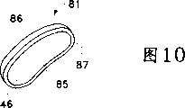
Figure 10 is used for the oval-shaped medicine nuclear of ophthalmic drug delivery device of the present invention or the perspective view of tablet;
Figure 11 is the perspective view two couplings, half elliptic medicine nuclear or tablet that is used for ophthalmic drug delivery device of the present invention;
The view that Figure 12 is the perspective of Fig. 7 and human body left eye ophthalmic drug delivery device shown in Figure 9, see from eye socket;
Figure 13 shown in Figure 12 and Figure 14, the view that comprises the perspective of the ophthalmic drug delivery device on inferior obliquus coupling inclined-plane, see from eye socket;
The view that Figure 14 is the perspective of the human body left eye ophthalmic drug delivery device shown in Fig. 7 and Fig. 9, see from sclera;
Figure 15 shown in Fig. 7 and Fig. 9, the view that comprises the perspective of the ophthalmic drug delivery device of this device taper longitudinal component, see from eye socket;
The view that Figure 16 is the perspective of the shortening pattern of ophthalmic drug delivery device shown in Fig. 7 and Fig. 9, see from eye socket;
The view that Figure 17 is the perspective that comprises the ophthalmic drug delivery device on an inferior obliquus coupling inclined-plane shown in Figure 16, see from eye socket;
The view that Figure 18 is the perspective of human body right eye ophthalmic drug delivery device according to a second, preferred embodiment of the present invention, see from eye socket;
The view that Figure 19 is the perspective that comprises the ophthalmic drug delivery device on an inferior obliquus coupling inclined-plane shown in Figure 180, see from eye socket;
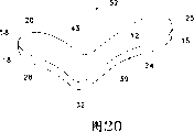
The view that Figure 20 is the perspective of the human body right eye ophthalmic drug delivery device of the 3rd preferred embodiment of the present invention, see from eye socket; With
The view that Figure 21 is the perspective of the ophthalmic drug delivery device that comprises inferior obliquus coupling inclined-plane shown in Figure 20, see from eye socket.
The specific embodiment
The preferred embodiments of the present invention and advantage thereof can be by getting the best understanding with reference to figs 1 to Figure 21, and wherein identical label symbol will be used among each figure on the identical or corresponding parts.
Fig. 1 has illustrated for the each several part of understanding the very important human eye of the present invention to Fig. 6.Referring to Fig. 1, wherein schematically shown human eye 90.Eye 90 has a cornea 92, crystalline lens 93, vitreous body 95, sclera 100, choroid 99, a retina 97 and an optic nerve 96.Eye 90 is divided into front portion 89 and rear portion 88 usually.The front portion 89 of eye 90 comprises the part in ora serrata 11 fronts of eye 90.The rear portion 88 of eye 90 generally includes eye 90 parts in ora serrata 11 back.Retina 97 near the place of ciliary ring 13, in the back of optic disk 19, with around mode and 99 actual linking to each other of choroid.Retina 97 has one to be positioned near the macula lutea 98 in optic disk 19 sides.Well-known in ophthalmology, macula lutea 98 mainly comprises retinal cones, and is the highest zone of visual acuity in the retina 97.Fascia bulbi or capsula bulbi 101 are configured on the sclera 100.Conjunctiva 94 has covered one of eyeball 90 short zone of back, edge 115 (spherical conjunctiva), and go up folding (going up pouch) or down folding (following pouch) cover the interior zone of upper eyelid 78 and palpebra inferior 79 respectively.Conjunctiva 94 is configured in the top of fascia bulbi 101.
As illustrated in fig. 1 and 2, and as following will the detailed description in detail, device 50 preferably directly is configured on the outer surface of sclera 100, at fascia bulbi below 101, so that treat most eye rear portion disease.In addition, in order to treat human ARMD and CNV, device 50 preferably directly is configured on the outer surface of sclera 100, and at fascia bulbi below 101, and the kernel that makes device 50 is near macula lutea 98.
Fig. 3 has shown the human body left eye 90 in its eye socket 112.As Fig. 3 finding, inferior obliquus 107 extends under side rectus 105.The insertion line 107a that inferior obliquus 107 inserts sclera 100 is positioned on the coboundary of side rectus 105.Certainly, the position of inferior obliquus is the mirror image of this position of the human body left eye 90 among Fig. 3 in the human body right eye 90.Cornea 92, conjunctiva 94, superior rectus 103, inferior retcus 104, superior obliquus 106 and edge 115 all are presented among Fig. 3.
Fig. 4 has shown the human body left eye 90 in its eye socket 112 similarly.Yet the part of side rectus 105 does not show in Fig. 4, so that make the sclera 100 and the optic nerve 96 that are stashed by muscle usually become visible.In Fig. 4, the insertion line 107b that inferior obliquus 107 inserts sclera 100 is lower than the insertion line 107a shown in Fig. 3, and this has shown the typical physiological change of the insertion line of inferior obliquus in human eye.
Fig. 5 has schematically shown the front view of a human eye 90 with its four rectus, i.e. superior rectus 103, middle rectus 108, inferior retcus 104 and side rectus 105.Fig. 5 has also shown in Fig. 5 by the edge of boundary line 115 expressions with by the insertion line of the rectus of 113 expressions of the boundary line among Fig. 5.
The rearview that in Fig. 6, has schematically shown human eye 90.Fig. 6 has shown the position of superior rectus 103, side rectus 105, inferior retcus 104, middle rectus 108, superior obliquus 106, inferior obliquus 107 and its insertion line 107a, optic nerve 96, ciliary canals 109, sclera 100, the sclera zone 110 on macula lutea 98, long arteria ciliares 111 and vasa vorticosa 114.
Fig. 7 and Fig. 9 have schematically shown the human body right eye ophthalmic drug delivery device 50 according to first preferred embodiment of the invention.Device 50 can be used for any situation that pharmaceutically active medicament part need be discharged into eye.Device 50 is specially adapted to pharmaceutically active medicament part to be discharged into the situation at eyes rear portion.A preferred application of device 50 is in order to treat ARMD, retina neovascularization (CNV), retinopathy, retinitis, uveitis, macular edema, glaucoma and neuropathy the pharmaceutically active medicament to be discharged on the retina near in the zone of macula lutea.
Preferably be arranged in the groove 20 at the kernel shown in Figure 10 81.As shown in figure 10, kernel 81 tablet that comprises one or more pharmaceutically active medicaments preferably.Tablet 81 preferably has oval-shaped matrix 46, and that this matrix 46 has is recessed, dome-shaped scleral surface 85 and protruding, dome-shaped orbit surfaces 86.Also preferably dispose the oblique angle 87 of a periphery on the matrix 46.In addition, as shown in figure 11, kernel also can comprise tablet 82a coupling, half elliptic and 82b.Tablet 82a preferably has half the identical matrix 47 with the matrix 46 of tablet 81.Tablet 82b preferably has another the hemiidentic matrix 48 with the matrix 46 of tablet 81.In addition, kernel 81 or kernel 82a and 82b also can comprise a kind of traditional hydrogel, glue, cream or other semi-solid silks agent, and one or more pharmaceutically active medicaments are arranged in said preparation.
Get back to Fig. 9, keep part 62 preferably be configured in opening 64 near.Keep part 62 to prevent that kernel 81 from dropping out groove 20.When kernel 81 is a tablet, keep a preferably successive frame or flange that is configured in opening 64 peripheries of part 62, this Element Design is used for holding the hypotenuse 87 of tablet 81.In addition, keep part 62 to comprise and one or morely extend to part the opening 64 from matrix 21.
Though do not demonstrate in Fig. 9 and 11, kernel 81 also can comprise a kind of suspension, solution, powder or its combination, has wherein comprised the pharmaceutically active medicament.In the present embodiment, do not have opening 64 on the scleral surface 14, and suspension, solution, powder or its combination are by scleral surface 14 or the diffusion of other film in the relative thin of kernel below 81.In addition, device 50 is trough of belt 20 or kernel 81 not, and is dispersed in the whole substrate 21 of device 50 with the pharmaceutically active medicament that suspension, solution, powder or its combining form exist.In this embodiment, the pharmaceutically active medicament is diffused in the destination organization by matrix 21.
If kernel 81 is tablets, it can also comprise the excipient that traditional pharmacy need be used, as filler and lubricant.This tablet can be produced with traditional pharmacy method.The pharmaceutically active medicament preferably is evenly distributed in the whole tablet.Except traditional tablet, kernel 81 can also comprise a kind of with control speed by the special tablet of biological corrosion, discharge the pharmaceutically active medicament.For example, this biological corrosion can produce by hydrolysis or enzyme division.If kernel 81 is a kind of hydrogel or other glue, this brood lac meeting is carried out biological corrosion with the speed of control, and discharges the pharmaceutically active medicament.In addition, this is brood lac can also to be non-biological corrosion, but can spread the pharmaceutically active medicament.
The geometry of the matrix 21 of device 50 comprises the depression characteristic of scleral surface 14; Lateral part 18, groove 20, opening 64, kernel 81 and the shape and the position that keep part 62; And the shape and the position of breach 42 and stopping part 36, all be to help from kernel 81, in retina 97, particularly discharging the pharmaceutically active medicament that drug effect dosage is arranged in the macula lutea 98 by sclera 100, choroid 99.Between groove 20 and sclera 100, do not establish polymeric layer or film, strengthen helping greatly and simplify of the release of a kind of active agents to retina 97.
It is believed that the concrete physicochemical characteristics according to the pharmaceutically active medicament that is adopted, device 50 can be used for discharging the pharmaceutically active medicament that drug effect dosage is arranged to retina 97 all the year round.The physicochemical characteristics of implant comprises hydrophobicity, dissolubility, rate of dissolution, diffusion coefficient, partition coefficient and tissue affinity.After kernel 20 no longer contained active agents, the surgeon can remove device 50 at an easy rate.In addition, the passage that " formed in the past " is convenient to replace old device 50 with a new equipment 50.
Fig. 8 has shown an ophthalmic drug delivery device 60, and it has done some improvement a little to ophthalmic drug delivery device 50, and these improvement help some implantation of the present invention.As shown in Figure 8, device 60 is similar substantially with the geometry of the device 50 shown in Fig. 9 to Fig. 7, and its difference is that the place near breach 42 has increased a slope 45 on the orbit surfaces 12 of matrix 21.Slope 45 is inclined surfaces, and this inclined surface preferably extends to orbit surfaces 12 places of second end from the scleral surface 14 of its first end.In addition, slope 45 can also extend to orbit surfaces 12 places of its second end from the circular edge 24 of the longitudinal component 15 of its first end.As mentioned above, in the time of in installing 60 implanted eyes 90, slope 45 helps holding inferior obliquus 107 in breach 42, wherein be in the breach 42 between lateral part 18 and the stopping part 36.Device 60 can adopt and install 50 basic similar techniques and make.
Figure 12 and 14 has schematically shown the ophthalmic drug delivery device 70 that is used for the human body left eye.Device 70 geometry is the mirror image according to the geometry of Fig. 7 and 9 described devices 50 that is used for the human body right eye.The application of device 70 is identical with the application of device 50 basically, and device 70 can adopt and install 50 basic similar techniques and make.
What Figure 13 showed is an ophthalmic drug delivery device 75 that is used for the human body left eye, wherein ophthalmic drug delivery device 70 has been done some improvement a little, and these improvement help some implantation of the present invention.The geometry of the device 75 of Figure 13 and the geometry of the device among Fig. 8 60 are similar substantially, and difference is that device 75 is mirror images of device 60.
Figure 15 has shown an ophthalmic drug delivery device 30, and it has done some improvement a little to ophthalmic drug delivery device 50, and these improvement help some implantation of the present invention.As shown in figure 15, device 30 is similar substantially with the geometry of the device 50 shown in Fig. 9 to Fig. 7, its difference is that when from the edge 24 when seeing, longitudinal component 15 has the thickness that diminishes gradually, this gradually the part of tapered thickness start from 33 places, position and be extended to near-end 25 places.This part of longitudinal component 15 is configured in the anterior of eye and can be seen by other people.Therefore, because this thickness that diminishes gradually, device 30 may become more comfortable, and accepts the convenient easier patient of allowing of beauty treatment.The application of device 30 is identical with the application of device 50 basically, and device 30 can adopt and install 50 basic similar techniques and make.
Figure 16 has shown an ophthalmic drug delivery device 40, and it has done some improvement a little to ophthalmic drug delivery device 50, and these improvement help some implantation of the present invention.As shown in figure 16, device 40 is similar substantially with the geometry of the device 50 shown in Fig. 9 to Fig. 7, and its difference is that the length of the longitudinal component 15 of device 40 has been shortened with respect to device 50.Similar with device 30, the shortening of this longitudinal component 15 makes device 40 become more comfortable, and accepts the convenient easier patient of allowing of beauty treatment.The application of device 40 is identical with the application of device 50 basically, and device 40 can adopt and install 50 basic similar techniques and make.
Figure 17 has shown an ophthalmic drug delivery device 80, and it has done some improvement a little to ophthalmic drug delivery device 40, and these improvement help some implantation of the present invention.As shown in figure 17, device 80 is similar substantially to the geometry of the device 40 shown in Figure 16, and its difference is that the place near breach 42 has increased a slope 45 on the orbit surfaces 12 of matrix 21.Slope 45 is inclined surfaces, and this inclined surface preferably extends to orbit surfaces 12 places of second end from the scleral surface 14 of its first end.In addition, slope 45 can also a bit extend to orbit surfaces 12 places of its second end from the edge 24 of the longitudinal component 15 of its first end.According to installing as described in 50, in the time of in installing 80 implanted eyes 90, slope 45 helps holding inferior obliquus 107 in breach 42, wherein be in the breach 42 between lateral part 18 and the stopping part 36 as above.Device 80 can adopt and install 50 basic similar techniques and make.
Figure 18 has schematically shown according to ophthalmic drug delivery device 65 second preferred embodiment of the invention, that be used for the human body right eye.Device 65 can be used in any local situation that the pharmaceutically active medicament is given eyes that discharges that needs to adopt.Device 65 is adapted to active agents is discharged into the situation at a rear portion especially.A preferred application of device 65 is that the pharmaceutically active medicament is discharged near on the retina of macula lutea, so that treatment ARMD, choroid neovascularization (CNV), retinopathy, retinitis, uveitis, macular edema, glaucoma and neuropathy.
Device 65 generally includes matrix 29, this matrix 29 have protruding, a dome-shaped orbit surfaces 12 and one recessed, dome-shaped scleral surface 14 (not showing).The radius of curvature of designed scleral surface 14 is convenient to it and is directly contacted with sclera 100.Be more preferably the radius of curvature 91 that the radius of curvature that makes scleral surface 14 equals usual human eye 90.Orbit surfaces 12 preferably is designed to have is convenient to be implanted to the radius of curvature of fascia bulbi below 101.When viewed from above, matrix 21 preferably has a general geometry for " C-shape ", and longitudinal component 17, lateral part 18 and sweep therebetween 32 are wherein arranged.Longitudinal component 17 is preferably in sweep 32 places with lateral part 18 and links to each other, to form an angle that is about 90 degree.Longitudinal component 17 has a near-end 25, a circular edge 24.Stopping part 37 has formed the D score portion of C shape and this stopping part and has cand be compared to usually the remainder of the orbit surfaces 12 of projection most and raise.A breach 42 is positioned at longitudinal component 17 places, and is determined by lateral part 18 and stopping part 37.The initial end similar with the breach 42 of Fig. 7 and device 50 shown in Figure 9, that the breach 42 of device 65 is designed to hold inferior obliquus 107.Similar with the stopping part 36 of device 50, stopping part 37 is by contacting with the preceding border of inferior obliquus 107, and anti-locking apparatus 65 too moves forward to optic nerve 96.There are a far-end 58, a circular edge 28 and a groove or the cavity 20 that opening 64 (not have demonstration) arranged in lateral part 18 on scleral surface 14 (not having to show), so that clamp to above according to the similar kernel of the described kernel of Figure 10 and Figure 11.Groove 20 and opening 64 preferably probably are oval.
The application of device 65 is identical with the application of said apparatus 50 basically.Device 65 can adopt and install 50 basic similar techniques and make.
Figure 19 has shown an ophthalmic drug delivery device 67, and it has done some improvement a little to ophthalmic drug delivery device 65, and these improvement help some implantation of the present invention.As shown in figure 19, device 67 is similar substantially to the geometry of the device 65 shown in Figure 19, and its difference is that the place near breach 42 has increased a slope 45 on the orbit surfaces 12 of matrix 29.Slope 45 is inclined surfaces, and this inclined surface preferably extends to orbit surfaces 12 places of second end from the scleral surface 14 of its first end.In addition, slope 45 can also a bit extend to orbit surfaces 12 places of its second end from the edge 24 of the longitudinal component 17 of its first end.According to as described in the device 50, when in the device 67 implanted eyes 90, slope 45 helps holding inferior obliquus 107 in breach 42 as above, and wherein breach 42 is between lateral part 18 and the stopping part 37.Device 67 can adopt and install 50 basic similar techniques and make.
Figure 20 has schematically shown according to ophthalmic drug delivery device 52 the 3rd preferred embodiment of the present invention, that be used for the human body right eye.Device 65 can be used in any local situation that the pharmaceutically active medicament is given eyes that discharges that needs to adopt.Device 65 is adapted to active agents is discharged into the situation at a rear portion especially.A preferred application of device 65 is that the pharmaceutically active medicament is discharged near on the retina of macula lutea, so that treatment ARMD, choroid neovascularization (CNV), retinopathy, retinitis, uveitis, macular edema, glaucoma and neuropathy.
Device 52 generally includes matrix 39, this matrix 39 have protruding, a dome-shaped orbit surfaces 12 and one recessed, dome-shaped scleral surface 14 (not showing).The radius of curvature of designed scleral surface 14 is convenient to it and is directly contacted with sclera 100.Be more preferably the radius of curvature 91 that the radius of curvature that makes scleral surface 14 equals usual human eye 90.Orbit surfaces 12 preferably is designed to have is convenient to be implanted to the radius of curvature of fascia bulbi below 101.When viewed from above, matrix 39 preferably has a general geometry for " L-shape ", and longitudinal component 15, lateral part 18 and sweep therebetween 32 are wherein arranged.Longitudinal component 15 is preferably in sweep 32 places with lateral part 18 and links to each other, to form an angle that is about 90 degree.Similar to the breach 42 of the device 50 of Fig. 7 and Fig. 9, the zone 43 of device 52 is designed to hold the initiating terminal of inferior obliquus 107.Longitudinal component 15 has a near-end 25, a circular edge 24.There are a far-end 58, a circular edge 28 and a groove or the cavity 20 that opening 64 (not having to show) arranged in lateral part 18 on scleral surface 14, so that clamp to above according to the similar kernel of the described kernel of Figure 10 and Figure 11.It is oval that groove 20 and opening 64 are preferably.
The application of device 52 is identical with the application of said apparatus 50 basically, and device 52 can adopt and install 50 basic similar techniques and make.
Figure 21 has shown an ophthalmic drug delivery device 54, and it has done some improvement a little to ophthalmic drug delivery device 52, and these improvement help some implantation of the present invention.As shown in figure 21, device 54 is similar substantially to the geometry of the device 52 shown in Figure 20, and its difference is that the place near zone 43 has increased a slope 45 on the orbit surfaces 12 of matrix 29.Slope 45 is inclined surfaces, and this inclined surface preferably extends to orbit surfaces 12 places of second end from the scleral surface 14 of its first end.In addition, slope 45 can also a bit extend to orbit surfaces 12 places of its second end from the edge 24 of the longitudinal component 15 of its first end.According to installing as described in 50, in the time of in installing 54 implanted eyes 90, slope 45 helps holding inferior obliquus 107 in zone 43 as above.Device 54 can adopt and install 50 basic similar techniques and make.
From the above mentioned, can realize and the invention provides improved apparatus and method, so that safely, effectively, the control speed ground various pharmaceutically active medicaments of local release are given eyes, particularly give the eyes rear portion, with opposing ARMD, CNV, retinopathy, retinitis, uveitis, macular edema, glaucoma and neuropathy.The surgical procedures of implanting this device is safe, easy, rapid and can implements to the out-patient.This device also is convenient and economical on making.In addition, because they can discharge various pharmaceutically active medicaments, these devices can be used for discharging various medicament for the eyes in clinical research so that produce a kind of special condition for the patient.
The present invention, it will be apparent to one skilled in the art that and can carry out various improvement by the embodiment explanation at this.For example, though above-mentioned a kind of the seeing from the top that the present invention relates to has " F-shape ", " C-shape ", or the ophthalmic drug delivery device of " L-shape " geometry, but also can adopt other geometry, particularly be implanted on the sclera outer surface of human eye and under the fascia bulbi time when this device, the geometry that is adopted helps this device, and to be implanted to inferior obliquus following and especially true when making the medicine active agents on macula lutea.
Can believe that operation of the present invention and structure will become apparent from the above description.Various distortion and improve and under the condition that does not break away from the following determined the spirit and scope of the invention of claim, to carry out have been used as when preferred embodiment is illustrated in the above shown and equipment described and method.
Claims (8)
1. drug release device that is used for human eye, described eye has a sclera, polylith extraocular muscles, wherein comprises an inferior obliquus and a macula lutea, and described device comprises:
A kind of pharmaceutically active medicament; With
Matrix with a geometry, this geometry helps described device is implanted on the outer surface of described sclera, below described inferior obliquus, and described pharmaceutically active medicament is configured on the described macula lutea, and does not need to disconnect or cut in the described polylith extraocular muscles any one.
2. according to the drug release device of claim 1, it is characterized in that: described matrix comprises a scleral surface and an orbit surfaces; And when described scleral surface or described orbit surfaces are seen, described geometry is the geometry of a F-shape.
3. according to the drug release device of claim 1, it is characterized in that: described matrix comprises a scleral surface and an orbit surfaces; And when described scleral surface or described orbit surfaces are seen, described geometry is the geometry of a C-shape.
4. according to the drug release device of claim 1, it is characterized in that: described matrix comprises a scleral surface and an orbit surfaces; And when described scleral surface or described orbit surfaces are seen, described geometry is the geometry of a L-shape.
5. according to the drug release device of claim 1, it is characterized in that: described human eye comprises a fascia bulbi, and described matrix comprises an orbit surfaces, and the radius of curvature of this orbit surfaces is convenient to described device is implanted under the described fascia bulbi.
6. according to the drug release device of claim 5, it is characterized in that: described orbit surfaces also comprises a breach, and when implanting described device, described breach is convenient to hold described inferior obliquus.
7. according to the drug release device of claim 6, it is characterized in that: described breach comprises a slope.
8. according to the drug release device of claim 1, it is characterized in that: described matrix has a scleral surface, and the radius of curvature of described scleral surface equals the radius of curvature of described human eye.
Applications Claiming Priority (4)
| Application Number | Priority Date | Filing Date | Title |
|---|---|---|---|
| US16067399P | 1999-10-21 | 1999-10-21 | |
| US60/160,673 | 1999-10-21 | ||
| US09/664,790 | 2000-09-19 | ||
| US09/664,790 US6416777B1 (en) | 1999-10-21 | 2000-09-19 | Ophthalmic drug delivery device |
Publications (2)
| Publication Number | Publication Date |
|---|---|
| CN1376042A CN1376042A (en) | 2002-10-23 |
| CN100341470C true CN100341470C (en) | 2007-10-10 |
Family
ID=26857116
Family Applications (1)
| Application Number | Title | Priority Date | Filing Date |
|---|---|---|---|
| CNB008131929A Expired - Fee Related CN100341470C (en) | 1999-10-21 | 2000-10-12 | Ophthalmic drug delivery device |
Country Status (19)
| Country | Link |
|---|---|
| US (4) | US6416777B1 (en) |
| EP (1) | EP1221919B1 (en) |
| JP (1) | JP4685311B2 (en) |
| KR (1) | KR100752821B1 (en) |
| CN (1) | CN100341470C (en) |
| AR (2) | AR026183A1 (en) |
| AT (1) | ATE289500T1 (en) |
| AU (2) | AU764226B2 (en) |
| BR (1) | BR0014928B1 (en) |
| CA (1) | CA2384255C (en) |
| DE (1) | DE60018298T2 (en) |
| DK (1) | DK1221919T3 (en) |
| ES (1) | ES2237463T3 (en) |
| HK (1) | HK1048428B (en) |
| PL (1) | PL196539B1 (en) |
| PT (1) | PT1221919E (en) |
| TR (1) | TR200201046T2 (en) |
| TW (1) | TW539560B (en) |
| WO (1) | WO2001028474A1 (en) |
Families Citing this family (154)
| Publication number | Priority date | Publication date | Assignee | Title |
|---|---|---|---|---|
| EP1040837A3 (en) * | 1999-02-26 | 2002-01-02 | Erasmus Universiteit Rotterdam | Medicaments for the treatment of a choroidal neovascularization (CNV) related disorder |
| HUP0201111A3 (en) | 1999-04-26 | 2004-05-28 | Gmp Vision Solutions Inc Ft La | Shunt device for treating glaucoma |
| US6416777B1 (en) * | 1999-10-21 | 2002-07-09 | Alcon Universal Ltd. | Ophthalmic drug delivery device |
| US7943162B2 (en) * | 1999-10-21 | 2011-05-17 | Alcon, Inc. | Drug delivery device |
| US20050119737A1 (en) * | 2000-01-12 | 2005-06-02 | Bene Eric A. | Ocular implant and methods for making and using same |
| US6638239B1 (en) | 2000-04-14 | 2003-10-28 | Glaukos Corporation | Apparatus and method for treating glaucoma |
| US7708711B2 (en) | 2000-04-14 | 2010-05-04 | Glaukos Corporation | Ocular implant with therapeutic agents and methods thereof |
| US7867186B2 (en) | 2002-04-08 | 2011-01-11 | Glaukos Corporation | Devices and methods for treatment of ocular disorders |
| US20020143284A1 (en) * | 2001-04-03 | 2002-10-03 | Hosheng Tu | Drug-releasing trabecular implant for glaucoma treatment |
| AR030346A1 (en) * | 2000-08-14 | 2003-08-20 | Alcon Inc | METHOD OF TREATMENT OF NEURODEGENERATIVE DISORDERS OF THE RETINA AND HEAD OF OPTICAL NERVE |
| AR030345A1 (en) * | 2000-08-14 | 2003-08-20 | Alcon Inc | METHOD OF TREATMENT OF DISORDERS RELATED TO ANGIOGENESIS |
| US7181287B2 (en) * | 2001-02-13 | 2007-02-20 | Second Sight Medical Products, Inc. | Implantable drug delivery device |
| AU2002247284A1 (en) * | 2001-04-02 | 2002-10-15 | Alcon, Inc. | Method of treating ocular inflammatory and angiogenesis-related disorders using an amide derivative of flubiprofen or ketorolac |
| US7488303B1 (en) * | 2002-09-21 | 2009-02-10 | Glaukos Corporation | Ocular implant with anchor and multiple openings |
| WO2002080811A2 (en) | 2001-04-07 | 2002-10-17 | Glaukos Corporation | Glaucoma stent and methods thereof for glaucoma treatment |
| US7431710B2 (en) | 2002-04-08 | 2008-10-07 | Glaukos Corporation | Ocular implants with anchors and methods thereof |
| US7678065B2 (en) | 2001-05-02 | 2010-03-16 | Glaukos Corporation | Implant with intraocular pressure sensor for glaucoma treatment |
| WO2002089699A2 (en) | 2001-05-03 | 2002-11-14 | Glaukos Corporation | Medical device and methods of use for glaucoma treatment |
| WO2002100318A2 (en) | 2001-06-12 | 2002-12-19 | Johns Hopkins University School Of Medicine | Reservoir device for intraocular drug delivery |
| PT1409065E (en) | 2001-07-23 | 2007-03-30 | Alcon Inc | Ophthalmic drug delivery device |
| DE60214697T2 (en) * | 2001-07-23 | 2007-09-13 | Alcon Inc. | DEVICE FOR RELEASING AN OPHTHALMIC MEDICINAL PRODUCT |
| US7331984B2 (en) | 2001-08-28 | 2008-02-19 | Glaukos Corporation | Glaucoma stent for treating glaucoma and methods of use |
| US7749528B2 (en) * | 2001-08-29 | 2010-07-06 | Ricardo Azevedo Pontes De Carvalho | Implantable and sealable medical device for unidirectional delivery of therapeutic agents to tissues |
| US7195774B2 (en) * | 2001-08-29 | 2007-03-27 | Carvalho Ricardo Azevedo Ponte | Implantable and sealable system for unidirectional delivery of therapeutic agents to tissues |
| US7186232B1 (en) | 2002-03-07 | 2007-03-06 | Glaukoa Corporation | Fluid infusion methods for glaucoma treatment |
| PL205443B1 (en) * | 2002-03-11 | 2010-04-30 | Alcon | Implantable drug delivery system |
| US7951155B2 (en) | 2002-03-15 | 2011-05-31 | Glaukos Corporation | Combined treatment for cataract and glaucoma treatment |
| US9301875B2 (en) | 2002-04-08 | 2016-04-05 | Glaukos Corporation | Ocular disorder treatment implants with multiple opening |
| AU2003231730A1 (en) * | 2002-05-03 | 2003-11-17 | Alcon, Inc. | Method of treating vascular endothelial growth factor mediated vascular disorders using amfenac or nepafenac |
| US20040127472A1 (en) * | 2002-08-05 | 2004-07-01 | Jerdan Janice A. | Use of anecortave acetate for the protection of visual acuity in patients with age related macular degeneration |
| DE10238310A1 (en) * | 2002-08-21 | 2004-03-04 | Erich Jaeger Gmbh | electrode assembly |
| CA2689424A1 (en) * | 2002-09-29 | 2004-04-08 | Surmodics, Inc. | Methods for treatment and/or prevention of retinal disease |
| US20030187072A1 (en) * | 2003-02-14 | 2003-10-02 | Kapin Michael A. | Method of treating angiogenesis-related disorders |
| JP2006518382A (en) * | 2003-02-20 | 2006-08-10 | アルコン,インコーポレイテッド | Use of steroids to treat people suffering from eye disorders |
| KR20050102653A (en) * | 2003-02-20 | 2005-10-26 | 알콘, 인코퍼레이티드 | Formulations of glucocorticoids to treat pathologic ocular angiogenesis |
| US20040236343A1 (en) * | 2003-05-23 | 2004-11-25 | Taylor Jon B. | Insertion tool for ocular implant and method for using same |
| KR20060019579A (en) * | 2003-06-13 | 2006-03-03 | 알콘, 인코퍼레이티드 | Formulations of non-steroidal anti-inflammatory agents to treat pathologic ocular angiogenesis |
| JP2007521274A (en) * | 2003-06-20 | 2007-08-02 | アルコン,インコーポレイテッド | Treatment of age-related macular degeneration using a combination of multiple components |
| KR20060082792A (en) * | 2003-07-10 | 2006-07-19 | 알콘, 인코퍼레이티드 | Ophthalmic drug delivery device |
| US20050009910A1 (en) * | 2003-07-10 | 2005-01-13 | Allergan, Inc. | Delivery of an active drug to the posterior part of the eye via subconjunctival or periocular delivery of a prodrug |
| EP1658109B1 (en) | 2003-08-26 | 2014-01-22 | Vista Scientific LLC | Ocular drug delivery device |
| AU2004274026A1 (en) | 2003-09-18 | 2005-03-31 | Macusight, Inc. | Transscleral delivery |
| US20050158365A1 (en) * | 2003-12-22 | 2005-07-21 | David Watson | Drug delivery device with mechanical locking mechanism |
| US7276050B2 (en) * | 2004-03-02 | 2007-10-02 | Alan Franklin | Trans-scleral drug delivery method and apparatus |
| US9549895B2 (en) * | 2004-04-23 | 2017-01-24 | Massachusetts Eye And Ear Infirmary | Methods and compositions for preserving the viability of photoreceptor cells |
| US20060110428A1 (en) | 2004-07-02 | 2006-05-25 | Eugene Dejuan | Methods and devices for the treatment of ocular conditions |
| US7722669B2 (en) * | 2004-08-13 | 2010-05-25 | Richard Foulkes | Method and insert for modifying eye color |
| US8246569B1 (en) | 2004-08-17 | 2012-08-21 | California Institute Of Technology | Implantable intraocular pressure drain |
| CN100428958C (en) * | 2004-09-06 | 2008-10-29 | 段亚东 | Implant agent for treating ophthalmopathy |
| EP1853259A1 (en) | 2005-02-09 | 2007-11-14 | Macusight, Inc. | Formulations for ocular treatment |
| US8663639B2 (en) | 2005-02-09 | 2014-03-04 | Santen Pharmaceutical Co., Ltd. | Formulations for treating ocular diseases and conditions |
| JP2008533204A (en) * | 2005-03-21 | 2008-08-21 | マクサイト, インコーポレイテッド | Drug delivery system for treatment of disease or condition |
| EP1868661A1 (en) | 2005-04-08 | 2007-12-26 | SurModics, Inc. | Sustained release implants for subretinal delivery |
| US20060258994A1 (en) * | 2005-05-12 | 2006-11-16 | Avery Robert L | Implantable delivery device for administering pharmacological agents to an internal portion of a body |
| NZ565953A (en) | 2005-07-27 | 2012-01-12 | Univ Florida | Small compounds that correct protein misfolding and uses thereof |
| US20070212397A1 (en) * | 2005-09-15 | 2007-09-13 | Roth Daniel B | Pharmaceutical delivery device and method for providing ocular treatment |
| US20070134244A1 (en) * | 2005-10-14 | 2007-06-14 | Alcon, Inc. | Combination treatment for pathologic ocular angiogenesis |
| MX2008006379A (en) | 2005-11-29 | 2009-03-03 | Smithkline Beecham Corp | Treatment method. |
| WO2007092620A2 (en) | 2006-02-09 | 2007-08-16 | Macusight, Inc. | Stable formulations, and methods of their preparation and use |
| WO2007106557A2 (en) * | 2006-03-14 | 2007-09-20 | University Of Southern California | Mems device for delivery of therapeutic agents |
| AU2007230964B2 (en) | 2006-03-23 | 2012-07-19 | Santen Pharmaceutical Co., Ltd. | Formulations and methods for vascular permeability-related diseases or conditions |
| US20070293807A1 (en) * | 2006-05-01 | 2007-12-20 | Lynch Mary G | Dual drainage pathway shunt device and method for treating glaucoma |
| US8216603B2 (en) * | 2006-05-04 | 2012-07-10 | Herbert Edward Kaufman | Method, device, and system for delivery of therapeutic agents to the eye |
| US8506515B2 (en) | 2006-11-10 | 2013-08-13 | Glaukos Corporation | Uveoscleral shunt and methods for implanting same |
| WO2008063639A2 (en) * | 2006-11-21 | 2008-05-29 | Massachusetts Eye And Ear Infirmary | Compositions and methods for preserving cells of the eye |
| ZA200903649B (en) * | 2006-12-18 | 2010-08-25 | Alcon Res Ltd | Devices and methods for ophthalmic drug delivery |
| DE102007024642A1 (en) * | 2007-05-24 | 2008-11-27 | Eyesense Ag | Hydrogel implant for sensor of metabolites on the eye |
| EP2242464B2 (en) | 2007-12-20 | 2017-03-01 | University Of Southern California | Apparatus for delivering therapeutic agents |
| US20090192493A1 (en) * | 2008-01-03 | 2009-07-30 | University Of Southern California | Implantable drug-delivery devices, and apparatus and methods for refilling the devices |
| US9849238B2 (en) | 2008-05-08 | 2017-12-26 | Minipumps, Llc | Drug-delivery pump with intelligent control |
| CA2723724C (en) * | 2008-05-08 | 2017-03-14 | Replenish Pumps, Llc | Implantable pumps and cannulas therefor |
| CA2723723C (en) | 2008-05-08 | 2019-06-25 | Replenish Pumps, Llc | Implantable drug-delivery devices, and apparatus and methods for filling the devices |
| CN102202708B (en) | 2008-05-08 | 2015-01-21 | 迷你泵有限责任公司 | Drug-delivery pumps and methods of manufacture |
| CA2723588A1 (en) | 2008-05-12 | 2009-11-19 | University Of Utah Research Foundation | Intraocular drug delivery device and associated uses |
| US9877973B2 (en) | 2008-05-12 | 2018-01-30 | University Of Utah Research Foundation | Intraocular drug delivery device and associated methods |
| US10064819B2 (en) | 2008-05-12 | 2018-09-04 | University Of Utah Research Foundation | Intraocular drug delivery device and associated methods |
| US9095404B2 (en) | 2008-05-12 | 2015-08-04 | University Of Utah Research Foundation | Intraocular drug delivery device and associated methods |
| US8664182B2 (en) | 2008-11-12 | 2014-03-04 | Duke University | Methods of inhibiting cancer cell growth with HDAC inhibitors and methods of screening for HDAC10 inhibitors |
| US8623395B2 (en) | 2010-01-29 | 2014-01-07 | Forsight Vision4, Inc. | Implantable therapeutic device |
| SG173167A1 (en) | 2009-01-29 | 2011-08-29 | Forsight Labs Llc | Posterior segment drug delivery |
| US8372036B2 (en) | 2009-05-06 | 2013-02-12 | Alcon Research, Ltd. | Multi-layer heat assembly for a drug delivery device |
| US10206813B2 (en) | 2009-05-18 | 2019-02-19 | Dose Medical Corporation | Implants with controlled drug delivery features and methods of using same |
| CN102596097B (en) | 2009-06-03 | 2015-05-20 | 弗赛特实验室有限责任公司 | Anterior segment drug delivery |
| IN2012DN00352A (en) | 2009-06-16 | 2015-08-21 | Bikam Pharmaceuticals Inc | |
| KR101697388B1 (en) | 2009-08-18 | 2017-01-17 | 미니펌프스, 엘엘씨 | Electrolytic drug-delivery pump with adaptive control |
| US20120232102A1 (en) | 2009-09-30 | 2012-09-13 | Chun-Fang Xu | Methods Of Administration And Treatment |
| US8177747B2 (en) | 2009-12-22 | 2012-05-15 | Alcon Research, Ltd. | Method and apparatus for drug delivery |
| EP2560629B1 (en) | 2010-04-23 | 2020-06-03 | Massachusetts Eye & Ear Infirmary | Methods and compositions for preserving photoreceptor and retinal pigment epithelial cells |
| JP2013526572A (en) | 2010-05-17 | 2013-06-24 | アエリエ・ファーマシューティカルズ・インコーポレーテッド | Drug delivery device for the delivery of eye treatments |
| CA2807537C (en) | 2010-08-05 | 2018-09-18 | Forsight Vision4, Inc. | Combined drug delivery methods and apparatus |
| EP2600812B1 (en) | 2010-08-05 | 2021-09-22 | ForSight Vision4, Inc. | Apparatus to treat an eye |
| HUE054113T2 (en) | 2010-08-05 | 2021-08-30 | Forsight Vision4 Inc | Injector apparatus for drug delivery |
| US8689439B2 (en) | 2010-08-06 | 2014-04-08 | Abbott Laboratories | Method for forming a tube for use with a pump delivery system |
| US8377000B2 (en) | 2010-10-01 | 2013-02-19 | Abbott Laboratories | Enteral feeding apparatus having a feeding set |
| US8377001B2 (en) | 2010-10-01 | 2013-02-19 | Abbott Laboratories | Feeding set for a peristaltic pump system |
| US20120109054A1 (en) * | 2010-10-29 | 2012-05-03 | Vista Scientific Llc | Devices with an erodible surface for delivering at least one active agent to tissue over a prolonged period of time |
| US20140024598A1 (en) | 2010-11-01 | 2014-01-23 | Demetrios Vavvas | Methods and compositions for preserving retinal ganglion cells |
| CA2818612C (en) | 2010-11-19 | 2020-12-29 | Forsight Vision4, Inc. | Therapeutic agent formulations for implanted devices |
| US9919099B2 (en) | 2011-03-14 | 2018-03-20 | Minipumps, Llc | Implantable drug pumps and refill devices therefor |
| US10286146B2 (en) | 2011-03-14 | 2019-05-14 | Minipumps, Llc | Implantable drug pumps and refill devices therefor |
| US9603997B2 (en) | 2011-03-14 | 2017-03-28 | Minipumps, Llc | Implantable drug pumps and refill devices therefor |
| US10245178B1 (en) | 2011-06-07 | 2019-04-02 | Glaukos Corporation | Anterior chamber drug-eluting ocular implant |
| BR112013032199A2 (en) | 2011-06-14 | 2017-12-12 | Bikam Pharmaceuticals Inc | opsin binding binders, compositions and methods of use |
| US20150005254A1 (en) | 2011-06-27 | 2015-01-01 | Massachusetts Eye And Ear Infirmary | Methods for treating ocular inflammatory disorders |
| EP4249059A3 (en) | 2011-06-28 | 2023-11-29 | ForSight Vision4, Inc. | An apparatus for collecting a sample of fluid from a reservoir chamber of a therapeutic device for the eye |
| EP2739252A4 (en) | 2011-08-05 | 2015-08-12 | Forsight Vision4 Inc | Small molecule delivery with implantable therapeutic device |
| WO2013025840A1 (en) | 2011-08-15 | 2013-02-21 | Massachusetts Eye And Ear Infirmary | Methods for preserving photoreceptor cell viability following retinal detachment |
| US9102105B2 (en) | 2011-09-13 | 2015-08-11 | Vista Scientific Llc | Method for forming an ocular drug delivery device |
| EP4193907A1 (en) | 2011-09-13 | 2023-06-14 | Glaukos Corporation | Intraocular physiological sensor |
| CN106073986B (en) | 2011-09-14 | 2019-01-11 | 弗赛特影像5股份有限公司 | The device for treating the eyes of patient |
| PL2755600T3 (en) | 2011-09-16 | 2021-09-20 | Forsight Vision4, Inc. | Fluid exchange apparatus |
| CA2856703A1 (en) | 2011-10-19 | 2013-04-25 | Bikam Pharmaceuticals, Inc. | Opsin-binding ligands, compositions and methods of use |
| WO2013059791A2 (en) | 2011-10-21 | 2013-04-25 | Massachusetts Eye And Ear Infirmary | Methods and compositions for promoting axon regeneration and nerve function |
| EP2785346A4 (en) | 2011-11-30 | 2015-04-29 | Bikam Pharmaceuticals Inc | Opsin-binding ligands, compositions and methods of use |
| BR112014013106A2 (en) | 2011-12-01 | 2017-06-13 | Bikam Pharmaceuticals Inc | opsin binding binders, compositions and methods of use |
| US10010448B2 (en) | 2012-02-03 | 2018-07-03 | Forsight Vision4, Inc. | Insertion and removal methods and apparatus for therapeutic devices |
| US9750636B2 (en) | 2012-10-26 | 2017-09-05 | Forsight Vision5, Inc. | Ophthalmic system for sustained release of drug to eye |
| US9730638B2 (en) | 2013-03-13 | 2017-08-15 | Glaukos Corporation | Intraocular physiological sensor |
| US9968603B2 (en) | 2013-03-14 | 2018-05-15 | Forsight Vision4, Inc. | Systems for sustained intraocular delivery of low solubility compounds from a port delivery system implant |
| US9592151B2 (en) | 2013-03-15 | 2017-03-14 | Glaukos Corporation | Systems and methods for delivering an ocular implant to the suprachoroidal space within an eye |
| JP6385423B2 (en) | 2013-03-28 | 2018-09-05 | フォーサイト・ビジョン フォー・インコーポレーテッド | Ocular graft for therapeutic substance delivery |
| US10098836B2 (en) * | 2013-05-02 | 2018-10-16 | Retina Foundation Of The Southwest | Method for forming a molded two-layer ocular implant |
| LT3066201T (en) | 2013-11-07 | 2018-08-10 | Editas Medicine, Inc. | Crispr-related methods and compositions with governing grnas |
| WO2015103480A1 (en) | 2014-01-02 | 2015-07-09 | Massachusetts Eye & Ear Infirmary | Treating ocular neovascularization |
| EP3114227B1 (en) | 2014-03-05 | 2021-07-21 | Editas Medicine, Inc. | Crispr/cas-related methods and compositions for treating usher syndrome and retinitis pigmentosa |
| US11141493B2 (en) | 2014-03-10 | 2021-10-12 | Editas Medicine, Inc. | Compositions and methods for treating CEP290-associated disease |
| US9938521B2 (en) | 2014-03-10 | 2018-04-10 | Editas Medicine, Inc. | CRISPR/CAS-related methods and compositions for treating leber's congenital amaurosis 10 (LCA10) |
| US11339437B2 (en) | 2014-03-10 | 2022-05-24 | Editas Medicine, Inc. | Compositions and methods for treating CEP290-associated disease |
| AU2015266850B2 (en) | 2014-05-29 | 2019-12-05 | Glaukos Corporation | Implants with controlled drug delivery features and methods of using same |
| JP6581194B2 (en) | 2014-07-15 | 2019-09-25 | フォーサイト・ビジョン フォー・インコーポレーテッドForsight Vision4, Inc. | Ocular implant delivery device and method |
| CN107106551A (en) | 2014-08-08 | 2017-08-29 | 弗赛特影像4股份有限公司 | The stabilization of receptor tyrosine kinase inhibitors and solvable preparation and its preparation method |
| SG11201703726XA (en) | 2014-11-10 | 2017-06-29 | Forsight Vision4 Inc | Expandable drug delivery devices and method of use |
| EP3283004A4 (en) | 2015-04-13 | 2018-12-05 | Forsight Vision5, Inc. | Ocular insert composition of semi-crystalline or crystalline pharmaceutically active agent |
| WO2016187426A1 (en) | 2015-05-19 | 2016-11-24 | Amorphex Therapeutics Llc | A device that delivers a sustained low-dose of a myopia-suppressing drug |
| WO2017040853A1 (en) | 2015-09-02 | 2017-03-09 | Glaukos Corporation | Drug delivery implants with bi-directional delivery capacity |
| US11564833B2 (en) | 2015-09-25 | 2023-01-31 | Glaukos Corporation | Punctal implants with controlled drug delivery features and methods of using same |
| WO2017075475A1 (en) | 2015-10-30 | 2017-05-04 | Editas Medicine, Inc. | Crispr/cas-related methods and compositions for treating herpes simplex virus |
| CN113069681B (en) | 2015-11-20 | 2022-12-23 | 弗赛特影像4股份有限公司 | Method of manufacturing a therapeutic device for sustained drug delivery |
| CN105997338B (en) * | 2016-03-09 | 2019-04-26 | 泰山医学院 | Through vitreum nano material drug delivery system |
| CA3019822A1 (en) | 2016-04-05 | 2017-10-12 | Forsight Vision4, Inc. | Implantable ocular drug delivery devices |
| CN115120405A (en) | 2016-04-20 | 2022-09-30 | 多斯医学公司 | Delivery device for bioabsorbable ocular drugs |
| BR112019001887A2 (en) | 2016-08-02 | 2019-07-09 | Editas Medicine Inc | compositions and methods for treating cep290-associated disease |
| JP7100019B2 (en) | 2016-08-19 | 2022-07-12 | ザ ユナイテッド ステイツ オブ アメリカ, アズ リプレゼンテッド バイ ザ セクレタリー, デパートメント オブ ヘルス アンド ヒューマン サービシーズ | Selective estrogen receptor modulators (SERMs) provide protection against photoreceptor degeneration |
| EP3532616A1 (en) | 2016-10-28 | 2019-09-04 | Editas Medicine, Inc. | Crispr/cas-related methods and compositions for treating herpes simplex virus |
| WO2018204848A1 (en) | 2017-05-05 | 2018-11-08 | University Of Pittsburgh - Of The Commonwealth System Of Higher Education | Ocular applications of matrix bound vesicles (mbvs) |
| IL271443B2 (en) | 2017-06-30 | 2024-01-01 | Univ California | Compositions and methods for modulating hair growth |
| WO2019103906A1 (en) | 2017-11-21 | 2019-05-31 | Forsight Vision4, Inc. | Fluid exchange apparatus for expandable port delivery system and methods of use |
| WO2019169291A1 (en) | 2018-03-02 | 2019-09-06 | The United States Of America, As Represented By The Secretary, Department Of Health And Human Services | Use of il-34 to treat retinal inflammation and neurodegeneration |
| JPWO2019203310A1 (en) * | 2018-04-18 | 2021-04-22 | 株式会社ニコン | Image processing methods, programs, and image processing equipment |
| US20220079899A1 (en) * | 2019-01-14 | 2022-03-17 | The Regents Of The University Of California | Compositions and methods for treating ocular conditions |
| US20220133768A1 (en) | 2019-02-25 | 2022-05-05 | Editas Medicine, Inc. | Crispr/rna-guided nuclease-related methods and compositions for treating rho-associated autosomal-dominant retinitis pigmentosa (adrp) |
| US20230066585A1 (en) | 2020-01-17 | 2023-03-02 | The United States Of America, As Represented By The Secretary, Department Of Health And Human Servic | Gene therapy for treatment of crx-autosomal dominant retinopathies |
| EP4175636A2 (en) | 2020-07-02 | 2023-05-10 | The United States of America, as represented by the Secretary, Department of Health and Human Services | Cyclic compounds for use in treating retinal degeneration |
| WO2022221741A1 (en) | 2021-04-16 | 2022-10-20 | Editas Medicine, Inc. | Crispr/rna-guided nuclease-related methods and compositions for treating rho-associated autosomal-dominant retinitis pigmentosa (adrp) |
Citations (3)
| Publication number | Priority date | Publication date | Assignee | Title |
|---|---|---|---|---|
| DE4022553A1 (en) * | 1990-07-16 | 1992-01-23 | Hund Helmut Gmbh | Medical contact lens - with recess contg. therapeutically or diagnostically active substance |
| US5725493A (en) * | 1994-12-12 | 1998-03-10 | Avery; Robert Logan | Intravitreal medicine delivery |
| US5743274A (en) * | 1996-03-18 | 1998-04-28 | Peyman; Gholam A. | Macular bandage for use in the treatment of subretinal neovascular members |
Family Cites Families (57)
| Publication number | Priority date | Publication date | Assignee | Title |
|---|---|---|---|---|
| US3416530A (en) | 1966-03-02 | 1968-12-17 | Richard A. Ness | Eyeball medication dispensing tablet |
| US3756478A (en) * | 1971-02-03 | 1973-09-04 | D Podell | Eye drop dispenser with liquid metering device |
| US3828777A (en) | 1971-11-08 | 1974-08-13 | Alza Corp | Microporous ocular device |
| US4014335A (en) | 1975-04-21 | 1977-03-29 | Alza Corporation | Ocular drug delivery device |
| US4300557A (en) | 1980-01-07 | 1981-11-17 | The United States Of America As Represented By The Secretary Of The Department Of Health And Human Services | Method for treating intraocular malignancies |
| US4327725A (en) | 1980-11-25 | 1982-05-04 | Alza Corporation | Osmotic device with hydrogel driving member |
| US5322691A (en) | 1986-10-02 | 1994-06-21 | Sohrab Darougar | Ocular insert with anchoring protrusions |
| US5147647A (en) | 1986-10-02 | 1992-09-15 | Sohrab Darougar | Ocular insert for the fornix |
| US4997652A (en) | 1987-12-22 | 1991-03-05 | Visionex | Biodegradable ocular implants |
| US4853224A (en) | 1987-12-22 | 1989-08-01 | Visionex | Biodegradable ocular implants |
| DE3905050A1 (en) | 1989-02-18 | 1990-08-30 | Lohmann Therapie Syst Lts | THERAPEUTIC SYSTEM FOR DELAYED AND CONTROLLED TRANSDERMAL OR TRANSMUCOSAL ADMINISTRATION OF ACTIVE SUBSTANCES (II) |
| US4946450A (en) | 1989-04-18 | 1990-08-07 | Biosource Genetics Corporation | Glucan/collagen therapeutic eye shields |
| EP0486589B1 (en) * | 1989-08-14 | 1998-03-04 | PHOTOGENESIS Incorporated | Surgical instrument and cell isolation |
| US5164188A (en) | 1989-11-22 | 1992-11-17 | Visionex, Inc. | Biodegradable ocular implants |
| US5290892A (en) | 1990-11-07 | 1994-03-01 | Nestle S.A. | Flexible intraocular lenses made from high refractive index polymers |
| US5378475A (en) | 1991-02-21 | 1995-01-03 | University Of Kentucky Research Foundation | Sustained release drug delivery devices |
| US5770592A (en) | 1991-11-22 | 1998-06-23 | Alcon Laboratories, Inc. | Prevention and treatment of ocular neovascularization using angiostatic steroids |
| US5679666A (en) * | 1991-11-22 | 1997-10-21 | Alcon Laboratories, Inc. | Prevention and treatment of ocular neovascularization by treatment with angiostatic steroids |
| WO1993020785A1 (en) * | 1992-04-20 | 1993-10-28 | Basilice Vincent P | Improvements in dispensing eye drops |
| US5178635A (en) | 1992-05-04 | 1993-01-12 | Allergan, Inc. | Method for determining amount of medication in an implantable device |
| FR2690846B1 (en) | 1992-05-05 | 1995-07-07 | Aiache Jean Marc | GALENIC FORM FOR EYE ADMINISTRATION AND METHOD OF PREPARATION. |
| US5387202A (en) * | 1992-07-20 | 1995-02-07 | Aaron Medical Industries | Eye drop dispensing device |
| WO1994005257A1 (en) | 1992-09-08 | 1994-03-17 | Allergan, Inc. | Sustained release of ophthalmic drugs from a soluble polymer drug delivery vehicle |
| US5707643A (en) * | 1993-02-26 | 1998-01-13 | Santen Pharmaceutical Co., Ltd. | Biodegradable scleral plug |
| AU685063B2 (en) * | 1993-03-16 | 1998-01-15 | Photogenesis, Incorporated | Method for preparation and transplantation of volute grafts and surgical instrument therefor |
| WO1995003009A1 (en) | 1993-07-22 | 1995-02-02 | Oculex Pharmaceuticals, Inc. | Method of treatment of macular degeneration |
| US5443505A (en) | 1993-11-15 | 1995-08-22 | Oculex Pharmaceuticals, Inc. | Biocompatible ocular implants |
| US5516522A (en) | 1994-03-14 | 1996-05-14 | Board Of Supervisors Of Louisiana State University | Biodegradable porous device for long-term drug delivery with constant rate release and method of making the same |
| CA2185699A1 (en) | 1994-04-04 | 1995-10-12 | William R. Freeman | Use of phosphonylmethoxyalkyl nucleosides for the treatment of raised intraocular pressure |
| US5466233A (en) | 1994-04-25 | 1995-11-14 | Escalon Ophthalmics, Inc. | Tack for intraocular drug delivery and method for inserting and removing same |
| US5710165A (en) * | 1994-07-06 | 1998-01-20 | Synthelabo | Use of polyamine antagonists for the treatment of glaucoma |
| AUPM897594A0 (en) | 1994-10-25 | 1994-11-17 | Daratech Pty Ltd | Controlled release container |
| WO1996014834A1 (en) | 1994-11-10 | 1996-05-23 | University Of Kentucky Research Foundation | Implantable refillable controlled release device to deliver drugs directly to an internal portion of the body |
| AU5857396A (en) | 1995-05-14 | 1996-11-29 | Optonol Ltd. | Intraocular implant, delivery device, and method of implanta tion |
| US5773019A (en) * | 1995-09-27 | 1998-06-30 | The University Of Kentucky Research Foundation | Implantable controlled release device to deliver drugs directly to an internal portion of the body |
| AUPN605795A0 (en) | 1995-10-19 | 1995-11-09 | F.H. Faulding & Co. Limited | Analgesic pharmaceutical composition |
| US5824073A (en) | 1996-03-18 | 1998-10-20 | Peyman; Gholam A. | Macular indentor for use in the treatment of subretinal neovascular membranes |
| US5904144A (en) | 1996-03-22 | 1999-05-18 | Cytotherapeutics, Inc. | Method for treating ophthalmic diseases |
| JP3309175B2 (en) | 1996-03-25 | 2002-07-29 | 参天製薬株式会社 | Scleral plug containing proteinaceous drug |
| US5797898A (en) | 1996-07-02 | 1998-08-25 | Massachusetts Institute Of Technology | Microchip drug delivery devices |
| US6120460A (en) * | 1996-09-04 | 2000-09-19 | Abreu; Marcio Marc | Method and apparatus for signal acquisition, processing and transmission for evaluation of bodily functions |
| US5941250A (en) * | 1996-11-21 | 1999-08-24 | University Of Louisville Research Foundation Inc. | Retinal tissue implantation method |
| ZA9710342B (en) | 1996-11-25 | 1998-06-10 | Alza Corp | Directional drug delivery stent and method of use. |
| PT973499E (en) | 1997-03-31 | 2003-12-31 | Alza Corp | IMPLANTABLE DIFFUSER ADMINISTRATION SYSTEM |
| JPH1170138A (en) * | 1997-07-02 | 1999-03-16 | Santen Pharmaceut Co Ltd | Polylactic acid scleral plug |
| EP1003569B1 (en) | 1997-08-11 | 2004-10-20 | Allergan, Inc. | Sterile bioerodible implant device containing retinoid with improved biocompatability and method of manufacture |
| US5902598A (en) | 1997-08-28 | 1999-05-11 | Control Delivery Systems, Inc. | Sustained release drug delivery devices |
| US6066671A (en) * | 1997-12-19 | 2000-05-23 | Alcon Laboratories, Inc. | Treatment of GLC1A glaucoma with 3-benzoyl-phenylacetic acids, esters, or amides |
| WO1999045920A2 (en) | 1998-03-13 | 1999-09-16 | Johns Hopkins University School Of Medicine | The use of a protein tyrosine inhibitor such as genistein in the treatment of diabetic retinopathy or ocular inflammation |
| US6146366A (en) | 1998-11-03 | 2000-11-14 | Ras Holding Corp | Device for the treatment of macular degeneration and other eye disorders |
| US6399655B1 (en) | 1998-12-22 | 2002-06-04 | Johns Hopkins University, School Of Medicine | Method for the prophylactic treatment of cataracts |
| US6217895B1 (en) | 1999-03-22 | 2001-04-17 | Control Delivery Systems | Method for treating and/or preventing retinal diseases with sustained release corticosteroids |
| ES2315598T3 (en) * | 1999-10-21 | 2009-04-01 | Alcon, Inc. | DEVICE FOR THE ADMINISTRATION OF PHARMACOS. |
| US6416777B1 (en) * | 1999-10-21 | 2002-07-09 | Alcon Universal Ltd. | Ophthalmic drug delivery device |
| RU2149615C1 (en) | 1999-11-10 | 2000-05-27 | Нестеров Аркадий Павлович | Method for introducing drugs in treating posterior eye segment diseases |
| US6375972B1 (en) | 2000-04-26 | 2002-04-23 | Control Delivery Systems, Inc. | Sustained release drug delivery devices, methods of use, and methods of manufacturing thereof |
| ATE547080T1 (en) * | 2000-08-30 | 2012-03-15 | Univ Johns Hopkins | DEVICES FOR INTRAOCULAR DRUG DELIVERY |
-
2000
- 2000-09-19 US US09/664,790 patent/US6416777B1/en not_active Expired - Lifetime
- 2000-10-12 PL PL355970A patent/PL196539B1/en not_active IP Right Cessation
- 2000-10-12 AU AU10812/01A patent/AU764226B2/en not_active Ceased
- 2000-10-12 PT PT00972099T patent/PT1221919E/en unknown
- 2000-10-12 EP EP00972099A patent/EP1221919B1/en not_active Expired - Lifetime
- 2000-10-12 CN CNB008131929A patent/CN100341470C/en not_active Expired - Fee Related
- 2000-10-12 AT AT00972099T patent/ATE289500T1/en active
- 2000-10-12 JP JP2001531071A patent/JP4685311B2/en not_active Expired - Fee Related
- 2000-10-12 WO PCT/US2000/028187 patent/WO2001028474A1/en active IP Right Grant
- 2000-10-12 TR TR2002/01046T patent/TR200201046T2/en unknown
- 2000-10-12 KR KR1020027005022A patent/KR100752821B1/en not_active IP Right Cessation
- 2000-10-12 DE DE60018298T patent/DE60018298T2/en not_active Expired - Lifetime
- 2000-10-12 DK DK00972099T patent/DK1221919T3/en active
- 2000-10-12 CA CA2384255A patent/CA2384255C/en not_active Expired - Fee Related
- 2000-10-12 ES ES00972099T patent/ES2237463T3/en not_active Expired - Lifetime
- 2000-10-12 BR BRPI0014928-4A patent/BR0014928B1/en not_active IP Right Cessation
- 2000-10-19 AR ARP000105517A patent/AR026183A1/en active IP Right Grant
- 2000-10-20 TW TW089122134A patent/TW539560B/en not_active IP Right Cessation
-
2002
- 2002-07-01 US US10/187,006 patent/US6669950B2/en not_active Expired - Lifetime
- 2002-11-15 HK HK02108314.0A patent/HK1048428B/en not_active IP Right Cessation
-
2003
- 2003-03-25 AR ARP030101033A patent/AR039130A2/en unknown
- 2003-10-30 US US10/697,423 patent/US20040131655A1/en not_active Abandoned
- 2003-10-30 US US10/697,141 patent/US20040131654A1/en not_active Abandoned
- 2003-11-12 AU AU2003262099A patent/AU2003262099B2/en not_active Ceased
Patent Citations (3)
| Publication number | Priority date | Publication date | Assignee | Title |
|---|---|---|---|---|
| DE4022553A1 (en) * | 1990-07-16 | 1992-01-23 | Hund Helmut Gmbh | Medical contact lens - with recess contg. therapeutically or diagnostically active substance |
| US5725493A (en) * | 1994-12-12 | 1998-03-10 | Avery; Robert Logan | Intravitreal medicine delivery |
| US5743274A (en) * | 1996-03-18 | 1998-04-28 | Peyman; Gholam A. | Macular bandage for use in the treatment of subretinal neovascular members |
Also Published As
Similar Documents
| Publication | Publication Date | Title |
|---|---|---|
| CN100341470C (en) | Ophthalmic drug delivery device | |
| CN1292721C (en) | Drug delivery device | |
| CN100349562C (en) | Ophthalmic drug delivery device | |
| JP4261343B2 (en) | Ophthalmic drug administration device | |
| CN1805719A (en) | Ophthalmic drug delivery device | |
| CN102105137B (en) | Composite lacrimal insert and related methods | |
| AU2002319606A1 (en) | Ophthalmic drug delivery device |
Legal Events
| Date | Code | Title | Description |
|---|---|---|---|
| C10 | Entry into substantive examination | ||
| SE01 | Entry into force of request for substantive examination | ||
| C06 | Publication | ||
| PB01 | Publication | ||
| C10 | Entry into substantive examination | ||
| SE01 | Entry into force of request for substantive examination | ||
| C14 | Grant of patent or utility model | ||
| GR01 | Patent grant | ||
| CF01 | Termination of patent right due to non-payment of annual fee |
Granted publication date: 20071010 Termination date: 20171012 |
|
| CF01 | Termination of patent right due to non-payment of annual fee |