EP0420260A2 - Biotin-labelled DNA by polymerase chain reaction and detection thereof - Google Patents
Biotin-labelled DNA by polymerase chain reaction and detection thereof Download PDFInfo
- Publication number
- EP0420260A2 EP0420260A2 EP90118620A EP90118620A EP0420260A2 EP 0420260 A2 EP0420260 A2 EP 0420260A2 EP 90118620 A EP90118620 A EP 90118620A EP 90118620 A EP90118620 A EP 90118620A EP 0420260 A2 EP0420260 A2 EP 0420260A2
- Authority
- EP
- European Patent Office
- Prior art keywords
- hybridization
- primers
- nucleic acid
- sequence
- capture
- Prior art date
- Legal status (The legal status is an assumption and is not a legal conclusion. Google has not performed a legal analysis and makes no representation as to the accuracy of the status listed.)
- Granted
Links
Images

Classifications
-
- C—CHEMISTRY; METALLURGY
- C12—BIOCHEMISTRY; BEER; SPIRITS; WINE; VINEGAR; MICROBIOLOGY; ENZYMOLOGY; MUTATION OR GENETIC ENGINEERING
- C12Q—MEASURING OR TESTING PROCESSES INVOLVING ENZYMES, NUCLEIC ACIDS OR MICROORGANISMS; COMPOSITIONS OR TEST PAPERS THEREFOR; PROCESSES OF PREPARING SUCH COMPOSITIONS; CONDITION-RESPONSIVE CONTROL IN MICROBIOLOGICAL OR ENZYMOLOGICAL PROCESSES
- C12Q1/00—Measuring or testing processes involving enzymes, nucleic acids or microorganisms; Compositions therefor; Processes of preparing such compositions
- C12Q1/68—Measuring or testing processes involving enzymes, nucleic acids or microorganisms; Compositions therefor; Processes of preparing such compositions involving nucleic acids
- C12Q1/6813—Hybridisation assays
- C12Q1/6816—Hybridisation assays characterised by the detection means
-
- C—CHEMISTRY; METALLURGY
- C12—BIOCHEMISTRY; BEER; SPIRITS; WINE; VINEGAR; MICROBIOLOGY; ENZYMOLOGY; MUTATION OR GENETIC ENGINEERING
- C12Q—MEASURING OR TESTING PROCESSES INVOLVING ENZYMES, NUCLEIC ACIDS OR MICROORGANISMS; COMPOSITIONS OR TEST PAPERS THEREFOR; PROCESSES OF PREPARING SUCH COMPOSITIONS; CONDITION-RESPONSIVE CONTROL IN MICROBIOLOGICAL OR ENZYMOLOGICAL PROCESSES
- C12Q1/00—Measuring or testing processes involving enzymes, nucleic acids or microorganisms; Compositions therefor; Processes of preparing such compositions
- C12Q1/68—Measuring or testing processes involving enzymes, nucleic acids or microorganisms; Compositions therefor; Processes of preparing such compositions involving nucleic acids
- C12Q1/6813—Hybridisation assays
- C12Q1/6832—Enhancement of hybridisation reaction
-
- C—CHEMISTRY; METALLURGY
- C12—BIOCHEMISTRY; BEER; SPIRITS; WINE; VINEGAR; MICROBIOLOGY; ENZYMOLOGY; MUTATION OR GENETIC ENGINEERING
- C12Q—MEASURING OR TESTING PROCESSES INVOLVING ENZYMES, NUCLEIC ACIDS OR MICROORGANISMS; COMPOSITIONS OR TEST PAPERS THEREFOR; PROCESSES OF PREPARING SUCH COMPOSITIONS; CONDITION-RESPONSIVE CONTROL IN MICROBIOLOGICAL OR ENZYMOLOGICAL PROCESSES
- C12Q1/00—Measuring or testing processes involving enzymes, nucleic acids or microorganisms; Compositions therefor; Processes of preparing such compositions
- C12Q1/68—Measuring or testing processes involving enzymes, nucleic acids or microorganisms; Compositions therefor; Processes of preparing such compositions involving nucleic acids
- C12Q1/6876—Nucleic acid products used in the analysis of nucleic acids, e.g. primers or probes
- C12Q1/6888—Nucleic acid products used in the analysis of nucleic acids, e.g. primers or probes for detection or identification of organisms
- C12Q1/689—Nucleic acid products used in the analysis of nucleic acids, e.g. primers or probes for detection or identification of organisms for bacteria
Definitions
- the present invention provides a format for hybridization capture of polymerase chain reaction (PCR) amplified DNA on a polystyrene solid support having enhanced protein binding capacity.
- a polystyrene solid support having enhanced protein binding capacity.
- solid support is a microtiter plate having a plurality of wells.
- biotin is used as the label
- avidin:HRP complex is added and is reacted with either (a) hydrogen peroxide substrate and o-phenylene diamine (OPD) chromogen or (b) hydrogen peroxide substrate and tetramethylbenzidine chromogen (TMB).
- OPD o-phenylene diamine
- TMB tetramethylbenzidine chromogen
- the sensitivity of the plate capture of biotinylated PCR products has been found to be comparable to the sensitivity using radiolabelled probes in Southern blot hybridizations and oligonucleotide hybridization (OH) assays.
- the plate capture format offers several advantages: (a) quicker assay time; (b) a less labor intensive assay format, without the use of a radiolabelled probe and (c) an objective, quantitative evaluation of hybridization.
- Another aspect of the invention is the diagnosis of specific disease states by the detection of the presence of specific DNA sequences that characterize the causative microorganism.
- Specific probes and primers have been discovered for one of such microorganisms; namely; Chlamydia trachomatis.
- kits for DNA sequence detection comprise the polystyrene solid support and sets of PCR reagents including the specific primers and probes pre-selected for the amplification of the target DNA sequences that will hybridize with the capture probes.
- kits for DNA sequence detection comprise the polystyrene solid support and sets of PCR reagents including the specific primers and probes pre-selected for the amplification of the target DNA sequences that will hybridize with the capture probes.
- kits would also typically include the enzyme or enzymes needed for the PCR reaction, preferably a thermostable enzyme such as Taq (Thermus aquaticus) polymerase.
- Taq Thermus aquaticus
- U.S. Patent Nos. 4,683,195 and 4,683,202 disclose methods of amplifying DNA sequences by a technique now known in the art as "polymerase chain reaction” (PCR).
- the polymerase chain reaction is a procedure in which DNA is specifically amplified by multiple primer extension syntheses of complementary strands (Saiki et al., Science, 230 : 1350-1354 and 239 : 487-491; 1985, 1988).
- the PCR product, amplified up to 106-107 fold, is a DNA fragment of discrete size (amplicon) which can be detected by gel electrophoresis, or by other means as described herein.
- PCR involves the preparation of short oligonucleotide primers which correspond to opposing ends of a known "target" sequence which one intends to amplify and subsequently detect.
- DNA or RNA is extracted from cells, tissues, body fluids and the like.
- the nucleic acid is denatured and the oligonucleotide primers are added in molar excess, along with dNTPs (deoxyribonucleotide triphosphates) and a DNA polymerase enzyme, such as preferably heat stable Taq polymerase.
- dNTPs deoxyribonucleotide triphosphates
- the present invention utilizes fixation of a capture DNA sequence to microtiter wells and subsequent detection by hybridization to labelled (viz. biotinylated) amplicons. It also involves the use of guanidine thiocyanate in the hybridization step:
- labelled viz. biotinylated
- guanidine thiocyanate in the hybridization step:
- Ranki et al. U.S. Patent No. 4,486,539, describes the use of two non-overlapping nucleic acid reagents (one bound to a solid carrier and the other being labelled) to detect and identify microbes by DNA hybridization.
- the present invention differs in that the target nucleic acid is itself labelled, and does not require the use of an additional label led nucleic acid probe for detection.
- Stabinsky U.S. Patent No. 4,751,177, described a DNA detection method wherein the target DNA is hybridized in solution to a mediator polynucleotide and to a labelled probe polynucleotide.
- the mediator polynucleotide in turn, is complementary to a polynucleotide immobilized on a solid support.
- the present invention differs in that (a) the target is labelled during amplification; no labelled reporter group is needed for detection, and (b) the capture probe is bound directly to a solid support, without the use of a mediator polynucleotide.
- Guanadine thiocyanate has been used in cell extraction and subsequent nucleic acid hybridization.
- Guanadine thiocyanate has been used in cell extraction and subsequent nucleic acid hybridization.
- Thompson and Gillespie Anal. Biochem., 163 :281-291 (1987), prepared radiolabelled RNA probes to detect target DNA or RNA.
- the 32P-labelled probe was hybridized in 5 M GuSCN/0.1 M ethylenediaminetetraacetic acid (EDTA), disodium salt, pH 8.0.
- EDTA ethylenediaminetetraacetic acid
- Pellegrino et al., BioTechniques, 5 :452-459 (1987) described a solution hybridization to detect HIV-RNA in blood cells.
- oligonucleotide and “primer” used herein will have the meaning defined in the aforesaid U.S. Patent No. 4,683,202.
- capture probe as used herein will be defined as an oligonucleotide which is completely or substantially complementary to the sequences of the amplicon within the boundaries of the primers. In the present preferred embodiment, the capture probe is not tailed, but it may be tailed with deoxyribonucleotides or ribonucleotides.
- the primers and probes are selected to be “substantially" complementary to the different strands of the target sequence to be amplified.
- two sets of PCR primers and capture probes were selected from the nucleotide sequence of the cryptic plasmid of the Chlamydia trachomatis L1 serovar (Hatt et al., Nucleic Acids Research, 16 : 4053-4067, 1988). This enabled the specific amplification and detection of C. trachomatis.
- primer and probe selection for individual targets will be as described in the aforesaid U.S. Patent No. 4,683,202.
- the present invention in its preferred embodiment, utilizes an oligonucleotide capture probe passively bound to the wells of the microtiter plate in order to capture (by sequence-specific hybridization) biotin-labelled amplicons.
- Avidin-horseradish peroxidase is then employed to the biotinylated amplicons, and its reaction with the substrate (hydrogen peroxide) and chromogen (either OPD or TMB) yields a quantitative colorimetric signal.
- deoxyribonucleotide triphosphates dATP, dCTP, dGTP and TTP are also added to the synthesis mixture in adequate amounts and the resulting solution is heated to about 90°-100° C for about 0.5 to 10 minutes, to separate strands of template. After this heating period, the solution is quickly cooled to the temperature which is preferable for the primer template annealing.
- an appropriate agent for inducing or catalyzing the primer extension reaction is added to occur under conditions known in the art.
- the inducing agent may be any compound or system which will function to accomplish the synthesis of primer extension products, including enzymes.
- Suitable enzymes for this purpose include, for example, Escherichia coli (E. coli) DNA polymerase I, Klenow fragment of E. coli DNA polymerase I, T4 DNA polymerase, other available DNA polymerases, reverse transcriptase, and other enzymes, including preferably heat-stable enzymes (viz. Taq DNA polymerase) which will facilitate incorporation of the nucleotides in the proper manner to form the primer extension products which are complementary to each nucleic acid strand.
- E. coli Escherichia coli
- Klenow fragment of E. coli DNA polymerase I Klenow fragment of E. coli DNA polymerase I
- T4 DNA polymerase other available DNA polymerases
- reverse transcriptase reverse transcriptase
- other enzymes including preferably heat-stable enzymes (viz. Taq DNA polymerase) which will facilitate incorporation of the nucleotides in the proper manner to form the primer extension products which are complementary to each nucleic acid strand.
- the newly synthesized strand and its complementary nucleic acid strand form a double-stranded molecule, the strands of which are separated using any denaturation procedure to provide single-stranded molecules, preferably heat.
- New nucleic acid is synthesized on the single-stranded template molecules. Additional enzyme, nucleotides and primers may be added if necessary for the reaction to proceed under the conditions prescribed above. Again, the synthesis will be initiated at one end of the oligonucleotide primers and will proceed along the single strands of the template to produce additional complementary nucleic acid by primer extension.
- the steps of strand separation and extension product synthesis can be repeated as often as needed to produce the desired quantity of amplified nucleic acid sequence.
- any material molecule, atom
- any material may be used in the present invention to provide an indication or "signal” that is detectable (and preferably quantifiable) and which can be attached to or incorporated into the nucleic acid.
- biotin-11-dUTP (a TTP analog chemically modified with a biotin label) partially replaces TTP in the PCR reaction.
- the biotin-11-dUTP is incorporated by Tag DNA polymerase during primer extension, and the resulting DNA product is "internally" labelled with biotin.
- a chemical "linker arm” is attached to the 5′ terminus of oligonucleotides bound to a solid support.
- the "linker arm” as used herein is a molecule which is chemically attached to the 5′ primer terminus (by phosphoramadite chemistry) and is capable of covalently linking an amino group or groups to the oligonucleotide. The amino groups in turn are biotinylated with biotin-X-NHS.
- the "linker arm” can be any molecule which is long enough to act as a spacing agent to distance the label away from the oligonucleotide sequence, and also be amenable to the attachment of one or more suitable labels, thermostable enzymes or ligands.
- either or both of these two biotin-labelling procedures may be used to label the amplified products of PCR.
- labels may also be used in this invention, including radiolabelled compounds, luminescent or fluorescent reagents, electron dense reagents, thermostable enzymes, ligands, antibody-hapten complexes, and chelating systems.
- the alternative labels must be compatible with the temperature conditions of PCR amplification, as well as conditions of denaturation and capture hybridization.
- digoxigenin-11-dUTP which like biotin-11-dUTP, is incorporated by Taq polymerase into labelled, amplified DNA. Digoxigenin labelled amplicons can then be detected with an antibody against digoxigenin conjugated with alkaline phosphatase, followed by standard colorimetric detection as described herein.
- 32P-labelled deoxynucleotide triphosphates which also can be used to partially replace unlabelled deoxynucleotide triphosphates in the PCR reaction mix.
- the extent of capture hybridization could be determined, for instance, by scintillation counting of removable microtiter wells (described below, Step 2) after capture hybridization and washing.
- the capture plate or support used in the present invention will be polystyrene solid support that has enhanced protein binding capacity.
- Various treatments of such support to accomplish such enhancement are known in the art (for example, irradiation with 60Co).
- the polystyrene solid support can take any of several forms; e.g. microtiter plates, micromagnetic particles (available from Advanced Magnetics, Inc.); beads, strips, dipsticks, etc.
- the polystyrene support used in the present invention will be a microtiter plate having enhanced protein binding capacity and a plurality of wells, with the most preferable of such microtiter plates being those known as "Dynatech Immulon R 2" (Dynatech Laboratories, Inc., Chantilly, VA, supplied by Fisher Scientific).
- an amplicon-specific oligonucleotide capture probe is passively bound to wells of an Immulon R 2 polystyrene microtiter plate at a concentration of 25 ng DNA/ well, in a solution of 1 M ammonium acetate.
- Such microtiter plates have a multiplicity of wells and thus provide both high surface area and permit a high number of simultaneous assays (e.g. 96).
- attachment In addition to the passive attachment of the capture probe, alternative methods of attachment may be used, including but not limited to: covalent attachment; attachment via an intermediary protein (e.g., BSA); and attachment by any alternative chemical means.
- BSA intermediary protein
- the hybridization step of the present invention is preferably accomplished in the presence of 1 M guanidine thiocyanate, 20 mM ethylene diaminetetraacetic acid (EDTA) disodium salt, pH 8.0.
- concentration range for accomplishing microtiter plate capture hybridization is preferably 0.5 to 2.0 Molar GuSCN and most preferably 1.0 to 2.0 M.
- ammonium thiocyanate 0.5 to 5.0 M, most preferably 5.0 M
- guanidine thiocyanate 0.5 to 5.0 M
- the format for such hybridization includes the following steps:
- the capture probes herein are selected to be “substantially” complementary to sequences of the amplicon within the boundaries of the primers. Therefore, the capture probes must be sufficiently complementary to specifically hybridize to the amplicon under capture hybridization conditions. In the presently preferred embodiment, the capture probe typically contains 20-200 nucleotides.
- more than one capture probe may be used, and additional capture probes may be used to capture the same or opposite strand of the labelled amplicon.
- the capture probe is defined herein as "sequences of the amplicon within the boundaries of the primers"
- the primers themselves, or oligonucleotides containing primer sequences may also be used as capture probes. It should be noted, however, that when such oligonucleotides are used as capture probes, they must compete during hybridization with PCR primers which have not been removed from the reaction products.
- the capture probes and PCR-primers herein have been synthesized on a milliGen 7500 DNA Synthesizer (MilliGen/Biosearch, Inc., Burlington, MA) by standard beta-cyanoethyl phosphoramadite chemistry, other procedures although known to those skilled in the art are possible.
- the capture probes and PCR-primers were partially purified by either electrophoresis through 15% (w/v) polyacrylamide gels or by spin column (See Example 3). The final partially purified capture probes and primers were suspended in water, its concentration was determined spectrophotometrically, and it was stored at 4° C until needed.
- the capture probe as described herein is produced synthetically, it need not be. It may also be obtained as a subset of naturally occurring DNA prepared by standard means (eg., restriction endonuclease digestion).
- the oligonucleotide capture probe is preferably fixed to the wells of a Dynatech Immulon R 2 polystyrene microtiter plate (Dynatech Laboratories, Inc., Chantilly, VA., supplied by Fisher Scientific).
- the Immulon R 2 plate is an arrangement of 96 miniature polystyrene test tubes (wells) in a single plastic plate, designed for general use in microvolume solid phase immunoassay procedures.
- plates with flat bottom wells 400 ⁇ l volume capacity
- "U" bottom wells may also be used (300 ⁇ l volume capacity).
- the probe may also be fixed to Immulon R 2 Removawell R strips (Dynatech), which are removable strips of twelve Immulon R 2 polystyrene test tubes (wells) that are fitted into Removawell R strip holders (Dynatech) in a microtiter plate format.
- the oligonucleotide capture probe is diluted to yield 25 ng per microtiter well in 50 ⁇ l 1 M ammonium acetate (Fisher Scientific, Fair Lawn, NJ). "Blank wells" are also prepared by adding 1 M ammonium acetate (without oligonucleotide capture probe) to certain wells (to which labelled PCR products will be later added for mock hybridization). These blank wells are an indication of non-specific sticking of biotinylated DNA to wells of microtiter plates, and are used to calibrate the plate reading instrument (spectrophotometer) (See below, Step 10).
- the plate is sealed with mylar plate sealer (Dynatech) and is incubated at 37° C overnight for passive fixation of the oligonucleotide capture probe to the polystyrene surface. If the plates are not to be used immediately, they are stored at 4° C until needed. Plates stored in this way for up to six weeks showed no significant difference in their ability to capture DNA.
- the biotinylated PCR products are diluted (in a range of from 1:10 to 1:100 or more) in 1 M GuSCN (Ultra-pure grade; Boehringer Mannheim Biochemicals, Indianapolis, IN), 20 mM EDTA (Sigma), pH 8.0 (prepared from a 5 x stock), heated at 100° C for five minutes and quickly chilled in an ice water bath. [The heating step accomplishes heat denaturation of the PCR products, and the quick cooling discourages reannealing of amplicon strands, favoring hybridization to the oligonucleotide capture probe.] Aliquots of 100 ⁇ l are loaded to each well, including the wells designated as "blanks" (no capture probe) as described above (Step 2).
- oligonucleotide capture probe Specific capture of amplified, labelled DNA by oligonucleotide capture probe is accomplished by incubation at room temperature for 1-3 hours. During this time, PCR amplicons are specifically captured by the oligonucleotide capture probe, based on sequence complementarity between the capture probe and labelled amplicon.
- PBS 0.13 M NaCl, 7 mM Na2HPO4, 3 mM NaH2PO4, pH 7.0
- BSA bovine serum albumin
- Triton X-100 0.1%
- HRP Human:horseradish peroxidase complex
- PBS 0.1% (v/v) Triton X-100
- a complex of streptavidin:HRP may be used instead.
- avidin or streptavidin
- alkaline phosphatase beta-galactosidase
- luciferase luciferase
- fluorescein Texas Red or any other agent capable of generating a colorimetric, fluorescent or luminescent signal.
- the wells of the microtiter plate are then washed four times with 200 ⁇ l PBS, 0.1% Triton X-100; and once with 200 ⁇ l PBS, 1 mM EDTA, the final wash incubated at room temperature for 1-10 minutes. These washes are performed to remove unbound avidin:HRP from microtiter wells prior to adding chromogen.
- Color development in the preferred embodiment of the present invention, is accomplished using either of two chromogen systems: (a) hydrogen peroxide/OPD and (b) hydrogen peroxide/TMB.
- the TMB substrate produces a precipitate with a blue color. Upon addition of phosphoric acid, a color change to yellow occurs, with an optical density measured at 450 nm (see below, Step 10).
- other soluble substrates include: 2,2-azino-di (3-ethylbenzthiazoline sulfonic acid)(ABTS) and 5-aminosalicylic acid (ASA).
- Insoluble substrates include 3-amino-9-ethylcarbazole (AEC) and 3,3-diaminobenzidene (DAB).
- the optical density (OD) of samples in each well is determined by reading in an automatic microtiter plate reader capable of spectrophotometric determination (eg. InterMed Immunoreader R NJ-2000) at wavelengths specific for the chromogen used (see below).
- an automatic microtiter plate reader capable of spectrophotometric determination (eg. InterMed Immunoreader R NJ-2000) at wavelengths specific for the chromogen used (see below).
- the OD of blank wells [biotinylated PCR products added to well without capture probe, as described in Step 2] is also determined.
- This signal (usually very low) is an indication of spurious "non-specific" sticking of biotinylated products to sample wells, rather than signal resulting from bona fide capture of biotinylated amplicons through hybridization with a capture probe.
- the OD of blank wells is subtracted from the OD of sample wells to give an accurate quantification of bona fide capture hybridization.
- the appropriate wavelength setting for the spectrophotometer is dependent on the specific chromogen used for the colorimetric reaction. Thus, if using hydrogen peroxide/OPD, the correct wavelength setting is 490 nm; if using hydrogen peroxide/TMB, the correct wavelength setting is 450 nm.
- various infectious diseases can be diagnosed by detecting the presence of the specific DNA sequences that characterize the causative microorganism; such as bacteria (e.g. Chlamydia, Salmonella, etc); viruses (e.g. hepatitis virus); various parasites (e.g. malaria-inducing); etc.
- bacteria e.g. Chlamydia, Salmonella, etc
- viruses e.g. hepatitis virus
- various parasites e.g. malaria-inducing
- the invention will be useful in detecting Human Immunodeficiency Viruses 1 and 2 (HIV 1 and HIV 2), Human T Lymphotropic Viruses I and II (HTLV-I and HTLV-II), Hepatitis B Virus (HBV), Hepatitis C Virus (HCV), Human Papilloma Virus (HPV) and Pneumocyctis carinii.
- HIV 1 and HIV 2 Human Immunodeficiency Viruses 1 and 2
- HTLV-I and HTLV-II Human T Lymphotropic Viruses I and II
- HBV Hepatitis B Virus
- HCV Hepatitis C Virus
- HPV Human Papilloma Virus
- the present invention also offers particular advantages in the context of clinical application. Through the use of the microtiter plates, a convenient, rapid, simple, economic and automatable assay procedure is provided.
- the assay is highly accurate and sensitive as compared to conventional means and can be conducted at a fraction of the time normally needed.
- Biotin-11-dUTP was chemically synthesized by the methods of Langer et al., Proc. Natl. Acad. Sci. (USA), 78 :6633-6637 (1981), and was suspended at a concentration of 0.4 mM in 100 mM Tris-HCl, pH 7.5, 0.4 mM EDTA. The ratio of biotin-11-dUTP:TTP was optimized, and the highest signal was obtained using a ratio of 4:1 (biotin-11-dUTP:TTP).
- oligonucleotides used as PCR primers were synthesized on a MilliGen 7500 automated DNA synthesizer by standard beta-cyanoethyl phosphoramadite chemistry, using procedures standard to those skilled in the art.
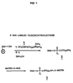
- Oligonucleotides prepared for use as biotinylated primers were modified as shown in Figure 1.
- the synthetic oligonucleotides on solid support were derivitized at the 5′ end by reaction with the protected hexylamino phosphoramadite [structure 1], followed by oxidation and deprotection according to standard procedures (McBride and Caruthers, Tetrahedron Letters, 24 :245-248, 1983) to afford 5′ amino labelled oligomers [structure 2].
- the reaction of [2] with N-hydroxysuccinimidyl esters of biotin derivatives gave 5′ biotinylated oligonucleotides [structure 3] in good yields, which were purified by polyacrylamide gel electrophoresis. (See below, Example 3.)
- Oligonucleotides used as PCR primers or capture probes were purified in one of two ways: (a) polyacrylamide gel electrophoresis or (b) spin column.
- primer set SK43 and SK44 (described by Kwok et al., J. Inf. Dis., 158 : 1193-1197, 1988) was used.
- reaction contents were as above, except that 200 ⁇ M TTP was replaced with 160 ⁇ M biotin-11-dUTP, 40 ⁇ M TTP.
- the PCR temperature regime began with an initial extended denaturation at 94° C for 5 minutes, followed by an annealing step at 50° C for 25 seconds, and an elongation step at 72° C for 1 minute.
- the temperature regime consisted of: denaturation at 94° C for 25 seconds, annealing at 50° C for seconds and extension at 72° C for 1 minute.
- the final (thirtieth) cycle was: denaturation at 94° C for 25 seconds, annealing at 50° C for 25 seconds and an extended elongation at 72° C for ten minutes.
- 90 ⁇ l of reaction contents were removed from under the mineral oil and stored at 4° C until analysis of PCR products.
- a plasmid (pTAX) containing the entire tax gene of HTLV-1 was used as a positive control template.
- concentration of plasmid DNA was determined spectrophotometrically, and dilutions were made in water to deliver known copy numbers of target DNA to PCR reactions. In this way, a positive control template was devised.
- a heterologous plasmid was also used with the HTLV-1 tax primer set, as a PCR negative control.
- This plasmid, pRT-POL contained sequences of the HTLV-1 polymerase (pol) region.
- the plasmid DNA concentration was determined, and was diluted serially as described above for pTAX. The plasmid concentrations were calculated to deliver a known copy number in a specific volume added to the PCR reaction mix (i.e., 2 ⁇ l template DNA added to 98 ⁇ l reaction mix).
- plasmid pTAX In order to directly compare three PCR detection systems side-by-side, known copy numbers of plasmid pTAX (Example 5) were amplified by PCR. The plasmid concentration was adjusted with water to deliver 1300, 100, 50, 20 or 10 copies of template per reaction.
- HTLV-1 tax primer set SK43/SK44 (Kwok et al., J. Inf. Dis., 158 : 1193-1197, 1988) was synthesized and purified by Bio-Spin R 6 spin columns (described in Example 3), and each primer was added to the PCR reaction at a concentration of 50 pmoles.
- Unlabelled amplified DNA was generated by using each of the four dNTPS at 200 ⁇ M for PCR amplification. These unlabelled PCR products were used for determining detection sensitivity of Southern blot hybridization (Example 9) and oligonucleotide hybridization (OH) (Example 10) assays. Amplification proceeded for 30 cycles using the reaction mix and temperature conditions as described in Example 4.
- PCR amplification with primer set SK43/SK44 was also done to produce biotinylated amplicons, through the incorporation of biotin-11-dUTP.
- Conditions for this HTLV-1 tax amplification were as described above (Example 6), except that the reaction mix included dATP, dCTP and dGTP at 200 ⁇ M; biotin-11-dUTP at 160 ⁇ M; TTP at 40 ⁇ M.
- the biotin-labelled PCR products were used to determine detection sensitivity of plate capture hybridization (Example 8) and Southern blot hybridization (Example 9). Amplification proceeded for 30 cycles using the reaction mix and temperature conditions as described in Example 4.
- a capture plate was prepared by binding 25 ng oligonucleotide "capture probe" SK45 (Kwok et al., J. Inf. Dis., 158 : 1193-1197, 1988) in 1 M ammonium acetate to wells of a Dynatech Immulon R 2 plate, and incubating overnight at 37°C.
- the biotinylated products of PCR (Example 7) were diluted 1:25 and 1:100 in 1 M guanidine thiocyanate (GuSCN), heat denatured (five minutes, 100° C) and quick cooled in an ice water bath.
- the detection limit for this assay appeared to be 20 copies of clean target plasmid (OD490 of 1:25 dilution was 0.123).
- the "no template" negative control and the negative control using a heterologous amplification template gave OD values of 0.007 and 0.000, respectively, at 1/25 dilution; and 0.026 and 0.042, respectively, at 1/100 dilution.
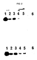
- the gel was visualized with a uv transilluminator and photographed. The gel was then treated for 30 minutes in 0.5 N NaOH, 1 M NaCl and for 30 minutes in 0.5 M Tris-HCl, pH 7.5, and blotted overnight to Nytran R (Schleicher and Schuell, Inc., Keene, HN) nylon membranes in high salt, following standard procedures known in the art.
- SDS sodium dodecyl sul
- PCR amplification was as described in Examples 6 and 7, and conditions for agarose gel electrophoresis, blotting, prehybridization and hybridization were as described in this Example.
- Biotin-labelled PCR products of Example 7 are shown: amplified products of 1300, 100, 50, 20 and 10 copies of pTAX (Lanes 1-5, respectively); PCR "no template” negative control (Lane 6).
- amplicon-specific probe SK45
- 32P-ATP polynucleotide kinase
- this radiolabelled probe was added to hybridization buffer (6 x SSPE, 1% (w/v) SDS) at a rate of 4.1 x 10⁶ cpm/blot. After hybridization at 60° C for 2-3 hours, the blot underwent several stringency washes and was placed under Kodak X-OMAT AR film overnight at -80° C with an intensifying screen.
- the detection limit for the Southern blot was 10 copies of plasmid pTAX target for PCR products that were not biotinylated (Example 6), and 20 copies of plasmid target for biotinylated PCR products (Example 7). (See photograph of autoradiogram, Figure 2.) The "no template" PCR negative control gave no detectable product on autoradiography.
- the photograph shown is an overnight exposure (-80° C, with intensifying screen on Kodak X-OMAT AR film) of autoradiogram, showing signal generated from either 1300, 100, 50, 20 or 10 copies of pTAX (Lanes 1-5, respectively); "no template" PCR negative control (Lane 6); 104 extracted Jurkat cells constitutively expressing HTLV-1 tax, amplified by PCR (Lane 7); 10 extracted Jurkat cells, amplified by PCR (Lane 8); OH negative control in which no amplified DNA was added to 32P-SK45 for OH assay (Lane 9).
- PCR products from primer set SK43/SK44 were analyzed by oligonucleotide hybridization assay (OH assay) to determine the limit of detection for this format.
- OH assay oligonucleotide hybridization assay
- the procedure was essentially as described by Abbott et al., J. Inf. Dis. 158 :1158-1169 (1988) for liquid hybridization (LH).
- Thirty microliters of PCR amplified product were added to 10 ⁇ l of a probe mixture containing 250,000-400,000 cpm of 32P labelled SK45 probe diluted in 44 mM EDTA, 66 mM NaCl.
- the DNA mixture was denatured at 95° C for five minutes, followed immediately by annealing to probe at 55° C for 15 minutes.
- agarose gel loading buffer (Example 9) were added, and half volume was loaded to a native 10% (w/v) polyacrylamide gel (acrylamide:bis-acrylamide, 19:1) in TBE. Electrophoresis was at 200 volts until the bromophenol blue reached the bottom gel front. The upper half of the gel was exposed to Kodak X-Omat AR film for exposures of 3 hours and overnight. at -80° C with an intensifying screen.
- the plate capture assay (Example 8) was compared to more conventional assays, Southern blot hybridization (Example 9) and oligonucleotide hybridization (OH) assay (Example 10), for the ability to detect minimal copy numbers of target HTLV-1 DNA after 30 cycles of PCR amplification (Examples 6 and 7).
- Each of the three detection systems tested (plate capture assay, Southern blot and OH assay) were essentially comparable in their ability to detect small copy numbers of clean target plasmid after thirty cycles of PCR amplification.
- the plate capture was found to detect 20 copies, the Southern blot between 10 and 20 copies, and OH, 10 copies of clean target. Of the three detection systems, the plate assay is fastest, giving a definitive result in hours.
- the plate assay provides numerical OD values, as opposed to subjective autoradiogram bands with Southern blot and OH assays. Therefore, the present invention enables the objective numerical determination of "positivity" of a sample, and the calculation of an objective cut-off value for the lower limits of positivity. (This cut-off value is an OD value, below which samples are considered to be negative with a calculated degree of statistical significance.)
- the OD values provided by the plate assay also allow for the quantitative determination of a "signal to noise" ratio, which is of importance in the validation of a diagnostic test.
- "signal to noise” can be only estimated by a visual evaluation of signal from autoradiography.
- Two sets of primers and capture probes were synthesized to specifically detect Chlamydia trachomatis.
- the oligonucleotide sequences were chosen from the cryptic plasmid of the C. trachomatis L1 serovar (Hatt et al., Nucleic Acids Research, 16 :4053-4067, 1988).
- Primer set A generated a 208 bp specific amplicon, using the following primers: CP-24 (25-mer, + polarity, bases 195-219) 5′ GGG ATT CCT GTA ACA ACA AGT CAG G 3′ i.e, the primer is any oligonucleotide which will bind to or cause elongation through the following sequence: 5′ C CTG ACT TGT TGT TAC AGG AAT CCC 3′ CP-27 (26-mer, - polarity, bases 377-402) 5′ CCT CTT CCC CAG AAC AAT AAG AAC AC 3′ i.e, the primer is any oligonucleotide which will bind to or cause elongation through the following sequence: 5′ GT GTT CTT ATT GTT CTG GGG AAG AGG 3′
- the primer set A 208 bp specific amplicon was detected with capture probe CP-35: CP-35 (26-mer, - polarity, bases 235-260) 5′ CAT AGC ACT ATA GAA CTC TGC AAG CC 3′ i.e, the probe is any oligonucleotide which will bind to the following sequence: 5′ GG CTT GCA GAG TTC TAT ACT GCT ATG 3′
- Chlamydia primer set B generated a 173 bp specific amplicon using the following primers: CP-37 (23-mer, + polarity, bases 678-700) 5′ GTC CTG CTT GAG AGA ACG TGC GG 3′ i.e, the primer is any oligonucleotide which will bind to or cause elongation through the following sequence: 5′ CC GCA CGT TCT CTC AAG CAG GAC 3′ CP-38 (24-mer, - polarity, bases 827-850) 5′ CTC CCA GCT TAA GAA CCG TCA GAC 3′ i.e, the primer is any oligonucleotide which will bind to or cause elongation through the following sequence: 5′ GTC TGA CGG TTC TTA AGC TGG GAG 3′
- the primer set B 173 bp amplicon was detected with capture probe CP-39: CP-39 (23-mer, - polarity, 726-748) 5′ TGT CTT CGT AAC TCG CTC CGG AA 3′ i.e, the probe is any oligonucleotide which wills bind to following sequence: 5′ TT CCG GAG CGA GTT ACG AAG ACA 3′
- McCoy cells either uninoculated or infected with C. trachomatis, were lysed in 2SP (20 mM sodium phosphate buffer, pH 7.2, 2 M sucrose) containing 100 ⁇ g/ml Proteinase K, 1% Tween-20 and were incubated at 55°C for 1 hour, followed by incubation at 95°C for ten minutes.
- Primer Set A (Example 12) was used in a 30 cycle PCR amplification, and the products were examined by plate assay as described in Example 8.
- CPE cytopathic effects
Abstract
Description
- The present invention provides a format for hybridization capture of polymerase chain reaction (PCR) amplified DNA on a polystyrene solid support having enhanced protein binding capacity. Preferably such solid support is a microtiter plate having a plurality of wells. After labelling the amplified target DNA (eg., with biotin) during amplification in the PCR reaction, the labelled DNA is specifically captured by base-pair hybridization to an amplicon-specific oligonucleotide capture probe that has been passively bound to the microtiter well. If biotin is used as the label, avidin:HRP complex is added and is reacted with either (a) hydrogen peroxide substrate and o-phenylene diamine (OPD) chromogen or (b) hydrogen peroxide substrate and tetramethylbenzidine chromogen (TMB). A colorimetric signal develops. allowing for the quantitative detection of PCR-amplified DNA.
- The sensitivity of the plate capture of biotinylated PCR products has been found to be comparable to the sensitivity using radiolabelled probes in Southern blot hybridizations and oligonucleotide hybridization (OH) assays. The plate capture format, however, offers several advantages: (a) quicker assay time; (b) a less labor intensive assay format, without the use of a radiolabelled probe and (c) an objective, quantitative evaluation of hybridization.
- Another aspect of the invention is the diagnosis of specific disease states by the detection of the presence of specific DNA sequences that characterize the causative microorganism. Specific probes and primers have been discovered for one of such microorganisms; namely; Chlamydia trachomatis.
- Still another aspect of the invention is the provision of kits for DNA sequence detection, which kits comprise the polystyrene solid support and sets of PCR reagents including the specific primers and probes pre-selected for the amplification of the target DNA sequences that will hybridize with the capture probes. Such kits would also typically include the enzyme or enzymes needed for the PCR reaction, preferably a thermostable enzyme such as Taq (Thermus aquaticus) polymerase. In the case of the use of the microtiter plate, they would also include microtiter plates with the capture probes for the specific target sequence already bound thereto.
- U.S. Patent Nos. 4,683,195 and 4,683,202 disclose methods of amplifying DNA sequences by a technique now known in the art as "polymerase chain reaction" (PCR). The polymerase chain reaction is a procedure in which DNA is specifically amplified by multiple primer extension syntheses of complementary strands (Saiki et al., Science, 230: 1350-1354 and 239: 487-491; 1985, 1988). The PCR product, amplified up to 10⁶-10⁷ fold, is a DNA fragment of discrete size (amplicon) which can be detected by gel electrophoresis, or by other means as described herein. Briefly, PCR involves the preparation of short oligonucleotide primers which correspond to opposing ends of a known "target" sequence which one intends to amplify and subsequently detect. In this procedure, DNA or RNA is extracted from cells, tissues, body fluids and the like. The nucleic acid is denatured and the oligonucleotide primers are added in molar excess, along with dNTPs (deoxyribonucleotide triphosphates) and a DNA polymerase enzyme, such as preferably heat stable Taq polymerase. Upon subsequent heat denaturing, cooling to allow annealing to primers, and primer extension by DNA polymerase, two "long products", which begin with the respective primers, are produced, complementary to the two original strands. This procedure is repeated, and after a second cycle two original strands, two long products from
cycle 1, two new "long products", and two "short products" are produced. The length of these short products (amplicons) is equal to the number of nucleotides between and including both primers. With additional cycles, additional "long products" are produced, increasing in a linear fashion with each cycle. However, the generation of amplicons increases at an exponential rate with each cycle, and by means of this amplification, the detection of extremely small quantities of DNA is enabled. - Through the use of PCR technology, the detection of specific DNA sequences present in minute quantities is possible. Several of these techniques involve the use of various hybridized probes affixed to certain materials.
- As will be fully described later, the present invention utilizes fixation of a capture DNA sequence to microtiter wells and subsequent detection by hybridization to labelled (viz. biotinylated) amplicons. It also involves the use of guanidine thiocyanate in the hybridization step: Several procedures have been described in the literature which are relevant herein. Two such reports describe the immobilization of large target DNAs to microtiter wells, followed by the hybridization of these targets to biotinylated oligonucleotide or nick-translated DNA probes.
- Cook et al., Nucleic Acids Research, 16:4077-4095 (1988) immobilized bacteriophage M13 target DNA on microtiter wells of ImmulonR 2 plates by incubating target DNA in 1 M ammonium acetate for 1.5-2 hours at 37° C. The immobilized target DNA was detected after hybridization to terminal-biotin labelled or internal-biotin labelled oligonucleotide probes, followed by addition of streptavidin-HRP complex and hydrogen peroxide/o-phenylene diamine (OPD) for colorimetric detection of hybridization.
- Nagata et al., FEBS Lett., 183:379-382 (1985) immobilized lambda target DNA in microtiter wells in PBS containing 0.1 M MgCl₂. After an overnight room temperature incubation followed by removal of the solution, the plate was irradiated with ultraviolet light. Hybridization with a biotinylated (nick-translated) lambda DNA probe was followed by complexing with avidin-beta-galactosidase. Fluorescence was measured after the addition of the substrate, 4-methylumbelliferyl-beta-D-galactoside.
- Hevey et al., U.S. Patent No. 4,228,237, described a method which uses biotin reacting to avidin (covalently attached to an enzyme) to detect a ligand in liquid medium. Our present method differs in that the ligand is directly labelled with biotin, not an intermediate biotin labelled anti-ligand. Further, various other prior disclosures describe specific hybridization probes and diagnostic uses thereof; e.g., U.S. Pat. No. 4,358,535 (Falkow et al.) and European Patent Application No. 82301804.9 (Publication No. 63,879; Yale University). The latter describes the use of biotin-labelled DNA probes detected by enzymes linked to avidin or biotin-specific antibodies.
- Ranki et al., U.S. Patent No. 4,486,539, describes the use of two non-overlapping nucleic acid reagents (one bound to a solid carrier and the other being labelled) to detect and identify microbes by DNA hybridization. The present invention differs in that the target nucleic acid is itself labelled, and does not require the use of an additional label led nucleic acid probe for detection.
- Stabinsky, U.S. Patent No. 4,751,177, described a DNA detection method wherein the target DNA is hybridized in solution to a mediator polynucleotide and to a labelled probe polynucleotide. The mediator polynucleotide, in turn, is complementary to a polynucleotide immobilized on a solid support. The present invention differs in that (a) the target is labelled during amplification; no labelled reporter group is needed for detection, and (b) the capture probe is bound directly to a solid support, without the use of a mediator polynucleotide.
- Guanadine thiocyanate (GuSCN) has been used in cell extraction and subsequent nucleic acid hybridization. For example, Thompson and Gillespie, Anal. Biochem., 163:281-291 (1987), prepared radiolabelled RNA probes to detect target DNA or RNA. In a dot blot format, the ³²P-labelled probe was hybridized in 5 M GuSCN/0.1 M ethylenediaminetetraacetic acid (EDTA), disodium salt, pH 8.0. Pellegrino et al., BioTechniques, 5:452-459 (1987), described a solution hybridization to detect HIV-RNA in blood cells. Blood cells were dissolved in 5 M GuSCN/0.1 M EDTA and hybridized with a radiolabelled RNA probe in the same solution at room temperature. TCA-precipitated hybrids were collected on membranes and radioactivity was determined by scintillation counting. Gillespie, International Patent Application No. PCT/US87/01023 (International Publication No. WO 87/06621), described the use of guanidine thiocyanate for molecular hybridization using a labelled probe to detect target DNA bound to a solid support. The patent describes the use of GuSCN for molecular hybridization over a concentration range of 3 M-6.5 M at ambient temperature. The format for such hybridization includes binding target nucleic acid to nitrocellulose or nylon membrane (dot blot or Southern blot), prehybridization, hybridization to a radiolabelled probe in GuSCN, washing and detection by autoradiography.
-
- Figure 1 illustrates the preparation of a chemical linker arm which was modified to enable biotinylation of oligonucleotide PCR primers.
- Figure 2 illustrates the results of an autoradiogram of PCR products bound to NytranR membrane.
- Figure 3 illustrates the results of oligonucleotide hybridization (OH) assay using PCR products of Example 6.
- The terms "oligonucleotide", and "primer" used herein will have the meaning defined in the aforesaid U.S. Patent No. 4,683,202. The term "capture probe" as used herein will be defined as an oligonucleotide which is completely or substantially complementary to the sequences of the amplicon within the boundaries of the primers. In the present preferred embodiment, the capture probe is not tailed, but it may be tailed with deoxyribonucleotides or ribonucleotides.
- In the practice of the present invention, the primers and probes are selected to be "substantially" complementary to the different strands of the target sequence to be amplified.
- For one of the specific embodiments described herein, two sets of PCR primers and capture probes were selected from the nucleotide sequence of the cryptic plasmid of the Chlamydia trachomatis L1 serovar (Hatt et al., Nucleic Acids Research, 16: 4053-4067, 1988). This enabled the specific amplification and detection of C. trachomatis. For other general purposes, primer and probe selection for individual targets will be as described in the aforesaid U.S. Patent No. 4,683,202.
- As an alternative to, and improvement over known techniques for capturing the amplified sequence (amplicon) resulting from the application of PCR as previously described, the present invention, in its preferred embodiment, utilizes an oligonucleotide capture probe passively bound to the wells of the microtiter plate in order to capture (by sequence-specific hybridization) biotin-labelled amplicons. Avidin-horseradish peroxidase is then employed to the biotinylated amplicons, and its reaction with the substrate (hydrogen peroxide) and chromogen (either OPD or TMB) yields a quantitative colorimetric signal.
- Again, as described in the aforesaid U.S. Pat. No. 4,683,202, deoxyribonucleotide triphosphates dATP, dCTP, dGTP and TTP are also added to the synthesis mixture in adequate amounts and the resulting solution is heated to about 90°-100° C for about 0.5 to 10 minutes, to separate strands of template. After this heating period, the solution is quickly cooled to the temperature which is preferable for the primer template annealing. To this mixture is added an appropriate agent for inducing or catalyzing the primer extension reaction, and the reaction is allowed to occur under conditions known in the art. The inducing agent may be any compound or system which will function to accomplish the synthesis of primer extension products, including enzymes. Suitable enzymes for this purpose include, for example, Escherichia coli (E. coli) DNA polymerase I, Klenow fragment of E. coli DNA polymerase I, T4 DNA polymerase, other available DNA polymerases, reverse transcriptase, and other enzymes, including preferably heat-stable enzymes (viz. Taq DNA polymerase) which will facilitate incorporation of the nucleotides in the proper manner to form the primer extension products which are complementary to each nucleic acid strand.
- The newly synthesized strand and its complementary nucleic acid strand form a double-stranded molecule, the strands of which are separated using any denaturation procedure to provide single-stranded molecules, preferably heat.
- New nucleic acid is synthesized on the single-stranded template molecules. Additional enzyme, nucleotides and primers may be added if necessary for the reaction to proceed under the conditions prescribed above. Again, the synthesis will be initiated at one end of the oligonucleotide primers and will proceed along the single strands of the template to produce additional complementary nucleic acid by primer extension.
- The steps of strand separation and extension product synthesis can be repeated as often as needed to produce the desired quantity of amplified nucleic acid sequence.
- In general, any material (molecule, atom) may be used in the present invention to provide an indication or "signal" that is detectable (and preferably quantifiable) and which can be attached to or incorporated into the nucleic acid.
- In a preferred embodiment of the present invention, two methods are employed to prepare amplicons which incorporate biotin labels during the PCR amplification. In the first case, biotin-11-dUTP (a TTP analog chemically modified with a biotin label) partially replaces TTP in the PCR reaction. The biotin-11-dUTP is incorporated by Tag DNA polymerase during primer extension, and the resulting DNA product is "internally" labelled with biotin.
- In the second case, a chemical "linker arm" is attached to the 5′ terminus of oligonucleotides bound to a solid support. In the preferred embodiment, the "linker arm" as used herein is a molecule which is chemically attached to the 5′ primer terminus (by phosphoramadite chemistry) and is capable of covalently linking an amino group or groups to the oligonucleotide. The amino groups in turn are biotinylated with biotin-X-NHS. These 5′ biotin-labelled oligonucleotides, when used as primers in the PCR reaction, generate amplicons which are biotinylated at their 5′ termini.
- The "linker arm" can be any molecule which is long enough to act as a spacing agent to distance the label away from the oligonucleotide sequence, and also be amenable to the attachment of one or more suitable labels, thermostable enzymes or ligands.
- In the preferred embodiment of the present invention, either or both of these two biotin-labelling procedures (incorporation of biotin-11-dUTP and 5′ biotin-labelled primers) may be used to label the amplified products of PCR.
- Besides biotin, other labels may also be used in this invention, including radiolabelled compounds, luminescent or fluorescent reagents, electron dense reagents, thermostable enzymes, ligands, antibody-hapten complexes, and chelating systems. The alternative labels must be compatible with the temperature conditions of PCR amplification, as well as conditions of denaturation and capture hybridization.
- One such example of alternative labels includes digoxigenin-11-dUTP, which like biotin-11-dUTP, is incorporated by Taq polymerase into labelled, amplified DNA. Digoxigenin labelled amplicons can then be detected with an antibody against digoxigenin conjugated with alkaline phosphatase, followed by standard colorimetric detection as described herein.
- Another example of an alternative label is ³²P-labelled deoxynucleotide triphosphates, which also can be used to partially replace unlabelled deoxynucleotide triphosphates in the PCR reaction mix. The extent of capture hybridization could be determined, for instance, by scintillation counting of removable microtiter wells (described below, Step 2) after capture hybridization and washing.
- The capture plate or support used in the present invention will be polystyrene solid support that has enhanced protein binding capacity. Various treatments of such support to accomplish such enhancement are known in the art (for example, irradiation with ⁶⁰Co).
- The polystyrene solid support can take any of several forms; e.g. microtiter plates, micromagnetic particles (available from Advanced Magnetics, Inc.); beads, strips, dipsticks, etc.
- Preferably, the polystyrene support used in the present invention will be a microtiter plate having enhanced protein binding capacity and a plurality of wells, with the most preferable of such microtiter plates being those known as "
Dynatech Immulon R 2" (Dynatech Laboratories, Inc., Chantilly, VA, supplied by Fisher Scientific). - In a preferred embodiment of the present invention, an amplicon-specific oligonucleotide capture probe is passively bound to wells of an
Immulon R 2 polystyrene microtiter plate at a concentration of 25 ng DNA/ well, in a solution of 1 M ammonium acetate. Such microtiter plates have a multiplicity of wells and thus provide both high surface area and permit a high number of simultaneous assays (e.g. 96). - In addition to the passive attachment of the capture probe, alternative methods of attachment may be used, including but not limited to: covalent attachment; attachment via an intermediary protein (e.g., BSA); and attachment by any alternative chemical means.
- The hybridization step of the present invention, more particularly the "capture hybridization" is preferably accomplished in the presence of 1 M guanidine thiocyanate, 20 mM ethylene diaminetetraacetic acid (EDTA) disodium salt, pH 8.0. The concentration range for accomplishing microtiter plate capture hybridization is preferably 0.5 to 2.0 Molar GuSCN and most preferably 1.0 to 2.0 M.
- Other reagents may also be used to accomplish capture hybridization. For example, a solution of ammonium thiocyanate (0.5 to 5.0 M, most preferably 5.0 M) may be used in place of guanidine thiocyanate.
- Another possible substitute for guanidine thiocyanate is a reagent consisting of: 30% (v/v) deionized formamide; 3 x SSPE [1 x SSPE= 0.18 M Nacl; 10 mM NaPO₄, pH 7.7; 1 mM EDTA]; 5% (w/v) dextran sulfate; 0.1% Triton X-100 (octyl phenoxy polyethoxyethanol; Sigma Chemical Co., St. Louis, MO).
- The format for such hybridization includes the following steps:
- 1. Preparation of Capture Probe
- 2. Preparation of Microtiter Capture Plate
- 3. Dilution and Denaturation of Amplified, Labelled DNA
- 4. Capture Hybridization
- 5. Wash
- 6. Block
- 7. Addition of Avidin:Horseradish Peroxidase
- 8. Wash
- 9. Addition of Substrate and Chromogen; Color Development
- 10. Plate Reading (Quantification)
- These steps will now be described in detail.
- The capture probes herein are selected to be "substantially" complementary to sequences of the amplicon within the boundaries of the primers. Therefore, the capture probes must be sufficiently complementary to specifically hybridize to the amplicon under capture hybridization conditions. In the presently preferred embodiment, the capture probe typically contains 20-200 nucleotides.
- Further, more than one capture probe may be used, and additional capture probes may be used to capture the same or opposite strand of the labelled amplicon.
- Although the capture probe is defined herein as "sequences of the amplicon within the boundaries of the primers", the primers themselves, or oligonucleotides containing primer sequences, may also be used as capture probes. It should be noted, however, that when such oligonucleotides are used as capture probes, they must compete during hybridization with PCR primers which have not been removed from the reaction products.
- The capture probes and PCR-primers herein have been synthesized on a milliGen 7500 DNA Synthesizer (MilliGen/Biosearch, Inc., Burlington, MA) by standard beta-cyanoethyl phosphoramadite chemistry, other procedures although known to those skilled in the art are possible. Before use, the capture probes and PCR-primers were partially purified by either electrophoresis through 15% (w/v) polyacrylamide gels or by spin column (See Example 3). The final partially purified capture probes and primers were suspended in water, its concentration was determined spectrophotometrically, and it was stored at 4° C until needed.
- Although the capture probe as described herein is produced synthetically, it need not be. It may also be obtained as a subset of naturally occurring DNA prepared by standard means (eg., restriction endonuclease digestion).
- The oligonucleotide capture probe is preferably fixed to the wells of a
Dynatech Immulon R 2 polystyrene microtiter plate (Dynatech Laboratories, Inc., Chantilly, VA., supplied by Fisher Scientific). TheImmulon R 2 plate is an arrangement of 96 miniature polystyrene test tubes (wells) in a single plastic plate, designed for general use in microvolume solid phase immunoassay procedures. In the preferred embodiment of the present invention, plates with flat bottom wells (400 µl volume capacity) are used, but it is assumed that "U" bottom wells may also be used (300 µl volume capacity). - As another preferred embodiment, the probe may also be fixed to
Immulon R 2 RemovawellR strips (Dynatech), which are removable strips of twelveImmulon R 2 polystyrene test tubes (wells) that are fitted into RemovawellR strip holders (Dynatech) in a microtiter plate format. - The oligonucleotide capture probe is diluted to yield 25 ng per microtiter well in 50 µl 1 M ammonium acetate (Fisher Scientific, Fair Lawn, NJ). "Blank wells" are also prepared by adding 1 M ammonium acetate (without oligonucleotide capture probe) to certain wells (to which labelled PCR products will be later added for mock hybridization). These blank wells are an indication of non-specific sticking of biotinylated DNA to wells of microtiter plates, and are used to calibrate the plate reading instrument (spectrophotometer) (See below, Step 10).
- The plate is sealed with mylar plate sealer (Dynatech) and is incubated at 37° C overnight for passive fixation of the oligonucleotide capture probe to the polystyrene surface. If the plates are not to be used immediately, they are stored at 4° C until needed. Plates stored in this way for up to six weeks showed no significant difference in their ability to capture DNA.
- Just prior to hybridization, the wells of the plates are washed twice with 200 µl of 2 x SSC [1 x SSC= 0.15 M NaCl; 15 mM sodium citrate buffer, pH 7.0] and once with 200 µl of 2 x SSC, 0.1% (v/v) Triton X-100. All plate washes described herein are done using a multiple channel pipet device (eg. TitertekR), or may be done using an appropriate automatic microtiter plate washer.
- The biotinylated PCR products are diluted (in a range of from 1:10 to 1:100 or more) in 1 M GuSCN (Ultra-pure grade; Boehringer Mannheim Biochemicals, Indianapolis, IN), 20 mM EDTA (Sigma), pH 8.0 (prepared from a 5 x stock), heated at 100° C for five minutes and quickly chilled in an ice water bath. [The heating step accomplishes heat denaturation of the PCR products, and the quick cooling discourages reannealing of amplicon strands, favoring hybridization to the oligonucleotide capture probe.] Aliquots of 100 µl are loaded to each well, including the wells designated as "blanks" (no capture probe) as described above (Step 2).
- Specific capture of amplified, labelled DNA by oligonucleotide capture probe is accomplished by incubation at room temperature for 1-3 hours. During this time, PCR amplicons are specifically captured by the oligonucleotide capture probe, based on sequence complementarity between the capture probe and labelled amplicon.
- Following hybridization, the contents of all microtiter wells are discarded and the wells are washed two times with 200 µl of 2 x SSC, 0.1% (v/v) Triton X-100; and four times with 200 µl of 0.2 x SSC, 0.1% (v/v) Triton X-100, previously warmed to 37° C. These washes are performed to remove unbound biotinylated products from the microtiter wells.
- Following plate washing, the wells of the plate are then "blocked" for a minimum of 15-30 minutes with 200 µl of a solution of PBS, (PBS= 0.13 M NaCl, 7 mM Na₂HPO₄, 3 mM NaH₂PO₄, pH 7.0), 2% (w/v) bovine serum albumin (BSA) (Sigma), 0.1% (v/v) Triton X-100. This "blocking" step is required to minimize non-specific binding of avidin:HRP to microtiter wells.
- Avidin:horseradish peroxidase (HRP) complex (Vector Laboratories, Inc., Burlingame, CA) is diluted 1:2000 in PBS, 0.1% (v/v) Triton X-100, to a final concentration of 2.5 µg avidin:HRP/ml. Fifty microliters of diluted avidin:HRP are added to each well, and incubated for 30 minutes at room temperature. During this step, the avidin:HRP complex binds tightly to captured biotinylated products on the well. While avidin:HRP is used in the present invention, a complex of streptavidin:HRP may be used instead. Likewise, other compounds complexed with avidin (or streptavidin) may be used, including: alkaline phosphatase, beta-galactosidase, luciferase, fluorescein, Texas Red or any other agent capable of generating a colorimetric, fluorescent or luminescent signal.
- The wells of the microtiter plate are then washed four times with 200 µl PBS, 0.1% Triton X-100; and once with 200 µl PBS, 1 mM EDTA, the final wash incubated at room temperature for 1-10 minutes. These washes are performed to remove unbound avidin:HRP from microtiter wells prior to adding chromogen.
- Color development, in the preferred embodiment of the present invention, is accomplished using either of two chromogen systems: (a) hydrogen peroxide/OPD and (b) hydrogen peroxide/TMB.
- (a) Hydrogen peroxide/OPD. After the wash buffer (described in Step 8) has been removed, 150 µl OPD reagent [1.6 mg/ml o-phenylene diamine disodium salt (Sigma), 0.0125% (v/v) hydrogen peroxide (Fisher Scientific) in 0.15 M sodium phosphate/citrate buffer, pH 6.0] is added and color development is allowed to proceed in the dark for 1-30 minutes. Color development is stopped by the addition of 50 µl 4 N H₂SO₄.
- (b) Hydrogen peroxide/TMB. After the final wash buffer (described in Step 8) has been removed, 100 µl TMB color reagent is added to each well. [TMB color reagent consists of freshly mixing equal volumes of TMB peroxidase substrate (3,3′,5,5′-tetramethylbenzidine at a concentration of 0.4 g/l in an organic base; commercially available from Kirkegaard and Perry, Inc., Gaithersburg, MD) and hydrogen peroxide solution [0.02% (v/v) hydrogen peroxide in citric acid buffer; commercially available from Kirkegaard and Perry, Inc.]. The reaction is allowed to proceed at room temperature in the dark for 1-30 minutes, at which time color development is stopped by the addition of 100 µl 1 M phosphoric acid (H₃PO₄).
- The TMB substrate produces a precipitate with a blue color. Upon addition of phosphoric acid, a color change to yellow occurs, with an optical density measured at 450 nm (see below, Step 10). In addition to HRP substrates OPD and TMB, other soluble substrates include:
2,2-azino-di (3-ethylbenzthiazoline sulfonic acid)(ABTS) and 5-aminosalicylic acid (ASA). Insoluble substrates include 3-amino-9-ethylcarbazole (AEC) and 3,3-diaminobenzidene (DAB). - After color development is stopped, the optical density (OD) of samples in each well is determined by reading in an automatic microtiter plate reader capable of spectrophotometric determination (eg. InterMed ImmunoreaderR NJ-2000) at wavelengths specific for the chromogen used (see below).
- The OD of blank wells [biotinylated PCR products added to well without capture probe, as described in Step 2] is also determined. This signal (usually very low) is an indication of spurious "non-specific" sticking of biotinylated products to sample wells, rather than signal resulting from bona fide capture of biotinylated amplicons through hybridization with a capture probe. Thus, the OD of blank wells is subtracted from the OD of sample wells to give an accurate quantification of bona fide capture hybridization.
- The appropriate wavelength setting for the spectrophotometer (plate reader) is dependent on the specific chromogen used for the colorimetric reaction. Thus, if using hydrogen peroxide/OPD, the correct wavelength setting is 490 nm; if using hydrogen peroxide/TMB, the correct wavelength setting is 450 nm.
- Through the use of the present invention, various infectious diseases can be diagnosed by detecting the presence of the specific DNA sequences that characterize the causative microorganism; such as bacteria (e.g. Chlamydia, Salmonella, etc); viruses (e.g. hepatitis virus); various parasites (e.g. malaria-inducing); etc. Specifically, the invention will be useful in detecting
Human Immunodeficiency Viruses 1 and 2 (HIV 1 and HIV 2), Human T Lymphotropic Viruses I and II (HTLV-I and HTLV-II), Hepatitis B Virus (HBV), Hepatitis C Virus (HCV), Human Papilloma Virus (HPV) and Pneumocyctis carinii. As previously described, and as an additional embodiment of the invention, novel capture probes and primers have been discovered that are specifically capable of use in the detection of the organism Chlamydia trachomatis. Such novel probes and primers will be described in detail in the following specific examples. - The present invention also offers particular advantages in the context of clinical application. Through the use of the microtiter plates, a convenient, rapid, simple, economic and automatable assay procedure is provided. The assay is highly accurate and sensitive as compared to conventional means and can be conducted at a fraction of the time normally needed.
- The following examples are now offered by way of illustration and are not intended to be limiting.
- Biotin-11-dUTP was chemically synthesized by the methods of Langer et al., Proc. Natl. Acad. Sci. (USA), 78:6633-6637 (1981), and was suspended at a concentration of 0.4 mM in 100 mM Tris-HCl, pH 7.5, 0.4 mM EDTA. The ratio of biotin-11-dUTP:TTP was optimized, and the highest signal was obtained using a ratio of 4:1 (biotin-11-dUTP:TTP).
- All oligonucleotides used as PCR primers were synthesized on a MilliGen 7500 automated DNA synthesizer by standard beta-cyanoethyl phosphoramadite chemistry, using procedures standard to those skilled in the art.
- Oligonucleotides prepared for use as biotinylated primers were modified as shown in Figure 1. The synthetic oligonucleotides on solid support were derivitized at the 5′ end by reaction with the protected hexylamino phosphoramadite [structure 1], followed by oxidation and deprotection according to standard procedures (McBride and Caruthers, Tetrahedron Letters, 24:245-248, 1983) to afford 5′ amino labelled oligomers [structure 2]. The reaction of [2] with N-hydroxysuccinimidyl esters of biotin derivatives gave 5′ biotinylated oligonucleotides [structure 3] in good yields, which were purified by polyacrylamide gel electrophoresis. (See below, Example 3.)
- Oligonucleotides used as PCR primers or capture probes were purified in one of two ways: (a) polyacrylamide gel electrophoresis or (b) spin column.
- (a) Polyacrylamide Gel Electrophoresis Aliquots of 100-250 µg each oligonucleotide were dried in vacuo, resuspended in a minimal volume of gel loading buffer [TBE (89 mM Trisborate, 89 mM boric acid, 2 mM EDTA); 90% (v/v) deionized formamide; 0.02% (w/v) bromophenol blue], heated at 100° C for 5 minutes, and quickly cooled in an ice water bath. Samples were loaded onto a denaturing polyacrylamide gel [15% (w/v) acrylamide (acrylamide:bis-acrylamide, 29:1) (IBI, New Haven, CT), 7 M urea (IBI) in TBE] and electrophoresed at 650 volts for four hours. The DNA was visualized by uv shadowing over a thin layer chromatography plate (Eastman Kodak #13254 cellulose), photographed, and the gel band of full-length oligonucleotide was excised. The gel piece was crushed and DNA was eluted in water overnight at 37°C. The DNA was further purified by passage over a C₁₈ Sep-PakR cartridge (Waters Associates, Milford, MA), and eluted in 40% (v/v) acetonitrile (Fisher). After evaporation to dryness in vacuo, the DNA was suspended in water and its concentration was determined spectrophotometrically. Stock concentrations were adjusted with water and stored at 4° C until needed.
- (b) Spin Column Some oligonucleotide capture probes and primers were partially purified using
Bio-Spin™ 6 columns (Bio-Rad, Inc., Richmond, CA) in a swinging bucket rotor at low centrifugal forces, following manufacturer's instructions for the spin column. After partial purification, the concentration of oligonucleotides was determined spectrophotometrically, and stock solutions were prepared with water and stored at 4° C until needed. - Most primer pairs were tested empirically to determine their optimal temperature conditions for PCR amplification, with particular attention paid to the annealing temperature.
- For detection of HTLV-1 tax sequences, primer set SK43 and SK44 (described by Kwok et al., J. Inf. Dis., 158: 1193-1197, 1988) was used. Using this primer pair, a 100 µl PCR reaction consisted of: 1 x reaction buffer (RB) [10 x RB= 500 mM KCl; 100 mM Tris-HCl, pH 8.5; 25 mM MgCl₂]; 200 th each of dATP, dCTP, dGTP and TTP; 50 pmole each of SK43 and SK44 and 2 units recombinant Taq DNA polymerase (RecombiTaqR, Cetus Perkin Elmer, Norwalk, CT).
- For experiments in which biotin-11-dUTP was incorporated, the reaction contents were as above, except that 200 µM TTP was replaced with 160 µM biotin-11-dUTP, 40 µM TTP.
- For primer set SK43 and SK 44, the PCR temperature regime began with an initial extended denaturation at 94° C for 5 minutes, followed by an annealing step at 50° C for 25 seconds, and an elongation step at 72° C for 1 minute. For the next 28 cycles, the temperature regime consisted of: denaturation at 94° C for 25 seconds, annealing at 50° C for seconds and extension at 72° C for 1 minute. The final (thirtieth) cycle was: denaturation at 94° C for 25 seconds, annealing at 50° C for 25 seconds and an extended elongation at 72° C for ten minutes. After PCR amplification, 90 µl of reaction contents were removed from under the mineral oil and stored at 4° C until analysis of PCR products.
- A plasmid (pTAX) containing the entire tax gene of HTLV-1 was used as a positive control template. The concentration of plasmid DNA was determined spectrophotometrically, and dilutions were made in water to deliver known copy numbers of target DNA to PCR reactions. In this way, a positive control template was devised.
- A heterologous plasmid was also used with the HTLV-1 tax primer set, as a PCR negative control. This plasmid, pRT-POL, contained sequences of the HTLV-1 polymerase (pol) region. The plasmid DNA concentration was determined, and was diluted serially as described above for pTAX. The plasmid concentrations were calculated to deliver a known copy number in a specific volume added to the PCR reaction mix (i.e., 2 µl template DNA added to 98 µl reaction mix).
- In order to directly compare three PCR detection systems side-by-side, known copy numbers of plasmid pTAX (Example 5) were amplified by PCR. The plasmid concentration was adjusted with water to deliver 1300, 100, 50, 20 or 10 copies of template per reaction. HTLV-1 tax primer set SK43/SK44 (Kwok et al., J. Inf. Dis., 158: 1193-1197, 1988) was synthesized and purified by
Bio-Spin R 6 spin columns (described in Example 3), and each primer was added to the PCR reaction at a concentration of 50 pmoles. - Unlabelled amplified DNA was generated by using each of the four dNTPS at 200 µM for PCR amplification. These unlabelled PCR products were used for determining detection sensitivity of Southern blot hybridization (Example 9) and oligonucleotide hybridization (OH) (Example 10) assays. Amplification proceeded for 30 cycles using the reaction mix and temperature conditions as described in Example 4.
- PCR amplification with primer set SK43/SK44 was also done to produce biotinylated amplicons, through the incorporation of biotin-11-dUTP. Conditions for this HTLV-1 tax amplification were as described above (Example 6), except that the reaction mix included dATP, dCTP and dGTP at 200 µM; biotin-11-dUTP at 160 µM; TTP at 40 µM. The biotin-labelled PCR products were used to determine detection sensitivity of plate capture hybridization (Example 8) and Southern blot hybridization (Example 9). Amplification proceeded for 30 cycles using the reaction mix and temperature conditions as described in Example 4.
- A capture plate was prepared by binding 25 ng oligonucleotide "capture probe" SK45 (Kwok et al., J. Inf. Dis., 158: 1193-1197, 1988) in 1 M ammonium acetate to wells of a
Dynatech Immulon R 2 plate, and incubating overnight at 37°C. The biotinylated products of PCR (Example 7) were diluted 1:25 and 1:100 in 1 M guanidine thiocyanate (GuSCN), heat denatured (five minutes, 100° C) and quick cooled in an ice water bath. For each sample and control, 100 µl were added to each of four microtiter wells (triplicate SK45 capture probe wells and one "blank" well), and incubated for hybridization two hours at room temperature. The contents of the plates were discarded, and the plates were washed, blocked, and treated with avidin:horseradish peroxidase, as described above inStep 9. After adding H₂O₂ and OPD, color development was determined at 490 nm using a plate reader. - The results of the plate capture hybridization are included below. Referring to the table 1 below, capture by hybridization of biotinylated PCR products is illustrated using HTLV-1 primers SK43/SK44 specific for HTLV-1 tax, amplifying homologous template (pTAX) and heterologous template (pRT-POL). Biotin-11-dUTP was added as a precursor into the PCR reaction (See Example 7), and PCR conditions were as described for Example 4. Plate capture assay was done as described. Samples were diluted 1:25 and 1:100 in 1 M GuSCN for hybridization, after which plates were washed, blocked, and treated with avidin-HRP. OD₄₉₀ was determined after 35 minutes color development with H₂O₂/OPD. Numbers presented below are averages of sample triplicates.
Table 1 OD 490 1:25 Dilution OD 490 1:100 Dilution 1300 copies pTAX 1.693 0.444 100 copies pTAX 0.343 0.117 50 copies pTAX 0.300 0.053 20 copies pTAX 0.123 0.040 10 copies pTAX 0.031 0.029 NEGATIVE CONTROLS: No Template 0.007 0.026 10⁴ copies pRT-POL 0.000 0.042 - The detection limit for this assay appeared to be 20 copies of clean target plasmid (OD₄₉₀ of 1:25 dilution was 0.123). The "no template" negative control and the negative control using a heterologous amplification template gave OD values of 0.007 and 0.000, respectively, at 1/25 dilution; and 0.026 and 0.042, respectively, at 1/100 dilution.
- Aliquots (25 µl) of both unlabelled (Example 6) and biotin-labelled (Example 7) PCR products were dried in vacuo, resuspended in 5 µl water and 2 µl agarose gel loading buffer [0.25% (w/v) bromophenol blue, 0.25% (w/v) xylene cyanol, 30% (v/v) glycerol] and loaded on a 3% (w/v) NuSieveR GTG (FMC BioProducts, Rockland, ME)/ 1% (w/v) Sea Kem (FMC) agarose gel in TBE, containing 0.5 µg/ml ethidium bromide. The gel was visualized with a uv transilluminator and photographed. The gel was then treated for 30 minutes in 0.5 N NaOH, 1 M NaCl and for 30 minutes in 0.5 M Tris-HCl, pH 7.5, and blotted overnight to NytranR (Schleicher and Schuell, Inc., Keene, HN) nylon membranes in high salt, following standard procedures known in the art. The blot ("Southern blot") was then baked 30 minutes at 75° C and prehybridized 1-4 hours at 37° C in a sealed bag containing 6 x SSPE [1 x SSPE= 0.18 M NaCl, 10 mM NaPO₄, pH 7.7, 1 mM EDTA]; 1% (w/v) sodium dodecyl sulfate (SDS) (Aldrich Chemical Co., Milwaukee, WI); 10 x Denhardt's reagent [1% (w/v) each of ficoll (Pharmacia, Piscataway, NJ), polyvinylpyrrolidone (Sigma), and BSA (Sigma)]; and 50 µg/ml salmon sperm DNA, heat denatured and quick cooled.
- In Figure 2, PCR amplification was as described in Examples 6 and 7, and conditions for agarose gel electrophoresis, blotting, prehybridization and hybridization were as described in this Example.
- In the upper half of the photograph, Biotin-labelled PCR products of Example 7 are shown: amplified products of 1300, 100, 50, 20 and 10 copies of pTAX (Lanes 1-5, respectively); PCR "no template" negative control (Lane 6).
- In the lower half of the photograph are shown the unlabelled PCR products of Example 6: amplified products of 1300, 100, 50, 20 and 10 copies of pTAX (Lanes 1-5, respectively); PCR "no template" negative control (Lane 6).
- An amplicon-specific probe, SK45, was labelled with ³²P-ATP using polynucleotide kinase (Boehringer Mannheim Biochemicals), using procedures standard in the art. For hybridization, this radiolabelled probe was added to hybridization buffer (6 x SSPE, 1% (w/v) SDS) at a rate of 4.1 x 10⁶ cpm/blot. After hybridization at 60° C for 2-3 hours, the blot underwent several stringency washes and was placed under Kodak X-OMAT AR film overnight at -80° C with an intensifying screen.
- After overnight exposure, the detection limit for the Southern blot was 10 copies of plasmid pTAX target for PCR products that were not biotinylated (Example 6), and 20 copies of plasmid target for biotinylated PCR products (Example 7). (See photograph of autoradiogram, Figure 2.) The "no template" PCR negative control gave no detectable product on autoradiography.
- Referring to Figure 3, the conditions of OH assay (preparation of label led probe, denaturation and annealing conditions and conditions for polyacrylamide gel electrophoresis) were as described in this Example.
- The photograph shown is an overnight exposure (-80° C, with intensifying screen on Kodak X-OMAT AR film) of autoradiogram, showing signal generated from either 1300, 100, 50, 20 or 10 copies of pTAX (Lanes 1-5, respectively); "no template" PCR negative control (Lane 6); 10⁴ extracted Jurkat cells constitutively expressing HTLV-1 tax, amplified by PCR (Lane 7); 10 extracted Jurkat cells, amplified by PCR (Lane 8); OH negative control in which no amplified DNA was added to ³²P-SK45 for OH assay (Lane 9).
- The unlabelled PCR products from primer set SK43/SK44 (Example 6) were analyzed by oligonucleotide hybridization assay (OH assay) to determine the limit of detection for this format. The procedure was essentially as described by Abbott et al., J. Inf. Dis. 158:1158-1169 (1988) for liquid hybridization (LH). Thirty microliters of PCR amplified product were added to 10 µl of a probe mixture containing 250,000-400,000 cpm of ³²P labelled SK45 probe diluted in 44 mM EDTA, 66 mM NaCl. The DNA mixture was denatured at 95° C for five minutes, followed immediately by annealing to probe at 55° C for 15 minutes. Ten microliters of agarose gel loading buffer (Example 9) were added, and half volume was loaded to a native 10% (w/v) polyacrylamide gel (acrylamide:bis-acrylamide, 19:1) in TBE. Electrophoresis was at 200 volts until the bromophenol blue reached the bottom gel front. The upper half of the gel was exposed to Kodak X-Omat AR film for exposures of 3 hours and overnight. at -80° C with an intensifying screen.
- After either a three hour (not shown) or overnight exposure (Figure 3), ten copies of amplified plasmid pTAX target can clearly be seen as a specific band. The "no template" negative control and other negative controls produced no bands.
- The plate capture assay (Example 8) was compared to more conventional assays, Southern blot hybridization (Example 9) and oligonucleotide hybridization (OH) assay (Example 10), for the ability to detect minimal copy numbers of target HTLV-1 DNA after 30 cycles of PCR amplification (Examples 6 and 7). Each of the three detection systems tested (plate capture assay, Southern blot and OH assay) were essentially comparable in their ability to detect small copy numbers of clean target plasmid after thirty cycles of PCR amplification. The plate capture was found to detect 20 copies, the Southern blot between 10 and 20 copies, and OH, 10 copies of clean target. Of the three detection systems, the plate assay is fastest, giving a definitive result in hours. Moreover, the plate assay provides numerical OD values, as opposed to subjective autoradiogram bands with Southern blot and OH assays. Therefore, the present invention enables the objective numerical determination of "positivity" of a sample, and the calculation of an objective cut-off value for the lower limits of positivity. (This cut-off value is an OD value, below which samples are considered to be negative with a calculated degree of statistical significance.)
- The OD values provided by the plate assay also allow for the quantitative determination of a "signal to noise" ratio, which is of importance in the validation of a diagnostic test. For the Southern blot hybridization and OH assay formats, "signal to noise" can be only estimated by a visual evaluation of signal from autoradiography.
- Two sets of primers and capture probes were synthesized to specifically detect Chlamydia trachomatis. The oligonucleotide sequences were chosen from the cryptic plasmid of the C. trachomatis L1 serovar (Hatt et al., Nucleic Acids Research, 16:4053-4067, 1988).
- Primer set A generated a 208 bp specific amplicon, using the following primers:
CP-24 (25-mer, + polarity, bases 195-219)
5′ GGG ATT CCT GTA ACA ACAAGT CAG G 3′
i.e, the primer is any oligonucleotide which will bind to or cause elongation through the following sequence:
5′ C CTG ACT TGT TGT TACAGG AAT CCC 3′
CP-27 (26-mer, - polarity, bases 377-402)
5′ CCT CTT CCC CAG AAC AATAAG AAC AC 3′
i.e, the primer is any oligonucleotide which will bind to or cause elongation through the following sequence:
5′ GT GTT CTT ATT GTT CTGGGG AAG AGG 3′ - The primer set A 208 bp specific amplicon was detected with capture probe CP-35:
CP-35 (26-mer, - polarity, bases 235-260)
5′ CAT AGC ACT ATA GAA CTCTGC AAG CC 3′
i.e, the probe is any oligonucleotide which will bind to the following sequence:
5′ GG CTT GCA GAG TTC TATACT GCT ATG 3′ - Chlamydia primer set B generated a 173 bp specific amplicon using the following primers:
CP-37 (23-mer, + polarity, bases 678-700)
5′ GTC CTG CTT GAG AGAACG TGC GG 3′
i.e, the primer is any oligonucleotide which will bind to or cause elongation through the following sequence:
5′ CC GCA CGT TCT CTCAAG CAG GAC 3′
CP-38 (24-mer, - polarity, bases 827-850)
5′ CTC CCA GCT TAA GAACCG TCA GAC 3′
i.e, the primer is any oligonucleotide which will bind to or cause elongation through the following sequence:
5′ GTC TGA CGG TTC TTAAGC TGG GAG 3′ - The primer set B 173 bp amplicon was detected with capture probe CP-39:
CP-39 (23-mer, - polarity, 726-748)
5′ TGT CTT CGT AAC TCGCTC CGG AA 3′
i.e, the probe is any oligonucleotide which wills bind to following sequence:
5′ TT CCG GAG CGA GTTACG AAG ACA 3′ - Primer set B was used to amplify target plasmid pCHL-1. Using 5′ biotinylated primers, the target was amplified with cycles of PCR, and the biotinylated products were examined by capture hybridization (Table 2). After ten minutes color development (TMB/H₂0₂), as few as 20 starting copies of pCHL-1 were detectable (1:25 dilution, OD₄₅₀ = 0.140), with negligible background signal (OD₄₅₀ = 0.009 for 1:25 dilution).
Table 2 OD ₄₅₀ 1:25 OD ₄₅₀ 1:100 10⁴ copies pCHL-1 > 1.500 > 1.500 10³ copies pCHL-1 > 1.500 1.286 10² copies pCHL-1 0.686 0.170 50 copies pCHL-1 0.215 0.050 20 copies pCHL-1 0.140 0.035 Negative Control No template 0.009 0.000 - McCoy cells, either uninoculated or infected with C. trachomatis, were lysed in 2SP (20 mM sodium phosphate buffer, pH 7.2, 2 M sucrose) containing 100 µg/ml Proteinase K, 1% Tween-20 and were incubated at 55°C for 1 hour, followed by incubation at 95°C for ten minutes. Primer Set A (Example 12) was used in a 30 cycle PCR amplification, and the products were examined by plate assay as described in Example 8. After ten minutes color development, both 10 and 100 infected McCoy cells, when spiked in 10⁵ uninfected McCoy cells, gave OD₄₅₀ values >2.000 (for both 1/25 and 1/100 dilutions of product in GuSCN). Extracted and amplified uninfected cells alone (10⁵ cells) gave OD₄₅₀ values of <0.050.
- Clinical specimens, previously graded for extent of cytopathic effects (CPE) [in a range of "negative" (no CPE) to 4+ (most prominent CPE)], were likewise extracted and amplified for 30 cycles of PCR, using Primer Set A. The results of this assay are shown in Table 3:
Table 3 Culture Rating Volume of Lysed Specimen OD₄₅₀ 1/25 Dilution in GuSCN 1/100 Dilution in GuSCN 4+ 90 µl > 2.000 1.686 1+ 90 µl 0.145 0.086 Negative 90 µl < 0.050 < 0.050 4+ 10 µl > 2.000 1.417 1+ 10 µl < 0.050 < 0.050 Negative 10 µl < 0.050 < 0.050 - In summary, novel ways of labelling and detecting the amplified products of PCR are herein described. These labelling and capturing procedures offer several advantages over conventional methods of detection:
- 1. The biotinylation and capture of PCR products on microtiter plates is a faster assay. The plate assay in the present invention can be accomplished in 2 hours, compared to about 6-24 hours for OH assay, and as much as 48 hours for Southern blotting and hybridization.
- 2. The plate assay format is less labor intensive, and may be amenable to automation for analysis of large numbers of samples.
- 3. Unlike the other two assay formats examined, the plate assay format does not require the preparation, purification or handling of dangerous radioactive probes of high specific activity.
- 4. The capture of biotinylated products via plate assay provides an objective, quantitative evaluation of hybridization; the calculation of a statistical cut-off point for positivity of samples; and the quantitative calculation of an assay "signal-to-noise" ratio.
- Many modifications and variations of this invention may be made without departing from its spirit and scope, as will become apparent to those skilled in the art. The specific embodiments described herein are offered by way of example only, and the invention is to be limited only by the terms of the appended claims.
Claims (52)
5′ CCTGACTTGTTGTTACAGGAATCCC 3′
and 5′ GTGTTCTTATTGTTCTGGGGAAGAGG 3′ .
5′ GGCTTGCAGAGTTCTATAGTGCTATG 3′ .
5′ CCGCACGTTCTCTCAAGCAGGAC 3′
and 5′ GTCTGACGGTTCTTAAGCTGGGAG 3′ .
5′ TTCCGGAGCGAGTTACGAAGACA 3′ .
5′ CCTGACTTGTTGTTACAGGAATCCC 3′
and 5′ GTGTTCTTATTGTTCTGGGGAAGAGG 3′ .
5′ GGCTTGCAGAGTTCTATAGTGCTATG 3′ .
5′ CCGCACGTTCTCTCAAGCAGGAC 3′
and 5′ GTCTGACGGTTCTTAAGCTGGGAG 3′ .
5′ TTCCGGAGCGAGTTACGAAGACA 3′ .
5′ CCTGACTTGTTGTTACAGGAATCCC 3′
and 5′ GTGTTCTTATTGTTCTGGGGAAGAGG 3′ .
5′ GGCTTGCAGAGTTCTATAGTGCTATG 3′ .
5′ CCGCACGTTCTCTCAAGCAGGAC 3′
and 5′ GTCTGACGGTTCTTAAGCTGGGAG 3′ .
5′ TTCCGGAGCGAGTTACGAAGACA 3′ .
5′ GGGATTCCTGTAACAACAAGTCAGG 3′
5′ CCTCTTCCCCAGAACAATAAGAACAC 3′
5′ GTCCTGCTTGAGAGAACGTGCGG 3′
and 5′ CTCCCAGCTTAAGAACCGTCAGAC 3′ .
5′ CATAGCACTATAGAACTCTGCAAGCC 3′
and 5′ TGTCTTCGTAACTCGCTCCGGAA 3′.
Priority Applications (1)
| Application Number | Priority Date | Filing Date | Title |
|---|---|---|---|
| EP98111076A EP0875583B1 (en) | 1989-09-29 | 1990-09-27 | Primers and kits for the detection of Chlamydia trachomatis |
Applications Claiming Priority (2)
| Application Number | Priority Date | Filing Date | Title |
|---|---|---|---|
| US414542 | 1989-09-29 | ||
| US07/414,542 US5232829A (en) | 1989-09-29 | 1989-09-29 | Detection of chlamydia trachomatis by polymerase chain reaction using biotin labelled lina primers and capture probes |
Related Child Applications (1)
| Application Number | Title | Priority Date | Filing Date |
|---|---|---|---|
| EP98111076A Division EP0875583B1 (en) | 1989-09-29 | 1990-09-27 | Primers and kits for the detection of Chlamydia trachomatis |
Publications (3)
| Publication Number | Publication Date |
|---|---|
| EP0420260A2 true EP0420260A2 (en) | 1991-04-03 |
| EP0420260A3 EP0420260A3 (en) | 1991-09-18 |
| EP0420260B1 EP0420260B1 (en) | 1999-12-08 |
Family
ID=23641905
Family Applications (2)
| Application Number | Title | Priority Date | Filing Date |
|---|---|---|---|
| EP90118620A Expired - Lifetime EP0420260B1 (en) | 1989-09-29 | 1990-09-27 | Method and kit for detecting a target nucleic acid using a capture probe bound to a polystyrene support via an intermediary protein. |
| EP98111076A Expired - Lifetime EP0875583B1 (en) | 1989-09-29 | 1990-09-27 | Primers and kits for the detection of Chlamydia trachomatis |
Family Applications After (1)
| Application Number | Title | Priority Date | Filing Date |
|---|---|---|---|
| EP98111076A Expired - Lifetime EP0875583B1 (en) | 1989-09-29 | 1990-09-27 | Primers and kits for the detection of Chlamydia trachomatis |
Country Status (14)
| Country | Link |
|---|---|
| US (1) | US5232829A (en) |
| EP (2) | EP0420260B1 (en) |
| JP (1) | JP2719225B2 (en) |
| AT (2) | ATE187499T1 (en) |
| AU (2) | AU6329090A (en) |
| BR (1) | BR9004881A (en) |
| CA (1) | CA2026280C (en) |
| DE (2) | DE69033387T2 (en) |
| DK (2) | DK0420260T3 (en) |
| ES (2) | ES2275292T3 (en) |
| IL (1) | IL95800A (en) |
| NO (1) | NO302204B1 (en) |
| NZ (2) | NZ247522A (en) |
| ZA (1) | ZA907706B (en) |
Cited By (31)
| Publication number | Priority date | Publication date | Assignee | Title |
|---|---|---|---|---|
| EP0460569A1 (en) * | 1990-06-04 | 1991-12-11 | Tecnogen S.C.P.A. | Process for identifying polynucleotide sequences and device suitable therefor |
| WO1991019812A1 (en) * | 1990-06-11 | 1991-12-26 | Bio Merieux | Method for detecting a nucleotide sequence by sandwich hybridization |
| EP0480289A1 (en) * | 1990-10-09 | 1992-04-15 | Roche Diagnostics GmbH | Method for genus and species-specific detection of bacteria in a testliquid |
| EP0546761A1 (en) * | 1991-12-11 | 1993-06-16 | Becton, Dickinson and Company | Probes to chlamydia trachomatis |
| WO1993020234A1 (en) * | 1992-03-31 | 1993-10-14 | E.I. Du Pont De Nemours And Company | A rapid, high capacity nucleic acid based assay |
| WO1994010335A2 (en) * | 1992-10-09 | 1994-05-11 | Amoco Corporation | Assay methods |
| WO1994026934A2 (en) * | 1993-05-06 | 1994-11-24 | Baxter Diagnostics Inc. | Human papillomavirus detection assay |
| WO1995006753A1 (en) * | 1993-09-03 | 1995-03-09 | The United States of America, represented by The Secretary, Department of Health & Human Services | Method and compositions for primer specific solid-phase detection of pcr products |
| EP0675965A1 (en) * | 1992-01-29 | 1995-10-11 | Hitachi Chemical Co., Ltd. | Polynucleotide immobilized support |
| EP0630971A3 (en) * | 1993-06-23 | 1995-10-25 | Hoffmann La Roche | Method, reagents and kits for detection of neisseria gonorrhoea. |
| US5512445A (en) * | 1994-10-14 | 1996-04-30 | Gen-Probe Incorporated | Methods for the detection of Chlamydia trachomatis |
| US5547861A (en) * | 1994-04-18 | 1996-08-20 | Becton, Dickinson And Company | Detection of nucleic acid amplification |
| EP0726963A1 (en) * | 1991-12-23 | 1996-08-21 | Chiron Corporation | $i(CHLAMYDIAE) PROBES FOR USE IN SOLUTION PHASE SANDWICH HYBRIDIZATION ASSAYS |
| US5695926A (en) * | 1990-06-11 | 1997-12-09 | Bio Merieux | Sandwich hybridization assays using very short capture probes noncovalently bound to a hydrophobic support |
| EP0812921A2 (en) * | 1996-06-11 | 1997-12-17 | Becton, Dickinson and Company | Amplification and detection of chlamydia trachomatis nucleic acids |
| WO1998013527A2 (en) * | 1996-09-24 | 1998-04-02 | Rapigene, Inc. | Compositions and methods for enhancing hybridization specificity |
| EP0915170A1 (en) * | 1997-11-04 | 1999-05-12 | Becton, Dickinson and Company | Assay for chlamydia trachomatis by amplification and detection of chlamydia trachomatis cryptic plasmid |
| EP0935672A1 (en) * | 1996-10-09 | 1999-08-18 | University of New Mexico | In situ hybridization slide processes |
| DE19811732A1 (en) * | 1998-03-18 | 1999-09-30 | November Ag Molekulare Medizin | Plastic micro-titration plate with biomolecular coating inside cavities, forming part of biomolecule detection kit |
| EP0952228A2 (en) * | 1996-09-24 | 1999-10-27 | Rapigene, Inc. | Compositions and methods for enhancing hybridization specificity |
| US6037127A (en) * | 1994-03-31 | 2000-03-14 | E. I. Du Pont De Nemours And Company | Method for detection of non-denatured nucleic acid fragments |
| EP1000083A1 (en) * | 1997-07-30 | 2000-05-17 | Corning Incorporated | Oxidized styrenic polymers for dna binding |
| WO2000063429A2 (en) * | 1999-04-15 | 2000-10-26 | Bayer Aktiengesellschaft | Automatable rapid test for detecting cancers, on the basis of telomerase(htc) mrna and specific primers and probes |
| EP1164201A1 (en) * | 2000-06-14 | 2001-12-19 | Facultés Universitaires Notre-Dame de la Paix | Reverse detection for identification and/or quantification of nucleotide target sequences on biochips |
| US6361940B1 (en) | 1996-09-24 | 2002-03-26 | Qiagen Genomics, Inc. | Compositions and methods for enhancing hybridization and priming specificity |
| DE19824900B4 (en) * | 1998-06-04 | 2006-05-04 | Roche Diagnostics Gmbh | DNA detection via a strand-annealing complex |
| US7052887B2 (en) | 2000-01-13 | 2006-05-30 | Bio-Rad Pasteur | Method for immobilization of (an) affinity reagent(s) on a hydrophobic solid phase |
| US7338763B2 (en) | 2004-06-02 | 2008-03-04 | Eppendorf Array Technologies S.A. | Method and kit for the detection and/or quantification of homologous nucleotide sequences on arrays |
| US7972778B2 (en) | 1997-04-17 | 2011-07-05 | Applied Biosystems, Llc | Method for detecting the presence of a single target nucleic acid in a sample |
| US8431336B2 (en) | 2000-07-07 | 2013-04-30 | Diagnostics For The Real World, Ltd. | Binding interactions in dipstick assays |
| US20200086324A1 (en) * | 2016-06-30 | 2020-03-19 | Visby Medical, Inc. | Devices and methods for nucleic acid extraction |
Families Citing this family (71)
| Publication number | Priority date | Publication date | Assignee | Title |
|---|---|---|---|---|
| US5753433A (en) * | 1909-12-05 | 1998-05-19 | Boehringer Mannheim Gmbh | Method for the sensitive detection of nucleic acids |
| US5714380A (en) * | 1986-10-23 | 1998-02-03 | Amoco Corporation | Closed vessel for isolating target molecules and for performing amplification |
| US5750338A (en) * | 1986-10-23 | 1998-05-12 | Amoco Corporation | Target and background capture methods with amplification for affinity assays |
| US5800992A (en) | 1989-06-07 | 1998-09-01 | Fodor; Stephen P.A. | Method of detecting nucleic acids |
| US6379895B1 (en) | 1989-06-07 | 2002-04-30 | Affymetrix, Inc. | Photolithographic and other means for manufacturing arrays |
| US6040138A (en) | 1995-09-15 | 2000-03-21 | Affymetrix, Inc. | Expression monitoring by hybridization to high density oligonucleotide arrays |
| US6406844B1 (en) | 1989-06-07 | 2002-06-18 | Affymetrix, Inc. | Very large scale immobilized polymer synthesis |
| US5143854A (en) | 1989-06-07 | 1992-09-01 | Affymax Technologies N.V. | Large scale photolithographic solid phase synthesis of polypeptides and receptor binding screening thereof |
| US5547839A (en) | 1989-06-07 | 1996-08-20 | Affymax Technologies N.V. | Sequencing of surface immobilized polymers utilizing microflourescence detection |
| US5424186A (en) | 1989-06-07 | 1995-06-13 | Affymax Technologies N.V. | Very large scale immobilized polymer synthesis |
| US6551784B2 (en) | 1989-06-07 | 2003-04-22 | Affymetrix Inc | Method of comparing nucleic acid sequences |
| US6309822B1 (en) | 1989-06-07 | 2001-10-30 | Affymetrix, Inc. | Method for comparing copy number of nucleic acid sequences |
| US5744101A (en) * | 1989-06-07 | 1998-04-28 | Affymax Technologies N.V. | Photolabile nucleoside protecting groups |
| US6346413B1 (en) | 1989-06-07 | 2002-02-12 | Affymetrix, Inc. | Polymer arrays |
| US5232829A (en) * | 1989-09-29 | 1993-08-03 | Hoffmann-La Roche Inc. | Detection of chlamydia trachomatis by polymerase chain reaction using biotin labelled lina primers and capture probes |
| US6506558B1 (en) | 1990-03-07 | 2003-01-14 | Affymetrix Inc. | Very large scale immobilized polymer synthesis |
| JPH0499500A (en) * | 1990-08-17 | 1992-03-31 | Kikkoman Corp | Determination of avidin or avidin-labeled material and determination of biotin or biotin-labeled material |
| US5635348A (en) * | 1990-10-05 | 1997-06-03 | Hoffmann-La Roche Inc. | Method and probes for identifying bacteria found in blood |
| NZ240079A (en) * | 1990-10-09 | 1993-07-27 | Boehringer Mannheim Gmbh | Method for the detection of a nucleic acid or part thereof |
| CA2095611A1 (en) * | 1990-11-14 | 1992-05-15 | Jennifer Kyoko Ishii | Non-isotopic detection of nucleic acids using a polystyrene support-based sandwich hybridization assay and compositions useful therefor |
| DE69132843T2 (en) | 1990-12-06 | 2002-09-12 | Affymetrix Inc N D Ges D Staat | Identification of nucleic acids in samples |
| FR2679255B1 (en) * | 1991-07-17 | 1993-10-22 | Bio Merieux | METHOD OF IMMOBILIZING A NUCLEIC FRAGMENT BY PASSIVE FIXING ON A SOLID SUPPORT, SOLID SUPPORT THUS OBTAINED AND ITS USE. |
| EP0672173B1 (en) * | 1991-11-01 | 2002-08-28 | Diatech Pty. Ltd. | Solid phase amplification process |
| DE69233331T3 (en) * | 1991-11-22 | 2007-08-30 | Affymetrix, Inc., Santa Clara | Combinatorial Polymersynthesis Strategies |
| US6943034B1 (en) | 1991-11-22 | 2005-09-13 | Affymetrix, Inc. | Combinatorial strategies for polymer synthesis |
| US6468740B1 (en) | 1992-11-05 | 2002-10-22 | Affymetrix, Inc. | Cyclic and substituted immobilized molecular synthesis |
| DE4212555A1 (en) * | 1992-04-15 | 1993-10-21 | Boehringer Mannheim Gmbh | Methods for the detection of nucleic acids |
| WO1994002634A1 (en) * | 1992-07-24 | 1994-02-03 | University Of South Australia | Amplification and detection process |
| US5545528A (en) * | 1993-04-15 | 1996-08-13 | Hitachi Chemical Research Center | Rapid screening method of gene amplification products in polypropylene plates |
| US5601978A (en) * | 1993-09-03 | 1997-02-11 | Abbott Laboratories | Oligonucleotides and methods for the detection of chlamydia trachomatis |
| FR2716263B1 (en) * | 1994-02-11 | 1997-01-17 | Pasteur Institut | Method for aligning macromolecules by passing a meniscus and applications in a method for highlighting, separating and / or assaying a macromolecule in a sample. |
| US5480783A (en) * | 1994-03-31 | 1996-01-02 | The Perkin-Elmer Corporation | Method for reducing background signals in DNA replication/detection assays |
| US7323298B1 (en) | 1994-06-17 | 2008-01-29 | The Board Of Trustees Of The Leland Stanford Junior University | Microarray for determining the relative abundances of polynuceotide sequences |
| US7378236B1 (en) | 1994-06-17 | 2008-05-27 | The Board Of Trustees Of The Leland Stanford Junior University | Method for analyzing gene expression patterns |
| US20030026782A1 (en) * | 1995-02-07 | 2003-02-06 | Arthur M. Krieg | Immunomodulatory oligonucleotides |
| ES2165402T3 (en) * | 1994-07-16 | 2002-03-16 | Roche Diagnostics Gmbh | PROCEDURE FOR SENSITIVE DETECTION OF NUCLEIC ACIDS. |
| US8236493B2 (en) | 1994-10-21 | 2012-08-07 | Affymetrix, Inc. | Methods of enzymatic discrimination enhancement and surface-bound double-stranded DNA |
| CA2163393C (en) * | 1994-11-30 | 2003-04-22 | Colleen Marie Nycz | Amplification and detection of mycobacteria nucleic acids |
| US5989813A (en) * | 1995-07-13 | 1999-11-23 | Molecular Innovations, Inc. | Detection of amplified nucleic acid sequences using bifunctional haptenization and dyed microparticles |
| US6027879A (en) * | 1995-08-09 | 2000-02-22 | The Regents Of The University Of California | Detection and isolation of nucleic acid sequences using a bifunctional hybridization probe |
| US5800989A (en) * | 1995-11-15 | 1998-09-01 | Becton, Dickinson And Company | Method for detection of nucleic acid targets by amplification and fluorescence polarization |
| EP0880598A4 (en) * | 1996-01-23 | 2005-02-23 | Affymetrix Inc | Nucleic acid analysis techniques |
| WO1998006435A2 (en) * | 1996-08-14 | 1998-02-19 | Vanderbilt University | COMPOSITIONS OF ANTICHLAMYDIAL AGENTS FOR THE DIAGNOSIS AND MANAGEMENT OF INFECTION CAUSED BY $i(CHLAMYDIA) |
| US6261769B1 (en) | 1998-03-31 | 2001-07-17 | The United States Of America As Represented By The Secretary Of Agriculture | Intergenic spacer target sequence for detecting and distinguishing Chlamydial species or strains |
| US6545264B1 (en) | 1998-10-30 | 2003-04-08 | Affymetrix, Inc. | Systems and methods for high performance scanning |
| US6248537B1 (en) | 1999-05-28 | 2001-06-19 | Institut Pasteur | Use of the combing process for the identification of DNA origins of replication |
| US7955794B2 (en) | 2000-09-21 | 2011-06-07 | Illumina, Inc. | Multiplex nucleic acid reactions |
| US7611869B2 (en) | 2000-02-07 | 2009-11-03 | Illumina, Inc. | Multiplexed methylation detection methods |
| US7582420B2 (en) | 2001-07-12 | 2009-09-01 | Illumina, Inc. | Multiplex nucleic acid reactions |
| US20020006617A1 (en) * | 2000-02-07 | 2002-01-17 | Jian-Bing Fan | Nucleic acid detection methods using universal priming |
| US8076063B2 (en) | 2000-02-07 | 2011-12-13 | Illumina, Inc. | Multiplexed methylation detection methods |
| US6682889B1 (en) | 2000-11-08 | 2004-01-27 | Becton, Dickinson And Company | Amplification and detection of organisms of the Chlamydiaceae family |
| JP2002176985A (en) * | 2000-12-14 | 2002-06-25 | Hitachi Software Eng Co Ltd | Method for detecting base sequence amplified by pcr and kit therefor |
| US20030104354A1 (en) * | 2001-11-30 | 2003-06-05 | Reliance Life Sciences Private Limited | Method and device for the rapid clinical diagnosis of human cytomegalovirus (hCMV) infection in biological samples |
| AU2002360474A1 (en) * | 2001-12-03 | 2003-06-17 | Illumina, Inc. | Multiplexed methylation detection methods |
| KR100532664B1 (en) * | 2002-09-09 | 2005-12-02 | 주식회사 바이오메드랩 | Probe for detecting chlamydia trachomatis, detection kit and detection method for chlamydia trachomatis using the same |
| US7807802B2 (en) | 2002-11-12 | 2010-10-05 | Abbott Lab | Polynucleotides for the amplification and detection of Chlamydia trachomatis and Neisseria gonorrhoeae |
| JP2004261017A (en) * | 2003-02-27 | 2004-09-24 | Arkray Inc | Method for detecting chlamydia trachomatis and kit for the same |
| KR100890885B1 (en) | 2003-03-31 | 2009-03-31 | 에프. 호프만-라 로슈 아게 | Compositions and methods for detecting certain flaviviruses, including members of the japanese encephalitis virus serogroup |
| US7820030B2 (en) * | 2003-04-16 | 2010-10-26 | Handylab, Inc. | System and method for electrochemical detection of biological compounds |
| US7939251B2 (en) | 2004-05-06 | 2011-05-10 | Roche Molecular Systems, Inc. | SENP1 as a marker for cancer |
| US20060134650A1 (en) * | 2004-12-21 | 2006-06-22 | Illumina, Inc. | Methylation-sensitive restriction enzyme endonuclease method of whole genome methylation analysis |
| US7455844B2 (en) | 2006-03-29 | 2008-11-25 | Merial Limited | Vaccine against streptococci |
| WO2009029648A2 (en) | 2007-08-27 | 2009-03-05 | Applera Corporation | Methods and compositions for pcr |
| WO2009099037A1 (en) | 2008-02-08 | 2009-08-13 | Wako Pure Chemical Industries, Ltd. | Primer and probe for detecting chlamydophila caviae, as well as a chlamydophila caviae detection method using the same |
| JP2012514461A (en) * | 2009-01-06 | 2012-06-28 | ヨウ,キミン | Cross-priming amplification of target nucleic acids |
| WO2011118496A1 (en) | 2010-03-23 | 2011-09-29 | 和光純薬工業株式会社 | Primer and probe for detecting chlamydia trachomatis, and method for detecting chlamydia trachomatis using same |
| AT512416B1 (en) | 2012-02-13 | 2013-10-15 | Greiner Bio One Gmbh | ARRANGEMENT AND METHOD FOR DETECTING MICROORGANISMS IN A CULTURAL VESSEL |
| EA028686B1 (en) * | 2014-11-14 | 2017-12-29 | Общество с ограниченной ответственностью "ИнтерЛабСервис" | Reagent kit for detection of chlamydia trachomatis dna and application thereof |
| US20210148904A1 (en) * | 2017-05-08 | 2021-05-20 | Rensselaer Polytechnic Institute | Multiplex detection of bacterial pathogens via cell wall binding domain complexes |
| US10323272B1 (en) * | 2018-01-31 | 2019-06-18 | Enzo Biochem, Inc. | Nucleic acid probes for in situ hybridization |
Citations (4)
| Publication number | Priority date | Publication date | Assignee | Title |
|---|---|---|---|---|
| EP0237362A1 (en) * | 1986-03-13 | 1987-09-16 | F. Hoffmann-La Roche Ag | Process for detecting specific nucleotide variations and genetic polymorphisms present in nucleic acids and kits therefor |
| WO1988003957A1 (en) * | 1986-11-24 | 1988-06-02 | Gen-Probe Incorporated | Nucleic acid probes for detection and/or quantitation of non-viral organisms |
| GB2202328A (en) * | 1987-03-11 | 1988-09-21 | Orion Yhtymae Oy | An improved method for assaying of nucleic acids, a reagent combination and a kit therefore |
| EP0336412A2 (en) * | 1988-04-08 | 1989-10-11 | SCLAVO S.p.A. | Synthetic oligonucleotides useful for the determination of Chlamydia trachomatis in a biological sample |
Family Cites Families (22)
| Publication number | Priority date | Publication date | Assignee | Title |
|---|---|---|---|---|
| US4154795A (en) * | 1976-07-23 | 1979-05-15 | Dynatech Holdings Limited | Microtest plates |
| US4228237A (en) * | 1978-09-21 | 1980-10-14 | Calbiochem-Behring Corp. | Methods for the detection and determination of ligands |
| US4358535A (en) * | 1980-12-08 | 1982-11-09 | Board Of Regents Of The University Of Washington | Specific DNA probes in diagnostic microbiology |
| FI63596C (en) * | 1981-10-16 | 1983-07-11 | Orion Yhtymae Oy | MICROBIA DIAGNOSIS FOERFARANDE SOM GRUNDAR SIG PAO SKIKTSHYBRIDISERING AV NUCLEINSYROR OCH VID FOERFARANDET ANVAENDA KOMBINATIONER AV REAGENSER |
| US4725388A (en) * | 1982-10-12 | 1988-02-16 | Dynatech Laboratories, Inc. | Non-fluorescent vessels for holding test samples in fluorescent assays |
| GB8311905D0 (en) * | 1983-04-29 | 1983-06-02 | Cox R A | Isolation and detection of nucleotide sequence |
| FI71768C (en) * | 1984-02-17 | 1987-02-09 | Orion Yhtymae Oy | Enhanced nucleic acid reagents and process for their preparation. |
| GB8507706D0 (en) * | 1985-03-25 | 1985-05-01 | Genetics Int Inc | Magnetic nucleic acid sequences |
| US4683202A (en) * | 1985-03-28 | 1987-07-28 | Cetus Corporation | Process for amplifying nucleic acid sequences |
| US4683195A (en) * | 1986-01-30 | 1987-07-28 | Cetus Corporation | Process for amplifying, detecting, and/or-cloning nucleic acid sequences |
| US4797355A (en) * | 1985-06-13 | 1989-01-10 | Amgen Inc. | Methods for attaching polynucleotides to supports |
| US4751177A (en) * | 1985-06-13 | 1988-06-14 | Amgen | Methods and kits for performing nucleic acid hybridization assays |
| US4822731A (en) * | 1986-01-09 | 1989-04-18 | Cetus Corporation | Process for labeling single-stranded nucleic acids and hybridizaiton probes |
| FI76119C (en) * | 1986-02-27 | 1988-09-09 | Orion Yhtymae Oy | Quantitative determination of nucleic acid molecules and reagent packaging used in the process |
| CA1301606C (en) * | 1986-05-02 | 1992-05-26 | David H. Gillespie | Chaotropic method for evaluating nucleic acids in a biological sample |
| FR2607518B1 (en) * | 1986-12-01 | 1989-10-20 | Transgene Sa | VIRAL VECTOR AND RECOMBINANT DNA ENCODING PROTEIN P25 OF THE VIRUS CAUSAL AGENT OF THE S.I.D.A., INFECTED CELL CULTURE, PROTEIN OBTAINED, VACCINE AND ANTIBODIES OBTAINED |
| JPH07107537B2 (en) * | 1987-08-03 | 1995-11-15 | イーストマン コダック カンパニー | Avidin- and biotin-fixing reagents, analytical elements and their use |
| CA1339351C (en) * | 1987-10-15 | 1997-08-26 | Michael S. Urdea | Nucleic acid multimers and amplified nucleic acid hybridization assays using same |
| WO1989011548A1 (en) * | 1988-05-20 | 1989-11-30 | Cetus Corporation | Immobilized sequence-specific probes |
| KR920700360A (en) * | 1989-03-22 | 1992-02-19 | 하리크 프리드리히 | Sliding bearing |
| US5232829A (en) * | 1989-09-29 | 1993-08-03 | Hoffmann-La Roche Inc. | Detection of chlamydia trachomatis by polymerase chain reaction using biotin labelled lina primers and capture probes |
| CA2031659A1 (en) * | 1990-01-26 | 1991-07-27 | John B. Findlay | Water-insoluble reagent, nucleic acid probe, test kit and diagnostic and purification methods |
-
1989
- 1989-09-29 US US07/414,542 patent/US5232829A/en not_active Expired - Lifetime
-
1990
- 1990-09-26 ZA ZA907706A patent/ZA907706B/en unknown
- 1990-09-26 CA CA002026280A patent/CA2026280C/en not_active Expired - Lifetime
- 1990-09-26 NZ NZ247522A patent/NZ247522A/en unknown
- 1990-09-26 IL IL9580090A patent/IL95800A/en not_active IP Right Cessation
- 1990-09-26 NZ NZ235463A patent/NZ235463A/en unknown
- 1990-09-27 AU AU63290/90A patent/AU6329090A/en not_active Abandoned
- 1990-09-27 AT AT90118620T patent/ATE187499T1/en not_active IP Right Cessation
- 1990-09-27 EP EP90118620A patent/EP0420260B1/en not_active Expired - Lifetime
- 1990-09-27 DE DE69033387T patent/DE69033387T2/en not_active Expired - Lifetime
- 1990-09-27 ES ES98111076T patent/ES2275292T3/en not_active Expired - Lifetime
- 1990-09-27 EP EP98111076A patent/EP0875583B1/en not_active Expired - Lifetime
- 1990-09-27 DK DK90118620T patent/DK0420260T3/en active
- 1990-09-27 AT AT98111076T patent/ATE345398T1/en not_active IP Right Cessation
- 1990-09-27 DE DE69034232T patent/DE69034232T2/en not_active Expired - Lifetime
- 1990-09-27 DK DK98111076T patent/DK0875583T3/en active
- 1990-09-27 ES ES90118620T patent/ES2140372T3/en not_active Expired - Lifetime
- 1990-09-28 BR BR909004881A patent/BR9004881A/en not_active Application Discontinuation
- 1990-09-28 NO NO904240A patent/NO302204B1/en not_active IP Right Cessation
- 1990-09-29 JP JP2262934A patent/JP2719225B2/en not_active Expired - Lifetime
-
1994
- 1994-09-02 AU AU72804/94A patent/AU685144B2/en not_active Expired
Patent Citations (4)
| Publication number | Priority date | Publication date | Assignee | Title |
|---|---|---|---|---|
| EP0237362A1 (en) * | 1986-03-13 | 1987-09-16 | F. Hoffmann-La Roche Ag | Process for detecting specific nucleotide variations and genetic polymorphisms present in nucleic acids and kits therefor |
| WO1988003957A1 (en) * | 1986-11-24 | 1988-06-02 | Gen-Probe Incorporated | Nucleic acid probes for detection and/or quantitation of non-viral organisms |
| GB2202328A (en) * | 1987-03-11 | 1988-09-21 | Orion Yhtymae Oy | An improved method for assaying of nucleic acids, a reagent combination and a kit therefore |
| EP0336412A2 (en) * | 1988-04-08 | 1989-10-11 | SCLAVO S.p.A. | Synthetic oligonucleotides useful for the determination of Chlamydia trachomatis in a biological sample |
Non-Patent Citations (8)
| Title |
|---|
| ABSTR. ANN. MEET. AM. SOC. MICROBIOL., vol. 90, no. 9, 1990; M.A. SULZINSKI et al.: "Microtiter plate capture and detection of biotinylated PCR products by oligonucleotide probes", & 90TH ANNUAL MEETING OF THE AMERICAN SOCIETY FOR MICROBIOLOGY 1990, Anaheim, CA, USA, MAY 13-17, 1990 * |
| ANAL. BIOCHEM., vol. 163, 1987, pages 281-291, Academic Press, Inc., Duluth, US; J. THOMPSON et al.: "Molecular hybridization with RNA probes in concentrated solutions of guanidine thiocyanate" * |
| J.Immunol Mrth. vol 48,1982.pp 169-175,Kavai et al * |
| J.Immunol.Meth.,vol 63,1983 pp.359-366,R.Rubin et al * |
| MOLECULAR AND CELLULAR PROBES, vol. 3, 1989, pages 383-389, Academic Press, Ltd, London, GB; D.R. POLLARD et al.: "A polymerase chain reaction (PCR) protocol for the specific detection of Chlamydia spp." * |
| NUCLEIC ACIDS RESEARCH, vol. 16, no. 9, 1988, pages 4077-4095, IRL Press Ltd, Oxford, GB; A.F. COOK et al.: "Synthesis and hybridization of a series biotinylated oligonucleotides" * |
| PLASMID, vol. 18, 1987, pages 205-214, Academic Press Inc., New York, US; K.S. SRIPRAKASH et al.: "Characterization and sequence of a plasmid from the trachoma biovar and Chlamydia trachomatis" * |
| PROC. NATL. ACAD. SCI. USA, vol. 86, August 1989, pages 6230-6234; R.K. SAIKI et al.: "Genetic analysis of amplified DNA with immobilized sequence-specific oligonucleotide probes" * |
Cited By (60)
| Publication number | Priority date | Publication date | Assignee | Title |
|---|---|---|---|---|
| EP0460569A1 (en) * | 1990-06-04 | 1991-12-11 | Tecnogen S.C.P.A. | Process for identifying polynucleotide sequences and device suitable therefor |
| WO1991019812A1 (en) * | 1990-06-11 | 1991-12-26 | Bio Merieux | Method for detecting a nucleotide sequence by sandwich hybridization |
| US5695926A (en) * | 1990-06-11 | 1997-12-09 | Bio Merieux | Sandwich hybridization assays using very short capture probes noncovalently bound to a hydrophobic support |
| US6225094B1 (en) | 1990-10-09 | 2001-05-01 | Roche Diagnostics Gmbh | Method for the genus-specific or/and species-specific detection of bacteria in a sample liquid |
| EP0480289A1 (en) * | 1990-10-09 | 1992-04-15 | Roche Diagnostics GmbH | Method for genus and species-specific detection of bacteria in a testliquid |
| EP0546761A1 (en) * | 1991-12-11 | 1993-06-16 | Becton, Dickinson and Company | Probes to chlamydia trachomatis |
| EP0726963A4 (en) * | 1991-12-23 | 1998-05-13 | Chiron Corp | -i(CHLAMYDIAE) PROBES FOR USE IN SOLUTION PHASE SANDWICH HYBRIDIZATION ASSAYS |
| US5618674A (en) * | 1991-12-23 | 1997-04-08 | Chiron Corporation | Chlamydiae probes for use in solution phase sandwich hybridization assays |
| EP0726963A1 (en) * | 1991-12-23 | 1996-08-21 | Chiron Corporation | $i(CHLAMYDIAE) PROBES FOR USE IN SOLUTION PHASE SANDWICH HYBRIDIZATION ASSAYS |
| EP0675965A1 (en) * | 1992-01-29 | 1995-10-11 | Hitachi Chemical Co., Ltd. | Polynucleotide immobilized support |
| EP0675965A4 (en) * | 1992-01-29 | 1999-01-20 | Hitachi Chemical Co Ltd | Polynucleotide immobilized support. |
| US5726012A (en) * | 1992-03-31 | 1998-03-10 | E. I. Du Pont De Nemours And Company | Rapid, high capacity nucleic acid based assay |
| WO1993020234A1 (en) * | 1992-03-31 | 1993-10-14 | E.I. Du Pont De Nemours And Company | A rapid, high capacity nucleic acid based assay |
| WO1994010335A3 (en) * | 1992-10-09 | 1994-08-18 | Amoco Corp | Assay methods |
| WO1994010335A2 (en) * | 1992-10-09 | 1994-05-11 | Amoco Corporation | Assay methods |
| WO1994026934A3 (en) * | 1993-05-06 | 1995-01-26 | Baxter Diagnostics Inc | Human papillomavirus detection assay |
| WO1994026934A2 (en) * | 1993-05-06 | 1994-11-24 | Baxter Diagnostics Inc. | Human papillomavirus detection assay |
| EP0630971A3 (en) * | 1993-06-23 | 1995-10-25 | Hoffmann La Roche | Method, reagents and kits for detection of neisseria gonorrhoea. |
| WO1995006753A1 (en) * | 1993-09-03 | 1995-03-09 | The United States of America, represented by The Secretary, Department of Health & Human Services | Method and compositions for primer specific solid-phase detection of pcr products |
| US6037127A (en) * | 1994-03-31 | 2000-03-14 | E. I. Du Pont De Nemours And Company | Method for detection of non-denatured nucleic acid fragments |
| US5547861A (en) * | 1994-04-18 | 1996-08-20 | Becton, Dickinson And Company | Detection of nucleic acid amplification |
| USRE39885E1 (en) | 1994-04-18 | 2007-10-16 | Becton, Dickinson And Company | Detection of nucleic acid amplification |
| US5512445A (en) * | 1994-10-14 | 1996-04-30 | Gen-Probe Incorporated | Methods for the detection of Chlamydia trachomatis |
| US5514551A (en) * | 1994-10-14 | 1996-05-07 | Gen-Probe Incorporated | Compositions for the detection of Chlamydia trachomatis |
| EP0812921A3 (en) * | 1996-06-11 | 1998-07-22 | Becton, Dickinson and Company | Amplification and detection of chlamydia trachomatis nucleic acids |
| EP0812921A2 (en) * | 1996-06-11 | 1997-12-17 | Becton, Dickinson and Company | Amplification and detection of chlamydia trachomatis nucleic acids |
| EP0952228A3 (en) * | 1996-09-24 | 1999-12-15 | Rapigene, Inc. | Compositions and methods for enhancing hybridization specificity |
| WO1998013527A3 (en) * | 1996-09-24 | 1998-08-20 | Darwin Molecular Corp | Compositions and methods for enhancing hybridization specificity |
| EP0952228A2 (en) * | 1996-09-24 | 1999-10-27 | Rapigene, Inc. | Compositions and methods for enhancing hybridization specificity |
| WO1998013527A2 (en) * | 1996-09-24 | 1998-04-02 | Rapigene, Inc. | Compositions and methods for enhancing hybridization specificity |
| US6361940B1 (en) | 1996-09-24 | 2002-03-26 | Qiagen Genomics, Inc. | Compositions and methods for enhancing hybridization and priming specificity |
| EP0935672A4 (en) * | 1996-10-09 | 2004-03-17 | Stc Unm | In situ hybridization slide processes |
| EP0935672A1 (en) * | 1996-10-09 | 1999-08-18 | University of New Mexico | In situ hybridization slide processes |
| US8859204B2 (en) | 1997-04-17 | 2014-10-14 | Applied Biosystems, Llc | Method for detecting the presence of a target nucleic acid sequence in a sample |
| US8067159B2 (en) | 1997-04-17 | 2011-11-29 | Applied Biosystems, Llc | Methods of detecting amplified product |
| US8822183B2 (en) | 1997-04-17 | 2014-09-02 | Applied Biosystems, Llc | Device for amplifying target nucleic acid |
| US9506105B2 (en) | 1997-04-17 | 2016-11-29 | Applied Biosystems, Llc | Device and method for amplifying target nucleic acid |
| US8551698B2 (en) | 1997-04-17 | 2013-10-08 | Applied Biosystems, Llc | Method of loading sample into a microfluidic device |
| US8257925B2 (en) | 1997-04-17 | 2012-09-04 | Applied Biosystems, Llc | Method for detecting the presence of a single target nucleic acid in a sample |
| US7972778B2 (en) | 1997-04-17 | 2011-07-05 | Applied Biosystems, Llc | Method for detecting the presence of a single target nucleic acid in a sample |
| US8563275B2 (en) | 1997-04-17 | 2013-10-22 | Applied Biosystems, Llc | Method and device for detecting the presence of a single target nucleic acid in a sample |
| US8278071B2 (en) | 1997-04-17 | 2012-10-02 | Applied Biosystems, Llc | Method for detecting the presence of a single target nucleic acid in a sample |
| EP1000083A4 (en) * | 1997-07-30 | 2002-01-02 | Corning Inc | Oxidized styrenic polymers for dna binding |
| US6391655B1 (en) | 1997-07-30 | 2002-05-21 | Corning Incorporated | Oxidized styrenic polymers for DNA binding |
| EP1000083A1 (en) * | 1997-07-30 | 2000-05-17 | Corning Incorporated | Oxidized styrenic polymers for dna binding |
| EP0915170A1 (en) * | 1997-11-04 | 1999-05-12 | Becton, Dickinson and Company | Assay for chlamydia trachomatis by amplification and detection of chlamydia trachomatis cryptic plasmid |
| US6096501A (en) * | 1997-11-04 | 2000-08-01 | Becton Dickinson And Company | Assay for Chlamydia trachomatis by amplification and detection of Chlamydia trachomatis crytpic plasmid |
| US6218125B1 (en) | 1997-11-04 | 2001-04-17 | Becton, Dickinson And Company | Assay for Chlamydia trachomatis by amplification and detection of Chlamydia trachomatis cryptic plasmid |
| DE19811732A1 (en) * | 1998-03-18 | 1999-09-30 | November Ag Molekulare Medizin | Plastic micro-titration plate with biomolecular coating inside cavities, forming part of biomolecule detection kit |
| DE19824900B4 (en) * | 1998-06-04 | 2006-05-04 | Roche Diagnostics Gmbh | DNA detection via a strand-annealing complex |
| US6808883B1 (en) | 1999-04-15 | 2004-10-26 | Bayer Ag | Automatable rapid test for detection of cancer, based on telomerase (hTC) mRNA with specific primers and probes |
| WO2000063429A2 (en) * | 1999-04-15 | 2000-10-26 | Bayer Aktiengesellschaft | Automatable rapid test for detecting cancers, on the basis of telomerase(htc) mrna and specific primers and probes |
| WO2000063429A3 (en) * | 1999-04-15 | 2002-01-03 | Bayer Ag | Automatable rapid test for detecting cancers, on the basis of telomerase(htc) mrna and specific primers and probes |
| US7052887B2 (en) | 2000-01-13 | 2006-05-30 | Bio-Rad Pasteur | Method for immobilization of (an) affinity reagent(s) on a hydrophobic solid phase |
| WO2001096592A3 (en) * | 2000-06-14 | 2002-04-11 | Univ Notre Dame De La Paix | Reverse detection for identification and/or quantification of nucleotide target sequences on biochips |
| WO2001096592A2 (en) * | 2000-06-14 | 2001-12-20 | Facultes Universitaires Notre-Dame De La Paix | Reverse detection for identification and/or quantification of nucleotide target sequences on biochips |
| EP1164201A1 (en) * | 2000-06-14 | 2001-12-19 | Facultés Universitaires Notre-Dame de la Paix | Reverse detection for identification and/or quantification of nucleotide target sequences on biochips |
| US8431336B2 (en) | 2000-07-07 | 2013-04-30 | Diagnostics For The Real World, Ltd. | Binding interactions in dipstick assays |
| US7338763B2 (en) | 2004-06-02 | 2008-03-04 | Eppendorf Array Technologies S.A. | Method and kit for the detection and/or quantification of homologous nucleotide sequences on arrays |
| US20200086324A1 (en) * | 2016-06-30 | 2020-03-19 | Visby Medical, Inc. | Devices and methods for nucleic acid extraction |
Also Published As
| Publication number | Publication date |
|---|---|
| CA2026280C (en) | 2003-04-15 |
| ES2275292T3 (en) | 2007-06-01 |
| DK0875583T3 (en) | 2007-02-26 |
| US5232829A (en) | 1993-08-03 |
| ZA907706B (en) | 1991-06-26 |
| EP0420260A3 (en) | 1991-09-18 |
| CA2026280A1 (en) | 1991-03-30 |
| EP0875583A3 (en) | 1999-02-17 |
| JPH03206898A (en) | 1991-09-10 |
| EP0420260B1 (en) | 1999-12-08 |
| BR9004881A (en) | 1991-09-10 |
| AU6329090A (en) | 1991-04-11 |
| IL95800A (en) | 1994-10-07 |
| NO904240D0 (en) | 1990-09-28 |
| NZ247522A (en) | 1994-03-25 |
| DE69033387T2 (en) | 2000-03-30 |
| DK0420260T3 (en) | 2000-03-27 |
| NO302204B1 (en) | 1998-02-02 |
| JP2719225B2 (en) | 1998-02-25 |
| EP0875583A2 (en) | 1998-11-04 |
| NO904240L (en) | 1991-04-02 |
| ES2140372T3 (en) | 2000-03-01 |
| DE69034232T2 (en) | 2007-03-01 |
| EP0875583B1 (en) | 2006-11-15 |
| DE69034232D1 (en) | 2006-12-28 |
| NZ235463A (en) | 1994-03-25 |
| AU7280494A (en) | 1994-11-24 |
| ATE187499T1 (en) | 1999-12-15 |
| DE69033387D1 (en) | 2000-01-13 |
| IL95800A0 (en) | 1991-06-30 |
| ATE345398T1 (en) | 2006-12-15 |
| AU685144B2 (en) | 1998-01-15 |
Similar Documents
| Publication | Publication Date | Title |
|---|---|---|
| EP0875583B1 (en) | Primers and kits for the detection of Chlamydia trachomatis | |
| US20220251618A1 (en) | Amplicon rescue multiplex polymerase chain reaction for amplification of multiple targets | |
| EP0359789B1 (en) | Amplification and detection of nucleic acid sequences | |
| EP0457824B1 (en) | Detection of a nucleic acid sequence or a change therein | |
| US7951541B2 (en) | Assay kit for use in method of detecting a target nucleic acid | |
| EP0408918A1 (en) | Method for detecting nucleic acid | |
| US20010039012A1 (en) | Methods for diagnostic screening | |
| AU5359690A (en) | Polymerase chain reaction products incorporating reporter moieties and affinity seperation | |
| WO1991002091A1 (en) | Method of identifying herpesviruses and oligonucleotides for use therein | |
| AU723602B2 (en) | Biotin-labelled DNA by polymerase chain reaction and detection thereof | |
| JP3396218B2 (en) | Nucleic acid detection method | |
| CA2303303A1 (en) | Amplification and detection of yersinia enterocolitica | |
| JPH03123500A (en) | Fractionation of nucleic acid | |
| JPH05236998A (en) | Method for nucleic acid detection | |
| JPH07284391A (en) | Oligonucleotide for detecting human herpes virus and its use | |
| CA2012984A1 (en) | Process for rapid nucleic acid detection by incorporating a reporter moiety into amplified target nucleic acid followed by affinity capture |
Legal Events
| Date | Code | Title | Description |
|---|---|---|---|
| PUAI | Public reference made under article 153(3) epc to a published international application that has entered the european phase |
Free format text: ORIGINAL CODE: 0009012 |
|
| 17P | Request for examination filed |
Effective date: 19900927 |
|
| AK | Designated contracting states |
Kind code of ref document: A2 Designated state(s): AT BE CH DE DK ES FR GB IT LI NL SE |
|
| PUAL | Search report despatched |
Free format text: ORIGINAL CODE: 0009013 |
|
| AK | Designated contracting states |
Kind code of ref document: A3 Designated state(s): AT BE CH DE DK ES FR GB IT LI NL SE |
|
| 17Q | First examination report despatched |
Effective date: 19940616 |
|
| RAP1 | Party data changed (applicant data changed or rights of an application transferred) |
Owner name: F. HOFFMANN-LA ROCHE AG |
|
| GRAG | Despatch of communication of intention to grant |
Free format text: ORIGINAL CODE: EPIDOS AGRA |
|
| GRAG | Despatch of communication of intention to grant |
Free format text: ORIGINAL CODE: EPIDOS AGRA |
|
| GRAH | Despatch of communication of intention to grant a patent |
Free format text: ORIGINAL CODE: EPIDOS IGRA |
|
| GRAH | Despatch of communication of intention to grant a patent |
Free format text: ORIGINAL CODE: EPIDOS IGRA |
|
| GRAA | (expected) grant |
Free format text: ORIGINAL CODE: 0009210 |
|
| AK | Designated contracting states |
Kind code of ref document: B1 Designated state(s): AT BE CH DE DK ES FR GB IT LI NL SE |
|
| REF | Corresponds to: |
Ref document number: 187499 Country of ref document: AT Date of ref document: 19991215 Kind code of ref document: T |
|
| REG | Reference to a national code |
Ref country code: CH Ref legal event code: EP |
|
| ITF | It: translation for a ep patent filed |
Owner name: BARZANO' E ZANARDO MILANO S.P.A. |
|
| REG | Reference to a national code |
Ref country code: CH Ref legal event code: NV Representative=s name: A. BRAUN, BRAUN, HERITIER, ESCHMANN AG PATENTANWAE |
|
| REF | Corresponds to: |
Ref document number: 69033387 Country of ref document: DE Date of ref document: 20000113 |
|
| ET | Fr: translation filed | ||
| REG | Reference to a national code |
Ref country code: ES Ref legal event code: FG2A Ref document number: 2140372 Country of ref document: ES Kind code of ref document: T3 |
|
| REG | Reference to a national code |
Ref country code: DK Ref legal event code: T3 |
|
| PLBE | No opposition filed within time limit |
Free format text: ORIGINAL CODE: 0009261 |
|
| STAA | Information on the status of an ep patent application or granted ep patent |
Free format text: STATUS: NO OPPOSITION FILED WITHIN TIME LIMIT |
|
| 26N | No opposition filed | ||
| REG | Reference to a national code |
Ref country code: GB Ref legal event code: IF02 |
|
| REG | Reference to a national code |
Ref country code: CH Ref legal event code: PFA Owner name: F. HOFFMANN-LA ROCHE AG Free format text: F. HOFFMANN-LA ROCHE AG# #4002 BASEL (CH) -TRANSFER TO- F. HOFFMANN-LA ROCHE AG# #4002 BASEL (CH) |
|
| PGFP | Annual fee paid to national office [announced via postgrant information from national office to epo] |
Ref country code: ES Payment date: 20090907 Year of fee payment: 20 Ref country code: DK Payment date: 20090811 Year of fee payment: 20 |
|
| PGFP | Annual fee paid to national office [announced via postgrant information from national office to epo] |
Ref country code: CH Payment date: 20090811 Year of fee payment: 20 Ref country code: SE Payment date: 20090904 Year of fee payment: 20 Ref country code: NL Payment date: 20090915 Year of fee payment: 20 Ref country code: GB Payment date: 20090807 Year of fee payment: 20 Ref country code: AT Payment date: 20090807 Year of fee payment: 20 |
|
| PGFP | Annual fee paid to national office [announced via postgrant information from national office to epo] |
Ref country code: DE Payment date: 20090930 Year of fee payment: 20 |
|
| PGFP | Annual fee paid to national office [announced via postgrant information from national office to epo] |
Ref country code: IT Payment date: 20090912 Year of fee payment: 20 |
|
| PGFP | Annual fee paid to national office [announced via postgrant information from national office to epo] |
Ref country code: BE Payment date: 20091022 Year of fee payment: 20 |
|
| BE20 | Be: patent expired |
Owner name: F. *HOFFMANN-LA ROCHE A.G. Effective date: 20100927 |
|
| REG | Reference to a national code |
Ref country code: NL Ref legal event code: V4 Effective date: 20100927 |
|
| REG | Reference to a national code |
Ref country code: CH Ref legal event code: PL |
|
| REG | Reference to a national code |
Ref country code: DK Ref legal event code: EUP |
|
| REG | Reference to a national code |
Ref country code: GB Ref legal event code: PE20 Expiry date: 20100926 |
|
| EUG | Se: european patent has lapsed | ||
| PG25 | Lapsed in a contracting state [announced via postgrant information from national office to epo] |
Ref country code: GB Free format text: LAPSE BECAUSE OF EXPIRATION OF PROTECTION Effective date: 20100926 |
|
| PG25 | Lapsed in a contracting state [announced via postgrant information from national office to epo] |
Ref country code: NL Free format text: LAPSE BECAUSE OF EXPIRATION OF PROTECTION Effective date: 20100927 |
|
| PGFP | Annual fee paid to national office [announced via postgrant information from national office to epo] |
Ref country code: FR Payment date: 20090916 Year of fee payment: 20 |
|
| PG25 | Lapsed in a contracting state [announced via postgrant information from national office to epo] |
Ref country code: DE Free format text: LAPSE BECAUSE OF EXPIRATION OF PROTECTION Effective date: 20100927 |
|
| REG | Reference to a national code |
Ref country code: ES Ref legal event code: FD2A Effective date: 20130722 |