EP0504027A2 - Method and system for three-dimensional tomography of activity and connectivity of brain and heart electromagnetic waves generators - Google Patents
Method and system for three-dimensional tomography of activity and connectivity of brain and heart electromagnetic waves generators Download PDFInfo
- Publication number
- EP0504027A2 EP0504027A2 EP19920400595 EP92400595A EP0504027A2 EP 0504027 A2 EP0504027 A2 EP 0504027A2 EP 19920400595 EP19920400595 EP 19920400595 EP 92400595 A EP92400595 A EP 92400595A EP 0504027 A2 EP0504027 A2 EP 0504027A2
- Authority
- EP
- European Patent Office
- Prior art keywords
- generators
- eeg
- meg
- ercs
- anatomical
- Prior art date
- Legal status (The legal status is an assumption and is not a legal conclusion. Google has not performed a legal analysis and makes no representation as to the accuracy of the status listed.)
- Withdrawn
Links
Images


Classifications
-
- A—HUMAN NECESSITIES
- A61—MEDICAL OR VETERINARY SCIENCE; HYGIENE
- A61B—DIAGNOSIS; SURGERY; IDENTIFICATION
- A61B5/00—Measuring for diagnostic purposes; Identification of persons
- A61B5/24—Detecting, measuring or recording bioelectric or biomagnetic signals of the body or parts thereof
- A61B5/316—Modalities, i.e. specific diagnostic methods
- A61B5/369—Electroencephalography [EEG]
- A61B5/377—Electroencephalography [EEG] using evoked responses
-
- A—HUMAN NECESSITIES
- A61—MEDICAL OR VETERINARY SCIENCE; HYGIENE
- A61B—DIAGNOSIS; SURGERY; IDENTIFICATION
- A61B5/00—Measuring for diagnostic purposes; Identification of persons
- A61B5/24—Detecting, measuring or recording bioelectric or biomagnetic signals of the body or parts thereof
- A61B5/242—Detecting biomagnetic fields, e.g. magnetic fields produced by bioelectric currents
- A61B5/245—Detecting biomagnetic fields, e.g. magnetic fields produced by bioelectric currents specially adapted for magnetoencephalographic [MEG] signals
- A61B5/246—Detecting biomagnetic fields, e.g. magnetic fields produced by bioelectric currents specially adapted for magnetoencephalographic [MEG] signals using evoked responses
Definitions
- the present invention relates to electronic computerized medical instruments and more particularly to the localization and characterization of the generators of brain and heart electric and magnetic activity by a non-invasive computerized method and system.
- EEG electroencephalogram
- MEG magnetoencephalogram
- CNS central nervous system
- the total current density vector field is determined by the vectorial additive combination of all of the elementary currents.
- the effects of the volume conductor properties of the different tissues composing the head must be taken into account : brain, meninges, cerebral spinal fluid, skull, and scalp.
- the resulting measured fields have the characteristics of a stochastic process, which can be described either in the frequency domain or in the temporal domain, as a function of the statistical moments.
- a stochastic process which can be described either in the frequency domain or in the temporal domain, as a function of the statistical moments.
- first and second order moments give an exhaustive description.
- the neural elements which generate a given EEG or MEG component may be localized on a small cortical area ("concentrated generator”) or may, on the other hand, be widely distributed in different parts of the CNS ("diffuse generator").
- concentration generator or may, on the other hand, be widely distributed in different parts of the CNS (“diffuse generator”).
- diffuse generator The determination of the spatial distribution of the generators and of the multivariate statistical moments describing their interactions is very important.
- the objective of the present invention is a method and system for the characterization of both concentrated and diffuse generators of the EEG and MEG, based on all the statistical information available in these signals, in the form of statistical moments of all orders, in the time or frequency domain.
- the invention will allow the detection and estimation of the effect of the diffuse generators on the EEG and MEG. Also, it will allow the estimation of an increased number of concentrated generators, together with their linear and non-linear interactions.
- a method for the three dimensional tomography of activity and connectivity of brain electromagnetic waves generators including :
- a system for the three dimensional tomography of activity and connectivity of brain electromagnetic waves generators including :
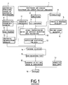
- Figure 1 is a schematic drawing of one embodiment of the present invention.
- Figure 2 illustrates an example of a visual display of the characteristics of diffuse and concentrated generators in a normal experimental subject.
- Figure 3 illustrates an example of a visual display of the characteristics of diffuse and concentrated generators in a patient with a cerebrovascular accident.
- a plurality of sensors (1) are placed on or in proximity to the experimental subject's scalp, for the detection of brain electromagnetic signals, which are generated due to neuronal sources.
- the sensors are placed according to a pre-defined plan in order to maximize the amount of information about the generators. Due to variability of head shape and size in the human population, measurements of the exact positions of the sensors are required, with respect to a reference coordinate system determined by certain anatomical landmarks of the individual subject's head.
- the electromagnetic signals are amplified (2) to the dynamic range of an analog to digital converter (3), which converts the signals into numbers that are stored in the memory of a digital computer.
- the recording of electromagnetic signals is carried out under the control of a central experimental program.
- the experimental subject can optionally be presented with visual, auditory, and somato-sensorial stimulation (4), introducing fiducial markers in the recording for later identification. Stimulation can be presented in the form of video-games.
- responses produced by the experimental subject (5) in the form of vocalizations or body movements can be recorded and identified, and can modify dynamically the central experimental program.
- real-time detection of spontaneous events in the EEG and MEG produced by the experimental subject during recording (6) is provided, allowing the dynamic modification of the central experimental program.
- Anatomical and functional information about the head including aspects such as geometry of the different constituent tissues (e.g., brain, skull, scalp), location and orientation of the cortex and of other neuronal aggregates, is summarized in what is here termed descriptive parametric geometry (8).
- anatomical images e. g., CAT images or NMR images
- functional images e.g., PET images or SPECT images
- M m is the number of sampled points on the m-th boundary.
- the descriptive parametric geometry previously explained can be used for computing the anatomical deconvolution operator (10), which is defined in the following equations : where are the electric potential and magnetic field component measurement vectors (the vector elements correspond to measurements made at different sensor positions), respectively; correspond to electric potential and magnetic field component values at the same sensor positions, and due to the same neuronal generators, in an infinite homogeneous medium; and are transfer coefficient matrices, which define the anatomical deconvolution operator.
- the anatomical deconvolution operator can be computed by the method and system described in CUBAN patent application 4/91, or alternatively by the new method described in the present invention.
- the descriptive parametric geometry is used for constructing a head phantom (9) with all the volume conductor properties of the experimental subject's head, said volume conductor properties consisting of the geometry and conductivity profile of the different constituent tissues (e.g., brain, skull, scalp).
- Electric potential ( V ) and magnetic field component ( B ) measurements are performed on the head phantom due to a plurality of implanted current dipoles (one at a time) with known locations and moments (dipoles located in the corresponding neural tissue volume).
- a further important use of the descriptive parametric geometry is the determination of anatomical and functional constraints for the localizations, orientations, activities, and connectivities of the brain electromagnetic waves generators (11) (generator constraints).
- Generator constraints are necessary for obtaining a unique inverse solution (14). For example, if the measured EEG and MEG activity is known beforehand to be generated only by cortical sources, then the generators can be located only on the cortical surface with orthogonal orientations.
- the sufficient statistics can consist of cumulants of any order, or multiple time series parametric models, either in the time, frequency, or time-frequency domain.
- Karhunen-Loeve type representations can be used for fitting the sufficient statistics in stationary or non-stationary, linear or non-linear models.
- This model has the following characteristics :
- Inverse solutions (14) are computed for the source model in infinite homogeneous medium, based on the sufficient statistics of the measured data transformed to infinite homogeneous medium by means of the anatomical deconvolution operator (12), and taking into account the constraints imposed on the generators (11).
- Inverse solutions can be obtained by a least squares criterion in which tr( ⁇ V - ⁇ V )2 is minimized with respect to the dipole location parameters in matrix ⁇ , the dipole orientation parameters in matrix , the generators cross spectral matrix ⁇ g , and the coefficients a k of the noise cross spectral matrix, where ⁇ v is the sample electric potential cross spectral matrix.
- inverse solutions can be obtained by maximizing the likelihood function, which is equivalent to minimizing tr( ⁇ -1 V ⁇ V )-det( ⁇ -1 V ⁇ V ). Independently of the estimation method used, the inverse solutions must be obtained under the generator constraints.
- G * t (g 1t g 2t .. g Ndt ) can be estimated at each time instant as : which is obtained by either maximum likelihood or weighted least squares methods.
- the estimated localizations, orientations, activities, and connectivities of the ERCs generators of an experimental subject are compared with those of a normal population (17), by means of multivariate metrics for measuring distances between estimators and normative data of a sample from the normal population, taking properly into account the effect of covariables such as age in order to decrease metric variability.
- visual displays are presented in the form of three dimensional and two dimensional images of the head, where the localizations, orientations, activities, and connectivities of the ERCs generators are displayed by coding their numerical values in terms of color, intensity, and graphical icons, and where optionally, the multivariate metrics corresponding to comparison with norms can also be displayed by superposition.
- Figure 2 illustrates a visual display of an inverse solution obtained from a normal experimental subject, based on a spontaneous EEG recording under eyes closed, awake, conditions.
- Two dipoles were fitted, together with additive uncorrelated homogeneous isotropic spatial noise.
- Generators are represented as arrows in the three head views (back, top, and left views).
- Generator localization and moments are given in cartesian coordinates referred to a unit radius sphere, with z axis coming out through the vertex, x axis coming out through nasion, and y axis coming out through the left ear (T3 electrode position).
- Connectivities are given in terms of the generator correlation matrix.
- Noise characteristics are illustrated as "BASE ACTIVITY", giving the values of the expansion coefficients of the homogeneous isotropic process. Note the alpha rhythm generators located in the occipital cortex.
- Figure 3 illustrates a visual display of an inverse solution obtained from an experimental subject with a lateralized right cerebrovascular accident, based on a spontaneous EEG recording under eyes closed, awake, conditions. Analysis procedures were the same to those used for the normal subject of Figure 2. Note that only one alpha generator lies in the normal position (left occipital cortex).
Abstract
Description
- The present invention relates to electronic computerized medical instruments and more particularly to the localization and characterization of the generators of brain and heart electric and magnetic activity by a non-invasive computerized method and system.
- The determination of the three dimensional localization and of the temporal activity of the neuronal generators which give place to waveshapes in the electroencephalogram (EEG) and the magnetoencephalogram (MEG) related to pathologies of the central nervous system (CNS), constitutes an important medical problem. Such knowledge can be helpful in producing more precise diagnostics in diverse neuropsychiatric pathologies and in determining more efficient treatments. A typical example is the study of the focus location followed by its sequential propagation in epilepsies that are being evaluated for surgical treatment.
- The EEG and the MEG both have their common origin in the ionic currents produced by the cellular elements (the neurons) composing the CNS. The total current density vector field is determined by the vectorial additive combination of all of the elementary currents. The simultaneous activation of a large number of such elements, together with an adequate geometrical distribution, produces resulting electric potentials and magnetic fields which can be measured outside the head. In the transformation process from total current density to measurable external fields, the effects of the volume conductor properties of the different tissues composing the head must be taken into account : brain, meninges, cerebral spinal fluid, skull, and scalp.
- The resulting measured fields have the characteristics of a stochastic process, which can be described either in the frequency domain or in the temporal domain, as a function of the statistical moments. In the case of a Gaussian process, first and second order moments give an exhaustive description.
- The neural elements which generate a given EEG or MEG component may be localized on a small cortical area ("concentrated generator") or may, on the other hand, be widely distributed in different parts of the CNS ("diffuse generator"). The determination of the spatial distribution of the generators and of the multivariate statistical moments describing their interactions is very important.
- For a number of decades electric potential measurements of the CNS have been performed by means of electrodes placed on the scalp. Much experience has accumulated on the practical utility of the visual inspection of the EEG in the diagnostics and treatment of patients with neuropsychiatric diseases. More recently, brain magnetic fields have been measured (US patent 4,591,787), offering complementary information to that obtained from the EEG.
- The current state of the art, as reflected in US patents 4,201,224; 4,408,616; and 4,417,592 is summarized as follows. Quantitative analysis of brain electric activity by means of digital signal processing methods (qEEG) allows an objective evaluation of the functioning of the CNS. The signal recorded at each electrode is summarized by means of a set of descriptive parameters (DPs), based on stochastic process modeling. The DPs reflect the normal and pathological functioning of the CNS. Topographic maps based on the DPs are clinically useful, and even more so when statistically compared to a normative data base.
- However, this analysis method generates an excessively large number of DPs, thus making quite difficult the evaluation of a particular patient. Moreover, the method does not attempt to localize the generators responsible for the measured DPs, thus limiting the clinical usefulness and contributing to the excessive redundancy of the DPs due to volume conduction effects. Finally, qEEG is limited to the study of second order moments in the frequency domain, which means that the EEG has been implicitly assumed to be a Gaussian stochastic process, despite the evidence revealing the non-linear nature of such signals.
- In US patent 4,913,160 a method for the reduction of the dimensionality of the DPs is proposed based on principal components (PCs) computation. This procedure produces minimum sets of linear combinations of the original DPs, with optimum descriptive properties, but which are meaningless in terms of the underlying neuronal generators and their localization. Furthermore, this method does not take into account the non-linear nature of the original signals.
- An improvement in the usefulness of qEEG has been achieved by means of biophysical models which take into account the behavior of the electromagnetic fields produced by current sources in a complex volume conductor such as the human head. In this sense, US patents 4,416,288; 4, 753, 246; and 4, 736, 751 propose procedures for eliminating the distortion effects due to the volume conductor. However, they do not deal with the spatial characterization of the generators.
- Several attempts have been made to fit equivalent dipoles to measured fields in order to represent, albeit approximately, concentrated generators, either in the time domain or in the frequency domain. These procedures are based on the minimization of a certain distance criterion between the measurements and the theoretical field values due to a current dipole inside a volume conductor model of the head.
- This type of procedure for source localization, based on first order moment data, does not take into account the existence of diffuse generators, nor the existence of other sources of "spatial noise". Furthermore, a statistical method for testing the goodness of fit of the source model is not provided. On the other hand, there is a fundamental limit on the number of dipoles that can be estimated, the maximum number being roughly equal to the number of electric or magnetic signals divided by six.
- In FR patent 2,622,990, several improvements are achieved by using frequency domain second order moment data, in the form of coherence matrices. An estimation method for the cross spectral spatial noise matrix is proposed, under the assumption of interelectrode independence, thus being statistically equivalent to the classical factor analysis model. The eigenvectors of the common factor space are then used for determining the concentrated generators (as many as the number of common factors).
- However, empirical and theoretical evidence point towards a diffuse generator model for spatial noise, producing a structured cross spectral noise matrix for EEG and MEG. This explains why the proposed noise elimination method under the interelectrode independence assumption gives incorrect results. In such a case computations based on coherence matrices are not justified. Furthermore, dipole fitting methods applied to second order moment data or to eigenvector data are not equivalent. Finally, interactions between generators, neither linear nor non-linear, are taken into account in the eigenvector dipole fitting approach.
- The objective of the present invention is a method and system for the characterization of both concentrated and diffuse generators of the EEG and MEG, based on all the statistical information available in these signals, in the form of statistical moments of all orders, in the time or frequency domain. The invention will allow the detection and estimation of the effect of the diffuse generators on the EEG and MEG. Also, it will allow the estimation of an increased number of concentrated generators, together with their linear and non-linear interactions.
- In accordance with a first aspect of the invention there is provided a method for the three dimensional tomography of activity and connectivity of brain electromagnetic waves generators, said method including :
- a) Attaching or approximating a set of electrodes and magnetic sensors to the scalp of an experimental subject to detect brain electromagnetic physiological activity in the form of electroencephalogram (EEG) and magnetoencephalogram (MEG), and measuring the exact positions of the electrodes and sensors with respect to a reference coordinate system determined by certain anatomical landmarks of the subject's head;
- b) Amplifying the said electromagnetic signals detected at each electrode and sensor;
- c) Obtaining on-line digital spatio-temporal signals, consisting of said EEG and MEG, by connecting analog-digital converters to each amplifier, and digitizing all data as it is gathered, all carried out under the control of a central experimental program;
- d) Optional presentation of visual, auditory, and somato-sensorial stimulation to the experimental subject during EEG and MEG recording, carried out under the control of said central experimental program;
- e) Optional recording and identification of responses produced by the experimental subject during EEG and MEG recording, for the inclusion of fiducial markers in said recording, and for the modification of said central experimental program;
- f) Optional real-time detection of spontaneous events in the EEG and MEG produced by the experimental subject during recording, for the inclusion of fiducial markers in said recording, and for the modification of said central experimental program;
- g) Determination of a parametric description for the anatomy of the experimental subject's head (parametric geometry), by means of : i) exact computations based on anatomical or functional image processing of the subject's head, or ii) approximate computations based on a small set of anatomical measurements and comparison with a data base of normal and abnormal variability;
- h) Using said parametric geometry for constructing a head phantom with all the volume conductor properties of the real head;
- i) Performing EEG and MEG measurements on said head phantom due to known current dipoles located in the corresponding neural tissue volume, for determining the linear operator which transforms original EEG and MEG measurements into equivalent infinite homogeneous medium measurements (anatomical deconvolution);
- j) Using said parametric geometry for determining anatomical and functional constraints for the localizations, orientations, activities, and connectivities of the brain electromagnetic waves generators (generator constraints);
- k) Digital pre-processing of the EEG and MEG for artifact and noise elimination, and for the separation of EEG and MEG samples related to said fiducial markers, for obtaining event related components (ERCs);
- l) Statistical analysis of said ERCs for determining the most adequate numerical description of the spatio-temporal properties in terms of sufficient statistics;
- m) Computation of the activities and connectivities of the ERCs generators, based on the static solution to the inverse electromagnetic problem, under said generator constraints, using said sufficient statistics for the ERCs transformed to infinite homogeneous medium by means of said anatomical deconvolution;
- n) In case that said generator constraints do not allow a unique solution to the inverse problem, the number of ERCs generators should be decreased sufficiently to allow for the proper identifiability of the inverse problem;
- o) Statistical evaluation of the goodness of fit of the inverse solution, taking into account the existence of colored spatial and temporal noise, and including statistical hypotheses testing on the absence of activity and connectivity of the ERCs generators;
- p) Optional computation of multivariate distances between ERCs generators characteristics (localizations, orientations, activities, and connectivities) of said experimental subject and of a normal population as determined from a normative data-base;
- q) Visual display of three dimensional and two dimensional images corresponding to the localizations, orientations, activities, and connectivities of the ERCs generators, and the optional display of said multivariate distances.
- In accordance with a second aspect of the invention there is provided a system for the three dimensional tomography of activity and connectivity of brain electromagnetic waves generators, said system including :
- a) A set of electrodes and magnetic sensors adapted to be attached or approximated to the scalp of an experimental subject for the detection of brain electromagnetic physiological activity in the form of electroencephalogram (EEG) and magnetoencephalogram (MEG), and means for measuring the exact positions of the electrodes and sensors with respect to a reference coordinate system determined by certain anatomical landmarks of the subject's head;
- b) Means for the amplification of the said electromagnetic signals detected at each electrode and sensor;
- c) Means for obtaining on-line digital spatio-temporal signals consisting of said EEG and MEG;
- d) Means for the presentation of visual, auditory, and somato-sensorial stimulation to the experimental subject during EEG and MEG recording;
- e) Means for recording the vocal or movement responses produced by the experimental subject during EEG and MEG recording;
- f) A central digital computer subsystem, consisting of a multitasking processor or a set of distributed processors, that comprises :
- Means for reading the experimental subject's image data in the form of CAT scan images, NMR images, PET images, or in the form of a small set of anatomical measurements, and means for computing and storing the descriptive parametric geometry, the anatomical deconvolution operator, and the generator constraints;
- Means for constructing a head phantom based on the descriptive parametric geometry, and means for the implantation of current dipoles in the corresponding neural tissue volume of the phantom;
- Means for programming and for the control of experiments that comprise stimulation of the experimental subject, recording of the subject's responses, detection and recording of special EEG and MEG events, and simultaneous recording of the digitized electromagnetic signals;
- Means for pre-processing the recorded electromagnetic signals for artifact and noise elimination;
- Means for estimating the ERCs;
- Means for computing the ERCs sufficient statistics;
- Means for estimating the additive non-white spatio-temporal noise due to diffuse generators;
- Means for performing tests of hypotheses about the goodness of fit of the estimated inverse solution;
- Means for estimating the localizations, orientations, activities, and connectivities of the ERCs generators;
- Means for comparing the ERCs generators characteristics with a normative data base and means for computing multivariate metrics;
- Means for the visual display of ERCs generators characteristics and of the multivariate metrics.
- In order that the invention be better understood, further detailed description follows with reference to accompanying drawings in which :
- Figure 1) is a schematic drawing of one embodiment of the present invention.
- Figure 2) illustrates an example of a visual display of the characteristics of diffuse and concentrated generators in a normal experimental subject.
- Figure 3) illustrates an example of a visual display of the characteristics of diffuse and concentrated generators in a patient with a cerebrovascular accident.
- A plurality of sensors (1) (electrodes for EEG, magnetometers or gradiometers for MEG) are placed on or in proximity to the experimental subject's scalp, for the detection of brain electromagnetic signals, which are generated due to neuronal sources. The sensors are placed according to a pre-defined plan in order to maximize the amount of information about the generators. Due to variability of head shape and size in the human population, measurements of the exact positions of the sensors are required, with respect to a reference coordinate system determined by certain anatomical landmarks of the individual subject's head.
- The electromagnetic signals are amplified (2) to the dynamic range of an analog to digital converter (3), which converts the signals into numbers that are stored in the memory of a digital computer.
- The recording of electromagnetic signals is carried out under the control of a central experimental program. During recording, the experimental subject can optionally be presented with visual, auditory, and somato-sensorial stimulation (4), introducing fiducial markers in the recording for later identification. Stimulation can be presented in the form of video-games. At the same time, responses produced by the experimental subject (5) in the form of vocalizations or body movements can be recorded and identified, and can modify dynamically the central experimental program. Also optionally, real-time detection of spontaneous events in the EEG and MEG produced by the experimental subject during recording (6) is provided, allowing the dynamic modification of the central experimental program.
- Anatomical and functional information about the head, including aspects such as geometry of the different constituent tissues (e.g., brain, skull, scalp), location and orientation of the cortex and of other neuronal aggregates, is summarized in what is here termed descriptive parametric geometry (8). Each surface boundary can in general be expressed as an implicit function F(x,y,z)=0, where the variables x, y, and z represent common cartesian coordinates. The descriptive parametric geometry of an experimental subject consists of the finite set of coefficients {Cmi jk} used in approximating each boundary m (m=1,2,...,Nb; Nb is number of boundaries) in terms of an expansion of the form :

where {Bn} is a finite set of
basis functions (n=1,2,..,Nf; Nf is number of basis functions). - For each boundary, the expansion is fitted (in a least squares sense) to data from anatomical images (7) (e. g., CAT images or NMR images) or from functional images (e.g., PET images or SPECT images) of the experimental subject's head :

where u=1,2,..,Mm; and Mm is the number of sampled points on the m-th boundary. Alternatively, the expansion coefficients can be statistically predicted from a minimum set of easily measured anatomical parameters (7) by means of the information contained in a normative data base with a representative sample of experimental subjects covering the typical population :
where Gmijk is the statistically estimated
function, and ϑεis the anatomical measurement parameter vector.
- Important basis functions Bn to be used are :
- a) The Fourier basis Bn(v) = exp(ivωn), where i=(-1)½, and v,ωnε.

- b) The Karhunen-Loeve basis for each type of boundary surface.
- c) Spatial spline bases.
- The descriptive parametric geometry previously explained can be used for computing the anatomical deconvolution operator (10), which is defined in the following equations :

where
are the electric potential and magnetic field component measurement vectors (the vector elements correspond to measurements made at different sensor positions), respectively;
correspond to electric potential and magnetic field component values at the same sensor positions, and due to the same neuronal generators, in an infinite homogeneous medium; and
are transfer coefficient matrices, which define the anatomical deconvolution operator. - The anatomical deconvolution operator

can be computed by the method and system described inCUBAN patent application 4/91, or alternatively by the new method described in the present invention. The descriptive parametric geometry is used for constructing a head phantom (9) with all the volume conductor properties of the experimental subject's head, said volume conductor properties consisting of the geometry and conductivity profile of the different constituent tissues (e.g., brain, skull, scalp). Electric potential (V) and magnetic field component (B) measurements are performed on the head phantom due to a plurality of implanted current dipoles (one at a time) with known locations and moments (dipoles located in the corresponding neural tissue volume). Also, theoretical EEG (V ∞) and MEG (B ∞) values are computed in the corresponding infinite homogeneous medium. The set of vectors {(V,B,V ∞,B ∞) i ) (where i=1,2,..,Nd, and Nd is the number of implanted dipoles) is now used for computing the anatomical deconvolution operator
in a least squares sense :
where i=1, 2,.., Nd, and ∥X∥ denotes the norm of the vector X. - A further important use of the descriptive parametric geometry is the determination of anatomical and functional constraints for the localizations, orientations, activities, and connectivities of the brain electromagnetic waves generators (11) (generator constraints). Generator constraints are necessary for obtaining a unique inverse solution (14). For example, if the measured EEG and MEG activity is known beforehand to be generated only by cortical sources, then the generators can be located only on the cortical surface with orthogonal orientations.
- Digital pre-processing (12) of the recorded EEG and MEG is necessary before proceeding to obtain inverse solutions. The two main steps consist of :
- a) Artifact and noise elimination.
- b) Separation of EEG and MEG samples related to fiducial markers (e.g., stimuli, subject's response, or spontaneous EEG and MEG events) for obtaining event related components (ERCs).
- Statistical hypothesis tests are performed for determining the most adequate numerical description of the spatio-temporal properties of the stochastic ERCs in terms of sufficient statistics (13). The sufficient statistics can consist of cumulants of any order, or multiple time series parametric models, either in the time, frequency, or time-frequency domain. Alternatively, Karhunen-Loeve type representations can be used for fitting the sufficient statistics in stationary or non-stationary, linear or non-linear models.
- The generator modiel for ERCs is :

where the vectors
are the time (t) dependent elecric potential and magnetic field component measurements, respectively; φkεand ψkε

denote the electric and magnetic transfer coefficient matrices for the k-th dipole source, respectively; M kεis the k-th dipole's moment; gkt is the time varying k-th dipole's intensity;

are noise vectors; k=1,2,..,Nd, and Nd is the number of dipole sources. This model has the following characteristics : - a) The ERCs are due to Nd dipoles, with fixed positions and orientations, which may be known or unknown, and with unknown time varying dipole intensities. The dipole intensities are modeled as stochastic processes.
- b) Measurements consist of the ERCs (due to dipoles as previously described) contaminated by additive noise. The additive noise is modeled as a stochastic process, not necessarily as spatio-temporal white noise. One particular model for the noise process allows for any general time-colored properties, with homogeneous isotropic spatial properties due to diffuse generators.
- Inverse solutions (14) are computed for the source model in infinite homogeneous medium, based on the sufficient statistics of the measured data transformed to infinite homogeneous medium by means of the anatomical deconvolution operator (12), and taking into account the constraints imposed on the generators (11).
- As a simple particular example, consider the zero mean linear stationary Gaussian case for spontaneous EEG activity, where the cross spectral matrices (frequency domain second order moments) constitute the sufficient statistics. Assuming that the ERCs concentrated generators and the noise diffuse generators are statistically independent, the model cross spectral matrix at frequency ω for the electric potential (Σv) is:

where

Σgεis the generators cross spectral matrix; and Σnε is the pure real valued symmetric homogeneous isotropic noise cross spectral matrix, where, in the case of spherical geometry,

where the ak≧0 are unknown coefficients, [P k ] ij = Pk(cos(γij)), P k being the Legendre polynomials of order k, and γij is the angle between the i-th and j-th electrodes. - Inverse solutions can be obtained by a least squares criterion in which tr(Σ̂V-ΣV)² is minimized with respect to the dipole location parameters in matrix Φ, the dipole orientation parameters in matrix, the generators cross spectral matrix Σg, and the coefficients ak of the noise cross spectral matrix, where Σ̂v is the sample electric potential cross spectral matrix. Alternatively, inverse solutions can be obtained by maximizing the likelihood function, which is equivalent to minimizing tr(Σ


which is obtained by either maximum likelihood or weighted least squares methods. - Summarizing the inverse solution example previously described, the following generator characteristics were computed for a given number of dipoles and a given number of coefficients ak in the expansion of the noise cross spectral matrix :
- a) The activity gkt for each dipole k, at each time instant t.
- b) The connectivities ΣV between all pairs of dipoles.
- c) The locations (Φ) and orientations () of the dipoles.

- d) The expansion coefficients of the homogeneous isotropic noise cross spectral matrix.
- Tests for inverse solution uniqueness (15) must be made before performing estimations, since under non-uniqueness, computations may render meaningless results. Uniqueness may be achieved by setting more severe generator constraints, or by decreasing the number of dipoles until the model is identifiable.
- Once the generator characteristics are estimated, a test for the goodness of fit of the inverse solution is performed (16). Also, hypotheses concerning if a given generator has significant activity, or if the connectivity between a pair of generators is significant, can also be tested (16). In all cases statistical hypothesis testing is based on resampling techniques such as Montecarlo, the Jackknife, and the Bootstrap.
- The estimated localizations, orientations, activities, and connectivities of the ERCs generators of an experimental subject are compared with those of a normal population (17), by means of multivariate metrics for measuring distances between estimators and normative data of a sample from the normal population, taking properly into account the effect of covariables such as age in order to decrease metric variability.
- Finally, visual displays (18) are presented in the form of three dimensional and two dimensional images of the head, where the localizations, orientations, activities, and connectivities of the ERCs generators are displayed by coding their numerical values in terms of color, intensity, and graphical icons, and where optionally, the multivariate metrics corresponding to comparison with norms can also be displayed by superposition.
- Figure 2 illustrates a visual display of an inverse solution obtained from a normal experimental subject, based on a spontaneous EEG recording under eyes closed, awake, conditions. The sufficient statistics used was the cross spectral matrix at the alpha peak maximum (ω = 9.75 Hz). Two dipoles were fitted, together with additive uncorrelated homogeneous isotropic spatial noise. Generators are represented as arrows in the three head views (back, top, and left views). Generator localization and moments are given in cartesian coordinates referred to a unit radius sphere, with z axis coming out through the vertex, x axis coming out through nasion, and y axis coming out through the left ear (T₃ electrode position). Connectivities are given in terms of the generator correlation matrix. Noise characteristics are illustrated as "BASE ACTIVITY", giving the values of the expansion coefficients of the homogeneous isotropic process. Note the alpha rhythm generators located in the occipital cortex.
- Figure 3 illustrates a visual display of an inverse solution obtained from an experimental subject with a lateralized right cerebrovascular accident, based on a spontaneous EEG recording under eyes closed, awake, conditions. Analysis procedures were the same to those used for the normal subject of Figure 2. Note that only one alpha generator lies in the normal position (left occipital cortex).
Claims (16)
- A method for the three dimensional tomography of activity and connectivity of brain electromagnetic waves generators, said method including the steps of :a) Attaching or approximating a set of electrodes and magnetic sensors to the scalp of an experimental subject to detect brain electromagnetic physiological activity in the form of electroencephalogram (EEG) and magnetoencephalogram (MEG), and measuring the exact positions of the electrodes and sensors with respect to a reference coordinate system determined by certain anatomical landmarks of the subject's head;b) Amplifying the said electromagnetic signals detected at each electrode and sensor;c) Obtaining on-line digital spatio-temporal signals, consisting of said EEG and MEG, by connecting analog-digital converters to each amplifier, and digitizing all data as it is gathered, all carried out under the control of a central experimental program;d) Optional presentation of visual, auditory, and somato-sensorial stimulation to the experimental subject during EEG and MEG recording, carried out under the control of said central experimental program;e) Optional recording and identification of responses produced by the experimental subject during EEG and MEG recording, for the inclusion of fiducial markers in said recording, and for the modification of said central experimental program;f) Optional real-time detection of spontaneous events in the EEG and MEG produced by the experimental subject during recording, for the inclusion of fiducial markers in said recording, and for the modification of said central experimental program;g) Determination of a parametric description for the anatomy of the experimental subject's head (parametric geometry), by means of : i) exact computations based on anatomical or functional image processing of the subject's head, or ii) approximate computations based on a small set of anatomical measurements and comparison with a data base of normal and abnormal variability;h) Using said parametric geometry for constructing a head phantom with all the volume conductor properties of the real head;i) Performing EEG and MEG measurements on said head phantom due to known current dipoles located in the corresponding neural tissue volume, for determining the linear operator which transforms original EEG and MEG measurements into equivalent infinite homogeneous medium measurements (anatomical deconvolution);j) Using said parametric geometry for determining anatomical and functional constraints for the localizations, orientations, activities, and connectivities of the brain electromagnetic waves generators (generator constraints);k) Digital pre-processing of the EEG and MEG for artifact and noise elimination, and for the separation of EEG and MEG samples related to said fiducial markers, for obtaining event related components (ERCs);l) Statistical analysis of said ERCs for determining the most adequate numerical description of the spatio-temporal properties in terms of sufficient statistics;m) Computation of the activities and connectivities of the ERCs generators, based on the static solution to the inverse electromagnetic problem, under said generator constraints, using said sufficient statistics for the ERCs transformed to infinite homogeneous medium by means of said anatomical deconvolution;n) In case that said generator constraints do not allow a unique solution to the inverse problem, the number of ERCs generators should be decreased sufficiently to allow for the proper identifiability of the inverse problem;o) Statistical evaluation of the goodness of fit of the inverse solution, taking into account the existence of colored spatial and temporal noise, and including statistical hypotheses testing on the absence of activity and connectivity of the ERCs generators;p) Optional computation of multivariate distances between ERCs generators characteristics (localizations, orientations, activities, and connectivities) of said experimental subject and of a normal population as determined from a normative data-base;q) Visual display of three dimensional and two dimensional images corresponding to the localizations, orientations, activities, and connectivities of the ERCs generators, and the optional display of said multivariate distances.
- A method as claimed in claim 1 wherein the experimental sequences are dynamically integrated, where stimulation can be absent, or where the stimulation sequence can be pre-determined, or can be presented in the form of computerized video-games, with either vocal or movement responses from the experimental subject.
- A method as claimed in claim 1 wherein events are detected in the EEG and MEG (e.g., paroxistic events or alpha rhythm desynchronization events), where events can either be defined deterministically, statistically, or in terms of fuzzy logic, and where events can be recorded and can modify the experimental sequence.
- A method as claimed in claim 1 wherein series expansions are established in terms of three dimensional basis functions for the description of contours, physical properties, and certain metabolic properties of the head, producing a descriptive parametric geometry consisting of the sets of expansion coefficients.
- A method as claimed in claim 1 wherein the descriptive parametric geometry is obtained from anatomical images (e.g., CAT images or NMR images), or from functional images (e.g., PET images or SPECT images) of the experimental subject's head, or from a minimum set of easily measured parameters which can be used to statistically predict the descriptive parametric geometry by means of the information contained in a normative data base with a representative sample covering the typical population.
- A method as claimed in claim 1 wherein the descriptive parametric geometry establishes a priori constraints on the localizations and orientations of the ERCs generators.
- A method as claimed in claim 1 wherein the descriptive parametric geometry is used for constructing a head phantom with all the volume conductor properties of the experimental subject's head.
- A method as claimed in claim 1 wherein EEG and MEG measurements are performed on the head phantom, or alternatively, are theoretically computed on a head phantom defined by the descriptive parametric geometry, using established current dipoles located in the corresponding neural tissue volume, with known locations, orientations and activities, where theoretical EEG and MEG values are computed in the corresponding infinite homogeneous medium, and where the linear operator in matrix form which transforms original EEG and MEG measurements into equivalent infinite homogeneous medium measurements is computed in a least squares sense, thus defining the anatomical deconvolution operation.
- A method as claimed in claim 1 wherein event related samples (ERSs) of EEG and MEG are separated according to the different types of fiducial markers (e.g., stimuli, subject's response, or spontaneous EEG and MEG events), where said ERSs contain ERCs, which are estimated under the assumption that the set of generators have fixed localizations and orientations, and stochastic activities and connectivities.
- A method as claimed in claim 1 wherein statistical hypothesis tests are used for determining the set of sufficient statistics for the description of the stochastic ERSs, where said set of sufficient statistics can consist of cumulants of any order, or multiple time series parametric models, either in the time, frequency, or time-frequency domain. Alternatively, Karhunen-Loeve type representations can be used for fitting the sufficient statistics in stationary or non-stationary, linear or non-linear models.
- A method as claimed in claim 1 wherein the ERSs are modeled as containing additive noise due to the activity of diffuse generators independent of the ERCs concentrated generators, said diffuse generators having the properties of a stochastic process not necessarily equivalent to spatio-temporal white noise.
- A method as claimed in claim 1 wherein the inverse problem is solved for the activities and connectivities of the neuronal generators, based on all the information provided by the ERCs sufficient statistics, under the generator constraints on localizations and orientations provided by the descriptive parametric geometry, for infinite homogeneous medium measurements obtained by applying the anatomical deconvolution operator.
- A method as claimed in claim 1 wherein the goodness of fit of the estimated inverse solution and tests of hypotheses on no activity or no connectivity of the ERCs generators are examined based on statistical resampling techniques such as Montecarlo methods, the Jackknife, and the Bootstrap.
- A method as claimed in claim 1 wherein the estimated localizations, orientations, activities, and connectivities of the ERCs generators of an experimental subject are compared with those of a normal population, by means of multivariate metrics for measuring distances between estimators and normative data of a sample from the normal population, taking properly into account the effect of covariables such as age in order to decrease metric variability.
- A method as claimed in claim 1 wherein visual displays are presented in the form of three dimensional and two dimensional images of the head, where the localizations, orientations, activities, and connectivities of the ERCs generators are displayed by coding their numerical values in terms of color, intensity, and graphical icons, and where optionally, the multivariate metrics corresponding to comparison with norms can also be displayed by superposition.
- A system for the three dimensional tomography of activity and connectivity of brain electromagnetic waves generators that comprises :a) A set of electrodes and magnetic sensors adapted to be attached or approximated to the scalp of an experimental subject for the detection of brain electromagnetic physiological activity in the form of electroencephalogram (EEG) and magnetoencephalogram (MEG), and means for measuring the exact positions of the electrodes and sensors with respect to a reference coordinate system determined by certain anatomical landmarks of the subject's head;b) Means for the amplification of the said electromagnetic signals detected at each electrode and sensor;c) Means for obtaining on-line digital spatio-temporal signals consisting of said EEG and MEG;d) Means for the presentation of visual, auditory, and somato-sensorial stimulation to the experimental subject during EEG and MEG recording;e) Means for recording the vocal or movement responses produced by the experimental subject during EEG and MEG recording;f) A central digital computer subsystem, consisting of a multitasking processor or a set of distributed processors, that comprises :- Means for reading the experimental subject's image data in the form of CAT scan images, NMR images, PET images, or in the form of a small set of anatomical measurements, and means for computing and storing the descriptive parametric geometry, the anatomical deconvolution operator, and the generator constraints;- Means for constructing a head phantom based on the descriptive parametric geometry, and means for the implantation of current dipoles in the corresponding neural tissue volume of the phantom;- Means for programming and for the control of experiments that comprise stimulation of the experimental subject, recording of the subject's responses, detection and recording of special EEG and MEG events, and simultaneous recording of the digitized electromagnetic signals;- Means for pre-processing the recorded electromagnetic signals for artifact and noise elimination;- Means for estimating the ERCs;- Means for computing the ERCs sufficient statistics;- Means for estimating the additive non-white spatio-temporal noise due to diffuse generators;- Means for performing tests of hypotheses about the goodness of fit of the estimated inverse solution;- Means for estimating the localizations, orientations, activities, and connectivities of the ERCs generators;- Means for comparing the ERCs generators characteristics with a normative data base and means for computing multivariate metrics;- Means for the visual display of ERCs generators characteristics and of the multivariate metrics.
Applications Claiming Priority (2)
| Application Number | Priority Date | Filing Date | Title |
|---|---|---|---|
| CU4491 | 1991-03-15 | ||
| CU1991044 | 1991-03-15 |
Publications (2)
| Publication Number | Publication Date |
|---|---|
| EP0504027A2 true EP0504027A2 (en) | 1992-09-16 |
| EP0504027A3 EP0504027A3 (en) | 1993-04-21 |
Family
ID=5459256
Family Applications (1)
| Application Number | Title | Priority Date | Filing Date |
|---|---|---|---|
| EP19920400595 Withdrawn EP0504027A3 (en) | 1991-03-15 | 1992-03-06 | Method and system for three-dimensional tomography of activity and connectivity of brain and heart electromagnetic waves generators |
Country Status (2)
| Country | Link |
|---|---|
| US (1) | US5307807A (en) |
| EP (1) | EP0504027A3 (en) |
Cited By (10)
| Publication number | Priority date | Publication date | Assignee | Title |
|---|---|---|---|---|
| WO1994012100A1 (en) * | 1992-11-30 | 1994-06-09 | Risto Ilmoniemi | Method and apparatus for separating the different components of evoked response and spontaneous activity brain signals as well as of signals measured from the heart |
| WO1998018384A1 (en) * | 1996-10-30 | 1998-05-07 | Risto Ilmoniemi | Method and apparatus for mapping cortical connections |
| EP0869736A1 (en) * | 1995-01-27 | 1998-10-14 | David Eidelberg | Markers for use in screening patients |
| WO2000010454A1 (en) * | 1998-08-24 | 2000-03-02 | Ctf Systems Inc. | Functional brain imaging from magnetoencephalographic data |
| US6370414B1 (en) | 1998-01-23 | 2002-04-09 | Ctf Systems, Inc. | System and method for measuring, estimating and displaying RMS current density maps |
| EP1579799A1 (en) * | 2004-03-25 | 2005-09-28 | Sei Matsuoka | Apparatus and method for measuring quantitative value of information |
| ES2319080A1 (en) * | 2008-09-26 | 2009-05-01 | Universidad Politecnica De Madrid | Fantoma multicanal of magnetic dipolos orientables for magnetoencefalografia (Machine-translation by Google Translate, not legally binding) |
| EP2130489A1 (en) * | 2008-06-06 | 2009-12-09 | Electrical Geodesics Inc. | Method for locating tracts of electrical brain activity |
| WO2011051807A1 (en) * | 2009-10-27 | 2011-05-05 | Cerebral Diagnostics Canada Incorporated | Spectral decomposition and display of three-dimensional electrical activity in the cerebral cortex |
| CN104755026A (en) * | 2012-11-26 | 2015-07-01 | 珀西斯特发展公司 | Method and system for displaying the amount of artifact present in EEG recording |
Families Citing this family (32)
| Publication number | Priority date | Publication date | Assignee | Title |
|---|---|---|---|---|
| US6405072B1 (en) * | 1991-01-28 | 2002-06-11 | Sherwood Services Ag | Apparatus and method for determining a location of an anatomical target with reference to a medical apparatus |
| US6006126A (en) | 1991-01-28 | 1999-12-21 | Cosman; Eric R. | System and method for stereotactic registration of image scan data |
| ATE172623T1 (en) * | 1991-12-17 | 1998-11-15 | Dynamics Imaging Inc | METHOD AND DEVICE FOR DIAGNOSING LIVING ORGANISMS |
| US5699797A (en) * | 1992-10-05 | 1997-12-23 | Dynamics Imaging, Inc. | Method of investigation of microcirculation functional dynamics of physiological liquids in skin and apparatus for its realization |
| US6002958A (en) * | 1992-12-24 | 1999-12-14 | Dynamics Imaging, Inc. | Method and apparatus for diagnostics of internal organs |
| US5458142A (en) * | 1993-03-19 | 1995-10-17 | Farmer; Edward J. | Device for monitoring a magnetic field emanating from an organism |
| JP2739804B2 (en) * | 1993-05-14 | 1998-04-15 | 日本電気株式会社 | Dipole estimator |
| US5687724A (en) * | 1993-10-26 | 1997-11-18 | Abratech Corporation | Apparatus and method for determining variations in measured physical parameters of signal-generators |
| US5357965A (en) * | 1993-11-24 | 1994-10-25 | General Electric Company | Method for controlling adaptive color flow processing using fuzzy logic |
| US5747789A (en) * | 1993-12-01 | 1998-05-05 | Dynamics Imaging, Inc. | Method for investigation of distribution of physiological components in human body tissues and apparatus for its realization |
| US5865743A (en) * | 1994-02-23 | 1999-02-02 | Dynamics Imaging, Inc. | Method of living organism multimodal functional mapping |
| US6192262B1 (en) | 1994-02-23 | 2001-02-20 | Dobi Medical Systems, Llc | Method of living organism multimodal functional mapping |
| US5730133A (en) * | 1994-05-20 | 1998-03-24 | Dynamics Imaging, Inc. | Optical functional mamoscope |
| JP3724014B2 (en) * | 1994-08-25 | 2005-12-07 | ソニー株式会社 | Image recognition apparatus and image recognition method |
| JP2735158B2 (en) * | 1996-03-18 | 1998-04-02 | 工業技術院長 | Magnetic field source movable phantom head |
| GB2321363A (en) * | 1997-01-21 | 1998-07-22 | Northern Telecom Ltd | Telecommunications |
| US6697660B1 (en) | 1998-01-23 | 2004-02-24 | Ctf Systems, Inc. | Method for functional brain imaging from magnetoencephalographic data by estimation of source signal-to-noise ratio |
| JP3246433B2 (en) * | 1998-01-27 | 2002-01-15 | 日本電気株式会社 | Cryptographic strength evaluation support apparatus and machine-readable recording medium recording program |
| US6195576B1 (en) * | 1998-03-09 | 2001-02-27 | New York University | Quantitative magnetoencephalogram system and method |
| US6309361B1 (en) | 1998-05-04 | 2001-10-30 | Kirtley E. Thornton | Method for improving memory by identifying and using QEEG parameters correlated to specific cognitive functioning |
| US7092748B2 (en) * | 2000-02-18 | 2006-08-15 | Centro Nacional De Investigaciones Cientificas (Cnic) | System and method for the tomography of the primary electric current of the brain and of the heart |
| US6856830B2 (en) * | 2001-07-19 | 2005-02-15 | Bin He | Method and apparatus of three dimension electrocardiographic imaging |
| US7840250B2 (en) * | 2001-11-13 | 2010-11-23 | Electrical Geodesics, Inc. | Method for neural current imaging |
| CA2657087A1 (en) * | 2008-03-06 | 2009-09-06 | David N. Fernandes | Normative database system and method |
| US9576107B2 (en) * | 2013-07-09 | 2017-02-21 | Biosense Webster (Israel) Ltd. | Model based reconstruction of the heart from sparse samples |
| WO2019060298A1 (en) | 2017-09-19 | 2019-03-28 | Neuroenhancement Lab, LLC | Method and apparatus for neuroenhancement |
| US11717686B2 (en) | 2017-12-04 | 2023-08-08 | Neuroenhancement Lab, LLC | Method and apparatus for neuroenhancement to facilitate learning and performance |
| EP3731749A4 (en) | 2017-12-31 | 2022-07-27 | Neuroenhancement Lab, LLC | System and method for neuroenhancement to enhance emotional response |
| US11364361B2 (en) | 2018-04-20 | 2022-06-21 | Neuroenhancement Lab, LLC | System and method for inducing sleep by transplanting mental states |
| WO2020056418A1 (en) | 2018-09-14 | 2020-03-19 | Neuroenhancement Lab, LLC | System and method of improving sleep |
| US11786694B2 (en) | 2019-05-24 | 2023-10-17 | NeuroLight, Inc. | Device, method, and app for facilitating sleep |
| CN110584898B (en) * | 2019-10-08 | 2020-08-14 | 南京邮电大学 | Brain-controlled wheelchair automatic obstacle avoidance method based on multiple sensors |
Citations (2)
| Publication number | Priority date | Publication date | Assignee | Title |
|---|---|---|---|---|
| US4736751A (en) * | 1986-12-16 | 1988-04-12 | Eeg Systems Laboratory | Brain wave source network location scanning method and system |
| EP0355506A1 (en) * | 1988-08-16 | 1990-02-28 | Siemens Aktiengesellschaft | Arrangement for measuring local bioelectric currents in biological tissue |
Family Cites Families (12)
| Publication number | Priority date | Publication date | Assignee | Title |
|---|---|---|---|---|
| US4201224A (en) * | 1978-12-29 | 1980-05-06 | Roy John E | Electroencephalographic method and system for the quantitative description of patient brain states |
| US4416288A (en) * | 1980-08-14 | 1983-11-22 | The Regents Of The University Of California | Apparatus and method for reconstructing subsurface electrophysiological patterns |
| US4417592A (en) * | 1981-05-11 | 1983-11-29 | Roy John E | Digital electroencephalographic instrument and method |
| US4408616A (en) * | 1981-05-15 | 1983-10-11 | The Children's Medical Center Corporation | Brain electricaL activity mapping |
| DE3247585A1 (en) * | 1982-12-22 | 1984-06-28 | Siemens AG, 1000 Berlin und 8000 München | MULTI-CHANNEL DEVICE FOR MEASURING DIFFERENT FIELD SOURCES OF LOW MAGNETIC FIELDS |
| US4753246A (en) * | 1986-03-28 | 1988-06-28 | The Regents Of The University Of California | EEG spatial filter and method |
| US4913160A (en) * | 1987-09-30 | 1990-04-03 | New York University | Electroencephalographic system and method using factor structure of the evoked potentials |
| FR2622990B1 (en) * | 1987-11-05 | 1990-03-30 | Centre Nat Rech Scient | DEVICE FOR SPATIAL STEREOLOCATION OF SOURCES OF CEREBRAL ACTIVITIES |
| US4991579A (en) * | 1987-11-10 | 1991-02-12 | Allen George S | Method and apparatus for providing related images over time of a portion of the anatomy using fiducial implants |
| US4949725A (en) * | 1988-07-01 | 1990-08-21 | Bio-Logic Systems Corporation | Apparatus and method for displaying electrical activity generated within a living body |
| FI83266C (en) * | 1988-09-12 | 1991-06-10 | Teknillinen Korkeakoulu | FOERFARANDE OCH ANORDNING FOER LOKALISERING AV ELEKTRODER FAESTADE VID KROPPEN AV EN MAENNISKA, I SYNNERHET HUVUDET. |
| US4977896A (en) * | 1989-05-26 | 1990-12-18 | Biomagnetic Technologies, Inc. | Analysis of biological signals using data from arrays of sensors |
-
1992
- 1992-03-06 EP EP19920400595 patent/EP0504027A3/en not_active Withdrawn
- 1992-03-11 US US07/849,594 patent/US5307807A/en not_active Expired - Lifetime
Patent Citations (2)
| Publication number | Priority date | Publication date | Assignee | Title |
|---|---|---|---|---|
| US4736751A (en) * | 1986-12-16 | 1988-04-12 | Eeg Systems Laboratory | Brain wave source network location scanning method and system |
| EP0355506A1 (en) * | 1988-08-16 | 1990-02-28 | Siemens Aktiengesellschaft | Arrangement for measuring local bioelectric currents in biological tissue |
Cited By (14)
| Publication number | Priority date | Publication date | Assignee | Title |
|---|---|---|---|---|
| WO1994012100A1 (en) * | 1992-11-30 | 1994-06-09 | Risto Ilmoniemi | Method and apparatus for separating the different components of evoked response and spontaneous activity brain signals as well as of signals measured from the heart |
| EP0869736A4 (en) * | 1995-01-27 | 2004-03-31 | David Eidelberg | Markers for use in screening patients |
| EP0869736A1 (en) * | 1995-01-27 | 1998-10-14 | David Eidelberg | Markers for use in screening patients |
| WO1998018384A1 (en) * | 1996-10-30 | 1998-05-07 | Risto Ilmoniemi | Method and apparatus for mapping cortical connections |
| US6256531B1 (en) | 1996-10-30 | 2001-07-03 | Risto Ilmoniemi | Method and apparatus for mapping cortical connections |
| US6370414B1 (en) | 1998-01-23 | 2002-04-09 | Ctf Systems, Inc. | System and method for measuring, estimating and displaying RMS current density maps |
| WO2000010454A1 (en) * | 1998-08-24 | 2000-03-02 | Ctf Systems Inc. | Functional brain imaging from magnetoencephalographic data |
| EP1579799A1 (en) * | 2004-03-25 | 2005-09-28 | Sei Matsuoka | Apparatus and method for measuring quantitative value of information |
| EP2130489A1 (en) * | 2008-06-06 | 2009-12-09 | Electrical Geodesics Inc. | Method for locating tracts of electrical brain activity |
| EP2478837A1 (en) * | 2008-06-06 | 2012-07-25 | Electrical Geodesics Inc. | Method for locating tracts of electrical brain activity |
| ES2319080A1 (en) * | 2008-09-26 | 2009-05-01 | Universidad Politecnica De Madrid | Fantoma multicanal of magnetic dipolos orientables for magnetoencefalografia (Machine-translation by Google Translate, not legally binding) |
| WO2011051807A1 (en) * | 2009-10-27 | 2011-05-05 | Cerebral Diagnostics Canada Incorporated | Spectral decomposition and display of three-dimensional electrical activity in the cerebral cortex |
| CN104755026A (en) * | 2012-11-26 | 2015-07-01 | 珀西斯特发展公司 | Method and system for displaying the amount of artifact present in EEG recording |
| EP2922466A4 (en) * | 2012-11-26 | 2016-07-20 | Persyst Dev Corp | Method and system for displaying the amount of artifact present in an eeg recording |
Also Published As
| Publication number | Publication date |
|---|---|
| EP0504027A3 (en) | 1993-04-21 |
| US5307807A (en) | 1994-05-03 |
Similar Documents
| Publication | Publication Date | Title |
|---|---|---|
| US5307807A (en) | Method and system for three dimensional tomography of activity and connectivity of brain and heart electromagnetic waves generators | |
| Van Veen et al. | Localization of brain electrical activity via linearly constrained minimum variance spatial filtering | |
| Liu et al. | Detecting large‐scale networks in the human brain using high‐density electroencephalography | |
| EP1049402B1 (en) | Method for measuring, estimating and displaying rms current density maps | |
| Wikswo Jr et al. | The future of the EEG and MEG | |
| Hämäläinen et al. | Magnetoencephalographic (MEG) characterization of dynamic brain activation | |
| Sekihara et al. | Localization bias and spatial resolution of adaptive and non-adaptive spatial filters for MEG source reconstruction | |
| Cheyne et al. | Event-related beamforming: a robust method for presurgical functional mapping using MEG | |
| Vrba et al. | Fetal MEG redistribution by projection operators | |
| Zumer et al. | A probabilistic algorithm integrating source localization and noise suppression of MEG and EEG data | |
| Van Hoey et al. | Influence of measurement noise and electrode mislocalisation on EEG dipole-source localisation | |
| Fender | Models of the human brain and the surrounding media: their influence on the reliability of source localization | |
| Goebel et al. | The added value of EEG-fMRI in imaging neuroscience | |
| Ioannides | Estimates of Brain Activity. Using Magnetic Field Tomography and | |
| Halchenko et al. | Multimodal integration: fMRI, mri, EEG, MEG | |
| Gutiérrez et al. | Ellipsoidal head model for fetal magnetoencephalography: forward and inverse solutions | |
| Kataja et al. | A probabilistic transcranial magnetic stimulation localization method | |
| Kim et al. | EEG distributed source imaging with a realistic finite-element head model | |
| Herdman et al. | A practical guide for MEG and beamforming | |
| Liu | Spatiotemporal brain imaging | |
| Im et al. | Anatomically constrained dipole adjustment (ANACONDA) for accurate MEG/EEG focal source localizations | |
| Makhortykh | Generalized spectral-analytical method for biomedical data processing | |
| Ustinin et al. | Functional Tomography of Complex Systems Using Spectral Analysis of Multichannel Measurement Data | |
| Sohrabpour et al. | Estimating underlying neuronal activity from EEG using an iterative sparse technique | |
| Acar et al. | Patch-basis electrocortical source imaging in epilepsy |
Legal Events
| Date | Code | Title | Description |
|---|---|---|---|
| PUAI | Public reference made under article 153(3) epc to a published international application that has entered the european phase |
Free format text: ORIGINAL CODE: 0009012 |
|
| AK | Designated contracting states |
Kind code of ref document: A2 Designated state(s): AT BE CH DE DK ES FR GB GR IT LI LU NL SE |
|
| RIN1 | Information on inventor provided before grant (corrected) |
Inventor name: PASCUAL MARQUI, ROBERTO D. Inventor name: GRAVE DE PERALTA MENENDEZ, ROLANDO Inventor name: GONZALEZ ANDINO, SARA L. Inventor name: BISCAY LIRIO, ROLANDO Inventor name: BOSCH BAYARD, JORGE Inventor name: RIERA DIAZ, JORGE J. Inventor name: VALDES SOSA, PEDRO A. |
|
| PUAL | Search report despatched |
Free format text: ORIGINAL CODE: 0009013 |
|
| AK | Designated contracting states |
Kind code of ref document: A3 Designated state(s): AT BE CH DE DK ES FR GB GR IT LI LU NL SE |
|
| 17P | Request for examination filed |
Effective date: 19930930 |
|
| 17Q | First examination report despatched |
Effective date: 19950127 |
|
| STAA | Information on the status of an ep patent application or granted ep patent |
Free format text: STATUS: THE APPLICATION IS DEEMED TO BE WITHDRAWN |
|
| 18D | Application deemed to be withdrawn |
Effective date: 19950607 |