EP0806212A1 - Device for incorporation and release of biologically active agents - Google Patents
Device for incorporation and release of biologically active agents Download PDFInfo
- Publication number
- EP0806212A1 EP0806212A1 EP97201425A EP97201425A EP0806212A1 EP 0806212 A1 EP0806212 A1 EP 0806212A1 EP 97201425 A EP97201425 A EP 97201425A EP 97201425 A EP97201425 A EP 97201425A EP 0806212 A1 EP0806212 A1 EP 0806212A1
- Authority
- EP
- European Patent Office
- Prior art keywords
- coating
- calcium phosphate
- biologically active
- calcium
- implantable device
- Prior art date
- Legal status (The legal status is an assumption and is not a legal conclusion. Google has not performed a legal analysis and makes no representation as to the accuracy of the status listed.)
- Granted
Links
Images


Classifications
-
- A—HUMAN NECESSITIES
- A61—MEDICAL OR VETERINARY SCIENCE; HYGIENE
- A61L—METHODS OR APPARATUS FOR STERILISING MATERIALS OR OBJECTS IN GENERAL; DISINFECTION, STERILISATION OR DEODORISATION OF AIR; CHEMICAL ASPECTS OF BANDAGES, DRESSINGS, ABSORBENT PADS OR SURGICAL ARTICLES; MATERIALS FOR BANDAGES, DRESSINGS, ABSORBENT PADS OR SURGICAL ARTICLES
- A61L27/00—Materials for grafts or prostheses or for coating grafts or prostheses
- A61L27/50—Materials characterised by their function or physical properties, e.g. injectable or lubricating compositions, shape-memory materials, surface modified materials
- A61L27/54—Biologically active materials, e.g. therapeutic substances
-
- A—HUMAN NECESSITIES
- A61—MEDICAL OR VETERINARY SCIENCE; HYGIENE
- A61F—FILTERS IMPLANTABLE INTO BLOOD VESSELS; PROSTHESES; DEVICES PROVIDING PATENCY TO, OR PREVENTING COLLAPSING OF, TUBULAR STRUCTURES OF THE BODY, e.g. STENTS; ORTHOPAEDIC, NURSING OR CONTRACEPTIVE DEVICES; FOMENTATION; TREATMENT OR PROTECTION OF EYES OR EARS; BANDAGES, DRESSINGS OR ABSORBENT PADS; FIRST-AID KITS
- A61F2/00—Filters implantable into blood vessels; Prostheses, i.e. artificial substitutes or replacements for parts of the body; Appliances for connecting them with the body; Devices providing patency to, or preventing collapsing of, tubular structures of the body, e.g. stents
- A61F2/02—Prostheses implantable into the body
- A61F2/30—Joints
- A61F2/30767—Special external or bone-contacting surface, e.g. coating for improving bone ingrowth
-
- A—HUMAN NECESSITIES
- A61—MEDICAL OR VETERINARY SCIENCE; HYGIENE
- A61L—METHODS OR APPARATUS FOR STERILISING MATERIALS OR OBJECTS IN GENERAL; DISINFECTION, STERILISATION OR DEODORISATION OF AIR; CHEMICAL ASPECTS OF BANDAGES, DRESSINGS, ABSORBENT PADS OR SURGICAL ARTICLES; MATERIALS FOR BANDAGES, DRESSINGS, ABSORBENT PADS OR SURGICAL ARTICLES
- A61L27/00—Materials for grafts or prostheses or for coating grafts or prostheses
- A61L27/28—Materials for coating prostheses
- A61L27/30—Inorganic materials
- A61L27/32—Phosphorus-containing materials, e.g. apatite
-
- A—HUMAN NECESSITIES
- A61—MEDICAL OR VETERINARY SCIENCE; HYGIENE
- A61L—METHODS OR APPARATUS FOR STERILISING MATERIALS OR OBJECTS IN GENERAL; DISINFECTION, STERILISATION OR DEODORISATION OF AIR; CHEMICAL ASPECTS OF BANDAGES, DRESSINGS, ABSORBENT PADS OR SURGICAL ARTICLES; MATERIALS FOR BANDAGES, DRESSINGS, ABSORBENT PADS OR SURGICAL ARTICLES
- A61L27/00—Materials for grafts or prostheses or for coating grafts or prostheses
- A61L27/50—Materials characterised by their function or physical properties, e.g. injectable or lubricating compositions, shape-memory materials, surface modified materials
-
- A—HUMAN NECESSITIES
- A61—MEDICAL OR VETERINARY SCIENCE; HYGIENE
- A61F—FILTERS IMPLANTABLE INTO BLOOD VESSELS; PROSTHESES; DEVICES PROVIDING PATENCY TO, OR PREVENTING COLLAPSING OF, TUBULAR STRUCTURES OF THE BODY, e.g. STENTS; ORTHOPAEDIC, NURSING OR CONTRACEPTIVE DEVICES; FOMENTATION; TREATMENT OR PROTECTION OF EYES OR EARS; BANDAGES, DRESSINGS OR ABSORBENT PADS; FIRST-AID KITS
- A61F2/00—Filters implantable into blood vessels; Prostheses, i.e. artificial substitutes or replacements for parts of the body; Appliances for connecting them with the body; Devices providing patency to, or preventing collapsing of, tubular structures of the body, e.g. stents
- A61F2/02—Prostheses implantable into the body
- A61F2/30—Joints
- A61F2002/30001—Additional features of subject-matter classified in A61F2/28, A61F2/30 and subgroups thereof
- A61F2002/30667—Features concerning an interaction with the environment or a particular use of the prosthesis
- A61F2002/30677—Means for introducing or releasing pharmaceutical products, e.g. antibiotics, into the body
-
- A—HUMAN NECESSITIES
- A61—MEDICAL OR VETERINARY SCIENCE; HYGIENE
- A61F—FILTERS IMPLANTABLE INTO BLOOD VESSELS; PROSTHESES; DEVICES PROVIDING PATENCY TO, OR PREVENTING COLLAPSING OF, TUBULAR STRUCTURES OF THE BODY, e.g. STENTS; ORTHOPAEDIC, NURSING OR CONTRACEPTIVE DEVICES; FOMENTATION; TREATMENT OR PROTECTION OF EYES OR EARS; BANDAGES, DRESSINGS OR ABSORBENT PADS; FIRST-AID KITS
- A61F2/00—Filters implantable into blood vessels; Prostheses, i.e. artificial substitutes or replacements for parts of the body; Appliances for connecting them with the body; Devices providing patency to, or preventing collapsing of, tubular structures of the body, e.g. stents
- A61F2/02—Prostheses implantable into the body
- A61F2/30—Joints
- A61F2/30767—Special external or bone-contacting surface, e.g. coating for improving bone ingrowth
- A61F2002/30906—Special external or bone-contacting surface, e.g. coating for improving bone ingrowth shot- sand- or grit-blasted
-
- A—HUMAN NECESSITIES
- A61—MEDICAL OR VETERINARY SCIENCE; HYGIENE
- A61F—FILTERS IMPLANTABLE INTO BLOOD VESSELS; PROSTHESES; DEVICES PROVIDING PATENCY TO, OR PREVENTING COLLAPSING OF, TUBULAR STRUCTURES OF THE BODY, e.g. STENTS; ORTHOPAEDIC, NURSING OR CONTRACEPTIVE DEVICES; FOMENTATION; TREATMENT OR PROTECTION OF EYES OR EARS; BANDAGES, DRESSINGS OR ABSORBENT PADS; FIRST-AID KITS
- A61F2310/00—Prostheses classified in A61F2/28 or A61F2/30 - A61F2/44 being constructed from or coated with a particular material
- A61F2310/00005—The prosthesis being constructed from a particular material
- A61F2310/00011—Metals or alloys
- A61F2310/00023—Titanium or titanium-based alloys, e.g. Ti-Ni alloys
-
- A—HUMAN NECESSITIES
- A61—MEDICAL OR VETERINARY SCIENCE; HYGIENE
- A61F—FILTERS IMPLANTABLE INTO BLOOD VESSELS; PROSTHESES; DEVICES PROVIDING PATENCY TO, OR PREVENTING COLLAPSING OF, TUBULAR STRUCTURES OF THE BODY, e.g. STENTS; ORTHOPAEDIC, NURSING OR CONTRACEPTIVE DEVICES; FOMENTATION; TREATMENT OR PROTECTION OF EYES OR EARS; BANDAGES, DRESSINGS OR ABSORBENT PADS; FIRST-AID KITS
- A61F2310/00—Prostheses classified in A61F2/28 or A61F2/30 - A61F2/44 being constructed from or coated with a particular material
- A61F2310/00005—The prosthesis being constructed from a particular material
- A61F2310/00179—Ceramics or ceramic-like structures
-
- A—HUMAN NECESSITIES
- A61—MEDICAL OR VETERINARY SCIENCE; HYGIENE
- A61F—FILTERS IMPLANTABLE INTO BLOOD VESSELS; PROSTHESES; DEVICES PROVIDING PATENCY TO, OR PREVENTING COLLAPSING OF, TUBULAR STRUCTURES OF THE BODY, e.g. STENTS; ORTHOPAEDIC, NURSING OR CONTRACEPTIVE DEVICES; FOMENTATION; TREATMENT OR PROTECTION OF EYES OR EARS; BANDAGES, DRESSINGS OR ABSORBENT PADS; FIRST-AID KITS
- A61F2310/00—Prostheses classified in A61F2/28 or A61F2/30 - A61F2/44 being constructed from or coated with a particular material
- A61F2310/00389—The prosthesis being coated or covered with a particular material
- A61F2310/00592—Coating or prosthesis-covering structure made of ceramics or of ceramic-like compounds
- A61F2310/00796—Coating or prosthesis-covering structure made of a phosphorus-containing compound, e.g. hydroxy(l)apatite
-
- A—HUMAN NECESSITIES
- A61—MEDICAL OR VETERINARY SCIENCE; HYGIENE
- A61F—FILTERS IMPLANTABLE INTO BLOOD VESSELS; PROSTHESES; DEVICES PROVIDING PATENCY TO, OR PREVENTING COLLAPSING OF, TUBULAR STRUCTURES OF THE BODY, e.g. STENTS; ORTHOPAEDIC, NURSING OR CONTRACEPTIVE DEVICES; FOMENTATION; TREATMENT OR PROTECTION OF EYES OR EARS; BANDAGES, DRESSINGS OR ABSORBENT PADS; FIRST-AID KITS
- A61F2310/00—Prostheses classified in A61F2/28 or A61F2/30 - A61F2/44 being constructed from or coated with a particular material
- A61F2310/00389—The prosthesis being coated or covered with a particular material
- A61F2310/0097—Coating or prosthesis-covering structure made of pharmaceutical products, e.g. antibiotics
-
- A—HUMAN NECESSITIES
- A61—MEDICAL OR VETERINARY SCIENCE; HYGIENE
- A61F—FILTERS IMPLANTABLE INTO BLOOD VESSELS; PROSTHESES; DEVICES PROVIDING PATENCY TO, OR PREVENTING COLLAPSING OF, TUBULAR STRUCTURES OF THE BODY, e.g. STENTS; ORTHOPAEDIC, NURSING OR CONTRACEPTIVE DEVICES; FOMENTATION; TREATMENT OR PROTECTION OF EYES OR EARS; BANDAGES, DRESSINGS OR ABSORBENT PADS; FIRST-AID KITS
- A61F2310/00—Prostheses classified in A61F2/28 or A61F2/30 - A61F2/44 being constructed from or coated with a particular material
- A61F2310/00389—The prosthesis being coated or covered with a particular material
- A61F2310/00976—Coating or prosthesis-covering structure made of proteins or of polypeptides, e.g. of bone morphogenic proteins BMP or of transforming growth factors TGF
-
- A—HUMAN NECESSITIES
- A61—MEDICAL OR VETERINARY SCIENCE; HYGIENE
- A61L—METHODS OR APPARATUS FOR STERILISING MATERIALS OR OBJECTS IN GENERAL; DISINFECTION, STERILISATION OR DEODORISATION OF AIR; CHEMICAL ASPECTS OF BANDAGES, DRESSINGS, ABSORBENT PADS OR SURGICAL ARTICLES; MATERIALS FOR BANDAGES, DRESSINGS, ABSORBENT PADS OR SURGICAL ARTICLES
- A61L2300/00—Biologically active materials used in bandages, wound dressings, absorbent pads or medical devices
- A61L2300/20—Biologically active materials used in bandages, wound dressings, absorbent pads or medical devices containing or releasing organic materials
- A61L2300/22—Lipids, fatty acids, e.g. prostaglandins, oils, fats, waxes
-
- A—HUMAN NECESSITIES
- A61—MEDICAL OR VETERINARY SCIENCE; HYGIENE
- A61L—METHODS OR APPARATUS FOR STERILISING MATERIALS OR OBJECTS IN GENERAL; DISINFECTION, STERILISATION OR DEODORISATION OF AIR; CHEMICAL ASPECTS OF BANDAGES, DRESSINGS, ABSORBENT PADS OR SURGICAL ARTICLES; MATERIALS FOR BANDAGES, DRESSINGS, ABSORBENT PADS OR SURGICAL ARTICLES
- A61L2300/00—Biologically active materials used in bandages, wound dressings, absorbent pads or medical devices
- A61L2300/20—Biologically active materials used in bandages, wound dressings, absorbent pads or medical devices containing or releasing organic materials
- A61L2300/23—Carbohydrates
- A61L2300/232—Monosaccharides, disaccharides, polysaccharides, lipopolysaccharides
-
- A—HUMAN NECESSITIES
- A61—MEDICAL OR VETERINARY SCIENCE; HYGIENE
- A61L—METHODS OR APPARATUS FOR STERILISING MATERIALS OR OBJECTS IN GENERAL; DISINFECTION, STERILISATION OR DEODORISATION OF AIR; CHEMICAL ASPECTS OF BANDAGES, DRESSINGS, ABSORBENT PADS OR SURGICAL ARTICLES; MATERIALS FOR BANDAGES, DRESSINGS, ABSORBENT PADS OR SURGICAL ARTICLES
- A61L2300/00—Biologically active materials used in bandages, wound dressings, absorbent pads or medical devices
- A61L2300/20—Biologically active materials used in bandages, wound dressings, absorbent pads or medical devices containing or releasing organic materials
- A61L2300/252—Polypeptides, proteins, e.g. glycoproteins, lipoproteins, cytokines
-
- A—HUMAN NECESSITIES
- A61—MEDICAL OR VETERINARY SCIENCE; HYGIENE
- A61L—METHODS OR APPARATUS FOR STERILISING MATERIALS OR OBJECTS IN GENERAL; DISINFECTION, STERILISATION OR DEODORISATION OF AIR; CHEMICAL ASPECTS OF BANDAGES, DRESSINGS, ABSORBENT PADS OR SURGICAL ARTICLES; MATERIALS FOR BANDAGES, DRESSINGS, ABSORBENT PADS OR SURGICAL ARTICLES
- A61L2300/00—Biologically active materials used in bandages, wound dressings, absorbent pads or medical devices
- A61L2300/40—Biologically active materials used in bandages, wound dressings, absorbent pads or medical devices characterised by a specific therapeutic activity or mode of action
- A61L2300/404—Biocides, antimicrobial agents, antiseptic agents
- A61L2300/406—Antibiotics
-
- A—HUMAN NECESSITIES
- A61—MEDICAL OR VETERINARY SCIENCE; HYGIENE
- A61L—METHODS OR APPARATUS FOR STERILISING MATERIALS OR OBJECTS IN GENERAL; DISINFECTION, STERILISATION OR DEODORISATION OF AIR; CHEMICAL ASPECTS OF BANDAGES, DRESSINGS, ABSORBENT PADS OR SURGICAL ARTICLES; MATERIALS FOR BANDAGES, DRESSINGS, ABSORBENT PADS OR SURGICAL ARTICLES
- A61L2300/00—Biologically active materials used in bandages, wound dressings, absorbent pads or medical devices
- A61L2300/40—Biologically active materials used in bandages, wound dressings, absorbent pads or medical devices characterised by a specific therapeutic activity or mode of action
- A61L2300/412—Tissue-regenerating or healing or proliferative agents
- A61L2300/414—Growth factors
-
- A—HUMAN NECESSITIES
- A61—MEDICAL OR VETERINARY SCIENCE; HYGIENE
- A61L—METHODS OR APPARATUS FOR STERILISING MATERIALS OR OBJECTS IN GENERAL; DISINFECTION, STERILISATION OR DEODORISATION OF AIR; CHEMICAL ASPECTS OF BANDAGES, DRESSINGS, ABSORBENT PADS OR SURGICAL ARTICLES; MATERIALS FOR BANDAGES, DRESSINGS, ABSORBENT PADS OR SURGICAL ARTICLES
- A61L2300/00—Biologically active materials used in bandages, wound dressings, absorbent pads or medical devices
- A61L2300/40—Biologically active materials used in bandages, wound dressings, absorbent pads or medical devices characterised by a specific therapeutic activity or mode of action
- A61L2300/416—Anti-neoplastic or anti-proliferative or anti-restenosis or anti-angiogenic agents, e.g. paclitaxel, sirolimus
-
- A—HUMAN NECESSITIES
- A61—MEDICAL OR VETERINARY SCIENCE; HYGIENE
- A61L—METHODS OR APPARATUS FOR STERILISING MATERIALS OR OBJECTS IN GENERAL; DISINFECTION, STERILISATION OR DEODORISATION OF AIR; CHEMICAL ASPECTS OF BANDAGES, DRESSINGS, ABSORBENT PADS OR SURGICAL ARTICLES; MATERIALS FOR BANDAGES, DRESSINGS, ABSORBENT PADS OR SURGICAL ARTICLES
- A61L2300/00—Biologically active materials used in bandages, wound dressings, absorbent pads or medical devices
- A61L2300/40—Biologically active materials used in bandages, wound dressings, absorbent pads or medical devices characterised by a specific therapeutic activity or mode of action
- A61L2300/43—Hormones, e.g. dexamethasone
-
- A—HUMAN NECESSITIES
- A61—MEDICAL OR VETERINARY SCIENCE; HYGIENE
- A61L—METHODS OR APPARATUS FOR STERILISING MATERIALS OR OBJECTS IN GENERAL; DISINFECTION, STERILISATION OR DEODORISATION OF AIR; CHEMICAL ASPECTS OF BANDAGES, DRESSINGS, ABSORBENT PADS OR SURGICAL ARTICLES; MATERIALS FOR BANDAGES, DRESSINGS, ABSORBENT PADS OR SURGICAL ARTICLES
- A61L2300/00—Biologically active materials used in bandages, wound dressings, absorbent pads or medical devices
- A61L2300/60—Biologically active materials used in bandages, wound dressings, absorbent pads or medical devices characterised by a special physical form
- A61L2300/606—Coatings
Definitions
- the present invention relates to an implantable device with a specific surface roughness that facilitates in vitro formation of a solution mediated coating, including calcium phosphate coatings, in which biologically active substances can be coprecipitated.
- the present invention further relates to a process of producing such a device and to the biomedical use of such a device.
- US Patent 5,456,723 discloses an implant having a porous metallic surface which has been treated by sandblasting and reductive acid etching resulting in a surface micro-roughness having a maximum peak-to-valley height of about 20 to 30 ⁇ m and a roughness spacing of about 1-5 ⁇ m.
- the extremely sharp, comb-like structure is necessary in order to achieve sufficient adhesion between the implant and the coating material (hydroxyapatite) formed on it by anchoring the hydroxyapatite in the implant.
- a drawback of most hydroxyapatite-coated implants is that the anchoring of hydroxyapatite onto the implant requires high processing temperatures, which limit the choice of substrate materials and result in higher processing costs.
- the previously preferred technique for coating implant materials with hydroxyapatite is plasma deposition (for a review, see P. Serekian, in Hydroxylapatite Coatings in Orthopaedic Surgery , Ed. Geesink and Manley, Raven Press NY, 1993, p. 81-87).
- Another disadvantage of the plasma deposition technique in addition to the high temperatures involved, resides in the relatively large particle size, in the order of 30-70 ⁇ m.
- the aim of the present invention is to provide a coated implantable device that can be used in a wide variety of biomedical applications (surgery, bone-replacement, prosthodontics etc.).
- the device should give rise to effective bone formation and simultaneously result in a desired biological effect, such as assisting bone formation, preventing infection or rejection during or after implantation, induced by the presence of biologically active substances, such as proteins, growth-factors, lipids, (lipo)polysaccharides, cytostatic agents, hormones, antibiotics or other biological agents.
- biologically active substances such as proteins, growth-factors, lipids, (lipo)polysaccharides, cytostatic agents, hormones, antibiotics or other biological agents.
- degradation of said coating due to solution mediated processes or cell mediated processes should result in a further exposure or release of biologically active agents.
- the processing of the device and the temperature at which it is produced should not have an adverse effect on the biological activity of said agents.
- Said solutions contain, but are not limited to calcium and phosphate ions, and biologically active agents (e.g. proteins, growth-factors, lipids, (lipo)polysaccharides, cytostatic agents, hormones, antibiotics) and may be saturated or supersaturated, but may also be relatively diluted.
- the coating can therefore be composed of both an organic phase, such as the biologically active agent and an inorganic (e.g. calcium phosphate) phase.
- the uniqueness about the present invention is that biologically active agents can be simultaneously co-precipitated during the formation of the solution mediated coating.
- a specified area in the coating or the whole thickness of said coating can be loaded with the biologically active agent(s), that expresses its use when exposed or released at the surface.
- said agent can be accurately co-precipitated anywhere throughout the thickness of the coating, as the coating formation is a time-dependent process and elimination of said agent from the solution results in the formation of an inorganic coating (i.e. a calcium phosphate coating).
- the surface roughness is an important factor of the device according to the invention.
- the surface roughness is defined herein by the average peak distance, i.e the average spacing between protrusions on the surface (Ra value).
- This average peak distance can be determined e.g. by means of Scanning Electron Microscopy (SEM).
- SEM Scanning Electron Microscopy
- the average peak distance may be 1,000 nm or less, down to 10 nm.
- the most suitable roughness depends on the nature of the material of the article.
- the average peak distance can be e.g. from 10 to 200 nm, for polymeric material, the preferred peak distance is from 20 to 500 nm, whereas for stainless steel the peak distance is advantageously between 50 and 1,000 nm.
- the preferred average peak distance range is between 2 and 500 nm.
- the depth of the surface roughness of the article is less critical than the peak distance.
- a minimum depth is desirable, in particular a peak height - with respect to the deepest sites on the surface - of at least 20 nm, up to about 2,000 nm.
- the preferred average depth is of the same order of magnitude as the average peak distance, and is in particular from 50 nm to 1,000 nm.
- the average depth can also be determined by means of Scanning Electron Microscopy.
- the substrate of the implant article can be of various materials. These include metals, in particular biocompatible metals such as titanium, tantalum, niobium, zirconium and alloys thereof, as well as stainless steel.
- Another useful class of bio-compatible materials comprises organic natural and synthetic polymers such as polyethylene, polypropylene, polytetrafluoroethylene (Teflon®), which may also be biodegradable polymers such as polyglycolic acid, polylactic acid or certain polysaccharides.
- Ceramic materials such as calcium phosphate, alumina or bioglass, as well as composite materials, can also be used as an implant substrate.
- the material may be porous or non-porous. Where it is porous, the pores are distinguished from the valleys of the surface roughness by the depth: i.e. the pores have depths substantially greater than 2 ⁇ m, and the surface roughness may be superimposed on the pore walls.
- the substrate having the desired surface roughness can efficiently be coated in vitro with a layer of a calcium phosphate and one or more biologically active agents.
- the composite coating can be relatively thin, in the order of from a e.g. 50 nm to 200 ⁇ m, especially from 1 to 50 ⁇ m.
- the calcium phosphate preferably forms small crystals, producing an amorphous-like structure.
- the calcium phosphate can be any combination of calcium and phosphate ions, optionally together with e.g. hydroxide, chloride, sulphate, nitrate etc. anions or hydrogen, sodium, potassium, magnesium etc. cations.
- the deposition step can be preceded by a precalcification step using a solution of calcium and phosphate ions, or two solutions containing calcium ions and phosphate ions respectively and applied consecutively.
- the biologically active agent in the coating includes, but is not limited to, single or combinations of proteins, lipids, (lipo)polysaccharides, growth-factors, cytostatic agents, hormones, and antibiotics. Examples of such agents are bone morphogenetic proteins (BMP's), basic fibroblast growth factor (bFGF), transforming growth factor (TGF- ⁇ ), osteogenic growth peptide (OGP), et cetera.
- BMP's bone morphogenetic proteins
- bFGF basic fibroblast growth factor
- TGF- ⁇ transforming growth factor
- OFGP osteogenic growth peptide
- the molecular weight of said biologically active agents can vary from several tens of Daltons, to thousands of kilo-Daltons.
- the calcium coating can be applied from a solution containing calcium and phosphate ions and one or more dissolved biologically active agents.
- the solution may be saturated or even super-saturated, but it may also be relatively diluted. This is an important advantage of the present invention since it allows the formation of a calcium phosphate coating from practically any solution containing calcium and phosphate ions and the biologically active agent.
- the pH range of the calcium phosphate containing solution may be between 4 and 10, preferentially between 6 and 8.
- the loading rate of the coating with the biologically active agent(s) can vary from several promilles to 60 percent (in weight with respect to the coating) and depends on the concentration in which it expresses its biological activity. This can be established by varying the concentration of the biologically active agent in the solution.
- the composite coating can be produced to degrade due to solution mediated or cell mediated processes, or can be prepared as a stable coating that shows little or no degradation.
- the implantable device with the calcium phosphate and biologically active agent composite coating can give rise to the acceleration, enhancement or induction of bone formation when implanted when the biologically active agent is composed of an osteoinductive protein, or growth factors.
- the invention also provides a process of producing a device as described above, comprising subjecting a solid material to a surface treatment until a surface roughness with the required average peak distance (Ra value) is obtained, and subsequently coating the device with calcium phosphate, optionally together with a biologically active substance.
- the surface treatment may e.g. be a sanding or scoring treatment using conventional sandpaper, emery paper or glass paper having an appropriate fineness, e.g. grade 4000, optionally in the presence of water or other fluids.
- Diamond paste can also be used in the mechanical surface treatment.
- the surface roughening can further be obtained by powder blasting, using suitable fine powders.
- the surface roughness may also be obtained by a chemical treatment with a strong, preferably mineral, acid solution, optionally followed by oxidising agents such as hydrogen peroxide, optionally followed by neutralising steps.
- the coated implantable devices according to the invention are intended for biomedical use, i.e. as a bone substitute, a joint prosthesis, a dental implant (prosthodontics), a maxillofacial implant, a vertebral surgery aid, a transcutaneous device (stoma and the like) and other medical or cosmetic devices.
- Such implants can serve as a bone replacement or bone reinforcement, but also as a means of fixing a device to a particular bone.
- Ti-6Al-4V and Ti-Al-2.5Fe samples 9.15 mm and 5 mm in diameter respectively and 1.5 mm thick, were used. They were ground flat in SiC papers, 1200, 4000 grit and diamond polished down to 1 ⁇ m. 316L stainless steel samples, ca. 80 mm 2 , were also ground in SiC papers, 1200 and 4000 grit. All samples were ultrasonically cleaned in 90% ethanol for 20 minutes followed by a 20-minute double rinse with distilled water and dried under a flow of hot air. The surface roughnesses were measured with a laser profilometer (Perkin Elmer).
- Table 1 shows the results of the following roughness parameters: R a - arithmetic mean of the roughness height, R z - mean peak-to-valley height and R max - maximum roughness depth.
- the morphology of calcium phosphate precipitates depends on the metal substrate and its surface characteristics. It is possible to produce a naturally formed calcium phosphate coating by immersing metals such as titanium alloys and stainless steel in HBSS. Ti-6Al-4V 4000 seems to be the surface that is most favourable to produce a continuous and more adherent apatite-like coating capable of bone induction.
- EXAMPLE 2 Determination of calcium phosphate depth distribution on a titanium alloy substrate using x-ray photoelectron spectroscopy
- This example illustrates the determination of the depth distributions of selected elements in a calcium, phosphorous and oxygen - containing coating on a titanium alloy sample using depth profiling X-ray Photoelectron Spectroscopy (XPS or ESCA).
- XPS X-ray Photoelectron Spectroscopy
- the samples were titanium alloy plates that had been surface treated according to the procedure of Example 1 to produce a calcium phosphate coating when immersed in calcification solutions or simulated body fluids.
- the samples were mounted directly to a standard XPS sample holder using a spring clip arrangement, with no pre-treatment.
- the outer coating surface was sufficiently electrically conducting that no electrostatic charging problems were encountered during X-ray irradiation or ion beam etching. All analysis were carried out using a Surface Science Instruments (SSI) M-probe operating at a base pressure of 3x10 -9 torr.
- SSI Surface Science Instruments
- a survey spectrum was recorded from the as received surface, to determine the surface composition of the coating and therefore determine the elements to be monitored for the depth profile.
- the XPS depth profile was obtained by alternating argon ion sputtering (over an area of approx. 2 x 2 mm) and data acquisition (from an area of approx. 300 ⁇ m diameter centred in the etched crater). Elements analysed were carbon, oxygen, calcium, phosphorus, magnesium and titanium. Etch time per step was variable from 15 to 120 seconds per cycle and the etch rate was 3 nm/min using a total sputter time of 4470 seconds.
- the surface chemical composition (in atomic percent) of the as received coating was: carbon 44.9%, oxygen 33.8%, calcium 10.5%, phosphorous 8.8%, magnesium 2.0% and titanium 0% (figure 7).
- the depth profile of the coating revealed a gradual transition of calcium and phosphorous from the coating to the substrate, indicating the incorporation of these elements in the surface (oxide layer), and thus a chemical bonding between coating and substrate (figure 8).
- the calcium - oxygen - phosphorous layer (calcium phosphate) is estimated as being approximately 90 nm, assuming a sputter rate of 3 nm per minute as calibrated on a tantalum pentoxide film on tantalum, and that the interface is defined as the point where the titanium reaches approx. 50% of its final value.
- a thin layer of titanium oxide separates the calcium phosphate layer from the titanium alloy substrate.
- the interface between the calcium phosphate and titanium shows changes in the oxygen, phosphorous, calcium and titanium chemistries.
- the XPS peak binding energies of calcium and phosphorous decrease at the interface with the titanium where a titanium oxide layer is found.
- An interphase region is likely to occur at the boundary and oxygen has been depleted from the calcium phosphate to form titanium dioxide at the interface.
- Metallic titanium is present below the interphase region. Magnesium is detected at 2 - 4 atomic percent throughout the calcium phosphate layer and increases slightly in concentration with depth towards the interface with the titanium (oxide). Carbon is found in the bulk of the titanium.
- the calcium phosphate layer that is formed on the titanium alloy substrate is chemically bound to the substrate via its surface oxide layer.
- This examples illustrates a new two-step chemical treatment for preparing an implant with a specific surface roughness, resulting in a metallic surface that allows fast precipitation of biomimetic calcium phosphate (Ca-P) coatings from in vitro supersaturated calcification solutions (SCS).
- the present method has the following advantages over the conventional techniques for the coating application: due to the specific surface roughness, (i) the biomimetic coatings directly induced from SCS are expected to be chemically bound to metallic substrates and to show higher bone-bonding ability, (ii) the coatings can be produced onto complex-shaped and/or macroporous implants, and (iii) it is a controllable and cost-effective way to acquire Ca-P coatings.
- bovine serum albumin (BSA) and Ca-P coatings were co-precipitated on the treated titanium surfaces by immersion in the SCS containing 0.2% wt. BSA.
- the BSA/Ca-P coatings are expected to serve as a potential drug-release system.
- a newly developed two-step chemical treatment was performed on the metallic implant materials, i.e. commercially pure titanium (cp.Ti), annealed Ti6Al4V and porous tantalum (Ta), to produce a specific surface roughness.
- the metallic implant materials i.e. commercially pure titanium (cp.Ti), annealed Ti6Al4V and porous tantalum (Ta)
- Ti titanium
- Ta porous tantalum
- FCS solution volume used for immersion was 15 ml per cm 2 of sample surface area.
- the samples were immersed in sealed polystyrene vials at 37°C in a calibrated water-bath. Scanning electron microscopy (SEM) together with energy disperse X-ray (EDX) analyses, X-ray diffraction (XRD) and infrared (IR) spectrophotometry were used to characterize the obtained Ca-P coatings.
- SEM Scanning electron microscopy
- EDX energy disperse X-ray
- XRD X-ray diffraction
- IR infrared
- HBSS HBSS
- HBSS apatite
- Biomimetic Ca-P coatings could also be precipitated on porous Ta samples (figure 14) after the treatment.
- the surface content change of the sample was detected by EDX as shown in figure 15. It is noteworthy that no precipitation was observed on any untreated samples after 2 weeks of immersion in FCS or HBSS, even with Pre-Ca.
- the formation of a specific titanium and tantalum oxide layer after their treatments is probably the main reason for the inductive precipitation of Ca-P coatings by means of in vitro immersion in SCS.
- the BSA content in solution was measured by means of total organic carbon (TOC) method.
- TOC total organic carbon
- Four blocks with Ca-P/BSA coatings were completely dissolved by 40 ml of 0.5 N HCl solution to detect the concentration of BSA in Ca-P/BSA coating.
- Four samples were immersed in 20 ml of 0.01 M PBS at pH 4 (adjusted using HCl) at 37°C. At certain intervals of time, 5 ml of the immersion solution was taken for TOC measurement at certain interval and then refill to 20 ml with new PBS at pH 4 every time.
- the Ca-P/BSA coatings on five samples were peeled off completely and then weighed using a balance.
- the weight of the Ca-P/BSA coating on each sample can be estimated to be a mean of 1.5 mg.
- the carbon concentration in the coating was calculated to be 15 % and the BSA concentration was about 30% because the carbon concentration in BSA was detected to be around 50% by the same method.
Abstract
Description
- The present invention relates to an implantable device with a specific surface roughness that facilitates in vitro formation of a solution mediated coating, including calcium phosphate coatings, in which biologically active substances can be coprecipitated. The present invention further relates to a process of producing such a device and to the biomedical use of such a device.
- US Patent 5,456,723 discloses an implant having a porous metallic surface which has been treated by sandblasting and reductive acid etching resulting in a surface micro-roughness having a maximum peak-to-valley height of about 20 to 30 µm and a roughness spacing of about 1-5 µm. The extremely sharp, comb-like structure is necessary in order to achieve sufficient adhesion between the implant and the coating material (hydroxyapatite) formed on it by anchoring the hydroxyapatite in the implant.
- A drawback of most hydroxyapatite-coated implants is that the anchoring of hydroxyapatite onto the implant requires high processing temperatures, which limit the choice of substrate materials and result in higher processing costs. The previously preferred technique for coating implant materials with hydroxyapatite is plasma deposition (for a review, see P. Serekian, in Hydroxylapatite Coatings in Orthopaedic Surgery, Ed. Geesink and Manley, Raven Press NY, 1993, p. 81-87). Another disadvantage of the plasma deposition technique, in addition to the high temperatures involved, resides in the relatively large particle size, in the order of 30-70 µm.
- The aim of the present invention is to provide a coated implantable device that can be used in a wide variety of biomedical applications (surgery, bone-replacement, prosthodontics etc.). The device should give rise to effective bone formation and simultaneously result in a desired biological effect, such as assisting bone formation, preventing infection or rejection during or after implantation, induced by the presence of biologically active substances, such as proteins, growth-factors, lipids, (lipo)polysaccharides, cytostatic agents, hormones, antibiotics or other biological agents. In case a degradable coating is produced, degradation of said coating due to solution mediated processes or cell mediated processes should result in a further exposure or release of biologically active agents. The processing of the device and the temperature at which it is produced should not have an adverse effect on the biological activity of said agents.
- The aim is achieved by a shaped article suitable as an implant of a solid, i.e. non-fluid, porous or non-porous material having a surface nano-roughness, giving rise to the formation of a composite coating when placed in certain solutions. Said solutions contain, but are not limited to calcium and phosphate ions, and biologically active agents (e.g. proteins, growth-factors, lipids, (lipo)polysaccharides, cytostatic agents, hormones, antibiotics) and may be saturated or supersaturated, but may also be relatively diluted. The coating can therefore be composed of both an organic phase, such as the biologically active agent and an inorganic (e.g. calcium phosphate) phase. The uniqueness about the present invention is that biologically active agents can be simultaneously co-precipitated during the formation of the solution mediated coating. As a result, a specified area in the coating or the whole thickness of said coating can be loaded with the biologically active agent(s), that expresses its use when exposed or released at the surface. Depending on the time at which the biologically active agent is added to the solution, said agent can be accurately co-precipitated anywhere throughout the thickness of the coating, as the coating formation is a time-dependent process and elimination of said agent from the solution results in the formation of an inorganic coating (i.e. a calcium phosphate coating). Utilizing such a co-precipitation technique where biologically active agents can either or not be co-precipitated at different time points and with different concentrations, can result in a wide variety of coatings, from relatively simple coatings in which a homogenous concentration of a co-precipitated biologically active agent is present, to a very complex coating containing, at different levels, different concentrations of different biologically active agents.
- The surface roughness is an important factor of the device according to the invention. The surface roughness is defined herein by the average peak distance, i.e the average spacing between protrusions on the surface (Ra value). This average peak distance can be determined e.g. by means of Scanning Electron Microscopy (SEM). In general, the average peak distance may be 1,000 nm or less, down to 10 nm. The most suitable roughness depends on the nature of the material of the article. For articles made of titanium, the average peak distance can be e.g. from 10 to 200 nm, for polymeric material, the preferred peak distance is from 20 to 500 nm, whereas for stainless steel the peak distance is advantageously between 50 and 1,000 nm. In general, the preferred average peak distance range is between 2 and 500 nm.
- The depth of the surface roughness of the article is less critical than the peak distance. However, a minimum depth is desirable, in particular a peak height - with respect to the deepest sites on the surface - of at least 20 nm, up to about 2,000 nm. The preferred average depth is of the same order of magnitude as the average peak distance, and is in particular from 50 nm to 1,000 nm. The average depth can also be determined by means of Scanning Electron Microscopy.
- The substrate of the implant article can be of various materials. These include metals, in particular biocompatible metals such as titanium, tantalum, niobium, zirconium and alloys thereof, as well as stainless steel. Another useful class of bio-compatible materials comprises organic natural and synthetic polymers such as polyethylene, polypropylene, polytetrafluoroethylene (Teflon®), which may also be biodegradable polymers such as polyglycolic acid, polylactic acid or certain polysaccharides. Ceramic materials such as calcium phosphate, alumina or bioglass, as well as composite materials, can also be used as an implant substrate. The material may be porous or non-porous. Where it is porous, the pores are distinguished from the valleys of the surface roughness by the depth: i.e. the pores have depths substantially greater than 2 µm, and the surface roughness may be superimposed on the pore walls.
- The substrate having the desired surface roughness can efficiently be coated in vitro with a layer of a calcium phosphate and one or more biologically active agents. The composite coating can be relatively thin, in the order of from a e.g. 50 nm to 200 µm, especially from 1 to 50 µm. The calcium phosphate preferably forms small crystals, producing an amorphous-like structure. The calcium phosphate can be any combination of calcium and phosphate ions, optionally together with e.g. hydroxide, chloride, sulphate, nitrate etc. anions or hydrogen, sodium, potassium, magnesium etc. cations. For a faster process, the deposition step can be preceded by a precalcification step using a solution of calcium and phosphate ions, or two solutions containing calcium ions and phosphate ions respectively and applied consecutively. The biologically active agent in the coating includes, but is not limited to, single or combinations of proteins, lipids, (lipo)polysaccharides, growth-factors, cytostatic agents, hormones, and antibiotics. Examples of such agents are bone morphogenetic proteins (BMP's), basic fibroblast growth factor (bFGF), transforming growth factor (TGF-β), osteogenic growth peptide (OGP), et cetera. The molecular weight of said biologically active agents can vary from several tens of Daltons, to thousands of kilo-Daltons.
- The calcium coating can be applied from a solution containing calcium and phosphate ions and one or more dissolved biologically active agents. The solution may be saturated or even super-saturated, but it may also be relatively diluted. This is an important advantage of the present invention since it allows the formation of a calcium phosphate coating from practically any solution containing calcium and phosphate ions and the biologically active agent. The pH range of the calcium phosphate containing solution may be between 4 and 10, preferentially between 6 and 8. The loading rate of the coating with the biologically active agent(s) can vary from several promilles to 60 percent (in weight with respect to the coating) and depends on the concentration in which it expresses its biological activity. This can be established by varying the concentration of the biologically active agent in the solution. The composite coating can be produced to degrade due to solution mediated or cell mediated processes, or can be prepared as a stable coating that shows little or no degradation.
- The implantable device with the calcium phosphate and biologically active agent composite coating can give rise to the acceleration, enhancement or induction of bone formation when implanted when the biologically active agent is composed of an osteoinductive protein, or growth factors.
- The invention also provides a process of producing a device as described above, comprising subjecting a solid material to a surface treatment until a surface roughness with the required average peak distance (Ra value) is obtained, and subsequently coating the device with calcium phosphate, optionally together with a biologically active substance.
- The surface treatment may e.g. be a sanding or scoring treatment using conventional sandpaper, emery paper or glass paper having an appropriate fineness,
e.g. grade 4000, optionally in the presence of water or other fluids. Diamond paste can also be used in the mechanical surface treatment. The surface roughening can further be obtained by powder blasting, using suitable fine powders. The surface roughness may also be obtained by a chemical treatment with a strong, preferably mineral, acid solution, optionally followed by oxidising agents such as hydrogen peroxide, optionally followed by neutralising steps. - The coated implantable devices according to the invention are intended for biomedical use, i.e. as a bone substitute, a joint prosthesis, a dental implant (prosthodontics), a maxillofacial implant, a vertebral surgery aid, a transcutaneous device (stoma and the like) and other medical or cosmetic devices. Such implants can serve as a bone replacement or bone reinforcement, but also as a means of fixing a device to a particular bone.
- Ti-6Al-4V and Ti-Al-2.5Fe samples, 9.15 mm and 5 mm in diameter respectively and 1.5 mm thick, were used. They were ground flat in SiC papers, 1200, 4000 grit and diamond polished down to 1 µm. 316L stainless steel samples, ca. 80 mm2, were also ground in SiC papers, 1200 and 4000 grit. All samples were ultrasonically cleaned in 90% ethanol for 20 minutes followed by a 20-minute double rinse with distilled water and dried under a flow of hot air. The surface roughnesses were measured with a laser profilometer (Perkin Elmer). Table 1 shows the results of the following roughness parameters: Ra - arithmetic mean of the roughness height, Rz - mean peak-to-valley height and Rmax - maximum roughness depth.
After surface polishing and cleaning, all samples were immersed in HBSS at 37°C for 14 days in separate polyethylene containers. To allow a constant supply of solution this was changed every 48 hours. Empty polyethylene containers were used as reference. A sample of each retrieved solution was stored in 2 mL Eppendorf™ at 4°C. Ca and P concentrations in these solutions were later determined by atomic absorption spectrometry (Varian SpectAA 300) and spectrophotometry (Vitalab 21, Vitalab Scientific), respectively. All the results are the average of at least three measurements.
All surfaces were observed, before and after immersion, by scanning electron microscopy (Philips SEM 525M) and analysed by XRMA (Voyager XRMA system, NORAN Instruments). XRD (Philips Thin-film XRD) was used to determine the structure of the precipitate layer, and AFM was used to observe its morphology on polished titanium alloys. -
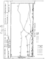
- Fig. 1 shows the Ca concentrations as a function of time. In the solutions that were in contact with the alloys, a monotonic decrease of the concentrations of Ca was detected. The same phenomenon was also observed for the reference HBSS. Until
day 5 all curves have similar forms but after day 5 a higher decrease for the Ti-6Al-4V 1 µm samples is apparent which reaches 123±1.5 ppm. For both Ti-6Al-4V 4000 and Ti-Al-2.5Fe 1 µm samples the Ca concentration decreased more rapidly afterday 7 attaining similar final Ca uptake concentrations, 61±2.3 and 63±1.5 ppm, respectively. All other surfaces exhibited Ca uptake values between 5 and 20 ppm. - Fig. 2 shows the P concentration as a function of time. The P uptake curves, like the Ca determinations, also showed a decrease as a function of time. The Ti-6Al-
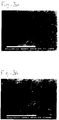
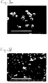
4V Fe 1 µm showed the highest P uptake; 29±2.1, 34±1.5 and 58.5±2.2 ppm, respectively. These findings suggest that a Ca and P rich precipitate is formed on all the surfaces from the HBSS. In fact, it was possible to see a white film deposited on the polyethylene that contacted with the HBSS. Ti-Al-2.5Fe - Fig. 3 shows SEM photomicrographs of the metal surfaces after immersion in HBSS. Comparing the photographs on Fig. 3 it can be seen that the precipitate layer has a plate morphology on which "globules" and/or crystals grow. XRMA revealed a higher quantity of Ca and P on these particles than in the plate precipitate. It was possible to observe that the plates fractured on some of the surfaces, namely Ti-6Al-
4V Fe 1 µm andstainless steel 1200. The orientation of the fractures does not seem to depend on the orientation of the grinding flaws as it is possible to observe a random cracking of the plates. The precipitate formed on Ti-6Al-4V 4000 shows a continuous texture at the same magnification as the other observations. It was only possible to detect fractures on these surfaces, on the Ca and P rich layer, at magnifications higher than 2400 x.
Li et al. [4] performed a series of experiments in which silica-gel was immersed in SBF. They suggest that the regulation of apatite growth is related to the Ca/P molar ratio of the fluids. Fugishiro et al. [1] obtained different HA morphologies by immersing Fe and Ti in Ca(edta)2--NaH2PO4 solution. Various concentrations of Ca(edta)2- had a direct effect on the morphology of the hydroxyapatite film.
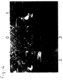
The SEM observations suggest that the morphology of the precipitate layer seems to be dependent both on material and surface finishing as the immersion fluid was the same in all experiments. - Fig. 4 shows an AFM photomicrograph from a Ti-Al-2.5
Fe 1 µm sample. It is apparent that the calcium phosphate rich coating is constituted by the agglomeration of spherical particles. Similar results were obtained for the Ti-6Al-4V 1 µm surfaces. It seems that the formation of the coating starts with heterogeneous precipitation of nuclei which gather with time until all the surface is covered.
It was noticeable that the Ti-Al-2.5Fe alloy surfaces 4000 and 1200 did not exhibit plate precipitates. It was only possible to observe small scattered deposits which had a similar morphology to crystals. XRMA acquisition on the flat-ground surfaces showed the presence of no Ca or P. The same acquisition on the crystals showed the presence of the alloy elements, Ca and P, associated with Si. Si seems to act as a nucleus for the precipitation and growth of the crystals. This impurity is probably due to the SiC emery paper used during the surface preparation. Either the degreasing and cleaning of the surface was not sufficient, on these surfaces, to remove the SiC or some SiC particles might be anchored in the alloy's surface as Ti-Al-2.5Fe is a softer material than the other alloys. - Fig. 5 exhibits XRMA spectra acquired in a Ti-6Al-
4V 4000 sample before and after immersion in HBSS. One can observe the presence of the alloy elements as well as very well defined Ca and P peaks on the after-immersion spectra. The calculated Ca/P ratio is 1.56±0.03 which indicates that the precipitate probably consists mainly of tricalcium phosphate. - Fig. 6 shows XRD spectra acquired on non-immersed (A) and immersed (B) Ti-6Al-
4V 1 µm surfaces. On the immersed samples one can observe the appearance of a well defined [002] peak and a broader peak which seems to be constituted by the junction of peaks [211] and [112] indicating the amorphous characteristics of the calcium phosphate. These results suggest that the precipitate layer has an amorphous apatite-like structure. Similar results were obtained for the Ti-Al-2.5Fe 1 µm samples.
The thickness of this layer was previously determined by SEM observations and is ca. 5 µm. Li et al. [4] monitored the development of hydroxyapatite deposits on gel-derived titania, as a function of time, after immersion in Simulated Body Fluid. In the initial stages they detected scattered precipitates all over the surface which increased in number and size until, eventually, all the surface was covered by a 10 µm coating. Ducheyne et al. [5] reported the formation of small deposits on titanium discs after 1-day exposure to a Simulated Physiological Solution. Two weeks of differential immersion were needed to produce an apatite layer with a thickness of 1 µm.
Hanawa et al. [3] also reported that apatite is naturally formed on titanium when titanium is immersed in a solution whose pH is similar to that of the bioliquid. They reported a thickness of 7 nm of the apatite film grown on Ti-6Al-4V which makes it impossible for this layer to exhibit any properties of calcium phosphate in this environment.
The present results indicate that a calcium phosphate with an apatite-like structure is naturally formed on the surfaces of polished titanium alloys. The thickness of this layer makes it a suitable surface for bone induction. Thicknesses of at least 1 µm are needed for the calcium phosphate to show its properties and cause bone induction [5]. - The morphology of calcium phosphate precipitates depends on the metal substrate and its surface characteristics. It is possible to produce a naturally formed calcium phosphate coating by immersing metals such as titanium alloys and stainless steel in HBSS. Ti-6Al-
4V 4000 seems to be the surface that is most favourable to produce a continuous and more adherent apatite-like coating capable of bone induction. - This example illustrates the determination of the depth distributions of selected elements in a calcium, phosphorous and oxygen - containing coating on a titanium alloy sample using depth profiling X-ray Photoelectron Spectroscopy (XPS or ESCA).
- The samples were titanium alloy plates that had been surface treated according to the procedure of Example 1 to produce a calcium phosphate coating when immersed in calcification solutions or simulated body fluids. The samples were mounted directly to a standard XPS sample holder using a spring clip arrangement, with no pre-treatment. The outer coating surface was sufficiently electrically conducting that no electrostatic charging problems were encountered during X-ray irradiation or ion beam etching. All analysis were carried out using a Surface Science Instruments (SSI) M-probe operating at a base pressure of 3x10⁻⁹ torr.
- A survey spectrum was recorded from theas received
 surface, to determine the surface composition of the coating and therefore determine the elements to be monitored for the depth profile. The XPS depth profile was obtained by alternating argon ion sputtering (over an area of approx. 2 x 2 mm) and data acquisition (from an area of approx. 300 µm diameter centred in the etched crater). Elements analysed were carbon, oxygen, calcium, phosphorus, magnesium and titanium. Etch time per step was variable from 15 to 120 seconds per cycle and the etch rate was 3 nm/min using a total sputter time of 4470 seconds.
- The surface chemical composition (in atomic percent) of theas receivedcoating was: carbon 44.9%, oxygen 33.8%, calcium 10.5%, phosphorous 8.8%, magnesium 2.0% and
titanium 0% (figure 7). The depth profile of the coating revealed a gradual transition of calcium and phosphorous from the coating to the substrate, indicating the incorporation of these elements in the surface (oxide layer), and thus a chemical bonding between coating and substrate (figure 8). The calcium - oxygen - phosphorous layer (calcium phosphate) is estimated as being approximately 90 nm, assuming a sputter rate of 3 nm per minute as calibrated on a tantalum pentoxide film on tantalum, and that theinterfaceis defined as the point where the titanium reaches approx. 50% of its final value. A thin layer of titanium oxide separates the calcium phosphate layer from the titanium alloy substrate. The interface between the calcium phosphate and titanium shows changes in the oxygen, phosphorous, calcium and titanium chemistries. The XPS peak binding energies of calcium and phosphorous decrease at the interface with the titanium where a titanium oxide layer is found. An interphase region is likely to occur at the boundary and oxygen has been depleted from the calcium phosphate to form titanium dioxide at the interface. Metallic titanium is present below the interphase region. Magnesium is detected at 2 - 4 atomic percent throughout the calcium phosphate layer and increases slightly in concentration with depth towards the interface with the titanium (oxide). Carbon is found in the bulk of the titanium. - The calcium phosphate layer that is formed on the titanium alloy substrate is chemically bound to the substrate via its surface oxide layer.
- This examples illustrates a new two-step chemical treatment for preparing an implant with a specific surface roughness, resulting in a metallic surface that allows fast precipitation of biomimetic calcium phosphate (Ca-P) coatings from in vitro supersaturated calcification solutions (SCS). The present method has the following advantages over the conventional techniques for the coating application: due to the specific surface roughness, (i) the biomimetic coatings directly induced from SCS are expected to be chemically bound to metallic substrates and to show higher bone-bonding ability, (ii) the coatings can be produced onto complex-shaped and/or macroporous implants, and (iii) it is a controllable and cost-effective way to acquire Ca-P coatings. To examine the potential of biomimetic calcium phosphate coatings as drug-release carriers, bovine serum albumin (BSA) and Ca-P coatings were co-precipitated on the treated titanium surfaces by immersion in the SCS containing 0.2% wt. BSA. The BSA/Ca-P coatings are expected to serve as a potential drug-release system.
- A newly developed two-step chemical treatment was performed on the metallic implant materials, i.e. commercially pure titanium (cp.Ti), annealed Ti6Al4V and porous tantalum (Ta), to produce a specific surface roughness. During this treatment, two series of chemical reagents were used for titanium (cp.Ti and Ti6Al4V) and tantalum implant materials, respectively, that resulted in the presence of the specific surface roughness necessary for the preparation of the coating. For the former, the samples were treated with a mixture of HCl and H2SO4, followed by immersion in a NaOH solution. The porous tantalum samples were treated with a mixture of HCl, H2SO4 and HF, followed by immersion in H2O2.
Two kinds of SCSs with different Ca and P concentrations, fast calcification solution (FCS) and commercial Hanks' balanced salt solution (HBSS), were used for preparing biomimetic Ca-P coatings. To promote the Ca-P nucleation on the metallic surfaces, a precalcification (Pre-Ca) procedure was performed on half the treated samples before immersion in the SCS. The Pre-Ca was carried out by incubating the samples in 0.5N Na2HPO4 overnight and then transferring them into saturated Ca(OH)2 for 5 h. The solution for co-precipitation of BSA and Ca-P coatings was prepared by dissolving 0.2%wt. BSA into FCS. The untreated metals were also immersed as controls. The FCS solution volume used for immersion was 15 ml per cm2 of sample surface area. The samples were immersed in sealed polystyrene vials at 37°C in a calibrated water-bath. Scanning electron microscopy (SEM) together with energy disperse X-ray (EDX) analyses, X-ray diffraction (XRD) and infrared (IR) spectrophotometry were used to characterize the obtained Ca-P coatings. - The biomimetic Ca-P coatings were fast precipitated on the treated cp.Ti and Ti6Al4V samples by immersion in both FCS and HBSS no matter whether the Pre-Ca procedure was performed or not. But the Pre-Ca treatments could dramatically speed-up the precipitation rate of the Ca-P coatings as listed in table 2.
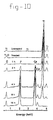
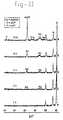
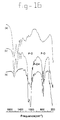
Figure 9 shows that a biomimetic Ca-P coating, approximately 16 µm thick, was formed on heated cp.Ti after 16 hours of immersion in FCS with Pre-Ca. The coating got thicker with immersion time as indicated by EDX (figure 10) and XRD (figure 11) results. The precipitation rate of the Ca-P coating in HBSS is slower than that in FCS. But the coating from HBSS (figure 12) was much denser than that from FCS. The coating from HBSS mainly consisted of apatite (figure 13). Biomimetic Ca-P coatings could also be precipitated on porous Ta samples (figure 14) after the treatment. The surface content change of the sample was detected by EDX as shown in figure 15. It is noteworthy that no precipitation was observed on any untreated samples after 2 weeks of immersion in FCS or HBSS, even with Pre-Ca. The formation of a specific titanium and tantalum oxide layer after their treatments is probably the main reason for the inductive precipitation of Ca-P coatings by means of in vitro immersion in SCS. The procedure of the treatments for titanium implants and tantalum could not be exchanged, otherwise no Ca-P coating was acquired. It is interesting to find that the results of co-precipitation of BSA and Ca-P coating were positive. The IR analyses (figure 16) indicated the obvious existence of BSA in the Ca-P coating on cp.Ti after immersion in FCS. Release experiments of BSA from BSA/Ca-P coatings are decribed in Example 4. - The results of this biomimetic calcium phosphate coating and protein co-precipitation study have shown that:-
- . The newly developed two-step chemical treatment is an effective method to prepare bioactive metallic implant surfaces allowing fast precipitation of adherent biomimetic Ca-P coatings by in vitro immersion in SCS. The chemical reagents needed for the treatment of titanium implant materials and tantalum are different from each other.
- . The precipitation of Ca-P coatings could be dramatically accelerated by means of precalcifying the treated samples before the immersions.
- . The precipitation rate and composition of the Ca-P coatings can be adjusted by controlling the components of the SCSs (FCS or HBSS) for immersion.
- . Some proteins like BSA can be co-precipitated with biomimetic Ca-P coatings. Such composite coatings, with other - bioactive - adsorbed organic molecules, are expected to be able to serve as potential drug-release systems.
- Commercially rectangular blocks in
size 10 × 10 × 2 mm were used as substrates. The two-step treatment and pre-calcification were performed on the metals as described in example 3. The supersaturated calcification solution used for immersion was prepared by dissolving 0.2% BSA into 0.8 ACS. All the samples were immersed in 37°C water-bath for two days. - The BSA content in solution was measured by means of total organic carbon (TOC) method. Four blocks with Ca-P/BSA coatings were completely dissolved by 40 ml of 0.5 N HCl solution to detect the concentration of BSA in Ca-P/BSA coating. Four samples were immersed in 20 ml of 0.01 M PBS at pH 4 (adjusted using HCl) at 37°C. At certain intervals of time, 5 ml of the immersion solution was taken for TOC measurement at certain interval and then refill to 20 ml with new PBS at
pH 4 every time. - The Ca-P/BSA coatings on five samples were peeled off completely and then weighed using a balance. The weight of the Ca-P/BSA coating on each sample can be estimated to be a mean of 1.5 mg. The carbon concentration in the coating was calculated to be 15 % and the BSA concentration was about 30% because the carbon concentration in BSA was detected to be around 50% by the same method.
- The amount of BSA released into PBS was plotted against the immersion time as shown in Figure 17. It is indicated that under these experimental conditions, BSA was released relatively fast in the first two days of the immersion and then the release rate levelled off somewhat. The main reason could be that the PBS was very acidic (pH = 4) at the beginning stage and the coating was dissolved very fast. After certain time of immersion the pH became higher. The pH values of PBS at the 7th and 14th were measured to be 5.628 and 5.584, respectively. Relating these results to the actual in vivo situation, where the pH value will be around 7, these results indicate that a gradual, slow release system is obtained, assuming that the 48 hours incubation in an acidic medium is comparable to several weeks, if not months implantation in an organism.
- This experiment shows that proteins such as BSA can be co-precipitated in a calcium phosphate coating and that it can subsequently be released from the coating. Although BSA was used to examine the feasibility of this technique, it is clear that a Ca-P/biologically active agent composite coating can be used as sort of a drug delivery system and has therefore potential in the medical field.
Table 1 Surface roughness measurements results Surface finish Ra (µm) Rz (µm) Rmax (µm) Ti-6Al- 4V 1200 grit0.47±0.01 3.74±0.04 5.13±0.08 Ti-6Al- 4V 4000 grit0.24±0.03 1.91±0.31 2.46±0.54 Ti-6Al- 4V 1 µm0.03±0.00 0.35±0.05 0.48±0.03 Ti-Al-2.5 Fe 1200 grit0.42±0.03 2.97±0.35 3.47±0.48 Ti-Al-2.5 Fe 4000 grit0.23±0.01 1.97±0.18 2.46±0.34 Ti-Al-2.5 Fe 1 µm0.04±0.01 0.28±0.11 0.36±0.19 316 L 1200 grit0.3±0.06 2.32±0.47 2.96±0.03 316 L 4000 grit0.04±0.01 0.35±0.1 0.46±0.1 Table 2 List of Ca2+ and HPO4 2- concentrations, precipitation rate and composition of Ca-P coatings on cp.Ti and Ti6Al4V. FCS HBSS Concentration (mM) Ca2+ 3.0 1.26 HPO4 2- 1.87 0.78 Precipitation rate of coating No Pre-Ca 0.5 µm/ hr 1 µm/ wk Pre-Ca 1 µm/ hr 3 µm/wk Composition of coating apatite, OCP apatite -
- Figure 1 -
- Ca concentration as a function of time
- Figure 2 -
- P concentration as a function of time
- Figure 3 -
- SEM photomicrographs of the metal surfaces after immersion in HBSS. A: Ti-6Al-
4V 1200; B: Ti-6Al-4V 4000; C: Ti-6Al-4V 1 µm; D: Ti-Al-2.5Fe 1 µm; E: Ti-Al-2.5Fe 4000; F -stainless steel 1200 - Figure 4 -
- AFM photomicrograph of a Ti-Al-2.5
Fe 1 µm sample after immersion in HBSS. Increasing magnification fromfield 0 to 3. Scanning length from field 3: 1.5 µm. - Figure 5 -
- XRMA spectra acquired on a Ti-6Al-
4V 4000 sample before (A) and after immersion (B) in HBSS. - Figure 6 -
- XRD spectra acquired on a non-immersed (A) and immersed (B) Ti-6Al-
4V 1 µm surface - Figure 7.
- Surface chemical composition (in atomic percent) of the as receivedcoating.
- Figure 8.
- Depth profile of the coating, from coating to substrate.
- Figure 9.
- Scanning electron micrograph of the Ca-P coating (CP) precipitated on cp.Ti (Ti) after 16 hours of immersion in FCS with Pre-Ca.
- Figure 10.
- EDX spectra of the cp.Ti surfaces non-treated, treated and immersed in FCS with Pre-Ca for different hours. The shoulder of O kα peak is clearly seen after the treatment. The Ca and P contents increased with the increase of immersion time.
- Figure 11.
- XRD patterns of the cp.Ti surfaces after different hours of immersion in FCS with Pre-Ca. The counts of apatite peaks get higher with increased immersion times. Octa-calcium phosphate (OCP) starts to be formed at around 8 hours.
- Figure 12.
- Scanning electron micrograph of a dense Ca-P coating (CP) precipitated on cp.Ti from HBSS after 1 week of immersion with Pre-Ca. The layer between coating and substrate is the titanium oxide layer (OL), formed as a result of the treatment. Note the surface roughness of the implant on which the calcium phosphate coating has been formed.
- Figure 13.
- Thin-film XRD pattern of a dense Ca-P coating deposited by immersion in HBSS with Pre-Ca for 1 week.
- Figure 14.
- Scanning electron micrograph of porous tantalum (Ta) after 2 days immersion in FCS with Pre-Ca. The coating is formed throughout the porous material.
- Figure 15.
- EDX spectra of (a) non-treated, (b) treated, and (c) Pre-Ca treated, 2 day FCS immersed porous tantalum (Ta) sample.
- Figure 16.
- IR spectra of BSA (a) and cp.Ti surfaces after 2 days of immersion in FCS containing 0.2%wt. BSA (b) and FCS only (c). The existence of four absorption bands of BSA in spectrum (b) indicates the co-precipitation of BSA with the biomimetic calcium phosphate coating.
- Figure 17.
- The amount of BSA released into PBS as a result of the immersion time
-
- 1. Y. Fujishiro, T. Sato and A. Okuwaki, "Coating of hydroxyapatite on metal plates using thermal dissociation of calcium -EDTA chelate in phosphate solutions under hydrothermal conditions", J. Mater. Sc: Mater in Med, 6, pp. 172-176, 1995
- 2. S.R. Radin and P. Ducheyne, J. Biom. Mater. Res.,, 27, pp. 35, 1993
- 3. T. Hanawa, "Titanium and its oxide film: a substrate for forming apatite", in Proc. of the Bone Biomaterial Interface Workshop, Toronto, Dec. 1990, J.E. Davies ed., Univ. Toronto Press, pp. 49-61, 1991
- 4. Li, P, PhD. thesis Leiden University (1993)
- 5. P. Ducheyne, S. Radin and K. Ishikawa, "The rate of calcium phosphate precipitation on metal and ceramics, and the relationship to bioactivity", in Bone Bonding Biomaterials, P. Ducheyne, T. Kokubo & C.A. van Blitterswijk (eds), Reed Heathcare Communications, pp. 213-218, 1992.
Claims (10)
- An implantable device of a material having a surface roughness with an average peak distance (Ra value) between 10 and 1,000 nm, said surface being coated with a layer comprising calcium ions and phosphate ions.
- An implantable device according to claim 1, wherein said layer further comprises a growth factor, a lipid, a (lipo)polysaccharide, a cytostatic agent, a hormone, an antibiotic, a protein and/or another biologically active agent.
- An implantable device according to claim 1 or 2, having a surface roughness with an average depth between 20 and 1,000 nm.
- An implantable device according to any one of claims 1-3, wherein said layer of calcium phosphate has a thickness of 50 nm - 200 µm, in particular of between 1000 nm and 50 µm.
- An implantable device according to any one of claims 1-4, wherein said material is a metal, a metal alloy, a ceramic material, a natural or synthetic polymer or a composite of any of these materials.
- A process for producing an implantable device, comprising subjecting a porous or non-porous material to a surface treatment until a surface roughness with an average peak distance (Ra value) between 10 and 1,000 nm is obtained, and subsequently subjecting the surface to precipitation of calcium phosphate from a solution containing calcium and phosphate ions.
- A process according to claim 6, wherein said solution has a pH of 4-10, preferably 6-8.
- A process according to claim 6 or 7, wherein said solution further contains a growth factor, a lipid, a (lipo)polysaccharide, a hormone, a cytostatic agent, an antibiotic, a protein and/or another biologically active agent.
- A process according to any one of claims 6-8, wherein said subjecting to precipitation of calcium phosphate is carried out in two stages.
- Use of a device according to any one of claims 1-5, or produced according to any one of claims 6-9, as a bone substitute, a joint prosthesis, a dental implant, a maxillofacial implant, a vertebral surgery aid, a transcutaneous device or any other hard-tissue-related device.
Priority Applications (1)
| Application Number | Priority Date | Filing Date | Title |
|---|---|---|---|
| EP97201425A EP0806212B1 (en) | 1996-05-10 | 1997-05-12 | Device for incorporation and release of biologically active agents |
Applications Claiming Priority (3)
| Application Number | Priority Date | Filing Date | Title |
|---|---|---|---|
| EP96201293 | 1996-05-10 | ||
| EP96201293 | 1996-05-10 | ||
| EP97201425A EP0806212B1 (en) | 1996-05-10 | 1997-05-12 | Device for incorporation and release of biologically active agents |
Publications (2)
| Publication Number | Publication Date |
|---|---|
| EP0806212A1 true EP0806212A1 (en) | 1997-11-12 |
| EP0806212B1 EP0806212B1 (en) | 2003-04-02 |
Family
ID=26142789
Family Applications (1)
| Application Number | Title | Priority Date | Filing Date |
|---|---|---|---|
| EP97201425A Expired - Lifetime EP0806212B1 (en) | 1996-05-10 | 1997-05-12 | Device for incorporation and release of biologically active agents |
Country Status (1)
| Country | Link |
|---|---|
| EP (1) | EP0806212B1 (en) |
Cited By (47)
| Publication number | Priority date | Publication date | Assignee | Title |
|---|---|---|---|---|
| EP0987031A1 (en) * | 1998-09-15 | 2000-03-22 | Isotis B.V. | Method for coating medical implants |
| WO2000072775A1 (en) * | 1999-05-31 | 2000-12-07 | Nobel Biocare Ab (Publ) | Implant for application in bone, method for producing such an implant, and use of such an implant |
| US6207218B1 (en) | 1998-09-15 | 2001-03-27 | Isotis B.V. | Method for coating medical implants |
| WO2001056628A1 (en) * | 2000-02-04 | 2001-08-09 | Isotis N.V. | Proteinaceous coating |
| DE10006992A1 (en) * | 2000-02-16 | 2001-09-06 | Aesculap Ag & Co Kg | Method of coating implantate used as bone prosthesis by electrolytic deposition of soluble calcium phosphate comprises incorporation of dispersion particles |
| WO2001074412A1 (en) * | 2000-04-04 | 2001-10-11 | Nobel Biocare Ab (Publ) | Implant having attachment and hole-insert parts, and method for producing such an implant |
| WO2001085068A1 (en) * | 2000-05-08 | 2001-11-15 | Universität Heidelberg | Endoprosthesis part for use in joint replacement, use of the same and production method therefor |
| WO2001097718A1 (en) * | 2000-06-23 | 2001-12-27 | University College London | Transcutaneous prosthesis |
| WO2002005862A1 (en) * | 2000-07-17 | 2002-01-24 | Dot Gmbh | Bioactive calcium phosphate composite layers electrochemically deposited on implants |
| WO2002024248A1 (en) * | 2000-09-22 | 2002-03-28 | Kensey Nash Corporation | Systems and methods for delivering beneficial agents into targeted tissue of a living being |
| WO2002024247A1 (en) * | 2000-09-22 | 2002-03-28 | Kensey Nash Corporation | Drug delivering prostheses and methods of use |
| WO2002024271A3 (en) * | 2000-09-22 | 2002-06-27 | Kensey Nash Corp | Systems for delivering agents for revascularizing accluded blood vessels |
| WO2002009597A3 (en) * | 2000-08-01 | 2002-08-08 | Regeneration Tech Inc | Diaphysial cortical dowel |
| WO2002060507A1 (en) * | 2001-01-30 | 2002-08-08 | Isotis S.A. | A method for applying a bioactive coating on a medical device |
| WO2002074355A1 (en) | 2001-03-15 | 2002-09-26 | Dot Gmbh | Calcium phosphate materials containing active ingredients |
| WO2002078759A1 (en) * | 2001-04-02 | 2002-10-10 | Stratec Medical Ag | Bioactive surface layer, particularly for medical implants and prostheses |
| DE10119096A1 (en) * | 2001-04-19 | 2002-10-24 | Keramed Medizintechnik Gmbh | New biologically functionalized coatings, useful for e.g. accelerating osteo-integration of implants, e.g. dental or joint implants, comprise resorbable calcium-phosphorus phase containing adhesion and/or signal proteins |
| EP1254673A1 (en) * | 2001-05-02 | 2002-11-06 | InFlow Dynamics, Inc. | Immuno-tolerant stent with surface microstructure |
| WO2003026718A1 (en) * | 2000-10-31 | 2003-04-03 | Cook Incorporated | Coated implantable medical device |
| EP1318844A2 (en) | 2000-07-26 | 2003-06-18 | Institut Straumann AG | Surface-modified implants |
| WO2003059407A1 (en) * | 2002-01-21 | 2003-07-24 | Straumann Holding Ag | Surface-modified implants |
| EP1358859A1 (en) * | 2002-04-29 | 2003-11-05 | Politecnico Di Milano | Bone prostheses having multilayer interface |
| WO2004006976A1 (en) * | 2002-07-12 | 2004-01-22 | Cook Incorporated | Coated medical device |
| WO2004040036A1 (en) * | 2002-10-31 | 2004-05-13 | Cerbio Tech Ab | Method of making structured ceramic coatings and coated devices prepared with the method |
| WO2004040037A1 (en) * | 2002-10-31 | 2004-05-13 | Cerbio Tech Ab | Method of making structured ceramic coatings and coated devices prepared with the method |
| WO2004047881A1 (en) * | 2002-11-25 | 2004-06-10 | National Institute Of Advanced Industrial Science And Technology | Calcium phosphate carrying protein, process for producing the same and sustained release protein preparation, artificial bone and tissue enginerring scaffold using the same |
| US6863693B2 (en) | 1999-08-31 | 2005-03-08 | Destiny Pharma Limited | Phospholipid-coated implants |
| WO2006110578A2 (en) * | 2005-04-08 | 2006-10-19 | Paradigm Spine, Llc. | Interspinous vertebral and lumbosacral stabilization devices and methods of use |
| WO2007044229A2 (en) * | 2005-09-28 | 2007-04-19 | Calcitec, Inc. | Surface treatments for calcium phosphate-based implants |
| EP1656959A3 (en) * | 2004-11-12 | 2007-09-19 | DePuy Products, Inc. | System attaching soft tissue to an implant |
| EP1847278A1 (en) * | 2006-04-13 | 2007-10-24 | Camlog Biotechnologies AG | Bio-dissolvable salt coatings of implants for the protection against organic impurities |
| EP1872805A2 (en) * | 2006-06-27 | 2008-01-02 | Biomed Est. | Coated implant |
| US7534249B2 (en) | 1996-07-26 | 2009-05-19 | Kensey Nash Corporation | System and method of use for agent delivery and revascularizing of grafts and vessels |
| US7594900B1 (en) | 1999-08-05 | 2009-09-29 | Kensey Nash Corporation | Systems and methods for delivering agents into targeted tissue of a living being |
| US7763270B2 (en) | 2002-09-10 | 2010-07-27 | Scil Technology Gmbh | Metal implant coated under reduced oxygen concentration with osteoinductive protein |
| US7833239B2 (en) | 1996-07-26 | 2010-11-16 | Kensey Nash Corporation | System and method of use for revascularizing stenotic bypass grafts and other blood vessels |
| US7922750B2 (en) | 2006-11-30 | 2011-04-12 | Paradigm Spine, Llc | Interlaminar-interspinous vertebral stabilization system |
| EP2308410A1 (en) * | 2001-07-04 | 2011-04-13 | Nobel Biocare Services AG | Implant |
| US7981129B2 (en) | 1996-07-26 | 2011-07-19 | Kensey Nash Corporation | System for opening a lumen in an occluded blood vessel |
| US8546334B2 (en) | 2001-11-19 | 2013-10-01 | Scil Technology Gmbh | Device having osteoinductive and osteoconductive properties |
| WO2014203204A1 (en) * | 2013-06-21 | 2014-12-24 | Sandvik Intellectual Property Ab | Co-precipitation of a therapeutic agent into hydroxyapatite coatings |
| US9662425B2 (en) | 2013-04-22 | 2017-05-30 | Stryker European Holdings I, Llc | Method for drug loading hydroxyapatite coated implant surfaces |
| US9801671B2 (en) | 2013-07-11 | 2017-10-31 | Stryker European Holdings I, Llc | Fixation assembly with multiple sections for securing parts of a sternum |
| WO2020158833A1 (en) | 2019-01-31 | 2020-08-06 | セルメディシン株式会社 | Inorganic salt-protein composite medical instrument |
| US10864297B2 (en) | 2013-09-02 | 2020-12-15 | Stryker European Holdings I, Llc | Method of manufacturing an implant for use in a surgical procedure |
| US11259853B2 (en) | 2015-01-09 | 2022-03-01 | Stryker European Operations Holdings Llc | Implant for bone fixation |
| US11596458B2 (en) | 2016-11-11 | 2023-03-07 | Stryker European Operations Holdings Llc | Implant for bone fixation |
Families Citing this family (2)
| Publication number | Priority date | Publication date | Assignee | Title |
|---|---|---|---|---|
| EP2022447A1 (en) | 2007-07-09 | 2009-02-11 | Astra Tech AB | Nanosurface |
| JP5806466B2 (en) | 2008-01-28 | 2015-11-10 | バイオメット・3アイ・エルエルシー | Implant surface with improved hydrophilicity |
Citations (6)
| Publication number | Priority date | Publication date | Assignee | Title |
|---|---|---|---|---|
| GB2045083A (en) * | 1979-03-07 | 1980-10-29 | Branemark Per Ingvar | Element for implantation in body tissue particularly bone tissue |
| EP0450939A2 (en) * | 1990-04-05 | 1991-10-09 | Norian Corporation | Improvements in hydroxyapatite coatings of substrates |
| US5344457A (en) * | 1986-05-19 | 1994-09-06 | The University Of Toronto Innovations Foundation | Porous surfaced implant |
| WO1995013101A1 (en) * | 1993-11-09 | 1995-05-18 | Klaas De Groot | A method of applying a coating of a bioactive material to implants |
| WO1995013102A1 (en) * | 1993-11-10 | 1995-05-18 | Implant Innovations, Inc. | Surgically implantable prosthetic devices |
| US5456723A (en) * | 1989-03-23 | 1995-10-10 | Institut Straumann Ag | Metallic implant anchorable to bone tissue for replacing a broken or diseased bone |
-
1997
- 1997-05-12 EP EP97201425A patent/EP0806212B1/en not_active Expired - Lifetime
Patent Citations (6)
| Publication number | Priority date | Publication date | Assignee | Title |
|---|---|---|---|---|
| GB2045083A (en) * | 1979-03-07 | 1980-10-29 | Branemark Per Ingvar | Element for implantation in body tissue particularly bone tissue |
| US5344457A (en) * | 1986-05-19 | 1994-09-06 | The University Of Toronto Innovations Foundation | Porous surfaced implant |
| US5456723A (en) * | 1989-03-23 | 1995-10-10 | Institut Straumann Ag | Metallic implant anchorable to bone tissue for replacing a broken or diseased bone |
| EP0450939A2 (en) * | 1990-04-05 | 1991-10-09 | Norian Corporation | Improvements in hydroxyapatite coatings of substrates |
| WO1995013101A1 (en) * | 1993-11-09 | 1995-05-18 | Klaas De Groot | A method of applying a coating of a bioactive material to implants |
| WO1995013102A1 (en) * | 1993-11-10 | 1995-05-18 | Implant Innovations, Inc. | Surgically implantable prosthetic devices |
Cited By (93)
| Publication number | Priority date | Publication date | Assignee | Title |
|---|---|---|---|---|
| US7833239B2 (en) | 1996-07-26 | 2010-11-16 | Kensey Nash Corporation | System and method of use for revascularizing stenotic bypass grafts and other blood vessels |
| US6569147B1 (en) | 1996-07-26 | 2003-05-27 | Kensey Nash Corporation | Systems and methods of use for delivering beneficial agents for revascularizing stenotic bypass grafts and other occluded blood vessels and for other purposes |
| US8226673B2 (en) | 1996-07-26 | 2012-07-24 | Kensey Nash Corporation | System and method of use for agent delivery and revascularizing of grafts and vessels |
| US7534249B2 (en) | 1996-07-26 | 2009-05-19 | Kensey Nash Corporation | System and method of use for agent delivery and revascularizing of grafts and vessels |
| US7981129B2 (en) | 1996-07-26 | 2011-07-19 | Kensey Nash Corporation | System for opening a lumen in an occluded blood vessel |
| EP0987031A1 (en) * | 1998-09-15 | 2000-03-22 | Isotis B.V. | Method for coating medical implants |
| US6207218B1 (en) | 1998-09-15 | 2001-03-27 | Isotis B.V. | Method for coating medical implants |
| AU779288B2 (en) * | 1999-05-31 | 2005-01-13 | Nobel Biocare Services Ag | Implant for application in bone, method for producing such an implant, and use of such an implant |
| WO2000072775A1 (en) * | 1999-05-31 | 2000-12-07 | Nobel Biocare Ab (Publ) | Implant for application in bone, method for producing such an implant, and use of such an implant |
| US7594900B1 (en) | 1999-08-05 | 2009-09-29 | Kensey Nash Corporation | Systems and methods for delivering agents into targeted tissue of a living being |
| US6863693B2 (en) | 1999-08-31 | 2005-03-08 | Destiny Pharma Limited | Phospholipid-coated implants |
| WO2001056628A1 (en) * | 2000-02-04 | 2001-08-09 | Isotis N.V. | Proteinaceous coating |
| US6692790B2 (en) | 2000-02-04 | 2004-02-17 | Chienna B.V. | Proteinaceous coating |
| DE10006992A1 (en) * | 2000-02-16 | 2001-09-06 | Aesculap Ag & Co Kg | Method of coating implantate used as bone prosthesis by electrolytic deposition of soluble calcium phosphate comprises incorporation of dispersion particles |
| DE10006992B4 (en) * | 2000-02-16 | 2006-10-12 | Aesculap Ag & Co. Kg | Process for coating an implant and implant with coating |
| WO2001074412A1 (en) * | 2000-04-04 | 2001-10-11 | Nobel Biocare Ab (Publ) | Implant having attachment and hole-insert parts, and method for producing such an implant |
| WO2001085068A1 (en) * | 2000-05-08 | 2001-11-15 | Universität Heidelberg | Endoprosthesis part for use in joint replacement, use of the same and production method therefor |
| US7014661B2 (en) | 2000-06-23 | 2006-03-21 | University College London | Transcutaneous prosthesis |
| US8137409B2 (en) | 2000-06-23 | 2012-03-20 | University College London | Method of installing a transcutaneous prosthesis |
| US8628584B2 (en) | 2000-06-23 | 2014-01-14 | University College London | Transcutaneous prosthesis |
| WO2001097718A1 (en) * | 2000-06-23 | 2001-12-27 | University College London | Transcutaneous prosthesis |
| US9452066B2 (en) | 2000-06-23 | 2016-09-27 | University College London | Transcutaneous prosthesis |
| AU2000259855B2 (en) * | 2000-07-17 | 2006-02-02 | Dot Gmbh | Bioactive calcium phosphate composite layers electrochemically deposited on implants |
| WO2002005862A1 (en) * | 2000-07-17 | 2002-01-24 | Dot Gmbh | Bioactive calcium phosphate composite layers electrochemically deposited on implants |
| EP1318844A2 (en) | 2000-07-26 | 2003-06-18 | Institut Straumann AG | Surface-modified implants |
| WO2002009597A3 (en) * | 2000-08-01 | 2002-08-08 | Regeneration Tech Inc | Diaphysial cortical dowel |
| WO2002024248A1 (en) * | 2000-09-22 | 2002-03-28 | Kensey Nash Corporation | Systems and methods for delivering beneficial agents into targeted tissue of a living being |
| WO2002024271A3 (en) * | 2000-09-22 | 2002-06-27 | Kensey Nash Corp | Systems for delivering agents for revascularizing accluded blood vessels |
| WO2002024247A1 (en) * | 2000-09-22 | 2002-03-28 | Kensey Nash Corporation | Drug delivering prostheses and methods of use |
| WO2003026718A1 (en) * | 2000-10-31 | 2003-04-03 | Cook Incorporated | Coated implantable medical device |
| US6918927B2 (en) | 2000-10-31 | 2005-07-19 | Cook Incorporated | Coated implantable medical device |
| WO2002060507A1 (en) * | 2001-01-30 | 2002-08-08 | Isotis S.A. | A method for applying a bioactive coating on a medical device |
| US6994883B2 (en) | 2001-01-30 | 2006-02-07 | Isotis S.A. | Method for applying a bioactive coating on a medical device |
| DE10113108A1 (en) * | 2001-03-15 | 2002-10-02 | Dot Gmbh | Calcium phosphate materials containing active ingredient |
| DE10113108B4 (en) * | 2001-03-15 | 2007-07-26 | Dot Gmbh | Active substance-containing calcium phosphate materials |
| WO2002074355A1 (en) | 2001-03-15 | 2002-09-26 | Dot Gmbh | Calcium phosphate materials containing active ingredients |
| WO2002078759A1 (en) * | 2001-04-02 | 2002-10-10 | Stratec Medical Ag | Bioactive surface layer, particularly for medical implants and prostheses |
| CN1296101C (en) * | 2001-04-02 | 2007-01-24 | 斯恩蒂斯有限公司 | Bioactive surface layer, particularly for medical implants and prostheses |
| DE10119096A1 (en) * | 2001-04-19 | 2002-10-24 | Keramed Medizintechnik Gmbh | New biologically functionalized coatings, useful for e.g. accelerating osteo-integration of implants, e.g. dental or joint implants, comprise resorbable calcium-phosphorus phase containing adhesion and/or signal proteins |
| EP1656961A1 (en) * | 2001-05-02 | 2006-05-17 | InFlow Dynamics, Inc. | Immuno-tolerant stent with surface microstructure |
| EP1254673A1 (en) * | 2001-05-02 | 2002-11-06 | InFlow Dynamics, Inc. | Immuno-tolerant stent with surface microstructure |
| EP2308410A1 (en) * | 2001-07-04 | 2011-04-13 | Nobel Biocare Services AG | Implant |
| EP1448246B2 (en) † | 2001-11-19 | 2015-09-09 | Scil Technology GmbH | Method for producing a homogeneously coated device having osteoinductive and osteoconductive properties |
| US8546334B2 (en) | 2001-11-19 | 2013-10-01 | Scil Technology Gmbh | Device having osteoinductive and osteoconductive properties |
| GB2401074B (en) * | 2002-01-21 | 2006-02-22 | Straumann Holding Ag | Surface-modified implants |
| CH695985A5 (en) * | 2002-01-21 | 2006-11-15 | Straumann Holding Ag | Surface-modified implants. |
| GB2401074A (en) * | 2002-01-21 | 2004-11-03 | Straumann Holding Ag | Surface-modified implants |
| WO2003059407A1 (en) * | 2002-01-21 | 2003-07-24 | Straumann Holding Ag | Surface-modified implants |
| EP1358859A1 (en) * | 2002-04-29 | 2003-11-05 | Politecnico Di Milano | Bone prostheses having multilayer interface |
| WO2003092555A1 (en) * | 2002-04-29 | 2003-11-13 | Franco Maria Montevecchi | Bone prosthesis with multilayer interface |
| EP2324866A3 (en) * | 2002-07-12 | 2011-10-26 | Cook Incorporated | Angioplasty balloons drug-coated in an expanded condition |
| WO2004006976A1 (en) * | 2002-07-12 | 2004-01-22 | Cook Incorporated | Coated medical device |
| US10532190B2 (en) | 2002-07-12 | 2020-01-14 | Cook Medical Technologies Llc | Coated medical device |
| EP2324867A1 (en) * | 2002-07-12 | 2011-05-25 | Cook Incorporated | Angioplasty balloons drug-coated in an expanded condition |
| US7763270B2 (en) | 2002-09-10 | 2010-07-27 | Scil Technology Gmbh | Metal implant coated under reduced oxygen concentration with osteoinductive protein |
| US8257728B2 (en) | 2002-09-10 | 2012-09-04 | Scil Technology Gmbh | Metal implant coated under reduced oxygen concentration with osteoinductive protein |
| WO2004040036A1 (en) * | 2002-10-31 | 2004-05-13 | Cerbio Tech Ab | Method of making structured ceramic coatings and coated devices prepared with the method |
| WO2004040037A1 (en) * | 2002-10-31 | 2004-05-13 | Cerbio Tech Ab | Method of making structured ceramic coatings and coated devices prepared with the method |
| WO2004047881A1 (en) * | 2002-11-25 | 2004-06-10 | National Institute Of Advanced Industrial Science And Technology | Calcium phosphate carrying protein, process for producing the same and sustained release protein preparation, artificial bone and tissue enginerring scaffold using the same |
| US20150209042A1 (en) * | 2004-11-12 | 2015-07-30 | Purdue Research Foundation | System And Method For Attaching Soft Tissue To An Implant |
| US11141277B2 (en) | 2004-11-12 | 2021-10-12 | Purdue Research Foundation | Metal implant for generating soft tissue and attaching to an implant |
| US9788953B2 (en) | 2004-11-12 | 2017-10-17 | Purdue Research Foundation | System and method of attaching soft tissue to an implant |
| US10596001B2 (en) | 2004-11-12 | 2020-03-24 | Purdue Research Foundation | System and method of attaching soft tissue to an implant |
| EP1656959A3 (en) * | 2004-11-12 | 2007-09-19 | DePuy Products, Inc. | System attaching soft tissue to an implant |
| US8329202B2 (en) | 2004-11-12 | 2012-12-11 | Depuy Products, Inc. | System and method for attaching soft tissue to an implant |
| US8399008B2 (en) | 2004-11-12 | 2013-03-19 | Purdue Research Foundation | System and method for attaching soft tissue to annodized metal implant |
| US8945601B2 (en) | 2004-11-12 | 2015-02-03 | DePuy Synthes Products, LLC | System and method for attaching soft tissue to an implant |
| US20130178942A1 (en) * | 2004-11-12 | 2013-07-11 | Thomas J. Webster | System and Method for Attaching Soft Tissue To An Implant |
| US8906402B2 (en) * | 2004-11-12 | 2014-12-09 | Purdue Research Foundation | System and method for attaching soft tissue to an implant |
| US9498218B2 (en) | 2004-11-12 | 2016-11-22 | Purdue Research Foundation | System and method for attaching soft tissue to an implant comprising a nanotextured surface |
| US10194956B2 (en) | 2005-04-08 | 2019-02-05 | Paradigm Spine, Llc | Interspinous vertebral and lumbosacral stabilization devices and methods of use |
| US8470000B2 (en) | 2005-04-08 | 2013-06-25 | Paradigm Spine, Llc | Interspinous vertebral and lumbosacral stabilization devices and methods of use |
| WO2006110578A2 (en) * | 2005-04-08 | 2006-10-19 | Paradigm Spine, Llc. | Interspinous vertebral and lumbosacral stabilization devices and methods of use |
| WO2006110578A3 (en) * | 2005-04-08 | 2007-01-18 | Paradigm Spine Llc | Interspinous vertebral and lumbosacral stabilization devices and methods of use |
| WO2007044229A2 (en) * | 2005-09-28 | 2007-04-19 | Calcitec, Inc. | Surface treatments for calcium phosphate-based implants |
| WO2007044229A3 (en) * | 2005-09-28 | 2007-07-26 | Calcitec Inc | Surface treatments for calcium phosphate-based implants |
| WO2007118734A1 (en) * | 2006-04-13 | 2007-10-25 | Camlog Biotechnologies Ag | Biodegrading coatings of salt for protecting implants against organic contaminants |
| EP1847278A1 (en) * | 2006-04-13 | 2007-10-24 | Camlog Biotechnologies AG | Bio-dissolvable salt coatings of implants for the protection against organic impurities |
| EP1872805A2 (en) * | 2006-06-27 | 2008-01-02 | Biomed Est. | Coated implant |
| EP1872805A3 (en) * | 2006-06-27 | 2008-04-09 | Biomed Est. | Coated implant |
| US7922750B2 (en) | 2006-11-30 | 2011-04-12 | Paradigm Spine, Llc | Interlaminar-interspinous vertebral stabilization system |
| US9662425B2 (en) | 2013-04-22 | 2017-05-30 | Stryker European Holdings I, Llc | Method for drug loading hydroxyapatite coated implant surfaces |
| US9919077B2 (en) * | 2013-06-21 | 2018-03-20 | Stryker European Holdings I, Llc | Co-precipitation of a therapeutic agent into hydroxyapatite coatings |
| WO2014203204A1 (en) * | 2013-06-21 | 2014-12-24 | Sandvik Intellectual Property Ab | Co-precipitation of a therapeutic agent into hydroxyapatite coatings |
| US20160144070A1 (en) * | 2013-06-21 | 2016-05-26 | Stryker European Holdings I. Llc | Co-precipitation of a therapeutic agent into hydroxyapatite coatings |
| US10433889B2 (en) | 2013-07-11 | 2019-10-08 | Stryker European Holdings I, Llc | Fixation assembly with a flexible elongated member for securing parts of a sternum |
| US9801671B2 (en) | 2013-07-11 | 2017-10-31 | Stryker European Holdings I, Llc | Fixation assembly with multiple sections for securing parts of a sternum |
| US11576707B2 (en) | 2013-07-11 | 2023-02-14 | Stryker European Operations Holdings Llc | Fixation assembly with a flexible elongated member for securing parts of a sternum |
| US10864297B2 (en) | 2013-09-02 | 2020-12-15 | Stryker European Holdings I, Llc | Method of manufacturing an implant for use in a surgical procedure |
| US11259853B2 (en) | 2015-01-09 | 2022-03-01 | Stryker European Operations Holdings Llc | Implant for bone fixation |
| US11596458B2 (en) | 2016-11-11 | 2023-03-07 | Stryker European Operations Holdings Llc | Implant for bone fixation |
| WO2020158833A1 (en) | 2019-01-31 | 2020-08-06 | セルメディシン株式会社 | Inorganic salt-protein composite medical instrument |
| KR20210132054A (en) | 2019-01-31 | 2021-11-03 | 셀 메디신 가부시키가이샤 | Inorganic salt protein complex medical device |
Also Published As
| Publication number | Publication date |
|---|---|
| EP0806212B1 (en) | 2003-04-02 |
Similar Documents
| Publication | Publication Date | Title |
|---|---|---|
| EP0806212B1 (en) | Device for incorporation and release of biologically active agents | |
| US6344061B1 (en) | Device for incorporation and release of biologically active agents | |
| US6069295A (en) | Implant material | |
| EP0806211B1 (en) | Implant material and process for producing it | |
| Wen et al. | Fast precipitation of calcium phosphate layers on titanium induced by simple chemical treatments | |
| Liu et al. | Surface modification of titanium, titanium alloys, and related materials for biomedical applications | |
| Wen et al. | Preparation of calcium phosphate coatings on titanium implant materials by simple chemistry | |
| EP1656164B1 (en) | Coated implants and methods of coating | |
| US6569489B1 (en) | Bioactive ceramic coating and method | |
| EP1251889B1 (en) | Proteinaceous coating | |
| EP1348452A1 (en) | Surface mineralized spinal implants | |
| EP0760687B1 (en) | A biomaterial and bone implant for bone repair and replacement | |
| JPH08299429A (en) | Method for surface treatment of titanium implant and bio-compatible titanium implant | |
| EP1984035A2 (en) | Medical devices having textured surfaces | |
| US7767250B2 (en) | Bioceramic coating of a metal-containing substrate | |
| EP2071916A1 (en) | Bio active ceramic coatings with excellent bio-compatibility and preaparation method thereof | |
| EP2773389A1 (en) | Method for modifying surfaces for better osseointegration | |
| EP2558136A1 (en) | Coated medical implant | |
| Leitao et al. | Influence of substrate material and surface finishing on the morphology of the calcium‐phosphate coating | |
| Barrere et al. | Physical and chemical characteristics of plasma-sprayed and biomimetic apatite coating | |
| CN115137875A (en) | High-efficiency two-phase calcium phosphate coating method | |
| Liu et al. | In-vitro forming of calcium phosphate layer on sol–gel hydroxyapatite-coated metal substrates | |
| Stevenson et al. | Production of Highly Adherent Hydroxylapatite Coatings by Ion Beam and Plasma Techniques | |
| Tan et al. | Biofunctionalization of Modified Surfaces of Titanium | |
| Ibrahim et al. | Effect of Hydroxyapatite Coating on Plasma Nitrided Surface of Commercially Pure Titanium |
Legal Events
| Date | Code | Title | Description |
|---|---|---|---|
| PUAI | Public reference made under article 153(3) epc to a published international application that has entered the european phase |
Free format text: ORIGINAL CODE: 0009012 |
|
| AK | Designated contracting states |
Kind code of ref document: A1 Designated state(s): AT BE CH DE DK ES FI FR GB GR IE IT LI NL PT SE |
|
| 17P | Request for examination filed |
Effective date: 19980127 |
|
| RAP1 | Party data changed (applicant data changed or rights of an application transferred) |
Owner name: ISOTIS N.V. |
|
| 17Q | First examination report despatched |
Effective date: 20010405 |
|
| GRAH | Despatch of communication of intention to grant a patent |
Free format text: ORIGINAL CODE: EPIDOS IGRA |
|
| GRAH | Despatch of communication of intention to grant a patent |
Free format text: ORIGINAL CODE: EPIDOS IGRA |
|
| GRAA | (expected) grant |
Free format text: ORIGINAL CODE: 0009210 |
|
| AK | Designated contracting states |
Designated state(s): AT BE CH DE DK ES FI FR GB GR IE IT LI NL PT SE |
|
| PG25 | Lapsed in a contracting state [announced via postgrant information from national office to epo] |
Ref country code: FI Free format text: LAPSE BECAUSE OF FAILURE TO SUBMIT A TRANSLATION OF THE DESCRIPTION OR TO PAY THE FEE WITHIN THE PRESCRIBED TIME-LIMIT Effective date: 20030402 Ref country code: AT Free format text: LAPSE BECAUSE OF FAILURE TO SUBMIT A TRANSLATION OF THE DESCRIPTION OR TO PAY THE FEE WITHIN THE PRESCRIBED TIME-LIMIT Effective date: 20030402 |
|
| REG | Reference to a national code |
Ref country code: GB Ref legal event code: FG4D |
|
| REG | Reference to a national code |
Ref country code: CH Ref legal event code: EP |
|
| REG | Reference to a national code |
Ref country code: IE Ref legal event code: FG4D |
|
| REF | Corresponds to: |
Ref document number: 69720316 Country of ref document: DE Date of ref document: 20030508 Kind code of ref document: P |
|
| PG25 | Lapsed in a contracting state [announced via postgrant information from national office to epo] |
Ref country code: SE Free format text: LAPSE BECAUSE OF FAILURE TO SUBMIT A TRANSLATION OF THE DESCRIPTION OR TO PAY THE FEE WITHIN THE PRESCRIBED TIME-LIMIT Effective date: 20030702 Ref country code: PT Free format text: LAPSE BECAUSE OF FAILURE TO SUBMIT A TRANSLATION OF THE DESCRIPTION OR TO PAY THE FEE WITHIN THE PRESCRIBED TIME-LIMIT Effective date: 20030702 Ref country code: GR Free format text: LAPSE BECAUSE OF FAILURE TO SUBMIT A TRANSLATION OF THE DESCRIPTION OR TO PAY THE FEE WITHIN THE PRESCRIBED TIME-LIMIT Effective date: 20030702 Ref country code: DK Free format text: LAPSE BECAUSE OF FAILURE TO SUBMIT A TRANSLATION OF THE DESCRIPTION OR TO PAY THE FEE WITHIN THE PRESCRIBED TIME-LIMIT Effective date: 20030702 |
|
| RAP2 | Party data changed (patent owner data changed or rights of a patent transferred) |
Owner name: CHIENNA B.V. |
|
| REG | Reference to a national code |
Ref country code: CH Ref legal event code: NV Representative=s name: PATENTANWAELTE FELDMANN & PARTNER AG |
|
| NLT2 | Nl: modifications (of names), taken from the european patent patent bulletin |
Owner name: CHIENNA B.V. |
|
| REG | Reference to a national code |
Ref country code: GB Ref legal event code: 732E |
|
| REG | Reference to a national code |
Ref country code: ES Ref legal event code: FG2A Ref document number: 2196252 Country of ref document: ES Kind code of ref document: T3 |
|
| ET | Fr: translation filed | ||
| PLBE | No opposition filed within time limit |
Free format text: ORIGINAL CODE: 0009261 |
|
| STAA | Information on the status of an ep patent application or granted ep patent |
Free format text: STATUS: NO OPPOSITION FILED WITHIN TIME LIMIT |
|
| 26N | No opposition filed |
Effective date: 20040105 |
|
| PGFP | Annual fee paid to national office [announced via postgrant information from national office to epo] |
Ref country code: IE Payment date: 20040510 Year of fee payment: 8 |
|
| PGFP | Annual fee paid to national office [announced via postgrant information from national office to epo] |
Ref country code: NL Payment date: 20040517 Year of fee payment: 8 |
|
| PGFP | Annual fee paid to national office [announced via postgrant information from national office to epo] |
Ref country code: CH Payment date: 20040527 Year of fee payment: 8 |
|
| PGFP | Annual fee paid to national office [announced via postgrant information from national office to epo] |
Ref country code: BE Payment date: 20040622 Year of fee payment: 8 |
|
| PG25 | Lapsed in a contracting state [announced via postgrant information from national office to epo] |
Ref country code: IT Free format text: LAPSE BECAUSE OF NON-PAYMENT OF DUE FEES;WARNING: LAPSES OF ITALIAN PATENTS WITH EFFECTIVE DATE BEFORE 2007 MAY HAVE OCCURRED AT ANY TIME BEFORE 2007. THE CORRECT EFFECTIVE DATE MAY BE DIFFERENT FROM THE ONE RECORDED. Effective date: 20050512 Ref country code: IE Free format text: LAPSE BECAUSE OF NON-PAYMENT OF DUE FEES Effective date: 20050512 |
|
| PG25 | Lapsed in a contracting state [announced via postgrant information from national office to epo] |
Ref country code: LI Free format text: LAPSE BECAUSE OF NON-PAYMENT OF DUE FEES Effective date: 20050531 Ref country code: CH Free format text: LAPSE BECAUSE OF NON-PAYMENT OF DUE FEES Effective date: 20050531 Ref country code: BE Free format text: LAPSE BECAUSE OF NON-PAYMENT OF DUE FEES Effective date: 20050531 |
|
| PGFP | Annual fee paid to national office [announced via postgrant information from national office to epo] |
Ref country code: FR Payment date: 20050927 Year of fee payment: 9 |
|
| PGFP | Annual fee paid to national office [announced via postgrant information from national office to epo] |
Ref country code: GB Payment date: 20050928 Year of fee payment: 9 |
|
| PGFP | Annual fee paid to national office [announced via postgrant information from national office to epo] |
Ref country code: ES Payment date: 20050930 Year of fee payment: 9 |
|
| BERE | Be: lapsed |
Owner name: *CHIENNA B.V. Effective date: 20050531 |
|
| PG25 | Lapsed in a contracting state [announced via postgrant information from national office to epo] |
Ref country code: NL Free format text: LAPSE BECAUSE OF NON-PAYMENT OF DUE FEES Effective date: 20051201 |
|
| REG | Reference to a national code |
Ref country code: CH Ref legal event code: PL |
|
| NLV4 | Nl: lapsed or anulled due to non-payment of the annual fee |
Effective date: 20051201 |
|
| REG | Reference to a national code |
Ref country code: IE Ref legal event code: MM4A |
|
| PG25 | Lapsed in a contracting state [announced via postgrant information from national office to epo] |
Ref country code: GB Free format text: LAPSE BECAUSE OF NON-PAYMENT OF DUE FEES Effective date: 20060512 |
|
| PG25 | Lapsed in a contracting state [announced via postgrant information from national office to epo] |
Ref country code: ES Free format text: LAPSE BECAUSE OF NON-PAYMENT OF DUE FEES Effective date: 20060513 |
|
| PGFP | Annual fee paid to national office [announced via postgrant information from national office to epo] |
Ref country code: DE Payment date: 20060630 Year of fee payment: 10 |
|
| GBPC | Gb: european patent ceased through non-payment of renewal fee |
Effective date: 20060512 |
|
| REG | Reference to a national code |
Ref country code: FR Ref legal event code: ST Effective date: 20070131 |
|
| REG | Reference to a national code |
Ref country code: ES Ref legal event code: FD2A Effective date: 20060513 |
|
| BERE | Be: lapsed |
Owner name: *CHIENNA B.V. Effective date: 20050531 |
|
| PG25 | Lapsed in a contracting state [announced via postgrant information from national office to epo] |
Ref country code: FR Free format text: LAPSE BECAUSE OF NON-PAYMENT OF DUE FEES Effective date: 20060531 Ref country code: DE Free format text: LAPSE BECAUSE OF NON-PAYMENT OF DUE FEES Effective date: 20071201 |