EP0853129A2 - Devices and methods for detecting nucleic acid analytes in samples - Google Patents
Devices and methods for detecting nucleic acid analytes in samples Download PDFInfo
- Publication number
- EP0853129A2 EP0853129A2 EP97310133A EP97310133A EP0853129A2 EP 0853129 A2 EP0853129 A2 EP 0853129A2 EP 97310133 A EP97310133 A EP 97310133A EP 97310133 A EP97310133 A EP 97310133A EP 0853129 A2 EP0853129 A2 EP 0853129A2
- Authority
- EP
- European Patent Office
- Prior art keywords
- nucleic acid
- analyte
- probe
- probes
- sample
- Prior art date
- Legal status (The legal status is an assumption and is not a legal conclusion. Google has not performed a legal analysis and makes no representation as to the accuracy of the status listed.)
- Withdrawn
Links
- 0 *C[C@]1C*(*)CC1 Chemical compound *C[C@]1C*(*)CC1 0.000 description 1
Images


Classifications
-
- C—CHEMISTRY; METALLURGY
- C12—BIOCHEMISTRY; BEER; SPIRITS; WINE; VINEGAR; MICROBIOLOGY; ENZYMOLOGY; MUTATION OR GENETIC ENGINEERING
- C12Q—MEASURING OR TESTING PROCESSES INVOLVING ENZYMES, NUCLEIC ACIDS OR MICROORGANISMS; COMPOSITIONS OR TEST PAPERS THEREFOR; PROCESSES OF PREPARING SUCH COMPOSITIONS; CONDITION-RESPONSIVE CONTROL IN MICROBIOLOGICAL OR ENZYMOLOGICAL PROCESSES
- C12Q1/00—Measuring or testing processes involving enzymes, nucleic acids or microorganisms; Compositions therefor; Processes of preparing such compositions
- C12Q1/68—Measuring or testing processes involving enzymes, nucleic acids or microorganisms; Compositions therefor; Processes of preparing such compositions involving nucleic acids
- C12Q1/6813—Hybridisation assays
- C12Q1/6816—Hybridisation assays characterised by the detection means
- C12Q1/682—Signal amplification
-
- C—CHEMISTRY; METALLURGY
- C12—BIOCHEMISTRY; BEER; SPIRITS; WINE; VINEGAR; MICROBIOLOGY; ENZYMOLOGY; MUTATION OR GENETIC ENGINEERING
- C12Q—MEASURING OR TESTING PROCESSES INVOLVING ENZYMES, NUCLEIC ACIDS OR MICROORGANISMS; COMPOSITIONS OR TEST PAPERS THEREFOR; PROCESSES OF PREPARING SUCH COMPOSITIONS; CONDITION-RESPONSIVE CONTROL IN MICROBIOLOGICAL OR ENZYMOLOGICAL PROCESSES
- C12Q1/00—Measuring or testing processes involving enzymes, nucleic acids or microorganisms; Compositions therefor; Processes of preparing such compositions
- C12Q1/68—Measuring or testing processes involving enzymes, nucleic acids or microorganisms; Compositions therefor; Processes of preparing such compositions involving nucleic acids
- C12Q1/6813—Hybridisation assays
- C12Q1/6834—Enzymatic or biochemical coupling of nucleic acids to a solid phase
- C12Q1/6837—Enzymatic or biochemical coupling of nucleic acids to a solid phase using probe arrays or probe chips
Definitions
- the invention relates to devices and methods for detecting nucleic acid analytes in samples.
- analytes present in trace amounts in samples for use in, e.g. , medical diagnostics requires sensitive and specific methods. Detection of such analytes can be hindered by the presence of substances present in higher concentrations in the samples. This problem is compounded if the analytes do not have physical or chemical properties that render them easy to detect.
- the invention provides assay devices that include two-dimensional distributions or fields of probes bound to a surface of a solid support.
- the devices of the invention can be used in methods for detecting analyte nucleic acids in samples. In these methods, samples are applied to the surface of the solid support containing the field of probes, and analytes in the sample that bind to the probes are detected by an amplification method.
- the invention features an assay device for detecting the presence of a nucleic acid analyte in a sample.
- the device of the invention includes a solid support (e.g. , a non-particulate solid support such as a glass plate), to which is bound, e.g. , covalently, a two-dimensional field of analyte-specific nucleic acid probes, each of which includes (1) a first end bound, e.g. , covalently, to the support and including a 5' portion of an autocatalytically replicable nucleic acid (e.g. , a nucleic acid, such as a midivariant nucleic acid, that is replicable by Q-beta replicase), and (2) a second end including an analyte-binding segment.
- an autocatalytically replicable nucleic acid e.g. , a nucleic acid, such as a midivariant nucleic acid, that is replicable by Q-be
- the invention also includes a method for detecting the presence of a nucleic acid analyte in a sample.
- the sample is first applied to a solid support, to which is bound, e.g. , covalently, a two-dimensional field of first analyte-specific nucleic acid probes, each of which has (i) a first end bound to the support and including a 5' portion of an autocatalytically replicable nucleic acid (e.g. , a nucleic acid, such as a midivariant nucleic acid, that is replicable by Q-beta replicase), and (ii) a second end including a first analyte-binding segment.
- an autocatalytically replicable nucleic acid e.g. , a nucleic acid, such as a midivariant nucleic acid, that is replicable by Q-beta replicase
- a second end including a first analyte-binding segment.
- the first analyte-binding segment of each of the first probes used in this method hybridizes to a first region of the analyte.
- a second nucleic acid probe that (i) has a second analyte-binding segment that hybridizes to a second region of the analyte, and (ii) includes a remainder of the autocatalytically replicable nucleic acid, is then applied to the solid support.
- the first and second regions of the analyte are adjacent nucleotide segments, and the analyte, the first probe, and the second probe hybridize together on the support to form a complex that includes a complete autocatalytically replicable nucleic acid.
- the sample can be contacted with the second probe prior to being applied to the solid support.
- a diffusion limiting (e.g. , convection limiting) matrix (made of, e.g. , gelatin, agarose, polyacrylamide, polyethylene glycol, or a combination thereof) is then applied to the solid support, and the complete autocatalytically replicable nucleic acid is amplified to generate an amplified product, which is detected as a measure of the presence of the analyte in the sample.
- the diffusion limiting matrix can additionally contain a reagent such as a cationic polymer to control the rate of autocatalytic replication.
- this method can also include a step of ligating the first and second probes in the complex together to form the complete autocatalytically replicable nucleic acid.
- the method can include a washing step, in which any unbound first or second probes are removed from the complex.
- the sample can be applied to the support in this method prior to being contacted with the second probe, or the sample can be contacted with the second probe prior to being applied to the support.
- the amplification step in this method can be carried out in the presence of a fluorescent intercalating dye, so that the detection step can be carried out by monitoring amplified products for the presence of the dye.
- the detection step can also be carried out by employing a fluor-quencher pair.
- This detection method can be used in variations of this method, in which multiple analytes are simultaneously detected in the sample by the use of multiple fluor-quencher pairs that include distinguishable fluorescent moieties.
- This method can also include a step in which the concentration of the analyte in the sample is determined.
- concentration of the analyte in the sample can be determined by counting colonies of amplified product formed on the solid support.
- the invention also includes an assay system for detecting a nucleic acid analyte in a sample.
- This assay system includes a solid support (e.g. , a non-particulate solid support), to which is bound, e.g. , covalently, a two-dimensional field of first analyte-specific nucleic acid probes, each of which includes (i) a first end bound, e.g. , covalently, to the support and including a 5' portion of an autocatalytically replicable nucleic acid, and (ii) a second end including a first analyte-binding segment.
- a solid support e.g. , a non-particulate solid support
- first analyte-specific nucleic acid probes each of which includes (i) a first end bound, e.g. , covalently, to the support and including a 5' portion of an autocatalytically replicable nucleic acid, and (ii
- the first analyte-binding segment of each of the first probes in this system hybridizes to a first region of the analyte.
- the system further includes a second nucleic acid probe, which includes (i) a second analyte-binding segment that hybridizes to a second region of the analyte, and (ii) a remainder of the autocatalytically replicable nucleic acid.
- the first and second regions of the analyte detected using this system include adjacent nucleotide segments, and binding of the first and second probes to the analyte permits amplification of the autocatalytically replicable nucleic acid.
- Another assay system for detecting a nucleic acid analyte in a sample that is included in the invention includes a solid support (e.g. , a non-particulate solid support), to which is bound a two-dimensional field of capture probes that each include a capture segment that hybridizes to a capture region of the analyte. Also included in this assay system is (i) a first nucleic acid probe having a first segment that hybridizes to a first region of the analyte, (ii) a second nucleic acid probe having a second segment that hybridizes to a second region of the analyte, and (iii) a diffusion limiting (e.g. , convection limiting) matrix for application to the solid support.
- binding of the capture probes and the first and second probes to the analyte permits amplification of a detectable product within the diffusion limiting matrix.
- This assay system can be used in a method for detecting the presence of a nucleic acid analyte in a sample.
- the sample is contacted with the bound capture probes and the first and second probes described above, the capture and first regions of the analyte are adjacent nucleotide segments, the capture and second regions of the analyte are adjacent nucleotide segments, the first nucleic acid probe further includes a portion of an autocatalytically replicable nucleic acid molecule, the second nucleic acid probe further includes a remaining portion of the autocatalytically replicable nucleic acid molecule, and the contacting permits the analyte, the first probe, and the second probe to hybridize together on the support to form a complex including a complete autocatalytically replicable nucleic acid.
- a diffusion limiting matrix is then applied to the solid support, and the complete autocatalytically replicable nucleic acid is amplified to generate an amplified product, which can be detected as a measure of the
- the first and second regions of the analyte can be adjacent nucleotide segments
- the first nucleic acid probe can further include a portion of an autocatalytically replicable nucleic acid molecule
- the second nucleic acid probe can further include a remaining portion of the autocatalytically replicable nucleic acid molecule.
- This variation can also be used in a method for detecting the presence of a nucleic acid analyte in a sample.
- the sample is contacted with the bound capture probes and the first and second probes described above, the first and second regions of the analyte are adjacent nucleotide segments, the first nucleic acid probe further includes a portion of an autocatalytically replicable nucleic acid molecule, the second nucleic acid probe further includes a remaining portion of the autocatalytically replicable nucleic acid molecule, and the contacting permits the analyte, the first probe, and the second probe to hybridize together on the support to form a complex including a complete autocatalytically replicable nucleic acid.
- a diffusion limiting matrix is then applied to the solid support, amplification of the complete autocatalytically replicable nucleic acid is carried out to generate an amplified product, and the amplified product is detected as a measure of the presence of the analyte in the sample.
- the capture and first regions of the analyte are adjacent nucleotide segments
- the capture and second regions of the analyte are adjacent nucleotide segments
- the first nucleic acid probe further contains a portion of an autocatalytically replicable nucleic acid molecule
- the second nucleic acid probe further includes a remaining portion of the autocatalytically replicable nucleic acid molecule.
- the invention includes an additional method for detecting the presence of a nucleic acid analyte in a sample.
- the sample is contacted with a solid support to which is bound, e.g. , covalently, a two-dimensional field of first analyte-specific nucleic acid probes, each of which has (i) a first end bound, e.g. , covalently, to the support, and (ii) a second end including a first analyte-binding segment.
- a diffusion limiting matrix is applied to the support, and amplification is carried out using the polymerase chain reaction, ligase chain reaction, transcription-mediated amplification, nucleic acid sequence-based amplification, or strand displacement amplification, in a manner dependent upon the presence of both the first probe and the specified analyte nucleic acid, and in such a manner that the products of such amplification reaction are restrained to form localized foci within the diffusion limiting matrix on the solid support.
- the amplified product is then detected as a measure of the presence of the analyte in the sample.
- Amplification methods that can be used in the invention include known methods, for example, (i) target amplification, e.g. , the polymerase chain reaction ("PCR”; Saiki et al., Science, 230:1350-1354, 1985), a transcription-based amplification system ("TAS”; Kwoh et al., Proc. Natl. Acad. Sci. USA, 86:1173-1177, 1989), the self-sustained sequence reaction (“3SR”; Guatelli et al., Proc. Natl. Acad. Sci. USA, 87:1874-1878, 1990), transcription-mediated amplification ("TMA”; U.S. Patent No.
- PCR polymerase chain reaction
- TAS transcription-based amplification system
- 3SR self-sustained sequence reaction
- TMA transcription-mediated amplification
- NASBA nucleic acid sequence-based amplification
- SDA strand displacement amplification
- probe amplification e.g. , Q-beta replicase ("QBR”; Lizardi et al., Bio/Technology, 6:1197-1202, 1988
- QBR Q-beta replicase
- LAR ligase amplification reaction
- LCR ligase chain reaction
- a pair of nucleic acid molecules (or two regions within a single nucleic acid molecule) are said to "hybridize” to each other if they form a duplex by base pairing interactions between them.
- hybridization between nucleic acid pairs does not require complete complementarity between the hybridizing regions, but only that there is a sufficient level of base pairing to maintain the duplex under the hybridization conditions used.
- a pair of molecules e.g. , a pair of nucleic acids
- a nucleic acid probe can be described as specifically binding to an analyte nucleic acid if it hybridizes to form a specific duplex with the analyte by base pairing interactions.
- nucleic acid analogs i.e. , non-natural variations of nucleic acids, see , e.g. , Cohen, (ed.), Oligodeoxynucleotides: Antisense Inhibitors of Gene Expression, CRC Press Inc., Florida, USA, 1989; Uhlmann et al., Chem. Rev., 90:543-584, 1990
- non-nucleic acid-based polymers such as peptide nucleic acids ("PNA”; Nielsen et al., Science, 254:1479-1500, 1991; Egholm et al., J. Am. Chem. Soc.
- An additional binding method that can be used to specifically bind nucleic acids to the two-dimensional field of the invention is triple helix formation (Felsenfeld et al., Biochim. Biophys. Acta., 26:457-468, 1957; Thuong et al., Angewandte Chemie, 32:666-690, 1993; Chubb et al., Trends Biotech. 10:132-136, 1992; Moffat, Science, 252:1374-1375, 1991).
- Autocatalytically replicable nucleic acids that can be used in the invention comprise nucleic acids that are replicable by QBR, for example, midivariant RNAs, e.g. , midivariant-1 (MDV-1), and variants of MDV-1, including, without limitation, minivariant RNA, microvariant RNA, nanovariant RNA, and variants thereof. These molecules are typically modified to include sequences of analyte-specific nucleic acids. In embodiments of the invention in which amplification methods other than QBR are employed, the signal-generating nucleic acids simply need to be replicable by amplification systems that are well known in the art ( e.g. , see above).
- a probe can be bound to a solid support directly, e.g. , via covalent bonds, or indirectly via a spacer, e.g. , a polyadenosine or other nucleic acid sequence, a protein ligand such as streptavidin, a protein, an organic polymer (see below), or other appropriate reagent.
- the probe can be covalently bound to the solid support with bovine serum albumin (e.g. , through a molecule of bovine serum albumin).
- the analyte-specific RNA probes used in the present invention can be synthesized in the presence of guanosine 5'-monophosphate (GMP) in greater than, e.g.
- GMP guanosine 5'-monophosphate
- GTP guanosine 5'-triphosphate
- RNA polymerase such as T7 polymerase can be used to contact a nucleic acid template to generate a desired RNA fragment, and if desired, the contacting step is performed in the presence of 5' modified GMP (e.g., the 5'-monophospate of the GMP is linked to a chemically active functional group).
- 5' modified GMP e.g., the 5'-monophospate of the GMP is linked to a chemically active functional group.
- Solid supports that can be used in the invention include glass, paper, gels, films, membranes, e.g. , a nitrocellulose membrane, or plastics. In general, any solid planar surface can be used, as long as nucleic acids can be bound to the surface.
- the diffusion path of an analyte that is required for it to be detected using the devices of the invention is "straight down.” That is, the analyte can contact the solid support and form a "productive" complex ( see , e.g. , Fig. 1A) at any place on the surface of the solid support; lateral diffusion is not required.
- other array-type assays known in the art employ discrete loci (e.g. , spots, squares, etc.) for detecting different analytes on a single device. For example, in the oligonucleotide arrays disclosed in U.S. Patent Nos.
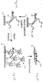
- Figs. 1A and 1B are schematic representations of assay formats included in the invention.
- Figs. 2A and 2B are schematic representations of a device of the invention and its use in methods for detecting nucleic acid analytes.
- Fig. 3 is a schematic representation of a QBR ligation-based binary probe assay.
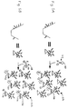
- Figs. 4A-4F are schematic representations of methods employing devices of the invention, in which the probe bound to the solid surface includes a portion of an autocatalytically replicable nucleic acid.
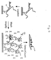
- Figs. 5A and 5B are schematic representations of two different methods of using a fluor-quencher pair, e.g. , a fluorescence energy transfer pair, to detect an amplified nucleic acid product.
- a fluor-quencher pair e.g. , a fluorescence energy transfer pair
- Fig. 6 is a schematic representation of a flow chart of the use of a fluor-quencher pair, e.g. , a fluorescence energy transfer pair, to detect an amplified nucleic acid product formed by use of a device and method of the invention.
- a fluor-quencher pair e.g. , a fluorescence energy transfer pair
- Figs. 7A-7G are schematic representations of variations in binary probe design that are within the scope of the invention.
- Fig. 8 is a schematic representation of 3' and 5'-half probes that can be used in detecting the major BCR/ABL junction found in CML patients.
- Figs. 9A and 9B are schematic representations of methods of the invention that employ PCR amplification.
- the invention provides devices and methods for sensitive and quantitative detection of nucleic acid analytes in samples.
- a central feature of the devices of the invention is that they each include a solid support to which is bound a two-dimensional, random distribution or field of nucleic acid probes that each bind to a nucleic acid analyte.
- a nucleic acid analyte is bound to a probe of the device, analyte-specific amplification is carried out in a diffusion limiting matrix (e.g., one that is made of agarose, gelatin, or polyethylene glycol) to generate amplification products that are centered around each of the original nucleic acid probes.
- a diffusion limiting matrix e.g., one that is made of agarose, gelatin, or polyethylene glycol
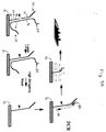
- the probe 10 bound to the solid support 12 of the device can include a first end 14 that is bound to the solid support 12 and includes a portion of an autocatalytically replicable nucleic acid 15, and a second end 16 that includes a first analyte-binding segment, which hybridizes to a first region 18 of a nucleic acid analyte 20.
- This device can be used in methods in which it is contacted with (1) a sample containing a nucleic acid analyte 20, and (2) a second nucleic acid probe 22 that contains a second analyte-binding segment 24, which hybridizes to a second region 26 of the nucleic acid analyte 20, and a remainder 28 of the autocatalytically replicable nucleic acid.
- first 18 and second 26 regions of the analyte 20 can be adjacent nucleotide segments, so that when the analyte 20, first probe 10, and second probe 22 hybridize together on the support 12 to form a complex 30, a complete template for autocatalytic replication, consisting of the first 10 and second 22 probes, is formed.
- the first 18 and second 26 regions of the analyte 20 can be separated by a third analyte region 32, to which a third probe 34 binds.
- the first 18 and second 26 regions of the analyte flank, and are adjacent to, the third region 32, so that when the analyte 20, first probe 10, second probe 22, and third probe 34 hybridize together on the support 12 to form a complex 30, a complete template for autocatalytic replication, consisting of the first 10, second 22, and third 34 probes, is formed.
- the probes forming the template for autocatalytic replication can, if desired, be ligated together using conventional methods, such as, e.g. , enzymatic methods (for example, by employing T4 DNA ligase (European Patent Applications EP 94/906626.0 and EP 94/906682.3), an RNA ligase, or a ribozyme ligase), chemical methods (see , e.g.
- the first probe 36 which is bound to the solid support 12 of the device, includes a first end 38 that is bound to the solid support 12 and a second end 40 that includes a first analyte-binding segment, which hybridizes to a first region 42 of a nucleic acid analyte 20.
- This device can be used in methods in which the solid support 12 is contacted with (1) a sample containing an analyte 20, (2) a second nucleic acid probe 44 that contains a second analyte-binding segment 46 that hybridizes to a second region 48 of the analyte 20, and a portion 50 of an autocatalytically replicable nucleic acid, and (3) a third nucleic acid probe 52 that contains a third analyte-binding segment 54, which hybridizes to a third region 56 of the analyte 20, and a remainder 58 of the autocatalytically replicable nucleic acid.
- the second 48 and third 56 regions of the analyte 20 can be adjacent nucleotide segments, so that when the analyte 20, second probe 44, and third probe 52 hybridize together on the support 12 to form a complex 60, a complete template for autocatalytic replication is formed between the second 44 and third 52 probes.
- the first probe 36 bound to the solid support 12 serves only as a capture probe, binding the complex 60 formed between the nucleic acid analyte 20 and the second 44 and third 52 probes to the solid support 12.
- the second 44 and third 52 probes can, if desired, be ligated together when bound to the analyte 20. Further, the order of addition of the probes and sample to the solid support 12 can be varied.
- the second 48 and third 56 regions of the analyte 20 can each flank, and be adjacent nucleotide segments with, the first region 42 of the analyte 20.
- first probe 36 e.g. , including a nucleic acid and a linker
- second probe 44, and third probe 52 hybridize together on the support 12 to form a complex 62, a complete template for autocatalytic replication, consisting of the first 36, second 44, and third 52 probes, is formed.
- the first probe 64 which is bound to the solid support 12 of the device, includes a first end 66 that is bound to the solid support 12 and a second end 68 that includes a first analyte-binding segment, which hybridizes to a first region 70 of a nucleic acid analyte 20.
- This device can be used in methods in which the solid support 12 is contacted with (1) a sample containing an analyte 20, and (2) a second nucleic acid probe 72 that contains a second analyte-binding segment 74, which hybridizes to a second region 76 of the analyte 20.
- the first 70 and second 76 regions of the analyte 20 are adjacent nucleotide segments, so that when the first probe 64, the analyte 20, and the second probe 72 hybridize together on the support 12 to form a complex 78, the first 64 and second 72 probes, are brought together by binding to the analyte nucleic acid 20, so that they can be ligated together.
- a high stringency wash is then carried out to remove unligated second probe 72 and analyte nucleic acid 20.
- the ligated product is then amplified by standard PCR.
- a first primer 82 is used to copy the ligated product to make a first product, which is complementary to the ligated product.
- a second primer 80 then anneals to the first product and copies it, making a second product strand that is identical to the ligated product in the region between and including the two primers.
- PCR then operates on these first and second products to make a large number of first and second product strands, which are localized to the vicinity of the original ligated probe product by the diffusion limiting matrix. This spot can be detected by any of the means discussed above for the detection of QBR products, e.g. , by detection of incorporated radioactivity, fluorescence, etc.
- the solid support 12 is contacted with the sample, but not the second probe (Fig. 9B).
- Complexes formed by specific hybridization between the first probe 64, which is bound to the solid support 12 via its 5' end, and the analyte nucleic acid 20 are washed, and the 3' end of the first probe 64 bound to the support 12 is extended, using the analyte nucleic acid 20 as a template.
- the extended product is amplified by any of the standard target or probe amplification techniques listed in the introduction (e.g. , PCR) to produce a detectable signal corresponding to the presence and location of the ligated product on the field of the device.
- a first primer 80 identical to a portion of the first probe 64
- a second primer 82 complementary to a portion of the extended region 84 of the first probe 64
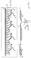
- the device of the invention is a field of analyte-specific probes 86 coated on the surface of a solid support 12, such as a microscope slide.
- the probes 86 are uniformly coated on the solid support 12, but they can be limited to specific locations, if desired.
- Analyte nucleic acid molecules 20 contained within, e.g. , a clinical specimen 88 are applied to the solid support 12, where they diffuse to the surface of the solid support 12 and are captured by ( i.e. , hybridize to) the specific probes on the field 86.
- the analytes 20 are then simply detected or quantitated using an appropriate replication system, e.g., the QBR amplification system.
- a second, analyte-specific probe 90 is hybridized to each analyte molecule 20 immediately adjacent to the hybridization site 92 of each one 94 of the probes 86 that is bound to the solid support 12.
- These two probes 90 and 94 can then be joined together to form a nucleic acid molecule that can be replicated or amplified by an enzyme, such as QBR.
- QBR amplification can then be used to form a spot of highly concentrated RNA product 96 in the immediate vicinity of the immobilized analyte molecule 20, which can easily be detected, e.g. , with a fluorescence microscope, with a CCD camera, by visual inspection, or other means, depending upon the label used for detection.
- the number of such "spots" 96 on the solid support 12 corresponds generally to the number of analyte molecules 20 that originally bound the solid support 12.
- this and other embodiments of the invention employ autocatalytically replicable nucleic acid molecules, such as midivariant and related RNAs that are modified to include a sequence of analyte-specific nucleic acid, that can be amplified by enzymes, such as QBR.
- nucleic acid molecules and QBR The use of such nucleic acid molecules and QBR in the invention is illustrated in Fig. 3, and is described further as follows.
- Autocatalytically replicable nucleic acids are employed in binary probe assays in the invention. Accordingly, for use in the invention, they are split into two "half molecules" 98 and 100, neither of which can alone be replicated appreciably by QBR.
- Each half molecule contains two functional elements: a partial MDV sequence 102 (and 104) and a probe sequence 106 (and 108), which is complementary to, and thus capable of specific hybridization with, a target sequence 110 (and 112) in a nucleic acid analyte 20.
- the probe sequences 106 and 108 of the two half molecules 98 and 100 are chosen so that they hybridize to adjacent nucleotide segments 110 and 112 in the analyte 20.
- the two half molecules are individually referred to below as a 3'-half probe 98 and a 5'-half probe 100, based on the polarity of the nucleic acids of which they are made.
- half molecule or “half probe” is not intended to mean that such probes have to contain exactly "half" of a complete, autocatalytically replicable molecule.
- the point of probe insertions can be about 1 / 3 of the way from the 5' end and about 2 / 3 from the 3' end.
- Other insertion points are known in the art ( see , e.g. , Burg et al., Anal. Biochem., 230:263-272, 1995).
- Replicable modifications of nucleic acids containing these modifications are also within the scope of the invention.
- both half probes can consist of DNA or RNA.
- one half probe can consist of DNA and the other of RNA.
- Each half probe can also contain a combination of deoxyribonucleotides and ribonucleotides, or modifications thereof.
- both half molecules 98 and 100 hybridize in the configuration shown in Fig. 3.
- Half molecules bound in this manner can, optionally, be covalently joined together by a ligation step, which creates a complete detection probe 114 that is replicable by QBR, and thus is capable of generating an assay signal.
- amplification can be carried out even if the two half probes are not ligated together.
- Unligated half probes that are held in close proximity to one another and in approximately correct topology by the target nucleic acid (as shown in Fig. 4C) can be amplified by QBR.
- ligated half probes can be amplified with significantly greater sensitivity (10 to 10 6 -fold, depending upon the hybridization and washing conditions used).
- Another significant advantage of employing a ligation step is that, following ligation, the ligated probe molecules are covalently linked to the solid support so that extremely high stringency wash conditions (such as would disrupt signal-generating complexes, e.g. , as shown in Fig. 4C, where the 3'-half probes are not covalently attached to the solid support) can be used. Nevertheless, for applications requiring lower sensitivity, the elimination of one assay step, e.g. , the ligation step, can be useful.
- ligation of the half molecules can be effected using any of a variety of well known methods including, e.g. , enzymatic methods (for example, by employing T4 DNA ligase, an RNA ligase, or a ribozyme ligase), chemical methods, and photochemical methods.
- enzymatic methods for example, by employing T4 DNA ligase, an RNA ligase, or a ribozyme ligase
- chemical methods for example, by employing T4 DNA ligase, an RNA ligase, or a ribozyme ligase
- photochemical methods for example, photochemical methods.
- a central feature of the invention is that a probe, which can contain a half molecule 100 of a QBR detector probe, is uniformly dispersed and specifically attached to the surface of a solid support 12, such as a microscope slide or a similar surface, or to a portion thereof.
- the 5'-half probe 100 is attached to the support 12 through its 5' end (Fig. 4A).
- the linkage can, optionally, include additional nucleotide or non-nucleotide spacers or "linker groups" 116 to prevent surface interference with efficient hybridization and replication.
- the 3'-half probe 98 can be attached to the support 12 through its 3' end.
- the probes illustrated in Figs. 4A-4F are based on a particular QBR amplifiable construct called midivariant RNA. As mentioned above, a variety of other autocatalytically replicable nucleic acids can be used in the invention.
- the 3'-half probe 98 is added to the sample under conditions that promote specific hybridization with the analyte nucleic acid 20. Subsequently, or simultaneously, the sample is applied to the support and is allowed to form probe/analyte nucleic acid hybrids by nucleic acid hybridization.
- a variety of well known agents that promote or modify hybridization can be used in the invention.
- the chaotropic agent guanidinium thiocyanate is a well known hybridization agent, which promotes both sample lysis and hybridization.
- a greater or lesser proportion of the analyte nucleic acids 20 may already have 3'-half probe 98 hybridized to it at the beginning of the hybridization on the solid support (Fig. 4B).
- a complex 118 containing a 5'-half probe 100, a nucleic acid analyte 20, and a 3'-half probe 98 is formed (Fig. 4C).
- a washing step can be carried out to remove from the support 12 a substantial portion of unbound reactants and components of the sample.
- a ligation step can then be performed, in which the two half molecules 98 and 100 are covalently joined together to produce an amplifiable nucleic acid 120 (Fig. 4D).
- An important element of this step is that the ligation is dependent upon the correct hybridization of the two half probes 98 and 100 to the correct analyte nucleic acid 20. Methods involving the use of such half, or binary, probe systems are known in the art and are described, e.g. , in European Patent Applications EP 94/906626.0 and EP 94/906682.3.
- a high stringency wash can be carried out to remove remaining components of the sample and 3'-half molecules 98 that are not now covalently attached to the support 12. Remaining on the support are the originally bound 5'-half probes 100 and, at each location where a nucleic acid analyte has bound to a 5'-half probe 100, a replication competent, signal generating nucleic acid 120 (Fig. 4D), which, in this example, includes a replicable MDV sequence.
- Amplification of the amplifiable nucleic acid 120 can be carried out as follows.
- Components of a QBR amplification reaction i.e. , QBR, nucleotide triphosphates, Mg++, appropriate salt, buffer, etc. (see , e.g. , Moody et al. , Biochemistry , 33:13836-13847, 1994)
- QBR QBR
- nucleotide triphosphates Mg++, appropriate salt, buffer, etc.
- each of the colonies of amplification product 124 form on the solid support in a locus corresponding to the presence of one nucleic acid analyte 20.
- Amplification products of each captured analyte molecule are confined to the vicinity of the template analyte molecule by a diffusion limiting matrix so that these products can be visualized as a colony.
- Particularly useful as a diffusion limiting matrix is PEG 8000 (e.g., 10%).
- cationic polymers e.g. , n-Vinyl pyrrolidone/diethylamino-methyl methacrylate copolymer
- these polymers can be used to reduce the colony size, allowing the detection of a larger number of colonies on a given area of a solid surface.
- Amplification is allowed to proceed under appropriate conditions, such as temperature conditions, for a length of time appropriate to generate a discrete and discernable amount of an amplification product 122 (Fig. 4E).
- loci, or spots, 124 of amplified QBR probe RNA can be visualized by any of a number of methods known in the art, including, e.g. , by detection of radioactivity, fluorescence, color, or a chemiluminescent substance incorporated into the amplification product 122.
- a fluorescent intercalating dye such as propidium iodide
- amplification mixture can be added to the amplification mixture so that, as amplification product 122 is produced, the dye intercalates into the product 122, leading to production of an intense fluorescence at the site of amplification 124 (Fig. 4F).
- the optimal length of time for the amplification reaction depends on a number of factors, which are well known to those skilled in the art, and include, e.g. , the inherent replication rate of the QBR probe, the concentration of reactants in the amplification reaction (e.g. , the QBR concentration, the dNTP concentration, and the Mg++ concentration), as well as the temperature at which the reaction is carried out ( see , e.g., Burg et al., 1995, supra ).
- the optimal reaction time also will depend upon the detection means used. Microscopic detection of fluorescence, for example, requires generation of only a very small spot. Macroscopic ( e.g. , visual) detection of amplified RNA may require less instrumentation, but bigger spots.
- the number of colonies of amplification products can be counted to quantitate the number of nucleic acid analyte molecules in the sample that was applied to the support. This can readily be determined, based on the volume of sample applied to the slide and the overall efficiency of the process. For example, each of the steps of the assay, e.g. , hybridization of the analyte to the first (5'-half) probe on the solid support, hybridization of the second (3'-half) probe, and ligation, has a characteristic efficiency of less than 100%.
- analyte nucleic acid molecules applied to the device will lead to production of an amplifiable, ligated product.
- analyte nucleic acid molecules applied to the device will produce about 250 spots of amplified signal.
- a fluor-quencher pair or, e.g. , a fluorescence energy transfer pair, as described, for example, by Morrison (Detection of Energy Transfer and Fluorescence Quenching, in Nonisotopic Probing, Blotting, and Sequencing, Academic Press, 1995) and Tyagi et al. (Nature BioTechnology, 14:303-308, 1996).
- a fluor-quencher pair consists of a fluorescent dye (labeled "F", e.g. , fluorescein) and a quencher dye (labeled "Q”), which blocks emission of fluorescence from the fluorescent dye when the two dyes are in close proximity.
- the fluorescent dye and the corresponding quencher dye are held in proximity to one another by attachment to a short nucleic acid (e.g. , DNA) probe that self-associates (e.g. , the "beacon" 126 in Fig. 5A, or the Morrison pair of probes 128 in Fig. 5B).
- a short nucleic acid e.g. , DNA
- the sequences in the loop structure 130 of the beacon 126, or of one of the oligonucleotides 132 and 134 in the Morrison pair 128, are designed so that they hybridize to a specific, predetermined target sequence in the amplified product 122.
- these probe sequences hybridize to the probe portion 136 of the amplified product 122.
- the F and Q entities are forced apart by the rigid structure of the double-stranded hybrid, thus allowing fluorescence of the F entity to be detected (Figs. 5A and 5B).
- Fig. 6 illustrates the use of fluor-quencher pairs, or fluorescence energy transfer pairs, in the detection of specific QBR amplification products in the methods of the invention.
- DBCYL 4-(4'-dimethylaminophenylaza)benzoic acid
- the beacon probes 126 hybridize to the amplification products 122, thus creating a detectable, fluorescent signal 124.
- An advantage of this type of detection strategy is that amplification of specific products can be detected. In other schemes, such as those employing an intercalator dye (e.g. , propidium iodide), amplification can easily be detected, but the identity of the amplification product cannot.
- an intercalator dye e.g. , propidium iodide
- means for the simultaneous quantitative detection of multiple analyte nucleic acids is provided.
- the support contains a random distribution or field of multiple types of half probes having probe sequences that correspond to each of the nucleic acid analytes to be assayed.
- the same principles described above for diffusion of the analyte to the surface, ligation, and subsequent amplification apply, but in this embodiment, in each locus or spot where an analyte causes the formation of a replicable product, the amplification products arising from that spot will have a probe sequence corresponding to the target sequence that nucleated the ligation event. Accordingly, different analyte nucleic acid molecules will cause the amplification of different QBR probe molecules. Each specific probe molecule will have a sequence that is specific for the analyte required for its formation.
- Multiple fluorescence energy transfer probes (beacons or Morrison probes), corresponding to multiple possible QBR probe sequences, and each carrying a distinguishable fluorescent moiety, are present during or added subsequent to the amplification reaction. These probes, upon hybridization to the amplified QBR RNA molecules in each spot, provide different colors, corresponding to each originating analyte molecule. Thus, multiple analyte nucleic acids can be detected and distinguished in a single homogeneous assay. This increases the power, flexibility, and cost effectiveness of such assays.
- Figs. 7A-7G Variations in binary probe design that are within the scope of invention are illustrated in Figs. 7A-7G.
- Figs. 7A, 7B, and 7C illustrate alternative possible "alignments" of half probes 98 and 100 with a suspected point mutation (black arrowhead) in an analyte nucleic acid 20 that, in this example, contains a fusion between portions of BCR and ABL genes.
- Fig. 7D illustrates a ternary version of the QBR assay (using a third probe 138), which can be applied in this invention as well.
- the advantages of using ternary probe strategies include achieving higher specificity of hybridization than in binary probe strategies.
- Fig. 7E shows the basic elements of an alternative assay format, in which a separate "capture" probe 140 (in this illustration attached to a bead or particle 142, such as a magnetic particle).
- This format is useful and convenient for enrichment of the analyte molecules 20 and removal of sample components, prior to ligation and amplification steps.
- magnetic particles can be used as the solid phase as shown in Fig. 7F and, following ligation and washing, dispensed into the diffusion limiting matrix for amplification.
- Other alternative assay formats are illustrated in Figs. 7F and 7G, and are described above.
- hybridization and washing conditions such as "stringent” hybridization and washing conditions.
- Hybridization reactions are typically carried out under “stringent conditions,” e.g. , low to moderate stringency conditions, in which specific and some non-specific interactions can occur.
- washing can be carried under higher stringency conditions to eliminate non-specific binding.
- optimal washing conditions can be determined empirically, e.g. , by gradually increasing the stringency.
- Condition parameters that can be changed to affect stringency include, e.g. , temperature and salt concentration. In general, the lower the salt concentration, pH, and chaotropic agent (e.g. , guanidine thiocyanate) concentration, and the higher the temperature, the higher the stringency.
- washing can be initiated at a low temperature (e.g. , room temperature), using a solution containing an equivalent or lower salt concentration than the hybridization solution. Subsequent washing can be carried out using progressively warmer solutions having the same salt solution. Alternatively, the salt concentration can be lowered and the temperature maintained in the washing step, or the salt concentration can be lowered and the temperature increased. Such standard variations are known in the art. Additional parameters can be altered to affect stringency including, e.g. , the use of a destabilizing agent, such as formamide.
- a destabilizing agent such as formamide.
- nucleic acid hybridization reactions the conditions used to achieve a particular level of stringency will vary, depending on the nature of the nucleic acids being hybridized. For example, the length, degree of complementarity, nucleotide sequence composition (e.g. , GC v. AT content), and nucleic acid type (e.g. , RNA v. DNA) of the hybridizing regions of the nucleic acids can be considered in selecting hybridization conditions. An additional consideration is whether one of the nucleic acids is immobilized, for example, on a solid support, such as a glass plate or a filter.
- a solid support such as a glass plate or a filter.
- hybridization conditions that can be used in the methods of the invention involves hybridization at room temperature in 3 x SSC (for recipe, see Molecular Cloning, a Laboratory Manual, 2nd Edition, edited by Sambrook et al., Cold Spring Harbor Press, Cold Spring Harbor, New York, 1989), and subsequent washing in 3 x SSC.
- the stringency is not varied from the hybridization to the wash.
- hybridization is carried out at room temperature in 3 x SSC, and subsequent washes are carried out in 3 x SSC at the following temperatures: 25°C, 42°C, 52°C, 58°C, and 70°C.
- Another example of progressively higher stringency conditions is as follows: 2 x SSC/0.1% SDS at about room temperature (hybridization conditions); 0.2 x SSC/0.1% SDS at about room temperature (low stringency conditions); 0.2 x SSC/0.1% SDS at about 42°C (moderate stringency conditions); and 0.1 x SSC at about 68°C (high stringency conditions). Washing can be carried out using only one of these conditions, e.g. , high stringency conditions, or each of the conditions can be used, e.g. , for 10-15 minutes each, in the order listed above, repeating any or all of the steps listed. However, as mentioned above, optimal conditions will vary, depending on the particular hybridization reaction involved, and can be determined empirically.
- the analyte-specific nucleic acid probes used in the invention contain autocatalytically replicable sequences serving as templates for generating amplification products, and can consist of RNA, DNA, or modifications or combinations thereof.
- the amplified products generated by QBR replication of such templates are RNA.
- the nucleic acid probes can be generated by standard chemical synthesis methods or recombinant techniques.
- the probes used to bind to solid supports can have a chemical group (at, e.g., their 5' ends) that allows covalent coupling to the supports.
- a chemical group can be introduced, for instance, by derivatizing the 5' phosphate of a probe with a water-soluble carbodiimide to generate a 5' phosphorimidazolide (Chu et al., Nucleic Acids Research 11:6513-6529, 1983). Exposure of the phosphorimidazolide to an amine-containing molecule on a solid support results in the chemical coupling of the probe to the support.
- an RNA probe can be obtained by transcribing a template with a phage (e.g., T7) RNA polymerase.
- a phage e.g., T7
- GMP Guanosine 5'-monophosphate
- ethylene diamine e.g., ethylene diamine
- the modified GMP will be incorporated at the 5' end of the probe. No post transcription chemical modification of the probe is needed in this method.
- RNA molecules produced by T7 RNA polymerase from the same template often have heterogenous 3' termini, i.e., having a few extra, non-templated nucleotides at the 3' end. This 3' heterogeneity interferes with the subsequent ligation step required in a number of embodiments of the invention.
- the extra nucleotides are added to the transcripts largely by a terminal transferase activity of T7 RNA polymerase. Such additions can be reduced by providing competing substrates for that activity.
- Effective competitors include extraneous RNA (e.g., total RNA from yeast), oligodeoxynucleotides with 3' hydroxyl termini, nucleotide monophosphates (e.g. , GMP), deoxyribonucleotide monophosphates, and nucleosides with 3' hydroxyls. Chemically modified GMP is shown to be effective also.
- Attachment of nucleic acid probes to solid supports used in the invention can be carried out using any of a number of standard methods, including direct adsorption or chemical coupling to reactive groups on the surface.
- a linker can be used, e.g. , a flexible carbon chain, such as a 3-glycidoxypropyltri-methoxysilane linker ( see , e.g. , Maskos et al., Nucl. Acids Res., 20(7):1679-1684, 1992), or a protein, such as a molecule of bovine serum albumin ("BSA").
- BSA bovine serum albumin
- Use of a linker e.g., a hydrophilic linker
- BSA is a particularly useful linker, since it can significantly reduce non-specific binding of nucleic acids to the probes immobilized on a solid surface. BSA also allows efficient replication of the autocatalytic replicable RNA to which it links. The following describes an exemplary protocol for linking RNA probes to a solid support via BSA. Acid washed glass slides are first reacted with glycidyl oxypropyl trimethoxy silane, which generates amine-reactive epoxy groups on the glass.
- BSA is then coupled to these epoxy groups, and the carboxyl groups on the coupled albumin are activated by, e.g., N-hydroxy-succinamide with a carbodiimide such as 1-cyclohexyl-3(2-morpholino-ethyl)carbodiimide (CMC) or 1-ethyl-3(3-dimethylamino propyl)carbodiimide (EDAC), to generate amine-reactive N-hydroxysuccinamide-esters (NHS).
- RNA with a 5' terminal amino group is then reacted with the NHS-derivatized albumin.
- NHS groups are unstable.
- non-aqueous conditions can be used.
- a solid support coated with NHS-activated BSA is dried and exposed to RNA probes solubilized in butanol by a cationic detergent (e.g., hexadecyl trimethyl ammonium bromide (CTAB)).
- a cationic detergent e.g., hexadecyl trimethyl ammonium bromide (CTAB)
- Photolithography can also be used to attach the probes to the solid supports.
- the surface of the solid support is coated with a photosensitive linker and an appropriate light source, such as a laser beam, is directed at the region of the surface where the binding of the probe is desired.
- the probe is then contacted with the surface, where it binds to the light-activated region.
- This method readily permits the binding of different probes to a solid support.
- target amplification e.g. , PCR; Saiki et al., supra
- TAS transcription-based amplification system
- TMA transcription-mediated amplification
- NASBA nucleic acid sequence-based amplification
- SDA strand displacement amplification
- probe amplification e.g. , QBR (Lizardi et al., supra ), ligase amplification reaction (LAR; Wu et al., supra ), ligase chain reaction (LCR; Barany, supra )
- QBR Lizardi et al., supra
- LAR ligase amplification reaction
- LCR ligase chain reaction
- midivariant RNAs e.g. , midivariant-1 (MDV-1)
- minivariant RNA e.g. , minivariant RNA
- microvariant RNA e.g. , midivariant-1 (MDV-1)
- nanovariant RNA e.g. , midivariant-1 (MDV-1)
- MDV-1 midivariant-1
- minivariant RNA minivariant RNA
- microvariant RNA microvariant RNA
- nanovariant RNA e.g.
- the sequences of these and other autocatalytically replicable nucleic acids are well known in the art, and readily adaptable for use in the present invention ( see , e.g. , Mills et al., Proc. Natl. Acad. Sci USA, 72:4252-4256, 1975; Schaffner et al., J. Mol.
- Detection of nucleic acid analytes using the devices and methods of the invention can be useful in fields such as medicine, forensics, agriculture, industry, food sciences, and veterinary medicine.
- the methods of the invention can be used in the diagnosis of conditions (e.g. , cancer) characterized by the presence or absence of specific nucleic acid markers and/or altered levels of normally occurring nucleic acids.
- the methods of the invention can also be used to detect gene mutations, which can be characterized by, e.g. , single base pair changes, small or large deletions, insertions, or rearrangements ( e.g. , chromosomal translocations), and genetic polymorphism.
- the methods of the invention can be used to detect the presence of an infectious pathogen (e.g. , a bacterium, a virus, a protozoan, a parasite, or a fungus) or a rare cell (e.g. , a fetal cell in maternal blood) in a sample, e.g. , a sample from a patient.
- infectious pathogen e.g. , a bacterium, a virus, a protozoan, a parasite, or a fungus
- a rare cell e.g. , a fetal cell in maternal blood
- the devices, methods, and systems of the invention can be readily incorporated into a panel format for the detection and quantitation of multiple target analytes.
- the following example describes the use of methods and devices of the invention in the diagnosis of a condition caused by a chromosomal translocation.
- Chromosomal translocations and inversions commonly result in the juxtaposition of two genes (one from each of the chromosomes or chromosomal segments involved in the recombination) that normally are physically and transcriptionally separate from one another.
- Such translocations can result in the production of novel fusion proteins that can have adverse physiological effects.
- a number of fusion proteins generated in this manner have been implicated in the etiology of cancer ( see , e.g. , Rabbits, Nature, 372:143-149, 1994).
- the archetype for such a cancer-causing gene rearrangement is the chromosome 9 -chromosome 22 translocation (t9;22) that results in formation of the so-called "Philadelphia" chromosome, the presence of which is strongly correlated with a number of leukemias, including chronic myelogenous leukemia (CML) and acute lymphocytic leukemia (ALL).
- CML chronic myelogenous leukemia
- ALL acute lymphocytic leukemia
- the translocation results in joining of the bcr gene on chromosome 22 to the abl gene on chromosome 9.
- a hybrid BCR/ABL protein is produced from the hybrid gene, which causes elevated expression of the encoded tyrosine kinase enzymatic activity.
- a minimum residual disease (MRD) state can persist for many years, in which only relatively low levels of the altered BCR/ABL protein are produced and during which no, or relatively minor, disease pathology is evident. Then, on average 4-5 years following the initial diagnosis, the disease escalates to an acute phase, which requires major medical intervention. The onset of this acute phase is presaged by an increase in expression of the fused BCR/ABL mRNA and protein. As understood in this field, medical intervention is most effective if it is initiated as soon as possible following the transition to the acute phase of the disease. Accordingly, a diagnostic test that can be used to quantitatively monitor the level of BCR/ABL mRNA expression in CML patients during the MRD state can be used in the management of their disease.
- the present invention describes means of performing such monitoring using the QBR amplification system.
- the present invention provides methods for monitoring the level of expression of gene fusions resulting from somatic rearrangement, e.g. , leading to juxtaposition, of two genes that are normally physically and transcriptionally separate from one another. This clearly has application in the diagnosis of CML and ALL, as well as other diseases in which fusion proteins play a significant role, e.g. , other hematopoietic cancers and solid tumors (Rabbits, 1994, supra ).
- Fig. 8 illustrates specific sequences of analyte-binding probe segments (SEQ ID NOs:1 and 2) that can be used in diagnostic methods for detecting mRNA transcribed from bcr/abl gene fusions (SEQ ID NO:3), according to the methods of the invention described above.
- probes can be used in, e.g. , the following method.
- Cells in a peripheral blood sample from a patient are lysed in, e.g. , guanidine thiocyanate.
- a 3'-half probe (50 ng/100 ⁇ L; e.g. , a probe having the sequence of SEQ ID NO:2; see Fig. 8) is added to the lysed sample, and the mixture is then applied to the surface of a solid support of the invention, to which is bound a probe having the sequence of, e.g. , SEQ ID NO:1 (see Fig. 8), where hybridization is allowed to proceed for, e.g. , about one hour. The surface of the solid support is then washed, e.g.
- the solid support e.g. , a glass plate
- Medium stringency conditions can be used for the wash, in which the support is incubated in fresh hybridization buffer, without probe, at 37°C for 5 minutes.
- Ligation buffer and ligase is then added to the support, and incubation is carried out at about 37°C for about five minutes.
- the support is then washed, e.g. , in 0.01 N NaOH for one minute at 37°C, QBR and amplification reagents (dNTPs, Mg, Tris buffer pH 7.8), in a diffusion limiting matrix (e.g.
- spots generated in this reaction can be counted using any of a number of suitable means (e.g., a fluorescence microscope, a CCD camera, or visual inspection), if the amplification is carried out for a sufficient length of time, so that the spots are, e.g. , greater than about 1 mm in diameter.
- suitable means e.g., a fluorescence microscope, a CCD camera, or visual inspection
- the invention can be used in any circumstances in which detection of nucleic acids (DNA or RNA) encoding fusion-proteins is desired.
Abstract
Description
Claims (30)
- An assay device for detecting the presence of a nucleic acid analyte in a sample, said device comprising a non-particulate, solid support having covalently bound thereto a two-dimensional field of analyte-specific nucleic acid probes, each probe comprising a first end covalently bound to said support and including a 5' portion of an autocatalytically replicable nucleic acid, and a second end including an analyte-binding segment.
- The device of claim 1, wherein said autocatalytically replicable nucleic acid is replicable by Q-beta replicase.
- The device of claim 2, wherein said autocatalytically replicable nucleic acid comprises a midivariant nucleic acid.
- A method for detecting the presence of a nucleic acid analyte in a sample, said method comprising the steps of:(a) applying the sample to a solid support having covalently bound thereto a two-dimensional field of first analyte-specific nucleic acid probes, each of said first probes comprising a first end bound to said support and including a 5' portion of an autocatalytically replicable nucleic acid, and a second end including a first analyte-binding segment, wherein said first analyte-binding segment of each of said first probes hybridizes to a first region of the analyte;(b) applying a second nucleic acid probe to the solid support, said second probe comprising a second analyte-binding segment that hybridizes to a second region of the analyte, said second probe further comprising a remainder of said autocatalytically replicable nucleic acid, wherein the first and second regions of the analyte are adjacent nucleotide segments, and wherein the analyte, said first probe, and said second probe hybridize together on the support to form a complex including a complete autocatalytically replicable nucleic acid;(c) applying a diffusion limiting matrix to said solid support;(d) amplifying said complete autocatalytically replicable nucleic acid to generate an amplified product; and(e) detecting said amplified product as a measure of the presence of the analyte in the sample.
- The method of claim 4, further comprising the step of ligating said first and second probes in said complex together to form said complete autocatalytically replicable nucleic acid.
- The method of claim 4, wherein said amplification step is carried out in the presence of a fluorescent intercalating dye and said detection step is carried out by monitoring said amplified product for the presence of said dye.
- The method of claim 4, wherein said detection step is carried out by employing a fluor-quencher pair.
- The method of claim 4, wherein multiple analytes are simultaneously detected in the sample and said detecting step is carried out by employing multiple fluor-quencher pairs that comprise distinguishable fluorescent moieties.
- The method of claim 4, wherein said autocatalytically replicable nucleic acid comprises a nucleic acid replicable by Q-beta replicase.
- The method of claim 9, wherein said autocatalytically replicable nucleic acid comprises a midivariant nucleic acid.
- The method of claim 4, further comprising washing said complex to remove any unbound first or second probes.
- The method of claim 4, wherein said diffusion limiting matrix is selected from the group consisting of gelatin, agarose, polyacrylamide, polyethylene glycol, and any combination thereof.
- The method of claim 4, further comprising determining the concentration of the analyte in the sample, wherein the concentration of the analyte in the sample is determined by counting colonies of amplified product formed on said solid support, and the number of colonies corresponds to the concentration of the analyte in the sample.
- An assay system for detecting a nucleic acid analyte in a sample, said assay system comprising:wherein binding of said first and second probes to the analyte permits amplification of said autocatalytically replicable nucleic acid.(a) a non-particulate solid support having covalently bound thereto a two-dimensional field of first analyte-specific nucleic acid probes, each of said first probes comprising a first end covalently bound to said support and including a 5' portion of an autocatalytically replicable nucleic acid, and a second end including a first analyte-binding segment, wherein said first analyte-binding segment of each of said first probes hybridizes to a first region of the analyte; and(b) a second nucleic acid probe comprising a second analyte-binding segment that hybridizes to a second region of the analyte, said second probe further comprising a remainder of said autocatalytically replicable nucleic acid, and said first and second regions of the analyte comprising adjacent nucleotide segments;
- An assay system for detecting a nucleic acid analyte in a sample, said assay system comprising:wherein binding of said capture probes and said first and second probes to the analyte permits amplification of a detectable product within the diffusion limiting matrix.(a) a non-particulate solid support having bound thereto a two-dimensional field of capture probes, each of said capture probes comprising a capture segment that hybridizes to a capture region of the analyte;(b) a first nucleic acid probe comprising a first segment that hybridizes to a first region of the analyte;(c) a second nucleic acid probe comprising a second segment that hybridizes to a second region of the analyte; and(d) a diffusion limiting matrix for application to said solid support;
- The assay system of claim 15, wherein the first and second regions of the analyte are adjacent nucleotide segments, said first nucleic acid probe further comprises a portion of an autocatalytically replicable nucleic acid molecule, and said second nucleic acid probe further comprises a remaining portion of said autocatalytically replicable nucleic acid molecule.
- The assay system of claim 15, wherein the capture and first regions of the analyte are adjacent nucleotide segments and the capture and second regions of the analyte are adjacent nucleotide segments, said first nucleic acid probe further comprises a portion of an autocatalytically replicable nucleic acid molecule, and said second nucleic acid probe further comprises a remaining portion of said autocatalytically replicable nucleic acid molecule.
- A method for detecting the presence of a nucleic acid analyte in a sample, said method comprising the steps of:(a) contacting the sample with said bound capture probes and said first and second probes of the assay system of claim 15, wherein said first and second regions of the analyte are adjacent nucleotide segments, said first nucleic acid probe further comprises a portion of an autocatalytically replicable nucleic acid molecule, said second nucleic acid probe further comprises a remaining portion of said autocatalytically replicable nucleic acid molecule, and said contacting permits the analyte, said first probe, and said second probe to hybridize together on the support to form a complex including a complete autocatalytically replicable nucleic acid;(b) applying a diffusion limiting matrix to said solid support;(c) amplifying said complete autocatalytically replicable nucleic acid to generate an amplified product; and(d) detecting said amplified product as a measure of the presence of the analyte in the sample.
- A method for detecting the presence of a nucleic acid analyte in a sample, said method comprising the steps of:(a) contacting the sample with said bound capture probes and said first and second probes of the assay system of claim 15, wherein the capture and first regions of the analyte are adjacent nucleotide segments and the capture and second regions of the analyte are adjacent nucleotide segments, said first nucleic acid probe further comprises a portion of an autocatalytically replicable nucleic acid molecule, and said second nucleic acid probe further comprises a remaining portion of said autocatalytically replicable nucleic acid molecule, and said contacting permits the analyte, said first probe, and said second probe to hybridize together on the support to form a complex including a complete autocatalytically replicable nucleic acid;(b) applying a diffusion limiting matrix to said solid support;(c) amplifying said complete autocatalytically replicable nucleic acid to generate an amplified product; and(d) detecting said amplified product as a measure of the presence of the analyte in the sample.
- A method for detecting the presence of a nucleic acid analyte in a sample, said method comprising the steps of:(a) contacting the sample with a solid support having covalently bound thereto a two-dimensional field of first analyte-specific nucleic acid probes, each of said first probes comprising a first end bound to said support and a second end including a first analyte-binding segment;(b) applying a diffusion limiting matrix to said support;(c) performing amplification by one of polymerase chain reaction, ligase chain reaction, transcription-mediated amplification, nucleic acid sequence-based amplification, and strand displacement amplification in a manner dependent upon the presence of both the first probe and the specified analyte nucleic acid, and in such a manner that the products of such amplification reaction are restrained to form localized foci within the diffusion limiting matrix on the solid support; and(d) detecting said amplified product as a measure of the presence of said analyte in the sample.
- The device of claim 1, wherein each of the analyte-specific nucleic acid probes is an RNA synthesized by an RNA polymerase in the presence of guanosine 5'-monophosphate.
- The device of claim 21, wherein the RNA polymerase is T7 RNA polymerase.
- The method of claim 4, wherein each of the first analyte-specific nucleic acid probes is an RNA synthesized by an RNA polymerase in the presence of guanosine 5'-monophosphate.
- A method of synthesizing an RNA, the method comprising contacting a template for the RNA with T7 polymerase in the presence of 5' modified guanosine 5'-monophosphate.
- The assay device of claim 1, wherein each of the analyte-specific nucleic acid probes is covalently bound to the solid support with bovine serum albumin.
- The method of claim 4, wherein each of the first analyte-specific nucleic acid probes is covalently bound to the solid support with bovine serum albumin.
- The method of claim 4, wherein the diffusion limiting matrix comprises polyethylene glycol.
- The method of claim 27, wherein the diffusion limiting matrix further comprises a cationic polymer.
- The assay system of claim 15, wherein the diffusion limiting matrix comprises polyethylene glycol.
- The assay system of claim 29, wherein the diffusion limiting matrix further comprises a cationic polymer.
Applications Claiming Priority (2)
| Application Number | Priority Date | Filing Date | Title |
|---|---|---|---|
| US768177 | 1996-12-16 | ||
| US08/768,177 US5837466A (en) | 1996-12-16 | 1996-12-16 | Devices and methods for detecting nucleic acid analytes in samples |
Publications (2)
| Publication Number | Publication Date |
|---|---|
| EP0853129A2 true EP0853129A2 (en) | 1998-07-15 |
| EP0853129A3 EP0853129A3 (en) | 1999-07-07 |
Family
ID=25081772
Family Applications (1)
| Application Number | Title | Priority Date | Filing Date |
|---|---|---|---|
| EP97310133A Withdrawn EP0853129A3 (en) | 1996-12-16 | 1997-12-16 | Devices and methods for detecting nucleic acid analytes in samples |
Country Status (3)
| Country | Link |
|---|---|
| US (2) | US5837466A (en) |
| EP (1) | EP0853129A3 (en) |
| JP (1) | JPH10293128A (en) |
Cited By (8)
| Publication number | Priority date | Publication date | Assignee | Title |
|---|---|---|---|---|
| WO2000006770A1 (en) * | 1998-07-30 | 2000-02-10 | Solexa Ltd. | Arrayed biomolecules and their use in sequencing |
| WO2001059162A2 (en) * | 2000-02-08 | 2001-08-16 | Bayer Corporation | METHOD FOR NUCLEIC ACID DETECTION VIA Qβ REPLICASE |
| US6355421B1 (en) | 1997-10-27 | 2002-03-12 | Boston Probes, Inc. | Methods, kits and compositions pertaining to PNA molecular beacons |
| US6361942B1 (en) | 1998-03-24 | 2002-03-26 | Boston Probes, Inc. | Method, kits and compositions pertaining to detection complexes |
| US6485901B1 (en) | 1997-10-27 | 2002-11-26 | Boston Probes, Inc. | Methods, kits and compositions pertaining to linear beacons |
| US6787308B2 (en) | 1998-07-30 | 2004-09-07 | Solexa Ltd. | Arrayed biomolecules and their use in sequencing |
| US7374945B2 (en) | 2000-12-14 | 2008-05-20 | Gen-Probe Incorporated | Method for enhancing the association rates of polynucleotides |
| US20120040867A1 (en) * | 2009-01-28 | 2012-02-16 | The Trustees Of Columbia University In The City Of New York | Microarrays of Binary Nucleic Acid Probes for Detecting Nucleic Acid Analytes |
Families Citing this family (96)
| Publication number | Priority date | Publication date | Assignee | Title |
|---|---|---|---|---|
| US5837466A (en) * | 1996-12-16 | 1998-11-17 | Vysis, Inc. | Devices and methods for detecting nucleic acid analytes in samples |
| EP3034626A1 (en) | 1997-04-01 | 2016-06-22 | Illumina Cambridge Limited | Method of nucleic acid sequencing |
| EP0878552A1 (en) | 1997-05-13 | 1998-11-18 | Erasmus Universiteit Rotterdam | Molecular detection of chromosome aberrations |
| US7569342B2 (en) | 1997-12-10 | 2009-08-04 | Sierra Molecular Corp. | Removal of molecular assay interferences |
| EP1078098A1 (en) | 1998-05-04 | 2001-02-28 | Dako A/S | Method and probes for the detection of chromosome aberrations |
| ES2459745T3 (en) * | 1999-05-14 | 2014-05-12 | Brandeis University | Aptamer based detection |
| US6680377B1 (en) * | 1999-05-14 | 2004-01-20 | Brandeis University | Nucleic acid-based detection |
| US6709816B1 (en) * | 1999-10-18 | 2004-03-23 | Affymetrix, Inc. | Identification of alleles |
| US7118860B2 (en) * | 1999-10-29 | 2006-10-10 | Stratagene California | Methods for detection of a target nucleic acid by capture |
| US6235483B1 (en) * | 2000-01-31 | 2001-05-22 | Agilent Technologies, Inc. | Methods and kits for indirect labeling of nucleic acids |
| EP1268769A2 (en) * | 2000-03-31 | 2003-01-02 | Memorec Stoffel GmbH Medizinisch-Molekulare Entwicklung | Method for extracting nucleic acids |
| US6936443B2 (en) * | 2000-04-03 | 2005-08-30 | Cytyc Corporation | Detection and typing of human papillomavirus using PNA probes |
| DE60141205D1 (en) * | 2000-04-03 | 2010-03-18 | Cytyc Corp | DETECTION AND TYPING OF PAPILLOMA VIRUS BY PNA PROBES |
| US20030082605A1 (en) * | 2000-09-06 | 2003-05-01 | Hodge Timothy A. | Genomic DNA detection method and system thereof |
| EP1978110B1 (en) * | 2000-09-06 | 2010-05-26 | Transnetyx, Inc. | Computer-based method and system for screening genomic DNA |
| US20050239125A1 (en) * | 2000-09-06 | 2005-10-27 | Hodge Timothy A | Methods for genotype screening |
| US20050272085A1 (en) * | 2000-09-06 | 2005-12-08 | Hodge Timothy A | Methods for forensic and congenic screening |
| US20050266494A1 (en) * | 2000-09-06 | 2005-12-01 | Hodge Timothy A | System and method for computer network ordering of biological testing |
| US20030087286A1 (en) * | 2000-09-06 | 2003-05-08 | Hodge Timothy A. | Isolation of eukaryotic genomic DNA using magnetically responsive solid functionalized particles |
| US7494817B2 (en) * | 2000-09-06 | 2009-02-24 | Transnet Yx, Inc. | Methods for genotype screening using magnetic particles |
| AR031640A1 (en) * | 2000-12-08 | 2003-09-24 | Applied Research Systems | ISOTHERMAL AMPLIFICATION OF NUCLEIC ACIDS IN A SOLID SUPPORT |
| US7258974B2 (en) * | 2001-04-23 | 2007-08-21 | Michael F. Chou | Transcription factor network discovery methods |
| US20060014186A1 (en) * | 2001-09-04 | 2006-01-19 | Hodge Timothy A | Methods for genotype screening of a strain disposed on an adsorbent carrier |
| WO2003030620A2 (en) * | 2001-10-12 | 2003-04-17 | Vysis, Inc. | Imaging microarrays |
| US20040002090A1 (en) * | 2002-03-05 | 2004-01-01 | Pascal Mayer | Methods for detecting genome-wide sequence variations associated with a phenotype |
| US6913890B2 (en) * | 2002-12-18 | 2005-07-05 | Palo Alto Research Center Incorporated | Process for preparing albumin protein conjugated oligonucleotide probes |
| US6936496B2 (en) | 2002-12-20 | 2005-08-30 | Hewlett-Packard Development Company, L.P. | Nanowire filament |
| US7132298B2 (en) * | 2003-10-07 | 2006-11-07 | Hewlett-Packard Development Company, L.P. | Fabrication of nano-object array |
| US7223611B2 (en) * | 2003-10-07 | 2007-05-29 | Hewlett-Packard Development Company, L.P. | Fabrication of nanowires |
| TWI246593B (en) * | 2003-11-12 | 2006-01-01 | Ind Tech Res Inst | Method and substrate for biochips by using single-step manufacturing process |
| US7407738B2 (en) * | 2004-04-02 | 2008-08-05 | Pavel Kornilovich | Fabrication and use of superlattice |
| US20050241959A1 (en) * | 2004-04-30 | 2005-11-03 | Kenneth Ward | Chemical-sensing devices |
| US7683435B2 (en) | 2004-04-30 | 2010-03-23 | Hewlett-Packard Development Company, L.P. | Misalignment-tolerant multiplexing/demultiplexing architectures |
| US7247531B2 (en) | 2004-04-30 | 2007-07-24 | Hewlett-Packard Development Company, L.P. | Field-effect-transistor multiplexing/demultiplexing architectures and methods of forming the same |
| JP4476050B2 (en) * | 2004-06-30 | 2010-06-09 | 株式会社ニデック | Perimeter |
| US20060024814A1 (en) * | 2004-07-29 | 2006-02-02 | Peters Kevin F | Aptamer-functionalized electrochemical sensors and methods of fabricating and using the same |
| US9238518B2 (en) | 2004-10-01 | 2016-01-19 | Edge Medical Properties, Llc | Inspection system and method with a control process that inspects different medications |
| JP5165383B2 (en) * | 2004-12-23 | 2013-03-21 | アイ−スタツト・コーポレイシヨン | Molecular diagnostic system and method |
| US7375012B2 (en) * | 2005-02-28 | 2008-05-20 | Pavel Kornilovich | Method of forming multilayer film |
| JP2009500020A (en) * | 2005-07-01 | 2009-01-08 | バイオヴェリス コーポレイション | Compositions and methods for detecting, amplifying and / or isolating nucleic acids |
| GB0514910D0 (en) * | 2005-07-20 | 2005-08-24 | Solexa Ltd | Method for sequencing a polynucleotide template |
| GB0514935D0 (en) * | 2005-07-20 | 2005-08-24 | Solexa Ltd | Methods for sequencing a polynucleotide template |
| GB0522310D0 (en) | 2005-11-01 | 2005-12-07 | Solexa Ltd | Methods of preparing libraries of template polynucleotides |
| GB0524069D0 (en) * | 2005-11-25 | 2006-01-04 | Solexa Ltd | Preparation of templates for solid phase amplification |
| EP1966366A4 (en) | 2005-12-29 | 2011-06-15 | I Stat Corp | Molecular diagnostics amplification system and methods |
| EP1987159B2 (en) | 2006-02-08 | 2020-08-12 | Illumina Cambridge Limited | Method for sequencing a polynucleotide template |
| WO2007098184A2 (en) * | 2006-02-21 | 2007-08-30 | Nanogen, Inc. | Methods and compositions for analyte detection |
| US20080009420A1 (en) * | 2006-03-17 | 2008-01-10 | Schroth Gary P | Isothermal methods for creating clonal single molecule arrays |
| US7754429B2 (en) | 2006-10-06 | 2010-07-13 | Illumina Cambridge Limited | Method for pair-wise sequencing a plurity of target polynucleotides |
| WO2008093098A2 (en) | 2007-02-02 | 2008-08-07 | Illumina Cambridge Limited | Methods for indexing samples and sequencing multiple nucleotide templates |
| CA2684998A1 (en) | 2007-04-30 | 2009-01-29 | Nanogen, Inc. | Multianalyte assay |
| WO2009032167A1 (en) * | 2007-08-29 | 2009-03-12 | Illumina Cambridge | Method for sequencing a polynucleotide template |
| WO2010038042A1 (en) * | 2008-10-02 | 2010-04-08 | Illumina Cambridge Ltd. | Nucleic acid sample enrichment for sequencing applications |
| US20110256637A1 (en) * | 2008-10-23 | 2011-10-20 | Hyongsok Soh | Target Detection Using a Single-Stranded, Self-Complementary, Triple-Stem DNA Probe |
| DK3133169T3 (en) | 2009-08-25 | 2019-12-16 | Illumina Inc | PROCEDURES FOR SELECTION AND AMPLIFICATION OF POLYNUCLEOTIDES |
| US8182994B2 (en) * | 2009-09-15 | 2012-05-22 | Illumina Cambridge Limited | Centroid markers for image analysis of high denisty clusters in complex polynucleotide sequencing |
| US20190300945A1 (en) | 2010-04-05 | 2019-10-03 | Prognosys Biosciences, Inc. | Spatially Encoded Biological Assays |
| JP5893607B2 (en) | 2010-04-05 | 2016-03-23 | プログノシス バイオサイエンシズ インコーポレイテッドPrognosys Biosciences,Inc. | Spatial-encoded biological assay |
| US10787701B2 (en) | 2010-04-05 | 2020-09-29 | Prognosys Biosciences, Inc. | Spatially encoded biological assays |
| WO2012118745A1 (en) * | 2011-02-28 | 2012-09-07 | Arnold Oliphant | Assay systems for detection of aneuploidy and sex determination |
| GB201106254D0 (en) | 2011-04-13 | 2011-05-25 | Frisen Jonas | Method and product |
| WO2014210225A1 (en) | 2013-06-25 | 2014-12-31 | Prognosys Biosciences, Inc. | Methods and systems for determining spatial patterns of biological targets in a sample |
| DE102013221402A1 (en) * | 2013-10-22 | 2015-04-23 | Siemens Aktiengesellschaft | Method for detecting and quantifying a single-stranded target nucleic acid |
| US10774374B2 (en) | 2015-04-10 | 2020-09-15 | Spatial Transcriptomics AB and Illumina, Inc. | Spatially distinguished, multiplex nucleic acid analysis of biological specimens |
| PT109359A (en) | 2016-05-03 | 2017-11-03 | Univ Aveiro | PORTABLE DEVICE, METHOD FOR READING AUTHENTICATION LABELS AND ITS USES. |
| US11519033B2 (en) | 2018-08-28 | 2022-12-06 | 10X Genomics, Inc. | Method for transposase-mediated spatial tagging and analyzing genomic DNA in a biological sample |
| US11926867B2 (en) | 2019-01-06 | 2024-03-12 | 10X Genomics, Inc. | Generating capture probes for spatial analysis |
| US11649485B2 (en) | 2019-01-06 | 2023-05-16 | 10X Genomics, Inc. | Generating capture probes for spatial analysis |
| EP4055185A1 (en) | 2019-11-08 | 2022-09-14 | 10X Genomics, Inc. | Spatially-tagged analyte capture agents for analyte multiplexing |
| WO2021092433A2 (en) | 2019-11-08 | 2021-05-14 | 10X Genomics, Inc. | Enhancing specificity of analyte binding |
| FI3891300T3 (en) | 2019-12-23 | 2023-05-10 | 10X Genomics Inc | Methods for spatial analysis using rna-templated ligation |
| US11702693B2 (en) | 2020-01-21 | 2023-07-18 | 10X Genomics, Inc. | Methods for printing cells and generating arrays of barcoded cells |
| US11732299B2 (en) | 2020-01-21 | 2023-08-22 | 10X Genomics, Inc. | Spatial assays with perturbed cells |
| US11821035B1 (en) | 2020-01-29 | 2023-11-21 | 10X Genomics, Inc. | Compositions and methods of making gene expression libraries |
| US11898205B2 (en) | 2020-02-03 | 2024-02-13 | 10X Genomics, Inc. | Increasing capture efficiency of spatial assays |
| US11732300B2 (en) | 2020-02-05 | 2023-08-22 | 10X Genomics, Inc. | Increasing efficiency of spatial analysis in a biological sample |
| US11835462B2 (en) | 2020-02-11 | 2023-12-05 | 10X Genomics, Inc. | Methods and compositions for partitioning a biological sample |
| US11891654B2 (en) | 2020-02-24 | 2024-02-06 | 10X Genomics, Inc. | Methods of making gene expression libraries |
| US11926863B1 (en) | 2020-02-27 | 2024-03-12 | 10X Genomics, Inc. | Solid state single cell method for analyzing fixed biological cells |
| US11768175B1 (en) | 2020-03-04 | 2023-09-26 | 10X Genomics, Inc. | Electrophoretic methods for spatial analysis |
| EP4242325A3 (en) | 2020-04-22 | 2023-10-04 | 10X Genomics, Inc. | Methods for spatial analysis using targeted rna depletion |
| US20210348154A1 (en) * | 2020-05-08 | 2021-11-11 | Waters Technologies Corporation | Hetero-functional coating for conjugating biomolecules on a solid support and use thereof for bio analysis |
| EP4153776A1 (en) | 2020-05-22 | 2023-03-29 | 10X Genomics, Inc. | Spatial analysis to detect sequence variants |
| EP4153775A1 (en) | 2020-05-22 | 2023-03-29 | 10X Genomics, Inc. | Simultaneous spatio-temporal measurement of gene expression and cellular activity |
| WO2021242834A1 (en) | 2020-05-26 | 2021-12-02 | 10X Genomics, Inc. | Method for resetting an array |
| AU2021283184A1 (en) | 2020-06-02 | 2023-01-05 | 10X Genomics, Inc. | Spatial transcriptomics for antigen-receptors |
| EP4025692A2 (en) | 2020-06-02 | 2022-07-13 | 10X Genomics, Inc. | Nucleic acid library methods |
| WO2021252499A1 (en) | 2020-06-08 | 2021-12-16 | 10X Genomics, Inc. | Methods of determining a surgical margin and methods of use thereof |
| WO2021252591A1 (en) | 2020-06-10 | 2021-12-16 | 10X Genomics, Inc. | Methods for determining a location of an analyte in a biological sample |
| AU2021294334A1 (en) | 2020-06-25 | 2023-02-02 | 10X Genomics, Inc. | Spatial analysis of DNA methylation |
| US11761038B1 (en) | 2020-07-06 | 2023-09-19 | 10X Genomics, Inc. | Methods for identifying a location of an RNA in a biological sample |
| US11926822B1 (en) | 2020-09-23 | 2024-03-12 | 10X Genomics, Inc. | Three-dimensional spatial analysis |
| US11827935B1 (en) | 2020-11-19 | 2023-11-28 | 10X Genomics, Inc. | Methods for spatial analysis using rolling circle amplification and detection probes |
| WO2022140028A1 (en) | 2020-12-21 | 2022-06-30 | 10X Genomics, Inc. | Methods, compositions, and systems for capturing probes and/or barcodes |
| EP4301870A1 (en) | 2021-03-18 | 2024-01-10 | 10X Genomics, Inc. | Multiplex capture of gene and protein expression from a biological sample |
| WO2023034489A1 (en) | 2021-09-01 | 2023-03-09 | 10X Genomics, Inc. | Methods, compositions, and kits for blocking a capture probe on a spatial array |
Citations (5)
| Publication number | Priority date | Publication date | Assignee | Title |
|---|---|---|---|---|
| WO1991007486A1 (en) * | 1989-11-17 | 1991-05-30 | Stapleton Marilyn J | Device for processing biological specimens for analysis of nucleic acids |
| EP0481704A1 (en) * | 1990-10-16 | 1992-04-22 | Ciba Corning Diagnostics Corp. | Amplification of midivariant DNA templates |
| WO1994011530A1 (en) * | 1992-11-06 | 1994-05-26 | Trustees Of Boston University | Positional sequencing by hybridization |
| WO1994016106A1 (en) * | 1993-01-15 | 1994-07-21 | The Public Health Research Institute Of The City Of New York, Inc. | Diagnostic assays and kits for rna using rna binary probes and an rna-directed rna ligase |
| WO1995011755A1 (en) * | 1993-10-28 | 1995-05-04 | Houston Advanced Research Center | Microfabricated, flowthrough porous apparatus for discrete detection of binding reactions |
Family Cites Families (13)
| Publication number | Priority date | Publication date | Assignee | Title |
|---|---|---|---|---|
| US4786600A (en) * | 1984-05-25 | 1988-11-22 | The Trustees Of Columbia University In The City Of New York | Autocatalytic replication of recombinant RNA |
| US4988617A (en) * | 1988-03-25 | 1991-01-29 | California Institute Of Technology | Method of detecting a nucleotide change in nucleic acids |
| US5112734A (en) * | 1989-05-26 | 1992-05-12 | Gene-Trak Systems | Target-dependent synthesis of an artificial gene for the synthesis of a replicatable rna |
| CA2020958C (en) * | 1989-07-11 | 2005-01-11 | Daniel L. Kacian | Nucleic acid sequence amplification methods |
| ATE159763T1 (en) * | 1990-12-31 | 1997-11-15 | Promega Corp | NUCLEIC ACID AMPLIFICATION WITH DNA DEPENDENT RNA POLYMERASES ACTIVITY OF RNA REPLICASES |
| US5747244A (en) * | 1991-12-23 | 1998-05-05 | Chiron Corporation | Nucleic acid probes immobilized on polystyrene surfaces |
| CA2135073C (en) * | 1992-05-06 | 2002-11-19 | Daniel L. Kacian | Nucleic acid sequence amplification method, composition and kit |
| US5376692A (en) * | 1992-05-15 | 1994-12-27 | Purdue Research Foundation | Method of binding using irradiation and product with albumin bound to biomaterials |
| RU2048522C1 (en) * | 1992-10-14 | 1995-11-20 | Институт белка РАН | Method of nucleic acid copying, method of their expression and a medium for their realization |
| WO1994016105A1 (en) * | 1993-01-15 | 1994-07-21 | The Public Health Research Institute Of The City Of New York, Inc. | Rna assays using rna binary probes and ribozyme ligase |
| EP0688366B1 (en) * | 1993-01-15 | 2002-05-22 | The Public Health Research Institute Of The City Of New York, Inc. | Sensitive nucleic acid sandwich hybridization assays and kits |
| US5610287A (en) * | 1993-12-06 | 1997-03-11 | Molecular Tool, Inc. | Method for immobilizing nucleic acid molecules |
| US5837466A (en) * | 1996-12-16 | 1998-11-17 | Vysis, Inc. | Devices and methods for detecting nucleic acid analytes in samples |
-
1996
- 1996-12-16 US US08/768,177 patent/US5837466A/en not_active Expired - Lifetime
-
1997
- 1997-12-16 EP EP97310133A patent/EP0853129A3/en not_active Withdrawn
- 1997-12-16 JP JP9346496A patent/JPH10293128A/en active Pending
- 1997-12-16 US US08/991,675 patent/US6165714A/en not_active Expired - Lifetime
Patent Citations (5)
| Publication number | Priority date | Publication date | Assignee | Title |
|---|---|---|---|---|
| WO1991007486A1 (en) * | 1989-11-17 | 1991-05-30 | Stapleton Marilyn J | Device for processing biological specimens for analysis of nucleic acids |
| EP0481704A1 (en) * | 1990-10-16 | 1992-04-22 | Ciba Corning Diagnostics Corp. | Amplification of midivariant DNA templates |
| WO1994011530A1 (en) * | 1992-11-06 | 1994-05-26 | Trustees Of Boston University | Positional sequencing by hybridization |
| WO1994016106A1 (en) * | 1993-01-15 | 1994-07-21 | The Public Health Research Institute Of The City Of New York, Inc. | Diagnostic assays and kits for rna using rna binary probes and an rna-directed rna ligase |
| WO1995011755A1 (en) * | 1993-10-28 | 1995-05-04 | Houston Advanced Research Center | Microfabricated, flowthrough porous apparatus for discrete detection of binding reactions |
Non-Patent Citations (4)
| Title |
|---|
| CHEMICAL ABSTRACTS, vol. 119, no. 15, 11 October 1993 Columbus, Ohio, US; abstract no. 160710, XP002102405 & LOGSDON N. ET AL.: "Selective modification of T7 RNA polymerase transcripts" ANALYTICAL BIOCHEMISTRY, vol. 205, no. 1, 1992, pages 36-41, * |
| DATABASE WPI Week 9629 Derwent Publications Ltd., London, GB; AN 96-285502 XP002095312 & RU A (2048522), 20 November 1995 & US 5 616 478 A (CHETVERIN A.B. AND CHETVERINA H.V.) 1 April 1997 * |
| NICHOLSON A.W. ET AL.: "Accurate in vitro cleavage by RNase III of phosphorothioate-substituted RNA processing signals in bacteriophage T7 early mRNA" NUCLEIC ACIDS RESEARCH, vol. 16, no. 4, 1988, pages 1577-1591, XP000572714 UK * |
| SEELIG B. ET AL.: "Site-specific modification of enzymatically synthesized RNA: Transcription initiation and Diels-Alder reaction" TETRAHEDRON LETTERS, vol. 38, no. 44, 1997, pages 7729-7732, XP004093407 UK * |
Cited By (18)
| Publication number | Priority date | Publication date | Assignee | Title |
|---|---|---|---|---|
| US6649349B2 (en) | 1997-10-27 | 2003-11-18 | Boston Probes, Inc. | In-situ methods for analyzing target sequences using linear beacons |
| US7422852B2 (en) | 1997-10-27 | 2008-09-09 | Boston Probes, Inc. | Detection of nucleic acid using linear beacons |
| US7892741B2 (en) | 1997-10-27 | 2011-02-22 | Boston Probes, Inc. | Methods, kits and compositions pertaining to linear beacons |
| US6949343B2 (en) | 1997-10-27 | 2005-09-27 | Boston Probes, Inc. | Methods, kits and compositions pertaining to PNA Molecular Beacons |
| US6485901B1 (en) | 1997-10-27 | 2002-11-26 | Boston Probes, Inc. | Methods, kits and compositions pertaining to linear beacons |
| US6355421B1 (en) | 1997-10-27 | 2002-03-12 | Boston Probes, Inc. | Methods, kits and compositions pertaining to PNA molecular beacons |
| US6528267B1 (en) | 1997-10-27 | 2003-03-04 | Boston Probes, Inc. | Methods, kits and compositions pertaining to PNA molecular beacons |
| US6607889B1 (en) | 1998-03-24 | 2003-08-19 | Boston Probes, Inc. | Methods, kits and compositions pertaining to detection complexes |
| US6361942B1 (en) | 1998-03-24 | 2002-03-26 | Boston Probes, Inc. | Method, kits and compositions pertaining to detection complexes |
| US7816501B2 (en) | 1998-03-24 | 2010-10-19 | Boston Probes, Inc. | Compositions comprising a linked acceptor moiety |
| US7232656B2 (en) | 1998-07-30 | 2007-06-19 | Solexa Ltd. | Arrayed biomolecules and their use in sequencing |
| US6787308B2 (en) | 1998-07-30 | 2004-09-07 | Solexa Ltd. | Arrayed biomolecules and their use in sequencing |
| WO2000006770A1 (en) * | 1998-07-30 | 2000-02-10 | Solexa Ltd. | Arrayed biomolecules and their use in sequencing |
| WO2001059162A3 (en) * | 2000-02-08 | 2002-12-05 | Bayer Ag | METHOD FOR NUCLEIC ACID DETECTION VIA Qβ REPLICASE |
| WO2001059162A2 (en) * | 2000-02-08 | 2001-08-16 | Bayer Corporation | METHOD FOR NUCLEIC ACID DETECTION VIA Qβ REPLICASE |
| US7374945B2 (en) | 2000-12-14 | 2008-05-20 | Gen-Probe Incorporated | Method for enhancing the association rates of polynucleotides |
| US8853134B2 (en) * | 2009-01-28 | 2014-10-07 | The Trustees Of Columbia University In The City Of New York | Microarrays of binary nucleic acid probes for detecting nucleic acid analytes |
| US20120040867A1 (en) * | 2009-01-28 | 2012-02-16 | The Trustees Of Columbia University In The City Of New York | Microarrays of Binary Nucleic Acid Probes for Detecting Nucleic Acid Analytes |
Also Published As
| Publication number | Publication date |
|---|---|
| US6165714A (en) | 2000-12-26 |
| EP0853129A3 (en) | 1999-07-07 |
| US5837466A (en) | 1998-11-17 |
| JPH10293128A (en) | 1998-11-04 |
Similar Documents
| Publication | Publication Date | Title |
|---|---|---|
| US6165714A (en) | Devices and methods for detecting nucleic acid analytes in samples | |
| AU744746B2 (en) | High-throughput screening method for identification of genetic mutations or disease-causing microorganisms using segmented primers | |
| AU632494B2 (en) | Immobilized sequence-specific probes | |
| EP0777750B1 (en) | High throughput screening method for sequences or genetic alterations in nucleic acids | |
| ES2320604T3 (en) | IDENTIFICATION OF DNA POLYMORPHISMS THROUGH THE USE OF FLOW CYTOMETRY. | |
| US7230092B2 (en) | Capture moieties for nucleic acids and uses thereof | |
| TW200525026A (en) | Method of amplifying nucleic acid and method of detecting mutated nucleic acid using the same | |
| WO2011001496A1 (en) | Sample analysis method and assay kit for use in the method | |
| WO1996015139A1 (en) | Quantitative method for early detection of mutant alleles and diagnostic kits for carrying out the method | |
| CN116829735A (en) | Method for detecting target nucleic acid sequence | |
| PL191989B1 (en) | Oligonucleotide starters for reproducing a nucleic acid of human immunological deficiency virus of type 1 (hiv-1), sets for detecting and methods of reproducing a nucleic acid of human immunological deficiency virus of type 1 (hiv-1) | |
| US6294326B1 (en) | Analyte detection process using dual labeled probes | |
| JP4806150B2 (en) | Nucleic acid primers and probes for tumor cell detection | |
| US7211382B2 (en) | Primer extension using modified nucleotides | |
| CA2349810C (en) | Isometric primer extension method and kit for detection and quantification of specific nucleic acid | |
| US7354716B2 (en) | RNA detection and quantitation | |
| US20090275028A1 (en) | Method of detecting target nucleic acid | |
| JP2012161319A (en) | β2 ADRENERGIC POLYMORPHISM DETECTION | |
| WO2006051991A1 (en) | Method of amplifying and detecting nucleic acid | |
| WO2023170144A1 (en) | Method of detection of a target nucleic acid sequence | |
| WO2024062126A1 (en) | Method of detection of a target nucleic acid sequence | |
| WO2023170151A1 (en) | Method of detection of a target nucleic acid sequence in a single reaction vessel | |
| JP2008048603A (en) | Method of amplifying and detecting nucleic acid | |
| JP2004141159A (en) | Hybridization assay of nucleic acid amplification product by utilizing nucleic acid precipitant | |
| WO2000029568A1 (en) | Hybridization probe |
Legal Events
| Date | Code | Title | Description |
|---|---|---|---|
| PUAI | Public reference made under article 153(3) epc to a published international application that has entered the european phase |
Free format text: ORIGINAL CODE: 0009012 |
|
| AK | Designated contracting states |
Kind code of ref document: A2 Designated state(s): AT BE CH DE DK ES FI FR GB GR IE IT LI LU MC NL PT SE |
|
| AX | Request for extension of the european patent |
Free format text: AL;LT;LV;MK;RO;SI |
|
| PUAL | Search report despatched |
Free format text: ORIGINAL CODE: 0009013 |
|
| RIC1 | Information provided on ipc code assigned before grant |
Free format text: 6C 12Q 1/68 A, 6C 07H 21/02 B |
|
| AK | Designated contracting states |
Kind code of ref document: A3 Designated state(s): AT BE CH DE DK ES FI FR GB GR IE IT LI LU MC NL PT SE |
|
| AX | Request for extension of the european patent |
Free format text: AL;LT;LV;MK;RO;SI |
|
| 17P | Request for examination filed |
Effective date: 19991102 |
|
| AKX | Designation fees paid |
Free format text: AT BE CH DE DK ES FI FR GB GR IE IT LI LU MC NL PT SE |
|
| 17Q | First examination report despatched |
Effective date: 20001127 |
|
| STAA | Information on the status of an ep patent application or granted ep patent |
Free format text: STATUS: THE APPLICATION IS DEEMED TO BE WITHDRAWN |
|
| 18D | Application deemed to be withdrawn |
Effective date: 20040929 |