EP0878554A2 - Detection of nucleic acids by fluorescence quenching - Google Patents
Detection of nucleic acids by fluorescence quenching Download PDFInfo
- Publication number
- EP0878554A2 EP0878554A2 EP98108491A EP98108491A EP0878554A2 EP 0878554 A2 EP0878554 A2 EP 0878554A2 EP 98108491 A EP98108491 A EP 98108491A EP 98108491 A EP98108491 A EP 98108491A EP 0878554 A2 EP0878554 A2 EP 0878554A2
- Authority
- EP
- European Patent Office
- Prior art keywords
- target
- primer
- sequence
- fluorescence
- restriction endonuclease
- Prior art date
- Legal status (The legal status is an assumption and is not a legal conclusion. Google has not performed a legal analysis and makes no representation as to the accuracy of the status listed.)
- Granted
Links
Images


Classifications
-
- C—CHEMISTRY; METALLURGY
- C12—BIOCHEMISTRY; BEER; SPIRITS; WINE; VINEGAR; MICROBIOLOGY; ENZYMOLOGY; MUTATION OR GENETIC ENGINEERING
- C12Q—MEASURING OR TESTING PROCESSES INVOLVING ENZYMES, NUCLEIC ACIDS OR MICROORGANISMS; COMPOSITIONS OR TEST PAPERS THEREFOR; PROCESSES OF PREPARING SUCH COMPOSITIONS; CONDITION-RESPONSIVE CONTROL IN MICROBIOLOGICAL OR ENZYMOLOGICAL PROCESSES
- C12Q1/00—Measuring or testing processes involving enzymes, nucleic acids or microorganisms; Compositions therefor; Processes of preparing such compositions
- C12Q1/68—Measuring or testing processes involving enzymes, nucleic acids or microorganisms; Compositions therefor; Processes of preparing such compositions involving nucleic acids
- C12Q1/6813—Hybridisation assays
- C12Q1/6816—Hybridisation assays characterised by the detection means
- C12Q1/6818—Hybridisation assays characterised by the detection means involving interaction of two or more labels, e.g. resonant energy transfer
-
- C—CHEMISTRY; METALLURGY
- C12—BIOCHEMISTRY; BEER; SPIRITS; WINE; VINEGAR; MICROBIOLOGY; ENZYMOLOGY; MUTATION OR GENETIC ENGINEERING
- C12Q—MEASURING OR TESTING PROCESSES INVOLVING ENZYMES, NUCLEIC ACIDS OR MICROORGANISMS; COMPOSITIONS OR TEST PAPERS THEREFOR; PROCESSES OF PREPARING SUCH COMPOSITIONS; CONDITION-RESPONSIVE CONTROL IN MICROBIOLOGICAL OR ENZYMOLOGICAL PROCESSES
- C12Q1/00—Measuring or testing processes involving enzymes, nucleic acids or microorganisms; Compositions therefor; Processes of preparing such compositions
- C12Q1/68—Measuring or testing processes involving enzymes, nucleic acids or microorganisms; Compositions therefor; Processes of preparing such compositions involving nucleic acids
- C12Q1/6813—Hybridisation assays
- C12Q1/6816—Hybridisation assays characterised by the detection means
- C12Q1/6825—Nucleic acid detection involving sensors
Definitions
- the invention relates to methods for detecting nucleic acid target sequences, and in particular to detection methods employing fluorescence quenching.
- Sequence-specific hybridization of oligonucleotide probes has long been used as a means for detecting and identifying selected nucleotide sequences, and labeling of such probes with fluorescent labels has provided a relatively sensitive, nonradioactive means for facilitating detection of probe hybridization
- Recently developed detection methods employ the process of fluorescence energy transfer (FET) for detection of probe hybridization rather than direct detection of fluorescence intensity.
- FET fluorescence energy transfer
- the excited-state energy of the donor fluorophore is transferred by a resonance dipole-induced dipole interaction to the neighboring acceptor. This results in quenching of donor fluorescence.
- the acceptor is also a fluorophore, the intensity of its fluorescence may be enhanced.
- the efficiency of energy transfer is highly dependent on the distance between the donor and acceptor, ad equations predicting these relationships have been developed by Förster (1948. Ann. Phys . 2 , 55-75).
- the distance between donor and acceptor dyes at which energy transfer efficiency is 50% is referred to as the Förster distance (R o ).
- Other mechanisms of fluorescence quenching are also known including, for example, charge transfer and collisional quenching.
- the change in fluorescence properties may be measured as a change in the amount of energy transfer or as a change in the amount of fluorescence quenching, typically indicated as an increase in the fluorescence intensity of one of the dyes.
- the nucleotide sequence of interest may be detected without separation of unhybridized and hybridized oligonucleotides.
- the hybridization may occur between two separate complementary oligonucleotides, one of which is labeled with the donor fluorophore and one of which is labeled with the acceptor. In double-stranded form there is decreased donor fluorescence (increased quenching) and/or increased energy transfer as compared to the single-stranded oligonucleotides.
- the donor and acceptor may be linked to a single oligonucleotide such that there is a detectable difference in the fluorescence properties of one or both when the oligonucleotide is unhybridized vs. when it is hybridized to its complementary sequence.
- donor fluorescence is typically increased and energy transfer/quenching are decreased when the oligonucleotide is hybridized.
- a self-complementary oligonucleotide labeled at each end may form a hairpin which brings the two fluorophores (i.e., the 5' and 3' ends) into close proximity where energy transfer and quenching can occur.
- Hybridization of the self-complementary oligonucleotide to its complement on a second oligonucleotide disrupts the hairpin and increases the distance between the two dyes, thus reducing quenching.
- a disadvantage of the hairpin structure is that it is very stable and conversion to the unquenched, hybridized form is often slow and only moderately favored, resulting in generally poor performance.
- a "double imperfect hairpin" scheme is described by B. Bagwell, et al. (1994. Nucl.
- 21 , 3761-3766) disclose a real-time detection method in which a doubly-labeled detector probe is cleaved in a target amplification-specific manner during PCR.
- the detector probe is hybridised downstream of the amplification primer so that the 5'-3' exonuclease activity of Taq polymerase digests the detector probe, spearating two fluorescent dyes which form an energy transfer pair. Fluorescence intensity increases as the probe is digested.
- Published PCT application WO 96/21144 discloses continuous fluorometric assays in which enzyme-mediated cleavage of nucleic acids results in increased fluorescence.
- Fluorescence energy transfer is suggested for use in the methods, but only in the context of a method employing a single fluorescent label which is quenched by hybridization to the target. There is no specific disclosure of how a restriction endonuclease would be used in a fluorescence energy transfer system.
- Japanese Patent No. 93015439 B discloses methods for measuring polynucleotides by hybridizing the single-stranded target to a single-stranded polynucleotide probe tagged with two labels which form an energy transfer pair.
- the double-stranded hybrid is cleaved by a restriction enzyme between the labels and fluorescence of one of the labels is measured.
- a shortcoming of this method is that the restriction site in the probe must also be present in the target sequence being detected.
- the patent does not describe adaptation of the probe for use in assays where the target sequence does not contain an appropriate restriction site or where cleavage of the target is not desired.
- the complementary oligonucleotides are hybridized (not amplified) to produce the double-stranded restriction site, and one of the fluorescent labels is linked to each of the two strands (i.e., they are not linked to the same strand, see Fig. 1 of Ghosh, et al.).
- Signal primers (sometimes referred to as detector probes) which hybridize to the target sequence downstream of the hybridization site of the amplification primers have been described for use in detection of nucleic acid amplification (U.S. Patent No. 5,547,861).
- the signal primer is extended by the polymerase in a manner similar to extension of the amplification primers. Extension of the amplification primer displaces the extension product of the signal primer in a target amplification-dependent manner, producing a double-stranded secondary amplification product which may be detected as an indication of target amplification.
- the secondary amplification products generated from signal primers may be detected by means of a variety of labels and reporter groups, restriction sites in the signal primer which are cleaved to produce fragments of a characteristic size, capture groups, and structural features such as triple helices and recognition sites for double-stranded DNA binding proteins. Examples of detection methods for use with signal primers are described in U.S. Patent No. 5,550,025 (incorporation of lipophilic dyes and restriction sites) and U.S. Patent No. 5,593,867 (fluorescence polarization detection).
- the present invention employs hybridization and extension of a signal primer for detection of nucleic acid target sequences by fluorescence quenching mechanisms.
- the single-stranded signal primer is modified by linkage to two dyes which form an energy transfer pair.
- the two dyes are positioned in proximity to each other on the signal primer such that the fluorescence of the first dye is quenched by the second dye.
- the signal primer may further comprise a restriction endonuclease recognition site (RERS) between the two dyes.
- RERS restriction endonuclease recognition site
- the signal primer and its RERS are rendered double-stranded, making the RERS cleavable or nickable by the restriction endonuclease. Cleavage separates the two dyes and the fluorescence intensity of the first dye increases (i.e., quenching is decreased) as an indication of the presence of the target sequence. A decrease in the fluorescence intensity of the second dye upon cleavage or nicking may also be detectable.
- the signal primer of the invention is employed in an amplification reaction for detection of target sequence amplification.
- the signal primer is hybridized at the 3' end of the target oligonucleotide such that the restriction endonuclease recognition site forms a 5' overhang.
- Extension of the target sequence on the signal primer using polymerase produces a fully double-stranded restriction site which is cleaved or nicked to separate the dyes. This results in a change in fluorescence which indicates the presence of the target sequence.
- Fig. 1 illustrates the signal primer reaction scheme for use in detection of target amplification according to the invention.
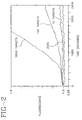
- Fig. 2 shows the change in fluorescence intensity which occurs as a nucleic acid target is amplified using the signal primers of the invention.
- Fig. 3 shows the change in fluorescence intensity associated with hybridization, extension and cleavage of a signal primer according to the invention.
- the present invention employs signal primers in hybridization and extension reactions to produce double-stranded products which contain a donor/acceptor dye pair. Fluorescence quenching occurs in the signal primer. Conversion of the single-stranded signal primer to double-stranded form also converts a single-stranded restriction endonuclease cleavage site in the signal primer to double-stranded form, rendering it cleavable or nickable by the restriction endonuclease. Cleavage or nicking by the restriction endonuclease separates the donor and acceptor dyes, resulting in decreased quenching of donor fluorescence and an increase in donor fluorescence intensity.
- An associated change in a fluorescence parameter (e.g., an increase in donor fluorescence intensity, a decrease in acceptor fluorescence intensity or the ratio of the two) is monitored as a indication of target sequence amplification. Monitoring of the change in donor fluorescence is preferred, as this change is typically larger than the change in acceptor fluorescence. Other fluorescence parameters such as a change in fluorescence lifetime may also be monitored.
- nucleic acid target amplification and signal primers are defined as follows:
- An amplification primer is a primer for amplification of a target sequence by primer extension.
- the 3' end of the amplification primer hybridizes at the 3' end of the target sequence.
- the amplification primer comprises a recognition site for a restriction endonuclease near its 5' end.
- the recognition site is for a restriction endonuclease which will cleave one strand of a DNA duplex when the recognition site is hemimodified ("nicking"), as described in US Patent No. 5,455,166; US Patent No. 5,270,184 and; EP 0 684 315.
- a hemimodified recognition site is a double stranded recognition site for a restriction endonuclease in which one strand contains at least one derivatized nucleotide which causes the restriction endonuclease to nick the primer strand rather than cleave both strands of the recognition site.
- the primer strand of the hemimodified recognition site does not contain derivatized nucleotides and is nicked by the restriction endonuclease.
- the primer may contain derivatized nucleotides which cause the unmodified target strand to be protected from cleavage while the modified primer strand is nicked.
- restriction endonucleases can be identified in routine screening systems in which a derivatized dNTP is incorporated into a restriction endonuclease recognition site for the enzyme.
- Preferred hemimodified recognition sites are hemiphosphorothioated recognition sites for the restriction endonucleases HincII, BsoBI and BsrI.
- the amplification primer also comprises a 3'-OH group which is extendable by DNA polymerase when the target binding sequence of the amplification primer is hybridized to the target sequence. For the majority of the SDA reaction, the amplification primer is responsible for exponential amplification of the target sequence.
- amplification primers for PCR generally consist only of target binding sequences.
- Amplification primers for 3SR and NASBA in contrast, comprise an RNA polymerase promoter near the 5' end. The promoter is appended to the target sequence and serves to drive the amplification reaction by directing transcription of multiple RNA copies of the target.
- Extension products are nucleic acids which comprise a primer or a portion of a primer and a newly synthesized strand which is the complement of the target sequence downstream of the primer binding site. Extension products result from hybridization of a primer to a target sequence and extension of the primer by polymerase using the target sequence as a template.
- a bumper primer is a primer which anneals to a target sequence upstream of the amplification primer, such that extension of the bumper primer displaces the downstream amplification primer and its extension product.
- Extension of bumper primers is one method for displacing the extension products of amplification primers, but heating is also suitable.
- target or target sequence refer to nucleic acid sequences to be amplified or detected. These include the original nucleic acid sequence to be amplified, its complementary second strand and either strand of a copy of the original sequence which is produced by replication or amplification.
- the target sequence may also be referred to as a template for extension of hybridized primers.
- a signal primer comprises, at its 3' end, a target binding sequence which hybridizes to the target sequence and, 5' to the target binding sequence, a label, detectable structure or specialized sequence for detection.
- the signal primers of the invention comprise a restriction endonuclease recognition site in a tail portion 5' to the target binding sequence and a donor/acceptor dye pair flanking the restriction endonuclease recognition site to facilitate letection of double-stranded products generated from the signal primer.
- the signal primer may hybridize to a target sequence downstream of an amplification primer such that extension of the amplification primer displaces the signal primer, a portion of the signal primer or the signal primer extension product. It is then rendered double-stranded by hybridization and extension of a second amplification primer.
- the target binding sequence of the signal primer may hybridize at the 3' end of the target sequence forming an 5' overhang such that extension of the target on the signal primer renders the signal primer, including the restriction endonuclease recognition site, double stranded.
- Amplification products, amplified products or amplicons are copies of the target sequence generated by hybridization and extension of an amplification primer. This term refers to both single stranded and double stranded amplification primer extension products which contain a copy of the original target sequence, including intermediates of the amplification reaction.
- Secondary amplification products or secondary products are oligonucleotides generated from a signal primer in a target amplification-dependent manner. These terms refer to single stranded or double stranded products generated from signal primers, as well as portions of signal primers or signal primer extension products generated as a result of target amplification.
- Cleavage of an oligonucleotide refers to breaking the phosphodiester bonds of both strands of a DNA duplex or breaking the bond of single-stranded DNA. This is in contrast to nicking, which refers to breaking the phosphodiester bond of only one of the two strands in a DNA duplex.
- Fig. 1 Generation of double-stranded secondary amplification products using a signal primer is illustrated in Fig. 1 and may be summarized as follows.
- a signal primer hybridizes to one strand of the target sequence downstream of an amplification primer. Both the amplification primer and the signal primer are extended by DNA polymerase using the target sequence as a template.
- the signal primer extension product is displaced from the template by extension of the upstream amplification primer and in turn serves as a template for hybridization and extension of a second amplification primer, rendering the signal primer extension product double-stranded.
- the RERS thereby becomes a substrate for the restriction endonuclease.
- a second signal primer which hybridizes to the second, complementary strand of a double stranded target sequence without overlapping the the hybridization site of the first signal primer may optionally be included in the reaction.
- the second signal primer hybridizes to the second strand of the target sequence downstream of the second amplification primer and is extended and displaced by extension of the second amplification primer.
- the second signal primer extension product is rendered double stranded by hybridization and extension of the first amplification primer.
- Multiple signal primers per strand of target may be employed if desired, each hybridizing to the target sequence downstream of the other on the same strand, and all signal primers being hybridized downstream of the amplification primer.
- each signal primer is displaced by extension of the upstream signal primer and the most 5' signal primer is displaced by the amplification primer.
- Use of multiple signal primers has the advantage of increasing or amplifying the signal generated per target, with an increase in sensitivity of the assay.
- signal primers do not serve as amplification primers. Secondary amplification products are therefore either unamplifiable or not exponentially amplifiable and have the advantage of not contributing significantly to background.
- the signal primers of the invention comprise a donor/acceptor dye pair linked at positions flanking a restriction endonuclease recognition site (RERS).
- RERS restriction endonuclease recognition site
- the RERS sequence corresponds to one strand of the double-stranded RERS.
- the signal primer restriction endonuclease recognition site is positioned 5' to the target binding region of the signal primer so as not to interfere with hybridization of the signal primer to the target sequence or its extension by polymerase.
- Either the donor or acceptor dye is linked to the signal primer 3' to the RERS but preferably not at the 3' terminus of the signal primer as a 3' terminal label may interfere with hybridization and/or extension of the primer.
- a selected donor fluorophore or acceptor dye may be linked at the 3' terminus of the signal primer.
- the donor fluorophore if the acceptor is 3' to the RERS or the acceptor (if the donor is 3' to the RERS) is linked to the signal primer at a position 5' to the RERS. That is, the donor and acceptor dyes are linked to the single-stranded signal primer such that they flank the RERS.
- the dyes are preferably linked on either side of the RERS at positions sufficiently close together that fluorescence quenching occurs but also sufficiently far apart to allow the restriction endonuclease access to the RERS for cleavage or tucking.
- the signal primer RERS may be a sequence which is recognized by the same restriction enzyme as provides the nicking function central to SDA. That is, two different recognition sequences for the same restriction endonuclease may be employed - one in the signal primer and one in the amplification primer.
- the sequence of the signal primer RERS may be selected such that double-stranded cleavage is not prevented when the modified deoxynucleoside triphosphates (dNTPs) of SDA are incorporated.
- the sequence of the amplification primer RERS is selected such that nicking by the restriction endonuclease is induced by incorporation of modified dNTPs.
- the CTCGAG and CCCGAG recognition sites for BsoBI remain cleavable when hemimodified, whereas the CTCGGG recognition site for the same enzyme is nicked when hemimodified.
- a recognition site for a restriction endonuclease different from that which provides the nicking function in the SDA reaction may be present in the signal primer.
- the RERS in the signal primer is preferably selected such that double-stranded cleavage is not compromised by incorporation of modified dNTPs.
- the RERS in the signal primer is selected so as to be nicked once by the restriction endonuclease, regenerating an RERS which is not renickable upon repair by the polymerase and incorporation of the modified dNTP.
- Such "singly-nickable" sites may be recognized by either the same restriction endonuclease which provides the nicking function in the SDA reaction or by a different restriction endonuclease.
- Singly nickable sites are generally canonical and contain a nucleotide at the nicking site which is the same as the modified dNTP in the SDA reaction. For example, the CCCGGG recognition site for BsoBI is nicked between the first and second C's.
- repair of the nick and displacement of the strand downstream of the nick incorporates the modified C nucleotide at the nicking site.
- Modification of the nicking site inhibits renicking, but the initial nick separates the donor and acceptor dyes by allowing strand displacement of the downstream fragment carrying one of the dyes.
- Singly nickable sites are desirable in the invention because they prevent amplification of the secondary amplification product independently of amplification of the target sequence, lowering background and improving quantitation.
- the signal primer is included in a nucleic acid target amplification reaction generally as described in in. S. Patent No. 5,547,861.
- the signal primers of the invention are converted to double-stranded form as previously described, converting the RERS to a double-stranded form which is cleavable by the restriction endonuclease. This process is illustrated in Fig. 1.
- "Cleavage” as used herein refers to cutting of both strands of a nucleic acid duplex by a restriction endonuclease, in contrast to "nicking" which refers to cutting of only one of the two strands in a duplex nucleic acid.
- Cleavage of the RERS produces two fragments of the double-stranded secondary amplification product. Because the donor and acceptor dyes flank the RERS, cleavage of the RERS results in separation of the dyes onto the separate fragments. Nicking of the RERS with displacement of the single-strand downstream of the nick results in a double-stranded fragment linked to one dye and a separate single-stranded fragment linked to the other dye. The distance between the dyes increases as the two fragments diffuse in the reaction solution, resulting in reduced quenching.
- a change in a fluorescence parameter resulting from reduced quenching e.g., an increase in donor fluorescence intensity or a decrease in acceptor fluorescence intensity, may be detected and/or monitored as an indication that target amplification is occurring or has occurred.
- the change in fluorescence may be monitored as the amplification reaction is occurring, i.e., in "real-time".
- Homogeneous assays reduce contamination because the reaction vessel does not have to be opened for detection and they allow the use of simpler instrumentation than in heterogeneous assays.
- the accuracy of the assay is not dependent on the starting point (i.e., establishing a "zero" point).
- the homogeneous, real-time assay of the invention can be used to provide semi-quantitative or quantitative information about the initial amount of target present.
- the rate at which the selected fluorescence parameter changes during amplification is an indication of the initial target levels.
- donor fluorescence more rapidly reaches the threshold of detection for the cleaved secondary amplification products (i.e., shorter time to positivity).
- acceptor fluorescence similarly exhibits a shorter time to positivity, detected as the time required to reach a selected minimum value.
- the rate of change in the fluorescence parameter during the course of the reaction is more rapid in samples containing higher initial amounts of target than in samples containing lower initial amounts of amounts of target (i.e., increased slope of the curve). That is, an increased rate of change in intensity, lifetime, etc. indicates a higher initial target level than is present in a sample exhibiting a relatively slower rate of change.
- the signal primer may be used in a non-amplification based format to detect a target oligonucleotide.
- the target binding sequence of the signal primer hybridizes to the 3' end of the target oligonucleotide such that the RERS forms a 5' overhang.
- Polymerase extends the target sequence using the 5' overhang of the signal primer, including the RERS, as a template.
- the target sequence functions as a primer in the primer extension reaction to synthesize the complementary sequence of the signal primer. If the target binding sequence of the signal primer is complementary to the entire length of the target sequence there are no other single-stranded overhangs and only the target is extended.
- the target binding sequence of the signal primer hybridizes to only a portion of the target sequence, the target sequence forms a second 5' overhang.
- the signal primer is also extended using the 5' overhang of the target as a template.
- the RERS of the signal primer is thus rendered double-stranded and cleavable or nickable. Extension to produce the double-stranded RERS and the resulting change in fluorescence can take place only in the presence of target, and the method is independent of the presence or absence of a restriction site in the target sequence itself. As this method does not require SDA or any other amplification reaction, modified nucleotides are not necessary. Any restriction site may be employed in the signal primer. However, if the RERS is to be nicked rather than cleaved, modified nucleotides may be employed as described above to produce a singly-nickable site.
- FITC fluorescein isothiocyanate
- TRITC tetramethylrhodamine isothiocyanate
- TRITC tetramethylrhodamine isothiocyanate
- TRITC tetramethylrhodamine isothiocyanate
- FITC/Texas RedTM Molecular Probes
- PYB FITC/N-hydroxysuccinimidyl 1-pyrenebutyrate
- EITC FITC/eosin isothiocyanate
- FITC/Rhodamine X FITC/tetramethylrhodamine
- TAMRA FITC/tetramethylrhodamine
- ABEI N-(4-aminobutyl)-N-ethylisoluminol
- Near-IR dyes such as Cy5 (N, N-modified tetramethyl indodicarbocyanine) may also be employed, e.g., paired with ROX.
- the selection of an appropriate quenching donor/acceptor pair is routine in the art. For energy transfer quenching it is only necessary that the emission wavelengths of the donor fluorophore overlap the excitation wavelengths of the acceptor fluorophore, i.e., there must be sufficient spectral overlap between the two dyes to allow efficient energy transfer, charge transfer or fluorescence quenching.
- DABYL p -(Dimethyl aminophenylazo) benzoic acid
- EDANS 5-((2'-aminoethyl) amino) naphthalene 1-sulfonic acid
- Terminal and internal labeling methods are also known in the art and may be used to link the donor and acceptor dyes at their respective sites in the signal primer.
- 5'-terminal labeling methods include a) periodate oxidation of a 5'-to-5'-coupled ribonucleotide followed by reaction with an amine-containing label, b) condensation of ethylenediamine with a 5'-phosphorylated polynucleotide followed by reaction with an amine-reactive label, and c) introduction of an aliphatic amine substituent using an aminohexyl phosphite reagent in solid-phase DNA synthesis followed by reaction with an amine-reactive label.
- Labels may also be linked to synthetic DNA oligonucleotides at specific locations using special aliphatic amine-containing nucleotide phosphoramidite reagents. Selection of an appropriate method for linking the selected labels to the signal primer and performing the linking reactions are routine in the art.
- the signal primers of the invention have a donor and an acceptor linked to the single-stranded signal primer such that donor fluorescence is totally or partially quenched. Between the two dyes, the signal primer comprises a RERS (in single-stranded form).
- the two dyes must be in sufficiently close spatial proximity for quenching to occur, however the distance between them must also allow the restriction endonuclease access to its recognition site for binding and cleavage or nicking when the signal primer is rendered double-stranded. To study the relationship of these two parameters, signal primers and their complements were chemically synthesized.
- the signal primer sequence selected was SEQ ID NO:1:
- the BsoBI site for cleavage is shown bolded, with additional tail sequence 5' to it to accommodate the "footprint" of the restriction enzyme when it binds. Double-stranded cleavage of this BsoBI recognition sequence is not inhibited by incorporation of the modified deoxynucleoside triphosphates during SDA, in contrast to the CTCGGG recognition sequence for BsoBI which is rendered nickable by incorporation of modified dNTPs during SDA.
- the sequence 3' to the BsoBI site is the target binding sequence, which is complementary to the target sequence to be amplified.
- the assay was performed at 52-53°C in 200 ⁇ L KPDG buffer (40 mM KPi, 3% DMSO, 5% glycerol) with 5 mM Mg(OAc) 2 added prior to the experiment. Measurements were obtained with an SLM 8100 research grade fluorometer equipped with a circulating bath for maintaining sample compartment temperature, a xenon arc lamp and grating monochromators for controlling excitation and emission wavelengths. Experiments with fluorescein (FAM) as the donor used 488 nm for the excitation wavelength and 525 nm for emission. Experiments in which Rhodamine X (ROX) was the donor used an excitation at 580 nm and emission at 604 nm. Experiments with Cy-5 used 640 nm and 665 nm respectively. Samples were prepared with 20 nM of the labeled signal primer for initial measurements of the emission spectrum using the optimal donor excitation wavelength.
- FAM fluorescein
- ROX Rhodamine
- the selected donor fluorophore was conjugated to the 5' phosphate.
- the selected acceptor dye was conjugated to either T6, T11, T16 or T24 to provide varying distances between the donor and acceptor dyes.
- Reactive dyes were obtained from Molecular Probes (Eugene, OR) or from the Applied Biosystems Division of Perkin Elmer (Foster City, CA).
- ROX-NHS (6-arboxy rhodamine X succinimidyl ester)
- TAMRA-SE (5-carboxy tetramethylrhodamine succinimidyl ester
- Oligonucleotides were synthesized on a 1 ⁇ mole scale using an ABI 380B automated DNA synthesizer with standard reagents supplied by the manufacturer.
- the 6-carboxy substituted fluorescein (6-FAM) was incorporated at the 5 position by addition of the phosphoramidite reagent 6-FAM Amidite (ABI) at the final step of the synthesis.
- 6-FAM Amidite (ABI)
- 5 aminohexyl phosphoramidite (ABI AMINOLINK 2) was substituted at the final step to provide a reactive amino group for subsequent conjugation.
- a modified dT phosphoramidite reagent, amino-modifier C6 dT (Glen Research, Sterling, VA) was substituted in the appropriate sequence position in place of unmodified dT.
- the crude oligonucleotides were deprotected by treatment with ammonium hydroxide for 4 to 8 hours at 55°C, which also deprotected the modified dT. These were filtered and solvent was evaporated from the filtrate with a rotary vacuum apparatus. Oligonucleotides were purified directly following this step by reverse phase HPLC.
- Oligonucleotides were labeled by dissolving an aliquot (0.5 ⁇ mole) in 100 ⁇ L NaHCO 3 /Na 2 CO 3 buffer, pH 8.0.
- the reactive dye was added to this as a solution of 3 mg in 30 ⁇ L DMSO and the resulting mixture was allowed to stand in the dark for 12-24 hours at 37 °C.
- the resulting reaction mixture was passed over a column of G-25 Sephadex resin (NAP5, Pharmacia Biotech) eluting with 4 mM TAE (4 mM TRIS acetate, 0.1 mM EDTA, pH 8.0).
- the first 0.5 to 1.0 mL of colored material eluted contained the highest fraction of reactive dye-labeled oligonucleotide and was further purified by HPLC on a Waters Delta Pak 300 ⁇ C18 3.9 X 150 mm reverse phase column using linear gradients over 30 minutes followed by 20 minutes re-equilibration. Most gradients used two solvents: A- 98% 50 mM TEAA (triethylammonium acetate)/2% acetonitrile and B - 10% 50 mM TEAA/90% acetonitrile, typically in a gradient from 95%A to 70% A over 30 minutes.
- TEAA triethylammonium acetate
- B - 10% 50 mM TEAA/90% acetonitrile typically in a gradient from 95%A to 70% A over 30 minutes.
- the identity of the conjugated material was confirmed by comparing peak intensities at 260 nm (for DNA) and the respective peak absorbances for the dyes. Concentrations of purified oligonucleotides were determined in TAE buffer by using the DNA absorbance at 260 nm corrected for the respective dye absorbance at that wavelength.
- the signal primer was initially tested for the effect of the distance between the donor and acceptor on quenching efficiency and cleavage efficiency in a hybridization assay.
- a 5-fold excess of the complementary sequence 100 nM was added and the fluorescence was measured after hybridization was judged to be complete (typically about 20 min.).
- the BsoBI enzyme was added to a concentration of 3.2 units/ ⁇ L and a final fluorescence measurement was recorded when no further change was observed in the emission spectrum of the sample.
- Table I The results for the various separation distances and dye pairs are shown in Table I.
- homologous donor/acceptor dye pairs (shown in the last three lines of the Table) exhibited an increase in donor fluorescence intensity only upon conversion from single- to double-stranded form. In contrast to heterologous dye pairs, no further increase was obtained upon cleavage of the double-stranded oligonuceotide, and in some cases cleavage produced a slight reduction in donor fluorescence intensity. Therefore, signal primers employing these fluorophore pairs need not contain an RERS.
- Target may be detected using the ss/ds ratio or a change in fluorescence associated with the conversion to double-stranded form, as quenching of the fluorophores decreases (i.e., fluorescence intensity will increase) as the signal primer is converted to double-stranded form in the presence of target.
- the methods of the invention may be easily adapted to other primer extension amplification methods (e.g., PCR, 3SR, NASBA, TMA, etc.).
- primer extension amplification methods e.g., PCR, 3SR, NASBA, TMA, etc.
- replacing SDA amplification primers with PCR amplification primers and using a PCR DNA polymerase which lacks 5' ⁇ 3' exonuclease activity e.g., Sequencing Grade Taq from Promega or exo - Vent or exo - Deep Vent from New England BioLabs
- secondary amplification products which contain a cleavable, double-stranded RERS contributed by the signal primer.
- any RERS may be selected for use in the signal primer, as there are typically no modified deoxynucleoside triphosphates present which might induce nicking rather than cleavage of the RERS.
- the double-stranded RERS in the secondary amplification product may be cleaved by a restriction endonuclease to separate a donor/acceptor dye pair as described above.
- the restriction endonuclease is preferably added at low temperature after the final cycle of primer annealing and extension for end-point detection of amplification.
- thermophilic restriction endonuclease which remains active through the high temperature phases of the PCR reaction could be present during amplification to provide a real-time assay.
- cleavage of the RERS and separation of the dye pair reduces fluorescence quenching, with the increase in fluorescence intensity serving as an indication of target amplification.
- a 5' ⁇ 3' exonuclease deficient reverse transcriptase with strand displacing activity is employed in the 3SR reaction, with hybridization of the signal primer to the RNA target downstream of an amplification primer which contains an RNA polymerase promoter.
- the hybridized signal primer containing the RERS is 1) extended, and 2) displaced by extension of the upstream amplification primer. The displaced extension product is then made double-stranded by hybridization and extension of the second amplification primer.
- the signal primer for 3SR or NASBA does not contain an RNA polymerase promoter sequence and therefore cannot function as an amplification primer, reducing nonspecific background signal. This is analogous to the signal primer in SDA, which does not contain a repeatably nickable RERS and therefore does not contribute to exponential background amplification of non-specific targets.
- signal primers are preferred for use in the methods of the invention with the signal primer extension product being separated from the target sequence by displacement due to extension of the upstream amplification primer.
- the amplification primers known for use in the various nucleic acid amplification reactions may also be labeled and modified as described for signal primers.
- the labeled amplification primer extension product may be separated from the target sequence by displacement due to extension of an upstream non-amplification primer (e.g., bumper primers as in SDA), by denaturation (e.g., heating as in PCR) or by enzymatic digestion of the target strand (e.g., RNase H as in 3SR).
- Amplification primers comprising the RERS flanked by the donor/acceptor dye pair eliminate the need for the additional signal primer in the reaction, but because background may be higher in this embodiment the sensitivity of the assay may be decreased.
- the amplification primer is modified by addition of an RERS in a 5' tail and the RERS is flanked by a donor/acceptor dye pair.
- This primer is structurally identical to the PCR signal primer described above. Functionally, however, it is different in that there is no downstream primer to be extended and displaced and the amplification primer itself provides the change in fluorescence.
- the RERS may be placed 5' to the promoter of an amplification primer so that the RERS is cleaved in the double-stranded DNA portion of the amplification cycle. Because the RERS is 5' to the promoter, cleavage does not remove the promoter from the amplification primer and generation of RNA transcripts continues to sustain target amplification.
- a second amplification primer which does not contain a promoter sequence may also or alternatively contain the RERS in a 5' tail portion.
- Target DNA for the following experimental examples was prepared from stocks of Chlamydia trachomatis elementary bodies (EB s) stored at concentrations of 10 6 EB s/ ⁇ L in 50% glycerol. EB stock solutions were diluted 1:10 in water, boiled for 15 minutes and prepared as 10-fold serial dilutions in 10 ng/ ⁇ L human placental DNA. These stock solutions contained 1 to 100 genome copies/ ⁇ L of target. The donor fluorophore was conjugated to the 5' phosphate. Measurements were obtained with an SLM 8100 research grade fluorometer equipped with a circulating bath for maintaining sample compartment temperature, a xenon arc lamp and grating monochromators for controlling excitation and emission wavelengths.
- SLM 8100 research grade fluorometer equipped with a circulating bath for maintaining sample compartment temperature, a xenon arc lamp and grating monochromators for controlling excitation and emission wavelengths.
- SDA was performed generally as described in EP 0 684 315, with addition of the signal primer labeled at the 5' end with FAM and at T11 with ROX.
- the final concentrations of components in each 100 ⁇ L reaction were 40 mM K i PO 4 pH 7.5, 6 mM MgOAc, 0.2 mM each dTTP, dGTP, dATP, 1.4 mM dCTP ⁇ S, 20 ⁇ g/ ⁇ L acetylated BSA, 3% DMSO, 8% (v/v) glycerol, 100 ng human placental DNA, 25 units Bst polymerase (exo - klenow fragment, New England BioLabs), 150 units AvaI (New England BioLabs, Beverly, MA), and DNA from 0, 10, 100 or 1,000 Chlamydia trachomatis elementary bodies.
- Each sample further contained 50 nM signal primer SEQ ID NO: 1 (5'-FAM/T 11 -ROX) and the four primers shown
- Fig. 2 shows the results. Fluorescence remained low (quenched) in the control reaction containing no target (no amplification) but increased significantly in reactions containing 100 and 1,000 targets, demonstrating specific detection of target amplification. There was no appreciable increase in fluorescence in the reaction containing 10 targets, indicating a sensitivity of detection between 10 and 100 targets. In addition, the rate of increase in fluorescence intensity of the donor (a measure of the rate of decrease in donor quenching) was more rapid in samples containing higher numbers of initial target. The rate of increase in donor fluorescence therefore provides not only detection of amplification in real-time, but also a semi-quantitative or relative measure of initial target levels.
- a quantitative measure of target levels in the unknown sample may be obtained.
- detection of an increase in fluorescence intensity above a predetermined threshold value may be used as an indication that the target is present and amplified in a simple positive/negative assay format.
- a signal primer according to the invention was used to detect a target oligonucleotide in the absence of target amplification.
- An unlabeled target oligonucleotide having the following sequence was synthesized by conventional methods:
Abstract
Description

| Donor | Acceptor | Fluorescence | Ratio | |||
| ss | ds | cleaved | ds/ss | cleaved/ss | ||
| ROX @ T11 | 5'-Cy5 | 3376 | 3919 | 7605 | 1.16 | 2.25 |
| 5'-FAM | ROX @ T6 | 2467 | 2973 | 3983 | 1.21 | 1.61 |
| 5'-FAM | ROX @ T11 | 3497 | 5290 | 18337 | 1.51 | 5.24 |
| 5'-FAM | ROX @ T16 | 990 | 1325 | 2007 | 1.34 | 2.03 |
| 5'-FAM | ROX @ T26 | 1900 | 1900 | 2000 | 1 | 1.1 |
| 5'-FAM | Dabcyl @ T11 | 10011 | 25566 | 45167 | 2.55 | 4.51 |
| TAMRA @ T11 | 5'-Cy5 | 7357 | 8412 | 9744 | 1.14 | 1.32 |
| 5'-ROX | ROX @ T11 | 18180 | 50080 | 46850 | 2.8 | 2.6 |
| 5'-FAM | FAM @ T11 | 4450 | 6100 | 5150 | 1.37 | 1.16 |
| 5'-Cy5 | Cy5 @ T11 | 3650 | 4150 | 4150 | 1.14 | 1.14 |
ACCGCATCGAATCGATGTCTCGGG TAGAAAATCGCATGCAAGATA
CGATTCCGCTCCAGACTTCTCGGG AGCTGCCTCAGAATATACTCAG
TAAACATGAAAACTCGTTCCG
TTTTATGATGAGAACACTTAAACTCA


Claims (10)
- A method for detecting presence of a target sequence comprising:a) hybridizing to the target sequence a signal primer comprising a target binding sequence and a restriction endonuclease recognition sequence 5' to the target binding sequence, the restriction endonuclease recognition sequence flanked by a donor fluorophore and an acceptor dye such that fluorescence of the donor fluorophore is quenched;b) in a primer extension reaction, synthesizing a complementary strand using the signal primer as a template, thereby rendering the restriction endonuclease recognition sequence double-stranded;d) cleaving or nicking the double-stranded restriction endonuclease recognition sequence with a restriction endonuclease, thereby reducing donor fluorophore quenching and producing a change in a fluorescence parameter, and;e) detecting the change in the fluorescence parameter as an indication of the presence of the target sequence.
- The method of Claim 1 wherein the complementary strand is synthesized in a target amplification reaction.
- The method of Claim 1 wherein a change in fluorescence intensity is detected as an indication of the presence of the target sequence.
- A method for detecting amplification of a target sequence comprising, in an amplification reaction:a) hybridizing to the target sequence a first primer comprising a target binding sequence and a restriction endonuclease recognition sequence 5' to the target binding sequence, the restriction endonuclease recognition sequence flanked by a donor fluorophore and an acceptor dye such that fluorescence of the donor fluorophore is quenched;b) extending the hybridized first primer on the target sequence with a polymerase to produce a first primer extension product and separating the first primer extension product from the target Sequence;c) rendering the separated first primer extension product and the restriction endonuclease recognition sequence double-stranded by hybridization and extension of a second primer;d) cleaving or nicking the double-stranded restriction endonuclease recognition sequence with a restriction endonuclease, thereby reducing donor fluorophore quenching and producing a change in a fluorescence parameter, and;e) detecting the change in the fluorescence parameter as an indication of amplification of the target sequence.
- The method of Claim 4 wherein the target sequence is amplified by Strand Displacement Amplification.
- The method of Claim 4 wherein the change in the fluorescence parameter is detected in real-time.
- The method of Claim 4 wherein a change in donor fluorophore or acceptor dye fluorescence intensity is detected as an indication of amplification of the target sequence.
- A single-stranded oligonucleotide comprising:(a) a target binding sequence;(b) a restriction endonuclease recognition site 5' to the target binding sequence, and;(c) a first dye and a second dye linked to the oligonucleotide at positions flanking the restriction endonuclease recognition site such that fluorescence of the first or the second dye is quenched.
- The oligonucleotide of Claim 8 wherein the first and second dyes are about 8-20 nucleotides apart in the oligonucleotide.
- The oligonucleotide of Claim 8 wherein the first dye is fluorescein and the second dye is Rhodamine X or DABCYL.
Priority Applications (1)
| Application Number | Priority Date | Filing Date | Title |
|---|---|---|---|
| DE69837125.9T DE69837125T3 (en) | 1997-05-13 | 1998-05-11 | Detection of nucleic acids by abrogation of fluorescence |
Applications Claiming Priority (2)
| Application Number | Priority Date | Filing Date | Title |
|---|---|---|---|
| US855085 | 1997-05-13 | ||
| US08/855,085 US5846726A (en) | 1997-05-13 | 1997-05-13 | Detection of nucleic acids by fluorescence quenching |
Publications (4)
| Publication Number | Publication Date |
|---|---|
| EP0878554A2 true EP0878554A2 (en) | 1998-11-18 |
| EP0878554A3 EP0878554A3 (en) | 2001-11-28 |
| EP0878554B1 EP0878554B1 (en) | 2007-02-21 |
| EP0878554B2 EP0878554B2 (en) | 2017-03-22 |
Family
ID=25320311
Family Applications (1)
| Application Number | Title | Priority Date | Filing Date |
|---|---|---|---|
| EP98108491.6A Expired - Lifetime EP0878554B2 (en) | 1997-05-13 | 1998-05-11 | Detection of nucleic acids by fluorescence quenching |
Country Status (5)
| Country | Link |
|---|---|
| US (3) | US5846726A (en) |
| EP (1) | EP0878554B2 (en) |
| JP (1) | JP3477366B2 (en) |
| CA (1) | CA2236616C (en) |
| DE (1) | DE69837125T3 (en) |
Cited By (14)
| Publication number | Priority date | Publication date | Assignee | Title |
|---|---|---|---|---|
| EP1003120A1 (en) * | 1998-11-20 | 2000-05-24 | Becton, Dickinson and Company | Computerized method and apparatus for analyzing nucleic acid assay readings |
| EP1087020A2 (en) * | 1999-09-27 | 2001-03-28 | Becton Dickinson and Company | Universal probes and methods for detection of nucleic acids |
| EP1158449A2 (en) * | 2000-05-19 | 2001-11-28 | Becton Dickinson and Company | Computerized method and apparatus for analyzing readings of nucleic acid assays |
| GB2346695B (en) * | 1997-11-29 | 2002-10-30 | Secr Defence | Fluorometric method for monitoring and detecting nucleic acid amplification |
| WO2003035864A1 (en) * | 2001-10-26 | 2003-05-01 | Matsushita Electric Industrial Co., Ltd. | Method of detecting target nucleic acid and nucleic acid probe |
| EP1321520A1 (en) * | 2001-03-27 | 2003-06-25 | National Institute of Advanced Industrial Science and Technology | Novel nucleic acid probe and novel method of assaying nucleic acids using the same |
| WO2003100095A1 (en) * | 2002-05-08 | 2003-12-04 | Arkray, Inc. | Method of detecting target nucleic acid |
| EP1586662A1 (en) * | 2004-04-14 | 2005-10-19 | Becton Dickinson and Company | Nucleic acid detection method using multiple pairs of donor flurophores and quencher molecules in the same probe |
| WO2006136621A1 (en) * | 2005-06-16 | 2006-12-28 | Biotools Biotechnological & Medical Laboratories, S.A. | Nucleic acid detection method involving the direct generation of a measurable signal |
| EP2504453A2 (en) * | 2009-11-23 | 2012-10-03 | Becton, Dickinson and Company | Assay method for target nucleic acid by signal amplification using probe hybridization and restriction |
| WO2013157821A1 (en) | 2012-04-19 | 2013-10-24 | Seegene, Inc. | Detection of target nucleic acid sequence by pto cleavage and extension-dependent signaling oligonucleotide cleavage |
| WO2018046521A1 (en) * | 2016-09-06 | 2018-03-15 | Base4 Innovation Limited | Single nucleotide detection method and associated probes |
| WO2018054964A1 (en) * | 2016-09-20 | 2018-03-29 | Base4 Innovation Limited | Single nucleotide detection method and associated probes |
| US11920192B2 (en) | 2017-05-15 | 2024-03-05 | Lightcast Discovery Ltd | Single nucleotide detection method and associated probes |
Families Citing this family (144)
| Publication number | Priority date | Publication date | Assignee | Title |
|---|---|---|---|---|
| US6787304B1 (en) | 1994-12-28 | 2004-09-07 | Georgetown University | Fluorometric assay for detecting nucleic acid cleavage |
| US20030165908A1 (en) * | 1994-12-30 | 2003-09-04 | Georgetown University | Fluorometric assay for detecting nucleic acid cleavage |
| US6766183B2 (en) | 1995-11-22 | 2004-07-20 | Medtronic Minimed, Inc. | Long wave fluorophore sensor compounds and other fluorescent sensor compounds in polymers |
| US6130038A (en) * | 1996-07-16 | 2000-10-10 | Gen-Probe Incorporated | Method for amplifying target nucleic acids using modified primers |
| US7070925B1 (en) | 1996-07-16 | 2006-07-04 | Gen-Probe Incorporated | Method for determining the presence of an RNA analyte in a sample using a modified oligonucleotide probe |
| US5853990A (en) | 1996-07-26 | 1998-12-29 | Edward E. Winger | Real time homogeneous nucleotide assay |
| US5846726A (en) * | 1997-05-13 | 1998-12-08 | Becton, Dickinson And Company | Detection of nucleic acids by fluorescence quenching |
| ATE363339T1 (en) | 1998-05-01 | 2007-06-15 | Gen Probe Inc | STIRRING DEVICE FOR THE FLUID CONTENTS OF A CONTAINER |
| GB9812768D0 (en) | 1998-06-13 | 1998-08-12 | Zeneca Ltd | Methods |
| WO2000001850A2 (en) * | 1998-07-02 | 2000-01-13 | Gen-Probe Incorporated | Molecular torches |
| ATE233795T1 (en) | 1999-04-23 | 2003-03-15 | Molecular Probes Inc | XANTHENE DYES AND THEIR APPLICATION AS LUMINESCENCE-CANCELING COMPOUNDS |
| ATE490777T1 (en) | 1999-06-01 | 2010-12-15 | Baylor College Medicine | COMPOSITIONS AND METHODS FOR THE THERAPEUTIC USE OF A SEQUENCE ASSOCIATED WITH THE GENE ATONAL |
| US6316200B1 (en) | 2000-06-08 | 2001-11-13 | Becton, Dickinson And Company | Probes and methods for detection of nucleic acids |
| US20020025519A1 (en) * | 1999-06-17 | 2002-02-28 | David J. Wright | Methods and oligonucleotides for detecting nucleic acid sequence variations |
| US20030165913A1 (en) | 1999-06-17 | 2003-09-04 | Sha-Sha Wang | Methods for detecting nucleic acid sequence variations |
| EP1061134A3 (en) * | 1999-06-17 | 2003-01-08 | Becton Dickinson and Company | Oligonucleotides for amplification and detection of hemochromatosis genes |
| US6727356B1 (en) | 1999-12-08 | 2004-04-27 | Epoch Pharmaceuticals, Inc. | Fluorescent quenching detection reagents and methods |
| US20040081959A9 (en) * | 1999-12-08 | 2004-04-29 | Epoch Biosciences, Inc. | Fluorescent quenching detection reagents and methods |
| AUPQ495700A0 (en) * | 2000-01-05 | 2000-02-03 | Johnson & Johnson Research Pty. Limited | Method for concurrent amplification and real time detection of polymorphic nucleic acid sequences |
| US6221603B1 (en) | 2000-02-04 | 2001-04-24 | Molecular Dynamics, Inc. | Rolling circle amplification assay for nucleic acid analysis |
| US6323337B1 (en) | 2000-05-12 | 2001-11-27 | Molecular Probes, Inc. | Quenching oligonucleotides |
| US6686188B2 (en) | 2000-05-26 | 2004-02-03 | Amersham Plc | Polynucleotide encoding a human myosin-like polypeptide expressed predominantly in heart and muscle |
| US6656700B2 (en) | 2000-05-26 | 2003-12-02 | Amersham Plc | Isoforms of human pregnancy-associated protein-E |
| US6258546B1 (en) | 2000-06-23 | 2001-07-10 | Becton, Dickinson And Company | Detection of nucleic acid amplification |
| AU2002211814A1 (en) | 2000-09-12 | 2002-03-26 | Gen-Probe Incorporated | Compositions, methods and kits for determining the presence of cryptosporidium organisms in a test sample |
| US20030148303A1 (en) * | 2000-09-19 | 2003-08-07 | Nadeau James G. | Universal probes and methods for detection of nucleic acids |
| DE10061166A1 (en) * | 2000-11-30 | 2002-06-06 | Attomol Gmbh Molekulare Diagno | Method and kit for the direct detection of nucleotide sequences, amino acid sequences or antigens |
| US6927246B2 (en) * | 2001-02-15 | 2005-08-09 | Medtronic Minimed, Inc. | Polymers functionalized with fluorescent boronate motifs and methods for making them |
| US20030017567A1 (en) * | 2001-04-24 | 2003-01-23 | 3M Innovative Properties Company | Biological sample processing methods and compositions that include surfactants |
| US6617136B2 (en) | 2001-04-24 | 2003-09-09 | 3M Innovative Properties Company | Biological sample processing methods and compositions that include surfactants |
| US7112423B2 (en) * | 2001-07-15 | 2006-09-26 | Keck Graduate Institute | Nucleic acid amplification using nicking agents |
| MXPA04000432A (en) * | 2001-07-15 | 2004-10-27 | Keck Graduate Inst | Amplification of nucleic acid fragments using nicking agents. |
| US20060073530A1 (en) * | 2001-08-15 | 2006-04-06 | Olaf Schneewind | Methods and compositions involving sortase B |
| US20030082547A1 (en) | 2001-08-27 | 2003-05-01 | Ewing Gregory J. | Non-fluorescent quencher compounds and biomolecular assays |
| US20050032060A1 (en) * | 2001-08-31 | 2005-02-10 | Shishir Shah | Arrays comprising pre-labeled biological molecules and methods for making and using these arrays |
| US7045361B2 (en) | 2001-09-12 | 2006-05-16 | Medtronic Minimed, Inc. | Analyte sensing via acridine-based boronate biosensors |
| US7439346B2 (en) * | 2001-10-12 | 2008-10-21 | Perkinelmer Las Inc. | Nucleic acids arrays and methods of use therefor |
| CA2475003A1 (en) | 2002-02-01 | 2003-08-07 | Sequitur, Inc. | Double-stranded oligonucleotides |
| US20030166282A1 (en) | 2002-02-01 | 2003-09-04 | David Brown | High potency siRNAS for reducing the expression of target genes |
| US20060009409A1 (en) | 2002-02-01 | 2006-01-12 | Woolf Tod M | Double-stranded oligonucleotides |
| US6916621B2 (en) * | 2002-03-27 | 2005-07-12 | Spectral Genomics, Inc. | Methods for array-based comparitive binding assays |
| KR101170653B1 (en) | 2002-08-12 | 2012-08-03 | 제네렉스, 인코포레이티드 | Methods and compositions concerning poxviruses and cancer |
| JP4694201B2 (en) * | 2002-09-20 | 2011-06-08 | インテグレイテッド ディーエヌエイ テクノロジーズ インコーポレイテッド | Anthraquinone quenching dyes, their production and use |
| CA2520538C (en) | 2003-03-31 | 2014-04-29 | F. Hoffmann-La Roche Ag | Compositions and methods for detecting certain flaviviruses, including members of the japanese encephalitis virus serogroup |
| CN101410530B (en) | 2003-04-18 | 2013-03-27 | 贝克顿·迪金森公司 | Immuno-amplification |
| US7291488B2 (en) | 2003-04-25 | 2007-11-06 | Becton, Dickinson & Company | Detection of herpes simplex virus types 1 and 2 by nucleic acid amplification |
| JP5656273B2 (en) | 2003-05-19 | 2015-01-21 | ジェン−プロウブ インコーポレイテッド | Compositions, methods and kits for determining the presence of TRICHOMONASVAginalis in a test sample |
| CA2538086A1 (en) * | 2003-09-12 | 2005-03-24 | Becton, Dickinson And Company | Assay for sars coronavirus by amplification and detection of nucleocapsid rna sequence |
| CN101415843B (en) * | 2003-09-12 | 2013-08-21 | 贝克顿·迪金森公司 | Assay for SARS coronavirus by amplification and detection of the replicase sequence |
| US20050118619A1 (en) * | 2003-09-22 | 2005-06-02 | Wensheng Xia | Dark quenchers for fluorescence resonance energy transfer (FRET) in bioassays |
| JP2007515947A (en) * | 2003-10-30 | 2007-06-21 | タフツ−ニュー イングランド メディカル センター | Prenatal diagnosis using acellular fetal DNA in amniotic fluid |
| WO2005049849A2 (en) * | 2003-11-14 | 2005-06-02 | Integrated Dna Technologies, Inc. | Fluorescence quenching azo dyes, their methods of preparation and use |
| US7667024B2 (en) * | 2003-11-19 | 2010-02-23 | Allelogic Biosciences Corp. | Oligonucleotides labeled with a plurality of fluorophores |
| JP5162133B2 (en) | 2003-12-03 | 2013-03-13 | アボット・ラボラトリーズ | Double-stranded linear nucleic acid probe and use thereof |
| US7939251B2 (en) | 2004-05-06 | 2011-05-10 | Roche Molecular Systems, Inc. | SENP1 as a marker for cancer |
| EP1766061B1 (en) * | 2004-05-20 | 2013-07-17 | Quest Diagnostics Investments Incorporated | Single label comparative hybridization |
| US20080311564A1 (en) * | 2004-08-06 | 2008-12-18 | Fort Thomas L | Sequences and Methods for Detection of Cytomegalovirus |
| US7910753B2 (en) * | 2004-09-10 | 2011-03-22 | Anaspec Incorporated | Cyanine dyes and their applications as luminescence quenching compounds |
| ES2345993T3 (en) | 2004-09-14 | 2010-10-07 | The Regents Of The University Of Colorado, A Body Corporate | METHOD FOR TREATMENT WITH BUCINDOLOL BASED ON GENETIC ADDRESSING. |
| US20060073511A1 (en) | 2004-10-05 | 2006-04-06 | Affymetrix, Inc. | Methods for amplifying and analyzing nucleic acids |
| DE602006018861D1 (en) * | 2005-01-27 | 2011-01-27 | Quest Diagnostics Invest Inc | FAST COMPARATIVE GENOM HYBRIDIZATION |
| WO2006088910A2 (en) * | 2005-02-15 | 2006-08-24 | Georgetown University | Sequence-specific detection of nucleotide sequences |
| WO2010096202A2 (en) | 2009-02-23 | 2010-08-26 | Georgetown University | Sequence-specific detection of nucleotide sequences |
| US7601498B2 (en) * | 2005-03-17 | 2009-10-13 | Biotium, Inc. | Methods of using dyes in association with nucleic acid staining or detection and associated technology |
| US7776567B2 (en) | 2005-03-17 | 2010-08-17 | Biotium, Inc. | Dimeric and trimeric nucleic acid dyes, and associated systems and methods |
| JP2008539730A (en) * | 2005-05-02 | 2008-11-20 | ストラタジーン カリフォルニア | Oligonucleotide probe / primer composition and polynucleotide detection method |
| WO2006127507A2 (en) | 2005-05-20 | 2006-11-30 | Integrated Dna Technologies, Inc. | Compounds and methods for labeling oligonucleotides |
| US20060292576A1 (en) * | 2005-06-23 | 2006-12-28 | Quest Diagnostics Investments Incorporated | Non-in situ hybridization method for detecting chromosomal abnormalities |
| US8980246B2 (en) | 2005-09-07 | 2015-03-17 | Sillajen Biotherapeutics, Inc. | Oncolytic vaccinia virus cancer therapy |
| CA2621982C (en) | 2005-09-07 | 2017-11-28 | Jennerex Biotherapeutics Ulc | Systemic treatment of metastatic and/or systemically-disseminated cancers using gm-csf-expressing poxviruses |
| US7910335B2 (en) | 2005-10-27 | 2011-03-22 | President And Fellows Of Harvard College | Methods and compositions for labeling nucleic acids |
| KR20080066993A (en) | 2005-11-07 | 2008-07-17 | 지멘스 헬쓰케어 다이아그노스틱스 인크. | Chlamydia trachomatis specific oligonucleotide sequences |
| US20090068643A1 (en) * | 2005-11-23 | 2009-03-12 | Integrated Dna Technologies, Inc. | Dual Function Primers for Amplifying DNA and Methods of Use |
| US8076074B2 (en) | 2005-11-29 | 2011-12-13 | Quest Diagnostics Investments Incorporated | Balanced translocation in comparative hybridization |
| US7790386B2 (en) | 2006-04-07 | 2010-09-07 | Siemens Healthcare Diagnostics Inc. | Neisseria gonorrhoeae specific oligonucleotide sequences |
| US11001881B2 (en) | 2006-08-24 | 2021-05-11 | California Institute Of Technology | Methods for detecting analytes |
| WO2008014485A2 (en) | 2006-07-28 | 2008-01-31 | California Institute Of Technology | Multiplex q-pcr arrays |
| US11525156B2 (en) | 2006-07-28 | 2022-12-13 | California Institute Of Technology | Multiplex Q-PCR arrays |
| US11560588B2 (en) | 2006-08-24 | 2023-01-24 | California Institute Of Technology | Multiplex Q-PCR arrays |
| US8481023B2 (en) | 2006-09-15 | 2013-07-09 | Ottawa Hospital Research Institute | Oncolytic rhabdovirus |
| US7807359B2 (en) * | 2006-12-01 | 2010-10-05 | Quest Diagnostics Investments Incorporated | Methods of detecting TPMT mutations |
| US7507539B2 (en) * | 2007-07-30 | 2009-03-24 | Quest Diagnostics Investments Incorporated | Substractive single label comparative hybridization |
| US8093063B2 (en) * | 2007-11-29 | 2012-01-10 | Quest Diagnostics Investments Incorporated | Assay for detecting genetic abnormalities in genomic nucleic acids |
| DK2644707T3 (en) | 2008-04-30 | 2015-06-29 | Integrated Dna Tech Inc | RNase-H-based assays using modified RNA monomers. |
| US8911948B2 (en) * | 2008-04-30 | 2014-12-16 | Integrated Dna Technologies, Inc. | RNase H-based assays utilizing modified RNA monomers |
| WO2009137369A1 (en) * | 2008-05-03 | 2009-11-12 | Tufts Medical Center, Inc. | Neonatal salivary genomics |
| US9090948B2 (en) | 2008-09-30 | 2015-07-28 | Abbott Molecular Inc. | Primers and probes for detecting human papillomavirus and human beta globin sequences in test samples |
| US8039794B2 (en) | 2008-12-16 | 2011-10-18 | Quest Diagnostics Investments Incorporated | Mass spectrometry assay for thiopurine-S-methyl transferase activity and products generated thereby |
| US8945842B2 (en) | 2009-01-14 | 2015-02-03 | Becton, Dickinson And Company | Assay for Trichomonas vaginalis by amplification and detection of Trichomonas vaginalis AP65-1 gene |
| WO2011008530A2 (en) | 2009-06-29 | 2011-01-20 | Luminex Corporation | Chimeric primers with hairpin conformations and methods of using same |
| US9409983B2 (en) | 2009-07-23 | 2016-08-09 | The Board Of Trustess Of The University Of Illinois | Methods and compositions involving PBEF inhibitors for lung inflammation conditions and diseases |
| US9512481B2 (en) | 2009-09-11 | 2016-12-06 | The Regents Of The University Of Colorado, A Body Corporate | Polymorphisms in the PDE3A gene |
| KR101838472B1 (en) | 2009-09-14 | 2018-03-15 | 신라젠(주) | Oncolytic vaccinia virus combination cancer therapy |
| CN102630250A (en) | 2009-09-25 | 2012-08-08 | 基因诊断测试公司 | Multiplex (+/-) stranded arrays and assays for detecting chromosomal abnormalities associated with cancer and other diseases |
| MX365946B (en) | 2009-12-10 | 2019-06-19 | Turnstone Lp | Oncolytic rhabdovirus. |
| US8877437B1 (en) | 2009-12-23 | 2014-11-04 | Biotium, Inc. | Methods of using dyes in association with nucleic acid staining or detection |
| EP2515899B1 (en) | 2009-12-23 | 2016-05-25 | ARCA biopharma, Inc. | Methods and compositions for cardiovascular diseases and conditions |
| CA2896129C (en) | 2010-01-22 | 2015-12-29 | Gen-Probe Incorporated | Probes for detecting the presence of trichomonas vaginalis in a sample |
| US20120108799A1 (en) | 2010-09-07 | 2012-05-03 | Integrated Dna Technologies, Inc. | Modifications for Antisense Compounds |
| US9506057B2 (en) | 2010-03-26 | 2016-11-29 | Integrated Dna Technologies, Inc. | Modifications for antisense compounds |
| EP2553123B1 (en) | 2010-03-26 | 2016-08-24 | Integrated DNA Technologies, Inc. | Methods for enhancing nucleic acid hybridization |
| CN103551212B (en) | 2010-07-23 | 2016-01-20 | 贝克曼考尔特公司 | Kit |
| JP6121910B2 (en) | 2011-01-04 | 2017-04-26 | シラジェン バイオセラピューティクス インコーポレイテッド | Generation of antibodies against tumor antigens and tumor-specific complement-dependent cytotoxicity by administration of oncolytic vaccinia virus |
| WO2012158238A2 (en) | 2011-02-28 | 2012-11-22 | University Of Iowa Research Foundation | Anti-müllerian hormone changes in pregnancy and prediction ofadverse pregnancy outcomes and gender |
| EP2683835B1 (en) | 2011-03-08 | 2017-01-11 | King Abdullah University Of Science And Technology | Molecular biomarker set for early detection of ovarian cancer |
| AU2012236896A1 (en) | 2011-03-25 | 2013-05-16 | Integrated Dna Technologies, Inc. | RNase H-based assays utilizing modified RNA monomers |
| US9364532B2 (en) | 2011-06-08 | 2016-06-14 | Children's Hospital Of Eastern Ontario Research Institute Inc. | Compositions and methods for glioblastoma treatment |
| WO2013036799A2 (en) | 2011-09-09 | 2013-03-14 | Fred Hutchinson Cancer Research Center | Methods and compositions involving nkg2d inhibitors and cancer |
| ES2844324T3 (en) | 2011-11-07 | 2021-07-21 | Beckman Coulter Inc | Robotic arm |
| JP6062449B2 (en) | 2011-11-07 | 2017-01-18 | ベックマン コールター, インコーポレイテッド | Specimen container detection |
| EP2776848B1 (en) | 2011-11-07 | 2019-12-25 | Beckman Coulter, Inc. | System and method for transporting sample containers |
| BR112014011035A2 (en) | 2011-11-07 | 2017-06-13 | Beckman Coulter, Inc. | aliquot system and workflow |
| KR20140091032A (en) | 2011-11-07 | 2014-07-18 | 베크만 컬터, 인코포레이티드 | Magnetic damping for specimen transport system |
| WO2013070755A2 (en) | 2011-11-07 | 2013-05-16 | Beckman Coulter, Inc. | Centrifuge system and workflow |
| US9212381B2 (en) | 2011-11-10 | 2015-12-15 | President And Fellows Of Harvard College | Methods and compositions for labeling polypeptides |
| WO2014116721A1 (en) | 2013-01-22 | 2014-07-31 | The Arizona Board Of Regents For And On Behalf Of Arizona State University | Geminiviral vector for expression of rituximab |
| RU2684211C2 (en) | 2013-02-21 | 2019-04-04 | Тёрнстоун Лимитед Партнершип | Vaccine composition |
| BR112016007391A2 (en) | 2013-10-03 | 2017-08-01 | Oklahoma Med Res Found | biomarkers of activity, intensity and acute phase of systemic lupus erythematosus disease |
| WO2015152024A1 (en) * | 2014-03-31 | 2015-10-08 | 独立行政法人理化学研究所 | Fluorescent labeled single-stranded nucleic acid and use thereof |
| WO2016086227A2 (en) | 2014-11-26 | 2016-06-02 | The Regents Of The University Of California | Therapeutic compositions comprising transcription factors and methods of making and using the same |
| WO2016123243A1 (en) * | 2015-01-28 | 2016-08-04 | The Regents Of The University Of California | Methods and compositions for labeling a single-stranded target nucleic acid |
| US9708647B2 (en) | 2015-03-23 | 2017-07-18 | Insilixa, Inc. | Multiplexed analysis of nucleic acid hybridization thermodynamics using integrated arrays |
| US9957393B2 (en) | 2015-03-30 | 2018-05-01 | Enzo Biochem, Inc. | Monoazo dyes with cyclic amine as fluorescence quenchers |
| EP3124619B1 (en) | 2015-07-31 | 2019-03-06 | Menicon Co., Ltd | Reagents, method and kit for across and within dog breed glaucoma diagnosis |
| US9499861B1 (en) | 2015-09-10 | 2016-11-22 | Insilixa, Inc. | Methods and systems for multiplex quantitative nucleic acid amplification |
| WO2017155858A1 (en) | 2016-03-07 | 2017-09-14 | Insilixa, Inc. | Nucleic acid sequence identification using solid-phase cyclic single base extension |
| US20190226014A1 (en) * | 2016-09-02 | 2019-07-25 | Base4 Innovation Limited | Single nucleotide detection method and associated probes |
| US10427162B2 (en) | 2016-12-21 | 2019-10-01 | Quandx Inc. | Systems and methods for molecular diagnostics |
| EP3574114B1 (en) | 2017-01-26 | 2022-06-29 | Oklahoma Medical Research Foundation | Biomarkers for systemic lupus erythematosus disease activity, and intensity and flare |
| CN110914459A (en) | 2017-09-27 | 2020-03-24 | 雅培分子公司 | Assays for detecting Hepatitis B Virus (HBV) |
| CA3068498A1 (en) | 2017-10-03 | 2019-04-11 | Abbott Molecular Inc. | Assay for detecting human immunodeficiency virus (hiv) |
| CA3068482A1 (en) | 2017-10-03 | 2019-04-11 | Abbott Molecular Inc. | Assay for detecting hepatitis c virus (hcv) |
| WO2019139792A1 (en) | 2018-01-09 | 2019-07-18 | Abbott Molecular Inc. | Assay for detecting chlamydia trachomatis, neisseria gonorrhoeae, trichomonas vaginalis, and mycoplasma genitalium |
| WO2019213619A1 (en) | 2018-05-04 | 2019-11-07 | Abbott Laboratories | Hbv diagnostic, prognostic, and therapeutic methods and products |
| US20210310059A1 (en) | 2018-08-08 | 2021-10-07 | Gen-Probe Incorporated | Compositions, methods and kits for detecting mycoplasma genitalium |
| WO2020081204A1 (en) | 2018-10-18 | 2020-04-23 | Oklahoma Medical Research Foundation | Biomarkers for a systemic lupus erythematosus (sle) disease activity immune index that characterizes disease activity |
| JP2022509018A (en) | 2018-10-31 | 2022-01-20 | アリゾナ ボード オブ リージェンツ オン ビハーフ オブ ザ ユニバーシティー オブ アリゾナ | Biomarkers and usage for radiation-induced lung injury |
| WO2020132090A1 (en) | 2018-12-18 | 2020-06-25 | Abbott Molecular Inc. | Assay for detecting human papilloma virus (hpv) |
| CN109694905A (en) * | 2019-01-29 | 2019-04-30 | 深圳市亚辉龙生物科技股份有限公司 | Detect the nucleic acid compositions of testing gene methylation and its detection method of kit and testing gene methylation |
| CN113924041A (en) | 2019-03-14 | 2022-01-11 | 因斯利克萨公司 | Method and system for fluorescence detection based on time gating |
| WO2021041056A1 (en) | 2019-08-23 | 2021-03-04 | Gen-Probe Incorporated | Compositions, methods and kits for detecting treponema pallidum |
| WO2021133916A1 (en) | 2019-12-23 | 2021-07-01 | Abbott Laboratories | Compositions and methods for detecting picobirnavirus |
| WO2021138325A1 (en) | 2019-12-30 | 2021-07-08 | Abbott Laboratories | Compositions and methods for detecting bunyavirus |
| WO2024030985A1 (en) | 2022-08-04 | 2024-02-08 | Abbott Laboratories | Assays for detecting monkeypox virus |
Citations (3)
| Publication number | Priority date | Publication date | Assignee | Title |
|---|---|---|---|---|
| EP0360940A2 (en) * | 1988-09-29 | 1990-04-04 | Chiron Corporation | Polynucleotide determination with selectable cleavage sites |
| EP0678582A1 (en) * | 1994-04-18 | 1995-10-25 | Becton, Dickinson and Company | Detection of nucleic acid amplification |
| US5538848A (en) * | 1994-11-16 | 1996-07-23 | Applied Biosystems Division, Perkin-Elmer Corp. | Method for detecting nucleic acid amplification using self-quenching fluorescence probe |
Family Cites Families (5)
| Publication number | Priority date | Publication date | Assignee | Title |
|---|---|---|---|---|
| JPS613063A (en) * | 1984-06-18 | 1986-01-09 | Fujirebio Inc | Measurement of polynucleotide utilizing luminous substance |
| JPH0515439A (en) * | 1991-07-08 | 1993-01-26 | Matsushita Electric Ind Co Ltd | Control circuit for jar type rice cooker |
| DE69535882D1 (en) * | 1994-12-30 | 2008-12-18 | Univ Georgetown | FLUORESCENT TEST FOR DETECTING THE CLEAVAGE OF NUCLEIC ACID |
| US5550025A (en) * | 1995-07-19 | 1996-08-27 | Becton, Dickinson And Company | Detection of hydrophobic amplification products by extraction into an organic phase |
| US5846726A (en) * | 1997-05-13 | 1998-12-08 | Becton, Dickinson And Company | Detection of nucleic acids by fluorescence quenching |
-
1997
- 1997-05-13 US US08/855,085 patent/US5846726A/en not_active Expired - Lifetime
-
1998
- 1998-05-04 CA CA002236616A patent/CA2236616C/en not_active Expired - Lifetime
- 1998-05-11 JP JP12706898A patent/JP3477366B2/en not_active Expired - Lifetime
- 1998-05-11 DE DE69837125.9T patent/DE69837125T3/en not_active Expired - Lifetime
- 1998-05-11 EP EP98108491.6A patent/EP0878554B2/en not_active Expired - Lifetime
- 1998-07-20 US US09/120,916 patent/US6054279A/en not_active Expired - Lifetime
- 1998-11-04 US US09/186,030 patent/US5919630A/en not_active Expired - Fee Related
Patent Citations (3)
| Publication number | Priority date | Publication date | Assignee | Title |
|---|---|---|---|---|
| EP0360940A2 (en) * | 1988-09-29 | 1990-04-04 | Chiron Corporation | Polynucleotide determination with selectable cleavage sites |
| EP0678582A1 (en) * | 1994-04-18 | 1995-10-25 | Becton, Dickinson and Company | Detection of nucleic acid amplification |
| US5538848A (en) * | 1994-11-16 | 1996-07-23 | Applied Biosystems Division, Perkin-Elmer Corp. | Method for detecting nucleic acid amplification using self-quenching fluorescence probe |
Non-Patent Citations (3)
| Title |
|---|
| DATABASE WPI Week 8625 Derwent Publications Ltd., London, GB; AN 1986-050950 XP002177789 & JP 61 003063 A ((FJRE) FUJI REBIO KK) 9 January 1986 (1986-01-09) * |
| WALKER G T ET AL.: "A DNA probe assay using strand displacement amplification (SDA) and filtration to separate reacted and unreacted detector probes" MOLECULAR AND CELLULAR PROBES, vol. 9, 1995, pages 399-403, * |
| YU H ET AL.: "Cyanine dye dUTP analogs for enzymatic labeling of DNA probes" NUCLEIC ACIDS RESEARCH, vol. 22, no. 15, 1994, pages 3226-3232, * |
Cited By (28)
| Publication number | Priority date | Publication date | Assignee | Title |
|---|---|---|---|---|
| GB2346695B (en) * | 1997-11-29 | 2002-10-30 | Secr Defence | Fluorometric method for monitoring and detecting nucleic acid amplification |
| EP1044283B2 (en) † | 1997-11-29 | 2015-09-23 | The Secretary Of State For Defence | Fluorometric method for monitoring and detecting nucleic acid amplification |
| EP1003120A1 (en) * | 1998-11-20 | 2000-05-24 | Becton, Dickinson and Company | Computerized method and apparatus for analyzing nucleic acid assay readings |
| US6216049B1 (en) | 1998-11-20 | 2001-04-10 | Becton, Dickinson And Company | Computerized method and apparatus for analyzing nucleic acid assay readings |
| EP1087020A2 (en) * | 1999-09-27 | 2001-03-28 | Becton Dickinson and Company | Universal probes and methods for detection of nucleic acids |
| US6379888B1 (en) | 1999-09-27 | 2002-04-30 | Becton, Dickinson And Company | Universal probes and methods for detection of nucleic acids |
| EP1087020A3 (en) * | 1999-09-27 | 2003-07-09 | Becton Dickinson and Company | Universal probes and methods for detection of nucleic acids |
| EP1158449A2 (en) * | 2000-05-19 | 2001-11-28 | Becton Dickinson and Company | Computerized method and apparatus for analyzing readings of nucleic acid assays |
| EP1158449A3 (en) * | 2000-05-19 | 2004-12-15 | Becton Dickinson and Company | Computerized method and apparatus for analyzing readings of nucleic acid assays |
| EP1321520A1 (en) * | 2001-03-27 | 2003-06-25 | National Institute of Advanced Industrial Science and Technology | Novel nucleic acid probe and novel method of assaying nucleic acids using the same |
| EP1321520A4 (en) * | 2001-03-27 | 2003-09-03 | Nat Inst Of Advanced Ind Scien | Novel nucleic acid probe and novel method of assaying nucleic acids using the same |
| WO2003035864A1 (en) * | 2001-10-26 | 2003-05-01 | Matsushita Electric Industrial Co., Ltd. | Method of detecting target nucleic acid and nucleic acid probe |
| US7220544B2 (en) | 2002-05-08 | 2007-05-22 | Arkray, Inc. | Method for detecting target nucleic acid |
| CN1330775C (en) * | 2002-05-08 | 2007-08-08 | 爱科来株式会社 | Method of detecting target nucleic acid |
| WO2003100095A1 (en) * | 2002-05-08 | 2003-12-04 | Arkray, Inc. | Method of detecting target nucleic acid |
| US8940882B2 (en) | 2004-04-14 | 2015-01-27 | Becton, Dickinson And Company | Multiple fluorophore detector system |
| EP1586662A1 (en) * | 2004-04-14 | 2005-10-19 | Becton Dickinson and Company | Nucleic acid detection method using multiple pairs of donor flurophores and quencher molecules in the same probe |
| US7919244B2 (en) | 2005-06-16 | 2011-04-05 | Biotools Biotechnological & Medical Laboratories, S.A. | Nucleic acid detection method involving the direct generation of a measurable signal |
| WO2006136621A1 (en) * | 2005-06-16 | 2006-12-28 | Biotools Biotechnological & Medical Laboratories, S.A. | Nucleic acid detection method involving the direct generation of a measurable signal |
| EP2504453A4 (en) * | 2009-11-23 | 2013-07-17 | Becton Dickinson Co | Assay method for target nucleic acid by signal amplification using probe hybridization and restriction |
| US8916349B2 (en) | 2009-11-23 | 2014-12-23 | Becton, Dickinson And Company | Assay method for target nucleic acid by signal amplification using probe hybridization and restriction |
| EP2504453A2 (en) * | 2009-11-23 | 2012-10-03 | Becton, Dickinson and Company | Assay method for target nucleic acid by signal amplification using probe hybridization and restriction |
| WO2013157821A1 (en) | 2012-04-19 | 2013-10-24 | Seegene, Inc. | Detection of target nucleic acid sequence by pto cleavage and extension-dependent signaling oligonucleotide cleavage |
| US9683259B2 (en) | 2012-04-19 | 2017-06-20 | Seegene, Inc. | Detection of target nucleic acid sequence by PTO cleavage and extension-dependent signaling oligonucleotide cleavage |
| WO2018046521A1 (en) * | 2016-09-06 | 2018-03-15 | Base4 Innovation Limited | Single nucleotide detection method and associated probes |
| CN109790573A (en) * | 2016-09-06 | 2019-05-21 | 贝斯4创新公司 | Mononucleotide detection method and relevant probe |
| WO2018054964A1 (en) * | 2016-09-20 | 2018-03-29 | Base4 Innovation Limited | Single nucleotide detection method and associated probes |
| US11920192B2 (en) | 2017-05-15 | 2024-03-05 | Lightcast Discovery Ltd | Single nucleotide detection method and associated probes |
Also Published As
| Publication number | Publication date |
|---|---|
| CA2236616A1 (en) | 1998-11-13 |
| JP3477366B2 (en) | 2003-12-10 |
| DE69837125D1 (en) | 2007-04-05 |
| EP0878554A3 (en) | 2001-11-28 |
| EP0878554B1 (en) | 2007-02-21 |
| JPH11123083A (en) | 1999-05-11 |
| CA2236616C (en) | 2002-04-23 |
| US6054279A (en) | 2000-04-25 |
| US5919630A (en) | 1999-07-06 |
| EP0878554B2 (en) | 2017-03-22 |
| DE69837125T2 (en) | 2007-12-13 |
| US5846726A (en) | 1998-12-08 |
| DE69837125T3 (en) | 2017-08-31 |
Similar Documents
| Publication | Publication Date | Title |
|---|---|---|
| US5846726A (en) | Detection of nucleic acids by fluorescence quenching | |
| US5928869A (en) | Detection of nucleic acids by fluorescence quenching | |
| US6261784B1 (en) | Detection of nucleic acids by strand displacement | |
| US5888739A (en) | Detection of nucleic acids using G-quartets and I-tetraplexes | |
| CA2336489C (en) | Wavelength-shifting hairpin-forming probes | |
| EP1087020B1 (en) | Universal probes and methods for detection of nucleic acids | |
| EP1162277A2 (en) | Probes and methods for detection of nucleic acids | |
| EP1509624A2 (en) | Met/fret based method of target nucleic acid detection whereby the donor/acceptor moieties are on complementary strands | |
| AU2010290277A1 (en) | TD probe and its uses | |
| WO2006119326A2 (en) | Oligonucleotide probe/primer compositions and methods for polynucleotide detection | |
| WO2005059548A1 (en) | Novel mixtures for assaying nucleic acid, novel method of assaying nucleic acid with the use of the same and nucleic acid probe to be used therefor | |
| EP1086245B1 (en) | Multi-fluorescent hairpin energy transfer oligonucleotides | |
| US20030148303A1 (en) | Universal probes and methods for detection of nucleic acids | |
| MXPA98007316A (en) | Detection of nucleic acids for the extinction of fluorescen |
Legal Events
| Date | Code | Title | Description |
|---|---|---|---|
| PUAI | Public reference made under article 153(3) epc to a published international application that has entered the european phase |
Free format text: ORIGINAL CODE: 0009012 |
|
| AK | Designated contracting states |
Kind code of ref document: A2 Designated state(s): AT BE CH CY DE DK ES FI FR GB GR IE IT LI LU MC NL PT SE Kind code of ref document: A2 Designated state(s): DE FR GB IT |
|
| AX | Request for extension of the european patent |
Free format text: AL;LT;LV;MK;RO;SI |
|
| PUAL | Search report despatched |
Free format text: ORIGINAL CODE: 0009013 |
|
| AK | Designated contracting states |
Kind code of ref document: A3 Designated state(s): AT BE CH CY DE DK ES FI FR GB GR IE IT LI LU MC NL PT SE |
|
| AX | Request for extension of the european patent |
Free format text: AL;LT;LV;MK;RO;SI |
|
| 17P | Request for examination filed |
Effective date: 20020408 |
|
| AKX | Designation fees paid |
Free format text: DE FR GB IT |
|
| 17Q | First examination report despatched |
Effective date: 20020703 |
|
| GRAP | Despatch of communication of intention to grant a patent |
Free format text: ORIGINAL CODE: EPIDOSNIGR1 |
|
| GRAS | Grant fee paid |
Free format text: ORIGINAL CODE: EPIDOSNIGR3 |
|
| GRAA | (expected) grant |
Free format text: ORIGINAL CODE: 0009210 |
|
| AK | Designated contracting states |
Kind code of ref document: B1 Designated state(s): DE FR GB IT |
|
| REG | Reference to a national code |
Ref country code: GB Ref legal event code: FG4D |
|
| REF | Corresponds to: |
Ref document number: 69837125 Country of ref document: DE Date of ref document: 20070405 Kind code of ref document: P |
|
| ET | Fr: translation filed | ||
| PLBI | Opposition filed |
Free format text: ORIGINAL CODE: 0009260 |
|
| 26 | Opposition filed |
Opponent name: THE SECRETARY OF STATE FOR DEFENCE DIRECTORATE OF Effective date: 20071121 |
|
| PLAX | Notice of opposition and request to file observation + time limit sent |
Free format text: ORIGINAL CODE: EPIDOSNOBS2 |
|
| PLAF | Information modified related to communication of a notice of opposition and request to file observations + time limit |
Free format text: ORIGINAL CODE: EPIDOSCOBS2 |
|
| PLAF | Information modified related to communication of a notice of opposition and request to file observations + time limit |
Free format text: ORIGINAL CODE: EPIDOSCOBS2 |
|
| PLBB | Reply of patent proprietor to notice(s) of opposition received |
Free format text: ORIGINAL CODE: EPIDOSNOBS3 |
|
| PGFP | Annual fee paid to national office [announced via postgrant information from national office to epo] |
Ref country code: IT Payment date: 20100526 Year of fee payment: 13 |
|
| APBM | Appeal reference recorded |
Free format text: ORIGINAL CODE: EPIDOSNREFNO |
|
| APBP | Date of receipt of notice of appeal recorded |
Free format text: ORIGINAL CODE: EPIDOSNNOA2O |
|
| APAH | Appeal reference modified |
Free format text: ORIGINAL CODE: EPIDOSCREFNO |
|
| APBM | Appeal reference recorded |
Free format text: ORIGINAL CODE: EPIDOSNREFNO |
|
| APBP | Date of receipt of notice of appeal recorded |
Free format text: ORIGINAL CODE: EPIDOSNNOA2O |
|
| APAW | Appeal reference deleted |
Free format text: ORIGINAL CODE: EPIDOSDREFNO |
|
| APAY | Date of receipt of notice of appeal deleted |
Free format text: ORIGINAL CODE: EPIDOSDNOA2O |
|
| APBM | Appeal reference recorded |
Free format text: ORIGINAL CODE: EPIDOSNREFNO |
|
| APBP | Date of receipt of notice of appeal recorded |
Free format text: ORIGINAL CODE: EPIDOSNNOA2O |
|
| PLAB | Opposition data, opponent's data or that of the opponent's representative modified |
Free format text: ORIGINAL CODE: 0009299OPPO |
|
| APBQ | Date of receipt of statement of grounds of appeal recorded |
Free format text: ORIGINAL CODE: EPIDOSNNOA3O |
|
| R26 | Opposition filed (corrected) |
Opponent name: THE SECRETARY OF STATE FOR DEFENCE DIRECTORATE OF Effective date: 20071121 |
|
| PG25 | Lapsed in a contracting state [announced via postgrant information from national office to epo] |
Ref country code: IT Free format text: LAPSE BECAUSE OF NON-PAYMENT OF DUE FEES Effective date: 20110511 |
|
| APBU | Appeal procedure closed |
Free format text: ORIGINAL CODE: EPIDOSNNOA9O |
|
| PLAY | Examination report in opposition despatched + time limit |
Free format text: ORIGINAL CODE: EPIDOSNORE2 |
|
| PLBC | Reply to examination report in opposition received |
Free format text: ORIGINAL CODE: EPIDOSNORE3 |
|
| PLAP | Information related to despatch of examination report in opposition + time limit deleted |
Free format text: ORIGINAL CODE: EPIDOSDORE2 |
|
| PLAT | Information related to reply to examination report in opposition deleted |
Free format text: ORIGINAL CODE: EPIDOSDORE3 |
|
| PLAY | Examination report in opposition despatched + time limit |
Free format text: ORIGINAL CODE: EPIDOSNORE2 |
|
| PLBC | Reply to examination report in opposition received |
Free format text: ORIGINAL CODE: EPIDOSNORE3 |
|
| REG | Reference to a national code |
Ref country code: FR Ref legal event code: PLFP Year of fee payment: 19 |
|
| PUAH | Patent maintained in amended form |
Free format text: ORIGINAL CODE: 0009272 |
|
| STAA | Information on the status of an ep patent application or granted ep patent |
Free format text: STATUS: PATENT MAINTAINED AS AMENDED |
|
| 27A | Patent maintained in amended form |
Effective date: 20170322 |
|
| AK | Designated contracting states |
Kind code of ref document: B2 Designated state(s): DE FR GB IT |
|
| REG | Reference to a national code |
Ref country code: DE Ref legal event code: R102 Ref document number: 69837125 Country of ref document: DE |
|
| REG | Reference to a national code |
Ref country code: FR Ref legal event code: PLFP Year of fee payment: 20 |
|
| PGFP | Annual fee paid to national office [announced via postgrant information from national office to epo] |
Ref country code: DE Payment date: 20170420 Year of fee payment: 20 Ref country code: GB Payment date: 20170426 Year of fee payment: 20 Ref country code: FR Payment date: 20170421 Year of fee payment: 20 |
|
| REG | Reference to a national code |
Ref country code: DE Ref legal event code: R071 Ref document number: 69837125 Country of ref document: DE |
|
| REG | Reference to a national code |
Ref country code: GB Ref legal event code: PE20 Expiry date: 20180510 |
|
| PG25 | Lapsed in a contracting state [announced via postgrant information from national office to epo] |
Ref country code: GB Free format text: LAPSE BECAUSE OF EXPIRATION OF PROTECTION Effective date: 20180510 |