EP1722834B1 - Matrix comprising naturally-occurring crosslinked protein backbone - Google Patents
Matrix comprising naturally-occurring crosslinked protein backbone Download PDFInfo
- Publication number
- EP1722834B1 EP1722834B1 EP04806668A EP04806668A EP1722834B1 EP 1722834 B1 EP1722834 B1 EP 1722834B1 EP 04806668 A EP04806668 A EP 04806668A EP 04806668 A EP04806668 A EP 04806668A EP 1722834 B1 EP1722834 B1 EP 1722834B1
- Authority
- EP
- European Patent Office
- Prior art keywords
- peg
- fibrinogen
- scaffold
- kda
- composition
- Prior art date
- Legal status (The legal status is an assumption and is not a legal conclusion. Google has not performed a legal analysis and makes no representation as to the accuracy of the status listed.)
- Not-in-force
Links
Images


Classifications
-
- A—HUMAN NECESSITIES
- A61—MEDICAL OR VETERINARY SCIENCE; HYGIENE
- A61L—METHODS OR APPARATUS FOR STERILISING MATERIALS OR OBJECTS IN GENERAL; DISINFECTION, STERILISATION OR DEODORISATION OF AIR; CHEMICAL ASPECTS OF BANDAGES, DRESSINGS, ABSORBENT PADS OR SURGICAL ARTICLES; MATERIALS FOR BANDAGES, DRESSINGS, ABSORBENT PADS OR SURGICAL ARTICLES
- A61L27/00—Materials for grafts or prostheses or for coating grafts or prostheses
- A61L27/14—Macromolecular materials
- A61L27/22—Polypeptides or derivatives thereof, e.g. degradation products
- A61L27/225—Fibrin; Fibrinogen
-
- A—HUMAN NECESSITIES
- A61—MEDICAL OR VETERINARY SCIENCE; HYGIENE
- A61K—PREPARATIONS FOR MEDICAL, DENTAL OR TOILETRY PURPOSES
- A61K38/00—Medicinal preparations containing peptides
- A61K38/16—Peptides having more than 20 amino acids; Gastrins; Somatostatins; Melanotropins; Derivatives thereof
- A61K38/17—Peptides having more than 20 amino acids; Gastrins; Somatostatins; Melanotropins; Derivatives thereof from animals; from humans
- A61K38/36—Blood coagulation or fibrinolysis factors
- A61K38/363—Fibrinogen
-
- A—HUMAN NECESSITIES
- A61—MEDICAL OR VETERINARY SCIENCE; HYGIENE
- A61K—PREPARATIONS FOR MEDICAL, DENTAL OR TOILETRY PURPOSES
- A61K38/00—Medicinal preparations containing peptides
- A61K38/16—Peptides having more than 20 amino acids; Gastrins; Somatostatins; Melanotropins; Derivatives thereof
- A61K38/17—Peptides having more than 20 amino acids; Gastrins; Somatostatins; Melanotropins; Derivatives thereof from animals; from humans
- A61K38/39—Connective tissue peptides, e.g. collagen, elastin, laminin, fibronectin, vitronectin, cold insoluble globulin [CIG]
-
- A—HUMAN NECESSITIES
- A61—MEDICAL OR VETERINARY SCIENCE; HYGIENE
- A61K—PREPARATIONS FOR MEDICAL, DENTAL OR TOILETRY PURPOSES
- A61K38/00—Medicinal preparations containing peptides
- A61K38/16—Peptides having more than 20 amino acids; Gastrins; Somatostatins; Melanotropins; Derivatives thereof
- A61K38/43—Enzymes; Proenzymes; Derivatives thereof
- A61K38/45—Transferases (2)
-
- A—HUMAN NECESSITIES
- A61—MEDICAL OR VETERINARY SCIENCE; HYGIENE
- A61K—PREPARATIONS FOR MEDICAL, DENTAL OR TOILETRY PURPOSES
- A61K47/00—Medicinal preparations characterised by the non-active ingredients used, e.g. carriers or inert additives; Targeting or modifying agents chemically bound to the active ingredient
- A61K47/50—Medicinal preparations characterised by the non-active ingredients used, e.g. carriers or inert additives; Targeting or modifying agents chemically bound to the active ingredient the non-active ingredient being chemically bound to the active ingredient, e.g. polymer-drug conjugates
- A61K47/51—Medicinal preparations characterised by the non-active ingredients used, e.g. carriers or inert additives; Targeting or modifying agents chemically bound to the active ingredient the non-active ingredient being chemically bound to the active ingredient, e.g. polymer-drug conjugates the non-active ingredient being a modifying agent
- A61K47/56—Medicinal preparations characterised by the non-active ingredients used, e.g. carriers or inert additives; Targeting or modifying agents chemically bound to the active ingredient the non-active ingredient being chemically bound to the active ingredient, e.g. polymer-drug conjugates the non-active ingredient being a modifying agent the modifying agent being an organic macromolecular compound, e.g. an oligomeric, polymeric or dendrimeric molecule
- A61K47/59—Medicinal preparations characterised by the non-active ingredients used, e.g. carriers or inert additives; Targeting or modifying agents chemically bound to the active ingredient the non-active ingredient being chemically bound to the active ingredient, e.g. polymer-drug conjugates the non-active ingredient being a modifying agent the modifying agent being an organic macromolecular compound, e.g. an oligomeric, polymeric or dendrimeric molecule obtained otherwise than by reactions only involving carbon-to-carbon unsaturated bonds, e.g. polyureas or polyurethanes
- A61K47/60—Medicinal preparations characterised by the non-active ingredients used, e.g. carriers or inert additives; Targeting or modifying agents chemically bound to the active ingredient the non-active ingredient being chemically bound to the active ingredient, e.g. polymer-drug conjugates the non-active ingredient being a modifying agent the modifying agent being an organic macromolecular compound, e.g. an oligomeric, polymeric or dendrimeric molecule obtained otherwise than by reactions only involving carbon-to-carbon unsaturated bonds, e.g. polyureas or polyurethanes the organic macromolecular compound being a polyoxyalkylene oligomer, polymer or dendrimer, e.g. PEG, PPG, PEO or polyglycerol
-
- A—HUMAN NECESSITIES
- A61—MEDICAL OR VETERINARY SCIENCE; HYGIENE
- A61K—PREPARATIONS FOR MEDICAL, DENTAL OR TOILETRY PURPOSES
- A61K47/00—Medicinal preparations characterised by the non-active ingredients used, e.g. carriers or inert additives; Targeting or modifying agents chemically bound to the active ingredient
- A61K47/50—Medicinal preparations characterised by the non-active ingredients used, e.g. carriers or inert additives; Targeting or modifying agents chemically bound to the active ingredient the non-active ingredient being chemically bound to the active ingredient, e.g. polymer-drug conjugates
- A61K47/51—Medicinal preparations characterised by the non-active ingredients used, e.g. carriers or inert additives; Targeting or modifying agents chemically bound to the active ingredient the non-active ingredient being chemically bound to the active ingredient, e.g. polymer-drug conjugates the non-active ingredient being a modifying agent
- A61K47/62—Medicinal preparations characterised by the non-active ingredients used, e.g. carriers or inert additives; Targeting or modifying agents chemically bound to the active ingredient the non-active ingredient being chemically bound to the active ingredient, e.g. polymer-drug conjugates the non-active ingredient being a modifying agent the modifying agent being a protein, peptide or polyamino acid
- A61K47/64—Drug-peptide, drug-protein or drug-polyamino acid conjugates, i.e. the modifying agent being a peptide, protein or polyamino acid which is covalently bonded or complexed to a therapeutically active agent
- A61K47/6435—Drug-peptide, drug-protein or drug-polyamino acid conjugates, i.e. the modifying agent being a peptide, protein or polyamino acid which is covalently bonded or complexed to a therapeutically active agent the peptide or protein in the drug conjugate being a connective tissue peptide, e.g. collagen, fibronectin or gelatin
-
- A—HUMAN NECESSITIES
- A61—MEDICAL OR VETERINARY SCIENCE; HYGIENE
- A61K—PREPARATIONS FOR MEDICAL, DENTAL OR TOILETRY PURPOSES
- A61K47/00—Medicinal preparations characterised by the non-active ingredients used, e.g. carriers or inert additives; Targeting or modifying agents chemically bound to the active ingredient
- A61K47/50—Medicinal preparations characterised by the non-active ingredients used, e.g. carriers or inert additives; Targeting or modifying agents chemically bound to the active ingredient the non-active ingredient being chemically bound to the active ingredient, e.g. polymer-drug conjugates
- A61K47/69—Medicinal preparations characterised by the non-active ingredients used, e.g. carriers or inert additives; Targeting or modifying agents chemically bound to the active ingredient the non-active ingredient being chemically bound to the active ingredient, e.g. polymer-drug conjugates the conjugate being characterised by physical or galenical forms, e.g. emulsion, particle, inclusion complex, stent or kit
- A61K47/6903—Medicinal preparations characterised by the non-active ingredients used, e.g. carriers or inert additives; Targeting or modifying agents chemically bound to the active ingredient the non-active ingredient being chemically bound to the active ingredient, e.g. polymer-drug conjugates the conjugate being characterised by physical or galenical forms, e.g. emulsion, particle, inclusion complex, stent or kit the form being semi-solid, e.g. an ointment, a gel, a hydrogel or a solidifying gel
-
- A—HUMAN NECESSITIES
- A61—MEDICAL OR VETERINARY SCIENCE; HYGIENE
- A61L—METHODS OR APPARATUS FOR STERILISING MATERIALS OR OBJECTS IN GENERAL; DISINFECTION, STERILISATION OR DEODORISATION OF AIR; CHEMICAL ASPECTS OF BANDAGES, DRESSINGS, ABSORBENT PADS OR SURGICAL ARTICLES; MATERIALS FOR BANDAGES, DRESSINGS, ABSORBENT PADS OR SURGICAL ARTICLES
- A61L27/00—Materials for grafts or prostheses or for coating grafts or prostheses
- A61L27/14—Macromolecular materials
- A61L27/18—Macromolecular materials obtained otherwise than by reactions only involving carbon-to-carbon unsaturated bonds
-
- A—HUMAN NECESSITIES
- A61—MEDICAL OR VETERINARY SCIENCE; HYGIENE
- A61L—METHODS OR APPARATUS FOR STERILISING MATERIALS OR OBJECTS IN GENERAL; DISINFECTION, STERILISATION OR DEODORISATION OF AIR; CHEMICAL ASPECTS OF BANDAGES, DRESSINGS, ABSORBENT PADS OR SURGICAL ARTICLES; MATERIALS FOR BANDAGES, DRESSINGS, ABSORBENT PADS OR SURGICAL ARTICLES
- A61L27/00—Materials for grafts or prostheses or for coating grafts or prostheses
- A61L27/14—Macromolecular materials
- A61L27/22—Polypeptides or derivatives thereof, e.g. degradation products
- A61L27/227—Other specific proteins or polypeptides not covered by A61L27/222, A61L27/225 or A61L27/24
-
- A—HUMAN NECESSITIES
- A61—MEDICAL OR VETERINARY SCIENCE; HYGIENE
- A61L—METHODS OR APPARATUS FOR STERILISING MATERIALS OR OBJECTS IN GENERAL; DISINFECTION, STERILISATION OR DEODORISATION OF AIR; CHEMICAL ASPECTS OF BANDAGES, DRESSINGS, ABSORBENT PADS OR SURGICAL ARTICLES; MATERIALS FOR BANDAGES, DRESSINGS, ABSORBENT PADS OR SURGICAL ARTICLES
- A61L27/00—Materials for grafts or prostheses or for coating grafts or prostheses
- A61L27/14—Macromolecular materials
- A61L27/22—Polypeptides or derivatives thereof, e.g. degradation products
- A61L27/24—Collagen
-
- A—HUMAN NECESSITIES
- A61—MEDICAL OR VETERINARY SCIENCE; HYGIENE
- A61L—METHODS OR APPARATUS FOR STERILISING MATERIALS OR OBJECTS IN GENERAL; DISINFECTION, STERILISATION OR DEODORISATION OF AIR; CHEMICAL ASPECTS OF BANDAGES, DRESSINGS, ABSORBENT PADS OR SURGICAL ARTICLES; MATERIALS FOR BANDAGES, DRESSINGS, ABSORBENT PADS OR SURGICAL ARTICLES
- A61L27/00—Materials for grafts or prostheses or for coating grafts or prostheses
- A61L27/14—Macromolecular materials
- A61L27/26—Mixtures of macromolecular compounds
-
- A—HUMAN NECESSITIES
- A61—MEDICAL OR VETERINARY SCIENCE; HYGIENE
- A61L—METHODS OR APPARATUS FOR STERILISING MATERIALS OR OBJECTS IN GENERAL; DISINFECTION, STERILISATION OR DEODORISATION OF AIR; CHEMICAL ASPECTS OF BANDAGES, DRESSINGS, ABSORBENT PADS OR SURGICAL ARTICLES; MATERIALS FOR BANDAGES, DRESSINGS, ABSORBENT PADS OR SURGICAL ARTICLES
- A61L27/00—Materials for grafts or prostheses or for coating grafts or prostheses
- A61L27/36—Materials for grafts or prostheses or for coating grafts or prostheses containing ingredients of undetermined constitution or reaction products thereof, e.g. transplant tissue, natural bone, extracellular matrix
- A61L27/38—Materials for grafts or prostheses or for coating grafts or prostheses containing ingredients of undetermined constitution or reaction products thereof, e.g. transplant tissue, natural bone, extracellular matrix containing added animal cells
-
- A—HUMAN NECESSITIES
- A61—MEDICAL OR VETERINARY SCIENCE; HYGIENE
- A61L—METHODS OR APPARATUS FOR STERILISING MATERIALS OR OBJECTS IN GENERAL; DISINFECTION, STERILISATION OR DEODORISATION OF AIR; CHEMICAL ASPECTS OF BANDAGES, DRESSINGS, ABSORBENT PADS OR SURGICAL ARTICLES; MATERIALS FOR BANDAGES, DRESSINGS, ABSORBENT PADS OR SURGICAL ARTICLES
- A61L27/00—Materials for grafts or prostheses or for coating grafts or prostheses
- A61L27/50—Materials characterised by their function or physical properties, e.g. injectable or lubricating compositions, shape-memory materials, surface modified materials
- A61L27/52—Hydrogels or hydrocolloids
-
- A—HUMAN NECESSITIES
- A61—MEDICAL OR VETERINARY SCIENCE; HYGIENE
- A61L—METHODS OR APPARATUS FOR STERILISING MATERIALS OR OBJECTS IN GENERAL; DISINFECTION, STERILISATION OR DEODORISATION OF AIR; CHEMICAL ASPECTS OF BANDAGES, DRESSINGS, ABSORBENT PADS OR SURGICAL ARTICLES; MATERIALS FOR BANDAGES, DRESSINGS, ABSORBENT PADS OR SURGICAL ARTICLES
- A61L27/00—Materials for grafts or prostheses or for coating grafts or prostheses
- A61L27/50—Materials characterised by their function or physical properties, e.g. injectable or lubricating compositions, shape-memory materials, surface modified materials
- A61L27/54—Biologically active materials, e.g. therapeutic substances
-
- A—HUMAN NECESSITIES
- A61—MEDICAL OR VETERINARY SCIENCE; HYGIENE
- A61L—METHODS OR APPARATUS FOR STERILISING MATERIALS OR OBJECTS IN GENERAL; DISINFECTION, STERILISATION OR DEODORISATION OF AIR; CHEMICAL ASPECTS OF BANDAGES, DRESSINGS, ABSORBENT PADS OR SURGICAL ARTICLES; MATERIALS FOR BANDAGES, DRESSINGS, ABSORBENT PADS OR SURGICAL ARTICLES
- A61L27/00—Materials for grafts or prostheses or for coating grafts or prostheses
- A61L27/50—Materials characterised by their function or physical properties, e.g. injectable or lubricating compositions, shape-memory materials, surface modified materials
- A61L27/58—Materials at least partially resorbable by the body
-
- A—HUMAN NECESSITIES
- A61—MEDICAL OR VETERINARY SCIENCE; HYGIENE
- A61P—SPECIFIC THERAPEUTIC ACTIVITY OF CHEMICAL COMPOUNDS OR MEDICINAL PREPARATIONS
- A61P17/00—Drugs for dermatological disorders
-
- A—HUMAN NECESSITIES
- A61—MEDICAL OR VETERINARY SCIENCE; HYGIENE
- A61P—SPECIFIC THERAPEUTIC ACTIVITY OF CHEMICAL COMPOUNDS OR MEDICINAL PREPARATIONS
- A61P19/00—Drugs for skeletal disorders
- A61P19/08—Drugs for skeletal disorders for bone diseases, e.g. rachitism, Paget's disease
-
- A—HUMAN NECESSITIES
- A61—MEDICAL OR VETERINARY SCIENCE; HYGIENE
- A61P—SPECIFIC THERAPEUTIC ACTIVITY OF CHEMICAL COMPOUNDS OR MEDICINAL PREPARATIONS
- A61P21/00—Drugs for disorders of the muscular or neuromuscular system
-
- A—HUMAN NECESSITIES
- A61—MEDICAL OR VETERINARY SCIENCE; HYGIENE
- A61P—SPECIFIC THERAPEUTIC ACTIVITY OF CHEMICAL COMPOUNDS OR MEDICINAL PREPARATIONS
- A61P25/00—Drugs for disorders of the nervous system
-
- A—HUMAN NECESSITIES
- A61—MEDICAL OR VETERINARY SCIENCE; HYGIENE
- A61P—SPECIFIC THERAPEUTIC ACTIVITY OF CHEMICAL COMPOUNDS OR MEDICINAL PREPARATIONS
- A61P9/00—Drugs for disorders of the cardiovascular system
-
- C—CHEMISTRY; METALLURGY
- C12—BIOCHEMISTRY; BEER; SPIRITS; WINE; VINEGAR; MICROBIOLOGY; ENZYMOLOGY; MUTATION OR GENETIC ENGINEERING
- C12M—APPARATUS FOR ENZYMOLOGY OR MICROBIOLOGY; APPARATUS FOR CULTURING MICROORGANISMS FOR PRODUCING BIOMASS, FOR GROWING CELLS OR FOR OBTAINING FERMENTATION OR METABOLIC PRODUCTS, i.e. BIOREACTORS OR FERMENTERS
- C12M25/00—Means for supporting, enclosing or fixing the microorganisms, e.g. immunocoatings
- C12M25/14—Scaffolds; Matrices
-
- C—CHEMISTRY; METALLURGY
- C12—BIOCHEMISTRY; BEER; SPIRITS; WINE; VINEGAR; MICROBIOLOGY; ENZYMOLOGY; MUTATION OR GENETIC ENGINEERING
- C12N—MICROORGANISMS OR ENZYMES; COMPOSITIONS THEREOF; PROPAGATING, PRESERVING, OR MAINTAINING MICROORGANISMS; MUTATION OR GENETIC ENGINEERING; CULTURE MEDIA
- C12N5/00—Undifferentiated human, animal or plant cells, e.g. cell lines; Tissues; Cultivation or maintenance thereof; Culture media therefor
- C12N5/0068—General culture methods using substrates
-
- A—HUMAN NECESSITIES
- A61—MEDICAL OR VETERINARY SCIENCE; HYGIENE
- A61L—METHODS OR APPARATUS FOR STERILISING MATERIALS OR OBJECTS IN GENERAL; DISINFECTION, STERILISATION OR DEODORISATION OF AIR; CHEMICAL ASPECTS OF BANDAGES, DRESSINGS, ABSORBENT PADS OR SURGICAL ARTICLES; MATERIALS FOR BANDAGES, DRESSINGS, ABSORBENT PADS OR SURGICAL ARTICLES
- A61L2300/00—Biologically active materials used in bandages, wound dressings, absorbent pads or medical devices
- A61L2300/40—Biologically active materials used in bandages, wound dressings, absorbent pads or medical devices characterised by a specific therapeutic activity or mode of action
- A61L2300/412—Tissue-regenerating or healing or proliferative agents
- A61L2300/414—Growth factors
-
- A—HUMAN NECESSITIES
- A61—MEDICAL OR VETERINARY SCIENCE; HYGIENE
- A61L—METHODS OR APPARATUS FOR STERILISING MATERIALS OR OBJECTS IN GENERAL; DISINFECTION, STERILISATION OR DEODORISATION OF AIR; CHEMICAL ASPECTS OF BANDAGES, DRESSINGS, ABSORBENT PADS OR SURGICAL ARTICLES; MATERIALS FOR BANDAGES, DRESSINGS, ABSORBENT PADS OR SURGICAL ARTICLES
- A61L2300/00—Biologically active materials used in bandages, wound dressings, absorbent pads or medical devices
- A61L2300/40—Biologically active materials used in bandages, wound dressings, absorbent pads or medical devices characterised by a specific therapeutic activity or mode of action
- A61L2300/418—Agents promoting blood coagulation, blood-clotting agents, embolising agents
-
- A—HUMAN NECESSITIES
- A61—MEDICAL OR VETERINARY SCIENCE; HYGIENE
- A61L—METHODS OR APPARATUS FOR STERILISING MATERIALS OR OBJECTS IN GENERAL; DISINFECTION, STERILISATION OR DEODORISATION OF AIR; CHEMICAL ASPECTS OF BANDAGES, DRESSINGS, ABSORBENT PADS OR SURGICAL ARTICLES; MATERIALS FOR BANDAGES, DRESSINGS, ABSORBENT PADS OR SURGICAL ARTICLES
- A61L2430/00—Materials or treatment for tissue regeneration
- A61L2430/02—Materials or treatment for tissue regeneration for reconstruction of bones; weight-bearing implants
-
- A—HUMAN NECESSITIES
- A61—MEDICAL OR VETERINARY SCIENCE; HYGIENE
- A61L—METHODS OR APPARATUS FOR STERILISING MATERIALS OR OBJECTS IN GENERAL; DISINFECTION, STERILISATION OR DEODORISATION OF AIR; CHEMICAL ASPECTS OF BANDAGES, DRESSINGS, ABSORBENT PADS OR SURGICAL ARTICLES; MATERIALS FOR BANDAGES, DRESSINGS, ABSORBENT PADS OR SURGICAL ARTICLES
- A61L2430/00—Materials or treatment for tissue regeneration
- A61L2430/06—Materials or treatment for tissue regeneration for cartilage reconstruction, e.g. meniscus
-
- A—HUMAN NECESSITIES
- A61—MEDICAL OR VETERINARY SCIENCE; HYGIENE
- A61L—METHODS OR APPARATUS FOR STERILISING MATERIALS OR OBJECTS IN GENERAL; DISINFECTION, STERILISATION OR DEODORISATION OF AIR; CHEMICAL ASPECTS OF BANDAGES, DRESSINGS, ABSORBENT PADS OR SURGICAL ARTICLES; MATERIALS FOR BANDAGES, DRESSINGS, ABSORBENT PADS OR SURGICAL ARTICLES
- A61L2430/00—Materials or treatment for tissue regeneration
- A61L2430/34—Materials or treatment for tissue regeneration for soft tissue reconstruction
-
- C—CHEMISTRY; METALLURGY
- C12—BIOCHEMISTRY; BEER; SPIRITS; WINE; VINEGAR; MICROBIOLOGY; ENZYMOLOGY; MUTATION OR GENETIC ENGINEERING
- C12N—MICROORGANISMS OR ENZYMES; COMPOSITIONS THEREOF; PROPAGATING, PRESERVING, OR MAINTAINING MICROORGANISMS; MUTATION OR GENETIC ENGINEERING; CULTURE MEDIA
- C12N2533/00—Supports or coatings for cell culture, characterised by material
- C12N2533/30—Synthetic polymers
- C12N2533/40—Polyhydroxyacids, e.g. polymers of glycolic or lactic acid (PGA, PLA, PLGA); Bioresorbable polymers
-
- C—CHEMISTRY; METALLURGY
- C12—BIOCHEMISTRY; BEER; SPIRITS; WINE; VINEGAR; MICROBIOLOGY; ENZYMOLOGY; MUTATION OR GENETIC ENGINEERING
- C12N—MICROORGANISMS OR ENZYMES; COMPOSITIONS THEREOF; PROPAGATING, PRESERVING, OR MAINTAINING MICROORGANISMS; MUTATION OR GENETIC ENGINEERING; CULTURE MEDIA
- C12N2533/00—Supports or coatings for cell culture, characterised by material
- C12N2533/50—Proteins
- C12N2533/52—Fibronectin; Laminin
-
- C—CHEMISTRY; METALLURGY
- C12—BIOCHEMISTRY; BEER; SPIRITS; WINE; VINEGAR; MICROBIOLOGY; ENZYMOLOGY; MUTATION OR GENETIC ENGINEERING
- C12N—MICROORGANISMS OR ENZYMES; COMPOSITIONS THEREOF; PROPAGATING, PRESERVING, OR MAINTAINING MICROORGANISMS; MUTATION OR GENETIC ENGINEERING; CULTURE MEDIA
- C12N2533/00—Supports or coatings for cell culture, characterised by material
- C12N2533/50—Proteins
- C12N2533/54—Collagen; Gelatin
-
- C—CHEMISTRY; METALLURGY
- C12—BIOCHEMISTRY; BEER; SPIRITS; WINE; VINEGAR; MICROBIOLOGY; ENZYMOLOGY; MUTATION OR GENETIC ENGINEERING
- C12Y—ENZYMES
- C12Y203/00—Acyltransferases (2.3)
- C12Y203/02—Aminoacyltransferases (2.3.2)
- C12Y203/02013—Protein-glutamine gamma-glutamyltransferase (2.3.2.13), i.e. transglutaminase or factor XIII
Definitions
- the present invention relates to a matrix composed of a naturally-occurring protein backbone cross-linked by polyethylene glycol (PEG) and, more particularly, to methods of generating and using same in tissue regeneration.
- PEG polyethylene glycol
- Tissue engineering i.e ., the generation of new living tissues in vitro, is widely used to replace diseased, traumatized or other unhealthy tissues.
- the classic tissue engineering approach utilizes living cells and a basic scaffold for cell culture (Langer and Vacanti, 1993; Nerem and Seliktar, 2001).
- the scaffold structure attempts to mimic the natural structure of the tissue it is replacing and to provide a temporary functional support for the cells (Griffith LG, 2002).
- Tissue engineering scaffolds are fabricated from either biological materials or synthetic polymers.
- Synthetic polymers such as polyethylene glycol (PEG), Hydroxyapatite/polycaprolactone (HA/PLC), polyglycolic acid (PGA), Poly-L-lactic acid (PLLA), Polymethyl methacrylate (PMMA), polyhydroxyalkanoate (PHA), poly-4-hydroxybutyrate (P4HB), polypropylene fumarate (PPF), polyethylene glycol-dimethacrylate (PEG-DMA), beta-tricalcium phosphate (beta-TCP) and nonbiodegradable polytetrafluoroethylene (PTFE) provide precise control over the mechanical properties of the material (Drury and Mooney, 2003).
- PEG polyethylene glycol
- H/PLC Hydroxyapatite/polycaprolactone
- PGA polyglycolic acid
- PLLA Poly-L-lactic acid
- PMMA Polymethyl methacrylate
- PMMA polyhydroxyalkanoate
- U.S. Patent No. 5,863,984 discloses porous matrices formed from a biopolymer such as collagen, which are conjugated to PEG by chemical means or by irradiation. The conjugation to PEG reduces the biodegradability of the matrix, thereby preserving the porous structure thereof.
- PEG hydrogels are highly biocompatible (Merrill and Salzman, 1983) and exhibit versatile physical characteristics based on their weight percent, molecular chain length, and cross-linking density (Temenoff JS et al., 2002).
- PEG hydrogels are capable of a controlled liquid-to-solid transition (gelation) in the presence of cell suspension (Elbert and Hubbell, 2001).
- the PEG gelation (i.e., PEGylation) reaction can be carried out under non-toxic conditions in the presence of a photoinitiator (Elisseeff J et al., 2000; Nguyen and West, 2002) or by mixing a two-part reactive solution of functionalized PEG and cross-linking constituents (Lutolf and Hubbell, 2003).
- a photoinitiator Elisseeff J et al., 2000; Nguyen and West, 2002
- mixing a two-part reactive solution of functionalized PEG and cross-linking constituents Litolf and Hubbell, 2003.
- scaffolds such as collagen, fibrin, alginate, hyaluronic acid, gelatin, and bacterial cellulose (BC) provide bio-functional signals and exhibit various cellular interactions.
- fibrin a natural substrate of tissue remodeling (Herrick S., et al., 1999)
- cell-signaling domains such as a protease degradation substrate (Werb Z, 1999) and cell-adhesion domains (Herrick S., 1999).
- biological materials exhibit multiple inherent signals (e.g., regulation of cell adhesion, proliferation, cellular phenotype, matrix production and enzyme activity)
- their use as scaffolds in tissue regeneration often results in abnormal regulation of cellular events (Hubbell, 2003).
- the natural scaffolds are often much weaker after reconstitution as compared to the strength of the original biological material, and little control can be exercised to improve their physical properties.
- the ideal scaffold for tissue engineering should exhibit the structural characteristics of synthetic materials with the biofunctionality of natural materials (Leach JB, et al., 2004; Leach and Schmidt, 2005).
- several methods of preparing scaffold with natural biofunctionality and physical properties of synthetic polymers have been proposed. Most of these "hybrid” approaches, however, fall short of producing a biomaterial with broad inherent biofunctionality and a wide range of physical properties; mainly because they employ only a single biofunctional element into the material design.
- prior studies describe the preparation of scaffolds consisting of biodegradable elements grafted into the backbone of a synthetic hydrogel network.
- Hydrogels were prepared from synthetic PEG which was cross-linked with short oligopeptides containing enzymatic substrates capable of being proteolytically degraded by cell-secreted enzymes [Lutolf et al (2003); Gobin and West (2002)]. Furthermore, to increase the biofunctionality of such hydrogels, synthetic adhesion motifs such as the RGD sequences [Lutolf et al (2003)] or VEGF (Seliktar et al; 2004, Zisch AH, et al, 2003; FASEB J. 17: 2260-2. Epub 2003 Oct 16 ) were grafted into the PEG backbone. However, the use of such scaffolds (in which PEG is the major component) was limited by the insufficient bio-feedback and/or long-term cellular responses which are essential for phenotypic stability.
- biosynthetic hybrid scaffolds composed of a fibrinogen backbone which is cross-linked with functional polyethylene glycol (PEG) side chains are excellent, biodegradable scaffolds and that such scaffolds can be used for tissue regeneration applications.
- PEG polyethylene glycol
- a scaffold comprising a plurality of units each composed of PEG denatured fibrinogen attached to
- hydrogel formed from the scaffold of claim 1.
- a method of generating a scaffold comprising: (a) covalently connecting PEG to denaturated fibrinogen to thereby obtain a polymer-protein precursor molecule, and; (b) cross-linking a plurality of the precursor molecules to thereby generate the biodegradable scaffold.
- a method of inducing in vivo formation of a tissue comprising: (a) providing a scaffold formed from a plurality of units each composed of PEG attached to denaturated fibrinogen (b) implanting the scaffold in a subject to thereby induce the formation of the tissue.
- a method of inducing in vivo formation of a tissue comprising administering to a subject in need thereof a composition composed of PEG attached to denaturated fibrinogen the composition capable of forming a scaffold within the subject and thereby inducing the formation of the tissue in vivo.
- a method of treating a subject having a disorder characterized by tissue damage or loss comprising: (a) providing a scaffold formed from a plurality of units each composed of PEG attached to and (b) implanting the scaffold in the subject to thereby induce formation of the tissue and treat the disorder characterized by tissue damage or loss.
- a method of treating a subject having a disorder characterized by tissue damage or loss comprising administering to the subject a composition composed of PEG attached to denaturated fibrinogen, the composition capable of forming a scaffold within the subject and thereby inducing a formation of a tissue and treating the disorder characterized by tissue damage or loss.
- composition-of-matter comprising polyethylene glycol (PEG) attached to denaturated fibrinogen
- the scaffold further comprising a molecule capable of cross-linking, the molecule covalently connecting the units.
- the scaffold is biodegradable.
- the PEG is selected from the group consisting of PEG-acrylate (PEG-Ac) and PEG-vinylsulfone (PEG-VS).
- the PEG-Ac is selected from the group consisting of PEG-DA, 4-arm star PEG multi-Acrylate and 8-arm star PEG multi-Acrylate.
- the PEG-DA is a 4-kDa PEG-DA, 6-kDa PEG-DA, 10-kDa PEG-DA and/or 20-kDa PEG-DA.
- the PEG is selected from the group consisting of PEG-DA, 4-arm star PEG multi-Acrylate and 8-arm star PEG multi-Acrylate.
- the PEG-DA is a 4-kDa PEG-DA, 6-kDa PEG-DA, 10-kDa PEG-DA and/or 20-kDa PEG-DA.
- the fibrinogen is denatured.
- the molar ratio between the PEG-DA to the fibrinogen in the units is 2-400 to 1, respectively.
- the molecule capable of cross-linking is selected from the group consisting of PEG-DA, PEG multi-Acrylate, and PEG-VS.
- the fibrinogen is whole fibrinogen or fragmented fibrinogen.
- the naturally occurring protein is whole fibrinogen and whereas a concentration of the units in the hydrogel is selected from a range of 0.5 - 35 %.
- the fibrinogen is fragmented fibrinogen and whereas a concentration of the units in the hydrogel is selected from a range of 0.5 - 35 %.
- the modulus of elasticity of the hydrogel is in a range of 0.02-0.11 kPa for 10-20 % polymer.
- the modulus of elasticity of the hydrogel is in a range of 0.01-0.07 kPa for 10-20 % polymer.
- cross-linking is effected using PEG, PEG-DA, PEG multi-Acrylate, and/or PEG-VS.
- cross-linking is effected in vitro, ex vivo and/or in vivo.
- the present invention successfully addresses the shortcomings of the presently known configurations by providing biodegradable scaffold suitable for tissue regeneration applications.
- the present invention is of a matrix composed of a naturally-occurring protein backbone cross-linked by polyethylene glycol (PEG) which can be used in tissue regeneration applications.
- PEG polyethylene glycol
- the present invention is of a PEGylated denaturated fibrinogen scaffold which can be used to treat disorders characterized by tissue damage or loss using biodegradable scaffolds.
- Tissue engineering approaches utilize basic scaffolds which mimic the natural structure of the tissues they replace and provide temporary functional support for cells seeded thereon (Griffith LG, 2002). Scaffolds can be fabricated from either biological materials or synthetic polymers. While synthetic polymers [e.g., polyethylene glycol (PEG) and Polymethyl methacrylate (PMMA)], provide precise control over the scaffold material and its mechanical properties, such polymers are devoid of biofunctional properties which enable cell attachment and spreading (Drury and Mooney, 2003), and are therefore unsuitable for long-term tissue culture or in vivo tissue regeneration.
- synthetic polymers e.g., polyethylene glycol (PEG) and Polymethyl methacrylate (PMMA)
- scaffolds such as collagen and fibrin provide bio-functional signals and exhibit various cellular interactions.
- biological materials exhibit multiple inherent signals (e.g., regulation of cell adhesion, proliferation, cellular phenotype, matrix production and enzyme activity)
- their use as scaffolds for tissue regeneration often results in abnormal regulation of cellular events (Hubbell, 2003).
- the strength of such scaffolds is lower than that characterizing the natural biological material (e.g., collagen).
- hybrid scaffolds were constructed by grafting biodegradable elements into synthetic backbones.
- synthetic PEG backbone was cross-linked with short oligopeptides containing enzymatic substrates, RGD and/or VEGF sequences [Lutolf et al (2003); Gobin and West (2002); Seliktar et al; 2004, Zisch AH, et al, 2003; FASEB J. 17: 2260-2. Epub 2003 Oct 16 ], or with genetically-engineered protein-like precursors of 100 amino acids ( Halstenberg et al. 2002; Biomacromolecules, 3: 710-23 ).
- PEG was the major component, failed to provide sufficient bio-feedback and/or long-term cellular responses which are essential for tissue regeneration and phenotypic stability.
- biosynthetic hybrid scaffolds composed of a fibrinogen backbone with functional polyethylene glycol (PEG) side chains are excellent, biodegradable scaffolds and that such scaffolds can be used for tissue regeneration applications.
- PEG polyethylene glycol
- the present inventors generated PEG-fibrinogen precursor molecules which were further used to form hydrogel scaffolds.
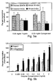
- the PEG-fibrinogen scaffolds of the present invention exhibit material properties and biodegradability which are superior to any known prior art scaffolds; they also exhibit high flexibility and a controllable elastic modulus ( Figures 3a-b ), efficient biodegradability ( Figures 5a-b ), improved biofunctionality and support for cell spreading and extension ( Figures 6a-e, 7a-b , 18a-e and Examples 2 and 4).
- the PEG-fibrinogen scaffolds of the present invention were capable of in vivo bone regeneration in rats exposed to critical size tibia defect.
- a scaffold comprising a plurality of units each composed of a synthetic polymer attached to a naturally occurring protein or a portion thereof.
- the scaffold of the present invention refers to a two-dimensional or a three-dimensional supporting framework.
- the scaffold of the present invention is composed of units (interchangeably referred to herein as "precursor molecules") which are directly or indirectly (e.g., via linker) attachable therebetween.
- precursor molecules can be for example, PEGylated fibrinogen (see Example 1 of the Examples section which follows), PEGylated collagen, PEGylated fibronectin and the like.
- the scaffold of the present invention can form two-or three-dimensional structure at any size, structure or porosity.
- the scaffold of the present invention can be embedded within, or formed around, another scaffold or gel or it can be linked to additional materials to form a hybrid or coated scaffold.
- the scaffold of the present invention can be used to support cell growth, attachment, spreading, and thus facilitate cell growth, tissue regeneration and/or tissue repair.
- polymer refers to a plurality of repeating units which form a new molecular structure.
- synthetic polymer refers to any polymer which is made of a synthetic material, i.e. , a non-natural, non-cellular material.
- Non-limiting examples for synthetic polymers which can be used along with the present invention include polyethylene glycol (PEG) (average Mw. 200; P3015, SIGMA), Hydroxyapatite/polycaprolactone (HA/PLC) [ Choi, D., et al., 2004, Materials Research Bulletin, 39: 417-432 ; Azevedo MC, et al., 2003, J. Mater Sci. Mater. Med.
- polyglycolic acid [ Nakamura T, et al., 2004, Brain Res. 1027(1-2): 18-29 ], Poly-L-lactic acid (PLLA) [ Ma Z, et al., 2005, Biomaterials. 26(11): 1253-9 ], Polymethyl methacrylate (PMMA) [average Mw 93,000, Aldrich Cat. # 370037; Li C, et al., 2004, J. Mater. Sci. Mater. Med. 15(1): 85-9 ], polyhydroxyalkanoate (PHA) [ Zinn M, et al., 2001, Adv. Drug Deliv. Rev. 53(1): 5-21 ; Sudesh K., 2004, Med. J. Malaysia.
- beta-TCP beta-tricalcium phosphate
- PTFE nonbiodegradable polytetrafluoroethylene
- the synthetic polymer used by the present invention is PEG.
- the PEG molecule used by the present invention can be linearized or branched ( i.e ., 2-arm, 4-arm, and 8-arm PEG) and can be of any molecular weight, e.g., 4 kDa, 6 kDa and 20 kDa for linearized or 2-arm PEG, 14 kDa and 20 kDa for 4-arm PEG, and 14 kDa and 20 kDa for 8-arm PEG and combination thereof.
- the OH-termini of the PEG molecule can be reacted with a chemical group such as acrylate (Ac) or vinylsulfone (VS) which turn the PEG molecule into a functionalized PEG, i.e. , PEG-Ac or PEG-VS.
- a chemical group such as acrylate (Ac) or vinylsulfone (VS) which turn the PEG molecule into a functionalized PEG, i.e. , PEG-Ac or PEG-VS.
- the PEG molecule used by the present invention is PEG-Ac.
- PEG-VS can be prepared under argon by reacting a dichloromethane (DCM) solution of the PEG-OH with NaH and then with di-vinylsulfone (molar ratios: OH 1: NaH 5: divinyl sulfone 50, at 0.2 gram PEG/mL DCM).
- DCM dichloromethane
- PEG-Ac is made under argon by reacting a DCM solution of the PEG-OH with acryloyl chloride and triethylamine (molar ratios: OH 1: acryloyl chloride 1.5: triethylamine 2, at 0.2 gram PEG/mL DCM), essentially as described in Example 1 of the Examples section which follows.
- the PEG-Ac used by the present invention is PEG-DA, 4-arm star PEG multi-Acrylate and/or 8-arm star PEG multi-Acrylate.
- naturally occurring protein or a portion thereof refers to denaturated fibrinogen.
- the scaffold of the present invention is composed of a naturally occurring protein such a scaffold can be configured susceptible to degradation by biological material such as enzymes, i.e. , biodegradable.
- biodegradable refers to capable of being degraded (i.e ., broken down) by biological proteases or biomolecules. Biodegradability depends on the availability of degradation substrates ( i.e ., biological materials or portion thereof), the presence of biodegrading materials (e.g., microorganisms, enzymes, proteins) and the availability of oxygen (for aerobic organisms, microorganisms or portions thereof), carbon dioxide (for anaerobic organisms, microorganisms or portions thereof) and/or other nutrients.
- biodegradability of a material, such as the scaffold of the present invention also depends on the material structure and/or mechanical properties, i.e.
- biodegradable materials include, but are not limited to, reconstituted collagen gels, fibrin glues, and hyaluronic acid scaffolds.
- the scaffold of the present invention is fabricated by cross linking multiple copies of a precursor molecule which is composed of a synthetic polymer attached to a naturally occurring protein or a portion thereof.
- the present inventors fabricated a PEGylated fibrinogen precursor molecule by denaturing fibrinogen molecules and reacting them with functionalized PEG-diacrylates.
- the polymer-protein precursor molecule is made of PEG and fibrinogen.
- the molar ratio between the synthetic polymer (e.g., PEG) and the naturally occurring protein (e.g., fibrinogen) of the present invention may affect the pore size, strength, flexibility and elasticity of the scaffold of the present invention.
- excess of the synthetic polymer would lead to binding of the polymer functional groups (e.g., PEG-DA) to all potential binding sites on the naturally occurring protein and would result in smaller pore size, more cross-linking sites, higher strength, less flexibility and increased rigidity.
- binding of only two molecules of the synthetic polymer to each molecule of the protein i.e ., a 2:1 molar ratio
- the molar ratio between the synthetic polymer and the protein can also affect the biodegradability of the scaffold.
- a higher molar ratio i.e ., excess of polymer
- Those of skills in the art are capable of adjusting the molar ratio between the synthetic polymer and the protein to obtain the desired scaffold with the optimal physical and biological characteristics.
- the PEG-fibrinogen precursor molecule can be prepared using a wide range of molar ratios.
- the molar ratio used by the present invention is 2-400 (PEG) to 1 (fibrinogen), more preferably, the molar ratio is 30-300 (PEG) to 1 (fibrinogen), more preferably, the molar ratio is 100-200 (PEG) to 1 (fibrinogen), most preferably, the molar ratio is 130-160 (PEG) to 1 (fibrinogen).
- PEG-fibrinogen the molar ratio used by the present invention is 2-400 (PEG) to 1 (fibrinogen)
- the molar ratio is 30-300 (PEG) to 1 (fibrinogen)
- the molar ratio is 100-200 (PEG) to 1 (fibrinogen)
- the molar ratio is 130-160 (PEG) to 1 (fibrinogen).
- the fibrinogen used by the present invention can be whole fibrinogen (i.e ., un-cleaved) or fragmented fibrinogen, which can be obtained using, for example, CNBr cleavage (see Example 1 of the Examples section which follows).
- the scaffold of the present invention is formed by cross-linking the polymer-protein precursor molecules of the present invention.
- Such cross-linking can be performed in vitro, ex vivo and/or in vivo.
- Cross-linking according to this aspect of the present invention is performed by subjecting the precursor molecules to a free-radical polymerization reaction (i.e. , a cross-linking reaction).
- a free-radical polymerization reaction i.e. , a cross-linking reaction.
- Methods of cross-linking polymers are known in the art, and include for example, cross-linking via photoinitiation (in the presence of an appropriate light, e.g., 365 nm), chemical cross-linking [in the presence of a free-radical donor] and/or heating [at the appropriate temperatures.
- cross-linking according to the present invention is effected by photoinitiation.
- Photoinitiation can take place using a photoinitiation agent (i.e ., photoinitiator) such as bis(2,4,6-trimethylbenzoyl) phenylphosphine oxide (BAPO) ( Fisher JP et al., 2001; J. Biomater. Sci. Polym. Ed. 12: 673-87 ), 2,2-dimethoxy-2-phenylacetophenone (DMPA) ( Witte RP et al., 2004; J. Biomed. Mater. Res. 71A(3): 508-18 ), camphorquinone (CQ), 1-phenyl-1,2-propanedione (PPD) ( Park YJ et al., 1999, Dent. Mater.
- a photoinitiation agent i.e ., photoinitiator
- BAPO bis(2,4,6-trimethylbenzoyl) phenylphosphine oxide
- DMPA 2,2-dimethoxy-2-phenylacetophenone
- CQ camphorquinone
- DMAEMA dimethylaminoethyl methacrylate
- the photoinitiation reaction can be performed using a variety of wave-lengths including UV (190-365 nm) wavelengths, and visible light (400-1100 nm) and at various light intensities. It will be appreciated that for ex vivo or in vivo applications, the photoinitiator and wavelengths used are preferably non-toxic and/or non-hazardous.
- the PEG-fibrinogen precursor molecule was cross-linked by photoinitiation in the presence of Igracure TM 2959 and a non-toxic UV light illumination (e.g., 5 minutes at 365 nm wavelength, 4-5 mWatts/cm 2 intensity).
- a non-toxic UV light illumination e.g., 5 minutes at 365 nm wavelength, 4-5 mWatts/cm 2 intensity.
- polymer-protein precursor molecules of the present invention e.g., PEGylated fibrinogen
- cross-linking according to the present invention can also utilize a molecule capable of cross-linking the polymer-protein precursors.
- Such cross-linking molecules can be for examples, PEG, PEG-DA, PEG multi-Acrylate, and/or PEG-VS.
- a functionalized PEG molecule e.g:, PEG-DA
- PEG-DA a functionalized PEG molecule
- the concentration of cross-linking molecules can affect the scaffold strength, flexibility, elasticity and biodegradability and determination of such a concentration depends on the scaffold application and is within the capabilities of those skilled in the arts. For example, excess of a cross-linking molecule is expected to result in smaller pores, more cross-linking sites, and higher scaffold strength and less flexibility.
- excess of PEG-DA i.e ., the cross-linking molecule of the present invention
- reduced scaffold biodegradability is probably a result of masking or modifying the protein binding sites or signals which are necessary for protein degradation.
- cross-linking is effected such that the polymer-protein precursors of the present invention are solubilized in a water-based solution and such solutions are further subjected to cross-linking (e.g., using photoinitiation) to form a hydrogel scaffold.
- a PEG-fibrinogen hydrogel was formed by mixing the PEGylated fibrinogen precursor molecules with the photoinitiation agent (IgracureTM2959) in the presence or absence of PEG-DA and exposing such a mixture to UV light.
- the PEGylated fibrinogen precursors were solubilized in 1-ml of 50 mM PBS, pH 7.4 and 25 °C to achieve a final concentration of 10, 15, or 20 % polymer-protein (w/v).
- the precursor solution also contained a PEG-DA cross-linking constituent at a molar ratio of 1:2 PEG-DA to functional groups on the PEGylated fibrinogen.
- the precursor solution was mixed with 10 ⁇ l of IgracureTM2959 photoinitiator solution (Ciba Specialty Chemicals, Tarrytown, New York) in 70 % ethanol (100 mg/ml) and centrifuged for 5 min at 14,000 RPM. The solution was then placed into Teflon tubes (5-min diameter and 20-mm long) and polymerized under UV light (365 nm, 4-5 mW/cm 2 ) for 15 minutes according to published protocols (Lum LY et al., 2003).
- the hydrogel can be generated from PEGylated whole fibrinogen or PEGylated fragmented fibrinogen.
- the molecular weight and length of the grafted PEG affects the degree of solubility of the PEGylated protein, i.e., higher length and/or molecular weight of PEG results in increased solubility of PEGylated protein. It will be appreciated that solubility of the PEGylated protein is also affected by the presence of whole or cleaved fibrinogen.
- the concentration of the precursor molecules in the hydrogel is between 0.5 to 35 %, more preferably, when PEGylated whole fibrinogen is used, the concentration of the precursor molecules in the hydrogel is between 0.5 to 5 % (depending on the MW and length of the grafted PEG used to PEGylate the protein) and when PEGylated fragmented fibrinogen is used, the concentration of the precursor molecules in the hydrogel is between 5 - 35 % (depending on the MW and length of PEG used to PEGylate the protein).
- the PEG-fibrinogen hydrogels of the present invention exhibited a superior flexibility over the prior art PEG-PEG hydrogels.
- a strain of 30 % was achieved by employing a stress of 90 kPa
- a similar strain was achieved by employing only 4 kPa ( Figures 3a-b and Figures 4a-c ).
- the modulus of elasticity of the hydrogels made from PEGylated whole fibrinogen is in a range of 0.02-0.11 kPa for 10-20 % polymer
- the modulus of elasticity of the hydrogels made from PEGylated fragmented fibrinogen is in a range of 0.01-0.07 kPa for 10-20 % polymer.
- hydrogel scaffolds of the present invention exhibit high biodegradability as compared with prior art hydrogel scaffolds [e.g., hydrogels made with an oligopeptide cross-linker containing a protease substrate (Seliktar et al 2004)].
- hydrogel scaffolds made with an oligopeptide cross-linker containing a protease substrate (Seliktar et al 2004)
- a significant degradation of 45 - 70 % or 35 - 85 % of the PEG-fibrinogen hydrogels was observed following 30 minutes incubation in the presence of 0.05 mg/ml trypsin or 0.5 mg/ml collagenase, respectively.
- higher concentrations of trypsin i.e ., 1-2 mg/ml
- the biodegradability of the hydrogel scaffold of the present invention can be determined by subjecting such hydrogels to enzymatic degradation using proteases such as plasmin, trypsin, collagenase, chemotrypsin and the like.
- the biodegradability and biofunctionality of the scaffold hydrogel of the present invention can be further increased by attaching or impregnating a protein such as a cell signaling protein, or a (as described hereinabove) to the hydrogel of the present invention.
- a protein such as a cell signaling protein, or a (as described hereinabove)
- Attaching such proteins to the hydrogel scaffold of the present invention is preferably employed by covalent immobilization of a PEGylated protein to the PEG hydrogel network during cross-linking (Seliktar et al 2004, JBMR).
- the immobilization of such factor is accomplished by directly reacting functionalized PEG to an unreacted thiol present on a cysteine residue of the protein sequence.
- Impregnation of the hydrogel with growth factors can be performed by dehydrating the scaffold and then immersing the hydrogels in a solution containing the growth factors and gently shaking such hydrogels for a few hours until the growth factors penetrate the scaffold during the hydration process.
- the hydrogel can be impregnated with growth factor by incubation in factor-containing solution overnight until the growth factor diffuses into the polymeric network of the scaffold by slow, passive diffusion. The latter is influenced by the degree of cross-linking, the porosity of the scaffold, and the structural properties described hereinabove.
- the scaffold of the present invention is highly reproducible, flexible (can be stressed or stretched easily), exhibit a controllable structural properties, and amenable to controllable biodegradation; characteristics which make it highly suitable for in vivo or ex vivo regeneration of tissues such as bone, cartilage, heart muscle, skin tissue, blood vessels, and other tissues (soft and hard) in the body.
- tissue such as bone, cartilage, heart muscle, skin tissue, blood vessels, and other tissues (soft and hard) in the body.
- a scaffold hydrogel can be easily placed into gaps within a tissue or an organ, following which it can fill the void and initiate the process of regeneration as the scaffold degrades away.
- in vivo refers to within a living organism such as a plant or an animal, preferably in mammals, preferably, in human subjects.
- the method is effected by implanting the scaffold of the present invention in a subject to thereby induce the formation of the tissue.
- the term "subject” refers to a vertebrate, preferably a mammal, more preferably a human being (male or female) at any age.
- the scaffold of the present invention can be implanted in the subject using a surgical tool such as a scalpel, spoon, spatula, or other surgical device.
- a surgical tool such as a scalpel, spoon, spatula, or other surgical device.
- in vivo formation of a tissue can be also achieved by administering the scaffold precursor molecules to the subject and further cross-linking the precursor molecules in vivo.
- a method of inducing in vivo formation of a tissue is effected by administering to a subject in need thereof a composition composed of a synthetic polymer attached to a naturally occurring protein or a portion thereof, the composition capable of forming a scaffold within the subject and thereby inducing the formation of the tissue in vivo.
- composition composed of a synthetic polymer attached to a naturally occurring protein or a portion thereof refers to the polymer-protein precursor molecule of the present invention which is described hereinabove.
- the method according to this aspect further comprising a step of cross-linking following administering the composition.
- Cross-linking can be performed as described hereinabove using non-toxic, non-hazardous agents and/or conditions.
- the method according to this aspect further comprising administering to the subject a molecule capable of cross-linking the composition.
- molecule capable of cross-linking the composition refers to the cross-linking agent described hereinabove (e.g., PEG-DA).
- the scaffold of the present invention can be also used for ex vivo formation of a tissue.
- tissue refers to part of an organism consisting of an aggregate of cells having a similar structure and function. Examples include, but are not limited to, brain tissue, retina, skin tissue, hepatic tissue, pancreatic tissue, bone, cartilage, connective tissue, blood tissue, muscle tissue, cardiac tissue brain tissue, vascular tissue, renal tissue, pulmunary tissue, gonadal tissue, hematopoietic tissue and fat tissue.
- tissue as used herein also encompasses the phrase "organ” which refers to a fully differentiated structural and functional unit in an animal that is specialized for some particular function. Non-limiting examples of organs include head, brain, eye, leg, hand, heart, liver kidney, lung, pancreas, ovary, testis, and stomach.
- ex vivo refers to living cells which are derived from an organism and are growing (or cultured) outside of the living organism, preferably, outside the body of a vertebrate, a mammal, or human being.
- cells which are derived from a human being such as human muscle cells or human aortic endothelial cells and are cultured outside of the body are referred to as cells which are cultured ex vivo.
- the scaffold of the present invention which is formed in vitro, ex vivo or in vivo can be used to induce tissue formation and/or regeneration and thus treat individuals suffering from tissue damage or loss.
- tissue damage or loss refers to any disorder, disease or condition exhibiting a tissue damage (i.e ., non-functioning tissue, cancerous or pre-cancerous tissue, broken tissue, fractured tissue, fibrotic tissue, or ischemic tissue) or a tissue loss (e.g., following a trauma, an infectious disease, a genetic disease, and the like) which require tissue regeneration.
- tissue damage i.e ., non-functioning tissue, cancerous or pre-cancerous tissue, broken tissue, fractured tissue, fibrotic tissue, or ischemic tissue
- tissue loss e.g., following a trauma, an infectious disease, a genetic disease, and the like
- liver cirrhosis such as in hepatitis C patients (liver), Type-1 diabetes (pancreas), cystic fibrosis (lung, liver, pancreas), bone cancer (bone), burn and wound repair (skin), age related macular degeneration (retina), myocardial infarction, myocardial repair, CNS lesions (myelin), articular cartilage defects (chondrocytes), bladder degeneration, intestinal degeneration, and the like.
- treating refers to inhibiting or arresting the development of a disease, disorder or condition and/or causing the reduction, remission, or regression of a disease, disorder or condition in an individual suffering from, or diagnosed with, the disease, disorder or condition.
- Those of skill in the art will be aware of various methodologies and assays which can be used to assess the development of a disease, disorder or condition, and similarly, various methodologies and assays which can be used to assess the reduction, remission or regression of a disease, disorder or condition.
- the method is effected by implanting the scaffold of the present invention alone or following seeding such a scaffold with cells, or by administering the scaffold units (i.e ., the polymer-protein precursor molecules of the present invention) into the subject to thereby induce formation of the tissue and treat the disorder characterized by tissue damage or loss.
- the scaffold units i.e ., the polymer-protein precursor molecules of the present invention
- Tissue engineering scaffolds with controllable mechanical properties and adequate biofunctional signals were generated from PEG and fibrinogen. Briefly, denatured fibrinogen fragments were PEGylated with PEG-diacrylates, mixed with photoinitiator and exposed to UV light to form a hydrogel material in the presence of a cell suspension. The degradability of the PEG-fibrinogen scaffold was further tested by enzyme-mediated proteolysis, as follows.
- PEG-DA PEG Diacrylate
- PEG-DA PEG-diacrylate
- acrylation of PEG-OH was carried out under Argon by reacting a dichloromethane (DCM) (Aldrich, Sleeze, Germany) solution of the PEG-OH with acryloyl chloride (Merck, Darmstadt, Germany) and triethylamine (Fluka) at a molar ratio of 1-OH to 1.5-acryloyl chloride to 1.5-triethylamine (0.2 g PEG/ml DCM).
- DCM dichloromethane
- Fluka triethylamine
- Cyanogen bromide cleavage of fibrinogen - Whole fibrinogen [Sigma-Aldrich, Steinheim, Germany, Cat # F8630, GenBank Accession No. AAC67562.1. ( ⁇ -chain; SEQ ID NO:1); GenBank Accession No. CAA23444.1 ( ⁇ -chain; SEQ ID NO:2), and GenBank Accession No. CAA33562.1 ( ⁇ -chain; SEQ ID NO:3)] was dissolved in a solution of 70 % formic acid containing 17 mg/ml Cyanogen Bromide (CNBr) (Aldrich, Cat. # C9, 149-2) and incubated overnight in the dark at 25 °C.
- CBr Cyanogen Bromide
- the cleaved fibrinogen fragments were dialyzed for 2 days at 4 °C in 50 mM phosphate buffered saline (PBS) at pH 7.4 with a twice-daily change of buffer to remove all the CNBr and formic acid from the solution.
- the dialyzed fragments were stored in PBS at 4 °C until they were subjected to PEGylation.
- the molar ratio of PEG to fibrinogen was 145:1 (linear PEG-DA, MW 4-kDa, 6-kDa, and 20-kDa).
- the final PEGylated protein product was precipitated for 20 minutes at room temperature while stirring in 5X excess acetone (Frutarom, Haifa, Israel).
- the precipitated protein solution was centrifuged for 20 minutes at 5000 RPM (Sorvall GSA rotor) and the pellet was redissolved at 20 mg/ml protein concentration in PBS containing 8 M urea.
- the PEGylated protein solution was then dialyzed for 2 days at 4 °C against PBS containing 0.1 % (v/v) glacial acetic acid (Frutarom) with twice-daily changes of PBS (Spectrum, 12-14-kDa MW cutoff).
- the dialyzed product was either used immediately or lyophilized in a solution of 10 % D-(+)-glucose (Riedel-deHa ⁇ n, Germany) to improve solubility upon redissolution.
- the lyophilized PEGylated product was stored under Argon at -80 °C for up to six months.
- the PEG-fibrinogen hydrogels were made from a precursor solution of PEGylated fibrinogen (whole or cleaved).
- the precursor solution was made by solubilizing PEGylated fibrinogen in 1-ml of 50 mM PBS, pH 7.4 and 25 °C to achieve a final concentration of 10, 15, or 20 % polymer (w/v).
- the precursor solution also contained a PEG-DA cross-linking constituent at a molar ratio of 1:2 PEG-DA to functional groups on the PEGylated fibrinogen.
- the additional PEG-DA was used to efficiently cross-link the PEGylated protein macromeres and minimize steric hindrances that result in poor gelation.
- the precursor solution was mixed with 10 ⁇ l of IgracureTM2959 photoinitiator solution (Ciba Specialty Chemicals, Tarrytown, New York) in 70 % ethanol (100 mg/ml) and centrifuged for 5 min at 14,000 RPM. The solution was then placed into Teflon tubes (5-mm diameter and 20-mm long) and polymerized under UV light (365 nm, 4-5 mW/cm 2 ) for 15 minutes according to published protocols (Lum LY et al., 2003).
- hydrogels were cut into 5-mm long sections for mechanical testing. Control hydrogels were made by solubilizing 10, 15, or 20 % (w/v) PEG-DA in 1-ml of 50 mM PBS with IgracureTM and then polymerizing under UV light as described above.
- Biodegradation assay - To assess the rate of enzymatic hydrogel degradation, Coomassie® brilliant blue G-250 dye (Aldrich) was bound to the PEG-fibrinogen hydrogels and the release of the Coomassie® dye was measured spectrophotometrically. Hydrogels were stained in 0.1 % Coomassie® (w/v) overnight with gentle agitation, and destained for 1 hour in destaining buffer. Since the Coomassie® dye binds to proteins with very high affinity, following the initial staining, the hydrogels were destained to release all unbound dye.
- PEGylation of fibrinogen can be performed using 4-kDa, 6-kDa and / or 20-kDa PEG DA -
- the PEG-fibrinogen hydrogels were prepared by covalently binding protein fragments to PEG-DA and cross-linking by UV photoinitiation.
- Figure 1a illustrates the potential of the fibrinogen constituent to be linked to fractionalized PEG.
- a Michael-type addition reaction Lutolf M et al, 2001, Bioconjugate chem.
- the PEG-fibrinogen product exhibits high percentage of PEGylated fibrinogen - Following an overnight PEGylation reaction the PEGylated fibrinogen product is purified using acetone precipitation which selectively precipitates the protein (i.e. , PEGylated fibrinogen) from the excess of unreacted PEG-DA. It is worth mentioning that the addition of acetone at room temperature to the PEG-DA reaction [using an iodide solution as described elsewhere (24)] did not result in the precipitation of free, unreacted PEG-DA (data not shown).
- the dry weight of the total PEGylated product was compared with the amount of total protein as measured using a Pierce BCA assay in a purified solution of PEGylated fibrinogen.
- the dry weight of PEGylated protein should be the sum of the weights of the fibrinogen and the grafted PEG, assuming 100 % PEGylation.
- the theoretical protein fractions for PEGylated fibrinogen using 4-kDa, 6-kDa, and 20-kDa PEG are 59 %, 49 %, and 22 %, respectively.
- the results indicate a protein fraction of 45 ⁇ 5.0 % using the 4-kDa PEG, 39 ⁇ 3.4 % using the 6-kDa PEG, and 36 ⁇ 1.5 % using the 20-kDa PEG.
- the difference between the theoretical and the measured protein factions can be attributed to several factors, including excess of unreacted PEG, partial PEGylation, and/or error in the BCA measurements.
- Cyanogen bromide cleavage of fibrinogen facilitates the preparation of PEGylated fibrinogen solutions - Since the combination of the highly hydrophilic PEG to the partially hydrophobic fibrinogen can result in a highly hydrophobic protein core, the fibrinogen molecule was denatured prior to being subjected to the PEGylation reaction. Such denaturation minimizes the formation of the hydrophobic core after PEGylation and significantly improves the solubility of the product. Denaturation of the fibrinogen molecule was accomplished using CNBr, a proteolytic molecule which chemically cleaves adjacent to methionine peptides in the fibrinogen sequence.
- PEGylated fibrinogen solutions with concentrations of up to 300 mg/ml were prepared under non-denaturing conditions.
- Table 1 Fibrinogen fragments and cysteines from whole fibrinogen Fragments Alpha beta gamma Total M.W. 65 53.3 47.6 165.9 Cysteines 8 11 10 29
- Table 2 Fibrinogen fragments and cysteines from multi thiol fragments of cleaved fibrinogen Fragments F1 F2 F3 F4 F7 F8 F15 F16 Total M.W. 32.4 24.6 12.1 10 7 6.3 3.8 3.8 100 Cysteines 2 4 3 4 2 2 4 4 25
- PEG-fibrinogen hydrogels are highly elastic -
- the compressive mechanical properties of the material were measured using the InstronTM single column material testing system. These measurements confirmed the formation of acellular PEG-fibrinogen hydrogels (and PEG-PEG controls) with varying amounts of polymer (10 %, 15 %, and 20 % w/v) and different molecular weights of PEG (4-kDa and 6-kDa, 20-kDa) as well as the formation of hydrogels which are made of either whole fibrinogen (whole) or CNBr-cleaved fibrinogen (cleaved).
- the elastic modulus (as determined from the stress-strain curve) was found to be dependent on the percent polymer, the molecular weight of the PEG precursor, and the fibrinogen backbone ( Figures 4a-c ).
- Biodegradation of PEG-fibrinogen hydrogels is dependent on the MW of the PEG component and the protein backbone - Hydrogel biodegradation was quantified by subjecting colorimetrically-labeled pure PEG-fibrinogen hydrogels (15 % w/v PEG-DA, MW 6-kDa) to varying concentrations of proteases (e.g., Collagenase or trypsin) and assessing the dissolution of the gels.
- proteases e.g., Collagenase or trypsin
- hydrogel scaffolds for tissue engineering (16, 23, 26-30). These materials are capable of promoting cell growth and exhibit proteolytic degradability via their biological domains while still providing exacting mechanical properties based on their synthetic composition.
- a number of these hybrid hydrogel materials are currently in use as ingrowth matrices or cell culture substrates which take advantage of biologically active oligopeptides in the material backbone that mimic the properties of natural tissue (17, 18). While these materials satisfy the general criteria of biofunctionality, the small oligopeptides provide only a fraction of the bioactive signals present in the natural extracellular matrix (ECM).
- ECM extracellular matrix
- the hydrogel material of the present invention contains a natural protein backbone onto which di-functional PEGs are covalently bound and cross-linked together using photo-polymerization.
- the protein backbone is comprised of alpha, beta, and gamma fragments of denatured fibrinogen. These fibrinogen fragments are inherently bioactive with proteolytically sensitive sequences, cell adhesion motifs, and other cell-signaling sequences (14). Free thiol groups present in unpaired cysteine residues in the denatured fibrinogen fragment are covalently conjugated by a Michael-type addition reaction to an unsaturated double bond on functionalized PEG-DA.
- the hydrogel material of the present invention uses denatured fibrinogen since it consists of a large number of free thiol groups (i.e ., unpaired cysteine residues) that can react with PEG-DA. After PEGylation, unreacted acrylates on the di-functional PEGs are used to cross-link the fibrinogen backbone into a hydrogel network using photo-polymerization.
- the molecular structure of the hydrogels is highly influenced by the degree of cross-linking.
- the other crucial determining factor of the hydrogel cross-linking is the ratio of reactive acrylate end-groups on the PEG per MW of protein (acrylates per kDa of PEGylated protein).
- PEGylated fibrinogen contains 29 acrylate groups for each molecule of protein, or approximately 0.1 acrylate/kDa for 4-kDa PEGylated fibrinogen, 0.086 acrylate/kDa for 6-kDa PEGylated fibrinogen, and 0.039 acrylate/kDa for 20-kDa PEGylated fibrinogen.
- a pure PEG-DA solution contains 0.5 acrylates/kDa for 4-kDa PEG, 0.33 acrylates/kDa for 6-kDa PEG, and 0.1 acrylates/kDa for 20-kDa PEG. If the ratio of acrylates to MW is too low, the polymer solution will not form a continuous hydrogel. For this reason, the experiments performed in the present study utilized high concentrations of PEGylated fibrinogen ( ⁇ 10 %).
- the PEGylated fibrinogen requires substantial effort to solubilize at these high concentrations. Since the solubility of the PEGylated fibrinogen is highly affected by the grafting of the PEG chains; presumably because of the formation of a hydrophobic protein complex in the presence of a highly hydrophilic PEG graft, the fibrinogen protein used by the present invention utilized an irreversibly cleaved by CNBr. Thus, PEGylation of cleaved fibrinogen results in a highly soluble protein precursor for hydrogel formation.
- the mechanical properties of the cleaved and whole PEG-fibrinogen hydrogels was characterized using the stress-strain behavior under quasi-static uniaxial unconfined compression.
- the PEG-fibrinogen material demonstrates the typical non-linear stress-strain characteristics of a polymeric material, similar to the PEG-PEG control gels.
- the composite behaves like a viscoelastic solid with minimal hysteresis under repetitive cyclic loading (data not shown).
- the material stiffness, as determined from the modulus of elasticity, is directly proportional to the percent polymer composition. It will be appreciated that the material stiffness of polymeric hydrogels can be directly attributed to the degree of cross-linking.
- the amount of functional groups available for cross-linking is proportional to the polymer concentration.
- the modulus of the material is directly proportional to the amount polymer in the hydrogel.
- the relationship between the elastic modulus of the hydrogel and the molecular chain length of the grafted PEG constituent is ambiguous. While pure PEG hydrogels exhibit a proportional relationship between the material modulus and the MW of the PEG, the addition of the protein into the hydrogel network can have a profound impact on this relationship, depending on the relative size of the two polymers. In the case of whole PEGylated fibrinogen hydrogels, where the PEG is significantly smaller than the protein, the impact of PEG MW on the modulus is more pronounced. In contrast, the relationship between PEG MW and the modulus of the hydrogels is less pronounced when the hydrogels are made of cleaved protein. In either case, it is difficult to resolve the difference between 4-kDa PEG and 6-kDa PEG when comparing the elastic moduli.
- the mechanical properties data demonstrate a significant difference in the stiffness of hydrogels made from pure PEGylated fibrinogen versus pure PEG hydrogels. Irrespective of the PEG MW, the PEGylated fibrinogen hydrogels are always less stiff than the pure PEG hydrogels. This can be attributed to the fact that pure PEG hydrogels (made with similar weight percent of polymer) contain nearly 5 times more functional groups available for cross-linking than the PEGylated hydrogels.
- cross-linking sites arises from the fact that the fibrinogen constituent accounts for more than half the weight of the polymer but does not contain inherent cross-linking sites, and each grafted PEG on the PEGylated fibrinogen only has a single functional group available for cross-linking, in contrast to two functional groups on free PEG-DA. Additional cross-linking limitations may arise because of steric hindrances caused by the bulky PEGylated fibrinogen molecules.
- the molecular chain length and percent composition represent two independent parameters to control the mechanical properties of the material.
- An additional parameter which can alter the mechanical properties of the material is the number of cross-linking sites on each grafted PEG molecule.
- the linear grafted PEG contains only one functional group for cross-linking the hydrogel; however, PEG molecules containing several functional groups such as star-PEGs can also be grafted onto the fibrinogen to form hydrogels. While star-PEG was not used as part of the current investigation, future studies aim to increase the stiffness of the PEG-fibrinogen hydrogels using 4-arm and 8-arm star-PEG precursors.
- the fibrinogen backbone of the hydrogel material provides proteolytic sensitivity via naturally occurring substrates for fibrinolysis.
- Fibrinolysis is a physiological process whereby the native fibrin molecule is proteolytically dismantled by serine proteases.
- the fibrinogen backbone is cleaved in the presence of activated proteases, resulting in complete dissolution of the PEG-fibrinogen hydrogel.
- the results shown in the present study demonstrate that pure PEG-fibrinogen hydrogels are proteolytically degradable while the PEG-PEG controls are not susceptible to proteolysis (data not shown).
- the degradation data reveals several other interesting patterns regarding the proteolytic degradation of cleaved and whole fibrinogen hydrogels in the presence of trypsin or Collagenase after 30 minutes.
- trypsin the cleaved and whole fibrinogen hydrogels degrade almost identically. This is likely explained by the observation that 0.05 mg/ml trypsin is saturated with substrate whereas 0.5 mg/ml collagenase is not saturated with substrate.
- the MW of the PEG constituent also affects the degradation results.
- the 20-kDa PEG hydrogels are comprised of less fibrinogen which results in faster hydrogel dissolution. This can also be explained by the observation that 0.05 mg/ml trypsin is saturated with enzyme and cannot degrade the 20-kDa PEGylated fibrinogen hydrogels faster. Future studies will examine more aspects of proteolytic degradability of the PEG-fibrinogen material, including degradation kinetics and fibrinolysis in the presence of plasmin.
- Bovine aortic smooth muscle cells from young donors were isolated and cultured according to a modified protocol of Oakes et al., 1982.
- the BSMCs were cultured up to 6 th passage in Dulbecco's Modified Eagle Medium (DMEM) (Gibco, U.K.) containing 10 % fetal bovine serum (FBS) (Biological Industries, Israel), 1 % penicillin-streptomycin (Biological Industries), and 1 % L-glutamine (Gibco).
- DMEM Dulbecco's Modified Eagle Medium
- PEG hydrogels containing BSMCs were made by mixing a PBS cell suspension and PEGylated fibrinogen precursor solution containing IgracureTM photoinitiator to make a 10 % (w/v) solution with 1.5 x 10 6 cells/ml. Aliquots of 100 ⁇ l of the suspension were added into wells in a flat-bottom 96-well plate and placed under UV-light (4-5 mW/cm 2 ) for 5 min in a laminar flow hood. DMEM culture medium (containing 10 % FBS) was added immediately to the polymerized hydrogels and changed daily (100- ⁇ l/well).
- BAECs bovine aortic endothelial cells
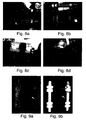
- PEG-fibrinogen hydrogels support cell spreading and attachment -
- endothelial (BAECs) or smooth muscle (BSMCs) cells were cultured on the surface of or inside the PEG hydrogels.
- BAECs were seeded on the surface of the hydrogels at a concentration of 30,000 cells/cm 2 and 24 hours following cell seeding the degree of cell attachment and spreading was evaluated using phase contrast microscopy.
- the hydrogel network contained homogeneously distributed BSMCs with round morphology.
- BSMCs cultured inside the PEG-fibrinogen hydrogels formed stable adhesions, processes, and cellular extensions ( Figures 6b-c ).
- PEGylated fibrinogen hydrogels made with cleaved fibrinogen also supported the adhesion and extension of BSMCs inside PEGylated hydrogels (data not shown).
- BSMCs cultured inside PEG-PEG hydrogels were round and devoid of any visible cell extensions ( Figure 6e ).
- the PEG hydrogel scaffold without the fibrinogen backbone is completely devoid of biofunctional domains for cell culture.
- the fibrinogen backbone provides at least two biofunctional characteristics to the hydrogel material: proteolytic sensitivity and cell adhesivity. With regards to the latter, the PEG-fibrinogen hydrogels support the attachment and spreading of endothelial cells in the presence of serum proteins whereas PEG-PEG controls are not able to support cell attachment. Endothelial cell-surface adhesion molecules can therefore attach either directly to adhesion domains on the fibrinogen backbone or to other serum proteins that interact non-specifically with the fibrinogen.
- cells are capable of proteolytically tunneling through the hydrogel network with the help of cell-secreted enzymes such as Collagenase.
- cell-secreted enzymes such as Collagenase.
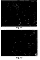
- smooth muscle cells that are three-dimensionally entrapped inside the hydrogel material after photopolymerization and begin to form clusters of cells after 24 hours in culture.
- Phase contrast micrographs and histological cross-sections confirm that BSMCs form flagella-like extensions which enable their migration inside the hydrogel network.
- BSMCs remained round and homogeneously dispersed and cell extensions are not observed in the non-degradable PEG-PEG controls.
- the biological domains in the fibrinogen backbone provide attachment motifs for endothelial cell and smooth muscle cell adhesion as well as proteolytic sensitivity for biodegradation.
- Smooth muscle cells demonstrate the ability to proteolytically penetrate through the hydrogel material and form interconnecting networks of cells.
- the scaffolds of the present invention are novel, biodegradable and highly suitable for cultivating cells in a 3-D environment for tissue regeneration therapies.
- Two needles were drilled into the proximal tibia (21G) and distal tibia (23G) and connected to two external fixators (screws) to form a stable fixation of the bone.
- a 10-mm gap was excised using a disk saw in the portion between the proximal and distal needles of the fixators ( Figure 8c ). The fibula was not osteotomized.
- PEG-fibrinogen (Gelrin TM ) scaffold -
- a PEG-fibrinogen plug (5-mm diameter and 10-mm long) was inserted into the defect site and the surrounding connective tissue was wrapped around to secure the plug into place ( Figure 8d ).
- the incisional wound was sutured using nylon surgical thread.
- the animal was given prophylactic antibiotics (ampicilline 0.1 gram/100 gram).
- the animal was x-rayed and further evaluated weekly by x-ray screening. The animal was free to move about the cage during the entire post-operative follow-up period.
- the animal was sacrificed with CO 2 and the right tibia was harvested for histology and mechanical testing.
- a critical size rat tibia defect is known to result in up to 20 % mortality.
- a critical rat tibia defect was introduced into 25 rats ( Figures 8a-d ), of them 17 rats were further subjected to scaffold implantation using the PEG-fibrinogen or PEG-PEG hydrogels (see Table 3, hereinbelow, for representative rats).
- Four to seven weeks following subjecting rats to critical size tibia defects the rats were sacrificed and the tibias were evaluated for the presence, extent and location of tissue ingrowth and new bone formation.
- Rats were implanted with either PEG-PEG (PEG-only) or PEG-fibrinogen hydrogels.
- the PEG-fibrinogen hydrogels were prepared from PEG-fibrinogen precursors and PEG-DA crosslinkers.
- the PEG-fibrinogen precursors were prepared from either whole fibrinogen (Gelrin TM ) or from CNBr-cleaved fibrinogen.
- Figures 10-17 demonstrate the presence of normal bone and cartilage tissue five weeks following introduction of a critical size tibia defect and Gelrin TM -implantation.
- tibias of Gelrin TM -treated rats exhibited well-vascularized, orientated and densely textured fibrous tissue ( Figure 13 ), osteonal healing ( Figure 11 ), and Haversian systems with small central canals containing blood vessels ( Figure 10 ).
- BIODEGRADABILITY OF PEG-FIBRINOGEN HYDROGELS IS CONTROLLABLE VL4 THE SYNTHETIC PEG CONSTITUENT
- Biodegradability of PEG-fibrinogen hydrogels is reduced by the addition of free PEG-DA to the hydrogels -
- concentration of the cross-linking molecule of the present invention i.e., PEG-DA (free PEG)
- PEG-DA free PEG
- 1x10 6 smooth muscle cells per ml were seeded inside Gelrin TM hydrogels (1.75 % PEG-fibrinogen, 10-kDa PEG) containing various concentrations of PEG-DA.
- the seeded hydrogels were cultured for 48 hours, following which the ability of the cells to attach and spread inside the hydrogels was evaluated using an inverted microscope.
- Gelrin TM hydrogels consisting of more than 1 % of a cross-linking molecule (PEG-DA) -
- PEG-DA cross-linking molecule
- Gelrin TM matrices (1.75 % PEG-fibrinogen, 10-kDa PEG) were prepared as pure hydrogels (in the absence of free PEG-DA cross-linker) or in the presence of 1 or 2 % of free PEG-DA and cell clusters composed of highly compacted smooth muscle cells inside a collagen gel matrix (5 x 10 6 cells in one mg collagen cluster).
- the clusters were seeded in the center of each hydrogel by placing the cell mass into the Gelrin TM matrix prior to polymerization and forming the scaffold around the tissue so that it encapsulates the tissue mass from all sides.
- the degree of cell extension was monitored following 1, 2, 4, and 7 days in culture using phase microscopy. As is shown in Figures 19a-1 , significant cell extensions were observed in pure Gelrin TM hydrogels beginning following one day in culture. In contrast, cell extensions were relatively decreased in smooth muscle cell clusters cultured inside Gelrin TM hydrogels which were fabricated in the presence of 2 % free PEG-DA.
Abstract
Description
- The present invention relates to a matrix composed of a naturally-occurring protein backbone cross-linked by polyethylene glycol (PEG) and, more particularly, to methods of generating and using same in tissue regeneration.
- Tissue engineering, i.e., the generation of new living tissues in vitro, is widely used to replace diseased, traumatized or other unhealthy tissues. The classic tissue engineering approach utilizes living cells and a basic scaffold for cell culture (Langer and Vacanti, 1993; Nerem and Seliktar, 2001). Thus, the scaffold structure attempts to mimic the natural structure of the tissue it is replacing and to provide a temporary functional support for the cells (Griffith LG, 2002).
- Tissue engineering scaffolds are fabricated from either biological materials or synthetic polymers. Synthetic polymers such as polyethylene glycol (PEG), Hydroxyapatite/polycaprolactone (HA/PLC), polyglycolic acid (PGA), Poly-L-lactic acid (PLLA), Polymethyl methacrylate (PMMA), polyhydroxyalkanoate (PHA), poly-4-hydroxybutyrate (P4HB), polypropylene fumarate (PPF), polyethylene glycol-dimethacrylate (PEG-DMA), beta-tricalcium phosphate (beta-TCP) and nonbiodegradable polytetrafluoroethylene (PTFE) provide precise control over the mechanical properties of the material (Drury and Mooney, 2003).
- Common scaffold fabrication methods are based on foams of synthetic polymers. However, cell migration into the depth of synthetic scaffolds is limited by the lack of oxygen and nutrient supply. To overcome such limitations, new approaches utilizing solid freeform fabrications and internal vascular architecture have been developed (Reviewed in Sachlos E and Czernuszka JT, 2003; Eur. Cell Mater. 5: 29-39). Likewise, freeze-drying methods are also employed to create unique three-dimensional architectures with distinct porosity and permeability. However, creating pores into these materials is an aggressive procedure involving the use of toxic reagents which eliminate the possibility of pre-casting tissue constructs with living cells. Therefore, many of the prefabricated materials are subject to uneven cell seeding and non-homogeneous populations of cells within the constructs. Furthermore, the materials are generally degraded unevenly during the tissue cultivation process, creating a highly anisotropic tissue with altered growth kinetics,
-
U.S. Patent No. 5,863,984 discloses porous matrices formed from a biopolymer such as collagen, which are conjugated to PEG by chemical means or by irradiation. The conjugation to PEG reduces the biodegradability of the matrix, thereby preserving the porous structure thereof. - Scaffolds made of PEG are highly biocompatible (Merrill and Salzman, 1983) and exhibit versatile physical characteristics based on their weight percent, molecular chain length, and cross-linking density (Temenoff JS et al., 2002). In addition, PEG hydrogels are capable of a controlled liquid-to-solid transition (gelation) in the presence of cell suspension (Elbert and Hubbell, 2001). Moreover, the PEG gelation (i.e., PEGylation) reaction can be carried out under non-toxic conditions in the presence of a photoinitiator (Elisseeff J et al., 2000; Nguyen and West, 2002) or by mixing a two-part reactive solution of functionalized PEG and cross-linking constituents (Lutolf and Hubbell, 2003).
- However, while the abovementioned synthetic polymers enable precise control over the scaffold material, they often provide inadequate biological information for cell culture. As a result, these materials are unsuitable for long-term tissue culture or in vivo tissue regeneration.
- On the other hand, naturally occurring scaffolds such as collagen, fibrin, alginate, hyaluronic acid, gelatin, and bacterial cellulose (BC) provide bio-functional signals and exhibit various cellular interactions. For example, fibrin, a natural substrate of tissue remodeling (Herrick S., et al., 1999), contains several cell-signaling domains such as a protease degradation substrate (Werb Z, 1999) and cell-adhesion domains (Herrick S., 1999). However, because such biological materials exhibit multiple inherent signals (e.g., regulation of cell adhesion, proliferation, cellular phenotype, matrix production and enzyme activity), their use as scaffolds in tissue regeneration often results in abnormal regulation of cellular events (Hubbell, 2003). Furthermore, the natural scaffolds are often much weaker after reconstitution as compared to the strength of the original biological material, and little control can be exercised to improve their physical properties.
- Therefore, the ideal scaffold for tissue engineering should exhibit the structural characteristics of synthetic materials with the biofunctionality of natural materials (Leach JB, et al., 2004; Leach and Schmidt, 2005). To this end, several methods of preparing scaffold with natural biofunctionality and physical properties of synthetic polymers have been proposed. Most of these "hybrid" approaches, however, fall short of producing a biomaterial with broad inherent biofunctionality and a wide range of physical properties; mainly because they employ only a single biofunctional element into the material design. For example, prior studies describe the preparation of scaffolds consisting of biodegradable elements grafted into the backbone of a synthetic hydrogel network. Hydrogels were prepared from synthetic PEG which was cross-linked with short oligopeptides containing enzymatic substrates capable of being proteolytically degraded by cell-secreted enzymes [Lutolf et al (2003); Gobin and West (2002)]. Furthermore, to increase the biofunctionality of such hydrogels, synthetic adhesion motifs such as the RGD sequences [Lutolf et al (2003)] or VEGF (Seliktar et al; 2004, Zisch AH, et al, 2003; FASEB J. 17: 2260-2. Epub 2003 Oct 16) were grafted into the PEG backbone. However, the use of such scaffolds (in which PEG is the major component) was limited by the insufficient bio-feedback and/or long-term cellular responses which are essential for phenotypic stability.
- Further attempts to increase the biofunctionality of the scaffolds included the manufacture of genetically-engineered protein-like precursors of 100 amino acids, which contain, among other things, several protease substrates and adhesion sites (Halstenberg et al. 2002; Biomacromolecules, 3: 710-23). However, the increased protein precursors size and the presence of thiol groups required for the PEGylation reaction complicated the purification and solubilization of the precursors during the scaffold manufacturing process. In addition, similar to the PEG-based biosynthetic materials, the genetically-engineered protein precursor scaffolds failed to provide sufficient biofunctionality to enable long-term stability.
- While reducing the present invention to practice, the present inventors have uncovered that biosynthetic hybrid scaffolds composed of a fibrinogen backbone which is cross-linked with functional polyethylene glycol (PEG) side chains are excellent, biodegradable scaffolds and that such scaffolds can be used for tissue regeneration applications.
- According to one aspect of the present invention there is provided a scaffold comprising a plurality of units each composed of PEG denatured fibrinogen attached to
- According to another aspect of the present invention there is provided a hydrogel formed from the scaffold of
claim 1. - According to yet another aspect of the present invention there is provided a method of generating a scaffold comprising: (a) covalently connecting PEG to denaturated fibrinogen to thereby obtain a polymer-protein precursor molecule, and; (b) cross-linking a plurality of the precursor molecules to thereby generate the biodegradable scaffold.
- According to still another aspect of the present invention there is provided a method of inducing in vivo formation of a tissue, the method comprising: (a) providing a scaffold formed from a plurality of units each composed of PEG attached to denaturated fibrinogen (b) implanting the scaffold in a subject to thereby induce the formation of the tissue.
- According to yet an additional aspect of the present invention there is provided a method of inducing in vivo formation of a tissue, the method comprising administering to a subject in need thereof a composition composed of PEG attached to denaturated fibrinogen the composition capable of forming a scaffold within the subject and thereby inducing the formation of the tissue in vivo.
- According to still an additional aspect of the present invention there is provided a method of treating a subject having a disorder characterized by tissue damage or loss, the method comprising: (a) providing a scaffold formed from a plurality of units each composed of PEG attached to and (b) implanting the scaffold in the subject to thereby induce formation of the tissue and treat the disorder characterized by tissue damage or loss.
- According to yet a further aspect of the present invention there is provided a method of treating a subject having a disorder characterized by tissue damage or loss, the method comprising administering to the subject a composition composed of PEG attached to denaturated fibrinogen, the composition capable of forming a scaffold within the subject and thereby inducing a formation of a tissue and treating the disorder characterized by tissue damage or loss.
- According to still a further aspect of the present invention there is provided a composition-of-matter comprising polyethylene glycol (PEG) attached to denaturated fibrinogen
- According to further features in preferred embodiments of the invention described below, the scaffold further comprising a molecule capable of cross-linking, the molecule covalently connecting the units.
- According to still further features in the described preferred embodiments the scaffold is biodegradable.
- According to still further features in the described preferred embodiments the PEG is selected from the group consisting of PEG-acrylate (PEG-Ac) and PEG-vinylsulfone (PEG-VS).
- According to still further features in the described preferred embodiments the PEG-Ac is selected from the group consisting of PEG-DA, 4-arm star PEG multi-Acrylate and 8-arm star PEG multi-Acrylate.
- According to still further features in the described preferred embodiments the PEG-DA is a 4-kDa PEG-DA, 6-kDa PEG-DA, 10-kDa PEG-DA and/or 20-kDa PEG-DA.
- According to still further features in the described preferred embodiments the synthetic polymer is PEG and whereas the naturally occurring protein is denaturated fibrinogen
- According to still further features in the described preferred embodiments the PEG is selected from the group consisting of PEG-DA, 4-arm star PEG multi-Acrylate and 8-arm star PEG multi-Acrylate.
- According to still further features in the described preferred embodiments the PEG-DA is a 4-kDa PEG-DA, 6-kDa PEG-DA, 10-kDa PEG-DA and/or 20-kDa PEG-DA.
- According to still further features in the described preferred embodiments the fibrinogen is denatured.
- According to still further features in the described preferred embodiments the molar ratio between the PEG-DA to the fibrinogen in the units is 2-400 to 1, respectively.
- According to still further features in the described preferred embodiments the molecule capable of cross-linking is selected from the group consisting of PEG-DA, PEG multi-Acrylate, and PEG-VS.
- According to still further features in the described preferred embodiments the fibrinogen is whole fibrinogen or fragmented fibrinogen.
- According to still further features in the described preferred embodiments the naturally occurring protein is whole fibrinogen and whereas a concentration of the units in the hydrogel is selected from a range of 0.5 - 35 %.
- According to still further features in the described preferred embodiments the fibrinogen is fragmented fibrinogen and whereas a concentration of the units in the hydrogel is selected from a range of 0.5 - 35 %.
- According to still further features in the described preferred embodiments the modulus of elasticity of the hydrogel is in a range of 0.02-0.11 kPa for 10-20 % polymer.
- According to still further features in the described preferred embodiments the modulus of elasticity of the hydrogel is in a range of 0.01-0.07 kPa for 10-20 % polymer.
- According to still further features in the described preferred embodiments cross-linking is effected using PEG, PEG-DA, PEG multi-Acrylate, and/or PEG-VS.
- According to still further features in the described preferred embodiments cross-linking is effected in vitro, ex vivo and/or in vivo.
- The present invention successfully addresses the shortcomings of the presently known configurations by providing biodegradable scaffold suitable for tissue regeneration applications.
- Unless otherwise defined, all technical and scientific terms used herein have the same meaning as commonly understood by one of ordinary skill in the art to which this invention belongs.
- The invention is herein described, by way of example only, with reference to the accompanying drawings. With specific reference now to the drawings in detail, it is stressed that the particulars shown are by way of example and for purposes of illustrative discussion of the preferred embodiments of the present invention only, and are presented in the cause of providing what is believed to be the most useful and readily understood description of the principles and conceptual aspects of the invention. In this regard, no attempt is made to show structural details of the invention in more detail than is necessary for a fundamental understanding of the invention, the description taken with the drawings making apparent to those skilled in the art how the several forms of the invention may be embodied in practice.
- In the drawings:
-
FIGs. 1a-b are schematic illustrations of the PEG-fibrinogen hydrogel assembly.Figure 1a - PEGylated fibrinogen fragments contain a natural protease cleavage site (yellow) and multiple unpaired thiol groups (red) for covalent conjugation of functionalized PEG by Michael-type addition reaction.Figure 1b -PEG-fibrinogen hydrogel assembly is accomplished by covalent cross-linking of unreacted PEG-acrylates to one another, resulting in a hydrogel network of PEGylated fibrinogen. The cross-linking reaction can be accomplished by free-radical polymerization in the presence of very small quantities of photoinitiator and low-power light. -
FIGs. 2a-b are Coomassie®-blue staining of SDS-PAGE gels illustrating PEGylated and un-PEGylated Fibrinogen Fragments before and after PEGylation with functionalized PEG.Figure 2a - Electrophoresis of PEGylated whole fibrinogen in 10 % SDS-PAGE. Fibrinogen was incubated with either 4-kDa linear fractionalized PEG di-acrylate (PEG-DA; lanes 1-3) or with PEG-OH (4 kDa, lane 4). Note the presence of the α-chains (63.5-kDa), β-chains (56-kDa), and γ-chains (47-kDa) of fibrinogen in the presence of PEG-OH following 12 hours of incubation (lane 4) and the progressive retardation of the protein fragment in the acrylamide gel following the incubation with 4-kDa linear functionalized PEG for 1 hour (lane 3), 2 hours (lane 2) or overnight (lane 1). Lane 5 - molecular weight marker (BIO-RAD SDS-PAGE molecular weight standards, Broad range: 200,000-6,000 Da);Figure 2b - Electrophoresis of PEGylated cyanogen bromide (CNBr) - cleaved fibrinogen fragments in 20 % SDS-PAGE. Fibrinogen was treated with CNBr for 12 hours as described under Materials and Experimental Methods of Example 1 of the Examples section which follows and the cleaved fibrinogen fragments were incubated for 1 hour in the presence (lanes 1 and 2) or absence (lane 3) of linear fractionalized PEG-DA. Note the highly PEGylated fibrinogen species (i.e., the high molecular weight fragments) present following 1 hour incubation with linear PEG-DA 6-kDa (lane 1) and 4-kDa (lane 2). Lane 4 - molecular weight marker. -
FIGs. 3a-b are graphs illustrating stress-strain characteristics of PEG-based hydrogels. The slope of the stress-strain curve represents the modulus of the material. The dependency of the modulus [expressed in units of kilo Pascal (kPa)] on the length of PEG used was measured using an Instron™ 5544 single column material testing system with the Merlin software (www.instron.com).Figure 3a - behavior of 20 % polymer (w/v) PEG-PEG hydrogels;Figure 3b - behavior of PEG-fibrinogen hydrogels. Note that material properties (modulus) are dependent on the molecular weight of the PEG (Figure 3a ). Also note that while in the PEG-PEG hydrogels the modulus of the hydrogel is directly proportional to molecular weight of the PEG (Figure 3a ), in the PEG-fibrinogen hydrogels similar modulus values were observed using the 4-kDa and the 6-kDa PEG. Aside from this exception, the stress-strain behavior of the PEG-fibrinogen hydrogels was identical to that of PEG-only controls. -
FIGs. 4a-c are graphs illustrating the ability to manipulate the mechanical properties of the PEG-fibrinogen hydrogels by altering the percent of polymer used (Figure 4a ), the PEG molecular weight used (Figure 4b ) and the polymer/protein composition (Figure 4c ). Introduction of variations in these parameters enables the generation of hydrogels with a broad range of properties, including differing elastic moduli (measured in kPa).Figure 4a shows the effect of increasing the percent polymeric composition of 4-kDa PEGylated fibrinogen on the hydrogel elastic modulus; note that the elastic modulus of PEG-fibrinogen hydrogels (cleaved and whole protein) proportionally increases with increasing percent polymeric composition (also true for 6-kDa and 20-kDa PEGylated fibrinogen hydrogels).Figure 4b demonstrates the effect of increasing the PEG molecular weight (using 20 % polymer) on the hydrogel elastic modulus; note that the molecular weight of the PEG constituent has a direct proportional effect on the elastic modulus of the hydrogels (also true for 10 % and 15 % PEG-fibrinogen hydrogels).Figure 4c shows the effect of increasing the ratio of PEGylated fibrinogen to additional PEG-DA in the composition of the hydrogels (using 15 % polymer and whole PEGylated fibrinogen). Note that the relative amount of PEGylated fibrinogen and free, unbound PEG-DA in the precursor solution directly impacts the stiffness of the hydrogel after the free-radical polymerization; the graph shows the increase in stiffness of the PEG-fibrinogen hydrogel as a function of the addition of free PEG-DA. Note that hydrogels made only with PEG-DA (no fibrinogen) are always stiffer in comparison to PEGylated fibrinogen hydrogels. All error bars show standard deviations from the mean. -
FIGs. 5a-b are graphs illustrating protease-mediated degradation of PEG-fibrinogen hydrogels. The release of colorimetric fibrinogen fragments from the hydrogels was used to assess protease-mediated degradation following 30 minutes incubation with the protease collagenase or trypsin.Figure 5a - illustrates protease-mediated degradation in the presence of 0.5 mg/ml Collagenase or 0.05 mg/ml trypsin solution after 30 minutes incubation at 37°C and mild agitation. Note that the amount of degradation taking place by trypsin-mediated proteolysis is significantly affected by the molecular weight of the grafted PEG constituent [i.e., more degradation in the case of the high molecular weight (20 kDa) PEG], whereas the amount of degradation taking place by Collagenase-mediated proteolysis is significantly affected by the fibrinogen constituent (i.e., more degradation in the case of cleaved fibrinogen).Figure 5b -illustrates protease-mediated degradation as a function of the protease concentration. PEG-fibrinogen hydrogels consisting of 15 % PEG (6 kDa molecular weight) and whole fibrinogen were subjected to Trypsin and Collagenase degradation as described hereinabove. Note the dose-dependent response of degradation products as a function of protease concentration. All degradation data is normalized with the colorimetric values of the fully degraded hydrogels in its respective protease solution. -
FIGs. 6a-e are phase-contrast microscopic images of vascular cell cultures in PEG-based Hydrogels.Figure 6a - A monolayer of bovine aortic endothelial cells after growing for 24 hours on a PEG-fibrinogen hydrogel surface (4-kDa PEG, 10 % polymer);Figures 6b-c - Bovine aortic smooth muscle cells after growing three-dimensionally for 24 hours inside a PEG-fibrinogen hydrogel (4-kDa PEG, 10 % polymer). Note the three-dimensional attachment and spreading of the cells in the two separate z-slices of the gel (Figures 6 b and c); Figure 6d - The monolayer of bovine aortic endothelial cells as inFigure 6a after growing for 24 hours on a PEG-PEG control hydrogel (4-kDa PEG, 10 % polymer);Figure 6e - The aortic smooth muscle cells as inFigures 6b-c after growing inside a PEG-PEG hydrogel (4 kDa PEG, 10 %). Note the adhesion and spreading of the endothelial cells (Figure 6a ), as well as the smooth muscle cells (Figures 6b-c ) when grown on the PEG-fibrinogen hydrogels but not in PEG-PEG control hydrogels (Figures 6d-e ). In the absence of a proteolytic substrate in the PEG-PEG control hydrogels, note that the encapsulated smooth muscle cells are devoid of cell extensions (Figure 6e ). All images were digitally acquired at 200 X magnification using a phase-contrast microscope. -
FIGs. 7a-b are histological cross-sections of similar cell-seeded PEG-based hydrogels imaged infigure 6 . Bovine aortic smooth muscle cells were cultured for 48 hours in a PEG-fibrinogen hydrogel (4 kDa PEG, 10 % polymer,Figure 7a ) or a PEG-PEG control hydrogel (4 kDa, 10 %); the hydrogel specimens were cut into 7-µm thick sections and stained with hematoxylin-eosin (H&E) which depicts the cell nucleus in dark purple staining. Note the spreading of cells within the gel network in the PEG-fibrinogen hydrogel (Figure 7a ) as opposed to the encapsulated cells shown within the PEG-PEG control hydrogel, which are proteolytically non-degradable. -
FIGs. 8a-d are photographs illustrating the formation of critical size rat tibia defect model.Figure 8a - illustrates the insertion of external fixation into the rat tibia;Figure 8b - illustrates the exposure of the tibia;Figure 8c - illustrates the excision of a 7-mm wide section of the tibia;Figure 8d - illustrates the insertion of a 3-mm diameter precast solid Gelrin™ cylinder (which is made of the PEG-fibrinogen hydrogel of the present invention) into the defect site. -
FIGs. 9a-b are x-ray images depicting the formation of new bone tissue inside the critical size rat tibia defect at 5-weeks post-operation and Gelrin™ implantation. The formation of a periosteal callus is clearly seen in the Gelrin™-treated animal (Figure 9b , arrow) as compared with the control, untreated animal (Figure 9a , arrow points to missing bone). Although the X-ray image of the control animal shown was taken 3-weeks post-operatively, similar results were obtained at 5 weeks post-operation (data not shown). -
FIG. 10 is a photomicrograph of a rat tibia histological section stained with hematoxylin-eosin (H&E) showing new bone formation in the gap region of Gelrin™-implanted rat tibia at 5-weeks following the induction of a critical size defect. Note the apparently almost-normal compacta-type bone, compatible with the usual cortex of the rat bone, the Haversian systems with a small central canal containing blood vessels (small arrow), and the concentrically organized lamellae. Also note the normal osteocytes (OST), one cell to a lacunae, such that there is no indication of a recent remodeling event. Even though a polarized photomicrograph is not available, the bone appears to be everywhere lamellar-patterned. -
FIG. 11 is a photomicrograph of a rat tibia histological section stained with hematoxylin-eosin (H&E) showing osteonal healing with a cartilaginous island in the gap region of Gelrin™-implanted rat tibia at 5-weeks following the induction of a critical size defect. Note that while the cartilage itself is avascular, a vascular invasion is seen in the upper and lower borders of the cartilage (short arrows), indicative of endochondral ossification. Furthermore, at the upper border, the fibro-vascular tissue invading the cartilage is accompanied by cuboidal osteoblasts (OST), which indicates that the deposition of primary bone is taking place. Also note the osteoblastic rimming of the bone (evidence of ongoing osteogenesis) which is seen at the lower part, partly by cuboidal osteoblasts (OST) and partly by bone lining cells (BLC). The features in this image are characteristic of normal bone repair; the like of which one encounters in the gap of a fractured bone which has been fixated without close contact. -
FIG. 12 is a photomicrograph of a rat tibia histological section stained with hematoxylin-eosin (H&E) showing a remnant of Gelrin™ in the gap region of Gelrin™-implanted rat tibia at 5-weeks following the induction of a critical size defect. The image shows the degraded Gelrin™ (star) fibrotically encapsulated, and regions of basic multicellular units (BMU) surrounding. Note the numerous newly formed osseous trabeculae (long arrows) and osseous trabeculae separated by wide tracks of cellular fibrous tissue (double pointed arrows). It is noteworthy to mention that similar features are encountered during gap healing of a bone fracture without contact of the bone edges. -
FIG. 13 is a high magnification photomicrograph of a rat tibia histological section stained with hematoxylin-eosin (H&E) showing a remnant of Gelrin™ in the gap region of Gelrin™-implanted rat tibia at 5-weeks following the induction of a critical size defect. This image shows in greater detail the dense connective matrix and formation of new bone near the residual Gelrin™ of a rat tibia 5-weeks following implantation. Short arrows indicate the regions of residual Gelrin™ surrounded by dense connective matrix and formation of new bone [asterisk (*)]. The cut edge of the bone with BMU (on the right lower corner) indicates the remodeling activity of mature bone in which the BMU prepares the way for new bone formation. The appearance of several small areas of residual Gelrin™ suggests that this tissue section represents the center of the gap. Note the presence of fibrous tissue without an inflammatory response around the two round collections of broken-up Gelrin™ (short arrows), demonstrating the biocompatible characteristics of the Gelrin™ hydrogel. -
FIG. 14 is a photomicrograph depicting new bone formation in the gap region of a Gelrin™-treated animal. Shown is a histological section of arat tibia 5 weeks following the creation of the critical size tibia defect and subsequent Gelrin™ implantation. The observed crack with the empty spaces around the septum (long arrows) is partly a result of sectioning the paraffin block which contains two different tissues (i.e., bone and fibrous tissue) exhibiting different biomechamical properties (i.e., a sectioning artifact). Note the presence of a fibrous tissular septum inside the "crack". Also note the well-formed Haversian systems (short arrows) representing part of the preexisting cortex and a compacta bone. -
FIG. 15 is a photomicrograph of a histological section of rat tibia depicting a characteristic view of primary bone at 5 weeks following implantation of Gelrin™ in the defect site of a critical size defect in the rat tibia. Note the cartilage undergoing endochondral ossification (in the bottom, left-hand side of the image) as evidenced by the ingrowth of vascularized fibrous tissue into the chondroid matrix (denoted by asterisks). The striking feature is the presence of rows of cuboidal osteoblasts (OST) along the newly formed bone. There is even a cluster of osteoblasts within the cartilage itself (long arrow). The diagnosis of primary bone is based on the presence of chondroid (basophilic) rests within the bone (short arrows). The presence of enlarged Haversian-like canals with the rows of cuboidal osteoblasts abutting on the bone, indicates the presence of extensive appositional osteogenesis at this stage following treatment. -
FIG. 16 is a photomicrograph of a histological section showing a primitive - primary bone in a Gelrin™-treated rat critical size tibial defect at 5 weeks following implantation. Note the abundance of residual beams of the initially present cartilage (asterisks), which at this stage has been replaced by osseous tissue. There is a complex network of channels. Note that the large Haversian-like canals in the center is at least partly hypervascularized, and contains a few lipocytes (long arrow). Most of the canal-bone interfaces are covered by cuboidal osteoblasts (small arrows), indicative of ongoing lively osteogenesis. -
FIG. 17 is a low-power photomicrograph showing a band of transitioning chondroid tissue in the gap region of a Gelrin™-treated animal. Note the band of cartilage (upper end, indicated by asterisk) in which a transition from fibrous tissue into chondroid tissue is evident (transition occurs from top to bottom, respectively). On both sides of this band, many vascularized fibrous tissular projections (short arrows) are invading the cartilage, i.e., initiating endochondral ossification. Although the bone is of osteonal pattern with numerous normal Haversian canals (long arrows), it is abnormally structured in as much as there are several large fibro-fatty tissular tracks with rows of cuboidal osteoblasts abutting on the bone (indicated by arrow heads). -
FIGs. 18a-e are phase-contrast microscopic images of vascular smooth muscle cell cultures inside Gelrin™ Hydrogels. Homogeneously distributed and dispersed smooth muscle cells were cultured for 48 hours inside Gelrin™ hydrogels (10-kDa PEG) and the ability of the cells to attach and spread inside the hydrogels was qualitatively assessed by phase contrast microscopy.Figure 18a illustrates the three-dimensional attachment and spreading of smooth muscle cells inside the pure Gelrin™ hydrogels. The addition of varying amounts of free PEG-DA (Figure 18b-e ) to the Gelrin™ matrix reduced the proteolytic degradability of the hydrogels and makes it more difficult for individual cells to spread with in the matrix.Figure 18a - pure Gelrin™ matrix;Figure 18b - Gelrin™ matrix with 0.5 % free PEG-DA;Figure 18c - Gelrin™ matrix with 1 % free PEG-DA;Figure 18d - Gelrin™ matrix with 1.5 % free PEG-DA;Figure 18e - Gelrin™ matrix with 2 % free PEG-DA. Note that the attachment and spreading of the cells inside the matrix is reduced in the presence of increasing concentrations of free PEG-DA (i.e., the cross-linking molecule) to the pure Gelrin™ matrix. -
FIGs. 19a-1 are photomicrographs of smooth muscle cell clusters embedded within a Gelrin™ substrate. The figure depicts the effect of the increasing concentrations of PEG-DA in the Gelrin™ hydrogels on migration of the cells into the Gelrin™. Gelfin™ hydrogels consisting of 6-kDa PEG were prepared using pure Gelrin™ hydrogels (1.75 % PEG-fibnnogen;Figures 19a-d ), Gelrin™ hydrogels cross-linked with 1 % free PEG-DA (Figures 19e-h ) or Gelrin™ hydrogels cross-linked with 2 % free PEG-DA (Figures 19i-l ) and the degree of cell migration from the cellularized tissue mass (dark) and into the Gelrin™ was detected using phase contrast microscopy following one (Figures 19a, e, and i ), two (Figures 19b, f and j ), four (Figures 19c, g, and k ), or seven (Figures 19d, h and 1 ) days in culture. Note the significant cell migration seen in pure Gelrin™ hydrogels and the relatively decreased cell extensions observed in Gelrin™ hydrogels which were cross-linked in the presence of 2 % free PEG-DA. - The present invention is of a matrix composed of a naturally-occurring protein backbone cross-linked by polyethylene glycol (PEG) which can be used in tissue regeneration applications. Specifically, the present invention is of a PEGylated denaturated fibrinogen scaffold which can be used to treat disorders characterized by tissue damage or loss using biodegradable scaffolds.
- The principles and operation of the method of generating a biodegradable scaffold according to the present invention may be better understood with reference to the drawings and accompanying descriptions.
- Tissue engineering approaches utilize basic scaffolds which mimic the natural structure of the tissues they replace and provide temporary functional support for cells seeded thereon (Griffith LG, 2002). Scaffolds can be fabricated from either biological materials or synthetic polymers. While synthetic polymers [e.g., polyethylene glycol (PEG) and Polymethyl methacrylate (PMMA)], provide precise control over the scaffold material and its mechanical properties, such polymers are devoid of biofunctional properties which enable cell attachment and spreading (Drury and Mooney, 2003), and are therefore unsuitable for long-term tissue culture or in vivo tissue regeneration.
- On the other hand, naturally occurring scaffolds such as collagen and fibrin provide bio-functional signals and exhibit various cellular interactions. However, since such biological materials exhibit multiple inherent signals (e.g., regulation of cell adhesion, proliferation, cellular phenotype, matrix production and enzyme activity), their use as scaffolds for tissue regeneration often results in abnormal regulation of cellular events (Hubbell, 2003). In addition, following reconstitution, the strength of such scaffolds is lower than that characterizing the natural biological material (e.g., collagen).
- To overcome such limitations "hybrid" scaffolds were constructed by grafting biodegradable elements into synthetic backbones. Thus, synthetic PEG backbone was cross-linked with short oligopeptides containing enzymatic substrates, RGD and/or VEGF sequences [Lutolf et al (2003); Gobin and West (2002); Seliktar et al; 2004, Zisch AH, et al, 2003; FASEB J. 17: 2260-2. Epub 2003 Oct 16], or with genetically-engineered protein-like precursors of 100 amino acids (Halstenberg et al. 2002; Biomacromolecules, 3: 710-23). However, such scaffolds, in which PEG was the major component, failed to provide sufficient bio-feedback and/or long-term cellular responses which are essential for tissue regeneration and phenotypic stability.
- While reducing the present invention to practice, the present inventors have uncovered that biosynthetic hybrid scaffolds composed of a fibrinogen backbone with functional polyethylene glycol (PEG) side chains are excellent, biodegradable scaffolds and that such scaffolds can be used for tissue regeneration applications.
- As is shown in the Examples section which follows, the present inventors generated PEG-fibrinogen precursor molecules which were further used to form hydrogel scaffolds. The PEG-fibrinogen scaffolds of the present invention exhibit material properties and biodegradability which are superior to any known prior art scaffolds; they also exhibit high flexibility and a controllable elastic modulus (
Figures 3a-b ), efficient biodegradability (Figures 5a-b ), improved biofunctionality and support for cell spreading and extension (Figures 6a-e, 7a-b ,18a-e and Examples 2 and 4). Moreover, as is shown inFigures 9-17 and Example 3 of the Examples section which follows, the PEG-fibrinogen scaffolds of the present invention were capable of in vivo bone regeneration in rats exposed to critical size tibia defect. - Thus, according to one aspect of the present invention, there is provided a scaffold comprising a plurality of units each composed of a synthetic polymer attached to a naturally occurring protein or a portion thereof.
- As used herein the phrase "scaffold" refers to a two-dimensional or a three-dimensional supporting framework. The scaffold of the present invention is composed of units (interchangeably referred to herein as "precursor molecules") which are directly or indirectly (e.g., via linker) attachable therebetween. Such precursor molecule can be for example, PEGylated fibrinogen (see Example 1 of the Examples section which follows), PEGylated collagen, PEGylated fibronectin and the like. By controlling cross-linking, the scaffold of the present invention can form two-or three-dimensional structure at any size, structure or porosity. The scaffold of the present invention can be embedded within, or formed around, another scaffold or gel or it can be linked to additional materials to form a hybrid or coated scaffold.
- Preferably, the scaffold of the present invention can be used to support cell growth, attachment, spreading, and thus facilitate cell growth, tissue regeneration and/or tissue repair.
- The term "polymer" refers to a plurality of repeating units which form a new molecular structure. The phrase "synthetic polymer" refers to any polymer which is made of a synthetic material, i.e., a non-natural, non-cellular material. Non-limiting examples for synthetic polymers which can be used along with the present invention include polyethylene glycol (PEG) (average Mw. 200; P3015, SIGMA), Hydroxyapatite/polycaprolactone (HA/PLC) [Choi, D., et al., 2004, Materials Research Bulletin, 39: 417-432; Azevedo MC, et al., 2003, J. Mater Sci. Mater. Med. 14(2): 103-7], polyglycolic acid (PGA) [Nakamura T, et al., 2004, Brain Res. 1027(1-2): 18-29], Poly-L-lactic acid (PLLA) [Ma Z, et al., 2005, Biomaterials. 26(11): 1253-9], Polymethyl methacrylate (PMMA) [average Mw 93,000, Aldrich Cat. # 370037; Li C, et al., 2004, J. Mater. Sci. Mater. Med. 15(1): 85-9], polyhydroxyalkanoate (PHA) [Zinn M, et al., 2001, Adv. Drug Deliv. Rev. 53(1): 5-21; Sudesh K., 2004, Med. J. Malaysia. 59 Suppl B: 55-6], poly-4-hydroxybutyrate (P4HB) [Dvorin EL et al., 2003, Tissue Eng. 9(3): 487-93], polypropylene fumarate (PPF) [Dean D, et al., 2003, Tissue Eng. 9(3): 495-504; He S, et al., 2000, Biomaterials, 21(23): 2389-94], polyethylene glycol-dimethacrylate (PEG-DMA) [Oral E and Peppas NA J, 2004, Biomed. Mater. Res. 68A(3): 439-47], beta-tricalcium phosphate (beta-TCP) [Dong J, et al., 2002, Biomaterials, 23(23): 4493-502], and nonbiodegradable polytetrafluoroethylene (PTFE) [Jernigan TW, et al., 2004. Ann. Surg. 239(5): 733-8; discussion 738-40].
- According to presently preferred embodiments of the present invention the synthetic polymer used by the present invention is PEG. The PEG molecule used by the present invention can be linearized or branched (i.e., 2-arm, 4-arm, and 8-arm PEG) and can be of any molecular weight, e.g., 4 kDa, 6 kDa and 20 kDa for linearized or 2-arm PEG, 14 kDa and 20 kDa for 4-arm PEG, and 14 kDa and 20 kDa for 8-arm PEG and combination thereof.
- As is shown in
Figures 1a-b and Example 1 of the Examples section which follows the OH-termini of the PEG molecule can be reacted with a chemical group such as acrylate (Ac) or vinylsulfone (VS) which turn the PEG molecule into a functionalized PEG, i.e., PEG-Ac or PEG-VS. Preferably, the PEG molecule used by the present invention is PEG-Ac. - Methods of preparing functionalized PEG molecules are known in the arts. For example, PEG-VS can be prepared under argon by reacting a dichloromethane (DCM) solution of the PEG-OH with NaH and then with di-vinylsulfone (molar ratios: OH 1: NaH 5:
divinyl sulfone 50, at 0.2 gram PEG/mL DCM). PEG-Ac is made under argon by reacting a DCM solution of the PEG-OH with acryloyl chloride and triethylamine (molar ratios: OH 1: acryloyl chloride 1.5:triethylamine 2, at 0.2 gram PEG/mL DCM), essentially as described in Example 1 of the Examples section which follows. - It will be appreciated that such chemical groups can be attached to linearized, 2-arm, 4-arm, or 8-arm PEG molecules.
- Preferably, the PEG-Ac used by the present invention is PEG-DA, 4-arm star PEG multi-Acrylate and/or 8-arm star PEG multi-Acrylate.
- As is shown in
Figures 2a-b and Example 1 of the Examples section which follows the present inventors used 4-kDa, 6-kDa and 20-kDa isoforms of PEG-diacrylate (PEG-DA) to prepare functionalized PEG molecules. - The phrase "naturally occurring protein or a portion thereof" as used herein refers to denaturated fibrinogen.
- Since the scaffold of the present invention is composed of a naturally occurring protein such a scaffold can be configured susceptible to degradation by biological material such as enzymes, i.e., biodegradable.
- As used herein, the phrase "biodegradable" refers to capable of being degraded (i.e., broken down) by biological proteases or biomolecules. Biodegradability depends on the availability of degradation substrates (i.e., biological materials or portion thereof), the presence of biodegrading materials (e.g., microorganisms, enzymes, proteins) and the availability of oxygen (for aerobic organisms, microorganisms or portions thereof), carbon dioxide (for anaerobic organisms, microorganisms or portions thereof) and/or other nutrients. In addition, biodegradability of a material, such as the scaffold of the present invention, also depends on the material structure and/or mechanical properties, i.e., the porosity, flexibility, viscosity, cross-link density, hydrophobicity/hydrophilicity, and elasticity which may affect passage and availability of gasses and nutrients, as well as cell attachment and spreading. Examples of biodegradable materials include, but are not limited to, reconstituted collagen gels, fibrin glues, and hyaluronic acid scaffolds.
- Thus, the scaffold of the present invention is fabricated by cross linking multiple copies of a precursor molecule which is composed of a synthetic polymer attached to a naturally occurring protein or a portion thereof.
- For example, as is shown in
Figures 1a-b, 2a-b and Example 1 of the Examples section which follows, the present inventors fabricated a PEGylated fibrinogen precursor molecule by denaturing fibrinogen molecules and reacting them with functionalized PEG-diacrylates. - Thus, according to preferred embodiments of the present invention, the polymer-protein precursor molecule is made of PEG and fibrinogen.
- The molar ratio between the synthetic polymer (e.g., PEG) and the naturally occurring protein (e.g., fibrinogen) of the present invention may affect the pore size, strength, flexibility and elasticity of the scaffold of the present invention. Thus, excess of the synthetic polymer would lead to binding of the polymer functional groups (e.g., PEG-DA) to all potential binding sites on the naturally occurring protein and would result in smaller pore size, more cross-linking sites, higher strength, less flexibility and increased rigidity. On the other hand, binding of only two molecules of the synthetic polymer to each molecule of the protein (i.e., a 2:1 molar ratio) would result in large pore size, fewer cross-linking sites, lower strength, higher flexibility and increased elasticity. It will be appreciated that the molar ratio between the synthetic polymer and the protein can also affect the biodegradability of the scaffold. Thus, a higher molar ratio (i.e., excess of polymer) is expected to result in less biodegradability due to potential masking of protein degradation sites. Those of skills in the art are capable of adjusting the molar ratio between the synthetic polymer and the protein to obtain the desired scaffold with the optimal physical and biological characteristics.
- For example, since each fibrinogen molecule includes 29 potential sites which can bind to PEG, the PEG-fibrinogen precursor molecule can be prepared using a wide range of molar ratios. Preferably, the molar ratio used by the present invention is 2-400 (PEG) to 1 (fibrinogen), more preferably, the molar ratio is 30-300 (PEG) to 1 (fibrinogen), more preferably, the molar ratio is 100-200 (PEG) to 1 (fibrinogen), most preferably, the molar ratio is 130-160 (PEG) to 1 (fibrinogen). As is shown in Example 1 of the Examples section which follows, one preferable molar ratio between PEG-DA and fibrinogen is 145 (PEG) to 1 (fibrinogen).
- The fibrinogen used by the present invention can be whole fibrinogen (i.e., un-cleaved) or fragmented fibrinogen, which can be obtained using, for example, CNBr cleavage (see Example 1 of the Examples section which follows).
- As is mentioned before, the scaffold of the present invention is formed by cross-linking the polymer-protein precursor molecules of the present invention. Such cross-linking can be performed in vitro, ex vivo and/or in vivo.
- Cross-linking according to this aspect of the present invention is performed by subjecting the precursor molecules to a free-radical polymerization reaction (i.e., a cross-linking reaction). Methods of cross-linking polymers are known in the art, and include for example, cross-linking via photoinitiation (in the presence of an appropriate light, e.g., 365 nm), chemical cross-linking [in the presence of a free-radical donor] and/or heating [at the appropriate temperatures. Preferably, cross-linking according to the present invention is effected by photoinitiation.
- Photoinitiation can take place using a photoinitiation agent (i.e., photoinitiator) such as bis(2,4,6-trimethylbenzoyl) phenylphosphine oxide (BAPO) (Fisher JP et al., 2001; J. Biomater. Sci. Polym. Ed. 12: 673-87), 2,2-dimethoxy-2-phenylacetophenone (DMPA) (Witte RP et al., 2004; J. Biomed. Mater. Res. 71A(3): 508-18), camphorquinone (CQ), 1-phenyl-1,2-propanedione (PPD) (Park YJ et al., 1999, Dent. Mater. 15(2): 120-7; Gamez E, et al., 2003, Cell Transplant. 12(5): 481-90), the organometallic complex Cp'Pt(CH(3))(3) (Cp' = eta(5)-C(5)H(4)CH(3)) (Jakubek V, and Lees AJ, 2004; Inorg. Chem. 43(22): 6869-71), 2-hydroxy-1-[4-(hydroxyethoxy)phenyl]-2-methyl-1-propanone (Irgacure 2959) (Williams CG, et al., 2005; Biomaterials. 26(11): 1211-8), dimethylaminoethyl methacrylate (DMAEMA) (Priyawan R, et al., 1997; J. Mater. Sci. Mater. Med. 8(7): 461-4), 2,2-dimethoxy-2-phenylacetophenone (Lee YM et al., 1997; J. Mater. Sci. Mater. Med. 8(9): 537-41), benzophenone (BP) (Wang Y and Yang W. 2004; Langmuir. 20(15): 6225-31), flavin (Sun G, and Anderson VE. 2004; Electrophoresis, 25(7-8): 959-65).
- The photoinitiation reaction can be performed using a variety of wave-lengths including UV (190-365 nm) wavelengths, and visible light (400-1100 nm) and at various light intensities. It will be appreciated that for ex vivo or in vivo applications, the photoinitiator and wavelengths used are preferably non-toxic and/or non-hazardous.
- For example, as is shown in Example 1 of the Examples section which follows, the PEG-fibrinogen precursor molecule was cross-linked by photoinitiation in the presence of Igracure™2959 and a non-toxic UV light illumination (e.g., 5 minutes at 365 nm wavelength, 4-5 mWatts/cm2 intensity).
- It will be appreciated that although the polymer-protein precursor molecules of the present invention (e.g., PEGylated fibrinogen) are capable of being cross-linked without the addition of a cross-linking molecule, cross-linking according to the present invention can also utilize a molecule capable of cross-linking the polymer-protein precursors. Such cross-linking molecules can be for examples, PEG, PEG-DA, PEG multi-Acrylate, and/or PEG-VS.
- For example, as is shown in Example 1 of the Examples section which follows, a functionalized PEG molecule (e.g:, PEG-DA) was used to enhance the cross-linking reaction.
- The concentration of cross-linking molecules (e.g., PEG-DA) can affect the scaffold strength, flexibility, elasticity and biodegradability and determination of such a concentration depends on the scaffold application and is within the capabilities of those skilled in the arts. For example, excess of a cross-linking molecule is expected to result in smaller pores, more cross-linking sites, and higher scaffold strength and less flexibility. On the other hand, as is shown in
Figures 18a-e ,19a-l and Example 4 of the Examples section which follows, excess of PEG-DA (i.e., the cross-linking molecule of the present invention) resulted in reduced scaffold biodegradability as indicated by the reduced cell attachment and/or spreading. It will be appreciated that reduced biodegradability is probably a result of masking or modifying the protein binding sites or signals which are necessary for protein degradation. - According to preferred embodiments of the present invention, cross-linking is effected such that the polymer-protein precursors of the present invention are solubilized in a water-based solution and such solutions are further subjected to cross-linking (e.g., using photoinitiation) to form a hydrogel scaffold.
- As is shown in Example 1 of the Examples section which follows, a PEG-fibrinogen hydrogel was formed by mixing the PEGylated fibrinogen precursor molecules with the photoinitiation agent (Igracure™2959) in the presence or absence of PEG-DA and exposing such a mixture to UV light. Briefly, the PEGylated fibrinogen precursors were solubilized in 1-ml of 50 mM PBS, pH 7.4 and 25 °C to achieve a final concentration of 10, 15, or 20 % polymer-protein (w/v). The precursor solution also contained a PEG-DA cross-linking constituent at a molar ratio of 1:2 PEG-DA to functional groups on the PEGylated fibrinogen. The precursor solution was mixed with 10 µl of Igracure™2959 photoinitiator solution (Ciba Specialty Chemicals, Tarrytown, New York) in 70 % ethanol (100 mg/ml) and centrifuged for 5 min at 14,000 RPM. The solution was then placed into Teflon tubes (5-min diameter and 20-mm long) and polymerized under UV light (365 nm, 4-5 mW/cm2) for 15 minutes according to published protocols (Lum LY et al., 2003).
- According to preferred embodiments of the present invention the hydrogel can be generated from PEGylated whole fibrinogen or PEGylated fragmented fibrinogen. Generally, the molecular weight and length of the grafted PEG affects the degree of solubility of the PEGylated protein, i.e., higher length and/or molecular weight of PEG results in increased solubility of PEGylated protein. It will be appreciated that solubility of the PEGylated protein is also affected by the presence of whole or cleaved fibrinogen. Preferably, the concentration of the precursor molecules in the hydrogel is between 0.5 to 35 %, more preferably, when PEGylated whole fibrinogen is used, the concentration of the precursor molecules in the hydrogel is between 0.5 to 5 % (depending on the MW and length of the grafted PEG used to PEGylate the protein) and when PEGylated fragmented fibrinogen is used, the concentration of the precursor molecules in the hydrogel is between 5 - 35 % (depending on the MW and length of PEG used to PEGylate the protein).
- The PEG-fibrinogen hydrogels of the present invention exhibited a superior flexibility over the prior art PEG-PEG hydrogels. For example, while in the PEG-PEG hydrogel (using 4 kDa PEG) a strain of 30 % was achieved by employing a stress of 90 kPa, in the PEG-fibrinogen hydrogel (using 4 kDa PEG) a similar strain was achieved by employing only 4 kPa (
Figures 3a-b andFigures 4a-c ). - Preferably, the modulus of elasticity of the hydrogels made from PEGylated whole fibrinogen is in a range of 0.02-0.11 kPa for 10-20 % polymer, and the modulus of elasticity of the hydrogels made from PEGylated fragmented fibrinogen is in a range of 0.01-0.07 kPa for 10-20 % polymer.
- In addition, the hydrogel scaffolds of the present invention exhibit high biodegradability as compared with prior art hydrogel scaffolds [e.g., hydrogels made with an oligopeptide cross-linker containing a protease substrate (Seliktar et al 2004)]. For example, as is shown in
Figures 5a-b and Example 1 of the Examples section which follows, a significant degradation of 45 - 70 % or 35 - 85 % of the PEG-fibrinogen hydrogels was observed following 30 minutes incubation in the presence of 0.05 mg/ml trypsin or 0.5 mg/ml collagenase, respectively. Moreover, as is further shown inFigure 5b , higher concentrations of trypsin (i.e., 1-2 mg/ml) resulted in a complete hydrogel degradation following 30 minutes of incubation. - Thus, the biodegradability of the hydrogel scaffold of the present invention can be determined by subjecting such hydrogels to enzymatic degradation using proteases such as plasmin, trypsin, collagenase, chemotrypsin and the like.
- It will be appreciated that the biodegradability and biofunctionality of the scaffold hydrogel of the present invention can be further increased by attaching or impregnating a protein such as a cell signaling protein, or a (as described hereinabove) to the hydrogel of the present invention. Attaching such proteins to the hydrogel scaffold of the present invention is preferably employed by covalent immobilization of a PEGylated protein to the PEG hydrogel network during cross-linking (Seliktar et al 2004, JBMR). The immobilization of such factor is accomplished by directly reacting functionalized PEG to an unreacted thiol present on a cysteine residue of the protein sequence. Impregnation of the hydrogel with growth factors can be performed by dehydrating the scaffold and then immersing the hydrogels in a solution containing the growth factors and gently shaking such hydrogels for a few hours until the growth factors penetrate the scaffold during the hydration process. Likewise, the hydrogel can be impregnated with growth factor by incubation in factor-containing solution overnight until the growth factor diffuses into the polymeric network of the scaffold by slow, passive diffusion. The latter is influenced by the degree of cross-linking, the porosity of the scaffold, and the structural properties described hereinabove.
- Apart from being inexpensive to produce, the scaffold of the present invention is highly reproducible, flexible (can be stressed or stretched easily), exhibit a controllable structural properties, and amenable to controllable biodegradation; characteristics which make it highly suitable for in vivo or ex vivo regeneration of tissues such as bone, cartilage, heart muscle, skin tissue, blood vessels, and other tissues (soft and hard) in the body. For example, such a scaffold hydrogel can be easily placed into gaps within a tissue or an organ, following which it can fill the void and initiate the process of regeneration as the scaffold degrades away.
- Indeed, as is shown in
Figures 8-17 and Example 3 of the Examples section which follows, implantation of the scaffold of the present invention in a critical size rat tibia defect resulted in new bone formation. - Thus, according to another aspect of the present invention there is provided a method of inducing in vivo formation of a tissue.
- The phrase "in vivo" refers to within a living organism such as a plant or an animal, preferably in mammals, preferably, in human subjects.
- The method is effected by implanting the scaffold of the present invention in a subject to thereby induce the formation of the tissue.
- As used herein, the term "subject" refers to a vertebrate, preferably a mammal, more preferably a human being (male or female) at any age.
- The scaffold of the present invention can be implanted in the subject using a surgical tool such as a scalpel, spoon, spatula, or other surgical device.
- It will be appreciated that in vivo formation of a tissue can be also achieved by administering the scaffold precursor molecules to the subject and further cross-linking the precursor molecules in vivo.
- Thus, according to another aspect of the present invention there is provided a method of inducing in vivo formation of a tissue. The method is effected by administering to a subject in need thereof a composition composed of a synthetic polymer attached to a naturally occurring protein or a portion thereof, the composition capable of forming a scaffold within the subject and thereby inducing the formation of the tissue in vivo.
- As used herein "a composition composed of a synthetic polymer attached to a naturally occurring protein or a portion thereof" refers to the polymer-protein precursor molecule of the present invention which is described hereinabove.
- According to preferred embodiments of the present invention, the method according to this aspect further comprising a step of cross-linking following administering the composition. Cross-linking can be performed as described hereinabove using non-toxic, non-hazardous agents and/or conditions.
- According to preferred embodiments of the present invention, the method according to this aspect further comprising administering to the subject a molecule capable of cross-linking the composition.
- The phrase "molecule capable of cross-linking the composition" refers to the cross-linking agent described hereinabove (e.g., PEG-DA).
- It will be appreciated that the scaffold of the present invention can be also used for ex vivo formation of a tissue.
- Thus according to another aspect of the present invention there is provided a method of inducing ex-vivo formation of a tissue.
- The phrase "tissue" refers to part of an organism consisting of an aggregate of cells having a similar structure and function. Examples include, but are not limited to, brain tissue, retina, skin tissue, hepatic tissue, pancreatic tissue, bone, cartilage, connective tissue, blood tissue, muscle tissue, cardiac tissue brain tissue, vascular tissue, renal tissue, pulmunary tissue, gonadal tissue, hematopoietic tissue and fat tissue. Preferably, the phrase "tissue" as used herein also encompasses the phrase "organ" which refers to a fully differentiated structural and functional unit in an animal that is specialized for some particular function. Non-limiting examples of organs include head, brain, eye, leg, hand, heart, liver kidney, lung, pancreas, ovary, testis, and stomach.
- As used herein, the phrase "ex vivo" refers to living cells which are derived from an organism and are growing (or cultured) outside of the living organism, preferably, outside the body of a vertebrate, a mammal, or human being. For example, cells which are derived from a human being such as human muscle cells or human aortic endothelial cells and are cultured outside of the body are referred to as cells which are cultured ex vivo.
- Thus, the scaffold of the present invention which is formed in vitro, ex vivo or in vivo can be used to induce tissue formation and/or regeneration and thus treat individuals suffering from tissue damage or loss.
- Thus, according to another aspect of the present invention there is provided a method of treating a subject having a disorder characterized by tissue damage or loss.
- As used herein the phrase "disorder characterized by tissue damage or loss" refers to any disorder, disease or condition exhibiting a tissue damage (i.e., non-functioning tissue, cancerous or pre-cancerous tissue, broken tissue, fractured tissue, fibrotic tissue, or ischemic tissue) or a tissue loss (e.g., following a trauma, an infectious disease, a genetic disease, and the like) which require tissue regeneration. Examples for disorders or conditions requiring tissue regeneration include, but are not limited to, liver cirrhosis such as in hepatitis C patients (liver), Type-1 diabetes (pancreas), cystic fibrosis (lung, liver, pancreas), bone cancer (bone), burn and wound repair (skin), age related macular degeneration (retina), myocardial infarction, myocardial repair, CNS lesions (myelin), articular cartilage defects (chondrocytes), bladder degeneration, intestinal degeneration, and the like.
- The phrase "treating" refers to inhibiting or arresting the development of a disease, disorder or condition and/or causing the reduction, remission, or regression of a disease, disorder or condition in an individual suffering from, or diagnosed with, the disease, disorder or condition. Those of skill in the art will be aware of various methodologies and assays which can be used to assess the development of a disease, disorder or condition, and similarly, various methodologies and assays which can be used to assess the reduction, remission or regression of a disease, disorder or condition.
- The method is effected by implanting the scaffold of the present invention alone or following seeding such a scaffold with cells, or by administering the scaffold units (i.e., the polymer-protein precursor molecules of the present invention) into the subject to thereby induce formation of the tissue and treat the disorder characterized by tissue damage or loss.
- Reference is now made to the following examples, which together with the above descriptions, illustrate the invention in a non limiting fashion.
- Generally, the nomenclature used herein and the laboratory procedures utilized in the present invention include molecular, biochemical, microbiological and recombinant DNA techniques. Such techniques are thoroughly explained in the literature. See, for example, "Molecular Cloning: A laboratory Manual" Sambrook et al., (1989); "Current Protocols in Molecular Biology" Volumes I-III Ausubel, R. M., Ed. (1994); Ausubel et al., "Current Protocols in Molecular Biology", John Wiley and Sons, Baltimore, Maryland (1989); Perbal, "A Practical Guide to Molecular Cloning", John Wiley & Sons, New York (1988); Watson et al., "Recombinant DNA", Scientific American Books, New York; Birren et al. (Eds.) "Genome Analysis: A Laboratory Manual Series", Vols. 1-4, Cold Spring Harbor Laboratory Press, New York (1998); methodologies as set forth in
U.S. Pat. Nos. 4,666,828 ;4,683,202 ;4,801,531 ;5,192,659 and5,272,057 ; "Cell Biology: A Laboratory Handbook", Volumes I-III Cellis, J. E., Ed. (1994); "Culture of Animal Cells - A Manual of Basic Technique" by Freshney, Wiley-Liss, N. Y. (1994), Third Edition; "Current Protocols in Immunology" Volumes I-III Coligan J. E., Ed. (1994); Stites et al. (Eds.), "Basic and Clinical Immunology" (8th Edition), Appleton & Lange, Norwalk, CT (1994); Mishell and Shiigi (Eds.), "Selected Methods in Cellular Immunology", W. H. Freeman and Co., New York (1980); available immunoassays are extensively described in the patent and scientific literature, see, for example,U.S. Pat. Nos. 3,791,932 ;3,839,153 ;3,850,752 ;3,850,578 ;3,853,987 ;3,867,517 ;3,879,262 ;3,901,654 ;3,935,074 ;3,984,533 ;3,996,345 ;4,034,074 ;4,098,876 ;4,879,219 ;5,011,771 and5,281,521 ; "Oligonucleotide Synthesis" Gait, M. J., Ed. (1984); "Nucleic Acid Hybridization" Hames, B. D., and Higgins S. J., Eds. (1985); "Transcription and Translation" Hames, B. D., and Higgins S. J., Eds. (1984); "Animal Cell Culture" Freshney, R. I., Ed. (1986); "Immobilized Cells and Enzymes" IRL Press, (1986); "A Practical Guide to Molecular Cloning" Perbal, B., (1984) and "Methods in Enzymology" Vol. 1-317, Academic Press; "PCR Protocols: A Guide To Methods And Applications", Academic Press, San Diego, CA (1990); Marshak et al., "Strategies for Protein Purification and Characterization - A Laboratory Course Manual" CSHL Press (1996); all of which are incorporated by reference as if fully set forth herein. Other general references are provided throughout this document. The procedures therein are believed to be well known in the art and are provided for the convenience of the reader. - Tissue engineering scaffolds with controllable mechanical properties and adequate biofunctional signals were generated from PEG and fibrinogen. Briefly, denatured fibrinogen fragments were PEGylated with PEG-diacrylates, mixed with photoinitiator and exposed to UV light to form a hydrogel material in the presence of a cell suspension. The degradability of the PEG-fibrinogen scaffold was further tested by enzyme-mediated proteolysis, as follows.
- Synthesis of PEG Diacrylate (PEG-DA) - PEG-diacrylate (PEG-DA) was prepared from linear PEG, MW = 4-kDa, 6-kDa, and 20-kDa (Fluka, Buchs, Switzerland), essentially as described elsewhere (Lutolf and Hubbell, 2003; Elbert DL., et al., 2001). Briefly, acrylation of PEG-OH was carried out under Argon by reacting a dichloromethane (DCM) (Aldrich, Sleeze, Germany) solution of the PEG-OH with acryloyl chloride (Merck, Darmstadt, Germany) and triethylamine (Fluka) at a molar ratio of 1-OH to 1.5-acryloyl chloride to 1.5-triethylamine (0.2 g PEG/ml DCM). The final product is precipitated in the presence of ice-cold diethyl ether and dried under vacuum overnight. The degree of end-group conversion was confirmed by 1H NMR and was found to be 97-99 % (data not shown).
- Cyanogen bromide cleavage of fibrinogen - Whole fibrinogen [Sigma-Aldrich, Steinheim, Germany, Cat # F8630, GenBank Accession No. AAC67562.1. (α-chain; SEQ ID NO:1); GenBank Accession No. CAA23444.1 (β-chain; SEQ ID NO:2), and GenBank Accession No. CAA33562.1 (γ-chain; SEQ ID NO:3)] was dissolved in a solution of 70 % formic acid containing 17 mg/ml Cyanogen Bromide (CNBr) (Aldrich, Cat. # C9, 149-2) and incubated overnight in the dark at 25 °C. The cleaved fibrinogen fragments were dialyzed for 2 days at 4 °C in 50 mM phosphate buffered saline (PBS) at pH 7.4 with a twice-daily change of buffer to remove all the CNBr and formic acid from the solution. The dialyzed fragments were stored in PBS at 4 °C until they were subjected to PEGylation. Since grafting of PEG has a detectable effect on the mobility of the protein in acrylamide gels (Kurfurst MM, 1992; Pomroy NC and Deber CM, 1998), visualization of the PEGylated fibrinogen fragments was performed by loading the samples on an SDS-PAGE followed by a Coomassie®-blue staining.
- PEGylation of fibrinogen - To PEGylate the fibrinogen protein, tris (2-carboxyethyl) phosphine hydrochloride (TCEP·HCl) (Sigma) was added to a 7 mg/ml solution of fibrinogen in 50 mM PBS with 8 M urea (molar ratio 68:1 TCEP to fibrinogen cysteins) and the solution was left shaking for 15 min at 25 °C until fully dissolved. After dissolution of the fibrinogen, a solution of PEG-DA (250-300 mg/ml) in 50 mM PBS and 8 M urea was added to the fibrinogen and reacted overnight in the dark at 25 °C. The molar ratio of PEG to fibrinogen was 145:1 (linear PEG-DA, MW 4-kDa, 6-kDa, and 20-kDa). The final PEGylated protein product was precipitated for 20 minutes at room temperature while stirring in 5X excess acetone (Frutarom, Haifa, Israel). The precipitated protein solution was centrifuged for 20 minutes at 5000 RPM (Sorvall GSA rotor) and the pellet was redissolved at 20 mg/ml protein concentration in PBS containing 8 M urea. The PEGylated protein solution was then dialyzed for 2 days at 4 °C against PBS containing 0.1 % (v/v) glacial acetic acid (Frutarom) with twice-daily changes of PBS (Spectrum, 12-14-kDa MW cutoff). The dialyzed product was either used immediately or lyophilized in a solution of 10 % D-(+)-glucose (Riedel-deHaën, Germany) to improve solubility upon redissolution. The lyophilized PEGylated product was stored under Argon at -80 °C for up to six months.
- Photo-polymerization of PEG hydrogels - The PEG-fibrinogen hydrogels were made from a precursor solution of PEGylated fibrinogen (whole or cleaved). The precursor solution was made by solubilizing PEGylated fibrinogen in 1-ml of 50 mM PBS, pH 7.4 and 25 °C to achieve a final concentration of 10, 15, or 20 % polymer (w/v). The precursor solution also contained a PEG-DA cross-linking constituent at a molar ratio of 1:2 PEG-DA to functional groups on the PEGylated fibrinogen. The additional PEG-DA was used to efficiently cross-link the PEGylated protein macromeres and minimize steric hindrances that result in poor gelation. The precursor solution was mixed with 10 µl of Igracure™2959 photoinitiator solution (Ciba Specialty Chemicals, Tarrytown, New York) in 70 % ethanol (100 mg/ml) and centrifuged for 5 min at 14,000 RPM. The solution was then placed into Teflon tubes (5-mm diameter and 20-mm long) and polymerized under UV light (365 nm, 4-5 mW/cm2) for 15 minutes according to published protocols (Lum LY et al., 2003). Following polymerization, the hydrogels were cut into 5-mm long sections for mechanical testing. Control hydrogels were made by solubilizing 10, 15, or 20 % (w/v) PEG-DA in 1-ml of 50 mM PBS with Igracure™ and then polymerizing under UV light as described above.
- Mechanical properties testing - The compressive mechanical properties of the acellular PEG-fibrinogen hydrogels were evaluated using an Instron™ 5544 single column material testing system with Merlin software. The stress-strain characteristics of 5-mm diameter plugs (5-mm long) were measured by constant straining (0.025 mm/sec) between two rigid, non-porous fixtures (unconfined). The material was strained to 30 % strain and the force-displacement was recorded. The Merlin software automatically converted the raw data into a stress-strain relationship describing the material. The elastic modulus was determined directly from the stress-stain data as the average slope of the lower portion of the stress-strain curve (< 15 % strain).
- Biodegradation assay - To assess the rate of enzymatic hydrogel degradation, Coomassie® brilliant blue G-250 dye (Aldrich) was bound to the PEG-fibrinogen hydrogels and the release of the Coomassie® dye was measured spectrophotometrically. Hydrogels were stained in 0.1 % Coomassie® (w/v) overnight with gentle agitation, and destained for 1 hour in destaining buffer. Since the Coomassie® dye binds to proteins with very high affinity, following the initial staining, the hydrogels were destained to release all unbound dye. The gels were then transferred to multiwell plates and incubated with known concentrations of Collagenase (Sigma) or trypsin (BD Biosciences, Sparks, MD) in PBS. The enzymatic degradation by Collagenase and trypsin was correlated to the release of Coomassie®-bound fibrinogen from the hydrogel network following a 24 hours incubation period. Samples of supernatant (350 µL) were transferred to a quartz cuvette and were measured using the UV/Vis spectrophotometer (Hitachi Instruments Inc., USA). All data was normalized with the spectrophotometric measurements of hydrogels degraded completely by their respective enzyme.
- Statistical Analysis - Statistical analysis was performed using the statistical analysis features of Microsoft Excel. Data from at least two independent experiments were quantified and analyzed for each variable. Comparisons between multiple treatments were made with analysis of variance (ANOVA) while comparisons between treatments were made using a two-tail student t-test with P < 0.05 considered statistically significant.
- PEGylation of fibrinogen can be performed using 4-kDa, 6-kDa and/ or 20-kDa PEG DA - The PEG-fibrinogen hydrogels were prepared by covalently binding protein fragments to PEG-DA and cross-linking by UV photoinitiation.
Figure 1a illustrates the potential of the fibrinogen constituent to be linked to fractionalized PEG. A Michael-type addition reaction (Lutolf M et al, 2001, Bioconjugate chem. 12(6):1051-6) was used to form an ester bond between the free thiol groups in the fibrinogen cysteines and the acrylate end-groups on the PEG-DA (i.e., PEGylation reaction) using varying molecular weight fragments of 4-kDa, 6-kDa, and 20-kDa of PEG-DA. - To further confirm the presence of PEGylated fibrinogen fragments and to determine the optimal conditions for the PEGylation reaction, the reaction products were subjected to an SDS-PAGE followed by Coomassie®-blue staining. As is shown in
Figure 2a , following the incubation of whole fibrinogen with a 4-kDa PEG-DA fragment, a time-dependent shift to higher molecular weight fragments was observed. Thus, while following one hour of incubation with the 4-kDa PEG-DA, many of the fragments were elevated above 60kDa, following two hours and/or overnight incubations most of the fragments were larger than 250 kDa. Moreover, as is shown inFigure 2b , when CNBr-cleaved fibrinogen was incubated with either the 4-kDa or the 6-kDa PEG-DA fragments, a considerable shift to higher molecular weight fragments was observed within one hour of incubation. - These results clearly demonstrate that PEGylation of fibrinogen is more efficient on CNBr-cleaved fibrinogen than on whole fibrinogen. In addition, these results show that PEGylation of fibrinogen is a time-dependent reaction.
- The PEG-fibrinogen product exhibits high percentage of PEGylated fibrinogen - Following an overnight PEGylation reaction the PEGylated fibrinogen product is purified using acetone precipitation which selectively precipitates the protein (i.e., PEGylated fibrinogen) from the excess of unreacted PEG-DA. It is worth mentioning that the addition of acetone at room temperature to the PEG-DA reaction [using an iodide solution as described elsewhere (24)] did not result in the precipitation of free, unreacted PEG-DA (data not shown). It will be appreciated that excess of unreacted PEG can theoretically become entangled with grafted PEG chains on the PEGylated protein and thus remain part of the PEGylated protein during precipitation. To confirm the purity of the PEGylated fibrinogen, the dry weight of the total PEGylated product was compared with the amount of total protein as measured using a Pierce BCA assay in a purified solution of PEGylated fibrinogen. The dry weight of PEGylated protein should be the sum of the weights of the fibrinogen and the grafted PEG, assuming 100 % PEGylation. The theoretical protein fractions for PEGylated fibrinogen using 4-kDa, 6-kDa, and 20-kDa PEG (assuming 100 % PEGylation and 100 % purity) are 59 %, 49 %, and 22 %, respectively. The results indicate a protein fraction of 45 ± 5.0 % using the 4-kDa PEG, 39 ± 3.4 % using the 6-kDa PEG, and 36 ± 1.5 % using the 20-kDa PEG. The difference between the theoretical and the measured protein factions can be attributed to several factors, including excess of unreacted PEG, partial PEGylation, and/or error in the BCA measurements. It is worth mentioning that excess of unreacted PEG was not visible in SDS-PAGE stained with iodine-acetate (data not shown), thus confirming the observation that the fibrinogen is highly PEGylated after an overnight reaction with PEG-DA. Thus, these results suggest that the deviation from the expected protein fractions result from either partial PEGylation or errors in the determination of protein concentrations using the BCA assay.
- Cyanogen bromide cleavage of fibrinogen facilitates the preparation of PEGylated fibrinogen solutions - Since the combination of the highly hydrophilic PEG to the partially hydrophobic fibrinogen can result in a highly hydrophobic protein core, the fibrinogen molecule was denatured prior to being subjected to the PEGylation reaction. Such denaturation minimizes the formation of the hydrophobic core after PEGylation and significantly improves the solubility of the product. Denaturation of the fibrinogen molecule was accomplished using CNBr, a proteolytic molecule which chemically cleaves adjacent to methionine peptides in the fibrinogen sequence. CNBr cleavage of bovine fibrinogen (3 parts fibrinogen to 2 parts CNBr) results in 30 fragments (F1-F30) ranging in size from 35-kDa to 0.1-kDa each. Eight of these fragments contain two or more unpaired thiol groups which are capable of contributing to the structure of the hydrogel network (see Tables 1 and 2, hereinbelow).
Figure 2a demonstrates the presence of small fragments of fibrinogen before and after PEGylation using SDS-PAGE. In addition, the PEGylated CNBr-cleaved fibrinogen fragments exhibit the same PEGylation efficiency and protein purity as the whole PEGylated protein after acetone precipitation (data not shown). Thus, using the method of the present invention, PEGylated fibrinogen solutions with concentrations of up to 300 mg/ml were prepared under non-denaturing conditions.Table 1 Fibrinogen fragments and cysteines from whole fibrinogen Fragments Alpha beta gamma Total M.W. 65 53.3 47.6 165.9 Cysteines 8 11 10 29 Table 2 Fibrinogen fragments and cysteines from multi thiol fragments of cleaved fibrinogen Fragments F1 F2 F3 F4 F7 F8 F15 F16 Total M.W. 32.4 24.6 12.1 10 7 6.3 3.8 3.8 100 Cysteines 2 4 3 4 2 2 4 4 25 - PEG-fibrinogen hydrogels are highly elastic - To confirm formation of hydrogels from the PEGylated protein precursors, the compressive mechanical properties of the material were measured using the Instron™ single column material testing system. These measurements confirmed the formation of acellular PEG-fibrinogen hydrogels (and PEG-PEG controls) with varying amounts of polymer (10 %, 15 %, and 20 % w/v) and different molecular weights of PEG (4-kDa and 6-kDa, 20-kDa) as well as the formation of hydrogels which are made of either whole fibrinogen (whole) or CNBr-cleaved fibrinogen (cleaved). Further measurements of the stress-strain characteristics of the PEG hydrogels demonstrated that the stress-strain characteristic of both PEG-PEG and PEG-fibrinogen hydrogels is non-linear and highly dependent on the molecular weight of the PEG precursor (
Figures 3a and b). In addition, as is shown by the stress-strain graphs (Figures 3a-b ), the modulus of elasticity observed in the PEG-fibrinogen hydrogels (in the range of 0.12 - 0.14) was significantly lower than that of the PEG-PEG hydrogels (in the range of 2.5 - 3.21). In addition, the elastic modulus (as determined from the stress-strain curve) was found to be dependent on the percent polymer, the molecular weight of the PEG precursor, and the fibrinogen backbone (Figures 4a-c ). In general, the elastic modulus of the PEG-PEG hydrogels (at any given percent polymer concentration) was found to be significantly higher than that of the PEG-fibrinogen hydrogels (n = 5, p < 0.05). Likewise, the elastic modulus of PEG-fibrinogen hydrogels made with whole fibrinogen was found to be significantly different than that of the hydrogels made with cleaved fibrinogen (n = 5, p < 0.05). - Biodegradation of PEG-fibrinogen hydrogels is dependent on the MW of the PEG component and the protein backbone - Hydrogel biodegradation was quantified by subjecting colorimetrically-labeled pure PEG-fibrinogen hydrogels (15 % w/v PEG-DA, MW 6-kDa) to varying concentrations of proteases (e.g., Collagenase or trypsin) and assessing the dissolution of the gels. Thus, as the hydrogel dissolves, fragments released into an overlaying buffer are quantified using a spectrophotometer. As is shown in
Figure 5a , degradation of the PEG-fibrinogen hydrogels was affected by both the MW of the grafted PEG and the molecular structure of the protein backbone of the hydrogel. Thus, changing the PEG MW from 6-kDa to 20-kDa resulted in accelerated degradation in the presence of 0.05 mg/ml trypsin (n = 6, p < 0.05) but not in the presence of 0.5 mg/ml Collagenase. Likewise, the degradation of the cleaved fibrinogen hydrogels in 0.5 mg/ml Collagenase was significantly higher than that of the whole fibrinogen hydrogels (n = 6, p < 0.05). As is shown inFigure 5b , increasing concentrations of either trypsin or Collagenase resulted in higher percent degradation of the hydrogels. Similar results were obtained by analyzing the degraded PEGylated fibrinogen hydrogels on SDS-PAGE (data not shown). - Altogether, these results demonstrate the generation of unique PEG-fibrinogen hydrogels which provide distinct advantages over other scaffold materials: (i) the mechanical properties of the PEG-hydrogels are highly malleable; (ii) the biological functionality is maintained by the protein backbone of the polymeric network; and (iii) the elastic modulus of the PEG-fibrinogen hydrogel is dependent on the molecular weight of the PEG constituent and proportional to the percent polymeric composition.
- The use of protein-based synthetic materials is a novel approach to designing the "next-generation" of hydrogel scaffolds for tissue engineering (16, 23, 26-30). These materials are capable of promoting cell growth and exhibit proteolytic degradability via their biological domains while still providing exacting mechanical properties based on their synthetic composition. A number of these hybrid hydrogel materials are currently in use as ingrowth matrices or cell culture substrates which take advantage of biologically active oligopeptides in the material backbone that mimic the properties of natural tissue (17, 18). While these materials satisfy the general criteria of biofunctionality, the small oligopeptides provide only a fraction of the bioactive signals present in the natural extracellular matrix (ECM). To overcome this limitation, some approaches use recombinant DNA technologies to create an engineered protein-like backbone with inherent bioactivity (16), while other approaches add bioactive growth factors that are either covalently (23, 27, 31) or non-covalently (32) immobilized into the material.
- The hydrogel material of the present invention contains a natural protein backbone onto which di-functional PEGs are covalently bound and cross-linked together using photo-polymerization. The protein backbone is comprised of alpha, beta, and gamma fragments of denatured fibrinogen. These fibrinogen fragments are inherently bioactive with proteolytically sensitive sequences, cell adhesion motifs, and other cell-signaling sequences (14). Free thiol groups present in unpaired cysteine residues in the denatured fibrinogen fragment are covalently conjugated by a Michael-type addition reaction to an unsaturated double bond on functionalized PEG-DA. The hydrogel material of the present invention uses denatured fibrinogen since it consists of a large number of free thiol groups (i.e., unpaired cysteine residues) that can react with PEG-DA. After PEGylation, unreacted acrylates on the di-functional PEGs are used to cross-link the fibrinogen backbone into a hydrogel network using photo-polymerization.
- The molecular structure of the hydrogels is highly influenced by the degree of cross-linking. In addition to the percent polymer, the other crucial determining factor of the hydrogel cross-linking is the ratio of reactive acrylate end-groups on the PEG per MW of protein (acrylates per kDa of PEGylated protein). Assuming that each PEG is bound to fibrinogen with one acrylate end-group, fully PEGylated fibrinogen contains 29 acrylate groups for each molecule of protein, or approximately 0.1 acrylate/kDa for 4-kDa PEGylated fibrinogen, 0.086 acrylate/kDa for 6-kDa PEGylated fibrinogen, and 0.039 acrylate/kDa for 20-kDa PEGylated fibrinogen. A pure PEG-DA solution contains 0.5 acrylates/kDa for 4-kDa PEG, 0.33 acrylates/kDa for 6-kDa PEG, and 0.1 acrylates/kDa for 20-kDa PEG. If the ratio of acrylates to MW is too low, the polymer solution will not form a continuous hydrogel. For this reason, the experiments performed in the present study utilized high concentrations of PEGylated fibrinogen (≥10 %).
- The PEGylated fibrinogen requires substantial effort to solubilize at these high concentrations. Since the solubility of the PEGylated fibrinogen is highly affected by the grafting of the PEG chains; presumably because of the formation of a hydrophobic protein complex in the presence of a highly hydrophilic PEG graft, the fibrinogen protein used by the present invention utilized an irreversibly cleaved by CNBr. Thus, PEGylation of cleaved fibrinogen results in a highly soluble protein precursor for hydrogel formation.
- The mechanical properties of the cleaved and whole PEG-fibrinogen hydrogels was characterized using the stress-strain behavior under quasi-static uniaxial unconfined compression. The PEG-fibrinogen material demonstrates the typical non-linear stress-strain characteristics of a polymeric material, similar to the PEG-PEG control gels. The composite behaves like a viscoelastic solid with minimal hysteresis under repetitive cyclic loading (data not shown). The material stiffness, as determined from the modulus of elasticity, is directly proportional to the percent polymer composition. It will be appreciated that the material stiffness of polymeric hydrogels can be directly attributed to the degree of cross-linking. In the PEG-fibrinogen polymer, the amount of functional groups available for cross-linking is proportional to the polymer concentration. Thus, the modulus of the material is directly proportional to the amount polymer in the hydrogel.
- The relationship between the elastic modulus of the hydrogel and the molecular chain length of the grafted PEG constituent is ambiguous. While pure PEG hydrogels exhibit a proportional relationship between the material modulus and the MW of the PEG, the addition of the protein into the hydrogel network can have a profound impact on this relationship, depending on the relative size of the two polymers. In the case of whole PEGylated fibrinogen hydrogels, where the PEG is significantly smaller than the protein, the impact of PEG MW on the modulus is more pronounced. In contrast, the relationship between PEG MW and the modulus of the hydrogels is less pronounced when the hydrogels are made of cleaved protein. In either case, it is difficult to resolve the difference between 4-kDa PEG and 6-kDa PEG when comparing the elastic moduli.
- The mechanical properties data demonstrate a significant difference in the stiffness of hydrogels made from pure PEGylated fibrinogen versus pure PEG hydrogels. Irrespective of the PEG MW, the PEGylated fibrinogen hydrogels are always less stiff than the pure PEG hydrogels. This can be attributed to the fact that pure PEG hydrogels (made with similar weight percent of polymer) contain nearly 5 times more functional groups available for cross-linking than the PEGylated hydrogels. The discrepancy in cross-linking sites arises from the fact that the fibrinogen constituent accounts for more than half the weight of the polymer but does not contain inherent cross-linking sites, and each grafted PEG on the PEGylated fibrinogen only has a single functional group available for cross-linking, in contrast to two functional groups on free PEG-DA. Additional cross-linking limitations may arise because of steric hindrances caused by the bulky PEGylated fibrinogen molecules.
- The addition of free PEG-DA to the PEGylated fibrinogen hydrogels introduces cross-linking sites to the hydrogel network and alters the material stiffness accordingly. Therefore, the balance between PEGylated fibrinogen and the additional free PEG-DA can be used to modulate the stiffness of the hydrogels without compromising the biofunctional domains of the fibrinogen constituent. It is important to note that the addition of free PEG to the PEGylated fibrinogen hydrogel can have a significant impact on the degradability of the hydrogels in the presence of cell-secreted proteases (data not shown).
- Based on these findings, it is clear that the molecular chain length and percent composition represent two independent parameters to control the mechanical properties of the material. An additional parameter which can alter the mechanical properties of the material is the number of cross-linking sites on each grafted PEG molecule. As alluded to earlier, the linear grafted PEG contains only one functional group for cross-linking the hydrogel; however, PEG molecules containing several functional groups such as star-PEGs can also be grafted onto the fibrinogen to form hydrogels. While star-PEG was not used as part of the current investigation, future studies aim to increase the stiffness of the PEG-fibrinogen hydrogels using 4-arm and 8-arm star-PEG precursors.
- The fibrinogen backbone of the hydrogel material provides proteolytic sensitivity via naturally occurring substrates for fibrinolysis. Fibrinolysis is a physiological process whereby the native fibrin molecule is proteolytically dismantled by serine proteases. In theory, the fibrinogen backbone is cleaved in the presence of activated proteases, resulting in complete dissolution of the PEG-fibrinogen hydrogel. The results shown in the present study demonstrate that pure PEG-fibrinogen hydrogels are proteolytically degradable while the PEG-PEG controls are not susceptible to proteolysis (data not shown).
- The degradation data reveals several other interesting patterns regarding the proteolytic degradation of cleaved and whole fibrinogen hydrogels in the presence of trypsin or Collagenase after 30 minutes. Cleaved fibrinogen presents less substrate for degradation thereby increasing the normalized dissolution of the hydrogel in Collagenase (n = 5, p < 0.05). In trypsin, the cleaved and whole fibrinogen hydrogels degrade almost identically. This is likely explained by the observation that 0.05 mg/ml trypsin is saturated with substrate whereas 0.5 mg/ml collagenase is not saturated with substrate. Evidently, the MW of the PEG constituent also affects the degradation results. In trypsin, the 20-kDa PEG hydrogels are significantly more degraded than 6-kDa PEG hydrogels after 30 min, while in Collagenase there is no significant difference in the degradation between the two conditions (n = 6, p < 0.05). The 20-kDa PEG hydrogels are comprised of less fibrinogen which results in faster hydrogel dissolution. This can also be explained by the observation that 0.05 mg/ml trypsin is saturated with enzyme and cannot degrade the 20-kDa PEGylated fibrinogen hydrogels faster. Future studies will examine more aspects of proteolytic degradability of the PEG-fibrinogen material, including degradation kinetics and fibrinolysis in the presence of plasmin.
- To test the capacity of the PEG-fibrinogen hydrogels to promote the expansion and differentiation of cell cultures, cell of bovine aortic endothelial cells and smooth muscle cells were cultured on various PEG-based hydrogels, as follows.
- In vitro cell-culture studies - Bovine aortic smooth muscle cells (BSMCs) from young donors were isolated and cultured according to a modified protocol of Oakes et al., 1982. The BSMCs were cultured up to 6th passage in Dulbecco's Modified Eagle Medium (DMEM) (Gibco, U.K.) containing 10 % fetal bovine serum (FBS) (Biological Industries, Israel), 1 % penicillin-streptomycin (Biological Industries), and 1 % L-glutamine (Gibco). PEG hydrogels containing BSMCs were made by mixing a PBS cell suspension and PEGylated fibrinogen precursor solution containing Igracure™ photoinitiator to make a 10 % (w/v) solution with 1.5 x 10⁶ cells/ml. Aliquots of 100 µl of the suspension were added into wells in a flat-bottom 96-well plate and placed under UV-light (4-5 mW/cm2) for 5 min in a laminar flow hood. DMEM culture medium (containing 10 % FBS) was added immediately to the polymerized hydrogels and changed daily (100-µl/well).
- Viability studies of cells following exposure to UV light - To verify that the exposure to UV light did not damage the cells in the hydrogel, bovine aortic endothelial cells (BAECs) were isolated and cultured up to 6th passage according to published protocols (Remuzzi A., et al., 1984). The BAECs were seeded and cultured on top of 1-mm thick PEG hydrogels in 24-well plates as described elsewhere (Seliktar D., et al., 2004). The seeding density of BAECs was 30,000 cells/cm2. BAECs and BSMCs were monitored daily using a Nikon TE2000 phase-contrast microscope and digitally imaged with a digital CCD camera (Jenoptik, Germany).
- Histological evaluations - The cellularized PEG-fibrinogen hydrogels were frozen in liquid nitrogen-cooled 2-methylbutane (J.T.Baker, Phillipsburg, NJ) and cut in 7 µm-thick sections using a cryostat. The sections were fixed onto glass microscope slides with ice-cold acetone and stained with Hematoxylin and Eosin (H&E) to visualize cell morphology. Cell morphology was documented using a Nikon light microscope (TS-100) connected to a digital imaging workstation (Sony Corporation, Japan).
- PEG-fibrinogen hydrogels support cell spreading and attachment - To test the capacity of the PEG-fibrinogen hydrogels to support three-dimensional spreading and attachment, endothelial (BAECs) or smooth muscle (BSMCs) cells were cultured on the surface of or inside the PEG hydrogels. BAECs were seeded on the surface of the hydrogels at a concentration of 30,000 cells/cm2 and 24 hours following cell seeding the degree of cell attachment and spreading was evaluated using phase contrast microscopy. As is shown in
Figures 6a and d , while BAECs grown on PEG-fibrinogen hydrogels exhibited significant cell attachment and spreading (Figure 6a ), BAECs cells grown on PEG-PEG hydrogels were round and devoid of any visible spreading (Figure 6d ). These results demonstrate the superior capacity of the PEG-fibrinogen to support cell adhesion or spreading over the PEG-PEG hydrogels. The capacity of the PEG-fibrinogen hydrogels to support three-dimensional spreading and attachment of cells was further tested dispersing BSMCs cells into the precursor solution (1.5 x 10⁶ cells/ml, 4-kDa PEG, 10 % polymer) prior to photo-polymerization. After assembly, the hydrogel network contained homogeneously distributed BSMCs with round morphology. However, following 24 hours of culturing, BSMCs cultured inside the PEG-fibrinogen hydrogels formed stable adhesions, processes, and cellular extensions (Figures 6b-c ). In addition, PEGylated fibrinogen hydrogels made with cleaved fibrinogen also supported the adhesion and extension of BSMCs inside PEGylated hydrogels (data not shown). In contrast, BSMCs cultured inside PEG-PEG hydrogels were round and devoid of any visible cell extensions (Figure 6e ). Further histological evaluation of the cultured BSMCs within the PEG hydrogels confirmed the observation that BSMCs are extended within the PEG-fibrinogen hydrogel network (Figure 7a ), but not within the PEG-PEG control hydrogels (Figure 7b ). - The PEG hydrogel scaffold without the fibrinogen backbone is completely devoid of biofunctional domains for cell culture. The fibrinogen backbone provides at least two biofunctional characteristics to the hydrogel material: proteolytic sensitivity and cell adhesivity. With regards to the latter, the PEG-fibrinogen hydrogels support the attachment and spreading of endothelial cells in the presence of serum proteins whereas PEG-PEG controls are not able to support cell attachment. Endothelial cell-surface adhesion molecules can therefore attach either directly to adhesion domains on the fibrinogen backbone or to other serum proteins that interact non-specifically with the fibrinogen. Once attached, cells are capable of proteolytically tunneling through the hydrogel network with the help of cell-secreted enzymes such as Collagenase. This is best exemplified with smooth muscle cells that are three-dimensionally entrapped inside the hydrogel material after photopolymerization and begin to form clusters of cells after 24 hours in culture. Phase contrast micrographs and histological cross-sections confirm that BSMCs form flagella-like extensions which enable their migration inside the hydrogel network. In this regards, there are no observed differences between the whole and cleaved fibrinogen hydrogel scaffolds. In contrast, BSMCs remained round and homogeneously dispersed and cell extensions are not observed in the non-degradable PEG-PEG controls.
- Thus, the biological domains in the fibrinogen backbone provide attachment motifs for endothelial cell and smooth muscle cell adhesion as well as proteolytic sensitivity for biodegradation. Smooth muscle cells demonstrate the ability to proteolytically penetrate through the hydrogel material and form interconnecting networks of cells. Thus, the scaffolds of the present invention are novel, biodegradable and highly suitable for cultivating cells in a 3-D environment for tissue regeneration therapies.
- To test the potential of the PEG-fibrinogen scaffold material to facilitate tissue regeneration, a critical size tibial defect was introduced in rats and the Gelrin™ scaffold was implanted at the site of surgery (tibia diaphysis).
- Animals - Female Sprague-Dawley rats (age 3-4 months) were adapted to animal cage life for 5 days prior to the experiment. The weight of the animal was monitored during this period to ensure stability and proper adaptation. The animals were fed and watered daily without restrictions.
- Introduction of a critical size tibia defect - The animals were anesthetized with a combination of Ketamine (120 mg/kg) and Xylazine (17 mg/kg). During the surgical procedure the animals were placed on a warm plate to maintain body temperature (and prevent hypothermia). The right tibia was shaved and wiped with polydine tincture solution. The mid-portion of the right tibia was exposed from the anterior medial side by longitudinal incision (
Figure 8a ). An external fixator was placed proximal and distal to the mid-section of the tibia (Figure 8b ). Two needles were drilled into the proximal tibia (21G) and distal tibia (23G) and connected to two external fixators (screws) to form a stable fixation of the bone. A 10-mm gap was excised using a disk saw in the portion between the proximal and distal needles of the fixators (Figure 8c ). The fibula was not osteotomized. - Implantation of a PEG-fibrinogen (Gelrin™) scaffold - A PEG-fibrinogen plug (5-mm diameter and 10-mm long) was inserted into the defect site and the surrounding connective tissue was wrapped around to secure the plug into place (
Figure 8d ). The incisional wound was sutured using nylon surgical thread. The animal was given prophylactic antibiotics (ampicilline 0.1 gram/100 gram). Immediately following surgery, the animal was x-rayed and further evaluated weekly by x-ray screening. The animal was free to move about the cage during the entire post-operative follow-up period. At the end of the 2-month evaluation period, the animal was sacrificed with CO2 and the right tibia was harvested for histology and mechanical testing. - Introduction of a critical size rat tibia defect is known to result in up to 20 % mortality. A critical rat tibia defect was introduced into 25 rats (
Figures 8a-d ), of them 17 rats were further subjected to scaffold implantation using the PEG-fibrinogen or PEG-PEG hydrogels (see Table 3, hereinbelow, for representative rats). Four to seven weeks following subjecting rats to critical size tibia defects the rats were sacrificed and the tibias were evaluated for the presence, extent and location of tissue ingrowth and new bone formation. As is shown inFigures 9a-b , five weeks following surgery a new bone can be seen in the Gelrin™-implanted rat tibia (Figure 9b ) but not in control rats (Figure 9a ).Table 3 Scaffold implantation in critical size rat tibia defect Rat # Surgery Date Leg Hydrogel Composition Days post-operation 5 24/5/04 Left PEG-fibrinogen (cleaved, 4-kDa) 15 % WN total; 10 % PEG-Fibrinogen and 5 % PEG-DA 42 6 24/5/04 Right PEG-fibrinogen (cleaved, 4-kDa) 15 % W/V total; 10 % PEG-Fibrinogen and 5 % PEG-DA 35 7 16/8/04 Right PEG-fibrinogen (whole, 10-kDa) 1.75 % PEG-fibrinogen (Gelrin™) and 3% PEG-DA 49 8 16/8/04 Right PEG-fibrinogen (whole, 10-kDa) 1.75 % PEG-fibrinogen (Gelrin™) and 3% PEG-DA 9 16/8/04 Right PEG-fibrinogen (whole, 10-kDa) 1.75 % PEG-fibrinogen (Gelrin™) and 3% PEG-DA 35 11 27/7/04 Right PEG-only (4-kDa) 10 % WN 41 12 27/7/04 Right PEG-only (4-kDa) 10 % WN 41 13 27/7/04 Right PEG-only (6-kDa) 15 % WN 41 14 27/7/04 Right PEG-only (6-kDa) 15 % WN 41 15 27/7/04 Right Empty - control 41 16 27/7/04 Right Empty - control 41 21 13/7/04 Right PEG-fibrinogen (whole, 10-kDa) 1.75 % PEG-fibrinogen (Gelrin™) and 3% PEG-DA 22 13/7/04 Right PEG-fibrinogen (whole, 10-kDa) 1.75 % PEG-fibrinogen (Gelrin™) and 3% PEG-DA 23 27/9/04 Right PEG-fibrinogen (whole, 10-kDa.) 1.75 % PEG-fibrinogen (Gelrin™) and 3% PEG-DA 24 27/9/04 Right PEG-fibrinogen (whole, 10-kDa) 1.75 % PEG-fibrinogen (Gelrin™) and 3% PEG-DA Table 3: Representative in vivo experiments performed following the introduction of critical size rat tibia defect. Rats were implanted with either PEG-PEG (PEG-only) or PEG-fibrinogen hydrogels. The PEG-fibrinogen hydrogels were prepared from PEG-fibrinogen precursors and PEG-DA crosslinkers. The PEG-fibrinogen precursors were prepared from either whole fibrinogen (Gelrin™) or from CNBr-cleaved fibrinogen. -
Figures 10-17 demonstrate the presence of normal bone and cartilage tissue five weeks following introduction of a critical size tibia defect and Gelrin™-implantation. Thus, tibias of Gelrin™-treated rats exhibited well-vascularized, orientated and densely textured fibrous tissue (Figure 13 ), osteonal healing (Figure 11 ), and Haversian systems with small central canals containing blood vessels (Figure 10 ). - Biodegradability of PEG-fibrinogen hydrogels is reduced by the addition of free PEG-DA to the hydrogels - To further characterize the effect of the concentration of the cross-linking molecule of the present invention [i.e., PEG-DA (free PEG)] on the hydrogel biodegradability, 1x10⁶ smooth muscle cells per ml were seeded inside Gelrin™ hydrogels (1.75 % PEG-fibrinogen, 10-kDa PEG) containing various concentrations of PEG-DA. The seeded hydrogels were cultured for 48 hours, following which the ability of the cells to attach and spread inside the hydrogels was evaluated using an inverted microscope. As is shown in
Figures 18a-e , while cells cultured in the pure Gelrin™ matrix (in the absence of free PEG-DA) exhibited cell extensions and significant spreading, cells cultured in hydrogels fabricated with increasing concentrations of free PEG (i.e., 0.5-2 %) were devoid of cell extensions and were more rounded. These results demonstrate that excess of a cross-linking molecule (PEG-DA) in the Gelrin™ matrix reduces the cell-mediated degradability and cell-penetration through the hydrogel. - Biodegradability and cell extension is decreased in Gelrin ™ hydrogels consisting of more than 1 % of a cross-linking molecule (PEG-DA) - To further test the effect of the cross-linking molecule (i.e., functionalized PEG-DA) on cell spreading through the PEG-fibrinogen scaffold of the present invention, Gelrin™ matrices (1.75 % PEG-fibrinogen, 10-kDa PEG) were prepared as pure hydrogels (in the absence of free PEG-DA cross-linker) or in the presence of 1 or 2 % of free PEG-DA and cell clusters composed of highly compacted smooth muscle cells inside a collagen gel matrix (5 x 10⁶ cells in one mg collagen cluster). The clusters were seeded in the center of each hydrogel by placing the cell mass into the Gelrin™ matrix prior to polymerization and forming the scaffold around the tissue so that it encapsulates the tissue mass from all sides. The degree of cell extension was monitored following 1, 2, 4, and 7 days in culture using phase microscopy. As is shown in
Figures 19a-1 , significant cell extensions were observed in pure Gelrin™ hydrogels beginning following one day in culture. In contrast, cell extensions were relatively decreased in smooth muscle cell clusters cultured inside Gelrin™ hydrogels which were fabricated in the presence of 2 % free PEG-DA. - Altogether, these results demonstrate that the biodegradability and cell spreading through the scaffolds depends on the degree of the hydrogel cross-linking; higher concentrations of a cross-linking molecule (PEG-DA) inversely correlates with biodegradability of the scaffold hydrogel of the present invention.
- It is appreciated that certain features of the invention, which are, for clarity, described in the context of separate embodiments, may also be provided in combination in a single embodiment. Conversely, various features of the invention, which are, for brevity, described in the context of a single embodiment, may also be provided separately or in any suitable subcombination.
- Although the invention has been described in conjunction with specific embodiments thereof, it is evident that many alternatives, modifications and variations will be apparent to those skilled in the art. Accordingly, it is intended to embrace all such alternatives, modifications and variations that fall within the spirit and broad scope of the appended claims. All publications, patents and patent applications mentioned in this specification are herein incorporated in their entirety by reference into the specification, to the same extent as if each individual publication, patent or patent application was specifically and individually indicated to be incorporated herein by reference. In addition, citation or identification of any reference in this application shall not be construed as an admission that such reference is available as prior art to the present invention.
-
- 1. Langer R, Vacanti JP. Tissue engineering. Science 1993;260(5110):920-6.
- 2. Nerem RM, Seliktar D. Vascular tissue engineering. Annu Rev Biomed Eng 2001;3:225-43.
- 3. Griffith LG. Emerging design principles in biomaterials and scaffolds for tissue engineering. Ann N Y Acad Sci 2002;961:83-95.
- 4. Drury JL, Mooney DJ. Hydrogels for tissue engineering: scaffold design variables and applications. Biomaterials 2003;24(24):4337-51.
- 5. Hubbell JA. Materials as morphogenetic guides in tissue engineering. Curr Opin Biotechnol 2003;14(5):551-8.
- 6. Leach JB, Bivens KA, Collins CN, Schmidt CE. Development of photocrosslinkable hyaluronic acid-polyethylene glycol-peptide composite hydrogels for soft tissue engineering. J Biomed Mater Res 2004;70A(1):74-82.
- 7. Leach JB, Schmidt CE. Characterization of protein release from photocrosslinkable hyaluronic acid-polyethylene glycol hydrogel tissue engineering scaffolds. Biomaterials 2005;26(2):125-135.
- 8. Merrill EA, Salzman EW. Polyethylene oxide as a biomaterial. ASAIO J 1983;6:60-4.
- 9. Temenoff JS, Athanasiou KA, LeBaron RG, Mikos AG. Effect of poly(ethylene glycol) molecular weight on tensile and swelling properties of oligo(poly(ethylene glycol) fumarate) hydrogels for cartilage tissue engineering. J Biomed Mater Res 2002;59(3):429-37.
- 10. Elbert DL, Hubbell JA. Conjugate addition reactions combined with free-radical cross-linking for the design of materials for tissue engineering. Biomacromolecules 2001;2(2):430-41.
- 11. Elisseeff J, McIntosh W, Anseth K, Riley S, Ragan P, Langer R. Photoencapsulation of chondrocytes in poly(ethylene oxide)-based semi-interpenetrating networks. J Biomed Mater Res 2000;51(2):164-71.
- 12. Nguyen KT, West JL. Photopolymerizable hydrogels for tissue engineering applications. Biomaterials 2002;23(22):4307-14.
- 13. Lutolf MP, Hubbell JA. Synthesis and Physicochemical Characterization of End-Linked Poly(ethylene glycol)-co-peptide Hydrogels Formed by Michael-Type Addition. Biomacromolecules 2003;4(3):713-22.
- 14. Herrick S, Blanc-Brude O, Gray A, Laurent G. Fibrinogen. Int J Biochem Cell Biol 1999;31(7):741-6.
- 15. Werb Z. ECM and cell surface proteolysis: regulating cellular ecology. Cell 1997;91(4):439-42.
- 16. Halstenberg S, Panitch A, Rizzi S, Hall H, Hubbell JA. Biologically engineered protein-graft-poly(ethylene glycol) hydrogels: a cell adhesive and plasmin-degradable biosynthetic material for tissue repair. Biomacromolecules 2002;3(4):710-23.
- 17. Lutolf MP, Lauer-Fields JL, Schmoekel HG, Metters AT, Weber FE, Fields GB, et al. Synthetic matrix metalloproteinase-sensitive hydrogels for the conduction of tissue regeneration: engineering cell-invasion characteristics. Proc Natl Acad Sci U S A 2003;100(9):5413-8.
- 18. Gobin AS, West JL. Cell migration through defined, synthetic ECM analogs. Faseb J 2002;16(7):751-3.
- 19. Elbert DL, Pratt AB, Lutolf MP, Halstenberg S, Hubbell JA. Protein delivery from materials formed by self-selective conjugate addition reactions. J Control Release 2001;76(1-2):11-25.
- 20. Lum LY, Cher NL, Williams CG, Elisseeff JH. An extracellular matrix extract for tissue-engineered cartilage. IEEE Eng Med Biol Mag 2003;22(5):71-6.
- 21. Oakes BW, Batty AC, Handley CJ, Sandberg LB. The synthesis of elastin, collagen, and glycosaminoglycans by high density primary cultures of neonatal rat aortic smooth muscle. An ultrastructural and biochemical study. Eur J Cell Biol 1982;27(1):34-46.
- 22. Remuzzi A, Dewey CF, Jr., Davies PF, Gimbrone MA, Jr. Orientation of endothelial cells in shear fields in vitro. Biorheology 1984;21(4):617-30.
- 23. Seliktar D, Zisch AH, Lutolf MP, Wrana JL, Hubbell JA. MMP-2 sensitive, VEGF-bearing bioactive hydrogels for promotion of vascular healing. J Biomed Mater Res 2004;68A(4):704-16.
- 24. Kurfurst MM. Detection and molecular weight determination of polyethylene glycol-modified hirudin by staining after sodium dodecyl sulfate-polyacrylamide gel electrophoresis. Anal Biochem 1992;200(2):244-8.
- 25. Pomroy NC, Deber CM. Solubilization of hydrophobic peptides by reversible cysteine PEGylation. Biochem Biophys Res Commun 1998;245(2):618-21.
- 26. Midha R, Munro CA, Dalton PD, Tator CH, Shoichet MS. Growth factor enhancement of peripheral nerve regeneration through a novel synthetic hydrogel tube. J Neurosurg 2003;99(3):555-65.
- 27. Zisch AH, Lutolf MP, Ehrbar M, Djonov V, Bezuidenhout D, Davies N, et al. Cell-demanded Release of VEGF from Synthetic, Biointeractive Cell-ingrowth Matrices for Vascularized Tissue Growth. FASEB Journal 2003.
- 28. Lutolf MP, Weber FE, Schmoekel HG, Schense JC, Kohler T, Muller R, et al. Repair of bone defects using synthetic mimetics of collagenous extracellular matrices. Nat Biotechnol 2003;21(5):513-8.
- 29. Mann BK, Gobin AS, Tsai AT, Schmedlen RH, West JL. Smooth muscle cell growth in photopolymerized hydrogels with cell adhesive and proteolytically degradable domains: synthetic ECM analogs for tissue engineering. Biomaterials 2001;22(22):3045-51.
- 30. Holmes TC. Novel peptide-based biomaterial scaffolds for tissue engineering. Trends Biotechnol 2002;20(1):16-21.
- 31. Mann BK, Schmedlen RH, West JL. Tethered-TGF-beta increases extracellular matrix production of vascular smooth muscle cells. Biomaterials 2001;22(5):439-44.
- 32. Blom EJ, Klein-Nulend J, Klein CP, Kurashina K, van Waas MA, Burger EH. Transforming growth factor-betal incorporated during setting in calcium phosphate cement stimulates bone cell differentiation in vitro. J Biomed Mater Res 2000; 50(1):67-74.
-
- <110> Regentis Biomaterials Ltd.
- <120> MATRIX COMPOSED OF A NATURALLY-OCCURRING PROTEIN BACKBONE CROSS LINKED BY POLYETHYLENE GLYCOL AND METHODS OF VENERATING AND USING SAME
- <130> F-13242/EP-SS
- <140> 04806668.2
- <141> 15-December-2004
- <150>
US 60/530, 917 - <151> 22-December-2003
- <160> 6
- <170> Patent In version 3.2
- <210> 1
<211> 391
<212> PRT
<213> Bos taurus - <400> 1


- <210> 2
<211> 425
<212> PRT
<213> Bos taurus - <400> 2


- <210> 3
<211> 444
<212> PRT
<213> Bops taurus - <400> 3


- <210> 4
<211> 644
<212> PRT
<213> Homo sapiens - <400> 4


- <210> 5
<211> 491
<212> PRT
<213> Homo sapiens - <400> 5


- <210> 6
<211> 453
<212> PRT
<213> Homo sapiens - <400> 6


Claims (28)
- A scaffold comprising a plurality of units each composed of a synthetic polymer attached to a naturally occurring protein or a portion thereof,
wherein said synthetic polymer is PEG and wherein said naturally occurring protein is denatured fibrinogen. - A method of generating a scaffold comprising:(a) covalently connecting a synthetic polymer to a naturally occurring protein or a portion thereof to thereby obtain a polymer-protein precursor molecule, and;(b) cross-linking a plurality of said precursor molecules to thereby generate the biodegradable scaffold, wherein said synthetic polymer is PEG and wherein said naturally occurring protein is denatured fibrinogen.
- The scaffold of claim 1, further comprising a molecule capable of cross-linking, said molecule covalently connecting said units.
- The scaffold or the method of any one of claims 1 to 3, wherein said fibrinogen is whole fibrinogen or fragmented fibrinogen.
- The scaffold or the method of any one of claims 1 to 4, wherein said PEG is selected from the group consisting of PEG-DA, 4-arm star PEG multi-Acrylate and 8-arm star PEG multi- Acrylate.
- The scaffold or the method of claim 5, wherein said PEG-DA is a 4-kDa PEG-DA, 6-kDa PEG-DA, 10-kDa PEG-DA and/or 20-kDa PEG-DA.
- The scaffold or the method of claim 6, wherein a molar ratio between said PEG-DA to said fibrinogen in said units is 2-400 to 1, respectively.
- A hydrogel formed from the scaffold of any one of claims 1 and 3 to 7 or formed from the scaffold produced by the method of any of one claims 2 and 4 to 7.
- The hydrogel of claim 8, wherein said naturally occurring protein is whole fibrinogen and wherein a concentration of said units in said hydrogel is selected from a range of 0.5-35 %.
- The hydrogel of claim 8, wherein said fibrinogen is fragmented fibrinogen and wherein a concentration of said units in said hydrogel is selected from a range of 0.5-35 %.
- The hydrogel of claim 9, wherein modulus of elasticity of said hydrogel is in a range of 0.02-0.11 kPa for 10-20 % polymer.
- The hydrogel of claim 10, wherein modulus of elasticity of said hydrogel is in a range of 0.01-0.07 kPa for 10-20 % polymer.
- A composition composed of a synthetic polymer attached to a naturally occurring protein or a portion thereof, wherein said synthetic polymer is PEG and wherein said naturally occurring protein is denatured fibrinogen for use in(a) the induction of in vivo formation of a tissue, wherein, said composition is capable of forming a scaffold within a subject, thereby inducing the formation of the tissue in vivo, or(b) treating a subject having a disorder characterized by tissue damage or loss, wherein said composition is capable of forming a scaffold within the subject, thereby inducing a formation of a tissue and treating the disorder characterized by tissue damage or loss.
- The composition for use of claim 13, wherein further a molecule capable of cross-linking said composition is to be administered to the subject.
- The composition for use of claim 13, wherein said PEG is selected from the group consisting of PEG-acrylate (PEG-Ac) and PEG-vinylsulfone (PEG-VS).
- The composition for use of claim 15, wherein said PEG-Ac is selected from the group consisting of PEG-DA, 4-arm star PEG multi-Acrylate and 8-arm star PEG multi-Acrylate.
- The composition for use of claim 16, wherein said PEG-DA is a 4-kDa PEG-DA, 6-kDa PEG-DA, 10-kDa PEG-DA and/or 20-kDa PEG-DA.
- The composition for use of claim 17, wherein a molar ratio between said PEG-DA to said fibrinogen in said composition is 2-400 to 1, respectively.
- The composition for use of claim 13, wherein said fibrinogen is whole fibrinogen or fragmented fibrinogen.
- The composition for use of claim 14, wherein said molecule capable of cross-linking is selected from the group consisting of PEG-DA, PEG multi-Acrylate, and PEG-VS.
- The composition for use of claim 13, wherein said scaffold is biodegradable.
- The composition for use of claim 13(b), wherein said tissue damage is associated with a disorder selected from the group consisting of liver cirrhosis, Type-1 diabetes, cystic fibrosis, bone cancer, burn and wound repair, age related macular degeneration, myocardial infarction, myocardial repair, CNS lesions, articular cartilage defect, bladder degeneration and intestinal degeneration.
- A composition-of-matter comprising polyethylene glycol (PEG) attached to denatured fibrinogen, wherein said composition-of-matter is capable of forming a scaffold.
- The composition-of-matter of claim 23, wherein said PEG is selected from the group consisting of PEG-acrylate (PEG-Ac) and PEG-vinylsulfone (PEG- VS).
- The composition-of-matter of claim 24, wherein said PEG-Ac is selected from the group consisting of PEG-DA, 4-arm star PEG multi-Acrylate and 8-arm star PEG multi-Acrylate.
- The composition-of-matter of claim 25, wherein said PEG-DA is a 4- kDa PEG-DA, 6-kDa PEG-DA, 10-kDa PEG-DA and/or 20-kDa PEG-DA.
- The composition-of-matter of claim 25, wherein a molar ratio between said PEG-DA to said fibrinogen is 2-400 to 1, respectively.
- The composition-of-matter of claim 22, wherein said fibrinogen is whole fibrinogen or fragmented fibrinogen.
Priority Applications (1)
| Application Number | Priority Date | Filing Date | Title |
|---|---|---|---|
| EP10012382A EP2289567A3 (en) | 2003-12-22 | 2004-12-15 | Matrix comprising naturally-occurring crosslinked protein backbone |
Applications Claiming Priority (2)
| Application Number | Priority Date | Filing Date | Title |
|---|---|---|---|
| US53091703P | 2003-12-22 | 2003-12-22 | |
| PCT/IL2004/001136 WO2005061018A1 (en) | 2003-12-22 | 2004-12-15 | Matrix comprising naturally-occurring protein backbone |
Related Child Applications (1)
| Application Number | Title | Priority Date | Filing Date |
|---|---|---|---|
| EP10012382.7 Division-Into | 2010-09-30 |
Publications (2)
| Publication Number | Publication Date |
|---|---|
| EP1722834A1 EP1722834A1 (en) | 2006-11-22 |
| EP1722834B1 true EP1722834B1 (en) | 2012-06-27 |
Family
ID=34710189
Family Applications (2)
| Application Number | Title | Priority Date | Filing Date |
|---|---|---|---|
| EP04806668A Not-in-force EP1722834B1 (en) | 2003-12-22 | 2004-12-15 | Matrix comprising naturally-occurring crosslinked protein backbone |
| EP10012382A Withdrawn EP2289567A3 (en) | 2003-12-22 | 2004-12-15 | Matrix comprising naturally-occurring crosslinked protein backbone |
Family Applications After (1)
| Application Number | Title | Priority Date | Filing Date |
|---|---|---|---|
| EP10012382A Withdrawn EP2289567A3 (en) | 2003-12-22 | 2004-12-15 | Matrix comprising naturally-occurring crosslinked protein backbone |
Country Status (5)
| Country | Link |
|---|---|
| US (4) | US8007774B2 (en) |
| EP (2) | EP1722834B1 (en) |
| DK (1) | DK1722834T3 (en) |
| ES (1) | ES2392061T3 (en) |
| WO (1) | WO2005061018A1 (en) |
Families Citing this family (72)
| Publication number | Priority date | Publication date | Assignee | Title |
|---|---|---|---|---|
| ES2396689T3 (en) | 2003-12-11 | 2013-02-25 | Isto Technologies Inc. | Particle Cartilage System |
| US7842667B2 (en) | 2003-12-22 | 2010-11-30 | Regentis Biomaterials Ltd. | Matrix composed of a naturally-occurring protein backbone cross linked by a synthetic polymer and methods of generating and using same |
| EP1722834B1 (en) | 2003-12-22 | 2012-06-27 | Regentis Biomaterials Ltd. | Matrix comprising naturally-occurring crosslinked protein backbone |
| US8512730B2 (en) | 2004-07-12 | 2013-08-20 | Isto Technologies, Inc. | Methods of tissue repair and compositions therefor |
| US8480757B2 (en) | 2005-08-26 | 2013-07-09 | Zimmer, Inc. | Implants and methods for repair, replacement and treatment of disease |
| KR101443926B1 (en) | 2006-06-15 | 2014-10-02 | 마이크로벤션, 인코포레이티드 | Embolization device constructed from expansible polymer |
| US8163549B2 (en) | 2006-12-20 | 2012-04-24 | Zimmer Orthobiologics, Inc. | Method of obtaining viable small tissue particles and use for tissue repair |
| AU2008240191B2 (en) | 2007-04-12 | 2013-09-19 | Zimmer, Inc. | Compositions and methods for tissue repair |
| PT2497505T (en) | 2007-04-16 | 2016-11-14 | Regentis Biomaterials Ltd | Compositions and methods for scaffold formation |
| US8092837B2 (en) | 2007-04-27 | 2012-01-10 | Biomet Manufacturing Corp | Fibrin based glue with functionalized hydrophilic polymer protein binding agent |
| US20110136935A1 (en) * | 2007-08-30 | 2011-06-09 | National University Of Singapore | Bone and/or dental cement composition and uses thereof |
| US8470035B2 (en) | 2007-12-21 | 2013-06-25 | Microvention, Inc. | Hydrogel filaments for biomedical uses |
| US8846003B2 (en) | 2008-03-27 | 2014-09-30 | Symic Biomedical, Inc. | Collagen-binding synthetic peptidoglycans, preparation, and methods of use |
| JP5702723B2 (en) | 2008-09-04 | 2015-04-15 | ザ ジェネラル ホスピタル コーポレイション | Hydrogels for strengthening and repairing vocal cords and soft tissues |
| US20110293722A1 (en) | 2008-12-04 | 2011-12-01 | Techion Research And Development Foundation Ltd. | Hydrogel sponges, methods of producing them and uses thereof |
| IL195764A0 (en) | 2008-12-07 | 2009-11-18 | Technion Res & Dev Foundation | Compositions and methods for drug delivery |
| US8148445B1 (en) * | 2009-01-14 | 2012-04-03 | Novartis Ag | Ophthalmic and otorhinolaryngological device materials containing a multi-arm PEG macromer |
| US9080138B2 (en) * | 2009-01-29 | 2015-07-14 | Empire Technology Development Llc | Cell culture system, cell culture method, cell culture vessel and method for manufacturing cell culture vessel |
| US20100261652A1 (en) * | 2009-04-08 | 2010-10-14 | California Institute Of Technology | Tissue Adhesive Using Engineered Proteins |
| WO2010123938A2 (en) * | 2009-04-20 | 2010-10-28 | Oregon Health & Science University | Bioresponsive hydrogel |
| US10639396B2 (en) | 2015-06-11 | 2020-05-05 | Microvention, Inc. | Polymers |
| BR112012009287A2 (en) | 2009-10-26 | 2017-06-06 | Microvention Inc | embolization device made of expandable polymer |
| EP3434769B1 (en) | 2009-10-30 | 2020-11-25 | Novartis AG | Universal fibronectin type iii bottom-side binding domain libraries |
| US8846020B2 (en) | 2009-12-16 | 2014-09-30 | Regentis Biomaterials Ltd. | Scaffolds formed from polymer-protein conjugates, methods of generating same and uses thereof |
| AT509355B1 (en) | 2010-02-10 | 2012-04-15 | Univ Graz Tech | TEST ARRANGEMENT |
| US9198568B2 (en) | 2010-03-04 | 2015-12-01 | The General Hospital Corporation | Methods and systems of matching voice deficits with a tunable mucosal implant to restore and enhance individualized human sound and voice production |
| KR101067827B1 (en) | 2010-03-19 | 2011-09-27 | 포항공과대학교 산학협력단 | Three-dimensional scaffold and fabrication method for the same |
| US9938501B2 (en) | 2010-07-28 | 2018-04-10 | Jointechlabs, Inc. | Encapsulated adipose-derived stem cells, methods for preparation and theraputic use |
| US8524215B2 (en) * | 2010-08-02 | 2013-09-03 | Janssen Biotech, Inc. | Absorbable PEG-based hydrogels |
| US9456823B2 (en) | 2011-04-18 | 2016-10-04 | Terumo Corporation | Embolic devices |
| MX349693B (en) | 2011-05-24 | 2017-08-09 | Symic Ip Llc | Hyaluronic acid-binding synthetic peptidoglycans, preparation, and methods of use. |
| WO2013158781A1 (en) | 2012-04-18 | 2013-10-24 | Microvention, Inc. | Embolic devices |
| US9351993B2 (en) | 2012-06-14 | 2016-05-31 | Microvention, Inc. | Polymeric treatment compositions |
| AU2013331439B2 (en) | 2012-10-15 | 2016-05-12 | Microvention, Inc. | Polymeric treatment compositions |
| US10245306B2 (en) | 2012-11-16 | 2019-04-02 | Isto Technologies Ii, Llc | Flexible tissue matrix and methods for joint repair |
| US20140178343A1 (en) | 2012-12-21 | 2014-06-26 | Jian Q. Yao | Supports and methods for promoting integration of cartilage tissue explants |
| SG11201506966RA (en) | 2013-03-15 | 2015-10-29 | Symic Biomedical Inc | Extracellular matrix-binding synthetic peptidoglycans |
| US20160296664A1 (en) | 2013-04-12 | 2016-10-13 | The Trustees Of Columbia University In The City Of New York | Methods for host cell homing and dental pulp regeneration |
| US20160114077A1 (en) * | 2013-05-31 | 2016-04-28 | University Of Massachusetts Medical School | Elastomeric and degradable polymer scaffolds and high-mineral content polymer composites, and in vivo applications thereof |
| CN105407933B (en) | 2013-06-27 | 2017-09-12 | 里捐提司生物材料有限公司 | Constituent and its application comprising polymer protein conjugate and environment-responsive polymer |
| US9587221B2 (en) | 2013-11-11 | 2017-03-07 | Auburn University | Encapsulation and cardiac differentiation of hiPSCs in 3D PEG-fibrinogen hydrogels |
| US10124090B2 (en) | 2014-04-03 | 2018-11-13 | Terumo Corporation | Embolic devices |
| US10772931B2 (en) | 2014-04-25 | 2020-09-15 | Purdue Research Foundation | Collagen binding synthetic peptidoglycans for treatment of endothelial dysfunction |
| CN106659851B (en) | 2014-04-25 | 2020-05-19 | 拜耳医药保健有限公司 | Syringe with rolling diaphragm |
| WO2015167751A1 (en) | 2014-04-29 | 2015-11-05 | Microvention, Inc. | Polymers |
| EP3137124B1 (en) | 2014-04-29 | 2019-01-09 | Microvention, Inc. | Polymers including active agents |
| US10179191B2 (en) | 2014-10-09 | 2019-01-15 | Isto Technologies Ii, Llc | Flexible tissue matrix and methods for joint repair |
| US20160303281A1 (en) | 2015-04-17 | 2016-10-20 | Rochal Industries, Llc | Composition and kits for pseudoplastic microgel matrices |
| AU2016250749B2 (en) | 2015-04-24 | 2021-05-27 | Bayer Healthcare Llc | Syringe with rolling diaphragm |
| GB201508024D0 (en) * | 2015-05-11 | 2015-06-24 | Haemostatix Ltd | Haemostatic compositions |
| WO2017103914A1 (en) * | 2015-12-17 | 2017-06-22 | Regentis Biomaterials Ltd. | Ready for use organic solvent free compositions comprising protein-polymer conjugates and uses thereof |
| WO2017168428A1 (en) | 2016-03-30 | 2017-10-05 | Regentis Biomaterials Ltd. | Treatments utilizing a polymer-protein conjugate |
| US10368874B2 (en) | 2016-08-26 | 2019-08-06 | Microvention, Inc. | Embolic compositions |
| CA3036818C (en) | 2016-09-16 | 2024-01-09 | Bayer Healthcare Llc | Pressure jacket having syringe retaining element |
| US10940233B2 (en) * | 2016-10-05 | 2021-03-09 | 3M Innovative Properties Company | Fibrinogen composition, method and wound articles |
| US11827754B2 (en) | 2016-10-05 | 2023-11-28 | 3M Innovative Properties Company | Fibrin composition comprising carrier material, method and wound articles |
| US20180105481A1 (en) * | 2016-10-14 | 2018-04-19 | The United States Of America, As Represented By The Secretary Of Agriculture | Bio-based methacrylic acid and other alkenoic-derived monomers via catalytic decarboxylation |
| WO2018075379A1 (en) | 2016-10-17 | 2018-04-26 | Bayer Healthcare Llc | Fluid injector with syringe engagement mechanism |
| WO2018075386A1 (en) | 2016-10-17 | 2018-04-26 | Bayer Healthcare Llc | Fluid injector with syringe engagement mechanism |
| US10072120B2 (en) | 2016-12-02 | 2018-09-11 | International Business Machines Corporation | Functionalized polyhydroxyalkanoate materials formed from an unsaturated polyhydroxyalkanoate material |
| US20180169326A1 (en) * | 2016-12-20 | 2018-06-21 | Liebel-Flarsheim Company Llc | Tapered front-load power injector syringe |
| US10081706B2 (en) | 2017-01-03 | 2018-09-25 | International Business Machines Corporation | Side-chain-functionalized polyhydroxyalkanoate materials |
| WO2019010484A2 (en) | 2017-07-07 | 2019-01-10 | Symic Ip, Llc | Synthetic bioconjugates |
| CA3075481A1 (en) | 2017-09-13 | 2019-03-21 | Bayer Healthcare Llc | Sliding syringe cap for separate filling and delivery |
| WO2019074965A1 (en) | 2017-10-09 | 2019-04-18 | Microvention, Inc. | Radioactive liquid embolic |
| WO2019136453A1 (en) * | 2018-01-08 | 2019-07-11 | The Regents Of The University Of Colorado, A Body Corporate | 3d in vitro models of lung tissue |
| EP3746092A4 (en) | 2018-02-01 | 2021-10-27 | North Carolina State University | Antimicrobial platelet-like particles |
| WO2021050507A1 (en) | 2019-09-10 | 2021-03-18 | Bayer Healthcare Llc | Pressure jackets and syringe retention features for angiography fluid injectors |
| US20230015266A1 (en) * | 2019-11-29 | 2023-01-19 | North Carolina State University | Neonatal fibrin scaffolds for promoting wound healing |
| US11938093B2 (en) | 2020-02-21 | 2024-03-26 | Bayer Healthcare Llc | Fluid path connectors for medical fluid delivery |
| CA3187431A1 (en) | 2020-06-18 | 2021-12-23 | Bayer Healthcare Llc | In-line air bubble suspension apparatus for angiography injector fluid paths |
| CN113758782A (en) * | 2021-09-06 | 2021-12-07 | 北京银河巴马生物技术股份有限公司 | Gaseous reinforcing means of biomaterial |
Family Cites Families (54)
| Publication number | Priority date | Publication date | Assignee | Title |
|---|---|---|---|---|
| NL154600B (en) | 1971-02-10 | 1977-09-15 | Organon Nv | METHOD FOR THE DETERMINATION AND DETERMINATION OF SPECIFIC BINDING PROTEINS AND THEIR CORRESPONDING BINDABLE SUBSTANCES. |
| NL154598B (en) | 1970-11-10 | 1977-09-15 | Organon Nv | PROCEDURE FOR DETERMINING AND DETERMINING LOW MOLECULAR COMPOUNDS AND PROTEINS THAT CAN SPECIFICALLY BIND THESE COMPOUNDS AND TEST PACKAGING. |
| NL154599B (en) | 1970-12-28 | 1977-09-15 | Organon Nv | PROCEDURE FOR DETERMINING AND DETERMINING SPECIFIC BINDING PROTEINS AND THEIR CORRESPONDING BINDABLE SUBSTANCES, AND TEST PACKAGING. |
| US3901654A (en) | 1971-06-21 | 1975-08-26 | Biological Developments | Receptor assays of biologically active compounds employing biologically specific receptors |
| US3853987A (en) | 1971-09-01 | 1974-12-10 | W Dreyer | Immunological reagent and radioimmuno assay |
| US3867517A (en) | 1971-12-21 | 1975-02-18 | Abbott Lab | Direct radioimmunoassay for antigens and their antibodies |
| NL171930C (en) | 1972-05-11 | 1983-06-01 | Akzo Nv | METHOD FOR DETERMINING AND DETERMINING BITES AND TEST PACKAGING. |
| US3850578A (en) | 1973-03-12 | 1974-11-26 | H Mcconnell | Process for assaying for biologically active molecules |
| US3935074A (en) | 1973-12-17 | 1976-01-27 | Syva Company | Antibody steric hindrance immunoassay with two antibodies |
| US3996345A (en) | 1974-08-12 | 1976-12-07 | Syva Company | Fluorescence quenching with immunological pairs in immunoassays |
| US4034074A (en) | 1974-09-19 | 1977-07-05 | The Board Of Trustees Of Leland Stanford Junior University | Universal reagent 2-site immunoradiometric assay using labelled anti (IgG) |
| US4188318A (en) * | 1975-06-16 | 1980-02-12 | Edward Shanbrom | Simplified method for preparation of high yield, high purity Factor VIII concentrate |
| US4069216A (en) * | 1975-06-16 | 1978-01-17 | Edward Shanbrom, Inc. | Simplified methods for preparation of very high purity Factor VIII concentrate |
| US3984533A (en) | 1975-11-13 | 1976-10-05 | General Electric Company | Electrophoretic method of detecting antigen-antibody reaction |
| US4098876A (en) | 1976-10-26 | 1978-07-04 | Corning Glass Works | Reverse sandwich immunoassay |
| US4879219A (en) | 1980-09-19 | 1989-11-07 | General Hospital Corporation | Immunoassay utilizing monoclonal high affinity IgM antibodies |
| MX163953B (en) | 1984-03-27 | 1992-07-03 | Univ New Jersey Med | PROCEDURE FOR PREPARING A BIODEGRADABLE COLLAGEN MATRIX |
| US4925924A (en) | 1984-03-27 | 1990-05-15 | University Of Medicine And Dentistry Of New Jersey | Biocompatible synthetic and collagen compositions having a dual-type porosity for treatment of wounds and pressure ulcers and therapeutic methods thereof |
| US5011771A (en) | 1984-04-12 | 1991-04-30 | The General Hospital Corporation | Multiepitopic immunometric assay |
| US4666828A (en) | 1984-08-15 | 1987-05-19 | The General Hospital Corporation | Test for Huntington's disease |
| US4683202A (en) | 1985-03-28 | 1987-07-28 | Cetus Corporation | Process for amplifying nucleic acid sequences |
| US4801531A (en) | 1985-04-17 | 1989-01-31 | Biotechnology Research Partners, Ltd. | Apo AI/CIII genomic polymorphisms predictive of atherosclerosis |
| US5272057A (en) | 1988-10-14 | 1993-12-21 | Georgetown University | Method of detecting a predisposition to cancer by the use of restriction fragment length polymorphism of the gene for human poly (ADP-ribose) polymerase |
| US5475052A (en) * | 1988-11-21 | 1995-12-12 | Collagen Corporation | Collagen-synthetic polymer matrices prepared using a multiple step reaction |
| US5614587A (en) * | 1988-11-21 | 1997-03-25 | Collagen Corporation | Collagen-based bioadhesive compositions |
| US5192659A (en) | 1989-08-25 | 1993-03-09 | Genetype Ag | Intron sequence analysis method for detection of adjacent and remote locus alleles as haplotypes |
| US5626863A (en) | 1992-02-28 | 1997-05-06 | Board Of Regents, The University Of Texas System | Photopolymerizable biodegradable hydrogels as tissue contacting materials and controlled-release carriers |
| US5529914A (en) | 1990-10-15 | 1996-06-25 | The Board Of Regents The Univeristy Of Texas System | Gels for encapsulation of biological materials |
| US5410016A (en) | 1990-10-15 | 1995-04-25 | Board Of Regents, The University Of Texas System | Photopolymerizable biodegradable hydrogels as tissue contacting materials and controlled-release carriers |
| US5573934A (en) | 1992-04-20 | 1996-11-12 | Board Of Regents, The University Of Texas System | Gels for encapsulation of biological materials |
| ES2153378T3 (en) | 1992-02-28 | 2001-03-01 | Univ Texas | PHOTOPOLIMERIZABLE BIODEGRADABLE HYDROGELS AS FABRIC CONTACT MATERIALS AND CONTROLLED DISCHARGE CARRIER. |
| US5281521A (en) | 1992-07-20 | 1994-01-25 | The Trustees Of The University Of Pennsylvania | Modified avidin-biotin technique |
| DE4242736A1 (en) | 1992-12-17 | 1994-06-23 | Behringwerke Ag | Synthetic peptides, antibodies against them and their use |
| EP0705298B1 (en) | 1993-12-01 | 2002-03-27 | Bioartificial Gel Technologies Inc. | Albumin based hydrogel |
| US6565842B1 (en) * | 1995-06-07 | 2003-05-20 | American Bioscience, Inc. | Crosslinkable polypeptide compositions |
| US6129761A (en) | 1995-06-07 | 2000-10-10 | Reprogenesis, Inc. | Injectable hydrogel compositions |
| CA2164262A1 (en) * | 1995-12-01 | 1997-06-02 | Charles J. Doillon | Biostable porous material comprising composite biopolymers |
| US5902599A (en) | 1996-02-20 | 1999-05-11 | Massachusetts Institute Of Technology | Biodegradable polymer networks for use in orthopedic and dental applications |
| EP0921817B1 (en) * | 1997-01-29 | 2001-03-28 | PolyMASC Pharmaceuticals plc | Pegylation process |
| US6224893B1 (en) | 1997-04-11 | 2001-05-01 | Massachusetts Institute Of Technology | Semi-interpenetrating or interpenetrating polymer networks for drug delivery and tissue engineering |
| IL134084A0 (en) | 1997-07-18 | 2001-04-30 | Infimed Inc | Biodegradable macromers for the controlled release of biologically active substances |
| ES2211033T3 (en) * | 1998-01-07 | 2004-07-01 | Debio Recherche Pharmaceutique S.A. | DEGRADABLE HETEROBIFUNCTIONAL POLYETHYLENGLYCOL ACRYLATES AND GELS AND CONJUGATES DERIVED FROM SUCH ACRYLATES |
| DE19831061A1 (en) * | 1998-07-10 | 2000-01-13 | Rotkreuzstiftung Zentrallab | Reducing aggregate content of protein preparation, especially virus-inactivated therapeutic plasma protein or chemically modified protein preparation |
| US6403672B1 (en) | 1998-11-30 | 2002-06-11 | University Technology Corporation | Preparation and use of photopolymerized microparticles |
| US6864301B2 (en) | 1998-11-30 | 2005-03-08 | The Regents Of The University Of Colorado | Preparation and use of photopolymerized microparticles |
| WO2002018441A2 (en) * | 2000-09-01 | 2002-03-07 | Virginia Commonwealth University Intellectual Property Foundation | Electroprocessed fibrin-based matrices and tissues |
| US6858229B1 (en) | 1999-04-26 | 2005-02-22 | California Institute Of Technology | In situ forming hydrogels |
| US7148190B2 (en) | 2000-01-20 | 2006-12-12 | Hadasit Medical Research Services & Development Company Ltd. | Haptotactic peprides |
| PT1351697E (en) | 2001-01-18 | 2013-12-24 | Bio Prod & Bio Eng Ag | Pharmaceutical for stimulating the regeneration of tissues |
| IL152609A0 (en) | 2002-11-03 | 2003-06-24 | Hapto Biotech Inc | Liposomal compositions comprising haptotactic peptides and uses thereof |
| US7842667B2 (en) | 2003-12-22 | 2010-11-30 | Regentis Biomaterials Ltd. | Matrix composed of a naturally-occurring protein backbone cross linked by a synthetic polymer and methods of generating and using same |
| EP1722834B1 (en) | 2003-12-22 | 2012-06-27 | Regentis Biomaterials Ltd. | Matrix comprising naturally-occurring crosslinked protein backbone |
| PT2497505T (en) * | 2007-04-16 | 2016-11-14 | Regentis Biomaterials Ltd | Compositions and methods for scaffold formation |
| US8846020B2 (en) | 2009-12-16 | 2014-09-30 | Regentis Biomaterials Ltd. | Scaffolds formed from polymer-protein conjugates, methods of generating same and uses thereof |
-
2004
- 2004-12-15 EP EP04806668A patent/EP1722834B1/en not_active Not-in-force
- 2004-12-15 ES ES04806668T patent/ES2392061T3/en active Active
- 2004-12-15 DK DK04806668.2T patent/DK1722834T3/en active
- 2004-12-15 EP EP10012382A patent/EP2289567A3/en not_active Withdrawn
- 2004-12-15 WO PCT/IL2004/001136 patent/WO2005061018A1/en active Application Filing
-
2006
- 2006-06-22 US US11/472,437 patent/US8007774B2/en active Active
-
2011
- 2011-07-13 US US13/181,562 patent/US8858925B2/en active Active
-
2014
- 2014-10-13 US US14/512,525 patent/US9474830B2/en active Active
-
2016
- 2016-09-08 US US15/259,057 patent/US20160375174A1/en not_active Abandoned
Also Published As
| Publication number | Publication date |
|---|---|
| EP2289567A3 (en) | 2011-06-22 |
| US20060233854A1 (en) | 2006-10-19 |
| US20150030580A1 (en) | 2015-01-29 |
| WO2005061018A1 (en) | 2005-07-07 |
| US20120020911A1 (en) | 2012-01-26 |
| US20160375174A1 (en) | 2016-12-29 |
| EP1722834A1 (en) | 2006-11-22 |
| DK1722834T3 (en) | 2012-10-22 |
| ES2392061T3 (en) | 2012-12-04 |
| US8007774B2 (en) | 2011-08-30 |
| EP2289567A2 (en) | 2011-03-02 |
| US8858925B2 (en) | 2014-10-14 |
| US9474830B2 (en) | 2016-10-25 |
Similar Documents
| Publication | Publication Date | Title |
|---|---|---|
| EP1722834B1 (en) | Matrix comprising naturally-occurring crosslinked protein backbone | |
| US9700600B2 (en) | Matrix composed of a naturally-occurring protein backbone cross linked by a synthetic polymer and methods of generating and using same | |
| EP2150282B1 (en) | Compositions and methods for scaffold formation | |
| Almany et al. | Biosynthetic hydrogel scaffolds made from fibrinogen and polyethylene glycol for 3D cell cultures | |
| Shu et al. | In situ crosslinkable hyaluronan hydrogels for tissue engineering | |
| Shu et al. | Synthesis and evaluation of injectable, in situ crosslinkable synthetic extracellular matrices for tissue engineering | |
| KR101054023B1 (en) | Synthetic Matrix to Control Intracellular Proliferation and Tissue Regeneration | |
| Gonen-Wadmany et al. | Protein–polymer conjugates for forming photopolymerizable biomimetic hydrogels for tissue engineering | |
| EP2498830B1 (en) | Dextran-hyaluronic acid based hydrogels | |
| EP2370115B1 (en) | Hydrogel sponges, methods of producing them and uses thereof | |
| JP7384767B2 (en) | Collagen-based therapeutic delivery system | |
| US20070264227A1 (en) | Synthetic Matrix for Controlled Cell Ingrowth and Tissue Regeneration | |
| Luong et al. | In situ functionalization of poly (hydroxyethyl methacrylate) cryogels with oligopeptides via β-cyclodextrin–adamantane complexation for studying cell-instructive peptide environment | |
| Jin | In-situ forming biomimetic hydrogels for tissue regeneration | |
| WO2005116084A1 (en) | Process | |
| Kasper et al. | Development of a bioactive tunable hyaluronic-protein bioconjugate hydrogel for tissue regenerative applications | |
| Rahman et al. | Stimuli-responsive hydrogels for tissue engineering | |
| Holmes | Development of Biomimetic Hydrogels as Cell-Laden Devices for Muscle Regeneration |
Legal Events
| Date | Code | Title | Description |
|---|---|---|---|
| PUAI | Public reference made under article 153(3) epc to a published international application that has entered the european phase |
Free format text: ORIGINAL CODE: 0009012 |
|
| 17P | Request for examination filed |
Effective date: 20060718 |
|
| AK | Designated contracting states |
Kind code of ref document: A1 Designated state(s): AT BE BG CH CY CZ DE DK EE ES FI FR GB GR HU IE IS IT LI LT LU MC NL PL PT RO SE SI SK TR |
|
| DAX | Request for extension of the european patent (deleted) | ||
| 17Q | First examination report despatched |
Effective date: 20061211 |
|
| REG | Reference to a national code |
Ref country code: DE Ref legal event code: R079 Ref document number: 602004038384 Country of ref document: DE Free format text: PREVIOUS MAIN CLASS: A61L0024100000 Ipc: C12N0005000000 |
|
| GRAP | Despatch of communication of intention to grant a patent |
Free format text: ORIGINAL CODE: EPIDOSNIGR1 |
|
| RIC1 | Information provided on ipc code assigned before grant |
Ipc: A61L 27/52 20060101ALI20111011BHEP Ipc: A61L 27/24 20060101ALI20111011BHEP Ipc: C07K 14/48 20060101ALI20111011BHEP Ipc: A61K 38/45 20060101ALI20111011BHEP Ipc: A61K 38/39 20060101ALI20111011BHEP Ipc: A61K 38/17 20060101ALI20111011BHEP Ipc: A61L 27/36 20060101ALI20111011BHEP Ipc: C07K 14/75 20060101ALI20111011BHEP Ipc: C12N 5/00 20060101AFI20111011BHEP Ipc: A61L 27/18 20060101ALI20111011BHEP Ipc: A61L 27/22 20060101ALI20111011BHEP Ipc: A61K 38/21 20060101ALI20111011BHEP Ipc: A61K 47/48 20060101ALI20111011BHEP Ipc: A61K 38/18 20060101ALI20111011BHEP |
|
| GRAS | Grant fee paid |
Free format text: ORIGINAL CODE: EPIDOSNIGR3 |
|
| GRAA | (expected) grant |
Free format text: ORIGINAL CODE: 0009210 |
|
| RAP1 | Party data changed (applicant data changed or rights of an application transferred) |
Owner name: REGENTIS BIOMATERIALS LTD. |
|
| AK | Designated contracting states |
Kind code of ref document: B1 Designated state(s): AT BE BG CH CY CZ DE DK EE ES FI FR GB GR HU IE IS IT LI LT LU MC NL PL PT RO SE SI SK TR |
|
| REG | Reference to a national code |
Ref country code: GB Ref legal event code: FG4D |
|
| REG | Reference to a national code |
Ref country code: CH Ref legal event code: EP |
|
| REG | Reference to a national code |
Ref country code: AT Ref legal event code: REF Ref document number: 564256 Country of ref document: AT Kind code of ref document: T Effective date: 20120715 |
|
| REG | Reference to a national code |
Ref country code: IE Ref legal event code: FG4D |
|
| REG | Reference to a national code |
Ref country code: DE Ref legal event code: R096 Ref document number: 602004038384 Country of ref document: DE Effective date: 20120830 |
|
| REG | Reference to a national code |
Ref country code: DK Ref legal event code: T3 |
|
| REG | Reference to a national code |
Ref country code: NL Ref legal event code: T3 |
|
| PG25 | Lapsed in a contracting state [announced via postgrant information from national office to epo] |
Ref country code: FI Free format text: LAPSE BECAUSE OF FAILURE TO SUBMIT A TRANSLATION OF THE DESCRIPTION OR TO PAY THE FEE WITHIN THE PRESCRIBED TIME-LIMIT Effective date: 20120627 Ref country code: LT Free format text: LAPSE BECAUSE OF FAILURE TO SUBMIT A TRANSLATION OF THE DESCRIPTION OR TO PAY THE FEE WITHIN THE PRESCRIBED TIME-LIMIT Effective date: 20120627 Ref country code: SE Free format text: LAPSE BECAUSE OF FAILURE TO SUBMIT A TRANSLATION OF THE DESCRIPTION OR TO PAY THE FEE WITHIN THE PRESCRIBED TIME-LIMIT Effective date: 20120627 |
|
| REG | Reference to a national code |
Ref country code: AT Ref legal event code: MK05 Ref document number: 564256 Country of ref document: AT Kind code of ref document: T Effective date: 20120627 |
|
| REG | Reference to a national code |
Ref country code: LT Ref legal event code: MG4D Effective date: 20120627 |
|
| PG25 | Lapsed in a contracting state [announced via postgrant information from national office to epo] |
Ref country code: SI Free format text: LAPSE BECAUSE OF FAILURE TO SUBMIT A TRANSLATION OF THE DESCRIPTION OR TO PAY THE FEE WITHIN THE PRESCRIBED TIME-LIMIT Effective date: 20120627 Ref country code: GR Free format text: LAPSE BECAUSE OF FAILURE TO SUBMIT A TRANSLATION OF THE DESCRIPTION OR TO PAY THE FEE WITHIN THE PRESCRIBED TIME-LIMIT Effective date: 20120928 |
|
| REG | Reference to a national code |
Ref country code: ES Ref legal event code: FG2A Ref document number: 2392061 Country of ref document: ES Kind code of ref document: T3 Effective date: 20121204 |
|
| PG25 | Lapsed in a contracting state [announced via postgrant information from national office to epo] |
Ref country code: CZ Free format text: LAPSE BECAUSE OF FAILURE TO SUBMIT A TRANSLATION OF THE DESCRIPTION OR TO PAY THE FEE WITHIN THE PRESCRIBED TIME-LIMIT Effective date: 20120627 Ref country code: CY Free format text: LAPSE BECAUSE OF FAILURE TO SUBMIT A TRANSLATION OF THE DESCRIPTION OR TO PAY THE FEE WITHIN THE PRESCRIBED TIME-LIMIT Effective date: 20120627 Ref country code: RO Free format text: LAPSE BECAUSE OF FAILURE TO SUBMIT A TRANSLATION OF THE DESCRIPTION OR TO PAY THE FEE WITHIN THE PRESCRIBED TIME-LIMIT Effective date: 20120627 Ref country code: SK Free format text: LAPSE BECAUSE OF FAILURE TO SUBMIT A TRANSLATION OF THE DESCRIPTION OR TO PAY THE FEE WITHIN THE PRESCRIBED TIME-LIMIT Effective date: 20120627 Ref country code: IS Free format text: LAPSE BECAUSE OF FAILURE TO SUBMIT A TRANSLATION OF THE DESCRIPTION OR TO PAY THE FEE WITHIN THE PRESCRIBED TIME-LIMIT Effective date: 20121027 Ref country code: EE Free format text: LAPSE BECAUSE OF FAILURE TO SUBMIT A TRANSLATION OF THE DESCRIPTION OR TO PAY THE FEE WITHIN THE PRESCRIBED TIME-LIMIT Effective date: 20120627 Ref country code: AT Free format text: LAPSE BECAUSE OF FAILURE TO SUBMIT A TRANSLATION OF THE DESCRIPTION OR TO PAY THE FEE WITHIN THE PRESCRIBED TIME-LIMIT Effective date: 20120627 |
|
| PG25 | Lapsed in a contracting state [announced via postgrant information from national office to epo] |
Ref country code: PT Free format text: LAPSE BECAUSE OF FAILURE TO SUBMIT A TRANSLATION OF THE DESCRIPTION OR TO PAY THE FEE WITHIN THE PRESCRIBED TIME-LIMIT Effective date: 20121029 Ref country code: PL Free format text: LAPSE BECAUSE OF FAILURE TO SUBMIT A TRANSLATION OF THE DESCRIPTION OR TO PAY THE FEE WITHIN THE PRESCRIBED TIME-LIMIT Effective date: 20120627 |
|
| PLBE | No opposition filed within time limit |
Free format text: ORIGINAL CODE: 0009261 |
|
| STAA | Information on the status of an ep patent application or granted ep patent |
Free format text: STATUS: NO OPPOSITION FILED WITHIN TIME LIMIT |
|
| 26N | No opposition filed |
Effective date: 20130328 |
|
| REG | Reference to a national code |
Ref country code: DE Ref legal event code: R097 Ref document number: 602004038384 Country of ref document: DE Effective date: 20130328 |
|
| PG25 | Lapsed in a contracting state [announced via postgrant information from national office to epo] |
Ref country code: BG Free format text: LAPSE BECAUSE OF FAILURE TO SUBMIT A TRANSLATION OF THE DESCRIPTION OR TO PAY THE FEE WITHIN THE PRESCRIBED TIME-LIMIT Effective date: 20120927 Ref country code: MC Free format text: LAPSE BECAUSE OF NON-PAYMENT OF DUE FEES Effective date: 20121231 |
|
| REG | Reference to a national code |
Ref country code: IE Ref legal event code: MM4A |
|
| PG25 | Lapsed in a contracting state [announced via postgrant information from national office to epo] |
Ref country code: IE Free format text: LAPSE BECAUSE OF NON-PAYMENT OF DUE FEES Effective date: 20121215 |
|
| PG25 | Lapsed in a contracting state [announced via postgrant information from national office to epo] |
Ref country code: TR Free format text: LAPSE BECAUSE OF FAILURE TO SUBMIT A TRANSLATION OF THE DESCRIPTION OR TO PAY THE FEE WITHIN THE PRESCRIBED TIME-LIMIT Effective date: 20120627 |
|
| PG25 | Lapsed in a contracting state [announced via postgrant information from national office to epo] |
Ref country code: LU Free format text: LAPSE BECAUSE OF NON-PAYMENT OF DUE FEES Effective date: 20121215 |
|
| PG25 | Lapsed in a contracting state [announced via postgrant information from national office to epo] |
Ref country code: HU Free format text: LAPSE BECAUSE OF FAILURE TO SUBMIT A TRANSLATION OF THE DESCRIPTION OR TO PAY THE FEE WITHIN THE PRESCRIBED TIME-LIMIT Effective date: 20041215 |
|
| REG | Reference to a national code |
Ref country code: FR Ref legal event code: PLFP Year of fee payment: 12 |
|
| REG | Reference to a national code |
Ref country code: FR Ref legal event code: PLFP Year of fee payment: 13 |
|
| REG | Reference to a national code |
Ref country code: FR Ref legal event code: PLFP Year of fee payment: 14 |
|
| PGFP | Annual fee paid to national office [announced via postgrant information from national office to epo] |
Ref country code: DE Payment date: 20181210 Year of fee payment: 15 Ref country code: DK Payment date: 20181221 Year of fee payment: 15 Ref country code: NL Payment date: 20181219 Year of fee payment: 15 |
|
| PGFP | Annual fee paid to national office [announced via postgrant information from national office to epo] |
Ref country code: BE Payment date: 20181217 Year of fee payment: 15 Ref country code: FR Payment date: 20181220 Year of fee payment: 15 Ref country code: CH Payment date: 20181218 Year of fee payment: 15 Ref country code: GB Payment date: 20181218 Year of fee payment: 15 |
|
| PGFP | Annual fee paid to national office [announced via postgrant information from national office to epo] |
Ref country code: ES Payment date: 20190124 Year of fee payment: 15 Ref country code: IT Payment date: 20181220 Year of fee payment: 15 |
|
| REG | Reference to a national code |
Ref country code: DE Ref legal event code: R119 Ref document number: 602004038384 Country of ref document: DE |
|
| REG | Reference to a national code |
Ref country code: DK Ref legal event code: EBP Effective date: 20191231 |
|
| REG | Reference to a national code |
Ref country code: CH Ref legal event code: PL |
|
| REG | Reference to a national code |
Ref country code: NL Ref legal event code: MM Effective date: 20200101 |
|
| REG | Reference to a national code |
Ref country code: BE Ref legal event code: MM Effective date: 20191231 |
|
| GBPC | Gb: european patent ceased through non-payment of renewal fee |
Effective date: 20191215 |
|
| PG25 | Lapsed in a contracting state [announced via postgrant information from national office to epo] |
Ref country code: NL Free format text: LAPSE BECAUSE OF NON-PAYMENT OF DUE FEES Effective date: 20200101 |
|
| PG25 | Lapsed in a contracting state [announced via postgrant information from national office to epo] |
Ref country code: GB Free format text: LAPSE BECAUSE OF NON-PAYMENT OF DUE FEES Effective date: 20191215 Ref country code: IT Free format text: LAPSE BECAUSE OF NON-PAYMENT OF DUE FEES Effective date: 20191215 Ref country code: FR Free format text: LAPSE BECAUSE OF NON-PAYMENT OF DUE FEES Effective date: 20191231 Ref country code: DE Free format text: LAPSE BECAUSE OF NON-PAYMENT OF DUE FEES Effective date: 20200701 |
|
| PG25 | Lapsed in a contracting state [announced via postgrant information from national office to epo] |
Ref country code: CH Free format text: LAPSE BECAUSE OF NON-PAYMENT OF DUE FEES Effective date: 20191231 Ref country code: BE Free format text: LAPSE BECAUSE OF NON-PAYMENT OF DUE FEES Effective date: 20191231 Ref country code: LI Free format text: LAPSE BECAUSE OF NON-PAYMENT OF DUE FEES Effective date: 20191231 |
|
| PG25 | Lapsed in a contracting state [announced via postgrant information from national office to epo] |
Ref country code: DK Free format text: LAPSE BECAUSE OF NON-PAYMENT OF DUE FEES Effective date: 20191231 |
|
| REG | Reference to a national code |
Ref country code: ES Ref legal event code: FD2A Effective date: 20210601 |
|
| PG25 | Lapsed in a contracting state [announced via postgrant information from national office to epo] |
Ref country code: ES Free format text: LAPSE BECAUSE OF NON-PAYMENT OF DUE FEES Effective date: 20191216 |