EP1925320A2 - Luciferases, fluorescent proteins, nucleic acids encoding the luciferases and fluorescent proteins and the use thereof in diagnostics - Google Patents
Luciferases, fluorescent proteins, nucleic acids encoding the luciferases and fluorescent proteins and the use thereof in diagnostics Download PDFInfo
- Publication number
- EP1925320A2 EP1925320A2 EP08001764A EP08001764A EP1925320A2 EP 1925320 A2 EP1925320 A2 EP 1925320A2 EP 08001764 A EP08001764 A EP 08001764A EP 08001764 A EP08001764 A EP 08001764A EP 1925320 A2 EP1925320 A2 EP 1925320A2
- Authority
- EP
- European Patent Office
- Prior art keywords
- luciferase
- renilla
- gfp
- luciferin
- bioluminescence
- Prior art date
- Legal status (The legal status is an assumption and is not a legal conclusion. Google has not performed a legal analysis and makes no representation as to the accuracy of the status listed.)
- Withdrawn
Links
- 0 CC(NC(C*)=CN12)=C1N=C(*)C2=O Chemical compound CC(NC(C*)=CN12)=C1N=C(*)C2=O 0.000 description 1
Images


Classifications
-
- C—CHEMISTRY; METALLURGY
- C12—BIOCHEMISTRY; BEER; SPIRITS; WINE; VINEGAR; MICROBIOLOGY; ENZYMOLOGY; MUTATION OR GENETIC ENGINEERING
- C12N—MICROORGANISMS OR ENZYMES; COMPOSITIONS THEREOF; PROPAGATING, PRESERVING, OR MAINTAINING MICROORGANISMS; MUTATION OR GENETIC ENGINEERING; CULTURE MEDIA
- C12N9/00—Enzymes; Proenzymes; Compositions thereof; Processes for preparing, activating, inhibiting, separating or purifying enzymes
- C12N9/0004—Oxidoreductases (1.)
- C12N9/0069—Oxidoreductases (1.) acting on single donors with incorporation of molecular oxygen, i.e. oxygenases (1.13)
-
- A—HUMAN NECESSITIES
- A61—MEDICAL OR VETERINARY SCIENCE; HYGIENE
- A61K—PREPARATIONS FOR MEDICAL, DENTAL OR TOILETRY PURPOSES
- A61K47/00—Medicinal preparations characterised by the non-active ingredients used, e.g. carriers or inert additives; Targeting or modifying agents chemically bound to the active ingredient
- A61K47/50—Medicinal preparations characterised by the non-active ingredients used, e.g. carriers or inert additives; Targeting or modifying agents chemically bound to the active ingredient the non-active ingredient being chemically bound to the active ingredient, e.g. polymer-drug conjugates
- A61K47/51—Medicinal preparations characterised by the non-active ingredients used, e.g. carriers or inert additives; Targeting or modifying agents chemically bound to the active ingredient the non-active ingredient being chemically bound to the active ingredient, e.g. polymer-drug conjugates the non-active ingredient being a modifying agent
- A61K47/68—Medicinal preparations characterised by the non-active ingredients used, e.g. carriers or inert additives; Targeting or modifying agents chemically bound to the active ingredient the non-active ingredient being chemically bound to the active ingredient, e.g. polymer-drug conjugates the non-active ingredient being a modifying agent the modifying agent being an antibody, an immunoglobulin or a fragment thereof, e.g. an Fc-fragment
- A61K47/6835—Medicinal preparations characterised by the non-active ingredients used, e.g. carriers or inert additives; Targeting or modifying agents chemically bound to the active ingredient the non-active ingredient being chemically bound to the active ingredient, e.g. polymer-drug conjugates the non-active ingredient being a modifying agent the modifying agent being an antibody, an immunoglobulin or a fragment thereof, e.g. an Fc-fragment the modifying agent being an antibody or an immunoglobulin bearing at least one antigen-binding site
- A61K47/6851—Medicinal preparations characterised by the non-active ingredients used, e.g. carriers or inert additives; Targeting or modifying agents chemically bound to the active ingredient the non-active ingredient being chemically bound to the active ingredient, e.g. polymer-drug conjugates the non-active ingredient being a modifying agent the modifying agent being an antibody, an immunoglobulin or a fragment thereof, e.g. an Fc-fragment the modifying agent being an antibody or an immunoglobulin bearing at least one antigen-binding site the antibody targeting a determinant of a tumour cell
- A61K47/6853—Carcino-embryonic antigens
-
- A—HUMAN NECESSITIES
- A61—MEDICAL OR VETERINARY SCIENCE; HYGIENE
- A61K—PREPARATIONS FOR MEDICAL, DENTAL OR TOILETRY PURPOSES
- A61K47/00—Medicinal preparations characterised by the non-active ingredients used, e.g. carriers or inert additives; Targeting or modifying agents chemically bound to the active ingredient
- A61K47/50—Medicinal preparations characterised by the non-active ingredients used, e.g. carriers or inert additives; Targeting or modifying agents chemically bound to the active ingredient the non-active ingredient being chemically bound to the active ingredient, e.g. polymer-drug conjugates
- A61K47/51—Medicinal preparations characterised by the non-active ingredients used, e.g. carriers or inert additives; Targeting or modifying agents chemically bound to the active ingredient the non-active ingredient being chemically bound to the active ingredient, e.g. polymer-drug conjugates the non-active ingredient being a modifying agent
- A61K47/68—Medicinal preparations characterised by the non-active ingredients used, e.g. carriers or inert additives; Targeting or modifying agents chemically bound to the active ingredient the non-active ingredient being chemically bound to the active ingredient, e.g. polymer-drug conjugates the non-active ingredient being a modifying agent the modifying agent being an antibody, an immunoglobulin or a fragment thereof, e.g. an Fc-fragment
- A61K47/6835—Medicinal preparations characterised by the non-active ingredients used, e.g. carriers or inert additives; Targeting or modifying agents chemically bound to the active ingredient the non-active ingredient being chemically bound to the active ingredient, e.g. polymer-drug conjugates the non-active ingredient being a modifying agent the modifying agent being an antibody, an immunoglobulin or a fragment thereof, e.g. an Fc-fragment the modifying agent being an antibody or an immunoglobulin bearing at least one antigen-binding site
- A61K47/6851—Medicinal preparations characterised by the non-active ingredients used, e.g. carriers or inert additives; Targeting or modifying agents chemically bound to the active ingredient the non-active ingredient being chemically bound to the active ingredient, e.g. polymer-drug conjugates the non-active ingredient being a modifying agent the modifying agent being an antibody, an immunoglobulin or a fragment thereof, e.g. an Fc-fragment the modifying agent being an antibody or an immunoglobulin bearing at least one antigen-binding site the antibody targeting a determinant of a tumour cell
- A61K47/6867—Medicinal preparations characterised by the non-active ingredients used, e.g. carriers or inert additives; Targeting or modifying agents chemically bound to the active ingredient the non-active ingredient being chemically bound to the active ingredient, e.g. polymer-drug conjugates the non-active ingredient being a modifying agent the modifying agent being an antibody, an immunoglobulin or a fragment thereof, e.g. an Fc-fragment the modifying agent being an antibody or an immunoglobulin bearing at least one antigen-binding site the antibody targeting a determinant of a tumour cell the tumour determinant being from a cell of a blood cancer
-
- A—HUMAN NECESSITIES
- A61—MEDICAL OR VETERINARY SCIENCE; HYGIENE
- A61K—PREPARATIONS FOR MEDICAL, DENTAL OR TOILETRY PURPOSES
- A61K49/00—Preparations for testing in vivo
- A61K49/001—Preparation for luminescence or biological staining
- A61K49/0013—Luminescence
-
- C—CHEMISTRY; METALLURGY
- C07—ORGANIC CHEMISTRY
- C07K—PEPTIDES
- C07K14/00—Peptides having more than 20 amino acids; Gastrins; Somatostatins; Melanotropins; Derivatives thereof
- C07K14/435—Peptides having more than 20 amino acids; Gastrins; Somatostatins; Melanotropins; Derivatives thereof from animals; from humans
- C07K14/43504—Peptides having more than 20 amino acids; Gastrins; Somatostatins; Melanotropins; Derivatives thereof from animals; from humans from invertebrates
- C07K14/43595—Peptides having more than 20 amino acids; Gastrins; Somatostatins; Melanotropins; Derivatives thereof from animals; from humans from invertebrates from coelenteratae, e.g. medusae
-
- C—CHEMISTRY; METALLURGY
- C12—BIOCHEMISTRY; BEER; SPIRITS; WINE; VINEGAR; MICROBIOLOGY; ENZYMOLOGY; MUTATION OR GENETIC ENGINEERING
- C12Q—MEASURING OR TESTING PROCESSES INVOLVING ENZYMES, NUCLEIC ACIDS OR MICROORGANISMS; COMPOSITIONS OR TEST PAPERS THEREFOR; PROCESSES OF PREPARING SUCH COMPOSITIONS; CONDITION-RESPONSIVE CONTROL IN MICROBIOLOGICAL OR ENZYMOLOGICAL PROCESSES
- C12Q1/00—Measuring or testing processes involving enzymes, nucleic acids or microorganisms; Compositions therefor; Processes of preparing such compositions
- C12Q1/66—Measuring or testing processes involving enzymes, nucleic acids or microorganisms; Compositions therefor; Processes of preparing such compositions involving luciferase
-
- A—HUMAN NECESSITIES
- A61—MEDICAL OR VETERINARY SCIENCE; HYGIENE
- A61K—PREPARATIONS FOR MEDICAL, DENTAL OR TOILETRY PURPOSES
- A61K38/00—Medicinal preparations containing peptides
-
- C—CHEMISTRY; METALLURGY
- C07—ORGANIC CHEMISTRY
- C07K—PEPTIDES
- C07K2319/00—Fusion polypeptide
Definitions
- the present invention relates to isolated and purified nucleic acids and encoded proteins from the genera Renilla, Gaussia, Philocarpus and Pleuromamma. More particularly, nucleic acids encoding luciferase and fluorescent proteins from species of these genera are provided.
- Luminescence is a phenomenon in which energy is specifically channeled to a molecule to produce an excited state. Return to a lower energy state is accompanied by release of a photon (h ⁇ ). Luminescence includes fluorescence, phosphorescence, chemiluminescence and bioluminescence. Bioluminescence is the process by which living organisms emit light that is visible to other organisms. Luminescence may be represented as follows: A + B ⁇ X * + Y X * ⁇ X + h ⁇ v , where X* is an electronically excited molecule and h ⁇ represents light emission upon return of X* to a lower energy state. Where the luminescence is bioluminescence, creation of the excited state derives from an enzyme catalyzed reaction. The color of the emitted light in a bioluminescent (or chemiluminescent or other luminescent) reaction is characteristic of the excited molecule, and is independent from its source of excitation and temperature.
- Luciferases are oxygenases, that act on a substrate, luciferin, in the presence of molecular oxygen and transform the substrate to an excited state. Upon return to a lower energy level, energy is released in the form of light [for reviews see, e.g., McElroy et al. (1966) in Molecular Architecture in Cell Physiology, Hayashi et al., eds., Prentice-Hall, Inc., Englewood Cliffs, NJ, pp.
- Bioluminescence is more common in marine organisms than in terrestrial organisms. Bioluminescence has developed from as many as thirty evolutionarily distinct origins and, thus, is manifested in a variety of ways so that the biochemical and physiological mechanisms responsible for bioluminescence in different organisms are distinct. Bioluminescent species span many genera and include microscopic organisms, such as bacteria [primarily marine bacteria including Vibrio species], fungi, algae and dinoflagellates, to marine organisms, including arthropods, mollusks, echinoderms, and chordates, and terrestrial organism including annelid worms and insects.
- bacteria primarily marine bacteria including Vibrio species
- fungi fungi
- algae and dinoflagellates to marine organisms, including arthropods, mollusks, echinoderms, and chordates
- terrestrial organism including annelid worms and insects.
- Bioluminescence is also beginning to directly impact medicine and biotechnology; for example, Aequorea GFP is employed to mark cells in murine model systems and as a reporter in high throughput drug screening. Renilla luciferase is under development for use in diagnostic platforms.
- Bioluminescence as well as other types of chemiluminescence, is used for quantitative determinations of specific substances in biology and medicine.
- luciferase genes have been cloned and exploited as reporter genes in numerous assays, for many purposes. Since the different luciferase systems have different specific requirements, they may be used to detect and quantify a variety of substances.
- the majority of commercial bioluminescence applications are based on firefly [ Photinus pyralis] luciferase.
- One of the first and still widely used assays involves the use of firefly luciferase to detect the presence of ATP. It is also used to detect and quantify other substrates or co-factors in the reaction. Any reaction that produces or utilizes NAD(H), NADP(H) or long chain aldehyde, either directly or indirectly, can be coupled to the light-emitting reaction of bacterial luciferase.
- Aequorin system Another luciferase system that has been used commercially for analytical purposes is the Aequorin system.
- the purified jellyfish photoprotein, aequorin is used to detect and quantify intracellular Ca 2+ and its changes under various experimental conditions.
- the Aequorin photoprotein is relatively small [ ⁇ 20kDa], nontoxic, and can be injected into cells in quantities adequate to detect calcium over a large concentration range [3 X 10 -7 to 10 -4 M].
- luciferases and substrates have been studied and well-characterized and are commercially available [ e.g. , firefly luciferase is available from Sigma, St. Louis, MO, and Boehringer Mannheim Biochemicals, Indianapolis, IN; recombinantly produced firefly luciferase and other reagents based on this gene or for use with this protein are available from Promega Corporation, Madison, WI; the aequorin photoprotein luciferase from jellyfish and luciferase from Renilla are commercially available from Sealite Sciences, Bogart, GA; coelenterazine, the naturally-occurring substrate for these luciferases, is available from Molecular Probes, Eugene, OR]. These luciferases and related reagents are used as reagents for diagnostics, quality control, environmental testing and other such analyses.
- luciferases Because of the utility of luciferases as reagents in analytical systems and the potential for use in high throughput screening systems, there is a need to identify and isolated a variety of luciferases that have improved or different spectral properties compared to those presently available. For all these reasons, it would be advantageous to have luciferases from a variety of species, such as Gaussia and various Renilla species available.
- Reporter genes when co-transfected into recipient cells with a gene of interest, provide a means to detect transfection and other events.
- reporter genes include those that encode fluorescent proteins.
- the bioluminescence generating systems described herein are among those used as reporter genes.
- fluorescent compounds and proteins such as naturally fluorescent phycobiliproteins.
- fluorescent proteins that are present in a variety of marine invertebrates, such as the green and blue fluorescent proteins, particularly the green fluorescent protein (GFP) of Aequorea victoria.
- GFP green fluorescent protein
- the green fluorescent proteins constitute a class of chromoproteins found only among certain bioluminescent coelenterates. These accessory proteins are fluorescent and function as the ultimate bioluminescence emitter in these organisms by accepting energy from enzyme-bound, excited-state oxyluciferin (e.g. , see Ward et al. (1979) J. Biol. Chem. 254:781-788 ; Ward et al. (1978) Photochem. Photobiol. 27:389-396 ; Ward et al. (1982) Biochemistry 21:4535-4540 ).
- the best characterized GFPs are those isolated from the jellyfish species Aequorea, particularly Aequorea victoria ( A. victoria) and Aequorea forsk ⁇ lea ( Ward et al. (1982) Biochemistry 21:4535-4540 ; Prendergast et al. (1978) Biochemistry 17:3448-3453 ).
- Purified A. victoria GFP is a monomeric protein of about 27 Kda that absorbs blue light with excitation wavelength maximum of 395 nm, with a minor peak at 470 nm, and emits green fluorescence with an emission wavelength of about 510 nm and a minor peak near 540 nm ( Ward et al. (1979) Photochem. Photobiol. Rev 4:1-57 ).
- This GFP has certain limitations.
- the excitation maximum of the wildtype GFP is not within the range of wavelengths of standard fluorescein detection optics.
- the detection of green fluorescence does not require any exogenous substrates or co-factors. Instead, the high level of fluorescence results from the intrinsic chromophore of the protein.
- the chromophore includes modified amino acid residues within the polypeptide chain.
- the fluorescent chromophore of A. victoria GFP is encoded by the hexapeptide sequence, FSYGVQ, encompassing amino acid residues 64-69.
- the chromophore is formed by the intramolecular cyclization of the polypeptide backbone at residues Ser65 and Gly67 and the oxidation of the ⁇ - ⁇ bond of residue Tyr66 ( e.g. , see Cody et al.
- the crystal structure of purified A. victoria GFP has been determined ( e.g. , see Ormö (1996) Science 273:1392-1395 ).
- the predominant structural features of the protein are an 11-stranded ⁇ barrel that forms a nearly perfect cylinder wrapping around a single central ⁇ -helix, which contains the modified p -hydroxybenzylideneimadaxolidinone chromophore.
- the chromophore is centrally located within the barrel structure and is completely shielded from exposure to bulk solvent.
- A. victoria GFP DNA encoding an isotype of A. victoria GFP has been isolated and its nucleotide sequence has been determined ( e.g. , see Prasher (1992) Gene 1 1 1:229-233 ).
- the A. victoria CDNA contains a 714 nucleotide open reading frame that encodes a 238 amino acid polypeptide of a calculated M, of 26,888 Da.
- Recombinantly expressed A. victoria GFPs retain their ability to fluoresce in vivo in a wide variety organisms, including bacteria ( e.g., see Chalfie et al. (1994) Science 263:802-805 ; Miller et al.
- Aequorea GFP vectors and isolated Aequorea GFP proteins have been used as markers for measuring gene expression, cell migration and localization, microtubule formation and assembly of functional ion channels ( e.g. , see Terry et al. (1995) Biochem. Biophys. Res. Commun. 217:21-27 ; Kain et al. (1995) Biotechniques 19:650-655 ).
- the A. victoria GFP is not ideal for use in analytical and diagnostic processes. Consequently GFP mutants have been selected with the hope of identifying mutants that have single excitation spectral peaks shifted to the red.
- Renilla GFP is almost ideal for fluorescein filter sets and is resistant to photobleaching, but has lower amplitude than the shorter wavelength peak at 395 nm, which is more susceptible to photobleaching [ Chalfie et al. (1994) Science 263:802-805 ].
- Renilla GFP available rather than use muteins of A. victoria GFP. It has, not, however, been possible to clone the gene encoding any Renilla GFPs. It would also be desirable to have a variety of GFPs and luciferases available in order to optimize systems for particular applications and to improve upon existing methods. Therefore, it is an object herein to provide isolated nucleic acids encoding heretofore unavailable luciferases and the protein encoded thereby.
- nucleic acids that encode fluorescent proteins and nucleic acids that encode luciferases are provided.
- Nucleic acid molecules encoding GFPs from Renilla and from Ptilosarcus are provided.
- Nucleic acid molecules that encode the Renilla mulleri luciferase, a Gaussia species luciferase and a Pleuromamma species luciferase are provided.
- Nucleic acid probes derived therefrom are also provided. Functionally equivalent nucleic acids, such as those that hybridize under conditions of high stringency to the disclosed molecules, are also contemplated.
- Host cells including bacterial, yeast and mammalian host cells, and plasmids for expression of the nucleic acids encoding each luciferase and GFP and combinations of luciferases and GFPs are also provided in these hosts are also provided.
- the genes can be modified by substitution of codons optimized for expression in selected host cells or hosts, such as humans and other mammals, or can be mutagenized to alter the emission properties.
- heterologous nucleic acid encodes the sequence of amino acids set forth in SEQ ID No. 18.
- the heterologous nucleic acid encodes the sequence of nucleotides set forth in SEQ ID No. 17.
- functionally equivalent nucleic acids such as nucleic acid molecules that hybridize under moderate or high stringency to the sequence of nucleotides set forth in SEQ ID No. 17, particularly when using the probes provided herein.
- nucleic acid fragments that encode Gaussia princeps luciferase, and nucleic acid probes derived therefrom are provided.
- the luciferase is encoded by the sequence of nucleotides set forth in SEQ ID No. 19.
- nucleic acid encoding Gaussia princeps luciferase is provided. This nucleic acid encodes the sequence of amino acids set forth in SEQ ID No. 20.
- Nucleic acids that encode Pleuromamma luciferase are provided.
- a nucleic acid molecule that encodes a Pleuromamma luciferase and the encoded luciferase are set forth in SEQ ID Nos. 28 and 29, respectively.
- Nucleic acid encoding a Pleuromamma luciferase has also been isolated.
- Expression vectors that contain DNA encoding a Renilla mulleri, Gaussia or Pleuromamma luciferase linked in operational association with a promoter element that allows for the constitutive or inducible expression of the luciferase are provided.
- the vectors are capable of expressing the Renilla mulleri luciferase in a wide variety of host cells.
- Vectors for producing chimeric Renilla mulleri luciferase fusion proteins preferably chimeric antibody-luciferase or acetylcholine esterase fusion proteins, containing a promoter element and a multiple cloning site located upstream or downstream of DNA encoding Renilla mulleri luciferase are also provided.
- Recombinant cells containing heterologous nucleic acid encoding a Gaussia luciferase are also provided.
- Purified Gaussia luciferases and compositions containing a Gaussia luciferase alone or in combination with at other components of a bioluminescence-generating system, such as a Renilla green fluorescent protein, are provided.
- the Gaussia luciferase can be used, for example, to provide fluorescent illumination of novelty items or used in methods of detecting and visualizing neoplastic tissue and other tissues, detecting infectious agents using immunoassays, such homogenous immunoassays and in vitro fluorescent-based screening assays using multi-well assay devices, or provided in kits for carrying out any of the above-described methods.
- the Gaussia luciferase may be used in conjunction with suitable fluorescent proteins in assays provided herein.
- the nucleic acid probes are degenerate probes of at least 14 nucleotides, preferably 16 to 30 nucleotides, that are based on amino acids set forth in SEQ ID No. 19 and or the sequence of nucleotides set forth in SEQ ID No. 29.
- Vectors containing DNA encoding a Gaussia luciferase or Pleuromamma luciferase are provided.
- expression vectors that contain DNA encoding the luciferase linked in operational association with a promoter element that allows for the constitutive or inducible expression of luciferase are provided.
- the vectors are capable of expressing the luciferase in a wide variety of host cells.
- Vectors for producing chimeric luciferase fusion proteins see, e.g. , U.S. Patent No. 5,464,745 , which describes the use of protein binding domains; see SEQ ID Nos.
- DNA encoding the luciferase is linked to DNA encoding the N-terminal portion of the cellulose binding domain (CBD ctos ; see, SEQ ID Nos. 21 and 22) in a PET vector (Novagen; see, U.S. Patent Nos.5,719,044 and 5,738,984 , 5,670,623 and 5,496,934 and the Novagen catalog; complete sequences of each PET vector are provided with purchase of the vector).
- Fusions of the nucleic acid, particularly DNA, encoding a Gaussia or Pleuromamma luciferase with DNA encoding a GFP or phycobiliprotein are also provided herein. Also provided are fusions of Renilla luciferase and a Renilla GFP. In these fusions the luciferase and GFP encoding DNA can be contiguous or separated by a spacer peptide. The fusions are used to produce fusion proteins, which by virtue of the interaction between the luciferase and GFP pair have a variety of unique analytical applications. The interaction is assessed by the emission spectrum of the luciferase-GFP protein pair in the presence of a luciferin and appropriate binding factors.
- Recombinant host cells containing heterologous nucleic acid encoding a Gaussia or Pleuromamma luciferase are provided.
- the recombinant cells that contain the heterologous DNA encoding the luciferase are produced by transfection with DNA encoding a luciferase or by introduction of RNA transcripts of DNA encoding the protein.
- the DNA may be introduced as a linear DNA fragment or may be included in an expression vector for stable or transient expression of the encoding DNA.
- the cells that express functional luciferase may be used alone or in conjunction with a bioluminescence-generating system, in cell-based assays and screening methods, such as those described herein.
- Presently preferred host cells for expressing the luciferase are bacteria, yeasts, fungi, plant cells, insect cells and animal cells.
- luciferases are provided. These luciferases are preferably obtained by expression of the nucleic acid provided herein in prokaryotic or eukaryotic cells that contain the nucleic acid that encodes the luciferase protein; and isolation of the expressed protein.
- compositions containing the luciferases are provided.
- the compositions can take any of a number of forms, depending on the intended method of use therefor.
- the compositions contain a Gaussia luciferase, Gaussia luciferase peptide or Gaussia luciferase fusion protein, formulated for use in luminescent novelty items, immunoassays, donors in FET [fluorescent energy transfer] assays, FRET [fluorescent resonance energy transfer] assays, HTRF [homogeneous time-resolved fluorescence] assays or used in conjunction with multi-well assay devices containing integrated photodetectors, such as those described herein.
- the bioluminescence-generating system includes, in addition to the luciferase a Renilla mulleri or Ptilosarcus GFP.
- These compositions can be used in a variety of methods and systems, such as included in conjunction with diagnostic systems for the in vivo detection of neoplastic tissues and other tissues, such as those methods described herein.
- Combinations containing a first composition containing a luciferase and a second composition containing one or more additional components of a bioluminescence-generating system for use with articles of manufacture to produce novelty items are provided.
- These novelty items are designed for entertainment, recreation and amusement, and include, but are not limited to: toys, particularly squirt guns, toy cigarettes, toy "Halloween” eggs, footbags and board/card games; finger paints and other paints, slimy play material; textiles, particularly clothing, such as shirts, hats and sports gear suits, threads and yarns; bubbles in bubble making toys and other toys that produce bubbles; balloons; figurines; personal items, such as cosmetics, bath powders, body lotions, gels, powders and creams, nail polishes, make-up, toothpastes and other dentifrices, soaps, body paints, and bubble bath; items such as inks, paper; foods, such as gelatins, icings and frostings; fish food containing luci
- any article of manufacture that can be combined with a bioluminescence-generating system as provided herein and thereby provide entertainment, recreation and/or amusement, including use of the items for recreation or to attract attention, such as for advertising goods and/or services that are associated with a logo or trademark is contemplated herein.
- Such uses may be in addition to or in conjunction with or in place of the ordinary or normal use of such items.
- the items glow or produce, such as in the case of squirt guns and fountains, a glowing fluid or spray of liquid or particles.
- the novelty in the novelty item derives from its bioluminescence.
- Isolated nucleic acids that encode GFPs from Renilla are provided herein. Also provided are isolated and purified nucleic acids that encode a component of the bioluminescence generating system and a green fluorescent protein (GFP) of a member of the genus Renilla, and the proteins encoded thereby are provided. In particular, nucleic acid fragments that encode Renilla green fluorescent protein (GFPs) and the Renilla mulleri luciferase, and nucleic acid probes derived therefrom are provided.
- GFP green fluorescent protein
- Nucleic acid molecules encoding Renilla GFP are provided.
- nucleic acid molecules encoding a Renilla GFP that includes the coding portion of the sequence of nucleotides set forth in SEQ ID No. 15 or that hybridizes under moderate or high stringency to the sequence of nucleotides set forth in SEQ ID No. 15, particularly when using probes provided herein are provided.
- Probes derived from this nucleic acid that can be used in methods provided herein to isolated GFPs from any Renilla species.
- nucleic acid encoding Renilla mulleri GFP is provided. This nucleic acid encodes the sequence of amino acids set forth in SEQ ID No. 16.
- Nucleic acid probes can be labeled, which if needed, for detection, containing at least about 14, preferably at least about 16, or, if desired, 20 or 30 or more, contiguous nucleotides of sequence of nucleotides encoding a Renilla GFP, particularly Renilla mulleri.
- the nucleic acid probes for the Renilla GFP are selected from the sequence of nucleotides set forth in SEQ ID No. 15.
- the nucleic acid probes are degenerate probes based upon the conserved regions between the Renilla species of GFP as set forth in Figure 3 .
- Such degenerate probes contain at least 14 nucleotides, preferably 16 to 30 nucleotides, that are based on amino acids 51 to 68, 82 to 98 and 198 to 208 set forth in SEQ ID No. 16, amino acid sequence set forth in SEQ ID No. 24, amino acids 9-20 set forth in SEQ ID No. 25 and amino acids 39-53 set forth in SEQ ID No. 27.
- nucleic acid probes encoding the above-described preferred amino acid regions are selected among the sequence of nucleotides encoding these regions as set forth in SEQ ID NO. 15.
- nucleic acids, particularly those set forth in SEQ ID No. 15 that encode the noted regions may be used as primers for PCR amplification of libraries of a selected Renilla species, whereby DNA comprising that encodes a Renilla GFP is isolated.
- Nucleic acids that encode a Ptilosarcus GFP are set forth in SEQ ID Nos. 30 and 31; the encoded GFP is set forth in SEQ ID No. 32. Also provided are nucleic acid molecules that hybridize under moderate or high stringency to the sequence of nucleotides set forth in SEQ ID Nos. 28, 30 and 31.
- Vectors containing DNA encoding a Renilla or Ptilosarcus GFP are provided.
- expression vectors that contain DNA encoding a Renilla or Ptilosarcus GFP linked in operational association with a promoter element that allows for the constitutive or inducible expression of Renilla or Ptilosarcus GFP are provided. Native Renilla GFP has been expressed.
- the vectors are capable of expressing the Renilla GFP in a wide variety of host cells.
- Vectors for producing chimeric Renilla GFP fusion proteins containing a promoter element and a multiple cloning site located upstream or downstream of DNA encoding Renilla GFP are also provided.
- Recombinant cells containing heterologous nucleic acid encoding a Ptilosarcus GFP, Renilla GFP, Renilla mulleri luciferase, Gaussia luciferase, and Pleuromamma luciferase are also provided.
- Purified Renilla mulleri GFP, Renilla reniformis GFP peptides and compositions containing a Renilla GFPs and GFP peptides alone or in combination with at least one component of a bioluminescence-generating system, such as a Renilla mulleri luciferase, are provided.
- the Renilla GFP and GFP peptide compositions can be used, for example, to provide fluorescent illumination of novelty items or used in methods of detecting and visualizing neoplastic tissue and other tissues, detecting infectious agents using immunoassays, such homogenous immunoassays and in vitro fluorescent-based screening assays using multi-well assay devices, or provided in kits for carrying out any of the above-described methods.
- these proteins may be used in FP [fluorescence polarization] assays, FET [fluorescent energy transfer] assays, FRET [fluorescent resonance energy transfer] assays and HTRF [homogeneous time-resolved fluorescence] assays and also in the BRET assays and sensors provided herein.
- Non-radioactive energy transfer reactions such as FET or FRET, FP and HTRF assays
- FET or FRET, FP and HTRF assays are homogeneous luminescence assays based on energy transfer are carried out between a donor luminescent label and an acceptor label
- an acceptor label See, e.g., Cardullo et al. (1988) Proc. Natl. Acad. Sci. U.S.A. 85:8790-8794 ; Peerce et al. (1986) Proc. Natl. Acad. Sci. U.S.A. 83:8092-8096 ; U.S. Patent No. 4,777,128 ; U.S. Patent No. 5,162,508 ; U.S. Patent No.
- Non-radioactive energy transfer reactions using GFPs have been developed [see, International PCT application Nos. WO 98/02571 and WO 97/28261 ].
- Non-radioactive energy transfer reactions using GFPs and luciferases, such as a luciferase and its cognate GFP (or multimers thereof), such as in a fusion protein, are contemplated herein.
- Nucleic acids that exhibit substantial sequence identity with the nucleic acids provided herein are also contemplated. These are nucleic acids that can be produced by substituting codons that encode conservative amino acids and also nucleic acids that exhibit at least about 80%, preferably 90 or 95% sequence identity. Sequence identity refers to identity as determined using standard programs with default gap penalties and other defaults as provided by the manufacturer thereof.
- the nucleic acids provide an opportunity to produce luciferases and GFPs, which have advantageous application in all areas in which luciferase/luciferins and GFPs have application.
- the nucleic acids can be used to obtain and produce GFPs and GFPs from other, particularly Renilla species using the probes described herein that correspond to conserved regions (see, e.g., Figure 3 ). These GFPs have advantageous application in all areas in which GFPs and/or luciferase/luciferins have application.
- the GFP's provide a means to amplify the output signal of bioluminescence generating systems.
- Renilla GFP has a single excitation absorbance peak in blue light (and around 498 nm) and a predominantly single emission peak around 510 nm (with a small shoulder near 540). This spectra provides a means for it to absorb blue light and efficiently convert it to green light. This results in an amplification of the output.
- a bioluminescence generating system that yields blue light, such as Aequorea or Renilla or Vargula ( Cypridina)
- the output signal for any application, including diagnostic applications is amplified.
- this green light can serve as an energy donor in fluorescence-based assays, such as fluorescence polarization assays, FET [fluorescent energy transfer] assays, FRET [fluorescent resonance energy transfer] assays and HTRF [homogeneous time-resolved fluorescence] assays.
- fluorescence-based assays such as fluorescence polarization assays, FET [fluorescent energy transfer] assays, FRET [fluorescent resonance energy transfer] assays and HTRF [homogeneous time-resolved fluorescence] assays.
- fluorescence-based assays such as fluorescence polarization assays, FET [fluorescent energy transfer] assays, FRET [fluorescent resonance energy transfer] assays and HTRF [homogeneous time-resolved fluorescence] assays.
- BRET bioluminescence resonance energy transfer assays in which energy is transferred from a bioluminescence reaction of a luci
- Non-radioactive energy transfer reactions such as FET or FRET, FP and HTRF assays
- FET or FRET, FP and HTRF assays are homogeneous luminescence assays based on energy transfer that are carried out between a donor luminescent label and an acceptor label
- a donor luminescent label and an acceptor label
- Peerce et al. (1986) Proc. Natl. Acad. Sci. U.S.A. 83:8092-8096 U.S. Patent No. 4,777,128 ; U.S. Patent No. 5,162,508 ; U.S. Patent No.
- Mutagenesis of the GFPs is contemplated herein, particularly mutagenesis that results in modified GFPs that have red-shifted excitation and emission spectra. The resulting systems have higher output compared to the unmutagenized forms. These GFPs may be selected by random mutagenesis and selection for GFPs with altered spectra or by selected mutagenesis of the chromophore region of the GFP.
- Recombinant host cells containing heterologous nucleic acid encoding a Renilla or Ptilosarcus GFP are also provided.
- the recombinant cells that contain the heterologous DNA encoding the Renilla or Ptilosarcus GFP are produced by transfection with DNA encoding a Renilla or Ptilosarcus GFP or by introduction of RNA transcripts of DNA encoding a Renilla or Ptilosarcus protein.
- the DNA may be introduced as a linear DNA fragment or may be included in an expression vector for stable or transient expression of the encoding DNA.
- the cells contain DNA or RNA encoding a Renilla mulleri GFP or a Ptilosarcus GFP (particularly from a species other than P. gurneyi) also express the recombinant Renilla mulleri GFP or Ptilosarcus polypeptide. It is preferred that the cells are selected to express functional GFPs that retain the ability to fluorescence and that are not toxic to the host cell. In some embodiments, cells may also include heterologous nucleic acid encoding a component of a bioluminescence-generating system, preferably a photoprotein or luciferase.
- the nucleic acid encoding the bioluminescence-generating system component is isolated from the species Aequorea, Vargula, Pleuromamma, Ptilosarcus or Renilla.
- the bioluminescence-generating system component is a Renilla mulleri luciferase including the amino acid sequence set forth in SEQ ID No. 18 or the Pleuromamma luciferase set forth in SEQ ID No. 28, or the Gaussia luciferase set forth in SEQ ID No. 19.
- the GFPs provided herein may be used in combination with any suitable bioluminescence generating system, but is preferably used in combination with a Renilla or Aequorea, Pleuromamma or Gaussia luciferase.
- Renilla GFPs particularly Renilla mulleri GFP, and purified Renilla reniformis GFP peptides are provided.
- Renilla GFP for use in the compositions herein is Renilla mulleri GFP including the sequence of amino acids set forth in SEQ ID No. 16.
- Renilla reniformis GFP peptides are those containing the GFP peptides selected from the amino acid sequences set forth in SEQ ID Nos 19-23.
- the Renilla GFP, GFP peptides and luciferase can be isolated from natural sources or isolated from a prokaryotic or eukaryotic cell transfected with nucleic acid that encodes the Renilla GFP and/or luciferase protein.
- Fusions of the nucleic acid, particularly DNA, encoding Renilla or Ptilosarcus GFP with DNA encoding a luciferase are also provided herein.
- the cells that express functional luciferase and/or GFP which may be used alone or in conjunction with a bioluminescence-generating system, in cell-based assays and screening methods, such as those described herein.
- Presently preferred host cells for expressing GFP and luciferase are bacteria, yeasts, fungi, plant cells, insect cells and animal cells.
- the luciferases and GFPs or cells that express them also may be used in methods of screening for bacterial contamination and methods of screening for metal contaminants.
- bacterial cells that express the luciferase and/or GFP are put in autoclaves or in other areas in which testing is contemplated. After treatment or use of the area, the area is tested for the presence of glowing bacteria. Presence of such bacteria is indicative of a failure to eradicate other bacteria.
- Screening for heavy metals and other environmental contaminants can also be performed with cells that contain the nucleic acids provided herein, if expression is linked to a system that is dependent upon the particular heavy metal or contaminant.
- the systems and cells provided herein can be used for high throughout screening protocols, intracellular assays, medical diagnostic assays, environmental testing, such as tracing bacteria in water supplies, in conjunction with enzymes for detecting heavy metals, in spores for testing autoclaves in hospital, foods and industrial autoclaves.
- Non-pathogenic bacteria containing the systems can be included in feed to animals to detect bacterial contamination in animal products and in meats.
- compositions containing a Renilla or Ptilosarcus GFP are provided.
- the compositions can take any of a number of forms, depending on the intended method of use therefor.
- the compositions contain a Renilla GFP or GFP peptide, preferably Renilla mulleri GFP or Renilla reniformis GFP peptide, formulated for use in luminescent novelty items, immunoassays, FET [fluorescent energy transfer] assays, FRET [fluorescent resonance energy transfer] assays, HTRF [homogeneous time-resolved fluorescence] assays or used in conjunction with multi-well assay devices containing integrated photodetectors, such as those described herein.
- the GFPs are used in beverages, foods or cosmetics.
- compositions that contain a Renilla mulleri GFP or GFP peptide and at least one component of a bioluminescence-generating system, preferably a luciferase, luciferin or a luciferase and a luciferin, are provided.
- the luciferase/luciferin bioluminescence- generating system is selected from those isolated from: an insect system, a coelenterate system, a ctenophore system, a bacterial system, a mollusk system, a crustacea system, a fish system, an annelid system, and an earthworm system.
- Bioluminescence-generating systems include those isolated from Renilla, Aequorea, and Vargula, Gaussia and Pleuromamma.
- Combinations containing a first composition containing a Renilla mulleri GFP or Ptilosarcus GFP or mixtures thereof and a second composition containing a bioluminescence- generating system for use with inanimate articles of manufacture to produce novelty items are provided.
- These novelty items which are articles of manufacture, are designed for entertainment, recreation and amusement, and include, but are not limited to: toys, particularly squirt guns, toy cigarettes, toy "Halloween” eggs, footbags and board/card games; finger paints and other paints, slimy play material; textiles, particularly clothing, such as shirts, hats and sports gear suits, threads and yarns; bubbles in bubble making toys and other toys that produce bubbles; balloons; figurines; personal items, such as bath powders, body lotions, gels, powders and creams, nail polishes, cosmetics including make-up, toothpastes and other dentifrices, soaps, cosmetics, body paints, and bubble bath, bubbles made from non-detergent sources, particularly proteins such as albumin and other non-toxic proteins; in fishing lures, particularly crosslinked polyacrylamide containing a fluorescent protein and/or components of a bioluminescence generating system, which glow upon contact with water; items such as inks, paper; foods, such as ge
- any article of manufacture that can be combined with a bioluminescence-generating system as provided herein and thereby provide entertainment, recreation and/or amusement, including use of the items for recreation or to attract attention, such as for advertising goods and/or services that are associated with a logo or trademark is contemplated herein.
- Such uses may be in addition to or in conjunction with or in place of the ordinary or normal use of such items.
- the items glow or produce, such as in the case of squirt guns and fountains, a glowing fluid or spray of liquid or particles.
- Renilla mulleri GFP can be used in conjunction with diagnostic systems that rely on bioluminescence for visualizing tissues in situ.
- the systems are particularly useful for visualizing and detecting neoplastic tissue and specialty tissue, such as during non-invasive and invasive procedures.
- the systems include compositions containing conjugates that include a tissue specific, particularly a tumor-specific, targeting agent linked to a targeted agent, a Renilla mulleri GFP, a luciferase or luciferin.
- the systems also include a second composition that contains the remaining components of a bioluminescence generating reaction and/or the Renilla mulleri GFP.
- all components, except for activators, which are provided in situ or are present in the body or tissue are included in a single composition.
- compositions containing a Gaussia luciferase are provided.
- the Gaussia luciferase or Gaussia luciferase peptide can be used in conjunction with diagnostic systems that rely on bioluminescence for visualizing tissues in situ.
- the systems are particularly useful for visualizing and detecting neoplastic tissue and specialty tissue, such as during non-invasive and invasive procedures.
- the systems include compositions containing conjugates that include a tissue specific, particularly a tumor-specific, targeting agent linked to a targeted agent, a Gaussia luciferase, a GFP or luciferin.
- the systems also include a second composition that contains the remaining components of a bioluminescence generating reaction and/or the Gaussia luciferase.
- a second composition that contains the remaining components of a bioluminescence generating reaction and/or the Gaussia luciferase.
- all components, except for activators, which are provided in situ or are present in the body or tissue, are included in a single composition.
- the diagnostic systems include two compositions.
- a first composition that contains conjugates that, in preferred embodiments, include antibodies directed against tumor antigens conjugated to a component of the bioluminescence generating reaction, a luciferase or luciferin, preferably a luciferase are provided.
- conjugates containing tumor-specific targeting agents are linked to luciferases or luciferins.
- tumor-specific targeting agents are linked to microcarriers that are coupled with, preferably more than one of the bioluminescence generating components, preferably more than one luciferase molecule.
- the second composition contains the remaining components of a bioluminescence generating system, typically the luciferin or luciferase substrate.
- these components particularly the luciferin are linked to a protein, such as a serum albumin, or other protein carrier.
- the carrier and time release formulations permit systemically administered components to travel to the targeted tissue without interaction with blood cell components, such as hemoglobin that deactivates the luciferin or luciferase..
- a luciferase/luciferin bioluminescence-generating system and a Renilla mulleri or Ptilosarcus GFP are provided.
- the chip includes an integrated photodetector that detects the photons emitted by the bioluminescence-generating system, particularly using luciferase encoded by the nucleic acids provided herein and/or Renilla mulleri or Ptilosarcus GFP.
- the chip is made using an integrated circuit with an array, such as an X-Y array, of photodetectors.
- the surface of circuit is treated to render it inert to conditions of the diagnostic assays for which the chip is intended, and is adapted, such as by derivatization for linking molecules, such as antibodies.
- a selected antibody or panel of antibodies, such as an antibody specific for a bacterial antigen, is affixed to the surface of the chip above each photodetector.
- the chip After contacting the chip with a test sample, the chip is contacted with a second antibody linked to a Renilla or Pleuromamm GFP, a chimeric antibody- Renilla GFP fusion protein or an antibody linked to a component of a bioluminescence generating system, such as a luciferase or luciferin, that are specific for the antigen.
- a bioluminescence generating system such as a luciferase or luciferin
- the remaining components of the bioluminescence generating reaction are added, and, if any of the antibodies linked to a component of a bioluminescence generating system are present on the chip, light will be generated and detected by the adjacent photodetector.
- the photodetector is operatively linked to a computer, which is programmed with information identifying the linked antibodies, records the event, and thereby identifies antigens present in the test sample.
- Methods for generating chimeric GFP fusion proteins include linking DNA encoding a gene of interest, or portion thereof, to DNA encoding a GFP coding region in the same translational reading frame.
- the encoded-protein of interest may be linked in-frame to the amino- or carboxyl-terminus of the GFP.
- the DNA encoding the chimeric protein is then linked in operable association with a promoter element of a suitable expression vector.
- the promoter element can be obtained directly from the targeted gene of interest and the promoter-containing fragment linked upstream of the GFP coding sequence to produce chimeric GFP proteins.
- the recombinant cells can be used to identify novel compounds or ligands that modulate the level of transcription from the promoter of interest by measuring GFP-mediated fluorescence.
- Recombinant cells expressing the chimeric Renilla or Ptilosarcus GFPs may also be used for monitoring gene expression or protein trafficking, or determining the cellular localization of the target protein by identifying localized regions of GFP-mediated fluorescence within the recombinant cell.
- Kits containing the GFPs for use in the methods, including those described herein, are provided.
- the kits containing an article of manufacture and appropriate reagents for generating bioluminescence are provided.
- the kits containing such soap compositions, with preferably a moderate Ph [between 5 and 8] and bioluminescence generating reagents, including luciferase and luciferin and the GFP are provided herein.
- These kits can be used with a bubble-blowing or producing toy.
- These kits can also include a reloading or charging cartridge or can be used in connection with a food.
- kits are used for detecting and visualizing neoplastic tissue and other tissues and include a first composition that contains the GFP and at least one component of a bioluminescence generating system, and a second that contains the activating composition, which contains the remaining components of the bioluminescence generating system and any necessary activating agents.
- kits will typically include two compositions, a first composition containing the GFP formulated for systemic administration (or in some embodiments local or topical application), and a second composition containing the components or remaining components of a bioluminescence generating system, formulated for systemic, topical or local administration depending upon the application. Instructions for administration will be included.
- kits are used for detecting and identifying diseases, particularly infectious diseases, using multi-well assay devices and include a multi-well assay device containing a plurality of wells, each having an integrated photodetector, to which an antibody or panel of antibodies specific for one or more infectious agents are attached, and composition containing a secondary antibody, such as an antibody specific for the infectious agent that is linked to a Renilla mulleri or Ptilosarcus GFP protein, a chimeric antibody- Renilla mulleri (or Ptilosarcus) GFP fusion protein or F(Ab) 2 antibody fragment- Renilla mulleri GFP fusion protein.
- a secondary antibody such as an antibody specific for the infectious agent that is linked to a Renilla mulleri or Ptilosarcus GFP protein, a chimeric antibody- Renilla mulleri (or Ptilosarcus) GFP fusion protein or F(Ab) 2 antibody fragment- Renilla mulleri GFP fusion protein.
- a second composition containing a bioluminescence generating system that emits a wavelength of light within the excitation range of the Renilla mulleri GFP, such as species of Renilla or Aequorea, for exciting the Renilla mulleri, which produces green light that is detected by the photodetector of the device to indicate the presence of the agent.
- fusions of nucleic acid encoding the luciferases and or GFPs provided herein with other luciferases and GFPs are provided.
- fusions that encode pairs of luciferases and GFPs such as a Renilla luciferase and a Renilla GFP (or a homodimer or other multiple of a Renilla GFP).
- the luciferase and GFP bind and in the presence of a luciferin will produced fluroescence that is red shifted compared to the luciferase in the absence of the GFP.
- This fusion or fusions in which the GFP and luciferase are linked via a target, such as a peptide can be used as a tool to assess anything that interacts with the linker.
- Muteins of the GFPs and luciferases are provided. Of particular interest are muteins, such as temperature sensivitive muteins, of the GFP and luciferases that alter their interaction, such as mutations in the Renilla luciferase and Renilla GFP that alters their interaction at a critical temperature.
- Antibodies, polyclonal and monoclonal antibodies that specifically bind to any of the proteins encoded by the nucleic acids provided herein are also provided. These antibodies, monoclonal or polyclonal, can be prepared employing standard techniques, known to those of skill in the art. In particular, immunoglobulins or antibodies obtained from the serum of an animal immunized with a substantially pure preparation of a luciferase or GFP provided herein or an or epitope-containing fragment thereof are provided. Monoclonal antibodies are also provided.
- the immunoglobulins that are produced have, among other properties, the ability to specifically and preferentially bind to and/or cause the immunoprecipitation of a GFP or luciferase, particularly a Renilla or Ptsilocarpus GFP or a Pleuromamma, Gaussia or Renilla mulleri luciferase, that may be present in a biological sample or a solution derived from such a biological sample.
- Insect bioluminescent systems including fireflies, click beetles, and other insect system
- Luciferase (2) Luciferin (3) Reaction e.
- Bacterial systems (1) Luciferases (2) Luciferins (3) Reactions f.
- Other systems (1) Dinoflagellate bioluminescence generating systems (2) Systems from molluscs, such as Latia and Pholas (3) Earthworms and other annelids (4) Glow worms (5) Marine polycheate worm systems (6) South American railway beetle (7) Fish g. Fluorescent proteins (1) Green and blue fluorescent proteins (2) Phycobiliproteins C.
- Host organisms for recombinant production of Gaussia proteins 4. Methods for recombinant production of Gaussia proteins Renilla 1. DNA encoding Renilla proteins 2. DNA constructs for recombinant production of Renilla proteins 3. Host organisms for recombinant production of Renilla proteins 4. Methods for recombinant production of Renilla proteins F. RECOMBINANT CELLS EXPRESSING HETEROLOGOUS NUCLEIC ACID ENCODING A LUCIFERASES AND GFPs Gaussia RECOMBINANT CELLS EXPRESSING HETEROLOGOUS NUCLEIC ACID ENCODING A Renilla GREEN FLUORESCENT PROTEIN AND/OR LUCIFERASE G.
- KITS Dispensing and Packaging Apparatus for Combination with the GFP and Bioluminescent System Components 2. Capsules, pellets, liposomes, endosomes, vacuoles, micronized particles a. Encapsulating vehicles in general b. Encapsulating vehicles -liposomes c. Encapsulating vehicles -gelatin and polymeric vehicles d. Endosomes and vacuoles e. Micronized particles 3. Immobilized systems a. Matrix materials b. Immobilization and activation M. Bioluminescence Resonance Energy Transfer (BRET) System N. EXAMPLES
- chemiluminescence refers to a chemical reaction in which energy is specifically channeled to a molecule causing it to become electronically excited and subsequently to release a photon thereby emitting visible light. Temperature does not contribute to this channeled energy. Thus, chemiluminescence involves the direct conversion of chemical energy to light energy.
- luminescence refers to the detectable EM radiation, generally, UV, IR or visible EM radiation that is produced when the excited product of an exergic chemical process reverts to its ground state with the emission of light.
- Chemiluminescence is luminescence that results from a chemical reaction.
- Bioluminescence is chemiluminescence that results from a chemical reaction using biological molecules [or synthetic versions or analogs thereof] as substrates and/or enzymes.
- bioluminescence which is a type of chemiluminescence, refers to the emission of light by biological molecules, particularly proteins.
- the essential condition for bioluminescence is molecular oxygen, either bound or free in the presence of an oxygenase, a luciferase, which acts on a substrate, a luciferin.
- Bioluminescence is generated by an enzyme or other protein [luciferase] that is an oxygenase that acts on a substrate luciferin [a bioluminescence substrate] in the presence of molecular oxygen and transforms the substrate to an excited state, which upon return to a lower energy level releases the energy in the form of light.
- luciferin and luciferase are generically referred to as luciferin and luciferase, respectively.
- each generic term is used with the name of the organism from which it derives, for example, bacterial luciferin or firefly luciferase.
- luciferase refers to oxygenases that catalyze a light emitting reaction.
- bacterial luciferases catalyze the oxidation of flavin mononucleotide [FMN] and aliphatic aldehydes, which reaction produces light.
- Another class of luciferases found among marine arthropods, catalyzes the oxidation of Cypridina [ Vargula] luciferin, and another class of luciferases catalyzes the oxidation of Coleoptera luciferin.
- luciferase refers to an enzyme or photoprotein that catalyzes a bioluminescent reaction [a reaction that produces bioluminescence].
- the luciferases such as firefly and Gaussia and Renilla luciferases, that are enzymes which act catalytically and are unchanged during the bioluminescence generating reaction.
- the luciferase photoproteins such as the aequorin photoprotein to which luciferin is non-covalently bound, are changed, such as by release of the luciferin, during bioluminescence generating reaction.
- the luciferase is a protein that occurs naturally in an organism or a variant or mutant thereof, such as a variant produced by mutagenesis that has one or more properties, such as thermal stability, that differ from the naturally-occurring protein. Luciferases and modified mutant or variant forms thereof are well known. For purposes herein, reference to luciferase refers to either the photoproteins or luciferases.
- Gaussia luciferase means an enzyme isolated from member of the genus Gaussia or an equivalent molecule obtained from any other source, such as from another related copepod, or that has been prepared synthetically. It is intended to encompass Gaussia luciferases with conservative amino acid substitutions that do not substantially alter activity. Suitable conservative substitutions of amino acids are known to those of skill in this art and may be made generally without altering the biological activity of the resulting molecule. Those of skill in this art recognize that, in general, single amino acid substitutions in non-essential regions of a polypeptide do not substantially alter biological activity (see, e.g. , Watson et al.
- Renilla GFP refers to GFPs from the genus Renilla and to mutants or variants thereof. It is intended to encompass Renilla GFPs with conservative amino acid substitutions that do not substantially alter activity.
- substitutions are preferably made in accordance with those set forth in TABLE 1 as follows: TABLE 1 Original residue Conservative substitution Ala (A) Gly; Ser Arg (R) Lys Asn (N) Gln; His Cys (C) Ser Gln (Q) Asn Glu (E) Asp Gly (G) Ala; Pro His (H) Asn; Gln Ile (I) Leu; Val Leu (L) Ile; Val Lys (K) Arg; Gln; Glu Met (M) Leu; Tyr; Ile Phe (F) Met; Leu; Tyr Ser (S) Thr Thr (T) Ser Trp(W) Tyr Tyr (Y) Trp; Phe Val (V) Ile; Leu
- Other substitutions are also permissible and may be determined empirically or in accord with known conservative substitutions.
- the luciferases and luciferin and activators thereof are referred to as bioluminescence generating reagents or components. Typically, a subset of these reagents will be provided or combined with an article of manufacture. Bioluminescence will be produced upon contacting the combination with the remaining reagents.
- the component luciferases, luciferins, and other factors, such as O 2 , Mg 2+ , Ca 2+ are also referred to as bioluminescence generating reagents [or agents or components].
- bioluminescence substrate refers to the compound that is oxidized in the presence of a luciferase, and any necessary activators, and generates light.
- luciferins are substrates that undergo oxidation in a bioluminescence reaction.
- bioluminescence substrates include any luciferin or analog thereof or any synthetic compound with which a luciferase interacts to generate light.
- Preferred substrates are those that are oxidized in the presence of a luciferase or protein in a light-generating reaction.
- Bioluminescence substrates thus, include those compounds that those of skill in the art recognize as luciferins.
- Luciferins for example, include firefly luciferin, Cypridina [also known as Vargula ] luciferin [coelenterazine], bacterial luciferin, as well as synthetic analogs of these substrates or other compounds that are oxidized in the presence of a luciferase in a reaction the produces bioluminescence.
- capable of conversion into a bioluminescence substrate means susceptible to chemical reaction, such as oxidation or reduction, that yields a bioluminescence substrate.
- the luminescence producing reaction of bioluminescent bacteria involves the reduction of a flavin mononucleotide group (FMN) to reduced flavin mononucleotide (FMNH 2 ) by a flavin reductase enzyme.
- the reduced flavin mononucleotide [substrate] then reacts with oxygen [an activator] and bacterial luciferase to form an intermediate peroxy flavin that undergoes further reaction, in the presence of a long-chain aldehyde, to generate light.
- the reduced flavin and the long chain aldehyde are substrates.
- a bioluminescence generating system refers to the set of reagents required to conduct a bioluminescent reaction.
- the specific luciferase, luciferin and other substrates, solvents and other reagents that may be required to complete a bioluminescent reaction form a bioluminescence system.
- a bioluminescence generating system refers to any set of reagents that, under appropriate reaction conditions, yield bioluminescence.
- Appropriate reaction conditions refers to the conditions necessary for a bioluminescence reaction to occur, such as pH, salt concentrations and temperature.
- bioluminescence systems include a bioluminescence substrate, luciferin, a luciferase, which includes enzymes luciferases and photoproteins, and one or more activators.
- a specific bioluminescence system may be identified by reference to the specific organism from which the luciferase derives; for example, the Vargula [also called Cypridina] bioluminescence system (or Vargula system) includes a Vargula luciferase, such as a luciferase isolated from the ostracod, Vargula or produced using recombinant means or modifications of these luciferases.
- This system would also include the particular activators necessary to complete the bioluminescence reaction, such as oxygen and a substrate with which the luciferase reacts in the presence of the oxygen to produce light.
- the luciferases provided herein may be incorporated into bioluminescence generating systems and used, as appropriate, with the GFPs provided herein or with other GFPs. Similarly, the GFPs provided herein may be used with known bioluminescence generating systems.
- amino acids which occur in the various amino acid sequences appearing herein, are identified according to their well-known, three-letter or one-letter abbreviations.
- the nucleotides, which occur in the various DNA fragments, are designated with the standard single-letter designations used routinely in the art.
- a fluorescent protein refers to a protein that possesses the ability to fluoresce (i.e. , to absorb energy at one wavelength and emit it at another wavelength). These proteins can be used as a fluorescent label or marker and in any applications in which such labels would be used, such as immunoassays, CRET, FRET, and FET assays, and in the assays designated herein as BRET assays.
- a green fluorescent protein refers to a polypeptide that has a peak in the emission spectrum at about 510 nm.
- tbee term BRET Bioluminescence Resonance Energy Transfer
- FRET Fluorescence Resonance Energy Transfer
- a BRET system refers the combination of a FP and luciferase for resonance energy transfer and and BRET refers to any method in which the luciferase is used to generate the light upon reaction with a luciferin which is then non-radiatively transferred to a FP.
- the energy is transferred to a FP, particularly a GFP, which focuses and shifts the energy and emits it at a different wavelength.
- the BRET sytem includes a bioluminescence generating system and a GFP from the same source as the luciferase in the system.
- a preferred pair is a Renilla luciferase and a Renilla GFP, which specifically interact. Alterations in the binding will be reflected in changes in the emission spectra of light produced by the luciferase. As a result the pair can function as a sensor of external events.
- a biosensor refers to a BRET system for use to detect alterations in the environment in vitro or in vivo in which the BRET system is used.
- photoprotein acts as a catalyst to promote the oxidation of the substrate, but it is changed in the reaction, since the bound substrate is oxidized and bound molecular oxygen is used in the reaction.
- photoproteins are regenerated by addition of the substrate and molecular oxygen under appropriate conditions known to those of skill in this art.
- a nucleic acid probe is single-stranded DNA or RNA that has a sequence of nucleotides that includes at least 14 contiguous bases, preferably at least 16 contiguous bases, typically about 30, that are the same as (or the complement of) any 14 or more contiguous bases set forth in any of SEQ ID No., particularly SEQ ID Nos 15, 19, 21, 28, 30, 31 and also nucleic acid that encodes any of the peptides in SEQ ID Nos. 23-27.
- the preferred regions from which to construct probes include 5' and/or 3' coding sequences, sequences predicted to encode regions that are conserved among Renilla species. Probes from regions conserved among Renilla species GFPs are for isolating GFP-encoding nucleic acid from Renilla libraries.
- the nucleic acid probes are degenerate probes of at least 14 nucleotides, preferably 16 to 30 nucleotides, that are based on amino acids 51 to 68, 82 to 98 and 198 to 208 set forth in SEQ ID No. 16, amino acid sequence set forth in SEQ ID No. 24, amino acids 9-20 set forth in SEQ ID No. 25 and amino acids 39-53 set forth in SEQ ID No. 27.
- the nucleic acid probes encoding the above-described preferred amino acid regions are selected among the sequence of nucleotides encoding these regions set forth in SEQ ID NO. 15.
- the nucleic acid probes are degenerate probes of at least 14 nucleotides, preferably 16 to 30 nucleotides, that are based on amino acids set forth in SEQ ID No. 20.
- the nucleic acid probes encoding the above-described preferred amino acid regions are selected among the sequence of nucleotides encoding these regions set forth in SEQ ID NO. 19.
- vector refers to discrete elements that are used to introduce heterologous DNA into cells for either expression or replication thereof. Selection and use of such vehicles are well within the skill of the artisan.
- An expression vector includes vectors capable of expressing DNAs that are operatively linked with regulatory sequences, such as promoter regions, that are capable of effecting expression of such DNA fragments.
- an expression vector refers to a recombinant DNA or RNA construct, such as a plasmid, a phage, recombinant virus or other vector that, upon introduction into an appropriate host cell, results in expression of the cloned DNA.
- Appropriate expression vectors are well known to those of skill in the art and include those that are replicable in eukaryotic cells and/or prokaryotic cells and those that remain episomal or those which integrate into the host cell genome.
- Presently preferred plasmids for expression of Gaussia luciferase, Renilla GFP and luciferase are those that are expressed in bacteria and yeast, such as those described herein.
- a promoter region or promoter element refers to a segment of DNA or RNA that controls transcription of the DNA or RNA to which it is operatively linked.
- the promoter region includes specific sequences that are sufficient for RNA polymerase recognition, binding and transcription initiation. This portion of the promoter region is referred to as the promoter.
- the promoter region includes sequences that modulate this recognition, binding and transcription initiation activity of RNA polymerase. These sequences may be cis acting or may be responsive to trans acting factors. Promoters, depending upon the nature of the regulation, may be constitutive or regulated. Exemplary promoters contemplated for use in prokaryotes include the bacteriophage T7 and T3 promoters, and the like.
- operatively linked or operationally associated refers to the functional relationship of DNA with regulatory and effector sequences of nucleotides, such as promoters, enhancers, transcriptional and translational stop sites, and other signal sequences.
- operative linkage of DNA to a promoter refers to the physical and functional relationship between the DNA and the promoter such that the transcription of such DNA is initiated from the promoter by an RNA polymerase that specifically recognizes, binds to and transcribes the DNA.
- a targeted agent such as a luciferase
- to target a targeted agent means to direct it to a cell that expresses a selected receptor or other cell surface protein by linking the agent to a such agent.
- the targeted agent Upon binding to or interaction with the receptor or cell surface protein the targeted agent, can be reacted with an appropriate substrate and activating agents, whereby bioluminescent light is produced and the tumorous tissue or cells distinguished from non-tumorous tissue.
- an effective amount of a compound for treating a particular disease is an amount that is sufficient to ameliorate, or in some manner reduce the symptoms associated with the disease. Such amount may be administered as a single dosage or may be administered according to a regimen, whereby it is effective. The amount may cure the disease but, typically, is administered in order to ameliorate the symptoms of the disease. Repeated administration may be required to achieve the desired amelioration of symptoms.
- an effective amount of a conjugate for diagnosing a disease is an amount that will result in a detectable tissue.
- the tissues are detected by visualization either without aid from a detector more sensitive than the human eye, or with the use of a light source to excite any fluorescent products.
- visualizable means detectable by eye, particularly during surgery under normal surgical conditions, or, if necessary, slightly dimmed light.
- pharmaceutically acceptable salts, esters or other derivatives of the conjugates include any salts, esters or derivatives that may be readily prepared by those of skill in this art using known methods for such derivatization and that produce compounds that may be administered to animals or humans without substantial toxic effects and that either are pharmaceutically active or are prodrugs.
- treatment means any manner in which the symptoms of a conditions, disorder or disease are ameliorated or otherwise beneficially altered. Treatment also encompasses any pharmaceutical use of the compositions herein.
- amelioration of the symptoms of a particular disorder by administration of a particular pharmaceutical composition refers to any lessening, whether permanent or temporary, lasting or transient that can be attributed to or associated with administration of the composition.
- substantially pure means sufficiently homogeneous to appear free of readily detectable impurities as determined by standard methods of analysis, such as thin layer chromatography (TLC), gel electrophoresis and high performance liquid chromatography (HPLC), used by those of skill in the art to assess such purity, or sufficiently pure such that further purification would not detectably alter the physical and chemical properties, such as enzymatic and biological activities, of the substance.
- TLC thin layer chromatography
- HPLC high performance liquid chromatography
- Methods for purification of the compounds to produce substantially chemically pure compounds are known to those of skill in the art.
- a substantially chemically pure compound may, however, be a mixture of stereoisomers or isomers. In such instances, further purification might increase the specific activity of the compound.
- a prodrug is a compound that, upon in vivo administration, is metabolized or otherwise converted to the biologically, pharmaceutically or therapeutically active form of the compound.
- the pharmaceutically active compound is modified such that the active compound will be regenerated by metabolic processes.
- the prodrug may be designed to alter the metabolic stability or the transport characteristics of a drug, to mask side effects or toxicity, to improve the flavor of a drug or to alter other characteristics or properties of a drug.
- biological activity refers to the in vivo activities of a compound or physiological responses that result upon in vivo administration of a compound, composition or other mixture.
- Biological activity thus, encompasses therapeutic effects and pharmaceutical activity of such compounds, compositions and mixtures.
- Biological activities may be observed in in vitro systems designed to test or use such activities.
- the biological activity of a luciferase is its oxygenase activity whereby, upon oxidation of a substrate, light is produced.
- targeting agent refers to an agent that specifically or preferentially targets a linked targeted agent, a luciferin or luciferase, to a neoplastic cell or tissue.
- tumor antigen refers to a cell surface protein expressed or located on the surface of tumor cells.
- neoplastic cells include any type of transformed or altered cell that exhibits characteristics typical of transformed cells, such as a lack of contact inhibition and the acquisition of tumor-specific antigens. Such cells include, but are not limited to leukemic cells and cells derived from a tumor.
- neoplastic disease is any disease in which neoplastic cells are present in the individual afflicted with the disease.
- diseases include, any disease characterized as cancer.
- metastatic tumors refers to tumors that are not localized in one site.
- specialty tissue refers to non-tumorous tissue for which information regarding location is desired.
- tissues include, for example, endometriotic tissue, ectopic pregnancies, tissues associated with certain disorders and myopathies or pathologies.
- a receptor refers to a molecule that has an affinity for a given ligand.
- Receptors may be naturally-occurring or synthetic molecules.
- Receptors may also be referred to in the art as anti-ligands.
- the receptor and anti-ligand are interchangeable.
- Receptors can be used in their unaltered state or as aggregates with other species.
- Receptors may be attached, covalently or noncovalently, or in physical contact with, to a binding member, either directly or indirectly via a specific binding substance or linker.
- receptors include, but are not limited to: antibodies, cell membrane receptors surface receptors and internalizing receptors, monoclonal antibodies and antisera reactive with specific antigenic determinants [such as on viruses, cells, or other materials], drugs, polynucleotides, nucleic acids, peptides, cofactors, lectins, sugars, polysaccharides, cells, cellular membranes, and organelles.
- receptors and applications using such receptors include but are not restricted to:
- antibody includes antibody fragments, such as Fab fragments, which are composed of a light chain and the variable region of a heavy chain.
- an antibody conjugate refers to a conjugate in which the targeting agent is an antibody.
- antibody activation refers to the process whereby activated antibodies are produced. Antibodies are activated upon reaction with a linker, such as heterobifunctional reagent.
- a surgical viewing refers to any procedure in which an opening is made in the body of an animal.
- Such procedures include traditional surgeries and diagnostic procedures, such as laparoscopies and arthroscopic procedures.
- humanized antibodies refer to antibodies that are modified to include "human" sequences of amino acids so that administration to a human will not provoke an immune response.
- Methods for preparation of such antibodies are known.
- the hybridoma that expresses the monoclonal antibody is altered by recombinant DNA techniques to express an antibody in which the amino acid composition of the non-variable regions is based on human antibodies.
- Computer programs have been designed to identify such regions.
- ATP, AMP, NAD + and NADH refer to adenosine triphosphate, adenosine monophosphate, nicotinamide adenine dinucleotide (oxidized form) and nicotinamide adenine dinucleotide (reduced form), respectively.
- production by recombinant means by using recombinant DNA methods means the use of the well known methods of molecular biology for expressing proteins encoded by cloned DNA.
- substantially identical to a product means sufficiently similar so that the property of interest is sufficiently unchanged so that the substantially identical product can be used in place of the product.
- “Complementary,” when referring to two nucleotide sequences, means that the two sequences of nucleotides are capable of hybridizing, preferably with less than 25%, more preferably with less than 15%, even more preferably with less than 5%, most preferably with no mismatches between opposed nucleotides. Preferably the two molecules will hybridize under conditions of high stringency.
- substantially identical or homologous or similar varies with the context as understood by those skilled in the relevant art and generally means at least 70%, preferably means at least 80%, more preferably at least 90%, and most preferably at least 95% identity.
- composition refers to a any mixture. It may be a solution, a suspension, liquid, powder, a paste, aqueous, non-aqueous or any combination thereof.
- a combination refers to any association between two or among more items.
- fluid refers to any composition that can flow. Fluids thus encompass compositions that are in the form of semi-solids, pastes, solutions, aqueous mixtures, gels, lotions, creams and other such compositions.
- receptors and applications using such receptors include but are not restricted to:
- complementary refers to the topological compatibility or matching together of interacting surfaces of a ligand molecule and its receptor.
- the receptor and its ligand can be described as complementary, and furthermore, the contact surface characteristics are complementary to each other.
- a ligand-receptor pair or complex formed when two macromolecules have combined through molecular recognition to form a complex.
- a substrate refers to any matrix that is used either directly or following suitable derivatization, as a solid support for chemical synthesis, assays and other such processes.
- Preferred substrates herein are silicon substrates or siliconized substrates that are derivitized on the surface intended for linkage of anti-ligands and ligands and other macromolecules, including the fluorescent proteins, phycobiliproteins and other emission shifters.
- a matrix refers to any solid or semisolid or insoluble support on which the molecule of interest, typically a biological molecule, macromolecule, organic molecule or biospecific ligand is linked or contacted.
- a matrix is a substrate material having a rigid or semi-rigid surface.
- at least one surface of the substrate will be substantially flat, although in some embodiments it may be desirable to physically separate synthesis regions for different polymers with, for example, wells, raised regions, etched trenches, or other such topology.
- Matrix materials include any materials that are used as affinity matrices or supports for chemical and biological molecule syntheses and analyses, such as, but are not limited to: polystyrene, polycarbonate, polypropylene, nylon, glass, dextran, chitin, sand, pumice, polytetrafluoroethylene, agarose, polysaccharides, dendrimers, buckyballs, polyacrylamide, Kieselguhr-polyacrylamide non-covalent composite, polystyrene-polyacrylamide covalent composite, polystyrene-PEG [polyethyleneglycol] composite, silicon, rubber, and other materials used as supports for solid phase syntheses, affinity separations and purifications, hybridization reactions, immunoassays and other such applications.
- the attachment layer refers the surface of the chip device to which molecules are linked.
- the chip is a semiconductor device, which is coated on a least a portion of the surface to render it suitable for linking molecules and inert to any reactions to which the device is exposed.
- Molecules are linked either directly or indirectly to the surface, linkage may be effected by absorption or adsorption, through covalent bonds, ionic interactions or any other interaction.
- the attachment layer is adapted, such as by derivatization for linking the molecules.
- luciferases and luciferins and fluorescent proteins can be used with the luciferases and GFPs provided herein.
- a bioluminescence-generating system refers to the components that are necessary and sufficient to generate bioluminescence. These include a luciferase, luciferin and any necessary co-factors or conditions. Virtually any bioluminescent system known to those of skill in the art will be amenable to use in the apparatus, systems, combinations and methods provided herein. Factors for consideration in selecting a bioluminescent-generating system, include, but are not limited to: the targeting agent used in combination with the bioluminescence; the medium in which the reaction is run; stability of the components, such as temperature or pH sensitivity; shelf life of the components; sustainability of the light emission, whether constant or intermittent; availability of components; desired light intensity; color of the light; and other such factors.
- bioluminescence refers to an energy-yielding chemical reaction in which a specific chemical substrate, a luciferin, undergoes oxidation, catalyzed by an enzyme, a luciferase. Bioluminescent reactions are easily maintained, requiring only replenishment of exhausted luciferin or other substrate or cofactor or other protein, in order to continue or revive the reaction. Bioluminescence generating reactions are well-known to those of skill in this art and any such reaction may be adapted for use in combination with articles of manufacture as described herein.
- bioluminescent organisms contemplated for use herein are Gonadostomias, Gaussia (copepods), Watensia, Halisturia, Vampire squid, Glyphus, Mycotophids (fish), Vinciguerria, Howella, Florenciella, Chaudiodus, Melanocostus and Sea Pens.
- a bioluminescence generating system may be isolated from natural sources, such as those in the above Table, or may be produced synthetically.
- the components need only be sufficiently pure so that mixture thereof, under appropriate reaction conditions, produces a glow so that cells and tissues can be visualized during a surgical procedure.
- a crude extract or merely grinding up the organism may be adequate.
- substantially pure components are used.
- components may be synthetic components that are not isolated from natural sources.
- DNA encoding luciferases is available [see, e.g., SEQ ID Nos. 1-13] and has been modified [see, e.g., SEQ ID Nos. 3 and 10-13] and synthetic and alternative substrates have been devised.
- the DNA listed herein is only representative of the DNA encoding luciferases that is available.
- bioluminescence generating system whether synthetic or isolated form natural sources, such as those set forth in Table 2, elsewhere herein or known to those of skill in the art, is intended for use in the combinations, systems and methods provided herein.
- the targeted agents herein include luciferases or luciferins.
- Luciferases refer to any compound that, in the presence of any necessary activators, catalyze the oxidation of a bioluminescence substrate [luciferin] in the presence of molecular oxygen, whether free or bound, from a lower energy state to a higher energy state such that the substrate, upon return to the lower energy state, emits light.
- luciferase is broadly used to encompass enzymes that act catalytically to generate light by oxidation of a substrate and also photoproteins, such as aequorin, that act, though not strictly catalytically [since such proteins are exhausted in the reaction], in conjunction with a substrate in the presence of oxygen to generate light.
- luciferases including photoproteins, such as aequorin, are herein also included among the luciferases.
- These reagents include the naturally-occurring luciferases [including photoproteins], proteins produced by recombinant DNA, and mutated or modified variants thereof that retain the ability to generate light in the presence of an appropriate substrate, co-factors and activators or any other such protein that acts as a catalyst to oxidize a substrate, whereby light is produced.
- bioluminescence encompasses light produced by reactions that are catalyzed by [in the case of luciferases that act enzymatically] or initiated by [in the case of the photoproteins, such as aequorin, that are not regenerated in the reaction] a biological protein or analog, derivative or mutant thereof.
- these catalytic proteins are referred to as luciferases and include enzymes such as the luciferases that catalyze the oxidation of luciferin, emitting light and releasing oxyluciferin. Also included among luciferases are photoproteins, which catalyze the oxidation of luciferin to emit light but are changed in the reaction and must be reconstituted to be used again.
- the luciferases may be naturally occurring or may be modified, such as by genetic engineering to improve or alter certain properties. As long as the resulting molecule retains the ability to catalyze the bioluminescent reaction, it is encompassed herein.
- any protein that has luciferase activity [a protein that catalyzes oxidation of a substrate in the presence of molecular oxygen to produce light as defined herein] may be used herein.
- the preferred luciferases are those that are described herein or that have minor sequence variations. Such minor sequence variations include, but are not limited to, minor allelic or species variations and insertions or deletions of residues, particularly cysteine residues. Suitable conservative substitutions of amino acids are known to those of skill in this art and may be made generally without altering the biological activity of the resulting molecule. Such substitutions are preferably made in accordance with those set forth in TABLE 1 as described above.
- the luciferases may be obtained commercially, isolated from natural sources, expressed in host cells using DNA encoding the luciferase, or obtained in any manner known to those of skill in the art. For purposes herein, crude extracts obtained by grinding up selected source organisms may suffice. Since large quantities of the luciferase may be desired, isolation of the luciferase from host cells is preferred. DNA for such purposes is widely available as are modified forms thereof.
- luciferases include, but are not limited to, those isolated from the ctenophores Mnemiopsis (mnemiopsin) and Beroe ovata (berovin), those isolated from the coelenterates Aequorea (aequorin), Obelia (obelin), Pelagia, the Renilla luciferase, the luciferases isolated from the mollusca Pholas (pholasin), the luciferases isolated from fish, such as Aristostomias, Pachystomias and Poricthys and from the ostracods, such as Cypridina (also referred to as Vargula).
- luciferases for use herein are the Aequorin protein, Renilla luciferase and Cypridina [also called Vargula ] luciferase [see, e.g. , SEQ ID Nos. 1, 2, and 4-13]. Also, preferred are luciferases which react to produce red and/or near infrared light. These include luciferases found in species of Aristostomias, such as A. scintillans, Pachystomias, Malacosteus, such as M. niger.
- the substrates for the reaction or for inclusion in the conjugates include any molecule(s) with which the luciferase reacts to produce light.
- molecules include the naturally-occurring substrates, modified forms thereof, and synthetic substrates [see, e.g. , U.S. Patent Nos. 5,374,534 and 5,098,828 ].
- Exemplary luciferins include those described herein, as well as derivatives thereof, analogs thereof, synthetic substrates, such as dioxetanes [see, e.g., U.S. Patent Nos. 5,004,565 and 5,455,357 ], and other compounds that are oxidized by a luciferase in a light-producing reaction [see, e.g., U.S. Patent Nos. 5,374,534 , 5,098,828 and 4,950,588 ].
- Such substrates also may be identified empirically by selecting compounds that are oxidized in bioluminescent reactions.
- bioluminescent generating systems also require additional components discussed herein and known to those of skill in the art. All bioluminescent reactions require molecular oxygen in the form of dissolved or bound oxygen. Thus, molecular oxygen, dissolved in water or in air or bound to a photoprotein, is the activator for bioluminescence reactions. Depending upon the form of the components, other activators include, but are not limited to, ATP [for firefly luciferase], flavin reductase [bacterial systems] for regenerating FMNH 2 from FMN, and Ca 2+ or other suitable metal ion [aequorin].
- mixtures will also contain any additional salts or buffers or ions that are necessary for each reaction to proceed. Since these reactions are well-characterized, those of skill in the art will be able to determine precise proportions and requisite components. Selection of components will depend upon the apparatus, article of manufacture and luciferase. Various embodiments are described and exemplified herein; in view of such description, other embodiments will be apparent.
- all but one component, either the luciferase or luciferin, of a bioluminescence generating system will be mixed or packaged with or otherwise combined.
- the remaining component is conjugated to a targeting agent and is intended for administration to an animal.
- the conjugate Prior to a surgical procedure, the conjugate is administered via any suitable route, whereby the targeting agent binds to the targeted tissue by virtue of its specific interaction with a tissue-specific cell surface protein.
- the tissue is contacted, with the remaining component(s), typically by spraying the area or local injection, and any tissue to which conjugate is bound will glow. The glow should be sufficient to see under dim light or, if necessary, in the dark.
- the precise proportions and amounts of components of the bioluminescence reaction need not be stringently determined or met. They must be sufficient to produce light. Generally, an amount of luciferin and luciferase sufficient to generate a visible glow is used; this amount can be readily determined empirically and is dependent upon the selected system and selected application. Where quantitative measurements are required, more precision may be required.
- such amount is preferably at least the concentrations and proportions used for analytical purposes by those of skill in the such arts. Higher concentrations may be used if the glow is not sufficiently bright.
- a microcarrier coupled to more than one luciferase molecule linked to a targeting agent may be utilized to increase signal output.
- higher concentration may be used to overcome any loss of activity.
- the amounts are 1 mg, preferably 10 mg and more preferably 100 mg, of a luciferase per liter of reaction mixture or 1 mg, preferably 10 mg, more preferably 100 mg.
- Compositions may contain at least about 0.01 mg/l, and typically 0.1 mg/l, 1 mg/l, 10 mg/l or more of each component on the item.
- the amount of luciferin is also between about 0.01 and 100 mg/l, preferably between 0.1 and 10 mg/l, additional luciferin can be added to many of the reactions to continue the reaction.
- additional luciferin can be added to many of the reactions to continue the reaction.
- lower amounts of luciferase can be used. In those in which it is changed during the reaction, it also can be replenished; typically higher concentrations will be selected.
- greater amounts of coating compositions containing higher concentrations of the luciferase or luciferin may be used.
- luciferin such as coelenterazine
- 5 mg of luciferin, such as coelenterazine in one liter of water will glow brightly for at least about 10 to 20 minutes, depending on the temperature of the water, when about 10 mgs of luciferase, such as aequorin photoprotein luciferase or luciferase from Renilla, is added thereto.
- luciferase such as aequorin photoprotein luciferase or luciferase from Renilla
- Increasing the concentration of luciferase for example, to 100 mg/l, provides a particularly brilliant display of light.
- concentrations and amounts to be used depend upon the selected bioluminescence generating system but these may be readily determined empirically. Proportions, particularly those used when commencing an empirical determination, are generally those used for analytical purposes, and amounts or concentrations are at least those used for analytical purposes, but the amounts can be increased, particularly if a sustained and brighter glow is desired.
- Renilla also known as soft coral sea pansies, are members of the class of coelenterates Anthozoa, which includes other bioluminescent genera, such as Cavarnularia, Ptilosarcus, Stylatula, Acanthoptilum, and Parazoanthus.
- Bioluminescent members of the Anthozoa genera contain luciferases and luciferins that are similar in structure [see, e.g., Cormier et al. (1973) J. Cell. Physiol. 81:291-298 ; see, also Ward et al. (1975) Proc. Natl. Acad. Sci. U.S.A. 72:2530-2534 ].
- the luciferases and luciferins from each of these anthozoans crossreact with one another and produce a characteristic blue luminescence.
- squid such as Chiroteuthis, Eucleoteuthis, Onychoteuthis, Watasenia, cuttlefish, Sepiolina
- shrimp such as Oplophorus, Acanthophyra, Sergestes, and Gnathophausia
- deep-sea fish such as Argyropelecus, Yarella, Diaphus, Gonadostomias and Neoscopelus.
- Renilla luciferase does not, however, have bound oxygen, and thus requires dissolved oxygen in order to produce light in the presence of a suitable luciferin substrate. Since Renilla luciferase acts as a true enzyme [ i.e. , it does not have to be reconstituted for further use] the resulting luminescence can be long-lasting in the presence of saturating levels of luciferin. Also, Renilla luciferase is relatively stable to heat.
- Renilla luciferases DNA encoding Renilla reniformis luciferase, and use of the Renilla reniformis DNA to produce recombinant luciferase, as well as DNA encoding luciferase from other coelenterates, are well known and available [see, e.g. , SEQ ID No. 1, U.S. Patent Nos. 5,418,155 and 5,292,658 ; see, also, Prasher et al. (1985) Biochem. Biophys. Res. Commun. 126:1259-1268 ; Cormier (1981) "Renilla and Aequorea bioluminescence” in Bioluminescence and Chemiluminescence, pp.
- Renilla reniformis luciferase and host cells containing such DNA provide a convenient means for producing large quantities of Renilla reniformis enzyme, such as in those known to those of skill in the art [see, e.g., U.S. Patent Nos. 5,418,155 and 5,292,658 , which describe recombinant production of Renilla reniformis luciferase].
- the Renilla luciferase can be packaged in lyophilized form, encapsulated in a vehicle, either by itself or in combination with the luciferin substrate.
- an aqueous composition preferably a phosphate buffered saline pH 7-8; dissolved O 2 will activate the reaction.
- Final concentrations of luciferase in the glowing mixture will be on the order of 0.01 to 1 mg/l or more. Concentrations of luciferin will be at least about 10 -8 M, but 1 to 100 or more orders of magnitude higher to produce a long lasting bioluminescence.
- about 1 to 10 mg, or preferably 2-5 mg, more preferably about 3 mg of coelenterazine will be used with about 100 mg of Renilla luciferase.
- the precise amounts can be determined empirically, and, also will depend to some extent on the ultimate concentration and application.
- about addition of about 0.25 ml of a crude extract from the bacteria that express Renilla to 100 ml of a suitable assay buffer and about 0.005 ⁇ g was sufficient to produce a visible and lasting glow [see, U.S. Patent Nos. 5,418,155 and 5,292,658 , which describe recombinant production of Renilla reniformis luciferase].
- Lyophilized mixtures, and compositions containing the Renilla luciferase are also provided.
- the luciferase or mixtures of the luciferase and luciferin may also be encapsulated into a suitable delivery vehicle, such as a liposome, glass particle, capillary tube, drug delivery vehicle, gelatin, time release coating or other such vehicle.
- a suitable delivery vehicle such as a liposome, glass particle, capillary tube, drug delivery vehicle, gelatin, time release coating or other such vehicle.
- the luciferase may also be linked to a substrate, such as biocompatible materials.
- Ctenophores such as Mnemiopsis (mnemiopsin) and Beroe ovata (berovin)
- coelenterates such as Aequorea (aequorin), Obelia (obelin) and Pelagia
- Aequorea as Aequorin
- Obelia obelin
- Pelagia produce bioluminescent light using similar chemistries [see, e.g., Stephenson et al. (1981) Biochimica et Biophysica Acta 678:65-75 ; Hart et al. (1979) Biochemistry 18:2204-2210 ; International PCT Application No. WO 94/18342 , which is based on U.S. application Serial No. 08/017,116 , U.S. Patent No.
- the Aequorin and Renilla systems are representative and are described in detail herein as exemplary and as among the presently preferred systems.
- the Aequorin and Renilla systems can use the same luciferin and produce light using the same chemistry, but each luciferase is different.
- the Aequorin luciferase aequorin is a photoprotein that includes bound oxygen and bound luciferin, requires Ca 2+ [or other suitable metal ion] to trigger the reaction, and must be regenerated for repeated use; whereas, the Renilla luciferase acts as a true enzyme because it is unchanged during the reaction and it requires dissolved molecular oxygen.
- aequorin system is well known [see, e.g., Tsuji et al. (1986) "Site-specific mutagenesis of the calcium-binding photoprotein aequorin," Proc. Natl. Acad. Sci. USA 83:8107-8111 ; Prasher et al. (1985) "Cloning and Expression of the cDNA Coding for Aequorin, a Bioluminescent Calcium-Binding Protein," Biochemical and Biophysical Research Communications 126:1259-1268 ; Prasher et al. (1986) Methods in Enzymology 133:288-297 ; Prasher, et al.
- the aequorin photoprotein includes noncovalently bound luciferin and molecular oxygen, it is suitable for storage in this form as a lyophilized powder or encapsulated into a selected delivery vehicle.
- the system can be encapsulated into pellets, such as liposomes or other delivery vehicles.
- the vehicles are contacted with a composition, even tap water, that contains Ca 2+ [or other suitable metal ion], to produce a mixture that glows.
- the bioluminescence photoprotein aequorin is isolated from a number of species of the jellyfish Aequorea. It is a 22 kilodalton [kD] molecular weight peptide complex [see, e.g., Shimomura et al. (1962) J. Cellular and Comp. Physiol. 59:233-238 ; Shimomura et al. (1969) Biochemistry 8:3991-3997 ; Kohama et al. (1971) Biochemistry 10:4149-4152 ; and Shimomura et al. (1972) Biochemistry 11:1602-1608 ].
- kD 22 kilodalton
- the native protein contains oxygen and a heterocyclic compound coelenterazine, a luciferin, [see, below] noncovalently bound thereto.
- the protein contains three calcium binding sites.
- Ca 2+ or other suitable metal ion, such as strontium
- it undergoes a conformational change that catalyzes the oxidation of the bound coelenterazine using the protein-bound oxygen. Energy from this oxidation is released as a flash of blue light, centered at 469 nm. Concentrations of calcium ions as low as 10 -6 M are sufficient to trigger the oxidation reaction.
- Naturally-occurring apoaequorin is not a single compound but rather is a mixture of microheterogeneous molecular species.
- Aequoria jellyfish extracts contain as many as twelve distinct variants of the protein [see, e.g., Prasher et al. (187) Biochemistry 26:1326-1332 ; Blinks et al. (1975) Fed. Proc. 34:474 ].
- DNA encoding numerous forms has been isolated [see, e.g. , SEQ ID Nos. 5-9 and 13].
- the photoprotein can be reconstituted [see, e.g. , U.S. Patent No. 5,023,181 ] by combining the apoprotein, such as a protein recombinantly produced in E. coli, with a coelenterazine, such as a synthetic coelenterazine, in the presence of oxygen and a reducing agent [see, e.g. , Shimomura et al. (1975) Nature 256:236-238 ; Shimomura et al. (1981) Biochemistry J.
- a coelenterazine such as a synthetic coelenterazine
- the light reaction is triggered by adding Ca 2+ at a concentration sufficient to overcome the effects of the chelator and achieve the 10 -6 M concentration. Because such low concentrations of Ca 2+ can trigger the reaction, for use in the methods herein, higher concentrations of chelator may be included in the compositions of photoprotein. Accordingly, higher concentrations of added Ca 2+ in the form of a calcium salt will be required. Precise amounts may be empirically determined. For use herein, it may be sufficient to merely add water to the photoprotein, which is provided in the form of a concentrated composition or in lyophilized or powdered form.
- Patent No. 5,288,623 U.S. Patent No. 5,422,266 , U.S. Patent No. 5,162,227 and SEQ ID Nos. 5-13, which set forth DNA encoding the apoprotein; and a form is commercially available form Sealite, Sciences, Bogart, GA as AQUALITE ® ].
- DNA encoding apoaequorin or variants thereof is useful for recombinant production of high quantities of the apoprotein.
- the photoprotein is reconstituted upon addition of the luciferin, coelenterazine, preferably a sulfated derivative thereof, or an analog thereof, and molecular oxygen [see, e.g., U.S. Patent No. 5,023,181 ].
- the apoprotein and other constituents of the photoprotein and bioluminescence generating reaction can be mixed under appropriate conditions to regenerate the photoprotein and concomitantly have the photoprotein produce light.
- Reconstitution requires the presence of a reducing agent, such as mercaptoethanol, except for modified forms, discussed below, that are designed so that a reducing agent is not required [see, e.g., U.S. Patent No. 5,093,240 ].
- aequorin is produced using DNA, such as that set forth in SEQ ID Nos. 5-13 and known to those of skill in the art or modified forms thereof.
- the DNA encoding aequorin is expressed in a host cell, such as E. coli, isolated and reconstituted to produce the photoprotein [see, e.g. , U.S. Patent Nos. 5,418,155 , 5,292,658 , 5,360,728 , 5,422,266 , 5,162,227 ].
- apoprotein forms of the apoprotein that have been modified so that the bioluminescent activity is greater than unmodified apoaequorin [see, e.g., U.S. Patent No. 5,360,728 , SEQ ID Nos. 10-12].
- Modified forms that exhibit greater bioluminescent activity than unmodified apoaequorin include proteins including sequences set forth in SEQ ID Nos. 10-12, in which aspartate 124 is changed to serine, glutamate 135 is changed to serine, and glycine 129 is changed to alanine, respectively.
- Other modified forms with increased bioluminescence are also available.
- the apoprotein and other components of the aequorin bioluminescence generating system are packaged or provided as a mixture, which, when desired is subjected to conditions under which the photoprotein reconstitutes from the apoprotein, luciferin and oxygen [see, e.g. , U.S. Patent No. 5,023,181 ; and U.S. Patent No. 5,093,240 ].
- Particularly preferred are forms of the apoprotein that do not require a reducing agent, such as 2-mercaptoethanol, for reconstitution. These forms, described, for example in U.S. Patent No. 5,093,240 [see, also Tsuji et al. (1986) Proc. Natl. Acad. Sci.
- U.S.A. 83:8107-8111 are modified by replacement of one or more, preferably all three cysteine residues with, for example serine.
- Replacement may be effected by modification of the DNA encoding the aequorin apoprotein, such as that set forth in SEQ ID No. 5, and replacing the cysteine codons with serine.
- the photoproteins and luciferases from related species, such as Obelia are also contemplated for use herein.
- DNA encoding the Ca 2+ -activated photoprotein obelin from the hydroid polyp Obelia longissima is known and available [see, e.g. , IIIarionov et al. (1995) Gene 153:273-274 ; and Bondar et al. (1995) Biochim. Biophys. Acta 1231:29-32 ].
- This photoprotein can also be activated by Mn 2+ [see, e.g ., Vysotski et al. (1995) Arch. Bioch. Biophys. 316:92-93 , Vysotski et al. (1993) J. Biolumin. Chemilumin. 8:301-305 ].
- the components of the bioluminescence are packaged or provided so that there is insufficient metal ions to trigger the reaction.
- the trace amounts of triggering metal ion, particularly Ca 2+ is contacted with the other components.
- aequorin can be continuously reconstituted or can be added or can be provided in high excess.
- the aequorin luciferin is coelenterazine and analogs therein, which include molecules including the structure [formula (1)]: in which R 1 is CH 2 C 6 H 5 or CH 3 ; R 2 is C 6 H 5 , and R 3 is p-C 6 H 4 OH or CH 3 or other such analogs that have activity.
- Preferred coelenterazine has the structure in which R 1 is p -CH 2 C 6 H 4 OH, R 2 is C 6 H 5 , and R 3 is p-C 6 H 4 OH, which can be prepared by known methods [see, e.g., Inouye et al. (1975) Jap. Chem. Soc., Chemistry Lttrs. pp 141-144 ; and Hart et al. (1979) Biochemistry 18:2204-2210 ].
- the preferred analogs are those that are modified, whereby the spectral frequency of the resulting light is shifted to another frequency.
- the preferred coelenterazine has the structure (formula (II)): and sulfated derivatives thereof.
- Another coelentratrazine has formula (V): [see, Hart et al. (1979) Biochemistry 18:2204-2210 ]. Using this derivative in the presence of luciferase all of the light is in the ultraviolet with a peak at 390 nm. Upon addition of GFP, all light emitted is now in the visible range with a peak at 509 nm accompanied by an about 200-fold increase in the amount of light emitted. Viewed with a cut-off filter of 470 nm, in the light yield in the absence of GFP would be about zero, and would be detectable in the presence of GFP. This provides the basis for an immunoassay described in the EXAMPLES.
- reaction of coelenterazine when bound to the aequorin photoprotein with bound oxygen and in the presence of Ca 2+ can represented as follows:
- the photoprotein aequorin [which contains apoaequorin bound to a coelenterate luciferin molecule] and Renilla luciferase, discussed below, can use the same coelenterate luciferin.
- the sulfate derivative of the coelenterate luciferin is particularly stable in water, and thus may be used in a coelenterate- like bioluminescent system.
- adenosine diphosphate (ADP) and a sulpha-kinase are used to convert the coelenterazine to the sulphated form.
- Sulfatase is then used to reconvert the lauryl-luciferin to the native coelenterazine.
- the more stable lauryl-luciferin is used in the item to be illuminated and the luciferase combined with the sulfatase are added to the luciferin mixture when illumination is desired.
- the bioluminescent system of Aequorea is particularly suitable for use in the methods herein.
- the particular amounts and the manner in which the components are provided depends upon the type of neoplasia or specialty tissue to be visualized.
- This system can be provided in lyophilized form, that will glow upon addition of Ca 2+ .
- It can be encapsulated, linked to microcarriers, such as microbeads, or in as a compositions, such as a solution or suspension, preferably in the presence of sufficient chelating agent to prevent triggering the reaction.
- concentration of the aequorin photoprotein will vary and can be determined empirically. Typically concentrations of at least 0.1 mg/l, more preferably at least 1 mg/l and higher, will be selected. In certain embodiments, 1-10 mg luciferin/100 mg of luciferase will be used in selected volumes and at the desired concentrations will be used.
- the ostracods such as Vargula serratta, hilgendorfii and noctiluca are small marine crustaceans, sometimes called sea fireflies. These sea fireflies are found in the waters off the coast of Japan and emit light by squirting luciferin and luciferase into the water, where the reaction, which produces a bright blue luminous cloud, occurs.
- the reaction involves only luciferin, luciferase and molecular oxygen, and, thus, is very suitable for application herein.
- the systems such as the Vargula bioluminescent systems, are particularly preferred herein because the components are stable at room temperature if dried and powdered and will continue to react even if contaminated. Further, the bioluminescent reaction requires only the luciferin/luciferase components in concentrations as low as 1:40 parts per billion to 1:100 parts per billion, water and molecular oxygen to proceed. An exhausted system can renewed by addition of luciferin.
- Vargula luciferase is water soluble and is among those preferred for use in the methods herein.
- Vargula luciferase is a 555-amino acid polypeptide that has been produced by isolation from Vargula and also using recombinant technology by expressing the DNA in suitable bacterial and mammalian host cells [see, e.g. , Thompson et al. (1989) Proc. Natl. Acad. Sci. U.S.A. 86:6567-6571 ; Inouye et al. (1992) Proc. Natl. Acad. Sci. U.S.A. 89:9584-9587 ; Johnson et al.
- Vargula [ Cypridina ] luciferase Methods for purification of Vargula [ Cypridina ] luciferase are well known.
- crude extracts containing the active can be readily prepared by grinding up or crushing the Vargula shrimp.
- a preparation of Cypridina hilgendorfi luciferase can be prepared by immersing stored frozen C.
- the luciferase is preferably produced by expression of cloned DNA encoding the luciferase [ European Patent Application No. 0 387 355 A1 ; International PCT Application No. WO 95/001542 ; see, also SEQ ID No. 5, which sets forth the sequence from Japanese Patent Application No. JP 3-30678 and Thompson et al. (1989) Proc. Natl. Acad. Sci. U.S.A. 86:6567-6571 ] DNA encoding the luciferase or variants thereof is introduced into E. coli using appropriate vectors and isolated using standard methods.
- the natural luciferin is a substituted imidazopyrazine nucleus, such a compound of formula (III):
- the luciferin can be isolated from ground dried Vargula by heating the extract, which destroys the luciferase but leaves the luciferin intact [see, e.g. , U.S. Patent No. 4,853,327 ].
- Analogs thereof and other compounds that react with the luciferase in a light producing reaction also may be used.
- bioluminescent organisms that have luciferases that can react with the Vargula luciferin include, the genera Apogon, Parapriacanthus and Porichthys.
- the luciferin upon reaction with oxygen forms a dioxetanone intermediate [which includes a cyclic peroxide similar to the firefly cyclic peroxide molecule intermediate].
- the peroxide breaks down to form CO 2 and an excited carbonyl.
- the excited molecule then emits a blue to blue-green light.
- the optimum pH for the reaction is about 7.
- any pH at which the reaction occurs may be used.
- concentrations of reagents are those normally used for analytical reactions or higher (see, e.g. , Thompson et al. (1990) Gene 96:257-262 ].
- concentrations of the luciferase between 0.1 and 10 mg/l, preferably 0.5 to 2.5 mg/l will be used. Similar concentrations or higher concentrations of the luciferin may be used.
- Insect bioluminescent systems including fireflies, click beetles, and other insect system
- bioluminescent reaction produces more light at 30°C than at 20°C
- the luciferase is stabilized by small quantities of bovine albumin serum, and the reaction can be buffered by tricine.
- DNA clones encoding luciferases from various insects and the use to produce the encoded luciferase is well known.
- DNA clones that encode luciferase from Photinus pyralis, Luciola cruciata see, e.g., de Wet et al. (1985) Proc. Natl. Acad. Sci. U.S.A. 82:7870-7873 ; de We et al. (1986) Methods in Enzymology 133:3 ; U.S. Patent No. 4,968,613 , see, also SEQ ID No. 3] are available.
- the DNA has also been expressed in Saccharomyces [see, e.g., Japanese Application No. JP 63317079, published December 26, 1988 , KIKKOMAN CORP] and in tobacco.
- luciferase modified insect luciferases have been prepared.
- heat stable luciferase mutants DNA-encoding the mutants, vectors and transformed cells for producing the luciferases are available.
- a protein with 60% amino acid sequence homology with luciferases from Photinus pyralis, Luciola mingrelica, L. cruciata or L. lateralis and having luciferase activity is available [see, e.g. , International PCT Application No. WO 95/25798 ]. It is more stable above 30° C than naturally-occurring insect luciferases and may also be produced at 37° C or above, with higher yield.
- Modified luciferases that generate light at different wavelengths [compared with native luciferase], and thus, may be selected for their color-producing characteristics.
- synthetic mutant beetle luciferase(s) and DNA encoding such luciferases that produce bioluminescence at a wavelength different from wild-type luciferase are known [Promega Corp, International PCT Application No. WO 95/18853 , which is based on U.S. application Serial No. 08/177,081 ].
- the mutant beetle luciferase has an amino acid sequence differing from that of the corresponding wild-type Luciola cruciata [see, e.g. , U.S. Patent Nos.
- the mutant luciferase produces a bioluminescence with a wavelength of peak intensity that differs by at least 1 nm from that produced by wild-type luciferases.
- mutant luciferases can be produced. Mutant luciferases with the amino acid sequence of wild-type luciferase, but with at least one mutation in which valine is replaced by isoleucine at the amino acid number 233, valine by isoleucine at 239, serine by asparagine at 286, glycine by serine at 326, histidine by tyrosine at 433 or proline by serine at 452 are known [see, e.g., U.S. Patent Nos. 5,219,737 , and 5,330,906 ]. The luciferases are produced by expressing DNA-encoding each mutant luciferase in E. coli and isolating the protein.
- mutant luciferases produce light with colors that differ from wild-type.
- the mutant luciferases catalyze luciferin to produce red [ ⁇ 609 nm and 612 nm], orange[ ⁇ 595 and 607 nm] or green [ ⁇ 558 nm] light.
- the other physical and chemical properties of mutant luciferase are substantially identical to native wild type-luciferase.
- the mutant luciferase has the amino acid sequence of Luciola cruciata luciferase with an alteration selected from Ser 286 replaced by Asn, Gly 326 replaced by Ser, His 433 replaced by Tyr or Pro 452 replaced by Ser.
- Thermostable luciferases are also available [see, e.g. , U.S.
- Patent No. 5,229,285 see, also International PCT Application No. WO 95/25798 , which provides Photinus luciferase in which the glutamate at position 354 is replaced lysine and Luciola luciferase in which the glutamate at 356 is replaced with lysine].
- mutant luciferases as well as the wild type luciferases can be used in combination with the GFPs provided herein particularly in instances when a variety of colors are desired or when stability at higher temperatures is desired.
- the firefly luciferin is a benzothiazole:
- Modified luciferins that have been modified to produce light of shifted frequencies are known to those of skill in the art.
- the reaction catalyzed by firefly luciferases and related insect luciferases requires ATP, Mg 2+ as well as molecular oxygen. Luciferin must be added exogenously. Firefly luciferase catalyzes the firefly luciferin activation and the subsequent steps leading to the excited product. The luciferin reacts with ATP to form a luciferyl adenylate intermediate. This intermediate then reacts with oxygen to form a cyclic luciferyl peroxy species, similar to that of the coelenterate intermediate cyclic peroxide, which breaks down to yield CO 2 and an excited state of the carbonyl product. The excited molecule then emits a yellow light; the color, however, is a function of pH. As the pH is lowered the color of the bioluminescence changes from yellow-green to red.
- Luminous bacteria typically emit a continuous light, usually blue-green. When strongly expressed, a single bacterium may emit 10 4 to 10 5 photons per second.
- Bacterial bioluminescence systems include, among others, those systems found in the bioluminescent species of the genera Photobacterium, Vibrio and Xenorhabdus. These systems are well known and well characterized [see, e.g., Baldwin et al. (1984) Biochemistry 23:3663-3667 ; Nicoli et al. (1974) J. Biol. Chem. 249:2393-2396 ; Popes et al. (1981) Biochemistry 20:512-517 ; Engebrecht et al.
- Bacterial luciferase as exemplified by luciferase derived from Vibrio harveyi [EC 1.14.14.3, alkanol reduced-FMN-oxygen oxidoreductase 1-hydroxylating, luminescing], is a mixed function oxidase, formed by the association of two different protein subunits ⁇ and ⁇ .
- the ⁇ -subunit has an apparent molecular weight of approximately 42,000 kD and the ⁇ -subunit has an apparent molecular weight of approximately 37,000 kD [see, e.g. , Cohn et al. (1989) Proc. Natl. Acad. Sci. U.S.A. 90:102-123 ].
- Bacterial luciferase genes have been cloned [see, e.g., U.S. Patent No. 5,221,623 ; U.S. Patent No. 4,581,335 ; European Patent Application No. EP 386 691 A ].
- Plasmids for expression of bacterial luciferase, such as Vibrio harveyi include pFIT001 (NRRL B-18080), pPALE001 (NRRL B-18082) and pMR19 (NRRL B-18081)] are known.
- pFIT001 NRRL B-18080
- pPALE001 NRRL B-18082
- pMR19 NRRL B-18081
- This regulon includes luxl gene, which encodes a protein required for autoinducer synthesis [see, e.g ., Engebrecht et al. (1984) Proc. Natl. Acad. Sci. U.S.A.
- the luxC, luxD, and luxE genes which encode enzymes that provide the luciferase with an aldehyde substrate
- the luxA and luxB genes which encode the alpha and beta subunits of the luciferase.
- luciferase alpha and beta subunit-encoding DNA is provided and can be used to produce the luciferase.
- DNA encoding the ⁇ [1065 bp] and ⁇ [984 bp] subunits DNA encoding a luciferase gene of 2124 bp, encoding the alpha and beta subunits, a recombinant vector containing DNA encoding both subunits and a transformed E. coli and other bacterial hosts for expression and production of the encoded luciferase are available.
- bacterial luciferases are commercially available.
- Bacterial luciferins include: or R is, for example, in which the tetradecanal with reduced flavin mononucleotide are considered luciferin since both are oxidized during the light emitting reaction.
- the bacterial systems require, in addition to reduced flavin, five polypeptides to complete the bioluminescent reaction: two subunits, ⁇ and ⁇ , of bacterial luciferin and three units of a fatty acid reductase system complex, which supplies the tetradecanal aldehyde.
- bacterial bioluminescent systems useful in the apparatus and methods provided herein include those derived from Vibrio fisheri and Vibrio harveyi.
- One advantage to this system is its ability to operate at cold temperatures; certain surgical procedures are performed by cooling the body to lower temperatures.
- the reaction can be initiated by contacting reduced flavin mononucleotide [FMNH 2 ] with a mixture of the bacterial luciferase, oxygen, and a long-chain aldehyde, usually n-decyl aldehyde.
- the excited luciferase-bound hydroxy flavin then emits light and dissociates from the luciferase as the oxidized flavin mononucleotide [FMN] and water. In vivo FMN is reduced again and recycled, and the aldehyde is regenerated from the acid.
- FMN oxidized flavin mononucleotide
- Flavin reductases have been cloned [see, e.g. , U.S. Patent No. 5,484,723 ; see, SEQ ID No. 14 for a representative sequence from this patent]. These as well as NAD(P)H can be included in the reaction to regenerate FMNH 2 for reaction with the bacterial luciferase and long chain aldehyde.
- the flavin reductase catalyzes the reaction of FMN, which is the luciferase reaction, into FMNH 2 ; thus, if luciferase and the reductase are included in the reaction system, it is possible to maintain the bioluminescent reaction. Namely, since the bacterial luciferase turns over many times, bioluminescence continues as long as a long chain aldehyde is present in the reaction system.
- the color of light produced by bioluminescent bacteria also results from the participation of a protein blue-florescent protein [BFP] in the bioluminescence reaction.
- BFP protein blue-florescent protein
- This protein which is well known [see, e.g. , Lee et al. (1978) Methods in Enzymology LVII:226-234 ], may also be added to bacterial bioluminescence reactions in order to cause a shift in the color.
- the luciferase is a 135 kD single chain protein that is active at pH 6.5, but inactive at pH 8 [see, e.g ., Hastings (1981) Bioluminescence and Chemiluminescence, DeLuca et al., eds. Academic Press, NY, pp.343-360 ].
- Luminescent activity can be obtained in extracts made at pH 8 by simply shifting the pH from 8 to 6. This occurs in soluble and particulate fractions. Within the intact scintillon, the luminescent flash occurs for -100 msec, which is the duration of the flash in vivo. In solution, the kinetics are dependent on dilution, as in any enzymatic reaction.
- the luciferin is bound to a protein [luciferin binding protein] that prevents reaction of the luciferin with the luciferase.
- pH 6 however, the luciferin is released and free to react with the enzyme.
- Molluscs Latia neritoides and species of Pholas are bioluminescent animals.
- the luciferin has the structure: and has been synthesized [see, e.g. , Shimomura et al. (1968) Biochemistry 7:1734-1738 ; Shimomura et al. (1972) Proc. Natl. Acad. Sci. U.S.A. 69:2086-2089 ].
- the reaction has a third component, a "purple protein".
- the reaction, which can be initiated by an exogenous reducing agent is represented by the following scheme: XH 2 is a reducing agent.
- reaction will require the purple protein as well as a reducing agent.
- the luciferin has the structure:
- the reaction requires hydrogen peroxide in addition to luciferin and luciferase.
- the luciferase is a photoprotein.
- the luciferase/luciferin system from the glow worms that are found in Great Britain, and in Australian and New Zealand caves are also intended for use herein.
- Marine polycheate worm bioluminescence generating systems such as Phyxotrix and Chaetopterus, are also contemplated for use herein.
- bioluminescence generating system from the South American railway beetle is also intended for use herein.
- luciferases and bioluminescence generating systems that generate red light. These include luciferases found in species of Aristostomias, such as A. scintillans [see, e.g. , O'Day et al. (1974) Vision Res. 14:545-550 ], Pachystomias, Malacosteus, such as M. niger.
- Blue/green emmitters include cyclthone, myctophids, hatchet fish (agyropelecus), vinciguerria, howella, florenciella, and Chauliodus.
- blue light is produced using the Renilla luciferase or the Aequorea photoprotein in the presence of Ca 2+ and the coelenterazine luciferin or analog thereof.
- This light can be converted into a green light if a green fluorescent protein (GFP) is added to the reaction.
- Green fluorescent proteins which have been purified [see, e.g. , Prasher et al. (1992) Gene 111:229-233 ] and also cloned [see, e.g. , International PCT Application No. WO 95/07463 , which is based on U.S. application Serial No. 08/119,678 and U.S. application Serial No.
- GFPs fluoresce in vivo upon receiving energy from a luciferase-oxyluciferein excited-state complex or a Ca 2+ -activated photoprotein.
- the chromophore is modified amino acid residues within the polypeptide.
- the best characterized GFPs are those of Aequorea and Renilla [see, e.g ., Prasher et al. (1992) Gene 111:229-233 ; Hart, et al. (1979)Biochemistry 18:2204-2210 ].
- a green fluorescent protein [GFP] from Aequorea victoria contains 238 amino acids, absorbs blue light and emits green light.
- inclusion of this protein in a composition containing the aequorin photoprotein charged with coelenterazine and oxygen can, in the presence of calcium, result in the production of green light.
- GFPs may be included in the bioluminescence generating reactions that employ the aequorin or Renilla luciferases or other suitable luciferase in order to enhance or alter color of the resulting bioluminescence.
- GFPs are activated by blue light to emit green light and thus may be used in the absence of luciferase and in conjunction with an external light source with novelty items, as described herein.
- blue fluorescent proteins such as from Vibrio fischeri, Vibrio harveyi or Photobacterium phosphoreum, may be used in conjunction with an external light source of appropriate wavelength to generate blue light.
- GFPs, and/or BFPs or other such fluorescent proteins may be used in the beverage and/or food combinations provided herein and served in rooms illuminated with light of an appropriate wavelength to cause the fluorescent proteins to fluoresce.
- GFPs and/or BFPs or other such fluorescent proteins may be used in any of the novelty items and combinations provided herein, such as the beverages and toys, including bubble making toys, particularly bubble-making compositions or mixtures. Also of particular interest are the use of these proteins in cosmetics, particularly face paints or make-up, hair colorants or hair conditioners, mousses or other such products. Such systems are particularly of interest because no luciferase is needed to activate the photoprotein and because the proteins are non-toxic and safe to apply to the skin, hair, eyes and to ingest. These fluorescent proteins may also be used in addition to bioluminescence generating systems to enhance or create an array of different colors.
- proteins may be used alone or in combination with bioluminescence generating systems to produce an array of colors. They may be used in combinations such that the color of, for example, a beverage changes over time, or includes layers of different colors.
- Phycobiliproteins are water soluble fluorescent proteins derived from cyanobacteria and eukaryotic algae [see, e.g. , Apt et al. (1995) J. Mol. Biol. 238:79-96 ; Glazer (1982) Ann. Rev. Microbiol. 36:173-198 ; and Fairchild et al. (1994) J. of Biol. Chem. 269:8686-8694 ]. These proteins have been used as fluorescent labels in immunoassay [see, Kronick (1986) J. of Immunolog. Meth. 92:1-13 ], the proteins have been isolated and DNA encoding them is also available [see, e.g., Pilot et al. (1984) Proc. Natl.
- the phycobiliproteins are arranged in subcellular structures termed phycobilisomes, and function as accessory pigments that participate in photosynthetic reactions by absorbing visible light and transferring the derived energy to chlorophyll via a direct fluorescence energy transfer mechanism.
- phycobiliproteins Two classes of phycobiliproteins are known based on their color: phycoerythrins (red) and phycocyanins (blue), which have reported absorbtion maxima between 490 and 570 nm and between 610 and 665 nm, respectively.
- Phycoerythrins and phycocyanins are heterogenous complexes composed of different ratios of alpha and beta monomers to which one or more class of linear tetrapyrrole chromophores are covalently bound.
- Particular phycobiliproteins may also contain a third ⁇ -subunit which often associated with ( ⁇ ) 6 aggregate proteins.
- All phycobiliproteins contain either phycothrombilin or phycoerythobilin chromophores, and may also contain other bilins phycourobilin, cryptoviolin or the 697 nm bilin.
- the ⁇ -subunit is covalently bound with phycourobilin which results in the 495-500 nm absorbtion peak of B- and R-phycoerythrins.
- the spectral characteristics of phycobiliproetins may be influenced by the combination of the different chromophores, the subunit composition of the apophycobiliproteins and/or the local environment effecting the tertiary and quaternary structure of the phycobiliproteins.
- phycobiliproteins are also activated by visible light of the appropriate wavelength and, thus, may be used in the absence of luciferase and in conjunction with an external light source to illuminate neoplaisa and specialty tissues, as described herein.
- the attachment of phycobiliproteins to solid support matrices is known (e.g., see U.S. Patent Nos. 4,714,682 ; 4,767,206 ; 4,774,189 and 4,867,908 ).
- these proteins may be used in combination with other fluorescent proteins and/or bioluminescence generating systems to produce an array of colors or to provide different colors over time.
- phycobiliproteins may be coupled to microcarriers coupled to one or more components of the bioluminescent reaction, preferably a luciferase, to convert the wavelength of the light generated from the bioluminescent reaction.
- microcarriers coupled to one or more phycobiliproteins may be used in any of the methods provided herein.
- the conversion of blue or green light to light of a longer wavelength, i.e., red or near infra-red, is particularly preferred for the visualization of deep neoplasias or specialty tissues using a laparoscope or computer tomogram imaging system, as described herein.
- the phycobiliprotein can be included with the bioluminescent generating components.
- Nucleic acid bioluminescent proteins including two new green fluorescent proteins (GFPs) and three coelenterazine-using luciferases are provided.
- GFPs green fluorescent proteins
- nucleic acids that encode these luciferases and GFPs can also be used to isolated related nucleic acid from related speices. Also provided herein, are methods for isolating additional genes encoding luciferases and, particularly GFPs, from related species that have heretofore proven difficult to isolate.
- Nucleic acids encoding luciferases from Renilla mulleri, Pleuromamma, Gaussia and Ptilosarcus have been isolated. These nucleic acids have been or can be introduced into plasmids and expression vectors and into suitable host cells. The host cells have been and can be used to produce the encoded protein, which can be used for any of the applications described herein or known to those of skill in the art.
- the cloned DNA fragments can be replicated in bacterial cells, preferably in E. coli.
- a preferred DNA fragment also includes a bacterial origin of replication, to ensure the maintenance of the DNA fragment from generation to generation of the bacteria. In this way, large quantities of the DNA fragment can be produced by replication in bacteria.
- Preferred bacterial origins of replication include, but are not limited to, the f1-ori and col E1 origins of replication.
- Preferred hosts contain chromosomal copies of DNA encoding T7 RNA polymerase operably linked to an inducible promoter, such as the lacUV promoter (see, U.S. Patent No. 4,952,496 ). Such hosts include, but are not limited to, lysogens E.
- coli strains HMS174(DE3)pLysS, BL21 (DE3)pLysS, HMS174(DE3) and BL21 (DE3).
- Strain BL21 (DE3) is preferred.
- the pLys strains provide low levels of T7 lysozyme, a natural inhibitor of T7 RNA polymerase.
- yeast cells For expression and for preparation of mutueins, such as temperature sensitive muteins, eukaryotic cells, among them, yeast cells, such as Saccharomyces are preferred.
- Fusion proteins of the luciferases and GFPs are also provided. Methods of use thereof are also provided.
- Renilla mullerei has spectral properties that make it extremely useful. These properties include very high quantum efficiency, high molar absorbency and efficient use with universally available fluorescein filters (e.g., Endo GFP filter set sold by Chroma). It is known that Renilla reniformis GFP is sixfold brighter than the wild-type Aequorea GFP on a molar basis, and three to fourfold brighter than the brightest mutant.
- the Renilla mulerei GFP encoded by the nucleic acid clones provided herein exhibits similar functional characteristics, and the spectra appear identical with those from native reniformis GFP.
- the Ptilosarcus GFP provided herein has a molar absorbance even higher than that of the R. mullerei GFP and should be even brighter.
- the Guassia and Pleuromamma luciferases are the first two copepod luciferases to be cloned; both are excreted, and so should effective markers for secreted proteins.
- the Guassia luciferase is the smallest luciferase so far found (MW 19,900). All the luciferases show the typical output spectrum of coelenterazine-using luciferases. All show a strong dependance on cation concentration, but do not require divalent cations (data not shown). None of the luciferases has any significant homology with the luciferases isolated from another species.
- Specimens of Gaussia are readily available from the oceans of the world, including the Gulf of Mexico, Pacific Ocean and Atlantic Ocean.
- the species used herein for isolation of the exemplified nucleic acid were isolated from the Pacific Ocean off of the Southern California coast in the San Pedro and San Clemente basins.
- the creatures are identified by sifting through samples of ocean water in the dark and selecting the glowing copepods. Upon capture, the specimens are washed thoroughly and may also be dissected to enrich for light-emitting tissues. The whole organisms or dissected tissues are then snap frozen and stored in liquid nitrogen.
- Gaussia As described in detail in the examples below, whole Gaussia were used as a source for isolation of nucleic acids encoding Gaussia luciferase (e.g., see SEQ ID No. 19.
- Gaussia cDNA expression libraries may be prepared from intact RNA following the methods described herein or by other methods known to those of skill in the art (eg. , see Sambrook et al. (1989) Molecular Cloning: A Laboratory Manual, Cold Spring Harbor Laboratory Press, Cold Spring Harbor, NY .; U.S. Patent No. 5,292,658 ).
- the preparation of cDNA libraries includes the isolation of polyadenylated RNA from the selected organism followed by single-strand DNA synthesis using reverse transcriptase, digestion of the RNA strand of the DNA/RNA hybrid and subsequent conversion of the single-stranded DNA to double stranded cDNA.
- RNA Total intact RNA can be isolated using standard techniques well known to those of skill, in the art (e.g. , see Sambrook et al. (1989) Molecular Cloning: A Laboratory Manual, Cold Spring Harbor Laboratory Press, Cold Spring Harbor, NY ). After isolating total cellular RNA, polyadenylated RNA species are then easily separated from the nonpolyadenylated species using affinity chromatography on oligodeoxythymidylate cellulose columns, ( e.g. , as described by Aviv et al., (1972) Proc. Natl. Acad. Sci. U.S.A. 69:1408 ).
- the purified Gaussia polyA-mRNA is then subjected to a cDNA synthesis reaction to generate a cDNA library from total polyA-mRNA.
- reverse transcriptase is used to extend an annealed polydT primer to generate an RNA/DNA duplex.
- the RNA strand is then digested using an RNase, e.g., RNase H, and following second-strand synthesis, the cDNA molecules are blunted-ended with S1 nuclease or other appropriate nuclease.
- the resulting double-stranded cDNA fragments can be ligated directly into a suitable expression vector or, alternatively, oligonucleotide linkers encoding restriction endonuclease sites can be ligated to the 5'-ends of the cDNA molecules to facilitate cloning of the cDNA fragments.
- Lambda-based vectors tolerate cDNA inserts of about 12 kb and provide greater ease in library screening, amplification and storage compared to standard plasmid vectors.
- Presently preferred vectors for the preparation of Gaussia cDNA (and the other libraries herein) expression libraries are the Lambda, Uni-Zap, Lambda-Zap II or Lambda-ZAP Express/ Eco RI/ Xho l vectors, which are known to those of skill in the art (e.g., see U.S. Pat. No. 5,128,256 ), and are also commercially available (Stratagene, La Jolla, CA).
- the Lambda-Zap vectors combine the high efficiency of a bacteriophage lambda vector systems with the versatility of a plasmid system. Fragments cloned into these vectors can be automatically excised using a helper phage and recirciularized to generate subclones in the pBK-derived phagemid.
- the pBK phagemid carries the ampicillin -resistance gene (AMP R ) for selection in bacteria and G418 selection in eukaryotic cells or may contain the ⁇ -lactamase resistance gene. Expression of the recombinant polypeptide is under the control of the lacZ promoter in bacteria and the CMV promoter in eukaryotes.
- AMP R ampicillin -resistance gene
- these lambda-based vectors are composed of an initiator-terminator cassette containing the plasmid system, e.g. , a the well known pBK Bluescript derivative (available from Stratagene), bracketed by the right and left arm of the bacteriophage lambda.
- the lambda arms allow for efficient packaging of replicated DNA whereas the excisable initiator-terminator cassette allows for easy cloning of the cDNA fragments and the generation of a plasmid library without the need for additional subcloning.
- cDNA fragments are inserted into the multiple cloning site contained within the initiator-terminator cassette of the Lambda-Zap vector to create a set of cDNA expression vectors.
- the set of cDNA expression vectors is allowed to infect suitable E. coli cells, followed by co-infection with a filamentous helper phage.
- trans-acting proteins encoded by the helper phage e.g., the gene II protein of M13, recognize two separate domains positioned within the lambda arms of the vector and introduce single-stranded nicks flanking the intiator-terminator cassette.
- a new DNA strand is synthesized that displaces the existing nick strand liberating the initiator-terminator cassette.
- the displaced strand is then circularized, packaged as filamentous phage by the helper proteins and excreted from the cell.
- the BK plasmid containing the cDNA is recovered by infecting an F' strain of E. coli and plating the infected cells on solid medium supplemented with ampicillin for the selection of pBK-containing cells.
- the Gaussia cDNA expression library can be screened using a variety of methods known to those of skill in the art. For example, identification of Gaussia luciferase may be achieved using a functional screening method by observing colonies visually for emission of blue light or by observing light emission using one or more bandpass filter.
- DNA encoding a Gaussia luciferase may be isolated using methods described herein, or by using other methods known to those of skill in the art. As described in detail below, a Gaussia ⁇ Uni-Zap cDNA expression plasmid library was prepared, transformed into competent E. coli cells and plated onto modified L-broth plates containing carbon black to absorb background fluorescence (e.g. , see EXAMPLES).
- Transformants were sprayed with a solution containing IPTG (isopropyl ⁇ -D-thiogalactopyranoside; see, et al. Nakamura et al. (1979) Cell 18:1109-1117 ) to induce expression of the recombinant Gaussia luciferase from the heterologous DNA.
- IPTG isopropyl ⁇ -D-thiogalactopyranoside
- Other induction systems may also be used.
- Preferred promoter regions are those that are inducible and functional in E. coli or early genes in vectors of viral origin. Examples of suitable inducible promoters and promoter regions include, but are not limited to: the E. coli lac operator responsive to isopropyl ⁇ -D-thiogalactopyranoside (IPTG; see, et al .
- T7 phage promoter and other T7-like phage promoters include, but are not limited to, the T7 phage promoter and other T7-like phage promoters, such as the T3, T5 and SP6 promoters, the trp, Ipp, and lac promoters, such as the lacUV5, from E. coli; the P10 or polyhedrin gene promoter of baculovirus/insect cell expression systems (see, e.g., U.S. Patent Nos. 5,243,041 , 5,242,687 , 5,266,317 , 4,745,051 , and 5,169,784 ) and inducible promoters from other eukaryotic expression systems.
- T7 phage promoter and other T7-like phage promoters such as the T3, T5 and SP6 promoters, the trp, Ipp, and lac promoters, such as the lacUV5, from E. coli
- lac promoters such as the lacUV
- Particularly preferred plasmids for transformation of E. coli cells include the pET expression vectors (see, U.S patent 4,952,496; available from NOVAGEN, Madison, WI; see, also literature published by Novagen describing the system).
- Such plasmids include, pET 34 (see Fig. 1 ), pET 11a, which contains the T7lac promoter, T7 terminator, the inducible E. coli lac operator, and the lac repressor gene; pET 12a-c, which contains the T7 promoter, T7 terminator, and the E.
- Plasmid pET 34 further includes the CBD to aid in purification.
- the plasmid pET34-LIC is a prokaryotic expression vector that contains a multiple cloning site for inserting heterologous DNA templates downstream from a bacteriophage T7 promoter. Transformation into a bacterial host that expresses T7 RNA polymerase, e.g., E . coli strain BL21 (DE3), results in high level, recombinant expression of the heterologous protein. DNA encoding the Gaussia luciferase has been inserted into the pET34 vector as a fusion with the cellulose binding domain (CBD; see, SEQ ID Nos. 21 and 22), and expressed in E. coli host cells.
- CBD cellulose binding domain
- CBD sequences Over 120 different CBD sequences have been identified and grouped into at least 10 families on the basis of sequence similarities ( Tomme et al. (1995) in Enzymatic Degradation of Insoluble Polysaccharides; Saddler, J. M., and Penner, M., Eds.; American Chemical Society, Washington, D.C; pp 142-161 ).
- the CBDclos ⁇ Tag sequence is derived from the Cellulose-Binding Protein A (CbpA) of Clostridium cellulovorans ( Goldstein et al. (1993) J. Bacteriol. 175:5762-5768 ) and has a high affinity for crystalline cellulose.
- CbpA Cellulose-Binding Protein A
- To identify luciferase-expressing clones transformants, grown on black agar, were sprayed with coelentrazine.
- Renilla Specimens of Renilla are readily available from the oceans of the world, including the Gulf of Mexico, Pacific Ocean and Atlantic Ocean. Renilla typically live on the ocean bottom at about 30 to 100 feet deep and can be easily collected by dragging. For example, specimens of R. kollikeri can be obtained off the coast of California or Baja, Mexico. Alternatively, live specimens of Renilla may be purchased from a commercial supplier ( e.g. , Gulf Marine Incorporated, Panacea, FIa). Upon capture or receipt, the specimens are washed thoroughly and may also be dissected to enrich for light-emitting tissues. The whole organisms or dissected tissues are then snap frozen and stored in liquid nitrogen.
- a commercial supplier e.g. , Gulf Marine Incorporated, Panacea, FIa
- the frozen tissues were used as a source to isolate nucleic acids encoding Renilla mulleri GFP and luciferase (e.g., see SEQ ID No. 15 and SEQ ID No. 17, respectively).
- Renilla cDNA expression libraries may be prepared from intact RNA following the methods described herein or by other methods known to those of skill the art (e.g., see Sambrook et al. (1989) Molecular Cloning: A Laboratory Manual, Cold Spring Harbor Laboratory Press, Cold Spring Harbor, NY .; U.S. Patent No. 5,292,658 ).
- the preparation of cDNA libraries includes the isolation of polyadenylated RNA from the selected organism followed by single-strand DNA synthesis using reverse transcriptase, digestion of the RNA strand of the DNA/RNA hybrid and subsequent conversion of the single-stranded DNA to double stranded cDNA.
- RNA can be isolated from crushed Renilla tissue, for example, by using a modification of methods generally known in the art (e.g., see Chirgwin et al. (1970) Biochemistry 18:5294-5299 ). After isolating total cellular RNA, polyadenylated RNA species are then easily separated from the nonpolyadenylated species using affinity chromatography on oligodeoxythymidylate cellulose columns, ( e.g. , as described by Aviv et al., (1972) Proc. Natl. Acad. Sci. U.S.A. 69:1408 ).
- the purified Renilla polyA-mRNA is then subjected to a cDNA synthesis reaction to generate a cDNA library from total polyA-mRNA.
- reverse transcriptase is used to extend an annealed polydT primer to generate an RNA/DNA duplex.
- the RNA strand is then digested using an RNase, e.g., RNase H, and following second-strand synthesis, the cDNA molecules are blunted-ended with S1 nuclease or other appropriate nuclease.
- the resulting double-stranded cDNA fragments can be ligated directly into a suitable expression vector or, alternatively, oligonucleotide linkers encoding restriction endonuclease sites can be ligated to the 5'-ends of the cDNA molecules to facilitate cloning of the cDNA fragments.
- the best characterized vectors for the construction of cDNA expression libraries are lambda vectors.
- Lambda-based vectors tolerate cDNA inserts of about 1 2 kb and provide greater ease in library screening, amplification and storage compared to standard plasmid vectors.
- Presently preferred vectors for the preparation of Renilla cDNA expression libraries are the Lambda, Uni-Zap, Lambda-Zap II or Lambda-ZAP Express/ Eco RI/ Xho l vectors, which are known to those of skill in the art ( e.g. , see U.S. Pat. No. 5,128,256 ), and are also commercially available (Stratagene, La Jolla, CA).
- the Lambda-Zap vectors combine the high efficiency of a bacteriophage lambda vector systems with the versatility of a plasmid system. Fragments cloned into these vectors can be automatically excised using a helper phage and recirciularized to generate subclones in the pBK-derived phagemid.
- the pBK phagemid carries the neomycin-resistance gene for selection in bacteria and G418 selection in eukaryotic cells or may contain the ⁇ -lactamase resistance gene. Expression of the recombinant polypeptide is under the control of the lacZ promoter in bacteria and the CMV promoter in eukaryotes.
- these lambda-based vectors are composed of an initiator-terminator cassette containing the plasmid system, e.g., a pBK Bluescript derivative (Stratagene, San Diego), bracketed by the right and left arm of the bacteriophage lambda.
- the lambda arms allow for efficient packaging of replicated DNA whereas the excisable initiator-terminator cassette allows for easy cloning of the cDNA fragments and the generation of a plasmid library without the need for additional subcloning.
- cDNA fragments are inserted into the multiple cloning site contained within the initiator-terminator cassette of the Lambda-Zap vector to create a set of cDNA expression vectors.
- the set of cDNA expression vectors is allowed to infect suitable E. coli cells, followed by co-infection with a filamentous helper phage.
- trans-acting proteins encoded by the helper phage e.g. , the gene II protein of M13, recognize two separate domains positioned within the lambda arms of the vector and introduce single-stranded nicks flanking the intiator-terminator cassette.
- a new DNA strand is synthesized that displaces the existing nick strand liberating the initiator-terminator cassette.
- the displaced strand is then circularized, packaged as filamentous phage by the helper proteins and excreted from the cell.
- the BK plasmid containing the cDNA is recovered by infecting an F' strain of E. coli and plating the infected cells on solid medium supplemented with kanamycin for the selection of pBK-containing cells.
- the Renilla cDNA expression library can be screened using a variety of methods known to those of skill in the art. For example, identification of Renilla GFP may be achieved using a functional screening method employing blue light and observing colonies visually for emission of green fluorescence or by observing light emission using one or more bandpass filter.
- DNA encoding a Renilla GFP may be isolated using methods described herein, or by using other methods known to those of skill in the art.
- a R. mulleri ⁇ Uni-Zap cDNA expression plasmid library was prepared, transformed into competent E. coli cells and plated onto modified L-broth plates containing carbon black to absorb background fluorescence (e.g. , see EXAMPLE 4). Transformants were sprayed with a solution containing IPTG to induce expression of the recombinant Renilla GFP from the heterologous cDNA. To identify GFP expressing clones, transformants were placed in blue light, preferably 470 to 490 nm light, and colonies that emitted green fluorescence were isolated and grown in pure culture.
- the nucleotide sequence of the cDNA insert of a green fluorescent transformant was determined (e.g., see SEQ ID No. 15). As described in EXAMPLE 4, the 1,079 cDNA insert encodes a 238 amino acid polypeptide that is only 23.5 % identical to A. victoria GFP, the only other GFP that has been characterized at the molecular level. The recombinant protein exhibits excitation and emission spectra similar to those reported for live Renilla species.
- R. mulleri cDNA expression library was also used to clone DNA encoding a R. mulleri luciferase (e.g. , see EXAMPLE 5).
- Single colony transformants were grown on modified L-broth plates containing carbon black and expression from the heterologous DNA was induced with IPTG, essentially as described above. After allowing time for expression, the transformants were sprayed with coelenterazine and screened for those colonies that emit blue light. Light-emitting colonies were isolated and grown in pure culture.
- the nucleotide sequence of the cDNA insert contained in the light-emitting transformant was determined. As described in EXAMPLE 5, the 1,217 cDNA insert encodes a 311 amino acid polypeptide. The recombinant protein exhibits excitation and emission spectra similar to those reported for live Renilla species.
- the nucleic acid exemplified herein that encodes the Gaussia luciferase may be used as a source of probes for isolating luciferases from other Gaussia species.
- Any suitable probe based upon the exemplified sequence of nucleotides may be used in any method. Such probe should hybridize under conditions of at least low stringency, more preferably moderate stringency and most preferably high stringency to related nucleic acids in a suitable Gaussia library.
- nucleic acid probes for isolating and cloning luciferase-encoding nucleic acid from other species of Gaussia.
- nucleic acid probes are degenerate probes, which are then used as hybridization probes to screen cDNA libraries prepared from the selected Gaussia species to obtain a DNA clone encoding a full-length Gaussia luciferase.
- Preferred nucleic acid probes are designed to be degenerate probes of at least 14 nucleotides, preferably 16 to 30 nucleotides, that are based on these conserved amino acid positions.
- particularly preferred regions for designing probes are based on amino acids 1 to 185, set forth in SEQ ID No. 20.
- the nucleic acid probes encoding the above-described preferred amino acid regions are selected among the sequence of nucleotides encoding these regions set forth in SEQ ID NO. 19.
- peptides corresponding to these amino acid positions can be prepared and used as immunogens to immunize animals to produce Gaussia luciferase-specific polyclonal or monoclonal antibodies using methods well known to those of skill in the art (e.g., see Sambrook et al. (1989) Molecular Cloning: A Laboratory Manual, Cold Spring Harbor Laboratory Press, Cold Spring Harbor, NY ).
- the antibodies can be used to screen cDNA expression libraries, such as those prepared following the methods described herein, to identify clones expressing a partial or full-length clones.
- the nucleic acid exemplified herein that encodes the Renilla mulleri GFP may be used as a source of probes for isolating GFPs from other Renilla species. Any suitable probe based upon the exemplified sequence of nucleotides may be used in any method. Such probe should hybridize under conditions of low stringency to related nucleic acid in a suitable Renilla library.
- nucleic acid probes for isolating and cloning GFP-encoding nucleic acid from other species of Renilla. These probes are based on regions of the Renilla GFP protein that are shared amongst members of the Renilla genus (see Figure 1 ). Typically the nucleic acid probes are degenerate probes, which are then used as hybridization probes to screen cDNA libraries prepared from the selected Renilla species to obtain a DNA clone encoding a full-length Renilla GFP.
- Renilla reniformis GFP was subjected to specific chemical and proteolytic degradation, e.g. , trypsin and Proteinase Q, to produce a variety of short peptides for analysis and the amino acid sequence of the Renilla reniformis peptides was determined.
- specific chemical and proteolytic degradation e.g. , trypsin and Proteinase Q
- Figure 1 displays an alignment of the deduced amino acid sequence of Renilla mulleri green fluorescent protein and the amino acid sequence determined for the isolated Renilla reniformis GFP peptides. Although the two species are closely related, the amino acid sequences of the Renilla GFPs are different. This difference, however, can be exploited to construct specific probes because there are highly conserved regions.
- the R. mulleri and R . reniformis sequences are identical at 103 of 187 residues present in peptides of sufficient length to yield satisfactory alignments.
- Renilla reniformis GFP (e.g. , see SEQ ID No. 20) shares a fairly high degree of sequence similarity with the amino acid residues corresponding to amino acids 81 to 106 of the R . mulleri sequence (60.9 %; 18 of 26 identical amino acids). Therefore, these regions provide the sequence for construction of probes.
- Preferred nucleic acid probes are designed to be degenerate probes of at least 14 nucleotides, preferably 16 to 30 nucleotides, that are based on these conserved amino acid positions.
- particularly preferred regions for designing probes are based on amino acids 51 to 68, 82 to 98 and 198 to 208 set forth in SEQ ID No. 16, amino acid sequence set forth in SEQ ID No. 20, amino acids 9-20 set forth in SEQ ID No. 21 and amino acids 39-53 set forth in SEQ ID No. 23.
- the nucleic acid probes encoding the above-described preferred amino acid regions are selected among the sequence of nucleotides encoding these regions set forth in SEQ ID NO. 15.
- nucleic acid probes can be used as hybridization probes for the isolation and cloning of GFP-encoding DNA in Renilla reniformis and other species. Alternatively or in addition, these probes may be used as primers in nucleic acid amplification reactions.
- peptides corresponding to these amino acid positions can be prepared and used as immunogens to immunize animals to produce Renilla GFP-specific polyclonal or monoclonal antibodies using methods well known to those of skill in the art (e.g., see Sambrook et al. (1989) Molecular Cloning: A Laboratory Manual, ,Cold Spring Harbor Laboratory Press, Cold Spring Harbor, NY ).
- the antibodies can be used to screen cDNA expression libraries, such as those prepared following the methods described herein, to identify clones expressing a partial or full-length clone encompassing all or a portion of amino acid residues 51 to 69 of the Renilla mulleri GFP ( e.g. , see Figure 1 ; SEQ ID Nos. 15 and 16).
- DNA encoding a Gaussia luciferase can be isolated from natural sources, synthesized based on Gaussia nucleic acid sequences provided herein or prepared using a number of recombinant DNA cloning and amplification techniques, e.g. , polymerase chain reaction (PCR).
- PCR polymerase chain reaction
- the DNA fragment encoding a Gaussia luciferase has the sequence of amino acids set forth in SEQ ID No. 20. In more preferred embodiments, the DNA fragment encodes the sequence of amino acids encoded by nucleotides 37-591 of the sequence of nucleotides set forth in SEQ ID No. 19.
- DNA is introduced into a plasmid for expression in a desired host.
- the host is a bacterial host.
- the sequences of nucleotides in the plasmids that are regulatory regions, such as promoters and operators, are operationally associated with one another for transcription of the sequence of nucleotides that encode a Gaussia luciferase.
- the sequence of nucleotides encoding the Gaussia luciferase may also include DNA encoding a secretion signal, whereby the resulting peptide is a precursor of the Gaussia luciferase
- the DNA plasmids also include a transcription terminator sequence.
- the promoter regions and transcription terminators are each independently selected from the same or different genes.
- T7 phage promoter and other T7-like phage promoters such as the T3, T5 and SP6 promoters, the trp, lpp, tet and lac promoters, such as the lac UV5, from E. coli; the SV40 promoter; the P10 or polyhedron gene promoter of baculovirus/insect cell expression systems, retroviral long-terminal repeats and inducible promoters from other eukaryotic expression systems.
- Host organisms include those organisms in which recombinant production of heterologous proteins have been carried out, such as, but not limited to, bacteria (for example, E. coli), yeast (for example, Saccharomvces cerevisiae and Pichia pastoris), fungi, baculovirus/insect systems, amphibian cells, mammalian cells, plant cells and insect cells.
- bacteria for example, E. coli
- yeast for example, Saccharomvces cerevisiae and Pichia pastoris
- fungi for example, Saccharomvces cerevisiae and Pichia pastoris
- baculovirus/insect systems such as amphibian cells, mammalian cells, plant cells and insect cells.
- Presently preferred host organisms are strains of bacteria or yeast.
- Most preferred host organisms are strains of E. coli or Saccharomyces cerevisiae.
- the DNA encoding a Gaussia luciferase is introduced into a plasmid in operative linkage to an appropriate promoter for expression of polypeptides in a selected host organism.
- the DNA fragment encoding the Gaussia luciferase may also include a protein secretion signal that functions in the selected host to direct the mature polypeptide into the periplasm or culture medium.
- the resulting luciferase can be purified by methods routinely used in the art, including methods described hereinafter in the Examples.
- suitable host cells preferably bacterial cells, and more preferably E. coli cells, as well as methods applicable for culturing said cells containing a gene encoding a heterologous protein, are generally known in the art. See, for example, Sambrook et al. (1989) Molecular Cloning: A Laboratory Manual, Cold Spring Harbor Laboratory Press, Cold Spring Harbor, NY .
- the desired protein is produced by subjecting the host cell to conditions under which the promoter is induced, whereby the operatively linked DNA is transcribed.
- the cellular extracts of lysed cells containing the protein may be prepared and the resulting "clarified lysate" employed as a source of the luciferase.
- the lysate may be subjected to additional purification steps (e.g., ion exchange chromatography or immunoaffinity chromatography) to further enrich the lysate or provide a homogeneous source of the purified enzyme (see e.g., U.S. Patent Nos. 5,292,658 and 5,418,155 ).
- DNA encoding a Renilla GFP or Renilla luciferase can be isolated from natural sources, synthesized based on Renilla sequences provided herein or prepared using a number of recombinant DNA cloning and amplification techniques, e.g., polymerase chain reaction (PCR).
- PCR polymerase chain reaction
- the DNA fragment encoding a Renilla GFP has the sequence of amino acids set forth in SEQ ID No. 16. In more preferred embodiments, the DNA fragment encodes the sequence of amino acids encoded by nucleotides 259-972 of the sequence of nucleotides set forth in SEQ ID No. 15.
- the DNA fragment encoding a Renilla luciferase has the sequence of amino acids set forth in SEQ ID No. 18. In more preferred embodiments, the DNA fragment encodes the sequence of amino acids encoded by nucleotides 31-963 of the sequence of nucleotides set forth in SEQ ID No. 17.
- DNA is introduced into a plasmid for expression in a desired host.
- the host is a bacterial host.
- the sequences of nucleotides in the plasmids that are regulatory regions, such as promoters and operators, are operationally associated with one another for transcription of the sequence of nucleotides that encode a Renilla GFP or luciferase.
- the sequence of nucleotides encoding the FGF mutein may also include DNA encoding a secretion signal, whereby the resulting peptide is a precursor of the Renilla GFP.
- the DNA plasmids also include a transcription terminator sequence.
- the promoter regions and transcription terminators are each independently selected from the same or different genes.
- T7 phage promoter and other T7-like phage promoters include, but are not limited to, the T3, T5 and SP6 promoters, the trp , lpp, tet and lac promoters, such as the lac UV5, from E. coli; the SV40 promoter; the P10 or polyhedron gene promoter of baculovirus/insect cell expression systems, retroviral long-terminal repeats and inducible promoters from other eukaryotic expression systems.
- Particularly preferred vectors for recombinant expression of Renilla mulleri in prokaryotic organisms are lac- and T7 promoter-based vectors, such as the well known Bluescript vectors, which are commercially available (Stratagene, La Jolla, CA).
- Host organisms include those organisms in which recombinant production of heterologous proteins have been carried out, such as, but not limited to, bacteria (for example, E. coli), yeast (for example, Saccharomyces cerevisiae and Pichia pastoris), fungi, baculovirus/insect systems, amphibian cells, mammalian cells, plant cells and insect cells.
- bacteria for example, E. coli
- yeast for example, Saccharomyces cerevisiae and Pichia pastoris
- fungi for example, Saccharomyces cerevisiae and Pichia pastoris
- baculovirus/insect systems such as amphibian cells, mammalian cells, plant cells and insect cells.
- Presently preferred host organisms are strains of bacteria or yeast.
- Most preferred host organisms are strains of E. coli or Saccharomyces cerevisiae.
- the DNA encoding a Renilla GFP or Renilla mulleri luciferase is introduced into a plasmid in operative linkage to an appropriate promoter for expression of polypeptides in a selected host organism.
- the DNA fragment encoding the Renilla GFP or luciferase may also include a protein secretion signal that functions in the selected host to direct the mature polypeptide into the periplasm or culture medium.
- the resulting Renilla GFP or luciferase can be purified by methods routinely used in the art, including methods described hereinafter in the Examples.
- suitable host cells preferably bacterial cells, and more preferably E. coli cells, as well as methods applicable for culturing said cells containing a gene encoding a heterologous protein, are generally known in the art. See, for example, Sambrook et al. (1989) Molecular Cloning: A Laboratory Manual, Cold Spring Harbor Laboratory Press, Cold Spring Harbor, NY .
- the desired Renilla GFP is produced by subjecting the host cell to conditions under which the promoter is induced, whereby the operatively linked DNA is transcribed.
- the cellular extracts of lysed cells containing the protein may be prepared and the resulting "clarified lysate" was employed as a source of recombinant Renilla GFP or Renilla mulleri luciferase.
- the lysate may be subjected to additional purification steps (e.g., ion exchange chromatography or immunoaffinity chromatography) to further enrich the lysate or provide a homogeneous source of the purified enzyme (see e.g., U.S. Patent Nos. 5,292,658 and 5,418,155 ).
- Recombinant cells containing heterologous nucleic acid encoding a Gaussia luciferase are provided.
- the recombinant cells express the encoded Gaussia luciferase which is functional and non-toxic to the cell.
- the Gaussia luciferase contains the amino acid sequence set forth in SEQ ID No. 20.
- the recombinant cells may also include heterologous nucleic acid encoding another component(s) of a bioluminescence-generating system, preferably a fluorescent protein.
- the nucleic acid encoding the bioluminescence-generating system component is isolated from the species Aequorea, Vargula or Renilla.
- the additional bioluminescence-generating system component is a Renilla mulleri or reniformis GFP.
- the Renilla GFP and GFP peptides can be isolated from natural sources or isolated from a prokaryotic or eukaryotic cell transfected with nucleic acid that encodes the Renilla GFP and/or GFP peptides, such as those described above.
- Exemplary cells include bacteria (e.g. , E. coli), plant cells, cells of mammalian origin (e.g., COS cells, mouse L cells, Chinese hamster ovary (CHO) cells, human embryonic kidney (HEK) cells, African green monkey cells and other such cells known to those of skill in the art), amphibian cells (e.g., Xenopus laevis oöcytes), yeast cells (e.g., Saccharomyces cerevisiae, Pichia pastoris), and the like.
- Exemplary cells for expressing injected RNA transcripts include Xenopus laevis oöcytes.
- Eukaryotic cells that are preferred for transfection of DNA are known to those of skill in the art or may be empirically identified, and include HEK293 (which are available from ATCC under accession #CRL 1573); Ltk - cells (which are available from ATCC under accession #CCL1.3); COS-7 cells (which are available from ATCC under accession #CRL 1651); and DG44 cells (dhfr - CHO cells; see, e.g., Urlaub et al. (1986) Cell. Molec. Genet. 12: 555 ). Presently preferred cells include strains of bacteria and yeast.
- the recombinant cells that contain the heterologous DNA encoding the Gaussia luciferase are produced by transfection with DNA encoding a Gaussia luciferase or by introduction of RNA transcripts of DNA encoding Gaussia proteins using methods well known to those of skill in the art.
- the DNA may be introduced as a linear DNA fragment or may be included in an expression vector for stable or transient expression of the encoding DNA.
- Heterologous DNA may be maintained in the cell as an episomal element or may be integrated into chromosomal DNA of the cell.
- the resulting recombinant cells may then be cultured or subcultured (or passaged, in the case of mammalian cells) from such a culture or a subculture thereof.
- DNA may be stably incorporated into cells or may be transiently expressed using methods known in the art.
- the recombinant cells can be used in a wide variety of cell-based assay methods, such as those methods described for cells expressing wild type or modified A. victoria GFPs or GFP fusion proteins (e.g., see U.S. Patent No. 5,625,048 ; International patent application Publication Nos. WO 95/21191 ; WO 96/23810 ; WO 96/27675 ; WO 97/26333 ; WO 97/28261 ; WO 97/41228 ; and WO 98/02571 ).
- Recombinant cells containing heterologous nucleic acid encoding a Renilla GFP are provided.
- the recombinant cells express the encoded Renilla GFP which is functional and non-toxic to the cell.
- the recombinant cells may also include heterologous nucleic acid encoding a component of a bioluminescence-generating system, preferably a photoprotein or luciferase.
- the nucleic acid encoding the bioluminescence-generating system component is isolated from the species Aequorea, Vargula or Renilla.
- the bioluminescence-generating system component is a Renilla mulleri luciferase having the amino acid sequence set forth in SEQ ID No. 18.
- heterologous nucleic acid encodes the sequence of amino acids as set forth in SEQ ID No. 18. In more preferred embodiments, the heterologous nucleic acid encodes the sequence of nucleotides set forth in SEQ ID No. 17.
- Exemplary cells include bacteria (e.g. , E. coli), plant cells, cells of mammalian origin (e.g., COS cells, mouse L cells, Chinese hamster ovary (CHO) cells, human embryonic kidney (HEK) cells, African green monkey cells and other such cells known to those of skill in the art), amphibian cells (e.g., Xenopus laevis oöcytes), yeast cells (e.g., Saccharomyces cerevisiae, Pichia pastoris ) , and the like.
- Exemplary cells for expressing injected RNA transcripts include Xenopus laevis oöcytes.
- Eukaryotic cells that are preferred for transfection of DNA are known to those of skill in the art or may be empirically identified, and include HEK293 (which are available from ATCC under accession #CRL 1573); Ltk - cells (which are available from ATCC under accession #CCL1.3); COS-7 cells (which are available from ATCC under accession #CRL 1651); and DG44 cells (dhfr - CHO cells; see, e.g., Urlaub et al. (1986) Cell. Molec. Genet. 12: 555 ). Presently preferred cells include strains of bacteria and yeast.
- the recombinant cells that contain the heterologous DNA encoding the Renilla GFP are produced by transfection with DNA encoding a Renilla GFP or luciferase or by introduction of RNA transcripts of DNA encoding a Renilla proteins using methods well known to those of skill in the art.
- the DNA may be introduced as a linear DNA fragment or may be included in an expression vector for stable or transient expression of the encoding DNA.
- Heterologous DNA may be maintained in the cell as an episomal element or may be integrated into chromosomal DNA of the cell.
- the resulting recombinant cells may then be cultured or subcultured (or passaged, in the case of mammalian cells) from such a culture or a subculture thereof.
- DNA may be stably incorporated into cells or may be transiently expressed using methods known in the art.
- the recombinant cells can be used in a wide variety of cell-based assay methods, such as those methods described for cells expressing wild type or modified A. victoria GFPs or GFP fusion proteins (e.g. , see U.S. Patent No. 5,625,048 ; International patent application Publication Nos. WO 95/21191 ; WO 96/23810 ; WO 96/27675 ; WO 97/26333 ; WO 97/28261 ; WO 97/41228 ; and WO 98/02571 ).
- Gaussia luciferase and Gaussia luciferase peptides as wells as Pleuromamma and Renilla mulleri luciferases are provided.
- the luciferase is produced by expressing the protein in selected host cells and isolating the resulting luciferase.
- Nucleic acid encoding a Renilla mulleri luciferase is also provided.
- the nucleic acid is used to produce the encoded luciferase.
- Presently preferred Renilla mulleri luciferase for use in compositions, combinations and methods has the amino acid sequence set forth in SEQ ID No. 18.
- the luciferase can be formulated for compositions and combinations that have a wide variety of end-use applications, such as those described herein.
- Renilla GFPs particularly Renilla mulleri GFP, and purified Renilla reniformis GFP peptides are provided.
- Renilla GFP for use in the compositions herein is Renilla mulleri GFP having the sequence of amino acids set forth in SEQ ID No. 16.
- Renilla reniformis GFP peptides are those containing the GFP peptides selected from the amino acid sequences set forth in SEQ ID Nos 19-23.
- the Renilla GFP and GFP peptides can be isolated from natural sources or isolated from a prokaryotic or eukaryotic cell transfected with nucleic acid that encodes the Renilla GFP and/or GFP peptides, such as those described in Section F above.
- compositions and conjugates and methods of use are described with reference to Gaussia and Renilla proteins and nucleic acids.
- the same compositions and methods for preparation and use thereof are intended for use with Pleuromamma and Ptilosarcus proteins and nucleic acids.
- compositions containing a Gaussia luciferase are provided.
- the compositions may also contain a Renilla GFP or GFP peptide.
- the compositions can take any of a number of forms, depending on the intended method of use therefor.
- the compositions are prepared for use in bioluminescent novelty items, immunoassays or FRET and FET assays.
- the compositions may also be used in conjunction with multi-well assay devices containing integrated photodetectors (see, e.g., copending U.S. application Serial No. 08/990,103 ), for detection of tumors (see, e.g. , U.S. application Serial No. 08/908,909 , or in bioluminescent novelty items (see, U.S. application Serial Nos. 08/591,274 and 08/757,046 .
- compositions can be used in a variety of methods and systems, such as included in conjunction with diagnostic systems for the in vivo detection of neoplastic tissues and other tissues, such as those methods described in detail below.
- diagnostic systems for the in vivo detection of neoplastic tissues and other tissues, such as those methods described in detail below.
- These methods and products include any known to those of skill in the art in which luciferase is used, including, but not limited to U.S. application Serial Nos. 08/757,046 , 08/597,274 and 08/990,103 , U.S. Patent No. 5,625,048 ; International patent application Publication Nos.
- the DNA encoding the Renilla mulleri luciferase is used to produce the encoded luciferase, which has diagnostic applications as well as use as a component of the bioluminescence generating systems as described herein, such as in beverages, and methods of diagnosis of neoplasia and in the diagnostic chips described herein.
- These methods and products include any known to those of skill in the art in which luciferase is used, including, but not limited to, U.S. application Serial No. 08/757,046 , 08/597,274 and 08/990,103 , U.S. Patent No. 5,625,048 ; International patent application Publication Nos.
- the Renilla mulleri luciferase and the remaining components may be packaged as separate compositions, that, upon mixing, glow.
- a composition containing Renilla mulleri luciferase may be provided separately from, and for use with, an a separate composition containing a bioluminescence substrate and bioluminescence activator.
- luciferase and luciferin compositions may be separately provided and the bioluminescence activator may be added after, or simultaneously with, mixing of the other two compositions.
- compositions containing a Renilla GFP or GFP peptide are provided.
- the compositions can take any of a number of forms, depending on the intended method of use therefor.
- the compositions contain a Renilla GFP or GFP peptide, preferably Renilla mulleri GFP or Renilla reniformis GFP peptide, formulated for use in luminescent novelty items, immunoassays, FRET and FET assays.
- the compositions may also be used in conjunction with multi-well assay devices containing integrated photodetectors, such as those described herein.
- compositions that contain a Renilla mulleri GFP or GFP peptide and at least one component of a bioluminescence-generating system, preferably a luciferase, luciferin or a luciferase and a luciferin, are provided.
- the luciferase/luciferin bioluminescence- generating system is selected from those isolated from: an insect system, a coelenterate system, a ctenophore system, a bacterial system, a mollusk system, a crustacea system, a fish system, an annelid system, and an earthworm system.
- Presently preferred bioluminescence- generating systems are those isolated from Renilla, Aequorea, and Vargula.
- the bioluminescence-generating system component is a Renilla mulleri luciferase having the amino acid sequence set forth in SEQ ID No. 18.
- the conjugates that are provided herein contain a targeting agent, such as a tissue specific or tumor specific monoclonal antibody or fragment thereof linked either directly or via a linker to a targeted agent, a Renilla GFP, Renilla mulleri or Gaussia luciferase and other luciferases (including photoproteins or luciferase enzymes) or a luciferin.
- a targeting agent such as a tissue specific or tumor specific monoclonal antibody or fragment thereof linked either directly or via a linker to a targeted agent, a Renilla GFP, Renilla mulleri or Gaussia luciferase and other luciferases (including photoproteins or luciferase enzymes) or a luciferin.
- the targeted agent may be coupled to a microcarrier.
- the linking is effected either chemically, by recombinant expression of a fusion protein in instances when the targeted agent is a protein, and by combinations of chemical and recomb
- aequorin that is designed for conjugation and conjugates containing such aequorin have been produced [see, e.g ., International PCT application No. WO 94/18342 ; see, also Smith et al. (1995) in American Biotechnology Laboratory ].
- Aequorin has been conjugated to an antibody molecule by means of a sulfhydryl-reacting binding agent ( Stultz et al. (1992) Use of Recombinant Biotinylated Apoaequorin from Escherichia coli. Biochemistry 31, 1433-1442 ).
- Such methods may be adapted for use herein to produce the luciferase coupled to protein or other such molecules, which are useful as targeting agents.
- Vargula luciferase has also been linked to other molecules [see, e.g., Japanese application No. JP 5064583, March 19, 1993 ].
- Such methods may be adapted for use herein to produce luciferase coupled to molecules that are useful as targeting agents.
- the conjugates can be employed to detect the presence of or quantitate a particular antigen in a biological sample by direct correlation to the light emitted from the bioluminescent reaction.
- a component of the bioluminescence generating system may be modified for linkage, such as by addition of amino acid residues that are particularly suitable for linkage to the selected substrate. This can be readily effected by modifying the DNA and expressing such modified DNA to produce luciferase with additional residues at the N- or C-terminus.
- aequorin that is designed for conjugation and conjugates containing such aequorin have been produced [see, e.g., International PCT application No. WO 94/18342 ; see, also Smith et al. (1995) in American Biotechnology Laboratory ].
- Aequorin has been conjugated to an antibody molecule by means of a sulfhydryl-reacting binding agent ( Stultz et al. (1992) Use of Recombinant Biotinylated Apoaequorin from Escherichia coli. Biochemistry 31, 1433-1442 ).
- Such methods may be adapted for use herein to produce aequorin coupled to protein or other such molecules, which are useful as targeting agents.
- Vargula luciferase has also been linked to other molecules [see, e.g., Japanese application No. JP 5064583, March 19, 1993 ].
- Such methods may be adapted for use herein to produce aequorin coupled to protein or other such molecules, which are useful as targeting agents.
- Aequorin-antibody conjugates have been employed to detect the presence of or quantitate a particular antigen in a biological sample by direct correlation to the light emitted from the bioluminescent reaction.
- Selection of the system depends upon factors such as the desired color and duration of the bioluminescence desired as well as the particular item. Selection of the targeting agent primarily depends upon the type and characteristics of neoplasia or tissue to be visualized and the setting in which visualization will be performed. For example, the luciferase isolated from Aristostomias emits red light, which is particularly beneficial for preoperative diagnosis because the red light is detectable through tissue using a photomultiplier.
- Linkers are suitable for incorporation into chemically produced conjugates.
- Linkers that are suitable for chemically linked conjugates include disulfide bonds, thioether bonds, hindered disulfide bonds, and covalent bonds between free reactive groups, such as amine and thiol groups. These bonds are produced using heterobifunctional reagents to produce reactive thiol groups on one or both of the polypeptides and then reacting the thiol groups on one polypeptide with reactive thiol groups or amine groups to which reactive maleimido groups or thiol groups can be attached on the other.
- linkers include, acid cleavable linkers, such as bismaleimideothoxy propane, acid labile-transferrin conjugates and adipic acid diihydrazide, that would be cleaved in more acidic intracellular compartments; cross linkers that are cleaved upon exposure to UV or visible light and linkers, such as the various domains, such as C H 1, C H 2, and C H 3, from the constant region of human lgG 1 (see, Batra et al. (1993) Molecular Immunol. 30:379-386 ). In some embodiments, several linkers may be included in order to take advantage of desired properties of each linker.
- Chemical linkers and peptide linkers may be inserted by covalently coupling the linker to the TA and the targeted agent.
- the heterobifunctional agents, described below, may be used to effect such covalent coupling.
- Peptide linkers may also be linked by expressing DNA encoding the linker and TA, linker and targeted agent, or linker, targeted agent and TA as a fusion protein.
- reagents include, but are not limited to: N-succinimidyl-3-(2-pyridyldithio)propionate (SPDP; disulfide linker); sulfosuccinimidyl 6-[3-(2-pyridyldithio)propionamido]hexanoate (sulfo-LC-SPDP); succinimidyloxycarbonyl- ⁇ -methyl benzyl thiosulfate (SMBT, hindered disulfate linker); succinimidyl 6-[3-(2-pyridyldithio) propionamido]-hexanoate (LC-SPDP); sulfosuccinimidyl 4-(N-maleimidomethyl)cyclohexane-1-carboxylate (sulfo-SMCC); succinimidyl 3-(2-pyridyldithio)butyrate (SPDB; hindered disulfide bond linker
- Acid cleavable linkers, photocleavable and heat sensitive linkers may also be used, particularly where it may be necessary to cleave the targeted agent to permit it to be more readily accessible to reaction.
- Acid cleavable linkers include, but are not limited to, bismaleimideothoxy propane; and adipic acid dihydrazide linkers (see, e.g., Fattom et al. (1992) Infection & Immun. 60:584-589 ) and acid labile transferrin conjugates that contain a sufficient portion of transferrin to permit entry into the intracellular transferrin cycling pathway (see, e.g ., Welhöner et al. (1991) J. Biol. Chem. 266:4309-4314 ).
- Photocleavable linkers are linkers that are cleaved upon exposure to light (see, e.g. , Goldmacher et al. (1992) Bioconi. Chem. 3:104-107 , which linkers are herein incorporated by reference), thereby releasing the targeted agent upon exposure to light.
- Photocleavable linkers that are cleaved upon exposure to light are known (see, e.g. , Hazum et al. (1981) in Pept., Proc. Eur. Pept. Symp., 16th, Brunfeldt, K (Ed), pp. 105-110 , which describes the use of a nitrobenzyl group as a photocleavable protective group for cysteine; Yen et al.
- Photobiol 42:231-237 which describes nitrobenzyloxycarbonyl chloride cross linking reagents that produce photocleavable linkages), thereby releasing the targeted agent upon exposure to light.
- linkers would have particular use in treating dermatological or ophthalmic conditions that can be exposed to light using fiber optics. After administration of the conjugate, the eye or skin or other body part can be exposed to light, resulting in release of the targeted moiety from the conjugate.
- Such photocleavable linkers are useful in connection with diagnostic protocols in which it is desirable to remove the targeting agent to permit rapid clearance from the body of the animal.
- Targeting agents include any agent that will interact with and localize the targeted agent cells in a tumor or specialized tissue [targeted tissue]. Such agents include any agent that specifically interacts with a cell surface protein or receptor that is present at sufficiently higher concentrations or amounts on the targeted tissue, whereby, when contacted with an appropriate bioluminescence generating reagent and activators produces light. These agents include, but are not limited to, growth factors, preferentially modified to not internalize, methotrexate, and antibodies, particularly, antibodies raised against tumor specific antigens. A plethora of tumor-specific antigens have been identified from a number of human neoplasms.
- Polyclonal and monoclonal antibodies may be produced against selected antigens. Alternatively, many such antibodies are presently available. An exemplary list of antibodies and the tumor antigen for which each has been directed against is provided in U.S. application Serial No., which is incorporated by reference in its entirety. It is contemplated that any of the antibodies listed may be conjugated with a bioluminescence generating component following the methods provided herein.
- antibodies for use in the methods herein are those of human origin or, more preferably, are humanized monoclonal antibodies. These are preferred for diagnosis of humans.
- any method for linking proteins may be used.
- methods for linking a luciferase to an antibody is described in U.S. Patent No. 5,486,455 .
- the targeting agent and luciferin or luciferase may be linked directly, such as through covalent bonds, i.e., sulfhyryl bonds or other suitable bonds, or they may be linked through a linker.
- covalent bonds i.e., sulfhyryl bonds or other suitable bonds
- an antibody, or F(Ab) 2 antigen-binding fragment thereof or other protein targeting agent may be fused (directly or via a linking peptide) to the luciferase using recombinant DNA technology.
- the DNA encoding any of the anti-tumor antibodies of Table 3 may be ligated in the same translational reading frame to DNA encoding any of the above-described luciferases, e.g. , SEQ ID NOs. 1-14 and inserted into an expression vector.
- the DNA encoding the recombinant antibody-luciferase fusion may be introduced into an appropriate host, such as bacteria or yeast, for expression.
- the Renilla GFPS and components of the diagnostic systems provided herein, such as Renilla mulleri luciferase are formulated into two compositions: a first composition containing the conjugate; and a second composition containing the remaining components of the bioluminescence generating system.
- the compositions are formulated in any manner suitable for administration to an animal, particularly a mammal, and more particularly a human.
- Such formulations include those suitable for topical, local, enteric, parenteral, intracystal, intracutaneous, intravitreal, subcutaneous, intramuscular, or intravenous administration.
- the conjugates which in preferred embodiments, are a targeting agent linked to a luciferase (or photoprotein) are formulated for systemic or local administration.
- the remaining components are formulated in a separate second composition for topical or local application.
- the second composition will typically contain any other agents, such as spectral shifters that will be included in the reaction. It is preferred that the components of the second composition are formulated in a time release manner or in some other manner that prevents degradation and/or interaction with blood components.
- the first composition formulation of the conjugates
- the conjugates either contain a luciferase or luciferin and a targeting agents.
- the preferred conjugates are formed between a targeting agent and a luciferase, particularly the Gaussia, Renilla mulleri or Pleuromamma luciferase.
- the conjugates may be formulated into pharmaceutical compositions suitable for topical, local, intravenous and systemic application. Effective concentrations of one or more of the conjugates are mixed with a suitable pharmaceutical carrier or vehicle. The concentrations or amounts of the conjugates that are effective requires delivery of an amount, upon administration, that results in a sufficient amount of targeted moiety linked to the targeted cells or tissue whereby the cells or tissue can be visualized during the surgical procedure.
- the compositions are formulated for single dosage administration. Effective concentrations and amounts may be determined empirically by testing the conjugates in known in vitro and in vivo systems, such as those described here; dosages for humans or other animals may then be extrapolated therefrom.
- the resulting mixture may be a solution, suspension, emulsion or the like.
- the form of the resulting mixture depends upon a number of factors, including the intended mode of administration and the solubility of the conjugate in the selected carrier or vehicle.
- the effective concentration is sufficient for targeting a sufficient amount of targeted agent to the site of interest, whereby when combined with the remaining reagents during a surgical procedure the site will glow.
- concentration or amount may be determined based upon in vitro and/or in vivo data, such as the data from the mouse xenograft model for tumors or rabbit ophthalmic model. If necessary, pharmaceutically acceptable salts or other derivatives of the conjugates may be prepared.
- compositions suitable for administration of the conjugates provided herein include any such carriers known to those skilled in the art to be suitable for the particular mode of administration.
- the conjugates may be formulated as the sole pharmaceutically ingredient in the composition or may be combined with other active ingredients.
- the conjugates can be administered by any appropriate route, for example, orally, parenterally, intravenously, intradermally, subcutaneously, or topically, in liquid, semi-liquid or solid form and are formulated in a manner suitable for each route of administration. Intravenous or local administration is presently preferred. Tumors and vascular proliferative disorders, will typically be visualized by systemic, intradermal or intramuscular, modes of administration.
- the conjugate is included in the pharmaceutically acceptable carrier in an amount sufficient to produce detectable tissue and to not result in undesirable side effects on the patient or animal. It is understood that number and degree of side effects depends upon the condition for which the conjugates are administered. For example, certain toxic and undesirable side effects are tolerated when trying to diagnose life-threatening illnesses, such as tumors, that would not be tolerated when diagnosing disorders of lesser consequence.
- the concentration of conjugate in the composition will depend on absorption, inactivation and excretion rates thereof, the dosage schedule, and amount administered as well as other factors known to those of skill in the art.
- an effective dosage should produce a serum concentration of active ingredient of from about 0.1 ng/ml to about 50-1000 ⁇ g/ml, preferably 50-100 ⁇ g/ml.
- the pharmaceutical compositions typically should provide a dosage of from about 0.01 mg to about 100 - 2000 mg of conjugate, depending upon the conjugate selected, per kilogram of body weight per day.
- a dosage of about between 0.05 and 1 mg/kg should be sufficient.
- Local application for such as visualization of ophthalmic tissues or local injection into joints, should provide about 1 ng up to 1000 ⁇ g, preferably about 1 ⁇ g to about 100 ⁇ g, per single dosage administration. It is understood that the amount to administer will be a function of the conjugate selected, the indication, and possibly the side effects that will be tolerated. Dosages can be empirically determined using recognized models.
- the active ingredient may be administered at once, or may be divided into a number of smaller doses to be administered at intervals of time. It is understood that the precise dosage and duration of administration is a function of the disease condition being diagnosed and may be determined empirically using known testing protocols or by extrapolation from in vivo or in vitro test data. It is to be noted that concentrations and dosage values may also vary with the severity of the condition to be alleviated. It is to be further understood that for any particular subject, specific dosage regimens should be adjusted over time according to the individual need and the professional judgment of the person administering or supervising the administration of the compositions, and that the concentration ranges set forth herein are exemplary only and are not intended to limit the scope or practice of the claimed compositions.
- Solutions or suspensions used for parenteral, intradermal, subcutaneous, or topical application can include any of the following components: a sterile diluent, such as water for injection, saline solution, fixed oil, polyethylene glycol, glycerine, propylene glycol or other synthetic solvent; antimicrobial agents, such as benzyl alcohol and methyl parabens; antioxidants, such as ascorbic acid and sodium bisulfite; chelating agents, such as ethylenediaminetetraacetic acid (EDTA); buffers, such as acetates, citrates and phosphates; and agents for the adjustment of tonicity such as sodium chloride or dextrose.
- a sterile diluent such as water for injection, saline solution, fixed oil, polyethylene glycol, glycerine, propylene glycol or other synthetic solvent
- antimicrobial agents such as benzyl alcohol and methyl parabens
- antioxidants such as ascorbic acid and sodium bisul
- suitable carriers include physiological saline or phosphate buffered saline (PBS), and solutions containing thickening and solubilizing agents, such as glucose, polyethylene glycol, and polypropylene glycol and mixtures thereof.
- PBS physiological saline or phosphate buffered saline
- suitable carriers include physiological saline or phosphate buffered saline (PBS), and solutions containing thickening and solubilizing agents, such as glucose, polyethylene glycol, and polypropylene glycol and mixtures thereof.
- PBS physiological saline or phosphate buffered saline
- solutions containing thickening and solubilizing agents such as glucose, polyethylene glycol, and polypropylene glycol and mixtures thereof.
- Liposomal suspensions may also be suitable as pharmaceutically acceptable carriers. These may be prepared according to methods known to those skilled in the art.
- the conjugates may be prepared with carriers that protect them against rapid elimination from the body, such as time release formulations or coatings.
- carriers include controlled release formulations, such as, but not limited to, implants and microencapsulated delivery systems, and biodegradable, biocompatible polymers, such as ethylene vinyl acetate, polyanhydrides, polyglycolic acid, polyorthoesters, polylacetic acid and others.
- the conjugates may be formulated for local or topical application, such as for topical application to the skin and mucous membranes, such as in the eye, in the form of gels, creams, and lotions and for application to the eye or for intracisternal or intraspinal application.
- solutions particularly those intended for ophthalmic use, may be formulated as 0.01 % -10% isotonic solutions, pH about 5-7, with appropriate salts.
- the ophthalmic compositions may also include additional components, such as hyaluronic acid.
- the conjugates may be formulated as aerosols for topical application (see, e.g. , U.S. Patent Nos. 4,044,126 , 4,414,209 , and 4,364,923 ).
- compositions for activation of the conjugate in vivo during surgical procedures may be formulated as an aerosol.
- These compositions contain the activators and also the remaining bioluminescence generating agent, such as luciferin, where the conjugate targets a luciferase, or a luciferase, where the conjugate targets a luciferin, such as coelenterazine.
- the conjugate should be provided in a composition that protects it from the acidic environment of the stomach.
- the composition can be formulated in an enteric coating that maintains its integrity in the stomach and releases the active compound in the intestine.
- Oral compositions will generally include an inert diluent or an edible carrier and may be compressed into tablets or enclosed in gelatin capsules.
- the active compound or compounds can be incorporated with excipients and used in the form of tablets, capsules or troches.
- Pharmaceutically compatible binding agents and adjuvant materials can be included as part of the composition.
- Tablets, pills, capsules, troches and the like can contain any of the following ingredients, or compounds of a similar nature: a binder, such as microcrystalline cellulose, gum tragacanth and gelatin; an excipient such as starch and lactose, a disintegrating agent such as, but not limited to, alginic acid and corn starch; a lubricant such as, but not limited to, magnesium stearate; a glidant, such as, but not limited to, colloidal silicon dioxide; a sweetening agent such as sucrose or saccharin; and a flavoring agent such as peppermint, methyl salicylate, and fruit flavoring.
- a binder such as microcrystalline cellulose, gum tragacanth and gelatin
- an excipient such as starch and lactose, a disintegrating agent such as, but not limited to, alginic acid and corn starch
- a lubricant such as, but not limited to, magnesium stearate
- the dosage unit form When the dosage unit form is a capsule, it can contain, in addition to material of the above type, a liquid carrier such as a fatty oil.
- dosage unit forms can contain various other materials which modify the physical form of the dosage unit, for example, coatings of sugar and other enteric agents.
- the conjugates can also be administered as a component of an elixir, suspension, syrup, wafer, chewing gum or the like.
- a syrup may contain, in addition to the active compounds, sucrose as a sweetening agent and certain preservatives, dyes and colorings and flavors.
- the active materials can also be mixed with other active materials that do not impair the desired action, or with materials that supplement the desired action, such as cis-platin for treatment of tumors.
- the compounds may be packaged as articles of manufacture containing packaging material, one or more conjugates or compositions as provided herein within the packaging material, and a label that indicates the indication for which the conjugate is provided.
- the second composition will include the remaining components of the bioluminescence generating reaction.
- the remaining components include the luciferin or substrate, and optionally additional agents, such as spectral shifters, particularly the GFPs provided herein.
- additional agents such as spectral shifters, particularly the GFPs provided herein.
- these components can be formulated as described above for the conjugates.
- the luciferin or luciferase in this composition will be linked to a protein carrier or other carrier to prevent degradation or dissolution into blood cells or other cellular components.
- the second composition in which the second composition is applied locally or topically, they can be formulated in a spray or aerosol or other suitable means for local or topical application.
- all components, except an activator are formulated together, such as by encapsulation in a time release formulation that is targeted to the tissue. Upon release the composition will have been localized to the desired site, and will begin to glow.
- the two compositions can be administered simultaneously or sequentially.
- the first composition, which contains the conjugate is administered first, generally an hour or two before the surgery, and the second composition is then administered, either pre-operatively or during surgery.
- the conjugates that are provided herein contain a targeting agent, such as a tissue specific or tumor specific monoclonal antibody or fragment thereof linked either directly or via a linker to a targeted agent, a luciferase (including photoproteins or luciferase enzymes) or a luciferin.
- a targeting agent such as a tissue specific or tumor specific monoclonal antibody or fragment thereof linked either directly or via a linker to a targeted agent, a luciferase (including photoproteins or luciferase enzymes) or a luciferin.
- the targeted agent may be coupled to a microcarrier.
- the linking is effected either chemically, by recombinant expression of a fusion protein in instances when the targeted agent is a protein, and by combinations of chemical and recombinant expression.
- the targeting agent is one that will preferentially bind to a selected tissue or cell type, such as a tumor cell surface antigen or other tissue specific antigen.
- aequorin that is designed for conjugation and conjugates containing such aequorin have been produced [see, e.g. , International PCT application No. WO 94/18342 ; see, also Smith et al. (1995) in American Biotechnology Laboratory ].
- Aequorin has been conjugated to an antibody molecule by means of a sulfhydryl-reacting binding agent ( Stultz et al. (1992) Use of Recombinant Biotinylated Apoaequorin from Escherichia coli. Biochemistry 31, 1433-1442 ).
- Such methods may be adapted for use herein to produce aequorin coupled to protein or other such molecules, which are useful as targeting agents.
- Vargula luciferase has also been linked to other molecules [see, e.g. , Japanese application No. JP 5064583, March 19, 1993 ].
- Such methods may be adapted for use herein to produce aequorin coupled to protein or other such molecules, which are useful as targeting agents.
- Aequorin-antibody conjugates have been employed to detect the presence of or quantitate a particular antigen in a biological sample by direct correlation to the light emitted from the bioluminescent reaction.
- the Renilla GFP or Renilla mulleri or Gaussia luciferase or a component of the bioluminescence generating system may be modified for linkage, such as by addition of amino acid residues that are particularly suitable for linkage to the selected substrate. This can be readily effected by modifying the DNA and expressing such modified DNA to produce luciferase with additional residues at the N- or C-terminus.
- Selection of the system depends upon factors such as the desired color and duration of the bioluminescence desired as well as the particular item.
- Selection of the targeting agent primarily depends upon the type and characteristics of neoplasia or tissue to be visualized and the setting in which visualization will be performed.
- each bioluminescence system will be combined with a selected targeting agent
- a selected targeting agent will be a function of the agent and the neoplasia or tissue to be visualized.
- a luciferin, Renilla GFP, Renilla mulleri, Pleuromamma or Gaussia luciferase or other luciferase, of the reaction will be conjugated to the targeting agent, administered to an animal prior to surgery.
- the tissues of interest are contacted with the remaining component(s) of a bioluminescence generating system. Any tissue to which or with which the targeting agent reacts will glow.
- any color of visible light produced by a bioluminescence generating system is contemplated for use in the methods herein.
- the visible light is a combination of blue, green and/or red light of varying intensities and wavelengths.
- longer wavelengths of visible light i.e., red and near infrared light
- wavelengths of near infrared light of about 700-1300 nm are known to penetrate soft tissue and bone [e.g., see U.S. Patent No. 4,281,645 ].
- the conjugate can be applied to the tissues during surgery, such as by spraying a sterile solution over the tissues, followed by application of the remaining components. Tissues that express the targeted antigen will glow.
- the reagents may be provided in compositions, such as suspensions, as powders, as pastes or any in other suitable sterile form. They may be provided as sprays, aerosols, or in any suitable form.
- the reagents may be linked to a matrix, particularly microbeads suitable for in vivo use and of size that they pass through capillaries. Typically all but one or more, though preferably all but one, of the components necessary for the reaction will be mixed and provided together; reaction will be triggered contacting the mixed component(s) with the remaining component(s), such as by adding Ca 2+ , FMN with reductase, FMNH 2 , ATP, air or oxygen.
- the luciferase or luciferase/luciferin will be provided in combination with the targeting agent before administration to the patient.
- the targeting agent conjugate will then be contacted in vivo with the remaining components.
- each system may be combined with a selected targeting agent.
- Pleuromamma, Gaussia or Renilla luciferases and/or Renilla and Ptilosarcus GFPs can be used in combination with articles of manufacture to produce novelty items. Such items and methods for preparation are described in detail in copending U.S. application Serial Nos. 08/597,274 and 08/757,046 .
- the luciferases and/or GFPs provided herein may be used in the methods and items as provided in the copending applications.
- These novelty items which are articles of manufacture, are designed for entertainment, recreation and amusement, and include, but are not limited to: toys, particularly squirt guns, toy cigarettes, toy "Halloween” eggs, footbags and board/card games; finger paints and other paints, slimy play material; textiles, particularly clothing, such as shirts, hats and sports gear suits, threads and yarns; bubbles in bubble making toys and other toys that produce bubbles; balloons; figurines; personal items, such as bath powders, body lotions, gels, powders and creams, nail polishes, cosmetics including make-up, toothpastes and other dentifrices, soaps, body paints, and bubble bath; items such as inks, paper; foods, such as gelatins, icings and frostings; fish food containing luciferins and transgenic fish, particularly transgenic fish that express a luciferase; plant food containing a luciferin or luciferase, preferably a luciferin
- any article of manufacture that can be combined with a bioluminescence-generating system as provided herein and thereby provide entertainment, recreation and/or amusement, including use of the items for recreation or to attract attention, such as for advertising goods and/or services that are associated with a logo or trademark is contemplated herein.
- Such uses may be in addition to or in conjunction with or in place of the ordinary or normal use of such items.
- the items glow or produce, such as in the case of squirt guns and fountains, a glowing fluid or spray of liquid or particles.
- Renilla mulleri GFP protein can be used in conjunction with diagnostic systems that rely on bioluminescence for visualizing tissues in situ, such as those described in co-pending application Serial No. 08/908,909.
- the systems are particularly useful for visualizing and detecting neoplastic tissue and specialty tissue, such as during non-invasive and invasive procedures.
- the systems include compositions containing conjugates that include a tissue specific, particularly a tumor-specific, targeting agent linked to a targeted agent, such as a Renilla mulleri GFP, a luciferase or luciferin.
- a tissue specific particularly a tumor-specific, targeting agent linked to a targeted agent, such as a Renilla mulleri GFP, a luciferase or luciferin.
- the systems also include a second composition that contains the remaining components of a bioluminescence generating reaction and/or the GFP.
- all components, except for activators, which are provided in situ or are present in the body or tissue are included in a single composition.
- the diagnostic systems include two compositions.
- a first composition that contains conjugates that, in preferred embodiments, include antibodies directed against tumor antigens conjugated to a component of the bioluminescence generating reaction, a luciferase or luciferin, preferably a luciferase are provided.
- conjugates containing tumor-specific targeting agents are linked to luciferases or luciferins.
- tumor-specific targeting agents are linked to microcarriers that are coupled with, preferably more than one of the bioluminescence generating components, preferably more than one luciferase molecule.
- the second composition contains the remaining components of a bioluminescence generating system, typically the luciferin or luciferase substrate.
- these components particularly the luciferin are linked to a protein, such as a serum albumin, or other protein carrier.
- the carrier and time release formulations permit systemically administered components to travel to the targeted tissue without interaction with blood cell components, such as hemoglobin that deactivates the luciferin or luciferase.
- a luciferase/luciferin bioluminescence-generating system including a Gaussia, Pleuromamma or Renilla mulleri luciferase and/or a Ptilosarcus or Renilla mulleri GFP.
- the chip includes an integrated photodetector that detects the photons emitted by the bioluminescence-generating system and/or GFP.
- the chip is made using an integrated circuit with an array, such as an X-Y array, of photodetectors, such as that described in copending U.S. application Serial No. 08/990,103 .
- the surface of circuit is treated to render it inert to conditions of the diagnostic assays for which the chip is intended, and is adapted, such as by derivatization for linking molecules, such as antibodies.
- a selected antibody or panel of antibodies, such as an antibody specific for particularly bacterial antigen, is affixed to the surface of the chip above each photodetector.
- the chip After contacting the chip with a test sample, the chip is contacted with a second antibody linked to the GFP, such as the Renilla GFP, to form a chimeric antibody- GFP fusion protein or an antibody linked to a component of a bioluminescence generating system, such as a Pleuromamma, Gaussia or R. mulleri luciferase.
- the antibody is specific for the antigen.
- the remaining components of the bioluminescence generating reaction are added, and, if any of the antibodies linked to a component of a bioluminescence generating system are present on the chip, light will be generated and detected by the adjacent photodetector.
- the photodetector is operatively linked to a computer, which is programmed with information identifying the linked antibodies, records the event, and thereby identifies antigens present in the test sample.
- the methods include linking DNA encoding a gene of interest, or portion thereof, to DNA encoding a GFP or luciferase provided herein in the same translational reading frame.
- the encoded-protein of interest may be linked in-frame to the amino- or carboxyl-terminus of the GFP or luciferase.
- the DNA encoding the chimeric protein is then linked in operable association with a promoter element of a suitable expression vector.
- the promoter element can be obtained directly from the targeted gene of interest and the promoter-containing fragment linked upstream from the GFP or luciferase coding sequence to produce chimeric GFP proteins.
- a chimeric fusion containing the Gaussia luciferase encoding DNA linked to the N-terminal portion of a cellulose binding domain is provided (see, SEQ ID Nos. 21 and 22).
- Methods for identifying compounds using recombinant cells that express heterologous DNA encoding a Renilla mulleri or Ptilosarcus GFP under the control of a promoter element of a gene of interest are provided.
- the recombinant cells can be used to identify compounds or ligands that modulate the level of transcription from the promoter of interest by measuring GFP-mediated fluorescence.
- Recombinant cells expressing chimeric GFPs may also be used for monitoring gene expression or protein trafficking, or determining the cellular localization of the target protein by identifying localized regions of GFP-mediated fluorescence within the recombinant cell.
- Kits may be prepared containing the Gaussia, Pleuromamma or Renilla mulleri luciferase or the Renilla and Ptilosarcus GFPs for use in diagnostic and immunoassay methods and with the novelty items, including those described herein.
- kits contain appropriate reagents and an article of manufacture for generating bioluminescence in combination with the article.
- kits for example, can be used with a bubble-blowing or producing toy or with a squirt gun.
- kits can also include a reloading or charging cartridge.
- kits are used for detecting and visualizing neoplastic tissue and other tissues and include a first composition that contains the luciferase and/or Renilla mulleri or Ptilosarcus GFP and at least one component of a bioluminescence generating system, and a second that contains the activating composition, which contains the remaining components of the bioluminescence generating system and any necessary activating agents.
- kits are used for detecting and identifying diseases, particularly infectious diseases, using multi-well assay devices and include a multi-well assay device containing a plurality of wells, each having an integrated photodetector, to which an antibody or panel of antibodies specific for one or more infectious agents are attached, and composition containing a secondary antibody, such as an antibody specific for the infectious agent that is linked, for example, to a Renilla mulleri GFP protein, a chimeric antibody- Renilla mulleri GFP fusion protein, F(Ab) 2 antibody fragment- Renilla mulleri GFP fusion protein or to such conjugates containing the, for example, Gaussia or Renilla mulleri, luciferase.
- a secondary antibody such as an antibody specific for the infectious agent that is linked, for example, to a Renilla mulleri GFP protein, a chimeric antibody- Renilla mulleri GFP fusion protein, F(Ab) 2 antibody fragment- Renilla mulleri GFP
- a second composition containing the remaining components of a bioluminescence generating system such as system that emits a wavelength of light within the excitation range of the GFP, such as species of Renilla or Aequorea, for exciting the Renilla mulleri luciferase, which produces green light that is detected by the photodetector of the device to indicate the presence of the agent.
- a bioluminescence generating system such as system that emits a wavelength of light within the excitation range of the GFP, such as species of Renilla or Aequorea, for exciting the Renilla mulleri luciferase, which produces green light that is detected by the photodetector of the device to indicate the presence of the agent.
- kits contain the components of the diagnostic systems.
- the kits comprise compositions containing the conjugates, preferably Renilla or Ptilosarcus GFP or Gaussia, or Pleuromamma or Renilla mulleri luciferase and remaining bioluminescence generating system components.
- the first composition in the kit typically contains the targeting agent conjugated to a GFP or luciferase.
- the second composition contains at least the luciferin (substrate) and/or luciferase. Both compositions are formulated for systemic, local or topical application to a mammal.
- the first composition contains the luciferin linked to a targeting agent
- the second composition contains the luciferase or the luciferase and a GFP.
- the packaging is non-reactive with the compositions contained therein and where needed should exclude water and or air to the degree those substances are required for the luminescent reaction to proceed.
- the bioluminescence generating reagents may be provided in pellets, encapsulated as micro or macro-capsules, linked to matrices, preferably biocompatible, more preferably biodegradable matrices, and included in or on articles of manufacture, or as mixtures in chambers within an article of manufacture or in some other configuration.
- matrices preferably biocompatible, more preferably biodegradable matrices
- a composition containing luciferase conjugate will be provided separately from, and for use with, a separate composition containing a bioluminescence substrate and bioluminescence activator.
- the Renilla or Ptilosarcus GFP, Pleuromamma, Renilla mulleri or Gaussia luciferase or luciferin may be provided in a composition that is a mixture, suspension, solution, powder, paste or other suitable composition separately from or in combination with the remaining components, but in the absence of an activating component.
- the reaction commences and the tissue glows.
- the luciferase, GFP and bioluminescence substrate are packaged to exclude water and/or air, the bioluminescence activator.
- the reaction with salts or other components at the site including air in the case of surgical procedures, will activate the components.
- the bioluminescence systems include at least three components: a bioluminescence substrate [e.g. , a luciferin], a luciferase [ e.g. , a luciferase or photoprotein], preferably Gaussia, Pleuromamma or Renilla mulleri luciferase, and a bioluminescence activator or activators [ e.g. , molecular oxygen or Ca 2+ ], and optionally a Renilla or Ptilosarcus GFP.
- the dispensing and packaging apparatus are configured to keep at least one of the components separate from the remaining components, until generation of bioluminescence is desired. Detailed descriptions of such apparatus are described in copending, commonly owned U.S. application Serial Nos. 08/757,046 and 08/597,274 , which are incorporated by reference herein.
- sequestering of the components of one of the compositions from the environment prior to use or provision of the components in particulate form, such as microparticles may be necessary.
- suitable means for such use include encapsulating bioluminescent generating system components in one or micro- [up to about 100 ⁇ m in size] or macroparticles [larger than 100 ⁇ M] of material that permits release of the contents, such as by diffusion or by dissolution of the encapsulating material.
- Microparticles to which a plurality of conjugates can be linked are among the preferred embodiments.
- the microparticles are biocompatible and preferably of a size that can pass through capillary walls.
- Liposomes and other encapsulating vehicles [see, e.g., U.S. Patent No. 4,525,306 , which describes encapsulation of compounds in gelatin; U.S. Patent Nos. 4,021,364 , 4,225,581 , 4,269,821 , 4,322,311 , 4,324,683 , 4,329,332 , 4,525,306 , 4,963,368 describe encapsulation of biologically active materials in various polymers] known to those of skill in the art, including those discussed herein and known to those of skill in the art [such as soluble paper, see U.S. Patent No. 3,859,125 ].
- encapsulating material such as liposomes
- Encapsulating material contemplated for use herein includes liposomes and other such materials used for encapsulating chemicals, such as drug delivery vehicles.
- liposomes that dissolve and slowly release the components into the medium, such as the blood, which contains dissolved oxygen or Ca 2+ or even ATP for the luciferase system are contemplated herein. They can be formulated in compositions, such as solutions, suspensions, gels, lotions, creams, and ointments, for topical application, such as procedures for diagnosing or visualizing melanomas. Liposomes and other slow release encapsulating compositions are well known and can be adapted for use in for slow release delivery of bioluminescence generating components. Typically the GFP, luciferin and/or luciferase will be encapsulated in the absence of oxygen or Ca 2+ or ATP or other activating component.
- the reaction Upon release into the environment or medium containing this component at a suitable concentration, the reaction will proceed and a glow will be produced.
- concentrations of encapsulated components should be relatively high, perhaps 0.1 - 1 mg/ml or more, to ensure high enough local concentrations upon release to be visible.
- Liposomes or other sustained release delivery system that are formulated in an ointment or sustained release topical vehicle, for example, would be suitable for use in a body paint, lotion. Those formulated as a suspension would be useful as a spray. Numerous ointments and suitable liposome formulations are known [see, e.g. , Liposome Technology, Targeted Drug Delivery and Biological Interaction, vol. III, G. Gregoriadis ed., CRC Press, Inc., 1984 ; U.S. Patent Nos.
- an appropriate ointment vehicle would contain petrolatum, mineral oil and/or anhydrous liquid lanolin.
- Sustained release vehicles such as liposomes, membrane or contact lens delivery systems, or gel-forming plastic polymers would also be suitable delivery vehicles. Liposomes for topical delivery are well known [see, e.g., U.S. Patent No. 5,296,231 ; Mezei et al.
- Liposomes are microcapsules [diameters typically on the order of less than 0.1 to 20 ⁇ m] that contain selected mixtures and can slowly release their contents in a sustained release fashion.
- Targeted liposomes or other capsule, particularly a time release coating, that dissolve upon exposure to oxygen, air, moisture, visible or ultraviolet [UV] light or a particular pH or temperature see, e.g., U.S. Patent No. 4,882,165 ; Kusumi et al. (1989) Chem. Lett. no.3 433-436 ; Koch Troels et al. (1990) Bioconjugate Chem. 4:296-304 ; U.S. Patent No. 5,482,719 ; U.S. Patent No.
- Liposomes be prepared by methods known to those of skill in the art [see, e.g. , Kimm et al. (1983) Bioch. Bioph. Acta 728:339-398 ; Assil et al. (1987) Arch Ophthalmol. 105:400 ; and U.S. Patent No. 4,522,811 , and other citations herein and known to those of skill in the art].
- Liposomes _that are sensitive to low pH are particularly suitable for use with alkaline agents.
- the contents of the liposome Upon contact with the low pH detergent or soap composition or a high pH composition, the contents of the liposome will be released.
- Other components, particularly Ca + or the presence of dissolved O 2 in the water will cause the components to glow as they are released.
- Temperature sensitive liposomes are also suitable for use in bath powders for release into the warm bath water.
- Macro or microcapsules made of gelatin or other such polymer that dissolve or release their contents on contact with air or light or changes in temperature may also be used to encapsulate components of the bioluminescence generating systems.
- microcapsules or macrocapsules may also be conjugated to a targeting agent, e.g., an antibody, such that the GFP or luciferase and bioluminescence generating components are delivered to the target by the antibody and then the components are released to produce a glow.
- a targeting agent e.g., an antibody
- the aequorin system is particularly suitable for this application. It can be encapsulated in suspension or solution or as a paste, or other suitable form, of buffer with sufficient chelating agent, such as EDTA, to prevent discharge of the bioluminescence. Upon exposure of the capsule [microcapsule or microcapsule] to moisture that contains Ca 2+ , such as in a buffer or blood, the released components will glow.
- encapsulated bioluminescence generating components can be used in combination with a variety of targeting agents and thereby release the luciferase/luciferin, such as the Renilla mulleri, Pleuromamma, Ptilosarcus or Gaussia system, which will light upon exposure to air.
- encapsulating containers or vehicles for use with the bioluminescence systems are those that dissolve sufficiently in water to release their contents, or that are readily opened when squeezed in the hand or from which the contents diffuse when mixed with a aqueous mixture. These containers can be made to exclude water, so that the bioluminescent system components may be desiccated and placed therein. Upon exposure to water, such as in an aqueous composition solution or in the atmosphere, the vehicle dissolves or otherwise releases the contents, and the components react and glow. Similarly, some portion less than all of the bioluminescence generating components may themselves be prepared in pellet form. For example, the component(s) may be mixed with gelatin or similar hardening agent, poured into a mold, if necessary and dried to a hard, water soluble pellet. The encapsulating containers or vehicles may be formed from gelatin or similar water soluble material that is biocompatible.
- Vehicles may be produced using endosomes or vacuoles from recombinant host cells in which the Renilla or Ptilosarcus GFPs or Renilla mulleri, Pleuromamma or Gaussia luciferase is expressed using method known to those of skill in the art [see, e.g. , U.S. Patent Nos. 5,284,646 , 5,342,607 , 5,352,432 , 5,484,589 , 5,192,679 , 5,206,161 , and 5,360,726 ].
- aequorin that is produced by expression in a host, such as E.
- coli can be isolated within vesicles, such as endosomes or vacuoles, after protein synthesis. Using routine methods the cells are lysed and the vesicles are released with their contents intact. The vesicles will serve as delivery vehicles. When used they will be charged with a luciferin, such as a coelenterazine, and dissolved oxygen, such as by diffusion, under pressure, or other appropriate means.
- a luciferin such as a coelenterazine
- dissolved oxygen such as by diffusion, under pressure, or other appropriate means.
- the bioluminescence generating system components that are suitable for lyophilization can be micronized to form fine powder and stored under desiccating conditions, such as with a desiccant.
- desiccating conditions such as with a desiccant.
- Contact with dissolved oxygen or Ca 2+ in the air or in a mist that can be supplied or in added solution will cause the particles to dissolve and glow.
- the GFPs or one component of the bioluminescence generating system linked to a matrix substrate, which can then be locally or systemically administered.
- the matrix substrate will be biocompatible.
- a mixture or mixtures(s) containing the remaining components typically a liquid mixture is applied, as by pouring or spraying onto the matrix substrate, to produce a glow.
- the aequorin photoprotein, including coelenterazine and oxygen is linked to the substrate.
- a liquid containing Ca 2+ such as tap water or, preferably, a liquid mixture containing the Ca 2+ in an appropriate buffer, is contacted, such as by spraying, with the matrix with linked luciferase.
- the material glows green.
- the Renilla GFP, Renilla mulleri or Gaussia, Pleuromamma luciferase or other luciferase, such as a Vargula luciferase is linked to the substrate material, and contacted with a liquid mixture containing the luciferin in an appropriate buffer. Contacting can be effected by spraying or pouring or other suitable manner.
- the matrix material is incorporated into, onto or is formed into an article of manufacture, such as surgical sponge or as part of a microbead.
- concentrations of the components which can be determined empirically, are not critical, but must be sufficient to produce a visible glow when combined. Typical concentrations are as low as nanomoles/l, preferably on the order of mg/l or higher.
- concentration on the substrate is that produced when a composition containing such typical concentration is applied to the material. Again, such ideal concentrations can be readily determined empirically by applying the first composition, letting it dry, spraying the second composition, and observing the result.
- the matrix material substrates contemplated herein are generally insoluble materials used to immobilize ligands and other molecules, and are those that used in many chemical syntheses and separations. Such matrices are fabricated preferably from biocompatible, more preferably from biodegradable materials. Such substrates, also called matrices, are used, for example, in affinity chromatography, in the immobilization of biologically active materials, and during chemical syntheses of biomolecules, including proteins, amino acids and other organic molecules and polymers. The preparation of and use of matrices is well known to those of skill in this art; there are many such materials and preparations thereof known.
- Naturally-occurring matrix materials such as agarose and cellulose
- synthetic materials may be prepared in accord with known protocols.
- Other matrices for use herein may comprise proteins, for example carrier molecules, such as albumin.
- the substrate matrices are typically insoluble materials that are solid, porous, deformable, or hard, and have any required structure and geometry, including, but not limited to: beads, pellets, disks, capillaries, hollow fibers, needles, solid fibers, random shapes, thin films and membranes.
- the item may be fabricated from the matrix material or combined with it, such by coating all or part of the surface or impregnating particles.
- the particles are at least about 10-2000 ⁇ M, but may be smaller or larger, depending upon the selected application. Selection of the matrices will be governed, at least in part, by their physical and chemical properties, such as solubility, functional groups, mechanical stability, surface area swelling propensity, hydrophobic or hydrophilic properties and intended use.
- the matrices are preferably biocompatible, more preferably biodegradable matrices.
- the support matrix material can be treated to contain an appropriate reactive moiety or in some cases the may be obtained commercially already containing the reactive moiety, and may thereby serve as the matrix support upon which molecules are linked.
- Materials containing reactive surface moieties such as amino silane linkages, hydroxyl linkages or carboxysilane linkages may be produced by well established surface chemistry techniques involving silanization reactions, or the like. Examples of these materials are those having surface silicon oxide moieties, covalently linked to gamma-aminopropylsilane, and other organic moieties; N-[3-(triethyoxysilyl)propyl]phthelamic acid; and bis-(2-hydroxyethyl)aminopropyltriethoxysilane.
- Exemplary of readily available materials containing amino group reactive functionalities include, but are not limited to, para-aminophenyltriethyoxysilane.
- derivatized polystyrenes and other such polymers are well known and readily available to those of skill in this art [e.g., the Tentagel ® Resins are available with a multitude of functional groups, and are sold by Rapp Polymere, Tubingen, Germany; see, U.S. Patent No. 4,908,405 and U.S. Patent No. 5,292,814 ; see, also Butz et al. (1994) Peptide Res. 7:20-23 ; Kleine et al. (1994) Immunobiol. 190:53-66 ].
- These matrix materials include any material that can act as a support matrix for attachment of the molecules of interest. Such materials are known to those of skill in this art, and include those that are used as a support matrix. These materials include, but are not limited to, inorganic, natural polymers, and synthetic polymers, including, but are not limited to: cellulose, cellulose derivatives, acrylic resins, glass, silica gels, polystyrene, gelatin, polyvinyl pyrrolidone, co-polymers of vinyl and acrylamide, polystyrene cross-linked with divinylbenzene or the like [see, Merrifield (1964) Biochemistry 3:1385-1390 ], polyacrylamides, latex gels, polystyrene, dextran, polyacrylamides, rubber, silicon, plastics, nitrocellulose, celluloses, natural sponges. Of particular interest herein, are highly porous glasses [see, e.g., U.S. Patent No. 4,244,721 ] and others prepared by mixing a boro
- Synthetic matrices include, but are not limited to: acrylamides, dextran-derivatives and dextran co-polymers, agarose-polyacrylamide blends, other polymers and co-polymers with various functional groups, methacrylate derivatives and co-polymers, polystyrene and polystyrene copolymers [see, e.g., Merrifield (1964) Biochemistry 3:1385-1390 ; Berg et al. (1990) in Innovation Perspect. Solid Phase Synth. Collect. Pap., Int. Symp., 1st, Epton, Roger (Ed), pp. 453-459 ; Berg et al. (1989) in Pept., Proc. Eur. Pept.
- Synthetic matrices include those made from polymers and co-polymers such as polyvinylalcohols, acrylates and acrylic acids such as polyethylene-co-acrylic acid, polyethylene-co-methacrylic acid, polyethylene-co-ethylacrylate, polyethylene-co-methyl acrylate, polypropylene-co-acrylic acid, polypropylene-co-methyl-acrylic acid, polypropylene-co-ethylacrylate, polypropylene-co-methyl acrylate, polyethylene-co-vinyl acetate, polypropylene-co-vinyl acetate, and those containing acid anhydride groups such as polyethylene-co-maleic anhydride, polypropylene-co-maleic anhydride and the like.
- Liposomes have also been used as solid supports for affinity purifications [ Powell et al. (1989) Biotechnol. Bioeng. 33:173 ].
- U.S. Patent No. 5,403,750 describes the preparation of polyurethane-based polymers.
- U.S. Pat. No. 4,241,537 describes a plant growth medium containing a hydrophilic polyurethane gel composition prepared from chain-extended polyols; random copolymerization is preferred with up to 50% propylene oxide units so that the prepolymer will be a liquid at room temperature.
- U.S. Pat. No. 3,939,123 describes lightly crosslinked polyurethane polymers of isocyanate terminated prepolymers containing poly(ethyleneoxy) glycols with up to 35% of a poly(propyleneoxy) glycol or a poly(butyleneoxy) glycol.
- an organic polyamine is used as a crosslinking agent.
- Other matrices and preparation thereof are described in U.S. Patent Nos. 4,177,038 , 4,175,183 , 4,439,585 , 4,485,227 , 4,569,981 , 5,092,992 , 5,334,640 , 5,328,603
- U.S. Patent No. 4,162,355 describes a polymer suitable for use in affinity chromatography, which is a polymer of an aminimide and a vinyl compound having at least one pendant halo-methyl group.
- An amine ligand which affords sites for binding in affinity chromatography is coupled to the polymer by reaction with a portion of the pendant halo-methyl groups and the remainder of the pendant halo-methyl groups are reacted with an amine containing a pendant hydrophilic group.
- a method of coating a substrate with this polymer is also described.
- An exemplary aminimide is 1,1-dimethyl-1-(2-hydroxyoctyl)amine methacrylimide and vinyl compound is a chloromethyl styrene.
- U.S. Patent No. 4,171,412 describes specific matrices based on hydrophilic polymeric gels, preferably of a macroporous character, which carry covalently bonded D-amino acids or peptides that contain D-amino acid units.
- the basic support is prepared by copolymerization of hydroxyalkyl esters or hydroxyalkylamides of acrylic and methacrylic acid with crosslinking acrylate or methacrylate comonomers are modified by the reaction with diamines, aminoacids or dicarboxylic acids and the resulting carboxyterminal or aminoterminal groups are condensed with D-analogs of aminoacids or peptides.
- the peptide containing D-aminoacids also can be synthesized stepwise on the surface of the carrier.
- U.S. Patent No. 4,178,439 describes a cationic ion exchanger and a method for preparation thereof.
- U.S. Patent No. 4,180,524 describes chemical syntheses on a silica support.
- Immobilized Artificial Membranes may also be used. IAMs mimic cell membrane environments and may be used to bind molecules that preferentially associate with cell membranes [see, e.g. , Pidgeon et al. (1990) Enzyme Microb. Technol. 12:1491 .
- These materials are also used for preparing articles of manufacture, surgical sponges soaps, and other items, and thus are amenable to linkage of molecules, either the luciferase, luciferin, mixtures of both.
- matrix particles may be impregnated into items that will then be contacted with an activator.
- Kits containing the item including the matrix material with or without the coating of the GFPs or bioluminescence generating components, and compositions containing the remaining components are provided.
- a solution of the protein or other biomolecule is contacted with a support material such as alumina, carbon, an ion-exchange resin, cellulose, glass or a ceramic.
- a support material such as alumina, carbon, an ion-exchange resin, cellulose, glass or a ceramic.
- Fluorocarbon polymers have been used as supports to which biomolecules have been attached by adsorption [see, U.S. Pat. No. 3,843,443 ; Published International PCT Application WO/86 03840 ].
- the support material will be biocompatible ( i.e. , suitable for use in the body).
- U.S. Pat. No. 4,681,870 describes a method for introducing free amino or carboxyl groups onto a silica matrix. These groups may subsequently be covalently linked to other groups, such as a protein or other anti-ligand, in the presence of a carbodiimide.
- a silica matrix may be activated by treatment with a cyanogen halide under alkaline conditions. The anti-ligand is covalently attached to the surface upon addition to the activated surface.
- Another method involves modification of a polymer surface through the successive application of multiple layers of biotin, avidin and extenders [see, e.g., U.S. Patent No. 4,282,287 ]; other methods involve photoactivation in which a polypeptide chain is attached to a solid substrate by incorporating a light-sensitive unnatural amino acid group into the polypeptide chain and exposing the product to low-energy ultraviolet light [see, e.g., U.S. Patent No. 4,762,881 ]. Oligonucleotides have also been attached using a photochemically active reagents, such as a psoralen compound, and a coupling agent, which attaches the photoreagent to the substrate [see, e.g., U.S. Patent No. 4,542,102 and U.S. Patent No. 4,562,157 ]. Photoactivation of the photoreagent binds a nucleic acid molecule to the substrate to give a surface-bound probe.
- Covalent binding of the protein or other biomolecule or organic molecule or biological particle to chemically activated solid matrix supports such as glass, synthetic polymers, and cross-linked polysaccharides is a more frequently used immobilization technique.
- the molecule or biological particle may be directly linked to the matrix support or linked via linker, such as a metal [see, e.g. , U.S. Patent No. 4,179,402 ; and Smith et al. (1992) Methods: A Companion to Methods in Enz. 4:73-78 ].
- An example of this method is the cyanogen bromide activation of polysaccharide supports, such as agarose.
- perfluorocarbon polymer-based supports for enzyme immobilization and affinity chromatography is described in U.S.
- the biomolecule is first modified by reaction with a perfluoroalkylating agent such as perfluorooctylpropylisocyanate described in U.S. Pat. No. 4,954,444 . Then, the modified protein is adsorbed onto the fluorocarbon support to effect immobilization.
- a perfluoroalkylating agent such as perfluorooctylpropylisocyanate described in U.S. Pat. No. 4,954,444 .
- matrices are well known and may be effected by any such known methods [see, e.g ., Hermanson et al. (1992) Immobilized Affinity Ligand Techniques, Academic Press, Inc., San Diego ].
- the coupling of the amino acids may be accomplished by techniques familiar to those in the art and provided, for example, in Stewart and Young, 1984, Solid Phase Synthesis, Second Edition, Pierce Chemical Co., Rockford .
- linkers that are suitable for chemically linking molecules, such as proteins, to supports and include, but are not limited to, disulfide bonds, thioether bonds, hindered disulfide bonds, and covalent bonds between free reactive groups, such as amine and thiol groups. These bonds can be produced using heterobifunctional reagents to produce reactive thiol groups on one or both of the moieties and then reacting the thiol groups on one moiety with reactive thiol groups or amine groups to which reactive maleimido groups or thiol groups can be attached on the other.
- linkers include, acid cleavable linkers, such as bismaleimideothoxy propane, acid labile-transferrin conjugates and adipic acid diihydrazide, that would be cleaved in more acidic intracellular compartments; cross linkers that are cleaved upon exposure to UV or visible light and linkers, such as the various domains, such as C H 1, C H 2, and C H 3, from the constant region of human IgG 1 (see, Batra et al. (1993) Molecular Immunol. 30:379-386 ).
- Presently preferred linkages are direct linkages effected by adsorbing the molecule to the surface of the matrix.
- linkages are photocleavable linkages that can be activated by exposure to light [see, e.g. , Goldmacher et al. (1992) Bioconi. Chem. 3:104-107 , which linkers are herein incorporated by reference].
- the photocleavable linker is selected such that the cleaving wavelength that does not damage linked moieties.
- Photocleavable linkers are linkers that are cleaved upon exposure to light [see, e.g. , Hazum et al. (1981) in Pept., Proc. Eur. Pept. Symp., 16th, Brunfeldt, K (Ed), pp.
- linking molecules to supports may be adapted for use to link the targeting agents to the targeted agents.
- Bioluminescence Resonance Energy Transfer is a natural phenomenon first inferred from studies of the hydrozoan Obelia ( Morin & Hastings (1971) J. Cell Physiol. 77:313-18 ), whereby the green bioluminescent emission observed in vivo was shown to be the result of the luciferase non-radiatively transferring energy to an accessory green fluorescent protein (GFP). BRET was soon thereafter observed in the hydrozoan Aequorea victoria and the anthozoan Renilla reniforms.
- the blue (486 nm) luminescent emission of Renilla luciferase can be completely converted to narrow band green emission (508 nm) upon addition of proper amounts of Renilla GFP ( Ward & Cormier (1976) J. Phys. Chem. 80: 2289-91 ).
- GFPs accept energy from excited states of luciferase-substrate complexes and re-emit the light as narrow-band green light ( ⁇ 510 nm). By virture of the non-radiative energy transfer, the quantum yield of the luciferase is increased.
- Luciferases and fluorescent proteins have many well-developed and valuable uses as protein tags and transcriptional reporters; BRET has the potential to increase the sensitivity and scope of these applications.
- a GFP increases the sensitivity of the luciferase reporter by raising the quantum yield.
- a single luciferase fused to several spectrally distinct GFPs allows the simultaneous use of multiple luciferase reporters, activated by addition of a single luciferin.
- two or more reporters can be used with a single substrate addition. Thus multiple events may be monitored or multiple assays run using a single reagent addition.
- Such a reporter system is self-ratioing if the distribution of luciferin is uniform or reproducible.
- BRET also enables completely new modes of reporting by exploiting changes in association or orientation of the luciferase and fluorescent protein.
- the luciferase-GFP acceptor pair may be made to respond to changes in association or conformation of the fused moieties and hence serves as a sensor.
- luciferase-GFP pair in the presence of substrate luciferin as provided herein has important advantages.
- Third, the wavelength shift from the peak emission of luciferase ( ⁇ 480 nm) to that of the GFP (typically 508-510 nm) is large, minimizing signal overlap. All three factors combine to increase the signal-to-noise ratio.
- the concentration of the GFP acceptor can be independently ascertained by using fluorescence.
- fusion proteins have many great advantages: A) In vivo use - unlike chemistry-based luminescence or radioactivity-based assays, fusion proteins can be genetically incorporated into living cells or whole organisms. This greatly increases the range of possible applications; B) Flexible and precise modification - many different response modifying elements can be reproducibly and quantitatively incorporated into a given luciferase-GFP pair; C) Simple purification - only one reagent would need to be purified, and its purification could be monitored via the fluorescent protein moiety. Ligand-binding motifs can be incorporated to facilitate affinity purification methods.
- Resonance energy transfer between two chromophores is a quantum mechanical process that is extremelyly sensitive to the distance between the donor and acceptor chromophores and their relative orientation in space ( Wu & Brand (1994) Anal. Biochem. 218 1-13 ). Efficiency of energy transfer is inversely proportional to the 6 th power of chromophore separation. In practice, the useful distance range is about 10 to 100 A, which has made resonance energy transfer a very useful technique for studying the interactions of biological macromolecules.
- a variety of fluorescence-based FRET biosensors have been constructed, initially employing chemical fluors conjugated to proteins or membrane components, and more recently, using pairs of spectrally distinct GFP mutants ( Giuliano & Taylor (1998) Trends Biotech. 16: 99-146 ; Tsien (1998) Annu. Rev. Biochem. 67:509-44 ).
- GFP bioluminescence -based biosensors have advantages over less convenient and less precise chemical conjugate-based biosensors, all share a limitation in their design: it is generally difficult to construct a biosensor in which energy transfer is quantitative when the chromophores are in closest apposition. It is almost impossible to arbitrarily manipulate the complex stereochemistry of proteins so that conjugated or intrinsic chromophores are stably positioned with minimal separation and optimal orientation. The efficiency of such biosensors are also often limited by stoichiometric imbalances between resonance energy donor and acceptor; the donor and acceptor macromolecules must be quantitatively complexed to avoid background signal emanating from uncomplexed chromophores.
- anthozoan GFPs such as the Renilla GFPs
- anthozoan luciferases-GFP complexes provide a "scaffold" upon which protein domains that confer the biological properties specific to a given biosensor can be linked.
- an anthozoan luciferase emits blue light from the coelenterazine-derived chromophore (A), and an anthozoan GFP that is excited with blue-green light emits green light from its integral peptide based flurophore (B).
- the luciferase and GFP associate as a complex in vivo or in vitro, the luciferase non-radiatively transfers its reaction energy to the GFO flurophore, which then emits the green (C).
- any molecular interaction that disrupts the luciferase-GFP complex can be quantitatively monitored by observing the ratio of blue to green light (D).
- the luciferase or GFP can be fused to a ligand-binding domain from a protein of interest or other target peptide or other moiety of interest. If the design of the fusion protein is correct, binding of a small molecule or protein ligand then prevents the luciferase-GFP association, and one has a BRET-based biosensor. More complex protein fusions can be designed to create two component and even single component BRET biosensors for a multitude of uses.
- FIGURE 11 illustrates the underlying principle of Bioluminescent Resonance Energy Transfer (BRET) and its use as sensor: A) in isolation, a luciferase, preferably an anthozoan luciferase, emits blue light from the coelenterazine-derived chromophore; B) in isolation, a GFP, preferably an anthozoan GFP that binds to the luciferase, that is excited with blue-green light emits green light from its integral peptide based flurophore; C) when the luciferase and GFP associate as a complex in vivo or in vitro, the luciferase non-radiatively transfers its reaction energy to the GFP flurophore, which then emits the green light; D) any molecular interaction that disrupts the luciferase-GFP complex can be quantitatively monitored by observing the spectral shift from green to blue light.
- BRET Bioluminescent Resonance Energy Transfer
- the nucleic acids, and the constructs and plasmids herein, permit preparation of a variety of configurations of fusion proteins that include an anthozoan GFP, such as Renilla, with its cognate anthozoan luciferase.
- the nucleic acid encoding the GFP can be fused adjacent to the nucleic acid encoding the luciferase or separated therefrom by insertion of nucleic acid encoding, for example, a ligand-binding domain of a protein of interest.
- the GFP and luciferase will be bound.
- the interaction of the GFP and luciferase Upon interaction of the ligand-binding domain with the a test compound or other moiety, the interaction of the GFP and luciferase will be altered thereby changing the emission signal of the complex. If necessary the GFP and luciferase can be modified to fine tune the interaction to make it more sensitive to conformational changes or to temperature or other parameters.
- BRET sensors are self-ratioing.
- the reporter and target are integrated into single polypeptide. This ensures 1:1:1 stoichiometry among luciferase, GFP and target (or a 1:N:1 stochiometry if more than one, typically a homodimer, GFP can be bound to a luciferase).
- GFP fluorescence allows absolute quantitation of sensor.
- the null state gives signal that verifies sensor functionality. Quantifiable null state facilitates disruption-of-BRET sensors (DBRET).
- BRET sensors have better signal-to-noise ratio than GFP FRET sensors because there is no cellular autofluorescence, no excitation of the acceptor from the primary exciting light, the quantum yield of luciferase greatly enhanced by non-radiative energy transfer to GFP, and there is minimal signal overlap between emission of the luciferase and emission of the GFP. Also, anthozoan GFPs have 6-fold higher extinction coefficients than Aequorea GFP.
- the BRET sensors can for used for hit identification and downstream evaluation in in vitro screening assays in in vitro or in vivo or in situ, including in cultured cells and tissues and animals.
- the BRET sensors can be created by thermal endpoint-selection, which is suited to DBRET (Disruption-of-BRET) and reduces need for knowledge of target 3D structure and functional dynamics.
- DBRET Disruption-of-BRET
- BRET sensors benefit from vast genetic diversity nthozoans have evolved efficient luciferase-GFP energy transfer systems and the components can be mixed and matched.
- Highly efficient heterologous luciferases may be substituted for less active luciferases. For example, a copepod luciferase active site can be fused to an anthozoan luciferase GFP-binding domain.
- coelenterazine-using luciferases There are many diverse coelenterazine-using
- BRET sensors are modular so that an optimized sensor scaffold may be used with different targets. Also the BRET acceptor may be varied to give shifted emissions, facilitating multiple simultaneous readouts.
- the anthozoan GFPs can be mutated, GPFs or other proteins can be modified with different chemical fluors, high throughput screening (HTS) fluor-modified FRET acceptors can be adapted, the BRET donor (luciferase) may be varied, such as by using an Aequorin (Ca + + activated) photoprotein, or a firefly luciferse (requires ATP and a firefly luciferin) to give conditional activation.
- Aequorin Ca + + activated
- a firefly luciferse requires ATP and a firefly luciferin
- the sensor scaffold can be incorporated into a variety of immobilization motifs, including free format plates, which can reduce reagent volumes, reusable microtiter plates, miniature columns and biochips.
- BRET sensors are inexpensive and reproducible reagents because they can be produced by standardized protein production and can inncorporate purification tags. Genetically encoded reporters more reproducible than chemically modified reporters. Linear translation of BRET modules ensures sensor integrity.
- Coelenterazine is not notably soluble in non-irritant vehicles. Coelenterazine is soluble to at least to a concentration of 200 micrograms/mL in a solution of 2% (w/v) PEG 400 containing about 0.8% (w/v) NaCl. Although this solution is slightly hypertonic, it is not an irritant for vehicle purposes.
- the solution was administered in the eyes of anesthetized rabbits following standard procedures and conjunctival irritation was measured.
- the stability of coelenterazine may be determined by analyzing biological samples for the presence of coelentrazine and metabolic products derived therefrom. In this experiment, blood will be collected and serum prepared, and this serum can be assayed for coelenterazine and its metabolites. Little interference was observed from the serum (mouse) at the emission wavelength requisite for coelenterazine.
- a lobe of liver may be resected from each animal and separately pooled, fixed, homogenized in cold acid acetone, and assayed for coelenterazine and its metabolites by standard biochemical analyses.
- the concentration of coelenterazine may be determined using its inherent fluorescence properties.
- coelenterazine may be measured in an alcohol solution by measuring the fluorescence at a specified wavelength.
- the detectable limit is less than 10 ng/mL. Given the dosages contemplated herein, this level of sensitivity should be sufficient for accurate measurement.
- the concentration of coelenterazine may also be determined by use of HPLC in combination with fluorescence detection.
- coelenterazine and its metabolites may be identified by Gas Chromatography (GC) or by Mass Spectrophotometry analysis. Final confirmation of the identity of coelenterazine and its metabolites may be performed by nuclear magnetic resonance (NMR).
- the sulfhydryl-activated photoprotein is conjugated to a sulfhydryl-reactive binding reagent (e.g., a macromolecule that has been chemically modified with a heterobifunctional linker that is capable of sulfhydryl crosslinking, such as maleimido- or sulfo-SMCC, sulfosuccinimidyl-4-(N-maleimidomethyl) cyclohexane-1-carboxylate).
- a sulfhydryl-reactive binding reagent e.g., a macromolecule that has been chemically modified with a heterobifunctional linker that is capable of sulfhydryl crosslinking, such as maleimido- or sulfo-SMCC, sulfosuccinimidyl-4-(N-maleimidomethyl) cyclohexane-1-carboxylate.
- the conjugated photoprotein may be used in crude form or may
- a monoclonal antibody directed against a human tumor antigen e.g., Lewis antigen or carcinoembryonic antigen [CEA]
- a human tumor antigen e.g., Lewis antigen or carcinoembryonic antigen [CEA]
- a humanized derivative thereof is conjugated to a photoprotein, preferably aequorin, or to the vargula luciferase, via the sulfhydryl-binding method (see U.S. Patent No. 5,486,455 ) and the conjugate is purified.
- a photoprotein preferably aequorin
- the vargula luciferase via the sulfhydryl-binding method (see U.S. Patent No. 5,486,455 ) and the conjugate is purified.
- 10-100 micrograms of the antibody-photoprotein conjugate is injected i.v. in the tail vein of a transgenic mouse which expresses a human tumor antigen. The injection should
- the mouse is then anesthetized and the region containing the neoplasm is exposed in a dark room. Regions that emit light as determined by a photometer or by the human eye are targeted for surgical removal. Alternatively, the region of interest may be visualized by the insertion of a laparoscope near the site of the neoplasm and subsequent placement of the imaging camera in a position to observe light.
- a R. mulleri cDNA expression library was prepared using the commercially available Lamda-UniZap XR Vector kit (Stratagene) according to the direction provided. Briefly, Eco Rl and Xho l adaptors were ligated to 5'-end of the cDNA fragments and the ligated cDNA fragments were purified from the remaining unligated adaptors. The purified cDNAs were ligated into Eco Rl- and Xhol -digested ⁇ Uni-ZAP XR vector, transformed into competent E. coli XL-1 Blue cells and the resulting DNA was packaged into viral particles using ⁇ phage helper extracts (Gigapak Plus Kit, Stratagene). The packaged lambda library was titered in E. coli XL-1 Blue cells and the sequence complexity of the Renilla mulleri cDNA expression library was calculated to be about 1.73 x 10 6 independent plaques.
- a plasmid library was derived from the lambda cDNA expression library by excision of the initiator-terminator cassette harboring the cloned Renilla cDNA. Approximately 2 ⁇ 10 8 independent plaque isolates were pooled and used to infect E. coli SOLR cells (Stratagene), which were then co-infected with a filamentous helper phage VCSM13, R408 or ExAssist helper phage (Stratagene). The cDNA-containing plasmids were recovered by plating the infected cells on solid medium supplemented with 200 ⁇ g/ml ampicillin for the selection of cells containing excised pBK plasmid.
- the expression of the Renilla mulleri GFP in the pBK plasmid is under the control of the lac Z promoter, whose transcription is easily induced by the addition of isopropylthio- ⁇ -D-galactopyranoside (IPTG) to the culture medium or may be applied directly to the colonies in spray form or other aerosols.
- IPTG isopropylthio- ⁇ -D-galactopyranoside
- Renilla GFP a functional screening method employing blue light, e.g., 490 nm, was used to identify fluorescent GFP transformants expressing a Renilla GFP.
- the Renilla cDNA expression plasmid library was screened by transforming competent E. coli XL-1 Blue cells and plating a portion of the transformation mixture on L-broth plates supplemented with 200 ⁇ g/ml ampicillin containing carbon black, which was added to completely absorb background fluorescence ( e.g ., from the agar). Plates were illuminated with narrow bandwidth light centered at 490 nm and observed through a 510 nm narrow bandpass filter using methods generally known in the art (e.g. , see Ward et al. (1978) J. Biol. Chem. 254: 781-788 ).
- mulleri GFP emission spectrum has a wavelength maximum near 506 nm, which is a slightly shorter wavelength maximum than the in vitro and in vivo emission spectrum obtained for naturally-occurring Renilla GFP ( e.g. , 509 nm; see Wampler et al. (1973) Biochem. Biophys. Acta 314:104-109 ).
- Plasmid DNA was purified from cultures of the fluorescent transformant and the nucleotide sequence of the Renilla cDNA plasmid insert was determined using methods well known to those of skill in the art (e.g. , see Sambrook et al. (1989) Molecular Cloning: A Laboratory Manual, Cold Spring Harbor Laboratory Press, Cold Spring Harbor, NY ; Sanger et al. () Proc. Natl. Acad. Sci. U.S.A. ).
- the nucleotide sequence of a cDNA encoding a full-length Renilla mulleri GFP is set forth in SEQ ID No. 15.
- the cDNA fragment encoding the Renilla mulleri GFP is 1,079 nt in length, including 258 nt of 5' noncoding sequence, a 714 nt open reading frame, encoding a 238 amino acid polypeptide, and 107 nt of 3' non-coding sequence.
- the nucleotide sequence of the cDNA encoding the Renilla GFP was compared to the nucleotide sequence of the A. victoria GFP, the only other GFP whose complete nucleotide sequence is known (e.g., see SEQ ID No. 1).
- the nucleic acids isolated from the two organisms encode proteins of identical length, however, the nucleotide sequence that encodes the amino-terminal 136 amino acid residues of the Renilla mulleri GFP is only 48.8% identical compared to A. victoria.
- the nucleotide sequence encoding the carboxyterminal 102 amino acid residues of Renilla mulleri GFP is even more highly divergent, being only 31.4% identical.
- mulleri chromophore is encoded by amino acid residues 68-73 whereas the A victoria chromophore is encoded by amino acid residues 64-69.
- the slightly different position and altered chromophore sequence likely contribute to the differing spectral properties exhibited by the two proteins.
- the R. mulleri cDNA plasmid library described in EXAMPLE 3 was transformed in E.coli XL-1 Blue cells and single colonies were obtained by plating a portion of the transformation mixture on L-broth plates supplemented with 200 ⁇ g/ml ampicillin and also supplemented with carbon black to absorb background fluorescence. The plates were incubated overnight at 37°C. Ampicillin resistant transformants were sprayed with a 1 mM IPTG solution to induce luciferase expression. After allowing time for the cells to express the luciferase, the surface of the plates were sprayed with a solution containing 20 mM coelenterazine and colonies emitting blue light were visualized using a blue bandwidth filter.
- Plasmid DNA was isolated from cultures of bioluminescent transformants and the nucleotide sequence of a cDNA insert of a positive clone was determined.
- the nucleotide sequence of DNA encoding a full-length Renilla mulleri luciferase and the deduced amino acid sequence are set forth in SEQ ID No. 17.
- the cDNA fragment encoding the Renilla mulleri luciferase is 1,217 nt in length, including 30 nt of 5'non-coding region, a 933 nt open reading frame encoding a 311 amino acid polypeptide and 254 nt of 3'-non-coding sequence.
- the phagemid pTZ18R (Pharmacia) is a multi-purpose DNA vector designed for in vitro transcriptions and useful for expression of recombinant proteins in bacterial hosts.
- the vector contains the bla gene, which allows for the selection of transformants by resistance to ampicillin, and a polylinker site adjacent to the lacZ' gene.
- the heterologous gene of interest is inserted in the polylinker and transcribed from the lac promoter by induction, for example, with isopropyl- ⁇ -D-thiogalactopyranoside (IPTG).
- the DNA encoding the Renilla reniformis luciferase has been cloned (e.g., see U.S. Patent Nos. 5,292,658 and 5,418,155 ).
- the plasmid pTZRLuc-1 encodes the Renilla luciferase on a 2.2 Kbp Eco Rl to Sst l DNA fragment inserted in Eco RI and Sst l sites of pTZ18R (plasmid construction is described U.S. Patent Nos. 5,292,658 and 5,418,155 ; see also Lorenz et al. (1991) Isolation and Expression of a cDNA encoding Renilla reniformis Luciferase, Proc. Natl. Acad.
- Renilla luciferase cDNA The initiation of transcription of the Renilla luciferase cDNA is under the control of the lacZ' promoter.
- E. coli strains harboring plasmid pTZRLuc-1 express Renilla luciferase that is functional in bioluminescence assays and retains the properties of the native enzyme (see e.g., U.S. Patent Nos. 5,292,658 and 5,418,155 ).
- a derivative of pTZRLuc-1, pTZRLuc-3.6 produces approximately 7-fold higher levels of recombinant Renilla luciferase than pTZRLuc-1 when transformed into the same E. coli host.
- Competent E. coli strain XL-1 was transformed using purified pTZRLuc-3.6 according to the instructions provided by the manufacturer (XL-1 Supercompetent cells and protocol; Stratagene, Inc., La Jolla, CA). Transfectants were selected by plating on Luria Broth (LB) plates supplemented with 100 ⁇ g/ml ampicillin.
- Cells were harvested by centrifugation at 10,000 x g and frozen at -20° C. The cell pellet was thawed and resuspended at a 1:5 ratio (w/w) in a solution of 10 mM EDTA, pH 8.0, containing 4 mg/ml lysozyme (Sigma Chemical Corp.). The cells were placed in a 25° C water bath for 30 minutes and then transferred to ice for 1 hour. The cells were lysed by sonication at 0° C using a 1 minute pulse from an Ultrasonics, Inc. cell disrupter.
- the lysed cellular debris was removed by centrifugation at 30,000 x g for 3 hours and the supernatant was decanted and retained. The pellet was resuspended at a 1:5 ratio in the above-described solutions, and the subsequent incubations, lysis and centrifugation steps were repeated. The two supernatants were combined and stored at -70° C. The resulting "clarified lysate" was employed as a source of recombinant luciferase.
- the lysate may be subjected to additional purification steps (e.g., ion exchange chromatography or immunoaffinity chromatography) to further enrich the lysate or provide a homogeneous source of the purified enzyme (see e.g. , U.S. Patent Nos. 5,292,658 and 5,418,155 ).
- additional purification steps e.g., ion exchange chromatography or immunoaffinity chromatography
- Renilla mulleri luciferase may be expressed by substituting the DNA encoding the R. reniformis luciferase for the DNA encoding a R. mulleri luciferase, such as DNA encoding the sequence of amino acids set forth in SEQ ID No. 18.
- the luciferase-based bioluminescent detection method has broad application in the visualization and precise localization of cancer cells.
- the Renilla GFP, Renilla mulleri luciferase or luciferin molecule may be conjugated to a targeting agent, such as an anti-tumor antigen antibody, which specifically recognizes certain cancer cells that express the antigen.
- the luciferase is coupled to a microcarrier and the targeting agent is conjugated to the luciferase and/or the microcarrier.
- the conjugate is introduced into a subject, for example, through intravenous, intraperitoneal or subcutaneous injection or through topical application or direct application during surgery using a laparoscope or trocar.
- the luciferase or luciferin is linked to the target cancer cells and available for interaction with luciferin substrate (if the conjugate contains luciferase) or luciferase enzyme (if the conjugate contains luciferin).
- the substrate or enzyme is then introduced into the subject, e.g., through injection or application, and allowed to react with the partner molecule contained in the antibody conjugate to yield the readily detectable light emission only the precise areas where the conjugate is stably present as an antibody-antigen complex.
- bioluminescence detection system makes it possible to discover cancer in its early stages, e.g, small numbers of cancer cells, in contrast to other less sensitive methods which are able to detect cancer cells only after the neoplasm has developed to a more advanced and potentially life-threatening stage.
- diagnostic methods disclosed herein may be utilized in the absence of invasive surgical procedures.
- surgical viewing devices, computer tomograms or miniature surgical viewing instruments, see above, that has been modified to detect low intensity levels of visible red and near infrared light emitted through the tissues of the patient may also be used to assist the surgeon, such as those described in co-pending, co-owned U.S. application Serial No. 08/990,103 .
- the bioluminescence detection system is particularly applicable in surgical procedures to remove cancerous lesions.
- the targeting of a Renilla GFP, Renilla mulleri luciferase and/or luciferin to, e.g., a tumor results in the precise delineation of the boundaries of the lesion and thereby facilitates a more exact excision and complete eradication of the cancer without removal of surrounding healthy tissue. This is of critical importance in the excision of cancerous lesions in complex, vital tissues, such as nervous tissue.
- the sensitivity of the bioluminescence generating system also makes it well-suited for post-surgery evaluations and identification of metastases in which the ability to detect small numbers of any remaining cancer cells enables a more accurate assessment of the effectiveness of the procedure in eradicating the cancer.
- the bioluminescence generating system finds further use in monitoring the progression and spread of cancer. Such information is invaluable in assessing the effectiveness of therapies, such as chemotherapy and radiation therapy, as well as the efficacy of drug-based therapies in treating cancer patients.
- a luciferase-based bioluminescence detection system can be used in the detection of cervical cancer.
- luciferin or luciferase may be conjugated, directly or through a linker or microcarrier, to antibodies specific for cervical cancer cell antigens (e.g., see Table 3).
- the conjugate is then directly applied in an appropriate formulation to the cervical tissue which is then rinsed to eliminate any unbound conjugate.
- the remaining components of the bioluminescent reaction i.e., luciferin if the conjugate contains luciferase or luciferase if the conjugate contains luciferin, is then applied to the cervical tissue, along with any necessary activators, and allowed to interact with any bound conjugate. Light emission is then monitored.
- the light emitted may be of any visible wavelength detectable by a human eye. If cancer cells presenting the recognized antigen are present in the tissue, those cells will glow and thereby be visualized. The bioluminescence serves to provide a more precise localization of the cancer which guides a surgeon in removal of the cancerous lesion.
- CEA Carcinoembryonic Antigen
- a luciferase-based bioluminescence detection system can also be used in the detection of neoplastic cells presenting CEA, such as, e.g. , cancerous cells present in colorectal cancers ( e.g. , see Table 3).
- CEA neoplastic cells presenting CEA
- the luciferase or luciferin is conjugated, directly or indirectly, to an antibody specific for CEA, and detection is accomplished as described above for the detection of cervical cancer.
- the migration of CEA-bearing cancer cells for instance into the wall of the colon and further into the lymphatics, may also be monitored with this detection system.
- the modified laparoscope that detects low intensity visible light may be further employed to enhance the detection and visualization of the CEA-bearing cancer cells.
- the luciferase or luciferin is conjugated to a targeting agent, e.g., an antibody that recognize antigens presented on bladder cancer cells, that serve to link the conjugate to the cancerous lesions.
- a targeting agent e.g., an antibody that recognize antigens presented on bladder cancer cells
- the conjugate is introduced into the bladder, for example through a catheter, and the lesions are visualized and delineated upon subsequent introduction of the remaining components of the bioluminescent reaction into the bladder.
- This embodiment is particularly useful for urinary cancers of the bladder, which are currently removed during surgery by transurethral burning of the tumor located in the bladder wall using an electrocautery. This technique would minimize cauterization of healthy bladder tissue, identify potential areas of metastasis and ensure complete surgical removal of the target.
- the location and margins of neoplastic bladder tissue may be defined with greater particularity by detecting the presence of the tumor with targeting agent coupled to the luciferase-bound microparticle.
- the bioluminescent reaction is initiated (i.e., by addition of a luciferin and/or any activators).
- a secondary, GFP-bound microparticle is covalently linked to a targeting agent which is directed against nearby surrounding tissue or which preferentially targets identical, non-tumorigenic tissue.
- the GFP conjugate is administered to the patient.
- the neoplastic tissue would glow emitting a blue light, e.g ., using aequorin or Renilla luciferase-targeting agent conjugate whereas the GFP-bound surrounding tissue would absorb the blue light and emit green light thereby providing additional contrast to clearly define the margins of the tissue to be surgically removed.
- the infiltration of the lymphatic system by migratory cancer cells may be readily detected using the bioluminescence detection system.
- the luciferase or luciferin conjugated to a targeting agent such as an antibody that recognizes a cancer cell antigen, complexes specifically with the cells, no matter where they are in the migratory process.
- the remaining bioluminescence generating components are then allowed to circulate throughout the body to interact only with the cells to which conjugate is bound.
- the conjugate and/or partner molecule may be topically applied and the resulting light emission readily detected by the human eye without invasive procedures.
- a photomultiplier or surgical viewing devices may also be used to amplify the light output through the skin. In this manner, it may be possible to trace lymphatic migration of tumor cells before surgery is attempted.
- the bioluminescence detection system provides a sensitive method to facilitate early diagnosis of breast cancers.
- the luciferase or luciferin may be conjugated to anti-estrogen or anti-progesterone receptor antibodies which target molecules that are greatly increased in number in breast cancer tissue as opposed to normal breast tissue.
- the diagnosis depends on the level of luminescence detected, for example, in biopsied breast tissue.
- the level of luminescence may be quantified using a photometer, photomultipliers or other suitable means.
- the targeting agent may be coupled directly or indirectly to the luciferase isolated from Aristostomias, Malacosteus or Pachystomias, which emit red light [e.g., see Widder et al. (1984) Science 225:512-514 ].
- the luciferase-containing targeting agent is administered to the patient followed by the remaining components of the bioluminescence generating system (e.g., a luciferin and/or activators).
- Light emissions in this wavelength are detected directly through the tissue without an invasive surgical procedure using a photomultiplier, computer tomograph or using surgical vision device that is highly sensitive to red light.
- a surgical viewing instrument may be used in which the optical detector means contains a CCD imager or an image intensifier that is particularly sensitive to red light emissions.
- Renilla GFP in combination with a luciferase that emits blue light and this coelenterazine derivative and the bandpass filter allows for the development of immunoassays in which detectable light production is dependent upon the presence of a GFP.
- a number of configurations for such immunoassays are possible and an exemplary immunoassay for use in which the reaction is performed on a solid support, such as a microtiter array format is as follows:
- a test sample suspected of containing the target antigen(s) is added to a microtiter plate containing a plurality of antibodies specific for a targeted antigen(s) that are individually attached to the wells.
- an antibody conjugate containing a secondary antibody specific for the antigen, or F(Ab) 2 fragment thereof, that is linked to a Renilla GFP, such as those described herein is added.
- a Renilla GFP a Renilla GFP
- a Renilla GFP such as those described herein
- Specifically bound secondary antibody is detected by the addition of a luciferase, preferably a Renilla reniformis or Renilla mulleri luciferase, and the compound of formula (V) and light production is observed using a 470 nm bandpass filter.
- the light intensity should be a measure of the amount of GFP-Ab 2 present, which in turn is a measure of the amount of antigen bound to Ab 1 .
- the presence of the antigen in the sample is confirmed by detecting light for the individual wells to which the antibody is specific.
- the specific antigen present in the sample can be identified.
- a Gaussia cDNA expression library was prepared using the commercially available Lamda-UniZap XR Vector kit (Stratagene) according to the directions provided. Briefly, Eco RI and Xhol adaptors were ligated to 5'-end of the cDNA fragments and the ligated cDNA fragments were purified from the remaining unligated adaptors. The purified cDNAs were ligated into Eco Rl- and Xho l-digested ⁇ Uni-ZAP XR vector, transformed into competent E . coli XL-1 Blue cells and the resulting DNA was packaged into viral particles using ⁇ phage helper extracts (Gigapak Plus Kit, Stratagene). The packaged lambda library was titered in E. coli XL-1 Blue cells and the sequence complexity of the cDNA expression library was calculated.
- a plasmid library was derived from the lambda cDNA expression library by excision of the initiator-terminator cassette harboring the cloned Gaussia luciferase-encoding DNA. Approximately 2 ⁇ 10 8 independent plaque isolates were pooled and used to infect E. coli SOLR cells (Stratagene), which were then co-infected with a filamentous helper phage VCSM13, R408 or ExAssist helper phage (Stratagene). The cDNA-containing plasmids were recovered by plating the infected cells on solid medium supplemented with 200 ⁇ g/ml ampicillin for the selection of cells containing excised pBK plasmid.
- the expression of the DNA encoding the luciferase in the pBK plasmid is under the control of the lac Z promoter, whose transcription is easily induced by the addition of isopropylthio- ⁇ -D-galactopyranoside (IPTG) to the culture medium or may be applied directly to the colonies in spray form or other aerosols.
- IPTG isopropylthio- ⁇ -D-galactopyranoside
- a functional screening method was used.
- the cDNA plasmid library transformed into E.coli XL-1 Blue cells and single colonies were obtained by plating a portion of the transformation mixture on L-broth plates supplemented with 200 ⁇ g/ml ampicillin and also supplemented with carbon black to absorb background fluorescence. The plates were incubated overnight at 37°C. Ampicillin resistant transformants were sprayed with a 1 mM IPTG solution to induce luciferase expression. After allowing time for the cells to express the luciferase, the surface of the plates were sprayed with a solution containing 20 mM coelenterazine and colonies emitting blue light were visualized using a blue bandwidth filter.
- Plasmid DNA was isolated from cultures of bioluminescent transformants and the nucleotide sequence of a cDNA insert of a positive clone was determined.
- the nucleotide sequence of DNA encoding a full-length Gaussia luciferase and the amino acid sequence are set forth in SEQ ID Nos. 19 and 20.
- the cDNA fragment encoding the Gaussia luciferase is 765 nt in length, including 5' non-coding region, a 455 nt open reading frame encoding a 185 amino acid polypeptide, and 3'-non-coding sequence.
- a nucleic acid encoding a GFP for a Ptilosarcus (a sea pen obtained from the Sea of Cortez) species, and a luciferase from a Pleuromamma (a copepod) species was obtained.
- the sequences are set forth in SEQ ID NOs. 28-32.
- the Pleuromamma luciferase was cloned from a mixed copepod library of Gaussia and Pleuromamma ("giant" calenoid copepods, ⁇ 6 mm and ⁇ 3 mm, respectively).
- the Pleuromamma luciferase was identifeid by its greener in vivo emission.
- Gaussia, Renilla mulleri and Pleuromamma luciferases and Renilla mulleri and Ptilosarcus GFPs have been cloned.
- Various nucleic acid constructs and plasmids containing nucleic acid encoding these proteins have been prepared and have been used for expression of the encoded proteins for use in diagnostics, in analytical procedures, and in the novelty items as described herein.
- the host plasmid is a pBluescript SK(-) phagemid (Stratagene).
- the construct shown is one that was isolated by the functional screening of a large population of phagemids derived from mass excision of an amplified Lambda ZAP cDNA library (Stratagene).
- Each of the cloned bioluminescent genes described herein were isolated in a similar phagemid as an insert between the EcoRl and Xhol sites of the multiple cloning site (MCS).
- Each insert includes DNA that encompasses the entire coding region (CDS) of the functional protein, as well as a variable number of nucleotides 5' and 3' of the coding region.
- the polypeptides expressed in the functionally screened isolates can contain additional N-terminal residues.
- a lacZ-Renilla mulleri luciferase coding domain sequence (CDS) fusion was cloned into the pBluescript SK-(r) (Stratagene).
- Theis well known commercially available vector contains, inter alia, a bacterial replication origin (ColE1 origin), a bacterial selectable marker (Ampicillin), a phage replication origin (f1 origin), T3 primer sequence, T3 20-mer sequence, NIH oligo 0495 sequence, T3 promoter, SK primer sequence, T7 promoter, KS primer sequence, T7 primer sequence, T7 22-mer sequence, NIH oligo 0436 sequence, phage-plasmid PCR1 sequence, phage-plasmid PCR2 sequence, phage-plasmid PCR2(b) sequence and various restriction cloning sites.
- Expression of the lacZ- Renilla mulleri luciferase fusion protein is under the control of lacZ promoter.
- a lacZ -Renilla mulleri luciferase coding domain sequence (CDS) fusion was cloned into the pBluescript SK-(r) (Stratagene), which contains, inter alia, a bacterial replication origin (ColE1 origin), a bacterial selectable marker (Ampicillin), a phage replication origin (f1 origin), T3 primer sequence, T3 promoter, SK primer sequence, T7 promoter, KS primer sequence, T7 primer sequence and various restriction cloning sites. Expression of the lacZ -Renilla mulleri luciferase fusion protein is under the control of IacZ promoter.
- the Renilla mulleri GFP or luciferase coding region was amplified by nucleic acid amplification (PCR), respectively appending an EcoRI site and a Xhol site immediately 5' and 3' to the coding sequence.
- the PCR product was inserted between the EcoRI-Xhol sites in the polylinker of pcDNA3.1 (+) (Invitrogen), and transformed into bacteria ( e.g. , XLI-Blue strain, Stratagene) for the purpose of producing large quantities of plasmid DNA.
- These plasmids contain the CMV promoter (Pcmv) to drive expression in mammalian cells.
- Renilla mulleri GFP CDS was cloned into the pcDNA3.1 (+) (Invitrogen, San Diego), which contains, inter alia, a bacterial replication origin (ColE1 origin), a bacterial selectable marker (Amp), a mammalian selectable marker (Neo), a phage replication origin (f1 origin), a viral replication origin (SV40 origin) and various restriction cloning sites.
- a Renilla mulleri GFP is under the control of CMV promoter.
- Renilla mulleri luciferase CDS was cloned into the pcDNA3.1 (+) (Invitrogen, San Diego) which contains, inter alia, a bacterial replication origin (ColE1 origin), a bacterial selectable marker (Amp), a mammalian selectable marker (Neo), a phage replication origin (f1 origin), a viral replication origin (SV40 origin) and various restriction cloning sites. Expression of the Renilla mulleri luciferase is under the control of CMV promoter.
- Renilla mulleri GFP or luciferase was PCR amplified and inserted between the polylinker EcoRI-Xhol sites of pYES2 (Invitrogen). These plasmids are designed for galactose-inducible expression in yeast under regulation of the GAL1 promoter.
- a Renilla mulleri GFP CDS was cloned into the yeast expression plasmid pYES2 (Invitrogen, San Diego), which contains, inter alia, a bacterial replication origin (ColE1 origin), a bacterial selectable marker (Amp), a yeast replication origin (2 micron origin), a yeast selectable marker (URA3), a phage replication origin (f1 origin) and various restriction cloning sites.
- Expression of the Renilla mulleri GFP is under the control of yeast GALL1. This vector is designed for expression in Saccharomyces cerevisiae cells.
- a Renilla mulleri luciferase CDS was cloned into the pYES2 (Invitrogen) which contains, inter alia, a bacterial replication origin (CoIE1 origin), a bacterial selectable marker (Amp), a yeast replication origin (2 micron origin), a yeast selectable marker (URA3), a phage replication origin (f1 origin) and various restriction cloning sites.
- pYES2 Invitrogen
- pET-34 CBD -Renilla mulleri luciferase or pET-34 CBD -Renilla mulleri GFP plasmid as template, high fidelity inverse PCR was used to precisely delete the CBD and all other coding sequences 5' to the native Renilla mulleri luciferase or GFP start codon.
- the plasmids were recircularized and reintroduced into BL21 (DE3) cells (Novagen, Madison, WI). These plasmids are designed to express large quantities of native-length polypeptide upon induction with IPTG.
- the protein can fold properly and reside in a functional form in the cytosol or be released into the culture medium. When expressed in this manner, significant functional activity is observed for all bioluminescent proteins described herein.
- the Gaussia luciferase is released into the culture medium.
- a Ptilosarcus GFP CDS was cloned into the pET-34 (Novagen) which contains, inter alia, a bacterial replication origin (ColE1 origin), a bacterial selectable marker (Kan), a phage replication origin (f1 origin), tact sequence, T7 promoter, lac operator, ribosome binding sequence (rbs), LIC (ligation independent cloning) site, 3' LIC overlap sequence, His tag CDS, His tag sequence, T7 terminator and various restriction cloning sites.
- Expression of the Ptilosarcus GFP is under the joint control of lac operator and T7 promoter, which is inducible by IPTG.
- Renilla mulleri GFP CDS was cloned into the pET-34 (Novagen) which contains, inter alia, a bacterial replication origin (CoIE1 origin), a bacterial selectable marker (Kan), a phage replication origin (f1 origin), lacl sequence, T7 promoter, lac operator, rbs, LIC site, 3' LIC overlap sequence, His tag CDS, His tag sequence, T7 terminator and various restriction cloning sites.
- Expression of the Renilla mulleri GFP is under the joint control of lac operator and T7 promoter, which is inducible by IPTG.
- a Gaussia luciferase CDS was cloned into the pET-34 (Novagen) which contains, inter alia, a bacterial replication origin (CoIE1 origin), a bacterial selectable marker (Kan), a phage replication origin (f1 origin), lacl sequence, T7 promoter, lac operator, rbs, LIC site, 3' LIC overlap sequence, His tag CDS, His tag sequence, T7 terminator and various restriction cloning sites.
- Expression of the Gaussia luciferase is under the joint control of lac operator and T7 promoter, which is inducible by IPTG.
- a Pleuromamma luciferase CDS was cloned into the pET-34 (Novagen) which contains, inter alia, a bacterial replication origin (ColE1 origin), a bacterial selectable marker (Kan), a phage replication origin (f1 origin), lacl sequence, T7 promoter, lac operator, rbs, LIC site, 3' LIC overlap sequence, His tag CDS, His tag sequence, T7 terminator and various restriction cloning sites.
- Expression of the Pleuromamma luciferase is under the joint control of lac operator and T7 promoter, which is inducible by IPTG.
- Renilla mulleri luciferase CDS was cloned into the pET-34 (Novagen) which contains, inter alia, a bacterial replication origin (ColE1 origin), a bacterial selectable marker (Kan), a phage replication origin (f1 origin), lacl sequence, T7 promoter, lac operator, rbs, LIC site, 3' LIC overlap sequence, His tag CDS, His tag sequence, T7 terminator and various restriction cloning sites.
- Expression of the Renilla mulleri luciferase is under the joint control of lac operator and T7 promoter, which is inducible by IPTG.
- the coding region of the Renilla mulleri luciferase or GFP was amplified with a high fidelity polymerase ( Pfu turbo, Stratagene) using the cloned pBluescript phagemid as template, and inserted using a ligation independent cloning (LIC) site into the pET-34 LIC vector (Novagen).
- the resulting cellulose binding domain (CBD)-luciferase or CBD-GFP fusion protein can be expressed at high levels in BLI(DE3) cells (Novagen) after induction with IPTG. Due to the nature of CBD-clos, the major portion of the expressed protein will reside in insoluble inclusion bodies.
- Inclusion bodies can be isolated in semi-pure state and functional CBD-fusion proteins can be recovered by renaturation. Inclusion of the thrombin and enterokinase (EK) cleavage sites in the fusion protein permits isolation of highly purified native or near-native proteins for the purposes of rigorous analysis.
- EK enterokinase
- a CBD -Renilla mulleri luciferase CDS was cloned into the pET-34 (Novagen), which contains, inter alia, a bacterial replication origin (ColE1 origin), a bacterial selectable marker (Kan), a phage replication origin (f1 origin), lacl sequence, T7 promoter, lac operator, rbs, LIC site, 3' LIC overlap sequence, His tag CDS, His tag sequence, T7 terminator, thrombin cleavage site, the RNase-S-peptide tag (S tag) CDS sequence, EK cleavage site and various restriction cloning sites.
- Expression of the CBD- Renilla mulleri luciferase is under the joint control of lac operator and T7 promoter, which is inducible by IPTG.
- a CBD -Gaussia luciferase CDS was cloned into the pET-34 (Novagen), which contains, inter alia, a bacterial replication origin (ColE 1 origin), a bacterial selectable marker (Kan), a phage replication origin (f1 origin), lacl sequence, T7 promoter, lac operator, rbs, LIC site, 3' LIC overlap sequence, His tag CDS, His tag sequence, T7 terminator, thrombin cleavage site, S tag CDS sequence, EK cleavage site and various restriction cloning sites.
- the expression of the CBD- Gaussia luciferase is under the joint control of the lac operator and T7 promoter, which is inducible by IPTG.
- a CBD -Pleuromamma luciferase CDS was cloned into the pET-34 (Novagen), which contains, inter alia, a bacterial replication origin (ColE1 origin), a bacterial selectable marker (Kan), a phage replication origin (f1 origin), lacl sequence, T7 promoter, lac operator, rbs sequence, LIC site, 3' LIC overlap sequence, His tag CDS, His tag sequence, T7 terminator, thrombin cleavage site, S tag CDS sequence, EK cleavage site and various restriction cloning sites.
- Expression of the CBD -Pleuromamma luciferase is under the joint control of lac operator and T7 promoter, which is inducible by IPTG.
- a CBD -Ptilosarcus GFP CDS was cloned into the pET-34 (Novagen) which contains, inter alia, a bacterial replication origin (ColE1 origin), a bacterial selectable marker (Kan), a phage replication origin (f1 origin), lacl sequence, T7 promoter, lac operator, rbs sequence, LIC site, 3' LIC overlap sequence, His tag CDS, His tag sequence, T7 terminator, thrombin cleavage site, S tag CDS sequence, EK cleavage site and various restriction cloning sites.
- Expression of the CBD- Ptilosarcus GFP is under the joint control of lac operator and T7 promoter, which is inducible by IPTG.
- a CBD -Renilla mulleri GFP CDS was cloned into the pET-34 (Novagen) which contains, inter alia, a bacterial replication origin (ColE1 origin), a bacterial selectable marker (Kan), a phage replication origin (f1 origin), lacl sequence, T7 promoter, lac operator, rbs sequence, LIC site, 3' LIC overlap sequence, His tag CDS, His tag sequence, T7 terminator, thrombin cleavage site, S tag CDS sequence, EK cleavage site and various restriction cloning sites.
- the expression of the CBD -Renilla mulleri GFP is under the joint control of the lac operator and T7 promoter, which is inducible by IPTG.
- Renilla mulleri GFP was inserted into the ligation independent cloning site of the pET-34 vector (Novagen).
- the cellulose binding domain (CBD) that is normally present in pET-34 was deleted using inverse PCR.
- additional restriction sites (not shown) have been introduced into the linker region, permitting insertion of desired linking and target proteins or moieties. Using two of these sites, the Renilla mulleri luciferase CDS was inserted in the standard position of the CBD.
- near-native and native GFP can be cleaved from the fusion protein by treatment with thrombin or enterokinase (EK), respectively.
- EK enterokinase
- the RNase S-peptide tag (S tag CDS) facilitates immunoaffinity purification of the GFP or the fusion protein, and allows quantitation of these proteins in crude extracts using a commercial RNase assay (Novagen). If the luciferase is separately fused to the S-tag in a second separate plasmid, association or coexpression of the S-protein/luciferase and S-protein/GFP via the RNase domain creates a test system for intermolecular BRET.
- HeLa cells were transfected with the plasmid pcDNA3.1 (+) containing Renilla mulleri GFP under the control of a CMV promoter (construct No. B(1) as described above). HeLa cells were burst transfvected with 1.5 micrograms of pcDNA3.1 per plate using the LipofectAMINE plus kit (GIBCO).
- Filters (470/40 excitation filter, 495 excitation dichroic filter and 525/50 emission filter) used in visualization of R. mulleri GFP fluorescence in HeLa cells were the Endo GFP filter set sold by Chroma.
Abstract
Description
- This application claims priority to
U.S. provisional application Serial No. 60/102,939, filed October 1, 1998, to Bruce Bryan and Christopher Szent-Gyorgyi U.S. provisional application Serial No. 60/089,367, filed June 15, 1998, to Bruce Bryan and Christopher Szent-Gyorgyi , entitled "GAUSSIA LUCIFERASE, NUCLEIC ACIDS ENCODING THE LUCIFERASE AND METHODS USING THE LUCIFERASE", and toU.S. provisional application Serial No. 60/079,624, filed March 27, 1998, to Bruce Bryan and Christopher Szent-Gyorgyi , entitled "RENILLA GREEN FLUORESCENT PROTEIN COMPOSITIONS AND METHODS." For U.S. purposes, benefit of priority to each of these applications is claimed under 35 U.S.C. §119(e). - This application is also related to subject matter in
U.S. application Serial No. 08/757,046, filed November 25, 1996, to Bruce Bryan U.S. Patent No. 5,876,995, issued March 2, 1999 , and inU.S. application Serial No. 08/597,274, filed February 6, 1996, to Bruce Bryan U.S. application Serial No. 08/908,909, filed August 8, 1997 U.S. application Serial No. 08/990,103, filed December 12, 1997, to Bruce Bryan - The subject matter of each of the above noted U.S. applications and provisional applications is herein incorporated by reference in its entirety.
- The present invention relates to isolated and purified nucleic acids and encoded proteins from the genera Renilla, Gaussia, Philocarpus and Pleuromamma. More particularly, nucleic acids encoding luciferase and fluorescent proteins from species of these genera are provided.
- Luminescence is a phenomenon in which energy is specifically channeled to a molecule to produce an excited state. Return to a lower energy state is accompanied by release of a photon (hγ). Luminescence includes fluorescence, phosphorescence, chemiluminescence and bioluminescence. Bioluminescence is the process by which living organisms emit light that is visible to other organisms. Luminescence may be represented as follows:


where X* is an electronically excited molecule and hγ represents light emission upon return of X* to a lower energy state. Where the luminescence is bioluminescence, creation of the excited state derives from an enzyme catalyzed reaction. The color of the emitted light in a bioluminescent (or chemiluminescent or other luminescent) reaction is characteristic of the excited molecule, and is independent from its source of excitation and temperature. - An essential condition for bioluminescence is the use of molecular oxygen, either bound or free in the presence of a luciferase. Luciferases, are oxygenases, that act on a substrate, luciferin, in the presence of molecular oxygen and transform the substrate to an excited state. Upon return to a lower energy level, energy is released in the form of light [for reviews see, e.g., McElroy et al. (1966) in Molecular Architecture in Cell Physiology, Hayashi et al., eds., Prentice-Hall, Inc., Englewood Cliffs, NJ, pp. 63-80; Ward et al., ; Hastings, J. W. in (1995) Cell Physiology:Source Book, N. Sperelakis (ed.), Academic Press, pp 665-681; Luminescence, Narcosis and Life in the Deep Sea, Johnson, Vantage Press, NY, see, esp. pp. 50-56].
- Though rare overall, bioluminescence is more common in marine organisms than in terrestrial organisms. Bioluminescence has developed from as many as thirty evolutionarily distinct origins and, thus, is manifested in a variety of ways so that the biochemical and physiological mechanisms responsible for bioluminescence in different organisms are distinct. Bioluminescent species span many genera and include microscopic organisms, such as bacteria [primarily marine bacteria including Vibrio species], fungi, algae and dinoflagellates, to marine organisms, including arthropods, mollusks, echinoderms, and chordates, and terrestrial organism including annelid worms and insects.
- During the past twenty years, high-sensitivity biochemical assays used in research and in medicine have increasingly employed luminescence and fluorescence rather than radioisotopes. This change has been driven partly by the increasing expense of radioisotope disposal and partly by the need to find more rapid and convenient assay methods. More recently, the need to perform biochemical assays in situ in living cells and whole animals has driven researchers toward protein-based luminescence and fluorescence. The uses of firefly luciferase for ATP assays, aequorin and obelin as calcium reporters, Vargula luciferase as a neurophysiological indicator, and the Aequorea green fluorescent protein as a protein tracer and pH indicator show the potential of bioluminescence-based methods in research laboratories.
- Bioluminescence is also beginning to directly impact medicine and biotechnology; for example, Aequorea GFP is employed to mark cells in murine model systems and as a reporter in high throughput drug screening. Renilla luciferase is under development for use in diagnostic platforms.
- Bioluminescence, as well as other types of chemiluminescence, is used for quantitative determinations of specific substances in biology and medicine. For example, luciferase genes have been cloned and exploited as reporter genes in numerous assays, for many purposes. Since the different luciferase systems have different specific requirements, they may be used to detect and quantify a variety of substances. The majority of commercial bioluminescence applications are based on firefly [Photinus pyralis] luciferase. One of the first and still widely used assays involves the use of firefly luciferase to detect the presence of ATP. It is also used to detect and quantify other substrates or co-factors in the reaction. Any reaction that produces or utilizes NAD(H), NADP(H) or long chain aldehyde, either directly or indirectly, can be coupled to the light-emitting reaction of bacterial luciferase.
- Another luciferase system that has been used commercially for analytical purposes is the Aequorin system. The purified jellyfish photoprotein, aequorin, is used to detect and quantify intracellular Ca2+ and its changes under various experimental conditions. The Aequorin photoprotein is relatively small [~20kDa], nontoxic, and can be injected into cells in quantities adequate to detect calcium over a large concentration range [3 X 10⁻⁷ to 10-4 M].
- Because of their analytical utility, luciferases and substrates have been studied and well-characterized and are commercially available [e.g., firefly luciferase is available from Sigma, St. Louis, MO, and Boehringer Mannheim Biochemicals, Indianapolis, IN; recombinantly produced firefly luciferase and other reagents based on this gene or for use with this protein are available from Promega Corporation, Madison, WI; the aequorin photoprotein luciferase from jellyfish and luciferase from Renilla are commercially available from Sealite Sciences, Bogart, GA; coelenterazine, the naturally-occurring substrate for these luciferases, is available from Molecular Probes, Eugene, OR]. These luciferases and related reagents are used as reagents for diagnostics, quality control, environmental testing and other such analyses.
- Because of the utility of luciferases as reagents in analytical systems and the potential for use in high throughput screening systems, there is a need to identify and isolated a variety of luciferases that have improved or different spectral properties compared to those presently available. For all these reasons, it would be advantageous to have luciferases from a variety of species, such as Gaussia and various Renilla species available.
- Reporter genes, when co-transfected into recipient cells with a gene of interest, provide a means to detect transfection and other events. Among reporter genes are those that encode fluorescent proteins. The bioluminescence generating systems described herein are among those used as reporter genes. To increase the sensitivity bioluminescence generating systems have been combined with fluorescent compounds and proteins, such as naturally fluorescent phycobiliproteins. Also of interest are the fluorescent proteins that are present in a variety of marine invertebrates, such as the green and blue fluorescent proteins, particularly the green fluorescent protein (GFP) of Aequorea victoria.
- The green fluorescent proteins (GFP) constitute a class of chromoproteins found only among certain bioluminescent coelenterates. These accessory proteins are fluorescent and function as the ultimate bioluminescence emitter in these organisms by accepting energy from enzyme-bound, excited-state oxyluciferin (e.g., see Ward et al. (1979) J. Biol. Chem. 254:781-788 ; Ward et al. (1978) Photochem. Photobiol. 27:389-396 ; Ward et al. (1982) Biochemistry 21:4535-4540).
- The best characterized GFPs are those isolated from the jellyfish species Aequorea, particularly Aequorea victoria (A. victoria) and Aequorea forskålea (Ward et al. (1982) Biochemistry 21:4535-4540; Prendergast et al. (1978) Biochemistry 17:3448-3453). Purified A. victoria GFP is a monomeric protein of about 27 Kda that absorbs blue light with excitation wavelength maximum of 395 nm, with a minor peak at 470 nm, and emits green fluorescence with an emission wavelength of about 510 nm and a minor peak near 540 nm (Ward et al. (1979) Photochem. Photobiol. Rev 4:1-57 ). This GFP has certain limitations. The excitation maximum of the wildtype GFP is not within the range of wavelengths of standard fluorescein detection optics.
- The detection of green fluorescence does not require any exogenous substrates or co-factors. Instead, the high level of fluorescence results from the intrinsic chromophore of the protein. The chromophore includes modified amino acid residues within the polypeptide chain. For example, the fluorescent chromophore of A. victoria GFP is encoded by the hexapeptide sequence, FSYGVQ, encompassing amino acid residues 64-69. The chromophore is formed by the intramolecular cyclization of the polypeptide backbone at residues Ser65 and Gly67 and the oxidation of the α-β bond of residue Tyr66 (e.g., see Cody et al. (1993) Biochemistry 32:1212-1218 ; Shimomura (1978) FEBS Letters 104:220-222 ; Ward et al. (1989) Photochem. Photobiol. 49:62S ). The emission spectrum of the isolated chromophore and the denatured protein at neutral Ph do not match the spectrum of the native protein, suggesting that chromophore formation occurs post-translationally (e.g., see Cody et al. (1993) Biochemistry 32:1212-1218 ).
- In addition, the crystal structure of purified A. victoria GFP has been determined (e.g., see Ormö (1996) Science 273:1392-1395 ). The predominant structural features of the protein are an 11-stranded β barrel that forms a nearly perfect cylinder wrapping around a single central α-helix, which contains the modified p-hydroxybenzylideneimadaxolidinone chromophore. The chromophore is centrally located within the barrel structure and is completely shielded from exposure to bulk solvent.
- DNA encoding an isotype of A. victoria GFP has been isolated and its nucleotide sequence has been determined (e.g., see Prasher (1992) ). The A. victoria CDNA contains a 714 nucleotide open reading frame that encodes a 238 amino acid polypeptide of a calculated M, of 26,888 Da. Recombinantly expressed A. victoria GFPs retain their ability to fluoresce in vivo in a wide variety organisms, including bacteria (e.g., see Chalfie et al. (1994) Science 263:802-805 ; Miller et al. (1997) Gene 191:149-153 ), yeast and fungi (Fey et al. (1995) Gene 165:127-130 ; Straight et al. (1996) Curr. Biol. 6:1599-1608; Cormack et al. (1997) Microbiology 143:303-311), Drosophila (e.g., see Wang et al. (1994) Nature 369:400-403 ; Plautz (1996) Gene 173:83-87 ), plants (Heinlein et al. (1995); Casper et al. (1996) Gene 173:69-73 ), fish (Amsterdam et al. (1995) ), and mammals (Ikawa et al. (1995). Aequorea GFP vectors and isolated Aequorea GFP proteins have been used as markers for measuring gene expression, cell migration and localization, microtubule formation and assembly of functional ion channels (e.g., see Terry et al. (1995) Biochem. Biophys. Res. Commun. 217:21-27 ; Kain et al. (1995) Biotechniques 19:650-655). The A. victoria GFP, however, is not ideal for use in analytical and diagnostic processes. Consequently GFP mutants have been selected with the hope of identifying mutants that have single excitation spectral peaks shifted to the red.
- In fact a stated purpose in constructing such mutants has been to attempt to make the A. victoria GFP more like the GFP from Renilla, which has thus far not been cloned, but which has properties that make it far more ideal for use as an analytical tool. For many practical applications, the spectrum of Renilla GFP would be preferable to that of the Aequorea GFP, because wavelength discrimination between different fluorophores and detection of resonance energy transfer are easier if the component spectra are tall and narrow rather than low and broad [see,
U.S. Patent No. 5,625,048 ]. Furthermore, the longer wavelength excitation peak (475 nm) of Renilla GFP is almost ideal for fluorescein filter sets and is resistant to photobleaching, but has lower amplitude than the shorter wavelength peak at 395 nm, which is more susceptible to photobleaching [Chalfie et al. (1994) Science 263:802-805 ]. - There exists a phylogenetically diverse and largely unexplored repertoire of bioluminescent proteins that are a reservoir for future development. Many of these, such as nucleic acid encoding Renilla GFPs have not, despite concentrated efforts to do so.
- For these reasons, it would be desirable to have a variety of new luciferases and fluorescent proteins, particularly, Renilla GFP available rather than use muteins of A. victoria GFP. It has, not, however, been possible to clone the gene encoding any Renilla GFPs. It would also be desirable to have a variety of GFPs and luciferases available in order to optimize systems for particular applications and to improve upon existing methods. Therefore, it is an object herein to provide isolated nucleic acids encoding heretofore unavailable luciferases and the protein encoded thereby. It is also an object herein to provide isolated nucleic acids encoding Renilla GFPs, GFPs from other species, and luciferases from a variety of species, and the proteins encoded thereby. It is also an object herein to provide bioluminescence generating systems that include the luciferases, luciferins, and also include GFPs.
- Isolated nucleic acids that encode fluorescent proteins and nucleic acids that encode luciferases are provided. Nucleic acid molecules encoding GFPs from Renilla and from Ptilosarcus are provided.
Nucleic acid molecules that encode the Renilla mulleri luciferase, a Gaussia species luciferase and a Pleuromamma species luciferase are provided. Nucleic acid probes derived therefrom are also provided. Functionally equivalent nucleic acids, such as those that hybridize under conditions of high stringency to the disclosed molecules, are also contemplated. - Host cells, including bacterial, yeast and mammalian host cells, and plasmids for expression of the nucleic acids encoding each luciferase and GFP and combinations of luciferases and GFPs are also provided in these hosts are also provided. The genes can be modified by substitution of codons optimized for expression in selected host cells or hosts, such as humans and other mammals, or can be mutagenized to alter the emission properties.
- Recombinant host cells, including bacterial, yeast and mammalian cells, containing heterologous nucleic acid encoding a Renilla mulleri luciferase and the nucleic acid are provided. In preferred embodiments, the heterologous nucleic acid encodes the sequence of amino acids set forth in SEQ ID No. 18. In more preferred embodiments, the heterologous nucleic acid encodes the sequence of nucleotides set forth in SEQ ID No. 17. Also provided are functionally equivalent nucleic acids, such as nucleic acid molecules that hybridize under moderate or high stringency to the sequence of nucleotides set forth in SEQ ID No. 17, particularly when using the probes provided herein.
- Isolated nucleic acids that encode luciferases from Gaussia are provided herein. In particular, nucleic acid fragments that encode Gaussia princeps luciferase, and nucleic acid probes derived therefrom are provided. In a particular embodiment, the luciferase is encoded by the sequence of nucleotides set forth in SEQ ID No. 19. Also provided are nucleic acid molecules that hybridize under moderate or high stringency to the sequence of nucleotides set forth in SEQ ID No. 19, particularly when using probes provided herein. Probes derived from this nucleic acid that can be used in methods provided herein to isolate luciferases from any Gaussia species are provided. In an exemplary embodiment, nucleic acid encoding Gaussia princeps luciferase is provided. This nucleic acid encodes the sequence of amino acids set forth in SEQ ID No. 20.
- Nucleic acids that encode Pleuromamma luciferase are provided. In particular, a nucleic acid molecule that encodes a Pleuromamma luciferase and the encoded luciferase are set forth in SEQ ID Nos. 28 and 29, respectively. Nucleic acid encoding a Pleuromamma luciferase has also been isolated.
- Expression vectors that contain DNA encoding a Renilla mulleri, Gaussia or Pleuromamma luciferase linked in operational association with a promoter element that allows for the constitutive or inducible expression of the luciferase are provided. In preferred embodiments, the vectors are capable of expressing the Renilla mulleri luciferase in a wide variety of host cells. Vectors for producing chimeric Renilla mulleri luciferase fusion proteins, preferably chimeric antibody-luciferase or acetylcholine esterase fusion proteins, containing a promoter element and a multiple cloning site located upstream or downstream of DNA encoding Renilla mulleri luciferase are also provided.
- Recombinant cells containing heterologous nucleic acid encoding a Gaussia luciferase are also provided. Purified Gaussia luciferases and compositions containing a Gaussia luciferase alone or in combination with at other components of a bioluminescence-generating system, such as a Renilla green fluorescent protein, are provided. The Gaussia luciferase can be used, for example, to provide fluorescent illumination of novelty items or used in methods of detecting and visualizing neoplastic tissue and other tissues, detecting infectious agents using immunoassays, such homogenous immunoassays and in vitro fluorescent-based screening assays using multi-well assay devices, or provided in kits for carrying out any of the above-described methods. In particular, the Gaussia luciferase may be used in conjunction with suitable fluorescent proteins in assays provided herein.
- Methods using the probes for the isolation and cloning of luciferase-encoding DNA in Gaussia, Pleuromamma and other species are also provided. In preferred embodiments, the nucleic acid probes are degenerate probes of at least 14 nucleotides, preferably 16 to 30 nucleotides, that are based on amino acids set forth in SEQ ID No. 19 and or the sequence of nucleotides set forth in SEQ ID No. 29.
- Vectors containing DNA encoding a Gaussia luciferase or Pleuromamma luciferase are provided. In particular, expression vectors that contain DNA encoding the luciferase linked in operational association with a promoter element that allows for the constitutive or inducible expression of luciferase are provided. In preferred embodiments, the vectors are capable of expressing the luciferase in a wide variety of host cells. Vectors for producing chimeric luciferase fusion proteins (see, e.g.,
U.S. Patent No. 5,464,745 , which describes the use of protein binding domains; see SEQ ID Nos. 21 and 22, which set forth the sequences of a cellulose binding domain-luciferase fusion protein; and which are depicted inFIGS. 1 and2 ) containing a promoter element and a multiple cloning site located upstream or downstream from DNA encoding Gaussia or Pleuromamma luciferase are also provided. In a particular embodiment, DNA encoding the luciferase is linked to DNA encoding the N-terminal portion of the cellulose binding domain (CBDctos; see, SEQ ID Nos. 21 and 22) in a PET vector (Novagen; see,U.S. Patent Nos.5,719,044 and5,738,984 ,5,670,623 and5,496,934 and the Novagen catalog; complete sequences of each PET vector are provided with purchase of the vector). - Fusions of the nucleic acid, particularly DNA, encoding a Gaussia or Pleuromamma luciferase with DNA encoding a GFP or phycobiliprotein are also provided herein. Also provided are fusions of Renilla luciferase and a Renilla GFP. In these fusions the luciferase and GFP encoding DNA can be contiguous or separated by a spacer peptide. The fusions are used to produce fusion proteins, which by virtue of the interaction between the luciferase and GFP pair have a variety of unique analytical applications. The interaction is assessed by the emission spectrum of the luciferase-GFP protein pair in the presence of a luciferin and appropriate binding factors.
- Recombinant host cells containing heterologous nucleic acid encoding a Gaussia or Pleuromamma luciferase are provided. In certain embodiments, the recombinant cells that contain the heterologous DNA encoding the luciferase are produced by transfection with DNA encoding a luciferase or by introduction of RNA transcripts of DNA encoding the protein. The DNA may be introduced as a linear DNA fragment or may be included in an expression vector for stable or transient expression of the encoding DNA.
- The cells that express functional luciferase may be used alone or in conjunction with a bioluminescence-generating system, in cell-based assays and screening methods, such as those described herein. Presently preferred host cells for expressing the luciferase are bacteria, yeasts, fungi, plant cells, insect cells and animal cells.
- Purified Gaussia, Pleuromamma and Renilla mulleri luciferases are provided. These luciferases are preferably obtained by expression of the nucleic acid provided herein in prokaryotic or eukaryotic cells that contain the nucleic acid that encodes the luciferase protein; and isolation of the expressed protein.
- Compositions containing the luciferases are provided. The compositions can take any of a number of forms, depending on the intended method of use therefor. In certain embodiments, for example, the compositions contain a Gaussia luciferase, Gaussia luciferase peptide or Gaussia luciferase fusion protein, formulated for use in luminescent novelty items, immunoassays, donors in FET [fluorescent energy transfer] assays, FRET [fluorescent resonance energy transfer] assays, HTRF [homogeneous time-resolved fluorescence] assays or used in conjunction with multi-well assay devices containing integrated photodetectors, such as those described herein.
- In more preferred embodiments, the bioluminescence-generating system includes, in addition to the luciferase a Renilla mulleri or Ptilosarcus GFP. These compositions can be used in a variety of methods and systems, such as included in conjunction with diagnostic systems for the in vivo detection of neoplastic tissues and other tissues, such as those methods described herein.
- Combinations containing a first composition containing a luciferase and a second composition containing one or more additional components of a bioluminescence-generating system for use with articles of manufacture to produce novelty items are provided. These novelty items are designed for entertainment, recreation and amusement, and include, but are not limited to: toys, particularly squirt guns, toy cigarettes, toy "Halloween" eggs, footbags and board/card games; finger paints and other paints, slimy play material; textiles, particularly clothing, such as shirts, hats and sports gear suits, threads and yarns; bubbles in bubble making toys and other toys that produce bubbles; balloons; figurines; personal items, such as cosmetics, bath powders, body lotions, gels, powders and creams, nail polishes, make-up, toothpastes and other dentifrices, soaps, body paints, and bubble bath; items such as inks, paper; foods, such as gelatins, icings and frostings; fish food containing luciferins and transgenic fish, particularly transgenic fish that express a luciferase; plant food containing a luciferin or luciferase, preferably a luciferin for use with transgenic plants that express luciferase; and beverages, such as beer, wine, champagne, soft drinks, and ice cubes and ice in other configurations; fountains, including liquid "fireworks" and other such jets or sprays or aerosols of compositions that are solutions, mixtures, suspensions, powders, pastes, particles or other suitable form.
- Any article of manufacture that can be combined with a bioluminescence-generating system as provided herein and thereby provide entertainment, recreation and/or amusement, including use of the items for recreation or to attract attention, such as for advertising goods and/or services that are associated with a logo or trademark is contemplated herein. Such uses may be in addition to or in conjunction with or in place of the ordinary or normal use of such items. As a result of the combination, the items glow or produce, such as in the case of squirt guns and fountains, a glowing fluid or spray of liquid or particles. The novelty in the novelty item derives from its bioluminescence.
- Isolated nucleic acids that encode GFPs from Renilla are provided herein. Also provided are isolated and purified nucleic acids that encode a component of the bioluminescence generating system and a green fluorescent protein (GFP) of a member of the genus Renilla, and the proteins encoded thereby are provided. In particular, nucleic acid fragments that encode Renilla green fluorescent protein (GFPs) and the Renilla mulleri luciferase, and nucleic acid probes derived therefrom are provided.
- Nucleic acid molecules encoding Renilla GFP are provided. In particular, nucleic acid molecules encoding a Renilla GFP that includes the coding portion of the sequence of nucleotides set forth in SEQ ID No. 15 or that hybridizes under moderate or high stringency to the sequence of nucleotides set forth in SEQ ID No. 15, particularly when using probes provided herein, are provided. Probes derived from this nucleic acid that can be used in methods provided herein to isolated GFPs from any Renilla species. In an exemplary embodiment, nucleic acid encoding Renilla mulleri GFP is provided. This nucleic acid encodes the sequence of amino acids set forth in SEQ ID No. 16.
- Nucleic acid probes can be labeled, which if needed, for detection, containing at least about 14, preferably at least about 16, or, if desired, 20 or 30 or more, contiguous nucleotides of sequence of nucleotides encoding a Renilla GFP, particularly Renilla mulleri. In preferred embodiments, the nucleic acid probes for the Renilla GFP are selected from the sequence of nucleotides set forth in SEQ ID No. 15.
- Methods using the probes for the isolation and cloning of GFP-encoding DNA in Renilla and other species are also provided. In preferred embodiments, the nucleic acid probes are degenerate probes based upon the conserved regions between the Renilla species of GFP as set forth in
Figure 3 . Such degenerate probes contain at least 14 nucleotides, preferably 16 to 30 nucleotides, that are based onamino acids 51 to 68, 82 to 98 and 198 to 208 set forth in SEQ ID No. 16, amino acid sequence set forth in SEQ ID No. 24, amino acids 9-20 set forth in SEQ ID No. 25 and amino acids 39-53 set forth in SEQ ID No. 27. In other preferred embodiments, the nucleic acid probes encoding the above-described preferred amino acid regions are selected among the sequence of nucleotides encoding these regions as set forth in SEQ ID NO. 15. Alternatively, nucleic acids, particularly those set forth in SEQ ID No. 15 that encode the noted regions may be used as primers for PCR amplification of libraries of a selected Renilla species, whereby DNA comprising that encodes a Renilla GFP is isolated. - Nucleic acids that encode a Ptilosarcus GFP are set forth in SEQ ID Nos. 30 and 31; the encoded GFP is set forth in SEQ ID No. 32. Also provided are nucleic acid molecules that hybridize under moderate or high stringency to the sequence of nucleotides set forth in SEQ ID Nos. 28, 30 and 31.
- Vectors containing DNA encoding a Renilla or Ptilosarcus GFP are provided. In particular, expression vectors that contain DNA encoding a Renilla or Ptilosarcus GFP linked in operational association with a promoter element that allows for the constitutive or inducible expression of Renilla or Ptilosarcus GFP are provided. Native Renilla GFP has been expressed.
- The vectors are capable of expressing the Renilla GFP in a wide variety of host cells. Vectors for producing chimeric Renilla GFP fusion proteins containing a promoter element and a multiple cloning site located upstream or downstream of DNA encoding Renilla GFP are also provided.
- Recombinant cells containing heterologous nucleic acid encoding a Ptilosarcus GFP, Renilla GFP, Renilla mulleri luciferase, Gaussia luciferase, and Pleuromamma luciferase are also provided. Purified Renilla mulleri GFP, Renilla reniformis GFP peptides and compositions containing a Renilla GFPs and GFP peptides alone or in combination with at least one component of a bioluminescence-generating system, such as a Renilla mulleri luciferase, are provided. The Renilla GFP and GFP peptide compositions can be used, for example, to provide fluorescent illumination of novelty items or used in methods of detecting and visualizing neoplastic tissue and other tissues, detecting infectious agents using immunoassays, such homogenous immunoassays and in vitro fluorescent-based screening assays using multi-well assay devices, or provided in kits for carrying out any of the above-described methods. In particular, these proteins may be used in FP [fluorescence polarization] assays, FET [fluorescent energy transfer] assays, FRET [fluorescent resonance energy transfer] assays and HTRF [homogeneous time-resolved fluorescence] assays and also in the BRET assays and sensors provided herein.
- Non-radioactive energy transfer reactions, such as FET or FRET, FP and HTRF assays, are homogeneous luminescence assays based on energy transfer are carried out between a donor luminescent label and an acceptor label [see, e.g., Cardullo et al. (1988) Proc. Natl. Acad. Sci. U.S.A. 85:8790-8794 ; Peerce et al. (1986) Proc. Natl. Acad. Sci. U.S.A. 83:8092-8096 ;
U.S. Patent No. 4,777,128 ;U.S. Patent No. 5,162,508 ;U.S. Patent No. 4,927,923 ;U.S. Patent No. 5,279,943 ; andInternational PCT Application No. WO 92/01225 International PCT application Nos. WO 98/02571 WO 97/28261 - Nucleic acids that exhibit substantial sequence identity with the nucleic acids provided herein are also contemplated. These are nucleic acids that can be produced by substituting codons that encode conservative amino acids and also nucleic acids that exhibit at least about 80%, preferably 90 or 95% sequence identity. Sequence identity refers to identity as determined using standard programs with default gap penalties and other defaults as provided by the manufacturer thereof.
- The nucleic acids provide an opportunity to produce luciferases and GFPs, which have advantageous application in all areas in which luciferase/luciferins and GFPs have application. The nucleic acids can be used to obtain and produce GFPs and GFPs from other, particularly Renilla species using the probes described herein that correspond to conserved regions (see, e.g.,
Figure 3 ). These GFPs have advantageous application in all areas in which GFPs and/or luciferase/luciferins have application. For example, The GFP's provide a means to amplify the output signal of bioluminescence generating systems. Renilla GFP has a single excitation absorbance peak in blue light (and around 498 nm) and a predominantly single emission peak around 510 nm (with a small shoulder near 540). This spectra provides a means for it to absorb blue light and efficiently convert it to green light. This results in an amplification of the output. When used in conjunction with a bioluminescence generating system that yields blue light, such as Aequorea or Renilla or Vargula ( Cypridina), the output signal for any application, including diagnostic applications, is amplified. In addition, this green light can serve as an energy donor in fluorescence-based assays, such as fluorescence polarization assays, FET [fluorescent energy transfer] assays, FRET [fluorescent resonance energy transfer] assays and HTRF [homogeneous time-resolved fluorescence] assays. Particular assays, herein referred to as BRET [bioluminescence resonance energy transfer assays in which energy is transferred from a bioluminescence reaction of a luciferase to a fluorescent protein], are provided. - Non-radioactive energy transfer reactions, such as FET or FRET, FP and HTRF assays, are homogeneous luminescence assays based on energy transfer that are carried out between a donor luminescent label and an acceptor label [see, e.g., Cardullo et al. (1988) Proc. Natl. Acad. Sci. U.S.A. 85:8790-8794 ; Peerce et al. (1986) Proc. Natl. Acad. Sci. U.S.A. 83:8092-8096 ;
U.S. Patent No. 4,777,128 ;U.S. Patent No. 5,162,508 ;U.S. Patent No. 4,927,923 ;U.S. Patent No. 5,279,943 ; andInternational PCT Application No. WO 92/01225 International PCT application Nos. WO 98/02571 WO 97/28261 - Mutagenesis of the GFPs is contemplated herein, particularly mutagenesis that results in modified GFPs that have red-shifted excitation and emission spectra. The resulting systems have higher output compared to the unmutagenized forms. These GFPs may be selected by random mutagenesis and selection for GFPs with altered spectra or by selected mutagenesis of the chromophore region of the GFP.
- Recombinant host cells containing heterologous nucleic acid encoding a Renilla or Ptilosarcus GFP are also provided. In certain embodiments, the recombinant cells that contain the heterologous DNA encoding the Renilla or Ptilosarcus GFP are produced by transfection with DNA encoding a Renilla or Ptilosarcus GFP or by introduction of RNA transcripts of DNA encoding a Renilla or Ptilosarcus protein. The DNA may be introduced as a linear DNA fragment or may be included in an expression vector for stable or transient expression of the encoding DNA.
- In certain embodiments, the cells contain DNA or RNA encoding a Renilla mulleri GFP or a Ptilosarcus GFP (particularly from a species other than P. gurneyi) also express the recombinant Renilla mulleri GFP or Ptilosarcus polypeptide. It is preferred that the cells are selected to express functional GFPs that retain the ability to fluorescence and that are not toxic to the host cell. In some embodiments, cells may also include heterologous nucleic acid encoding a component of a bioluminescence-generating system, preferably a photoprotein or luciferase. In preferred embodiments, the nucleic acid encoding the bioluminescence-generating system component is isolated from the species Aequorea, Vargula, Pleuromamma, Ptilosarcus or Renilla. In more preferred embodiments, the bioluminescence-generating system component is a Renilla mulleri luciferase including the amino acid sequence set forth in SEQ ID No. 18 or the Pleuromamma luciferase set forth in SEQ ID No. 28, or the Gaussia luciferase set forth in SEQ ID No. 19.
- The GFPs provided herein may be used in combination with any suitable bioluminescence generating system, but is preferably used in combination with a Renilla or Aequorea, Pleuromamma or Gaussia luciferase.
- Purified Renilla GFPs, particularly Renilla mulleri GFP, and purified Renilla reniformis GFP peptides are provided. Presently preferred Renilla GFP for use in the compositions herein is Renilla mulleri GFP including the sequence of amino acids set forth in SEQ ID No. 16. Presently preferred Renilla reniformis GFP peptides are those containing the GFP peptides selected from the amino acid sequences set forth in SEQ ID Nos 19-23.
- The Renilla GFP, GFP peptides and luciferase can be isolated from natural sources or isolated from a prokaryotic or eukaryotic cell transfected with nucleic acid that encodes the Renilla GFP and/or luciferase protein.
- Fusions of the nucleic acid, particularly DNA, encoding Renilla or Ptilosarcus GFP with DNA encoding a luciferase are also provided herein.
- The cells that express functional luciferase and/or GFP, which may be used alone or in conjunction with a bioluminescence-generating system, in cell-based assays and screening methods, such as those described herein.
- Presently preferred host cells for expressing GFP and luciferase are bacteria, yeasts, fungi, plant cells, insect cells and animal cells.
- The luciferases and GFPs or cells that express them also may be used in methods of screening for bacterial contamination and methods of screening for metal contaminants. To screen for bacterial contamination, bacterial cells that express the luciferase and/or GFP are put in autoclaves or in other areas in which testing is contemplated. After treatment or use of the area, the area is tested for the presence of glowing bacteria. Presence of such bacteria is indicative of a failure to eradicate other bacteria. Screening for heavy metals and other environmental contaminants can also be performed with cells that contain the nucleic acids provided herein, if expression is linked to a system that is dependent upon the particular heavy metal or contaminant.
- The systems and cells provided herein can be used for high throughout screening protocols, intracellular assays, medical diagnostic assays, environmental testing, such as tracing bacteria in water supplies, in conjunction with enzymes for detecting heavy metals, in spores for testing autoclaves in hospital, foods and industrial autoclaves. Non-pathogenic bacteria containing the systems can be included in feed to animals to detect bacterial contamination in animal products and in meats.
- Compositions containing a Renilla or Ptilosarcus GFP are provided. The compositions can take any of a number of forms, depending on the intended method of use therefor. In certain embodiments, for example, the compositions contain a Renilla GFP or GFP peptide, preferably Renilla mulleri GFP or Renilla reniformis GFP peptide, formulated for use in luminescent novelty items, immunoassays, FET [fluorescent energy transfer] assays, FRET [fluorescent resonance energy transfer] assays, HTRF [homogeneous time-resolved fluorescence] assays or used in conjunction with multi-well assay devices containing integrated photodetectors, such as those described herein. In other instances, the GFPs are used in beverages, foods or cosmetics.
- Compositions that contain a Renilla mulleri GFP or GFP peptide and at least one component of a bioluminescence-generating system, preferably a luciferase, luciferin or a luciferase and a luciferin, are provided. In preferred embodiments, the luciferase/luciferin bioluminescence- generating system is selected from those isolated from: an insect system, a coelenterate system, a ctenophore system, a bacterial system, a mollusk system, a crustacea system, a fish system, an annelid system, and an earthworm system. Bioluminescence-generating systems include those isolated from Renilla, Aequorea, and Vargula, Gaussia and Pleuromamma.
- Combinations containing a first composition containing a Renilla mulleri GFP or Ptilosarcus GFP or mixtures thereof and a second composition containing a bioluminescence- generating system for use with inanimate articles of manufacture to produce novelty items are provided. These novelty items, which are articles of manufacture, are designed for entertainment, recreation and amusement, and include, but are not limited to: toys, particularly squirt guns, toy cigarettes, toy "Halloween" eggs, footbags and board/card games; finger paints and other paints, slimy play material; textiles, particularly clothing, such as shirts, hats and sports gear suits, threads and yarns; bubbles in bubble making toys and other toys that produce bubbles; balloons; figurines; personal items, such as bath powders, body lotions, gels, powders and creams, nail polishes, cosmetics including make-up, toothpastes and other dentifrices, soaps, cosmetics, body paints, and bubble bath, bubbles made from non-detergent sources, particularly proteins such as albumin and other non-toxic proteins; in fishing lures, particularly crosslinked polyacrylamide containing a fluorescent protein and/or components of a bioluminescence generating system, which glow upon contact with water; items such as inks, paper; foods, such as gelatins, icings and frostings; fish food containing luciferins and transgenic fish, particularly transgenic fish that express a luciferase; plant food containing a luciferin or luciferase, preferably a luciferin for use with transgenic plants that express luciferase; and beverages, such as beer, wine, champagne, soft drinks, and ice cubes and ice in other configurations; fountains, including liquid "fireworks" and other such jets or sprays or aerosols of compositions that are solutions, mixtures, suspensions, powders, pastes, particles or other suitable form.
- Any article of manufacture that can be combined with a bioluminescence-generating system as provided herein and thereby provide entertainment, recreation and/or amusement, including use of the items for recreation or to attract attention, such as for advertising goods and/or services that are associated with a logo or trademark is contemplated herein. Such uses may be in addition to or in conjunction with or in place of the ordinary or normal use of such items. As a result of the combination, the items glow or produce, such as in the case of squirt guns and fountains, a glowing fluid or spray of liquid or particles.
- Methods for diagnosis and visualization of tissues in vivo or in situ using compositions containing a Renilla mulleri GFP and/or a Renilla mulleri luciferase or others of the luciferases and/or GFPs provided herein are provided. For example, the Renilla mulleri GFP protein can be used in conjunction with diagnostic systems that rely on bioluminescence for visualizing tissues in situ. The systems are particularly useful for visualizing and detecting neoplastic tissue and specialty tissue, such as during non-invasive and invasive procedures. The systems include compositions containing conjugates that include a tissue specific, particularly a tumor-specific, targeting agent linked to a targeted agent, a Renilla mulleri GFP, a luciferase or luciferin. The systems also include a second composition that contains the remaining components of a bioluminescence generating reaction and/or the Renilla mulleri GFP. In some embodiments, all components, except for activators, which are provided in situ or are present in the body or tissue, are included in a single composition.
- Methods for diagnosis and visualization of tissues in vivo or in situ using compositions containing a Gaussia luciferase are provided. For example, the Gaussia luciferase or Gaussia luciferase peptide can be used in conjunction with diagnostic systems that rely on bioluminescence for visualizing tissues in situ. The systems are particularly useful for visualizing and detecting neoplastic tissue and specialty tissue, such as during non-invasive and invasive procedures. The systems include compositions containing conjugates that include a tissue specific, particularly a tumor-specific, targeting agent linked to a targeted agent, a Gaussia luciferase, a GFP or luciferin. The systems also include a second composition that contains the remaining components of a bioluminescence generating reaction and/or the Gaussia luciferase. In some embodiments, all components, except for activators, which are provided in situ or are present in the body or tissue, are included in a single composition.
- In particular, the diagnostic systems include two compositions. A first composition that contains conjugates that, in preferred embodiments, include antibodies directed against tumor antigens conjugated to a component of the bioluminescence generating reaction, a luciferase or luciferin, preferably a luciferase are provided. In certain embodiments, conjugates containing tumor-specific targeting agents are linked to luciferases or luciferins. In other embodiments, tumor-specific targeting agents are linked to microcarriers that are coupled with, preferably more than one of the bioluminescence generating components, preferably more than one luciferase molecule.
- The second composition contains the remaining components of a bioluminescence generating system, typically the luciferin or luciferase substrate. In some embodiments, these components, particularly the luciferin are linked to a protein, such as a serum albumin, or other protein carrier. The carrier and time release formulations; permit systemically administered components to travel to the targeted tissue without interaction with blood cell components, such as hemoglobin that deactivates the luciferin or luciferase..
- Methods for diagnosing diseases, particularly infectious diseases, using chip methodology (see, e.g., copending
U.S. application Serial No. 08/990,103 ) a luciferase/luciferin bioluminescence-generating system and a Renilla mulleri or Ptilosarcus GFP are provided. In particular, the chip includes an integrated photodetector that detects the photons emitted by the bioluminescence-generating system, particularly using luciferase encoded by the nucleic acids provided herein and/or Renilla mulleri or Ptilosarcus GFP. - In one embodiment, the chip is made using an integrated circuit with an array, such as an X-Y array, of photodetectors. The surface of circuit is treated to render it inert to conditions of the diagnostic assays for which the chip is intended, and is adapted, such as by derivatization for linking molecules, such as antibodies. A selected antibody or panel of antibodies, such as an antibody specific for a bacterial antigen, is affixed to the surface of the chip above each photodetector. After contacting the chip with a test sample, the chip is contacted with a second antibody linked to a Renilla or Pleuromamm GFP, a chimeric antibody-Renilla GFP fusion protein or an antibody linked to a component of a bioluminescence generating system, such as a luciferase or luciferin, that are specific for the antigen. The remaining components of the bioluminescence generating reaction are added, and, if any of the antibodies linked to a component of a bioluminescence generating system are present on the chip, light will be generated and detected by the adjacent photodetector. The photodetector is operatively linked to a computer, which is programmed with information identifying the linked antibodies, records the event, and thereby identifies antigens present in the test sample.
- Methods for generating chimeric GFP fusion proteins are provided. The methods include linking DNA encoding a gene of interest, or portion thereof, to DNA encoding a GFP coding region in the same translational reading frame. The encoded-protein of interest may be linked in-frame to the amino- or carboxyl-terminus of the GFP. The DNA encoding the chimeric protein is then linked in operable association with a promoter element of a suitable expression vector. Alternatively, the promoter element can be obtained directly from the targeted gene of interest and the promoter-containing fragment linked upstream of the GFP coding sequence to produce chimeric GFP proteins.
- Methods for identifying compounds using recombinant cells that express heterologous DNA encoding a GFP under the control of a promoter element of a gene of interest are provided. The recombinant cells can be used to identify novel compounds or ligands that modulate the level of transcription from the promoter of interest by measuring GFP-mediated fluorescence. Recombinant cells expressing the chimeric Renilla or Ptilosarcus GFPs may also be used for monitoring gene expression or protein trafficking, or determining the cellular localization of the target protein by identifying localized regions of GFP-mediated fluorescence within the recombinant cell.
- Other assays using the GFPs and/or luciferases are contemplated herein. Any assay or diagnostic method known used by those of skill in the art that employ Aequora GFPs and/or other luciferases are contemplated herein.
- Kits containing the GFPs for use in the methods, including those described herein, are provided. In one embodiment, the kits containing an article of manufacture and appropriate reagents for generating bioluminescence are provided. The kits containing such soap compositions, with preferably a moderate Ph [between 5 and 8] and bioluminescence generating reagents, including luciferase and luciferin and the GFP are provided herein. These kits, for example, can be used with a bubble-blowing or producing toy. These kits can also include a reloading or charging cartridge or can be used in connection with a food.
- In another embodiment, the kits are used for detecting and visualizing neoplastic tissue and other tissues and include a first composition that contains the GFP and at least one component of a bioluminescence generating system, and a second that contains the activating composition, which contains the remaining components of the bioluminescence generating system and any necessary activating agents.
- Thus, these kits will typically include two compositions, a first composition containing the GFP formulated for systemic administration (or in some embodiments local or topical application), and a second composition containing the components or remaining components of a bioluminescence generating system, formulated for systemic, topical or local administration depending upon the application. Instructions for administration will be included.
- In other embodiments, the kits are used for detecting and identifying diseases, particularly infectious diseases, using multi-well assay devices and include a multi-well assay device containing a plurality of wells, each having an integrated photodetector, to which an antibody or panel of antibodies specific for one or more infectious agents are attached, and composition containing a secondary antibody, such as an antibody specific for the infectious agent that is linked to a Renilla mulleri or Ptilosarcus GFP protein, a chimeric antibody-Renilla mulleri (or Ptilosarcus) GFP fusion protein or F(Ab)2 antibody fragment-Renilla mulleri GFP fusion protein. A second composition containing a bioluminescence generating system that emits a wavelength of light within the excitation range of the Renilla mulleri GFP, such as species of Renilla or Aequorea, for exciting the Renilla mulleri, which produces green light that is detected by the photodetector of the device to indicate the presence of the agent.
- As noted above, fusions of nucleic acid encoding the luciferases and or GFPs provided herein with other luciferases and GFPs are provided. Of particular interest are fusions that encode pairs of luciferases and GFPs, such as a Renilla luciferase and a Renilla GFP (or a homodimer or other multiple of a Renilla GFP). The luciferase and GFP bind and in the presence of a luciferin will produced fluroescence that is red shifted compared to the luciferase in the absence of the GFP. This fusion or fusions in which the GFP and luciferase are linked via a target, such as a peptide, can be used as a tool to assess anything that interacts with the linker.
- Muteins of the GFPs and luciferases are provided. Of particular interest are muteins, such as temperature sensivitive muteins, of the GFP and luciferases that alter their interaction, such as mutations in the Renilla luciferase and Renilla GFP that alters their interaction at a critical temperature.
- Antibodies, polyclonal and monoclonal antibodies that specifically bind to any of the proteins encoded by the nucleic acids provided herein are also provided. These antibodies, monoclonal or polyclonal, can be prepared employing standard techniques, known to those of skill in the art. In particular, immunoglobulins or antibodies obtained from the serum of an animal immunized with a substantially pure preparation of a luciferase or GFP provided herein or an or epitope-containing fragment thereof are provided. Monoclonal antibodies are also provided. The immunoglobulins that are produced have, among other properties, the ability to specifically and preferentially bind to and/or cause the immunoprecipitation of a GFP or luciferase, particularly a Renilla or Ptsilocarpus GFP or a Pleuromamma, Gaussia or Renilla mulleri luciferase, that may be present in a biological sample or a solution derived from such a biological sample.
-
-
FIGURE 1 depicts the components of the commercially available PET-34 vector (EK is enterokinase). -
FIGURE 2 shows a portion of the vector with the inserted Gaussia-encoding luciferase. -
FIGURE 3 displays an alignment of the deduced amino acid sequence of Renilla mulleri green fluorescent protein and the amino acid sequence of isolated Renilla reniformis GFP peptides obtained by proteolytic digestion of purified Renilla reniformis GFP. Positions in the amino acid sequence of direct identity are marked by the solid vertical lines (I) between the two Renilla species. -
FIGURE 4 shows the fluorescence emission and excitation spectrum for the Renilla mulleri GFP, with a peak emission at 506 nm.. -
FIGURE 5 shows the fluorescence emission and excitation spectrum for the Ptilosarcus GFP, with a peak emission at 508. -
FIGURE 6 shows a photoemission as a function of salt and Ph for Pleuromamma luciferase. -
FIGURE 7 depicts the components of a Renilla mulleri luciferase-GFP fusion construct in pET-34; rbs: ribosome binding sequence; CDS: coding domain sequence; CBD: the cellulose binding domain; thrombin: thrombin cleavage site; EK: enterokinase cleavage site; S Tag: the RNase-S-peptide tag; and LIC: ligation independent cloning site. -
FIGURE 8 shows a photoemission as a function of salt and pH for a Gaussia luciferase. -
FIGURE 9 shows a photoemission spectrum for a Gaussia luciferase. -
FIGURE 10 shows a photoemission spectrum for a Pleuromamma luciferase. -
FIGURE 11 illustrates the underlying principle of Bioluminescent Resonance Energy Transfer (BRET) and its use as sensor: A) in isolation, a luciferase, preferably an anthozoan luciferase, emits blue light from the coelenterazine-derived chromophore; B) in isolation, a GFP, preferably an anthozoan GFP that binds to the luciferase, that is excited with blue-green light emits green light from its integral peptide based flurophore; C) when the luciferase and GFP associate as a complex in vivo or in vitro, the luciferase non-radiatively transfers its reaction energy to the GFP flurophore, which then emits the green light; D) any molecular interaction that disrupts the luciferase-GFP complex can be quantitatively monitored by observing the spectral shift from green to blue light. -
TABLE OF CONTENTS A. DEFINITIONS B. BIOLUMINESCENT GENERATING SYSTEMS 1. Exemplary bioluminescence generating systems a. General description (1) Luciferases (2) Luciferins (3) Activators (4) Reactions b. Ctenophore systems (1) The aequorin system (a) Aequorin and related photoproteins (b) Luciferin (2) The Renilla system c. Crustacean, particularly Cyrpidina systems (1) Vargula luciferase (a) Purification from Cypridina (b) Preparation by Recombinant Methods (2) Vargula luciferin (3) Reaction d. Insect bioluminescent systems including fireflies, click beetles, and other insect system (1) Luciferase (2) Luciferin (3) Reaction e. Bacterial systems (1) Luciferases (2) Luciferins (3) Reactions f. Other systems (1) Dinoflagellate bioluminescence generating systems (2) Systems from molluscs, such as Latia and Pholas (3) Earthworms and other annelids (4) Glow worms (5) Marine polycheate worm systems (6) South American railway beetle (7) Fish g. Fluorescent proteins (1) Green and blue fluorescent proteins (2) Phycobiliproteins C. ISOLATION AND IDENTIFICATION OF NUCLEIC ACIDS ENCODING LUCIFERASES ISOLATION AND IDENTIFICATION OF NUCLEIC ACID ENCODING Gaussia LUCIFERASE 1. Isolation of specimens of the genus Gaussia 2. Preparation of Gaussia cDNA expression libraries 3. Isolation and identification of DNA encoding Gaussia luciferase ISOLATION AND IDENTIFICATION OF NUCLEIC ACID ENCODING Renilla PROTEINS 1. Isolation of specimens of the genus Renilla 2. Preparation of Renilla cDNA expression libraries a. RNA isolation and cDNA synthesis b. Construction of cDNA expression libraries 3. Isolation and identification of DNA encoding Renilla GFP 4. Isolation and identification of DNA encoding Renilla luciferase D. NUCLEIC ACID PROBES AND METHODS FOR ISOLATING AND CLONING LUCIFERASE-and GFP-ENCODING NUCLEIC ACIDS FROM OTHER SPECIES Gaussia NUCLEIC ACID PROBES AND METHODS FOR ISOLATING AND CLONING OF GFP-ENCODING NUCLEIC ACIDS FROM OTHER SPECIES OF Renilla Other species E. RECOMBINANT EXPRESSION OF PROTEINS Gaussia 1. DNA encoding Gaussia proteins 2. DNA constructs for recombinant production of Gaussia proteins 3. Host organisms for recombinant production of Gaussia proteins 4. Methods for recombinant production of Gaussia proteins Renilla 1. DNA encoding Renilla proteins 2. DNA constructs for recombinant production of Renilla proteins 3. Host organisms for recombinant production of Renilla proteins 4. Methods for recombinant production of Renilla proteins F. RECOMBINANT CELLS EXPRESSING HETEROLOGOUS NUCLEIC ACID ENCODING A LUCIFERASES AND GFPs Gaussia RECOMBINANT CELLS EXPRESSING HETEROLOGOUS NUCLEIC ACID ENCODING A Renilla GREEN FLUORESCENT PROTEIN AND/OR LUCIFERASE G. Luciferases H. Renilla and Ptilosarcus GFPs 1. COMPOSITIONS 1. Gaussia luciferase compositions 2. Renilla luciferase compositions 3. Renilla GFP compositions 4. Conjugates a. Linkers b. Targeting Agents Anti-tumor Antigen Antibodies Preparation of the conjugates 5. Formulation of the compositions for use in the a. The first composition: formulation of the conjugates diagnostic systems b. The second composition c. Practice of the reactions in combination with targeting agents J. COMBINATIONS K. METHODS OF USE 1. Methods for diagnosis of neoplasms and other tissues 2. Methods of diagnosing diseases 3. Methods for generating chimeric Renilla or Ptilosarcus GFP, Renilla mulleri luciferase,Pleuromamma luciferase and Gaussia luciferase fusion proteins 4. Cell-based assays for identifying compounds L. KITS 1. Dispensing and Packaging Apparatus for Combination with the GFP and Bioluminescent System Components 2. Capsules, pellets, liposomes, endosomes, vacuoles, micronized particles a. Encapsulating vehicles in general b. Encapsulating vehicles -liposomes c. Encapsulating vehicles -gelatin and polymeric vehicles d. Endosomes and vacuoles e. Micronized particles 3. Immobilized systems a. Matrix materials b. Immobilization and activation M. Bioluminescence Resonance Energy Transfer (BRET) System N. EXAMPLES - Unless defined otherwise, all technical and scientific terms used herein have the same meaning as is commonly understood by one of skill in the art to which this invention belongs. All patents and publications of referred to herein are incorporated by reference in their entirety.
- As used herein, chemiluminescence refers to a chemical reaction in which energy is specifically channeled to a molecule causing it to become electronically excited and subsequently to release a photon thereby emitting visible light. Temperature does not contribute to this channeled energy. Thus, chemiluminescence involves the direct conversion of chemical energy to light energy.
- As used herein, luminescence refers to the detectable EM radiation, generally, UV, IR or visible EM radiation that is produced when the excited product of an exergic chemical process reverts to its ground state with the emission of light. Chemiluminescence is luminescence that results from a chemical reaction. Bioluminescence is chemiluminescence that results from a chemical reaction using biological molecules [or synthetic versions or analogs thereof] as substrates and/or enzymes.
- As used herein, bioluminescence, which is a type of chemiluminescence, refers to the emission of light by biological molecules, particularly proteins. The essential condition for bioluminescence is molecular oxygen, either bound or free in the presence of an oxygenase, a luciferase, which acts on a substrate, a luciferin. Bioluminescence is generated by an enzyme or other protein [luciferase] that is an oxygenase that acts on a substrate luciferin [a bioluminescence substrate] in the presence of molecular oxygen and transforms the substrate to an excited state, which upon return to a lower energy level releases the energy in the form of light.
- As used herein, the substrates and enzymes for producing bioluminescence are generically referred to as luciferin and luciferase, respectively. When reference is made to a particular species thereof, for clarity, each generic term is used with the name of the organism from which it derives, for example, bacterial luciferin or firefly luciferase.
- As used herein, luciferase refers to oxygenases that catalyze a light emitting reaction. For instance, bacterial luciferases catalyze the oxidation of flavin mononucleotide [FMN] and aliphatic aldehydes, which reaction produces light. Another class of luciferases, found among marine arthropods, catalyzes the oxidation of Cypridina [Vargula] luciferin, and another class of luciferases catalyzes the oxidation of Coleoptera luciferin.
- Thus, luciferase refers to an enzyme or photoprotein that catalyzes a bioluminescent reaction [a reaction that produces bioluminescence]. The luciferases, such as firefly and Gaussia and Renilla luciferases, that are enzymes which act catalytically and are unchanged during the bioluminescence generating reaction. The luciferase photoproteins, such as the aequorin photoprotein to which luciferin is non-covalently bound, are changed, such as by release of the luciferin, during bioluminescence generating reaction. The luciferase is a protein that occurs naturally in an organism or a variant or mutant thereof, such as a variant produced by mutagenesis that has one or more properties, such as thermal stability, that differ from the naturally-occurring protein. Luciferases and modified mutant or variant forms thereof are well known. For purposes herein, reference to luciferase refers to either the photoproteins or luciferases.
- Thus, reference, for example, to "Gaussia luciferase" means an enzyme isolated from member of the genus Gaussia or an equivalent molecule obtained from any other source, such as from another related copepod, or that has been prepared synthetically. It is intended to encompass Gaussia luciferases with conservative amino acid substitutions that do not substantially alter activity. Suitable conservative substitutions of amino acids are known to those of skill in this art and may be made generally without altering the biological activity of the resulting molecule. Those of skill in this art recognize that, in general, single amino acid substitutions in non-essential regions of a polypeptide do not substantially alter biological activity (see, e.g., Watson et al. Molecular Biology of the Gene, 4th Edition, 1987, The Bejacmin/Cummings Pub. co., p.224). "Renilla GFP" refers to GFPs from the genus Renilla and to mutants or variants thereof. It is intended to encompass Renilla GFPs with conservative amino acid substitutions that do not substantially alter activity.
- Such substitutions are preferably made in accordance with those set forth in TABLE 1 as follows:
TABLE 1 Original residue Conservative substitution Ala (A) Gly; Ser Arg (R) Lys Asn (N) Gln; His Cys (C) Ser Gln (Q) Asn Glu (E) Asp Gly (G) Ala; Pro His (H) Asn; Gln Ile (I) Leu; Val Leu (L) Ile; Val Lys (K) Arg; Gln; Glu Met (M) Leu; Tyr; Ile Phe (F) Met; Leu; Tyr Ser (S) Thr Thr (T) Ser Trp(W) Tyr Tyr (Y) Trp; Phe Val (V) Ile; Leu - The luciferases and luciferin and activators thereof are referred to as bioluminescence generating reagents or components. Typically, a subset of these reagents will be provided or combined with an article of manufacture. Bioluminescence will be produced upon contacting the combination with the remaining reagents. Thus, as used herein, the component luciferases, luciferins, and other factors, such as O2, Mg2+, Ca2+ are also referred to as bioluminescence generating reagents [or agents or components].
- As used herein, bioluminescence substrate refers to the compound that is oxidized in the presence of a luciferase, and any necessary activators, and generates light. These substrates are referred to as luciferins herein, are substrates that undergo oxidation in a bioluminescence reaction. These bioluminescence substrates include any luciferin or analog thereof or any synthetic compound with which a luciferase interacts to generate light. Preferred substrates are those that are oxidized in the presence of a luciferase or protein in a light-generating reaction. Bioluminescence substrates, thus, include those compounds that those of skill in the art recognize as luciferins. Luciferins, for example, include firefly luciferin, Cypridina [also known as Vargula] luciferin [coelenterazine], bacterial luciferin, as well as synthetic analogs of these substrates or other compounds that are oxidized in the presence of a luciferase in a reaction the produces bioluminescence.
- As used herein, capable of conversion into a bioluminescence substrate means susceptible to chemical reaction, such as oxidation or reduction, that yields a bioluminescence substrate. For example, the luminescence producing reaction of bioluminescent bacteria involves the reduction of a flavin mononucleotide group (FMN) to reduced flavin mononucleotide (FMNH2) by a flavin reductase enzyme. The reduced flavin mononucleotide [substrate] then reacts with oxygen [an activator] and bacterial luciferase to form an intermediate peroxy flavin that undergoes further reaction, in the presence of a long-chain aldehyde, to generate light. With respect to this reaction, the reduced flavin and the long chain aldehyde are substrates.
- As used herein, a bioluminescence generating system refers to the set of reagents required to conduct a bioluminescent reaction. Thus, the specific luciferase, luciferin and other substrates, solvents and other reagents that may be required to complete a bioluminescent reaction form a bioluminescence system. Thus a bioluminescence generating system refers to any set of reagents that, under appropriate reaction conditions, yield bioluminescence. Appropriate reaction conditions refers to the conditions necessary for a bioluminescence reaction to occur, such as pH, salt concentrations and temperature. In general, bioluminescence systems include a bioluminescence substrate, luciferin, a luciferase, which includes enzymes luciferases and photoproteins, and one or more activators. A specific bioluminescence system may be identified by reference to the specific organism from which the luciferase derives; for example, the Vargula [also called Cypridina] bioluminescence system (or Vargula system) includes a Vargula luciferase, such as a luciferase isolated from the ostracod, Vargula or produced using recombinant means or modifications of these luciferases. This system would also include the particular activators necessary to complete the bioluminescence reaction, such as oxygen and a substrate with which the luciferase reacts in the presence of the oxygen to produce light.
- The luciferases provided herein may be incorporated into bioluminescence generating systems and used, as appropriate, with the GFPs provided herein or with other GFPs. Similarly, the GFPs provided herein may be used with known bioluminescence generating systems.
- As used herein, the amino acids, which occur in the various amino acid sequences appearing herein, are identified according to their well-known, three-letter or one-letter abbreviations. The nucleotides, which occur in the various DNA fragments, are designated with the standard single-letter designations used routinely in the art.
- As used herein, a fluorescent protein refers to a protein that possesses the ability to fluoresce (i.e., to absorb energy at one wavelength and emit it at another wavelength). These proteins can be used as a fluorescent label or marker and in any applications in which such labels would be used, such as immunoassays, CRET, FRET, and FET assays, and in the assays designated herein as BRET assays. For example, a green fluorescent protein refers to a polypeptide that has a peak in the emission spectrum at about 510 nm.
- As used herein, tbee term BRET (Bioluminescence Resonance Energy Transfer) refers to non-radiative luciferase-to-FP energy transfer. It differs from (Fluorescence Resonance Energy Transfer), which historically has been used for energy transfer between chemical fluors, but more recently has been applied to energy transfer between Aequorea GFP spectral variants.
- As used herein, a BRET system refers the combination of a FP and luciferase for resonance energy transfer and and BRET refers to any method in which the luciferase is used to generate the light upon reaction with a luciferin which is then non-radiatively transferred to a FP. The energy is transferred to a FP, particularly a GFP, which focuses and shifts the energy and emits it at a different wavelength. In preferred embodiments, the BRET sytem includes a bioluminescence generating system and a GFP from the same source as the luciferase in the system. A preferred pair is a Renilla luciferase and a Renilla GFP, which specifically interact. Alterations in the binding will be reflected in changes in the emission spectra of light produced by the luciferase. As a result the pair can function as a sensor of external events.
- As used herein, a biosensor (or sensor) refers to a BRET system for use to detect alterations in the environment in vitro or in vivo in which the BRET system is used.
- As used herein, "not strictly catalytically" means that the photoprotein acts as a catalyst to promote the oxidation of the substrate, but it is changed in the reaction, since the bound substrate is oxidized and bound molecular oxygen is used in the reaction. Such photoproteins are regenerated by addition of the substrate and molecular oxygen under appropriate conditions known to those of skill in this art.
- As used herein, a nucleic acid probe is single-stranded DNA or RNA that has a sequence of nucleotides that includes at least 14 contiguous bases, preferably at least 16 contiguous bases, typically about 30, that are the same as (or the complement of) any 14 or more contiguous bases set forth in any of SEQ ID No., particularly SEQ ID Nos 15, 19, 21, 28, 30, 31 and also nucleic acid that encodes any of the peptides in SEQ ID Nos. 23-27. Among the preferred regions from which to construct probes include 5' and/or 3' coding sequences, sequences predicted to encode regions that are conserved among Renilla species. Probes from regions conserved among Renilla species GFPs are for isolating GFP-encoding nucleic acid from Renilla libraries.
- In preferred embodiments, the nucleic acid probes are degenerate probes of at least 14 nucleotides, preferably 16 to 30 nucleotides, that are based on
amino acids 51 to 68, 82 to 98 and 198 to 208 set forth in SEQ ID No. 16, amino acid sequence set forth in SEQ ID No. 24, amino acids 9-20 set forth in SEQ ID No. 25 and amino acids 39-53 set forth in SEQ ID No. 27. In other preferred embodiments, the nucleic acid probes encoding the above-described preferred amino acid regions are selected among the sequence of nucleotides encoding these regions set forth in SEQ ID NO. 15. - In preferred embodiments, the nucleic acid probes are degenerate probes of at least 14 nucleotides, preferably 16 to 30 nucleotides, that are based on amino acids set forth in SEQ ID No. 20. In other preferred embodiments, the nucleic acid probes encoding the above-described preferred amino acid regions are selected among the sequence of nucleotides encoding these regions set forth in SEQ ID NO. 19.
- As used herein, vector (or plasmid) refers to discrete elements that are used to introduce heterologous DNA into cells for either expression or replication thereof. Selection and use of such vehicles are well within the skill of the artisan. An expression vector includes vectors capable of expressing DNAs that are operatively linked with regulatory sequences, such as promoter regions, that are capable of effecting expression of such DNA fragments. Thus, an expression vector refers to a recombinant DNA or RNA construct, such as a plasmid, a phage, recombinant virus or other vector that, upon introduction into an appropriate host cell, results in expression of the cloned DNA. Appropriate expression vectors are well known to those of skill in the art and include those that are replicable in eukaryotic cells and/or prokaryotic cells and those that remain episomal or those which integrate into the host cell genome. Presently preferred plasmids for expression of Gaussia luciferase, Renilla GFP and luciferase are those that are expressed in bacteria and yeast, such as those described herein.
- As used herein, a promoter region or promoter element refers to a segment of DNA or RNA that controls transcription of the DNA or RNA to which it is operatively linked. The promoter region includes specific sequences that are sufficient for RNA polymerase recognition, binding and transcription initiation. This portion of the promoter region is referred to as the promoter. In addition, the promoter region includes sequences that modulate this recognition, binding and transcription initiation activity of RNA polymerase. These sequences may be cis acting or may be responsive to trans acting factors. Promoters, depending upon the nature of the regulation, may be constitutive or regulated. Exemplary promoters contemplated for use in prokaryotes include the bacteriophage T7 and T3 promoters, and the like.
- As used herein, operatively linked or operationally associated refers to the functional relationship of DNA with regulatory and effector sequences of nucleotides, such as promoters, enhancers, transcriptional and translational stop sites, and other signal sequences. For example, operative linkage of DNA to a promoter refers to the physical and functional relationship between the DNA and the promoter such that the transcription of such DNA is initiated from the promoter by an RNA polymerase that specifically recognizes, binds to and transcribes the DNA. In order to optimize expression and/or in vitro transcription, it may be necessary to remove, add or alter 5' untranslated portions of the clones to eliminate extra, potential inappropriate alternative translation initiation (i.e., start) codons or other sequences that may interfere with or reduce expression, either at the level of transcription or translation. Alternatively, consensus ribosome binding sites (see, e.g., Kozak (1991) J. Biol. Chem. 266:19867-19870 ) can be inserted immediately 5' of the start codon and may enhance expression. The desirability of (or need for) such modification may be empirically determined.
- As used herein, to target a targeted agent, such as a luciferase, means to direct it to a cell that expresses a selected receptor or other cell surface protein by linking the agent to a such agent. Upon binding to or interaction with the receptor or cell surface protein the targeted agent, can be reacted with an appropriate substrate and activating agents, whereby bioluminescent light is produced and the tumorous tissue or cells distinguished from non-tumorous tissue.
- As used herein, an effective amount of a compound for treating a particular disease is an amount that is sufficient to ameliorate, or in some manner reduce the symptoms associated with the disease. Such amount may be administered as a single dosage or may be administered according to a regimen, whereby it is effective. The amount may cure the disease but, typically, is administered in order to ameliorate the symptoms of the disease. Repeated administration may be required to achieve the desired amelioration of symptoms.
- As used herein, an effective amount of a conjugate for diagnosing a disease is an amount that will result in a detectable tissue. The tissues are detected by visualization either without aid from a detector more sensitive than the human eye, or with the use of a light source to excite any fluorescent products.
- As used herein, visualizable means detectable by eye, particularly during surgery under normal surgical conditions, or, if necessary, slightly dimmed light.
- As used herein, pharmaceutically acceptable salts, esters or other derivatives of the conjugates include any salts, esters or derivatives that may be readily prepared by those of skill in this art using known methods for such derivatization and that produce compounds that may be administered to animals or humans without substantial toxic effects and that either are pharmaceutically active or are prodrugs.
- As used herein, treatment means any manner in which the symptoms of a conditions, disorder or disease are ameliorated or otherwise beneficially altered. Treatment also encompasses any pharmaceutical use of the compositions herein.
- As used herein, amelioration of the symptoms of a particular disorder by administration of a particular pharmaceutical composition refers to any lessening, whether permanent or temporary, lasting or transient that can be attributed to or associated with administration of the composition.
- As used herein, substantially pure means sufficiently homogeneous to appear free of readily detectable impurities as determined by standard methods of analysis, such as thin layer chromatography (TLC), gel electrophoresis and high performance liquid chromatography (HPLC), used by those of skill in the art to assess such purity, or sufficiently pure such that further purification would not detectably alter the physical and chemical properties, such as enzymatic and biological activities, of the substance. Methods for purification of the compounds to produce substantially chemically pure compounds are known to those of skill in the art. A substantially chemically pure compound may, however, be a mixture of stereoisomers or isomers. In such instances, further purification might increase the specific activity of the compound.
- As used herein, a prodrug is a compound that, upon in vivo administration, is metabolized or otherwise converted to the biologically, pharmaceutically or therapeutically active form of the compound. To produce a prodrug, the pharmaceutically active compound is modified such that the active compound will be regenerated by metabolic processes. The prodrug may be designed to alter the metabolic stability or the transport characteristics of a drug, to mask side effects or toxicity, to improve the flavor of a drug or to alter other characteristics or properties of a drug. By virtue of knowledge of pharmacodynamic processes and drug metabolism in vivo, those of skill in this art, once a pharmaceutically active compound is known, can design prodrugs of the compound (see, e.g., Nogrady (1985) Medicinal Chemistry A Biochemical Approach, Oxford University Press, New York, pages 388-392).
- As used herein, biological activity refers to the in vivo activities of a compound or physiological responses that result upon in vivo administration of a compound, composition or other mixture. Biological activity, thus, encompasses therapeutic effects and pharmaceutical activity of such compounds, compositions and mixtures. Biological activities may be observed in in vitro systems designed to test or use such activities. Thus, for purposes herein the biological activity of a luciferase is its oxygenase activity whereby, upon oxidation of a substrate, light is produced.
- As used herein, targeting agent refers to an agent that specifically or preferentially targets a linked targeted agent, a luciferin or luciferase, to a neoplastic cell or tissue.
- As used herein, tumor antigen refers to a cell surface protein expressed or located on the surface of tumor cells.
- As used herein, neoplastic cells include any type of transformed or altered cell that exhibits characteristics typical of transformed cells, such as a lack of contact inhibition and the acquisition of tumor-specific antigens. Such cells include, but are not limited to leukemic cells and cells derived from a tumor.
- As used herein, neoplastic disease is any disease in which neoplastic cells are present in the individual afflicted with the disease. Such diseases include, any disease characterized as cancer.
- As used herein, metastatic tumors refers to tumors that are not localized in one site.
- As used herein, specialty tissue refers to non-tumorous tissue for which information regarding location is desired. Such tissues include, for example, endometriotic tissue, ectopic pregnancies, tissues associated with certain disorders and myopathies or pathologies.
- As used herein, a receptor refers to a molecule that has an affinity for a given ligand. Receptors may be naturally-occurring or synthetic molecules. Receptors may also be referred to in the art as anti-ligands. As used herein, the receptor and anti-ligand are interchangeable. Receptors can be used in their unaltered state or as aggregates with other species. Receptors may be attached, covalently or noncovalently, or in physical contact with, to a binding member, either directly or indirectly via a specific binding substance or linker. Examples of receptors, include, but are not limited to: antibodies, cell membrane receptors surface receptors and internalizing receptors, monoclonal antibodies and antisera reactive with specific antigenic determinants [such as on viruses, cells, or other materials], drugs, polynucleotides, nucleic acids, peptides, cofactors, lectins, sugars, polysaccharides, cells, cellular membranes, and organelles.
- Examples of receptors and applications using such receptors, include but are not restricted to:
- a) enzymes: specific transport proteins or enzymes essential to survival of microorganisms, which could serve as targets for antibiotic [ligand] selection;
- b) antibodies: identification of a ligand-binding site on the antibody molecule that combines with the epitope of an antigen of interest may be investigated; determination of a sequence that mimics an antigenic epitope may lead to the development of vaccines of which the immunogen is based on one or more of such sequences or lead to the development of related diagnostic agents or compounds useful in therapeutic treatments such as for auto-immune diseases
- c) nucleic acids: identification of ligand, such as protein or RNA, binding sites;
- d) catalytic polypeptides: polymers, preferably polypeptides, that are capable of promoting a chemical reaction involving the conversion of one or more reactants to one or more products; such polypeptides generally include a binding site specific for at least one reactant or reaction intermediate and an active functionality proximate to the binding site, in which the functionality is capable of chemically modifying the bound reactant [see, e.g.,
U.S. Patent No. 5,215,899 ]; - e) hormone receptors: determination of the ligands that bind with high affinity to a receptor is useful in the development of hormone replacement therapies; for example, identification of ligands that bind to such receptors may lead to the development of drugs to control blood pressure; and
- f) opiate receptors: determination of ligands that bind to the opiate receptors in the brain is useful in the development of less-addictive replacements for morphine and related drugs.
- As used herein; antibody includes antibody fragments, such as Fab fragments, which are composed of a light chain and the variable region of a heavy chain.
- As used herein, an antibody conjugate refers to a conjugate in which the targeting agent is an antibody.
- As used herein, antibody activation refers to the process whereby activated antibodies are produced. Antibodies are activated upon reaction with a linker, such as heterobifunctional reagent.
- As used herein, a surgical viewing refers to any procedure in which an opening is made in the body of an animal. Such procedures include traditional surgeries and diagnostic procedures, such as laparoscopies and arthroscopic procedures.
- As used herein, humanized antibodies refer to antibodies that are modified to include "human" sequences of amino acids so that administration to a human will not provoke an immune response. Methods for preparation of such antibodies are known. For example, the hybridoma that expresses the monoclonal antibody is altered by recombinant DNA techniques to express an antibody in which the amino acid composition of the non-variable regions is based on human antibodies. Computer programs have been designed to identify such regions.
- As used herein, ATP, AMP, NAD + and NADH refer to adenosine triphosphate, adenosine monophosphate, nicotinamide adenine dinucleotide (oxidized form) and nicotinamide adenine dinucleotide (reduced form), respectively.
- As used herein, production by recombinant means by using recombinant DNA methods means the use of the well known methods of molecular biology for expressing proteins encoded by cloned DNA.
- As used herein, substantially identical to a product means sufficiently similar so that the property of interest is sufficiently unchanged so that the substantially identical product can be used in place of the product.
- As used herein equivalent, when referring to two sequences of nucleic acids means that the two sequences in question encode the same sequence of amino acids or equivalent proteins. When "equivalent" is used in referring to two proteins or peptides, it means that the two proteins or peptides have substantially the same amino acid sequence with only conservative amino acid substitutions [see, e.g., Table 1, above] that do not substantially alter the activity or function of the protein or peptide. When "equivalent" refers to a property, the property does not need to be present to the same extent [e.g., two peptides can exhibit different rates of the same type of enzymatic activity], but the activities are preferably substantially the same. "Complementary," when referring to two nucleotide sequences, means that the two sequences of nucleotides are capable of hybridizing, preferably with less than 25%, more preferably with less than 15%, even more preferably with less than 5%, most preferably with no mismatches between opposed nucleotides. Preferably the two molecules will hybridize under conditions of high stringency.
- As used herein: stringency of hybridization in determining percentage mismatch is as follows:
- 1) high stringency: 0.1 x SSPE, 0.1 % SDS, 65°C
- 2) medium stringency: 0.2 x SSPE, 0.1 % SDS, 50°C
- 3) low stringency: 1.0 x SSPE, 0.1 % SDS, 50°C
- The term "substantially" identical or homologous or similar varies with the context as understood by those skilled in the relevant art and generally means at least 70%, preferably means at least 80%, more preferably at least 90%, and most preferably at least 95% identity.
- As used herein, a composition refers to a any mixture. It may be a solution, a suspension, liquid, powder, a paste, aqueous, non-aqueous or any combination thereof.
- As used herein, a combination refers to any association between two or among more items.
- As used herein, fluid refers to any composition that can flow. Fluids thus encompass compositions that are in the form of semi-solids, pastes, solutions, aqueous mixtures, gels, lotions, creams and other such compositions.
- Examples of receptors and applications using such receptors, include but are not restricted to:
- a) enzymes: specific transport proteins or enzymes essential to survival of microorganisms, which could serve as targets for antibiotic [ligand] selection;
- b) antibodies: identification of a ligand-binding site on the antibody molecule that combines with the epitope of an antigen of interest may be investigated; determination of a sequence that mimics an antigenic epitope may lead to the development of vaccines of which the immunogen is based on one or more of such sequences or lead to the development of related diagnostic agents or compounds useful in therapeutic treatments such as for auto-immune diseases
- c) nucleic acids: identification of ligand, such as protein or RNA, binding sites;
- d) catalytic polypeptides: polymers, preferably polypeptides, that are capable of promoting a chemical reaction involving the conversion of one or more reactants to one or more products; such polypeptides generally include a binding site specific for at least one reactant or reaction intermediate and an active functionality proximate to the binding site, in which the functionality is capable of chemically modifying the bound reactant [see, e.g.,
U.S. Patent No. 5,215,899 ]; - e) hormone receptors: determination of the ligands that bind with high affinity to a receptor is useful in the development of hormone replacement therapies; for example, identification of ligands that bind to such receptors may lead to the development of drugs to control blood pressure; and
- f) opiate receptors: determination of ligands that bind to the opiate receptors in the brain is useful in the development of less-addictive replacements for morphine and related drugs.
- As used herein, complementary refers to the topological compatibility or matching together of interacting surfaces of a ligand molecule and its receptor. Thus, the receptor and its ligand can be described as complementary, and furthermore, the contact surface characteristics are complementary to each other.
- As used herein, a ligand-receptor pair or complex formed when two macromolecules have combined through molecular recognition to form a complex.
- As used herein, a substrate refers to any matrix that is used either directly or following suitable derivatization, as a solid support for chemical synthesis, assays and other such processes. Preferred substrates herein, are silicon substrates or siliconized substrates that are derivitized on the surface intended for linkage of anti-ligands and ligands and other macromolecules, including the fluorescent proteins, phycobiliproteins and other emission shifters.
- As used herein, a matrix refers to any solid or semisolid or insoluble support on which the molecule of interest, typically a biological molecule, macromolecule, organic molecule or biospecific ligand is linked or contacted. Typically a matrix is a substrate material having a rigid or semi-rigid surface. In many embodiments, at least one surface of the substrate will be substantially flat, although in some embodiments it may be desirable to physically separate synthesis regions for different polymers with, for example, wells, raised regions, etched trenches, or other such topology. Matrix materials include any materials that are used as affinity matrices or supports for chemical and biological molecule syntheses and analyses, such as, but are not limited to: polystyrene, polycarbonate, polypropylene, nylon, glass, dextran, chitin, sand, pumice, polytetrafluoroethylene, agarose, polysaccharides, dendrimers, buckyballs, polyacrylamide, Kieselguhr-polyacrylamide non-covalent composite, polystyrene-polyacrylamide covalent composite, polystyrene-PEG [polyethyleneglycol] composite, silicon, rubber, and other materials used as supports for solid phase syntheses, affinity separations and purifications, hybridization reactions, immunoassays and other such applications.
- As used herein, the attachment layer refers the surface of the chip device to which molecules are linked. Typically, the chip is a semiconductor device, which is coated on a least a portion of the surface to render it suitable for linking molecules and inert to any reactions to which the device is exposed. Molecules are linked either directly or indirectly to the surface, linkage may be effected by absorption or adsorption, through covalent bonds, ionic interactions or any other interaction. Where necessary the attachment layer is adapted, such as by derivatization for linking the molecules.
- The following is a description of bioluminescence generating systems and the components thereof. These luciferases and luciferins and fluorescent proteins can be used with the luciferases and GFPs provided herein.
- A bioluminescence-generating system refers to the components that are necessary and sufficient to generate bioluminescence. These include a luciferase, luciferin and any necessary co-factors or conditions. Virtually any bioluminescent system known to those of skill in the art will be amenable to use in the apparatus, systems, combinations and methods provided herein. Factors for consideration in selecting a bioluminescent-generating system, include, but are not limited to: the targeting agent used in combination with the bioluminescence; the medium in which the reaction is run; stability of the components, such as temperature or pH sensitivity; shelf life of the components; sustainability of the light emission, whether constant or intermittent; availability of components; desired light intensity; color of the light; and other such factors.
- In general, bioluminescence refers to an energy-yielding chemical reaction in which a specific chemical substrate, a luciferin, undergoes oxidation, catalyzed by an enzyme, a luciferase. Bioluminescent reactions are easily maintained, requiring only replenishment of exhausted luciferin or other substrate or cofactor or other protein, in order to continue or revive the reaction. Bioluminescence generating reactions are well-known to those of skill in this art and any such reaction may be adapted for use in combination with articles of manufacture as described herein.
- There are numerous organisms and sources of bioluminescence generating systems, and some representative genera and species that exhibit bioluminescence are set forth in the following table [reproduced in part from Hastings in (1995) Cell Physiology:Source Book, N. Sperelakis (ed.), Academic Press, pp 665-681 ]:
TABLE 2 Representative luminous organism Type of Organism Representative genera Bacteria Photobacterium Vibrio Xenorhabdus Mushrooms Panus, Armillaria Pleurotus Dinoflagellates Gonyaulax Pyrocystis Noctiluca Cnidaria (coelenterates) Jellyfish Aequorea Hydroid Obelia Sea Pansy Renilla Ctenophores Mnemiopsis Beroe Annelids Earthworms Diplocardia Marine polychaetes Chaetopterus, Phyxotrix Syllid fireworm Odontosyllis Molluscs Limpet Latia Clam Pholas Squid Heteroteuthis Heterocarpus Crustacea Ostracod Vargula (Cypridina) Shrimp (euphausids) Meganyctiphanes Acanthophyra Oplophorus Gnathophausia Decapod Sergestes Copepods Insects Coleopterids (beetles) Firefly Photinus, Photuris Click beetles Pyrophorus Railroad worm Phengodes, Phrixothrix Diptera (flies) Arachnocampa Echinoderms Brittle stars Ophiopsila Sea cucumbers Laetmogone Chordates Tunicates Pyrosoma Fish Cartilaginous Squalus Bony Ponyfish Leiognathus Flashlight fish Photoblepharon Angler fish Cryptopsaras Midshipman Porichthys Lantern fish Benia Shiny loosejaw Aristostomias Hatchet fish Agyropelecus and other fish Pachystomias Malacosteus Midwater fish Cyclothone Neoscopelus Tarletonbeania - Other bioluminescent organisms contemplated for use herein are Gonadostomias, Gaussia (copepods), Watensia, Halisturia, Vampire squid, Glyphus, Mycotophids (fish), Vinciguerria, Howella, Florenciella, Chaudiodus, Melanocostus and Sea Pens.
- It is understood that a bioluminescence generating system may be isolated from natural sources, such as those in the above Table, or may be produced synthetically. In addition, for uses herein, the components need only be sufficiently pure so that mixture thereof, under appropriate reaction conditions, produces a glow so that cells and tissues can be visualized during a surgical procedure.
- Thus, in some embodiments, a crude extract or merely grinding up the organism may be adequate. Generally, however, substantially pure components are used. Also, components may be synthetic components that are not isolated from natural sources. DNA encoding luciferases is available [see, e.g., SEQ ID Nos. 1-13] and has been modified [see, e.g., SEQ ID Nos. 3 and 10-13] and synthetic and alternative substrates have been devised. The DNA listed herein is only representative of the DNA encoding luciferases that is available.
- Any bioluminescence generating system, whether synthetic or isolated form natural sources, such as those set forth in Table 2, elsewhere herein or known to those of skill in the art, is intended for use in the combinations, systems and methods provided herein. Chemiluminescence systems per se, which do not rely on oxygenases [luciferases] are not encompassed herein.
- The targeted agents herein include luciferases or luciferins. Luciferases refer to any compound that, in the presence of any necessary activators, catalyze the oxidation of a bioluminescence substrate [luciferin] in the presence of molecular oxygen, whether free or bound, from a lower energy state to a higher energy state such that the substrate, upon return to the lower energy state, emits light. For purposes herein, luciferase is broadly used to encompass enzymes that act catalytically to generate light by oxidation of a substrate and also photoproteins, such as aequorin, that act, though not strictly catalytically [since such proteins are exhausted in the reaction], in conjunction with a substrate in the presence of oxygen to generate light. These luciferases, including photoproteins, such as aequorin, are herein also included among the luciferases. These reagents include the naturally-occurring luciferases [including photoproteins], proteins produced by recombinant DNA, and mutated or modified variants thereof that retain the ability to generate light in the presence of an appropriate substrate, co-factors and activators or any other such protein that acts as a catalyst to oxidize a substrate, whereby light is produced.
- Generically, the protein that catalyzes or initiates the bioluminescent reaction is referred to as a luciferase, and the oxidizable substrate is referred to as a luciferin. The oxidized reaction product is termed oxyluciferin, and certain luciferin precursors are termed etioluciferin. Thus, for purposes herein bioluminescence encompasses light produced by reactions that are catalyzed by [in the case of luciferases that act enzymatically] or initiated by [in the case of the photoproteins, such as aequorin, that are not regenerated in the reaction] a biological protein or analog, derivative or mutant thereof.
- For clarity herein, these catalytic proteins are referred to as luciferases and include enzymes such as the luciferases that catalyze the oxidation of luciferin, emitting light and releasing oxyluciferin. Also included among luciferases are photoproteins, which catalyze the oxidation of luciferin to emit light but are changed in the reaction and must be reconstituted to be used again. The luciferases may be naturally occurring or may be modified, such as by genetic engineering to improve or alter certain properties. As long as the resulting molecule retains the ability to catalyze the bioluminescent reaction, it is encompassed herein.
- Any protein that has luciferase activity [a protein that catalyzes oxidation of a substrate in the presence of molecular oxygen to produce light as defined herein] may be used herein. The preferred luciferases are those that are described herein or that have minor sequence variations. Such minor sequence variations include, but are not limited to, minor allelic or species variations and insertions or deletions of residues, particularly cysteine residues. Suitable conservative substitutions of amino acids are known to those of skill in this art and may be made generally without altering the biological activity of the resulting molecule. Such substitutions are preferably made in accordance with those set forth in TABLE 1 as described above.
- The luciferases may be obtained commercially, isolated from natural sources, expressed in host cells using DNA encoding the luciferase, or obtained in any manner known to those of skill in the art. For purposes herein, crude extracts obtained by grinding up selected source organisms may suffice. Since large quantities of the luciferase may be desired, isolation of the luciferase from host cells is preferred. DNA for such purposes is widely available as are modified forms thereof.
- Examples of luciferases include, but are not limited to, those isolated from the ctenophores Mnemiopsis (mnemiopsin) and Beroe ovata (berovin), those isolated from the coelenterates Aequorea (aequorin), Obelia (obelin), Pelagia, the Renilla luciferase, the luciferases isolated from the mollusca Pholas (pholasin), the luciferases isolated from fish, such as Aristostomias, Pachystomias and Poricthys and from the ostracods, such as Cypridina (also referred to as Vargula). Preferred luciferases for use herein are the Aequorin protein, Renilla luciferase and Cypridina [also called Vargula] luciferase [see, e.g., SEQ ID Nos. 1, 2, and 4-13]. Also, preferred are luciferases which react to produce red and/or near infrared light. These include luciferases found in species of Aristostomias, such as A. scintillans, Pachystomias, Malacosteus, such as M. niger.
- The substrates for the reaction or for inclusion in the conjugates include any molecule(s) with which the luciferase reacts to produce light. Such molecules include the naturally-occurring substrates, modified forms thereof, and synthetic substrates [see, e.g.,
U.S. Patent Nos. 5,374,534 and5,098,828 ]. Exemplary luciferins include those described herein, as well as derivatives thereof, analogs thereof, synthetic substrates, such as dioxetanes [see, e.g.,U.S. Patent Nos. 5,004,565 and5,455,357 ], and other compounds that are oxidized by a luciferase in a light-producing reaction [see, e.g.,U.S. Patent Nos. 5,374,534 ,5,098,828 and4,950,588 ]. Such substrates also may be identified empirically by selecting compounds that are oxidized in bioluminescent reactions. - The bioluminescent generating systems also require additional components discussed herein and known to those of skill in the art. All bioluminescent reactions require molecular oxygen in the form of dissolved or bound oxygen. Thus, molecular oxygen, dissolved in water or in air or bound to a photoprotein, is the activator for bioluminescence reactions. Depending upon the form of the components, other activators include, but are not limited to, ATP [for firefly luciferase], flavin reductase [bacterial systems] for regenerating FMNH2 from FMN, and Ca2+ or other suitable metal ion [aequorin].
- Most of the systems provided herein will generate light when the luciferase and luciferin are mixed and exposed to air or water. The systems that use photoproteins that have bound oxygen, such as aequorin, however, will require exposure to Ca2+ [or other suitable metal ion], which can be provided in the form of an aqueous composition of a calcium salt. In these instances, addition of a Ca2+ [or other suitable metal ion] to a mixture of luciferase [aequorin] and luciferin [such as coelenterazine] will result in generation of light. The Renilla system and other Anthozoa systems also require Ca2+ [or other suitable metal ion].
- If crude preparations are used, such as ground up Cypridina [shrimp] or ground fireflies, it may be necessary to add only water. In instances in which fireflies [or a firefly or beetle luciferase] are used the reaction may only require addition ATP. The precise components will be apparent, in light of the disclosure herein, to those of skill in this art or may be readily determined empirically.
- It is also understood that these mixtures will also contain any additional salts or buffers or ions that are necessary for each reaction to proceed. Since these reactions are well-characterized, those of skill in the art will be able to determine precise proportions and requisite components. Selection of components will depend upon the apparatus, article of manufacture and luciferase. Various embodiments are described and exemplified herein; in view of such description, other embodiments will be apparent.
- In all embodiments, all but one component, either the luciferase or luciferin, of a bioluminescence generating system will be mixed or packaged with or otherwise combined. The remaining component is conjugated to a targeting agent and is intended for administration to an animal.
- Prior to a surgical procedure, the conjugate is administered via any suitable route, whereby the targeting agent binds to the targeted tissue by virtue of its specific interaction with a tissue-specific cell surface protein. During surgery the tissue is contacted, with the remaining component(s), typically by spraying the area or local injection, and any tissue to which conjugate is bound will glow. The glow should be sufficient to see under dim light or, if necessary, in the dark.
- In general, since the result to be achieved is the production of light visible to the naked eye for qualitative, not quantitative, diagnostic purposes, the precise proportions and amounts of components of the bioluminescence reaction need not be stringently determined or met. They must be sufficient to produce light. Generally, an amount of luciferin and luciferase sufficient to generate a visible glow is used; this amount can be readily determined empirically and is dependent upon the selected system and selected application. Where quantitative measurements are required, more precision may be required.
- For purposes herein, such amount is preferably at least the concentrations and proportions used for analytical purposes by those of skill in the such arts. Higher concentrations may be used if the glow is not sufficiently bright. Alternatively, a microcarrier coupled to more than one luciferase molecule linked to a targeting agent may be utilized to increase signal output. Also because the conditions in which the reactions are used are not laboratory conditions and the components are subject to storage, higher concentration may be used to overcome any loss of activity. Typically, the amounts are 1 mg, preferably 10 mg and more preferably 100 mg, of a luciferase per liter of reaction mixture or 1 mg, preferably 10 mg, more preferably 100 mg. Compositions may contain at least about 0.01 mg/l, and typically 0.1 mg/l, 1 mg/l, 10 mg/l or more of each component on the item. The amount of luciferin is also between about 0.01 and 100 mg/l, preferably between 0.1 and 10 mg/l, additional luciferin can be added to many of the reactions to continue the reaction. In embodiments in which the luciferase acts catalytically and does not need to be regenerated, lower amounts of luciferase can be used. In those in which it is changed during the reaction, it also can be replenished; typically higher concentrations will be selected. Ranges of concentration per liter [or the amount of coating on substrate the results from contacting with such composition] of each component on the order of 0.1 to 20 mg, preferably 0.1 to 10 mg, more preferably between about 1 and 10 mg of each component will be sufficient. When preparing coated substrates, as described herein, greater amounts of coating compositions containing higher concentrations of the luciferase or luciferin may be used.
- Thus, for example, in presence of calcium, 5 mg of luciferin, such as coelenterazine, in one liter of water will glow brightly for at least about 10 to 20 minutes, depending on the temperature of the water, when about 10 mgs of luciferase, such as aequorin photoprotein luciferase or luciferase from Renilla, is added thereto. Increasing the concentration of luciferase, for example, to 100 mg/l, provides a particularly brilliant display of light.
- It is understood, that concentrations and amounts to be used depend upon the selected bioluminescence generating system but these may be readily determined empirically. Proportions, particularly those used when commencing an empirical determination, are generally those used for analytical purposes, and amounts or concentrations are at least those used for analytical purposes, but the amounts can be increased, particularly if a sustained and brighter glow is desired.
- Renilla, also known as soft coral sea pansies, are members of the class of coelenterates Anthozoa, which includes other bioluminescent genera, such as Cavarnularia, Ptilosarcus, Stylatula, Acanthoptilum, and Parazoanthus. Bioluminescent members of the Anthozoa genera contain luciferases and luciferins that are similar in structure [see, e.g., Cormier et al. (1973) J. Cell. Physiol. 81:291-298; see, also Ward et al. (1975) Proc. Natl. Acad. Sci. U.S.A. 72:2530-2534]. The luciferases and luciferins from each of these anthozoans crossreact with one another and produce a characteristic blue luminescence.
- Renilla luciferase and the other coelenterate and ctenophore luciferases, such as the aequorin photoprotein, use imidazopyrazine substrates, particularly the substrates generically called coelenterazine [see, formulae (I) and (II) of Section B.1.b, above]. Other genera that have luciferases that use a coelenterazine include: squid, such as Chiroteuthis, Eucleoteuthis, Onychoteuthis, Watasenia, cuttlefish, Sepiolina; shrimp, such as Oplophorus, Acanthophyra, Sergestes, and Gnathophausia ; deep-sea fish, such as Argyropelecus, Yarella, Diaphus, Gonadostomias and Neoscopelus.
- Renilla luciferase does not, however, have bound oxygen, and thus requires dissolved oxygen in order to produce light in the presence of a suitable luciferin substrate. Since Renilla luciferase acts as a true enzyme [i.e., it does not have to be reconstituted for further use] the resulting luminescence can be long-lasting in the presence of saturating levels of luciferin. Also, Renilla luciferase is relatively stable to heat.
- Renilla luciferases, DNA encoding Renilla reniformis luciferase, and use of the Renilla reniformis DNA to produce recombinant luciferase, as well as DNA encoding luciferase from other coelenterates, are well known and available [see, e.g., SEQ ID No. 1,
U.S. Patent Nos. 5,418,155 and5,292,658 ; see, also, Prasher et al. (1985) Biochem. Biophys. Res. Commun. 126:1259-1268; Cormier (1981) "Renilla and Aequorea bioluminescence" in Bioluminescence and Chemiluminescence, pp. 225-233; Charbonneau et al. (1979) J. Biol. Chem. 254:769-780; Ward et al. (1979) J. Biol. Chem. 254:781-788; Lorenz et al. (1981) Proc. Natl. Acad. Sci. U.S.A. 88: 4438-4442; Hori et al. (1977) Proc. Natl. Acad. Sci. U.S.A. 74:4285-4287; Hori et al. (1975) Biochemistry 14:2371-2376; Hori et al. (1977) Proc. Natl. Acad. Sci. U.S.A. 74:4285-4287; Inouye et al. (1975) Jap. Soc. Chem. Lett.141-144; and Matthews et al. (1979) Biochemistry 16:85-91]. The DNA encoding Renilla reniformis luciferase and host cells containing such DNA provide a convenient means for producing large quantities of Renilla reniformis enzyme, such as in those known to those of skill in the art [see, e.g.,U.S. Patent Nos. 5,418,155 and5,292,658 , which describe recombinant production of Renilla reniformis luciferase]. - When used herein, the Renilla luciferase can be packaged in lyophilized form, encapsulated in a vehicle, either by itself or in combination with the luciferin substrate. Prior to use the mixture is contacted with an aqueous composition, preferably a phosphate buffered saline pH 7-8; dissolved O2 will activate the reaction. Final concentrations of luciferase in the glowing mixture will be on the order of 0.01 to 1 mg/l or more. Concentrations of luciferin will be at least about 10-8 M, but 1 to 100 or more orders of magnitude higher to produce a long lasting bioluminescence.
- In certain embodiments herein, about 1 to 10 mg, or preferably 2-5 mg, more preferably about 3 mg of coelenterazine will be used with about 100 mg of Renilla luciferase. The precise amounts, of course can be determined empirically, and, also will depend to some extent on the ultimate concentration and application. In particular, about addition of about 0.25 ml of a crude extract from the bacteria that express Renilla to 100 ml of a suitable assay buffer and about 0.005 µg was sufficient to produce a visible and lasting glow [see,
U.S. Patent Nos. 5,418,155 and5,292,658 , which describe recombinant production of Renilla reniformis luciferase]. - Lyophilized mixtures, and compositions containing the Renilla luciferase are also provided. The luciferase or mixtures of the luciferase and luciferin may also be encapsulated into a suitable delivery vehicle, such as a liposome, glass particle, capillary tube, drug delivery vehicle, gelatin, time release coating or other such vehicle. The luciferase may also be linked to a substrate, such as biocompatible materials.
- Ctenophores, such as Mnemiopsis (mnemiopsin) and Beroe ovata (berovin), and coelenterates, such as Aequorea (aequorin), Obelia (obelin) and Pelagia, produce bioluminescent light using similar chemistries [see, e.g., Stephenson et al. (1981) Biochimica et Biophysica Acta 678:65-75; Hart et al. (1979) Biochemistry 18:2204-2210;
International PCT Application No. WO 94/18342 U.S. application Serial No. 08/017,116 ,U.S. Patent No. 5,486,455 and other references and patents cited herein]. The Aequorin and Renilla systems are representative and are described in detail herein as exemplary and as among the presently preferred systems. The Aequorin and Renilla systems can use the same luciferin and produce light using the same chemistry, but each luciferase is different. The Aequorin luciferase aequorin, as well as, for example, the luciferases mnemiopsin and berovin, is a photoprotein that includes bound oxygen and bound luciferin, requires Ca2+ [or other suitable metal ion] to trigger the reaction, and must be regenerated for repeated use; whereas, the Renilla luciferase acts as a true enzyme because it is unchanged during the reaction and it requires dissolved molecular oxygen. - The aequorin system is well known [see, e.g., Tsuji et al. (1986) "Site-specific mutagenesis of the calcium-binding photoprotein aequorin," Proc. Natl. Acad. Sci. USA 83:8107-8111; Prasher et al. (1985) "Cloning and Expression of the cDNA Coding for Aequorin, a Bioluminescent Calcium-Binding Protein," Biochemical and Biophysical Research Communications 126:1259-1268; Prasher et al. (1986) Methods in Enzymology 133:288-297; Prasher, et al. (1987) "Sequence Comparisons of cDNAs Encoding for Aequorin Isotypes," Biochemistry 26:1326-1332; Charbonneau et al. (1985) "Amino Acid Sequence of the Calcium-Dependent Photoprotein Aequorin," Biochemistry 24:6762-6771; Shimomura et al. (1981) "Resistivity to denaturation of the apoprotein of aequorin and reconstitution of the luminescent photoprotein from the partially denatured apoprotein," Biochem, J. 199:825-828; Inouye et al. (1989) J. Biochem. 105:473-477; Inouye et al. (1986) "Expression of Apoaequorin Complementary DNA in Escherichia coli," Biochemistry 25:8425-8429; Inouye et al. (1985) "Cloning and sequence analysis of cDNA for the luminescent protein aequorin," Proc. Natl. Acad. Sci. USA 82:3154-3158; Prendergast, et al. (1978) "Chemical and Physical Properties of Aequorin and the Green Fluorescent Protein Isolated from Aequorea forskalea" J. Am. Chem. Soc. 17:3448-3453;
European Patent Application 0 540 064 A1 ;European Patent Application 0 226 979 A2 ,European Patent Application 0 245 093 A1 andEuropean Patent Application 0 245 093 B1 ;U.S. Patent No. 5,093,240 ;U.S. Patent No. 5,360,728 ;U.S. Patent No. 5,139,937 ;U.S. Patent No. 5,422,266 ;U.S. Patent No. 5,023,181 ;U.S. Patent No. 5,162,227 ; and SEQ ID Nos. 5-13, which set forth DNA encoding the apoprotein; and a form, described inU.S. Patent No. 5,162,227 ,European Patent Application 0 540 064 A1 - This system is among the preferred systems for use herein. As will be evident, since the aequorin photoprotein includes noncovalently bound luciferin and molecular oxygen, it is suitable for storage in this form as a lyophilized powder or encapsulated into a selected delivery vehicle. The system can be encapsulated into pellets, such as liposomes or other delivery vehicles. When used, the vehicles are contacted with a composition, even tap water, that contains Ca2+ [or other suitable metal ion], to produce a mixture that glows.
- The photoprotein, aequorin, isolated from the jellyfish, Aequorea, emits light upon the addition of Ca2+ [or other suitable metal ion]. The aequorin photoprotein, which includes bound luciferin and bound oxygen that is released by Ca2+, does not require dissolved oxygen. Luminescence is triggered by calcium, which releases oxygen and the luciferin substrate producing apoaqueorin.
- The bioluminescence photoprotein aequorin is isolated from a number of species of the jellyfish Aequorea. It is a 22 kilodalton [kD] molecular weight peptide complex [see, e.g., Shimomura et al. (1962) J. Cellular and Comp. Physiol. 59:233-238; Shimomura et al. (1969) Biochemistry 8:3991-3997; Kohama et al. (1971) Biochemistry 10:4149-4152; and Shimomura et al. (1972) Biochemistry 11:1602-1608]. The native protein contains oxygen and a heterocyclic compound coelenterazine, a luciferin, [see, below] noncovalently bound thereto. The protein contains three calcium binding sites. Upon addition of trace amounts Ca2+ [or other suitable metal ion, such as strontium] to the photoprotein, it undergoes a conformational change that catalyzes the oxidation of the bound coelenterazine using the protein-bound oxygen. Energy from this oxidation is released as a flash of blue light, centered at 469 nm. Concentrations of calcium ions as low as 10-6 M are sufficient to trigger the oxidation reaction.
- Naturally-occurring apoaequorin is not a single compound but rather is a mixture of microheterogeneous molecular species. Aequoria jellyfish extracts contain as many as twelve distinct variants of the protein [see, e.g., Prasher et al. (187) Biochemistry 26:1326-1332; Blinks et al. (1975) Fed. Proc. 34:474]. DNA encoding numerous forms has been isolated [see, e.g., SEQ ID Nos. 5-9 and 13].
- The photoprotein can be reconstituted [see, e.g.,
U.S. Patent No. 5,023,181 ] by combining the apoprotein, such as a protein recombinantly produced in E. coli, with a coelenterazine, such as a synthetic coelenterazine, in the presence of oxygen and a reducing agent [see, e.g., Shimomura et al. (1975) Nature 256:236-238; Shimomura et al. (1981) Biochemistry J. 199:825-828], such as 2-mercaptoethanol, and also EDTA or EGTA [concentrations between about 5 to about 100 mM or higher for applications herein] tie up any Ca2+ to prevent triggering the oxidation reaction until desired. DNA encoding a modified form of the apoprotein that does not require 2-mercaptoethanol for reconstitution is also available [see, e.g.,U.S. Patent No. U.S. Patent No. 5,093,240 ]. The reconstituted photoprotein is also commercially available [sold, e.g., under the trademark AQUALITE®, which is described inU.S. Patent No. 5,162,227 ]. - The light reaction is triggered by adding Ca2+ at a concentration sufficient to overcome the effects of the chelator and achieve the 10-6 M concentration. Because such low concentrations of Ca2+ can trigger the reaction, for use in the methods herein, higher concentrations of chelator may be included in the compositions of photoprotein. Accordingly, higher concentrations of added Ca2+ in the form of a calcium salt will be required. Precise amounts may be empirically determined. For use herein, it may be sufficient to merely add water to the photoprotein, which is provided in the form of a concentrated composition or in lyophilized or powdered form. Thus, for purposes herein, addition of small quantities of Ca2+, such as those present in phosphate buffered saline (PBS) or other suitable buffers or the moisture on the tissue to which the compositions are contacted, should trigger the bioluminescence reaction.
- Numerous isoforms of the aequorin apoprotein been identified isolated. DNA encoding these proteins has been cloned, and the proteins and modified forms thereof have been produced using suitable host cells [see, e.g.,
U.S. Patent Nos. 5,162,227 ,5,360,728 ,5,093,240 ; see, also, Prasher et al. (1985) Biophys. Biochem. Res. Commun. 126:1259-1268 ; Inouye et al. (1986) Biochemistry 25: 8425-8429].U.S. Patent No. 5,093,240 ;U.S. Patent No. 5,360,728 ;U.S. Patent No. 5,139,937 ;U.S. Patent No. 5,288,623 ;U.S. Patent No. 5,422,266 ,U.S. Patent No. 5,162,227 and SEQ ID Nos. 5-13, which set forth DNA encoding the apoprotein; and a form is commercially available form Sealite, Sciences, Bogart, GA as AQUALITE®]. DNA encoding apoaequorin or variants thereof is useful for recombinant production of high quantities of the apoprotein. The photoprotein is reconstituted upon addition of the luciferin, coelenterazine, preferably a sulfated derivative thereof, or an analog thereof, and molecular oxygen [see, e.g.,U.S. Patent No. 5,023,181 ]. The apoprotein and other constituents of the photoprotein and bioluminescence generating reaction can be mixed under appropriate conditions to regenerate the photoprotein and concomitantly have the photoprotein produce light. Reconstitution requires the presence of a reducing agent, such as mercaptoethanol, except for modified forms, discussed below, that are designed so that a reducing agent is not required [see, e.g.,U.S. Patent No. 5,093,240 ]. - For use herein, it is preferred aequorin is produced using DNA, such as that set forth in SEQ ID Nos. 5-13 and known to those of skill in the art or modified forms thereof. The DNA encoding aequorin is expressed in a host cell, such as E. coli, isolated and reconstituted to produce the photoprotein [see, e.g.,
U.S. Patent Nos. 5,418,155 ,5,292,658 ,5,360,728 ,5,422,266 ,5,162,227 ]. - Of interest herein, are forms of the apoprotein that have been modified so that the bioluminescent activity is greater than unmodified apoaequorin [see, e.g.,
U.S. Patent No. 5,360,728 , SEQ ID Nos. 10-12]. Modified forms that exhibit greater bioluminescent activity than unmodified apoaequorin include proteins including sequences set forth in SEQ ID Nos. 10-12, in which aspartate 124 is changed to serine, glutamate 135 is changed to serine, and glycine 129 is changed to alanine, respectively. Other modified forms with increased bioluminescence are also available. - For use in certain embodiments herein, the apoprotein and other components of the aequorin bioluminescence generating system are packaged or provided as a mixture, which, when desired is subjected to conditions under which the photoprotein reconstitutes from the apoprotein, luciferin and oxygen [see, e.g.,
U.S. Patent No. 5,023,181 ; andU.S. Patent No. 5,093,240 ]. Particularly preferred are forms of the apoprotein that do not require a reducing agent, such as 2-mercaptoethanol, for reconstitution. These forms, described, for example inU.S. Patent No. 5,093,240 [see, also Tsuji et al. (1986) Proc. Natl. Acad. Sci. U.S.A. 83:8107-8111], are modified by replacement of one or more, preferably all three cysteine residues with, for example serine. Replacement may be effected by modification of the DNA encoding the aequorin apoprotein, such as that set forth in SEQ ID No. 5, and replacing the cysteine codons with serine. - The photoproteins and luciferases from related species, such as Obelia are also contemplated for use herein. DNA encoding the Ca2+-activated photoprotein obelin from the hydroid polyp Obelia longissima is known and available [see, e.g., IIIarionov et al. (1995) Gene 153:273-274; and Bondar et al. (1995) Biochim. Biophys. Acta 1231:29-32]. This photoprotein can also be activated by Mn2+ [see, e.g., Vysotski et al. (1995) Arch. Bioch. Biophys. 316:92-93, Vysotski et al. (1993) J. Biolumin. Chemilumin. 8:301-305].
- In general for use herein, the components of the bioluminescence are packaged or provided so that there is insufficient metal ions to trigger the reaction. When used, the trace amounts of triggering metal ion, particularly Ca2+ is contacted with the other components. For a more sustained glow, aequorin can be continuously reconstituted or can be added or can be provided in high excess.
- The aequorin luciferin is coelenterazine and analogs therein, which include molecules including the structure [formula (1)]:

- The preferred coelenterazine has the structure (formula (II)):
- Another coelentratrazine has formula (V):

- The reaction of coelenterazine when bound to the aequorin photoprotein with bound oxygen and in the presence of Ca2+ can represented as follows:

- The photoprotein aequorin [which contains apoaequorin bound to a coelenterate luciferin molecule] and Renilla luciferase, discussed below, can use the same coelenterate luciferin. The aequorin photoprotein catalyses the oxidation of coelenterate luciferin [coelenterazine] to oxyluciferin [coelenteramide] with the concomitant production of blue light [lambdamax = 469 nm].
- Importantly, the sulfate derivative of the coelenterate luciferin [lauryl-luciferin] is particularly stable in water, and thus may be used in a coelenterate- like bioluminescent system. In this system, adenosine diphosphate (ADP) and a sulpha-kinase are used to convert the coelenterazine to the sulphated form. Sulfatase is then used to reconvert the lauryl-luciferin to the native coelenterazine. Thus, the more stable lauryl-luciferin is used in the item to be illuminated and the luciferase combined with the sulfatase are added to the luciferin mixture when illumination is desired.
- Thus, the bioluminescent system of Aequorea is particularly suitable for use in the methods herein. The particular amounts and the manner in which the components are provided depends upon the type of neoplasia or specialty tissue to be visualized. This system can be provided in lyophilized form, that will glow upon addition of Ca2+. It can be encapsulated, linked to microcarriers, such as microbeads, or in as a compositions, such as a solution or suspension, preferably in the presence of sufficient chelating agent to prevent triggering the reaction. The concentration of the aequorin photoprotein will vary and can be determined empirically. Typically concentrations of at least 0.1 mg/l, more preferably at least 1 mg/l and higher, will be selected. In certain embodiments, 1-10 mg luciferin/100 mg of luciferase will be used in selected volumes and at the desired concentrations will be used.
- The ostracods, such as Vargula serratta, hilgendorfii and noctiluca are small marine crustaceans, sometimes called sea fireflies. These sea fireflies are found in the waters off the coast of Japan and emit light by squirting luciferin and luciferase into the water, where the reaction, which produces a bright blue luminous cloud, occurs. The reaction involves only luciferin, luciferase and molecular oxygen, and, thus, is very suitable for application herein.
- The systems, such as the Vargula bioluminescent systems, are particularly preferred herein because the components are stable at room temperature if dried and powdered and will continue to react even if contaminated. Further, the bioluminescent reaction requires only the luciferin/luciferase components in concentrations as low as 1:40 parts per billion to 1:100 parts per billion, water and molecular oxygen to proceed. An exhausted system can renewed by addition of luciferin.
- The Vargula luciferase is water soluble and is among those preferred for use in the methods herein. Vargula luciferase is a 555-amino acid polypeptide that has been produced by isolation from Vargula and also using recombinant technology by expressing the DNA in suitable bacterial and mammalian host cells [see, e.g., Thompson et al. (1989) Proc. Natl. Acad. Sci. U.S.A. 86:6567-6571; Inouye et al. (1992) Proc. Natl. Acad. Sci. U.S.A. 89:9584-9587; Johnson et al. (1978) Methods in Enzymology LVII:331-349; Tsuji et al. (1978) Methods Enzymol. 57:364-72; Tsuji (1974) Biochemistry 13:5204-5209;
Japanese Patent Application No. JP 3-30678 European Patent Application No. ].EP 0 387 355 A1 - Methods for purification of Vargula [Cypridina] luciferase are well known. For example, crude extracts containing the active can be readily prepared by grinding up or crushing the Vargula shrimp. In other embodiments, a preparation of Cypridina hilgendorfi luciferase can be prepared by immersing stored frozen C. hilgendorfi in distilled water containing, 0.5-5.0 M salt, preferably 0.5-2.0 M sodium or potassium chloride, ammonium sulfate, at 0-30° C, preferably 0-10° C, for 1-48 hr, preferably 10-24 hr, for extraction followed by hydrophobic chromatography and then ion exchange or affinity chromatography [TORAY IND INC,
Japanese patent application JP 4258288, published September 14, 1993 - The luciferase is preferably produced by expression of cloned DNA encoding the luciferase [
European Patent Application No. 0 387 355 A1 ; International PCT Application No.WO 95/001542 Japanese Patent Application No. JP 3-30678 - The natural luciferin is a substituted imidazopyrazine nucleus, such a compound of formula (III):

- The luciferin can be isolated from ground dried Vargula by heating the extract, which destroys the luciferase but leaves the luciferin intact [see, e.g.,
U.S. Patent No. 4,853,327 ]. - Analogs thereof and other compounds that react with the luciferase in a light producing reaction also may be used.
- Other bioluminescent organisms that have luciferases that can react with the Vargula luciferin include, the genera Apogon, Parapriacanthus and Porichthys.
- The luciferin upon reaction with oxygen forms a dioxetanone intermediate [which includes a cyclic peroxide similar to the firefly cyclic peroxide molecule intermediate]. In the final step of the bioluminescent reaction, the peroxide breaks down to form CO2 and an excited carbonyl. The excited molecule then emits a blue to blue-green light.
- The optimum pH for the reaction is about 7. For purposes herein, any pH at which the reaction occurs may be used. The concentrations of reagents are those normally used for analytical reactions or higher (see, e.g., Thompson et al. (1990) Gene 96:257-262]. Typically concentrations of the luciferase between 0.1 and 10 mg/l, preferably 0.5 to 2.5 mg/l will be used. Similar concentrations or higher concentrations of the luciferin may be used.
- The biochemistry of firefly bioluminescence was the first bioluminescent system to be characterized [see, e.g., Wienhausen et al. (1985) Photochemistry and Photobiology 42:609-611; McElroy et al. (1966) in Molecular Architecture in cell Physiology, Hayashi et al., eds. Prentice Hall, Inc., Englewood Cliffs, NJ, pp. 63-80] and it is commercially available [e.g., from Promega Corporation, Madison, WI, see, e.g., Leach et al. (1986) Methods in Enzymology 133:51-70, esp. Table 1]. Luciferases from different species of fireflies are antigenically similar. These species include members of the genera Photinus, Photurins and Luciola. Further, the bioluminescent reaction produces more light at 30°C than at 20°C, the luciferase is stabilized by small quantities of bovine albumin serum, and the reaction can be buffered by tricine.
- DNA clones encoding luciferases from various insects and the use to produce the encoded luciferase is well known. For example, DNA clones that encode luciferase from Photinus pyralis, Luciola cruciata [see, e.g., de Wet et al. (1985) Proc. Natl. Acad. Sci. U.S.A. 82:7870-7873; de We et al. (1986) Methods in Enzymology 133:3;
U.S. Patent No. 4,968,613 , see, also SEQ ID No. 3] are available. The DNA has also been expressed in Saccharomyces [see, e.g.,Japanese Application No. JP 63317079, published December 26, 1988 - In addition to the wild-type luciferase modified insect luciferases have been prepared. For example, heat stable luciferase mutants, DNA-encoding the mutants, vectors and transformed cells for producing the luciferases are available. A protein with 60% amino acid sequence homology with luciferases from Photinus pyralis, Luciola mingrelica, L. cruciata or L. lateralis and having luciferase activity is available [see, e.g.,
International PCT Application No. WO 95/25798 - Modified luciferases that generate light at different wavelengths [compared with native luciferase], and thus, may be selected for their color-producing characteristics. For example, synthetic mutant beetle luciferase(s) and DNA encoding such luciferases that produce bioluminescence at a wavelength different from wild-type luciferase are known [Promega Corp,
International PCT Application No. WO 95/18853 U.S. application Serial No. 08/177,081 ]. The mutant beetle luciferase has an amino acid sequence differing from that of the corresponding wild-type Luciola cruciata [see, e.g.,U.S. Patent Nos. 5,182,202 ,5,219,737 ,5,352,598 , see, also SEQ ID No.3] by a substitution(s) at one or two positions. The mutant luciferase produces a bioluminescence with a wavelength of peak intensity that differs by at least 1 nm from that produced by wild-type luciferases. - Other mutant luciferases can be produced. Mutant luciferases with the amino acid sequence of wild-type luciferase, but with at least one mutation in which valine is replaced by isoleucine at the amino acid number 233, valine by isoleucine at 239, serine by asparagine at 286, glycine by serine at 326, histidine by tyrosine at 433 or proline by serine at 452 are known [see, e.g.,
U.S. Patent Nos. 5,219,737 , and5,330,906 ]. The luciferases are produced by expressing DNA-encoding each mutant luciferase in E. coli and isolating the protein. These luciferases produce light with colors that differ from wild-type. The mutant luciferases catalyze luciferin to produce red [λ 609 nm and 612 nm], orange[λ595 and 607 nm] or green [λ 558 nm] light. The other physical and chemical properties of mutant luciferase are substantially identical to native wild type-luciferase. The mutant luciferase has the amino acid sequence of Luciola cruciata luciferase with an alteration selected from Ser 286 replaced by Asn, Gly 326 replaced by Ser, His 433 replaced by Tyr or Pro 452 replaced by Ser. Thermostable luciferases are also available [see, e.g.,U.S. Patent No. 5,229,285 ; see, alsoInternational PCT Application No. WO 95/25798 - These mutant luciferases as well as the wild type luciferases can be used in combination with the GFPs provided herein particularly in instances when a variety of colors are desired or when stability at higher temperatures is desired.
- The firefly luciferin is a benzothiazole:

- Analogs of this luciferin and synthetic firefly luciferins are also known to those of skill in art [see, e.g.,
U.S. Patent No. 5,374,534 and5,098,828 ]. These include compounds of formula (IV) [see,U.S. Patent No. 5,098,828 ]:
- R1 is hydroxy, amino, linear or branched C1-C20 alkoxy, C2-C20 alkyenyloxy, an L-amino acid radical bond via the α-amino group, an oligopeptide radical with up to ten L-amino acid units linked via the α-amino group of the terminal unit;
- R2 is hydrogen, H2PO3, HSO3, unsubstituted or phenyl substituted linear or branched C1-C20 alkyl or C2-C20alkenyl, aryl containing 6 to 18 carbon atoms, or R3-C(O)-; and
- R3 is an unsubstituted or phenyl substituted linear or branched C1-C20 alkyl or C2-C20alkenyl, aryl containing 6 to 18 carbon atoms, a nucleotide radical with 1 to 3 phosphate groups, or a glycosidically attached mono- or disaccharide, except when formula (IV) is a D-luciferin or D-luciferin methyl ester.
- Modified luciferins that have been modified to produce light of shifted frequencies are known to those of skill in the art.
- The reaction catalyzed by firefly luciferases and related insect luciferases requires ATP, Mg2+ as well as molecular oxygen. Luciferin must be added exogenously. Firefly luciferase catalyzes the firefly luciferin activation and the subsequent steps leading to the excited product. The luciferin reacts with ATP to form a luciferyl adenylate intermediate. This intermediate then reacts with oxygen to form a cyclic luciferyl peroxy species, similar to that of the coelenterate intermediate cyclic peroxide, which breaks down to yield CO2 and an excited state of the carbonyl product. The excited molecule then emits a yellow light; the color, however, is a function of pH. As the pH is lowered the color of the bioluminescence changes from yellow-green to red.
- Different species of fireflies emit different colors of bioluminescence so that the color of the reaction will be dependent upon the species from which the luciferase is obtained. Additionally, the reaction is optimized at pH 7.8.
- Addition of ATP and luciferin to a reaction that is exhausted produces additional light emission. Thus, the system, once established, is relatively easily maintained. Therefore, it is highly suitable for use herein in embodiments in which a sustained glow is desired.
- Luminous bacteria typically emit a continuous light, usually blue-green. When strongly expressed, a single bacterium may emit 104 to 105 photons per second. Bacterial bioluminescence systems include, among others, those systems found in the bioluminescent species of the genera Photobacterium, Vibrio and Xenorhabdus. These systems are well known and well characterized [see, e.g., Baldwin et al. (1984) Biochemistry 23:3663-3667; Nicoli et al. (1974) J. Biol. Chem. 249:2393-2396; Welches et al. (1981) Biochemistry 20:512-517; Engebrecht et al. (1986) Methods in Enzymology 133:83-99; Frackman et al. (1990) J. of Bacteriology 172:5767-5773; Miyamoto et al. (1986) Methods in Enzymology 133:70;
U.S. Patent No. 4,581,335 ]. - Bacterial luciferase, as exemplified by luciferase derived from Vibrio harveyi [EC 1.14.14.3, alkanol reduced-FMN-oxygen oxidoreductase 1-hydroxylating, luminescing], is a mixed function oxidase, formed by the association of two different protein subunits α and β. The α-subunit has an apparent molecular weight of approximately 42,000 kD and the β-subunit has an apparent molecular weight of approximately 37,000 kD [see, e.g., Cohn et al. (1989) Proc. Natl. Acad. Sci. U.S.A. 90:102-123]. These subunits associate to form a 2-chain complex luciferase enzyme, which catalyzes the light emitting reaction of bioluminescent bacteria, such as Vibrio harveyi [
U.S. Patent No. 4,581,335 ; Belas et al. (1982) Science 218:791-793], Vibrio fischeri [ Engebrecht et al. (1983) Cell 32:773-781; Engebrecht et al. (1984) Proc. Natl. Acad. Sci. U.S.A. 81:4154-4158] and other marine bacteria. - Bacterial luciferase genes have been cloned [see, e.g.,
U.S. Patent No. 5,221,623 ;U.S. Patent No. 4,581,335 ; European Patent Application No.EP 386 691 A U.S. Patent Nos. 5,196,318 ,5,221,623 , and4,581,335 ]. This regulon includes luxl gene, which encodes a protein required for autoinducer synthesis [see, e.g., Engebrecht et al. (1984) Proc. Natl. Acad. Sci. U.S.A. 81:4154-4158], the luxC, luxD, and luxE genes, which encode enzymes that provide the luciferase with an aldehyde substrate, and the luxA and luxB genes, which encode the alpha and beta subunits of the luciferase. - Lux genes from other bacteria have also been cloned and are available [see, e.g., Cohn et al. (1985) J. Biol. Chem. 260:6139-6146;
U.S. Patent No. 5,196,524 , which provides a fusion of the luxA and luxB genes from Vibrio harveyi]. Thus, luciferase alpha and beta subunit-encoding DNA is provided and can be used to produce the luciferase. DNA encoding the α [1065 bp] and β [984 bp] subunits, DNA encoding a luciferase gene of 2124 bp, encoding the alpha and beta subunits, a recombinant vector containing DNA encoding both subunits and a transformed E. coli and other bacterial hosts for expression and production of the encoded luciferase are available. In addition, bacterial luciferases are commercially available. - Bacterial luciferins include:


- The bacterial systems require, in addition to reduced flavin, five polypeptides to complete the bioluminescent reaction: two subunits, α and β, of bacterial luciferin and three units of a fatty acid reductase system complex, which supplies the tetradecanal aldehyde. Examples of bacterial bioluminescent systems useful in the apparatus and methods provided herein include those derived from Vibrio fisheri and Vibrio harveyi. One advantage to this system is its ability to operate at cold temperatures; certain surgical procedures are performed by cooling the body to lower temperatures.
- Bacterial luciferase catalyzes the flavin-mediated hydroxylation of a long-chain aldehyde to yield carboxylic acid and an excited flavin; the flavin decays to ground state with the concomitant emission of blue green light [λmax = 490 nm; see, e.g., Legocki et al. (1986) Proc. Natl. Acad. Sci. USA 81:9080; see
U.S. Patent No. 5,196,524 ]:
- DNA encoding luciferase from the fluorescent bacterium Alteromonas hanedai is known [CHISSO CORP; see, also,
Japanese application JP 7222590, published August 22, 1995 - Flavin reductases have been cloned [see, e.g.,
U.S. Patent No. 5,484,723 ; see, SEQ ID No. 14 for a representative sequence from this patent]. These as well as NAD(P)H can be included in the reaction to regenerate FMNH2 for reaction with the bacterial luciferase and long chain aldehyde. The flavin reductase catalyzes the reaction of FMN, which is the luciferase reaction, into FMNH2; thus, if luciferase and the reductase are included in the reaction system, it is possible to maintain the bioluminescent reaction. Namely, since the bacterial luciferase turns over many times, bioluminescence continues as long as a long chain aldehyde is present in the reaction system. - The color of light produced by bioluminescent bacteria also results from the participation of a protein blue-florescent protein [BFP] in the bioluminescence reaction. This protein, which is well known [see, e.g., Lee et al. (1978) Methods in Enzymology LVII:226-234], may also be added to bacterial bioluminescence reactions in order to cause a shift in the color.
- In dinoflagellates, bioluminescence occurs in organelles termed scintillons. These organelles are outpocketings of the cytoplasm into the cell vacuole. The scintillons contain only dinoflagellate luciferase and luciferin [with its binding protein], other cytoplasmic components being somehow excluded. The dinoflagellate luciferin is a tetrapyrrole related to chlorophyll:

- The luciferase is a 135 kD single chain protein that is active at pH 6.5, but inactive at pH 8 [see, e.g., Hastings (1981) Bioluminescence and Chemiluminescence, DeLuca et al., eds. Academic Press, NY, pp.343-360]. Luminescent activity can be obtained in extracts made at
pH 8 by simply shifting the pH from 8 to 6. This occurs in soluble and particulate fractions. Within the intact scintillon, the luminescent flash occurs for -100 msec, which is the duration of the flash in vivo. In solution, the kinetics are dependent on dilution, as in any enzymatic reaction. AtpH 8, the luciferin is bound to a protein [luciferin binding protein] that prevents reaction of the luciferin with the luciferase. AtpH 6, however, the luciferin is released and free to react with the enzyme. - Molluscs Latia neritoides and species of Pholas are bioluminescent animals. The luciferin has the structure:


- Thus for practice herein, the reaction will require the purple protein as well as a reducing agent.
- Earthworm species, such as Diplocardia longa, Chaetopterus and Harmothoe, exhibit bioluminescence. The luciferin has the structure:

- The reaction requires hydrogen peroxide in addition to luciferin and luciferase. The luciferase is a photoprotein.
- The luciferase/luciferin system from the glow worms that are found in Great Britain, and in Australian and New Zealand caves are also intended for use herein.
- Marine polycheate worm bioluminescence generating systems, such as Phyxotrix and Chaetopterus, are also contemplated for use herein.
- The bioluminescence generating system from the South American railway beetle is also intended for use herein.
- Of interest herein, are luciferases and bioluminescence generating systems that generate red light. These include luciferases found in species of Aristostomias, such as A. scintillans [see, e.g., O'Day et al. (1974) Vision Res. 14:545-550], Pachystomias, Malacosteus, such as M. niger.
- Blue/green emmitters include cyclthone, myctophids, hatchet fish (agyropelecus), vinciguerria, howella, florenciella, and Chauliodus.
- The GFP from Aequorea and that of the sea pansy Renilla reniformis share the same chromophore, yet Aequorea GFP has two absorbance peaks at 395 and 475 nm, whereas Renilla GFP has only a single absorbance peak at 498 nm, with about 5.5 fold greater monomer extinction coefficient than the major 395 nm peak of the Aequorea protein [Ward, W. W. in Biohtminescence and Chemiluminescence (eds. DeLuca, M. A. & McElroy, W. D.) 235-242 (Academic Press, New York, 1981)]. The spectra of the isolated chromophore and denatured protein at neutral pH do not match the spectra of either native protein [Cody, C. W. et al. (1993) Biochemistry 32:1212-1218 ].
- As described herein, blue light is produced using the Renilla luciferase or the Aequorea photoprotein in the presence of Ca2+ and the coelenterazine luciferin or analog thereof. This light can be converted into a green light if a green fluorescent protein (GFP) is added to the reaction. Green fluorescent proteins, which have been purified [see, e.g., Prasher et al. (1992) Gene 111:229-233] and also cloned [see, e.g.,
International PCT Application No. WO 95/07463 U.S. application Serial No. 08/119,678 andU.S. application Serial No. 08/192,274 , which are herein incorporated by reference], are used by cnidarians as energy-transfer acceptors. GFPs fluoresce in vivo upon receiving energy from a luciferase-oxyluciferein excited-state complex or a Ca2+-activated photoprotein. The chromophore is modified amino acid residues within the polypeptide. The best characterized GFPs are those of Aequorea and Renilla [see, e.g., Prasher et al. (1992) Gene 111:229-233; Hart, et al. (1979)Biochemistry 18:2204-2210]. For example, a green fluorescent protein [GFP] from Aequorea victoria contains 238 amino acids, absorbs blue light and emits green light. Thus, inclusion of this protein in a composition containing the aequorin photoprotein charged with coelenterazine and oxygen, can, in the presence of calcium, result in the production of green light. Thus, it is contemplated that GFPs may be included in the bioluminescence generating reactions that employ the aequorin or Renilla luciferases or other suitable luciferase in order to enhance or alter color of the resulting bioluminescence. - GFPs are activated by blue light to emit green light and thus may be used in the absence of luciferase and in conjunction with an external light source with novelty items, as described herein. Similarly, blue fluorescent proteins (BFPs), such as from Vibrio fischeri, Vibrio harveyi or Photobacterium phosphoreum, may be used in conjunction with an external light source of appropriate wavelength to generate blue light. (See for example, Karatani, et al., "A blue fluorescent protein from a yellow-emitting luminous bacterium," Photochem. Photobiol. 55(2):293-299 (1992); Lee, et al., "Purification of a blue-fluorescent protein from the bioluminescent bacterium Photobacterium phosphoreum" Methods Enzymol. (Biolumin. Chemilumin.) 57:226-234 (1978); and Gast, et al. "Separation of a blue fluorescence protein from bacterial luciferase" Biochem. Biophys. Res. Commun. 80(1):14-21 (1978), each, as all references cited herein, incorporated in its entirety by reference herein.) In particular, GFPs, and/or BFPs or other such fluorescent proteins may be used in the beverage and/or food combinations provided herein and served in rooms illuminated with light of an appropriate wavelength to cause the fluorescent proteins to fluoresce.
- GFPs and/or BFPs or other such fluorescent proteins may be used in any of the novelty items and combinations provided herein, such as the beverages and toys, including bubble making toys, particularly bubble-making compositions or mixtures. Also of particular interest are the use of these proteins in cosmetics, particularly face paints or make-up, hair colorants or hair conditioners, mousses or other such products. Such systems are particularly of interest because no luciferase is needed to activate the photoprotein and because the proteins are non-toxic and safe to apply to the skin, hair, eyes and to ingest. These fluorescent proteins may also be used in addition to bioluminescence generating systems to enhance or create an array of different colors.
- These proteins may be used alone or in combination with bioluminescence generating systems to produce an array of colors. They may be used in combinations such that the color of, for example, a beverage changes over time, or includes layers of different colors.
- Phycobiliproteins are water soluble fluorescent proteins derived from cyanobacteria and eukaryotic algae [see, e.g., Apt et al. (1995) J. Mol. Biol. 238:79-96; Glazer (1982) Ann. Rev. Microbiol. 36:173-198; and Fairchild et al. (1994) J. of Biol. Chem. 269:8686-8694]. These proteins have been used as fluorescent labels in immunoassay [see, Kronick (1986) J. of Immunolog. Meth. 92:1-13], the proteins have been isolated and DNA encoding them is also available [see, e.g., Pilot et al. (1984) Proc. Natl. Acad. Sci. U.S.A. 81:6983-6987; Lui et al. (1993) Plant Physiol 103:293-294; and Houmard et al. (1988) J. Bacteriol. 170:5512-5521; the proteins are commercially available from, for example, ProZyme, Inc., San Leandro, CA].
- In these organisms, the phycobiliproteins are arranged in subcellular structures termed phycobilisomes, and function as accessory pigments that participate in photosynthetic reactions by absorbing visible light and transferring the derived energy to chlorophyll via a direct fluorescence energy transfer mechanism.
- Two classes of phycobiliproteins are known based on their color: phycoerythrins (red) and phycocyanins (blue), which have reported absorbtion maxima between 490 and 570 nm and between 610 and 665 nm, respectively. Phycoerythrins and phycocyanins are heterogenous complexes composed of different ratios of alpha and beta monomers to which one or more class of linear tetrapyrrole chromophores are covalently bound. Particular phycobiliproteins may also contain a third γ-subunit which often associated with (αβ)6 aggregate proteins.
- All phycobiliproteins contain either phycothrombilin or phycoerythobilin chromophores, and may also contain other bilins phycourobilin, cryptoviolin or the 697 nm bilin. The γ-subunit is covalently bound with phycourobilin which results in the 495-500 nm absorbtion peak of B- and R-phycoerythrins. Thus, the spectral characteristics of phycobiliproetins may be influenced by the combination of the different chromophores, the subunit composition of the apophycobiliproteins and/or the local environment effecting the tertiary and quaternary structure of the phycobiliproteins.
- As described above for GFPs and BFPs, phycobiliproteins are also activated by visible light of the appropriate wavelength and, thus, may be used in the absence of luciferase and in conjunction with an external light source to illuminate neoplaisa and specialty tissues, as described herein. Furthermore, the attachment of phycobiliproteins to solid support matrices is known (e.g., see
U.S. Patent Nos. 4,714,682 ;4,767,206 ;4,774,189 and4,867,908 ). As noted above, these proteins may be used in combination with other fluorescent proteins and/or bioluminescence generating systems to produce an array of colors or to provide different colors over time. - As described above, attachment of phycobiliproteins to solid support matrices is known (e.g., see
U.S. Patent Nos. 4,714,682 ;4,767,206 ;4,774,189 and4,867,908 ). Therefore, phycobiliproteins may be coupled to microcarriers coupled to one or more components of the bioluminescent reaction, preferably a luciferase, to convert the wavelength of the light generated from the bioluminescent reaction. Microcarriers coupled to one or more phycobiliproteins may be used in any of the methods provided herein. - The conversion of blue or green light to light of a longer wavelength, i.e., red or near infra-red, is particularly preferred for the visualization of deep neoplasias or specialty tissues using a laparoscope or computer tomogram imaging system, as described herein.
- Thus, when a change in the frequency of emitted light is desired, the phycobiliprotein can be included with the bioluminescent generating components.
- Nucleic acid bioluminescent proteins, including two new green fluorescent proteins (GFPs) and three coelenterazine-using luciferases are provided. An advantage of the coelenterazine-using luciferases in many applkications, particularly analytical applications, is that only the light-emitting luciferin and molecular oxygen are needed; cofactors such as ATP or Ca+ + are not required.
- The nucleic acids that encode these luciferases and GFPs can also be used to isolated related nucleic acid from related speices. Also provided herein, are methods for isolating additional genes encoding luciferases and, particularly GFPs, from related species that have heretofore proven difficult to isolate.
- Nucleic acids encoding luciferases from Renilla mulleri, Pleuromamma, Gaussia and Ptilosarcus have been isolated. These nucleic acids have been or can be introduced into plasmids and expression vectors and into suitable host cells. The host cells have been and can be used to produce the encoded protein, which can be used for any of the applications described herein or known to those of skill in the art.
- The cloned DNA fragments can be replicated in bacterial cells, preferably in E. coli. A preferred DNA fragment also includes a bacterial origin of replication, to ensure the maintenance of the DNA fragment from generation to generation of the bacteria. In this way, large quantities of the DNA fragment can be produced by replication in bacteria. Preferred bacterial origins of replication include, but are not limited to, the f1-ori and col E1 origins of replication. Preferred hosts contain chromosomal copies of DNA encoding T7 RNA polymerase operably linked to an inducible promoter, such as the lacUV promoter (see,
U.S. Patent No. 4,952,496 ). Such hosts include, but are not limited to, lysogens E. coli strains HMS174(DE3)pLysS, BL21 (DE3)pLysS, HMS174(DE3) and BL21 (DE3). Strain BL21 (DE3) is preferred. The pLys strains provide low levels of T7 lysozyme, a natural inhibitor of T7 RNA polymerase. - For expression and for preparation of mutueins, such as temperature sensitive muteins, eukaryotic cells, among them, yeast cells, such as Saccharomyces are preferred.
- Fusion proteins of the luciferases and GFPs are also provided. Methods of use thereof are also provided.
- The methods are described with respect to Renilla and Gaussia nucleic acids and proteins. Similar methods were used to identify and isolated the Ptilosarcus and Pleuromamma nucleic acids and proteins provided herein.
- The GFP cloned from Renilla mullerei has spectral properties that make it extremely useful. These properties include very high quantum efficiency, high molar absorbency and efficient use with universally available fluorescein filters (e.g., Endo GFP filter set sold by Chroma). It is known that Renilla reniformis GFP is sixfold brighter than the wild-type Aequorea GFP on a molar basis, and three to fourfold brighter than the brightest mutant. The Renilla mulerei GFP encoded by the nucleic acid clones provided herein exhibits similar functional characteristics, and the spectra appear identical with those from native reniformis GFP.
- Based on the excitation and emission curve shapes, the Ptilosarcus GFP provided herein has a molar absorbance even higher than that of the R. mullerei GFP and should be even brighter.
- The Guassia and Pleuromamma luciferases are the first two copepod luciferases to be cloned; both are excreted, and so should effective markers for secreted proteins. The Guassia luciferase is the smallest luciferase so far found (MW 19,900). All the luciferases show the typical output spectrum of coelenterazine-using luciferases. All show a strong dependance on cation concentration, but do not require divalent cations (data not shown). None of the luciferases has any significant homology with the luciferases isolated from another species.
- There, however, is considerable homology between the Ptilosarcus GFP and the R. mulleri GFP (~80%) but little homology with the A. victoria GFP (~25%). In spite of this, all three proteins are 238AA in length, suggesting that the structures of all three proteins are similar. Sequence comparison among the GFPs isolated from Aequorea victoria, Renilla mullerei, and Ptilosarcus reveal that the chromophore sequences of R. mullerei and Ptilosarcus are identical, and differ from A. victoria. These sequence differences point to protein sites that can be modified without affecting the essential fluorescence properties and also provide a means to identify residues that change these properties.
- Specimens of Gaussia are readily available from the oceans of the world, including the Gulf of Mexico, Pacific Ocean and Atlantic Ocean. The species used herein for isolation of the exemplified nucleic acid were isolated from the Pacific Ocean off of the Southern California coast in the San Pedro and San Clemente basins. The creatures are identified by sifting through samples of ocean water in the dark and selecting the glowing copepods. Upon capture, the specimens are washed thoroughly and may also be dissected to enrich for light-emitting tissues. The whole organisms or dissected tissues are then snap frozen and stored in liquid nitrogen.
- As described in detail in the examples below, whole Gaussia were used as a source for isolation of nucleic acids encoding Gaussia luciferase (e.g., see SEQ ID No. 19.
- Gaussia cDNA expression libraries may be prepared from intact RNA following the methods described herein or by other methods known to those of skill in the art (eg., see Sambrook et al. (1989) Molecular Cloning: A Laboratory Manual, Cold Spring Harbor Laboratory Press, Cold Spring Harbor, NY.;
U.S. Patent No. 5,292,658 ). - Typically, the preparation of cDNA libraries includes the isolation of polyadenylated RNA from the selected organism followed by single-strand DNA synthesis using reverse transcriptase, digestion of the RNA strand of the DNA/RNA hybrid and subsequent conversion of the single-stranded DNA to double stranded cDNA.
- Whole Gaussia was used as source of total cytoplasmic RNA for the preparation of Gaussia cDNA. Total intact RNA can be isolated using standard techniques well known to those of skill, in the art (e.g., see Sambrook et al. (1989) Molecular Cloning: A Laboratory Manual, Cold Spring Harbor Laboratory Press, Cold Spring Harbor, NY). After isolating total cellular RNA, polyadenylated RNA species are then easily separated from the nonpolyadenylated species using affinity chromatography on oligodeoxythymidylate cellulose columns, (e.g., as described by Aviv et al., (1972) Proc. Natl. Acad. Sci. U.S.A. 69:1408).
- The purified Gaussia polyA-mRNA is then subjected to a cDNA synthesis reaction to generate a cDNA library from total polyA-mRNA. Briefly, reverse transcriptase is used to extend an annealed polydT primer to generate an RNA/DNA duplex. The RNA strand is then digested using an RNase, e.g., RNase H, and following second-strand synthesis, the cDNA molecules are blunted-ended with S1 nuclease or other appropriate nuclease. The resulting double-stranded cDNA fragments can be ligated directly into a suitable expression vector or, alternatively, oligonucleotide linkers encoding restriction endonuclease sites can be ligated to the 5'-ends of the cDNA molecules to facilitate cloning of the cDNA fragments.
- The best characterized vectors for the construction of cDNA expression libraries are lambda vectors. Lambda-based vectors tolerate cDNA inserts of about 12 kb and provide greater ease in library screening, amplification and storage compared to standard plasmid vectors. Presently preferred vectors for the preparation of Gaussia cDNA (and the other libraries herein) expression libraries are the Lambda, Uni-Zap, Lambda-Zap II or Lambda-ZAP Express/EcoRI/Xhol vectors, which are known to those of skill in the art (e.g., see
U.S. Pat. No. 5,128,256 ), and are also commercially available (Stratagene, La Jolla, CA). - Generally, the Lambda-Zap vectors combine the high efficiency of a bacteriophage lambda vector systems with the versatility of a plasmid system. Fragments cloned into these vectors can be automatically excised using a helper phage and recirciularized to generate subclones in the pBK-derived phagemid. The pBK phagemid carries the ampicillin -resistance gene (AMPR) for selection in bacteria and G418 selection in eukaryotic cells or may contain the β-lactamase resistance gene. Expression of the recombinant polypeptide is under the control of the lacZ promoter in bacteria and the CMV promoter in eukaryotes.
- More specifically, these lambda-based vectors are composed of an initiator-terminator cassette containing the plasmid system, e.g., a the well known pBK Bluescript derivative (available from Stratagene), bracketed by the right and left arm of the bacteriophage lambda. The lambda arms allow for efficient packaging of replicated DNA whereas the excisable initiator-terminator cassette allows for easy cloning of the cDNA fragments and the generation of a plasmid library without the need for additional subcloning.
- When used herein, cDNA fragments are inserted into the multiple cloning site contained within the initiator-terminator cassette of the Lambda-Zap vector to create a set of cDNA expression vectors. The set of cDNA expression vectors is allowed to infect suitable E. coli cells, followed by co-infection with a filamentous helper phage. Within the cell, trans-acting proteins encoded by the helper phage, e.g., the gene II protein of M13, recognize two separate domains positioned within the lambda arms of the vector and introduce single-stranded nicks flanking the intiator-terminator cassette. Upon a subsequent round of DNA synthesis, a new DNA strand is synthesized that displaces the existing nick strand liberating the initiator-terminator cassette. The displaced strand is then circularized, packaged as filamentous phage by the helper proteins and excreted from the cell. The BK plasmid containing the cDNA is recovered by infecting an F' strain of E. coli and plating the infected cells on solid medium supplemented with ampicillin for the selection of pBK-containing cells.
- The Gaussia cDNA expression library can be screened using a variety of methods known to those of skill in the art. For example, identification of Gaussia luciferase may be achieved using a functional screening method by observing colonies visually for emission of blue light or by observing light emission using one or more bandpass filter.
- DNA encoding a Gaussia luciferase may be isolated using methods described herein, or by using other methods known to those of skill in the art. As described in detail below, a Gaussia λ Uni-Zap cDNA expression plasmid library was prepared, transformed into competent E. coli cells and plated onto modified L-broth plates containing carbon black to absorb background fluorescence (e.g., see EXAMPLES).
- Transformants were sprayed with a solution containing IPTG (isopropyl β-D-thiogalactopyranoside; see, et al. Nakamura et al. (1979) Cell 18:1109-1117) to induce expression of the recombinant Gaussia luciferase from the heterologous DNA. Other induction systems may also be used. Preferred promoter regions are those that are inducible and functional in E. coli or early genes in vectors of viral origin. Examples of suitable inducible promoters and promoter regions include, but are not limited to: the E. coli lac operator responsive to isopropyl β-D-thiogalactopyranoside (IPTG; see, et al. Nakamura et al. (1979) Cell 18:1109-1117); the metallothionein promoter metal-regulatory-elements responsive to heavy-metal (e.g., zinc) induction (see, e.g.,
U.S. Patent No. 4,870,009 to Evans et al. ); the phage T7lac promoter responsive to IPTG (see, e.g.,U.S. Patent No. 4,952,496 ; and Studier et al. (1990) Meth. Enzymol. 185:60-89 ) and the TAC promoter. Other promoters include, but are not limited to, the T7 phage promoter and other T7-like phage promoters, such as the T3, T5 and SP6 promoters, the trp, Ipp, and lac promoters, such as the lacUV5, from E. coli; the P10 or polyhedrin gene promoter of baculovirus/insect cell expression systems (see, e.g.,U.S. Patent Nos. 5,243,041 ,5,242,687 ,5,266,317 ,4,745,051 , and5,169,784 ) and inducible promoters from other eukaryotic expression systems. - Particularly preferred plasmids for transformation of E. coli cells include the pET expression vectors (see, U.S patent 4,952,496; available from NOVAGEN, Madison, WI; see, also literature published by Novagen describing the system). Such plasmids include, pET 34 (see
Fig. 1 ), pET 11a, which contains the T7lac promoter, T7 terminator, the inducible E. coli lac operator, and the lac repressor gene; pET 12a-c, which contains the T7 promoter, T7 terminator, and the E. coli ompT secretion signal; and pET 15b (NOVAGEN, Madison, WI), which contains a His-Tag™ leader sequence) for use in purification with a His column and a thrombin cleavage site that permits cleavage following purification over the column; the T7-lac promoter region and the T7 terminator. Plasmid pET 34 further includes the CBD to aid in purification. - Particularly preferred plasmids for transformation of E. coli cells include the pET expression vectors (see, U.S patent 4,952,496; available from NOVAGEN, Madison, WI). For example, the plasmid pET34-LIC is a prokaryotic expression vector that contains a multiple cloning site for inserting heterologous DNA templates downstream from a bacteriophage T7 promoter. Transformation into a bacterial host that expresses T7 RNA polymerase, e.g., E. coli strain BL21 (DE3), results in high level, recombinant expression of the heterologous protein. DNA encoding the Gaussia luciferase has been inserted into the pET34 vector as a fusion with the cellulose binding domain (CBD; see, SEQ ID Nos. 21 and 22), and expressed in E. coli host cells.
- Over 120 different CBD sequences have been identified and grouped into at least 10 families on the basis of sequence similarities (Tomme et al. (1995) in Enzymatic Degradation of Insoluble Polysaccharides; Saddler, J. M., and Penner, M., Eds.; American Chemical Society, Washington, D.C; pp 142-161). The CBDclos·Tag sequence is derived from the Cellulose-Binding Protein A (CbpA) of Clostridium cellulovorans (Goldstein et al. (1993) J. Bacteriol. 175:5762-5768) and has a high affinity for crystalline cellulose.
To identify luciferase-expressing clones, transformants, grown on black agar, were sprayed with coelentrazine. - Expression was apparent because of the resulting colonies that emit an intense blue-green light. Glowing colonies were selected. The nucleotide sequence of the cDNA insert of a blue light-emitting transformant was determined (e.g., see SEQ ID No. 19). As described in herein the 765 DNA insert encodes a 185 amino acid polypeptide.
- Specimens of Renilla are readily available from the oceans of the world, including the Gulf of Mexico, Pacific Ocean and Atlantic Ocean. Renilla typically live on the ocean bottom at about 30 to 100 feet deep and can be easily collected by dragging. For example, specimens of R. kollikeri can be obtained off the coast of California or Baja, Mexico. Alternatively, live specimens of Renilla may be purchased from a commercial supplier (e.g., Gulf Marine Incorporated, Panacea, FIa). Upon capture or receipt, the specimens are washed thoroughly and may also be dissected to enrich for light-emitting tissues. The whole organisms or dissected tissues are then snap frozen and stored in liquid nitrogen.
- As described in detail in the examples below, the frozen tissues were used as a source to isolate nucleic acids encoding Renilla mulleri GFP and luciferase (e.g., see SEQ ID No. 15 and SEQ ID No. 17, respectively).
- Renilla cDNA expression libraries may be prepared from intact RNA following the methods described herein or by other methods known to those of skill the art (e.g., see Sambrook et al. (1989) Molecular Cloning: A Laboratory Manual, Cold Spring Harbor Laboratory Press, Cold Spring Harbor, NY.;
U.S. Patent No. 5,292,658 ). - Typically, the preparation of cDNA libraries includes the isolation of polyadenylated RNA from the selected organism followed by single-strand DNA synthesis using reverse transcriptase, digestion of the RNA strand of the DNA/RNA hybrid and subsequent conversion of the single-stranded DNA to double stranded cDNA.
- Whole Renilla or dissected Renilla tissues can be used a source of total cytoplasmic RNA for the preparation of Renilla cDNA. Total intact RNA can be isolated from crushed Renilla tissue, for example, by using a modification of methods generally known in the art (e.g., see Chirgwin et al. (1970) Biochemistry 18:5294-5299). After isolating total cellular RNA, polyadenylated RNA species are then easily separated from the nonpolyadenylated species using affinity chromatography on oligodeoxythymidylate cellulose columns, (e.g., as described by Aviv et al., (1972) Proc. Natl. Acad. Sci. U.S.A. 69:1408 ).
- The purified Renilla polyA-mRNA is then subjected to a cDNA synthesis reaction to generate a cDNA library from total polyA-mRNA. Briefly, reverse transcriptase is used to extend an annealed polydT primer to generate an RNA/DNA duplex. The RNA strand is then digested using an RNase, e.g., RNase H, and following second-strand synthesis, the cDNA molecules are blunted-ended with S1 nuclease or other appropriate nuclease. The resulting double-stranded cDNA fragments can be ligated directly into a suitable expression vector or, alternatively, oligonucleotide linkers encoding restriction endonuclease sites can be ligated to the 5'-ends of the cDNA molecules to facilitate cloning of the cDNA fragments.
- The best characterized vectors for the construction of cDNA expression libraries are lambda vectors. Lambda-based vectors tolerate cDNA inserts of about 1 2 kb and provide greater ease in library screening, amplification and storage compared to standard plasmid vectors. Presently preferred vectors for the preparation of Renilla cDNA expression libraries are the Lambda, Uni-Zap, Lambda-Zap II or Lambda-ZAP Express/EcoRI/Xhol vectors, which are known to those of skill in the art (e.g., see
U.S. Pat. No. 5,128,256 ), and are also commercially available (Stratagene, La Jolla, CA). - Generally, the Lambda-Zap vectors combine the high efficiency of a bacteriophage lambda vector systems with the versatility of a plasmid system. Fragments cloned into these vectors can be automatically excised using a helper phage and recirciularized to generate subclones in the pBK-derived phagemid. The pBK phagemid carries the neomycin-resistance gene for selection in bacteria and G418 selection in eukaryotic cells or may contain the β-lactamase resistance gene. Expression of the recombinant polypeptide is under the control of the lacZ promoter in bacteria and the CMV promoter in eukaryotes.
- More specifically, these lambda-based vectors are composed of an initiator-terminator cassette containing the plasmid system, e.g., a pBK Bluescript derivative (Stratagene, San Diego), bracketed by the right and left arm of the bacteriophage lambda. The lambda arms allow for efficient packaging of replicated DNA whereas the excisable initiator-terminator cassette allows for easy cloning of the cDNA fragments and the generation of a plasmid library without the need for additional subcloning.
- When used herein, cDNA fragments are inserted into the multiple cloning site contained within the initiator-terminator cassette of the Lambda-Zap vector to create a set of cDNA expression vectors. The set of cDNA expression vectors is allowed to infect suitable E. coli cells, followed by co-infection with a filamentous helper phage. Within the cell, trans-acting proteins encoded by the helper phage, e.g., the gene II protein of M13, recognize two separate domains positioned within the lambda arms of the vector and introduce single-stranded nicks flanking the intiator-terminator cassette. Upon a subsequent round of DNA synthesis, a new DNA strand is synthesized that displaces the existing nick strand liberating the initiator-terminator cassette. The displaced strand is then circularized, packaged as filamentous phage by the helper proteins and excreted from the cell. The BK plasmid containing the cDNA is recovered by infecting an F' strain of E. coli and plating the infected cells on solid medium supplemented with kanamycin for the selection of pBK-containing cells.
- The Renilla cDNA expression library can be screened using a variety of methods known to those of skill in the art. For example, identification of Renilla GFP may be achieved using a functional screening method employing blue light and observing colonies visually for emission of green fluorescence or by observing light emission using one or more bandpass filter.
- DNA encoding a Renilla GFP may be isolated using methods described herein, or by using other methods known to those of skill in the art. As described in detail below, a R. mulleri λ Uni-Zap cDNA expression plasmid library was prepared, transformed into competent E. coli cells and plated onto modified L-broth plates containing carbon black to absorb background fluorescence (e.g., see EXAMPLE 4). Transformants were sprayed with a solution containing IPTG to induce expression of the recombinant Renilla GFP from the heterologous cDNA. To identify GFP expressing clones, transformants were placed in blue light, preferably 470 to 490 nm light, and colonies that emitted green fluorescence were isolated and grown in pure culture.
- The nucleotide sequence of the cDNA insert of a green fluorescent transformant was determined (e.g., see SEQ ID No. 15). As described in EXAMPLE 4, the 1,079 cDNA insert encodes a 238 amino acid polypeptide that is only 23.5 % identical to A. victoria GFP, the only other GFP that has been characterized at the molecular level. The recombinant protein exhibits excitation and emission spectra similar to those reported for live Renilla species.
- The above-described R. mulleri cDNA expression library was also used to clone DNA encoding a R. mulleri luciferase (e.g., see EXAMPLE 5). Single colony transformants were grown on modified L-broth plates containing carbon black and expression from the heterologous DNA was induced with IPTG, essentially as described above. After allowing time for expression, the transformants were sprayed with coelenterazine and screened for those colonies that emit blue light. Light-emitting colonies were isolated and grown in pure culture.
- The nucleotide sequence of the cDNA insert contained in the light-emitting transformant was determined. As described in EXAMPLE 5, the 1,217 cDNA insert encodes a 311 amino acid polypeptide. The recombinant protein exhibits excitation and emission spectra similar to those reported for live Renilla species.
- The nucleic acid exemplified herein that encodes the Gaussia luciferase may be used as a source of probes for isolating luciferases from other Gaussia species. Any suitable probe based upon the exemplified sequence of nucleotides may be used in any method. Such probe should hybridize under conditions of at least low stringency, more preferably moderate stringency and most preferably high stringency to related nucleic acids in a suitable Gaussia library.
- Also provided herein are specific nucleic acid probes for isolating and cloning luciferase-encoding nucleic acid from other species of Gaussia. Typically the nucleic acid probes are degenerate probes, which are then used as hybridization probes to screen cDNA libraries prepared from the selected Gaussia species to obtain a DNA clone encoding a full-length Gaussia luciferase.
- Preferred nucleic acid probes are designed to be degenerate probes of at least 14 nucleotides, preferably 16 to 30 nucleotides, that are based on these conserved amino acid positions. For example, particularly preferred regions for designing probes are based on
amino acids 1 to 185, set forth in SEQ ID No. 20. In other preferred embodiments, the nucleic acid probes encoding the above-described preferred amino acid regions are selected among the sequence of nucleotides encoding these regions set forth in SEQ ID NO. 19. - Alternatively, peptides corresponding to these amino acid positions can be prepared and used as immunogens to immunize animals to produce Gaussia luciferase-specific polyclonal or monoclonal antibodies using methods well known to those of skill in the art (e.g., see Sambrook et al. (1989) Molecular Cloning: A Laboratory Manual, Cold Spring Harbor Laboratory Press, Cold Spring Harbor, NY). The antibodies can be used to screen cDNA expression libraries, such as those prepared following the methods described herein, to identify clones expressing a partial or full-length clones.
- The nucleic acid exemplified herein that encodes the Renilla mulleri GFP may be used as a source of probes for isolating GFPs from other Renilla species. Any suitable probe based upon the exemplified sequence of nucleotides may be used in any method. Such probe should hybridize under conditions of low stringency to related nucleic acid in a suitable Renilla library.
- Also provided herein are specific nucleic acid probes for isolating and cloning GFP-encoding nucleic acid from other species of Renilla. These probes are based on regions of the Renilla GFP protein that are shared amongst members of the Renilla genus (see
Figure 1 ). Typically the nucleic acid probes are degenerate probes, which are then used as hybridization probes to screen cDNA libraries prepared from the selected Renilla species to obtain a DNA clone encoding a full-length Renilla GFP. - To elucidate regions of the GFP that are shared amongst of Renilla species, purified Renilla reniformis GFP was subjected to specific chemical and proteolytic degradation, e.g., trypsin and Proteinase Q, to produce a variety of short peptides for analysis and the amino acid sequence of the Renilla reniformis peptides was determined.
-
Figure 1 displays an alignment of the deduced amino acid sequence of Renilla mulleri green fluorescent protein and the amino acid sequence determined for the isolated Renilla reniformis GFP peptides. Although the two species are closely related, the amino acid sequences of the Renilla GFPs are different. This difference, however, can be exploited to construct specific probes because there are highly conserved regions. The R. mulleri and R. reniformis sequences are identical at 103 of 187 residues present in peptides of sufficient length to yield satisfactory alignments. - Certain regions of the two amino acid sequences exhibit a high degree of conservation. For instance, 18 of 19 amino acids corresponding to
positions 51 to 69 of the Renilla mulleri sequence are identical between the two Renilla GFPs, including a contiguous stretch of 16 identical amino acid residues which correspond to amino acid positions 51 to 65. Also, as shown inFigure 1 , Renilla reniformis GFP (e.g., see SEQ ID No. 20) shares a fairly high degree of sequence similarity with the amino acid residues corresponding to amino acids 81 to 106 of the R. mulleri sequence (60.9 %; 18 of 26 identical amino acids). Therefore, these regions provide the sequence for construction of probes. - Preferred nucleic acid probes are designed to be degenerate probes of at least 14 nucleotides, preferably 16 to 30 nucleotides, that are based on these conserved amino acid positions. For example, particularly preferred regions for designing probes are based on
amino acids 51 to 68, 82 to 98 and 198 to 208 set forth in SEQ ID No. 16, amino acid sequence set forth in SEQ ID No. 20, amino acids 9-20 set forth in SEQ ID No. 21 and amino acids 39-53 set forth in SEQ ID No. 23. In other embodiments, the nucleic acid probes encoding the above-described preferred amino acid regions are selected among the sequence of nucleotides encoding these regions set forth in SEQ ID NO. 15. These degenerate nucleic acid probes can be used as hybridization probes for the isolation and cloning of GFP-encoding DNA in Renilla reniformis and other species. Alternatively or in addition, these probes may be used as primers in nucleic acid amplification reactions. - Alternatively, peptides corresponding to these amino acid positions can be prepared and used as immunogens to immunize animals to produce Renilla GFP-specific polyclonal or monoclonal antibodies using methods well known to those of skill in the art (e.g., see Sambrook et al. (1989) Molecular Cloning: A Laboratory Manual, ,Cold Spring Harbor Laboratory Press, Cold Spring Harbor, NY). The antibodies can be used to screen cDNA expression libraries, such as those prepared following the methods described herein, to identify clones expressing a partial or full-length clone encompassing all or a portion of
amino acid residues 51 to 69 of the Renilla mulleri GFP (e.g., seeFigure 1 ; SEQ ID Nos. 15 and 16). - Similar methods may be used with the nucleic acids provided herein that encode the Ptilosarcus and Pleuromamma proteins.
- As described above, DNA encoding a Gaussia luciferase can be isolated from natural sources, synthesized based on Gaussia nucleic acid sequences provided herein or prepared using a number of recombinant DNA cloning and amplification techniques, e.g., polymerase chain reaction (PCR).
- In preferred embodiments, the DNA fragment encoding a Gaussia luciferase has the sequence of amino acids set forth in SEQ ID No. 20. In more preferred embodiments, the DNA fragment encodes the sequence of amino acids encoded by nucleotides 37-591 of the sequence of nucleotides set forth in SEQ ID No. 19.
- DNA is introduced into a plasmid for expression in a desired host. In preferred embodiments, the host is a bacterial host. The sequences of nucleotides in the plasmids that are regulatory regions, such as promoters and operators, are operationally associated with one another for transcription of the sequence of nucleotides that encode a Gaussia luciferase. The sequence of nucleotides encoding the Gaussia luciferase may also include DNA encoding a secretion signal, whereby the resulting peptide is a precursor of the Gaussia luciferase
- In preferred embodiments the DNA plasmids also include a transcription terminator sequence. The promoter regions and transcription terminators are each independently selected from the same or different genes.
- A wide variety of multipurpose vectors suitable for the expression of heterologous proteins are known to those of skill in the art and are commercially available. Expression vectors containing inducible promoters or constitutive promoters that are linked to regulatory regions are preferred. Such promoters include, but are not limited to, the T7 phage promoter and other T7-like phage promoters, such as the T3, T5 and SP6 promoters, the trp, lpp, tet and lac promoters, such as the lacUV5, from E. coli; the SV40 promoter; the P10 or polyhedron gene promoter of baculovirus/insect cell expression systems, retroviral long-terminal repeats and inducible promoters from other eukaryotic expression systems.
- Host organisms include those organisms in which recombinant production of heterologous proteins have been carried out, such as, but not limited to, bacteria (for example, E. coli), yeast (for example, Saccharomvces cerevisiae and Pichia pastoris), fungi, baculovirus/insect systems, amphibian cells, mammalian cells, plant cells and insect cells. Presently preferred host organisms are strains of bacteria or yeast. Most preferred host organisms are strains of E. coli or Saccharomyces cerevisiae.
- The DNA encoding a Gaussia luciferase is introduced into a plasmid in operative linkage to an appropriate promoter for expression of polypeptides in a selected host organism. The DNA fragment encoding the Gaussia luciferase may also include a protein secretion signal that functions in the selected host to direct the mature polypeptide into the periplasm or culture medium. The resulting luciferase can be purified by methods routinely used in the art, including methods described hereinafter in the Examples.
- Methods of transforming suitable host cells, preferably bacterial cells, and more preferably E. coli cells, as well as methods applicable for culturing said cells containing a gene encoding a heterologous protein, are generally known in the art. See, for example, Sambrook et al. (1989) Molecular Cloning: A Laboratory Manual, Cold Spring Harbor Laboratory Press, Cold Spring Harbor, NY.
- Once the Gaussia-encoding nucleic acid molecule has been introduced into the host cell, the desired protein is produced by subjecting the host cell to conditions under which the promoter is induced, whereby the operatively linked DNA is transcribed. The cellular extracts of lysed cells containing the protein may be prepared and the resulting "clarified lysate" employed as a source of the luciferase. Alternatively, the lysate may be subjected to additional purification steps (e.g., ion exchange chromatography or immunoaffinity chromatography) to further enrich the lysate or provide a homogeneous source of the purified enzyme (see e.g.,
U.S. Patent Nos. 5,292,658 and5,418,155 ). - As described above, DNA encoding a Renilla GFP or Renilla luciferase can be isolated from natural sources, synthesized based on Renilla sequences provided herein or prepared using a number of recombinant DNA cloning and amplification techniques, e.g., polymerase chain reaction (PCR).
- In preferred embodiments, the DNA fragment encoding a Renilla GFP has the sequence of amino acids set forth in SEQ ID No. 16. In more preferred embodiments, the DNA fragment encodes the sequence of amino acids encoded by nucleotides 259-972 of the sequence of nucleotides set forth in SEQ ID No. 15.
- In preferred embodiments, the DNA fragment encoding a Renilla luciferase has the sequence of amino acids set forth in SEQ ID No. 18. In more preferred embodiments, the DNA fragment encodes the sequence of amino acids encoded by nucleotides 31-963 of the sequence of nucleotides set forth in SEQ ID No. 17.
- DNA is introduced into a plasmid for expression in a desired host. In preferred embodiments, the host is a bacterial host. The sequences of nucleotides in the plasmids that are regulatory regions, such as promoters and operators, are operationally associated with one another for transcription of the sequence of nucleotides that encode a Renilla GFP or luciferase. The sequence of nucleotides encoding the FGF mutein may also include DNA encoding a secretion signal, whereby the resulting peptide is a precursor of the Renilla GFP.
- In preferred embodiments the DNA plasmids also include a transcription terminator sequence. The promoter regions and transcription terminators are each independently selected from the same or different genes.
- A wide variety of multipurpose vectors suitable for the expression of heterologous proteins are known to those of skill in the art and are commercially available. Expression vectors containing inducible promoters or constitutive promoters that are linked to regulatory regions are preferred. Such promoters include, but are not limited to, the T7 phage promoter and other T7-like phage promoters, such as the T3, T5 and SP6 promoters, the trp, lpp, tet and lac promoters, such as the lacUV5, from E. coli; the SV40 promoter; the P10 or polyhedron gene promoter of baculovirus/insect cell expression systems, retroviral long-terminal repeats and inducible promoters from other eukaryotic expression systems.
- Particularly preferred vectors for recombinant expression of Renilla mulleri in prokaryotic organisms are lac- and T7 promoter-based vectors, such as the well known Bluescript vectors, which are commercially available (Stratagene, La Jolla, CA).
- Host organisms include those organisms in which recombinant production of heterologous proteins have been carried out, such as, but not limited to, bacteria (for example, E. coli), yeast (for example, Saccharomyces cerevisiae and Pichia pastoris), fungi, baculovirus/insect systems, amphibian cells, mammalian cells, plant cells and insect cells. Presently preferred host organisms are strains of bacteria or yeast. Most preferred host organisms are strains of E. coli or Saccharomyces cerevisiae.
- The DNA encoding a Renilla GFP or Renilla mulleri luciferase is introduced into a plasmid in operative linkage to an appropriate promoter for expression of polypeptides in a selected host organism. The DNA fragment encoding the Renilla GFP or luciferase may also include a protein secretion signal that functions in the selected host to direct the mature polypeptide into the periplasm or culture medium. The resulting Renilla GFP or luciferase can be purified by methods routinely used in the art, including methods described hereinafter in the Examples.
- Methods of transforming suitable host cells, preferably bacterial cells, and more preferably E. coli cells, as well as methods applicable for culturing said cells containing a gene encoding a heterologous protein, are generally known in the art. See, for example, Sambrook et al. (1989) Molecular Cloning: A Laboratory Manual, Cold Spring Harbor Laboratory Press, Cold Spring Harbor, NY.
- Once the Renilla-encoding DNA fragment has been introduced into the host cell, the desired Renilla GFP is produced by subjecting the host cell to conditions under which the promoter is induced, whereby the operatively linked DNA is transcribed. The cellular extracts of lysed cells containing the protein may be prepared and the resulting "clarified lysate" was employed as a source of recombinant Renilla GFP or Renilla mulleri luciferase. Alternatively, the lysate may be subjected to additional purification steps (e.g., ion exchange chromatography or immunoaffinity chromatography) to further enrich the lysate or provide a homogeneous source of the purified enzyme (see e.g.,
U.S. Patent Nos. 5,292,658 and5,418,155 ). - These cells, vectors and methods are exemplified with respect to Renilla and Gaussia. The same cells, vectors and methods may be used for expressing the Pleuromamma and Ptilosarcus proteins.
- Recombinant cells containing heterologous nucleic acid encoding a Gaussia luciferase are provided. In preferred embodiments, the recombinant cells express the encoded Gaussia luciferase which is functional and non-toxic to the cell. In more preferred embodiments, the Gaussia luciferase contains the amino acid sequence set forth in SEQ ID No. 20.
- In certain embodiments, the recombinant cells may also include heterologous nucleic acid encoding another component(s) of a bioluminescence-generating system, preferably a fluorescent protein. In preferred embodiments, the nucleic acid encoding the bioluminescence-generating system component is isolated from the species Aequorea, Vargula or Renilla. In more preferred embodiments, the additional bioluminescence-generating system component is a Renilla mulleri or reniformis GFP.
- The Renilla GFP and GFP peptides can be isolated from natural sources or isolated from a prokaryotic or eukaryotic cell transfected with nucleic acid that encodes the Renilla GFP and/or GFP peptides, such as those described above.
- Exemplary cells include bacteria (e.g., E. coli), plant cells, cells of mammalian origin (e.g., COS cells, mouse L cells, Chinese hamster ovary (CHO) cells, human embryonic kidney (HEK) cells, African green monkey cells and other such cells known to those of skill in the art), amphibian cells (e.g., Xenopus laevis oöcytes), yeast cells (e.g., Saccharomyces cerevisiae, Pichia pastoris), and the like. Exemplary cells for expressing injected RNA transcripts include Xenopus laevis oöcytes. Eukaryotic cells that are preferred for transfection of DNA are known to those of skill in the art or may be empirically identified, and include HEK293 (which are available from ATCC under accession #CRL 1573); Ltk- cells (which are available from ATCC under accession #CCL1.3); COS-7 cells (which are available from ATCC under accession #CRL 1651); and DG44 cells (dhfr- CHO cells; see, e.g., Urlaub et al. (1986) Cell. Molec. Genet. 12: 555). Presently preferred cells include strains of bacteria and yeast.
- The recombinant cells that contain the heterologous DNA encoding the Gaussia luciferase are produced by transfection with DNA encoding a Gaussia luciferase or by introduction of RNA transcripts of DNA encoding Gaussia proteins using methods well known to those of skill in the art. The DNA may be introduced as a linear DNA fragment or may be included in an expression vector for stable or transient expression of the encoding DNA.
- Heterologous DNA may be maintained in the cell as an episomal element or may be integrated into chromosomal DNA of the cell. The resulting recombinant cells may then be cultured or subcultured (or passaged, in the case of mammalian cells) from such a culture or a subculture thereof. Also, DNA may be stably incorporated into cells or may be transiently expressed using methods known in the art.
- The recombinant cells can be used in a wide variety of cell-based assay methods, such as those methods described for cells expressing wild type or modified A. victoria GFPs or GFP fusion proteins (e.g., see
U.S. Patent No. 5,625,048 ;International patent application Publication Nos. WO 95/21191 WO 96/23810 WO 96/27675 WO 97/26333 WO 97/28261 WO 97/41228 WO 98/02571 - Recombinant cells containing heterologous nucleic acid encoding a Renilla GFP are provided. In preferred embodiments, the recombinant cells express the encoded Renilla GFP which is functional and non-toxic to the cell.
- In certain embodiments, the recombinant cells may also include heterologous nucleic acid encoding a component of a bioluminescence-generating system, preferably a photoprotein or luciferase. In preferred embodiments, the nucleic acid encoding the bioluminescence-generating system component is isolated from the species Aequorea, Vargula or Renilla. In more preferred embodiments, the bioluminescence-generating system component is a Renilla mulleri luciferase having the amino acid sequence set forth in SEQ ID No. 18.
- Recombinant host cells containing heterologous nucleic acid encoding a Renilla mulleri luciferase are also provided. In preferred embodiments, the heterologous nucleic acid encodes the sequence of amino acids as set forth in SEQ ID No. 18. In more preferred embodiments, the heterologous nucleic acid encodes the sequence of nucleotides set forth in SEQ ID No. 17.
- Exemplary cells include bacteria (e.g., E. coli), plant cells, cells of mammalian origin (e.g., COS cells, mouse L cells, Chinese hamster ovary (CHO) cells, human embryonic kidney (HEK) cells, African green monkey cells and other such cells known to those of skill in the art), amphibian cells (e.g., Xenopus laevis oöcytes), yeast cells (e.g., Saccharomyces cerevisiae, Pichia pastoris), and the like. Exemplary cells for expressing injected RNA transcripts include Xenopus laevis oöcytes. Eukaryotic cells that are preferred for transfection of DNA are known to those of skill in the art or may be empirically identified, and include HEK293 (which are available from ATCC under accession #CRL 1573); Ltk- cells (which are available from ATCC under accession #CCL1.3); COS-7 cells (which are available from ATCC under accession #CRL 1651); and DG44 cells (dhfr- CHO cells; see, e.g., Urlaub et al. (1986) Cell. Molec. Genet. 12: 555). Presently preferred cells include strains of bacteria and yeast.
- The recombinant cells that contain the heterologous DNA encoding the Renilla GFP are produced by transfection with DNA encoding a Renilla GFP or luciferase or by introduction of RNA transcripts of DNA encoding a Renilla proteins using methods well known to those of skill in the art. The DNA may be introduced as a linear DNA fragment or may be included in an expression vector for stable or transient expression of the encoding DNA.
- Heterologous DNA may be maintained in the cell as an episomal element or may be integrated into chromosomal DNA of the cell. The resulting recombinant cells may then be cultured or subcultured (or passaged, in the case of mammalian cells) from such a culture or a subculture thereof. Also, DNA may be stably incorporated into cells or may be transiently expressed using methods known in the art.
- The recombinant cells can be used in a wide variety of cell-based assay methods, such as those methods described for cells expressing wild type or modified A. victoria GFPs or GFP fusion proteins (e.g., see
U.S. Patent No. 5,625,048 ;International patent application Publication Nos. WO 95/21191 WO 96/23810 WO 96/27675 WO 97/26333 WO 97/28261 WO 97/41228 WO 98/02571 - Purified Gaussia luciferase and Gaussia luciferase peptides as wells as Pleuromamma and Renilla mulleri luciferases are provided. The luciferase is produced by expressing the protein in selected host cells and isolating the resulting luciferase.
- Nucleic acid encoding a Renilla mulleri luciferase is also provided. The nucleic acid is used to produce the encoded luciferase. Presently preferred Renilla mulleri luciferase for use in compositions, combinations and methods has the amino acid sequence set forth in SEQ ID No. 18. The luciferase can be formulated for compositions and combinations that have a wide variety of end-use applications, such as those described herein.
- Purified Renilla GFPs, particularly Renilla mulleri GFP, and purified Renilla reniformis GFP peptides are provided. Presently preferred Renilla GFP for use in the compositions herein is Renilla mulleri GFP having the sequence of amino acids set forth in SEQ ID No. 16. Presently preferred Renilla reniformis GFP peptides are those containing the GFP peptides selected from the amino acid sequences set forth in SEQ ID Nos 19-23.
- The Renilla GFP and GFP peptides can be isolated from natural sources or isolated from a prokaryotic or eukaryotic cell transfected with nucleic acid that encodes the Renilla GFP and/or GFP peptides, such as those described in Section F above.
- As above, compositions and conjugates and methods of use are described with reference to Gaussia and Renilla proteins and nucleic acids. The same compositions and methods for preparation and use thereof are intended for use with Pleuromamma and Ptilosarcus proteins and nucleic acids.
- Compositions containing a Gaussia luciferase are provided. The compositions may also contain a Renilla GFP or GFP peptide. The compositions can take any of a number of forms, depending on the intended method of use therefor. In certain embodiments, the compositions are prepared for use in bioluminescent novelty items, immunoassays or FRET and FET assays. The compositions may also be used in conjunction with multi-well assay devices containing integrated photodetectors (see,, e.g., copending
U.S. application Serial No. 08/990,103 ), for detection of tumors (see, e.g.,U.S. application Serial No. 08/908,909 , or in bioluminescent novelty items (see,U.S. application Serial Nos. 08/591,274 and08/757,046 . - These compositions can be used in a variety of methods and systems, such as included in conjunction with diagnostic systems for the in vivo detection of neoplastic tissues and other tissues, such as those methods described in detail below. These methods and products include any known to those of skill in the art in which luciferase is used, including, but not limited to
U.S. application Serial Nos. 08/757,046 ,08/597,274 and08/990,103 ,U.S. Patent No. 5,625,048 ;International patent application Publication Nos. WO 95/21191 WO 96/23810 WO 96/27675 WO 97/26333 WO 97/28261 WO 97/41228 WO 98/02571 - The DNA encoding the Renilla mulleri luciferase is used to produce the encoded luciferase, which has diagnostic applications as well as use as a component of the bioluminescence generating systems as described herein, such as in beverages, and methods of diagnosis of neoplasia and in the diagnostic chips described herein. These methods and products include any known to those of skill in the art in which luciferase is used, including, but not limited to,
U.S. application Serial No. 08/757,046 ,08/597,274 and08/990,103 ,U.S. Patent No. 5,625,048 ;International patent application Publication Nos. WO 95/21191 WO 96/23810 WO 96/27675 WO 97/26333 WO 97/28261 WO 97/41228 WO 98/02571 - In other embodiments, the Renilla mulleri luciferase and the remaining components may be packaged as separate compositions, that, upon mixing, glow. For example, a composition containing Renilla mulleri luciferase may be provided separately from, and for use with, an a separate composition containing a bioluminescence substrate and bioluminescence activator. In another instance, luciferase and luciferin compositions may be separately provided and the bioluminescence activator may be added after, or simultaneously with, mixing of the other two compositions.
- Compositions containing a Renilla GFP or GFP peptide are provided. The compositions can take any of a number of forms, depending on the intended method of use therefor. In certain embodiments, for example, the compositions contain a Renilla GFP or GFP peptide, preferably Renilla mulleri GFP or Renilla reniformis GFP peptide, formulated for use in luminescent novelty items, immunoassays, FRET and FET assays. The compositions may also be used in conjunction with multi-well assay devices containing integrated photodetectors, such as those described herein.
- Compositions that contain a Renilla mulleri GFP or GFP peptide and at least one component of a bioluminescence-generating system, preferably a luciferase, luciferin or a luciferase and a luciferin, are provided. In preferred embodiments, the luciferase/luciferin bioluminescence- generating system is selected from those isolated from: an insect system, a coelenterate system, a ctenophore system, a bacterial system, a mollusk system, a crustacea system, a fish system, an annelid system, and an earthworm system. Presently preferred bioluminescence- generating systems are those isolated from Renilla, Aequorea, and Vargula.
- In more preferred embodiments, the bioluminescence-generating system component is a Renilla mulleri luciferase having the amino acid sequence set forth in SEQ ID No. 18. These compositions can be used in a variety of methods and systems, such as included in conjunction with diagnostic systems for the in vivo detection of neoplastic tissues and other tissues, such as those methods described in detail below.
- These methods and products include any known to those of skill in the art in which luciferase is used, including, but not limited to
U.S. application Serial No. 08/757,046 ,08/597,274 and08/990,103 ,U.S. Patent No. 5,625,048 ;International patent application Publication Nos. WO 95/21191 WO 96/23810 WO 96/27675 WO 97/26333 WO 97/28261 WO 97/41228 WO 98/02571 - The conjugates that are provided herein contain a targeting agent, such as a tissue specific or tumor specific monoclonal antibody or fragment thereof linked either directly or via a linker to a targeted agent, a Renilla GFP, Renilla mulleri or Gaussia luciferase and other luciferases (including photoproteins or luciferase enzymes) or a luciferin. The targeted agent may be coupled to a microcarrier. The linking is effected either chemically, by recombinant expression of a fusion protein in instances when the targeted agent is a protein, and by combinations of chemical and recombinant expression. The targeting agent is one that will preferentially bind to a selected tissue or cell type, such as a tumor cell surface antigen or other tissue specific antigen.
- Methods for preparing conjugates are known to those of skill in the art. For example, aequorin that is designed for conjugation and conjugates containing such aequorin have been produced [see, e.g.,
International PCT application No. WO 94/18342 Japanese application No. JP 5064583, March 19, 1993 - The conjugates can be employed to detect the presence of or quantitate a particular antigen in a biological sample by direct correlation to the light emitted from the bioluminescent reaction.
- As an alternative, a component of the bioluminescence generating system may be modified for linkage, such as by addition of amino acid residues that are particularly suitable for linkage to the selected substrate. This can be readily effected by modifying the DNA and expressing such modified DNA to produce luciferase with additional residues at the N- or C-terminus.
- Methods for preparing conjugates are known to those of skill in the art. For example, aequorin that is designed for conjugation and conjugates containing such aequorin have been produced [see, e.g.,
International PCT application No. WO 94/18342 Japanese application No. JP 5064583, March 19, 1993 - Aequorin-antibody conjugates have been employed to detect the presence of or quantitate a particular antigen in a biological sample by direct correlation to the light emitted from the bioluminescent reaction.
- Selection of the system depends upon factors such as the desired color and duration of the bioluminescence desired as well as the particular item. Selection of the targeting agent primarily depends upon the type and characteristics of neoplasia or tissue to be visualized and the setting in which visualization will be performed. For example, the luciferase isolated from Aristostomias emits red light, which is particularly beneficial for preoperative diagnosis because the red light is detectable through tissue using a photomultiplier.
- Any linker known to those of skill in the art may be used herein. Other linkers are suitable for incorporation into chemically produced conjugates. Linkers that are suitable for chemically linked conjugates include disulfide bonds, thioether bonds, hindered disulfide bonds, and covalent bonds between free reactive groups, such as amine and thiol groups. These bonds are produced using heterobifunctional reagents to produce reactive thiol groups on one or both of the polypeptides and then reacting the thiol groups on one polypeptide with reactive thiol groups or amine groups to which reactive maleimido groups or thiol groups can be attached on the other. Other linkers include, acid cleavable linkers, such as bismaleimideothoxy propane, acid labile-transferrin conjugates and adipic acid diihydrazide, that would be cleaved in more acidic intracellular compartments; cross linkers that are cleaved upon exposure to UV or visible light and linkers, such as the various domains, such as
C H1, CH2, andC H3, from the constant region of human lgG1 (see, Batra et al. (1993) Molecular Immunol. 30:379-386). In some embodiments, several linkers may be included in order to take advantage of desired properties of each linker. - Chemical linkers and peptide linkers may be inserted by covalently coupling the linker to the TA and the targeted agent. The heterobifunctional agents, described below, may be used to effect such covalent coupling. Peptide linkers may also be linked by expressing DNA encoding the linker and TA, linker and targeted agent, or linker, targeted agent and TA as a fusion protein.
- Flexible linkers and linkers that increase solubility of the conjugates are contemplated for use, either alone or with other linkers are contemplated herein.
- Numerous heterobifunctional cross-linking reagents that are used to form covalent bonds between amino groups and thiol groups and to introduce thiol groups into proteins, are known to those of skill in this art (see, e.g., the PIERCE CATALOG, ImmunoTechnology Catalog & Handbook, 1992-1993, which describes the preparation of and use of such reagents and provides a commercial source for such reagents; see, also, e.g., Cumber et al. (1992) Bioconjugate Chem. 3:397-401; Thorpe et al. (1987) Cancer Res. 47:5924-5931; Gordon et al. (1987) Proc. Natl. Acad Sci. 84:308-312; Walden et al. (1986) J. Mol. Cell Immunol. 2:191-197; Carlsson et al. (1978) Biochem. J. 173: 723-737; Mahan et al. (1987) Anal. Biochem. 162:163-170; Wawryznaczak et al. (1992) Br. J. Cancer 66:361-366; Fattom et al. (1992) Infection & Immun. 60:584-589). These reagents may be used to form covalent bonds between the TA and targeted agent. These reagents include, but are not limited to: N-succinimidyl-3-(2-pyridyldithio)propionate (SPDP; disulfide linker); sulfosuccinimidyl 6-[3-(2-pyridyldithio)propionamido]hexanoate (sulfo-LC-SPDP); succinimidyloxycarbonyl-α-methyl benzyl thiosulfate (SMBT, hindered disulfate linker); succinimidyl 6-[3-(2-pyridyldithio) propionamido]-hexanoate (LC-SPDP); sulfosuccinimidyl 4-(N-maleimidomethyl)cyclohexane-1-carboxylate (sulfo-SMCC); succinimidyl 3-(2-pyridyldithio)butyrate (SPDB; hindered disulfide bond linker); sulfosuccinimidyl 2-(7-azido-4-methylcoumarin-3-acetamide) ethyl-1,3'-dithiopropionate (SAED); sulfo-succinimidyl 7-azido-4-methylcoumarin-3-acetate (SAMCA); sulfosuccinimidyl 6-[alpha-methyl-alpha-(2-pyridyldithio)toluamido]hexanoate (sulfo-LC-SMPT); 1,4-di-[3'-(2'-pyridyldithio)propionamido]butane (DPDPB); 4-succinimidyloxycarbonyl-α-methyl-α-(2-pyridylthio)toluene (SMPT, hindered disulfate linker);sulfosuccinimidyl6[α-methyl-α-(2-pyridyldithio)toluamido]hexanoate (sulfo-LC-SMPT); m-maleimidobenzoyl-N-hydroxysuccinimide ester (MBS); m-maleimidobenzoyl-N-hydroxysulfosuccinimide ester (sulfo-MBS); N-succinimidyl(4-iodoacetyl)aminobenzoate (SIAB; thioether linker); sulfosuccinimidyl(4-iodoacetyl)amino benzoate (sulfo-SIAB); succinimidyl4(p-maleimidophenyl)butyrate (SMPB); sulfosuccinimidyl4-(p-maleimidophenyl)butyrate (sulfo-SMPB); azidobenzoyl hydrazide (ABH).
- Acid cleavable linkers, photocleavable and heat sensitive linkers may also be used, particularly where it may be necessary to cleave the targeted agent to permit it to be more readily accessible to reaction. Acid cleavable linkers include, but are not limited to, bismaleimideothoxy propane; and adipic acid dihydrazide linkers (see, e.g., Fattom et al. (1992) Infection & Immun. 60:584-589) and acid labile transferrin conjugates that contain a sufficient portion of transferrin to permit entry into the intracellular transferrin cycling pathway (see, e.g., Welhöner et al. (1991) J. Biol. Chem. 266:4309-4314).
- Photocleavable linkers are linkers that are cleaved upon exposure to light (see, e.g., Goldmacher et al. (1992) Bioconi. Chem. 3:104-107, which linkers are herein incorporated by reference), thereby releasing the targeted agent upon exposure to light. Photocleavable linkers that are cleaved upon exposure to light are known (see, e.g., Hazum et al. (1981) in Pept., Proc. Eur. Pept. Symp., 16th, Brunfeldt, K (Ed), pp. 105-110, which describes the use of a nitrobenzyl group as a photocleavable protective group for cysteine; Yen et al. (1989) Makromol. Chem 190:69-82, which describes water soluble photocleavable copolymers, including hydroxypropylmethacrylamide copolymer, glycine copolymer, fluorescein copolymer and methylrhodamine copolymer; Goldmacher et al. (1992) Bioconi. Chem. 3:104-107, which describes a cross-linker and reagent that undergoes photolytic degradation upon exposure to near UV light (350 nm); and Senter et al. (1985) Photochem. Photobiol 42:231-237, which describes nitrobenzyloxycarbonyl chloride cross linking reagents that produce photocleavable linkages), thereby releasing the targeted agent upon exposure to light. Such linkers would have particular use in treating dermatological or ophthalmic conditions that can be exposed to light using fiber optics. After administration of the conjugate, the eye or skin or other body part can be exposed to light, resulting in release of the targeted moiety from the conjugate. Such photocleavable linkers are useful in connection with diagnostic protocols in which it is desirable to remove the targeting agent to permit rapid clearance from the body of the animal.
- Targeting agents include any agent that will interact with and localize the targeted agent cells in a tumor or specialized tissue [targeted tissue]. Such agents include any agent that specifically interacts with a cell surface protein or receptor that is present at sufficiently higher concentrations or amounts on the targeted tissue, whereby, when contacted with an appropriate bioluminescence generating reagent and activators produces light. These agents include, but are not limited to, growth factors, preferentially modified to not internalize, methotrexate, and antibodies, particularly, antibodies raised against tumor specific antigens. A plethora of tumor-specific antigens have been identified from a number of human neoplasms.
- Polyclonal and monoclonal antibodies may be produced against selected antigens. Alternatively, many such antibodies are presently available. An exemplary list of antibodies and the tumor antigen for which each has been directed against is provided in U.S. application Serial No., which is incorporated by reference in its entirety. It is contemplated that any of the antibodies listed may be conjugated with a bioluminescence generating component following the methods provided herein.
- Among the preferred antibodies for use in the methods herein are those of human origin or, more preferably, are humanized monoclonal antibodies. These are preferred for diagnosis of humans.
- Any method for linking proteins may be used. For example, methods for linking a luciferase to an antibody is described in
U.S. Patent No. 5,486,455 . As noted above, the targeting agent and luciferin or luciferase may be linked directly, such as through covalent bonds, i.e., sulfhyryl bonds or other suitable bonds, or they may be linked through a linker. There may be more than one luciferase or luciferin per targeting agent, or more than one targeting agent per luciferase or luciferin. - Alternatively, an antibody, or F(Ab)2 antigen-binding fragment thereof or other protein targeting agent may be fused (directly or via a linking peptide) to the luciferase using recombinant DNA technology. For example, the DNA encoding any of the anti-tumor antibodies of Table 3 may be ligated in the same translational reading frame to DNA encoding any of the above-described luciferases, e.g., SEQ ID NOs. 1-14 and inserted into an expression vector. The DNA encoding the recombinant antibody-luciferase fusion may be introduced into an appropriate host, such as bacteria or yeast, for expression.
- In most embodiments, the Renilla GFPS and components of the diagnostic systems provided herein, such as Renilla mulleri luciferase, are formulated into two compositions: a first composition containing the conjugate; and a second composition containing the remaining components of the bioluminescence generating system. The compositions are formulated in any manner suitable for administration to an animal, particularly a mammal, and more particularly a human. Such formulations include those suitable for topical, local, enteric, parenteral, intracystal, intracutaneous, intravitreal, subcutaneous, intramuscular, or intravenous administration.
- For example, the conjugates, which in preferred embodiments, are a targeting agent linked to a luciferase (or photoprotein) are formulated for systemic or local administration. The remaining components are formulated in a separate second composition for topical or local application. The second composition will typically contain any other agents, such as spectral shifters that will be included in the reaction. It is preferred that the components of the second composition are formulated in a time release manner or in some other manner that prevents degradation and/or interaction with blood components.
- As noted above, the conjugates either contain a luciferase or luciferin and a targeting agents. The preferred conjugates are formed between a targeting agent and a luciferase, particularly the Gaussia, Renilla mulleri or Pleuromamma luciferase. The conjugates may be formulated into pharmaceutical compositions suitable for topical, local, intravenous and systemic application. Effective concentrations of one or more of the conjugates are mixed with a suitable pharmaceutical carrier or vehicle. The concentrations or amounts of the conjugates that are effective requires delivery of an amount, upon administration, that results in a sufficient amount of targeted moiety linked to the targeted cells or tissue whereby the cells or tissue can be visualized during the surgical procedure. Typically, the compositions are formulated for single dosage administration. Effective concentrations and amounts may be determined empirically by testing the conjugates in known in vitro and in vivo systems, such as those described here; dosages for humans or other animals may then be extrapolated therefrom.
- Upon mixing or addition of the conjugate(s) with the vehicle, the resulting mixture may be a solution, suspension, emulsion or the like. The form of the resulting mixture depends upon a number of factors, including the intended mode of administration and the solubility of the conjugate in the selected carrier or vehicle. The effective concentration is sufficient for targeting a sufficient amount of targeted agent to the site of interest, whereby when combined with the remaining reagents during a surgical procedure the site will glow. Such concentration or amount may be determined based upon in vitro and/or in vivo data, such as the data from the mouse xenograft model for tumors or rabbit ophthalmic model. If necessary, pharmaceutically acceptable salts or other derivatives of the conjugates may be prepared.
- Pharmaceutical carriers or vehicles suitable for administration of the conjugates provided herein include any such carriers known to those skilled in the art to be suitable for the particular mode of administration.
In addition, the conjugates may be formulated as the sole pharmaceutically ingredient in the composition or may be combined with other active ingredients. - The conjugates can be administered by any appropriate route, for example, orally, parenterally, intravenously, intradermally, subcutaneously, or topically, in liquid, semi-liquid or solid form and are formulated in a manner suitable for each route of administration. Intravenous or local administration is presently preferred. Tumors and vascular proliferative disorders, will typically be visualized by systemic, intradermal or intramuscular, modes of administration.
- The conjugate is included in the pharmaceutically acceptable carrier in an amount sufficient to produce detectable tissue and to not result in undesirable side effects on the patient or animal. It is understood that number and degree of side effects depends upon the condition for which the conjugates are administered. For example, certain toxic and undesirable side effects are tolerated when trying to diagnose life-threatening illnesses, such as tumors, that would not be tolerated when diagnosing disorders of lesser consequence.
- The concentration of conjugate in the composition will depend on absorption, inactivation and excretion rates thereof, the dosage schedule, and amount administered as well as other factors known to those of skill in the art. Typically an effective dosage should produce a serum concentration of active ingredient of from about 0.1 ng/ml to about 50-1000 µg/ml, preferably 50-100 µg/ml. The pharmaceutical compositions typically should provide a dosage of from about 0.01 mg to about 100 - 2000 mg of conjugate, depending upon the conjugate selected, per kilogram of body weight per day. Typically, for intravenous administration a dosage of about between 0.05 and 1 mg/kg should be sufficient. Local application for, such as visualization of ophthalmic tissues or local injection into joints, should provide about 1 ng up to 1000 µg, preferably about 1 µg to about 100 µg, per single dosage administration. It is understood that the amount to administer will be a function of the conjugate selected, the indication, and possibly the side effects that will be tolerated. Dosages can be empirically determined using recognized models.
- The active ingredient may be administered at once, or may be divided into a number of smaller doses to be administered at intervals of time. It is understood that the precise dosage and duration of administration is a function of the disease condition being diagnosed and may be determined empirically using known testing protocols or by extrapolation from in vivo or in vitro test data. It is to be noted that concentrations and dosage values may also vary with the severity of the condition to be alleviated. It is to be further understood that for any particular subject, specific dosage regimens should be adjusted over time according to the individual need and the professional judgment of the person administering or supervising the administration of the compositions, and that the concentration ranges set forth herein are exemplary only and are not intended to limit the scope or practice of the claimed compositions.
- Solutions or suspensions used for parenteral, intradermal, subcutaneous, or topical application can include any of the following components: a sterile diluent, such as water for injection, saline solution, fixed oil, polyethylene glycol, glycerine, propylene glycol or other synthetic solvent; antimicrobial agents, such as benzyl alcohol and methyl parabens; antioxidants, such as ascorbic acid and sodium bisulfite; chelating agents, such as ethylenediaminetetraacetic acid (EDTA); buffers, such as acetates, citrates and phosphates; and agents for the adjustment of tonicity such as sodium chloride or dextrose. Parental preparations can be enclosed in ampules, disposable syringes or multiple dose vials made of glass, plastic or other suitable material.
- If administered intravenously, suitable carriers include physiological saline or phosphate buffered saline (PBS), and solutions containing thickening and solubilizing agents, such as glucose, polyethylene glycol, and polypropylene glycol and mixtures thereof. Liposomal suspensions may also be suitable as pharmaceutically acceptable carriers. These may be prepared according to methods known to those skilled in the art.
- The conjugates may be prepared with carriers that protect them against rapid elimination from the body, such as time release formulations or coatings. Such carriers include controlled release formulations, such as, but not limited to, implants and microencapsulated delivery systems, and biodegradable, biocompatible polymers, such as ethylene vinyl acetate, polyanhydrides, polyglycolic acid, polyorthoesters, polylacetic acid and others.
- The conjugates may be formulated for local or topical application, such as for topical application to the skin and mucous membranes, such as in the eye, in the form of gels, creams, and lotions and for application to the eye or for intracisternal or intraspinal application. Such solutions, particularly those intended for ophthalmic use, may be formulated as 0.01 % -10% isotonic solutions, pH about 5-7, with appropriate salts. The ophthalmic compositions may also include additional components, such as hyaluronic acid. The conjugates may be formulated as aerosols for topical application (see, e.g.,
U.S. Patent Nos. 4,044,126 ,4,414,209 , and4,364,923 ). - Also, the compositions for activation of the conjugate in vivo during surgical procedures may be formulated as an aerosol. These compositions contain the activators and also the remaining bioluminescence generating agent, such as luciferin, where the conjugate targets a luciferase, or a luciferase, where the conjugate targets a luciferin, such as coelenterazine.
- If oral administration is desired, the conjugate should be provided in a composition that protects it from the acidic environment of the stomach. For example, the composition can be formulated in an enteric coating that maintains its integrity in the stomach and releases the active compound in the intestine. Oral compositions will generally include an inert diluent or an edible carrier and may be compressed into tablets or enclosed in gelatin capsules. For the purpose of oral administration, the active compound or compounds can be incorporated with excipients and used in the form of tablets, capsules or troches. Pharmaceutically compatible binding agents and adjuvant materials can be included as part of the composition.
- Tablets, pills, capsules, troches and the like can contain any of the following ingredients, or compounds of a similar nature: a binder, such as microcrystalline cellulose, gum tragacanth and gelatin; an excipient such as starch and lactose, a disintegrating agent such as, but not limited to, alginic acid and corn starch; a lubricant such as, but not limited to, magnesium stearate; a glidant, such as, but not limited to, colloidal silicon dioxide; a sweetening agent such as sucrose or saccharin; and a flavoring agent such as peppermint, methyl salicylate, and fruit flavoring.
- When the dosage unit form is a capsule, it can contain, in addition to material of the above type, a liquid carrier such as a fatty oil. In addition, dosage unit forms can contain various other materials which modify the physical form of the dosage unit, for example, coatings of sugar and other enteric agents. The conjugates can also be administered as a component of an elixir, suspension, syrup, wafer, chewing gum or the like. A syrup may contain, in addition to the active compounds, sucrose as a sweetening agent and certain preservatives, dyes and colorings and flavors.
- The active materials can also be mixed with other active materials that do not impair the desired action, or with materials that supplement the desired action, such as cis-platin for treatment of tumors.
- Finally, the compounds may be packaged as articles of manufacture containing packaging material, one or more conjugates or compositions as provided herein within the packaging material, and a label that indicates the indication for which the conjugate is provided.
- The second composition will include the remaining components of the bioluminescence generating reaction. In preferred embodiments in which these components are administered systemically, the remaining components include the luciferin or substrate, and optionally additional agents, such as spectral shifters, particularly the GFPs provided herein. These components, such as the luciferin, can be formulated as described above for the conjugates. In some embodiments, the luciferin or luciferase in this composition will be linked to a protein carrier or other carrier to prevent degradation or dissolution into blood cells or other cellular components.
- For embodiments, in which the second composition is applied locally or topically, they can be formulated in a spray or aerosol or other suitable means for local or topical application.
- In certain embodiments described herein, all components, except an activator are formulated together, such as by encapsulation in a time release formulation that is targeted to the tissue. Upon release the composition will have been localized to the desired site, and will begin to glow.
- In practice, the two compositions can be administered simultaneously or sequentially. Typically, the first composition, which contains the conjugate is administered first, generally an hour or two before the surgery, and the second composition is then administered, either pre-operatively or during surgery.
- The conjugates that are provided herein contain a targeting agent, such as a tissue specific or tumor specific monoclonal antibody or fragment thereof linked either directly or via a linker to a targeted agent, a luciferase (including photoproteins or luciferase enzymes) or a luciferin. The targeted agent may be coupled to a microcarrier. The linking is effected either chemically, by recombinant expression of a fusion protein in instances when the targeted agent is a protein, and by combinations of chemical and recombinant expression. The targeting agent is one that will preferentially bind to a selected tissue or cell type, such as a tumor cell surface antigen or other tissue specific antigen.
- Methods for preparing conjugates are known to those of skill in the art. For example, aequorin that is designed for conjugation and conjugates containing such aequorin have been produced [see, e.g.,
International PCT application No. WO 94/18342 Japanese application No. JP 5064583, March 19, 1993 - Aequorin-antibody conjugates have been employed to detect the presence of or quantitate a particular antigen in a biological sample by direct correlation to the light emitted from the bioluminescent reaction.
- As an alternative, the Renilla GFP or Renilla mulleri or Gaussia luciferase or a component of the bioluminescence generating system may be modified for linkage, such as by addition of amino acid residues that are particularly suitable for linkage to the selected substrate. This can be readily effected by modifying the DNA and expressing such modified DNA to produce luciferase with additional residues at the N- or C-terminus.
- Selection of the system depends upon factors such as the desired color and duration of the bioluminescence desired as well as the particular item. Selection of the targeting agent primarily depends upon the type and characteristics of neoplasia or tissue to be visualized and the setting in which visualization will be performed.
- The particular manner in which each bioluminescence system will be combined with a selected targeting agent will be a function of the agent and the neoplasia or tissue to be visualized. In general, however, a luciferin, Renilla GFP, Renilla mulleri, Pleuromamma or Gaussia luciferase or other luciferase, of the reaction will be conjugated to the targeting agent, administered to an animal prior to surgery. During the surgery, the tissues of interest are contacted with the remaining component(s) of a bioluminescence generating system. Any tissue to which or with which the targeting agent reacts will glow.
- Any color of visible light produced by a bioluminescence generating system is contemplated for use in the methods herein. Preferably the visible light is a combination of blue, green and/or red light of varying intensities and wavelengths. For visualizing neoplasia or specialty tissues through mammalian tissues or tumors deeply embedded in tissue, longer wavelengths of visible light, i.e., red and near infrared light, is preferred because wavelengths of near infrared light of about 700-1300 nm are known to penetrate soft tissue and bone [e.g., see
U.S. Patent No. 4,281,645 ]. - In other embodiments, the conjugate can be applied to the tissues during surgery, such as by spraying a sterile solution over the tissues, followed by application of the remaining components. Tissues that express the targeted antigen will glow.
- The reagents may be provided in compositions, such as suspensions, as powders, as pastes or any in other suitable sterile form. They may be provided as sprays, aerosols, or in any suitable form. The reagents may be linked to a matrix, particularly microbeads suitable for in vivo use and of size that they pass through capillaries. Typically all but one or more, though preferably all but one, of the components necessary for the reaction will be mixed and provided together; reaction will be triggered contacting the mixed component(s) with the remaining component(s), such as by adding Ca2+, FMN with reductase, FMNH2, ATP, air or oxygen.
- In preferred embodiments the luciferase or luciferase/luciferin will be provided in combination with the targeting agent before administration to the patient. The targeting agent conjugate will then be contacted in vivo with the remaining components. As will become apparent herein, there are a multitude of ways in which each system may be combined with a selected targeting agent.
- In addition, the above-described Pleuromamma, Gaussia or Renilla luciferases and/or Renilla and Ptilosarcus GFPs can be used in combination with articles of manufacture to produce novelty items. Such items and methods for preparation are described in detail in copending
U.S. application Serial Nos. 08/597,274 and08/757,046 . The luciferases and/or GFPs provided herein may be used in the methods and items as provided in the copending applications. These novelty items, which are articles of manufacture, are designed for entertainment, recreation and amusement, and include, but are not limited to: toys, particularly squirt guns, toy cigarettes, toy "Halloween" eggs, footbags and board/card games; finger paints and other paints, slimy play material; textiles, particularly clothing, such as shirts, hats and sports gear suits, threads and yarns; bubbles in bubble making toys and other toys that produce bubbles; balloons; figurines; personal items, such as bath powders, body lotions, gels, powders and creams, nail polishes, cosmetics including make-up, toothpastes and other dentifrices, soaps, body paints, and bubble bath; items such as inks, paper; foods, such as gelatins, icings and frostings; fish food containing luciferins and transgenic fish, particularly transgenic fish that express a luciferase; plant food containing a luciferin or luciferase, preferably a luciferin for use with transgenic plants that express luciferase; and beverages, such as beer, wine, champagne, soft drinks, and ice cubes and ice in other configurations; fountains, including liquid "fireworks" and other such jets or sprays or aerosols of compositions that are solutions, mixtures, suspensions, powders, pastes, particles or other suitable form. - Any article of manufacture that can be combined with a bioluminescence-generating system as provided herein and thereby provide entertainment, recreation and/or amusement, including use of the items for recreation or to attract attention, such as for advertising goods and/or services that are associated with a logo or trademark is contemplated herein. Such uses may be in addition to or in conjunction with or in place of the ordinary or normal use of such items. As a result of the combination, the items glow or produce, such as in the case of squirt guns and fountains, a glowing fluid or spray of liquid or particles. K. METHODS OF USE
- Methods for diagnosis and visualization of tissues in vivo or in situ, preferably neoplastic tissue, using compositions containing a Renilla mulleri or Ptilosarcus GFP and/or a Renilla mulleri, Pleuromamma or Gaussia luciferase are provided. For example, the Renilla mulleri GFP protein can be used in conjunction with diagnostic systems that rely on bioluminescence for visualizing tissues in situ, such as those described in co-pending application Serial No. 08/908,909. The systems are particularly useful for visualizing and detecting neoplastic tissue and specialty tissue, such as during non-invasive and invasive procedures. The systems include compositions containing conjugates that include a tissue specific, particularly a tumor-specific, targeting agent linked to a targeted agent, such as a Renilla mulleri GFP, a luciferase or luciferin. The systems also include a second composition that contains the remaining components of a bioluminescence generating reaction and/or the GFP. In some embodiments, all components, except for activators, which are provided in situ or are present in the body or tissue, are included in a single composition.
- In particular, the diagnostic systems include two compositions. A first composition that contains conjugates that, in preferred embodiments, include antibodies directed against tumor antigens conjugated to a component of the bioluminescence generating reaction, a luciferase or luciferin, preferably a luciferase are provided. In certain embodiments, conjugates containing tumor-specific targeting agents are linked to luciferases or luciferins. In other embodiments, tumor-specific targeting agents are linked to microcarriers that are coupled with, preferably more than one of the bioluminescence generating components, preferably more than one luciferase molecule.
- The second composition contains the remaining components of a bioluminescence generating system, typically the luciferin or luciferase substrate. In some embodiments, these components, particularly the luciferin are linked to a protein, such as a serum albumin, or other protein carrier. The carrier and time release formulations, permit systemically administered components to travel to the targeted tissue without interaction with blood cell components, such as hemoglobin that deactivates the luciferin or luciferase.
- Methods for diagnosing diseases, particularly infectious diseases, using chip methodology, a luciferase/luciferin bioluminescence-generating system, including a Gaussia, Pleuromamma or Renilla mulleri luciferase and/or a Ptilosarcus or Renilla mulleri GFP, are provided. In particular, the chip includes an integrated photodetector that detects the photons emitted by the bioluminescence-generating system and/or GFP.
- In one embodiment, the chip is made using an integrated circuit with an array, such as an X-Y array, of photodetectors, such as that described in copending
U.S. application Serial No. 08/990,103 . The surface of circuit is treated to render it inert to conditions of the diagnostic assays for which the chip is intended, and is adapted, such as by derivatization for linking molecules, such as antibodies. A selected antibody or panel of antibodies, such as an antibody specific for particularly bacterial antigen, is affixed to the surface of the chip above each photodetector. After contacting the chip with a test sample, the chip is contacted with a second antibody linked to the GFP, such as the Renilla GFP, to form a chimeric antibody- GFP fusion protein or an antibody linked to a component of a bioluminescence generating system, such as a Pleuromamma, Gaussia or R. mulleri luciferase. The antibody is specific for the antigen. The remaining components of the bioluminescence generating reaction are added, and, if any of the antibodies linked to a component of a bioluminescence generating system are present on the chip, light will be generated and detected by the adjacent photodetector. The photodetector is operatively linked to a computer, which is programmed with information identifying the linked antibodies, records the event, and thereby identifies antigens present in the test sample. - Methods for generating chimeric GFP and luciferase fusion proteins are provided. The methods include linking DNA encoding a gene of interest, or portion thereof, to DNA encoding a GFP or luciferase provided herein in the same translational reading frame. The encoded-protein of interest may be linked in-frame to the amino- or carboxyl-terminus of the GFP or luciferase. The DNA encoding the chimeric protein is then linked in operable association with a promoter element of a suitable expression vector. Alternatively, the promoter element can be obtained directly from the targeted gene of interest and the promoter-containing fragment linked upstream from the GFP or luciferase coding sequence to produce chimeric GFP proteins.
- For example, a chimeric fusion containing the Gaussia luciferase encoding DNA linked to the N-terminal portion of a cellulose binding domain is provided (see, SEQ ID Nos. 21 and 22).
- Methods for identifying compounds using recombinant cells that express heterologous DNA encoding a Renilla mulleri or Ptilosarcus GFP under the control of a promoter element of a gene of interest are provided. The recombinant cells can be used to identify compounds or ligands that modulate the level of transcription from the promoter of interest by measuring GFP-mediated fluorescence. Recombinant cells expressing chimeric GFPs may also be used for monitoring gene expression or protein trafficking, or determining the cellular localization of the target protein by identifying localized regions of GFP-mediated fluorescence within the recombinant cell.
- Kits may be prepared containing the Gaussia, Pleuromamma or Renilla mulleri luciferase or the Renilla and Ptilosarcus GFPs for use in diagnostic and immunoassay methods and with the novelty items, including those described herein.
- In one embodiment, the kits contain appropriate reagents and an article of manufacture for generating bioluminescence in combination with the article. These kits, for example, can be used with a bubble-blowing or producing toy or with a squirt gun. These kits can also include a reloading or charging cartridge.
- In another embodiment, the kits are used for detecting and visualizing neoplastic tissue and other tissues and include a first composition that contains the luciferase and/or Renilla mulleri or Ptilosarcus GFP and at least one component of a bioluminescence generating system, and a second that contains the activating composition, which contains the remaining components of the bioluminescence generating system and any necessary activating agents.
- In other embodiments, the kits are used for detecting and identifying diseases, particularly infectious diseases, using multi-well assay devices and include a multi-well assay device containing a plurality of wells, each having an integrated photodetector, to which an antibody or panel of antibodies specific for one or more infectious agents are attached, and composition containing a secondary antibody, such as an antibody specific for the infectious agent that is linked, for example, to a Renilla mulleri GFP protein, a chimeric antibody-Renilla mulleri GFP fusion protein, F(Ab)2 antibody fragment-Renilla mulleri GFP fusion protein or to such conjugates containing the, for example, Gaussia or Renilla mulleri, luciferase. A second composition containing the remaining components of a bioluminescence generating system, such as system that emits a wavelength of light within the excitation range of the GFP, such as species of Renilla or Aequorea, for exciting the Renilla mulleri luciferase, which produces green light that is detected by the photodetector of the device to indicate the presence of the agent.
- In further embodiments, the kits contain the components of the diagnostic systems. The kits comprise compositions containing the conjugates, preferably Renilla or Ptilosarcus GFP or Gaussia, or Pleuromamma or Renilla mulleri luciferase and remaining bioluminescence generating system components. The first composition in the kit typically contains the targeting agent conjugated to a GFP or luciferase. The second composition, contains at least the luciferin (substrate) and/or luciferase. Both compositions are formulated for systemic, local or topical application to a mammal. In alternative embodiments, the first composition contains the luciferin linked to a targeting agent, and the second composition contains the luciferase or the luciferase and a GFP.
- In general, the packaging is non-reactive with the compositions contained therein and where needed should exclude water and or air to the degree those substances are required for the luminescent reaction to proceed.
- Diagnostic applications may require specific packaging. The bioluminescence generating reagents may be provided in pellets, encapsulated as micro or macro-capsules, linked to matrices, preferably biocompatible, more preferably biodegradable matrices, and included in or on articles of manufacture, or as mixtures in chambers within an article of manufacture or in some other configuration. For example, a composition containing luciferase conjugate will be provided separately from, and for use with, a separate composition containing a bioluminescence substrate and bioluminescence activator.
- Similarly, the Renilla or Ptilosarcus GFP, Pleuromamma, Renilla mulleri or Gaussia luciferase or luciferin may be provided in a composition that is a mixture, suspension, solution, powder, paste or other suitable composition separately from or in combination with the remaining components, but in the absence of an activating component. Upon contacting the conjugate, which has been targeted to a selected tissue, with this composition the reaction commences and the tissue glows. In preferred embodiments, the tissue glows green emitting light near 510 nm. The luciferase, GFP and bioluminescence substrate, for example, are packaged to exclude water and/or air, the bioluminescence activator. Upon administration and release at the targeted site, the reaction with salts or other components at the site, including air in the case of surgical procedures, will activate the components.
- The bioluminescence systems, described in detail herein, include at least three components: a bioluminescence substrate [e.g., a luciferin], a luciferase [e.g., a luciferase or photoprotein], preferably Gaussia, Pleuromamma or Renilla mulleri luciferase, and a bioluminescence activator or activators [e.g., molecular oxygen or Ca2+], and optionally a Renilla or Ptilosarcus GFP. The dispensing and packaging apparatus are configured to keep at least one of the components separate from the remaining components, until generation of bioluminescence is desired. Detailed descriptions of such apparatus are described in copending, commonly owned
U.S. application Serial Nos. 08/757,046 and08/597,274 , which are incorporated by reference herein. - In certain embodiments sequestering of the components of one of the compositions from the environment prior to use or provision of the components in particulate form, such as microparticles, may be necessary. Examples of suitable means for such use include encapsulating bioluminescent generating system components in one or micro- [up to about 100 µm in size] or macroparticles [larger than 100 µM] of material that permits release of the contents, such as by diffusion or by dissolution of the encapsulating material. Microparticles to which a plurality of conjugates can be linked are among the preferred embodiments. The microparticles are biocompatible and preferably of a size that can pass through capillary walls.
- Liposomes and other encapsulating vehicles [see, e.g.,
U.S. Patent No. 4,525,306 , which describes encapsulation of compounds in gelatin;U.S. Patent Nos. 4,021,364 ,4,225,581 ,4,269,821 ,4,322,311 ,4,324,683 ,4,329,332 ,4,525,306 ,4,963,368 describe encapsulation of biologically active materials in various polymers] known to those of skill in the art, including those discussed herein and known to those of skill in the art [such as soluble paper, seeU.S. Patent No. 3,859,125 ]. - The components of the bioluminescence generating system, except for the oxygen or water or Ca2+, depending upon the selected system can be incorporated into encapsulating material, such as liposomes, that protect the contents from the environment until placed into conditions that cause release of the contents into the environment. Encapsulating material contemplated for use herein includes liposomes and other such materials used for encapsulating chemicals, such as drug delivery vehicles.
- For example, liposomes that dissolve and slowly release the components into the medium, such as the blood, which contains dissolved oxygen or Ca2+ or even ATP for the luciferase system are contemplated herein. They can be formulated in compositions, such as solutions, suspensions, gels, lotions, creams, and ointments, for topical application, such as procedures for diagnosing or visualizing melanomas. Liposomes and other slow release encapsulating compositions are well known and can be adapted for use in for slow release delivery of bioluminescence generating components. Typically the GFP, luciferin and/or luciferase will be encapsulated in the absence of oxygen or Ca2+ or ATP or other activating component. Upon release into the environment or medium containing this component at a suitable concentration, the reaction will proceed and a glow will be produced. Generally the concentrations of encapsulated components should be relatively high, perhaps 0.1 - 1 mg/ml or more, to ensure high enough local concentrations upon release to be visible.
- Liposomes or other sustained release delivery system that are formulated in an ointment or sustained release topical vehicle, for example, would be suitable for use in a body paint, lotion. Those formulated as a suspension would be useful as a spray. Numerous ointments and suitable liposome formulations are known [see, e.g., Liposome Technology, Targeted Drug Delivery and Biological Interaction, vol. III, G. Gregoriadis ed., CRC Press, Inc., 1984;
U.S. Patent Nos. 5,470,881 ;5,366,881 ;5,296,231 ;5,272,079 ;5,225,212 ;5,190,762 ;5,188,837 ;5,188,837 ;4,921,757 ;4,522,811 ]. For example, an appropriate ointment vehicle would contain petrolatum, mineral oil and/or anhydrous liquid lanolin. Sustained release vehicles such as liposomes, membrane or contact lens delivery systems, or gel-forming plastic polymers would also be suitable delivery vehicles. Liposomes for topical delivery are well known [see, e.g.,U.S. Patent No. 5,296,231 ; Mezei et al. (1980) "Liposomes -A selective drug delivery system for the topical route of administration, I. lotion dosage form" Life Sciences 26:1473-1477; Mezei et al. (1981) "Liposomes -A selective drug delivery system for the topical route of administration: gel dosage form" Journal of Pharmacy and Pharmacology 34:473-474 ; Gesztes et al. (1988) "Topical anaesthesia of the skin by liposome -encapsulated tetracaine" Anesthesia and Analgesia 67:1079-1081; Patel (1985) "Liposomes as a controlled-release system", Biochemical Soc. Trans. 13:513-516; Wohlrab et al. (1987) "Penetration kinetics of liposomal hydrocortisone in human skin" Dermatologica 174:18-22]. - Liposomes are microcapsules [diameters typically on the order of less than 0.1 to 20 µm] that contain selected mixtures and can slowly release their contents in a sustained release fashion. Targeted liposomes or other capsule, particularly a time release coating, that dissolve upon exposure to oxygen, air, moisture, visible or ultraviolet [UV] light or a particular pH or temperature [see, e.g.,
U.S. Patent No. 4,882,165 ; Kusumi et al. (1989) Chem. Lett. no.3 433-436; Koch Troels et al. (1990) Bioconjugate Chem. 4:296-304;U.S. Patent No. 5,482,719 ;U.S. Patent No. 5,411,730 ;U.S. Patent No. 4,891;043 ; Straubinger et al. (1983) Cell 32:1069-1079; and Straubinger et al. (1985) FEBS Lttrs. 179:148-154; and Duzgunes et al. in Chapter 11 of the book CELL FUSION, edited by A. E. Sowers; Ellens et al. (1984) Biochemistry 23:1532-1538; Yatvin et al. (1987) Methods in Enzymology 149:77-87] may be used. Liposome formulations for use in baking [see, e.g.,U.S. Patent No. 4,999,208 ] are available. They release their contents when eaten or heated. Such liposomes may be suitable for intravenous or local administration. - Liposomes be prepared by methods known to those of skill in the art [see, e.g., Kimm et al. (1983) Bioch. Bioph. Acta 728:339-398; Assil et al. (1987) Arch Ophthalmol. 105:400; and
U.S. Patent No. 4,522,811 , and other citations herein and known to those of skill in the art]. - Liposomes _that are sensitive to low pH [see, e.g.,
U.S. Patent No. 5,352,448 ,5,296,231 ;5,283,122 ;5,277,913 ,4,789,633 ] are particularly suitable for use with alkaline agents. Upon contact with the low pH detergent or soap composition or a high pH composition, the contents of the liposome will be released. Other components, particularly Ca+ or the presence of dissolved O2 in the water will cause the components to glow as they are released. Temperature sensitive liposomes are also suitable for use in bath powders for release into the warm bath water. - Macro or microcapsules made of gelatin or other such polymer that dissolve or release their contents on contact with air or light or changes in temperature may also be used to encapsulate components of the bioluminescence generating systems.
- Such microcapsules or macrocapsules may also be conjugated to a targeting agent, e.g., an antibody, such that the GFP or luciferase and bioluminescence generating components are delivered to the target by the antibody and then the components are released to produce a glow.
- The aequorin system is particularly suitable for this application. It can be encapsulated in suspension or solution or as a paste, or other suitable form, of buffer with sufficient chelating agent, such as EDTA, to prevent discharge of the bioluminescence. Upon exposure of the capsule [microcapsule or microcapsule] to moisture that contains Ca2+, such as in a buffer or blood, the released components will glow.
- Thus, encapsulated bioluminescence generating components can be used in combination with a variety of targeting agents and thereby release the luciferase/luciferin, such as the Renilla mulleri, Pleuromamma, Ptilosarcus or Gaussia system, which will light upon exposure to air.
- Other encapsulating containers or vehicles for use with the bioluminescence systems are those that dissolve sufficiently in water to release their contents, or that are readily opened when squeezed in the hand or from which the contents diffuse when mixed with a aqueous mixture. These containers can be made to exclude water, so that the bioluminescent system components may be desiccated and placed therein. Upon exposure to water, such as in an aqueous composition solution or in the atmosphere, the vehicle dissolves or otherwise releases the contents, and the components react and glow. Similarly, some portion less than all of the bioluminescence generating components may themselves be prepared in pellet form. For example, the component(s) may be mixed with gelatin or similar hardening agent, poured into a mold, if necessary and dried to a hard, water soluble pellet. The encapsulating containers or vehicles may be formed from gelatin or similar water soluble material that is biocompatible.
- Vehicles may be produced using endosomes or vacuoles from recombinant host cells in which the Renilla or Ptilosarcus GFPs or Renilla mulleri, Pleuromamma or Gaussia luciferase is expressed using method known to those of skill in the art [see, e.g.,
U.S. Patent Nos. 5,284,646 ,5,342,607 ,5,352,432 ,5,484,589 ,5,192,679 ,5,206,161 , and5,360,726 ]. For example, aequorin that is produced by expression in a host, such as E. coli, can be isolated within vesicles, such as endosomes or vacuoles, after protein synthesis. Using routine methods the cells are lysed and the vesicles are released with their contents intact. The vesicles will serve as delivery vehicles. When used they will be charged with a luciferin, such as a coelenterazine, and dissolved oxygen, such as by diffusion, under pressure, or other appropriate means. - The bioluminescence generating system components that are suitable for lyophilization, such as the aequorin photoprotein, the Renilla system, Ptilosarcus, Pleuromamma and the Gaussia systems, can be micronized to form fine powder and stored under desiccating conditions, such as with a desiccant. Contact with dissolved oxygen or Ca2+ in the air or in a mist that can be supplied or in added solution will cause the particles to dissolve and glow.
- In some embodiments, it will be desirable to provide at least the GFPs or one component of the bioluminescence generating system linked to a matrix substrate, which can then be locally or systemically administered. The matrix substrate will be biocompatible. When desired, a mixture or mixtures(s) containing the remaining components, typically a liquid mixture is applied, as by pouring or spraying onto the matrix substrate, to produce a glow. For example, the aequorin photoprotein, including coelenterazine and oxygen, is linked to the substrate. When desired a liquid containing Ca2+, such as tap water or, preferably, a liquid mixture containing the Ca2+ in an appropriate buffer, is contacted, such as by spraying, with the matrix with linked luciferase. Upon contacting in the presence of a GFP the material glows green.
- In other embodiments, the Renilla GFP, Renilla mulleri or Gaussia, Pleuromamma luciferase or other luciferase, such as a Vargula luciferase, is linked to the substrate material, and contacted with a liquid mixture containing the luciferin in an appropriate buffer. Contacting can be effected by spraying or pouring or other suitable manner. The matrix material is incorporated into, onto or is formed into an article of manufacture, such as surgical sponge or as part of a microbead.
- It is understood that the precise components and optimal means for application or storage are a function of the selected bioluminescence system. The concentrations of the components, which can be determined empirically, are not critical, but must be sufficient to produce a visible glow when combined. Typical concentrations are as low as nanomoles/l, preferably on the order of mg/l or higher. The concentration on the substrate is that produced when a composition containing such typical concentration is applied to the material. Again, such ideal concentrations can be readily determined empirically by applying the first composition, letting it dry, spraying the second composition, and observing the result.
- The matrix material substrates contemplated herein are generally insoluble materials used to immobilize ligands and other molecules, and are those that used in many chemical syntheses and separations. Such matrices are fabricated preferably from biocompatible, more preferably from biodegradable materials. Such substrates, also called matrices, are used, for example, in affinity chromatography, in the immobilization of biologically active materials, and during chemical syntheses of biomolecules, including proteins, amino acids and other organic molecules and polymers. The preparation of and use of matrices is well known to those of skill in this art; there are many such materials and preparations thereof known. For example, naturally-occurring matrix materials, such as agarose and cellulose, may be isolated from their respective sources, and processed according to known protocols, and synthetic materials may be prepared in accord with known protocols. Other matrices for use herein may comprise proteins, for example carrier molecules, such as albumin.
- The substrate matrices are typically insoluble materials that are solid, porous, deformable, or hard, and have any required structure and geometry, including, but not limited to: beads, pellets, disks, capillaries, hollow fibers, needles, solid fibers, random shapes, thin films and membranes. Thus, the item may be fabricated from the matrix material or combined with it, such by coating all or part of the surface or impregnating particles.
- Typically, when the matrix is particulate, the particles are at least about 10-2000 µM, but may be smaller or larger, depending upon the selected application. Selection of the matrices will be governed, at least in part, by their physical and chemical properties, such as solubility, functional groups, mechanical stability, surface area swelling propensity, hydrophobic or hydrophilic properties and intended use. For use herein, the matrices are preferably biocompatible, more preferably biodegradable matrices.
- If necessary the support matrix material can be treated to contain an appropriate reactive moiety or in some cases the may be obtained commercially already containing the reactive moiety, and may thereby serve as the matrix support upon which molecules are linked. Materials containing reactive surface moieties such as amino silane linkages, hydroxyl linkages or carboxysilane linkages may be produced by well established surface chemistry techniques involving silanization reactions, or the like. Examples of these materials are those having surface silicon oxide moieties, covalently linked to gamma-aminopropylsilane, and other organic moieties; N-[3-(triethyoxysilyl)propyl]phthelamic acid; and bis-(2-hydroxyethyl)aminopropyltriethoxysilane. Exemplary of readily available materials containing amino group reactive functionalities, include, but are not limited to, para-aminophenyltriethyoxysilane. Also derivatized polystyrenes and other such polymers are well known and readily available to those of skill in this art [e.g., the Tentagel® Resins are available with a multitude of functional groups, and are sold by Rapp Polymere, Tubingen, Germany; see,
U.S. Patent No. 4,908,405 andU.S. Patent No. 5,292,814 ; see, also Butz et al. (1994) Peptide Res. 7:20-23; Kleine et al. (1994) Immunobiol. 190:53-66]. - These matrix materials include any material that can act as a support matrix for attachment of the molecules of interest. Such materials are known to those of skill in this art, and include those that are used as a support matrix. These materials include, but are not limited to, inorganic, natural polymers, and synthetic polymers, including, but are not limited to: cellulose, cellulose derivatives, acrylic resins, glass, silica gels, polystyrene, gelatin, polyvinyl pyrrolidone, co-polymers of vinyl and acrylamide, polystyrene cross-linked with divinylbenzene or the like [see, Merrifield (1964) Biochemistry 3:1385-1390], polyacrylamides, latex gels, polystyrene, dextran, polyacrylamides, rubber, silicon, plastics, nitrocellulose, celluloses, natural sponges. Of particular interest herein, are highly porous glasses [see, e.g.,
U.S. Patent No. 4,244,721 ] and others prepared by mixing a borosilicate, alcohol and water. - Synthetic matrices include, but are not limited to: acrylamides, dextran-derivatives and dextran co-polymers, agarose-polyacrylamide blends, other polymers and co-polymers with various functional groups, methacrylate derivatives and co-polymers, polystyrene and polystyrene copolymers [see, e.g., Merrifield (1964) Biochemistry 3:1385-1390; Berg et al. (1990) in Innovation Perspect. Solid Phase Synth. Collect. Pap., Int. Symp., 1st, Epton, Roger (Ed), pp. 453-459; Berg et al. (1989) in Pept., Proc. Eur. Pept. Symp., 20th, Jung, G. et al. (Eds), pp. 196-198; Berg et al. (1989) J. Am. Chem. Soc. 111 :8024-8026; Kent et al. (1979) Isr. J. Chem. 17:243-247; Kent et al. (1978) J. Org. Chem. 43:2845-2852; Mitchell et al. (1976) Tetrahedron Lett. 42:3795-3798;
U.S. Patent No. 4,507,230 ;U.S. Patent No. 4,006,117 ; andU.S. Patent No. 5,389,449 ]. Methods for preparation of such matrices are well-known to those of skill in this art. - Synthetic matrices include those made from polymers and co-polymers such as polyvinylalcohols, acrylates and acrylic acids such as polyethylene-co-acrylic acid, polyethylene-co-methacrylic acid, polyethylene-co-ethylacrylate, polyethylene-co-methyl acrylate, polypropylene-co-acrylic acid, polypropylene-co-methyl-acrylic acid, polypropylene-co-ethylacrylate, polypropylene-co-methyl acrylate, polyethylene-co-vinyl acetate, polypropylene-co-vinyl acetate, and those containing acid anhydride groups such as polyethylene-co-maleic anhydride, polypropylene-co-maleic anhydride and the like. Liposomes have also been used as solid supports for affinity purifications [Powell et al. (1989) Biotechnol. Bioeng. 33:173 ].
- For example,
U.S. Patent No. 5,403,750 , describes the preparation of polyurethane-based polymers.U.S. Pat. No. 4,241,537 describes a plant growth medium containing a hydrophilic polyurethane gel composition prepared from chain-extended polyols; random copolymerization is preferred with up to 50% propylene oxide units so that the prepolymer will be a liquid at room temperature.U.S. Pat. No. 3,939,123 describes lightly crosslinked polyurethane polymers of isocyanate terminated prepolymers containing poly(ethyleneoxy) glycols with up to 35% of a poly(propyleneoxy) glycol or a poly(butyleneoxy) glycol. In producing these polymers, an organic polyamine is used as a crosslinking agent. Other matrices and preparation thereof are described inU.S. Patent Nos. 4,177,038 ,4,175,183 ,4,439,585 ,4,485,227 ,4,569,981 ,5,092,992 ,5,334,640 ,5,328,603 -
U.S. Patent No. 4,162,355 describes a polymer suitable for use in affinity chromatography, which is a polymer of an aminimide and a vinyl compound having at least one pendant halo-methyl group. An amine ligand, which affords sites for binding in affinity chromatography is coupled to the polymer by reaction with a portion of the pendant halo-methyl groups and the remainder of the pendant halo-methyl groups are reacted with an amine containing a pendant hydrophilic group. A method of coating a substrate with this polymer is also described. An exemplary aminimide is 1,1-dimethyl-1-(2-hydroxyoctyl)amine methacrylimide and vinyl compound is a chloromethyl styrene. -
U.S. Patent No. 4,171,412 describes specific matrices based on hydrophilic polymeric gels, preferably of a macroporous character, which carry covalently bonded D-amino acids or peptides that contain D-amino acid units. The basic support is prepared by copolymerization of hydroxyalkyl esters or hydroxyalkylamides of acrylic and methacrylic acid with crosslinking acrylate or methacrylate comonomers are modified by the reaction with diamines, aminoacids or dicarboxylic acids and the resulting carboxyterminal or aminoterminal groups are condensed with D-analogs of aminoacids or peptides. The peptide containing D-aminoacids also can be synthesized stepwise on the surface of the carrier. -
U.S. Patent No. 4,178,439 describes a cationic ion exchanger and a method for preparation thereof.U.S. Patent No. 4,180,524 describes chemical syntheses on a silica support. - Immobilized Artificial Membranes [IAMs; see, e.g.,
U.S. Patent Nos. 4,931,498 and4,927,879 ] may also be used. IAMs mimic cell membrane environments and may be used to bind molecules that preferentially associate with cell membranes [see, e.g., Pidgeon et al. (1990) Enzyme Microb. Technol. 12:1491. - These materials are also used for preparing articles of manufacture, surgical sponges soaps, and other items, and thus are amenable to linkage of molecules, either the luciferase, luciferin, mixtures of both. For example, matrix particles may be impregnated into items that will then be contacted with an activator.
- Kits containing the item including the matrix material with or without the coating of the GFPs or bioluminescence generating components, and compositions containing the remaining components are provided.
- Numerous methods have been developed for the immobilization of proteins and other biomolecules onto insoluble or liquid supports [see, e.g., Mosbach (1976) Methods in Enzymology 44 ; Weetall (1975) Immobilized Enzymes, Antigens, Antibodies, and Peptides ; and Kennedy et al. (1983) Solid Phase Biochemistry, Analytical and Synthetic Aspects, Scouten, ed., pp. 253-391; see, generally, Affinity Techniques. Enzyme Purification: Part B. Methods in Enzymology, Vol. 34, ed. W. B. Jakoby, M. Wilchek, Acad. Press, N.Y. (1974); Immobilized Biochemicals and Affinity Chromatography, Advances in Experimental Medicine and Biology, vol. 42, ed. R. Dunlap, Plenum Press, N.Y. (1974)].
- Among the most commonly used methods are absorption and adsorption or covalent binding to the support, either directly or via a linker, such as the numerous disulfide linkages, thioether bonds, hindered disulfide bonds, and covalent bonds between free reactive groups, such as amine and thiol groups, known to those of skill in art [see, e.g., the PIERCE CATALOG, Immuno Technology Catalog & Handbook, 1992-1993, which describes the preparation of and use of such reagents and provides a commercial source for such reagents; and Wong (1993) Chemistry of Protein Conjugation and Cross Linking, CRC Press; see, also DeWitt et al. (1993) Proc. Natl. Acad. Sci. U.S.A. 90:6909; Zuckermann et al. (1992) J. Am. Chem. Soc. 114:10646; Kurth et al. (1994) J. Am. Chem. Soc. 116:2661; Ellman et al. (1994) Proc. Natl. Acad. Sci. U.S.A. 91:4708; Sucholeiki (1994) Tetrahedron Lttrs. 35:7307; and Su-Sun Wang (1976) J. Org. Chem. 41:3258; Padwa et al. (1971) J. Org. Chem. 41:3550 and Vedejs et al. (1984) J. Org. Chem. 49:575, which describe photosensitive linkers]
- To effect immobilization, a solution of the protein or other biomolecule is contacted with a support material such as alumina, carbon, an ion-exchange resin, cellulose, glass or a ceramic. Fluorocarbon polymers have been used as supports to which biomolecules have been attached by adsorption [see,
U.S. Pat. No. 3,843,443 ;Published International PCT Application WO/86 03840 - A large variety of methods are known for attaching biological molecules, including proteins and nucleic acids, molecules to solid supports [see. e.g.,
U.S. Patent No. 5451683 ]. For example,U.S. Pat. No. 4,681,870 describes a method for introducing free amino or carboxyl groups onto a silica matrix. These groups may subsequently be covalently linked to other groups, such as a protein or other anti-ligand, in the presence of a carbodiimide. Alternatively, a silica matrix may be activated by treatment with a cyanogen halide under alkaline conditions. The anti-ligand is covalently attached to the surface upon addition to the activated surface. Another method involves modification of a polymer surface through the successive application of multiple layers of biotin, avidin and extenders [see, e.g.,U.S. Patent No. 4,282,287 ]; other methods involve photoactivation in which a polypeptide chain is attached to a solid substrate by incorporating a light-sensitive unnatural amino acid group into the polypeptide chain and exposing the product to low-energy ultraviolet light [see, e.g.,U.S. Patent No. 4,762,881 ]. Oligonucleotides have also been attached using a photochemically active reagents, such as a psoralen compound, and a coupling agent, which attaches the photoreagent to the substrate [see, e.g.,U.S. Patent No. 4,542,102 andU.S. Patent No. 4,562,157 ]. Photoactivation of the photoreagent binds a nucleic acid molecule to the substrate to give a surface-bound probe. - Covalent binding of the protein or other biomolecule or organic molecule or biological particle to chemically activated solid matrix supports such as glass, synthetic polymers, and cross-linked polysaccharides is a more frequently used immobilization technique. The molecule or biological particle may be directly linked to the matrix support or linked via linker, such as a metal [see, e.g.,
U.S. Patent No. 4,179,402 ; and Smith et al. (1992) Methods: A Companion to Methods in Enz. 4:73-78]. An example of this method is the cyanogen bromide activation of polysaccharide supports, such as agarose. The use of perfluorocarbon polymer-based supports for enzyme immobilization and affinity chromatography is described inU.S. Pat. No. 4,885,250 ]. In this method the biomolecule is first modified by reaction with a perfluoroalkylating agent such as perfluorooctylpropylisocyanate described inU.S. Pat. No. 4,954,444 . Then, the modified protein is adsorbed onto the fluorocarbon support to effect immobilization. - The activation and use of matrices are well known and may be effected by any such known methods [see, e.g., Hermanson et al. (1992) Immobilized Affinity Ligand Techniques, Academic Press, Inc., San Diego]. For example, the coupling of the amino acids may be accomplished by techniques familiar to those in the art and provided, for example, in Stewart and Young, 1984, Solid Phase Synthesis, Second Edition, Pierce Chemical Co., Rockford.
- Other suitable methods for linking molecules to solid supports are well known to those of skill in this art [see, e.g.,
U.S. Patent No. 5,416,193 ]. These include linkers that are suitable for chemically linking molecules, such as proteins, to supports and include, but are not limited to, disulfide bonds, thioether bonds, hindered disulfide bonds, and covalent bonds between free reactive groups, such as amine and thiol groups. These bonds can be produced using heterobifunctional reagents to produce reactive thiol groups on one or both of the moieties and then reacting the thiol groups on one moiety with reactive thiol groups or amine groups to which reactive maleimido groups or thiol groups can be attached on the other. Other linkers include, acid cleavable linkers, such as bismaleimideothoxy propane, acid labile-transferrin conjugates and adipic acid diihydrazide, that would be cleaved in more acidic intracellular compartments; cross linkers that are cleaved upon exposure to UV or visible light and linkers, such as the various domains, such asC H1, CH2, andC H3, from the constant region of human IgG1 (see, Batra et al. (1993) Molecular Immunol. 30:379-386 ). Presently preferred linkages are direct linkages effected by adsorbing the molecule to the surface of the matrix. Other linkages are photocleavable linkages that can be activated by exposure to light [see, e.g., Goldmacher et al. (1992) Bioconi. Chem. 3:104-107, which linkers are herein incorporated by reference]. The photocleavable linker is selected such that the cleaving wavelength that does not damage linked moieties. Photocleavable linkers are linkers that are cleaved upon exposure to light [see, e.g., Hazum et al. (1981) in Pept., Proc. Eur. Pept. Symp., 16th, Brunfeldt, K (Ed), pp. 105-110, which describes the use of a nitrobenzyl group as a photocleavable protective group for cysteine; Yen et al. (1989) Makromol. Chem 190:69-82, which describes water soluble photocleavable copolymers, including hydroxypropylmethacrylamide copolymer, glycine copolymer, fluorescein copolymer and methylrhodamine copolymer; Goldmacher et al. (1992) Bioconj. Chem. 3:104-107, which describes a cross-linker and reagent that undergoes photolytic degradation upon exposure to near UV light (350 nm); and Senter et al. (1985) Photochem. Photobiol 42:231-237, which describes nitrobenzyloxycarbonyl chloride cross linking reagents that produce photocleavable linkages]. The selected linker will depend upon the particular application and, if needed, may be empirically selected. - These methods for linking molecules to supports may be adapted for use to link the targeting agents to the targeted agents.
- In nature, coelenterazine-using luciferases emit broadband blue-green light (max. ~480 nm). Bioluminescence Resonance Energy Transfer (BRET) is a natural phenomenon first inferred from studies of the hydrozoan Obelia (Morin & Hastings (1971) J. Cell Physiol. 77:313-18), whereby the green bioluminescent emission observed in vivo was shown to be the result of the luciferase non-radiatively transferring energy to an accessory green fluorescent protein (GFP). BRET was soon thereafter observed in the hydrozoan Aequorea victoria and the anthozoan Renilla reniforms. Although energy transfer in vitro between purified luciferase and GFP has been demonstrated in Aequorea (Morise et al. (1974) Biochemistry 13: 2656-62) and Renilla (Ward & Cormier (1976) J. Phys. Chem. 80:2289-91) systems, a key difference is that in solution efficient radiationless energy transfer occurs only in Renilla, apparently due to the pre-association of one luciferase molecule with one GFP homodimer (Ward & Cormier (1978) Photochem. Photobiol. 27:389-96). The blue (486 nm) luminescent emission of Renilla luciferase can be completely converted to narrow band green emission (508 nm) upon addition of proper amounts of Renilla GFP (Ward & Cormier (1976) J. Phys. Chem. 80: 2289-91). GFPs accept energy from excited states of luciferase-substrate complexes and re-emit the light as narrow-band green light (~510 nm). By virture of the non-radiative energy transfer, the quantum yield of the luciferase is increased.
- Luciferases and fluorescent proteins have many well-developed and valuable uses as protein tags and transcriptional reporters; BRET has the potential to increase the sensitivity and scope of these applications. A GFP increases the sensitivity of the luciferase reporter by raising the quantum yield. A single luciferase fused to several spectrally distinct GFPs allows the simultaneous use of multiple luciferase reporters, activated by addition of a single luciferin. By preparing two fusion proteins, each containing a GFP having a different emission wavelength fused to identical luciferases, two or more reporters can be used with a single substrate addition. Thus multiple events may be monitored or multiple assays run using a single reagent addition. Such a reporter system is self-ratioing if the distribution of luciferin is uniform or reproducible.
- The ability to conveniently monitor several simultaneous macromolecular events within a cell is a major improvement over current bioluminescent technology. BRET also enables completely new modes of reporting by exploiting changes in association or orientation of the luciferase and fluorescent protein. By making fusion proteins, the luciferase-GFP acceptor pair may be made to respond to changes in association or conformation of the fused moieties and hence serves as a sensor.
- Energy transfer between two fluorescent proteins (FRET) as a physiological reporter has been reported [Miyawaki et al. (1997) Nature 388:882-7], in which two different GFPs were fused to the carboxyl and amino termini of calmodulin. Changes in calcium ion concentration caused a sufficient conformational change in calmodulin to alter the level of energy transfer between the GFP moieties. The observed change in donor emission was ~ 10% while the change in ratio was ~ 1.8.
- The similar use of a luciferase-GFP pair in the presence of substrate luciferin as provided herein has important advantages. First, there is no background and no excitation of the acceptor from the primary exciting light. Second, because the quantum yield of the luciferase is greatly enhanced by nonradiative transfer to GFP, background from donor emission is less, and the signal from the acceptor relatively greater. Third, the wavelength shift from the peak emission of luciferase (~480 nm) to that of the GFP (typically 508-510 nm) is large, minimizing signal overlap. All three factors combine to increase the signal-to-noise ratio. The concentration of the GFP acceptor can be independently ascertained by using fluorescence.
- For some applications, in vitro crosslinked or otherwise in vitro modified versions of the native proteins is contemplated. The genetically encoded fusion proteins have many great advantages: A) In vivo use - unlike chemistry-based luminescence or radioactivity-based assays, fusion proteins can be genetically incorporated into living cells or whole organisms. This greatly increases the range of possible applications; B) Flexible and precise modification - many different response modifying elements can be reproducibly and quantitatively incorporated into a given luciferase-GFP pair; C) Simple purification - only one reagent would need to be purified, and its purification could be monitored via the fluorescent protein moiety. Ligand-binding motifs can be incorporated to facilitate affinity purification methods.
- Resonance energy transfer between two chromophores is a quantum mechanical process that is exquisitely sensitive to the distance between the donor and acceptor chromophores and their relative orientation in space (Wu & Brand (1994) Anal. Biochem. 218 1-13). Efficiency of energy transfer is inversely proportional to the 6th power of chromophore separation. In practice, the useful distance range is about 10 to 100 A, which has made resonance energy transfer a very useful technique for studying the interactions of biological macromolecules. A variety of fluorescence-based FRET biosensors have been constructed, initially employing chemical fluors conjugated to proteins or membrane components, and more recently, using pairs of spectrally distinct GFP mutants (Giuliano & Taylor (1998) Trends Biotech. 16: 99-146; Tsien (1998) Annu. Rev. Biochem. 67:509-44).
- Although these genetically encoded GFP bioluminescence -based biosensors have advantages over less convenient and less precise chemical conjugate-based biosensors, all share a limitation in their design: it is generally difficult to construct a biosensor in which energy transfer is quantitative when the chromophores are in closest apposition. It is almost impossible to arbitrarily manipulate the complex stereochemistry of proteins so that conjugated or intrinsic chromophores are stably positioned with minimal separation and optimal orientation. The efficiency of such biosensors are also often limited by stoichiometric imbalances between resonance energy donor and acceptor; the donor and acceptor macromolecules must be quantitatively complexed to avoid background signal emanating from uncomplexed chromophores. These limitations in general design become important when biosensors must be robust, convenient and cheap. Developing technologies such as high throughput screening for candidate drugs (using high throughput screening (HTS) protocoals), biochips and environmental monitoring systems would benefit greatly from modular biosensors where the signal of a rare target "hit" (e.g., complex formation between two polypeptides) is unambiguously (statistically) distinguishable from the huge excess of "non-hits"). Current genetically encoded FRET and bioluminescence-based biosensors display hit signals that very often are less than two-fold greater than non-hit signals, and are at best a few-fold greater (Xu et al. (1999) Proc. Natl. Acad. Sci USA 96: 151-156; Miyawaki et al. (1997) Nature 388:882-7).
- To solve these problems, the anthozoan GFPs, such as the Renilla GFPs, provided herein can be used in combination with their cognate luciferases. Anthozoan luciferases-GFP complexes provide a "scaffold" upon which protein domains that confer the biological properties specific to a given biosensor can be linked. Although one can construct many useful two component biosensors based on this scaffold, in a biosensor contemplated herein, independent protein domains that potentially complex with one another are respectively fused to the luciferase and the GFP.
- In isolation, an anthozoan luciferase emits blue light from the coelenterazine-derived chromophore (A), and an anthozoan GFP that is excited with blue-green light emits green light from its integral peptide based flurophore (B). When the luciferase and GFP associate as a complex in vivo or in vitro, the luciferase non-radiatively transfers its reaction energy to the GFO flurophore, which then emits the green (C). Thus any molecular interaction that disrupts the luciferase-GFP complex can be quantitatively monitored by observing the ratio of blue to green light (D).
- There are many possible variations on this theme. For example, in a three component system either the luciferase or GFP can be fused to a ligand-binding domain from a protein of interest or other target peptide or other moiety of interest. If the design of the fusion protein is correct, binding of a small molecule or protein ligand then prevents the luciferase-GFP association, and one has a BRET-based biosensor. More complex protein fusions can be designed to create two component and even single component BRET biosensors for a multitude of uses.
-
FIGURE 11 illustrates the underlying principle of Bioluminescent Resonance Energy Transfer (BRET) and its use as sensor: A) in isolation, a luciferase, preferably an anthozoan luciferase, emits blue light from the coelenterazine-derived chromophore; B) in isolation, a GFP, preferably an anthozoan GFP that binds to the luciferase, that is excited with blue-green light emits green light from its integral peptide based flurophore; C) when the luciferase and GFP associate as a complex in vivo or in vitro, the luciferase non-radiatively transfers its reaction energy to the GFP flurophore, which then emits the green light; D) any molecular interaction that disrupts the luciferase-GFP complex can be quantitatively monitored by observing the spectral shift from green to blue light. - The nucleic acids, and the constructs and plasmids herein, permit preparation of a variety of configurations of fusion proteins that include an anthozoan GFP, such as Renilla, with its cognate anthozoan luciferase. The nucleic acid encoding the GFP can be fused adjacent to the nucleic acid encoding the luciferase or separated therefrom by insertion of nucleic acid encoding, for example, a ligand-binding domain of a protein of interest. The GFP and luciferase will be bound. Upon interaction of the ligand-binding domain with the a test compound or other moiety, the interaction of the GFP and luciferase will be altered thereby changing the emission signal of the complex. If necessary the GFP and luciferase can be modified to fine tune the interaction to make it more sensitive to conformational changes or to temperature or other parameters.
- There are many advantages to the BRET senors provided herein. For example, BRET sensors are self-ratioing. The reporter and target are integrated into single polypeptide. This ensures 1:1:1 stoichiometry among luciferase, GFP and target (or a 1:N:1 stochiometry if more than one, typically a homodimer, GFP can be bound to a luciferase). GFP fluorescence allows absolute quantitation of sensor. The null state gives signal that verifies sensor functionality. Quantifiable null state facilitates disruption-of-BRET sensors (DBRET). BRET sensors have better signal-to-noise ratio than GFP FRET sensors because there is no cellular autofluorescence, no excitation of the acceptor from the primary exciting light, the quantum yield of luciferase greatly enhanced by non-radiative energy transfer to GFP, and there is minimal signal overlap between emission of the luciferase and emission of the GFP. Also, anthozoan GFPs have 6-fold higher extinction coefficients than Aequorea GFP.
- The BRET sensors can for used for hit identification and downstream evaluation in in vitro screening assays in in vitro or in vivo or in situ, including in cultured cells and tissues and animals. The BRET sensors can be created by thermal endpoint-selection, which is suited to DBRET (Disruption-of-BRET) and reduces need for knowledge of target 3D structure and functional dynamics. Existing screening robotics to optimize biosensors. BRET sensors benefit from vast genetic diversity nthozoans have evolved efficient luciferase-GFP energy transfer systems and the components can be mixed and matched. Highly efficient heterologous luciferases may be substituted for less active luciferases. For example, a copepod luciferase active site can be fused to an anthozoan luciferase GFP-binding domain. There are many diverse coelenterazine-using luciferases.
- BRET sensors are modular so that an optimized sensor scaffold may be used with different targets. Also the BRET acceptor may be varied to give shifted emissions, facilitating multiple simultaneous readouts. The anthozoan GFPs can be mutated, GPFs or other proteins can be modified with different chemical fluors, high throughput screening (HTS) fluor-modified FRET acceptors can be adapted, the BRET donor (luciferase) may be varied, such as by using an Aequorin (Ca + + activated) photoprotein, or a firefly luciferse (requires ATP and a firefly luciferin) to give conditional activation. The sensor scaffold can be incorporated into a variety of immobilization motifs, including free format plates, which can reduce reagent volumes, reusable microtiter plates, miniature columns and biochips. Finally, BRET sensors are inexpensive and reproducible reagents because they can be produced by standardized protein production and can inncorporate purification tags. Genetically encoded reporters more reproducible than chemically modified reporters. Linear translation of BRET modules ensures sensor integrity.
- The following examples are included for illustrative purposes only and are not intended to limit the scope of the invention.
- Coelenterazine is not terribly soluble in non-irritant vehicles. Coelenterazine is soluble to at least to a concentration of 200 micrograms/mL in a solution of 2% (w/v)
PEG 400 containing about 0.8% (w/v) NaCl. Although this solution is slightly hypertonic, it is not an irritant for vehicle purposes. - To examine the toxicology of the above-described coelenterazine solution, the solution was administered in the eyes of anesthetized rabbits following standard procedures and conjunctival irritation was measured. Animals were sedated with diazepam (about 2 mg/kg) and 100 µL of the coelenterazine in the PEG solution was instilled in one eye and to the other eye only the PEG vehicle was instilled. Animals were observed for a 30-min period and then the animals were carefully examined for any conjunctival irritation as well as any corneal ulceration. The examination was performed using a slit-lamp to visualize the eye well. Only minimal conjunctival irritation from the vehicle was observed in either eye (n = 3). Thus, the direct administration to the eye of about 20 µg of coelenterazine in this solution produced no irritation, ulceration, or other signs of toxicity in this topical assay.
- In a second experiment, mice are administered coelenterazine (n=6) at a
concentration 1 mg/kg, i.p. or vehicle (n=6) for a seven-day period. Mice are examined over the course of the study for any gross signs of toxicity as evidenced in their behavior. - At the end of the one-week period, blood is collected by cardiac puncture immediately prior to sacrifice. Animals are sacrificed and ten different tissue samples are removed post mortem from each animal. Isolated tissues are fixed, stained, blocked and sectioned. The pathology of the tissue samples are analyzed and the toxicology data are compiled. Daily administration of coelenterazine for three days resulted in no gross behavior changes in the test animals.
- The stability of coelenterazine may be determined by analyzing biological samples for the presence of coelentrazine and metabolic products derived therefrom. In this experiment, blood will be collected and serum prepared, and this serum can be assayed for coelenterazine and its metabolites. Little interference was observed from the serum (mouse) at the emission wavelength requisite for coelenterazine.
- Alternatively, a lobe of liver may be resected from each animal and separately pooled, fixed, homogenized in cold acid acetone, and assayed for coelenterazine and its metabolites by standard biochemical analyses.
- The concentration of coelenterazine may be determined using its inherent fluorescence properties. For example, coelenterazine may be measured in an alcohol solution by measuring the fluorescence at a specified wavelength. To date, the detectable limit is less than 10 ng/mL. Given the dosages contemplated herein, this level of sensitivity should be sufficient for accurate measurement.
- The concentration of coelenterazine may also be determined by use of HPLC in combination with fluorescence detection. In addition to an HPLC-based detection system, coelenterazine and its metabolites may be identified by Gas Chromatography (GC) or by Mass Spectrophotometry analysis. Final confirmation of the identity of coelenterazine and its metabolites may be performed by nuclear magnetic resonance (NMR).
- A method for the preparation of photoprotein conjugates that retain bioluminescent activity has been described (see
U.S. Patent No. 5,486,455 ). In general, additional sulfhydryl groups are introduced into the photoprotein by treatment of the photoprotein with Trauts Reagent (2-iminothiolane) to generate a sulfhydryl-activated photoprotein. The sulfhydryl-activated photoprotein is conjugated to a sulfhydryl-reactive binding reagent (e.g., a macromolecule that has been chemically modified with a heterobifunctional linker that is capable of sulfhydryl crosslinking, such as maleimido- or sulfo-SMCC, sulfosuccinimidyl-4-(N-maleimidomethyl) cyclohexane-1-carboxylate). The conjugated photoprotein may be used in crude form or may be further purified by methods known to those of skill in the art, such as ion exchange or affinity chromatography. - A monoclonal antibody directed against a human tumor antigen (e.g., Lewis antigen or carcinoembryonic antigen [CEA]) or a humanized derivative thereof is conjugated to a photoprotein, preferably aequorin, or to the vargula luciferase, via the sulfhydryl-binding method (see
U.S. Patent No. 5,486,455 ) and the conjugate is purified. Approximately, 10-100 micrograms of the antibody-photoprotein conjugate is injected i.v. in the tail vein of a transgenic mouse which expresses a human tumor antigen. The injection should be tolerated well by the animal. - After sufficient time is allowed for antibody binding (2-48 hours), approximately 1 µg of the coelenterazine or 10 µL of crude lysate containing the remaining bioluminescence generating components is injected i.p. directly in the region of the proposed neoplasm. Alternatively, 10 µL of the lysate or 1 µg of coelenterazine is i.p. injected and time allowed for the coelenterazine to circulate to the target region (25 minutes to 2 days).
- The mouse is then anesthetized and the region containing the neoplasm is exposed in a dark room. Regions that emit light as determined by a photometer or by the human eye are targeted for surgical removal. Alternatively, the region of interest may be visualized by the insertion of a laparoscope near the site of the neoplasm and subsequent placement of the imaging camera in a position to observe light.
- A R. mulleri cDNA expression library was prepared using the commercially available Lamda-UniZap XR Vector kit (Stratagene) according to the direction provided. Briefly, EcoRl and Xhol adaptors were ligated to 5'-end of the cDNA fragments and the ligated cDNA fragments were purified from the remaining unligated adaptors. The purified cDNAs were ligated into EcoRl- and Xhol-digested λ Uni-ZAP XR vector, transformed into competent E. coli XL-1 Blue cells and the resulting DNA was packaged into viral particles using λ phage helper extracts (Gigapak Plus Kit, Stratagene). The packaged lambda library was titered in E. coli XL-1 Blue cells and the sequence complexity of the Renilla mulleri cDNA expression library was calculated to be about 1.73 x 10⁶ independent plaques.
- A plasmid library was derived from the lambda cDNA expression library by excision of the initiator-terminator cassette harboring the cloned Renilla cDNA. Approximately 2 × 10⁸ independent plaque isolates were pooled and used to infect E. coli SOLR cells (Stratagene), which were then co-infected with a filamentous helper phage VCSM13, R408 or ExAssist helper phage (Stratagene). The cDNA-containing plasmids were recovered by plating the infected cells on solid medium supplemented with 200 µg/ml ampicillin for the selection of cells containing excised pBK plasmid.
- In E. coli XL-1 Blue cells, the expression of the Renilla mulleri GFP in the pBK plasmid is under the control of the lacZ promoter, whose transcription is easily induced by the addition of isopropylthio-β-D-galactopyranoside (IPTG) to the culture medium or may be applied directly to the colonies in spray form or other aerosols.
- To identify clones expressing a Renilla GFP, a functional screening method employing blue light, e.g., 490 nm, was used to identify fluorescent GFP transformants expressing a Renilla GFP. The Renilla cDNA expression plasmid library was screened by transforming competent E. coli XL-1 Blue cells and plating a portion of the transformation mixture on L-broth plates supplemented with 200 µg/ml ampicillin containing carbon black, which was added to completely absorb background fluorescence (e.g., from the agar). Plates were illuminated with narrow bandwidth light centered at 490 nm and observed through a 510 nm narrow bandpass filter using methods generally known in the art (e.g., see Ward et al. (1978) J. Biol. Chem. 254: 781-788).
- Approximately 3-4 x 10⁶ individual colonies were screened and a three light emitting colony were identified. To confirm that the above-described strain harbored a plasmid that encoded a GFP, the spectral properties of the plasmid-encoded protein were assessed using cell lysates and partially purified cell extracts (e.g., see EXAMPLE 4). The fluorescence excitation spectrum for partially purified recombinant Renilla mulleri GFP was similar to those reported for other Renilla species (maximum near 498 nm); however, the recombinant R. mulleri GFP emission spectrum has a wavelength maximum near 506 nm, which is a slightly shorter wavelength maximum than the in vitro and in vivo emission spectrum obtained for naturally-occurring Renilla GFP (e.g., 509 nm; see Wampler et al. (1973) Biochem. Biophys. Acta 314:104-109).
- Plasmid DNA was purified from cultures of the fluorescent transformant and the nucleotide sequence of the Renilla cDNA plasmid insert was determined using methods well known to those of skill in the art (e.g., see Sambrook et al. (1989) Molecular Cloning: A Laboratory Manual, Cold Spring Harbor Laboratory Press, Cold Spring Harbor, NY; Sanger et al. () Proc. Natl. Acad. Sci. U.S.A. ).
- The nucleotide sequence of a cDNA encoding a full-length Renilla mulleri GFP is set forth in SEQ ID No. 15. The cDNA fragment encoding the Renilla mulleri GFP is 1,079 nt in length, including 258 nt of 5' noncoding sequence, a 714 nt open reading frame, encoding a 238 amino acid polypeptide, and 107 nt of 3' non-coding sequence.
- The nucleotide sequence of the cDNA encoding the Renilla GFP was compared to the nucleotide sequence of the A. victoria GFP, the only other GFP whose complete nucleotide sequence is known (e.g., see SEQ ID No. 1). The nucleic acids isolated from the two organisms encode proteins of identical length, however, the nucleotide sequence that encodes the amino-terminal 136 amino acid residues of the Renilla mulleri GFP is only 48.8% identical compared to A. victoria. Furthermore, the nucleotide sequence encoding the carboxyterminal 102 amino acid residues of Renilla mulleri GFP is even more highly divergent, being only 31.4% identical.
- A comparison of the deduced amino acid sequences of the Renilla mulleri GFP and the A. victoria GFP revealed the protein sequences are also highly divergent. Only 56 of 238 amino acid residues between the deduced amino acid sequences are identical (i.e., 23.5% direct amino acid identity). Moreover, the deduced sequence of the putative hexapeptide chromophore in R. mulleri (FQYGNR) is quite different from that of A. victoria (FSYGVQ) having only 3 out of 6 identical amino acid residues. The Renilla mulleri chromophore is also located in a slight different position in the polypeptide chain compared to A. victoria GFP. The R. mulleri chromophore is encoded by amino acid residues 68-73 whereas the A victoria chromophore is encoded by amino acid residues 64-69. The slightly different position and altered chromophore sequence likely contribute to the differing spectral properties exhibited by the two proteins.
- The R. mulleri cDNA plasmid library described in EXAMPLE 3 was transformed in E.coli XL-1 Blue cells and single colonies were obtained by plating a portion of the transformation mixture on L-broth plates supplemented with 200µg/ml ampicillin and also supplemented with carbon black to absorb background fluorescence. The plates were incubated overnight at 37°C. Ampicillin resistant transformants were sprayed with a 1 mM IPTG solution to induce luciferase expression. After allowing time for the cells to express the luciferase, the surface of the plates were sprayed with a solution containing 20 mM coelenterazine and colonies emitting blue light were visualized using a blue bandwidth filter. Plasmid DNA was isolated from cultures of bioluminescent transformants and the nucleotide sequence of a cDNA insert of a positive clone was determined. The nucleotide sequence of DNA encoding a full-length Renilla mulleri luciferase and the deduced amino acid sequence are set forth in SEQ ID No. 17. The cDNA fragment encoding the Renilla mulleri luciferase is 1,217 nt in length, including 30 nt of 5'non-coding region, a 933 nt open reading frame encoding a 311 amino acid polypeptide and 254 nt of 3'-non-coding sequence.
- The phagemid pTZ18R (Pharmacia) is a multi-purpose DNA vector designed for in vitro transcriptions and useful for expression of recombinant proteins in bacterial hosts. The vector contains the bla gene, which allows for the selection of transformants by resistance to ampicillin, and a polylinker site adjacent to the lacZ' gene. The heterologous gene of interest is inserted in the polylinker and transcribed from the lac promoter by induction, for example, with isopropyl-β-D-thiogalactopyranoside (IPTG).
- The DNA encoding the Renilla reniformis luciferase has been cloned (e.g., see
U.S. Patent Nos. 5,292,658 and5,418,155 ). The plasmid pTZRLuc-1 encodes the Renilla luciferase on a 2.2 Kbp EcoRl to Sstl DNA fragment inserted in EcoRI and Sstl sites of pTZ18R (plasmid construction is describedU.S. Patent Nos. 5,292,658 and5,418,155 ; see also Lorenz et al. (1991) Isolation and Expression of a cDNA encoding Renilla reniformis Luciferase, Proc. Natl. Acad. Sci. U.S.A. 88, 4438-4442). The initiation of transcription of the Renilla luciferase cDNA is under the control of the lacZ' promoter. E. coli strains harboring plasmid pTZRLuc-1 express Renilla luciferase that is functional in bioluminescence assays and retains the properties of the native enzyme (see e.g.,U.S. Patent Nos. 5,292,658 and5,418,155 ). - A derivative of pTZRLuc-1, pTZRLuc-3.6, produces approximately 7-fold higher levels of recombinant Renilla luciferase than pTZRLuc-1 when transformed into the same E. coli host. Competent E. coli strain XL-1 was transformed using purified pTZRLuc-3.6 according to the instructions provided by the manufacturer (XL-1 Supercompetent cells and protocol; Stratagene, Inc., La Jolla, CA). Transfectants were selected by plating on Luria Broth (LB) plates supplemented with 100 µg/ml ampicillin.
- Single ampicillin resistant colonies were grown in LB medium supplemented with 100 µg/ml ampicillin at ambient temperature using continuous shaking until cell growth reached mid-log phase (i.e., cell culture reaches an O.D.600nm=0.6-0.8 units). Transcription from the lac promoter was induced by addition of 1 mM IPTG and cell culture was shaken at ambient temperature for an additional 8 hours.
- Cells were harvested by centrifugation at 10,000 x g and frozen at -20° C. The cell pellet was thawed and resuspended at a 1:5 ratio (w/w) in a solution of 10 mM EDTA, pH 8.0, containing 4 mg/ml lysozyme (Sigma Chemical Corp.). The cells were placed in a 25° C water bath for 30 minutes and then transferred to ice for 1 hour. The cells were lysed by sonication at 0° C using a 1 minute pulse from an Ultrasonics, Inc. cell disrupter.
- The lysed cellular debris was removed by centrifugation at 30,000 x g for 3 hours and the supernatant was decanted and retained. The pellet was resuspended at a 1:5 ratio in the above-described solutions, and the subsequent incubations, lysis and centrifugation steps were repeated. The two supernatants were combined and stored at -70° C. The resulting "clarified lysate" was employed as a source of recombinant luciferase. Alternatively, the lysate may be subjected to additional purification steps (e.g., ion exchange chromatography or immunoaffinity chromatography) to further enrich the lysate or provide a homogeneous source of the purified enzyme (see e.g.,
U.S. Patent Nos. 5,292,658 and5,418,155 ). - Alternatively, recombinant Renilla mulleri luciferase may be expressed by substituting the DNA encoding the R. reniformis luciferase for the DNA encoding a R. mulleri luciferase, such as DNA encoding the sequence of amino acids set forth in SEQ ID No. 18.
- The luciferase-based bioluminescent detection method has broad application in the visualization and precise localization of cancer cells. In such applications, the Renilla GFP, Renilla mulleri luciferase or luciferin molecule may be conjugated to a targeting agent, such as an anti-tumor antigen antibody, which specifically recognizes certain cancer cells that express the antigen. Alternatively, the luciferase is coupled to a microcarrier and the targeting agent is conjugated to the luciferase and/or the microcarrier. The conjugate is introduced into a subject, for example, through intravenous, intraperitoneal or subcutaneous injection or through topical application or direct application during surgery using a laparoscope or trocar. Through formation of an antibody-antigen complex, the luciferase or luciferin is linked to the target cancer cells and available for interaction with luciferin substrate (if the conjugate contains luciferase) or luciferase enzyme (if the conjugate contains luciferin). Thus, the substrate or enzyme is then introduced into the subject, e.g., through injection or application, and allowed to react with the partner molecule contained in the antibody conjugate to yield the readily detectable light emission only the precise areas where the conjugate is stably present as an antibody-antigen complex.
- The sensitivity and biocompatibility of this bioluminescence detection system make it possible to discover cancer in its early stages, e.g, small numbers of cancer cells, in contrast to other less sensitive methods which are able to detect cancer cells only after the neoplasm has developed to a more advanced and potentially life-threatening stage. In addition, diagnostic methods disclosed herein may be utilized in the absence of invasive surgical procedures. For example, surgical viewing devices, computer tomograms or miniature surgical viewing instruments, see above, that has been modified to detect low intensity levels of visible red and near infrared light emitted through the tissues of the patient may also be used to assist the surgeon, such as those described in co-pending, co-owned
U.S. application Serial No. 08/990,103 . - The bioluminescence detection system is particularly applicable in surgical procedures to remove cancerous lesions. The targeting of a Renilla GFP, Renilla mulleri luciferase and/or luciferin to, e.g., a tumor, results in the precise delineation of the boundaries of the lesion and thereby facilitates a more exact excision and complete eradication of the cancer without removal of surrounding healthy tissue. This is of critical importance in the excision of cancerous lesions in complex, vital tissues, such as nervous tissue. The sensitivity of the bioluminescence generating system also makes it well-suited for post-surgery evaluations and identification of metastases in which the ability to detect small numbers of any remaining cancer cells enables a more accurate assessment of the effectiveness of the procedure in eradicating the cancer.
- The bioluminescence generating system finds further use in monitoring the progression and spread of cancer. Such information is invaluable in assessing the effectiveness of therapies, such as chemotherapy and radiation therapy, as well as the efficacy of drug-based therapies in treating cancer patients.
- A luciferase-based bioluminescence detection system can be used in the detection of cervical cancer. For example, luciferin or luciferase may be conjugated, directly or through a linker or microcarrier, to antibodies specific for cervical cancer cell antigens (e.g., see Table 3). The conjugate is then directly applied in an appropriate formulation to the cervical tissue which is then rinsed to eliminate any unbound conjugate. The remaining components of the bioluminescent reaction, i.e., luciferin if the conjugate contains luciferase or luciferase if the conjugate contains luciferin, is then applied to the cervical tissue, along with any necessary activators, and allowed to interact with any bound conjugate. Light emission is then monitored. The light emitted may be of any visible wavelength detectable by a human eye. If cancer cells presenting the recognized antigen are present in the tissue, those cells will glow and thereby be visualized. The bioluminescence serves to provide a more precise localization of the cancer which guides a surgeon in removal of the cancerous lesion.
- A luciferase-based bioluminescence detection system can also be used in the detection of neoplastic cells presenting CEA, such as, e.g., cancerous cells present in colorectal cancers (e.g., see Table 3). In this application, the luciferase or luciferin is conjugated, directly or indirectly, to an antibody specific for CEA, and detection is accomplished as described above for the detection of cervical cancer. The migration of CEA-bearing cancer cells, for instance into the wall of the colon and further into the lymphatics, may also be monitored with this detection system. The modified laparoscope that detects low intensity visible light may be further employed to enhance the detection and visualization of the CEA-bearing cancer cells.
- For detection of urinary bladder cancers, the luciferase or luciferin is conjugated to a targeting agent, e.g., an antibody that recognize antigens presented on bladder cancer cells, that serve to link the conjugate to the cancerous lesions. The conjugate is introduced into the bladder, for example through a catheter, and the lesions are visualized and delineated upon subsequent introduction of the remaining components of the bioluminescent reaction into the bladder. This embodiment is particularly useful for urinary cancers of the bladder, which are currently removed during surgery by transurethral burning of the tumor located in the bladder wall using an electrocautery. This technique would minimize cauterization of healthy bladder tissue, identify potential areas of metastasis and ensure complete surgical removal of the target.
- In another embodiment, the location and margins of neoplastic bladder tissue may be defined with greater particularity by detecting the presence of the tumor with targeting agent coupled to the luciferase-bound microparticle. After administration of the target agent conjugate, the bioluminescent reaction is initiated (i.e., by addition of a luciferin and/or any activators). A secondary, GFP-bound microparticle is covalently linked to a targeting agent which is directed against nearby surrounding tissue or which preferentially targets identical, non-tumorigenic tissue. The GFP conjugate is administered to the patient. Thus, for example, the neoplastic tissue would glow emitting a blue light, e.g., using aequorin or Renilla luciferase-targeting agent conjugate whereas the GFP-bound surrounding tissue would absorb the blue light and emit green light thereby providing additional contrast to clearly define the margins of the tissue to be surgically removed.
- The infiltration of the lymphatic system by migratory cancer cells, such as from cutaneous melanomas, deep breast tumors and hepatic metastases originating in colon cancer, may be readily detected using the bioluminescence detection system. The luciferase or luciferin conjugated to a targeting agent, such as an antibody that recognizes a cancer cell antigen, complexes specifically with the cells, no matter where they are in the migratory process. The remaining bioluminescence generating components are then allowed to circulate throughout the body to interact only with the cells to which conjugate is bound. In instances in which the cancer cells have invaded the epithelial tissues at or near the surface of the skin, the conjugate and/or partner molecule may be topically applied and the resulting light emission readily detected by the human eye without invasive procedures. Additionally, a photomultiplier or surgical viewing devices may also be used to amplify the light output through the skin. In this manner, it may be possible to trace lymphatic migration of tumor cells before surgery is attempted.
- The benefits of early detection of breast cancer, e.g., increased survivability rate and greater options for treatment, are numerous and well documented. The bioluminescence detection system provides a sensitive method to facilitate early diagnosis of breast cancers. For example, in such applications, the luciferase or luciferin may be conjugated to anti-estrogen or anti-progesterone receptor antibodies which target molecules that are greatly increased in number in breast cancer tissue as opposed to normal breast tissue. Thus, in this essentially quantitative assay system, the diagnosis depends on the level of luminescence detected, for example, in biopsied breast tissue. The level of luminescence may be quantified using a photometer, photomultipliers or other suitable means.
- Alternatively, the targeting agent may be coupled directly or indirectly to the luciferase isolated from Aristostomias, Malacosteus or Pachystomias, which emit red light [e.g., see Widder et al. (1984) Science 225:512-514]. Particularly preferred are bioluminescence components isolated from the species Aristostomias scintillans and Malacosteus niger. In this application, the luciferase-containing targeting agent is administered to the patient followed by the remaining components of the bioluminescence generating system (e.g., a luciferin and/or activators). Light emissions in this wavelength are detected directly through the tissue without an invasive surgical procedure using a photomultiplier, computer tomograph or using surgical vision device that is highly sensitive to red light. Alternatively, a surgical viewing instrument may be used in which the optical detector means contains a CCD imager or an image intensifier that is particularly sensitive to red light emissions.
- In the presence of Renilla GFP, the bioluminescence quantum yield of the Renilla luciferase-coelenterazine reaction is increased from 6.9% to 13%, an amplification of nearly two-fold. Derivatives of coelenterazine in which one or more group of the coelenterazine structure have been replaced are known (e.g., see Hart et al. (1979) Biochemistry 18:2004). Of particular interest herein is coelenterazine of formula (V), discussed above:

- As noted, reaction of this compound in the presence of Renilla luciferase produces ultraviolet light, λ maximum 390 nm, and the bioluminescence quantum yield is relatively low (about 0.012%). Upon adddition of GFP, however, the Renilla luciferase/GFP complex emits green light and the bioluminescence quantum yield is increased to 2.3%. Therefore, the addition of GFP results in an approximate 200-fold increase in the amount of light emitted by Renilla luciferase. Furthermore, using a bandpass filter with a exclusion limit of less than 470 nM, only those wavelengths of light greater than 470 nm can be observed. Under these conditions, the visualization of light emissions is directly dependent on the presence of a GFP in order to shift the blue photons of light to those greater than 470 nm (e.g., 510 nm green light).
- The use of Renilla GFP in combination with a luciferase that emits blue light and this coelenterazine derivative and the bandpass filter allows for the development of immunoassays in which detectable light production is dependent upon the presence of a GFP. A number of configurations for such immunoassays are possible and an exemplary immunoassay for use in which the reaction is performed on a solid support, such as a microtiter array format is as follows:

- When used herein, a test sample suspected of containing the target antigen(s) is added to a microtiter plate containing a plurality of antibodies specific for a targeted antigen(s) that are individually attached to the wells. After forming an antibody-antigen complex, an antibody conjugate containing a secondary antibody specific for the antigen, or F(Ab)2 fragment thereof, that is linked to a Renilla GFP, such as those described herein is added. Specifically bound secondary antibody is detected by the addition of a luciferase, preferably a Renilla reniformis or Renilla mulleri luciferase, and the compound of formula (V) and light production is observed using a 470 nm bandpass filter. The light intensity should be a measure of the amount of GFP-Ab2 present, which in turn is a measure of the amount of antigen bound to Ab1.
- Using this system, the presence of the antigen in the sample is confirmed by detecting light for the individual wells to which the antibody is specific. By knowing the specificity of the antibody, the specific antigen present in the sample can be identified. Thus, it should be possible to perform immunoassays that do not require an intermediate washing step prior to the addition of luciferase/luciferin.
- A Gaussia cDNA expression library was prepared using the commercially available Lamda-UniZap XR Vector kit (Stratagene) according to the directions provided. Briefly, EcoRI and Xhol adaptors were ligated to 5'-end of the cDNA fragments and the ligated cDNA fragments were purified from the remaining unligated adaptors. The purified cDNAs were ligated into EcoRl- and Xhol-digested λ Uni-ZAP XR vector, transformed into competent E. coli XL-1 Blue cells and the resulting DNA was packaged into viral particles using λ phage helper extracts (Gigapak Plus Kit, Stratagene). The packaged lambda library was titered in E. coli XL-1 Blue cells and the sequence complexity of the cDNA expression library was calculated.
- A plasmid library was derived from the lambda cDNA expression library by excision of the initiator-terminator cassette harboring the cloned Gaussia luciferase-encoding DNA. Approximately 2 × 10⁸ independent plaque isolates were pooled and used to infect E. coli SOLR cells (Stratagene), which were then co-infected with a filamentous helper phage VCSM13, R408 or ExAssist helper phage (Stratagene). The cDNA-containing plasmids were recovered by plating the infected cells on solid medium supplemented with 200 µg/ml ampicillin for the selection of cells containing excised pBK plasmid.
- In E. coli XL-1 Blue cells, the expression of the DNA encoding the luciferase in the pBK plasmid is under the control of the lacZ promoter, whose transcription is easily induced by the addition of isopropylthio-β-D-galactopyranoside (IPTG) to the culture medium or may be applied directly to the colonies in spray form or other aerosols.
- To identify clones expressing a Gaussia luciferase, a functional screening method was used. The cDNA plasmid library transformed into E.coli XL-1 Blue cells and single colonies were obtained by plating a portion of the transformation mixture on L-broth plates supplemented with 200µg/ml ampicillin and also supplemented with carbon black to absorb background fluorescence. The plates were incubated overnight at 37°C. Ampicillin resistant transformants were sprayed with a 1 mM IPTG solution to induce luciferase expression. After allowing time for the cells to express the luciferase, the surface of the plates were sprayed with a solution containing 20 mM coelenterazine and colonies emitting blue light were visualized using a blue bandwidth filter.
- Plasmid DNA was isolated from cultures of bioluminescent transformants and the nucleotide sequence of a cDNA insert of a positive clone was determined. The nucleotide sequence of DNA encoding a full-length Gaussia luciferase and the amino acid sequence are set forth in SEQ ID Nos. 19 and 20. The cDNA fragment encoding the Gaussia luciferase is 765 nt in length, including 5' non-coding region, a 455 nt open reading frame encoding a 185 amino acid polypeptide, and 3'-non-coding sequence.
- Using the methods described in the preceding examples, a nucleic acid encoding a GFP for a Ptilosarcus (a sea pen obtained from the Sea of Cortez) species, and a luciferase from a Pleuromamma (a copepod) species was obtained. The sequences are set forth in SEQ ID NOs. 28-32. The Pleuromamma luciferase was cloned from a mixed copepod library of Gaussia and Pleuromamma ("giant" calenoid copepods, ~6 mm and ~3 mm, respectively). The Pleuromamma luciferase was identifeid by its greener in vivo emission.
- Emission spectra and pH and salt curves for the encoded proteins are provided in
FIGURES 4-6 and8-10 . - Gaussia, Renilla mulleri and Pleuromamma luciferases and Renilla mulleri and Ptilosarcus GFPs have been cloned. Various nucleic acid constructs and plasmids containing nucleic acid encoding these proteins have been prepared and have been used for expression of the encoded proteins for use in diagnostics, in analytical procedures, and in the novelty items as described herein.
- The host plasmid is a pBluescript SK(-) phagemid (Stratagene). The construct shown is one that was isolated by the functional screening of a large population of phagemids derived from mass excision of an amplified Lambda ZAP cDNA library (Stratagene). Each of the cloned bioluminescent genes described herein were isolated in a similar phagemid as an insert between the EcoRl and Xhol sites of the multiple cloning site (MCS). Each insert includes DNA that encompasses the entire coding region (CDS) of the functional protein, as well as a variable number of nucleotides 5' and 3' of the coding region. In addition to the amino acids of the native protein, the polypeptides expressed in the functionally screened isolates (here, the lacZ-Renilla mulleri luciferase cDNA fusion CDS or the lacZ-Renilla mulleri GFP cDNA fusion CDS) can contain additional N-terminal residues.
- A lacZ-Renilla mulleri luciferase coding domain sequence (CDS) fusion was cloned into the pBluescript SK-(r) (Stratagene). Theis well known commercially available vector contains, inter alia, a bacterial replication origin (ColE1 origin), a bacterial selectable marker (Ampicillin), a phage replication origin (f1 origin), T3 primer sequence, T3 20-mer sequence, NIH oligo 0495 sequence, T3 promoter, SK primer sequence, T7 promoter, KS primer sequence, T7 primer sequence, T7 22-mer sequence, NIH oligo 0436 sequence, phage-plasmid PCR1 sequence, phage-plasmid PCR2 sequence, phage-plasmid PCR2(b) sequence and various restriction cloning sites. Expression of the lacZ-Renilla mulleri luciferase fusion protein is under the control of lacZ promoter.
- A lacZ-Renilla mulleri luciferase coding domain sequence (CDS) fusion was cloned into the pBluescript SK-(r) (Stratagene), which contains, inter alia, a bacterial replication origin (ColE1 origin), a bacterial selectable marker (Ampicillin), a phage replication origin (f1 origin), T3 primer sequence, T3 promoter, SK primer sequence, T7 promoter, KS primer sequence, T7 primer sequence and various restriction cloning sites. Expression of the lacZ-Renilla mulleri luciferase fusion protein is under the control of IacZ promoter.
- The Renilla mulleri GFP or luciferase coding region was amplified by nucleic acid amplification (PCR), respectively appending an EcoRI site and a Xhol site immediately 5' and 3' to the coding sequence. The PCR product was inserted between the EcoRI-Xhol sites in the polylinker of pcDNA3.1 (+) (Invitrogen), and transformed into bacteria (e.g., XLI-Blue strain, Stratagene) for the purpose of producing large quantities of plasmid DNA. These plasmids contain the CMV promoter (Pcmv) to drive expression in mammalian cells.
- A Renilla mulleri GFP CDS was cloned into the pcDNA3.1 (+) (Invitrogen, San Diego), which contains, inter alia, a bacterial replication origin (ColE1 origin), a bacterial selectable marker (Amp), a mammalian selectable marker (Neo), a phage replication origin (f1 origin), a viral replication origin (SV40 origin) and various restriction cloning sites. Expression of the Renilla mulleri GFP is under the control of CMV promoter.
- A Renilla mulleri luciferase CDS was cloned into the pcDNA3.1 (+) (Invitrogen, San Diego) which contains, inter alia, a bacterial replication origin (ColE1 origin), a bacterial selectable marker (Amp), a mammalian selectable marker (Neo), a phage replication origin (f1 origin), a viral replication origin (SV40 origin) and various restriction cloning sites. Expression of the Renilla mulleri luciferase is under the control of CMV promoter.
- The Renilla mulleri GFP or luciferase was PCR amplified and inserted between the polylinker EcoRI-Xhol sites of pYES2 (Invitrogen). These plasmids are designed for galactose-inducible expression in yeast under regulation of the GAL1 promoter.
- A Renilla mulleri GFP CDS was cloned into the yeast expression plasmid pYES2 (Invitrogen, San Diego), which contains, inter alia, a bacterial replication origin (ColE1 origin), a bacterial selectable marker (Amp), a yeast replication origin (2 micron origin), a yeast selectable marker (URA3), a phage replication origin (f1 origin) and various restriction cloning sites. Expression of the Renilla mulleri GFP is under the control of yeast GALL1. This vector is designed for expression in Saccharomyces cerevisiae cells.
- A Renilla mulleri luciferase CDS was cloned into the pYES2 (Invitrogen) which contains, inter alia, a bacterial replication origin (CoIE1 origin), a bacterial selectable marker (Amp), a yeast replication origin (2 micron origin), a yeast selectable marker (URA3), a phage replication origin (f1 origin) and various restriction cloning sites. Expression of the Renilla mulleri luciferase is under the control of yeast GAL1.
- Using the pET-34 CBD-Renilla mulleri luciferase or pET-34 CBD-Renilla mulleri GFP plasmid as template, high fidelity inverse PCR was used to precisely delete the CBD and all other coding sequences 5' to the native Renilla mulleri luciferase or GFP start codon. The plasmids were recircularized and reintroduced into BL21 (DE3) cells (Novagen, Madison, WI). These plasmids are designed to express large quantities of native-length polypeptide upon induction with IPTG. Dependent on the nature of the expressed polypeptide, the protein can fold properly and reside in a functional form in the cytosol or be released into the culture medium. When expressed in this manner, significant functional activity is observed for all bioluminescent proteins described herein. The Gaussia luciferase is released into the culture medium.
- A Ptilosarcus GFP CDS was cloned into the pET-34 (Novagen) which contains, inter alia, a bacterial replication origin (ColE1 origin), a bacterial selectable marker (Kan), a phage replication origin (f1 origin), tact sequence, T7 promoter, lac operator, ribosome binding sequence (rbs), LIC (ligation independent cloning) site, 3' LIC overlap sequence, His tag CDS, His tag sequence, T7 terminator and various restriction cloning sites. Expression of the Ptilosarcus GFP is under the joint control of lac operator and T7 promoter, which is inducible by IPTG.
- A Renilla mulleri GFP CDS was cloned into the pET-34 (Novagen) which contains, inter alia, a bacterial replication origin (CoIE1 origin), a bacterial selectable marker (Kan), a phage replication origin (f1 origin), lacl sequence, T7 promoter, lac operator, rbs, LIC site, 3' LIC overlap sequence, His tag CDS, His tag sequence, T7 terminator and various restriction cloning sites. Expression of the Renilla mulleri GFP is under the joint control of lac operator and T7 promoter, which is inducible by IPTG.
- A Gaussia luciferase CDS was cloned into the pET-34 (Novagen) which contains, inter alia, a bacterial replication origin (CoIE1 origin), a bacterial selectable marker (Kan), a phage replication origin (f1 origin), lacl sequence, T7 promoter, lac operator, rbs, LIC site, 3' LIC overlap sequence, His tag CDS, His tag sequence, T7 terminator and various restriction cloning sites. Expression of the Gaussia luciferase is under the joint control of lac operator and T7 promoter, which is inducible by IPTG.
- A Pleuromamma luciferase CDS was cloned into the pET-34 (Novagen) which contains, inter alia, a bacterial replication origin (ColE1 origin), a bacterial selectable marker (Kan), a phage replication origin (f1 origin), lacl sequence, T7 promoter, lac operator, rbs, LIC site, 3' LIC overlap sequence, His tag CDS, His tag sequence, T7 terminator and various restriction cloning sites. Expression of the Pleuromamma luciferase is under the joint control of lac operator and T7 promoter, which is inducible by IPTG.
- A Renilla mulleri luciferase CDS was cloned into the pET-34 (Novagen) which contains, inter alia, a bacterial replication origin (ColE1 origin), a bacterial selectable marker (Kan), a phage replication origin (f1 origin), lacl sequence, T7 promoter, lac operator, rbs, LIC site, 3' LIC overlap sequence, His tag CDS, His tag sequence, T7 terminator and various restriction cloning sites. Expression of the Renilla mulleri luciferase is under the joint control of lac operator and T7 promoter, which is inducible by IPTG.
- The coding region of the Renilla mulleri luciferase or GFP was amplified with a high fidelity polymerase (Pfu turbo, Stratagene) using the cloned pBluescript phagemid as template, and inserted using a ligation independent cloning (LIC) site into the pET-34 LIC vector (Novagen). The resulting cellulose binding domain (CBD)-luciferase or CBD-GFP fusion protein can be expressed at high levels in BLI(DE3) cells (Novagen) after induction with IPTG. Due to the nature of CBD-clos, the major portion of the expressed protein will reside in insoluble inclusion bodies. Inclusion bodies can be isolated in semi-pure state and functional CBD-fusion proteins can be recovered by renaturation. Inclusion of the thrombin and enterokinase (EK) cleavage sites in the fusion protein permits isolation of highly purified native or near-native proteins for the purposes of rigorous analysis.
- A CBD-Renilla mulleri luciferase CDS was cloned into the pET-34 (Novagen), which contains, inter alia, a bacterial replication origin (ColE1 origin), a bacterial selectable marker (Kan), a phage replication origin (f1 origin), lacl sequence, T7 promoter, lac operator, rbs, LIC site, 3' LIC overlap sequence, His tag CDS, His tag sequence, T7 terminator, thrombin cleavage site, the RNase-S-peptide tag (S tag) CDS sequence, EK cleavage site and various restriction cloning sites. Expression of the CBD-Renilla mulleri luciferase is under the joint control of lac operator and T7 promoter, which is inducible by IPTG.
- A CBD-Gaussia luciferase CDS was cloned into the pET-34 (Novagen), which contains, inter alia, a bacterial replication origin (
ColE 1 origin), a bacterial selectable marker (Kan), a phage replication origin (f1 origin), lacl sequence, T7 promoter, lac operator, rbs, LIC site, 3' LIC overlap sequence, His tag CDS, His tag sequence, T7 terminator, thrombin cleavage site, S tag CDS sequence, EK cleavage site and various restriction cloning sites. The expression of the CBD-Gaussia luciferase is under the joint control of the lac operator and T7 promoter, which is inducible by IPTG. - A CBD-Pleuromamma luciferase CDS was cloned into the pET-34 (Novagen), which contains, inter alia, a bacterial replication origin (ColE1 origin), a bacterial selectable marker (Kan), a phage replication origin (f1 origin), lacl sequence, T7 promoter, lac operator, rbs sequence, LIC site, 3' LIC overlap sequence, His tag CDS, His tag sequence, T7 terminator, thrombin cleavage site, S tag CDS sequence, EK cleavage site and various restriction cloning sites. Expression of the CBD-Pleuromamma luciferase is under the joint control of lac operator and T7 promoter, which is inducible by IPTG.
- A CBD-Ptilosarcus GFP CDS was cloned into the pET-34 (Novagen) which contains, inter alia, a bacterial replication origin (ColE1 origin), a bacterial selectable marker (Kan), a phage replication origin (f1 origin), lacl sequence, T7 promoter, lac operator, rbs sequence, LIC site, 3' LIC overlap sequence, His tag CDS, His tag sequence, T7 terminator, thrombin cleavage site, S tag CDS sequence, EK cleavage site and various restriction cloning sites. Expression of the CBD-Ptilosarcus GFP is under the joint control of lac operator and T7 promoter, which is inducible by IPTG.
- A CBD-Renilla mulleri GFP CDS was cloned into the pET-34 (Novagen) which contains, inter alia, a bacterial replication origin (ColE1 origin), a bacterial selectable marker (Kan), a phage replication origin (f1 origin), lacl sequence, T7 promoter, lac operator, rbs sequence, LIC site, 3' LIC overlap sequence, His tag CDS, His tag sequence, T7 terminator, thrombin cleavage site, S tag CDS sequence, EK cleavage site and various restriction cloning sites. The expression of the CBD-Renilla mulleri GFP is under the joint control of the lac operator and T7 promoter, which is inducible by IPTG.
- As depicted in
Figure 7 , Renilla mulleri GFP was inserted into the ligation independent cloning site of the pET-34 vector (Novagen). The cellulose binding domain (CBD) that is normally present in pET-34 was deleted using inverse PCR. To facilitate optimization for analytical uses of the resulting fusion protein (such as for BRET), additional restriction sites (not shown) have been introduced into the linker region, permitting insertion of desired linking and target proteins or moieties. Using two of these sites, the Renilla mulleri luciferase CDS was inserted in the standard position of the CBD. In this plasmid, or in similar constructs that retain the CBD, near-native and native GFP can be cleaved from the fusion protein by treatment with thrombin or enterokinase (EK), respectively. The RNase S-peptide tag (S tag CDS) facilitates immunoaffinity purification of the GFP or the fusion protein, and allows quantitation of these proteins in crude extracts using a commercial RNase assay (Novagen). If the luciferase is separately fused to the S-tag in a second separate plasmid, association or coexpression of the S-protein/luciferase and S-protein/GFP via the RNase domain creates a test system for intermolecular BRET. - HeLa cells were transfected with the plasmid pcDNA3.1 (+) containing Renilla mulleri GFP under the control of a CMV promoter (construct No. B(1) as described above). HeLa cells were burst transfvected with 1.5 micrograms of pcDNA3.1 per plate using the LipofectAMINE plus kit (GIBCO).
- When HeLa cells were burst transfected with 30 micrograms of pcDNA-Renilla mulleri GFP DNA and grown at 37°C for 8 hours, a sub-population of cells were observed to be highly fluorescent. The fluorescence was localized to pairs of cells that were undergoing, or apparently had recently undergone, a single round of cell-division. This result indicates that native Renilla mulleri GFP can be expressed, folded and complexed properly with the chromophore, and retain its function to produce green fluorescence in mammalian cells. This is in contrast to the Aequorea GFP, which folds inefficiently into a functional form under the physiological conditions.
- Filters (470/40 excitation filter, 495 excitation dichroic filter and 525/50 emission filter) used in visualization of R. mulleri GFP fluorescence in HeLa cells were the Endo GFP filter set sold by Chroma.
- Since modifications will be apparent to those of skill in this art, it is intended that this invention be limited only by the scope of the appended claims.
- Summary of the Sequence Listing
- 1. SEQ ID NO. 1 Renilla reniformis Luciferase [
U.S. Patent No. 5,418,155 ] - 2. SEQ ID NO. 2 Cypridina hilgendorfii luciferase [
EP 0 387 355 - 3. SEQ ID NO. 3 Modified Luciola cruciata Luciferase [firefly;
U.S. Patent No. 4,968,613 ] - 4. SEQ ID NO. 4 Vargula (Cypridina) luciferase [Thompson et al. (1989) Proc. Natl. Acad. Sci. U.S.A. 86:6567-6571 and from
JP 3-30678 - 5. SEQ ID NO. 5 Apoaequorin-encoding gene [U S. Patent No.
5,093,240 , PAQ440] - 6. SEQ ID NO. 6 Encoded Aequorin AEQ1 [Prasher et al. (1987) "Sequence Comparisons of CDNAS Encoding for Aequorin Isotypes," Biochemistry 26:1326-1332]
- 7. SEQ ID NO. 7 Encoded Aequorin AEQ2 [Prasher et al. (1987)]
- 8. SEQ ID NO. 8 Encoded Aequorin AEQ3 [Prasher et al. (1987)]
- 9. SEQ ID NO. 9 Aequorin photoprotein [Charbonneau et al. (1985) "Amino Acid Sequence of the Calcium-Dependent Photoprotein Aequorin," Biochemistry 24:6762-6771]
- 10. SEQ ID NO. 10 Aequorin mutant with increased bioluminescence activity [
U.S. Patent No. 5,360,728 ; Asp 124 changed to Ser] - 11. SEQ ID NO. 11 Aequorin mutant with increased bioluminescence activity [
U.S. Patent No. 5,360,728 ; Glu 135 changed to Ser] - 12. SEQ ID NO. 12 Aequorin mutant with increased bioluminescence activity [
U.S. Patent No. 5,360,728 Gly 129 changed to Ala] - 13. SEQ ID NO. 13 Encoded apoaequorin [sold by Sealite, Sciences, Bogart, GA as AQUALITE®, when reconstituted to form aequorin]
- 14. SEQ ID NO. 14 Vibrio fisheri Flavin reductase [
U.S. Patent No. 5,484,723 ] - 15. SEQ ID NO. 15 nucleic acid encoding Renilla mulleri green fluorescent protein (GFP)
- 16. SEQ ID NO. 16 Encoded Renilla mulleri green fluorescent protein (GFP)
- 17. SEQ ID NO. 17 nucleic acid encoding Renilla mulleri luciferase
- 18. SEQ ID NO. 18 Encoded Renilla mulleri luciferase
- 19. SEQ ID NO. 19 nucleic acid encoding Gausssia luciferase
- 20. SEQ ID NO. 20 Encoded Gausssia luciferase
- 21. SEQ ID NO. 21 nucleic acid encoding a Gaussia luciferase fusion protein
- 22. SEQ ID NO. 22 Encoded Gaussia luciferase fusion protein
- 23. SEQ ID NO. 23 Renilla reniformis GFP peptide corresponding to amino acid sequences near the amino-terminus of R. mulleri GFP
- 24. SEQ ID NO. 24 Renilla reniformis GFP peptide corresponding to amino acid sequences near the amino-terminus of R. mulleri GFP
- 25. SEQ ID NO. 25 Renilla reniformis GFP peptide corresponding to amino acid sequences near the middle of R. mulleri GFP
- 26. SEQ ID NO. 26 Renilla reniformis GFP peptide corresponding to amino acid sequences near the middle of R. mulleri GFP
- 27. SEQ ID NO. 27 Renilla reniformis GFP peptide corresponding to amino acid sequences near the carboxy-terminus of R. mulleri GFP
- 28. SEQ ID NO. 28 Pleuromamma luciferase\insert 861 bp
- 29. SEQ ID No. 29 encoded Pleuromamma luciferase 198 aa
- 30. SEQ ID NO:30 Ptilosarcus GFP\insertA\1104 bp
- 31. SEQ ID NO:31 Ptilosarcus GFP
\insertB\ 1 279 bp - 32. SEQ ID NO:32 Ptilosarcus GFP 238 aa
- The invention will be further understood by reference to the following clauses:
- 1. An isolated nucleic acid fragment, comprising a sequence of nucleotides encoding a Renilla mulleri luciferase, a Gaussia luciferase or a Pleuromamma luciferase.
- 2. The isolated nucleic acid fragment of
clause 1, wherein the Gaussia is a member of the species of princeps. - 3. The isolated nucleic acid fragment of
clause 1, wherein the nucleic acid is DNA. - 4. The isolated nucleic acid fragment of
clause 1, wherein the nucleic acid is RNA. - 5. An isolated nucleic acid fragment of
clause 1, comprising the sequence of nucleotides selected from the group consisting of a sequence of nucleotides set forth in SEQ ID No. 17, SEQ ID No. 19, or SEQ ID No. 28; a sequence of nucleotides encoding the amino acid sequence set forth in SEQ ID No. 18, SEQ ID No. 20 or SEQ ID No. 29; and a sequence of nucleotides that hybridizes under high stringency to the sequence of nucleotides set forth in SEQ ID No. 17, SEQ ID No. 19 or SEQ ID No. 28. - 6. A nucleic acid probe or primer, comprising at least 14 contiguous nucleotides selected from the sequence of nucleotides set forth in the coding portion of SEQ ID Nos. 17,19 or 28.
- 7. The nucleic acid probe or primer of
clause 6, comprising at least 16 contiguous nucleotides selected from the sequence of nucleotides set forth in the coding portion of SEQ ID Nos. 17,19 or 28. - 8. The nucleic acid probe or primer of
clause 6, comprising at least 30 contiguous nucleotides selected from the sequence of nucleotides set forth in the coding portion of SEQ ID Nos. 17,19 or 28. - 9. A plasmid, comprising the nucleic acid fragment of
clause 1. - 10. The plasmid of
clause 9 that is an expression vector. - 11. The plasmid of clause 10, comprising a sequence of nucleotides encoding: a promoter region; a Gaussia, Pleuromamma or Renilla mulleri luciferase; and a selectable marker; wherein the sequence of nucleotides encoding the luciferase is operatively linked to the promoter, whereby the luciferase is expressed.
- 12. The plasmid of clause 10, further comprising a sequence of nucleotides encoding a green fluorescent protein (GFP).
- 13. A recombinant host cell, comprising the plasmid of clause 10.
- 14. The cell of clause 13, wherein the cell is selected from the group consisting of a bacterial cell, a yeast cell, a fungal cell, a plant cell, an insect cell and an animal cell.
- 15. A method for producing a Gaussia, Renilla mulleri or Pleuromamma luciferase protein, comprising growing the recombinant host cell of clause 13, wherein the luciferase protein is expressed by the cell, and recovering the expressed luciferase protein.
- 16. An isolated substantially purified Gaussia, Renilla mulleri or Pleuromamma luciferase protein.
- 17. The isolated protein of clause 16, wherein the protein has the sequence of amino acids set forth in SEQ ID Nos. 18, 20 or 29.
- 18. A combination, comprising: an article of manufacture; and a bioluminescence generating system, whereby the combination is a novelty item, wherein the bioluminescence generating system comprises a luciferase encoded by the nucleic acid of
clause 1. - 19. The combination of clause 18, further comprising a luciferin.
- 20. The combination of clause 18, further comprising a green fluorescent protein (GFP).
- 21. The combination of clause 20, wherein the GFP is a Renilla GFP or a Ptilosarcus GFP.
- 22. The combination of clause 18, wherein the article of manufacture is selected from among toys, cosmetics, fountains, personal care items, fairy dust, beverages, soft drinks, foods, textile products, bubbles, balloons, personal items, dentifrices, soaps, body paints, bubble bath, ink and paper products.
- 23. The combination of clause 22 that is a toy gun.
- 24. The combination of clause 22 that is a food.
- 25. The combination of clause 22 that is a beverage.
- 26. The combination of clause 22 that is a cosmetic.
- 27. The combination of clause 18, wherein the article of manufacture is selected from among squirt guns, pellet guns, finger paints, foot bags, greeting cards, slimy play material, clothing, bubble making toys, bath powders, cosmetics, body lotions, gels, body powders, body creams, toothpastes, mouthwashes, soaps, body paints, bubble bath, inks, wrapping paper, gelatins, icings, frostings, greeting cards, beer, wine, champagne, soft drinks, ice cubes, ice, dry ice and fountains.
- 28. The combination of clause 27 that is a toy gun.
- 29. The combination of clause 27 that is a food.
- 30. The combination of clause 27 that is a beverage.
- 31. The combination of clause 27 that is a cosmetic.
- 32. An isolated nucleic acid fragment, comprising a sequence of nucleotides encoding a Renilla green fluorescent protein (GFP) or a Ptilosarcus green fluorescent protein.
- 33. The isolated nucleic acid fragment of clause 32, wherein the Renilla species is selected from the group consisting of Renilla reniformis,* Renilla kollokeri and Renilla mulleri.
- 34. The isolated nucleic acid fragment of clause 32, wherein the nucleic acid is DNA.
- 35. The isolated nucleic acid fragment of clause 32, wherein the nucleic acid is RNA.
- 36. An isolated nucleic acid fragment encoding a green fluorescent protein (GFP), comprising the sequence of nucleotides selected from the group consisting of a sequence of nucleotides set forth in SEQ ID No. 15, SEQ ID No. 30 or SEQ ID No. 31; a sequence of nucleotides encoding the amino acid sequence set forth in SEQ ID No. 16 or SEQ ID No. 32; and a sequence of nucleotides that hybridizes under high stringency to the sequence of nucleotides set forth in SEQ ID No. 15, SEQ ID No. 30 or SEQ ID No. 31.
- 37. A nucleic acid probe or primer, comprising at least 14 contiguous nucleotides selected from the sequence of nucleotides set forth in SEQ ID No. 15, SEQ ID No. 30 or SEQ ID No. 31.
- 38. The probe or primer of clause 37, comprising at least 16 contiguous nucleotides selected from the sequence of nucleotides set forth in SEQ ID No. 15, SEQ ID No. 30 or SEQ ID No. 31.
- 39. The probe or primer of clause 37, comprising at least 30 contiguous nucleotides selected from the sequence of nucleotides set forth in SEQ ID No. 15, SEQ ID No. 30 or SEQ ID No. 31.
- 40. A plasmid, comprising the sequence of nucleotides of clause 32.
- 41. An expression vector, comprising: the plasmid of clause 40; a promoter element; a multiple cloning site for the introduction of nucleic acid; and a selectable marker; wherein the nucleic acid encoding the multiple cloning site is positioned between nucleic acids encoding the promoter element and the green fluorescent protein and wherein the nucleic acid encoding the green fluorescent protein is operatively linked to the promoter element.
- 42. The plasmid of clause 40, further comprising a sequence of nucleotides encoding: a promoter element; a selectable marker; wherein, the sequence of nucleotides encoding the green fluorescent protein is operatively linked to the promoter element, whereby the green fluorescent protein is expressed.
- 43. The plasmid of clause 42, further comprising a sequence of nucleotides encoding a luciferase.
- 44. A recombinant host cell, comprising the plasmid of clause 40.
- 45. The cell of clause 44, wherein the cell is selected from the group consisting of a bacterial cell, a yeast cell, a fungal cell, a plant cell, an insect cell and an animal cell.
- 46. An isolated substantially purified Renilla mulleri green fluorescent protein (GFP) or a Ptilosarcus GFP.
- 47. A composition, comprising the green fluorescent protein of clause 46.
- 48. The composition of clause 47, further comprising at least one component of a bioluminescence generating system.
- 49. The composition of clause 48, wherein the bioluminescence generating system is selected from those isolated from: an insect system, a coelenterate system, a ctenophore system, a bacterial system, a mollusk system, a crustacea system, a fish system, an annelid system, and an earthworm system.
- 50. The composition of clause 48, wherein the bioluminescence generating system is selected from those isolated from: fireflies, Mnemiopsis, Beroe ovata, Aequorea, Obelia, Vargula, Pelagia, Renilla, Pholas Aristostomias, Pachystomias, Poricthys, Cypridina, Aristostomias, such Pachystomias, Malacosteus, Gonadostomias, Gaussia, Watensia, Halisturia, Vampire squid, Glyphus, Mycotophids, Vinciguerria, Howella, Florenciella, Chaudiodus, Melanocostus, Sea Pens, Chiroteuthis, Eucleoteuthis, Onychoteuthis, Watasenia, cuttlefish, Sepiolina, Oplophorus, Acanthophyra, Sergestes, Gnathophausia, Argyropelecus, Yarella, Diaphus, Gonadostomias and Neoscopelus.
- 51. The composition of clause 50, wherein the bioluminescence generating system is selected from those isolated from Aequorea, Obelia, Vargula and Renilla.
- 52. A combination, comprising: an article of manufacture; and a Renilla or Ptilosarcus green fluorescent protein (GFP).
- 53. The combination of clause 52, further comprising at least one component of a bioluminescence generating system, whereby the combination is a novelty item.
- 54. The combination of clause 53, wherein the combination comprises a luciferase.
- 55. The combination of clause 53, wherein the combination comprises a luciferin.
- 56. The combination of clause 53, wherein the combination comprises a luciferin and a luciferase.
- 57. The combination of clause 52, wherein the article of manufacture is selected from among toys, fountains, personal care items, cosmetics, fairy dust, beverages, soft drinks, foods, textile products, bubbles, balloons, personal items, dentifrices, soaps, body paints, bubble bath, ink and paper products.
- 58. The combination of clause 57 that is a toy gun.
- 59. The combination of clause 57 that is a food.
- 60. The combination of clause 57 that is a cosmetic.
- 61. The combination of clause 57 that is a beverage.
- 62. The combination of clause 52, wherein the article of manufacture is selected from among squirt guns, pellet guns, finger paints, foot bags, slimy play material, clothing, bubble making toys, bath powders, body lotions, gels, body powders, body creams, toothpastes, mouthwashes, soaps, body paints, bubble bath, inks, wrapping paper, gelatins, icings, frostings, beer, wine, champagne, soft drinks, ice cubes, ice, dry ice and fountains.
- 63. The combination of clause 62 that is a toy gun.
- 64. The combination of clause 62 that is a food.
- 65. The combination of clause 62 that is a cosmetic.
- 66. The combination of clause 62 that is a beverage.
- 67. An antibody that immuno-specifically binds a Renilla mulleri luciferase, a Gaussia luciferase or a Pleuromamma luciferase, or a fragment or derivative of said antibody containing the binding domain thereof.
- 68. The antibody of clause 67 which is a monoclonal antibody.
- 69. An antibody that immuno-specifically binds a Renilla green fluorescent protein (GFP) or a Ptilosarcus GFP, or a fragment or derivative of said antibody containing the binding domain thereof.
- 70. The antibody of clause 69, wherein the Renilla GFP is a Renilla reniformis, Renilla kollokeri or Renilla mulleri GFP.
- 71. The antibody of clause 69 which is a monoclonal antibody.
- 72. A nucleic acid construct, comprising a nucleotide sequence encoding a luciferase and a nucleotide sequence encoding a green fluorescent protein (GFP).
- 73. The nucleic acid construct of clause 72, wherein the luciferase is a Renilla mulleri luciferase, a Gaussia luciferase or a Pleuromamma luciferase.
- 74. The nucleic acid construct of clause 73, wherein the Gaussia luciferase is a Gaussia princeps luciferase.
- 75. The nucleic acid construct of clause 72, wherein the luciferase is encoded by: a sequence of nucleotides set forth in SEQ ID No. 17, SEQ ID No. 19, or SEQ ID No. 28; a sequence of nucleotides encoding the amino acid sequence set forth in SEQ ID No. 18, SEQ ID No. 20 or SEQ ID No. 29; and a sequence of nucleotides that hybridizes under high stringency to the sequence of nucleotides set forth in SEQ ID No. 17, SEQ ID No. 19 or SEQ ID No. 28.
- 76. The nucleic acid construct of clause 72, wherein the GFP is a Renilla green fluorescent protein (GFP) or a Ptilosarcus GFP.
- 77. The nucleic acid construct of clause 76, wherein the Renilla GFP is a Renilla reniformis, Renilla kollokeri or Renilla mulleri GFP.
- 78. The nucleic acid construct of clause 72, wherein the GFP is encoded by: a sequence of nucleotides set forth in SEQ ID No. 15, SEQ ID No. 30 or SEQ ID No. 31; a sequence of nucleotides encoding the amino acid sequence set forth in SEQ ID No. 16 or SEQ ID No. 32; and a sequence of nucleotides that hybridizes under high stringency to the sequence of nucleotides set forth in SEQ ID No. 15, SEQ ID No. 30 or SEQ ID No. 31.
- 79. The nucleic acid construct of clause 72, wherein the luciferase and GFP are from Renilla.
- 80. The nucleic acid construct of clause 79, wherein the Renilla luciferase and GFP are from Renilla mulleri.
- 81. The nucleic acid construct of clause 72, wherein the nucleotide sequence encoding the luciferase and GFP are linked contiguously.
- 82. The nucleic acid construct of clause 72 which is DNA.
- 83. The nucleic acid construct of clause 72 which is RNA.
- 84. A plasmid, comprising the nucleic acid construct of clause 72.
- 85. The plasmid of clause 84, further comprising a sequence of nucleotides encoding: a promoter element; a selectable marker; wherein, the sequence of nucleotides encoding the luciferase and GFP is operatively linked to the promoter element, whereby a fusion protein of the luciferase and GFP is expressed.
- 86. A recombinant host cell, comprising the plasmid of clause 84.
- 87. The cell of clause 86, wherein the cell is selected from the group consisting of a bacterial cell, a yeast cell, a fungal cell, a plant cell, an insect cell and an animal cell.
- 88. An isolated substantially purified luciferase and GFP fusion protein.
- 89. The fusion protein of clause 88, wherein the luciferase and GFP are from Renilla.
- 90. The fusion protein of clause 89, wherein the Renilla luciferase and GFP are from Renilla mulleri.
- 91. A composition, comprising the fusion protein of clause 88.
- 92. The composition of clause 91, further comprising at least one component of a bioluminescence generating system.
- 93. The composition of clause 92, wherein the component of the bioluminescence generating system is luciferin.
- 94. The nucleic acid construct of clause 72, wherein the nucleotide sequence encoding the luciferase and GFP are not contiguous.
- 95. The nucleic acid construct of clause 94, comprising a sequence of nucleotides that encodes a ligand binding domain of a target protein.
- 96. A nucleic acid probe or primer, comprising at least 14 nucleotides, preferably 16 to 30 nucleotides, that are based on
amino acids 51 to 68, 82 to 98 and 198 to 208 set forth in SEQ ID No. 16, amino acid sequence set forth in SEQ ID No. 24, amino acids 9-20 set forth in SEQ ID No. 25 or amino acids 39- 53 set forth in SEQ ID No. 27. - 97. The probe or primer of clause 96, comprising a sequence of nucleic acids in SEQ ID No. 15 that encodes the sequence of amino acids.
- 98. A method of isolating nucleic acid that encodes a Renilla green fluorescent protein (GFP), comprising: screening a Renilla nucleic acid library with a probe or plurality of probes of clause 96; and identifying and isolating nucleic acid that encodes a GFP.
- 99. A method of isolating nucleic acid that encodes a Renilla green fluorescent protein (GFP), comprising: amplifying nucleic acid in a Renilla nucleic acid library with a primer or plurality of primers of clause 96; and isolating the amplified nucleic acids, whereby nucleic acid encoding a GFP is identified and isolated.


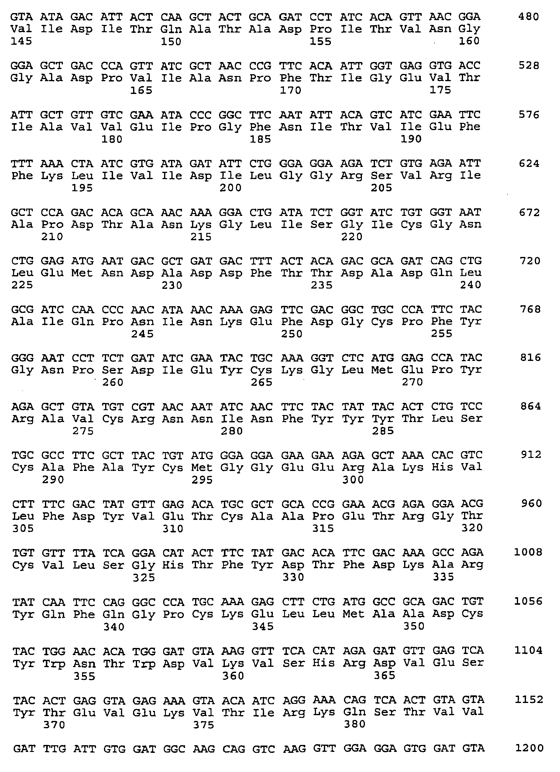


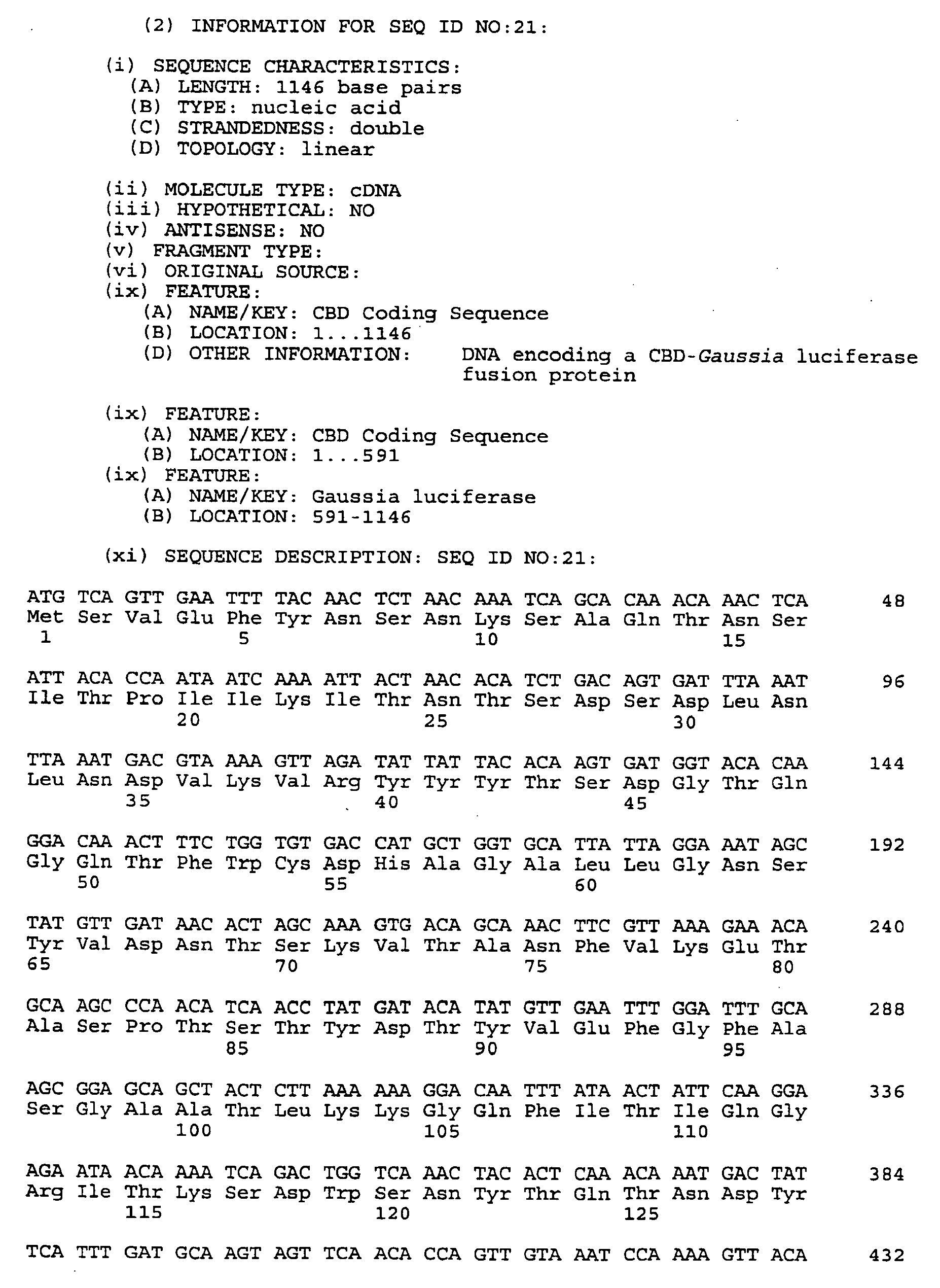


Claims (8)
- A diagnostic system containing a bioluminescence generating system, comprising:(a) a first composition comprising, in a pharmaceutically acceptable vehicle, a conjugate that comprises a targeted agent that is a luciferase or a luciferin and a targeting agent, wherein:a bioluminescence generating system comprises a luciferase as one component and a luciferin as another component;the conjugate binds to a cell surface receptor;the targeting agent specifically binds to a cell surface protein; and(b) a second composition, comprising, in a pharmaceutically acceptable vehicle: a luciferin or a luciferase, wherein the composition comprises a luciferin if the targeted agent in the first composition is a luciferase, and a luciferase if the targeted agent in the first composition is a luciferin; and(c) a compound that is a spectral shifter that shifts the spectrum of light generated by the bioluminescence generating system.
- The diagnostic system of claim 1, wherein the cell surface protein is found in neoplastic tissue.
- The diagnostic system of claim 1 or claim 2, wherein the targeting agent is an antibody.
- The diagnostic system of claim 3, wherein the antibody is directed towards a tumour antigen.
- The diagnostic system of any one of claims 1 to 4, wherein the luciferase is selected from the group comprising Renilla mulleri luciferase, Pleuromamma luciferase and Gaussia luciferase.
- A method for in vivo labelling of selected tissues, comprising:contacting the tissue with a conjugate comprising a first component of a bioluminescence generating system, wherein the first component is a luciferin or luciferase; andthen contacting the tissue with other components of the bioluminescence generating system, wherein contacting is effected in vivo.
- The method of claim 6, wherein the selected tissue is neoplastic tissue.
- The method of claim 6 or claim 7, wherein the luciferase is selected from the group comprising Renilla mulleri luciferase, Pleuromamma luciferase and Gaussia luciferase.
Applications Claiming Priority (4)
| Application Number | Priority Date | Filing Date | Title |
|---|---|---|---|
| US7962498P | 1998-03-27 | 1998-03-27 | |
| US8936798P | 1998-06-15 | 1998-06-15 | |
| US10293998P | 1998-10-01 | 1998-10-01 | |
| EP99914203A EP1064360B1 (en) | 1998-03-27 | 1999-03-26 | Luciferases, gfp fluorescent proteins, their nucleic acids and the use thereof in diagnostics |
Related Parent Applications (1)
| Application Number | Title | Priority Date | Filing Date |
|---|---|---|---|
| EP99914203A Division EP1064360B1 (en) | 1998-03-27 | 1999-03-26 | Luciferases, gfp fluorescent proteins, their nucleic acids and the use thereof in diagnostics |
Publications (2)
| Publication Number | Publication Date |
|---|---|
| EP1925320A2 true EP1925320A2 (en) | 2008-05-28 |
| EP1925320A3 EP1925320A3 (en) | 2008-09-03 |
Family
ID=39315527
Family Applications (1)
| Application Number | Title | Priority Date | Filing Date |
|---|---|---|---|
| EP08001764A Withdrawn EP1925320A3 (en) | 1998-03-27 | 1999-03-26 | Luciferases, fluorescent proteins, nucleic acids encoding the luciferases and fluorescent proteins and the use thereof in diagnostics |
Country Status (1)
| Country | Link |
|---|---|
| EP (1) | EP1925320A3 (en) |
Cited By (1)
| Publication number | Priority date | Publication date | Assignee | Title |
|---|---|---|---|---|
| EP2893327A4 (en) * | 2012-09-01 | 2016-08-17 | Prolume Ltd | Novel coelenterazine compounds and methods of use |
Citations (142)
| Publication number | Priority date | Publication date | Assignee | Title |
|---|---|---|---|---|
| US1711693A (en) | 1927-01-14 | 1929-05-07 | Rheinische Metallw & Maschf | Electric fuse for projectiles |
| US3843443A (en) | 1973-03-30 | 1974-10-22 | J Fishman | Polypeptide materials bound to fluorocarbon polymers |
| US3859125A (en) | 1972-10-10 | 1975-01-07 | Gilbreth Co | Soluble coated paper |
| US3939123A (en) | 1974-06-18 | 1976-02-17 | Union Carbide Corporation | Lightly cross-linked polyurethane hydrogels based on poly(alkylene ether) polyols |
| US4006117A (en) | 1973-01-24 | 1977-02-01 | Hooker Chemicals & Plastics Corporation | Amine phosphite antioxidants |
| US4021364A (en) | 1972-12-04 | 1977-05-03 | Prof. Dr. Peter Speiser | Microcapsules in the nanometric range and a method for their production |
| US4044126A (en) | 1972-04-20 | 1977-08-23 | Allen & Hanburys Limited | Steroidal aerosol compositions and process for the preparation thereof |
| US4162355A (en) | 1976-06-30 | 1979-07-24 | Board Of Regents, For And On Behalf Of The University Of Florida | Copolymers of (a) aminimides and (b) vinyl pendant primary halomethy monomers useful for affinity chromatography |
| US4171412A (en) | 1974-04-25 | 1979-10-16 | Ceskoslovenska Akademie Ved | Specific sorbent having D-aminoacid moiety covalently bonded to solid particulate hydrophilic polymeric carrier |
| US4175183A (en) | 1977-03-01 | 1979-11-20 | Development Finance Corporation Of New Zealand | Hydroxyalkylated cross-linked regenerated cellulose and method of preparation thereof |
| US4177038A (en) | 1976-05-18 | 1979-12-04 | Max-Planck-Gesellschaft Zur Forderung Der Wissenschafen E.V. | Method for the production of a vehicle substance which is capable of covalent bonding with biologically active matter |
| US4178439A (en) | 1976-03-04 | 1979-12-11 | Development Finance Corporation Of New Zealand | Sulphated ion exchanger and method for preparation thereof |
| US4179402A (en) | 1978-05-15 | 1979-12-18 | Shell Oil Company | Resin-metal-ligand composition |
| US4180524A (en) | 1978-02-16 | 1979-12-25 | Phillips Petroleum Company | Disproportionation/double-bond isomerization of olefins |
| US4225581A (en) | 1975-03-20 | 1980-09-30 | Jorg Kreuter | Biological materials |
| US4241537A (en) | 1979-05-10 | 1980-12-30 | W. R. Grace & Co. | Plant growth media utilizing polyurethane hydrogel |
| US4244721A (en) | 1979-01-31 | 1981-01-13 | Pedro Buarque De Macedo | Method of making composite borosilicate glass articles |
| US4281645A (en) | 1977-06-28 | 1981-08-04 | Duke University, Inc. | Method and apparatus for monitoring metabolism in body organs |
| US4282287A (en) | 1980-01-24 | 1981-08-04 | Giese Roger W | Biochemical avidin-biotin multiple-layer system |
| US4322311A (en) | 1978-08-04 | 1982-03-30 | Damon Corporation | Process for producing controlled porosity microcapsules |
| US4324683A (en) | 1975-08-20 | 1982-04-13 | Damon Corporation | Encapsulation of labile biological material |
| US4329332A (en) | 1978-07-19 | 1982-05-11 | Patrick Couvreur | Biodegradable submicroscopic particles containing a biologically active substance and compositions containing them |
| US4364923A (en) | 1972-04-20 | 1982-12-21 | Allen & Hanburs Limited | Chemical compounds |
| US4439585A (en) | 1980-11-12 | 1984-03-27 | Tyndale Plains-Hunter, Ltd. | Polyurethane diacrylate compositions as carrier for pharmacological agents |
| US4485227A (en) | 1983-06-16 | 1984-11-27 | Howmedica, Inc. | Biocompatible poly-(ether-urethane-urea) and process for its preparation |
| US4507230A (en) | 1982-05-12 | 1985-03-26 | Research Corporation | Peptide synthesis reagents and method of use |
| US4522811A (en) | 1982-07-08 | 1985-06-11 | Syntex (U.S.A.) Inc. | Serial injection of muramyldipeptides and liposomes enhances the anti-infective activity of muramyldipeptides |
| US4525306A (en) | 1981-09-21 | 1985-06-25 | Asama Chemical Co., Ltd. | Method for prevention of oxidation of oils and fats and soft capsules containing the treated oils and fats |
| US4542102A (en) | 1983-07-05 | 1985-09-17 | Molecular Diagnostics, Inc. | Coupling of nucleic acids to solid support by photochemical methods |
| US4562157A (en) | 1983-05-25 | 1985-12-31 | National Research Development Corporation | Diagnostic device incorporating a biochemical ligand |
| US4569981A (en) | 1980-07-18 | 1986-02-11 | Bayer Aktiengesellschaft | Solid, water-dispersible plastics precursors containing isocyanate groups, a process for the production of aqueous plastics dispersions using such plastics precursors |
| US4581335A (en) | 1982-12-01 | 1986-04-08 | Texas A&M University System | Process for producing a cloned luciferase-synthesizing microorganism |
| WO1986003840A1 (en) | 1984-12-17 | 1986-07-03 | Rijksuniversiteit Te Groningen | Solid phase immunoassay using immunoreagents immobilized on inert synthetic resin surfaces |
| EP0226979A2 (en) | 1985-12-14 | 1987-07-01 | Chisso Corporation | Process for producing the protein of an aequorin activity by using a photoprotein aequorin gene |
| US4681870A (en) | 1985-01-11 | 1987-07-21 | Imre Corporation | Protein A-silica immunoadsorbent and process for its production |
| EP0245093A1 (en) | 1986-05-08 | 1987-11-11 | Chisso Corporation | Method for detecting metals |
| US4714682A (en) | 1985-12-11 | 1987-12-22 | Flow Cytometry Standards Corporation | Fluorescent calibration microbeads simulating stained cells |
| US4745051A (en) | 1983-05-27 | 1988-05-17 | The Texas A&M University System | Method for producing a recombinant baculovirus expression vector |
| US4762881A (en) | 1987-01-09 | 1988-08-09 | E. I. Du Pont De Nemours And Company | Photoreactive benzoylphenylalanines and related peptides |
| US4767206A (en) | 1984-12-24 | 1988-08-30 | Flow Cytometry Standards Corporation | Calibration method for flow cytometry using fluorescent microbeads and synthesis thereof |
| US4774189A (en) | 1984-12-24 | 1988-09-27 | Flow Cytometry Standards Corp. | Fluorescent calibration microbeads simulating stained cells |
| US4777128A (en) | 1986-05-27 | 1988-10-11 | Ethigen Corporation | Fluorescence immunoassay involving energy transfer between two fluorophores |
| US4789633A (en) | 1984-04-19 | 1988-12-06 | University Of Tennessee Research Corporation | Fused liposome and acid induced method for liposome fusion |
| JPS63317079A (en) | 1987-06-19 | 1988-12-26 | Kikkoman Corp | Production of luciferase |
| US4853327A (en) | 1985-07-10 | 1989-08-01 | Molecular Diagnostics, Inc. | Enhanced phthalazinedione chemiluminescence |
| US4867908A (en) | 1986-08-29 | 1989-09-19 | Becton, Dickinson And Company | Method and materials for calibrating flow cytometers and other analysis instruments |
| US4870009A (en) | 1982-11-22 | 1989-09-26 | The Salk Institute For Biological Studies | Method of obtaining gene product through the generation of transgenic animals |
| US4882165A (en) | 1981-11-09 | 1989-11-21 | The Regents Of The University Of California | Light sensitive liposomes |
| US4885250A (en) | 1987-03-02 | 1989-12-05 | E. I. Du Pont De Nemours And Company | Enzyme immobilization and bioaffinity separations with perfluorocarbon polymer-based supports |
| US4891043A (en) | 1987-05-28 | 1990-01-02 | Board Of Trustees Of The University Of Illinois | System for selective release of liposome encapsulated material via laser radiation |
| US4908405A (en) | 1985-01-04 | 1990-03-13 | Ernst Bayer | Graft copolymers of crosslinked polymers and polyoxyethylene, processes for their production, and their usage |
| US4921757A (en) | 1985-04-26 | 1990-05-01 | Massachusetts Institute Of Technology | System for delayed and pulsed release of biologically active substances |
| US4927879A (en) | 1988-02-25 | 1990-05-22 | Purdue Research Foundation | Method for solid phase membrane mimetics |
| US4927923A (en) | 1984-09-26 | 1990-05-22 | Compagnie Oris Industries | Macropolycyclic rare earth complexes and application as fluorescent tracers |
| US4931498A (en) | 1988-02-25 | 1990-06-05 | Purdue Research Foundation | Immobilized artificial membranes |
| US4950588A (en) | 1985-07-10 | 1990-08-21 | Molecular Diagnostics, Inc. | Prolonged enhanced chemiluminescence |
| US4952496A (en) | 1984-03-30 | 1990-08-28 | Associated Universities, Inc. | Cloning and expression of the gene for bacteriophage T7 RNA polymerase |
| US4954444A (en) | 1987-03-02 | 1990-09-04 | E. I. Du Pont De Nemours And Company | Enzyme immobilization and bioaffinity separations with perfluorocarbon polymer-based supports |
| EP0386691A2 (en) | 1989-03-08 | 1990-09-12 | Toyo Boseki Kabushiki Kaisha | DNA probe detection method with bacterial luciferase system |
| EP0387355A1 (en) | 1988-08-09 | 1990-09-19 | Toray Industries, Inc. | Process for preparing luciferase by recombinant expression of a luciferase-coding gene |
| US4963368A (en) | 1988-04-18 | 1990-10-16 | Nabisco Brands, Inc. | Oxidoreductase enzyme stabilized highly unsaturated fatty acids and derivatives of such acids |
| US4968613A (en) | 1987-07-29 | 1990-11-06 | Kikkoman Corporation | Luciferase gene and novel recombinant DNA as well as a method of producing luciferase |
| JPH0330678A (en) | 1989-06-29 | 1991-02-08 | Osaka Bio Sci Kenkyusho | Dna compound coding luciferase and manifestation vector containing the same compound |
| US4999208A (en) | 1989-06-07 | 1991-03-12 | Nabisco Brands, Inc. | Extrusion baking of cookies having liposome encapsulated ingredients |
| US5004565A (en) | 1986-07-17 | 1991-04-02 | The Board Of Governors Of Wayne State University | Method and compositions providing enhanced chemiluminescence from 1,2-dioxetanes |
| US5023181A (en) | 1987-08-19 | 1991-06-11 | Chisso Corporation | Process for preparing a calcium-dependent oxygenase |
| WO1992001225A1 (en) | 1990-07-13 | 1992-01-23 | Cis Bio International | Method for amplifying a signal emitted by a luminescent compound |
| US5092992A (en) | 1989-06-07 | 1992-03-03 | J. T. Baker Inc. | Polyethyleneimine matrixes for affinity chromatography |
| US5093240A (en) | 1986-10-15 | 1992-03-03 | Chisso Corporation | Variant aequorin genes and process for producing variant aequorin proteins |
| US5098828A (en) | 1985-10-24 | 1992-03-24 | Reinhard Geiger | D-luciferin derivatives, their application and processes for the detection of ligands marked with an enzyme in the determination of biochemical substances |
| US5128256A (en) | 1987-01-12 | 1992-07-07 | Stratagene | DNA cloning vectors with in vivo excisable plasmids |
| US5139937A (en) | 1987-11-18 | 1992-08-18 | Chisso Corporation | Process for producing apoaequorin |
| JPH04258288A (en) | 1991-02-13 | 1992-09-14 | Toray Ind Inc | Production of luciferase of cypridina hilgendorfi |
| US5162227A (en) | 1984-12-31 | 1992-11-10 | University Of Georgia Research Foundation, Inc. | Recombinant DNA vectors capable of expressing apoaequorin in E. coli |
| US5162508A (en) | 1987-12-18 | 1992-11-10 | Compagnie Oris Industrie | Rare earth cryptates, processes for their preparation, synthesis intermediates and application as fluorescent tracers |
| US5169784A (en) | 1990-09-17 | 1992-12-08 | The Texas A & M University System | Baculovirus dual promoter expression vector |
| US5182202A (en) | 1987-11-30 | 1993-01-26 | Kikkoman Corporation | Purified luciferase from luciola cruciata |
| US5188837A (en) | 1989-11-13 | 1993-02-23 | Nova Pharmaceutical Corporation | Lipsopheres for controlled delivery of substances |
| US5190762A (en) | 1988-07-06 | 1993-03-02 | Applied Genetics, Inc. | Method of administering proteins to living skin cells |
| US5192679A (en) | 1990-05-03 | 1993-03-09 | The United States Of America As Represented By The Secretary Of The Department Of Health And Human Services | Growing ehrlichia species in a continuous cell line |
| US5196524A (en) | 1989-01-06 | 1993-03-23 | Eli Lilly And Company | Fusion reporter gene for bacterial luciferase |
| US5196318A (en) | 1990-06-26 | 1993-03-23 | The Texas A&M University System | Precisely regulated expression of deleterious genes |
| US5206161A (en) | 1991-02-01 | 1993-04-27 | Genentech, Inc. | Human plasma carboxypeptidase B |
| US5215899A (en) | 1989-11-09 | 1993-06-01 | Miles Inc. | Nucleic acid amplification employing ligatable hairpin probe and transcription |
| US5219737A (en) | 1990-03-27 | 1993-06-15 | Kikkoman Corporation | Mutant luciferase of a firefly, mutant luciferase genes, recombinant dnas containing the genes and a method of producing mutant luciferase |
| US5221623A (en) | 1986-07-22 | 1993-06-22 | Boyce Thompson Institute For Plant Research, Inc. | Use of bacterial luciferase structural genes for cloning and monitoring gene expression in microorganisms and for tagging and identification of genetically engineered organisms |
| US5225212A (en) | 1989-10-20 | 1993-07-06 | Liposome Technology, Inc. | Microreservoir liposome composition and method |
| US5229285A (en) | 1991-06-27 | 1993-07-20 | Kikkoman Corporation | Thermostable luciferase of firefly, thermostable luciferase gene of firefly, novel recombinant dna, and process for the preparation of thermostable luciferase of firefly |
| US5242687A (en) | 1989-03-15 | 1993-09-07 | Tkb Associates Limited Partnership | Method of reducing cellular immune response involving T-cells using CD8-bearing antigen presenting cells |
| US5243041A (en) | 1991-08-22 | 1993-09-07 | Fernandez Pol Jose A | DNA vector with isolated CDNA gene encoding metallopanstimulin |
| JPH0564583B2 (en) | 1987-07-31 | 1993-09-14 | Kyoraku Co Ltd | |
| US5266317A (en) | 1990-10-04 | 1993-11-30 | University Of Georgia Research Foundation, Inc. | Insect-specific paralytic neurotoxin genes for use in biological insect control: methods and compositions |
| US5272079A (en) | 1988-07-06 | 1993-12-21 | Applied Genetics, Inc. | Purification and administration of DNA repair enzymes |
| US5277913A (en) | 1991-09-09 | 1994-01-11 | Thompson David H | Liposomal delivery system with photoactivatable triggered release |
| US5279943A (en) | 1985-08-02 | 1994-01-18 | Compagnie Oris Industrie | Homogeneous process for the detection and/or determination by luminescence of an analyte in a medium in which it may be present |
| US5283122A (en) | 1984-04-19 | 1994-02-01 | University Of Tennessee Research Corporation | Fused liposome and acid induced method for liposome fusion |
| US5284646A (en) | 1986-07-03 | 1994-02-08 | Advanced Magnetics Inc. | Hepatocyte specific receptor mediated endocytosis type magnetic resonance imaging contrast agents |
| US5288623A (en) | 1989-10-26 | 1994-02-22 | Chisso Corporation | Process for secretory production of a calcium-binding protein |
| US5292658A (en) | 1989-12-29 | 1994-03-08 | University Of Georgia Research Foundation, Inc. Boyd Graduate Studies Research Center | Cloning and expressions of Renilla luciferase |
| US5292814A (en) | 1987-04-29 | 1994-03-08 | Ernst Bayer | Process for the preparation of monodispersed polymer beads |
| US5296231A (en) | 1988-07-06 | 1994-03-22 | Applied Genetics, Inc. | Purification and administration of DNA repair enzymes |
| US5328603A (en) | 1990-03-20 | 1994-07-12 | The Center For Innovative Technology | Lignocellulosic and cellulosic beads for use in affinity and immunoaffinity chromatography of high molecular weight proteins |
| US5334640A (en) | 1992-04-08 | 1994-08-02 | Clover Consolidated, Ltd. | Ionically covalently crosslinked and crosslinkable biocompatible encapsulation compositions and methods |
| WO1994018342A1 (en) | 1993-02-12 | 1994-08-18 | Sealite Sciences, Inc. | Preparation of photoprotein conjugates and methods of use thereof |
| US5342607A (en) | 1986-07-03 | 1994-08-30 | Advanced Magnetics, Inc. | Receptor mediated endocytosis type magnetic resonance imaging contrast agents |
| US5352598A (en) | 1988-04-12 | 1994-10-04 | Kikkoman Corporation | Purified luciferase from luciola lateralis |
| US5352432A (en) | 1986-07-03 | 1994-10-04 | Advanced Magnetics, Inc. | Hepatocyte specific composition and their use as diagnostic imaging agents |
| US5352448A (en) | 1992-07-20 | 1994-10-04 | Purdue Research Foundatioin | Oral administration of antigens |
| US5360728A (en) | 1992-12-01 | 1994-11-01 | Woods Hole Oceanographic Institution (W.H.O.I.) | Modified apoaequorin having increased bioluminescent activity |
| US5360726A (en) | 1989-09-12 | 1994-11-01 | Board Of Trustees Operating Michigan State University | Polypeptides enabling sorting of proteins to vacuoles in plants |
| US5366881A (en) | 1993-02-23 | 1994-11-22 | The United States Of America As Represented By The Secretary Of The Navy | Polymerizable lipids for preparing vesicles that controllably release an encapsulant |
| US5374534A (en) | 1990-07-19 | 1994-12-20 | Charm Sciences, Inc. | Method of preparing D-luciferin derivatives |
| WO1995001542A1 (en) | 1993-06-29 | 1995-01-12 | St Speicher-Technologie Gmbh | Heater |
| US5389449A (en) | 1991-01-04 | 1995-02-14 | Perseptive Biosystems, Inc. | Sulfonamide bonded hydrophilic coatings |
| WO1995007463A1 (en) | 1993-09-10 | 1995-03-16 | The Trustees Of Columbia University In The City Of New York | Uses of green fluorescent protein |
| US5403750A (en) | 1991-03-06 | 1995-04-04 | W. R. Grace & Co.-Conn. | Biocompatible, low protein adsorption affinity matrix |
| US5411730A (en) | 1993-07-20 | 1995-05-02 | Research Corporation Technologies, Inc. | Magnetic microparticles |
| US5416193A (en) | 1993-04-30 | 1995-05-16 | Pfizer Inc. | Coupling reagent and method |
| US5422266A (en) | 1984-12-31 | 1995-06-06 | University Of Georgia Research Foundation, Inc. | Recombinant DNA vectors capable of expressing apoaequorin |
| WO1995018853A1 (en) | 1994-01-03 | 1995-07-13 | Promega Corporation | Mutant luciferases |
| WO1995021191A1 (en) | 1994-02-04 | 1995-08-10 | William Ward | Bioluminescent indicator based upon the expression of a gene for a modified green-fluorescent protein |
| JPH07222590A (en) | 1994-02-08 | 1995-08-22 | Chisso Corp | Luciferase gene of alteromonas hanedai |
| US5451683A (en) | 1989-11-13 | 1995-09-19 | Affymax Technologies N.V. | Spatially-addressable immobilization of anti-ligands on surfaces |
| WO1995025798A1 (en) | 1994-03-23 | 1995-09-28 | The Secretary Of State Of Defence In Her Britannic Majesty's Government Of The United Kingdom Of Great Britain And Northern Ireland | Luciferases |
| US5455357A (en) | 1992-04-01 | 1995-10-03 | Boehringer Mannheim Gmbh | Substituted thiazoline-dioxetan substrates, process for the production and use |
| US5464745A (en) | 1993-03-31 | 1995-11-07 | Novagen, Inc. | Protein ligand binding region mapping system |
| US5470881A (en) | 1993-09-09 | 1995-11-28 | West Virginia University Research Corporation | Urea ophthalmic ointment and solution |
| US5482719A (en) | 1992-10-30 | 1996-01-09 | Guillet; James E. | Drug delivery systems |
| US5484589A (en) | 1992-04-20 | 1996-01-16 | Rufeld, Inc. | Anti-viral methods using RNAse and DNAse |
| US5484723A (en) | 1993-06-28 | 1996-01-16 | Chisso Corporation | Flavin reductase gene from Vibrio fischeri |
| US5491084A (en) | 1993-09-10 | 1996-02-13 | The Trustees Of Columbia University In The City Of New York | Uses of green-fluorescent protein |
| US5496934A (en) | 1993-04-14 | 1996-03-05 | Yissum Research Development Company Of The Hebrew University Of Jerusalem | Nucleic acids encoding a cellulose binding domain |
| WO1996023810A1 (en) | 1994-11-10 | 1996-08-08 | The Regents Of The University Of California | Modified green fluorescenct proteins |
| WO1996027675A1 (en) | 1995-03-06 | 1996-09-12 | Medical Research Council | Jellyfish green fluorescent protein (gfp) expression in plants |
| WO1997026333A1 (en) | 1996-01-18 | 1997-07-24 | University Of Florida Research Foundation, Incorporated | Humanized green fluorescent protein genes and methods |
| WO1997028261A1 (en) | 1996-01-31 | 1997-08-07 | The Regents Of The University Of California | Tandem fluorescent protein constructs |
| WO1997041228A2 (en) | 1996-05-01 | 1997-11-06 | Pioneer Hi-Bred International, Inc. | Use of the green fluorescent protein as a screenable marker for plant transformation |
| WO1998002571A1 (en) | 1996-07-16 | 1998-01-22 | The Regents Of The University Of California | Assays for protein kinases using fluorescent protein substrates |
| US5876995A (en) | 1996-02-06 | 1999-03-02 | Bryan; Bruce | Bioluminescent novelty items |
| US6247995B1 (en) | 1996-02-06 | 2001-06-19 | Bruce Bryan | Bioluminescent novelty items |
| US6416960B1 (en) | 1996-08-08 | 2002-07-09 | Prolume, Ltd. | Detection and visualization of neoplastic tissues and other tissues |
| US6458547B1 (en) | 1996-12-12 | 2002-10-01 | Prolume, Ltd. | Apparatus and method for detecting and identifying infectious agents |
Family Cites Families (3)
| Publication number | Priority date | Publication date | Assignee | Title |
|---|---|---|---|---|
| ES8607563A1 (en) * | 1983-10-13 | 1986-06-01 | Univ Georgia Res Found | Bioluminescent immunoassays. |
| US4606364A (en) * | 1984-10-01 | 1986-08-19 | George Tash | Flushing device adapter |
| US5976796A (en) * | 1996-10-04 | 1999-11-02 | Loma Linda University | Construction and expression of renilla luciferase and green fluorescent protein fusion genes |
-
1999
- 1999-03-26 EP EP08001764A patent/EP1925320A3/en not_active Withdrawn
Patent Citations (153)
| Publication number | Priority date | Publication date | Assignee | Title |
|---|---|---|---|---|
| US1711693A (en) | 1927-01-14 | 1929-05-07 | Rheinische Metallw & Maschf | Electric fuse for projectiles |
| US4044126A (en) | 1972-04-20 | 1977-08-23 | Allen & Hanburys Limited | Steroidal aerosol compositions and process for the preparation thereof |
| US4364923A (en) | 1972-04-20 | 1982-12-21 | Allen & Hanburs Limited | Chemical compounds |
| US4414209A (en) | 1972-04-20 | 1983-11-08 | Allen & Hanburys Limited | Micronized aerosol steroids |
| US3859125A (en) | 1972-10-10 | 1975-01-07 | Gilbreth Co | Soluble coated paper |
| US4021364A (en) | 1972-12-04 | 1977-05-03 | Prof. Dr. Peter Speiser | Microcapsules in the nanometric range and a method for their production |
| US4006117A (en) | 1973-01-24 | 1977-02-01 | Hooker Chemicals & Plastics Corporation | Amine phosphite antioxidants |
| US3843443A (en) | 1973-03-30 | 1974-10-22 | J Fishman | Polypeptide materials bound to fluorocarbon polymers |
| US4171412A (en) | 1974-04-25 | 1979-10-16 | Ceskoslovenska Akademie Ved | Specific sorbent having D-aminoacid moiety covalently bonded to solid particulate hydrophilic polymeric carrier |
| US3939123A (en) | 1974-06-18 | 1976-02-17 | Union Carbide Corporation | Lightly cross-linked polyurethane hydrogels based on poly(alkylene ether) polyols |
| US4225581A (en) | 1975-03-20 | 1980-09-30 | Jorg Kreuter | Biological materials |
| US4269821A (en) | 1975-03-20 | 1981-05-26 | Jorg Kreuter | Biological materials |
| US4324683A (en) | 1975-08-20 | 1982-04-13 | Damon Corporation | Encapsulation of labile biological material |
| US4178439A (en) | 1976-03-04 | 1979-12-11 | Development Finance Corporation Of New Zealand | Sulphated ion exchanger and method for preparation thereof |
| US4177038A (en) | 1976-05-18 | 1979-12-04 | Max-Planck-Gesellschaft Zur Forderung Der Wissenschafen E.V. | Method for the production of a vehicle substance which is capable of covalent bonding with biologically active matter |
| US4162355A (en) | 1976-06-30 | 1979-07-24 | Board Of Regents, For And On Behalf Of The University Of Florida | Copolymers of (a) aminimides and (b) vinyl pendant primary halomethy monomers useful for affinity chromatography |
| US4175183A (en) | 1977-03-01 | 1979-11-20 | Development Finance Corporation Of New Zealand | Hydroxyalkylated cross-linked regenerated cellulose and method of preparation thereof |
| US4281645A (en) | 1977-06-28 | 1981-08-04 | Duke University, Inc. | Method and apparatus for monitoring metabolism in body organs |
| US4180524A (en) | 1978-02-16 | 1979-12-25 | Phillips Petroleum Company | Disproportionation/double-bond isomerization of olefins |
| US4179402A (en) | 1978-05-15 | 1979-12-18 | Shell Oil Company | Resin-metal-ligand composition |
| US4329332A (en) | 1978-07-19 | 1982-05-11 | Patrick Couvreur | Biodegradable submicroscopic particles containing a biologically active substance and compositions containing them |
| US4322311A (en) | 1978-08-04 | 1982-03-30 | Damon Corporation | Process for producing controlled porosity microcapsules |
| US4244721A (en) | 1979-01-31 | 1981-01-13 | Pedro Buarque De Macedo | Method of making composite borosilicate glass articles |
| US4241537A (en) | 1979-05-10 | 1980-12-30 | W. R. Grace & Co. | Plant growth media utilizing polyurethane hydrogel |
| US4282287A (en) | 1980-01-24 | 1981-08-04 | Giese Roger W | Biochemical avidin-biotin multiple-layer system |
| US4569981A (en) | 1980-07-18 | 1986-02-11 | Bayer Aktiengesellschaft | Solid, water-dispersible plastics precursors containing isocyanate groups, a process for the production of aqueous plastics dispersions using such plastics precursors |
| US4439585A (en) | 1980-11-12 | 1984-03-27 | Tyndale Plains-Hunter, Ltd. | Polyurethane diacrylate compositions as carrier for pharmacological agents |
| US4525306A (en) | 1981-09-21 | 1985-06-25 | Asama Chemical Co., Ltd. | Method for prevention of oxidation of oils and fats and soft capsules containing the treated oils and fats |
| US4882165A (en) | 1981-11-09 | 1989-11-21 | The Regents Of The University Of California | Light sensitive liposomes |
| US4507230A (en) | 1982-05-12 | 1985-03-26 | Research Corporation | Peptide synthesis reagents and method of use |
| US4522811A (en) | 1982-07-08 | 1985-06-11 | Syntex (U.S.A.) Inc. | Serial injection of muramyldipeptides and liposomes enhances the anti-infective activity of muramyldipeptides |
| US4870009A (en) | 1982-11-22 | 1989-09-26 | The Salk Institute For Biological Studies | Method of obtaining gene product through the generation of transgenic animals |
| US4581335A (en) | 1982-12-01 | 1986-04-08 | Texas A&M University System | Process for producing a cloned luciferase-synthesizing microorganism |
| US4562157A (en) | 1983-05-25 | 1985-12-31 | National Research Development Corporation | Diagnostic device incorporating a biochemical ligand |
| US4745051A (en) | 1983-05-27 | 1988-05-17 | The Texas A&M University System | Method for producing a recombinant baculovirus expression vector |
| US4485227A (en) | 1983-06-16 | 1984-11-27 | Howmedica, Inc. | Biocompatible poly-(ether-urethane-urea) and process for its preparation |
| US4542102A (en) | 1983-07-05 | 1985-09-17 | Molecular Diagnostics, Inc. | Coupling of nucleic acids to solid support by photochemical methods |
| US4952496A (en) | 1984-03-30 | 1990-08-28 | Associated Universities, Inc. | Cloning and expression of the gene for bacteriophage T7 RNA polymerase |
| US4789633A (en) | 1984-04-19 | 1988-12-06 | University Of Tennessee Research Corporation | Fused liposome and acid induced method for liposome fusion |
| US5283122A (en) | 1984-04-19 | 1994-02-01 | University Of Tennessee Research Corporation | Fused liposome and acid induced method for liposome fusion |
| US4927923A (en) | 1984-09-26 | 1990-05-22 | Compagnie Oris Industries | Macropolycyclic rare earth complexes and application as fluorescent tracers |
| WO1986003840A1 (en) | 1984-12-17 | 1986-07-03 | Rijksuniversiteit Te Groningen | Solid phase immunoassay using immunoreagents immobilized on inert synthetic resin surfaces |
| US4774189A (en) | 1984-12-24 | 1988-09-27 | Flow Cytometry Standards Corp. | Fluorescent calibration microbeads simulating stained cells |
| US4767206A (en) | 1984-12-24 | 1988-08-30 | Flow Cytometry Standards Corporation | Calibration method for flow cytometry using fluorescent microbeads and synthesis thereof |
| EP0540064A1 (en) | 1984-12-31 | 1993-05-05 | The University Of Georgia Research Foundation, Inc. | Apoaequorin peptides, recombinant DNA vectors capable of expressing the same and microorganisms containing said vectors |
| US5422266A (en) | 1984-12-31 | 1995-06-06 | University Of Georgia Research Foundation, Inc. | Recombinant DNA vectors capable of expressing apoaequorin |
| US5162227A (en) | 1984-12-31 | 1992-11-10 | University Of Georgia Research Foundation, Inc. | Recombinant DNA vectors capable of expressing apoaequorin in E. coli |
| US4908405A (en) | 1985-01-04 | 1990-03-13 | Ernst Bayer | Graft copolymers of crosslinked polymers and polyoxyethylene, processes for their production, and their usage |
| US4681870A (en) | 1985-01-11 | 1987-07-21 | Imre Corporation | Protein A-silica immunoadsorbent and process for its production |
| US4921757A (en) | 1985-04-26 | 1990-05-01 | Massachusetts Institute Of Technology | System for delayed and pulsed release of biologically active substances |
| US4950588A (en) | 1985-07-10 | 1990-08-21 | Molecular Diagnostics, Inc. | Prolonged enhanced chemiluminescence |
| US4853327A (en) | 1985-07-10 | 1989-08-01 | Molecular Diagnostics, Inc. | Enhanced phthalazinedione chemiluminescence |
| US5279943A (en) | 1985-08-02 | 1994-01-18 | Compagnie Oris Industrie | Homogeneous process for the detection and/or determination by luminescence of an analyte in a medium in which it may be present |
| US5098828A (en) | 1985-10-24 | 1992-03-24 | Reinhard Geiger | D-luciferin derivatives, their application and processes for the detection of ligands marked with an enzyme in the determination of biochemical substances |
| US4714682A (en) | 1985-12-11 | 1987-12-22 | Flow Cytometry Standards Corporation | Fluorescent calibration microbeads simulating stained cells |
| EP0226979A2 (en) | 1985-12-14 | 1987-07-01 | Chisso Corporation | Process for producing the protein of an aequorin activity by using a photoprotein aequorin gene |
| EP0245093B1 (en) | 1986-05-08 | 1992-03-11 | Chisso Corporation | Method for detecting metals |
| EP0245093A1 (en) | 1986-05-08 | 1987-11-11 | Chisso Corporation | Method for detecting metals |
| US4777128A (en) | 1986-05-27 | 1988-10-11 | Ethigen Corporation | Fluorescence immunoassay involving energy transfer between two fluorophores |
| US5284646A (en) | 1986-07-03 | 1994-02-08 | Advanced Magnetics Inc. | Hepatocyte specific receptor mediated endocytosis type magnetic resonance imaging contrast agents |
| US5352432A (en) | 1986-07-03 | 1994-10-04 | Advanced Magnetics, Inc. | Hepatocyte specific composition and their use as diagnostic imaging agents |
| US5342607A (en) | 1986-07-03 | 1994-08-30 | Advanced Magnetics, Inc. | Receptor mediated endocytosis type magnetic resonance imaging contrast agents |
| US5004565A (en) | 1986-07-17 | 1991-04-02 | The Board Of Governors Of Wayne State University | Method and compositions providing enhanced chemiluminescence from 1,2-dioxetanes |
| US5221623A (en) | 1986-07-22 | 1993-06-22 | Boyce Thompson Institute For Plant Research, Inc. | Use of bacterial luciferase structural genes for cloning and monitoring gene expression in microorganisms and for tagging and identification of genetically engineered organisms |
| US4867908A (en) | 1986-08-29 | 1989-09-19 | Becton, Dickinson And Company | Method and materials for calibrating flow cytometers and other analysis instruments |
| US5093240A (en) | 1986-10-15 | 1992-03-03 | Chisso Corporation | Variant aequorin genes and process for producing variant aequorin proteins |
| US4762881A (en) | 1987-01-09 | 1988-08-09 | E. I. Du Pont De Nemours And Company | Photoreactive benzoylphenylalanines and related peptides |
| US5128256A (en) | 1987-01-12 | 1992-07-07 | Stratagene | DNA cloning vectors with in vivo excisable plasmids |
| US4954444A (en) | 1987-03-02 | 1990-09-04 | E. I. Du Pont De Nemours And Company | Enzyme immobilization and bioaffinity separations with perfluorocarbon polymer-based supports |
| US4885250A (en) | 1987-03-02 | 1989-12-05 | E. I. Du Pont De Nemours And Company | Enzyme immobilization and bioaffinity separations with perfluorocarbon polymer-based supports |
| US5292814A (en) | 1987-04-29 | 1994-03-08 | Ernst Bayer | Process for the preparation of monodispersed polymer beads |
| US4891043A (en) | 1987-05-28 | 1990-01-02 | Board Of Trustees Of The University Of Illinois | System for selective release of liposome encapsulated material via laser radiation |
| JPS63317079A (en) | 1987-06-19 | 1988-12-26 | Kikkoman Corp | Production of luciferase |
| US4968613A (en) | 1987-07-29 | 1990-11-06 | Kikkoman Corporation | Luciferase gene and novel recombinant DNA as well as a method of producing luciferase |
| JPH0564583B2 (en) | 1987-07-31 | 1993-09-14 | Kyoraku Co Ltd | |
| US5023181A (en) | 1987-08-19 | 1991-06-11 | Chisso Corporation | Process for preparing a calcium-dependent oxygenase |
| US5139937A (en) | 1987-11-18 | 1992-08-18 | Chisso Corporation | Process for producing apoaequorin |
| US5182202A (en) | 1987-11-30 | 1993-01-26 | Kikkoman Corporation | Purified luciferase from luciola cruciata |
| US5162508A (en) | 1987-12-18 | 1992-11-10 | Compagnie Oris Industrie | Rare earth cryptates, processes for their preparation, synthesis intermediates and application as fluorescent tracers |
| US4927879A (en) | 1988-02-25 | 1990-05-22 | Purdue Research Foundation | Method for solid phase membrane mimetics |
| US4931498A (en) | 1988-02-25 | 1990-06-05 | Purdue Research Foundation | Immobilized artificial membranes |
| US5352598A (en) | 1988-04-12 | 1994-10-04 | Kikkoman Corporation | Purified luciferase from luciola lateralis |
| US4963368A (en) | 1988-04-18 | 1990-10-16 | Nabisco Brands, Inc. | Oxidoreductase enzyme stabilized highly unsaturated fatty acids and derivatives of such acids |
| US5296231A (en) | 1988-07-06 | 1994-03-22 | Applied Genetics, Inc. | Purification and administration of DNA repair enzymes |
| US5272079A (en) | 1988-07-06 | 1993-12-21 | Applied Genetics, Inc. | Purification and administration of DNA repair enzymes |
| US5190762A (en) | 1988-07-06 | 1993-03-02 | Applied Genetics, Inc. | Method of administering proteins to living skin cells |
| EP0387355A1 (en) | 1988-08-09 | 1990-09-19 | Toray Industries, Inc. | Process for preparing luciferase by recombinant expression of a luciferase-coding gene |
| US5196524A (en) | 1989-01-06 | 1993-03-23 | Eli Lilly And Company | Fusion reporter gene for bacterial luciferase |
| EP0386691A2 (en) | 1989-03-08 | 1990-09-12 | Toyo Boseki Kabushiki Kaisha | DNA probe detection method with bacterial luciferase system |
| US5242687A (en) | 1989-03-15 | 1993-09-07 | Tkb Associates Limited Partnership | Method of reducing cellular immune response involving T-cells using CD8-bearing antigen presenting cells |
| US5092992A (en) | 1989-06-07 | 1992-03-03 | J. T. Baker Inc. | Polyethyleneimine matrixes for affinity chromatography |
| US4999208A (en) | 1989-06-07 | 1991-03-12 | Nabisco Brands, Inc. | Extrusion baking of cookies having liposome encapsulated ingredients |
| JPH0330678A (en) | 1989-06-29 | 1991-02-08 | Osaka Bio Sci Kenkyusho | Dna compound coding luciferase and manifestation vector containing the same compound |
| US5360726A (en) | 1989-09-12 | 1994-11-01 | Board Of Trustees Operating Michigan State University | Polypeptides enabling sorting of proteins to vacuoles in plants |
| US5225212A (en) | 1989-10-20 | 1993-07-06 | Liposome Technology, Inc. | Microreservoir liposome composition and method |
| US5288623A (en) | 1989-10-26 | 1994-02-22 | Chisso Corporation | Process for secretory production of a calcium-binding protein |
| US5215899A (en) | 1989-11-09 | 1993-06-01 | Miles Inc. | Nucleic acid amplification employing ligatable hairpin probe and transcription |
| US5451683A (en) | 1989-11-13 | 1995-09-19 | Affymax Technologies N.V. | Spatially-addressable immobilization of anti-ligands on surfaces |
| US5188837A (en) | 1989-11-13 | 1993-02-23 | Nova Pharmaceutical Corporation | Lipsopheres for controlled delivery of substances |
| US5292658A (en) | 1989-12-29 | 1994-03-08 | University Of Georgia Research Foundation, Inc. Boyd Graduate Studies Research Center | Cloning and expressions of Renilla luciferase |
| US5418155A (en) | 1989-12-29 | 1995-05-23 | University Of Georgia Research Foundation, Inc. | Isolated Renilla luciferase and method of use thereof |
| US5328603A (en) | 1990-03-20 | 1994-07-12 | The Center For Innovative Technology | Lignocellulosic and cellulosic beads for use in affinity and immunoaffinity chromatography of high molecular weight proteins |
| US5219737A (en) | 1990-03-27 | 1993-06-15 | Kikkoman Corporation | Mutant luciferase of a firefly, mutant luciferase genes, recombinant dnas containing the genes and a method of producing mutant luciferase |
| US5330906A (en) | 1990-03-27 | 1994-07-19 | Kikkoman Corporation | Mutant luciferase of a firefly |
| US5192679A (en) | 1990-05-03 | 1993-03-09 | The United States Of America As Represented By The Secretary Of The Department Of Health And Human Services | Growing ehrlichia species in a continuous cell line |
| US5196318A (en) | 1990-06-26 | 1993-03-23 | The Texas A&M University System | Precisely regulated expression of deleterious genes |
| WO1992001225A1 (en) | 1990-07-13 | 1992-01-23 | Cis Bio International | Method for amplifying a signal emitted by a luminescent compound |
| US5374534A (en) | 1990-07-19 | 1994-12-20 | Charm Sciences, Inc. | Method of preparing D-luciferin derivatives |
| US5169784A (en) | 1990-09-17 | 1992-12-08 | The Texas A & M University System | Baculovirus dual promoter expression vector |
| US5266317A (en) | 1990-10-04 | 1993-11-30 | University Of Georgia Research Foundation, Inc. | Insect-specific paralytic neurotoxin genes for use in biological insect control: methods and compositions |
| US5389449A (en) | 1991-01-04 | 1995-02-14 | Perseptive Biosystems, Inc. | Sulfonamide bonded hydrophilic coatings |
| US5206161A (en) | 1991-02-01 | 1993-04-27 | Genentech, Inc. | Human plasma carboxypeptidase B |
| JPH04258288A (en) | 1991-02-13 | 1992-09-14 | Toray Ind Inc | Production of luciferase of cypridina hilgendorfi |
| US5403750A (en) | 1991-03-06 | 1995-04-04 | W. R. Grace & Co.-Conn. | Biocompatible, low protein adsorption affinity matrix |
| US5229285A (en) | 1991-06-27 | 1993-07-20 | Kikkoman Corporation | Thermostable luciferase of firefly, thermostable luciferase gene of firefly, novel recombinant dna, and process for the preparation of thermostable luciferase of firefly |
| US5243041A (en) | 1991-08-22 | 1993-09-07 | Fernandez Pol Jose A | DNA vector with isolated CDNA gene encoding metallopanstimulin |
| US5277913A (en) | 1991-09-09 | 1994-01-11 | Thompson David H | Liposomal delivery system with photoactivatable triggered release |
| US5455357A (en) | 1992-04-01 | 1995-10-03 | Boehringer Mannheim Gmbh | Substituted thiazoline-dioxetan substrates, process for the production and use |
| US5334640A (en) | 1992-04-08 | 1994-08-02 | Clover Consolidated, Ltd. | Ionically covalently crosslinked and crosslinkable biocompatible encapsulation compositions and methods |
| US5484589A (en) | 1992-04-20 | 1996-01-16 | Rufeld, Inc. | Anti-viral methods using RNAse and DNAse |
| US5352448A (en) | 1992-07-20 | 1994-10-04 | Purdue Research Foundatioin | Oral administration of antigens |
| US5482719A (en) | 1992-10-30 | 1996-01-09 | Guillet; James E. | Drug delivery systems |
| US5360728A (en) | 1992-12-01 | 1994-11-01 | Woods Hole Oceanographic Institution (W.H.O.I.) | Modified apoaequorin having increased bioluminescent activity |
| WO1994018342A1 (en) | 1993-02-12 | 1994-08-18 | Sealite Sciences, Inc. | Preparation of photoprotein conjugates and methods of use thereof |
| US5486455A (en) | 1993-02-12 | 1996-01-23 | Sealite Sciences, Inc. | Photoprotein conjugates and methods of use thereof |
| US5366881A (en) | 1993-02-23 | 1994-11-22 | The United States Of America As Represented By The Secretary Of The Navy | Polymerizable lipids for preparing vesicles that controllably release an encapsulant |
| US5464745A (en) | 1993-03-31 | 1995-11-07 | Novagen, Inc. | Protein ligand binding region mapping system |
| US5496934A (en) | 1993-04-14 | 1996-03-05 | Yissum Research Development Company Of The Hebrew University Of Jerusalem | Nucleic acids encoding a cellulose binding domain |
| US5670623A (en) | 1993-04-14 | 1997-09-23 | Yissum Research Development Company Of The Hebrew University Of Jerusalem | Methods of use of cellulose binding domain proteins |
| US5719044A (en) | 1993-04-14 | 1998-02-17 | Yissum Research Development Company Of The Hebrew University Of Jerusalem | Cellulose binding domain fusion proteins |
| US5738984A (en) | 1993-04-14 | 1998-04-14 | Yissum Research Development Company Of The Hebrew University Of Jerusalem | Kits and methods of detection using cellulose binding domain fusion proteins |
| US5416193A (en) | 1993-04-30 | 1995-05-16 | Pfizer Inc. | Coupling reagent and method |
| US5484723A (en) | 1993-06-28 | 1996-01-16 | Chisso Corporation | Flavin reductase gene from Vibrio fischeri |
| WO1995001542A1 (en) | 1993-06-29 | 1995-01-12 | St Speicher-Technologie Gmbh | Heater |
| US5411730A (en) | 1993-07-20 | 1995-05-02 | Research Corporation Technologies, Inc. | Magnetic microparticles |
| US5470881A (en) | 1993-09-09 | 1995-11-28 | West Virginia University Research Corporation | Urea ophthalmic ointment and solution |
| WO1995007463A1 (en) | 1993-09-10 | 1995-03-16 | The Trustees Of Columbia University In The City Of New York | Uses of green fluorescent protein |
| US5491084A (en) | 1993-09-10 | 1996-02-13 | The Trustees Of Columbia University In The City Of New York | Uses of green-fluorescent protein |
| WO1995018853A1 (en) | 1994-01-03 | 1995-07-13 | Promega Corporation | Mutant luciferases |
| WO1995021191A1 (en) | 1994-02-04 | 1995-08-10 | William Ward | Bioluminescent indicator based upon the expression of a gene for a modified green-fluorescent protein |
| JPH07222590A (en) | 1994-02-08 | 1995-08-22 | Chisso Corp | Luciferase gene of alteromonas hanedai |
| WO1995025798A1 (en) | 1994-03-23 | 1995-09-28 | The Secretary Of State Of Defence In Her Britannic Majesty's Government Of The United Kingdom Of Great Britain And Northern Ireland | Luciferases |
| WO1996023810A1 (en) | 1994-11-10 | 1996-08-08 | The Regents Of The University Of California | Modified green fluorescenct proteins |
| US5625048A (en) | 1994-11-10 | 1997-04-29 | The Regents Of The University Of California | Modified green fluorescent proteins |
| WO1996027675A1 (en) | 1995-03-06 | 1996-09-12 | Medical Research Council | Jellyfish green fluorescent protein (gfp) expression in plants |
| WO1997026333A1 (en) | 1996-01-18 | 1997-07-24 | University Of Florida Research Foundation, Incorporated | Humanized green fluorescent protein genes and methods |
| WO1997028261A1 (en) | 1996-01-31 | 1997-08-07 | The Regents Of The University Of California | Tandem fluorescent protein constructs |
| US5876995A (en) | 1996-02-06 | 1999-03-02 | Bryan; Bruce | Bioluminescent novelty items |
| US6247995B1 (en) | 1996-02-06 | 2001-06-19 | Bruce Bryan | Bioluminescent novelty items |
| WO1997041228A2 (en) | 1996-05-01 | 1997-11-06 | Pioneer Hi-Bred International, Inc. | Use of the green fluorescent protein as a screenable marker for plant transformation |
| WO1998002571A1 (en) | 1996-07-16 | 1998-01-22 | The Regents Of The University Of California | Assays for protein kinases using fluorescent protein substrates |
| US6416960B1 (en) | 1996-08-08 | 2002-07-09 | Prolume, Ltd. | Detection and visualization of neoplastic tissues and other tissues |
| US6458547B1 (en) | 1996-12-12 | 2002-10-01 | Prolume, Ltd. | Apparatus and method for detecting and identifying infectious agents |
Non-Patent Citations (204)
| Title |
|---|
| "Affinity Techniques. Enzyme Purification: Part B. Methods in Enzvmoloqv", vol. 34, 1974, ACAD. PRESS |
| "Immobilized Biochemicals and Affinity Chromatography, Advances in Experimental Medicine and Bioloqv", vol. 42, 1974, PLENUM PRESS |
| "ImmunoTechnology Catalog & Handbook", 1992, article "PIERCE CATALOG" |
| "Liposome Technology, Targeted Drug Delivery and Biological Interaction", vol. III, 1984, CRC PRESS, INC. |
| "Luminescence, Narcosis and Life in the Deep Sea, Johnson", VANTAGE PRESS, pages: 50 - 56 |
| APT ET AL., J. MOL. BIOL., vol. 238, 1995, pages 79 - 96 |
| ASSIL ET AL., ARCH OPHTHALMOL., vol. 105, 1987, pages 400 |
| AVIV ET AL., PROC. NATL. ACAD. SCI. U.S.A., vol. 69, 1972, pages 1408 |
| BALDWIN ET AL., BIOCHEM., vol. 20, 1981, pages 512 - 517 |
| BALDWIN ET AL., BIOCHEM., vol. 23, 1984, pages 3663 - 3667 |
| BALDWIN ET AL., BIOCHEMISTRY, vol. 23, 1984, pages 3663 - 3667 |
| BATRA ET AL., MOLECULAR IMMUNOL., vol. 30, 1993, pages 379 - 386 |
| BELAS ET AL., SCIENCE, vol. 218, 1982, pages 791 - 793 |
| BERG ET AL., J. AM. CHEM. SOC., vol. 111, 1989, pages 8024 - 8026 |
| BERG ET AL.: "Innovation Perspect. Solid Phase Svnth. Collect. Pap., Int. Symp.", 1990, pages: 453 - 459 |
| BERG ET AL.: "Pept.. Proc. Eur. Pept. Svmp.", 1989, pages: 196 - 198 |
| BLINKS ET AL., FED. PROC., vol. 34, 1975, pages 474 |
| BONDAR ET AL., BIOCHIM. BIOPHVS. ACTA, vol. 1231, 1995, pages 29 - 32 |
| BUTZ ET AL., PEPTIDE RES., vol. 7, 1994, pages 20 - 23 |
| CARDULLO ET AL., PROC. NATL. ACAD. SCI. U.S.A., vol. 85, 1988, pages 8790 - 8794 |
| CARLSSON ET AL., BIOCHEM. J., vol. 173, 1978, pages 723 - 737 |
| CASPER ET AL., GENE, vol. 173, 1996, pages 69 - 73 |
| CHALFIE ET AL., SCIENCE, vol. 263, 1994, pages 802 - 805 |
| CHARBONNEAU ET AL., J. BIOL. CHEM., vol. 254, 1979, pages 769 - 780 |
| CHARBONNEAU ET AL.: "Amino Acid Sequence of the Calcium-Dependent Photoprotein Aequorin", BIOCHEMISTRY, vol. 24, 1985, pages 6762 - 6771 |
| CHIRGWIN ET AL., BIOCHEMISTRY, vol. 18, 1970, pages 5294 - 5299 |
| CODY ET AL., BIOCHEMISTRY, vol. 32, 1993, pages 1212 - 1218 |
| CODY, C. W. ET AL., BIOCHEMISTRY, vol. 32, 1993, pages 1212 - 1218 |
| COHN ET AL., J. BIOL. CHEM., vol. 260, 1985, pages 6139 - 6146 |
| COHN ET AL., PROC. NATL. ACAD. SCI. U.S.A., vol. 90, 1989, pages 102 - 123 |
| CORMACK ET AL., MICROBIOLOGY, vol. 143, 1997, pages 303 - 311 |
| CORMIER ET AL., J. CELL. PHVSIOL., vol. 81, 1973, pages 291 - 298 |
| CORMIER: "Renilla and Aequorea bioluminescence", BIOLUMINESCENCE AND CHEMILUMINESCENCE, 1981, pages 225 - 233 |
| CUMBER ET AL., BIOCONIUQATE CHEM., vol. 3, 1992, pages 397 - 401 |
| DEWITT ET AL., PROC. NATL. ACAD. SCI. U.S.A., vol. 90, 1993, pages 6909 |
| DUZGUNES ET AL.: "CELL FUSION" |
| ELLENS ET AL., BIOCHEMISTRY, vol. 23, 1984, pages 1532 - 1538 |
| ELLMAN ET AL., PROC. NATL. ACAD. SCI. U.S.A., vol. 91, 1994, pages 4708 |
| ENGEBRECHT ET AL., CELL, vol. 32, 1983, pages 773 - 781 |
| ENGEBRECHT ET AL., METHODS IN ENZVMOLOQV, vol. 133, 1986, pages 83 - 99 |
| ENGEBRECHT ET AL., PROC. NATL. ACAD. SCI. U.S.A., vol. 81, 1984, pages 4154 - 4158 |
| FAIRCHILD ET AL., J. OF BIOL. CHEM., vol. 269, 1994, pages 8686 - 8694 |
| FATTOM ET AL., INFECTION & IMMUN., vol. 60, 1992, pages 584 - 589 |
| FEY ET AL., GENE, vol. 165, 1995, pages 127 - 130 |
| FRACKMAN ET AL., J. OF BACTERIOLOQY, vol. 172, 1990, pages 5767 - 5773 |
| GAST ET AL.: "Separation of a blue fluorescence protein from bacterial luciferase", BIOCHEM. BIOPHVS. RES. COMMUN., vol. 80, no. 1, 1978, pages 14 - 21 |
| GESZTES ET AL.: "Topical anaesthesia of the skin by liposome -encapsulated tetracaine", ANESTHESIA AND ANALQESIA, vol. 67, 1988, pages 1079 - 1081 |
| GIULIANO; TAYLOR, TRENDS BIOTECH., vol. 16, 1998, pages 99 - 146 |
| GLAZER, ANN. REV. MICROBIOL., vol. 36, 1982, pages 173 - 198 |
| GOLDMACHER ET AL., BIOCONI. CHEM., vol. 3, 1992, pages 104 - 107 |
| GOLDSTEIN ET AL., J. BACTERIOL., vol. 175, 1993, pages 5762 - 5768 |
| GORDON ET AL., PROC. NATL. ACAD SCI., vol. 84, 1987, pages 308 - 312 |
| HART ET AL., BIOCHEMISTRY, vol. 18, 1979, pages 2004 |
| HART ET AL., BIOCHEMISTRY, vol. 18, 1979, pages 2204 - 2210 |
| HASTINGS ET AL.: "Bioluminescence and Chemiluminescence", 1981, ACADEMIC PRESS, pages: 343 - 360 |
| HASTINGS, J. W.: "Cell Physiology:Source Book", 1995, ACADEMIC PRESS, pages: 665 - 681 |
| HASTINGS: "Cell Physiology:Source Book", 1995, ACADEMIC PRESS, pages: 665 - 681 |
| HAZUM ET AL.: "Pept., Proc. Eur. Pept. Svmp.", 1981, pages: 105 - 110 |
| HAZUM ET AL.: "Pept., Proc. Eur. Pept. Symp.", 1981, pages: 105 - 110 |
| HERMANSON ET AL.: "Immobilized Affinity Liqand Techniques", 1992, ACADEMIC PRESS, INC. |
| HORI ET AL., BIOCHEMISTRY, vol. 14, 1975, pages 2371 - 2376 |
| HORI ET AL., PROC. NATL. ACAD. SCI. U.S.A., vol. 74, 1977, pages 4285 - 4287 |
| HOUMARD ET AL., J. BACTERIOL., vol. 170, 1988, pages 5512 - 5521 |
| ILLARIONOV ET AL., GENE, vol. 153, 1995, pages 273 - 274 |
| INOUYE ET AL., BIOCHEMISTRY, vol. 25, 1986, pages 8425 - 8429 |
| INOUYE ET AL., J. BIOCHEM., vol. 105, 1989, pages 473 - 477 |
| INOUYE ET AL., JAP. CHEM. SOC., CHEMISTRY LTTRS., 1975, pages 141 - 144 |
| INOUYE ET AL., JAP. SOC. CHEM. LETT., 1975, pages 141 - 144 |
| INOUYE ET AL., PROC. NATL. ACAD. SCI. U.S.A., vol. 89, 1992, pages 9584 - 9587 |
| INOUYE ET AL.: "Cloning and sequence analysis of cDNA for the luminescent protein aequorin", PROC. NATL. ACAD. SCI. USA, vol. 82, 1985, pages 3154 - 3158 |
| INOUYE ET AL.: "Expression of Apoaequorin Complementary DNA in Escherichia coli", BIOCHEMISTRY, vol. 25, 1986, pages 8425 - 8429 |
| JOHNSON ET AL., METHODS IN ENZVMOLOQV, vol. LVII, 1978, pages 331 - 349 |
| KAIN ET AL., BIOTECHNICLUES, vol. 19, 1995, pages 650 - 655 |
| KARATANI ET AL.: "A blue fluorescent protein from a yellow-emitting luminous bacterium", PHOTOCHEM. PHOTOBIOL., vol. 55, no. 2, 1992, pages 293 - 299 |
| KENNEDY ET AL.: "Solid Phase Biochemistrv. Analvtical and Synthetic Aspects", 1983, pages: 253 - 391 |
| KENT ET AL., ISR. J. CHEM., vol. 17, 1979, pages 243 - 247 |
| KENT ET AL., J. ORG. CHEM., vol. 43, 1978, pages 2845 - 2852 |
| KIMM ET AL., BIOCH. BIOPH. ACTA, vol. 728, 1983, pages 339 - 398 |
| KLEINE ET AL., IMMUNOBIOL., vol. 190, 1994, pages 53 - 661 |
| KOCH TROELS ET AL., BIOCONIUQATE CHEM., vol. 4, 1990, pages 296 - 304 |
| KOHAMA ET AL., BIOCHEMISTRY, vol. 10, 1971, pages 4149 - 4152 |
| KOZAK, J. BIOL. CHEM., vol. 266, 1991, pages 19867 - 19870 |
| KRONICK, J. OF IMMUNOLOG. METH., vol. 92, 1986, pages 1 - 13 |
| KURTH ET AL., J. AM. CHEM. SOC., vol. 116, 1994, pages 2661 |
| KUSUMI ET AL., CHEM. LETT., vol. 3, 1989, pages 433 - 436 |
| LEACH ET AL., METHODS IN ENZVMOLOQV, vol. 133, 1986, pages 51 - 70 |
| LEE ET AL., METHODS IN ENZVMOLOQV LVLL, 1978, pages 226 - 234 |
| LEE ET AL.: "Purification of a blue-fluorescent protein from the bioluminescent bacterium Photobacterium phosphoreum", METHODS ENZVMOL. (BIOLUMIN. CHEMILUMIN., vol. 57, 1978, pages 226 - 234 |
| LEGOCKI ET AL., PROC. NATL. ACAD. SCI. USA, vol. 81, 1986, pages 9080 |
| LORENZ ET AL., PROC. NATL. ACAD. SCI. U.S.A., vol. 88, 1981, pages 4438 - 4442 |
| LORENZ ET AL.: "Isolation and Expression of a cDNA encodinq Renilla reniformis Luciferase", PROC. NATL. ACAD. SCI. U.S.A., vol. 88, 1991, pages 4438 - 4442 |
| LUI ET AL., PLANT PHYSIOL, vol. 103, 1993, pages 293 - 294 |
| MAHAN ET AL., ANAL. BIOCHEM., vol. 162, 1987, pages 163 - 170 |
| MATTHEWS ET AL., BIOCHEMISTRY, vol. 16, 1979, pages 85 - 91 |
| MCELROY ET AL.: "Molecular Architecture in cell Physiology", 1966, PRENTICE HALL, INC., pages: 63 - 80 |
| MCELROY ET AL.: "Molecular Architecture in Cell Physiology", 1966, PRENTICE-HALL, INC., pages: 63 - 80 |
| MERRIFIELD, BIOCHEMISTRY, vol. 1, 1964, pages 1385 - 1390 |
| MERRIFIELD, BIOCHEMISTRY, vol. 3, 1964, pages 1385 - 1390 |
| MEZEI ET AL.: "Liposomes -A selective drug delivery system for the topical route of administration, I. lotion dosage form", LIFE SCIENCES, vol. 26, 1980, pages 1473 - 1477 |
| MEZEI ET AL.: "Liposomes -A selective drug delivery system for the topical route of administration: gel dosage form", JOURNAL OF PHARMACY AND PHARMACOLOQY, vol. 34, 1981, pages 473 - 474 |
| MILLER ET AL., GENE, vol. 191, 1997, pages 149 - 153 |
| MITCHELL ET AL., TETRAHEDRON LETT., vol. 42, 1976, pages 3795 - 3798 |
| MIYAMOTO ET AL., METHODS IN ENZYMOLOQV, vol. 133, 1986, pages 70 |
| MIYAWAKI ET AL., NATURE, vol. 388, 1997, pages 882 - 7 |
| MORIN; HASTINGS, J. CELL PHVSIOL., vol. 77, 1971, pages 313 - 18 |
| MORISE ET AL., BIOCHEMISTRY, vol. 13, 1974, pages 2656 - 62 |
| MOSBACH, METHODS IN ENZVMOLOQV, 1976, pages 44 |
| NAKAMURA ET AL., CELL, vol. 18, 1979, pages 1109 - 1117 |
| NICOLI ET AL., J. BIOL. CHEM., vol. 249, 1974, pages 2393 - 2396 |
| NOGRADY: "Medicinal Chemistry A Biochemical Approach", 1985, OXFORD UNIVERSITY PRESS, pages: 388 - 392 |
| O'DAY ET AL., VISION RES., vol. 14, 1974, pages 545 - 5501 |
| ORM6, SCIENCE, vol. 273, 1996, pages 1392 - 1395 |
| PADWA ET AL., J. ORG. CHEM., vol. 41, 1971, pages 3550 |
| PATEL: "Liposomes as a controlled-release system", BIOCHEMICAL SOC. TRANS., vol. 13, 1985, pages 513 - 516 |
| PEERCE ET AL., PROC. NATL. ACAD. SCI. U.S.A., vol. 83, 1986, pages 8092 - 8096 |
| PIDGEON ET AL., ENZYME MICROB. TECHNOL., vol. 12, 1990, pages 1491 |
| PILOT ET AL., PROC. NATL. ACAD. SCI. U.S.A., vol. 81, 1984, pages 6983 - 6987 |
| PLAUTZ, GENE, vol. 173, 1996, pages 83 - 87 |
| POWELL ET AL., BIOTECHNOL. BIOENQ., vol. 33, 1989, pages 173 |
| PRASHER ET AL., BIOCHEM. BIOPHVS. RES. COMMUN., vol. 126, 1985, pages 1259 - 1268 |
| PRASHER ET AL., BIOCHEMISTRY, vol. 26, January 1987 (1987-01-01), pages 1326 - 1332 |
| PRASHER ET AL., BIOPHVS. BIOCHEM. RES. COMMUN., vol. 126, 1985, pages 1259 - 1268 |
| PRASHER ET AL., GENE, vol. 111, 1992, pages 229 - 233 |
| PRASHER ET AL., GENE, vol. 111, 1992, pages 229 - 2331 |
| PRASHER ET AL., METHODS IN ENZVMOLOQV, vol. 133, 1986, pages 288 - 297 |
| PRASHER ET AL.: "Cloning and Expression of the cDNA Coding for Aequorin, a Bioluminescent Calcium-Binding Protein", BIOCHEMICAL AND BIOPHVSICAL RESEARCH COMMUNICATIONS, vol. 126, 1985, pages 1259 - 1268 |
| PRASHER ET AL.: "Sequence Comparisons of cDNAs Encoding for Aequorin Isotypes", BIOCHEMISTRY, vol. 26, 1987, pages 1326 - 1332 |
| PRASHER, GENE, vol. 111, 1992, pages 229 - 233 |
| PRENDERGAST ET AL., BIOCHEMISTRY, vol. 17, 1978, pages 3448 - 3453 |
| PRENDERGAST ET AL.: "Chemical and Physical Properties of Aequorin and the Green Fluorescent Protein Isolated from Aequorea forskalea", J. AM. CHEM. SOC., vol. 17, 1978, pages 3448 - 3453 |
| SAMBROOK ET AL.: "Molecular Cloninq: A Laboratorv Manual", 1989, COID SPRING HARBOR LABORATORY PRESS |
| SAMBROOK ET AL.: "Molecular Cloninq: A Laboratorv Manual", 1989, COLD SPRING HARBOR LABORATORY PRESS |
| SAMBROOK ET AL.: "Molecular Cloninq: A Laboratory Manual", 1989, COLD SPRING HARBOR LABORATORY PRESS |
| SAMBROOK ET AL.: "Molecular Ooninq: A Laboratorv Manual", 1989, COLD SPRING HARBOR LABORATORY PRESS |
| SANGER ET AL., PROC. NATL. ACAD. SCI. U.S.A. |
| SEALITE SCIENCES TECHNICAL REPORT, vol. 3, 1994 |
| SENTER ET AL., PHOTOCHEM. PHOTOBIOL, vol. 42, 1985, pages 231 - 237 |
| SHIMOMURA ET AL., BIOCHEMISTRV J., vol. 199, 1981, pages 825 - 8281 |
| SHIMOMURA ET AL., BIOCHEMISTRY, vol. 11, 1972, pages 1602 - 1608 |
| SHIMOMURA ET AL., BIOCHEMISTRY, vol. 7, 1968, pages 1734 - 1738 |
| SHIMOMURA ET AL., BIOCHEMISTRY, vol. 8, 1969, pages 3991 - 3997 |
| SHIMOMURA ET AL., J. CELLULAR AND COMP. PHYSIOL., vol. 59, 1962, pages 233 - 238 |
| SHIMOMURA ET AL., NATURE, vol. 256, 1975, pages 236 - 238 |
| SHIMOMURA ET AL., PROC. NATL. ACAD. SCI. U.S.A., vol. 69, 1972, pages 2086 - 2089 |
| SHIMOMURA ET AL.: "Resistivity to denaturation of the apoprotein of aequorin and reconstitution of the luminescent photoprotein from the partially denatured apoprotein", BIOCHEM. J., vol. 199, 1981, pages 825 - 828 |
| SHIMOMURA, FEBS LETTERS, vol. 104, 1978, pages 220 - 222 |
| SMITH ET AL., AMERICAN BIOTECHNOLOGY LABORATORVL, 1995 |
| SMITH ET AL., AMERICAN BIOTECHNOLOQV LABORATORV, 1995 |
| SMITH ET AL., AMERICAN BIOTECHNOLOQV LABORATORY, 1995 |
| SMITH ET AL., METHODS: A COMPANION TO METHODS IN ENZ, vol. 4, 1992, pages 73 - 78 |
| STEPHENSON ET AL., BIOCHIMICA ET BIOPHVSICA ACTA, vol. 678, 1981, pages 65 - 75 |
| STEWART; YOUNG: "Solid Phase Synthesis", 1984, PIERCE CHEMICAL CO. |
| STRAIGHT ET AL., CURR. BIOL., vol. 6, 1996, pages 1599 - 1608 |
| STRAUBINGER ET AL., CELL, vol. 32, 1983, pages 1069 - 1079 |
| STRAUBINGER ET AL., FEBS LTTRS., vol. 179, 1985, pages 148 - 154 |
| STUDIER ET AL., METH. ENZVMOL., vol. 185, 1990, pages 60 - 89 |
| STULTZ ET AL., BIOCHEMISTRY, vol. 31, 1992, pages 1433 - 1442 |
| STULTZ ET AL.: "Use of Recombinant Biotinylated Apoaequorin from Escherichia coli", BIOCHEMISTRY, vol. 31, 1992, pages 1433 - 1442 |
| SUCHOLEIKI, TETRAHEDRON LTTRS., vol. 35, 1994, pages 7307 |
| SU-SUN WANG, J. ORG. CHEM., vol. 41, 1976, pages 3258 |
| TERRY ET AL., BIOCHEM. BIOPHVS. RES. COMMUN., vol. 217, 1995, pages 21 - 27 |
| THOMPSON ET AL., GENE, vol. 96, 1990, pages 257 - 262 |
| THOMPSON ET AL., PROC. NATL. ACAD. SCI. U.S.A., vol. 86, 1989, pages 6567 - 6571 |
| THORPE ET AL., CANCER RES., vol. 47, 1987, pages 5924 - 5931 |
| TOMME ET AL.: "Enzymatic Degradation of Insoluble Polysaccharides", 1995, AMERICAN CHEMICAL SOCIETY, pages: 142 - 161 |
| TSIEN, ANNU. REV. BIOCHEM., vol. 67, 1998, pages 509 - 44 |
| TSUJI ET AL., METHODS ENZVMOL., vol. 57, 1978, pages 364 - 72 |
| TSUJI ET AL., PROC. NATL. ACAD. SCI. U.S.A., vol. 83, 1986, pages 8107 - 8111 |
| TSUJI ET AL.: "Site-specific mutagenesis of the calcium-binding photoprotein aequorin", PROC. NATL. ACAD. SCI. USA, vol. 83, 1986, pages 8107 - 8111 |
| TSUJI, BIOCHEMISTRY, vol. 13, 1974, pages 5204 - 5209 |
| URLAUB ET AL., CELL. MOLEC. GENET., vol. 12, 1986, pages 555 |
| VEDEJS ET AL., J. ORG. CHEM., vol. 49, 1984, pages 575 |
| VYSOTSKI ET AL., ARCH. BIOCH. BIOPHVS., vol. 316, 1995, pages 92 - 93 |
| VYSOTSKI ET AL., J. BIOLUMIN. CHEMILUMIN., vol. 8, 1993, pages 301 - 305 |
| WALDEN ET AL., J. MOL. CELL LMMUNOL., vol. 2, 1986, pages 191 - 197 |
| WAMPLER ET AL., BIOCHEM. BIOPHVS. ACTA, vol. 314, 1973, pages 104 - 109 |
| WANG ET AL., NATURE, vol. 369, 1994, pages 400 - 403 |
| WARD ET AL., BIOCHEMISTRY, vol. 21, 1982, pages 4535 - 4540 |
| WARD ET AL., J. BIOL. CHEM., vol. 254, 1978, pages 781 - 788 |
| WARD ET AL., J. BIOL. CHEM., vol. 254, 1979, pages 781 - 788 |
| WARD ET AL., PHOTOCHEM. PHOTOBIOL. REV, vol. 4, 1979, pages 1 - 57 |
| WARD ET AL., PHOTOCHEM. PHOTOBIOL., vol. 27, 1978, pages 389 - 396 |
| WARD ET AL., PHOTOCHEM. PHOTOBIOL., vol. 49, 1989, pages 62S |
| WARD ET AL., PROC. NATL. ACAD. SCI. U.S.A., vol. 72, 1975, pages 2530 - 2534 |
| WARD ET AL.: "Chemi-and Bioluminescence", MARCEL DEKKER, INC., pages: 321 - 358 |
| WARD, W. W.: "Biohtminescence and Chemiluminescence", 1981, ACADEMIC PRESS, pages: 235 - 242 |
| WARD; CORMIER, J. PHVS. CHEM., vol. 80, 1976, pages 2289 - 91 |
| WARD; CORMIER, PHOTOCHEM. PHOTOBIOL., vol. 27, 1978, pages 389 - 96 |
| WATSON ET AL.: "Molecular Biology of the Gene", 1987, CUMMINGS PUB. CO., pages: 224 |
| WAWRYZNACZAK ET AL., BR. J. CANCER, vol. 66, 1992, pages 361 - 366 |
| WE ET AL., METHODS IN ENZVMOLOQV, vol. 133, 1986, pages 3 |
| WEETALL, IMMOBILIZED ENZVMES, ANTIQENS, ANTIBODIES, AND PEPTIDES, 1975 |
| WELCHES ET AL., BIOCHEMISTRY, vol. 20, 1981, pages 512 - 517 |
| WELH6NER ET AL., J. BIOL. CHEM., vol. 266, 1991, pages 4309 - 4314 |
| WET ET AL., PROC. NATL. ACAD. SCI. U.S.A., vol. 82, 1985, pages 7870 - 7873 |
| WIDDER ET AL., SCIENCE, vol. 225, 1984, pages 512 - 5141 |
| WIENHAUSEN ET AL., PHOTOCHEMISTRV AND PHOTOBIOLOQV, vol. 42, 1985, pages 609 - 611 |
| WOHLRAB ET AL.: "Penetration kinetics of liposomal hydrocortisone in human skin", DERMATOLOQICA, vol. 174, 1987, pages 18 - 22 |
| WONG: "Chemistry of Protein Coniuqation and Cross Linking", 1993, CRC PRESS |
| WU; BRAND, ANAL. BIOCHEM., vol. 218, 1994, pages 1 - 13 |
| XU ET AL., PROC. NATL. ACAD. SCI USA, vol. 96, 1999, pages 151 - 156 |
| YATVIN ET AL., METHODS IN ENZYMOLOQY, vol. 149, 1987, pages 77 - 87 |
| YEN ET AL., MAKROMOL. CHEM, vol. 190, 1989, pages 69 - 82 |
| ZUCKERMANN ET AL., J. AM. CHEM. SOC., vol. 114, 1992, pages 10646 |
Cited By (3)
| Publication number | Priority date | Publication date | Assignee | Title |
|---|---|---|---|---|
| EP2893327A4 (en) * | 2012-09-01 | 2016-08-17 | Prolume Ltd | Novel coelenterazine compounds and methods of use |
| US10253254B2 (en) | 2012-09-01 | 2019-04-09 | Prolume, Ltd. | Substituted benzo[f]imidazo[1,2-a]quinoxalines |
| US11034886B2 (en) | 2012-09-01 | 2021-06-15 | Prolume, Ltd. | Method for visually enhancing the experience of an audience during a performance |
Also Published As
| Publication number | Publication date |
|---|---|
| EP1925320A3 (en) | 2008-09-03 |
Similar Documents
| Publication | Publication Date | Title |
|---|---|---|
| EP1064360B1 (en) | Luciferases, gfp fluorescent proteins, their nucleic acids and the use thereof in diagnostics | |
| US7109315B2 (en) | Renilla reniformis fluorescent proteins, nucleic acids encoding the fluorescent proteins and the use thereof in diagnostics, high throughput screening and novelty items | |
| US6416960B1 (en) | Detection and visualization of neoplastic tissues and other tissues | |
| EP1265990B1 (en) | Renilla reniformis fluorescent proteins, nucleic acids encoding the fluorescent proteins and the use thereof in diagnostics, high throughput screening and novelty items | |
| AU2001247445A1 (en) | Renilla reniformis fluorescent proteins, nucleic acids encoding the fluorescent proteins and the use thereof in diagnostics, high throughput screening and novelty items | |
| US6649357B2 (en) | Apparatus and method for detecting and identifying infectious agents | |
| JP2003526364A5 (en) | ||
| US6247995B1 (en) | Bioluminescent novelty items | |
| WO1998026277A9 (en) | Apparatus and method for detecting and identifying infectious agents | |
| EP1292613B1 (en) | Chimeric GPF-Aequorin as bioluminiscent Ca++ reporters at the single cell level | |
| EP1925320A2 (en) | Luciferases, fluorescent proteins, nucleic acids encoding the luciferases and fluorescent proteins and the use thereof in diagnostics | |
| Cutler | Characterization and energy transfer mechanism of the green-fluorescent protein from Aequorea victoria |
Legal Events
| Date | Code | Title | Description |
|---|---|---|---|
| PUAI | Public reference made under article 153(3) epc to a published international application that has entered the european phase |
Free format text: ORIGINAL CODE: 0009012 |
|
| AC | Divisional application: reference to earlier application |
Ref document number: 1064360 Country of ref document: EP Kind code of ref document: P |
|
| AK | Designated contracting states |
Kind code of ref document: A2 Designated state(s): AT BE CH CY DE DK ES FI FR GB GR IE IT LI LU MC NL PT SE |
|
| AX | Request for extension of the european patent |
Extension state: AL LT LV MK RO SI |
|
| PUAL | Search report despatched |
Free format text: ORIGINAL CODE: 0009013 |
|
| AK | Designated contracting states |
Kind code of ref document: A3 Designated state(s): AT BE CH CY DE DK ES FI FR GB GR IE IT LI LU MC NL PT SE |
|
| AX | Request for extension of the european patent |
Extension state: AL LT LV MK RO SI |
|
| 17P | Request for examination filed |
Effective date: 20090225 |
|
| AKX | Designation fees paid |
Designated state(s): AT BE CH CY DE DK ES FI FR GB GR IE IT LI LU MC NL PT SE |
|
| 17Q | First examination report despatched |
Effective date: 20090325 |
|
| STAA | Information on the status of an ep patent application or granted ep patent |
Free format text: STATUS: THE APPLICATION IS DEEMED TO BE WITHDRAWN |
|
| 18D | Application deemed to be withdrawn |
Effective date: 20110505 |