EP1967525A2 - A method for regulating immune function in primates using the foxp3 protein - Google Patents
A method for regulating immune function in primates using the foxp3 protein Download PDFInfo
- Publication number
- EP1967525A2 EP1967525A2 EP08104208A EP08104208A EP1967525A2 EP 1967525 A2 EP1967525 A2 EP 1967525A2 EP 08104208 A EP08104208 A EP 08104208A EP 08104208 A EP08104208 A EP 08104208A EP 1967525 A2 EP1967525 A2 EP 1967525A2
- Authority
- EP
- European Patent Office
- Prior art keywords
- cells
- expression
- scurfin
- cell
- fkh
- Prior art date
- Legal status (The legal status is an assumption and is not a legal conclusion. Google has not performed a legal analysis and makes no representation as to the accuracy of the status listed.)
- Granted
Links
Images


Classifications
-
- C—CHEMISTRY; METALLURGY
- C12—BIOCHEMISTRY; BEER; SPIRITS; WINE; VINEGAR; MICROBIOLOGY; ENZYMOLOGY; MUTATION OR GENETIC ENGINEERING
- C12Q—MEASURING OR TESTING PROCESSES INVOLVING ENZYMES, NUCLEIC ACIDS OR MICROORGANISMS; COMPOSITIONS OR TEST PAPERS THEREFOR; PROCESSES OF PREPARING SUCH COMPOSITIONS; CONDITION-RESPONSIVE CONTROL IN MICROBIOLOGICAL OR ENZYMOLOGICAL PROCESSES
- C12Q1/00—Measuring or testing processes involving enzymes, nucleic acids or microorganisms; Compositions therefor; Processes of preparing such compositions
- C12Q1/66—Measuring or testing processes involving enzymes, nucleic acids or microorganisms; Compositions therefor; Processes of preparing such compositions involving luciferase
-
- A—HUMAN NECESSITIES
- A61—MEDICAL OR VETERINARY SCIENCE; HYGIENE
- A61P—SPECIFIC THERAPEUTIC ACTIVITY OF CHEMICAL COMPOUNDS OR MEDICINAL PREPARATIONS
- A61P1/00—Drugs for disorders of the alimentary tract or the digestive system
- A61P1/04—Drugs for disorders of the alimentary tract or the digestive system for ulcers, gastritis or reflux esophagitis, e.g. antacids, inhibitors of acid secretion, mucosal protectants
-
- A—HUMAN NECESSITIES
- A61—MEDICAL OR VETERINARY SCIENCE; HYGIENE
- A61P—SPECIFIC THERAPEUTIC ACTIVITY OF CHEMICAL COMPOUNDS OR MEDICINAL PREPARATIONS
- A61P11/00—Drugs for disorders of the respiratory system
- A61P11/06—Antiasthmatics
-
- A—HUMAN NECESSITIES
- A61—MEDICAL OR VETERINARY SCIENCE; HYGIENE
- A61P—SPECIFIC THERAPEUTIC ACTIVITY OF CHEMICAL COMPOUNDS OR MEDICINAL PREPARATIONS
- A61P17/00—Drugs for dermatological disorders
- A61P17/06—Antipsoriatics
-
- A—HUMAN NECESSITIES
- A61—MEDICAL OR VETERINARY SCIENCE; HYGIENE
- A61P—SPECIFIC THERAPEUTIC ACTIVITY OF CHEMICAL COMPOUNDS OR MEDICINAL PREPARATIONS
- A61P19/00—Drugs for skeletal disorders
- A61P19/02—Drugs for skeletal disorders for joint disorders, e.g. arthritis, arthrosis
-
- A—HUMAN NECESSITIES
- A61—MEDICAL OR VETERINARY SCIENCE; HYGIENE
- A61P—SPECIFIC THERAPEUTIC ACTIVITY OF CHEMICAL COMPOUNDS OR MEDICINAL PREPARATIONS
- A61P25/00—Drugs for disorders of the nervous system
-
- A—HUMAN NECESSITIES
- A61—MEDICAL OR VETERINARY SCIENCE; HYGIENE
- A61P—SPECIFIC THERAPEUTIC ACTIVITY OF CHEMICAL COMPOUNDS OR MEDICINAL PREPARATIONS
- A61P29/00—Non-central analgesic, antipyretic or antiinflammatory agents, e.g. antirheumatic agents; Non-steroidal antiinflammatory drugs [NSAID]
-
- A—HUMAN NECESSITIES
- A61—MEDICAL OR VETERINARY SCIENCE; HYGIENE
- A61P—SPECIFIC THERAPEUTIC ACTIVITY OF CHEMICAL COMPOUNDS OR MEDICINAL PREPARATIONS
- A61P3/00—Drugs for disorders of the metabolism
- A61P3/08—Drugs for disorders of the metabolism for glucose homeostasis
- A61P3/10—Drugs for disorders of the metabolism for glucose homeostasis for hyperglycaemia, e.g. antidiabetics
-
- A—HUMAN NECESSITIES
- A61—MEDICAL OR VETERINARY SCIENCE; HYGIENE
- A61P—SPECIFIC THERAPEUTIC ACTIVITY OF CHEMICAL COMPOUNDS OR MEDICINAL PREPARATIONS
- A61P31/00—Antiinfectives, i.e. antibiotics, antiseptics, chemotherapeutics
- A61P31/12—Antivirals
- A61P31/14—Antivirals for RNA viruses
- A61P31/18—Antivirals for RNA viruses for HIV
-
- A—HUMAN NECESSITIES
- A61—MEDICAL OR VETERINARY SCIENCE; HYGIENE
- A61P—SPECIFIC THERAPEUTIC ACTIVITY OF CHEMICAL COMPOUNDS OR MEDICINAL PREPARATIONS
- A61P35/00—Antineoplastic agents
-
- A—HUMAN NECESSITIES
- A61—MEDICAL OR VETERINARY SCIENCE; HYGIENE
- A61P—SPECIFIC THERAPEUTIC ACTIVITY OF CHEMICAL COMPOUNDS OR MEDICINAL PREPARATIONS
- A61P37/00—Drugs for immunological or allergic disorders
- A61P37/02—Immunomodulators
-
- A—HUMAN NECESSITIES
- A61—MEDICAL OR VETERINARY SCIENCE; HYGIENE
- A61P—SPECIFIC THERAPEUTIC ACTIVITY OF CHEMICAL COMPOUNDS OR MEDICINAL PREPARATIONS
- A61P37/00—Drugs for immunological or allergic disorders
- A61P37/02—Immunomodulators
- A61P37/04—Immunostimulants
-
- C—CHEMISTRY; METALLURGY
- C07—ORGANIC CHEMISTRY
- C07K—PEPTIDES
- C07K14/00—Peptides having more than 20 amino acids; Gastrins; Somatostatins; Melanotropins; Derivatives thereof
- C07K14/435—Peptides having more than 20 amino acids; Gastrins; Somatostatins; Melanotropins; Derivatives thereof from animals; from humans
- C07K14/46—Peptides having more than 20 amino acids; Gastrins; Somatostatins; Melanotropins; Derivatives thereof from animals; from humans from vertebrates
- C07K14/47—Peptides having more than 20 amino acids; Gastrins; Somatostatins; Melanotropins; Derivatives thereof from animals; from humans from vertebrates from mammals
- C07K14/4701—Peptides having more than 20 amino acids; Gastrins; Somatostatins; Melanotropins; Derivatives thereof from animals; from humans from vertebrates from mammals not used
- C07K14/4713—Autoimmune diseases, e.g. Insulin-dependent diabetes mellitus, multiple sclerosis, rheumathoid arthritis, systemic lupus erythematosus; Autoantigens
-
- G—PHYSICS
- G01—MEASURING; TESTING
- G01N—INVESTIGATING OR ANALYSING MATERIALS BY DETERMINING THEIR CHEMICAL OR PHYSICAL PROPERTIES
- G01N33/00—Investigating or analysing materials by specific methods not covered by groups G01N1/00 - G01N31/00
- G01N33/48—Biological material, e.g. blood, urine; Haemocytometers
- G01N33/50—Chemical analysis of biological material, e.g. blood, urine; Testing involving biospecific ligand binding methods; Immunological testing
- G01N33/5005—Chemical analysis of biological material, e.g. blood, urine; Testing involving biospecific ligand binding methods; Immunological testing involving human or animal cells
- G01N33/5008—Chemical analysis of biological material, e.g. blood, urine; Testing involving biospecific ligand binding methods; Immunological testing involving human or animal cells for testing or evaluating the effect of chemical or biological compounds, e.g. drugs, cosmetics
- G01N33/5044—Chemical analysis of biological material, e.g. blood, urine; Testing involving biospecific ligand binding methods; Immunological testing involving human or animal cells for testing or evaluating the effect of chemical or biological compounds, e.g. drugs, cosmetics involving specific cell types
- G01N33/5047—Cells of the immune system
- G01N33/505—Cells of the immune system involving T-cells
-
- A—HUMAN NECESSITIES
- A01—AGRICULTURE; FORESTRY; ANIMAL HUSBANDRY; HUNTING; TRAPPING; FISHING
- A01K—ANIMAL HUSBANDRY; CARE OF BIRDS, FISHES, INSECTS; FISHING; REARING OR BREEDING ANIMALS, NOT OTHERWISE PROVIDED FOR; NEW BREEDS OF ANIMALS
- A01K2217/00—Genetically modified animals
- A01K2217/05—Animals comprising random inserted nucleic acids (transgenic)
-
- A—HUMAN NECESSITIES
- A61—MEDICAL OR VETERINARY SCIENCE; HYGIENE
- A61K—PREPARATIONS FOR MEDICAL, DENTAL OR TOILETRY PURPOSES
- A61K38/00—Medicinal preparations containing peptides
-
- G—PHYSICS
- G01—MEASURING; TESTING
- G01N—INVESTIGATING OR ANALYSING MATERIALS BY DETERMINING THEIR CHEMICAL OR PHYSICAL PROPERTIES
- G01N2500/00—Screening for compounds of potential therapeutic value
- G01N2500/20—Screening for compounds of potential therapeutic value cell-free systems
Definitions
- the present invention relates generally to pharmaceutical products and methods and, more specifically, to methods for identifying compounds which can modulate the immune system, further, to methods for identifying proteins regulated by Scurfin and those that induce or inhibit Foxp3 expression.
- autoimmune diseases such as Inflammatory Bowel Disease, Multiple Sclerosis, rheumatoid Arthritis, and Asthma
- T cells are hyper-activated and contribute to an immune reaction towards self.
- mice with mutations in CD95, CD95-ligand, CTLA-4 or TGF- ⁇ have proven useful for dissecting a number of pathways involved in T cell regulation and immune system homeostasis. Mice with mutations in any one if the above genes have profoundly altered immune responses, attributed to a failure to control T cell function.
- T cell activation in the periphery involves signaling via the T cell receptor and CD28 costimulation (reviewed in Bluestone, J.A., Immunity 2:555-559 (1995 ); Jenkins, M. K., Immunity 1:443-448 (1994 ); Rudd, C. E., Immunity 4:527-534 (1996 )).
- Down regulation of peripheral T cell responses involves several pathways. Some of these include apoptosis mediated by members of the TNFR family, including CD95 and its ligand, activation induced death due to cytokine withdrawal, and negative signaling through CTLA-4 (CD152) ( Lenardo et al., Ann. Rev. Immun.
- T-reg cells regulatory T cells
- TGFB and IL-10 can mediate suppressive effects, and blocking these cytokines eliminates suppression in some in vivo models, there is good evidence to indicate other molecules are also involved.
- Mounting evidence indicates a role for CD152 in the activation and/or function of CD4 + CD25 + T cells ( Read et al., J. Exp. Med. 192:295-302 (2000 ); Takahashi et al., J. Exp. Med. 192:303-310 (2000 )).
- signaling through CD152 results in the induction of TGFB ( Chen et al., J. Exp. Med.
- the present invention discloses methods and compositions useful for diagnosing scurfy-related diseases, more specifically, to methods for identifying compounds which can modulate the immune system, further, to methods for identifying proteins regulated by Scurfin and those that induce or inhibit Foxp3 expression
- the present invention relates generally to the discovery of novel genes which, when mutated, results in a profound lymphoproliferative disorder.
- a mutant mouse designated 'Scurfy'
- isolated nucleic acid molecules which encode FKH sf or Fkh sf , including mutant forms thereof.
- Fkh sf of any type may be from a warm-blooded animal, such as a mouse or human.
- isolated nucleic acid molecules are provided wherein the nucleic acid molecule is selected from the group consisting of (a) a nucleic acid molecule that encodes an amino acid sequence comprising SEQ ID NOs:2 or 4, (b) a nucleic acid molecule that hybridizes under stringent conditions to a nucleic acid molecule having the nucleotide sequence of SEQ ID NOs:1 or 3, or its complement, and (c) a nucleic acid molecule that encodes a functional fragment of the polypeptide encoded by either (a) or (b).
- the nucleic acid molecule is not JM2.
- vectors including expression vectors
- recombinant host cells are also provided, as well as proteins which are encoded by the above-noted nucleic acid molecules.
- fusion proteins are also provided which combine at least a portion of the above-described nucleic acid molecules with the coding region of another protein.
- oligonucleotide fragments including probes and primers which are based upon the above sequence. Such fragments are at least 8, 10, 12, 15, 20, or 25 nucleotides in length, and may extend up to 100, 200, 500, 1000, 1500, or, 2000 nucleotides in length.
- methods of using the above noted expression vector for producing a Fkh sf protein comprising the general steps of (a) culturing recombinant host cells that comprise the expression vector and that produce Fkh sf protein, and (b) isolating protein from the cultured recombinant host cells.
- antibodies and antibody fragments that specifically bind to Fkh sf proteins.
- Representative examples of such antibodies include both polyclonal and monoclonal antibodies (whether obtained from a murine hybridoma, or derived into human form).
- Repesentative examples of antibody fragments include F(ab') 2 , F(ab) 2 , Fab', Fab, Fv, sFv, and minimal recognition units or complementarity determining regions.
- methods for detecting the presence of a Fkh sf nucleic acid sequence in a biological sample from a subject, comprising the steps of (a) contacting a Fkh sf specific nucleic acid probe under hybridizing conditions with either (i) test nucleic acid molecules isolated from said biological sample, or (ii) nucleic acid molecules synthesized from RNA molecules, wherein the probe recognizes at least a portion of nucleotide sequence of SEQ ID NOs:1 or 3, and (b) detecting the formation of hybrids of the nucleic acid probe and (i) or (ii).
- methods for detecting the presence of an FKh sf , or a mutant form thereof, in a biological sample, comprising the steps of: (a) contacting a biological sample with an anti-Fkh sf antibody or an antibody fragment, wherein the contacting is performed under conditions that allow the binding of the antibody or antibody fragment to the biological sample, and (b) detecting any of the bound antibody or bound antibody fragment.
- methods for introducing Fkh sf nucleic acid molecules to an animal, comprising the step of administering a Fkh sf nucleic acid molecule as described herein to an animal (e.g., a human, monkey, dog, cat, rat, or, mouse).
- the nucleic acid molecule is contained within and expressed by a viral vector (e.g., a vector generated at least in part from a retrovirus, adenovirus, adeno-associated virus, herpes virus, or, alphavirus).
- a viral vector e.g., a vector generated at least in part from a retrovirus, adenovirus, adeno-associated virus, herpes virus, or, alphavirus.
- the nucleic acid molecule is expressed by, or contained within a plasmid vector.
- Such vectors may be administered either in vivo, or ex vivo (e.g., to hematopoietic cells such as T cells).
- transgenic non-human animals wherein the cells of the animal express a transgene that contains a sequence encoding Fkh sf protein.
- a method for regulating an immune function in a primate.
- the method comprises inserting a plurality of nucleic acid sequences that encode the Foxp3 protein into the lymphocytes of the primate; placing the nucleic acid sequence under the control of cytokine c; and activating expression of the nucleic acid sequences to increase the amount of the Foxp3 protein in the primate with cytokine c.
- an assay for use in identifying agents that alter expression of Foxp3.
- an assay is provided to measure the induction or inhibition of Foxp3 under varying conditions.
- the expression altering agents include small molecules, peptides, polynucleotides, cytokines, antibodies and Fab' fragments.
- a method for identifying a compound that modulates the level of expression of scurfin.
- the method comprises providing a composition comprising a reporter gene ligated to a scurfin promoter; contacting the composition with a test compound; determining the level of reporter gene expression; and comparing the level of reporter gene expression in (c) with the predetermined level of expression and thereby determining if the test compound modulates the expression of scurfin.
- the compound decreases the level of scurfin expression.
- the compound increases the level of scurfin.
- test compound is selected from the group consisting of: a monoclonal antibody, a polyclonal antibody, a peptide, and a small molecule.
- test compound is selected from the group consisting of an organic molecule, a natural product, a peptide, an oligosaccharide, a nucleic acid; a lipid, an antibody or binding fragment thereof, and a cell.
- test compound is from a library of compounds.
- the library is selected from the group consisting of a random peptide library, a natural products library, a combinatorial library, an oligosaccharide library and a phage display library.
- a method for suppressing an immune response comprising contacting T cells of the mammal with a compound that increases scurfin expression in the T cell, wherein an immune response is suppressed.
- a method for enhancing an immune response comprising contacting T cells with a compound that decreases scurfin expression in the T cell, wherein an immune response is enhanced.
- a method for inhibiting an autoimmune response in a subject comprising administering to the subject a compound which increases scurfin expression, thereby inhibiting an autoimmune response by the subject.
- the autoimmune response is selected from the group consisting of Inflammatory Bowel Disease, Psoriasis, Diabetes, Multiple Sclerosis, Rheumatoid Arthritis, and Asthma.
- a method for enhancing an immune response to a disease in a subject, wherein the method comprises administering to the subject a compound which decreases scurfin expression, thereby treating the disease in the subject.
- a method for enhancing an immune response to HIV or cancer in a subject, wherein the method comprises administering to the subject a compound which decreases scurfin expression, thereby treating HIV and cancer.
- a method for inhibiting graft versus host disease in a subject comprising administering to the subject a compound that increases scurfin expression, thereby inhibiting tissue transplant rejection by the subject.
- a method for inhibiting an autoimmune response in a patient comprising. The method comprising: isolating T cells from the patient; transducing the T cells with the scurfin gene; expanding the tranduced T cells; and reintroducing the transduced T cells into said patient, wherein an autoimmune disease in the patient is inhibited.
- a method for inhibiting an autoimmune response in a patient comprising.
- the method comprising: isolating CD4+CD25+ regulatory T cells from the patient; transducing the CD4+CD25+ regulatory T cells with the scurfin gene; expanding the tranduced CD4+CD25+ regulatory T cells; and reintroducing the transduced CD4+CD25+ regulatory T cells into the patient, wherein an autoimmune disease in the patient is inhibited.
- a method for inhibiting an autoimmune response in a patient wherein the autoimmune disease is selected from the group consisting of Inflammatory Bowel Disease, Multiple Sclerosis, Rheumatoid Arthritis, Psoriasis, Diabetes and Asthma.
- the method comprising: isolating T cells from the patient; transducing the T cells with the scurfin gene; expanding the tranduced T cells; and reintroducing the transduced T cells into said patient, wherein an autoimmune disease in the patient is inhibited.
- a method for inhibiting an autoimmune response in a patient comprising. The method comprising: isolating T cells from the patient; transducing the T cells with the scurfin gene contained in a retroviral vector; expanding the tranduced T cells; and reintroducing the transduced T cells into said patient, wherein an autoimmune disease in the patient is inhibited.
- a method for enhancing an immune response to a disease in a patient comprising: isolating T cells from the patient; transfecting the T cells with a test compound that inhibits scurfin expression; expanding the transfected T cells; and reintroducing the transfected T cells into said patient, wherein an immune response to a disease in the patient is enhanced.
- a method for enhancing an immune response to a HIV and cancer in a patient comprising: isolating T cells from the patient; transfecting the T cells with a test compound that inhibits scurfin expression; expanding the transfected T cells; and reintroducing the transfected T cells into said patient, wherein an immune response to HIV or cancer in the patient is enhanced.
- the invention relates to the discovery that the scurfin protein is involved in the generation and/or activity of the CD4 + CD25 + subset of regulatory T cells. Foxp3 expression is directly correlated with cells of this regulatory phenotype and its expression is uniquely increased upon activation of this specific subset. Mutant ( sf ) animals appear to lack this subset, whereas Foxp3 transgenic animals appear to possess an increased percentage of CD4 + CD25 + cells. Further, while the CD4 + CD25 + subset from transgenic animals does not appear inhibitory on a per cell basis, the expression of Foxp3 is still elevated in this subset relative to their CD25- counterparts.
- scurfin Foxp3 gene
- T-reg regulatory T
- scurfin expression can be down-regulated to activate the immune system in cancer or AIDS.
- Scurfy refers to an inherited disease in mice which exhibit a severe lymphoproliferative disorder (see, e.g. , Lyon et al., Proc. Natl. Acad. Sci. USA 87:2433, 1990 ).
- the responsible gene (mutant forms of which are responsible for the disease) is shown in Sequence I.D. Nos. 1 and 3.
- Foxp3 refers to the forkhead domain-containing gene, which is mutated in the scurfy mouse mutant.
- Foxp3 refers to the protein encoded by the mouse Foxp3 gene.
- FOXP3 refers to the human ortholog of the murine Foxp3 gene.
- FOXP3 refers to the protein encoded by the human FOXP3 gene.
- the cDNA sequences for murine Foxp3 and human FOXP3 are disclosed in U.S. Patent Application No. 09/372,668 wherein the mouse scurfy gene is designated Fkh sf and the human ortholog is designated FKH sf .
- the genomic sequence for human FOXP3 is disclosed in Genbank Accession No. AF235087. Genbank Accession No. AF235097 and U.S. Patent Application No. 09/372,668 are incorporated by reference in their entireties for all purposes.
- Molecule should be understood to include proteins or peptides (e.g :, antibodies, recombinant binding partners, peptides with a desired binding affinity), nucleic acids (e.g. , DNA, RNA, chimeric nucleic acid molecules, and nucleic acid analogues such as PNA), and organic or inorganic compounds.
- proteins or peptides e.g :, antibodies, recombinant binding partners, peptides with a desired binding affinity
- nucleic acids e.g. , DNA, RNA, chimeric nucleic acid molecules, and nucleic acid analogues such as PNA
- organic or inorganic compounds e.g :, organic or inorganic compounds.
- Nucleic acid or “nucleic acid molecule” refers to any of deoxyribonucleic acid (DNA), ribonucleic acid (RNA), oligonucleotides, fragments generated by the polymerase chain reaction (PCR), and fragments generated by any of ligation, scission, endonuclease action, and exonuclease action.
- Nucleic acids can be composed of monomers that are naturally-occurring nucleotides (such as deoxyribonucleotides and ribonucleotides), or analogs of naturally-occurring nucleotides (e.g., ⁇ -enantiomeric forms of naturally-occurring nucleotides), or a combination of both.
- Modified nucleotides can have modifications in sugar moieties and/or in pyrimidine or purine base moieties.
- Sugar modifications include, for example, replacement of one or more hydroxyl groups with halogens, alkyl groups, amines, and azido groups, or sugars can be functionalized as ethers or esters.
- the entire sugar moiety can be replaced with sterically and electronically similar structures, such as aza-sugars and carbocyclic sugar analogs.
- modifications in a base moiety include alkylated purines and pyrimidines, acylated purines or pyrimidines, or other well-known heterocyclic substitutes. Nucleic acid monomers can be linked by phosphodiester bonds or analogs of such linkages.
- nucleic acid also includes so-called “peptide nucleic acids,” which comprise naturally-occurring or modified nucleic acid bases attached to a polyamide backbone. Nucleic acids can be either single stranded or double stranded.
- isolated nucleic acid molecule is a nucleic acid molecule that is not integrated in the genomic DNA of an organism.
- a DNA molecule that corresponds to a gene that has been separated from the genomic DNA of a eukaryotic cell is an isolated DNA molecule.
- Another example of an isolated nucleic acid molecule is a chemically-synthesized nucleic acid molecule that is not integrated in the genome of an organism.
- Promoter is a nucleotide sequence that directs the transcription of a structural gene. Typically, a promoter is located in the 5' region of a gene, proximal to the transcriptional start site of a structural gene. If a promoter is an inducible promoter, then the rate of transcription increases in response to an inducing agent, in contrast, the rate of transcription is not regulated by an inducing agent if the promoter is a constitutive promoter.
- Vector refers to an assembly which is capable of directing the expression of desired protein.
- the vector must include transcriptional promoter elements which are operably linked to the genes of interest.
- the vector may be composed of either deoxyribonucleic acids ("DNA”), ribonucleic acids ("RNA”), or a combination of the two ( e.g. , a DNA-RNA chimeric).
- the vector may include a polyadenylation sequence, one or more restriction sites, as well as one or more selectable markers such as neomycin phosphotransferase or hygromycin phosphotransferase.
- other genetic elements such as an origin of replication, additional nucleic acid restriction sites, enhancers, sequences conferring inducibility of transcription, and selectable markers, may also be incorporated into the vectors described herein.
- Isolated in the case of proteins or polypeptides, refers to molecules which are present in the substantial absence of other biological macromolecules, and appear nominally as a single band on SDS-PAGE gel with coomassie blue staining. "Isolated” when referring to organic molecules means that the compounds are greater than 90% pure utilizing methods which are well known in the art ( e . g ., NMR, melting point).
- Cloning vector refers to nucleic acid molecules, such as a plasmid, cosmid, or bacteriophage, that has the capability of replicating autonomously in a host cell.
- Cloning vectors typically contain one or a small number of restriction endonuclease recognition sites at which foreign nucleotide sequences can be inserted in a determinable fashion without loss of an essential biological function of the vector, as well as nucleotide sequences encoding a marker gene that is suitable for use in the identification and selection of cells transformed with the cloning vector.
- Marker genes typically include genes that provide tetracycline resistance or ampicillin resistance.
- “Expression vector” refers to a nucleic acid molecule encoding a gene that is expressed in a host cell. Typically, gene expression is placed under the control of a promoter, and optionally, under the control of at least one regulatory element, Such a gene is said to be “operably linked to” the promoter. Similarly, a regulatory element and a promoter are operably linked if the regulatory element modulates the activity of the promoter.
- Recombinant host refers to any prokaryotic or eukaryotic cell that contains either a cloning vector or expression vector. This term also includes those prokaryotic or eukaryotic cells that have been genetically engineered to contain the cloned gene(s) in the chromosome or genome of the host cell.
- RNA polymerase II catalyzes the transcription of a structural gene to produce mRNA.
- a nucleic acid molecule can be designed to contain an RNA polymerase II template in which the RNA transcript has a sequence that is complementary to that of a specific mRNA.
- the RNA transcript is termed an "anti-sense RNA" and a nucleic acid molecule that encodes the anti-sense RNA is termed an "anti-sense gene.”
- Anti-sense RNA molecules are capable of binding to mRNA molecules, resulting in an inhibition of mRNA translation.
- an "anti-sense oligonucleotide specific for Fkh sf " or a " Fkh sf anti-sense oligonucleotide” is an oligonucleotide having a sequence (a) capable of forming a stable triplex with a portion of the gene, or (b) capable of forming a stable duplex with a portion of an mRNA transcript.
- an "anti-sense oligonucleotide specific for " Fkh sf " or a " Fkh sf anti-sense oligonucleotide” is an oligonucleotide having a sequence (a) capable of forming a stable triplex with a portion of the Fkh sf gene, or (b) capable of forming a stable duplex with a portion of an mRNA transcript of the Fkh sf gene.
- ribozyme is a nucleic acid molecule that contains a catalytic center.
- the term includes RNA enzymes, self-splicing RNAs, self-cleaving RNAs, and nucleic acid molecules that perform these catalytic functions.
- a nucleic acid molecule that encodes a ribozyme is termed a "ribozyme gene.”
- YAC yeast artificial chromosome
- PCR polymerase chain reaction
- RT-PCR PCR process in which RNA is first transcribed into DNA at the first step using reverse transcriptase (RT);
- cDNA any DNA made by copying an RNA sequence into DNA form.
- Fkh sf refers to the gene product of the Fkh sf gene (irrespective of whether the gene is obtained from humans, mammals, or any other warm-blooded animal).
- FKH sf the gene product (and gene) should be understood to be derived from humans.
- the present invention relates generally to pharmaceutical products and methods and, more specifically, to methods and compositions useful for diagnosing scurfy-related diseases, as well as methods for identifying compounds which can modulate the immune system.
- the present invention is based upon the unexpected discovery that a mutation in the gene which encodes Fkh sf results in rare condition (scurfy) characterized by a progressive lymphocytic infiltration of the lymph nodes, spleen, liver and skin resulting in gross morphological symptoms which include splenomegaly, hepatomegaly, greatly enlarged lymph nodes, runting, exfoliative dermatitis, and thickened malformed ears ( Godfrey et al., Amer. J. Pathol. 138:1379, 1991 ; Godfrey et al., Proc. Natl. Acad. Sci. USA 88:5528, 1991 ).
- This new member of the winged-helix family represents a novel component of the immune system.
- Example 1 Methods which were utilized to discover the gene responsible for scurfy are provided below in Example 1. Methods for cloning the gene responsible for murine scurfy, as well as the human ortholog, are provided below in Examples 2 and 3. Methods for confirmation of gene identity and correlation with gene function, as determined using transgenic mice, are also provided in the Examples.
- Such methods comprise the general steps of (a) contacting a Fkh sf specific nucleic acid probe under hybridizing conditions with either (i) test nucleic acid molecules isolated from the biological sample, or (ii) nucleic acid molecules synthesized from RNA molecules, wherein the probe recognizes at least a portion of an Fkh sf nucleotide sequence, and (b) detecting the formation of hybrids of said nucleic acid probe and (i) or (ii).
- RNA amplification see Lizardi et al., Bio/Technology 6:1197-1202, 1988 ; Kramer et al., Nature 339:401-02, 1989 ; Lomeli et al., Clinical Chem. 35(9):1826-31, 1989 ; U.S. Patent No. 4,786,600
- PCR Polymerase Chain Reaction
- U.S. Patent Nos. 4,683,195 , 4,683,202 , and 4,890,159 see U.S. Patent Nos. 4,876,187 , and 5,011,769 ).
- antibodies may be utilized to detect the presence of Fkh sf gene products. More specifically, within one embodiment methods are provided for detecting the presence of an Fkh sf peptide, or a mutant form thereof, in a biological sample, comprising the steps of (a) contacting a biological sample with an anti- Fkh sf antibody or an antibody fragment, wherein said contacting is performed under conditions that allow the binding of said antibody or antibody fragment to the biological sample, and (b) detecting any of the bound antibody or bound antibody fragment.
- Such methods may be accomplished in a wide variety of assay formats including, for example, Countercurrent Immuno-Electrophoresis (CIEP), Radioimmunoassays, Radioimmunoprecipitations, Enzyme-Linked lmmuno-Sorbent Assays (ELISA), Dot Blot assays, Inhibition or Competition assays, and sandwich assays (see U.S. Patent Nos. 4,376,110 and 4,486,530 ; see also Antibodies: A Laboratory Manual, supra).
- CIEP Countercurrent Immuno-Electrophoresis
- Radioimmunoassays Radioimmunoprecipitations
- ELISA Enzyme-Linked lmmuno-Sorbent Assays
- Dot Blot assays Inhibition or Competition assays
- sandwich assays see U.S. Patent Nos. 4,376,110 and 4,486,530 ; see also Antibodies: A Laboratory Manual, supra).
- FKH sf or Fkh sf proteins and nucleic acid molecules have been provided herein, it should be understood that within the context of the present invention, reference to one or more of these proteins should be understood to include proteins of a substantially similar activity.
- proteins are deemed to be "substantially similar” if: (a) they are encoded by a nucleotide sequence which is derived from the coding region of a gene which encodes the protein (including, for example, portions of the sequence or allelic variations of the sequence); (b) the nucleotide sequence is capable of hybridization to nucleotide sequences of the present invention under moderate, high or very high stringency (see Sambrook et al., Molecular Cloning: A Laboratory Manual, 2d ed., Cold Spring Harbor Laboratory Press, N.Y., 1989 ), or has at least 50%, 60%, 70%, 75%, 80%, 90%, 95%, or greater homology to the sequences disclosed herein, or, (c) the DNA sequences are degenerate as a result of the genetic code to the DNA sequences defined in (a) or (b).
- nucleic acid molecule disclosed herein includes both complementary and non-complementary sequences, provided the sequences otherwise meet the criteria set forth herein.
- high stringency means standard hybridization conditions (e.g. , 5XSSPE, 0.5% SDS at 65°C, or the equivalent).
- nucleic acid molecules which encode the amino-terminal domain, zinc finger domain, middle domain, or forkhead domain may be utilized.
- the structure of the proteins encoded by the nucleic acid molecules described herein may be predicted from the primary translation products using the hydrophobicity plot function of, for example, P/C Gene or Intelligenetics Suite (intelligenetics, Mountain View, California), or according to the methods described by Kyte and Doolittle (J. Mol. Biol. 157:105-32, 1982 ).
- Proteins of the present invention may be prepared in the form of acidic or basic salts, or in neutral form.
- individual amino acid residues may be modified by oxidation or reduction.
- various substitutions, deletions, or additions may be made to the amino acid or nucleic acid sequences, the net effect of which is to retain or further enhance or decrease the biological activity of the mutant or wild-type protein.
- due to degeneracy in the genetic code for example, there may be considerable variation in nucleotide sequences encoding the same amino acid sequence.
- fusion proteins e.g., FKH or Fkh-luciferase or FKH or Fkh-GFP
- FKH or Fkh-GFP may be constructed in order to assist in the identification, expression, and analysis of the protein.
- Proteins of the present invention may be constructed using a wide variety of techniques described herein. Further, mutations may be introduced at particular loci by synthesizing oligonucleotides containing a mutant sequence, flanked by restriction sites enabling ligation to fragments of the native sequence. Following ligation, the resulting reconstructed sequence encodes a derivative having the desired amino acid insertion, substitution, or deletion.
- oligonucleotide-directed site-specific (or segment specific) mutagenesis procedures may be employed to provide an altered gene having particular codons altered according to the substitution, deletion, or insertion required.
- Exemplary methods of making the alterations set forth above are disclosed by Walder et al. (Gene 42:133, 1986 ); Bauer et al. (Gene 37:73, 1985 ); Craik (BioTechniques, January 1985, 12-19 ); Smith et al. (Genetic Engineering: Principles and Methods, Plenum Press, 1981 ); and Sambrook et al. (supra).
- Deletion or truncation derivatives of proteins may also be constructed by utilizing convenient restriction endonuclease sites adjacent to the desired deletion. Subsequent to restriction, overhangs may be filled in, and the DNA religated. Exemplary methods of making the alterations set forth above are disclosed by Sambrook et al. (Molecular Cloning: A Laboratory Manual, 2d ed., Cold Spring Harbor Laboratory Press, 1989 ).
- Mutations which are made in the nucleic acid molecules of the present invention preferably preserve the reading frame of the coding sequences. Furthermore, the mutations will preferably not create complementary regions that could hybridize to produce secondary mRNA structures, such as loops or hairpins, that would adversely affect translation of the mRNA.
- a mutation site may be predetermined, it is not necessary that the nature of the mutation per se be predetermined. For example, in order to select for optimum characteristics of mutants at a given site, random mutagenesis may be conducted at the target codon and the expressed mutants screened for indicative biological activity.
- mutations may be introduced at particular loci by synthesizing oligonucleotides containing a mutant sequence, flanked by restriction sites enabling ligation to fragments of the native sequence. Following ligation, the resulting reconstructed sequence encodes a derivative having the desired amino acid insertion, substitution, or deletion. Mutations may be introduced for purpose of preserving or increasing activity of the protein, or, for decreasing or disabling the protein (e.g., mutant Fkh ).
- Nucleic acid molecules which encode proteins of the present invention may also be constructed utilizing techniques of PCR mutagenesis, chemical mutagenesis ( Drinkwater and Klinedinst, PNAS 83:3402-06, 1986 ), by forced nucleotide misincorporation ( e,g. , Liao and Wise Gene 88:107-11, 1990 ), or by use of randomly mutagenized oligonucleotides ( Horwitz et al., Genome 3:112-17, 1989 ).
- the present invention also provides for the manipulation and expression of the above described genes by culturing host cells containing a vector capable of expressing the above-described genes.
- vectors or vector constructs include either synthetic or eDNA-derived nucleic acid molecules encoding the desired protein, which are operably linked to suitable transcriptional or translational regulatory elements.
- Suitable regulatory elements may be derived from a variety of sources, including bacterial, fungal, viral, mammalian, insect, or plant genes. Selection of appropriate regulatory elements is dependent on the host cell chosen, and may be readily accomplished by one of ordinary skill in the art. Examples of regulatory elements include: a transcriptional promoter and enhancer or RNA polymerase binding sequence, a transcriptional terminator, and a ribosomal binding sequence, including a translation initiation signal.
- Nucleic acid molecules that encode any of the proteins described above may be readily expressed by a wide variety of prokaryotic and eukaryotic host cells, including bacterial, mammalian, yeast or other fungi, viral, insect, or plant cells. Methods for transforming or transfecting such cells to express foreign DNA are well known in the art (see, e . g ., Itakura et al., U.S. Patent No. 4,704,362 ; Hinnen et al., Proc. Natl. Acad. Sci. USA 75:1929-33, 1978 ; Murray et al., U.S. Patent No. 4,801,542 ; Upshall et al., U.S. Patent No.
- Bacterial host cells suitable for carrying out the present invention include E . coli, B. subtilis, Salmonella typhimurium, and various species within the genera Pseudomonas, Streptomyces, and Staphylococcus, as well as many other bacterial species well known to one of ordinary skill in the art.
- Representative examples of bacterial host cells include DH5 ⁇ (Stratagene, LaJolla, California).
- Bacterial expression vectors preferably comprise a promoter which functions in the host cell, one or more selectable phenotypic markers, and a bacterial origin of replication.
- Representative promoters include the ⁇ -lactamase (penicillinase) and lactose promoter system (see Chang et al., Nature 275:615, 1978 ), the T7 RNA polymerase promoter ( Studier et al., Meth. Enzymol. 185:60-89, 1990 ), the lambda promoter ( Elvin et al., Gene 87:123-26, 1990 ), the trp promoter ( Nichols and Yanofsky, Meth.
- telomeres suitable for transforming host cells are well known in the art, including among others, pBR322 (see Bolivar et al., Gene 2:95, 1977 ), the pUC plasmids pUC18, pUC19, pUC118, pUG119 (see Messing, Meth.
- Yeast and fungi host cells suitable for carrying out the present invention include, among others, Saccharomyces pombe, Saccharomyces cerevisiae, the genera Pichia or Kluyveromyces and various species of the genus Aspergillus ( McKnight et al., U.S. Patent No. 4,935,349 ).
- Suitable expression vectors for yeast and fungi include, among others, YCp50 (ATCC No. 37419) for yeast, and the amdS cloning vector pV3 ( Turnbull, Bio/Technology 7:169, 1989 ), YRp7 ( Struhl et al., Proc. Natl. Acad. Sci.
- promoters for use in yeast include promoters from yeast glycolytic genes ( Hitzeman et al., J. Biol. Chem. 255:12073-080, 1980 ; Alber and Kawasaki, J. Mol. Appl. Genet. 1:419-34, 1982 ) or alcohol dehydrogenase genes ( Young et al., Hollaender et al. (eds.), in Genetic Engineering of Microorganisms for Chemicals, Plenum, New York, 1982, p. 355 ; Ammerer, Meth. Enzymol. 101: 192-201, 1983 ).
- useful promoters for fungi vectors include those derived from Aspergillus nidulans glycolytic genes, such as the adh3 promoter ( McKnight et al., EMBO J. 4:2093-99, 1985 ).
- the expression units may also include a transcriptional terminator.
- An example of a suitable terminator is the adh3 terminator (McKnight et al., ibid., 1885).
- the yeast vectors will generally include a selectable marker, which may be one of any number of genes that exhibit a dominant phenotype for which a phenotypic assay exists to enable transformants to be selected.
- selectable markers are those that complement host cell auxotrophy, provide antibiotic resistance or enable a cell to utilize specific carbon sources, and include leu2 (Broach et al., ibid. ), ura3 ( Botstein et al., Gene 8:17, 1979 ), or his3 ( Struhl et al., ibid .).
- Another suitable selectable marker is the cat gene, which confers chloramphenicol resistance on yeast cells.
- the genotype of the host cell may contain a genetic defect that is complemented by the selectable marker present on the expression vector. Choice of a particular host and selectable marker is well within the level of ordinary skill in the art.
- Transformation may be readily accomplished either by preparation of spheroplasts of yeast with DNA (see Hinnen et al., PNAS USA 75:1929, 1978 ) or by treatment with alkaline salts such as LiCl (see Itoh et al., J. Bacteriology 153:163, 1983 ). Transformation of fungi may also be carried out using polyethylene glycol as described by Cullen et al. (Bio/Technology 5:369, 1987 ).
- Viral vectors include those which comprise a promoter that directs the expression of an isolated nucleic acid molecule that encodes a desired protein as described above.
- a wide variety of promoters may be utilized within the context of the present invention, including for example, promoters such as MoMLV LTR, RSV LTR, Friend MuLV LTR, adenoviral promoter ( Ohno et al., Science 265:781-84,1994 ), neomycin phosphotransferase promoter/enhancer, late parvovirus promoter ( Koering et al., Hum. Gene Therap. 5:457-63, 1994 ), Herpes TK promoter, SV40 promoter, metallothionein IIa gene.
- the promoter is a tissue-specific promoter (see e.g. , WO 91/02805 ; EP 0,415,731 ; and WO 90/07936 ).
- tissue specific promoters include neural specific enolase promoter, platelet derived growth factor beta promoter, human alpha1-chimaerin promoter, synapsin I promoter and synapsin 11 promoter.
- other viral-specific promoters e.g.
- retroviral promoters including those noted above, as well as others such as HIV promoters
- hepatitis e.g. , herpes ( e.g. , EBV)
- bacterial, fungal or parasitic e.g. , malarial
- retroviral promoters including those noted above, as well as others such as HIV promoters
- hepatitis e.g. , herpes
- bacterial, fungal or parasitic e.g. , malarial
- Mammalian cells suitable for carrying out the present invention include, among others: PC12 (ATCC No. CRL1721), N1E-115 neuroblastoma, SK-N-BE(2)C neuroblastoma, SHSY5 adrenergic neuroblastoma, NS20Y and NG108-15 murine cholinergic cell lines, or rat F2 dorsal root ganglion line, COS (e.g ., ATCC No. CRL 1650 or 1651), BHK ( e.g. , ATCC No. CRL 6281; BHK 570 cell line (deposited with the American Type Culture Collection under accession number CRL 10314)), CHO (ATCC No. CCL61), HeLa (e.g.
- ATCC No. CCL 2 ATCC No. CCL 2
- 293 ATCC No. 1573; Graham et al., J. Gen. Virol. 36:59-72, 1977
- NS-1 cells Other mammalian cell lines may be used within the present invention, including Rat Hep I (ATCC No. CRL 1600), Rat Hep II (ATCC No. CRL 1548), TCMK (ATCC No. CCL 139), Human lung (ATCC No. CCL 75.1), Human hepatoma (ATCC No. HTB-52), Hep G2 (ATCC No. HB 8065), Mouse liver (ATCC No. CCL 29.1), NCTC 1469 (ATCC No. CCL 9.1), SP2/0-Ag14 (ATCC No. 1581), HIT-T15 (ATCC No. CRL 1777), Jurkat (ATCC No. Tib 152) and RlNm 5AHT 2 B ( Orskov and Nielson, FEBS 229(1):175-178, 1988 ).
- Mammalian expression vectors for use in carrying out the present invention will include a promoter capable of directing the transcription of a cloned gene or cDNA.
- Preferred promoters include viral promoters and cellular promoters.
- Viral promoters include the cytomegalovirus immediate early promoter ( Boshart et al., Cell 41:521-30, 1985 ), cytomegalovirus immediate late promoter, SV40 promoter ( Subramani et al., Mol. Cell. Biol. 1:854-64, 1981 ), MMTV LTR, RSV LTR, metallothionein-1, adenovirus E1a.
- Cellular promoters include the mouse metallothionein-1 promoter ( Palmiter et al., U.S. Patent No. 4,579,821 ), a mouse V K promoter ( Bergman et al., Proc. Natl. Acad. Sci. USA 81:7041-45, 1983 ; Grant et al., Nucl. Acids Res. 15:5496, 1987 ) and a mouse V H promoter ( Loh et al., Cell 33:85-93, 1983 ).
- the choice of promoter will depend, at least in part, upon the level of expression desired or the recipient cell line to be transfected.
- Such expression vectors may also contain a set of RNA splice sites located downstream from the promoter and upstream from the DNA sequence encoding the peptide or protein of interest. Preferred RNA splice sites may be obtained from adenovirus and/or immunoglobulin genes. Also contained in the expression vectors is a polyadenylation signal located downstream of the coding sequence of interest. Suitable polyadenylation signals include the early or late polyadenylation signals from SV40 (Kaufman and Sharp, ibid .), the polyadenylation signal from the Adenovirus 5 E1B region and the human growth hormone gene terminator ( DeNoto et al., Nuc. Acids Res. 9:3719-30, 1981 ).
- the expression vectors may include a noncoding viral leader sequence, such as the Adenovirus 2 tripartite leader, located between the promoter and the RNA splice sites.
- Preferred vectors may also include enhancer sequences, such as the SV40 enhancer.
- Expression vectors may also include sequences encoding the adenovirus VA RNAs. Suitable expression vectors can be obtained from commercial sources ( e . g ., Stratagene, La Jolla, California).
- Vector constructs comprising cloned DNA sequences can be introduced into cultured mammalian cells by, for example, calcium phosphate-mediated transfection ( Wigler et al., Cell 14:725, 1978 ; Corsaro and Pearson, Somatic Cell Genetics 7:603, 1981 ; Graham and Van der Eb, Virology 52:456, 1973 ), electroporation ( Neumann et al., EMBO J. 1:841-45, 1982 ), or DEAE-dextran mediated transfection ( Ausubel et al. (eds.), Current Protocols in Molecular Biology, John Wiley and Sons, Inc., N.Y., 1987 ).
- a selectable marker is generally introduced into the cells along with the gene or cDNA of interest.
- Preferred selectable markers for use in cultured mammalian cells include genes that confer resistance to drugs, such as neomycin, hygromycin, and methotrexate.
- Other selectable markers include fluorescent proteins such as GFP (green fluorescent protein) or BFP (blue fluorescent protein).
- the selectable marker may be an amplifiable selectable marker.
- Preferred amplifiable selectable markers are the DHFR gene and the neomycin resistance gene. Selectable markers are reviewed by Thilly ( Mammalian Cell Technology , Butterworth Publishers, Stoneham, MA).
- Mammalian cells containing a suitable vector are allowed to grow for a period of time, typically 1-2 days, to begin expressing the DNA sequence(s) of interest. Drug selection is then applied to select for growth of cells that are expressing the selectable marker in a stable fashion. For cells that have been transfected with an amplifiable, selectable marker the drug concentration may be increased in a stepwise manner to select for increased copy number of the cloned sequences, thereby increasing expression levels. Cells expressing the introduced sequences are selected and screened for production of the protein of interest in the desired form or at the desired level. Cells that satisfy these criteria can then be cloned and scaled up for production. Cells may also be selected for transfection based on their expression of GFP by sorting for GFP-positive cells using a flow cytometer.
- Protocols for the transfection of mammalian cells are well known to those of ordinary skill in the art. Representative methods include calcium phosphate mediated transfection, electroporation, lipofection, retroviral, adenoviral and protoplast fusion-mediated transfection (see Sambrook et al., supra). Naked vector constructs can also be taken up by muscle cells or other suitable cells subsequent to injection into the muscle of a mammal (or other animals).
- baculoviruses as vectors for expressing heterologous DNA sequences in insect cells has been reviewed by Atkinson et al. (Pestic. Sci. 28:215-24, 1990 ).
- Agrobacterium rhizogenes as vectors for expressing genes in plant cells has been reviewed by Sinkar et al. (J. Biosci. (Bangalore) 11:47-58, 1987 ).
- proteins of the present invention may be expressed in a transgenic animal whose germ cells and somatic cells contain a gene which encodes the desired protein and which is operably linked to a promoter effective for the expression of the gene.
- transgenic animals may be prepared that lack the desired gene (e.g. , "knockout” mice).
- Such transgenics may be prepared in a variety non-human animals, including mice, rats, rabbits, sheep, dogs, goats and pigs (see Hammer et al., Nature 315:680-83, 1985 , Palmiter et al., Science 222:809-14, 1983 , Brinster et al., Proc. Natl. Acad. Sci.
- an expression vector including a nucleic acid molecule to be expressed together with appropriately positioned expression control sequences, is introduced into pronuclei of fertilized eggs, for example, by microinjection. Integration of the injected DNA is detected by blot analysis of DNA from tissue samples.
- tissue-specific expression may be achieved through the use of a tissue-specific promoter, or through the use of an inducible promoter, such as the metallothionein gene promoter (Palmiter et al., 1983, ibid ) , which allows regulated expression of the transgene.
- Proteins can be isolated by, among other methods, culturing suitable host and vector systems to produce the recombinant translation products of the present invention.
- Supernatants from such cell lines, or protein inclusions or whole cells where the protein is not excreted into the supernatant can then be treated by a variety of purification procedures in order to isolate the desired proteins.
- the supernatant may be first concentrated using commercially available protein concentration filters, such as an Amicon or Millipore Pellicon ultrafiltration unit. Following concentration, the concentrate may be applied to a suitable purification matrix such as, for example, an anti-protein antibody bound to a suitable support.
- anion or cation exchange resins may be employed in order to purify the protein.
- one or more reverse-phase high performance liquid chromatography (RP-HPLC) steps may be employed to further purify the protein.
- RP-HPLC reverse-phase high performance liquid chromatography
- a protein is deemed to be "isolated" within the context of the present invention if no other (undesired) protein is detected pursuant to SDS-PAGE analysis followed by Coomassie blue staining.
- the desired protein can be isolated such that no other (undesired) protein is detected pursuant to SDS-PAGE analysis followed by silver staining.
- the present invention provides methods for selecting and/or isolating molecules which are capable of modulating the immune system.
- suitable assays include the yeast and mammalian 2-hybrid systems (e.g., Dang et al., Mol. Cell. Biol. 11:954, 1991 ; Fearon et al., Proc. Natl. Acad. Sci. USA 89:7958, 1992 ), DNA binding assays, antisense assays, traditional protein binding assays ( e .
- proteins that directly interact with Fkh sf can be detected by an assay such as a yeast 2-hybrid binding system (see, e.g., U.S. Patent Nos. 5,283,173 , 5,468,614 , 5,610,015, and 5,667,973 ).
- an assay such as a yeast 2-hybrid binding system
- a fusion of a DNA-binding domain- Fkh sf protein e.g., GAL4- Fkh sf fusion
- GAL4- Fkh sf fusion a DNA-binding domain- Fkh sf protein
- the whole Fkh sf protein or subregions of Fkh sf may be used.
- a library of cDNAs fused to the GAL4 activation domain is also constructed and co-transfected.
- the cDNA in the cDNA-GAL4 activation domain fusion encodes a protein that interacts with Fkh sf , the selectable marker is expressed.
- Cells containing the cDNA are then grown, the construct isolated and characterized.
- Other assays may also be used to identify interacting proteins. Such assays include ELISA, Western blotting, co-immunoprecipitations, in vitro transcription/translation analysis and the like.
- methods for determining whether a selected molecule is capable of modulating the immune system, comprising the steps of (a) exposing a selected candidate molecule to cells which express Fkh sf , or, mutant Fkh sf , and (b) determining whether the molecule modulates the activity of Fkh sf , and thereby determining whether said molecule can modulate the immune system.
- Cells for such tests may derive from (a) normal lymphocytes, (b) cell lines engineered to overexpress the FKH sf (or Fkh sf ) protein (or mutant forms thereof) or (c) transgenic animals engineered to express said protein. Cells from such transgenic mice are characterized, in part, by a hyporesponsive state including diminished cell number and a decreased responsiveness to various stimuli ( e.g ., Example 8).
- the methods recited herein may refer to the analysis of an individual test molecule, that the present invention should not be so limited.
- the selected molecule may be contained within a mixture of compounds.
- the recited methods may further comprise the step of isolating the desired molecule.
- candidate molecules can be assessed for their ability to modulate the immune system by a number of parameters, including for example, T-cell proliferation, cytokine production, and the like.
- a wide variety of molecules may be assayed for their ability to modulate the immune system. Representative examples which are discussed in more detail below include organic molecules, proteins or peptides, and nucleic acid molecules.
- organic molecules may be assayed for their ability to modulate the immune system.
- suitable organic molecules may be selected either from a chemical library, wherein chemicals are assayed individually, or from combinatorial chemical libraries where multiple compounds are assayed at once, then deconvoluted to determine and isolate the most active compounds.
- combinatorial chemical libraries include those described by Agrafiotis et al., "System and method of automatically generating chemical compounds with desired properties," U.S. Patent No. 5,463,564; Armstrong, R.W. , "Synthesis of combinatorial arrays of organic compounds through the use of multiple component combinatorial array syntheses," WO 95/02566; Baldwin, J.J. et al. , “Sulfonamide derivatives and their use,” WO 95/24186; Baldwin, J.J. et al ., “Combinatorial dihydrobenzopyran library," WO 95/30642; Brenner, S. , “New kit for preparing combinatorial libraries,” WO 95/16918; Chenera, B.
- proteins and peptides make likewise be utilized as candidate molecules for modulating the immune system.
- Peptide molecules which modulate the immune system may be obtained through the screening of combinatorial peptide libraries.
- Such libraries may either be prepared by one of skill in the art (see, e.g., U.S. Patent Nos. 4,528,266 and 4,359,535 , and Patent Cooperation Treaty Publication Nos. WO 92/15679 , WO 92/15677 , WO 90/07862 , WO 90/02809 ), or purchased from commercially available sources ( e.g. , New England Biolabs TM Phage Display Peptide Library Kit).
- antibodies which modulate the immune system may readily be prepared given the disclosure provided herein.
- antibodies are understood to include monoclonal antibodies, polyclonal antibodies, anti-idiotypic antibodies, antibody fragments (e.g. , Fab, and F(ab') 2 , F V variable regions, or complementarity determining regions).
- antibodies are understood to be specific against Fkh sf if they bind with a K a of greater than or equal to 10 7 M, preferably greater than of equal to 10 8 M.
- K a of greater than or equal to 10 7 M
- the affinity of a monoclonal antibody or binding partner, as well as inhibition of binding can be readily determined by one of ordinary skill in the art (see Scatchard, Ann. N.Y. Acad. Sci. 51:660-72, 1949 ).
- polyclonal antibodies may be readily generated by one of ordinary skill in the art from a variety of warm-blooded animals such as horses, cows, various fowl, rabbits, mice, or rats.
- Fkh sf or a unique peptide thereof of 13-20 amino acids (preferably conjugated to keyhole limpet hemocyanin by cross-linking with glutaraldehyde) is utilized to immunize the animal through intraperitoneal, intramuscular, intraocular, or subcutaneous injections, in conjunction with an adjuvant such as Freund's complete or incomplete adjuvant.
- an adjuvant such as Freund's complete or incomplete adjuvant.
- samples of serum are collected and tested for reactivity to the protein or peptide.
- Particularly preferred polyclonal antisera will give a signal on one of these assays that is at least three times greater than background. Once the titer of the animal has reached a plateau in terms of its reactivity to the protein, larger quantities of antisera may be readily obtained either by weekly bleedings, or by exsanguinating the animal.
- Monoclonal antibodies may also be readily generated using conventional techniques (see U.S. Patent Nos. RE 32,011 , 4,902,614 , 4,543,439 , and 4,411,993 which are incorporated herein by reference; see also Monoclonal Antibodies, Hybridomas: A New Dimension in Biological Analyses, Plenum Press, Kennett, McKearn, and Bechtol (eds.), 1980 , and Antibodies: A Laboratory Manual, Harlow and Lane (eds.), Cold Spring Harbor Laboratory Press, 1988 ).
- assays may be utilized to determine the presence of antibodies which are reactive against the Fkh sf (or the mutant forms of Fkh sf described herein), including for example countercurrent immuno-electrophoresis, radioimmunoassays, radioimmunoprecipitations, enzyme-linked immuno-sorbent assays (ELISA), dot blot assays, western blots, immunoprecipitation, Inhibition or Competition Assays, and sandwich assays (see U.S. Patent Nos. 4,376,110 and 4,486,530 ; see also Harlow and Lane (eds.), Antibodies: A Laboratory Manual, Cold Spring Harbor Laboratory Press, 1988 ).
- ELISA enzyme-linked immuno-sorbent assays
- sandwich assays see U.S. Patent Nos. 4,376,110 and 4,486,530 ; see also Harlow and Lane (eds.), Antibodies: A Laboratory Manual, Cold Spring Harbor Laboratory Press, 1988 ).
- Suitable antibodies may be isolated or purified by many techniques well known to those of ordinary skill in the art (see Harlow and Lane (eds.), Antibodies: A Laboratory Manual, Cold Spring Harbor Laboratory Press, 1988 ). Suitable techniques include peptide or protein affinity columns, HPLC or RP-HPLC, purification on protein A or protein G columns, or any combination of these techniques.
- Antibodies of the present invention may be utilized not only for modulating the immune system, but for diagnostic tests (e.g., to determine the presence of an PKH sf or Fkh sf protein or peptide), for therapeutic purpose, or for purification of proteins.
- altered versions of Fkh sf may be utilized to inhibit the normal activity of Fkh sf , thereby modulating the immune system (see generally, nucleic acid molecules and proteins above).
- FKH sf or Fkh sf may be utilized for a wide variety of in vitro assays (e.g., in order to examine the effect of such proteins in various models), or, for the development of antibodies.
- nucleic acid molecules are provided which are capable of modulating the immune system.
- antisense oligonucleotide molecules are provided which specifically inhibit expression of FKN sf or Fkh sf nucleic acid sequences, or, of mutant FKH sf or Fkh sf (see generally, Hirashima et al., in Molecular Biology of RNA: New Perspectives (M. Inouye and B. S. Dudock, eds., 1987 Academic Press, San Diego, p. 401 ); Oligonucleotides: Antisense Inhibitors of Gene Expression (J.S.
- ribozymes are provided which are capable of inhibiting FKH sf or Fkh sf , or mutant forms FKH sf or Fkh sf .
- ribozymes are intended to include RNA molecules that contain anti-sense sequences for specific recognition, and an RNA-cleaving enzymatic activity. The catalytic strand cleaves a specific site in a target RNA at greater than stoichiometric concentration.
- ribozymes may be utilized within the context of the present invention, including for example, the hammerhead ribozyme (for example, as described by Forster and Symons, Cell 48:211-20, 1987 ; Haseloff and Gerlach, Nature 328:596-600, 1988 ; Walbot and Bruening, Nature 334:196, 1988 ; Haseloff and Gerlach, Nature 334:585, 1988 ); the hairpin ribozyme (for example, as described by Haselhoff et al., U.S. Patent No. 5,254,678, issued October 19, 1993 and Hempel et al., European Patent Publication No.
- Ribozymes of the present invention typically consist of RNA, but may also be composed of DNA, nucleic acid analogs (e.g., phosphorothioates), or chimerics thereof ( e.g. , DNA/RNA/RNA).
- FKH sf or Fkh sf may be labeled with a variety of compounds, including for example, fluorescent molecules, toxins, and radionuclides.
- fluorescent molecules include fluorescein, Phycobili proteins, such as phycoerythrin, rhodamine, Texas red and luciferase.
- toxins include ricin, abrin diphtheria toxin, cholera toxin, gelonin, pokeweed antiviral protein, tritin, Shigella toxin, and Pseudomonas exotoxin A.
- radionuclides include Cu-64, Ga-67, Ga-68, Zr-89, Ru-97, Tc-99m, Rh-105, Pd-109, In-111, I-123, I-125, I-131, Re-186, Re-188, Au-198, Au-199, Pb-203, At-211, Pb-212 and Bi-212.
- the antibodies described above may also be labeled or conjugated to one partner of a ligand binding pair.
- Representative examples include avidin-biotin, and riboflavin-riboflavin binding protein.
- the present invention also provides a variety of pharmaceutical compositions, comprising one of the above-described molecules which modulates the immune system, along with a pharmaceutically or physiologically acceptable carrier, excipients or diluents.
- a pharmaceutically or physiologically acceptable carrier such as a pharmaceutically or physiologically acceptable carrier, excipients or diluents.
- such carriers should be nontoxic to recipients at the dosages and concentrations employed.
- the preparation of such compositions entails combining the therapeutic agent with buffers, antioxidants such as ascorbic acid, low molecular weight (less than about 10 residues) polypeptides, proteins, amino acids, carbohydrates including glucose, sucrose or dextrins, chelating agents such as EDTA, glutathione and other stabilizers and excipients.
- Neutral buffered saline or saline mixed with nonspecific serum albumin are exemplary appropriate diluents.
- the pharmaceutical composition (or, 'medicament') is provided in sterile
- compositions of the present invention may be prepared for administration by a variety of different routes.
- pharmaceutical compositions of the present invention may be placed within containers, along with packaging material which provides instructions regarding the use of such pharmaceutical compositions.
- such instructions will include a tangible expression describing the reagent concentration, as well as within certain embodiments, relative amounts of excipient ingredients or diluents (e.g. , water, saline or PBS) which may be necessary to reconstitute the pharmaceutical composition.
- the present invention also provides methods for modulating the immune system.
- a wide variety of conditions in warm blooded animals may be readily treated or prevented.
- warm-blooded animals that may be treated include both vertebrates and mammals, including for example humans, horses, cows, pigs, sheep, dogs, cats, rats and mice.
- Such methods may have therapeutic value in patients with altered immune systems. This would include such patients as those undergoing chemotherapy of those with various immunodeficiency syndromes, as well as patients with a T cell mediated autoimmune disease.
- Therapeutic value may also be recognized from utility as a vaccine adjuvant.
- Therapeutic molecules may be administered via a variety of routes in a variety of formulations.
- organic molecules may be delivered by oral or nasal routes, or by injection (e.g. , intramuscularly, intravenously, and the like).
- methods for modulating the immune system comprising the step of introducing into lymphoid cells a vector which directs the expression of a molecule which modulates the immune system, and administering the vector containing cells to a warm-blooded animal.
- the vector may be directly administered to a desired target location (e.g. , the bone marrow).
- viral vectors may be utilized for such therapeutic purposes, including both viral and non-viral vectors.
- suitable viral vectors include herpes viral vectors (e.g. , U.S. Patent No. 5,288,641 ), adenoviral vectors (e.g.
- Viral vectors may likewise be constructed which contain a mixture of different elements (e.g. , promoters, envelope sequences and the like) from different viruses, or non-viral sources.
- the viral vector itself, or a viral particle which contains the viral vector may be utilized in the methods and compositions described below.
- nucleic acid molecules which encode a molecule which modulates the immune system may be administered by a variety of alternative techniques, including for example administration of asialoosomucoid (ASOR) conjugated with poly-L-lysine DNA complexes ( Cristano et al., PNAS 92122-126, 1993 ), DNA linked to killed adenovirus ( Curiel et al., Hum. Gene Ther.
- ASOR asialoosomucoid
- cytofectin-mediated introduction DMRIE-DOPE, Vical, California
- direct DNA injection Acsadi et al., Nature 352:815-18, 1991
- DNA ligand Wu et al., J. of Biol. Chem. 264:16985-987, 1989
- lipofection Feigner et al., Proc. Natl. Acad. Sci. USA 84:7413-17, 1989
- liposomes Pickering et al., Circ.
- molecules which may be expressed by the vectors of present invention include ribozymes and antisense molecules, each of which are discussed in more detail above.
- compositions may be administered by a variety of techniques, as noted above.
- Candidate genes were then screened for Scurfy-specific mutations by comparing the sequences of cDNAs obtained by the Reverse Transcription-Polymerase Chain Reaction (RT-PCR) procedure from normal and Scurfy-derived RNA samples.
- RT-PCR Reverse Transcription-Polymerase Chain Reaction
- Fkh sf a two base pair insertion was found in the coding region of the Scurfy cDNA, relative to the normal cDNA.
- the insertion was confirmed by comparing the DNA sequences of PCR products derived from the genomic DNA of several mouse strains, including the Scurfy mutant. Again, the two bp insertion was found only in the Scurfy sample, establishing this as the probable cause of the Scurfy defect,
- the mouse Fkh sf gene is contained within the BAC clone 8C22, and has been completely sequenced. It spans ⁇ 14 kb and contains 11 coding exons. The locations of exon breaks were initially identified by computer analysis of the genomic DNA sequence, using the GenScan exon prediction program; exon locations were then confirmed by direct comparison of cDNA sequences derived from normal mouse tissues to the genomic sequence.
- the length of cDNA obtained is 2160 bp; the coding region spans 1287 bp of that, encoding a protein of 429 amino acids.
- Figure 1 shows the nucleotide sequence of the mouse Fkh sf cDNA; translation is predicted to initiate at position 259 and terminate at position 1546.
- Figure 2 shows the amino acid sequence of mouse Fkh sf .
- Fkh sf gene was identified in transgenic mice. Briefly, a 30 kb fragment of the normal genomic DNA, including the ⁇ 7 kb coding region of the Fkh sf gene, as well as ⁇ 20 kb of upstream flanking sequences and ⁇ 4 kb of downstream sequences ( Figure 5 ) was microinjected into normal mouse one-cell embryos. Five individual founder animals were generated, each with distinct integrations, and a male animal from each transgenic line was crossed to a female sf carriers. Male offspring carrying both the transgene (normal Fkh sf ) and sf mutation (mutant Fkh sf ) were analyzed.
- a complementary DNA (cDNA) encoding the complete mouse Fkh sf protein may be obtained by a reverse-transcriptase polymerase chain reaction (RT-PCR) procedure. More specifically, first-strand cDNA is generated by oligo dT priming 5 ug of total RNA from a suitable source (eg., mouse spleen) and extending with reverse transcriptase under standard conditions (eg., Gibco/BRL Superscript kit).
- a suitable source eg., mouse spleen
- reverse transcriptase under standard conditions (eg., Gibco/BRL Superscript kit).
- An aliquot of the first-strand cDNA is then subjected to 35 cycles of PCR (94°C for 30 sec, 63°C for 30 sec, 72°C for 2 min) in the presence of the forward and reverse primers (Forward primer: GCAGATCTCC TGACTCTGCC TTC; Reverse primer: GCAGATCTGA CAAGCTGTGT CTG) (0.2 mM final concentration), 60 mM Tris-HCl, 15 mM ammonium sulfate, 1.5 mM magnesium chloride, 0.2 mM each dNTP and 1 unit of Taq polymerase.
- Forward primer GCAGATCTCC TGACTCTGCC TTC
- Reverse primer GCAGATCTGA CAAGCTGTGT CTG
- a human FKH sf cDNA encoding the complete FKH sf protein may be obtained by essentially the same procedure as described in Example 2. in particular, starting with total spleen RNA, and utilizing the following oligonucleotide primers (Forward primer: AGCCTGCCCT TGGACAAGGA C; Reverse primer: GCAAGACAGT GGAAACCTCA C), and the same PCR conditions outlined above, except with a 60°C annealing temperature.
- Figure 3 shows the nucleotide sequence of the 1869 bp cDNA obtained to date (including an 1293 bp coding region); translation is predicted to initiate at position 189 and terminate at position 1482.
- Figure 4 shows the sequence of the 431 amino acid human FKH sf protein. Comparison of the predicted coding region of the human gene to the mouse cDNA sequence reveals nearly identical exon structure and 86.1% amino acid sequence identity across the entire protein.
- the Scurfy mutation was originally discovered by directly sequencing cDNAs derived by RT-PCR of sf and normal mouse RNA samples, and confirmed by sequencing the same region from genomic DNA.
- the nature of the mutation i . e ., a 2 bp insertion
- the first is based on differential hybridization of oligonucleotide probes. Such a hybridization-based assay could allow quantitative analysis of allele-specific expression.
- a 360 bp DNA fragment is amplified from 1 st strand cDNA using the following oligos:
- PCR products are run on an 1.8% agarose gel, transferred to nylon membrane and probed with end-labeled oligonucleotides that are complementary to the region corresponding to the site of the Scurfy-specific 2 bp insertion.
- Two separate hybridization reactions are performed to detect the normal and Scurfy PCR products, using the oligonucleotides below (the site of the 2 bp insertion is shown in bold):
- the Scurfy mutation can also be detected by a cold Single-Strand Conformation Polymorphism (cSSCP) assay.
- cSSCP Cold Single-Strand Conformation Polymorphism
- TBE 20% acrylamide
- the Scurfy insertion causes a shift in strand mobility, relative to the normal sequence, and the separate strands are detected after staining with ethidium bromide.
- RT-PCR Semi-quantitative RT-PCR has been used to analyze the pattern of mouse and human Fkh sf gene expression in a wide variety of tissues and cell lines. Levels of expression are normalized to the ubiquitously expressed DAD-1 gene. In short, the Fkh sf gene is expressed, albeit at very low levels, in nearly every tissue examined thus far, including thymus, spleen, sorted CD4+ and CD4-CD8- T-lymphocytes, as well as kidney, brain, and various mouse and human T-cell lines and human tumors. Absence of expression, however, was noted in freshly sorted mouse B-cells.
- Full-length mouse and human Fkh sf cDNAs, as well as various sub-regions of the cDNAs are cloned into vectors which allow expression in mammalian cells (such as the human Jurkat T-cell line), E. coli or yeast.
- mammalian cells such as the human Jurkat T-cell line
- E. coli or yeast systems can be used for production of protein for the purpose of raising Fkh sf -specific antibodies (see below).
- Protein expressed from vectors described in Example 6 are used to immunize appropriate animals for the production of FKH sf specific antibodies. Either full length or truncated proteins can be used for this purpose. Protein can be obtained, for example, from bacteria such as E. coli , insect cells or mammalian cells. Animal species can include mouse, rabbit, guinea pig, chicken or other. Rabbit antisera specific for FKH sf has been generated, as determined by biochemical characterization (immunoprecipitation and western blotting).
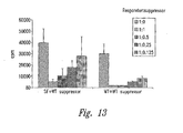
- Transgenic animals are examined for their state of immune competence, using several different parameters. Animals are examined for the number of lymphoid cells present in lymph nodes and thymus ( Figure 7 ) as well as the responsiveness of T cells to in vitro stimulation ( Figure 8 ).
- mice which express excess levels of the normal FKH sf protein contain roughly one-third as many cells ( Figure 7 ). Further, the number of thymocytes is normal ( Figure 7 ) as is their cell surface phenotype as assessed by flow cytometry using standard antisera (not shown), indicating that there is no developmental defect associated with excess FKH sf protein.
- CD4+ T cells are reacted with antibodies to CD3 and CD28 and their proliferative response measured using radioactive thymidine incorporation. Whereas only scurfy cells divide in the absence of stimulation, normal cells respond well following stimulation. FKH sf transgenic cells also respond to stimulation, however the response is significantly less than that of normal cells ( Figure 8 ). This indicates that CD4+ T cells that express excess FKH sf have a diminished capacity to respond to stimuli.
- the 5' ends of the two sequences differ in their location within the context of the genomic DNA sequence, the second coding exon of FKH sf is omitted from JM2, and the last intron of the FKH sf gene is unspliced in the JM2 sequence. These differences result in a JM2 protein with a shorter amino-terminal domain, relative to FKH sf , and a large insertion within the forkhead domain (see below) at the carboxy-terminus.
- the FKH sf protein can be divided into sub-regions, based on sequence motifs that may indicate functional domains.
- the two principal motifs in FKH sf are the single zinc finger (ZNF) of the C 2 H 2 class in the middle portion of the protein, and the forkhead, or winged-helix domain at the extreme carboxy-terminus of the protein.
- ZNF single zinc finger
- the unique features of the FKH sf gene sequence may be used to identify other novel genes (and proteins) which fall into the same sub-class of forkhead-containing molecules.
- the FKN sf protein is unique in its having a single zinc finger domain amino-terminal to the forkhead domain as well as in the extreme carboxy-terminal position of the forkhead domain.
- a degenerate PCR approach may be taken to isolate novel genes containing a zinc finger sequence upstream of a forkhead domain.
- the following degenerate primers were synthesized (positions of degeneracy are indicated by parentheses, and "I" indicates the nucleoside inosine):
- the forward primer corresponds to a region within the zinc finger sequence and the reverse primer corresponds to a region in the middle of the forkhead domain.
- These primers were used to amplify first-strand cDNA produced as in Example 2 from a variety of human tissues (including liver, spleen, brain, lung, kidney, etc.). The following PCR conditions were used: forward and reverse primers at 0.2 mM final concentration, 60 mM Tris-HCl, 15 mM ammonium sulfate, 1.5 mM magnesium chloride, 0.2 mM each dNTP and 1 unit of Taq polymerase, subjected to 35 cycles (94°C for 30 sec, 50°C for 30 sec, 72°C for 2 min).
- PCR products were visualized on a 1.8% agarose gel (run in 1x TAE) and sub-cloned into the TA cloning vector (Invitrogen, Carlsbad, CA); individual clones were sequenced and used for further characterization of full-length cDNAs.
- the unique regions of the FKN sf gene may be used to screen cDNA libraries by hybridization.
- cDNA libraries derived from a variety of human and/or mouse tissues, and propagated in lambda phage vectors (eg., lambda gt11) were plated on agarose, plaques were transferred to nylon membranes and probed with fragments derived from the unique regions of the FKH sf gene.
- mice were generated by oocyte microinjection by DNX Transgenic Services (Cranbury, NJ), as described (Brunkow et al., Nat. Gen. 27 :68-72, 2001).
- DNX Transgenic Services Cranbury, NJ
- a 30.8 kb cosmid construct was generated from mouse BAC K60 for injection. This cosmid contains the entire Foxp3 gene along with approximately 18 kbp of 5' sequence and 4 kbp of 3' sequence. Expression of the gene parallels that of the endogenous gene with respect to tissue distribution ( Brunkow et al., Nat. Gen.
- the approximate transgene copy number was determined by Southern blot analysis and correlated well with Foxp3 gene expression ( Brunkow et al., Nat. Gen. 27:68-72, 2001 ). Although there is a less dramatic, but consistent, difference in the number of splenic cells in the transgenic mice as well, the number of thymocytes is not significantly affected. For reasons of simplicity, except where noted, the remainder of the experiments utilized the 2826 transgenic line. Animals from this line are generally healthy and survive for greater than one year under SPF conditions. The line has approximately 16 copies of the transgene and by northern blot analysis is expressed at ten to twenty times the level of the endogenous gene in lymphoid tissues ( Brunkow et al., Nat. Gen.
- the transgene like the endogenous gene, is only poorly expressed in non-lymphoid tissues, a likely consequence of its expression under the control of its endogenous promoter.
- Lymph node cell number in mice from this line range from 15-50 percent of normal, with the number of cells accumulating with age. Splenic cell number is less dramatically affected although generally decreased, with a range of 25-90 percent of normal.
- Table 1 Genotype Transgene Copy Number Thymus Cell Number (x10 6 ) Lymph Node Spleen NLC NA 121.4 1.5 84.4 2826 ⁇ 16 111.8 0.5 60.8 1292 -9 98.6 1.0 76.4 2828 ⁇ 45 108.5 0.4 61.1 Scurfy NA 64.4 4.7 109.5
- Thymus, lymph node and splenic tissues were collected as described ( Clark et al., Immunol 162:2546, 1999 ) and were resuspended in staining buffer (1% BSA, 0.1% sodium azide in PBS) at a cell density of 20x10 6 /mL.
- mAbs fluorochrome-conjugated anti-mouse monoclonal antibodies
- CD3, CD8 ⁇ , CD4, CD25, IgG2a control Caltag Laboratories, Burlingame, CA
- CD28, CD45RB, CD44, CD62L, CD69, CD95 PharMingen, San Diego, CA.
- the fluorescence intensity of approximately 10 5 cells was examined using a MoFlo TM flow cytometer (Cytomation, Fort Collins, CO) with dead cell exclusion by addition of propidium iodide (10 ⁇ g/mL).
- CD4 - 8 - subset also reveals a normal distribution of gamma-delta cells and CD25 + cells. Importantly, the fraction of CD4 + 8 - thymocytes expressing the maturation markers CD69 and HSA is identical in 2826 and control animals, suggesting that the maturation process is normal.
- mice expressing Foxp3 selectively in the thymus (16.5 and 8.3) under control of the lck proximal promoter were crossed to sfl + carrier females.
- Male scurfy mice ( sfl Y) that carried the thymus-specific transgene (16.5 and 8.3) succumbed to disease at the same time and in the same manner as non-transgenic littermates.
- Sf /Y transgenic animals expressing Foxp3 under its endogenous regulatory sequences (2826) did succumb to disease.
- Cell number is derived from mice that carried the transgene in addition to the wild-type Foxp3 gene.
- Transgenic animals that express the Foxp3 gene exclusively in thymus are unable to rescue sf /Y mice from disease (see, Table 2 below).
- Two separate founder animals were crossed to scurfy carrier females in an attempt to prevent disease.
- sf /Y mice carrying the lck proximal promoter -Foxp3 transgene developed an acute lymphoproliferative disease that was identical both in severity and time course to that seen in non-transgenic sf /Y siblings.
- expression of the transgene was restricted to the thymus with no detectable expression in peripheral organs, including spleen.
- the Northern Blot analysis was carried out as presented in Example 1.
- thymic expression of the lck-driven transgene was substantially greater than that of the gene in 2826 transgenic animals or of the endogenous gene in normal littermate control mice. Hence it appears that the fatal lymphoproliferative disease seen in sf /Y mice does not arise as a consequence of scurfin mediated developmental defects in the thymus.
- transgenic (non- sf ) animals carrying the lck-driven transgene appear generally normal, high level expression of the transgene within the thymus does have phenotypic consequence in normal (non- sf ) animals.
- Significantly increased expression of the transgene in otherwise normal mice leads to a relative decrease in the percentage of double-positive thymocytes and a corresponding increase in the percentage of double-negative (DN) cells, as well as a decrease in overall thymic cell number (see, Table 2).
- T cell development still occurs in these animals as assessed by the generation of CD4 and CD8 single positive cells and by the presence of relatively normal numbers of peripheral T cells in both lymph node and spleen ( see , Table 2).
- CD69 expression on CD4 + 8 - cells from the thymus is similar in transgenic and wild-type littermates, suggesting positive selection likely proceeds normally, whereas within the DN compartment, the fraction of cells expressing CD25 is diminished relative to wild-type animals.
- transgenic animals indicate that overexpression of the Foxp3 gene within the thymic compartment specifically can after thymic development, but this appears to have no effect on regulating peripheral T cell activity.
- CD4 + 8 - cells In addition to a decrease in the number of peripheral T cells in 2826 mice, there is a slight reduction in the percentage of CD4 + 8 - cells in both the lymph node and spleen relative to NLC. Whereas the CD3 levels appear normal on peripheral T cells, there are a number of other surface markers with altered expression levels. For CD4 + 8 - cells in the transgenic mice, the most consistent changes are a small decrease in the expression of CD62L and CD45RB as well as an increase in the expression of CD95. By comparison, cells from sf mutant animals have a very different phenotype. CD4 + 8 - cells from these mice are large and clearly activated. They are predominantly CD44 H1 , CD45RB LO , CD62L LO and partially CD69 + ( Clark et al., Immunol 162:2546 ).
- CD4 - 8 + cell numbers were also reduced in both the spleen and lymph nodes of scurfin-transgenic mice. This decrease is typically more dramatic (50-75%) than the decrease in the CD4 + 8 - compartment (25-50%).
- CD4 - 8 + T cells display relatively minor and variable changes in the level of CD62L, CD45RB and CD95 on the cell surface in comparison to NLC. In contrast to CD4 + 8 - T cells, there is a more pronounced increase in the percentage of CD4 - 8 + T cells that were also CD44 HI . Overall, the CD4 - S + cells do not express surface markers at levels that characterize them as specifically naive, activated or memory.
- lymphoid organs were also perturbed. Histological examination of the major lymphoid organs (thymus, lymph node and spleen) indicated that the most significant changes were found in the mesenteric and peripheral lymph nodes. Tissues for histological analysis were removed from mice approximately 8 weeks after birth and immediately fixed in buffered 10% formalin. Paraffin-embedded sections were processed for hematoxylin and eosin staining and comparative histopathology performed on representative mice.
- the thymus appears relatively normal, with a well-defined cortico-medullary junction, although there appears to be a slight reduction in the size of the thymic medulla.
- Transgenic animals had smaller peripheral lymph nodes, lack robust and normally distributed lymphoid follicles, lack distinct margins between follicular and interfollicular areas and had more obvious sinuses than those found in the lymph nodes of the normal littermate control mice. Even though the spleen and Peyer's patches appear approximately normal in size and microarchitecture, there is a moderate decrease in total cell number and no or minimal evidence of germinal centers in these tissues.
- the changes noted here reflect a hypacellular state distinct from a number of other targeted mutations in which the lymph nodes fail to develop. Therefore, while T cells are capable of development in an apparently normal manner, their representation within the peripheral lymphoid tissues, particularly the lymph nodes, is substantially decreased.
- CD4 T cells The phenotypic and cell number data suggest that there are specific defects in the biology of CD4 T cells from 2826 transgenic animals.
- the functional responses of T cells from these animals to several stimuli were evaluated, including anti-CD3 and anti-CD28.
- Lymphocytes were isolated from various tissues from NLC, 2826 transgenic or scurfy (mutant) mice and CD4 cells were purified by cell sorting. Thymus, lymph node and splenic tissues were removed from appropriate animals, macerated between sterile microscope slides, filtered through a sterile 70 ⁇ m nylon mesh and collected by centrifugation.
- CD4 + T lymphocytes were sort purified from these tissues by positive selection using the MoFlo. Sort purities as determined by post-sort analysis were typically greater than 95%.
- RPMI complete RPMI
- cRPMI complete RPMI
- fetal bovine serum fetal bovine serum
- 0.05 mM 2-mercaptoethanol 15 mM HEPES, 100 U/mL penicillin, 100 ⁇ g/mL each streptomycin and glutamine
- Culture wells were prepared for T cell activation by pre-incubation with the indicated concentrations of purified antibody to CD3 (clone 2C11) in sterile PBS for 4 hours at 37°C.
- Purified ⁇ -mouse CD28 (clone 37.51) or ⁇ -mouse KLH (control antibody) was co-immobilized at 1 ⁇ g/ml final concentration.
- T cells were cultured at a cell density of 1 to 5x10 4 cells/well in 200 ⁇ L of cRPMI for 72 hours. Supernatant (100 ⁇ l) was removed at 48 hours for analysis of cytokine production. Wells were pulsed with 1 ⁇ Ci/well of 3 [H]-thymidine (Amersham Life Science, Arlington Heights, IL) for the last 8-12 hours of culture and then harvested (Tomtec). Proliferation data reported are based upon mean value of triplicate wells and represent a minimum of 3 experiments. Cytokine levels were determined by ELISA assay according to the manufacturer's direction (Biosource International, Camarillo, CA).
- IL-2 IL-2 was added at 100 U/ml.
- proliferation assays cells were pulsed after five days and harvested as above. Both proliferation and IL-2 production are significantly diminished in cells from the transgenic animals compared to their littermates. Although transgenic animals increase their responsiveness with increasing stimulation, they rarely reached the levels achieved by NLC. This is particularly true for IL-2 production, in which cells from 2826 mice consistently produce low to undetectable amounts of this cytokine. Similar results were seen whether the cells were derived from the spleen or the lymph nodes.
- IL-2 production by thymic CD4 + 8 - cells is approximately comparable between transgenic and NLC mice.
- IL-2 production by thymic CD4 + 8 - cells is reduced substantially from the transgenic animals.
- the reduction in IL-2 production by thymocytes is somewhat more variable than that seen in lymph node or spleen and may suggest that the IL-2 produced is also consumed during the culture.
- thymocytes may produce other growth factors less affected by the expression of the Foxp3 gene. Nevertheless, the data generally support the conclusion that a major defect in the transgenic animals is in the ability of both thymic and peripheral T cells to produce IL-2.
- transgenic T cells to generate and function as cytotoxic T cells (CTL) was determined in an in vitro assay.
- a single cell suspension of Balb/c spleen cells was generated to use as stimulator cells. These cells were irradiated (3300 rads) and incubated a 10:1 ratio (stimulator:effector) with scurfin-transgenic or NLC spleen cells.
- IL-2 was added at 100 U/ml.
- splenic T cells were stimulated in a similar manner in the presence of 100 U/ml of IL-2. After five days, cells were either assayed in the JAM assay ( Matzinger, P. J Immunol 145(1-2):185 (1991 )) or re-stimulated on a new stimulator layer. Cells were approximately 95% CD4 - 8 + .
- Transgenic T cells were stimulated in a mixed-lymphocyte culture containing increasing numbers of irradiated allogeneic stimulator cells in the presence or absence of IL-2. The proliferative response of either transgenic or NLC effector cells was then measured. T cells from the transgenic animals responded poorly in the absence of exogenous IL-2, consistent with the data for purified CD4 + 8 - cells (above). In the presence of exogenous IL-2, transgenic T cells displayed an increased proliferative response, but still required a higher number of stimulator cells to reach a similar level of proliferation as control cells. The ability of mixed T cell populations to respond to stimulation in this assay may reflect the presence of both CD4 + 8 - and CD4 - 8 + T cells in these cultures.
- CD4 - 8 + T cells were generated using allogeneic feeder cells in the presence of IL-2 and assayed to determine the ability of these cells to lyse target cells.
- Balb/c spleen cells were stimulated with PMA (10 ng/ml) in the presence of ionomycin (250 ng/ml) for 24 hours to allow for efficient loading of cells with 3 [H]-thymidine. After 24 hours, 3 [H]-thymidine (5 ⁇ Ci/ml) was added to PMA+Ionomycin-stimulated Balb/c spleen cells.
- CD4 - 8 + effector cells were plated with target Balb/c cells at increasing ratios ranging from 1.5:1 to 50:1 (effector:target) in a 96-well flat-bottom plate (experimental) in a final volume of 100 ⁇ l. The cells were pelleted by centrifugation and incubated at 37°C for four hours. A plate containing labeled Balb/c cells alone was harvested immediately and used to determine total counts (TC). A second plate containing labeled Balb/c cells alone was also incubated at 37°C for four hours to determine spontaneous release (SR). After four hours of incubation, cells were harvested onto glass fiber and counted in a scintillation counter.
- TC total counts
- SR spontaneous release
- effector-to-target ratios 50:1 and 25:1
- scurfin-transgenic CD4 - 8 + cells were as effective at lysing target cells as cells generated from NLC, while at the intermediate ratios (12.5-3:1), transgenic cells were significantly reduced in their cytolytic function in comparison to NLC. However, the transgenic cells were still effective with 50-60% lysis at these intermediate ratios.
- exogenous IL-2 was required to generate functional CD4 - 8 + T cells, presumably due to the poor endogenous production of this cytokine.
- transgenic animals made a consistently poor response to Oxazalone at all times examined, whereas control animals responded normally.
- Further studies using animals transgenic for both a TCR and Foxp3 will examine in vivo responses in greater detail.
- C3H SCID mice were purchased from The Jackson Laboratory (Bar Harbor, Maine). All animals were housed in specific pathogen free environment and studies were conducted following PHS guidelines.
- the double mutants were backcrossed to Mus musculus castaneous to obtain recombinants carrying either (Otc spf ) mutation or sf mutation ( Brunkow et al., Nat Gen 27:68-72, 2001 ).
- the single mutant sf strain was maintained by breeding carrier females to F1 males of (C3Hf/rl x 101/RI) or (101/RI x C3H/RI). Sf males were used at age 15-21 days and wildtype control animals were used at 6-12 weeks of age. Scurfy or wildtype CD4 + T were purified by cell sorting. The cells were resuspended in 0.9% saline, pH 7.2 and mixed at different ratios in a final volume of 200 ⁇ l and injected into SCID mice via tail-vein. Mice were monitored weekly for weight loss. Approximately 50 ⁇ l of blood was collected by eye-bleeds. Red blood cells were lysed and leukocytes were stimulated at 5 x 10 4 cells/well for 48 hours with immobilized anti-CD3 (5 ⁇ g/ml) and anti-CD28 (1 ⁇ g/ml).
- mice that received sf T cells showed signs of wasting (seen as weight loss) 3-4 weeks post-transfer that became progressively worse, whereas the mice that received a mixture of sf and wildtype T cells showed a normal weight gain corresponding to their age ( Figure 11A ).
- Mice that received only wildtype T cells showed a similar weight gain with age.
- mice receiving only sfT cells developed an inflammatory reaction around, but not within, the eye that persisted throughout the experiment. If the disease was allowed to progress, the recipients of sfT cells only died 8-16 weeks after transfer. Recipients of a mix of sf and wildtype T cells remained healthy throughout the experiment (experiments done up to 16 weeks).
- tissues were removed from C3H/SCID mice receiving either sf T cells, wildtype T cells or a mixture of sf and wildtype T cells.
- Intestines were flushed with cold PBS and immediately fixed in 10% formalin. Paraffin embedded sections were processed for hematoxylin and eosin staining and comparative histopathology (Applied Veterinary Pathobiology, Bainbridge Is., WA).
- lymph nodes and spleen from sf -recipient animals were substantially enlarged compared to their controls, indicating a marked lymphoproliferative process. Lymph nodes were collected from 6-12 weeks old mice and macerated in DMEM+10% FBS in between the ground glass ends of sterile microscope slides. The cells were filtered through 70 ⁇ M nylon mesh, collected by centrifugation and resuspended at ⁇ 50 x 10 6 cells/ml in complete media.
- CD4 + T cells from sf mice have been shown to be hyperproliferative and to secrete large amounts of cytokines such as IL-2, IL-4 and IFN- ⁇ ( Blair et al., J. Immunol. 153:3764-774 (1994 ); Kanangat et al., Eur. J. Immunol. 26:161-165 (1996 )).
- cytokines such as IL-2, IL-4 and IFN- ⁇
- PBMC from various recipients were stimulated with anti-CD3 and anti-CD28 in vitro for 48 hours and secreted IL-4 was detected by ELISA kit (BioSource International, Camarillo, CA) according to manufacturer's instruction.
- IL-4 was produced by PBMC from all the recipients ( Figure 11c ).
- PBMC from recipients of sf T cells secreted significant amounts of IL-4 whereas the PBMC of mice receiving either wildtype T cells only or a mixture of sf and wildtype T cells secreted little IL-4.
- T-reg cells peripheral CD4 + T cells
- T-reg cells were responsible for the inhibition of disease seen after co-transfer of sf and wildtype CD4 + T cells in vivo.
- Two million sf CD4 + T cells were mixed at different ratios either with wildtype CD4 + CD25 - T cells or with wildtype CD4 + CD25 + T-reg cells and injected into C3H/SCID mice.
- the recipients were monitored for weight loss and IL-4 secretion by PBMC as described in Example 1.
- PBMC as described in Example 1.
- T-reg cells these were stained with anti-CD4-FITC (Caltag Laboratories, Burlingame, CA) and anti-CD25-biotin (Caltag) for 30 min on ice.
- the cells were washed twice with PBS and stained with strepavidin-APC (Molecular Probes, Eugene, OR) for 20 min on ice. Cells were washed twice and positive sorted for CD4 + CD25 + T cells.
- mice receiving sf T cells alone showed signs of wasting ( Figure 12 ) and IL-4 production.
- mice that received a mixture of sf T cells and higher doses (110,000 or more) of wildtype CD4 + CD25 + T-reg cells showed a marked reduction in signs of disease such as weight loss.
- mice that received a mix of sf and CD4 + CD25 - T cells showed signs of disease at all doses except when the number of CD4 + CD25 - T cells was greater than 1.1 million.
- the small amount of suppression seen with higher numbers of CD4 + CD25 - T cells may indicate that there are additional mechanisms of suppression or that CD4 + CD25 - T cells give rise to CD4 + CD25 + T-reg cells post-transfer. It seems unlikely that the mechanism of inhibition by CD4 + CD25 - T-reg involves in vivo competition for lymphoid space, since as few as 1.1x10 5 T-reg can inhibit the activity of 2x10 6 sf T cells.
- CD4+CD25+ T-reg cells were less effective in inhibiting sf CD4 + T cells than wildtype CD4 + T cells. This result, like that seen with co-transfer in vivo, indicates that the hyper-responsive state of sf T cells can be regulated by T-reg cells.
- spleens were collected in a similar fashion as lymph nodes and stained with anti-Thy-1-FITC or anti-Thy1-PE (Caltag). Cells were washed and negative sorted for Thy-1. The cells were sorted using a MoFlo flow cytometer (Cytomation, Fort Collins, CO) and Cyclops (Cytomation) software at a rate of 10-20,000/min. Cell doublets and monocytic cells were eliminated on the basis of forward and side scatter gates, and dead cells were excluded by propidium iodide (10 ⁇ g/ml) stain. The purity of the sorted cell population was routinely 90-99%.
- Thy-1 - APC were treated with mitomycin C (Sigma, 50 ⁇ g/ml) for 20 min at 37°C and washed three times with DMEM+10% FBS before using in proliferation assays.
- mitomycin C Sigma, 50 ⁇ g/ml
- CD4+ T cells were stimulated at 5 x 10 4 cells/well in 200ul DMEM+10% FBS with soluble anti-CD3 (2C11; Pharmingen) at 1 ⁇ g/ml and an equal number of mitomycin C treated Thy-1 - APC from spleens.
- MoFlo sorted CD4 + CD25 + T cells were added at various ratios.
- CD4 + CD25 + or CD4 + CD25 - T cells were stimulated at 5 x 10 4 cells/well in 200 ⁇ l DMEM+10% FBS with soluble anti-CD3 (1 ⁇ g/ml for wildtype cells or 10 ⁇ g/ml for Foxp3 transgenic cells), 4 ng/ml rIL-2 (Chiron) and an equal number of mitomycin-C treated Thy-1 - APC from spleens.
- the cells were harvested at 72 hours, stained with CD4-FITC or CD4-PE and positively sorted for CD4 using a MoFIo flow cytometer as described above. These cells were then added to the regulatory T cell assay as previously described at the same ratios as freshly isolated CD4 + CD25 + T cells.
- TGF- ⁇ (R&D) was added at a final concentration of 2.5 ng/ml. Cultures were incubated for indicated time periods at 37°C. Individual wells were pulsed with 1 ⁇ Ci/well with [ 3 H] thymidine (Amersham Life Sciences, Arlington, IL) for the last 8-12 hours of culture. Proliferation data are mean value of triplicate wells and represent a minimum of three experiments.
- TGF- ⁇ inhibition was specific to T cells since proliferation and cytokine production by B cells as well as monocytes, both stimulated with LPS, from sf mice were sensitive to TGF- ⁇ inhibition, It is worth noting however, that high levels of exogenous IL-2 can largely overcome the inhibitory effect of TGF- ⁇ on T cells, potentially by downregulating TGF- ⁇ receptor expression ( Cottrez et al., J. Immunol. 167:773-778, 2001 ). T cells from sf animals produce extremely high levels of IL-2 upon stimulation, and this may contribute to the lack of inhibition by TGF- ⁇ on T cell function of sf mice.
- TGF- ⁇ production by T-reg cells in in vitro assays is not clear. Most experimental systems do not point to a role for TGF- ⁇ in this system, although the in vivo data does indicate an important role for TGF- ⁇ in inhibitory activity of CD4 + CD25 + T-reg cells ( Read et al., J. Exp. Med. 192:295-302, 2000 ).
- the Foxp3 gene is expressed at highest levels in lymphoid tissues such as thymus, lymph node and spleen ( Brunkow et al., Nature Genetics 27:68-72, 2001 ).
- the lymphoid expression of Foxp3 seems to be predominantly in CD4 + T cells, since the level of expression in CD8 + T cells as well as B cells was significantly lower or undetectable ( Brunkow et al., Nature Genetics 27:68-72, 2001 ).
- CD4 + CD25 + and CD4 + CD25 - T cells were collected as described above.
- Oligo dT primed first-strand cDNA was synthesized from these cells using the Superscript Preamplification System (Gibco-BRL, Rockville, MD) and used as a template for real-time RT-PCR using an ABI Prism 7700 instrument.
- Foxp3 expression was measured using the primers 5'-GGCCCTTCTCCAGGACAGA-3' (SEQ ID NO:17) and 5'-GCTGATCATGGCTGGGTTGT-3' (SEQ ID NO:18) at a final concentration of 300 nM and with internal TaqMan probe 5'-FAM-AGCTTCATCCTAGCGGTTTGCCTGAG-MTAC-TAMRA-3' (SEQ ID NO:19) at a final concentration of 100 nM. Dad1 was used as an endogenous reference (Hong et al., 1997).
- Dad1 primers were 5'-CCTCTCTG-GCTTCATCTCTTGTGT-3' (SEQ ID NO:20) and 5'-CCGGAGAGATGCCTTGGAA-3' (SEQ ID NO:21), used at a final concentration of 50 nM and TaqMan probe 5'-6FAM-AGCTTCATCCTAGCGGTTTGCCTGAGAATAC-TAMARA-3' (SEQ ID NO:22) at a final concentration of 100 nM.
- Other components of the PCR mix were from the TaqMan Universal Master Mix (PE Applied Biosystems). PCR cycling conditions were 50°C for 2 min; 95°C for 10 min; and 40 cycles of 95°C for 15 seconds, 60°C for 1 minute.
- CD4 + CD25 + cells in sf mutant animals also expresses CD25, although this population is large in size and expresses CD69, indicating they are likely cells previously activated in vivo. Nonetheless, it was determined that these CD4 + CD25 + cells from the sf mutant animals do not show enhanced amounts of Foxp3 message, indicating that these cells are likely not T-reg in nature.
- CD4 + CD25 + T-reg cells express certain markers such as CTLA-4, OX-40, GITR ( McHugh, R. S. et al. Immunity 16: 311-23, 2002 ); Shimizu, J. et a1: Nature Immunology 3: 135-42, 2002 ) that are characteristics of activated T cells.
- CTLA-4 CTLA-4
- OX-40 OX-40
- GITR GITR
- CD4 + CD25 + T-reg cells decreased slightly after activation. This indicated that Foxp3 unlike any other markers reported so far on CD4 + CD25 + T-reg cells was specific to this subset and was unrelated to the activated/memory phenotype of these cells.
- CD4 + CD25 + T-reg cell function the functional activity of CD4 + CD25 + and CD4 + CD25 - T cell subset from Foxp3 transgenic mice was examined. These animals have 16 fold more Foxp3 message than found in wildtype animals, with very high amounts in the CD4 + CD25 + subset. Additionally, there were fewer total CD4 + cells in these transgenic animals and those cells are hyporesponsive relative to their littermate controls.
- CD4 + CD25 + T-reg cells were activated at the same time as the responders. Since CD4 + T cells from Foxp3 transgenics were hyporesponsive to TCR stimulation it was likely that the Foxp3-Tg CD4+CD25+ T-reg cells were not getting activated to the same extent as the wildytpe CD4 + CD25 + T-reg cells during the assay. This raised the possibility that if CD4 + CD25 + T-reg cells from Foxp3 transgenics were activated to the same extent as wildtype cells they would exhibit higher regulatory activity.
- CD4 + CD25 + T cells were pre-activated in vitro with anti-CD3 in the presence of APC and IL-2 for 72 hours according to the previously published protocol ( Thornton et al., J. Immun. 164:183-190, 2000 ). Based on our previous observations the T cells from Foxp3 transgenic mice were activated with a higher dose of anti-CD3 in vitro to give comparable proliferation as the wildtype cells. These preactivated T cells were then tested in a T-reg assay. As'reported by others, preactivation of CD4 + CD25 + T cells in vitro made them much more potent suppressors.
- CD4 + CD25 - T cells from Foxp3 transgenics express Foxp3 at levels higher than wild-type CD4 + CD25 + T cells
- the CD4 + CD25 - T cells from Foxp3 transgenics also expressed GITR (TNFRSF18) that has recently been shown to modulate T-reg activity ( Figure 18 ).
- GITR TNFRSF18
- These cells did not express other activation associated T cell markers such as OX40, CTLA4 or Ly-6A/E (data not shown).
- This activity usually ranged from comparable to lower than that of CD4 + CD25 + T cells from the same mice. As expected, such suppressive activity was never detected with wild-type CD4 + CD25 - T cells. In contrast to the CD4 + CD25 + T cells, the suppressive activity of CD4 + CD25 - T cells from Foxp3 transgenic could not be enhanced by preactivation with anti-CD3 and IL-2 in vitro (data not shown). This further supports the idea that the expression of Foxp3 commits a T-cell to the T-reg lineage without a direct correlation with regulatory activity.
- sf mice The gene mutated in sf mice (Foxp3) has a critical role in the regulation of peripheral T cell responses. Loss of function mutations in the gene leads to a potentially fatal T cell mediated autoimmune disease both in mice and humans ( Bennett et al., Nature Genetics 27:20-21 (2001 ); Lyon et al., Proc. Nat'l. Acad. Sci. USA 87:2433-2437 (1990 ); Wildin et al., Nature Genetics 27:18-20 (2001 )). Additionally, overexpression of scurfin in transgenic mice leads to decreased peripheral T cell numbers and inhibition of a variety of T cell responses including proliferation and IL-2 production.
- wildtype T cells can inhibit disease caused by adoptive transfer of sf CD4+ T cells into SGID mice. These observations are very similar to those made by several other groups characterizing the activity of regulatory T cells. Similar to the observation made by Powrie et al., the disease caused by sf cells was inhibited by even a small number of CD4 + CD25 + T cells. CD4 + CD25 - T cells were less effective at inhibiting sf T cell activity in this model, which may be due to a subset of these cells developing into a T-reg cell subset and making the appropriate inhibitory factors or due to additional mechanisms of inhibition.
- the in vitro hyper-responsive state of sf T cells can be inhibited by the presence of wildtype CD4 + CD25 + cells, but not by the addition of TGF- ⁇ .
- data from in vitro T-reg experiments suggest a direct cell-cell interaction is required with no involvement of cytokines such as TGF- ⁇ ( Thornton et al., J. Exp. Med. 188:287-296 (1998 ); Thornton et al., J. Immun. 164:183-190 (2000 )).
- TGF- ⁇ has no inhibitory effect on activated T cells ( Cottrez et al., J. Immunol. 167:773-778 (2001 )) making it unlikely that CD4 + CD25 + T-reg cell inhibition of sf cells in vivo is mediated by TGF- ⁇ .
- CD4+CD25+ T-reg To assess whether the Foxp3 gene product plays a role in CD4+CD25+ T-reg cell function we measured the expression of Foxp3 in CD4+CD25+ T-reg and CD4+CD25- T cells and measured the regulatory activity of CD4+CD25+ T-reg cells from mice overexpressing the Foxp3 gene. In both wildtype and Foxp3 transgenics, CD4+CD25+ T-reg cells expressed the highest level of Foxp3 mRNA of all different cell populations tested to date. A comparison of functional activity of CD4+CD25+ T-reg cells from wildtype and Foxp3 transgenic mice showed no increase in regulatory activity in cells from transgenic mice, even following an optimal stimulation of these cells.
- CD4+CD25- T cells from Foxp3 transgenic animals did have suppressive activity. While it is not possible to phenotypically identify a subset of T-reg cells in sf mutant mice (due to the high level of endogenous activation), CD4+CD25+ cells isolated from mutant animals neither expressed the Foxp3 gene nor did they display any suppressive activity in vitro.
- Antibodies or NCEs that modulate scurfin expression are identified using the following methods:
- the scurfin promoter is cloned into commercially available Luciferase reporter vector (Promega, Madison, WI). This construct is then transfected into cells, such as a murine or human T cell line. Agents, such as antibodies generated against T cells, cytokines, receptors, or other proteins, in addition to small molecules, peptides, and cytokines, will be used to treat the transfected cells. The level of Luciferase activity is then determined using commercially available Luciferase assay systems (Promega) according to manufacturer's instruction to identify agents that either increase or decrease the expression of scurfin,
- agents such as those described above are incubated with primary T cells under conditions that allow for the modulation of scurfin expression.
- the scurfin expression will be measured using the RT-PCR method described above in Example 21.
- Agents identified by either of the above methods will be used directly for the treatment of an autoimmune disease.
- T cells will be isolated from patients of an autoimmune disease, treated with the specific agents identified above to induce scurfin expression and transferred back into the patients to suppress the activation of other T cells.
Abstract
Description
- The present invention relates generally to pharmaceutical products and methods and, more specifically, to methods for identifying compounds which can modulate the immune system, further, to methods for identifying proteins regulated by Scurfin and those that induce or inhibit Foxp3 expression.
- A number of autoimmune diseases, such as Inflammatory Bowel Disease, Multiple Sclerosis, rheumatoid Arthritis, and Asthma, involve immune dysregulation. In all these diseases, subsets of T cells are hyper-activated and contribute to an immune reaction towards self. In recent years, mice with mutations in CD95, CD95-ligand, CTLA-4 or TGF-β have proven useful for dissecting a number of pathways involved in T cell regulation and immune system homeostasis. Mice with mutations in any one if the above genes have profoundly altered immune responses, attributed to a failure to control T cell function.
- T cell activation in the periphery involves signaling via the T cell receptor and CD28 costimulation (reviewed in Bluestone, J.A., Immunity 2:555-559 (1995); Jenkins, M. K., Immunity 1:443-448 (1994); Rudd, C. E., Immunity 4:527-534 (1996)). Down regulation of peripheral T cell responses involves several pathways. Some of these include apoptosis mediated by members of the TNFR family, including CD95 and its ligand, activation induced death due to cytokine withdrawal, and negative signaling through CTLA-4 (CD152) (Lenardo et al., Ann. Rev. Immun. 17:221-253 (1999); Oosterwegel et al., Curr. Opin. Immun. 11:294-300 (1999); Saito, T., Curr. Opin. Immun. 10:313-321 (1998); Wallach et al., Ann. Rev. Immun. 17:331-367 (1999)). Mutations or expression of dominant negative forms of some of these genes have proven their critical role in the regulation of peripheral T cell responses. Mutations in CD95, CD95L, TGF-β or CTLA-4 lead to progressive autoimmune lymphoproliferative disorders (Kulkarni et al., Proc. Nat'l. Acad. Sci. USA 90:770-774 (1993); Shull et al., Nature 359:693-699 (1992); Takahashi et al., Cell 76:969-976 (1994); Tivol et al., Immunity 3:541-547 (1995); Watanbe-Fukunaga et al., Nature 356:314-347 (1992); Waterhouse et al., Science 270:985-988 (1995)). More recent data suggests that regulation of T cell activity by CD4+CD25+ regulatory T cells is also important for maintaining peripheral T cell tolerance (Roncarolo et al., Curr. Opin. Immun. 12:676-683 (2000); Sakaguchi, S., Cell 101:455-458 (2000); Shevach, E. M., Ann. Rev. Immun. 18:423-449 (2000)). Depletion of such regulatory T cells from normal animals leads to development of various autoimmune diseases and the adoptive transfer of these regulatory cells can also prevent disease in vivo in a number of systems (Asano et al., J. Exp. Med. 184:387-396 (1996); Sakaguchi et al., J. Immun. 155:1151-1164 (1995); Suri-Payer et al., J. Immun. 160:1212-1218 (1998)).
- The specific mechanism by which regulatory T cells (T-reg cells) mediate their suppressive effect is currently unclear. While TGFB and IL-10 can mediate suppressive effects, and blocking these cytokines eliminates suppression in some in vivo models, there is good evidence to indicate other molecules are also involved. Mounting evidence indicates a role for CD152 in the activation and/or function of CD4+CD25+ T cells (Read et al., J. Exp. Med. 192:295-302 (2000); Takahashi et al., J. Exp. Med. 192:303-310 (2000)). Intriguingly, several studies suggest that signaling through CD152 results in the induction of TGFB (Chen et al., J. Exp. Med. 188:1849-1857 (1998); Gomes et al., J. Immunol. 164:2001-2008 (2000); Kitani et al., J. Immunol. 165:691-702 (2000)), providing a potential link between TGFB-mediated inhibition and the inhibitory activity of CD4+CD25+ cells.
- The X-linked lymphoproliferative disease observed in the scurfy (sf) mouse, a spontaneous mutant animal that shares many characteristics with the pathogenesis seen in targeted deletions of CTLA-4 (Tivol et al., Immunity 3:541-547 (1995); Waterhouse et al., Science 270:985-988 (1995)) as well as TGF-β (Kulkarni et al., Proc. Nat'l. Acad. Sci. USA 90:770-774 (1993); Shull et al., Nature 359:693-699 (1992)), including death by three weeks of age (Godfrey et al., Am. J. Pathol. 145:281-286 (1994); Godfrey et al., Proc. Nat'l. Acad. Sci. USA 88:5528-5532 (1991); Godfrey et al., Am. J. Pathol. 138:1379-1387 (1991); Kanangat et al., Eur. J. Immunol. 26:161-165 (1996); Lyon et al., Proc, Nat'l. Acad. Sci. USA 87:2433-2437 (1990)). In sf animals, disease is mediated by CD4+ T cells, and these cells exhibit an activated phenotype both in vivo and in vitro (Blair et al., J. Immunol. 153:3764-774 (1994)). The specific mutation responsible for the disease has been recently cloned and the gene shown to be a new member of the forkhead family of transcription factors (Brunkow et al., Nature Genetics 27:68-72 (2001)). The gene has been designated Foxp3 and the protein product, scurfin. Mutations in the orthologous human gene cause a similar lymphoproliferative disorder among affected male progeny, which if left untreated is generally fatal (Bennett et al., Nature Genetics 27:20-21 (2001); Chatila et al., JM2, J. Clin. Invest. 106:R75-81 (2000); Wildin et al., Nature Genetics 27:18-20 (2001)).
- The present invention discloses methods and compositions useful for diagnosing scurfy-related diseases, more specifically, to methods for identifying compounds which can modulate the immune system, further, to methods for identifying proteins regulated by Scurfin and those that induce or inhibit Foxp3 expression
- The present invention relates generally to the discovery of novel genes which, when mutated, results in a profound lymphoproliferative disorder. In particular, a mutant mouse, designated 'Scurfy', was used to identify the gene responsible for this disorder through backcross analysis, physical mapping and large-scale DNA sequencing. Analysis of the sequence of this gene indicated that it belongs to a family of related genes, all containing a winged-helix DNA binding domain.
- Thus, within one aspect of the invention isolated nucleic acid molecules are provided which encode FKHsf or Fkhsf, including mutant forms thereof. Within certain embodiments, Fkhsf of any type may be from a warm-blooded animal, such as a mouse or human. Within further embodiments, isolated nucleic acid molecules are provided wherein the nucleic acid molecule is selected from the group consisting of (a) a nucleic acid molecule that encodes an amino acid sequence comprising SEQ ID NOs:2 or 4, (b) a nucleic acid molecule that hybridizes under stringent conditions to a nucleic acid molecule having the nucleotide sequence of SEQ ID NOs:1 or 3, or its complement, and (c) a nucleic acid molecule that encodes a functional fragment of the polypeptide encoded by either (a) or (b). Preferably, the nucleic acid molecule is not JM2. Within related aspects, vectors (including expression vectors), and recombinant host cells are also provided, as well as proteins which are encoded by the above-noted nucleic acid molecules. Further, fusion proteins are also provided which combine at least a portion of the above-described nucleic acid molecules with the coding region of another protein. Also provided are oligonucleotide fragments (including probes and primers) which are based upon the above sequence. Such fragments are at least 8, 10, 12, 15, 20, or 25 nucleotides in length, and may extend up to 100, 200, 500, 1000, 1500, or, 2000 nucleotides in length.
- Within other aspects methods of using the above noted expression vector for producing a Fkhsf protein (of any type) are provided, comprising the general steps of (a) culturing recombinant host cells that comprise the expression vector and that produce Fkhsf protein, and (b) isolating protein from the cultured recombinant host cells.
- Also provided are antibodies and antibody fragments that specifically bind to Fkhsf proteins. Representative examples of such antibodies include both polyclonal and monoclonal antibodies (whether obtained from a murine hybridoma, or derived into human form). Repesentative examples of antibody fragments include F(ab')2, F(ab)2, Fab', Fab, Fv, sFv, and minimal recognition units or complementarity determining regions.
- Within yet other aspects, methods are provided for detecting the presence of a Fkhsf nucleic acid sequence in a biological sample from a subject, comprising the steps of (a) contacting a Fkhsf specific nucleic acid probe under hybridizing conditions with either (i) test nucleic acid molecules isolated from said biological sample, or (ii) nucleic acid molecules synthesized from RNA molecules, wherein the probe recognizes at least a portion of nucleotide sequence of SEQ ID NOs:1 or 3, and (b) detecting the formation of hybrids of the nucleic acid probe and (i) or (ii).
- Within another related embodiment, methods are provided for detecting the presence of an FKh sf , or a mutant form thereof, in a biological sample, comprising the steps of: (a) contacting a biological sample with an anti-Fkhsf antibody or an antibody fragment, wherein the contacting is performed under conditions that allow the binding of the antibody or antibody fragment to the biological sample, and (b) detecting any of the bound antibody or bound antibody fragment.
- Within other aspects of the invention, methods are provided for introducing Fkhsf nucleic acid molecules to an animal, comprising the step of administering a Fkhsf nucleic acid molecule as described herein to an animal (e.g., a human, monkey, dog, cat, rat, or, mouse). Within one embodiment, the nucleic acid molecule is contained within and expressed by a viral vector (e.g., a vector generated at least in part from a retrovirus, adenovirus, adeno-associated virus, herpes virus, or, alphavirus). Within another embodiment the nucleic acid molecule is expressed by, or contained within a plasmid vector. Such vectors may be administered either in vivo, or ex vivo (e.g., to hematopoietic cells such as T cells).
- Within other embodiments, transgenic non-human animals are provided wherein the cells of the animal express a transgene that contains a sequence encoding Fkhsf protein.
- In one preferred embodiment, a method is provided for regulating an immune function in a primate. The method comprises inserting a plurality of nucleic acid sequences that encode the Foxp3 protein into the lymphocytes of the primate; placing the nucleic acid sequence under the control of cytokine c; and activating expression of the nucleic acid sequences to increase the amount of the Foxp3 protein in the primate with cytokine c.
- Accordingly, it is an object of the present invention to provide an assay for use in identifying agents that alter expression of Foxp3. Specifically, an assay is provided to measure the induction or inhibition of Foxp3 under varying conditions. The expression altering agents include small molecules, peptides, polynucleotides, cytokines, antibodies and Fab' fragments.
- In one preferred embodiment, a method is provided for identifying a compound that modulates the level of expression of scurfin. The method comprises providing a composition comprising a reporter gene ligated to a scurfin promoter; contacting the composition with a test compound; determining the level of reporter gene expression; and comparing the level of reporter gene expression in (c) with the predetermined level of expression and thereby determining if the test compound modulates the expression of scurfin.
- In a preferred embodiment, the compound decreases the level of scurfin expression.
- In another embodiment, the compound increases the level of scurfin.
- In one embodiment the test compound is selected from the group consisting of: a monoclonal antibody, a polyclonal antibody, a peptide, and a small molecule.
- In another embodiment the test compound is selected from the group consisting of an organic molecule, a natural product, a peptide, an oligosaccharide, a nucleic acid; a lipid, an antibody or binding fragment thereof, and a cell.
- In yet another embodiment, the test compound is from a library of compounds.
- With other embodiments, the library is selected from the group consisting of a random peptide library, a natural products library, a combinatorial library, an oligosaccharide library and a phage display library.
- In one preferred embodiment, a method is provided for suppressing an immune response comprising contacting T cells of the mammal with a compound that increases scurfin expression in the T cell, wherein an immune response is suppressed.
- In one preferred embodiment, a method is provided for enhancing an immune response comprising contacting T cells with a compound that decreases scurfin expression in the T cell, wherein an immune response is enhanced.
- Within another related embodiment, a method for inhibiting an autoimmune response in a subject, wherein the method comprises administering to the subject a compound which increases scurfin expression, thereby inhibiting an autoimmune response by the subject.
- In a related embodiment the autoimmune response is selected from the group consisting of Inflammatory Bowel Disease, Psoriasis, Diabetes, Multiple Sclerosis, Rheumatoid Arthritis, and Asthma.
- In one preferred embodiment, a method is provided for enhancing an immune response to a disease in a subject, wherein the method comprises administering to the subject a compound which decreases scurfin expression, thereby treating the disease in the subject.
- In a related embodiment, a method is provided for enhancing an immune response to HIV or cancer in a subject, wherein the method comprises administering to the subject a compound which decreases scurfin expression, thereby treating HIV and cancer.
- In one preferred embodiment, a method for inhibiting graft versus host disease in a subject wherein the method comprises administering to the subject a compound that increases scurfin expression, thereby inhibiting tissue transplant rejection by the subject.
- In one preferred embodiment, a method is provided for inhibiting an autoimmune response in a patient comprising. The method comprising: isolating T cells from the patient; transducing the T cells with the scurfin gene; expanding the tranduced T cells; and reintroducing the transduced T cells into said patient, wherein an autoimmune disease in the patient is inhibited.
- In a related embodiment, a method is provided for inhibiting an autoimmune response in a patient comprising. The method comprising: isolating CD4+CD25+ regulatory T cells from the patient; transducing the CD4+CD25+ regulatory T cells with the scurfin gene; expanding the tranduced CD4+CD25+ regulatory T cells; and reintroducing the transduced CD4+CD25+ regulatory T cells into the patient, wherein an autoimmune disease in the patient is inhibited.
- In one preferred embodiment, a method is provided for inhibiting an autoimmune response in a patient, wherein the autoimmune disease is selected from the group consisting of Inflammatory Bowel Disease, Multiple Sclerosis, Rheumatoid Arthritis, Psoriasis, Diabetes and Asthma. The method comprising: isolating T cells from the patient; transducing the T cells with the scurfin gene; expanding the tranduced T cells; and reintroducing the transduced T cells into said patient, wherein an autoimmune disease in the patient is inhibited.
- In one preferred embodiment, a method is provided for inhibiting an autoimmune response in a patient comprising. The method comprising: isolating T cells from the patient; transducing the T cells with the scurfin gene contained in a retroviral vector; expanding the tranduced T cells; and reintroducing the transduced T cells into said patient, wherein an autoimmune disease in the patient is inhibited.
- In yet another preferred embodiment, a method for enhancing an immune response to a disease in a patient is provided. The method comprising: isolating T cells from the patient; transfecting the T cells with a test compound that inhibits scurfin expression; expanding the transfected T cells; and reintroducing the transfected T cells into said patient, wherein an immune response to a disease in the patient is enhanced.
- In yet another preferred embodiment, a method for enhancing an immune response to a HIV and cancer in a patient is provided. The method comprising: isolating T cells from the patient; transfecting the T cells with a test compound that inhibits scurfin expression; expanding the transfected T cells; and reintroducing the transfected T cells into said patient, wherein an immune response to HIV or cancer in the patient is enhanced.
- These and other aspects of the present invention will become evident upon reference to the following detailed description and attached drawings. In addition, various references are set forth herein which describe in more detail certain procedures or compositions (e.g., plasmids, etc.), and are therefore incorporated by reference in their entirety.
-
-
Figure 1A and1B depict a nucleotide sequence of mouse Fkhsf cDNA (SEQ ID NO:1); translation is predicted to initiate at position 259 and terminate at position 1546. -
Figure 2 depicts the amino acid sequence of mouse Fhhsf (SEQ ID NO:2). -
Figure 3A and3B depict a nucleotide sequence of 1735 bp corresponding to human FKHsf cDNA (SEQ ID NO: 3; including a 1293 bp coding region); translation is predicted to initiate atposition 55 and terminate at position 1348. -
Figure 4 depicts the sequence of a 431 amino acid human FKHsf protein (SEQ ID NO: 4). -
Figure 5 diagrammatically depicts a vector for generation of FKHsf transgenic mice. -
Figure 6 is a photograph which demonstrates that the FKHsf transgene corrects the defect in scurfy animals. -
Figure 7 is a diagram which shows that FKHsf tg mice have reduced lymph node cells, as compared to normal cells. -
Figure 8 is a diagram which shows that FKHsf transgenic mice respond poorly to in vitro stimulation. -
Figure 9 is a comparison of FKHsf and JM2 cDNAs. -
Figure 10 compares homology in various regions of human FKHsf and murine Fkhsf. -
Figure 11A is a graph monitoring the weight of both scurfy and wild-type mice. The mice were monitored for weight loss at regular intervals for 10weeks, Each data point is an average of 3 mice except afterweek 5 when one of the three mice died in sf CD4 transfer group (indicated by an arrow on the graph). The data is representative of more than 3 independent experiments. -
Figure 11B is a photograph of a tissue section. Large intestines from C3H/SCID mice receiving either sfT cells (left panel) or a mixture of WT and sfT cells (right panel) were fixed in formalin, sectioned and processed for hematoxylin and eosin staining. -
Figure 11C is a graph depicting IL-4 production from 5 x 10⁴ PBMC from C3H/SCID mice receiving either sf CD4+ T cells or a mixture of WT and sf CD4+ T cells or WT CD4+ T cells were stimulated with 5 µg/ml anti-CD3 and 1 µg/ml anti-CD28 immobilized onto round bottom plates. Supernatants were harvested at 48h and IL-4 levels were measured by ELISA. -
Figure 12A and 12B are graphs depicting the weight loss of mice treated with either CD4+CD25+ or CD4+CD25- T-regulatory cells. CD4+CD25+ T-regufatory subset mediates the suppression of disease caused by sf T cells in vivo. A mixture of 4 x 10⁶ sf T cells and varying numbers of wildtype CD4+CD25+ (a) or CD4+CD25- (b) T cells was transferred into C3/SCID mice via tail-vein injection. These mice were monitored for weight loss over a period of time. Each data point is an average of 3 mice except sf CD4 transfer group and sf CD4 + 1.1 x 10⁶ CD4+CD25- T cells which have 2 mice each in the group. Also, arrows on the graph indicate mice that died or were sacrificed due to disease progression. -
Figure 13 depicts a graph of a proliferation assay that determines the suppressor activity of CD4+CD25+ T regulatory cells. Sf CD4+ T cells can be inhibited by CD4+CD25+ T-regulatory cells in vitro. 5 x 10⁴ WT or sf CD4+ T cells were stimulated with anti-CD3 (1µg/ml) and 5 x 10⁴ mitomycin C treated Thy-1- APC. CD4+CD25+ T-regulatory cells were added at various ratios to the assay. The cells were cultured for 72 h and pulsed with [3H] thymidine for final 8 hrs of the culture. Data is mean of triplicates. -
Figure 14A depicts a graph of a proliferation assay in which 5 x 10⁴ WT or sf CD4+ T cells were stimulated with immobilized anti-CD3. TGF-β was added at a final concentration of 2.5 ng/ml at the beginning of the assay. The cells were cultured for 72 h and pulsed with [3H] thymidine for final 8 hrs of the culture. Data is mean of triplicates. -
Figure 14B depicts a graph of a proliferation assay in which 5 x 10⁴ WT or sf CD4+ T cells were stimulated with immobilized anti-CD3 (varying concentrations) and anti-CD28 (1 µg/ml). TGF-β was added at a final concentration of 2.5 ng/ml at the beginning of the assay. The cells were cultured for 72 h and pulsed with [3H] thymidine for final 8 hrs of the culture. Data is mean of triplicates. -
Figures 15A and 15B are graphs examining Foxp3 expression in cDNA samples from various cell subsets using a real-time RT-PCR method in which Dad1 served as an endogenous reference gene. Normalized Foxp3 values were derived from the ratio of Foxp3 expression to Dad1 expression. -
Figure 16 depicts the level of CD25 surface expression on CD4+ T cells from WT animals, Foxp3 transgenic animals, and scurfy animals. Lymph node cells from sf, Foxp3 transgenic or littermate controls were examined for the expression of CD25 expression on CD4+ T cells. Data is representative of six individual mice examined. -
Figure 17 is a graph depicting the level of proliferation in 5 x 10⁴ WT CD4+ T cells were stimulated with anti-CD3 (1µg/ml) and 5 x 10⁴ mitomycin C treated Thy-1- APC. CD4+CD25+ T-regulatory cells from WT or Foxp3 transgenics were added at various ratios to the assay. The cells were cultured for 72 h and pulsed with [3H] thymidine for final 8 hrs of the culture. Data is mean or triplicates. -
Figure 18 is a FACS plot evaluating the expression of surface markers associated with T regulatory cells and the suppressive activity of these cells. -
Figure 19 is a graph depicting the level of T cell inhibition in freshly isolated CD4+CD25- T cells from Foxp3 transgenic as tested in T-reg assays. - The invention relates to the discovery that the scurfin protein is involved in the generation and/or activity of the CD4+CD25+ subset of regulatory T cells. Foxp3 expression is directly correlated with cells of this regulatory phenotype and its expression is uniquely increased upon activation of this specific subset. Mutant (sf) animals appear to lack this subset, whereas Foxp3 transgenic animals appear to possess an increased percentage of CD4+CD25+ cells. Further, while the CD4+CD25+ subset from transgenic animals does not appear inhibitory on a per cell basis, the expression of Foxp3 is still elevated in this subset relative to their CD25- counterparts. Interestingly, overexpression of Foxp3 in CD4+CD25- T cells confers suppressive activity on these cells, although they remain less effective than CD4+CD25+ T cells. Overall, the data suggest that the recently described transcription factor, scurfin, is a critical regulator of immune cell function and may work primarily through the generation and/or activity of CD4+CD25+ regulatory T cells.
- The results from the Examples indicate that expression of scurfin (Foxp3 gene) can downregulate the immune system in part through regulatory T (T-reg) cell activity. Consequentially, if the expression of the endogenous Foxp3 gene can be induced in T cells it can be used to downregulate the immune response in a variety of autoimmune diseases such as Inflammatory Bowel Disease, Multiple Sclerosis, Rheumatoid Arthritis, Psoriasis, Diabetes, and Asthma or in other scenarios such as Graft versus Host disease. Furthermore, scurfin expression can be down-regulated to activate the immune system in cancer or AIDS.
- Prior to setting forth the Invention in detail, it may be helpful to an understanding thereof to set forth definitions of certain terms and to list and to define the abbreviations that will be used hereinafter.
- "Scurfy" refers to an inherited disease in mice which exhibit a severe lymphoproliferative disorder (see, e.g., Lyon et al., Proc. Natl. Acad. Sci. USA 87:2433, 1990). The responsible gene (mutant forms of which are responsible for the disease) is shown in Sequence I.D. Nos. 1 and 3.
- " Foxp3 " refers to the forkhead domain-containing gene, which is mutated in the scurfy mouse mutant. "Foxp3" refers to the protein encoded by the mouse Foxp3 gene. "FOXP3" refers to the human ortholog of the murine Foxp3 gene. "FOXP3" refers to the protein encoded by the human FOXP3 gene. The cDNA sequences for murine Foxp3 and human FOXP3 are disclosed in
U.S. Patent Application No. 09/372,668 wherein the mouse scurfy gene is designated Fkhsf and the human ortholog is designated FKHsf . The genomic sequence for human FOXP3 is disclosed in Genbank Accession No. AF235087. Genbank Accession No. AF235097 andU.S. Patent Application No. 09/372,668 are incorporated by reference in their entireties for all purposes. - "Molecule" should be understood to include proteins or peptides (e.g:, antibodies, recombinant binding partners, peptides with a desired binding affinity), nucleic acids (e.g., DNA, RNA, chimeric nucleic acid molecules, and nucleic acid analogues such as PNA), and organic or inorganic compounds.
- "Nucleic acid" or "nucleic acid molecule" refers to any of deoxyribonucleic acid (DNA), ribonucleic acid (RNA), oligonucleotides, fragments generated by the polymerase chain reaction (PCR), and fragments generated by any of ligation, scission, endonuclease action, and exonuclease action. Nucleic acids can be composed of monomers that are naturally-occurring nucleotides (such as deoxyribonucleotides and ribonucleotides), or analogs of naturally-occurring nucleotides (e.g., α-enantiomeric forms of naturally-occurring nucleotides), or a combination of both. Modified nucleotides can have modifications in sugar moieties and/or in pyrimidine or purine base moieties. Sugar modifications include, for example, replacement of one or more hydroxyl groups with halogens, alkyl groups, amines, and azido groups, or sugars can be functionalized as ethers or esters. Moreover, the entire sugar moiety can be replaced with sterically and electronically similar structures, such as aza-sugars and carbocyclic sugar analogs. Examples of modifications in a base moiety include alkylated purines and pyrimidines, acylated purines or pyrimidines, or other well-known heterocyclic substitutes. Nucleic acid monomers can be linked by phosphodiester bonds or analogs of such linkages. Analogs of phosphodiester linkages include phosphorothioate, phosphorodithioate, phosphoroselenoate, phosphorodiselenoate, phosphoroanilothioate, phosphoranilidate, phosphoramidate, and the like. The term "nucleic acid" also includes so-called "peptide nucleic acids," which comprise naturally-occurring or modified nucleic acid bases attached to a polyamide backbone. Nucleic acids can be either single stranded or double stranded.
- "Isolated nucleic acid molecule" is a nucleic acid molecule that is not integrated in the genomic DNA of an organism. For example, a DNA molecule that corresponds to a gene that has been separated from the genomic DNA of a eukaryotic cell is an isolated DNA molecule. Another example of an isolated nucleic acid molecule is a chemically-synthesized nucleic acid molecule that is not integrated in the genome of an organism.
- "Promoter" is a nucleotide sequence that directs the transcription of a structural gene. Typically, a promoter is located in the 5' region of a gene, proximal to the transcriptional start site of a structural gene. If a promoter is an inducible promoter, then the rate of transcription increases in response to an inducing agent, in contrast, the rate of transcription is not regulated by an inducing agent if the promoter is a constitutive promoter.
- "Vector" refers to an assembly which is capable of directing the expression of desired protein. The vector must include transcriptional promoter elements which are operably linked to the genes of interest. The vector may be composed of either deoxyribonucleic acids ("DNA"), ribonucleic acids ("RNA"), or a combination of the two (e.g., a DNA-RNA chimeric). Optionally, the vector may include a polyadenylation sequence, one or more restriction sites, as well as one or more selectable markers such as neomycin phosphotransferase or hygromycin phosphotransferase. Additionally, depending on the host cell chosen and the vector employed, other genetic elements such as an origin of replication, additional nucleic acid restriction sites, enhancers, sequences conferring inducibility of transcription, and selectable markers, may also be incorporated into the vectors described herein.
- "Isolated" in the case of proteins or polypeptides, refers to molecules which are present in the substantial absence of other biological macromolecules, and appear nominally as a single band on SDS-PAGE gel with coomassie blue staining. "Isolated" when referring to organic molecules means that the compounds are greater than 90% pure utilizing methods which are well known in the art (e.g., NMR, melting point).
- "Cloning vector" refers to nucleic acid molecules, such as a plasmid, cosmid, or bacteriophage, that has the capability of replicating autonomously in a host cell. Cloning vectors typically contain one or a small number of restriction endonuclease recognition sites at which foreign nucleotide sequences can be inserted in a determinable fashion without loss of an essential biological function of the vector, as well as nucleotide sequences encoding a marker gene that is suitable for use in the identification and selection of cells transformed with the cloning vector. Marker genes typically include genes that provide tetracycline resistance or ampicillin resistance.
- "Expression vector" refers to a nucleic acid molecule encoding a gene that is expressed in a host cell. Typically, gene expression is placed under the control of a promoter, and optionally, under the control of at least one regulatory element, Such a gene is said to be "operably linked to" the promoter. Similarly, a regulatory element and a promoter are operably linked if the regulatory element modulates the activity of the promoter.
- "Recombinant host" refers to any prokaryotic or eukaryotic cell that contains either a cloning vector or expression vector. This term also includes those prokaryotic or eukaryotic cells that have been genetically engineered to contain the cloned gene(s) in the chromosome or genome of the host cell.
- In eukaryotes, RNA polymerase II catalyzes the transcription of a structural gene to produce mRNA. A nucleic acid molecule can be designed to contain an RNA polymerase II template in which the RNA transcript has a sequence that is complementary to that of a specific mRNA. The RNA transcript is termed an "anti-sense RNA" and a nucleic acid molecule that encodes the anti-sense RNA is termed an "anti-sense gene." Anti-sense RNA molecules are capable of binding to mRNA molecules, resulting in an inhibition of mRNA translation.
- An "anti-sense oligonucleotide specific for Fkhsf " or a " Fkhsf anti-sense oligonucleotide" is an oligonucleotide having a sequence (a) capable of forming a stable triplex with a portion of the gene, or (b) capable of forming a stable duplex with a portion of an mRNA transcript. Similarly, an "anti-sense oligonucleotide specific for "Fkhsf " or a "Fkhsf anti-sense oligonucleotide" is an oligonucleotide having a sequence (a) capable of forming a stable triplex with a portion of the Fkhsf gene, or (b) capable of forming a stable duplex with a portion of an mRNA transcript of the Fkhsf gene.
- A "ribozyme" is a nucleic acid molecule that contains a catalytic center. The term includes RNA enzymes, self-splicing RNAs, self-cleaving RNAs, and nucleic acid molecules that perform these catalytic functions. A nucleic acid molecule that encodes a ribozyme is termed a "ribozyme gene."
- Abbreviations: YAC, yeast artificial chromosome; PCR, polymerase chain reaction; RT-PCR, PCR process in which RNA is first transcribed into DNA at the first step using reverse transcriptase (RT); cDNA, any DNA made by copying an RNA sequence into DNA form. As utilized herein "Fkhsf" refers to the gene product of the Fkhsf gene (irrespective of whether the gene is obtained from humans, mammals, or any other warm-blooded animal). When capitalized "FKHsf" the gene product (and gene) should be understood to be derived from humans.
- As noted above, the present invention relates generally to pharmaceutical products and methods and, more specifically, to methods and compositions useful for diagnosing scurfy-related diseases, as well as methods for identifying compounds which can modulate the immune system.
- Thus, as discussed in more detail below this discovery has led to the development of assays which may be utilized to select molecules which can act as agonists, or alternatively, antagonists of the immune system. Furthermore, such assays may be utilized to identify other genes and gene products which are likewise active in modulating the immune system.
- Briefly, the present invention is based upon the unexpected discovery that a mutation in the gene which encodes Fkhsf results in rare condition (scurfy) characterized by a progressive lymphocytic infiltration of the lymph nodes, spleen, liver and skin resulting in gross morphological symptoms which include splenomegaly, hepatomegaly, greatly enlarged lymph nodes, runting, exfoliative dermatitis, and thickened malformed ears (Godfrey et al., Amer. J. Pathol. 138:1379, 1991; Godfrey et al., Proc. Natl. Acad. Sci. USA 88:5528, 1991). This new member of the winged-helix family represents a novel component of the immune system.
- Methods which were utilized to discover the gene responsible for scurfy are provided below in Example 1. Methods for cloning the gene responsible for murine scurfy, as well as the human ortholog, are provided below in Examples 2 and 3. Methods for confirmation of gene identity and correlation with gene function, as determined using transgenic mice, are also provided in the Examples.
- Also provided by the present invention are methods for determining the presence of Fkhsf genes and gene products. Within one embodiment, such methods comprise the general steps of (a) contacting a Fkhsf specific nucleic acid probe under hybridizing conditions with either (i) test nucleic acid molecules isolated from the biological sample, or (ii) nucleic acid molecules synthesized from RNA molecules, wherein the probe recognizes at least a portion of an Fkhsf nucleotide sequence, and (b) detecting the formation of hybrids of said nucleic acid probe and (i) or (ii). A variety of methods may be utilized in order to amplify a selected sequence, including, for example, RNA amplification (see Lizardi et al., Bio/Technology 6:1197-1202, 1988; Kramer et al., Nature 339:401-02, 1989; Lomeli et al., Clinical Chem. 35(9):1826-31, 1989;
U.S. Patent No. 4,786,600 ), and nucleic acid amplification utilizing Polymerase Chain Reaction ("PCR") (seeU.S. Patent Nos. 4,683,195 ,4,683,202 , and4,890,159 ), reverse-transcriptase-PCR and CPT (seeU.S. Patent Nos. 4,876,187 , and5,011,769 ). - Alternatively, antibodies may be utilized to detect the presence of Fkhsf gene products. More specifically, within one embodiment methods are provided for detecting the presence of an Fkhsf peptide, or a mutant form thereof, in a biological sample, comprising the steps of (a) contacting a biological sample with an anti- Fkhsf antibody or an antibody fragment, wherein said contacting is performed under conditions that allow the binding of said antibody or antibody fragment to the biological sample, and (b) detecting any of the bound antibody or bound antibody fragment.
- Such methods may be accomplished in a wide variety of assay formats including, for example, Countercurrent Immuno-Electrophoresis (CIEP), Radioimmunoassays, Radioimmunoprecipitations, Enzyme-Linked lmmuno-Sorbent Assays (ELISA), Dot Blot assays, Inhibition or Competition assays, and sandwich assays (see
U.S. Patent Nos. 4,376,110 and4,486,530 ; see also Antibodies: A Laboratory Manual, supra). - Although various FKHsf or Fkhsf proteins and nucleic acid molecules (or portions thereof) have been provided herein, it should be understood that within the context of the present invention, reference to one or more of these proteins should be understood to include proteins of a substantially similar activity. As used herein, proteins are deemed to be "substantially similar" if: (a) they are encoded by a nucleotide sequence which is derived from the coding region of a gene which encodes the protein (including, for example, portions of the sequence or allelic variations of the sequence); (b) the nucleotide sequence is capable of hybridization to nucleotide sequences of the present invention under moderate, high or very high stringency (see Sambrook et al., Molecular Cloning: A Laboratory Manual, 2d ed., Cold Spring Harbor Laboratory Press, N.Y., 1989), or has at least 50%, 60%, 70%, 75%, 80%, 90%, 95%, or greater homology to the sequences disclosed herein, or, (c) the DNA sequences are degenerate as a result of the genetic code to the DNA sequences defined in (a) or (b). Further, the nucleic acid molecule disclosed herein includes both complementary and non-complementary sequences, provided the sequences otherwise meet the criteria set forth herein. Within the context of the present invention, high stringency means standard hybridization conditions (e.g., 5XSSPE, 0.5% SDS at 65°C, or the equivalent). For purpose of hybridization, nucleic acid molecules which encode the amino-terminal domain, zinc finger domain, middle domain, or forkhead domain (see Example 10) may be utilized.
- The structure of the proteins encoded by the nucleic acid molecules described herein may be predicted from the primary translation products using the hydrophobicity plot function of, for example, P/C Gene or Intelligenetics Suite (intelligenetics, Mountain View, California), or according to the methods described by Kyte and Doolittle (J. Mol. Biol. 157:105-32, 1982).
- Proteins of the present invention may be prepared in the form of acidic or basic salts, or in neutral form. In addition, individual amino acid residues may be modified by oxidation or reduction. Furthermore, various substitutions, deletions, or additions may be made to the amino acid or nucleic acid sequences, the net effect of which is to retain or further enhance or decrease the biological activity of the mutant or wild-type protein. Moreover, due to degeneracy in the genetic code, for example, there may be considerable variation in nucleotide sequences encoding the same amino acid sequence.
- Other derivatives of the proteins disclosed herein include conjugates of the proteins along with other proteins or polypeptides. This may be accomplished, for example, by the synthesis of N-terminal or C-terminal fusion proteins which may be added to facilitate purification or identification of proteins (see
U.S. Patent No. 4,851,341 , see also, Hopp et al., Bio/Technology 6:1204, 1988.) Alternatively, fusion proteins (e.g., FKH or Fkh-luciferase or FKH or Fkh-GFP) may be constructed in order to assist in the identification, expression, and analysis of the protein. - Proteins of the present invention may be constructed using a wide variety of techniques described herein. Further, mutations may be introduced at particular loci by synthesizing oligonucleotides containing a mutant sequence, flanked by restriction sites enabling ligation to fragments of the native sequence. Following ligation, the resulting reconstructed sequence encodes a derivative having the desired amino acid insertion, substitution, or deletion.
- Alternatively, oligonucleotide-directed site-specific (or segment specific) mutagenesis procedures may be employed to provide an altered gene having particular codons altered according to the substitution, deletion, or insertion required. Exemplary methods of making the alterations set forth above are disclosed by Walder et al. (Gene 42:133, 1986); Bauer et al. (Gene 37:73, 1985); Craik (BioTechniques, January 1985, 12-19); Smith et al. (Genetic Engineering: Principles and Methods, Plenum Press, 1981); and Sambrook et al. (supra). Deletion or truncation derivatives of proteins (e.g., a soluble extracellular portion) may also be constructed by utilizing convenient restriction endonuclease sites adjacent to the desired deletion. Subsequent to restriction, overhangs may be filled in, and the DNA religated. Exemplary methods of making the alterations set forth above are disclosed by Sambrook et al. (Molecular Cloning: A Laboratory Manual, 2d ed., Cold Spring Harbor Laboratory Press, 1989).
- Mutations which are made in the nucleic acid molecules of the present invention preferably preserve the reading frame of the coding sequences. Furthermore, the mutations will preferably not create complementary regions that could hybridize to produce secondary mRNA structures, such as loops or hairpins, that would adversely affect translation of the mRNA. Although a mutation site may be predetermined, it is not necessary that the nature of the mutation per se be predetermined. For example, in order to select for optimum characteristics of mutants at a given site, random mutagenesis may be conducted at the target codon and the expressed mutants screened for indicative biological activity. Alternatively, mutations may be introduced at particular loci by synthesizing oligonucleotides containing a mutant sequence, flanked by restriction sites enabling ligation to fragments of the native sequence. Following ligation, the resulting reconstructed sequence encodes a derivative having the desired amino acid insertion, substitution, or deletion. Mutations may be introduced for purpose of preserving or increasing activity of the protein, or, for decreasing or disabling the protein (e.g., mutant Fkh).
- Nucleic acid molecules which encode proteins of the present invention may also be constructed utilizing techniques of PCR mutagenesis, chemical mutagenesis (Drinkwater and Klinedinst, PNAS 83:3402-06, 1986), by forced nucleotide misincorporation (e,g., Liao and Wise Gene 88:107-11, 1990), or by use of randomly mutagenized oligonucleotides (Horwitz et al., Genome 3:112-17, 1989).
- The present invention also provides for the manipulation and expression of the above described genes by culturing host cells containing a vector capable of expressing the above-described genes. Such vectors or vector constructs include either synthetic or eDNA-derived nucleic acid molecules encoding the desired protein, which are operably linked to suitable transcriptional or translational regulatory elements. Suitable regulatory elements may be derived from a variety of sources, including bacterial, fungal, viral, mammalian, insect, or plant genes. Selection of appropriate regulatory elements is dependent on the host cell chosen, and may be readily accomplished by one of ordinary skill in the art. Examples of regulatory elements include: a transcriptional promoter and enhancer or RNA polymerase binding sequence, a transcriptional terminator, and a ribosomal binding sequence, including a translation initiation signal.
- Nucleic acid molecules that encode any of the proteins described above may be readily expressed by a wide variety of prokaryotic and eukaryotic host cells, including bacterial, mammalian, yeast or other fungi, viral, insect, or plant cells. Methods for transforming or transfecting such cells to express foreign DNA are well known in the art (see, e.g.,
Itakura et al., U.S. Patent No. 4,704,362 ; Hinnen et al., Proc. Natl. Acad. Sci. USA 75:1929-33, 1978;Murray et al., U.S. Patent No. 4,801,542 ;Upshall et al., U.S. Patent No. 4,935,349 ;Hagen et al., U.S. Patent No. 4,784,950 ;Axel et al., U.S. Patent No. 4,399,216 ;Goeddel et al., U.S. Patent No. 4,766,075 ; and Sambrook et al., Molecular Cloning: A Laboratory Manual, 2nd ed., Cold Spring Harbor Laboratory Press, 1989; for plant cells see Czako and Marton, Plant Physiol. 104:1067-71, 1994; and Paszkowski et al., Biotech. 24:387-92, 1992). - Bacterial host cells suitable for carrying out the present invention include E. coli, B. subtilis, Salmonella typhimurium, and various species within the genera Pseudomonas, Streptomyces, and Staphylococcus, as well as many other bacterial species well known to one of ordinary skill in the art. Representative examples of bacterial host cells include DH5α (Stratagene, LaJolla, California).
- Bacterial expression vectors preferably comprise a promoter which functions in the host cell, one or more selectable phenotypic markers, and a bacterial origin of replication. Representative promoters include the β-lactamase (penicillinase) and lactose promoter system (see Chang et al., Nature 275:615, 1978), the T7 RNA polymerase promoter (Studier et al., Meth. Enzymol. 185:60-89, 1990), the lambda promoter (Elvin et al., Gene 87:123-26, 1990), the trp promoter (Nichols and Yanofsky, Meth. in Enzymology 101:155, 1983) and the tac promoter (Russell et al., Gene 20:231, 1982). Representative selectable markers include various antibiotic resistance markers such as the kanamycin or ampicillin resistance genes. Many plasmids suitable for transforming host cells are well known in the art, including among others, pBR322 (see Bolivar et al., Gene 2:95, 1977), the pUC plasmids pUC18, pUC19, pUC118, pUG119 (see Messing, Meth. in Enzymology 101:20-77, 1983 and Vieira and Messing, Gene 19:259-68, 1982), and pNH8A, pNH16a, pNH18a, and Bluescript M13 (Stratagene, La Jolla, California).
- Yeast and fungi host cells suitable for carrying out the present invention include, among others, Saccharomyces pombe, Saccharomyces cerevisiae, the genera Pichia or Kluyveromyces and various species of the genus Aspergillus (
McKnight et al., U.S. Patent No. 4,935,349 ). Suitable expression vectors for yeast and fungi include, among others, YCp50 (ATCC No. 37419) for yeast, and the amdS cloning vector pV3 (Turnbull, Bio/Technology 7:169, 1989), YRp7 (Struhl et al., Proc. Natl. Acad. Sci. USA 76:1035-39, 1978), YEp13 (Broach et al., Gene 8:121-33, 1979), pJDB249 and pJDB219 (Beggs, Nature 275:104-08, 1978) and derivatives thereof. - Preferred promoters for use in yeast include promoters from yeast glycolytic genes (Hitzeman et al., J. Biol. Chem. 255:12073-080, 1980; Alber and Kawasaki, J. Mol. Appl. Genet. 1:419-34, 1982) or alcohol dehydrogenase genes (Young et al., Hollaender et al. (eds.), in Genetic Engineering of Microorganisms for Chemicals, Plenum, New York, 1982, p. 355; Ammerer, Meth. Enzymol. 101: 192-201, 1983). Examples of useful promoters for fungi vectors include those derived from Aspergillus nidulans glycolytic genes, such as the adh3 promoter (McKnight et al., EMBO J. 4:2093-99, 1985). The expression units may also include a transcriptional terminator. An example of a suitable terminator is the adh3 terminator (McKnight et al., ibid., 1885).
- As with bacterial vectors, the yeast vectors will generally include a selectable marker, which may be one of any number of genes that exhibit a dominant phenotype for which a phenotypic assay exists to enable transformants to be selected. Preferred selectable markers are those that complement host cell auxotrophy, provide antibiotic resistance or enable a cell to utilize specific carbon sources, and include leu2 (Broach et al., ibid.), ura3 (Botstein et al., Gene 8:17, 1979), or his3 (Struhl et al., ibid.). Another suitable selectable marker is the cat gene, which confers chloramphenicol resistance on yeast cells.
- Techniques for transforming fungi are well known in the literature, and have been described, for instance, by Beggs (ibid.), Hinnen et al. (Proc. Natl. Acad. Sci. USA 75:1929-33, 1978), Yelton et al. (Proc. Natl. Acad. Sci. USA 81:1740-47, 1984), and Russell (Nature 301:167-69, 1983). The genotype of the host cell may contain a genetic defect that is complemented by the selectable marker present on the expression vector. Choice of a particular host and selectable marker is well within the level of ordinary skill in the art.
- Protocols for the transformation of yeast are also well known to those of ordinary skill in the art. For example, transformation may be readily accomplished either by preparation of spheroplasts of yeast with DNA (see Hinnen et al., PNAS USA 75:1929, 1978) or by treatment with alkaline salts such as LiCl (see Itoh et al., J. Bacteriology 153:163, 1983). Transformation of fungi may also be carried out using polyethylene glycol as described by Cullen et al. (Bio/Technology 5:369, 1987).
- Viral vectors include those which comprise a promoter that directs the expression of an isolated nucleic acid molecule that encodes a desired protein as described above. A wide variety of promoters may be utilized within the context of the present invention, including for example, promoters such as MoMLV LTR, RSV LTR, Friend MuLV LTR, adenoviral promoter (Ohno et al., Science 265:781-84,1994), neomycin phosphotransferase promoter/enhancer, late parvovirus promoter (Koering et al., Hum. Gene Therap. 5:457-63, 1994), Herpes TK promoter, SV40 promoter, metallothionein IIa gene. enhancer/promoter, cytomegalovirus immediate early promoter, and the cytomegalovirus immediate late promoter. Within particularly preferred embodiments of the invention, the promoter is a tissue-specific promoter (see e.g.,
WO 91/02805 EP 0,415,731 ; andWO 90/07936 synapsin 11 promoter. In addition to the above-noted promoters, other viral-specific promoters (e.g., retroviral promoters (including those noted above, as well as others such as HIV promoters), hepatitis, herpes (e.g., EBV), and bacterial, fungal or parasitic (e.g., malarial) -specific promoters may be utilized in order to target a specific cell or tissue which is infected with a virus, bacteria, fungus or parasite. - Mammalian cells suitable for carrying out the present invention include, among others: PC12 (ATCC No. CRL1721), N1E-115 neuroblastoma, SK-N-BE(2)C neuroblastoma, SHSY5 adrenergic neuroblastoma, NS20Y and NG108-15 murine cholinergic cell lines, or rat F2 dorsal root ganglion line, COS (e.g., ATCC No. CRL 1650 or 1651), BHK (e.g., ATCC No. CRL 6281; BHK 570 cell line (deposited with the American Type Culture Collection under accession number CRL 10314)), CHO (ATCC No. CCL61), HeLa (e.g., ATCC No. CCL 2), 293 (ATCC No. 1573; Graham et al., J. Gen. Virol. 36:59-72, 1977) and NS-1 cells. Other mammalian cell lines may be used within the present invention, including Rat Hep I (ATCC No. CRL 1600), Rat Hep II (ATCC No. CRL 1548), TCMK (ATCC No. CCL 139), Human lung (ATCC No. CCL 75.1), Human hepatoma (ATCC No. HTB-52), Hep G2 (ATCC No. HB 8065), Mouse liver (ATCC No. CCL 29.1), NCTC 1469 (ATCC No. CCL 9.1), SP2/0-Ag14 (ATCC No. 1581), HIT-T15 (ATCC No. CRL 1777), Jurkat (ATCC No. Tib 152) and RlNm 5AHT2B (Orskov and Nielson, FEBS 229(1):175-178, 1988).
- Mammalian expression vectors for use in carrying out the present invention will include a promoter capable of directing the transcription of a cloned gene or cDNA. Preferred promoters include viral promoters and cellular promoters. Viral promoters include the cytomegalovirus immediate early promoter (Boshart et al., Cell 41:521-30, 1985), cytomegalovirus immediate late promoter, SV40 promoter (Subramani et al., Mol. Cell. Biol. 1:854-64, 1981), MMTV LTR, RSV LTR, metallothionein-1, adenovirus E1a. Cellular promoters include the mouse metallothionein-1 promoter (
Palmiter et al., U.S. Patent No. 4,579,821 ), a mouse VK promoter (Bergman et al., Proc. Natl. Acad. Sci. USA 81:7041-45, 1983; Grant et al., Nucl. Acids Res. 15:5496, 1987) and a mouse VH promoter (Loh et al., Cell 33:85-93, 1983). The choice of promoter will depend, at least in part, upon the level of expression desired or the recipient cell line to be transfected. - Such expression vectors may also contain a set of RNA splice sites located downstream from the promoter and upstream from the DNA sequence encoding the peptide or protein of interest. Preferred RNA splice sites may be obtained from adenovirus and/or immunoglobulin genes. Also contained in the expression vectors is a polyadenylation signal located downstream of the coding sequence of interest. Suitable polyadenylation signals include the early or late polyadenylation signals from SV40 (Kaufman and Sharp, ibid.), the polyadenylation signal from the
Adenovirus 5 E1B region and the human growth hormone gene terminator (DeNoto et al., Nuc. Acids Res. 9:3719-30, 1981). The expression vectors may include a noncoding viral leader sequence, such as theAdenovirus 2 tripartite leader, located between the promoter and the RNA splice sites. Preferred vectors may also include enhancer sequences, such as the SV40 enhancer. Expression vectors may also include sequences encoding the adenovirus VA RNAs. Suitable expression vectors can be obtained from commercial sources (e.g., Stratagene, La Jolla, California). - Vector constructs comprising cloned DNA sequences can be introduced into cultured mammalian cells by, for example, calcium phosphate-mediated transfection (Wigler et al., Cell 14:725, 1978; Corsaro and Pearson, Somatic Cell Genetics 7:603, 1981; Graham and Van der Eb, Virology 52:456, 1973), electroporation (Neumann et al., EMBO J. 1:841-45, 1982), or DEAE-dextran mediated transfection (Ausubel et al. (eds.), Current Protocols in Molecular Biology, John Wiley and Sons, Inc., N.Y., 1987). To identify cells that have stably integrated the cloned DNA, a selectable marker is generally introduced into the cells along with the gene or cDNA of interest. Preferred selectable markers for use in cultured mammalian cells include genes that confer resistance to drugs, such as neomycin, hygromycin, and methotrexate. Other selectable markers include fluorescent proteins such as GFP (green fluorescent protein) or BFP (blue fluorescent protein). The selectable marker may be an amplifiable selectable marker. Preferred amplifiable selectable markers are the DHFR gene and the neomycin resistance gene. Selectable markers are reviewed by Thilly (Mammalian Cell Technology, Butterworth Publishers, Stoneham, MA).
- Mammalian cells containing a suitable vector are allowed to grow for a period of time, typically 1-2 days, to begin expressing the DNA sequence(s) of interest. Drug selection is then applied to select for growth of cells that are expressing the selectable marker in a stable fashion. For cells that have been transfected with an amplifiable, selectable marker the drug concentration may be increased in a stepwise manner to select for increased copy number of the cloned sequences, thereby increasing expression levels. Cells expressing the introduced sequences are selected and screened for production of the protein of interest in the desired form or at the desired level. Cells that satisfy these criteria can then be cloned and scaled up for production. Cells may also be selected for transfection based on their expression of GFP by sorting for GFP-positive cells using a flow cytometer.
- Protocols for the transfection of mammalian cells are well known to those of ordinary skill in the art. Representative methods include calcium phosphate mediated transfection, electroporation, lipofection, retroviral, adenoviral and protoplast fusion-mediated transfection (see Sambrook et al., supra). Naked vector constructs can also be taken up by muscle cells or other suitable cells subsequent to injection into the muscle of a mammal (or other animals).
- Numerous insect host cells known in the art can also be useful within the present invention, in light of the subject specification. For example, the use of baculoviruses as vectors for expressing heterologous DNA sequences in insect cells has been reviewed by Atkinson et al. (Pestic. Sci. 28:215-24, 1990).
- Numerous plant host cells known in the art can also be useful within the present invention, in light of the subject specification. For example, the use of Agrobacterium rhizogenes as vectors for expressing genes in plant cells has been reviewed by Sinkar et al. (J. Biosci. (Bangalore) 11:47-58, 1987).
- Within related aspects of the present invention, proteins of the present invention, may be expressed in a transgenic animal whose germ cells and somatic cells contain a gene which encodes the desired protein and which is operably linked to a promoter effective for the expression of the gene. Alternatively, in a similar manner transgenic animals may be prepared that lack the desired gene (e.g., "knockout" mice). Such transgenics may be prepared in a variety non-human animals, including mice, rats, rabbits, sheep, dogs, goats and pigs (see Hammer et al., Nature 315:680-83, 1985, Palmiter et al., Science 222:809-14, 1983, Brinster et al., Proc. Natl. Acad. Sci. USA 82:4438-42, 1985, Palmiter and Brinster, Cell 41:343-45, 1985, and
U.S. Patent Nos. 5,175,383 ,5,087,571 ,4,736,866 ,5,387,742 ,5,347,075 ,5,221,778 , and5,175,384 ). Briefly, an expression vector, including a nucleic acid molecule to be expressed together with appropriately positioned expression control sequences, is introduced into pronuclei of fertilized eggs, for example, by microinjection. Integration of the injected DNA is detected by blot analysis of DNA from tissue samples. It is preferred that the introduced DNA be incorporated into the germ line of the animal so that it is passed on to the animal's progeny. Tissue-specific expression may be achieved through the use of a tissue-specific promoter, or through the use of an inducible promoter, such as the metallothionein gene promoter (Palmiter et al., 1983, ibid), which allows regulated expression of the transgene. - Animals which produce mutant forms of Fkhsf other than the naturally occurring scurfy mutant ("sf"), or in genetic backgrounds different from the naturally occurring mutant, may be readily produced given the disclosure provided herein.
- Proteins can be isolated by, among other methods, culturing suitable host and vector systems to produce the recombinant translation products of the present invention. Supernatants from such cell lines, or protein inclusions or whole cells where the protein is not excreted into the supernatant, can then be treated by a variety of purification procedures in order to isolate the desired proteins. For example, the supernatant may be first concentrated using commercially available protein concentration filters, such as an Amicon or Millipore Pellicon ultrafiltration unit. Following concentration, the concentrate may be applied to a suitable purification matrix such as, for example, an anti-protein antibody bound to a suitable support. Alternatively, anion or cation exchange resins may be employed in order to purify the protein. As a further alternative, one or more reverse-phase high performance liquid chromatography (RP-HPLC) steps may be employed to further purify the protein. Other methods of isolating the proteins of the present invention are well known in the skill of the art.
- A protein is deemed to be "isolated" within the context of the present invention if no other (undesired) protein is detected pursuant to SDS-PAGE analysis followed by Coomassie blue staining. Within other embodiments, the desired protein can be isolated such that no other (undesired) protein is detected pursuant to SDS-PAGE analysis followed by silver staining.
- As noted above, the present invention provides methods for selecting and/or isolating molecules which are capable of modulating the immune system. Representative examples of suitable assays include the yeast and mammalian 2-hybrid systems (e.g., Dang et al., Mol. Cell. Biol. 11:954, 1991; Fearon et al., Proc. Natl. Acad. Sci. USA 89:7958, 1992), DNA binding assays, antisense assays, traditional protein binding assays (e.g., utilizing 125I or time-resolved fluorescence), immunoprecipitation coupled with gel electrophoresis and direct protein sequencing, transcriptional analysis of Fkhsf regulated genes, cytokine production and proliferation assays.
- For example, within one embodiment proteins that directly interact with Fkhsf can be detected by an assay such as a yeast 2-hybrid binding system (see, e.g.,
U.S. Patent Nos. 5,283,173 ,5,468,614 ,5,610,015, and5,667,973 ). Briefly, in a two-hybrid system, a fusion of a DNA-binding domain- Fkhsf protein (e.g., GAL4- Fkhsf fusion) is constructed and transfected into a cell containing a GAL4 binding site linked to a selectable marker gene. The whole Fkhsf protein or subregions of Fkhsf may be used. A library of cDNAs fused to the GAL4 activation domain is also constructed and co-transfected. When the cDNA in the cDNA-GAL4 activation domain fusion encodes a protein that interacts with Fkhsf, the selectable marker is expressed. Cells containing the cDNA are then grown, the construct isolated and characterized. Other assays may also be used to identify interacting proteins. Such assays include ELISA, Western blotting, co-immunoprecipitations, in vitro transcription/translation analysis and the like. - Within another aspect of the present invention, methods are provided for determining whether a selected molecule is capable of modulating the immune system, comprising the steps of (a) exposing a selected candidate molecule to cells which express Fkhsf , or, mutant Fkhsf , and (b) determining whether the molecule modulates the activity of Fkhsf , and thereby determining whether said molecule can modulate the immune system. Cells for such tests may derive from (a) normal lymphocytes, (b) cell lines engineered to overexpress the FKHsf (or Fkhsf) protein (or mutant forms thereof) or (c) transgenic animals engineered to express said protein. Cells from such transgenic mice are characterized, in part, by a hyporesponsive state including diminished cell number and a decreased responsiveness to various stimuli (e.g., Example 8).
- It should be noted that while the methods recited herein may refer to the analysis of an individual test molecule, that the present invention should not be so limited. In particular, the selected molecule may be contained within a mixture of compounds. Hence, the recited methods may further comprise the step of isolating the desired molecule. Furthermore, it should be understood that candidate molecules can be assessed for their ability to modulate the immune system by a number of parameters, including for example, T-cell proliferation, cytokine production, and the like.
- A wide variety of molecules may be assayed for their ability to modulate the immune system. Representative examples which are discussed in more detail below include organic molecules, proteins or peptides, and nucleic acid molecules.
- Numerous organic molecules may be assayed for their ability to modulate the immune system. For example, within one embodiment of the invention suitable organic molecules may be selected either from a chemical library, wherein chemicals are assayed individually, or from combinatorial chemical libraries where multiple compounds are assayed at once, then deconvoluted to determine and isolate the most active compounds.
- Representative examples of such combinatorial chemical libraries include those described by Agrafiotis et al., "System and method of automatically generating chemical compounds with desired properties,"
U.S. Patent No. 5,463,564; Armstrong, R.W. , "Synthesis of combinatorial arrays of organic compounds through the use of multiple component combinatorial array syntheses,"WO 95/02566; Baldwin, J.J. et al. WO 95/24186; Baldwin, J.J. et al WO 95/30642; Brenner, S. WO 95/16918; Chenera, B. et al. WO 95/16712; Ellman, J.A. U.S. Patent No. 5,288,514; Felder, E. et al. , "Novel combinatorial compound libraries,"WO 95/16209; Lerner, R. et al. WO 93/20242; Pavia, M.R. et al. WO 95/04277; Summerton, J.E. and D.D. Weller U.S. Patent No. 5,506,337; Holmes, C. , "Methods for the Solid Phase Synthesis of Thiazolidinones, Metathiazanones, and Derivatives thereof,"WO 96/00148; Phillips, G.B. and G.P. Wei - A wide range of proteins and peptides make likewise be utilized as candidate molecules for modulating the immune system.
- Peptide molecules which modulate the immune system may be obtained through the screening of combinatorial peptide libraries. Such libraries may either be prepared by one of skill in the art (see, e.g.,
U.S. Patent Nos. 4,528,266 and4,359,535 , and Patent Cooperation Treaty Publication Nos.WO 92/15679 WO 92/15677 WO 90/07862 WO 90/02809 - Antibodies which modulate the immune system may readily be prepared given the disclosure provided herein. Within the context of the present invention, antibodies are understood to include monoclonal antibodies, polyclonal antibodies, anti-idiotypic antibodies, antibody fragments (e.g., Fab, and F(ab')2, FV variable regions, or complementarity determining regions). As discussed above, antibodies are understood to be specific against Fkhsf if they bind with a Ka of greater than or equal to 107M, preferably greater than of equal to 108M. The affinity of a monoclonal antibody or binding partner, as well as inhibition of binding can be readily determined by one of ordinary skill in the art (see Scatchard, Ann. N.Y. Acad. Sci. 51:660-72, 1949).
- Briefly, polyclonal antibodies may be readily generated by one of ordinary skill in the art from a variety of warm-blooded animals such as horses, cows, various fowl, rabbits, mice, or rats. Typically, Fkhsf, or a unique peptide thereof of 13-20 amino acids (preferably conjugated to keyhole limpet hemocyanin by cross-linking with glutaraldehyde) is utilized to immunize the animal through intraperitoneal, intramuscular, intraocular, or subcutaneous injections, in conjunction with an adjuvant such as Freund's complete or incomplete adjuvant. Following several booster immunizations, samples of serum are collected and tested for reactivity to the protein or peptide. Particularly preferred polyclonal antisera will give a signal on one of these assays that is at least three times greater than background. Once the titer of the animal has reached a plateau in terms of its reactivity to the protein, larger quantities of antisera may be readily obtained either by weekly bleedings, or by exsanguinating the animal.
- Monoclonal antibodies may also be readily generated using conventional techniques (see
U.S. Patent Nos. RE 32,011 ,4,902,614 ,4,543,439 , and4,411,993 which are incorporated herein by reference; see also Monoclonal Antibodies, Hybridomas: A New Dimension in Biological Analyses, Plenum Press, Kennett, McKearn, and Bechtol (eds.), 1980, and Antibodies: A Laboratory Manual, Harlow and Lane (eds.), Cold Spring Harbor Laboratory Press, 1988). - Other techniques may also be utilized to construct monoclonal antibodies (see William D. Huse et al., "Generation of a Large Combinational Library of the Immunoglobulin Repertoire in Phage Lambda," Science 246:1275-81, December 1989; see also L. Sastry et al., "Cloning of the Immunological Repertoire in Escherichia coli for Generation of Monoclonal Catalytic Antibodies: Construction of a Heavy Chain Variable Region-Specific cDNA Library," Proc. Natl. Acad. Sci. USA 86:5728-32, August 1989; see also Michelle Alting-Mees et al., "Monoclonal Antibody Expression Libraries: A Rapid Alternative to Hybridomas," Strategies in Molecular Biology 3:1-9, January 1990).
- A wide variety of assays may be utilized to determine the presence of antibodies which are reactive against the Fkhsf (or the mutant forms of Fkhsf described herein), including for example countercurrent immuno-electrophoresis, radioimmunoassays, radioimmunoprecipitations, enzyme-linked immuno-sorbent assays (ELISA), dot blot assays, western blots, immunoprecipitation, Inhibition or Competition Assays, and sandwich assays (see
U.S. Patent Nos. 4,376,110 and4,486,530 ; see also Harlow and Lane (eds.), Antibodies: A Laboratory Manual, Cold Spring Harbor Laboratory Press, 1988). - Once suitable antibodies have been obtained, they may be isolated or purified by many techniques well known to those of ordinary skill in the art (see Harlow and Lane (eds.), Antibodies: A Laboratory Manual, Cold Spring Harbor Laboratory Press, 1988). Suitable techniques include peptide or protein affinity columns, HPLC or RP-HPLC, purification on protein A or protein G columns, or any combination of these techniques.
- Antibodies of the present invention may be utilized not only for modulating the immune system, but for diagnostic tests (e.g., to determine the presence of an PKHsf or Fkhsf protein or peptide), for therapeutic purpose, or for purification of proteins.
- As described herein and below in the Examples, altered versions of Fkhsf , may be utilized to inhibit the normal activity of Fkhsf , thereby modulating the immune system (see generally, nucleic acid molecules and proteins above).
- Further mutant or altered forms of FKHsf or Fkhsf may be utilized for a wide variety of in vitro assays (e.g., in order to examine the effect of such proteins in various models), or, for the development of antibodies.
- Within other aspects of the invention, nucleic acid molecules are provided which are capable of modulating the immune system. For example, within one embodiment antisense oligonucleotide molecules are provided which specifically inhibit expression of FKNsf or Fkhsf nucleic acid sequences, or, of mutant FKHsf or Fkhsf (see generally, Hirashima et al., in Molecular Biology of RNA: New Perspectives (M. Inouye and B. S. Dudock, eds., 1987 Academic Press, San Diego, p. 401); Oligonucleotides: Antisense Inhibitors of Gene Expression (J.S. Cohen, ed., 1989 MacMillan Press, London); Stein and Cheng, Science 261:1004-12, 1993;
WO 95/10607 U.S. Patent No. 5,359,051 ;WO 92/06693 EP-A2-612844 - Within other aspects of the invention, ribozymes are provided which are capable of inhibiting FKHsf or Fkhsf , or mutant forms FKHsf or Fkhsf . As used herein, "ribozymes" are intended to include RNA molecules that contain anti-sense sequences for specific recognition, and an RNA-cleaving enzymatic activity. The catalytic strand cleaves a specific site in a target RNA at greater than stoichiometric concentration. A wide variety of ribozymes may be utilized within the context of the present invention, including for example, the hammerhead ribozyme (for example, as described by Forster and Symons, Cell 48:211-20, 1987; Haseloff and Gerlach, Nature 328:596-600, 1988; Walbot and Bruening, Nature 334:196, 1988; Haseloff and Gerlach, Nature 334:585, 1988); the hairpin ribozyme (for example, as described by
Haselhoff et al., U.S. Patent No. 5,254,678, issued October 19, 1993 and Hempel et al., European Patent Publication No.0 360 257, published March 26, 1990 Cech et al., U.S. Patent No. 4,987,071 ). Ribozymes of the present invention typically consist of RNA, but may also be composed of DNA, nucleic acid analogs (e.g., phosphorothioates), or chimerics thereof (e.g., DNA/RNA/RNA). - FKHsf or Fkhsf , (as well as mutant forms thereof), or, any of the candidate molecules described above and below, may be labeled with a variety of compounds, including for example, fluorescent molecules, toxins, and radionuclides. Representative examples of fluorescent molecules include fluorescein, Phycobili proteins, such as phycoerythrin, rhodamine, Texas red and luciferase. Representative examples of toxins include ricin, abrin diphtheria toxin, cholera toxin, gelonin, pokeweed antiviral protein, tritin, Shigella toxin, and Pseudomonas exotoxin A. Representative examples of radionuclides include Cu-64, Ga-67, Ga-68, Zr-89, Ru-97, Tc-99m, Rh-105, Pd-109, In-111, I-123, I-125, I-131, Re-186, Re-188, Au-198, Au-199, Pb-203, At-211, Pb-212 and Bi-212. In addition, the antibodies described above may also be labeled or conjugated to one partner of a ligand binding pair. Representative examples include avidin-biotin, and riboflavin-riboflavin binding protein.
- Methods for conjugating or labeling the molecules described herein with the representative labels set forth above may be readily accomplished by one of ordinary skill in the art (see Trichothecene Antibody Conjugate,
U.S. Patent No. 4,744,981 ; Antibody Conjugate,U.S. Patent No. 5,106,951 ; Fluorogenic Materials and Labeling Techniques,U.S. Patent No. 4,018,884 ; Metal Radionuclide Labeled Proteins for Diagnosis and Therapy,U.S. Patent No. 4,897,255 ; and Metal Radionuclide Chelating Compounds for improved Chelation Kinetics,U.S. Patent No. 4,988,496 ; see also Inman, Jakoby and Wilchek (eds.), Methods In Enzymology, Vol. 34, Affinity Techniques, Enzyme Purification: Part B, Academic Press, New York, 1974, p. 30; see also Wilchek and Bayer, "The Avidin-Biotin Complex in Bioanalytical Applications," Anal. Biochem. 171:1-32, 1988). - As noted above, the present invention also provides a variety of pharmaceutical compositions, comprising one of the above-described molecules which modulates the immune system, along with a pharmaceutically or physiologically acceptable carrier, excipients or diluents. Generally, such carriers should be nontoxic to recipients at the dosages and concentrations employed. Ordinarily, the preparation of such compositions entails combining the therapeutic agent with buffers, antioxidants such as ascorbic acid, low molecular weight (less than about 10 residues) polypeptides, proteins, amino acids, carbohydrates including glucose, sucrose or dextrins, chelating agents such as EDTA, glutathione and other stabilizers and excipients. Neutral buffered saline or saline mixed with nonspecific serum albumin are exemplary appropriate diluents. Preferably, the pharmaceutical composition (or, 'medicament') is provided in sterile, pyrogen-free form.
- In addition, the pharmaceutical compositions of the present invention may be prepared for administration by a variety of different routes. In addition, pharmaceutical compositions of the present invention may be placed within containers, along with packaging material which provides instructions regarding the use of such pharmaceutical compositions. Generally, such instructions will include a tangible expression describing the reagent concentration, as well as within certain embodiments, relative amounts of excipient ingredients or diluents (e.g., water, saline or PBS) which may be necessary to reconstitute the pharmaceutical composition.
- The present invention also provides methods for modulating the immune system. Through use of the molecules described herein which modulate the immune system, a wide variety of conditions in warm blooded animals may be readily treated or prevented. Examples of warm-blooded animals that may be treated include both vertebrates and mammals, including for example humans, horses, cows, pigs, sheep, dogs, cats, rats and mice. Such methods may have therapeutic value in patients with altered immune systems. This would include such patients as those undergoing chemotherapy of those with various immunodeficiency syndromes, as well as patients with a T cell mediated autoimmune disease. Therapeutic value may also be recognized from utility as a vaccine adjuvant.
- Therapeutic molecules, depending on the type of molecule, may be administered via a variety of routes in a variety of formulations. For example, within one embodiment organic molecules may be delivered by oral or nasal routes, or by injection (e.g., intramuscularly, intravenously, and the like).
- Within one aspect, methods are provided for modulating the immune system, comprising the step of introducing into lymphoid cells a vector which directs the expression of a molecule which modulates the immune system, and administering the vector containing cells to a warm-blooded animal. Within other related embodiments, the vector may be directly administered to a desired target location (e.g., the bone marrow).
- A wide variety of vectors may be utilized for such therapeutic purposes, including both viral and non-viral vectors. Representative examples of suitable viral vectors include herpes viral vectors (e.g.,
U.S. Patent No. 5,288,641 ), adenoviral vectors (e.g.,WO 94/26914 WO 93/9191 WO 99/20778 WO 99/20773 WO 99/20779 WO 95/13365 EP 0,415,731 ;WO 90/07936 WO 91/0285 WO 94/03622 WO 93/25698 WO 93/25234 U.S. Patent No. 5,219,740 ;WO 93/11230 WO 93/10218 - Within other embodiments of the invention, nucleic acid molecules which encode a molecule which modulates the immune system (e.g., a mutant Fkhsf , or, an antisense or ribozyme molecule which cleaves Fkhsf ) may be administered by a variety of alternative techniques, including for example administration of asialoosomucoid (ASOR) conjugated with poly-L-lysine DNA complexes (Cristano et al., PNAS 92122-126, 1993), DNA linked to killed adenovirus (Curiel et al., Hum. Gene Ther. 3(2):147-54, 1992), cytofectin-mediated introduction (DMRIE-DOPE, Vical, California), direct DNA injection (Acsadi et al., Nature 352:815-18, 1991); DNA ligand (Wu et al., J. of Biol. Chem. 264:16985-987, 1989); lipofection (Feigner et al., Proc. Natl. Acad. Sci. USA 84:7413-17, 1989); liposomes (Pickering et al., Circ. 89(1):13-21, 1994; and Wang et al., PNAS 84:7851-55, 1987); microprojectile bombardment (Williams et al., PNAS 88:2726-30, 1991); and direct delivery of nucleic acids which encode the protein itself either alone (Vile and Hart, Cancer Res. 53: 3860-64, 1993), or utilizing PEG-nucleic acid complexes.
- Representative examples of molecules which may be expressed by the vectors of present invention include ribozymes and antisense molecules, each of which are discussed in more detail above.
- As will be evident to one of skill in the art, the amount and frequency of administration will depend, of course, on such factors as the nature and severity of the indication being treated, the desired response, the condition of the patient, and so forth. Typically, the compositions may be administered by a variety of techniques, as noted above.
- The following examples are offered by way of illustration, and not by way of limitation.
- The original scurfy mutation arose spontaneously in the partially inbred MR stock at Oak Ridge National Laboratory (ORNL) in 1949. Backcross analysis was used to fine map the peri-centomeric region of the X chromosome containing the mouse Scurfy mutation. A physical map covering the same region was generated concurrently through the isolation of overlapping yeast and bacterial artificial chromosomes (YACs and BACs). Once the candidate region was narrowed down to ~500 kilobase pairs (kb), large-scale DNA sequencing was performed on 4 overlapping BAC clones. All the transcription units in this 500 kb region were identified through a combination of sequence database searching and the application of computer exon prediction programs. Candidate genes were then screened for Scurfy-specific mutations by comparing the sequences of cDNAs obtained by the Reverse Transcription-Polymerase Chain Reaction (RT-PCR) procedure from normal and Scurfy-derived RNA samples. In one gene, referred to here as Fkhsf , a two base pair (bp) insertion was found in the coding region of the Scurfy cDNA, relative to the normal cDNA. The insertion was confirmed by comparing the DNA sequences of PCR products derived from the genomic DNA of several mouse strains, including the Scurfy mutant. Again, the two bp insertion was found only in the Scurfy sample, establishing this as the probable cause of the Scurfy defect,
- The mouse Fkhsf gene is contained within the BAC clone 8C22, and has been completely sequenced. It spans ~14 kb and contains 11 coding exons. The locations of exon breaks were initially identified by computer analysis of the genomic DNA sequence, using the GenScan exon prediction program; exon locations were then confirmed by direct comparison of cDNA sequences derived from normal mouse tissues to the genomic sequence.
- The length of cDNA obtained is 2160 bp; the coding region spans 1287 bp of that, encoding a protein of 429 amino acids.
Figure 1 shows the nucleotide sequence of the mouse Fkhsf cDNA; translation is predicted to initiate at position 259 and terminate at position 1546.Figure 2 shows the amino acid sequence of mouse Fkhsf. - The identity of the Fkhsf gene as the true cause of the Scurfy phenotype was confirmed in transgenic mice. Briefly, a 30 kb fragment of the normal genomic DNA, including the ~7 kb coding region of the Fkhsf gene, as well as ~20 kb of upstream flanking sequences and ~4 kb of downstream sequences (
Figure 5 ) was microinjected into normal mouse one-cell embryos. Five individual founder animals were generated, each with distinct integrations, and a male animal from each transgenic line was crossed to a female sf carriers. Male offspring carrying both the transgene (normal Fkhsf ) and sf mutation (mutant Fkhsf ) were analyzed. - Analysis consisted of examination of animals for runting, scaly skin, fur abnormalities and other hallmarks of the scurfy phenotype. In addition, lymphoid tissues (thymus, spleen and nodes) were harvested and their size and cell number examined and compared to both normal animals as well as scurfy mice, For all five transgenic lines, male sf progeny that carried the transgene were normal in size and weight and appeared healthy in all respects. Lymph node size in these transgenic mice was similar to (or smaller than) that of normal animals (
Figure 6 ) and there was no sign of activated T cells. These parameters are extremely different from sf mice and indicate that addition of the normal Fkhsf gene can overcome the defect found in scurfy mice, thus confirming that the mutation in the Fkhsf gene is the cause of Scurfy disease. - A complementary DNA (cDNA) encoding the complete mouse Fkhsf protein may be obtained by a reverse-transcriptase polymerase chain reaction (RT-PCR) procedure. More specifically, first-strand cDNA is generated by oligo dT priming 5 ug of total RNA from a suitable source (eg., mouse spleen) and extending with reverse transcriptase under standard conditions (eg., Gibco/BRL Superscript kit). An aliquot of the first-strand cDNA is then subjected to 35 cycles of PCR (94°C for 30 sec, 63°C for 30 sec, 72°C for 2 min) in the presence of the forward and reverse primers (Forward primer: GCAGATCTCC TGACTCTGCC TTC; Reverse primer: GCAGATCTGA CAAGCTGTGT CTG) (0.2 mM final concentration), 60 mM Tris-HCl, 15 mM ammonium sulfate, 1.5 mM magnesium chloride, 0.2 mM each dNTP and 1 unit of Taq polymerase.
- A human FKHsf cDNA encoding the complete FKH sf protein may be obtained by essentially the same procedure as described in Example 2. in particular, starting with total spleen RNA, and utilizing the following oligonucleotide primers (Forward primer: AGCCTGCCCT TGGACAAGGA C; Reverse primer: GCAAGACAGT GGAAACCTCA C), and the same PCR conditions outlined above, except with a 60°C annealing temperature.
-
Figure 3 shows the nucleotide sequence of the 1869 bp cDNA obtained to date (including an 1293 bp coding region); translation is predicted to initiate at position 189 and terminate at position 1482.Figure 4 shows the sequence of the 431 amino acid human FKHsf protein. Comparison of the predicted coding region of the human gene to the mouse cDNA sequence reveals nearly identical exon structure and 86.1% amino acid sequence identity across the entire protein. - As noted above, the Scurfy mutation was originally discovered by directly sequencing cDNAs derived by RT-PCR of sf and normal mouse RNA samples, and confirmed by sequencing the same region from genomic DNA. The nature of the mutation (i.e., a 2 bp insertion) lends itself to a number of different mutation detection assays. The first is based on differential hybridization of oligonucleotide probes. Such a hybridization-based assay could allow quantitative analysis of allele-specific expression.
- As an example, a 360 bp DNA fragment is amplified from 1st strand cDNA using the following oligos:
- DMO5985 (forward): CTACCCACTGCTGGCAAATG (ntd. 825-844 of
Figure 1 ) - DMO6724 (reverse): GAAGGAACTATTGCCATGGCTTC (ntd. 1221-1199)
- The PCR products are run on an 1.8% agarose gel, transferred to nylon membrane and probed with end-labeled oligonucleotides that are complementary to the region corresponding to the site of the Scurfy-specific 2 bp insertion. Two separate hybridization reactions are performed to detect the normal and Scurfy PCR products, using the oligonucleotides below (the site of the 2 bp insertion is shown in bold):
- Normal: ATGCAGCAAGAGCTCTTGTCCATTGAGG DMO7439
- Scurfy: GCAGCAAGAGCTCTTTTGTCCATTGAGG DMO6919
- The Scurfy mutation can also be detected by a cold Single-Strand Conformation Polymorphism (cSSCP) assay. In this assay, the same PCR products described above are run on 20% acrylamide (TBE) gels after strand denaturation. The Scurfy insertion causes a shift in strand mobility, relative to the normal sequence, and the separate strands are detected after staining with ethidium bromide.
- Semi-quantitative RT-PCR has been used to analyze the pattern of mouse and human Fkhsf gene expression in a wide variety of tissues and cell lines. Levels of expression are normalized to the ubiquitously expressed DAD-1 gene. In short, the Fkhsf gene is expressed, albeit at very low levels, in nearly every tissue examined thus far, including thymus, spleen, sorted CD4+ and CD4-CD8- T-lymphocytes, as well as kidney, brain, and various mouse and human T-cell lines and human tumors. Absence of expression, however, was noted in freshly sorted mouse B-cells.
- As expected, no differences in level of expression were observed in normal vs. Scurfy tissues in the RT-PCR assays.
- Full-length mouse and human Fkhsf cDNAs, as well as various sub-regions of the cDNAs are cloned into vectors which allow expression in mammalian cells (such as the human Jurkat T-cell line), E. coli or yeast. The E. coli or yeast systems can be used for production of protein for the purpose of raising Fkhsf-specific antibodies (see below).
- Protein expressed from vectors described in Example 6 are used to immunize appropriate animals for the production of FKHsf specific antibodies. Either full length or truncated proteins can be used for this purpose. Protein can be obtained, for example, from bacteria such as E. coli, insect cells or mammalian cells. Animal species can include mouse, rabbit, guinea pig, chicken or other. Rabbit antisera specific for FKHsf has been generated, as determined by biochemical characterization (immunoprecipitation and western blotting).
- Since loss of function of the FKHsf protein results in the phenotype observed in scurfy animals (wasting, hyperactive immune responsiveness and death), assays are described for assessing excessive expression of the FKHsf protein. Transgenic animals (described in Example 1) are examined for their state of immune competence, using several different parameters. Animals are examined for the number of lymphoid cells present in lymph nodes and thymus (
Figure 7 ) as well as the responsiveness of T cells to in vitro stimulation (Figure 8 ). - Scurfy mutant animals have roughly twice as many cells in their lymph nodes as normal animals, whereas mice which express excess levels of the normal FKHsf protein contain roughly one-third as many cells (
Figure 7 ). Further, the number of thymocytes is normal (Figure 7 ) as is their cell surface phenotype as assessed by flow cytometry using standard antisera (not shown), indicating that there is no developmental defect associated with excess FKHsf protein. - Normal, scurfy and transgenic animals are further examined for their proliferative responses to T cell stimulation. CD4+ T cells are reacted with antibodies to CD3 and CD28 and their proliferative response measured using radioactive thymidine incorporation. Whereas only scurfy cells divide in the absence of stimulation, normal cells respond well following stimulation. FKHsf transgenic cells also respond to stimulation, however the response is significantly less than that of normal cells (
Figure 8 ). This indicates that CD4+ T cells that express excess FKHsf have a diminished capacity to respond to stimuli. - A modified version of the human FKHsf cDNA sequence exists in the GenBank public sequence database. This sequence is called JM2 (GenBank acc. # AJ005891), and is the result of the application of exon prediction programs to the genomic sequence containing the FKHsf gene (Strom, T.M. et al., unpublished- see GenBank acc. # AJ005891). In contrast, the structure of the FKHsf cDNA was determined experimentally. The GAP program of the Genetics Computer Group (GCG; Madison, USA) Wisconsin sequence analysis package was used to compare the two sequences, and the differences are illustrated in
Figure 9 . The 5' ends of the two sequences differ in their location within the context of the genomic DNA sequence, the second coding exon of FKHsf is omitted from JM2, and the last intron of the FKHsf gene is unspliced in the JM2 sequence. These differences result in a JM2 protein with a shorter amino-terminal domain, relative to FKHsf, and a large insertion within the forkhead domain (see below) at the carboxy-terminus. - The FKHsf protein can be divided into sub-regions, based on sequence motifs that may indicate functional domains. The two principal motifs in FKHsf are the single zinc finger (ZNF) of the C2H2 class in the middle portion of the protein, and the forkhead, or winged-helix domain at the extreme carboxy-terminus of the protein. For the purposes of characterizing the degree of homology between FKHsf and other proteins, we have split the protein up into four regions:
- Amino-terminal domain: residues 1-197 of
Figure 2 residues 1-198 ofFigure 4 - Zinc finger domain: residues 198-221 of
Figure 2 residues 199-222 ofFigure 4 - Middle domain: residues 222-336 of
Figure 2 residues 223-336 ofFigure 4 - Forkhead domain: residues 337-429 of
Figure 2 residues 337-431 ofFigure 4 - Using the Multiple Sequence Alignment program from the DNAStar sequence analysis package, the Lipman-Pearson algorithm was employed to determine the degree of similarity between the human FKHsf and mouse Fkhsf proteins across these four domains. The results are shown in
Figure 10 . The similarity indices ranged from 82.8% to 96.4%, indicating that this protein is very highly conserved across species. - The unique features of the FKHsf gene sequence may be used to identify other novel genes (and proteins) which fall into the same sub-class of forkhead-containing molecules. The FKNsf protein is unique in its having a single zinc finger domain amino-terminal to the forkhead domain as well as in the extreme carboxy-terminal position of the forkhead domain. A degenerate PCR approach may be taken to isolate novel genes containing a zinc finger sequence upstream of a forkhead domain. By way of example, the following degenerate primers were synthesized (positions of degeneracy are indicated by parentheses, and "I" indicates the nucleoside inosine):
- Forward primer: CA(TC)GGIGA(GA)TG(CT)AA(GA)TGG
- Reverse primer: (GA)AACCA(GA)TT(AG)TA(AGT)AT(CT)TC(GA)TT
- The forward primer corresponds to a region within the zinc finger sequence and the reverse primer corresponds to a region in the middle of the forkhead domain. These primers were used to amplify first-strand cDNA produced as in Example 2 from a variety of human tissues (including liver, spleen, brain, lung, kidney, etc.). The following PCR conditions were used: forward and reverse primers at 0.2 mM final concentration, 60 mM Tris-HCl, 15 mM ammonium sulfate, 1.5 mM magnesium chloride, 0.2 mM each dNTP and 1 unit of Taq polymerase, subjected to 35 cycles (94°C for 30 sec, 50°C for 30 sec, 72°C for 2 min). PCR products were visualized on a 1.8% agarose gel (run in 1x TAE) and sub-cloned into the TA cloning vector (Invitrogen, Carlsbad, CA); individual clones were sequenced and used for further characterization of full-length cDNAs.
- Alternatively, the unique regions of the FKNsf gene (i.e., the "Amino-terminal" and "Middle" domains) may be used to screen cDNA libraries by hybridization. cDNA libraries, derived from a variety of human and/or mouse tissues, and propagated in lambda phage vectors (eg., lambda gt11) were plated on agarose, plaques were transferred to nylon membranes and probed with fragments derived from the unique regions of the FKHsf gene. Under high stringency conditions (eg., hybridization in 5x SSPE, 5x Denhardt's solution, 0.5% SDS at 65°C, washed in 0.1x SSPE, 0.1% SDS at 65C) only very closely related sequences are expected to hybridize (i.e., 90-100% homologous). Under lower stringency, such as hybridization and washing at 45°-55°C in the same buffer as above, genes that are related to FKHsf (65-90% homologous) may be identified. Based on results obtained from searching public databases with the unique sequences of FKHsf any genes identified through low- to mid-stringency hybridization experiments are expected to represent novel members of a "FKHsf family".
- The original breeding stocks for scurfy mice were obtained from Oak Ridge National Laboratory (ORNL), with mice subsequently derived by caesarian section into SPF conditions. Transgenic mice were generated by oocyte microinjection by DNX Transgenic Services (Cranbury, NJ), as described (Brunkow et al., Nat. Gen. 27:68-72, 2001). For the 2826 mouse line, a 30.8 kb cosmid construct was generated from mouse BAC K60 for injection. This cosmid contains the entire Foxp3 gene along with approximately 18 kbp of 5' sequence and 4 kbp of 3' sequence. Expression of the gene parallels that of the endogenous gene with respect to tissue distribution (Brunkow et al., Nat. Gen. 27:68-72, 2001). The lck-Foxp3 transgenic animals were generated using the lck pacmotor to drive expression (Garvin et al., Int. Immunol 2(2):173, 1990). Both transgenic and scurfy mice were backcrossed onto the C57B1/6 background (JAX) for 4-6 generations for all studies. No differences in responsiveness or phenotype were noted during backcrossing. Northern blot analysis was performed as described previously (Brunkow et al., Nat. Gen. 27:68-72,2001).
- Initial experiments involving the Foxp3 transgenic mice demonstrated that in 5/5 lines generated from distinct founder animals, the expression of the wild-type Foxp3 transgene prevented disease in sf/Y mutant mice (Brunkow et al., Nat. Gen. 27:68-72, 2001). Further analysis demonstrates that the copy number of the transgene is directly correlated to the expression of the gene at the mRNA level (Brunkow et al., Nat. Gen. 27:68-72, 2001). This is likely due to the fact that the transgene construct consisted of a large genomic fragment including a substantial portion of 5' sequence and much of the regulatory region. In analyzing the various transgenic lines, it also becomes clear that there was a direct relationship between the expression of the Foxp3 gene and the number of lymph node cells (Brunkow et al., Nat. Gen. 27:68-72, 2001). The relationship between transgene copy number and cell number is shown for three of the founder lines, with the scurfy mutant animal (sflY) and normal littermate controls (NLC) for comparison (see, Table 1 below). Lymphoid cell number from transgenic (lines 2826, 1292 and 2828), normal littermate control and scurfy mutant (sflY) mice were determined for various tissues from representative age-matched (4 week old) mice. The approximate transgene copy number was determined by Southern blot analysis and correlated well with Foxp3 gene expression (Brunkow et al., Nat. Gen. 27:68-72, 2001). Although there is a less dramatic, but consistent, difference in the number of splenic cells in the transgenic mice as well, the number of thymocytes is not significantly affected. For reasons of simplicity, except where noted, the remainder of the experiments utilized the 2826 transgenic line. Animals from this line are generally healthy and survive for greater than one year under SPF conditions. The line has approximately 16 copies of the transgene and by northern blot analysis is expressed at ten to twenty times the level of the endogenous gene in lymphoid tissues (Brunkow et al., Nat. Gen. 27:68-72, 2001). The transgene, like the endogenous gene, is only poorly expressed in non-lymphoid tissues, a likely consequence of its expression under the control of its endogenous promoter. Lymph node cell number in mice from this line range from 15-50 percent of normal, with the number of cells accumulating with age. Splenic cell number is less dramatically affected although generally decreased, with a range of 25-90 percent of normal.
Table 1 Genotype Transgene Copy Number Thymus Cell Number (x106) Lymph Node Spleen NLC NA 121.4 1.5 84.4 2826 ~16 111.8 0.5 60.8 1292 -9 98.6 1.0 76.4 2828 ~45 108.5 0.4 61.1 Scurfy NA 64.4 4.7 109.5 - The role of the Foxp3 gene in thymic selection remains unclear. Deletion of superantigen-specific Vβ-bearing thymocytes appears normal in both sf/Y as well as 2826 transgenic mice. Consistent with this, overexpression of the Foxp3 gene using its own endogenous promoter (2826 line) also does not appear to result in any gross changes in thymic development or selection. The number of thymocytes (Table I) and their distribution amongst the major phenotypic subsets is indistinguishable from littermate control animals. Thymus, lymph node and splenic tissues were collected as described (Clark et al., Immunol 162:2546, 1999) and were resuspended in staining buffer (1% BSA, 0.1% sodium azide in PBS) at a cell density of 20x106/mL. Cell aliquots were treated with 2% normal mouse serum (Sigma) to block non-specific binding then stained by incubation on ice for 30 minutes with combinations of the following fluorochrome-conjugated anti-mouse monoclonal antibodies (mAbs): CD3, CD8β, CD4, CD25, IgG2a control (Caltag Laboratories, Burlingame, CA); CD28, CD45RB, CD44, CD62L, CD69, CD95 (PharMingen, San Diego, CA). The fluorescence intensity of approximately 105 cells was examined using a MoFlo™ flow cytometer (Cytomation, Fort Collins, CO) with dead cell exclusion by addition of propidium iodide (10 µg/mL).
- A more detailed examination of the
CD4 -8- subset also reveals a normal distribution of gamma-delta cells and CD25+ cells. Importantly, the fraction ofCD4 +8- thymocytes expressing the maturation markers CD69 and HSA is identical in 2826 and control animals, suggesting that the maturation process is normal. - Overexpression of the Foxp3 gene in the thymus alone has a significantly different phenotype from the 2826 mice noted above. Transgenic mice expressing Foxp3 selectively in the thymus (16.5 and 8.3) under control of the lck proximal promoter were crossed to sfl+ carrier females. Male scurfy mice (sflY) that carried the thymus-specific transgene (16.5 and 8.3) succumbed to disease at the same time and in the same manner as non-transgenic littermates. Sf/Y transgenic animals expressing Foxp3 under its endogenous regulatory sequences (2826) did succumb to disease. Cell number is derived from mice that carried the transgene in addition to the wild-type Foxp3 gene.
- Transgenic animals that express the Foxp3 gene exclusively in thymus (under the control of the lck proximal promoter) are unable to rescue sf/Y mice from disease (see, Table 2 below). Two separate founder animals were crossed to scurfy carrier females in an attempt to prevent disease. In each case sf/Y mice carrying the lck proximal promoter -Foxp3 transgene developed an acute lymphoproliferative disease that was identical both in severity and time course to that seen in non-transgenic sf/Y siblings. In each case expression of the transgene was restricted to the thymus with no detectable expression in peripheral organs, including spleen. The Northern Blot analysis was carried out as presented in Example 1. Further, thymic expression of the lck-driven transgene was substantially greater than that of the gene in 2826 transgenic animals or of the endogenous gene in normal littermate control mice. Hence it appears that the fatal lymphoproliferative disease seen in sf/Y mice does not arise as a consequence of scurfin mediated developmental defects in the thymus.
Table 2 Genotype Disease in SflY mice? Thymus Cell Number (x106) Lymph Node NLC NA 79.0 2.9 2826 No 100.1 2.2 16.5 Yes 110.4 2.7 8.3 Yes 32.2 2.9 - Although transgenic (non-sf) animals carrying the lck-driven transgene appear generally normal, high level expression of the transgene within the thymus does have phenotypic consequence in normal (non-sf) animals. Significantly increased expression of the transgene in otherwise normal mice leads to a relative decrease in the percentage of double-positive thymocytes and a corresponding increase in the percentage of double-negative (DN) cells, as well as a decrease in overall thymic cell number (see, Table 2). T cell development still occurs in these animals as assessed by the generation of CD4 and CD8 single positive cells and by the presence of relatively normal numbers of peripheral T cells in both lymph node and spleen (see, Table 2). CD69 expression on
CD4 +8- cells from the thymus is similar in transgenic and wild-type littermates, suggesting positive selection likely proceeds normally, whereas within the DN compartment, the fraction of cells expressing CD25 is diminished relative to wild-type animals. These transgenic animals indicate that overexpression of the Foxp3 gene within the thymic compartment specifically can after thymic development, but this appears to have no effect on regulating peripheral T cell activity. - In addition to a decrease in the number of peripheral T cells in 2826 mice, there is a slight reduction in the percentage of
CD4 +8- cells in both the lymph node and spleen relative to NLC. Whereas the CD3 levels appear normal on peripheral T cells, there are a number of other surface markers with altered expression levels. ForCD4 +8- cells in the transgenic mice, the most consistent changes are a small decrease in the expression of CD62L and CD45RB as well as an increase in the expression of CD95. By comparison, cells from sf mutant animals have a very different phenotype.CD4 +8- cells from these mice are large and clearly activated. They are predominantly CD44H1, CD45RBLO, CD62LLO and partially CD69+ (Clark et al., Immunol 162:2546). -
CD4 -8+ cell numbers were also reduced in both the spleen and lymph nodes of scurfin-transgenic mice. This decrease is typically more dramatic (50-75%) than the decrease in theCD4 +8- compartment (25-50%). CD4-8+ T cells display relatively minor and variable changes in the level of CD62L, CD45RB and CD95 on the cell surface in comparison to NLC. In contrast to CD4+8- T cells, there is a more pronounced increase in the percentage of CD4-8+ T cells that were also CD44HI. Overall, the CD4-S+ cells do not express surface markers at levels that characterize them as specifically naive, activated or memory. - Whereas peripheral T cells in 2826 mice are clearly decreased in number, a determination was made whether the architecture of the lymphoid organs was also perturbed. Histological examination of the major lymphoid organs (thymus, lymph node and spleen) indicated that the most significant changes were found in the mesenteric and peripheral lymph nodes. Tissues for histological analysis were removed from mice approximately 8 weeks after birth and immediately fixed in buffered 10% formalin. Paraffin-embedded sections were processed for hematoxylin and eosin staining and comparative histopathology performed on representative mice. As expected, the thymus appears relatively normal, with a well-defined cortico-medullary junction, although there appears to be a slight reduction in the size of the thymic medulla. Transgenic animals had smaller peripheral lymph nodes, lack robust and normally distributed lymphoid follicles, lack distinct margins between follicular and interfollicular areas and had more obvious sinuses than those found in the lymph nodes of the normal littermate control mice. Even though the spleen and Peyer's patches appear approximately normal in size and microarchitecture, there is a moderate decrease in total cell number and no or minimal evidence of germinal centers in these tissues. The changes noted here reflect a hypacellular state distinct from a number of other targeted mutations in which the lymph nodes fail to develop. Therefore, while T cells are capable of development in an apparently normal manner, their representation within the peripheral lymphoid tissues, particularly the lymph nodes, is substantially decreased.
- The phenotypic and cell number data suggest that there are specific defects in the biology of CD4 T cells from 2826 transgenic animals. The functional responses of T cells from these animals to several stimuli were evaluated, including anti-CD3 and anti-CD28. Lymphocytes were isolated from various tissues from NLC, 2826 transgenic or scurfy (mutant) mice and CD4 cells were purified by cell sorting. Thymus, lymph node and splenic tissues were removed from appropriate animals, macerated between sterile microscope slides, filtered through a sterile 70 µm nylon mesh and collected by centrifugation. CD4+ T lymphocytes were sort purified from these tissues by positive selection using the MoFlo. Sort purities as determined by post-sort analysis were typically greater than 95%. Cells were cultured at 37°C in complete RPMI (cRPMI) (10% fetal bovine serum, 0.05 mM 2-mercaptoethanol, 15 mM HEPES, 100 U/mL penicillin, 100 µg/mL each streptomycin and glutamine) in 96-well round-bottomed tissue culture plates. Culture wells were prepared for T cell activation by pre-incubation with the indicated concentrations of purified antibody to CD3 (clone 2C11) in sterile PBS for 4 hours at 37°C. Purified α-mouse CD28 (clone 37.51) or α-mouse KLH (control antibody) was co-immobilized at 1 µg/ml final concentration.
- T cells were cultured at a cell density of 1 to 5x10⁴ cells/well in 200 µL of cRPMI for 72 hours. Supernatant (100 µl) was removed at 48 hours for analysis of cytokine production. Wells were pulsed with 1 µCi/well of 3[H]-thymidine (Amersham Life Science, Arlington Heights, IL) for the last 8-12 hours of culture and then harvested (Tomtec). Proliferation data reported are based upon mean value of triplicate wells and represent a minimum of 3 experiments. Cytokine levels were determined by ELISA assay according to the manufacturer's direction (Biosource International, Camarillo, CA).
- To test for proliferation and IL-2 production, a single cell suspension of Balb/c spleen cells was generated to used as stimulator cells. These cells were irradiated (3300 rads) and incubated a 10:1 ratio (stimulator:effector) with scurfin-transgenic or NLC spleen cells. To some cultures, IL-2 was added at 100 U/ml. For proliferation assays, cells were pulsed after five days and harvested as above. Both proliferation and IL-2 production are significantly diminished in cells from the transgenic animals compared to their littermates. Although transgenic animals increase their responsiveness with increasing stimulation, they rarely reached the levels achieved by NLC. This is particularly true for IL-2 production, in which cells from 2826 mice consistently produce low to undetectable amounts of this cytokine. Similar results were seen whether the cells were derived from the spleen or the lymph nodes.
- As expected, cells from scurfy animals were hyper-responsive to stimulation and produce increased amounts of IL-2. The effect of the transgene was independent of strain and have remained constant during the backcrossing of the animals onto C57BI/6 through at least generation N6. T cells from transgenic mice remained responsive to anti-CD28 in this assay whereas stimulation with anti-CD3 and control Ig results in generally poor responses that were lower than, but similar to NLC responses. Addition of high doses of IL-2 is able to partially overcome the proliferative defect in CD4+8- T cells from 2826 mice, but generally fails to restore the response to that of wild-type animals.
- In contrast to peripheral T cells, but consistent with the phenotypic data above, the proliferative response of
thymic CD4 +8- cells is approximately comparable between transgenic and NLC mice. IL-2 production bythymic CD4 +8- cells however, is reduced substantially from the transgenic animals. The reduction in IL-2 production by thymocytes is somewhat more variable than that seen in lymph node or spleen and may suggest that the IL-2 produced is also consumed during the culture. Alternatively, thymocytes may produce other growth factors less affected by the expression of the Foxp3 gene. Nevertheless, the data generally support the conclusion that a major defect in the transgenic animals is in the ability of both thymic and peripheral T cells to produce IL-2. - The ability of transgenic T cells to generate and function as cytotoxic T cells (CTL) was determined in an in vitro assay. A single cell suspension of Balb/c spleen cells was generated to use as stimulator cells. These cells were irradiated (3300 rads) and incubated a 10:1 ratio (stimulator:effector) with scurfin-transgenic or NLC spleen cells. To some cultures, IL-2 was added at 100 U/ml. For generation of CTL, splenic T cells were stimulated in a similar manner in the presence of 100 U/ml of IL-2. After five days, cells were either assayed in the JAM assay (Matzinger, P. J Immunol 145(1-2):185 (1991)) or re-stimulated on a new stimulator layer. Cells were approximately 95
% CD4 -8+. - Transgenic T cells were stimulated in a mixed-lymphocyte culture containing increasing numbers of irradiated allogeneic stimulator cells in the presence or absence of IL-2. The proliferative response of either transgenic or NLC effector cells was then measured. T cells from the transgenic animals responded poorly in the absence of exogenous IL-2, consistent with the data for purified
CD4 +8- cells (above). In the presence of exogenous IL-2, transgenic T cells displayed an increased proliferative response, but still required a higher number of stimulator cells to reach a similar level of proliferation as control cells. The ability of mixed T cell populations to respond to stimulation in this assay may reflect the presence of bothCD4 +8- and CD4-8+ T cells in these cultures. - As a direct indicator of
CD4 -8+ activity, the cytotoxic ability of T cells were assayed in a standard target cell lysis assay. CD4-8+ T cells were generated using allogeneic feeder cells in the presence of IL-2 and assayed to determine the ability of these cells to lyse target cells. Balb/c spleen cells were stimulated with PMA (10 ng/ml) in the presence of ionomycin (250 ng/ml) for 24 hours to allow for efficient loading of cells with 3[H]-thymidine. After 24 hours, 3[H]-thymidine (5 µCi/ml) was added to PMA+Ionomycin-stimulated Balb/c spleen cells. Cells were incubated at 37°C for 18 hours and then washed.CD4 -8+ effector cells were plated with target Balb/c cells at increasing ratios ranging from 1.5:1 to 50:1 (effector:target) in a 96-well flat-bottom plate (experimental) in a final volume of 100 µl. The cells were pelleted by centrifugation and incubated at 37°C for four hours. A plate containing labeled Balb/c cells alone was harvested immediately and used to determine total counts (TC). A second plate containing labeled Balb/c cells alone was also incubated at 37°C for four hours to determine spontaneous release (SR). After four hours of incubation, cells were harvested onto glass fiber and counted in a scintillation counter. - Percent lysis was determined as follows: {[(Total-SR) - (Experimental - SR)]/(Total counts - SR)} * 100 = % lysis. At higher effector-to-target ratios (50:1 and 25:1), scurfin-
transgenic CD4 -8+ cells were as effective at lysing target cells as cells generated from NLC, while at the intermediate ratios (12.5-3:1), transgenic cells were significantly reduced in their cytolytic function in comparison to NLC. However, the transgenic cells were still effective with 50-60% lysis at these intermediate ratios. Overall, these data suggest that scurfin-transgenic T cells possess cytolytic activity, but are less effective than NLC. In addition, exogenous IL-2 was required to generate functional CD4-8+ T cells, presumably due to the poor endogenous production of this cytokine. - As a further indicator of T cell responsiveness, the functional responsiveness of 2826 transgenic animals to antigen in vivo was addressed. Contact sensitivity responses using Oxazalone as the challenging agent were carried out on 2826 mice and their littermate controls. Age-matched animals were treated on the left ear with 2% Oxazalone (diluted in olive oil/acetone), using a final volume of 25 µl. After 7 days, ear thickness was measured using spring-loaded calipers and mice were challenged on the right ear with 2% Oxazalone (8 µl per ear). Ear thickness was measured at 24 hours and is reported as change in ear thickness compared to pre-challenge. Control mice were challenged only. Thickness of ears following initial priming (prior to challenge) was no different from untreated ears. Mice were subsequently treated with PMA (10 ng/ml; 8 µl/ear) on the priming ear. Ear thickness was measured at 18 hours and is reported as thickness compared to pre-treatment.
- In these studies, transgenic animals made a consistently poor response to Oxazalone at all times examined, whereas control animals responded normally. The transgenic animals however responded normally to challenge with PMA, indicating that they were capable of generating an inflammatory reaction to a strong, non antigen-specific challenge. Further studies using animals transgenic for both a TCR and Foxp3 will examine in vivo responses in greater detail.
- It has previously been reported that adoptive transfer of CD4+8- T cells from sf mice into nude mice transfers disease as measured by the wasting and skin lesions characteristics of sf. However, grafting of sf thymus into normal mice does not transmit the disease suggesting immunocompetent mice are capable of inhibiting sf cells (Godfrey et al., Am. J. Pathol. 145:281-286, 1994). To better understand the mechanism of inhibition either 3 x 10⁶ sf CD4+ T cells or wildtype CD4+ T cells or a mix of sf and wildtype CD4+ T cells were adoptively transferred into syngeneic C3H-SCID mice.
- C3H SCID mice were purchased from The Jackson Laboratory (Bar Harbor, Maine). All animals were housed in specific pathogen free environment and studies were conducted following PHS guidelines. The original double mutant strain, sf (sf) and closely linked sparse-fur (Otcspf), were obtained from Oak Ridge National Laboratory. The double mutants were backcrossed to Mus musculus castaneous to obtain recombinants carrying either (Otcspf) mutation or sf mutation (Brunkow et al., Nat Gen 27:68-72, 2001). Prior to cloning of sf gene carrier females for sf mutation were identified by the amplification of genomic DNA with primers 5'-ATTTTGATT ACAGCATGTCCCC-3' (SEQ ID NO:15) and 5'-ACGGAAACACTCTTATGTGCG-3' (SEQ ID NO:16) (primers for microsatellite marker DXMit136 which was found to be inseparable from sf phenotype during backcrossing).
- The single mutant sf strain was maintained by breeding carrier females to F1 males of (C3Hf/
rl x 101/RI) or (101/RI x C3H/RI). Sf males were used at age 15-21 days and wildtype control animals were used at 6-12 weeks of age. Scurfy or wildtype CD4+ T were purified by cell sorting. The cells were resuspended in 0.9% saline, pH 7.2 and mixed at different ratios in a final volume of 200 µl and injected into SCID mice via tail-vein. Mice were monitored weekly for weight loss. Approximately 50 µl of blood was collected by eye-bleeds. Red blood cells were lysed and leukocytes were stimulated at 5 x 10⁴ cells/well for 48 hours with immobilized anti-CD3 (5 µg/ml) and anti-CD28 (1 µg/ml). - Mice that received sf T cells showed signs of wasting (seen as weight loss) 3-4 weeks post-transfer that became progressively worse, whereas the mice that received a mixture of sf and wildtype T cells showed a normal weight gain corresponding to their age (
Figure 11A ). Mice that received only wildtype T cells showed a similar weight gain with age. In addition, mice receiving only sfT cells developed an inflammatory reaction around, but not within, the eye that persisted throughout the experiment. If the disease was allowed to progress, the recipients of sfT cells only died 8-16 weeks after transfer. Recipients of a mix of sf and wildtype T cells remained healthy throughout the experiment (experiments done up to 16 weeks). - Histological examination of the large intestine of mice receiving sf T cells showed crypt abscesses, thick epithelium, increased epithelial cellularity and cellular infiltrates in the colonic wall, consistent with proliferative colitis (
Figure 11B ). In comparison, the intestine of mice receiving a mixture of sf and wildtype T cells (or wildtype cells alone) appeared normal, correlating with the lack of wasting in these mice. - For the histological examination, tissues were removed from C3H/SCID mice receiving either sf T cells, wildtype T cells or a mixture of sf and wildtype T cells. Intestines were flushed with cold PBS and immediately fixed in 10% formalin. Paraffin embedded sections were processed for hematoxylin and eosin staining and comparative histopathology (Applied Veterinary Pathobiology, Bainbridge Is., WA).
- Cellular infiltration and inflammation was also noted in a number of other organs (including kidney, liver and skin) from mice that received sfT cells only and such cells were not found in animals that received wildtype cells. Further, the lymph nodes and spleen from sf-recipient animals were substantially enlarged compared to their controls, indicating a marked lymphoproliferative process. Lymph nodes were collected from 6-12 weeks old mice and macerated in DMEM+10% FBS in between the ground glass ends of sterile microscope slides. The cells were filtered through 70 µM nylon mesh, collected by centrifugation and resuspended at ~50 x 106 cells/ml in complete media.
- CD4+ T cells from sf mice have been shown to be hyperproliferative and to secrete large amounts of cytokines such as IL-2, IL-4 and IFN-γ (Blair et al., J. Immunol. 153:3764-774 (1994); Kanangat et al., Eur. J. Immunol. 26:161-165 (1996)). To monitor the activation status of the CD4+ T cells that were transferred into the SCID animals, IL-4 secretion by PBMC of recipient mice was measured. PBMC from various recipients were stimulated with anti-CD3 and anti-CD28 in vitro for 48 hours and secreted IL-4 was detected by ELISA kit (BioSource International, Camarillo, CA) according to manufacturer's instruction. At an early time point post-transfer (8-10 days), IL-4 was produced by PBMC from all the recipients (
Figure 11c ). At later time points (2 weeks or more), PBMC from recipients of sf T cells secreted significant amounts of IL-4 whereas the PBMC of mice receiving either wildtype T cells only or a mixture of sf and wildtype T cells secreted little IL-4. Lack of weight loss, tissue infiltrates and suppression of IL-4 secretion in mice receiving a mixture of sf and wildtype T cells indicated that wildtype T cells were inhibiting the activation and disease progression normally associated with the transfer of sf CD4+ T cells. - There have been numerous reports that the CD4+CD25+ subset of peripheral CD4+ T cells (T-reg cells) is involved in regulating other T cells, both in vivo and in vitro (Roncarolo et al., Curr. Opin. Immun. 12:676-683 (2000); Sakaguchi, S., Cell 101:455-458 (2000); Shevach, E. M., Ann. Rev. Immun. 18:423-449 (2000)). It was therefore of interest to determine if such T-reg cells were responsible for the inhibition of disease seen after co-transfer of sf and wildtype CD4+ T cells in vivo, Two million sf CD4+ T cells were mixed at different ratios either with wildtype CD4+CD25- T cells or with wildtype CD4+CD25+ T-reg cells and injected into C3H/SCID mice. The recipients were monitored for weight loss and IL-4 secretion by PBMC as described in Example 1. For isolating T-reg cells these were stained with anti-CD4-FITC (Caltag Laboratories, Burlingame, CA) and anti-CD25-biotin (Caltag) for 30 min on ice. The cells were washed twice with PBS and stained with strepavidin-APC (Molecular Probes, Eugene, OR) for 20 min on ice. Cells were washed twice and positive sorted for CD4+CD25+ T cells.
- As before, mice receiving sf T cells alone showed signs of wasting (
Figure 12 ) and IL-4 production. However, mice that received a mixture of sf T cells and higher doses (110,000 or more) of wildtype CD4+CD25+ T-reg cells showed a marked reduction in signs of disease such as weight loss. In comparison, mice that received a mix of sf and CD4+CD25- T cells showed signs of disease at all doses except when the number of CD4+CD25- T cells was greater than 1.1 million. The small amount of suppression seen with higher numbers of CD4+CD25- T cells may indicate that there are additional mechanisms of suppression or that CD4+CD25- T cells give rise to CD4+CD25+ T-reg cells post-transfer. It seems unlikely that the mechanism of inhibition by CD4+CD25- T-reg involves in vivo competition for lymphoid space, since as few as 1.1x10⁵ T-reg can inhibit the activity of 2x10⁶ sf T cells. - In order to better understand the mechanism of inhibition of sf CD4+ T cells by CD4+CD25+ T-reg cells, in vitro mixing experiments were conducted. Sf CD4+ T cells or wildtype CD4+CD25- T cells were activated with anti-CD3 in presence or absence of CD4+CD25+ T-reg cells at various responder:suppressor ratios.
Figure 13 shows that the proliferative responses of wildtype CD4+ T cells stimulated with APC and anti-CD3 were suppressed significantly by the addition of CD4+CD25+ T-reg cells. These CD4+CD25+ T-reg cells also inhibited the proliferative responses of sf CD4+ T cells. However CD4+CD25+ T-reg cells were less effective in inhibiting sf CD4+ T cells than wildtype CD4+ T cells. This result, like that seen with co-transfer in vivo, indicates that the hyper-responsive state of sfT cells can be regulated by T-reg cells. - For APC, spleens were collected in a similar fashion as lymph nodes and stained with anti-Thy-1-FITC or anti-Thy1-PE (Caltag). Cells were washed and negative sorted for Thy-1. The cells were sorted using a MoFlo flow cytometer (Cytomation, Fort Collins, CO) and Cyclops (Cytomation) software at a rate of 10-20,000/min. Cell doublets and monocytic cells were eliminated on the basis of forward and side scatter gates, and dead cells were excluded by propidium iodide (10 µg/ml) stain. The purity of the sorted cell population was routinely 90-99%. Thy-1- APC were treated with mitomycin C (Sigma, 50 µg/ml) for 20 min at 37°C and washed three times with DMEM+10% FBS before using in proliferation assays. For regulatory T cell assays, CD4+ T cells were stimulated at 5 x 10⁴ cells/well in 200ul DMEM+10% FBS with soluble anti-CD3 (2C11; Pharmingen) at 1µg/ml and an equal number of mitomycin C treated Thy-1- APC from spleens. For T-reg assays MoFlo sorted CD4+CD25+ T cells were added at various ratios.
- Cultures were incubated 72 hour at 37°C and pulsed with 1 µCi/well with [3H] thymidine (Amersham Life Sciences, Arlington, IL) for the last 8-12 hours of culture. For in vitro preactivation, CD4+CD25+ or CD4+CD25- T cells were stimulated at 5 x 10⁴ cells/well in 200µl DMEM+10% FBS with soluble anti-CD3 (1 µg/ml for wildtype cells or 10 µg/ml for Foxp3 transgenic cells), 4 ng/ml rIL-2 (Chiron) and an equal number of mitomycin-C treated Thy-1- APC from spleens. The cells were harvested at 72 hours, stained with CD4-FITC or CD4-PE and positively sorted for CD4 using a MoFIo flow cytometer as described above. These cells were then added to the regulatory T cell assay as previously described at the same ratios as freshly isolated CD4+CD25+ T cells.
- Recent studies have implicated a critical role for CTLA-4 and secretion of TGF-β in regulatory function of CD4+CD25+ T-reg cells in vivo (Read et al., J. Exp. Med. 192:295-302, 2000); Takahashi et al., J. Exp. Med. 192:303-310, 2000). To test whether sf cells were sensitive to inhibition with TGF-β, CD4+ T cells were stimulated with or without the addition of exogenous TGF-β. For the TGF-β assays, anti-CD3 (at varying concentrations, Pharmingen) and anti-CD28 (1µg/ml, Pharmingen) were immobilized on plastic. TGF-β (R&D) was added at a final concentration of 2.5 ng/ml. Cultures were incubated for indicated time periods at 37°C. Individual wells were pulsed with 1 µCi/well with [3H] thymidine (Amersham Life Sciences, Arlington, IL) for the last 8-12 hours of culture. Proliferation data are mean value of triplicate wells and represent a minimum of three experiments.
- As expected, wildtype CD4+ T cells stimulated with either anti-CD3 alone (
Figure 14A ) or with the combination of anti-CD3 and anti-CD28 were inhibited significantly by TGF-β (Figure 14B ). However, sf cells stimulated either with anti-CD3 or anti-CD3/CD28 were not sensitive to inhibition with TGF-β, regardless of the dose of anti-CD3 or TGF-β. The lack of TGF-β inhibition was specific to T cells since proliferation and cytokine production by B cells as well as monocytes, both stimulated with LPS, from sf mice were sensitive to TGF-β inhibition, It is worth noting however, that high levels of exogenous IL-2 can largely overcome the inhibitory effect of TGF-β on T cells, potentially by downregulating TGF-β receptor expression (Cottrez et al., J. Immunol. 167:773-778, 2001). T cells from sf animals produce extremely high levels of IL-2 upon stimulation, and this may contribute to the lack of inhibition by TGF-β on T cell function of sf mice. Additionally, the role of TGF-β production by T-reg cells in in vitro assays is not clear. Most experimental systems do not point to a role for TGF-β in this system, although the in vivo data does indicate an important role for TGF-β in inhibitory activity of CD4+CD25+ T-reg cells (Read et al., J. Exp. Med. 192:295-302, 2000). - The Foxp3 gene is expressed at highest levels in lymphoid tissues such as thymus, lymph node and spleen (Brunkow et al., Nature Genetics 27:68-72, 2001). The lymphoid expression of Foxp3 seems to be predominantly in CD4+ T cells, since the level of expression in CD8+ T cells as well as B cells was significantly lower or undetectable (Brunkow et al., Nature Genetics 27:68-72, 2001).
- To assess if Foxp3 plays a role in CD4+CD25+ T-reg cells, the expression of Foxp3 transcript in CD4+CD25+ and CD4+CD25- T cells from normal as well as Foxp3 transgenic mice (~16 copies of Foxp3 transgene) was compared. CD4+CD25+ or CD4+CD25- T cell populations were collected as described above. Oligo dT primed first-strand cDNA was synthesized from these cells using the Superscript Preamplification System (Gibco-BRL, Rockville, MD) and used as a template for real-time RT-PCR using an ABI Prism 7700 instrument. Foxp3 expression was measured using the primers 5'-GGCCCTTCTCCAGGACAGA-3' (SEQ ID NO:17) and 5'-GCTGATCATGGCTGGGTTGT-3' (SEQ ID NO:18) at a final concentration of 300 nM and with internal TaqMan probe 5'-FAM-AGCTTCATCCTAGCGGTTTGCCTGAG-MTAC-TAMRA-3' (SEQ ID NO:19) at a final concentration of 100 nM. Dad1 was used as an endogenous reference (Hong et al., 1997). Dad1 primers were 5'-CCTCTCTG-GCTTCATCTCTTGTGT-3' (SEQ ID NO:20) and 5'-CCGGAGAGATGCCTTGGAA-3' (SEQ ID NO:21), used at a final concentration of 50 nM and TaqMan probe 5'-6FAM-AGCTTCATCCTAGCGGTTTGCCTGAGAATAC-TAMARA-3' (SEQ ID NO:22) at a final concentration of 100 nM. Other components of the PCR mix were from the TaqMan Universal Master Mix (PE Applied Biosystems). PCR cycling conditions were 50°C for 2 min; 95°C for 10 min; and 40 cycles of 95°C for 15 seconds, 60°C for 1 minute.
- Data was collected by ABI Prism 7700 Sequence Detection System Software, Version 1.6.4. A standard curve was generated with a dilution series (1x, 1:10, 1:100, 1:1000, 1:10,000) of a standard cDNA sample which was run at the same time as the unknown samples. The software determines the relative quantity of each unknown based by plotting a curve of threshold cycle (CT) versus starting quantity and using the CT to calculate the relative level of unknown sample. Each sample was run in duplicate and mean values used for calculations. Data is expressed as normalized Foxp3 expression, which was obtained by dividing the relative quantity of Foxp3 for each sample by the relative quantity of Dad1 for the same sample.
- Interestingly, the level of Foxp3 expression in CD4+CD25- T cells was nearly undetectable whereas CD4+CD25+ T-reg cells expressed the highest amounts of Foxp3 so far described (
Figure 15A ). The level of Foxp3 expression in T cell subsets of Foxp3 transgenic mice was also determined. These animals have ~16 fold the amount of Foxp3 message found in wildtype animals. In Foxp3 transgenic mice, Foxp3 expression was detectable in both CD4+CD25- as well as CD4+CD25+ T cells, but similar to wildtype cells, CD4+CD25+ T cells expressed significantly greater levels of Foxp3. A subset of CD4+ cells in sf mutant animals also expresses CD25, although this population is large in size and expresses CD69, indicating they are likely cells previously activated in vivo. Nonetheless, it was determined that these CD4+CD25+ cells from the sf mutant animals do not show enhanced amounts of Foxp3 message, indicating that these cells are likely not T-reg in nature. - CD4+CD25+ T-reg cells express certain markers such as CTLA-4, OX-40, GITR (McHugh, R. S. et al. Immunity 16: 311-23, 2002); Shimizu, J. et a1: Nature Immunology 3: 135-42, 2002) that are characteristics of activated T cells. To assess if Foxp3 expression in CD4+CD25+ T-reg cells was due to activation of T cells, Foxp3 expression was measured in CD25+ and CD25-subsets of CD4 T cells before and after in vitro activation (
Figure 15B ). CD4+CD25- T cells did not express any Foxp3 even after in vitro with anti-CD3 and IL-2. Interestingly, the expression of Foxp3 in CD4+CD25+ T-reg cells decreased slightly after activation. This indicated that Foxp3 unlike any other markers reported so far on CD4+CD25+ T-reg cells was specific to this subset and was unrelated to the activated/memory phenotype of these cells. - The relatively exclusive expression of Foxp3 within the T-reg subset might indicate that this transcription factor is either required for the generation of this subset or is directly involved in its function. To determine if Foxp3 plays a role in CD4+CD25+ T-reg cell function, the functional activity of CD4+CD25+ and CD4+CD25- T cell subset from Foxp3 transgenic mice was examined. These animals have 16 fold more Foxp3 message than found in wildtype animals, with very high amounts in the CD4+CD25+ subset. Additionally, there were fewer total CD4+ cells in these transgenic animals and those cells are hyporesponsive relative to their littermate controls. Whereas there were a slightly increased percentage of CD4+CD25+ T cells in the transgenic mice, the expression of CD25 was more diffuse and, unlike in wildtype animals, these cells did not comprise a distinct subset of cells (
Figure 16 ). A comparison of functional activity of CD4+CD25+ T-reg cells from wildtype and Foxp3 transgenic mice showed that although cells from the transgenic mice do display regulatory activity, there was no significant increase in suppressive ability relative to their wildtype counterparts on a per-cell basis (Figure 17 ). - Under the T-reg assay conditions the CD4+CD25+ T-reg cells were activated at the same time as the responders. Since CD4+ T cells from Foxp3 transgenics were hyporesponsive to TCR stimulation it was likely that the Foxp3-Tg CD4+CD25+ T-reg cells were not getting activated to the same extent as the wildytpe CD4+CD25+ T-reg cells during the assay. This raised the possibility that if CD4+CD25+ T-reg cells from Foxp3 transgenics were activated to the same extent as wildtype cells they would exhibit higher regulatory activity.
- To address the issue CD4+CD25+ T cells were pre-activated in vitro with anti-CD3 in the presence of APC and IL-2 for 72 hours according to the previously published protocol (Thornton et al., J. Immun. 164:183-190, 2000). Based on our previous observations the T cells from Foxp3 transgenic mice were activated with a higher dose of anti-CD3 in vitro to give comparable proliferation as the wildtype cells. These preactivated T cells were then tested in a T-reg assay. As'reported by others, preactivation of CD4+CD25+ T cells in vitro made them much more potent suppressors. However, preactivation of Foxp3 transgenic T-reg cells gave them comparable suppressor activity as wildtype T-reg cells (
Figure 17 ). This suggested that there was no intrinsic defect in T-reg cells from Foxp3 transgenics however overexpression of Foxp3 beyond a threshold level did not further enhance T-reg activity. - Since CD4+CD25- T cells from Foxp3 transgenics express Foxp3 at levels higher than wild-type CD4+CD25+ T cells, we next evaluated expression of surface markers associated with T regulatory cells and the suppressive activity of these cells. Interestingly, the CD4+CD25- T cells from Foxp3 transgenics also expressed GITR (TNFRSF18) that has recently been shown to modulate T-reg activity (
Figure 18 ). These cells did not express other activation associated T cell markers such as OX40, CTLA4 or Ly-6A/E (data not shown). More importantly, when freshly isolated CD4+CD25- T cells from Foxp3 transgenic were tested for function in T-reg assay they had significant suppressive activity (Figure 19 ). This activity usually ranged from comparable to lower than that of CD4+CD25+ T cells from the same mice. As expected, such suppressive activity was never detected with wild-type CD4+CD25- T cells. In contrast to the CD4+CD25+ T cells, the suppressive activity of CD4+CD25- T cells from Foxp3 transgenic could not be enhanced by preactivation with anti-CD3 and IL-2 in vitro (data not shown). This further supports the idea that the expression of Foxp3 commits a T-cell to the T-reg lineage without a direct correlation with regulatory activity. - The gene mutated in sf mice (Foxp3) has a critical role in the regulation of peripheral T cell responses. Loss of function mutations in the gene leads to a potentially fatal T cell mediated autoimmune disease both in mice and humans (Bennett et al., Nature Genetics 27:20-21 (2001); Lyon et al., Proc. Nat'l. Acad. Sci. USA 87:2433-2437 (1990); Wildin et al., Nature Genetics 27:18-20 (2001)). Additionally, overexpression of scurfin in transgenic mice leads to decreased peripheral T cell numbers and inhibition of a variety of T cell responses including proliferation and IL-2 production. Inhibition of IL-2 production by scurfin is not the sole explanation for hyporesponsivess since addition of exogenous IL-2 does not completely restore normal T cell response in mice overexpressing scurfin. To better understand the immunoregulatory mechanisms that may be controlled by the Foxp3 gene, further studies were conducted on the expression of this gene and the biological role of scurfin-expressing cells.
- As shown in this Example, wildtype T cells can inhibit disease caused by adoptive transfer of sf CD4+ T cells into SGID mice. These observations are very similar to those made by several other groups characterizing the activity of regulatory T cells. Similar to the observation made by Powrie et al., the disease caused by sf cells was inhibited by even a small number of CD4+CD25+ T cells. CD4+CD25- T cells were less effective at inhibiting sf T cell activity in this model, which may be due to a subset of these cells developing into a T-reg cell subset and making the appropriate inhibitory factors or due to additional mechanisms of inhibition. In addition, the in vitro hyper-responsive state of sfT cells can be inhibited by the presence of wildtype CD4+CD25+ cells, but not by the addition of TGF-β. Generally, data from in vitro T-reg experiments suggest a direct cell-cell interaction is required with no involvement of cytokines such as TGF-β (Thornton et al., J. Exp. Med. 188:287-296 (1998); Thornton et al., J. Immun. 164:183-190 (2000)). Additionally, TGF-β has no inhibitory effect on activated T cells (Cottrez et al., J. Immunol. 167:773-778 (2001)) making it unlikely that CD4+CD25+ T-reg cell inhibition of sf cells in vivo is mediated by TGF-β.
- To assess whether the Foxp3 gene product plays a role in CD4+CD25+ T-reg cell function we measured the expression of Foxp3 in CD4+CD25+ T-reg and CD4+CD25- T cells and measured the regulatory activity of CD4+CD25+ T-reg cells from mice overexpressing the Foxp3 gene. In both wildtype and Foxp3 transgenics, CD4+CD25+ T-reg cells expressed the highest level of Foxp3 mRNA of all different cell populations tested to date. A comparison of functional activity of CD4+CD25+ T-reg cells from wildtype and Foxp3 transgenic mice showed no increase in regulatory activity in cells from transgenic mice, even following an optimal stimulation of these cells. Importantly however, CD4+CD25- T cells from Foxp3 transgenic animals did have suppressive activity. While it is not possible to phenotypically identify a subset of T-reg cells in sf mutant mice (due to the high level of endogenous activation), CD4+CD25+ cells isolated from mutant animals neither expressed the Foxp3 gene nor did they display any suppressive activity in vitro.
- These results indicate that although expression of Foxp3 can commit a T cell to the T-reg cell lineage, over expression of Foxp3 beyond a threshold level does not lead to further enhancement of regulatory activity. Furthermore, expression of Foxp3 by itself is likely not sufficient to generate T-reg cells, as CD4+CD25- from Foxp3 transgenic mice have comparable Foxp3 expression to wild-type T-reg cells but less suppressive activity. This effect on regulatory activity is unlikely due to an effect on CTLA-4 expression since there is no increase in CTLA-4 expression in Foxp3 transgenic mice and sf mutant animals express normal levels of CTLA-4.
- Antibodies or NCEs that modulate scurfin expression are identified using the following methods:
- The scurfin promoter is cloned into commercially available Luciferase reporter vector (Promega, Madison, WI). This construct is then transfected into cells, such as a murine or human T cell line. Agents, such as antibodies generated against T cells, cytokines, receptors, or other proteins, in addition to small molecules, peptides, and cytokines, will be used to treat the transfected cells. The level of Luciferase activity is then determined using commercially available Luciferase assay systems (Promega) according to manufacturer's instruction to identify agents that either increase or decrease the expression of scurfin,
- In an alternative approach, agents such as those described above are incubated with primary T cells under conditions that allow for the modulation of scurfin expression. The scurfin expression will be measured using the RT-PCR method described above in Example 21. Agents identified by either of the above methods will be used directly for the treatment of an autoimmune disease. Alternatively, T cells will be isolated from patients of an autoimmune disease, treated with the specific agents identified above to induce scurfin expression and transferred back into the patients to suppress the activation of other T cells.
- In summary, the results of the Examples show that Foxp3 expression is predominantly seen in CD4+CD25+ T-reg subset and correlates with a basal level of regulatory activity. Over-expression of this gene can confer a regulatory function on CD4+ cells that lack CD25, indicating that this factor may be directly involved in commitment to this functional lineage.
- All of the above U.S. patents, U.S. patent application publications, U.S. patent applications, foreign patents, foreign patent applications and non-patent publications referred to in this specification and/or listed in the Application Data Sheet, are incorporated herein by reference, in their entirety.
- From the foregoing it will be appreciated that, although specific embodiments of the invention have been described herein for purposes of illustration, various modifications may be made without deviating from the spirit and scope of the invention. Accordingly, the invention is not limited except as by the appended claims.
- "Preferred embodiments of the present invention will now be present as numbered clauses"
- 1. A method for identifying a compound that modulates the level of expression of scurfin comprising the steps of:
- (a) providing a composition comprising a reporter gene ligated to a scurfin promoter;
- (b) contacting the composition with a test compound;
- (c) determining the level of reporter gene expression; and
- (d) comparing the level of reporter gene expression in (c) with the predetermined level of expression and thereby determining if the test compound modulates the expression of scurfin.
- 2. The method of
clause 1, wherein the level of scurfin expression is decreased. - 3. The method of
clause 1, wherein the level of scurfin expression is increased. - 4. The method of
clause 1, wherein the test compound is selected from the group consisting of a monoclonal antibody, a polyclonal antibody, a peptide, and a small molecule. - 5. The method of
clause 1, wherein the test compound is selected from the group consisting of an organic molecule, a natural product, a peptide, an oligosaccharide, a nucleic acid, a lipid, and an antibody or binding fragment thereof. - 6. The method of clause I, wherein the test compound is from a library of compounds.
- 7. The method of
clause 6, wherein the library is selected from the group consisting of a random peptide library, a combinatorial library, an oligosaccharide library and a phage display library. - 8. A compound identified according to the method of
clause 1. - 9. A method for suppressing an immune response in a mammal comprising contacting T cells of the mammal with a compound that increases scurfin expression in the T cell, wherein an immune response is suppressed.
- 10. A method for enhancing an immune response in a mammal comprising contacting T cells of the mammal with a compound that decreases scurfin expression in the T cell, wherein an immune response is enhanced.
- 11. A method for inhibiting an autoimmune response in a subject, wherein the method comprises administering to the subject a compound which increases scurfin expression, thereby inhibiting an autoimmune response by the subject.
- 12. The method of
clause 11, wherein the autoimmune response is selected from the group consisting of Inflammatory Bowel Disease, Multiple Sclerosis, Rheumatoid Arthritis, Psoriasis, Diabetes, and Asthma. - 13. A method for enhancing an immune response to a disease in a subject, wherein the method comprises administering to the subject a compound which decreases scurfin expression, thereby enhancing an immune response to a disease in the subject.
- 14. The method of
clause 13, wherein the disease is selected from the group consisting of AIDS and cancer. - 15. A method for inhibiting graft versus host disease in a subject
wherein the method comprises administering to the subject a compound that increases scurfin expression, thereby inhibiting graft versus host disease in the subject. - 16. A method for inhibiting an autoimmune response in a patient comprising:
- (a) isolating T cells from the patient;
- (b) transducing the T cells with the scurfin gene;
- (c) expanding the transduced T cells; and
- (d) reintroducing the transduced T cells into the patient,
- 17. The method of
clause 16 wherein the T cells are CD4+CD25+ regulatory T cells. - 18. The method of
clause 16, wherein the autoimmune disease is selected from the group consisting of Inflammatory Bowel Disease, Multiple Sclerosis, Rheumatoid Arthritis, Psoriasis, Diabetes, and Asthma. - 19. The method of
clause 16, wherein the transduction vector is a retroviral vector. - 20. A method for enhancing an immune response to a disease in a patient comprising:
- (a) isolating T cells from the patient;
- (b) transfecting the T cells with a compound that inhibits scurfin expression;
- (c) expanding the transfected T cells; and
- (d) reintroducing the transfected T cells into the patient,
- 21. The method of
clause 20 wherein the compound is an anti-sense molecule. - 22. The method of
clause 20, wherein the disease is selected from the group consisting of AIDS and cancer.


Claims (11)
- A method for identifying a compound that modulates the level of expression of scurfin comprising the steps of:(a) providing a composition comprising a reporter gene ligated to a scurfin promoter;(b) contacting the composition with a test compound;(c) determining the level of reporter gene expression; and(d) comparing the level of reporter gene expression in (c) with a predetermined level of expression and thereby determining if the test compound modulates the expression of scurfin.
- The method of claim 1, wherein the level of scurfin expression is decreased or the level of scurfin expression is increased.
- The method of claim 1, wherein the test compound is selected from the group consisting of a peptide, a small molecule, an organic molecule, a natural product, an oligosaccharide, a nucleic acid, a lipid, and an antibody or binding fragment thereof.
- The method according to claim 3, wherein the antibody is a monoclonal antibody or a polyclonal antibody.
- The method of claim 1, wherein the test compound is from a library of compounds.
- The method of claim 4, wherein the library is selected from the group consisting of a random peptide library, a combinatorial library, an oligosaccharide library and a phage display library.
- An antibody, antisense polynucleotide or ribozyme, that modulates scurfin expression in a T cell, for suppressing an immune response in a mammal; or for enhancing an immune response in a mammal; or for inhibiting an autoimmune response in a subject who has an autoimmune disease; or for enhancing an immune response to a disease in a subject; or for inhibiting graft versus host disease in a subject.
- The antibody, antisense polynucleotide or ribozyme according to claim 7, wherein the disease in a subject is selected from the group consisting of AIDS and cancer.
- A method for inhibiting expression of scurfin in a T cell comprising(a) isolating at least one T cell from a sample obtained from a patient; and(b) transfecting the T cell with a compound that inhibits scurfin expression,wherein the compound is an anti-sense nucleic acid molecule that specifically inhibits scurfin expression.
- A T cell, in which scurfin expression is inhibited according to the method of claim 9, for enhancing an immune response to a disease in a patient.
- The use according to claim 10, wherein the disease is selected from the group consisting of AIDS and cancer.
Applications Claiming Priority (3)
| Application Number | Priority Date | Filing Date | Title |
|---|---|---|---|
| US28965401P | 2001-05-08 | 2001-05-08 | |
| US33340901P | 2001-11-26 | 2001-11-26 | |
| EP02729263A EP1446500B1 (en) | 2001-05-08 | 2002-05-08 | A method for regulating immune function in primates using the foxp3 protein |
Related Parent Applications (2)
| Application Number | Title | Priority Date | Filing Date |
|---|---|---|---|
| EP02729263.0 Division | 2002-05-08 | ||
| EP02729263A Division EP1446500B1 (en) | 2001-05-08 | 2002-05-08 | A method for regulating immune function in primates using the foxp3 protein |
Publications (3)
| Publication Number | Publication Date |
|---|---|
| EP1967525A2 true EP1967525A2 (en) | 2008-09-10 |
| EP1967525A3 EP1967525A3 (en) | 2009-03-11 |
| EP1967525B1 EP1967525B1 (en) | 2012-11-14 |
Family
ID=26965773
Family Applications (2)
| Application Number | Title | Priority Date | Filing Date |
|---|---|---|---|
| EP08104208A Expired - Lifetime EP1967525B1 (en) | 2001-05-08 | 2002-05-08 | A method for regulating immune function in primates using the foxp3 protein |
| EP02729263A Expired - Lifetime EP1446500B1 (en) | 2001-05-08 | 2002-05-08 | A method for regulating immune function in primates using the foxp3 protein |
Family Applications After (1)
| Application Number | Title | Priority Date | Filing Date |
|---|---|---|---|
| EP02729263A Expired - Lifetime EP1446500B1 (en) | 2001-05-08 | 2002-05-08 | A method for regulating immune function in primates using the foxp3 protein |
Country Status (7)
| Country | Link |
|---|---|
| US (2) | US7507542B2 (en) |
| EP (2) | EP1967525B1 (en) |
| JP (2) | JP4353701B2 (en) |
| AT (1) | ATE405586T1 (en) |
| CA (1) | CA2446112C (en) |
| DE (1) | DE60228477D1 (en) |
| WO (1) | WO2002090600A2 (en) |
Cited By (7)
| Publication number | Priority date | Publication date | Assignee | Title |
|---|---|---|---|---|
| WO2014197826A1 (en) * | 2013-06-07 | 2014-12-11 | Rana Therapeutics, Inc. | Compositions and methods for modulating foxp3 expression |
| US10058623B2 (en) | 2012-05-16 | 2018-08-28 | Translate Bio Ma, Inc. | Compositions and methods for modulating UTRN expression |
| US10059941B2 (en) | 2012-05-16 | 2018-08-28 | Translate Bio Ma, Inc. | Compositions and methods for modulating SMN gene family expression |
| US10174315B2 (en) | 2012-05-16 | 2019-01-08 | The General Hospital Corporation | Compositions and methods for modulating hemoglobin gene family expression |
| US10174323B2 (en) | 2012-05-16 | 2019-01-08 | The General Hospital Corporation | Compositions and methods for modulating ATP2A2 expression |
| US10655128B2 (en) | 2012-05-16 | 2020-05-19 | Translate Bio Ma, Inc. | Compositions and methods for modulating MECP2 expression |
| US10837014B2 (en) | 2012-05-16 | 2020-11-17 | Translate Bio Ma, Inc. | Compositions and methods for modulating SMN gene family expression |
Families Citing this family (26)
| Publication number | Priority date | Publication date | Assignee | Title |
|---|---|---|---|---|
| EP1105479B1 (en) * | 1998-08-11 | 2011-01-12 | Darwin Discovery Limited | Identification of the gene causing the mouse scurfy phenotype and its human ortholog |
| EP1967525B1 (en) * | 2001-05-08 | 2012-11-14 | Darwin Molecular Corporation | A method for regulating immune function in primates using the foxp3 protein |
| EP1786465A4 (en) | 2004-07-30 | 2009-01-21 | Univ Oregon Health & Science | Methods for detecting and treating autoimmune disorders |
| WO2006044864A2 (en) * | 2004-10-19 | 2006-04-27 | Duke University | Vaccine adjuvant |
| US20060165687A1 (en) * | 2004-10-19 | 2006-07-27 | Duke University | Vaccine adjuvant |
| WO2007084775A2 (en) * | 2006-01-20 | 2007-07-26 | The Trustees Of The University Of Pennsylvania | Compositions and methods for modulation of suppressor t cell activation |
| EP1826278A1 (en) * | 2006-02-28 | 2007-08-29 | Epiontis GmbH | Epigenetic modification of the loci for camta1 and/or foxp3 as a marker for cancer treatment |
| EP2001509A1 (en) * | 2006-04-03 | 2008-12-17 | Medizinische Hochschule Hannover | Pharmaceuticals for influencing the reaction of the human immune system |
| EP2046365A4 (en) | 2006-06-30 | 2010-08-11 | Forhumantech Co Ltd | Pharmaceutical composition for treating autoimmune, allergic and inflammatory diseases and delivery method thereof |
| GB0614536D0 (en) * | 2006-07-21 | 2006-08-30 | Metcalfe Susan M | Methods of controlling cellular response to LIF |
| US8420791B2 (en) | 2006-11-27 | 2013-04-16 | Ludwig Institute For Cancer Research Ltd. | Expression of FoxP3 by cancer cells |
| TWI441648B (en) * | 2007-01-03 | 2014-06-21 | Oncotherapy Science Inc | Foxp3 peptide vaccine |
| US20100310635A1 (en) * | 2007-04-25 | 2010-12-09 | Engin Ozkaynak | Compositions and Methods for Regulating T-Cell Activity |
| WO2008141282A2 (en) * | 2007-05-11 | 2008-11-20 | The Regents Of The University Of Michigan | Materials and methods for foxp3 tumor suppression |
| GB0718160D0 (en) * | 2007-09-18 | 2007-10-24 | Medical Res Council | Methods |
| ES2328776B1 (en) * | 2007-11-19 | 2010-07-06 | Proyecto De Biomedicina Cima S.L. | PEPTIDES WITH CAPACITY TO JOIN ESCURFINA AND APPLICATIONS. |
| US20120058096A1 (en) | 2009-03-03 | 2012-03-08 | St. Jude Children's Research Hospital | Compositions and methods for generating interleukin-35-induced regulatory t cells |
| WO2011097511A1 (en) | 2010-02-05 | 2011-08-11 | The United States Of America, As Represented By The Secretary Department Of Health & Human Services | REGULATORY B CELLS (tBREGS) AND THEIR USE |
| WO2013151490A1 (en) * | 2012-04-02 | 2013-10-10 | John Andersson | Methods and compounds for treating diseases |
| WO2014180943A1 (en) * | 2013-05-08 | 2014-11-13 | Vib Vzw | Mcl-1 as critical regulator of foxp3+ regulatory t cell survival, and use thereof to treat severe immune disorders |
| WO2018080541A1 (en) | 2016-10-31 | 2018-05-03 | Seattle Children's Hospital (dba Seattle Children's Research Institute) | Method for treating autoimmune disease using cd4 t-cells with engineered stabilization of expression of endogennous foxp3 gene |
| EP3672617A4 (en) | 2017-08-22 | 2021-06-23 | The Regents of the University of California | Lentiviral vectors expressing foxp3 in hematopoietic stem cells to treat immuine deficiencies and autoimmune diseases |
| EP3758736A1 (en) | 2018-03-02 | 2021-01-06 | The United States of America, as represented by the Secretary, Department of Health and Human Services | Use of il-34 to treat retinal inflammation and neurodegeneration |
| EP3784690A4 (en) | 2018-04-27 | 2022-01-19 | Seattle Children's Hospital (DBA Seattle Children's Research Institute) | Expression of foxp3 in edited cd34+ cells |
| TW202028222A (en) | 2018-11-14 | 2020-08-01 | 美商Ionis製藥公司 | Modulators of foxp3 expression |
| US11091524B1 (en) | 2020-04-22 | 2021-08-17 | Houston Gene Therapeutics Llc | Compositions for treatment of vascular disease |
Citations (76)
| Publication number | Priority date | Publication date | Assignee | Title |
|---|---|---|---|---|
| US4018884A (en) | 1975-06-26 | 1977-04-19 | Hoffmann-La Roche Inc. | Fluorogenic materials and labeling techniques |
| US4359535A (en) | 1979-10-01 | 1982-11-16 | George Pieczenik | Autonomously replicating DNA containing inserted DNA sequences |
| US4376110A (en) | 1980-08-04 | 1983-03-08 | Hybritech, Incorporated | Immunometric assays using monoclonal antibodies |
| US4399216A (en) | 1980-02-25 | 1983-08-16 | The Trustees Of Columbia University | Processes for inserting DNA into eucaryotic cells and for producing proteinaceous materials |
| US4411993A (en) | 1981-04-29 | 1983-10-25 | Steven Gillis | Hybridoma antibody which inhibits interleukin 2 activity |
| US4486530A (en) | 1980-08-04 | 1984-12-04 | Hybritech Incorporated | Immunometric assays using monoclonal antibodies |
| US4528266A (en) | 1979-10-01 | 1985-07-09 | George Pieczenik | Method of inserting unique DNA sequences into DNA vectors |
| US4543439A (en) | 1982-12-13 | 1985-09-24 | Massachusetts Institute Of Technology | Production and use of monoclonal antibodies to phosphotyrosine-containing proteins |
| USRE32011E (en) | 1981-12-14 | 1985-10-22 | Scripps Clinic And Research Foundation | Ultrapurification of factor VIII using monoclonal antibodies |
| US4579821A (en) | 1981-11-23 | 1986-04-01 | University Patents, Inc. | Control of DNA sequence transcription |
| US4683195A (en) | 1986-01-30 | 1987-07-28 | Cetus Corporation | Process for amplifying, detecting, and/or-cloning nucleic acid sequences |
| US4683202A (en) | 1985-03-28 | 1987-07-28 | Cetus Corporation | Process for amplifying nucleic acid sequences |
| US4704362A (en) | 1977-11-08 | 1987-11-03 | Genentech, Inc. | Recombinant cloning vehicle microbial polypeptide expression |
| US4736888A (en) | 1985-06-26 | 1988-04-12 | Fasnacht Kenneth P | Row crop band sprayer |
| US4744981A (en) | 1985-10-17 | 1988-05-17 | Neorx Corporation | Trichothecene antibody conjugates |
| US4766075A (en) | 1982-07-14 | 1988-08-23 | Genentech, Inc. | Human tissue plasminogen activator |
| US4784950A (en) | 1985-04-17 | 1988-11-15 | Zymogenetics, Inc. | Expression of factor VII activity in mammalian cells |
| US4786600A (en) | 1984-05-25 | 1988-11-22 | The Trustees Of Columbia University In The City Of New York | Autocatalytic replication of recombinant RNA |
| US4800159A (en) | 1986-02-07 | 1989-01-24 | Cetus Corporation | Process for amplifying, detecting, and/or cloning nucleic acid sequences |
| US4801542A (en) | 1984-10-12 | 1989-01-31 | Zymogenetics, Inc. | Expression of biologically active PDGF analogs in eucaryotic cells |
| US4851341A (en) | 1986-12-19 | 1989-07-25 | Immunex Corporation | Immunoaffinity purification system |
| US4876187A (en) | 1985-12-05 | 1989-10-24 | Meiogenics, Inc. | Nucleic acid compositions with scissile linkage useful for detecting nucleic acid sequences |
| US4897255A (en) | 1985-01-14 | 1990-01-30 | Neorx Corporation | Metal radionuclide labeled proteins for diagnosis and therapy |
| US4902614A (en) | 1984-12-03 | 1990-02-20 | Teijin Limited | Monoclonal antibody to human protein C |
| WO1990002809A1 (en) | 1988-09-02 | 1990-03-22 | Protein Engineering Corporation | Generation and selection of recombinant varied binding proteins |
| EP0360257A2 (en) | 1988-09-20 | 1990-03-28 | The Board Of Regents For Northern Illinois University | RNA catalyst for cleaving specific RNA sequences |
| US4935349A (en) | 1986-01-17 | 1990-06-19 | Zymogenetics, Inc. | Expression of higher eucaryotic genes in aspergillus |
| WO1990007936A1 (en) | 1989-01-23 | 1990-07-26 | Chiron Corporation | Recombinant therapies for infection and hyperproliferative disorders |
| WO1990007862A2 (en) | 1989-01-06 | 1990-07-26 | Protein Engineering Corporation | Generation and selection of novel dna-binding proteins and polypeptides |
| WO1991000285A1 (en) | 1989-06-23 | 1991-01-10 | Celltech Limited | Tri-aza macrocycles and processes for their preparation |
| US4987071A (en) | 1986-12-03 | 1991-01-22 | University Patents, Inc. | RNA ribozyme polymerases, dephosphorylases, restriction endoribonucleases and methods |
| US4988496A (en) | 1988-05-31 | 1991-01-29 | Neorx Corporation | Metal radionuclide chelating compounds for improved chelation kinetics |
| EP0415731A2 (en) | 1989-08-30 | 1991-03-06 | The Wellcome Foundation Limited | Novel entities for cancer therapy |
| WO1991002805A2 (en) | 1989-08-18 | 1991-03-07 | Viagene, Inc. | Recombinant retroviruses delivering vector constructs to target cells |
| US5011769A (en) | 1985-12-05 | 1991-04-30 | Meiogenics U.S. Limited Partnership | Methods for detecting nucleic acid sequences |
| US5087571A (en) | 1984-06-22 | 1992-02-11 | President And Fellows Of Harvard College | Method for providing a cell culture from a transgenic non-human mammal |
| US5106951A (en) | 1988-02-19 | 1992-04-21 | Neorx Corporation | Antibody conjugates |
| WO1992006693A1 (en) | 1990-10-22 | 1992-04-30 | Fox Chase Cancer Center | Dna construct for providing rna therapy |
| WO1992015677A1 (en) | 1991-03-01 | 1992-09-17 | Protein Engineering Corporation | Process for the development of binding mini-proteins |
| US5175383A (en) | 1989-02-17 | 1992-12-29 | President And Fellows Of Harvard College | Animal model for benign prostatic disease |
| US5175384A (en) | 1988-12-05 | 1992-12-29 | Genpharm International | Transgenic mice depleted in mature t-cells and methods for making transgenic mice |
| WO1993009191A1 (en) | 1991-10-29 | 1993-05-13 | E.I. Du Pont De Nemours And Company | Process for cathodic electrodeposition of a clear coating over a conductive paint layer |
| WO1993010218A1 (en) | 1991-11-14 | 1993-05-27 | The United States Government As Represented By The Secretary Of The Department Of Health And Human Services | Vectors including foreign genes and negative selective markers |
| WO1993011230A1 (en) | 1991-12-02 | 1993-06-10 | Dynal As | Modified mammalian stem cell blocking viral replication |
| US5219740A (en) | 1987-02-13 | 1993-06-15 | Fred Hutchinson Cancer Research Center | Retroviral gene transfer into diploid fibroblasts for gene therapy |
| US5221778A (en) | 1988-08-24 | 1993-06-22 | Yale University | Multiplex gene regulation |
| WO1993020242A1 (en) | 1992-03-30 | 1993-10-14 | The Scripps Research Institute | Encoded combinatorial chemical libraries |
| US5254678A (en) | 1987-12-15 | 1993-10-19 | Gene Shears Pty. Limited | Ribozymes |
| WO1993025698A1 (en) | 1992-06-10 | 1993-12-23 | The United States Government As Represented By The | Vector particles resistant to inactivation by human serum |
| WO1993025234A1 (en) | 1992-06-08 | 1993-12-23 | The Regents Of The University Of California | Methods and compositions for targeting specific tissue |
| US5283173A (en) | 1990-01-24 | 1994-02-01 | The Research Foundation Of State University Of New York | System to detect protein-protein interactions |
| WO1994003622A1 (en) | 1992-07-31 | 1994-02-17 | Imperial College Of Science, Technology & Medicine | D-type retroviral vectors, based on mpmv |
| US5288514A (en) | 1992-09-14 | 1994-02-22 | The Regents Of The University Of California | Solid phase and combinatorial synthesis of benzodiazepine compounds on a solid support |
| US5288641A (en) | 1984-06-04 | 1994-02-22 | Arch Development Corporation | Herpes Simplex virus as a vector |
| EP0612844A2 (en) | 1993-02-25 | 1994-08-31 | Ortho Pharmaceutical Corporation | Expression constructs containing HIV inhibitory antisense and other nucleotide sequences, retroviral vectors and recombinant retroviruses containing same |
| US5347075A (en) | 1987-05-01 | 1994-09-13 | Stratagene | Mutagenesis testing using transgenic non-human animals carrying test DNA sequences |
| US5359051A (en) | 1990-01-11 | 1994-10-25 | Isis Pharmaceuticals | Compounds useful in the synthesis of nucleic acids capable of cleaning RNA |
| WO1994026914A1 (en) | 1993-05-18 | 1994-11-24 | Rhone-Poulenc Rorer S.A. | Adenoviral vectors of animal origin and use thereof in gene therapy |
| WO1995002566A1 (en) | 1993-07-16 | 1995-01-26 | Ontogen Corporation | Synthesis of combinatorial arrays of organic compounds through the use of multiple component combinatorial array syntheses |
| US5387742A (en) | 1990-06-15 | 1995-02-07 | Scios Nova Inc. | Transgenic mice displaying the amyloid-forming pathology of alzheimer's disease |
| WO1995004277A1 (en) | 1993-08-03 | 1995-02-09 | Sphinx Pharmaceuticals Corporation | A method for preparing and selecting pharmaceutically useful non-peptide compounds from a structurally diverse universal library |
| WO1995010607A1 (en) | 1993-10-14 | 1995-04-20 | The Regents Of The University Of California | Antisense oligonucleotide generators |
| WO1995013365A1 (en) | 1993-11-09 | 1995-05-18 | Targeted Genetics Corporation | Generation of high titers of recombinant aav vectors |
| WO1995016209A1 (en) | 1993-12-09 | 1995-06-15 | Ciba-Geigy Ag | Process for the production of combinatorial compound libraries |
| WO1995016712A1 (en) | 1993-12-15 | 1995-06-22 | Smithkline Beecham Corporation | Compounds and methods |
| WO1995016918A1 (en) | 1993-12-15 | 1995-06-22 | Combichem, Inc. | Combinatorial libraries and methods for their use |
| WO1995024186A1 (en) | 1994-03-11 | 1995-09-14 | Pharmacopeia, Inc. | Sulfonamide derivatives and their use |
| US5463564A (en) | 1994-09-16 | 1995-10-31 | 3-Dimensional Pharmaceuticals, Inc. | System and method of automatically generating chemical compounds with desired properties |
| WO1995030642A1 (en) | 1994-05-06 | 1995-11-16 | Pharmacopeia, Inc. | Combinatorial dihydrobenzopyran library |
| WO1996000148A1 (en) | 1994-06-23 | 1996-01-04 | Affymax Technologies N.V. | Methods for the solid phase synthesis of thiazolidinones, metathiazanones, and derivatives thereof |
| US5506337A (en) | 1985-03-15 | 1996-04-09 | Antivirals Inc. | Morpholino-subunit combinatorial library and method |
| US5610015A (en) | 1995-03-23 | 1997-03-11 | Wisconsin Alumni Research Foundation | System to detect protein-RNA interactions |
| WO1999020779A1 (en) | 1997-10-21 | 1999-04-29 | Targeted Genetics Corporation | Amplifiable adeno-associated virus (aav) packaging cassettes for the production of recombinant aav vectors |
| WO1999020773A2 (en) | 1997-10-21 | 1999-04-29 | Targeted Genetics Corporation | TRANSCRIPTIONALLY-ACTIVATED AAV INVERTED TERMINAL REPEATS (ITRs) FOR USE WITH RECOMBINANT AAV VECTORS |
| WO1999020778A1 (en) | 1997-10-17 | 1999-04-29 | Cantab Pharmaceuticals Research Limited | Gene delivery vectors and their uses |
| US6414124B1 (en) | 1997-07-02 | 2002-07-02 | Washington State Research Foundation | Methods for the isolation of proteinase inhibitor proteins from potato tubers |
Family Cites Families (8)
| Publication number | Priority date | Publication date | Assignee | Title |
|---|---|---|---|---|
| DE4028955A1 (en) * | 1990-09-12 | 1992-03-19 | Boehringer Mannheim Gmbh | MONOCLONAL ANTIBODIES AGAINST THE INTERLEUKIN 2 RECEPTOR |
| US6084067A (en) * | 1993-07-26 | 2000-07-04 | Dana-Farber Cancer Institute | CTLA4/CD28 ligands and uses therefor |
| US5700289A (en) * | 1995-10-20 | 1997-12-23 | North Shore University Hospital Research Corporation | Tissue-engineered bone repair using cultured periosteal cells |
| US5854279A (en) * | 1996-06-04 | 1998-12-29 | Eisai Co., Ltd | Therapeutic agent for dermatosis |
| EP1105479B1 (en) * | 1998-08-11 | 2011-01-12 | Darwin Discovery Limited | Identification of the gene causing the mouse scurfy phenotype and its human ortholog |
| WO2002016656A2 (en) * | 2000-08-21 | 2002-02-28 | Celltech R & D, Inc. | Methods for detecting mutations in the human scurfy/foxp3 gene |
| EP1967525B1 (en) * | 2001-05-08 | 2012-11-14 | Darwin Molecular Corporation | A method for regulating immune function in primates using the foxp3 protein |
| US6878523B2 (en) * | 2002-05-08 | 2005-04-12 | Gentel Bio Surfaces, Inc. | Molecular interaction assays on a solid surface |
-
2002
- 2002-05-08 EP EP08104208A patent/EP1967525B1/en not_active Expired - Lifetime
- 2002-05-08 US US10/142,667 patent/US7507542B2/en not_active Expired - Fee Related
- 2002-05-08 EP EP02729263A patent/EP1446500B1/en not_active Expired - Lifetime
- 2002-05-08 WO PCT/US2002/015897 patent/WO2002090600A2/en active Application Filing
- 2002-05-08 JP JP2002587659A patent/JP4353701B2/en not_active Expired - Fee Related
- 2002-05-08 CA CA2446112A patent/CA2446112C/en not_active Expired - Fee Related
- 2002-05-08 AT AT02729263T patent/ATE405586T1/en not_active IP Right Cessation
- 2002-05-08 DE DE60228477T patent/DE60228477D1/en not_active Expired - Lifetime
-
2008
- 2008-04-28 JP JP2008117921A patent/JP2008220379A/en active Pending
- 2008-11-06 US US12/291,359 patent/US20090169543A1/en not_active Abandoned
Patent Citations (81)
| Publication number | Priority date | Publication date | Assignee | Title |
|---|---|---|---|---|
| US4018884A (en) | 1975-06-26 | 1977-04-19 | Hoffmann-La Roche Inc. | Fluorogenic materials and labeling techniques |
| US4704362A (en) | 1977-11-08 | 1987-11-03 | Genentech, Inc. | Recombinant cloning vehicle microbial polypeptide expression |
| US4528266A (en) | 1979-10-01 | 1985-07-09 | George Pieczenik | Method of inserting unique DNA sequences into DNA vectors |
| US4359535A (en) | 1979-10-01 | 1982-11-16 | George Pieczenik | Autonomously replicating DNA containing inserted DNA sequences |
| US4399216A (en) | 1980-02-25 | 1983-08-16 | The Trustees Of Columbia University | Processes for inserting DNA into eucaryotic cells and for producing proteinaceous materials |
| US4486530A (en) | 1980-08-04 | 1984-12-04 | Hybritech Incorporated | Immunometric assays using monoclonal antibodies |
| US4376110A (en) | 1980-08-04 | 1983-03-08 | Hybritech, Incorporated | Immunometric assays using monoclonal antibodies |
| US4411993A (en) | 1981-04-29 | 1983-10-25 | Steven Gillis | Hybridoma antibody which inhibits interleukin 2 activity |
| US4579821A (en) | 1981-11-23 | 1986-04-01 | University Patents, Inc. | Control of DNA sequence transcription |
| USRE32011E (en) | 1981-12-14 | 1985-10-22 | Scripps Clinic And Research Foundation | Ultrapurification of factor VIII using monoclonal antibodies |
| US4766075A (en) | 1982-07-14 | 1988-08-23 | Genentech, Inc. | Human tissue plasminogen activator |
| US4543439A (en) | 1982-12-13 | 1985-09-24 | Massachusetts Institute Of Technology | Production and use of monoclonal antibodies to phosphotyrosine-containing proteins |
| US4786600A (en) | 1984-05-25 | 1988-11-22 | The Trustees Of Columbia University In The City Of New York | Autocatalytic replication of recombinant RNA |
| US5288641A (en) | 1984-06-04 | 1994-02-22 | Arch Development Corporation | Herpes Simplex virus as a vector |
| US5087571A (en) | 1984-06-22 | 1992-02-11 | President And Fellows Of Harvard College | Method for providing a cell culture from a transgenic non-human mammal |
| US4801542A (en) | 1984-10-12 | 1989-01-31 | Zymogenetics, Inc. | Expression of biologically active PDGF analogs in eucaryotic cells |
| US4902614A (en) | 1984-12-03 | 1990-02-20 | Teijin Limited | Monoclonal antibody to human protein C |
| US4897255A (en) | 1985-01-14 | 1990-01-30 | Neorx Corporation | Metal radionuclide labeled proteins for diagnosis and therapy |
| US5506337A (en) | 1985-03-15 | 1996-04-09 | Antivirals Inc. | Morpholino-subunit combinatorial library and method |
| US4683202B1 (en) | 1985-03-28 | 1990-11-27 | Cetus Corp | |
| US4683202A (en) | 1985-03-28 | 1987-07-28 | Cetus Corporation | Process for amplifying nucleic acid sequences |
| US4784950A (en) | 1985-04-17 | 1988-11-15 | Zymogenetics, Inc. | Expression of factor VII activity in mammalian cells |
| US4736888A (en) | 1985-06-26 | 1988-04-12 | Fasnacht Kenneth P | Row crop band sprayer |
| US4744981A (en) | 1985-10-17 | 1988-05-17 | Neorx Corporation | Trichothecene antibody conjugates |
| US5011769A (en) | 1985-12-05 | 1991-04-30 | Meiogenics U.S. Limited Partnership | Methods for detecting nucleic acid sequences |
| US4876187A (en) | 1985-12-05 | 1989-10-24 | Meiogenics, Inc. | Nucleic acid compositions with scissile linkage useful for detecting nucleic acid sequences |
| US4935349A (en) | 1986-01-17 | 1990-06-19 | Zymogenetics, Inc. | Expression of higher eucaryotic genes in aspergillus |
| US4683195B1 (en) | 1986-01-30 | 1990-11-27 | Cetus Corp | |
| US4683195A (en) | 1986-01-30 | 1987-07-28 | Cetus Corporation | Process for amplifying, detecting, and/or-cloning nucleic acid sequences |
| US4800159A (en) | 1986-02-07 | 1989-01-24 | Cetus Corporation | Process for amplifying, detecting, and/or cloning nucleic acid sequences |
| US4987071A (en) | 1986-12-03 | 1991-01-22 | University Patents, Inc. | RNA ribozyme polymerases, dephosphorylases, restriction endoribonucleases and methods |
| US4851341A (en) | 1986-12-19 | 1989-07-25 | Immunex Corporation | Immunoaffinity purification system |
| US5219740A (en) | 1987-02-13 | 1993-06-15 | Fred Hutchinson Cancer Research Center | Retroviral gene transfer into diploid fibroblasts for gene therapy |
| US5347075A (en) | 1987-05-01 | 1994-09-13 | Stratagene | Mutagenesis testing using transgenic non-human animals carrying test DNA sequences |
| US5254678A (en) | 1987-12-15 | 1993-10-19 | Gene Shears Pty. Limited | Ribozymes |
| US5106951A (en) | 1988-02-19 | 1992-04-21 | Neorx Corporation | Antibody conjugates |
| US4988496A (en) | 1988-05-31 | 1991-01-29 | Neorx Corporation | Metal radionuclide chelating compounds for improved chelation kinetics |
| US5221778A (en) | 1988-08-24 | 1993-06-22 | Yale University | Multiplex gene regulation |
| WO1990002809A1 (en) | 1988-09-02 | 1990-03-22 | Protein Engineering Corporation | Generation and selection of recombinant varied binding proteins |
| EP0360257A2 (en) | 1988-09-20 | 1990-03-28 | The Board Of Regents For Northern Illinois University | RNA catalyst for cleaving specific RNA sequences |
| US5175384A (en) | 1988-12-05 | 1992-12-29 | Genpharm International | Transgenic mice depleted in mature t-cells and methods for making transgenic mice |
| WO1990007862A2 (en) | 1989-01-06 | 1990-07-26 | Protein Engineering Corporation | Generation and selection of novel dna-binding proteins and polypeptides |
| WO1990007936A1 (en) | 1989-01-23 | 1990-07-26 | Chiron Corporation | Recombinant therapies for infection and hyperproliferative disorders |
| US5175383A (en) | 1989-02-17 | 1992-12-29 | President And Fellows Of Harvard College | Animal model for benign prostatic disease |
| WO1991000285A1 (en) | 1989-06-23 | 1991-01-10 | Celltech Limited | Tri-aza macrocycles and processes for their preparation |
| WO1991002805A2 (en) | 1989-08-18 | 1991-03-07 | Viagene, Inc. | Recombinant retroviruses delivering vector constructs to target cells |
| EP0415731A2 (en) | 1989-08-30 | 1991-03-06 | The Wellcome Foundation Limited | Novel entities for cancer therapy |
| US5359051A (en) | 1990-01-11 | 1994-10-25 | Isis Pharmaceuticals | Compounds useful in the synthesis of nucleic acids capable of cleaning RNA |
| US5283173A (en) | 1990-01-24 | 1994-02-01 | The Research Foundation Of State University Of New York | System to detect protein-protein interactions |
| US5468614A (en) | 1990-01-24 | 1995-11-21 | The Research Foundation Of State University Of New York | System to detect protein-protein interactions |
| US5667973A (en) | 1990-01-24 | 1997-09-16 | The Research Foundation Of State University Of New York | System to detect protein-protein interactions |
| US5387742A (en) | 1990-06-15 | 1995-02-07 | Scios Nova Inc. | Transgenic mice displaying the amyloid-forming pathology of alzheimer's disease |
| WO1992006693A1 (en) | 1990-10-22 | 1992-04-30 | Fox Chase Cancer Center | Dna construct for providing rna therapy |
| WO1992015677A1 (en) | 1991-03-01 | 1992-09-17 | Protein Engineering Corporation | Process for the development of binding mini-proteins |
| WO1992015679A1 (en) | 1991-03-01 | 1992-09-17 | Protein Engineering Corporation | Improved epitode displaying phage |
| WO1993009191A1 (en) | 1991-10-29 | 1993-05-13 | E.I. Du Pont De Nemours And Company | Process for cathodic electrodeposition of a clear coating over a conductive paint layer |
| WO1993010218A1 (en) | 1991-11-14 | 1993-05-27 | The United States Government As Represented By The Secretary Of The Department Of Health And Human Services | Vectors including foreign genes and negative selective markers |
| WO1993011230A1 (en) | 1991-12-02 | 1993-06-10 | Dynal As | Modified mammalian stem cell blocking viral replication |
| WO1993020242A1 (en) | 1992-03-30 | 1993-10-14 | The Scripps Research Institute | Encoded combinatorial chemical libraries |
| WO1993025234A1 (en) | 1992-06-08 | 1993-12-23 | The Regents Of The University Of California | Methods and compositions for targeting specific tissue |
| WO1993025698A1 (en) | 1992-06-10 | 1993-12-23 | The United States Government As Represented By The | Vector particles resistant to inactivation by human serum |
| WO1994003622A1 (en) | 1992-07-31 | 1994-02-17 | Imperial College Of Science, Technology & Medicine | D-type retroviral vectors, based on mpmv |
| US5288514A (en) | 1992-09-14 | 1994-02-22 | The Regents Of The University Of California | Solid phase and combinatorial synthesis of benzodiazepine compounds on a solid support |
| EP0612844A2 (en) | 1993-02-25 | 1994-08-31 | Ortho Pharmaceutical Corporation | Expression constructs containing HIV inhibitory antisense and other nucleotide sequences, retroviral vectors and recombinant retroviruses containing same |
| WO1994026914A1 (en) | 1993-05-18 | 1994-11-24 | Rhone-Poulenc Rorer S.A. | Adenoviral vectors of animal origin and use thereof in gene therapy |
| WO1995002566A1 (en) | 1993-07-16 | 1995-01-26 | Ontogen Corporation | Synthesis of combinatorial arrays of organic compounds through the use of multiple component combinatorial array syntheses |
| WO1995004277A1 (en) | 1993-08-03 | 1995-02-09 | Sphinx Pharmaceuticals Corporation | A method for preparing and selecting pharmaceutically useful non-peptide compounds from a structurally diverse universal library |
| WO1995010607A1 (en) | 1993-10-14 | 1995-04-20 | The Regents Of The University Of California | Antisense oligonucleotide generators |
| WO1995013365A1 (en) | 1993-11-09 | 1995-05-18 | Targeted Genetics Corporation | Generation of high titers of recombinant aav vectors |
| WO1995016209A1 (en) | 1993-12-09 | 1995-06-15 | Ciba-Geigy Ag | Process for the production of combinatorial compound libraries |
| WO1995016712A1 (en) | 1993-12-15 | 1995-06-22 | Smithkline Beecham Corporation | Compounds and methods |
| WO1995016918A1 (en) | 1993-12-15 | 1995-06-22 | Combichem, Inc. | Combinatorial libraries and methods for their use |
| WO1995024186A1 (en) | 1994-03-11 | 1995-09-14 | Pharmacopeia, Inc. | Sulfonamide derivatives and their use |
| WO1995030642A1 (en) | 1994-05-06 | 1995-11-16 | Pharmacopeia, Inc. | Combinatorial dihydrobenzopyran library |
| WO1996000148A1 (en) | 1994-06-23 | 1996-01-04 | Affymax Technologies N.V. | Methods for the solid phase synthesis of thiazolidinones, metathiazanones, and derivatives thereof |
| US5463564A (en) | 1994-09-16 | 1995-10-31 | 3-Dimensional Pharmaceuticals, Inc. | System and method of automatically generating chemical compounds with desired properties |
| US5610015A (en) | 1995-03-23 | 1997-03-11 | Wisconsin Alumni Research Foundation | System to detect protein-RNA interactions |
| US6414124B1 (en) | 1997-07-02 | 2002-07-02 | Washington State Research Foundation | Methods for the isolation of proteinase inhibitor proteins from potato tubers |
| WO1999020778A1 (en) | 1997-10-17 | 1999-04-29 | Cantab Pharmaceuticals Research Limited | Gene delivery vectors and their uses |
| WO1999020779A1 (en) | 1997-10-21 | 1999-04-29 | Targeted Genetics Corporation | Amplifiable adeno-associated virus (aav) packaging cassettes for the production of recombinant aav vectors |
| WO1999020773A2 (en) | 1997-10-21 | 1999-04-29 | Targeted Genetics Corporation | TRANSCRIPTIONALLY-ACTIVATED AAV INVERTED TERMINAL REPEATS (ITRs) FOR USE WITH RECOMBINANT AAV VECTORS |
Non-Patent Citations (156)
| Title |
|---|
| "Antibodies: A Laboratory Manual", 1988, COLD SPRING HARBOR LABORATORY PRESS |
| "Current Protocols in Molecular Biology", 1987, JOHN WILEY AND SONS, INC. |
| "Genetic Engineering of Microorganisms for Chemicals", 1982, PLENUM, pages: 355 |
| "Methods In Enzymology", vol. 34, 1974, ACADEMIC PRESS, article "Affinity Techniques, Enzyme Purification: Part B", pages: 30 |
| "Molecular Cloning: A Laboratory Manual", 1989, COLD SPRING HARBOR LABORATORY PRESS |
| "Monoclonal Antibodies, Hybridomas: A New Dimension in Biological Analyses", 1980, PLENUM PRESS |
| "Ohgonucleotides: Antisense Inhibitors of Gene Expression", 1989, MACMILLAN PRESS |
| "Phage Display Peptide Library Kit", NEW ENGLAND BIOLABSTM |
| ACSADI ET AL., NATURE, vol. 352, 1991, pages 815 - 18 |
| ALBER; KAWASAKI, J. MOL. APPL. GENET., vol. 1, 1982, pages 419 - 34 |
| AMMERER, METH. ENZYMOL., vol. 101, 1983, pages 192 - 201 |
| ASANO ET AL., J. EXP. MED., vol. 184, 1996, pages 387 - 396 |
| ATKINSON ET AL., PESTIC. SCI., vol. 28, 1990, pages 215 - 24 |
| BAUER ET AL., GENE, vol. 37, 1985, pages 73 |
| BEGGS, NATURE, vol. 275, 1978, pages 104 - 08 |
| BENNETT ET AL., NATURE GENETICS, vol. 27, 2001, pages 20 - 21 |
| BERGMAN ET AL., PROC. NATL. ACAD. SCI. USA, vol. 81, 1983, pages 7041 - 45 |
| BLAIR ET AL., J. IMMUNOL., vol. 153, 1994, pages 3764 - 774 |
| BLUESTONE, J.A., IMMUNITY, vol. 2, 1995, pages 555 - 559 |
| BOLIVAR ET AL., GENE, vol. 2, 1977, pages 95 |
| BOSHART ET AL., CELL, vol. 41, 1985, pages 521 - 30 |
| BOTSTEIN ET AL., GENE, vol. 8, 1979, pages 17 |
| BRINSTER ET AL., PROC. NATF. ACAD. SCI. USA, vol. 82, 1985, pages 4438 - 42 |
| BROACH ET AL., GENE, vol. 8, 1979, pages 121 - 33 |
| BRUNKOW ET AL., NAT. GEN., vol. 27, 2001, pages 68 - 72 |
| BRUNKOW ET AL., NATURE GENETICS, vol. 27, 2001, pages 68 - 72 |
| BRUNKOW ET AL., NTT. GEN., vol. 27, 2001, pages 68 - 72 |
| BRUNKOW, NAT GEN, vol. 27, 2001, pages 68 - 72 |
| CAILLAUD ET AL., EUR. J. NEUROSCI., vol. 5, no. 10, 1993, pages 1287 - 91 |
| CHANG ET AL., NATURE, vol. 275, 1978, pages 615 |
| CHATILA ET AL., JM2, J. CLIN. INVEST., vol. 106, 2000, pages R75 - 81 |
| CHEN ET AL., J. EXP. MED., vol. 188, 1998, pages 1849 - 1857 |
| CLARK ET AL., IMMUNOL, vol. 162, 1999, pages 2546 |
| CORSARO; PEARSON, SOMATIC CELL GENETICS, vol. 7, 1981, pages 603 |
| COTTREZ ET AL., J. IMMUNOL., vol. 167, 2001, pages 773 - 778 |
| COTTREZ ET AL., J. LMMUNOL., vol. 167, 2001, pages 773 - 778 |
| CRAIK, BIOTECHNIQUES, JANUARY, vol. 9985, pages 12 - 19 |
| CRISTANO ET AL., PNAS, vol. 92, 1993, pages 122 - 126 |
| CULLEN ET AL., BIO/TECHNOLOGY, vol. 5, 1987, pages 369 |
| CURIEL ET AL., HUM. GENE THER., vol. 3, no. 2, 1992, pages 147 - 54 |
| CZAKO; MARTON, PLANT PHYSIOL., vol. 104, 1994, pages 1067 - 71 |
| DANG ET AL., MOL. CELL. BIOL., vol. 11, 1991, pages 954 |
| DENOTO, NUC. ACIDS RES., vol. 9, 1981, pages 3719 - 30 |
| DRINKWATER; KLINEDINST, PNAS, vol. 83, 1986, pages 3402 - 06 |
| ELVIN ET AL., GENE, vol. 87, 1990, pages 123 - 26 |
| FEARON ET AL., PROC. NATL ACAD. SCI. USA, vol. 89, 1992, pages 7958 |
| FEIGNER ET AL., PROC. NATL. ACAD. SCI. USA, vol. 84, 1989, pages 7413 - 17 |
| FLOTTE ET AL., PNAS, vol. 90, no. 22, 1993, pages 10613 - 617 |
| FORSTER; SYMONS, CELL, vol. 48, 1987, pages 211 - 20 |
| GARVIN ET AL., INT. LMMUNOL, vol. 2, no. 2, 1990, pages 173 |
| GODFREY ET AL., AM. J. PATHOL., vol. 138, 1991, pages 1379 - 1387 |
| GODFREY ET AL., AM. J. PATHOL., vol. 145, 1994, pages 281 - 286 |
| GODFREY ET AL., AMER. J. PATHOL., vol. 138, 1991, pages 1379 |
| GODFREY ET AL., PROC. NATL. ACAD. SCI. USA, vol. 88, 1991, pages 5528 |
| GODFREY, PROC. NAT'L. ACAD. SCI. USA, vol. 88, 1991, pages 5528 - 5532 |
| GOMES ET AL., J. IMMUNOL., vol. 164, 2000, pages 2001 - 2008 |
| GRAHAM ET AL., J. GEN. VIROL., vol. 36, 1977, pages 59 - 72 |
| GRAHAM; VAN DER EB, VIROLOGY, vol. 52, 1973, pages 456 |
| GRANT, NUCL. ACIDS RES., vol. 15, 1987, pages 5496 |
| GUZMAN ET AL., CIR. RES., vol. 73, no. 6, 1993, pages 1202 - 07 |
| GUZMAN ET AL., CIRCULATION, vol. 88, no. 6, 1993, pages 2838 - 48 |
| HAMMER ET AL., NATURE, vol. 315, 1985, pages 680 - 83 |
| HASELOFF; GERLACH, NATURE, vol. 328, 1988, pages 596 - 600 |
| HASELOFF; GERLACH, NATURE, vol. 334, 1988, pages 585 |
| HINNEN ET AL., PNAS USA, vol. 75, 1978, pages 1929 |
| HINNEN ET AL., PROC. NATL ACAD. SCI. USA, vol. 75, 1978, pages 1929 - 33 |
| HINNEN ET AL., PROC. NATL. ACAD. SCI. USA, vol. 75, 1978, pages 1929 - 33 |
| HIRASHIMA ET AL.: "Molecular Biology of RNA: New Perspectives", 1987, ACADEMIC PRESS, pages: 401 |
| HITZEMAN ET AL., J. BROL. CHEM., vol. 255, 1980, pages 12073 - 080 |
| HORWITZ ET AL., GENOME, vol. 3, 1989, pages 112 - 17 |
| ITOH ET AL., J. BACTERIOLOGY, vol. 153, 1983, pages 163 |
| JENKINS, M. K., IMMUNITY, vol. 1, 1994, pages 443 - 448 |
| KANANGAT ET AL., EUR. J. LMMUNAL., vol. 26, 1996, pages 161 - 165 |
| KANANGAT ET AL., EUR. J. LMMUNOL., vol. 26, 1996, pages 161 - 165 |
| KASS-EISLER ET AL., PNAS, vol. 90, no. 24, 1993, pages 11498 - 502 |
| KITANI ET AL., J. IMMUNOL., vol. 165, 2000, pages 691 - 702 |
| KOERING ET AL., HUM. GENE THERAP., vol. 5, 1994, pages 457 - 63 |
| KOERING ET AL., HUM. GENE, vol. 5, 1994, pages 457 - 63 |
| KOLLS ET AL., PNAS, vol. 91, no. 1, 1994, pages 215 - 19 |
| KRAMER ET AL., NATURE, vol. 339, 1989, pages 401 - 02 |
| KUIKARNI ET AL., PROC. NAF'L. ACAD. SCI. USA, vol. 90, 1993, pages 770 - 774 |
| KULKARNI ET AL., PROC. NAT'L. ACAD. SCI. USA, vol. 90, 1993, pages 770 - 774 |
| KYTE; DOOLITTLE, J. MOL. BIOI., vol. 157, 1982, pages 105 - 32 |
| L. SASTRY ET AL.: "Cloning of the Immunological Repertoire in Escherichia coli for Generation of Monoclonal Catalytic Antibodies: Construction of a Heavy Chain Variable Region-Specific cDNA Library", PROC. NATL. ACAD. SCI. USA, vol. 86, August 1989 (1989-08-01), pages 5728 - 32, XP008026026, DOI: doi:10.1073/pnas.86.15.5728 |
| LENARDO ET AL., ANN. REV. LMMUN., vol. 17, 1999, pages 221 - 253 |
| LEVRERO, GENE, vol. 101, no. 2, 1991, pages 195 - 202 |
| LI ET AL., HUM GENE THER., vol. 4, no. 4, 1993, pages 403 - 09 |
| LIAO; WISE, GENE, vol. 88, 1990, pages 107 - 11 |
| LIZARDI ET AL., BIO/TECHNOLOGY, vol. 6, 1988, pages 1197 - 1202 |
| LOH ET AL., CELL, vol. 33, 1983, pages 85 - 93 |
| LOMELI ET AL., CLINICAL CHEM., vol. 35, no. 9, 1989, pages 1826 - 31 |
| LOOK, G.C. ET AL.: "The Indentification of Cyclooxygenase-1 Inhibitors from 4-Thiazolidinone Combinatorial Libraries", BIOORG AND MED. CHEM. LETTERS, vol. 6, 1996, pages 707 - 12, XP004135010, DOI: doi:10.1016/0960-894X(96)00097-2 |
| LYON ET AL., PROC. NAT'L. ACAD. SCI. USA, vol. 87, 1990, pages 2433 - 2437 |
| MCHUGH, R. S. ET AL., IMMUNITY, vol. 16, 2002, pages 311 - 23 |
| MCKNIGHT ET AL., EMBO J., vol. 4, 1985, pages 2093 - 99 |
| MESSING, METH. IN ENZYMOLOGY, vol. 101, 1983, pages 20 - 77 |
| MICHELLE ALTING-MEES ET AL.: "Manoclonal Antibody Expression Libraries: A Rapid Alternative to Hybridomas", STRATEGIES IN MOLECULAR BIOLOGY, vol. 3, January 1990 (1990-01-01), pages 1 - 9 |
| NAT. GENET., vol. 1, no. 5, 1992, pages 372 - 78 |
| NEUMANN ET AL., EMBO J., vol. 1, 1982, pages 841 - 45 |
| NICHOLS; YANOFSKY, METH. IN ENZYMOLOGY, vol. 101, 1983, pages 155 |
| OHNO ET AL., SCIENCE, vol. 265, 1994, pages 781 - 84 |
| OOSTERWEGEL ET AL., CURR. OPIN. IMMUN., vol. 11, 1999, pages 294 - 300 |
| ORSKOV; NIELSON, FEBS, vol. 229, no. 1, pages 175 - 178 |
| OZAKI ET AL., BIOCHEM. BIOPHYS. RES. COMM., vol. 193, no. 2, 1993, pages 653 - 60 |
| PALMITER ET AL., SCIENCE, vol. 222, 1983, pages 809 - 14 |
| PALMITER; BRINSTER, CELL, vol. 41, 1985, pages 343 - 45 |
| PANICALI; PAOLETTI, PNAS, vol. 79, 1982, pages 4927 - 31 |
| PASZKOWSKI, BIOTECH., vol. 24, 1992, pages 387 - 92 |
| PHILLIPS, G.B.; G.P. WEI: "Solid-phase Synthesis of Benzimidazoles", TET. LETTERS, vol. 37, 1996, pages 4887 - 90, XP004029540, DOI: doi:10.1016/0040-4039(96)01002-7 |
| PICKERING ET AL., CIRC., vol. 89, no. 1, 1994, pages 13 - 21 |
| READ ET AL., J. EXP. MED., vol. 192, 2000, pages 295 - 302 |
| RONCAROLO ET AL., CURR. OPIN. IMMUN., vol. 12, 2000, pages 676 - 683 |
| RUDD, C. E., IMMUNITY, vol. 4, 1996, pages 527 - 534 |
| RUHLAND, B. ET AL.: "Solid-supported Combinatorial Synthesis of Structurally Diverse p-Lactams", J. AMER. CHEM. SOC., vol. 111, 1996, pages 253 - 54, XP002919650, DOI: doi:10.1021/ja953322p |
| RUSSELL ET AL., GENE, vol. 20, 1982, pages 231 |
| RUSSELL, NATURE, vol. 301, 1983, pages 167 - 69 |
| SAITO, T., CURR. OPIN. IMMUN., vol. 10, 1998, pages 313 - 321 |
| SAKAGUCHI ET AL., J. LMMUN., vol. 155, 1995, pages 1151 - 1164 |
| SAKAGUCHI, S., CELL, vol. 101, 2000, pages 455 - 458 |
| SAMBROOK ET AL.: "Molecular Cloning: A Laboratory Manual", 1989, COLD SPRING HARBOR LABORATORY PRESS |
| SAMBROOK: "Molecular Cloning: A Laboratory Manual", 1989, COLD SPRING HARBOR LABORATORY PRESS |
| SCATCHARD, ANN. N.Y. ACAD. SCI., vol. 5, no. 1, 1949, pages 660 - 72 |
| SHEVACH, E. M., ANN. REV. IMMUN., vol. 18, 2000, pages 423 - 449 |
| SHIMIZU, J. E, NATURE IMMUNOLOGY, vol. 3, 2002, pages 135 - 42 |
| SHULL ET AL., NATURE, vol. 359, 1992, pages 693 - 699 |
| SINKAR ET AL., J. BIOSCI. (BANGALORE, vol. 11, 1987, pages 47 - 58 |
| SMITH: "Genetic Engineering: Principles and Methods", 1981, PLENUM PRESS |
| STEIN; CHENG, SCIENCE, vol. 261, 1993, pages 1004 - 12 |
| STRUHL ET AL., PROC. NATL. ACAD. SCI. USA, vol. 76, 1978, pages 1035 - 39 |
| STUDIER ET AL., METH. ENZYMOL., vol. 185, 1990, pages 60 - 89 |
| SUBRAMANI ET AL., MOL. CELL. BIOL., vol. 1, 1981, pages 854 - 64 |
| SURI-PAYER ET AL., J. LMMUN., vol. 160, 1998, pages 1212 - 1218 |
| TAKAHASHI ET AL., J. EXP. MED, vol. 192, 2000, pages 303 - 310 |
| TAKAHASHI ET AL., J. EXP. MED., vol. 192, 2000, pages 303 - 310 |
| TAKAHASHI, CELL, vol. 76, 1994, pages 969 - 976 |
| THORNTON ET AL., J. EXP. MED., vol. 188, 1998, pages 287 - 296 |
| THORNTON ET AL., J. IMMUN., vol. 164, 2000, pages 183 - 190 |
| TIVOL ET AL., IMMUNITY, vol. 3, 1995, pages 541 - 547 |
| TURNBULL, BIOLTECHNOLOGY, vol. 7, 1989, pages 169 |
| VIEIRA; MESSING, GENE, vol. 19, 1982, pages 259 - 68 |
| VILE; HART, CANCER RES., vol. 53, 1993, pages 3860 - 64 |
| VINCENT ET AL., NAT GENET., vol. 5, no. 2, 1993, pages 130 - 34 |
| WALBOT; BRUENING, NATURE, vol. 334, 1988, pages 196 |
| WALDER ET AL., GENE, vol. 42, 1986, pages 133 |
| WALLACH ET AL., ANN. REV. LMMUN., vol. 17, 1999, pages 331 - 367 |
| WANG, PNAS, vol. 84, 1987, pages 7851 - 55 |
| WATANBE-FUKUNAGA ET AL., NATURE, vol. 356, 1992, pages 314 - 347 |
| WATERHOUSE ET AL., SCIENCE, vol. 270, 1995, pages 985 - 988 |
| WIGLER ET AL., CELL, vol. 14, 1978, pages 725 |
| WILCHEK; BAYER: "The Avidin-Biotin Complex in Bioanalytical Applications", ANAL BIOCHEM., vol. 171, 1988, pages 1 - 32, XP024829207, DOI: doi:10.1016/0003-2697(88)90120-0 |
| WILDIN ET AL., NATURE GENETICS, vol. 27, 2001, pages 18 - 20 |
| WILLIAM D. HUSE ET AL.: "Generation of a Large Combinational Library of the Immunoglobulin Repertoire in Phage Lambda", SCIENCE, vol. 246, December 1989 (1989-12-01), pages 1275 - 81 |
| WILLIAMS ET AL., PNAS, vol. 88, 1991, pages 2726 - 30 |
| WU ET AL., J. OF BIOL. CHEM., vol. 264, 1989, pages 16985 - 987 |
| YELTON ET AL., PROC. NATL. ACAD. SCI. USA, vol. 81, 1984, pages 1740 - 47 |
| ZABNER ET AL., CELL, vol. 75, no. 2, 1993, pages 207 - 16 |
Cited By (8)
| Publication number | Priority date | Publication date | Assignee | Title |
|---|---|---|---|---|
| US10058623B2 (en) | 2012-05-16 | 2018-08-28 | Translate Bio Ma, Inc. | Compositions and methods for modulating UTRN expression |
| US10059941B2 (en) | 2012-05-16 | 2018-08-28 | Translate Bio Ma, Inc. | Compositions and methods for modulating SMN gene family expression |
| US10174315B2 (en) | 2012-05-16 | 2019-01-08 | The General Hospital Corporation | Compositions and methods for modulating hemoglobin gene family expression |
| US10174323B2 (en) | 2012-05-16 | 2019-01-08 | The General Hospital Corporation | Compositions and methods for modulating ATP2A2 expression |
| US10655128B2 (en) | 2012-05-16 | 2020-05-19 | Translate Bio Ma, Inc. | Compositions and methods for modulating MECP2 expression |
| US10837014B2 (en) | 2012-05-16 | 2020-11-17 | Translate Bio Ma, Inc. | Compositions and methods for modulating SMN gene family expression |
| US11788089B2 (en) | 2012-05-16 | 2023-10-17 | The General Hospital Corporation | Compositions and methods for modulating MECP2 expression |
| WO2014197826A1 (en) * | 2013-06-07 | 2014-12-11 | Rana Therapeutics, Inc. | Compositions and methods for modulating foxp3 expression |
Also Published As
| Publication number | Publication date |
|---|---|
| ATE405586T1 (en) | 2008-09-15 |
| EP1967525B1 (en) | 2012-11-14 |
| US7507542B2 (en) | 2009-03-24 |
| DE60228477D1 (en) | 2008-10-02 |
| EP1446500A4 (en) | 2006-01-11 |
| CA2446112C (en) | 2011-04-26 |
| WO2002090600A2 (en) | 2002-11-14 |
| EP1446500A2 (en) | 2004-08-18 |
| US20090169543A1 (en) | 2009-07-02 |
| EP1446500B1 (en) | 2008-08-20 |
| CA2446112A1 (en) | 2002-11-14 |
| JP2004532036A (en) | 2004-10-21 |
| JP4353701B2 (en) | 2009-10-28 |
| WO2002090600A3 (en) | 2004-06-10 |
| EP1967525A3 (en) | 2009-03-11 |
| JP2008220379A (en) | 2008-09-25 |
| US20030170648A1 (en) | 2003-09-11 |
Similar Documents
| Publication | Publication Date | Title |
|---|---|---|
| EP1967525B1 (en) | A method for regulating immune function in primates using the foxp3 protein | |
| US20080131411A1 (en) | Identification of the gene causing the mouse scurfy phenotype and its human ortholog | |
| CA2342965A1 (en) | Leptin induced genes | |
| AU2006287315B2 (en) | Manipulation of regulatory T cell and DC function by targeting neuritin gene using antibodies, agonists and antagonists | |
| US20030166072A1 (en) | Isolated human secreted proteins, nucleic acid molecules encoding human secreted proteins, and uses thereof | |
| US20030143712A1 (en) | Histone deacetylase and methods of use thereof | |
| US6171857B1 (en) | Leucine zipper protein, KARP-1 and methods of regulating DNA dependent protein kinase activity | |
| WO2002020560A1 (en) | Novel human cancer/testis antigen and gene thereof | |
| KR20060036466A (en) | Receptor | |
| US20040191791A1 (en) | Novel mutation | |
| EP1161442A2 (en) | Exocytosis pathway proteins and methods of use | |
| JP2005500049A (en) | Organic compounds | |
| JP2002112784A (en) | T cell receptor variable region for treating antiphospholipid antibody syndrome | |
| MXPA00010172A (en) | Novel mutations in the freac3 |
Legal Events
| Date | Code | Title | Description |
|---|---|---|---|
| PUAI | Public reference made under article 153(3) epc to a published international application that has entered the european phase |
Free format text: ORIGINAL CODE: 0009012 |
|
| 17P | Request for examination filed |
Effective date: 20080602 |
|
| AC | Divisional application: reference to earlier application |
Ref document number: 1446500 Country of ref document: EP Kind code of ref document: P |
|
| AK | Designated contracting states |
Kind code of ref document: A2 Designated state(s): AT BE CH CY DE DK ES FI FR GB GR IE IT LI LU MC NL PT SE TR |
|
| PUAL | Search report despatched |
Free format text: ORIGINAL CODE: 0009013 |
|
| AK | Designated contracting states |
Kind code of ref document: A3 Designated state(s): AT BE CH CY DE DK ES FI FR GB GR IE IT LI LU MC NL PT SE TR |
|
| RIN1 | Information on inventor provided before grant (corrected) |
Inventor name: BRUNKOW, MARY E. Inventor name: KHATTRI, ROLI Inventor name: RAMSDELL, FRED |
|
| AKX | Designation fees paid |
Designated state(s): AT BE CH CY DE DK ES FI FR GB GR IE IT LI LU MC NL PT SE TR |
|
| 17Q | First examination report despatched |
Effective date: 20091106 |
|
| GRAP | Despatch of communication of intention to grant a patent |
Free format text: ORIGINAL CODE: EPIDOSNIGR1 |
|
| GRAS | Grant fee paid |
Free format text: ORIGINAL CODE: EPIDOSNIGR3 |
|
| GRAA | (expected) grant |
Free format text: ORIGINAL CODE: 0009210 |
|
| AC | Divisional application: reference to earlier application |
Ref document number: 1446500 Country of ref document: EP Kind code of ref document: P |
|
| AK | Designated contracting states |
Kind code of ref document: B1 Designated state(s): AT BE CH CY DE DK ES FI FR GB GR IE IT LI LU MC NL PT SE TR |
|
| REG | Reference to a national code |
Ref country code: GB Ref legal event code: FG4D |
|
| REG | Reference to a national code |
Ref country code: CH Ref legal event code: EP Ref country code: AT Ref legal event code: REF Ref document number: 583956 Country of ref document: AT Kind code of ref document: T Effective date: 20121115 |
|
| REG | Reference to a national code |
Ref country code: IE Ref legal event code: FG4D |
|
| REG | Reference to a national code |
Ref country code: DE Ref legal event code: R096 Ref document number: 60244061 Country of ref document: DE Effective date: 20130103 |
|
| REG | Reference to a national code |
Ref country code: NL Ref legal event code: VDEP Effective date: 20121114 |
|
| REG | Reference to a national code |
Ref country code: AT Ref legal event code: MK05 Ref document number: 583956 Country of ref document: AT Kind code of ref document: T Effective date: 20121114 |
|
| PG25 | Lapsed in a contracting state [announced via postgrant information from national office to epo] |
Ref country code: FI Free format text: LAPSE BECAUSE OF FAILURE TO SUBMIT A TRANSLATION OF THE DESCRIPTION OR TO PAY THE FEE WITHIN THE PRESCRIBED TIME-LIMIT Effective date: 20121114 Ref country code: ES Free format text: LAPSE BECAUSE OF FAILURE TO SUBMIT A TRANSLATION OF THE DESCRIPTION OR TO PAY THE FEE WITHIN THE PRESCRIBED TIME-LIMIT Effective date: 20130225 Ref country code: SE Free format text: LAPSE BECAUSE OF FAILURE TO SUBMIT A TRANSLATION OF THE DESCRIPTION OR TO PAY THE FEE WITHIN THE PRESCRIBED TIME-LIMIT Effective date: 20121114 |
|
| PG25 | Lapsed in a contracting state [announced via postgrant information from national office to epo] |
Ref country code: BE Free format text: LAPSE BECAUSE OF FAILURE TO SUBMIT A TRANSLATION OF THE DESCRIPTION OR TO PAY THE FEE WITHIN THE PRESCRIBED TIME-LIMIT Effective date: 20121114 Ref country code: CY Free format text: LAPSE BECAUSE OF FAILURE TO SUBMIT A TRANSLATION OF THE DESCRIPTION OR TO PAY THE FEE WITHIN THE PRESCRIBED TIME-LIMIT Effective date: 20121114 Ref country code: GR Free format text: LAPSE BECAUSE OF FAILURE TO SUBMIT A TRANSLATION OF THE DESCRIPTION OR TO PAY THE FEE WITHIN THE PRESCRIBED TIME-LIMIT Effective date: 20130215 Ref country code: PT Free format text: LAPSE BECAUSE OF FAILURE TO SUBMIT A TRANSLATION OF THE DESCRIPTION OR TO PAY THE FEE WITHIN THE PRESCRIBED TIME-LIMIT Effective date: 20130314 |
|
| PG25 | Lapsed in a contracting state [announced via postgrant information from national office to epo] |
Ref country code: AT Free format text: LAPSE BECAUSE OF FAILURE TO SUBMIT A TRANSLATION OF THE DESCRIPTION OR TO PAY THE FEE WITHIN THE PRESCRIBED TIME-LIMIT Effective date: 20121114 |
|
| PG25 | Lapsed in a contracting state [announced via postgrant information from national office to epo] |
Ref country code: DK Free format text: LAPSE BECAUSE OF FAILURE TO SUBMIT A TRANSLATION OF THE DESCRIPTION OR TO PAY THE FEE WITHIN THE PRESCRIBED TIME-LIMIT Effective date: 20121114 |
|
| PG25 | Lapsed in a contracting state [announced via postgrant information from national office to epo] |
Ref country code: IT Free format text: LAPSE BECAUSE OF FAILURE TO SUBMIT A TRANSLATION OF THE DESCRIPTION OR TO PAY THE FEE WITHIN THE PRESCRIBED TIME-LIMIT Effective date: 20121114 Ref country code: NL Free format text: LAPSE BECAUSE OF FAILURE TO SUBMIT A TRANSLATION OF THE DESCRIPTION OR TO PAY THE FEE WITHIN THE PRESCRIBED TIME-LIMIT Effective date: 20121114 |
|
| PLBE | No opposition filed within time limit |
Free format text: ORIGINAL CODE: 0009261 |
|
| STAA | Information on the status of an ep patent application or granted ep patent |
Free format text: STATUS: NO OPPOSITION FILED WITHIN TIME LIMIT |
|
| 26N | No opposition filed |
Effective date: 20130815 |
|
| REG | Reference to a national code |
Ref country code: DE Ref legal event code: R097 Ref document number: 60244061 Country of ref document: DE Effective date: 20130815 |
|
| PG25 | Lapsed in a contracting state [announced via postgrant information from national office to epo] |
Ref country code: MC Free format text: LAPSE BECAUSE OF FAILURE TO SUBMIT A TRANSLATION OF THE DESCRIPTION OR TO PAY THE FEE WITHIN THE PRESCRIBED TIME-LIMIT Effective date: 20121114 |
|
| REG | Reference to a national code |
Ref country code: CH Ref legal event code: PL |
|
| GBPC | Gb: european patent ceased through non-payment of renewal fee |
Effective date: 20130508 |
|
| PG25 | Lapsed in a contracting state [announced via postgrant information from national office to epo] |
Ref country code: DE Free format text: LAPSE BECAUSE OF NON-PAYMENT OF DUE FEES Effective date: 20131203 Ref country code: CH Free format text: LAPSE BECAUSE OF NON-PAYMENT OF DUE FEES Effective date: 20130531 Ref country code: LI Free format text: LAPSE BECAUSE OF NON-PAYMENT OF DUE FEES Effective date: 20130531 |
|
| REG | Reference to a national code |
Ref country code: IE Ref legal event code: MM4A |
|
| REG | Reference to a national code |
Ref country code: DE Ref legal event code: R119 Ref document number: 60244061 Country of ref document: DE Effective date: 20131203 |
|
| REG | Reference to a national code |
Ref country code: FR Ref legal event code: ST Effective date: 20140131 |
|
| PG25 | Lapsed in a contracting state [announced via postgrant information from national office to epo] |
Ref country code: GB Free format text: LAPSE BECAUSE OF NON-PAYMENT OF DUE FEES Effective date: 20130508 Ref country code: IE Free format text: LAPSE BECAUSE OF NON-PAYMENT OF DUE FEES Effective date: 20130508 |
|
| PG25 | Lapsed in a contracting state [announced via postgrant information from national office to epo] |
Ref country code: FR Free format text: LAPSE BECAUSE OF NON-PAYMENT OF DUE FEES Effective date: 20130531 |
|
| PG25 | Lapsed in a contracting state [announced via postgrant information from national office to epo] |
Ref country code: TR Free format text: LAPSE BECAUSE OF FAILURE TO SUBMIT A TRANSLATION OF THE DESCRIPTION OR TO PAY THE FEE WITHIN THE PRESCRIBED TIME-LIMIT Effective date: 20121114 |
|
| PG25 | Lapsed in a contracting state [announced via postgrant information from national office to epo] |
Ref country code: LU Free format text: LAPSE BECAUSE OF NON-PAYMENT OF DUE FEES Effective date: 20130508 |