EP2308374A2 - A method of optical measurements for determining various parameters of the patient's blood - Google Patents
A method of optical measurements for determining various parameters of the patient's blood Download PDFInfo
- Publication number
- EP2308374A2 EP2308374A2 EP10185849A EP10185849A EP2308374A2 EP 2308374 A2 EP2308374 A2 EP 2308374A2 EP 10185849 A EP10185849 A EP 10185849A EP 10185849 A EP10185849 A EP 10185849A EP 2308374 A2 EP2308374 A2 EP 2308374A2
- Authority
- EP
- European Patent Office
- Prior art keywords
- blood
- patient
- time
- time interval
- blood flow
- Prior art date
- Legal status (The legal status is an assumption and is not a legal conclusion. Google has not performed a legal analysis and makes no representation as to the accuracy of the status listed.)
- Granted
Links
- 238000005259 measurement Methods 0.000 title claims abstract description 143
- 210000004369 blood Anatomy 0.000 title claims abstract description 123
- 239000008280 blood Substances 0.000 title claims abstract description 123
- 238000000034 method Methods 0.000 title claims abstract description 105
- 230000003287 optical effect Effects 0.000 title claims abstract description 64
- 230000017531 blood circulation Effects 0.000 claims abstract description 84
- 210000003743 erythrocyte Anatomy 0.000 claims abstract description 83
- 230000004298 light response Effects 0.000 claims abstract description 46
- 230000002776 aggregation Effects 0.000 claims abstract description 37
- 238000004220 aggregation Methods 0.000 claims abstract description 37
- 108010054147 Hemoglobins Proteins 0.000 claims description 35
- 102000001554 Hemoglobins Human genes 0.000 claims description 35
- QVGXLLKOCUKJST-UHFFFAOYSA-N atomic oxygen Chemical compound [O] QVGXLLKOCUKJST-UHFFFAOYSA-N 0.000 claims description 21
- 229910052760 oxygen Inorganic materials 0.000 claims description 21
- 239000001301 oxygen Substances 0.000 claims description 21
- WQZGKKKJIJFFOK-GASJEMHNSA-N Glucose Natural products OC[C@H]1OC(O)[C@H](O)[C@@H](O)[C@@H]1O WQZGKKKJIJFFOK-GASJEMHNSA-N 0.000 claims description 12
- 239000008103 glucose Substances 0.000 claims description 12
- 239000000126 substance Substances 0.000 claims description 12
- 238000001514 detection method Methods 0.000 claims description 10
- 238000005286 illumination Methods 0.000 claims description 8
- 230000003595 spectral effect Effects 0.000 claims description 7
- HVYWMOMLDIMFJA-DPAQBDIFSA-N cholesterol Chemical compound C1C=C2C[C@@H](O)CC[C@]2(C)[C@@H]2[C@@H]1[C@@H]1CC[C@H]([C@H](C)CCCC(C)C)[C@@]1(C)CC2 HVYWMOMLDIMFJA-DPAQBDIFSA-N 0.000 claims description 6
- 238000012545 processing Methods 0.000 claims description 6
- 238000000429 assembly Methods 0.000 claims description 4
- 230000000712 assembly Effects 0.000 claims description 4
- 238000004062 sedimentation Methods 0.000 claims description 4
- 235000012000 cholesterol Nutrition 0.000 claims description 3
- 239000003814 drug Substances 0.000 claims description 3
- 229940079593 drug Drugs 0.000 claims description 3
- 230000008569 process Effects 0.000 abstract description 19
- 238000004458 analytical method Methods 0.000 abstract description 5
- 230000006870 function Effects 0.000 description 63
- 230000005540 biological transmission Effects 0.000 description 61
- 230000005855 radiation Effects 0.000 description 59
- 238000011088 calibration curve Methods 0.000 description 29
- 239000002245 particle Substances 0.000 description 26
- 238000010521 absorption reaction Methods 0.000 description 25
- 229910001868 water Inorganic materials 0.000 description 21
- 230000000694 effects Effects 0.000 description 16
- 210000001519 tissue Anatomy 0.000 description 14
- XLYOFNOQVPJJNP-UHFFFAOYSA-N water Substances O XLYOFNOQVPJJNP-UHFFFAOYSA-N 0.000 description 14
- 239000000306 component Substances 0.000 description 13
- 210000002381 plasma Anatomy 0.000 description 12
- 230000014509 gene expression Effects 0.000 description 11
- 238000000338 in vitro Methods 0.000 description 10
- 238000001727 in vivo Methods 0.000 description 10
- 241001274197 Scatophagus argus Species 0.000 description 7
- 230000008859 change Effects 0.000 description 7
- 238000000691 measurement method Methods 0.000 description 7
- 238000005534 hematocrit Methods 0.000 description 5
- 238000002474 experimental method Methods 0.000 description 4
- 230000031700 light absorption Effects 0.000 description 4
- 230000000541 pulsatile effect Effects 0.000 description 4
- 238000004088 simulation Methods 0.000 description 4
- 108010064719 Oxyhemoglobins Proteins 0.000 description 3
- 238000009792 diffusion process Methods 0.000 description 3
- 238000011156 evaluation Methods 0.000 description 3
- 230000002427 irreversible effect Effects 0.000 description 3
- 239000000463 material Substances 0.000 description 3
- 210000000056 organ Anatomy 0.000 description 3
- 238000002106 pulse oximetry Methods 0.000 description 3
- 238000000149 argon plasma sintering Methods 0.000 description 2
- 230000008321 arterial blood flow Effects 0.000 description 2
- 230000008901 benefit Effects 0.000 description 2
- 210000004204 blood vessel Anatomy 0.000 description 2
- 238000004364 calculation method Methods 0.000 description 2
- 210000004027 cell Anatomy 0.000 description 2
- 239000000470 constituent Substances 0.000 description 2
- 230000007423 decrease Effects 0.000 description 2
- 239000000203 mixture Substances 0.000 description 2
- 238000002798 spectrophotometry method Methods 0.000 description 2
- 238000012935 Averaging Methods 0.000 description 1
- 108010085686 Hemoglobin C Proteins 0.000 description 1
- 241000209035 Ilex Species 0.000 description 1
- 206010039238 Rouleaux formation Diseases 0.000 description 1
- 229910003798 SPO2 Inorganic materials 0.000 description 1
- 101100478210 Schizosaccharomyces pombe (strain 972 / ATCC 24843) spo2 gene Proteins 0.000 description 1
- 230000002547 anomalous effect Effects 0.000 description 1
- 238000013459 approach Methods 0.000 description 1
- 230000001174 ascending effect Effects 0.000 description 1
- 230000006399 behavior Effects 0.000 description 1
- 239000012503 blood component Substances 0.000 description 1
- 230000008081 blood perfusion Effects 0.000 description 1
- 238000009534 blood test Methods 0.000 description 1
- 238000004422 calculation algorithm Methods 0.000 description 1
- 230000000747 cardiac effect Effects 0.000 description 1
- 238000010586 diagram Methods 0.000 description 1
- 230000035487 diastolic blood pressure Effects 0.000 description 1
- ZZUFCTLCJUWOSV-UHFFFAOYSA-N furosemide Chemical compound C1=C(Cl)C(S(=O)(=O)N)=CC(C(O)=O)=C1NCC1=CC=CO1 ZZUFCTLCJUWOSV-UHFFFAOYSA-N 0.000 description 1
- 125000002791 glucosyl group Chemical group C1([C@H](O)[C@@H](O)[C@H](O)[C@H](O1)CO)* 0.000 description 1
- 238000012623 in vivo measurement Methods 0.000 description 1
- 208000015181 infectious disease Diseases 0.000 description 1
- 238000011835 investigation Methods 0.000 description 1
- 230000001678 irradiating effect Effects 0.000 description 1
- 238000012417 linear regression Methods 0.000 description 1
- 150000002632 lipids Chemical class 0.000 description 1
- 239000007788 liquid Substances 0.000 description 1
- 230000007246 mechanism Effects 0.000 description 1
- 239000012528 membrane Substances 0.000 description 1
- 210000003205 muscle Anatomy 0.000 description 1
- -1 muscles Substances 0.000 description 1
- 230000003121 nonmonotonic effect Effects 0.000 description 1
- 230000000737 periodic effect Effects 0.000 description 1
- 230000008288 physiological mechanism Effects 0.000 description 1
- 230000002035 prolonged effect Effects 0.000 description 1
- 230000001902 propagating effect Effects 0.000 description 1
- 238000011158 quantitative evaluation Methods 0.000 description 1
- 210000003491 skin Anatomy 0.000 description 1
- 238000006467 substitution reaction Methods 0.000 description 1
- 239000000725 suspension Substances 0.000 description 1
- 230000035488 systolic blood pressure Effects 0.000 description 1
- 238000012360 testing method Methods 0.000 description 1
- 230000036962 time dependent Effects 0.000 description 1
- 238000011144 upstream manufacturing Methods 0.000 description 1
- 230000008320 venous blood flow Effects 0.000 description 1
- 210000000707 wrist Anatomy 0.000 description 1
Images


Classifications
-
- A—HUMAN NECESSITIES
- A61—MEDICAL OR VETERINARY SCIENCE; HYGIENE
- A61B—DIAGNOSIS; SURGERY; IDENTIFICATION
- A61B5/00—Measuring for diagnostic purposes; Identification of persons
- A61B5/68—Arrangements of detecting, measuring or recording means, e.g. sensors, in relation to patient
- A61B5/6801—Arrangements of detecting, measuring or recording means, e.g. sensors, in relation to patient specially adapted to be attached to or worn on the body surface
- A61B5/6813—Specially adapted to be attached to a specific body part
- A61B5/6825—Hand
-
- A—HUMAN NECESSITIES
- A61—MEDICAL OR VETERINARY SCIENCE; HYGIENE
- A61B—DIAGNOSIS; SURGERY; IDENTIFICATION
- A61B5/00—Measuring for diagnostic purposes; Identification of persons
- A61B5/02—Detecting, measuring or recording pulse, heart rate, blood pressure or blood flow; Combined pulse/heart-rate/blood pressure determination; Evaluating a cardiovascular condition not otherwise provided for, e.g. using combinations of techniques provided for in this group with electrocardiography or electroauscultation; Heart catheters for measuring blood pressure
- A61B5/021—Measuring pressure in heart or blood vessels
- A61B5/022—Measuring pressure in heart or blood vessels by applying pressure to close blood vessels, e.g. against the skin; Ophthalmodynamometers
-
- A—HUMAN NECESSITIES
- A61—MEDICAL OR VETERINARY SCIENCE; HYGIENE
- A61B—DIAGNOSIS; SURGERY; IDENTIFICATION
- A61B5/00—Measuring for diagnostic purposes; Identification of persons
- A61B5/145—Measuring characteristics of blood in vivo, e.g. gas concentration, pH value; Measuring characteristics of body fluids or tissues, e.g. interstitial fluid, cerebral tissue
- A61B5/14532—Measuring characteristics of blood in vivo, e.g. gas concentration, pH value; Measuring characteristics of body fluids or tissues, e.g. interstitial fluid, cerebral tissue for measuring glucose, e.g. by tissue impedance measurement
-
- A—HUMAN NECESSITIES
- A61—MEDICAL OR VETERINARY SCIENCE; HYGIENE
- A61B—DIAGNOSIS; SURGERY; IDENTIFICATION
- A61B5/00—Measuring for diagnostic purposes; Identification of persons
- A61B5/145—Measuring characteristics of blood in vivo, e.g. gas concentration, pH value; Measuring characteristics of body fluids or tissues, e.g. interstitial fluid, cerebral tissue
- A61B5/14546—Measuring characteristics of blood in vivo, e.g. gas concentration, pH value; Measuring characteristics of body fluids or tissues, e.g. interstitial fluid, cerebral tissue for measuring analytes not otherwise provided for, e.g. ions, cytochromes
-
- A—HUMAN NECESSITIES
- A61—MEDICAL OR VETERINARY SCIENCE; HYGIENE
- A61B—DIAGNOSIS; SURGERY; IDENTIFICATION
- A61B5/00—Measuring for diagnostic purposes; Identification of persons
- A61B5/145—Measuring characteristics of blood in vivo, e.g. gas concentration, pH value; Measuring characteristics of body fluids or tissues, e.g. interstitial fluid, cerebral tissue
- A61B5/1455—Measuring characteristics of blood in vivo, e.g. gas concentration, pH value; Measuring characteristics of body fluids or tissues, e.g. interstitial fluid, cerebral tissue using optical sensors, e.g. spectral photometrical oximeters
- A61B5/14551—Measuring characteristics of blood in vivo, e.g. gas concentration, pH value; Measuring characteristics of body fluids or tissues, e.g. interstitial fluid, cerebral tissue using optical sensors, e.g. spectral photometrical oximeters for measuring blood gases
- A61B5/14552—Details of sensors specially adapted therefor
-
- A—HUMAN NECESSITIES
- A61—MEDICAL OR VETERINARY SCIENCE; HYGIENE
- A61B—DIAGNOSIS; SURGERY; IDENTIFICATION
- A61B5/00—Measuring for diagnostic purposes; Identification of persons
- A61B5/68—Arrangements of detecting, measuring or recording means, e.g. sensors, in relation to patient
- A61B5/6801—Arrangements of detecting, measuring or recording means, e.g. sensors, in relation to patient specially adapted to be attached to or worn on the body surface
- A61B5/6813—Specially adapted to be attached to a specific body part
- A61B5/6825—Hand
- A61B5/6826—Finger
-
- A—HUMAN NECESSITIES
- A61—MEDICAL OR VETERINARY SCIENCE; HYGIENE
- A61B—DIAGNOSIS; SURGERY; IDENTIFICATION
- A61B5/00—Measuring for diagnostic purposes; Identification of persons
- A61B5/68—Arrangements of detecting, measuring or recording means, e.g. sensors, in relation to patient
- A61B5/6801—Arrangements of detecting, measuring or recording means, e.g. sensors, in relation to patient specially adapted to be attached to or worn on the body surface
- A61B5/683—Means for maintaining contact with the body
- A61B5/6838—Clamps or clips
-
- A—HUMAN NECESSITIES
- A61—MEDICAL OR VETERINARY SCIENCE; HYGIENE
- A61B—DIAGNOSIS; SURGERY; IDENTIFICATION
- A61B5/00—Measuring for diagnostic purposes; Identification of persons
- A61B5/145—Measuring characteristics of blood in vivo, e.g. gas concentration, pH value; Measuring characteristics of body fluids or tissues, e.g. interstitial fluid, cerebral tissue
- A61B5/14542—Measuring characteristics of blood in vivo, e.g. gas concentration, pH value; Measuring characteristics of body fluids or tissues, e.g. interstitial fluid, cerebral tissue for measuring blood gases
-
- A—HUMAN NECESSITIES
- A61—MEDICAL OR VETERINARY SCIENCE; HYGIENE
- A61B—DIAGNOSIS; SURGERY; IDENTIFICATION
- A61B5/00—Measuring for diagnostic purposes; Identification of persons
- A61B5/68—Arrangements of detecting, measuring or recording means, e.g. sensors, in relation to patient
- A61B5/6801—Arrangements of detecting, measuring or recording means, e.g. sensors, in relation to patient specially adapted to be attached to or worn on the body surface
- A61B5/6813—Specially adapted to be attached to a specific body part
- A61B5/6824—Arm or wrist
-
- G—PHYSICS
- G01—MEASURING; TESTING
- G01N—INVESTIGATING OR ANALYSING MATERIALS BY DETERMINING THEIR CHEMICAL OR PHYSICAL PROPERTIES
- G01N15/00—Investigating characteristics of particles; Investigating permeability, pore-volume, or surface-area of porous materials
- G01N2015/0092—Monitoring flocculation or agglomeration
-
- G01N2015/012—
Definitions
- This invention is in the field of optical measuring techniques and relates to a method for determining desired parameters of the patient's blood, for example, the concentration of a substance in blood, such as glucose, hemoglobin, drugs or cholesterol, or other important blood parameters such as oxygen saturation.
- the invention is particularly useful for non-invasive measurements.
- Optical methods of determining the chemical composition of blood are typically based on spectrophotometric measurements enabling the indication of the presence of various blood constituents based on known spectral behaviors of these constituents.
- These spectrophotometric measurements may be effected either in vitro or in vivo.
- the measurements in vitro are invasive, i.e. require a blood sample to be physically withdrawn and examined. At present, these measurements have become unpopular, due to the increasing danger of infection.
- the non-invasive optical measurements in vivo may be briefly divided into two main groups based on different methodological concepts.
- the first group represents a so-called “DC measurement technique”
- the second group is called "AC measurement technique”.
- any desired location of a blood perfused tissue is illuminated by the light of a predetermined spectral range, and the tissue reflection and/or transmission effect is studied.
- this technique provides' a relatively high signal-to-noise ratio, as compared to the AC measurement technique, the results of such measurements depend on all the spectrally active components of the tissue (i.e. skin, blood, muscles, fat, etc.), and therefore need to be further processed to separate the "blood signals" from the detected signals.
- proportions of the known components vary from person to person and from time to time. To resolve this problem, calibration must periodically be provided, which constitutes an invasive blood test and therefore renders the DC technique of optical measurements to be actually invasive.
- the AC measurement technique focuses on measuring only the "blood signal" of a blood perfused tissue illuminated by a predetermined range of wavelengths. To this end, what is actually measured is a time-dependent component only of the total light reflection or light transmission signal obtained from the tissue.
- a typical example of the AC measurement technique is the known method of pulse oximetry, wherein a pulsatile component of the optical signal obtained from a blood perfused tissue is utilized for determining arterial blood oxygen saturation.
- the difference in light absorption of the tissue measured during the systole and the diastole is considered to be caused by blood that is pumped into the tissue during the systole phase from arterial vessels, and therefore has the same oxygen saturation as in the central arterial vessels.
- the major drawback of the AC measurement technique is its relatively low signal-to-noise ratio, especially in cases where an individual has a poor cardiac output, insufficient for providing a pulsatile signal suitable for accurate measurements.
- the venous blood flow will be affected, whereas the arterial blood flow will not be affected, since the individual's diastolic pressure is usually higher than 60mmHg.
- the applied artificial pressure definitely should not exceed pressures causing substantial deformation of the tissue, since only blood flow changes are supposed to be detected by optical measurements, and the measurements are to be effected in synchronism with the artificial pulse.
- such an artificially induced pulse causes uncontrollable changes of the optical properties of the tissue, these changes cannot be distinguished from those caused by the blood flow fluctuations which are the target of the measurements.
- the present invention takes advantage of the technique disclosed in the co-pending application assigned to the assignee of the present application.
- the main idea underlying this technique is based on the fact that the light response characteristics (i.e., absorption and/or scattering) of a blood perfused medium dramatically changes when a character of blood flow changes. It has been found by the inventors, that the optical characteristics of a blood perfused fleshy medium (e.g., the patient's finger) start to change in time, when causing blood flow cessation. In other words, once the blood flow cessation state is established, the optical characteristics start to change dramatically, such that they differ from those of the fleshy medium with a normal blood flow by about 25 to 45 %, and sometimes even by 60 %.
- the accuracy (i.e., signal-to-noise ratio) of the optical measurements can be substantially improved by performing at least two timely separated measurement sessions, each including at least two measurements with different wavelengths of incident radiation.
- the main idea of the present invention is based on the investigation that the changes of the light response of a blood perfused fleshy medium at the state of the blood flow cessation (either monotonous or not, depending on the wavelength of incident radiation) are caused by the changes of the shape and average size of the scattering centers in the medium, i.e., red blood cells (RBC) aggregation (Rouleaux effect).
- RBC red blood cells
- the main principles of this effect are disclosed, for example, in the article " Quantitative Evaluation of the Rate of Rouleaux Formation of Erythrocytes by Measuring Light Reflection ("Syllectometry")", R. Brinkman et al., 1963 .
- the light response (transmission) of the blood perfused fleshy medium undergoing the occlusion which causes the blood flow cessation, can be considered as the time dependence of scattering in a system with growing scatterers.
- light response of a medium is defined by the scattering and absorption properties of the medium.
- the crucial parameter defining the time evolution of the light response is a number of erythrocytes in aggregates. Therefore, it can be concluded that the average size of aggregates also changes with time.
- the scattering properties of blood depend on the size and shape of aggregates (scatterers). As for the absorption properties, they do not depend on the shape and size of scatterers, but depend only on the volume of the components.
- T ⁇ 2 is the time dependence of the transmission of the medium irradiated with the wavelength ⁇ 2
- T ⁇ 1 is the time dependence of the transmission of the medium irradiated with the wavelength ⁇ 1 .
- the time period considered in the determination of the parametric slope may be the so-called "initial time interval" of the entire time period during which the measurements were made at the blood flow cessation state, or the so-called “asymptotic time interval” that follows the initial time interval.
- the initial time interval is distinguished from the asymptotic time interval, in that the transmission signals more strongly change with time during this interval, as compared to that of the asymptotic time interval.
- the two wavelengths are selected in accordance with the parameter to be determined. For example, if the hemoglobin concentration is to be determined, the selected wavelengths are in those ranges, where the absorption properties of the hemoglobin and plasma are more sharply expressed, namely, the ranges of 600-1000nm and 1100-1400nm. If the oxygen saturation is to be determined, the elected wavelengths lie in the ranges where the difference in the absorption of hemoglobin (Hb) and oxyhemoglobin (HbO2) are more sharply expressed, namely, the ranges of 600-780nm (HbO2 sensitive range) and 820-980nm (Hb sensitive range). When dealing with the glucose concentration, the spectral ranges of 1500-1600nm may be added to the above-mentioned range of 600-1300nm for selecting the two wavelengths, respectively.
- a corresponding calibration curve presenting the corresponding parametric slope as the function of the desired parameter is used for determining the desired parameter for the specific patient
- the calibration curve or a set of such curves for different parameters, is previously prepared and stored as reference data.
- the calibration curve is prepared by applying measurements of the present invention and the conventional ones to different patients, and determining the parametric slope and the desired parameter, respectively.
- a calibration curve may be prepared by applying measurements of the present invention to a specific patient, but at the multiple-occlusion mode at the blood flow cessation state in a breath hold experiment.
- the RGF may be constructed in different ways.
- the RGF can be taken as a certain "cut-off" wavelength ⁇ 0 corresponding to the transmission value staying nearly constant with time.
- K(x(n Hb -n pl )) which describes the effects of light diffraction on particles depending on the model used, has several extremum values.
- x 2 ⁇ a/ ⁇ , a being the erythrocyte size; n Hb is the refraction index of hemoglobin and n pl is the refraction index of liquid surroundings, i.e., plasma, which is similar to water by its optical characteristics. It is also known, and is shown in the description below, that the transmission signal is almost proportional to this function K: It is thus evident that the existence of extremum values of the function K is the physical reason for the cut-off wavelengths appearing.
- the scattering function K(x(n Hb -n pl )) in the particular case can be used for determining a corresponding value of the parameter as x(n Hb -n pl ) and the difference (n Hb - n pl ) for a specific patient.
- the ranges for the product x(n Hb - n pl ) can be defined.
- the extremum value of the function K corresponds to a certain value of this product, and to the cut-off wavelength ⁇ 0 which can be determined as described above.
- the actual aggregate size contributing to the scattering after the averaging is equal to the effective transverse size of the aggregate, which in the particular case may be taken, for example, of the order of small size a of the single erythrocyte.
- RGF may be such a wavelength, ⁇ max , that corresponds to such a condition that the ratio ⁇ (logT)/ ⁇ t as the function of wavelength ⁇ has its maximal value.
- ⁇ max a wavelength that corresponds to such a condition that the ratio ⁇ (logT)/ ⁇ t as the function of wavelength ⁇ has its maximal value.
- This enables to provide an additional calibration parameter, which is specific for a certain blood condition of a specific patient.
- Other peculiarities, well defined mathematically, of the ratio ⁇ (logT)/ ⁇ t as the function of wavelength and/or time t enable to characterize the blood conditions of a specific patient, which can be utilized for calibration purposes.
- the knowledge of the RGB for a specific patient enables the determination of the difference (n Hb - n pl ) for this patient.
- the knowledge of this data is very important for diagnostic, purposes. For example, it is known that the concentration of glucose affects the difference (n Hb - n H20 ) .
- numerous sets of calibration curves can be prepared, wherein each such set corresponds to a certain value of the RGB, and each calibration curve in the set corresponds to a certain blood parameter. This enables to obtain more precise information about the patient's blood.
- the present invention presents a technique for obtaining and analyzing the time changes of the spectral dependence of the light response (transmission) of the patient's blood at the state of blood flow cessation, wherein these changes result from the effect of scattering on particles of different size (erythrocyte aggregates).
- the state of blood flow cessation is preferably obtained in vivo by applying over-systolic pressure to the patient's blood perfused fleshy medium, e.g., his finger, but can also be obtained in vitro, by providing the flow of the patient's blood sample into a cuvette and occluding the flow for a certain time period.
- the scattering and absorption coefficients are evaluated.
- the absorption coefficient ⁇ abs does not depend on the shape of particles and their sizes. What does depend on the particle size is the scattering coefficient ⁇ scat . This conclusion is true for various models of multiple scattering theories, such as the model of Twersky, diffusion models, model of Hemenger, model of Rogozkin, and Small-Angle model.
- a method of optical measurements of at least one desired parameter of a patient's blood comprising the steps of:
- measurement sessions signifies either timely separated measurements, or continuous measurements over a certain time interval lying within the predetermined time period during which the blood flow cessation state is maintained.
- the state of blood flow cessation can be provided by occluding the blood flow within a measurement region of the patient's blood perfused fleshy medium, by applying over systolic pressure to the medium.
- the pressure is applied at a first location on the patient's organ, while measurements are applied to a second location downstream of the first location with respect to the direction of normal blood flow.
- the measurements start upon detecting the existence of the blood flow cessation state, through preliminary optical measurements. Occlusion is maintained during a predetermined period of time insufficient for irreversible changes in the fleshy medium, ranging generally from one second to several minutes.
- the same measurements can be applied to the patient's blood sample in a cuvette.
- the analysis of the measured data may include the determination of a parametric slope for the specific patient, in which case certain reference data is utilized in the form of a calibration curve presenting the parametric slope as a function of values of the desired parameter.
- the different wavelengths are preferably selected in accordance with the blood parameter to be determined. If the concentration of a substance in the patient's blood is to be determined, the use of two different wavelengths is sufficient.
- the analysis of the measured data includes the determination of an RGF.
- RGF is a factor characterizing the light response of blood in the state of blood flow cessation as the function of time and wavelengths of incident radiation, associated with the Rouleaux effect, or erythrocytes' aggregation.
- the theoretical data indicative of a scattering function K-(x(n Hb -n pl ) may be used for determining the parameter x(n Hb -n pl ) for the specific patient, if the "cut-off" wavelength serves as the RGF.
- ⁇ (logT)/ ⁇ t (or AT/At) as the function of the wavelength ⁇ is determined for the time interval ⁇ t that lies substantially within the asymptotic time interval.
- EAR is determined as the ratio ⁇ T/ ⁇ t or ⁇ (logT)/ ⁇ t.
- ⁇ T/ ⁇ t the ratio of the wavelength of incident radiation
- ⁇ (logT)/ ⁇ t the ratio of the wavelength of incident radiation
- a method of optical measurements of desired parameters of a patient's blood extracted from optical characteristics associated with erythrocytes aggregation process during the state of the blood flow cessation comprising the steps of:
- a method of optical measurements of at least one desired parameter of blood of a specific patient extracted from optical characteristics associated with erythrocytes aggregation process during the state of blood flow cessation comprising the steps of:
- a method of optical measurements of at least one desired parameter of blood of a specific patient extracted from optical characteristics associated with erythrocytes aggregation process during the state of blood flow cessation comprising the steps of:
- a measurement apparatus for performing non-invasive optical measurements of desired parameters of the patient's blood.
- a method according to the present invention consists of applying optical measurements to the patient's blood while in the state of blood flow cessation, within a measurement region, by irradiating this region with at least two different wavelengths in the near IR or visible range, and detecting transmission signals as the functions of time during a predetermined time period.
- This can be implemented by applying over-systolic pressure to a location on the patient's organ so as to create the state of blood flow cessation, and applying the optical measurements to a location on the finger downstream of the pressurized location with respect to the direction of a normal blood flow ( in vivo ).
- the flow of a blood sample can be directed into a cuvette, and upon creating the state of blood flow cessation in the cuvette, the optical measurements are applied to the blood sample therein ( in vitro ) .
- Fig. 1a illustrates a block diagram of a measuring apparatus 1 for carrying out the method of the present invention in a non-invasive manner.
- the apparatus includes, such main constructional parts as a pressurizing assembly 2 , an illumination assembly 4 , a detection assembly 6 , and a control unit 8 .
- the pressurizing assembly 2 is composed of an occlusion cuff 10 which may be of any known suitable type for attaching to the patient's organ, e.g., finger (not shown), and a pneumatic system 12 that applies pressure to the location on the patient's tissue underneath the cuff 10 .
- the illumination assembly 4 includes a plurality (an array) of light sources (e.g., LEDs) 14 associated with a suitable drive mechanism 16 .
- the detection assembly 6 includes one or more frequency selective detectors 18 , e.g., spectrophotometer or photodiodes with frequency selective filters, typically equipped with an amplifying means 19.
- the detection assembly 6 is accommodated so as to detect light response of the tissue at the measurement location, namely light transmitted through the tissue or light reflected therefrom, as the case may be; and generating data representative thereof.
- the output of the block 20 is coupled to the control unit 8 .
- the cuff 10 may be accommodated on the patient's wrist or palm, and the illumination/detection assemblies may be located on the patient's finger.
- the first location, to which the pressure is applied, and the second location, to which the measurements are applied are aligned along the direction of the normal blood flow.
- the control unit 8 is interconnected between the illumination and detection assemblies 4 and 6 , and is coupled to the pneumatic system 12 (i.e., to the pressurizing assembly).
- the control unit 8 is a computer device having such known utilities as a memory, a processor, a synchronizer, a display, etc.
- the processor is preprogrammed by suitable software capable of analyzing the received output of the detection assembly and determining one or more desired conditions of the patient's blood, as will be described more specifically further below.
- Fig. 1b illustrates a measurement apparatus 100 utilized for carrying out a method of the present invention in an invasive manner.
- the apparatus 100 is generally similar to the apparatus 1 , having the same illumination and detection assemblies 4 and 6 and the control unit 8 .
- a pump 102 serves for directing the flow of the patient's blood sample from a buffer 103 into a cuvette 102 . By manipulating the pump, the state of blood flow cessation in the cuvette can be provided and maintained for a predetermined period of time;
- Fig. 2 illustrates a graph G presenting experimental results obtained by applying the apparatuses 1 to the patient's blood perfused fleshy medium.
- the graph G shows how the light-transmitting characteristic of blood changes under the application of the over-systolic pressure.
- the transmitting characteristic are shown here as the so-called "Relative Transmission", i.e., in Transmission Arbitraty Units or T(A.U.).
- the application of pressure starts at a moment T start , and is maintained for a period of time-such as not to cause irreversible changes in the fleshy medium (e.g., 4. seconds).
- the pressure is released at the moment T release .
- Measurements of the Relative Transmission are performed continuously, starting prior to the application of the over-systolic pressure.
- Different states of the blood flow designated A, B, C , D and E , are observed.
- State A is a state of normal blood flow before the over-systolic pressure is applied. As shown, this state is characterized by a standard fluctuating value of the relative light transmission of blood.
- State B starts at the moment T start (when the pressure is initially applied) and exists during a short period of time T B (about 0.5sec) within which the over-systolic pressure is actually applied. Measurements taken during this time period should be disregarded, due to the unavoidable influence of motional and/or other artifacts causing non-monotonic fluctuations of the light transmission.
- State C is a state of the temporary cessation of blood flow which lasts within a time period T C between a moment determined as ( T start + T B ) and the moment T release .
- Tc the ascending curve (or descending curve depending on the incident wavelength) of relative light transmission of blood is observed. It reaches its maximum, and may last for about 2-5.5 sec (generally, from one second to several minutes).
- State D is a transitional state of blood flow which takes place after releasing the over-systolic pressure. This state starts with a slight delay T d (approximately 0.5sec), i.e. at the moment determined as ( T release + T d ). During the time period T D of the duration of state D , the relative transmission of blood monotonously descends until it reaches values characteristic of the normal blood flow. Such a moment is marked as T end in the drawing. The end of state D , and the beginning of state E , is detected when the changes of the light transmission become periodic and minimal (about 2%). State E is a state of normal blood flow, which is similar to state A .
- Fig. 3 a illustrates three graphs showing the measured time variations of the transmission signals, i.e., T 1 (t), T 2 (t) and T 3 (t) , corresponding to the wavelengths ⁇ 1 , ⁇ 2 and ⁇ 3 , respectively, obtained with the apparatus 1 (in vivo).
- Fig. 3b illustrates three graphs corresponding to the time dependence of the transmission signals T' 1 (t) , T' 2 (t) and T' 3 (t) obtained with the same three wavelengths, but in vitro, i.e., with the measurement apparatus 100 illustrated in Fig. 1b .
- Red blood cells are biconcave discoid cells, the alignment of which drastically changes with the blood flow changes.
- scattering properties of the discoid red cells depend on their orientation relatively to the axis of optical measurement. Changes in the scattering properties of the red blood cells alter light absorption of the blood perfused medium.
- the cessation of the blood flow causes the massive appearance of the aggregated chains that change the light scattering and light absorption of the blood in the fleshy medium.
- the degree of blood perfusion of the vessels and their dimensions essentially depend on the presence of the arterial blood flow, thus effecting optical characteristics thereof.
- the light response of a medium is defined by absorption and scattering affects.
- Blood consists of, particles (erythrocytes) and the surrounding plasma (which is considered to be similar to water by its optical characteristics).
- Erythrocytes are the concave disks of a certain diameter (about 8 ⁇ m) having a certain volume V o (about 90 ⁇ m 3 ).
- V o about 90 ⁇ m 3
- N erythrocytes In the entire volume V of blood there are N erythrocytes.
- Erythrocyte contains a membrane from lipids and some other components, the, main one being hemoglobin that occupies 30% of the erythrocyte volume, the plasma (mostly water) occupying the remaining 70% of the erythrocyte volume.
- optical properties of blood For the calculation of optical properties of blood (reflection and transmission coefficients), properties of the entire system should be connected with the scattering and absorption properties of the unit of the system volume. To this end, the scattering and absorption coefficient have to be evaluated.
- the radiation is scattered mainly from erythrocytes. This is associated with the following. As indicated above, plasma is similar to water by its optical characteristics.
- the dielectric constant of erythrocyte depends on the dielectric constant of hemoglobin C Hb , which changes in the interval 30-36g/dl.
- the relative dielectric constant n ' changes in the interval 1.03 ⁇ n' ⁇ 1.07 (1.37 ⁇ ,n Hb ⁇ 1.42).
- n Hb -n pl ⁇ n pl .
- the process that takes place at the state of blood cessation is the aggregation of erythrocytes, during which the erythrocytes forms a long chain.
- the number of erythrocytes in aggregate depends on many parameters, such as hematocrit H , chemical composition of the blood plasma, and of erythrocyte themselves. Considering that initially the erythrocyte is a concave disk or spheroid having its small size c and the long size a, during the aggregation, the erythrocytes adhere to each other along their long surfaces. If there is aggregation with x erythrocytes, the number of aggregates are Nlx. The volume per one aggregate is Vx / N .
- the volume of one aggregate is V oX .
- the part of volume occupied by one aggregate i.e., the new hematocrit H .
- H ⁇ Vx / N * 1 / V 0
- the size of a responding (scattering and absorbing) particle increases.
- radiation propagates inside the particle in the same direction that the incident radiation does (low-refractive particles), and the radiation wave number inside the particle is equal to the wave number of the radiation in the particle material.
- the effect of scattering in such a case is connected with the radiation phase change when the radiation propagates inside the particle or more specifically in the hemoglobin of the erythrocyte.
- cosy 1/2r, wherein l is the path length of the radiation ray passing through the scattering sphere; ⁇ is the angle between the direction of the radiation propagation and the direction of the radius from the center of the sphere to the first intersection point of the radiation ray with the sphere surface.
- ⁇ scat 4 ⁇ Re ⁇ Kcom ⁇ - 2 ⁇ ix ( n Hb - n pl ⁇ ⁇ r 2
- the function K( ⁇ ) has many maximum and minimum values, the physical sense of which is the interference between the refracted and diffracted waves.
- the scattering coefficient ⁇ scat decreases to zero when r increases to infinity.
- Fig. 4b illustrates the function K( ⁇ >). It can be seen from this graph that the scattering coefficient u scat for spheroids which is proportional to K( ⁇ >) has the finite limit when one of the sizes c increases to infinity. This limit is determined by the unchanged spheroid size a.
- Fig. 5a shows the results of in vivo measurements during prolonged occlusion, in the form of several graph six in the present example.
- Fig. 5a shows the results of in vivo measurements during prolonged occlusion, in the form of several graph six in the present example.
- FIG. 5b shows a graph in the form of a ratio ⁇ (logT)/ ⁇ t as the function of the wavelength obtained from the graphs in Fig. 5a , wherein ⁇ t lies substantially within the asymptotic time interval t asym ,-where the transmissions signals change with time slower than in the initial time interval t in
- the point ⁇ 0 is the cut-off wavelength of incident radiation corresponding to a certain time stable transmission for a specific patient, which, in turn, corresponds to the extremum of the function K( ⁇ ), i.e., its maximum or minimum.
- ⁇ 0 the parameter ⁇ varies from patient to patient, and lies in a certain interval defined by the minimum and maximum acceptable erythrocyte size and minimum and maximal acceptable values of the difference (n Hb -n pl ).
- the corresponding value of ⁇ in this interval can be evaluated for the specific patient. This data can be used for further analysis, which will be described further below.
- the knowledge of the difference (n Hb -n pl ) is indicative, for example, of the glucose concentration.
- the incident radiation is absorbed by the blood components - hemoglobin and water.
- the absorption coefficient of hemoglobin ⁇ Hb abs (per unit of hemoglobin density) and analogous absorption coefficient of plasma (water) ⁇ pl abs can be obtained experimentally and are known from literature. These coefficients depend on the wavelength of incident radiation, and their magnitudes may be estimated as follows:, ⁇ Hb abs ⁇ 2.5* 10 -2 dl/(g*mm) and ⁇ Pl abs ⁇ 1.5* 10 -3 dl/(g*mm).
- C Hb is the concentration of hemoglobin and is approximately equal to 30g/dl
- ⁇ Hb abs is the absorption coefficient of hemoglobin and is approximately equal to 2.5* 10 -2
- ⁇ pl abs is the absorption coefficient of plasma which is about 1.58*10 -3 (considering that the optical characteristics of plasma as similar to those of water).
- Simulations were carried out utilizing various models of multiple scattering theories, such as the model of Twersky, diffusion models (which provide better approximation for blood in a regular situation as compared to that of the model of Twersky), model of Hemenger, model of Rogozkin, and Small-Angle model.
- the transport theory dealing with average values was also considered in the simulations.
- the transmissions at different wavelengths are the functions of the number of particles in aggregates or particle size
- the transmission at one wavelength can be expressed as a parametric function of the transmission at another wavelength. This function is a straight line with reasonable accuracy. This conclusion is verified for both the transmissions themselves and the logarithms of transmission for different models and different simulations of erythrocyte shape.
- the slope of the line T 1 ( ⁇ 1 )/T 2 ( ⁇ 2 ) is a certain parameter, called "parametric slope" (PS) for a specific patient that can be determined.
- T is the transmission signal
- ⁇ tr is the transport scattering coefficient that describes the energy decay for the case of anisotropic scattering, and is determined as follows:
- ⁇ tr ⁇ scat (1-g), g being the anisotropic coefficient.
- H( ⁇ ,1) is the function of Chandrasekhar that depends on the angle ⁇ of the radiation propagation
- ⁇ H( ⁇ ,1)> is the value averaged by all angles of radiation propagation.
- Each calibration curve corresponds to a specific substance and is in the form of a parametric slope (PS), as the function of the hematocrit.
- PS parametric slope
- the measurements of one kind are those of the present invention enabling the determination of the parametric slope value for each patient (as will be described below), and the others are conventional ones enabling the determination of the corresponding parameter for the same patient.
- This set of calibration curves presents reference data, which is previously stored in the memory of the control unit.
- two measurement sessions are applied with the above-described measurement apparatus 1 in vivo (or apparatus 100 in vitro ) , with two different wavelengths ⁇ 1 and ⁇ 2. These wavelengths are selected in accordance with the given task, namely the blood parameter to be determined, as will be exemplified further below.
- These measurements can be taken within the initial time interval of the entire time period during which the blood flow cessation state is maintained, namely the time interval where the time changes of the transmission signal are stronger as compared to the asymptotic time interval, as well as during the asymptotic time interval.
- the two selected wavelengths are those of the absorption of hemoglobin and plasma (water) in the ranges where the difference in the absorption of hemoglobin and water is more sharply expressed, i.e., typically 600-1000nm and 1100-1400nm, but may also be 940-980 (water sensitive space) and 600-930 (hemoglobin sensitive space).
- the corresponding graphs of the transmission logarithms log(T 1 ) and log(T 2 ) as the functions of time t corresponding to these wavelengths are shown in Fig. 3 a.
- a parametric slope PS As graphically illustrated in Fig. 6 , to determined a parametric slope PS, the function of the transmission logarithm at the wavelength ⁇ 2 , i.e., Log(T 2 ), versus the transmission logarithm at the wavelength ⁇ 1 , i.e., Log(T 1 ) is determined over the initial time interval.
- This graph is obtained by the linear regression algorithm.
- the value of the tg( ⁇ ) corresponds to the parametric slope PS.
- the corresponding calibration curve is shown in Fig. 6b . By using this curve, the concentration of hemoglobin can be determined for the specific patient, to whom the measurements are applied.
- Figs. 7a and 7b there are illustrated two steps in the method for determining the concentration of glucose.
- Fig. 7a illustrates the corresponding transmission signals as the functions of time, i.e., T 1 (t) and T 2 (t).
- the function logT 2 vs. LogT 1 is determined, as graphically illustrated in Fig. 7b , and the corresponding parametric slope is determined as described above. Having determined the parametric slope value for a specific patient, a corresponding calibration curve (not shown) is used for determining the glucose concentration for this specific patient.
- Oxygen saturation is defined as the ratio of the content of oxyhemoglobin (HbO 2 ) to the total amount of hemoglobin (Hb) in the blood volume unit.
- the classic pulse oximetry method allows for determining the oxygen saturation. This method utilizes the so-called “natural pulsatile" component of a light transmission signal. This pure natural pulse-related signal component of a detected signal, determined by an appropriate signal processing technique, is commonly called the “AC component", of the detected signal, whereas the entire transmission signal by itself is called the “DC component" of the detected signal.
- Two pairs of AC and DC components are obtained.
- the ratio R defined as (AC/DC) ⁇ 1 /(AC/DC) ⁇ 2 , is the value of oxygen saturation.
- Fig. 8a illustrates two graphs P 1 and P 2 (the provision of only one of them being sufficient for the purposes of the present invention), corresponding respectively to (logT 1 ) ⁇ 1 vs. (logT 3 ) ⁇ 3 and (logT 2 ) ⁇ 2 vs. (logT 3 ) ⁇ 3 .
- a calibration curve to be used for determining the oxygen saturation for a specific patient may be constructed as described above, i.e., applying measurements to various patients.
- the calibration curve can be plotted when applying the two kinds of measurements for a single patient in a breath hold experiment using a multiple-occlusion mode. This is illustrated in Fig. 8b and 8c .
- T 1 (t) transmission functions
- ⁇ 2 760nm
- ⁇ 3 940nm.
- Fig. 8c shows the so-obtained calibration curve in the form of the parametric slope PS as the function of the oxygen saturation SPO2.
- Figs. 9a and 9b illustrate another important feature of the present invention consisting of the determination of a so-called "MegaSlope" (MS).
- Fig. 9a shows a graph P' determined from the measured data shown in Fig. 3a .
- the entire time period in Fig. 3a is divided into a plurality of time intervals ⁇ t ⁇ ten in the present example, and for each time interval ⁇ t a pair of parametric slope values is obtained from the following: (logT) ⁇ 3 vs. (logT) ⁇ 2 and (logT) ⁇ 1 vs. (logT) ⁇ 2 .
- each point in the graph P' corresponds to a pair of parametric slopes calculated for a pair of wavelengths ⁇ 3 - ⁇ 2 and ⁇ 1 - ⁇ 2 , respectively, each for a corresponding one of the time intervals ⁇ t.
- Each such parametric slope is determined in the above-described manner.
- the MegaSlope is determined as tg( ⁇ ).
- a calibration curve shown in Fig. 9b presents the MegaSlope as the function of hemoglobin concentration, i.e., MS(H).
- One more important feature of the present invention consists of determining the Erythrocyte Aggregation Rate (EAR) for a specific patient. Assuming that the only process that takes place at the state of the blood flow cessation is the erythrocytes' aggregation, the EAR can be simply determined as the rate of the time changes of light response signal, i.e.,AT/At (or ⁇ logT/ ⁇ t). To this end, the transmission as the function of time is measured with one wavelength of incident radiation. For more precise measurements, two such transmission signals as functions of time are measured with two different wavelengths of incident radiation. As for the time interval ⁇ t, it may be either initial time interval or asymptotic time interval.
- the EAR parameter can be used for the determination of such an important parameter as Erythrocyte Sedimentation Rate (ESR). This is illustrated in Fig. 10 , showing the EAR ( ⁇ logT/ ⁇ t) as the function of ESR, the latter being measured in the conventional in vitro manner.
- ESR Erythrocyte Sedimentation Rate
- the advantages of the present invention are self-evident.
- the main effect defining the optical characteristics of blood in the state of temporarily blood flow cessation is the erythrocytes' aggregation.
- the erythrocyte serves as the sensor for the determination of the various blood parameters.
- the technique of the present invention preferably performed in a non-invasive manner, but even in an invasive manner, is simpler and quicker then the conventional one. A physician can apply this technique to evaluate the various blood conditions of a patient, and then, if desired, direct him to a laboratory for more careful measurements.
- the parameter x(n Hb -n p1 ) can be used in the set of calibration curves for PS and MS or the combination of the both.
Abstract

Description
- This invention is in the field of optical measuring techniques and relates to a method for determining desired parameters of the patient's blood, for example, the concentration of a substance in blood, such as glucose, hemoglobin, drugs or cholesterol, or other important blood parameters such as oxygen saturation. The invention is particularly useful for non-invasive measurements.
- Optical methods of determining the chemical composition of blood are typically based on spectrophotometric measurements enabling the indication of the presence of various blood constituents based on known spectral behaviors of these constituents. These spectrophotometric measurements may be effected either in vitro or in vivo. The measurements in vitro are invasive, i.e. require a blood sample to be physically withdrawn and examined. At present, these measurements have become unpopular, due to the increasing danger of infection.
- The non-invasive optical measurements in vivo may be briefly divided into two main groups based on different methodological concepts. The first group represents a so-called "DC measurement technique", and the second group is called "AC measurement technique".
- According to the DC measurement technique, any desired location of a blood perfused tissue is illuminated by the light of a predetermined spectral range, and the tissue reflection and/or transmission effect is studied. Although this technique provides' a relatively high signal-to-noise ratio, as compared to the AC measurement technique, the results of such measurements depend on all the spectrally active components of the tissue (i.e. skin, blood, muscles, fat, etc.), and therefore need to be further processed to separate the "blood signals" from the detected signals. Moreover, proportions of the known components vary from person to person and from time to time. To resolve this problem, calibration must periodically be provided, which constitutes an invasive blood test and therefore renders the DC technique of optical measurements to be actually invasive.
- The AC measurement technique focuses on measuring only the "blood signal" of a blood perfused tissue illuminated by a predetermined range of wavelengths. To this end, what is actually measured is a time-dependent component only of the total light reflection or light transmission signal obtained from the tissue. A typical example of the AC measurement technique is the known method of pulse oximetry, wherein a pulsatile component of the optical signal obtained from a blood perfused tissue is utilized for determining arterial blood oxygen saturation. In other words, the difference in light absorption of the tissue measured during the systole and the diastole is considered to be caused by blood that is pumped into the tissue during the systole phase from arterial vessels, and therefore has the same oxygen saturation as in the central arterial vessels.
- The major drawback of the AC measurement technique is its relatively low signal-to-noise ratio, especially in cases where an individual has a poor cardiac output, insufficient for providing a pulsatile signal suitable for accurate measurements.
- Various methods have been suggested to enhance the natural pulsatile signal of an individual for effecting non-invasive optical measurements, and are disclosed for example in the following patents:
US 4,883,055 ;US 4,927,264 ; andUS 5,638,816 . All these techniques utilize the artificially induced volumetric changes of either arterial or venous blood. Since each of these techniques is specific about the kind of blood under test, they all impose severe restrictions on the value of the artificially applied pressure. This is due to different "disturbing pressure values" allowed for different kinds of blood flow. It means that for each kind of blood flow, there is a pressure value that disturbs specifically this kind of flow much more than any other kind. For example, when the artificial pressure at a value of 60mmHg is applied to a proximal body part, the venous blood flow will be affected, whereas the arterial blood flow will not be affected, since the individual's diastolic pressure is usually higher than 60mmHg. The applied artificial pressure definitely should not exceed pressures causing substantial deformation of the tissue, since only blood flow changes are supposed to be detected by optical measurements, and the measurements are to be effected in synchronism with the artificial pulse. However, if such an artificially induced pulse causes uncontrollable changes of the optical properties of the tissue, these changes cannot be distinguished from those caused by the blood flow fluctuations which are the target of the measurements. - There is a need in the art to facilitate the determination of various parameters of the patient's blood, by providing a novel method of optical measurements which can be utilized in a non-invasive manner for in vivo determination of such parameters as the concentration of a substance in blood (e.g., hemoglobin, glucose), oxygen saturation, the difference between the refraction indexes of hemoglobin and plasma in the patient's blood, and/or Erythrocyte Aggregation Rate (EAR).
- It is a major feature of the present invention to provide such a method that is universal and does not depend on such conditions as concrete kinetics, aggregation shape, etc. which vary from patient to patient.
- The present invention takes advantage of the technique disclosed in the co-pending application assigned to the assignee of the present application. The main idea underlying this technique is based on the fact that the light response characteristics (i.e., absorption and/or scattering) of a blood perfused medium dramatically changes when a character of blood flow changes. It has been found by the inventors, that the optical characteristics of a blood perfused fleshy medium (e.g., the patient's finger) start to change in time, when causing blood flow cessation. In other words, once the blood flow cessation state is established, the optical characteristics start to change dramatically, such that they differ from those of the fleshy medium with a normal blood flow by about 25 to 45 %, and sometimes even by 60 %. Hence, the accuracy (i.e., signal-to-noise ratio) of the optical measurements can be substantially improved by performing at least two timely separated measurement sessions, each including at least two measurements with different wavelengths of incident radiation.
- The main idea of the present invention is based on the investigation that the changes of the light response of a blood perfused fleshy medium at the state of the blood flow cessation (either monotonous or not, depending on the wavelength of incident radiation) are caused by the changes of the shape and average size of the scattering centers in the medium, i.e., red blood cells (RBC) aggregation (Rouleaux effect). The main principles of this effect are disclosed, for example, in the article "Quantitative Evaluation of the Rate of Rouleaux Formation of Erythrocytes by Measuring Light Reflection ("Syllectometry")", R. Brinkman et al., 1963.
- At the state of the blood flow cessation, when there is actually no blood flow, no shear forces prevent the erythrocytes' aggregation process. Hence, the light response (transmission) of the blood perfused fleshy medium undergoing the occlusion, which causes the blood flow cessation, can be considered as the time dependence of scattering in a system with growing scatterers.
- Generally, light response of a medium is defined by the scattering and absorption properties of the medium. According to the model of the present invention, at the state of blood flow cessation under proper conditions, the crucial parameter defining the time evolution of the light response is a number of erythrocytes in aggregates. Therefore, it can be concluded that the average size of aggregates also changes with time. The scattering properties of blood depend on the size and shape of aggregates (scatterers). As for the absorption properties, they do not depend on the shape and size of scatterers, but depend only on the volume of the components.
- Although the time increase of the size of aggregates for a specific patient is unknown, as well as a concrete geometry of aggregates or concrete RBC's refraction index, there exists a parameter, which is universal and does not significantly depend on concrete kinetics, aggregation shape, etc. This parameter is determined as the parametric slope of the line Tλ2(Tλ1) (or logTλ2(LogTλ1)), wherein Tλ2 is the time dependence of the transmission of the medium irradiated with the wavelength λ2, and Tλ1 is the time dependence of the transmission of the medium irradiated with the wavelength λ1. This enables the explicit usage of the size of aggregates (i.e., the values that are unknown in experiments in vivo) to be eliminated. The time period considered in the determination of the parametric slope may be the so-called "initial time interval" of the entire time period during which the measurements were made at the blood flow cessation state, or the so-called "asymptotic time interval" that follows the initial time interval. The initial time interval is distinguished from the asymptotic time interval, in that the transmission signals more strongly change with time during this interval, as compared to that of the asymptotic time interval.
- To determine the parametric slope aimed at determining a desired parameter of blood, the two wavelengths are selected in accordance with the parameter to be determined. For example, if the hemoglobin concentration is to be determined, the selected wavelengths are in those ranges, where the absorption properties of the hemoglobin and plasma are more sharply expressed, namely, the ranges of 600-1000nm and 1100-1400nm. If the oxygen saturation is to be determined, the elected wavelengths lie in the ranges where the difference in the absorption of hemoglobin (Hb) and oxyhemoglobin (HbO2) are more sharply expressed, namely, the ranges of 600-780nm (HbO2 sensitive range) and 820-980nm (Hb sensitive range). When dealing with the glucose concentration, the spectral ranges of 1500-1600nm may be added to the above-mentioned range of 600-1300nm for selecting the two wavelengths, respectively.
- Having determined the parametric slope for a specific patient, a corresponding calibration curve presenting the corresponding parametric slope as the function of the desired parameter is used for determining the desired parameter for the specific patient The calibration curve, or a set of such curves for different parameters, is previously prepared and stored as reference data. The calibration curve is prepared by applying measurements of the present invention and the conventional ones to different patients, and determining the parametric slope and the desired parameter, respectively. For the determination of oxygen saturation, generally, a calibration curve may be prepared by applying measurements of the present invention to a specific patient, but at the multiple-occlusion mode at the blood flow cessation state in a breath hold experiment.
- Additionally, it was found that for one wavelength of the incident radiation the time dependence of transmission signal, i.e., T(t), asymptotically falls, and for the another wavelength it grows. This fact allows for constructing a certain rouleaux geometry factor (RGF). This RGF essentially involves the different time evolutions of light responses at the different wavelengths of incident radiation, and may serve as one of the key-parameters for attributing the measurement results to the certain calibration curve.
- The RGF may be constructed in different ways. For example, the RGF can be taken as a certain "cut-off" wavelength λ0 corresponding to the transmission value staying nearly constant with time. This cut-off wavelength can be determined as the wavelength corresponding to the condition ΔT/Δt=0 (or Δ(logT)/Δt=0). On the other hand, it is known from literature and is theoretically obtainable, that a function K(x(nHb-npl)), which describes the effects of light diffraction on particles depending on the model used, has several extremum values. Here, x=2πa/λ, a being the erythrocyte size; nHb is the refraction index of hemoglobin and npl is the refraction index of liquid surroundings, i.e., plasma, which is similar to water by its optical characteristics. It is also known, and is shown in the description below, that the transmission signal is almost proportional to this function K: It is thus evident that the existence of extremum values of the function K is the physical reason for the cut-off wavelengths appearing. Having determined this cut-off wavelength λ0, the scattering function K(x(nHb-npl)) in the particular case (or another relevant diffraction related function) can be used for determining a corresponding value of the parameter as x(n Hb -n pl ) and the difference (n Hb-n pl ) for a specific patient.
- Indeed, since the erythrocyte size, a , and the difference (n Hb-n pl ) lie in certain accepted ranges, the ranges for the product x(n Hb-n pl ) can be defined. The extremum value of the function K corresponds to a certain value of this product, and to the cut-off wavelength λ0 which can be determined as described above. Considering that erythrocyte is a biconcave disk or spheroid having its small and long sizes, and that during the aggregation process the erythrocytes adhere to each other along their long surfaces (or in different geometries), at the asymptotic time interval, the actual aggregate size contributing to the scattering after the averaging is equal to the effective transverse size of the aggregate, which in the particular case may be taken, for example, of the order of small size a of the single erythrocyte.
- Another example of the RGF may be such a wavelength, λmax, that corresponds to such a condition that the ratio Δ(logT)/Δt as the function of wavelength λ has its maximal value. This enables to provide an additional calibration parameter, which is specific for a certain blood condition of a specific patient. Other peculiarities, well defined mathematically, of the ratio Δ(logT)/Δt as the function of wavelength and/or time t enable to characterize the blood conditions of a specific patient, which can be utilized for calibration purposes.
- Hence, the knowledge of the RGB for a specific patient enables the determination of the difference (n Hb-n pl ) for this patient. The knowledge of this data is very important for diagnostic, purposes. For example, it is known that the concentration of glucose affects the difference (n Hb-n H20 ). Furthermore, numerous sets of calibration curves can be prepared, wherein each such set corresponds to a certain value of the RGB, and each calibration curve in the set corresponds to a certain blood parameter. This enables to obtain more precise information about the patient's blood.
- Generally speaking, the present invention presents a technique for obtaining and analyzing the time changes of the spectral dependence of the light response (transmission) of the patient's blood at the state of blood flow cessation, wherein these changes result from the effect of scattering on particles of different size (erythrocyte aggregates). The state of blood flow cessation is preferably obtained in vivo by applying over-systolic pressure to the patient's blood perfused fleshy medium, e.g., his finger, but can also be obtained in vitro, by providing the flow of the patient's blood sample into a cuvette and occluding the flow for a certain time period.
- For the calculation of the optical properties of blood (reflection and transmission coefficients), properties of the entire system should be connected with the scattering and absorption properties of the unit of the system volume. To this end, the scattering and absorption coefficients are evaluated. As indicated above, for blood, the absorption coefficient µabs does not depend on the shape of particles and their sizes. What does depend on the particle size is the scattering coefficient µscat. This conclusion is true for various models of multiple scattering theories, such as the model of Twersky, diffusion models, model of Hemenger, model of Rogozkin, and Small-Angle model.
- There is thus provided according to one aspect of the present invention, a method of optical measurements of at least one desired parameter of a patient's blood, the method comprising the steps of:
- providing a state of blood flow cessation of the patient's blood within a measurement region, and maintaining the blood-flow cessation state during a predetermined time period;
- performing measurement sessions within said predetermined time period, each measurement session including at least two measurements with different wavelengths of incident light, and obtaining measured data representative of the time dependence of light response of the blood in the measurement region;
- analyzing the measured data for determining said at least one desired parameter, extracted from optical characteristics associated with erythrocytes aggregation process during the state of the blood flow cessation.
- The term "measurement sessions" used herein signifies either timely separated measurements, or continuous measurements over a certain time interval lying within the predetermined time period during which the blood flow cessation state is maintained.
- The state of blood flow cessation can be provided by occluding the blood flow within a measurement region of the patient's blood perfused fleshy medium, by applying over systolic pressure to the medium. In this case, the pressure is applied at a first location on the patient's organ, while measurements are applied to a second location downstream of the first location with respect to the direction of normal blood flow. In this case, the measurements start upon detecting the existence of the blood flow cessation state, through preliminary optical measurements. Occlusion is maintained during a predetermined period of time insufficient for irreversible changes in the fleshy medium, ranging generally from one second to several minutes. However, the same measurements can be applied to the patient's blood sample in a cuvette.
- The analysis of the measured data may include the determination of a parametric slope for the specific patient, in which case certain reference data is utilized in the form of a calibration curve presenting the parametric slope as a function of values of the desired parameter. For the purposes of this specific application, the different wavelengths are preferably selected in accordance with the blood parameter to be determined. If the concentration of a substance in the patient's blood is to be determined, the use of two different wavelengths is sufficient.
- Alternatively or additionally, the analysis of the measured data includes the determination of an RGF. The term "RGF" used herein is a factor characterizing the light response of blood in the state of blood flow cessation as the function of time and wavelengths of incident radiation, associated with the Rouleaux effect, or erythrocytes' aggregation. In this case, the theoretical data indicative of a scattering function K-(x(nHb-npl) may be used for determining the parameter x(nHb-npl) for the specific patient, if the "cut-off" wavelength serves as the RGF. To this end, preferably more than two different wavelengths of incident radiation are used in each measurement session and corresponding time variations of the transmission signals T(t) are measured in order to construct the proper RGF. Then, in the example of the cut-off wavelength, a ratio Δ(logT)/Δt (or AT/At) as the function of the wavelength λ is determined for the time interval Δt that lies substantially within the asymptotic time interval. The point λ0 corresponding to the condition Δ(logT)/Δt=0 is the cut-off wavelength of incident radiation corresponding to a certain time stable transmission for a specific patient, which, in turn, corresponds to the extremum of the function K(x(nHb-npl)),within the accepted range of (x(nHb-npl)).
- Another important parameter that can be obtained through the analysis of the measured data is the EAR, which is determined as the ratio ΔT/Δt or Δ(logT)/Δt. Generally, the use of only one wavelength of incident radiation is sufficient for this specific application. But practically, in order to enable the determination of several different parameters through the single measurement procedure, more than one wavelength is used.
- According to another broad aspect of the present invention, there is provided' a method of optical measurements of desired parameters of a patient's blood extracted from optical characteristics associated with erythrocytes aggregation process during the state of the blood flow cessation, the method comprising the steps of:
- providing the state of the blood flow cessation within a measurement region, and maintaining the blood-flow cessation state during a predetermined time period;
- performing measurement sessions within said predetermine time period, each measurement session including at least two measurements with different wavelengths of incident light, and obtaining measured data representative of the time dependence of light response of the blood in the measurement region;
- analyzing the measured data for determining said at least one desired parameter, by determining at least one parametric slope value and a Rouleaux Geometry Factor (RGF) for said patient, the RGF characterizing the changes of the light response of blood at the state of the blood flow cessation as the function of time and wavelengths of the incident radiation, associated with the erythrocytes' aggregation.
- According to yet another broad aspect of the present invention, there is provided a method of optical measurements of at least one desired parameter of blood of a specific patient extracted from optical characteristics associated with erythrocytes aggregation process during the state of blood flow cessation, the method comprising the steps of:
- providing reference data in the form of a function describing diffractions effects on particles, K(x(n Hb-n H20 ), wherein x=2 π α/λ; a is the size of erythrocyte, n Hb is the refraction index of hemoglobin and n pl is the refraction index of water, λ is the wavelength of incident radiation;
- providing the state of the blood flow cessation and maintaining said state during a predetermined period of time;
- performing measurement sessions within said predetermined time period, each measurement session including several measurements with different wavelengths of incident radiation, and obtaining measured data representative of the time dependence of light response signals;
- analyzing the measured data, for determining a Rouleaux Geometry Factor (RGF) for the specific patient, the RGF characterizing the changes of the light response of blood at the state of the blood flow cessation as the function of time and wavelengths of the incident radiation, associated with the erythrocytes' aggregation.
- According to yet another aspect of the present invention, there is provided a method of optical measurements of at least one desired parameter of blood of a specific patient extracted from optical characteristics associated with erythrocytes aggregation process during the state of blood flow cessation, the method comprising the steps of:
- providing reference data in the form of at least one calibration curve corresponding to a parametric slope as a function of values of said desired parameter;
- providing the state of the blood flow cessation within a measurement region, and maintaining the blood-flow cessation state during a predetermined time period;
- performing timely separated measurement sessions within said predetermined time period, each measurements session including at least two measurements with different wavelengths of incident light, and obtaining the time dependence of transmission signals, wherein the at least two wavelengths are selected in accordance with the desired parameter to be determined;
- analyzing the obtained data for determining the parametric slope value for said specific patient;
- using said calibration curve for determining the value of said desired parameter for said specific patient.
- There is also provided a measurement apparatus for performing non-invasive optical measurements of desired parameters of the patient's blood.
- In order to understand the invention and to see how it may be carried out in practice, a preferred embodiment will now be described, by way of non-limiting example only, with reference to the accompanying drawings, in which:
-
Fig. 1a is a schematic illustration of a measurement apparatus for carrying out a method of the invention applied to the patient's finger for performing non-invasive, in vivo, measurements; -
Fig. 1b is a schematic illustration of a measurement apparatus for carrying out a method of the invention applied to a blood sample of the patient's blood in a cuvette for performing invasive, in vitro, measurements; -
Fig. 2 graphically illustrates the light transmitting characteristic of blood changes under the experimental results obtained by applying the apparatus ofFig. 1a to the patient's blood-perfused fleshy medium, showing the time changes of the blood flow state; -
Figs. 3a and 3b illustrate graphs showing the measured time variations of the transmission signals corresponding to different wavelengths, measured by the apparatuses ofFigs. 1a and1b , respectively; -
Figs. 4a and 4b are graphs of the functions K(ω) and K(<ω>) describing diffraction effects on particles; -
Figs. 5a and 5b illustrate the main principles of the determination of a cut-off wavelength, whereinFig. 5a shows six graphs of the time variations of transmission signals corresponding to six different values of the wavelengths of incident radiation, andFig. 5b shows a graph of Δ(logT)/Δt as the function of the wavelength obtained from the graphs inFig. 5a ; -
Figs. 6a and 6b graphically illustrate the main principles of the determination of a parametric slope aimed at determining the concentration of hemoglobin, whereinFig. 6a illustrates a parametric slope plotted as the transmission logarithm at the wavelength λ2, i.e., Log(T2), versus the transmission logarithm at the wavelength λ1, i.e., Log(T1), over the initial time interval, using data from the graphs inFig. 3 a , andFig. 6b illustrates a calibration curve in the form of the parametric slope as the function of the concentration of hemoglobin. -
Figs. 7a and 7b illustrate the determination of the glucose concentration, whereinFig. 7a shows the measurement data in the form of transmission signals as the functions of time T1(t) and T2(t) for two different wavelengths λ1 and λ2 of incident radiation, andFig. 7b shows a parametric slope plotted as the transmission logarithm at the wavelength λ2, i.e., Log(T2), versus the transmission logarithm at the wavelength λ1, i.e., Log(T1); -
Figs. 8a to 8c illustrate the determination of the oxygen saturation parameter, whereinFig. 8a shows two graphs for parametric slopes corresponding to two different pairs of wavelengths, respectively, andFigs. 8b and8c illustrate one possible example for plotting a corresponding calibration curve; -
Figs. 9a and 9b illustrate some more features of the present invention relating to a so-called "MegaSlope" concept, whereinFig. 9a graphically shows the MegaSlope graph plotted using the measured data ofFig. 3a , andFig. 9b shows the corresponding calibration curve; and -
Fig. 10 illustrates the determination of Erythrocyte Aggregation Rate, showing Δ(logT)/Δt as the function of Erythrocyte Sedimentation Rate, obtained through the conventional in vitro measurements. - A method according to the present invention consists of applying optical measurements to the patient's blood while in the state of blood flow cessation, within a measurement region, by irradiating this region with at least two different wavelengths in the near IR or visible range, and detecting transmission signals as the functions of time during a predetermined time period. This can be implemented by applying over-systolic pressure to a location on the patient's organ so as to create the state of blood flow cessation, and applying the optical measurements to a location on the finger downstream of the pressurized location with respect to the direction of a normal blood flow (in vivo). Alternatively, the flow of a blood sample can be directed into a cuvette, and upon creating the state of blood flow cessation in the cuvette, the optical measurements are applied to the blood sample therein (in vitro).
-
Fig. 1a illustrates a block diagram of a measuringapparatus 1 for carrying out the method of the present invention in a non-invasive manner. The apparatus includes, such main constructional parts as a pressurizingassembly 2, anillumination assembly 4, adetection assembly 6, and acontrol unit 8. The pressurizingassembly 2 is composed of anocclusion cuff 10 which may be of any known suitable type for attaching to the patient's organ, e.g., finger (not shown), and apneumatic system 12 that applies pressure to the location on the patient's tissue underneath thecuff 10. Theillumination assembly 4 includes a plurality (an array) of light sources (e.g., LEDs) 14 associated with asuitable drive mechanism 16. Alternatively, although not specifically shown, a single broad band illuminator can be used. The light sources generate' incident radiation propagating through tissue at a measurement location: Thedetection assembly 6 includes one or more frequencyselective detectors 18, e.g., spectrophotometer or photodiodes with frequency selective filters, typically equipped with an amplifying means 19. Thedetection assembly 6 is accommodated so as to detect light response of the tissue at the measurement location, namely light transmitted through the tissue or light reflected therefrom, as the case may be; and generating data representative thereof. A suitableelectronic block 20, typically including an analog to digital (A/D) converter and data acquisition means, processes the generated data. The output of theblock 20 is coupled to thecontrol unit 8. - It should be noted that the
cuff 10 may be accommodated on the patient's wrist or palm, and the illumination/detection assemblies may be located on the patient's finger. Generally speakhig, the first location, to which the pressure is applied, and the second location, to which the measurements are applied, are aligned along the direction of the normal blood flow. - The
control unit 8 is interconnected between the illumination anddetection assemblies control unit 8 is a computer device having such known utilities as a memory, a processor, a synchronizer, a display, etc. The processor is preprogrammed by suitable software capable of analyzing the received output of the detection assembly and determining one or more desired conditions of the patient's blood, as will be described more specifically further below. -
Fig. 1b illustrates ameasurement apparatus 100 utilized for carrying out a method of the present invention in an invasive manner. To facilitate understanding, the same reference numbers are used for identifying those components which are identical in theapparatuses apparatus 100 is generally similar to theapparatus 1, having the same illumination anddetection assemblies control unit 8. Here, in distinction to theapparatus 1, apump 102 serves for directing the flow of the patient's blood sample from abuffer 103 into acuvette 102. By manipulating the pump, the state of blood flow cessation in the cuvette can be provided and maintained for a predetermined period of time; -
Fig. 2 : illustrates a graph G presenting experimental results obtained by applying theapparatuses 1 to the patient's blood perfused fleshy medium. The graph G shows how the light-transmitting characteristic of blood changes under the application of the over-systolic pressure. The transmitting characteristic are shown here as the so-called "Relative Transmission", i.e., in Transmission Arbitraty Units or T(A.U.). - The application of pressure starts at a moment Tstart , and is maintained for a period of time-such as not to cause irreversible changes in the fleshy medium (e.g., 4. seconds). The pressure is released at the moment Trelease. Measurements of the Relative Transmission are performed continuously, starting prior to the application of the over-systolic pressure. Different states of the blood flow, designated A, B, C, D and E, are observed. State A is a state of normal blood flow before the over-systolic pressure is applied. As shown, this state is characterized by a standard fluctuating value of the relative light transmission of blood. State B starts at the moment T start (when the pressure is initially applied) and exists during a short period of time TB (about 0.5sec) within which the over-systolic pressure is actually applied. Measurements taken during this time period should be disregarded, due to the unavoidable influence of motional and/or other artifacts causing non-monotonic fluctuations of the light transmission.
- State C is a state of the temporary cessation of blood flow which lasts within a time period TC between a moment determined as (Tstart +TB ) and the moment Trelease . During this period of time, Tc, the ascending curve (or descending curve depending on the incident wavelength) of relative light transmission of blood is observed. It reaches its maximum, and may last for about 2-5.5 sec (generally, from one second to several minutes).
- It is appreciated that when over-systolic pressure is applied to any proximal part of the body, there is still sufficient space for the redistribution of blood between the exact area of the measurement (i.e. the location of the detector) and the adjacent areas in close proximity to the detector. For example, if the detector is located on a fingertip and over-systolic pressure is applied on the palm, there is enough space between the fingertip and the margin of the applied pressure to "squeeze" the blood from one location to another.
- State D is a transitional state of blood flow which takes place after releasing the over-systolic pressure. This state starts with a slight delay Td (approximately 0.5sec), i.e. at the moment determined as (Trelease +Td ). During the time period TD of the duration of state D, the relative transmission of blood monotonously descends until it reaches values characteristic of the normal blood flow. Such a moment is marked as Tend in the drawing. The end of state D, and the beginning of state E, is detected when the changes of the light transmission become periodic and minimal (about 2%). State E is a state of normal blood flow, which is similar to state A.
- According to the invented method, optical measurements are applied during the state of blood cessation, i.e., state C, with three different wavelengths of incident radiation - λ1=660nm, λ2=940mn and λ3=1300nm in the present example, and corresponding transmission signals are measured as the functions of time.
Fig. 3 a illustrates three graphs showing the measured time variations of the transmission signals, i.e., T1(t), T2(t) and T3(t), corresponding to the wavelengths λ1, λ2 and λ3, respectively, obtained with the apparatus 1 (in vivo). As shown, the transmission always grows during a certain initial time interval tin , which is different for different wavelengths of incident radiation, and then, in an asymptotic time interval, tasym, it monotonously grows or falls, depending on the wavelength of the incident radiation.Fig. 3b illustrates three graphs corresponding to the time dependence of the transmission signals T'1(t), T'2(t) and T'3(t) obtained with the same three wavelengths, but in vitro, i.e., with themeasurement apparatus 100 illustrated inFig. 1b . - As shown, the corresponding graphs in
Figs. 3a and 3b , i.e., T1 and T'1, T2 and T'2, and T3 and T'3 , are similar. This signifies that the same process takes place in the blood while in the patient's body under the occlusion mode, and in the blood sample in the cuvette, effecting the light response of the blood. This process is the erythrocytes' aggregation. - Turning back to
Fig: 2 , such an essential difference between the optical characteristics of the blood perfused fleshy medium at the state of blood flow cessation (state C) and those of the fleshy medium with normal blood flow (states A and E) can be explained by the physical and physiological mechanism - the preferred orientation of red blood cells, their aggregation, and condition of blood vessels. Red blood cells are biconcave discoid cells, the alignment of which drastically changes with the blood flow changes. In turn, scattering properties of the discoid red cells depend on their orientation relatively to the axis of optical measurement. Changes in the scattering properties of the red blood cells alter light absorption of the blood perfused medium. The cessation of the blood flow causes the massive appearance of the aggregated chains that change the light scattering and light absorption of the blood in the fleshy medium. With regard to the condition of blood vessels, the degree of blood perfusion of the vessels and their dimensions essentially depend on the presence of the arterial blood flow, thus effecting optical characteristics thereof. - When radiation propagates in blood or in other bio-tissues, it is scattered by nonuniformities of the medium which have a random position in space. In a weakly scattering high-transparent medium, radiation undergoes the single scattering by the scatterer and then leaves, the system. In another type of medium, defined as the strongly scattering or opaque medium, radiation undergoes many scatterings by the scatterers during the whole time of its propagation in the medium. Blood is the strongly (multiple) scattering medium of this type for near IR and visible spectral ranges.
- Generally, the light response of a medium is defined by absorption and scattering affects. Blood consists of, particles (erythrocytes) and the surrounding plasma (which is considered to be similar to water by its optical characteristics). Erythrocytes are the concave disks of a certain diameter (about 8µm) having a certain volume Vo (about 90µm3). In the entire volume V of blood there are N erythrocytes. The concentration of erythrocytes, p, is determined as: ρ=N/V The volume per one particle is: N/V =1/p, and part ,of volume that is occupied by erythrocyte is: H=ρ *V 0. We have the following condition: 0≤H≤1, where H=0 stands for no scattering materials in the volume V and H=1 stands for the volume V completely filled with scattering material. Erythrocyte contains a membrane from lipids and some other components, the, main one being hemoglobin that occupies 30% of the erythrocyte volume, the plasma (mostly water) occupying the remaining 70% of the erythrocyte volume.
- For the calculation of optical properties of blood (reflection and transmission coefficients), properties of the entire system should be connected with the scattering and absorption properties of the unit of the system volume. To this end, the scattering and absorption coefficient have to be evaluated.
- The radiation is scattered mainly from erythrocytes. This is associated with the following. As indicated above, plasma is similar to water by its optical characteristics. The refraction index of hemoglobin nHb is about 1.4 and differs from the refraction index of water, which is nH20=1.33, the relative dielectric constant being n'= nHb /nH2O=1.052. It is known that the dielectric constant of erythrocyte depends on the dielectric constant of hemoglobin C Hb , which changes in the interval 30-36g/dl. Correspondingly, the relative dielectric constant n ' changes in the interval 1.03<n'<1.07 (1.37<,nHb<1.42). In the whole interval of variation, there is a condition that: nHb-npl<<npl . Here, the estimations that nHb ≈1.4, nHb ≈nH20 and nHb-nH2O= 1.4-1.33=0.07 are taken.
- The process that takes place at the state of blood cessation is the aggregation of erythrocytes, during which the erythrocytes forms a long chain.The number of erythrocytes in aggregate depends on many parameters, such as hematocrit H, chemical composition of the blood plasma, and of erythrocyte themselves. Considering that initially the erythrocyte is a concave disk or spheroid having its small size c and the long size a, during the aggregation, the erythrocytes adhere to each other along their long surfaces. If there is aggregation with x erythrocytes, the number of aggregates are Nlx. The volume per one aggregate is Vx/N . The volume of one aggregate is VoX. The part of volume occupied by one aggregate, i.e., the new hematocrit H, is as follows:

Hence, within this model, the hematocrit of the system does not change in the process of aggregation. - With aggregation, the size of a responding (scattering and absorbing) particle increases. Simulating the aggregates by a sphere, the radius of an aggregate is: r'=rx 1/3 , wherein r is the radius of the erythrocyte. In a spheroid, the small size c'=cx or both sizes c and are increased, so that: (a')2 c'=a2cx
- The total cross-section σtot is equal to the sum of the scattering cross-section σscat and absorption cross-section σabs, that is:

- Let us consider the scattering and absorption effects separately:
- To evaluate the optical properties of the entire system (blood under occlusion or blood sample), the scattering characteristics of a single scatterer should be first evaluated. To this end, WKB approximation is used, which is known in optics as the approximation of anomalous diffraction of Van der Hulst, enabling to obtain a microscopic scattering coefficient of radiation by the erythrocytes of blood. The disclosures in the following articles can be used: "Wave Propagation and Scattering in Random Media", A. Ishimaru, Vol. 1-2, Academic Press, New York, 1978; " Light Scattering by Small Particles" H. C. van der Hulst, Wiley, New Ypork, 1957. In accordance with this approximation, radiation propagates inside the particle in the same direction that the incident radiation does (low-refractive particles), and the radiation wave number inside the particle is equal to the wave number of the radiation in the particle material. The effect of scattering in such a case is connected with the radiation phase change when the radiation propagates inside the particle or more specifically in the hemoglobin of the erythrocyte.
- The radiation in visible and near IR region (λ=0.5-1.6µm) is characterized by the following important property: the wavelength of the radiation is smaller than the characteristic size of the particle, that is λ<r for sphere or λ<a,c for spheroid. Otherwise, this property is expressed as the inequality: kr=2πr/λ>>1 (or 2πa/λ>>1 2πc/λ>1). This inequality is not strong for small erythrocyte size c=1,34µm, but is fulfilled with great margin for aggregates of erythrocytes.
- The total cross-section of the radiation scattering σscat is equal:

Here, cosy=1/2r, wherein l is the path length of the radiation ray passing through the scattering sphere; γ is the angle between the direction of the radiation propagation and the direction of the radius from the center of the sphere to the first intersection point of the radiation ray with the sphere surface. The parameters x', x and n' are as follows:
- The result is:

For real refraction indices(nHb, np1), we have: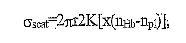
wherein

- Thus, we defined the function K(ω) which is graphically illustrated in
Fig. 4a . This function describes diffraction effects on particles depending on the model used. As shown, this function has theasymptotic value 1 at ω→∞, in which case σscat=2πr2 The other asymptotic expression at small ω→0, K(ω)=(ω)2, but this contradicts with the WKB approximation. For blood, there is the intermediate case x≈20 and ω≈1. - The function K(ω) has many maximum and minimum values, the physical sense of which is the interference between the refracted and diffracted waves. Considering the second order statistical correlation of mutual arrangements of the particles, the scattering coefficient µscat for spheres may be written as follows:

wherein ω=2πr(nHb-npl)/λ. - For spheres, the scattering coefficient µscat decreases to zero when r increases to infinity. For spheroids, the scattering coefficient µscat in WKB approximation depends on the radiation propagation direction, and is as follows:

wherein A is an area of geometrical shadow perpendicular to axes, A=πaI(ζ) and I(ζ)=[a2cos2ζ+c2sin2ζ]1/2; ζ is an angle between the axis of spheroid and the direction z of the radiation propagation. - The parameter ω is as follows:

wherein b is the half the mean thickness of a scatterer (erythrocyte) along the direction of incidence z, that is:
The scattering coefficient is minimal (b=3 c/4) for broadside incidence (ζ=0) and maximal (b=3a/4) for rim-on incidence (ζ=π/4). - The similar approach may be applied to each particular geometry of the center of scattering, such as spheroids with averaged parameters over various orientations, etc.
-
Fig. 4b illustrates the function K(<ω>). It can be seen from this graph that the scattering coefficient uscat for spheroids which is proportional to K(<ω>) has the finite limit when one of the sizes c increases to infinity. This limit is determined by the unchanged spheroid size a. - The above equations for the scattering coefficient µscat are very important for the evaluation of the optical properties of blood, with taking into account the real size and shape of erythrocytes. This also allows the evaluation of the optical properties of erythrocytes when they aggregate one with another.
- Turning back to
Figs. 3a-3b andFig. 4a , and keeping in mind that ω=(27πr/λ)(nHb-npl), i.e., K is the function of the wavelengths of the incident radiation, we can conclude the following. Since for one wavelength the transmission signal grows with time in the asymptotic time interval tasym and for the other wavelength it falls, there exists such a wavelength (cut-off wavelength) of incident radiation, which causes no time changes in the transmission signal and which corresponds to the extremum (maximum or minimum) of the function K(o). The transmission signal corresponding to the cut-off wavelength of incident radiation will therefore be independent on the erythrocyte aggregation process. - Reference is now made to
Figs. 5 a and 5b, illustrating the main principles of the determination of the cut-off wavelength as an example of the particular RGF.Fig. 5a shows the results of in vivo measurements during prolonged occlusion, in the form of several graph six in the present example. Graphs T1 λ1(t) - T6 λ7(t) of the time variation of transmission signals correspond to different values of the wavelengths, of incident radiation: λ1=650nm, λ2=700Mn, λ3=:760nm, λ4=880nm, λ5=940nm and λ6=1300nm.Fig. 5b shows a graph in the form of a ratio Δ(logT)/Δt as the function of the wavelength obtained from the graphs inFig. 5a , wherein Δt lies substantially within the asymptotic time interval tasym,-where the transmissions signals change with time slower than in the initial time interval tin The point λ0 is the cut-off wavelength of incident radiation corresponding to a certain time stable transmission for a specific patient, which, in turn, corresponds to the extremum of the function K(ω), i.e., its maximum or minimum. - As indicated above, ω=x(nHb-npl)=2πr(nHb-npl)/λ for the sphere-like erythrocyte, or ω=x(nHb-npl)=2πa(nHb-npl)/λ for the spheroid-like erythrocyte. For a given cut-off wavelength, λ0, the parameter ω varies from patient to patient, and lies in a certain interval defined by the minimum and maximum acceptable erythrocyte size and minimum and maximal acceptable values of the difference (nHb-npl). Having determined the wavelength λ0 corresponding to the maximum or minimum of K(ω), and considering the relevant interval of ω values, the corresponding value of ω in this interval can be evaluated for the specific patient. This data can be used for further analysis, which will be described further below. As indicated above, the knowledge of the difference (nHb-npl) is indicative, for example, of the glucose concentration.
- The incident radiation is absorbed by the blood components - hemoglobin and water. The absorption coefficient of hemoglobin µHb abs (per unit of hemoglobin density) and analogous absorption coefficient of plasma (water) µpl abs can be obtained experimentally and are known from literature. These coefficients depend on the wavelength of incident radiation, and their magnitudes may be estimated as follows:,µ Hb abs≈2.5* 10-2dl/(g*mm) and µPl abs≈1.5* 10-3dl/(g*mm).
- The total absorption coefficient is equal:

Here, CHb is the concentration of hemoglobin and is approximately equal to 30g/dl; µHb abs is the absorption coefficient of hemoglobin and is approximately equal to 2.5* 10-2; and µpl abs is the absorption coefficient of plasma which is about 1.58*10-3 (considering that the optical characteristics of plasma as similar to those of water). - Since there is a concrete geometry of erythrocytes in the system, the role of their spherical shape in the absorption of radiation should be considered.
- The absorption coefficient is expressed by the imaginary part of the refraction indices and is as follows:

wherein hemoglobin occupies only the part s of the erythrocyte volume V0. - For the real system x≈18, Im nHb,npl is about 10-4 and the argument of function is small. Considering the expression for Kcom[y] :

we have:
Taking into account the following:


we receive:
- For the absorption coefficient of erythrocyte we have the expression:

- It is thus evident that for blood, the absorption coefficient does not depend on the shape of particles and their sizes, but depends only on the volume of the components. With the contribution of water in the space between the erythrocytes, we reproduce again the above, more general equation for µabs.
- For the opposite limit of very large' particles, we have: σabs≈πr2, and therefore (when x(nHb-npl)»1); we have: σscat=σtot-σabs=πr2. So, for this case: σscat/σtot=1/2, which means that they are of the same order of magnitude.
- Simulations were carried out utilizing various models of multiple scattering theories, such as the model of Twersky, diffusion models (which provide better approximation for blood in a regular situation as compared to that of the model of Twersky), model of Hemenger, model of Rogozkin, and Small-Angle model. The transport theory dealing with average values was also considered in the simulations.
- The above models are disclosed, for example, in the following articles:
- " Interface Effects in Multiple Scattering by Biological Suspensions, V Twersky, Journal of the Optical Society of America; Vol. 60, No. 7, pp..908-914, 1970;
- " Optical Properties of Turbid Media with Specularly Reflecting Boundaries: Applications to Biological Problems"', R.P. Hemenger, Appl. Opt., Vol. 16. NO. 17, 99.2007-2012, 1997;
- " Long-Range Correlations in Wave, Reflection from a Disordered Medium ", D.B. Rogozkin, Phys. Rev B. Vol. 51, No. 18, pp. 12256-12267, 1995;
- '- " Long-Range Intensity Correlations for the Multiple Scattering of Waves in Unordered Media ","D-B. Rogozkin, JETP Vol. 84(5), pp. 916-939, 1997; and
- " Intensity Correlation in a Disordered Medium with Large Scaterrers ", D.B. Rogozkin, Physics Letters A 178, pp. 431-439,1993.
- Since the transmissions at different wavelengths are the functions of the number of particles in aggregates or particle size, the transmission at one wavelength can be expressed as a parametric function of the transmission at another wavelength. This function is a straight line with reasonable accuracy. This conclusion is verified for both the transmissions themselves and the logarithms of transmission for different models and different simulations of erythrocyte shape.
- If the transmission signal T1(t) is measured when using the incident wavelength λ1, and the transmission signal T2(t) is measured when using the incident wavelength λ2, then the slope of the line T1(λ1)/T2(λ2) (or logT1(λ1)/logT2(λ2) is a certain parameter, called "parametric slope" (PS) for a specific patient that can be determined. By this, we can get rid of the explicit usage of the size of aggregates, i.e., the values that cannot be known from experiments in vivo.
- The model of Rogoskin for spheres is used to obtain an approximate analytical expression of the parametric slope PS. It should however be noted that, for the cases of more complicated shapes, the proper generalization also may be done.
- Thus, the following expression is used:

wherein T is the transmission signal and µtr is the transport scattering coefficient that describes the energy decay for the case of anisotropic scattering, and is determined as follows: µtr=µscat(1-g), g being the anisotropic coefficient. - Here, the case when (µdiff*d)≥1 and Sinh(µdiff*d)≈exp(µdiff*d) is used, wherein µdiff is the diffusion coefficient; d is the thickness of the blood slab.

- At (µabs/µtr)>>1, we have:

- At (µabs/µtr)<<1, we have:

- Here, H(θ,1) is the function of Chandrasekhar that depends on the angle θ of the radiation propagation, <H(θ,1)> is the value averaged by all angles of radiation propagation.
- Analogous, the expressions for derivatives of LogT can be obtained and the parametric slope for LogT can be defined:

- At (µabs/µtr)>>1, we have:

- At (µabs/µtr)<<1, we have:

- For the Small-Angle model:

- The derivative (dµtr/dr)λ is got from the above expression for the scattering coefficient µscat for the sphere-based model.
- The following expression is known:

wherein g is the average cosine of the scattering angle (g=<cosθ>) and is connected with the anisotropy of the radiation scattering. - In the region of real value of
parameters 1<ω<2, there is the approximation K(ω)≈0.82053*ω+0.0366. Then, we have:
- The above ratio (dµtr/dr)λ depends on the wavelength through the factor (1-g) only.
- The value µtr with K(ω)≈0.82053*ω becomes:

- If we substitute this expression in the above equations for (dT/dµtr)λ, we receive the expression that depends on the wavelength very simply and does not depend on the size of particles.
- As the result of substitution of µtr and (dµtr/dr)λ, in the above expression for PS, we receive the parameter that does not depend on the particle size.
- Now we shall describe how the above theoretical considerations can be used in practice. What we actually obtained through the above simulations, is the fact that there exist a parameter ("parametric slope"), which does not depend on the particle size and only on the concentration of hematocrit in blood. This fact can be used for determining the concentration of a substance of interest in blood.
- To do this, first, a set of calibration curves should be provided. Each calibration curve corresponds to a specific substance and is in the form of a parametric slope (PS), as the function of the hematocrit. This can be done by applying two kinds of measurements to a large number of patients. The measurements of one kind are those of the present invention enabling the determination of the parametric slope value for each patient (as will be described below), and the others are conventional ones enabling the determination of the corresponding parameter for the same patient. This set of calibration curves presents reference data, which is previously stored in the memory of the control unit.
- Then, for a specific patient, two measurement sessions are applied with the above-described
measurement apparatus 1 in vivo (orapparatus 100 in vitro), with two different wavelengths λ1 and λ2. These wavelengths are selected in accordance with the given task, namely the blood parameter to be determined, as will be exemplified further below. These measurements can be taken within the initial time interval of the entire time period during which the blood flow cessation state is maintained, namely the time interval where the time changes of the transmission signal are stronger as compared to the asymptotic time interval, as well as during the asymptotic time interval. - For example, if the concentration of hemoglobin is to be determined, the two selected wavelengths are those of the absorption of hemoglobin and plasma (water) in the ranges where the difference in the absorption of hemoglobin and water is more sharply expressed, i.e., typically 600-1000nm and 1100-1400nm, but may also be 940-980 (water sensitive space) and 600-930 (hemoglobin sensitive space). In the present example, the wavelengths λ1=950nm and λ2=1300nm are used. The corresponding graphs of the transmission logarithms log(T1) and log(T2) as the functions of time t corresponding to these wavelengths are shown in
Fig. 3 a. - As graphically illustrated in
Fig. 6 , to determined a parametric slope PS, the function of the transmission logarithm at the wavelength λ2, i.e., Log(T2), versus the transmission logarithm at the wavelength λ1, i.e., Log(T1) is determined over the initial time interval. This graph is obtained by the linear regression algorithm. The value of the tg(ϕ) corresponds to the parametric slope PS. The corresponding calibration curve is shown inFig. 6b . By using this curve, the concentration of hemoglobin can be determined for the specific patient, to whom the measurements are applied. - Turning now to
Figs. 7a and 7b , there are illustrated two steps in the method for determining the concentration of glucose. Here, the wavelengths λ1 and λ2 are selected in the ranges 600-1300nm and 1500-1600nm, respectively, namely λ1=660nm and λ2=1550nm.Fig. 7a illustrates the corresponding transmission signals as the functions of time, i.e., T1(t) and T2(t). Then, the function logT2 vs. LogT1 is determined, as graphically illustrated inFig. 7b , and the corresponding parametric slope is determined as described above. Having determined the parametric slope value for a specific patient, a corresponding calibration curve (not shown) is used for determining the glucose concentration for this specific patient. - Another important blood parameter that can be determined with the invented method is the oxygen saturation in the patient's blood. Oxygen saturation is defined as the ratio of the content of oxyhemoglobin (HbO2) to the total amount of hemoglobin (Hb) in the blood volume unit. The classic pulse oximetry method allows for determining the oxygen saturation. This method utilizes the so-called "natural pulsatile" component of a light transmission signal. This pure natural pulse-related signal component of a detected signal, determined by an appropriate signal processing technique, is commonly called the "AC component", of the detected signal, whereas the entire transmission signal by itself is called the "DC component" of the detected signal. The transmission measurements in the pulse oximetry are carried out simultaneously at two different wavelengths, for example λ1=760nm and λ2=940nm, where the significant difference in the light absorption of oxyhemoglobin and hemoglobin exists between the two chosen wavelengths. Two pairs of AC and DC components are obtained. Generally, the ratio R, defined as (AC/DC)λ1/(AC/DC)λ2, is the value of oxygen saturation.
- We applied the "parametric slope" concept and obtained the same results as with the pulse oxymetry technique.
Fig. 8a illustrates two graphs P1 and P2 (the provision of only one of them being sufficient for the purposes of the present invention), corresponding respectively to (logT1)λ1 vs. (logT3)λ3 and (logT2)λ2 vs. (logT3)λ3. The measured data T1(t), T2(t) and T3(t) is obtained in the above-described manner using the following wavelengths of incident radiation: λ1=650nm, λ2=760nm and λ3=940nm. It should be understood that the use of two wavelengths and, correspondingly, the provision of one parametric slope related graph, is sufficient for the determination of the parametric slope PS. - Generally, a calibration curve to be used for determining the oxygen saturation for a specific patient may be constructed as described above, i.e., applying measurements to various patients. Alternatively, or additionally, in the case of oxygen saturation determination, the calibration curve can be plotted when applying the two kinds of measurements for a single patient in a breath hold experiment using a multiple-occlusion mode. This is illustrated in
Fig. 8b and8c . -
Fig. 8b shows the measured data in the multiple-occlusion mode in the form of the transmission functions T1(t), T2(t) and T3(t) corresponding, respectively, to the wavelengths λ1=650nm, λ2=760nm and λ3=940nm. As shown, several occlusions were performed during the state of the patient's breath hold, and the corresponding parametric slope values were determined during the time interval of the saturation decrease. Concurrently, conventional measurements are applied to the same patient, for example to his other finger, for determining the changes in the oxygen saturation.Fig. 8c shows the so-obtained calibration curve in the form of the parametric slope PS as the function of the oxygen saturation SPO2. - It should be noted, although not specifically shown, that the similar results for the oxygen saturation, as well as for the concentrations of hemoglobin and glucose, were obtained when applying the "parametric slope" based measurements to a blood sample in cuvette, i.e., in vitro measurements.
-
Figs. 9a and9b illustrate another important feature of the present invention consisting of the determination of a so-called "MegaSlope" (MS).Fig. 9a shows a graph P' determined from the measured data shown inFig. 3a . The wavelengths used are: λ1=660nm, λ2=950nm and λ3=1300nm. To this end, the entire time period inFig. 3a is divided into a plurality of time intervals Δt ― ten in the present example, and for each time interval Δt a pair of parametric slope values is obtained from the following: (logT)λ3 vs. (logT)λ2 and (logT)λ1 vs. (logT)λ2. In other words, each point in the graph P' corresponds to a pair of parametric slopes calculated for a pair of wavelengths λ3-λ2 and λ1-λ2, respectively, each for a corresponding one of the time intervals Δt. Each such parametric slope is determined in the above-described manner. The MegaSlope is determined as tg(ϕ). A calibration curve shown inFig. 9b presents the MegaSlope as the function of hemoglobin concentration, i.e., MS(H). - One more important feature of the present invention consists of determining the Erythrocyte Aggregation Rate (EAR) for a specific patient. Assuming that the only process that takes place at the state of the blood flow cessation is the erythrocytes' aggregation, the EAR can be simply determined as the rate of the time changes of light response signal, i.e.,AT/At (or ΔlogT/Δt). To this end, the transmission as the function of time is measured with one wavelength of incident radiation. For more precise measurements, two such transmission signals as functions of time are measured with two different wavelengths of incident radiation. As for the time interval Δt, it may be either initial time interval or asymptotic time interval. The EAR parameter can be used for the determination of such an important parameter as Erythrocyte Sedimentation Rate (ESR). This is illustrated in
Fig. 10 , showing the EAR (ΔlogT/Δt) as the function of ESR, the latter being measured in the conventional in vitro manner. - Thus, the advantages of the present invention are self-evident. We have proved that the main effect defining the optical characteristics of blood in the state of temporarily blood flow cessation is the erythrocytes' aggregation. In other words, in the asymptotic time interval, the erythrocyte serves as the sensor for the determination of the various blood parameters. The technique of the present invention, preferably performed in a non-invasive manner, but even in an invasive manner, is simpler and quicker then the conventional one. A physician can apply this technique to evaluate the various blood conditions of a patient, and then, if desired, direct him to a laboratory for more careful measurements. The parameter x(nHb-np1) can be used in the set of calibration curves for PS and MS or the combination of the both.
- Aspects are set out in the following numbered clauses.
- 1. A method of optical measurements of at least one desired parameter of the patient's blood, the method comprising the steps of:
- providing a state of blood flow cessation of the patient's blood within a measurement region, and maintaining the blood-flow cessation state during a predetermined time period;
- performing measurement sessions within said predetermined, time period, each measurement session including at least two measurements with different wavelengths of incident light, and obtaining measured data representative of the time dependence of light response of the blood in the measurement region;
- analyzing the measured data for determining said at least one desired parameter, extracted from optical characteristics associated with erythrocytes aggregation process during the state of the blood flow cessation.
- 2. The method according to
Clause 1, wherein said state of the blood flow cessation in the measurement region is provided by applying an occlusion mode to the patient's blood perfused fleshy medium. - 3. The method according to
Clause 2, wherein the application of the occlusion mode comprises the application of over-systolic pressure to the patient's blood perfused fleshy medium at a location upstream of said measurements region with respect to the direction of the normal blood flow in the patient's body - 4.The method according to
Clause 3, and also comprising the step of preliminary optical measurements for detecting the existence of the blood flow cessation state. - 5. The method according to
Clause 2, wherein said predetermined period of time is insufficient for irreversible changes to occur in the fleshy medium. - 6. The method according to
clause 5, wherein said predetermined period of time is from one second to several minutes. - 7. The method according to
clause 1, wherein said state of the blood flow cessation in the measurement region is provided by applying an occlusion mode to the flow of the patient's blood sample into a cuvette, said measurement region being located in the cuvette. - 8. The method according to
Clause 1, wherein the analyzing of the measured data comprises the step of- determining a Rouleaux Geometry Factor (RGF) characterizing the changes of the light response of blood in the state of blood flow cessation as the function of time and wavelengths of the incident radiation, associated with the erythrocytes' aggregation.
- 9. The method according to
Clause 8, wherein the determination of the RGF comprises:- determining a ratio Δ(logT)/Δt as the function of wavelength λ obtained from the several measurement sessions with the different wavelengths, Δt being a preset time interval of said predetermined period of time; and
- determining a cut-off wavelength corresponding to the condition Δ(logT)/Δt=0.
- 10. The method according to
Clause 8, wherein the determination of the RGF comprises:- determining a ratio or ΔT/Δt as the function of wavelength λ obtained from the several measurement sessions with the different wavelengths, Δt being a preset time interval of said predetermined period of time; and
- determining a cut-off wavelength is that corresponding to the condition or ΔT/Δt =0.
- 11.The method according to
Clause - 12. The method according to
Clause - 13.The method according to
Clause 12, wherein the evaluation of the parameter (nHb-np1) comprises the utilization of theoretical data representative of a scattering function K(x(nHb-np1)), wherein x=2πa/λ, a being the effective size of erythrocyte. - 14. The method according to
Clause 1, wherein the analyzing of the measured data comprises the steps of:- determining a parametric slope, said at least two wavelengths being selected in accordance with said desired parameter to be determined; and
- using reference data in the form of a calibration curve of the parametric slope as a function of values of the desired parameter.
- 15. The method according to
Clause 14, wherein the determination of the parametric slope comprises the determination of a function Tλ2(Tλ1), wherein Tλ2 and Tλ1 are the measured data corresponding to the wavelengths λ2 and λ1 of the incident radiation, respectively, the function Tλ2(Tλ1) being determined for a preset time interval of said predetermined time period. - 16. The method according to
Clause 14, wherein the determination of the parametric slope comprises the determination of a function logTλ2(logTλ1), wherein Tλ2 and Tλ1 are the measured data corresponding to the wavelengths λ2 and λ1 of the incident radiation, respectively, the function Tλ2(Tλ1) being determined for a preset time interval of said predetermined time period. - 17. The method according to
Clause - 18. The method according to
Clause - 19. The method according to
Clause 14, wherein said desired parameter is the concentration of a certain substance in blood. - 20. The method according to
Clause 19, wherein said calibration curve is determined from values of the parametric slope and values of the concentration obtained for different patients. - 21. The method according to
Clause 19, wherein said substance is hemoglobin. - 22. The method according to Clause 21, wherein the at least two selected wavelengths are in the ranges of 600-1000nm and 1100-1400nm.
- 23. The method according to
Clause 19, wherein said substance is glucose. - 24. The method according to Clause 23, wherein the at least two selected wavelengths, are in the ranges of 1500-1600nm and 600-1300nm.
- 25. The method according to
Clause 14, wherein said desired parameter is oxygen saturation. - 26. The method according to
Clause 25, wherein said at least two wavelengths are in the ranges of 600-780nm and 820-980nm. - 27. The method according to
Clause 25, wherein said calibration curve is determined from values of the parametric slope and values of the oxygen saturation obtained for different patients. - 28. The method according to
Clause 25, wherein said calibration curve is, plotted by applying at least two multiple-occlusion measurement sessions with said at least two selected wavelengths to the blood of said patient in the measurement region, determining the parametric slope values for each occlusion, and concurrently determining the oxygen saturation in the blood perfused fleshy medium of said patient outside said measurement region. - 29. The method according to
Clause 1, wherein the analyzing of the measured data comprises the determination of Erythrocyte Aggregation Rate, as ΔT/Δt. - 30. The method according to Clause 29, wherein Δt is within an initial time interval of the predetermined period of time characterized by relatively strong changes of the light response signal with time, as compared to a next, asymptotic time interval of said predetermined period of time.
- 31. The method according to Clause 29, wherein Δt is an asymptotic time interval of the predetermined period of time characterized by relatively slow changes of the light response signal with time, as compared to an initial time interval of said predetermined period of time.
- 32. A method of optical measurements of desired parameters of the patient's blood extracted from optical characteristics associated with erythrocytes aggregation process during the state of the blood flow cessation, the method comprising the steps of:
- providing the state of the blood flow cessation within a measurement region, and maintaining the blood-flow cessation state during a predetermined time period;
- performing measurement sessions within said predetermined time period, each' measurement session including at least two measurements with different wavelengths of incident light, and obtaining measured data representative of the time dependence of light response of the blood in the measurement region;
- analyzing the measured data for determining said at least one desired parameter, by determining at least one parametric slope value and a Rouleaux Geometry Factor (RGF) for said patient, the RGF characterizing the changes of the light response of blood at the state of the blood flow cessation as the function of time and wavelengths of the incident radiation, associated with the erythrocytes' aggregation.
- 33. A method of optical measurements of at least one desired parameter of blood of a specific patient extracted from optical characteristics associated with erythrocytes aggregation process during the state of the blood flow cessation, the method comprising the steps of:
- providing reference data-in the form of a function describing diffraction effects on particles, K(x(nHb-nH2O), wherein x=2πa/λ; ais the size of erythrocyte, nHb is the refraction index of hemoglobin and npl is the refraction index of water, λ is the wavelength of incident radiation;
- providing the state of the blood flow cessation and maintaining said state during a predetermined period of time;
- performing measurement sessions within said predetermined time period, each measurement session including several measurements with different wavelengths of incident radiation, and obtaining measured data representative of the time dependence of light response signals;
- analyzing the measured data for determining a Rouleaux Geometry Factor (RGF) for the specific patient, the RGF characterizing the changes of the light response of blood at the state of the blood flow cessation as the function of time and wavelengths of the incident radiation, associated with the erythrocytes' aggregation.
- 34. A method of optical measurements of at least one desired parameter of blood of a specific patient extracted from optical characteristics associated with erythrocytes aggregation process during the state of the blood flow cessation, the method comprising the steps of:
- providing, reference data in the form of at least one calibration curve corresponding to a parametric slope as a function of values of said desired parameter;
- providing the state of the blood flow cessation within a measurement region, and maintaining the blood-flow cessation state during a predetermined time period;
- performing timely separated measurement sessions within said predetermined time period, each measurement session including at least two measurements with different wavelengths of incident light, and obtaining the time dependence of transmission signals, wherein the at least two wavelengths are selected in accordance with the desired parameter to be determined;
- analyzing the obtained data for determining the parametric slope value for said specific patient;
- using said calibration curve for determining the value of said desired parameter for said specific patient.
- 35. A measurement apparatus for performing non-invasive optical measurements of desired parameters of the patient's blood, the apparatus comprising:
- an occlusion assembly for applying to the flow of the patient's blood, so as to create a state of the blood flow cessation in a measurement region for a predetermined time period;
- illumination/detection, assembly for applying optical measurements with at least two different wavelengths of incident radiation to said measurement region during said predetermined period of time, and generating data representative of light response signals; and
- a control unit responsive to the generated data for determining measured data in the form of time variations of the light response signals, for utilizing optical characteristics associated with erythrocytes aggregation process during the state of the blood flow cessation to analyze the measured data from within a preset time interval of said predetermined period of time and determine the desired parameter of the patient's blood.
Claims (14)
- A method of processing data measured in non-invasive optical measurements on a patient's body, wherein the measured data corresponds to a light response of a measurement location in a blood perfused fleshy medium in the body to incident light of at least one wavelength of a selected spectral range collected during either timely separated optical measurements or continuous optical measurements over time including a predetermined time period during which a blood flow cessation state has been maintained at the measurement location, method comprising:processing the measured data and determining at least one time dependence of the light response to the incident light of said at least one wavelength for said predetermined time period;analyzing said at least one time dependence, and determining a rate of time changes of the light response corresponding to Erythrocyte Aggregation Rate (EAR).
- The method of Claim 1, comprising using the EAR data and determining a corresponding value of Erythrocyte Sedimentation Rate (ESR).
- The method of Claim 1 or 2, wherein said analyzing of the at least one time dependence of the light response comprises selecting at least one time interval Δt of said predetermined time period during which the blood flow cessation state has been maintained, and determining the rate of time changes of the light response for said selected time interval Δt.
- The method of Claim 3, wherein said time interval Δt includes an initial time interval of the light response variation, after creation of the state of blood flow cessation.
- The method of Claim 3 or 4, wherein said time interval Δt includes an asymptotic time interval that follows an initial time interval and has relatively slow changes of the light response as compared to the initial time interval.
- The method according to any one of the preceding Claims, wherein said predetermined time period is from one second to several minutes.
- The method of any one of the preceding Claims, comprising processing the measured data corresponding to at least two light responses of the measurement location to the incident light of at least two different predetermined wavelengths, and determining one or more parameters of the patient including one or more of the following: concentration of a substance, such as glucose, hemoglobin, drugs or cholesterol; oxygen saturation; a difference between refraction indices of hemoglobin and plasma in the patient's blood.
- The method according to Claim 7, wherein the determination of the parameter of the patient comprises determining the at least two time dependencies for said at least two wavelengths, and determining one of the time dependencies as a function of the other time dependence, for the two wavelengths corresponding to the parameter to be determined.
- A system for use in non-invasive optical measurements on a patient's body, the system comprising: a processor configured and operable for receiving optical measured data, which corresponds to a light response of a measurement location in a blood perfused fleshy medium in the body to incident light of at least one wavelength of a selected spectral range collected during either timely separated optical measurements or continuous optical measurements over time including a predetermined time period during which a blood flow cessation state has been maintained at the measurement location, processing said measured data to determine at least one time dependence of the light response to the incident light of said at least one wavelength for said predetermined time period, analyzing said at least one time dependence and determining a rate of time changes of the light response corresponding to Erythrocyte Aggregation Rate (EAR).
- The system of Claim 9, wherein the processor is configured and operable for analyzing said EAR and generating data indicative of Erythrocyte Sedimentation Rate (ESR) for said patient.
- The system of Claim 9 or 10, wherein said processor is configured and operable for processing measured data corresponding to at least two light responses of the measurement location to the incident light of at least two different predetermined wavelengths respectively, and determining one or more parameters of the patient including one or more of the following: concentration of a substance, such as glucose, hemoglobin, drugs or cholesterol; oxygen saturation; a difference between refraction indices of hemoglobin and plasma in the patient's blood
- The system of any one of Claims 9 to 11, comprising a pressurizing assembly, an illumination assembly, and a detection assembly that generates said measured data, said control unit being connected to the detection assembly for receiving said measured data and being connected to the illumination and pressurizing assemblies for controlling optical measurements while at the state of the blood flow cessation.
- The system of Claim 12, wherein the control unit is configured and operable for analyzing the measured data corresponding to measurements taken during at least one time interval Δt of said predetermined time period during which the blood flow cessation state has been maintained, and determining the rate of time changes of the light response for said selected time interval Δt.
- The system of Claim 13, wherein said time interval Δt is either an initial time interval of the light response variation, after creation of the state of blood flow cessation, or an asymptotic time interval that follows the initial time interval and has relatively slow changes of the light response as compared to the initial time interval.
Applications Claiming Priority (2)
| Application Number | Priority Date | Filing Date | Title |
|---|---|---|---|
| PCT/IL1999/000694 WO2001045553A1 (en) | 1999-12-22 | 1999-12-22 | A method of optical measurements for determining various parameters of the patient's blood |
| EP99959659A EP1251772B1 (en) | 1999-12-22 | 1999-12-22 | A method of optical measurements for determining various parameters of the patient's blood |
Related Parent Applications (1)
| Application Number | Title | Priority Date | Filing Date |
|---|---|---|---|
| EP99959659.6 Division | 1999-12-22 |
Publications (3)
| Publication Number | Publication Date |
|---|---|
| EP2308374A2 true EP2308374A2 (en) | 2011-04-13 |
| EP2308374A3 EP2308374A3 (en) | 2011-05-25 |
| EP2308374B1 EP2308374B1 (en) | 2012-07-18 |
Family
ID=11062768
Family Applications (2)
| Application Number | Title | Priority Date | Filing Date |
|---|---|---|---|
| EP10185849A Expired - Lifetime EP2308374B1 (en) | 1999-12-22 | 1999-12-22 | A method of optical measurements for determining various parameters of the patient's blood |
| EP99959659A Expired - Lifetime EP1251772B1 (en) | 1999-12-22 | 1999-12-22 | A method of optical measurements for determining various parameters of the patient's blood |
Family Applications After (1)
| Application Number | Title | Priority Date | Filing Date |
|---|---|---|---|
| EP99959659A Expired - Lifetime EP1251772B1 (en) | 1999-12-22 | 1999-12-22 | A method of optical measurements for determining various parameters of the patient's blood |
Country Status (4)
| Country | Link |
|---|---|
| US (3) | US6711424B1 (en) |
| EP (2) | EP2308374B1 (en) |
| AU (1) | AU1678800A (en) |
| WO (1) | WO2001045553A1 (en) |
Families Citing this family (292)
| Publication number | Priority date | Publication date | Assignee | Title |
|---|---|---|---|---|
| US6931268B1 (en) | 1995-06-07 | 2005-08-16 | Masimo Laboratories, Inc. | Active pulse blood constituent monitoring |
| US6517283B2 (en) * | 2001-01-16 | 2003-02-11 | Donald Edward Coffey | Cascading chute drainage system |
| US6018673A (en) | 1996-10-10 | 2000-01-25 | Nellcor Puritan Bennett Incorporated | Motion compatible sensor for non-invasive optical blood analysis |
| US9042952B2 (en) | 1997-01-27 | 2015-05-26 | Lawrence A. Lynn | System and method for automatic detection of a plurality of SPO2 time series pattern types |
| US20060161071A1 (en) | 1997-01-27 | 2006-07-20 | Lynn Lawrence A | Time series objectification system and method |
| US8932227B2 (en) | 2000-07-28 | 2015-01-13 | Lawrence A. Lynn | System and method for CO2 and oximetry integration |
| US20070191697A1 (en) | 2006-02-10 | 2007-08-16 | Lynn Lawrence A | System and method for SPO2 instability detection and quantification |
| US9521971B2 (en) | 1997-07-14 | 2016-12-20 | Lawrence A. Lynn | System and method for automatic detection of a plurality of SPO2 time series pattern types |
| US6391005B1 (en) * | 1998-03-30 | 2002-05-21 | Agilent Technologies, Inc. | Apparatus and method for penetration with shaft having a sensor for sensing penetration depth |
| US6675031B1 (en) | 1999-04-14 | 2004-01-06 | Mallinckrodt Inc. | Method and circuit for indicating quality and accuracy of physiological measurements |
| AU1678800A (en) * | 1999-12-22 | 2001-07-03 | Orsense Ltd. | A method of optical measurements for determining various parameters of the patient's blood |
| US8224412B2 (en) | 2000-04-17 | 2012-07-17 | Nellcor Puritan Bennett Llc | Pulse oximeter sensor with piece-wise function |
| CA2405825C (en) | 2000-04-17 | 2010-11-09 | Nellcor Puritan Bennett Incorporated | Pulse oximeter sensor with piece-wise function |
| DE60039336D1 (en) * | 2000-08-03 | 2008-08-14 | Koninkl Philips Electronics Nv | Fluid transport by pressure variation for the analysis of biological fluids |
| US8641644B2 (en) | 2000-11-21 | 2014-02-04 | Sanofi-Aventis Deutschland Gmbh | Blood testing apparatus having a rotatable cartridge with multiple lancing elements and testing means |
| US20060195041A1 (en) | 2002-05-17 | 2006-08-31 | Lynn Lawrence A | Centralized hospital monitoring system for automatically detecting upper airway instability and for preventing and aborting adverse drug reactions |
| US9053222B2 (en) | 2002-05-17 | 2015-06-09 | Lawrence A. Lynn | Patient safety processor |
| US8135448B2 (en) * | 2001-03-16 | 2012-03-13 | Nellcor Puritan Bennett Llc | Systems and methods to assess one or more body fluid metrics |
| CA2448902C (en) | 2001-06-12 | 2010-09-07 | Pelikan Technologies, Inc. | Self optimizing lancing device with adaptation means to temporal variations in cutaneous properties |
| US9427532B2 (en) | 2001-06-12 | 2016-08-30 | Sanofi-Aventis Deutschland Gmbh | Tissue penetration device |
| US7344507B2 (en) | 2002-04-19 | 2008-03-18 | Pelikan Technologies, Inc. | Method and apparatus for lancet actuation |
| US7041068B2 (en) | 2001-06-12 | 2006-05-09 | Pelikan Technologies, Inc. | Sampling module device and method |
| US9226699B2 (en) | 2002-04-19 | 2016-01-05 | Sanofi-Aventis Deutschland Gmbh | Body fluid sampling module with a continuous compression tissue interface surface |
| US8337419B2 (en) | 2002-04-19 | 2012-12-25 | Sanofi-Aventis Deutschland Gmbh | Tissue penetration device |
| US7981056B2 (en) | 2002-04-19 | 2011-07-19 | Pelikan Technologies, Inc. | Methods and apparatus for lancet actuation |
| US9795747B2 (en) | 2010-06-02 | 2017-10-24 | Sanofi-Aventis Deutschland Gmbh | Methods and apparatus for lancet actuation |
| US7033371B2 (en) | 2001-06-12 | 2006-04-25 | Pelikan Technologies, Inc. | Electric lancet actuator |
| US7749174B2 (en) | 2001-06-12 | 2010-07-06 | Pelikan Technologies, Inc. | Method and apparatus for lancet launching device intergrated onto a blood-sampling cartridge |
| US6754516B2 (en) * | 2001-07-19 | 2004-06-22 | Nellcor Puritan Bennett Incorporated | Nuisance alarm reductions in a physiological monitor |
| US6748254B2 (en) | 2001-10-12 | 2004-06-08 | Nellcor Puritan Bennett Incorporated | Stacked adhesive optical sensor |
| US7232451B2 (en) | 2002-04-19 | 2007-06-19 | Pelikan Technologies, Inc. | Method and apparatus for penetrating tissue |
| US7909778B2 (en) | 2002-04-19 | 2011-03-22 | Pelikan Technologies, Inc. | Method and apparatus for penetrating tissue |
| US7229458B2 (en) | 2002-04-19 | 2007-06-12 | Pelikan Technologies, Inc. | Method and apparatus for penetrating tissue |
| US7892183B2 (en) | 2002-04-19 | 2011-02-22 | Pelikan Technologies, Inc. | Method and apparatus for body fluid sampling and analyte sensing |
| US9795334B2 (en) * | 2002-04-19 | 2017-10-24 | Sanofi-Aventis Deutschland Gmbh | Method and apparatus for penetrating tissue |
| US7547287B2 (en) * | 2002-04-19 | 2009-06-16 | Pelikan Technologies, Inc. | Method and apparatus for penetrating tissue |
| US7976476B2 (en) | 2002-04-19 | 2011-07-12 | Pelikan Technologies, Inc. | Device and method for variable speed lancet |
| US9314194B2 (en) * | 2002-04-19 | 2016-04-19 | Sanofi-Aventis Deutschland Gmbh | Tissue penetration device |
| US9248267B2 (en) | 2002-04-19 | 2016-02-02 | Sanofi-Aventis Deustchland Gmbh | Tissue penetration device |
| US7901362B2 (en) * | 2002-04-19 | 2011-03-08 | Pelikan Technologies, Inc. | Method and apparatus for penetrating tissue |
| US8360992B2 (en) | 2002-04-19 | 2013-01-29 | Sanofi-Aventis Deutschland Gmbh | Method and apparatus for penetrating tissue |
| US8372016B2 (en) | 2002-04-19 | 2013-02-12 | Sanofi-Aventis Deutschland Gmbh | Method and apparatus for body fluid sampling and analyte sensing |
| US8784335B2 (en) | 2002-04-19 | 2014-07-22 | Sanofi-Aventis Deutschland Gmbh | Body fluid sampling device with a capacitive sensor |
| US8221334B2 (en) | 2002-04-19 | 2012-07-17 | Sanofi-Aventis Deutschland Gmbh | Method and apparatus for penetrating tissue |
| US8579831B2 (en) * | 2002-04-19 | 2013-11-12 | Sanofi-Aventis Deutschland Gmbh | Method and apparatus for penetrating tissue |
| US7648468B2 (en) * | 2002-04-19 | 2010-01-19 | Pelikon Technologies, Inc. | Method and apparatus for penetrating tissue |
| US7226461B2 (en) | 2002-04-19 | 2007-06-05 | Pelikan Technologies, Inc. | Method and apparatus for a multi-use body fluid sampling device with sterility barrier release |
| US7674232B2 (en) | 2002-04-19 | 2010-03-09 | Pelikan Technologies, Inc. | Method and apparatus for penetrating tissue |
| US7331931B2 (en) | 2002-04-19 | 2008-02-19 | Pelikan Technologies, Inc. | Method and apparatus for penetrating tissue |
| US8267870B2 (en) | 2002-04-19 | 2012-09-18 | Sanofi-Aventis Deutschland Gmbh | Method and apparatus for body fluid sampling with hybrid actuation |
| US7491178B2 (en) | 2002-04-19 | 2009-02-17 | Pelikan Technologies, Inc. | Method and apparatus for penetrating tissue |
| US7297122B2 (en) | 2002-04-19 | 2007-11-20 | Pelikan Technologies, Inc. | Method and apparatus for penetrating tissue |
| US8702624B2 (en) | 2006-09-29 | 2014-04-22 | Sanofi-Aventis Deutschland Gmbh | Analyte measurement device with a single shot actuator |
| US20080200775A1 (en) * | 2007-02-20 | 2008-08-21 | Lynn Lawrence A | Maneuver-based plethysmographic pulse variation detection system and method |
| EP1538971B1 (en) | 2002-08-01 | 2017-03-15 | Tensys Medical, Inc. | Method and apparatus for control of non-invasive parameter measurements |
| US6974419B1 (en) * | 2002-08-01 | 2005-12-13 | Tensys Medical, Inc. | Method and apparatus for control of non-invasive parameter measurements |
| US7190986B1 (en) | 2002-10-18 | 2007-03-13 | Nellcor Puritan Bennett Inc. | Non-adhesive oximeter sensor for sensitive skin |
| US8574895B2 (en) | 2002-12-30 | 2013-11-05 | Sanofi-Aventis Deutschland Gmbh | Method and apparatus using optical techniques to measure analyte levels |
| US7006856B2 (en) * | 2003-01-10 | 2006-02-28 | Nellcor Puritan Bennett Incorporated | Signal quality metrics design for qualifying data for a physiological monitor |
| US7016715B2 (en) | 2003-01-13 | 2006-03-21 | Nellcorpuritan Bennett Incorporated | Selection of preset filter parameters based on signal quality |
| US20040215487A1 (en) * | 2003-04-25 | 2004-10-28 | Murphey James P. | Method for displaying variations in cells and tissues |
| EP1620021A4 (en) * | 2003-05-02 | 2008-06-18 | Pelikan Technologies Inc | Method and apparatus for a tissue penetrating device user interface |
| ES2347248T3 (en) | 2003-05-30 | 2010-10-27 | Pelikan Technologies Inc. | PROCEDURE AND APPLIANCE FOR FLUID INJECTION. |
| JP5153138B2 (en) | 2003-06-03 | 2013-02-27 | オルセンス リミテッド | Method and system for use in non-invasive optical measurement of blood parameters |
| US6993372B2 (en) | 2003-06-03 | 2006-01-31 | Orsense Ltd. | Method and system for use in non-invasive optical measurements of blood parameters |
| WO2004107964A2 (en) | 2003-06-06 | 2004-12-16 | Pelikan Technologies, Inc. | Blood harvesting device with electronic control |
| WO2006001797A1 (en) | 2004-06-14 | 2006-01-05 | Pelikan Technologies, Inc. | Low pain penetrating |
| US7591801B2 (en) | 2004-02-26 | 2009-09-22 | Dexcom, Inc. | Integrated delivery device for continuous glucose sensor |
| US8886273B2 (en) | 2003-08-01 | 2014-11-11 | Dexcom, Inc. | Analyte sensor |
| US20190357827A1 (en) | 2003-08-01 | 2019-11-28 | Dexcom, Inc. | Analyte sensor |
| US8626257B2 (en) | 2003-08-01 | 2014-01-07 | Dexcom, Inc. | Analyte sensor |
| US7920906B2 (en) | 2005-03-10 | 2011-04-05 | Dexcom, Inc. | System and methods for processing analyte sensor data for sensor calibration |
| WO2005033659A2 (en) | 2003-09-29 | 2005-04-14 | Pelikan Technologies, Inc. | Method and apparatus for an improved sample capture device |
| US9351680B2 (en) | 2003-10-14 | 2016-05-31 | Sanofi-Aventis Deutschland Gmbh | Method and apparatus for a variable user interface |
| US7020506B2 (en) | 2003-11-06 | 2006-03-28 | Orsense Ltd. | Method and system for non-invasive determination of blood-related parameters |
| US9247900B2 (en) | 2004-07-13 | 2016-02-02 | Dexcom, Inc. | Analyte sensor |
| US20080197024A1 (en) * | 2003-12-05 | 2008-08-21 | Dexcom, Inc. | Analyte sensor |
| US8364231B2 (en) | 2006-10-04 | 2013-01-29 | Dexcom, Inc. | Analyte sensor |
| US8364230B2 (en) | 2006-10-04 | 2013-01-29 | Dexcom, Inc. | Analyte sensor |
| US8774886B2 (en) * | 2006-10-04 | 2014-07-08 | Dexcom, Inc. | Analyte sensor |
| US11633133B2 (en) | 2003-12-05 | 2023-04-25 | Dexcom, Inc. | Dual electrode system for a continuous analyte sensor |
| US8287453B2 (en) * | 2003-12-05 | 2012-10-16 | Dexcom, Inc. | Analyte sensor |
| US8423114B2 (en) | 2006-10-04 | 2013-04-16 | Dexcom, Inc. | Dual electrode system for a continuous analyte sensor |
| US8425417B2 (en) | 2003-12-05 | 2013-04-23 | Dexcom, Inc. | Integrated device for continuous in vivo analyte detection and simultaneous control of an infusion device |
| US8425416B2 (en) | 2006-10-04 | 2013-04-23 | Dexcom, Inc. | Analyte sensor |
| EP1706026B1 (en) | 2003-12-31 | 2017-03-01 | Sanofi-Aventis Deutschland GmbH | Method and apparatus for improving fluidic flow and sample capture |
| US7822454B1 (en) | 2005-01-03 | 2010-10-26 | Pelikan Technologies, Inc. | Fluid sampling device with improved analyte detecting member configuration |
| US7120479B2 (en) * | 2004-02-25 | 2006-10-10 | Nellcor Puritan Bennett Inc. | Switch-mode oximeter LED drive with a single inductor |
| US7190985B2 (en) * | 2004-02-25 | 2007-03-13 | Nellcor Puritan Bennett Inc. | Oximeter ambient light cancellation |
| US8808228B2 (en) | 2004-02-26 | 2014-08-19 | Dexcom, Inc. | Integrated medicament delivery device for use with continuous analyte sensor |
| US7534212B2 (en) * | 2004-03-08 | 2009-05-19 | Nellcor Puritan Bennett Llc | Pulse oximeter with alternate heart-rate determination |
| US7194293B2 (en) * | 2004-03-08 | 2007-03-20 | Nellcor Puritan Bennett Incorporated | Selection of ensemble averaging weights for a pulse oximeter based on signal quality metrics |
| US8611977B2 (en) * | 2004-03-08 | 2013-12-17 | Covidien Lp | Method and apparatus for optical detection of mixed venous and arterial blood pulsation in tissue |
| US7277741B2 (en) * | 2004-03-09 | 2007-10-02 | Nellcor Puritan Bennett Incorporated | Pulse oximetry motion artifact rejection using near infrared absorption by water |
| US8828203B2 (en) | 2004-05-20 | 2014-09-09 | Sanofi-Aventis Deutschland Gmbh | Printable hydrogels for biosensors |
| US9820684B2 (en) | 2004-06-03 | 2017-11-21 | Sanofi-Aventis Deutschland Gmbh | Method and apparatus for a fluid sampling device |
| US9775553B2 (en) | 2004-06-03 | 2017-10-03 | Sanofi-Aventis Deutschland Gmbh | Method and apparatus for a fluid sampling device |
| US7313425B2 (en) | 2004-07-08 | 2007-12-25 | Orsense Ltd. | Device and method for non-invasive optical measurements |
| US7640048B2 (en) | 2004-07-13 | 2009-12-29 | Dexcom, Inc. | Analyte sensor |
| US20060020192A1 (en) | 2004-07-13 | 2006-01-26 | Dexcom, Inc. | Transcutaneous analyte sensor |
| US7783333B2 (en) | 2004-07-13 | 2010-08-24 | Dexcom, Inc. | Transcutaneous medical device with variable stiffness |
| JP2006068491A (en) * | 2004-08-02 | 2006-03-16 | Nippon Seimitsu Sokki Kk | Method and apparatus for evaluating fluidity of blood |
| US7946994B2 (en) * | 2004-10-07 | 2011-05-24 | Tensys Medical, Inc. | Compact apparatus and methods for non-invasively measuring hemodynamic parameters |
| US8652831B2 (en) | 2004-12-30 | 2014-02-18 | Sanofi-Aventis Deutschland Gmbh | Method and apparatus for analyte measurement test time |
| US20060167382A1 (en) * | 2004-12-30 | 2006-07-27 | Ajay Deshmukh | Method and apparatus for storing an analyte sampling and measurement device |
| US20070103678A1 (en) * | 2005-02-14 | 2007-05-10 | Sterling Bernhard B | Analyte detection system with interferent identification and correction |
| US7392075B2 (en) | 2005-03-03 | 2008-06-24 | Nellcor Puritan Bennett Incorporated | Method for enhancing pulse oximetry calculations in the presence of correlated artifacts |
| US8133178B2 (en) | 2006-02-22 | 2012-03-13 | Dexcom, Inc. | Analyte sensor |
| US7657294B2 (en) | 2005-08-08 | 2010-02-02 | Nellcor Puritan Bennett Llc | Compliant diaphragm medical sensor and technique for using the same |
| US7590439B2 (en) | 2005-08-08 | 2009-09-15 | Nellcor Puritan Bennett Llc | Bi-stable medical sensor and technique for using the same |
| US7657295B2 (en) | 2005-08-08 | 2010-02-02 | Nellcor Puritan Bennett Llc | Medical sensor and technique for using the same |
| US7254432B2 (en) * | 2005-08-17 | 2007-08-07 | Orsense Ltd. | Method and device for non-invasive measurements of blood parameters |
| US20070060808A1 (en) | 2005-09-12 | 2007-03-15 | Carine Hoarau | Medical sensor for reducing motion artifacts and technique for using the same |
| US7899510B2 (en) | 2005-09-29 | 2011-03-01 | Nellcor Puritan Bennett Llc | Medical sensor and technique for using the same |
| US7869850B2 (en) | 2005-09-29 | 2011-01-11 | Nellcor Puritan Bennett Llc | Medical sensor for reducing motion artifacts and technique for using the same |
| US7725146B2 (en) | 2005-09-29 | 2010-05-25 | Nellcor Puritan Bennett Llc | System and method for pre-processing waveforms |
| US7725147B2 (en) | 2005-09-29 | 2010-05-25 | Nellcor Puritan Bennett Llc | System and method for removing artifacts from waveforms |
| US8092379B2 (en) | 2005-09-29 | 2012-01-10 | Nellcor Puritan Bennett Llc | Method and system for determining when to reposition a physiological sensor |
| US7904130B2 (en) | 2005-09-29 | 2011-03-08 | Nellcor Puritan Bennett Llc | Medical sensor and technique for using the same |
| US8233954B2 (en) | 2005-09-30 | 2012-07-31 | Nellcor Puritan Bennett Llc | Mucosal sensor for the assessment of tissue and blood constituents and technique for using the same |
| US7486979B2 (en) | 2005-09-30 | 2009-02-03 | Nellcor Puritan Bennett Llc | Optically aligned pulse oximetry sensor and technique for using the same |
| US8062221B2 (en) | 2005-09-30 | 2011-11-22 | Nellcor Puritan Bennett Llc | Sensor for tissue gas detection and technique for using the same |
| US7881762B2 (en) | 2005-09-30 | 2011-02-01 | Nellcor Puritan Bennett Llc | Clip-style medical sensor and technique for using the same |
| US20070106126A1 (en) | 2005-09-30 | 2007-05-10 | Mannheimer Paul D | Patient monitoring alarm escalation system and method |
| US7555327B2 (en) | 2005-09-30 | 2009-06-30 | Nellcor Puritan Bennett Llc | Folding medical sensor and technique for using the same |
| US7483731B2 (en) | 2005-09-30 | 2009-01-27 | Nellcor Puritan Bennett Llc | Medical sensor and technique for using the same |
| US7536214B2 (en) * | 2005-10-26 | 2009-05-19 | Hutchinson Technology Incorporated | Dynamic StO2 measurements and analysis |
| US20070100220A1 (en) * | 2005-10-28 | 2007-05-03 | Baker Clark R Jr | Adjusting parameters used in pulse oximetry analysis |
| US7668579B2 (en) | 2006-02-10 | 2010-02-23 | Lynn Lawrence A | System and method for the detection of physiologic response to stimulation |
| US20070208259A1 (en) * | 2006-03-06 | 2007-09-06 | Mannheimer Paul D | Patient monitoring alarm escalation system and method |
| US8702606B2 (en) * | 2006-03-21 | 2014-04-22 | Covidien Lp | Patient monitoring help video system and method |
| US8073518B2 (en) | 2006-05-02 | 2011-12-06 | Nellcor Puritan Bennett Llc | Clip-style medical sensor and technique for using the same |
| JP5441689B2 (en) | 2006-05-13 | 2014-03-12 | テンシス メディカル インコーポレイテッド | Continuous positioning apparatus and method |
| US8380271B2 (en) | 2006-06-15 | 2013-02-19 | Covidien Lp | System and method for generating customizable audible beep tones and alarms |
| US8145288B2 (en) | 2006-08-22 | 2012-03-27 | Nellcor Puritan Bennett Llc | Medical sensor for reducing signal artifacts and technique for using the same |
| US8219170B2 (en) | 2006-09-20 | 2012-07-10 | Nellcor Puritan Bennett Llc | System and method for practicing spectrophotometry using light emitting nanostructure devices |
| US8064975B2 (en) | 2006-09-20 | 2011-11-22 | Nellcor Puritan Bennett Llc | System and method for probability based determination of estimated oxygen saturation |
| US8175671B2 (en) | 2006-09-22 | 2012-05-08 | Nellcor Puritan Bennett Llc | Medical sensor for reducing signal artifacts and technique for using the same |
| US8195264B2 (en) | 2006-09-22 | 2012-06-05 | Nellcor Puritan Bennett Llc | Medical sensor for reducing signal artifacts and technique for using the same |
| US8396527B2 (en) | 2006-09-22 | 2013-03-12 | Covidien Lp | Medical sensor for reducing signal artifacts and technique for using the same |
| US7869849B2 (en) | 2006-09-26 | 2011-01-11 | Nellcor Puritan Bennett Llc | Opaque, electrically nonconductive region on a medical sensor |
| US20080076977A1 (en) * | 2006-09-26 | 2008-03-27 | Nellcor Puritan Bennett Inc. | Patient monitoring device snapshot feature system and method |
| US7574245B2 (en) | 2006-09-27 | 2009-08-11 | Nellcor Puritan Bennett Llc | Flexible medical sensor enclosure |
| US8696593B2 (en) | 2006-09-27 | 2014-04-15 | Covidien Lp | Method and system for monitoring intracranial pressure |
| US7796403B2 (en) | 2006-09-28 | 2010-09-14 | Nellcor Puritan Bennett Llc | Means for mechanical registration and mechanical-electrical coupling of a faraday shield to a photodetector and an electrical circuit |
| US7890153B2 (en) | 2006-09-28 | 2011-02-15 | Nellcor Puritan Bennett Llc | System and method for mitigating interference in pulse oximetry |
| US7922665B2 (en) | 2006-09-28 | 2011-04-12 | Nellcor Puritan Bennett Llc | System and method for pulse rate calculation using a scheme for alternate weighting |
| US7706896B2 (en) * | 2006-09-29 | 2010-04-27 | Nellcor Puritan Bennett Llc | User interface and identification in a medical device system and method |
| US7680522B2 (en) | 2006-09-29 | 2010-03-16 | Nellcor Puritan Bennett Llc | Method and apparatus for detecting misapplied sensors |
| US7925511B2 (en) | 2006-09-29 | 2011-04-12 | Nellcor Puritan Bennett Llc | System and method for secure voice identification in a medical device |
| US8068891B2 (en) | 2006-09-29 | 2011-11-29 | Nellcor Puritan Bennett Llc | Symmetric LED array for pulse oximetry |
| US8160668B2 (en) * | 2006-09-29 | 2012-04-17 | Nellcor Puritan Bennett Llc | Pathological condition detector using kernel methods and oximeters |
| US8068890B2 (en) | 2006-09-29 | 2011-11-29 | Nellcor Puritan Bennett Llc | Pulse oximetry sensor switchover |
| US7476131B2 (en) | 2006-09-29 | 2009-01-13 | Nellcor Puritan Bennett Llc | Device for reducing crosstalk |
| US20080081956A1 (en) * | 2006-09-29 | 2008-04-03 | Jayesh Shah | System and method for integrating voice with a medical device |
| US20080082338A1 (en) * | 2006-09-29 | 2008-04-03 | O'neil Michael P | Systems and methods for secure voice identification and medical device interface |
| US7848891B2 (en) | 2006-09-29 | 2010-12-07 | Nellcor Puritan Bennett Llc | Modulation ratio determination with accommodation of uncertainty |
| US8175667B2 (en) | 2006-09-29 | 2012-05-08 | Nellcor Puritan Bennett Llc | Symmetric LED array for pulse oximetry |
| US20080097175A1 (en) * | 2006-09-29 | 2008-04-24 | Boyce Robin S | System and method for display control of patient monitor |
| US8728059B2 (en) * | 2006-09-29 | 2014-05-20 | Covidien Lp | System and method for assuring validity of monitoring parameter in combination with a therapeutic device |
| US7684842B2 (en) | 2006-09-29 | 2010-03-23 | Nellcor Puritan Bennett Llc | System and method for preventing sensor misuse |
| US7698002B2 (en) * | 2006-09-29 | 2010-04-13 | Nellcor Puritan Bennett Llc | Systems and methods for user interface and identification in a medical device |
| US8447376B2 (en) | 2006-10-04 | 2013-05-21 | Dexcom, Inc. | Analyte sensor |
| US8449464B2 (en) | 2006-10-04 | 2013-05-28 | Dexcom, Inc. | Analyte sensor |
| US8562528B2 (en) | 2006-10-04 | 2013-10-22 | Dexcom, Inc. | Analyte sensor |
| US8478377B2 (en) | 2006-10-04 | 2013-07-02 | Dexcom, Inc. | Analyte sensor |
| US8298142B2 (en) * | 2006-10-04 | 2012-10-30 | Dexcom, Inc. | Analyte sensor |
| US8275438B2 (en) | 2006-10-04 | 2012-09-25 | Dexcom, Inc. | Analyte sensor |
| US7952692B2 (en) * | 2006-12-12 | 2011-05-31 | Orsense Ltd. | Method and apparatus for determination of analyte concentration |
| US20080200819A1 (en) * | 2007-02-20 | 2008-08-21 | Lynn Lawrence A | Orthostasis detection system and method |
| US8175665B2 (en) * | 2007-03-09 | 2012-05-08 | Nellcor Puritan Bennett Llc | Method and apparatus for spectroscopic tissue analyte measurement |
| US20080221416A1 (en) * | 2007-03-09 | 2008-09-11 | Nellcor Puritan Bennett Llc | System and method for detection of macular degeneration using spectrophotometry |
| US8280469B2 (en) | 2007-03-09 | 2012-10-02 | Nellcor Puritan Bennett Llc | Method for detection of aberrant tissue spectra |
| US8265724B2 (en) | 2007-03-09 | 2012-09-11 | Nellcor Puritan Bennett Llc | Cancellation of light shunting |
| US7894869B2 (en) | 2007-03-09 | 2011-02-22 | Nellcor Puritan Bennett Llc | Multiple configuration medical sensor and technique for using the same |
| US20080221426A1 (en) * | 2007-03-09 | 2008-09-11 | Nellcor Puritan Bennett Llc | Methods and apparatus for detecting misapplied optical sensors |
| US8597190B2 (en) | 2007-05-18 | 2013-12-03 | Optiscan Biomedical Corporation | Monitoring systems and methods with fast initialization |
| US7688448B2 (en) * | 2007-06-01 | 2010-03-30 | University Of Utah Research Foundation | Through-container optical evaluation system |
| EP2152350A4 (en) | 2007-06-08 | 2013-03-27 | Dexcom Inc | Integrated medicament delivery device for use with continuous analyte sensor |
| US8412296B2 (en) * | 2007-07-20 | 2013-04-02 | General Electric Company | Non-invasive determination of the concentration of a blood substance |
| CN103637768B (en) | 2007-09-13 | 2017-08-08 | 圣路易斯医疗器械有限公司 | Optical device components |
| CA2700996C (en) * | 2007-10-04 | 2016-12-13 | The Curators Of The University Of Missouri | Optical device components |
| EP4098177A1 (en) | 2007-10-09 | 2022-12-07 | DexCom, Inc. | Integrated insulin delivery system with continuous glucose sensor |
| CN101896117B (en) | 2007-10-12 | 2015-03-04 | 坦西斯医药股份有限公司 | Apparatus and methods for non-invasively measuring a patient's arterial blood pressure |
| ITBS20070161A1 (en) | 2007-10-19 | 2009-04-20 | Nirox Srl | METHOD AND INSTRUMENT FOR THE NON-INVASIVE MEASUREMENT OF OXYGENATION / SATURATION OF A BIOLOGICAL FABRIC |
| US7961305B2 (en) * | 2007-10-23 | 2011-06-14 | The Curators Of The University Of Missouri | Optical device components |
| US8204567B2 (en) * | 2007-12-13 | 2012-06-19 | Nellcor Puritan Bennett Llc | Signal demodulation |
| US8346328B2 (en) | 2007-12-21 | 2013-01-01 | Covidien Lp | Medical sensor and technique for using the same |
| US8352004B2 (en) | 2007-12-21 | 2013-01-08 | Covidien Lp | Medical sensor and technique for using the same |
| US8366613B2 (en) | 2007-12-26 | 2013-02-05 | Covidien Lp | LED drive circuit for pulse oximetry and method for using same |
| US20090171167A1 (en) * | 2007-12-27 | 2009-07-02 | Nellcor Puritan Bennett Llc | System And Method For Monitor Alarm Management |
| US8577434B2 (en) | 2007-12-27 | 2013-11-05 | Covidien Lp | Coaxial LED light sources |
| US8442608B2 (en) | 2007-12-28 | 2013-05-14 | Covidien Lp | System and method for estimating physiological parameters by deconvolving artifacts |
| US8452364B2 (en) | 2007-12-28 | 2013-05-28 | Covidien LLP | System and method for attaching a sensor to a patient's skin |
| US8070508B2 (en) | 2007-12-31 | 2011-12-06 | Nellcor Puritan Bennett Llc | Method and apparatus for aligning and securing a cable strain relief |
| US20090171226A1 (en) * | 2007-12-31 | 2009-07-02 | Nellcor Puritan Bennett Llc | System and method for evaluating variation in the timing of physiological events |
| US20090171174A1 (en) * | 2007-12-31 | 2009-07-02 | Nellcor Puritan Bennett Llc | System and method for maintaining battery life |
| US8092993B2 (en) | 2007-12-31 | 2012-01-10 | Nellcor Puritan Bennett Llc | Hydrogel thin film for use as a biosensor |
| US8199007B2 (en) | 2007-12-31 | 2012-06-12 | Nellcor Puritan Bennett Llc | Flex circuit snap track for a biometric sensor |
| US8897850B2 (en) | 2007-12-31 | 2014-11-25 | Covidien Lp | Sensor with integrated living hinge and spring |
| US20090209883A1 (en) * | 2008-01-17 | 2009-08-20 | Michael Higgins | Tissue penetrating apparatus |
| US8275553B2 (en) * | 2008-02-19 | 2012-09-25 | Nellcor Puritan Bennett Llc | System and method for evaluating physiological parameter data |
| US8750953B2 (en) | 2008-02-19 | 2014-06-10 | Covidien Lp | Methods and systems for alerting practitioners to physiological conditions |
| CA2715628A1 (en) * | 2008-02-21 | 2009-08-27 | Dexcom, Inc. | Systems and methods for processing, transmitting and displaying sensor data |
| US10542919B2 (en) * | 2008-03-25 | 2020-01-28 | St. Louis Medical Devices, Inc. | Method and system for non-invasive blood glucose detection utilizing spectral data of one or more components other than glucose |
| US8396528B2 (en) | 2008-03-25 | 2013-03-12 | Dexcom, Inc. | Analyte sensor |
| US20090247851A1 (en) * | 2008-03-26 | 2009-10-01 | Nellcor Puritan Bennett Llc | Graphical User Interface For Monitor Alarm Management |
| US8140272B2 (en) * | 2008-03-27 | 2012-03-20 | Nellcor Puritan Bennett Llc | System and method for unmixing spectroscopic observations with nonnegative matrix factorization |
| US20090247850A1 (en) * | 2008-03-28 | 2009-10-01 | Nellcor Puritan Bennett Llc | Manually Powered Oximeter |
| US8437822B2 (en) | 2008-03-28 | 2013-05-07 | Covidien Lp | System and method for estimating blood analyte concentration |
| US8292809B2 (en) | 2008-03-31 | 2012-10-23 | Nellcor Puritan Bennett Llc | Detecting chemical components from spectroscopic observations |
| US8364224B2 (en) * | 2008-03-31 | 2013-01-29 | Covidien Lp | System and method for facilitating sensor and monitor communication |
| US8112375B2 (en) | 2008-03-31 | 2012-02-07 | Nellcor Puritan Bennett Llc | Wavelength selection and outlier detection in reduced rank linear models |
| WO2009126900A1 (en) * | 2008-04-11 | 2009-10-15 | Pelikan Technologies, Inc. | Method and apparatus for analyte detecting device |
| EP2283443A1 (en) | 2008-05-07 | 2011-02-16 | Lynn, Lawrence A. | Medical failure pattern search engine |
| CN102988061B (en) | 2008-05-22 | 2015-04-01 | 密苏里大学董事会 | Method and system for non-invasive optical blood glucose detection utilizing spectral data analysis |
| US7887345B2 (en) | 2008-06-30 | 2011-02-15 | Nellcor Puritan Bennett Llc | Single use connector for pulse oximetry sensors |
| US7880884B2 (en) | 2008-06-30 | 2011-02-01 | Nellcor Puritan Bennett Llc | System and method for coating and shielding electronic sensor components |
| USD626561S1 (en) | 2008-06-30 | 2010-11-02 | Nellcor Puritan Bennett Llc | Circular satseconds indicator and triangular saturation pattern detection indicator for a patient monitor display panel |
| US20090327515A1 (en) * | 2008-06-30 | 2009-12-31 | Thomas Price | Medical Monitor With Network Connectivity |
| US8862194B2 (en) | 2008-06-30 | 2014-10-14 | Covidien Lp | Method for improved oxygen saturation estimation in the presence of noise |
| US8071935B2 (en) | 2008-06-30 | 2011-12-06 | Nellcor Puritan Bennett Llc | Optical detector with an overmolded faraday shield |
| USD626562S1 (en) | 2008-06-30 | 2010-11-02 | Nellcor Puritan Bennett Llc | Triangular saturation pattern detection indicator for a patient monitor display panel |
| US9895068B2 (en) * | 2008-06-30 | 2018-02-20 | Covidien Lp | Pulse oximeter with wait-time indication |
| US7961326B2 (en) * | 2008-08-21 | 2011-06-14 | Palo Alto Research Center Incorporated | Method of detecting the concentration of an analyte |
| US9161714B2 (en) * | 2008-08-21 | 2015-10-20 | Palo Alto Research Center Incorporated | Specificity of analyte detection in etalons |
| US8364220B2 (en) | 2008-09-25 | 2013-01-29 | Covidien Lp | Medical sensor and technique for using the same |
| US8433382B2 (en) * | 2008-09-30 | 2013-04-30 | Covidien Lp | Transmission mode photon density wave system and method |
| US8417309B2 (en) | 2008-09-30 | 2013-04-09 | Covidien Lp | Medical sensor |
| US8246546B2 (en) * | 2008-09-30 | 2012-08-21 | General Electric Company | Method, arrangement and apparatus for monitoring fluid balance status of a subject |
| US8423112B2 (en) | 2008-09-30 | 2013-04-16 | Covidien Lp | Medical sensor and technique for using the same |
| US8386000B2 (en) * | 2008-09-30 | 2013-02-26 | Covidien Lp | System and method for photon density wave pulse oximetry and pulse hemometry |
| US8968193B2 (en) * | 2008-09-30 | 2015-03-03 | Covidien Lp | System and method for enabling a research mode on physiological monitors |
| US8914088B2 (en) | 2008-09-30 | 2014-12-16 | Covidien Lp | Medical sensor and technique for using the same |
| AU2009308780B2 (en) * | 2008-10-31 | 2013-10-17 | Covidien Lp | System and method for facilitating observation of monitored physiologic data |
| WO2010051479A1 (en) | 2008-10-31 | 2010-05-06 | Nellcor Puritan Bennett Llc | System and method for facilitating observation of monitored physiologic data |
| US20090171172A1 (en) * | 2008-12-19 | 2009-07-02 | Nellcor Puritan Bennett Llc | Method and system for pulse gating |
| US9375169B2 (en) | 2009-01-30 | 2016-06-28 | Sanofi-Aventis Deutschland Gmbh | Cam drive for managing disposable penetrating member actions with a single motor and motor and control system |
| US8452366B2 (en) | 2009-03-16 | 2013-05-28 | Covidien Lp | Medical monitoring device with flexible circuitry |
| US20100240972A1 (en) * | 2009-03-20 | 2010-09-23 | Nellcor Puritan Bennett Llc | Slider Spot Check Pulse Oximeter |
| US8221319B2 (en) | 2009-03-25 | 2012-07-17 | Nellcor Puritan Bennett Llc | Medical device for assessing intravascular blood volume and technique for using the same |
| JP2012522579A (en) | 2009-04-01 | 2012-09-27 | ザ・キュレーターズ・オブ・ザ・ユニバーシティ・オブ・ミズーリ | Methods of use associated with optical spectroscopy devices for non-invasive detection of blood glucose |
| US8708907B2 (en) * | 2009-05-06 | 2014-04-29 | Elfi-Tech | Method and apparatus for determining one or more blood parameters from analog electrical signals |
| US8509869B2 (en) | 2009-05-15 | 2013-08-13 | Covidien Lp | Method and apparatus for detecting and analyzing variations in a physiologic parameter |
| US8634891B2 (en) | 2009-05-20 | 2014-01-21 | Covidien Lp | Method and system for self regulation of sensor component contact pressure |
| US8311601B2 (en) | 2009-06-30 | 2012-11-13 | Nellcor Puritan Bennett Llc | Reflectance and/or transmissive pulse oximeter |
| US9010634B2 (en) | 2009-06-30 | 2015-04-21 | Covidien Lp | System and method for linking patient data to a patient and providing sensor quality assurance |
| US8505821B2 (en) | 2009-06-30 | 2013-08-13 | Covidien Lp | System and method for providing sensor quality assurance |
| US8391941B2 (en) | 2009-07-17 | 2013-03-05 | Covidien Lp | System and method for memory switching for multiple configuration medical sensor |
| US8494786B2 (en) | 2009-07-30 | 2013-07-23 | Covidien Lp | Exponential sampling of red and infrared signals |
| US20110029865A1 (en) * | 2009-07-31 | 2011-02-03 | Nellcor Puritan Bennett Llc | Control Interface For A Medical Monitor |
| US8417310B2 (en) | 2009-08-10 | 2013-04-09 | Covidien Lp | Digital switching in multi-site sensor |
| US8494606B2 (en) * | 2009-08-19 | 2013-07-23 | Covidien Lp | Photoplethysmography with controlled application of sensor pressure |
| US8428675B2 (en) | 2009-08-19 | 2013-04-23 | Covidien Lp | Nanofiber adhesives used in medical devices |
| US8704666B2 (en) * | 2009-09-21 | 2014-04-22 | Covidien Lp | Medical device interface customization systems and methods |
| US8494604B2 (en) * | 2009-09-21 | 2013-07-23 | Covidien Lp | Wavelength-division multiplexing in a multi-wavelength photon density wave system |
| US8788001B2 (en) * | 2009-09-21 | 2014-07-22 | Covidien Lp | Time-division multiplexing in a multi-wavelength photon density wave system |
| US8798704B2 (en) * | 2009-09-24 | 2014-08-05 | Covidien Lp | Photoacoustic spectroscopy method and system to discern sepsis from shock |
| US8571621B2 (en) * | 2009-09-24 | 2013-10-29 | Covidien Lp | Minimax filtering for pulse oximetry |
| US8923945B2 (en) * | 2009-09-24 | 2014-12-30 | Covidien Lp | Determination of a physiological parameter |
| WO2011037699A2 (en) * | 2009-09-24 | 2011-03-31 | Nellcor Puritan Bennett Llc | Determination of a physiological parameter |
| US8515511B2 (en) | 2009-09-29 | 2013-08-20 | Covidien Lp | Sensor with an optical coupling material to improve plethysmographic measurements and method of using the same |
| US8376955B2 (en) * | 2009-09-29 | 2013-02-19 | Covidien Lp | Spectroscopic method and system for assessing tissue temperature |
| US9554739B2 (en) | 2009-09-29 | 2017-01-31 | Covidien Lp | Smart cable for coupling a medical sensor to an electronic patient monitor |
| US20110074342A1 (en) * | 2009-09-30 | 2011-03-31 | Nellcor Puritan Bennett Llc | Wireless electricity for electronic devices |
| US20110077470A1 (en) * | 2009-09-30 | 2011-03-31 | Nellcor Puritan Bennett Llc | Patient Monitor Symmetry Control |
| US8401608B2 (en) * | 2009-09-30 | 2013-03-19 | Covidien Lp | Method of analyzing photon density waves in a medical monitor |
| US8391943B2 (en) | 2010-03-31 | 2013-03-05 | Covidien Lp | Multi-wavelength photon density wave system using an optical switch |
| US8965476B2 (en) | 2010-04-16 | 2015-02-24 | Sanofi-Aventis Deutschland Gmbh | Tissue penetration device |
| US8498683B2 (en) | 2010-04-30 | 2013-07-30 | Covidien LLP | Method for respiration rate and blood pressure alarm management |
| EP2580589B1 (en) | 2010-06-09 | 2016-08-31 | Optiscan Biomedical Corporation | Measuring analytes in a fluid sample drawn from a patient |
| US8930145B2 (en) | 2010-07-28 | 2015-01-06 | Covidien Lp | Light focusing continuous wave photoacoustic spectroscopy and its applications to patient monitoring |
| US9380982B2 (en) | 2010-07-28 | 2016-07-05 | Covidien Lp | Adaptive alarm system and method |
| CN103202056B (en) * | 2010-11-11 | 2017-09-08 | 瑞典爱立信有限公司 | It is used to configure almost blank subframe transmission mode and correspondence measurement pattern in isomery cellular radio communication system to reduce the method and network node of inter-cell interference |
| US8489168B2 (en) * | 2010-11-18 | 2013-07-16 | Medtronic, Inc. | Coefficent determination for blood oxygen saturation and total hemoglobin concentration indices |
| US8610769B2 (en) | 2011-02-28 | 2013-12-17 | Covidien Lp | Medical monitor data collection system and method |
| WO2012142502A2 (en) | 2011-04-15 | 2012-10-18 | Dexcom Inc. | Advanced analyte sensor calibration and error detection |
| US9155472B2 (en) * | 2011-05-31 | 2015-10-13 | Angionica Sp. Z O.O. | Method for evaluating vascular endothelium function and a system therefor |
| PL226889B1 (en) * | 2011-05-31 | 2017-09-29 | Politechnika Łódzka | Method and system for assessing the endothelial function |
| US9279800B2 (en) | 2012-01-13 | 2016-03-08 | Alcor Scientific, Inc. | Apparatus, method, system for the determination of the aggregation rate of red blood cells |
| US8647886B1 (en) | 2012-01-13 | 2014-02-11 | Alcor Scientific, Inc. | Apparatus, method, system for the determination of the aggregation rate of red blood cells |
| US9833146B2 (en) | 2012-04-17 | 2017-12-05 | Covidien Lp | Surgical system and method of use of the same |
| WO2014006612A1 (en) | 2012-07-06 | 2014-01-09 | Orsense Ltd. | System and method for measuring blood parameters |
| JP6055688B2 (en) * | 2013-01-31 | 2016-12-27 | 日本光電工業株式会社 | Biosignal measurement system, biosignal measurement device, and control program for biosignal measurement device |
| KR102335739B1 (en) | 2014-12-19 | 2021-12-06 | 삼성전자주식회사 | Apparatus and method for measuring a blood glucose in a noninvasive manner |
| US11134901B2 (en) | 2016-03-30 | 2021-10-05 | Elfi-Tech Ltd. | Method and apparatus for optically measuring blood pressure |
| US11350837B2 (en) | 2016-03-30 | 2022-06-07 | Elfi-Tech Ltd. | Method and apparatus for optically measuring blood pressure |
| US11517225B2 (en) | 2017-08-07 | 2022-12-06 | Owlet Baby Care, Inc. | Multi-purpose dynamically configurable biometric photonics sensor probe |
| US11382540B2 (en) | 2017-10-24 | 2022-07-12 | Dexcom, Inc. | Pre-connected analyte sensors |
| US11331022B2 (en) | 2017-10-24 | 2022-05-17 | Dexcom, Inc. | Pre-connected analyte sensors |
| US11490838B2 (en) | 2019-01-03 | 2022-11-08 | Owlet Baby Care, Inc. | Simultaneous multi-LED pulse-oximetry sampling |
| US20230404420A1 (en) * | 2020-10-30 | 2023-12-21 | The Regents Of The University Of California | Non-invasive optical measurement of blood flow speed |
Citations (3)
| Publication number | Priority date | Publication date | Assignee | Title |
|---|---|---|---|---|
| US4883055A (en) | 1988-03-11 | 1989-11-28 | Puritan-Bennett Corporation | Artificially induced blood pulse for use with a pulse oximeter |
| US4927264A (en) | 1987-12-02 | 1990-05-22 | Omron Tateisi Electronics Co. | Non-invasive measuring method and apparatus of blood constituents |
| US5638816A (en) | 1995-06-07 | 1997-06-17 | Masimo Corporation | Active pulse blood constituent monitoring |
Family Cites Families (17)
| Publication number | Priority date | Publication date | Assignee | Title |
|---|---|---|---|---|
| GB1601222A (en) * | 1977-02-24 | 1981-10-28 | Schmitt H J | Method and apparatus for measurement of the aggregation rate of red blood corpuscles suspended in blood |
| US4463762A (en) * | 1977-10-18 | 1984-08-07 | Institute Of Applied Biology Special Cancer Research Project | Apparatus for obtaining an objective determination of the rate of oxygen utilization in peripheral tissue |
| JPS62226057A (en) * | 1986-03-28 | 1987-10-05 | Minoru Tomita | Method and apparatus for measuring agglutination of red blood cell for whole blood |
| US5069214A (en) | 1988-12-14 | 1991-12-03 | Gms Engineering Corporation | Flash reflectance oximeter |
| JPH06103257B2 (en) | 1988-12-19 | 1994-12-14 | 大塚電子株式会社 | Method and apparatus for measuring absorption coefficient of substance using light scattering |
| US4975581A (en) | 1989-06-21 | 1990-12-04 | University Of New Mexico | Method of and apparatus for determining the similarity of a biological analyte from a model constructed from known biological fluids |
| US5054487A (en) | 1990-02-02 | 1991-10-08 | Boston Advanced Technologies, Inc. | Laser systems for material analysis based on reflectance ratio detection |
| US6222189B1 (en) | 1992-07-15 | 2001-04-24 | Optix, Lp | Methods of enhancing optical signals by mechanical manipulation in non-invasive testing |
| US6178342B1 (en) * | 1993-09-09 | 2001-01-23 | Vasamedics | Surface perfusion pressure monitoring system |
| US5833602A (en) * | 1993-09-20 | 1998-11-10 | Osemwota; Omoigui | Process of continuous noninvasive hemometry |
| US5810734A (en) * | 1994-04-15 | 1998-09-22 | Vital Insite, Inc. | Apparatus and method for measuring an induced perturbation to determine a physiological parameter |
| US5827181A (en) | 1995-09-07 | 1998-10-27 | Hewlett-Packard Co. | Noninvasive blood chemistry measurement method and system |
| US5931779A (en) * | 1996-06-06 | 1999-08-03 | Wisconsin Alumni Research Foundation | Real-time in-vivo measurement of myoglobin oxygen saturation |
| WO1998017174A1 (en) * | 1996-10-23 | 1998-04-30 | Cardiac Crc Nominees Pty. Limited | Non-invasive determination of oxygen saturation in blood in deep tissue |
| IL124965A (en) * | 1998-06-17 | 2002-08-14 | Orsense Ltd | Non-invasive method of optical measurements for determining concentration of a substance in blood |
| US6842635B1 (en) * | 1998-08-13 | 2005-01-11 | Edwards Lifesciences Llc | Optical device |
| AU1678800A (en) * | 1999-12-22 | 2001-07-03 | Orsense Ltd. | A method of optical measurements for determining various parameters of the patient's blood |
-
1999
- 1999-12-22 AU AU16788/00A patent/AU1678800A/en not_active Abandoned
- 1999-12-22 EP EP10185849A patent/EP2308374B1/en not_active Expired - Lifetime
- 1999-12-22 EP EP99959659A patent/EP1251772B1/en not_active Expired - Lifetime
- 1999-12-22 US US10/168,951 patent/US6711424B1/en not_active Expired - Lifetime
- 1999-12-22 WO PCT/IL1999/000694 patent/WO2001045553A1/en active Application Filing
-
2004
- 2004-03-22 US US10/805,416 patent/US7043289B2/en not_active Expired - Lifetime
-
2006
- 2006-04-24 US US11/409,034 patent/US7317939B2/en not_active Expired - Fee Related
Patent Citations (3)
| Publication number | Priority date | Publication date | Assignee | Title |
|---|---|---|---|---|
| US4927264A (en) | 1987-12-02 | 1990-05-22 | Omron Tateisi Electronics Co. | Non-invasive measuring method and apparatus of blood constituents |
| US4883055A (en) | 1988-03-11 | 1989-11-28 | Puritan-Bennett Corporation | Artificially induced blood pulse for use with a pulse oximeter |
| US5638816A (en) | 1995-06-07 | 1997-06-17 | Masimo Corporation | Active pulse blood constituent monitoring |
Non-Patent Citations (8)
Also Published As
| Publication number | Publication date |
|---|---|
| US20040176671A1 (en) | 2004-09-09 |
| EP2308374B1 (en) | 2012-07-18 |
| EP1251772B1 (en) | 2012-11-14 |
| US20060200014A1 (en) | 2006-09-07 |
| US7043289B2 (en) | 2006-05-09 |
| EP1251772A1 (en) | 2002-10-30 |
| US7317939B2 (en) | 2008-01-08 |
| AU1678800A (en) | 2001-07-03 |
| WO2001045553A1 (en) | 2001-06-28 |
| US6711424B1 (en) | 2004-03-23 |
| EP2308374A3 (en) | 2011-05-25 |
Similar Documents
| Publication | Publication Date | Title |
|---|---|---|
| EP2308374B1 (en) | A method of optical measurements for determining various parameters of the patient's blood | |
| US6587704B1 (en) | Method for non-invasive optical measurements of blood parameters | |
| US7167734B2 (en) | Method for optical measurements of tissue to determine disease state or concentration of an analyte | |
| JP3625475B2 (en) | Non-intrusive system for monitoring hematocrit values | |
| US6064898A (en) | Non-invasive blood component analyzer | |
| US6804002B2 (en) | Method and device for measuring concentration of glucose or other substances in blood | |
| US7020506B2 (en) | Method and system for non-invasive determination of blood-related parameters | |
| Weiss et al. | Noninvasive continuous glucose monitoring using photoacoustic technology—results from the first 62 subjects | |
| JP4688402B2 (en) | Noninvasive determination of analyte concentration by compensation of tissue hydration effects. | |
| US6606509B2 (en) | Method and apparatus for improving the accuracy of noninvasive hematocrit measurements | |
| US8017407B2 (en) | Device and method for monitoring blood parameters | |
| WO1993013706A2 (en) | Optical method for monitoring arterial blood hematocrit | |
| KR20150050523A (en) | Noninvasive measurement of analyte concentration using a fiberless transflectance probe | |
| Oguntuase et al. | Evaluation of the accuracy of the masimo pronto compared to laboratory spectrophotometric method of intraoperative haemoglobin measurement | |
| Jadhav | Haemoglobin Measurement Using Non-Invasive Technique: State of the art | |
| Cysewska-Sobusiak | Living tissue as the optical pulsatile cuvette contributing to arrangement of the measuring system | |
| IL175450A (en) | Method and system for non-invasive measurements in a human body | |
| IL201199A (en) | Method for monitoring blood parameters |
Legal Events
| Date | Code | Title | Description |
|---|---|---|---|
| PUAI | Public reference made under article 153(3) epc to a published international application that has entered the european phase |
Free format text: ORIGINAL CODE: 0009012 |
|
| AC | Divisional application: reference to earlier application |
Ref document number: 1251772 Country of ref document: EP Kind code of ref document: P |
|
| AK | Designated contracting states |
Kind code of ref document: A2 Designated state(s): AT BE CH CY DE DK ES FI FR GB GR IE IT LI LU MC NL PT SE |
|
| PUAL | Search report despatched |
Free format text: ORIGINAL CODE: 0009013 |
|
| AK | Designated contracting states |
Kind code of ref document: A3 Designated state(s): AT BE CH CY DE DK ES FI FR GB GR IE IT LI LU MC NL PT SE |
|
| 17P | Request for examination filed |
Effective date: 20111125 |
|
| GRAP | Despatch of communication of intention to grant a patent |
Free format text: ORIGINAL CODE: EPIDOSNIGR1 |
|
| GRAS | Grant fee paid |
Free format text: ORIGINAL CODE: EPIDOSNIGR3 |
|
| GRAA | (expected) grant |
Free format text: ORIGINAL CODE: 0009210 |
|
| AC | Divisional application: reference to earlier application |
Ref document number: 1251772 Country of ref document: EP Kind code of ref document: P |
|
| AK | Designated contracting states |
Kind code of ref document: B1 Designated state(s): AT BE CH CY DE DK ES FI FR GB GR IE IT LI LU MC NL PT SE |
|
| REG | Reference to a national code |
Ref country code: GB Ref legal event code: FG4D |
|
| REG | Reference to a national code |
Ref country code: CH Ref legal event code: EP |
|
| REG | Reference to a national code |
Ref country code: AT Ref legal event code: REF Ref document number: 566726 Country of ref document: AT Kind code of ref document: T Effective date: 20120815 Ref country code: IE Ref legal event code: FG4D |
|
| REG | Reference to a national code |
Ref country code: DE Ref legal event code: R096 Ref document number: 69944307 Country of ref document: DE Effective date: 20120913 |
|
| REG | Reference to a national code |
Ref country code: NL Ref legal event code: VDEP Effective date: 20120718 |
|
| REG | Reference to a national code |
Ref country code: AT Ref legal event code: MK05 Ref document number: 566726 Country of ref document: AT Kind code of ref document: T Effective date: 20120718 |
|
| RAP2 | Party data changed (patent owner data changed or rights of a patent transferred) |
Owner name: ORSENSE LTD. |
|
| RIN2 | Information on inventor provided after grant (corrected) |
Inventor name: SHVARTSMAN, LEONID Inventor name: FINE, ILYA |
|
| PG25 | Lapsed in a contracting state [announced via postgrant information from national office to epo] |
Ref country code: BE Free format text: LAPSE BECAUSE OF FAILURE TO SUBMIT A TRANSLATION OF THE DESCRIPTION OR TO PAY THE FEE WITHIN THE PRESCRIBED TIME-LIMIT Effective date: 20120718 Ref country code: CY Free format text: LAPSE BECAUSE OF FAILURE TO SUBMIT A TRANSLATION OF THE DESCRIPTION OR TO PAY THE FEE WITHIN THE PRESCRIBED TIME-LIMIT Effective date: 20120718 Ref country code: FI Free format text: LAPSE BECAUSE OF FAILURE TO SUBMIT A TRANSLATION OF THE DESCRIPTION OR TO PAY THE FEE WITHIN THE PRESCRIBED TIME-LIMIT Effective date: 20120718 Ref country code: AT Free format text: LAPSE BECAUSE OF FAILURE TO SUBMIT A TRANSLATION OF THE DESCRIPTION OR TO PAY THE FEE WITHIN THE PRESCRIBED TIME-LIMIT Effective date: 20120718 |
|
| PG25 | Lapsed in a contracting state [announced via postgrant information from national office to epo] |
Ref country code: SE Free format text: LAPSE BECAUSE OF FAILURE TO SUBMIT A TRANSLATION OF THE DESCRIPTION OR TO PAY THE FEE WITHIN THE PRESCRIBED TIME-LIMIT Effective date: 20120718 Ref country code: PT Free format text: LAPSE BECAUSE OF FAILURE TO SUBMIT A TRANSLATION OF THE DESCRIPTION OR TO PAY THE FEE WITHIN THE PRESCRIBED TIME-LIMIT Effective date: 20121119 Ref country code: GR Free format text: LAPSE BECAUSE OF FAILURE TO SUBMIT A TRANSLATION OF THE DESCRIPTION OR TO PAY THE FEE WITHIN THE PRESCRIBED TIME-LIMIT Effective date: 20121019 |
|
| PG25 | Lapsed in a contracting state [announced via postgrant information from national office to epo] |
Ref country code: NL Free format text: LAPSE BECAUSE OF FAILURE TO SUBMIT A TRANSLATION OF THE DESCRIPTION OR TO PAY THE FEE WITHIN THE PRESCRIBED TIME-LIMIT Effective date: 20120718 |
|
| PG25 | Lapsed in a contracting state [announced via postgrant information from national office to epo] |
Ref country code: DK Free format text: LAPSE BECAUSE OF FAILURE TO SUBMIT A TRANSLATION OF THE DESCRIPTION OR TO PAY THE FEE WITHIN THE PRESCRIBED TIME-LIMIT Effective date: 20120718 Ref country code: ES Free format text: LAPSE BECAUSE OF FAILURE TO SUBMIT A TRANSLATION OF THE DESCRIPTION OR TO PAY THE FEE WITHIN THE PRESCRIBED TIME-LIMIT Effective date: 20121029 |
|
| PLBE | No opposition filed within time limit |
Free format text: ORIGINAL CODE: 0009261 |
|
| STAA | Information on the status of an ep patent application or granted ep patent |
Free format text: STATUS: NO OPPOSITION FILED WITHIN TIME LIMIT |
|
| PG25 | Lapsed in a contracting state [announced via postgrant information from national office to epo] |
Ref country code: IT Free format text: LAPSE BECAUSE OF FAILURE TO SUBMIT A TRANSLATION OF THE DESCRIPTION OR TO PAY THE FEE WITHIN THE PRESCRIBED TIME-LIMIT Effective date: 20120718 |
|
| 26N | No opposition filed |
Effective date: 20130419 |
|
| PG25 | Lapsed in a contracting state [announced via postgrant information from national office to epo] |
Ref country code: MC Free format text: LAPSE BECAUSE OF NON-PAYMENT OF DUE FEES Effective date: 20121231 |
|
| REG | Reference to a national code |
Ref country code: CH Ref legal event code: PL |
|
| REG | Reference to a national code |
Ref country code: DE Ref legal event code: R097 Ref document number: 69944307 Country of ref document: DE Effective date: 20130419 |
|
| REG | Reference to a national code |
Ref country code: IE Ref legal event code: MM4A |
|
| PG25 | Lapsed in a contracting state [announced via postgrant information from national office to epo] |
Ref country code: IE Free format text: LAPSE BECAUSE OF NON-PAYMENT OF DUE FEES Effective date: 20121222 Ref country code: CH Free format text: LAPSE BECAUSE OF NON-PAYMENT OF DUE FEES Effective date: 20121231 Ref country code: LI Free format text: LAPSE BECAUSE OF NON-PAYMENT OF DUE FEES Effective date: 20121231 |
|
| PG25 | Lapsed in a contracting state [announced via postgrant information from national office to epo] |
Ref country code: LU Free format text: LAPSE BECAUSE OF NON-PAYMENT OF DUE FEES Effective date: 20121222 |
|
| REG | Reference to a national code |
Ref country code: FR Ref legal event code: PLFP Year of fee payment: 17 |
|
| REG | Reference to a national code |
Ref country code: FR Ref legal event code: PLFP Year of fee payment: 18 |
|
| PGFP | Annual fee paid to national office [announced via postgrant information from national office to epo] |
Ref country code: GB Payment date: 20161228 Year of fee payment: 18 |
|
| PGFP | Annual fee paid to national office [announced via postgrant information from national office to epo] |
Ref country code: FR Payment date: 20161222 Year of fee payment: 18 |
|
| PGFP | Annual fee paid to national office [announced via postgrant information from national office to epo] |
Ref country code: DE Payment date: 20180228 Year of fee payment: 19 |
|
| GBPC | Gb: european patent ceased through non-payment of renewal fee |
Effective date: 20171222 |
|
| REG | Reference to a national code |
Ref country code: FR Ref legal event code: ST Effective date: 20180831 |
|
| PG25 | Lapsed in a contracting state [announced via postgrant information from national office to epo] |
Ref country code: FR Free format text: LAPSE BECAUSE OF NON-PAYMENT OF DUE FEES Effective date: 20180102 |
|
| PG25 | Lapsed in a contracting state [announced via postgrant information from national office to epo] |
Ref country code: GB Free format text: LAPSE BECAUSE OF NON-PAYMENT OF DUE FEES Effective date: 20171222 |
|
| REG | Reference to a national code |
Ref country code: DE Ref legal event code: R119 Ref document number: 69944307 Country of ref document: DE |
|
| PG25 | Lapsed in a contracting state [announced via postgrant information from national office to epo] |
Ref country code: DE Free format text: LAPSE BECAUSE OF NON-PAYMENT OF DUE FEES Effective date: 20190702 |