EP2311530A2 - Mammalian genes involved in infection - Google Patents
Mammalian genes involved in infection Download PDFInfo
- Publication number
- EP2311530A2 EP2311530A2 EP10011179A EP10011179A EP2311530A2 EP 2311530 A2 EP2311530 A2 EP 2311530A2 EP 10011179 A EP10011179 A EP 10011179A EP 10011179 A EP10011179 A EP 10011179A EP 2311530 A2 EP2311530 A2 EP 2311530A2
- Authority
- EP
- European Patent Office
- Prior art keywords
- gene
- infection
- cell
- protein
- expression
- Prior art date
- Legal status (The legal status is an assumption and is not a legal conclusion. Google has not performed a legal analysis and makes no representation as to the accuracy of the status listed.)
- Withdrawn
Links
Images


Classifications
-
- C—CHEMISTRY; METALLURGY
- C12—BIOCHEMISTRY; BEER; SPIRITS; WINE; VINEGAR; MICROBIOLOGY; ENZYMOLOGY; MUTATION OR GENETIC ENGINEERING
- C12Q—MEASURING OR TESTING PROCESSES INVOLVING ENZYMES, NUCLEIC ACIDS OR MICROORGANISMS; COMPOSITIONS OR TEST PAPERS THEREFOR; PROCESSES OF PREPARING SUCH COMPOSITIONS; CONDITION-RESPONSIVE CONTROL IN MICROBIOLOGICAL OR ENZYMOLOGICAL PROCESSES
- C12Q1/00—Measuring or testing processes involving enzymes, nucleic acids or microorganisms; Compositions therefor; Processes of preparing such compositions
- C12Q1/02—Measuring or testing processes involving enzymes, nucleic acids or microorganisms; Compositions therefor; Processes of preparing such compositions involving viable microorganisms
- C12Q1/18—Testing for antimicrobial activity of a material
-
- A—HUMAN NECESSITIES
- A61—MEDICAL OR VETERINARY SCIENCE; HYGIENE
- A61P—SPECIFIC THERAPEUTIC ACTIVITY OF CHEMICAL COMPOUNDS OR MEDICINAL PREPARATIONS
- A61P31/00—Antiinfectives, i.e. antibiotics, antiseptics, chemotherapeutics
- A61P31/04—Antibacterial agents
-
- A—HUMAN NECESSITIES
- A61—MEDICAL OR VETERINARY SCIENCE; HYGIENE
- A61P—SPECIFIC THERAPEUTIC ACTIVITY OF CHEMICAL COMPOUNDS OR MEDICINAL PREPARATIONS
- A61P31/00—Antiinfectives, i.e. antibiotics, antiseptics, chemotherapeutics
- A61P31/10—Antimycotics
-
- A—HUMAN NECESSITIES
- A61—MEDICAL OR VETERINARY SCIENCE; HYGIENE
- A61P—SPECIFIC THERAPEUTIC ACTIVITY OF CHEMICAL COMPOUNDS OR MEDICINAL PREPARATIONS
- A61P31/00—Antiinfectives, i.e. antibiotics, antiseptics, chemotherapeutics
- A61P31/12—Antivirals
-
- A—HUMAN NECESSITIES
- A61—MEDICAL OR VETERINARY SCIENCE; HYGIENE
- A61P—SPECIFIC THERAPEUTIC ACTIVITY OF CHEMICAL COMPOUNDS OR MEDICINAL PREPARATIONS
- A61P43/00—Drugs for specific purposes, not provided for in groups A61P1/00-A61P41/00
-
- G—PHYSICS
- G01—MEASURING; TESTING
- G01N—INVESTIGATING OR ANALYSING MATERIALS BY DETERMINING THEIR CHEMICAL OR PHYSICAL PROPERTIES
- G01N33/00—Investigating or analysing materials by specific methods not covered by groups G01N1/00 - G01N31/00
- G01N33/48—Biological material, e.g. blood, urine; Haemocytometers
- G01N33/50—Chemical analysis of biological material, e.g. blood, urine; Testing involving biospecific ligand binding methods; Immunological testing
- G01N33/5005—Chemical analysis of biological material, e.g. blood, urine; Testing involving biospecific ligand binding methods; Immunological testing involving human or animal cells
- G01N33/5008—Chemical analysis of biological material, e.g. blood, urine; Testing involving biospecific ligand binding methods; Immunological testing involving human or animal cells for testing or evaluating the effect of chemical or biological compounds, e.g. drugs, cosmetics
- G01N33/502—Chemical analysis of biological material, e.g. blood, urine; Testing involving biospecific ligand binding methods; Immunological testing involving human or animal cells for testing or evaluating the effect of chemical or biological compounds, e.g. drugs, cosmetics for testing non-proliferative effects
- G01N33/5023—Chemical analysis of biological material, e.g. blood, urine; Testing involving biospecific ligand binding methods; Immunological testing involving human or animal cells for testing or evaluating the effect of chemical or biological compounds, e.g. drugs, cosmetics for testing non-proliferative effects on expression patterns
-
- A—HUMAN NECESSITIES
- A01—AGRICULTURE; FORESTRY; ANIMAL HUSBANDRY; HUNTING; TRAPPING; FISHING
- A01K—ANIMAL HUSBANDRY; CARE OF BIRDS, FISHES, INSECTS; FISHING; REARING OR BREEDING ANIMALS, NOT OTHERWISE PROVIDED FOR; NEW BREEDS OF ANIMALS
- A01K2217/00—Genetically modified animals
- A01K2217/07—Animals genetically altered by homologous recombination
- A01K2217/075—Animals genetically altered by homologous recombination inducing loss of function, i.e. knock out
-
- A—HUMAN NECESSITIES
- A01—AGRICULTURE; FORESTRY; ANIMAL HUSBANDRY; HUNTING; TRAPPING; FISHING
- A01K—ANIMAL HUSBANDRY; CARE OF BIRDS, FISHES, INSECTS; FISHING; REARING OR BREEDING ANIMALS, NOT OTHERWISE PROVIDED FOR; NEW BREEDS OF ANIMALS
- A01K2267/00—Animals characterised by purpose
- A01K2267/02—Animal zootechnically ameliorated
-
- C—CHEMISTRY; METALLURGY
- C12—BIOCHEMISTRY; BEER; SPIRITS; WINE; VINEGAR; MICROBIOLOGY; ENZYMOLOGY; MUTATION OR GENETIC ENGINEERING
- C12Q—MEASURING OR TESTING PROCESSES INVOLVING ENZYMES, NUCLEIC ACIDS OR MICROORGANISMS; COMPOSITIONS OR TEST PAPERS THEREFOR; PROCESSES OF PREPARING SUCH COMPOSITIONS; CONDITION-RESPONSIVE CONTROL IN MICROBIOLOGICAL OR ENZYMOLOGICAL PROCESSES
- C12Q2600/00—Oligonucleotides characterized by their use
- C12Q2600/136—Screening for pharmacological compounds
Definitions
- the present invention relates to nucleic acid sequences and cellular proteins encoded by these sequences that are involved in infection or are otherwise associated with the life cycle of one or more pathogens, such as a virus, a bacteria, a fungus or a parasite.
- pathogens such as a virus, a bacteria, a fungus or a parasite.
- the current invention focuses on genes that are not essential for cellular survival when disrupted in one or both alleles, but which are required for virus replication. This may occur with a dose effect, in which one allele knock-out may confer the phenotype of virus resistance for the cell.
- inhibition of these cellular gene products including: proteins, parts of proteins (modification enzymes that include, but are not restricted to glycosylation, lipid modifiers [myristylation, etc.]), lipids, transcription elements and RNA regulatory molecules, may be less likely to have profound toxic side effects and virus mutation is less likely to overcome the 'block' to replicate successfully.
- the present invention provides a significant improvement over previous methods of attempted therapeutic intervention against viral infection by addressing the cellular genes required by the virus for growth. Therefore, the present invention also provides an innovative therapeutic approach to intervention in viral infection by providing methods to treat viruses by inhibiting the cellular genes necessary for viral infection. Inhibition of these cellular genes can also be useful in treating infection by other pathogens such as bacteria, fungi and parasites. Because these genes are nonessential to the cell's survival, these treatment methods can be used in a subject without serious detrimental effects to the subject, as has been found with previous methods.
- the present invention provides nucleic acid sequences and cellular proteins encoded by these sequences that are involved in infection or are otherwise associated with the life cycle of one or more pathogens, such as a virus, a bacteria, a fungus or a parasite.
- the invention also provides methods of identifying modulators of nucleic acid sequences and cellular proteins encoded by these sequences that are involved in infection or are otherwise associated with the life cycle of a pathogen. Also provided are modulators of nucleic acid sequences and cellular proteins encoded by these sequences that are involved in infection or are otherwise associated with the life cycle of a pathogen.
- Ranges may be expressed herein as from “about” one particular value, and/or to "about” another particular value. When such a range is expressed, another embodiment includes from the one particular value and/or to the other particular value. Similarly, when values are expressed as approximations, by use of the antecedent "about,” it will be understood that the particular value forms another embodiment. It will be further understood that the endpoints of each of the ranges are significant both in relation to the other endp oint, and independently of the other endpoint.
- subject is meant an individual.
- the subject is a mammal such as a primate, and, more preferably, a human.
- subject includes domesticated animals, such as cats, dogs, etc., livestock (for example, cattle, horses, pigs, sheep, goats, etc.), laboratory animals (for example, mouse, rabbit, rat, gerbil, guinea pig, etc.) and avian species (for example, chickens, turkeys, ducks, pheasants, pigeons, doves, parrots, cockatoos, geese, etc.).
- livestock for example, cattle, horses, pigs, sheep, goats, etc.
- laboratory animals for example, mouse, rabbit, rat, gerbil, guinea pig, etc.
- avian species for example, chickens, turkeys, ducks, pheasants, pigeons, doves, parrots, cockatoos, geese, etc.
- a cellular gene "nonessential for cellular survival” means a gene for which disruption of one or both alleles results in a cell viable for at least a period of time which allows viral replication to be decreased or inhibited in a cell. Such a decrease can be utilized for preventative or therapeutic uses or used in research.
- a gene "necessary for viral growth” means the gene product of this gene, either protein or RNA, secreted or not, is necessary, either directly or indirectly in some way for the virus to grow, and therefore, in the absence of that gene product (i.e., a functionally available gene product), at least some of the cells containing the virus die.
- gene product is the RNA or protein resulting from the expression of a gene.
- the host nucleic acid sequences set forth herein can be identified by a method of identifying a cellular gene necessary for viral growth in a cell and nonessential for cellular survival, comprising: (a) transferring into a cell culture a vector encoding a selective marker gene lacking a functional promoter, (b) selecting cells expressing the marker gene, (c) infecting the cell culture with the virus, and (d) isolating from the surviving cells a cellular gene within which the marker gene is inserted, thereby identifying a gene necessary for viral growth in a cell and nonessential for cellular survival.
- the host nucleic acid sequences can also be identified by a method of identifying a cellular gene necessary for viral growth in a cell and nonessential for cellular survival, comprising (a) transferring into a cell culture growing in serum-containing medium a vector encoding a selective marker gene lacking a functional promoter, (b) selecting cells expressing the marker gene, (c) removing serum from the culture medium, (d) infecting the cell culture with the virus, and (e) isolating from the surviving cells a cellular gene within which the marker gene is inserted, thereby identifying a gene necessary for viral growth in a cell and nonessential for cellular survival.
- the identification of these host sequences and their encoded proteins permits the identification of sequences that can be targeted for modulation (for example, decreasing gene expression and/or activity of the gene product or increasing gene expression and/or activity of the gene product) and/or therapeutic intervention.
- Table 1 sets forth host nucleic acid sequences that are involved in viral infection or otherwise associated with the life cycle of a virus.
- these nucleic acids and their encoded proteins can be involved in all phases of viral life cycles including, but not limited to, viral attachment to cellular receptors, viral infection, viral entry, internalization, disassembly of the virus, viral replication, genomic integration of viral sequences, translation of mRNA, assembly of viral particles, cell lysis and egress of virus from the cells.
- a gene is a nucleic acid sequence that encodes a polypeptide under the control of a regulatory sequence, such as a promoter or operator.
- the coding sequence of the gene is the portion transcribed and translated into a polypeptide (in vivo, in vitro or in situ) when placed under the control of an appropriate regulatory sequence.
- the boundaries of the coding sequence can be determined by a start codon at the 5' (amino) terminus and a stop codon at the 3' (carboxyl) terminus. If the coding sequence is intended to be expressed in a eukaryotic cell, a polyadenylation signal and transcription termination sequence can be included 3' to the coding sequence.
- Transcriptional and translational control sequences include, but are not limited to, DNA regulatory sequences such as promoters, enhancers, and terminators that provide for the expression of the coding sequence, such as expression in a host cell.
- a polyadenylation signal is an exemplary eukaryotic control sequence.
- a promoter is a regulatory region capable ofbinding RNA polymerase and initiating transcription of a downstream. (3' direction) coding sequence.
- a gene can include a signal sequence at the beginning of the coding sequence of a protein to be secreted or expressed on the surface of a cell. This sequence can encode a signal peptide, N-terminal to the mature polypeptide, which directs the host cell to translocate the polypeptide.
- Table 1 (column 2) also provides the protein encoded by the genes listed in Table 1.
- the gene trap vector utilized to trap the genes disrupted two genes one of which is due to location of the vector as it resides in a gene transcribed off the negative strand.
- An example of such an occurrence is the disruption of the gene encoding aprataxin and the gene encoding DnaJ (Hsp40) homolog, subfamily A, member 1 by the same vector.
- Table 1 also provides the chromosomal location of the gene in the rat and human genome (columns 3 and column 4, respectively).
- the present invention identifies the genomic loci of genes associated with viral infection. By identifying the gene and its location in the genome, the invention provides both the gene and its product(s) as targets for therapies such as antiviral, antibacterial, antifungal and antiparasitic therapies, to name a few.
- GenBank Accession Nos. for the rat mRNA sequences (column 5), the GenBank Accession Nos. for the human mRNA sequences (column 6) and the GenBank Accession Nos. for the human protein sequences (column 7).
- GenBank Accession Nos. for the rat mRNA sequences (column 5)
- GenBank Accession Nos. for the human mRNA sequences (column 6)
- GenBank Accession Nos. for the human protein sequences columnumn 7
- the nucleic acid sequences and protein sequences provided under the GenB ank Accession Nos. mentioned herein are hereby incorporated in their entireties by this reference.
- One of skill in the art would know that the nucleotide sequences provided under the GenBank Accession Nos.
- Table 1 also provides the GenBank Accession Nos. for the partial sequences of the rat genes obtained upon the insertion of the gene trap vector (column 9). Briefly, these genes were isolated by generating gene trap libraries by infecting cells with a retrovirus gene trap vector and selecting for cells in which a gene trap event occurred (i.e., in which the vector had inserted such that the promoterless marker gene was inserted such that a cellular promoter promotes transcription of the marker gene, i.e., inserted into a functioning gene). Genes into which the retrovirus gene trap vector inserted were then isolated from the colonies using probes specific for the retrovirus gene trap vector. Thus nucleic acids isolated by this method are isolated portions of genes. These portions were then utilized to identify the complete sequences of each gene via sequence comparisons and other bioinformatics methods.
- Entrez Gene By accessing Entrez Gene, one of skill in the art can readily obtain additional information about every gene listed in Table 1, such as the genomic location of the gene, a summary of the properties of the protein encoded by the gene, information on homologs of the gene as well as numerous reference sequences, such as the genomic, mRNA and protein sequences for each gene. Thus, in addition to the sequences set forth under the GenBank Accession Nos. in Table 1, one of skill in the art can readily obtain additional sequences, such as genomic, mRNA and protein sequences by accessing additional information available under the Entrez Gene number provided for each gene. Thus, all of the information readily obtained from the Entrez Gene Nos. set forth herein is also hereby incorporated by reference in its entirety.
- Table 2 provides classification of numerous genes set forth in Table 1 according to their cellular roles.
- regulatory sequences e.g., transcription factors
- Table 2 Several examples of regulatory sequences (e.g., transcription factors) are provided in Table 2 (for example, Brd2, Brd3, Ctcf, E2f2, Gtf2e1, Hnrpl, Hoxc13, Hpl-bp74, Id3, Znf207 and Zfp7). Therefore, these transcription factors control multi-gene pathways.
- disruption of the transcription factor has a direct impact on viral growth.
- disruption of the transcription factor affects viral growth by affecting transcription or translation of the gene or genes that are under its control.
- the genes that are under the control of the transcription factors set forth in Table 1 and Table 2 are also provided by the present invention as targets for therapy, such as antiviral, antibacterial, antiparasitic and antifungal therapy.
- Table 2 also provides examples of genes involved in other pathways such as vesicular trafficking, ubiquitination, apoptosis, metabolism etc.
- other genes in these pathways either upstream or downstream of the genes set forth for these pathways in Table 2 are also provided herein as targets for therapeutic intervention (therapy).
- a gene that produces a gene product that interacts with Ubel c either upstream or downstream in the ubiquitination pathway is considered a target for therapy against intracellular pathogens.
- this can be a transcription factor that regulates expression of Ube1c or another protein that binds to Ubelc.
- ANXA1 annexin 1
- ANXA1 also includes any protein from any organism that can function as an ANXA1 protein.
- nucleic acid refers to single or multiple stranded molecules which may be DNA or RNA, or any combination thereof, including modifications to those nucleic acids.
- the nucleic acid may represent a coding strand or its complement, or any combination thereof.
- Nucleic acids may be identical in sequence to the sequences which are naturally occurring for any of the moieties discussed herein or may include alternative codons which encode the same amino acid as that which is found in the naturally occurring sequence. These nucleic acids can also be modified from their typical structure.
- Such modifications include, but are not limited to, methylated nucleic acids, the substitution of a non-bridging oxygen on the phosphate residue with either a sulfur (yielding phosphorothioate deoxynucleotides), selenium (yielding phosphorselenoate deoxynucleotides), or methyl groups (yielding methylphosphonate deoxynucleotides), a reduction in the AT content of AT rich regions, or replacement of non-preferred codon usage of the expression system to preferred codon usage of the expression system.
- the nucleic acid can be directly cloned into an appropriate vector, or if desired, can be modified to facilitate the subsequent cloning steps.
- the sequence encoding the specific amino acids can be modified or changed at any particular amino acid position by techniques well known in the art.
- PCR primers can be designed which span the amino acid position or positions and which can substitute any amino acid for another amino acid.
- one skilled in the art can introduce specific mutations at any point in a particular nucleic acid sequence through techniques for point mutagenesis.
- General methods are set forth in Smith, M. "In vitro mutagenesis” Ann. Rev. Gen., 19:423-462 (1985 ) and Zoller, M.J. "New molecular biology methods for protein engineering” Curr". Opin. Struct. Biol., 1:605-610 (1991 ), which are incorporated herein in their entirety for the methods. These techniques can be used to alter the coding sequence without altering the amino acid sequence that is encoded.
- sequences contemplated herein include full-length wild-type (or native) sequences, as well as allelic variants, variants, fragments, homologs or fusion sequences that retain the ability to function as the cellular nucleic acid or protein involved in viral infection.
- a protein or nucleic acid sequence has at least 70% sequence identity, for example at least 75%, 80%, 85%, 90%, 95%, or 98% sequence identity to a native sequence set forth in Table 1.
- a nucleic acid sequence involved in viral infection has a sequence that hybridizes to a sequence set forth in Table 1 and retains the activity of the sequence set forth in Table 1.
- nucleic acid that hybridizes to an ANXA1 nucleic acid sequence set forth in Table 1 (for example the nucleic acid sequence set forth under GenBank Accession No. NM_000700) and encodes a protein that retains ANXA1 activity is contemplated by the present invention.
- sequences include the genomic sequence for the genes set forth in Table 1.
- the examples set forth above for ANXA1 are merely illustrative and should not be limited to ANXA1 as these examples would apply to every nucleic acid and protein listed in Table 1.
- any reference to a nucleic acid molecule includes the reverse complement of the nucleic acid. Except where single-strandedness is required by the text herein (for example, a ssRNA molecule), any nucleic acid written to depict only a single strand encompasses both strands of a corresponding double-stranded nucleic acid. For example, depiction of a plus-strand of a dsDNA also encompasses the complementary minus-strand of that dsDNA. Additionally, reference to the nucleic acid molecule that encodes a specific protein, or a fragment thereof, encompasses both the sense strand and its reverse complement. Fragments of the nucleic acids set forth in Table 1 and throughout the specification are also contemplated. These fragments can be utilized as primers and probes to amplify or detect any of the nucleic acids or genes set forth in Table 1.
- Hybridization conditions resulting in particular degrees of stringency will vary depending upon the nature of the hybridization method and the composition and length of the hybridizing nucleic acid sequences. Generally, the temperature of hybridization and the ionic strength (such as the Na+ concentration) of the hybridization buffer will determine the stringency of hybridization. Calculations regarding hybridization conditions for attaining particular degrees of stringency are discussed in Sambrook et al., (1989) Molecular Cloning, second edition, Cold Spring Harbor Laboratory, Plainview, NY (chapters 9 and 11). The following is an exemplary set of hybridization conditions and is not limiting:
- Hybridization 5x SSC at 65°C for 16 hours Wash twice: 2x SSC at room temperature (RT) for 15 minutes each Wash twice: 0.5x SSC at 65°C for 20 minutes each
- Hybridization 5x-6x SSC at 65°C-70°C for 16-20 hours Wash twice: 2x SSC at RT for 5-20 minutes each Wash twice: 1x SSC at 55°C-70°C for 30 minutes each
- Hybridization 6x SSC at RT to 55°C for 16-20 hours Wash at least twice: 2x-3x SSC at RT to 55°C for 20-30 minutes each.
- a vector comprising a nucleic acid of the present invention.
- the vector can direct the in vivo or in vitro synthesis of any of the proteins or polypeptides described herein.
- the vector is contemplated to have the necessary functional elements that direct and regulate transcription of the inserted nucleic acid.
- These functional elements include, but are not limited to, a promoter, regions upstream or downstream of the promoter, such as enhancers that may regulate the transcriptional activity of the promoter, an origin of replication, appropriate restriction sites to facilitate cloning of inserts adjacent to the promoter, antibiotic resistance genes or other markers which can serve to select for cells containing the vector or the vector containing the insert, RNA splice junctions, a transcription termination region, or any other region which may serve to facilitate the expression of the inserted gene or hybrid gene. (See generally, Sambrook et al. ) .
- the vector for example, can be a plasmid.
- the vectors can contain genes conferring hygromycin resistance, ampicillin resistance, gentamicin resistance, neomycin resistance or other genes or phenotypes suitable for use as selectable markers, or methotrexate resistance for gene amplification.
- E. coli Esscherichia coli
- Other microbial hosts suitable for use include bacilli, such as Bacillus subtilis, and other enterobacteriaceae, such as Salmonella, Serratia, and various Pseudomonas species.
- bacilli such as Bacillus subtilis
- enterobacteriaceae such as Salmonella, Serratia, and various Pseudomonas species.
- prokaryotic hosts one can also make expression vectors, which will typically contain expression control sequences compatible with the host cell (e.g., an origin of replication).
- any number of a variety of well-known promoters will be present, such as the lactose promoter system, a tryptophan (Trp) promoter system, a beta-lactamase promoter system, or a promoter system from phage lambda.
- the promoters will typically control expression, optionally with an operator sequence, and have ribosome binding site sequences for example, for initiating and completing transcription and translation.
- an amino terminal methionine can be provided by insertion of a Met codon 5' and in-frame with the downstream nucleic acid insert.
- the carboxy-terminal extension of the nucleic acid insert can be removed using standard oligonucleotide mutagenesis procedures.
- nucleic acid modifications can be made to promote amino terminal homogeneity.
- yeast expression can be used.
- the invention provides a nucleic acid encoding a polypeptide of the present invention, wherein the nucleic acid can be expressed by a yeast cell. More specifically, the nucleic acid can be expressed by Pichia pastoris or S. cerevisiae.
- yeast expression systems which include, for example, Saccharomyces cerevisiae and Pichia pastoris.
- the Saccharomyces cerevisiae pre-pro-alpha mating factor leader region (encoded by the MF ⁇ -1 gene) can be used to direct protein secretion from yeast (Brake, et al.).
- the leader region of pre-pro-alpha mating factor contains a signal peptide and a pro-segrnent which includes a recognition sequence for a yeast protease encoded by the KEX2 gene: this enzyme cleaves the precursor protein on the carboxyl side of a Lys-Arg dipeptide cleavage signal sequence.
- the nucleic acid coding sequence can be fused in-frame to the pre-pro-a-lpha mating factor leader region.
- This construct can be put under the control of a strong transcription promoter, such as the alcohol dehydrogenase I promoter, alcohol oxidase I promoter, a glycolytic promoter, or a promoter for the galactose utilization pathway.
- the nucleic acid coding sequence is followed by a translation termination codon which is followed by transcription termination signals.
- the nucleic acid coding sequences can be fused to a second protein coding sequence, such as Sj26 or beta-galactosidase, used to facilitate purification of the fusion protein by affinity chromatography.
- the insertion of protease cleavage sites to separate the components of the fusion protein is applicable to constructs used for expression in yeast. Efficient post translational glycosylation and expression of recombinant proteins can also be achieved in Baculovirus systems.
- Mammalian cells permit the expression of proteins in an environment that favors important post-translational modifications such as folding and cysteine pairing, addition of complex carbohydrate structures, and secretion of active protein.
- Vectors useful for the expression of active proteins in mammalian cells are characterized by insertion of the protein coding sequence between a strong viral promoter and a polyadenylation signal.
- the vectors can contain genes conferring hygromycin resistance, genticin or G418 resistance, or other genes or phenotypes suitable for use as selectable markers, or methotrexate resistance for gene amplification.
- the chimeric protein coding sequence can be introduced into a Chinese hamster ovary (CHO) cell line using a methotrexate resistance-encoding vector, or other cell lines using suitable selection markers. Presence of the vector DNA in transformed cells can be confirmed by Southern blot analysis. Production of RNA corresponding to the insert coding sequence can be confirmed by Northern blot analysis.
- suitable host cell lines capable of secreting intact human proteins have been developed in the art, and include the CHO cell lines, HeLa cells, myeloma cell lines, Jurkat cells, etc.
- Expression vectors for these cells can include expression control sequences, such as an origin of replication, a promoter, an enhancer, and necessary information processing sites, such as ribosome binding sites, RNA splice sites, polyadenylation sites, and transcriptional terminator sequences.
- expression control sequences such as an origin of replication, a promoter, an enhancer, and necessary information processing sites, such as ribosome binding sites, RNA splice sites, polyadenylation sites, and transcriptional terminator sequences.
- Preferred expression control sequences are promoters derived from immunoglobulin genes, SV40, Adenovirus, Bovine Papilloma Virus, etc.
- the expression vectors described herein can also include nucleic acids of the present invention under the control of an inducible promoter such as the tetracycline inducible promoter or a glucocorticoid inducible promoter.
- the nucleic acids of the present invention can also be under the control of a tissue-specific promoter to promote expression of the nucleic acid in specific cells, tissues or organs.
- Any regulatable promoter such as a metallothionein promoter, a heat-shock promoter, and other regulatable promoters, of which many examples are well known in the art are also contemplated.
- a Cre-loxP inducible system can also be used, as well as a F1p recombinase inducible promoter system, both of which are known in the art.
- vectors for the expression of genes or nucleic acids in mammalian cells those similar to those developed for the expression of human gamma-interferon, tissue plasminogen activator, clotting Factor VIII, hepatitis B virus surface antigen, protease Nexinl, and eosinophil major basic protein, can be employed.
- the vector can include CMV promoter sequences and a polyadenylation signal available for expression of inserted nucleic acids in mammalian cells (such as COS-7).
- Insect cells also permit the expression of mammalian proteins. Recombinant proteins produced in insect cells with baculovirus vectors undergo post-translational modifications similar to that of wild-type proteins.
- baculovirus vectors useful for the expression of active proteins in insect cells are characterized by insertion of the protein coding sequence downstream of the Autographica californica nuclear polyhedrosis virus (AcNPV) promoter for the gene encoding polyhedrin, the major occlusion protein.
- Cultured insect cells such as Spodoptera frugiperda cell lines are transfected with a mixture of viral and plasmid DNAs and the viral progeny are plated.
- occlusion negative viruses which form plaques that are distinctively different from those of wild-type occlusion positive viruses. These distinctive plaque morphologies allow visual screening for recombinant viruses in which the AcNPV gene has been replaced with a hybrid gene of choice.
- the invention also provides for the vectors containing the contemplated nucleic acids in a host suitable for expressing the nucleic acids.
- the host cell can be a prokaryotic cell, including, for example, a bacterial cell. More particularly, the bacterial cell can be an E. coli cell.
- the cell can be a eukaryotic cell, including, for example, a Chinese hamster ovary (CHO) cell, a COS-7 cell, a HELA cell, an avian cell, a myeloma cell, a Pichia cell, or an insect cell.
- the coding sequence for any of the polypeptides described herein can be introduced into a Chinese hamster ovary (CHO) cell line, for example, using a methotrexate resistance-encoding vector, or other cell lines using suitable selection markers. Presence of the vector DNA in transformed cells can be confirmed by Southern blot analysis. Production of RNA corresponding to the insert coding sequence can be confirmed by Northern blot analysis.
- suitable host cell lines include myeloma cell lines, fibroblast cell lines, and a variety of tumor cell lines such as melanoma cell lines.
- Expression vectors for these cells can include expression control sequences, such as an origin of replication, a promoter, an enhancer, and necessary information processing sites, such as ribosome binding sites, RNA splice sites, polyadenylation sites, and transcriptional terminator sequences.
- Preferred expression control sequences are promoters derived from immunoglobulin genes, SV40, Adenovirus, Bovine Papilloma Virus, etc.
- the vectors containing the nucleic acid segments of interest can be transferred into the host cell by well-known methods, which vary depending on the type of cellular host.
- calcium chloride transformation is commonly utilized for prokaryotic cells, whereas calcium phosphate, DEAE dextran, Lipofectamine, or lipofectin mediated transfection, electroporation or any method now known or identified in the future can be used for other eukaryotic cellular hosts.
- the present invention provides isolated polypeptide comprising the polypeptide or protein sequences set forth under GenBank Accession Nos. in Table 1.
- the present invention also provides fragments of these polypeptides, for example, fragments of an annexin A1 protein, fragments of an annexin A2 protein, fragments of an annexin A3 protein, etc. These fragments can be of sufficient length to serve as antigenic peptides for the generation of antibodies.
- the present invention also contemplates functional fragments of the proteins set forth in Table 1 that possess at least one activity of the protein, for example, necessary for viral infection, but not necessary for survival of the cell. It will be known to one of skill in the art that the polypeptides set forth in Table 1 possess other properties.
- ABCA4 is a member of the superfamily of ATP-binding cassette transporters. Therefore one of skill in the art could assess a ABCA4 fragment for its ability to function as an ATP-binding cassette transporter. If there is ATP-binding cassette transporter activity, one of skill in the art would know that the ABCA4 is a functional fragment of ASCA4. Fragments and variants of the polypeptides listed in Table 1 can include one or more conservative amino acid residues as compared to the amino acid sequence listed under their respective GenBank Accession Nos.
- isolated polypeptide or “purified polypeptide” is meant a polypeptide that is substantially free from the materials with which the polypeptide is normally associated in nature or in culture.
- the polypeptides of the invention can be obtained, for example, by extraction from a natural source if available (for example, a mammalian cell), by expression of a recombinant nucleic acid encoding the polypeptide (for example, in a cell or in a cell-free translation system), or by chemically synthesizing the polypeptide.
- polypeptide may be obtained by cleaving full length polypeptides. When the polypeptide is a fragment of a larger naturally occurring polypeptide, the isolated polypeptide is shorter than and excludes the full-length, naturally-occurring polypeptide of which it is a fragment.
- polypeptides of the invention can be prepared using any of a number of chemical polypeptide synthesis techniques well known to those of ordinary skill in the art including solution methods and solid phase methods.
- One method of producing the polypeptides of the present invention is to link two or more peptides or polypeptides together by protein chemistry techniques.
- peptides or polypeptides can be chemically synthesized using currently available laboratory equipment using either Fmoc (9-fluorenylmethyloxycarbonyl) or Boc (tert -butyloxycarbonoyl) chemistry. (Applied Biosystems, Inc., Foster City, CA).
- a peptide or polypeptide corresponding to the antibody of the present invention can be synthesized by standard chemical reactions.
- a peptide or polypeptide can be synthesized and not cleaved from its synthesis resin, whereas the other fragment of an antibody can be synthesized and subsequently cleaved from the resin, thereby exposing a terminal group which is functionally blocked on the other fragment.
- peptide condensation reactions these two fragments can be covalently joined via a peptide bond at their carboxyl and amino termini, respectively, to form an antibody, or fragment thereof.
- peptide or polypeptide is independently synthesized in vivo as described above. Once isolated, these independent peptides or polypeptides may be linked to form an antibody or fragment thereof via similar peptide condensation reactions.
- enzymatic ligation of cloned or synthetic peptide segments allow relatively short peptide fragments to be joined to produce larger peptide fragments, polypeptides or whole protein domains ( Abrahmsen L et al., Biochemistry, 30:4151 (1991 )).
- native chemical ligation of synthetic peptides can be utilized to synthetically construct large peptides or polypeptides from shorter peptide fragments. This method consists of a two-step chemical reaction ( Dawson et al. Synthesis of Proteins by Native Chemical Ligation. Science, 266:776-779 (1994 )).
- the first step is the chemoselective reaction of an unprotected synthetic peptide-alpha-thioester with another unprotected peptide segment containing an amino-terminal Cys residue to give a thioester-linked intermediate as the initial covalent product. Without a change in the reaction conditions, this intermediate undergoes spontaneous, rapid intramolecular reaction to form a native peptide bond at the ligation site.
- IL-8 human interleukin 8
- unprotected peptide segments are chemically linked where the bond formed between the peptide segments as a result of the chemical ligation is an unnatural (non-peptide) bond ( Schnolzer, M et al. Science, 256:221 (1992 )).
- This technique has been used to synthesize analogs of protein domains as well as large amounts of relatively pure proteins with full biological activity ( deLisle Milton RC et al., Techniques in Protein Chernistry IV. Academic Press, New York, pp. 257-267 (1992 )).
- polypeptides of the invention can also be prepared by other means including, for example, recombinant techniques. Examples of appropriate cloning and sequencing techniques, and instructions sufficient to direct persons of skill through many cloning exercises are found in Sambrook et al. (2001) Molecular Cloning - A Laboratory Manual (3rd ed.) Vol. 1-3, Cold Spring Harbor Laboratory, Cold Spring Harbor Press, NY , (Sambrook).
- polypeptide comprising an amino acid sequence at least about 70%, 75%, 80%, 85%, 90%, 95%, 96%, 97%, 98% or 99% identical to the polypeptide sequences set forth under GenBank Accession Nos. in Table 1 or fragments of these polypeptide sequences.
- variants of nucleic acids and polypeptides herein disclosed typically have at least, about 70, 71, 72, 73, 74, 75, 76, 77, 78, 79, 80, 81, 82, 83, 84, 85, 86, 87, 88, 89, 90, 91, 92, 93, 94, 95, 96, 97, 98, or 99 percent homology to the stated sequence or the native sequence.
- the homology can be calculated after aligning the two sequences so that the homology is at its highest level.
- nucleic acids can be obtained by for example the algorithms disclosed in Zuker, M. Science 244:48-52, 1989 , Jaeger et al. Proc. Natl. Acad. Sci. USA 86:7706-7710, 1989 , Jaeger et al. Methods Enzymol. 183:281-306, 1989 which are herein incorporated by reference for at least material related to nucleic acid alignment. It is understood that any of the methods typically can be used and that in certain instances the results of these various methods may differ, but the skilled artisan understands if identity is found with at least one of these methods, the sequences would be said to have the stated identity.
- a sequence recited as having a particular percent homology to another sequence refers to sequences that have the recited homology as calculated by any one or more of the calculation methods described above.
- a first sequence has 80 percent homology, as defined herein, to a second sequence if the first sequence is calculated to have 80 percent homology to the second sequence using the Zuker calculation method even if the first sequence does not have 80 percent homology to the second sequence as calculated by any of the other calculation methods.
- a first sequence has 80 percent homology, as defined herein, to a second sequence if the first sequence is calculated to have 80 percent homology to the second sequence using both the Zuker calculation method and the Pearson and Lipman calculation method even if the first sequence does not have 80 percent homology to the second sequence as calculated by the Smith and Waterman calculation method, the Needleman and Wunsch calculation method, the Jaeger calculation methods, or any of the other calculation methods.
- a first sequence has 80 percent homology, as defined herein, to a second sequence if the first sequence is calculated to have 80 percent homology to the second sequence using each of calculation methods (although, in practice, the different calculation methods will often result in different calculated homology percentages).
- polypeptides set forth under GenBank Accession Nos. in Table 1 with one or more conservative amino acid substitutions. These conservative substitutions are such that a naturally occurring amino acid is replaced by one having similar properties. Such conservative substitutions do not alter the function of the polypeptide.
- the present invention also provides antibodies that specifically bind to the gene products, polypeptides, proteins and fragments thereof set forth in Table 1.
- the antibody of the present invention can be a polyclonal antibody or a monoclonal antibody.
- the antibody of the invention selectively binds a polypeptide.
- selectively binds or “specifically binds” is meant an antibody binding reaction which is determinative of the presence of the antigen (in the present case, a polypeptide set forth in Table 1 or antigenic fragment thereof among a heterogeneous population of proteins and other biologics).
- the specified antibodies bind preferentially to a particular peptide and do not bind in a significant amount to other proteins in the sample.
- selective binding includes binding at about or above 1.5 times assay background and the absence of significant binding is less than 1.5 times assay background.
- This invention also contemplates antibodies that compete for binding to natural interactors or ligands to the proteins set forth in Table 1.
- the present invention provides antibodies that disrupt interactions between the proteins set forth in Table 1 and their binding partners.
- an antibody of the present invention can compete with a protein for a binding site (e.g. a receptor) on a cell or the antibody can compete with a protein for binding to another protein or biological molecule, such as a nucleic acid that is under the transcriptional control of a transcription factor set forth in Table 1.
- the antibody optionally can have either an antagonistic or agonistic function as compared to the antigen.
- the antibody binds a polypeptide ex vivo or in vivo.
- the antibody of the invention is labeled with a detectable moiety.
- the detectable moiety can be selected from the group consisting of a fluorescent moiety, an enzyme-linked moiety, a biotin moiety and a radiolabeled moiety.
- the antibody can be used in techniques or procedures such as diagnostics, screening, or imaging. Anti-idiotypic antibodies and affinity matured antibodies are also considered to be part of the invention.
- antibody or fragments thereof encompasses chimeric antibodies and hybrid antibodies, with dual or multiple antigen or epitope specificities, and fragments, such as F(ab')2, Fab', Fab and the like, including hybrid fragments.
- fragments of the antibodies that retain the ability to bind their specific antigens are provided.
- Such antibodies and fragments can be made by techniques known in the art and can be screened for specificity and activity according to the methods set forth in the Examples and in general methods for producing antibodies and screening antibodies for specificity and activity (See Harlow and Lane. Antibodies, A Laboratory Manual. Cold Spring Harbor Publications, New York, (1988 )).
- antibody or fragments thereof' conjugates of antibody fragments and antigen binding proteins (single chain antibodies) as described, for example, in U.S. Pat. No. 4,704,692 , the contents of which are hereby incorporated by reference.
- the antibodies are generated in other species and "humanized” for administration in humans.
- the "humanized” antibody is a human version of the antibody produced by a germ line mutant animal.
- Humanized forms of non-human (e.g., murine) antibodies are chimeric immunoglobulins, immunoglobulin chains or fragments thereof (such as Fv, Fab, Fab', F(ab')2, or other antigen-binding subsequences of antibodies) which contain minimal sequence derived from non-human immunoglobulin.
- Humanized antibodies include human immunoglobulins (recipient antibody) in which residues from a CDR of the recipient are replaced by residues from a CDR of a non-human species (donor antibody) such as mouse, rat or rabbit having the desired specificity, affinity and capacity.
- the present invention provides a humanized version of an antibody, comprising at least one, two, three, four, or up to all CDRs of a monoclonal antibody that specifically binds to a protein set forth in Table 1.
- Fv framework residues of the human immunoglobulin are replaced by corresponding non-human residues.
- Humanized antibodies may also comprise residues that are found neither in the recipient antibody nor in the imported CDR or framework sequences.
- the humanized antibody will comprise substantially all of or at least one, and typically two, variable domains, in which all or substantially all of the CDR regions correspond to those of a non-human immunoglobulin and all or substantially all of the FR regions are those of a human immunoglobulin consensus sequence.
- the humanized antibody optimally also will comprise at least a portion of an immunoglobulin constant region (Fc), typically that of a human immunoglobulin ( Jones et al., Nature, 321:522-525 (1986 ); Riechmann et al., Nature, 332:323-327 (1988 ); and Presta, Curr. Op. Struct. Biol., 2:593-596 (1992 )).
- Fc immunoglobulin constant region
- a humanized antibody has one or more amino acid residues introduced into it from a source that is non-human. These non-human amino acid residues are often referred to as "import" residues, which are typically taken from an "import” variable domain. Humanization can be essentially performed following the method of Winter and co-workers ( Jones et al., Nature, 321:522-525 (1986 ); Riechmann et al., Nature, 332:323-327 (1988 ); Verhoeyen et al., Science, 239:1534-1536 (1988 )), by substituting rodent CDRs or CDR sequences for the corresponding sequences of a human antibody.
- humanized antibodies are chimeric antibodies ( U.S. Pat. No. 4,816,567 ), wherein substantially less than an intact human variable domain has been substituted by the corresponding sequence from a non-human species.
- humanized antibodies are typically human antibodies in which some CDR residues and possibly some FR residues are substituted by residues from analogous sites in rodent antibodies.
- a method of identifying an antiviral agent comprising a) administering the agent to a cell containing a cellular gene listed in Table 1, b) detecting the level and/or activity of the gene product produced by the cellular gene, a decrease or elimination of the gene product and/or gene product activity indicating a compound with antiviral activity.
- the present invention also provides a method of identifying an antiviral agent comprising: a) administering the agent to a cell containing a cellular gene listed in Table 1, b) contacting the cell with a virus; c) detecting the level of viral infection; and d) associating the level of viral infection with the level of expression of the gene from Table 1 or the activity of the protein encoded by the gene from Table 1, a decrease or elimination of viral infection associated with a decrease or elimination of gene expression and/or activity indicating that the agent is an antiviral agent.
- the agent can interfere with gene expression and/or the activity of the protein or polypeptide product of the gene.
- the test compounds or antiviral agents of the invention can be delivered before or after contacting a cell with a virus or simultaneously with the virus.
- the steps of contacting the cell with the virus can be replaced with contacting the cell with any infectious pathogen.
- Infection includes the introduction of an infectious agent, such as a non-recombinant virus, recombinant virus, plasmid, bacteria, prion, eukaryotic microbe, or other agent capable of infecting a host, such as a cell in cell culture or a cell of a subject.
- infectious agent such as a non-recombinant virus, recombinant virus, plasmid, bacteria, prion, eukaryotic microbe, or other agent capable of infecting a host, such as a cell in cell culture or a cell of a subject.
- infectious agent such as a non-recombinant virus, recombinant virus, plasmid, bacteria, prion, eukaryotic microbe, or other agent capable of infecting a host, such as a cell in cell culture or a cell of a subject.
- Such infection can be in vitro
- test compounds used in the methods described herein can be, but are not limited to, chemicals, small molecules, drugs, proteins, cDNAs, antibodies, morpholinos, triple helix molecule, siRNAs, shRNAs, antisense RNAs, ribozymes or any other compound now known or identified in the future that interferes with the expression and/or function of the cellular genes described herein.
- the test compounds can also modulate the activity of the gene products of the cellular genes set forth herein.
- siRNAs Short interfering RNAs
- siRNA molecules are about 19-23 nucleotides in length, such as at least 21 nucleotides, for example at least 23 nucleotides.
- siRNA triggers the specific degradation of homologous RNA molecules, such as mRNAs, within the region of sequence identity between both the siRNA and the target RNA.
- WO 02/44321 discloses siRNAs capable of sequence-specific degradation of target mRNAs when base-paired with 3' overhanging ends.
- siRNAs can be used to modulate transcription, for example, by silencing genes, such as one or more genes set forth in Table 1.
- silencing genes such as one or more genes set forth in Table 1.
- the effects of siRNAs have been demonstrated in cells from a variety of organisms, including Drosophila, C. elegans, insects, frogs, plants, fungi, mice and humans (for example, WO 02/44321 ; Gitlin et al., Nature 418:430-4, 2002 ; Caplen et al., Proc. Natl. Acad. Sci.
- siRNAs are directed against certain target genes, to confirm results of the gene-trap method used against the same nucleic acid sequence.
- sequence analysis tools one of skill in the art can design siRNAs to specifically target any gene set forth in Table 1 for decreased gene expression.
- siRNAs that inhibit or silence gene expression of any gene set forth in Table 1 can be obtained from Ambion Inc. 2130 Woodward Austin, TX 78744-1832 USA.
- the siRNAs synthesized by Ambion Inc. can by readily obtained by providing the GenBank Accession No. for a coding sequence or the Entrez Gene number for a gene, both of which are provided for rat and human coding sequences in Table 1.
- Table 3 provides sense RNA sequences and antisense RNA sequences for the genes listed in Table 1. Therefore, any of the sense or antisense sequences set forth in Table 3 can be used alone or in combination with other sequences to inhibit gene expression. These sequences can comprise a 3'TT overhang and/or additional sequences that allow efficient cloning and expression of the siRNA sequences. These sequences were obtained by analyzing the open reading frames of the genes listed in Table 3. Therefore, Table 3 provides the name of each gene analyzed, the GenBank Accession No. for the mRNA of the gene, the length of the mRNA, the ORF region of the mRNA and the Locus number for each gene.
- the Locus number for each gene is equivalent to the Entrez Gene number listed in Table 1.
- Table 3 also provides the start site of the sequence in the open reading frame of the gene that is targeted by the sense RNA sequence and/or the antisense RNA sequence set forth in Table 3.
- the start site for the target sequence is indicated in the Name column and in the Start column.
- the Name column also provides a GenBank Acession No. identifier for each target sequence.
- a row in Table 3 that had the Name NM_000350_siRNA_458 indicates that the sesnse and antisense sequences correspond to GenBank Accession No. NM_000350 and the start site for the target sequence is 458.
- a target sequence for the ABCA4 gene starts at position 458.
- a sequence comprising SEQ ID NO: 1 and/or a sequence comprising SEQ ID NO: 2 are two sequences that can be utilized to target ABCA4 and decrease ABCA4 gene expression.
- a sequence comprising SEQ ID NO: 3 and/or a sequence comprising SEQ ID NO: 4 can be utilized to target ABCA4 expression.
- These examples are not meant to be limiting and pertain to every sense and antisense RNA sequence set forth in Table 3.
- Sequences comprising the sense and antisense RNA sequences set forth herein can be utilized to inhibit gene expression in any cell (eukaryotic or prokaryotic), animal or any other organism. These sequences can be cloned into vectors and utilized in vitro, ex vivo or in vivo to decrease gene expression.
- shRNA short hairpin RNA
- shRNA is a DNA molecule that can be cloned into expression vectors to express siRNA (19-21nt RNA duplex) for RNAi interference studies.
- shRNA has the following structural features: a short nucleotide sequence ranging from about 19-29 nucleotides derived from the target gene, followed by a short spacer of about 4-15 nucleotides ( i.e. loop) and about a 19-29 nucleotide sequence that is the reverse complement of the initial target sequence.
- the sense siRNa sequence for any of the genes set forth in Table 3 can be utilized to design and shRNA.
- a sense RNA sequence for C10orf3 was utilized to design an shRNA with an exemplary linker sequence (CGAA).
- the sense sequence is linked via the linker (CGAA) to the antisense sequence to form the top strand.
- the top strand and the bottom strand are annealed to make a double stranded oligonucleotide that can be cloned into an appropriate vector for expression.
- the double stranded oligonucleotide can have nucleotide overhangs as depicted below to facilitate cloning.
- any sense sequence set forth in Table 3 can be linked to its corresponding antisense sequence with a linker to make a top strand for an shRNA.
- the bottom strand is the reverse complement of the top strand.
- the "U”s in the sequence set forth in Table 3 are replaced with "T”s to make DNA strands.
- the top strand and the bottom strand are then annealed to form the double stranded shRNA.
- the top and bottom strand can have overhangs or additional sequence to facilitate cloning into an expression vector.
- Ribozymes are enzymatic RNA molecules capable of catalyzing the specific cleavage of RNA.
- the mechanism of ribozyme action involves sequence specific hybridization of the ribozyme molecule to complementary target RNA, followed by a endonucleolytic cleavage.
- Methods of using ribozymes to decrease or inhibit RNA expression are known in the art (for example see Kashani-Sabet, J. Investig. Dermatol. Symp. Proc., 7:76-78, 2002 ).
- the term "antisense” refers to a nucleic acid molecule capable of hybridizing to a portion of an RNA sequence (such as mRNA) by virtue of some sequence complementarity.
- the antisense nucleic acids disclosed herein can be oligonucleotides that are double-stranded or single-stranded, RNA or DNA or a modification or derivative thereof, which can be directly administered to a cell (for example by administering the antisense molecule to the subject), or which can be produced intracellularly by transcription of exogenous, introduced sequences (for example by administering to the subject a vector that includes the antisense molecule under control of a promoter).
- Antisense nucleic acids are polynucleotides, for example nucleic acid molecules that are at least 6 nucleotides in length, at least 10 nucleotides, at least 15 nucleotides, at least 20 nucleotides, at least 100 nucleotides, at least 200 nucleotides, such as 6 to 100 nucleotides-However, antisense molecules can be much longer.
- the nucleotide is modified at one or more base moiety, sugar moiety, or phosphate backbone (or combinations thereof), and can include other appending groups such as peptides, or agents facilitating transport across the cell membrane ( Letsinger et al., Proc. Natl. Acad. Sci.
- modified base moieties include, but are not limited to: 5-fluorouracil, 5-bromouracil, 5-chlorouracil, 5-iodouracil, hypoxanthine, xanthine, acetylcytosine, 5-(carboxyhydroxylinethyl) uracil, 5-carboxymethylaminomethyl-2-thiouridine, 5-carboxymethylaminomethyluracil, dihydrouracil, beta-D-galactosylqueosine, inosine, N ⁇ 6-sopentenyladenine, 1-methylguanine, 1-methylinosine, 2,2-dimethylguanine, 2-methyladenine, 2-methylguanine, 3-methylcytosine, 5-methylcytosine, N6-adenine, 7-methylguanine, 5-methylaminomethyluracil, methoxyarninomethyl-2-thiouracil, beta-D-mannosylqueosine, 5'-methoxycarboxymethyluracil
- modified sugar moieties include, but are not limited to: arabinose, 2-fluoroarabinose, xylose, and hexose, or a modified component of the phosphate backbone, such as phosphorothioate, a phosphorodithioate, a phosphoramidothioate, a phosphoramidate, a phosphordiamidate, a methylphosphonate, an alkyl phosphotriester, or a formacetal or analog thereof.
- an antisense molecule is an ⁇ -anomeric oligonucleotide.
- An ⁇ -anomeric oligonucleotide forms specific double-stranded hybrids with complementary RNA in which, contrary to the usual ⁇ -units, the strands run parallel to each other ( Gautier et al., Nucl. Acids Res. 15:6625-41, 1987 ).
- the oligonucleotide can be conjugated to another molecule, such as a peptide, hybridization triggered cross-linking agent, transport agent, or hybridization-triggered cleavage agent.
- Oligonucleotides can include a targeting moiety that enhances uptake of the molecule by host cells.
- the targeting moiety can be a specific binding molecule, such as an antibody or fragment thereof that recognizes a molecule present on the surface of the host cell.
- antisense molecules that recognize a nucleic acid set forth herein include a catalytic RNA or a ribozyme (for example see WO 90/11364 ; WO 95/06764 ; and Sarver et al., Science 247:1222-5, 1990 ).
- Conjugates of antisense with a mental complex, such as terpyridylCu (II), capable of mediating mRNA hydrolysis are described in Bashkin et al. (Appl. Biochem Biotechnol. 54:43-56, 1995 ).
- the antisense nucleotide is a 2'-0-methylribonucleotide ( Inoue et al., Nucl. Acids Res. 15:6131-48, 1987 ), or a chimeric RNA-DNA analogue ( Inoue et al., FEBS Lett. 215:327-30, 1987 ).
- antiviral agents identified utilizing these methods can be used to inhibit viral infection in cells either in vitro, ex vivo or in vivo.
- any cell that can be infected with a virus or other pathogen, such as bacteria, parasite or fungi can be utilized.
- the cell can be prokaryotic or eukaryotic, such as a cell from an insect, fish, crustacean, mammal, bird, reptile, yeast or a bacterium, such as E. coli.
- the cell can be part of an organism, or part of a cell culture, such as a culture of mammalian cells or a bacterial culture.
- viruses of the present invention include all RNA viruses (including negative stranded RNA viruses, positive stranded RNA viruses, double stranded RNA viruses and retroviruses) and DNA viruses.
- viruses include, but are not limited to, HIV (including HIV-1 and HIV-2), parvovirus, papillomaviruses, measles, filovirus (for example, Marburg), SARS (severe acute respiratory syndrome) virus, hantaviruses, influenza viruses (e.g., influenza A, B and C viruses), hepatitis viruses A to G, caliciviruses, astroviruses, rotaviruses, coronaviruses, (for example, human respiratory coronavirus), picornaviruses, (for example, human rhinovirus and enterovirus), Ebola virus, human herpesvirus (such as, HSV-1-9, including zoster, Epstein-Barr, and human cytomegalovirus), foot and mouth disease virus, human adenovirus,
- viruses include, but are not limited to, the animal counterpart to any above listed human virus, avian influenza (for example, strains H5N1, H5N2, H7N1, H7N7 and H9N2), and animal retroviruses, such as simian immunodeficiency virus, avian immunodeficiency virus, pseudocowpox, bovine immunodeficiency virus, feline immunodeficiency virus, equine infectious anemia virus, caprine arthritis encephalitis virus and visna virus.
- avian influenza for example, strains H5N1, H5N2, H7N1, H7N7 and H9N2
- animal retroviruses such as simian immunodeficiency virus, avian immunodeficiency virus, pseudocowpox, bovine immunodeficiency virus, feline immunodeficiency virus, equine infectious anemia virus, caprine arthritis encephalitis virus and visna virus.
- the methods of the present invention can also be used to assess bacterial infection and identify antibacterial agents. Specifically, the same methods are employed but instead of contacting a cell with a virus, the cell is contacted with a bacterium. Therefore, the present invention provides a method of identifying an antibacterial agent comprising a) administering the agent to a cell containing a cellular gene listed in Table 1, b) detecting the level and/or activity of the gene product produced by the cellular gene, a decrease or elimination of the gene product and/or gene product activity indicating a compound with antibacterial activity.
- the present invention also provides a method of identifying an antibacterial agent comprising: a) administering the agent to a cell containing a cellular gene listed in Table 1, b) contacting the cell with a bacteria; c) detecting the level of bacterial infection; and d) associating the level of bacterial infection with the level of expression of the gene from Table 1 or the activity of the protein encoded by the gene from Table 1, a decrease or elimination of bacterial infection associated with a decrease or elimination of gene expression and/or activity indicating that the agent is an antibacterial agent.
- bacteria examples include, but are not limited to, the following: Listeria ( sp. ), Mycobacterium tuberculosis, Rickettsia (all types), Ehrlichia, Chylamida. Further examples of bacteria that can be targeted by the present methods include M tuberculosis, M bovis, M bovis strain BCG, BCG substrains, M . avium, M. intracellulare, M. africanum, M. kansasii, M marinum, M ulcerans, M.
- avium subspecies paratuberculosis Nocardia asteroides, other Nocardia species, Legionella pneumophila, other Legionella species, Salmonella typhi, other Salmonella species, Shigella species, Yersinia pestis, Pasteurella haemolytica, Pasteurella multocida, other Pasteurella species, Actinobacillus pleuropneumoniae, Listeria monocytogenes, Listeria ivanovii, Brucella abortus, other Brucella species, Cowdria ruminantium, Chlamydia pneumoniae, Chlamydia trachomatis, Chlamydia psittaci, Coxiella burnetti, other Rickettsial species, Ehrlichia species, Staphylococcus aureus, Staphylococcus epidermidis, Streptococus pyogenes, Streptococcus agalactiae, Bacillus anth
- Antibacterial agents found to be effective for one bacterium can also be effective for other bacteria, particularly bacteria from the same family. Therefore, antibacterial agents identified for one bacteria can be tested utilizing the methods of the present invention for antibacterial activity against other bacteria.
- the methods of the present invention can also be used to assess parasitic infection and identify antiparasitic agents. Specifically, the same methods are employed but instead of contacting a cell with a virus, the cell is contacted with a parasite. Therefore, the present invention provides a method of identifying an antiparasitic agent comprising a) administering the agent to a cell containing a cellular gene listed in Table 1, b) detecting the level and/or activity of the gene product produced by the cellular gene, a decrease or elimination of the gene product and/or gene product activity indicating a compound with antiparasitic activity.
- the present invention also provides a method of identifying an antiparasitic agent comprising: a) administering the agent to a cell containing a cellular gene listed in Table 1, b) contacting the cell with a parasite; c) detecting the level of parasitic infection; and d) associating the level of parasitic infection with the level of expression of the gene from Table 1 or the activity of the protein encoded by the gene from Table 1, a decrease or elimination of parasitic infection associated with a decrease or elimination of gene expression and/or activity indicating that the agent is an antiparasitic agent.
- Examples of parasites include, but are not limited to, the following: Csyptos poridium, Plasmodium (all species), American trypanosomes ( T. cruzi ) .
- examples of protozoan and fungal species contemplated within the present methods include, but are not limited to, Plasmodium falciparum, other Plasmodium species, Toxoplasma gondii, Pneumocystis carinii, Trypanosoma cruzi, other trypanosomal species, Leishmania donovani, other Leishmania species, Theileria annulata, other Theileria species, Eimeria tenella, other Eimeria species, Histoplasma capsulatum, Cryptococcus neoformans, Blastomyces dermatitidis, Coccidioides immitis, Paracoccidioides brasiliensis, Penicillium marneffei, and Candida species.
- Antiparasitic agents found to be effective for one parasite can also be effective for other parasite, particularly parasites from the same family. Therefore, antiparasitic agents identified for one parasitic can be tested utilizing the methods of the present invention for antiparasitic activity against other parasites.
- the level of infection can be associated with the level of gene expression and/or activity, such that a decrease or elimination of infection associated with a decrease or elimination of gene expression and/or activity indicates that the agent is effective against the pathogen.
- these methods can be utilized to assess the effects of an agent on bacterial infection, antiviral infection, antifungal infection, antiparasitic infection, to name a few.
- the level of viral infection can be measured in a cell after administration of siRNA that inhibits expression of a gene set forth in Table 1. If there is a decrease in viral infection, then the siRNA is an effective antiviral agent.
- the level of viral infection can be assessed by measuring an antigen or other product associated with a particular viral infection (for example, p24 for HIV infection). If there is a decrease in p24 levels after administration of an siRNA directed to a gene set forth in Table 1, the siRNA targeting that gene is an effective antiviral agent against HIV.
- the level of viral infection can also be measured by real-time quantitative reverse transcription-polymerase chain reaction (RT-PCR) assay (See for example, Payungporn et al. "Single step multiplex real-time RT-PCR for H5N1 influenza A virus detection.” J Virol Methods. Sep 22, 2005 ; Landolt et la. "Use of real-time reverse transcriptase polymerase chain reaction assay and cell culture methods for detection of swine influenza A viruses" Am J Vet Res. 2005 Jan;66(1):119-24 )
- RT-PCR real-time quantitative reverse transcription-polymerase chain reaction
- the level of viral infection can be measured in a cell, utilizing the methods set forth above and known in the art, after administration of a chemical, small molecule, drug, protein, cDNA, antibody, morpholino, antisense RNA, ribozyme or any other compound. If there is a decrease in viral infection, then the chemical, small molecule, drug, protein, cDNA, antibody, morpholino, antisense RNA, ribozyme or any other compound is an effective antiviral agent. Similar methods can be utilized to measure the levels of other types of infection such as bacterial infection, fungal infection and parasitic infection.
- Antiviral agents found to be effective for one virus can also be effective for other viruses, particularly viruses from the same family. However, it is also contemplated that an agent found to be effective against HIV can also be effective against influenza or avian flu or any other virus. Therefore, antiviral agents identified for one virus can be tested utilizing the methods of the present invention for antiviral activity against other viruses.
- the level of the gene product can be measured by any standard means, such as by detection with an antibody specific for the protein.
- the nucleic acids set forth herein and fragments thereof can be utilized as primers to amplify nucleic acid sequences, such as a gene transcript of one of the genes set forth in Table 1 by standard amplification techniques.
- expression of a gene transcript can be quantified by RT-PCR using RNA isolated from cells.
- PCR techniques are familiar to those skilled in the art. For a review of PCR technology, see White (1997 ) and the publication entitled “PCR Methods and Applications” (1991, Cold Spring Harbor Laboratory Press ), which is incorporated herein by reference in its entirety for amplification methods.
- PCR primers on either side of the nucleic acid sequences to be amplified are added to a suitably prepared nucleic acid sample along with dNTPs and a thermostable polymerase such as Taq polymerase, Pfu polymerase, or Vent polymerase.
- the nucleic acid in the sample is denatured and the PCR primers are specifically hybridized to complementary nucleic acid sequences in the sample.
- the hybridized primers are extended. Thereafter, another cycle of denaturation, hybridization, and extension is initiated. The cycles are repeated multiple times to produce an amplified fragment containing the nucleic acid sequence between the primer sites.
- PCR has further been described in several patents including U.S. Pat. Nos. 4,683,195 , 4,683,202 and 4,965,188 . Each of these publications is incorporated herein by reference in its entirety for PCR methods.
- One of skill in the art would know how to design and synthesize primers that amplify any of the nucleic acid sequences set forth in Table 1 or a fragment thereof.
- a detectable label may be included in an amplification reaction.
- Suitable labels include fluorochromes, e.g. fluorescein isothiocyanate (FITC), rhodamine, Texas Red, phycoerythrin, allophycocyanin, 6-carboxyfluorescein (6-FAM), 2',7'-dimethoxy-4',5'-dichloro-6-carboxyfluorescein (JOE), 6-carboxy-X-rhodamine (ROX), 6-carboxy-2',4',7',4,7-hexachlorofluorescein (HEX), 5-carboxyfluorescein (5-FAM) or N,N,N',N'-tetramethyl-6-carboxyrhodamine (TAMRA), radioactive labels, e.g., 32 P, 35 S, 3 H; etc.
- FITC fluorescein isothiocyanate
- rhodamine Texas Red
- the label may be a two stage system, where the amplified DNA is conjugated to biotin, haptens, etc. having a high affinity binding partner, e.g. avidin, specific antibodies, etc., where the binding partner is conjugated to a detectable label.
- the label may be conjugated to one or both of the primers.
- the pool of nucleotides used in the amplification is labeled, so as to incorporate the label into the amplification product.
- the sample nucleic acid e.g. amplified fragment
- the nucleic acid can be sequenced by dideoxy or other methods. Hybridization with the sequence can also be used to determine its presence, by Southern blots, dot blots, etc.
- the genes and nucleic acids of the invention can also be used in polynucleotide arrays.
- Polynucleotide arrays provide a high throughput technique that can assay a large number of polynucleotide sequences in a single sample.
- This technology can be used, for example, to identify samples with reduced expression of a nucleic acid set forth in Table 1 as compared to a control sample.
- This technology can also be utilized to determine the effects of reduced expression of a nucleic acid set forth in Table 1. In this way, one of skill in the art can identify genes that are upregulated or downregulated upon reduction of expression of a nucleic acid set forth in Table 1.
- other genes can be identified as targets for therapy, such as antiviral therapy, antibacterial therapy, antiparasitic therapy or antifungal therapy.
- single-stranded polynucleotide probes can be spotted onto a substrate in a two-dimensional matrix or array.
- Each single-stranded polynucleotide probe can comprise at least 6, 7, 8, 9, 10, 11, 12, 13, 14, 15, 16, 17, 18, 19, 20, 25, or 30 or more contiguous nucleotides selected from the nucleotide sequences set forth under GenBank Accession Nos. in Table 1.
- the array can also be a microarray that includes probes to different polymorphic alleles of one or more genes set forth in Table 1.
- a polymorphism exists when two or more versions of a nucleic acid sequence exist within a population of subjects.
- a polymorphic nucleic acid can be one where the most common allele has a frequency of 99% or less.
- Different alleles can be identified according to differences in nucleic acid sequences, and genetic variations occurring in more than 1% of a population (which is the commonly accepted frequency for defining polymorphism) are useful polymorphisms for certain applications.
- allelic frequency (the proportion of all allele nucleic acids within a population that are of a specified type) can be determined by directly counting or estimating the number and type of alleles within a population. Polymorphisms and methods of determining allelic frequencies are discussed in Hartl, D.L. and Clark, A.G., Principles of Population Genetics, Third Edition (Sinauer Associates, Inc., Sunderland Massachusetts, 1997), particularly in chapters 1 and 2 .
- microarrays can be utilized to detect polymorphic alleles in samples from subjects. Such alleles may indicate that a subject is more susceptible to viral infection or less susceptible to viral infection. For example, since the present invention shows that a disruption in any of the genes set forth in Table 1 results in decreased viral infection, Such microarrays can be utilized to detect polymorphic versions of the genes set forth in Table 1 that result in decreased gene expression and/or decreased activity of the gene product to identify subj ects that are less susceptible to viral infection.
- the substrate can be any substrate to which polynucleotide probes can be attached, including but not limited to glass, nitrocellulose, silicon, and nylon.
- Polynucleotide probes can be bound to the substrate by either covalent bonds or by non-specific interactions, such as hydrophobic interactions. Techniques for constructing arrays and methods of using these arrays are described in EP No. 0 799 897 ; PCT No. WO 97/29212 ; PCT No. WO 97/27317 ; EP No. 0 785 280 ; PCT No. WO 97/02357 ; U.S. Pat. Nos. 5,593,839 ; 5,578,832 ; EP No. 0 728 520 ; U.S. Pat. No.
- the level of gene product can be compared to the level of the gene product in a control cell not contacted with the compound.
- the level of gene product can be compared to the level of the gene product in the same cell prior to addition of the compound.
- Activity or function can be measured by any standard means, such as by enzymatic assays that measure the conversion of a substrate to a product or binding assays that measure the binding of a protein to a nucleic acid, for example.
- the regulatory region of the gene can be functionally linked to a reporter gene and compounds can be screened for inhibition of the reporter gene.
- Such regulatory regions can be isolated from the genomic sequences and identified by any characteristics observed that are characteristic for regulatory regions of the species and by their relation to the start codon for the coding region of the gene.
- a reporter gene encodes a reporter protein.
- a reporter protein is any protein that can be specifically detected when expressed. Reporter proteins are useful for detecting or quantitating expression from expression sequences. Many reporter proteins are known to one of skill in the art. These include, but are not limited to, ⁇ -galactosidase, luciferase, and alkaline phosphatase that produce specific detectable products. Fluorescent reporter proteins can also be used, such as green fluorescent protein (GFP), cyan fluorescent protein (CFP), red fluorescent protein (RFP) and yellow fluorescent protein (YFP).
- GFP green fluorescent protein
- CFP cyan fluorescent protein
- RFP red fluorescent protein
- YFP yellow fluorescent protein
- Viral infection can also be measured via cell based assays. Briefly, cells (20,000 to 2,500,000) are infected with the desired pathogen, and the incubation continued for 3-7 days. The antiviral agent can be applied to the cells before, during, or after infection with the pathogen. The amount of virus and agent administered can be determined by skilled practitioners. In some examples, several different doses of the potential therapeutic agent can be administered, to identify optimal dose ranges. Following transfection, assays are conducted to determine the resistance of the cells to infection by various agents.
- the presence of a viral antigen can be determined by using antibody specific for the viral protein then detecting the antibody.
- the antibody that specifically binds to the viral protein is labeled, for example with a detectable marker such as a fluorophore.
- the antib ody is detected by using a secondary antibody containing a label. The presence of bound antibody is then detected, for example using microscopy, flow cytometry and ELISA.
- the ability of the cells to survive viral infection is determined, for example, by performing a cell viability assay, such as trypan blue exclusion.
- the amount of a protein listed in Table 1, in a cell can be determined by methods standard in the art for quantitating proteins in a cell, such as Western blotting, ELISA, ELISPOT, immunoprecipitation, immunofluorescence (e.g., FACS), immunohistochemistry, immunocytochemistry, etc., as well as any other method now known or later developed for quantitating protein in or produced by a cell.
- methods standard in the art for quantitating proteins in a cell such as Western blotting, ELISA, ELISPOT, immunoprecipitation, immunofluorescence (e.g., FACS), immunohistochemistry, immunocytochemistry, etc., as well as any other method now known or later developed for quantitating protein in or produced by a cell.
- the amount of a nucleic acid listed in Table 1, in a cell can be determined by methods standard in the art for quantitating nucleic acid in a cell, such as in situ hybridization, quantitative PCR, RT-PCR, Taqman assay, Northern blotting, ELISPOT, dot blotting, etc., as well as any other method now known or later developed for quantitating the amount of a nucleic acid in a cell.
- an antiviral agent to prevent or decrease infection by a virus, such as HIV, Ebola, influenza A, SARS, smallpox, to name a few, can be assessed in animal models.
- a virus such as HIV, Ebola, influenza A, SARS, smallpox, to name a few
- animal models for viral infection are known in the art. For example, mouse HIV models are disclosed in Sutton et al. (Res. Initiat Treat. Action, 8:22-4, 2003 ) and Pincus et al. (AIDS Res. Hum. Retroviruses 19:901-8, 2003 ); guinea pig models for Ebola infection are disclosed in Parren et al. (J. Virol. 76:6408-12, 2002 ) and Xu et al. (Nat. Med.
- cynomolgus monkey ( Macaca fascicularis ) models for influenza infection are disclosed in Kuiken et al. (Vet. Pathol. 40:304-10, 2003 ).
- Such animal models can also be used to test agents for an ability to ameliorate symptoms associated with viral infection.
- animal models can be used to determine the LD 50 and the ED 50 in animal subjects, and such data can be used to determine the in vivo efficacy of potential agents. Animal models can also be used to assess antibacterial, antifungal and antiparasitic agents.
- Animals of any species including, but not limited to, birds, mice, rats, rabbits, guinea pigs, pigs, micro-pigs, goats, and non-human primates, e.g., baboons, monkeys, and chimpanzees, can be used to generate an animal model of viral infection, bacterial infection, fungal infection or parasitic infection if needed.
- the appropriate animal is inoculated with the desired virus, in the presence or absence of the antiviral agent.
- the amount of virus and agent administered can be determined by skilled practitioners.
- several different doses of the potential therapeutic agent for example, an antiviral agent
- the therapeutic agent can be administered before, during, or after infection with the virus.
- animals are observed for the development of the appropriate viral infection and symptoms associated therewith.
- a decrease in the development of the appropriate viral infection, or symptoms associated therewith, in the presence of the agent provides evidence that the agent is a therapeutic agent that can be used to decrease or even inhibit viral infection in a subj ect.
- the present invention also provides a method of making compound that reduces infection, comprising: a) synthesizing a compound; b) administering the compound to a cell containing a cellular gene set forth in Table 1, c) contacting the cell with an infectious pathogen; c) detecting the level of infection; d) associating the level of infection with the level of expression of the gene from Table 1 or the activity of the protein encoded by the gene from Table 1, a decrease or elimination of infection associated with a decrease or elimination of gene expression and/or activity indicating that a compound that reduces infection was made.
- Also provided is a method of making compound that reduces infection comprising: a) administering the compound to a cell containing a cellular gene set forth in Table 1, b) contacting the cell with an infectious pathogen; c) detecting the level of infection; d) associating the level of infection with the level of expression of the gene from Table 1 or the activity of the protein encoded by the gene from Table 1, a decrease or elimination of infection associated with a decrease or elimination of gene expression and/or activity indicating that a compound is a compound that reduces infection; and e) placing the compound in a pharmaceutically acceptable carrier.
- the compounds that reduce infection can be antiviral, antibacterial, antifungal or antiparasitic compounds or any compound that reduces infection by an infectious agent..
- Transgenic animal models can be generated from the host nucleic acids described herein.
- exemplary transgenic non-human mammals include, but are not limited to, mice, rabbits, rats, chickens, cows, and pigs.
- the present invention provides a transgenic non-human mammal that has a knock-out of one or more genes listed in Table 1 and has a decreased susceptibility to infection by pathogens, such as viruses, bacteria, fungi and parasites. Such knock-out animals are useful for reducing the transmission of viruses from animals to humans.
- pathogens such as viruses, bacteria, fungi and parasites.
- Such knock-out animals are useful for reducing the transmission of viruses from animals to humans.
- one or both alleles of one or more genes set forth in Table 1 can be knocked out.
- decreasing susceptibility is meant that the animal is less susceptible to infection or experiences decreased infection by a pathogen as compared to an animal that does not have one or both alleles of a gene of Table 1 knocked out or functionally deleted.
- the animal does not have to be completely resistant to the pathogen.
- the animal can be 10%, 20%, 30%, 40%, 50%, 60%, 70%, 80%, 90%, 100% or any percentage in between less susceptible to infection by a pathogen as compared to an animal that does not have a functional deletion of a gene set forth in Table 1.
- decreasing infection or decreasing susceptibility to infection includes decreasing entry, replication, pathogenesis, insertion, lysis, or other steps in the replication strategy of a virus or other pathogen into a cell or subject, or combinations thereof.
- the present invention provides a non-human transgenic mammal comprising a functional deletion of one or more genes listed in Table 1, wherein the mammal has decreased susceptibility to infection by a pathogen, such as a virus, a bacterium, a parasite or a fungus.
- a functional deletion is a mutation, partial or complete deletion, insertion, or other variation made to a gene sequence that inhibits production of the gene product or renders a gene product that is not completely functional or non-functional.
- expression of the sequence used to knock-out or functionally delete the desired gene can be regulated by an appropriate promoter sequence.
- constitutive promoters can be used to ensure that the functionally deleted gene is not expressed by the animal.
- an inducible promoter can be used to control when the transgenic animal does or does not express the gene of interest.
- exemplary inducible promoters include tissue-specific promoters and promoters responsive or unresponsive to a particular stimulus (such as light, oxygen, chemical concentration, such as a tetracycline inducible promoter).
- transgenic mouse or other transgenic animal including a disrupted gene listed in Table 1 can be examined during exposure to various pathogens. Comparison data can provide insight into the life cycles of pathogens.
- knock-out animals such as pigs
- an infection for example influenza
- these transgenic animals can be bred to establish flocks or herds that are less susceptible to infection.
- Transgenic animals including methods of making and using transgenic animals, are described in various patents and publications, such as WO 01/43540 ; WO 02/19811 ; U.S. Pub. Nos: 2001-0044937 and 2002-0066117 ; and U.S. Pat. Nos: 5,859,308 ; 6,281,408 ; and 6,376,743 ; and the references cited therein.
- the transgenic animals of this invention also include conditional gene knockdown animals produced, for example, by utilizing the SIRIUS-Cre system that combines siRNA for specific gene-knockdown, Cre-loxP for tissue-specific expression and tetracycline-on for inducible expression. These animals can be generated by mating two parental lines that contain a specific siRNA of interest gene and tissue-specific recombinase under tetracycline control. See Chang et al. "Using siRNA Technique to Generate Transgenic Animals with Spatiotemporal and Conditional Gene Knockdown.” American Journal of Pathology 165: 1535-1541 (2004 ) which is hereby incorporated in its entirety by this reference regarding production of conditional gene knockdown animals.
- the present invention also provides cells including an altered or disrupted gene listed in Table 1 that are resistant to infection by a pathogen. These cells can be in vitro, ex vivo or in vivo cells and can have one or both alleles altered. Such cells therefore include cells having decreased susceptibility to HIV infection, Ebola infection, avian flu, influenza A or any of the other pathogens described herein, including bacteria, parasites and fungi.
- Assessing the genetic characteristics of a population can provide information about the susceptibility or resistance of that population to viral infection. For example, polymorphic analysis of alleles in a particular human population, such as the population of a particular city or geographic area, can indicate how susceptible that population is to infection. A higher percentage of alleles substantially similar to wild-type sequences listed in Table 1 indicates that the population is more susceptible to infection, while a large number of polymorphic alleles that are substantially different than wild-type sequences listed in Table 1 indicates that a population is more resistant to infection. Such information can be used, for example, in making public health decisions about vaccinating susceptible populations.
- the present invention also provides a method of screening a cell for a variant form of a gene listed in Table 1.
- a variant can be a gene with a functional deletion, mutation or alteration in the gene such that the amount or activity of the gene pro duct is altered.
- These cells containing a variant form of a gene listed in Table 1 can be contacted with a pathogen to determine if cells comprising a naturally occurring variant of a gene listed in Table 1 differ in their resistance to infection.
- cells from an animal for example, a chicken, can be screened for a variant form of a gene listed in Table 1.
- a naturally occurring variant is found and chickens possessing a variant form of the gene in their genome are less susceptible to infection, these chickens can be selectively bred to establish flocks that are resistant to infection. By utilizing these methods flocks of chickens that are resistant to avian flu can be established. Similarly, other animals can be screened for a variant form of a gene listed in Table 1. If a naturally occurring variant is found and animals possessing a variant form of the gene in their genome are less susceptible to infection, these animals can be selectively bred to establish populations that are resistant to infection.
- These animals include, but are not limited to, cats, dogs, livestock (for example, cattle, horses, pigs, sheep, goats, etc.), laboratory animals (for example, mouse, rabbit, rat, gerbil, guinea pig, etc.) and avian species (for example, flocks of chickens, geese. turkeys, ducks, pheasants, pigeons, doves etc.). Therefore, the present application provides populations of animals that comprise a naturally occurring variant of a gene listed in Table 1 that results in decreased susceptibility to viral infection, thus providing populations of animals that are less susceptible to viral infection.
- livestock for example, cattle, horses, pigs, sheep, goats, etc.
- laboratory animals for example, mouse, rabbit, rat, gerbil, guinea pig, etc.
- avian species for example, flocks of chickens, geese. turkeys, ducks, pheasants, pigeons, doves etc.
- Also provided by the present invention is a method of inhibiting infection in a cell comprising inhibiting expression or activity of a gene or a gene product listed in Table 1.
- infection can be a viral infection, bacterial infection, fungal infection or a parasitic infection, to name a few.
- Inhibition can occur in a cell, in vitro, ex vivo or in vivo .
- Expression of one or more genes from Table 1 can be inhibited.
- the activity of one or more gene products listed in Table 1 can be inhibited. Inhibition or a decrease in expression does not have to be complete as this can range from a slight decrease in expression to complete ablation of expression.
- expression can be inhibited by about 5%, 10%, 20%, 30%, 40%, 50%, 60%, 70%, 80%, 90%, 95%, 99%, 100% or any percentage in between as compared to a control cell wherein the expression of a gene listed in Table 1 has not been inhibited.
- inhibition or decrease in the activity of a gene product does not have to be complete as this can range from a slight decrease to complete ablation of the activity of the gene product.
- the activity of a gene product can be inhibited by about 5%, 10%, 20%, 30%, 40%, 50%, 60%, 70%, 80%, 90%, 95%, 99%, 100% or any percentage in between as compared to a control cell wherein activity of a gene product listed in Table 1 has not been inhibited.
- Antisense oligonucleotides, RNAi molecules, ribozymes and siRNA molecules can be utilized to disrupt expression and/or decrease the activity of a gene product. Small molecules, drugs, antibodies, proteins and chemicals can also be utilized to inhibit expression and/or the activity of a gene product. Antisense oligonucleotides, RNAi molecules, ribozymes, siRNA, small molecules, chemicals, antibodies proteins, cDNAs, and any other compound now know or identified in the future to decrease expression of a gene and/or activity of a gene product set forth in Table 1 can be used alone or in combination with other therapeutic agents such as anti-viral compounds, antibacterial agents, antifungal agents, antiparasitic agents, anti-inflammatory agents, anti-cancer agents, etc.
- antiviral compounds include, but are not limited to, amantadine, rimantadine, zanamavir and oseltamavir (Tamiflu) for the treatment of flu and its associated symptoms.
- Antiviral compounds useful in the treatment of HIV include Combivir® (lamivudine-zidovudine), Crixivan® (indinavir), Emtriva® (emtricitabine), Epivir® (lamivudine), Fortovase® (saquinavir-sg), Hivid® (zalcitabine), Invirase® (saquinavir-hg), Kaletra® (lopinavir-ritonavir), LexivaTM (fosamprenavir), Norvir® (ritonavir), Retrovir® (zidovudine) Sustiva® (efavirenz), Videx EC® (didanosine), Videx® (didanosine), Viracept® (nelfinavir) Viramune® (
- antiviral compounds useful in the treatment of Ebola and other filoviruses include ribavirin and cyanovirin-N (CV-N).
- CV-N cyanovirin-N
- antibacterial agents included, but are not limited to, antibiotics (for example, penicillin and ampicillin), sulfa Drugs and folic acid Analogs, Beta-Lactams, aminoglycosides, tetracyclines, macrolides, lincosamides, streptogramins, fluoroquinolones, rifampin, mupirocin, cycloserine, aminocyclitol and oxazolidinones.
- Antifungal agents include, but are not limited to, amphotericin, nystatin, terbinafine, itraconazole, fluconazole, ketoconazole, griselfulvin.
- Antiparasitic agents include, but are not limited to, anthelmintics, antinematodal Agents, antiplatyhelmintic agents, antiprotozoal agents, amebicides, antimalarials, antitrichomonal agents, aoccidiostats and trypanocidal agents.
- the present invention provides a method of reducing or inhibiting infection in a subject comprising administering to the subject an amount of a composition that inhibits expression or activity of a gene or a gene product in Table 1.
- the infection can be from any pathogen, for example, a virus, a bacteria, a fungus or a parasite.
- the reduction or inhibition of infection does not have to be complete as this can range from a slight reduction in infection to elimination of the infection.
- the reduction can result in amelioration of the symptoms associated with the infection to complete amelioration of the infection.
- complete amelioration of the infection is not necessary for a reduction or inhibition of infection in a subject to be effective in reducing the symptoms associated with the infection or effective in making the subject less susceptible to infection.
- preventing infection refers to inhibiting the development of an infection.
- treatment this refers to a method that ameliorates a sign or symptom of a disease or pathological condition related to a infection, such as inhibiting or decreasing viral infection and does not require a complete amelioration of symptoms or of the infection.
- the present invention also provides a method of reducing or inhibiting infection in a subject comprising mutating ex vivo in a selected cell from the subj ect a gene listed in Table 1 to a mutated gene incapable of producing a functional gene product of the gene or to a mutated gene producing a reduced amount of a functional gene product of the gene and replacing the cell in the subject, thereby reducing infection of cells in the subject.
- a host cell can be prokaryotic or eukaryotic, such as a cell from an insect, crustacean, mammal, bird, reptile, yeast, or a bacterium such as E. coli.
- Exemplary host cells include, but are not limited to, mammalian B-lymphocyte cells, hematopoeitic cells, embryonic stem cells, embryonic germ line cells, pluripotent stem cells and totipotent stem cells.
- a subject susceptible to or suffering from an infection can be treated with a therapeutically effective amount of an antisense oligonucleotide, a RNAi molecule, a ribozyme or a siRNA molecule (or combinations thereof).
- an antisense oligonucleotide, an RNAi molecule, a ribozyme or a siRNA molecule has taken effect (a decreased level of viral infection is observed, or symptoms associated with viral infection decrease), for example after 24-48 hours, the subject can be monitored for symptoms associated with viral infection.
- agents such as an antibody, polypeptide, small molecule or other drug that interacts with a host protein from Table 1 can also be used to decrease or viral infection.
- This interaction can be direct, such as binding to the host protein or indirect, such as binding to or modulating a protein that interacts with a host protein from Table 1.
- the antibody, polypeptide, small molecule or other drug has taken effect (a decreased level of infection is observed, or symptoms associated with infection decrease), for example after 24-48 hours, the subject can be monitored for symptoms associated with infection.
- Other agents such as peptides and organic or inorganic molecules can also be administered to a subject in a therapeutically effective amount.
- the treatments disclosed herein can also be used prophylactically, for example, to inhibit or prevent infection, such as viral infection, bacterial infection, fungal infection or parasitic infection. Such administration is indicated where the treatment is shown to have utility for treatment or prevention of the disorder.
- the prophylactic use is indicated in conditions known or suspected of progressing to disorders associated with infection.
- the present invention provides a method of decreasing or inhibiting viral replication by overexpressing genes that lead to decreased viral replication.
- the present invention also provides a method of increasing the expression of genes whose overexpression decreases viral replication by disrupting the a gene, or inhibiting the gene product of a gene that results in increased expression of the gene whose overexpression decreases viral replication. For example, one could inhibit CTCF in order to increase expression of IGF2. This would be similar for any gene found to increase the expression of another gene and results in decreased viral replication.
- the gene that is overexpressed can be modulated directly, i.e., via the overexpressed gene, or indirectly, i.e., via the gene that was originally disrupted (or its gene product) or via another gene (or its gene product) associated with the overexpressed gene.
- the present invention provides a method of identifying an agent that increases the expression of a gene and/or the activity of a gene product comprising contacting a cell with the agent and measuring an increase in gene expression and/or activity of the gene product.
- the present invention also provides a method of identifying an agent that decreases or inhibits infection via overexpression of a gene and/or an increase in the activity of a gene product comprising administering the agent to the cell, contacting the cell with a pathogen and associating an increase in gene expression and/or activity of the gene product with the level of infection such that an increase in gene expression and/or activity of the gene product accompanied by a decrease in infection indicates that the agent inhibits infection by the pathogen via overexpression of the gene and/or an increase in the activity of the gene product.
- one of skill in the art can administer a compound that results in the overexpression of IGF2.
- This compound may interact with the IGF2 gene, IGF2 mRNA, the IGF2 protein or a fragment thereof. Information for the human IGF 2 gene and its related sequences can be found under Entrez Gene number 3481.
- the compound could also interact with the CTCF gene, CTCF mRNA, the CTCF protein or a fragment thereof to increase IGF2 expression.
- the compound can also interact with other genes, mRNAs or proteins involved in the IGF2 pathway, resulting in increased IGF2 expression.
- the gene that is overexpressed (Y) can be modulated directly, i.e. via the overexpressed gene (Y), or indirectly, i.e., via the gene that was originally disrupted (X) (or its gene product) or via another gene (or its gene product) associated with the overexpressed gene (Y).
- a compound that results in the overexpression of Y may interact with the Y gene, Y mRNA or the Y protein.
- the compound could also interact with the X gene, X mRNA or the X protein to increase Y expression.
- the compound can also interact with other genes, mRNAs or proteins involved in the gene Y pathway (i.e. a cellular pathway in which gene Y is involved), resulting in increased Y expression.
- Various delivery systems for administering the therapies disclosed herein are known, and include encapsulation in liposomes, microparticles, microcapsules, expression by recombinant cells, receptor-mediated endocytosis ( Wu and Wu, J Biol. Chem. 1987, 262:4429-32 ), and construction of therapeutic nucleic acids as part of a retroviral or other vector.
- Methods of introduction include, but are not limited to, mucosal, topical, intradermal, intramuscular, intraperitoneal, vaginal, rectal, intravenous, subcutaneous, intranasal, and oral routes.
- the compounds can be administered by any convenient route, for example by infusion or bolus injection, by absorption through epithelial or mucocutaneous linings (for example, oral mucosa, rectal, vaginal and intestinal mucosa, etc.) and can be administered together with other biologically active agents. Administration can be systemic or local. Pharmaceutical compositions can be delivered locally to the area in need of treatment, for example by topical application or local injection.
- compositions include a therapeutically effective amount of a RNA, DNA, antisense molecule, ribozyme, siRNA, molecule, drug, protein, antibody or other therapeutic agent, alone or with a pharmaceutically acceptable carrier.
- the pharmaceutical compositions or methods of treatment can be administered in combination with (such as before, during, or following) other therapeutic treatments, such as other antiviral agents, antibacterial agents, antifungal agents and antiparasitic agents.
- compositions and formulations suitable for pharmaceutical delivery of the therapeutic agents herein disclosed are conventional.
- Remington's Pharmaceutical Sciences, by Martin, Mack Publishing Co., Easton, PA, 15th Edition (1975 ) describes compositions and formulations suitable for pharmaceutical delivery of the therapeutic agents herein disclosed.
- the nature of the carrier will depend on the mode of administration being employed.
- parenteral formulations usually include injectable fluids that include pharmaceutically and physiologically acceptable fluids such as water, physiological saline, balanced salt solutions, aqueous dextrose, sesame oil, glycerol, ethanol, combinations thereof, or the like, as a vehicle.
- the carrier and composition can be sterile, and the formulation suits the mode of administration.
- compositions to be administered can contain minor amounts of non-toxic auxiliary substances, such as wetting or emulsifying agents, preservatives, and pH buffering agents and the like, for example sodium acetate or sorbitan monolaurate.
- non-toxic auxiliary substances such as wetting or emulsifying agents, preservatives, and pH buffering agents and the like, for example sodium acetate or sorbitan monolaurate.
- the composition can be a liquid solution, suspension, emulsion, tablet, pill, capsule, sustained release formulation, or powder.
- conventional non-toxic solid carriers can include, for example, pharmaceutical grades of mannitol, lactose, starch, sodium saccharine, cellulose, magnesium carbonate, or magnesium stearate.
- the composition can be formulated as a suppository, with traditional binders and carriers such as triglycerides.
- Embodiments of the disclosure including medicaments can be prepared with conventional pharmaceutically acceptable carriers, adjuvants and counterions as would be known to those of skill in the art.
- the amount of therapeutic agent effective in decreasing or inhibiting infection can depend on the nature of the pathogen and its associated disorder or condition, and can be determined by standard clinical techniques. Therefore, these amounts will vary depending on the type of virus, bacteria, fungus, parasite or other pathogen.
- in vitro assays can be employed to identify optimal dosage ranges. The precise dose to be employed in the formulation will also depend on the route of administration, and the seriousness of the disease or disorder, and should be decided according to the judgment of the practitioner and each subject's circumstances. Effective doses can be extrapolated from dose-response curves derived from in vitro or animal model test systems.
- the disclosure also provides a pharmaceutical pack or kit comprising one or more containers filled with one or more of the ingredients of the pharmaceutical compositions.
- a pharmaceutical pack or kit comprising one or more containers filled with one or more of the ingredients of the pharmaceutical compositions.
- Optionally associated with such container(s) can be a notice in the form prescribed by a governmental agency regulating the manufacture, use or sale of pharmaceuticals or biological products, which notice reflects approval by the agency of manufacture, use or sale for human administration.
- Instructions for use of the composition can also be included.
- nucleic acid in an example in which a nucleic acid is employed to reduce infection, for example, a viral infection, such as an antisense or siRNA molecule, the nucleic acid can be delivered intracellularly (for example by expression from a nucleic acid vector or by receptor-mediated mechanisms), or by an appropriate nucleic acid expression vector which is administered so that it becomes intracellular, for example by use of a retroviral vector (see U.S. Patent No.
- vector delivery can be via a viral system, such as a retroviral vector system which can package a recombinant retroviral genome (see e.g., Pastan et al., Proc. Natl. Acad. Sci. U.S.A. 85:4486, 1988 ; Miller et al. Mol Cell. Biol. 6:2895, 1986 ).
- the recombinant retrovirus can then be used to infect and thereby deliver to the infected cells a nucleic acid, for example an antisense molecule or siRNA.
- the exact method of introducing the altered nucleic acid into mammalian cells is, of course, not limited to the use of retroviral vectors.
- adenoviral vectors Mitani et al., Hum. Gene Ther. 5:941-948, 1994
- adeno-associated viral (AAV) vectors Goodman et al., Blood 84:1492-1500, 1994
- lentiviral vectors Naidini et al., Science 272:263-267, 1996
- pseudotyped retroviral vectors Agrawal et al., Exper. Hematol. 24:738-747, 1996 .
- Other nonpathogenic vector systems such as the foamy virus vector can also be utilized ( Park et al.
- Physical transduction techniques can also be used, such as liposome delivery and receptor-mediated and other endocytosis mechanisms (see, for example, Schwartzenberger et al., Blood 87:472-478, 1996 ) to name a few examples.
- This invention can be used in conjunction with any of these or other commonly used gene transfer methods.
- Viruses are obligate intracellular parasites that rely upon the host cell for different steps in their life cycles.
- the characterization of cellular genes required for virus infection and/or cell killing will be essential for understanding viral life cycles, and can provide cellular targets for new antiviral therapies.
- Candidate genes required for lytic reovirus infection were identified by tagged sequence mutagenesis, a process that permits rapid identification of genes disrupted by gene entrapment.
- One hundred fifty-one reovirus resistant clones were selected from cell libraries containing 2 x 10 5 independently disrupted genes, of which 111 contained mutations in previously characterized genes and functionally anonymous transcription units.
- the genes associated with reovirus resistance differed from genes targeted by random gene entrapment in that known mutational hot spots were under represented, and a number of mutations appeared to cluster around specific cellular processes, including: IGF-II expression/signaling, vesicular transport/cytoskeletal trafficking and apoptosis.
- Tagged sequence mutagenesis provides a rapid, genome-wide strategy to identify candidate cellular genes required for virus infection.
- Cellular genes are likely to participate in all phases of viral life cycles including attachment to cellular receptors, internalization, disassembly, translation of mRNA, assembly and egress from the cells [1].
- the susceptibility to virus infection varies greatly among different cell types, and virus-resistant cells frequently emerge post-infection [2-4]. This suggests that genetic determinants can influence host cell contributions to the virus life cycle.
- mammalian genes that influence virus infection the identification of such genes has been hampered by the lack of practical methods for genetic analysis in cultured cells.
- tagged sequence mutagenesis- a gene entrapment strategy widely used to mutate genes in mouse embryonic stem cells [5-10] could be used to identify candidate cellular genes required for lytic infection by reovirus, a small cytolytic RNA virus that replicates in the cytoplasm was tested.
- the mammalian reoviruses serve as useful models for virus-host cell interaction due to their capacity to replicate preferentially in proliferating and undifferentiated cells [3].
- Gene traps are efficient mutagens as assessed by studies in mice of mutations induced originally in embryonic stem cells. In somatic cells, the approach assumes that loss-of function mutations induced by gene entrapment may confer reovirus resistance as a result of gene dosage effects (e.g. haploinsufficiency), pre-existing heterozygosity or loss of heterozygosity.
- gene dosage effects e.g. haploinsufficiency
- the entrapment libraries were infected with reovirus type 1, and virus-resistant clones were selected under conditions that also selected against the emergence of persistently infected cells (PI) that may express virus resistance in the absence of cellular mutations [4].
- PI persistently infected cells
- Genes disrupted in a total of 151 reovirus resistant cells were identified by sequencing regions of genomic DNA adjacent to the entrapment vector [6]; of these, 111 contained mutations in previously characterized genes and anonymous transcription units.
- Reovirus-resistant clones were selected at higher frequencies from entrapment libraries than from non-mutagenized cells, suggesting that reovirus-resistant phenotypes were induced by gene trap mutagenesis.
- clones with the selected phenotype may arise from spontaneous mutations, and consequently, additional experiments are required to demonstrate that individual genes disrupted by gene entrapment actually contribute to the reovirus-resistant phenotype.
- a mutation in Ctcf a transcriptional repressor of insulin growth factor II (IGF-II)
- IGF-II insulin growth factor II
- genes collectively identified by tagged sequence mutagenesis in a panel of reovirus resistant clones provide candidates for mechanistic studies of cellular processes that participate in the virus lifecycle. Since the disrupted genes do not adversely affect cell survival, drugs that inhibit proteins encoded by the genes are not expected to be overtly toxic to cells. Hence, the candidate genes also include targets for novel anti-viral therapies.
- Reovirus type 1 strain Lang, was initially obtained from Bernard N. Fields. Virus was passaged in L-cells and a third passaged stock was purified over a CsC1 gradient as previously described and was used for these experiments [59]. To develop PI cell lines, RIE-1 cells were infected with reovirus type 1, at a multiplicity of infection (MOI) of 5, and surviving cells were maintained in Dulbecco's modification of Eagle's minimum essential medium (DMEM) (Irvine Scientific, Santa Ana, CA, USA).
- MOI multiplicity of infection
- DMEM Dulbecco's modification of Eagle's minimum essential medium
- HSV-1 KOStk12 The herpes simplex virus (HSV)-1 clone, HSV-1 KOStk12, that expresses a reporter gene, lacZ, as an immediate-early gene [46] was a generous gift of Patricia Spear, Northwestern University, USA.
- medium was supplemented with 10% fetal bovine serum, 2 mM per ml, L-Glutamine 100 units per ml, Penicillin, and 100 ⁇ g per ml Streptomycin (Irvine Scientific, Santa Ana, CA, USA) [complete medium]. In some experiments, serum was omitted from the medium.
- the continuance of cell monolayers following infection with reovirus or HSV-1 was determined by staining with gentian violet.
- RIE-1 cells Following infection of RIE-1 cells with the U3neoSV1 vector, MOI of 0.1, mutagenized cells were selected for neomycin resistance in medium containing 1 mg/ml G418 sulfate (Clontech, Palo Alto, CA, USA) [6]. Twenty libraries of mutant RIE-1 cells, and one library of A549 human adenocarcinoma cells, each consisting of 10 4 gene entrapment events, were expanded until approximately 10 3 sibling cells represented each mutant clone. These cells were plated at a sub-confluent density and incubated in serum-free media for 3 days until they became quiescent, and infected with reovirus serotype 1, MOI of 35 plaque forming units (pfu) per cell.
- the cells were detached with trypsin, and plated in DMEM medium containing 10% fetal bovine serum (FBS) (Hyclone Laboratories, Inc., Logan, Utah, USA). After 6 hrs, the medium was removed and cells were maintained in serum-free medium until only a few cells remained attached to the flask. On average, one to ten clones were recovered from a library consisting of 10 7 mutant cells, an enrichment for selected cells of six orders of magnitude. Cells that survived the selection were transferred to cell culture plates in media containing 10% FBS and cells were divided for extraction of DNA and cryopreservation.
- FBS fetal bovine serum
- the transcription and translation of the HSV-1 immediate early gene reporter gene, lacZ was determined by standard northern blot techniques and ⁇ -galactosidase assay, respectively.
- Genomic DNA immediately adjoining the 5' end of the proviral insert in each of 130 cell lines was cloned by plasmid rescue [6]. Approximately 300 to 600 base pairs of this flanking DNA were sequenced and compared with the non-redundant (nr) and expressed sequence tag (dbEST) nucleic acid databases [61]. The probability of a match with orthologous sequences in the databases varies due to interspecies variation, the amount of exon in the flanking DNA (in cases where the flanking DNA matches cDNA sequences), alternative splicing and sequencing errors. Matches with sequences in the database were considered potentially significant if probability score was ⁇ 10 -5 and the sequence was non-repetitive.
- the matching gene was in the same transcriptional orientation as the provirus. Moreover, matches involving cDNA sequences were co-linear across exons present in the flanking genomic DNA and diverged at splice sites. As indicated, virtually all of the genes identified had matches to murine, rat, or human gene sequences with p ⁇ 10 -10 .
- U3NeoSV 1 contains coding sequences for a neomycin resistance gene in the U3 region of the viral long terminal repeat (LTR). Selection for neomycin resistance generates clones with proviruses inserted within actively transcribed genes.
- LTR viral long terminal repeat
- a total of 151 reovirus-resistant clones were isolated--approximately 1 mutant per 10 3 gene trap clones or 1 mutant per 10 7 reovirus infected cells.
- the frequency of recovering resistant clones from RIE-1 cells not mutagenized by gene entrapment was less than 10 -8 . This suggests that reovirus-resistant phenotypes were induced by gene trap mutagenesis.
- Reovirus-resistant cells selected in serum-free media did not express viral antigens ( Figure 1 ) and did not produce infectious virus as assessed by plaque assay. Most clones were resistant to infection by high titer reovirus and were further analyzed ( Figure 2 ). While reovirus resistance did not initially result from the establishment of a persistent infection, many clones became persistently infected upon subsequent passages, presumably because mutant cells that display virus resistance are susceptible to the establishment of a PI state [2] from residual virus used in selection.
- the U3NeoSV1 gene trap vector contains a plasmid origin of replication and ampicillin resistance gene; thus, regions of genomic DNA adjacent to the targeting vector were readily cloned by plasmid rescue and sequenced [6].
- the flanking sequences were compared to the nucleic acid databases to identify candidate cellular genes that confer resistance to lytic infection by reovirus when altered by gene entrapment. Altogether, the 151 cloned flanking sequences matched 111 annotated gene and transcription units in the pub lic DNA sequence databases [non-redundant (nr), high-throughput genomic sequences (htgs), and human, mouse, and rat genome sequences [6]. 40 flanking sequences were uninformative because they matched repetitive elements or regions of genomic DNA not associated with any annotated transcription unit.
- Table I lists genes disrupted in reovirus resistant clones for which some functional information is available.
- the Table also includes genes that encode proteins that are known to physically interact. Genes associated with particular metabolic or signaling pathways are shown in Table 2. These include gene products that could play potential roles in all aspects of virus replication: entry, disassembly, transcription, translation, and reassembly (Table 1, 2, Figure 3 ).
- HSV-1(KOS)tk12 an infectious virus that expresses a lacZ reporter as an immediate-early gene [46].
- HSV-1(KOS)tk12 an infectious virus that expresses a lacZ reporter as an immediate-early gene [46].
- Clones with mutations in Eif3s10, Mgat1, and Igf2r also show decreases in transcription and translation of virus mRNA and cell death.
- Patent No. 6,777,177 Anx3 annexin A3 14p22 4q13-q22 NM_012823 NM_005139 NP_005130 70A-rE BD079025 SEQ ID NO: 105 from U.S. Patent No. 6,777,177 25291 _30G Aptx-l.
- dnaja1 [Hsj2] aprataxin/DnaJ (Hsp40) homolog, subfamily A, member 1 5q22 9p13.3 NW_07454 NM_017692 NM_175069 NM_175071 NM_175072/ NP_060162 NP_778239 NP_778241 NP_778242/ 12_3b#7-rE BD078944 SEQ ID NO: 24 U.S. Patent No. 259271 65028 54840 3301 NM_001539 NP_001530 12_3B#8-rE 6,777,177 BD078943 SEQ ID NO: 23 from U.S. 9B27_2_rE Patent No.
- Patent 6,777,177 312203 114960 Cstf2t cleavage stimulation factor, 3' pre-RNA, subunit 2, 64kDa 1q52 10q11 NW_047565 NM_015235 NP_056050 19_9BE_rE BD078960 SEQ ID NO: 40 from U.S. Patent No. 6,777,177 , BD078958 SEQ ID NO: 38 from U.S. Patent no. 6,777,177 309338 23283 Csmd2 CUB and sushi multiple domains 2 5q36 1p35.1-p34.3 XP_232753 NM_052896 NP_443128 L24_9_1-rE BD079033 SEQ ID NO: 113 in U.S.
- Patent 6,777,177 650268 3301 12_3B#8-rE Drel Also known as KLHL24 Drel protein 11q23 3q27.1 NM_181473 NM 017644 NP 060114 10_4b_4-rE AR228014, SEQ ID NO: 7 from U.S. Patent No. 6,448,000 ; BD069364 BD078933 SEQ ID NO: 13 from U.S. Patent No. 6,777,177 303803 54800 E2ig2 Estrogen induced gene 2 1q32 11q13.3 NP_057649 NM 016565 NP 057649 L197B3E-rE for BD078972 SEQ ID NO: 52 from U.S. Patent No.
- NP_059106 L191B2E#3+_rE BD078923 SEQ ID NO: 3 from U.S. Patent No. 6,777,177 366995 3229 Hp1-bp74 heterochromatin protein 1, binding protein 74 5q36 1p36.13 NM_199108 NM_016287 NP_057371 10_3bE AR228022 ; BD069S72 SEQ ID NO: 15 from U.S. Patent No.
- AR228014 SEQ ID NO: 7 from U.S. Patent No. 6,448,000 89857 Ki-67 proliferation 2q26 10q25 XP_227096 NM_002417 NP_002408 12_4b#11_rE AR228033, BD069383 SEQ ID NO: 26 from U.S. Patent No. 6,448,000 310382 4288 Lipc / LOC363090 lipase, member H glyceraldehyde-3-phosphate 8q24 15q21-q23 NM_012597XP_343422 NM_000236 / NP 000227/ 6BSA12_rE AR228018 .
- BD069368 SEQ ID 24538/ 3990 Human Genbank accession Nos. and Entrez Gene correspond to Lipc gene dehydrogenase (phosphorylating) NO:11 from U.S. Patent No. 6,448,000 ;, AR228046 BD069396 SEQ ID NO: 39 from U.S. Patent No. 6,448,000 ; 363090 Madh7 MAD, mothers against decapentaplegic homolog 7 (Drosophila) 18q12.3 18q21.1 NW_047516 NM_005904 NP_005895 . 14A7re BD078950 SEQ ID NO: 30 from U.S. Patent No.
- OL16 [Asam in humans] adipocyte specific protein 5 and splice variant ol-16 8q22 11q24.1 NM_173154 NM_024769 NP_079045 X236E1_T7 X236El_T3 286939 79827 Numb putative inhibitor of Notch signaling 6q31 14q24.3 XP_234394 NM_003744 NP_003735 SCA7#5_rE AR228078 SEQ ID NO: 71 from U.S. Patent No.
- SEQ ID NO: 111 is L25-10-rE ID NO: 114 is 17-L25-27#7-rE SEQ ID NO: 109 is 3_2_13-rE SEQ ID NO: 115 is L21ClE-rE BD069363 , BD079030 SEQ ID NO: from U.S. Patent Nos. 61 and 110 6,777,177 BD078981 , BD079031 SEQ ID NO:111 from U.S. Patent No. 6,777,177 , BD079034 SEQ ID NO; 114 from U.S. Patent No. 6,777,177 , BD079029 SEQ ID NO; 109 from U.S. Patent No.
Abstract
(a) administering the agent to a cell containing a cellular gene listed in Table 1; and
(b) detecting the level and/or activity of the gene product produced by the cellular gene, a decrease or elimination of the gene product and/or gene product activity indicating a compound with antiviral activity.
Description
- This application claims priority to
U.S. provisional application Serial No. 60/622,486, filed October 27, 2004 - This invention was made with government support under Public Health Service Grant R01CA68283. The government has certain rights in this invention.
- The present invention relates to nucleic acid sequences and cellular proteins encoded by these sequences that are involved in infection or are otherwise associated with the life cycle of one or more pathogens, such as a virus, a bacteria, a fungus or a parasite. The invention also relates to modulators of nucleic acid sequences and cellular proteins encoded by these sequences that are involved in infection or are otherwise associated with the life cycle of a pathogen.
- Traditional treatments for viral infection include pharmaceuticals aimed at specific virus derived proteins, such as HIV protease or reverse transcriptase, or recombinant (cloned) immune modulators (host derived), such as the interferons. However, the current methods have several limitations and drawbacks which include high rates of viral mutations which render anti-viral pharmaceuticals ineffective. For immune modulators, limited effectiveness, limiting side effects, a lack of specificity all limit the general applicability of these agents. Also the rate of success with current antivirals and immune-modulators has been disappointing.
- The current invention focuses on genes that are not essential for cellular survival when disrupted in one or both alleles, but which are required for virus replication. This may occur with a dose effect, in which one allele knock-out may confer the phenotype of virus resistance for the cell. As targets for therapeutic intervention, inhibition of these cellular gene products, including: proteins, parts of proteins (modification enzymes that include, but are not restricted to glycosylation, lipid modifiers [myristylation, etc.]), lipids, transcription elements and RNA regulatory molecules, may be less likely to have profound toxic side effects and virus mutation is less likely to overcome the 'block' to replicate successfully.
- The present invention provides a significant improvement over previous methods of attempted therapeutic intervention against viral infection by addressing the cellular genes required by the virus for growth. Therefore, the present invention also provides an innovative therapeutic approach to intervention in viral infection by providing methods to treat viruses by inhibiting the cellular genes necessary for viral infection. Inhibition of these cellular genes can also be useful in treating infection by other pathogens such as bacteria, fungi and parasites. Because these genes are nonessential to the cell's survival, these treatment methods can be used in a subject without serious detrimental effects to the subject, as has been found with previous methods.
- The present invention provides nucleic acid sequences and cellular proteins encoded by these sequences that are involved in infection or are otherwise associated with the life cycle of one or more pathogens, such as a virus, a bacteria, a fungus or a parasite. The invention also provides methods of identifying modulators of nucleic acid sequences and cellular proteins encoded by these sequences that are involved in infection or are otherwise associated with the life cycle of a pathogen. Also provided are modulators of nucleic acid sequences and cellular proteins encoded by these sequences that are involved in infection or are otherwise associated with the life cycle of a pathogen.
-
-
Figure 1 shows the characterization of phenotypic properties of cloned RIE-1 cells resistant toreovirus type 1 infection. (A) Cells were stained for reovirus antigen as previously described [3]. Only the PI cells contain reovirus antigen as detected by immunohistochemistry (dark wells). Upper wells are cloned mutant RIE-1 cells from two sets of RIE-1 mutant cell lines selected for reovirus resistance. Lower wells, PI RIE-1 (left), and uninfected wild type RIE-1 (right). (B) Reovirus susceptible L-cell monolayers, maintained in 1 ml of completed medium, were used to detect the presence of virus in a 100 µl lysate obtained of mutant cells (upper two wells), PI RIE-1 cells (lower left) or uninfected parental RIE-1 cells (lower right). Note, that only L- cell monolayers exposed to a lysate from PI RIE-1 cells lysed within one week of exposure (gentian violet stain). -
Figure 2 shows RIE-1 mutant cells resist lytic infection by reovirus. The columns contain an unselected RIE-1 cell library, RIE-1 40°C, and representative reovirus resistant mutant cell clones. Serial two-fold dilutions of reovirus were made with the highest titer in the upper row, MOI= 1x10⁴. Resistance toreovirus type 1 infection was observed in the mutant cells 3 to 7 days post-infection. The bottom row of cells, denoted "C", were not infected to serve as controls for cell viability and proliferation. Cells were stained with gentian violet four days post-infection. A clear well indicates cell death following virus infection. -
Figure 3 shows a model of the life cycle of reovirus and proposed checkpoints based on function of the cellular genes identified by insertional mutagenesis in the present application. The virus life cycle begins (top, clockwise) with virus binding to cell surface receptor and being endocytosed into early endosomes. These endosomes then associate with annexin-II (Anax2) [85] and fuse with annexin-II-associated vesicles containing newly synthesized lysosomal enzymes migrating from the Golgi [86], which further fuse with the lysosome. The vacuolar H+-ATPase acidifies the lysosome, allowing acid-dependent proteases to digest the outer coat from the virus particles and activate them [87]. These activated particles then pass through the lysosomal membrane and begin transcription of mRNA. The Golgi protein gm 13 0 (Golga2) is believed to mediate the docking of vesicles as they carry their newly synthesized cargo through the Golgi stack [88, 89]. N-acetylglucosaminyl transferase I (Mgat1) initiates the glycosylation of cell surface proteins (for example, receptors) and may play a major role, through kinship recognition, in helping maintain the correct assortment of lysosomal enzymes [90-94]. The Igf2r shuttles enzymes bound for the lysosome from the Golgi [95] and transfer cathepsins to the lysosome. Igf2 over expression may alter the delivery of igf2r bound cargo to the lysosome (Sheng J, Organ EL, Hao C, Wells, KS, Ruley HE, Rubin DH. 2004. Mutations in the IGF-II pathway that confer resistance to lytic reovirus infection. BMC Cell Biology, 5:32 (27 August 2004). Calcyclin and the α-tropomyosins specifically bind each other, and calcyclin is known to bind Anxa2 [16, 20]. Thus, they could be involved in endosome fusion. Eij3s10 specifically binds the virus message to begin its preferential translation. The DnaJal protein could facilitate the proper folding of virus proteins with its chaperone function [96]. However, DnaJa1 protein and Eif3 could play additional roles in virus trafficking or apoptosis, respectively. Eventually, morphogenesis is complete when crystalline-like arrays of new virions form, cell lysis occurs, and virus is released. Many of the cellular proteins encoded by mutated genes have direct or indirect roles in trafficking of endosomes or lysosomal fusion and thus could play roles in the early disassembly or delivery of transcriptionally active virions to the appropriate cell location. - The present invention may be understood more readily by reference to the following detailed description of preferred embodiments of the invention and the Examples included therein.
- Before the present compounds, compositions, articles, devices, and/or methods are disclosed and described, it is to be understood that this invention is not limited to specific nucleic acids, specific polypeptides, or to particular methods, as such may, of course, vary. It is also to be understood that the terminology used herein is for the purpose of describing particular embodiments only and is not intended to be limiting.
- As used in the specification and the appended claims, the singular forms "a," "an" and "the" include plural referents unless the context clearly dictates otherwise. The term "or" refers to a single element of stated alternative elements or a combination of two or more elements, unless the context clearly indicates otherwise. As used herein, "comprises" means "includes." Thus, "comprising A or B," means "including A, B, or A and B," without excluding additional elements.
- Ranges may be expressed herein as from "about" one particular value, and/or to "about" another particular value. When such a range is expressed, another embodiment includes from the one particular value and/or to the other particular value. Similarly, when values are expressed as approximations, by use of the antecedent "about," it will be understood that the particular value forms another embodiment. It will be further understood that the endpoints of each of the ranges are significant both in relation to the other endp oint, and independently of the other endpoint.
- "Optional" or "optionally" means that the subsequently described event or circumstance may or may not occur, and that the description includes instances where said event or circumstance occurs and instances where it does not. For example, the phrase "optionally obtained prior to treatment" means obtained before treatment, after treatment, or not at all.
- As used throughout, by "subject" is meant an individual. Preferably, the subject is a mammal such as a primate, and, more preferably, a human. The term "subject" includes domesticated animals, such as cats, dogs, etc., livestock (for example, cattle, horses, pigs, sheep, goats, etc.), laboratory animals (for example, mouse, rabbit, rat, gerbil, guinea pig, etc.) and avian species (for example, chickens, turkeys, ducks, pheasants, pigeons, doves, parrots, cockatoos, geese, etc.). The subjects of the present invention can also include, but are not limited to fish, amphibians and reptiles.
- The present method provides several cellular genes that are necessary for viral growth in the cell but are not essential for the cell to survive. As used herein, a cellular gene "nonessential for cellular survival" means a gene for which disruption of one or both alleles results in a cell viable for at least a period of time which allows viral replication to be decreased or inhibited in a cell. Such a decrease can be utilized for preventative or therapeutic uses or used in research. A gene "necessary for viral growth" means the gene product of this gene, either protein or RNA, secreted or not, is necessary, either directly or indirectly in some way for the virus to grow, and therefore, in the absence of that gene product (i.e., a functionally available gene product), at least some of the cells containing the virus die. As utilized throughout, "gene product" is the RNA or protein resulting from the expression of a gene.
- These cellular genes or host nucleic acid sequences involved in viral infection were identified using gene trap methods. These gene trap methods are set forth in the Examples as well as in
U.S. Patent No. 6,448,000 and6,777,177 .U.S. Patent Nos. 6,448,000 and6,777,177 are both incorporated herein in their entireties by this reference. For example, the host nucleic acid sequences set forth herein can be identified by a method of identifying a cellular gene necessary for viral growth in a cell and nonessential for cellular survival, comprising: (a) transferring into a cell culture a vector encoding a selective marker gene lacking a functional promoter, (b) selecting cells expressing the marker gene, (c) infecting the cell culture with the virus, and (d) isolating from the surviving cells a cellular gene within which the marker gene is inserted, thereby identifying a gene necessary for viral growth in a cell and nonessential for cellular survival. The host nucleic acid sequences can also be identified by a method of identifying a cellular gene necessary for viral growth in a cell and nonessential for cellular survival, comprising (a) transferring into a cell culture growing in serum-containing medium a vector encoding a selective marker gene lacking a functional promoter, (b) selecting cells expressing the marker gene, (c) removing serum from the culture medium, (d) infecting the cell culture with the virus, and (e) isolating from the surviving cells a cellular gene within which the marker gene is inserted, thereby identifying a gene necessary for viral growth in a cell and nonessential for cellular survival. - The identification of these host sequences and their encoded proteins permits the identification of sequences that can be targeted for modulation (for example, decreasing gene expression and/or activity of the gene product or increasing gene expression and/or activity of the gene product) and/or therapeutic intervention.
- Table 1 sets forth host nucleic acid sequences that are involved in viral infection or otherwise associated with the life cycle of a virus. For example, these nucleic acids and their encoded proteins can be involved in all phases of viral life cycles including, but not limited to, viral attachment to cellular receptors, viral infection, viral entry, internalization, disassembly of the virus, viral replication, genomic integration of viral sequences, translation of mRNA, assembly of viral particles, cell lysis and egress of virus from the cells.
- As used herein, a gene is a nucleic acid sequence that encodes a polypeptide under the control of a regulatory sequence, such as a promoter or operator. The coding sequence of the gene is the portion transcribed and translated into a polypeptide (in vivo, in vitro or in situ) when placed under the control of an appropriate regulatory sequence. The boundaries of the coding sequence can be determined by a start codon at the 5' (amino) terminus and a stop codon at the 3' (carboxyl) terminus. If the coding sequence is intended to be expressed in a eukaryotic cell, a polyadenylation signal and transcription termination sequence can be included 3' to the coding sequence.
- Transcriptional and translational control sequences include, but are not limited to, DNA regulatory sequences such as promoters, enhancers, and terminators that provide for the expression of the coding sequence, such as expression in a host cell. A polyadenylation signal is an exemplary eukaryotic control sequence. A promoter is a regulatory region capable ofbinding RNA polymerase and initiating transcription of a downstream. (3' direction) coding sequence. Additionally, a gene can include a signal sequence at the beginning of the coding sequence of a protein to be secreted or expressed on the surface of a cell. This sequence can encode a signal peptide, N-terminal to the mature polypeptide, which directs the host cell to translocate the polypeptide.
- Table 1 (column 2) also provides the protein encoded by the genes listed in Table 1. In several instances, the gene trap vector utilized to trap the genes disrupted two genes, one of which is due to location of the vector as it resides in a gene transcribed off the negative strand. An example of such an occurrence is the disruption of the gene encoding aprataxin and the gene encoding DnaJ (Hsp40) homolog, subfamily A,
member 1 by the same vector. - Table 1 also provides the chromosomal location of the gene in the rat and human genome (columns 3 and column 4, respectively). Thus, the present invention identifies the genomic loci of genes associated with viral infection. By identifying the gene and its location in the genome, the invention provides both the gene and its product(s) as targets for therapies such as antiviral, antibacterial, antifungal and antiparasitic therapies, to name a few.
- Also provided in Table 1 are the GenBank Accession Nos. for the rat mRNA sequences (column 5), the GenBank Accession Nos. for the human mRNA sequences (column 6) and the GenBank Accession Nos. for the human protein sequences (column 7). The nucleic acid sequences and protein sequences provided under the GenB ank Accession Nos. mentioned herein are hereby incorporated in their entireties by this reference. One of skill in the art would know that the nucleotide sequences provided under the GenBank Accession Nos. set forth herein can be readily obtained from the National Center for Biotechnology Information at the National Library of Medicine (hty://www.ncbi.nlm.nih.gov/entrez/query.fcgi?db=nucleotide). Similarly, the protein sequences set forth herein can be readily obtained from the National Center for Biotechnology Information at the National Library of Medicine (http://www.ncbi.nlm.nih.gov/entre/query.fcgi?db=protein). The nucleic acid sequences and protein sequences provided under the GenBank Accession Nos. mentioned herein are hereby incorporated in their entireties by this reference.
- Table 1 also provides the GenBank Accession Nos. for the partial sequences of the rat genes obtained upon the insertion of the gene trap vector (column 9). Briefly, these genes were isolated by generating gene trap libraries by infecting cells with a retrovirus gene trap vector and selecting for cells in which a gene trap event occurred (i.e., in which the vector had inserted such that the promoterless marker gene was inserted such that a cellular promoter promotes transcription of the marker gene, i.e., inserted into a functioning gene). Genes into which the retrovirus gene trap vector inserted were then isolated from the colonies using probes specific for the retrovirus gene trap vector. Thus nucleic acids isolated by this method are isolated portions of genes. These portions were then utilized to identify the complete sequences of each gene via sequence comparisons and other bioinformatics methods.
- Further provided are the Entrez Gene numbers for the rat gene and human gene (
columns 10 and 11, respectively). The information provided under the Entrez Gene numbers listed in Table 1 is also hereby incorporated entirely by this reference. One of skill in the art can readily obtain this information from the National Center for Biotechnology Information at the National Library of Medicine (http://www.ncbi.nlm.nih.gov/entrez/query.fcgi?db=gene). By accessing Entrez Gene, one of skill in the art can readily obtain additional information about every gene listed in Table 1, such as the genomic location of the gene, a summary of the properties of the protein encoded by the gene, information on homologs of the gene as well as numerous reference sequences, such as the genomic, mRNA and protein sequences for each gene. Thus, in addition to the sequences set forth under the GenBank Accession Nos. in Table 1, one of skill in the art can readily obtain additional sequences, such as genomic, mRNA and protein sequences by accessing additional information available under the Entrez Gene number provided for each gene. Thus, all of the information readily obtained from the Entrez Gene Nos. set forth herein is also hereby incorporated by reference in its entirety. - Table 2 provides classification of numerous genes set forth in Table 1 according to their cellular roles. Several examples of regulatory sequences (e.g., transcription factors) are provided in Table 2 (for example, Brd2, Brd3, Ctcf, E2f2, Gtf2e1, Hnrpl, Hoxc13, Hpl-bp74, Id3, Znf207 and Zfp7). Therefore, these transcription factors control multi-gene pathways. In some cases, disruption of the transcription factor has a direct impact on viral growth. In other cases disruption of the transcription factor affects viral growth by affecting transcription or translation of the gene or genes that are under its control. Therefore, the genes that are under the control of the transcription factors set forth in Table 1 and Table 2 are also provided by the present invention as targets for therapy, such as antiviral, antibacterial, antiparasitic and antifungal therapy. Table 2 also provides examples of genes involved in other pathways such as vesicular trafficking, ubiquitination, apoptosis, metabolism etc. Thus, other genes in these pathways, either upstream or downstream of the genes set forth for these pathways in Table 2 are also provided herein as targets for therapeutic intervention (therapy). For example, a gene that produces a gene product that interacts with Ubel c either upstream or downstream in the ubiquitination pathway is considered a target for therapy against intracellular pathogens. For example, this can be a transcription factor that regulates expression of Ube1c or another protein that binds to Ubelc. These examples are merely exemplary as this applies to all of the genes set forth herein and the cellular pathways that they are involved in.
- When referring to a gene(s) in Table 1, this includes any gene, nucleic acid, cDNA or RNA from any organism that can function as the gene or nucleic acid listed in Table 1. When referring to a protein(s) listed in Table 1 this includes any protein or fragment thereof from any organism that can function as the protein listed in Table 1. For example, the term ANXA1 (annexin 1) includes any ANXA1 gene, nucleic acid, cDNA or RNA, from any organism that can function as an ANXA1 gene or ANXA1 nucleic acid. The term ANXA1 also includes any protein from any organism that can function as an ANXA1 protein.
- As used herein, the term "nucleic acid" refers to single or multiple stranded molecules which may be DNA or RNA, or any combination thereof, including modifications to those nucleic acids. The nucleic acid may represent a coding strand or its complement, or any combination thereof. Nucleic acids may be identical in sequence to the sequences which are naturally occurring for any of the moieties discussed herein or may include alternative codons which encode the same amino acid as that which is found in the naturally occurring sequence. These nucleic acids can also be modified from their typical structure. Such modifications include, but are not limited to, methylated nucleic acids, the substitution of a non-bridging oxygen on the phosphate residue with either a sulfur (yielding phosphorothioate deoxynucleotides), selenium (yielding phosphorselenoate deoxynucleotides), or methyl groups (yielding methylphosphonate deoxynucleotides), a reduction in the AT content of AT rich regions, or replacement of non-preferred codon usage of the expression system to preferred codon usage of the expression system. The nucleic acid can be directly cloned into an appropriate vector, or if desired, can be modified to facilitate the subsequent cloning steps. Such modification steps are routine, an example of which is the addition of oligonucleotide linkers which contain restriction sites to the termini of the nucleic acid. General methods are set forth in in Sambrook et al. (2001) Molecular Cloning - A Laboratory Manual (3rd ed.) Vol. 1-3, Cold Spring Harbor Laboratory, Cold Spring Harbor Press, NY, (Sambrook).
- Once the nucleic acid sequence is obtained, the sequence encoding the specific amino acids can be modified or changed at any particular amino acid position by techniques well known in the art. For example, PCR primers can be designed which span the amino acid position or positions and which can substitute any amino acid for another amino acid. Alternatively, one skilled in the art can introduce specific mutations at any point in a particular nucleic acid sequence through techniques for point mutagenesis. General methods are set forth in Smith, M. "In vitro mutagenesis" Ann. Rev. Gen., 19:423-462 (1985) and Zoller, M.J. "New molecular biology methods for protein engineering" Curr". Opin. Struct. Biol., 1:605-610 (1991), which are incorporated herein in their entirety for the methods. These techniques can be used to alter the coding sequence without altering the amino acid sequence that is encoded.
- The sequences contemplated herein include full-length wild-type (or native) sequences, as well as allelic variants, variants, fragments, homologs or fusion sequences that retain the ability to function as the cellular nucleic acid or protein involved in viral infection. In certain examples, a protein or nucleic acid sequence has at least 70% sequence identity, for example at least 75%, 80%, 85%, 90%, 95%, or 98% sequence identity to a native sequence set forth in Table 1. In other examples, a nucleic acid sequence involved in viral infection has a sequence that hybridizes to a sequence set forth in Table 1 and retains the activity of the sequence set forth in Table 1. For example, a nucleic acid that hybridizes to an ANXA1 nucleic acid sequence set forth in Table 1 (for example the nucleic acid sequence set forth under GenBank Accession No. NM_000700) and encodes a protein that retains ANXA1 activity is contemplated by the present invention. Such sequences include the genomic sequence for the genes set forth in Table 1. The examples set forth above for ANXA1 are merely illustrative and should not be limited to ANXA1 as these examples would apply to every nucleic acid and protein listed in Table 1.
- Unless otherwise specified, any reference to a nucleic acid molecule includes the reverse complement of the nucleic acid. Except where single-strandedness is required by the text herein (for example, a ssRNA molecule), any nucleic acid written to depict only a single strand encompasses both strands of a corresponding double-stranded nucleic acid. For example, depiction of a plus-strand of a dsDNA also encompasses the complementary minus-strand of that dsDNA. Additionally, reference to the nucleic acid molecule that encodes a specific protein, or a fragment thereof, encompasses both the sense strand and its reverse complement. Fragments of the nucleic acids set forth in Table 1 and throughout the specification are also contemplated. These fragments can be utilized as primers and probes to amplify or detect any of the nucleic acids or genes set forth in Table 1.
- Hybridization conditions resulting in particular degrees of stringency will vary depending upon the nature of the hybridization method and the composition and length of the hybridizing nucleic acid sequences. Generally, the temperature of hybridization and the ionic strength (such as the Na+ concentration) of the hybridization buffer will determine the stringency of hybridization. Calculations regarding hybridization conditions for attaining particular degrees of stringency are discussed in Sambrook et al., (1989) Molecular Cloning, second edition, Cold Spring Harbor Laboratory, Plainview, NY (chapters 9 and 11). The following is an exemplary set of hybridization conditions and is not limiting:
-
Hybridization: 5x SSC at 65°C for 16 hours Wash twice: 2x SSC at room temperature (RT) for 15 minutes each Wash twice: 0.5x SSC at 65°C for 20 minutes each -
Hybridization: 5x-6x SSC at 65°C-70°C for 16-20 hours Wash twice: 2x SSC at RT for 5-20 minutes each Wash twice: 1x SSC at 55°C-70°C for 30 minutes each -
Hybridization: 6x SSC at RT to 55°C for 16-20 hours Wash at least twice: 2x-3x SSC at RT to 55°C for 20-30 minutes each. - Also provided is a vector, comprising a nucleic acid of the present invention. The vector can direct the in vivo or in vitro synthesis of any of the proteins or polypeptides described herein. The vector is contemplated to have the necessary functional elements that direct and regulate transcription of the inserted nucleic acid. These functional elements include, but are not limited to, a promoter, regions upstream or downstream of the promoter, such as enhancers that may regulate the transcriptional activity of the promoter, an origin of replication, appropriate restriction sites to facilitate cloning of inserts adjacent to the promoter, antibiotic resistance genes or other markers which can serve to select for cells containing the vector or the vector containing the insert, RNA splice junctions, a transcription termination region, or any other region which may serve to facilitate the expression of the inserted gene or hybrid gene. (See generally, Sambrook et al.). The vector, for example, can be a plasmid. The vectors can contain genes conferring hygromycin resistance, ampicillin resistance, gentamicin resistance, neomycin resistance or other genes or phenotypes suitable for use as selectable markers, or methotrexate resistance for gene amplification.
- There are numerous other E. coli (Escherichia coli) expression vectors, known to one of ordinary skill in the art, which are useful for the expression of the nucleic acid insert. Other microbial hosts suitable for use include bacilli, such as Bacillus subtilis, and other enterobacteriaceae, such as Salmonella, Serratia, and various Pseudomonas species. In these prokaryotic hosts one can also make expression vectors, which will typically contain expression control sequences compatible with the host cell (e.g., an origin of replication). In addition, any number of a variety of well-known promoters will be present, such as the lactose promoter system, a tryptophan (Trp) promoter system, a beta-lactamase promoter system, or a promoter system from phage lambda. The promoters will typically control expression, optionally with an operator sequence, and have ribosome binding site sequences for example, for initiating and completing transcription and translation. If necessary, an amino terminal methionine can be provided by insertion of a Met codon 5' and in-frame with the downstream nucleic acid insert. Also, the carboxy-terminal extension of the nucleic acid insert can be removed using standard oligonucleotide mutagenesis procedures. Also, nucleic acid modifications can be made to promote amino terminal homogeneity.
- Additionally, yeast expression can be used. The invention provides a nucleic acid encoding a polypeptide of the present invention, wherein the nucleic acid can be expressed by a yeast cell. More specifically, the nucleic acid can be expressed by Pichia pastoris or S. cerevisiae. There are several advantages to yeast expression systems, which include, for example, Saccharomyces cerevisiae and Pichia pastoris. First, evidence exists that proteins produced in a yeast secretion systems exhibit correct disulfide pairing. Second, efficient large scale production can be carried out using yeast expression systems. The Saccharomyces cerevisiae pre-pro-alpha mating factor leader region (encoded by the MFα-1 gene) can be used to direct protein secretion from yeast (Brake, et al.). The leader region of pre-pro-alpha mating factor contains a signal peptide and a pro-segrnent which includes a recognition sequence for a yeast protease encoded by the KEX2 gene: this enzyme cleaves the precursor protein on the carboxyl side of a Lys-Arg dipeptide cleavage signal sequence. The nucleic acid coding sequence can be fused in-frame to the pre-pro-a-lpha mating factor leader region. This construct can be put under the control of a strong transcription promoter, such as the alcohol dehydrogenase I promoter, alcohol oxidase I promoter, a glycolytic promoter, or a promoter for the galactose utilization pathway. The nucleic acid coding sequence is followed by a translation termination codon which is followed by transcription termination signals. Alternatively, the nucleic acid coding sequences can be fused to a second protein coding sequence, such as Sj26 or beta-galactosidase, used to facilitate purification of the fusion protein by affinity chromatography. The insertion of protease cleavage sites to separate the components of the fusion protein is applicable to constructs used for expression in yeast. Efficient post translational glycosylation and expression of recombinant proteins can also be achieved in Baculovirus systems.
- Mammalian cells permit the expression of proteins in an environment that favors important post-translational modifications such as folding and cysteine pairing, addition of complex carbohydrate structures, and secretion of active protein. Vectors useful for the expression of active proteins in mammalian cells are characterized by insertion of the protein coding sequence between a strong viral promoter and a polyadenylation signal. The vectors can contain genes conferring hygromycin resistance, genticin or G418 resistance, or other genes or phenotypes suitable for use as selectable markers, or methotrexate resistance for gene amplification. The chimeric protein coding sequence can be introduced into a Chinese hamster ovary (CHO) cell line using a methotrexate resistance-encoding vector, or other cell lines using suitable selection markers. Presence of the vector DNA in transformed cells can be confirmed by Southern blot analysis. Production of RNA corresponding to the insert coding sequence can be confirmed by Northern blot analysis. A number of other suitable host cell lines capable of secreting intact human proteins have been developed in the art, and include the CHO cell lines, HeLa cells, myeloma cell lines, Jurkat cells, etc. Expression vectors for these cells can include expression control sequences, such as an origin of replication, a promoter, an enhancer, and necessary information processing sites, such as ribosome binding sites, RNA splice sites, polyadenylation sites, and transcriptional terminator sequences. Preferred expression control sequences are promoters derived from immunoglobulin genes, SV40, Adenovirus, Bovine Papilloma Virus, etc.
- The expression vectors described herein can also include nucleic acids of the present invention under the control of an inducible promoter such as the tetracycline inducible promoter or a glucocorticoid inducible promoter. The nucleic acids of the present invention can also be under the control of a tissue-specific promoter to promote expression of the nucleic acid in specific cells, tissues or organs. Any regulatable promoter, such as a metallothionein promoter, a heat-shock promoter, and other regulatable promoters, of which many examples are well known in the art are also contemplated. Furthermore, a Cre-loxP inducible system can also be used, as well as a F1p recombinase inducible promoter system, both of which are known in the art.
- Alternative vectors for the expression of genes or nucleic acids in mammalian cells, those similar to those developed for the expression of human gamma-interferon, tissue plasminogen activator, clotting Factor VIII, hepatitis B virus surface antigen, protease Nexinl, and eosinophil major basic protein, can be employed. Further, the vector can include CMV promoter sequences and a polyadenylation signal available for expression of inserted nucleic acids in mammalian cells (such as COS-7).
- Insect cells also permit the expression of mammalian proteins. Recombinant proteins produced in insect cells with baculovirus vectors undergo post-translational modifications similar to that of wild-type proteins. Briefly, baculovirus vectors useful for the expression of active proteins in insect cells are characterized by insertion of the protein coding sequence downstream of the Autographica californica nuclear polyhedrosis virus (AcNPV) promoter for the gene encoding polyhedrin, the major occlusion protein. Cultured insect cells such as Spodoptera frugiperda cell lines are transfected with a mixture of viral and plasmid DNAs and the viral progeny are plated. Deletion or insertional inactivation of the polyhedrin gene results in the production of occlusion negative viruses which form plaques that are distinctively different from those of wild-type occlusion positive viruses. These distinctive plaque morphologies allow visual screening for recombinant viruses in which the AcNPV gene has been replaced with a hybrid gene of choice.
- The invention also provides for the vectors containing the contemplated nucleic acids in a host suitable for expressing the nucleic acids. The host cell can be a prokaryotic cell, including, for example, a bacterial cell. More particularly, the bacterial cell can be an E. coli cell. Alternatively, the cell can be a eukaryotic cell, including, for example, a Chinese hamster ovary (CHO) cell, a COS-7 cell, a HELA cell, an avian cell, a myeloma cell, a Pichia cell, or an insect cell. The coding sequence for any of the polypeptides described herein can be introduced into a Chinese hamster ovary (CHO) cell line, for example, using a methotrexate resistance-encoding vector, or other cell lines using suitable selection markers. Presence of the vector DNA in transformed cells can be confirmed by Southern blot analysis. Production of RNA corresponding to the insert coding sequence can be confirmed by Northern blot analysis. A number of other suitable host cell lines have been developed and include myeloma cell lines, fibroblast cell lines, and a variety of tumor cell lines such as melanoma cell lines. Expression vectors for these cells can include expression control sequences, such as an origin of replication, a promoter, an enhancer, and necessary information processing sites, such as ribosome binding sites, RNA splice sites, polyadenylation sites, and transcriptional terminator sequences. Preferred expression control sequences are promoters derived from immunoglobulin genes, SV40, Adenovirus, Bovine Papilloma Virus, etc. The vectors containing the nucleic acid segments of interest can be transferred into the host cell by well-known methods, which vary depending on the type of cellular host. For example, calcium chloride transformation is commonly utilized for prokaryotic cells, whereas calcium phosphate, DEAE dextran, Lipofectamine, or lipofectin mediated transfection, electroporation or any method now known or identified in the future can be used for other eukaryotic cellular hosts.
- The present invention provides isolated polypeptide comprising the polypeptide or protein sequences set forth under GenBank Accession Nos. in Table 1. The present invention also provides fragments of these polypeptides, for example, fragments of an annexin A1 protein, fragments of an annexin A2 protein, fragments of an annexin A3 protein, etc. These fragments can be of sufficient length to serve as antigenic peptides for the generation of antibodies. The present invention also contemplates functional fragments of the proteins set forth in Table 1 that possess at least one activity of the protein, for example, necessary for viral infection, but not necessary for survival of the cell. It will be known to one of skill in the art that the polypeptides set forth in Table 1 possess other properties. For example, ABCA4 is a member of the superfamily of ATP-binding cassette transporters. Therefore one of skill in the art could assess a ABCA4 fragment for its ability to function as an ATP-binding cassette transporter. If there is ATP-binding cassette transporter activity, one of skill in the art would know that the ABCA4 is a functional fragment of ASCA4. Fragments and variants of the polypeptides listed in Table 1 can include one or more conservative amino acid residues as compared to the amino acid sequence listed under their respective GenBank Accession Nos.
- By "isolated polypeptide" or "purified polypeptide" is meant a polypeptide that is substantially free from the materials with which the polypeptide is normally associated in nature or in culture. The polypeptides of the invention can be obtained, for example, by extraction from a natural source if available (for example, a mammalian cell), by expression of a recombinant nucleic acid encoding the polypeptide (for example, in a cell or in a cell-free translation system), or by chemically synthesizing the polypeptide. In addition, polypeptide may be obtained by cleaving full length polypeptides. When the polypeptide is a fragment of a larger naturally occurring polypeptide, the isolated polypeptide is shorter than and excludes the full-length, naturally-occurring polypeptide of which it is a fragment.
- The polypeptides of the invention can be prepared using any of a number of chemical polypeptide synthesis techniques well known to those of ordinary skill in the art including solution methods and solid phase methods. One method of producing the polypeptides of the present invention is to link two or more peptides or polypeptides together by protein chemistry techniques. For example, peptides or polypeptides can be chemically synthesized using currently available laboratory equipment using either Fmoc (9-fluorenylmethyloxycarbonyl) or Boc (tert -butyloxycarbonoyl) chemistry. (Applied Biosystems, Inc., Foster City, CA). One skilled in the art can readily appreciate that a peptide or polypeptide corresponding to the antibody of the present invention, for example, can be synthesized by standard chemical reactions. For example, a peptide or polypeptide can be synthesized and not cleaved from its synthesis resin, whereas the other fragment of an antibody can be synthesized and subsequently cleaved from the resin, thereby exposing a terminal group which is functionally blocked on the other fragment. By peptide condensation reactions, these two fragments can be covalently joined via a peptide bond at their carboxyl and amino termini, respectively, to form an antibody, or fragment thereof. (Grant GA (1992) Synthetic Peptides: A User Guide. W.H. Freeman and Co., N.Y. (1992); Bodansky M and Trost B., Ed. (1993) Principles of Peptide Synthesis. Springer-Verlag Inc., NY. Alternatively, the peptide or polypeptide is independently synthesized in vivo as described above. Once isolated, these independent peptides or polypeptides may be linked to form an antibody or fragment thereof via similar peptide condensation reactions.
- For example, enzymatic ligation of cloned or synthetic peptide segments allow relatively short peptide fragments to be joined to produce larger peptide fragments, polypeptides or whole protein domains (Abrahmsen L et al., Biochemistry, 30:4151 (1991)). Alternatively, native chemical ligation of synthetic peptides can be utilized to synthetically construct large peptides or polypeptides from shorter peptide fragments. This method consists of a two-step chemical reaction (Dawson et al. Synthesis of Proteins by Native Chemical Ligation. Science, 266:776-779 (1994)). The first step is the chemoselective reaction of an unprotected synthetic peptide-alpha-thioester with another unprotected peptide segment containing an amino-terminal Cys residue to give a thioester-linked intermediate as the initial covalent product. Without a change in the reaction conditions, this intermediate undergoes spontaneous, rapid intramolecular reaction to form a native peptide bond at the ligation site. Application of this native chemical ligation method to the total synthesis of a protein molecule is illustrated by the preparation of human interleukin 8 (IL-8) (Baggiolini M et al. (1992) FEBS Lett. 307:97-101; Clark-Lewis I et al., J.Biol.Chem., 269:16075 (1994); Clark-Lewis I et al., Biochemistry, 30:3128 (1991); Rajarathnam K et al., Biochemistry 33:6623-30 (1994)).
- Alternatively, unprotected peptide segments are chemically linked where the bond formed between the peptide segments as a result of the chemical ligation is an unnatural (non-peptide) bond (Schnolzer, M et al. Science, 256:221 (1992)). This technique has been used to synthesize analogs of protein domains as well as large amounts of relatively pure proteins with full biological activity (deLisle Milton RC et al., Techniques in Protein Chernistry IV. Academic Press, New York, pp. 257-267 (1992)).
- The polypeptides of the invention can also be prepared by other means including, for example, recombinant techniques. Examples of appropriate cloning and sequencing techniques, and instructions sufficient to direct persons of skill through many cloning exercises are found in Sambrook et al. (2001) Molecular Cloning - A Laboratory Manual (3rd ed.) Vol. 1-3, Cold Spring Harbor Laboratory, Cold Spring Harbor Press, NY, (Sambrook).
- Also provided by the present invention is a polypeptide comprising an amino acid sequence at least about 70%, 75%, 80%, 85%, 90%, 95%, 96%, 97%, 98% or 99% identical to the polypeptide sequences set forth under GenBank Accession Nos. in Table 1 or fragments of these polypeptide sequences.
- It is understood that as discussed herein the use of the terms "homology" and "identity" mean the same thing as similarity. Thus, for example, if the use of the word homology is used to refer to two non-natural sequences, it is understood that this is not necessarily indicating an evolutionary relationship between these two sequences, but rather is looking at the similarity or relatedness between their nucleic acid sequences. Many of the methods for determining homology between two evolutionarily related molecules are routinely applied to any two or more nucleic acids or proteins for the purpose of measuring sequence similarity regardless of whether they are evolutionarily related.
- In general, it is understood that one way to define any known variants and derivatives or those that might arise, of the disclosed nucleic acids and polypeptides herein, is through defining the variants and derivatives in terms of homology to specific known sequences. In general, variants of nucleic acids and polypeptides herein disclosed typically have at least, about 70, 71, 72, 73, 74, 75, 76, 77, 78, 79, 80, 81, 82, 83, 84, 85, 86, 87, 88, 89, 90, 91, 92, 93, 94, 95, 96, 97, 98, or 99 percent homology to the stated sequence or the native sequence. Those of skill in the art readily understand how to determine the homology of two polypeptides or nucleic acids. For example, the homology can be calculated after aligning the two sequences so that the homology is at its highest level.
- Another way of calculating homology can be performed by published algorithms. Optimal alignment of sequences for comparison may be conducted by the local homology algorithm of Smith and Waterman Adv. Appl. Math. 2: 482 (1981), by the homology alignment algorithm of Needleman and Wunsch, J. Mol. Biol. 48: 443 (1970), by the search for similarity method of Pearson and Lipman, Proc. Natl. Acad. Sci. U.S.A. 85: 2444 (1988), by computerized implementations of these algorithms (GAP, BESTFIT, FASTA, and TFASTA in the Wisconsin Genetics Software Package, Genetics Computer Group, 575 Science Dr., Madison, WI; the BLAST algorithm of Tatusova and Madden FEMS Microbiol. Lett. 174: 247-250 (1999) available from the National Center for Biotechnology Information (http://www.ncbi.nlm.nih.gov/blast/b12seq/b12.html)), or by inspection.
- The same types of homology can be obtained for nucleic acids by for example the algorithms disclosed in Zuker, M. Science 244:48-52, 1989, Jaeger et al. Proc. Natl. Acad. Sci. USA 86:7706-7710, 1989, Jaeger et al. Methods Enzymol. 183:281-306, 1989 which are herein incorporated by reference for at least material related to nucleic acid alignment. It is understood that any of the methods typically can be used and that in certain instances the results of these various methods may differ, but the skilled artisan understands if identity is found with at least one of these methods, the sequences would be said to have the stated identity.
- For example, as used herein, a sequence recited as having a particular percent homology to another sequence refers to sequences that have the recited homology as calculated by any one or more of the calculation methods described above. For example, a first sequence has 80 percent homology, as defined herein, to a second sequence if the first sequence is calculated to have 80 percent homology to the second sequence using the Zuker calculation method even if the first sequence does not have 80 percent homology to the second sequence as calculated by any of the other calculation methods. As another example, a first sequence has 80 percent homology, as defined herein, to a second sequence if the first sequence is calculated to have 80 percent homology to the second sequence using both the Zuker calculation method and the Pearson and Lipman calculation method even if the first sequence does not have 80 percent homology to the second sequence as calculated by the Smith and Waterman calculation method, the Needleman and Wunsch calculation method, the Jaeger calculation methods, or any of the other calculation methods. As yet another example, a first sequence has 80 percent homology, as defined herein, to a second sequence if the first sequence is calculated to have 80 percent homology to the second sequence using each of calculation methods (although, in practice, the different calculation methods will often result in different calculated homology percentages).
- Also provided by the present invention are polypeptides set forth under GenBank Accession Nos. in Table 1, with one or more conservative amino acid substitutions. These conservative substitutions are such that a naturally occurring amino acid is replaced by one having similar properties. Such conservative substitutions do not alter the function of the polypeptide. For example, conservative substitutions can be made according to the following table:
TABLE 1: Amino Acid Substitutions Original Residue Exemplary Substitutions Arg Lys Asn Gin Asp Glu Cys Ser Gln Asn Glu Asp Gly Pro His Gln Ile leu; val Leu ile; val Lys arg; gln Met leu; ile Phe met; leu; tyr Ser Thr Thr Ser Trp Tyr Tyr trp; phe Val ile; leu - Thus, it is understood that, where desired, modifications and changes may be made in the nucleic acid encoding the polypeptides of this invention and/or amino acid sequence of the polypeptides of the present invention and still obtain a polypeptide having like or otherwise desirable characteristics. Such changes may occur in natural isolates or may be synthetically introduced using site-specific mutagenesis, the procedures for which, such as mis-match polymerase chain reaction (PCR), are well known in the art. For example, certain amino acids may be substituted for other amino acids in a polypeptide without appreciable loss of functional activity. It is thus contemplated that various changes may be made in the amino acid sequence of the polypeptides of the present invention (or underlying nucleic acid sequence) without appreciable loss of biological utility or activity and possibly with an increase in such utility or activity. Thus, it is clear that naturally occurring variations in the polypeptide sequences set forth herein as well as genetically engineered variations in the polypeptide sequences set forth herein are contemplated by the present invention. By providing the genomic location of genes that are involved in viral infection, the present invention has also provided the genomic location of any variant sequences of these genes. Thus, based on the information provided herein, it would be routine for one of skill in the art to identify and sequence the genomic region identified by applicants and identify variant sequences of the genes set forth herein. It would also be routine for one of skill in the art to utilize comparison tools and bioinformatics techniques to identify sequences from other species that are homologs of the genes set forth herein and are also necessary for infection, but not necessary for survival of the cell.
- The present invention also provides antibodies that specifically bind to the gene products, polypeptides, proteins and fragments thereof set forth in Table 1. The antibody of the present invention can be a polyclonal antibody or a monoclonal antibody. The antibody of the invention selectively binds a polypeptide. By "selectively binds" or "specifically binds" is meant an antibody binding reaction which is determinative of the presence of the antigen (in the present case, a polypeptide set forth in Table 1 or antigenic fragment thereof among a heterogeneous population of proteins and other biologics). Thus, under designated immunoassay conditions, the specified antibodies bind preferentially to a particular peptide and do not bind in a significant amount to other proteins in the sample. Preferably, selective binding includes binding at about or above 1.5 times assay background and the absence of significant binding is less than 1.5 times assay background.
- This invention also contemplates antibodies that compete for binding to natural interactors or ligands to the proteins set forth in Table 1. In other words, the present invention provides antibodies that disrupt interactions between the proteins set forth in Table 1 and their binding partners. For example, an antibody of the present invention can compete with a protein for a binding site (e.g. a receptor) on a cell or the antibody can compete with a protein for binding to another protein or biological molecule, such as a nucleic acid that is under the transcriptional control of a transcription factor set forth in Table 1. The antibody optionally can have either an antagonistic or agonistic function as compared to the antigen.
- Preferably, the antibody binds a polypeptide ex vivo or in vivo. Optionally, the antibody of the invention is labeled with a detectable moiety. For example, the detectable moiety can be selected from the group consisting of a fluorescent moiety, an enzyme-linked moiety, a biotin moiety and a radiolabeled moiety. The antibody can be used in techniques or procedures such as diagnostics, screening, or imaging. Anti-idiotypic antibodies and affinity matured antibodies are also considered to be part of the invention.
- As used herein, the term "antibody or fragments thereof encompasses chimeric antibodies and hybrid antibodies, with dual or multiple antigen or epitope specificities, and fragments, such as F(ab')2, Fab', Fab and the like, including hybrid fragments. Thus, fragments of the antibodies that retain the ability to bind their specific antigens are provided. Such antibodies and fragments can be made by techniques known in the art and can be screened for specificity and activity according to the methods set forth in the Examples and in general methods for producing antibodies and screening antibodies for specificity and activity (See Harlow and Lane. Antibodies, A Laboratory Manual. Cold Spring Harbor Publications, New York, (1988)).
- Also included within the meaning of "antibody or fragments thereof' are conjugates of antibody fragments and antigen binding proteins (single chain antibodies) as described, for example, in
U.S. Pat. No. 4,704,692 , the contents of which are hereby incorporated by reference. - Optionally, the antibodies are generated in other species and "humanized" for administration in humans. In one embodiment of the invention, the "humanized" antibody is a human version of the antibody produced by a germ line mutant animal. Humanized forms of non-human (e.g., murine) antibodies are chimeric immunoglobulins, immunoglobulin chains or fragments thereof (such as Fv, Fab, Fab', F(ab')2, or other antigen-binding subsequences of antibodies) which contain minimal sequence derived from non-human immunoglobulin. Humanized antibodies include human immunoglobulins (recipient antibody) in which residues from a CDR of the recipient are replaced by residues from a CDR of a non-human species (donor antibody) such as mouse, rat or rabbit having the desired specificity, affinity and capacity. In one embodiment, the present invention provides a humanized version of an antibody, comprising at least one, two, three, four, or up to all CDRs of a monoclonal antibody that specifically binds to a protein set forth in Table 1. In some instances, Fv framework residues of the human immunoglobulin are replaced by corresponding non-human residues. Humanized antibodies may also comprise residues that are found neither in the recipient antibody nor in the imported CDR or framework sequences. In general, the humanized antibody will comprise substantially all of or at least one, and typically two, variable domains, in which all or substantially all of the CDR regions correspond to those of a non-human immunoglobulin and all or substantially all of the FR regions are those of a human immunoglobulin consensus sequence. The humanized antibody optimally also will comprise at least a portion of an immunoglobulin constant region (Fc), typically that of a human immunoglobulin (Jones et al., Nature, 321:522-525 (1986); Riechmann et al., Nature, 332:323-327 (1988); and Presta, Curr. Op. Struct. Biol., 2:593-596 (1992)).
- Methods for humanizing non-human antibodies are well known in the art. Generally, a humanized antibody has one or more amino acid residues introduced into it from a source that is non-human. These non-human amino acid residues are often referred to as "import" residues, which are typically taken from an "import" variable domain. Humanization can be essentially performed following the method of Winter and co-workers (Jones et al., Nature, 321:522-525 (1986); Riechmann et al., Nature, 332:323-327 (1988); Verhoeyen et al., Science, 239:1534-1536 (1988)), by substituting rodent CDRs or CDR sequences for the corresponding sequences of a human antibody. Accordingly, such "humanized" antibodies are chimeric antibodies (
U.S. Pat. No. 4,816,567 ), wherein substantially less than an intact human variable domain has been substituted by the corresponding sequence from a non-human species. In practice, humanized antibodies are typically human antibodies in which some CDR residues and possibly some FR residues are substituted by residues from analogous sites in rodent antibodies. - A method of identifying an antiviral agent comprising a) administering the agent to a cell containing a cellular gene listed in Table 1, b) detecting the level and/or activity of the gene product produced by the cellular gene, a decrease or elimination of the gene product and/or gene product activity indicating a compound with antiviral activity.
- The present invention also provides a method of identifying an antiviral agent comprising: a) administering the agent to a cell containing a cellular gene listed in Table 1, b) contacting the cell with a virus; c) detecting the level of viral infection; and d) associating the level of viral infection with the level of expression of the gene from Table 1 or the activity of the protein encoded by the gene from Table 1, a decrease or elimination of viral infection associated with a decrease or elimination of gene expression and/or activity indicating that the agent is an antiviral agent. For example, the agent can interfere with gene expression and/or the activity of the protein or polypeptide product of the gene. In the methods of the present invention, the test compounds or antiviral agents of the invention can be delivered before or after contacting a cell with a virus or simultaneously with the virus.
- The methods described above can be utilized to identify any agent with an activity that decreases infection, prevents infection or promotes cell survival after infection with a pathogen(s).. Therefore, in the methods of the present invention, the step of contacting the cell with the virus can be replaced with contacting the cell with any infectious pathogen. Infection includes the introduction of an infectious agent, such as a non-recombinant virus, recombinant virus, plasmid, bacteria, prion, eukaryotic microbe, or other agent capable of infecting a host, such as a cell in cell culture or a cell of a subject. Such infection can be in vitro, ex vivo or in vivo.
- The test compounds used in the methods described herein can be, but are not limited to, chemicals, small molecules, drugs, proteins, cDNAs, antibodies, morpholinos, triple helix molecule, siRNAs, shRNAs, antisense RNAs, ribozymes or any other compound now known or identified in the future that interferes with the expression and/or function of the cellular genes described herein. The test compounds can also modulate the activity of the gene products of the cellular genes set forth herein.
- Short interfering RNAs (siRNAs) are double-stranded RNAs that can induce sequence-specific post-transcriptional gene silencing, thereby decreasing or even inhibiting gene expression. In some examples, siRNA molecules are about 19-23 nucleotides in length, such as at least 21 nucleotides, for example at least 23 nucleotides. In one example, siRNA triggers the specific degradation of homologous RNA molecules, such as mRNAs, within the region of sequence identity between both the siRNA and the target RNA. For example,
WO 02/44321 WO 02/44321
In certain examples, siRNAs are directed against certain target genes, to confirm results of the gene-trap method used against the same nucleic acid sequence. Utilizing sequence analysis tools, one of skill in the art can design siRNAs to specifically target any gene set forth in Table 1 for decreased gene expression. siRNAs that inhibit or silence gene expression of any gene set forth in Table 1 can be obtained from Ambion Inc. 2130 Woodward Austin, TX 78744-1832
USA. The siRNAs synthesized by Ambion Inc. can by readily obtained by providing the GenBank Accession No. for a coding sequence or the Entrez Gene number for a gene, both of which are provided for rat and human coding sequences in Table 1. - Also provided herein are examples of sequences that can be utilized to decrease gene expression of the genes listed in Table 1. Specifically, Table 3 provides sense RNA sequences and antisense RNA sequences for the genes listed in Table 1. Therefore, any of the sense or antisense sequences set forth in Table 3 can be used alone or in combination with other sequences to inhibit gene expression. These sequences can comprise a 3'TT overhang and/or additional sequences that allow efficient cloning and expression of the siRNA sequences. These sequences were obtained by analyzing the open reading frames of the genes listed in Table 3. Therefore, Table 3 provides the name of each gene analyzed, the GenBank Accession No. for the mRNA of the gene, the length of the mRNA, the ORF region of the mRNA and the Locus number for each gene. The Locus number for each gene is equivalent to the Entrez Gene number listed in Table 1. Table 3 also provides the start site of the sequence in the open reading frame of the gene that is targeted by the sense RNA sequence and/or the antisense RNA sequence set forth in Table 3. The start site for the target sequence is indicated in the Name column and in the Start column. The Name column also provides a GenBank Acession No. identifier for each target sequence. Thus, it would be clear that a row in Table 3 that had the Name NM_000350_siRNA_458 indicates that the sesnse and antisense sequences correspond to GenBank Accession No. NM_000350 and the start site for the target sequence is 458. For example, a target sequence for the ABCA4 gene starts at position 458. Therefore, a sequence comprising SEQ ID NO: 1 and/or a sequence comprising SEQ ID NO: 2 are two sequences that can be utilized to target ABCA4 and decrease ABCA4 gene expression. Similarly, a sequence comprising SEQ ID NO: 3 and/or a sequence comprising SEQ ID NO: 4 can be utilized to target ABCA4 expression. These examples are not meant to be limiting and pertain to every sense and antisense RNA sequence set forth in Table 3. Sequences comprising the sense and antisense RNA sequences set forth herein can be utilized to inhibit gene expression in any cell (eukaryotic or prokaryotic), animal or any other organism. These sequences can be cloned into vectors and utilized in vitro, ex vivo or in vivo to decrease gene expression.
- shRNA (short hairpin RNA) is a DNA molecule that can be cloned into expression vectors to express siRNA (19-21nt RNA duplex) for RNAi interference studies. shRNA has the following structural features: a short nucleotide sequence ranging from about 19-29 nucleotides derived from the target gene, followed by a short spacer of about 4-15 nucleotides (i.e. loop) and about a 19-29 nucleotide sequence that is the reverse complement of the initial target sequence. For example, the sense siRNa sequence for any of the genes set forth in Table 3 can be utilized to design and shRNA. As an example, a sense RNA sequence for C10orf3 (SEQ ID NO: 292) was utilized to design an shRNA with an exemplary linker sequence (CGAA). As shown below, the sense sequence is linked via the linker (CGAA) to the antisense sequence to form the top strand. The top strand and the bottom strand are annealed to make a double stranded oligonucleotide that can be cloned into an appropriate vector for expression. The double stranded oligonucleotide can have nucleotide overhangs as depicted below to facilitate cloning. These sequences can be cloned into vectors and utilized in vitro, ex vivo or in vivo to decrease gene expression.

- Therefore, any sense sequence set forth in Table 3 can be linked to its corresponding antisense sequence with a linker to make a top strand for an shRNA. The bottom strand is the reverse complement of the top strand. The "U"s in the sequence set forth in Table 3 are replaced with "T"s to make DNA strands. The top strand and the bottom strand are then annealed to form the double stranded shRNA. As mentioned above, the top and bottom strand can have overhangs or additional sequence to facilitate cloning into an expression vector.
- Ribozymes are enzymatic RNA molecules capable of catalyzing the specific cleavage of RNA. The mechanism of ribozyme action involves sequence specific hybridization of the ribozyme molecule to complementary target RNA, followed by a endonucleolytic cleavage. Methods of using ribozymes to decrease or inhibit RNA expression are known in the art (for example see Kashani-Sabet, J. Investig. Dermatol. Symp. Proc., 7:76-78, 2002).
- Generally, the term "antisense" refers to a nucleic acid molecule capable of hybridizing to a portion of an RNA sequence (such as mRNA) by virtue of some sequence complementarity. The antisense nucleic acids disclosed herein can be oligonucleotides that are double-stranded or single-stranded, RNA or DNA or a modification or derivative thereof, which can be directly administered to a cell (for example by administering the antisense molecule to the subject), or which can be produced intracellularly by transcription of exogenous, introduced sequences (for example by administering to the subject a vector that includes the antisense molecule under control of a promoter).
- Antisense nucleic acids are polynucleotides, for example nucleic acid molecules that are at least 6 nucleotides in length, at least 10 nucleotides, at least 15 nucleotides, at least 20 nucleotides, at least 100 nucleotides, at least 200 nucleotides, such as 6 to 100 nucleotides-However, antisense molecules can be much longer. In particular examples, the nucleotide is modified at one or more base moiety, sugar moiety, or phosphate backbone (or combinations thereof), and can include other appending groups such as peptides, or agents facilitating transport across the cell membrane (Letsinger et al., Proc. Natl. Acad. Sci. USA 1989, 86:6553-6; Lemaitre et al., Proc. Natl. Acad. Sci. USA 1987, 84:648-52;
WO 88/09810 WO 89/10134 - Examples of modified base moieties include, but are not limited to: 5-fluorouracil, 5-bromouracil, 5-chlorouracil, 5-iodouracil, hypoxanthine, xanthine, acetylcytosine, 5-(carboxyhydroxylinethyl) uracil, 5-carboxymethylaminomethyl-2-thiouridine, 5-carboxymethylaminomethyluracil, dihydrouracil, beta-D-galactosylqueosine, inosine, N∼6-sopentenyladenine, 1-methylguanine, 1-methylinosine, 2,2-dimethylguanine, 2-methyladenine, 2-methylguanine, 3-methylcytosine, 5-methylcytosine, N6-adenine, 7-methylguanine, 5-methylaminomethyluracil, methoxyarninomethyl-2-thiouracil, beta-D-mannosylqueosine, 5'-methoxycarboxymethyluracil, 5-methoxyuracil, 2-methylthio-N6-isopentenyladenine, uracil-5-oxyacetic acid, pseudouracil, queosine, 2-thiocytosine, 5-methyl-2-thiouracil, 2-thiouracil, 4-thiouracil, 5-methyluracil, uracil-5-oxyacetic acid methylester, uracil-S-oxyacetic acid, 5-methyl-2-thiouracil, 3-(3-amino-3-N-2-carboxypropyl) uracil, and 2,6-diaminopurine.
- Examples of modified sugar moieties include, but are not limited to: arabinose, 2-fluoroarabinose, xylose, and hexose, or a modified component of the phosphate backbone, such as phosphorothioate, a phosphorodithioate, a phosphoramidothioate, a phosphoramidate, a phosphordiamidate, a methylphosphonate, an alkyl phosphotriester, or a formacetal or analog thereof.
- In a particular example, an antisense molecule is an α-anomeric oligonucleotide. An α-anomeric oligonucleotide forms specific double-stranded hybrids with complementary RNA in which, contrary to the usual β-units, the strands run parallel to each other (Gautier et al., Nucl. Acids Res. 15:6625-41, 1987). The oligonucleotide can be conjugated to another molecule, such as a peptide, hybridization triggered cross-linking agent, transport agent, or hybridization-triggered cleavage agent. Oligonucleotides can include a targeting moiety that enhances uptake of the molecule by host cells. The targeting moiety can be a specific binding molecule, such as an antibody or fragment thereof that recognizes a molecule present on the surface of the host cell.
- In a specific example, antisense molecules that recognize a nucleic acid set forth herein, include a catalytic RNA or a ribozyme (for example see
WO 90/11364 WO 95/06764 - The antiviral agents identified utilizing these methods can be used to inhibit viral infection in cells either in vitro, ex vivo or in vivo.
- In the methods of the present invention any cell that can be infected with a virus or other pathogen, such as bacteria, parasite or fungi can be utilized. The cell can be prokaryotic or eukaryotic, such as a cell from an insect, fish, crustacean, mammal, bird, reptile, yeast or a bacterium, such as E. coli. The cell can be part of an organism, or part of a cell culture, such as a culture of mammalian cells or a bacterial culture.
- The viruses of the present invention include all RNA viruses (including negative stranded RNA viruses, positive stranded RNA viruses, double stranded RNA viruses and retroviruses) and DNA viruses. Examples of viruses include, but are not limited to, HIV (including HIV-1 and HIV-2), parvovirus, papillomaviruses, measles, filovirus (for example, Marburg), SARS (severe acute respiratory syndrome) virus, hantaviruses, influenza viruses (e.g., influenza A, B and C viruses), hepatitis viruses A to G, caliciviruses, astroviruses, rotaviruses, coronaviruses, (for example, human respiratory coronavirus), picornaviruses, (for example, human rhinovirus and enterovirus), Ebola virus, human herpesvirus (such as, HSV-1-9, including zoster, Epstein-Barr, and human cytomegalovirus), foot and mouth disease virus, human adenovirus, adeno-associated virus, smallpox virus (variola), cowpox, monkey pox, vaccinia, polio, viral meningitis and hantaviruses.
- For animals, viruses include, but are not limited to, the animal counterpart to any above listed human virus, avian influenza (for example, strains H5N1, H5N2, H7N1, H7N7 and H9N2), and animal retroviruses, such as simian immunodeficiency virus, avian immunodeficiency virus, pseudocowpox, bovine immunodeficiency virus, feline immunodeficiency virus, equine infectious anemia virus, caprine arthritis encephalitis virus and visna virus.
- The methods of the present invention can also be used to assess bacterial infection and identify antibacterial agents. Specifically, the same methods are employed but instead of contacting a cell with a virus, the cell is contacted with a bacterium. Therefore, the present invention provides a method of identifying an antibacterial agent comprising a) administering the agent to a cell containing a cellular gene listed in Table 1, b) detecting the level and/or activity of the gene product produced by the cellular gene, a decrease or elimination of the gene product and/or gene product activity indicating a compound with antibacterial activity.
- The present invention also provides a method of identifying an antibacterial agent comprising: a) administering the agent to a cell containing a cellular gene listed in Table 1, b) contacting the cell with a bacteria; c) detecting the level of bacterial infection; and d) associating the level of bacterial infection with the level of expression of the gene from Table 1 or the activity of the protein encoded by the gene from Table 1, a decrease or elimination of bacterial infection associated with a decrease or elimination of gene expression and/or activity indicating that the agent is an antibacterial agent.
- Examples of bacteria include, but are not limited to, the following: Listeria (sp.), Mycobacterium tuberculosis, Rickettsia (all types), Ehrlichia, Chylamida. Further examples of bacteria that can be targeted by the present methods include M tuberculosis, M bovis, M bovis strain BCG, BCG substrains, M. avium, M. intracellulare, M. africanum, M. kansasii, M marinum, M ulcerans, M. avium subspecies paratuberculosis, Nocardia asteroides, other Nocardia species, Legionella pneumophila, other Legionella species, Salmonella typhi, other Salmonella species, Shigella species, Yersinia pestis, Pasteurella haemolytica, Pasteurella multocida, other Pasteurella species, Actinobacillus pleuropneumoniae, Listeria monocytogenes, Listeria ivanovii, Brucella abortus, other Brucella species, Cowdria ruminantium, Chlamydia pneumoniae, Chlamydia trachomatis, Chlamydia psittaci, Coxiella burnetti, other Rickettsial species, Ehrlichia species, Staphylococcus aureus, Staphylococcus epidermidis, Streptococus pyogenes, Streptococcus agalactiae, Bacillus anthracis, epidermidis, Streptococcus pyogenes, Streptococcus agalactiae, Bacillus anthracis, Escherichia coli, Vibrio cholerae, Campylobacter species, Neiserria meningitidis, Neiserria gonorrhea, Pseudomonas aeruginosa, other Pseudomonas species, Haemophilus influenzae, Haemophilus ducreyi, other Hemophilus species, Clostridium tetani, other Clostridium species, Yersinia enterolitica, and other Yersinia species.
- Antibacterial agents found to be effective for one bacterium, can also be effective for other bacteria, particularly bacteria from the same family. Therefore, antibacterial agents identified for one bacteria can be tested utilizing the methods of the present invention for antibacterial activity against other bacteria.
- The methods of the present invention can also be used to assess parasitic infection and identify antiparasitic agents. Specifically, the same methods are employed but instead of contacting a cell with a virus, the cell is contacted with a parasite. Therefore, the present invention provides a method of identifying an antiparasitic agent comprising a) administering the agent to a cell containing a cellular gene listed in Table 1, b) detecting the level and/or activity of the gene product produced by the cellular gene, a decrease or elimination of the gene product and/or gene product activity indicating a compound with antiparasitic activity.
- The present invention also provides a method of identifying an antiparasitic agent comprising: a) administering the agent to a cell containing a cellular gene listed in Table 1, b) contacting the cell with a parasite; c) detecting the level of parasitic infection; and d) associating the level of parasitic infection with the level of expression of the gene from Table 1 or the activity of the protein encoded by the gene from Table 1, a decrease or elimination of parasitic infection associated with a decrease or elimination of gene expression and/or activity indicating that the agent is an antiparasitic agent.
- Examples of parasites include, but are not limited to, the following: Csyptos poridium, Plasmodium (all species), American trypanosomes (T. cruzi). Furthermore, examples of protozoan and fungal species contemplated within the present methods include, but are not limited to, Plasmodium falciparum, other Plasmodium species, Toxoplasma gondii, Pneumocystis carinii, Trypanosoma cruzi, other trypanosomal species, Leishmania donovani, other Leishmania species, Theileria annulata, other Theileria species, Eimeria tenella, other Eimeria species, Histoplasma capsulatum, Cryptococcus neoformans, Blastomyces dermatitidis, Coccidioides immitis, Paracoccidioides brasiliensis, Penicillium marneffei, and Candida species.
- Antiparasitic agents found to be effective for one parasite, can also be effective for other parasite, particularly parasites from the same family. Therefore, antiparasitic agents identified for one parasitic can be tested utilizing the methods of the present invention for antiparasitic activity against other parasites.
- In the methods described herein, once the cell containing a cellular gene listed in Table 1 has been contacted with an agent, the level of infection can be associated with the level of gene expression and/or activity, such that a decrease or elimination of infection associated with a decrease or elimination of gene expression and/or activity indicates that the agent is effective against the pathogen. These methods can be utilized to assess the effects of an agent on bacterial infection, antiviral infection, antifungal infection, antiparasitic infection, to name a few. For example, the level of viral infection can be measured in a cell after administration of siRNA that inhibits expression of a gene set forth in Table 1. If there is a decrease in viral infection, then the siRNA is an effective antiviral agent. The level of viral infection can be assessed by measuring an antigen or other product associated with a particular viral infection (for example, p24 for HIV infection). If there is a decrease in p24 levels after administration of an siRNA directed to a gene set forth in Table 1, the siRNA targeting that gene is an effective antiviral agent against HIV. The level of viral infection can also be measured by real-time quantitative reverse transcription-polymerase chain reaction (RT-PCR) assay (See for example, Payungporn et al. "Single step multiplex real-time RT-PCR for H5N1 influenza A virus detection." J Virol Methods. Sep 22, 2005; Landolt et la. "Use of real-time reverse transcriptase polymerase chain reaction assay and cell culture methods for detection of swine influenza A viruses" Am J Vet Res. 2005 Jan;66(1):119-24)
- Smilarly, the level of viral infection can be measured in a cell, utilizing the methods set forth above and known in the art, after administration of a chemical, small molecule, drug, protein, cDNA, antibody, morpholino, antisense RNA, ribozyme or any other compound. If there is a decrease in viral infection, then the chemical, small molecule, drug, protein, cDNA, antibody, morpholino, antisense RNA, ribozyme or any other compound is an effective antiviral agent. Similar methods can be utilized to measure the levels of other types of infection such as bacterial infection, fungal infection and parasitic infection.
- Antiviral agents found to be effective for one virus, can also be effective for other viruses, particularly viruses from the same family. However, it is also contemplated that an agent found to be effective against HIV can also be effective against influenza or avian flu or any other virus. Therefore, antiviral agents identified for one virus can be tested utilizing the methods of the present invention for antiviral activity against other viruses. The level of the gene product can be measured by any standard means, such as by detection with an antibody specific for the protein. The nucleic acids set forth herein and fragments thereof can be utilized as primers to amplify nucleic acid sequences, such as a gene transcript of one of the genes set forth in Table 1 by standard amplification techniques. For example, expression of a gene transcript can be quantified by RT-PCR using RNA isolated from cells. A variety of PCR techniques are familiar to those skilled in the art. For a review of PCR technology, see White (1997) and the publication entitled "PCR Methods and Applications" (1991, Cold Spring Harbor Laboratory Press), which is incorporated herein by reference in its entirety for amplification methods. In each of these PCR procedures, PCR primers on either side of the nucleic acid sequences to be amplified are added to a suitably prepared nucleic acid sample along with dNTPs and a thermostable polymerase such as Taq polymerase, Pfu polymerase, or Vent polymerase. The nucleic acid in the sample is denatured and the PCR primers are specifically hybridized to complementary nucleic acid sequences in the sample. The hybridized primers are extended. Thereafter, another cycle of denaturation, hybridization, and extension is initiated. The cycles are repeated multiple times to produce an amplified fragment containing the nucleic acid sequence between the primer sites. PCR has further been described in several patents including
U.S. Pat. Nos. 4,683,195 ,4,683,202 and4,965,188 . Each of these publications is incorporated herein by reference in its entirety for PCR methods. One of skill in the art would know how to design and synthesize primers that amplify any of the nucleic acid sequences set forth in Table 1 or a fragment thereof. - A detectable label may be included in an amplification reaction. Suitable labels include fluorochromes, e.g. fluorescein isothiocyanate (FITC), rhodamine, Texas Red, phycoerythrin, allophycocyanin, 6-carboxyfluorescein (6-FAM), 2',7'-dimethoxy-4',5'-dichloro-6-carboxyfluorescein (JOE), 6-carboxy-X-rhodamine (ROX), 6-carboxy-2',4',7',4,7-hexachlorofluorescein (HEX), 5-carboxyfluorescein (5-FAM) or N,N,N',N'-tetramethyl-6-carboxyrhodamine (TAMRA), radioactive labels, e.g., 32P, 35S, 3H; etc. The label may be a two stage system, where the amplified DNA is conjugated to biotin, haptens, etc. having a high affinity binding partner, e.g. avidin, specific antibodies, etc., where the binding partner is conjugated to a detectable label. The label may be conjugated to one or both of the primers. Alternatively, the pool of nucleotides used in the amplification is labeled, so as to incorporate the label into the amplification product.
- The sample nucleic acid, e.g. amplified fragment, can be analyzed by one of a number of methods known in the art. The nucleic acid can be sequenced by dideoxy or other methods. Hybridization with the sequence can also be used to determine its presence, by Southern blots, dot blots, etc.
- The genes and nucleic acids of the invention can also be used in polynucleotide arrays. Polynucleotide arrays provide a high throughput technique that can assay a large number of polynucleotide sequences in a single sample. This technology can be used, for example, to identify samples with reduced expression of a nucleic acid set forth in Table 1 as compared to a control sample. This technology can also be utilized to determine the effects of reduced expression of a nucleic acid set forth in Table 1. In this way, one of skill in the art can identify genes that are upregulated or downregulated upon reduction of expression of a nucleic acid set forth in Table 1. Similarly, one of skill in the art can identify genes that are upregulated or downregulated upon increased expression of a nucleic acid set forth in Table 1. In this way, other genes can be identified as targets for therapy, such as antiviral therapy, antibacterial therapy, antiparasitic therapy or antifungal therapy.
- To create arrays, single-stranded polynucleotide probes can be spotted onto a substrate in a two-dimensional matrix or array. Each single-stranded polynucleotide probe can comprise at least 6, 7, 8, 9, 10, 11, 12, 13, 14, 15, 16, 17, 18, 19, 20, 25, or 30 or more contiguous nucleotides selected from the nucleotide sequences set forth under GenBank Accession Nos. in Table 1.
- The array can also be a microarray that includes probes to different polymorphic alleles of one or more genes set forth in Table 1. A polymorphism exists when two or more versions of a nucleic acid sequence exist within a population of subjects. For example, a polymorphic nucleic acid can be one where the most common allele has a frequency of 99% or less. Different alleles can be identified according to differences in nucleic acid sequences, and genetic variations occurring in more than 1% of a population (which is the commonly accepted frequency for defining polymorphism) are useful polymorphisms for certain applications.
The allelic frequency (the proportion of all allele nucleic acids within a population that are of a specified type) can be determined by directly counting or estimating the number and type of alleles within a population. Polymorphisms and methods of determining allelic frequencies are discussed in Hartl, D.L. and Clark, A.G., Principles of Population Genetics, Third Edition (Sinauer Associates, Inc., Sunderland Massachusetts, 1997), particularly in . - These microarrays can be utilized to detect polymorphic alleles in samples from subjects. Such alleles may indicate that a subject is more susceptible to viral infection or less susceptible to viral infection. For example, since the present invention shows that a disruption in any of the genes set forth in Table 1 results in decreased viral infection, Such microarrays can be utilized to detect polymorphic versions of the genes set forth in Table 1 that result in decreased gene expression and/or decreased activity of the gene product to identify subj ects that are less susceptible to viral infection.
- The substrate can be any substrate to which polynucleotide probes can be attached, including but not limited to glass, nitrocellulose, silicon, and nylon. Polynucleotide probes can be bound to the substrate by either covalent bonds or by non-specific interactions, such as hydrophobic interactions. Techniques for constructing arrays and methods of using these arrays are described in
EP No. 0 799 897 ;PCT No. WO 97/29212 PCT No. WO 97/27317 EP No. 0 785 280 ;PCT No. WO 97/02357 U.S. Pat. Nos. 5,593,839 ;5,578,832 ;EP No. 0 728 520 ;U.S. Pat. No. 5,599,695 ;EP No. 0 721 016 ;U.S. Pat. No. 5,556,752 ;PCT No. WO 95/22058 U.S. Pat. No. 5,631,734 . Commercially available polynucleotide arrays, such as Affymetrix GeneChip.TM., can also be used. Use of the GeneChip.™. to detect gene expression is described, for example, in Lockhart et al., Nature Biotechnology 14:1675 (1996); Chee et al., Science 274:610 (1996); Hacia et al., Nature Genetics 14:441, 1996; and Kozal et al., Nature Medicine 2:753, 1996. - The level of gene product can be compared to the level of the gene product in a control cell not contacted with the compound. The level of gene product can be compared to the level of the gene product in the same cell prior to addition of the compound. Activity or function, can be measured by any standard means, such as by enzymatic assays that measure the conversion of a substrate to a product or binding assays that measure the binding of a protein to a nucleic acid, for example.
- Moreover, the regulatory region of the gene can be functionally linked to a reporter gene and compounds can be screened for inhibition of the reporter gene. Such regulatory regions can be isolated from the genomic sequences and identified by any characteristics observed that are characteristic for regulatory regions of the species and by their relation to the start codon for the coding region of the gene. As used herein, a reporter gene encodes a reporter protein. A reporter protein is any protein that can be specifically detected when expressed. Reporter proteins are useful for detecting or quantitating expression from expression sequences. Many reporter proteins are known to one of skill in the art. These include, but are not limited to, β-galactosidase, luciferase, and alkaline phosphatase that produce specific detectable products. Fluorescent reporter proteins can also be used, such as green fluorescent protein (GFP), cyan fluorescent protein (CFP), red fluorescent protein (RFP) and yellow fluorescent protein (YFP).
- Viral infection can also be measured via cell based assays. Briefly, cells (20,000 to 2,500,000) are infected with the desired pathogen, and the incubation continued for 3-7 days. The antiviral agent can be applied to the cells before, during, or after infection with the pathogen. The amount of virus and agent administered can be determined by skilled practitioners. In some examples, several different doses of the potential therapeutic agent can be administered, to identify optimal dose ranges. Following transfection, assays are conducted to determine the resistance of the cells to infection by various agents.
- For example, if analyzing viral infection, the presence of a viral antigen can be determined by using antibody specific for the viral protein then detecting the antibody. In one example, the antibody that specifically binds to the viral protein is labeled, for example with a detectable marker such as a fluorophore. In another example, the antib ody is detected by using a secondary antibody containing a label. The presence of bound antibody is then detected, for example using microscopy, flow cytometry and ELISA.
- Alternatively, or in addition, the ability of the cells to survive viral infection is determined, for example, by performing a cell viability assay, such as trypan blue exclusion.
- The amount of a protein listed in Table 1, in a cell, can be determined by methods standard in the art for quantitating proteins in a cell, such as Western blotting, ELISA, ELISPOT, immunoprecipitation, immunofluorescence (e.g., FACS), immunohistochemistry, immunocytochemistry, etc., as well as any other method now known or later developed for quantitating protein in or produced by a cell.
- The amount of a nucleic acid listed in Table 1, in a cell, can be determined by methods standard in the art for quantitating nucleic acid in a cell, such as in situ hybridization, quantitative PCR, RT-PCR, Taqman assay, Northern blotting, ELISPOT, dot blotting, etc., as well as any other method now known or later developed for quantitating the amount of a nucleic acid in a cell.
- The ability of an antiviral agent to prevent or decrease infection by a virus, such as HIV, Ebola, influenza A, SARS, smallpox, to name a few, can be assessed in animal models. Several animal models for viral infection are known in the art. For example, mouse HIV models are disclosed in Sutton et al. (Res. Initiat Treat. Action, 8:22-4, 2003) and Pincus et al. (AIDS Res. Hum. Retroviruses 19:901-8, 2003); guinea pig models for Ebola infection are disclosed in Parren et al. (J. Virol. 76:6408-12, 2002) and Xu et al. (Nat. Med. 4:37-42, 1998); and cynomolgus monkey (Macaca fascicularis) models for influenza infection are disclosed in Kuiken et al. (Vet. Pathol. 40:304-10, 2003). Such animal models can also be used to test agents for an ability to ameliorate symptoms associated with viral infection. In addition, such animal models can be used to determine the LD50 and the ED50 in animal subjects, and such data can be used to determine the in vivo efficacy of potential agents. Animal models can also be used to assess antibacterial, antifungal and antiparasitic agents.
- Animals of any species, including, but not limited to, birds, mice, rats, rabbits, guinea pigs, pigs, micro-pigs, goats, and non-human primates, e.g., baboons, monkeys, and chimpanzees, can be used to generate an animal model of viral infection, bacterial infection, fungal infection or parasitic infection if needed.
- For example, for a model of viral infection, the appropriate animal is inoculated with the desired virus, in the presence or absence of the antiviral agent. The amount of virus and agent administered can be determined by skilled practitioners. In some examples, several different doses of the potential therapeutic agent (for example, an antiviral agent) can be administered to different test subjects, to identify optimal dose ranges. The therapeutic agent can be administered before, during, or after infection with the virus. Subsequent to the treatment, animals are observed for the development of the appropriate viral infection and symptoms associated therewith. A decrease in the development of the appropriate viral infection, or symptoms associated therewith, in the presence of the agent provides evidence that the agent is a therapeutic agent that can be used to decrease or even inhibit viral infection in a subj ect.
- The present invention also provides a method of making compound that reduces infection, comprising: a) synthesizing a compound; b) administering the compound to a cell containing a cellular gene set forth in Table 1, c) contacting the cell with an infectious pathogen; c) detecting the level of infection; d) associating the level of infection with the level of expression of the gene from Table 1 or the activity of the protein encoded by the gene from Table 1, a decrease or elimination of infection associated with a decrease or elimination of gene expression and/or activity indicating that a compound that reduces infection was made.
- Also provided is a method of making compound that reduces infection, comprising: a) administering the compound to a cell containing a cellular gene set forth in Table 1, b) contacting the cell with an infectious pathogen; c) detecting the level of infection; d) associating the level of infection with the level of expression of the gene from Table 1 or the activity of the protein encoded by the gene from Table 1, a decrease or elimination of infection associated with a decrease or elimination of gene expression and/or activity indicating that a compound is a compound that reduces infection; and e) placing the compound in a pharmaceutically acceptable carrier. The compounds that reduce infection can be antiviral, antibacterial, antifungal or antiparasitic compounds or any compound that reduces infection by an infectious agent..
- Transgenic animal models, including recombinant and knock-out animals, can be generated from the host nucleic acids described herein. Exemplary transgenic non-human mammals include, but are not limited to, mice, rabbits, rats, chickens, cows, and pigs. The present invention provides a transgenic non-human mammal that has a knock-out of one or more genes listed in Table 1 and has a decreased susceptibility to infection by pathogens, such as viruses, bacteria, fungi and parasites. Such knock-out animals are useful for reducing the transmission of viruses from animals to humans. In the transgenic animals of the present invention one or both alleles of one or more genes set forth in Table 1 can be knocked out.
- By "decreased susceptibility" is meant that the animal is less susceptible to infection or experiences decreased infection by a pathogen as compared to an animal that does not have one or both alleles of a gene of Table 1 knocked out or functionally deleted. The animal does not have to be completely resistant to the pathogen. For example, the animal can be 10%, 20%, 30%, 40%, 50%, 60%, 70%, 80%, 90%, 100% or any percentage in between less susceptible to infection by a pathogen as compared to an animal that does not have a functional deletion of a gene set forth in Table 1. Furthermore, decreasing infection or decreasing susceptibility to infection includes decreasing entry, replication, pathogenesis, insertion, lysis, or other steps in the replication strategy of a virus or other pathogen into a cell or subject, or combinations thereof.
- Therefore, the present invention provides a non-human transgenic mammal comprising a functional deletion of one or more genes listed in Table 1, wherein the mammal has decreased susceptibility to infection by a pathogen, such as a virus, a bacterium, a parasite or a fungus. A functional deletion is a mutation, partial or complete deletion, insertion, or other variation made to a gene sequence that inhibits production of the gene product or renders a gene product that is not completely functional or non-functional.
- Expression of the sequence used to knock-out or functionally delete the desired gene can be regulated by an appropriate promoter sequence. For example, constitutive promoters can be used to ensure that the functionally deleted gene is not expressed by the animal. In contrast, an inducible promoter can be used to control when the transgenic animal does or does not express the gene of interest. Exemplary inducible promoters include tissue-specific promoters and promoters responsive or unresponsive to a particular stimulus (such as light, oxygen, chemical concentration, such as a tetracycline inducible promoter).
- For example, a transgenic mouse or other transgenic animal including a disrupted gene listed in Table 1 can be examined during exposure to various pathogens. Comparison data can provide insight into the life cycles of pathogens. Moreover, knock-out animals (such as pigs) that are otherwise susceptible to an infection (for example influenza) can be made to resist infection, conferred by disruption of the gene. If disruption of the gene in the transgenic animal results in an increased resistance to infection, these transgenic animals can be bred to establish flocks or herds that are less susceptible to infection.
- Transgenic animals, including methods of making and using transgenic animals, are described in various patents and publications, such as
WO 01/43540 WO 02/19811 U.S. Pub. Nos: 2001-0044937 and2002-0066117 ; andU.S. Pat. Nos: 5,859,308 ;6,281,408 ; and6,376,743 ; and the references cited therein. - The transgenic animals of this invention also include conditional gene knockdown animals produced, for example, by utilizing the SIRIUS-Cre system that combines siRNA for specific gene-knockdown, Cre-loxP for tissue-specific expression and tetracycline-on for inducible expression. These animals can be generated by mating two parental lines that contain a specific siRNA of interest gene and tissue-specific recombinase under tetracycline control. See Chang et al. "Using siRNA Technique to Generate Transgenic Animals with Spatiotemporal and Conditional Gene Knockdown." American Journal of Pathology 165: 1535-1541 (2004) which is hereby incorporated in its entirety by this reference regarding production of conditional gene knockdown animals.
- The present invention also provides cells including an altered or disrupted gene listed in Table 1 that are resistant to infection by a pathogen. These cells can be in vitro, ex vivo or in vivo cells and can have one or both alleles altered. Such cells therefore include cells having decreased susceptibility to HIV infection, Ebola infection, avian flu, influenza A or any of the other pathogens described herein, including bacteria, parasites and fungi.
- Also provided herein are methods of screening host subjects for resistance to infection by characterizing a nucleotide sequence of a host nucleic acid or the amino acid sequence of a host polypeptide (such as those shown in Table 1). For example, a ANXA1 nucleic acid of a subject can be isolated, sequenced, and compared to the wildtype sequence for ANXA1. The greater the similarity between that subject's ANXA1 nucleic acid and the wildtype ANXA1 sequence, the more susceptible that person is to infection, while a decrease in similarity between that subject's ANXA1 nucleic acid and the wildtype ANXA1 sequence, the more resistant that subject may be to infection. Such screens can be performed for any host nucleic acid or the amino acid sequence of a host polypeptide set forth in Table 1, in any species.
- Assessing the genetic characteristics of a population can provide information about the susceptibility or resistance of that population to viral infection. For example, polymorphic analysis of alleles in a particular human population, such as the population of a particular city or geographic area, can indicate how susceptible that population is to infection. A higher percentage of alleles substantially similar to wild-type sequences listed in Table 1 indicates that the population is more susceptible to infection, while a large number of polymorphic alleles that are substantially different than wild-type sequences listed in Table 1 indicates that a population is more resistant to infection. Such information can be used, for example, in making public health decisions about vaccinating susceptible populations.
- The present invention also provides a method of screening a cell for a variant form of a gene listed in Table 1. A variant can be a gene with a functional deletion, mutation or alteration in the gene such that the amount or activity of the gene pro duct is altered. These cells containing a variant form of a gene listed in Table 1 can be contacted with a pathogen to determine if cells comprising a naturally occurring variant of a gene listed in Table 1 differ in their resistance to infection. For example, cells from an animal, for example, a chicken, can be screened for a variant form of a gene listed in Table 1. If a naturally occurring variant is found and chickens possessing a variant form of the gene in their genome are less susceptible to infection, these chickens can be selectively bred to establish flocks that are resistant to infection. By utilizing these methods flocks of chickens that are resistant to avian flu can be established. Similarly, other animals can be screened for a variant form of a gene listed in Table 1. If a naturally occurring variant is found and animals possessing a variant form of the gene in their genome are less susceptible to infection, these animals can be selectively bred to establish populations that are resistant to infection. These animals include, but are not limited to, cats, dogs, livestock (for example, cattle, horses, pigs, sheep, goats, etc.), laboratory animals (for example, mouse, rabbit, rat, gerbil, guinea pig, etc.) and avian species (for example, flocks of chickens, geese. turkeys, ducks, pheasants, pigeons, doves etc.). Therefore, the present application provides populations of animals that comprise a naturally occurring variant of a gene listed in Table 1 that results in decreased susceptibility to viral infection, thus providing populations of animals that are less susceptible to viral infection. Similarly, if a naturally occurring variant is found and animals possessing a variant form of the gene in their genome are less susceptible to bacterial, parasitic or fungal infection, these animals can be selectively bred to establish populations that are resistant to bacterial, parasitic or fungal infection.
- Also provided by the present invention is a method of inhibiting infection in a cell comprising inhibiting expression or activity of a gene or a gene product listed in Table 1. As discussed throughout, infection can be a viral infection, bacterial infection, fungal infection or a parasitic infection, to name a few. Inhibition can occur in a cell, in vitro, ex vivo or in vivo. Expression of one or more genes from Table 1 can be inhibited. Similarly, the activity of one or more gene products listed in Table 1 can be inhibited. Inhibition or a decrease in expression does not have to be complete as this can range from a slight decrease in expression to complete ablation of expression. For example, expression can be inhibited by about 5%, 10%, 20%, 30%, 40%, 50%, 60%, 70%, 80%, 90%, 95%, 99%, 100% or any percentage in between as compared to a control cell wherein the expression of a gene listed in Table 1 has not been inhibited. Similarly, inhibition or decrease in the activity of a gene product does not have to be complete as this can range from a slight decrease to complete ablation of the activity of the gene product. For example, the activity of a gene product can be inhibited by about 5%, 10%, 20%, 30%, 40%, 50%, 60%, 70%, 80%, 90%, 95%, 99%, 100% or any percentage in between as compared to a control cell wherein activity of a gene product listed in Table 1 has not been inhibited.
- Antisense oligonucleotides, RNAi molecules, ribozymes and siRNA molecules can be utilized to disrupt expression and/or decrease the activity of a gene product. Small molecules, drugs, antibodies, proteins and chemicals can also be utilized to inhibit expression and/or the activity of a gene product. Antisense oligonucleotides, RNAi molecules, ribozymes, siRNA, small molecules, chemicals, antibodies proteins, cDNAs, and any other compound now know or identified in the future to decrease expression of a gene and/or activity of a gene product set forth in Table 1 can be used alone or in combination with other therapeutic agents such as anti-viral compounds, antibacterial agents, antifungal agents, antiparasitic agents, anti-inflammatory agents, anti-cancer agents, etc.
- Examples of antiviral compounds include, but are not limited to, amantadine, rimantadine, zanamavir and oseltamavir (Tamiflu) for the treatment of flu and its associated symptoms. Antiviral compounds useful in the treatment of HIV include Combivir® (lamivudine-zidovudine), Crixivan® (indinavir), Emtriva® (emtricitabine), Epivir® (lamivudine), Fortovase® (saquinavir-sg), Hivid® (zalcitabine), Invirase® (saquinavir-hg), Kaletra® (lopinavir-ritonavir), LexivaTM (fosamprenavir), Norvir® (ritonavir), Retrovir® (zidovudine) Sustiva® (efavirenz), Videx EC® (didanosine), Videx® (didanosine), Viracept® (nelfinavir) Viramune® (nevirapine), Zerit® (stavudine), Ziagen® (abacavir), Fuzeon® (enfuvirtide) Rescriptor® (delavirdine), Reyataz® (atazanavir), Trizivir® (abacavir-lamivudine-zidovudine) Viread® (tenofovir disoproxil fumarate) and Agenerase® (amprenavir). Other antiviral compounds useful in the treatment of Ebola and other filoviruses include ribavirin and cyanovirin-N (CV-N). For the treatment of herpes virus, Zovirax®(acyclovir) is available. Antibacterial agents included, but are not limited to, antibiotics (for example, penicillin and ampicillin), sulfa Drugs and folic acid Analogs, Beta-Lactams, aminoglycosides, tetracyclines, macrolides, lincosamides, streptogramins, fluoroquinolones, rifampin, mupirocin, cycloserine, aminocyclitol and oxazolidinones.
- Antifungal agents include, but are not limited to, amphotericin, nystatin, terbinafine, itraconazole, fluconazole, ketoconazole, griselfulvin.
- Antiparasitic agents include, but are not limited to, anthelmintics, antinematodal Agents, antiplatyhelmintic agents, antiprotozoal agents, amebicides, antimalarials, antitrichomonal agents, aoccidiostats and trypanocidal agents.
- The present invention provides a method of reducing or inhibiting infection in a subject comprising administering to the subject an amount of a composition that inhibits expression or activity of a gene or a gene product in Table 1. As discussed throughout, the infection can be from any pathogen, for example, a virus, a bacteria, a fungus or a parasite. The reduction or inhibition of infection does not have to be complete as this can range from a slight reduction in infection to elimination of the infection. For example, in the methods described herein, there can be a reduction of infection of 10%, 20%, 30%, 40%, 50%, 60%, 70%, 80%, 90%, 95%, 100% or any percentage in between. The reduction can result in amelioration of the symptoms associated with the infection to complete amelioration of the infection. However, complete amelioration of the infection is not necessary for a reduction or inhibition of infection in a subject to be effective in reducing the symptoms associated with the infection or effective in making the subject less susceptible to infection.
- As utilized throughout, preventing infection refers to inhibiting the development of an infection. When referring to treatment, this refers to a method that ameliorates a sign or symptom of a disease or pathological condition related to a infection, such as inhibiting or decreasing viral infection and does not require a complete amelioration of symptoms or of the infection.
- The present invention also provides a method of reducing or inhibiting infection in a subject comprising mutating ex vivo in a selected cell from the subj ect a gene listed in Table 1 to a mutated gene incapable of producing a functional gene product of the gene or to a mutated gene producing a reduced amount of a functional gene product of the gene and replacing the cell in the subject, thereby reducing infection of cells in the subject.
- Any cell that can be infected with a virus or other pathogen, such as a bacterium, a fungus or a parasite is contemplated herein. A host cell can be prokaryotic or eukaryotic, such as a cell from an insect, crustacean, mammal, bird, reptile, yeast, or a bacterium such as E. coli. Exemplary host cells include, but are not limited to, mammalian B-lymphocyte cells, hematopoeitic cells, embryonic stem cells, embryonic germ line cells, pluripotent stem cells and totipotent stem cells.
- For example, a subject susceptible to or suffering from an infection can be treated with a therapeutically effective amount of an antisense oligonucleotide, a RNAi molecule, a ribozyme or a siRNA molecule (or combinations thereof). After the antisense oligonucleotide, an RNAi molecule, a ribozyme or a siRNA molecule has taken effect (a decreased level of viral infection is observed, or symptoms associated with viral infection decrease), for example after 24-48 hours, the subject can be monitored for symptoms associated with viral infection.
- Similarly, other agents, such as an antibody, polypeptide, small molecule or other drug that interacts with a host protein from Table 1 can also be used to decrease or viral infection. This interaction can be direct, such as binding to the host protein or indirect, such as binding to or modulating a protein that interacts with a host protein from Table 1. After the antibody, polypeptide, small molecule or other drug has taken effect (a decreased level of infection is observed, or symptoms associated with infection decrease), for example after 24-48 hours, the subject can be monitored for symptoms associated with infection. Other agents such as peptides and organic or inorganic molecules can also be administered to a subject in a therapeutically effective amount.
- The treatments disclosed herein can also be used prophylactically, for example, to inhibit or prevent infection, such as viral infection, bacterial infection, fungal infection or parasitic infection. Such administration is indicated where the treatment is shown to have utility for treatment or prevention of the disorder. The prophylactic use is indicated in conditions known or suspected of progressing to disorders associated with infection.
- In certain instances, some genes when disrupted by the present method of retrovirus insertion, resulted in overexpression of the gene product, and this overexpression inhibited viral replication. For example, upon disrupting the genomic locus comprising the sequence of CTCF this resulted in an overexpression of IGF2 which led to decreased viral replication. Therefore, the present invention provides a method of decreasing or inhibiting viral replication by overexpressing genes that lead to decreased viral replication. The present invention also provides a method of increasing the expression of genes whose overexpression decreases viral replication by disrupting the a gene, or inhibiting the gene product of a gene that results in increased expression of the gene whose overexpression decreases viral replication. For example, one could inhibit CTCF in order to increase expression of IGF2. This would be similar for any gene found to increase the expression of another gene and results in decreased viral replication.
- Once a gene is found that upon disruption results in overexpression of another gene, the gene that is overexpressed can be modulated directly, i.e., via the overexpressed gene, or indirectly, i.e., via the gene that was originally disrupted (or its gene product) or via another gene (or its gene product) associated with the overexpressed gene.
- Therefore, the present invention provides a method of identifying an agent that increases the expression of a gene and/or the activity of a gene product comprising contacting a cell with the agent and measuring an increase in gene expression and/or activity of the gene product.
- The present invention also provides a method of identifying an agent that decreases or inhibits infection via overexpression of a gene and/or an increase in the activity of a gene product comprising administering the agent to the cell, contacting the cell with a pathogen and associating an increase in gene expression and/or activity of the gene product with the level of infection such that an increase in gene expression and/or activity of the gene product accompanied by a decrease in infection indicates that the agent inhibits infection by the pathogen via overexpression of the gene and/or an increase in the activity of the gene product.
- For example, one of skill in the art can administer a compound that results in the overexpression of IGF2. This compound may interact with the IGF2 gene, IGF2 mRNA, the IGF2 protein or a fragment thereof. Information for the human IGF 2 gene and its related sequences can be found under Entrez Gene number 3481. The compound could also interact with the CTCF gene, CTCF mRNA, the CTCF protein or a fragment thereof to increase IGF2 expression. The compound can also interact with other genes, mRNAs or proteins involved in the IGF2 pathway, resulting in increased IGF2 expression.
- Therefore, if disruption of gene X results in overexpression of gene Y, the gene that is overexpressed (Y) can be modulated directly, i.e. via the overexpressed gene (Y), or indirectly, i.e., via the gene that was originally disrupted (X) (or its gene product) or via another gene (or its gene product) associated with the overexpressed gene (Y). For example, one of skill in the art can administer a compound that results in the overexpression of Y. This compound may interact with the Y gene, Y mRNA or the Y protein. The compound could also interact with the X gene, X mRNA or the X protein to increase Y expression. The compound can also interact with other genes, mRNAs or proteins involved in the gene Y pathway (i.e. a cellular pathway in which gene Y is involved), resulting in increased Y expression.
- Various delivery systems for administering the therapies disclosed herein are known, and include encapsulation in liposomes, microparticles, microcapsules, expression by recombinant cells, receptor-mediated endocytosis (Wu and Wu, J Biol. Chem. 1987, 262:4429-32), and construction of therapeutic nucleic acids as part of a retroviral or other vector. Methods of introduction include, but are not limited to, mucosal, topical, intradermal, intramuscular, intraperitoneal, vaginal, rectal, intravenous, subcutaneous, intranasal, and oral routes. The compounds can be administered by any convenient route, for example by infusion or bolus injection, by absorption through epithelial or mucocutaneous linings (for example, oral mucosa, rectal, vaginal and intestinal mucosa, etc.) and can be administered together with other biologically active agents. Administration can be systemic or local. Pharmaceutical compositions can be delivered locally to the area in need of treatment, for example by topical application or local injection.
- Pharmaceutical compositions are disclosed that include a therapeutically effective amount of a RNA, DNA, antisense molecule, ribozyme, siRNA, molecule, drug, protein, antibody or other therapeutic agent, alone or with a pharmaceutically acceptable carrier. Furthermore, the pharmaceutical compositions or methods of treatment can be administered in combination with (such as before, during, or following) other therapeutic treatments, such as other antiviral agents, antibacterial agents, antifungal agents and antiparasitic agents.
- The pharmaceutically acceptable carriers useful herein are conventional. Remington's Pharmaceutical Sciences, by Martin, Mack Publishing Co., Easton, PA, 15th Edition (1975), describes compositions and formulations suitable for pharmaceutical delivery of the therapeutic agents herein disclosed. In general, the nature of the carrier will depend on the mode of administration being employed. For instance, parenteral formulations usually include injectable fluids that include pharmaceutically and physiologically acceptable fluids such as water, physiological saline, balanced salt solutions, aqueous dextrose, sesame oil, glycerol, ethanol, combinations thereof, or the like, as a vehicle. The carrier and composition can be sterile, and the formulation suits the mode of administration. In addition to biologically-neutral carriers, pharmaceutical compositions to be administered can contain minor amounts of non-toxic auxiliary substances, such as wetting or emulsifying agents, preservatives, and pH buffering agents and the like, for example sodium acetate or sorbitan monolaurate.
- The composition can be a liquid solution, suspension, emulsion, tablet, pill, capsule, sustained release formulation, or powder. For solid compositions (for example powder, pill, tablet, or capsule forms), conventional non-toxic solid carriers can include, for example, pharmaceutical grades of mannitol, lactose, starch, sodium saccharine, cellulose, magnesium carbonate, or magnesium stearate. The composition can be formulated as a suppository, with traditional binders and carriers such as triglycerides.
- Embodiments of the disclosure including medicaments can be prepared with conventional pharmaceutically acceptable carriers, adjuvants and counterions as would be known to those of skill in the art.
- The amount of therapeutic agent effective in decreasing or inhibiting infection can depend on the nature of the pathogen and its associated disorder or condition, and can be determined by standard clinical techniques. Therefore, these amounts will vary depending on the type of virus, bacteria, fungus, parasite or other pathogen. In addition, in vitro assays can be employed to identify optimal dosage ranges. The precise dose to be employed in the formulation will also depend on the route of administration, and the seriousness of the disease or disorder, and should be decided according to the judgment of the practitioner and each subject's circumstances. Effective doses can be extrapolated from dose-response curves derived from in vitro or animal model test systems.
- The disclosure also provides a pharmaceutical pack or kit comprising one or more containers filled with one or more of the ingredients of the pharmaceutical compositions. Optionally associated with such container(s) can be a notice in the form prescribed by a governmental agency regulating the manufacture, use or sale of pharmaceuticals or biological products, which notice reflects approval by the agency of manufacture, use or sale for human administration. Instructions for use of the composition can also be included.
- In an example in which a nucleic acid is employed to reduce infection, for example, a viral infection, such as an antisense or siRNA molecule, the nucleic acid can be delivered intracellularly (for example by expression from a nucleic acid vector or by receptor-mediated mechanisms), or by an appropriate nucleic acid expression vector which is administered so that it becomes intracellular, for example by use of a retroviral vector (see
U.S. Patent No. 4,980,286 ), or by direct injection, or by use of micro particle bombardment (such as a gene gun; Biolistic, Dupont), or coating with lipids or cell-surface receptors or transfecting agents, or by administering it in linkage to a homeobox-like peptide which is known to enter the nucleus (for example Joliot et al., Proc. Natl. Acad Sci. USA 1991, 88:1864-8). The present disclosure includes all forms of nucleic acid delivery, including synthetic oligos, naked DNA, plasmid and viral delivery, integrated into the genome or not. - As mentioned above, vector delivery can be via a viral system, such as a retroviral vector system which can package a recombinant retroviral genome (see e.g., Pastan et al., Proc. Natl. Acad. Sci. U.S.A. 85:4486, 1988; Miller et al. Mol Cell. Biol. 6:2895, 1986). The recombinant retrovirus can then be used to infect and thereby deliver to the infected cells a nucleic acid, for example an antisense molecule or siRNA. The exact method of introducing the altered nucleic acid into mammalian cells is, of course, not limited to the use of retroviral vectors. Other techniques are widely available for this procedure including the use of adenoviral vectors (Mitani et al., Hum. Gene Ther. 5:941-948, 1994), adeno-associated viral (AAV) vectors (Goodman et al., Blood 84:1492-1500, 1994), lentiviral vectors (Naidini et al., Science 272:263-267, 1996), and pseudotyped retroviral vectors (Agrawal et al., Exper. Hematol. 24:738-747, 1996). Other nonpathogenic vector systems such as the foamy virus vector can also be utilized (Park et al. "Inhibition of simian immunodeficiency virus by foamy virus vectors expressing siRNAs." Virology. 2005 Sep 20). It is also possible to deliver short hairpin RNAs (shRNAs) via vector delivery systems in order to inhibit gene expression (See Pichler et al. "In vivo RNA interference-mediated ablation of MDR1 P-glycoprotein." Clin Cancer Res. 2005 Jun 15;11(12):4487-94; Lee et al. "Specific inhibition of HIV-1 replication by short hairpin RNAs targeting human cyclin T1 without inducing apoptosis." FEBS Lett. 2005 Jun 6;579(14):3100-6.).
- Physical transduction techniques can also be used, such as liposome delivery and receptor-mediated and other endocytosis mechanisms (see, for example, Schwartzenberger et al., Blood 87:472-478, 1996) to name a few examples. This invention can be used in conjunction with any of these or other commonly used gene transfer methods.
- Throughout this application, various publications are referenced. The disclosures of these publications in their entireties are hereby incorporated by reference into this application in order to more fully describe the state of the art to which this invention pertains.
- It will be apparent to those skilled in the art that various modifications and variations can be made in the present invention without departing from the scope or spirit of the invention. Other embodiments of the invention will be apparent to those skilled in the art from consideration of the specification and practice of the invention disclosed herein. It is intended that the specification and examples be considered as exemplary only, with a true scope and spirit of the invention being indicated by the following claims.
- Viruses are obligate intracellular parasites that rely upon the host cell for different steps in their life cycles. The characterization of cellular genes required for virus infection and/or cell killing will be essential for understanding viral life cycles, and can provide cellular targets for new antiviral therapies.
- Candidate genes required for lytic reovirus infection were identified by tagged sequence mutagenesis, a process that permits rapid identification of genes disrupted by gene entrapment. One hundred fifty-one reovirus resistant clones were selected from cell libraries containing 2 x 10⁵ independently disrupted genes, of which 111 contained mutations in previously characterized genes and functionally anonymous transcription units. Collectively, the genes associated with reovirus resistance differed from genes targeted by random gene entrapment in that known mutational hot spots were under represented, and a number of mutations appeared to cluster around specific cellular processes, including: IGF-II expression/signaling, vesicular transport/cytoskeletal trafficking and apoptosis.
- Tagged sequence mutagenesis provides a rapid, genome-wide strategy to identify candidate cellular genes required for virus infection.
- Cellular genes are likely to participate in all phases of viral life cycles including attachment to cellular receptors, internalization, disassembly, translation of mRNA, assembly and egress from the cells [1]. The susceptibility to virus infection varies greatly among different cell types, and virus-resistant cells frequently emerge post-infection [2-4]. This suggests that genetic determinants can influence host cell contributions to the virus life cycle. Despite examples of mammalian genes that influence virus infection, the identification of such genes has been hampered by the lack of practical methods for genetic analysis in cultured cells. In the present study, whether tagged sequence mutagenesis- a gene entrapment strategy widely used to mutate genes in mouse embryonic stem cells [5-10] could be used to identify candidate cellular genes required for lytic infection by reovirus, a small cytolytic RNA virus that replicates in the cytoplasm was tested. The mammalian reoviruses serve as useful models for virus-host cell interaction due to their capacity to replicate preferentially in proliferating and undifferentiated cells [3].
- Gene traps are efficient mutagens as assessed by studies in mice of mutations induced originally in embryonic stem cells. In somatic cells, the approach assumes that loss-of function mutations induced by gene entrapment may confer reovirus resistance as a result of gene dosage effects (e.g. haploinsufficiency), pre-existing heterozygosity or loss of heterozygosity. Following infection with the U3NeoSV1 retrovirus gene trap shuttle vector, libraries of mutagenized rat intestinal epithelial (RIE)-1 cell clones were isolated in which each clone contained a single gene disrupted by provirus integration [6]. The entrapment libraries were infected with
reovirus type 1, and virus-resistant clones were selected under conditions that also selected against the emergence of persistently infected cells (PI) that may express virus resistance in the absence of cellular mutations [4]. Genes disrupted in a total of 151 reovirus resistant cells were identified by sequencing regions of genomic DNA adjacent to the entrapment vector [6]; of these, 111 contained mutations in previously characterized genes and anonymous transcription units. - Reovirus-resistant clones were selected at higher frequencies from entrapment libraries than from non-mutagenized cells, suggesting that reovirus-resistant phenotypes were induced by gene trap mutagenesis. However in any genetic screen, clones with the selected phenotype may arise from spontaneous mutations, and consequently, additional experiments are required to demonstrate that individual genes disrupted by gene entrapment actually contribute to the reovirus-resistant phenotype. For examples, a mutation in Ctcf, a transcriptional repressor of insulin growth factor II (IGF-II), was one of 4 mutations associated with reovirus resistance that affected IGF-II expression and/or signaling. Subsequent experiments demonstrated that enforced IGF-II expression is sufficient to confer high levels of reovirus resistance [4]. In short, genes collectively identified by tagged sequence mutagenesis in a panel of reovirus resistant clones provide candidates for mechanistic studies of cellular processes that participate in the virus lifecycle. Since the disrupted genes do not adversely affect cell survival, drugs that inhibit proteins encoded by the genes are not expected to be overtly toxic to cells. Hence, the candidate genes also include targets for novel anti-viral therapies.
-
Reovirus type 1, strain Lang, was initially obtained from Bernard N. Fields. Virus was passaged in L-cells and a third passaged stock was purified over a CsC1 gradient as previously described and was used for these experiments [59]. To develop PI cell lines, RIE-1 cells were infected withreovirus type 1, at a multiplicity of infection (MOI) of 5, and surviving cells were maintained in Dulbecco's modification of Eagle's minimum essential medium (DMEM) (Irvine Scientific, Santa Ana, CA, USA). The herpes simplex virus (HSV)-1 clone, HSV-1 KOStk12, that expresses a reporter gene, lacZ, as an immediate-early gene [46] was a generous gift of Patricia Spear, Northwestern University, USA. For RIE-1 and L-cells, medium was supplemented with 10% fetal bovine serum, 2 mM per ml, L-Glutamine 100 units per ml, Penicillin, and 100 µg per ml Streptomycin (Irvine Scientific, Santa Ana, CA, USA) [complete medium]. In some experiments, serum was omitted from the medium. The continuance of cell monolayers following infection with reovirus or HSV-1 was determined by staining with gentian violet. - Following infection of RIE-1 cells with the U3neoSV1 vector, MOI of 0.1, mutagenized cells were selected for neomycin resistance in medium containing 1 mg/ml G418 sulfate (Clontech, Palo Alto, CA, USA) [6]. Twenty libraries of mutant RIE-1 cells, and one library of A549 human adenocarcinoma cells, each consisting of 104 gene entrapment events, were expanded until approximately 103 sibling cells represented each mutant clone. These cells were plated at a sub-confluent density and incubated in serum-free media for 3 days until they became quiescent, and infected with
reovirus serotype 1, MOI of 35 plaque forming units (pfu) per cell. Eighteen hours following infection, the cells were detached with trypsin, and plated in DMEM medium containing 10% fetal bovine serum (FBS) (Hyclone Laboratories, Inc., Logan, Utah, USA). After 6 hrs, the medium was removed and cells were maintained in serum-free medium until only a few cells remained attached to the flask. On average, one to ten clones were recovered from a library consisting of 107 mutant cells, an enrichment for selected cells of six orders of magnitude. Cells that survived the selection were transferred to cell culture plates in media containing 10% FBS and cells were divided for extraction of DNA and cryopreservation. - The transcription and translation of the HSV-1 immediate early gene reporter gene, lacZ, was determined by standard northern blot techniques and β-galactosidase assay, respectively.
- Libraries of mutagenized cells were infected with reovirus serotype-1, strain Lang, to select for clones resistant to lytic infection. Selection of virus-resistant clones was performed in serum-free medium to suppress the emergence of persistently infected (PI) cells [4]. This is important since PI cells, which arise by a process involving adaptive mutations in both the virus and the cell genomes [60], provide a means whereby RIE-1 cells can acquire virus resistance in the absence of cellular mutations. Uninfected RIE-1 cells undergo growth arrest, whereas PI RIE-1 cells are killed in serum-free medium.
- Genomic DNA immediately adjoining the 5' end of the proviral insert in each of 130 cell lines was cloned by plasmid rescue [6]. Approximately 300 to 600 base pairs of this flanking DNA were sequenced and compared with the non-redundant (nr) and expressed sequence tag (dbEST) nucleic acid databases [61]. The probability of a match with orthologous sequences in the databases varies due to interspecies variation, the amount of exon in the flanking DNA (in cases where the flanking DNA matches cDNA sequences), alternative splicing and sequencing errors. Matches with sequences in the database were considered potentially significant if probability score was <10-5 and the sequence was non-repetitive. In most cases, the matching gene was in the same transcriptional orientation as the provirus. Moreover, matches involving cDNA sequences were co-linear across exons present in the flanking genomic DNA and diverged at splice sites. As indicated, virtually all of the genes identified had matches to murine, rat, or human gene sequences with p<10-10.
- Twenty libraries of mutagenized RIE-1 cells, each representing approximately 104 independent gene trap events, were isolated following infection with the U3NeoSV1 gene trap retrovirus.
U3NeoSV 1 contains coding sequences for a neomycin resistance gene in the U3 region of the viral long terminal repeat (LTR). Selection for neomycin resistance generates clones with proviruses inserted within actively transcribed genes. Cells pooled from each entrapment library were separately infected withType 1 reovirus at a multiplicity of infection of 35, and reovirus-resistant clones were selected in serum-free media to suppress the emergence of persistently infected (PI) cells (ref). A total of 151 reovirus-resistant clones were isolated--approximately 1 mutant per 103 gene trap clones or 1 mutant per 107 reovirus infected cells. For comparison, the frequency of recovering resistant clones from RIE-1 cells not mutagenized by gene entrapment was less than 10-8. This suggests that reovirus-resistant phenotypes were induced by gene trap mutagenesis. - Reovirus-resistant cells selected in serum-free media did not express viral antigens (
Figure 1 ) and did not produce infectious virus as assessed by plaque assay. Most clones were resistant to infection by high titer reovirus and were further analyzed (Figure 2 ). While reovirus resistance did not initially result from the establishment of a persistent infection, many clones became persistently infected upon subsequent passages, presumably because mutant cells that display virus resistance are susceptible to the establishment of a PI state [2] from residual virus used in selection. - The U3NeoSV1 gene trap vector contains a plasmid origin of replication and ampicillin resistance gene; thus, regions of genomic DNA adjacent to the targeting vector were readily cloned by plasmid rescue and sequenced [6]. The flanking sequences were compared to the nucleic acid databases to identify candidate cellular genes that confer resistance to lytic infection by reovirus when altered by gene entrapment. Altogether, the 151 cloned flanking sequences matched 111 annotated gene and transcription units in the pub lic DNA sequence databases [non-redundant (nr), high-throughput genomic sequences (htgs), and human, mouse, and rat genome sequences [6]. 40 flanking sequences were uninformative because they matched repetitive elements or regions of genomic DNA not associated with any annotated transcription unit.
- Table I lists genes disrupted in reovirus resistant clones for which some functional information is available. The Table also includes genes that encode proteins that are known to physically interact. Genes associated with particular metabolic or signaling pathways are shown in Table 2. These include gene products that could play potential roles in all aspects of virus replication: entry, disassembly, transcription, translation, and reassembly (Table 1, 2,
Figure 3 ). Eleven genes encoding calcyclin, insulin growth factor binding protein 5 protease (prss11), type C-like lectin protein (Clr)-f and -C, Dnajal-/Apiataxin+ (Aprx), GATA binding protein 4 (Gata4), Bc12 like-1 (Bcl211); andchromosome 10 open reading frame 3 (Chr10orƒ3) and myoferlin, fer-1 like protein 3 (Fer113), S100a6 (encoding calcyclin), and two functionally anonymous cDNAs were independently mutated in separate cell libraries (Table 1). The proviruses in these independent of mutant clones were located within 7 to over 1500 nucleotides of each other. - Experiments were conducted to determine if genes were resistant to HSV-1 infection. These experiments utilized HSV-1(KOS)tk12, an infectious virus that expresses a lacZ reporter as an immediate-early gene [46]. Four clones, with mutations in the Eif3s10, AnxaI, Mgat1 and Igf2r genes, were resistant to HSV-I infection and there was a diminished capacity to express the immediate-early lacZ reporter gene. Clones with mutations in Eif3s10, Mgat1, and Igf2r also show decreases in transcription and translation of virus mRNA and cell death. Mutations in the Igf2r are known to affect HSV replication [15, 54, 58]; whereas, association of HSV replication with proteins encoded by the Eif3s10, Anxal, and Mgat1 are novel. These data show that some of the candidate genes discovered in clones surviving reovirus infection can affect common cellular processes that are used by other viruses.
TABLE 1 GENE Definition Rat CHR Human CHR Rat Genbank Accession Human Genbank Accession # for mRNA Human GenBank Accession # for protein Clone Designation Genbank Accession # for Nucleic Acid Insert Entrez Gene for Rat Gene Entrez Gene for Human Gene Loc310836 (Abca4) ATP-binding cassette, subfamily A(ABC1), member 4 2q41 1p22.1-p2l XP_241525 NM_000350 NP_000341 6BE65_T7 AR228076 BD069426 SEQ ID NO: 69 from U.S. Patent No. 6,448,000 ;310836 24 Anxa1 annexin A1 1q51 9q12-q21.2 U25159 NM_000700 NP_000691 6B37H_T7 AR228072 BD069422 sequence 65 from U.S. Patent No. 6,448,000 25380 301 Anxa2 annexin A2 8q24 15q21-q22 NM_019905 NM_001002857 NM_001002858 NM_004039 NP_001002857 NP_001002858 NP_004030 7A7_rE AR228047 BD069397 sequence 40 from U.S. Patent No. 6,448,000 ;56611 302 BD078939 SEQ ID NO: 19 from U. S. Patent No. 6,777,177 ;Anx3 annexin A3 14p22 4q13-q22 NM_012823 NM_005139 NP_005130 70A-rE BD079025 SEQ ID NO: 105 from U.S. Patent No. 6,777,177 25291 _30G Aptx-l. dnaja1 [Hsj2] aprataxin/DnaJ (Hsp40) homolog, subfamily A, member 15q22 9p13.3 NW_07454 NM_017692 NM_175069 NM_175071 NM_175072/ NP_060162 NP_778239 NP_778241 NP_778242/ 12_3b#7-rE BD078944 SEQ ID NO: 24 U.S. Patent No. 259271 65028 54840 3301 NM_001539 NP_001530 12_3B#8-rE 6,777,177 BD078943 SEQ ID NO: 23 from U.S. 9B27_2_rE Patent No. 6,777,177 BD078945 SEQ ID NO; 25 U.S. Patent No. 6,777,177 Atp6v0c ATPase, H+ transporting, lysosomal 16kDa, V0 subunit c 10q12 16p13.3 NC_000016 NM_001694 NP_001685 6BE3_lac AR228008, BD069358 SEQ ID NO: 1 from U.S. Patent No. 6,448,000 170667 527 Bc1211 Bc12-like1 3q41.2 20q11.21 NP 238186 NM_0011911 NM_138578 NP_001182 NP_612815 L197B3E-rE L24 5-3-rE L24-4-4-r-E BD079008 SEQ ID NO: 88 from U.S. Patent No. 6,777,177 BD079032 SEQ ID NO: 112, U.S. Patent No.6,777,177 24888 598 Brd2 bromodomain--containing 20p12 6p21.3 XP_238186 NM_005104 NP 005095 36_5_2_6-rE BD078987 SEQ ID NO: 67 from U.S. Patent No. 6,777,177 294276 6046 Brd3/ Bromodomain-containing 3/ 3p12 9q34 XP_342398 3/ NM_007371/ NP_031397/ 1A_rE AR228036, BD069386 SEQ ID NO: 29 from U.S. Patent No. 6,448,000 362093 8019/ Wdr5 WD repeat domain 5 XP_342397 NM_017588 NM_052821 NP_060058 NP_438172 11091 C9orf119+/ gene of unknown function 3p11 9q34.13 NW_047652 XM_372143 BC029911 XP_372143 AAH29911 6_3_6_2E-rE BD078967 SEQ ID NO: 47 375757 375757 Golga2- Golgi autoantigen, Golgin subfamily a, 2 NM_022596 NM_004486 NP_004477 from U.S. Patent No. 6,177,177 64528 2801 C10orf3 chromosome 10 open reading frame 3 1q54 10q23.33 XP_220034 NM_018131 NP_060601 14_24#6-rE AR228054BD069404 SEQ ID NO: 47 from U.S. Patent No. 55165 55165 L24 5-3-rE 6,448,000; BD078938 SEQ ID NO: 88 from U.S. L192A3E-rE Patent No. 6,777,177 BD078978 SEQ ID NO:58 6b52-rE from U.S. Patent No. 6,777,177 AR228049 BD069399,SEQ ID NO: 42 from U.S. Patent No. 6,448,000Cald1 caldesmon 1 4q22 7q33 NM_013146 NM_004342 NM_004342 NM_033138 NM_033139 NM_033140 NM_033157 NP_004333 NP_004333 NP_149129 NP_149130 NP_149131 NP_149347 191E2E-rE BD078958 SEQ ID NO:38 from U.S. Patent No. 6,777,177 25687 800 Calm2 calmodulin 2 (phosphorylase kinase, delta) 6q11- 2p21 NM_017326 q12 NM_001743 NP_001734 32-3-2#1E/-rE BD078921 SEQ ID NO: 1 from U.S. Patent No. 6,777,177 50663 805 Celsr2 cadherin, EGF LAG seven-pass G-type receptor 2 4q11 1p21 NW_047687 NM_001408 NP_001399 12_4b_9-rE AR228032., SEQ ID NO: 25 from U.S. Patent No. 6,448,000; AR228027 SEQ ID NO: 20 from U.S. Patent No. 6,448,000 BD069377: BD06938283465 1952 Clr-f killer cell lectin-like receptor F1 4q12 N/A XM_232399 N/A N/A 36_7_1a-rE BD078982 SEQ ID NO: 62 from U.S. Patent No. 312745 N/A SCA9#14_rE 6,777,177 AR228038 SEQ ID NO: 31 from U.S. Patent No.6,448,000 Copg2/ coatomerprotein complex, subunit gamma 2/ 4q22 7q32 NW_047355/ NM_012133/ NP_036265 36_5_2-19b_lac 12cx#11-rE AR228051, SEQ ID NO: 44 from U.S. 301742 26958 Tsga13 testis specific gene A13- variegated monoallelic expression 1XP_231582 NM_052933 NP_443165 Patent 6,448,000 ; BD078955 SEQ ID NO: 35 fromU.S. Patent 6,777,177 , BD078941, BD069401, BD078988 SEQ ID NO: 68 fromU.S. Patent 6,777,177 .312203 114960 Cstf2t cleavage stimulation factor, 3' pre-RNA, subunit 2, 64kDa 1q52 10q11 NW_047565 NM_015235 NP_056050 19_9BE_rE BD078960 SEQ ID NO: 40 from U.S. Patent No. 6,777,177 , BD078958 SEQ ID NO: 38 fromU.S. Patent no. 6,777,177 309338 23283 Csmd2 CUB and sushi multiple domains 2 5q36 1p35.1-p34.3 XP_232753 NM_052896 NP_443128 L24_9_1-rE BD079033 SEQ ID NO: 113 in U.S. Patent 6,777,177 313040 114784 Ctcf CCCTC-binding factor (zinc finger protein) 19q12 16q21-q22.3 NP_114012 NM_006565 NP_006556 6BE72_rE AR228009 BD069366 SEQ ID NO: 2 from U.S. Patent No. 6,448,000 ;;83726 10664 Cutl1/ cut-like 1, CCAAT displacement protein 12q12 7q22.1 XP_341054 NM_001913 NM_181500 NM_181552/ NP_001904 NP_852477 NP_853530/ 31_3_17_rE BD079017 BD078994 SEQ ID NO: 74 116639/ 1523 Mylc2pl + (Drosophila) XP_344098 NM_138403 NP_612412 from U.S. Patent No. 363900 93408 myosin light chain 2, precursor lymphocyte-specific 6,777,177 Dlx2 distal-less homeo box 2 similar to TES-1 homeobox 3q21 2q32 XP_230986 NM_004405 NP_004396 L24_4_2BE_rE BD079037 SEQ ID NO: 117 from U.S. Patent No. 6,777,177 296499 1746 Dnajal DnaJ (Hsp40) homolog, subfamily A, member 15q22 9p13-p12 NP_075223 NM_001539 NP_001530 10_46_4-Lac BD078944 SEQ ID NO: 24 from U.S. Patent 6,777,177 650268 3301 12_3B#8-rE Drel Also known as KLHL24 Drel protein 11q23 3q27.1 NM_181473 NM 017644 NP 060114 10_4b_4-rE AR228014, SEQ ID NO: 7 from U.S. Patent No. 6,448,000 ; BD069364 BD078933 SEQ ID NO: 13 fromU.S. Patent No. 6,777,177 303803 54800 E2ig2 Estrogen induced gene 2 1q32 11q13.3 NP_057649 NM 016565 NP 057649 L197B3E-rE for BD078972 SEQ ID NO: 52 from U.S. Patent No. 6,777,177 51287 ' Eif3s10 Elongation initiation factor 3 subunit 101q55 10q26 NW_047570 NM 003750 NP 003741 12PSA#6_rE AR228011 BD069361 SEQ ID NO: 4 from U.S. Patent No. 6,448,000 292148 8661 Erbb2ip Erbb2 interacting protein 2q12 5q13.1 XP_345149 NM_018695 NP_061165 SCA9#14_rE AR228015 BD069365 55914 ScB2#19-rE SEQ ID NO: 8 from U.S. Patent No. 6,448,000 Fer113 fer-1-lilce protein 1q53 3, myoferlin 10q24 NW_047565 Patent No. NM_013451 NM_133337 NP_038479 NP_579899 14_24 #6-rE 6b52-re AR228049, BD069399 SEQ ID NO; 42 from U.S. Patent No. 6,448,000 26509 Fkbp8 FK506 binding protein 8, 38kDa 16p14 19p12 XP_214316 NM_012181 NP_036313 31_3_15#1_Lac 31_3_15#1_rE BD079018 BD078996 SEQ ID 23770 31_3 6_2-xE. NO:76 from U.S. Patent No. 6,777,177 ;BD079016, BD078993 SEQ lD NO: 96 from U.S. Patent No. 6,777,177 Fusip1 FUS interacting protein (serine-arginine rich) 1 5q36 1p36.11 XP_342949 NM_006625 NM_054016 NP 006616 NP 473357 L28ap-rE BD079009 SEQ ID NO: 89 from U.S. Patent No. 6,777,177 362630 10772 Gas5 Growth arrest specific 5 13q21 1q23.3 U77829 AK025846 AL110141BC038733 N/A 10_4A_8_rE AR228010 D069360 SEQ ID NO: 3 from 6,448,000 811714 60674 Gata4. GATA binding protein 4 15p12 8p23.1-p22 NW_047454 NM_002052 NP_002043 10_2A_3_12-rE 10_2A_3-rE AR228023 SEQ ID NO: 16 from U.S. Patent No. 2626 6,448,000 BD069373; AR228024 SEQ ID NO: 17 from U.S. Patent No. 6,448,000 Grb2 Growth factor receptor bound protein 2 10q32.2 17q24-q25 NP_110473 NM 002086 NM 203506.. NP002077 NP987102. 36_5_2_19a In process 81504 2885 Gtf2e1/ General transcription factor IIE, polypeptide 1, alpha 56kDa;11q22 3q21-q24 XP_221426 NM_005513/ NP_005504/ L195B1E BD078975 SEQ ID NO; 55 from U.S. Patent No. 2960 Rabl3 RAB, member of RAS oncogene family-like 3 XP_40993 NM_173825 NP_776186 6,777,177 360720 285282 HM13 histocompatibility (minor) 13, human gene from A549 cell library 20q11.21 NM_030789. NP_110416 RA3 A-rE RA2#C_rE 81502 HNRPL heterogeneous nuclear ribonucleoprotein L- human gene from A549 cell 1q21 library 19q13.2 NM_001533 NP_001524. RA2_A-rE 3191 Hoxc13 Homeo box C13 7q36 12q13.3 XP_345881 NM_017410. NP_059106 L191B2E#3+_rE BD078923 SEQ ID NO: 3 from U.S. Patent No. 6,777,177 366995 3229 Hp1- bp74 heterochromatin protein 1, binding protein 74 5q36 1p36.13 NM_199108 NM_016287 NP_057371 10_3bE AR228022 ;BD069S72 SEQ ID NO: 15 from U.S. Patent No. 6,448,000 313647 50809 Hspc135+ / HSPC135 protein 11q21 3q13.2 homologue/ XP_340986 NM_014170/ NP_054889/ 14D#8 AR228059 BD069409 SEQ ID 360714/ 29083 Mox2r Cd200 receptor 2 NP_076443 NM_138806 NM_138939 NM_138940NM_170780 NP_620161 NP_620385 NP_620386 NP_740750 NO: 52 from U.S. Patent No. 6,448,000 ;131450 Id3 Inhibitor of DNA binding 3, dominant negative helix-loop-helix protein 5q36 1p36.13-p36.12 NP_037190 NM_002167 NP_002158 21_5_8E-rE 21_5_7E-rE 21_5_9E-rE BD078974 SEQ ID NO; 54 from U.S. Patent No. 6,777,177 25585 3399 BD078924 SEQ ID NO: 4 from U.S. Patent No. 6,777,177 , BD079042 SEQ ID NO: 122 from U.S. Patent No. 6,777,177lgf2r Insulin-like 1q11 6q26 NW_047553 NM_000876 NP_000867 L192B3E#_13 rE BD078980 25151 3482 growth factor 2 receptor (IGF2R) SEQ ID NO: 60 from U.S. Patent No. 6,777,177 Jakl Tyrosine-protein 5q33 1p32.3-p31.3 XP_342873 NM_002227 NP_002218 18A_8_4E-rE BD078969 BD079044 SEQ ID NO: 49 U.S. Patent No. 6,777,177 362552 3716 Kif13b guanylate kinase 15p12 8p12 XP_224288 NM_015254 NP_056069. 194c4e-rE BD078976 SEQ ID NO: 56 from U.S. Patent No. 6,777,177 305967 23303 Klhl6 kelch-like 6 11q23 3q27.3 NW_047358 NM 130446 NP_569713 10_3b_2_rE AR228020 BD069370 SEQ ID NO: 13 from U.S. Patent No. 6,448,000 ,; BD078933 SEQ ID NO; 13 fromU.S. Patent No. 6,777,177 ; AR228014 SEQ ID NO: 7 fromU.S. Patent No. 6,448,000 89857 Ki-67 proliferation 2q26 10q25 XP_227096 NM_002417 NP_002408 12_4b#11_rE AR228033, BD069383 SEQ ID NO: 26 from U.S. Patent No. 6,448,000 310382 4288 Lipc/ LOC363090 lipase, member H glyceraldehyde-3-phosphate 8q24 15q21-q23 NM_012597XP_343422 NM_000236/ NP 000227/ 6BSA12_rE AR228018. BD069368 SEQ ID 24538/ 3990 Human Genbank accession Nos. and Entrez Gene correspond to Lipc gene dehydrogenase (phosphorylating) NO:11 from U.S. Patent No. 6,448,000 ;, AR228046 BD069396 SEQ ID NO: 39 fromU.S. Patent No. 6,448,000 ;363090 Madh7 MAD, mothers against decapentaplegic homolog 7 (Drosophila) 18q12.3 18q21.1 NW_047516 NM_005904 NP_005895. 14A7re BD078950 SEQ ID NO: 30 from U.S. Patent No. 6,777,177 4092 IKBζ (MAIL) Molecule possessing ankyrin repeats induced by lipopolysaccharide 11q12 3p12-q12 NW_047355 NM_031419 NM 001005474 NP_113607 NP_001005474 31_3_9_rE BD079015 BD078992 SEQ ID NO: 95 from U.S. Patent No. 6,777,177 ,64332 Map3k7ipl Mitogen-activated protein kinase kinase kinase 7 interacting protein 17q34 22q13.1 NW_047780 NM_006116 NM_153497 NP_006107 NP_705717 34X24_126-rE BD078985 SEQ ID NO: 65 from U.S. Patent No. 6,777,177 10454 Mapt Microtubule-associated protein TAU 10q32.1 17q21.1 NM_017212 NM_005910 NM_016834 NM_016835 NM_016841 NP_005901 NP_058518 NP_058519 NP_058525 12_3B#7 AR228033, BD069383 SEQ ID NO: 26 from U.S. Patent No. 6,448,000 ;360248 4137 Mgat1 mannosyl (alpha- 10q21 5q35 NW_047334 NM_002406 NP_002397 14_7#2E_rE AR228012. BD069362, SEQ ID NO: 5 from U.S. Patent No. 6,448,000 ; BD079013 SEQ ID NO: 93 fromU.S. Patent No. 6,777,177 BD078952 SEQ ID NO: 32 fromU.S. Patent No. 6,777,177 ,81519 4245 Mical2 flavoprotein 1q33 11p15.3 NP_872610 NM_014632 NP_055447 12C#A_rE AR228031, BD069381 SEQ ID NO: 24 365352 9645 from U.S. Patent No. 6,448,000 ; BD078931 SEQ ID NO: 11 formU.S. Patent No. 6,777,177 ,Ocil C-type osteoclast inhibitory lectin 4q32 12p13 XP_342770 NM_001004419 NM_001004420 NM_013269 NP_001004419 NP_001004420 NP_037401 14XD#12E-rE 191E9E-rE AR228081, SEQ ID NO; 74 from U.S. Patent No. 6,448,000 ; BD079024; BD069431.; BD078961 SEQ ID NO: 41 fromU.S. Patent 6,777,177 ;113937 29121 BD078951 SEQ ID NO: 31 from U.S. Patent No. 6,777,177 OL16 [Asam in humans] adipocyte specific protein 5 and splice variant ol-16 8q22 11q24.1 NM_173154 NM_024769 NP_079045 X236E1_T7 X236El_T3 286939 79827 Numb putative inhibitor of Notch signaling 6q31 14q24.3 XP_234394 NM_003744 NP_003735 SCA7#5_rE AR228078 SEQ ID NO: 71 from U.S. Patent No. 6,448,000 ;29419 8650 Pde4b-l phosphodiesterase 4B, cAMP-specific (phosphodiesterase E4 dunce homolog, Drosophila) 5q32 1p31 NP_058727 NM_002600 NP_002591 34X25_23_Lac BD078990 SEQ ID NO: 70 from U.S. Patent No. 6,777,177 24626 5142 Pgyl-(Abcbl)l ATP-binding cassette, sub-family B (MDR/TAP), member 14q12 7q21.1 NM_012623 NM_000927 NP_000918 34X23_3-rE BD078989 SEQ ID NO: 69 Patent No. Patent No. 6,777,177 24646 5243 Prss11 protease, serine, 11 (IGF binding) 1q37 10q26.3 NP_113909 NM_002775 NP_002766 12CXY#7-rE BD069425 AR228075 SEQ ID NO: 68 from U.S. Patent No. 6,448,000 65164 5654 Psa Aminopeptidase puromycin sensitive 10q31 17q21 NW-047338 NM_006310 NP_006301 8C5_6_rE BD069403; AR228053 SEQ ID NO: 46 from U.S. Patent No. 6,448,000 50558 9520 Psma7 proteasome (prosome, 3q43 20q13.33 XP_342599 NM_002792 NM_152255 NP_002783 NP_689468 12CX#11E-rE BD078983 29674 5688 macropain) subunit, alpha type, 7 35 5 1 4a-rE I SEQ ID NO: 62 from U.S. Patent No. 6,777,177 Pts 6-pyruvoyl-tetrahydropterin synthase 8q23 11q22.3- NP_058916 q23.3 NM_000317 NP_000308 19D5E_rE BD078942 SEQ ID NO; 22 from U.S. Patent No. 6,777,177 29498 5805 Rfp2 ret finger protein 2 orTriml3 15p12 13q14 NM_005798 NM_005798 NM_052811 NM_213590 NP_005789 NP_434698NP_998755 1A_A549_6_rE 10206 Rps18 S18 ribosomal protein, cytosolic, 5q24 6p21.3 XP_232915 NM_022551 NP_072045 12_4b#11_rE AR228028 BD069378 298014 6222 substrate for Ca2+/ calmodulin-activated protein kinase II 12_4b#7_rE SEQ ID NO: 21 from U.S. Patent No, 6,448,000 ;Rin 2 ras association (RalGDS/AF-6) domain containing protein JC265; RAB5 interacting protein 2 3q41 20p11 XP_230647 NM_018993 NP_061866 12-3B#10-rE AR228026 BD069376 SEQ ID NO: 19 from 6,448,000 311494 54453 Ror1 Receptor tyrosine kinase-like orphan receptor 1 5q33 1p32-p31 XP_238402 NM_005012 NP_005003 12-6B#6-rE AR228030, BD069380 SEQ ID NO: 23 from U.S. Patent No. 6,448,000 117094 4919 Rraga Ras-related GTP binding A 5q32 9p21.3 NP_446425 NM_006570 NP_006561 L19AlE#A_rE BD078977 SEQ ID NO: 57 from U.S. Patent No. 6,777,177 117044 10670 Ryk Receptor-like tyrosine kinase 8q32 3q22 XP_343459 NM_002958 NP_002949 19_7ae_rE BD078959 SEQ ID NO; 39 from U.S. Patent No. 6,777,177 140585 6259 S100a6/ S100 calcium binding protein A6 (calcyclin)/ 2q34 1q21 NP_445937 NM_014624/ NP_055439/ L25_10_Lac 18_3#3E-rE is SEQ ID NO: 6 and 3_2_4-rE is AR228013., SEQ ID NO:6from U.S. Patent No. 6,777,177 85247 6277 S100a1 S-100 protein, alpha chain XP_215606 SEQ NM_006271 NP_006262 61 and 110 from U.S. patent No. 6,777,177 SEQ ID NO: 111 is L25-10-rE ID NO: 114 is 17-L25-27#7-rE SEQ ID NO: 109 is 3_2_13-rE SEQ ID NO: 115 is L21ClE-rEBD069363, BD079030 SEQ ID NO: from U.S. Patent Nos. 61 and 110 6,777,177 BD078981, BD079031 SEQ ID NO:111 from U.S. Patent No. 6,777,177 , BD079034 SEQ ID NO; 114 fromU.S. Patent No. 6,777,177 , BD079029 SEQ ID NO; 109 fromU.S. Patent No. 6,777,177 , BD079035 SEQ ID NO: 115 NO: 115 fromU.S. Patent No. 6,777,177 ,295214 6271 Scmh1 sex comb on midleg homolog 1 (Drosophila) 5q36 1p34 XP_342901 NM_012236 NP_036368 38_17#2-rE In process 22955 Serp1 Stress-associated endoplasmic reticulum protein 115q11 3q25.1 NM_030835 NM_014445 NP_055260 191E8E_rE BD078962 SEQ ID NO: 42 from U.S. Patent No. 6,777,177 80881 27230 Srp19 signal recognition particle 19kDa 18p12 5q21-q22 NW_047510 NM_003135 NP_00312G 14C_2E_rE BD078963 SEQ ID NO; 43 from U.S. Patent No. 6,777,177 ,291685 6728 Stmn1 Stathmin, microtubule-depolymerizing protein 8q31 1p36.1-p35 XP_343442 NM_005563 NM_203399 NM_203401 NP_005554 NP_981944 NP_981946 SCA6#1_rE AR228025, BD069375 SEQ YID NO: 18 from U.S. Patent No. 6,448,000 ;363108 3925 Tpm 1tropomyosin 1 (alpha) 8q24 15q22.1 NW_047799 NM_000366 NP_000357 6BE60_rE BD078934 SEQ ID NO: 14 from U.S. Patent No. 6,777,177 24851 7168 Trim 52 Tripartite motif-containing 52 15q22 5q35.3 X_224468 NM_032765 NP_116154 L24_26_2A-rE L24_3_2B-rE L24_26_1-BL BD079011 SEQ ID NO:91 from U.S. Patent No. 6,777,177 ; BD079005 SEQ ID NO: 85 fromU.S. Patent No. 6,777,177 ; BD079002 SEQ ID NO: 82 fromU.S. Patent No. 6,777,177 290458 84851 Tsec-2 + l Mthfdl tsec-2+/Cl-tetrahydrofolate synthase 6q24 14q24 NW_047761 NM_022508 NM_005956 NP_005947 36_5_2_196-rE 14D#18-rE AR228055 BD069405 SEQ ID NO; 48 from U.S. Patent No. 6,448,000 ,299152 64300 4522 Ubelc ubiquitin-activating enzyme E1C 4q34 3p24.3-p13 NP_476553 NM 003968 NM 198195 NM 198197 NP 003959 NP 937838 NP 937840 12_3B#2_rE AR228029 BD069379 SEQ LID NO; 22 from 6,448,000, 117553 9039 Zfp207 zinc finger protein 207 10q26 17q12 XP_221231 NM_003457 NP_003448 31_3_5-rE BD078995 SEQ ID NO: 75 from U.S. Patent No. 6,777,177 303763 7756 Znf7 Zinc finger protein 7 7q34 8q24 XP_ 235457 NM_003416 NP_003407 1bw_lac 315101 7553 Table 2. Classification of trapped genes according to function. Trapped genes are listed by the official HUGO Gene Nomenclature Committee names, when available. Functional placement of genes or their products are determined by literature assignments. Some genes perform more than one cellular role, and are classified arbitrarily and others have undefined roles. Transcription Cytoskeletal-Related Membrane Signaling Vesicle/Trafficking Brd2 Anx3 Abca4 E2ip2 Anxa1 Brd3 Cald1 Csmd2 Fusip1 Anxa2 Ctcf Calm2 Celsr2 Fkbp8 Atp6v0c E2j2 Mapt Erbb2ip Gata4 Copg2 Gtf2el Ppm1a OL16 Grb2 Golga2 Hnrpl Rps18 Pgyl Jakl Hm13 Hoxc13 Stmn1 Rab13 Madh7 lgf2r Hpl-bp74 Tpm1 Serp1 Map3k7ip1 Psa Id3 Kif13b Pde4b Rin2 Znf207 Rraga Rab13 Zfp7 Ryk S100a6 Translation Apoptosis Metabolism Chaperonin Cstj2 Bc121 Gas5 DnajaI Eif3s10 Cycs Lipc Srp19 IkB□ Mgat1 Pts Ubiquitination/Prote Mical2 osome Rfp2 Ubelc Psma7 Unassigned Aptx Hspc135 Ocil Wdr5 Clr-f Klh16 Ror1 Dre1 Mox2r Scmh1 Dlx2 Numb Trim52 


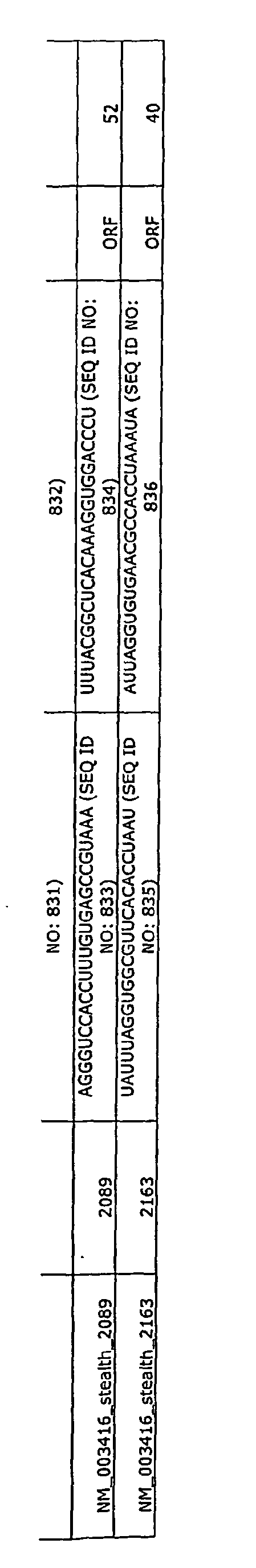
-
- 1. Op De Beeck A, Caillet-Fauquet P: Viruses and the cell cycle. Prog Cell Cycle Res 1997, 3:1-19.
- 2. Dermody TS, Nibert ML, Wetzel JD, Tong X, Fields BN: Cells and viruses with mutations affecting viral entry are selected during persistent infections of L cells with mammalian reoviruses. J Virol 1993, 67(4):2055-2063.
- 3. Taterka J, Sutcliffe M, Rubin DH: Selective reovirus infection of murine hepatocarcinoma cells during cell division. A model of viral liver infection. J Clin Invest 1994, 94(1):353-360.
- 4. Sheng J, Organ EL, Hao C, Wells KS, Ruley HE, Rubin DH: Mutations in the IGF-II pathway that confer resistance to lytic reovirus infection. BMC Cell Biol 2004, 5(1):32.
- 5. Hansen J, Floss T, Van Sloun P, Fuchtbauer EM, Vauti F, Arnold HH, S chnutgen F, Wurst W, von Melchner H, Ruiz P: A large-scale, gene-driven mutagenesis approach for the functional analysis of the mouse genome. Proc Natl Acad Sci U S A 2003, 100(17):9918-9922.
- 6. Hicks GG, Shi EG, Li XM, Li CH, Pawlak M, Ruley HE: Functional genomics in mice by tagged sequence mutagenesis. Nat Genet 1997, 16(4):338-344.
- 7. Salminen M, Meyer BI, Gruss P: Efficient poly A trap approach allows the capture of genes specifically active in differentiated embryonic stem cells and in mouse embryos. Dev Dyn 1998, 212(2):326-333.
- 8. Stryke D, Kawamoto M, Huang CC, Johns SJ, King LA, Harper CA, Meng EC, Lee RE, Yee A, L'Italien L et al: BayGenomics: a resource of insertional mutations in mouse embryonic stem cells. Nucleic Acids Res 2003, 31(1):278-281.
- 9. Wiles MV, Vauti F, Otte J, Fuchtbauer EM, Ruiz P, Fuchtbauer A, Arnold HH, Lehrach H, Metz T, von Melchner H et al: Establishment of a gene-trap sequence tag library to generate mutant mice from embryonic stem cells. Nat Genet 2000, 24(1):13-14.
- 10. Zambrowicz BP, Friedrich GA, Buxton EC, Lilleberg SL, Person C, Sands AT: Disruption and sequence identification of 2,000 genes in mouse embryonic stem cells. Nature 1998, 392(6676):608-611.
- 11. Osipovich AB, White-Grindley EK, Hicks GG, Roshon MJ, Shaffer C, Moore JH, H.E. R: Activation of cryptic 3' splice sites within introns of cellular genes following gene entrapment. Nucleic Acids Res 2004, in press.
- 12. De Ceuninck F, Poiraudeau S, Pagano M, Tsagris L, Blanchard O, Willeput J, Corvol M: Inhibition of chondrocyte cathepsin B and L activities by insulin-like growth factor-II (IGF-II) and its Ser29 variant in vitro: possible role of the mannose 6-phosphate/IGF-II receptor. Mol Cell Endocrinol 1995, 113(2):205-213.
- 13. Martinez CG, Guinea R, Benavente J, Carrasco L: The entry of reovirus into L cells is dependent on vacuolar proton- ATPase activity. J Virol 1996, 70(1):576-579.
- 14. Guinea R, Carrasco L: Requirement for vacuolar proton-ATPase activity during entry of influenza virus into cells. J Virol 1995, 69(4):2306-2312.
- 15. Brunetti CR, Burke RL, Komfeld S, Gregory W, Masiarz FR, Dingwell KS, Johnson DC: Herpes simplex virus glycoprotein D acquires mannose 6-phosphate residues and binds to mannose 6-phosphate receptors. J Biol Chem 1994, 269(25):17067-17074.
- 16. Zeng FY, Gerke V, Gabius HJ: Identification of annexin II, annexin VI and glyceraldehyde-3-phosphate dehydrogenase as calcyclin-binding proteins in bovine heart. Int JBiochem 1993, 25(7):1019-1027.
- 17. Lee KH, Na DS, Kim JW: Calcium-dependent interaction of annexin I with annexin II and mapping of the interaction sites. FEBS Lett 1999, 442(2-3):143-146.
- 18. Filipek A, Wojda U, Lesniak W: Interaction of calcyclin and its cyanogen bromide fragments with annexin II and glyceraldehyde 3-phosphate dehydrogenase. Int J Biochem Cell Biol 1995, 27(11):1123-1131.
- 19. Pietropaolo RL, Compton T: Direct interaction between human cytomegalovirus glycoprotein B and cellular annexin II. J Virol 1997, 71(12):9803-9807.
- 20. Golitsina NL, Kordowska J, Wang CL, Lehrer SS: Ca2+-dependent binding of calcyclin to muscle tropomyosin. Biochem Biophys Res Commun 1996, 220(2):360-365.
- 21. Hida K, Wada J, Zhang H, Hiragushi K, Tsuchiyarna Y, Shikata K, Makino H: Identification of genes specifically expressed in the accumulated visceral adipose tissue of OLETF rats. J Lipid Res 2000, 41(10):1615-1622.
- 22. Katoh M: IGSF11 gene, frequently up-regulated in intestinal-type gastric cancer, encodes adhesion molecule homologous to CXADR, FLJ22415 and ESAM. Int J Oncol 2003, 23(2):525-531 .
- 23. Barton ES, Forrest JC, Connolly JL, Chappell JD, Liu Y, Schnell FJ, Nusrat A, Parks CA, Dermody TS: Junction adhesion molecule is a receptor for reovirus. Cell 2001, 104(3):441-451.
- 24. Weiner HL, Powers ML, Fields BN: Absolute linkage of virulence and central nervous system cell tropism of reoviruses to viral hemagglutinin. J Infect Dis 1980, 141(5):609-616.
- 25. Rubin DH, Wetzel JD, Williams WV, Cohen JA, Dworkin C, Dermody TS: Binding of type 3 reovirus by a domain of the .
- 26. Sizova DV, Kolupaeva VG, Pestova TV, Shatsky IN, Hellen CU: Specific interaction of eukaryotic translation initiation factor 3 with the 5' nontranslated regions of hepatitis C virus and classical swine fever virus RNAs. J Virol 1998, 72(6):4775-4782.
- 27. McGregor F, Phelan A, Dunlop J, Clements JB: Regulation of herpes simplex virus poly (A) site usage and the action of immediate-early protein IE63 in the early-late switch. J Virol 1996, 70(3):1931-1940.
- 28. Pitha PM, Au WC, Lowther W, Juang YT, Schafer SL, Burysek L, Hiscott J, Moore PA: Role of the interferon regulatory factors (IRFs) in virus-mediated signaling and regulation of cell growth. Biochimie 1998, 80(8-9):651-658.
- 29. Hawiger J: Innate immunity and inflammation: a transcriptional paradigm. Immunol Res 2001, 23(2-3):99-109.
- 30. Lau JF, Horvath CM: Mechanisms of Type I interferon cell signaling and STAT-mediated transcriptional responses. Mt Sinai J Med 2002, 69(3):156-168.
- 31. Werner-Felmayer G, Werner ER, Fuchs D, Hausen A, Reibnegger G, Schmidt K, Weiss G, Wachter H: Pteridine biosynthesis in human endothelial cells. Impact on nitric oxide-mediated formation of cyclic GMP. J Biol Chem 1993, 268(3):1842-1846.
- 32. Reiss CS, Komatsu T: Does nitric oxide play a critical role in viral infections? J Virol 1998, 72(6):4547-4551.
- 33. Pertile TL, Karaca K, Sharma JM, Walser MM: An antiviral effect of nitric oxide: inhibition of reovirus replication. Avian Dis 1996, 40(2):342-348.
- 34. Takekawa M, Maeda T, Saito H: Protein phosphatase 2Calpha inhibits the human stress-responsive p38 and JNK MAPK pathways. Embo J 1998, 17(16):4744-4752.
- 35. Rousse S, Lallemand F, Montarras D, Pinset C, Mazars A, Prunier C, Atfi A, Dubois C: Transforming growth factor-beta inhibition of insulin-like growth factor-binding protein-5 synthesis in skeletal muscle cells involves a c-Jun N-terminal kinase-dependent pathway. J Biol Chem 2001, 276(50):46961-46967.
- 36. Uchida K, Suzuki H, Ohashi T, Nitta K, Yumura W, Nihei H: Involvement of MAP kinase cascades in Smad7 transcriptional regulation. Biochem Biophys Res Commun 2001, 289(2):376-381.
- 37. Arsura M, Panta GR, Bilyeu JD, Cavin LG, Sovak MA, Oliver AA, Factor V, Heuchel R, Mercurio F, Thorgeirsson SS et al: Transient activation of NF-kappaB through a TAK1/IKK kinase pathway by TGF-betal inhibits AP-1/SMAD signaling and apoptosis: implications in liver tumor formation. Oncogene 2003, 22(3):412-425.
- 38. Amir RE, Iwai K, Ciechanover A: The NEDD8 pathway is essential for SCF(beta -TrCP)-mediated ubiquitination and processing of the NF-kappa B precursor p105. J Biol Chem 2002, 277(26):23253-23259.
- 39. Tanaka K, Kawakami T, Tateishi K, Yashiroda H, Chiba T: Control of IkappaBalpha proteolysis by the ubiquitin-proteasome pathway. Biochimie 2001, 83(3-4):351-356.
- 40. Sakurai H, Shigemori N, Hasegawa K, Sugita T: TGF-beta-activated .
- 41. Shibuya H, Yamaguchi K, Shirakabe K, Tonegawa A, Gotoh Y, Ueno N, Irie K, Nishida E, Matsumoto K: TAB1: an activator of the .
- 42. Bhat NR, Shen Q, Fan F: TAK1-mediated induction of nitric oxide synthase gene expression in glial cells. J Neurochem 2003, 87(1):238-247.
- 43. Yanagisawa M, Nakashima K, Takeda K, Ochiai W, Takizawa T, Ueno M, Takizawa M, Shibuya H, Taga T: Inhibition of BMP2-induced, TAK1 kinase-mediated neurite outgrowth by Smad6 and Smad7. Genes Cells 2001, 6(12):1091-1099.
- 44. Asano K, Vornlocher HP, Richter-Cook NJ, Merrick WC, Hinnebusch AG, Hershey JW: Structure of cDNAs encoding human eukaryotic initiation factor 3 subunits. Possible roles in RNA binding and macromolecular assembly. J Biol Chem 1997, 272(43):27042-27052.
- 45. Higaki S, Gebhardt BM, Lukiw WJ, Thompson HW, Hill JM: Effect of immunosuppression on gene expression in the HSV-1 latently infected mouse trigeminal ganglion. Invest Ophthalmol Vis Sci 2002, 43(6):1862-1869.
- 46. Spear BT, Longley T, Moulder S, Wang SL, Peterson ML: A sensitive lacZ-based expression vector for analyzing transcriptional control elements in eukaryotic cells. DNA Cell Biol 1995, 14(7):635-642.
- 47. Pier GB, Grout M, Zaidi T, Meluleni G, Mueschenborn SS, Banting G, Ratcliff R, Evans MJ, Colledge WH: Salmonella typhi uses CFTR to enter intestinal epithelial cells. Nature 1998, 393(6680):79-82.
- 48. Perkins ME, Wu TW, Le Blancq SM: Cyclosporin analogs inhibit in vitro growth of Cryptosporidium parvum. Antimicrob Agents Chemother 1998, 42(4):843-848.
- 49.Clarke P, Tyler KL: Reovirus-induced apoptosis: A minireview. Apoptosis 2003, 8(2):141-150.
- 50. Richardson-Burns SM, Tyler KL: Regional differences in viral growth and central nervous system injury correlate with apoptosis. J Virol 2004, 78(10):5466-5475.
- 51. Clarke P, Meintzer SM, Widmann C, Johnson GL, Tyler KL: Reovirus infection activates JNK and the JNK-dependent transcription factor c-Jun. J Virol 2001, 75(23):11275-11283.
- 52. Clarke P, Meintzer SM, Moffitt LA, Tyler KL: Two distinct phases of virus-induced nuclear factor kappa B regulation enhance tumor necrosis factor-related apoptosis-inducing ligand-mediated apoptosis in virus-infected cells. J Biol Chem 2003,278(20):18092-18100.
- 53. Bender FC, Whitbeck JC, Ponce de Leon M, Lou H, Eisenberg RJ, Cohen GH: Specific association of glycoprotein B with lipid rafts during herpes simplex virus entry. J Virol 2003, 77(17):9542-9552.
- 54. Zhou G, Avitabile E, Campadelli-Fiume G, Roizman B: The domains of glycoprotein D required to block apoptosis induced by .
- 55. Schelling JR, Gentry DJ, Dubyak GR: Annexin II inhibition of G protein-regulated inositol trisphosphate formation in rat aortic smooth muscle. Am J Physiol 1996, 270(4Pt 2):F682-690.
- 56. Babiychuk EB, Monastyrskaya K, Burkhard FC, Wray S, Draeger A: Modulating signaling events in smooth muscle: cleavage of annexin 2 abolishes its binding to lipid rafts. Faseb J 2002, 16(10):1177-1184.
- 57. Pittis MG, Muzzolin L, Giulianini PG, Garcia RC: Mycobacteria-containing phagosomes associate less annexins I, VI, VII and XI, but not II, concomitantly with a diminished phagolysosomal fusion. Eur J Cell Biol 2003, 82(1):9-17.
- 58. Brunetti CR, Dingwell KS, Wale C, Graham FL, Johnson DC: Herpes simplex virus gD and virions accumulate in endosomes by mannose 6-phosphate-dependent and -independent mechanisms. J Virol 1998, 72(4):3330-3339.
- 59. Rubin DH, Komstein MJ, Anderson AO: .
- 60. Ahmed R, Canning WM, Kauffman RS, Sharpe AH, Hallum JV, Fields BN: Role of the host cell in persistent viral infection: coevolution of L cells and reovoirus during persistent infection. Cell 1981,25(2):325-332.
- 61. Altschul SF, Madden TL, Schaffer AA, Zhang J, Zhang Z, Miller W, Lipman DJ: Gapped BLAST and PSI-BLAST: a new generation of protein database search programs. Nucleic Acids Res 1997, 25(17):3389-3402.
- 62. Raynor CM, Wright JF, Waisman DM, Pryzdial EL: Annexin II enhances cytomegalovirus binding and fusion to phospholipid membranes. Biochemistry 1999, 38(16):5089-5095.
- 63. Glomb-Reinmund S, Kielian M: The role of low pH and disulfide shuffling in the entry and fusion of Semliki Forest virus and Sindbis virus. Virology 1998, 248(2):372-381.
- 64. Roberts PC, Kipperman T, Compans RW: Vesicular stomatitis virus G protein acquires pH-independent fusion activity during transport in a polarized endometrial cell line. J Virol 1999, 73(12):10447-10457.
- 65. Luo T, Douglas JL, Livingston RL, Garcia JV: Infectivity enhancement by HIV-1 Nef is dependent on the pathway of virus entry: implications for HIV-based gene transfer systems. Virology 1998,241(2):224-233.
- 66. Platt GM, Simpson GR, Mittnacht S, Schulz TF: Latent nuclear antigen of Kaposi's sarcoma-associated herpesvirus interacts with RING3, a homolog of the Drosophila female sterile homeotic (fsh) gene. J Virol 1999, 73(12):9789-9795.
- 67. Koffa MD, Graham SV, Takagaki Y, Manley JL, Clements JB: The human papillomavirus type 16 negative regulatory RNA element interacts with three proteins that act at different posttranscriptional levels. Proc Natl Acad Sci U S A 2000, 97(9):4677-4682.
- 68. Hirose Y, Manley JL: Creatine phosphate, not ATP, is required for 3' end cleavage of mammalian pre-mRNA in vitro. J Biol Chem 1997, 272(47):29636-29642.
- 69. Hansen J, Etchison D, Hershey JW, Ehrenfeld E: Association of cap-binding protein with eucaryotic initiation factor 3 in initiation factor preparations from uninfected and poliovirus-infected HeLa cells. J Virol 1982, 42(1):200-207.
- 70. Kieft JS, Zhou K, Jubin R, Murray MG, Lau JY, Doudna JA: The hepatitis C virus internal ribosome entry site adopts an ion-dependent tertiary fold. J Mol Biol 1999, 292(3):513-529.
- 71. Briggs CJ, Ott DE, Coren LV, Oroszlan S, Tozser J: Comparison of the effect of FK506 and cyclosporin A on virus production in H9 cells chronically and newly infected by HIV-1. Arch Virol 1999, 144(11):2151-2160.
- 72. Kanopka A, Muhlemann O, Petersen-Mahrt S, Estmer C, Ohrmaln C, Akusj arvi G: Regulation of adenovirus alternative RNA splicing by dephosphorylation of SR proteins. Nature 1998, 393(6681):185-187.
- 73. Tan SL, Nakao H, He Y, Vijaysri S, Neddermann P, Jacobs BL, Mayer BJ, Katze MG: NS5A, a nonstructural protein of hepatitis C virus, binds growth factor receptor-bound protein 2 adaptor protein in a Src homology 3 domain/ligand-dependent manner and perturbs mitogenic signaling. Proc Natl Acad Sci USA 1999, 96(10):5533-5538.
- 74. Korkaya H, Jameel S, Gupta D, Tyagi S, Kumar R, Zafrullah M, Mazumdar M, Lal SK, Xiaofang L, Sehgal D et al: The ORF3 protein of hepatitis E virus binds to Src homology 3 domains and activates MAPK. J Biol Chem 2001, 276(45):42389-42400.
- 75. Scaplehom N, Holmstrom A, Moreau V, Frischknecht F, Reckmann I, Way M: Grb2 and Nck act cooperatively to promote actin-based motility of vaccinia virus. Curr Biol 2002, 12(9):740-745.
- 76. Finkelstein LD, Ney PA, Liu QP, Paulson RF, Correll PH: Sf-Stk kinase activity and the Grb2 binding site are required for Epo-independent growth of primary erythroblasts infected with Friend virus. Oncogene 2002, 21(22):3562-3570.
- 77. Huh JR, Park JM, Kim M, Carlson BA, Hatfield DL, Lee BJ: Recruitment of TBP or TFIIB to a promoter proximal position leads to stimulation of RNA polymerase II transcription without activator proteins both in vivo and in vitro. Biochem Biophys Res Commun 1999, 256(1):45-51.
- 78. Zhou M, Kashanchi F, Jiang H, Ge H, Brady JN: Phosphorylation of the RAP74 subunit of TFIIF correlates with Tat-activated transcription of the HIV-1 long terminal repeat. Virology 2000, 268(2):452-460.
- 79. Kim H, Lee YH, Won J, Yun Y: Through induction of juxtaposition and tyrosine kinase activity of Jak1, X-gene product of hepatitis B virus stimulates Ras and the transcriptional activation through AP-1, NF-kappaB, and SRE enhancers. Biochem Biophys Res Commun 2001, 286(5):886-894.
- 80. Breslin JJ, Mork I, Smith MK, Vogel LK, Hemmila EM, Bonavia A, Talbot PJ, Sjostrom H, Noren O, Holmes KV: Human coronavirus 229E: receptor binding domain and neutralization by soluble receptor at 37 degrees C. J Virol 2003, 77(7):4435-4438.
- 81. Li Y, Kang J, Horwitz MS: Interaction of an adenovirus 14.7-kilodalton protein inhibitor of tumor necrosis factor alpha cytolysis with a new member of the GTPase superfamily of signal transducers. J Virol 1997, 71(2):1576-1582.
- 82. Hugle T, Fehrmann F, Bieck E, Kohara M, Krausslich HG, Rice CM, Blum HE, Moradpour D: The hepatitis C virus nonstructural protein 4B is an integral endoplasmic reticulum membrane protein. Virology 2001, 284(1):70-81.
- 83.Bonatti S, Migliaccio G, Blobel G, Walter P: Role of signal recognition particle in the membrane assembly of Sindbis viral glycoproteins. Eur J Biochem 1984, 140(3):499-502.
- 84. Melancon P, Garoff H: Reinitiation of translocation in the Semliki Forest virus structural polyprotein: identification of the signal for the E1 glycoprotein. Em bo J 1986, 5(7):1 S51-1560.
- 85. Emans N, Gorvel JP, Walter C, Gerke V, Kellner R, Griffiths G, Gruenberg J = Annexin II is a major component of fusogenic endosomal vesicles. J Cell Biol 1993, 120(6):1357-1369.
- 86. Fiedler K, Kellner R, Simons K: Mapping the protein composition of trans-Golgi network (TGN)-derived carrier vesicles from polarized MDCK cells. Electrophoresis 1997, 18(14):2613-2619.
- 87. Nezu J, Motojima K, Tamura H, Ohkuma S: Molecular cloning of a rat liver cDNA encoding the 16 kDa subunit of vacuolar H(+)-ATPases: organellar and tissue distribution of 16 kDa proteolipids. J Biochem (Tokyo) 1992, 112(2):212-219.
- 88. Orci L, Perrelet A, Rothman JE: Vesicles on strings: morphological evidence for processive transport within the Golgi stack. Proc Natl Acad Sci USA 1998, 95(5):2279-2283.
- 89. Nakamura N, Lowe M, Levine TP, Rabouille C, Warren G: The vesicle docking protein p115 binds GM130, a cis-Golgi matrix protein, in a mitotically regulated manner. Cell 1997, 89(3):445-455.
- 90. Okutsu T, Kuroiwa Y, Kagitani F, Kai M, Aisaka K, Tsutsumi O, Kaneko Y, Yokomori K, Surani MA, Kohda T et al: Expression and imprinting status of human PEG8/IGF2AS, a paternally expressed antisense transcript from the IGF2 locus, in Wilms' tumors. J Biochem (Tokyo) 2000, 127(3):475-483.
- 91. Kumar R, Yang J, Larsen RD, Stanley P: Cloning and expression of N-acetylglucosaminyltransferase I, the medial Golgi transferase that initiates complex N-linked carbohydrate formation. Proc Natl Acad Sci U S A 1990, 87(24):9948-9952.
- 92. Nilsson T, Rabouille C, Hui N, Watson R, Warren G: The role of the membrane-spanning domain and stalk region of N-acetylglucosaminyltransferase I in retention, kin recognition and structural maintenance of the Golgi apparatus in HeLa cells. J Cell Sci 1996, 109(Pt 7):1975-1989.
- 93. Nilsson T, Slusarewicz P, Hoe MH, Warren G: Kin recognition. A model for the retention of Golgi enzymes. FEBS Lett 1993, 330(1):1-4.
- 94. Yang W, Pepperkok R, Bender P, Kreis TE, Storrie B: Modification of the cytoplasmic domain affects the subcellular localization of Golgi glycosyltransferases. Eur J Cell Biol 1996, 71(1):53-61.
- 95. Hirst J, Futter CE, Hopkins CR: The kinetics ofmannose 6-phosphate receptor trafficking in the endocytic pathway in HEp-2 cells: the receptor enters and rapidly leaves multivesicular endosomes without accumulating in a prelysosomal compartment. Mol Biol Cell 1998, 9(4):809-816.
- 96. Sheng Q, Denis D, Ratnofsky M, Roberts TM, DeCaprio JA, Schaffhausen B: The DnaJ domain of polyomavirus large T antigen is required to regulate Rb family tumor suppressor function. J Viro11997, 71(12):9410-9416.
- Preferred embodiments are outlined in the following paragraphs:
-
- 1. A method of identifying an antiviral agent comprising:
- (a) administering the agent to a cell containing a cellular gene listed in Table 1; and
- (b) detecting the level and/or activity of the gene product produced by the cellular gene, a decrease or elimination of the gene product and/or gene product activity indicating a compound with antiviral activity.
- 2. A method of identifying an antiviral agent comprising:
- (a) administering the agent to a cell containing a cellular gene listed in Table 1;
- (b) contacting the cell with a virus;
- (c) detecting the level of viral infection; and
- (d) associating the level of viral infection with the level of expression of the gene from Table 1 or the activity of the protein encoded by the gene from Table 1, a decrease or elimination of viral infection associated with a decrease or elimination of gene expression and/or activity indicating that the agent is an antiviral agent.
- 3. A method of identifying an antiviral agent comprising:
- (a) administering the agent to a cell containing a cellular gene listed in Table 1;
- (b) contacting the cell with a virus;
- (c) detecting the level of viral infection; and
- (d) associating the level of viral infection with the level of expression of the gene from Table 1 or the activity of the protein encoded by the gene from Table 1, a decrease or elimination of viral infection associated with a decrease or elimination of gene expression and/or activity indicating that the agent is an antiviral agent.
- 4. A non-human transgenic mammal comprising a functional deletion of one or more genes listed in Table 1, wherein the mammal has decreased susceptibility to infection by a pathogen.
- 5. The transgenic mammal of paragraph 4, wherein the pathogen is a virus.
- 6. The transgenic mammal of paragraph 4, wherein the pathogen is a bacteria.
- 7. The transgenic mammal of paragraph 4, wherein the pathogen is a fungi.
- 8. A cell including an altered or disrupted gene listed in Table 1 that has decreased susceptibility to infection by a pathogen.
- 9. The cell of paragraph 8, wherein the pathogen is a virus.
- 10. The cell of paragraph 8, wherein the pathogen is a bacteria.
- 11. The cell of paragraph 8, wherein the pathogen is a fungi.
- 12. The cell of paragraph 8, wherein the cell is a hematopoietic cell.
- 13. A population of cells including an altered or disrupted gene listed in Table 1 that has decreased susceptibility to infection by a pathogen.
- 14. A method of inhibiting infection in a cell comprising inhibiting expression or activity of a gene or a gene product listed in Table 1.
- 15. The method of paragraph 14, wherein the infection is a viral infection.
- 16. The method of paragraph 14, wherein the infection is a bacterial infection.
- 17. The method of paragraph 14, wherein the infection is a fungal infection.
- 18. A method of reducing or inhibiting a viral infection in a subject comprising administering to the subject an amount of a composition that inhibits expression or activity of a gene or a gene product in Table 1.
- 19. The method of paragraph 18, wherein the infection is a viral infection.
- 20. The method of paragraph 18, wherein the infection is a bacterial infection.
- 21. The method of paragraph 18, wherein the infection is a fungal infection.
- 22. A population of non-human animals possessing a variant form of one or more genes listed in Table 1, wherein the population is less susceptible to infection.
- 23. The population of paragraph 22, wherein the non-human animals are from an avian species.
- 24. The population of paragraph 23, wherein population is a flock of chickens.
- 25. The population of paragraph 23, wherein the flock of chickens is less susceptible to avian flu.
- 26. The antiviral agent identified by the method of
paragraph 1. - 27. The antiviral agent identified by the method of
paragraph 1. - 28. The method of paragraph 14, wherein expression or activity of a gene or a gene product listed in Table 1 is inhibited by contacting the cell with a chemical, a small molecule, a drug, a protein, a cDNA, an antibody, a morpholino, a triple helix molecule, an siRNA, an shRNAs, an antisense RNA or a ribozyme.
- 29. The method of paragraph 18, wherein the composition is a chemical, a small molecule, a drug, a protein, a cDNA, an antibody, a morpholino, a triple helix molecule, an siRNA, an shRNAs, an antisense RNA or a ribozyme.


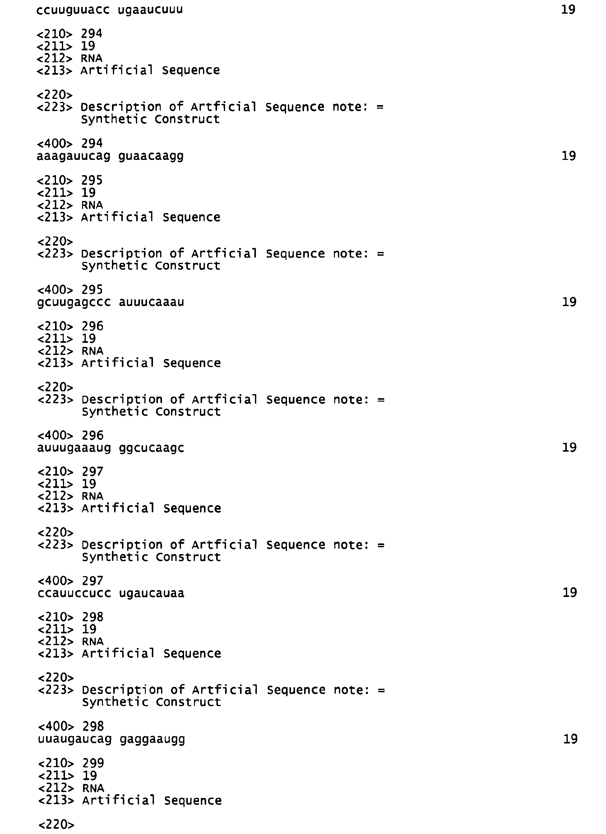


Claims (17)
- Composition inhibiting expression or activity of a gene or a gene product for use for inhibiting a viral infection in a subject wherein the gene or gene product is selected from genes having a cellular role in the transcription, for the membrane, for signaling, for vesicle/trafficking, for metabolism, or being cytoskeletal-related.
- Composition for use according to claim 1, wherein the gene or gene product is selected from the cytosceletal-related group comprising Anx3, Cald1, Calm2, Mapt, Ppm1a, Rps18, Stmn1, Tpm1, Kif13b.
- Composition for use according to claim 1, wherein the gene is selected from the membrane-related group comprising Abca4, Csmd2, Celsr2, Erbb2ip, OL16, Pgy1, Rab13, Serp1.
- Composition according to claim 1, wherein the gene is selected from a transcription-related group comprising Brd2, Brd3, Ctcf, E2f2, Gtf2e1, Hnrpl, Hoxc13, Hp1-bp74, ld3, Znf207, Zfp7, a signaling-related group E2ip2, Fusip1, Fkbp8, Gata4, Grb2, Jak1, Madh7, Map3k7ip1, Pde4b, Rraga, Ryk, vesicle/trafficking-related group Anxa1, Anxa2, Atp6vOc, Copg2, Golga2, Hm13, lgf2r, Psa, Rin2, Rabl3, S100a6,, translation-related group Cstf2, Eif3s10, Srp19, apoptosis-related group Bcl21, Cycs, IkB□, Mical2, Rfp2,and metabolism-related group comprising Gas5, Lipc, Magt1, Pts.
- The composition of one of the preceding claims wherein the inhibitor of the expression or activity of a gene or gene product is a chemical, a small molecule, a drug, a protein, a cDNA, an antibody, a morpholino, a triple helix molecule, an siRNA, an shRNA, an antisense RNA or a ribozyme.
- Composition according to any one of the preceding claims, wherein the fragment is a functional fragment possessing at least one activity of the protein that is necessary for viral infection but not for survival of the cell.
- The composition of one of claims 1 to 6, wherein the inhibiting agent is an antibody.
- The composition of claim 1 for use for preventing an infection or for prophylactic treatment.
- Antiviral agent for use for treating viral infections in a subject wherein the antiviral agent is a protein, peptide or fragment thereof that is necessary for viral infection but not for survival of the cell, wherein the protein or peptide is selected from the list of claim 1.
- Antiviral composition for use for decreasing susceptibility to a viral infection in a subject by inhibiting a gene or gene product as defined in one of claims 1 to 4.
- A cell including an altered or disrupted gene selected from one of the genes of claims 1 to 4 having a decreased susceptibility to infection by a pathogen.
- The cell of claim 11, wherein the pathogen is a virus, bacterium, or a fungus.
- A method for determining susceptibility to viral infection of a subject by detecting polymorphic alleles in a sample of a subject.
- The method of claim 13, wherein for the detection a micro array is used including probes to different polymorphic alleles of one or more genes selected from the group defined in one of claims 1 to 4.
- A method of screening a host subject for resistance to infection by characterizing a nucleotide sequence of a host nucleic acid or the amino acid sequence of a host polypeptide selected from the proteins and peptides defined in one of claims 1 to 4.
- The method of claim 15, wherein a nucleic acid of a subject is isolated, sequenced, and compared to the wild-type sequence, where the greater similarity between the subject's nucleic acid and the wild-type sequence is, the more susceptible the person is to infection.
- The method of one of claims 15, wherein expression of a gene and/or activity of a gene product is increased wherein the gene is one that upon disruption results in overexpression and wherein the agent is obtained by administering the agent to the cell, contacting the cell with pathogen and associating an increase in gene expression and/or activity of the gene product with the level of infection such that an increase in gene expression and/or activity of the gene product accompanied by a decrease in infection indicates that the agent inhibits infection by the pathogen via overexpression.
Applications Claiming Priority (2)
| Application Number | Priority Date | Filing Date | Title |
|---|---|---|---|
| US62248604P | 2004-10-27 | 2004-10-27 | |
| EP05814043A EP1812597A2 (en) | 2004-10-27 | 2005-10-27 | Mammalian genes involved in infection |
Related Parent Applications (1)
| Application Number | Title | Priority Date | Filing Date |
|---|---|---|---|
| EP05814043.5 Division | 2005-10-27 |
Publications (1)
| Publication Number | Publication Date |
|---|---|
| EP2311530A2 true EP2311530A2 (en) | 2011-04-20 |
Family
ID=36228473
Family Applications (2)
| Application Number | Title | Priority Date | Filing Date |
|---|---|---|---|
| EP10011179A Withdrawn EP2311530A2 (en) | 2004-10-27 | 2005-10-27 | Mammalian genes involved in infection |
| EP05814043A Ceased EP1812597A2 (en) | 2004-10-27 | 2005-10-27 | Mammalian genes involved in infection |
Family Applications After (1)
| Application Number | Title | Priority Date | Filing Date |
|---|---|---|---|
| EP05814043A Ceased EP1812597A2 (en) | 2004-10-27 | 2005-10-27 | Mammalian genes involved in infection |
Country Status (8)
| Country | Link |
|---|---|
| US (1) | US7964346B2 (en) |
| EP (2) | EP2311530A2 (en) |
| JP (1) | JP2008520191A (en) |
| CN (1) | CN101115846A (en) |
| AU (1) | AU2005299296A1 (en) |
| CA (1) | CA2585970A1 (en) |
| SG (1) | SG156690A1 (en) |
| WO (1) | WO2006047673A2 (en) |
Cited By (1)
| Publication number | Priority date | Publication date | Assignee | Title |
|---|---|---|---|---|
| EP3543340A1 (en) * | 2018-03-19 | 2019-09-25 | Fundació Centre de Regulació Genòmica | Antisense oligonucleotides and uses thereof |
Families Citing this family (14)
| Publication number | Priority date | Publication date | Assignee | Title |
|---|---|---|---|---|
| TW200911290A (en) * | 2007-07-02 | 2009-03-16 | Alcon Res Ltd | RNAI-mediated inhibition of HTRA1 for treatment of macular degeneration |
| WO2010144908A2 (en) * | 2009-06-12 | 2010-12-16 | Zirus, Inc. | Mammalian genes involved in tularemia and other infections |
| US20140220690A1 (en) * | 2009-11-11 | 2014-08-07 | Vanderbilt University | Mammalian genes involved in infection |
| WO2012045067A2 (en) * | 2010-10-01 | 2012-04-05 | Zirus, Inc. | Mammalian genes involved in infection |
| EP2831232A4 (en) | 2012-03-30 | 2015-11-04 | Univ Washington | Methods for modulating tau expression for reducing seizure and modifying a neurodegenerative syndrome |
| ES2807379T3 (en) * | 2013-03-14 | 2021-02-22 | Ionis Pharmaceuticals Inc | Compositions and methods to regulate the expression of Tau |
| TWI772856B (en) * | 2013-07-19 | 2022-08-01 | 美商百健Ma公司 | Compositions for modulating tau expression |
| CA2931090A1 (en) | 2013-12-12 | 2015-06-18 | Alnylam Pharmaceuticals, Inc. | Complement component irna compositions and methods of use thereof |
| GB201400309D0 (en) * | 2014-01-08 | 2014-02-26 | Aqua Gen As | Predicting Resistance to disease |
| SG10201807208RA (en) * | 2014-03-04 | 2018-09-27 | Sigma Aldrich Co Llc | Viral resistant cells and uses thereof |
| JOP20190065A1 (en) | 2016-09-29 | 2019-03-28 | Ionis Pharmaceuticals Inc | Compounds and methods for reducing tau expression |
| AU2018360697A1 (en) | 2017-11-01 | 2020-05-14 | Alnylam Pharmaceuticals, Inc. | Complement component C3 iRNA compositions and methods of use thereof |
| CN111450251B (en) * | 2020-03-23 | 2021-11-26 | 清华大学 | Application of MTHFD1 inhibitor in inhibiting and killing virus |
| WO2022197607A2 (en) * | 2021-03-14 | 2022-09-22 | Sirnaomics, Inc. | Methods of cancer treatment by delivery of sirnas against bclxl and mcl1 using a polypeptide nanoparticle. |
Citations (30)
| Publication number | Priority date | Publication date | Assignee | Title |
|---|---|---|---|---|
| US4683202A (en) | 1985-03-28 | 1987-07-28 | Cetus Corporation | Process for amplifying nucleic acid sequences |
| US4683195A (en) | 1986-01-30 | 1987-07-28 | Cetus Corporation | Process for amplifying, detecting, and/or-cloning nucleic acid sequences |
| US4704692A (en) | 1986-09-02 | 1987-11-03 | Ladner Robert C | Computer based system and method for determining and displaying possible chemical structures for converting double- or multiple-chain polypeptides to single-chain polypeptides |
| WO1988009810A1 (en) | 1987-06-11 | 1988-12-15 | Synthetic Genetics | Novel amphiphilic nucleic acid conjugates |
| US4816567A (en) | 1983-04-08 | 1989-03-28 | Genentech, Inc. | Recombinant immunoglobin preparations |
| WO1989010134A1 (en) | 1988-04-25 | 1989-11-02 | The Regents Of The University Of California | Chimeric peptides for neuropeptide delivery through the blood-brain barrier |
| WO1990011364A1 (en) | 1989-03-24 | 1990-10-04 | University Patents, Inc. | Rna ribozyme restriction endoribonucleases and methods |
| US4965188A (en) | 1986-08-22 | 1990-10-23 | Cetus Corporation | Process for amplifying, detecting, and/or cloning nucleic acid sequences using a thermostable enzyme |
| US4980286A (en) | 1985-07-05 | 1990-12-25 | Whitehead Institute For Biomedical Research | In vivo introduction and expression of foreign genetic material in epithelial cells |
| WO1995006764A2 (en) | 1993-09-03 | 1995-03-09 | Vpi Holdings Ltd. | Oligonucleotides with rna cleavage activity |
| WO1995022058A1 (en) | 1994-02-10 | 1995-08-17 | Affymax Technologies N.V. | Method and apparatus for detection of fluorescently labeled materials |
| EP0721016A2 (en) | 1994-10-21 | 1996-07-10 | Affymax Technologies N.V. | Nucleic acid library arrays, methods for synthesizing them and methods for sequencing and sample screening using them |
| EP0728520A1 (en) | 1995-02-27 | 1996-08-28 | Affymax Technologies N.V. | Printing molecular library arrays |
| US5556752A (en) | 1994-10-24 | 1996-09-17 | Affymetrix, Inc. | Surface-bound, unimolecular, double-stranded DNA |
| US5578832A (en) | 1994-09-02 | 1996-11-26 | Affymetrix, Inc. | Method and apparatus for imaging a sample on a device |
| US5593839A (en) | 1994-05-24 | 1997-01-14 | Affymetrix, Inc. | Computer-aided engineering system for design of sequence arrays and lithographic masks |
| WO1997002357A1 (en) | 1995-06-29 | 1997-01-23 | Affymetrix, Inc. | Integrated nucleic acid diagnostic device |
| EP0785280A2 (en) | 1995-11-29 | 1997-07-23 | Affymetrix, Inc. (a California Corporation) | Polymorphism detection |
| WO1997027317A1 (en) | 1996-01-23 | 1997-07-31 | Affymetrix, Inc. | Nucleic acid analysis techniques |
| WO1997029212A1 (en) | 1996-02-08 | 1997-08-14 | Affymetrix, Inc. | Chip-based speciation and phenotypic characterization of microorganisms |
| EP0799897A1 (en) | 1996-04-04 | 1997-10-08 | Affymetrix, Inc. (a California Corporation) | Methods and compositions for selecting tag nucleic acids and probe arrays |
| US5859308A (en) | 1994-12-29 | 1999-01-12 | University Of Medicine And Denistry Of New Jersey | Transgenic animals and related aspects |
| WO2001043540A2 (en) | 1999-12-17 | 2001-06-21 | Oregon Health And Science University | Methods for producing transgenic animals |
| US6281408B1 (en) | 1998-02-20 | 2001-08-28 | Thomas Jefferson University | Efficient method for production of compound transgenic animals |
| WO2002019811A2 (en) | 2000-09-06 | 2002-03-14 | Nexia Biotechnologies, Inc. | Generation of transgenic animals using nuclear transfer and oocytes at the germinal vesicle stage |
| US6376743B1 (en) | 1998-08-11 | 2002-04-23 | University Of Hawaii | Mammalian transgenesis by intracytoplasmic sperm injection |
| US20020066117A1 (en) | 2000-07-13 | 2002-05-30 | Lars Nilsson | Transgenic animal and methods |
| WO2002044321A2 (en) | 2000-12-01 | 2002-06-06 | MAX-PLANCK-Gesellschaft zur Förderung der Wissenschaften e.V. | Rna interference mediating small rna molecules |
| US6448000B1 (en) | 1996-04-15 | 2002-09-10 | Vanderbilt University | Mammalian genes involved in viral infection and tumor suppression |
| US6777177B1 (en) | 1997-10-10 | 2004-08-17 | Vanderbilt University | Mammalian genes involved in viral infection and tumor suppression |
-
2005
- 2005-10-27 CA CA002585970A patent/CA2585970A1/en not_active Abandoned
- 2005-10-27 US US11/666,453 patent/US7964346B2/en not_active Expired - Fee Related
- 2005-10-27 AU AU2005299296A patent/AU2005299296A1/en not_active Abandoned
- 2005-10-27 EP EP10011179A patent/EP2311530A2/en not_active Withdrawn
- 2005-10-27 EP EP05814043A patent/EP1812597A2/en not_active Ceased
- 2005-10-27 SG SG200907248-9A patent/SG156690A1/en unknown
- 2005-10-27 JP JP2007539100A patent/JP2008520191A/en active Pending
- 2005-10-27 CN CNA2005800449607A patent/CN101115846A/en active Pending
- 2005-10-27 WO PCT/US2005/038740 patent/WO2006047673A2/en active Application Filing
Patent Citations (35)
| Publication number | Priority date | Publication date | Assignee | Title |
|---|---|---|---|---|
| US4816567A (en) | 1983-04-08 | 1989-03-28 | Genentech, Inc. | Recombinant immunoglobin preparations |
| US4683202A (en) | 1985-03-28 | 1987-07-28 | Cetus Corporation | Process for amplifying nucleic acid sequences |
| US4683202B1 (en) | 1985-03-28 | 1990-11-27 | Cetus Corp | |
| US4980286A (en) | 1985-07-05 | 1990-12-25 | Whitehead Institute For Biomedical Research | In vivo introduction and expression of foreign genetic material in epithelial cells |
| US4683195B1 (en) | 1986-01-30 | 1990-11-27 | Cetus Corp | |
| US4683195A (en) | 1986-01-30 | 1987-07-28 | Cetus Corporation | Process for amplifying, detecting, and/or-cloning nucleic acid sequences |
| US4965188A (en) | 1986-08-22 | 1990-10-23 | Cetus Corporation | Process for amplifying, detecting, and/or cloning nucleic acid sequences using a thermostable enzyme |
| US4704692A (en) | 1986-09-02 | 1987-11-03 | Ladner Robert C | Computer based system and method for determining and displaying possible chemical structures for converting double- or multiple-chain polypeptides to single-chain polypeptides |
| WO1988009810A1 (en) | 1987-06-11 | 1988-12-15 | Synthetic Genetics | Novel amphiphilic nucleic acid conjugates |
| WO1989010134A1 (en) | 1988-04-25 | 1989-11-02 | The Regents Of The University Of California | Chimeric peptides for neuropeptide delivery through the blood-brain barrier |
| WO1990011364A1 (en) | 1989-03-24 | 1990-10-04 | University Patents, Inc. | Rna ribozyme restriction endoribonucleases and methods |
| WO1995006764A2 (en) | 1993-09-03 | 1995-03-09 | Vpi Holdings Ltd. | Oligonucleotides with rna cleavage activity |
| WO1995022058A1 (en) | 1994-02-10 | 1995-08-17 | Affymax Technologies N.V. | Method and apparatus for detection of fluorescently labeled materials |
| US5631734A (en) | 1994-02-10 | 1997-05-20 | Affymetrix, Inc. | Method and apparatus for detection of fluorescently labeled materials |
| US5593839A (en) | 1994-05-24 | 1997-01-14 | Affymetrix, Inc. | Computer-aided engineering system for design of sequence arrays and lithographic masks |
| US5578832A (en) | 1994-09-02 | 1996-11-26 | Affymetrix, Inc. | Method and apparatus for imaging a sample on a device |
| EP0721016A2 (en) | 1994-10-21 | 1996-07-10 | Affymax Technologies N.V. | Nucleic acid library arrays, methods for synthesizing them and methods for sequencing and sample screening using them |
| US5556752A (en) | 1994-10-24 | 1996-09-17 | Affymetrix, Inc. | Surface-bound, unimolecular, double-stranded DNA |
| US5859308A (en) | 1994-12-29 | 1999-01-12 | University Of Medicine And Denistry Of New Jersey | Transgenic animals and related aspects |
| EP0728520A1 (en) | 1995-02-27 | 1996-08-28 | Affymax Technologies N.V. | Printing molecular library arrays |
| US5599695A (en) | 1995-02-27 | 1997-02-04 | Affymetrix, Inc. | Printing molecular library arrays using deprotection agents solely in the vapor phase |
| WO1997002357A1 (en) | 1995-06-29 | 1997-01-23 | Affymetrix, Inc. | Integrated nucleic acid diagnostic device |
| EP0785280A2 (en) | 1995-11-29 | 1997-07-23 | Affymetrix, Inc. (a California Corporation) | Polymorphism detection |
| WO1997027317A1 (en) | 1996-01-23 | 1997-07-31 | Affymetrix, Inc. | Nucleic acid analysis techniques |
| WO1997029212A1 (en) | 1996-02-08 | 1997-08-14 | Affymetrix, Inc. | Chip-based speciation and phenotypic characterization of microorganisms |
| EP0799897A1 (en) | 1996-04-04 | 1997-10-08 | Affymetrix, Inc. (a California Corporation) | Methods and compositions for selecting tag nucleic acids and probe arrays |
| US6448000B1 (en) | 1996-04-15 | 2002-09-10 | Vanderbilt University | Mammalian genes involved in viral infection and tumor suppression |
| US6777177B1 (en) | 1997-10-10 | 2004-08-17 | Vanderbilt University | Mammalian genes involved in viral infection and tumor suppression |
| US6281408B1 (en) | 1998-02-20 | 2001-08-28 | Thomas Jefferson University | Efficient method for production of compound transgenic animals |
| US6376743B1 (en) | 1998-08-11 | 2002-04-23 | University Of Hawaii | Mammalian transgenesis by intracytoplasmic sperm injection |
| WO2001043540A2 (en) | 1999-12-17 | 2001-06-21 | Oregon Health And Science University | Methods for producing transgenic animals |
| US20010044937A1 (en) | 1999-12-17 | 2001-11-22 | Gerald Schatten | Methods for producing transgenic animals |
| US20020066117A1 (en) | 2000-07-13 | 2002-05-30 | Lars Nilsson | Transgenic animal and methods |
| WO2002019811A2 (en) | 2000-09-06 | 2002-03-14 | Nexia Biotechnologies, Inc. | Generation of transgenic animals using nuclear transfer and oocytes at the germinal vesicle stage |
| WO2002044321A2 (en) | 2000-12-01 | 2002-06-06 | MAX-PLANCK-Gesellschaft zur Förderung der Wissenschaften e.V. | Rna interference mediating small rna molecules |
Non-Patent Citations (164)
| Title |
|---|
| ABRAHMSEN L ET AL., BIOCHEMISTRY, vol. 30, 1991, pages 4151 |
| AGRAWAL ET AL., EXPER. HEMATOL., vol. 24, 1996, pages 738 - 747 |
| AHMED R; CANNING WM; KAUFFMAN RS; SHARPE AH; HALLUM JV; FIELDS BN: "Role of the host cell in persistent viral infection: coevolution of L cells and reovoirus during persistent infection", CELL, vol. 25, no. 2, 1981, pages 325 - 332, XP027462958, DOI: doi:10.1016/0092-8674(81)90050-7 |
| ALTSCHUL SF; MADDEN TL; SCHAFFER AA; ZHANG J; ZHANG Z; MILLER W; LIPMAN DJ: "Gapped BLAST and PSI-BLAST: a new generation of protein database search programs", NUCLEIC ACIDS RES, vol. 25, no. 17, 1997, pages 3389 - 3402, XP002905950, DOI: doi:10.1093/nar/25.17.3389 |
| AMIR RE; IWAI K; CIECHANOVER A: "The NEDD8 pathway is essential for SCF(beta -TrCP)-mediated ubiquitination and processing of the NF-kappa B precursor p105", JBIOL CHEM, vol. 277, no. 26, 2002, pages 23253 - 23259 |
| ARSURA M; PANTA GR; BILYEU JD; CAVIN LG; SOVAK MA; OLIVER AA; FACTOR V; HEUCHEL R; MERCURIO F; THORGEIRSSON SS ET AL.: "Transient activation ofNF- kappaB through a TAK1/IKK kinase pathway by TGF-betal inhibits AP-1/SMAD signaling and apoptosis: implications in liver tumor formation", ONCOGENE, vol. 22, no. 3, 2003, pages 412 - 425 |
| ASANO K; VORNLOCHER HP; RICHTER-COOK NJ; MERRICK WC; HINNEBUSCH AG; HERSHEY JW: "Structure of cDNAs encoding human eukaryotic initiation factor 3 subunits. Possible roles in RNA binding and macromolecular assembly", JBIOL CHEM, vol. 272, no. 43, 1997, pages 27042 - 27052, XP002198751, DOI: doi:10.1074/jbc.272.43.27042 |
| BABIYCHUK EB; MONASTYRSKAYA K; BURKHARD FC; WRAY S; DRAEGER A: "Modulating signaling events in smooth muscle: cleavage of annexin 2 abolishes its binding to lipid rafts", FASEB J, vol. 16, no. 10, 2002, pages 1 177 - 1184 |
| BAGGIOLINI M ET AL., FEBS LETT., vol. 307, 1992, pages 97 - 101 |
| BARTON ES; FORREST JC; CONNOLLY JL; CHAPPELL JD; LIU Y; SCHNELL FJ; NUSRAT A; PARKOS CA; DERMODY TS: "Junction adhesion molecule is a receptor for reovirus", CELL, vol. 104, no. 3, 2001, pages 441 - 451 |
| BASHKIN ET AL., APPL. BIOCHEM BIOTECHNOL., vol. 54, 1995, pages 43 - 56 |
| BENDER FC; WHITBECK JC; PONCE DE LEON M; LO-U H; EISENBERG RJ; COHEN GH: "Specific association of glycoprotein B with lipid rafts during herpes simplex virus entry", J VIROL, vol. 77, no. 17, 2003, pages 9542 - 9552 |
| BHAT NR; SHEN Q; FAN F: "TAKI-mediated induction of nitric oxide synthase gene expression in glial cells", J NEUROCHEM, vol. 87, no. 1, 2003, pages 238 - 247 |
| BMC CELL BIOLOGY, vol. 5, 27 August 2004 (2004-08-27), pages 32 |
| BODANSKY M AND TROST B.,: "Principles of Peptide Synthesis", 1993, SPRINGER-VERLAG INC. |
| BONATTI S; MIGLIACCIO G; BLOBEL G; WALTER P: "Role of signal recognition particle in the membrane assembly of Sindbis viral glycoproteins", EUR J BIOCHEM, vol. 140, no. 3, 1984, pages 499 - 502 |
| BRESLIN JJ; MORK I; SMITH MK; VOGEL LK; HEMMILA EM; BONAVIA A; TALBOT PJ; SJOSTROM H; NOREN O; HOLMES KV: "Human coronavirus 229E: receptor binding domain and neutralization by soluble receptor at 37 degrees C", J VIROL, vol. 77, no. 7, 2003, pages 4435 - 4438 |
| BRIGGS CJ; OTT DE; COREN LV; OROSZLAN S; TOZSER J: "Comparison of the effect of FK506 and cyclosporin A on virus production in. H9 cells chronically and newly infected by HIV-1", ARCH VIROL, vol. 144, no. 11, 1999, pages 2151 - 2160, XP000993073, DOI: doi:10.1007/s007050050629 |
| BRUNETTI CR; BURKE RL; KOMFELD S; GREGORY W; MASIARZ FR; DINGWELL KS; JOHNSON DC: "Herpes simplex virus glycoprotein D acquires mannose 6-phosphate residues and binds to mannose 6-phosphate receptors", JBIOL CHEM, vol. 269, no. 25, 1994, pages 17067 - 17074 |
| BRUNETTI CR; DINGWELL KS; WALE C; GRAHAM FL; JOHNSON DC: "Herpes simplex virus gD and virions accumulate in endosomes by mannose 6-phosphate- dependent and -independent mechanisms", J VIR-OL, vol. 72, no. 4, 1998, pages 3330 - 3339 |
| CAPLEN ET AL., PROC. NATL. ACAD. SCI., vol. 98, 2001, pages 9742 - 9747 |
| CHANG ET AL.: "Using siRNA Technique to Generate Transgenic Animals with Spatiotemporal and Conditional Gene Knockdown", AMERICAN JOURNAL OF PATHOLOGY, vol. 165, 2004, pages 1535 - 1541, XP009049548 |
| CHEE ET AL., SCIENCE, vol. 274, 1996, pages 610 |
| CLARKE P; MEINTZER SM; MOFFITT LA; TYLER KL: "Two distinct phases of virus- induced nuclear factor kappa B regulation enhance tumor necrosis factor-related apoptosis-inducing ligand-mediated apoptosis in virus-infected cells", J BIOL CHEM, vol. 278, no. 20, 2003, pages 18092 - 18100 |
| CLARKE P; MEINTZER SM; WIDMANN C; JOHNSON GL; TYLER KL: "Reovirus infection activates JNK and the JNK-dependent transcription factor c-Jun", J VIROL, vol. 75, no. 23, 2001, pages 11275 - 11283 |
| CLARKE P; TYLER KL; REOVIRUS-INDUCED APOPTOSIS: A MINIREVIEW, APOPTOSIS, vol. 8, no. 2, 2003, pages 141 - 150 |
| CLARK-LEWIS I ET AL., BIOCHEMISTRY, vol. 30, 1991, pages 3128 |
| CLARK-LEWIS I ET AL., J.BIOL.CHEM., vol. 269, 1994, pages 16075 |
| DAWSON ET AL.: "Synthesis of Proteins by Native Chemical Ligation", SCIENCE, vol. 266, 1994, pages 776 - 779 |
| DE BEECK A: "Caillet-Fauquet P: Viruses and the cell cycle", PROG CELL CYCLE RES, vol. 3, 1997, pages 1 - 19 |
| DE CEUNINCK F; POIRAUDEAU S; PAGANO M; TSAGRIS L; BLANCHARD O; WILLEPUT J; CORVOL M: "Inhibition of chondrocyte cathepsin B and L activities by insulin-like growth factor-II (IGF-II) and its Ser29 variant in vitro: possible role of the mannose 6-phosphate/IGF-II receptor", MOL CELL ENDOCRINOL, vol. 113, no. 2, 1995, pages 205 - 213, XP005865674, DOI: doi:10.1016/0303-7207(95)03632-H |
| DERMODY TS; NIBERT ML; WETZEL JD; TONG X: "Fields BN: Cells and viruses with mutations affecting viral entry are selected during persistent infections of L cells with mammalian reoviruses", J VIROL, vol. 67, no. 4, 1993, pages 2055 - 2063 |
| ELBASHIR ET AL., NATURE, vol. 411, 2001, pages 494 - 8 |
| EMANS N; GORVEL JP; WALTER C; GERKE V; KELLNER R; GRIFFITHS G; GRUENBERG J: "Annexin II is a major component offusogenic endosomal vesicles", J CELL BIOL, vol. 120, no. 6, 1993, pages 1357 - 1369 |
| FIEDLER K; KELLNER R; SIMONS K: "Mapping the protein composition of trans-Golgi network (TGN)-derived carrier vesicles from polarized MDCK cells", ELECTROPHORESIS, vol. 18, no. 14, 1997, pages 2613 - 2619 |
| FILIPEK A; WOJDA U; LESNIAK W: "Interaction of calcyclin and its cyanogen bromide fragments with annexin II and glyceraldehyde 3-phosphate dehydrogenase", INT J BIOCHEM CELL BIOL, vol. 27, no. 11, 1995, pages 1123 - 1131 |
| FINKELSTEIN LD; NEY PA; LIU QP; PAULSON RF; CORRELL PH: "Sf-Stk kinase activity and the Grb2 binding site are required for Epo-independent growth of primary erythroblasts infected with Friend virus", ONCOGENE, vol. 21, no. 22, 2002, pages 3562 - 3570 |
| GAUTIER ET AL., NUCL. ACIDS RES., vol. 15, 1987, pages 6625 - 41 |
| GITLIN ET AL., NATURE, vol. 418, 2002, pages 430 - 4 |
| GLOMB-REINMUND S; KIELIAN M: "The role of low pH and disulfide shuffling in the entry and fusion of Semliki Forest virus and Sindbis virus", VIROLOGY, vol. 248, no. 2, 1998, pages 372 - 381, XP004445704, DOI: doi:10.1006/viro.1998.9275 |
| GOLITSINA NL; KORDOWSKA J; WANG CL; LEHRER SS: "Ca2+-dependent binding of calcyclin to muscle tropomyosin", BIOCHEM BIOPHYS RES COMMUN, vol. 220, no. 2, 1996, pages 360 - 365 |
| GOODMAN ET AL., BLOOD, vol. 84, 1994, pages 1492 - 1500 |
| GRANT GA: "Synthetic Peptides: A User Guide", 1992, W.H. FREEMAN AND CO. |
| GUINEA R; CARRASCO L: "Requirement for vacuolar proton-ATPase activity during entry of influenza virus into cells", J VIROL, vol. 69, no. 4, 1995, pages 2306 - 2312 |
| HACIA ET AL., NATURE GENETICS, vol. 14, 1996, pages 441 |
| HANSEN J; ETCHISON D; HERSHEY JW; EHRENFELD E: "Association of cap-binding protein with eucaryotic initiation factor 3 in initiation factor preparations from uninfected and poliovirus-infected HeLa cells", J YIROL, vol. 42, no. 1, 1982, pages 200 - 207 |
| HANSEN J; FLOSS T; VAN SLOUN P; FUCHTBAUER EM; VAUTI F; ARNOLD HH; SCHNUTGEN F; WURST W; VON MELCHNER H: "Ruiz P: A large-scale, gene-driven mutagenesis approach for the functional analysis of the mouse genome", PROC NATL ACAD SCI USA, vol. 100, no. 17, 2003, pages 9918 - 9922 |
| HARLOW; LANE: "Antibodies, A Laboratory Manual", 1988, COLD SPRING HARBOR PUBLICATIONS |
| HARTL, D.L.; CLARK, A.G.: "Principles of Population Genetics", 1997, SINAUER ASSOCIATES, INC. |
| HAWIGER J: "Innate immunity and inflammation: a transcriptional paradigm", IMMUNOL RES, vol. 23, no. 2-3, 2001, pages 99 - 109 |
| HICKS GG; SHI EG; LI XM; LI CH; PAWLAK M: "Ruley HE: Functional genomics in mice by tagged sequence mutagenesis", NAT GENET, vol. 16, no. 4, 1997, pages 338 - 344, XP008043780, DOI: doi:10.1038/ng0897-338 |
| HIDA K; WADA J; ZHANG H; HIRAGUSHI K; TSUCHIYARNA Y; SHIKATA K; MAKINO H: "Identification of genes specifically expressed in the accumulated visceral adipose tissue of OLETF rats", JLIPID RES, vol. 41, no. 10, 2000, pages 1615 - 1622 |
| HIGAKI S; GEBHARDT BM; LUKIW WJ; THOMPSON HW; HILL JM: "Effect of immunosuppression on gene expression in the HSV-1 latently infected mouse trigeminal ganglion", INVEST OPHTHALMOL VIS SCI, vol. 43, no. 6, 2002, pages 1862 - 1869 |
| HIROSE Y; MANLEY JL: "Creatine phosphate, not ATP, is required for 3' end cleavage of mammalian pre-mRNA in vitro", JBIOL CHEM, vol. 272, no. 47, 1997, pages 29636 - 29642 |
| HIRST J; FUTTER CE; HOPKINS CR: "The kinetics of mannose 6-phosphate receptor trafficking in the endocytic pathway in HEp-2 cells: the receptor enters and rapidly leaves multivesicular endosomes without accumulating in a prelysosomal compartment", MOL BIOL CELL, vol. 9, no. 4, 1998, pages 809 - 816 |
| HUGLE T; FEHRMANN F; BIECK E; KOHARA M; KRAUSSLICH HG; RICE CM; BLUM HE; MORADPOUR D: "The hepatitis C virus nonstructural protein 4B is an integral endoplasmic reticulum membrane protein", VIROLOGY, vol. 284, no. 1, 2001, pages 70 - 81 |
| HUH JR; PARK JM; KIM M; CARLSON BA; HATFIELD DL; LEE BJ: "Recruitment of TBP or TFIIB to a promoter proximal position leads to stimulation of RNA polymerase II transcription without activator proteins both in vivo and in vitro", BIOCHEM BIOPHYS RES COMMUN, vol. 256, no. 1, 1999, pages 45 - 51 |
| INOUE ET AL., FEBS LETT., vol. 215, 1987, pages 327 - 30 |
| INOUE ET AL., NUCL. ACIDS RES., vol. 15, 1987, pages 6131 - 48 |
| JAEGER ET AL., METHODS ENZYMOL., vol. 183, 1989, pages 281 - 306 |
| JAEGER ET AL., PROC. NATL. ACAD. SCI. USA, vol. 86, 1989, pages 7706 - 7710 |
| JOLIOT ET AL., PROC. NATL. ACAD. SCI. USA, vol. 88, 1991, pages 1864 - 8 |
| JONES ET AL., NATURE, vol. 321, 1986, pages 522 - 525 |
| KANOPKA A; MUHLEMANN O; PETERSEN-MAHRT S; ESTMER C; OHRMALM C; AKUSJARVI G: "Regulation of adenovirus alternative RNA splicing by dephosphorylation of SR proteins", NATURE, vol. 393, no. 6681, 1998, pages 185 - 187, XP002987719, DOI: doi:10.1038/30277 |
| KASHANI-SABET, J. INVESNG. DERMATOL. SYMP. PROC., vol. 7, 2002, pages 76 - 78 |
| KATOH M: "IGSF11 gene, frequently up-regulated in intestinal-type gastric cancer, encodes adhesion molecule homologous to CXADR, FLJ22415 and ESAM", INT J ONCOL, vol. 23, no. 2, 2003, pages 525 - 531 |
| KIEFT JS; ZHOU K; JUBIN R; MURRAY MG; LAU JY; DOUDNA JA: "The hepatitis C virus internal ribosome entry site adopts an ion-dependent tertiary fold", J MOL BIOL, vol. 292, no. 3, 1999, pages 513 - 529, XP004466251, DOI: doi:10.1006/jmbi.1999.3095 |
| KIM H; LEE YH; WON J; YUN Y: "Through induction of juxtaposition and tyrosine kinase activity of Jakl, X-gene product of hepatitis B virus stimulates Ras and the transcriptional activation through AP-1, NF-kappaB, and SRE enhancers", BIOCHEM BIOPHYS RES COMMUN, vol. 286, no. 5, 2001, pages 886 - 894 |
| KOFFA MD; GRAHAM SV; TAKAGAKI Y; MANLEY JL; CLEMENTS JB: "The human papillomavirus type 16 negative regulatory RNA element interacts with three proteins that act at different posttranscriptional levels", PROC NATL ACAD SCI U S A, vol. 97, no. 9, 2000, pages 4677 - 4682 |
| KORKAYA H; JAMEEL S; GUPTA D; TYAGI S; KUMAR R; ZAFRULLAH M; MAZUMDAR M; LAL SK; XIAOFANG L; SEHGAL D ET AL.: "The ORF3 protein of hepatitis E virus binds to Src homology 3 domains and activates MAPK", J BIOL CHEM, vol. 276, no. 45, 2001, pages 42389 - 42400 |
| KOZAL ET AL., NATURE MEDICINE, vol. 2, 1996, pages 753 |
| KROL ET AL., BIOTECLMIQUES, vol. 6, 1988, pages 958 - 76 |
| KUIKEN ET AL., VET. PATHOL., vol. 40, 2003, pages 304 - 10 |
| KUMAR R; YANG J; LARSEN RD; STANLEY P: "Cloning and expression ofN- acetylglucosaminyltransferase I, the medial Golgi transferase that initiates complex N-linked carbohydrate formation", PROC NATL ACAD SCI U S A, vol. 87, no. 24, 1990, pages 9948 - 9952, XP002907898, DOI: doi:10.1073/pnas.87.24.9948 |
| LANDOLT: "Use of real-time reverse transcriptase polymerase chain reaction assay and cell culture methods for detection of swine influenza A viruses", AM J VET RES., vol. 66, no. 1, January 2005 (2005-01-01), pages 119 - 24, XP009139695 |
| LAU JF; HORVATH CM: "Mechanisms of Type I interferon cell signaling and STAT-mediated transcriptional responses", MT SINAI JMED, vol. 69, no. 3, 2002, pages 156 - 168 |
| LEE ET AL.: "Specific inhibition of HIV-1 replication by short hairpin RNAs targeting human cyclin T without inducing apoptosis", FEBS LETT., vol. 579, no. 14, 6 June 2005 (2005-06-06), pages 3100 - 6 |
| LEE KH; NA DS; KIM JW: "Calcium-dependent interaction of annexin I with annexin II and mapping of the interaction sites", FEBS LETT, vol. 442, no. 2-3, 1999, pages 143 - 146, XP004259054, DOI: doi:10.1016/S0014-5793(98)01643-3 |
| LEE RE; YEE A; L'ITALIEN L ET AL.: "BayGenomics: a resource of insertional mutations in mouse embryonic stem cells", NUCLEIC ACIDS RES, vol. 31, no. 1, 2003, pages 278 - 281 |
| LEMAITRE ET AL., PROC. NATL. ACAD. SCI. USA, vol. 84, 1987, pages 648 - 52 |
| LETSINGER ET AL., PROC. NATL. ACAD. SCI. USA, vol. 86, 1989, pages 6553 - 6 |
| LI Y; KANG J; HORWITZ MS: "Interaction of an adenovirus 14.7-kilodalton protein inhibitor of tumor necrosis factor alpha cytolysis with a new member of the GTPase superfamily of signal transducers", J VIROL, vol. 71, no. 2, 1997, pages 1576 - 1582, XP002085584 |
| LISLE MILTON RC ET AL.: "Techniques in Protein Chemistry IV", 1992, ACADEMIC PRESS, pages: 257 - 267 |
| LOCKHART ET AL., NATURE BIOTECHNOLOGY, vol. 14, 1996, pages 1675 |
| LUO T; DOUGLAS JL; LIVINGSTON RL; GARCIA JV: "Infectivity enhancement by HIV-1 Nef is dependent on the pathway of virus entry: implications for HIV- based gene transfer systems", VIROLOGY, vol. 241, no. 2, 1998, pages 224 - 233, XP004445986, DOI: doi:10.1006/viro.1997.8966 |
| MARTIN: "Remington's Pharmaceutical Sciences, 15th Edition", 1975, MACK PUBLISHING CO. |
| MARTINEZ CG; GUINEA R; BENAVENTE J; CARRASCO L: "The entry of reovirus into L cells is dependent on vacuolar proton- ATPase activity", J VIROL, vol. 70, no. 1, 1996, pages 576 - 579 |
| MCGREGOR F; PHELAN A; DUNLOP J; CLEMENTS JB: "Regulation of herpes simplex virus poly (A) site usage and the action of immediate-early protein IE63 in the early-late switch", J VIROL, vol. 70, no. 3, 1996, pages 1931 - 1940 |
| MELANCON P; GAROFF H: "Reinitiation of translocation in the Semliki Forest virus structural polyprotein: identification of the signal for the E1 glycoprotein", EM BO J, vol. 5, no. 7, 1986, pages 1551 - 1560 |
| MILLER ET AL., MOL. CELL. BIOL., vol. 6, 1986, pages 2895 |
| MITANI ET AL., HUM. GENE THER., vol. 5, 1994, pages 941 - 948 |
| NAIDINI ET AL., SCIENCE, vol. 272, 1996, pages 263 - 267 |
| NAKAMURA N; LOWE M; LEVINE TP; RABOUILLE C; WARREN G: "The vesicle docking protein p115 binds GM130, a cis-Golgi matrix protein, in a mitotically regulated manner", CELL, vol. 89, no. 3, 1997, pages 445 - 455 |
| NEEDLEMAN; WUNSCH, J. MOL. BIOL., vol. 48, 1970, pages 443 |
| NEZU J; MOTOJIMA K; TAMURA H; OHKUMA S: "Molecular cloning of a rat liver cDNA encoding the 16 kDa subunit of vacuolar H(+)-ATPases: organellar arid tissue distribution of 16 kDa proteolipids", J BIOCHEM, vol. 112, no. 2, 1992, pages 212 - 219 |
| NILSSON T; RABOUILLE C; HUI N; WATSON R; WARREN G: "The role of the membrane-spanning domain and stalk region ofN- acetylglucosaminyltransferase I in retention, kin recognition and structural maintenance of the Golgi apparatus in HeLa cells", J CELL SCI, vol. 109, no. 7, 1996, pages 1975 - 1989 |
| NILSSON T; SLUSAREWICZ P; HOE MH; WARREN G: "Kin recognition. A model for the retention of Golgi enzymes", FEBS LEFT, vol. 330, no. 1, 1993, pages 1 - 4 |
| OKUTSU T; KUROIWA Y; KAGITANI F; KAI M, AISAKA K; TSUTSUMI O; KANEKO Y; YOKOMORI K; SURANI MA; KOHDA T ET AL.: "Expression and imprinting status of human PEG8/IGF2AS, a paternally expressed antisense transcript from the IGF2 locus, in Wilms' tumors", JBIOCHEM, vol. 127, no. 3, 2000, pages 475 - 483 |
| ORCI L; PERRELET A; ROTHMAN JE: "Vesicles on strings: morphological evidence for processive transport within the Golgi stack", PROC NATL ACAD SCI USA, vol. 95, no. 5, 1998, pages 2279 - 2283 |
| OSIPOVICH AB; WHITE-GRINDLEY EK; HICKS GG; ROSHON MJ; SHAFFER C; MOORE JH: "H.E. R: Activation of cryptic 3' splice sites within introns of cellular genes following gene entrapment", NUCLEIC ACIDS RES, 2004 |
| PARK ET AL.: "Inhibition of simian immunodeficiency virus by foamy virus vectors expressing siRNAs", VIROLOGY, 20 September 2005 (2005-09-20) |
| PARREN ET AL., J. VIROL., vol. 76, 2002, pages 6408 - 12 |
| PASTAN ET AL., PROC. NATL. ACAD. SCI. U.S.A., vol. 85, 1988, pages 4486 |
| PAYUNGPORN ET AL.: "Single step multiplex real-time RT-PCR for H5N influenza A virus detection", J VIROL METHODS, 22 September 2005 (2005-09-22) |
| PEARSON; LIPMAN, PROC. NATL. ACAD. SCI. U.S.A., vol. 85, 1988, pages 2444 |
| PERKINS ME; WU TW; LE BLANCQ SM: "Cyclosporin analogs inhibit in vitro growth of Cryptosporidium parvum", ANTIMICROB AGENTS CHEMOTHER, vol. 42, no. 4, 1998, pages 843 - 848 |
| PERTILE TL; KARACA K; SHARMA JM; WALSER MM: "An antiviral effect of nitric oxide: inhibition of reovirus replication", AVIAN DIS, vol. 40, no. 2, 1996, pages 342 - 348 |
| PICHLER ET AL.: "In vivo RNA interference-mediated ablation ofMDRl P-glycoprotein", CLIN CANCER RES., vol. 11, no. 12, 15 June 2005 (2005-06-15), pages 4487 - 94 |
| PIER GB; GROUT M; ZAIDI T; MELULENI G; MUESCHENBOM SS; BANTING G; RATCLIFF R; EVANS MJ; COLLEDGE WH: "Salmonella typhi uses CFTR to enter intestinal epithelial cells", NATURE, vol. 393, no. 6680, 1998, pages 79 - 82 |
| PIETROPAOLO RL; COMPTON T: "Direct interaction between human cytomegalovirus glycoprotein B and cellular annexin II", J VIROL, vol. 71, no. 12, 1997, pages 9803 - 9807 |
| PINCUS ET AL., AIDS RES. HUM. RETROVIRUSES, vol. 19, 2003, pages 901 - 8 |
| PITHA PM; AU WC; LOWTHER W; JUANG YT; SCHAFER SL; BURYSEK L; HISCOTT J; MOORE PA: "Role of the interferon regulatory factors (IRFs) in virus-mediated signaling and regulation of cell growth", BIOCHIMIE, vol. 80, no. 8-9, 1998, pages 651 - 658 |
| PITTIS MG; MUZZOLIN L; GIULIANINI PG; GARCIA RC: "Mycobacteria-containing phagosomes associate less annexins I, VI, VII and XI, but not II, concomitantly with a diminished phagolysosomal fusion", EUR J CELL BIOL, vol. 82, no. 1, 2003, pages 9 - 17, XP004954575, DOI: doi:10.1078/0171-9335-00293 |
| PLATT GM; SIMPSON GR; MITTNACHT S; SCHULZ TF: "Latent nuclear antigen of Kaposi's sarcoma-associated herpesvirus interacts with RING3, a homolog of the Drosophila female sterile homeotic (fsh) gene", J VIROL, vol. 73, no. 12, 1999, pages 9789 - 9795 |
| PRESTA, CURR. OP. STRUCT. BIOL., vol. 2, 1992, pages 593 - 596 |
| RAJARATHNAM K ET AL., BIOCHEMISTRY, vol. 33, 1994, pages 6623 - 30 |
| RAYNOR CM; WRIGHT JF; WAISMAN DM; PRYZDIAL EL: "Annexin II enhances cytomegalovirus binding and fusion to phospholipid membranes", BIOCHEMISTRY, vol. 38, no. 16, 1999, pages 5089 - 5095 |
| REISS CS; KOMATSU T: "Does nitric oxide play a critical role in viral infections?", J VIROL, vol. 72, no. 6, 1998, pages 4547 - 4551 |
| RICHARDSON-BURNS SM; TYLER KL: "Regional differences in viral growth and central nervous system injury correlate with apoptosis", J VIROL, vol. 78, no. 10, 2004, pages 5466 - 5475 |
| RIECHMANN ET AL., NATURE, vol. 332, 1988, pages 323 - 327 |
| ROBERTS PC; KIPPERMAN T; COMPANS RW: "Vesicular stomatitis virus G protein acquires pH-independent fusion activity during transport in a polarized endometrial cell line", J VIROL, vol. 73, no. 12, 1999, pages 10447 - 10457 |
| ROUSSE S; LALLEMAND F; MONTARRAS D; PINSET C; MAZARS A; PRUNIER C; ATFI A; DUBOIS C: "Transforming growth factor-beta inhibition of insulin-like growth factor-binding protein-5 synthesis in skeletal muscle cells involves a c-Jun N-terminal kinase-dependent pathway", JBIOL CHERN, vol. 276, no. 50, 2001, pages 46961 - 46967 |
| RUBIN DH; KOMSTEIN MJ; ANDERSON AO: "Reovirus serotype 1 intestinal infection: a novel replicative cycle with ileal disease", J VIROL, vol. 53, no. 2, 1985, pages 391 - 398 |
| RUBIN DH; WETZEL JD; WILLIAMS WV; COHEN JA; DWORKIN C; DERMODY TS: "Binding of type 3 reovirus by a domain of the sigrna 1 protein important for hemagglutination leads to infection of murine erythroleukemia cells", J CLIN INVEST, vol. 90, no. 6, 1992, pages 2536 - 2542 |
| SAKURAI H; SHIGEMORI N; HASEGAWA K; SUGITA T: "TGF-beta-activated kinase 1 stimulates NF-kappa B activation by an NF-kappa B-inducing kinase- independent mechanism", BIOCHEM BIOPHYS RES COMMUN, vol. 243, no. 2, 1998, pages 545 - 549 |
| SALMINEN M; MEYER BI: "Gruss P: Efficient poly A trap approach allows the capture of genes specifically active in differentiated embryonic stem cells and in mouse embryos", DEV DYN, vol. 212, no. 2, 1998, pages 326 - 333 |
| SAMBROOK ET AL.: "Molecular Cloning - A Laboratory Manual(3rd ed.)", vol. 1-3, 2001, COLD SPRING HARBOR PRESS |
| SAMBROOK ET AL.: "Molecular Cloning - A Laboratory Manual(3rd ed.)", vol. 1--3, 2001, COLD SPRING HARBOR PRESS |
| SAMBROOK ET AL.: "Molecular Cloning, second edition,", 1989, COLD SPRING HARBOR LABORATORY |
| SARVER, SCIENCE, vol. 247, 1990, pages 1222 - 5 |
| SCAPLEHOM N; HOLMSTROM A; MOREAU V; FRISCHKNECHT F; RECKMANN I; WAY M: "Grb2 and Nck act cooperatively to promote actin-based motility of vaccinia virus", CURR BIOL, vol. 12, no. 9, 2002, pages 740 - 745, XP026073749, DOI: doi:10.1016/S0960-9822(02)00812-6 |
| SCHELLING JR; GENTRY DJ; DUBYAK GR: "Annexin II inhibition of G protein- regulated inositol trisphosphate formation in rat aortic smooth muscle", AM J PHYSIOL, vol. 270, no. 4 PT 2, 1996, pages 682 - 690 |
| SCHNOLZER, M ET AL., SCIENCE, vol. 256, 1992, pages 221 |
| SCHWARTZENBERGER ET AL., BLOOD, vol. 87, 1996, pages 472 - 478 |
| SHENG J; ORGAN EL; HAO C; WELLS KS; RULEY HE: "Rubin DH: Mutations in the IGF-II pathway that confer resistance to lytic reovirus infection", BMC CELL BIOL, vol. 5, no. 1, 2004, pages 32 |
| SHENG Q; DENIS D; RATNOFSKY M; ROBERTS TM; DECAPRIO JA; SCHAFFHAUSEN B: "The DnaJ domain ofpolyomavirus large T antigen is required to regulate Rb family tumor suppressor function", J VIROL, vol. 71, no. 12, 1997, pages 9410 - 9416 |
| SHIBUYA H; YAMAGUCHI K; SHIRAKABE K; TONEGAWA A; GOTOH Y; UENO N; IRIE K; NISHIDA E; MATSUMOTO K: "TAB 1: an activator of the TAK1 MAPKKK in TGF-beta signal transduction", SCIENCE, vol. 272, no. 5265, 1996, pages 1179 - 1182 |
| SIZOVA DV; KOLUPAEVA VG; PESTOVA TV; SHATSKY IN; HELLEN CU: "Specific interaction of eukaryotic translation initiation factor 3 with the 5' nontranslated regions of hepatitis C virus and classical swine fever virus RNAs", J VIROL, vol. 72, no. 6, 1998, pages 4775 - 4782, XP000993423 |
| SMITH, M.: "In vitro mutagenesis", ANN. REV. GEN., vol. 19, 1985, pages 423 - 462 |
| SMITH; WATERMAN, ADV. APPL. MATH., vol. 2, 1981, pages 482 |
| SPEAR BT; LONGLEY T; MOULDER S; WANG SL; PETERSON ML: "A sensitive lacZ- based expression vector for analyzing transcriptional control elements in eukaryotic cells", DNA CELL BIOL, vol. 14, no. 7, 1995, pages 635 - 642 |
| SUTTON ET AL., RES. INITIAT TREAT. ACTION, vol. 8, 22 April 2003 (2003-04-22) |
| TAKEKAWA M; MAEDA T; SAITO H: "Protein phosphatase 2Calpha inhibits the human stress-responsive p38 and JNK MAPK pathways", ENIBO J, vol. 17, no. 16, 1998, pages 4744 - 4752, XP002269650, DOI: doi:10.1093/emboj/17.16.4744 |
| TAN SL; NAKAO H; HE Y; VIJAYSRI S; NEDDERMANM P; JACOBS BL; MAYER BJ; KATZE MG: "NS5A, a nonstructural protein of hepatitis C virus, binds growth factor receptor-bound protein 2 adaptor protein in a Src homology 3 domain/ligand-dependent manner and perturbs mitogenic signaling", PROC NATL ACAD SCI USA, vol. 96, no. 10, 1999, pages 5533 - 5538 |
| TANAKA K; KAWAKAMI T; TATEISHI K; YASHIRODA H; CHIBA T: "Control of IkappaBalpha proteolysis by the ubiquitin-proteasome pathway", BIOCLZIMIE, vol. 83, no. 3-4, 2001, pages 351 - 356 |
| TATERKA J; SUTCLIFFE M: "Rubin DH: Selective reovirus infection of murine hepatocarcinoma cells during cell division. A model of viral liver infection", J CLIN INVEST, vol. 94, no. 1, 1994, pages 353 - 360 |
| TATUSOVA; MADDEN: "FEMS Microbiol. Lett.", vol. 174, 1999, NATIONAL CENTER FOR BIOTECHNOLOGY INFORMATION, pages: 247 - 250 |
| UCHIDA K; SUZUKI H; OHASHI T; NITTA K; YUMURA W; NIHEI H: "Involvement of MAP kinase cascades in Smad7 transcriptional regulation", BIOCHEM BIOPHYS RES COMMUN, vol. 289, no. 2, 2001, pages 376 - 381 |
| VERHOEYEN ET AL., SCIENCE, vol. 239, 1988, pages 1534 - 1536 |
| WEINER HL; POWERS ML; FIELDS BN: "Absolute linkage of virulence and central nervous system cell tropism of reoviruses to viral hemagglutinin", JLNFECT DIS, vol. 141, no. 5, 1980, pages 609 - 616 |
| WERNER-FELMAYER G; WEMER ER; FUCHS D; HAUSEN A; REIBNEGGER G; SCHMIDT K; WEISS G; WACHTER H: "Pteridine biosynthesis in human endothelial cells. Impact on nitric oxide-mediated formation of cyclic GMP", JBIOL CHEM, vol. 268, no. 3, 1993, pages 1842 - 1846 |
| WHITE: "PCR Methods and Applications", 1991, COLD SPRING HARBOR LABORATORY PRESS |
| WILES MV; VAUTI F; OTTE J; FUCHTBAUER EM; RUIZ P; FUCHTBAUER A; ARNOLD HH; LEHRACH H; METZ T; VON MELCHNER H ET AL.: "Establishment of a gene-trap sequence tag library to generate mutant mice from embryonic stem cells", NAT GENET, vol. 24, no. 1, 2000, pages 13 - 14, XP002180070, DOI: doi:10.1038/71622 |
| WU; WU, J BIOL. CHEM., vol. 262, 1987, pages 4429 - 32 |
| XU ET AL., NAT. MED., vol. 4, 1998, pages 37 - 42 |
| YANAGISAWA M; NAKASHIMA K; TAKEDA K; OCHIAI W; TAKIZAWA T; UENO M; TAKIZAWA M; SHIBUYA H; TAGA T: "Inhibition ofBMP2-induced, TAK1 kinase- mediated neurite outgrowth by Smad6 and Smad7", GENES CELLS, vol. 6, no. 12, 2001, pages 1091 - 1099 |
| YANG W; PEPPERKOK R; BENDER P; KREIS TE; STORRIE B: "Modification of the cytoplasmic domain affects the subcellular localization of Golgi glycosyl- transferases", EUR J CELL BIOL, vol. 71, no. 1, 1996, pages 53 - 61 |
| ZAMBROWICZ BP; FRIEDRICH GA; BUXTON EC; LILLEBERG SL; PERSON C: "Sands AT: Disruption and sequence identification of 2,000 genes in mouse embryonic stem cells", NATURE, vol. 392, no. 6676, 1998, pages 608 - 611, XP002111633, DOI: doi:10.1038/33423 |
| ZENG FY; GERKE V; GABIUS HJ: "Identification of annexin II, annexin VI and glyceraldehyde-3-phosphate dehydrogenase as calcyclin-binding proteins in bovine heart. hit", JBIOCHEM, vol. 25, no. 7, 1993, pages 1019 - 1027, XP025195958, DOI: doi:10.1016/0020-711X(93)90116-V |
| ZHOU G; AVITABILE E; CAMPADELLI-FIUME G; ROIZMAN B: "The domains of glycoprotein D required to block apoptosis induced by herpes simplex virus 1 are largely distinct from those involved in cell-cell fusion and binding to nectin1", J VIROL, vol. 77, no. 6, 2003, pages 3759 - 3767, XP002507619, DOI: doi:10.1128/JVI.77.6.3759-3767.2003 |
| ZHOU M; KASHANCHI F; JIANG H; GE H; BRADY JN: "Phosphorylation of the RAP74 subunit ofTFIIF correlates with Tat-activated transcription of the HIV-1 long terminal repeat", VIROLOGY, vol. 268, no. 2, 2000, pages 452 - 460, XP004436089, DOI: doi:10.1006/viro.1999.0177 |
| ZOLLER, M.J.: "New molecular biology methods for protein engineering", CURR. OPIN. STRUCT. BIOL., vol. 1, 1991, pages 605 - 610 |
| ZON, PHARM. RES., vol. 5, 1988, pages 539 - 49 |
| ZUKER, M., SCIENCE, vol. 244, 1989, pages 48 - 52 |
Cited By (3)
| Publication number | Priority date | Publication date | Assignee | Title |
|---|---|---|---|---|
| EP3543340A1 (en) * | 2018-03-19 | 2019-09-25 | Fundació Centre de Regulació Genòmica | Antisense oligonucleotides and uses thereof |
| WO2019180046A1 (en) * | 2018-03-19 | 2019-09-26 | Fundació Centre De Regulació Genòmica | Antisense oligonucleotides and uses thereof |
| US11672816B2 (en) | 2018-03-19 | 2023-06-13 | Institució Catalana De Recerca I Estudis Avançats | Antisense oligonucleotides and uses thereof |
Also Published As
| Publication number | Publication date |
|---|---|
| WO2006047673A2 (en) | 2006-05-04 |
| US20080118495A1 (en) | 2008-05-22 |
| JP2008520191A (en) | 2008-06-19 |
| WO2006047673A3 (en) | 2007-02-22 |
| EP1812597A2 (en) | 2007-08-01 |
| CN101115846A (en) | 2008-01-30 |
| SG156690A1 (en) | 2009-11-26 |
| AU2005299296A1 (en) | 2006-05-04 |
| CA2585970A1 (en) | 2006-05-04 |
| US7964346B2 (en) | 2011-06-21 |
Similar Documents
| Publication | Publication Date | Title |
|---|---|---|
| US7964346B2 (en) | Mammalian genes involved in infection | |
| AU2003299645B2 (en) | Novel compositions and methods in cancer | |
| US20060228710A1 (en) | Novel therapeutic targets in cancer | |
| US8399241B2 (en) | Modulators of alpha-synuclein toxicity | |
| AU2003299646B2 (en) | Novel therapeutic targets in cancer | |
| EP1587476A2 (en) | Novel compositions and methods in cancer | |
| US7816076B2 (en) | Therapeutic targets in cancer | |
| US20100280060A1 (en) | Methods of screening and compositions for life span modulators | |
| EP2204376A2 (en) | Novel therapeutic targets in cancer | |
| JP2007528707A (en) | EGR gene as a target for diagnosis and treatment of schizophrenia | |
| US7767387B2 (en) | Therapeutic targets in cancer | |
| AU2011250874A1 (en) | Mammalian genes involved in infection | |
| US20070149449A1 (en) | Therapeutic targets in cancer | |
| US20090214542A1 (en) | Novel therapeutic targets in cancer | |
| AU2014331938A1 (en) | Perforin-2 activators and inhibitors as drug targets for infectious disease and gut inflammation | |
| Bremer | Functional characterization of NLRC3 | |
| EP1498493A1 (en) | Method of examining allergic disease | |
| Atrium | MMG Undergraduate Research Showcase | |
| Chr | Additional File 1. Genes associated with resistance to lytic reovirus infection identified by tagged sequence mutagenesis | |
| WO2010151625A1 (en) | Methods for treating and diagnosing glucose metabolic syndrome |
Legal Events
| Date | Code | Title | Description |
|---|---|---|---|
| PUAI | Public reference made under article 153(3) epc to a published international application that has entered the european phase |
Free format text: ORIGINAL CODE: 0009012 |
|
| AC | Divisional application: reference to earlier application |
Ref document number: 1812597 Country of ref document: EP Kind code of ref document: P |
|
| AK | Designated contracting states |
Kind code of ref document: A2 Designated state(s): AT BE BG CH CY CZ DE DK EE ES FI FR GB GR HU IE IS IT LI LT LU LV MC NL PL PT RO SE SI SK TR |
|
| RIC1 | Information provided on ipc code assigned before grant |
Ipc: G01N 33/68 20060101ALI20120306BHEP Ipc: G01N 33/50 20060101ALI20120306BHEP Ipc: C12Q 1/68 20060101ALI20120306BHEP Ipc: C12Q 1/18 20060101ALI20120306BHEP Ipc: A61P 31/12 20060101AFI20120306BHEP Ipc: C12N 5/10 20060101ALI20120306BHEP |
|
| STAA | Information on the status of an ep patent application or granted ep patent |
Free format text: STATUS: THE APPLICATION IS DEEMED TO BE WITHDRAWN |
|
| 18D | Application deemed to be withdrawn |
Effective date: 20120503 |