EP2520926A1 - Blood analyzer, blood analysis method, and computer program product - Google Patents
Blood analyzer, blood analysis method, and computer program product Download PDFInfo
- Publication number
- EP2520926A1 EP2520926A1 EP11164911A EP11164911A EP2520926A1 EP 2520926 A1 EP2520926 A1 EP 2520926A1 EP 11164911 A EP11164911 A EP 11164911A EP 11164911 A EP11164911 A EP 11164911A EP 2520926 A1 EP2520926 A1 EP 2520926A1
- Authority
- EP
- European Patent Office
- Prior art keywords
- blood specimen
- scattered light
- blood
- present
- abnormal
- Prior art date
- Legal status (The legal status is an assumption and is not a legal conclusion. Google has not performed a legal analysis and makes no representation as to the accuracy of the status listed.)
- Granted
Links
- 210000004369 blood Anatomy 0.000 title claims abstract description 283
- 239000008280 blood Substances 0.000 title claims abstract description 283
- 238000000034 method Methods 0.000 title claims abstract description 49
- 238000004590 computer program Methods 0.000 title claims abstract description 24
- 238000004159 blood analysis Methods 0.000 title claims abstract description 8
- 238000005259 measurement Methods 0.000 claims abstract description 225
- 210000004698 lymphocyte Anatomy 0.000 claims abstract description 196
- 230000002159 abnormal effect Effects 0.000 claims abstract description 185
- 230000002949 hemolytic effect Effects 0.000 claims abstract description 38
- 239000003795 chemical substances by application Substances 0.000 claims abstract description 35
- 210000002433 mononuclear leukocyte Anatomy 0.000 claims description 79
- 239000007850 fluorescent dye Substances 0.000 claims description 52
- 210000000265 leukocyte Anatomy 0.000 claims description 46
- 150000007523 nucleic acids Chemical class 0.000 claims description 42
- 102000039446 nucleic acids Human genes 0.000 claims description 42
- 108020004707 nucleic acids Proteins 0.000 claims description 42
- 238000010186 staining Methods 0.000 claims description 41
- 230000010365 information processing Effects 0.000 claims description 39
- 210000004027 cell Anatomy 0.000 claims description 25
- 239000003093 cationic surfactant Substances 0.000 claims description 22
- 239000004094 surface-active agent Substances 0.000 claims description 20
- 238000002360 preparation method Methods 0.000 claims description 16
- 210000005259 peripheral blood Anatomy 0.000 claims description 15
- 239000011886 peripheral blood Substances 0.000 claims description 15
- 230000001613 neoplastic effect Effects 0.000 claims description 11
- 230000001678 irradiating effect Effects 0.000 claims description 5
- 239000003153 chemical reaction reagent Substances 0.000 abstract description 115
- 238000012757 fluorescence staining Methods 0.000 abstract 2
- 238000001514 detection method Methods 0.000 description 56
- 210000003677 hemocyte Anatomy 0.000 description 53
- 238000012545 processing Methods 0.000 description 44
- 238000004458 analytical method Methods 0.000 description 37
- 125000000217 alkyl group Chemical group 0.000 description 36
- 230000008569 process Effects 0.000 description 31
- 230000003287 optical effect Effects 0.000 description 29
- -1 polyoxyethylene Polymers 0.000 description 24
- 125000004432 carbon atom Chemical group C* 0.000 description 20
- 239000002245 particle Substances 0.000 description 17
- 229920003171 Poly (ethylene oxide) Polymers 0.000 description 13
- 239000002736 nonionic surfactant Substances 0.000 description 13
- 239000002904 solvent Substances 0.000 description 13
- 210000001616 monocyte Anatomy 0.000 description 12
- 210000003651 basophil Anatomy 0.000 description 11
- 239000012530 fluid Substances 0.000 description 11
- 229940000351 hemocyte Drugs 0.000 description 11
- 210000000440 neutrophil Anatomy 0.000 description 11
- 125000001436 propyl group Chemical group [H]C([*])([H])C([H])([H])C([H])([H])[H] 0.000 description 11
- 125000003342 alkenyl group Chemical group 0.000 description 9
- DHMQDGOQFOQNFH-UHFFFAOYSA-N Glycine Chemical compound NCC(O)=O DHMQDGOQFOQNFH-UHFFFAOYSA-N 0.000 description 8
- 150000001450 anions Chemical class 0.000 description 8
- 210000003979 eosinophil Anatomy 0.000 description 8
- 210000003743 erythrocyte Anatomy 0.000 description 8
- 125000004435 hydrogen atom Chemical group [H]* 0.000 description 8
- 125000002496 methyl group Chemical group [H]C([H])([H])* 0.000 description 8
- 125000003545 alkoxy group Chemical group 0.000 description 7
- 238000010586 diagram Methods 0.000 description 7
- 238000002474 experimental method Methods 0.000 description 7
- 210000003714 granulocyte Anatomy 0.000 description 7
- 210000001167 myeloblast Anatomy 0.000 description 7
- FFJCNSLCJOQHKM-CLFAGFIQSA-N (z)-1-[(z)-octadec-9-enoxy]octadec-9-ene Chemical compound CCCCCCCC\C=C/CCCCCCCCOCCCCCCCC\C=C/CCCCCCCC FFJCNSLCJOQHKM-CLFAGFIQSA-N 0.000 description 6
- HEMHJVSKTPXQMS-UHFFFAOYSA-M Sodium hydroxide Chemical compound [OH-].[Na+] HEMHJVSKTPXQMS-UHFFFAOYSA-M 0.000 description 6
- 238000004891 communication Methods 0.000 description 6
- 208000010839 B-cell chronic lymphocytic leukemia Diseases 0.000 description 5
- 125000000304 alkynyl group Chemical group 0.000 description 5
- 239000000872 buffer Substances 0.000 description 5
- 125000001495 ethyl group Chemical group [H]C([H])([H])C([H])([H])* 0.000 description 5
- 229910052736 halogen Inorganic materials 0.000 description 5
- 125000002887 hydroxy group Chemical group [H]O* 0.000 description 5
- HEBKCHPVOIAQTA-UHFFFAOYSA-N meso ribitol Natural products OCC(O)C(O)C(O)CO HEBKCHPVOIAQTA-UHFFFAOYSA-N 0.000 description 5
- 230000003204 osmotic effect Effects 0.000 description 5
- FSYKKLYZXJSNPZ-UHFFFAOYSA-N sarcosine Chemical class C[NH2+]CC([O-])=O FSYKKLYZXJSNPZ-UHFFFAOYSA-N 0.000 description 5
- 235000002639 sodium chloride Nutrition 0.000 description 5
- 125000001424 substituent group Chemical group 0.000 description 5
- 229910052717 sulfur Inorganic materials 0.000 description 5
- 125000004434 sulfur atom Chemical group 0.000 description 5
- 208000031261 Acute myeloid leukaemia Diseases 0.000 description 4
- QTANTQQOYSUMLC-UHFFFAOYSA-O Ethidium cation Chemical compound C12=CC(N)=CC=C2C2=CC=C(N)C=C2[N+](CC)=C1C1=CC=CC=C1 QTANTQQOYSUMLC-UHFFFAOYSA-O 0.000 description 4
- 239000004471 Glycine Substances 0.000 description 4
- 208000031422 Lymphocytic Chronic B-Cell Leukemia Diseases 0.000 description 4
- 208000033776 Myeloid Acute Leukemia Diseases 0.000 description 4
- TVXBFESIOXBWNM-UHFFFAOYSA-N Xylitol Natural products OCCC(O)C(O)C(O)CCO TVXBFESIOXBWNM-UHFFFAOYSA-N 0.000 description 4
- 125000002252 acyl group Chemical group 0.000 description 4
- BHQCQFFYRZLCQQ-OELDTZBJSA-N cholic acid Chemical class C([C@H]1C[C@H]2O)[C@H](O)CC[C@]1(C)[C@@H]1[C@@H]2[C@@H]2CC[C@H]([C@@H](CCC(O)=O)C)[C@@]2(C)[C@@H](O)C1 BHQCQFFYRZLCQQ-OELDTZBJSA-N 0.000 description 4
- 239000002812 cholic acid derivative Substances 0.000 description 4
- 208000032852 chronic lymphocytic leukemia Diseases 0.000 description 4
- 150000001875 compounds Chemical class 0.000 description 4
- 201000010099 disease Diseases 0.000 description 4
- 208000037265 diseases, disorders, signs and symptoms Diseases 0.000 description 4
- 150000007524 organic acids Chemical class 0.000 description 4
- 230000035484 reaction time Effects 0.000 description 4
- 150000003839 salts Chemical class 0.000 description 4
- 108700004121 sarkosyl Proteins 0.000 description 4
- 239000000811 xylitol Substances 0.000 description 4
- 235000010447 xylitol Nutrition 0.000 description 4
- HEBKCHPVOIAQTA-SCDXWVJYSA-N xylitol Chemical compound OC[C@H](O)[C@@H](O)[C@H](O)CO HEBKCHPVOIAQTA-SCDXWVJYSA-N 0.000 description 4
- 229960002675 xylitol Drugs 0.000 description 4
- DVLFYONBTKHTER-UHFFFAOYSA-N 3-(N-morpholino)propanesulfonic acid Chemical compound OS(=O)(=O)CCCN1CCOCC1 DVLFYONBTKHTER-UHFFFAOYSA-N 0.000 description 3
- LYCAIKOWRPUZTN-UHFFFAOYSA-N Ethylene glycol Chemical compound OCCO LYCAIKOWRPUZTN-UHFFFAOYSA-N 0.000 description 3
- 125000001797 benzyl group Chemical group [H]C1=C([H])C([H])=C(C([H])=C1[H])C([H])([H])* 0.000 description 3
- 230000015572 biosynthetic process Effects 0.000 description 3
- 229910052799 carbon Inorganic materials 0.000 description 3
- 239000000460 chlorine Substances 0.000 description 3
- 229910052801 chlorine Inorganic materials 0.000 description 3
- 239000000975 dye Substances 0.000 description 3
- 125000001301 ethoxy group Chemical group [H]C([H])([H])C([H])([H])O* 0.000 description 3
- 229910052731 fluorine Inorganic materials 0.000 description 3
- 238000011835 investigation Methods 0.000 description 3
- 229910052740 iodine Inorganic materials 0.000 description 3
- 125000000956 methoxy group Chemical group [H]C([H])([H])O* 0.000 description 3
- 235000005985 organic acids Nutrition 0.000 description 3
- 125000004430 oxygen atom Chemical group O* 0.000 description 3
- KSAVQLQVUXSOCR-UHFFFAOYSA-M sodium lauroyl sarcosinate Chemical compound [Na+].CCCCCCCCCCCC(=O)N(C)CC([O-])=O KSAVQLQVUXSOCR-UHFFFAOYSA-M 0.000 description 3
- 239000012128 staining reagent Substances 0.000 description 3
- 235000000346 sugar Nutrition 0.000 description 3
- 150000005846 sugar alcohols Chemical class 0.000 description 3
- FDCJDKXCCYFOCV-UHFFFAOYSA-N 1-hexadecoxyhexadecane Chemical compound CCCCCCCCCCCCCCCCOCCCCCCCCCCCCCCCC FDCJDKXCCYFOCV-UHFFFAOYSA-N 0.000 description 2
- JKMHFZQWWAIEOD-UHFFFAOYSA-N 2-[4-(2-hydroxyethyl)piperazin-1-yl]ethanesulfonic acid Chemical compound OCC[NH+]1CCN(CCS([O-])(=O)=O)CC1 JKMHFZQWWAIEOD-UHFFFAOYSA-N 0.000 description 2
- GZFVOFMKXXTWQE-UHFFFAOYSA-N 3,8-diazido-5-ethyl-6-phenylphenanthridin-5-ium Chemical compound C12=CC(N=[N+]=[N-])=CC=C2C2=CC=C(N=[N+]=[N-])C=C2[N+](CC)=C1C1=CC=CC=C1 GZFVOFMKXXTWQE-UHFFFAOYSA-N 0.000 description 2
- NUFBIAUZAMHTSP-UHFFFAOYSA-N 3-(n-morpholino)-2-hydroxypropanesulfonic acid Chemical compound OS(=O)(=O)CC(O)CN1CCOCC1 NUFBIAUZAMHTSP-UHFFFAOYSA-N 0.000 description 2
- UMCMPZBLKLEWAF-BCTGSCMUSA-N 3-[(3-cholamidopropyl)dimethylammonio]propane-1-sulfonate Chemical compound C([C@H]1C[C@H]2O)[C@H](O)CC[C@]1(C)[C@@H]1[C@@H]2[C@@H]2CC[C@H]([C@@H](CCC(=O)NCCC[N+](C)(C)CCCS([O-])(=O)=O)C)[C@@]2(C)[C@@H](O)C1 UMCMPZBLKLEWAF-BCTGSCMUSA-N 0.000 description 2
- BGWLYQZDNFIFRX-UHFFFAOYSA-N 5-[3-[2-[3-(3,8-diamino-6-phenylphenanthridin-5-ium-5-yl)propylamino]ethylamino]propyl]-6-phenylphenanthridin-5-ium-3,8-diamine;dichloride Chemical compound [Cl-].[Cl-].C=1C(N)=CC=C(C2=CC=C(N)C=C2[N+]=2CCCNCCNCCC[N+]=3C4=CC(N)=CC=C4C4=CC=C(N)C=C4C=3C=3C=CC=CC=3)C=1C=2C1=CC=CC=C1 BGWLYQZDNFIFRX-UHFFFAOYSA-N 0.000 description 2
- IVRMZWNICZWHMI-UHFFFAOYSA-N Azide Chemical compound [N-]=[N+]=[N-] IVRMZWNICZWHMI-UHFFFAOYSA-N 0.000 description 2
- WKBOTKDWSSQWDR-UHFFFAOYSA-N Bromine atom Chemical compound [Br] WKBOTKDWSSQWDR-UHFFFAOYSA-N 0.000 description 2
- ZAMOUSCENKQFHK-UHFFFAOYSA-N Chlorine atom Chemical compound [Cl] ZAMOUSCENKQFHK-UHFFFAOYSA-N 0.000 description 2
- 206010013700 Drug hypersensitivity Diseases 0.000 description 2
- PXGOKWXKJXAPGV-UHFFFAOYSA-N Fluorine Chemical compound FF PXGOKWXKJXAPGV-UHFFFAOYSA-N 0.000 description 2
- 239000007995 HEPES buffer Substances 0.000 description 2
- VEXZGXHMUGYJMC-UHFFFAOYSA-N Hydrochloric acid Chemical compound Cl VEXZGXHMUGYJMC-UHFFFAOYSA-N 0.000 description 2
- QNAYBMKLOCPYGJ-REOHCLBHSA-N L-alanine Chemical compound C[C@H](N)C(O)=O QNAYBMKLOCPYGJ-REOHCLBHSA-N 0.000 description 2
- 239000007993 MOPS buffer Substances 0.000 description 2
- KWYHDKDOAIKMQN-UHFFFAOYSA-N N,N,N',N'-tetramethylethylenediamine Chemical compound CN(C)CCN(C)C KWYHDKDOAIKMQN-UHFFFAOYSA-N 0.000 description 2
- QIAFMBKCNZACKA-UHFFFAOYSA-N N-benzoylglycine Chemical compound OC(=O)CNC(=O)C1=CC=CC=C1 QIAFMBKCNZACKA-UHFFFAOYSA-N 0.000 description 2
- FAPWRFPIFSIZLT-UHFFFAOYSA-M Sodium chloride Chemical compound [Na+].[Cl-] FAPWRFPIFSIZLT-UHFFFAOYSA-M 0.000 description 2
- 229930006000 Sucrose Natural products 0.000 description 2
- CZMRCDWAGMRECN-UGDNZRGBSA-N Sucrose Chemical compound O[C@H]1[C@H](O)[C@@H](CO)O[C@@]1(CO)O[C@@H]1[C@H](O)[C@@H](O)[C@H](O)[C@@H](CO)O1 CZMRCDWAGMRECN-UGDNZRGBSA-N 0.000 description 2
- 208000036142 Viral infection Diseases 0.000 description 2
- 125000002777 acetyl group Chemical group [H]C([H])([H])C(*)=O 0.000 description 2
- 235000004279 alanine Nutrition 0.000 description 2
- 235000001014 amino acid Nutrition 0.000 description 2
- 150000001413 amino acids Chemical class 0.000 description 2
- WPYMKLBDIGXBTP-UHFFFAOYSA-N benzoic acid Chemical compound OC(=O)C1=CC=CC=C1 WPYMKLBDIGXBTP-UHFFFAOYSA-N 0.000 description 2
- GDTBXPJZTBHREO-UHFFFAOYSA-N bromine Substances BrBr GDTBXPJZTBHREO-UHFFFAOYSA-N 0.000 description 2
- 229910052794 bromium Inorganic materials 0.000 description 2
- 210000000170 cell membrane Anatomy 0.000 description 2
- 239000002738 chelating agent Substances 0.000 description 2
- 238000007865 diluting Methods 0.000 description 2
- QLBHNVFOQLIYTH-UHFFFAOYSA-L dipotassium;2-[2-[bis(carboxymethyl)amino]ethyl-(carboxylatomethyl)amino]acetate Chemical compound [K+].[K+].OC(=O)CN(CC([O-])=O)CCN(CC(O)=O)CC([O-])=O QLBHNVFOQLIYTH-UHFFFAOYSA-L 0.000 description 2
- 201000005311 drug allergy Diseases 0.000 description 2
- ZMMJGEGLRURXTF-UHFFFAOYSA-N ethidium bromide Chemical compound [Br-].C12=CC(N)=CC=C2C2=CC=C(N)C=C2[N+](CC)=C1C1=CC=CC=C1 ZMMJGEGLRURXTF-UHFFFAOYSA-N 0.000 description 2
- 229960005542 ethidium bromide Drugs 0.000 description 2
- 238000000684 flow cytometry Methods 0.000 description 2
- 239000011737 fluorine Substances 0.000 description 2
- 150000004676 glycans Chemical class 0.000 description 2
- 125000005843 halogen group Chemical group 0.000 description 2
- 150000002367 halogens Chemical class 0.000 description 2
- 208000014951 hematologic disease Diseases 0.000 description 2
- 208000018706 hematopoietic system disease Diseases 0.000 description 2
- 239000000833 heterodimer Substances 0.000 description 2
- 239000011630 iodine Substances 0.000 description 2
- 239000007788 liquid Substances 0.000 description 2
- 239000000463 material Substances 0.000 description 2
- 125000000325 methylidene group Chemical group [H]C([H])=* 0.000 description 2
- 239000011259 mixed solution Substances 0.000 description 2
- 150000002772 monosaccharides Chemical class 0.000 description 2
- GCRLIVCNZWDCDE-SJXGUFTOSA-N n-methyl-n-[(2s,3r,4r,5r)-2,3,4,5,6-pentahydroxyhexyl]nonanamide Chemical compound CCCCCCCCC(=O)N(C)C[C@H](O)[C@@H](O)[C@H](O)[C@H](O)CO GCRLIVCNZWDCDE-SJXGUFTOSA-N 0.000 description 2
- SBWGZAXBCCNRTM-CTHBEMJXSA-N n-methyl-n-[(2s,3r,4r,5r)-2,3,4,5,6-pentahydroxyhexyl]octanamide Chemical compound CCCCCCCC(=O)N(C)C[C@H](O)[C@@H](O)[C@H](O)[C@H](O)CO SBWGZAXBCCNRTM-CTHBEMJXSA-N 0.000 description 2
- 229910052757 nitrogen Inorganic materials 0.000 description 2
- XNGIFLGASWRNHJ-UHFFFAOYSA-N phthalic acid Chemical compound OC(=O)C1=CC=CC=C1C(O)=O XNGIFLGASWRNHJ-UHFFFAOYSA-N 0.000 description 2
- 229920001282 polysaccharide Polymers 0.000 description 2
- 239000005017 polysaccharide Substances 0.000 description 2
- 239000003755 preservative agent Substances 0.000 description 2
- XJMOSONTPMZWPB-UHFFFAOYSA-M propidium iodide Chemical compound [I-].[I-].C12=CC(N)=CC=C2C2=CC=C(N)C=C2[N+](CCC[N+](C)(CC)CC)=C1C1=CC=CC=C1 XJMOSONTPMZWPB-UHFFFAOYSA-M 0.000 description 2
- YGSDEFSMJLZEOE-UHFFFAOYSA-N salicylic acid Chemical compound OC(=O)C1=CC=CC=C1O YGSDEFSMJLZEOE-UHFFFAOYSA-N 0.000 description 2
- 239000004065 semiconductor Substances 0.000 description 2
- 239000005720 sucrose Substances 0.000 description 2
- 230000009385 viral infection Effects 0.000 description 2
- CMCBDXRRFKYBDG-UHFFFAOYSA-N 1-dodecoxydodecane Chemical compound CCCCCCCCCCCCOCCCCCCCCCCCC CMCBDXRRFKYBDG-UHFFFAOYSA-N 0.000 description 1
- HBXWUCXDUUJDRB-UHFFFAOYSA-N 1-octadecoxyoctadecane Chemical compound CCCCCCCCCCCCCCCCCCOCCCCCCCCCCCCCCCCCC HBXWUCXDUUJDRB-UHFFFAOYSA-N 0.000 description 1
- GUQQBLRVXOUDTN-XOHPMCGNSA-N 3-[dimethyl-[3-[[(4r)-4-[(3r,5s,7r,8r,9s,10s,12s,13r,14s,17r)-3,7,12-trihydroxy-10,13-dimethyl-2,3,4,5,6,7,8,9,11,12,14,15,16,17-tetradecahydro-1h-cyclopenta[a]phenanthren-17-yl]pentanoyl]amino]propyl]azaniumyl]-2-hydroxypropane-1-sulfonate Chemical compound C([C@H]1C[C@H]2O)[C@H](O)CC[C@]1(C)[C@@H]1[C@@H]2[C@@H]2CC[C@H]([C@@H](CCC(=O)NCCC[N+](C)(C)CC(O)CS([O-])(=O)=O)C)[C@@]2(C)[C@@H](O)C1 GUQQBLRVXOUDTN-XOHPMCGNSA-N 0.000 description 1
- HVBSAKJJOYLTQU-UHFFFAOYSA-N 4-aminobenzenesulfonic acid Chemical compound NC1=CC=C(S(O)(=O)=O)C=C1 HVBSAKJJOYLTQU-UHFFFAOYSA-N 0.000 description 1
- ZCYVEMRRCGMTRW-UHFFFAOYSA-N 7553-56-2 Chemical compound [I] ZCYVEMRRCGMTRW-UHFFFAOYSA-N 0.000 description 1
- 206010000830 Acute leukaemia Diseases 0.000 description 1
- 208000014697 Acute lymphocytic leukaemia Diseases 0.000 description 1
- 239000005711 Benzoic acid Substances 0.000 description 1
- CPELXLSAUQHCOX-UHFFFAOYSA-M Bromide Chemical compound [Br-] CPELXLSAUQHCOX-UHFFFAOYSA-M 0.000 description 1
- 208000035473 Communicable disease Diseases 0.000 description 1
- FBPFZTCFMRRESA-FSIIMWSLSA-N D-Glucitol Natural products OC[C@H](O)[C@H](O)[C@@H](O)[C@H](O)CO FBPFZTCFMRRESA-FSIIMWSLSA-N 0.000 description 1
- FBPFZTCFMRRESA-KVTDHHQDSA-N D-Mannitol Chemical compound OC[C@@H](O)[C@@H](O)[C@H](O)[C@H](O)CO FBPFZTCFMRRESA-KVTDHHQDSA-N 0.000 description 1
- FBPFZTCFMRRESA-JGWLITMVSA-N D-glucitol Chemical compound OC[C@H](O)[C@@H](O)[C@H](O)[C@H](O)CO FBPFZTCFMRRESA-JGWLITMVSA-N 0.000 description 1
- QZKRHPLGUJDVAR-UHFFFAOYSA-K EDTA trisodium salt Chemical compound [Na+].[Na+].[Na+].OC(=O)CN(CC([O-])=O)CCN(CC([O-])=O)CC([O-])=O QZKRHPLGUJDVAR-UHFFFAOYSA-K 0.000 description 1
- 229930091371 Fructose Natural products 0.000 description 1
- 239000005715 Fructose Substances 0.000 description 1
- RFSUNEUAIZKAJO-ARQDHWQXSA-N Fructose Chemical compound OC[C@H]1O[C@](O)(CO)[C@@H](O)[C@@H]1O RFSUNEUAIZKAJO-ARQDHWQXSA-N 0.000 description 1
- WQZGKKKJIJFFOK-GASJEMHNSA-N Glucose Natural products OC[C@H]1OC(O)[C@H](O)[C@@H](O)[C@@H]1O WQZGKKKJIJFFOK-GASJEMHNSA-N 0.000 description 1
- 239000006173 Good's buffer Substances 0.000 description 1
- ONIBWKKTOPOVIA-BYPYZUCNSA-N L-Proline Chemical compound OC(=O)[C@@H]1CCCN1 ONIBWKKTOPOVIA-BYPYZUCNSA-N 0.000 description 1
- KZSNJWFQEVHDMF-BYPYZUCNSA-N L-valine Chemical compound CC(C)[C@H](N)C(O)=O KZSNJWFQEVHDMF-BYPYZUCNSA-N 0.000 description 1
- 208000028018 Lymphocytic leukaemia Diseases 0.000 description 1
- 206010025323 Lymphomas Diseases 0.000 description 1
- 229930195725 Mannitol Natural products 0.000 description 1
- BACYUWVYYTXETD-UHFFFAOYSA-N N-Lauroylsarcosine Chemical compound CCCCCCCCCCCC(=O)N(C)CC(O)=O BACYUWVYYTXETD-UHFFFAOYSA-N 0.000 description 1
- ONIBWKKTOPOVIA-UHFFFAOYSA-N Proline Natural products OC(=O)C1CCCN1 ONIBWKKTOPOVIA-UHFFFAOYSA-N 0.000 description 1
- JUJWROOIHBZHMG-UHFFFAOYSA-N Pyridine Chemical class C1=CC=NC=C1 JUJWROOIHBZHMG-UHFFFAOYSA-N 0.000 description 1
- JVWLUVNSQYXYBE-UHFFFAOYSA-N Ribitol Natural products OCC(C)C(O)C(O)CO JVWLUVNSQYXYBE-UHFFFAOYSA-N 0.000 description 1
- 101150093282 SG12 gene Proteins 0.000 description 1
- KZSNJWFQEVHDMF-UHFFFAOYSA-N Valine Natural products CC(C)C(N)C(O)=O KZSNJWFQEVHDMF-UHFFFAOYSA-N 0.000 description 1
- 239000008186 active pharmaceutical agent Substances 0.000 description 1
- 230000002411 adverse Effects 0.000 description 1
- 238000013019 agitation Methods 0.000 description 1
- 150000007933 aliphatic carboxylic acids Chemical class 0.000 description 1
- PYMYPHUHKUWMLA-WDCZJNDASA-N arabinose Chemical compound OC[C@@H](O)[C@@H](O)[C@H](O)C=O PYMYPHUHKUWMLA-WDCZJNDASA-N 0.000 description 1
- PYMYPHUHKUWMLA-UHFFFAOYSA-N arabinose Natural products OCC(O)C(O)C(O)C=O PYMYPHUHKUWMLA-UHFFFAOYSA-N 0.000 description 1
- 238000000149 argon plasma sintering Methods 0.000 description 1
- 125000003118 aryl group Chemical group 0.000 description 1
- 125000004429 atom Chemical group 0.000 description 1
- SRSXLGNVWSONIS-UHFFFAOYSA-N benzenesulfonic acid Chemical compound OS(=O)(=O)C1=CC=CC=C1 SRSXLGNVWSONIS-UHFFFAOYSA-N 0.000 description 1
- 229940092714 benzenesulfonic acid Drugs 0.000 description 1
- DMSMPAJRVJJAGA-UHFFFAOYSA-N benzo[d]isothiazol-3-one Chemical compound C1=CC=C2C(=O)NSC2=C1 DMSMPAJRVJJAGA-UHFFFAOYSA-N 0.000 description 1
- 235000010233 benzoic acid Nutrition 0.000 description 1
- SRBFZHDQGSBBOR-UHFFFAOYSA-N beta-D-Pyranose-Lyxose Natural products OC1COC(O)C(O)C1O SRBFZHDQGSBBOR-UHFFFAOYSA-N 0.000 description 1
- WQZGKKKJIJFFOK-VFUOTHLCSA-N beta-D-glucose Chemical compound OC[C@H]1O[C@@H](O)[C@H](O)[C@@H](O)[C@@H]1O WQZGKKKJIJFFOK-VFUOTHLCSA-N 0.000 description 1
- 238000004820 blood count Methods 0.000 description 1
- 210000001772 blood platelet Anatomy 0.000 description 1
- 229910052796 boron Inorganic materials 0.000 description 1
- 125000000484 butyl group Chemical group [H]C([*])([H])C([H])([H])C([H])([H])C([H])([H])[H] 0.000 description 1
- KRKNYBCHXYNGOX-UHFFFAOYSA-N citric acid Chemical class OC(=O)CC(O)(C(O)=O)CC(O)=O KRKNYBCHXYNGOX-UHFFFAOYSA-N 0.000 description 1
- 125000002704 decyl group Chemical group [H]C([H])([H])C([H])([H])C([H])([H])C([H])([H])C([H])([H])C([H])([H])C([H])([H])C([H])([H])C([H])([H])C([H])([H])* 0.000 description 1
- 230000001419 dependent effect Effects 0.000 description 1
- 238000003745 diagnosis Methods 0.000 description 1
- ROSDSFDQCJNGOL-UHFFFAOYSA-O dimethylaminium Chemical compound C[NH2+]C ROSDSFDQCJNGOL-UHFFFAOYSA-O 0.000 description 1
- 125000003438 dodecyl group Chemical group [H]C([H])([H])C([H])([H])C([H])([H])C([H])([H])C([H])([H])C([H])([H])C([H])([H])C([H])([H])C([H])([H])C([H])([H])C([H])([H])C([H])([H])* 0.000 description 1
- 125000004185 ester group Chemical group 0.000 description 1
- 125000001033 ether group Chemical group 0.000 description 1
- 238000011156 evaluation Methods 0.000 description 1
- 239000008103 glucose Substances 0.000 description 1
- 238000010438 heat treatment Methods 0.000 description 1
- 125000004051 hexyl group Chemical group [H]C([H])([H])C([H])([H])C([H])([H])C([H])([H])C([H])([H])C([H])([H])* 0.000 description 1
- 239000004615 ingredient Substances 0.000 description 1
- XMBWDFGMSWQBCA-UHFFFAOYSA-M iodide Chemical compound [I-] XMBWDFGMSWQBCA-UHFFFAOYSA-M 0.000 description 1
- 150000002500 ions Chemical class 0.000 description 1
- 125000000959 isobutyl group Chemical group [H]C([H])([H])C([H])(C([H])([H])[H])C([H])([H])* 0.000 description 1
- 210000002751 lymph Anatomy 0.000 description 1
- 208000003747 lymphoid leukemia Diseases 0.000 description 1
- 239000000594 mannitol Substances 0.000 description 1
- 235000010355 mannitol Nutrition 0.000 description 1
- UMWKZHPREXJQGR-UHFFFAOYSA-N n-methyl-n-(2,3,4,5,6-pentahydroxyhexyl)decanamide Chemical compound CCCCCCCCCC(=O)N(C)CC(O)C(O)C(O)C(O)CO UMWKZHPREXJQGR-UHFFFAOYSA-N 0.000 description 1
- UMWKZHPREXJQGR-XOSAIJSUSA-N n-methyl-n-[(2s,3r,4r,5r)-2,3,4,5,6-pentahydroxyhexyl]decanamide Chemical compound CCCCCCCCCC(=O)N(C)C[C@H](O)[C@@H](O)[C@H](O)[C@H](O)CO UMWKZHPREXJQGR-XOSAIJSUSA-N 0.000 description 1
- 210000003924 normoblast Anatomy 0.000 description 1
- 125000002347 octyl group Chemical group [H]C([*])([H])C([H])([H])C([H])([H])C([H])([H])C([H])([H])C([H])([H])C([H])([H])C([H])([H])[H] 0.000 description 1
- FJKROLUGYXJWQN-UHFFFAOYSA-N papa-hydroxy-benzoic acid Natural products OC(=O)C1=CC=C(O)C=C1 FJKROLUGYXJWQN-UHFFFAOYSA-N 0.000 description 1
- 125000001147 pentyl group Chemical group C(CCCC)* 0.000 description 1
- 239000008363 phosphate buffer Substances 0.000 description 1
- 150000003016 phosphoric acids Chemical class 0.000 description 1
- 229910052698 phosphorus Inorganic materials 0.000 description 1
- 239000011574 phosphorus Substances 0.000 description 1
- 230000002335 preservative effect Effects 0.000 description 1
- 125000001501 propionyl group Chemical group O=C([*])C([H])([H])C([H])([H])[H] 0.000 description 1
- 125000002572 propoxy group Chemical group [*]OC([H])([H])C(C([H])([H])[H])([H])[H] 0.000 description 1
- 150000003242 quaternary ammonium salts Chemical class 0.000 description 1
- 230000004044 response Effects 0.000 description 1
- HEBKCHPVOIAQTA-ZXFHETKHSA-N ribitol Chemical compound OC[C@H](O)[C@H](O)[C@H](O)CO HEBKCHPVOIAQTA-ZXFHETKHSA-N 0.000 description 1
- 229960004889 salicylic acid Drugs 0.000 description 1
- 238000012216 screening Methods 0.000 description 1
- 125000002914 sec-butyl group Chemical group [H]C([H])([H])C([H])([H])C([H])(*)C([H])([H])[H] 0.000 description 1
- 239000011734 sodium Substances 0.000 description 1
- 229910052708 sodium Inorganic materials 0.000 description 1
- 239000011780 sodium chloride Substances 0.000 description 1
- 239000000243 solution Substances 0.000 description 1
- 239000000600 sorbitol Substances 0.000 description 1
- 235000010356 sorbitol Nutrition 0.000 description 1
- 230000003595 spectral effect Effects 0.000 description 1
- 230000000638 stimulation Effects 0.000 description 1
- 239000000126 substance Substances 0.000 description 1
- 150000008163 sugars Chemical class 0.000 description 1
- 229950000244 sulfanilic acid Drugs 0.000 description 1
- 125000000999 tert-butyl group Chemical group [H]C([H])([H])C(*)(C([H])([H])[H])C([H])([H])[H] 0.000 description 1
- 239000004474 valine Substances 0.000 description 1
- 230000003612 virological effect Effects 0.000 description 1
Images


Classifications
-
- G—PHYSICS
- G01—MEASURING; TESTING
- G01N—INVESTIGATING OR ANALYSING MATERIALS BY DETERMINING THEIR CHEMICAL OR PHYSICAL PROPERTIES
- G01N15/00—Investigating characteristics of particles; Investigating permeability, pore-volume, or surface-area of porous materials
- G01N15/10—Investigating individual particles
- G01N15/14—Electro-optical investigation, e.g. flow cytometers
- G01N15/1456—Electro-optical investigation, e.g. flow cytometers without spatial resolution of the texture or inner structure of the particle, e.g. processing of pulse signals
- G01N15/1459—Electro-optical investigation, e.g. flow cytometers without spatial resolution of the texture or inner structure of the particle, e.g. processing of pulse signals the analysis being performed on a sample stream
-
- G—PHYSICS
- G01—MEASURING; TESTING
- G01N—INVESTIGATING OR ANALYSING MATERIALS BY DETERMINING THEIR CHEMICAL OR PHYSICAL PROPERTIES
- G01N35/00—Automatic analysis not limited to methods or materials provided for in any single one of groups G01N1/00 - G01N33/00; Handling materials therefor
- G01N35/02—Automatic analysis not limited to methods or materials provided for in any single one of groups G01N1/00 - G01N33/00; Handling materials therefor using a plurality of sample containers moved by a conveyor system past one or more treatment or analysis stations
- G01N35/026—Automatic analysis not limited to methods or materials provided for in any single one of groups G01N1/00 - G01N33/00; Handling materials therefor using a plurality of sample containers moved by a conveyor system past one or more treatment or analysis stations having blocks or racks of reaction cells or cuvettes
-
- G—PHYSICS
- G01—MEASURING; TESTING
- G01N—INVESTIGATING OR ANALYSING MATERIALS BY DETERMINING THEIR CHEMICAL OR PHYSICAL PROPERTIES
- G01N15/00—Investigating characteristics of particles; Investigating permeability, pore-volume, or surface-area of porous materials
- G01N15/10—Investigating individual particles
- G01N2015/1006—Investigating individual particles for cytology
-
- G—PHYSICS
- G01—MEASURING; TESTING
- G01N—INVESTIGATING OR ANALYSING MATERIALS BY DETERMINING THEIR CHEMICAL OR PHYSICAL PROPERTIES
- G01N21/00—Investigating or analysing materials by the use of optical means, i.e. using sub-millimetre waves, infrared, visible or ultraviolet light
- G01N21/17—Systems in which incident light is modified in accordance with the properties of the material investigated
- G01N21/47—Scattering, i.e. diffuse reflection
- G01N2021/4704—Angular selective
- G01N2021/4707—Forward scatter; Low angle scatter
-
- G—PHYSICS
- G01—MEASURING; TESTING
- G01N—INVESTIGATING OR ANALYSING MATERIALS BY DETERMINING THEIR CHEMICAL OR PHYSICAL PROPERTIES
- G01N21/00—Investigating or analysing materials by the use of optical means, i.e. using sub-millimetre waves, infrared, visible or ultraviolet light
- G01N21/17—Systems in which incident light is modified in accordance with the properties of the material investigated
- G01N21/47—Scattering, i.e. diffuse reflection
- G01N2021/4704—Angular selective
- G01N2021/4726—Detecting scatter at 90°
-
- G—PHYSICS
- G01—MEASURING; TESTING
- G01N—INVESTIGATING OR ANALYSING MATERIALS BY DETERMINING THEIR CHEMICAL OR PHYSICAL PROPERTIES
- G01N21/00—Investigating or analysing materials by the use of optical means, i.e. using sub-millimetre waves, infrared, visible or ultraviolet light
- G01N21/62—Systems in which the material investigated is excited whereby it emits light or causes a change in wavelength of the incident light
- G01N21/63—Systems in which the material investigated is excited whereby it emits light or causes a change in wavelength of the incident light optically excited
- G01N21/64—Fluorescence; Phosphorescence
- G01N21/6428—Measuring fluorescence of fluorescent products of reactions or of fluorochrome labelled reactive substances, e.g. measuring quenching effects, using measuring "optrodes"
- G01N2021/6439—Measuring fluorescence of fluorescent products of reactions or of fluorochrome labelled reactive substances, e.g. measuring quenching effects, using measuring "optrodes" with indicators, stains, dyes, tags, labels, marks
- G01N2021/6441—Measuring fluorescence of fluorescent products of reactions or of fluorochrome labelled reactive substances, e.g. measuring quenching effects, using measuring "optrodes" with indicators, stains, dyes, tags, labels, marks with two or more labels
-
- G—PHYSICS
- G01—MEASURING; TESTING
- G01N—INVESTIGATING OR ANALYSING MATERIALS BY DETERMINING THEIR CHEMICAL OR PHYSICAL PROPERTIES
- G01N35/00—Automatic analysis not limited to methods or materials provided for in any single one of groups G01N1/00 - G01N33/00; Handling materials therefor
- G01N35/02—Automatic analysis not limited to methods or materials provided for in any single one of groups G01N1/00 - G01N33/00; Handling materials therefor using a plurality of sample containers moved by a conveyor system past one or more treatment or analysis stations
- G01N35/04—Details of the conveyor system
- G01N2035/0401—Sample carriers, cuvettes or reaction vessels
- G01N2035/0412—Block or rack elements with a single row of samples
- G01N2035/0415—Block or rack elements with a single row of samples moving in two dimensions in a horizontal plane
-
- G—PHYSICS
- G01—MEASURING; TESTING
- G01N—INVESTIGATING OR ANALYSING MATERIALS BY DETERMINING THEIR CHEMICAL OR PHYSICAL PROPERTIES
- G01N21/00—Investigating or analysing materials by the use of optical means, i.e. using sub-millimetre waves, infrared, visible or ultraviolet light
- G01N21/17—Systems in which incident light is modified in accordance with the properties of the material investigated
- G01N21/47—Scattering, i.e. diffuse reflection
- G01N21/49—Scattering, i.e. diffuse reflection within a body or fluid
- G01N21/51—Scattering, i.e. diffuse reflection within a body or fluid inside a container, e.g. in an ampoule
Definitions
- the present invention relates to a blood analyzer and a blood analysis method for optically measuring a blood specimen and classifying hemocytes contained in the blood specimen, and a computer program product for enabling a computer to analyze blood.
- leukocytes consisting of lymphocytes, monocytes, basophils, eosinophils, and neutrophils are present in normal peripheral blood, and many blood cell counting apparatuses have the function of classifying leukocytes contained in a blood specimen into the five types.
- cells that are not present in normal peripheral blood appear in peripheral blood affected with diseases such as viral infectious diseases and hematopoietic system diseases.
- Abnormal leukocytes that appear in peripheral blood include abnormal mononuclear leukocytes, which can be largely categorized into reactive abnormal mononuclear leukocytes and neoplastic abnormal mononuclear leukocytes.
- Reactive abnormal mononuclear leukocytes include "atypical lymphocytes", which can be observed for viral infection, drug allergy, and the like.
- Neoplastic abnormal mononuclear leukocytes can be further categorized into neoplastic mature abnormal mononuclear leukocytes and neoplastic immature abnormal mononuclear leukocytes.
- Neoplastic mature mononuclear leukocytes include "abnormal lymphocytes”, which can be observed for e.g. chronic lymphocytic leukemia (CLL).
- Neoplastic immature mononuclear leukocytes include "blasts", which can be observed for e.g. acute leukemia. Distinguishable detecting atypical lymphocytes, abnormal lymphocytes, and blasts in peripheral blood is very useful in screening or diagnosis of diseases as described above.
- Japanese Laid-Open Patent Publication No. 2006-91024 discloses detecting atypical lymphocytes and myeloblasts distinguishably from normal leukocytes using reagents for classifying leukocytes into four or five categories (see FIGS. 12 and 14 ).
- U. S. Patent Publication No. 2009/0023129 discloses detecting a cell group consisting of abnormal lymphocytes and blasts distinguishably from normal leukocytes using reagents for classifying leukocytes into five categories (see FIG. 2 ).
- the techniques disclosed in these documents are similar in that they use a hemolyzing agent containing a cationic surfactant and a nonionic surfactant, and a stain solution containing a fluorescent dye for staining nucleic acid as the reagents for classifying leukocytes.
- atypical lymphocytes, abnormal lymphocytes, and blasts appear in substantially the same area for fluorescence intensity and scattered light intensity in Japanese Laid-Open Patent Publication No. 2006-91024 and U. S. Patent Publication No. 2009/0023129 , and therefore cannot be distinguished from one another.
- U. S. Patent Publication No. 2007/231913 discloses detecting myeloblasts distinguishably from mature leukocytes and immature granulocytes using predetermined reagents (see FIGS. 1 , 2 , and 5 ).
- This document discloses, as the above-described reagents, a hemolyzing agent containing a nonionic surfactant and a solubilizing agent, and a fluorescent dye for staining nucleic acid.
- U.S. Patent Publication No. 2010/248247 discloses distinguishably detecting lymphoblasts, myeloblasts, mature leukocytes, and immature granulocytes using predetermined reagents (see FIGS. 13A and 13B ).
- the present invention provides a blood analyzer, a blood analysis method, and a computer program product that can distinguishably detect abnormal lymphocytes, blasts, and atypical lymphocytes.
- a first aspect of the present invention is a blood analyzer comprising: a dispensing portion that dispenses a first, blood specimen and a second blood specimen from a blood specimen a sample preparation portion that prepares a first measurement sample from the first blood specimen dispensed by the dispensing portion, a first fluorescent dye for staining nucleic acid, and a first hemolyzing agent containing a cationic surfactant, and prepares a second measurement sample from the second blood specimen dispensed by the dispensing portion, a second fluorescent dye for staining nucleic acid, and a second hemolyzing agent not containing a cationic surfactant but containing another surfactant; a light source that irradiates light onto each of the first measurement sample and the second measurement sample prepared by the sample preparation portion; a light-receiving portion that receives fluorescence and scattered light that are generated when light is irradiated onto the first measurement sample or the second measurement sample by the light source, and outputs a fluorescence signal
- a second aspect of the present invention is a blood analyzer comprising: a dispensing portion that dispenses a first blood specimen and a second blood specimen from a blood specimen; a sample preparation portion that prepares a first measurement sample from the first blood specimen dispensed by the dispensing portion, a first fluorescent dye for staining nucleic acid, and a first hemolyzing agent containing a cationic surfactant, and prepares a second measurement sample from the second blood specimen dispensed by the dispensing portion, a second fluorescent dye for staining nucleic acid, and a second hemolyzing agent not containing a cationic surfactant but containing another surfactant; a light source that irradiates light onto each of the first measurement sample and the second measurement sample prepared by the sample preparation portion; a light-receiving portion that receives fluorescence and scattered light that are generated when light is irradiated onto the first measurement sample or the second measurement sample by the light source, and outputs a fluorescence signal
- a third aspect of the present invention is a blood analysis method comprising: (a) dispensing a first blood specimen from a blood specimen; (b) preparing a first measurement sample from the dispensed first blood specimen, a first fluorescent dye for staining nucleic acid, and a first hemolyzing agent containing a cationic surfactant; (c) irradiating light onto the prepared first measurement sample; (d) receiving fluorescence and scattered light that are generated when light is irradiated onto the first measurement sample, and obtaining a first fluorescence signal relating to the received fluorescence and a first scattered light signal relating to the received scattered light; (e) dispensing a second blood specimen from the blood specimen; (f) preparing a second measurement sample from the dispensed second blood specimen, a second fluorescent dye for staining nucleic acid, and a second hemolyzing agent not containing a cationic surfactant but containing another surfactant; (g) irradiating light onto the prepared second measurement sample
- a fourth aspect of the present invention is a computer program product comprising: a computer readable medium, and software instructions, on the computer readable medium, for enabling a computer to perform operations comprising: receiving a first fluorescence signal and a first scattered light signal relating to fluorescence and scattered light that are generated when light is irradiated onto a first measurement sample prepared from a first blood specimen dispensed from a blood specimen, a first fluorescent dye for staining nucleic acid, and a first hemolyzing agent containing a cationic surfactant; receiving a second fluorescence signal and a second scattered light signal relating to fluorescence and scattered light that are generated when light is irradiated onto a second measurement sample prepared from a second blood specimen dispensed from the blood specimen, a second fluorescent dye for staining nucleic acid, and a second hemolyzing agent not containing a cationic surfactant but containing another surfactant; determining whether any atypical lymphocyte is present in the blood specimen, whether any abnormal
- FIG. 1 is a perspective view showing an external appearance of a blood analyzer according to this embodiment.
- a blood analyzer 1 according to this embodiment is a multiple-item hemocyte analyzer for detecting hemocytes contained in a blood specimen, such as leukocytes, erythrocytes, and platelets, and counting each type of the hemocyte.
- the blood analyzer 1 includes a measurement unit 2, a specimen carrying unit 4 disposed on the front side of the measurement unit 2, and an information processing unit 5 that can control the measurement unit 2 and the specimen carrying unit 4.
- a blood specimen that is peripheral blood collected from a patient is housed in a specimen container (blood collecting tube).
- a plurality of specimen containers are held in a sample rack, and the sample rack is carried by the specimen carrying unit 4, and thereby the blood specimen is supplied to the measurement unit 2.
- FIG. 2 is a block diagram showing the configuration of the measurement unit.
- the measurement unit 2 includes a specimen suction portion 21 that sucks blood as a specimen from the specimen container (blood collecting tube) T, a sample preparation portion 22 that prepares a measurement sample used for measurement from the blood sucked by the specimens suction portion 21, and a detection portion 23 that detects hemocytes in the measurement sample prepared by the sample preparation portion 22.
- the measurement unit 2 further includes an inlet (see FIG.
- the specimen suction portion 21 includes a suction tube 211.
- the specimen suction portion 21 also includes a syringe pump.
- the suction tube 211 is vertically movable, and is configured to suck the blood contained in the specimen container T that has been carried to the suction position when moved downward.
- the sample preparation portion 22 includes a first mixing chamber MC1 and a second mixing chamber MC2.
- the suction tube 211 sucks a predetermined amount of a whole blood specimen from the specimen container T using the syringe pump. The specimen thus sucked is transferred to the position of the first mixing chamber MC1 and the second mixing chamber MC2, and a predetermined amount of the whole blood specimen is dispensed to each of the chambers MC1 and MC2 using the syringe pump.
- the sample preparation portion 22 also includes a heater H for heating the first mixing chamber MC1 and the second mixing chamber MC2.
- the sample preparation portion 22 is connected via a tube with a reagent container 221a for housing a first reagent, a reagent container 221b for housing a second reagent, a reagent container 222a for housing a third reagent, a reagent container 222b for housing a fourth reagent, and a reagent container 223 for housing a sheath fluid (diluting fluid).
- the sample preparation portion 22 is also connected with a compressor, and the respective reagents can be drawn from the corresponding reagent containers 221a, 221b, 222a, 222b, and 223 with the pressure generated by the compressor.
- the first reagent is a hemolyzing agent for classifying leucocytes into at least four subclasses.
- the hemolyzing agent for use contains a cationic surfactant that is said to have a particularly great hemolyzing ability among surfactants.
- Use of the hemolyzing agent allows erythrocytes to be hemolyzed and the cell membranes of normal leukocytes and abnormal mononuclear leukocytes (atypical lymphocytes, abnormal lymphocytes, and blasts) to be damaged. Accordingly, normal leukocytes and abnormal mononuclear leukocytes are more likely to be stained with a fluorescent dye that will be described below.
- Abnormal lymphocyte means a mature lymphocyte which is neoplastic. The abnormal lymphocyte appears in a peripheral blood of a patient with diseases such as chronic lymphocytic leukemia and malignant lymphoma. "Atypical lymphocyte” means a lymphocytes which is antigenically-stimulated and altered morphology in response to the stimulation. The atypical lymphocyte appears in a peripheral blood of a patient with diseases such as viral infection and drug allergy.
- Blast means an immature lymphocyte such as myeloblast and lymphoblast.
- the myeloblast appears in a peripheral blood of a patient with acute myelocytic leukemia
- the lymphoblast appear in a peripheral blood of a patient with acute lymphatic leukemia.
- a quaternary ammonium salt surfactant or a pyridinium salt surfactant is preferable as a cationic surfactant. More specific examples include surfactants having 9 to 30 total carbon atoms as represented by structural formula (I) or (II): wherein R 1 is an alkyl or alkynyl group having 6 to 18 carbon atoms; R 2 and R 3 each are an alkyl or alkenyl group having 1 to 4 carbon atoms; R 4 is an alkyl or alkenyl group having 1 to 4 carbon atoms or a benzyl group; and X is a halogen atom.
- R 1 is an alkyl or alkynyl group having 6 to 18 carbon atoms
- R 2 and R 3 each are an alkyl or alkenyl group having 1 to 4 carbon atoms
- R 4 is an alkyl or alkenyl group having 1 to 4 carbon atoms or a benzyl group
- X is a halogen
- R 1 is preferably an alkyl or alkenyl group having 6, 8, 10, 12, or 14 carbon atoms, with a linear alkyl group being particularly preferable. More specific examples of R 1 include an octyl group, a decyl group, and a dodecyl group.
- R 2 and R 3 each are particularly preferably a methyl group, an ethyl group, or a propyl group.
- R 4 is preferably a methyl group, an ethyl group, or a propyl group.
- the first reagent may further contain a nonionic surfactant.
- the nonionic surfactant is preferably a polyoxyethylene-based nonionic surfactant represented by structural formula (III) below: R 1 -R 2 -(CH 2 CH 2 O)n-H (III) wherein R 1 is an alkyl, alkenyl, or alkynyl group having 8 to 25 carbon atoms; R 2 is O, or COO: and n represents an integer of 10 to 50.
- the first reagent may contain components other than the cationic surfactants and the nonionic surfactants mentioned above.
- examples of such other components that may be contained in the hemolyzing agent include organic acids, buffers, and the like.
- organic acids organic acids that have at least one aromatic ring in the molecule or salts thereof are preferable. More specific examples include benzoic acid, phthalic acid, hippuric acid, salicylic acid, p-aminobenzenesulfonic acid, benzenesulfonic acid, salts thereof, and the like.
- buffers include citric acid salts, HEPES, phosphoric acid salts, and the like.
- Preferable buffers maintain the pH of the hemolyzing agent at 4.5 to 11.0 and preferably 5.0 to 10.0.
- normal leukocytes and abnormal mononuclear leukocytes are more likely to be stained with a fluorescent dye that will be described below, and in addition, normal leukocytes develop a difference in size or other features of lymphocytes, monocytes, eosinophils, and granulocytes other than eosinophils. It is therefore possible based on the fluorescent signal (fluorescence intensity) and the scattered light signal (scattered light intensity) derived from hemocytes to classify normal leukocytes into at least four subclasses and to detect abnormal mononuclear leukocytes.
- fluorescent signal fluorescence intensity
- scattered light signal scattered light intensity
- hemolyzing reagents for leukocyte classification can also be used for the first reagent.
- An example of a commercially available hemolyzing reagent for leukocyte classification may be a Stomatolyser 4DL manufactured by Sysmex Corporation.
- the Stomatolyser 4DL is a hemolyzing agent containing the aforementioned cationic surfactant, nonionic surfactant, and organic acid, and having a pH within the aforementioned range.
- the second reagent is a reagent for fluorescently staining nucleated cells in a blood sample.
- a fluorescent dye for staining nucleic acid is contained in the second reagent. Such a dye barely stains erythrocytes that do not have nucleic acid, but stains nucleated hemocytes such as leukocytes having nucleic acid and nucleated erythrocytes.
- the fluorescent dye capable of staining nucleic acid can be suitably selected according to the light irradiated from a light source.
- fluorescent dyes capable of staining nucleic acid include propidium iodide, ethidium bromide, ethidium-acridine heterodimer, ethidium diazide, ethidium homodimer-1, ethidium homodimer-2, ethidium monoazide, trimethylenebis[[3-[[4-[[(3-methylbenzothiazol-3-ium)-2-yl]methylene]-1,4-dihydroquinoline]-1-yl] propyl]dimethylanimium]tetraiodide (TOTO-1), 4-[(3-methylbenzothiazol-2(3H)-ylidene)methyl]-1-[3-(trimethylaminio)propyl]quinolini um diiodide (TO-PRO-1), N,N,N',N'-tetramethyl-N,N'-bis[3-4-3-(3-methylbenzothiazol-3-ium-2-yl]-2
- R 1 and R 4 each represent a hydrogen atom, an alkyl group, an alkyl group having a hydroxy group, an alkyl group having an ether group, an alkyl group having an ester group, or a benzyl group optionally having a substituent;
- R 2 and R 3 each are a hydrogen atom, a hydroxyl group, a halogen, an alkyl group, an alkenyl group, an alkynyl group, or an alkoxy group;
- Z is a sulfur atom, an oxygen atom, or a carbon atom having a methyl group; n is 0, 1, 2, or 3; and
- X - is an anion.
- R 1 and R 4 in structural formula (IV) is an alkyl group having 6 to 18 carbon atoms
- the other is a hydrogen atom or an alkyl group having fewer than 6 carbon atoms.
- the alkyl group having 6 to 18 atoms is preferably an alkyl group having 6, 8, or 10 carbon atoms.
- substituents of the benzyl group represented by R 1 and R 4 include alkyl groups having 1 to 20 carbon atoms, alkenyl groups having 2 to 20 carbon atoms, and alkynyl groups having 2 to 20 carbon atoms, with a methyl group or an ethyl group being particularly preferable.
- alkenyl groups represented by R 2 and R 3 include alkenyl groups having 2 to 20 carbon atoms.
- alkoxy groups represented by R 2 and R 3 include alkoxy groups having 1 to 20 carbon atoms, with a methoxy group or an ethoxy group being particularly preferable.
- anions represented by X - include F, Cl - , Br - , I - , and like halogen ions, CF 3 SO 3 - , BF 4 - , and the like.
- the concentration of fluorescent dye capable of staining nucleic acid in the second reagent can be suitably determined according to the kind of fluorescent dye.
- the concentration of fluorescent dye represented by structural formula (IV) is preferably 0.2 to 0.6 pg/ ⁇ L and particularly preferably 0.3 to 0.5 pg/ ⁇ L.
- the second reagent may contain one or two or more fluorescent dyes capable of staining nucleic acid.
- staining reagents for leukocyte classification can also be used for the second reagent.
- An example of a commercially available staining reagent for leukocyte classification may be a Stomatolyser 4DS manufactured by Sysmex Corporation.
- the Stomatolyser 4DS is a staining reagent containing a fluorescent dye represented by structural formula (IV) above.
- the sheath fluid is a fluid supplied to a sheath flow cell that will be described below.
- the sheath fluid is also used as a diluting fluid.
- An example of the sheath fluid may be a Cellpack (II) manufactured by Sysmex Corporation.
- the third reagent is a hemolyzing agent for distinguishably detecting an abnormal lymphocyte, an atypical lymphocyte, and a blast.
- Hemolyzing agents that contain a nonionic surfactant and do not substantially contain a cationic surfactant can be used for the third reagent.
- Use of the hemolyzing agent allows erythrocytes to be hemolyzed and the cell membranes of normal leukocytes and abnormal mononuclear leukocytes to be damaged. Accordingly, normal leukocytes and abnormal mononuclear leukocytes are more likely to be stained with a fluorescent dye that will be described below.
- the nonionic surfactant is preferably a polyoxyethylene-based nonionic surfactant.
- polyoxyethylene-based nonionic surfactants includes those represented by structural formula (V) below: R 1 -R 2 -(CH 2 CH 2 O) n -H (V) wherein R 1 an alkyl, alkenyl, or alkynyl group having 9 to 25 carbon atoms; R 2 is -O-, -COO-, or: and n is an integer of 10 to 40.
- surfactants represented by structural formula (V) above include polyoxyethylene (15) oleyl ether, polyoxyethylene (15) cetyl ether, polyoxyethylene (16) oleyl ether, polyoxyethylene (20) oleyl ether, polyoxyethylene (20) lauryl ether, polyoxyethylene (20) stearyl ether, polyoxyethylene (20) cetyl ether, and the like, with polyoxyethylene (20) oleyl ether being preferable.
- the third reagent may contain one or more surfactants.
- the concentration of surfactant contained in the third reagent can be suitably selected according to the kind of surfactant, the osmotic pressure of the hemolyzing agent, and like factors.
- the concentration of surfactant contained in the third reagent is 0.5 to 50.0 g/L and preferably 1.0 to 20.0 g/L.
- the third reagent may contain in addition to the nonionic surfactant a solubilizing agent to sufficiently shrink the hemolyzed erythrocytes so that the hemolyzed erythrocytes form a ghost population that does not adversely affect measurement.
- solubilizing agents include sarcosine derivatives, cholic acid derivatives, methylglucanamide, n-octyl- ⁇ -glucoside, sucrose monocaprate, N-formylmethylleucylalanine, and the like, with sarcosine derivatives being particularly preferable.
- the third reagent may contain one or two or more solubilizing agents.
- sarcosine derivatives include compounds represented by structural formula (VI) below or salts thereof. wherein R 1 is a C10-22 alkyl group, and n is 1 to 5.
- Specific examples of sarcosine derivatives include sodium N-lauroylsarcosinate, sodium lauroyl methyl 6-alanine, lauroyl sarcosine, and the like, with sodium N-lauroylsarcosinate being particularly preferable.
- cholic acid derivatives include compounds represented by structural formula (VII) below or salts thereof. wherein R 1 is a hydrogen atom or a hydroxy group.
- Specific examples of cholic acid derivatives include CHAPS (3-[(3-cholamidopropyl)dimethylammonio]-1-propanesulfonate), CHAPSO ([(3-cholamidopropyl)dimethylammonio]-2-hydroxy-1-propanesulfonate), and the like.
- methylglucanamides include compounds represented by structural formula (VIII) below: wherein n is 5 to 7. Specific examples of methylglucanamides include MEGA8 (octanoyl-N-methylglucamide), MEGA9 (nonanoyl-N-methylglucamide), MEGA10 (decanoyl-N-methylglucamide), and the like.
- the concentration of solubilizing agent contained in the third reagent may be suitably selected according to the kind of solubilizing agent used.
- the concentration of solubilizing agent contained in the third reagent is 0.05 to 3.0 g/L and preferably 0.1 to 1.0 g/L.
- the concentration of solubilizing agent contained in the third reagent is 0.1 to 10.0 g/L and preferably 0.2 to 2.0 g/L.
- concentration of solubilizing agent contained in the third reagent is 1.0 to 8.0 g/L and preferably 2.0 to 6.0 g/L.
- the concentration of solubilizing agent contained in the third reagent is 0.01 to 50.0 g/L and preferably 0.05 to 30.0 g/L.
- the pH of the third reagent is preferably 5.0 to 9.0, more preferably 6.5 to 7.5, and even more preferably 0.8 to 7.3.
- the pH of the fourth reagent can be controlled with a buffer or a pH adjustor.
- buffers include Good's buffers such as HEPES, MOPS (3-morpholinopropanesulfonic acid) and MOPSO (2-Hydroxy-3-morpholinopropanesulfonic acid), phosphate buffers, and the like.
- pH adjusters include sodium hydroxide, hydrochloric acid, and the like.
- the osmotic pressure of the third reagent can be suitably determined according to the kind of surfactant described above and the concentration thereof in the third reagent.
- a specific example of the osmotic pressure of the third reagent may be 10 to 600 mOsm/kg.
- the osmotic pressure of the third reagent may be controlled by adding sugar, amino acid, sodium chloride, or the like to the third reagent.
- Specific examples of sugars include monosaccharides, polysaccharides, sugar alcohols, and the like. Glucose and fructose are preferable as monosaccharides. Arabinose is preferable as a polysaccharide.
- Xylitol, sorbitol, mannitol, and ribitol are preferable as sugar alcohols.
- a sugar to be added to the third reagent is preferably a sugar alcohol and particularly preferably xylitol.
- the concentration of xylitol in the third reagent is preferably 1.0 to 75.0 g/L and particularly preferably 20.0 to 50.0 g/L.
- Specific examples of amino acids include valine, proline, glycine, alanine, and the like, with glycine and alanine being particularly preferable.
- the concentration of glycine in the third reagent is preferably 1.0 to 50.0 g/L and particularly preferably 10.0 to 30.0 g/L.
- the electric conductivity of the third reagent is preferably 0.01 to 3 mS/cm and particularly preferable 0.1 to 2 mS/cm.
- a chelating agent, a preservative, or the like may be added to the third reagent.
- chelating agents include EDTA-2K, EDTA-3Na, and the like.
- preservatives include Proxel GXL (manufactured by Avecia), Material TKM-A (API Corporation), and the like.
- abnormal mononuclear leukocytes Due to the use of the third reagent, normal leukocytes and abnormal mononuclear leukocytes are more likely to be stained with a fluorescent dye that will be described below, and in addition, abnormal mononuclear leukocytes develop a difference in size or other features of abnormal lymphocytes, atypical lymphocytes, and blasts. It is therefore possible based on the fluorescent signal (fluorescence intensity) and the scattered light signal (scattered light intensity) derived from hemocytes to distinguishably detect abnormal lymphocytes, atypical lymphocytes, and blasts in abnormal mononuclear leukocytes
- the fourth reagent is a reagent for fluorescently staining nucleated cells in a blood sample.
- a fluorescent dye capable of staining nucleic acid is contained in the fourth reagent.
- the fluorescent dye is not particularly limited as long as it is capable of fluorescently staining nucleic acid. Such a dye barely stains erythrocytes that do not have nucleic acid, but stains nucleated hemocytes such as abnormal lymphocytes having nucleic acid.
- the fluorescent dye capable of staining nucleic acid can be suitably selected according to the light irradiated from a light source.
- fluorescent dyes capable of staining nucleic acid include propidium iodide, ethidium bromide, ethidium-acridine heterodimer, ethidium diazide, ethidium homodimer-1, ethidium homodimer-2, ethidium monoazide, trimethylenebis[[3-[[4-[[(3-methylbenzothiazol-3-ium)-2-yl]methylene]-1,4-dihydroquin oline]-1-yl] propyl]dimethylaminium]tetraiodide (TOTO-1), 4-[(3-methylbenzothiazol-2(3H)-ylidene)methyl]-1-[3-(trimethylaminio)propyl]quinolini um diiodide (TO-PRO-1), N,N,N',N'-tetramethyl-N,N'-bis[3-[4-[3-[(3-methylbenzothiazol
- R 1 and R 2 each are a lower alkyl group; n is 1 or 2; X - is an anion; and Z is a sulfur atom, an oxygen atom, or a carbon atom substituted with a lower alkyl group.
- the lower alkyl group is a linear or branched alkyl group having 1 to 6 carbon atoms.
- specific examples of lower alkyl groups include a methyl group, an ethyl group, a propyl group, a butyl group, an iso-butyl group, a sec-butyl group, a tert-butyl group, a pentyl group, a hexyl group, and the like, with a methyl group and an ethyl group being preferable.
- Z is preferably a sulfur atom.
- Examples of anions represented by X - include halogen ions (fluorine, chlorine, bromine, and iodine ions), boron halide ions (BF 4 - , BCl 4 - , BBr 4 - , and the like), phosphorus compound ions, halogen oxoacid ions, fluorosulfuric acid ions, methylsulfuric acid ions, ions of tetraphenylboron compounds having a haloaromatic ring or an alkyl group having a halogen as a substituent, and the like, with an iodine ion being preferable.
- NK-321 represented by structural formula (X) below:
- R 1 and R 2 each are a lower alkyl group; n is 1 or 2; and X - is an anion.
- fluorescent dyes represented by structural formula (XI) a particularly preferable fluorescent dye capable of staining nucleic acid is represented by structural formula (XII) below:
- R 1 is a hydrogen atom or a lower alkyl group
- R 2 and R 3 each are a hydrogen atom, a lower alkyl group, or a lower alkoxy group
- R 4 is a hydrogen atom, an acyl group, or a lower alkyl group
- R 5 is a hydrogen atom or a lower alkyl group that may be substituted
- Z is a sulfur atom, an oxygen atom, or a carbon atom substituted with a lower alkyl group
- n is 1 or 2
- X - is an anion.
- the lower alkyl group and the anion represented by X - in structural formula (XIII) are the same as those in structural formula (IX).
- the lower alkoxy group refers to an alkoxy group having 1 to 6 carbon atoms. Specific examples of lower alkoxy groups include a methoxy group, an ethoxy group, a propoxy group, and the like, with a methoxy group and an ethoxy group being particularly preferable.
- the acyl group is preferably an acyl group derived from an aliphatic carboxylic acid. Specific examples of acyl groups include an acetyl group, a propionyl group, and the like, with an acetyl group being particularly preferable.
- substituents of the lower alkyl group that may be substituted include a hydroxyl group and halogen atoms (fluorine, chlorine, bromine, and iodine).
- the lower alkyl group that may be substituted may be substituted by 1 to 3 substituents. It is particularly preferable that the lower alkyl group that may be substituted is a lower alkyl group substituted with one hydroxyl group.
- Z is preferably a sulfur atom and X - is preferably a bromine ion or BF 4 - .
- fluorescent dyes represented by structural formula (XIII) particularly preferable fluorescent dyes capable of staining nucleic acid are represented by any of the three structural formulas (XIV) to (XVI) below:
- NK-321 represented by the structural formula (XXIII) below:
- the concentration of fluorescent dye capable of staining nucleic acid in the fourth reagent is preferably 10 to 500 mg/L and particularly preferably 30 to 100 mg/L.
- the fourth reagent may contain one or two or more fluorescent dyes capable of staining nucleic acid.
- the detection portion 23 includes an optical detector D that can conduct a first measurement and a second measurement.
- leukocytes normal leukocytes
- LYMPH leukocytes
- EO eosinophils
- NEUT neurotrophils
- BASO basicophils
- MONO monocytes
- a measurement sample obtained by mixing the blood specimen, the first reagent, and the second reagent is supplied to the optical detector D, and optical information (fluorescence intensity, forward scattered light intensity, and side scattered light intensity) is detected by the optical detector D at this time.
- the optical information obtained by the first measurement is supplied to the information processing unit 5, and thereby the normal leukocytes contained in the blood specimen are classified into the four subclasses.
- the information processing unit 5 can detect abnormal mononuclear leukocytes (a group consisting of abnormal lymphocytes, atypical lymphocytes, and blasts) in the blood specimen based on the optical information obtained by the first measurement.
- abnormal lymphocytes and blasts contained in the blood specimen are detected.
- a measurement sample (second measurement sample) obtained by mixing the blood specimen, the third reagent, and the fourth reagent is supplied to the optical detector D, and optical information (fluorescence intensity, forward scattered light intensity, and side scattered light intensity) is detected by the optical detector D at this time.
- the optical information obtained by the second measurement is supplied to the information processing unit 5, thereby identifying whether the abnormal mononuclear leukocytes contained in the blood specimen that have been detected by the first measurement are abnormal lymphocytes, atypical lymphocytes, or blasts.
- FIG. 3 shows a configuration outline of the optical detector D.
- the optical detector D feeds the measurement samples and the sheath fluid into a flow cell 231 to generate a liquid current in the flow cell 231, and measures the hemocytes contained in the liquid current flossing through the flow cell 231 by irradiating semiconductor laser light onto the hemocytes.
- the optical detector D includes a sheath flows system 232, a beam spot formation system 233, a forward scattered light receiving system 234, a side scattered light receiving system 235, and afluorescence light receiving system 236.
- the sheath flows system 232 is configured to cause the measurement samples to flow through the flow cell 231 in such a state where the measurement samples are each enclosed in the sheath fluid.
- the beam spot formation system 233 is configured to allow light irradiated from a semiconductor laser 237 to be irradiated onto the flow cell 231 through a collimator lens 238 and a condenser lens 239. Further, the beam spot formation system 233 includes a beam stopper 240.
- the forward scattered light receiving system 234 is configured to focus forward scattered light with a forward focusing lens 241, and receive the light that has passed through a pinhole 242 with a photodiode (forward scattered light receiving portion) 243.
- the side scattered light receiving system 235 is configured to focus side scattered light with a side focusing lens 244, reflect a portion of the light at a dichroic mirror 245, and receive the reflected light with a photodiode (side scattered light receiving portion) 246.
- Light scattering is a phenomenon that occurs when light changes the direction of its movement due the presence of particles such as hemocytes in the movement direction as impediments.
- Information relating to the size and the material of the particles can be obtained by detecting such scattered light.
- information relating to the size of the particles (hemocytes) can be obtained from forward scattered light.
- information about the interior of the particles can be obtained from side scattered light.
- the intensity of side scattered light is dependent on the complexity of the cell interior (the shape, size, density, and the granular amount of the nucleus). Therefore, the intensity of these scattered light beams can be utilized for classification of leukocytes, detection of abnormal mononuclear leukocytes, detection of blasts, and other measurements.
- the fluorescence light receiving system 236 is configured to allow the light that has transmitted through the dichroic mirror 245 to further transmit through a spectral filter 247, and receive the transmitted light with an avalanche photodiode (fluorescence light receiving portion) 248.
- the hemocyte When light is irradiated onto a hemocyte that has been stained by a fluorescent substance, the hemocyte emits light having a wavelength longer than the wavelength of the irradiated light.
- the intensity of fluorescence is increased if the hemocyte has been stained well, and information relating to the staining degree of the hemocyte can be obtained by measuring the fluorescence intensity. Accordingly, the difference in (side) fluorescence intensity can be utilized for classification of leukocytes, detection of abnormal mononuclear leukocytes, detection of abnormal lymphocytes, detection of blasts, and the like.
- the specimen container carrying position 25 includes a hand portion 25a that can grip the specimen container T.
- the hand portion 25a includes a pair of gripping members arranged facing each other, and can move these gripping members toward and away from each other.
- the specimen container T can be gripped by the gripping members by moving the gripping members toward each other with the specimen container T interposed therebetween.
- the specimen container carrying portion 25 can move the hand portion 25a in the up-down direction and the front-back direction (Y direction), and also can oscillate the hand portion 25a. This allows the specimen container T housed in the sample rack L and located at the specimen supply position 43a to be gripped by the hand portion 25a.
- the hand portion 25a is moved upward to pull out the specimen container T from the sample rack L.
- the specimen in the specimen container T can be agitated by oscillating the hand portion 25a.
- the specimen container carrying portion 25 also includes a specimen container setting portion 25b having a hole into which the specimen container T can be inserted. After completion of agitation, the specimen container T gripped by the hand portion 25a described above is moved such that the gripped specimen container T is inserted into the hole of the specimen container setting portion 25b. Then, the gripping members are moved away from each other, thereby releasing the specimen container T from the hand portion 25a and setting the specimen container T in the specimen container setting portion 25b.
- the specimen container setting portion 25b can be moved horizontally in Y1 and Y2 directions in FIG. 2 by the power of a stepping motor.
- a bar code reading portion 26 is provided inside the measurement unit 2.
- the specimen container setting portion 25b can be moved to a bar code reading position 26a in the vicinity of the bar code reading portion 26 and to the suction position where the specimen is sucked by the specimen suction portion 21.
- the specimen container setting portion 25b is moved to the bar code reading position 26a, the bar code of the specimen is read by the bar code reading portion 26.
- the specimen container setting portion 25b is moved to the suction position, the specimen is sucked by the specimen suction portion 21 from the specimen container T that has been set.
- FIG. 4 is a block diagram showing the configuration of the information processing unit 5.
- the information processing unit 5 can be implemented by a computer 5a.
- the computer 5a includes a body 51, a display device 52, and an input device 53.
- the body 51 includes a CPU 51a, a ROM 51b, a RAM 51c, a hard disk 51d, a readout device 51e, an input/output interface 51f, a communication interface 51g, and an image output interface 51h.
- the CPU 51a, the ROM 51b, the RAM 51c, the hard disk 51d, the readout device 51e, the input/output interface 51f, the communication interface 51g, and the image output interface 51h are connected by a bus 51j.
- the CPU 51a can execute a computer program loaded into the RAM 51c.
- the computer 5a functions as the information processing unit 5 by the CPU 51a executing a computer program 54a for blood analysis and control of the measurement unit 2 and the specimen carrying unit 4 as will be described later.
- Various computer programs including, for example, an operating system and application programs, for being executed by the CPU 51a, and the data used for execution of such computer programs are installed in the hard disk 51.
- the computer program 54a for enabling the CPU 51a to execute processing described later is also installed in the hard disk 51d.
- the computer program 54a is an event-driven computer program.
- the readout device 51e is configured by a flexible disk drive, a CD-ROM drive, a DVD-ROM drive, or the like, and can read out the computer programs or data recorded in a portable recording medium 54.
- the computer program 54a for enabling the computer to function as the information processing unit 5 is stored in the portable recording medium 54.
- the computer 5a can read out the computer program 54a from the portable recording medium 54, and install the computer program 54a in the hard disk 51d.
- a multitasking operating system such as Windows (registered trademark) manufactured and sold by Microsoft Corporation, US is installed in the hard disk 51d.
- Windows registered trademark
- US is installed in the hard disk 51d.
- the following description is given assuming that the computer program 54a according to this embodiment runs on that operating system.
- the input/output interface 51f is configured, for example, by a serial interface such as USB, IEEE1394, or RS-232C, a parallel interface such as SCSI, IDE, or IEEE1284, and an analog interface made up of a D/A converter, an A/D converter, and the like.
- An input device 53 made up of a keyboard and a mouse is connected to the input/output interface 51f, and the user can input data into the computer 5a using the input device 53.
- the input/output interface 51f is connected to the measurement unit 2 and the specimen carrying unit 4. This enables the information processing unit 5 to control each of the measurement unit 2 and the specimen carrying unit 4.
- the communication interface 51g is an Ethernet (registered trademark) interface.
- the communication interface 51g is connected to a host computer 6 via a LAN (see FIG. 2 ).
- the computer 5a can transmit and receive data via the communication interface 51g to and from the host computer 6 connected to the LAN using a predetermined communications protocol.
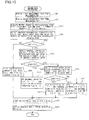
- FIG. 5 is a flowchart illustrating the procedure of the specimen analysis operation performed by the blood analyzer 1.
- the initial examination (the first measurement) of a specimen is conducted first.
- the initial examination includes a first measurement process (step S1) in which a first measurement sample is measured by the measurement unit 2 and first data processing (step S2) in which the measurement data obtained in the first measurement process is subjected to analysis processing performed by the information processing unit 5.
- the sample rack L holding the specimen container T is placed on the specimen carrying unit 4 by the operator.
- the sample rack L is carried by the specimen carrying unit 4, and the specimen container T housing a specimen to be measured is positioned in the specimen supply position 43a.
- the specimen container T is gripped by the hand portion 25a of the measurement unit 2, and the specimen container T is taken out from the sample rack L.
- the hand portion 25a then causes oscillating movement, and thereby the specimen inside the specimen container T is agitated.
- the specimen container T is inserted into the specimen container setting portion 25b, and the specimen container setting portion 25b is moved in the Y direction. After the bar code of the specimen is read by the bar code reading portion 26, the specimen container T reaches the suction position. Then, a first measurement process described below is performed.
- the blood analyzer 1 mixes a whole blood specimen (17 ⁇ L), a first reagent (1 mL), and a second reagent (20 ⁇ L) to prepare a first measurement sample, and measures the first measurement sample by flow cytometry using the optical detector D.
- the above-described Stomatolyser 4DL is used as the first reagent
- the above-described Stomatolyser 4DS is used as the second reagent.
- FIG. 6 is a flowchart illustrating the procedure of operation performed by the blood analyzer 1 in the first measurement process.
- the CPU 51a controls the specimen suction portion 21 to suck a fixed amount of the whole blood specimen in the specimen container T with the suction tube 211 (step S101).
- the suction tube 211 is inserted into the specimen container T, and a fixed amount (80.0 ⁇ L) of the whole blood specimen is sucked by driving the syringe pump.
- the CPU 51a controls the measurement unit 2 to supply, to the first mixing chamber MC1, the first reagent (1 mL) from the reagent container 221a, the second reagent (20 ⁇ L) from the reagent container 221b, and the whole blood specimen (17 ⁇ L) from the suction tube 211 (step S102).
- the CPU 51a waits 21.8 seconds and determines whether 21.8 seconds have elapsed since the supply of the first reagent, the second reagent and the whole blood specimen to the first mixing chamber MC1 (step S103).
- the first mixing chamber MC1 has been heated to 41°C by the heater.
- the mixed solution of the first reagent, the second reagent and the blood specimen is heated at 41°C for 21.8 seconds to prepare the first measurement sample.
- step S104 optical measurement is conducted on the first measurement sample with the optical detector D (step S104). Specifically, in the processing of step S104, the first measurement sample and the sheath fluid are simultaneously supplied to the flow cell 231 of the optical detector D. At that time, forward scattered light is received by the photodiode 243, and side scattered light is received by the photodiode 246, and fluorescence light is received by the avalanche photodiode 248. Output signals (analog signals) output from these various light-receiving elements of the optical detector D are converted into digital signals by an A/D converter, and then converted into first measurement data that is digital data through predetermined signal processing. The first measurement data is transmitted to the information processing unit 5.
- a forward scattered light signal (forward scattered light intensity), a side scattered light signal (side scattered light intensity), and a fluorescence signal (fluorescence intensity) are obtained as feature parameters contained in the first measurement data.
- the CPU 51a of the information processing unit 5 performs predetermined analysis processing on the first measurement data to generate analysis result data containing numeric data for NEUT, LYMPH, EO, BASO, MONO, WBC, and the like, and stores the analysis result data in the hard disk 51d.
- FIG. 7 is a flowchart showing the procedure of the first data processing performed by the blood analyzer according to this embodiment.
- the information processing unit 5 of the blood analyzer 1 receives the first measurement data from the measurement unit 2 (step S151).
- the computer program 54a which is executed by the CPU 51a, is an event-driven program, and the processing of step S152 is invoked upon occurrence of an event of receiving the first measurement data.
- the CPU 51a classifies the normal leucocytes into a plurality of subclasses using the first measurement data, and counts the number of hemocytes belonging to each of the subclasses (step S152). Further, the CPU 51a detects a cell group of abnormal mononuclear leukocytes (hereinafter, referred to as an "abnormal mononuclear leukocyte group”) using the first measurement data, and counts the number of hemocytes CN1 contained in the detected abnormal mononuclear leukocyte group (step S153).
- abnormal mononuclear leukocyte group a cell group of abnormal mononuclear leukocytes
- FIG. 8 is a scattergram of side scattered light intensity and fluorescence intensity in the first measurement data.
- a cluster of immature granulocytes, a cluster of eosinophils, a cluster of a hemocyte group consisting of neutrophils and basophils, a cluster of lymphocytes, and a cluster of monocytes appear in the scattergram of side scattered light intensity and fluorescence intensity in the first measurement data shown in FIG. 8 .
- FIG. 8 is a scattergram of side scattered light intensity and fluorescence intensity in the first measurement data.
- normal leukocytes can be classified into eosinophils, a hemocyte group consisting of neutrophils and basophils, lymphocytes, and monocytes using the side scattered light intensity and the fluorescence intensity in the first measurement data.
- the blood analyzer 1 can prepare a measurement sample for separately detecting neutrophils and basophils by mixing the blood specimen with predetermined reagents, and can separately detect neutrophils and basophils using measurement data obtained by measuring this measurement sample with the optical detector D.
- the blood analyzer 1 classifies the normal leukocytes contained in the blood specimen into five subclasses (eosinophils, neutrophils, basophils, lymphocytes, and monocytes) using the thus obtained detection result for neutrophils and basophils and the detection results for four subclasses obtained by the above-described analysis of the first measurement data.
- the CPU 51a classifies mature leukocytes into subclasses using the forward scattered light, the side scattered light intensity and the fluorescence intensity in the first measurement data, and counts the number of hemocytes belonging to each of the subclasses.
- the range of side scattered light intensity and fluorescence intensity indicated by the dashed line in FIG. 8 is defined as a detection area A1 for an abnormal mononuclear leukocyte group.
- the detection area A1 is set in a portion with a fluorescence intensity higher than that in the area where lymphocytes appear.
- the present inventors have found that atypical lymphocytes, abnormal lymphocytes, lymphoblasts and myeloblasts all appear in the detection area A1. Therefore, use of the detection area A1 enables highly accurate detection of the presence or absence of abnormal mononuclear leukocytes.
- any cell group that appears within the above-described detection area A1 is detected as an abnormal mononuclear leukocyte group, and the number of hemocytes CN1 is counted.
- the CPU 51a determines whether CN1 is greater than a predetermined threshold T1 (step S154).
- the threshold T1 is a reference value for determining whether any abnormal mononuclear leukocyte is present in a blood specimen. If CN1 is greater than the threshold T1, it is determined that abnormal mononuclear leukocytes are present in the blood specimen. If CN1 is less than or equal to the threshold T1, it is determined that no abnormal mononuclear leukocytes are present in the blood specimen.
- the CPU 51a sets a re-examination flag provided in the RAM 51c to "0" (step S155).
- the re-examination flag is information indicating the necessity of the re-examination (the second specimen measurement conducted by the blood analyzer 1).
- the re-examination flag indicates that the re-examination is necessary if it is set to "1", and indicates that the re-examination is not necessary if it is set to "0". Then, the processing executed by the CPU 51a moves to step S157.
- step S154 determines that the re-examination is necessary, and sets the re-examination flag provided in the RAM 51c to "1" (step S156). Then, the processing executed by the CPU 51a moves to step S157.
- the CPU 51a stores the thus obtained analysis result (including the re-examination flag) in the hard disk 51d (step S157).
- the CPU 51a causes the display device 52 to display an analysis result screen showing the analysis result stored in the hard disk 51d (step S158), and ends the first data processing.
- step S3 the CPU 51a of the blood analyzer 1 checks the result regarding whether the re-examination is necessary. If the re-examination of the specimen is not necessary, or in other words, if the re-examination flag is set to "0" (NO in step S3), the specimen analysis operation ends. On the other hand, if the re-examination of the specimen is necessary, or in other words, if the re-examination flag is set to "1" (YES in step S3), the blood analyzer 1 performs the re-examination of the specimen.
- the re-examination is an operation for determining whether a blood specimen that has undergone the initial examination contains abnormal lymphocytes, whether the blood specimen contains blasts, and whether the blood specimen contains atypical lymphocytes.
- This re-examination includes a first measurement process (step S4) that is the same as step S1, a second measurement process (step S5) in which a second measurement sample is measured by the measurement unit 2, and second data processing (step S6) in which the measurement data obtained in each of the first measurement process and the second measurement process is subjected to analysis processing performed by the information processing unit 5.
- the first measurement process in the re-examination is the same as the first measurement process in the initial examination, and therefore the description thereof is omitted.
- the second measurement process is performed in such a manner that it partially overlaps in time with the first measurement process.
- the blood analyzer 1 mixes the whole blood specimen (17.0 ⁇ L), the third reagent (1000 ⁇ L), and the fourth reagent (20 ⁇ L) to prepare a second measurement sample, and measures the second measurement sample by flow cytometry using the optical detector D.
- the following reagents are used as the third reagent.
- Third Reagent MOPS 2.09 g/L polyoxyethylene (20) oleyl ether 1.25 g/L sodium N-lauroylsarcosinate 0.268 g/L EDTA-2K 0.5 g/L
- the above-listed ingredients were mixed, and NaOH was further added to adjust the pH to 7.3.
- the osmotic pressure of the third reagent was 37 mOsm/Kg, and the electric conductivity thereof was 0.745 mS/cm.
- NK-321 50 mg/L NK-321 (50 mg/L) dissolved in ethylene glycol was used as the fourth reagent.
- FIG. 9 is a flowchart illustrating the procedure of operation performed by the blood analyzer 1 in the second measurement process.
- the CPU 51a controls the measurement unit 2 to supply, to the first mixing chamber MC1, the third reagent (1000 ⁇ L) from the reagent container 222a, the fourth reagent (20 ⁇ L) from the reagent container 222b, and the whole blood specimen (17 ⁇ L) from the suction tube 211 (step S201).
- the specimen supplied to the first mixing chamber MC1 is a portion of the whole blood specimen sucked by the suction tube 211 in step S101 described above.
- step S101 the specimen to be supplied to the first mixing chamber MC1 and the specimen to be supplied to the second mixing chamber MC2 are sucked at a time from the specimen container T.
- the CPU 51a waits 18.5 seconds and determines whether 18.5 seconds have elapsed since the supply of the third reagent, the fourth reagent and the whole blood specimen to the first mixing chamber MC1 (step S202).
- the first mixing chamber MC1 has been heated to 34.0°C by the heater.
- the mixed solution of the third reagent, the fourth reagent and the blood specimen is heated at 34.0°C for 18.5 seconds to prepare a second measurement sample.
- step S203 optical measurement is conducted on the second measurement sample with the optical detector D (step S203). Specifically, in the processing of step S203, the second measurement sample and the sheath fluid are simultaneously supplied to the flow cell 231 of the optical detector D. At that time, forward scattered light is received by the photodiode 243, and side scattered light is received by the photodiode 246, and fluorescence light is received by the avalanche photodiode 248. Output signals (analog signals) output from these various light-receiving elements of the optical detector D are converted into digital signals as in the first measurement process described above, and then converted into second measurement data that is digital data through predetermined signal processing. The second measurement data is transmitted to the information processing unit 5.
- a forward scattered light signal (forward scattered light intensity), a side scattered light signal (side scattered light intensity), and a fluorescence signal (fluorescence intensity) are obtained as feature parameters contained in the second measurement data.
- the CPU 51a of the information processing unit 5 performs analysis processing on the second measurement data, thereby detecting abnormal lymphocytes, blasts, or atypical lymphocytes and generating analysis result data containing the detection result, and stores the analysis result data in the hard disk 51d.
- FIG. 10 is a flowchart showing the procedure of the second data processing performed by the blood analyzer according to this embodiment.
- the information processing unit 5 of the blood analyzer 1 receives, from the measurement unit 2, the first measurement data (step S301) and also receives the second measurement data (step S302).
- the computer program 54a which is executed by the CPU 51a, is an event-driven program, and the processing of step S303 is invoked upon occurrence of an event of receiving the first measurement data and the second measurement data.
- the CPU 51a classifies the normal leukocyte into a plurality of subclasses using the first measurement data, and counts the number of hemocytes belonging to each of the subclasses (step S303). Further, the CPU 51a detects a cell group of abnormal mononuclear leukocytes (hereinafter, referred to as an "abnormal mononuclear leukocyte group") using the first measurement data, and counts the number ofhemocytes CN1 contained in the detected abnormal mononuclear leukocyte group (step S304).
- the processing of step S303 is the same as that of step S152 described above and the processing of step S304 is the same as that of step S153 described above, and therefore the description thereof is omitted.
- the CPU 51a determines whether CN1 is greater than a predetermined threshold T1 (step S305).
- the threshold T1 is a reference value that is the same as the above-described threshold T1 in step S154. If CN1 is greater than the threshold T1, it is determined that abnormal mononuclear leukocytes are present in the blood specimen. If CN1 is less than or equal to the threshold T1, it is determined that no abnormal mononuclear leukocytes are present in the blood specimen.
- the CPU 51a sets an abnormal lymphocyte flag, an atypical lymphocyte flag, and a blast flag that are provided in the RAM 51c to "0" (step S306).
- the abnormal lymphocyte flag is a flag indicating the presence or absence of abnormal lymphocytes in a blood specimen.
- the abnormal lymphocyte flag indicates the presence of abnormal lymphocytes if it is set to "1", and indicates the absence of abnormal lymphocytes if it is set to "0".
- the atypical lymphocyte flag is a flag indicating the presence or absence of atypical lymphocytes in the blood specimen.
- the atypical lymphocyte flag indicates the presence of atypical lymphocytes if it is set to "1", and indicates the absence of atypical lymphocytes if it is set to "0".
- the blast flag is a flag indicating the presence or absence of blasts in the blood specimen. The blast flag indicates the presence of blasts if it is set to "1", and indicates the absence of blasts if it is set to "0". Then, the processing executed by the CPU 51a moves to step S314.
- the CPU 51a detects a cell group of abnormal lymphocytes (hereinafter, referred to as an "abnormal lymphocyte group") using the second measurement data, and counts the number of hemocytes CN2 contained in the detected abnormal lymphocyte group (step S307).
- abnormal lymphocyte group a cell group of abnormal lymphocytes
- FIG. 11 is a scattergram of forward scattered light intensity and fluorescence intensity in the second measurement data
- FIG. 12 is a scattergram of side scattered light intensity and fluorescence intensity in the second measurement data.
- a cluster of blasts, a cluster of granulocytes (a hemocyte group consisting of neutrophils, eosinophils, and basophils), a cluster of lymphocytes, and a cluster of monocytes appear in the scattergram of forward scattered light intensity and fluorescence intensity in the second measurement data shown in FIG. 11 .
- a cluster of blasts, a cluster of granulocytes, a cluster of lymphocytes, and a cluster of monocytes appear also in the scattergram of side scattered light intensity and fluorescence intensity in the second measurement data shown in FIG. 12 .
- the range of forward scattered light intensity and fluorescence intensity indicated by the dashed line in FIG. 11 is defined as a detection areaA2 for an abnormal lymphocyte group.
- the detection area A2 is set in a portion with a fluorescence intensity higher than that in the area where lymphocytes and monocytes appear.
- the present inventors have found that abnormal lymphocytes appear in the detection areaA2 and an areas21 as shown in FIG. 12 , and that blasts appear in an area A31 as shown in FIG. 11 and an area A32 as shown in FIG. 12 .
- any cell group that appears within the above-described detection area A2. is detected as an abnormal lymphocyte group, and the number of hemocytes CN2 is counted.
- the CPU 51a determines whether CN2 is greater than a predetermined threshold T2 (step S308).
- the threshold T2 is a reference value for determining whether any abnormal lymphocyte is present in a blood specimen.
- CN2 is greater than the threshold T2
- CN2 is less than or equal to the threshold T2
- no abnormal lymphocytes are present in the blood specimen.
- step S308 If CN2 > T2 in step S308 (YES in step S308), the CPU 51a sets the abnormal lymphocyte flag provided in the RAM 51c to "1", and sets each of the atypical lymphocyte flag and the blast flag to "0" (step S309). Then, the processing executed by the CPU 51a moves to step S314.
- step S308 the CPU 51a detects a cell group of blasts (hereinafter, referred to as a "blast group") using the second measurement data, and counts the number of hemocytes CN3 contained in the detected blast group (step S310).
- a blast group a cell group of blasts
- FIG. 13 is a scattergram of forward scattered light intensity and side scattered light intensity in the second measurement data.
- a cluster of granulocytes, a cluster of lymphocytes, and a cluster of monocyte appear in the scattergram of forward scattered light intensity and side scattered light intensity in the second measurement data shown in FIG. 13 .
- the range of forward scattered light intensity and side scattered light intensity indicated by the dashed line in FIG. 13 is defined as a detection area A3 for the blast group. As shown in FIG. 13 , the detection area A3 is set in a portion with a forward scattered light intensity higher than that in the area where lymphocytes appear.
- any cell group that appears within the above-described detection areaA3 is detected as a blast group, and the number of hemocytes CN3 is counted.
- the present inventors have also found that blasts appear in the area A31 shown in FIG. 11 and the area A32 shown in FIG. 12 , and abnormal lymphocytes and atypical lymphocytes do not appear in these areas. Therefore, the area A31 or the areaA32 may be used to detect blasts distinguishably from abnormal lymphocytes and atypical lymphocytes.
- the CPU 51a determines whether CN3 is greater than a predetermined threshold T3 (step S311).
- the threshold T3 is a reference value for determining whether any blast is present in a blood specimen.
- CN3 is greater than the threshold T3, it is determined that blasts are present in the blood specimen.
- CN3 is less than or equal to the threshold T3, it is determined that no blasts are present in the blood specimen.
- the processing of step S311 it is already determined in step S305 that abnormal mononuclear leukocytes are present in the blood specimen and it is already determined in step S308 that no abnormal lymphocytes are present in the blood specimen. Accordingly, if CN3 is less than or equal to the threshold T3 in step S311, it is determined that atypical lymphocytes are present in the blood specimen.
- step S308 or step S311 if it is determined in step S308 or step S311 that CN2 > T2 or CN3 > T3 after it is determined in step S305 that abnormal mononuclear leukocytes are present in the blood specimen, this means that it is determined that the abnormal mononuclear leukocytes present in the blood specimen are neoplastic abnormal mononuclear leukocytes (abnormal mononuclear leukocytes derived from a hematopoietic system disease).
- step S308 and step S311 that CN2 ⁇ T2 and CN3 ⁇ T3 after it is determined in step S305 that abnormal mononuclear leukocytes are present in the blood specimen, this means that it is determined that the abnormal mononuclear leukocytes present in the blood specimen are reactive abnormal mononuclear leukocytes.
- step S3111 If CN3 > T3 in step S311 (YES in step S311), the CPU 51a sets the blast flag provided in the RAM 51c to "1", and sets each of the abnormal lymphocyte flag and the atypical lymphocyte flag to "0" (step S312). Then, the processing executed by the CPU 51a moves to step S314.
- step S311 CN3 ⁇ T3 in step S311 (NO in step S311)
- the CPU 51a sets the atypical lymphocyte flag provided in the RAM 51c to "1", and sets each of the abnormal lymphocyte flag and the blast flag to "0" (step S313). Then, the processing executed by the CPU 51a moves to step S314.
- the CPU 51a stores the thus obtained analysis result (including the abnormal lymphocyte flag, the atypical lymphocyte flag, and the blast flag) in the hard disk 51d (step S314).
- the CPU 51a causes the display device 52 to display an analysis result screen showing the analysis result stored in the hard disk 51d (step S315), and ends the second data processing.
- FIGS. 14A to 14D show examples of scattergrams obtained by the blood analyzer 1.
- FIG. 14A shows examples of scattergrams obtained when measuring a blood specimen A collected from a patient with acute myelocytic leukemia.
- FIG. 14B shows examples of scattergram obtained when measuring a blood specimen B collected from a patient with chronic lymphocytic leukemia.
- FIG. 14C shows examples of scattergrams obtained when measuring a blood specimen C containing atypical lymphocytes.
- FIG. 14D shows examples of scattergrams obtained when measuring a normal blood specimen D.
- Myeloblasts appear in the peripheral blood of a patient with acute myelocytic leukemia.
- particles corresponding to the detected hemocytes are present in the detection area A1 for the abnormal mononuclear leukocyte group in a scattergram SG11 of side scattered light intensity and fluorescence intensity (scattergram for leukocyte classification) in the first measurement data of the blood specimen A. That is, as can be seen from the scattergram SG11, the abnormal mononuclear leukocyte group is detected in the blood specimen A.
- FIG. 14A also shows a scattergram SG13 of side scattered light intensity and fluorescence intensity in the second measurement data of the blood specimen A for reference. It can be seen that in the scattergram SG13 as well, hemocytes are substantially absent in the areaA21 where abnormal lymphocytes appear.
- particles corresponding to the detected hemocytes are present in the detection area A3 for blasts in a scattergram SG14 of forward scattered light intensity and side scattered light intensity (scattergram for blast detection) in the second measurement data of the blood specimen A. That is, as can be seen from the scattergram SG14, the blast group is detected in the blood specimen A.
- Abnormal lymphocytes which are mature lymphocytic leukemia cells, appear in the peripheral blood of a patient with chronic lymphocytic leukemia.
- particles corresponding to the detected hemocytes are present in the detection area A1 for the abnormal mononuclear leukocyte group in a scattergram SG21 for leukocyte classification of the blood specimen B. That is, as can be seen from the scattergram SG21, the abnormal mononuclear leukocyte group is detected in the blood specimen B.
- Particles corresponding to the detected hemocytes are present in the detection area A2 for abnormal lymphocytes in a scattergram SG22 for abnormal lymphocyte detection in the second measurement data of the blood specimen B.
- 14B also shows a scattergram SG23 of side scattered light intensity and fluorescence intensity in the second measurement data of the blood specimen B for reference. It can be seen that in the scattergram SG23 as well, hemocytes are present in the area A21 where abnormal lymphocytes appear. Furthermore, particles corresponding to the detected hemocytes are substantially absent in the detection area A3 for blasts in a scatter gram SG24 for blast detection in the second measurement data of the blood specimen B. That is, as can be seen from the scattergram SG22, the abnormal lymphocyte group is detected in the blood specimen B.
- particles corresponding to the detected hemocytes are present in the detection area A1 for the abnormal mononuclear leukocyte group in a scattergram SG31 for leukocyte classification of the blood specimen C containing atypical lymphocytes. That is, as can be seen from the scattergram SG31, the abnormal mononuclear leukocyte group is detected in the blood specimen C. Particles corresponding to the detected hemocytes are substantially absent in the detection area A2 for abnormal lymphocytes in a scattergram SG32 for abnormal lymphocyte detection in the second measurement data of the blood specimen C.
- FIG. 14C also shows a scattergram SG33 of side scattered light intensity and fluorescence intensity in the second measurement data of the blood specimen C for reference.
- hemocytes are substantially absent in the area A21 where abnormal lymphocytes appear. Furthermore, particles corresponding to the detected hemocytes are substantially absent in the detection area A3 for blasts in a scattergram SG34 for blast detection in the second measurement data of the blood specimen C. That is, as can be seen from the scattergrams SG32 and SG34, neither the abnormal lymphocyte group nor the blast group is detected in the blood specimen C. Thus, Con1 > T1, CN2 ⁇ T2, and CN3 ⁇ T3 when the blood analyzer 1 according to this embodiment analyses the blood specimen C, and therefore it is determined that atypical lymphocytes are present.
- Abnormal mononuclear leukocytes do not appear in normal peripheral blood.
- particles corresponding to the detected hemocytes are substantially absent in the detection area A1 for the abnormal mononuclear leukocyte group in a scattergram SG41 for leukocyte classification of the blood specimen D. That is, as can be seen from the scattergram SG41, the abnormal mononuclear leukocyte group is not detected in the blood specimen D.
- Particles corresponding to the detected hemocytes are substantially absent in the detection area A2 for abnormal lymphocytes in a scattergram SG42 for abnormal lymphocyte detection in the second measurement data of the blood specimen D.
- 14D also shows a scattergram SG43 of side scattered light intensity and fluorescence intensity in the second measurement data of the blood specimen D for reference. It can be seen that in the scattergram SG43 as well, hemocytes are substantially absent in the area A21 where abnormal lymphocytes appear. Furthermore, particles corresponding to the detected hemocytes are substantially absent in the detection area A3 for blasts in a scattergram SG44 for blast detection in the second measurement data of the blood specimen D. That is, as can be seen from the scattergrams SG42 and SG44, neither the abnormal lymphocyte group nor the blast group is detected in the blood specimen D. Thus, CN1 ⁇ T1 when the blood analyzer 1 according to this embodiment analyses the blood specimen D, and therefore it is determined that no abnormal mononuclear leukocytes are present.
- FIG. 15 is a diagram showing an example of an analysis result screen of the blood analyzer 1.
- FIG. 15 shows an analysis result screen for the blood specimen B.
- the measured numeric data of the measurement items WBC, RBC, PLT, etc.
- the blood specimen B contains abnormal lymphocytes.
- the abnormal lymphocyte flag is set to "1"
- the blast flag and the atypical lymphocyte flag are each set to "0".
- the indication "Abn Lympho?" which is information indicating the possibility that abnormal lymphocytes are present, is added to the Flag field FLG on the analysis result screen R1 for the blood specimen B.
- the operator can understand whether any abnormal lymphocyte is detected in the blood specimen, whether any blast is detected in the blood specimen, and whether any atypical lymphocyte is detected in the blood specimen by just looking at the analysis result screen.
- the scattergram SG21 of side scattered light intensity and fluorescence intensity of the first measurement data is indicated on the analysis result screen R1.
- the scattergram SG23 of side scattered light intensity and fluorescence intensity of the second measurement data is indicated on the analysis result screen R1.
- the blood analyzer 1 can detect abnormal mononuclear leukocytes by measuring, with the optical detector D, the first measurement sample prepared by mixing the blood specimen, the first reagent containing a hemolyzing agent, and the second reagent containing a fluorescent dye for staining nucleic acid. Furthermore, when abnormal mononuclear leukocytes are detected, the blood analyzer 1 can determine whether the abnormal mononuclear leukocytes are abnormal lymphocytes, blasts, or atypical lymphocytes by measuring, with the optical detector D, the second measurement sample prepared by mixing the blood specimen, the third reagent containing a hemolyzing agent, and the fourth reagent containing a fluorescent dye for staining nucleic acid.
- reaction temperature and the reaction time during mixing of the blood specimen, the first reagent, and the second reagent, as well as during mixing of the blood specimen, the third reagent, and the fourth reagent in the sample preparation portion 22 may be suitably set according to the state of damage and staining of the hemocytes contained in the blood specimen, without any particular limitation.
- the reaction time and the reaction temperature may be adjusted such that the reaction time is short when the reaction temperature is high and the reaction time is long when the reaction temperature is low. More specifically, it is preferable that the blood specimen and the reagents are mixed at a temperature of 20°C to 45°C for 3 to 40 seconds.
- the present invention is not limited thereto. It is possible to adopt a configuration in which the second measurement sample is prepared by mixing the blood specimen with a single reagent containing a hemolyzing agent and a nucleic acid staining dye, and abnormal lymphocytes, blasts, and atypical lymphocytes are detected using the second measurement sample. In this case, the concentrations of the surfactant, the solubilizing agent, and the fluorescent dye are adjusted to the above-described concentrations when the reagents have been mixed.
- the present invention has addressed a configuration in which the presence or absence of abnormal mononuclear leukocytes in the blood specimen is determined based on whether the number of hemocytes CN1 appeared within the detection area A1. is greater than the threshold T1 for abnormal mononuclear leukocytes with respect to side scattered light intensity and fluorescence intensity in the first measurement data, the present invention is not limited thereto.
- the ratio of the number of hemocytes appeared within the detection area A2 for abnormal lymphocytes to the total number of leukocytes is obtained with respect to forward scattered light intensity and fluorescence intensity in the second measurement data, and whether abnormal lymphocytes are present is determined by determining whether the obtained ratio is greater than a predetermined reference value.
- the present invention has addressed a configuration in which the second measurement sample is optically measured with a flow cytometer to obtain an optical signal including fluorescence intensity, forward scattered light intensity, and side scattered light intensity, and the fluorescence intensity and the side scattered light intensity are used to determine whether any abnormal lymphocyte is present in a blood specimen, whether any blast is present in a blood specimen, and whether any atypical lymphocyte is present in a blood specimen, the present invention is not limited thereto.
- scattered light information relating to scattered light e.g., wide angle forward scattered light intensity
- forward scattered light intensity and side scattered light intensity are obtained along with fluorescence intensity
- the scattered light information and the fluorescence intensity are used for classification of leukocytes and detection of abnormal mononuclear leukocytes.
- the present invention is not limited thereto. It is also possible to adopt a configuration in which the control of the measurement unit 2 and the processing of measurement data are performed by dedicated hardware, such as FPGA or ASIC, that can perform the same processing as that performed by the computer program 54a.
- the present invention is not limited thereto. It is also possible to adopt a distributed system in which the same processing as that of the above-described computer program 54a is executed by a plurality of devices (computers) in a distributed manner.
- the present invention is not limited thereto. It is also possible to first determine whether any blast is present in a blood specimen and then determine whether any abnormal lymphocyte is present in the blood specimen after having being determined that no blasts are present. This also enables the operator to determine whether the abnormal mononuclear leukocyte is an abnormal lymphocyte, a blast, or an atypical lymphocyte.
- the present invention is not limited thereto.
- the first measurement process and the second measurement process are performed in the first specimen measurement (initial examination), whether any abnormal mononuclear leukocyte is present in the blood specimen is determined based on the first measurement data obtained in the first measurement process, and, if it is determined that abnormal mononuclear leukocytes are present, whether any abnormal lymphocyte is present in the blood specimen, whether any blast is present in the blood specimen, and whether any atypical lymphocyte is present in the blood specimen are determined based on the second measurement data obtained in the second measurement process (i.e., a Configuration in which an operation corresponding to the re-examination of the above-described embodiment is performed in the initial examination).
- the present invention is not limited thereto. It is possible to adopt a configuration in which only the second measurement process is performed in the re-examination, and the second measurement data thus obtained is used to determine whether any abnormal lymphocyte is present in the blood specimen, whether any blast is present in the blood specimen, and whether any atypical lymphocyte is present in the blood specimen.
Abstract
Description
- The present invention relates to a blood analyzer and a blood analysis method for optically measuring a blood specimen and classifying hemocytes contained in the blood specimen, and a computer program product for enabling a computer to analyze blood.
- Five types of leukocyte consisting of lymphocytes, monocytes, basophils, eosinophils, and neutrophils are present in normal peripheral blood, and many blood cell counting apparatuses have the function of classifying leukocytes contained in a blood specimen into the five types. On the other hand, cells that are not present in normal peripheral blood appear in peripheral blood affected with diseases such as viral infectious diseases and hematopoietic system diseases. Abnormal leukocytes that appear in peripheral blood include abnormal mononuclear leukocytes, which can be largely categorized into reactive abnormal mononuclear leukocytes and neoplastic abnormal mononuclear leukocytes. Reactive abnormal mononuclear leukocytes include "atypical lymphocytes", which can be observed for viral infection, drug allergy, and the like. Neoplastic abnormal mononuclear leukocytes can be further categorized into neoplastic mature abnormal mononuclear leukocytes and neoplastic immature abnormal mononuclear leukocytes. Neoplastic mature mononuclear leukocytes include "abnormal lymphocytes", which can be observed for e.g. chronic lymphocytic leukemia (CLL). Neoplastic immature mononuclear leukocytes include "blasts", which can be observed for e.g. acute leukemia. Distinguishable detecting atypical lymphocytes, abnormal lymphocytes, and blasts in peripheral blood is very useful in screening or diagnosis of diseases as described above.
- Japanese Laid-Open Patent Publication No.
2006-91024 FIGS. 12 and14 ).U. S. Patent Publication No. 2009/0023129 discloses detecting a cell group consisting of abnormal lymphocytes and blasts distinguishably from normal leukocytes using reagents for classifying leukocytes into five categories (seeFIG. 2 ). The techniques disclosed in these documents are similar in that they use a hemolyzing agent containing a cationic surfactant and a nonionic surfactant, and a stain solution containing a fluorescent dye for staining nucleic acid as the reagents for classifying leukocytes. However, atypical lymphocytes, abnormal lymphocytes, and blasts appear in substantially the same area for fluorescence intensity and scattered light intensity in Japanese Laid-Open Patent Publication No.2006-91024 U. S. Patent Publication No. 2009/0023129 , and therefore cannot be distinguished from one another. -
U. S. Patent Publication No. 2007/231913 discloses detecting myeloblasts distinguishably from mature leukocytes and immature granulocytes using predetermined reagents (seeFIGS. 1 ,2 , and5 ). This document discloses, as the above-described reagents, a hemolyzing agent containing a nonionic surfactant and a solubilizing agent, and a fluorescent dye for staining nucleic acid.U.S. Patent Publication No. 2010/248247 discloses distinguishably detecting lymphoblasts, myeloblasts, mature leukocytes, and immature granulocytes using predetermined reagents (seeFIGS. 13A and 13B ). This document discloses, as the above-described reagents, a hemolyzing agent containing a nonionic surfactant and a solubilizing agent, and a fluorescent dye for staining nucleic acid. However, neitherU. S. Patent Publication No. 2007/231913 norU.S. Patent Publication No. 2010/248247 discloses detecting atypical lymphocytes distinguishably from abnormal lymphocytes. - In view of the foregoing circumstances, the present invention provides a blood analyzer, a blood analysis method, and a computer program product that can distinguishably detect abnormal lymphocytes, blasts, and atypical lymphocytes.
- A first aspect of the present invention is a blood analyzer comprising: a dispensing portion that dispenses a first, blood specimen and a second blood specimen from a blood specimen a sample preparation portion that prepares a first measurement sample from the first blood specimen dispensed by the dispensing portion, a first fluorescent dye for staining nucleic acid, and a first hemolyzing agent containing a cationic surfactant, and prepares a second measurement sample from the second blood specimen dispensed by the dispensing portion, a second fluorescent dye for staining nucleic acid, and a second hemolyzing agent not containing a cationic surfactant but containing another surfactant; a light source that irradiates light onto each of the first measurement sample and the second measurement sample prepared by the sample preparation portion; a light-receiving portion that receives fluorescence and scattered light that are generated when light is irradiated onto the first measurement sample or the second measurement sample by the light source, and outputs a fluorescence signal relating to the received fluorescence and a scattered light signal relating to the received scattered light; an information processing portion that determines whether any atypical lymphocyte is present in the blood specimen, whether any abnormal lymphocyte is present in the blood specimen and whether any blast is present in the blood specimen based on a first fluorescence signal and a first scattered light signal outputted by the light-receiving portion when light is irradiated onto the first measurement sample by the light source and a second fluorescence signal and a second scattered light signal outputted by the light-receiving portion when light is irradiated onto the second measurement sample by the light source; and an output portion that outputs a result of the determination made by the information processing portion.
- A second aspect of the present invention is a blood analyzer comprising: a dispensing portion that dispenses a first blood specimen and a second blood specimen from a blood specimen; a sample preparation portion that prepares a first measurement sample from the first blood specimen dispensed by the dispensing portion, a first fluorescent dye for staining nucleic acid, and a first hemolyzing agent containing a cationic surfactant, and prepares a second measurement sample from the second blood specimen dispensed by the dispensing portion, a second fluorescent dye for staining nucleic acid, and a second hemolyzing agent not containing a cationic surfactant but containing another surfactant; a light source that irradiates light onto each of the first measurement sample and the second measurement sample prepared by the sample preparation portion; a light-receiving portion that receives fluorescence and scattered light that are generated when light is irradiated onto the first measurement sample or the second measurement sample by the light source, and outputs a fluorescence signal relating to the received fluorescence and a scattered light signal relating to the received scattered light; an information processing portion that determines whether any abnormal mononuclear leukocyte, which is not present in normal peripheral blood, is contained in the blood specimen based on a first fluorescence signal and a first scattered light signal outputted by the light-receiving portion when light is irradiated onto the first measurement sample by the light source, and, if it has been determined that the abnormal mononuclear leukocyte is present in the blood specimen, determines whether the abnormal mononuclear leukocyte is reactive or neoplastic based on a second fluorescence signal and a second scattered light signal outputted by the light-receiving portion when light is irradiated onto the second measurement sample by the light source; and an output portion that outputs a result of the determination made by the information processing portion.
- A third aspect of the present invention is a blood analysis method comprising: (a) dispensing a first blood specimen from a blood specimen; (b) preparing a first measurement sample from the dispensed first blood specimen, a first fluorescent dye for staining nucleic acid, and a first hemolyzing agent containing a cationic surfactant; (c) irradiating light onto the prepared first measurement sample; (d) receiving fluorescence and scattered light that are generated when light is irradiated onto the first measurement sample, and obtaining a first fluorescence signal relating to the received fluorescence and a first scattered light signal relating to the received scattered light; (e) dispensing a second blood specimen from the blood specimen; (f) preparing a second measurement sample from the dispensed second blood specimen, a second fluorescent dye for staining nucleic acid, and a second hemolyzing agent not containing a cationic surfactant but containing another surfactant; (g) irradiating light onto the prepared second measurement sample; (h) receiving fluorescence and scattered light that are generated when light is irradiated onto the second measurement sample, and obtaining a second fluorescence signal relating to the received fluorescence and a second scattered light signal relating to the received scattered light; (i) determining whether or not any atypical lymphocyte is present in the blood specimen, whether or not any abnormal lymphocyte is present in the blood specimen, and whether or not any blast is present in the blood specimen based on the first fluorescence signal and the first scattered light signal as well as the second fluorescence signal and the second scattered light signal; and (j) outputting a result of the determination.
- A fourth aspect of the present invention is a computer program product comprising: a computer readable medium, and software instructions, on the computer readable medium, for enabling a computer to perform operations comprising: receiving a first fluorescence signal and a first scattered light signal relating to fluorescence and scattered light that are generated when light is irradiated onto a first measurement sample prepared from a first blood specimen dispensed from a blood specimen, a first fluorescent dye for staining nucleic acid, and a first hemolyzing agent containing a cationic surfactant; receiving a second fluorescence signal and a second scattered light signal relating to fluorescence and scattered light that are generated when light is irradiated onto a second measurement sample prepared from a second blood specimen dispensed from the blood specimen, a second fluorescent dye for staining nucleic acid, and a second hemolyzing agent not containing a cationic surfactant but containing another surfactant; determining whether any atypical lymphocyte is present in the blood specimen, whether any abnormal lymphocyte is present in the blood specimen, and whether any blast is present in the blood specimen based on the first fluorescence signal and the first scattered light signal as well as the second fluorescence signal and the second scattered light signal; and outputting a result of the determination.
- The invention, together with objects and advantages thereof, may best be understood by reference to the following description of the presently preferred embodiments together with the accompanying drawings in which:
-
FIG. 1 is a perspective view showing an external appearance of a blood analyzer according to an embodiment; -
FIG. 2 is a block diagram showing the configuration of a measurement unit according to the embodiment; -
FIG. 3 is a schematic diagram showing the configuration outline of an optical detector; -
FIG. 4 is a block diagram showing the configuration of an information processing unit according to the embodiment; -
FIG. 5 is a flowchart illustrating the procedure of a specimen analysis operation performed by the blood analyzer according to the embodiment -
FIG. 6 is a flowchart illustrating the procedure of operation in a first measurement process performed by the blood analyzer according to the embodiment; -
FIG. 7 is a flowchart illustrating the procedure of first data processing performed by the blood analyzer according to the embodiment; -
FIG. 8 is a scattergrams of side scattered light intensity and fluorescence intensity in first measurement data; -
FIG. 9 is a flowchart illustrating the procedure of operation in a second measurement process performed by the blood analyzer according to the embodiment; -
FIG. 10 is a flowchart illustrating the procedure of data processing performed by the blood analyzer according to the embodiment; -
FIG. 11 is a scattergram of forward scattered light intensity and fluorescence intensity in second measurement data; -
FIG. 12 is a scattergram of side scattered light intensity and fluorescence intensity in the second measurement data; -
FIG. 13 is a scattergram of forward scattered light intensity and side scattered light intensity in the second measurement data; -
FIG. 14A shows examples of scattergrams obtained when measuring a blood specimen collected from a patient with acute myelocytic leukemia; -
FIG. 14B shows examples of scattergrams obtained when measuring a blood specimen collected from a patient with chronic lymphocytic leukemia; -
FIG. 14C shows examples of scattergrams obtained when measuring a blood specimen containing atypical lymphocytes; -
FIG. 14D shows examples of scattergrams obtained when measuring a normal blood specimen; and -
FIG. 15 is a diagram showing an example of an analysis result screen of the blood analyzer according to the embodiment. - Preferred embodiments of the present invention will now be described below with reference to the accompanying drawings.
-
FIG. 1 is a perspective view showing an external appearance of a blood analyzer according to this embodiment. Ablood analyzer 1 according to this embodiment is a multiple-item hemocyte analyzer for detecting hemocytes contained in a blood specimen, such as leukocytes, erythrocytes, and platelets, and counting each type of the hemocyte. As shown inFIG. 1 , theblood analyzer 1 includes ameasurement unit 2, aspecimen carrying unit 4 disposed on the front side of themeasurement unit 2, and aninformation processing unit 5 that can control themeasurement unit 2 and thespecimen carrying unit 4. - A blood specimen that is peripheral blood collected from a patient is housed in a specimen container (blood collecting tube). A plurality of specimen containers are held in a sample rack, and the sample rack is carried by the
specimen carrying unit 4, and thereby the blood specimen is supplied to themeasurement unit 2. - The configuration of the measurement unit will now be described.
FIG. 2 is a block diagram showing the configuration of the measurement unit. As shown inFIG. 2 , themeasurement unit 2 includes aspecimen suction portion 21 that sucks blood as a specimen from the specimen container (blood collecting tube) T, asample preparation portion 22 that prepares a measurement sample used for measurement from the blood sucked by thespecimens suction portion 21, and adetection portion 23 that detects hemocytes in the measurement sample prepared by thesample preparation portion 22. Themeasurement unit 2 further includes an inlet (seeFIG. 1 ) for taking, into themeasurement unit 2, the specimen container T housed in the sample rack L carried by arack carrying portion 43 of thespecimen carrying unit 4, and a specimencontainer carrying portion 25 that takes the specimen container T from the sample rack L into themeasurement unit 2 and carries the specimen container T to a suction position where blood is sucked by thespecimen suction portion 21. - As shown in
FIG. 2 , thespecimen suction portion 21 includes asuction tube 211. Thespecimen suction portion 21 also includes a syringe pump. Furthermore, thesuction tube 211 is vertically movable, and is configured to suck the blood contained in the specimen container T that has been carried to the suction position when moved downward. - The
sample preparation portion 22 includes a first mixing chamber MC1 and a second mixing chamber MC2. Thesuction tube 211 sucks a predetermined amount of a whole blood specimen from the specimen container T using the syringe pump. The specimen thus sucked is transferred to the position of the first mixing chamber MC1 and the second mixing chamber MC2, and a predetermined amount of the whole blood specimen is dispensed to each of the chambers MC1 and MC2 using the syringe pump. Thesample preparation portion 22 also includes a heater H for heating the first mixing chamber MC1 and the second mixing chamber MC2. - The
sample preparation portion 22 is connected via a tube with areagent container 221a for housing a first reagent, areagent container 221b for housing a second reagent, areagent container 222a for housing a third reagent, a reagent container 222b for housing a fourth reagent, and areagent container 223 for housing a sheath fluid (diluting fluid). Thesample preparation portion 22 is also connected with a compressor, and the respective reagents can be drawn from the correspondingreagent containers - The first reagent is a hemolyzing agent for classifying leucocytes into at least four subclasses. The hemolyzing agent for use contains a cationic surfactant that is said to have a particularly great hemolyzing ability among surfactants. Use of the hemolyzing agent allows erythrocytes to be hemolyzed and the cell membranes of normal leukocytes and abnormal mononuclear leukocytes (atypical lymphocytes, abnormal lymphocytes, and blasts) to be damaged. Accordingly, normal leukocytes and abnormal mononuclear leukocytes are more likely to be stained with a fluorescent dye that will be described below.
- "Abnormal lymphocyte" means a mature lymphocyte which is neoplastic. The abnormal lymphocyte appears in a peripheral blood of a patient with diseases such as chronic lymphocytic leukemia and malignant lymphoma. "Atypical lymphocyte" means a lymphocytes which is antigenically-stimulated and altered morphology in response to the stimulation. The atypical lymphocyte appears in a peripheral blood of a patient with diseases such as viral infection and drug allergy.
- "Blast" means an immature lymphocyte such as myeloblast and lymphoblast. The myeloblast appears in a peripheral blood of a patient with acute myelocytic leukemia, and the lymphoblast appear in a peripheral blood of a patient with acute lymphatic leukemia.
- Here, a quaternary ammonium salt surfactant or a pyridinium salt surfactant is preferable as a cationic surfactant. More specific examples include surfactants having 9 to 30 total carbon atoms as represented by structural formula (I) or (II):


- R1 is preferably an alkyl or alkenyl group having 6, 8, 10, 12, or 14 carbon atoms, with a linear alkyl group being particularly preferable. More specific examples of R1 include an octyl group, a decyl group, and a dodecyl group. R2 and R3 each are particularly preferably a methyl group, an ethyl group, or a propyl group. R4 is preferably a methyl group, an ethyl group, or a propyl group.
- The first reagent may further contain a nonionic surfactant. The nonionic surfactant is preferably a polyoxyethylene-based nonionic surfactant represented by structural formula (III) below:
R1-R2-(CH2CH2O)n-H (III)
wherein R1 is an alkyl, alkenyl, or alkynyl group having 8 to 25 carbon atoms; R2 is O,
- The first reagent may contain components other than the cationic surfactants and the nonionic surfactants mentioned above. Examples of such other components that may be contained in the hemolyzing agent include organic acids, buffers, and the like.
- Here, as for the organic acids, organic acids that have at least one aromatic ring in the molecule or salts thereof are preferable. More specific examples include benzoic acid, phthalic acid, hippuric acid, salicylic acid, p-aminobenzenesulfonic acid, benzenesulfonic acid, salts thereof, and the like.
- Examples of buffers include citric acid salts, HEPES, phosphoric acid salts, and the like. Preferable buffers maintain the pH of the hemolyzing agent at 4.5 to 11.0 and preferably 5.0 to 10.0.
- Due to the use of the first reagent, normal leukocytes and abnormal mononuclear leukocytes are more likely to be stained with a fluorescent dye that will be described below, and in addition, normal leukocytes develop a difference in size or other features of lymphocytes, monocytes, eosinophils, and granulocytes other than eosinophils. It is therefore possible based on the fluorescent signal (fluorescence intensity) and the scattered light signal (scattered light intensity) derived from hemocytes to classify normal leukocytes into at least four subclasses and to detect abnormal mononuclear leukocytes.
- Moreover, commercially available hemolyzing reagents for leukocyte classification can also be used for the first reagent. An example of a commercially available hemolyzing reagent for leukocyte classification may be a Stomatolyser 4DL manufactured by Sysmex Corporation. The Stomatolyser 4DL is a hemolyzing agent containing the aforementioned cationic surfactant, nonionic surfactant, and organic acid, and having a pH within the aforementioned range.
- The second reagent is a reagent for fluorescently staining nucleated cells in a blood sample. A fluorescent dye for staining nucleic acid is contained in the second reagent. Such a dye barely stains erythrocytes that do not have nucleic acid, but stains nucleated hemocytes such as leukocytes having nucleic acid and nucleated erythrocytes. The fluorescent dye capable of staining nucleic acid can be suitably selected according to the light irradiated from a light source.
- Specific examples of fluorescent dyes capable of staining nucleic acid include propidium iodide, ethidium bromide, ethidium-acridine heterodimer, ethidium diazide, ethidium homodimer-1, ethidium homodimer-2, ethidium monoazide, trimethylenebis[[3-[[4-[[(3-methylbenzothiazol-3-ium)-2-yl]methylene]-1,4-dihydroquinoline]-1-yl] propyl]dimethylanimium]tetraiodide (TOTO-1), 4-[(3-methylbenzothiazol-2(3H)-ylidene)methyl]-1-[3-(trimethylaminio)propyl]quinolini um diiodide (TO-PRO-1), N,N,N',N'-tetramethyl-N,N'-bis[3-4-3-(3-methylbenzothiazol-3-ium-2-yl]-2-propenyli denel-1,4-dihydroquinolin-1-yl]propyl]-1,3-propandiaminium tetraiodide (TOTO- 3), 2-3-[[1-[3-(trimethylamino)propyl]-1,4-dihydroquinolin]-4-ylidene]-1-propenyl]-3-met hylbenzothiazol-3-ium diiodide (TO-PRO-3), and fluorescent dyes represented by structural formula (IV) below. Among these examples, the fluorescent dyes represented by structural formula (IV) below are preferable.
-

- Here, it is preferable that when one of R1 and R4 in structural formula (IV) is an alkyl group having 6 to 18 carbon atoms, the other is a hydrogen atom or an alkyl group having fewer than 6 carbon atoms. The alkyl group having 6 to 18 atoms is preferably an alkyl group having 6, 8, or 10 carbon atoms. Examples of substituents of the benzyl group represented by R1 and R4 include alkyl groups having 1 to 20 carbon atoms, alkenyl groups having 2 to 20 carbon atoms, and alkynyl groups having 2 to 20 carbon atoms, with a methyl group or an ethyl group being particularly preferable. Examples of alkenyl groups represented by R2 and R3 include alkenyl groups having 2 to 20 carbon atoms. Examples of alkoxy groups represented by R2 and R3 include alkoxy groups having 1 to 20 carbon atoms, with a methoxy group or an ethoxy group being particularly preferable. Examples of anions represented by X- include F, Cl-, Br-, I-, and like halogen ions, CF3SO3 -, BF4 -, and the like.
- The concentration of fluorescent dye capable of staining nucleic acid in the second reagent can be suitably determined according to the kind of fluorescent dye. For example, the concentration of fluorescent dye represented by structural formula (IV) is preferably 0.2 to 0.6 pg/µL and particularly preferably 0.3 to 0.5 pg/µL. The second reagent may contain one or two or more fluorescent dyes capable of staining nucleic acid.
- Moreover, commercially available staining reagents for leukocyte classification can also be used for the second reagent. An example of a commercially available staining reagent for leukocyte classification may be a Stomatolyser 4DS manufactured by Sysmex Corporation. The Stomatolyser 4DS is a staining reagent containing a fluorescent dye represented by structural formula (IV) above.
- The sheath fluid is a fluid supplied to a sheath flow cell that will be described below. The sheath fluid is also used as a diluting fluid. An example of the sheath fluid may be a Cellpack (II) manufactured by Sysmex Corporation.
- The third reagent is a hemolyzing agent for distinguishably detecting an abnormal lymphocyte, an atypical lymphocyte, and a blast. Hemolyzing agents that contain a nonionic surfactant and do not substantially contain a cationic surfactant can be used for the third reagent. Use of the hemolyzing agent allows erythrocytes to be hemolyzed and the cell membranes of normal leukocytes and abnormal mononuclear leukocytes to be damaged. Accordingly, normal leukocytes and abnormal mononuclear leukocytes are more likely to be stained with a fluorescent dye that will be described below.
- Here, the nonionic surfactant is preferably a polyoxyethylene-based nonionic surfactant. Specific examples of polyoxyethylene-based nonionic surfactants includes those represented by structural formula (V) below:
R1-R2-(CH2CH2O)n-H (V)
wherein R1 an alkyl, alkenyl, or alkynyl group having 9 to 25 carbon atoms; R2 is -O-, -COO-, or:
- Specific examples of surfactants represented by structural formula (V) above include polyoxyethylene (15) oleyl ether, polyoxyethylene (15) cetyl ether, polyoxyethylene (16) oleyl ether, polyoxyethylene (20) oleyl ether, polyoxyethylene (20) lauryl ether, polyoxyethylene (20) stearyl ether, polyoxyethylene (20) cetyl ether, and the like, with polyoxyethylene (20) oleyl ether being preferable. The third reagent may contain one or more surfactants.
- The concentration of surfactant contained in the third reagent can be suitably selected according to the kind of surfactant, the osmotic pressure of the hemolyzing agent, and like factors. For example, when the surfactant is polyoxyethylene oleyl ether, the concentration of surfactant contained in the third reagent is 0.5 to 50.0 g/L and preferably 1.0 to 20.0 g/L.
- The third reagent may contain in addition to the nonionic surfactant a solubilizing agent to sufficiently shrink the hemolyzed erythrocytes so that the hemolyzed erythrocytes form a ghost population that does not adversely affect measurement. Examples of solubilizing agents that may be contained in the third reagent include sarcosine derivatives, cholic acid derivatives, methylglucanamide, n-octyl-β-glucoside, sucrose monocaprate, N-formylmethylleucylalanine, and the like, with sarcosine derivatives being particularly preferable. The third reagent may contain one or two or more solubilizing agents.
- Examples of sarcosine derivatives include compounds represented by structural formula (VI) below or salts thereof.

- Examples of cholic acid derivatives include compounds represented by structural formula (VII) below or salts thereof.

(3-[(3-cholamidopropyl)dimethylammonio]-1-propanesulfonate), CHAPSO
([(3-cholamidopropyl)dimethylammonio]-2-hydroxy-1-propanesulfonate), and the like. - Examples of methylglucanamides include compounds represented by structural formula (VIII) below:

- The concentration of solubilizing agent contained in the third reagent may be suitably selected according to the kind of solubilizing agent used. For example, when a sarcosine derivative is used as a solubilizing agent, the concentration of solubilizing agent contained in the third reagent is 0.05 to 3.0 g/L and preferably 0.1 to 1.0 g/L. When a cholic acid derivative is used, the concentration of solubilizing agent contained in the third reagent is 0.1 to 10.0 g/L and preferably 0.2 to 2.0 g/L. When a methylglucanamide is used, the concentration of solubilizing agent contained in the third reagent is 1.0 to 8.0 g/L and preferably 2.0 to 6.0 g/L. When n-octyl B-glucoside, sucrose monocaprate or N-formylmethylleucylalanine is used, the concentration of solubilizing agent contained in the third reagent is 0.01 to 50.0 g/L and preferably 0.05 to 30.0 g/L.
- The pH of the third reagent is preferably 5.0 to 9.0, more preferably 6.5 to 7.5, and even more preferably 0.8 to 7.3. The pH of the fourth reagent can be controlled with a buffer or a pH adjustor. Examples of buffers include Good's buffers such as HEPES, MOPS (3-morpholinopropanesulfonic acid) and MOPSO (2-Hydroxy-3-morpholinopropanesulfonic acid), phosphate buffers, and the like. Examples of pH adjusters include sodium hydroxide, hydrochloric acid, and the like.
- The osmotic pressure of the third reagent can be suitably determined according to the kind of surfactant described above and the concentration thereof in the third reagent. A specific example of the osmotic pressure of the third reagent may be 10 to 600 mOsm/kg. The osmotic pressure of the third reagent may be controlled by adding sugar, amino acid, sodium chloride, or the like to the third reagent. Specific examples of sugars include monosaccharides, polysaccharides, sugar alcohols, and the like. Glucose and fructose are preferable as monosaccharides. Arabinose is preferable as a polysaccharide. Xylitol, sorbitol, mannitol, and ribitol are preferable as sugar alcohols. A sugar to be added to the third reagent is preferably a sugar alcohol and particularly preferably xylitol. When xylitol is added to the third reagent, the concentration of xylitol in the third reagent is preferably 1.0 to 75.0 g/L and particularly preferably 20.0 to 50.0 g/L. Specific examples of amino acids include valine, proline, glycine, alanine, and the like, with glycine and alanine being particularly preferable. When glycine is added to the third reagent, the concentration of glycine in the third reagent is preferably 1.0 to 50.0 g/L and particularly preferably 10.0 to 30.0 g/L.
- The electric conductivity of the third reagent is preferably 0.01 to 3 mS/cm and particularly preferable 0.1 to 2 mS/cm. In addition, a chelating agent, a preservative, or the like may be added to the third reagent. Examples of chelating agents include EDTA-2K, EDTA-3Na, and the like. Examples of preservatives include Proxel GXL (manufactured by Avecia), Material TKM-A (API Corporation), and the like.
- Due to the use of the third reagent, normal leukocytes and abnormal mononuclear leukocytes are more likely to be stained with a fluorescent dye that will be described below, and in addition, abnormal mononuclear leukocytes develop a difference in size or other features of abnormal lymphocytes, atypical lymphocytes, and blasts. It is therefore possible based on the fluorescent signal (fluorescence intensity) and the scattered light signal (scattered light intensity) derived from hemocytes to distinguishably detect abnormal lymphocytes, atypical lymphocytes, and blasts in abnormal mononuclear leukocytes
- The fourth reagent is a reagent for fluorescently staining nucleated cells in a blood sample. A fluorescent dye capable of staining nucleic acid is contained in the fourth reagent. The fluorescent dye is not particularly limited as long as it is capable of fluorescently staining nucleic acid. Such a dye barely stains erythrocytes that do not have nucleic acid, but stains nucleated hemocytes such as abnormal lymphocytes having nucleic acid. The fluorescent dye capable of staining nucleic acid can be suitably selected according to the light irradiated from a light source. Specific examples of fluorescent dyes capable of staining nucleic acid include propidium iodide, ethidium bromide, ethidium-acridine heterodimer, ethidium diazide, ethidium homodimer-1, ethidium homodimer-2, ethidium monoazide, trimethylenebis[[3-[[4-[[(3-methylbenzothiazol-3-ium)-2-yl]methylene]-1,4-dihydroquin oline]-1-yl] propyl]dimethylaminium]tetraiodide (TOTO-1), 4-[(3-methylbenzothiazol-2(3H)-ylidene)methyl]-1-[3-(trimethylaminio)propyl]quinolini um diiodide (TO-PRO-1), N,N,N',N'-tetramethyl-N,N'-bis[3-[4-[3-[(3-methylbenzothiazol-3-ium)-2-yl]-2-propenyli done]-1,4-dihydroquinolin-1-yl]propyl]-1,3-propandiaminium tetraiodide (TOTO-3), 2-[3-[[1-[3-(trimethylamio)propyl]-1,4-dihydroquinolin]-4-ylidene]-1-propenyl]-3-met hylbenzothiazol-3-ium diiodide (TO-PRO-3), and fluorescent dyes represented by structural formulas (IX) to (XXII) below.
- Structural formula (IX)

- In structural formula (IX), the lower alkyl group is a linear or branched alkyl group having 1 to 6 carbon atoms. Specific examples of lower alkyl groups include a methyl group, an ethyl group, a propyl group, a butyl group, an iso-butyl group, a sec-butyl group, a tert-butyl group, a pentyl group, a hexyl group, and the like, with a methyl group and an ethyl group being preferable. Z is preferably a sulfur atom. Examples of anions represented by X- include halogen ions (fluorine, chlorine, bromine, and iodine ions), boron halide ions (BF4 -, BCl4 -, BBr4 -, and the like), phosphorus compound ions, halogen oxoacid ions, fluorosulfuric acid ions, methylsulfuric acid ions, ions of tetraphenylboron compounds having a haloaromatic ring or an alkyl group having a halogen as a substituent, and the like, with an iodine ion being preferable.
- Among the fluorescent dyes represented by structural formula (IX), a particularly preferable fluorescent dye capable of staining nucleic acid is NK-321 represented by structural formula (X) below:

- Structural formula (XI)

- The lower alkyl group and the anion X- in structural formula (XI) are the same as those in structural formula (IX).
- Among the fluorescent dyes represented by structural formula (XI), a particularly preferable fluorescent dye capable of staining nucleic acid is represented by structural formula (XII) below:

- Structural formula (XIII)

- The lower alkyl group and the anion represented by X- in structural formula (XIII) are the same as those in structural formula (IX). The lower alkoxy group refers to an alkoxy group having 1 to 6 carbon atoms. Specific examples of lower alkoxy groups include a methoxy group, an ethoxy group, a propoxy group, and the like, with a methoxy group and an ethoxy group being particularly preferable. The acyl group is preferably an acyl group derived from an aliphatic carboxylic acid. Specific examples of acyl groups include an acetyl group, a propionyl group, and the like, with an acetyl group being particularly preferable. Examples of substituents of the lower alkyl group that may be substituted include a hydroxyl group and halogen atoms (fluorine, chlorine, bromine, and iodine). The lower alkyl group that may be substituted may be substituted by 1 to 3 substituents. It is particularly preferable that the lower alkyl group that may be substituted is a lower alkyl group substituted with one hydroxyl group. Z is preferably a sulfur atom and X- is preferably a bromine ion or BF4 -.
- Among the fluorescent dyes represented by structural formula (XIII), particularly preferable fluorescent dyes capable of staining nucleic acid are represented by any of the three structural formulas (XIV) to (XVI) below:

-

-

- Structural formula (XVII)

- Structural formula (XVIII)

- Structural formula (XIX) (NK-1570)

- Structural formula (XX) (NK-1049)

- Structural formula (XXI) (NK-98)

- Structural formula (XXII) (NK-141)

- Among the fluorescent dyes capable of staining nucleic acid, a particularly preferable fluorescent dye contained in the fourth reagent is NK-321 represented by the structural formula (XXIII) below:

- The concentration of fluorescent dye capable of staining nucleic acid in the fourth reagent is preferably 10 to 500 mg/L and particularly preferably 30 to 100 mg/L. The fourth reagent may contain one or two or more fluorescent dyes capable of staining nucleic acid.
- The
detection portion 23 includes an optical detector D that can conduct a first measurement and a second measurement. In the first measurement, leukocytes (normal leukocytes) present in a blood specimen are classified into four subclasses: LYMPH (lymphocytes); EO (eosinophils); a hemocyte group consisting of NEUT (neutrophils) and BASO (basophils); and MONO (monocytes). In the first measurement, a measurement sample (first measurement sample) obtained by mixing the blood specimen, the first reagent, and the second reagent is supplied to the optical detector D, and optical information (fluorescence intensity, forward scattered light intensity, and side scattered light intensity) is detected by the optical detector D at this time. The optical information obtained by the first measurement is supplied to theinformation processing unit 5, and thereby the normal leukocytes contained in the blood specimen are classified into the four subclasses. As will be described later, theinformation processing unit 5 can detect abnormal mononuclear leukocytes (a group consisting of abnormal lymphocytes, atypical lymphocytes, and blasts) in the blood specimen based on the optical information obtained by the first measurement. - In the second measurement, abnormal lymphocytes and blasts contained in the blood specimen are detected. In the second measurement, a measurement sample (second measurement sample) obtained by mixing the blood specimen, the third reagent, and the fourth reagent is supplied to the optical detector D, and optical information (fluorescence intensity, forward scattered light intensity, and side scattered light intensity) is detected by the optical detector D at this time. The optical information obtained by the second measurement is supplied to the
information processing unit 5, thereby identifying whether the abnormal mononuclear leukocytes contained in the blood specimen that have been detected by the first measurement are abnormal lymphocytes, atypical lymphocytes, or blasts. -
FIG. 3 shows a configuration outline of the optical detector D. The optical detector D feeds the measurement samples and the sheath fluid into aflow cell 231 to generate a liquid current in theflow cell 231, and measures the hemocytes contained in the liquid current flossing through theflow cell 231 by irradiating semiconductor laser light onto the hemocytes. The optical detector D includes a sheath flowssystem 232, a beamspot formation system 233, a forward scatteredlight receiving system 234, a side scatteredlight receiving system 235, and afluorescence light receivingsystem 236. - The sheath flows
system 232 is configured to cause the measurement samples to flow through theflow cell 231 in such a state where the measurement samples are each enclosed in the sheath fluid. The beamspot formation system 233 is configured to allow light irradiated from asemiconductor laser 237 to be irradiated onto theflow cell 231 through acollimator lens 238 and acondenser lens 239. Further, the beamspot formation system 233 includes abeam stopper 240. - The forward scattered
light receiving system 234 is configured to focus forward scattered light with aforward focusing lens 241, and receive the light that has passed through apinhole 242 with a photodiode (forward scattered light receiving portion) 243. - The side scattered
light receiving system 235 is configured to focus side scattered light with aside focusing lens 244, reflect a portion of the light at adichroic mirror 245, and receive the reflected light with a photodiode (side scattered light receiving portion) 246. - Light scattering is a phenomenon that occurs when light changes the direction of its movement due the presence of particles such as hemocytes in the movement direction as impediments. Information relating to the size and the material of the particles can be obtained by detecting such scattered light. In particular, information relating to the size of the particles (hemocytes) can be obtained from forward scattered light. Meanwhile, information about the interior of the particles can be obtained from side scattered light. When laser light is irradiated onto hemocyte particles, the intensity of side scattered light is dependent on the complexity of the cell interior (the shape, size, density, and the granular amount of the nucleus). Therefore, the intensity of these scattered light beams can be utilized for classification of leukocytes, detection of abnormal mononuclear leukocytes, detection of blasts, and other measurements.
- The fluorescence
light receiving system 236 is configured to allow the light that has transmitted through thedichroic mirror 245 to further transmit through aspectral filter 247, and receive the transmitted light with an avalanche photodiode (fluorescence light receiving portion) 248. - When light is irradiated onto a hemocyte that has been stained by a fluorescent substance, the hemocyte emits light having a wavelength longer than the wavelength of the irradiated light. The intensity of fluorescence is increased if the hemocyte has been stained well, and information relating to the staining degree of the hemocyte can be obtained by measuring the fluorescence intensity. Accordingly, the difference in (side) fluorescence intensity can be utilized for classification of leukocytes, detection of abnormal mononuclear leukocytes, detection of abnormal lymphocytes, detection of blasts, and the like.
- Referring back to
FIG. 2 , the configuration of the specimencontainer carrying portion 25 will now be described next. The specimencontainer carrying position 25 includes ahand portion 25a that can grip the specimen container T. Thehand portion 25a includes a pair of gripping members arranged facing each other, and can move these gripping members toward and away from each other. The specimen container T can be gripped by the gripping members by moving the gripping members toward each other with the specimen container T interposed therebetween. Further, the specimencontainer carrying portion 25 can move thehand portion 25a in the up-down direction and the front-back direction (Y direction), and also can oscillate thehand portion 25a. This allows the specimen container T housed in the sample rack L and located at thespecimen supply position 43a to be gripped by thehand portion 25a. In this state, thehand portion 25a is moved upward to pull out the specimen container T from the sample rack L. Then, the specimen in the specimen container T can be agitated by oscillating thehand portion 25a. - The specimen
container carrying portion 25 also includes a specimencontainer setting portion 25b having a hole into which the specimen container T can be inserted. After completion of agitation, the specimen container T gripped by thehand portion 25a described above is moved such that the gripped specimen container T is inserted into the hole of the specimencontainer setting portion 25b. Then, the gripping members are moved away from each other, thereby releasing the specimen container T from thehand portion 25a and setting the specimen container T in the specimencontainer setting portion 25b. The specimencontainer setting portion 25b can be moved horizontally in Y1 and Y2 directions inFIG. 2 by the power of a stepping motor. - A bar
code reading portion 26 is provided inside themeasurement unit 2. The specimencontainer setting portion 25b can be moved to a barcode reading position 26a in the vicinity of the barcode reading portion 26 and to the suction position where the specimen is sucked by thespecimen suction portion 21. When the specimencontainer setting portion 25b is moved to the barcode reading position 26a, the bar code of the specimen is read by the barcode reading portion 26. When the specimencontainer setting portion 25b is moved to the suction position, the specimen is sucked by thespecimen suction portion 21 from the specimen container T that has been set. - Next, the configuration of the
information processing unit 5 will now be described. Theinformation processing unit 5 is configured by a computer.FIG. 4 is a block diagram showing the configuration of theinformation processing unit 5. Theinformation processing unit 5 can be implemented by a computer 5a. As shown inFIG. 4 , the computer 5a includes abody 51, adisplay device 52, and aninput device 53. Thebody 51 includes aCPU 51a, aROM 51b, aRAM 51c, ahard disk 51d, areadout device 51e, an input/output interface 51f, acommunication interface 51g, and animage output interface 51h. TheCPU 51a, theROM 51b, theRAM 51c, thehard disk 51d, thereadout device 51e, the input/output interface 51f, thecommunication interface 51g, and theimage output interface 51h are connected by abus 51j. - The
CPU 51a can execute a computer program loaded into theRAM 51c. The computer 5a functions as theinformation processing unit 5 by theCPU 51a executing acomputer program 54a for blood analysis and control of themeasurement unit 2 and thespecimen carrying unit 4 as will be described later. - Various computer programs, including, for example, an operating system and application programs, for being executed by the
CPU 51a, and the data used for execution of such computer programs are installed in thehard disk 51. Thecomputer program 54a for enabling theCPU 51a to execute processing described later is also installed in thehard disk 51d. Thecomputer program 54a is an event-driven computer program. - The
readout device 51e is configured by a flexible disk drive, a CD-ROM drive, a DVD-ROM drive, or the like, and can read out the computer programs or data recorded in aportable recording medium 54. Thecomputer program 54a for enabling the computer to function as theinformation processing unit 5 is stored in theportable recording medium 54. The computer 5a can read out thecomputer program 54a from theportable recording medium 54, and install thecomputer program 54a in thehard disk 51d. - For example, a multitasking operating system such as Windows (registered trademark) manufactured and sold by Microsoft Corporation, US is installed in the
hard disk 51d. The following description is given assuming that thecomputer program 54a according to this embodiment runs on that operating system. - The input/
output interface 51f is configured, for example, by a serial interface such as USB, IEEE1394, or RS-232C, a parallel interface such as SCSI, IDE, or IEEE1284, and an analog interface made up of a D/A converter, an A/D converter, and the like. Aninput device 53 made up of a keyboard and a mouse is connected to the input/output interface 51f, and the user can input data into the computer 5a using theinput device 53. Further, the input/output interface 51f is connected to themeasurement unit 2 and thespecimen carrying unit 4. This enables theinformation processing unit 5 to control each of themeasurement unit 2 and thespecimen carrying unit 4. - The
communication interface 51g is an Ethernet (registered trademark) interface. Thecommunication interface 51g is connected to ahost computer 6 via a LAN (seeFIG. 2 ). The computer 5a can transmit and receive data via thecommunication interface 51g to and from thehost computer 6 connected to the LAN using a predetermined communications protocol. - In the following, the operation of the
blood analyzer 1 according to this embodiment will now be described. Theblood analyzer 1 performs a specimen analysis operation as described below.FIG. 5 is a flowchart illustrating the procedure of the specimen analysis operation performed by theblood analyzer 1. - As shown in
FIG. 5 , in the specimen analysis operation, the initial examination (the first measurement) of a specimen is conducted first. The initial examination includes a first measurement process (step S1) in which a first measurement sample is measured by themeasurement unit 2 and first data processing (step S2) in which the measurement data obtained in the first measurement process is subjected to analysis processing performed by theinformation processing unit 5. - First, the sample rack L holding the specimen container T is placed on the
specimen carrying unit 4 by the operator. The sample rack L is carried by thespecimen carrying unit 4, and the specimen container T housing a specimen to be measured is positioned in thespecimen supply position 43a. Next, the specimen container T is gripped by thehand portion 25a of themeasurement unit 2, and the specimen container T is taken out from the sample rack L. Thehand portion 25a then causes oscillating movement, and thereby the specimen inside the specimen container T is agitated. Next, the specimen container T is inserted into the specimencontainer setting portion 25b, and the specimencontainer setting portion 25b is moved in the Y direction. After the bar code of the specimen is read by the barcode reading portion 26, the specimen container T reaches the suction position. Then, a first measurement process described below is performed. - The first measurement process will now be described first. In the first measurement process, the
blood analyzer 1 mixes a whole blood specimen (17 µL), a first reagent (1 mL), and a second reagent (20 µL) to prepare a first measurement sample, and measures the first measurement sample by flow cytometry using the optical detector D. Here, the above-described Stomatolyser 4DL is used as the first reagent, and the above-described Stomatolyser 4DS is used as the second reagent. -
FIG. 6 is a flowchart illustrating the procedure of operation performed by theblood analyzer 1 in the first measurement process. First, theCPU 51a controls thespecimen suction portion 21 to suck a fixed amount of the whole blood specimen in the specimen container T with the suction tube 211 (step S101). Specifically, in the processing of step S101, thesuction tube 211 is inserted into the specimen container T, and a fixed amount (80.0 µL) of the whole blood specimen is sucked by driving the syringe pump. - Next, the
CPU 51a controls themeasurement unit 2 to supply, to the first mixing chamber MC1, the first reagent (1 mL) from thereagent container 221a, the second reagent (20 µL) from thereagent container 221b, and the whole blood specimen (17 µL) from the suction tube 211 (step S102). - Next, the
CPU 51a waits 21.8 seconds and determines whether 21.8 seconds have elapsed since the supply of the first reagent, the second reagent and the whole blood specimen to the first mixing chamber MC1 (step S103). Here, the first mixing chamber MC1 has been heated to 41°C by the heater. Thus, the mixed solution of the first reagent, the second reagent and the blood specimen is heated at 41°C for 21.8 seconds to prepare the first measurement sample. - Then, optical measurement is conducted on the first measurement sample with the optical detector D (step S104). Specifically, in the processing of step S104, the first measurement sample and the sheath fluid are simultaneously supplied to the
flow cell 231 of the optical detector D. At that time, forward scattered light is received by thephotodiode 243, and side scattered light is received by thephotodiode 246, and fluorescence light is received by theavalanche photodiode 248. Output signals (analog signals) output from these various light-receiving elements of the optical detector D are converted into digital signals by an A/D converter, and then converted into first measurement data that is digital data through predetermined signal processing. The first measurement data is transmitted to theinformation processing unit 5. In this signal processing, a forward scattered light signal (forward scattered light intensity), a side scattered light signal (side scattered light intensity), and a fluorescence signal (fluorescence intensity) are obtained as feature parameters contained in the first measurement data. This completes the first measurement process. As will be described later, theCPU 51a of theinformation processing unit 5 performs predetermined analysis processing on the first measurement data to generate analysis result data containing numeric data for NEUT, LYMPH, EO, BASO, MONO, WBC, and the like, and stores the analysis result data in thehard disk 51d. - Next, the first data processing will now be described.
FIG. 7 is a flowchart showing the procedure of the first data processing performed by the blood analyzer according to this embodiment. Theinformation processing unit 5 of theblood analyzer 1 receives the first measurement data from the measurement unit 2 (step S151). Thecomputer program 54a, which is executed by theCPU 51a, is an event-driven program, and the processing of step S152 is invoked upon occurrence of an event of receiving the first measurement data. - The
CPU 51a classifies the normal leucocytes into a plurality of subclasses using the first measurement data, and counts the number of hemocytes belonging to each of the subclasses (step S152). Further, theCPU 51a detects a cell group of abnormal mononuclear leukocytes (hereinafter, referred to as an "abnormal mononuclear leukocyte group") using the first measurement data, and counts the number of hemocytes CN1 contained in the detected abnormal mononuclear leukocyte group (step S153). - The processing of step S152 and S153 will now be described in detail.
FIG. 8 is a scattergram of side scattered light intensity and fluorescence intensity in the first measurement data. A cluster of immature granulocytes, a cluster of eosinophils, a cluster of a hemocyte group consisting of neutrophils and basophils, a cluster of lymphocytes, and a cluster of monocytes appear in the scattergram of side scattered light intensity and fluorescence intensity in the first measurement data shown inFIG. 8 . As can be seen from the scattergram inFIG. 8 , normal leukocytes can be classified into eosinophils, a hemocyte group consisting of neutrophils and basophils, lymphocytes, and monocytes using the side scattered light intensity and the fluorescence intensity in the first measurement data. Although the description is omitted here, theblood analyzer 1 can prepare a measurement sample for separately detecting neutrophils and basophils by mixing the blood specimen with predetermined reagents, and can separately detect neutrophils and basophils using measurement data obtained by measuring this measurement sample with the optical detector D. Theblood analyzer 1 classifies the normal leukocytes contained in the blood specimen into five subclasses (eosinophils, neutrophils, basophils, lymphocytes, and monocytes) using the thus obtained detection result for neutrophils and basophils and the detection results for four subclasses obtained by the above-described analysis of the first measurement data. In the processing of step S152, theCPU 51a classifies mature leukocytes into subclasses using the forward scattered light, the side scattered light intensity and the fluorescence intensity in the first measurement data, and counts the number of hemocytes belonging to each of the subclasses. - In this embodiment, the range of side scattered light intensity and fluorescence intensity indicated by the dashed line in
FIG. 8 is defined as a detection area A1 for an abnormal mononuclear leukocyte group. As shown inFIG. 8 , the detection area A1 is set in a portion with a fluorescence intensity higher than that in the area where lymphocytes appear. As a result of experiments using clinical specimens and detailed investigation of the results of the experiments, the present inventors have found that atypical lymphocytes, abnormal lymphocytes, lymphoblasts and myeloblasts all appear in the detection area A1. Therefore, use of the detection area A1 enables highly accurate detection of the presence or absence of abnormal mononuclear leukocytes. However, it has been found that whether the detected abnormal mononuclear leukocytes are abnormal lymphocytes, blasts, or atypical lymphocytes cannot be determined with the detection area A1. In step S153, any cell group that appears within the above-described detection area A1 is detected as an abnormal mononuclear leukocyte group, and the number of hemocytes CN1 is counted. - Next, the
CPU 51a determines whether CN1 is greater than a predetermined threshold T1 (step S154). The threshold T1 is a reference value for determining whether any abnormal mononuclear leukocyte is present in a blood specimen. If CN1 is greater than the threshold T1, it is determined that abnormal mononuclear leukocytes are present in the blood specimen. If CN1 is less than or equal to the threshold T1, it is determined that no abnormal mononuclear leukocytes are present in the blood specimen. - If CN1 ≤ T1 in step S154 (NO in step S154), the
CPU 51a sets a re-examination flag provided in theRAM 51c to "0" (step S155). Here, the re-examination flag is information indicating the necessity of the re-examination (the second specimen measurement conducted by the blood analyzer 1). The re-examination flag indicates that the re-examination is necessary if it is set to "1", and indicates that the re-examination is not necessary if it is set to "0". Then, the processing executed by theCPU 51a moves to step S157. - On the other hand, if CN1 > T1 in step S154 (YES in step S154), the
CPU 51a determines that the re-examination is necessary, and sets the re-examination flag provided in theRAM 51c to "1" (step S156). Then, the processing executed by theCPU 51a moves to step S157. - The
CPU 51a stores the thus obtained analysis result (including the re-examination flag) in thehard disk 51d (step S157). Next, theCPU 51a causes thedisplay device 52 to display an analysis result screen showing the analysis result stored in thehard disk 51d (step S158), and ends the first data processing. - The specimen analysis operation will now be described with reference to
FIG. 5 . After completion of the initial examination as described above, theCPU 51a of theblood analyzer 1 checks the result regarding whether the re-examination is necessary (step S3). If the re-examination of the specimen is not necessary, or in other words, if the re-examination flag is set to "0" (NO in step S3), the specimen analysis operation ends. On the other hand, if the re-examination of the specimen is necessary, or in other words, if the re-examination flag is set to "1" (YES in step S3), theblood analyzer 1 performs the re-examination of the specimen. The re-examination is an operation for determining whether a blood specimen that has undergone the initial examination contains abnormal lymphocytes, whether the blood specimen contains blasts, and whether the blood specimen contains atypical lymphocytes. This re-examination includes a first measurement process (step S4) that is the same as step S1, a second measurement process (step S5) in which a second measurement sample is measured by themeasurement unit 2, and second data processing (step S6) in which the measurement data obtained in each of the first measurement process and the second measurement process is subjected to analysis processing performed by theinformation processing unit 5. Note that the first measurement process in the re-examination is the same as the first measurement process in the initial examination, and therefore the description thereof is omitted. - Next, the second measurement process will now be described. The second measurement process is performed in such a manner that it partially overlaps in time with the first measurement process. In the second measurement process, the
blood analyzer 1 mixes the whole blood specimen (17.0 µL), the third reagent (1000 µL), and the fourth reagent (20 µL) to prepare a second measurement sample, and measures the second measurement sample by flow cytometry using the optical detector D. - In this embodiment, the following reagents are used as the third reagent. Third Reagent
MOPS 2.09 g/L polyoxyethylene (20) oleyl ether 1.25 g/L sodium N-lauroylsarcosinate 0.268 g/L EDTA-2K 0.5 g/L -
NK-321 50 mg/L -
FIG. 9 is a flowchart illustrating the procedure of operation performed by theblood analyzer 1 in the second measurement process. First, theCPU 51a controls themeasurement unit 2 to supply, to the first mixing chamber MC1, the third reagent (1000 µL) from thereagent container 222a, the fourth reagent (20 µL) from the reagent container 222b, and the whole blood specimen (17 µL) from the suction tube 211 (step S201). In step S201, the specimen supplied to the first mixing chamber MC1 is a portion of the whole blood specimen sucked by thesuction tube 211 in step S101 described above. In other words, in step S101, the specimen to be supplied to the first mixing chamber MC1 and the specimen to be supplied to the second mixing chamber MC2 are sucked at a time from the specimen container T. - Next, the
CPU 51a waits 18.5 seconds and determines whether 18.5 seconds have elapsed since the supply of the third reagent, the fourth reagent and the whole blood specimen to the first mixing chamber MC1 (step S202). Here, the first mixing chamber MC1 has been heated to 34.0°C by the heater. Thus, the mixed solution of the third reagent, the fourth reagent and the blood specimen is heated at 34.0°C for 18.5 seconds to prepare a second measurement sample. - Then, optical measurement is conducted on the second measurement sample with the optical detector D (step S203). Specifically, in the processing of step S203, the second measurement sample and the sheath fluid are simultaneously supplied to the
flow cell 231 of the optical detector D. At that time, forward scattered light is received by thephotodiode 243, and side scattered light is received by thephotodiode 246, and fluorescence light is received by theavalanche photodiode 248. Output signals (analog signals) output from these various light-receiving elements of the optical detector D are converted into digital signals as in the first measurement process described above, and then converted into second measurement data that is digital data through predetermined signal processing. The second measurement data is transmitted to theinformation processing unit 5. In this signal processing, a forward scattered light signal (forward scattered light intensity), a side scattered light signal (side scattered light intensity), and a fluorescence signal (fluorescence intensity) are obtained as feature parameters contained in the second measurement data. This completes the second measurement process. As will be described later, theCPU 51a of theinformation processing unit 5 performs analysis processing on the second measurement data, thereby detecting abnormal lymphocytes, blasts, or atypical lymphocytes and generating analysis result data containing the detection result, and stores the analysis result data in thehard disk 51d. - Next, the second data processing will now be described.
FIG. 10 is a flowchart showing the procedure of the second data processing performed by the blood analyzer according to this embodiment. Theinformation processing unit 5 of theblood analyzer 1 receives, from themeasurement unit 2, the first measurement data (step S301) and also receives the second measurement data (step S302). Thecomputer program 54a, which is executed by theCPU 51a, is an event-driven program, and the processing of step S303 is invoked upon occurrence of an event of receiving the first measurement data and the second measurement data. - The
CPU 51a classifies the normal leukocyte into a plurality of subclasses using the first measurement data, and counts the number of hemocytes belonging to each of the subclasses (step S303). Further, theCPU 51a detects a cell group of abnormal mononuclear leukocytes (hereinafter, referred to as an "abnormal mononuclear leukocyte group") using the first measurement data, and counts the number ofhemocytes CN1 contained in the detected abnormal mononuclear leukocyte group (step S304). The processing of step S303 is the same as that of step S152 described above and the processing of step S304 is the same as that of step S153 described above, and therefore the description thereof is omitted. - Next, the
CPU 51a determines whether CN1 is greater than a predetermined threshold T1 (step S305). The threshold T1 is a reference value that is the same as the above-described threshold T1 in step S154. If CN1 is greater than the threshold T1, it is determined that abnormal mononuclear leukocytes are present in the blood specimen. If CN1 is less than or equal to the threshold T1, it is determined that no abnormal mononuclear leukocytes are present in the blood specimen. - If CN1 ≤ T1 in step S305 (NO in step 5305), the
CPU 51a sets an abnormal lymphocyte flag, an atypical lymphocyte flag, and a blast flag that are provided in theRAM 51c to "0" (step S306). Here, the abnormal lymphocyte flag is a flag indicating the presence or absence of abnormal lymphocytes in a blood specimen. The abnormal lymphocyte flag indicates the presence of abnormal lymphocytes if it is set to "1", and indicates the absence of abnormal lymphocytes if it is set to "0". The atypical lymphocyte flag is a flag indicating the presence or absence of atypical lymphocytes in the blood specimen. The atypical lymphocyte flag indicates the presence of atypical lymphocytes if it is set to "1", and indicates the absence of atypical lymphocytes if it is set to "0". Likewise, the blast flag is a flag indicating the presence or absence of blasts in the blood specimen. The blast flag indicates the presence of blasts if it is set to "1", and indicates the absence of blasts if it is set to "0". Then, the processing executed by theCPU 51a moves to step S314. - On the other hand, if CN1 > T1 in step S305 (YES in step S305), the
CPU 51a detects a cell group of abnormal lymphocytes (hereinafter, referred to as an "abnormal lymphocyte group") using the second measurement data, and counts the number of hemocytes CN2 contained in the detected abnormal lymphocyte group (step S307). - The processing of step S307 will now be described in detail.
FIG. 11 is a scattergram of forward scattered light intensity and fluorescence intensity in the second measurement data, andFIG. 12 is a scattergram of side scattered light intensity and fluorescence intensity in the second measurement data. A cluster of blasts, a cluster of granulocytes (a hemocyte group consisting of neutrophils, eosinophils, and basophils), a cluster of lymphocytes, and a cluster of monocytes appear in the scattergram of forward scattered light intensity and fluorescence intensity in the second measurement data shown inFIG. 11 . A cluster of blasts, a cluster of granulocytes, a cluster of lymphocytes, and a cluster of monocytes appear also in the scattergram of side scattered light intensity and fluorescence intensity in the second measurement data shown inFIG. 12 . - In this embodiment, the range of forward scattered light intensity and fluorescence intensity indicated by the dashed line in
FIG. 11 is defined as a detection areaA2 for an abnormal lymphocyte group. As shown inFIG. 11 , the detection area A2 is set in a portion with a fluorescence intensity higher than that in the area where lymphocytes and monocytes appear. As a result of experiments using clinical specimens and detailed investigation of the results of the experiments, the present inventors have found that abnormal lymphocytes appear in the detection areaA2 and an areas21 as shown inFIG. 12 , and that blasts appear in an area A31 as shown inFIG. 11 and an area A32 as shown inFIG. 12 . As a result of detailed evaluation of the experiment results, the present inventors have also found that in the scattergram of forward scattered light intensity and fluorescence intensity and the scattergram of side scattered light intensity and fluorescence intensity in the second measurement data, atypical lymphocytes do not appear in any of the above-mentioned areasA2, A21, A31, andA32, but appear in the areas where normal leukocytes appear. Therefore, use of the area A2 shown inFIG. 11 or the area21 shown inFIG. 12 allows abnormal lymphocytes to be detected distinguishably from atypical lymphocytes and blasts. In step S307, any cell group that appears within the above-described detection area A2. is detected as an abnormal lymphocyte group, and the number of hemocytes CN2 is counted. - Next, the
CPU 51a determines whether CN2 is greater than a predetermined threshold T2 (step S308). The threshold T2 is a reference value for determining whether any abnormal lymphocyte is present in a blood specimen. In step S308, if CN2 is greater than the threshold T2, it is determined that abnormal lymphocytes are present in the blood specimen. If CN2 is less than or equal to the threshold T2, it is determined that no abnormal lymphocytes are present in the blood specimen. - If CN2 > T2 in step S308 (YES in step S308), the
CPU 51a sets the abnormal lymphocyte flag provided in theRAM 51c to "1", and sets each of the atypical lymphocyte flag and the blast flag to "0" (step S309). Then, the processing executed by theCPU 51a moves to step S314. - On the other hand, if CN2 ≤ T2 in step S308 (NO in step S308), the
CPU 51a detects a cell group of blasts (hereinafter, referred to as a "blast group") using the second measurement data, and counts the number of hemocytes CN3 contained in the detected blast group (step S310). - The processing of step S310 will now be described in detail.
FIG. 13 is a scattergram of forward scattered light intensity and side scattered light intensity in the second measurement data. A cluster of granulocytes, a cluster of lymphocytes, and a cluster of monocyte appear in the scattergram of forward scattered light intensity and side scattered light intensity in the second measurement data shown inFIG. 13 . In this embodiment, the range of forward scattered light intensity and side scattered light intensity indicated by the dashed line inFIG. 13 is defined as a detection area A3 for the blast group. As shown inFIG. 13 , the detection area A3 is set in a portion with a forward scattered light intensity higher than that in the area where lymphocytes appear. As a result of experiments using clinical specimens and detailed investigation of the results of the experiments, the present inventors have found that blasts appear in the detection area A3, and that abnormal lymphocytes and atypical lymphocytes do not appear in the detection area A3. Therefore, use of the detection area A3 allows blasts to be detected distinguishably from abnormal lymphocytes and atypical lymphocytes. In step S310, any cell group that appears within the above-described detection areaA3 is detected as a blast group, and the number of hemocytes CN3 is counted. As described above, the present inventors have also found that blasts appear in the area A31 shown inFIG. 11 and the area A32 shown inFIG. 12 , and abnormal lymphocytes and atypical lymphocytes do not appear in these areas. Therefore, the area A31 or the areaA32 may be used to detect blasts distinguishably from abnormal lymphocytes and atypical lymphocytes. - Next, the
CPU 51a determines whether CN3 is greater than a predetermined threshold T3 (step S311). The threshold T3 is a reference value for determining whether any blast is present in a blood specimen. In step S311, if CN3 is greater than the threshold T3, it is determined that blasts are present in the blood specimen. If CN3 is less than or equal to the threshold T3, it is determined that no blasts are present in the blood specimen. When the processing of step S311 is performed, it is already determined in step S305 that abnormal mononuclear leukocytes are present in the blood specimen and it is already determined in step S308 that no abnormal lymphocytes are present in the blood specimen. Accordingly, if CN3 is less than or equal to the threshold T3 in step S311, it is determined that atypical lymphocytes are present in the blood specimen. - In other words, if it is determined in step S308 or step S311 that CN2 > T2 or CN3 > T3 after it is determined in step S305 that abnormal mononuclear leukocytes are present in the blood specimen, this means that it is determined that the abnormal mononuclear leukocytes present in the blood specimen are neoplastic abnormal mononuclear leukocytes (abnormal mononuclear leukocytes derived from a hematopoietic system disease). On the other hand, if it is determined in step S308 and step S311 that CN2 ≤ T2 and CN3 ≤ T3 after it is determined in step S305 that abnormal mononuclear leukocytes are present in the blood specimen, this means that it is determined that the abnormal mononuclear leukocytes present in the blood specimen are reactive abnormal mononuclear leukocytes.
- If CN3 > T3 in step S311 (YES in step S311), the
CPU 51a sets the blast flag provided in theRAM 51c to "1", and sets each of the abnormal lymphocyte flag and the atypical lymphocyte flag to "0" (step S312). Then, the processing executed by theCPU 51a moves to step S314. - On the other hand, if CN3 ≤ T3 in step S311 (NO in step S311), the
CPU 51a sets the atypical lymphocyte flag provided in theRAM 51c to "1", and sets each of the abnormal lymphocyte flag and the blast flag to "0" (step S313). Then, the processing executed by theCPU 51a moves to step S314. - The
CPU 51a stores the thus obtained analysis result (including the abnormal lymphocyte flag, the atypical lymphocyte flag, and the blast flag) in thehard disk 51d (step S314). Next, theCPU 51a causes thedisplay device 52 to display an analysis result screen showing the analysis result stored in thehard disk 51d (step S315), and ends the second data processing. - When the above-described re-examination ends, the specimen analysis operation ends.
- Next, examples of scattergrams obtained when measuring a specific blood specimen by the above-described re-examination are shown, and the analysis of the measurement data performed by the
blood analyzer 1 according to this embodiment will now be described.FIGS. 14A to14D show examples of scattergrams obtained by theblood analyzer 1.FIG. 14A shows examples of scattergrams obtained when measuring a blood specimen A collected from a patient with acute myelocytic leukemia.FIG. 14B shows examples of scattergram obtained when measuring a blood specimen B collected from a patient with chronic lymphocytic leukemia.FIG. 14C shows examples of scattergrams obtained when measuring a blood specimen C containing atypical lymphocytes.FIG. 14D shows examples of scattergrams obtained when measuring a normal blood specimen D. - Myeloblasts appear in the peripheral blood of a patient with acute myelocytic leukemia. As shown in
FIG. 14A , particles corresponding to the detected hemocytes are present in the detection area A1 for the abnormal mononuclear leukocyte group in a scattergram SG11 of side scattered light intensity and fluorescence intensity (scattergram for leukocyte classification) in the first measurement data of the blood specimen A. That is, as can be seen from the scattergram SG11, the abnormal mononuclear leukocyte group is detected in the blood specimen A. Particles corresponding to the detected hemocytes are substantially absent (they are present in a very small amount, but the number of hemocytes does not exceed the threshold T2) in the detection areaA2 for abnormal lymphocytes in a scattergram SG12 of forward scattered light intensity and fluorescence intensity (scattergram for abnormal lymphocyte detection) in the second measurement data of the blood specimen A.FIG. 14A also shows a scattergram SG13 of side scattered light intensity and fluorescence intensity in the second measurement data of the blood specimen A for reference. It can be seen that in the scattergram SG13 as well, hemocytes are substantially absent in the areaA21 where abnormal lymphocytes appear. Furthermore, particles corresponding to the detected hemocytes are present in the detection area A3 for blasts in a scattergram SG14 of forward scattered light intensity and side scattered light intensity (scattergram for blast detection) in the second measurement data of the blood specimen A. That is, as can be seen from the scattergram SG14, the blast group is detected in the blood specimen A. Thus, CN1 > T1, CN2 ≤ T2, and CN3 > T3 when theblood analyzer 1 according to this embodiment analyses the blood specimen A, and therefore it is determined that blasts are present. - Abnormal lymphocytes, which are mature lymphocytic leukemia cells, appear in the peripheral blood of a patient with chronic lymphocytic leukemia. As shown in
FIG. 14B , particles corresponding to the detected hemocytes are present in the detection area A1 for the abnormal mononuclear leukocyte group in a scattergram SG21 for leukocyte classification of the blood specimen B. That is, as can be seen from the scattergram SG21, the abnormal mononuclear leukocyte group is detected in the blood specimen B. Particles corresponding to the detected hemocytes are present in the detection area A2 for abnormal lymphocytes in a scattergram SG22 for abnormal lymphocyte detection in the second measurement data of the blood specimen B.FIG. 14B also shows a scattergram SG23 of side scattered light intensity and fluorescence intensity in the second measurement data of the blood specimen B for reference. It can be seen that in the scattergram SG23 as well, hemocytes are present in the area A21 where abnormal lymphocytes appear. Furthermore, particles corresponding to the detected hemocytes are substantially absent in the detection area A3 for blasts in a scatter gram SG24 for blast detection in the second measurement data of the blood specimen B. That is, as can be seen from the scattergram SG22, the abnormal lymphocyte group is detected in the blood specimen B. Thus, CN1 > T1 and CN2 > T2 when theblood analyzer 1 according to this embodiment analyses the blood specimen B, and therefore it is determined that abnormal lymphocytes are present. - As shown in
FIG. 14C , particles corresponding to the detected hemocytes are present in the detection area A1 for the abnormal mononuclear leukocyte group in a scattergram SG31 for leukocyte classification of the blood specimen C containing atypical lymphocytes. That is, as can be seen from the scattergram SG31, the abnormal mononuclear leukocyte group is detected in the blood specimen C. Particles corresponding to the detected hemocytes are substantially absent in the detection area A2 for abnormal lymphocytes in a scattergram SG32 for abnormal lymphocyte detection in the second measurement data of the blood specimen C.FIG. 14C also shows a scattergram SG33 of side scattered light intensity and fluorescence intensity in the second measurement data of the blood specimen C for reference. It can be seen that in the scattergram SG33 as well, hemocytes are substantially absent in the area A21 where abnormal lymphocytes appear. Furthermore, particles corresponding to the detected hemocytes are substantially absent in the detection area A3 for blasts in a scattergram SG34 for blast detection in the second measurement data of the blood specimen C. That is, as can be seen from the scattergrams SG32 and SG34, neither the abnormal lymphocyte group nor the blast group is detected in the blood specimen C. Thus, Con1 > T1, CN2 ≤ T2, and CN3 ≤ T3 when theblood analyzer 1 according to this embodiment analyses the blood specimen C, and therefore it is determined that atypical lymphocytes are present. - Abnormal mononuclear leukocytes do not appear in normal peripheral blood. As shown in
FIG. 14D , particles corresponding to the detected hemocytes are substantially absent in the detection area A1 for the abnormal mononuclear leukocyte group in a scattergram SG41 for leukocyte classification of the blood specimen D. That is, as can be seen from the scattergram SG41, the abnormal mononuclear leukocyte group is not detected in the blood specimen D. Particles corresponding to the detected hemocytes are substantially absent in the detection area A2 for abnormal lymphocytes in a scattergram SG42 for abnormal lymphocyte detection in the second measurement data of the blood specimen D.FIG. 14D also shows a scattergram SG43 of side scattered light intensity and fluorescence intensity in the second measurement data of the blood specimen D for reference. It can be seen that in the scattergram SG43 as well, hemocytes are substantially absent in the area A21 where abnormal lymphocytes appear. Furthermore, particles corresponding to the detected hemocytes are substantially absent in the detection area A3 for blasts in a scattergram SG44 for blast detection in the second measurement data of the blood specimen D. That is, as can be seen from the scattergrams SG42 and SG44, neither the abnormal lymphocyte group nor the blast group is detected in the blood specimen D. Thus, CN1 ≤ T1 when theblood analyzer 1 according to this embodiment analyses the blood specimen D, and therefore it is determined that no abnormal mononuclear leukocytes are present. -
FIG. 15 is a diagram showing an example of an analysis result screen of theblood analyzer 1.FIG. 15 shows an analysis result screen for the blood specimen B. As shown inFIG. 15 , the measured numeric data of the measurement items (WBC, RBC, PLT, etc.) are displayed on an analysis result screen R1. The blood specimen B contains abnormal lymphocytes. In the analysis result data relating to the blood specimen B, the abnormal lymphocyte flag is set to "1", and the blast flag and the atypical lymphocyte flag are each set to "0". Accordingly, as shown inFIG. 15 , the indication "Abn Lympho?", which is information indicating the possibility that abnormal lymphocytes are present, is added to the Flag field FLG on the analysis result screen R1 for the blood specimen B. Although not shown here, when the blast flag is set to "1" and each of the abnormal lymphocyte flag and the atypical lymphocyte flag is set to "0", "Blasts?" is indicated in the flag indication field FLG. When the atypical lymphocyte flag is set to "1" and the abnormal lymphocyte flag and the blast flag are each set to "0", "Atypical Lympho?" is indicated in the flag indication field FLG. Furthermore, when the abnormal lymphocyte flag, the blast flag, and the atypical lymphocyte flag are all set to "0", none of "Abn Lympho?", "Blasts?", and "Atypical Lymph?" above will be indicated on the analysis result screen. This enables the operator to understand whether any abnormal lymphocyte is detected in the blood specimen, whether any blast is detected in the blood specimen, and whether any atypical lymphocyte is detected in the blood specimen by just looking at the analysis result screen. In addition, the scattergram SG21 of side scattered light intensity and fluorescence intensity of the first measurement data is indicated on the analysis result screen R1. Furthermore, the scattergram SG23 of side scattered light intensity and fluorescence intensity of the second measurement data is indicated on the analysis result screen R1. By referring to these scattergrams, the operator can understand the basis of the detection results obtained by theblood analyzer 1 for abnormal lymphocytes, blasts, atypical lymphocytes. The operator can also determine the validity of the detection results for abnormal lymphocytes, blasts, and atypical lymphocyte obtained by theblood analyzer 1. - With the configuration described above, the
blood analyzer 1 can detect abnormal mononuclear leukocytes by measuring, with the optical detector D, the first measurement sample prepared by mixing the blood specimen, the first reagent containing a hemolyzing agent, and the second reagent containing a fluorescent dye for staining nucleic acid. Furthermore, when abnormal mononuclear leukocytes are detected, theblood analyzer 1 can determine whether the abnormal mononuclear leukocytes are abnormal lymphocytes, blasts, or atypical lymphocytes by measuring, with the optical detector D, the second measurement sample prepared by mixing the blood specimen, the third reagent containing a hemolyzing agent, and the fourth reagent containing a fluorescent dye for staining nucleic acid. - Note that the reaction temperature and the reaction time during mixing of the blood specimen, the first reagent, and the second reagent, as well as during mixing of the blood specimen, the third reagent, and the fourth reagent in the
sample preparation portion 22 may be suitably set according to the state of damage and staining of the hemocytes contained in the blood specimen, without any particular limitation. Specifically, the reaction time and the reaction temperature may be adjusted such that the reaction time is short when the reaction temperature is high and the reaction time is long when the reaction temperature is low. More specifically, it is preferable that the blood specimen and the reagents are mixed at a temperature of 20°C to 45°C for 3 to 40 seconds. - Although the above-described embodiment has addressed a configuration in which the third reagent containing a hemolyzing agent and the fourth reagent containing a fluorescent dye that can stain nucleic acid are used to perform the second measurement process, the present invention is not limited thereto. It is possible to adopt a configuration in which the second measurement sample is prepared by mixing the blood specimen with a single reagent containing a hemolyzing agent and a nucleic acid staining dye, and abnormal lymphocytes, blasts, and atypical lymphocytes are detected using the second measurement sample. In this case, the concentrations of the surfactant, the solubilizing agent, and the fluorescent dye are adjusted to the above-described concentrations when the reagents have been mixed.
- Although the above-described embodiment has addressed a configuration in which the presence or absence of abnormal mononuclear leukocytes in the blood specimen is determined based on whether the number of hemocytes CN1 appeared within the detection area A1. is greater than the threshold T1 for abnormal mononuclear leukocytes with respect to side scattered light intensity and fluorescence intensity in the first measurement data, the present invention is not limited thereto. It is possible to adopt a configuration in which the ratio of the number of hemocytes appeared within the detection area A1 for abnormal mononuclear leukocytes to the total number of leukocytes is obtained with respect to side scattered light intensity and fluorescence intensity in the first measurement data, and whether abnormal mononuclear leukocytes are present is determined by determining whether the obtained ratio is greater than a predetermined reference value. For detection of abnormal lymphocytes as well, it is also possible to adopt a configuration in which the ratio of the number of hemocytes appeared within the detection area A2 for abnormal lymphocytes to the total number of leukocytes is obtained with respect to forward scattered light intensity and fluorescence intensity in the second measurement data, and whether abnormal lymphocytes are present is determined by determining whether the obtained ratio is greater than a predetermined reference value. Likewise, for detection of blasts as well, it is also possible to adopt a configuration in which the ratio of the number of hemocytes appeared within the detection area A3 for blasts to the total number of leukocytes is obtained with respect to forward scattered light intensity and side scattered light intensity in the second measurement data, and whether any blast is present is determined by determining whether the obtained ratio is greater than a predetermined reference value.
- Although the above-described embodiment has addressed a configuration in which the second measurement sample is optically measured with a flow cytometer to obtain an optical signal including fluorescence intensity, forward scattered light intensity, and side scattered light intensity, and the fluorescence intensity and the side scattered light intensity are used to determine whether any abnormal lymphocyte is present in a blood specimen, whether any blast is present in a blood specimen, and whether any atypical lymphocyte is present in a blood specimen, the present invention is not limited thereto. It is also possible to adopt a configuration in which scattered light information other than forward scattered light intensity and side scattered light intensity, such as wide angle forward scattered light intensity is obtained along with fluorescence intensity, and the scattered light information and the fluorescence intensity are used to determine whether abnormal lymphocytes are present in a blood specimen, whether any blast is present in a blood specimen, and whether any atypical lymphocyte is present in a blood specimen. Likewise, for the optical measurement of the first measurement sample as well, it is also possible to adopt a configuration in which scattered light information relating to scattered light (e.g., wide angle forward scattered light intensity) other than forward scattered light intensity and side scattered light intensity is obtained along with fluorescence intensity, and the scattered light information and the fluorescence intensity are used for classification of leukocytes and detection of abnormal mononuclear leukocytes.
- Although the above-described embodiment has addressed a configuration in which the control of the
measurement unit 2 and the processing of the measurement data are performed by theCPU 51a executing the above-describedcomputer program 54a, the present invention is not limited thereto. It is also possible to adopt a configuration in which the control of themeasurement unit 2 and the processing of measurement data are performed by dedicated hardware, such as FPGA or ASIC, that can perform the same processing as that performed by thecomputer program 54a. - Although the above-described embodiment has addressed a configuration in which a single computer 5a executes all the processing of the
computer program 54a, the present invention is not limited thereto. It is also possible to adopt a distributed system in which the same processing as that of the above-describedcomputer program 54a is executed by a plurality of devices (computers) in a distributed manner. - Although the above-described embodiment has addressed a configuration in which whether any blast is present in a blood specimen is determined after determining that no abnormal lymphocytes are present as a result of determining whether any abnormal lymphocyte is present in the blood specimen, the present invention is not limited thereto. It is also possible to first determine whether any blast is present in a blood specimen and then determine whether any abnormal lymphocyte is present in the blood specimen after having being determined that no blasts are present. This also enables the operator to determine whether the abnormal mononuclear leukocyte is an abnormal lymphocyte, a blast, or an atypical lymphocyte.
- Although the above-described embodiment has addressed a configuration in which the first measurement sample is measured in the initial examination, whether any abnormal mononuclear leukocyte is present in a blood specimen is determined, and, if it is determined that abnormal mononuclear leukocytes are present, the second measurement sample is measured in the re-examination, thereby determining whether any abnormal lymphocyte is present in the blood specimen, whether any blast is present in the blood specimen, and whether any atypical lymphocyte is present in the blood specimen, the present invention is not limited thereto. It is possible to adopt a configuration in which the first measurement process and the second measurement process are performed in the first specimen measurement (initial examination), whether any abnormal mononuclear leukocyte is present in the blood specimen is determined based on the first measurement data obtained in the first measurement process, and, if it is determined that abnormal mononuclear leukocytes are present, whether any abnormal lymphocyte is present in the blood specimen, whether any blast is present in the blood specimen, and whether any atypical lymphocyte is present in the blood specimen are determined based on the second measurement data obtained in the second measurement process (i.e., a Configuration in which an operation corresponding to the re-examination of the above-described embodiment is performed in the initial examination). Although the above-described embodiment has addressed a configuration in which the first measurement process is performed also in the re-examination, and whether any abnormal mononuclear leukocyte is present in the blood specimen is determined, the present invention is not limited thereto. It is possible to adopt a configuration in which only the second measurement process is performed in the re-examination, and the second measurement data thus obtained is used to determine whether any abnormal lymphocyte is present in the blood specimen, whether any blast is present in the blood specimen, and whether any atypical lymphocyte is present in the blood specimen.
- The foregoing detailed description and accompanying drawings have been provided by way of explanation and allustration, and are not intended to limit the scope of the appended claims. Many variations in the presently preferred embodiments illustrated herein will be obvious to one of ordinary skill in the art, and remain within the scope of the appended claims and their equivalents.
Claims (15)
- A blood analyzer comprising
a dispensing portion that dispenses a first blood specimen and a second blood specimen from a blood specimen;
a sample preparation portion that prepares a first measurement sample from the first blood specimen dispensed by the dispensing portion, a first fluorescent dye for staining nucleic acid, and a first hemolyzing agent containing a cationic surfactant, and prepares a second measurement sample from the second blood specimen dispensed by the dispensing portion, a second fluorescent dye for staining nucleic acid, and a second hemolyzing agent not containing a cationic surfactant but containing another surfactant;
a light source that irradiates light onto each of the first measurement sample and the second measurement sample prepared by the sample preparation portion;
a light-receiving portion that receives fluorescence and scattered light that are generated when light is irradiated onto the first measurement sample or the second measurement sample by the light source, and outputs a fluorescence signal relating to the received fluorescence and a scattered light signal relating to the received scattered light;
an information processing portion that determines whether any atypical lymphocyte is present in the blood specimen, whether or not any abnormal lymphocyte is present in the blood specimen and whether or not any blast is present in the blood specimen based on a first fluorescence signal and a first scattered light signal outputted by the light-receiving portion when light is irradiated onto the first measurement sample by the light source and a second fluorescence signal and a second scattered light signal outputted by the light-receiving portion when light is irradiated onto the second measurement sample by the light source; and
an output portion that outputs a result of the determination made by the information processing portion. - The blood analyzer according to claim 1,
wherein the information processing portion determines whether or not at least one of the atypical lymphocyte, the abnormal lymphocyte, and the blast is present in the blood specimen based on the first fluorescence signal and the first scattered light signal, and determines whether or not any atypical lymphocyte is present in the blood specimen, whether or not any abnormal lymphocyte is present in the blood specimen, and whether or not any blast is present in the blood specimen based on the second fluorescence signal and the second scattered light signal if it has been determined that at least one of the atypical lymphocyte, the abnormal lymphocyte, and the blast is present in the blood specimen. - The blood analyzer according to claim 2,
wherein the information processing portion controls the dispensing portion and the sample preparation portion so as to dispense the second blood specimen from the blood specimen and prepares the second measurement sample, if it has been determined that at least one of the atypical lymphocyte, the abnormal lymphocyte, and the blast is present in the blood specimen. - The blood analyzer according to claim 2 or 3,
wherein the information processing portion classifies leukocytes contained in the blood specimen into a plurality of types and determines whether at least one of the atypical lymphocyte, the abnormal lymphocyte, and the blast is present in the blood specimen, based on the first fluorescence signal and the first scattered light signal. - The blood analyzer according to any one of claims 2 to 4,
wherein the information processing portion determines that at least one of the atypical lymphocyte, the abnormal lymphocyte, and the blast is present in the blood specimen if a cell group indicating a predetermined range of fluorescence intensity and scattered light intensity has been detected based on the first fluorescence signal and the first scattered light signal. - The blood analyzer according to any one of claims 2 to 5,
wherein the information processing portion is configured to determine that an abnormal lymphocyte is present in the blood specimen if a cell group is detected in a predetermined signal intensity range of the second fluorescence signal. - The blood analyzer according to any one of claims 2 to 6,
wherein the light-receiving portion receives forward scattered light and side scattered light that are generated when light is irradiated onto the second measurement sample, and outputs the second scattered light signal including a forward scattered light signal relating to the received forward scattered light and a side scattered light signal relating to the received side scattered light, and
the information processing portion determines that any blast is present in the blood specimen if a cell group indicating a predetermined range of forward scattered light intensity and side scattered light intensity has been detected based on the forward scattered light signal and the side scattered light signal. - The blood analyzer according to any one of claims 2 to 7,
wherein the information processing portion determines whether or not any abnormal lymphocyte is present in the blood specimen and whether or not any blast is present in the blood specimen based on the second fluorescence signal and the second scattered light signal, and determine that any atypical lymphocyte is present in the blood specimen if it has been determined that neither the abnormal lymphocyte nor the blast is present in the blood specimen. - A blood analyzer comprising:a dispensing portion that dispenses a first blood specimen and a second blood specimen from a blood specimen;a sample preparation portion that prepares a first measurement sample from the first blood specimen dispensed by the dispensing portion, a first fluorescent dye for staining nucleic acid, and a first hemolyzing agent containing a cationic surfactant, and prepares a second measurement sample from the second blood specimen dispensed by the dispensing portion, a second fluorescent dye for staining nucleic acid, and a second hemolyzing agent not containing a cationic surfactant but containing another surfactant;a light source that irradiates light onto each of the first measurement sample and the second measurement sample prepared by the sample preparation portion;a light-receiving portion that receives fluorescence and scattered light that are generated when light is irradiated onto the first measurement sample or the second measurement sample by the light source, and outputs a fluorescence signal relating to the received fluorescence and a scattered light signal relating to the received scattered light;an information processing portion that determines whether or not any abnormal mononuclear leukocyte, which is not present in normal peripheral blood, is present in the blood specimen based on a first fluorescence signal and a first scattered light signal outputted by the light-receiving portion when light is irradiated onto the first measurement sample by the light source, and, if it has been determined that the abnormal mononuclear leukocyte is present in the blood specimen, determines whether or not the abnormal mononuclear leukocyte is reactive or neoplastic based on a second fluorescence signal and a second scattered light signal outputted by the light-receiving portion when light is irradiated onto the second measurement sample by the light source; andan output portion that outputs a result of the determination made by the information processing portion.
- The blood analyzer according to claim 9,
wherein the information processing portion classifies leukocytes contained in the blood specimen into a plurality of types and determines whether or not any abnormal mononuclear leukocyte is present in the blood specimen, based on the first fluorescence signal and the first scattered light signal. - The blood analyzer according to claim 9 or 10,
wherein the information processing portion determines that the abnormal mononuclear leukocyte is present in the blood specimen if a cell group indicating a predetermined range of fluorescence intensity and scattered light intensity has been detected based on the first fluorescence signal and the first scattered light signal. - The blood analyzer according to any one of claims 9 to 11,
wherein the information processing portion determines whether or not the abnormal mononuclear leukocyte is a blast or an abnormal lymphocyte based on the second fluorescence signal and the second scattered light signal if it has been determined that the abnormal mononuclear leukocyte is neoplastic. - A blood analysis method comprising:(a) dispensing a first blood specimen from a blood specimen;(b) preparing a first measurement sample from the dispensed first blood specimen, a first fluorescent dye for staining nucleic acid, and a first hemolyzing agent containing a cationic surfactant;(c) irradiating light onto the prepared first measurement sample;(d) receiving fluorescence and scattered light that are generated when light is irradiated onto the first measurement sample, and obtaining a first fluorescence signal relating to the received fluorescence and a first scattered light signal relating to the received scattered light;(e) dispensing a second blood specimen from the blood specimen;(f) preparing a second measurement sample from the dispensed second blood specimen, a second fluorescent dye for staining nucleic acid, and a second hemolyzing agent not containing a cationic surfactant but containing another surfactant;(g) irradiating light onto the prepared second measurement sample;(h) receiving fluorescence and scattered light that are generated when light is irradiated onto the second measurement sample, and obtaining a second fluorescence signal relating to the received fluorescence and a second scattered light signal relating to the received scattered light;(i) determining whether or not any atypical lymphocyte is present in the blood specimen, whether or not any abnormal lymphocyte is present in the blood specimen, and whether or not any blast is present in the blood specimen based on the first fluorescence signal and the first scattered light signal as well as the second fluorescence signal and the second scattered light signal; and(j) outputting a result of the determination.
- The blood analysis method according to claim 13, wherein
the step (i) comprises determining whether or not at least one of the atypical lymphocyte, the abnormal lymphocyte, and the blast is present in the blood specimen based on the first fluorescence signal and the first scattered light signal, and determining whether or not any atypical lymphocyte is present in the blood specimen, whether or not any abnormal lymphocyte is present in the blood specimen, and whether or not any blast is present in the blood specimen based on the second fluorescence signal and the second scattered light signal if it has been determined that at least one of the atypical lymphocyte, the abnormal lymphocyte, and the blast is present in the blood specimen. - A computer program product comprising:a computer readable medium, andsoftware instructions, on the computer readable medium, for enabling a computer to perform operations comprising:receiving a first fluorescence signal and a first scattered light signal relating to fluorescence and scattered light that are generated when light is irradiated onto a first measurement sample prepared from a first blood specimen dispensed from a blood specimen, a first fluorescent dye for staining nucleic acid, and a first hemolyzing agent containing a cationic surfactant;receiving a second fluorescence signal and a second scattered light signal relating to fluorescence and scattered light that are generated when light is irradiated onto a second measurement sample prepared from a second blood specimen dispensed from the blood specimen, a second fluorescent dye for staining nucleic acid, and a second hemolyzing agent not containing a cationic surfactant but containing another surfactant;determining whether or not any atypical lymphocyte is present in the blood specimen, whether or not any abnormal lymphocyte is present in the blood specimen, and whether or not any blast is present in the blood specimen based on the first fluorescence signal and the first scattered light signal as well as the second fluorescence signal and the second scattered light signal; andoutputting a result of the determination.
Priority Applications (4)
| Application Number | Priority Date | Filing Date | Title |
|---|---|---|---|
| EP11164911.7A EP2520926B1 (en) | 2011-05-05 | 2011-05-05 | Blood analyzer, blood analysis method, and computer program product |
| JP2012096223A JP5881519B2 (en) | 2011-05-05 | 2012-04-20 | Blood analyzer, blood analysis method, and computer program |
| US13/462,236 US9116093B2 (en) | 2011-05-05 | 2012-05-02 | Blood analyzer, blood analysis method, and computer program product |
| CN201210138044.1A CN102768179B (en) | 2011-05-05 | 2012-05-04 | Blood analyzer, blood analysis method, and computer program product |
Applications Claiming Priority (1)
| Application Number | Priority Date | Filing Date | Title |
|---|---|---|---|
| EP11164911.7A EP2520926B1 (en) | 2011-05-05 | 2011-05-05 | Blood analyzer, blood analysis method, and computer program product |
Publications (2)
| Publication Number | Publication Date |
|---|---|
| EP2520926A1 true EP2520926A1 (en) | 2012-11-07 |
| EP2520926B1 EP2520926B1 (en) | 2022-06-15 |
Family
ID=44653686
Family Applications (1)
| Application Number | Title | Priority Date | Filing Date |
|---|---|---|---|
| EP11164911.7A Active EP2520926B1 (en) | 2011-05-05 | 2011-05-05 | Blood analyzer, blood analysis method, and computer program product |
Country Status (4)
| Country | Link |
|---|---|
| US (1) | US9116093B2 (en) |
| EP (1) | EP2520926B1 (en) |
| JP (1) | JP5881519B2 (en) |
| CN (1) | CN102768179B (en) |
Cited By (3)
| Publication number | Priority date | Publication date | Assignee | Title |
|---|---|---|---|---|
| EP3073265A1 (en) * | 2015-03-27 | 2016-09-28 | Sysmex Corporation | Blood analyzer and blood analysis method |
| EP2735859A3 (en) * | 2012-11-26 | 2018-01-03 | Sysmex Corporation | Method for analyzing blood cells and blood cell analyzer |
| CN113640196A (en) * | 2021-02-03 | 2021-11-12 | 深圳市帝迈生物技术有限公司 | Kit and POCT blood cell analyzer |
Families Citing this family (24)
| Publication number | Priority date | Publication date | Assignee | Title |
|---|---|---|---|---|
| CN103308669B (en) * | 2013-05-08 | 2014-07-09 | 上海市第六人民医院 | Streaming blood cell analyzer channel for automatic immunofluorescence detection and use method thereof |
| CN107576634B (en) * | 2013-07-16 | 2021-07-23 | 成都深迈瑞医疗电子技术研究院有限公司 | Blood cell analyzer and cell identification method and system thereof |
| DE102013216362A1 (en) * | 2013-08-19 | 2015-02-19 | Siemens Healthcare Diagnostics Products Gmbh | Analysis method for classification support |
| US10739359B2 (en) | 2014-04-17 | 2020-08-11 | Sony Corporation | Blood condition analysis device, blood condition analysis system, blood condition analysis method, and blood condition analysis program for causing computer to implement the method |
| JP6442858B2 (en) | 2014-04-17 | 2018-12-26 | ソニー株式会社 | Blood state analysis apparatus, blood state analysis system, blood state analysis method, and blood state analysis program for causing a computer to realize the method |
| JP6461494B2 (en) * | 2014-06-19 | 2019-01-30 | シスメックス株式会社 | Blood analyzer, blood analysis method and blood analysis program |
| JP6383216B2 (en) | 2014-08-08 | 2018-08-29 | シスメックス株式会社 | Blood analysis method, blood analysis apparatus and program |
| JP6238856B2 (en) * | 2014-08-25 | 2017-11-29 | シスメックス株式会社 | Urine atypical cell analysis method, urine analyzer, and body fluid atypical cell analysis method |
| JP6352750B2 (en) * | 2014-09-26 | 2018-07-04 | シスメックス株式会社 | Blood analyzer and blood analysis method |
| CN104297134A (en) * | 2014-11-05 | 2015-01-21 | 深圳市开立科技有限公司 | Hemolytic agent and application thereof as well as classifying and counting method for white blood cells |
| CN106687810B (en) * | 2014-12-31 | 2019-10-22 | 深圳迈瑞生物医疗电子股份有限公司 | A kind of erythroblast alarm method, device and the stream type cell analyzer of non-diagnostic purpose |
| WO2016130962A1 (en) | 2015-02-13 | 2016-08-18 | Abbott Laboratories | Automated storage modules for diagnostic analyzer liquids and related systems and methods |
| JP6703824B2 (en) | 2015-11-30 | 2020-06-03 | シスメックス株式会社 | Cell selection method, cell detection method, cell selection device, and cell detection device |
| JP6738652B2 (en) * | 2016-05-26 | 2020-08-12 | シスメックス株式会社 | Sample analysis method, sample analyzer and reagent |
| JP6860016B2 (en) * | 2016-09-12 | 2021-04-14 | ソニー株式会社 | Fine particle measuring device and fine particle measuring method |
| JP6903494B2 (en) * | 2017-06-09 | 2021-07-14 | シスメックス株式会社 | Particle analysis methods for identifying infectious diseases |
| JP7002245B2 (en) * | 2017-08-10 | 2022-01-20 | シスメックス株式会社 | Blood analyzers, blood analysis methods and programs |
| US11538566B2 (en) * | 2018-05-23 | 2022-12-27 | Beckman Coulter, Inc. | Sample analysis with test determination based on identified condition |
| WO2020042027A1 (en) * | 2018-08-29 | 2020-03-05 | 深圳迈瑞生物医疗电子股份有限公司 | Blood sample testing method, blood sample testing device, and storage medium |
| CN111381017B (en) * | 2018-12-27 | 2023-04-18 | 深圳市帝迈生物技术有限公司 | Five-classification hemolytic agent for white blood cells, preparation method, blood analyzer and detection method |
| WO2021179177A1 (en) * | 2020-03-10 | 2021-09-16 | 深圳迈瑞生物医疗电子股份有限公司 | Blood analyzer, blood analysis method, and computer readable storage medium |
| CN113218847A (en) * | 2021-04-12 | 2021-08-06 | 武汉凯普瑞生物技术有限公司 | Hemolytic agent for flow cytometry analysis and preparation method and application method thereof |
| WO2023125939A1 (en) * | 2021-12-31 | 2023-07-06 | 深圳迈瑞生物医疗电子股份有限公司 | Hematology analyzer, method for indicating infection status, and use of infection flag parameter |
| CN116990212A (en) * | 2022-04-25 | 2023-11-03 | 深圳迈瑞生物医疗电子股份有限公司 | Blood cell analyzer for detecting primitive cells and detection method |
Citations (4)
| Publication number | Priority date | Publication date | Assignee | Title |
|---|---|---|---|---|
| JP2006091024A (en) | 2005-10-28 | 2006-04-06 | Sysmex Corp | Method and reagent kit for classifying and counting leucocytes |
| US20070231913A1 (en) | 2006-03-29 | 2007-10-04 | Sysmex Corporation | Method and apparatus for measuring hematological sample |
| US20090023129A1 (en) | 2007-07-20 | 2009-01-22 | Shenzhen Mindray Bio-Medical Electronics Co., Ltd. | White blood cell differentiation reagent and method of use thereof |
| US20100248247A1 (en) | 2009-03-31 | 2010-09-30 | Sysmex Corporation | Blood analyzer and method for determining existence and nonexistence of lymphoblasts in blood sample |
Family Cites Families (11)
| Publication number | Priority date | Publication date | Assignee | Title |
|---|---|---|---|---|
| JP3345135B2 (en) * | 1992-11-19 | 2002-11-18 | シスメックス株式会社 | Blood analysis method |
| JP3355038B2 (en) * | 1994-08-03 | 2002-12-09 | シスメックス株式会社 | Leukocyte classification method |
| JP3783808B2 (en) * | 1997-05-19 | 2006-06-07 | シスメックス株式会社 | Leukocyte classification and counting reagent |
| US6228652B1 (en) * | 1999-02-16 | 2001-05-08 | Coulter International Corp. | Method and apparatus for analyzing cells in a whole blood sample |
| JP4484375B2 (en) * | 2001-01-10 | 2010-06-16 | シスメックス株式会社 | Abnormal cell detection method |
| JP4212827B2 (en) * | 2002-05-16 | 2009-01-21 | シスメックス株式会社 | Bone marrow nucleated cell automatic analysis method |
| JP4417143B2 (en) * | 2004-03-11 | 2010-02-17 | シスメックス株式会社 | SAMPLE ANALYZER, PROGRAM, AND RECORDING MEDIUM CONTAINING THE PROGRAM |
| JP2007024844A (en) * | 2005-07-21 | 2007-02-01 | Sysmex Corp | Method and system for hemanalysis |
| JP4745030B2 (en) * | 2005-11-15 | 2011-08-10 | シスメックス株式会社 | Blood analyzer |
| CN101750274B (en) * | 2008-12-17 | 2014-06-25 | 深圳迈瑞生物医疗电子股份有限公司 | Differential blood count reagent, kit and method of differential blood count |
| EP2476053A4 (en) * | 2009-09-08 | 2014-03-12 | Nodality Inc | Analysis of cell networks |
-
2011
- 2011-05-05 EP EP11164911.7A patent/EP2520926B1/en active Active
-
2012
- 2012-04-20 JP JP2012096223A patent/JP5881519B2/en active Active
- 2012-05-02 US US13/462,236 patent/US9116093B2/en active Active
- 2012-05-04 CN CN201210138044.1A patent/CN102768179B/en active Active
Patent Citations (5)
| Publication number | Priority date | Publication date | Assignee | Title |
|---|---|---|---|---|
| JP2006091024A (en) | 2005-10-28 | 2006-04-06 | Sysmex Corp | Method and reagent kit for classifying and counting leucocytes |
| US20070231913A1 (en) | 2006-03-29 | 2007-10-04 | Sysmex Corporation | Method and apparatus for measuring hematological sample |
| US20090023129A1 (en) | 2007-07-20 | 2009-01-22 | Shenzhen Mindray Bio-Medical Electronics Co., Ltd. | White blood cell differentiation reagent and method of use thereof |
| US20100248247A1 (en) | 2009-03-31 | 2010-09-30 | Sysmex Corporation | Blood analyzer and method for determining existence and nonexistence of lymphoblasts in blood sample |
| EP2237019A2 (en) * | 2009-03-31 | 2010-10-06 | Sysmex Corporation | Blood analyzer and method for determining existence and nonexistence of lymphoblasts in blood sample |
Non-Patent Citations (1)
| Title |
|---|
| TAKASHI SAKATA: "Reagent Characteristics in the XE-2100 NRBC Channel", SYSMEX JOURNAL INTERNATIONAL, 1 January 2000 (2000-01-01), pages 41 - 46, XP055009011, Retrieved from the Internet <URL:http://www.sysmex.dk/files/f1/Image/pic_305/NRBC-Reagent_Sakata.pdf> [retrieved on 20111007] * |
Cited By (7)
| Publication number | Priority date | Publication date | Assignee | Title |
|---|---|---|---|---|
| EP2735859A3 (en) * | 2012-11-26 | 2018-01-03 | Sysmex Corporation | Method for analyzing blood cells and blood cell analyzer |
| EP3073265A1 (en) * | 2015-03-27 | 2016-09-28 | Sysmex Corporation | Blood analyzer and blood analysis method |
| CN106018771A (en) * | 2015-03-27 | 2016-10-12 | 希森美康株式会社 | Blood analyzer and blood analysis method |
| US10379120B2 (en) | 2015-03-27 | 2019-08-13 | Sysmex Corporation | Blood analyzer and blood analysis method |
| CN106018771B (en) * | 2015-03-27 | 2019-11-19 | 希森美康株式会社 | Blood analysis device and blood analysis method |
| CN113640196A (en) * | 2021-02-03 | 2021-11-12 | 深圳市帝迈生物技术有限公司 | Kit and POCT blood cell analyzer |
| CN113640196B (en) * | 2021-02-03 | 2023-12-01 | 深圳市帝迈生物技术有限公司 | Kit and POCT blood cell analyzer |
Also Published As
| Publication number | Publication date |
|---|---|
| EP2520926B1 (en) | 2022-06-15 |
| JP5881519B2 (en) | 2016-03-09 |
| CN102768179A (en) | 2012-11-07 |
| CN102768179B (en) | 2014-12-10 |
| JP2012233889A (en) | 2012-11-29 |
| US9116093B2 (en) | 2015-08-25 |
| US20120282601A1 (en) | 2012-11-08 |
Similar Documents
| Publication | Publication Date | Title |
|---|---|---|
| EP2520926B1 (en) | Blood analyzer, blood analysis method, and computer program product | |
| US20160202170A1 (en) | Blood analyzer, blood analysis method, and computer program product | |
| JP5452058B2 (en) | Blood analyzer | |
| US9329172B2 (en) | Method for analyzing blood cells and blood cell analyzer | |
| US7892841B2 (en) | Method and apparatus for measuring hematological sample | |
| US8524505B2 (en) | Blood analyzer and blood analyzing method | |
| JP4806334B2 (en) | Method and apparatus for measuring hematological samples | |
| JP5583629B2 (en) | White blood cell classification and counting method, white blood cell classification reagent kit, and white blood cell classification reagent | |
| US20200150021A1 (en) | Bone marrow fluid analysis method, sample analyzer, and non-transitory storage medium | |
| JP2012088336A (en) | Method and device for discriminating myeloblast from platelet aggregation | |
| JP5416232B2 (en) | Hematological sample measuring device |
Legal Events
| Date | Code | Title | Description |
|---|---|---|---|
| PUAI | Public reference made under article 153(3) epc to a published international application that has entered the european phase |
Free format text: ORIGINAL CODE: 0009012 |
|
| AK | Designated contracting states |
Kind code of ref document: A1 Designated state(s): AL AT BE BG CH CY CZ DE DK EE ES FI FR GB GR HR HU IE IS IT LI LT LU LV MC MK MT NL NO PL PT RO RS SE SI SK SM TR |
|
| AX | Request for extension of the european patent |
Extension state: BA ME |
|
| 17P | Request for examination filed |
Effective date: 20130408 |
|
| STAA | Information on the status of an ep patent application or granted ep patent |
Free format text: STATUS: EXAMINATION IS IN PROGRESS |
|
| 17Q | First examination report despatched |
Effective date: 20180416 |
|
| STAA | Information on the status of an ep patent application or granted ep patent |
Free format text: STATUS: EXAMINATION IS IN PROGRESS |
|
| REG | Reference to a national code |
Ref country code: DE Ref legal event code: R079 Ref document number: 602011072998 Country of ref document: DE Free format text: PREVIOUS MAIN CLASS: G01N0021510000 Ipc: G01N0015140000 |
|
| GRAP | Despatch of communication of intention to grant a patent |
Free format text: ORIGINAL CODE: EPIDOSNIGR1 |
|
| STAA | Information on the status of an ep patent application or granted ep patent |
Free format text: STATUS: GRANT OF PATENT IS INTENDED |
|
| RIC1 | Information provided on ipc code assigned before grant |
Ipc: G01N 15/14 20060101AFI20211217BHEP |
|
| INTG | Intention to grant announced |
Effective date: 20220121 |
|
| GRAS | Grant fee paid |
Free format text: ORIGINAL CODE: EPIDOSNIGR3 |
|
| GRAA | (expected) grant |
Free format text: ORIGINAL CODE: 0009210 |
|
| STAA | Information on the status of an ep patent application or granted ep patent |
Free format text: STATUS: THE PATENT HAS BEEN GRANTED |
|
| AK | Designated contracting states |
Kind code of ref document: B1 Designated state(s): AL AT BE BG CH CY CZ DE DK EE ES FI FR GB GR HR HU IE IS IT LI LT LU LV MC MK MT NL NO PL PT RO RS SE SI SK SM TR |
|
| REG | Reference to a national code |
Ref country code: CH Ref legal event code: EP Ref country code: GB Ref legal event code: FG4D |
|
| REG | Reference to a national code |
Ref country code: IE Ref legal event code: FG4D |
|
| REG | Reference to a national code |
Ref country code: DE Ref legal event code: R096 Ref document number: 602011072998 Country of ref document: DE |
|
| REG | Reference to a national code |
Ref country code: AT Ref legal event code: REF Ref document number: 1498663 Country of ref document: AT Kind code of ref document: T Effective date: 20220715 |
|
| REG | Reference to a national code |
Ref country code: LT Ref legal event code: MG9D |
|
| REG | Reference to a national code |
Ref country code: NL Ref legal event code: MP Effective date: 20220615 |
|
| PG25 | Lapsed in a contracting state [announced via postgrant information from national office to epo] |
Ref country code: SE Free format text: LAPSE BECAUSE OF FAILURE TO SUBMIT A TRANSLATION OF THE DESCRIPTION OR TO PAY THE FEE WITHIN THE PRESCRIBED TIME-LIMIT Effective date: 20220615 Ref country code: NO Free format text: LAPSE BECAUSE OF FAILURE TO SUBMIT A TRANSLATION OF THE DESCRIPTION OR TO PAY THE FEE WITHIN THE PRESCRIBED TIME-LIMIT Effective date: 20220915 Ref country code: LT Free format text: LAPSE BECAUSE OF FAILURE TO SUBMIT A TRANSLATION OF THE DESCRIPTION OR TO PAY THE FEE WITHIN THE PRESCRIBED TIME-LIMIT Effective date: 20220615 Ref country code: HR Free format text: LAPSE BECAUSE OF FAILURE TO SUBMIT A TRANSLATION OF THE DESCRIPTION OR TO PAY THE FEE WITHIN THE PRESCRIBED TIME-LIMIT Effective date: 20220615 Ref country code: GR Free format text: LAPSE BECAUSE OF FAILURE TO SUBMIT A TRANSLATION OF THE DESCRIPTION OR TO PAY THE FEE WITHIN THE PRESCRIBED TIME-LIMIT Effective date: 20220916 Ref country code: FI Free format text: LAPSE BECAUSE OF FAILURE TO SUBMIT A TRANSLATION OF THE DESCRIPTION OR TO PAY THE FEE WITHIN THE PRESCRIBED TIME-LIMIT Effective date: 20220615 Ref country code: BG Free format text: LAPSE BECAUSE OF FAILURE TO SUBMIT A TRANSLATION OF THE DESCRIPTION OR TO PAY THE FEE WITHIN THE PRESCRIBED TIME-LIMIT Effective date: 20220915 |
|
| REG | Reference to a national code |
Ref country code: AT Ref legal event code: MK05 Ref document number: 1498663 Country of ref document: AT Kind code of ref document: T Effective date: 20220615 |
|
| PG25 | Lapsed in a contracting state [announced via postgrant information from national office to epo] |
Ref country code: RS Free format text: LAPSE BECAUSE OF FAILURE TO SUBMIT A TRANSLATION OF THE DESCRIPTION OR TO PAY THE FEE WITHIN THE PRESCRIBED TIME-LIMIT Effective date: 20220615 Ref country code: LV Free format text: LAPSE BECAUSE OF FAILURE TO SUBMIT A TRANSLATION OF THE DESCRIPTION OR TO PAY THE FEE WITHIN THE PRESCRIBED TIME-LIMIT Effective date: 20220615 |
|
| PG25 | Lapsed in a contracting state [announced via postgrant information from national office to epo] |
Ref country code: NL Free format text: LAPSE BECAUSE OF FAILURE TO SUBMIT A TRANSLATION OF THE DESCRIPTION OR TO PAY THE FEE WITHIN THE PRESCRIBED TIME-LIMIT Effective date: 20220615 |
|
| PG25 | Lapsed in a contracting state [announced via postgrant information from national office to epo] |
Ref country code: SM Free format text: LAPSE BECAUSE OF FAILURE TO SUBMIT A TRANSLATION OF THE DESCRIPTION OR TO PAY THE FEE WITHIN THE PRESCRIBED TIME-LIMIT Effective date: 20220615 Ref country code: SK Free format text: LAPSE BECAUSE OF FAILURE TO SUBMIT A TRANSLATION OF THE DESCRIPTION OR TO PAY THE FEE WITHIN THE PRESCRIBED TIME-LIMIT Effective date: 20220615 Ref country code: RO Free format text: LAPSE BECAUSE OF FAILURE TO SUBMIT A TRANSLATION OF THE DESCRIPTION OR TO PAY THE FEE WITHIN THE PRESCRIBED TIME-LIMIT Effective date: 20220615 Ref country code: PT Free format text: LAPSE BECAUSE OF FAILURE TO SUBMIT A TRANSLATION OF THE DESCRIPTION OR TO PAY THE FEE WITHIN THE PRESCRIBED TIME-LIMIT Effective date: 20221017 Ref country code: ES Free format text: LAPSE BECAUSE OF FAILURE TO SUBMIT A TRANSLATION OF THE DESCRIPTION OR TO PAY THE FEE WITHIN THE PRESCRIBED TIME-LIMIT Effective date: 20220615 Ref country code: EE Free format text: LAPSE BECAUSE OF FAILURE TO SUBMIT A TRANSLATION OF THE DESCRIPTION OR TO PAY THE FEE WITHIN THE PRESCRIBED TIME-LIMIT Effective date: 20220615 Ref country code: CZ Free format text: LAPSE BECAUSE OF FAILURE TO SUBMIT A TRANSLATION OF THE DESCRIPTION OR TO PAY THE FEE WITHIN THE PRESCRIBED TIME-LIMIT Effective date: 20220615 Ref country code: AT Free format text: LAPSE BECAUSE OF FAILURE TO SUBMIT A TRANSLATION OF THE DESCRIPTION OR TO PAY THE FEE WITHIN THE PRESCRIBED TIME-LIMIT Effective date: 20220615 |
|
| PG25 | Lapsed in a contracting state [announced via postgrant information from national office to epo] |
Ref country code: PL Free format text: LAPSE BECAUSE OF FAILURE TO SUBMIT A TRANSLATION OF THE DESCRIPTION OR TO PAY THE FEE WITHIN THE PRESCRIBED TIME-LIMIT Effective date: 20220615 Ref country code: IS Free format text: LAPSE BECAUSE OF FAILURE TO SUBMIT A TRANSLATION OF THE DESCRIPTION OR TO PAY THE FEE WITHIN THE PRESCRIBED TIME-LIMIT Effective date: 20221015 |
|
| REG | Reference to a national code |
Ref country code: DE Ref legal event code: R097 Ref document number: 602011072998 Country of ref document: DE |
|
| PG25 | Lapsed in a contracting state [announced via postgrant information from national office to epo] |
Ref country code: AL Free format text: LAPSE BECAUSE OF FAILURE TO SUBMIT A TRANSLATION OF THE DESCRIPTION OR TO PAY THE FEE WITHIN THE PRESCRIBED TIME-LIMIT Effective date: 20220615 |
|
| REG | Reference to a national code |
Ref country code: FR Ref legal event code: PLFP Year of fee payment: 13 |
|
| PLBE | No opposition filed within time limit |
Free format text: ORIGINAL CODE: 0009261 |
|
| STAA | Information on the status of an ep patent application or granted ep patent |
Free format text: STATUS: NO OPPOSITION FILED WITHIN TIME LIMIT |
|
| PG25 | Lapsed in a contracting state [announced via postgrant information from national office to epo] |
Ref country code: DK Free format text: LAPSE BECAUSE OF FAILURE TO SUBMIT A TRANSLATION OF THE DESCRIPTION OR TO PAY THE FEE WITHIN THE PRESCRIBED TIME-LIMIT Effective date: 20220615 |
|
| 26N | No opposition filed |
Effective date: 20230316 |
|
| PG25 | Lapsed in a contracting state [announced via postgrant information from national office to epo] |
Ref country code: SI Free format text: LAPSE BECAUSE OF FAILURE TO SUBMIT A TRANSLATION OF THE DESCRIPTION OR TO PAY THE FEE WITHIN THE PRESCRIBED TIME-LIMIT Effective date: 20220615 |
|
| PGFP | Annual fee paid to national office [announced via postgrant information from national office to epo] |
Ref country code: GB Payment date: 20230330 Year of fee payment: 13 |
|
| PGFP | Annual fee paid to national office [announced via postgrant information from national office to epo] |
Ref country code: FR Payment date: 20230411 Year of fee payment: 13 Ref country code: DE Payment date: 20230331 Year of fee payment: 13 |
|
| REG | Reference to a national code |
Ref country code: CH Ref legal event code: PL |
|
| PG25 | Lapsed in a contracting state [announced via postgrant information from national office to epo] |
Ref country code: MC Free format text: LAPSE BECAUSE OF FAILURE TO SUBMIT A TRANSLATION OF THE DESCRIPTION OR TO PAY THE FEE WITHIN THE PRESCRIBED TIME-LIMIT Effective date: 20220615 |
|
| REG | Reference to a national code |
Ref country code: BE Ref legal event code: MM Effective date: 20230531 |
|
| PG25 | Lapsed in a contracting state [announced via postgrant information from national office to epo] |
Ref country code: MC Free format text: LAPSE BECAUSE OF FAILURE TO SUBMIT A TRANSLATION OF THE DESCRIPTION OR TO PAY THE FEE WITHIN THE PRESCRIBED TIME-LIMIT Effective date: 20220615 Ref country code: LU Free format text: LAPSE BECAUSE OF NON-PAYMENT OF DUE FEES Effective date: 20230505 Ref country code: LI Free format text: LAPSE BECAUSE OF NON-PAYMENT OF DUE FEES Effective date: 20230531 Ref country code: IT Free format text: LAPSE BECAUSE OF FAILURE TO SUBMIT A TRANSLATION OF THE DESCRIPTION OR TO PAY THE FEE WITHIN THE PRESCRIBED TIME-LIMIT Effective date: 20220615 Ref country code: CH Free format text: LAPSE BECAUSE OF NON-PAYMENT OF DUE FEES Effective date: 20230531 |
|
| REG | Reference to a national code |
Ref country code: IE Ref legal event code: MM4A |