EP3138927A1 - Separating target analytes using alternating magnetic fields - Google Patents
Separating target analytes using alternating magnetic fields Download PDFInfo
- Publication number
- EP3138927A1 EP3138927A1 EP16190239.0A EP16190239A EP3138927A1 EP 3138927 A1 EP3138927 A1 EP 3138927A1 EP 16190239 A EP16190239 A EP 16190239A EP 3138927 A1 EP3138927 A1 EP 3138927A1
- Authority
- EP
- European Patent Office
- Prior art keywords
- sample
- target
- channel
- magnets
- target analyte
- Prior art date
- Legal status (The legal status is an assumption and is not a legal conclusion. Google has not performed a legal analysis and makes no representation as to the accuracy of the status listed.)
- Granted
Links
Images


Classifications
-
- C—CHEMISTRY; METALLURGY
- C07—ORGANIC CHEMISTRY
- C07K—PEPTIDES
- C07K1/00—General methods for the preparation of peptides, i.e. processes for the organic chemical preparation of peptides or proteins of any length
- C07K1/14—Extraction; Separation; Purification
- C07K1/16—Extraction; Separation; Purification by chromatography
- C07K1/22—Affinity chromatography or related techniques based upon selective absorption processes
-
- G—PHYSICS
- G01—MEASURING; TESTING
- G01N—INVESTIGATING OR ANALYSING MATERIALS BY DETERMINING THEIR CHEMICAL OR PHYSICAL PROPERTIES
- G01N33/00—Investigating or analysing materials by specific methods not covered by groups G01N1/00 - G01N31/00
- G01N33/48—Biological material, e.g. blood, urine; Haemocytometers
- G01N33/50—Chemical analysis of biological material, e.g. blood, urine; Testing involving biospecific ligand binding methods; Immunological testing
- G01N33/53—Immunoassay; Biospecific binding assay; Materials therefor
- G01N33/543—Immunoassay; Biospecific binding assay; Materials therefor with an insoluble carrier for immobilising immunochemicals
- G01N33/54313—Immunoassay; Biospecific binding assay; Materials therefor with an insoluble carrier for immobilising immunochemicals the carrier being characterised by its particulate form
- G01N33/54326—Magnetic particles
-
- C—CHEMISTRY; METALLURGY
- C07—ORGANIC CHEMISTRY
- C07K—PEPTIDES
- C07K16/00—Immunoglobulins [IGs], e.g. monoclonal or polyclonal antibodies
- C07K16/12—Immunoglobulins [IGs], e.g. monoclonal or polyclonal antibodies against material from bacteria
- C07K16/1267—Immunoglobulins [IGs], e.g. monoclonal or polyclonal antibodies against material from bacteria from Gram-positive bacteria
-
- C—CHEMISTRY; METALLURGY
- C12—BIOCHEMISTRY; BEER; SPIRITS; WINE; VINEGAR; MICROBIOLOGY; ENZYMOLOGY; MUTATION OR GENETIC ENGINEERING
- C12N—MICROORGANISMS OR ENZYMES; COMPOSITIONS THEREOF; PROPAGATING, PRESERVING, OR MAINTAINING MICROORGANISMS; MUTATION OR GENETIC ENGINEERING; CULTURE MEDIA
- C12N13/00—Treatment of microorganisms or enzymes with electrical or wave energy, e.g. magnetism, sonic waves
-
- C—CHEMISTRY; METALLURGY
- C12—BIOCHEMISTRY; BEER; SPIRITS; WINE; VINEGAR; MICROBIOLOGY; ENZYMOLOGY; MUTATION OR GENETIC ENGINEERING
- C12Q—MEASURING OR TESTING PROCESSES INVOLVING ENZYMES, NUCLEIC ACIDS OR MICROORGANISMS; COMPOSITIONS OR TEST PAPERS THEREFOR; PROCESSES OF PREPARING SUCH COMPOSITIONS; CONDITION-RESPONSIVE CONTROL IN MICROBIOLOGICAL OR ENZYMOLOGICAL PROCESSES
- C12Q1/00—Measuring or testing processes involving enzymes, nucleic acids or microorganisms; Compositions therefor; Processes of preparing such compositions
- C12Q1/68—Measuring or testing processes involving enzymes, nucleic acids or microorganisms; Compositions therefor; Processes of preparing such compositions involving nucleic acids
- C12Q1/6806—Preparing nucleic acids for analysis, e.g. for polymerase chain reaction [PCR] assay
-
- G—PHYSICS
- G01—MEASURING; TESTING
- G01N—INVESTIGATING OR ANALYSING MATERIALS BY DETERMINING THEIR CHEMICAL OR PHYSICAL PROPERTIES
- G01N33/00—Investigating or analysing materials by specific methods not covered by groups G01N1/00 - G01N31/00
- G01N33/48—Biological material, e.g. blood, urine; Haemocytometers
- G01N33/50—Chemical analysis of biological material, e.g. blood, urine; Testing involving biospecific ligand binding methods; Immunological testing
- G01N33/53—Immunoassay; Biospecific binding assay; Materials therefor
- G01N33/543—Immunoassay; Biospecific binding assay; Materials therefor with an insoluble carrier for immobilising immunochemicals
- G01N33/54313—Immunoassay; Biospecific binding assay; Materials therefor with an insoluble carrier for immobilising immunochemicals the carrier being characterised by its particulate form
- G01N33/54326—Magnetic particles
- G01N33/54333—Modification of conditions of immunological binding reaction, e.g. use of more than one type of particle, use of chemical agents to improve binding, choice of incubation time or application of magnetic field during binding reaction
-
- G—PHYSICS
- G01—MEASURING; TESTING
- G01N—INVESTIGATING OR ANALYSING MATERIALS BY DETERMINING THEIR CHEMICAL OR PHYSICAL PROPERTIES
- G01N33/00—Investigating or analysing materials by specific methods not covered by groups G01N1/00 - G01N31/00
- G01N33/48—Biological material, e.g. blood, urine; Haemocytometers
- G01N33/50—Chemical analysis of biological material, e.g. blood, urine; Testing involving biospecific ligand binding methods; Immunological testing
- G01N33/53—Immunoassay; Biospecific binding assay; Materials therefor
- G01N33/569—Immunoassay; Biospecific binding assay; Materials therefor for microorganisms, e.g. protozoa, bacteria, viruses
- G01N33/56911—Bacteria
-
- B—PERFORMING OPERATIONS; TRANSPORTING
- B01—PHYSICAL OR CHEMICAL PROCESSES OR APPARATUS IN GENERAL
- B01L—CHEMICAL OR PHYSICAL LABORATORY APPARATUS FOR GENERAL USE
- B01L2200/00—Solutions for specific problems relating to chemical or physical laboratory apparatus
- B01L2200/06—Fluid handling related problems
- B01L2200/0647—Handling flowable solids, e.g. microscopic beads, cells, particles
-
- B—PERFORMING OPERATIONS; TRANSPORTING
- B01—PHYSICAL OR CHEMICAL PROCESSES OR APPARATUS IN GENERAL
- B01L—CHEMICAL OR PHYSICAL LABORATORY APPARATUS FOR GENERAL USE
- B01L2400/00—Moving or stopping fluids
- B01L2400/04—Moving fluids with specific forces or mechanical means
- B01L2400/0403—Moving fluids with specific forces or mechanical means specific forces
- B01L2400/043—Moving fluids with specific forces or mechanical means specific forces magnetic forces
-
- C—CHEMISTRY; METALLURGY
- C12—BIOCHEMISTRY; BEER; SPIRITS; WINE; VINEGAR; MICROBIOLOGY; ENZYMOLOGY; MUTATION OR GENETIC ENGINEERING
- C12Q—MEASURING OR TESTING PROCESSES INVOLVING ENZYMES, NUCLEIC ACIDS OR MICROORGANISMS; COMPOSITIONS OR TEST PAPERS THEREFOR; PROCESSES OF PREPARING SUCH COMPOSITIONS; CONDITION-RESPONSIVE CONTROL IN MICROBIOLOGICAL OR ENZYMOLOGICAL PROCESSES
- C12Q1/00—Measuring or testing processes involving enzymes, nucleic acids or microorganisms; Compositions therefor; Processes of preparing such compositions
- C12Q1/68—Measuring or testing processes involving enzymes, nucleic acids or microorganisms; Compositions therefor; Processes of preparing such compositions involving nucleic acids
- C12Q1/6876—Nucleic acid products used in the analysis of nucleic acids, e.g. primers or probes
- C12Q1/6888—Nucleic acid products used in the analysis of nucleic acids, e.g. primers or probes for detection or identification of organisms
- C12Q1/689—Nucleic acid products used in the analysis of nucleic acids, e.g. primers or probes for detection or identification of organisms for bacteria
-
- G—PHYSICS
- G01—MEASURING; TESTING
- G01N—INVESTIGATING OR ANALYSING MATERIALS BY DETERMINING THEIR CHEMICAL OR PHYSICAL PROPERTIES
- G01N2333/00—Assays involving biological materials from specific organisms or of a specific nature
- G01N2333/195—Assays involving biological materials from specific organisms or of a specific nature from bacteria
-
- G—PHYSICS
- G01—MEASURING; TESTING
- G01N—INVESTIGATING OR ANALYSING MATERIALS BY DETERMINING THEIR CHEMICAL OR PHYSICAL PROPERTIES
- G01N2446/00—Magnetic particle immunoreagent carriers
- G01N2446/20—Magnetic particle immunoreagent carriers the magnetic material being present in the particle core
-
- Y—GENERAL TAGGING OF NEW TECHNOLOGICAL DEVELOPMENTS; GENERAL TAGGING OF CROSS-SECTIONAL TECHNOLOGIES SPANNING OVER SEVERAL SECTIONS OF THE IPC; TECHNICAL SUBJECTS COVERED BY FORMER USPC CROSS-REFERENCE ART COLLECTIONS [XRACs] AND DIGESTS
- Y10—TECHNICAL SUBJECTS COVERED BY FORMER USPC
- Y10T—TECHNICAL SUBJECTS COVERED BY FORMER US CLASSIFICATION
- Y10T428/00—Stock material or miscellaneous articles
- Y10T428/29—Coated or structually defined flake, particle, cell, strand, strand portion, rod, filament, macroscopic fiber or mass thereof
- Y10T428/2982—Particulate matter [e.g., sphere, flake, etc.]
Definitions
- the invention generally relates to using magnetic particles and alternating magnet fields to separate a target analyte from a sample.
- bio-specific affinity reactions are commonly utilized in diagnostic testing of biological samples, or for the separation of a wide range of target substances, especially biological entities such as cells, viruses, proteins, nucleic acids and the like.
- Various methods are available for analyzing or separating the above-mentioned target substances based upon complex formation between the substance of interest and another substance to which the target specifically binds. Separation of complexes from unbound material may be accomplished gravitationally, e.g. by settling, or, alternatively, by centrifugation of finely divided particles or beads coupled to the target substance. If desired, such particles or beads may be made magnetic to facilitate the bound/free separation step. Magnetic particles are well known in the art, as is their use in immune and other bio-specific affinity reactions.
- a problem with magnetic separation protocols is that magnetic beads must be added in excess to a sample to ensure a sufficient amount of binding of beads to a target analyte in the sample, thus producing a sample that contains a very high percent of magnetic particles that are not bound to target analytes, as well as non-specific target entities.
- Non-specific target entities may for example be bound at a much lower efficiency, for example 1% of the surface area, while a target of interest might be loaded at 50% or nearly 100% of the available surface area or available antigenic cites. However, even 1% loading may be sufficient to impart force necessary for trapping in a magnetic gradient flow cell or sample chamber.
- the present invention generally relates to using magnetic particles having a target-specific binding moiety and alternating magnet fields to separate a target analyte from a sample.
- Methods of the invention allow for rapid (less than 1 hr) and efficient capture of target analytes from a sample while eliminating non-specific binding and reducing background noise resulting from excess magnetic particles not bound to target analytes.
- Methods of the invention involve contacting a sample with magnetic particles including first moieties specific for a target analyte, thereby forming target/particle complexes in the sample, flowing the sample through a channel including second moieties attached to at least one surface of the channel, applying alternating magnetic fields to the flowing sample to result in target/particle complexes being brought into proximity of the surface to bind the second moieties and unbound particles remaining free in the sample, binding the target/particle complexes to the second moieties, and washing away unbound particles and unbound analytes of the sample.
- a particular advantage of methods of the invention is for capture and isolation of bacteria and fungi directly from blood samples at low concentrations that are present in clinical samples (as low as 1 CFU/ml of bacteria in a blood sample).
- the free magnetic particles and the target/particle complexes both interact numerous times with the surface of the flow channel.
- the targets are bound due to the specific interaction between the target analyte and the second moiety on the surface of the flow channel, and the free magnetic particles are not bound, and are thus continue flowing through the flow channel.
- Methods of the invention may further involve eluting the bound target/particle complexes from the second moieties on the surface of the channel.
- Methods of the invention may further involve analyzing the eluted target/particle complexes.
- the target may be analyzed by a multitude of existing technologies, such as miniature NMR , Polymerase Chain Reaction (PCR), fluorescent labeling and visualization using microscopic observation, fluorescent in situ hybridization (FISH), growth-based antibiotic sensitivity tests, and variety of other methods that may be conducted with purified target without significant contamination from other sample components.
- analyzing involves flowing the target/particle complexes into an NMR instrument.
- the target analyte refers to the target that will be captured and isolated by methods of the invention.
- the target may be a bacteria, a fungi, a protein, a nucleic acid, a receptor, a ligand, a cell, a virus, or any molecule known in the art.
- the target is a bacteria.
- sample that includes a detectable target may be used with methods of the invention.
- the sample may be a biological sample (e.g., a human tissue or body fluid), an agricultural sample, or an environmental sample (e.g., a water or soil sample).
- the sample is a blood sample.
- the first and second target-specific binding moieties will depend on the target to be captured.
- the moieties may be any capture moieties known in the art, such as an antibody, an aptamer, a phage, a nucleic acid, a protein, a receptor, or a ligand.
- the target-specific binding moieties are antibodies.
- the antibodies are specific for bacteria.
- the antibodies are specific for fungi or viruses.
- the first and second moieties are the same. In alternative embodiments, the first and second moieties are different.
- Generating alternating magnetic fields may be accomplished by any method known in the art.
- the alternating magnetic fields result from the channel being positioned between first and second sets of magnets, in which the channel remains stationary and the first and second sets of magnets are moved to alternate proximity to the channel, thereby producing the alternating magnetic fields.
- the alternating magnetic fields result from the channel being positioned between first and second sets of magnets, in which first and second sets of magnets remain stationary and the channel is moved to alternate its proximity to the first and second sets of magnets, thereby producing the alternating magnetic fields.
- Another aspect of the invention provides methods for detecting a target analyte in a sample including contacting a sample with magnetic particles including first moieties specific for a target analyte, thereby forming target/particle complexes in the sample, flowing the sample through a channel including second moieties attached to at least one surface of the channel, applying alternating magnetic fields to the flowing sample to result in target/particle complexes being brought into proximity of the surface of the channel to bind the second moieties and unbound particles remaining free in the sample, binding the target/particle complexes to the second moieties, washing away unbound particles and unbound analytes of the sample, eluting the target/particle complexes from the second moieties, and detecting the target/particle complexes.
- the invention generally relates to using magnetic particles and alternating magnet fields to separate a target analyte from a sample.
- Methods of the invention involve contacting a sample with magnetic particles including first moieties specific for a target analyte, thereby forming target/particle complexes in the sample, flowing the sample through a channel including second moieties attached to at least one surface of the channel, applying alternating magnetic fields to the flowing sample to result in target/particle complexes being brought into proximity of the surface to bind the second moieties and unbound particles remaining free in the sample, binding the target/particle complexes to the second moieties, and washing away unbound particles and unbound analytes of the sample.
- Methods of the invention involve collecting a sample having a target analyte in a container, such as a blood collection tube (e.g., Vacutainer) in the case of blood.
- a solution is added that prevents or reduces aggregation of endogenous aggregating factors, such as heparin in the case of blood.
- sample that includes a detectable target may be used with methods of the invention.
- the sample may be a biological sample (e.g., a human tissue or body fluid), a food sample, an agricultural sample, or an environmental sample (e.g., a water or soil sample).
- Exemplary biological samples include human tissue or body fluid.
- a tissue is a mass of connected cells and/or extracellular matrix material, e.g. skin tissue, nasal passage tissue, CNS tissue, neural tissue, eye tissue, liver tissue, kidney tissue, placental tissue, mammary gland tissue, placental tissue, gastrointestinal tissue, musculoskeletal tissue, genitourinary tissue, bone marrow, and the like, derived from, for example, a human or other mammal and includes the connecting material and the liquid material in association with the cells and/or tissues.
- a body fluid is a liquid material derived from, for example, a human or other mammal.
- Such body fluids include, but are not limited to, mucous, blood, plasma, serum, serum derivatives, bile, phlegm, saliva, sweat, amniotic fluid, mammary fluid, urine, sputum, and cerebrospinal fluid (CSF), such as lumbar or ventricular CSF.
- a sample may also be a fine needle aspirate.
- a sample also may be media containing cells or biological material. In particular embodiments, the sample is blood.
- Exemplary agricultural samples include any plant material that is being interrogated by a method of the present invention.
- An agricultural sample includes, but is not limited to, seeds or plant tissue. Seeds include a single seed, a batch of seeds, a portion of a seed, or a seed scraping.
- Plant tissue includes, but is not limited to, any plant part such as leaf, flower, root, or petal. Plant tissue can also include a leaf punch.
- the target analyte refers to the substance in the sample that will be captured and isolated by methods of the invention.
- the target may be a bacteria, a fungi, a protein, a cell (such as a cancer cell, a white blood cell, a virally infected cell, or a fetal cell circulating in maternal circulation), a virus, a nucleic acid (e.g., DNA or RNA), a receptor, a ligand, a hormone, a drug, a chemical substance, or any molecule known in the art.
- the target is a pathogenic bacteria.
- the target is a gram positive or gram negative bacteria.
- Exemplary bacterial species that may be captured and isolated by methods of the invention include E. coli, Lysteria, Clostridium, Mycobacterium, Shigella, Borrelia, Campylobacter, Bacillus, Salmonella, Staphylococcus, Enterococcus, Pneumococcus, Streptococcus, and a combination thereof.
- the sample is then mixed with magnetic particles including a target-specific binding moiety to generate a mixture that is allowed to incubate such that the particles bind to a target in the sample, such as a bacteria in a blood sample.
- the mixture is allowed to incubate for a sufficient time to allow for the particles to bind to the target analyte.
- the process of binding the magnetic particles to the target analytes associates a magnetic moment with the target analytes, and thus allows the target analytes to be manipulated through forces generated by magnetic fields upon the attached magnetic moment.
- incubation time will depend on the desired degree of binding between the target analyte and the magnetic beads (e.g., the amount of moment that would be desirably attached to the target), the amount of moment per target, the amount of time of mixing, the type of mixing, the reagents present to promote the binding and the binding chemistry system that is being employed. Incubation time can be anywhere from about 5 seconds to a few days. Exemplary incubation times range from about 10 seconds to about 2 hours. Binding occurs over a wide range of temperatures, generally between 15 °C and 40 °C.
- Methods of the invention may be performed with any type of magnetic particle.
- Production of magnetic particles and particles for use with the invention are known in the art. See for example Giaever (U.S. 3,970,518 ), Senyi et al. (U.S. 4,230,685 ), Dodin et al. (U.S. 4,677,055 ), Whitehead et al. (U.S. 4,695,393 ), Benjamin et al. (U.S. 5,695,946 ), Giaever (U.S. 4,018,886 ), Rembaum (U.S. 4,267,234 ), Molday ( U.S. 4,452,773 ), Whitehead et al. (U.S.
- Magnetic particles generally fall into two broad categories.
- the first category includes particles that are permanently magnetizable, or ferromagnetic; and the second category includes particles that demonstrate bulk magnetic behavior only when subjected to a magnetic field.
- the latter are referred to as magnetically responsive particles.
- Materials displaying magnetically responsive behavior are sometimes described as superparamagnetic.
- materials exhibiting bulk ferromagnetic properties e.g., magnetic iron oxide, may be characterized as superparamagnetic when provided in crystals of about 30 nm or less in diameter. Larger crystals of ferromagnetic materials, by contrast, retain permanent magnet characteristics after exposure to a magnetic field and tend to aggregate thereafter due to strong particle-particle interaction.
- the particles are superparamagnetic beads.
- the magnetic particle is an iron containing magnetic particle.
- the magnetic particle includes iron oxide or iron platinum.
- the magnetic particles include at least about 10% superparamagnetic beads by weight, at least about 20% superparamagnetic beads by weight, at least about 30% superparamagnetic beads by weight, at least about 40% superparamagnetic beads by weight, at least about 50% superparamagnetic beads by weight, at least about 60% superparamagnetic beads by weight, at least about 70% superparamagnetic beads by weight, at least about 80% superparamagnetic beads by weight, at least about 90% superparamagnetic beads by weight, at least about 95% superparamagnetic beads by weight, or at least about 99% superparamagnetic beads by weight.
- the magnetic particles include at least about 70% superparamagnetic beads by weight.
- the superparamagnetic beads are less than 100 nm in diameter. In other embodiments, the superparamagnetic beads are about 150 nm in diameter, are about 200 nm in diameter, are about 250 nm in diameter, are about 300 nm in diameter, are about 350 nm in diameter, are about 400 nm in diameter, are about 500 nm in diameter, or are about 1000 nm in diameter. In a particular embodiment, the superparamagnetic beads are from about 100 nm to about 250 nm in diameter.
- the particles are beads (e.g., nanoparticles) that incorporate magnetic materials, or magnetic materials that have been functionalized, or other configurations as are known in the art.
- nanoparticles may be used that include a polymer material that incorporates magnetic material(s), such as nanometal material(s).
- those nanometal material(s) or crystal(s), such as Fe 3 O 4 are superparamagnetic, they may provide advantageous properties, such as being capable of being magnetized by an external magnetic field, and demagnetized when the external magnetic field has been removed. This may be advantageous for facilitating sample transport into and away from an area where the sample is being processed without undue bead aggregation.
- nanometal(s) may be employed, such as Fe 3 O 4 , FePt, or Fe, in a core-shell configuration to provide stability, and/or various others as may be known in the art.
- a certain saturation field may be provided.
- this field may be on the order of about 0.3T.
- the size of the nanometal containing bead may be optimized for a particular application, for example, maximizing moment loaded upon a target, maximizing the number of beads on a target with an acceptable detectability, maximizing desired force-induced motion, and/or maximizing the difference in attached moment between the labeled target and non-specifically bound targets or bead aggregates or individual beads. While maximizing is referenced by example above, other optimizations or alterations are contemplated, such as minimizing or otherwise desirably affecting conditions.
- a polymer bead containing 80 wt% Fe 3 O 4 superparamagnetic particles, or for example, 90 wt% or higher superparamagnetic particles is produced by encapsulating superparamagnetic particles with a polymer coating to produce a bead having a diameter of about 250 nm.
- Magnetic particles for use with methods of the invention have a target-specific binding moiety that allows for the particles to specifically bind the target of interest in the sample.
- the target-specific moiety may be any molecule known in the art and will depend on the target to be captured and isolated.
- Exemplary target-specific binding moieties include, nucleic acids, proteins, ligands, antibodies, aptamers, and receptors.
- the target-specific binding moiety is an antibody, such as an antibody that binds a particular bacteria.
- an antibody such as an antibody that binds a particular bacteria.
- General methodologies for antibody production including criteria to be considered when choosing an animal for the production of antisera, are described in Harlow et al. (Antibodies, Cold Spring Harbor Laboratory, pp. 93-117, 1988 ).
- an animal of suitable size such as goats, dogs, sheep, mice, or camels are immunized by administration of an amount of immunogen, such as target bacteria, effective to produce an immune response.
- An exemplary protocol is as follows.
- the animal is injected with 100 micrograms to 100 milligrams of antigen resuspended in adjuvant, for example Freund's complete adjuvant, dependent on the size of the animal, followed three weeks later with a subcutaneous injection of 100 micrograms to 100 milligrams of immunogen with adjuvant dependent on the size of the animal, for example Freund's incomplete adjuvant. Additional subcutaneous or intraperitoneal injections every two weeks with adjuvant, for example Freund's incomplete adjuvant, are administered until a suitable titer of antibody in the animal's blood is achieved.
- adjuvant for example Freund's complete adjuvant
- Exemplary titers include a titer of at least about 1:5000 or a titer of 1:100,000 or more, i.e., the dilution having a detectable activity.
- the antibodies are purified, for example, by affinity purification on columns containing protein G resin or target-specific affinity resin.
- Immunomagnetic beads against Salmonella are provided in Vermunt et al. (J. Appl. Bact. 72:112, 1992 ). Immunomagnetic beads against Staphylococcus aureus are provided in Johne et al. (J. Clin. Microbiol. 27:1631, 1989 ). Immunomagnetic beads against Listeria are provided in Skjerve et al. (Appl. Env. Microbiol. 56:3478, 1990 ). Immunomagnetic beads against Escherichia coli are provided in Lund et al. (J. Clin. Microbiol. 29:2259, 1991 ).
- a buffer solution is added to the sample along with the magnetic beads.
- An exemplary buffer includes Tris(hydroximethyl)-aminomethane hydrochloride at a concentration of about 75mM. It has been found that the buffer composition, mixing parameters (speed, type of mixing, such as rotation, shaking etc., and temperature) influence binding. It is important to maintain osmolality of the final solution (e.g., blood + buffer) to maintain high label efficiency.
- buffers used in methods of the invention are designed to prevent lysis of blood cells, facilitate efficient binding of targets with magnetic beads and to reduce formation of bead aggregates. It has been found that the buffer solution containing 300 mM NaCl, 75 mM Tris-HCl pH 8.0 and 0.1% Tween 20 meets these design goals.
- Tris(hydroximethyl)-aminomethane hydrochloride is a well established buffer compound frequently used in biology to maintain pH of a solution. It has been found that 75 mM concentration is beneficial and sufficient for high binding efficiency.
- Tween 20 is widely used as a mild detergent to decrease nonspecific attachment due to hydrophobic interactions.
- Various assays use Tween 20 at concentrations ranging from 0.01% to 1%. The 0.1% concentration appears to be optimal for the efficient labeling of bacteria, while maintaining blood cells intact.
- An alternative approach to achieve high binding efficiency while reducing time required for the binding step is to use static mixer, or other mixing devices that provide efficient mixing of viscous samples at high flow rates, such as at or around 5 mL/min.
- the sample is mixed with binding buffer in ratio of, or about, 1:1, using a mixing interface connector.
- the diluted sample then flows through a second mixing interface connector where it is mixed with target-specific nanoparticles.
- Additional mixing interface connectors providing mixing of sample and antigen-specific nanoparticles can be attached downstream to improve binding efficiency.
- the combined flow rate of the labeled sample is selected such that it is compatible with downstream processing.
- the mixture includes target/magnetic particle complexes, unbound magnetic particles, and the remaining components of the mixture.
- Prior art techniques for isolating target/magnetic particle complexes involve applying a magnetic field to the mixture to capture the complexes on a surface. Components of the mixture that are not bound to magnetic particles will not be affected by the magnetic field and will remain free in the mixture.
- the above described type of magnetic separation produces efficient capture of a target analyte and the removal of a majority of the remaining components of a sample mixture.
- a sample that contains a very high percent of magnetic particles that are not bound to target analytes because the magnetic particles are typically added in excess, as well as non-specific target entities.
- Non-specific target entities may be bound at a much lower efficiency, for example 1% of the surface area, while a target of interest might be loaded at 50% or nearly 100% of the available surface area or available antigenic cites. However, even 1 % loading may be sufficient to impart force necessary for trapping in a magnetic gradient flow cell or sample chamber.
- the sample may include: labeled targets at a concentration of about 1/mL or a concentration less of about 10 6 /mL; background beads at a concentration of about 10 7 /ml to about 10 10 /ml; and non-specific targets at a concentration of about 10/ml to about 10 5 /ml.
- the beads may accumulate into large amorphous piles. Such intra-label forces do occur, and thus the aggregates of beads tend to exist in chains and long linear aggregates that are aligned with the 'field lines' of the magnetic trap pieces.
- Methods of the invention address this problem by applying alternating magnetic fields to the sample as it flows through the channel.
- the frequency of the alternating magnetic field is selected such that the free magnetic nanoparticles cannot transverse the whole distance between top and bottom of the flow cell before the direction of the magnetic field is changed, causing nanoparticles to move in the opposite direction. Therefore, a majority of free nanoparticles will not come into close contact with active surfaces of the flow cell and will be washed away by liquid flow. Labeled target, due to higher magnetic moment, have higher velocity in the magnetic field and will reach a surface of the flow cell before change of the magnetic field, thus coming into close contact with the surface.
- the second target-specific moiety may be the same or different from the first target-specific moiety.
- the second moiety may be attached to the surface of the flow channel by methods described above relating to attaching first target-specific moieties to magnetic particles.
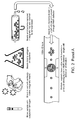
- Fig. 1 provides one exemplary configuration of a flow cell and first and second sets of magnets for generating alternating magnetic fields.
- This figures shows that the flow cell is positioned between the first and second sets of magnetics. Either movement of the flow cell or movement of the magnets brings the flow cell closer to one set of magnets and further from the other set of magnets. Subsequent movement brings the flow cell within proximity of the other set of magnets. Such movements generate alternating magnetic fields within the channels of the flow cell that are felt by the unbound magnetic particles and the target/magnetic particle complexes.
- a flow cell may be about 15 mm wide and about 15 mm long, with a lead-in region and an lead-out section, and a height of about 0.5mm ( Fig. 1 ).
- a flow rate for such a cell may be about 100 ⁇ l/min, about 1 ml/min, about 10 ml/min, or from about 100 ⁇ l/min to about 10 ml/min or other ranges therein.
- a magnetic configuration may be an array of magnets, for example, an array of 7 bar magnets, or 5 bar magnets, or 3 bar magnets ( Fig. 1 ). Magnets may be configured with alternating magnet poles facing one another, n-n, s-s, etc., with the pole face being normal to the array's rectangular face in this embodiment.
- the cycling of the magnetic bar trap assemblies may be optimized based on the flow characteristics of the target(s) of interest.
- a characteristic transit time across the height of the cell may be established.
- An efficient frequency of the alternating magnetic attractors, such that many surface interactions may be established prior to the exit from the flow cell, may be established.
- the transit time can be substantially different for the target of interest versus the unbound magnetic particles, or non-specific bound non-target.
- the target can be ensured to interact with surface a maximal amount of times, while the unbound magnetic particles or non-target can interact a minimal number of times, or not at all.
- the unbound magnetic particles may form aggregates, which may be in the form of linear chains or clumps. This may be the case at high concentrations of beads. At all concentrations, the unbound magnetic particles may exhibit spatial poison statistics, and there is some probability that there will be a neighboring bead close enough to be captured by the forces associated with the magnetic field of the beads themselves.
- methods of the invention break up these linear aggregates, particularly when the spatial gradient field from the trap magnets is shifted faster than the unbound magnetic particles can move mutually to reorient to the new distribution of trap gradient.
- Particles organized in chains, with N-S axis co-aligned may quickly be subjected to an external field that produces particle moments with the N-S poles shifted by 90°, and may produce very strong intra-particle repulsive forces.
- Transverse motion of the trap magnets serves this purpose in concert with, or as a discrete step in addition to, the alternation of the trapping magnets from one surface to the other.
- the flow characteristics of the cell may be considered along with the spatial distribution of the gradient of the trap magnets.
- Flow characteristics may dictate the transport of the magnetic materials from entrance to exit of the cell, so that parabolic flow, plug flow, or any particular flow characteristic may be considered to facilitate obtaining desired deposition patterns and desired interactions with the surfaces of interest.
- a 125 mm x 15 mm x 0.5 mm cell volume may be filled with tubes, longitudinally aligned with the cell flow direction, such that there is a great increase in the functionalized surface area and a limitation on the number of unbound magnetic particles that may interact and impede in the encounter of the target with the surface.
- Planar structures may be used for this purpose, in which the cell volume is constructed with multiple layers of smaller flow channels such that the surface area is increased and the number of unbound magnetic particles available to impede the target on its way to the surface is decreased.
- the general approach of cycling the trap magnets is similar to that described above, but variables such as time constants, amplitudes and gradient field distributions, for example, are optimized for the particular situation.
- the general approach is similar to that described above.
- the magnetic materials and labeled target may also be trapped in flow tubes and other fluidic structures through magnetic forces in undesired areas. Shielding can be accomplished by the appropriate design of the trap magnets, for example, by managing the 'return path' of the field, and/or by using high permeability materials to capture and channel the field to minimize fringing field exposure.
- the target can be removed from the surface by eluting off the surface through the use of an appropriate bonding chemistry that can be released such that the target can be analyzed in some subsequent process, for example, by use of a flowing system similar to a cytometer, or by collection of the individual targets for other analysis, such a NAT, PCR, etc, or some other post-processing and analysis as is enabled by the supply of target entity.
- the sample is analyzed using a flowing NMR detector.
- Detection of bacteria of interest can be performed by use of nucleic acid probes following procedures which are known in the art. Suitable procedures for detection of bacteria using nucleic acid probes are described, for example, in Stackebrandt et al. (U.S. 5,089,386 ), King et al. (WO 90/08841 ), Foster et al. (WO 92/15883 ), and Cossart et al. (WO 89/06699 ), each of which is hereby incorporated by reference.
- a suitable nucleic acid probe assay generally includes sample treatment and lysis, hybridization with selected probe(s), hybrid capture, and detection. Lysis of the bacteria is necessary to release the nucleic acid for the probes.
- the nucleic acid target molecules are released by treatment with any of a number of lysis agents, including alkali (such as NaOH), guanidine salts (such as guanidine isothiocyanate), enzymes (such as lysozyme, mutanolysin and proteinase K), and detergents. Lysis of the bacteria, therefore, releases both DNA and RNA, particularly ribosomal RNA and chromosomal DNA both of which can be utilized as the target molecules with appropriate selection of a suitable probe.
- rRNA as the target molecule(s), may be advantageous because rRNAs constitute a significant component of cellular mass, thereby providing an abundance of target molecules.
- the use of rRNA probes also enhances specificity for the bacteria of interest, that is, positive detection without undesirable cross-reactivity which can lead to false positives or false detection.
- Hybridization is includes addition of the specific nucleic acid probes.
- hybridization is the procedure by which two partially or completely complementary nucleic acids are combined, under defined reaction conditions, in an anti-parallel fashion to form specific and stable hydrogen bonds.
- the selection or stringency of the hybridization/reaction conditions is defined by the length and base composition of the probe/target duplex, as well as by the level and geometry of mis-pairing between the two nucleic acid strands. Stringency is also governed by such reaction parameters as temperature, types and concentrations of denaturing agents present and the type and concentration of ionic species present in the hybridization solution.
- the hybridization phase of the nucleic acid probe assay is performed with a single selected probe or with a combination of two, three or more probes. Probes are selected having sequences which are homologous to unique nucleic acid sequences of the target organism.
- a first capture probe is utilized to capture formed hybrid molecules.
- the hybrid molecule is then detected by use of antibody reaction or by use of a second detector probe which may be labelled with a radioisotope (such as phosphorus-32) or a fluorescent label (such as fluorescein) or chemiluminescent label.
- Detection of bacteria of interest can also be performed by use of PCR techniques.
- a suitable PCR technique is described, for example, in Verhoef et al. (WO 92/08805 ). Such protocols may be applied directly to the bacteria captured on the magnetic beads.
- the bacteria is combined with a lysis buffer and collected nucleic acid target molecules are then utilized as the template for the PCR reaction.
- isolated bacteria are contacted with antibodies specific to the bacteria of interest.
- antibodies specific to the bacteria of interest either polyclonal or monoclonal antibodies can be utilized, but in either case have affinity for the particular bacteria to be detected. These antibodies, will adhere/bind to material from the specific target bacteria.
- labeling of the antibodies these are labeled either directly or indirectly with labels used in other known immunoassays.
- Direct labels may include fluorescent, chemiluminescent, bioluminescent, radioactive, metallic, biotin or enzymatic molecules. Methods of combining these labels to antibodies or other macromolecules are well known to those in the art. Examples include the methods of Hijmans, W. et al. (1969), Clin. Exp. Immunol.
- detector antibodies may also be labeled indirectly.
- the actual detection molecule is attached to a secondary antibody or other molecule with binding affinity for the anti-bacteria cell surface antibody.
- a secondary antibody is used it is preferably a general antibody to a class of antibody (IgG and IgM) from the animal species used to raise the anti-bacteria cell surface antibodies.
- the second antibody may be conjugated to an enzyme, either alkaline phosphatase or to peroxidase.
- the isolated component of the sample is immersed in a solution containing a chromogenic substrate for either alkaline phosphatase or peroxidase.
- a chromogenic substrate is a compound that can be cleaved by an enzyme to result in the production of some type of detectable signal which only appears when the substrate is cleaved from the base molecule.
- the chromogenic substrate is colorless, until it reacts with the enzyme, at which time an intensely colored product is made. Thus, material from the bacteria colonies adhered to the membrane sheet will become an intense blue/purple/black color, or brown/red while material from other colonies will remain colorless.
- detection molecules include fluorescent substances, such as 4-methylumbelliferyl phosphate, and chromogenic substances, such as 4-nitrophenylphosphate, 3,3',5,5'-tetramethylbenzidine and 2,2'-azino-di-[3-ethelbenz-thiazoliane sulfonate (6)].
- fluorescent substances such as 4-methylumbelliferyl phosphate
- chromogenic substances such as 4-nitrophenylphosphate, 3,3',5,5'-tetramethylbenzidine and 2,2'-azino-di-[3-ethelbenz-thiazoliane sulfonate (6)].
- other useful enzymes include ⁇ -galactosidase, ⁇ -glucuronidase, ⁇ -glucosidase, ⁇ -glucosidase, ⁇ -mannosidase, galactose oxidase, glucose oxidase and hexokinase.
- Detection of bacteria of interest using NMR may be accomplished as follows.
- the target of interest such as a magnetically labeled bacterium
- the target of interest may be delivered by a fluid medium, such as a fluid substantially composed of water.
- the magnetically labeled target may go from a region of very low magnetic field to a region of high magnetic field, for example, a field produced by an about 1 to about 2 Tesla magnet.
- the sample may traverse a magnetic gradient, on the way into the magnet and on the way out of the magnet.
- the target may experience a force pulling into the magnet in the direction of sample flow on the way into the magnet, and a force into the magnet in the opposite direction of flow on the way out of the magnet.
- the target may experience a retaining force trapping the target in the magnet if flow is not sufficient to overcome the gradient force.
- Magnetic fields on a path into a magnet may be non-uniform in the transverse direction with respect to the flow into the magnet.
- the time it takes a target to reach the wall of a conduit is associated with the terminal velocity and is lower with increasing viscosity.
- the terminal velocity is associated with the drag force, which may be indicative of creep flow in certain cases.
- Newtonian fluids have a flow characteristic in a conduit, such as a round pipe, for example, that is parabolic, such that the flow velocity is zero at the wall, and maximal at the center, and having a parabolic characteristic with radius.
- the velocity decreases in a direction toward the walls, and it is easier to magnetically trap targets near the walls, either with transverse gradients force on the target toward the conduit wall, or in longitudinal gradients sufficient to prevent target flow in the pipe at any position.
- the detection may be based on a perturbation of the NMR water signal caused by a magnetically labeled target ( Sillerud et al., JMR (Journal of Magnetic Resonance), vol. 181, 2006 ).
- the sample may be excited at time 0, and after some delay, such as about 50ms or about 100ms, an acceptable measurement (based on a detected NMR signal) may be produced.
- an acceptable measurement based on a detected NMR signal
- such a measurement may be produced immediately after excitation, with the detection continuing for some duration, such as about 50ms or about 100ms. It may be advantageous to detect the NMR signal for substantially longer time durations after the excitation.
- the detection of the NMR signal may continue for a period of about 2 seconds in order to record spectral information at high-resolution.
- the perturbation excited at time 0 is typically smeared because the water around the perturbation source travels at different velocity, depending on radial position in the conduit.
- spectral information may be lost due to the smearing or mixing effects of the differential motion of the sample fluid during signal detection.
- Differential motion within a flowing Newtonian fluid may have deleterious effects in certain situations, such as a situation in which spatially localized NMR detection is desired, as in magnetic resonance imaging.
- a magnetic object such as a magnetically labeled bacterium
- the detection may be possible due to the magnetic field of the magnetic object, since this field perturbs the magnetic field of the fluid in the vicinity of the magnetic object.
- the detection of the magnetic object is improved if the fluid near the object remains near the object. Under these conditions, the magnetic perturbation may be allowed to act longer on any given volume element of the fluid, and the volume elements of the fluid so affected will remain in close spatial proximity. Such a stronger, more localized magnetic perturbation will be more readily detected using NMR or MRI techniques.
- the velocity of the fluid volume elements will depend on radial position in the fluid conduit.
- the fluid near a magnetic object will not remain near the magnetic object as the object flows through the detector.
- the effect of the magnetic perturbation of the object on the surrounding fluid may be smeared out in space, and the strength of the perturbation on any one fluid volume element may be reduced because that element does not stay within range of the perturbation.
- the weaker, less-well-localized perturbation in the sample fluid may be undetectable using NMR or MRI techniques.
- Certain liquids, or mixtures of liquids exhibit non-parabolic flow profiles in circular conduits. Such fluids may exhibit non-Newtonian flow profiles in other conduit shapes.
- the use of such a fluid may prove advantageous as the detection fluid in an application employing an NMR-based detection device. Any such advantageous effect may be attributable to high viscosity of the fluid, a plug-like flow profile associated with the fluid, and/or other characteristic(s) attributed to the fluid that facilitate detection.
- a shear-thinning fluid of high viscosity may exhibit a flow velocity profile that is substantially uniform across the central regions of the conduit cross-section. The velocity profile of such a fluid may transition to a zero or very low value near or at the walls of the conduit, and this transition region may be confined to a very thin layer near the wall.
- a mixture of glycerol and water can provide high viscosity, but the NMR measurement is degraded because separate NMR signals are detected from the water and glycerol molecules making up the mixture. This can undermine the sensitivity of the NMR detector.
- the non-water component of the fluid mixture can be chosen to have no NMR signal, which may be achieved by using a perdeuterated fluid component, for example, or using a perfluorinated fluid component. This approach may suffer from the loss of signal intensity since a portion of the fluid in the detection coil does not produce a signal.
- Another approach may be to use a secondary fluid component that constitutes only a small fraction of the total fluid mixture.
- a low-concentration secondary fluid component can produce an NMR signal that is of negligible intensity when compared to the signal from the main component of the fluid, which may be water.
- a perfluorinated or perdeuterated secondary fluid component may be used.
- the fluid mixture used in the NMR detector may include one, two, or more than two secondary components in addition to the main fluid component.
- the fluid components employed may act in concert to produce the desired fluid flow characteristics, such as high-viscosity and/or plug flow.
- the fluid components may be useful for providing fluid characteristics that are advantageous for the performance of the NMR detector, for example by providing NMR relaxation times that allow faster operation or higher signal intensities.
- a non-Newtonian fluid may provide additional advantages for the detection of objects by NMR or MRI techniques.
- the objects being detected may all have substantially the same velocity as they go through the detection coil. This characteristic velocity may allow simpler or more robust algorithms for the analysis of the detection data.
- the objects being detected may have fixed, known, and uniform velocity. This may prove advantageous in devices where the position of the detected object at later times is needed, such as in a device that has a sequestration chamber or secondary detection chamber down-stream from the NMR or MRI detection coil, for example.
- sample delivery into and out of a 1.7T cylindrical magnet using a fluid delivery medium containing 0.1% to 0.5 % Xanthan gum in water was successfully achieved.
- a fluid delivery medium containing 0.1% to 0.5 % Xanthan gum in water was successfully achieved.
- Such delivery is suitable to provide substantially plug-like flow, high viscosity, such as from about 10cP to about 3000cP, and good NMR contrast in relation to water.
- Xanthan gum acts as a non-Newtonian fluid, having characteristics of a non-Newtonian fluid that are well know in the art, and does not compromise NMR signal characteristics desirable for good detection in a desirable mode of operation.
- Figure 2 panel A provides an exemplary process chart for implementation of methods of the invention for separation of bacteria from blood.
- Sample is collected in sodium heparin tube by venipuncture, acceptable sample volume is 1 - 10 mL.
- Superparamagnetic particles having target-specific binding moieties are added to the sample, followed by incubation on a shaking incubator at 37 °C for 30 -120 min.
- Capture of the labeled targets allows for the removal of blood components and reduction of sample volume from 30 mL to 5 mL.
- the capture is performed by injecting the mixture of sample, unbound particles, and target/particle complexes into a channel that has target-specific antibodies coated on the surface.
- alternating magnetic fields are applied by a mechanical system that moves NdFeB permanent magnets (3 bars arranged in opposing orientation) on either side of the channel such that a magnet is present in close proximity only on one side of the channel.
- the frequency of the alternating magnetic field (1 Hz) was selected so that labeled targets would have sufficient time to reach one or both surfaces of the channel multiple times, while the majority of free beads would be prevented from reaching either surface of the channel.
- target/particle complexes were eluted from the second moiety on the surface of the channel and flowed into an NMR machine for analysis.
- the detection method is based on a miniature NMR detector tuned to the magnetic resonance of water.
- the NMR signal from water is clearly detectable and strong.
- the presence of magnetic material in the detector coil disturbs the magnetic field, resulting in reduction in water signal.
- One of the primary benefits of this detection method is that there is no magnetic background in biological samples which significantly reduces the requirements for stringency of sample processing.
- the detected signal is generated by water, there is a built-in signal amplification which allows for the detection of a single labeled bacterium.
- Methods of the invention may also be combined with other separation and isolation protocols known in the art. Particularly, methods of the invention may be combined with methods shown in co-pending and co-owned U.S. patent application serial number 12/850,203, filed August 4, 2010 , entitled Isolating A Target Analyte From A Body Fluid, the content of which is incorporated by reference herein in its entirety.
- Blood samples from healthy volunteers were spiked with clinically relevant concentrations of bacteria (1-10 CFU/mL) including both laboratory strains and clinical isolates of the bacterial species most frequently found in bloodstream infections.
- the immune serum was purified using affinity chromatography on a protein G sepharose column (GE Healthcare), and reactivity was determined using ELISA.
- Antibodies cross-reacting with Gram-negative bacteria and fungi were removed by absorption of purified IgG with formalin-fixed Gram-negative bacteria and fungi.
- the formalin-fixed organisms were prepared similar to as described above and mixed with IgG. After incubation for 1 hr at room temperature, the preparation was centrifuged to remove bacteria and absorption was repeated. Final antibody preparation was clarified by centrifugation and used for the preparation of antigen-specific magnetic beads.
- Superparamagnetic beads were synthesized by encapsulating iron oxide nanoparticles (5-15 nm diameter) in a latex core and labeling with goat IgG.
- Ferrofluid containing nanoparticles in organic solvent was precipitated with ethanol, nanoparticles were resuspended in aqueous solution of styrene and surfactant Hitenol BC-10, and emulsified using sonication. The mixture was allowed to equilibrate overnight with stirring and filtered through 1.2 and 0.45 ⁇ m filters to achieve uniform micelle size.
- Styrene, acrylic acid and divynilbenzene were added in carbonate buffer at pH 9.6.
- the polymerization was initiated in a mixture at 70 °C with the addition of K 2 S 2 0 8 and the reaction was allowed to complete overnight.
- the synthesized particles were washed 3 times with 0.1% SDS using magnetic capture, filtered through 1.2, 0.8, and 0.45 ⁇ m filters and used for antibody conjugation.
- the production of beads resulted in a distribution of sizes that may be characterized by an average size and a standard deviation.
- the average size for optimal performance was found to be between 100 and 350 nm, for example between 200 nm to 250 nm.
- the purified IgG were conjugated to prepared beads using standard chemistry. After conjugation, the beads were resuspended in 0.1% BSA which is used to block non-specific binding sites on the bead and to increase the stability of bead preparation.
- Target-specific antibodies were attached to a surface of a flow channel using the following approach:
- Example 5 Separating target from remaining components of the sample
- Target cells magnetically labeled using target-specific beads with the excess of free beads were injected into a channel that had target-specific antibodies coated on the surface.
- alternating magnetic field was applied by a mechanical system that moved NdFeB permanent magnets (3 bars arranged in opposing orientation) on either side of the channel such that a magnet was present in close proximity only on one side of the channel.
- the frequency of the alternating magnetic field (1 Hz) was selected so that labeled targets would have sufficient time to reach one or both surfaces of the channel multiple times, while the majority of free beads would be prevented from reaching either surface of the channel.
- the bound bacteria were eluted with 35 mM biotin solution in B29, followed by magnetic capture and re-suspension in the detection buffer containing Xanthan gum.
- the concentration of bacteria in samples were analyzed in flow-through NMR detector. Results indicate that the number of detected bacteria was directly proportional to the number of bacteria spiked into blood ( Fig. 3 ).
- Embodiments of the invention may include the features of the following enumerated paragraphs ("paras").
Abstract
Description
- The present application claims the benefit of and priority to
U.S. nonprovisional patent application serial number 12/855,147, filed August 12, 2010 U.S. provisional patent application serial number 61/326,588, filed April 21, 2010 - The invention generally relates to using magnetic particles and alternating magnet fields to separate a target analyte from a sample.
- Many laboratory and clinical procedures employ bio-specific affinity reactions. Such reactions are commonly utilized in diagnostic testing of biological samples, or for the separation of a wide range of target substances, especially biological entities such as cells, viruses, proteins, nucleic acids and the like. Various methods are available for analyzing or separating the above-mentioned target substances based upon complex formation between the substance of interest and another substance to which the target specifically binds. Separation of complexes from unbound material may be accomplished gravitationally, e.g. by settling, or, alternatively, by centrifugation of finely divided particles or beads coupled to the target substance. If desired, such particles or beads may be made magnetic to facilitate the bound/free separation step. Magnetic particles are well known in the art, as is their use in immune and other bio-specific affinity reactions. See, for example, Whitehead et al. (
U.S. 4,554,088 ) and Hunter et al. (Immunoassays for Clinical Chemistry, pp. 147-162, eds., Churchill Livingston, Edinborough, 1983). Generally, any material that facilitates magnetic or gravitational separation may be employed for this purpose. More recently, the superiority of magnetics for performing such separations has led to its use in many applications. - A problem with magnetic separation protocols is that magnetic beads must be added in excess to a sample to ensure a sufficient amount of binding of beads to a target analyte in the sample, thus producing a sample that contains a very high percent of magnetic particles that are not bound to target analytes, as well as non-specific target entities. Non-specific target entities may for example be bound at a much lower efficiency, for example 1% of the surface area, while a target of interest might be loaded at 50% or nearly 100% of the available surface area or available antigenic cites. However, even 1% loading may be sufficient to impart force necessary for trapping in a magnetic gradient flow cell or sample chamber.
- The presence of magnetic particles that are not bound to target analytes and non-specific target entities on the surface that includes the target/magnetic particle complexes interferes with the ability to successfully separate the target of interest of the remaining components of the mixture and unbound magnetic particles. The magnetic capture of the resulting mix, and close contact of magnetic particles with each other and labeled targets, result in the formation of aggregate that is hard to dispense and which might be resistant or inadequate for subsequent processing or analysis steps.
- There is a need for methods for separating target analytes from a sample.
- The present invention generally relates to using magnetic particles having a target-specific binding moiety and alternating magnet fields to separate a target analyte from a sample. Methods of the invention allow for rapid (less than 1 hr) and efficient capture of target analytes from a sample while eliminating non-specific binding and reducing background noise resulting from excess magnetic particles not bound to target analytes. Methods of the invention involve contacting a sample with magnetic particles including first moieties specific for a target analyte, thereby forming target/particle complexes in the sample, flowing the sample through a channel including second moieties attached to at least one surface of the channel, applying alternating magnetic fields to the flowing sample to result in target/particle complexes being brought into proximity of the surface to bind the second moieties and unbound particles remaining free in the sample, binding the target/particle complexes to the second moieties, and washing away unbound particles and unbound analytes of the sample. A particular advantage of methods of the invention is for capture and isolation of bacteria and fungi directly from blood samples at low concentrations that are present in clinical samples (as low as 1 CFU/ml of bacteria in a blood sample).
- In certain embodiments, the free magnetic particles and the target/particle complexes both interact numerous times with the surface of the flow channel. The targets are bound due to the specific interaction between the target analyte and the second moiety on the surface of the flow channel, and the free magnetic particles are not bound, and are thus continue flowing through the flow channel.
- Methods of the invention may further involve eluting the bound target/particle complexes from the second moieties on the surface of the channel. Methods of the invention may further involve analyzing the eluted target/particle complexes. The target may be analyzed by a multitude of existing technologies, such as miniature NMR , Polymerase Chain Reaction (PCR), fluorescent labeling and visualization using microscopic observation, fluorescent in situ hybridization (FISH), growth-based antibiotic sensitivity tests, and variety of other methods that may be conducted with purified target without significant contamination from other sample components. In particular embodiments, analyzing involves flowing the target/particle complexes into an NMR instrument.
- The target analyte refers to the target that will be captured and isolated by methods of the invention. The target may be a bacteria, a fungi, a protein, a nucleic acid, a receptor, a ligand, a cell, a virus, or any molecule known in the art. In a particular embodiment, the target is a bacteria.
- Methods of the invention do not depend and are not limited by the type of sample. Any sample that includes a detectable target may be used with methods of the invention. The sample may be a biological sample (e.g., a human tissue or body fluid), an agricultural sample, or an environmental sample (e.g., a water or soil sample). In certain embodiments, the sample is a blood sample.
- The first and second target-specific binding moieties will depend on the target to be captured. The moieties may be any capture moieties known in the art, such as an antibody, an aptamer, a phage, a nucleic acid, a protein, a receptor, or a ligand. In particular embodiments, the target-specific binding moieties are antibodies. In certain embodiments, the antibodies are specific for bacteria. In other embodiments, the antibodies are specific for fungi or viruses. In certain embodiments, the first and second moieties are the same. In alternative embodiments, the first and second moieties are different.
- Generating alternating magnetic fields may be accomplished by any method known in the art. In certain embodiments, the alternating magnetic fields result from the channel being positioned between first and second sets of magnets, in which the channel remains stationary and the first and second sets of magnets are moved to alternate proximity to the channel, thereby producing the alternating magnetic fields. In other embodiments, the alternating magnetic fields result from the channel being positioned between first and second sets of magnets, in which first and second sets of magnets remain stationary and the channel is moved to alternate its proximity to the first and second sets of magnets, thereby producing the alternating magnetic fields.
- Another aspect of the invention provides methods for detecting a target analyte in a sample including contacting a sample with magnetic particles including first moieties specific for a target analyte, thereby forming target/particle complexes in the sample, flowing the sample through a channel including second moieties attached to at least one surface of the channel, applying alternating magnetic fields to the flowing sample to result in target/particle complexes being brought into proximity of the surface of the channel to bind the second moieties and unbound particles remaining free in the sample, binding the target/particle complexes to the second moieties, washing away unbound particles and unbound analytes of the sample, eluting the target/particle complexes from the second moieties, and detecting the target/particle complexes.
-
-
Figure 1 provides one exemplary configuration of a flow cell and first and second sets of magnets for generating alternating magnetic fields. -
Figure 2 panel A provides an exemplary process chart for implementation of methods of the invention for separation of bacteria from blood. Panel B provides an magnified view of a target/magnetic particle complex. -
Figure 3 is a graph showing recovery of bacteria from blood. - The invention generally relates to using magnetic particles and alternating magnet fields to separate a target analyte from a sample. Methods of the invention involve contacting a sample with magnetic particles including first moieties specific for a target analyte, thereby forming target/particle complexes in the sample, flowing the sample through a channel including second moieties attached to at least one surface of the channel, applying alternating magnetic fields to the flowing sample to result in target/particle complexes being brought into proximity of the surface to bind the second moieties and unbound particles remaining free in the sample, binding the target/particle complexes to the second moieties, and washing away unbound particles and unbound analytes of the sample. Certain fundamental technologies and principles are associated with binding magnetic materials to target entities and subsequently separating by use of magnet fields and gradients. Such fundamental technologies and principles are known in the art and have been previously described, such as those described in Janeway (Immunobiology, 6th edition, Garland Science Publishing), the content of which is incorporated by reference herein in its entirety.
- Methods of the invention involve collecting a sample having a target analyte in a container, such as a blood collection tube (e.g., Vacutainer) in the case of blood. In certain embodiments, a solution is added that prevents or reduces aggregation of endogenous aggregating factors, such as heparin in the case of blood.
- Methods of the invention do not depend and are not limited by the type of sample. Any sample that includes a detectable target may be used with methods of the invention. The sample may be a biological sample (e.g., a human tissue or body fluid), a food sample, an agricultural sample, or an environmental sample (e.g., a water or soil sample).
- Exemplary biological samples include human tissue or body fluid. A tissue is a mass of connected cells and/or extracellular matrix material, e.g. skin tissue, nasal passage tissue, CNS tissue, neural tissue, eye tissue, liver tissue, kidney tissue, placental tissue, mammary gland tissue, placental tissue, gastrointestinal tissue, musculoskeletal tissue, genitourinary tissue, bone marrow, and the like, derived from, for example, a human or other mammal and includes the connecting material and the liquid material in association with the cells and/or tissues. A body fluid is a liquid material derived from, for example, a human or other mammal. Such body fluids include, but are not limited to, mucous, blood, plasma, serum, serum derivatives, bile, phlegm, saliva, sweat, amniotic fluid, mammary fluid, urine, sputum, and cerebrospinal fluid (CSF), such as lumbar or ventricular CSF. A sample may also be a fine needle aspirate. A sample also may be media containing cells or biological material. In particular embodiments, the sample is blood.
- Exemplary agricultural samples include any plant material that is being interrogated by a method of the present invention. An agricultural sample includes, but is not limited to, seeds or plant tissue. Seeds include a single seed, a batch of seeds, a portion of a seed, or a seed scraping. Plant tissue includes, but is not limited to, any plant part such as leaf, flower, root, or petal. Plant tissue can also include a leaf punch.
- Methods of the invention may be used to detect any target analyte. The target analyte refers to the substance in the sample that will be captured and isolated by methods of the invention. The target may be a bacteria, a fungi, a protein, a cell (such as a cancer cell, a white blood cell, a virally infected cell, or a fetal cell circulating in maternal circulation), a virus, a nucleic acid (e.g., DNA or RNA), a receptor, a ligand, a hormone, a drug, a chemical substance, or any molecule known in the art. In certain embodiments, the target is a pathogenic bacteria. In other embodiments, the target is a gram positive or gram negative bacteria. Exemplary bacterial species that may be captured and isolated by methods of the invention include E. coli, Lysteria, Clostridium, Mycobacterium, Shigella, Borrelia, Campylobacter, Bacillus, Salmonella, Staphylococcus, Enterococcus, Pneumococcus, Streptococcus, and a combination thereof.
- The sample is then mixed with magnetic particles including a target-specific binding moiety to generate a mixture that is allowed to incubate such that the particles bind to a target in the sample, such as a bacteria in a blood sample. The mixture is allowed to incubate for a sufficient time to allow for the particles to bind to the target analyte. The process of binding the magnetic particles to the target analytes associates a magnetic moment with the target analytes, and thus allows the target analytes to be manipulated through forces generated by magnetic fields upon the attached magnetic moment.
- In general, incubation time will depend on the desired degree of binding between the target analyte and the magnetic beads (e.g., the amount of moment that would be desirably attached to the target), the amount of moment per target, the amount of time of mixing, the type of mixing, the reagents present to promote the binding and the binding chemistry system that is being employed. Incubation time can be anywhere from about 5 seconds to a few days. Exemplary incubation times range from about 10 seconds to about 2 hours. Binding occurs over a wide range of temperatures, generally between 15 °C and 40 °C.
- Methods of the invention may be performed with any type of magnetic particle. Production of magnetic particles and particles for use with the invention are known in the art. See for example
Giaever (U.S. 3,970,518 ),Senyi et al. (U.S. 4,230,685 ),Dodin et al. (U.S. 4,677,055 ),Whitehead et al. (U.S. 4,695,393 ),Benjamin et al. (U.S. 5,695,946 ),Giaever (U.S. 4,018,886 ),Rembaum (U.S. 4,267,234 ), Molday (U.S. 4,452,773 ),Whitehead et al. (U.S. 4,554,088 ),Forrest (U.S. 4,659,678 ),Liberti et al. (U.S. 5,186,827 ),Own et al. (U.S. 4,795,698 ), andLiberti et al. (WO 91/02811 - Magnetic particles generally fall into two broad categories. The first category includes particles that are permanently magnetizable, or ferromagnetic; and the second category includes particles that demonstrate bulk magnetic behavior only when subjected to a magnetic field. The latter are referred to as magnetically responsive particles. Materials displaying magnetically responsive behavior are sometimes described as superparamagnetic. However, materials exhibiting bulk ferromagnetic properties, e.g., magnetic iron oxide, may be characterized as superparamagnetic when provided in crystals of about 30 nm or less in diameter. Larger crystals of ferromagnetic materials, by contrast, retain permanent magnet characteristics after exposure to a magnetic field and tend to aggregate thereafter due to strong particle-particle interaction. In certain embodiments, the particles are superparamagnetic beads. In certain embodiments, the magnetic particle is an iron containing magnetic particle. In other embodiments, the magnetic particle includes iron oxide or iron platinum.
- In certain embodiments, the magnetic particles include at least about 10% superparamagnetic beads by weight, at least about 20% superparamagnetic beads by weight, at least about 30% superparamagnetic beads by weight, at least about 40% superparamagnetic beads by weight, at least about 50% superparamagnetic beads by weight, at least about 60% superparamagnetic beads by weight, at least about 70% superparamagnetic beads by weight, at least about 80% superparamagnetic beads by weight, at least about 90% superparamagnetic beads by weight, at least about 95% superparamagnetic beads by weight, or at least about 99% superparamagnetic beads by weight. In a particular embodiment, the magnetic particles include at least about 70% superparamagnetic beads by weight.
- In certain embodiments, the superparamagnetic beads are less than 100 nm in diameter. In other embodiments, the superparamagnetic beads are about 150 nm in diameter, are about 200 nm in diameter, are about 250 nm in diameter, are about 300 nm in diameter, are about 350 nm in diameter, are about 400 nm in diameter, are about 500 nm in diameter, or are about 1000 nm in diameter. In a particular embodiment, the superparamagnetic beads are from about 100 nm to about 250 nm in diameter.
- In certain embodiments, the particles are beads (e.g., nanoparticles) that incorporate magnetic materials, or magnetic materials that have been functionalized, or other configurations as are known in the art. In certain embodiments, nanoparticles may be used that include a polymer material that incorporates magnetic material(s), such as nanometal material(s). When those nanometal material(s) or crystal(s), such as Fe3O4, are superparamagnetic, they may provide advantageous properties, such as being capable of being magnetized by an external magnetic field, and demagnetized when the external magnetic field has been removed. This may be advantageous for facilitating sample transport into and away from an area where the sample is being processed without undue bead aggregation.
- One or more or many different nanometal(s) may be employed, such as Fe3O4, FePt, or Fe, in a core-shell configuration to provide stability, and/or various others as may be known in the art. In many applications, it may be advantageous to have a nanometal having as high a saturated moment per volume as possible, as this may maximize gradient related forces, and/or may enhance a signal associated with the presence of the beads. It may also be advantageous to have the volumetric loading in a bead be as high as possible, for the same or similar reason(s).
- In order to maximize the moment provided by a magnetizable nanometal, a certain saturation field may be provided. For example, for Fe3O4 superparamagnetic particles, this field may be on the order of about 0.3T.
- The size of the nanometal containing bead may be optimized for a particular application, for example, maximizing moment loaded upon a target, maximizing the number of beads on a target with an acceptable detectability, maximizing desired force-induced motion, and/or maximizing the difference in attached moment between the labeled target and non-specifically bound targets or bead aggregates or individual beads. While maximizing is referenced by example above, other optimizations or alterations are contemplated, such as minimizing or otherwise desirably affecting conditions.
- In an exemplary embodiment, a polymer bead containing 80 wt% Fe3O4 superparamagnetic particles, or for example, 90 wt% or higher superparamagnetic particles, is produced by encapsulating superparamagnetic particles with a polymer coating to produce a bead having a diameter of about 250 nm.
- Magnetic particles for use with methods of the invention have a target-specific binding moiety that allows for the particles to specifically bind the target of interest in the sample. The target-specific moiety may be any molecule known in the art and will depend on the target to be captured and isolated. Exemplary target-specific binding moieties include, nucleic acids, proteins, ligands, antibodies, aptamers, and receptors.
- In particular embodiments, the target-specific binding moiety is an antibody, such as an antibody that binds a particular bacteria. General methodologies for antibody production, including criteria to be considered when choosing an animal for the production of antisera, are described in Harlow et al. (Antibodies, Cold Spring Harbor Laboratory, pp. 93-117, 1988). For example, an animal of suitable size such as goats, dogs, sheep, mice, or camels are immunized by administration of an amount of immunogen, such as target bacteria, effective to produce an immune response. An exemplary protocol is as follows. The animal is injected with 100 micrograms to 100 milligrams of antigen resuspended in adjuvant, for example Freund's complete adjuvant, dependent on the size of the animal, followed three weeks later with a subcutaneous injection of 100 micrograms to 100 milligrams of immunogen with adjuvant dependent on the size of the animal, for example Freund's incomplete adjuvant. Additional subcutaneous or intraperitoneal injections every two weeks with adjuvant, for example Freund's incomplete adjuvant, are administered until a suitable titer of antibody in the animal's blood is achieved. Exemplary titers include a titer of at least about 1:5000 or a titer of 1:100,000 or more, i.e., the dilution having a detectable activity. The antibodies are purified, for example, by affinity purification on columns containing protein G resin or target-specific affinity resin.
- The technique of in vitro immunization of human lymphocytes is used to generate monoclonal antibodies. Techniques for in vitro immunization of human lymphocytes are well known to those skilled in the art. See, e.g., Inai, et al., Histochemistry, 99(5):335 362, May 1993; Mulder, et al., Hum. Immunol., 36(3):186 192, 1993; Harada, et al., J. Oral Pathol. Med., 22(4):145 152, 1993; Stauber, et al., J. Immunol. Methods, 161(2):157 168, 1993; and Venkateswaran, et al., Hybridoma, 11(6) 729 739, 1992. These techniques can be used to produce antigen-reactive monoclonal antibodies, including antigen-specific IgG, and IgM monoclonal antibodies.
- Any antibody or fragment thereof having affinity and specific for the bacteria of interest is within the scope of the invention provided herein. Immunomagnetic beads against Salmonella are provided in Vermunt et al. (J. Appl. Bact. 72:112, 1992). Immunomagnetic beads against Staphylococcus aureus are provided in Johne et al. (J. Clin. Microbiol. 27:1631, 1989). Immunomagnetic beads against Listeria are provided in Skjerve et al. (Appl. Env. Microbiol. 56:3478, 1990). Immunomagnetic beads against Escherichia coli are provided in Lund et al. (J. Clin. Microbiol. 29:2259, 1991).
- Methods for attaching the target-specific binding moiety to the magnetic particle are known in the art. Coating magnetic particles with antibodies is well known in the art, see for example Harlow et al. (Antibodies, Cold Spring Harbor Laboratory, 1988), Hunter et al. (Immunoassays for Clinical Chemistry, pp. 147-162, eds., Churchill Livingston, Edinborough, 1983), and Stanley (Essentials in Immunology and Serology, Delmar, pp. 152-153, 2002). Such methodology can easily be modified by one of skill in the art to bind other types of target-specific binding moieties to the magnetic particles. Certain types of magnetic particles coated with a functional moiety are commercially available from Sigma-Aldrich (St. Louis, MO).
- In certain embodiments, a buffer solution is added to the sample along with the magnetic beads. An exemplary buffer includes Tris(hydroximethyl)-aminomethane hydrochloride at a concentration of about 75mM. It has been found that the buffer composition, mixing parameters (speed, type of mixing, such as rotation, shaking etc., and temperature) influence binding. It is important to maintain osmolality of the final solution (e.g., blood + buffer) to maintain high label efficiency. In certain embodiments, buffers used in methods of the invention are designed to prevent lysis of blood cells, facilitate efficient binding of targets with magnetic beads and to reduce formation of bead aggregates. It has been found that the buffer solution containing 300 mM NaCl, 75 mM Tris-HCl pH 8.0 and 0.1% Tween 20 meets these design goals.
- Without be limited by any particular theory or mechanism if action, it is believed that sodium chloride is mainly responsible for maintaining osmolality of the solution and for the reduction of non-specific binding of magnetic bead through ionic interaction. Tris(hydroximethyl)-aminomethane hydrochloride is a well established buffer compound frequently used in biology to maintain pH of a solution. It has been found that 75 mM concentration is beneficial and sufficient for high binding efficiency. Likewise, Tween 20 is widely used as a mild detergent to decrease nonspecific attachment due to hydrophobic interactions. Various assays use Tween 20 at concentrations ranging from 0.01% to 1%. The 0.1% concentration appears to be optimal for the efficient labeling of bacteria, while maintaining blood cells intact.
- An alternative approach to achieve high binding efficiency while reducing time required for the binding step is to use static mixer, or other mixing devices that provide efficient mixing of viscous samples at high flow rates, such as at or around 5 mL/min. In one embodiment, the sample is mixed with binding buffer in ratio of, or about, 1:1, using a mixing interface connector. The diluted sample then flows through a second mixing interface connector where it is mixed with target-specific nanoparticles. Additional mixing interface connectors providing mixing of sample and antigen-specific nanoparticles can be attached downstream to improve binding efficiency. The combined flow rate of the labeled sample is selected such that it is compatible with downstream processing. At this point in the process, the mixture includes target/magnetic particle complexes, unbound magnetic particles, and the remaining components of the mixture. Prior art techniques for isolating target/magnetic particle complexes involve applying a magnetic field to the mixture to capture the complexes on a surface. Components of the mixture that are not bound to magnetic particles will not be affected by the magnetic field and will remain free in the mixture.
- The above described type of magnetic separation, produces efficient capture of a target analyte and the removal of a majority of the remaining components of a sample mixture. However, such a process produces a sample that contains a very high percent of magnetic particles that are not bound to target analytes because the magnetic particles are typically added in excess, as well as non-specific target entities. Non-specific target entities may be bound at a much lower efficiency, for example 1% of the surface area, while a target of interest might be loaded at 50% or nearly 100% of the available surface area or available antigenic cites. However, even 1 % loading may be sufficient to impart force necessary for trapping in a magnetic gradient flow cell or sample chamber.
- For example, in the case of immunomagnetic labeling of bacteria or fungi in a blood sample, the sample may include: labeled targets at a concentration of about 1/mL or a concentration less of about 106/mL; background beads at a concentration of about 107/ml to about 1010/ml; and non-specific targets at a concentration of about 10/ml to about 105/ml.
- The presence of magnetic particles that are not bound to target analytes and non-specific target entities on the surface that includes the target/magnetic particle complexes interferes with the ability to successfully detect the target of interest. The magnetic capture of the resulting mix, and close contact of magnetic particles with each other and labeled targets, result in the formation of aggregate that is hard to dispense and which might be resistant or inadequate for subsequent processing or analysis steps. Further, with addition of excess magnetic particles to the sample, a large number of particles may accumulate in the areas of high gradients, and thus a magnetically bound target analyte may likely be in the body of the accumulation of particles as opposed to the desired location adjacent the functionalized surface where specific binding may occur. Ignoring intra-bead forces (those forces associated with the magnetic field distribution of the individual beads and the forces these fields and associated gradients have on other beads), the beads may accumulate into large amorphous piles. Such intra-label forces do occur, and thus the aggregates of beads tend to exist in chains and long linear aggregates that are aligned with the 'field lines' of the magnetic trap pieces.
- Methods of the invention address this problem by applying alternating magnetic fields to the sample as it flows through the channel. The frequency of the alternating magnetic field is selected such that the free magnetic nanoparticles cannot transverse the whole distance between top and bottom of the flow cell before the direction of the magnetic field is changed, causing nanoparticles to move in the opposite direction. Therefore, a majority of free nanoparticles will not come into close contact with active surfaces of the flow cell and will be washed away by liquid flow. Labeled target, due to higher magnetic moment, have higher velocity in the magnetic field and will reach a surface of the flow cell before change of the magnetic field, thus coming into close contact with the surface. This, in turn, will result in a specific binding event and result in a specific capture of the target analyte in the sample (such as a bacterium or other rare cell) to the surface coated with a second moiety. Components of the mixture that are not bound to magnetic particles will not be affected by the magnetic field and will remain free in the mixture.
- The second target-specific moiety may be the same or different from the first target-specific moiety. The second moiety may be attached to the surface of the flow channel by methods described above relating to attaching first target-specific moieties to magnetic particles.
-
Fig. 1 provides one exemplary configuration of a flow cell and first and second sets of magnets for generating alternating magnetic fields. This figures shows that the flow cell is positioned between the first and second sets of magnetics. Either movement of the flow cell or movement of the magnets brings the flow cell closer to one set of magnets and further from the other set of magnets. Subsequent movement brings the flow cell within proximity of the other set of magnets. Such movements generate alternating magnetic fields within the channels of the flow cell that are felt by the unbound magnetic particles and the target/magnetic particle complexes. - In one embodiment, a flow cell may be about 15 mm wide and about 15 mm long, with a lead-in region and an lead-out section, and a height of about 0.5mm (
Fig. 1 ). A flow rate for such a cell may be about 100 µl/min, about 1 ml/min, about 10 ml/min, or from about 100 µl/min to about 10 ml/min or other ranges therein. A magnetic configuration may be an array of magnets, for example, an array of 7 bar magnets, or 5 bar magnets, or 3 bar magnets (Fig. 1 ). Magnets may be configured with alternating magnet poles facing one another, n-n, s-s, etc., with the pole face being normal to the array's rectangular face in this embodiment. - In a flowing system, successive encounters of unbound magnetic particles with a surface of a flow channel, without a resulting binding event, will allow the unbound magnetic particles to travel through the system and subsequently out of the cell. The cycling of the magnetic bar trap assemblies may be optimized based on the flow characteristics of the target(s) of interest. The expression of force on a magnetic moment and of terminal velocity for such target(s) is the following:


- A characteristic transit time across the height of the cell may be established. An efficient frequency of the alternating magnetic attractors, such that many surface interactions may be established prior to the exit from the flow cell, may be established. In certain embodiments, the transit time can be substantially different for the target of interest versus the unbound magnetic particles, or non-specific bound non-target. In such an embodiment, the target can be ensured to interact with surface a maximal amount of times, while the unbound magnetic particles or non-target can interact a minimal number of times, or not at all.
- Because of the magnetizing characteristics of the particles, the unbound magnetic particles may form aggregates, which may be in the form of linear chains or clumps. This may be the case at high concentrations of beads. At all concentrations, the unbound magnetic particles may exhibit spatial poison statistics, and there is some probability that there will be a neighboring bead close enough to be captured by the forces associated with the magnetic field of the beads themselves. By using alternating magnetic fields, methods of the invention break up these linear aggregates, particularly when the spatial gradient field from the trap magnets is shifted faster than the unbound magnetic particles can move mutually to reorient to the new distribution of trap gradient. Particles organized in chains, with N-S axis co-aligned, may quickly be subjected to an external field that produces particle moments with the N-S poles shifted by 90°, and may produce very strong intra-particle repulsive forces. Transverse motion of the trap magnets serves this purpose in concert with, or as a discrete step in addition to, the alternation of the trapping magnets from one surface to the other.
- In the optimization of the cycling timing of the trap magnets, the flow characteristics of the cell may be considered along with the spatial distribution of the gradient of the trap magnets. Flow characteristics may dictate the transport of the magnetic materials from entrance to exit of the cell, so that parabolic flow, plug flow, or any particular flow characteristic may be considered to facilitate obtaining desired deposition patterns and desired interactions with the surfaces of interest.
- In certain embodiments, it may be desirable in various applications to maximize the encounters of the target/magnetic particle complexes with the functionalized surface of the channel, to minimize interference with the unbound magnetic particles, and/or to minimize adhesion of the unbound magnetic particles and non-specific materials to the surface. It may be advantageous to produce an array of pipes, or tubes, through which the flow of the sample materials may flow. By way of example, a 125 mm x 15 mm x 0.5 mm cell volume may be filled with tubes, longitudinally aligned with the cell flow direction, such that there is a great increase in the functionalized surface area and a limitation on the number of unbound magnetic particles that may interact and impede in the encounter of the target with the surface. Planar structures may be used for this purpose, in which the cell volume is constructed with multiple layers of smaller flow channels such that the surface area is increased and the number of unbound magnetic particles available to impede the target on its way to the surface is decreased. In this embodiment, the general approach of cycling the trap magnets is similar to that described above, but variables such as time constants, amplitudes and gradient field distributions, for example, are optimized for the particular situation. Similarly, in the case of transverse trap motion for the breaking-up of aggregates, the general approach is similar to that described above.
- It may be desirable to shield the portion of the sample flow outside the trap cell from fringing magnetic field so that magnetic material does not have the opportunity to self-aggregate prior to entering the strong field and gradient zone of the trap. The magnetic materials and labeled target may also be trapped in flow tubes and other fluidic structures through magnetic forces in undesired areas. Shielding can be accomplished by the appropriate design of the trap magnets, for example, by managing the 'return path' of the field, and/or by using high permeability materials to capture and channel the field to minimize fringing field exposure.
- It is possible to purify samples and fixate targets of interest and to then examine the target in situ on the surface, in any suitable manner known in the art, or the target can be removed from the surface by eluting off the surface through the use of an appropriate bonding chemistry that can be released such that the target can be analyzed in some subsequent process, for example, by use of a flowing system similar to a cytometer, or by collection of the individual targets for other analysis, such a NAT, PCR, etc, or some other post-processing and analysis as is enabled by the supply of target entity. In an exemplary case, the sample is analyzed using a flowing NMR detector.
- Detection of bacteria of interest can be performed by use of nucleic acid probes following procedures which are known in the art. Suitable procedures for detection of bacteria using nucleic acid probes are described, for example, in
Stackebrandt et al. (U.S. 5,089,386 ),King et al. (WO 90/08841 Foster et al. (WO 92/15883 Cossart et al. (WO 89/06699 - A suitable nucleic acid probe assay generally includes sample treatment and lysis, hybridization with selected probe(s), hybrid capture, and detection. Lysis of the bacteria is necessary to release the nucleic acid for the probes. The nucleic acid target molecules are released by treatment with any of a number of lysis agents, including alkali (such as NaOH), guanidine salts (such as guanidine isothiocyanate), enzymes (such as lysozyme, mutanolysin and proteinase K), and detergents. Lysis of the bacteria, therefore, releases both DNA and RNA, particularly ribosomal RNA and chromosomal DNA both of which can be utilized as the target molecules with appropriate selection of a suitable probe. Use of rRNA as the target molecule(s), may be advantageous because rRNAs constitute a significant component of cellular mass, thereby providing an abundance of target molecules. The use of rRNA probes also enhances specificity for the bacteria of interest, that is, positive detection without undesirable cross-reactivity which can lead to false positives or false detection.
- Hybridization is includes addition of the specific nucleic acid probes. In general, hybridization is the procedure by which two partially or completely complementary nucleic acids are combined, under defined reaction conditions, in an anti-parallel fashion to form specific and stable hydrogen bonds. The selection or stringency of the hybridization/reaction conditions is defined by the length and base composition of the probe/target duplex, as well as by the level and geometry of mis-pairing between the two nucleic acid strands. Stringency is also governed by such reaction parameters as temperature, types and concentrations of denaturing agents present and the type and concentration of ionic species present in the hybridization solution.
- The hybridization phase of the nucleic acid probe assay is performed with a single selected probe or with a combination of two, three or more probes. Probes are selected having sequences which are homologous to unique nucleic acid sequences of the target organism. In general, a first capture probe is utilized to capture formed hybrid molecules. The hybrid molecule is then detected by use of antibody reaction or by use of a second detector probe which may be labelled with a radioisotope (such as phosphorus-32) or a fluorescent label (such as fluorescein) or chemiluminescent label.
- Detection of bacteria of interest can also be performed by use of PCR techniques. A suitable PCR technique is described, for example, in
Verhoef et al. (WO 92/08805 - For detection of the selected bacteria by use of antibodies, isolated bacteria are contacted with antibodies specific to the bacteria of interest. As noted above, either polyclonal or monoclonal antibodies can be utilized, but in either case have affinity for the particular bacteria to be detected. These antibodies, will adhere/bind to material from the specific target bacteria. With respect to labeling of the antibodies, these are labeled either directly or indirectly with labels used in other known immunoassays. Direct labels may include fluorescent, chemiluminescent, bioluminescent, radioactive, metallic, biotin or enzymatic molecules. Methods of combining these labels to antibodies or other macromolecules are well known to those in the art. Examples include the methods of Hijmans, W. et al. (1969), Clin. Exp. Immunol. 4, 457-, for fluorescein isothiocyanate, the method of Goding, J. W. (1976), J. Immunol. Meth. 13, 215-, for tetramethylrhodamine isothiocyanate, and the method of Ingrall, E. (1980), Meth. in Enzymol. 70, 419-439 for enzymes.
- These detector antibodies may also be labeled indirectly. In this case the actual detection molecule is attached to a secondary antibody or other molecule with binding affinity for the anti-bacteria cell surface antibody. If a secondary antibody is used it is preferably a general antibody to a class of antibody (IgG and IgM) from the animal species used to raise the anti-bacteria cell surface antibodies. For example, the second antibody may be conjugated to an enzyme, either alkaline phosphatase or to peroxidase. To detect the label, after the bacteria of interest is contacted with the second antibody and washed, the isolated component of the sample is immersed in a solution containing a chromogenic substrate for either alkaline phosphatase or peroxidase. A chromogenic substrate is a compound that can be cleaved by an enzyme to result in the production of some type of detectable signal which only appears when the substrate is cleaved from the base molecule. The chromogenic substrate is colorless, until it reacts with the enzyme, at which time an intensely colored product is made. Thus, material from the bacteria colonies adhered to the membrane sheet will become an intense blue/purple/black color, or brown/red while material from other colonies will remain colorless. Examples of detection molecules include fluorescent substances, such as 4-methylumbelliferyl phosphate, and chromogenic substances, such as 4-nitrophenylphosphate, 3,3',5,5'-tetramethylbenzidine and 2,2'-azino-di-[3-ethelbenz-thiazoliane sulfonate (6)]. In addition to alkaline phosphatase and peroxidase, other useful enzymes include β-galactosidase, β-glucuronidase, α-glucosidase, β-glucosidase, α-mannosidase, galactose oxidase, glucose oxidase and hexokinase.
- Detection of bacteria of interest using NMR may be accomplished as follows. In the use of NMR as a detection methodology, in which a sample is delivered to a detector coil centered in a magnet, the target of interest, such as a magnetically labeled bacterium, may be delivered by a fluid medium, such as a fluid substantially composed of water. In such a case, the magnetically labeled target may go from a region of very low magnetic field to a region of high magnetic field, for example, a field produced by an about 1 to about 2 Tesla magnet. In this manner, the sample may traverse a magnetic gradient, on the way into the magnet and on the way out of the magnet. As may be seen via equations 1 and 2 above, the target may experience a force pulling into the magnet in the direction of sample flow on the way into the magnet, and a force into the magnet in the opposite direction of flow on the way out of the magnet. The target may experience a retaining force trapping the target in the magnet if flow is not sufficient to overcome the gradient force.
- Magnetic fields on a path into a magnet may be non-uniform in the transverse direction with respect to the flow into the magnet. As such, there may be a transverse force that pulls targets to the side of a container or a conduit that provides the sample flow into the magnet. Generally, the time it takes a target to reach the wall of a conduit is associated with the terminal velocity and is lower with increasing viscosity. The terminal velocity is associated with the drag force, which may be indicative of creep flow in certain cases. In general, it may be advantageous to have a high viscosity to provide a higher drag force such that a target will tend to be carried with the fluid flow through the magnet without being trapped in the magnet or against the conduit walls.
- Newtonian fluids have a flow characteristic in a conduit, such as a round pipe, for example, that is parabolic, such that the flow velocity is zero at the wall, and maximal at the center, and having a parabolic characteristic with radius. The velocity decreases in a direction toward the walls, and it is easier to magnetically trap targets near the walls, either with transverse gradients force on the target toward the conduit wall, or in longitudinal gradients sufficient to prevent target flow in the pipe at any position. In order to provide favorable fluid drag force to keep the samples from being trapped in the conduit, it may be advantageous to have a plug flow condition, wherein the fluid velocity is substantially uniform as a function of radial position in the conduit.
- When NMR detection is employed in connection with a flowing sample, the detection may be based on a perturbation of the NMR water signal caused by a magnetically labeled target (Sillerud et al., JMR (Journal of Magnetic Resonance), vol. 181, 2006). In such a case, the sample may be excited at time 0, and after some delay, such as about 50ms or about 100ms, an acceptable measurement (based on a detected NMR signal) may be produced. Alternatively, such a measurement may be produced immediately after excitation, with the detection continuing for some duration, such as about 50ms or about 100ms. It may be advantageous to detect the NMR signal for substantially longer time durations after the excitation.
- By way of example, the detection of the NMR signal may continue for a period of about 2 seconds in order to record spectral information at high-resolution. In the case of parabolic or Newtonian flow, the perturbation excited at time 0 is typically smeared because the water around the perturbation source travels at different velocity, depending on radial position in the conduit. In addition, spectral information may be lost due to the smearing or mixing effects of the differential motion of the sample fluid during signal detection. When carrying out an NMR detection application involving a flowing fluid sample, it may be advantageous to provide plug-like sample flow to facilitate desirable NMR contrast and/or desirable NMR signal detection.
- Differential motion within a flowing Newtonian fluid may have deleterious effects in certain situations, such as a situation in which spatially localized NMR detection is desired, as in magnetic resonance imaging. In one example, a magnetic object, such as a magnetically labeled bacterium, is flowed through the NMR detector and its presence and location are detected using MRI techniques. The detection may be possible due to the magnetic field of the magnetic object, since this field perturbs the magnetic field of the fluid in the vicinity of the magnetic object. The detection of the magnetic object is improved if the fluid near the object remains near the object. Under these conditions, the magnetic perturbation may be allowed to act longer on any given volume element of the fluid, and the volume elements of the fluid so affected will remain in close spatial proximity. Such a stronger, more localized magnetic perturbation will be more readily detected using NMR or MRI techniques.
- If a Newtonian fluid is used to carry the magnetic objects through the detector, the velocity of the fluid volume elements will depend on radial position in the fluid conduit. In such a case, the fluid near a magnetic object will not remain near the magnetic object as the object flows through the detector. The effect of the magnetic perturbation of the object on the surrounding fluid may be smeared out in space, and the strength of the perturbation on any one fluid volume element may be reduced because that element does not stay within range of the perturbation. The weaker, less-well-localized perturbation in the sample fluid may be undetectable using NMR or MRI techniques.
- Certain liquids, or mixtures of liquids, exhibit non-parabolic flow profiles in circular conduits. Such fluids may exhibit non-Newtonian flow profiles in other conduit shapes. The use of such a fluid may prove advantageous as the detection fluid in an application employing an NMR-based detection device. Any such advantageous effect may be attributable to high viscosity of the fluid, a plug-like flow profile associated with the fluid, and/or other characteristic(s) attributed to the fluid that facilitate detection. As an example, a shear-thinning fluid of high viscosity may exhibit a flow velocity profile that is substantially uniform across the central regions of the conduit cross-section. The velocity profile of such a fluid may transition to a zero or very low value near or at the walls of the conduit, and this transition region may be confined to a very thin layer near the wall.
- Not all fluids, or all fluid mixtures, are compatible with the NMR detection methodology. In one example, a mixture of glycerol and water can provide high viscosity, but the NMR measurement is degraded because separate NMR signals are detected from the water and glycerol molecules making up the mixture. This can undermine the sensitivity of the NMR detector. In another example, the non-water component of the fluid mixture can be chosen to have no NMR signal, which may be achieved by using a perdeuterated fluid component, for example, or using a perfluorinated fluid component. This approach may suffer from the loss of signal intensity since a portion of the fluid in the detection coil does not produce a signal.
- Another approach may be to use a secondary fluid component that constitutes only a small fraction of the total fluid mixture. Such a low-concentration secondary fluid component can produce an NMR signal that is of negligible intensity when compared to the signal from the main component of the fluid, which may be water. It may be advantageous to use a low-concentration secondary fluid component that does not produce an NMR signal in the detector. For example, a perfluorinated or perdeuterated secondary fluid component may be used.
The fluid mixture used in the NMR detector may include one, two, or more than two secondary components in addition to the main fluid component. The fluid components employed may act in concert to produce the desired fluid flow characteristics, such as high-viscosity and/or plug flow. The fluid components may be useful for providing fluid characteristics that are advantageous for the performance of the NMR detector, for example by providing NMR relaxation times that allow faster operation or higher signal intensities. - A non-Newtonian fluid may provide additional advantages for the detection of objects by NMR or MRI techniques. As one example, the objects being detected may all have substantially the same velocity as they go through the detection coil. This characteristic velocity may allow simpler or more robust algorithms for the analysis of the detection data. As another example, the objects being detected may have fixed, known, and uniform velocity. This may prove advantageous in devices where the position of the detected object at later times is needed, such as in a device that has a sequestration chamber or secondary detection chamber down-stream from the NMR or MRI detection coil, for example.
- In an exemplary embodiment, sample delivery into and out of a 1.7T cylindrical magnet using a fluid delivery medium containing 0.1% to 0.5 % Xanthan gum in water was successfully achieved. Such delivery is suitable to provide substantially plug-like flow, high viscosity, such as from about 10cP to about 3000cP, and good NMR contrast in relation to water. Xanthan gum acts as a non-Newtonian fluid, having characteristics of a non-Newtonian fluid that are well know in the art, and does not compromise NMR signal characteristics desirable for good detection in a desirable mode of operation.
-
Figure 2 panel A provides an exemplary process chart for implementation of methods of the invention for separation of bacteria from blood. Sample is collected in sodium heparin tube by venipuncture, acceptable sample volume is 1 - 10 mL. Superparamagnetic particles having target-specific binding moieties are added to the sample, followed by incubation on a shaking incubator at 37 °C for 30 -120 min. - Capture of the labeled targets allows for the removal of blood components and reduction of sample volume from 30 mL to 5 mL. The capture is performed by injecting the mixture of sample, unbound particles, and target/particle complexes into a channel that has target-specific antibodies coated on the surface. During the injection step, alternating magnetic fields are applied by a mechanical system that moves NdFeB permanent magnets (3 bars arranged in opposing orientation) on either side of the channel such that a magnet is present in close proximity only on one side of the channel. The frequency of the alternating magnetic field (1 Hz) was selected so that labeled targets would have sufficient time to reach one or both surfaces of the channel multiple times, while the majority of free beads would be prevented from reaching either surface of the channel.
- After separation, target/particle complexes were eluted from the second moiety on the surface of the channel and flowed into an NMR machine for analysis. The detection method is based on a miniature NMR detector tuned to the magnetic resonance of water. When the sample is magnetically homogenous (no labeled targets), the NMR signal from water is clearly detectable and strong. The presence of magnetic material in the detector coil disturbs the magnetic field, resulting in reduction in water signal. One of the primary benefits of this detection method is that there is no magnetic background in biological samples which significantly reduces the requirements for stringency of sample processing. In addition, since the detected signal is generated by water, there is a built-in signal amplification which allows for the detection of a single labeled bacterium.
- Using methods described herein, the following has been accomplished: binding of magnetic particles having bacteria-specific moieties to about 10,000 bacteria in about 5ml of blood using about 5x10⁹/ml of about 250 nm-diameter immunomagnetic label beads in an about 20 ml solution optimized for binding; separating the target from other components of the sample to produce a sample of about 5ml targets and unbound magnetic particles; and removing virtually all unbound magnetic particles to enrich the sample into about 0.5ml with about 61% of the starting bacteria.
- Methods of the invention may also be combined with other separation and isolation protocols known in the art. Particularly, methods of the invention may be combined with methods shown in co-pending and co-owned
U.S. patent application serial number 12/850,203, filed August 4, 2010 - References and citations to other documents, such as patents, patent applications, patent publications, journals, books, papers, web contents, have been made throughout this disclosure. All such documents are hereby incorporated herein by reference in their entirety for all purposes.
- Various modifications of the invention and many further embodiments thereof, in addition to those shown and described herein, will become apparent to those skilled in the art from the full contents of this document, including references to the scientific and patent literature cited herein. The subject matter herein contains important information, exemplification and guidance that can be adapted to the practice of this invention in its various embodiments and equivalents thereof.
- Blood samples from healthy volunteers were spiked with clinically relevant concentrations of bacteria (1-10 CFU/mL) including both laboratory strains and clinical isolates of the bacterial species most frequently found in bloodstream infections.
- In order to generate polyclonal, pan-Gram-positive bacteria-specific IgG, a goat was immunized by first administering bacterial antigens suspended in complete Freund's adjuvant intra lymph node, followed by subcutaneous injection of bacterial antigens in incomplete Freund's adjuvant in 2 week intervals. The antigens were prepared for antibody production by growing bacteria to exponential phase (OD600 = 0.4-0.8). Following harvest of the bacteria by centrifugation, the bacteria was inactivated using formalin fixation in 4% formaldehyde for 4 hr at 37 °C. After 3 washes of bacteria with PBS (15 min wash, centrifugation for 20 min at 4000 rpm) the antigen concentration was measured using BCA assay and the antigen was used at 1 mg/mL for immunization. In order to generate Gram-positive bacteria-specific IgG, several bacterial species were used for inoculation: Staphylococcus aureus, Staphylococcus epidermidis, Enterococcus faecium and Enterococcus fecalis.
- The immune serum was purified using affinity chromatography on a protein G sepharose column (GE Healthcare), and reactivity was determined using ELISA. Antibodies cross-reacting with Gram-negative bacteria and fungi were removed by absorption of purified IgG with formalin-fixed Gram-negative bacteria and fungi. The formalin-fixed organisms were prepared similar to as described above and mixed with IgG. After incubation for 1 hr at room temperature, the preparation was centrifuged to remove bacteria and absorption was repeated. Final antibody preparation was clarified by centrifugation and used for the preparation of antigen-specific magnetic beads.
- Superparamagnetic beads were synthesized by encapsulating iron oxide nanoparticles (5-15 nm diameter) in a latex core and labeling with goat IgG. Ferrofluid containing nanoparticles in organic solvent was precipitated with ethanol, nanoparticles were resuspended in aqueous solution of styrene and surfactant Hitenol BC-10, and emulsified using sonication. The mixture was allowed to equilibrate overnight with stirring and filtered through 1.2 and 0.45 µm filters to achieve uniform micelle size. Styrene, acrylic acid and divynilbenzene were added in carbonate buffer at pH 9.6. The polymerization was initiated in a mixture at 70 °C with the addition of K2S208 and the reaction was allowed to complete overnight. The synthesized particles were washed 3 times with 0.1% SDS using magnetic capture, filtered through 1.2, 0.8, and 0.45 µm filters and used for antibody conjugation.
- The production of beads resulted in a distribution of sizes that may be characterized by an average size and a standard deviation. In the case of labeling and extracting of bacteria from blood, the average size for optimal performance was found to be between 100 and 350 nm, for example between 200 nm to 250 nm.
- The purified IgG were conjugated to prepared beads using standard chemistry. After conjugation, the beads were resuspended in 0.1% BSA which is used to block non-specific binding sites on the bead and to increase the stability of bead preparation.
- Target-specific antibodies were attached to a surface of a flow channel using the following approach:
- 1. The surface was treated with 5M NaOH for 30 min at 37°C, followed by rinse with water and absorption of streptavidin, to coat the surface with streptavidin and provide means of attaching biotinylated antibodies;
- 2. Antibodies were biotinylated using des-thiobiotin, purified to remove excess biotin and attached to the surface through a specific binding to streptavidin.
- Target cells magnetically labeled using target-specific beads with the excess of free beads were injected into a channel that had target-specific antibodies coated on the surface. During the injection step, alternating magnetic field was applied by a mechanical system that moved NdFeB permanent magnets (3 bars arranged in opposing orientation) on either side of the channel such that a magnet was present in close proximity only on one side of the channel. The frequency of the alternating magnetic field (1 Hz) was selected so that labeled targets would have sufficient time to reach one or both surfaces of the channel multiple times, while the majority of free beads would be prevented from reaching either surface of the channel.
- The bound bacteria were eluted with 35 mM biotin solution in B29, followed by magnetic capture and re-suspension in the detection buffer containing Xanthan gum. The concentration of bacteria in samples were analyzed in flow-through NMR detector. Results indicate that the number of detected bacteria was directly proportional to the number of bacteria spiked into blood (
Fig. 3 ). - Embodiments of the invention may include the features of the following enumerated paragraphs ("paras").
- 1. A method for separating a target analyte from a sample, the method comprising:
- contacting a sample with magnetic particles comprising first moieties specific for a target analyte, thereby forming target/particle complexes in the sample;
- flowing the sample through a channel comprising second moieties attached to at least one surface of the channel;
- applying alternating magnetic fields to the flowing sample to result in target/particle complexes being brought into proximity of the surface to bind the second moieties and unbound particles remaining free in the sample;
- binding the target/particle complexes to the second moieties; and
- washing away unbound particles and unbound analytes of the sample.
- 2. The method according to para 1, further comprising eluting the bound target/particle complexes from the second moieties on the surface of the channel.
- 3. The method according to para 1, further comprising analyzing the eluted target/particle complexes.
- 4. The method according to
para 3, wherein analyzing comprises flowing the target/particle complexes into an NMR instrument. - 5. The method according to para 1, wherein the target analyte is selected from the group consisting of a bacteria, a virus, a fungus, a cell, a protein, and a nucleic acid.
- 6. The method according to para 5, wherein the target analyte is a bacteria.
- 7. The method according to para 1, wherein the sample is human tissue or body fluid.
- 8. The method according to para 8, wherein the body fluid is blood.
- 9. The method according to para 1, wherein the first and second moieties are the same.
- 10. The method according to para 1, wherein the first and second moieties are different.
- 11. The method according to para 1, wherein the first and second moieties are selected from the group consisting of antibodies, receptors, aptamers, proteins, and ligands.
- 12. The method according to para 1, wherein the alternating magnetic fields result from the channel being positioned between first and second sets of magnets, wherein the channel remains stationary and the first and second sets of magnets are moved to alternate proximity to the channel, thereby producing the alternating magnetic fields.
- 13. The method according to para 1, wherein the alternating magnetic fields result from the channel being positioned between first and second sets of magnets, wherein first and second sets of magnets remain stationary and the channel is moved to alternate its proximity to the first and second sets of magnets, thereby producing the alternating magnetic fields.
- 14. A method for detecting a target analyte in a sample, the method comprising:
- contacting a sample with magnetic particles comprising first moieties specific for a target analyte, thereby forming target/particle complexes in the sample;
- flowing the sample through a channel comprising second moieties attached to at least one surface of the channel;
- applying alternating magnetic fields to the flowing sample to result in target/particle complexes being brought into proximity of the surface of the channel to bind the second moieties and unbound particles remaining free in the sample;
- binding the target/particle complexes to the second moieties;
- washing away unbound particles and unbound analytes of the sample;
- eluting the target/particle complexes from the second moieties; and
- detecting the target/particle complexes.
- 15. The method according to para 14, wherein the alternating magnetic fields result from the channel being positioned between first and second sets of magnets, wherein the channel remains stationary and the first and second sets of magnets are moved to alternate proximity of the first and second sets of magnets to the channel, thereby producing the alternating magnetic fields.
- 16. The method according to para 14, wherein the alternating magnetic fields result from the channel being positioned between first and second sets of magnets, wherein the first and second sets of magnets remain stationary and the channel is moved to alternate its proximity to the first and second sets of magnets, thereby producing the alternating magnetic fields.
- 17. The method according to para 14, wherein detecting comprises flowing the target/particle complexes into an NMR instrument.
- 18. The method according to para 14, wherein the target analyte is a bacteria.
- 19. The method according to para 14, wherein the sample is human tissue or body fluid.
- 20. The method according to para 14, wherein the body fluid is blood.
- 21. The method according to para 14, wherein the first and second moieties are the same.
- 22. The method according to para 14, wherein the first and second moieties are different.
- 23. The method according to para 14, wherein the first and second moieties are selected from the group consisting of antibodies, receptors, aptamers, proteins, and ligands.
- 24. A method for separating a target analyte from a sample, the method comprising:
- contacting a sample with magnetic particles comprising first moieties specific for a target analyte, thereby forming target/particle complexes in the sample;
- applying a magnetic field to capture target/particle complexes on a surface;
- washing the target/particle complexes;
- removing the magnetic field, thereby releasing target/particle complexes from the surface;
- flowing the sample through a channel comprising second moieties attached to at least one surface of the channel;
- applying alternating magnetic fields to the flowing sample to result in target/particle complexes being brought into proximity of the surface to bind the second moieties and unbound particles remaining free in the sample;
- binding the target/particle complexes to the second moieties; and
- washing away unbound particles and unbound analytes of the sample.
Claims (15)
- A method for separating and analyzing a target analyte in a sample, the method comprising:contacting a sample with magnetic particles comprising binding moieties specific for a target analyte;flowing the sample through a channel while applying alternating magnetic fields to capture magnetic particles that are bound to said target analyte;rinsing said channel to remove unbound components of said sample;releasing captured magnetic particles from the surface of the channel; andanalyzing the released target analyte attached to said released magnetic particles,wherein the target/particle complexes are not examined in situ on the surface.
- The method according to claim 1, wherein analyzing comprises the use of one or more processes selected from the group consisting of lysis, hybridization, cytometry, NAT, PCR, and NMR.
- The method according to claim 1, wherein the target analyte is selected from the group consisting of a bacteria, a virus, a fungus, a cell, a protein, and a nucleic acid.
- The method according to claim 1, wherein the alternating magnetic fields result from the channel being positioned between first and second sets of magnets, wherein the channel remains stationary and the first and second sets of magnets are moved to alternate proximity to the channel, thereby producing the alternating magnetic fields.
- The method according to claim 1, wherein the alternating magnetic fields result from the channel being positioned between first and second sets of magnets, wherein first and second sets of magnets remain stationary and the channel is moved to alternate its proximity to the first and second sets of magnets, thereby producing the alternating magnetic fields.
- A method for detecting a target analyte in a sample, the method comprising:contacting a sample with magnetic particles comprising binding moieties specific for a target analyte;flowing the sample through a channel while applying alternating magnetic fields to capture magnetic particles that are bound to said target analyte;rinsing said channel to remove unbound components of said sample;releasing captured magnetic particles from the surface of the channel; anddetecting the released target analyte attached to said released magnetic particles,wherein the target/particle complexes are not examined in situ on the surface.
- The method according to claim 6, wherein the alternating magnetic fields result from the channel being positioned between first and second sets of magnets, wherein the channel remains stationary and the first and second sets of magnets are moved to alternate proximity of the first and second sets of magnets to the channel, thereby producing the alternating magnetic fields.
- The method according to claim 6, wherein the alternating magnetic fields result from the channel being positioned between first and second sets of magnets, wherein the first and second sets of magnets remain stationary and the channel is moved to alternate its proximity to the first and second sets of magnets, thereby producing the alternating magnetic fields.
- The method according to claim 6, wherein detecting comprises the use of one or more processes selected from the group consisting of lysis, hybridization, cytometry, NAT, PCR, and NMR.
- The method according to claim 1 or claim 6, wherein the target analyte is a bacterium.
- The method according to claim 1 or claim 6, wherein the sample is human tissue or body fluid.
- The method according to claim 11, wherein the body fluid is blood.
- The method according to claim 1 or claim 6, wherein the binding moieties are selected from the group consisting of antibodies, receptors, aptamers, proteins, and ligands.
- The method according to claim 13, wherein the binding moieties are antibodies.
- A method for detecting a target analyte in a sample, the method comprising:contacting a sample with magnetic particles comprising binding moieties specific for a target analyte;flowing the sample through a channel while applying a magnetic field to capture magnetic particles that are bound to said target analyte;rinsing said channel to remove unbound components of said sample;releasing captured magnetic particles from the surface of the channel; anddetecting the released target analyte attached to said released magnetic particles, wherein the target/particle complexes are not examined in situ on the surface.
Applications Claiming Priority (4)
| Application Number | Priority Date | Filing Date | Title |
|---|---|---|---|
| US32658810P | 2010-04-21 | 2010-04-21 | |
| US12/855,147 US9389225B2 (en) | 2010-04-21 | 2010-08-12 | Separating target analytes using alternating magnetic fields |
| EP11772608.3A EP2561099B1 (en) | 2010-04-21 | 2011-04-20 | Separating target analytes using alternating magnetic fields |
| PCT/US2011/033186 WO2011133632A1 (en) | 2010-04-21 | 2011-04-20 | Separating target analytes using alternating magnetic fields |
Related Parent Applications (2)
| Application Number | Title | Priority Date | Filing Date |
|---|---|---|---|
| EP11772608.3A Division-Into EP2561099B1 (en) | 2010-04-21 | 2011-04-20 | Separating target analytes using alternating magnetic fields |
| EP11772608.3A Division EP2561099B1 (en) | 2010-04-21 | 2011-04-20 | Separating target analytes using alternating magnetic fields |
Publications (2)
| Publication Number | Publication Date |
|---|---|
| EP3138927A1 true EP3138927A1 (en) | 2017-03-08 |
| EP3138927B1 EP3138927B1 (en) | 2019-12-04 |
Family
ID=44816109
Family Applications (5)
| Application Number | Title | Priority Date | Filing Date |
|---|---|---|---|
| EP16190239.0A Active EP3138927B1 (en) | 2010-04-21 | 2011-04-20 | Separating target analytes using alternating magnetic fields |
| EP11772608.3A Active EP2561099B1 (en) | 2010-04-21 | 2011-04-20 | Separating target analytes using alternating magnetic fields |
| EP11772606.7A Active EP2561360B9 (en) | 2010-04-21 | 2011-04-20 | Isolating a target analyte from a body fluid |
| EP11772699.2A Ceased EP2561361A4 (en) | 2010-04-21 | 2011-04-21 | Compositions and methods for isolating mid-log phase bacteria |
| EP11772698.4A Active EP2561354B1 (en) | 2010-04-21 | 2011-04-21 | Extractin low concentrations of bacteria from a sample |
Family Applications After (4)
| Application Number | Title | Priority Date | Filing Date |
|---|---|---|---|
| EP11772608.3A Active EP2561099B1 (en) | 2010-04-21 | 2011-04-20 | Separating target analytes using alternating magnetic fields |
| EP11772606.7A Active EP2561360B9 (en) | 2010-04-21 | 2011-04-20 | Isolating a target analyte from a body fluid |
| EP11772699.2A Ceased EP2561361A4 (en) | 2010-04-21 | 2011-04-21 | Compositions and methods for isolating mid-log phase bacteria |
| EP11772698.4A Active EP2561354B1 (en) | 2010-04-21 | 2011-04-21 | Extractin low concentrations of bacteria from a sample |
Country Status (6)
| Country | Link |
|---|---|
| US (11) | US20110262989A1 (en) |
| EP (5) | EP3138927B1 (en) |
| JP (3) | JP5814344B2 (en) |
| CA (4) | CA2796767C (en) |
| ES (3) | ES2774393T3 (en) |
| WO (4) | WO2011133630A1 (en) |
Families Citing this family (42)
| Publication number | Priority date | Publication date | Assignee | Title |
|---|---|---|---|---|
| US8710836B2 (en) | 2008-12-10 | 2014-04-29 | Nanomr, Inc. | NMR, instrumentation, and flow meter/controller continuously detecting MR signals, from continuously flowing sample material |
| US20130109590A1 (en) * | 2010-04-21 | 2013-05-02 | Lisa-Jo Ann CLARIZIA | Isolating a target analyte from a body fluid |
| US20110262989A1 (en) * | 2010-04-21 | 2011-10-27 | Nanomr, Inc. | Isolating a target analyte from a body fluid |
| US20140100136A1 (en) * | 2010-04-21 | 2014-04-10 | Nanomr, Inc. | Isolation and characterization of pathogens |
| US9428547B2 (en) | 2010-04-21 | 2016-08-30 | Dna Electronics, Inc. | Compositions for isolating a target analyte from a heterogeneous sample |
| US8841104B2 (en) | 2010-04-21 | 2014-09-23 | Nanomr, Inc. | Methods for isolating a target analyte from a heterogeneous sample |
| US9476812B2 (en) | 2010-04-21 | 2016-10-25 | Dna Electronics, Inc. | Methods for isolating a target analyte from a heterogeneous sample |
| GB201100515D0 (en) * | 2011-01-13 | 2011-02-23 | Matrix Microscience Ltd | Methods of capturing bindable targets from liquids |
| US9099233B2 (en) * | 2011-09-23 | 2015-08-04 | Uchicago Argonne, Llc | Interface colloidal robotic manipulator |
| CN103389241A (en) * | 2012-05-10 | 2013-11-13 | 中国科学院理化技术研究所 | Blood purifying method |
| KR101523822B1 (en) * | 2012-11-26 | 2015-05-28 | 포항공과대학교 산학협력단 | Methods of sensing for food poisoning bacteria using magnetic nano particles and high viscous solution |
| CN104871026B (en) | 2012-12-17 | 2020-01-10 | 通用电气公司 | In vitro magnetic resonance detection of target substances |
| US20140170667A1 (en) * | 2012-12-19 | 2014-06-19 | Nanomr, Inc. | Methods for amplifying nucleic acid from a target |
| US9434940B2 (en) | 2012-12-19 | 2016-09-06 | Dna Electronics, Inc. | Methods for universal target capture |
| US9551704B2 (en) | 2012-12-19 | 2017-01-24 | Dna Electronics, Inc. | Target detection |
| US10000557B2 (en) | 2012-12-19 | 2018-06-19 | Dnae Group Holdings Limited | Methods for raising antibodies |
| US9599610B2 (en) | 2012-12-19 | 2017-03-21 | Dnae Group Holdings Limited | Target capture system |
| US9995742B2 (en) | 2012-12-19 | 2018-06-12 | Dnae Group Holdings Limited | Sample entry |
| US9804069B2 (en) | 2012-12-19 | 2017-10-31 | Dnae Group Holdings Limited | Methods for degrading nucleic acid |
| CA2895945C (en) * | 2012-12-19 | 2022-09-06 | Nanomr, Inc. | Target capture system |
| WO2014120794A1 (en) | 2013-01-29 | 2014-08-07 | Massachusetts Institute Of Technology | Magnetic separation using nanoparticles |
| CN105102979B (en) * | 2013-01-29 | 2017-05-03 | 生物辐射海法有限公司 | Hand maneuverable laser welding gun |
| US20160299132A1 (en) | 2013-03-15 | 2016-10-13 | Ancera, Inc. | Systems and methods for bead-based assays in ferrofluids |
| WO2014144782A2 (en) | 2013-03-15 | 2014-09-18 | Ancera, Inc. | Systems and methods for active particle separation |
| WO2014144340A1 (en) * | 2013-03-15 | 2014-09-18 | Ancera, Inc. | Systems and methods for three-dimensional extraction of target particles ferrofluids |
| JP6252933B2 (en) * | 2013-09-17 | 2017-12-27 | 学校法人上智学院 | Structure and method for collecting and detecting bacteria using the same |
| US20150119285A1 (en) * | 2013-10-28 | 2015-04-30 | Chin-Yih Hong | Magnetic-assisted rapid aptamer selection method for generating high affinity dna aptamer |
| CN104655838A (en) * | 2013-11-19 | 2015-05-27 | 北京市理化分析测试中心 | Method for detecting living bacteria body of staphylococcus aureus in sample |
| DE102014206444A1 (en) * | 2014-04-03 | 2015-10-08 | Siemens Aktiengesellschaft | A method for molecular diagnostics for enriching a nucleic acid from a biological sample |
| US11285490B2 (en) | 2015-06-26 | 2022-03-29 | Ancera, Llc | Background defocusing and clearing in ferrofluid-based capture assays |
| US10151749B2 (en) * | 2015-08-05 | 2018-12-11 | Alfaisal University | Method and kit for the detection of microorganisms |
| AU2016321877B9 (en) | 2015-09-14 | 2021-07-15 | Medisieve Ltd | Magnetic filter apparatus and method |
| US20180348213A1 (en) * | 2015-09-22 | 2018-12-06 | Purdue Research Foundation | Centrifuge-free isolation and detection of rare cells |
| WO2017083874A1 (en) | 2015-11-11 | 2017-05-18 | Serimmune Inc. | Methods and compositions for assessing antibody specificities |
| WO2018020264A1 (en) | 2016-07-28 | 2018-02-01 | Medisieve Ltd. | Magnetic mixer and method |
| EP3529595B1 (en) | 2016-10-20 | 2023-05-31 | Quantum Diamond Technologies Inc. | Methods and apparatus for magnetic particle analysis using wide-field diamond magnetic imaging |
| CN110023758B (en) * | 2016-11-24 | 2022-08-30 | 西门子医疗系统荷兰有限公司 | Devices, systems, methods, and kits for separating analytes from bodily fluid samples |
| CA3046849A1 (en) | 2016-12-23 | 2018-06-28 | Quantum Diamond Technologies Inc. | Methods and apparatus for magnetic multi-bead assays |
| US11143594B2 (en) | 2017-07-31 | 2021-10-12 | Quantum Diamond Technologies Inc. | Methods and apparatus for sample measurement |
| WO2019031815A1 (en) * | 2017-08-07 | 2019-02-14 | 울산과학기술원 | Fluid separation system and method which use magnetic particles |
| US11154828B2 (en) * | 2018-09-14 | 2021-10-26 | Uchicago Argonne, Llc | Turbulent mixing by microscopic self-assembled spinners |
| CN114341352A (en) * | 2019-07-26 | 2022-04-12 | 贝克顿·迪金森公司 | Buffer compositions for reducing aggregation |
Citations (23)
| Publication number | Priority date | Publication date | Assignee | Title |
|---|---|---|---|---|
| US3970518A (en) | 1975-07-01 | 1976-07-20 | General Electric Company | Magnetic separation of biological particles |
| US4018886A (en) | 1975-07-01 | 1977-04-19 | General Electric Company | Diagnostic method and device employing protein-coated magnetic particles |
| US4230685A (en) | 1979-02-28 | 1980-10-28 | Northwestern University | Method of magnetic separation of cells and the like, and microspheres for use therein |
| US4267234A (en) | 1978-03-17 | 1981-05-12 | California Institute Of Technology | Polyglutaraldehyde synthesis and protein bonding substrates |
| US4452773A (en) | 1982-04-05 | 1984-06-05 | Canadian Patents And Development Limited | Magnetic iron-dextran microspheres |
| US4554088A (en) | 1983-05-12 | 1985-11-19 | Advanced Magnetics Inc. | Magnetic particles for use in separations |
| US4659678A (en) | 1982-09-29 | 1987-04-21 | Serono Diagnostics Limited | Immunoassay of antigens |
| US4677055A (en) | 1982-12-09 | 1987-06-30 | Institut Pasteur | Immunological detection of bacterial pathogens with antibody-containing magnetic gel |
| US4695393A (en) | 1983-05-12 | 1987-09-22 | Advanced Magnetics Inc. | Magnetic particles for use in separations |
| US4795698A (en) | 1985-10-04 | 1989-01-03 | Immunicon Corporation | Magnetic-polymer particles |
| WO1989006699A1 (en) | 1988-01-13 | 1989-07-27 | Institut Pasteur | Dna probe specific for hemolytic listeria |
| WO1990008841A1 (en) | 1989-02-06 | 1990-08-09 | Gene-Trak Systems | Probes and methods for the detection of listeria |
| WO1991002811A1 (en) | 1989-08-22 | 1991-03-07 | Immunicon Corporation | Resuspendable coated magnetic particles and stable magnetic particle suspensions |
| US5089386A (en) | 1987-09-11 | 1992-02-18 | Gene-Trak Systems | Test for listeria |
| WO1992008805A1 (en) | 1990-11-15 | 1992-05-29 | U-Gene Research B.V. | Method and kit for demonstrating microorganisms |
| WO1992015883A1 (en) | 1991-03-08 | 1992-09-17 | Amoco Corporation | Improved assays including colored organic compounds |
| US5186827A (en) | 1991-03-25 | 1993-02-16 | Immunicon Corporation | Apparatus for magnetic separation featuring external magnetic means |
| US5695946A (en) | 1991-02-14 | 1997-12-09 | Vicam, Lp | Assay method for detecting presence of bacteria |
| WO2005106480A1 (en) * | 2004-04-28 | 2005-11-10 | Canon Kabushiki Kaisha | Detector and detecting method |
| US20060257945A1 (en) * | 2005-05-02 | 2006-11-16 | Bioscale, Inc. | Methods and apparatus for detecting cardiac injury markers using an acoustic device |
| US20090146658A1 (en) * | 2007-10-23 | 2009-06-11 | Abqmr, Inc. | Microcoil Magnetic Resonance Detectors |
| US20090227044A1 (en) * | 2006-01-26 | 2009-09-10 | Dosi Dosev | Microchannel Magneto-Immunoassay |
| EP2138835A1 (en) * | 2008-06-27 | 2009-12-30 | Chin-Yih Rex Hong | Method and system for suppressing bindings on magnetic particles |
Family Cites Families (283)
| Publication number | Priority date | Publication date | Assignee | Title |
|---|---|---|---|---|
| US4180563A (en) * | 1971-12-24 | 1979-12-25 | Institut Pasteur | Immunostimulant agent from Salmonella typhimurium or Listeria monocytogenes bacterial cells and pharmaceutical composition |
| US4434237A (en) * | 1982-03-31 | 1984-02-28 | Dinarello Charles A | Human leukocytic pyrogen test for the detection of exogenous fever-producing substances |
| EP0531798B2 (en) | 1983-01-10 | 2003-12-17 | Gen-Probe Incorporated | Method for detecting, identifying, and quantitating organisms and viruses |
| US4551435A (en) | 1983-08-24 | 1985-11-05 | Immunicon, Inc. | Selective removal of immunospecifically recognizable substances from solution |
| US5242794A (en) | 1984-12-13 | 1993-09-07 | Applied Biosystems, Inc. | Detection of specific sequences in nucleic acids |
| US4683195A (en) | 1986-01-30 | 1987-07-28 | Cetus Corporation | Process for amplifying, detecting, and/or-cloning nucleic acid sequences |
| US4683202A (en) | 1985-03-28 | 1987-07-28 | Cetus Corporation | Process for amplifying nucleic acid sequences |
| US5512332A (en) | 1985-10-04 | 1996-04-30 | Immunivest Corporation | Process of making resuspendable coated magnetic particles |
| US5583024A (en) | 1985-12-02 | 1996-12-10 | The Regents Of The University Of California | Recombinant expression of Coleoptera luciferase |
| US4925788A (en) | 1986-10-24 | 1990-05-15 | Immunicon Corporation | Immunoassay system and procedure based on precipitin-like interaction between immune complex and Clq or other non-immunospecific factor |
| DE3752172T3 (en) | 1986-11-24 | 2008-04-10 | Gen-Probe Inc., San Diego | Nucleic acid probes for the detection and / or quantitation of non-viral organisms |
| US5136095A (en) | 1987-05-19 | 1992-08-04 | Syntex (U.S.A.) Inc. | Reversible agglutination mediators |
| US4901018A (en) | 1987-06-01 | 1990-02-13 | Lew Hyok S | Nuclear magnetic resonance net organic flowmeter |
| US5149625A (en) | 1987-08-11 | 1992-09-22 | President And Fellows Of Harvard College | Multiplex analysis of DNA |
| US4942124A (en) | 1987-08-11 | 1990-07-17 | President And Fellows Of Harvard College | Multiplex sequencing |
| US5004699A (en) | 1987-11-20 | 1991-04-02 | Sri International | Method to detect fungi and yeasts |
| US4988617A (en) | 1988-03-25 | 1991-01-29 | California Institute Of Technology | Method of detecting a nucleotide change in nucleic acids |
| US5057413A (en) | 1988-06-13 | 1991-10-15 | Becton, Dickinson And Company | Method for discriminating between intact and damaged cells in a sample |
| US5047321A (en) | 1988-06-15 | 1991-09-10 | Becton Dickinson & Co. | Method for analysis of cellular components of a fluid |
| US5108933A (en) | 1988-09-16 | 1992-04-28 | Immunicon Corporation | Manipulation of colloids for facilitating magnetic separations |
| US5698271A (en) | 1989-08-22 | 1997-12-16 | Immunivest Corporation | Methods for the manufacture of magnetically responsive particles |
| GB8927744D0 (en) | 1989-12-07 | 1990-02-07 | Diatec A S | Process and apparatus |
| DE4002160A1 (en) | 1990-01-25 | 1991-08-08 | Bruker Analytische Messtechnik | Double tuned single coil sampling head for NMR measurements - allows transmission line to be shortened if capacitors are includes in lower end of inner conductor |
| US5622853A (en) | 1990-05-01 | 1997-04-22 | Becton Dickinson And Company | T lymphocyte precursor |
| US5840580A (en) | 1990-05-01 | 1998-11-24 | Becton Dickinson And Company | Phenotypic characterization of the hematopoietic stem cell |
| US5254460A (en) | 1990-05-03 | 1993-10-19 | Advanced Magnetics, Inc. | Solvent mediated relaxation assay system |
| US5164297A (en) | 1990-05-03 | 1992-11-17 | Advanced Magnetics Inc. | Solvent mediated relaxation assay system |
| US5494810A (en) | 1990-05-03 | 1996-02-27 | Cornell Research Foundation, Inc. | Thermostable ligase-mediated DNA amplifications system for the detection of genetic disease |
| US5270163A (en) | 1990-06-11 | 1993-12-14 | University Research Corporation | Methods for identifying nucleic acid ligands |
| US5200084A (en) | 1990-09-26 | 1993-04-06 | Immunicon Corporation | Apparatus and methods for magnetic separation |
| US6013532A (en) | 1990-09-26 | 2000-01-11 | Immunivest Corporation | Methods for magnetic immobilization and manipulation of cells |
| US5541072A (en) | 1994-04-18 | 1996-07-30 | Immunivest Corporation | Method for magnetic separation featuring magnetic particles in a multi-phase system |
| US5622831A (en) | 1990-09-26 | 1997-04-22 | Immunivest Corporation | Methods and devices for manipulation of magnetically collected material |
| US5795470A (en) | 1991-03-25 | 1998-08-18 | Immunivest Corporation | Magnetic separation apparatus |
| US5646001A (en) | 1991-03-25 | 1997-07-08 | Immunivest Corporation | Affinity-binding separation and release of one or more selected subset of biological entities from a mixed population thereof |
| US5466574A (en) | 1991-03-25 | 1995-11-14 | Immunivest Corporation | Apparatus and methods for magnetic separation featuring external magnetic means |
| GB9107124D0 (en) | 1991-04-05 | 1991-05-22 | Dynal As | Chemical process |
| US5234816A (en) | 1991-07-12 | 1993-08-10 | Becton, Dickinson And Company | Method for the classification and monitoring of leukemias |
| CA2087086A1 (en) | 1992-01-22 | 1993-07-23 | Leon Wmm Terstappen | Multidimensional cell differential analysis |
| US6100099A (en) | 1994-09-06 | 2000-08-08 | Abbott Laboratories | Test strip having a diagonal array of capture spots |
| US5869252A (en) | 1992-03-31 | 1999-02-09 | Abbott Laboratories | Method of multiplex ligase chain reaction |
| US5338687A (en) | 1992-09-11 | 1994-08-16 | Lee Lawrence L | Detection of biological macromolecules by NMR-sensitive labels |
| US5342790A (en) | 1992-10-30 | 1994-08-30 | Becton Dickinson And Company | Apparatus for indirect fluorescent assay of blood samples |
| EP0610774B1 (en) | 1993-02-09 | 2001-03-28 | Becton, Dickinson and Company | Automatic lineage assignment of acute leukemias by flow cytometry |
| KR100305306B1 (en) | 1993-02-17 | 2001-11-22 | 존 펑크하우저 | Dry chemical cascade immunoassay and affinity analysis |
| ATE438723T1 (en) | 1993-05-27 | 2009-08-15 | Univ Washington | CYCLIC GMP BINDING CYCLIC GMP SPECIFIC PHOSPHODIESTERASE MATERIALS AND METHODS |
| US5445936A (en) * | 1993-09-15 | 1995-08-29 | Ciba Corning Diagnostics Corp. | Method for non-competitive binding assays |
| FR2710410B1 (en) | 1993-09-20 | 1995-10-20 | Bio Merieux | Method and device for determining an analyte in a sample. |
| US6555324B1 (en) | 1993-11-04 | 2003-04-29 | Becton Dickinson & Company | Method to distinguish hematopoietic progenitor cells |
| KR100216242B1 (en) | 1994-05-17 | 1999-08-16 | 안도 타미노리 | Glazing layer forming composition for hot coating of oven refractory and method of forming glazing layer |
| US5607846A (en) | 1994-05-17 | 1997-03-04 | Research Foundation Of State University Of New York | Vaccine for moraxella catarrhalis |
| WO1995031481A1 (en) * | 1994-05-18 | 1995-11-23 | The Research And Development Institute, Inc. | Simple, rapid method for the detection, identification and enumeration of specific viable microorganisms |
| US5942391A (en) * | 1994-06-22 | 1999-08-24 | Mount Sinai School Of Medicine | Nucleic acid amplification method: ramification-extension amplification method (RAM) |
| JP3607320B2 (en) | 1994-09-02 | 2005-01-05 | 株式会社日立製作所 | Method and apparatus for recovering solid phase in analysis using fine particles |
| US5604097A (en) | 1994-10-13 | 1997-02-18 | Spectragen, Inc. | Methods for sorting polynucleotides using oligonucleotide tags |
| US5846719A (en) | 1994-10-13 | 1998-12-08 | Lynx Therapeutics, Inc. | Oligonucleotide tags for sorting and identification |
| US5695934A (en) | 1994-10-13 | 1997-12-09 | Lynx Therapeutics, Inc. | Massively parallel sequencing of sorted polynucleotides |
| US5654636A (en) | 1994-11-14 | 1997-08-05 | The Board Of Trustees Of The University Of Illinois | Method and apparatus for NMR spectroscopy of nanoliter volume samples |
| US5935825A (en) | 1994-11-18 | 1999-08-10 | Shimadzu Corporation | Process and reagent for amplifying nucleic acid sequences |
| US6097188A (en) | 1995-01-31 | 2000-08-01 | The Board Of Trustees Of The University Of Illinois | Microcoil based micro-NMR spectrometer and method |
| US6884357B2 (en) | 1995-02-21 | 2005-04-26 | Iqbal Waheed Siddiqi | Apparatus and method for processing magnetic particles |
| FR2732116B1 (en) | 1995-03-21 | 1997-05-09 | Bio Merieux | METHOD AND DEVICE FOR THE QUALITATIVE AND / OR QUANTITATIVE DETERMINATION OF AN ANALYTE, IN PARTICULAR OF A BACTERIA, IN A SAMPLE, BY MAGNETIC WAY |
| US6265150B1 (en) | 1995-06-07 | 2001-07-24 | Becton Dickinson & Company | Phage antibodies |
| US6495357B1 (en) * | 1995-07-14 | 2002-12-17 | Novozyme A/S | Lipolytic enzymes |
| US5741714A (en) | 1995-07-18 | 1998-04-21 | Immunivest Corporation | Detection of bound analyte by magnetic partitioning and masking |
| US5636400A (en) | 1995-08-07 | 1997-06-10 | Young; Keenan L. | Automatic infant bottle cleaner |
| US5660990A (en) | 1995-08-18 | 1997-08-26 | Immunivest Corporation | Surface immobilization of magnetically collected materials |
| US6060882A (en) | 1995-12-29 | 2000-05-09 | Doty Scientific, Inc. | Low-inductance transverse litz foil coils |
| US5684401A (en) | 1996-02-01 | 1997-11-04 | Board Of Trustees Of The University Of Illinois | Apparatus and method for compensation of magnetic susceptibility variation in NMR microspectroscopy detection microcoils |
| JP3749959B2 (en) * | 1996-02-26 | 2006-03-01 | 株式会社シノテスト | Measuring method of test substance using magnetic particles and measuring instrument used for the method |
| US6790366B2 (en) | 1996-06-07 | 2004-09-14 | Immunivest Corporation | Magnetic separation apparatus and methods |
| US6890426B2 (en) | 1996-06-07 | 2005-05-10 | Immunivest Corporation | Magnetic separation apparatus and methods |
| US6660159B1 (en) | 1996-06-07 | 2003-12-09 | Immunivest Corporation | Magnetic separation apparatus and methods |
| US5993665A (en) | 1996-06-07 | 1999-11-30 | Immunivest Corporation | Quantitative cell analysis methods employing magnetic separation |
| US6136182A (en) | 1996-06-07 | 2000-10-24 | Immunivest Corporation | Magnetic devices and sample chambers for examination and manipulation of cells |
| US7666308B2 (en) | 1996-06-07 | 2010-02-23 | Veridex, Llc. | Magnetic separation apparatus and methods |
| US20030054376A1 (en) | 1997-07-07 | 2003-03-20 | Mullis Kary Banks | Dual bead assays using cleavable spacers and/or ligation to improve specificity and sensitivity including related methods and apparatus |
| US6383763B1 (en) * | 1996-07-26 | 2002-05-07 | Case Western Reserve University | Detection of mycobacteria |
| US6361944B1 (en) | 1996-07-29 | 2002-03-26 | Nanosphere, Inc. | Nanoparticles having oligonucleotides attached thereto and uses therefor |
| US6228624B1 (en) | 1996-08-02 | 2001-05-08 | Immunivest Corporation | Method to select and transfect cell subpopulations |
| WO1998020148A1 (en) * | 1996-11-04 | 1998-05-14 | The Regents Of The University Of California | Method for detection of pathogens in food |
| US5768089A (en) | 1997-01-10 | 1998-06-16 | Varian Associates, Inc. | Variable external capacitor for NMR probe |
| US6146838A (en) | 1997-03-18 | 2000-11-14 | Igen International, Inc. | Detecting water-borne parasites using electrochemiluminescence |
| EP0970365B1 (en) | 1997-03-25 | 2003-10-01 | Immunivest Corporation | Apparatus and methods for capture and analysis of particulate entities |
| CA2285918C (en) | 1997-04-04 | 2007-07-03 | Biosite Diagnostics | Methods for concentrating ligands using magnetic particles |
| US6587706B1 (en) | 1997-08-22 | 2003-07-01 | Image-Guided Drug Delivery Systems, Inc. | Microcoil device with a forward field-of-view for large gain magnetic resonance imaging |
| US6046585A (en) | 1997-11-21 | 2000-04-04 | Quantum Design, Inc. | Method and apparatus for making quantitative measurements of localized accumulations of target particles having magnetic particles bound thereto |
| JPH11169194A (en) * | 1997-12-09 | 1999-06-29 | Kikkoman Corp | Immunological bioluminescent reagent for detection of microorganism and immunological detection of microorganism |
| DE19755782A1 (en) | 1997-12-16 | 1999-06-17 | Philips Patentverwaltung | Magnetic resonance device |
| DE69839709D1 (en) | 1997-12-24 | 2008-08-21 | Cepheid | Apparatus and method for lysis |
| DE19800471A1 (en) | 1998-01-09 | 1999-07-15 | Philips Patentverwaltung | MR method with micro-coils located in the examination area |
| US7282350B2 (en) | 1998-02-12 | 2007-10-16 | Immunivest Corporation | Labeled cell sets for use as functional controls in rare cell detection assays |
| US20010018192A1 (en) | 1998-02-12 | 2001-08-30 | Terstappen Leon W.M.M. | Labeled cells for use as an internal functional control in rare cell detection assays |
| EP1062515B1 (en) | 1998-02-12 | 2009-11-25 | Immunivest Corporation | Methods and reagents for the rapid and efficient isolation of circulating cancer cells |
| US20020172987A1 (en) | 1998-02-12 | 2002-11-21 | Terstappen Leon W.M.M. | Methods and reagents for the rapid and efficient isolation of circulating cancer cells |
| US6036857A (en) * | 1998-02-20 | 2000-03-14 | Florida State University Research Foundation, Inc. | Apparatus for continuous magnetic separation of components from a mixture |
| AU3458299A (en) * | 1998-03-31 | 1999-10-18 | United States Of America, As Represented By The Secretary Of Agriculture, The | Monoclonal antibodies against (campylobacter jejuni) and (campylobacter coli) outer membrane antigens |
| EP1068532A4 (en) | 1998-04-15 | 2003-07-16 | Univ Utah State | Real time detection of antigens |
| US7078224B1 (en) | 1999-05-14 | 2006-07-18 | Promega Corporation | Cell concentration and lysate clearance using paramagnetic particles |
| EP1078238A2 (en) | 1998-05-15 | 2001-02-28 | Robin Medical Inc. | Method and apparatus for generating controlled torques on objects particularly objects inside a living body |
| US6194900B1 (en) | 1998-06-19 | 2001-02-27 | Agilent Technologies, Inc. | Integrated miniaturized device for processing and NMR detection of liquid phase samples |
| US6361749B1 (en) | 1998-08-18 | 2002-03-26 | Immunivest Corporation | Apparatus and methods for magnetic separation |
| US6469636B1 (en) | 1998-12-02 | 2002-10-22 | Halliburton Energy Services, Inc. | High-power well logging method and apparatus |
| US6551843B1 (en) | 1999-01-29 | 2003-04-22 | Immunivest Corporation | Methods for enhancing binding interactions between members of specific binding pairs |
| WO2000050924A1 (en) | 1999-02-26 | 2000-08-31 | Purdue Research Foundation | Nuclear magnetic resonance analysis of multiple samples |
| US6326787B1 (en) | 1999-06-10 | 2001-12-04 | Sandia National Laboratories | NMR of thin layers using a meanderline surface coil |
| US7523385B2 (en) | 1999-06-22 | 2009-04-21 | Starcite, Inc. | System and method for enterprise event marketing and management automation |
| US6818395B1 (en) | 1999-06-28 | 2004-11-16 | California Institute Of Technology | Methods and apparatus for analyzing polynucleotide sequences |
| US6623982B1 (en) | 1999-07-12 | 2003-09-23 | Immunivest Corporation | Increased separation efficiency via controlled aggregation of magnetic nanoparticles |
| EP1210358A4 (en) | 1999-08-13 | 2005-01-05 | Univ Brandeis | Detection of nucleic acids |
| US6649414B1 (en) | 1999-08-17 | 2003-11-18 | Luminex Corporation | Microparticles with multiple fluorescent signals and methods of using same |
| JP2003507433A (en) * | 1999-08-20 | 2003-02-25 | ザ ゼネラル ホスピタル コーポレーション | Outer membrane protein A, peptidoglycan-related lipoprotein and murein lipoprotein as targets for use in the treatment of sepsis |
| US6242915B1 (en) | 1999-08-27 | 2001-06-05 | General Electric Company | Field-frequency lock system for magnetic resonance system |
| US6898430B1 (en) | 1999-10-27 | 2005-05-24 | Telecordia Technologies, Inc. | Methods for establishing reliable communications between two points in a mobile wireless network |
| US6307372B1 (en) | 1999-11-02 | 2001-10-23 | Glaxo Wellcome, Inc. | Methods for high throughput chemical screening using magnetic resonance imaging |
| DE19956595A1 (en) | 1999-11-25 | 2001-05-31 | Philips Corp Intellectual Pty | Nuclear magnetic resonance method for excitation of nuclear magnetization in only a limited area around a micro-coil that can be inserted with an instrument into an object under examination |
| JP2003534773A (en) | 1999-12-03 | 2003-11-25 | ザイモジェネティクス,インコーポレイティド | Human cytokine receptor |
| US6582938B1 (en) | 2001-05-11 | 2003-06-24 | Affymetrix, Inc. | Amplification of nucleic acids |
| US6514415B2 (en) | 2000-01-31 | 2003-02-04 | Dexter Magnetic Technologies, Inc. | Method and apparatus for magnetic separation of particles |
| EP1139110B1 (en) | 2000-03-21 | 2009-11-11 | Image-Guided Neurologics, Inc. | Device with field-modifying structure |
| US6487437B1 (en) | 2000-03-21 | 2002-11-26 | Image-Guided Neurologies, Inc. | Device for high gain and uniformly localized magnetic resonance imaging |
| US20030206577A1 (en) | 2000-03-21 | 2003-11-06 | Liberti Joseph Charles | Combined adaptive spatio-temporal processing and multi-user detection for CDMA wireless systems |
| US6845262B2 (en) | 2000-03-29 | 2005-01-18 | The Brigham And Women's Hospital, Inc. | Low-field MRI |
| US7027854B2 (en) | 2000-03-30 | 2006-04-11 | Koninklijke Philips Electronics N.V. | Magnetic resonance imaging utilizing a microcoil |
| US6456072B1 (en) | 2000-05-26 | 2002-09-24 | The Board Of Trustees Of The University Of Illinois | Method and apparatus for simultaneous acquisition of high resolution NMR spectra from multiple samples |
| US6788061B1 (en) | 2000-08-01 | 2004-09-07 | The Board Of Trustees Of The University Of Illinois | Microcoil based micro-NMR spectrometer and method |
| US6610499B1 (en) * | 2000-08-31 | 2003-08-26 | The Regents Of The University Of California | Capillary array and related methods |
| US20020164659A1 (en) | 2000-11-30 | 2002-11-07 | Rao Galla Chandra | Analytical methods and compositions |
| EP1340094A2 (en) | 2000-12-01 | 2003-09-03 | Protasis Corporation | Microfluidic device with multiple microcoil nmr detectors |
| US6700379B2 (en) | 2000-12-01 | 2004-03-02 | Protasis Corporation | Steep solvent gradient NMR analysis method |
| WO2002101354A2 (en) * | 2001-01-25 | 2002-12-19 | Thaco Research, Inc. | Rapid methods for microbial typing and enumeration |
| US6867021B2 (en) | 2001-02-20 | 2005-03-15 | Board Of Trustees Of Michigan State University | Multiplex RT-PCR/PCR for simultaneous detection of bovine coronavirus, bovine rotavirus, Cryptosporidium parvum, and Escherichia coli |
| ES2340144T3 (en) * | 2001-03-09 | 2010-05-31 | Mitsubishi Chemical Medience Corporation | METHOD OF COMPLETE BLOOD MEASUREMENT. |
| US7297518B2 (en) | 2001-03-12 | 2007-11-20 | California Institute Of Technology | Methods and apparatus for analyzing polynucleotide sequences by asynchronous base extension |
| US7200430B2 (en) | 2001-03-29 | 2007-04-03 | The Regents Of The University Of California | Localized two-dimensional shift correlated MR spectroscopy of human brain |
| US6404193B1 (en) | 2001-04-09 | 2002-06-11 | Waters Investments Limited | Solvent susceptibility compensation for coupled LC-NMR |
| AU2002320058A1 (en) | 2001-06-06 | 2002-12-16 | The General Hspital Corporation | Magnetic-nanoparticle conjugates and methods of use |
| US6905885B2 (en) | 2001-06-12 | 2005-06-14 | The Regents Of The University Of California | Portable pathogen detection system |
| US20020192676A1 (en) * | 2001-06-18 | 2002-12-19 | Madonna Angelo J. | Method for determining if a type of bacteria is present in a mixture |
| US7494771B2 (en) | 2001-07-19 | 2009-02-24 | Geneohm Sciences Canada, Inc. | Universal method and composition for the rapid lysis of cells for the release of nucleic acids and their detection |
| US7863012B2 (en) | 2004-02-17 | 2011-01-04 | Veridex, Llc | Analysis of circulating tumor cells, fragments, and debris |
| EP2311934B1 (en) | 2001-09-06 | 2013-06-05 | Rapid Micro Biosystems, Inc. | Rapid detection of replicating cells |
| DE10151779A1 (en) | 2001-10-19 | 2003-05-08 | Philips Corp Intellectual Pty | Method for localizing an object in an MR apparatus, and catheter and MR apparatus for performing the method |
| US7148683B2 (en) | 2001-10-25 | 2006-12-12 | Intematix Corporation | Detection with evanescent wave probe |
| US20050233455A1 (en) * | 2001-10-29 | 2005-10-20 | Damha Masad J | Acyclic linker-containing oligonucleotides and uses thereof |
| US7764821B2 (en) | 2002-02-14 | 2010-07-27 | Veridex, Llc | Methods and algorithms for cell enumeration in a low-cost cytometer |
| CA2474509C (en) | 2002-02-14 | 2012-01-31 | Immunivest Corporation | Methods and algorithms for cell enumeration in a low-cost cytometer |
| US6798200B2 (en) | 2002-06-03 | 2004-09-28 | Long-Sheng Fan | Batch-fabricated gradient and RF coils for submicrometer resolution magnetic resonance imaging and manipulation |
| US20040018611A1 (en) | 2002-07-23 | 2004-01-29 | Ward Michael Dennis | Microfluidic devices for high gradient magnetic separation |
| US7096057B2 (en) | 2002-08-02 | 2006-08-22 | Barnes Jewish Hospital | Method and apparatus for intracorporeal medical imaging using a self-tuned coil |
| JP4212331B2 (en) | 2002-10-24 | 2009-01-21 | 株式会社日立メディコ | Magnetic resonance imaging apparatus and superconducting magnet apparatus |
| US7011794B2 (en) | 2002-11-25 | 2006-03-14 | Immunivest Corporation | Upon a cartridge for containing a specimen sample for optical analysis |
| US7037343B2 (en) | 2002-12-23 | 2006-05-02 | Python, Inc. | Stomach prosthesis |
| US7338637B2 (en) | 2003-01-31 | 2008-03-04 | Hewlett-Packard Development Company, L.P. | Microfluidic device with thin-film electronic devices |
| US6876200B2 (en) | 2003-03-31 | 2005-04-05 | Varian, Inc. | NMR probe having an inner quadrature detection coil combined with a spiral wound outer coil for irradiation |
| GB0313259D0 (en) | 2003-06-09 | 2003-07-16 | Consejo Superior Investigacion | Magnetic nanoparticles |
| US7282180B2 (en) | 2003-07-02 | 2007-10-16 | Immunivest Corporation | Devices and methods to image objects |
| US7422296B2 (en) | 2003-07-07 | 2008-09-09 | Ws Packaging Group, Inc. | Sheet dispenser display strip |
| EP1660858A4 (en) * | 2003-07-21 | 2007-10-24 | Amplified Proteomics Inc | Multiplexed analyte detection |
| GB0319671D0 (en) | 2003-08-21 | 2003-09-24 | Secr Defence | Apparatus for processing a fluid sample |
| US7682833B2 (en) | 2003-09-10 | 2010-03-23 | Abbott Point Of Care Inc. | Immunoassay device with improved sample closure |
| EP1664819B1 (en) | 2003-09-12 | 2017-07-05 | Philips Intellectual Property & Standards GmbH | Process for locating a medical instrument with a microcoil |
| US20050069900A1 (en) | 2003-09-25 | 2005-03-31 | Cytyc Corporation | Analyte sample detection |
| US7169560B2 (en) | 2003-11-12 | 2007-01-30 | Helicos Biosciences Corporation | Short cycle methods for sequencing polynucleotides |
| US20050181353A1 (en) | 2004-02-17 | 2005-08-18 | Rao Galla C. | Stabilization of cells and biological specimens for analysis |
| CA2560572A1 (en) | 2004-03-23 | 2005-09-29 | Commonwealth Scientific And Industrial Research Organisation | Polymer beads incorporating iron oxide particles |
| CA2505464C (en) | 2004-04-28 | 2013-12-10 | Sunnybrook And Women's College Health Sciences Centre | Catheter tracking with phase information |
| FR2869996B1 (en) * | 2004-05-05 | 2006-12-15 | Diagast Soc Par Actions Simpli | USE OF FERROFLUIDS FOR BLOOD PHENOTYPING AND DERIVED APPLICATIONS |
| US7553132B2 (en) | 2004-05-20 | 2009-06-30 | Wisconsin Alumni Research Foundation | Micro device incorporating programmable element |
| US20060019096A1 (en) * | 2004-06-01 | 2006-01-26 | Hatton T A | Field-responsive superparamagnetic composite nanofibers and methods of use thereof |
| US7202667B2 (en) | 2004-06-07 | 2007-04-10 | California Institute Of Technology | Anisotropic nanoparticle amplification of magnetic resonance signals |
| US7271592B1 (en) | 2004-06-14 | 2007-09-18 | U.S. Department Of Energy | Toroid cavity/coil NMR multi-detector |
| US7754444B2 (en) | 2004-06-24 | 2010-07-13 | The Hong Kong University Of Science And Technology | Biofunctional magnetic nanoparticles for pathogen detection |
| WO2006098752A2 (en) | 2004-07-29 | 2006-09-21 | Kim Laboratories | Ultrasensitive sensor and rapid detection of analytes |
| US8189899B2 (en) | 2004-07-30 | 2012-05-29 | Veridex, Llc | Methods and algorithms for cell enumeration in a low-cost cytometer |
| WO2006015379A2 (en) | 2004-08-02 | 2006-02-09 | Cornell Research Foundation, Inc. | Electron spin resonance microscope for imaging with micron resolution |
| CN102759466A (en) | 2004-09-15 | 2012-10-31 | 英特基因有限公司 | Microfluidic devices |
| EP1871527B1 (en) | 2004-12-23 | 2017-09-27 | Abbott Point of Care Inc. | Molecular diagnostics system |
| US7699979B2 (en) | 2005-01-07 | 2010-04-20 | Board Of Trustees Of The University Of Arkansas | Separation system and efficient capture of contaminants using magnetic nanoparticles |
| US20080135490A1 (en) | 2005-01-07 | 2008-06-12 | Board Of Trustees Of The University Of Arkansas | Quantum dot biolabeling and immunomagnetic separation for detection of contaminants |
| US7393665B2 (en) | 2005-02-10 | 2008-07-01 | Population Genetics Technologies Ltd | Methods and compositions for tagging and identifying polynucleotides |
| WO2007075179A2 (en) | 2005-02-18 | 2007-07-05 | The Government Of The United States Of America, As Represented By The Secretary, Department Of Health And Human Services | Bacteriophage-quantum dot (phage-qd) nanocomplex to detect biological targets in clinical and environmental isolates |
| JP4791867B2 (en) * | 2005-03-31 | 2011-10-12 | 日立マクセル株式会社 | Detection method of test substance using precious metal coated magnetic particles |
| US20070037173A1 (en) | 2005-08-12 | 2007-02-15 | Allard Jeffrey W | Circulating tumor cells (CTC's): early assessment of time to progression, survival and response to therapy in metastatic cancer patients |
| DK1924704T3 (en) | 2005-08-02 | 2011-09-05 | Rubicon Genomics Inc | Compositions and Methods for Processing and Multiplying DNA, including Using Multiple Enzymes in a Single Reaction |
| US7901950B2 (en) | 2005-08-12 | 2011-03-08 | Veridex, Llc | Method for assessing disease states by profile analysis of isolated circulating endothelial cells |
| AU2006284832B2 (en) | 2005-08-31 | 2011-06-02 | T2 Biosystems Inc. | NMR device for detection of analytes involving magnetic particles |
| JP2007097551A (en) * | 2005-10-07 | 2007-04-19 | Daikin Ind Ltd | Method for detecting microorganism |
| US20080241909A1 (en) * | 2007-03-27 | 2008-10-02 | Searete Llc, A Limited Liability Corporation Of The State Of Delaware | Microfluidic chips for pathogen detection |
| US7763453B2 (en) * | 2005-11-30 | 2010-07-27 | Micronics, Inc. | Microfluidic mixing and analytic apparatus |
| US20070166835A1 (en) | 2005-12-23 | 2007-07-19 | Perkinelmer Las, Inc. | Multiplex assays using magnetic and non-magnetic particles |
| US7345479B2 (en) | 2005-12-29 | 2008-03-18 | Intel Corporation | Portable NMR device and method for making and using the same |
| US7274191B2 (en) | 2005-12-29 | 2007-09-25 | Intel Corporation | Integrated on-chip NMR and ESR device and method for making and using the same |
| WO2007087312A2 (en) | 2006-01-23 | 2007-08-02 | Population Genetics Technologies Ltd. | Molecular counting |
| WO2007087310A2 (en) | 2006-01-23 | 2007-08-02 | Population Genetics Technologies Ltd. | Nucleic acid analysis using sequence tokens |
| CA2659773A1 (en) | 2006-02-21 | 2007-08-30 | Nanogen, Inc. | Methods and compositions for analyte detection |
| JP5254949B2 (en) | 2006-03-15 | 2013-08-07 | マイクロニクス, インコーポレイテッド | Integrated nucleic acid assay |
| US8945946B2 (en) | 2006-03-31 | 2015-02-03 | Canon Kabushiki Kaisha | Sensor element and detection method of magnetic particles using this element, and detection method of target substance |
| US7282337B1 (en) | 2006-04-14 | 2007-10-16 | Helicos Biosciences Corporation | Methods for increasing accuracy of nucleic acid sequencing |
| WO2007123342A1 (en) | 2006-04-20 | 2007-11-01 | Korea Institute Of Constructuion Technology | The linear infiltration system functioning as a storm sewer |
| CA2649359A1 (en) | 2006-04-21 | 2007-11-01 | Peoplebio, Inc. | Method for differentially detecting multimeric form from monomeric form of multimer-forming polypeptides through three-dimensional interactions |
| US8007999B2 (en) | 2006-05-10 | 2011-08-30 | Theranos, Inc. | Real-time detection of influenza virus |
| DE602007006471D1 (en) | 2006-05-18 | 2010-06-24 | Veterinaermedizinische Uni Wie | DETECTION PROCEDURE FOR FLOAT VIRUSES |
| EP2035540A2 (en) | 2006-06-15 | 2009-03-18 | Stratagene | System for isolating biomolecules from a sample |
| AU2007285775B2 (en) * | 2006-08-17 | 2011-03-03 | Sanofi Pasteur Ltd. | Immunogenic PcpA polypeptides and uses thereof |
| GB0616508D0 (en) * | 2006-08-18 | 2006-09-27 | Iti Scotland Ltd | Analyte manipulation and detection |
| US8339135B2 (en) | 2006-08-21 | 2012-12-25 | Stc.Unm | Biological detector and method |
| US7405567B2 (en) | 2006-08-21 | 2008-07-29 | Abqmr, Inc. | Tuning low-inductance coils at low frequencies |
| US20090256572A1 (en) | 2008-04-14 | 2009-10-15 | Mcdowell Andrew F | Tuning Low-Inductance Coils at Low Frequencies |
| US20080081330A1 (en) | 2006-09-28 | 2008-04-03 | Helicos Biosciences Corporation | Method and devices for analyzing small RNA molecules |
| US8368402B2 (en) | 2006-11-08 | 2013-02-05 | T2 Biosystems, Inc. | NMR systems for in vivo detection of analytes |
| US20080113350A1 (en) | 2006-11-09 | 2008-05-15 | Terstappen Leon W M M | Blood test to monitor the genetic changes of progressive cancer using immunomagnetic enrichment and fluorescence in situ hybridization (FISH) |
| US8349167B2 (en) | 2006-12-14 | 2013-01-08 | Life Technologies Corporation | Methods and apparatus for detecting molecular interactions using FET arrays |
| EP2639579B1 (en) | 2006-12-14 | 2016-11-16 | Life Technologies Corporation | Apparatus for measuring analytes using large scale FET arrays |
| US8262900B2 (en) | 2006-12-14 | 2012-09-11 | Life Technologies Corporation | Methods and apparatus for measuring analytes using large scale FET arrays |
| US7820454B2 (en) * | 2006-12-29 | 2010-10-26 | Intel Corporation | Programmable electromagnetic array for molecule transport |
| US8354514B2 (en) | 2007-01-12 | 2013-01-15 | Lawrence Livermore National Security, Llc | Multiplex detection of agricultural pathogens |
| JP4458103B2 (en) | 2007-02-27 | 2010-04-28 | Tdk株式会社 | Magnetic sensor, magnetic direction sensor, magnetic field detection method, and magnetic direction detection method |
| AU2008231041B2 (en) | 2007-03-23 | 2013-03-07 | Wyeth Llc | Shortened purification process for the production of capsular Streptococcus pneumoniae polysaccharides |
| WO2008119054A1 (en) | 2007-03-27 | 2008-10-02 | Abqmr, Inc. | System and method for detecting labeled entities using microcoil magnetic mri |
| WO2009008925A2 (en) * | 2007-04-05 | 2009-01-15 | The Regents Of The University Of California | A particle-based microfluidic device for providing high magnetic field gradients |
| CA2685229A1 (en) | 2007-04-26 | 2008-11-06 | Her Majesty The Queen In Right Of Canada, As Represented By The Minister Of Health | Process for isolating microorganisms from samples and system, apparatus and compositions therefor |
| WO2008137721A2 (en) | 2007-05-03 | 2008-11-13 | Abqmr, Inc. | Microcoil nmr detectors |
| EP1992938A1 (en) | 2007-05-14 | 2008-11-19 | Koninklijke Philips Electronics N.V. | Improved methods of SE(R)RS detection using multiple labels |
| WO2008147530A1 (en) | 2007-05-24 | 2008-12-04 | The Regents Of The University Of California | Integrated fluidics devices with magnetic sorting |
| WO2009015390A2 (en) | 2007-07-26 | 2009-01-29 | University Of Chicago | Co-incuating confined microbial communities |
| US20090053799A1 (en) | 2007-08-23 | 2009-02-26 | Cynvenio Biosystems, Llc | Trapping magnetic sorting system for target species |
| US20090061456A1 (en) | 2007-08-30 | 2009-03-05 | Allard William J | Method for predicting progression free and overall survival at each follow-up time point during therapy of metastatic breast cancer patients using circulating tumor cells |
| US7828968B2 (en) | 2007-08-30 | 2010-11-09 | Veridex, Llc | Method and apparatus for imaging target components in a biological sample using permanent magnets |
| US8110101B2 (en) | 2007-08-30 | 2012-02-07 | Veridex, Llc | Method and apparatus for imaging target components in a biological sample using permanent magnets |
| EP2195461B1 (en) | 2007-09-17 | 2015-05-27 | Mattias Strömberg | Magnetic detection of small entities |
| US20110091987A1 (en) | 2007-10-04 | 2011-04-21 | Ralph Weissleder | Miniaturized Magnetic Resonance Systems and Methods |
| EP2225563B1 (en) | 2007-11-27 | 2015-01-21 | Janssen Diagnostics, LLC | Automated enumeration and characterization of circulating melanoma cells in blood |
| US8470816B2 (en) | 2007-12-03 | 2013-06-25 | Takeda Pharmaceutical Company Limited | Nitrogen-containing heterocyclic compound and use thereof |
| ES2640216T3 (en) * | 2007-12-07 | 2017-11-02 | Miltenyi Biotec Gmbh | Systems and methods for cell processing |
| CN202011883U (en) | 2007-12-12 | 2011-10-19 | 里兰斯坦福初级大学理事会 | Device for capturing and separating target cell |
| US20090191535A1 (en) | 2007-12-22 | 2009-07-30 | Mark Carle Connelly | Method of assessing metastatic carcinomas from circulating endothelial cells and disseminated tumor cells |
| JPWO2009084369A1 (en) * | 2007-12-28 | 2011-05-19 | シスメックス株式会社 | Reagent for detection of HIV-1 antigen and detection method |
| WO2009086343A2 (en) | 2007-12-31 | 2009-07-09 | 3M Innovative Properties Company | Microorganism-capturing compositions and methods |
| US8372590B2 (en) | 2008-01-07 | 2013-02-12 | Luminex Corporation | Isolation and enumeration of cells from a complex sample matrix |
| US20090258365A1 (en) | 2008-03-25 | 2009-10-15 | Terstappen Leon W M M | METHOD FOR DETECTING IGF1R/Chr 15 in CIRCULATING TUMOR CELLS USING FISH |
| GB0806147D0 (en) | 2008-04-04 | 2008-05-14 | Alaska Food Diagnostics | Assay system |
| EP2271919A1 (en) | 2008-04-16 | 2011-01-12 | Cynvenio Biosystems, Inc. | Magnetic separation system with pre and post processing modules |
| US8569077B2 (en) | 2008-05-19 | 2013-10-29 | Veridex, Llc | Imaging of immunomagnetically enriched rare cells |
| AU2009260042A1 (en) * | 2008-06-17 | 2009-12-23 | Georgia Tech Research Corporation | Superparamagnetic nanoparticles for removal of cells, pathogens or viruses |
| KR101005924B1 (en) | 2008-06-27 | 2011-01-06 | 포항공과대학교 산학협력단 | Nucleic acid extraction apparatus |
| US20100184033A1 (en) | 2008-07-16 | 2010-07-22 | West Michael D | Methods to accelerate the isolation of novel cell strains from pluripotent stem cells and cells obtained thereby |
| US20100035252A1 (en) | 2008-08-08 | 2010-02-11 | Ion Torrent Systems Incorporated | Methods for sequencing individual nucleic acids under tension |
| US10384203B2 (en) | 2008-09-24 | 2019-08-20 | First Light Biosciences, Inc. | Kits and devices for detecting analytes |
| US10794903B2 (en) * | 2008-10-17 | 2020-10-06 | Minicare B.V. | Pulsed magnetic actuation for sensitive assays |
| US20100137143A1 (en) | 2008-10-22 | 2010-06-03 | Ion Torrent Systems Incorporated | Methods and apparatus for measuring analytes |
| US8546128B2 (en) | 2008-10-22 | 2013-10-01 | Life Technologies Corporation | Fluidics system for sequential delivery of reagents |
| US20100301398A1 (en) | 2009-05-29 | 2010-12-02 | Ion Torrent Systems Incorporated | Methods and apparatus for measuring analytes |
| US20110301042A1 (en) | 2008-11-11 | 2011-12-08 | Helicos Biosciences Corporation | Methods of sample encoding for multiplex analysis of samples by single molecule sequencing |
| US20100129785A1 (en) * | 2008-11-21 | 2010-05-27 | General Electric Company | Agents and methods for spectrometric analysis |
| WO2010123594A2 (en) * | 2009-01-15 | 2010-10-28 | Children's Medical Center Corporation | Device for filtration of fluids there through and accompanying method |
| FR2945126B1 (en) * | 2009-04-29 | 2019-11-01 | Commissariat A L'energie Atomique | METHOD AND APPARATUS FOR COUNTING THROMBOCYTES |
| US20100282788A1 (en) | 2009-05-06 | 2010-11-11 | Liberti Paul A | Asymmetric hanger for short and long trousers |
| US8673627B2 (en) | 2009-05-29 | 2014-03-18 | Life Technologies Corporation | Apparatus and methods for performing electrochemical reactions |
| US8574835B2 (en) | 2009-05-29 | 2013-11-05 | Life Technologies Corporation | Scaffolded nucleic acid polymer particles and methods of making and using |
| WO2011019874A1 (en) | 2009-08-12 | 2011-02-17 | President And Fellows Of Harvard College | Biodetection methods and compositions |
| US20110046475A1 (en) | 2009-08-24 | 2011-02-24 | Benny Assif | Techniques for correcting temperature measurement in magnetic resonance thermometry |
| JP2011177092A (en) * | 2010-02-26 | 2011-09-15 | Canon Inc | Screening method for ligand |
| US20110262989A1 (en) | 2010-04-21 | 2011-10-27 | Nanomr, Inc. | Isolating a target analyte from a body fluid |
| US20130109590A1 (en) | 2010-04-21 | 2013-05-02 | Lisa-Jo Ann CLARIZIA | Isolating a target analyte from a body fluid |
| US9428547B2 (en) | 2010-04-21 | 2016-08-30 | Dna Electronics, Inc. | Compositions for isolating a target analyte from a heterogeneous sample |
| US20140100136A1 (en) | 2010-04-21 | 2014-04-10 | Nanomr, Inc. | Isolation and characterization of pathogens |
| US9476812B2 (en) | 2010-04-21 | 2016-10-25 | Dna Electronics, Inc. | Methods for isolating a target analyte from a heterogeneous sample |
| US8841104B2 (en) | 2010-04-21 | 2014-09-23 | Nanomr, Inc. | Methods for isolating a target analyte from a heterogeneous sample |
| TWI539158B (en) | 2010-06-08 | 2016-06-21 | 維里德克斯有限責任公司 | A method of predicting clinical outcomes for melanoma patients using circulating melanoma cells in blood |
| WO2012044387A2 (en) | 2010-07-06 | 2012-04-05 | T2 Biosystems, Inc. | Methods and compositions for detection of analytes |
| WO2012024658A2 (en) | 2010-08-20 | 2012-02-23 | IntegenX, Inc. | Integrated analysis system |
| WO2012051390A1 (en) | 2010-10-14 | 2012-04-19 | Veridex, Llc | Methods and kits for the detection of circulating tumor cells in pancreatic patients using polyspecific capture and cocktail detection reagents |
| US8563298B2 (en) | 2010-10-22 | 2013-10-22 | T2 Biosystems, Inc. | NMR systems and methods for the rapid detection of analytes |
| US9599610B2 (en) | 2012-12-19 | 2017-03-21 | Dnae Group Holdings Limited | Target capture system |
| US20140170727A1 (en) | 2012-12-19 | 2014-06-19 | Nanomr, Inc. | Affinity medium using fixed whole cells |
| US9434940B2 (en) | 2012-12-19 | 2016-09-06 | Dna Electronics, Inc. | Methods for universal target capture |
| US9995742B2 (en) | 2012-12-19 | 2018-06-12 | Dnae Group Holdings Limited | Sample entry |
| US20140170667A1 (en) | 2012-12-19 | 2014-06-19 | Nanomr, Inc. | Methods for amplifying nucleic acid from a target |
| US9804069B2 (en) | 2012-12-19 | 2017-10-31 | Dnae Group Holdings Limited | Methods for degrading nucleic acid |
| US9551704B2 (en) | 2012-12-19 | 2017-01-24 | Dna Electronics, Inc. | Target detection |
| US10000557B2 (en) | 2012-12-19 | 2018-06-19 | Dnae Group Holdings Limited | Methods for raising antibodies |
| US20140170669A1 (en) | 2012-12-19 | 2014-06-19 | Nanomr, Inc. | Devices for target detection and methods of use thereof |
-
2010
- 2010-08-04 US US12/850,203 patent/US20110262989A1/en not_active Abandoned
- 2010-08-12 US US12/855,147 patent/US9389225B2/en active Active
-
2011
- 2011-04-20 WO PCT/US2011/033184 patent/WO2011133630A1/en active Application Filing
- 2011-04-20 JP JP2013506262A patent/JP5814344B2/en active Active
- 2011-04-20 WO PCT/US2011/033186 patent/WO2011133632A1/en active Application Filing
- 2011-04-20 EP EP16190239.0A patent/EP3138927B1/en active Active
- 2011-04-20 EP EP11772608.3A patent/EP2561099B1/en active Active
- 2011-04-20 CA CA2796767A patent/CA2796767C/en active Active
- 2011-04-20 EP EP11772606.7A patent/EP2561360B9/en active Active
- 2011-04-20 ES ES16190239T patent/ES2774393T3/en active Active
- 2011-04-20 CA CA2796897A patent/CA2796897C/en active Active
- 2011-04-20 ES ES11772606.7T patent/ES2656965T3/en active Active
- 2011-04-21 EP EP11772699.2A patent/EP2561361A4/en not_active Ceased
- 2011-04-21 US US13/091,518 patent/US20110262932A1/en not_active Abandoned
- 2011-04-21 WO PCT/US2011/033411 patent/WO2011133760A1/en active Application Filing
- 2011-04-21 CA CA2796912A patent/CA2796912A1/en not_active Abandoned
- 2011-04-21 US US13/091,534 patent/US9562896B2/en active Active
- 2011-04-21 JP JP2013506301A patent/JP6023698B2/en active Active
- 2011-04-21 US US13/091,548 patent/US9671395B2/en active Active
- 2011-04-21 ES ES11772698.4T patent/ES2685471T3/en active Active
- 2011-04-21 WO PCT/US2011/033410 patent/WO2011133759A1/en active Application Filing
- 2011-04-21 CA CA2796900A patent/CA2796900C/en active Active
- 2011-04-21 US US13/091,527 patent/US20110262933A1/en not_active Abandoned
- 2011-04-21 EP EP11772698.4A patent/EP2561354B1/en active Active
-
2016
- 2016-07-11 US US15/206,751 patent/US11073513B2/en active Active
- 2016-10-07 JP JP2016198681A patent/JP2017012198A/en active Pending
-
2017
- 2017-02-06 US US15/425,539 patent/US20170145406A1/en not_active Abandoned
- 2017-04-25 US US15/496,790 patent/US9869671B2/en active Active
-
2018
- 2018-01-10 US US15/866,628 patent/US10677789B2/en active Active
- 2018-01-12 US US15/869,146 patent/US11448646B2/en active Active
Patent Citations (23)
| Publication number | Priority date | Publication date | Assignee | Title |
|---|---|---|---|---|
| US4018886A (en) | 1975-07-01 | 1977-04-19 | General Electric Company | Diagnostic method and device employing protein-coated magnetic particles |
| US3970518A (en) | 1975-07-01 | 1976-07-20 | General Electric Company | Magnetic separation of biological particles |
| US4267234A (en) | 1978-03-17 | 1981-05-12 | California Institute Of Technology | Polyglutaraldehyde synthesis and protein bonding substrates |
| US4230685A (en) | 1979-02-28 | 1980-10-28 | Northwestern University | Method of magnetic separation of cells and the like, and microspheres for use therein |
| US4452773A (en) | 1982-04-05 | 1984-06-05 | Canadian Patents And Development Limited | Magnetic iron-dextran microspheres |
| US4659678A (en) | 1982-09-29 | 1987-04-21 | Serono Diagnostics Limited | Immunoassay of antigens |
| US4677055A (en) | 1982-12-09 | 1987-06-30 | Institut Pasteur | Immunological detection of bacterial pathogens with antibody-containing magnetic gel |
| US4554088A (en) | 1983-05-12 | 1985-11-19 | Advanced Magnetics Inc. | Magnetic particles for use in separations |
| US4695393A (en) | 1983-05-12 | 1987-09-22 | Advanced Magnetics Inc. | Magnetic particles for use in separations |
| US4795698A (en) | 1985-10-04 | 1989-01-03 | Immunicon Corporation | Magnetic-polymer particles |
| US5089386A (en) | 1987-09-11 | 1992-02-18 | Gene-Trak Systems | Test for listeria |
| WO1989006699A1 (en) | 1988-01-13 | 1989-07-27 | Institut Pasteur | Dna probe specific for hemolytic listeria |
| WO1990008841A1 (en) | 1989-02-06 | 1990-08-09 | Gene-Trak Systems | Probes and methods for the detection of listeria |
| WO1991002811A1 (en) | 1989-08-22 | 1991-03-07 | Immunicon Corporation | Resuspendable coated magnetic particles and stable magnetic particle suspensions |
| WO1992008805A1 (en) | 1990-11-15 | 1992-05-29 | U-Gene Research B.V. | Method and kit for demonstrating microorganisms |
| US5695946A (en) | 1991-02-14 | 1997-12-09 | Vicam, Lp | Assay method for detecting presence of bacteria |
| WO1992015883A1 (en) | 1991-03-08 | 1992-09-17 | Amoco Corporation | Improved assays including colored organic compounds |
| US5186827A (en) | 1991-03-25 | 1993-02-16 | Immunicon Corporation | Apparatus for magnetic separation featuring external magnetic means |
| WO2005106480A1 (en) * | 2004-04-28 | 2005-11-10 | Canon Kabushiki Kaisha | Detector and detecting method |
| US20060257945A1 (en) * | 2005-05-02 | 2006-11-16 | Bioscale, Inc. | Methods and apparatus for detecting cardiac injury markers using an acoustic device |
| US20090227044A1 (en) * | 2006-01-26 | 2009-09-10 | Dosi Dosev | Microchannel Magneto-Immunoassay |
| US20090146658A1 (en) * | 2007-10-23 | 2009-06-11 | Abqmr, Inc. | Microcoil Magnetic Resonance Detectors |
| EP2138835A1 (en) * | 2008-06-27 | 2009-12-30 | Chin-Yih Rex Hong | Method and system for suppressing bindings on magnetic particles |
Non-Patent Citations (21)
| Title |
|---|
| CHANDLER DARRELL P ET AL: "Automated immunomagnetic separation and microarray detection of E. coli O157:H7 from poultry carcass rinse", INTERNATIONAL JOURNAL OF FOOD MICROBIOLOGY, ELSEVIER BV, NL, vol. 70, no. 1-2, 22 October 2001 (2001-10-22), pages 143 - 154, XP002261190, ISSN: 0168-1605, DOI: 10.1016/S0168-1605(01)00536-0 * |
| FURDUI VASILE I ET AL: "Immunomagnetic T cell capture from blood for PCR analysis using microfluidic systems", 2004, LAB ON A CHIP, VOL. 4, NR. 6, PAGE(S) 614-618, ISSN: 1473-0197, XP002765758 * |
| GODING, J. W., J. IMMUNOL. METH., vol. 13, 1976, pages 215 |
| HARADA ET AL., J. ORAL PATHOL. MED., vol. 22, no. 4, 1993, pages 145 152 |
| HARLOW ET AL.: "Antibodies", 1988, COLD SPRING HARBOR LABORATORY |
| HARLOW ET AL.: "Antibodies", 1988, COLD SPRING HARBOR LABORATORY, pages: 93 - 117 |
| HIJMANS, W. ET AL., CLIN. EXP. IMMUNOL., vol. 4, 1969, pages 457 |
| HUNTER ET AL.: "Immunoassays for Clinical Chemistry", 1983, CHURCHILL LIVINGSTON, pages: 147 - 162 |
| INAI ET AL., HISTOCHEMISTRY, vol. 99, no. 5, May 1993 (1993-05-01), pages 335 362 |
| INGRALL, E., METH. IN ENZYMOL., vol. 70, 1980, pages 419 - 439 |
| JANEWAY: "Immunobiology, 6th edition,", GARLAND SCIENCE PUBLISHING |
| JOHNE ET AL., J. CLIN. MICROBIOL., vol. 27, 1989, pages 1631 |
| LUND ET AL., J. CLIN. MICROBIOL., vol. 29, 1991, pages 2259 |
| MULDER ET AL., HUM. IMMUNOL., vol. 36, no. 3, 1993, pages 186 192 |
| SILLERUD ET AL., JMR (JOURNAL OF MAGNETIC RESONANCE, vol. 181, 2006 |
| SKJERVE ET AL., APPL. ENV. MICROBIOL., vol. 56, 1990, pages 3478 |
| STANLEY, ESSENTIALS IN IMMUNOLOGY AND SEROLOGY, 2002, pages 152 - 153 |
| STAUBER ET AL., J. IMMUNOL. METHODS, vol. 161, no. 2, 1993, pages 157 168 |
| VASILE I FURDUI ET AL: "Microfabricated electrolysis pump system for isolating rare cells in blood; Micro-electrolysis pumps for blood", JOURNAL OF MICROMECHANICS & MICROENGINEERING, INSTITUTE OF PHYSICS PUBLISHING, BRISTOL, GB, vol. 13, no. 4, 1 July 2003 (2003-07-01), pages S164 - S170, XP020068953, ISSN: 0960-1317, DOI: 10.1088/0960-1317/13/4/324 * |
| VENKATESWARAN ET AL., HYBRIDOMA, vol. 11, no. 6, 1992, pages 729 739 |
| VERMUNT ET AL., J. APPL. BACT., vol. 72, 1992, pages 112 |
Also Published As
Similar Documents
| Publication | Publication Date | Title |
|---|---|---|
| US11073513B2 (en) | Separating target analytes using alternating magnetic fields | |
| US9863940B2 (en) | Methods for isolating a target analyte from a heterogenous sample | |
| US20130109590A1 (en) | Isolating a target analyte from a body fluid | |
| US20110263833A1 (en) | Compositions for isolating a target analyte from a heterogeneous sample |
Legal Events
| Date | Code | Title | Description |
|---|---|---|---|
| PUAI | Public reference made under article 153(3) epc to a published international application that has entered the european phase |
Free format text: ORIGINAL CODE: 0009012 |
|
| STAA | Information on the status of an ep patent application or granted ep patent |
Free format text: STATUS: THE APPLICATION HAS BEEN PUBLISHED |
|
| AC | Divisional application: reference to earlier application |
Ref document number: 2561099 Country of ref document: EP Kind code of ref document: P |
|
| AK | Designated contracting states |
Kind code of ref document: A1 Designated state(s): AL AT BE BG CH CY CZ DE DK EE ES FI FR GB GR HR HU IE IS IT LI LT LU LV MC MK MT NL NO PL PT RO RS SE SI SK SM TR |
|
| RAP1 | Party data changed (applicant data changed or rights of an application transferred) |
Owner name: DNAE GROUP HOLDINGS LIMITED |
|
| STAA | Information on the status of an ep patent application or granted ep patent |
Free format text: STATUS: REQUEST FOR EXAMINATION WAS MADE |
|
| 17P | Request for examination filed |
Effective date: 20170908 |
|
| RBV | Designated contracting states (corrected) |
Designated state(s): AL AT BE BG CH CY CZ DE DK EE ES FI FR GB GR HR HU IE IS IT LI LT LU LV MC MK MT NL NO PL PT RO RS SE SI SK SM TR |
|
| STAA | Information on the status of an ep patent application or granted ep patent |
Free format text: STATUS: EXAMINATION IS IN PROGRESS |
|
| 17Q | First examination report despatched |
Effective date: 20190107 |
|
| GRAP | Despatch of communication of intention to grant a patent |
Free format text: ORIGINAL CODE: EPIDOSNIGR1 |
|
| STAA | Information on the status of an ep patent application or granted ep patent |
Free format text: STATUS: GRANT OF PATENT IS INTENDED |
|
| INTG | Intention to grant announced |
Effective date: 20190731 |
|
| GRAS | Grant fee paid |
Free format text: ORIGINAL CODE: EPIDOSNIGR3 |
|
| GRAA | (expected) grant |
Free format text: ORIGINAL CODE: 0009210 |
|
| STAA | Information on the status of an ep patent application or granted ep patent |
Free format text: STATUS: THE PATENT HAS BEEN GRANTED |
|
| AC | Divisional application: reference to earlier application |
Ref document number: 2561099 Country of ref document: EP Kind code of ref document: P |
|
| AK | Designated contracting states |
Kind code of ref document: B1 Designated state(s): AL AT BE BG CH CY CZ DE DK EE ES FI FR GB GR HR HU IE IS IT LI LT LU LV MC MK MT NL NO PL PT RO RS SE SI SK SM TR |
|
| REG | Reference to a national code |
Ref country code: GB Ref legal event code: FG4D |
|
| REG | Reference to a national code |
Ref country code: CH Ref legal event code: EP |
|
| REG | Reference to a national code |
Ref country code: AT Ref legal event code: REF Ref document number: 1209460 Country of ref document: AT Kind code of ref document: T Effective date: 20191215 |
|
| REG | Reference to a national code |
Ref country code: DE Ref legal event code: R096 Ref document number: 602011063886 Country of ref document: DE |
|
| REG | Reference to a national code |
Ref country code: IE Ref legal event code: FG4D |
|
| REG | Reference to a national code |
Ref country code: SE Ref legal event code: TRGR |
|
| REG | Reference to a national code |
Ref country code: NL Ref legal event code: FP |
|
| REG | Reference to a national code |
Ref country code: LT Ref legal event code: MG4D |
|
| PG25 | Lapsed in a contracting state [announced via postgrant information from national office to epo] |
Ref country code: GR Free format text: LAPSE BECAUSE OF FAILURE TO SUBMIT A TRANSLATION OF THE DESCRIPTION OR TO PAY THE FEE WITHIN THE PRESCRIBED TIME-LIMIT Effective date: 20200305 Ref country code: LV Free format text: LAPSE BECAUSE OF FAILURE TO SUBMIT A TRANSLATION OF THE DESCRIPTION OR TO PAY THE FEE WITHIN THE PRESCRIBED TIME-LIMIT Effective date: 20191204 Ref country code: NO Free format text: LAPSE BECAUSE OF FAILURE TO SUBMIT A TRANSLATION OF THE DESCRIPTION OR TO PAY THE FEE WITHIN THE PRESCRIBED TIME-LIMIT Effective date: 20200304 Ref country code: FI Free format text: LAPSE BECAUSE OF FAILURE TO SUBMIT A TRANSLATION OF THE DESCRIPTION OR TO PAY THE FEE WITHIN THE PRESCRIBED TIME-LIMIT Effective date: 20191204 Ref country code: BG Free format text: LAPSE BECAUSE OF FAILURE TO SUBMIT A TRANSLATION OF THE DESCRIPTION OR TO PAY THE FEE WITHIN THE PRESCRIBED TIME-LIMIT Effective date: 20200304 Ref country code: LT Free format text: LAPSE BECAUSE OF FAILURE TO SUBMIT A TRANSLATION OF THE DESCRIPTION OR TO PAY THE FEE WITHIN THE PRESCRIBED TIME-LIMIT Effective date: 20191204 |
|
| PG25 | Lapsed in a contracting state [announced via postgrant information from national office to epo] |
Ref country code: HR Free format text: LAPSE BECAUSE OF FAILURE TO SUBMIT A TRANSLATION OF THE DESCRIPTION OR TO PAY THE FEE WITHIN THE PRESCRIBED TIME-LIMIT Effective date: 20191204 Ref country code: RS Free format text: LAPSE BECAUSE OF FAILURE TO SUBMIT A TRANSLATION OF THE DESCRIPTION OR TO PAY THE FEE WITHIN THE PRESCRIBED TIME-LIMIT Effective date: 20191204 |
|
| PG25 | Lapsed in a contracting state [announced via postgrant information from national office to epo] |
Ref country code: AL Free format text: LAPSE BECAUSE OF FAILURE TO SUBMIT A TRANSLATION OF THE DESCRIPTION OR TO PAY THE FEE WITHIN THE PRESCRIBED TIME-LIMIT Effective date: 20191204 |
|
| REG | Reference to a national code |
Ref country code: ES Ref legal event code: FG2A Ref document number: 2774393 Country of ref document: ES Kind code of ref document: T3 Effective date: 20200720 |
|
| PG25 | Lapsed in a contracting state [announced via postgrant information from national office to epo] |
Ref country code: PT Free format text: LAPSE BECAUSE OF FAILURE TO SUBMIT A TRANSLATION OF THE DESCRIPTION OR TO PAY THE FEE WITHIN THE PRESCRIBED TIME-LIMIT Effective date: 20200429 Ref country code: CZ Free format text: LAPSE BECAUSE OF FAILURE TO SUBMIT A TRANSLATION OF THE DESCRIPTION OR TO PAY THE FEE WITHIN THE PRESCRIBED TIME-LIMIT Effective date: 20191204 Ref country code: RO Free format text: LAPSE BECAUSE OF FAILURE TO SUBMIT A TRANSLATION OF THE DESCRIPTION OR TO PAY THE FEE WITHIN THE PRESCRIBED TIME-LIMIT Effective date: 20191204 Ref country code: EE Free format text: LAPSE BECAUSE OF FAILURE TO SUBMIT A TRANSLATION OF THE DESCRIPTION OR TO PAY THE FEE WITHIN THE PRESCRIBED TIME-LIMIT Effective date: 20191204 |
|
| PG25 | Lapsed in a contracting state [announced via postgrant information from national office to epo] |
Ref country code: SM Free format text: LAPSE BECAUSE OF FAILURE TO SUBMIT A TRANSLATION OF THE DESCRIPTION OR TO PAY THE FEE WITHIN THE PRESCRIBED TIME-LIMIT Effective date: 20191204 Ref country code: IS Free format text: LAPSE BECAUSE OF FAILURE TO SUBMIT A TRANSLATION OF THE DESCRIPTION OR TO PAY THE FEE WITHIN THE PRESCRIBED TIME-LIMIT Effective date: 20200404 Ref country code: SK Free format text: LAPSE BECAUSE OF FAILURE TO SUBMIT A TRANSLATION OF THE DESCRIPTION OR TO PAY THE FEE WITHIN THE PRESCRIBED TIME-LIMIT Effective date: 20191204 |
|
| REG | Reference to a national code |
Ref country code: DE Ref legal event code: R097 Ref document number: 602011063886 Country of ref document: DE |
|
| REG | Reference to a national code |
Ref country code: AT Ref legal event code: MK05 Ref document number: 1209460 Country of ref document: AT Kind code of ref document: T Effective date: 20191204 |
|
| PLBE | No opposition filed within time limit |
Free format text: ORIGINAL CODE: 0009261 |
|
| STAA | Information on the status of an ep patent application or granted ep patent |
Free format text: STATUS: NO OPPOSITION FILED WITHIN TIME LIMIT |
|
| PG25 | Lapsed in a contracting state [announced via postgrant information from national office to epo] |
Ref country code: DK Free format text: LAPSE BECAUSE OF FAILURE TO SUBMIT A TRANSLATION OF THE DESCRIPTION OR TO PAY THE FEE WITHIN THE PRESCRIBED TIME-LIMIT Effective date: 20191204 |
|
| 26N | No opposition filed |
Effective date: 20200907 |
|
| PG25 | Lapsed in a contracting state [announced via postgrant information from national office to epo] |
Ref country code: MC Free format text: LAPSE BECAUSE OF FAILURE TO SUBMIT A TRANSLATION OF THE DESCRIPTION OR TO PAY THE FEE WITHIN THE PRESCRIBED TIME-LIMIT Effective date: 20191204 Ref country code: AT Free format text: LAPSE BECAUSE OF FAILURE TO SUBMIT A TRANSLATION OF THE DESCRIPTION OR TO PAY THE FEE WITHIN THE PRESCRIBED TIME-LIMIT Effective date: 20191204 Ref country code: SI Free format text: LAPSE BECAUSE OF FAILURE TO SUBMIT A TRANSLATION OF THE DESCRIPTION OR TO PAY THE FEE WITHIN THE PRESCRIBED TIME-LIMIT Effective date: 20191204 Ref country code: PL Free format text: LAPSE BECAUSE OF FAILURE TO SUBMIT A TRANSLATION OF THE DESCRIPTION OR TO PAY THE FEE WITHIN THE PRESCRIBED TIME-LIMIT Effective date: 20191204 |
|
| REG | Reference to a national code |
Ref country code: CH Ref legal event code: PL |
|
| PG25 | Lapsed in a contracting state [announced via postgrant information from national office to epo] |
Ref country code: LI Free format text: LAPSE BECAUSE OF NON-PAYMENT OF DUE FEES Effective date: 20200430 Ref country code: CH Free format text: LAPSE BECAUSE OF NON-PAYMENT OF DUE FEES Effective date: 20200430 Ref country code: LU Free format text: LAPSE BECAUSE OF NON-PAYMENT OF DUE FEES Effective date: 20200420 |
|
| REG | Reference to a national code |
Ref country code: BE Ref legal event code: MM Effective date: 20200430 |
|
| PG25 | Lapsed in a contracting state [announced via postgrant information from national office to epo] |
Ref country code: BE Free format text: LAPSE BECAUSE OF NON-PAYMENT OF DUE FEES Effective date: 20200430 |
|
| PG25 | Lapsed in a contracting state [announced via postgrant information from national office to epo] |
Ref country code: IE Free format text: LAPSE BECAUSE OF NON-PAYMENT OF DUE FEES Effective date: 20200420 |
|
| PG25 | Lapsed in a contracting state [announced via postgrant information from national office to epo] |
Ref country code: TR Free format text: LAPSE BECAUSE OF FAILURE TO SUBMIT A TRANSLATION OF THE DESCRIPTION OR TO PAY THE FEE WITHIN THE PRESCRIBED TIME-LIMIT Effective date: 20191204 Ref country code: MT Free format text: LAPSE BECAUSE OF FAILURE TO SUBMIT A TRANSLATION OF THE DESCRIPTION OR TO PAY THE FEE WITHIN THE PRESCRIBED TIME-LIMIT Effective date: 20191204 Ref country code: CY Free format text: LAPSE BECAUSE OF FAILURE TO SUBMIT A TRANSLATION OF THE DESCRIPTION OR TO PAY THE FEE WITHIN THE PRESCRIBED TIME-LIMIT Effective date: 20191204 |
|
| PG25 | Lapsed in a contracting state [announced via postgrant information from national office to epo] |
Ref country code: MK Free format text: LAPSE BECAUSE OF FAILURE TO SUBMIT A TRANSLATION OF THE DESCRIPTION OR TO PAY THE FEE WITHIN THE PRESCRIBED TIME-LIMIT Effective date: 20191204 |
|
| P01 | Opt-out of the competence of the unified patent court (upc) registered |
Effective date: 20230522 |
|
| PGFP | Annual fee paid to national office [announced via postgrant information from national office to epo] |
Ref country code: NL Payment date: 20230426 Year of fee payment: 13 |
|
| PGFP | Annual fee paid to national office [announced via postgrant information from national office to epo] |
Ref country code: IT Payment date: 20230519 Year of fee payment: 13 Ref country code: FR Payment date: 20230425 Year of fee payment: 13 Ref country code: ES Payment date: 20230503 Year of fee payment: 13 Ref country code: DE Payment date: 20230427 Year of fee payment: 13 |
|
| PGFP | Annual fee paid to national office [announced via postgrant information from national office to epo] |
Ref country code: SE Payment date: 20230427 Year of fee payment: 13 |
|
| PGFP | Annual fee paid to national office [announced via postgrant information from national office to epo] |
Ref country code: GB Payment date: 20230427 Year of fee payment: 13 |