US20040022438A1 - Method and apparatus for image segmentation using Jensen-Shannon divergence and Jensen-Renyi divergence - Google Patents
Method and apparatus for image segmentation using Jensen-Shannon divergence and Jensen-Renyi divergence Download PDFInfo
- Publication number
- US20040022438A1 US20040022438A1 US10/211,779 US21177902A US2004022438A1 US 20040022438 A1 US20040022438 A1 US 20040022438A1 US 21177902 A US21177902 A US 21177902A US 2004022438 A1 US2004022438 A1 US 2004022438A1
- Authority
- US
- United States
- Prior art keywords
- value
- contour
- image
- data
- divergence
- Prior art date
- Legal status (The legal status is an assumption and is not a legal conclusion. Google has not performed a legal analysis and makes no representation as to the accuracy of the status listed.)
- Granted
Links
Images


Classifications
-
- G—PHYSICS
- G06—COMPUTING; CALCULATING OR COUNTING
- G06T—IMAGE DATA PROCESSING OR GENERATION, IN GENERAL
- G06T7/00—Image analysis
- G06T7/0002—Inspection of images, e.g. flaw detection
- G06T7/0012—Biomedical image inspection
-
- G—PHYSICS
- G06—COMPUTING; CALCULATING OR COUNTING
- G06T—IMAGE DATA PROCESSING OR GENERATION, IN GENERAL
- G06T7/00—Image analysis
- G06T7/10—Segmentation; Edge detection
- G06T7/12—Edge-based segmentation
-
- G—PHYSICS
- G06—COMPUTING; CALCULATING OR COUNTING
- G06T—IMAGE DATA PROCESSING OR GENERATION, IN GENERAL
- G06T2207/00—Indexing scheme for image analysis or image enhancement
- G06T2207/10—Image acquisition modality
- G06T2207/10072—Tomographic images
- G06T2207/10081—Computed x-ray tomography [CT]
-
- G—PHYSICS
- G06—COMPUTING; CALCULATING OR COUNTING
- G06T—IMAGE DATA PROCESSING OR GENERATION, IN GENERAL
- G06T2207/00—Indexing scheme for image analysis or image enhancement
- G06T2207/30—Subject of image; Context of image processing
- G06T2207/30004—Biomedical image processing
Abstract
Description
- The present invention relates to image segmentation. More specifically, the present invention addresses an improved technique for identifying the boundaries of distinct, discrete objects depicted in digital images, particularly medical images.
- Image segmentation (also known as “autosegmentation”, “contouring”, and “autocontouring”) is a technique of processing a digital image to detect, classify, and enumerate discrete objects depicted therein. Image segmentation presents a difficult problem because digital images generally lack sufficient information to constrain the possible solutions of the segmentation problem to a small set of solutions that includes the correct solution.
- Image segmentation finds a popular application in the field of medical images, particularly computed tomography (CT) images, x-ray images, magnetic resonance (MR) images, and the like. It is highly desirable to accurately contour various anatomic objects (such as the prostate, kidneys, the liver, the pancreas, etc.) that are shown in such medical images. By accurately determining the boundary of such anatomic objects, the location of the anatomic object relative to its surroundings can be used to plan and execute medical procedures such as radiotherapy treatment for cancer. Because it is often the case that the healthy tissues and organs surrounding cancerous tissue are highly sensitive to radiation, it is extremely important that the exact location of the anatomic object to be treated be known. That is, accurate placement, shaping, and intensity modulation of external X-ray beams for tumor irradiation (teletherapy) depends absolutely on detailed 3-D maps of the patient's anatomy, as do other forms of radiotherapy treatment such as brachytherapy and radioimmunotherapy.
- Treatment planning for medical procedures such as teletherapy and brachytherapy begins by creating a detailed 3-D map of the patient's anatomy (including tumors). These maps are often created as a series of contours drawn on CT slices. In teletherapy procedures, once the map is obtained, a set of external X-ray beam angles, beam cross-section shapes, and beam intensities can be designed to deliver a prescribed dose of radiation to the tumor while minimizing the harm done to nearby healthy tissues. For brachytherapy procedures, once the map is obtained, the appropriate location(s) to deliver radioactive seeds can be determined, also while minimizing the harm done to nearby healthy tissues.
- Image segmentation operates on medical images in their digital form. A digital image of a target such as the human body is a data set comprising an array of data elements, each data element having a numerical data value corresponding to a property of the target, wherein the property is measured by an image sensor at regular intervals throughout the image sensor's field of view. The property to which the data values correspond may be the light intensity of black and white photography, the separate RGB components of a color image, or other similar properties. Typically the image data set is an array of pixels, wherein each pixel has a value corresponding to intensity. The leftmost image shown in FIG. 1 is a CT digital image of an interior portion of a patient's body. As can be seen, depicted in the image is the left kidney along with nearby anatomical objects such the spleen, the vertebral body, the spinal cord, and the ribs. The usefulness of digital images derives partly from the capacity of digital images to be transformed and enhanced by computer programs so that meaning can be extracted therefrom.
- The data value of each data element can be modeled as a random variable. A random variable is a measured quantity whose repeated observations cluster about some central value. The “mean” is the most likely value and the “mode” is the most-often observed value. Random variables are most completely described by a histogram, resealed as a probability distribution. Typically, the data values of the data elements of the image data set constitute a random variable X which may take its values from a finite set of possible data values, such as the interval −1024 to 3071, typical for CT. In mathematical notation, the data value of any given data element of the image data set may be equal to any x i, wherein xi is a member of the set {x1, x2, . . . xn}, and wherein “n” is 4096 for the CT example above. Random variables whose values are limited to members of finite sets are termed discrete; random variables which can take any real value are continuous random variables. The probability that a particular data element takes the value xi for random variable X, is expressed as p(xi)=Pr{X=xi}. Pr{ } is the probability that the expression in the braces { } is true. The full set of probabilities, written p(X), is the probability distribution. As is well-known, probabilities and their distribution have the following properties:
- p(x i)≧0;
- 0≦p(x i)>1; and
- Σ n i=1p(xi)=1
- To deal with 2 or more random variables at once, the concepts of joint probability and conditional probability are used. For two discrete random variables (X and Y), the joint distribution is the set of joint probabilities p(x,y)=Pr{x=x, Y=y} for all possible values of x and y. A histogram corresponding to such a joint distribution can be represented by a two-dimensional plot with the x entries along the rows and the y entries along the columns.
- The conditional probability of observing any X given that Y=y is written as p(X|Y=y), which corresponds to the column of histogram entries for Y=y. The conditional and joint probabilities are related by the definition:

- The conditional probability of Y, given X, is defined as:

- Referring to FIG. 2, it can be seen that the relative sizes of the peaks shown in the histogram correspond to the probabilities that a pixel selected randomly will belong to one of the regions in the image. In the CT image shown in FIG. 2, two regions are easily discernible; interior region 100, which corresponds to the anatomical portion of the image, and exterior region 102, which corresponds to the non-anatomical portions of the image (the black background and the CT scanner couch and gantry ring depicted at the bottom of the image). The conditional probability (or likelihood) that a pixel with value xi is in a particular region k, is p(xi|regionk).
- If the individual intensity probabilities in a region k are summed:

- wherein p(region k) is the probability of hitting region k when a pixel is randomly selected from the image. If there are R regions in the image, the sum of probabilities over all regions is

- because the regions cover the entire image area. Thus, the total probability over all regions and all pixel values is:

- These conditional probability expressions can be used to make decisions as to which pixels belong to which regions. For example, if two regions, region, and region 2, are equally probable, then a pixel with value xi most likely belongs to the region corresponding to the largest conditional probability p(xi|regionk), wherein k is 1 or 2 Referring back to FIG. 2, it can be seen that more pixels with high intensity are found in the interior region than in the exterior region. Similarly, more pixels with a low intensity are found in the exterior region that the interior region. Therefore, if a pixel with value −1024 (the minimum pixel value) is observed, a reasonable decision can be made that such a pixel belongs in the exterior region rather than the interior region. Various decision-making techniques for processing digital images to perform image segmentation have been developed from this basic concept.
- For example, one known method of assigning pixels to a particular region is known as the “likelihood ratio test” (LRT). The LRT is defined as: If

- then decide region 1, else decide region2
- wherein T is a threshold value determined from training data in which the regions corresponding to each pixel value are known exactly.
- The conditional probability of region k being the true region given that a pixel has a pixel value X=xi is proportional to the product of the conditional likelihood and the prior (region) probability. This conditional probability is called the a posteriori or posterior probability, and the maximum a posteriori (MAP) decision rule is derived therefrom as:

- Referring back to FIG. 1, it can be seen that the different anatomic objects (kidney, spleen, ribs, spinal cord, and vertebral body) exhibit varying degrees of differences in pixel intensities. While the kidney, spleen, and spinal cord share similar pixel intensities, the pixel intensity for the ribs and vertebral body are markedly different therefrom. Further, the pixels within each object have more or less constant pixel intensities (regional coherence). Moreover, it can be seen that there are often large steps in intensity as one passes out of one object and into a neighbor (edge step differences). For example, note the dark region between the kidney and spleen. In fact, regional coherence and edge-steps are the two dominant structural features in images.
- Five basic image segmentation techniques are known in the art: (1) region-based methods, (2) edge-based methods, (3) image feature clustering methods, (4) global optimization methods, and (5) hybrid methods combining region- and edge-based ideas. See Haralick R M and Shapiro L G, “Image segmentation techniques,” Computer Vision Graphics and Image Processing, 29:100-132, 1985; Pal, N. R. and Pal, S. K., “A review on image segmentation techniques,” Pattern Recognition, 26:1277-1294, 1993; and Tang, M. and Ma, S., “General scheme of region competition based on scale space,” IEEE Transactions on Pattern Analysis and Machine Intelligence, 23(12), 1366-1378, 2001, the entire disclosures of which are hereby incorporated by reference.
- Region-based approaches assume that region pixels have almost-constant properties, and that these properties are different from region to region. Pixel properties (also called features) include pixel intensity, and mean, variance, range, moments, etc. computed over a neighborhood of surrounding pixels.
- Region-based segmentation assumes that the means and variances of these properties are constant within a region, and differ among regions. See Rosenfeld, A. and Kak, A. C., Digital Picture Processing, Academic Press, 1982; Beveridge, J R, Griffith, J, Kohler, R R, Hanson, A R, and Riseman, E M, “Segmenting images using localized histograms and region merging,” International Journal of Computer Vision, 2, 311-347, 1989; and Adams, R. and Bischof, L., “Seeded region growing,” IEEE Trans. Pattern Analysis and Machine Intelligence, 16(6), 641-647, 1994, the entire disclosures of which are hereby incorporated by reference. Because of this assumption, region-based approaches falter if the pixel properties vary across the region, or if the boundaries between regions with the same properties are blurred (by patient breathing motion in CT, for example), or if neighboring regions' properties are similar or have overlapping distributions. The success of a region-based segmentation operation is highly dependent upon the proper choice of a pixel homogeneity criterion. If the pixel homogeneity criterion is poorly chosen, the image segmentation operation results in ragged boundaries and holes in the segmented region. Moreover, even with a good choice of a pixel homogeneity criterion, the overlap of pixel distributions from adjoining regions constitutes a big problem for this approach.
- Edge-based methods assume that all regions are surrounded by a significant step in the pixel property. Using this assumption, edge-based image segmentation extracts a segment by semantic analysis of the edges (see Ballard, D H and Brown, C M, Computer Vision, Prentice-Hall, Englewood Cliffs, N.J., 1982, the entire disclosure of which is hereby incorporated by reference) or by fitting a flexible curve or active contour to the actual boundary in the image (see Kass, M., Witkin, A., Terzopoulos, D., “Snakes: Active contour models,” International Journal of Computer Vision, 1, 321-331, 1988. ; Cohen, L. D., On active contour models and balloons,” CVGIP: Image Understanding, 53(2), 211-218, 1991; and Malladi, R., Sethia, J. A., and Vemuri, B. C., “Shape modeling with front propagation: A level set approach,” IEEE Trans. Pattern Analysis and Machine Intelligence, 17(2), 158-175, 1995, the entire disclosures of which are hereby incorporated by reference). Like the region approach, active contour segmentation may fail if the actual pixel data does not exhibit sharp steps everywhere around the object. Successful segmentation also depends on the kind of flexible model used to match the boundary. Flexible models such as snakes and balloons show troublesome sensitivity to initial placement in the image, and the image segmentation goes awry if that placement is not sufficiently close to the true boundary. Semantic methods can assemble contour fragments discovered in the map of edges by a set of rules to produce the object form, but this is a very complex task, and except for some highly regular applications, has not found wide use in medical image analysis.
- Image-feature clustering segmentation identifies objects by looking in the space of image pixel properties for clusters that correspond to the feature combinations of object pixels. Each combination corresponds to a point in feature space, where the space dimension is equal to the number of features. Sets of object pixels with similar properties correspond to sets of points clustered in feature space. Finding such point clusters enables the analyst to go back to the image and identify the pixels inhabiting this concentration, and by implication, the object. Well-established methods exist for identifying point density maxima and classifying the feature space points/image pixels. See Devroye L, Gyorfi L, and Lugosi G, A Probabilistic Theory of Pattern Recognition, Springer, N.Y., Springer, 1996; and Duda R O, Hart P E, and Stork D G, Pattern Classification, 2nd Ed., New York: Wiley-Interscience, 2001, the entire disclosures of which are hereby incorporated by reference. For example, the nearest neighbor rule specifies that each pixel be assigned to the region corresponding to the feature space cluster nearest that pixel's feature space location. Many other clustering rules exist and are used in, for example, commercial data mining applications. See Witten, I. H. and Frank, E., Data Mining, Morgan Kaufmann, San Francisco, Calif., 1999 (the entire disclosure of which is hereby incorporated by reference); and Duda et al. (2001). The problems which can confound density rules for image clustering are the same problems described above in connection with region-based techniques; property densities overlap making classification error inevitable. Estimating cluster point densities requires that feature space be “binned”, which means that a smoothed version of the space must be computed so that areas of low frequency are given some reasonable probability. If the number of features is large, the data is spread out over more dimensions, making the pixel classification more computationally expensive, and exacerbating the data sparsity.
- Global optimization segmentation performs non-linear transformations on the image pixel values to maximize an objective function which is assumed to accurately describe the properties of the image, or the properties of the measurement process that formed the image. The two most famous ideas are iterated conditional modes (See Besag, J., “On the statistical analysis of dirty pictures,” Journal of the Royal Statistical Society, 48, 259-279, 1986, the entire disclosure of which is hereby incorporated by reference) and Markov-Gibbs refinement (See Geman, S. and Geman, D., “Stochastic relaxation, Gibbs distribution and the Bayesian restoration of images”. IEEE Transactions on Pattern Analysis and Machine Intelligence, 6(6): 721-741, 1984, the entire disclosure of which is hereby incorporated by reference). In both cases, the image is transformed by small, iterative steps to versions in which the regions have constant features different from the surrounding regions. While built on powerful ideas, these methods assume simple behaviors for the pixel properties, and these assumed behaviors do not seem to correspond well to those of real images. One problem, already mentioned, is that the pixel properties are not stationary-that is, their means and variances change across a region. Non-stationarity is a complex problem not well modeled by any current theories. A second problem is that the pixel properties do not always exhibit normal distributions (Gaussian) or other distributions that such theories often employ. As such, these methods are less well-equipped to operate on images with arbitrary property distributions.
- Hybrid segmentation methods operate on several image properties at once. A good example of these methods are those developed by Duncan and Staib. See Staib L H and Duncan J S, “Boundary finding with parametrically deformable models,” IEEE Transactions on Pattern Analysis and Machine Intelligence, 14, 1061-1075, 1992, the entire disclosure of which is hereby incorporated by reference. Briefly, Staib and Duncan proposed applying rigorously correct statistical inference to automated image segmentation by matching the properties of a flexible-shape template to image objects. Later, Chakraborty added to the Duncan/Staib method by integrating the flexible contour model with region, edge, and prior shape information to find a region boundary. See Chakraborty A, Staib L H, Duncan J S, “Deformable boundary finding in medical images by integrating gradient and region information,” IEEE Transactions on Medical Imaging, 15, 859-870, 1996, the entire disclosure of which is hereby incorporated by reference. So, in addition to the region and edge properties of the current image, one could add a bias in favor of the shapes of the same organ determined previously on neighboring CT sections. The inference methods used are either the likelihood ratio decision rule or the maximum a posteriori decision rule (when prior boundary shapes are known). These decision rules guarantee minimum average errors given the data.
- Because the correct boundary is not computable directly, the above-described Duncan/Staib/Chakraborty (DSC) method iteratively computes the value of the decision rule and adjusts the flexible curve so as to maximize the value of the decision-rule function. The flexible curve is a finite trigonometric series whose coefficients, or parameters, fully define the location, size, and shape of the curve. The actual “footprint” of the curve on the image can be quickly computed, and compared to an independently-segmented region boundary, to object edges in a map of all the image object edges, and to the shape of a similar object determined previously. The extent of these agreements is input to a decision rule function. If the value of the decision rule function is higher (more probable) than any previous value, this curve is saved, and the program searches for another curve that gives even higher values to the decision rule function. After satisfying a stopping criterion, the program reports the curve corresponding to the largest value of the decision rule function.
- In addition to the five segmentation approaches described above, there have been a small number of information theoretic image applications. Information theory since its start has been applied to problems of data compression, coding, and model estimation. Kullback and Leibler discovered information theoretic equivalents to the minimum-error maximum likelihood and Bayesian decision rules. These rules use the data distributions directly, giving results that are independent of the origin of the data or any model of the data.
- Image segmentation techniques previously developed by the inventor herein are disclosed in U.S. Pat. Nos. 5,859,951 and 6,249,594. The '951 patent describes a region-based approach using Bayesian classification of pixels operating on pixel intensity and variance features. The '594 patent describes a unique combination of Bayesian region classification with the DSC approach to create a useable tool. The initial contour used by the programs described in the '951 and '594 patents are either generated manually by the operator or taken from the contour of a related image. The '951 technique operated on single image regions, and was capable of generating a refined contour faster than the operator could draw it. The '594 technique includes the functionality of the first program, and steps from CT section to CT section, automatically computing refined contours using previous-section contours as starting estimates.
- While the above-described segmentation techniques have contributed to the art, a need still exists for a segmentation technique that is more accurate and less sensitive to the effects of modeling errors or the like.
- Toward this end, the inventor herein has developed an image segmentation technique using new decisional rules that do not require any model for the image data. Further, the image segmentation technique of the present invention is more capable than the prior art in segmenting objects having non-stationary data characteristics. Further still, the present invention treats both region data and edge data in a natural way. The approach of the present invention is based on statistical pattern recognition, information theory, and numerical optimization of objective functions that capture the probabilistic dependencies among the different types of information contained in digital images.
- The inventor herein has found that using either the Jensen-Shannon divergence (JSD) or the Jensen-Renyi divergence (JRD) as the criterion for optimizing the accuracy of a boundary estimate greatly improves the accuracy of image segmentation.
- Accordingly, disclosed herein is a method of identifying the boundary of an object in an image, the image being represented by a data set, the data set comprising a plurality of data elements, each data element having a data value corresponding to a feature of at least a part of the image, the method comprising adjusting a boundary estimate at least partially according to one of the group consisting of a Jensen-Shannon divergence value corresponding to the boundary estimate and a Jensen-Renyi divergence value corresponding to the boundary estimate such that the boundary estimate substantially matches the object boundary. The divergence value represents a measure of the difference between the data values of the data elements located in a zone Z I and the data values of the data elements located in a zone ZO, ZI and ZO being defined by the boundary estimate and representing, respectively, the data elements of the image located inside and outside the boundary estimate.
- Preferably, each data value x i of the random variable X is any member of the data value set X′={x1, x2, . . . xn}, X′ comprising a plurality of possible data values, and wherein each data value is the value of a random variable X drawn from the data value set X′ according to the probability law p(xi)=Pr{X=xi}, and wherein the probability laws in the several image zones may be different such that the probability of observing X with the specific value xi in the zone Z0 is the conditional probability p(xi|Z0) and the probability of observing X with the specific value xi in the zone ZI is the conditional probability p(xi|ZI), and wherein the divergence value represents a measure of the difference between a conditional probability distribution p(X|ZI) and a conditional probability distribution p(X|ZO). Also, the boundary estimate is preferably a contour C, and more preferably a parametric contour C comprising a plurality N of ordered contour coefficients c1 through CN, each contour coefficient ci having a numeric value.
- Moreover, the method may further comprise the steps of: (1) determining a contour that represents an approximation of the object boundary; (2) calculating one of the group consisting of the Jensen-Shannon divergence value and the Jensen-Renyi divergence for the contour; and (3) wherein the adjusting step comprises adjusting the approximate contour so as to increase its calculated divergence value.
- The divergence value calculating step may comprise the steps of: (1) identifying which data elements are located in Z I; (2) identifying which data elements are located ZO; (3) calculating the conditional probability distribution p(X|ZI); (4) calculating the conditional probability distribution p(X|ZO); and (5) calculating the divergence value between p(X|ZI) and p(X|ZO).
- Also, the adjusting step preferably further comprises adjusting the approximate contour so as to substantially maximize its calculated divergence value. Preferably this adjustment process adjusts the most significant coefficients prior to adjusting less significant coefficients by individually adjusting the coefficient values of at least a plurality of approximate contour coefficients starting from c 1 and continuing through successively higher order approximate contour coefficients until the approximate contour's divergence value is substantially maximized.
- The image is preferably a medical image of an interior portion of a body, wherein the data set comprises a plurality of pixels, each pixel having a pixel value corresponding to image intensity.
- Furthermore, the image may comprise a plurality of objects, each object having a boundary, the method further comprising performing the adjusting step simultaneously for at least two of the image objects.
- Further still, each data element may have a plurality of data values corresponding to different features of the image, and the segmentation process may use each of these data values when fitting the contour to the object boundary.
- These and other features and advantages of the present invention will be in part apparent and in part pointed out in the following description and referenced figures.
- FIG. 1 is an exemplary CT digital image;
- FIG. 2 is an exemplary CT digital image accompanied by histograms of pixel intensities corresponding to the areas of the image inside and outside the patient;
- FIGS. 3(a) and 3(b) are overviews of the apparatus for carrying out the present invention;
- FIG. 3( c) depicts a multiprocessor computer that can perform the processing for the present invention;
- FIG. 4 is an illustration of the image segmentation problem;
- FIG. 5 is a flowchart for the Jensen-Shannon divergence (JSD)/Jensen-Renyi divergence (JRD) image segmentation process of the present invention when a single object is contoured;
- FIG. 6 is a flowchart for the JSD/JRD image segmentation process of the present invention when a multiple objects are contoured;
- FIG. 7 depicts an example of JSD/JRD image segmentation performed on a random shape figure;
- FIG. 8 depicts examples of JSD/JRD image segmentation performed on a various random shape figures;
- FIG. 9 depicts examples of JSD/JRD image segmentation performed on a series of CT images; and
- FIG. 10 depicts an example of JSD/JRD image segmentation performed on multiple objects simultaneously within a CT image.
- FIG. 3( a) illustrates an overview of the system configured to carry out the image segmentation technique of the present invention. An
image sensor 148 is used to generate the image data set that is processed byprocessor 150.Processor 150 performs the image segmentation. The image data set may be passed directly from thesensor 148 to processor 150 (as shown in FIG. 3(a)) or passed via an image database 152 (as shown in FIG. 3(b)). The image sensor is preferably a CT scanner. However, any image sensor capable of producing a digital image, such as an MR scanner, X-ray scanner, or the like may be used in the practice of the present invention. - To speed processing,
processor 150 can be a multi-processor computer. As will become more apparent below, many of the computations made in the preferred implementation of the invention can be performed independently. As such, these independent computations can be made on separate hardware processors in parallel to speed the operation of the invention.Processor 150 is preferably a general purpose computer, such as a high performance UNIX workstation, executing software programmed to carry out the image segmentation technique of the present invention. - FIG. 4 illustrates the basic problem presented by image segmentation. FIG. 4 shows an
image 160 that includes an object 162 and a background 170 (background 170 encompasses the area of theimage 160 outside the object 162). Theimage 160 is comprised of rows and columns of pixels that cover the image area. In image segmentation, the goal is to accurately identify object 162 from background 170. To do this, a boundary estimate (segmentation estimate) 164 is determined. The placement of boundary estimate 164 defines a zone ZI of pixels located inside the boundary estimate 164 and a zone ZO of pixels located outside the boundary estimate 164. During image segmentation, the boundary estimate 164 is refined such that it substantially matches the true boundary of the object 162. When the refined boundary estimate 164 substantially matches the object's true boundary, ZI will correspond to the pixels making up the object 162 and ZO will correspond to the pixels making up the background 170. - 1. Flexible Parametric Contours
- The boundary estimate 164 is preferably a flexible contour curve C. Contour C has a length T and is a function of arc length t (wherein 0≦t≦T). Contour C is defined by a vector of ordered real coefficients c1 through CN.
- C(t)=f(c,t); c=(c 1 ,c 2 . . . c N); CεR N (8)
- The contour coefficients c i fully define the location, size, and shape of the contour C in the image space. The contour coefficients serve as the independent variables whose numerical values are adjusted during the image segmentation process to fit the contour to the object boundary.
- Preferably, N=4K+2, and the coefficients c i are the coefficients of a finite trigonometric series truncated at K harmonics, known as a Fourier elliptic representation. See Giardina C R and Kuhl F P, “Accuracy of curve approximation by harmonically related vectors with the elliptical loci,” Computer Graphics and Image Processing, 21, 277-285, 1977; and Kuhl F P and Giardina C R, “Elliptic Fourier features of a closed contour,” Computer Graphics and Image Processing, 18, 236-258, 1982, the entire disclosures of which are incorporated herein by reference; and see also Duncan and Staib (1992). This Fourier elliptic representation:

- is defined by the curve coordinate formulas 10 and 11 for the coefficients as:


- wherein:
- c=(c 1 , c 2 , . . . , c 4K+2)=(A 0 , B 0 , a 1 , b 1 , c 1 , d 1 , a 2 , b 2 , c 2 , d 2 , . . . , a K , b K , c K , d K) (12)
- where parameter tε[0,T) for the closed contour C of length T.
- The number of harmonics K determines the accuracy with which C is capable of matching an object boundary. A trade-off exists when determining the number K. If the number of harmonics K is too small, the resolution of C may not be sufficient to accurately match the object boundary. A large value for K produces better boundary matches, but requires a proportionally longer time to compute. Further, if a very large value for K is used, the resulting contour C exhibits noise that appears as wiggles. The wiggles cause inaccuracy in the matching process. Preferably, to resolve this tradeoff, the rule disclosed in Hibbard, L., “Maximum a posteriori Segmentation for Medical Visualization”, IEEE Workshop on Biomedical Image Analysis (WBIA98), Santa Barbara, Calif., Jun. 26-27, 1998, pp. 93-102 (the entire disclosure of which is hereby incorporated by reference), is followed.
- The transformation of C from x,y coordinates to parameters c i (and vice versa) is straightforward and known in the art. See Kuhl and Giardina (1982). Because the computation of coordinates from coefficients (and from coefficients to coordinates) is fast, a computer program can explore a large space of different contours by systematically varying the values of the individual coefficients cl. The question then becomes how a decision-making process can be designed to determine how the coefficients should be adjusted to result in a matching contour C and how such a process will recognize when a substantial match has occurred.
- 2. Jensen-Shannon and Jensen-Renyi Divergences
- Consider the possible values for pixel intensity to be a discrete random variable X which may take a specific value X=x from a set of n possible values xε{x 1, x2, . . . , xn}. The probability distribution for X is p(xi)=Pr{X=xi} where p(xi)≧0 and the sum of all p(xi) for all n values of X is 1. The Shannon entropy HS(p) for the distribution p is defined as:

- where H S(p)≧0. See Shannon C E, “A mathematical theory of communication,” The Bell System Technical Journal, Vol. 27, pp. 379-423, October 1948; and Cover T M and Thomas J A, Elements of Information Theory. New York: Wiley-Interscience, 1991, the entire disclosures of which are incorporated herein by reference. The Shannon entropy for a distribution is a measure of the degree of randomness of the distribution. HS(p)=0 exactly if all the probability is associated with one outcome, such as X always equals xi such that p(xi)=1, with all the other elements of the distribution p(xk)=0, k≠i. HS(p) is a maximum when all the xi are equally probable, or p(xi)=1/n for all i.
- For the discrete random variable X, there may exist two (or more) distributions associated with X in different states of nature. The relative entropy D is a measure of the differences between two distributions of X, p(X) and q(X), and is defined as:

- where, by convention 0log(0/q)=0 and plog(p/0)=∞. See Kullback S and Leibler R A, “On information and sufficiency,” Annals of Mathematical Statistics, 22, 79-86, 1951; and Kullback S, Information Theory and Statistics, New York: Wiley, 1959, the entire disclosures of which are incorporated herein by reference; and Cover and Thomas (1991). The relative entropy is also known as the Kullback-Leibler divergence, the I-divergence, the cross entropy, etc. Relative entropy is always non-negative, is exactly zero when p(xi)=q(xi) for all i, and increases in value as the distributions are more different. Further, relative entropy is not symmetric since D(p∥q)≠D(q∥p) in general.
- The present invention is concerned with deciding whether a series of samples {X 1, X2, . . . , XQ} drawn independently and identically from an unknown distribution corresponds to one or the other of two states of nature (anatomic regions) H1 and H2 which could have produced them. It is usual in statistics to cast the problem in terms of hypotheses:
- H1:{X1, . . . ,XQ} drawn from p(X|H1)
- H2:{X1, . . . ,XQ} drawn from p(X|H2) (15)
- where p(X|H i) is the probability of observing x given that it corresponds to the state of nature Hi. These are the class-conditional distributions, or data likelihoods, and if they can be estimated from labeled training data or other information, one can use the LRT to decide between H1 and H2. See Van Trees H L, Detection, Estimation, and Modulation Theory, Part I, Wiley, New York, 1969; Duda R O and Hart P E, Pattern Classification and Scene Analysis, Wiley, New York, 1973; and Fukunaga K, Introduction to Statistical Pattern Recognition, Academic Press, New York, 1990, the entire disclosures of which are incorporated herein by reference; and Devroye (1996); and Duda (2001).
- The ratio of the likelihoods can be rewritten as the log-quantity:

- Since the samples are independent, their joint conditional probability is equal to the product of the individual conditional probabilities,

- and the log of the joint probability is:

- Therefore, the log-product of probability-ratios in (16) can be written directly as the sum of log-ratio terms:

- Changing from summation over the samples to summation over the set of values each sample may take:

- where p x(xi) is the distribution of the xi in the sequence of samples X1,X2, . . . ,XQ, and where Qpx(xi) weights the log-ratio by the number of times that x1 occurs in the sequence X1 through XQ. Now, multiplying each term by a factor of one, px(xi)/px(xi), one can obtain the difference of sums:

- which can be written as the difference of relative entropies:
- L(X 1, . . . , XQ)=n[D(p x(x)∥p(x|H 2))−D(p x(x)∥p(x|H 1)))]. (22)
- Therefore, the relative entropy-equivalent to the LRT is:

- Formula 23 is a minimum-error classifier capable of assigning a sample distribution of a random variable to one of two known distributions, without reference to any model for the distributions.
- Renyi introduced a generalization of the Shannon entropy. See Renyi A, “On measures of entropy and information,” Proceedings of the Fourth Berkeley Symposium on Mathematical Statistics and Probability, Berkeley, Calif., Jun. 20-Jul. 30, 1960, Volume I, published 1961, pp. 547-561; Renyi A, “Some Fundamental Questions of Information Theory”. Selected Papers of Alfred Renyi,
Volume 2, pp 526-552, Akademia Kiado, Budapest, 1976; and Renyi A, “On Measures of Entropy and Information”. Selected Papers of Alfred Renyi, Vol. 2. pp 565-580, Akademia Kiado, Budapest, 1976, the entire disclosures of which are hereby incorporated by reference. The Renyi entropy of degree α, HRa(p), is defined as
- That is, the Renyi entropy tends to the Shannon entropy as α→1. The Shannon and Renyi entropies are related as:
- HRα≧HS≧HRβ, 0<α<1, β>1. (25)
- See Principe J C, Xu D, and Fisher J W III, “Information-theoretic learning,” in Unsupervised Adaptive Filtering, S. Haykin, Ed. New York: Wiley, 1999, 265-319, the entire disclosure of which is incorporated herein by reference.
- Jensen-Shannon divergence (JSD) and Jensen-Renyi divergence (JRD) are two relative entropy measures. For the case of two hypotheses H 1 and H2, the JSD is defined as:
- JS π =H S(π1 p(x|H 1)+π2 p(x|H 2))−π1 H S(p(x|H 1))−π2HS(p(x|H 2)) (26)
- See Lin J, “Divergence measures based on the Shannon entropy,” IEEE Transactions on Information Theory, 37, 145-151, 1991, the entire disclosure of which is incorporated herein by reference. JSπ is a measure of the difference between two distributions p(x|H1) and p(x|H2), where the variables π1 and π2 are weights for the probability distributions. Preferably, π1 represents the a priori probability that H1 is true , and π2 represents the a priori probability that H2 is true. In image segmentation, π1 could represent the prior probability that any pixel belongs to the region associated with H1, and π2 could represent the prior probability that any pixel belongs to the region associated with H2. Unlike the relative entropy, the Jensen-Shannon divergence is symmetric. That is, JSπ in equation (26) is unchanged if the
subscripts - In addition, JS π can be defined for m hypotheses

- where the p i through Pm are the conditional distributions p(x|Hi) through p(x|Hm) and the πi are the distribution weights where πi≧0 and where the sum of πi for each value of i is 1. The JSπ is maximum when the distributions are all maximally different one from another.
- For the case of m-distributions p(x|H i) through p(x|Hm) of X, the JRD is defined as:

- where π i through πm are the weights attached to each distribution such that πi≧0 and the sum of πi for each value of i is 1. See He Y, Hamza A B, and Krim H, “An information divergence measure for inverse synthetic aperture radar focusing,” IEEE Workshop on Signals and Signal Processing, Singapore, August 2001, the entire disclosure of which is incorporated herein by reference. The JRD is nonnegative for 0<α<1, symmetric to the interchange of subscripts (unlike the relative entropy), and equals zero only when all the probability p(x|Hi) through p(x|Hm) are equal for all α. Finally, as α→1 the JRD tends to the JSD. And like the JSD, the JRD is maximum when the distributions are all maximally different one from another.
- 3. Divergence and Optimal Contours
- The present invention is addressed to minimum error contouring based on either the Jensen-Shannon or Jensen-Renyi divergences. Referring back to FIG. 4, when object 162 (or region RI) is to be segmented from the background 170 (or region RO), contour C is fit to the object boundary by finding the vector of coefficients for C which defines the closed curve that best matches the actual boundary. A family of such contours . . . , C(i−l), C(i), c(i+l), . . . might be generated during the course of an iterative search for the best C. The interior and exterior regions of the specific contour C(i) are designated ZI (i) and ZO (i) respectively.
- First, the total distribution of pixel property X is defined as:

- where p(X|R j) is the conditional probability of observing data X given that it is associated with region Rj and p(Rj) is the probability of encountering region Rj (the prior probability). See Grimmett G R and Stirzaker D R, Probability and Random Processes, 2nd Edition, Oxford University Press, 1992, the entire disclosure of which is incorporated herein by reference. Likewise, the total distribution for the zones for an approximate contour C(i) can be written as:

- The full and complete description of the variation in the random variable X with respect to the set of zones {Z 1,ZO} and regions {RI,RO} is expressed in the total joint probability distribution p(X,Z,R) which may be written as the product of conditional probabilities.
- p(X, Z, R)=p(X|Z,R)p(Z|R)p(R) (31)
- where p(X|Z,R) is the probability of observing X given both Z and R jointly, p(Z|R) is the conditional probability of being at a point in zone Z given region R, and p(R) is the prior probability of being located in region R. The total distribution is formed over X alone by summing over the sets of zones and regions as:

- For any contour C that is not equal to the true region boundary, the interior and exterior zones contain mixtures of both R I and RO. That is, Zj overlaps Ri so the conditional probability p(Zj|Ri) captures the extent of overlap of zone Zj with region Ri. With improvement of the C approximation, the zones will approach the regions in size, shape, and pixel distribution, or as Zj→Ri, j={I,O}, it will be true that p(Zj|Rj)>>p(Zj|Ri) for i≠j. Since p(Ri) is constant, the total distribution can be approximated by

- As the contour C is fit to the true region boundary, the changes in the distribution p(X) will be a function only of the factors dependent on Z, p(X|Z j) and p(Zj|Rj).
- Next, the LRT for two separate distributions (inside X 1,X2, . . . ,XQεp(X|Zl) and outside X′1,X′2, . . . ,X′Qεp(X|Z0) simultaneously as the joint probability distribution p(X1,X2, . . . ,XQ, X′1,X′2, . . . ,X′Q). The LRT for the combined distributions versus their opposing region-hypotheses is written:

- since the probabilities not involving X or X′ can be factored out to give a product equal to 1. In log form,
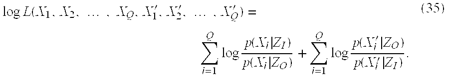
- which is a function only of the zones and not the regions.
- By derivation, the difference between relative entropies can be obtained. Rewriting (35) with the sums over the sets of values the random variables X,X′ may take:

- where the distributions p x(x),px(x′) are those of the random variables with respect the finite set of values they may assume.
- Multiplying each term by a factor of 1:

- and rearranging:

- we obtain the sums and differences of relative entropy:
- logL(X 1 ,X 2 , . . . ,X Q , X 1 ′, X 2 ′, . . . ,X Q′)=Q[D(p x(x)∥p(x|Z O))−D(p x(x)∥p(x|Z 1))+D(p X′(x)∥p(x|Z 1))−D(p X′(x)∥p(x|Z O))] (39)
- The log-likelihood logL(.)is maximized when the distribution of X inside the contour C is most like the distribution x conditioned on being in the zone Z I and simultaneously when the distribution of X′ outside the contour C is most like the distribution X′ conditioned on being in the zone ZO. This achieved for the simpler expression:

- That is, the best contour C* is the one for which the distributions p(X|Z I) and p(X′|ZO) are most different by the relative entropy measure.
- The corresponding maximum contour expressions for the Jensen-Shannon and the Jensen-Renyi divergences are stated by analogy to equation (40). The Jensen-Shannon divergence corresponding to the best contour for the case of two regions is

- where Hs(.) is the Shannon entropy defined in (13). For m regions, or m−1 objects plus the background, the set of best contours C 1* through Cm−1* are those whose collective coefficients maximize the Jensen-Shannon divergence:

- where the π i are the weights on the probability distributions of the m regions. The contour subscripts match the index of the corresponding region.
- The maximum-contour Jensen-Renyi divergence expression can be similarly written. For the case of two regions (R I and RO) to be separated, the Jensen-Renyi divergence is maximized for the boundary contour with parameter vector c* which most perfectly separates the two regions:

- where HR.(.) is the Renyi entropy defined in (24). For m regions, or (m−1) objects plus the background, the set of best contours C 1* through Cm−1* are those whose collective coefficients maximize the Jensen-Renyi divergence:

- where the π l are the weights on the probability distributions of the m regions. The contour subscripts match the index of the corresponding region.
- 4. Autocontouring Program Based on JSD/JRD
- The program using the objective functions (41) or (43) for single object contouring is described in FIG. 5. Multiple object contouring using objective functions ( 42) or (44) is described in FIG. 6. Both the JSD and the JRD can maximized by the same schemes, given in detail here.
- The single contour program is initialized with an image (step 5.1) and an initial boundary estimate, preferably a polygon created either by the operator or by the program itself (step 5.2). The polygon is converted to the initial pixel boundary vector b(0) with B0 pixel-contiguous elements b1 (0), . . . , b2 (0), . . . b80 (0). Each element contains the row and column coordinates of the contour pixel as bj (0)=(row j, column j). From these pixels, the coefficients for the initial parametric contour C(0) are computed (see (8) through (11)).
- Contour determination proceeds through two loops. The outer loop (steps 5.6-5.12) iterates over successive versions of the parametric contour C(0),C(1), . . . , C(i), . . . , stopping when the increases between succeeding values of the JSD (or JRD) fall below a pre-selected, threshold minimum T. So, if
- JS π (i+l) −JS π (i) <T (45)
- or
- JR α π(i+1) −JR α π(i) <T (46)
- then the program reports the contour C (i+1) and stops (step 5.13). As would be appreciated by those of ordinary skill in the art, the choice of a value for T is up to the practitioner of the invention, with smaller values of T providing higher accuracy but requiring more time to compute. The inventor herein has found that a value in the range of 0.001 to 0.1 is preferable for T.
- The inner loop (steps 5.7-5.9) maximizes the JSD (or JRD) over the components of the current contour C(i). This maximization can be done many ways (steepest descent, Quasi-Newton method, conjugate gradient descent, etc.), and preferably maximization is performed using Powell's direction set method or alternatively, the method of conjugate gradient descent with equivalently-accurate results. See Press, W H, Teukolsky, S A, Vettering, W T, and Flannery, B P, Numerical Recipes in C++, Second Edition, Cambridge University Press, Cambridge, UK, 2002; and Nocedal, J. and Wright, S. J., Numerical Optimization , Springer-Verlag, New York, 1999, the entire disclosures of which are incorporated herein by reference. It is worth noting that although the methods' names indicate minimization, minimizing the negative of the function locates the function's maximum.
- Through the argmax function (step 5.7), the inner loop maximizes the JSD (or JRD) by systematically varying the numeric values of the individual parameters c1 (i),c2 (i), . . . ,c4K+2 (i), and noting the resulting values of JSπ (i) or JRα π(i), according to whichever of the maximization methods is used. At each inner-loop substep, trial (*) contours C(i,j)* (i-th iteration contour, j-th component being varied) are computed for each of the several values of the parameter cj (i) (step 5.7). Each parametric contour is converted to its corresponding boundary b(i,j)*, pixels in the zones ZI(i,j)* and ZO (i,j)* are enumerated, the conditional probability distributions p(X|ZI (i,j)*) and p(X|ZO (i,j)*) are computed, and the JSD or JRD entropy terms and divergences JSπ (i,j)* or JRαπ (i,j)* are computed. The largest maximum JSπ (i,j) or JRαπ (i,j)* for all j is relabeled JSπ (i+l) or JRαπ (i+l) and submitted to the outer loop test, and the corresponding contour is saved as C(i+l). While it is preferred that the random variable X used in the program correspond to pixel intensity, other pixel properties can be used.
- The order in which the contour coefficients are adjusted during optimization has an important effect on optimization. Like other Fourier representations, the low number coefficients affect the global properties while the higher coefficients represent the fine details. In the Fourier elliptic representation,
contour coefficients - Referring to FIG. 6, the program which computes multiple contours simultaneously using the objective functions ( 42) or (44) is initialized with an image and m−1 polygons created either by the operator or by the program itself (steps 6.1 and 6.2). There are m regions including the common background region. Maximization of the JSD or the JRD for multiple object contours follows the same scheme as it does for single object contouring. The main difference between the optimization of multiple contours and a single contour is the re-ordering of the multi-contour coefficients for the optimization step.
- The polygons are converted to a set of initial pixel boundary vectors b 1 (0), b2 (0), . . . , bm−1 (0) (step 6.3) from which the initial set of parametric contours C1 (0), C2 (O), . . . , Cm−1 (0) are computed (step 6.4). The subscripts refer to the regions/objects being contoured. The coefficient vectors for each contour are
- C i (0)*=(c i,1 (0) , . . . , c i,4k
i +2 (0)) (47) - where the first subscript for each coefficient identifies the contour and the second subscript identifies the coefficient's order. Also, each contour may have a different number of harmonics K i.
- Like the single contour program, optimization over a contour set has two nested loops. The outer loop (steps 6.6-6.12) iterates over optimized versions of the contour set,

- and the inner loop (steps 6.7-6.9) traverses all the coefficients in all the contours in the set. The convergence test for the outer loop is the same as that for the single contour program: stopping when the increases between successive values of the JSD (JRD) fall below a pre-selected, threshold minimum. So, if
- JS π (i+1) −JS π (i)<threshold (49)
- or
- JR α π(i+1) −JR α π(i)<threshold (50)
- then the program reports the contour set C 1 (i+1) through Cm−1 (i+1) as the matching contour set and stops (steps 6.11, 6.13).
- For each pass of the outer loop, the inner loop maximizes the JSD (JRD) over all the contours' coefficients. The entire set of coefficients, ordered by contour, is
- {c 1 (i) ,c 2 ( i), . . . , c m−1 (i) }={c 1,1 (i) . . . , c 1,4K
1 +2 (i) , c 2,1 (i) . . . , c2,1 (i) . . . ,c 2,4K2 +2) (51) - . . .
- c m−1,1 (i) . . . ,c m−1,4K
m−1 +2 (i)} - where the subscripts are indexes for contour and coefficient, respectively, and there are

- parameters in all contours. Because the Fourier elliptic representation orders the global parameters before the fine detail parameters, it is important to establish all the contours' global locations and sizes before determining the shape details for the contours. Therefore, we re-order the parameters in the set (51) by harmonic order as follows,

- and where the contours have different numbers of coefficients, the order is filled in harmonic order by the remaining coefficients in the largest contour sets. If we re-label the re-ordered contours as singly-indexed parameters ĉ j (i) in the set,
- {c 1 (i) ,c 2 (i) , . . . , c m−1 (i) }={ĉ 1 (i) , . . . ,ĉ j (i) , . . . ,ĉ M
params (i)} (54) - we can describe the inner loop optimization just like that of the single contour program.
- With reference to FIG. 3( c) and as noted above, a multi-processor computer can be used to carry out the contouring program of the present invention. For example, as shown in FIG. 3(c), the image data set can be provided to a plurality of
processors 150 a. For computations that do not depend on each other, increased processing speed can be obtained by splitting a problem into multiple independent parts and running each part on adifferent processor 150 a. This would be especially the case where multiple object contouring is run on m regions in an image data set. Independent functions here include enumerating the region pixels, assembling the pixel feature histograms and distributions, and computing the Jensen or Renyi entropies and their weights. The processing required for each region can be performed on aseparate processor 150 a in parallel. The results of the parallel processing can then be collated in anotherprocessor 150 a that would perform the outer loop test. - 5. Demonstrations
- Two sets of demonstrations are shown in FIGS. 7-10. The first set shows the action of the JRD contouring single figures in a series of random shapes with defined signal-to-noise ratios (SNRs). The second demonstration set shows the action of the JRD to contour anatomic objects in CT images.
- Because the Jensen-Renyi divergence method consistently produced lower errors than the Jensen-Shannon divergence method in tests on random figures (described below) only demonstrations of the Jensen-Renyi method will be shown here.
- In the first demonstration of the action of the JRD, a estimation is performed for the contours in a series of random-shape figures defined by curves C (true)={c1 (true), c2 (true), . . . , c4K+2 (true)} with known, but random, coefficients. This “true” boundary is designed to fill the center of a 256×256 image array, and to have somewhat sharper direction changes than usual in medical anatomy in CT. FIG. 7 shows the process: panel A is a figure template corresponding to a random shape figure, and panel B shows the shape of panel A smoothed with a 3×3 moving window and with Gaussian noise added, to the level of a SNR of about 3.0. Panel C shows a circle that serves as the initial contour estimate, and panel D shows the matching contour (white line) computed by the JRD and overlaid on the figure.
- FIG. 8 shows a series of randomly sized and shaped figures, with the JRD matching contours (white lines) computed from the circle initial estimate, and with SNR of about 3.0.
- To compare the computed estimate contours with the true curves, the error is defined as the average distance between corresponding points in the two curves. The true, defined contour C (true) has a corresponding pixel boundary b(true) which is a vector of B-pixel coordinates as defined b1 (true) through bB (true) where bi (true)=(rowi, columni). The contour estimates C* obtained using the JSD or the JRD produce a trial pixel boundary b* with B* coefficients. To make the two curves b(true) and b* commensurate, a comparison is made between intervals of points on the two curves, selected to sample both curves uniformly. The true boundary is divided into W equal intervals with Ŵ=B/W pixels per interval. For each Ŵ-pixel interval in b(true), a Ŵ-pixel interval in b* is selected, offset by a varying number of pixels in order to find that interval in b* closest to the fixed b(true) interval. The error is the average point-to-point distance for all points in all intervals

- In tests, the b (true)−b* errors associated with the JSD technique, the JRD technique, and the known relative entropy technique consistently follow the order: Error(JRD)<Error(JSD)<25 Error(Relative Entropy). For example, the images in FIGS. 7 and 8 have SNR˜3.5 and for the JSD/JRD contours have distance errors of 4-5 pixels. The relative entropy error distance for the same images is 5-7 pixels. Fuller quantization of these methods performance will follow shortly.
- FIG. 9 shows the action of JRD contouring the liver of a patient in a series of CT sections of the abdomen. The thin lines indicate user-input polygons forming the initial estimates of the contours, and the thick white lines are the JRD matching contours. These matching contours were computed using 6 Fourier harmonics. The maximization is that given in equation (43).
- FIG. 10 shows examples of multiple objects contoured by the JRD, simultaneously in the same picture. The maximization is that given in equation (44).
- 6. Additional Embodiments
- The JSD/JRD image segmentation of the present invention can be based on multiple random variables rather than only a single random variable. JSD/JRD image segmentation can be applied to a random vector of properties, X=(X 1, X1, . . . , Xd) in which each component Xi is a discrete random variable. Better estimates of contours might be obtained by using more information encoded in multiple random variables than using just one random variable. The extent to which several random variables contain more information than any one alone depends on whether they are drawn from independent distributions. Extending the JSD/JRD methods to random vectors requires consideration of two cases: 1) the random variates Xi are conditionally dependent, or 2) the random variates Xi are mutually independent. Dependence is the more general situation, with independence a special case of the conditionally dependent case.
- The probability distribution of a random vector is simply the joint distribution over the separate component random variables (see Grimmett and Stirzaker (1992))

- where d is the dimension of the property vector. The distributions from which the individual variates are drawn may exhibit some inter-dependence, and this is expressed using the conditional probability, defined in ((1), (2)). The joint probability distribution may be written as a product of conditional probabilities, illustrated for these examples:

- The joint probability distribution can be written more compactly as the product formula

- If any one of the individual random variables X i is from a distribution independent of the distributions of other random variables Xj, j≠i, the conditional distribution of Xi relative to other variables is simply equal to its probability, without conditions. That is,
- p(X i | . . . , X j, . . . )=p(X i) (58)
- for all X j, j≠i. In the case where all the random variables are mutually independent, the joint distribution is the product of the individual variates' probabilities,

- If the variates are truly independent, not conditionally dependent, the decision rules for the relative entropy/LRT ((23), (40) 5.12), the JSD ((41), (42) 5.14) and the JRD ((43), (44) 5.15, 5.16) could simply be implemented as d parallel classifications with a rule to combine the separate decisions (see Devroye, 1996).
- The multivariate LRT-equivalent relative energy decision rule from (23) is for the i-th variate
- If D(p x(X i)∥p(X i |H 2))−D(p X(X i)|p(X i |H 1))>T′, decide H 1, else H2. (60)
- and the overall decision for H 1, H2 going to the outcome having the largest number of individual variate-decisions. In the case of even numbers of variates, tie votes would be always given to one outcome, say H1. The multivariate relative entropy contour function (40) is rewritten
- c*=arg max D(p(X|Z 1)∥p(X′|Z 0)) (61)
- where X, X′ are the random property vectors for pixels inside and outside the contour c*. Rewriting the expression for the relative entropy,

- which is the sum of individual-variate relative entropies,

- So the best contour c* is the one for which the distributions p(X i|Zl), p(Xi|Z0) are most different by their contributions to the sum of the individual-variate relative entropies.
- The Jensen-Shannon divergence for multiple independent variates begins with a reconsideration of the equation for estimating a single contour (41) in which the multivariate random vectors X, X′ have been substituted,

- Recall that the Shannon entropy for the distribution of a random variable X is

- where the sum is taken over the domain of values on which X is defined. For the random vector X with independent random variable components, the entropy is

- where the sums are taken over the domains of the respective component random variables. Since the logarithm of a product is equal to the sum of the logs of the factors,

- and since independent variates sum only over their own domains, equation (66) can be rearranged to give

- where the entropy of the random vector is the sum of the independent, component variables' entropies. We can use (67) to rewrite equation (64) as follows,

- That is, the best contour estimate is obtained by the parameter set c for which the expression in the brackets is a maximum.
- By analogy, we can write the equation corresponding to the multi-contour estimate given by equation (42) as

- where the M regions are the background and the interiors for the M−1 contours. Using (67) we obtain an expression in terms of the individual random variables X i

- The Jensen-Renyi divergence has a similar development. Starting with the definition of the Renyi entropy of degree a from (24)

- we substitute the random vector for the scalar random variable to obtain

- the log of a product of sums since the variables X i are independent. This can be simplified to

- where again the inner sum is over the domain of random variable X i. Like the Shannon entropy, the Renyi entropy of a random vector is a sum of entropies of the independent, component random variables.
- Now we consider the single contour JRD decision rule function given in equation (43), here substituted with the random vector X

- and using (73), obtain an expression for the optimal contour on the separate independent variates,

- The JRD applied to M−1 simultaneous contours results when substituting the random vector X into equation (44)

- and using (73) we obtain an expression for the multiple simultaneous contours in terms of the independent variates,

- This is the expression one would use to compute the M−1 contours based on the maximum of the JRD over the d features in each of the M regions.
- In summary, it has been shown here the numerical formulas needed to compute contour estimates for the JSD/JRD methods, on image pixel properties represented as several random variables. The method and the accompanying formula is given in the following table.
Method Equation JSD, single contour 68 JSD, multiple contours 70 JRD, single contour 75 JRD, multiple contours 77 - These are the formulas which, implemented in computer programs, can be used to compute contour estimates using multiple image pixel properties
- The above discussion of contour estimation using multiple random variables assumed that the variables were all statistically independent, or that the joint distribution is the product of the individual variate's unconditioned probabilities (equation (59)). The more general situation is given by equation (57) in which the variates are allowed to be from distributions which may be conditionally dependent. For the two variates X 1 and X2, the joint probability p(X1,X2) is related to the conditional probabilities by definition

- The relative entropy for two distributions of these two random variables, p(X 1,X2) and q(X1,X2) is given by

- which is equivalent to the sum of relative entropies,

- or more compactly,
- D(p(X 1 ,X 2)∥q(X 1 ,X 2))=D(p(X 1 ,X 2)∥q(X 1 ,X 2))=D(p(X 1 |X 2)∥q(X 2)) (81)
- which is equivalent to
- D(p(X 2 ,X 1)∥q(X 2 ,X 1))=D(p(X 2 |X 1)∥q(X 2 |X 1))D(p(X 1)∥q(X 1)) (82)
- Computing these relative entropies can be done by literally using equation (80) to form the sums of ratios across the rows or columns of the joint distributions p(X 1,X2) and q(X1,X2). This is considerably more complicated than computing the sum of relative entropies for the independent variates given in equation (62), and would have to be repeated for each iteration of the search for the contour's coefficients. And the complications multiply as one adds more random variables to the problem. However, given the speed and memory of current computers, such computations are only lengthy, not impossible. The JSD/JRD image segmentation technique of the present invention is also applicable to non-stationary data. “Non-stationary” applies to pixel data in which random variable means, variances, and distributions vary across the image of a single logical object. Classifiers which depend on a fixed mean and variance (e.g., the multivariate Gaussian model described in Section 2) to sort pixels into one or another objects pictured can produce higher error rates for non-stationary image data.
- Since the JSD/JRD methods use functions of the random variables' distributions, it is possible that these methods may be more robust than parametric models since the JSD/JRD functions operate by portioning the data by distribution differences, regardless of any parameters that might be used to describe the distributions. During iterative region contour estimation, non-stationary data would produce small, continuous changes in the region data distributions, and such changes need not increase the error in the JSD/JRD autosegmentation.
- Further still, the JSD/JRD image segmentation discussed above was a region-centered approach in which the decision rules operated on distributions of pixel properties. The teachings of the present invention are also applicable to an edge-centered approach in which the decision rules operate on intensity gradients. Edges represented by coherent, high-valued gradient-image pixels should maximize a decision rule that superimposes the contour C* on gradient maxima pixels. Edges contain the highest values in the gradient image so a rule which seeks to maximize the difference between edge-pixel distributions and all the other pixels should force the contour onto the gradient image maxima.
- Thus, the present invention can be used in combination to (1) partition an object from a background (as previously described) and (2) separate the edge of the object, as observed in an image of the gray level gradient maxima (which is derived from the original image data set) by maximizing the JSD/JRD divergence between the pixels underneath contour C i and all other pixels in the gradient image. The center panel of FIG. 1 illustrates an example of a gradient image. Because the set of pixels coincident with an edge will necessarily have a distribution different from that of the gradient image background plus other object edges, maximizing the divergence over these two sets of gradient image pixels should add to the specificity of the boundary estimate produced in the regional divergence estimation.
- Due to the fact that images are usually never simple, it is highly likely that the regional divergence and the edge divergence would be maximized for different C i contours. In such cases, the “matching contour” can be defined as the contour that jointly maximizes the combined divergences. Because the JSD/JRD divergences take on arbitrary real values determined by the data at hand, the regional and edge divergences for any given region in any given image might have very different values, thereby requiring a weighting to make them equivalent. Such a function would be expressed as:
- F(JSD)=w region JSD region(C′)+W edge JSD edge(C′) (83)
- or
- F(JRD)=W region JRD region(C′)+w edge JRD edge(C′) (84)
- wherein w region and wedge are real number coefficients that can be set to equalize the contributions of the region and edge to the total divergence.
- Further, combining separate JSD/JRD methods for the region pixel data and for edge pixel data could result in more accurate estimates of image object boundaries.
- Also, restrictions on the pixel-classes segmented in the multiple, simultaneous segmentations by the JSD (42) and the JRD (44) might be used to obtain segmentations of like organs along with unlike organs. For instance, the kidneys might share a single distribution, so the rule would differentiate them collectively from the spleen, liver, stomach, or other organs. This is a kind of prior information which could improve the performance any above-described method.
- Moreover, the present invention is also applicable to images having more than 2 dimensions. The present invention should work equally well in 3D as 2D, but would require flexible surfaces or volumes instead of a contour.
- While the present invention has been described above in relation to its preferred embodiment, various modifications may be made thereto that still fall within the invention's scope, as would be recognized by those of ordinary skill in the art. For example, the invention has been described wherein the contouring program adjusts all coefficients of a contour in finding a maximal divergence value. However, given that the very high order coefficients have a relatively insignificant effect on the resulting contour (they add only very fine details), a practitioner of the present invention can modify the contouring program to adjust the numeric values of less than all of a contour's coefficients. While some accuracy would be lost, less processing power would be needed. Such modifications to the invention will be recognizable upon review of the teachings herein. As such, the full scope of the present invention is to be defined solely by the appended claims and their legal equivalents.
Claims (89)
Priority Applications (3)
| Application Number | Priority Date | Filing Date | Title |
|---|---|---|---|
| US10/211,779 US7187800B2 (en) | 2002-08-02 | 2002-08-02 | Method and apparatus for image segmentation using Jensen-Shannon divergence and Jensen-Renyi divergence |
| PCT/US2003/023685 WO2004013811A2 (en) | 2002-08-02 | 2003-07-29 | Image segmentation using jensen-shannon divergence and jensen-renyi divergence |
| AU2003256972A AU2003256972A1 (en) | 2002-08-02 | 2003-07-29 | Image segmentation using jensen-shannon divergence and jensen-renyi divergence |
Applications Claiming Priority (1)
| Application Number | Priority Date | Filing Date | Title |
|---|---|---|---|
| US10/211,779 US7187800B2 (en) | 2002-08-02 | 2002-08-02 | Method and apparatus for image segmentation using Jensen-Shannon divergence and Jensen-Renyi divergence |
Publications (2)
| Publication Number | Publication Date |
|---|---|
| US20040022438A1 true US20040022438A1 (en) | 2004-02-05 |
| US7187800B2 US7187800B2 (en) | 2007-03-06 |
Family
ID=31187654
Family Applications (1)
| Application Number | Title | Priority Date | Filing Date |
|---|---|---|---|
| US10/211,779 Active 2024-05-07 US7187800B2 (en) | 2002-08-02 | 2002-08-02 | Method and apparatus for image segmentation using Jensen-Shannon divergence and Jensen-Renyi divergence |
Country Status (3)
| Country | Link |
|---|---|
| US (1) | US7187800B2 (en) |
| AU (1) | AU2003256972A1 (en) |
| WO (1) | WO2004013811A2 (en) |
Cited By (31)
| Publication number | Priority date | Publication date | Assignee | Title |
|---|---|---|---|---|
| US20060050958A1 (en) * | 2004-09-09 | 2006-03-09 | Kazunori Okada | System and method for volumetric tumor segmentation using joint space-intensity likelihood ratio test |
| US20060072817A1 (en) * | 2004-09-29 | 2006-04-06 | Shih-Jong J. Lee | Method for robust analysis of biological activity in microscopy images |
| US20070003011A1 (en) * | 2004-06-21 | 2007-01-04 | Lane Derek G | Fast gradient-histogram inverse planning technique for radiation treatment |
| US20070147700A1 (en) * | 2005-12-28 | 2007-06-28 | Samsung Electronics Co., Ltd | Method and apparatus for editing images using contour-extracting algorithm |
| US20070153091A1 (en) * | 2005-12-29 | 2007-07-05 | John Watlington | Methods and apparatus for providing privacy in a communication system |
| US20070160274A1 (en) * | 2006-01-10 | 2007-07-12 | Adi Mashiach | System and method for segmenting structures in a series of images |
| US20070230765A1 (en) * | 2006-03-30 | 2007-10-04 | Accuray Incorporated | Delineation on three-dimensional medical image |
| US20070253529A1 (en) * | 2006-04-13 | 2007-11-01 | Seppi Edward J | Systems and methods for digital volumetric laminar tomography |
| US20070263915A1 (en) * | 2006-01-10 | 2007-11-15 | Adi Mashiach | System and method for segmenting structures in a series of images |
| US20080226161A1 (en) * | 2007-03-12 | 2008-09-18 | Jeffrey Kimball Tidd | Determining Edgeless Areas in a Digital Image |
| US20080260229A1 (en) * | 2006-05-25 | 2008-10-23 | Adi Mashiach | System and method for segmenting structures in a series of images using non-iodine based contrast material |
| US20090097704A1 (en) * | 2007-10-10 | 2009-04-16 | Micron Technology, Inc. | On-chip camera system for multiple object tracking and identification |
| US7813581B1 (en) | 2005-05-06 | 2010-10-12 | Fitzpatrick Ben G | Bayesian methods for noise reduction in image processing |
| US7860344B1 (en) | 2005-05-06 | 2010-12-28 | Stochastech Corporation | Tracking apparatus and methods using image processing noise reduction |
| US20110002532A1 (en) * | 2003-06-13 | 2011-01-06 | David Harold Frakes | Data Reconstruction Using Directional Interpolation Techniques |
| WO2011072259A1 (en) * | 2009-12-10 | 2011-06-16 | Indiana University Research & Technology Corporation | System and method for segmentation of three dimensional image data |
| WO2011073990A1 (en) * | 2009-12-17 | 2011-06-23 | Elta Systems Ltd. | Method and system for enhancing an image |
| US20110208058A1 (en) * | 2008-10-28 | 2011-08-25 | Washington University In St. Louis | Applying Renyi Entropy To Detect Changes In Scattering Architecture |
| WO2012035463A1 (en) * | 2010-09-17 | 2012-03-22 | Koninklijke Philips Electronics N.V. | Contour delineation for radiation therapy planning with real-time contour segment impact rendering |
| CN105046712A (en) * | 2015-08-07 | 2015-11-11 | 江西理工大学 | Adaptive Gauss differential evolution based circle detection method |
| US20160123904A1 (en) * | 2014-11-04 | 2016-05-05 | Kabushiki Kaisha Toshiba | Method of, and apparatus for, material classification in multi-energy image data |
| US9630025B2 (en) | 2005-07-25 | 2017-04-25 | Varian Medical Systems International Ag | Methods and apparatus for the planning and delivery of radiation treatments |
| CN107180433A (en) * | 2017-06-06 | 2017-09-19 | 衢州学院 | A kind of local cross-entropy measures the level set image segmentation algorithm of fuzzy C-mean algorithm |
| CN107180442A (en) * | 2017-04-13 | 2017-09-19 | 太原理工大学 | A kind of photoacoustic image based on Renyi entropys rebuilds prefilter |
| USRE46953E1 (en) | 2007-04-20 | 2018-07-17 | University Of Maryland, Baltimore | Single-arc dose painting for precision radiation therapy |
| US10773101B2 (en) | 2010-06-22 | 2020-09-15 | Varian Medical Systems International Ag | System and method for estimating and manipulating estimated radiation dose |
| US10930386B2 (en) | 2018-12-11 | 2021-02-23 | International Business Machines Corporation | Automated normality scoring of echocardiograms |
| US11281993B2 (en) * | 2016-12-05 | 2022-03-22 | Apple Inc. | Model and ensemble compression for metric learning |
| US11864894B2 (en) | 2006-08-09 | 2024-01-09 | Abbott Diabetes Care Inc. | Method and system for providing calibration of an analyte sensor in an analyte monitoring system |
| US11896371B2 (en) | 2012-09-26 | 2024-02-13 | Abbott Diabetes Care Inc. | Method and apparatus for improving lag correction during in vivo measurement of analyte concentration with analyte concentration variability and range data |
| US11957463B2 (en) | 2005-08-31 | 2024-04-16 | Abbott Diabetes Care Inc. | Accuracy of continuous glucose sensors |
Families Citing this family (32)
| Publication number | Priority date | Publication date | Assignee | Title |
|---|---|---|---|---|
| US7466848B2 (en) * | 2002-12-13 | 2008-12-16 | Rutgers, The State University Of New Jersey | Method and apparatus for automatically detecting breast lesions and tumors in images |
| GB2398379A (en) * | 2003-02-11 | 2004-08-18 | Qinetiq Ltd | Automated digital image analysis |
| US20060056701A1 (en) * | 2004-03-02 | 2006-03-16 | Gozde Unal | Joint segmentation and registration of images for object detection |
| SE0400731D0 (en) * | 2004-03-22 | 2004-03-22 | Contextvision Ab | Method, computer program product and apparatus for enhancing a computerized tomography image |
| US7519209B2 (en) * | 2004-06-23 | 2009-04-14 | Vanderbilt University | System and methods of organ segmentation and applications of same |
| US7421415B2 (en) * | 2004-09-07 | 2008-09-02 | Siemens Corporate Research, Inc. | Methods and systems for 3D object detection using learning |
| US7720271B2 (en) * | 2005-04-08 | 2010-05-18 | Siemens Medical Solutions Usa, Inc. | Estimation of solitary pulmonary nodule diameters with reaction-diffusion segmentation |
| US7590273B2 (en) * | 2005-04-08 | 2009-09-15 | Siemens Corporate Research, Inc. | Estimation of solitary pulmonary nodule diameters with a hybrid segmentation approach |
| US7711172B2 (en) * | 2005-04-08 | 2010-05-04 | Siemens Medical Solutions Usa, Inc. | Automatic nodule measurements |
| US7400755B2 (en) * | 2005-06-02 | 2008-07-15 | Accuray Incorporated | Inverse planning using optimization constraints derived from image intensity |
| US7603000B2 (en) * | 2005-08-31 | 2009-10-13 | Siemens Medical Solutions Usa, Inc. | System and method for learning relative distance in a shape space using image based features |
| US8176414B1 (en) * | 2005-09-30 | 2012-05-08 | Google Inc. | Document division method and system |
| US7773807B2 (en) * | 2006-08-29 | 2010-08-10 | Siemens Medical Solutions Usa, Inc. | Seed segmentation using l∞ minimization |
| US7904530B2 (en) * | 2008-01-29 | 2011-03-08 | Palo Alto Research Center Incorporated | Method and apparatus for automatically incorporating hypothetical context information into recommendation queries |
| US7639777B2 (en) * | 2008-02-26 | 2009-12-29 | United Technologies Corp. | Computed tomography systems and related methods involving forward collimation |
| US20090213984A1 (en) * | 2008-02-26 | 2009-08-27 | United Technologies Corp. | Computed Tomography Systems and Related Methods Involving Post-Target Collimation |
| US8238521B2 (en) * | 2008-03-06 | 2012-08-07 | United Technologies Corp. | X-ray collimators, and related systems and methods involving such collimators |
| US20090225954A1 (en) * | 2008-03-06 | 2009-09-10 | United Technologies Corp. | X-Ray Collimators, and Related Systems and Methods Involving Such Collimators |
| US7876875B2 (en) * | 2008-04-09 | 2011-01-25 | United Technologies Corp. | Computed tomography systems and related methods involving multi-target inspection |
| US8718743B2 (en) * | 2008-04-24 | 2014-05-06 | Duke University | Methods for single-pass volumetric bidirectional blood flow imaging spectral domain optical coherence tomography using a modified hilbert transform |
| US7888647B2 (en) * | 2008-04-30 | 2011-02-15 | United Technologies Corp. | X-ray detector assemblies and related computed tomography systems |
| US20090274264A1 (en) * | 2008-04-30 | 2009-11-05 | United Technologies Corp. | Computed Tomography Systems and Related Methods Involving Localized Bias |
| US8133127B1 (en) * | 2008-07-21 | 2012-03-13 | Synder Terrance W | Sports training device and methods of use |
| US8693745B2 (en) * | 2009-05-04 | 2014-04-08 | Duke University | Methods and computer program products for quantitative three-dimensional image correction and clinical parameter computation in optical coherence tomography |
| US8663210B2 (en) | 2009-05-13 | 2014-03-04 | Novian Health, Inc. | Methods and apparatus for performing interstitial laser therapy and interstitial brachytherapy |
| JP2012115658A (en) * | 2010-11-12 | 2012-06-21 | Toshiba Corp | Diagnostic imaging apparatus |
| KR101439412B1 (en) * | 2010-11-22 | 2014-09-11 | 한국전자통신연구원 | Method of segmenting lesions in images |
| JP5565810B2 (en) * | 2010-11-29 | 2014-08-06 | 国立大学法人浜松医科大学 | Mass spectrometry data processing method and apparatus |
| US9279659B2 (en) | 2011-01-21 | 2016-03-08 | Duke University | Systems and methods for complex conjugate artifact resolved optical coherence tomography |
| KR101886333B1 (en) | 2012-06-15 | 2018-08-09 | 삼성전자 주식회사 | Apparatus and method for region growing with multiple cores |
| US9950194B2 (en) | 2014-09-09 | 2018-04-24 | Mevion Medical Systems, Inc. | Patient positioning system |
| WO2018122585A1 (en) * | 2016-12-30 | 2018-07-05 | 同济大学 | Method for urban road traffic incident detecting based on floating-car data |
Citations (98)
| Publication number | Priority date | Publication date | Assignee | Title |
|---|---|---|---|---|
| US4567896A (en) * | 1984-01-20 | 1986-02-04 | Elscint, Inc. | Method and apparatus for calibrating a biopsy attachment for ultrasonic imaging apparatus |
| US4586512A (en) * | 1981-06-26 | 1986-05-06 | Thomson-Csf | Device for localized heating of biological tissues |
| US4751643A (en) * | 1986-08-04 | 1988-06-14 | General Electric Company | Method and apparatus for determining connected substructures within a body |
| US4896673A (en) * | 1988-07-15 | 1990-01-30 | Medstone International, Inc. | Method and apparatus for stone localization using ultrasound imaging |
| US4991579A (en) * | 1987-11-10 | 1991-02-12 | Allen George S | Method and apparatus for providing related images over time of a portion of the anatomy using fiducial implants |
| US4994013A (en) * | 1988-07-28 | 1991-02-19 | Best Industries, Inc. | Pellet for a radioactive seed |
| US5133020A (en) * | 1989-07-21 | 1992-07-21 | Arch Development Corporation | Automated method and system for the detection and classification of abnormal lesions and parenchymal distortions in digital medical images |
| US5178148A (en) * | 1990-04-06 | 1993-01-12 | Technomed International | Method of automatically measuring the volume of a tumor or of a gland, in particular the prostate, a measuring device, and a method and apparatus constituting and application thereof |
| US5185809A (en) * | 1987-08-14 | 1993-02-09 | The General Hospital Corporation | Morphometric analysis of anatomical tomographic data |
| US5187658A (en) * | 1990-01-17 | 1993-02-16 | General Electric Company | System and method for segmenting internal structures contained within the interior region of a solid object |
| US5204625A (en) * | 1990-12-20 | 1993-04-20 | General Electric Company | Segmentation of stationary and vascular surfaces in magnetic resonance imaging |
| US5222499A (en) * | 1989-11-15 | 1993-06-29 | Allen George S | Method and apparatus for imaging the anatomy |
| US5227969A (en) * | 1988-08-01 | 1993-07-13 | W. L. Systems, Inc. | Manipulable three-dimensional projection imaging method |
| US5289374A (en) * | 1992-02-28 | 1994-02-22 | Arch Development Corporation | Method and system for analysis of false positives produced by an automated scheme for the detection of lung nodules in digital chest radiographs |
| US5299253A (en) * | 1992-04-10 | 1994-03-29 | Akzo N.V. | Alignment system to overlay abdominal computer aided tomography and magnetic resonance anatomy with single photon emission tomography |
| US5319549A (en) * | 1992-11-25 | 1994-06-07 | Arch Development Corporation | Method and system for determining geometric pattern features of interstitial infiltrates in chest images |
| US5319551A (en) * | 1989-10-27 | 1994-06-07 | Hitachi, Ltd. | Region extracting method and three-dimensional display method |
| US5389101A (en) * | 1992-04-21 | 1995-02-14 | University Of Utah | Apparatus and method for photogrammetric surgical localization |
| US5391139A (en) * | 1992-09-03 | 1995-02-21 | William Beaumont Hospital | Real time radiation treatment planning system |
| US5391199A (en) * | 1993-07-20 | 1995-02-21 | Biosense, Inc. | Apparatus and method for treating cardiac arrhythmias |
| US5394457A (en) * | 1992-10-08 | 1995-02-28 | Leibinger Gmbh | Device for marking body sites for medical examinations |
| US5398690A (en) * | 1994-08-03 | 1995-03-21 | Batten; Bobby G. | Slaved biopsy device, analysis apparatus, and process |
| US5410617A (en) * | 1991-04-25 | 1995-04-25 | Unisys Corporation | Method for adaptively thresholding grayscale image data |
| US5409006A (en) * | 1992-12-03 | 1995-04-25 | Siemens Aktiengesellschaft | System for the treatment of pathological tissue having a catheter with a marker for avoiding damage to healthy tissue |
| US5412563A (en) * | 1993-09-16 | 1995-05-02 | General Electric Company | Gradient image segmentation method |
| US5411026A (en) * | 1993-10-08 | 1995-05-02 | Nomos Corporation | Method and apparatus for lesion position verification |
| US5433199A (en) * | 1994-02-24 | 1995-07-18 | General Electric Company | Cardiac functional analysis method using gradient image segmentation |
| US5491627A (en) * | 1993-05-13 | 1996-02-13 | Arch Development Corporation | Method and system for the detection of microcalcifications in digital mammograms |
| US5494039A (en) * | 1993-07-16 | 1996-02-27 | Cryomedical Sciences, Inc. | Biopsy needle insertion guide and method of use in prostate cryosurgery |
| US5517602A (en) * | 1992-12-03 | 1996-05-14 | Hewlett-Packard Company | Method and apparatus for generating a topologically consistent visual representation of a three dimensional surface |
| US5526812A (en) * | 1993-06-21 | 1996-06-18 | General Electric Company | Display system for enhancing visualization of body structures during medical procedures |
| US5531227A (en) * | 1994-01-28 | 1996-07-02 | Schneider Medical Technologies, Inc. | Imaging device and method |
| US5531223A (en) * | 1990-02-15 | 1996-07-02 | Kabushiki Kaisha Toshiba | Method and apparatus of nuclear magnetic resonance imaging with nonlinearly processed image display |
| US5537485A (en) * | 1992-07-21 | 1996-07-16 | Arch Development Corporation | Method for computer-aided detection of clustered microcalcifications from digital mammograms |
| US5603318A (en) * | 1992-04-21 | 1997-02-18 | University Of Utah Research Foundation | Apparatus and method for photogrammetric surgical localization |
| US5622170A (en) * | 1990-10-19 | 1997-04-22 | Image Guided Technologies, Inc. | Apparatus for determining the position and orientation of an invasive portion of a probe inside a three-dimensional body |
| US5624382A (en) * | 1992-03-10 | 1997-04-29 | Siemens Aktiengesellschaft | Method and apparatus for ultrasound tissue therapy |
| US5638819A (en) * | 1995-08-29 | 1997-06-17 | Manwaring; Kim H. | Method and apparatus for guiding an instrument to a target |
| US5640496A (en) * | 1991-02-04 | 1997-06-17 | Medical Instrumentation And Diagnostics Corp. (Midco) | Method and apparatus for management of image data by linked lists of pixel values |
| US5709206A (en) * | 1995-11-27 | 1998-01-20 | Teboul; Michel | Imaging system for breast sonography |
| US5715836A (en) * | 1993-02-16 | 1998-02-10 | Kliegis; Ulrich | Method and apparatus for planning and monitoring a surgical operation |
| US5727538A (en) * | 1996-04-05 | 1998-03-17 | Shawn Ellis | Electronically actuated marking pellet projector |
| US5734739A (en) * | 1994-05-31 | 1998-03-31 | University Of Washington | Method for determining the contour of an in vivo organ using multiple image frames of the organ |
| US5740225A (en) * | 1995-12-07 | 1998-04-14 | Kabushiki Kaisha Toshiba | Radiation therapy planning method and its system and apparatus |
| US5742263A (en) * | 1995-12-18 | 1998-04-21 | Telxon Corporation | Head tracking system for a head mounted display system |
| US5749362A (en) * | 1992-05-27 | 1998-05-12 | International Business Machines Corporation | Method of creating an image of an anatomical feature where the feature is within a patient's body |
| US5765561A (en) * | 1994-10-07 | 1998-06-16 | Medical Media Systems | Video-based surgical targeting system |
| US5769074A (en) * | 1994-10-13 | 1998-06-23 | Horus Therapeutics, Inc. | Computer assisted methods for diagnosing diseases |
| US5776063A (en) * | 1996-09-30 | 1998-07-07 | Molecular Biosystems, Inc. | Analysis of ultrasound images in the presence of contrast agent |
| US5778043A (en) * | 1996-09-20 | 1998-07-07 | Cosman; Eric R. | Radiation beam control system |
| US5859891A (en) * | 1997-03-07 | 1999-01-12 | Hibbard; Lyn | Autosegmentation/autocontouring system and method for use with three-dimensional radiation therapy treatment planning |
| US5860909A (en) * | 1996-10-18 | 1999-01-19 | Mick Radio Nuclear Instruments, Inc. | Seed applicator for use in radiation therapy |
| US5868757A (en) * | 1994-11-16 | 1999-02-09 | Pgk, Enterprises, Inc. | Method and apparatus for interstitial radiation of the prostate gland |
| US5868673A (en) * | 1995-03-28 | 1999-02-09 | Sonometrics Corporation | System for carrying out surgery, biopsy and ablation of a tumor or other physical anomaly |
| US5871448A (en) * | 1997-10-14 | 1999-02-16 | Real World Design And Development Co. | Stepper apparatus for use in the imaging/treatment of internal organs using an ultrasound probe |
| US5891034A (en) * | 1990-10-19 | 1999-04-06 | St. Louis University | System for indicating the position of a surgical probe within a head on an image of the head |
| US5906574A (en) * | 1995-10-06 | 1999-05-25 | Kan; William C. | Apparatus for vacuum-assisted handling and loading of radioactive seeds and spacers into implant needles within an enclosed visible radiation shield for use in therapeutic radioactive seed implantation |
| US5928130A (en) * | 1998-03-16 | 1999-07-27 | Schmidt; Bruno | Apparatus and method for implanting radioactive seeds in tissue |
| US6025128A (en) * | 1994-09-29 | 2000-02-15 | The University Of Tulsa | Prediction of prostate cancer progression by analysis of selected predictive parameters |
| US6027446A (en) * | 1998-01-12 | 2000-02-22 | Washington Univ. Of Office Of Technology Transfer | Pubic arch detection and interference assessment in transrectal ultrasound guided prostate cancer therapy |
| US6033357A (en) * | 1997-03-28 | 2000-03-07 | Navius Corporation | Intravascular radiation delivery device |
| US6036632A (en) * | 1998-05-28 | 2000-03-14 | Barzell-Whitmore Maroon Bells, Inc. | Sterile disposable template grid system |
| US6038467A (en) * | 1997-01-24 | 2000-03-14 | U.S. Philips Corporation | Image display system and image guided surgery system |
| US6048312A (en) * | 1998-04-23 | 2000-04-11 | Ishrak; Syed Omar | Method and apparatus for three-dimensional ultrasound imaging of biopsy needle |
| US6083166A (en) * | 1997-12-02 | 2000-07-04 | Situs Corporation | Method and apparatus for determining a measure of tissue manipulation |
| US6083167A (en) * | 1998-02-10 | 2000-07-04 | Emory University | Systems and methods for providing radiation therapy and catheter guides |
| US6179768B1 (en) * | 1996-07-08 | 2001-01-30 | Delft Instruments Intellectual Property B.V. | Capsule for use in brachytherapy and a combination of a capsule for brachytherapy and a guidewire |
| US6196963B1 (en) * | 1999-03-02 | 2001-03-06 | Medtronic Ave, Inc. | Brachytherapy device assembly and method of use |
| US6196964B1 (en) * | 1996-07-05 | 2001-03-06 | Delft Instruments Intellectual Property B.V. | Combination of a capsule for brachytherapy and a guidewire |
| US6206832B1 (en) * | 1996-11-29 | 2001-03-27 | Life Imaging Systems | Apparatus for guiding medical instruments during ultrasonographic imaging |
| US6208883B1 (en) * | 1995-07-26 | 2001-03-27 | Associates Of The Joint Center For Radiation Therapy, Inc. | Ultrasound localization and image fusion for the treatment of prostate cancer |
| US6210315B1 (en) * | 1998-07-20 | 2001-04-03 | Cook Urological Inc. | Brachytherapy device including an anti-static handle |
| US6213932B1 (en) * | 1997-12-12 | 2001-04-10 | Bruno Schmidt | Interstitial brachytherapy device and method |
| US6213110B1 (en) * | 1999-12-16 | 2001-04-10 | Odyssey Paintball Products, Inc. | Rapid feed paintball loader |
| US6226543B1 (en) * | 1998-09-24 | 2001-05-01 | Super Dimension Ltd. | System and method of recording and displaying in context of an image a location of at least one point-of-interest in a body during an intra-body medical procedure |
| US6236875B1 (en) * | 1994-10-07 | 2001-05-22 | Surgical Navigation Technologies | Surgical navigation systems including reference and localization frames |
| US6238342B1 (en) * | 1998-05-26 | 2001-05-29 | Riverside Research Institute | Ultrasonic tissue-type classification and imaging methods and apparatus |
| US6241670B1 (en) * | 1997-07-02 | 2001-06-05 | Kabushiki Kaisha Toshiba | Radiotherapy system |
| US6256529B1 (en) * | 1995-07-26 | 2001-07-03 | Burdette Medical Systems, Inc. | Virtual reality 3D visualization for surgical procedures |
| US6261219B1 (en) * | 1998-05-04 | 2001-07-17 | Novoste Corporation | Intraluminal radiation treatment system |
| US6266453B1 (en) * | 1999-07-26 | 2001-07-24 | Computerized Medical Systems, Inc. | Automated image fusion/alignment system and method |
| US6354989B1 (en) * | 1998-10-14 | 2002-03-12 | Terumo Kabushiki Kaisha | Radiation source delivery wire and catheter assembly for radiation therapy provided with the same |
| US6358195B1 (en) * | 2000-03-09 | 2002-03-19 | Neoseed Technology Llc | Method and apparatus for loading radioactive seeds into brachytherapy needles |
| US6361487B1 (en) * | 2000-03-09 | 2002-03-26 | Neoseed Technology Llc | Method and apparatus for brachytherapy treatment of prostate disease |
| US6366796B1 (en) * | 1998-10-23 | 2002-04-02 | Philips Medical Systems (Cleveland), Inc. | Method and apparatus for planning brachytherapy surgical procedures |
| US6379302B1 (en) * | 1999-10-28 | 2002-04-30 | Surgical Navigation Technologies Inc. | Navigation information overlay onto ultrasound imagery |
| US6387035B1 (en) * | 1997-03-28 | 2002-05-14 | Jomed, Inc. | Catheter with swivel tip |
| US6387034B1 (en) * | 1998-08-17 | 2002-05-14 | Georia Tech Research Corporation | Brachytherapy treatment planning method and apparatus |
| US20020077546A1 (en) * | 2000-10-17 | 2002-06-20 | Bernd Aldefeld | Method for intravascular localization and imaging without X-rays |
| US20020087080A1 (en) * | 2000-12-28 | 2002-07-04 | Slayton Michael H. | Visual imaging system for ultrasonic probe |
| US6416492B1 (en) * | 2000-09-28 | 2002-07-09 | Scimed Life Systems, Inc. | Radiation delivery system utilizing intravascular ultrasound |
| US6423009B1 (en) * | 1996-11-29 | 2002-07-23 | Life Imaging Systems, Inc. | System, employing three-dimensional ultrasonographic imaging, for assisting in guiding and placing medical instruments |
| US6425865B1 (en) * | 1998-06-12 | 2002-07-30 | The University Of British Columbia | Robotically assisted medical ultrasound |
| US6512942B1 (en) * | 1997-11-24 | 2003-01-28 | Computerized Medical Systems, Inc. | Radiation therapy and real time imaging of a patient treatment region |
| US20030074011A1 (en) * | 1998-09-24 | 2003-04-17 | Super Dimension Ltd. | System and method of recording and displaying in context of an image a location of at least one point-of-interest in a body during an intra-body medical procedure |
| US6572525B1 (en) * | 2000-05-26 | 2003-06-03 | Lisa Yoshizumi | Needle having an aperture for detecting seeds or spacers loaded therein and colored seeds or spacers |
| US20030132947A1 (en) * | 2002-01-14 | 2003-07-17 | Huitao Luo | Systems and methods for processing boundary information of a graphical object |
| US6766036B1 (en) * | 1999-07-08 | 2004-07-20 | Timothy R. Pryor | Camera based man machine interfaces |
Family Cites Families (40)
| Publication number | Priority date | Publication date | Assignee | Title |
|---|---|---|---|---|
| GB1574823A (en) | 1976-03-27 | 1980-09-10 | Emi Ltd | Video display arrangements |
| US4764971A (en) | 1985-11-25 | 1988-08-16 | Eastman Kodak Company | Image processing method including image segmentation |
| US4791567A (en) | 1986-09-15 | 1988-12-13 | General Electric Company | Three dimensional connectivity system employing an equivalence schema for determining connected substructures within a body |
| JPH0738681B2 (en) | 1987-03-20 | 1995-04-26 | 富士ゼロックス株式会社 | Area recognition device |
| US4961425A (en) | 1987-08-14 | 1990-10-09 | Massachusetts Institute Of Technology | Morphometric analysis of anatomical tomographic data |
| FR2639238B1 (en) | 1988-11-21 | 1991-02-22 | Technomed Int Sa | APPARATUS FOR SURGICAL TREATMENT OF TISSUES BY HYPERTHERMIA, PREFERABLY THE PROSTATE, COMPRISING MEANS OF THERMAL PROTECTION COMPRISING PREFERABLY RADIOREFLECTIVE SCREEN MEANS |
| US5072384A (en) | 1988-11-23 | 1991-12-10 | Arch Development Corp. | Method and system for automated computerized analysis of sizes of hearts and lungs in digital chest radiographs |
| US5205289A (en) | 1988-12-23 | 1993-04-27 | Medical Instrumentation And Diagnostics Corporation | Three-dimensional computer graphics simulation and computerized numerical optimization for dose delivery and treatment planning |
| FR2660732B1 (en) | 1990-04-06 | 1992-09-04 | Technomed Int Sa | TRANSLATABLE END ARM AND THERAPEUTIC TREATMENT APPARATUS, INCLUDING APPLICATION. |
| US5457754A (en) | 1990-08-02 | 1995-10-10 | University Of Cincinnati | Method for automatic contour extraction of a cardiac image |
| EP1690511B1 (en) | 1990-10-19 | 2010-07-14 | St. Louis University | Surgical probe locating system for head use |
| US5166876A (en) | 1991-01-16 | 1992-11-24 | General Electric Company | System and method for detecting internal structures contained within the interior region of a solid object |
| US5662111A (en) | 1991-01-28 | 1997-09-02 | Cosman; Eric R. | Process of stereotactic optical navigation |
| US5210799A (en) | 1991-03-28 | 1993-05-11 | Texas Instruments Incorporated | System and method for ranking and extracting salient contours for target recognition |
| DE69224679T2 (en) | 1991-05-27 | 1998-11-12 | Hitachi Ltd | Method and device for processing multidimensional data |
| US5239591A (en) | 1991-07-03 | 1993-08-24 | U.S. Philips Corp. | Contour extraction in multi-phase, multi-slice cardiac mri studies by propagation of seed contours between images |
| US5260871A (en) | 1991-07-31 | 1993-11-09 | Mayo Foundation For Medical Education And Research | Method and apparatus for diagnosis of breast tumors |
| US5242373A (en) | 1991-09-17 | 1993-09-07 | Scott Walter P | Medical seed implantation instrument |
| US5371810A (en) | 1991-09-27 | 1994-12-06 | E. I. Du Pont De Nemours And Company | Method of determining the interior points of an object in a background |
| US5574799A (en) | 1992-06-12 | 1996-11-12 | The Johns Hopkins University | Method and system for automated detection of microcalcification clusters in mammograms |
| FR2694881B1 (en) | 1992-07-31 | 1996-09-06 | Univ Joseph Fourier | METHOD FOR DETERMINING THE POSITION OF AN ORGAN. |
| CA2110148C (en) | 1992-12-24 | 1999-10-05 | Aaron Fenster | Three-dimensional ultrasound imaging system |
| US5787886A (en) | 1993-03-19 | 1998-08-04 | Compass International Incorporated | Magnetic field digitizer for stereotatic surgery |
| EP0649117A3 (en) | 1993-10-15 | 1996-01-31 | George S Allen | Method for providing medical images. |
| US5452367A (en) | 1993-11-29 | 1995-09-19 | Arch Development Corporation | Automated method and system for the segmentation of medical images |
| US5460592A (en) | 1994-01-24 | 1995-10-24 | Amersham Holdings, Inc. | Apparatus and method for making carrier assembly for radioactive seed carrier |
| US5458126A (en) | 1994-02-24 | 1995-10-17 | General Electric Company | Cardiac functional analysis system employing gradient image segmentation |
| US5570430A (en) | 1994-05-31 | 1996-10-29 | University Of Washington | Method for determining the contour of an in vivo organ using multiple image frames of the organ |
| US5672172A (en) | 1994-06-23 | 1997-09-30 | Vros Corporation | Surgical instrument with ultrasound pulse generator |
| US5583659A (en) | 1994-11-10 | 1996-12-10 | Eastman Kodak Company | Multi-windowing technique for thresholding an image using local image properties |
| US5797849A (en) | 1995-03-28 | 1998-08-25 | Sonometrics Corporation | Method for carrying out a medical procedure using a three-dimensional tracking and imaging system |
| US5817022A (en) | 1995-03-28 | 1998-10-06 | Sonometrics Corporation | System for displaying a 2-D ultrasound image within a 3-D viewing environment |
| US5660185A (en) | 1995-04-13 | 1997-08-26 | Neovision Corporation | Image-guided biopsy apparatus with enhanced imaging and methods |
| US5833627A (en) | 1995-04-13 | 1998-11-10 | United States Surgical Corporation | Image-guided biopsy apparatus and methods of use |
| WO1997000649A1 (en) | 1995-06-20 | 1997-01-09 | Wan Sing Ng | Articulated arm for medical procedures |
| US5682886A (en) | 1995-12-26 | 1997-11-04 | Musculographics Inc | Computer-assisted surgical system |
| US5830145A (en) | 1996-09-20 | 1998-11-03 | Cardiovascular Imaging Systems, Inc. | Enhanced accuracy of three-dimensional intraluminal ultrasound (ILUS) image reconstruction |
| US5669382A (en) | 1996-11-19 | 1997-09-23 | General Electric Company | System for measuring myocardium in cardiac images |
| US5931786A (en) | 1997-06-13 | 1999-08-03 | Barzell Whitmore Maroon Bells, Inc. | Ultrasound probe support and stepping device |
| US5938583A (en) | 1997-12-29 | 1999-08-17 | Grimm; Peter D. | Precision implant needle and method of using same in seed implant treatment of prostate cancer |
-
2002
- 2002-08-02 US US10/211,779 patent/US7187800B2/en active Active
-
2003
- 2003-07-29 WO PCT/US2003/023685 patent/WO2004013811A2/en not_active Application Discontinuation
- 2003-07-29 AU AU2003256972A patent/AU2003256972A1/en not_active Abandoned
Patent Citations (99)
| Publication number | Priority date | Publication date | Assignee | Title |
|---|---|---|---|---|
| US4586512A (en) * | 1981-06-26 | 1986-05-06 | Thomson-Csf | Device for localized heating of biological tissues |
| US4567896A (en) * | 1984-01-20 | 1986-02-04 | Elscint, Inc. | Method and apparatus for calibrating a biopsy attachment for ultrasonic imaging apparatus |
| US4751643A (en) * | 1986-08-04 | 1988-06-14 | General Electric Company | Method and apparatus for determining connected substructures within a body |
| US5185809A (en) * | 1987-08-14 | 1993-02-09 | The General Hospital Corporation | Morphometric analysis of anatomical tomographic data |
| US4991579A (en) * | 1987-11-10 | 1991-02-12 | Allen George S | Method and apparatus for providing related images over time of a portion of the anatomy using fiducial implants |
| US4896673A (en) * | 1988-07-15 | 1990-01-30 | Medstone International, Inc. | Method and apparatus for stone localization using ultrasound imaging |
| US4994013A (en) * | 1988-07-28 | 1991-02-19 | Best Industries, Inc. | Pellet for a radioactive seed |
| US5227969A (en) * | 1988-08-01 | 1993-07-13 | W. L. Systems, Inc. | Manipulable three-dimensional projection imaging method |
| US5133020A (en) * | 1989-07-21 | 1992-07-21 | Arch Development Corporation | Automated method and system for the detection and classification of abnormal lesions and parenchymal distortions in digital medical images |
| US5319551A (en) * | 1989-10-27 | 1994-06-07 | Hitachi, Ltd. | Region extracting method and three-dimensional display method |
| US5222499A (en) * | 1989-11-15 | 1993-06-29 | Allen George S | Method and apparatus for imaging the anatomy |
| US5187658A (en) * | 1990-01-17 | 1993-02-16 | General Electric Company | System and method for segmenting internal structures contained within the interior region of a solid object |
| US5531223A (en) * | 1990-02-15 | 1996-07-02 | Kabushiki Kaisha Toshiba | Method and apparatus of nuclear magnetic resonance imaging with nonlinearly processed image display |
| US5178148A (en) * | 1990-04-06 | 1993-01-12 | Technomed International | Method of automatically measuring the volume of a tumor or of a gland, in particular the prostate, a measuring device, and a method and apparatus constituting and application thereof |
| US5622170A (en) * | 1990-10-19 | 1997-04-22 | Image Guided Technologies, Inc. | Apparatus for determining the position and orientation of an invasive portion of a probe inside a three-dimensional body |
| US5891034A (en) * | 1990-10-19 | 1999-04-06 | St. Louis University | System for indicating the position of a surgical probe within a head on an image of the head |
| US5204625A (en) * | 1990-12-20 | 1993-04-20 | General Electric Company | Segmentation of stationary and vascular surfaces in magnetic resonance imaging |
| US5640496A (en) * | 1991-02-04 | 1997-06-17 | Medical Instrumentation And Diagnostics Corp. (Midco) | Method and apparatus for management of image data by linked lists of pixel values |
| US5410617A (en) * | 1991-04-25 | 1995-04-25 | Unisys Corporation | Method for adaptively thresholding grayscale image data |
| US5289374A (en) * | 1992-02-28 | 1994-02-22 | Arch Development Corporation | Method and system for analysis of false positives produced by an automated scheme for the detection of lung nodules in digital chest radiographs |
| US5624382A (en) * | 1992-03-10 | 1997-04-29 | Siemens Aktiengesellschaft | Method and apparatus for ultrasound tissue therapy |
| US5299253A (en) * | 1992-04-10 | 1994-03-29 | Akzo N.V. | Alignment system to overlay abdominal computer aided tomography and magnetic resonance anatomy with single photon emission tomography |
| US5603318A (en) * | 1992-04-21 | 1997-02-18 | University Of Utah Research Foundation | Apparatus and method for photogrammetric surgical localization |
| US5389101A (en) * | 1992-04-21 | 1995-02-14 | University Of Utah | Apparatus and method for photogrammetric surgical localization |
| US5749362A (en) * | 1992-05-27 | 1998-05-12 | International Business Machines Corporation | Method of creating an image of an anatomical feature where the feature is within a patient's body |
| US5537485A (en) * | 1992-07-21 | 1996-07-16 | Arch Development Corporation | Method for computer-aided detection of clustered microcalcifications from digital mammograms |
| US5391139A (en) * | 1992-09-03 | 1995-02-21 | William Beaumont Hospital | Real time radiation treatment planning system |
| US5394457A (en) * | 1992-10-08 | 1995-02-28 | Leibinger Gmbh | Device for marking body sites for medical examinations |
| US5319549A (en) * | 1992-11-25 | 1994-06-07 | Arch Development Corporation | Method and system for determining geometric pattern features of interstitial infiltrates in chest images |
| US5517602A (en) * | 1992-12-03 | 1996-05-14 | Hewlett-Packard Company | Method and apparatus for generating a topologically consistent visual representation of a three dimensional surface |
| US5409006A (en) * | 1992-12-03 | 1995-04-25 | Siemens Aktiengesellschaft | System for the treatment of pathological tissue having a catheter with a marker for avoiding damage to healthy tissue |
| US5715836A (en) * | 1993-02-16 | 1998-02-10 | Kliegis; Ulrich | Method and apparatus for planning and monitoring a surgical operation |
| US5491627A (en) * | 1993-05-13 | 1996-02-13 | Arch Development Corporation | Method and system for the detection of microcalcifications in digital mammograms |
| US5526812A (en) * | 1993-06-21 | 1996-06-18 | General Electric Company | Display system for enhancing visualization of body structures during medical procedures |
| US5494039A (en) * | 1993-07-16 | 1996-02-27 | Cryomedical Sciences, Inc. | Biopsy needle insertion guide and method of use in prostate cryosurgery |
| US5391199A (en) * | 1993-07-20 | 1995-02-21 | Biosense, Inc. | Apparatus and method for treating cardiac arrhythmias |
| US5412563A (en) * | 1993-09-16 | 1995-05-02 | General Electric Company | Gradient image segmentation method |
| US5411026A (en) * | 1993-10-08 | 1995-05-02 | Nomos Corporation | Method and apparatus for lesion position verification |
| US5531227A (en) * | 1994-01-28 | 1996-07-02 | Schneider Medical Technologies, Inc. | Imaging device and method |
| US5433199A (en) * | 1994-02-24 | 1995-07-18 | General Electric Company | Cardiac functional analysis method using gradient image segmentation |
| US5734739A (en) * | 1994-05-31 | 1998-03-31 | University Of Washington | Method for determining the contour of an in vivo organ using multiple image frames of the organ |
| US5398690A (en) * | 1994-08-03 | 1995-03-21 | Batten; Bobby G. | Slaved biopsy device, analysis apparatus, and process |
| US6025128A (en) * | 1994-09-29 | 2000-02-15 | The University Of Tulsa | Prediction of prostate cancer progression by analysis of selected predictive parameters |
| US6236875B1 (en) * | 1994-10-07 | 2001-05-22 | Surgical Navigation Technologies | Surgical navigation systems including reference and localization frames |
| US5765561A (en) * | 1994-10-07 | 1998-06-16 | Medical Media Systems | Video-based surgical targeting system |
| US5769074A (en) * | 1994-10-13 | 1998-06-23 | Horus Therapeutics, Inc. | Computer assisted methods for diagnosing diseases |
| US5868757A (en) * | 1994-11-16 | 1999-02-09 | Pgk, Enterprises, Inc. | Method and apparatus for interstitial radiation of the prostate gland |
| US5868673A (en) * | 1995-03-28 | 1999-02-09 | Sonometrics Corporation | System for carrying out surgery, biopsy and ablation of a tumor or other physical anomaly |
| US6208883B1 (en) * | 1995-07-26 | 2001-03-27 | Associates Of The Joint Center For Radiation Therapy, Inc. | Ultrasound localization and image fusion for the treatment of prostate cancer |
| US6256529B1 (en) * | 1995-07-26 | 2001-07-03 | Burdette Medical Systems, Inc. | Virtual reality 3D visualization for surgical procedures |
| US5638819A (en) * | 1995-08-29 | 1997-06-17 | Manwaring; Kim H. | Method and apparatus for guiding an instrument to a target |
| US5906574A (en) * | 1995-10-06 | 1999-05-25 | Kan; William C. | Apparatus for vacuum-assisted handling and loading of radioactive seeds and spacers into implant needles within an enclosed visible radiation shield for use in therapeutic radioactive seed implantation |
| US5709206A (en) * | 1995-11-27 | 1998-01-20 | Teboul; Michel | Imaging system for breast sonography |
| US5740225A (en) * | 1995-12-07 | 1998-04-14 | Kabushiki Kaisha Toshiba | Radiation therapy planning method and its system and apparatus |
| US5742263A (en) * | 1995-12-18 | 1998-04-21 | Telxon Corporation | Head tracking system for a head mounted display system |
| US5727538A (en) * | 1996-04-05 | 1998-03-17 | Shawn Ellis | Electronically actuated marking pellet projector |
| US6196964B1 (en) * | 1996-07-05 | 2001-03-06 | Delft Instruments Intellectual Property B.V. | Combination of a capsule for brachytherapy and a guidewire |
| US6179768B1 (en) * | 1996-07-08 | 2001-01-30 | Delft Instruments Intellectual Property B.V. | Capsule for use in brachytherapy and a combination of a capsule for brachytherapy and a guidewire |
| US5778043A (en) * | 1996-09-20 | 1998-07-07 | Cosman; Eric R. | Radiation beam control system |
| US5776063A (en) * | 1996-09-30 | 1998-07-07 | Molecular Biosystems, Inc. | Analysis of ultrasound images in the presence of contrast agent |
| US5860909A (en) * | 1996-10-18 | 1999-01-19 | Mick Radio Nuclear Instruments, Inc. | Seed applicator for use in radiation therapy |
| US6206832B1 (en) * | 1996-11-29 | 2001-03-27 | Life Imaging Systems | Apparatus for guiding medical instruments during ultrasonographic imaging |
| US6423009B1 (en) * | 1996-11-29 | 2002-07-23 | Life Imaging Systems, Inc. | System, employing three-dimensional ultrasonographic imaging, for assisting in guiding and placing medical instruments |
| US6038467A (en) * | 1997-01-24 | 2000-03-14 | U.S. Philips Corporation | Image display system and image guided surgery system |
| US5859891A (en) * | 1997-03-07 | 1999-01-12 | Hibbard; Lyn | Autosegmentation/autocontouring system and method for use with three-dimensional radiation therapy treatment planning |
| US6249594B1 (en) * | 1997-03-07 | 2001-06-19 | Computerized Medical Systems, Inc. | Autosegmentation/autocontouring system and method |
| US6387035B1 (en) * | 1997-03-28 | 2002-05-14 | Jomed, Inc. | Catheter with swivel tip |
| US6033357A (en) * | 1997-03-28 | 2000-03-07 | Navius Corporation | Intravascular radiation delivery device |
| US6241670B1 (en) * | 1997-07-02 | 2001-06-05 | Kabushiki Kaisha Toshiba | Radiotherapy system |
| US5871448A (en) * | 1997-10-14 | 1999-02-16 | Real World Design And Development Co. | Stepper apparatus for use in the imaging/treatment of internal organs using an ultrasound probe |
| US6512942B1 (en) * | 1997-11-24 | 2003-01-28 | Computerized Medical Systems, Inc. | Radiation therapy and real time imaging of a patient treatment region |
| US6083166A (en) * | 1997-12-02 | 2000-07-04 | Situs Corporation | Method and apparatus for determining a measure of tissue manipulation |
| US6213932B1 (en) * | 1997-12-12 | 2001-04-10 | Bruno Schmidt | Interstitial brachytherapy device and method |
| US6027446A (en) * | 1998-01-12 | 2000-02-22 | Washington Univ. Of Office Of Technology Transfer | Pubic arch detection and interference assessment in transrectal ultrasound guided prostate cancer therapy |
| US6083167A (en) * | 1998-02-10 | 2000-07-04 | Emory University | Systems and methods for providing radiation therapy and catheter guides |
| US5928130A (en) * | 1998-03-16 | 1999-07-27 | Schmidt; Bruno | Apparatus and method for implanting radioactive seeds in tissue |
| US6048312A (en) * | 1998-04-23 | 2000-04-11 | Ishrak; Syed Omar | Method and apparatus for three-dimensional ultrasound imaging of biopsy needle |
| US6261219B1 (en) * | 1998-05-04 | 2001-07-17 | Novoste Corporation | Intraluminal radiation treatment system |
| US6238342B1 (en) * | 1998-05-26 | 2001-05-29 | Riverside Research Institute | Ultrasonic tissue-type classification and imaging methods and apparatus |
| US6036632A (en) * | 1998-05-28 | 2000-03-14 | Barzell-Whitmore Maroon Bells, Inc. | Sterile disposable template grid system |
| US6425865B1 (en) * | 1998-06-12 | 2002-07-30 | The University Of British Columbia | Robotically assisted medical ultrasound |
| US6210315B1 (en) * | 1998-07-20 | 2001-04-03 | Cook Urological Inc. | Brachytherapy device including an anti-static handle |
| US6387034B1 (en) * | 1998-08-17 | 2002-05-14 | Georia Tech Research Corporation | Brachytherapy treatment planning method and apparatus |
| US20030074011A1 (en) * | 1998-09-24 | 2003-04-17 | Super Dimension Ltd. | System and method of recording and displaying in context of an image a location of at least one point-of-interest in a body during an intra-body medical procedure |
| US6226543B1 (en) * | 1998-09-24 | 2001-05-01 | Super Dimension Ltd. | System and method of recording and displaying in context of an image a location of at least one point-of-interest in a body during an intra-body medical procedure |
| US6354989B1 (en) * | 1998-10-14 | 2002-03-12 | Terumo Kabushiki Kaisha | Radiation source delivery wire and catheter assembly for radiation therapy provided with the same |
| US6366796B1 (en) * | 1998-10-23 | 2002-04-02 | Philips Medical Systems (Cleveland), Inc. | Method and apparatus for planning brachytherapy surgical procedures |
| US6196963B1 (en) * | 1999-03-02 | 2001-03-06 | Medtronic Ave, Inc. | Brachytherapy device assembly and method of use |
| US6766036B1 (en) * | 1999-07-08 | 2004-07-20 | Timothy R. Pryor | Camera based man machine interfaces |
| US6266453B1 (en) * | 1999-07-26 | 2001-07-24 | Computerized Medical Systems, Inc. | Automated image fusion/alignment system and method |
| US6379302B1 (en) * | 1999-10-28 | 2002-04-30 | Surgical Navigation Technologies Inc. | Navigation information overlay onto ultrasound imagery |
| US6213110B1 (en) * | 1999-12-16 | 2001-04-10 | Odyssey Paintball Products, Inc. | Rapid feed paintball loader |
| US6361487B1 (en) * | 2000-03-09 | 2002-03-26 | Neoseed Technology Llc | Method and apparatus for brachytherapy treatment of prostate disease |
| US6358195B1 (en) * | 2000-03-09 | 2002-03-19 | Neoseed Technology Llc | Method and apparatus for loading radioactive seeds into brachytherapy needles |
| US6572525B1 (en) * | 2000-05-26 | 2003-06-03 | Lisa Yoshizumi | Needle having an aperture for detecting seeds or spacers loaded therein and colored seeds or spacers |
| US6416492B1 (en) * | 2000-09-28 | 2002-07-09 | Scimed Life Systems, Inc. | Radiation delivery system utilizing intravascular ultrasound |
| US20020077546A1 (en) * | 2000-10-17 | 2002-06-20 | Bernd Aldefeld | Method for intravascular localization and imaging without X-rays |
| US20020087080A1 (en) * | 2000-12-28 | 2002-07-04 | Slayton Michael H. | Visual imaging system for ultrasonic probe |
| US20030132947A1 (en) * | 2002-01-14 | 2003-07-17 | Huitao Luo | Systems and methods for processing boundary information of a graphical object |
Cited By (58)
| Publication number | Priority date | Publication date | Assignee | Title |
|---|---|---|---|---|
| US8233701B2 (en) * | 2003-06-13 | 2012-07-31 | David Frakes | Data reconstruction using directional interpolation techniques |
| US20110002532A1 (en) * | 2003-06-13 | 2011-01-06 | David Harold Frakes | Data Reconstruction Using Directional Interpolation Techniques |
| US20070003011A1 (en) * | 2004-06-21 | 2007-01-04 | Lane Derek G | Fast gradient-histogram inverse planning technique for radiation treatment |
| US7369645B2 (en) * | 2004-06-21 | 2008-05-06 | Derek Graham Lane | Information theoretic inverse planning technique for radiation treatment |
| US20060050958A1 (en) * | 2004-09-09 | 2006-03-09 | Kazunori Okada | System and method for volumetric tumor segmentation using joint space-intensity likelihood ratio test |
| US7430321B2 (en) * | 2004-09-09 | 2008-09-30 | Siemens Medical Solutions Usa, Inc. | System and method for volumetric tumor segmentation using joint space-intensity likelihood ratio test |
| US7697755B2 (en) * | 2004-09-29 | 2010-04-13 | Drvision Technologies Llc | Method for robust analysis of biological activity in microscopy images |
| US20060072817A1 (en) * | 2004-09-29 | 2006-04-06 | Shih-Jong J. Lee | Method for robust analysis of biological activity in microscopy images |
| US7813581B1 (en) | 2005-05-06 | 2010-10-12 | Fitzpatrick Ben G | Bayesian methods for noise reduction in image processing |
| US7860344B1 (en) | 2005-05-06 | 2010-12-28 | Stochastech Corporation | Tracking apparatus and methods using image processing noise reduction |
| US9764159B2 (en) | 2005-07-25 | 2017-09-19 | Varian Medical Systems International Ag | Methods and apparatus for the planning and delivery of radiation treatments |
| US9687677B2 (en) | 2005-07-25 | 2017-06-27 | Varian Medical Systems International Ag | Methods and apparatus for the planning and delivery of radiation treatments |
| US9687674B2 (en) | 2005-07-25 | 2017-06-27 | Varian Medical Systems International Ag | Methods and apparatus for the planning and delivery of radiation treatments |
| US9788783B2 (en) | 2005-07-25 | 2017-10-17 | Varian Medical Systems International Ag | Methods and apparatus for the planning and delivery of radiation treatments |
| US9687675B2 (en) | 2005-07-25 | 2017-06-27 | Varian Medical Systems International Ag | Methods and apparatus for the planning and delivery of radiation treatments |
| US9687676B2 (en) | 2005-07-25 | 2017-06-27 | Varian Medical Systems International Ag | Methods and apparatus for the planning and delivery of radiation treatments |
| US9630025B2 (en) | 2005-07-25 | 2017-04-25 | Varian Medical Systems International Ag | Methods and apparatus for the planning and delivery of radiation treatments |
| US11642027B2 (en) | 2005-07-25 | 2023-05-09 | Siemens Healthineers International Ag | Methods and apparatus for the planning and delivery of radiation treatments |
| US10595774B2 (en) | 2005-07-25 | 2020-03-24 | Varian Medical Systems International | Methods and apparatus for the planning and delivery of radiation treatments |
| US9687673B2 (en) | 2005-07-25 | 2017-06-27 | Varian Medical Systems International Ag | Methods and apparatus for the planning and delivery of radiation treatments |
| US9687678B2 (en) | 2005-07-25 | 2017-06-27 | Varian Medical Systems International Ag | Methods and apparatus for the planning and delivery of radiation treatments |
| US11957463B2 (en) | 2005-08-31 | 2024-04-16 | Abbott Diabetes Care Inc. | Accuracy of continuous glucose sensors |
| US20070147700A1 (en) * | 2005-12-28 | 2007-06-28 | Samsung Electronics Co., Ltd | Method and apparatus for editing images using contour-extracting algorithm |
| US20070153091A1 (en) * | 2005-12-29 | 2007-07-05 | John Watlington | Methods and apparatus for providing privacy in a communication system |
| WO2007080580A3 (en) * | 2006-01-10 | 2009-04-16 | Innovea Medical Ltd | System and method for segmenting structures in a series of images |
| US20070263915A1 (en) * | 2006-01-10 | 2007-11-15 | Adi Mashiach | System and method for segmenting structures in a series of images |
| WO2007080580A2 (en) * | 2006-01-10 | 2007-07-19 | Innovea Medical Ltd. | System and method for segmenting structures in a series of images |
| US20070160274A1 (en) * | 2006-01-10 | 2007-07-12 | Adi Mashiach | System and method for segmenting structures in a series of images |
| US7907772B2 (en) | 2006-03-30 | 2011-03-15 | Accuray Incorporated | Delineation on three-dimensional medical image |
| WO2007126842A3 (en) * | 2006-03-30 | 2008-04-24 | Accuray Inc | Delineation on three-dimensional medical image |
| WO2007126842A2 (en) * | 2006-03-30 | 2007-11-08 | Accuray Incorporated | Delineation on three-dimensional medical image |
| US20070230765A1 (en) * | 2006-03-30 | 2007-10-04 | Accuray Incorporated | Delineation on three-dimensional medical image |
| US7737972B2 (en) * | 2006-04-13 | 2010-06-15 | Varian Medical Systems, Inc. | Systems and methods for digital volumetric laminar tomography |
| US20070253529A1 (en) * | 2006-04-13 | 2007-11-01 | Seppi Edward J | Systems and methods for digital volumetric laminar tomography |
| US20080260229A1 (en) * | 2006-05-25 | 2008-10-23 | Adi Mashiach | System and method for segmenting structures in a series of images using non-iodine based contrast material |
| US11864894B2 (en) | 2006-08-09 | 2024-01-09 | Abbott Diabetes Care Inc. | Method and system for providing calibration of an analyte sensor in an analyte monitoring system |
| US20080226161A1 (en) * | 2007-03-12 | 2008-09-18 | Jeffrey Kimball Tidd | Determining Edgeless Areas in a Digital Image |
| US7929762B2 (en) * | 2007-03-12 | 2011-04-19 | Jeffrey Kimball Tidd | Determining edgeless areas in a digital image |
| USRE46953E1 (en) | 2007-04-20 | 2018-07-17 | University Of Maryland, Baltimore | Single-arc dose painting for precision radiation therapy |
| US20090097704A1 (en) * | 2007-10-10 | 2009-04-16 | Micron Technology, Inc. | On-chip camera system for multiple object tracking and identification |
| US8500644B2 (en) * | 2008-10-28 | 2013-08-06 | Washington University | Applying renyi entropy to detect changes in scattering architecture |
| US20110208058A1 (en) * | 2008-10-28 | 2011-08-25 | Washington University In St. Louis | Applying Renyi Entropy To Detect Changes In Scattering Architecture |
| WO2011072259A1 (en) * | 2009-12-10 | 2011-06-16 | Indiana University Research & Technology Corporation | System and method for segmentation of three dimensional image data |
| US20120243777A1 (en) * | 2009-12-10 | 2012-09-27 | Indiana University Research And Technology Corporation | System and method for segmentation of three-dimensional image data |
| US8897544B2 (en) * | 2009-12-10 | 2014-11-25 | Indiana University Research And Technology Corp. | System and method for segmentation of three-dimensional image data |
| US8724918B2 (en) | 2009-12-17 | 2014-05-13 | Elta Systems Ltd. | Method and system for enhancing an image |
| WO2011073990A1 (en) * | 2009-12-17 | 2011-06-23 | Elta Systems Ltd. | Method and system for enhancing an image |
| US10773101B2 (en) | 2010-06-22 | 2020-09-15 | Varian Medical Systems International Ag | System and method for estimating and manipulating estimated radiation dose |
| US9020234B2 (en) | 2010-09-17 | 2015-04-28 | Koninklijke Philips N.V. | Contour delineation for radiation therapy planning with real-time contour segment impact rendering |
| WO2012035463A1 (en) * | 2010-09-17 | 2012-03-22 | Koninklijke Philips Electronics N.V. | Contour delineation for radiation therapy planning with real-time contour segment impact rendering |
| US11896371B2 (en) | 2012-09-26 | 2024-02-13 | Abbott Diabetes Care Inc. | Method and apparatus for improving lag correction during in vivo measurement of analyte concentration with analyte concentration variability and range data |
| US9964499B2 (en) * | 2014-11-04 | 2018-05-08 | Toshiba Medical Systems Corporation | Method of, and apparatus for, material classification in multi-energy image data |
| US20160123904A1 (en) * | 2014-11-04 | 2016-05-05 | Kabushiki Kaisha Toshiba | Method of, and apparatus for, material classification in multi-energy image data |
| CN105046712A (en) * | 2015-08-07 | 2015-11-11 | 江西理工大学 | Adaptive Gauss differential evolution based circle detection method |
| US11281993B2 (en) * | 2016-12-05 | 2022-03-22 | Apple Inc. | Model and ensemble compression for metric learning |
| CN107180442A (en) * | 2017-04-13 | 2017-09-19 | 太原理工大学 | A kind of photoacoustic image based on Renyi entropys rebuilds prefilter |
| CN107180433A (en) * | 2017-06-06 | 2017-09-19 | 衢州学院 | A kind of local cross-entropy measures the level set image segmentation algorithm of fuzzy C-mean algorithm |
| US10930386B2 (en) | 2018-12-11 | 2021-02-23 | International Business Machines Corporation | Automated normality scoring of echocardiograms |
Also Published As
| Publication number | Publication date |
|---|---|
| AU2003256972A1 (en) | 2004-02-23 |
| US7187800B2 (en) | 2007-03-06 |
| WO2004013811A3 (en) | 2004-09-16 |
| WO2004013811A2 (en) | 2004-02-12 |
| AU2003256972A8 (en) | 2004-02-23 |
Similar Documents
| Publication | Publication Date | Title |
|---|---|---|
| US7187800B2 (en) | Method and apparatus for image segmentation using Jensen-Shannon divergence and Jensen-Renyi divergence | |
| US9262827B2 (en) | Lung, lobe, and fissure imaging systems and methods | |
| US7783092B2 (en) | Method for enhancing diagnostic images using vessel reconstruction | |
| US6249594B1 (en) | Autosegmentation/autocontouring system and method | |
| US7916917B2 (en) | Method of segmenting anatomic entities in digital medical images | |
| US10121243B2 (en) | Advanced computer-aided diagnosis of lung nodules | |
| CN111105424A (en) | Lymph node automatic delineation method and device | |
| Martın-Fernández et al. | An approach for contour detection of human kidneys from ultrasound images using Markov random fields and active contours | |
| US20070223815A1 (en) | Feature Weighted Medical Object Contouring Using Distance Coordinates | |
| US8170308B2 (en) | Error adaptive functional imaging | |
| US8165359B2 (en) | Method of constructing gray value or geometric models of anatomic entity in medical image | |
| Priyadarsini et al. | Survey on segmentation of liver from CT images | |
| US9245208B2 (en) | Patient modeling from multispectral input image volumes | |
| Ghosh et al. | Chaotic firefly algorithm-based fuzzy C-means algorithm for segmentation of brain tissues in magnetic resonance images | |
| US7630548B2 (en) | Image segmentation using isoperimetric trees | |
| US20210220670A1 (en) | Systems and Methods for Accelerated Online Adaptive Radiation Therapy | |
| Alves et al. | Extracting lungs from ct images using fully convolutional networks | |
| Soleymanifard et al. | Segmentation of whole tumor using localized active contour and trained neural network in boundaries | |
| Archip et al. | Ultrasound image segmentation using spectral clustering | |
| Hibbard | Region segmentation using information divergence measures | |
| Sreeja et al. | Pseudo Computed Tomography Image Generation from Brain Magnetic Resonance Image for Radiation Therapy Treatment Planning Using DCNN-UNET | |
| El-Torky et al. | Brain tumor segmentation using wavelet Multi-resolution expectation maximization algorithm | |
| Moradi et al. | Integration of Dynamic Multi-Atlas and Deep Learning Techniques to Improve Segmentation of the Prostate in MR Images | |
| Al-Waeli | An Automated System for the Classification and Segmentation of Brain Tumours in Mri Images Based on the Modified Grey Level Co-Occurrence Matrix | |
| de Bruijne et al. | Active shape model segmentation using a non-linear appearance model: application to 3D AAA segmentation |
Legal Events
| Date | Code | Title | Description |
|---|---|---|---|
| AS | Assignment |
Owner name: COMPUTERIZED MEDICAL SYSTEMS, INC., MISSOURI Free format text: ASSIGNMENT OF ASSIGNORS INTEREST;ASSIGNOR:HIBBARD, LYNDON S.;REEL/FRAME:013170/0761 Effective date: 20020802 |
|
| AS | Assignment |
Owner name: USB CAPITAL FUNDING CORP., MISSOURI Free format text: SECURITY AGREEMENT;ASSIGNOR:COMPUTERIZED MEDICAL SYSTEMS, INC.;REEL/FRAME:017400/0161 Effective date: 20060328 Owner name: U.S. BANK NATIONAL ASSOCIATION, MISSOURI Free format text: SECURITY AGREEMENT;ASSIGNOR:COMPUTERIZED MEDICAL SYSTEMS, INC.;REEL/FRAME:017400/0082 Effective date: 20060328 |
|
| STCF | Information on status: patent grant |
Free format text: PATENTED CASE |
|
| AS | Assignment |
Owner name: COMPUTERIZED MEDICAL SYSTEMS, INC., MISSOURI Free format text: RELEASE BY SECURED PARTY;ASSIGNOR:U.S. BANK, N.A.;REEL/FRAME:020617/0273 Effective date: 20080304 Owner name: COMPUTERIZED MEDICAL SYSTEMS, INC., MISSOURI Free format text: RELEASE BY SECURED PARTY;ASSIGNOR:USB CAPITAL RESOURCES, INC., AS SUCCESSOR IN INTEREST TO USB CAPITAL FUNDING CORP., F/K/A WISCONSIN CAPITAL CORPORATION;REEL/FRAME:020617/0139 Effective date: 20080304 |
|
| FEPP | Fee payment procedure |
Free format text: PAT HOLDER NO LONGER CLAIMS SMALL ENTITY STATUS, ENTITY STATUS SET TO UNDISCOUNTED (ORIGINAL EVENT CODE: STOL); ENTITY STATUS OF PATENT OWNER: LARGE ENTITY |
|
| FPAY | Fee payment |
Year of fee payment: 4 |
|
| AS | Assignment |
Owner name: IMPAC MEDICAL SYSTEMS, INC., CALIFORNIA Free format text: MERGER;ASSIGNOR:CMSI HOLDINGS CORP.;REEL/FRAME:029977/0053 Effective date: 20120430 Owner name: CMSI HOLDINGS CORP., MISSOURI Free format text: MERGER;ASSIGNOR:COMPUTERIZED MEDICAL SYSTEMS, INC.;REEL/FRAME:029974/0710 Effective date: 20120430 |
|
| FPAY | Fee payment |
Year of fee payment: 8 |
|
| MAFP | Maintenance fee payment |
Free format text: PAYMENT OF MAINTENANCE FEE, 12TH YEAR, LARGE ENTITY (ORIGINAL EVENT CODE: M1553); ENTITY STATUS OF PATENT OWNER: LARGE ENTITY Year of fee payment: 12 |