US20040054278A1 - Ingestible pill - Google Patents
Ingestible pill Download PDFInfo
- Publication number
- US20040054278A1 US20040054278A1 US10/616,301 US61630103A US2004054278A1 US 20040054278 A1 US20040054278 A1 US 20040054278A1 US 61630103 A US61630103 A US 61630103A US 2004054278 A1 US2004054278 A1 US 2004054278A1
- Authority
- US
- United States
- Prior art keywords
- ingestible device
- probe
- radiation
- nuclear
- tissue
- Prior art date
- Legal status (The legal status is an assumption and is not a legal conclusion. Google has not performed a legal analysis and makes no representation as to the accuracy of the status listed.)
- Granted
Links
- 239000006187 pill Substances 0.000 title description 11
- 210000001035 gastrointestinal tract Anatomy 0.000 claims abstract description 155
- 238000003384 imaging method Methods 0.000 claims abstract description 65
- 238000012544 monitoring process Methods 0.000 claims abstract description 9
- 239000000523 sample Substances 0.000 claims description 192
- 230000005855 radiation Effects 0.000 claims description 126
- 238000000034 method Methods 0.000 claims description 112
- 238000004891 communication Methods 0.000 claims description 29
- 238000004422 calculation algorithm Methods 0.000 claims description 7
- 238000012633 nuclear imaging Methods 0.000 claims 1
- 238000004364 calculation method Methods 0.000 abstract description 5
- 210000001519 tissue Anatomy 0.000 description 131
- 206010028980 Neoplasm Diseases 0.000 description 53
- 230000006870 function Effects 0.000 description 36
- 238000002604 ultrasonography Methods 0.000 description 34
- 238000003745 diagnosis Methods 0.000 description 30
- 230000003287 optical effect Effects 0.000 description 29
- 239000013078 crystal Substances 0.000 description 28
- 239000002775 capsule Substances 0.000 description 27
- 230000001575 pathological effect Effects 0.000 description 27
- 238000001931 thermography Methods 0.000 description 26
- 230000001133 acceleration Effects 0.000 description 24
- 230000007170 pathology Effects 0.000 description 24
- 238000012216 screening Methods 0.000 description 23
- 230000005291 magnetic effect Effects 0.000 description 20
- 238000002595 magnetic resonance imaging Methods 0.000 description 20
- 239000012217 radiopharmaceutical Substances 0.000 description 20
- 229940121896 radiopharmaceutical Drugs 0.000 description 20
- 230000002799 radiopharmaceutical effect Effects 0.000 description 20
- 238000010521 absorption reaction Methods 0.000 description 19
- 230000037406 food intake Effects 0.000 description 19
- 210000000813 small intestine Anatomy 0.000 description 19
- UJKPHYRXOLRVJJ-MLSVHJFASA-N CC(O)C1=C(C)/C2=C/C3=N/C(=C\C4=C(CCC(O)=O)C(C)=C(N4)/C=C4\N=C(\C=C\1/N\2)C(C)=C4C(C)O)/C(CCC(O)=O)=C3C Chemical class CC(O)C1=C(C)/C2=C/C3=N/C(=C\C4=C(CCC(O)=O)C(C)=C(N4)/C=C4\N=C(\C=C\1/N\2)C(C)=C4C(C)O)/C(CCC(O)=O)=C3C UJKPHYRXOLRVJJ-MLSVHJFASA-N 0.000 description 16
- 239000007788 liquid Substances 0.000 description 16
- 230000007423 decrease Effects 0.000 description 15
- 238000001514 detection method Methods 0.000 description 15
- 230000003902 lesion Effects 0.000 description 15
- 230000000694 effects Effects 0.000 description 13
- 230000002496 gastric effect Effects 0.000 description 13
- 230000001613 neoplastic effect Effects 0.000 description 13
- 230000008569 process Effects 0.000 description 13
- 201000011510 cancer Diseases 0.000 description 12
- 239000003814 drug Substances 0.000 description 12
- 210000001072 colon Anatomy 0.000 description 11
- 229960003569 hematoporphyrin Drugs 0.000 description 11
- 230000008859 change Effects 0.000 description 10
- -1 krypton ion Chemical class 0.000 description 10
- 230000036962 time dependent Effects 0.000 description 10
- 210000000481 breast Anatomy 0.000 description 9
- 239000002872 contrast media Substances 0.000 description 9
- 230000033001 locomotion Effects 0.000 description 9
- 206010009944 Colon cancer Diseases 0.000 description 8
- 230000002494 anti-cea effect Effects 0.000 description 8
- 210000000265 leukocyte Anatomy 0.000 description 8
- 230000035945 sensitivity Effects 0.000 description 8
- 208000001333 Colorectal Neoplasms Diseases 0.000 description 7
- 241001465754 Metazoa Species 0.000 description 7
- 239000011248 coating agent Substances 0.000 description 7
- 238000000576 coating method Methods 0.000 description 7
- 238000001727 in vivo Methods 0.000 description 7
- 239000003550 marker Substances 0.000 description 7
- 239000000463 material Substances 0.000 description 7
- 210000002784 stomach Anatomy 0.000 description 7
- 229910004611 CdZnTe Inorganic materials 0.000 description 6
- 206010061218 Inflammation Diseases 0.000 description 6
- 230000002159 abnormal effect Effects 0.000 description 6
- 238000012937 correction Methods 0.000 description 6
- 239000000975 dye Substances 0.000 description 6
- 208000015181 infectious disease Diseases 0.000 description 6
- 230000004054 inflammatory process Effects 0.000 description 6
- 238000001228 spectrum Methods 0.000 description 6
- GKLVYJBZJHMRIY-OUBTZVSYSA-N Technetium-99 Chemical compound [99Tc] GKLVYJBZJHMRIY-OUBTZVSYSA-N 0.000 description 5
- 238000004458 analytical method Methods 0.000 description 5
- 238000013459 approach Methods 0.000 description 5
- 208000037265 diseases, disorders, signs and symptoms Diseases 0.000 description 5
- 239000007789 gas Substances 0.000 description 5
- 230000001976 improved effect Effects 0.000 description 5
- 230000006872 improvement Effects 0.000 description 5
- 230000000155 isotopic effect Effects 0.000 description 5
- 238000002372 labelling Methods 0.000 description 5
- 238000005259 measurement Methods 0.000 description 5
- 210000000664 rectum Anatomy 0.000 description 5
- 230000004044 response Effects 0.000 description 5
- 238000005070 sampling Methods 0.000 description 5
- ZCYVEMRRCGMTRW-UHFFFAOYSA-N 7553-56-2 Chemical compound [I] ZCYVEMRRCGMTRW-UHFFFAOYSA-N 0.000 description 4
- 206010006187 Breast cancer Diseases 0.000 description 4
- 208000026310 Breast neoplasm Diseases 0.000 description 4
- 208000032124 Squamous Intraepithelial Lesions Diseases 0.000 description 4
- SQVRNKJHWKZAKO-UHFFFAOYSA-N beta-N-Acetyl-D-neuraminic acid Natural products CC(=O)NC1C(O)CC(O)(C(O)=O)OC1C(O)C(O)CO SQVRNKJHWKZAKO-UHFFFAOYSA-N 0.000 description 4
- 210000004369 blood Anatomy 0.000 description 4
- 239000008280 blood Substances 0.000 description 4
- 201000010989 colorectal carcinoma Diseases 0.000 description 4
- 230000000875 corresponding effect Effects 0.000 description 4
- 238000013500 data storage Methods 0.000 description 4
- 238000011161 development Methods 0.000 description 4
- 201000010099 disease Diseases 0.000 description 4
- 150000002148 esters Chemical class 0.000 description 4
- 238000002189 fluorescence spectrum Methods 0.000 description 4
- 238000005286 illumination Methods 0.000 description 4
- 229940027941 immunoglobulin g Drugs 0.000 description 4
- APFVFJFRJDLVQX-AHCXROLUSA-N indium-111 Chemical compound [111In] APFVFJFRJDLVQX-AHCXROLUSA-N 0.000 description 4
- 229910052740 iodine Inorganic materials 0.000 description 4
- 239000011630 iodine Substances 0.000 description 4
- 230000004807 localization Effects 0.000 description 4
- 230000003211 malignant effect Effects 0.000 description 4
- 230000007246 mechanism Effects 0.000 description 4
- 238000009206 nuclear medicine Methods 0.000 description 4
- 210000002307 prostate Anatomy 0.000 description 4
- 230000002285 radioactive effect Effects 0.000 description 4
- SQVRNKJHWKZAKO-OQPLDHBCSA-N sialic acid Chemical compound CC(=O)N[C@@H]1[C@@H](O)C[C@@](O)(C(O)=O)OC1[C@H](O)[C@H](O)CO SQVRNKJHWKZAKO-OQPLDHBCSA-N 0.000 description 4
- 210000004872 soft tissue Anatomy 0.000 description 4
- 238000001356 surgical procedure Methods 0.000 description 4
- 230000007704 transition Effects 0.000 description 4
- 230000000007 visual effect Effects 0.000 description 4
- MARUHZGHZWCEQU-UHFFFAOYSA-N 5-phenyl-2h-tetrazole Chemical compound C1=CC=CC=C1C1=NNN=N1 MARUHZGHZWCEQU-UHFFFAOYSA-N 0.000 description 3
- KRKNYBCHXYNGOX-UHFFFAOYSA-K Citrate Chemical compound [O-]C(=O)CC(O)(CC([O-])=O)C([O-])=O KRKNYBCHXYNGOX-UHFFFAOYSA-K 0.000 description 3
- GYHNNYVSQQEPJS-OIOBTWANSA-N Gallium-67 Chemical compound [67Ga] GYHNNYVSQQEPJS-OIOBTWANSA-N 0.000 description 3
- 206010017964 Gastrointestinal infection Diseases 0.000 description 3
- 230000003321 amplification Effects 0.000 description 3
- 210000001367 artery Anatomy 0.000 description 3
- 239000012141 concentrate Substances 0.000 description 3
- 230000001419 dependent effect Effects 0.000 description 3
- 238000002405 diagnostic procedure Methods 0.000 description 3
- 238000003748 differential diagnosis Methods 0.000 description 3
- 238000009826 distribution Methods 0.000 description 3
- 230000005284 excitation Effects 0.000 description 3
- 230000001747 exhibiting effect Effects 0.000 description 3
- 238000001506 fluorescence spectroscopy Methods 0.000 description 3
- 238000002847 impedance measurement Methods 0.000 description 3
- 150000002605 large molecules Chemical class 0.000 description 3
- 229920002521 macromolecule Polymers 0.000 description 3
- 238000004519 manufacturing process Methods 0.000 description 3
- 239000004005 microsphere Substances 0.000 description 3
- 238000003199 nucleic acid amplification method Methods 0.000 description 3
- 239000002245 particle Substances 0.000 description 3
- IEQIEDJGQAUEQZ-UHFFFAOYSA-N phthalocyanine Chemical class N1C(N=C2C3=CC=CC=C3C(N=C3C4=CC=CC=C4C(=N4)N3)=N2)=C(C=CC=C2)C2=C1N=C1C2=CC=CC=C2C4=N1 IEQIEDJGQAUEQZ-UHFFFAOYSA-N 0.000 description 3
- 238000012545 processing Methods 0.000 description 3
- PUZPDOWCWNUUKD-ULWFUOSBSA-M sodium;fluorine-18(1-) Chemical compound [18F-].[Na+] PUZPDOWCWNUUKD-ULWFUOSBSA-M 0.000 description 3
- 230000003595 spectral effect Effects 0.000 description 3
- 238000004611 spectroscopical analysis Methods 0.000 description 3
- 239000000126 substance Substances 0.000 description 3
- 101150047061 tag-72 gene Proteins 0.000 description 3
- 229910052724 xenon Inorganic materials 0.000 description 3
- ZMEWRPBAQVSBBB-GOTSBHOMSA-N (2s)-2-[[(2s)-2-[(2-aminoacetyl)amino]-3-(4-hydroxyphenyl)propanoyl]amino]-6-[[2-[2-[2-[bis(carboxymethyl)amino]ethyl-(carboxymethyl)amino]ethyl-(carboxymethyl)amino]acetyl]amino]hexanoic acid Chemical compound OC(=O)CN(CC(O)=O)CCN(CC(O)=O)CCN(CC(O)=O)CC(=O)NCCCC[C@@H](C(O)=O)NC(=O)[C@@H](NC(=O)CN)CC1=CC=C(O)C=C1 ZMEWRPBAQVSBBB-GOTSBHOMSA-N 0.000 description 2
- 206010009900 Colitis ulcerative Diseases 0.000 description 2
- 208000011231 Crohn disease Diseases 0.000 description 2
- 206010012742 Diarrhoea infectious Diseases 0.000 description 2
- BPNZYADGDZPRTK-UDUYQYQQSA-N Exametazime Chemical compound O/N=C(\C)[C@@H](C)NCC(C)(C)CN[C@H](C)C(\C)=N\O BPNZYADGDZPRTK-UDUYQYQQSA-N 0.000 description 2
- 102000009123 Fibrin Human genes 0.000 description 2
- 108010073385 Fibrin Proteins 0.000 description 2
- BWGVNKXGVNDBDI-UHFFFAOYSA-N Fibrin monomer Chemical compound CNC(=O)CNC(=O)CN BWGVNKXGVNDBDI-UHFFFAOYSA-N 0.000 description 2
- 206010064147 Gastrointestinal inflammation Diseases 0.000 description 2
- 108010010803 Gelatin Proteins 0.000 description 2
- 102000003886 Glycoproteins Human genes 0.000 description 2
- 108090000288 Glycoproteins Proteins 0.000 description 2
- 102000008394 Immunoglobulin Fragments Human genes 0.000 description 2
- 108010021625 Immunoglobulin Fragments Proteins 0.000 description 2
- 102000004318 Matrilysin Human genes 0.000 description 2
- 108090000855 Matrilysin Proteins 0.000 description 2
- 201000006704 Ulcerative Colitis Diseases 0.000 description 2
- 230000000259 anti-tumor effect Effects 0.000 description 2
- 239000000427 antigen Substances 0.000 description 2
- 102000036639 antigens Human genes 0.000 description 2
- 108091007433 antigens Proteins 0.000 description 2
- 238000003491 array Methods 0.000 description 2
- 230000004888 barrier function Effects 0.000 description 2
- 230000008901 benefit Effects 0.000 description 2
- 230000004071 biological effect Effects 0.000 description 2
- 238000001574 biopsy Methods 0.000 description 2
- QWUZMTJBRUASOW-UHFFFAOYSA-N cadmium tellanylidenezinc Chemical compound [Zn].[Cd].[Te] QWUZMTJBRUASOW-UHFFFAOYSA-N 0.000 description 2
- 208000035269 cancer or benign tumor Diseases 0.000 description 2
- 238000006243 chemical reaction Methods 0.000 description 2
- 208000037976 chronic inflammation Diseases 0.000 description 2
- 230000006020 chronic inflammation Effects 0.000 description 2
- 238000004140 cleaning Methods 0.000 description 2
- 230000001276 controlling effect Effects 0.000 description 2
- 231100000433 cytotoxic Toxicity 0.000 description 2
- 230000001472 cytotoxic effect Effects 0.000 description 2
- 230000005516 deep trap Effects 0.000 description 2
- 230000004069 differentiation Effects 0.000 description 2
- 230000001079 digestive effect Effects 0.000 description 2
- 229940015493 dihematoporphyrin ether Drugs 0.000 description 2
- 150000002016 disaccharides Chemical class 0.000 description 2
- 208000001848 dysentery Diseases 0.000 description 2
- 230000005670 electromagnetic radiation Effects 0.000 description 2
- 230000008378 epithelial damage Effects 0.000 description 2
- 238000011156 evaluation Methods 0.000 description 2
- 230000005281 excited state Effects 0.000 description 2
- 229950003499 fibrin Drugs 0.000 description 2
- 239000012530 fluid Substances 0.000 description 2
- 238000002073 fluorescence micrograph Methods 0.000 description 2
- 229920000159 gelatin Polymers 0.000 description 2
- 239000008273 gelatin Substances 0.000 description 2
- 235000019322 gelatine Nutrition 0.000 description 2
- 235000011852 gelatine desserts Nutrition 0.000 description 2
- 238000002991 immunohistochemical analysis Methods 0.000 description 2
- 238000000338 in vitro Methods 0.000 description 2
- 238000011503 in vivo imaging Methods 0.000 description 2
- 229910052738 indium Inorganic materials 0.000 description 2
- APFVFJFRJDLVQX-UHFFFAOYSA-N indium atom Chemical compound [In] APFVFJFRJDLVQX-UHFFFAOYSA-N 0.000 description 2
- 230000001939 inductive effect Effects 0.000 description 2
- 238000002347 injection Methods 0.000 description 2
- 239000007924 injection Substances 0.000 description 2
- 210000000936 intestine Anatomy 0.000 description 2
- 230000001678 irradiating effect Effects 0.000 description 2
- 229910052743 krypton Inorganic materials 0.000 description 2
- 150000002632 lipids Chemical class 0.000 description 2
- 239000004973 liquid crystal related substance Substances 0.000 description 2
- 210000003141 lower extremity Anatomy 0.000 description 2
- 238000009607 mammography Methods 0.000 description 2
- 239000011159 matrix material Substances 0.000 description 2
- 201000001441 melanoma Diseases 0.000 description 2
- 108020004999 messenger RNA Proteins 0.000 description 2
- 229910052751 metal Inorganic materials 0.000 description 2
- 239000002184 metal Substances 0.000 description 2
- 239000000203 mixture Substances 0.000 description 2
- 238000012986 modification Methods 0.000 description 2
- 230000004048 modification Effects 0.000 description 2
- 231100000252 nontoxic Toxicity 0.000 description 2
- 230000003000 nontoxic effect Effects 0.000 description 2
- 239000002405 nuclear magnetic resonance imaging agent Substances 0.000 description 2
- 239000013307 optical fiber Substances 0.000 description 2
- 230000002018 overexpression Effects 0.000 description 2
- 229950011098 pendetide Drugs 0.000 description 2
- 229920000642 polymer Polymers 0.000 description 2
- MCJGNVYPOGVAJF-UHFFFAOYSA-N quinolin-8-ol Chemical compound C1=CN=C2C(O)=CC=CC2=C1 MCJGNVYPOGVAJF-UHFFFAOYSA-N 0.000 description 2
- 230000000717 retained effect Effects 0.000 description 2
- 229950007308 satumomab Drugs 0.000 description 2
- 239000004065 semiconductor Substances 0.000 description 2
- 210000002966 serum Anatomy 0.000 description 2
- 229910052710 silicon Inorganic materials 0.000 description 2
- 239000007787 solid Substances 0.000 description 2
- 230000001954 sterilising effect Effects 0.000 description 2
- 238000004659 sterilization and disinfection Methods 0.000 description 2
- BKVIYDNLLOSFOA-OIOBTWANSA-N thallium-201 Chemical compound [201Tl] BKVIYDNLLOSFOA-OIOBTWANSA-N 0.000 description 2
- 230000001225 therapeutic effect Effects 0.000 description 2
- 238000004861 thermometry Methods 0.000 description 2
- 238000012800 visualization Methods 0.000 description 2
- OWEGMIWEEQEYGQ-UHFFFAOYSA-N 100676-05-9 Natural products OC1C(O)C(O)C(CO)OC1OCC1C(O)C(O)C(O)C(OC2C(OC(O)C(O)C2O)CO)O1 OWEGMIWEEQEYGQ-UHFFFAOYSA-N 0.000 description 1
- GUBGYTABKSRVRQ-XLOQQCSPSA-N Alpha-Lactose Chemical compound O[C@@H]1[C@@H](O)[C@@H](O)[C@@H](CO)O[C@H]1O[C@@H]1[C@@H](CO)O[C@H](O)[C@H](O)[C@H]1O GUBGYTABKSRVRQ-XLOQQCSPSA-N 0.000 description 1
- 208000004881 Amebiasis Diseases 0.000 description 1
- 206010001980 Amoebiasis Diseases 0.000 description 1
- 206010003210 Arteriosclerosis Diseases 0.000 description 1
- 208000037260 Atherosclerotic Plaque Diseases 0.000 description 1
- 229910004613 CdTe Inorganic materials 0.000 description 1
- 206010008342 Cervix carcinoma Diseases 0.000 description 1
- 206010056979 Colitis microscopic Diseases 0.000 description 1
- 206010012735 Diarrhoea Diseases 0.000 description 1
- 206010013554 Diverticulum Diseases 0.000 description 1
- 108090000790 Enzymes Proteins 0.000 description 1
- 102000004190 Enzymes Human genes 0.000 description 1
- 102000008857 Ferritin Human genes 0.000 description 1
- 108050000784 Ferritin Proteins 0.000 description 1
- 238000008416 Ferritin Methods 0.000 description 1
- 102000008946 Fibrinogen Human genes 0.000 description 1
- 108010049003 Fibrinogen Proteins 0.000 description 1
- 206010016717 Fistula Diseases 0.000 description 1
- 206010017993 Gastrointestinal neoplasms Diseases 0.000 description 1
- 244000043261 Hevea brasiliensis Species 0.000 description 1
- 108060003951 Immunoglobulin Proteins 0.000 description 1
- 208000022559 Inflammatory bowel disease Diseases 0.000 description 1
- 206010023648 Lactase deficiency Diseases 0.000 description 1
- GUBGYTABKSRVRQ-QKKXKWKRSA-N Lactose Natural products OC[C@H]1O[C@@H](O[C@H]2[C@H](O)[C@@H](O)C(O)O[C@@H]2CO)[C@H](O)[C@@H](O)[C@H]1O GUBGYTABKSRVRQ-QKKXKWKRSA-N 0.000 description 1
- 206010059240 Lymphostasis Diseases 0.000 description 1
- 239000002616 MRI contrast agent Substances 0.000 description 1
- GUBGYTABKSRVRQ-PICCSMPSSA-N Maltose Natural products O[C@@H]1[C@@H](O)[C@H](O)[C@@H](CO)O[C@@H]1O[C@@H]1[C@@H](CO)OC(O)[C@H](O)[C@H]1O GUBGYTABKSRVRQ-PICCSMPSSA-N 0.000 description 1
- 206010027476 Metastases Diseases 0.000 description 1
- 101000844719 Mus musculus Deleted in malignant brain tumors 1 protein Proteins 0.000 description 1
- 239000004677 Nylon Substances 0.000 description 1
- 206010030113 Oedema Diseases 0.000 description 1
- 206010061535 Ovarian neoplasm Diseases 0.000 description 1
- 239000004698 Polyethylene Substances 0.000 description 1
- 208000005718 Stomach Neoplasms Diseases 0.000 description 1
- CZMRCDWAGMRECN-UGDNZRGBSA-N Sucrose Chemical compound O[C@H]1[C@H](O)[C@@H](CO)O[C@@]1(CO)O[C@@H]1[C@H](O)[C@@H](O)[C@H](O)[C@@H](CO)O1 CZMRCDWAGMRECN-UGDNZRGBSA-N 0.000 description 1
- 229930006000 Sucrose Natural products 0.000 description 1
- 241000656145 Thyrsites atun Species 0.000 description 1
- 229910001069 Ti alloy Inorganic materials 0.000 description 1
- RTAQQCXQSZGOHL-UHFFFAOYSA-N Titanium Chemical compound [Ti] RTAQQCXQSZGOHL-UHFFFAOYSA-N 0.000 description 1
- 206010044002 Tonsil cancer Diseases 0.000 description 1
- 208000006842 Tonsillar Neoplasms Diseases 0.000 description 1
- 102000004338 Transferrin Human genes 0.000 description 1
- 108090000901 Transferrin Proteins 0.000 description 1
- 241000396836 Trinodes Species 0.000 description 1
- 206010064390 Tumour invasion Diseases 0.000 description 1
- 208000025865 Ulcer Diseases 0.000 description 1
- 208000006105 Uterine Cervical Neoplasms Diseases 0.000 description 1
- 235000005811 Viola adunca Nutrition 0.000 description 1
- 240000009038 Viola odorata Species 0.000 description 1
- 235000013487 Viola odorata Nutrition 0.000 description 1
- 235000002254 Viola papilionacea Nutrition 0.000 description 1
- 240000008042 Zea mays Species 0.000 description 1
- 235000005824 Zea mays ssp. parviglumis Nutrition 0.000 description 1
- 235000002017 Zea mays subsp mays Nutrition 0.000 description 1
- 210000001015 abdomen Anatomy 0.000 description 1
- 230000005856 abnormality Effects 0.000 description 1
- 230000001154 acute effect Effects 0.000 description 1
- 229910052782 aluminium Inorganic materials 0.000 description 1
- XAGFODPZIPBFFR-UHFFFAOYSA-N aluminium Chemical compound [Al] XAGFODPZIPBFFR-UHFFFAOYSA-N 0.000 description 1
- 230000002547 anomalous effect Effects 0.000 description 1
- 230000003143 atherosclerotic effect Effects 0.000 description 1
- QVGXLLKOCUKJST-UHFFFAOYSA-N atomic oxygen Chemical compound [O] QVGXLLKOCUKJST-UHFFFAOYSA-N 0.000 description 1
- GUBGYTABKSRVRQ-QUYVBRFLSA-N beta-maltose Chemical compound OC[C@H]1O[C@H](O[C@H]2[C@H](O)[C@@H](O)[C@H](O)O[C@@H]2CO)[C@H](O)[C@@H](O)[C@@H]1O GUBGYTABKSRVRQ-QUYVBRFLSA-N 0.000 description 1
- 239000000560 biocompatible material Substances 0.000 description 1
- 230000005540 biological transmission Effects 0.000 description 1
- 230000000740 bleeding effect Effects 0.000 description 1
- 210000004204 blood vessel Anatomy 0.000 description 1
- 230000036760 body temperature Effects 0.000 description 1
- 229910052793 cadmium Inorganic materials 0.000 description 1
- BDOSMKKIYDKNTQ-UHFFFAOYSA-N cadmium atom Chemical group [Cd] BDOSMKKIYDKNTQ-UHFFFAOYSA-N 0.000 description 1
- OJIJEKBXJYRIBZ-UHFFFAOYSA-N cadmium nickel Chemical compound [Ni].[Cd] OJIJEKBXJYRIBZ-UHFFFAOYSA-N 0.000 description 1
- 230000009400 cancer invasion Effects 0.000 description 1
- 210000004534 cecum Anatomy 0.000 description 1
- 210000004027 cell Anatomy 0.000 description 1
- 201000010881 cervical cancer Diseases 0.000 description 1
- 208000015698 cervical squamous intraepithelial neoplasia Diseases 0.000 description 1
- 238000012512 characterization method Methods 0.000 description 1
- 239000002800 charge carrier Substances 0.000 description 1
- 239000003153 chemical reaction reagent Substances 0.000 description 1
- 239000005482 chemotactic factor Substances 0.000 description 1
- 210000000038 chest Anatomy 0.000 description 1
- 235000019219 chocolate Nutrition 0.000 description 1
- 239000000571 coke Substances 0.000 description 1
- 206010009887 colitis Diseases 0.000 description 1
- 208000008609 collagenous colitis Diseases 0.000 description 1
- 238000002052 colonoscopy Methods 0.000 description 1
- 239000003086 colorant Substances 0.000 description 1
- 230000000052 comparative effect Effects 0.000 description 1
- 239000002131 composite material Substances 0.000 description 1
- 239000004020 conductor Substances 0.000 description 1
- 238000012790 confirmation Methods 0.000 description 1
- 238000010276 construction Methods 0.000 description 1
- 230000008602 contraction Effects 0.000 description 1
- 229940039231 contrast media Drugs 0.000 description 1
- 235000005822 corn Nutrition 0.000 description 1
- 230000002596 correlated effect Effects 0.000 description 1
- 230000006378 damage Effects 0.000 description 1
- 230000002950 deficient Effects 0.000 description 1
- 238000013461 design Methods 0.000 description 1
- 208000035475 disorder Diseases 0.000 description 1
- 239000002019 doping agent Substances 0.000 description 1
- 229940079593 drug Drugs 0.000 description 1
- 230000002183 duodenal effect Effects 0.000 description 1
- 230000007340 echolocation Effects 0.000 description 1
- 230000002500 effect on skin Effects 0.000 description 1
- 238000005538 encapsulation Methods 0.000 description 1
- 238000001839 endoscopy Methods 0.000 description 1
- 210000003038 endothelium Anatomy 0.000 description 1
- 238000005516 engineering process Methods 0.000 description 1
- 239000003822 epoxy resin Substances 0.000 description 1
- 210000003238 esophagus Anatomy 0.000 description 1
- 238000002474 experimental method Methods 0.000 description 1
- 239000000284 extract Substances 0.000 description 1
- 229940012952 fibrinogen Drugs 0.000 description 1
- 230000005669 field effect Effects 0.000 description 1
- 230000003890 fistula Effects 0.000 description 1
- NBVXSUQYWXRMNV-UHFFFAOYSA-N fluoromethane Chemical class FC NBVXSUQYWXRMNV-UHFFFAOYSA-N 0.000 description 1
- 230000004907 flux Effects 0.000 description 1
- 230000005251 gamma ray Effects 0.000 description 1
- 230000014509 gene expression Effects 0.000 description 1
- 201000006592 giardiasis Diseases 0.000 description 1
- 239000003365 glass fiber Substances 0.000 description 1
- 230000012010 growth Effects 0.000 description 1
- 231100001261 hazardous Toxicity 0.000 description 1
- 210000003767 ileocecal valve Anatomy 0.000 description 1
- 210000003405 ileum Anatomy 0.000 description 1
- 102000018358 immunoglobulin Human genes 0.000 description 1
- 230000002757 inflammatory effect Effects 0.000 description 1
- 238000003780 insertion Methods 0.000 description 1
- 230000037431 insertion Effects 0.000 description 1
- 230000003993 interaction Effects 0.000 description 1
- 238000001990 intravenous administration Methods 0.000 description 1
- 230000009545 invasion Effects 0.000 description 1
- PNDPGZBMCMUPRI-UHFFFAOYSA-N iodine Chemical compound II PNDPGZBMCMUPRI-UHFFFAOYSA-N 0.000 description 1
- 208000002551 irritable bowel syndrome Diseases 0.000 description 1
- 230000000302 ischemic effect Effects 0.000 description 1
- 238000002955 isolation Methods 0.000 description 1
- 239000008101 lactose Substances 0.000 description 1
- 210000002429 large intestine Anatomy 0.000 description 1
- 238000000608 laser ablation Methods 0.000 description 1
- 239000010808 liquid waste Substances 0.000 description 1
- 208000020816 lung neoplasm Diseases 0.000 description 1
- 208000004341 lymphocytic colitis Diseases 0.000 description 1
- 230000005415 magnetization Effects 0.000 description 1
- 230000014759 maintenance of location Effects 0.000 description 1
- 150000002739 metals Chemical class 0.000 description 1
- 230000009401 metastasis Effects 0.000 description 1
- 208000008275 microscopic colitis Diseases 0.000 description 1
- 210000004877 mucosa Anatomy 0.000 description 1
- 229920003052 natural elastomer Polymers 0.000 description 1
- 229920001194 natural rubber Polymers 0.000 description 1
- 238000001208 nuclear magnetic resonance pulse sequence Methods 0.000 description 1
- 229920001778 nylon Polymers 0.000 description 1
- 210000000056 organ Anatomy 0.000 description 1
- 230000002611 ovarian Effects 0.000 description 1
- 239000001301 oxygen Substances 0.000 description 1
- 229910052760 oxygen Inorganic materials 0.000 description 1
- 238000002559 palpation Methods 0.000 description 1
- 230000005298 paramagnetic effect Effects 0.000 description 1
- 230000000737 periodic effect Effects 0.000 description 1
- 230000037081 physical activity Effects 0.000 description 1
- 230000004962 physiological condition Effects 0.000 description 1
- 239000004033 plastic Substances 0.000 description 1
- 229920003023 plastic Polymers 0.000 description 1
- 229920000515 polycarbonate Polymers 0.000 description 1
- 239000004417 polycarbonate Substances 0.000 description 1
- 229920000647 polyepoxide Polymers 0.000 description 1
- 229920000573 polyethylene Polymers 0.000 description 1
- 150000004032 porphyrins Chemical class 0.000 description 1
- 238000004393 prognosis Methods 0.000 description 1
- 230000000750 progressive effect Effects 0.000 description 1
- 102000004169 proteins and genes Human genes 0.000 description 1
- 108090000623 proteins and genes Proteins 0.000 description 1
- 238000011160 research Methods 0.000 description 1
- 238000007493 shaping process Methods 0.000 description 1
- 239000010703 silicon Substances 0.000 description 1
- 239000000243 solution Substances 0.000 description 1
- 229940043517 specific immunoglobulins Drugs 0.000 description 1
- 238000007619 statistical method Methods 0.000 description 1
- 238000002672 stereotactic surgery Methods 0.000 description 1
- 238000003860 storage Methods 0.000 description 1
- 239000000758 substrate Substances 0.000 description 1
- 239000005720 sucrose Substances 0.000 description 1
- 230000008093 supporting effect Effects 0.000 description 1
- 230000009747 swallowing Effects 0.000 description 1
- 238000002560 therapeutic procedure Methods 0.000 description 1
- 210000000115 thoracic cavity Anatomy 0.000 description 1
- 239000010936 titanium Substances 0.000 description 1
- 229910052719 titanium Inorganic materials 0.000 description 1
- 239000012581 transferrin Substances 0.000 description 1
- 230000001960 triggered effect Effects 0.000 description 1
- 230000004614 tumor growth Effects 0.000 description 1
- 238000012285 ultrasound imaging Methods 0.000 description 1
- 210000002700 urine Anatomy 0.000 description 1
- 210000001215 vagina Anatomy 0.000 description 1
- 238000010200 validation analysis Methods 0.000 description 1
- 210000001835 viscera Anatomy 0.000 description 1
- XLYOFNOQVPJJNP-UHFFFAOYSA-N water Substances O XLYOFNOQVPJJNP-UHFFFAOYSA-N 0.000 description 1
Images


Classifications
-
- A—HUMAN NECESSITIES
- A61—MEDICAL OR VETERINARY SCIENCE; HYGIENE
- A61B—DIAGNOSIS; SURGERY; IDENTIFICATION
- A61B1/00—Instruments for performing medical examinations of the interior of cavities or tubes of the body by visual or photographical inspection, e.g. endoscopes; Illuminating arrangements therefor
- A61B1/04—Instruments for performing medical examinations of the interior of cavities or tubes of the body by visual or photographical inspection, e.g. endoscopes; Illuminating arrangements therefor combined with photographic or television appliances
- A61B1/041—Capsule endoscopes for imaging
-
- A—HUMAN NECESSITIES
- A61—MEDICAL OR VETERINARY SCIENCE; HYGIENE
- A61B—DIAGNOSIS; SURGERY; IDENTIFICATION
- A61B5/00—Measuring for diagnostic purposes; Identification of persons
- A61B5/06—Devices, other than using radiation, for detecting or locating foreign bodies ; determining position of probes within or on the body of the patient
- A61B5/061—Determining position of a probe within the body employing means separate from the probe, e.g. sensing internal probe position employing impedance electrodes on the surface of the body
- A61B5/062—Determining position of a probe within the body employing means separate from the probe, e.g. sensing internal probe position employing impedance electrodes on the surface of the body using magnetic field
-
- A—HUMAN NECESSITIES
- A61—MEDICAL OR VETERINARY SCIENCE; HYGIENE
- A61B—DIAGNOSIS; SURGERY; IDENTIFICATION
- A61B5/00—Measuring for diagnostic purposes; Identification of persons
- A61B5/06—Devices, other than using radiation, for detecting or locating foreign bodies ; determining position of probes within or on the body of the patient
- A61B5/065—Determining position of the probe employing exclusively positioning means located on or in the probe, e.g. using position sensors arranged on the probe
- A61B5/067—Determining position of the probe employing exclusively positioning means located on or in the probe, e.g. using position sensors arranged on the probe using accelerometers or gyroscopes
-
- A—HUMAN NECESSITIES
- A61—MEDICAL OR VETERINARY SCIENCE; HYGIENE
- A61B—DIAGNOSIS; SURGERY; IDENTIFICATION
- A61B5/00—Measuring for diagnostic purposes; Identification of persons
- A61B5/07—Endoradiosondes
- A61B5/073—Intestinal transmitters
-
- A—HUMAN NECESSITIES
- A61—MEDICAL OR VETERINARY SCIENCE; HYGIENE
- A61B—DIAGNOSIS; SURGERY; IDENTIFICATION
- A61B5/00—Measuring for diagnostic purposes; Identification of persons
- A61B5/41—Detecting, measuring or recording for evaluating the immune or lymphatic systems
- A61B5/414—Evaluating particular organs or parts of the immune or lymphatic systems
- A61B5/415—Evaluating particular organs or parts of the immune or lymphatic systems the glands, e.g. tonsils, adenoids or thymus
-
- A—HUMAN NECESSITIES
- A61—MEDICAL OR VETERINARY SCIENCE; HYGIENE
- A61B—DIAGNOSIS; SURGERY; IDENTIFICATION
- A61B6/00—Apparatus for radiation diagnosis, e.g. combined with radiation therapy equipment
- A61B6/42—Apparatus for radiation diagnosis, e.g. combined with radiation therapy equipment with arrangements for detecting radiation specially adapted for radiation diagnosis
- A61B6/4208—Apparatus for radiation diagnosis, e.g. combined with radiation therapy equipment with arrangements for detecting radiation specially adapted for radiation diagnosis characterised by using a particular type of detector
- A61B6/425—Apparatus for radiation diagnosis, e.g. combined with radiation therapy equipment with arrangements for detecting radiation specially adapted for radiation diagnosis characterised by using a particular type of detector using detectors specially adapted to be used in the interior of the body
-
- A—HUMAN NECESSITIES
- A61—MEDICAL OR VETERINARY SCIENCE; HYGIENE
- A61B—DIAGNOSIS; SURGERY; IDENTIFICATION
- A61B6/00—Apparatus for radiation diagnosis, e.g. combined with radiation therapy equipment
- A61B6/42—Apparatus for radiation diagnosis, e.g. combined with radiation therapy equipment with arrangements for detecting radiation specially adapted for radiation diagnosis
- A61B6/4208—Apparatus for radiation diagnosis, e.g. combined with radiation therapy equipment with arrangements for detecting radiation specially adapted for radiation diagnosis characterised by using a particular type of detector
- A61B6/4258—Apparatus for radiation diagnosis, e.g. combined with radiation therapy equipment with arrangements for detecting radiation specially adapted for radiation diagnosis characterised by using a particular type of detector for detecting non x-ray radiation, e.g. gamma radiation
-
- A—HUMAN NECESSITIES
- A61—MEDICAL OR VETERINARY SCIENCE; HYGIENE
- A61B—DIAGNOSIS; SURGERY; IDENTIFICATION
- A61B6/00—Apparatus for radiation diagnosis, e.g. combined with radiation therapy equipment
- A61B6/52—Devices using data or image processing specially adapted for radiation diagnosis
- A61B6/5211—Devices using data or image processing specially adapted for radiation diagnosis involving processing of medical diagnostic data
- A61B6/5229—Devices using data or image processing specially adapted for radiation diagnosis involving processing of medical diagnostic data combining image data of a patient, e.g. combining a functional image with an anatomical image
- A61B6/5235—Devices using data or image processing specially adapted for radiation diagnosis involving processing of medical diagnostic data combining image data of a patient, e.g. combining a functional image with an anatomical image combining images from the same or different ionising radiation imaging techniques, e.g. PET and CT
-
- A—HUMAN NECESSITIES
- A61—MEDICAL OR VETERINARY SCIENCE; HYGIENE
- A61B—DIAGNOSIS; SURGERY; IDENTIFICATION
- A61B8/00—Diagnosis using ultrasonic, sonic or infrasonic waves
- A61B8/44—Constructional features of the ultrasonic, sonic or infrasonic diagnostic device
- A61B8/4444—Constructional features of the ultrasonic, sonic or infrasonic diagnostic device related to the probe
- A61B8/4472—Wireless probes
-
- A—HUMAN NECESSITIES
- A61—MEDICAL OR VETERINARY SCIENCE; HYGIENE
- A61B—DIAGNOSIS; SURGERY; IDENTIFICATION
- A61B8/00—Diagnosis using ultrasonic, sonic or infrasonic waves
- A61B8/52—Devices using data or image processing specially adapted for diagnosis using ultrasonic, sonic or infrasonic waves
- A61B8/5215—Devices using data or image processing specially adapted for diagnosis using ultrasonic, sonic or infrasonic waves involving processing of medical diagnostic data
- A61B8/5238—Devices using data or image processing specially adapted for diagnosis using ultrasonic, sonic or infrasonic waves involving processing of medical diagnostic data for combining image data of patient, e.g. merging several images from different acquisition modes into one image
-
- A—HUMAN NECESSITIES
- A61—MEDICAL OR VETERINARY SCIENCE; HYGIENE
- A61B—DIAGNOSIS; SURGERY; IDENTIFICATION
- A61B90/00—Instruments, implements or accessories specially adapted for surgery or diagnosis and not covered by any of the groups A61B1/00 - A61B50/00, e.g. for luxation treatment or for protecting wound edges
- A61B90/36—Image-producing devices or illumination devices not otherwise provided for
- A61B90/37—Surgical systems with images on a monitor during operation
- A61B2090/378—Surgical systems with images on a monitor during operation using ultrasound
-
- A—HUMAN NECESSITIES
- A61—MEDICAL OR VETERINARY SCIENCE; HYGIENE
- A61B—DIAGNOSIS; SURGERY; IDENTIFICATION
- A61B6/00—Apparatus for radiation diagnosis, e.g. combined with radiation therapy equipment
- A61B6/44—Constructional features of apparatus for radiation diagnosis
- A61B6/4423—Constructional features of apparatus for radiation diagnosis related to hygiene or sterilisation
-
- A—HUMAN NECESSITIES
- A61—MEDICAL OR VETERINARY SCIENCE; HYGIENE
- A61B—DIAGNOSIS; SURGERY; IDENTIFICATION
- A61B8/00—Diagnosis using ultrasonic, sonic or infrasonic waves
-
- A—HUMAN NECESSITIES
- A61—MEDICAL OR VETERINARY SCIENCE; HYGIENE
- A61B—DIAGNOSIS; SURGERY; IDENTIFICATION
- A61B8/00—Diagnosis using ultrasonic, sonic or infrasonic waves
- A61B8/08—Detecting organic movements or changes, e.g. tumours, cysts, swellings
- A61B8/0833—Detecting organic movements or changes, e.g. tumours, cysts, swellings involving detecting or locating foreign bodies or organic structures
-
- A—HUMAN NECESSITIES
- A61—MEDICAL OR VETERINARY SCIENCE; HYGIENE
- A61N—ELECTROTHERAPY; MAGNETOTHERAPY; RADIATION THERAPY; ULTRASOUND THERAPY
- A61N7/00—Ultrasound therapy
- A61N7/02—Localised ultrasound hyperthermia
Definitions
- the present invention relates generally to the field of the diagnosis of ailments of said gastrointestinal tract, and particularly, to an ingestible device that travels in the gastrointestinal tract and performs diagnosis therein.
- gastrointestinal-tract disorders which similarly require reliable screening and diagnostic procedures for early detection and treatment. These include, for example, irritable bowel syndrome, fluxional diarrhea, ulcerative colitis, collagenous colitis, microscopic colitis, lymphocytic colitis, inflammatory bowel disease, Crohn's disease, infectious diarrhea, ulcerative bowel disease, lactase deficiency, infectious diarrhea, amebiasis, and giardiasis.
- PCT International Application WO92/00402 PCT describes a non-invasive method for detecting gastric epithelial damage using a disaccharide such as sucrose, maltose or lactose which is orally administered to a patient. Blood and urine samples are then assayed, for the disaccharide, to determine the existence and extent of gastric epithelial damage.
- this method does not reliably detect damage of the intestinal tract.
- a typical colonoscope includes, at its distal end, with respect to an operator, a light source, a video chip, and a suction channel. These elements are all in communication with a proximal end of the colonoscope via wires and channels housed within a flexible tube. The distal end is inserted into a patient's rectum and can be maneuvered along the length of the colon. A colonoscope can be inserted far enough into a patient's colon for the distal end to enter the patient's cecum. The tip of the colonoscope can also be maneuvered through the ileo-cecal valve into the terminal ileum.
- a colonoscope provides a visual image only of the region of the colon that is immediately near the light source and video chip, yielding visual information for only a small region of the colon at any given time. Lesions in a patient's colon typically are identified by progressive and painstaking visual examination of the entire colon. However, a single colonoscopy is often not sufficient to identify the source of colorectal bleeding which is typically sporadic and in many cases would be best located by observing the entire colon over a period of time.
- Various attachments to a colonoscope allow small surgical procedures, such as tissue biopsies, to be carried out during a colonoscopic examination.
- the aforementioned endoscopes while providing means to access and visualize portions of the gastrointestinal track, do not provide means of detecting gastrointestinal pathologies, which are not clearly visible. In particular, they do not provide means for localization and differentiation of occult tumors. Typically, a large tumor is readily located by visualization. Yet, for subsequent operative success, as well as for the success of other forms of treatment, it is necessary to somehow locate tumors in their occult stage, when they cannot be found by sight and feel.
- CEA-Scan is a Tc 99m -labeled monoclonal antibody fragment, which targets CEA—produced and shed by colorectal carcinoma cells.
- the use of anti-CEA monoclonal antibody has been recommended as the only marker to estimate prognosis and response to therapy.
- Anti-CEA monoclonal antibody may also be labeled by other radioisotopes, for example, iodine isotopes. (Jessup J M. 1998, Tumor markers—prognostic and therapeutic implications for colorectal carcinoma, Surgical Oncology; 7: 139-151.)
- TAG-72 is a mucin-like glycoprotein expressed in human colorectal, gastric, ovarian, breast and lung cancers. It is rarely expressed in normal human adult tissues.
- Mocint® a mucin-like glycoprotein expressed in human colorectal, gastric, ovarian, breast and lung cancers. It is rarely expressed in normal human adult tissues.
- Lipid-Associated Sialic Acid is a tumor antigen, which for colorectal carcinoma LASA, has a similar sensitivity as CEA but a greater specificity for differentiating between benign and malignant lesions.
- Matrix Metaloproteinase-7 (MMP-7) is a proteins enzyme, believed to be involved in tumor invasion and metastasis. Its expression is elevated in tumor tissue compared to normal tissue and may be a potential marker for tumor aggressiveness and traditional staging. (Mori M, Barnard G F et al. 1995, Overexpression of matrix metalloproteinase-7 mRNA in human colon carcinoma Cancer; 75: 1516-1519.)
- pharmaceuticals may be used as markers for nonmalignant pathologies, such as gastrointestinal inflammations and infections. Examples include the following:
- Ga 67 citrate binds to transferrin and is used for detection of chronic inflammation. (Mettler F A, and Guiberteau M J, Eds. 1998, Inflammation and infection imaging. Essentials of nuclear medicine. Fourth edition. Pgs: 387-403.)
- Nonspecific-polyclonal immunoglobulin G may be labeled with both In 111 or Tc 99m , and has a potential to localize nonbacterial infections. (Mettler F A, and Guiberteau M J, ibid.)
- Radio-labeled leukocytes such as such as In 111 oxine leukocytes and Tc 99m HMPAO leukocytes are attracted to sites of inflammation, where they are activated by local chemotactic factors and pass through the endothelium into the soft tissue.
- Labeled leukocytes in the gastrointestinal tract are nonspecific and may indicate a number of pathologies, including Crohn's disease, ulcerative colitis, psudomembranous colitis, diverticulosis, various gastrointestinal infections, fistulas, ischemic or infracted bowel. (Mettler F A, and Guiberteau M J, ibid; Corstens F H; van der Meer J W. 1999. Nuclear medicine's role in infection and inflammation. Lancet; 354 (9180): 765-70.)
- radionuclide for labeling antibodies is dependent upon its nuclear properties, the physical half-life, the detection instruments' capabilities, the pharmacokinetics of the radiolabeled antibody, and the degree of difficulty of the labeling procedure.
- radionuclides used for labeling antibodies include Technetium Tc 99m , Iodine I 125 , I 123 , I 131 , and I 133 , Indium In 111 , Gallium Ga 67 , thallium Tl 201 , fluorine F 18 and P 32 .
- Nuclear-radiation imaging of radionuclide-labeled antibodies is a subject of continued development and study.
- a particular difficulty in using radionuclides is that blood-pool background radioactivity has caused ordinary scintigrams to prove difficult to interpret.
- Computer subtraction of radioactive blood-pool background radioactivity has been attempted to enhance imaging. Yet the ability to detect occult tumors has remained low.
- a waiting period follows, to permit the labeled antibody to preferentially concentrate in any neoplastic tissue present in the animal and to allow blood-pool background radioactivity to decrease, thus increasing the ratio of photon emissions from neoplastic tissue to background photon emissions in the animal.
- a general background photon-emission count is determined, for the tissue.
- the tissue suspected of being neoplastic is accessed by surgical means, and a handheld probe is manually maneuvered along that tissue.
- the probe is configured for fascicle hand positioning and maneuvering.
- the probe is characterized by a collimatable radiation detector having a selective photon entrance and having an output deriving discrete signals responsive to photon emissions when the entrance is positioned immediately adjacent thereto.
- the probe further comprises amplifier means having an input coupled with the radiation detector output and responsive to the discrete signals to provide corresponding amplified output pulses.
- the probe comprises readout means responsive to the output pulses and actuable to an initial condition for commencing the provision of a perceptible indication of an indicia corresponding to the number of the output pulses received. From the perceptible indication, the extent of tissue exhibiting a number of output pulses having a value above background output pulses is determined and such tissue is removed surgically.
- RIGSTM radioimmunoguided surgery
- the RIGSTM system for surgery is successful because the blood-pool background of the circulating radiolabeled antibody is cleared from the body prior to imaging with the probe. As a consequence, the photon emissions or radiation emitted at minute tumors, compared to surrounding tissue, become detectable.
- the radiolabeled antibody is capable of remaining bound to or associated with neoplastic tissue for extended periods of time with the radio tag still bound thereto. Even though the accretion of radioactivity at the tumor site decreases over time, the blood-pool background at surrounding tissue (relative to the tumor sites) decreases at a much greater rate.
- RIG instrumentation generally includes two basic components, a hand-held probe, as described hereinabove, and a control console, in electrical communication with hand-held probe, via a flexible cable.
- the control console is located within the operating room facility but out of the sterile field, while the hand-held probe and forward portions of its associated cable are located within that field.
- the hand-held radiation-detecting probe is relatively small and performs in conjunction with a cadmium-zinc-telluride detector or crystal.
- a preamplifier is incorporated within the probe itself, which employs an integrator stage front end combining a field effect transistor and bipolar device with a very small feedback capacitance of less than one picofarad.
- a bootstrap technique is utilized to enhance the amplification of the bipolar amplification stage. Pulse related signals outputted from the device are normalized and compared to produce pulse data, which are analyzed. In one mode of operation a siren effect is employed to guide the surgeon towards emission sources.
- the aforementioned probe is directed at low energy radionucleides, such as I 125 . Additionally, the distribution of radiolabeled antibody with the nuclide is quite sparse so that background emissions can be minimized and the ratio of tumor-specific counts received to background counts can be maximized.
- the probe instrument and related control circuitry has been assigned the trade designation “NEOPROBE” instrument.
- the probe includes a cadmium telluride crystal, secured in a light-tight environment.
- a noise immune structuring of the probe and crystal combination includes the utilization of electrically conductive, compliant cushion layer located at one face of the crystal in conjunction with freely abutting biasing and ground contacts.
- a nylon, resilient retainer is positioned in tension over the assemblage of crystal, ground and biasing contacts and compliant layers to achieve a compressively retained assemblage.
- a dead air space is developed between the forward facing window of the probe and the crystal retaining assemblage.
- a microprocessor-driven complex system of analysis continuously works to statistically evaluate validated counts or gamma strikes to apprise the surgeon of the presence or absence of occult neoplastic tissue.
- a hand-held gamma radiation probe such as NEOPROBE instrument, is employed, in conjunction with a control function which provides an enhanced audio output, directed for cueing the user to the source position, as he maneuvers the probe along the tissue.
- the probe is positioned at a location on the animal body representing background radiation and a squelch low count rate is developed therefrom.
- the squelch low count rate is multiplied by a range factor to develop a squelch high-count rate and frequencies are developed from a look-up frequency table from lowest to highest in correspondence with the developed high and low squelch count rates.
- Slew rate limiting of the count rates is provided by development of a squelch delta value representing the difference between the squelch high and low count rates divided by a time element. Selection of frequencies for audio output from the frequency table is limited by the value of the squelch delta value. Weighting of received radiation counts is carried out continuously to develop count rate values used by the system.
- the apparatus incorporates a large window display utilizing icon imagery to identify counting functions such as target count and background.
- a variety of radionuclide modes of operation can be selected by the operator and the system automatically defaults to detector bias selection and window reference voltage selection in correspondence with the elected radionuclide.
- a bar graph readout apprises the user of the amount of time or count level remaining in a target or background procedure and the flashing of icon identifiers occurs during such procedures.
- Pulse validation is improved by the utilization of a discriminator, which evaluates pulse width.
- Optical fluorescence spectroscopy is a known imaging technique.
- HPD hematoporphyrin derivatives
- DHE dihematoporphyrin ether/ester
- HP hematoporphyrin
- PHE polyhematoporphyrin ester
- TSPC tetrasulfonated phthalocyanine
- U.S. Pat. No. 5,115,137 to Andersson-Engels, et al, entitled, “Diagnosis by means of fluorescent light emission from tissue,” whose disclosure is incorporated herein by reference, relates to improved detection of properties of tissue by means of induced fluorescence of large molecules. The tissue character may then be evaluated from the observed large-molecule spectra According to U.S. Pat. No. 5,115,137, the spectrum for tonsil cancer is clearly different from normal mucosa, due to endogenous porphyrins.
- Optical fibers direct low power light energy at a section of tissue to be ablated to cause the section to fluoresce.
- the fluorescence pattern is analyzed to determine whether the fluorescence frequency spectrum is representative of normal or abnormal tissue.
- a source of high power, ultraviolet, laser energy directed through an optical fiber at the section of tissue is fired only when the fluorometric analysis indicates that it is directed at abnormal tissue.
- the method includes initially administering to the patient a non-toxic atheroma-enhancing reagent which causes the plaque to have a characteristic optical property when illuminated with a given radiation, introducing a catheter system including fiberoptic cable means into the artery such that the distal end thereof is operatively opposite the plaque site, introducing into the proximal end of the fiberoptic cable means the given radiation, photoelectrically sensing at the proximal end the characteristic optical property to generate a control signal, and directly under the control of the control signal transmitting via the cable means from the proximal end to the distal end, periodically occurring laser pulses until the characteristic optical property is no longer sensed.
- a related fluorescence technique is described in U.S. Pat. No. 4,336,809 to Clark, entitled. “Human and animal tissue photoradiation system and method,” whose disclosure is incorporated herein by reference. It relates to utilizing certain dyes, which not only selectively stain neoplastic tissue but also fluoresce in response to irradiation. Additionally, they are photodynamically cytotoxic in response to a proper wavelength of light in the presence of oxygen within living tissue.
- One of the dyes that is presently preferred for these characteristics contains hematoporphyrin or hematoporphyrin derivatives that when administered intravenously remain at higher concentrations for longer periods of time in traumatized or malignant tumorous tissue than in normal tissue.
- This dye also has a strong absorption peak centered at a wavelength of approximately 407 nanometers and responds to excitation at about this wavelength by fluorescing at a wavelength of about 614 nanometers.
- This same dye has a photodynamic absorption peak at a wavelength of about 631 nanometers and is cytotoxic to malignant tissue containing the dye when irradiated with red light of about this wavelength.
- krypton ion laser was used for its 406.7/413.1 nanometer lines matching the 407 nanometer absorption peak of hematoporphyrin.
- SILs squamous intraepithelial lesions
- the detection may be performed in vitro or in vivo.
- Multivariate statistical analysis has been employed to reduce the number of fluorescence excitation-emission wavelength pairs needed to re-develop algorithms that demonstrate a minimum decrease in classification accuracy.
- the method of the aforementioned patent may comprise illuminating a tissue sample with electromagnetic radiation wavelengths of about 337 nm, 380 nm and 460 nm, to produce fluorescence; detecting a plurality of discrete emission wavelengths from the fluorescence; and calculating from the emission wavelengths a probability that the tissue sample belongs in particular tissue classification.
- Ultrasound is another known imaging technique.
- Conventional ultrasonic probes are used for internal examinations in the field of obstetrics, gynecology, urology and the like.
- U.S. Patent Application 20010020131 to Kawagishi, Tetsuya, et al., entitled, “Ultrasonic diagnosis system,” whose disclosure is incorporated herein by reference, describes an ultrasonic diagnosis apparatus that has an ultrasonic probe, having a plurality of arrayed transducer elements, a transmitting beam former for generating driving signals for driving transducer elements, and a receiving beam former for generating receiving signals based on echo signals received by transducer elements.
- the transmitting beam former generates driving signals so that phases of ultrasonic waves generated from transducer elements are aligned at multiple focal points.
- An image processor extracts harmonic components from receiving signals of ultrasonic waves having multiple focal points, and generates ultrasonic image data based on the harmonic components.
- U.S. Pat. No. 5,284,147 to Hanoaka, et al., entitled, “Ultrasonic probe to be installed on fingertip,” whose disclosure is incorporated herein by reference, relates to an ultrasonic probe to be inserted into the body of a subject for image-processing a diagnostic target thereof by ultrasonic waves transmitted to and received from the inside of the body. More particularly, it relates to an internal examination ultrasonic probe which can be directly installed on a palpation finger.
- the ultrasonic probe includes a transducer array for transmitting and receiving the ultrasonic waves; a housing for supporting the transducer array, which housing is provided with a device for installing a fingertip of an operator therein; and electric wiring members connected to the transducer array and extending from the housing to the outside so that transmission and reception signals of the ultrasonic waves are supplied therethrough.
- Contrast agents may be used in conjunction with ultrasound imaging, for example as taught by U.S. Pat. No. 6,280,704, to Schutt, et al., entitled, “Ultrasonic imaging system utilizing a long-persistence contrast agent,” whose disclosure is incorporated herein by reference.
- Temperature imaging for locating and detecting neoplastic tissue is also known.
- the surface temperature of skin in the area of a malignant tumor exhibited a higher temperature than that expected of healthy tissue.
- contact thermometry became a reality along with its use in medical applications.
- Devices employing contact thermometry could sense and display temperature changes through indicators which changed colors, either permanently or temporarily, when placed in direct physical contact with a surface such as skin, reflecting a temperature at or near the point of contact.
- Tomatis, A., et al studied reflectance images of 43 pigmented lesions of the skin (18 melanomas, 17 common melanocytic naevi and eight dysplastic naevi). Reflectance images were acquired by a telespectrophotometric system and were analyzed in the spectral range from 420 to 1040 nm, to discriminate melanoma from benign melanocytic entities. Different evaluations were carried out considering the whole spectrum, the visible and the near infared. A total of 33 (76.7%) lesions were correctly diagnosed by the telespectrophotometric system, compared with 35 (81.4%) correct clinical diagnoses. Reflectance in the infrared band appears diagnostically relevant.
- the device is particularly useful in medical applications such as screening for cancer and other abnormal biological activity signaled by an increase in temperature at a selected site.
- the device is temporarily, adhesively affixed to a user's fingertip or to a mechanical probe.
- the device includes two temperature-sensing elements, which may include a plurality of chemical indicators. Each indicator changes color in response to detection of a predetermined particular temperature.
- the first element When properly aligned and installed, the first element is located on the palmar surface of the fingertip while the second element is located on the dorsal surface of the fingertip.
- a prostate examination is performed during which the first element is brought into constant but brief contact with the prostate region and the second element is similarly, simultaneously brought into contact with a dermal surface opposing the prostate region.
- the two temperature sensing elements may be visually examined in order to determine the temperatures detected by each one. A significant difference in observed temperatures indicates the possibility of abnormal biological activity and the need for further diagnostic or medical procedures.
- Infrared thermography is a temperature imaging technique, which measures thermal energy emitted from the body surface without contact, quickly and dynamically, and produces a temperature image for analysis.
- Harzbecker K, et al. report, based on thermic observations in 63 patients and a control experiment in 15 persons, on experiences with thermography in the diagnosis of diseases, which are localized more profoundly in the thoracic cavity. (Harzbeeker K, et al., “Thermographic thorax diagnostics,” Z Intele Inn Med. Feb. 1, 1978;33(3):78-80.)
- Dexter L I, Kondrat'ev V B. report data concerning the use of lymphography and thermography for the purpose of establishing a differential diagnosis in 42 patients with edema of the lower limbs of a different origin.
- a comparative estimation of different methods of the differential diagnosis indicated the advantages of infrared thermography.
- Electrical Impedance imaging is another known imaging technique for detecting tumors. Relying on inexpensive probes, it provides a simple screening procedure, particularly for breast cancer. (“Breast Cancer screening by impedance measurements” by G. Piperno et at. Frontiers Med. Biol. Eng., Vol. 2, pp 111-117). It involves systems in which the impedance between a point on the surface of the skin and some reference point on the body of a patient is determined. Sometimes, a multi-element probe, formed as a sheet with an array of electrical contacts is used, for obtaining a two-dimensional impedance map of the tissue, for example, the breast. The two-dimensional impedance map may be used, possibly in conjunction with other data, such as mammography, for the detection of cancer.
- U.S. Pat. No. 4,458,694 to Sollish, et al. entitled, “Apparatus and method for detection of tumors in tissue,” whose disclosure is incorporated herein by reference, relates to apparatus for detecting tumors in human breast, based on the dielectric constants of localized regions of the breast tissue.
- the apparatus includes a probe, comprising a plurality of elements.
- the apparatus further includes means for applying an AC signal to the tissue, means for sensing electrical properties at each of the probe elements at different times, and signal processing circuitry, coupled to the sensing means, for comparing the electrical properties sensed at the different times.
- the apparatus thus provides an output of the dielectric constants of localized regions of breast tissue associated with the probe.
- U.S. Pat. No. 4,291,708 to Frei, et al. entitled, “Apparatus and method for detection of tumors in tissue,” whose disclosure is incorporated herein by reference, relates to apparatus for detecting tumors in human breast tissue.
- the apparatus includes means for determining the dielectric constants of a plurality of localized regions of human breast tissue. These include a bridge, which is provided with a circuit for automatically nulling the bridge while in operation. Means are further provided for measuring variations in the dielectric constants over a plurality of the regions and for indicating the possible presence of a tumor as result of the measurement.
- the apparatus may be utilized in carrying out a method of detecting tumors which includes the steps of applying a plurality of probe elements to breast tissue for sensing characteristics of localized regions thereof, applying an electrical signal to the probe elements for determining dielectric constants of localized regions of the tissue, sensing variations in the dielectric constants and determining the rate-of-change of dielectric constant at each of the localized regions.
- Magnetic resonance imaging is based on the absorption and emission of energy in the radio frequency range of the electromagnetic spectrum, by nuclei having unpaired spins.
- the hardware components associated with an MRI imager are:
- a computer for controlling the components of the MRI imager.
- the magnet is a large horizontal bore superconducting magnet, which provides a homogeneous magnetic field in an internal region within the magnet.
- a patient or object to be imaged is usually positioned in the homogeneous field region located in the central air gap for imaging.
- a typical gradient coil system comprises an antihelmholtz type of coil. These are two parallel ring shaped coils, around the z axis. Current in each of the two coils flows in opposite directions creating a magnetic field gradient between the two coils.
- the RF coil creates a B 1 field, which rotates the net magnetization in a pulse sequence. They may be: 1) transmit and receive coils, 2) receive only coils, and 3) transmit only coils.
- the invention teaches an MRI spectroscopic probe having an external background magnetic field B 0 (as opposed to the internal background magnetic filed of the large horizontal bore superconducting magnet.)
- the probe comprises (i) a miniature primary magnet having a longitudinal axis and an external surface extending in the axial direction and (ii) a RF coil surrounding and proximal to said surface.
- the primary magnet is structured and configured to provide a symmetrical, preferably cylindrically shaped, homogeneous field region external to the surface of the magnet.
- the RF coil receives NMR signals from excited nuclei.
- one or more gradient coils are provided to spatially encode the nuclear spins of nuclei excited by an RF coil, which may be the same coil used for receiving NMR signals or another RF coil.
- U.S. Pat. No. 6,315,981 to Unger entitled, “Gas filled microspheres as magnetic resonance imaging contrast agents,” whose disclosure is incorporated herein by reference, describes the use of gas filled microspheres as contrast agents for magnetic resonance imaging (MRI).
- Unger further describes how gas can be used in combination with polymer compositions and possibly also with paramagnetic, superparamagnetic, and liquid fluorocarbon compounds as MRI contrast agents. It is further shown how the gas stabilized by polymers would function as an effective susceptibility contrast agent to decrease signal intensity on T2 weighted images; and that such systems are particularly effective for use as gastrointestinal MRI contrast media.
- Ingestible radio pills which are ingestible capsules containing a transmitter are known.
- research at Heidelberg University developed a pill for monitoring pH of the gastrointestinal tract.
- Noller, H. G. “The Heidelberg Capsule Used For the Diagnosis of Pepic Diseases”, Aerospace Medicine, Feb., 1964, pp. 15-117.)
- the capsule is configured to monitor average body temperature, internally.
- the ingestible size temperature pill can be configured in a rechargeable embodiment. In this embodiment the pill uses the inductive coil in the tank circuit as the magnetic pickup to charge a rechargeable nickel cadmium battery.
- U.S. Pat. No. 5,279,607 to Schentag, et al., “Telemetry capsule and process,” whose disclosure is incorporated herein by reference, describes an ingestible capsule and a process for delivery, particularly repeatable delivery, of a medicament to the alimentary canal.
- the ingestible capsule is essentially non-digestible capsule, which contains an electric energy emitting means, a radio signal transmitting means, a medicament storage means and a remote actuatable medicament releasing means.
- the capsule signals a remote receiver as it progresses through the alimentary tract in a previously mapped route and upon reaching a specified site is remotely triggered to release a dosage of medicament.
- U.S. Pat. No. 5,604,531 to Iddan, et al., entitled, “In vivo video camera system,” whose disclosure is incorporated herein by reference, describes a video camera system, encapsulated within an ingestible pill, arranged to pass through the entire digestive tract, operating as an autonomous video endoscope.
- the ingestible pill includes a camera system and an optical system for imaging an area of interest onto the camera system, and a transmitter, which relays the video output of the camera system to an extracorporeal reception system.
- a light source is located within a borehole of the optical system.
- U.S. Patent Application 20010035902 to Iddan, G. J., et al., entitled, “Device and system for in vivo imaging,” Whose disclosure is incorporated herein by reference, describes a system and method for obtaining in vivo images.
- the system contains an imaging system and an ultra low power radio frequency transmitter for transmitting signals from the CMOS imaging camera to a receiving system located outside a patient.
- the imaging system includes at least one CMOS imaging camera, at least one illumination source for illuminating an in vivo site and an optical system for imaging the in vivo site onto the CMOS imaging camera.
- the housing may be in the form of a hand-holdable probe or in the form of a capsule that can be swallowed or implanted in the body.
- the probe further includes a display mounted at a proximal end of the housing for displaying tissue characteristics.
- the capsule further includes a transmitter mounted inside the capsule and a receiver placed outside the body for transmitting signals representative of tissue characteristics to a remote receiver.
- the capsule includes one or more light emitters and one or more light detectors.
- the light detectors may be located in various places within the housing for detecting spectroscopic properties from various tissues near the capsule.
- the capsule may further include other types of emitters and sensors.
- the additional emitters and sensors can relate to electromagnetic radiation, pressure, temperature, x-ray radiation and/or heat.
- the capsule further comprises an acoustic transmitter and a receiver for measuring flow of fluid or for detecting echo location of the capsule.
- the capsule further includes diagnostic sensors such as monitoring electrodes, pressure sensors and temperature sensors.
- U.S. Pat. No. 5,415,1818 to Hogrefe, et al., entitled, “AM/FM multi-channel implantable/ingestible biomedical monitoring telemetry system,” whose disclosure is incorporated herein by reference, describes a wireless multi-channel circuit for telemetering signals representing physiological values from a point in a human body to a receiver outside of the body.
- the two signals, S1 and S2, other than the temperature signal are used to provide two frequency modulated signals summed by an amplifier with the summed FM signal then being applied to amplitude modulate a carrier whose frequency varies as a function of temperature.
- the resulting FM/AM signal is telemetered inductively outside of the body to an external receiver.
- Appropriate demodulation, filter, and shaping circuits within the external circuit detect the FM signals and thus produce three independent frequencies two of which are the original physiological variables and the third a function of local temperature.
- Real time plot of the two physiological variables can be obtained using FM discriminators while the temperature dependent frequency is best monitored by a counter.
- an ingestible device arranged for traveling within a gastrointestinal tract of a body, comprising:
- a probe operative to perform, along said gastrointestinal tract, a diagnostic image by nuclear radiation of a radiophamaceutical
- data-handling apparatus in signal communication with said probe, for receiving and handling imaging data, generated by said probe;
- a power source for powering said probe and data-handling apparatus
- a shell which encapsulates said probe, data-handling apparatus, and power source within.
- said probe comprises a nuclear-radiation detector, arranged for detecting gamma and beta radiation.
- said nuclear-radiation detector is not collimated, to detect nuclear radiation impinging at any angle.
- said nuclear-radiation detector is gated to a narrow energy range, associated with a particular radioisotope.
- said nuclear-radiation detector comprises at least two crystals.
- each of said at least two crystals is gated to a different narrow energy range, associated with a different radioisotope.
- said at least two crystals are a predetermined distance apart, in the direction of travel, and are operative to evaluate an incremental distance traveled within said gastrointestinal tract, during a period ⁇ T, by cross correlating nuclear radiation striking said at least two crystals at a time T and at a later time T+ ⁇ T.
- said probe comprises a photodetector, arranged to detect scintillation produced by a scintillation liquid responsive to nuclear radiation of said radiophamaceutical.
- said photodetector comprises at least two photo-sensing diodes, arranged a predetermined distance apart, in the direction of travel, operative to evaluate an incremental distance traveled within said gastrointestinal tract, during a period ⁇ T, by cross correlating scintillation striking said photo-sensing diodes at a time T and at a later time T+ ⁇ T.
- an ingestible device arranged for traveling within a gastrointestinal tract of a body, comprising:
- a probe comprising a photodetector, operative to perform, along said gastrointestinal tract, a diagnostic image by optical fluorescence of a fluorescing-pharmaceutical;
- a laser light source of a wavelength which substantially matches at least one absorption peak of said fluorescing-pharmaceutical
- data-handling apparatus in signal communication with said probe, for receiving and handling imaging data, generated by said probe;
- a power source for powering said probe, light source, and data-handling apparatus
- a shell which encapsulates said probe, light source, data-handling apparatus, and power source within.
- said photodetector comprises at least two photo-sensing diodes, arranged a predetermined distance apart, in the direction of travel, operative to evaluate an incremental distance traveled within said gastrointestinal tract, during a period ⁇ T, by cross correlating fluorescence striking said photo-sensing diodes at a time T and at a later time T+ ⁇ T.
- said ingestible device further includes at least two reflected-light photo-sensing diodes, arranged a predetermined distance apart, in the direction of travel, operative to evaluate an incremental distance traveled within said gastrointestinal tract, during a period ⁇ T, by cross correlating reflected light striking said reflected-light photo-sensing diodes at a time T and at a later time T+ ⁇ T.
- an ingestible device arranged for traveling within a gastrointestinal tract of a body, comprising:
- a probe comprising a photodetector, operative to perform, along said gastrointestinal tract, a diagnostic image by optical fluorescence of a bare gastrointestinal-tract tissue;
- a laser light source of a wavelength which substantially matches an absorption peak of said bare gastrointestinal-tract tissue
- data-handling apparatus in signal communication with said probe, for receiving and handling imaging data, generated by said probe;
- a power source for powering said probe, light source, and data-handling apparatus
- a shell which encapsulates said probe, light source, data-handling apparatus, and power source within
- said photodetector comprises at least two photo-sensing diodes, arranged a predetermined distance apart, in the direction of travel, operative to evaluate an incremental distance traveled within said gastrointestinal tract, during a period ⁇ T, by cross correlating fluorescence striking said photo-sensing diodes at a time T and at a later time T+ ⁇ T.
- said ingestible device further includes at least two reflected-light photo-sensing diodes, adapted to sense reflected light from said laser light source, arranged a predetermined distance apart, in the direction of travel, operative to evaluate an incremental distance traveled within said gastrointestinal tract, during a period ⁇ T, by cross correlating reflected light striking said reflected-light photo-sensing diodes at a time T and at a later time T+ ⁇ T.
- an ingestible device arranged for traveling within a gastrointestinal tract of a body, comprising:
- thermography detector operative to perform, along said gastrointestinal tract, a diagnostic image by infrared thermography
- data-handling apparatus in signal communication with said probe, for receiving and handling imaging data, generated by said probe;
- a shell which encapsulates said probe, data-handling apparatus, and power source within.
- thermography detector comprises at least two photo-sensing diodes, arranged a predetermined distance apart, in the direction of travel, operative to evaluate an incremental distance traveled within said gastrointestinal tract, during a period ⁇ T, by cross correlating infrared radiation striking said photo-sensing diodes at a time T and at a later time T+ ⁇ T.
- an ingestible device arranged for traveling within a gastrointestinal tract of a body, comprising:
- thermocouple probe operative to perform, along said gastrointestinal tract, a diagnostic image by temperature-differences
- data-handling apparatus in signal communication with said probe, for receiving and handling imaging data, generated by said probe;
- a power source for powering said probe and data-handling apparatus
- a shell which encapsulates said probe, data-handling apparatus, and power source within.
- an ingestible device arranged for traveling within a gastrointestinal tract of a body, comprising:
- an impedance probe operative to perform, along said gastrointestinal tract, a diagnostic image by impedance
- data-handling apparatus in signal communication with said probe, for receiving and handling imaging data, generated by said probe;
- a power source for powering said probe and data-handling apparatus
- a shell which encapsulates said probe, data-handling apparatus, and power source within.
- an ingestible device arranged for traveling within a gastrointestinal tract of a body, comprising:
- an ultrasound probe operative to perform, along said gastrointestinal tract, a diagnostic image by ultrasound reflection
- data-handling apparatus in signal communication with said probe, for receiving and handling imaging data, generated by said probe;
- a power source for powering said probe and data-handling apparatus
- a shell which encapsulates said probe, data-handling apparatus, and power source within.
- an ingestible device arranged for traveling within a gastrointestinal tract of a body, comprising:
- an MRI probe operative to perform, along said gastrointestinal tract, a diagnostic image by magnetic resonance
- data-handling apparatus in signal communication with said probe, for receiving and handling imaging data, generated by said probe;
- a power source for powering said probe and data-handling apparatus
- a shell which encapsulates said probe, data-handling apparatus, and power source within.
- an ingestible device arranged for traveling within a gastrointestinal tract of a body, comprising:
- At least two probes each operative to perform, along said gastrointestinal tract, a diagnostic image selected from the group, which consists of nuclear radiation of a radiophamaceutical, scintillation of a scintillation liquid, responsive to nuclear radiation of a radiophamaceutical, optical fluorescence of a fluorescing-pharmaceutical, optical fluorescence of a bare gastrointestinal-tract tissue, infrared thermography, temperature-differences, impedance, ultrasound reflection, magnetic resonance, and video, wherein each probe is operative to perform a different diagnostic image;
- data-handling apparatus in signal communication with said probes, for receiving and handling imaging data, generated by said probes
- a power source for powering said probes and said data-handling apparatus
- a shell which encapsulates said probes, data-handling apparatus, and power source within.
- said ingestible device further includes a coating, selected from the group consisting of a candy-like coating, a biologically inert coating which is replaced between uses, and a biologically inert coating which is replaced between uses, covered with a candy-like coating.
- said data-handling apparatus comprises a transmitter, communicable with said probe and in signal communication with extracorporeal apparatus.
- said transmitter comprises a piezoelectric transducer.
- said piezoelectric transducer is further arranged for tracking said ingestible device within said gastrointestinal tract, in tandem with at least three extracorporeal piezoelectric transducers, at different locations, in direct contact with said body, based on the time of signal travel from each of said extracorporeal transducer to said ingestible device and back.
- said transmitter comprises an RF transmitter.
- said transmitter is further arranged for tracking said ingestible device within said gastrointestinal tract, in tandem with at least three extracorporeal RF receivers.
- said transmitter comprises a multi-channel transmitter.
- said transmitter produces a reference signal at predetermined time intervals.
- said reference signal further includes identifying information of said body.
- said ingestible device further includes a receiver.
- said receiver comprises a multi-channel receiver.
- said data-handling apparatus comprises a computing means.
- said ingestible device further includes a memory, for recording diagnostic information produced by said probe, therein.
- said memory is a removable data-storage implement.
- said power source comprises an energizable power source.
- said energizable power source comprises a piezoelectric transducer.
- said ingestible device further includes a tracking means, for tracking said ingestible device within said gastrointestinal tract.
- said tracking is performed vis a vis an extracorporeal reference system.
- said tracking means comprises at least one acceleration sensor, which senses accelerations in at least three degrees of freedom, with respect to a set of three mutually perpendicular coordinate axes.
- said tracking means comprises at least at least three acceleration sensors, each sensing accelerations along a single axis of a set of three mutually perpendicular coordinate axes.
- said tracking means comprises a magnetic tracking and location system.
- said tracking means includes a piezoelectric transducer, operable in tandem with at least three extracorporeal piezoelectric transducers, at different locations, in direct contact with said body, for tracking based on the time of signal travel from each of said extracorporeal transducer to said ingestible device and back.
- said tracking is performed vis a vis the walls of said gastrointestinal tract.
- said tracking means comprises at least one roller, adapted to roll against the tissue of said gastrointestinal tract, wherein said at least one roller is in communication with a counter, and wherein the number of revolutions made by said at least one roller indicate the length traveled by said ingestible device.
- said tracking means includes at least two piezoelectric transducers, arranged a predetermined distance apart, in the direction of travel, operative to evaluate an incremental distance traveled within said gastrointestinal tract, during a period ⁇ T, by cross correlating ultrasound reflection of an ultrasound pulse, originating from one of said at least two piezoelectric transducers, striking said at least two piezoelectric transducers, at a time T and at a later time T+ ⁇ T.
- said ingestible device further includes a plurality of piezoelectric transducers, to enhance the cross correlation.
- said tracking means includes a light source and at least two photo-sensing diodes, arranged a predetermined distance apart, in the direction of travel, operative to evaluate an incremental distance traveled within said gastrointestinal tract, during a period ⁇ T, by cross correlating reflected light striking said photo-sensing diodes at a time T and at a later time T+ ⁇ T.
- said ingestible device further includes a plurality of photo-sensing diodes to enhance the cross correlation.
- said ingestible device is disposable, and needs not be retrieved.
- tissue diagnostic system comprising:
- extracorporeal apparatus comprising:
- At least one extracorporeal receiver At least one extracorporeal receiver
- said extracorporeal apparatus further includes a replaceable interface.
- said at least one extracorporeal receiver further includes at least three extracorporeal receivers, for tracking said ingestible device.
- said at least three extracorporeal receivers further includes at least three piezoelectric-transducer patch-sensor devices.
- said at least one extracorporeal receiver comprises an RF receiver.
- said at least one extracorporeal receiver comprises a multi-channel receiver.
- said system further comprises an RF transmitter.
- said ingestible device further comprises at least one intracorporeal acceleration sensor, which senses accelerations in at least three degrees of freedom, with respect to a set of three mutually perpendicular coordinate axes
- said extracorporeal apparatus further comprises at least one extracorporeal acceleration sensor, for sensing accelerations of said body, in at least three degrees of freedom, with respect to a set of three mutually perpendicular coordinate axes, in order to correct measurements of said intracorporeal acceleration sensor, for movements of said body.
- a method of performing tissue diagnosis within a gastrointestinal tract of a body comprising:
- an ingestible device comprising a probe, operative to perform, along said gastrointestinal tract, a diagnostic image by nuclear radiation of a radiophamaceutical;
- said probe comprises a nuclear-radiation detector, arranged for detecting gamma and beta radiation.
- said nuclear-radiation detector comprises at least two crystals.
- said method further includes gating each of said crystals to a different narrow energy range, associated with a different radioisotope.
- said method further includes using the clock-like property of nuclear radiation to identify a pathological site, by an activity ratio of at least two radioisotopes.
- said at least two crystals are arranged a predetermined distance apart, in the direction of travel, and wherein said method further includes evaluating the distance traveled within said gastrointestinal tract, by cross correlating nuclear radiation striking said crystals at a time T and at a later time T+ ⁇ T.
- said probe comprises a photodetector, wherein said method further includes administrating a scintillation liquid, a predetermined time after said administrating said radiophamaceutical and a predetermined time before said ingesting said ingestible device, and wherein said producing diagnostic signals with said probe further includes detecting scintillation, produced by said scintillation liquid, responsive to nuclear radiation of said radiophamaceutical, thus forming said diagnostic image.
- said probe comprises at least two photo-sensing diodes, arranged a predetermined distance apart, in the direction of travel, and said method further includes evaluating the distance traveled within said gastrointestinal tract, by cross correlating scintillation striking said photo-sensing diodes at a time T and at a later time T+ ⁇ T.
- a method of performing tissue diagnosis within a gastrointestinal tract comprising:
- an ingestible device comprising a laser light source and a probe, comprising a photodetector, operative to perform, along said gastrointestinal tract, a diagnostic image by optical fluorescence of a fluorescing-pharmaceutical, wherein said laser light source is operative at a wavelength that substantially matches an absorption peak of said fluorescing-pharmaceutical;
- said photodetector comprises at least two photo-sensing diodes, arranged a predetermined distance apart, in the direction of travel, and said method further includes evaluating the distance traveled within said gastrointestinal tract, by cross correlating fluorescence striking said photo-sensing diodes at a time T and at a later time T+ ⁇ T.
- said ingestible device further includes at least two reflected-light photo-sensing diodes, arranged a predetermined distance apart, in the direction of travel, and said method further includes evaluating the distance traveled within said gastrointestinal tract, by cross correlating reflected light striking said reflected-light photo-sensing diodes at a time T and at a later time T+ ⁇ T.
- a method of performing tissue diagnosis within a gastrointestinal tract comprising:
- an ingestible device comprising a laser light source and a probe, comprising a photodetector, operative to perform, along said gastrointestinal tract, a diagnostic image by optical fluorescence of a bare tissue, wherein said laser light source is operative at a wavelength that substantially matches an absorption peak of said bare gastrointestinal-tract tissue;
- said photodetector comprises at least two photo-sensing diodes, arranged a predetermined distance apart, in the direction of travel, and wherein said method further includes evaluating the distance traveled within said gastrointestinal tract, by cross correlating fluorescence striking said photo-sensing diodes at a time T and at a later time T+ ⁇ T.
- said ingestible device further includes at least two reflected-light photo-sensing diodes, arranged a predetermined distance apart, in the direction of travel, and wherein said method further includes evaluating the distance traveled within said gastrointestinal tract, by cross correlating reflected light striking said reflected-light photo-sensing diodes at a time T and at a later time T+ ⁇ T.
- a method of performing tissue diagnosis within a gastrointestinal tract comprising:
- thermography detector operative to perform, along said gastrointestinal tract, a diagnostic image by infrared thermography
- thermography detector further comprises at least two photo-sensing diodes, arranged a predetermined distance apart, in the direction of travel, and wherein said method further includes evaluating the distance traveled within said gastrointestinal tract, by cross correlating infrared radiation striking said photo-sensing diodes at a time T and at a later time T+ ⁇ T.
- a method of performing tissue diagnosis within a gastrointestinal tract comprising:
- thermocouple probe operative to perform, along said gastrointestinal tract, a diagnostic image by temperature-differences
- a method of performing tissue diagnosis within a gastrointestinal tract comprising:
- an ingestible device comprising an impedance probe, operative to perform, along said gastrointestinal tract, a diagnostic image by impedance;
- a method of performing tissue diagnosis within a gastrointestinal tract comprising:
- an ingestible device comprising an ultrasound probe, operative to perform, along said gastrointestinal tract, a diagnostic image by ultrasound reflection;
- a method of performing tissue diagnosis within a gastrointestinal tract comprising:
- an ingestible device comprising an MRI probe, operative to perform, along said gastrointestinal tract, a diagnostic image by magnetic resonance;
- said method further includes resonating at a frequency of a contrast agent that has been administered to said body.
- a method of performing tissue diagnosis within a gastrointestinal tract comprising:
- an ingestible device comprising at least two probes, each operative to perform, along said gastrointestinal tract, a diagnostic image selected from the group, which consists of nuclear radiation of a radiophamaceutical, scintillation of a scintillation liquid, responsive to nuclear radiation of a radiophamaceutical, optical fluorescence of a fluorescing-pharmaceutical, optical fluorescence of a bare gastrointestinal-tract tissue, infrared thermography, temperature-differences, impedance, ultrasound reflection, magnetic resonance, and video, wherein each probe is operative to perform a different diagnostic image;
- said diagnostic image comprises diagnostic information as a function of time.
- said diagnostic image comprises diagnostic information as a function of distance traveled by said ingestible device.
- said recording further includes transmitting said information extracorporeally, and recording said information by extracorporeal apparatus.
- said recording further includes recording said information within said ingestible device.
- said method further includes administrating a pharmaceutical a predetermined time prior to said ingesting said ingestible device.
- said method further includes screening a large population.
- said method further includes screening for gastrointestinal-tract neoplasm.
- said method further includes diagnosing for a suspected pathology.
- said suspected pathology is malignant.
- said suspected pathology is nonmalignant.
- a method of locating a site in a gastrointestinal tract comprising:
- said evaluating said distance further includes:
- said parameter, sensed by said at least two sensors is selected from the group consisting of nuclear radiation of a radiopharmaceutical, scintillation light produced by a scintillation liquid, responsive to nuclear radiation of a radiopharmaceutical, optical fluorescence, reflected light, infrared radiation, temperature differences, impedance, and ultrasound reflection.
- said evaluating said distance further includes:
- [0271] employing a counter, in communication with said at least one roller, for counting the number of revolutions made by said at least one roller, between the time said ingestible device pissed by said reference point and the time said ingestible device passed by said site.
- a method of locating a site in a gastrointestinal tract comprising:
- said method further includes:
- said tracking is selected from the group consisting of tracking with an intracorporeal RF transmitter and three extracorporeal RF receivers, tracking with an intracorporeal piezoelectric transducer and three extracorporeal piezoelectric transducer, tracking with at least one acceleration sensor, and tracking with a magnetic tracking and location system.
- a method of identifying a pathology, using a clock-like property of radioisotopes comprising:
- the present invention successfully addresses the shortcomings of the presently known configurations, by providing a an ingestible device, adapted to travel in the gastrointestinal tract and perform a diagnostic image of tissue therein.
- the diagnostic image comprises diagnostic information as a function of time, for example, since the ingestion of the ingestible device, or diagnostic information as a function of distance traveled by the ingestible device.
- the ingestible device may be arranged to perform a diagnostic image of any of the following, or a combination thereof:
- the ingestible device may be adapted for general screening of a large population, as well as for specific diagnoses of suspected pathologies.
- FIGS. 1 A- 1 C schematically illustrate an overview of a diagnostic system, in accordance with the present invention
- FIGS. 2 A- 2 B schematically illustrate an ingestible device, in accordance with a preferred embodiment of the present invention
- FIGS. 3 A- 3 D schematically illustrate an ingestible device, comprising a probe arranged as a nuclear-radiation detector, in accordance with a preferred embodiment of the present invention
- FIGS. 4 A- 4 D schematically illustrate an ingestible device, comprising probe, arranged as a nuclear-radiation detector, in accordance with another preferred embodiment of the present invention
- FIG. 5 schematically illustrates an ingestible device, comprising a probe arranged as at least one photo-detector, in accordance with yet another preferred embodiment of the present invention
- FIG. 6 schematically illustrates an ingestible device, comprising a probe arranged as at least one detector optical fluorescence and a light source, in accordance with still another preferred embodiment of the present invention
- FIG. 7 schematically illustrates an ingestible device, comprising a probe, arranged for infrared thermography, in accordance with yet another preferred embodiment of the present invention
- FIGS. 8A and 8B schematically illustrate the operation of an ingestible device comprising at least one thermocouple probe, in accordance with yet another preferred embodiment of the present invention
- FIGS. 9A and 9B schematically illustrate the operation of an ingestible device comprising at least one impedance probe, in accordance with still another preferred embodiment of the present invention.
- FIGS. 10A and 10B schematically illustrate ingestible devices, in accordance with still other preferred embodiments of the present invention.
- FIG. 11 schematically illustrates an ingestible device comprising an ultrasound probe, in accordance with yet another preferred embodiment of the present invention.
- FIGS. 12 A- 12 C schematically illustrate a probe arranged as an MRI probe, in accordance with yet another preferred embodiment of the present invention.
- FIGS. 13 A- 13 B schematically illustrate a tracking system, in accordance with a preferred embodiment of the present invention.
- FIGS. 14 A- 14 C schematically illustrate a tracking system, in accordance with another preferred embodiment of the present invention.
- FIG. 15 schematically illustrates a tracking system, in accordance with a another preferred embodiment of the present invention.
- FIGS. 16 A- 16 B schematically illustrate a tracking system, in accordance with still another preferred embodiment of the present invention.
- FIG. 17 schematically illustrates a tracking system, in accordance with yet another preferred embodiment of the present invention.
- FIG. 18 schematically illustrates a tracking system, in accordance with still another preferred embodiment of the present invention.
- FIGS. 19 A- 19 B schematically illustrate a tracking system, in accordance with yet another preferred embodiment of the present invention.
- FIG. 20 schematically illustrates an ingestible device, arranged for general screening, in accordance with a preferred embodiment of the present invention.
- FIG. 21 schematically illustrates an ingestible device, in accordance with a preferred embodiment of the present invention.
- FIG. 22 schematically illustrates a device for detector calibration, in accordance with a preferred embodiment of the present invention.
- FIG. 23 schematically illustrates a method of detector calibration, in accordance with a preferred embodiment of the present invention.
- FIG. 24 schematically illustrates a method of calculating the depth of a radiation source, in accordance with a preferred embodiment of the present invention.
- the present invention is of an ingestible device, adapted to travel in the gastrointestinal tract and perform a diagnostic image of tissue therein.
- the diagnostic image comprises diagnostic information as a function of time, for example, since the ingestion of the ingestible device, or diagnostic information as a function of distance traveled by the ingestible device.
- the ingestible device may be arranged to perform a diagnostic image of any of the following, or a combination thereof:
- the ingestible device may be adapted for general screening of a large population, on the one hand, and for specific diagnoses of suspected pathologies, on the other.
- FIGS. 1 A- 1 C schematically illustrate components 12 , 18 , and 20 of a diagnostic system 10 , in accordance with a preferred embodiment of the present invention.
- Diagnostic system 10 includes an ingestible device 12 , adapted to travel within a gastrointestinal track 14 of a body 16 and perform diagnosis of a tissue therein.
- Diagnostic system 10 may further include extracorporeal apparatus 18 , in wireless communication with ingestible device 12 , adapted to be worn by body 16 , or be placed near body 16 . Additionally, diagnostic system 10 may include a computer station 20 .
- extracorporeal apparatus 18 may be configured as a girdle-like garment 22 , with straps 24 and buckles 26 , arranged to be worn around the abdomen of body 16 , to closely proximate gastrointestinal track 14 .
- apparatus 18 may be worn as an elastic garment, a backpack, a handbag, or the like, or be placed near body 16 .
- extracorporeal apparatus 18 when worn by body 16 , extracorporeal apparatus 18 farther includes an interface 15 , such as a removable lining 15 or a removable wrapping 15 , for providing a replaceable or a washable surface, between apparatus 18 and body 16 .
- an interface 15 such as a removable lining 15 or a removable wrapping 15 , for providing a replaceable or a washable surface, between apparatus 18 and body 16 .
- extracorporeal apparatus 18 includes a power source 28 , a computer means 30 , and a related circuitry 32 .
- computer means 30 includes a processor 34 and preferably, a memory 36 and a related circuitry 33 .
- signal communication within extracorporeal apparatus 18 and (or) computer means 30 may be wireless.
- computer means 30 further includes a removable data storage implement 38 , such as a diskette, a minidisk, a CD, a tape or the like.
- Apparatus 18 further includes at least one receiver 40 , for receiving signals from ingestible device 12 . Additionally, apparatus 18 may include two, or preferably three or more receivers 40 , such as 40 A , 40 B , 40 C , and possibly also 40 D , 40 E , and 40 F . Communication of with ingestible device 12 may be by RF or by ultrasound radiation.
- Apparatus 18 may further include a transmitter 42 , or a transmitter and receiver system 42 , for communicating with computer station 20 , preferably, by RF radiation. Alternatively, communication with computer station 20 may be by cable.
- transmitter 42 may be used for sending instructions to ingestible device 12 .
- Diagnostic system 10 may further include an extracorporeal reference system x;y;z, referenced for example, to any one of receivers 40 of apparatus 18 . Additionally, diagnostic system 10 may further include an intracorporeal reference system u;v;w, referenced, for example, to the exit of a stomach 11 .
- Computer station 20 may be a Personal Computer, a minicomputer, a laptop, or the like.
- computer station 20 includes a data reading implement 44 , compatible with removable data-storage implement 38 of apparatus 18 .
- computer station 20 may include a receiver 46 or a transmitter and receiver system 46 , for communicating with transmitter and receiver system 42 of apparatus 18 , or with ingestible device 12 .
- Computer station 20 may also be in communication with a network, for example, for accessing databanks and for contributing to databanks of diagnostic data, as relevant.
- FIGS. 2 A- 2 B schematically illustrate ingestible device 12 , in accordance with a preferred embodiment of the present invention.
- ingestible device 12 includes at least one probe 50 , operative to perform a diagnostic image of tissue along gastrointestinal tract 14 .
- Ingestible device 12 further includes a distal end 11 and a proximal end 13 , with respect to stomach 11 (FIG. 1A).
- ingestible device 12 defines an axis R, parallel with its direction of travel.
- ingestible device 12 includes data-handling apparatus 53 , in signal communication with probe 50 , arranged for receiving and handling imaging data generated by probe 50 .
- Data-handling apparatus 53 may be, for example, a transmitter 54 , arranged to transmit data, sensed by probe 50 , to at least one receiver 40 of extracorporeal apparatus 18 (FIG. 1C), or directly to receiver 46 of computer station 20 .
- Transmitter 54 may also transmit a periodic reference signal, which may include identifying details of body 16 and the date and (or) time of the diagnosis.
- transmitter 54 and at least one receiver 40 are arranged for RF communication, which may further include multichannel communication.
- data may be transmitted in one channel, and a reference signal may be transmitted in another.
- each probe may be assigned a channel.
- transmitter 54 may be arranged to communicate with at least one receiver 40 by ultrasound radiation.
- Ingestible device 12 may further include a power source 52 and a related circuitry 56 .
- signal communication within ingestible device 12 may be wireless.
- Probe 50 , data-handling apparatus 53 , power source 52 and related circuitry 56 are encapsulated within a shell 58 .
- Shell 58 may be formed of an inert biocompatible material, such as, polycarbonate, polyethylene, natural rubber, silicon, or a composite formed for example, as an epoxy resin impregnated with glass fibers.
- shell 58 may be coated with a candy-like coating 59 , formed, for example, of crusted sugar, sugared gelatin, chocolate, or the like.
- the overall size of ingestible device 12 should be small enough for easy ingestion, for example, about 2 cm in length, and about 1 cm in width. It will be appreciated that smaller dimensions are possible. Additionally, somewhat larger dimensions may be possible.
- ingestible device 12 is disposable. Ingestible device 12 may be disposed naturally, by the body, or retrieved for examination, and then disposed. Alternatively, ingestible device 12 may be retrieved for repeated use, following cleaning and sterilization.
- device 12 includes a minimal number of components, necessary for diagnosis. As such, it is relatively inexpensive, thus suitable as a general screening device. Additionally, noise, which may arise from interference between components, is kept at a minimum.
- ingestible device 12 is arranged for retrieval and repeated use and further includes a second shell 60 .
- Second shell 60 may be formed, for example, of a thin polycarbon layer, or a similar material, and is replaced between uses, following cleaning and sterilization. Additionally, second shell 60 may comprise a candy-like coating. Second shell 60 is utilized in order to overcome any uneasiness, associated with ingesting a device that has been through the gastrointestinal tract of another.
- FIGS. 3 A- 3 D schematically illustrate ingestible device 12 , arranged for imaging nuclear radiation of a radiophamaceutical, and a method of imaging thereof, in accordance with a preferred embodiment of the present invention.
- probe 50 comprises a nuclear-radiation detector 49 .
- Ingestible device 12 may further include transmitter 54 , power source 52 and related circuitry 56 , as has been described hereinabove, in conjunction with FIG. 2A.
- Nuclear-radiation detector 49 may comprise at least one Cadmium Zinc Telluride crystal or at least one Cadmium Telluride crystal, operative to detect both gamma and beta radiation. Additionally, two or more crystals may be employed. These may be obtained from eV Products, PA, USA) 375 Saxonburg Blvd. Saxonburg, Pa. 16056. Alternatively, another nuclear-radiation detector 49 , preferably operative to detect both gamma and beta radiation, may be used, as known.
- nuclear-radiation detector 49 is not collimated; rather, it is operative to detect nuclear radiation from any directions.
- nuclear-radiation detector 49 may include a honeycomb-type collimator, arranged around its circumference, operative to detect nuclear radiation from any directions.
- another collimator may be used, as known.
- nuclear-radiation detector 49 is operative to detect nuclear radiation across a wide energy spectrum from about 6.0 KeV to about 1.5 MeV, associated with beta and gamma radiation.
- gating may be performed to detect radiation at a specific energy range, associated with a particular isotope.
- nuclear-radiation detector 49 may be gated for incoming radiation at an energy of about 28 KeV, which corresponds to gamma photons, emitted by I 125 .
- nuclear-radiation detector 49 may be gated for incoming radiation at an energy of about 0.9 MeV, which corresponds to beta energy of P 32 . Where two or more crystals are used, one may be gated for one energy range, and the other, for another energy range, in order to detect specific radiation emitted by different radioisotopes, for example, to minimize background interference.
- nuclear-radiation detector 49 generates a current pulse that is proportional to the energy of a detected particle, with sufficient time resolution to detect each gamma and (or) beta particle separately.
- gating may be performed by the electronic circuitry, according to the particle's energies.
- a radiopharmaceutical is administered to body 16 .
- administration is by injection.
- administration may be oral or intravenous.
- the radiopharmaceutical may include a monoclonal antibody such as anti-CEA, anti-TAG-72, or another antibody, labeled with a radioisotope, for example, any one of Technetium Tc 99m , Iodine I 125 , I 123 , I 131 , and I 133 , Indium In 111 , Gallium Ga 67 , thalium Tl 201 , fluorine F 18 and P 32 Among these, Ga 67 , I 131 , and P 32 emit ⁇ ⁇ ⁇ ⁇ radiation.
- a monoclonal antibody such as anti-CEA, anti-TAG-72, or another antibody, labeled with a radioisotope
- a radioisotope for example, any one of Technetium Tc 99m , Iodine I 125 , I 123 , I 131 , and I 133 , Indium In 111 , Gallium Ga 67 , thalium Tl 201 ,
- ⁇ ⁇ ⁇ ⁇ radiation is of particular use, in the small intestine.
- ⁇ ⁇ ⁇ ⁇ radiation In water, or body tissue, ⁇ ⁇ ⁇ ⁇ radiation has a range of only a few millimeters before it is absorbed.
- ingestible device makes contact with the walls of gastrointestinal tract 14 , and when gated to a particular beta energy, is operative to detect ⁇ ⁇ ⁇ ⁇ radiation, without the interference of background radiation.
- the radiopharmaceutical may include two or more antibodies, each labeled by a different isotope.
- a cocktail of anti-CEA labeled with any one of I 125 , I 123 , I 131 , I 133 or Tc 99m and anti-TAG-72 labeled with Indium In 111 may be used.
- the radiopharmaceutical may include a mixture of two radioisotopes, for example, anti-CEA labeled with I 131 and anti-CEA labeled with I 133 .
- the patient Prior to the ingestion of ingestible device 12 , the patient is prepared so that minimal contents are present in gastrointestinal track 14 .
- ingestible device 12 As ingestible device 12 travels in gastrointestinal tract 14 , as seen in FIG. 3A, it transmits data, representing nuclear radiation counts, to extracorporeal computer means 30 (FIG. 1C).
- computer means 30 records the incoming data as a function of time, from the time of ingestion.
- computer means 30 records the data as the number of counts during a predetermined time interval, or time channel, for all the time intervals, from the time of ingestion.
- the predetermined time intervals may be, for example, 30 seconds, 1 minute, or 10 minutes, or another predetermined value, and may depend on the expected count rate.
- computer means 30 may collect the data in 4200 channels of 1-minute intervals, or in 420 channels of 10-minute intervals, or in any other number of channels that is predetermined.
- Data manipulation may later coalesce channels to aid in interpretation. For example, data may be collected and stored in very fine channels of, for example, 1 second, and later coalesced and displayed in channels of 10 minutes.
- FIG. 3B schematically illustrates nuclear-radiation counts in 10-minute channels, at 10 to 12 hours (600-720 minutes) after ingestion, as may be generated by computer means 30 (FIG. 1C).
- a statistically significant radiation peak centered around 640 minutes after ingestion, is indicative of a suspected pathology, such as a neoplastic tissue, at that location.
- ingestible device 12 takes about 70 hours or approximately 3 days to complete its route. Of these, the later 30 to 50 hours are spent in the colon. Thus a surgeon may estimate that at about 11 hours after ingestion, ingestible device 12 was probably in the small intestine.
- a method of identifying the location of pathological site 82 is described hereinbelow, in conjunction with FIGS. 3C and 3D.
- Alternative methods of identifying the location of pathological site 82 are described hereinbelow, in conjunction with FIGS. 13 A- 19 B.
- At least three receivers such as receivers 40 A , 40 B and 40 C (FIG. 1C) arranged at different locations, and dedicated algorithms, may be used to determine a precise location of the source of radiation, transmitter 54 (FIG. 2A) of ingestible device 12 , at a given time.
- a distance L traveled by ingestible device 12 , from intracorporeal reference system u;v;w, for example, the exit of stomach 11 , to site 82 may be estimated, based on instantaneous x;y;z values of ingestible device 12 .
- This distance is of value, as a surgeon may measure, invasively, along gastrointestinal tract 14 and arrive at site 82 .
- extracorporeal reference system x;y;z (FIG. 1A) is correlated with the locations of receivers 40 , for example, by using one of the receivers as position (0;0;0).
- the instantaneous x,y,z, values of each time interval i may be denoted as (x,y,z) i .
- FIG. 3C schematically illustrates instantaneous (x;y;z) i values of ingestible device 12 , as obtained with receivers 40 A , 40 B , and 40 C . Based on theses values, an estimated distance L that has been traveled by ingestible device 12 , from intracorporeal reference system u;v;w to site 82 may be calculated, by summing over estimated incremental distances ⁇ L, as follows:
- the instantaneous values of (x;y;z) i are obtained at very short time intervals, for example, of a few seconds.
- FIG. 3D schematically illustrates estimated distance L as a function of time, since ingestion.
- another time may be used, for example, the time from intracorporeal reference system u;v;w.
- a surgeon may observe, for example, that at 640 minutes after ingestion, which may correspond, for example, to 240 minutes from intracorporeal reference system u;v;w, ingestible device 12 passed near site 82 , having traveled approximately 2.8 meters within gastrointestinal tract 14 .
- a diagnostic image of nuclear radiation may comprise diagnostic information as a function of time, as seen in FIG. 3A, or diagnostic information as a function of distance traveled by ingestible device 12 , based on the information seen in FIG. 3D.
- computer station 20 may be used in tandem with, or in place of computer means 30 (FIG. 1C).
- FIGS. 4 A- 4 D schematically illustrate ingestible device 12 , arranged for imaging nuclear radiation of at least two radioisotopes, and a method of imaging thereof, in accordance with another preferred embodiment of the present invention.
- the clock-like property of radioisotopes may by itself serve for techniques to identify pathological sites in the body, as follows:
- N ( t ) N 0 e ⁇ t .
- cleansing may be described by a cleansing rate constant ⁇ .
- ⁇ the time-dependent isotope concentration in the body decreases by decay and cleansing, at a rate constant of ⁇ + ⁇ .
- the decrease rate constant ⁇ + ⁇ is unique to each isotope.
- a given isotope behaves as if it has different effective decay constants, as follows: ⁇ + ⁇ for the body in general, and ⁇ + ⁇ for the pathological site. Since the antibody or radiopharmaceutical is selected specifically because of a hold up mechanism within pathologies, which is markedly different from that of the tissue in general (i.e., ⁇ ), these effective decay constants may be used to identify a pathological site.
- a first technique to identify a pathological site is based on administrating a radiopharmaceutical which contains two radio-isotopes, A and B, preferably bound to the same antibody.
- a radiopharmaceutical which contains two radio-isotopes, A and B, preferably bound to the same antibody.
- the time-dependent concentration of the two radio-isotopes will decrease at the rates, ⁇ A + ⁇ and ⁇ B + ⁇ for A and B, respectively, and a time-dependent concentration ratio of A/B will depend on these values.
- their time-dependent concentrations will decrease at the rates, ⁇ A + ⁇ and ⁇ B + ⁇ for A and B, respectively.
- a change in the isotopic concentration ratio may occur at a pathological site. The change will be observed by a change in the activity ratio between the tissue in general and the pathological site.
- the administration of radiopharmaceutical to body 16 has included a cocktail of two isotopes, I 131 and I 133 . Additionally, nuclear-radiation detector 49 has been arranged to distinguish between photons of a first energy, associated with I 131 and photons of a second energy, associated with I 133 , based on the current pulses that are generated, as has been described hereinabove.
- a pathological site 92 may exist in gastrointestinal tract 14 , for example, at about 540 minutes from the time of ingestion of ingestible device 12 . Additionally, as seen in FIGS. 4B and 4C, pathological site 92 is too small to generate statistically significant photon peaks of radiation counts either of I 131 or of I 133 .
- a diagnostic image of the change in the isotopic activity ratio may comprise diagnostic information as a function of time, as seen in FIG. 4D, or diagnostic information as a function of distance traveled by ingestible device 12 , based on the information seen in FIG. 3D.
- a second technique to identify a pathological site is based on administrating a radiopharmaceutical which contains two radio-isotopes, A and B, wherein only A is bound to an antibody.
- a radiopharmaceutical which contains two radio-isotopes, A and B, wherein only A is bound to an antibody.
- the time-dependent concentration of the two radio-isotopes will decrease at the rates, ⁇ A + ⁇ and ⁇ B + ⁇ for A and B, respectively, and the time-dependent concentration ratio of A/B will depend on these values.
- the time-dependent concentration of A will decrease at the rate, ⁇ A + ⁇ , while that of B will decrease at the rate ⁇ B + ⁇ , and the time-dependent concentration ratio of A/B at the pathological site will depend on these values.
- a change in the isotopic activity ratio may be observed near a pathological site.
- the techniques for detecting a pathological site, using activity ratios of two isotopes may be optimized by the selection of isotopes, antibodies, the form of administration and the waiting period between the administration of the radiopharmaceutical and the ingestion of ingestible device 12 . Additionally, three or more radio-isotopes may be used. Furthermore, the isotopes need not be chemically identical. Additionally, they need not be bound to the same antibody. Many variations of the aforementioned techniques, that rely on the clock-like property of radio-isotopes to identify the hold-up mechanism, associated with a pathological site are possible, and are within the scope of the present invention.
- nuclear-radiation detector 49 may include features taught by U.S. Pat. No. 4,801,803 to Denen, et al., entitled, “Detector and localizer for low energy radiation emissions,” U.S. Pat. No. 5,151,598 to Denen, entitled, “Detector and localizer for low energy radiation emissions,” U.S. Pat. No. 4,893,013 to Denen et al., entitled, entitled “Detector and Localizer for Low Energy Radiation Emissions,” and U.S. Pat. No. 5,070,878 to Denen, entitled, “Detector and localizer for low energy radiation emissions,” and U.S. Pat. No. 6,259,095, to Boutun, et al., entitled, “System and apparatus for detecting and locating sources of radiation,” whose disclosures are incorporated herein by reference.
- FIG. 5 schematically illustrates ingestible device 12 , arranged for indirect imaging of nuclear radiation by the scintillation that it produces, in accordance with still another preferred embodiment of the present invention.
- the present embodiment provides a technique for identifying a pathological site indirectly, with a scintillation liquid.
- probe 50 of ingestible device 12 includes a photodetector 51 .
- Ingestible device 12 may further include transmitter 54 , power source 52 and related circuitry 56 , as has been described hereinabove, in conjunction with FIG. 2A.
- the administration of pharmaceuticals to body 16 includes a radiopharmaceutical and a scintillation liquid.
- the radiopharmaceutical is administered, preferably, by injection, between several hours to about two days prior to the ingestion of ingestible device 12
- the scintillation liquid is preferably administered orally, about two hours prior to the ingestion of ingestible device 12 .
- body 16 is prepared so that minimal content is present in gastrointestinal tract 14 .
- the scintillation liquid may be obtained, for example, from IN/U.S. Systems, Inc., 5809 North 50th Street, Tampa, Fla. 33610-4809, which offers two biodegradable, non-toxic scintillation cocktails, IN-FLOW BD and IN-FLOW ES. Both products have low viscosity to assure pumpability, are non-hazardous and can be disposed of as normal liquid waste.
- ingestible device 12 As ingestible device 12 travels within gastrointestinal tract 14 , it is surrounded by a scintillation liquid 94 , which produces scintillation to gamma and beta radiation. In the vicinity of pathological site 82 , scintillation 96 is produced within the liquid, generated by nuclear radiation 81 from the radiopharmaceutical bound to site 82 . Scintillation 96 will be detected by photodetector 51 , and transmitted to apparatus 18 , via transmitter 54 .
- a diagnostic image of scintillation may comprise diagnostic information as a function of time, in a manner analogous to that seen in FIG. 3A, or diagnostic information as a function of distance traveled by ingestible device 12 , based on the information seen in FIG. 3D.
- Photodetector 51 may comprise a single photo-sensing diode, or two or more photo-sensing diodes.
- Examples of photo-sensing diodes that may be used for the present embodiment, include NT55-754 or NT53-372 described in www.edmundoptics.com/IOD/DisplayProduct.cfm?productid 2232, of Edmund Industrial Optics.
- FIG. 6 schematically illustrates ingestible device 12 , arranged for imaging optical fluorescence, in accordance with a preferred embodiment of the present invention.
- the optical fluorescence may be of a fluorescing-pharmaceutical, or of bare gastrointestinal-tract tissue.
- probe 50 comprises a photodetector 55 , similar, for example, to photodetector 51 , described hereinabove, in conjunction with FIG. 5, but which preferably further includes a color filter, for example, NT46-149 obtained from Edmund Industrial Optics, hereinabove, so as to be sensitive to a specific color.
- photodetector 51 may comprise more than one phtodiode, each having a different filter.
- ingestible device 12 further includes an excitation source 78 , preferably, a laser light source 78 , distal to photodetector 55 .
- Laser light source 78 may be fitted into ingestible device 12 as taught by U.S. Pat. No. 6,324,418 Crowley, entitled, “Portable tissue spectroscopy apparatus and method,” whose disclosure is incorporated herein by reference.
- a light barrier 79 may separate source 78 and photodetector 55 .
- Ingestible device 12 may further include transmitter 54 , power source 52 and related circuitry 56 , as has been described hereinabove, in conjunction with FIG. 2A.
- a diagnostic image of optical fluorescence may comprise diagnostic information as a function of time, in a manner analogous to that seen in FIG. 3A, or diagnostic information as a function of distance traveled by ingestible device 12 , based on the information seen in FIG. 3D.
- Known fluorescing pharmaceuticals which give well structured fluorescence spectra include hematoporphyrin derivatives (HPD), when excited in the Soret band around 405 nm. Additionally, they include dihematoporphyrin ether/ester (DHE), hematoporphyrin (HP), polyhematoporphyrin ester (PHE), and tetrasulfonated phthalocyanine (TSPC), when irradiated at 337 nm, for example by an N 2 laser.
- DHE dihematoporphyrin ether/ester
- HP hematoporphyrin
- PHE polyhematoporphyrin ester
- TSPC tetrasulfonated phthalocyanine
- the fluorescing-pharmaceutical may include tetrasulfonated phthalocyanine (TSPC), and source 78 may comprise an N 2 laser for irradiation at 337 nm.
- TSPC tetrasulfonated phthalocyanine
- the fluorescing-pharmaceutical may include a hematoporphyrin or hematoporphyrin derivative, and source 78 may comprise a xenon ion laser.
- xenon ion laser has a singly ionized lasing transition in the red range, at a wavelength of about 627 nanometers, which approximately matches the red absorption peak of hematoporphyrin.
- xenon ion laser has a group of doubly ionized lines at wavelengths of about 406, 421, 424, and 427 nanometers. These approximately match the 407 nanometer blue absorption peak of hematoporphyrin.
- the pharmaceuticals that are administered may include a hematoporphyrin or hematoporphyrin derivative, and source 78 may be a krypton ion laser which has 406.7/413.1 nanometer lines matching the 407 nanometer absorption peak of hematoporphyrin.
- an optical fluorescence image of the fluorescing-pharmaceutical may be generated.
- the information of the fluorescence image may be recorded in a manner analogous to that described in conjunction with FIG. 3A.
- the present invention includes administrating a fluorescence pharmaceutical and inducing it at an energy that specifically matches an absorption peak of the pharmaceutical.
- ingestible device 12 may be arranged for imaging optical fluorescence of bare gastrointestinal-tract tissue.
- FIG. 7 schematically illustrates ingestible device 12 , arranged for imaging infrared radiation of the gastrointestinal-tract tissue, by infrared thermography, in accordance with a preferred embodiment of the present invention.
- ingestible device 12 In the small intestine, ingestible device 12 is likely to make contact with the walls of gastrointestinal tract 14 . However, in the colon, contact with the walls is unlikely. Infrared thermography, which measures thermal energy emitted from a surface without contact, and produces a temperature image for analysis, is thus uniquely suitable for use with ingestible device 12 .
- probe 50 comprises an infrared thermography detector 61 , formed as photodetector 51 , described hereinabove, in conjunction with FIG. 5, which further includes an IR filer, for example, IR-NT54-518, obtained from Edmund Industrial Optics hereinabove.
- infrared thermography detector 61 may be formed of a single photo-sensing diode, or two or more photo-sensing diodes for IR such as EPD-740-0/1.0-IR selective photo diode, obtained from_ROITHNER LASERTECHNIK, A-1040 Vienna, Austria, Schoenbrunner Strasse.
- Ingestible device 12 may further include transmitter 54 , power source 52 and related circuitry 56 , as has been described hereinabove, in conjunction with FIG. 2A.
- ingestible device 12 travels within gastrointestinal tract 14 , an image of tissue temperature may be obtained.
- a pathological site such as site 82 (FIG. 3A) is likely to be at higher temperature than the surrounding tissue, and may thus produce a thermography peak, indicative of pathology.
- a diagnostic image of tissue temperature may comprise diagnostic information as a function of time, in a manner analogous to that seen in FIG. 3A, or diagnostic information as a function of distance traveled by ingestible device 12 , based on the information seen in FIG. 3D.
- FIGS. 8A and 8B schematically illustrate ingestible device 12 , arranged for imaging temperature-differences along the gastrointestinal-tract tissue, and a method of imaging thereof, using at least one thermocouple 106 A , in accordance with a preferred embodiment of the present invention.
- thermocouple is a known means for measuring temperature. It includes two wires, made of different metals, connected at one end and very close, but not connected, at the other end. When the connected end of the thermocouple is placed in an area of higher temperature than the other end, a voltage builds up between the wires, at the other end.
- At least one thermocouple probe 106 A has tips 108 A1 and 108 A2 which preferably are butt with the external surface of shell 58 . Temperature differences may thus be measured between tips 108 A1 and 108 A2 .
- probe 50 includes additional thermocouples, such as 106 B , having tips 108 B1 and 108 B2 , and 106 C , having tips 108 C1 and 108 C2 .
- Ingestible device 12 may further include transmitter 54 , power source 52 and related circuitry 56 , as has been described hereinabove, in conjunction with FIG. 2A.
- ingestible device 12 In the small intestine, direct contact between ingestible device 12 and the walls of gastrointestinal tract 14 is likely to occur. As ingestible device 12 travels within gastrointestinal tract 14 , particularly in the small intestine, differences in tissue temperatures are detected, as tips 108 A , 108 B , and 108 C form contact with tissue of gastrointestinal tract 14 . At an interface between a healthy tissue and a pathology, for example, where tip 108 A1 is in contact with the pathology, and tip 108 A2 is in contact with a healthy tissue, a spike, indicative of a temperature gradient between the two types of tissue, may be observed.
- a diagnostic image of tissue temperature differences may comprise diagnostic information as a function of time, in the manner seen in FIG. 8B, or diagnostic information as a function of distance traveled by ingestible device 12 , based on the information seen in FIG. 3D.
- FIGS. 9A and 9B schematically illustrate ingestible device 12 , arranged for imaging impedance of the gastrointestinal-tract tissue, and a method of imaging thereof, using at least one impedance probe 110 A , in accordance with a preferred embodiment of the present invention.
- Impedance imaging has been found useful in detecting tumors and other pathologies.
- At least one impedance probe 110 A has tips 112 A1 and 112 A2 which preferably are butt with the external surface of shell 58 , so as to form direct contact with tissue of gastrointestinal tract 14 .
- tips 112 A1 and 112 A2 are formed of a biocompatible metal, such as SS, titanium, titanium alloy, and the like, or of another biocompatible conductor. Impedance may thus be measured between tips 112 A1 and 112 A2 .
- probe 50 includes additional impedance probes, such as 110 B , having tips 112 B1 and 112 B2 , and 110 C , having tips 112 C1 and 112 C2 .
- Ingestible device 12 may further include transmitter 54 , power source 52 and related circuitry 56 , as has been described hereinabove, in conjunction with FIG. 2A.
- ingestible device 12 In the small intestine, direct contact between ingestible device 12 and the walls of gastrointestinal tract 14 is likely to occur. As ingestible device 12 travels within gastrointestinal tract 14 , particularly in the small intestine, differences in tissue impedance are detected, as tips 112 A1 and 112 A2 , 112 B1 and 112 B2 , and 112 C1 and 112 C2 form contact with tissue of gastrointestinal tract 14 . At pathological site, a change in impedance is likely to be observed.
- a diagnostic image of tissue impedance may comprise diagnostic information as a function of time, in the manner seen in FIG. 9B, or diagnostic information as a function of distance traveled by ingestible device 12 , based on the information seen in FIG. 3D.
- FIGS. 10A and 10B schematically illustrate additional components of ingestible device 12 , in accordance with other preferred embodiments of the present invention.
- Ingestible device 12 may further include any one of the following components:
- computer means 64 which may include a processor 66 , and preferably also a memory 68 , for example, in a form of a microcomputer 64 ;
- a receiver 70 for receiving instructions from computer means 30 or from computer system 20 , as will be described hereinbelow;
- transducer 69 in power communication with power source 52 , for extracorporeally energizing power source 52 ;
- circuitry and components 74 dedicated to signal amplification and (or) preamplification, as known.
- circuitry and components 76 dedicated to reducing signal to noise ratio, as known.
- computer means 64 is another component of data-handling apparatus 53 , arranged for receiving and handling imaging data generated by probe 50 .
- Computer means 64 may be used in tandem with computer means 30 of extracorporeal apparatus 18 (FIG. 1C), and (or) computer station 20 (FIG. 1B), via transmitter 54 , and possibly also, receiver 70 , shown in FIG. 10A.
- computer means 64 may be used in tandem with computer means 30 of extracorporeal apparatus 18 (FIG. 1C), and (or) computer station 20 (FIG. 1B), via receiver 70 only.
- computer means 64 may be used in place of computer means 30 of extracorporeal apparatus 18 (FIG. 1C) and in place of transmitter 54 , making ingestible device 12 an autonomous unit, as shown in FIG. 10B. Accordingly, extracorporeal apparatus 18 need not be used.
- data may be recorded by computer means 64 , and retrieved with ingestible device 12 after the completion of the diagnostic route in gastrointestinal tract 14 .
- Computer means 64 may record the data and perform calculations in manners analogous to that of computer means 30 (FIG. 1C), or computer station 20 (FIG. 1B), as described hereinabove, in conjunction with FIGS. 3 A- 9 B.
- Memory 68 is preferably analogous to removable data storage implement 38 (FIG. 1C) and may be removed and read by data reading implement 44 of computer station 20 (FIG. 1B).
- Power source 52 may be an energizable power source, which further includes transducer 69 , for example, as taught by U.S. Pat. No. 6,277,078, to Porat, et al., entitled, “System and method for monitoring a parameter associated with the performance of a heart,” whose disclosure is incorporated herein by reference.
- transducer 69 is a piezoelectric transducer, which may be energized by extracorporeal ultrasound radiation, directed at it.
- Receiver 70 may be arranged for RF communication, which may be multichanneled. Alternatively, receiver 70 may be an ultrasound receiver. Receiver 70 and transmitter 54 may be integrated to a single unit.
- Communication between the components of ingestible device 12 may be wired or wireless.
- tracking systems 48 may be used, in accordance with the present invention. These may be additional to, or in place of plurality of receivers 40 of extracorporeal apparatus 18 (FIG. 1C) and transmitter 54 , as will be described hereinbelow, in conjunction with FIGS. 13 A- 19 B.
- FIG. 11 schematically illustrates ingestible device 12 , arranged for imaging ultrasound reflection of the gastrointestinal-tract tissue, in accordance with a preferred embodiment of the present invention.
- probe 50 comprises an ultrasound probe 67 , formed, for example, as a transducer array, arranged for transmitting and receiving the ultrasonic radiation.
- Ingestible device 12 may further include computer means 64 , and (or) transmitter 54 and possibly also receiver 70 , and other components, as described hereinabove, in conjunction with FIGS. 10A and 10B.
- Ultrasound probes similar to probe 67 of the present invention are taught by U.S. Pat. No. 5,088,500 to Wedel, et al., entitled, “Ultrasound finger probe and method for use,” U.S. Pat. No. 5,284,147, to Hanoaka, et al., entitled, “Ultrasonic probe to be installed on fingertip,” and U.S. Patent Application 20010020131, to Kawagishi, Tetsuya, et al., entitled, “Ultrasonic diagnosis system,” whose disclosures are incorporated herein by reference.
- Various contrast agents may be used with ultrasound probe 67 , for example, as taught by U.S. Pat. No. 6,280,704, to Schutt, et al., entitled, “Ultrasonic imaging system utilizing a long-persistence contrast agent,” whose disclosure is incorporated herein by reference.
- a diagnostic image of ultrasound reflection may comprise diagnostic information as a function of time, in the manner analogous to that seen in FIG. 3A, or diagnostic information as a function of distance traveled by ingestible device 12 , based on the information seen in FIG. 3D.
- FIGS. 12 A- 12 C schematically illustrate ingestible device 12 , arranged for imaging magnetic resonance of the gastrointestinal-tract tissue, in accordance with a preferred embodiment of the present invention.
- probe 50 comprises an MRI probe 63 .
- MRI probe 63 comprises a miniature permanent magnet 120 , preferably formed as a cylindrical rod. Permanent magnet 120 defines a longitudinal axis z, and has magnetic field B 0 in the z direction. Additionally, MRI probe 63 comprises an RF coil 122 , preferably surrounding permanent magnet 120 .
- RF coil 122 may be formed as a bird cage RF coil. Alternatively, RF coil may be formed as a multiple-turn RF coil, the multiple turns surrounding permanent magnet 120 . Alternatively, another known RF coil may be used.
- no gradient coils are used; positional information may be acquired, as has been described hereinbelow, in conjunction with FIGS. 3 A- 3 D, or as described hereinabove, in conjunction with FIGS. 13 A- 17 B.
- a diagnostic image of MRI may comprise diagnostic information as a function of time, in the manner analogous to that seen in FIG. 3A, or diagnostic information as a function of distance traveled by ingestible device 12 , based on the information seen in FIG. 3D.
- gradient coils 124 formed for example, as antihelmholtz type of coils may be used.
- MRI Probe 63 may be controlled by computer station 20 , or by computer means 30 , in a wireless manner, via receiver 70 .
- the operation of MRI probe 63 may be controlled by computer means 64 .
- transmitter 54 preferably comprises an ultrasound transmitter
- receiver 70 preferably comprises an ultrasound receiver, wherein the transmitter and receiver may be incorporated into a single ultrasound transducer.
- transmitter and receiver may be incorporated into a single ultrasound transducer.
- Various contrast agents may be used with MRI probe 63 , for example, as taught by U.S. Pat. No. 6,315,981 to Unger, entitled, “Gas filled microspheres as magnetic resonance imaging contrast agents,” whose disclosure is incorporated herein by reference.
- FIGS. 13 A- 13 B schematically illustrate tracking system 48 , using at least one acceleration sensor 152 , in accordance with a preferred embodiment of the present invention.
- tracking system 48 may comprise at least one acceleration sensor 152 , which senses accelerations in at least three degrees of freedom, such as with respect to a set of three mutually perpendicular coordinate axes.
- tracking system 48 may comprise at least three acceleration sensors 152 , each sensing accelerations along a single axis of a set of three mutually perpendicular coordinate axes.
- the acceleration sensors may comprise one or more miniature or micro-accelerometers.
- Computer means 64 or computer means 30 may estimate distance L (FIG. 3A) traveled by gastrointestinal diagnostic device 12 , within gastrointestinal tract 14 , as a function of an accelerations sensed by the acceleration sensors.
- extracorporeal apparatus 18 may further include at least one extracorporeal acceleration sensor 154 which senses accelerations in at least three degrees of freedom, or at least three acceleration sensors, each sensing accelerations in a single degree of freedom, of the set of three mutually perpendicular coordinate axes. In this way, correction for the motion of body 16 (FIG. 1A) may be made.
- Acceleration sensors 152 and 154 may be used in place of plurality of antennae 40 , or in addition to them.
- FIGS. 14 A- 14 C schematically illustrate tracking system 48 , by magnetic tracking and location, in accordance with another preferred embodiment of the present invention.
- Tracking system 48 may comprise a system 158 known as miniBirdTM, which is a magnetic tracking and location system commercially available from Ascension Technology Corporation, P.O. Box 527, Burlington, Vt. 05402 USA (http://www.ascension-tech.corn/gra hic.htrn).
- the miniBirdTM 158 measures the real-time position and orientation (six degrees of freedom) of one or more miniaturized sensors, so as to accurately track the spatial location of probes, instruments, and other devices.
- distance L (FIG. 3A) may be estimated.
- the dimensions of miniBirdTM 158 are 18 mm ⁇ 8 mm ⁇ 8 mm for Model 800 and 10 mm ⁇ 5 mm ⁇ 5 mm the Model 500, small enough for use with ingestible device 12 .
- FIGS. 14B and 14C Experimental results of the operation of miniBirdTM 158 are seen in FIGS. 14B and 14C.
- a flexible U-shaped plastic tube 140 of 120 cm in length and 6 cm in diameter, was fixed to a flat surface (not shown) and served as a model for the human colon.
- a single radiation source constituting a point source 142 of 100 ⁇ Ci of 57 Co was attached to the outer surface of the tube.
- Ingestible device 12 was simulated by radiation detector 144 comprising a 125 mm 3 CdZnTe crystal, obtained from eV Products, PA, USA) 375 Saxonburg Blvd. Saxonburg, Pa. 16056, used without a collimator.
- Attached to radiation detector 144 was miniBird 158 , forming a model of ingestible device 12 .
- the count readings were filtered using an energy window of +/ ⁇ 6% around the 122 KeV energy peak.
- Radiation detector 144 and miniBird 158 were tied to a string (not shown) and pulled by hand, a distance L′ through the lumen of tube 140 , past radiation source 142 .
- the integrated count readings and location information were relayed to a personal computer for processing and visual presentation.
- the end result was a color-coded map, shown in black-and-white in FIG. 14C, which was proportional to the radiation count readings detected along the tube.
- FIG. 14C shows a gradual increase in radiation and a gradual decline with peak radiation corresponding to the true location of the source.
- ingestible device 12 equipped with a radiation detector and location system and software may correctly identify a radiolabeled tissue within the gastrointestinal tract.
- FIG. 15 schematically illustrates a tracking system 48 , which includes at least one miniature roller 84 , in accordance with yet another embodiment of the present invention.
- ingestible device 12 further includes at least one miniature roller 84 , external to shell 58 .
- Roller 84 is in communication with a counter 86 , which is internal to shell 58 and which counts complete revolutions performed by roller 84 and converts the count to signals, which are relayed to transmitter 54 and transmitted to extracorporeal computer means 30 .
- Roller 84 measures distance traveled by ingestible device 12 in a manner similar to that by which tires measure the distance traveled by a car. In some embodiments, two or more rollers 84 may be used.
- ingestible device 12 with at least one roller 84 are enclosed within a cast 88 of gelatin, sugar or another substance that dissolves easily, to facilitate swallowing.
- cast 88 dissolves, uncovering at least one roller 84 , which may then track the distance traveled in gastrointestinal tract 14 , from intracorporeal reference system u;v;w, at the exit of stomach 11 .
- the distance traveled by ingestible device 12 may be presented as a function of time, in a manner analogous to that of FIG. 3D.
- FIGS. 16 A- 16 B schematically illustrate tracking system 48 , which is based on cross correlation of reflected light, in accordance with still another preferred embodiment of the present invention.
- Cross correlation of reflected light is a technique of movement tracking, described in www.logitech.com/cf/products/productoverview.cfm/95, and used by Logitec iFeelTM MouseMan.
- tracking system 48 comprises a light source 75 , for example, a light-emitting diode 75 , and at least two photo-sensing diodes, 71 A and 71 B , arranged a distance ⁇ ⁇ ⁇ ⁇ ⁇ ⁇ ⁇ ⁇ ⁇ ⁇ ⁇ ⁇ ⁇ ⁇ ⁇ ⁇ ⁇ ⁇ ⁇ ⁇ ⁇ ⁇ ⁇ ⁇ ⁇ ⁇ ⁇ ⁇ ⁇ ⁇ ⁇ ⁇ ⁇ ⁇ ⁇ ⁇ ⁇ ⁇ ⁇ ⁇ ⁇ ⁇
- a light barrier 79 separates light-emitting diode 75 and photo-sensing diodes, 71 A and 71 B .
- a photodetector 71 comprising a plurality of photo-sensing diodes, may be used, arranged with various distances between them along the R axis, to enhance the cross correlation.
- light source 78 (FIG. 6) is used, as described hereinabove, light source 78 may be used in place of diode 75 .
- photo-sensing diodes, 71 A and 71 B may be arranged to sense reflected light, emitted by light source 75 or 78 , or optical fluorescence.
- FIG. 17 schematically illustrates tracking system 48 , wherein cross correlation is based on background-level nuclear radiation, in accordance with still another preferred embodiment of the present invention.
- nuclear-detector 49 includes at least two, and preferably a plurality of crystals, arranged with various distances between them along the R axis.
- thermography detector 61 may comprise at least two, and preferably a plurality of photo-sensing diodes, arranged with various distances between them along the R axis.
- tracking in the small intestine may be performed by cross correlation of impedance, using an impedance probe, which is preferably a multi-element impedance probe, with the multi-elements arranged with various distances between them, along the R axis, in accordance with still another preferred embodiment of the present invention.
- an impedance probe which is preferably a multi-element impedance probe, with the multi-elements arranged with various distances between them, along the R axis, in accordance with still another preferred embodiment of the present invention.
- thermocouple probe which is preferably a multi-element thermocouple probe, with the multi-elements arranged with various distances between them, along the R axis, in accordance with yet another preferred embodiment of the present invention.
- FIGS. 19A and 19B schematically illustrates tracking system 48 , using ultrasound radiation, in accordance with still other preferred embodiments of the present invention.
- Tracking system 48 comprises a piezoelectric transducer 72 , operable in the frequency range of about 40 KHz to about 20 MHz, at a power of few milliwatts.
- Piezoelectric transducer 72 is operable by several methods, for tracking ingestible device 12 , as follows:
- Tracking may be performed by cross correlation of ultrasound radiation.
- a signal sent by transducer 72 will be reflected off the walls of gastrointestinal tract 14 , and received again by transducer 72 and at least one additional transducer 77 , of similar characteristics.
- Transducers 77 and 72 are arranged at a predetermined distance between them, along the R axis. By cross correlating signals from transducer 72 at a first time T and at a later time T+ ⁇ T, the incremental distance traveled by ingestible device 12 during period ⁇ T may be evaluated. Additionally, a plurality of transducers 77 may be used, arranged with various distances between them, along the R axis.
- Transducer 72 may operate in tandem with at least three extracorporeal receivers 40 A , 40 B and 40 C (FIG. 1C), formed as piezoelectric transducers and arranged in direct contact with body 16 , at different locations.
- extracorporeal transducers 40 A , 40 B and 40 C may be patch-sensor devices, described in U.S. Pat. Nos. 5,807,268; 5,913,829 and 5,885,222, all of which are assigned to MedAcoustics, Inc., Raleigh, N.C., USA, the disclosures of which are incorporated herein by reference.
- a first signal, sent by transducer 40 A is received by transducer 72 , then sent out again by transducer 72 and received by transducers 40 A , 40 B and 40 C .
- a second signal, sent by transducer 40 B is received by transducer 72 , then sent out again by transducer 72 and received by transducers 40 A , 40 B and 40 C .
- a third signal, sent by transducer 40 C is received by transducer 72 , then sent out again by transducer 72 and received by transducers 40 A , 40 B and 40 C .
- a signal is then sent out again by transducer 40 A and the process is repeated.
- the distance between transducers 40 A and 72 is calculated based on the time the signal traveled from transducer 40 A to transducer 72 and back to transducer 40 A .
- the distances between transducers 40 B and 72 and between transducers 40 C and 72 may be calculated.
- the instantaneous x;y;z location of ingestible device 12 may be obtained, and distance L (FIG. 3A) traveled by ingestible device 12 , may be estimated, as described hereinbelow, in conjunction with FIGS. 3C and 3D.
- Additional extracorporeal transducers, such as 40 D , 40 E , and 40 F may further be used.
- signals sent by transducer 72 may be received by at least three extracorporeal transducers 40 A , 40 B and 40 C , and the distances from receivers 40 to transducer 72 may be estimated in accordance with the inverse square relationship, based on differences in amplitudes.
- Transducer 72 may further be used as an ultrasound transmitter, in place of, or in addition to transmitter 54 (FIG. 2A). Furthermore, transducer 72 may be used as an ultrasound receiver, in place of, or in addition to receiver 70 (FIG. 10A). As such, transducer 72 comprises data-handling apparatus 53 and is arranged for receiving and handling imaging data generated by probe 50 .
- the present invention further includes a gastrointestinal-tract diagnostic program, comprising a range of ingestible devices, suitable for general screening of a large population, on the one hand, and specific diagnoses of suspected pathologies, on the other.
- ingestible device 12 comprising nuclear-radiation detector 49 , ingested after the administration of an anti-CEA or anti-TAG-72 radiopharmaceutical, or a radiopharmaceutical containing both.
- ingestible device 12 comprising nuclear-radiation detector 49 , ingested after the administration of Ga 67 citrate which is used for the detection of chronic inflammation, or after the administration of Tc 99m -HMPAO leukocytes, which have high sensitivity and specificity for acute infections.
- general screening for gastrointestinal-tract pathologies may be addressed without a pharmaceutical
- general screening may be addressed by providing an inexpensive ingestible device, which need not be retrieved and may be disposed of naturally, by the body. It may be pointed out that for general screening, ingestible device 12 that need not be retrieved is advantageous, since invariably, retrieval is associated with psychological and physical uncasiness.
- thermography detector 61 is used for temperature imaging.
- thermocouple probe 106 A is used, for temperature-difference imaging, particularly of the small intestine.
- impedance probe 110 A is used, for impedance imaging, particularly of the small intestine.
- FIG. 20 schematically illustrates a preferably disposable general-screening ingestible device 12 , in accordance with a preferred embodiment of the present invention.
- ingestible device 12 includes infrared thermography detector 61 , for temperature imaging without contact
- infrared thermography detector 61 preferably includes a plurality of photo-sensing diodes, arranged, for example, along the R axis, for tracking ingestible device 12 by cross correlation of infared radiation.
- general-screening ingestible device 12 may include a multi-element thermocouple probe 106 , having a plurality of tips 108 , and 1082 , arranged, for example, as two or more rings around the circumference of ingestible device 12 .
- general-screening ingestible device 12 may include a multi-element impedance probe 110 , having a plurality of tips 112 1 , and 112 2 , 108 2 , arranged, for example, as two or more rings around the circumference of ingestible device 12 .
- thermocouple probe 106 and impedance probe 110 are suitable for diagnosis of the small intestine
- infrared thermography detector 61 is arranged to produce a temperature image of entire gastrointestinal tract 14 .
- ingestible device 12 further includes power source 52 , transmitter 54 or transducer 72 (FIG. 19B) and related circuitry 56 .
- general screening ingestible device 12 may be administered as a first stage. Where pathologies are suspected, imaging may be repeated with ingestible device 12 arranged for other forms of diagnosis, preferably with specific pharmaceuticals.
- ingestible device 12 arranged for other forms of diagnosis may further include the probes of general screening ingestible device 12 , in order to correlate early findings with those of later stages.
- ingestible device 12 may comprise a single probe 50 , or two or more different probes 50 , for producing simultaneous imaging by different techniques.
- ingestible device 12 may comprise probe 50 and a second probe, formed as a video camera, for example, a video camera as taught by U.S. Pat. No. 5,604,531, to Iddan, et al., entitled, “In vivo video camera system,” and U.S. Patent Application 20010035902, to Iddan, G. J., et al. entitled, “Device and system for in vivo imaging,” whose disclosures are incorporated herein by reference.
- a video camera for example, a video camera as taught by U.S. Pat. No. 5,604,531, to Iddan, et al., entitled, “In vivo video camera system,” and U.S. Patent Application 20010035902, to Iddan, G. J., et al. entitled, “Device and system for in vivo imaging,” whose disclosures are incorporated herein by reference.
- the choice of a radiopharmaceutical for the detection of neoplastic tissue may include any one of the following
- CEA-Scan is a Tc 99m -labeled monoclonal antibody fragment, which targets CEA, or an anti-CEA monoclonal antibody labeled by another radioisotope, for example, I 131 . (Jessup J M. 1998, Tumor markers—prognostic and therapeutic implications for colorectal carcinoma, Surgical Oncology; 7: 139-151.)
- MMP-7 Anti-Matrix Metaloproteinase-7
- a radiopharmaceutical may be used a marker for nonmalignant pathologies, such as gastrointestinal inflammations and infections. Examples include the following:
- Radio-labeled leukocytes such as such as In 111 oxine leukocytes and Tc 99m HMPAO leukocytes.
- the particular choice of a radionuclide for labeling the radiopharmaceutical is dependent upon its nuclear properties, the physical half-life, the detection instruments' capabilities, the pharmacokinetics of the radiolabeled antibody, and the degree of difficulty of the labeling procedure.
- the radionuclide may be, for example, any one of Technetium Tc 99m , Iodine I 125 , I 123 , I 131 , and I 133 , Indium In 111 , Gallium Ga 67 , thallium Tl 201 , fluorine F 18 and P 32 .
- FIG. 21 schematically illustrates ingestible device 12 , in accordance with a preferred embodiment of the present invention.
- ingestible device 12 is shown in a coordinate system x;y;z.
- probe 50 of ingestible device 12 includes a plurality of nuclear-radiation detectors 49 , such as nuclear radiation detectors 49 ( 1 ), 49 ( 2 ), 49 ( 3 ), 49 ( 4 ), . . . 49 ( n ), which preferably cover up the external surface of ingestible device 12 .
- each detector 49 ( n ) is operative as a single pixel.
- this design may also lend itself to a Compton camera, as some of detectors 49 ( n ) are opposite each other. Thus, a few of detectors 49 ( n ) may be dedicated for this purpose.
- the shape of ingestible device 12 may then change slightly, for example, include a narrow portion, similar for example, to a Coke bottle, to allow the compton camera detectors to be clsoer to each other.
- radiation detectors 49 arc room temperature, solid-state detectors, prefrably, CdZnTe, obtained, for example, from IMARAD IMAGING SYSTEMS LTD., of Rehovot, ISRAEL, 76124, www.imarad.com.
- CdZnTe obtained, for example, from IMARAD IMAGING SYSTEMS LTD., of Rehovot, ISRAEL, 76124, www.imarad.com.
- other detectors as known may be used, for example, solid state detectors, such as CdTe, HgI, Si, Ge, and the like, and scintillation detectors, such as NaI(Tl), LSO, GSO, Csl, CaF, and the like.
- CZT Room temperature solid-state CdZnTe
- the detector may be optimized, in accordance with the teaching of “Electron lifetime determination in semiconductor gamma detector arrays,” http://urila.trinod.com/hecht.htm, “GdTe and CdZnTe Crystal Growth and Production of Gamma Radiation Detectors,” http://members.tripod.com/-urila/crystal.htm, and “Driving Energy Resolution to the Noise Limit in Semiconductor Gamma Detector Arrays,” Poster presented at NSS2000 Conference, Lyon France, Oct. 15-20, 2000, http://urila.tripod.com/NSS.htm, all by Uri Lachish, of Guma Science, P.O. Box 2104, Rehovot 76120, Israel, urila@internet-zahav.net, all of whose disclosures are incorporated herein b) reference.
- each radiation detector 49 ( n ) may be a monolithic CdZnTe crystal, doped with a trivalent donor, such as indium.
- a trivalent donor such as indium
- aluminum may be used as the trivalent donor.
- a trivalent dopant such as indium
- the extra electron falls into a deep trap, leaving behind an ionized shallow donor.
- the addition of more donors shifts the Fermi level from below the trapping band to somewhere within it.
- An optimal donor concentration is achieved when nearly all the deep traps become occupied and the Fermi level shifts to just above the deep trapping band.
- Optimal spectral resolution may be achieved by adjusting the gamma charge collection time (i.e., the shape time) with respect to the electron transition time from contact to contact.
- Gamma photons are absorbed at different depth within the detector where they generate the electrons. As a result, these electrons travel a different distance to the counter electrode and therefore produce a different external signal for each gamma absorption event.
- the shape time By making the shape time shorter than the electron transition time, from contact to contact, these external signals become more or less equal leading to a dramatic improvement in resolution.
- the electrons move from the point of photon absorption towards the positive contact of a specific pixel.
- the holes which are far slower, move towards the negative contact, and their signal contribution is distributed over a number of pixels.
- the detector circuit collects only the electrons' contribution to the signal, and the spectral response is not deteriorated by the charge of the holes.
- crystal electrical resistively may be, for example, about 5 ⁇ 10 8 ohm cm.
- the bias voltage may be, for example, ⁇ 200 volts.
- the shape time may be, for example, 0.5 ⁇ 10 6 sec. It will be appreciated that other values, which may be larger or smaller are also possible.
- FIG. 22 schematically illustrates a device for detector 49 ( n ) calibration in accordance with a preferred embodiment of the present invention.
- Non-uniformity of the material leads to different sensitivies for each pixel 49 ( n ), of probe 50 .
- a correction is required to eliminate the sensitivity effect of the different pixels.
- the calibration procedure includes placing ingestible device 12 in a cylindrical source 200 , enclosed by cylindrical source walls 204 and top and bottom plate sources 202 , and maintaining a substantially even flux 206 .
- the radiation that is used is low gamma radiation, for example, 24 KeV.
- FIG. 23 schematically illustrates a method for detector calibration, in accordance with a preferred embodiment of the present invention.
- the location of the energy peak corresponding to the peak photon energy, for example, 24 KeV, is bounded, by an upper level (UL) and a lower level (LL) for that detector, forming an energy window for the specific detector 49 ( n ). Only photons which fall within the bounded energy window are counted, to exclude Compton scattering and pair production.
- a multi-energy source may be used, and several energy windows may be bounded for each detector 49 ( n ).
- each detector 49 ( n ) is assigned a sensitivity correction factor f(n).
- image reconstruction analysis may include various deconvolution algorithms, as known, for example, as taught in commonly owned, concurrently filed, co-pending application, “Radioactive Emission detector Equipped with a Position Tracking System,” whose disclosure is incorporated herein by reference.
- FIG. 24 schematically illustrates a method of calculating the depth, d, of a radiation source 220 , based on the attenuation of photons of different energies, which are emitted from the same source 220 , within tissue layer 212 , by ingestible device 12 , in accordance with a preferred embodiment of the present invention.
- the transmitted intensity of a monochromatic gamma-ray beam of photon energy E and intensity I 0 crossing a) absorbing layer, such as tissue layer 212 , of a depth d is a function of the following: the photons energy E, the layer thickness d, and the total linear attenuation coefficient as a function of energy, ⁇ total , of the material constituting the layer.
- the transmitted intensity is given by:
- the linear attenuation coefficient is expressed in cm ⁇ 1 and is obtained by multiplying the cross section (in cm 2 /gr) with the absorbing medium density.
- the total linear attenuation coefficient accounts for absorption due to all the relevant interaction mechanisms: photoelectric effect, Compton scattering, and pair production.
- the layer thickness d which is the depth of the radiation source, can be measured using a radionuclide that emits two or more photon energies, since attenuation is a function of the photon energy.
- the ratio I 0 (E 1 )/I 0 (E 2 ) for a known isotope or isotopes is calculated from an isotope table and the ratio I(E 1 )/I(E 2 ) is measured at the inspected object edges.
- X-ray information for example, mammography may be used, for more material linear attenuation coefficient, since X-ray imaging provides information about the material density.
- E 2 159 keV with 83.4%.
- the soft tissue density is 0.8 g/cm 3 .
- the measured ratio between I(E 1 ) to I(E 2 ) on the attenuating soft tissue edge is 0.6153.
- a radiation source depth map with respect to the gastrointestinal, track, as ingestible device 12 travels through it.
- This information may be superimposed on a radiation source image, as produced by other methods.
- a radiation source image produced by deconvolution algorithms may he superimposed on a radiation source image produced by depth calculations, based on the attenuation of photons of different energies.
Abstract
Description
- This is a continuation-in-part of U.S. patent application Ser. No. 10/240,239, filed on Jan. 22, 2002, which is a National Phase application of PCT/IL02/00057, filed on Jan. 22, 2002, which is a continuation-in-part of U.S. patent application Ser. No. 09/765,316 filed Jan. 22, 2001, which derived priority form U.S. Provisional Application No. 60/285,233, filed on Apr. 23, 2001 Additionally, this application is filed concurrently with co-pending application “RADIOACTIVE EMISSION DETECTOR EQUIPPED WITH A POSITION TRACKING SYSTEM,” whose disclosure is incorporated herein by reference, and both applications derive priority from U.S. Provisional application No. 60/394,936, filed on Jul. 11, 2002.
- The present invention relates generally to the field of the diagnosis of ailments of said gastrointestinal tract, and particularly, to an ingestible device that travels in the gastrointestinal tract and performs diagnosis therein.
- The impact of cancer of the gastrointestinal tract is grave. In spite of enormous expenditures of financial and human resources, early detection of malignant tumors remains an unfulfilled medical goal. While it is known that a number of cancers are treatable if detected at an early stage, lack of reliable screening procedures results in their being undetected and untreated.
- There are other gastrointestinal-tract disorders, which similarly require reliable screening and diagnostic procedures for early detection and treatment. These include, for example, irritable bowel syndrome, fluxional diarrhea, ulcerative colitis, collagenous colitis, microscopic colitis, lymphocytic colitis, inflammatory bowel disease, Crohn's disease, infectious diarrhea, ulcerative bowel disease, lactase deficiency, infectious diarrhea, amebiasis, and giardiasis.
- To some extent, simple diagnostic procedures for gastrointestinal pathologies may be employed, as part of routine checkups. For example, sampling for blood in the stool is a screening technique for digestive tract cancer. However, this procedure is not very sensitive, because blood is released when comparatively large polyps develop. Sometimes, there is no release of blood to the stool until very late in the development of the disease.
- Additionally, PCT International Application WO92/00402 PCT describes a non-invasive method for detecting gastric epithelial damage using a disaccharide such as sucrose, maltose or lactose which is orally administered to a patient. Blood and urine samples are then assayed, for the disaccharide, to determine the existence and extent of gastric epithelial damage. However, this method does not reliably detect damage of the intestinal tract.
- For more reliable diagnoses, various forms of endoscopes and other imaging apparatus may be used.
- Diagnosis of different conditions of the colon generally involves using a colonoscope. A typical colonoscope includes, at its distal end, with respect to an operator, a light source, a video chip, and a suction channel. These elements are all in communication with a proximal end of the colonoscope via wires and channels housed within a flexible tube. The distal end is inserted into a patient's rectum and can be maneuvered along the length of the colon. A colonoscope can be inserted far enough into a patient's colon for the distal end to enter the patient's cecum. The tip of the colonoscope can also be maneuvered through the ileo-cecal valve into the terminal ileum.
- A colonoscope provides a visual image only of the region of the colon that is immediately near the light source and video chip, yielding visual information for only a small region of the colon at any given time. Lesions in a patient's colon typically are identified by progressive and painstaking visual examination of the entire colon. However, a single colonoscopy is often not sufficient to identify the source of colorectal bleeding which is typically sporadic and in many cases would be best located by observing the entire colon over a period of time.
- Various attachments to a colonoscope allow small surgical procedures, such as tissue biopsies, to be carried out during a colonoscopic examination.
- Endoscopy of the small intestine is also known. U.S. Pat. No. 5,984,860, to Shan, entitled, “Pass-through duodenal enteroscopic device,” whose disclosure is incorporated herein by reference, describes a tethered ingestible, enteroscopic video camera, which utilizes the natural contraction wave of the small intestine to propel it through the small intestine at about the same speed as any other object therein. The video camera includes an illumination source at its forward end. Covering the camera lens and illumination source is a transparent inflatable balloon, adapted to gently expand the small intestine immediately forward the camera for better viewing. A small diameter communication and power cable unwinds through an aperture in the rear of the camera as it moves through the small intestine. Upon completion of movement through the small intestine the cable is automatically separated, permitting the cable to be withdrawn through the stomach and intestine. The camera continues through the large intestine and passes from the patient through the rectum.
- The aforementioned endoscopes, while providing means to access and visualize portions of the gastrointestinal track, do not provide means of detecting gastrointestinal pathologies, which are not clearly visible. In particular, they do not provide means for localization and differentiation of occult tumors. Typically, a large tumor is readily located by visualization. Yet, for subsequent operative success, as well as for the success of other forms of treatment, it is necessary to somehow locate tumors in their occult stage, when they cannot be found by sight and feel.
- The use of radiolabeled immunoglobulin for tumor localization was shown to be possible in 1959 when Day et al. radiolabeled isolated antifibrin. (Day, E. O.; Planisek, J. A.; Pressman D: “Localization of Radioiodinated Rat Fibrinogen in Transplanted Rat Tumors”, J. Natl. Cancer Inst. 23: 799-812, 1959). Fibrin, while not a tumor-specific antigen, was known to have a frequency of presence in tumors due to the inflammatory process-accompanying invasion. Day et al. demonstrated that a protein in high concentration in tumor sites could be used to localize tumors. The antibodies against human fibrin and ferritin were used in attempts to employ specific immunoglobulins for diagnosis.
- Since the work of Day et al, in 1959, an expanding number of monoclonal antibodies have received FDA approval. Examples, applicable to gastrointestinal tract tumors, include the following:
- 1. CEA-Scan is a Tc 99m-labeled monoclonal antibody fragment, which targets CEA—produced and shed by colorectal carcinoma cells. The use of anti-CEA monoclonal antibody has been recommended as the only marker to estimate prognosis and response to therapy. Anti-CEA monoclonal antibody may also be labeled by other radioisotopes, for example, iodine isotopes. (Jessup J M. 1998, Tumor markers—prognostic and therapeutic implications for colorectal carcinoma, Surgical Oncology; 7: 139-151.)
- 2. In 111-Satumomab Pendetide (Oncoscint®) is designed to target TAG-72. TAG-72 is a mucin-like glycoprotein expressed in human colorectal, gastric, ovarian, breast and lung cancers. It is rarely expressed in normal human adult tissues. (Molinolo A; Simpson J F; et al. 1990, Enhanced tumor binding using immunohistochemical analyses by second generation anti-tumor-associated
glycoprotein 72 monoclonal antibodies versus monoclonal antibody B72.3 in human tissue, Cancer Res. 50(4): 1291-8.) - 3. Lipid-Associated Sialic Acid (LASA) is a tumor antigen, which for colorectal carcinoma LASA, has a similar sensitivity as CEA but a greater specificity for differentiating between benign and malignant lesions. (Ebril K M, Jones J D, Klee G G. 1985, Use and limitations of serum total and lipid-bound sialic acid concentrations as markers for colorectal cancer, Cancer; 55:404409.)
- 4. Matrix Metaloproteinase-7 (MMP-7) is a proteins enzyme, believed to be involved in tumor invasion and metastasis. Its expression is elevated in tumor tissue compared to normal tissue and may be a potential marker for tumor aggressiveness and traditional staging. (Mori M, Barnard G F et al. 1995, Overexpression of matrix metalloproteinase-7 mRNA in human colon carcinoma Cancer; 75: 1516-1519.)
- Additionally, pharmaceuticals may be used as markers for nonmalignant pathologies, such as gastrointestinal inflammations and infections. Examples include the following:
- 1. Ga 67 citrate binds to transferrin and is used for detection of chronic inflammation. (Mettler F A, and Guiberteau M J, Eds. 1998, Inflammation and infection imaging. Essentials of nuclear medicine. Fourth edition. Pgs: 387-403.)
- 2. Nonspecific-polyclonal immunoglobulin G (IgG) may be labeled with both In 111 or Tc99m, and has a potential to localize nonbacterial infections. (Mettler F A, and Guiberteau M J, ibid.)
- 3. Radio-labeled leukocytes, such as such as In 111 oxine leukocytes and Tc99m HMPAO leukocytes are attracted to sites of inflammation, where they are activated by local chemotactic factors and pass through the endothelium into the soft tissue. Labeled leukocytes in the gastrointestinal tract are nonspecific and may indicate a number of pathologies, including Crohn's disease, ulcerative colitis, psudomembranous colitis, diverticulosis, various gastrointestinal infections, fistulas, ischemic or infracted bowel. (Mettler F A, and Guiberteau M J, ibid; Corstens F H; van der Meer J W. 1999. Nuclear medicine's role in infection and inflammation. Lancet; 354 (9180): 765-70.)
- The particular choice of a radionuclide for labeling antibodies is dependent upon its nuclear properties, the physical half-life, the detection instruments' capabilities, the pharmacokinetics of the radiolabeled antibody, and the degree of difficulty of the labeling procedure. Examples of radionuclides used for labeling antibodies include Technetium Tc 99m, Iodine I125, I123, I131, and I133, Indium In111, Gallium Ga67, thallium Tl201, fluorine F18 and P32.
- Nuclear-radiation imaging of radionuclide-labeled antibodies is a subject of continued development and study. A particular difficulty in using radionuclides is that blood-pool background radioactivity has caused ordinary scintigrams to prove difficult to interpret. Computer subtraction of radioactive blood-pool background radioactivity has been attempted to enhance imaging. Yet the ability to detect occult tumors has remained low.
- An attempt to overcome the blood-pool background radioactivity is described in U.S. Pat. No. 4,782,840 to Martin, Jr., et al., entitled, “Method for locating, differentiating, and removing neoplasms,” whose disclosure is incorporated herein by reference. Martin, Jr., et al describe a method for improved localization, differentiation and removal of neoplastic tissue in animals. The improved method commences with the administering to the animal of an effective amount of a labeled antibody specific for neoplastic tissue and labeled with a radioactive isotope exhibiting specific photon emissions of energy levels. A waiting period follows, to permit the labeled antibody to preferentially concentrate in any neoplastic tissue present in the animal and to allow blood-pool background radioactivity to decrease, thus increasing the ratio of photon emissions from neoplastic tissue to background photon emissions in the animal. Thereafter, a general background photon-emission count is determined, for the tissue. Once the background count has been determined, the tissue suspected of being neoplastic is accessed by surgical means, and a handheld probe is manually maneuvered along that tissue. The probe is configured for fascicle hand positioning and maneuvering. The probe is characterized by a collimatable radiation detector having a selective photon entrance and having an output deriving discrete signals responsive to photon emissions when the entrance is positioned immediately adjacent thereto. The probe further comprises amplifier means having an input coupled with the radiation detector output and responsive to the discrete signals to provide corresponding amplified output pulses. Finally, the probe comprises readout means responsive to the output pulses and actuable to an initial condition for commencing the provision of a perceptible indication of an indicia corresponding to the number of the output pulses received. From the perceptible indication, the extent of tissue exhibiting a number of output pulses having a value above background output pulses is determined and such tissue is removed surgically.
- Due to the proximity of the detection probe to the labeled antibody, the faint radiation emanating from occult sites becomes detectable. This is in part because of the inherent application of the approximate inverse square law of radiation propagation, and in part because the collimatable radiation detector may be maneuvered at various angles with respect to the suspected neoplastic tissue, so that at some positions, the collimator is aligned with the source of radiation. The procedure now is known as radioimmunoguided surgery, or RIGS™. (RIGS is a registered trademark of Neoprobe Corporation of Dublin, Ohio).
- The RIGS™ system for surgery is successful because the blood-pool background of the circulating radiolabeled antibody is cleared from the body prior to imaging with the probe. As a consequence, the photon emissions or radiation emitted at minute tumors, compared to surrounding tissue, become detectable. Fortuitously, the radiolabeled antibody is capable of remaining bound to or associated with neoplastic tissue for extended periods of time with the radio tag still bound thereto. Even though the accretion of radioactivity at the tumor site decreases over time, the blood-pool background at surrounding tissue (relative to the tumor sites) decreases at a much greater rate.
- RIG instrumentation generally includes two basic components, a hand-held probe, as described hereinabove, and a control console, in electrical communication with hand-held probe, via a flexible cable. The control console is located within the operating room facility but out of the sterile field, while the hand-held probe and forward portions of its associated cable are located within that field. The hand-held radiation-detecting probe is relatively small and performs in conjunction with a cadmium-zinc-telluride detector or crystal.
- Further work continued to improve the sensitivity of RIGS™ to the minute number of photons that may be emitted from an occult tumor. U.S. Pat. No. 4,801,803 to Denen, et al., entitled, “Detector and localizer for low energy radiation emissions,” whose disclosure is incorporated herein by reference, describes a probe particularly suited for use in immuno-guided surgery capable of detecting very faint gamma emissions and thereby localizing cancerous tumor. Detection is achieved under room temperature conditions using a crystal such as cadmium telluride. To achieve the extreme sensitivity capabilities of the apparatus, an instrumentation approach has been developed in which the somewhat fragile crystal is securely retained in isolation from externally induced incidents otherwise creating excessive noise. Microphonic effects are minimized through employment of a sequence of materials exhibiting divergent acoustic impedance. Capacitive effects caused by minute intercomponent movements are controlled to acceptable levels.
- Additionally, a preamplifier is incorporated within the probe itself, which employs an integrator stage front end combining a field effect transistor and bipolar device with a very small feedback capacitance of less than one picofarad. A bootstrap technique is utilized to enhance the amplification of the bipolar amplification stage. Pulse related signals outputted from the device are normalized and compared to produce pulse data, which are analyzed. In one mode of operation a siren effect is employed to guide the surgeon towards emission sources.
- The aforementioned probe is directed at low energy radionucleides, such as I 125. Additionally, the distribution of radiolabeled antibody with the nuclide is quite sparse so that background emissions can be minimized and the ratio of tumor-specific counts received to background counts can be maximized. The probe instrument and related control circuitry has been assigned the trade designation “NEOPROBE” instrument.
- Further improvements to the “NEOPROBE” instrument are described in U.S. Pat. No. 5,151,598 to Denen, entitled, “Detector and localizer for low energy radiation emissions,” whose disclosure is incorporated herein by reference. Further improvements include controlling capacitive and piezoelectric effects occasioned by the most minute of intercomponent movements. Additionally, compressive retention of the crystal and electrical contacts is employed in conjunction with electrically conductive but pliable surface supports.
- Additionally, improvements to the “NEOPROBE” instrument are described in U.S. Pat. No. 4,893,013 to Denen et al., entitled, “Detector and Localizer for Low Energy Radiation Emissions,” and U.S. Pat. No. 5,070,878 to Denen, entitled, “Detector and localizer for low energy radiation emissions,” whose disclosures are incorporated herein by reference. The probe includes a cadmium telluride crystal, secured in a light-tight environment. A noise immune structuring of the probe and crystal combination includes the utilization of electrically conductive, compliant cushion layer located at one face of the crystal in conjunction with freely abutting biasing and ground contacts. A nylon, resilient retainer is positioned in tension over the assemblage of crystal, ground and biasing contacts and compliant layers to achieve a compressively retained assemblage. A dead air space is developed between the forward facing window of the probe and the crystal retaining assemblage.
- To derive data representing the presence or absence of occult tumor, a microprocessor-driven complex system of analysis continuously works to statistically evaluate validated counts or gamma strikes to apprise the surgeon of the presence or absence of occult neoplastic tissue. U.S. Pat. No. 4,889,991 by Ramsey and Thurston, entitled, “Gamma Radiation Detector with Enhanced Signal Treatment,” whose disclosure is incorporated herein by reference, describes an algorithm under which such an evaluation takes place. Accordingly, a hand-held gamma radiation probe, such as NEOPROBE instrument, is employed, in conjunction with a control function which provides an enhanced audio output, directed for cueing the user to the source position, as he maneuvers the probe along the tissue. The probe is positioned at a location on the animal body representing background radiation and a squelch low count rate is developed therefrom. The squelch low count rate is multiplied by a range factor to develop a squelch high-count rate and frequencies are developed from a look-up frequency table from lowest to highest in correspondence with the developed high and low squelch count rates. Slew rate limiting of the count rates is provided by development of a squelch delta value representing the difference between the squelch high and low count rates divided by a time element. Selection of frequencies for audio output from the frequency table is limited by the value of the squelch delta value. Weighting of received radiation counts is carried out continuously to develop count rate values used by the system.
- U.S. Pat. No. 6,259,095, to Boutun, et al., entitled, “System and apparatus for detecting and locating sources of radiation,” whose disclosure is incorporated herein by reference, describes further improvements to the aforementioned probes of Neoprobe Corporation. The apparatus incorporates a large window display utilizing icon imagery to identify counting functions such as target count and background. A variety of radionuclide modes of operation can be selected by the operator and the system automatically defaults to detector bias selection and window reference voltage selection in correspondence with the elected radionuclide. A bar graph readout apprises the user of the amount of time or count level remaining in a target or background procedure and the flashing of icon identifiers occurs during such procedures. Pulse validation is improved by the utilization of a discriminator, which evaluates pulse width.
- In spite of these advances, background radiation remains an obstacle that limits the probe sensitivity to occult tumors, and there are continued endeavors to minimize its effect.
- Optical fluorescence spectroscopy is a known imaging technique.
- When a sample of large molecules is irradiated, for example, by laser light, it will absorb radiation, and various levels will be excited. Some of the excited states will return back substantially to the previous state, by elastic scattering, and some energy will be lost in internal conversion, collisions and other loss mechanisms. However, some excited states will create fluorescent radiation, which, due to the distribution of states will give a characteristic wavelength distribution.
- Some tumor-marking agents give well-structured fluorescence spectra, when irradiated by laser light. In particular, hematoporphyrin derivatives (HPD), give a well-structured fluorescence spectrum, when excited in the Soret band around 405 nm. The fluorescence spectrum shows typical peaks at about 630 and 690 run, superimposed in practice on more unstructured tissue autofluorescence. Other useful tumor-marking agents are dihematoporphyrin ether/ester (DHE), hematoporphyrin (HP), polyhematoporphyrin ester (PHE), and tetrasulfonated phthalocyanine (TSPC), when irradiated at 337 nm (N 2 laser)
- U.S. Pat. No. 5,115,137, to Andersson-Engels, et al, entitled, “Diagnosis by means of fluorescent light emission from tissue,” whose disclosure is incorporated herein by reference, relates to improved detection of properties of tissue by means of induced fluorescence of large molecules. The tissue character may then be evaluated from the observed large-molecule spectra According to U.S. Pat. No. 5,115,137, the spectrum for tonsil cancer is clearly different from normal mucosa, due to endogenous porphyrins.
- Similarly, U.S. Pat. No. 4,785,806, to Deckelbaum, entitled, “Laser ablation process and apparatus,” whose disclosure is incorporated herein by reference, describes a process and apparatus for ablating atherosclerotic or neoplastic tissues. Optical fibers direct low power light energy at a section of tissue to be ablated to cause the section to fluoresce. The fluorescence pattern is analyzed to determine whether the fluorescence frequency spectrum is representative of normal or abnormal tissue. A source of high power, ultraviolet, laser energy directed through an optical fiber at the section of tissue is fired only when the fluorometric analysis indicates that it is directed at abnormal tissue.
- Additionally, U.S. Pat. No. 4,682,594, to Mok, entitled, “Probe-and fire lasers,” whose disclosure is incorporated herein by reference, describes a method and apparatus of irradiating a treatment area within a body, such as blood vessel plaque. The method includes initially administering to the patient a non-toxic atheroma-enhancing reagent which causes the plaque to have a characteristic optical property when illuminated with a given radiation, introducing a catheter system including fiberoptic cable means into the artery such that the distal end thereof is operatively opposite the plaque site, introducing into the proximal end of the fiberoptic cable means the given radiation, photoelectrically sensing at the proximal end the characteristic optical property to generate a control signal, and directly under the control of the control signal transmitting via the cable means from the proximal end to the distal end, periodically occurring laser pulses until the characteristic optical property is no longer sensed.
- A related fluorescence technique is described in U.S. Pat. No. 4,336,809 to Clark, entitled. “Human and animal tissue photoradiation system and method,” whose disclosure is incorporated herein by reference. It relates to utilizing certain dyes, which not only selectively stain neoplastic tissue but also fluoresce in response to irradiation. Additionally, they are photodynamically cytotoxic in response to a proper wavelength of light in the presence of oxygen within living tissue. One of the dyes that is presently preferred for these characteristics contains hematoporphyrin or hematoporphyrin derivatives that when administered intravenously remain at higher concentrations for longer periods of time in traumatized or malignant tumorous tissue than in normal tissue. This dye also has a strong absorption peak centered at a wavelength of approximately 407 nanometers and responds to excitation at about this wavelength by fluorescing at a wavelength of about 614 nanometers. This makes tumor diagnosis possible by injecting the dye, allowing it to concentrate in tumorous tissue, irradiating the tissue with deep blue violet light, and observing red fluorescence. Thus, the difference in the optical property of the stained tissue and the unstained healthy tissue improves the visualization of the treatment area. This same dye has a photodynamic absorption peak at a wavelength of about 631 nanometers and is cytotoxic to malignant tissue containing the dye when irradiated with red light of about this wavelength. For diagnostic purposes krypton ion laser was used for its 406.7/413.1 nanometer lines matching the 407 nanometer absorption peak of hematoporphyrin.
- U.S. Pat. No. 6,258,576, to Richards-Kortum, et al., entitled, “Diagnostic method and apparatus for cervical squamous intraepithelial lesions in vitro and in vivo using fluorescence spectroscopy,” whose disclosure is incorporated herein by reference, relates to the use of multiple illumination wavelengths in fluorescence spectroscopy for the diagnosis of cervical cancer and precancer. In this manner, it has been possible to (i) differentiate normal or inflamed tissue from squamous intraepithelial lesions (SILs) and (ii) differentiate high grade SILs from non-high grade SILs. The detection may be performed in vitro or in vivo. Multivariate statistical analysis has been employed to reduce the number of fluorescence excitation-emission wavelength pairs needed to re-develop algorithms that demonstrate a minimum decrease in classification accuracy.
- For example, the method of the aforementioned patent may comprise illuminating a tissue sample with electromagnetic radiation wavelengths of about 337 nm, 380 nm and 460 nm, to produce fluorescence; detecting a plurality of discrete emission wavelengths from the fluorescence; and calculating from the emission wavelengths a probability that the tissue sample belongs in particular tissue classification.
- Ultrasound is another known imaging technique. Conventional ultrasonic probes are used for internal examinations in the field of obstetrics, gynecology, urology and the like.
- U.S. Patent Application 20010020131, to Kawagishi, Tetsuya, et al., entitled, “Ultrasonic diagnosis system,” whose disclosure is incorporated herein by reference, describes an ultrasonic diagnosis apparatus that has an ultrasonic probe, having a plurality of arrayed transducer elements, a transmitting beam former for generating driving signals for driving transducer elements, and a receiving beam former for generating receiving signals based on echo signals received by transducer elements. The transmitting beam former generates driving signals so that phases of ultrasonic waves generated from transducer elements are aligned at multiple focal points. An image processor extracts harmonic components from receiving signals of ultrasonic waves having multiple focal points, and generates ultrasonic image data based on the harmonic components.
- U.S. Pat. No. 5,088,500 to Wedel., et al., entitled, “Ultrasound finger probe and method for use,” whose disclosure is incorporated herein by reference, describes a method and apparatus for performing ultrasound rectal examinations, by providing an ultrasound transducer which is slipped over the physician's finger tip and then inserted into the patient's rectum, together with an apparatus for guiding medical instruments into the area to be imaged.
- Similarly, U.S. Pat. No. 5,284,147, to Hanoaka, et al., entitled, “Ultrasonic probe to be installed on fingertip,” whose disclosure is incorporated herein by reference, relates to an ultrasonic probe to be inserted into the body of a subject for image-processing a diagnostic target thereof by ultrasonic waves transmitted to and received from the inside of the body. More particularly, it relates to an internal examination ultrasonic probe which can be directly installed on a palpation finger. The ultrasonic probe includes a transducer array for transmitting and receiving the ultrasonic waves; a housing for supporting the transducer array, which housing is provided with a device for installing a fingertip of an operator therein; and electric wiring members connected to the transducer array and extending from the housing to the outside so that transmission and reception signals of the ultrasonic waves are supplied therethrough.
- Contrast agents may be used in conjunction with ultrasound imaging, for example as taught by U.S. Pat. No. 6,280,704, to Schutt, et al., entitled, “Ultrasonic imaging system utilizing a long-persistence contrast agent,” whose disclosure is incorporated herein by reference.
- Temperature imaging for locating and detecting neoplastic tissue is also known. In the 1950's, it was discovered that the surface temperature of skin in the area of a malignant tumor exhibited a higher temperature than that expected of healthy tissue. Thus, by measuring body skin temperatures, it became possible to screen for the existence of abnormal body activity such as cancerous tumor growth. With the development of liquid crystals and methods of forming temperature responsive chemical substrates, contact thermometry became a reality along with its use in medical applications. Devices employing contact thermometry could sense and display temperature changes through indicators which changed colors, either permanently or temporarily, when placed in direct physical contact with a surface such as skin, reflecting a temperature at or near the point of contact. An abnormal reading would alert a user to the need for closer, more detailed examination of the region in question. However, the art in this area has been directed primarily at sensing and displaying temperatures on exterior skin surfaces. Thus, for example, the patent to Vanzetti et al. (U.S. Pat. No. 3,830,224) disclosed the placement of temperature responsive, color changing liquid crystals at various points in a brassiere for the purpose of detecting the existence of breast cancer, while the patent to Sagi (U.S. Re. No. 32,000) disclosed the use of radially arranged rows of temperature responsive indicators deposited on a disc for insertion into the breast-receiving cups of a brassiere for the same purpose.
- Additionally, Tomatis, A., et al, studied reflectance images of 43 pigmented lesions of the skin (18 melanomas, 17 common melanocytic naevi and eight dysplastic naevi). Reflectance images were acquired by a telespectrophotometric system and were analyzed in the spectral range from 420 to 1040 nm, to discriminate melanoma from benign melanocytic entities. Different evaluations were carried out considering the whole spectrum, the visible and the near infared. A total of 33 (76.7%) lesions were correctly diagnosed by the telespectrophotometric system, compared with 35 (81.4%) correct clinical diagnoses. Reflectance in the infrared band appears diagnostically relevant.
- It is believed that the same principle may apply to internal body organs. An abnormally high temperature at the surface of an internal organ when compared with surrounding tissue may also indicate the likelihood of a medical problem. Thus, there are advantages to diagnostic measurements of temperature in body cavities for early indications of abnormalities. These may provide simple, speedy, accurate and cost-effective solution to screening procedures.
- U.S. Pat. No. 6,135,968, to Brounstein, entitled, entitled, “Differential temperature measuring device and method,” whose disclosure is incorporated herein by reference, describes a device and method for sensing temperatures at internal body locations non-surgically accessible only through body orifices. The device is particularly useful in medical applications such as screening for cancer and other abnormal biological activity signaled by an increase in temperature at a selected site. As applied to prostate examinations, the device is temporarily, adhesively affixed to a user's fingertip or to a mechanical probe. In the preferred embodiment, the device includes two temperature-sensing elements, which may include a plurality of chemical indicators. Each indicator changes color in response to detection of a predetermined particular temperature. When properly aligned and installed, the first element is located on the palmar surface of the fingertip while the second element is located on the dorsal surface of the fingertip. After an examination glove has been donned over the fingertip carrying the device, a prostate examination is performed during which the first element is brought into constant but brief contact with the prostate region and the second element is similarly, simultaneously brought into contact with a dermal surface opposing the prostate region. Upon withdrawal of the fingertip from the rectum and removal of the glove, the two temperature sensing elements may be visually examined in order to determine the temperatures detected by each one. A significant difference in observed temperatures indicates the possibility of abnormal biological activity and the need for further diagnostic or medical procedures.
- Infrared thermography is a temperature imaging technique, which measures thermal energy emitted from the body surface without contact, quickly and dynamically, and produces a temperature image for analysis. Harzbecker K, et al. report, based on thermic observations in 63 patients and a control experiment in 15 persons, on experiences with thermography in the diagnosis of diseases, which are localized more profoundly in the thoracic cavity. (Harzbeeker K, et al., “Thermographic thorax diagnostics,” Z Gesamte Inn Med. Feb. 1, 1978;33(3):78-80.)
- Similarly, Dexter L I, Kondrat'ev V B. report data concerning the use of lymphography and thermography for the purpose of establishing a differential diagnosis in 42 patients with edema of the lower limbs of a different origin. A comparative estimation of different methods of the differential diagnosis indicated the advantages of infrared thermography. (Dexter L I, Kondrat'ev V B., “Thermography in differential diagnosis of lymphostasis in the lower limbs,” Vestn Khir Im I I Grek. 1976 June; 116(6):60-4.)
- Electrical Impedance imaging is another known imaging technique for detecting tumors. Relying on inexpensive probes, it provides a simple screening procedure, particularly for breast cancer. (“Breast Cancer screening by impedance measurements” by G. Piperno et at. Frontiers Med. Biol. Eng., Vol. 2, pp 111-117). It involves systems in which the impedance between a point on the surface of the skin and some reference point on the body of a patient is determined. Sometimes, a multi-element probe, formed as a sheet with an array of electrical contacts is used, for obtaining a two-dimensional impedance map of the tissue, for example, the breast. The two-dimensional impedance map may be used, possibly in conjunction with other data, such as mammography, for the detection of cancer.
- Rajshekhar, V., describes using an impedance probe having a single electrode to measure the impedance characteristics of lesions (“Continuous impedance monitoring during CT-guided stereotactic surgery: relative value in cystic and solid lesions,” Rajshekhar, V., British Journal of Neurosurgery, 1992, 6, 439-444). The objective of the study was to use the measurements made in the lesions to determine the extent of the lesions and to localize the lesions more accurately. The probe is guided to the tumor by CT and four measurements were made within the lesion as the probe passed through the lesion. A biopsy of the lesion was performed using the outer sheath of the probe as a guide to position, after the probe itself was withdrawn.
- U.S. Pat. No. 4,458,694, to Sollish, et al., entitled, “Apparatus and method for detection of tumors in tissue,” whose disclosure is incorporated herein by reference, relates to apparatus for detecting tumors in human breast, based on the dielectric constants of localized regions of the breast tissue. The apparatus includes a probe, comprising a plurality of elements. The apparatus further includes means for applying an AC signal to the tissue, means for sensing electrical properties at each of the probe elements at different times, and signal processing circuitry, coupled to the sensing means, for comparing the electrical properties sensed at the different times. The apparatus thus provides an output of the dielectric constants of localized regions of breast tissue associated with the probe.
- Similarly, U.S. Pat. No. 4,291,708 to Frei, et al., entitled, “Apparatus and method for detection of tumors in tissue,” whose disclosure is incorporated herein by reference, relates to apparatus for detecting tumors in human breast tissue. The apparatus includes means for determining the dielectric constants of a plurality of localized regions of human breast tissue. These include a bridge, which is provided with a circuit for automatically nulling the bridge while in operation. Means are further provided for measuring variations in the dielectric constants over a plurality of the regions and for indicating the possible presence of a tumor as result of the measurement. The apparatus may be utilized in carrying out a method of detecting tumors which includes the steps of applying a plurality of probe elements to breast tissue for sensing characteristics of localized regions thereof, applying an electrical signal to the probe elements for determining dielectric constants of localized regions of the tissue, sensing variations in the dielectric constants and determining the rate-of-change of dielectric constant at each of the localized regions.
- U.S. Pat. Nos. 6,308,097, 6,055,452 and 5,810,742, to Pearlman, A. L., entitled, “Tissue characterization based on impedance images and on impedance measurements,” whose disclosures are incorporated herein by reference, describe apparatus for aiding in the identification of tissue type for an anomalous tissue in an impedance image comprising: means for providing an polychromic immitance map of a portion of the body; means for determining a plurality of polychromic measures from one or both of a portion of the body; and a display which displays an indication based on said plurality of polychromic measures.
- Magnetic resonance imaging (MRI) is based on the absorption and emission of energy in the radio frequency range of the electromagnetic spectrum, by nuclei having unpaired spins.
- The hardware components associated with an MRI imager are:
- i. a primary magnet, which produces the B o field for the imaging procedure;
- ii. gradient coils for producing a gradient in B o;
- iii. an RF coil, for producing the B 1 magnetic field, necessary to rotate the spins by 90° or 180° and for detecting the NRI signal; and
- iv. a computer, for controlling the components of the MRI imager.
- Generally, the magnet is a large horizontal bore superconducting magnet, which provides a homogeneous magnetic field in an internal region within the magnet. A patient or object to be imaged is usually positioned in the homogeneous field region located in the central air gap for imaging.
- A typical gradient coil system comprises an antihelmholtz type of coil. These are two parallel ring shaped coils, around the z axis. Current in each of the two coils flows in opposite directions creating a magnetic field gradient between the two coils.
- The RF coil creates a B 1 field, which rotates the net magnetization in a pulse sequence. They may be: 1) transmit and receive coils, 2) receive only coils, and 3) transmit only coils.
- In this geometry, use of catheters equipped with miniature RF coils for internal imaging of body cavities, still requires positioning the patient in a conventional large MRI magnet. This environment can result in deficient images because the various orientations of the RF coil, e.g., in an artery, will not be positioned always colinearly with the RF excitation field.
- This problem has been resolved by U.S. Pat. No. 5,572,132, to Pulyer, et al., entitled, “MRI probe for external imaging,” whose disclosure is incorporated herein by reference, wherein an MRI catheter for endoscopical imaging of tissue of the artery wall, rectum, urinal tract, intestine, esophagus, nasal passages, vagina and other biomedical applications is described.
- The invention teaches an MRI spectroscopic probe having an external background magnetic field B 0 (as opposed to the internal background magnetic filed of the large horizontal bore superconducting magnet.) The probe comprises (i) a miniature primary magnet having a longitudinal axis and an external surface extending in the axial direction and (ii) a RF coil surrounding and proximal to said surface. The primary magnet is structured and configured to provide a symmetrical, preferably cylindrically shaped, homogeneous field region external to the surface of the magnet. The RF coil receives NMR signals from excited nuclei. For imaging, one or more gradient coils are provided to spatially encode the nuclear spins of nuclei excited by an RF coil, which may be the same coil used for receiving NMR signals or another RF coil.
- U.S. Pat. No. 6,315,981 to Unger, entitled, “Gas filled microspheres as magnetic resonance imaging contrast agents,” whose disclosure is incorporated herein by reference, describes the use of gas filled microspheres as contrast agents for magnetic resonance imaging (MRI). Unger further describes how gas can be used in combination with polymer compositions and possibly also with paramagnetic, superparamagnetic, and liquid fluorocarbon compounds as MRI contrast agents. It is further shown how the gas stabilized by polymers would function as an effective susceptibility contrast agent to decrease signal intensity on T2 weighted images; and that such systems are particularly effective for use as gastrointestinal MRI contrast media.
- Ingestible radio pills, which are ingestible capsules containing a transmitter are known. In 1964 research at Heidelberg University developed a pill for monitoring pH of the gastrointestinal tract. (Noller, H. G., “The Heidelberg Capsule Used For the Diagnosis of Pepic Diseases”, Aerospace Medicine, Feb., 1964, pp. 15-117.)
- U.S. Pat. No. 4,844,076, to Lesho, et al., of July 1989, entitled, “Ingestible size continuously transmitting temperature monitoring pill,” whose disclosure is incorporated herein by reference, describes a temperature responsive transmitter, encapsulation in an ingestible size capsule. The capsule is configured to monitor average body temperature, internally. The ingestible size temperature pill can be configured in a rechargeable embodiment. In this embodiment the pill uses the inductive coil in the tank circuit as the magnetic pickup to charge a rechargeable nickel cadmium battery.
- U.S. Pat. No. 5,279,607, to Schentag, et al., “Telemetry capsule and process,” whose disclosure is incorporated herein by reference, describes an ingestible capsule and a process for delivery, particularly repeatable delivery, of a medicament to the alimentary canal. The ingestible capsule is essentially non-digestible capsule, which contains an electric energy emitting means, a radio signal transmitting means, a medicament storage means and a remote actuatable medicament releasing means. The capsule signals a remote receiver as it progresses through the alimentary tract in a previously mapped route and upon reaching a specified site is remotely triggered to release a dosage of medicament.
- Similarly, U.S. Pat. No. 5,395,366, to D'Andrea et al., entitled, “Sampling capsule and process,” whose disclosure is incorporated herein by reference, describes a similar ingestible capsule and a process for sampling of fluids in the to the alimentary canal.
- U.S. Pat. No. 5,604,531, to Iddan, et al., entitled, “In vivo video camera system,” whose disclosure is incorporated herein by reference, describes a video camera system, encapsulated within an ingestible pill, arranged to pass through the entire digestive tract, operating as an autonomous video endoscope. The ingestible pill includes a camera system and an optical system for imaging an area of interest onto the camera system, and a transmitter, which relays the video output of the camera system to an extracorporeal reception system. A light source is located within a borehole of the optical system.
- Similarly, U.S. Patent Application 20010035902, to Iddan, G. J., et al., entitled, “Device and system for in vivo imaging,” Whose disclosure is incorporated herein by reference, describes a system and method for obtaining in vivo images. The system contains an imaging system and an ultra low power radio frequency transmitter for transmitting signals from the CMOS imaging camera to a receiving system located outside a patient. The imaging system includes at least one CMOS imaging camera, at least one illumination source for illuminating an in vivo site and an optical system for imaging the in vivo site onto the CMOS imaging camera.
- U.S. Pat. No. 6,324,418, to Crowley, et al., entitled, “Portable tissue spectroscopy apparatus and method,” whose disclosure is incorporated herein by reference, describes a portable tissue spectroscopy apparatus including at least one light source, at least one light detector, a power source and a controller module, all disposed inside a housing that is insertable inside a body. The housing may be in the form of a hand-holdable probe or in the form of a capsule that can be swallowed or implanted in the body. The probe further includes a display mounted at a proximal end of the housing for displaying tissue characteristics. The capsule further includes a transmitter mounted inside the capsule and a receiver placed outside the body for transmitting signals representative of tissue characteristics to a remote receiver.
- The capsule includes one or more light emitters and one or more light detectors. The light detectors may be located in various places within the housing for detecting spectroscopic properties from various tissues near the capsule. The capsule may further include other types of emitters and sensors. The additional emitters and sensors, for example, can relate to electromagnetic radiation, pressure, temperature, x-ray radiation and/or heat. In one embodiment, the capsule further comprises an acoustic transmitter and a receiver for measuring flow of fluid or for detecting echo location of the capsule. In another embodiment, the capsule further includes diagnostic sensors such as monitoring electrodes, pressure sensors and temperature sensors.
- U.S. Pat. No. 5,415,1818, to Hogrefe, et al., entitled, “AM/FM multi-channel implantable/ingestible biomedical monitoring telemetry system,” whose disclosure is incorporated herein by reference, describes a wireless multi-channel circuit for telemetering signals representing physiological values from a point in a human body to a receiver outside of the body. The two signals, S1 and S2, other than the temperature signal are used to provide two frequency modulated signals summed by an amplifier with the summed FM signal then being applied to amplitude modulate a carrier whose frequency varies as a function of temperature. The resulting FM/AM signal is telemetered inductively outside of the body to an external receiver. Appropriate demodulation, filter, and shaping circuits within the external circuit detect the FM signals and thus produce three independent frequencies two of which are the original physiological variables and the third a function of local temperature. Real time plot of the two physiological variables can be obtained using FM discriminators while the temperature dependent frequency is best monitored by a counter.
- Similarly, U.S. Pat. No. 5,842,977 to Lesho, et al., entitled, “Multi-channel pill with integrated optical interface,” whose disclosure is incorporated herein by reference, describes an optical interface incorporated into a multi-channel telemetry device, used to provide data representing physiological conditions.
- Methods of tracking ingestible devices, such as radio pills, are known. U.S. Pat. No. 5,279,607, to Schentag, et al., entitled, “Telemetry capsule and process,” and U.S. Pat. No. 5,395,366, to D'Andrea et al., entitled, “Sampling capsule and process,” described hereinabove, include extracorporeal apparatus having a plurality of antennae, used to determine the geographic position of the capsule within the gastrointestinal tract. For example, at least three antennae, located at different distances from the point source, and dedicated algorithms may be used to determine the precise location of the capsule, at any time.
- U.S. Pat. No. 6,082,366 to Andrii et al., entitled, “Method and arrangement for determining the position of a marker in an organic cavity,” whose disclosure is incorporated herein by reference, describe a method for pinpointing a marker such as an ingestible capsule. The method requires that the patient be positioned within a magnetic field, for example, as used for MRI imaging.
- Notwithstanding the high level of sophistication of the aforementioned systems, gastrointestinal pathologies, and particularly, occult tumors have remained elusive in medical diagnosis. There is thus a widely recognized need for, and it would be highly advantageous to have, a device and method for detecting pathologies in the gastrointestinal tract devoid of the above limitations.
- According to an aspect of the present invention there is provided an ingestible device, arranged for traveling within a gastrointestinal tract of a body, comprising:
- a probe, operative to perform, along said gastrointestinal tract, a diagnostic image by nuclear radiation of a radiophamaceutical,
- data-handling apparatus, in signal communication with said probe, for receiving and handling imaging data, generated by said probe;
- a power source, for powering said probe and data-handling apparatus; and
- a shell, which encapsulates said probe, data-handling apparatus, and power source within.
- According to an additional aspect of the present invention, said probe comprises a nuclear-radiation detector, arranged for detecting gamma and beta radiation.
- According to still an additional aspect of the present invention, said nuclear-radiation detector is not collimated, to detect nuclear radiation impinging at any angle.
- According to yet an additional aspect of the present invention, said nuclear-radiation detector is gated to a narrow energy range, associated with a particular radioisotope.
- According to still an additional aspect of the present invention, said nuclear-radiation detector comprises at least two crystals.
- According to yet an additional aspect of the present invention, each of said at least two crystals is gated to a different narrow energy range, associated with a different radioisotope.
- According to still an additional aspect of the present invention, said at least two crystals are a predetermined distance apart, in the direction of travel, and are operative to evaluate an incremental distance traveled within said gastrointestinal tract, during a period ΔT, by cross correlating nuclear radiation striking said at least two crystals at a time T and at a later time T+ΔT.
- According another aspect of the present invention, said probe comprises a photodetector, arranged to detect scintillation produced by a scintillation liquid responsive to nuclear radiation of said radiophamaceutical.
- According to an additional aspect of the present invention, said photodetector comprises at least two photo-sensing diodes, arranged a predetermined distance apart, in the direction of travel, operative to evaluate an incremental distance traveled within said gastrointestinal tract, during a period ΔT, by cross correlating scintillation striking said photo-sensing diodes at a time T and at a later time T+ΔT.
- According to an aspect of the present invention there is provided an ingestible device, arranged for traveling within a gastrointestinal tract of a body, comprising:
- a probe, comprising a photodetector, operative to perform, along said gastrointestinal tract, a diagnostic image by optical fluorescence of a fluorescing-pharmaceutical;
- a laser light source, of a wavelength which substantially matches at least one absorption peak of said fluorescing-pharmaceutical;
- data-handling apparatus, in signal communication with said probe, for receiving and handling imaging data, generated by said probe;
- a power source, for powering said probe, light source, and data-handling apparatus; and
- a shell, which encapsulates said probe, light source, data-handling apparatus, and power source within.
- According to an additional aspect of the present invention, said photodetector comprises at least two photo-sensing diodes, arranged a predetermined distance apart, in the direction of travel, operative to evaluate an incremental distance traveled within said gastrointestinal tract, during a period ΔT, by cross correlating fluorescence striking said photo-sensing diodes at a time T and at a later time T+ΔT.
- According to still an additional aspect of the present invention, said ingestible device further includes at least two reflected-light photo-sensing diodes, arranged a predetermined distance apart, in the direction of travel, operative to evaluate an incremental distance traveled within said gastrointestinal tract, during a period ΔT, by cross correlating reflected light striking said reflected-light photo-sensing diodes at a time T and at a later time T+ΔT.
- According to an aspect of the present invention there is provided an ingestible device, arranged for traveling within a gastrointestinal tract of a body, comprising:
- a probe, comprising a photodetector, operative to perform, along said gastrointestinal tract, a diagnostic image by optical fluorescence of a bare gastrointestinal-tract tissue;
- a laser light source, of a wavelength which substantially matches an absorption peak of said bare gastrointestinal-tract tissue;
- data-handling apparatus, in signal communication with said probe, for receiving and handling imaging data, generated by said probe;
- a power source, for powering said probe, light source, and data-handling apparatus; and
- a shell, which encapsulates said probe, light source, data-handling apparatus, and power source within,
- wherein said photodetector comprises at least two photo-sensing diodes, arranged a predetermined distance apart, in the direction of travel, operative to evaluate an incremental distance traveled within said gastrointestinal tract, during a period ΔT, by cross correlating fluorescence striking said photo-sensing diodes at a time T and at a later time T+ΔT.
- According to an additional aspect of the present invention, said ingestible device further includes at least two reflected-light photo-sensing diodes, adapted to sense reflected light from said laser light source, arranged a predetermined distance apart, in the direction of travel, operative to evaluate an incremental distance traveled within said gastrointestinal tract, during a period ΔT, by cross correlating reflected light striking said reflected-light photo-sensing diodes at a time T and at a later time T+ΔT.
- According to an aspect of the present invention there is provided an ingestible device, arranged for traveling within a gastrointestinal tract of a body, comprising:
- a probe, comprising a thermography detector, operative to perform, along said gastrointestinal tract, a diagnostic image by infrared thermography;
- data-handling apparatus, in signal communication with said probe, for receiving and handling imaging data, generated by said probe;
- a power source, for powering said probe and data-handling apparatus; and
- a shell, which encapsulates said probe, data-handling apparatus, and power source within.
- According to an additional aspect of the present invention, said thermography detector comprises at least two photo-sensing diodes, arranged a predetermined distance apart, in the direction of travel, operative to evaluate an incremental distance traveled within said gastrointestinal tract, during a period ΔT, by cross correlating infrared radiation striking said photo-sensing diodes at a time T and at a later time T+ΔT.
- According to an aspect of the present invention there is provided an ingestible device, arranged for traveling within a gastrointestinal tract of a body, comprising:
- a thermocouple probe, operative to perform, along said gastrointestinal tract, a diagnostic image by temperature-differences;
- data-handling apparatus, in signal communication with said probe, for receiving and handling imaging data, generated by said probe;
- a power source, for powering said probe and data-handling apparatus; and
- a shell, which encapsulates said probe, data-handling apparatus, and power source within.
- According to an aspect of the present invention there is provided an ingestible device, arranged for traveling within a gastrointestinal tract of a body, comprising:
- an impedance probe, operative to perform, along said gastrointestinal tract, a diagnostic image by impedance;
- data-handling apparatus, in signal communication with said probe, for receiving and handling imaging data, generated by said probe;
- a power source, for powering said probe and data-handling apparatus; and
- a shell, which encapsulates said probe, data-handling apparatus, and power source within.
- According to an aspect of the present invention there is provided an ingestible device, arranged for traveling within a gastrointestinal tract of a body, comprising:
- an ultrasound probe, operative to perform, along said gastrointestinal tract, a diagnostic image by ultrasound reflection;
- data-handling apparatus, in signal communication with said probe, for receiving and handling imaging data, generated by said probe;
- a power source, for powering said probe and data-handling apparatus; and
- a shell, which encapsulates said probe, data-handling apparatus, and power source within.
- According to an aspect of the present invention there is provided an ingestible device, arranged for traveling within a gastrointestinal tract of a body, comprising:
- an MRI probe, operative to perform, along said gastrointestinal tract, a diagnostic image by magnetic resonance;
- data-handling apparatus, in signal communication with said probe, for receiving and handling imaging data, generated by said probe;
- a power source, for powering said probe and data-handling apparatus; and
- a shell, which encapsulates said probe, data-handling apparatus, and power source within.
- According to an aspect of the present invention there is provided an ingestible device, arranged for traveling within a gastrointestinal tract of a body, comprising:
- at least two probes, each operative to perform, along said gastrointestinal tract, a diagnostic image selected from the group, which consists of nuclear radiation of a radiophamaceutical, scintillation of a scintillation liquid, responsive to nuclear radiation of a radiophamaceutical, optical fluorescence of a fluorescing-pharmaceutical, optical fluorescence of a bare gastrointestinal-tract tissue, infrared thermography, temperature-differences, impedance, ultrasound reflection, magnetic resonance, and video, wherein each probe is operative to perform a different diagnostic image;
- data-handling apparatus, in signal communication with said probes, for receiving and handling imaging data, generated by said probes;
- a power source, for powering said probes and said data-handling apparatus; and
- a shell, which encapsulates said probes, data-handling apparatus, and power source within.
- According to an additional aspect of the present invention, said ingestible device further includes a coating, selected from the group consisting of a candy-like coating, a biologically inert coating which is replaced between uses, and a biologically inert coating which is replaced between uses, covered with a candy-like coating.
- According to still an additional aspect of the present invention, said data-handling apparatus comprises a transmitter, communicable with said probe and in signal communication with extracorporeal apparatus.
- According to yet an additional aspect of the present invention, said transmitter comprises a piezoelectric transducer.
- According to still an additional aspect of the present invention, said piezoelectric transducer is further arranged for tracking said ingestible device within said gastrointestinal tract, in tandem with at least three extracorporeal piezoelectric transducers, at different locations, in direct contact with said body, based on the time of signal travel from each of said extracorporeal transducer to said ingestible device and back.
- According to yet an additional aspect of the present invention, said transmitter comprises an RF transmitter.
- According to still an additional aspect of the present invention, said transmitter is further arranged for tracking said ingestible device within said gastrointestinal tract, in tandem with at least three extracorporeal RF receivers.
- According to yet an additional aspect of the present invention, said transmitter comprises a multi-channel transmitter.
- According to still an additional aspect of the present invention, said transmitter produces a reference signal at predetermined time intervals.
- According to yet an additional aspect of the present invention, said reference signal further includes identifying information of said body.
- According to still an additional aspect of the present invention, said ingestible device further includes a receiver.
- According to yet an additional aspect of the present invention, said receiver comprises a multi-channel receiver.
- According to still an additional aspect of the present invention, said data-handling apparatus comprises a computing means.
- According to yet an additional aspect of the present invention, said ingestible device further includes a memory, for recording diagnostic information produced by said probe, therein.
- According to still an additional aspect of the present invention, said memory is a removable data-storage implement.
- According to yet an additional aspect of the present invention, said power source comprises an energizable power source.
- According to still an additional aspect of the present invention, said energizable power source comprises a piezoelectric transducer.
- According to yet an additional aspect of the present invention, said ingestible device further includes a tracking means, for tracking said ingestible device within said gastrointestinal tract.
- According to still an additional aspect of the present invention, said tracking is performed vis a vis an extracorporeal reference system.
- According to yet an additional aspect of the present invention, said tracking means comprises at least one acceleration sensor, which senses accelerations in at least three degrees of freedom, with respect to a set of three mutually perpendicular coordinate axes.
- According to another aspect of the present invention, said tracking means comprises at least at least three acceleration sensors, each sensing accelerations along a single axis of a set of three mutually perpendicular coordinate axes.
- According to still another aspect of the present invention, said tracking means comprises a magnetic tracking and location system.
- According to yet another aspect of the present invention, said tracking means includes a piezoelectric transducer, operable in tandem with at least three extracorporeal piezoelectric transducers, at different locations, in direct contact with said body, for tracking based on the time of signal travel from each of said extracorporeal transducer to said ingestible device and back.
- According to another aspect of the present invention, said tracking is performed vis a vis the walls of said gastrointestinal tract.
- According to an additional aspect of the present invention, said tracking means comprises at least one roller, adapted to roll against the tissue of said gastrointestinal tract, wherein said at least one roller is in communication with a counter, and wherein the number of revolutions made by said at least one roller indicate the length traveled by said ingestible device.
- According to still an additional aspect of the present invention, said tracking means includes at least two piezoelectric transducers, arranged a predetermined distance apart, in the direction of travel, operative to evaluate an incremental distance traveled within said gastrointestinal tract, during a period ΔT, by cross correlating ultrasound reflection of an ultrasound pulse, originating from one of said at least two piezoelectric transducers, striking said at least two piezoelectric transducers, at a time T and at a later time T+ΔT.
- According to yet an additional aspect of the present invention, said ingestible device further includes a plurality of piezoelectric transducers, to enhance the cross correlation.
- According to still an additional aspect of the present invention, said tracking means includes a light source and at least two photo-sensing diodes, arranged a predetermined distance apart, in the direction of travel, operative to evaluate an incremental distance traveled within said gastrointestinal tract, during a period ΔT, by cross correlating reflected light striking said photo-sensing diodes at a time T and at a later time T+ΔT.
- According to yet an additional aspect of the present invention, said ingestible device further includes a plurality of photo-sensing diodes to enhance the cross correlation.
- According to still an additional aspect of the present invention, said ingestible device is disposable, and needs not be retrieved.
- According to an aspect of the present invention there is provided a tissue diagnostic system, comprising:
- an ingestible device; and
- extracorporeal apparatus, comprising:
- at least one extracorporeal receiver;
- an extracorporeal computing means; and
- an extracorporeal power source.
- According to an additional aspect of the present invention, said extracorporeal apparatus further includes a replaceable interface.
- According to still an additional aspect of the present invention, said at least one extracorporeal receiver further includes at least three extracorporeal receivers, for tracking said ingestible device.
- According to yet an additional aspect of the present invention, said at least three extracorporeal receivers further includes at least three piezoelectric-transducer patch-sensor devices.
- According to another aspect of the present invention, said at least one extracorporeal receiver comprises an RF receiver.
- According to an additional aspect of the present invention, said at least one extracorporeal receiver comprises a multi-channel receiver.
- According to still an additional aspect of the present invention, said system further comprises an RF transmitter.
- According to yet an additional aspect of the present invention, said ingestible device further comprises at least one intracorporeal acceleration sensor, which senses accelerations in at least three degrees of freedom, with respect to a set of three mutually perpendicular coordinate axes, and said extracorporeal apparatus further comprises at least one extracorporeal acceleration sensor, for sensing accelerations of said body, in at least three degrees of freedom, with respect to a set of three mutually perpendicular coordinate axes, in order to correct measurements of said intracorporeal acceleration sensor, for movements of said body.
- According to an aspect of the present invention there is provided a method of performing tissue diagnosis within a gastrointestinal tract of a body, comprising:
- providing an ingestible device comprising a probe, operative to perform, along said gastrointestinal tract, a diagnostic image by nuclear radiation of a radiophamaceutical;
- administrating said radiophamaceutical;
- ingesting said ingestible device, a predetermined time after said administrating said radiophamaceutical;
- producing diagnostic signals with said probe, as said ingestible device travels in said gastrointestinal tract, thus forming said diagnostic image; and
- recording information of said diagnostic image.
- According to an additional aspect of the present invention, said probe comprises a nuclear-radiation detector, arranged for detecting gamma and beta radiation.
- According to still an additional aspect of the present invention, said nuclear-radiation detector comprises at least two crystals.
- According to yet an additional aspect of the present invention, said method further includes gating each of said crystals to a different narrow energy range, associated with a different radioisotope.
- According to still an additional aspect of the present invention, said method further includes using the clock-like property of nuclear radiation to identify a pathological site, by an activity ratio of at least two radioisotopes.
- According to yet an additional aspect of the present invention, said at least two crystals are arranged a predetermined distance apart, in the direction of travel, and wherein said method further includes evaluating the distance traveled within said gastrointestinal tract, by cross correlating nuclear radiation striking said crystals at a time T and at a later time T+ΔT.
- According another aspect of the present invention, said probe comprises a photodetector, wherein said method further includes administrating a scintillation liquid, a predetermined time after said administrating said radiophamaceutical and a predetermined time before said ingesting said ingestible device, and wherein said producing diagnostic signals with said probe further includes detecting scintillation, produced by said scintillation liquid, responsive to nuclear radiation of said radiophamaceutical, thus forming said diagnostic image.
- According to an additional aspect of the present invention, said probe comprises at least two photo-sensing diodes, arranged a predetermined distance apart, in the direction of travel, and said method further includes evaluating the distance traveled within said gastrointestinal tract, by cross correlating scintillation striking said photo-sensing diodes at a time T and at a later time T+ΔT.
- According to an aspect of the present invention there is provided a method of performing tissue diagnosis within a gastrointestinal tract, comprising:
- providing an ingestible device comprising a laser light source and a probe, comprising a photodetector, operative to perform, along said gastrointestinal tract, a diagnostic image by optical fluorescence of a fluorescing-pharmaceutical, wherein said laser light source is operative at a wavelength that substantially matches an absorption peak of said fluorescing-pharmaceutical;
- administrating said fluorescing-pharmaceutical;
- ingesting said ingestible device, a predetermined time after said administrating said fluorescing-pharmaceutical;
- producing diagnostic signals with said probe, as said ingestible device travels in said gastrointestinal tract, thus forming said diagnostic image; and
- recording information of said diagnostic image.
- According to an additional aspect of the present invention, said photodetector comprises at least two photo-sensing diodes, arranged a predetermined distance apart, in the direction of travel, and said method further includes evaluating the distance traveled within said gastrointestinal tract, by cross correlating fluorescence striking said photo-sensing diodes at a time T and at a later time T+ΔT.
- According to still an additional aspect of the present invention, said ingestible device further includes at least two reflected-light photo-sensing diodes, arranged a predetermined distance apart, in the direction of travel, and said method further includes evaluating the distance traveled within said gastrointestinal tract, by cross correlating reflected light striking said reflected-light photo-sensing diodes at a time T and at a later time T+ΔT.
- According to an aspect of the present invention there is provided a method of performing tissue diagnosis within a gastrointestinal tract, comprising:
- providing an ingestible device comprising a laser light source and a probe, comprising a photodetector, operative to perform, along said gastrointestinal tract, a diagnostic image by optical fluorescence of a bare tissue, wherein said laser light source is operative at a wavelength that substantially matches an absorption peak of said bare gastrointestinal-tract tissue;
- ingesting said ingestible device;
- producing diagnostic signals with said probe, as said ingestible device travels in said gastrointestinal tract, thus forming said diagnostic image; and
- recording information of said diagnostic image,
- wherein said photodetector comprises at least two photo-sensing diodes, arranged a predetermined distance apart, in the direction of travel, and wherein said method further includes evaluating the distance traveled within said gastrointestinal tract, by cross correlating fluorescence striking said photo-sensing diodes at a time T and at a later time T+ΔT.
- According to an additional aspect of the present invention, said ingestible device further includes at least two reflected-light photo-sensing diodes, arranged a predetermined distance apart, in the direction of travel, and wherein said method further includes evaluating the distance traveled within said gastrointestinal tract, by cross correlating reflected light striking said reflected-light photo-sensing diodes at a time T and at a later time T+ΔT.
- According to an aspect of the present invention there is provided a method of performing tissue diagnosis within a gastrointestinal tract, comprising:
- providing an ingestible device comprising a probe, which further comprises a thermography detector, operative to perform, along said gastrointestinal tract, a diagnostic image by infrared thermography;
- ingesting said ingestible device;
- producing diagnostic signals with said probe, as said ingestible device travels in said gastrointestinal tract, thus forming said diagnostic image; and
- recording information of said diagnostic image.
- According to an additional aspect of the present invention, said thermography detector further comprises at least two photo-sensing diodes, arranged a predetermined distance apart, in the direction of travel, and wherein said method further includes evaluating the distance traveled within said gastrointestinal tract, by cross correlating infrared radiation striking said photo-sensing diodes at a time T and at a later time T+ΔT.
- According to an aspect of the present invention there is provided a method of performing tissue diagnosis within a gastrointestinal tract, comprising:
- providing an ingestible device comprising a thermocouple probe, operative to perform, along said gastrointestinal tract, a diagnostic image by temperature-differences;
- ingesting said ingestible device;
- producing diagnostic signals with said probe, as said ingestible device travels in said gastrointestinal tract, thus forming said diagnostic image; and
- recording information of said diagnostic image.
- According to an aspect of the present invention there is provided a method of performing tissue diagnosis within a gastrointestinal tract, comprising:
- providing an ingestible device comprising an impedance probe, operative to perform, along said gastrointestinal tract, a diagnostic image by impedance;
- ingesting said ingestible device;
- producing diagnostic signals with said probe, as said ingestible device travels in said gastrointestinal tract, thus forming said diagnostic image; and
- recording information of said diagnostic image.
- According to an aspect of the present invention there is provided a method of performing tissue diagnosis within a gastrointestinal tract, comprising:
- providing an ingestible device comprising an ultrasound probe, operative to perform, along said gastrointestinal tract, a diagnostic image by ultrasound reflection;
- ingesting said ingestible device;
- producing diagnostic signals with said probe, as said ingestible device travels in said gastrointestinal tract, thus forming said diagnostic image; and
- recording information of said diagnostic image.
- According to an aspect of the present invention there is provided a method of performing tissue diagnosis within a gastrointestinal tract, comprising:
- providing an ingestible device comprising an MRI probe, operative to perform, along said gastrointestinal tract, a diagnostic image by magnetic resonance;
- ingesting said ingestible device;
- producing diagnostic signals with said probe, as said ingestible device travels in said gastrointestinal tract, thus forming said diagnostic image; and
- recording information of said diagnostic image.
- According to an additional aspect of the present invention, said method further includes resonating at a frequency of a contrast agent that has been administered to said body.
- According to an aspect of the present invention there is provided a method of performing tissue diagnosis within a gastrointestinal tract, comprising:
- providing an ingestible device comprising at least two probes, each operative to perform, along said gastrointestinal tract, a diagnostic image selected from the group, which consists of nuclear radiation of a radiophamaceutical, scintillation of a scintillation liquid, responsive to nuclear radiation of a radiophamaceutical, optical fluorescence of a fluorescing-pharmaceutical, optical fluorescence of a bare gastrointestinal-tract tissue, infrared thermography, temperature-differences, impedance, ultrasound reflection, magnetic resonance, and video, wherein each probe is operative to perform a different diagnostic image;
- ingesting said ingestible device;
- producing diagnostic signals with said probes, as said ingestible device travels in said gastrointestinal tract, thus forming said diagnostic images; and
- recording information of said diagnostic images.
- According to an additional aspect of the present invention, said diagnostic image comprises diagnostic information as a function of time.
- According to yet an additional aspect of the present invention, said diagnostic image comprises diagnostic information as a function of distance traveled by said ingestible device.
- According to still an additional aspect of the present invention, said recording further includes transmitting said information extracorporeally, and recording said information by extracorporeal apparatus.
- According to another aspect of the present invention, said recording further includes recording said information within said ingestible device.
- According to still an additional aspect of the present invention, said method further includes administrating a pharmaceutical a predetermined time prior to said ingesting said ingestible device.
- According to still an additional aspect of the present invention, said method further includes screening a large population.
- According to still an additional aspect of the present invention, said method further includes screening for gastrointestinal-tract neoplasm.
- According to still an additional aspect of the present invention, said method further includes diagnosing for a suspected pathology.
- According to still an additional aspect of the present invention, said suspected pathology is malignant.
- According to still an additional aspect of the present invention, said suspected pathology is nonmalignant.
- According to an aspect of the present invention there is provided a method of locating a site in a gastrointestinal tract, comprising:
- evaluating a distance from a reference point to said site, by tracking an ingestible device within said gastrointestinal tract, vis a vis the walls of said gastrointestinal tract; and
- invasively measuring said distance along said gastrointestinal tract from said reference point to said site.
- According to an additional aspect of the present invention, said evaluating said distance further includes:
- providing at least two sensors, arranged a predetermined distance apart, in the direction of travel;
- cross correlating a parameter sensed by said at least two sensors, at a time T and at a later time T+AT;
- determining an incremental distance traveled by said ingestible device within said gastrointestinal tract, during period AT; and
- summing incremental distances between the time said ingestible device passed by said reference point and the time said ingestible device passed by said site, to obtain said distance.
- According to still an additional aspect of the present invention, said parameter, sensed by said at least two sensors, is selected from the group consisting of nuclear radiation of a radiopharmaceutical, scintillation light produced by a scintillation liquid, responsive to nuclear radiation of a radiopharmaceutical, optical fluorescence, reflected light, infrared radiation, temperature differences, impedance, and ultrasound reflection.
- According to another aspect of the present invention, said evaluating said distance further includes:
- employing at least one roller, arranged to roll over the walls of said gastrointestinal tract; and
- employing a counter, in communication with said at least one roller, for counting the number of revolutions made by said at least one roller, between the time said ingestible device pissed by said reference point and the time said ingestible device passed by said site.
- According to an aspect of the present invention there is provided a method of locating a site in a gastrointestinal tract, comprising:
- estimating a distance from a reference point to said site, by tracking an ingestible device within said gastrointestinal tract, vis a vis an extracorporeal reference system; and
- invasively measuring said distance along said gastrointestinal tract from said reference point to said site.
- According to an additional aspect of the present invention, said method further includes:
- tracking an ingestible device within said gastrointestinal tract, to obtain instantaneous x;y;z; values vis a vis said extracorporeal reference system;
- estimating an incremental distance traveled by said ingestible device within said gastrointestinal tract, during period AT; and
- summing estimated incremental distances between the time said ingestible device passed by said reference point and the time said ingestible device passed by said site, to estimate said distance.
- According to an additional aspect of the present invention, said tracking is selected from the group consisting of tracking with an intracorporeal RF transmitter and three extracorporeal RF receivers, tracking with an intracorporeal piezoelectric transducer and three extracorporeal piezoelectric transducer, tracking with at least one acceleration sensor, and tracking with a magnetic tracking and location system.
- According to an aspect of the present invention there is provided a method of identifying a pathology, using a clock-like property of radioisotopes, comprising:
- providing a nuclear-radiation detector arranged for distinguishing between at least two forms of radiation, associated with at least two radioisotopes;
- administering a radiophamaceutical which includes said at least two radioisotopes;
- performing diagnostic images by nuclear radiation for each of said at least two radioisotopes;
- evaluating an activity ratio for said at least two radioisotopes; and identifying said pathology, by an observed change in said activity ratio.
- The present invention successfully addresses the shortcomings of the presently known configurations, by providing a an ingestible device, adapted to travel in the gastrointestinal tract and perform a diagnostic image of tissue therein. The diagnostic image comprises diagnostic information as a function of time, for example, since the ingestion of the ingestible device, or diagnostic information as a function of distance traveled by the ingestible device. Specifically, the ingestible device may be arranged to perform a diagnostic image of any of the following, or a combination thereof:
- i. nuclear radiation of a radiophamaceutical;
- ii. scintillation of a scintillation liquid, responsive to nuclear radiation of a radiophamaceutical;
- iii. optical fluorescence of a fluorescing-pharmaceutical or of bare gastrointestinal-tract tissue;
- iv. infrared radiation of the gastrointestinal-tract tissue, by infrared thermography;
- v. temperature-differences along the gastrointestinal-tract tissue;
- vi. impedance of the gastrointestinal-tract tissue;
- vii. ultrasound reflection of the gastrointestinal-tract tissue; and
- viii. magnetic resonance of the gastrointestinal-tract tissue.
- Additionally, the ingestible device may be adapted for general screening of a large population, as well as for specific diagnoses of suspected pathologies.
- The invention is herein described, by way of example only, with reference to the accompanying drawings. With specific reference now to the drawings in detail, it is stressed that the particulars shown are by way of example and for purposes of illustrative discussion of the preferred embodiments of the present invention only, and are presented in the cause of providing what is believed to be the most useful and readily understood description of the principles and conceptual aspects of the invention. In this regard, no attempt is made to show structural details of the invention in more detail than is necessary for a fundamental understanding of the invention, the description taken with the drawings making apparent to those skilled in the art how the several forms of the invention may be embodied in practice.
- In the drawings:
- FIGS. 1A-1C schematically illustrate an overview of a diagnostic system, in accordance with the present invention;
- FIGS. 2A-2B schematically illustrate an ingestible device, in accordance with a preferred embodiment of the present invention;
- FIGS. 3A-3D schematically illustrate an ingestible device, comprising a probe arranged as a nuclear-radiation detector, in accordance with a preferred embodiment of the present invention;
- FIGS. 4A-4D schematically illustrate an ingestible device, comprising probe, arranged as a nuclear-radiation detector, in accordance with another preferred embodiment of the present invention;
- FIG. 5 schematically illustrates an ingestible device, comprising a probe arranged as at least one photo-detector, in accordance with yet another preferred embodiment of the present invention;
- FIG. 6 schematically illustrates an ingestible device, comprising a probe arranged as at least one detector optical fluorescence and a light source, in accordance with still another preferred embodiment of the present invention;
- FIG. 7 schematically illustrates an ingestible device, comprising a probe, arranged for infrared thermography, in accordance with yet another preferred embodiment of the present invention;
- FIGS. 8A and 8B schematically illustrate the operation of an ingestible device comprising at least one thermocouple probe, in accordance with yet another preferred embodiment of the present invention;
- FIGS. 9A and 9B schematically illustrate the operation of an ingestible device comprising at least one impedance probe, in accordance with still another preferred embodiment of the present invention;
- FIGS. 10A and 10B schematically illustrate ingestible devices, in accordance with still other preferred embodiments of the present invention;
- FIG. 11 schematically illustrates an ingestible device comprising an ultrasound probe, in accordance with yet another preferred embodiment of the present invention;
- FIGS. 12A-12C schematically illustrate a probe arranged as an MRI probe, in accordance with yet another preferred embodiment of the present invention;
- FIGS. 13A-13B schematically illustrate a tracking system, in accordance with a preferred embodiment of the present invention;
- FIGS. 14A-14C schematically illustrate a tracking system, in accordance with another preferred embodiment of the present invention;
- FIG. 15 schematically illustrates a tracking system, in accordance with a another preferred embodiment of the present invention;
- FIGS. 16A-16B schematically illustrate a tracking system, in accordance with still another preferred embodiment of the present invention;
- FIG. 17 schematically illustrates a tracking system, in accordance with yet another preferred embodiment of the present invention;
- FIG. 18 schematically illustrates a tracking system, in accordance with still another preferred embodiment of the present invention,
- FIGS. 19A-19B schematically illustrate a tracking system, in accordance with yet another preferred embodiment of the present invention; and
- FIG. 20 schematically illustrates an ingestible device, arranged for general screening, in accordance with a preferred embodiment of the present invention.
- FIG. 21 schematically illustrates an ingestible device, in accordance with a preferred embodiment of the present invention;
- FIG. 22 schematically illustrates a device for detector calibration, in accordance with a preferred embodiment of the present invention;
- FIG. 23 schematically illustrates a method of detector calibration, in accordance with a preferred embodiment of the present invention; and
- FIG. 24 schematically illustrates a method of calculating the depth of a radiation source, in accordance with a preferred embodiment of the present invention.
- The present invention is of an ingestible device, adapted to travel in the gastrointestinal tract and perform a diagnostic image of tissue therein. The diagnostic image comprises diagnostic information as a function of time, for example, since the ingestion of the ingestible device, or diagnostic information as a function of distance traveled by the ingestible device. Specifically, the ingestible device may be arranged to perform a diagnostic image of any of the following, or a combination thereof:
- i. nuclear radiation of a radiophamaceutical;
- ii. scintillation of a scintillation liquid, responsive to nuclear radiation of a radiophamaceutical;
- iii. optical fluorescence of a fluorescing-pharmaceutical or of bare gastrointestinal-tract tissue;
- iv. infrared radiation of the gastrointestinal-tract tissue, by infrared thermography;
- v. temperature-differences along the gastrointestinal-tract tissue;
- vi. impedance of the gastrointestinal-tract tissue;
- vii. ultrasound reflection of the gastrointestinal-tract tissue; and
- viii. magnetic resonance of the gastrointestinal-tract tissue.
- Additionally, the ingestible device may be adapted for general screening of a large population, on the one hand, and for specific diagnoses of suspected pathologies, on the other.
- The principles and operation of the ingestible device according to the present invention may be better understood with reference to the drawings and accompanying descriptions.
- Before explaining at least one embodiment of the invention in detail, it is to be understood that the invention is not limited in its application to the details of construction and the arrangement of the components set forth in the following description or illustrated in the drawings. The invention is capable of other embodiments or of being practiced or carried out in various ways. Also, it is to be understood that the phraseology and terminology employed herein is for the purpose of description and should not be regarded as limiting.
- Referring to the drawings, FIGS. 1A-1C schematically illustrate
components diagnostic system 10, in accordance with a preferred embodiment of the present invention. -
Diagnostic system 10 includes aningestible device 12, adapted to travel within agastrointestinal track 14 of abody 16 and perform diagnosis of a tissue therein. -
Diagnostic system 10 may further includeextracorporeal apparatus 18, in wireless communication withingestible device 12, adapted to be worn bybody 16, or be placed nearbody 16. Additionally,diagnostic system 10 may include acomputer station 20. - For example,
extracorporeal apparatus 18 may be configured as a girdle-like garment 22, withstraps 24 and buckles 26, arranged to be worn around the abdomen ofbody 16, to closely proximategastrointestinal track 14. Alternatively,apparatus 18 may be worn as an elastic garment, a backpack, a handbag, or the like, or be placed nearbody 16. - Preferably, when worn by
body 16,extracorporeal apparatus 18 farther includes aninterface 15, such as aremovable lining 15 or aremovable wrapping 15, for providing a replaceable or a washable surface, betweenapparatus 18 andbody 16. - Preferably,
extracorporeal apparatus 18 includes apower source 28, a computer means 30, and arelated circuitry 32. Additionally, computer means 30 includes aprocessor 34 and preferably, amemory 36 and arelated circuitry 33. However, in accordance with the present invention, signal communication withinextracorporeal apparatus 18 and (or) computer means 30 may be wireless. Preferably, computer means 30 further includes a removable data storage implement 38, such as a diskette, a minidisk, a CD, a tape or the like. -
Apparatus 18 further includes at least onereceiver 40, for receiving signals fromingestible device 12. Additionally,apparatus 18 may include two, or preferably three ormore receivers 40, such as 40 A, 40 B, 40 C, and possibly also 40 D, 40 E, and 40 F. Communication of withingestible device 12 may be by RF or by ultrasound radiation. -
Apparatus 18 may further include atransmitter 42, or a transmitter andreceiver system 42, for communicating withcomputer station 20, preferably, by RF radiation. Alternatively, communication withcomputer station 20 may be by cable. - Alternatively or additionally,
transmitter 42 may be used for sending instructions toingestible device 12. -
Diagnostic system 10 may further include an extracorporeal reference system x;y;z, referenced for example, to any one ofreceivers 40 ofapparatus 18. Additionally,diagnostic system 10 may further include an intracorporeal reference system u;v;w, referenced, for example, to the exit of astomach 11. -
Computer station 20 may be a Personal Computer, a minicomputer, a laptop, or the like. Preferably,computer station 20 includes a data reading implement 44, compatible with removable data-storage implement 38 ofapparatus 18. Additionally,computer station 20 may include areceiver 46 or a transmitter andreceiver system 46, for communicating with transmitter andreceiver system 42 ofapparatus 18, or withingestible device 12.Computer station 20 may also be in communication with a network, for example, for accessing databanks and for contributing to databanks of diagnostic data, as relevant. - Referring further to the drawings, FIGS. 2A-2B schematically illustrate
ingestible device 12, in accordance with a preferred embodiment of the present invention. - As seen in FIG. 2A,
ingestible device 12 includes at least oneprobe 50, operative to perform a diagnostic image of tissue alonggastrointestinal tract 14.Ingestible device 12 further includes adistal end 11 and aproximal end 13, with respect to stomach 11 (FIG. 1A). Furthermore,ingestible device 12 defines an axis R, parallel with its direction of travel. - Additionally,
ingestible device 12 includes data-handlingapparatus 53, in signal communication withprobe 50, arranged for receiving and handling imaging data generated byprobe 50. - Data-
handling apparatus 53 may be, for example, atransmitter 54, arranged to transmit data, sensed byprobe 50, to at least onereceiver 40 of extracorporeal apparatus 18 (FIG. 1C), or directly toreceiver 46 ofcomputer station 20.Transmitter 54 may also transmit a periodic reference signal, which may include identifying details ofbody 16 and the date and (or) time of the diagnosis. - In accordance with a preferred embodiment of the present invention,
transmitter 54 and at least one receiver 40 (FIG. 1C) are arranged for RF communication, which may further include multichannel communication. For example, data may be transmitted in one channel, and a reference signal may be transmitted in another. Additionally, where a plurality of probes is used in conjunction withingestible device 12, as will be described below, each probe may be assigned a channel. Alternatively,transmitter 54 may be arranged to communicate with at least onereceiver 40 by ultrasound radiation. -
Ingestible device 12 may further include apower source 52 and arelated circuitry 56. However, signal communication withiningestible device 12 may be wireless. -
Probe 50, data-handlingapparatus 53,power source 52 andrelated circuitry 56 are encapsulated within ashell 58.Shell 58 may be formed of an inert biocompatible material, such as, polycarbonate, polyethylene, natural rubber, silicon, or a composite formed for example, as an epoxy resin impregnated with glass fibers. - Additionally,
shell 58 may be coated with a candy-like coating 59, formed, for example, of crusted sugar, sugared gelatin, chocolate, or the like. - The overall size of
ingestible device 12 should be small enough for easy ingestion, for example, about 2 cm in length, and about 1 cm in width. It will be appreciated that smaller dimensions are possible. Additionally, somewhat larger dimensions may be possible. - Preferably,
ingestible device 12 is disposable.Ingestible device 12 may be disposed naturally, by the body, or retrieved for examination, and then disposed. Alternatively,ingestible device 12 may be retrieved for repeated use, following cleaning and sterilization. - In accordance with a preferred embodiment of the present embodiment seen in FIG. 2A,
device 12 includes a minimal number of components, necessary for diagnosis. As such, it is relatively inexpensive, thus suitable as a general screening device. Additionally, noise, which may arise from interference between components, is kept at a minimum. - In accordance with another preferred embodiment of the present invention, seen in FIG. 2B,
ingestible device 12 is arranged for retrieval and repeated use and further includes asecond shell 60.Second shell 60 may be formed, for example, of a thin polycarbon layer, or a similar material, and is replaced between uses, following cleaning and sterilization. Additionally,second shell 60 may comprise a candy-like coating.Second shell 60 is utilized in order to overcome any uneasiness, associated with ingesting a device that has been through the gastrointestinal tract of another. - Referring further to the drawings, FIGS. 3A-3D schematically illustrate
ingestible device 12, arranged for imaging nuclear radiation of a radiophamaceutical, and a method of imaging thereof, in accordance with a preferred embodiment of the present invention. Preferably,probe 50 comprises a nuclear-radiation detector 49.Ingestible device 12 may further includetransmitter 54,power source 52 andrelated circuitry 56, as has been described hereinabove, in conjunction with FIG. 2A. - Nuclear-
radiation detector 49 may comprise at least one Cadmium Zinc Telluride crystal or at least one Cadmium Telluride crystal, operative to detect both gamma and beta radiation. Additionally, two or more crystals may be employed. These may be obtained from eV Products, PA, USA) 375 Saxonburg Blvd. Saxonburg, Pa. 16056. Alternatively, another nuclear-radiation detector 49, preferably operative to detect both gamma and beta radiation, may be used, as known. - Preferably, nuclear-
radiation detector 49 is not collimated; rather, it is operative to detect nuclear radiation from any directions. Alternatively, nuclear-radiation detector 49 may include a honeycomb-type collimator, arranged around its circumference, operative to detect nuclear radiation from any directions. Alternatively, another collimator may be used, as known. - Preferably, nuclear-
radiation detector 49 is operative to detect nuclear radiation across a wide energy spectrum from about 6.0 KeV to about 1.5 MeV, associated with beta and gamma radiation. Alternatively, gating may be performed to detect radiation at a specific energy range, associated with a particular isotope. As an example, nuclear-radiation detector 49 may be gated for incoming radiation at an energy of about 28 KeV, which corresponds to gamma photons, emitted by I125. As another example, nuclear-radiation detector 49 may be gated for incoming radiation at an energy of about 0.9 MeV, which corresponds to beta energy of P32. Where two or more crystals are used, one may be gated for one energy range, and the other, for another energy range, in order to detect specific radiation emitted by different radioisotopes, for example, to minimize background interference. - Preferably, nuclear-
radiation detector 49 generates a current pulse that is proportional to the energy of a detected particle, with sufficient time resolution to detect each gamma and (or) beta particle separately. Thus, gating may be performed by the electronic circuitry, according to the particle's energies. - Sometime prior to the ingestion of
ingestible device 12, for example, several hours to about two days prior to the ingestion, a radiopharmaceutical is administered tobody 16. Preferably, administration is by injection. Alternatively, administration may be oral or intravenous. The radiopharmaceutical may include a monoclonal antibody such as anti-CEA, anti-TAG-72, or another antibody, labeled with a radioisotope, for example, any one of Technetium Tc99m, Iodine I125, I123, I131, and I133, Indium In111, Gallium Ga67, thalium Tl201, fluorine F18 and P32 Among these, Ga67, I131, and P32 emit □ □ □ □ radiation. - In accordance with the present invention, □ □ □ □ radiation is of particular use, in the small intestine. In water, or body tissue, □ □ □ □ radiation has a range of only a few millimeters before it is absorbed. Yet in the small intestine, ingestible device makes contact with the walls of
gastrointestinal tract 14, and when gated to a particular beta energy, is operative to detect □ □ □ □ radiation, without the interference of background radiation. - The radiopharmaceutical may include two or more antibodies, each labeled by a different isotope. For example, a cocktail of anti-CEA labeled with any one of I 125, I123, I131, I133 or Tc99m and anti-TAG-72 labeled with Indium In111 may be used.
- Additionally, the radiopharmaceutical may include a mixture of two radioisotopes, for example, anti-CEA labeled with I 131 and anti-CEA labeled with I133.
- Preferably, Prior to the ingestion of
ingestible device 12, the patient is prepared so that minimal contents are present ingastrointestinal track 14. - For illustrative purposes, it is assumed that a
pathological site 82 exists alonggastrointestinal tract 14. The radiopharmaceutical tied to pathological specific antibodies is likely to concentrate atsite 82, generatingnuclear radiation 81. - As
ingestible device 12 travels ingastrointestinal tract 14, as seen in FIG. 3A, it transmits data, representing nuclear radiation counts, to extracorporeal computer means 30 (FIG. 1C). Preferably, computer means 30 records the incoming data as a function of time, from the time of ingestion. - Preferably, computer means 30 (FIG. 1C) records the data as the number of counts during a predetermined time interval, or time channel, for all the time intervals, from the time of ingestion. The predetermined time intervals may be, for example, 30 seconds, 1 minute, or 10 minutes, or another predetermined value, and may depend on the expected count rate. For example, if
ingestible device 12 takes 70 hours (=4200 minutes) to travel the length ofgastrointestinal tract 14, computer means 30 may collect the data in 4200 channels of 1-minute intervals, or in 420 channels of 10-minute intervals, or in any other number of channels that is predetermined. Data manipulation may later coalesce channels to aid in interpretation. For example, data may be collected and stored in very fine channels of, for example, 1 second, and later coalesced and displayed in channels of 10 minutes. - FIG. 3B schematically illustrates nuclear-radiation counts in 10-minute channels, at 10 to 12 hours (600-720 minutes) after ingestion, as may be generated by computer means 30 (FIG. 1C). A statistically significant radiation peak, centered around 640 minutes after ingestion, is indicative of a suspected pathology, such as a neoplastic tissue, at that location.
- Although a location known only as 640 (=10.7 hours) after ingestion is not necessarily well defined, it is nonetheless somewhat informative. Generally,
ingestible device 12 takes about 70 hours or approximately 3 days to complete its route. Of these, the later 30 to 50 hours are spent in the colon. Thus a surgeon may estimate that at about 11 hours after ingestion,ingestible device 12 was probably in the small intestine. - A method of identifying the location of
pathological site 82 is described hereinbelow, in conjunction with FIGS. 3C and 3D. Alternative methods of identifying the location ofpathological site 82 are described hereinbelow, in conjunction with FIGS. 13A-19B. - As taught by U.S. Pat. No. 5,279,607, to Schentag et al., entitled, “Telemetry Capsule and Process,” and U.S. Pat. No. 5,396,366 to A'Andrea et al., entitled, Sampling capsule and process,” whose disclosures are incorporated herein by reference, at least three receivers, such as
receivers ingestible device 12, at a given time. - However, due to intrinsic motion of
gastrointestinal tract 14 within body 16 (FIG. 1A), as part of the digestive process, a precise location ofsite 82, with respect to extracorporeal reference system x;y;z, is meaningless. The same diagnosis, performed a week later, with the same extracorporeal reference system x;y;z, will produce different x,y,z values forsite 82. - Nonetheless, a distance L traveled by
ingestible device 12, from intracorporeal reference system u;v;w, for example, the exit ofstomach 11, tosite 82, may be estimated, based on instantaneous x;y;z values ofingestible device 12. This distance is of value, as a surgeon may measure, invasively, alonggastrointestinal tract 14 and arrive atsite 82. - For this purpose, precise, instantaneous locations of
ingestible device 12 may be estimated, vis a vis plurality ofreceivers 40 of extracorporeal apparatus 18 (FIG. 1C), for each time interval i, by computer means 30. Preferably, extracorporeal reference system x;y;z (FIG. 1A) is correlated with the locations ofreceivers 40, for example, by using one of the receivers as position (0;0;0). The instantaneous x,y,z, values of each time interval i may be denoted as (x,y,z)i. - FIG. 3C schematically illustrates instantaneous (x;y;z) i values of
ingestible device 12, as obtained withreceivers ingestible device 12, from intracorporeal reference system u;v;w tosite 82 may be calculated, by summing over estimated incremental distances ΔL, as follows: - L=ΣΔL, where ΔL=[(x i+1 −x i)2+(y i+1 −y i)2+(z i+1 −z i)2]1/2
- Preferably, the instantaneous values of (x;y;z) i are obtained at very short time intervals, for example, of a few seconds.
- FIG. 3D schematically illustrates estimated distance L as a function of time, since ingestion. Alternatively, another time may be used, for example, the time from intracorporeal reference system u;v;w. Thus, a surgeon may observe, for example, that at 640 minutes after ingestion, which may correspond, for example, to 240 minutes from intracorporeal reference system u;v;w,
ingestible device 12 passed nearsite 82, having traveled approximately 2.8 meters withingastrointestinal tract 14. - Thus, a diagnostic image of nuclear radiation may comprise diagnostic information as a function of time, as seen in FIG. 3A, or diagnostic information as a function of distance traveled by
ingestible device 12, based on the information seen in FIG. 3D. - With reference to FIGS. 3A-3D, it will be appreciated that computer station 20 (FIG. 1B) may be used in tandem with, or in place of computer means 30 (FIG. 1C).
- Referring further to the drawings, FIGS. 4A-4D schematically illustrate
ingestible device 12, arranged for imaging nuclear radiation of at least two radioisotopes, and a method of imaging thereof, in accordance with another preferred embodiment of the present invention. - The clock-like property of radioisotopes may by itself serve for techniques to identify pathological sites in the body, as follows:
- In a stagnant pool, the time-dependent isotope concentration N(t) of an isotope having an initial concentration N 0 and a decay constant λ may be described as,
- N(t)=N 0 e −λt.
- In the body, cleansing may be described by a cleansing rate constant φ. Thus, the time-dependent isotope concentration in the body decreases by decay and cleansing, at a rate constant of λ+φ. Except where φ>>λ, the decrease rate constant λ+φ is unique to each isotope.
- At a pathological site, while buildup occurs by absorption, removal takes place by decay and release, wherein release may be described by a release rate constant η. Thus, the time-dependent isotope concentration at the pathological site decreases at a rate constant of λ+η. As in the case of the body in general, except where η>>λ, the decrease rate constant λ+η is unique to each isotope.
- In essence, a given isotope behaves as if it has different effective decay constants, as follows: λ+φ for the body in general, and λ+η for the pathological site. Since the antibody or radiopharmaceutical is selected specifically because of a hold up mechanism within pathologies, which is markedly different from that of the tissue in general (i.e., η<<φ), these effective decay constants may be used to identify a pathological site.
- A first technique to identify a pathological site is based on administrating a radiopharmaceutical which contains two radio-isotopes, A and B, preferably bound to the same antibody. Within the body, the time-dependent concentration of the two radio-isotopes will decrease at the rates, λ A+φ and λB+φ for A and B, respectively, and a time-dependent concentration ratio of A/B will depend on these values. However, at a pathological site, their time-dependent concentrations will decrease at the rates, λA+η and λB+η for A and B, respectively. Thus, a change in the isotopic concentration ratio may occur at a pathological site. The change will be observed by a change in the activity ratio between the tissue in general and the pathological site.
- In FIGS. 4A-4D, the administration of radiopharmaceutical to
body 16 has included a cocktail of two isotopes, I131 and I133. Additionally, nuclear-radiation detector 49 has been arranged to distinguish between photons of a first energy, associated with I131 and photons of a second energy, associated with I133, based on the current pulses that are generated, as has been described hereinabove. - As seen in FIG. 4A, a pathological site 92 may exist in
gastrointestinal tract 14, for example, at about 540 minutes from the time of ingestion ofingestible device 12. Additionally, as seen in FIGS. 4B and 4C, pathological site 92 is too small to generate statistically significant photon peaks of radiation counts either of I131 or of I133. - However, as seen in FIG. 4D, a change in the isotopic activity ratio, I 131 to I133, at site 92, is indicative of a suspected pathology.
- It will be appreciated that a change in the isotopic activity ratio may be observed even when statistically significant peaks of nuclear-radiation counts are observed, and may be used as a confirmation.
- A diagnostic image of the change in the isotopic activity ratio may comprise diagnostic information as a function of time, as seen in FIG. 4D, or diagnostic information as a function of distance traveled by
ingestible device 12, based on the information seen in FIG. 3D. - A second technique to identify a pathological site is based on administrating a radiopharmaceutical which contains two radio-isotopes, A and B, wherein only A is bound to an antibody. For the body in general, the time-dependent concentration of the two radio-isotopes will decrease at the rates, λ A+φ and λB+φ for A and B, respectively, and the time-dependent concentration ratio of A/B will depend on these values. However, at a pathological site, the time-dependent concentration of A will decrease at the rate, λA+η, while that of B will decrease at the rate λB+φ, and the time-dependent concentration ratio of A/B at the pathological site will depend on these values. Again, a change in the isotopic activity ratio may be observed near a pathological site.
- In accordance with the present invention, the techniques for detecting a pathological site, using activity ratios of two isotopes may be optimized by the selection of isotopes, antibodies, the form of administration and the waiting period between the administration of the radiopharmaceutical and the ingestion of
ingestible device 12. Additionally, three or more radio-isotopes may be used. Furthermore, the isotopes need not be chemically identical. Additionally, they need not be bound to the same antibody. Many variations of the aforementioned techniques, that rely on the clock-like property of radio-isotopes to identify the hold-up mechanism, associated with a pathological site are possible, and are within the scope of the present invention. - In accordance with the present invention, nuclear-
radiation detector 49 may include features taught by U.S. Pat. No. 4,801,803 to Denen, et al., entitled, “Detector and localizer for low energy radiation emissions,” U.S. Pat. No. 5,151,598 to Denen, entitled, “Detector and localizer for low energy radiation emissions,” U.S. Pat. No. 4,893,013 to Denen et al., entitled, entitled “Detector and Localizer for Low Energy Radiation Emissions,” and U.S. Pat. No. 5,070,878 to Denen, entitled, “Detector and localizer for low energy radiation emissions,” and U.S. Pat. No. 6,259,095, to Boutun, et al., entitled, “System and apparatus for detecting and locating sources of radiation,” whose disclosures are incorporated herein by reference. - Referring further to the drawings, FIG. 5 schematically illustrates
ingestible device 12, arranged for indirect imaging of nuclear radiation by the scintillation that it produces, in accordance with still another preferred embodiment of the present invention. The present embodiment provides a technique for identifying a pathological site indirectly, with a scintillation liquid. Accordingly, probe 50 ofingestible device 12 includes aphotodetector 51.Ingestible device 12 may further includetransmitter 54,power source 52 andrelated circuitry 56, as has been described hereinabove, in conjunction with FIG. 2A. - In accordance with the present embodiment, the administration of pharmaceuticals to body 16 (FIG. 1A) includes a radiopharmaceutical and a scintillation liquid. White the radiopharmaceutical is administered, preferably, by injection, between several hours to about two days prior to the ingestion of
ingestible device 12, the scintillation liquid is preferably administered orally, about two hours prior to the ingestion ofingestible device 12. - Preferably, prior to the ingestion of
ingestible device 12,body 16 is prepared so that minimal content is present ingastrointestinal tract 14. - The scintillation liquid may be obtained, for example, from IN/U.S. Systems, Inc., 5809 North 50th Street, Tampa, Fla. 33610-4809, which offers two biodegradable, non-toxic scintillation cocktails, IN-FLOW BD and IN-FLOW ES. Both products have low viscosity to assure pumpability, are non-hazardous and can be disposed of as normal liquid waste.
- As
ingestible device 12 travels withingastrointestinal tract 14, it is surrounded by ascintillation liquid 94, which produces scintillation to gamma and beta radiation. In the vicinity ofpathological site 82,scintillation 96 is produced within the liquid, generated bynuclear radiation 81 from the radiopharmaceutical bound tosite 82.Scintillation 96 will be detected byphotodetector 51, and transmitted toapparatus 18, viatransmitter 54. - A diagnostic image of scintillation may comprise diagnostic information as a function of time, in a manner analogous to that seen in FIG. 3A, or diagnostic information as a function of distance traveled by
ingestible device 12, based on the information seen in FIG. 3D. -
Photodetector 51 may comprise a single photo-sensing diode, or two or more photo-sensing diodes. Examples of photo-sensing diodes that may be used for the present embodiment, include NT55-754 or NT53-372 described in www.edmundoptics.com/IOD/DisplayProduct.cfm?productid=2232, of Edmund Industrial Optics. - Referring further to the drawings, FIG. 6 schematically illustrates
ingestible device 12, arranged for imaging optical fluorescence, in accordance with a preferred embodiment of the present invention. The optical fluorescence may be of a fluorescing-pharmaceutical, or of bare gastrointestinal-tract tissue. - Preferably,
probe 50 comprises aphotodetector 55, similar, for example, tophotodetector 51, described hereinabove, in conjunction with FIG. 5, but which preferably further includes a color filter, for example, NT46-149 obtained from Edmund Industrial Optics, hereinabove, so as to be sensitive to a specific color. Alternatively,photodetector 51 may comprise more than one phtodiode, each having a different filter. - Additionally,
ingestible device 12 further includes anexcitation source 78, preferably, alaser light source 78, distal tophotodetector 55.Laser light source 78 may be fitted intoingestible device 12 as taught by U.S. Pat. No. 6,324,418 Crowley, entitled, “Portable tissue spectroscopy apparatus and method,” whose disclosure is incorporated herein by reference. Alight barrier 79 may separatesource 78 andphotodetector 55. -
Ingestible device 12 may further includetransmitter 54,power source 52 andrelated circuitry 56, as has been described hereinabove, in conjunction with FIG. 2A. - A diagnostic image of optical fluorescence may comprise diagnostic information as a function of time, in a manner analogous to that seen in FIG. 3A, or diagnostic information as a function of distance traveled by
ingestible device 12, based on the information seen in FIG. 3D. - Known fluorescing pharmaceuticals, which give well structured fluorescence spectra include hematoporphyrin derivatives (HPD), when excited in the Soret band around 405 nm. Additionally, they include dihematoporphyrin ether/ester (DHE), hematoporphyrin (HP), polyhematoporphyrin ester (PHE), and tetrasulfonated phthalocyanine (TSPC), when irradiated at 337 nm, for example by an N 2 laser. Each of these, or a combination of these, or other known fluorescing-pharmaceutical and various combinations thereof may be used, in accordance with the present invention.
- As taught by U.S. Pat. No. 5,115,137, to Andersson-Engels, et al, entitled, “Diagnosis by means of fluorescent light emission from tissue,” whose disclosure is incorporated herein by reference, the fluorescing-pharmaceutical may include tetrasulfonated phthalocyanine (TSPC), and
source 78 may comprise an N2 laser for irradiation at 337 nm. - Alternatively, as taught by U.S. Pat. No. 4,336,809, to Clark entitled, “Human and animal tissue photoradiation system and method,” whose disclosure is incorporated herein by reference, the fluorescing-pharmaceutical may include a hematoporphyrin or hematoporphyrin derivative, and
source 78 may comprise a xenon ion laser. According to Clark, xenon ion laser has a singly ionized lasing transition in the red range, at a wavelength of about 627 nanometers, which approximately matches the red absorption peak of hematoporphyrin. Additionally, xenon ion laser has a group of doubly ionized lines at wavelengths of about 406, 421, 424, and 427 nanometers. These approximately match the 407 nanometer blue absorption peak of hematoporphyrin. - Alternatively, as taught by Clark hereinabove, the pharmaceuticals that are administered may include a hematoporphyrin or hematoporphyrin derivative, and
source 78 may be a krypton ion laser which has 406.7/413.1 nanometer lines matching the 407 nanometer absorption peak of hematoporphyrin. - As
ingestible device 12 travels withingastrointestinal tract 14, an optical fluorescence image of the fluorescing-pharmaceutical may be generated. The information of the fluorescence image may be recorded in a manner analogous to that described in conjunction with FIG. 3A. - It will be appreciated that other pharmaceuticals may be used, having absorption peaks that may be specifically matched by an appropriate laser.
- Unlike U.S. Pat. No. 6,324,418 to Crowley, hereinabove, which teaches a ingestible pill for performing laser-excited optical fluorescence of bare tissue, the present invention includes administrating a fluorescence pharmaceutical and inducing it at an energy that specifically matches an absorption peak of the pharmaceutical.
- However, in accordance with other preferred embodiments of the present invention,
ingestible device 12 may be arranged for imaging optical fluorescence of bare gastrointestinal-tract tissue. - Referring further to the drawings, FIG. 7 schematically illustrates
ingestible device 12, arranged for imaging infrared radiation of the gastrointestinal-tract tissue, by infrared thermography, in accordance with a preferred embodiment of the present invention. - In the small intestine,
ingestible device 12 is likely to make contact with the walls ofgastrointestinal tract 14. However, in the colon, contact with the walls is unlikely. Infrared thermography, which measures thermal energy emitted from a surface without contact, and produces a temperature image for analysis, is thus uniquely suitable for use withingestible device 12. - Preferably,
probe 50 comprises aninfrared thermography detector 61, formed asphotodetector 51, described hereinabove, in conjunction with FIG. 5, which further includes an IR filer, for example, IR-NT54-518, obtained from Edmund Industrial Optics hereinabove. Alternatively,infrared thermography detector 61 may be formed of a single photo-sensing diode, or two or more photo-sensing diodes for IR such as EPD-740-0/1.0-IR selective photo diode, obtained from_ROITHNER LASERTECHNIK, A-1040 Vienna, Austria, Schoenbrunner Strasse. -
Ingestible device 12 may further includetransmitter 54,power source 52 andrelated circuitry 56, as has been described hereinabove, in conjunction with FIG. 2A. - As
ingestible device 12 travels withingastrointestinal tract 14, an image of tissue temperature may be obtained. A pathological site, such as site 82 (FIG. 3A) is likely to be at higher temperature than the surrounding tissue, and may thus produce a thermography peak, indicative of pathology. - A diagnostic image of tissue temperature may comprise diagnostic information as a function of time, in a manner analogous to that seen in FIG. 3A, or diagnostic information as a function of distance traveled by
ingestible device 12, based on the information seen in FIG. 3D. - Referring further to the drawings, FIGS. 8A and 8B schematically illustrate
ingestible device 12, arranged for imaging temperature-differences along the gastrointestinal-tract tissue, and a method of imaging thereof, using at least onethermocouple 106 A, in accordance with a preferred embodiment of the present invention. - A thermocouple is a known means for measuring temperature. It includes two wires, made of different metals, connected at one end and very close, but not connected, at the other end. When the connected end of the thermocouple is placed in an area of higher temperature than the other end, a voltage builds up between the wires, at the other end.
- At least one
thermocouple probe 106 A hastips shell 58. Temperature differences may thus be measured betweentips tips tips Ingestible device 12 may further includetransmitter 54,power source 52 andrelated circuitry 56, as has been described hereinabove, in conjunction with FIG. 2A. - In the small intestine, direct contact between
ingestible device 12 and the walls ofgastrointestinal tract 14 is likely to occur. Asingestible device 12 travels withingastrointestinal tract 14, particularly in the small intestine, differences in tissue temperatures are detected, astips gastrointestinal tract 14. At an interface between a healthy tissue and a pathology, for example, wheretip 108 A1 is in contact with the pathology, andtip 108 A2 is in contact with a healthy tissue, a spike, indicative of a temperature gradient between the two types of tissue, may be observed. - A diagnostic image of tissue temperature differences may comprise diagnostic information as a function of time, in the manner seen in FIG. 8B, or diagnostic information as a function of distance traveled by
ingestible device 12, based on the information seen in FIG. 3D. - Referring further to the drawings, FIGS. 9A and 9B schematically illustrate
ingestible device 12, arranged for imaging impedance of the gastrointestinal-tract tissue, and a method of imaging thereof, using at least oneimpedance probe 110 A, in accordance with a preferred embodiment of the present invention. Impedance imaging has been found useful in detecting tumors and other pathologies. - At least one
impedance probe 110 A hastips shell 58, so as to form direct contact with tissue ofgastrointestinal tract 14. Preferably,tips tips tips tips -
Ingestible device 12 may further includetransmitter 54,power source 52 andrelated circuitry 56, as has been described hereinabove, in conjunction with FIG. 2A. - In the small intestine, direct contact between
ingestible device 12 and the walls ofgastrointestinal tract 14 is likely to occur. Asingestible device 12 travels withingastrointestinal tract 14, particularly in the small intestine, differences in tissue impedance are detected, astips gastrointestinal tract 14. At pathological site, a change in impedance is likely to be observed. - A diagnostic image of tissue impedance may comprise diagnostic information as a function of time, in the manner seen in FIG. 9B, or diagnostic information as a function of distance traveled by
ingestible device 12, based on the information seen in FIG. 3D. - Referring further to the drawings, FIGS. 10A and 10B schematically illustrate additional components of
ingestible device 12, in accordance with other preferred embodiments of the present invention.Ingestible device 12 may further include any one of the following components: - i. a
tracking system 48; - ii. computer means 64, which may include a
processor 66, and preferably also amemory 68, for example, in a form of amicrocomputer 64; - iii. a
receiver 70, for receiving instructions from computer means 30 or fromcomputer system 20, as will be described hereinbelow; - iv. a
transducer 69, in power communication withpower source 52, for extracorporeally energizingpower source 52; - v. circuitry and
components 74 dedicated to signal amplification and (or) preamplification, as known; and - vi. circuitry and
components 76, dedicated to reducing signal to noise ratio, as known. - In accordance with the present invention, computer means 64 is another component of data-handling
apparatus 53, arranged for receiving and handling imaging data generated byprobe 50. Computer means 64 may be used in tandem with computer means 30 of extracorporeal apparatus 18 (FIG. 1C), and (or) computer station 20 (FIG. 1B), viatransmitter 54, and possibly also,receiver 70, shown in FIG. 10A. - Alternatively, computer means 64 may be used in tandem with computer means 30 of extracorporeal apparatus 18 (FIG. 1C), and (or) computer station 20 (FIG. 1B), via
receiver 70 only. - Alternatively, computer means 64 may be used in place of computer means 30 of extracorporeal apparatus 18 (FIG. 1C) and in place of
transmitter 54, makingingestible device 12 an autonomous unit, as shown in FIG. 10B. Accordingly,extracorporeal apparatus 18 need not be used. Preferably, whereextracorporeal apparatus 18 is not used, data may be recorded by computer means 64, and retrieved withingestible device 12 after the completion of the diagnostic route ingastrointestinal tract 14. Computer means 64 may record the data and perform calculations in manners analogous to that of computer means 30 (FIG. 1C), or computer station 20 (FIG. 1B), as described hereinabove, in conjunction with FIGS. 3A-9B.Memory 68 is preferably analogous to removable data storage implement 38 (FIG. 1C) and may be removed and read by data reading implement 44 of computer station 20 (FIG. 1B). -
Power source 52 may be an energizable power source, which further includestransducer 69, for example, as taught by U.S. Pat. No. 6,277,078, to Porat, et al., entitled, “System and method for monitoring a parameter associated with the performance of a heart,” whose disclosure is incorporated herein by reference. Preferably,transducer 69 is a piezoelectric transducer, which may be energized by extracorporeal ultrasound radiation, directed at it. -
Receiver 70 may be arranged for RF communication, which may be multichanneled. Alternatively,receiver 70 may be an ultrasound receiver.Receiver 70 andtransmitter 54 may be integrated to a single unit. - Communication between the components of
ingestible device 12 may be wired or wireless. - Various types of tracking
systems 48 may be used, in accordance with the present invention. These may be additional to, or in place of plurality ofreceivers 40 of extracorporeal apparatus 18 (FIG. 1C) andtransmitter 54, as will be described hereinbelow, in conjunction with FIGS. 13A-19B. - Referring further to the drawings, FIG. 11 schematically illustrates
ingestible device 12, arranged for imaging ultrasound reflection of the gastrointestinal-tract tissue, in accordance with a preferred embodiment of the present invention. Accordingly,probe 50 comprises anultrasound probe 67, formed, for example, as a transducer array, arranged for transmitting and receiving the ultrasonic radiation.Ingestible device 12 may further include computer means 64, and (or)transmitter 54 and possibly alsoreceiver 70, and other components, as described hereinabove, in conjunction with FIGS. 10A and 10B. - Ultrasound probes similar to probe 67 of the present invention are taught by U.S. Pat. No. 5,088,500 to Wedel, et al., entitled, “Ultrasound finger probe and method for use,” U.S. Pat. No. 5,284,147, to Hanoaka, et al., entitled, “Ultrasonic probe to be installed on fingertip,” and U.S. Patent Application 20010020131, to Kawagishi, Tetsuya, et al., entitled, “Ultrasonic diagnosis system,” whose disclosures are incorporated herein by reference.
- Various contrast agents may be used with
ultrasound probe 67, for example, as taught by U.S. Pat. No. 6,280,704, to Schutt, et al., entitled, “Ultrasonic imaging system utilizing a long-persistence contrast agent,” whose disclosure is incorporated herein by reference. - A diagnostic image of ultrasound reflection may comprise diagnostic information as a function of time, in the manner analogous to that seen in FIG. 3A, or diagnostic information as a function of distance traveled by
ingestible device 12, based on the information seen in FIG. 3D. - Referring further to the drawings, FIGS. 12A-12C schematically illustrate
ingestible device 12, arranged for imaging magnetic resonance of the gastrointestinal-tract tissue, in accordance with a preferred embodiment of the present invention. Accordingly,probe 50 comprises anMRI probe 63. -
MRI probe 63 comprises a miniaturepermanent magnet 120, preferably formed as a cylindrical rod.Permanent magnet 120 defines a longitudinal axis z, and has magnetic field B0 in the z direction. Additionally,MRI probe 63 comprises anRF coil 122, preferably surroundingpermanent magnet 120.RF coil 122 may be formed as a bird cage RF coil. Alternatively, RF coil may be formed as a multiple-turn RF coil, the multiple turns surroundingpermanent magnet 120. Alternatively, another known RF coil may be used. - In accordance with a preferred embodiment of the present invention, no gradient coils are used; positional information may be acquired, as has been described hereinbelow, in conjunction with FIGS. 3A-3D, or as described hereinabove, in conjunction with FIGS. 13A-17B.
- Thus, a diagnostic image of MRI may comprise diagnostic information as a function of time, in the manner analogous to that seen in FIG. 3A, or diagnostic information as a function of distance traveled by
ingestible device 12, based on the information seen in FIG. 3D. - In accordance with another preferred embodiment of the present invention, gradient coils 124, formed for example, as antihelmholtz type of coils may be used.
- The operation of
MRI Probe 63 may be controlled bycomputer station 20, or by computer means 30, in a wireless manner, viareceiver 70. Alternatively, the operation ofMRI probe 63 may be controlled by computer means 64. - In accordance with a preferred embodiment of the present invention, for use with
MRI probe 63,transmitter 54 preferably comprises an ultrasound transmitter, andreceiver 70 preferably comprises an ultrasound receiver, wherein the transmitter and receiver may be incorporated into a single ultrasound transducer. Thus, interference from extraneous RF signals is minimized. - Various contrast agents may be used with
MRI probe 63, for example, as taught by U.S. Pat. No. 6,315,981 to Unger, entitled, “Gas filled microspheres as magnetic resonance imaging contrast agents,” whose disclosure is incorporated herein by reference. - Referring further to the drawings, FIGS. 13A-13B schematically illustrate tracking
system 48, using at least oneacceleration sensor 152, in accordance with a preferred embodiment of the present invention. - As seen in FIG. 13A, tracking
system 48 may comprise at least oneacceleration sensor 152, which senses accelerations in at least three degrees of freedom, such as with respect to a set of three mutually perpendicular coordinate axes. Alternatively, trackingsystem 48 may comprise at least threeacceleration sensors 152, each sensing accelerations along a single axis of a set of three mutually perpendicular coordinate axes. The acceleration sensors may comprise one or more miniature or micro-accelerometers. Computer means 64 or computer means 30 may estimate distance L (FIG. 3A) traveled by gastrointestinaldiagnostic device 12, withingastrointestinal tract 14, as a function of an accelerations sensed by the acceleration sensors. - As seen in FIG. 13B,
extracorporeal apparatus 18 may further include at least oneextracorporeal acceleration sensor 154 which senses accelerations in at least three degrees of freedom, or at least three acceleration sensors, each sensing accelerations in a single degree of freedom, of the set of three mutually perpendicular coordinate axes. In this way, correction for the motion of body 16 (FIG. 1A) may be made. -
Acceleration sensors antennae 40, or in addition to them. - Referring further to the drawings, FIGS. 14A-14C schematically illustrate tracking
system 48, by magnetic tracking and location, in accordance with another preferred embodiment of the present invention.Tracking system 48 may comprise asystem 158 known as miniBird™, which is a magnetic tracking and location system commercially available from Ascension Technology Corporation, P.O. Box 527, Burlington, Vt. 05402 USA (http://www.ascension-tech.corn/gra hic.htrn). TheminiBird™ 158 measures the real-time position and orientation (six degrees of freedom) of one or more miniaturized sensors, so as to accurately track the spatial location of probes, instruments, and other devices. Thus, distance L (FIG. 3A) may be estimated. The dimensions ofminiBird™ 158 are 18 mm×8 mm×8 mm forModel ingestible device 12. - Experimental results of the operation of
miniBird™ 158 are seen in FIGS. 14B and 14C. A flexible U-shapedplastic tube 140, of 120 cm in length and 6 cm in diameter, was fixed to a flat surface (not shown) and served as a model for the human colon. A single radiation source constituting apoint source 142 of 100 μCi of 57Co was attached to the outer surface of the tube.Ingestible device 12, was simulated byradiation detector 144 comprising a 125 mm3 CdZnTe crystal, obtained from eV Products, PA, USA) 375 Saxonburg Blvd. Saxonburg, Pa. 16056, used without a collimator. - Attached to
radiation detector 144 wasminiBird 158, forming a model ofingestible device 12. The count readings were filtered using an energy window of +/−6% around the 122 KeV energy peak.Radiation detector 144 andminiBird 158 were tied to a string (not shown) and pulled by hand, a distance L′ through the lumen oftube 140,past radiation source 142. The integrated count readings and location information were relayed to a personal computer for processing and visual presentation. The end result was a color-coded map, shown in black-and-white in FIG. 14C, which was proportional to the radiation count readings detected along the tube. FIG. 14C shows a gradual increase in radiation and a gradual decline with peak radiation corresponding to the true location of the source. - The result confirms that
ingestible device 12, equipped with a radiation detector and location system and software may correctly identify a radiolabeled tissue within the gastrointestinal tract. - Referring further to the drawings, FIG. 15 schematically illustrates a
tracking system 48, which includes at least oneminiature roller 84, in accordance with yet another embodiment of the present invention. Accordingly,ingestible device 12 further includes at least oneminiature roller 84, external to shell 58.Roller 84 is in communication with acounter 86, which is internal to shell 58 and which counts complete revolutions performed byroller 84 and converts the count to signals, which are relayed totransmitter 54 and transmitted to extracorporeal computer means 30.Roller 84 measures distance traveled byingestible device 12 in a manner similar to that by which tires measure the distance traveled by a car. In some embodiments, two ormore rollers 84 may be used. - Preferably,
ingestible device 12 with at least oneroller 84 are enclosed within acast 88 of gelatin, sugar or another substance that dissolves easily, to facilitate swallowing. In stomach 11 (FIG. 1A) cast 88 dissolves, uncovering at least oneroller 84, which may then track the distance traveled ingastrointestinal tract 14, from intracorporeal reference system u;v;w, at the exit ofstomach 11. The distance traveled byingestible device 12, may be presented as a function of time, in a manner analogous to that of FIG. 3D. - Referring further to the drawings, FIGS. 16A-16B schematically illustrate tracking
system 48, which is based on cross correlation of reflected light, in accordance with still another preferred embodiment of the present invention. - Cross correlation of reflected light is a technique of movement tracking, described in www.logitech.com/cf/products/productoverview.cfm/95, and used by Logitec iFeel™ MouseMan.
- As seen in FIG. 16A, tracking
system 48 comprises alight source 75, for example, a light-emittingdiode 75, and at least two photo-sensing diodes, 71 A and 71 B, arranged a distance Δ □ □ □ □ □ □ □ □ □ □ □ □ □ □ □ □ □ □ □ □ Preferably, alight barrier 79 separates light-emittingdiode 75 and photo-sensing diodes, 71 A and 71 B. - Light, emitted from
diode 75, is reflected by the walls ofgastrointestinal tract 14 and detected by the at least two photo-sensing diodes, 71 A and 71 B. By cross correlating detected signals at a first time T and at a later time T+ΔT, the incremental distance traveled byingestible device 12, withingastrointestinal tract 14, during period ΔT may be evaluated. Distance L (FIG. 3A), traveled byingestible device 12, may thus be evaluated by summing the incremental distances. Preferably, period ΔT is of the order of several seconds. - Alternatively, as seen in FIG. 16B, a
photodetector 71, comprising a plurality of photo-sensing diodes, may be used, arranged with various distances between them along the R axis, to enhance the cross correlation. - In embodiments where light source 78 (FIG. 6) is used, as described hereinabove,
light source 78 may be used in place ofdiode 75. - Additionally, photo-sensing diodes, 71 A and 71 B may be arranged to sense reflected light, emitted by
light source - In accordance with the present invention, other forms of cross correlation may be used, for example, by ultrasound reflection, nuclear radiation, infrared radiation, scintillation produced by a scintillation liquid, impedance measurements, and the like.
- Referring further to the drawings, FIG. 17 schematically illustrates tracking
system 48, wherein cross correlation is based on background-level nuclear radiation, in accordance with still another preferred embodiment of the present invention. Accordingly, nuclear-detector 49 includes at least two, and preferably a plurality of crystals, arranged with various distances between them along the R axis. By cross correlating background radiation levels at a first time T and at a later time T+ΔT, the incremental distance traveled byingestible device 12 during period ΔT may be evaluated. - Referring further to the drawings, FIG. 18 schematically illustrates tracking
system 48, wherein cross correlation is based on infrared radiation, in accordance with yet another preferred embodiment of the present invention. Thus,thermography detector 61 may comprise at least two, and preferably a plurality of photo-sensing diodes, arranged with various distances between them along the R axis. By cross correlating infrared radiation levels at a first time T and at a later time T+ΔT, the incremental distance traveled byingestible device 12 during period ΔT may be evaluated. - Similarly, tracking in the small intestine may be performed by cross correlation of impedance, using an impedance probe, which is preferably a multi-element impedance probe, with the multi-elements arranged with various distances between them, along the R axis, in accordance with still another preferred embodiment of the present invention.
- Additionally, tracking in the small intestine may be performed by cross correlation of temperature differences, using a thermocouple probe, which is preferably a multi-element thermocouple probe, with the multi-elements arranged with various distances between them, along the R axis, in accordance with yet another preferred embodiment of the present invention.
- Referring further to the drawings, FIGS. 19A and 19B schematically illustrates tracking
system 48, using ultrasound radiation, in accordance with still other preferred embodiments of the present invention.Tracking system 48 comprises apiezoelectric transducer 72, operable in the frequency range of about 40 KHz to about 20 MHz, at a power of few milliwatts. -
Piezoelectric transducer 72 is operable by several methods, for trackingingestible device 12, as follows: - 1. Tracking may be performed by cross correlation of ultrasound radiation. As seen in FIG. 19A, a signal sent by
transducer 72 will be reflected off the walls ofgastrointestinal tract 14, and received again bytransducer 72 and at least oneadditional transducer 77, of similar characteristics.Transducers transducer 72 at a first time T and at a later time T+ΔT, the incremental distance traveled byingestible device 12 during period ΔT may be evaluated. Additionally, a plurality oftransducers 77 may be used, arranged with various distances between them, along the R axis. - 2.
Transducer 72 may operate in tandem with at least threeextracorporeal receivers body 16, at different locations. For example,extracorporeal transducers transducer 40 A is received bytransducer 72, then sent out again bytransducer 72 and received bytransducers transducer 40 B is received bytransducer 72, then sent out again bytransducer 72 and received bytransducers transducer 40 C is received bytransducer 72, then sent out again bytransducer 72 and received bytransducers transducer 40 A and the process is repeated. The distance betweentransducers transducer 40 A totransducer 72 and back totransducer 40 A. In a similar manner, the distances betweentransducers transducers ingestible device 12 may be obtained, and distance L (FIG. 3A) traveled byingestible device 12, may be estimated, as described hereinbelow, in conjunction with FIGS. 3C and 3D. Additional extracorporeal transducers, such as 40 D, 40 E, and 40 F, may further be used. - 3. Alternatively, or additionally, signals sent by
transducer 72 may be received by at least threeextracorporeal transducers receivers 40 totransducer 72 may be estimated in accordance with the inverse square relationship, based on differences in amplitudes. -
Transducer 72 may further be used as an ultrasound transmitter, in place of, or in addition to transmitter 54 (FIG. 2A). Furthermore,transducer 72 may be used as an ultrasound receiver, in place of, or in addition to receiver 70 (FIG. 10A). As such,transducer 72 comprises data-handlingapparatus 53 and is arranged for receiving and handling imaging data generated byprobe 50. - It is important to point out the difference in approach, between estimating distance L (FIG. 3A), as described hereinabove, in conjunction with FIGS. 3C-3D, 13A-13B, 14A-14C and 19B, and evaluating distance L, as described hereinabove, in conjunction with FIGS. 15, 16A, 16B, 17, 18, and 19A.
- In FIGS. 3C-3D, 13A-13C, 14A-14C, and 19B, instantaneous x;y;z, values are obtained with respect to extracorporeal reference system x;y;z, using at least three extracorporeal receivers, or at least one acceleration sensor, or a magnetic tracking and location system. This approach is fraught with a small error due to movement of
gastrointestinal tract 14, as part of the digestive process. Thus, a calculation of the distance traveled byingestible device 12, for example, from the exit ofstomach 11 to a pathological site, will, give only an estimated distance. - Yet, in FIGS. 15, 16A, 16B, 17, 18, and 19A, incremental distances are obtained vis a vis the walls of
gastrointestinal tract 14, using a roller or cross correlation of a sensed parameter. This approach is free of any error due to movement ofgastrointestinal tract 14. Thus, a calculation of the distance traveled byingestible device 12 will give a more exact value, than that of the first approach. - The present invention further includes a gastrointestinal-tract diagnostic program, comprising a range of ingestible devices, suitable for general screening of a large population, on the one hand, and specific diagnoses of suspected pathologies, on the other.
- For example, general screening for gastrointestinal-tract neoplasm may be addressed with
ingestible device 12, comprising nuclear-radiation detector 49, ingested after the administration of an anti-CEA or anti-TAG-72 radiopharmaceutical, or a radiopharmaceutical containing both. - Specific diagnoses, for example, of inflammations, may be addressed with
ingestible device 12, comprising nuclear-radiation detector 49, ingested after the administration of Ga67 citrate which is used for the detection of chronic inflammation, or after the administration of Tc99m-HMPAO leukocytes, which have high sensitivity and specificity for acute infections. - It will be appreciated that many other combinations of
ingestible device 12 and a specific pharmaceutical may be employed. - In accordance with another preferred embodiment of the present invention, general screening for gastrointestinal-tract pathologies may be addressed without a pharmaceutical Additionally, general screening may be addressed by providing an inexpensive ingestible device, which need not be retrieved and may be disposed of naturally, by the body. It may be pointed out that for general screening,
ingestible device 12 that need not be retrieved is advantageous, since invariably, retrieval is associated with psychological and physical uncasiness. - An example of a relatively inexpensive
ingestible device 12, operative without a pharmaceutical, is provided byingestible device 12 of FIG. 7, hereinabove, whereininfrared thermography detector 61 is used for temperature imaging. Additionally, an example is provided in FIG. 8A, hereinabove, wherein at least onethermocouple probe 106 A is used, for temperature-difference imaging, particularly of the small intestine. Additionally, an example is provided in FIG. 9A, hereinabove, wherein at least oneimpedance probe 110 A is used, for impedance imaging, particularly of the small intestine. These may be used alone, or in combination. Since these are used without pharmaceuticals, there are little side effects associated with them. - Referring further to the drawings, FIG. 20 schematically illustrates a preferably disposable general-screening
ingestible device 12, in accordance with a preferred embodiment of the present invention. Preferably,ingestible device 12 includesinfrared thermography detector 61, for temperature imaging without contact Furthermore,infrared thermography detector 61 preferably includes a plurality of photo-sensing diodes, arranged, for example, along the R axis, for trackingingestible device 12 by cross correlation of infared radiation. - Additionally, general-screening
ingestible device 12 may include amulti-element thermocouple probe 106, having a plurality oftips 108, and 1082, arranged, for example, as two or more rings around the circumference ofingestible device 12. Furthermore, general-screeningingestible device 12 may include amulti-element impedance probe 110, having a plurality oftips ingestible device 12. - While
multi-element thermocouple probe 106 andimpedance probe 110 are suitable for diagnosis of the small intestine,infrared thermography detector 61 is arranged to produce a temperature image of entiregastrointestinal tract 14. - Preferably,
ingestible device 12 further includespower source 52,transmitter 54 or transducer 72 (FIG. 19B) andrelated circuitry 56. - In accordance with the present invention, general screening
ingestible device 12 may be administered as a first stage. Where pathologies are suspected, imaging may be repeated withingestible device 12 arranged for other forms of diagnosis, preferably with specific pharmaceuticals. - Additionally,
ingestible device 12, arranged for other forms of diagnosis may further include the probes of general screeningingestible device 12, in order to correlate early findings with those of later stages. - In accordance with the present invention,
ingestible device 12 may comprise asingle probe 50, or two or moredifferent probes 50, for producing simultaneous imaging by different techniques. - In accordance with the present invention,
ingestible device 12 may compriseprobe 50 and a second probe, formed as a video camera, for example, a video camera as taught by U.S. Pat. No. 5,604,531, to Iddan, et al., entitled, “In vivo video camera system,” and U.S. Patent Application 20010035902, to Iddan, G. J., et al. entitled, “Device and system for in vivo imaging,” whose disclosures are incorporated herein by reference. - In accordance with the present invention, the choice of a radiopharmaceutical for the detection of neoplastic tissue, may include any one of the following
- 1. CEA-Scan is a Tc 99m-labeled monoclonal antibody fragment, which targets CEA, or an anti-CEA monoclonal antibody labeled by another radioisotope, for example, I131. (Jessup J M. 1998, Tumor markers—prognostic and therapeutic implications for colorectal carcinoma, Surgical Oncology; 7: 139-151.)
- 2. In 99m-Satumomab Pendetide (Oncoscint®), as an anti TAG-72. (Molinolo A; Simpson J F; et al. 1990, Enhanced tumor binding using immunohistochemical analyses by second generation anti-tumor-associated
glycoprotein 72 monoclonal antibodies versus monoclonal antibody B72.3 in human tissue, Cancer Res. 50(4): 1291-8.) - 3. Anti-Lipid-Associated Sialic Acid (LASA). (Ebril K M, Jones J D, Klee G G. 1985, Use and limitations of serum total and lipid-bound sialic acid concentrations as markers for Colorectal cancer, Cancer; 55:404409.)
- 4. Anti-Matrix Metaloproteinase-7 (MMP-7). (Mori M, Barnard G F et al. 1995, Overexpression of matrix metalloproteinase-7 mRNA in human colon carcinoma. Cancer; 75: 1516-1519.)
- Additionally, in accordance with the present invention, a radiopharmaceutical may be used a marker for nonmalignant pathologies, such as gastrointestinal inflammations and infections. Examples include the following:
- 1. Ga 67 citrate. (Mettler F A, and Guiberteau M J, Eds. 1998, Inflammation and infection imaging. Essentials of nuclear medicine. Fourth edition. Pgs: 387-403.)
- 2. Nonspecific-polyclonal immunoglobulin G (IgG). (Mettler F A, and Guiberteau M J, ibid.)
- 3. Radio-labeled leukocytes, such as such as In 111 oxine leukocytes and Tc99m HMPAO leukocytes. (Mettler F A, and Guiberteau M J, ibid; Corstens F H; van der Meer J W. 1999. Nuclear medicine's role in infection and inflammation. Lancet; 354 (9180): 765-70.)
- The particular choice of a radionuclide for labeling the radiopharmaceutical is dependent upon its nuclear properties, the physical half-life, the detection instruments' capabilities, the pharmacokinetics of the radiolabeled antibody, and the degree of difficulty of the labeling procedure. The radionuclide may be, for example, any one of Technetium Tc 99m, Iodine I125, I123, I131, and I133, Indium In111, Gallium Ga67, thallium Tl201, fluorine F18 and P32.
- Referring further to the drawings, FIG. 21 schematically illustrates
ingestible device 12, in accordance with a preferred embodiment of the present invention. For the purpose of illustration,ingestible device 12 is shown in a coordinate system x;y;z. Preferably, probe 50 ofingestible device 12 includes a plurality of nuclear-radiation detectors 49, such as nuclear radiation detectors 49(1), 49(2), 49(3), 49(4), . . . 49(n), which preferably cover up the external surface ofingestible device 12. Preferably, each detector 49(n) is operative as a single pixel. - It will be appreciated that this design may also lend itself to a Compton camera, as some of detectors 49(n) are opposite each other. Thus, a few of detectors 49(n) may be dedicated for this purpose. The shape of
ingestible device 12 may then change slightly, for example, include a narrow portion, similar for example, to a Coke bottle, to allow the compton camera detectors to be clsoer to each other. - Preferably,
radiation detectors 49 arc room temperature, solid-state detectors, prefrably, CdZnTe, obtained, for example, from IMARAD IMAGING SYSTEMS LTD., of Rehovot, ISRAEL, 76124, www.imarad.com. Alternatively, other detectors as known may be used, for example, solid state detectors, such as CdTe, HgI, Si, Ge, and the like, and scintillation detectors, such as NaI(Tl), LSO, GSO, Csl, CaF, and the like. - Room temperature solid-state CdZnTe (CZT) is among the more promising nuclear detectors currently available. It has a better count-rate capability than other detectors on the market, and its pixilated structure provides intrinsic spatial resolution. Furthermore, because of the direct conversion of the gamma photon to charge-carriers, energy resolution is enhanced and there is better rejection of scatter events and improved contrast.
- In accordance with the present invention, the detector may be optimized, in accordance with the teaching of “Electron lifetime determination in semiconductor gamma detector arrays,” http://urila.trinod.com/hecht.htm, “GdTe and CdZnTe Crystal Growth and Production of Gamma Radiation Detectors,” http://members.tripod.com/-urila/crystal.htm, and “Driving Energy Resolution to the Noise Limit in Semiconductor Gamma Detector Arrays,” Poster presented at NSS2000 Conference, Lyon France, Oct. 15-20, 2000, http://urila.tripod.com/NSS.htm, all by Uri Lachish, of Guma Science, P.O. Box 2104, Rehovot 76120, Israel, urila@internet-zahav.net, all of whose disclosures are incorporated herein b) reference.
- Accordingly, each radiation detector 49(n) may be a monolithic CdZnTe crystal, doped with a trivalent donor, such as indium. Alternatively, aluminum may be used as the trivalent donor. When a trivalent dopant, such as indium, replaces a bivalent cadmium atom within the crystal lattice, the extra electron falls into a deep trap, leaving behind an ionized shallow donor. The addition of more donors shifts the Fermi level from below the trapping band to somewhere within it. An optimal donor concentration is achieved when nearly all the deep traps become occupied and the Fermi level shifts to just above the deep trapping band.
- Optimal spectral resolution may be achieved by adjusting the gamma charge collection time (i.e., the shape time) with respect to the electron transition time from contact to contact. Gamma photons are absorbed at different depth within the detector where they generate the electrons. As a result, these electrons travel a different distance to the counter electrode and therefore produce a different external signal for each gamma absorption event. By making the shape time shorter than the electron transition time, from contact to contact, these external signals become more or less equal leading to a dramatic improvement in resolution.
- Furthermore, for a multi-pixel detector, the electrons move from the point of photon absorption towards the positive contact of a specific pixel. The holes, which are far slower, move towards the negative contact, and their signal contribution is distributed over a number of pixels. By adjusting the gamma charge collection time (i.e., the shape time) with respect to the electron transition time from contact to contact, the detector circuit collects only the electrons' contribution to the signal, and the spectral response is not deteriorated by the charge of the holes.
- For an optimal detector, crystal electrical resistively may be, for example, about 5×10 8 ohm cm. The bias voltage may be, for example, −200 volts. The shape time may be, for example, 0.5×10⁶ sec. It will be appreciated that other values, which may be larger or smaller are also possible.
- Referring further to the drawings, FIG. 22 schematically illustrates a device for detector 49(n) calibration in accordance with a preferred embodiment of the present invention. Non-uniformity of the material leads to different sensitivies for each pixel 49(n), of
probe 50. A correction is required to eliminate the sensitivity effect of the different pixels. - The calibration procedure includes placing
ingestible device 12 in acylindrical source 200, enclosed bycylindrical source walls 204 and top andbottom plate sources 202, and maintaining a substantially evenflux 206. Preferably the radiation that is used is low gamma radiation, for example, 24 KeV. - Referring further to the drawings, FIG. 23 schematically illustrates a method for detector calibration, in accordance with a preferred embodiment of the present invention. For each detector 49(n), the location of the energy peak, corresponding to the peak photon energy, for example, 24 KeV, is bounded, by an upper level (UL) and a lower level (LL) for that detector, forming an energy window for the specific detector 49(n). Only photons which fall within the bounded energy window are counted, to exclude Compton scattering and pair production. A multi-energy source may be used, and several energy windows may be bounded for each detector 49(n).
- After the energy windows, representing specific energy peaks, are determined for each detector 49(n), they are stored in a memory unit.
- To correct for the different sensitivites of each detector, due to non-univormity of the material, each detector 49(n) is assigned a sensitivity correction factor f(n). To obtain f(n), the total counts of
ingestible device 12 in a given period is divided by the number of detectors, or pixels 49(n), thus obtaining an average count per pixel. That average, divided by the actual counts of a particular detector, yields the correction factor f(n). For example,
- During aquistion, the sum of counts within the bounded windows of each detector 49(n) is multiplied by the the correction factor f(n) to obtain a sensitivity corrected count rate.
- It will be appreciated that image reconstruction analysis may include various deconvolution algorithms, as known, for example, as taught in commonly owned, concurrently filed, co-pending application, “Radioactive Emission detector Equipped with a Position Tracking System,” whose disclosure is incorporated herein by reference.
- Referring further to the drawings, FIG. 24 schematically illustrates a method of calculating the depth, d, of a
radiation source 220, based on the attenuation of photons of different energies, which are emitted from thesame source 220, within tissue layer 212, byingestible device 12, in accordance with a preferred embodiment of the present invention. - The transmitted intensity of a monochromatic gamma-ray beam of photon energy E and intensity I 0 crossing a) absorbing layer, such as tissue layer 212, of a depth d, is a function of the following: the photons energy E, the layer thickness d, and the total linear attenuation coefficient as a function of energy, μtotal, of the material constituting the layer. The transmitted intensity is given by:
- I(E)=I 0(E)exp(−μtotal(E)d) (Eq-1)
- The linear attenuation coefficient is expressed in cm −1 and is obtained by multiplying the cross section (in cm2/gr) with the absorbing medium density. The total linear attenuation coefficient accounts for absorption due to all the relevant interaction mechanisms: photoelectric effect, Compton scattering, and pair production.
- The layer thickness d, which is the depth of the radiation source, can be measured using a radionuclide that emits two or more photon energies, since attenuation is a function of the photon energy.
- For photons of different energies, originating from a same source, and having the same half-life, we get:
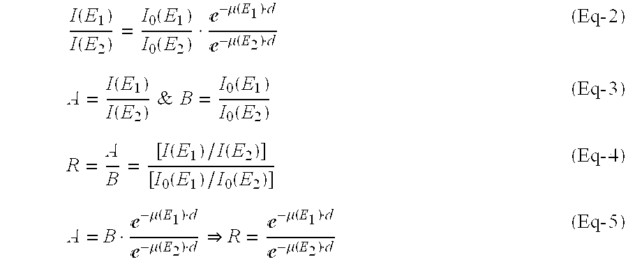
- R=e {μ(R 2 )−μ(E 1 )}dln(R)=[μ(E 2)−μ(E 1)]d (Eq-6)

- The source depth, d, can be calculated as:

- The linear attenuation coefficients for specific energies are calculated from the photon mass attenuation and energy absorption coefficients table.
- The ratio I 0(E1)/I0(E2) for a known isotope or isotopes is calculated from an isotope table and the ratio I(E1)/I(E2) is measured at the inspected object edges.
- Additionally, X-ray information, for example, mammography may be used, for more material linear attenuation coefficient, since X-ray imaging provides information about the material density.
- For example, we may consider a lesion inside a soft tissue, such as a breast, at a distance d that holds an isotope of 123I, which emits two main photons, as follows:
- E 1=27 keV with 86.5%; and
- E 2=159 keV with 83.4%.
- The soft tissue density is 0.8 g/cm 3.
- The linear attenuation coefficients for E 1 and E2 are calculated from J. Il Hubbell, “Photon Mass Attenuation and Energy—absorption Coefficients from 1 keV to 20 MeV”. Int. J. Appl. Radiat Isat. Vol. 33. pp. 1269 to 1290, 1982, as follows:
- μ(E 1)=(0.474 cm/g→μ(E 1)=0.474 cm/g·0.8 g/cm 3=0.38 l/cm
- μ(E 2)=0.1489 cm/g→μ(E 2)=0.1489 cm/g·0.8 g/cm 3=0.119 l/cm
- The measured ratio between I(E 1) to I(E2) on the attenuating soft tissue edge is 0.6153.
- Using Eq-7, one gets that the lesion depth is: d=2 cm.
- Alternatively, if the measured ration between I(E 1) to I(E2) is 0.473 the extracted lesion depth is; d=3 cm.
- It will be appreciated that when two or more isotopes used for obtaining the different photons, corrections for their respective half-lives may be required.
- It will be appreciated that by calculating the depth of a radiation source at each position, one may obtain a radiation source depth map, with respect to the gastrointestinal, track, as
ingestible device 12 travels through it. This information may be superimposed on a radiation source image, as produced by other methods. For example, a radiation source image produced by deconvolution algorithms may he superimposed on a radiation source image produced by depth calculations, based on the attenuation of photons of different energies. - It is appreciated that certain features of the invention, which are, for clarity, described in the context of separate embodiments, may also be provided in combination in a single embodiment. Conversely, various features of the invention., which are, for brevity, described in the context of a single embodiment, may also be provided separately or in any suitable subcombination.
- Although the invention has been described in conjunction with specific embodiments thereof, it is evident that many alternatives, modifications and variations will be apparent to those skilled in the art. Accordingly, it is intended to embrace all such alternatives, modifications and variations that fall within the spirit and broad scope of the appended claims. All publications, patents and patent applications mentioned in this specification are herein incorporated in their entirety by reference into the specification, to the same extent as if each individual publication, patent or patent application was specifically and individually indicated to be incorporated herein by reference. In addition, citation or identification of any reference in this application shall not be construed as an admission that such reference is available as prior art to the present invention.
Claims (15)
Priority Applications (8)
| Application Number | Priority Date | Filing Date | Title |
|---|---|---|---|
| US10/616,301 US8036731B2 (en) | 2001-01-22 | 2003-07-10 | Ingestible pill for diagnosing a gastrointestinal tract |
| PCT/IL2003/000917 WO2004042546A1 (en) | 2002-11-04 | 2003-11-04 | Apparatus and methods for imaging and attenuation correction |
| AT03810570T ATE447734T1 (en) | 2002-11-04 | 2003-11-04 | DEVICES AND METHODS FOR IMAGING AND ATTENUATION CORRECTION |
| DE60329938T DE60329938D1 (en) | 2002-11-04 | 2003-11-04 | DEVICES AND METHOD FOR PICTURE AND DAMPING CORRECTION |
| EP03810570A EP1573495B1 (en) | 2002-11-04 | 2003-11-04 | Apparatus and methods for imaging and attenuation correction |
| US10/533,568 US7652259B2 (en) | 2000-08-21 | 2003-11-04 | Apparatus and methods for imaging and attenuation correction |
| AU2003276658A AU2003276658A1 (en) | 2002-11-04 | 2003-11-04 | Apparatus and methods for imaging and attenuation correction |
| AU2004203126A AU2004203126A1 (en) | 2003-07-10 | 2004-07-12 | Three-Dimensional Nuclear-Emission Imaging, Based on Attenuation of Photons of Different Energies |
Applications Claiming Priority (5)
| Application Number | Priority Date | Filing Date | Title |
|---|---|---|---|
| US09/765,316 US20020099310A1 (en) | 2001-01-22 | 2001-01-22 | Gastrointestinal-tract sensor |
| PCT/IL2002/000057 WO2002058531A2 (en) | 2001-01-22 | 2002-01-22 | Ingestible device |
| US10/240,239 US8055329B2 (en) | 2001-01-22 | 2002-01-22 | Ingestible device for radioimaging of the gastrointestinal tract |
| US39493602P | 2002-07-11 | 2002-07-11 | |
| US10/616,301 US8036731B2 (en) | 2001-01-22 | 2003-07-10 | Ingestible pill for diagnosing a gastrointestinal tract |
Related Parent Applications (2)
| Application Number | Title | Priority Date | Filing Date |
|---|---|---|---|
| PCT/IL2002/000057 Continuation-In-Part WO2002058531A2 (en) | 2000-08-21 | 2002-01-22 | Ingestible device |
| US10/240,239 Continuation-In-Part US8055329B2 (en) | 2000-08-21 | 2002-01-22 | Ingestible device for radioimaging of the gastrointestinal tract |
Related Child Applications (1)
| Application Number | Title | Priority Date | Filing Date |
|---|---|---|---|
| PCT/IL2003/000917 Continuation-In-Part WO2004042546A1 (en) | 2000-08-21 | 2003-11-04 | Apparatus and methods for imaging and attenuation correction |
Publications (2)
| Publication Number | Publication Date |
|---|---|
| US20040054278A1 true US20040054278A1 (en) | 2004-03-18 |
| US8036731B2 US8036731B2 (en) | 2011-10-11 |
Family
ID=31999042
Family Applications (1)
| Application Number | Title | Priority Date | Filing Date |
|---|---|---|---|
| US10/616,301 Expired - Lifetime US8036731B2 (en) | 2000-08-21 | 2003-07-10 | Ingestible pill for diagnosing a gastrointestinal tract |
Country Status (1)
| Country | Link |
|---|---|
| US (1) | US8036731B2 (en) |
Cited By (123)
| Publication number | Priority date | Publication date | Assignee | Title |
|---|---|---|---|---|
| US20030139661A1 (en) * | 2001-01-22 | 2003-07-24 | Yoav Kimchy | Ingestible device |
| US20040116807A1 (en) * | 2002-10-17 | 2004-06-17 | Roni Amrami | Blood vessels wall imaging catheter |
| US20040204646A1 (en) * | 2002-11-04 | 2004-10-14 | V-Target Technologies Ltd. | Intracorporeal-imaging head |
| US20050003323A1 (en) * | 2003-01-14 | 2005-01-06 | J. Morita Manufacturing Corporation | Diagnostic imaging apparatus |
| US20050043617A1 (en) * | 2002-11-29 | 2005-02-24 | Mordechai Frisch | Methods, device and system for in vivo detection |
| US20050143648A1 (en) * | 2003-12-25 | 2005-06-30 | Olympus Corporation | System for detecting position of capsule endoscope in subject |
| US20050154277A1 (en) * | 2002-12-31 | 2005-07-14 | Jing Tang | Apparatus and methods of using built-in micro-spectroscopy micro-biosensors and specimen collection system for a wireless capsule in a biological body in vivo |
| US20050205792A1 (en) * | 2004-01-13 | 2005-09-22 | Benny Rousso | Multi-dimensional image reconstruction |
| US20050266074A1 (en) * | 2004-05-20 | 2005-12-01 | Yoel Zilberstein | Ingestible device platform for the colon |
| US20060189846A1 (en) * | 2005-02-23 | 2006-08-24 | Ker-Jer Huang | Disposable two-step endoscope |
| WO2006121949A1 (en) * | 2005-05-06 | 2006-11-16 | Regents Of The University Of Minnesota | Wirelessly coupled magnetic resonance coil |
| EP1765143A1 (en) * | 2004-06-21 | 2007-03-28 | Korea Institute of Science and Technology | Capsule type endoscope control system |
| WO2007013952A3 (en) * | 2005-07-20 | 2007-04-19 | Neil R Euliano | Medication compliance system and associated methods |
| EP1779765A1 (en) * | 2004-08-06 | 2007-05-02 | Olympus Corporation | System for acquiring image in subject and device to be introduced into subject |
| US20070156047A1 (en) * | 2000-08-21 | 2007-07-05 | Michael Nagler | Radioactive-emission-measurement optimization to specific body structures |
| US20070161885A1 (en) * | 2003-12-17 | 2007-07-12 | Check-Cap Ltd. | Intra-lumen polyp detection |
| US20070213659A1 (en) * | 2004-08-27 | 2007-09-13 | Koninklijke Philips Electronics, N.V. | Electronically and remotely controlled pill and system for delivering at least one medicament |
| US20070219413A1 (en) * | 2006-03-20 | 2007-09-20 | Lin Feng C | Capsulated endoscope with memory storage device |
| US20070265230A1 (en) * | 2006-05-11 | 2007-11-15 | Benny Rousso | Radiopharmaceuticals For Diagnosis And Therapy |
| US20070276309A1 (en) * | 2006-05-12 | 2007-11-29 | Kci Licensing, Inc. | Systems and methods for wound area management |
| US20070276195A1 (en) * | 2006-05-12 | 2007-11-29 | Kci Licensing, Inc. | Systems and methods for wound area management |
| US20080001090A1 (en) * | 2006-06-28 | 2008-01-03 | Spectrum Dynamics Llc | Imaging Techniques For Reducing Blind Spots |
| US7316930B1 (en) | 2003-04-21 | 2008-01-08 | National Semiconductor Corporation | Use of vertically stacked photodiodes in a gene chip system |
| US20080042067A1 (en) * | 2004-11-09 | 2008-02-21 | Spectrum Dynamics Llc | Radioimaging |
| US20080058597A1 (en) * | 2006-09-06 | 2008-03-06 | Innurvation Llc | Imaging and Locating Systems and Methods for a Swallowable Sensor Device |
| US20080064938A1 (en) * | 2006-09-08 | 2008-03-13 | Semler John R | Method of determining location of an ingested capsule |
| US20080125645A1 (en) * | 2005-07-26 | 2008-05-29 | Frank Volke | Device for nuclear magnetic resonance examination of intracorporal sections of the body |
| US20080128626A1 (en) * | 2004-11-09 | 2008-06-05 | Spectrum Dynamics Llc | Radioimaging |
| US20080146871A1 (en) * | 2006-09-06 | 2008-06-19 | Innurvation, Inc. | Ingestible Low Power Sensor Device and System for Communicating with Same |
| US20080161660A1 (en) * | 2006-09-06 | 2008-07-03 | Innurvation, Inc. | System and Method for Acoustic Information Exchange Involving an Ingestible Low Power Capsule |
| US7399274B1 (en) * | 2003-08-19 | 2008-07-15 | National Semiconductor Corporation | Sensor configuration for a capsule endoscope |
| US20080195249A1 (en) * | 2004-11-09 | 2008-08-14 | Spectrum Dynamics Llc | Radiopharmaceutical dispensing, administration, and imaging |
| US20080260637A1 (en) * | 2004-11-17 | 2008-10-23 | Dalia Dickman | Methods of Detecting Prostate Cancer |
| EP2008572A1 (en) * | 2006-04-19 | 2008-12-31 | Olympus Medical Systems Corp. | Capsule medical device |
| WO2009019689A2 (en) * | 2007-08-06 | 2009-02-12 | Cascades Ltd. | Tumor screening system and methods thereof |
| US20090078875A1 (en) * | 2004-11-09 | 2009-03-26 | Spectrum Dynamics Llc | Radioimaging |
| US20090152471A1 (en) * | 2005-11-09 | 2009-06-18 | Spectrum Dynamics Llc | Dynamic Spect Camera |
| US20090190807A1 (en) * | 2005-07-19 | 2009-07-30 | Spectrum Dynamics Llc | Reconstruction Stabilizer and Active Vision |
| US20090326362A1 (en) * | 2004-12-15 | 2009-12-31 | Koninklijke Philips Electronics N.V. | Registration of multi-modality images |
| US7652259B2 (en) | 2000-08-21 | 2010-01-26 | Spectrum Dynamics Llc | Apparatus and methods for imaging and attenuation correction |
| US20100099985A1 (en) * | 2005-12-05 | 2010-04-22 | Paul Gueye | Apparatus and method for brachytherapy radiation distribution mapping |
| US20100142774A1 (en) * | 2006-12-20 | 2010-06-10 | Spectrum Dynamics Llc | method, a system, and an apparatus for using and processing multidimensional data |
| US20100140483A1 (en) * | 2006-11-13 | 2010-06-10 | Benny Rousso | Radioimaging applications of and novel formulations of teboroxime |
| US20100174180A1 (en) * | 2004-11-09 | 2010-07-08 | Benny Rousso | Imaging System Customization Using Data From Radiopharmaceutical-Associated Data Carrier |
| US20100245354A1 (en) * | 2004-01-13 | 2010-09-30 | Spectrum Dynamics Llc | Dynamic spect camera |
| US20100288934A1 (en) * | 2005-12-05 | 2010-11-18 | Keppel Cynthia E | Apparatus and method for external beam radiation distribution mapping |
| US20100331827A1 (en) * | 2008-02-18 | 2010-12-30 | Koninklijke Philips Electronics N.V. | Administration of drugs to a patient |
| US20110009715A1 (en) * | 2008-07-08 | 2011-01-13 | David O' Reilly | Ingestible event marker data framework |
| US7872235B2 (en) | 2005-01-13 | 2011-01-18 | Spectrum Dynamics Llc | Multi-dimensional image reconstruction and analysis for expert-system diagnosis |
| US20110106064A1 (en) * | 2008-06-19 | 2011-05-05 | Koninklijke Philips Electronics N.V. | Device for delivery of powder like medication in a humid environment |
| US7956888B2 (en) | 2005-06-22 | 2011-06-07 | Ge Inspection Technologies, Lp | Remote video inspection system integrating audio communication functionality |
| WO2011091275A1 (en) * | 2010-01-23 | 2011-07-28 | Duke University | Jetless intravenous catheters and mechanical assist devices for hand-injection of contrast media during dynamic computer tomography and methods of using same |
| US20110282146A1 (en) * | 2004-01-14 | 2011-11-17 | Olympus Corporation | Capsule endoscope apparatus |
| WO2012011914A1 (en) * | 2010-07-22 | 2012-01-26 | Hampton University | Apparatus and method for external beam radiation distribution mapping |
| US8204500B2 (en) | 2005-12-28 | 2012-06-19 | Starhome Gmbh | Optimal voicemail deposit for roaming cellular telephony |
| US20120232405A1 (en) * | 2009-08-29 | 2012-09-13 | Eugene Grigoriev | Compact probe for tracer-assisted diagnostic and surgery |
| WO2012125901A1 (en) * | 2011-03-17 | 2012-09-20 | Given Imaging Ltd. | Reusable in-vivo device, system and method of assembly thereof |
| US8280124B2 (en) | 2004-06-01 | 2012-10-02 | Spectrum Dynamics Llc | Methods of view selection for radioactive emission measurements |
| WO2012158648A1 (en) * | 2011-05-13 | 2012-11-22 | Massachusetts Institute Of Technology | Method and apparatus for delivering a substance |
| US8338788B2 (en) | 2009-07-29 | 2012-12-25 | Spectrum Dynamics Llc | Method and system of optimized volumetric imaging |
| US8437833B2 (en) | 2008-10-07 | 2013-05-07 | Bard Access Systems, Inc. | Percutaneous magnetic gastrostomy |
| US8478382B2 (en) | 2008-02-11 | 2013-07-02 | C. R. Bard, Inc. | Systems and methods for positioning a catheter |
| US8489176B1 (en) | 2000-08-21 | 2013-07-16 | Spectrum Dynamics Llc | Radioactive emission detector equipped with a position tracking system and utilization thereof with medical systems and in medical procedures |
| US8512256B2 (en) | 2006-10-23 | 2013-08-20 | Bard Access Systems, Inc. | Method of locating the tip of a central venous catheter |
| US8521253B2 (en) | 2007-10-29 | 2013-08-27 | Spectrum Dynamics Llc | Prostate imaging |
| WO2013149264A1 (en) * | 2012-03-30 | 2013-10-03 | The Regents Of The University Of California | System, device and method for measurement of esophageal wall blood perfusion |
| US20130258804A1 (en) * | 2010-12-27 | 2013-10-03 | Hitachi Medical Corporation | Ultrasonic diagnostic apparatus and image processing method |
| US8565860B2 (en) | 2000-08-21 | 2013-10-22 | Biosensors International Group, Ltd. | Radioactive emission detector equipped with a position tracking system |
| US20130281833A1 (en) * | 2012-04-19 | 2013-10-24 | Korea Institute Of Science And Technology | Device and method for diagnosing lung cancer using electromagnetic wave |
| US8644910B2 (en) | 2005-07-19 | 2014-02-04 | Biosensors International Group, Ltd. | Imaging protocols |
| CN103784157A (en) * | 2014-02-14 | 2014-05-14 | 王葆春 | Device for accurately positioning diseased region of large intestine |
| US8774907B2 (en) | 2006-10-23 | 2014-07-08 | Bard Access Systems, Inc. | Method of locating the tip of a central venous catheter |
| US8781555B2 (en) | 2007-11-26 | 2014-07-15 | C. R. Bard, Inc. | System for placement of a catheter including a signal-generating stylet |
| US8784336B2 (en) | 2005-08-24 | 2014-07-22 | C. R. Bard, Inc. | Stylet apparatuses and methods of manufacture |
| US8801693B2 (en) | 2010-10-29 | 2014-08-12 | C. R. Bard, Inc. | Bioimpedance-assisted placement of a medical device |
| US8837793B2 (en) | 2005-07-19 | 2014-09-16 | Biosensors International Group, Ltd. | Reconstruction stabilizer and active vision |
| US8849382B2 (en) | 2007-11-26 | 2014-09-30 | C. R. Bard, Inc. | Apparatus and display methods relating to intravascular placement of a catheter |
| US8869390B2 (en) | 2007-10-01 | 2014-10-28 | Innurvation, Inc. | System and method for manufacturing a swallowable sensor device |
| US8909325B2 (en) | 2000-08-21 | 2014-12-09 | Biosensors International Group, Ltd. | Radioactive emission detector equipped with a position tracking system and utilization thereof with medical systems and in medical procedures |
| US20150057549A1 (en) * | 2005-01-24 | 2015-02-26 | The Board Of Trustees Of The Leland Stanford Junior University | Live being optical analysis system and approach |
| USD724745S1 (en) | 2011-08-09 | 2015-03-17 | C. R. Bard, Inc. | Cap for an ultrasound probe |
| US9040016B2 (en) | 2004-01-13 | 2015-05-26 | Biosensors International Group, Ltd. | Diagnostic kit and methods for radioimaging myocardial perfusion |
| US9047746B1 (en) | 2005-07-20 | 2015-06-02 | Neil Euliano | Electronic medication compliance monitoring system and associated methods |
| US20150150481A1 (en) * | 1999-03-01 | 2015-06-04 | West View Research, Llc | Computerized apparatus with ingestible probe |
| US9125578B2 (en) | 2009-06-12 | 2015-09-08 | Bard Access Systems, Inc. | Apparatus and method for catheter navigation and tip location |
| US9195043B2 (en) | 2010-08-27 | 2015-11-24 | The Board Of Trustees Of The Leland Stanford Junior University | Microscopy imaging device with advanced imaging properties |
| US9211107B2 (en) | 2011-11-07 | 2015-12-15 | C. R. Bard, Inc. | Ruggedized ultrasound hydrogel insert |
| US9316743B2 (en) | 2004-11-09 | 2016-04-19 | Biosensors International Group, Ltd. | System and method for radioactive emission measurement |
| USD754357S1 (en) | 2011-08-09 | 2016-04-19 | C. R. Bard, Inc. | Ultrasound probe head |
| US9339206B2 (en) | 2009-06-12 | 2016-05-17 | Bard Access Systems, Inc. | Adaptor for endovascular electrocardiography |
| US9392961B2 (en) | 2003-12-17 | 2016-07-19 | Check-Cap Ltd. | Intra-lumen polyp detection |
| US9445734B2 (en) | 2009-06-12 | 2016-09-20 | Bard Access Systems, Inc. | Devices and methods for endovascular electrography |
| US9456766B2 (en) | 2007-11-26 | 2016-10-04 | C. R. Bard, Inc. | Apparatus for use with needle insertion guidance system |
| US9470801B2 (en) | 2004-01-13 | 2016-10-18 | Spectrum Dynamics Llc | Gating with anatomically varying durations |
| US9492097B2 (en) | 2007-11-26 | 2016-11-15 | C. R. Bard, Inc. | Needle length determination and calibration for insertion guidance system |
| US9521961B2 (en) | 2007-11-26 | 2016-12-20 | C. R. Bard, Inc. | Systems and methods for guiding a medical instrument |
| US9532724B2 (en) | 2009-06-12 | 2017-01-03 | Bard Access Systems, Inc. | Apparatus and method for catheter navigation using endovascular energy mapping |
| US9554716B2 (en) | 2007-11-26 | 2017-01-31 | C. R. Bard, Inc. | Insertion guidance system for needles and medical components |
| US9636031B2 (en) | 2007-11-26 | 2017-05-02 | C.R. Bard, Inc. | Stylets for use with apparatus for intravascular placement of a catheter |
| US9649048B2 (en) | 2007-11-26 | 2017-05-16 | C. R. Bard, Inc. | Systems and methods for breaching a sterile field for intravascular placement of a catheter |
| US9681823B2 (en) | 2007-11-26 | 2017-06-20 | C. R. Bard, Inc. | Integrated system for intravascular placement of a catheter |
| US9839372B2 (en) | 2014-02-06 | 2017-12-12 | C. R. Bard, Inc. | Systems and methods for guidance and placement of an intravascular device |
| US9844354B2 (en) | 2007-02-06 | 2017-12-19 | Check-Cap Ltd. | Intra-lumen polyp detection |
| US9861268B2 (en) | 1999-03-01 | 2018-01-09 | West View Research, Llc | Methods of processing data obtained from medical device |
| US9901714B2 (en) | 2008-08-22 | 2018-02-27 | C. R. Bard, Inc. | Catheter assembly including ECG sensor and magnetic assemblies |
| WO2018044985A1 (en) * | 2016-09-02 | 2018-03-08 | Elwha Llc | Intraluminal devices with deployable elements |
| US10046139B2 (en) | 2010-08-20 | 2018-08-14 | C. R. Bard, Inc. | Reconfirmation of ECG-assisted catheter tip placement |
| US10136865B2 (en) | 2004-11-09 | 2018-11-27 | Spectrum Dynamics Medical Limited | Radioimaging using low dose isotope |
| US10349890B2 (en) | 2015-06-26 | 2019-07-16 | C. R. Bard, Inc. | Connector interface for ECG-based catheter positioning system |
| US10449330B2 (en) | 2007-11-26 | 2019-10-22 | C. R. Bard, Inc. | Magnetic element-equipped needle assemblies |
| US10521561B1 (en) | 2013-12-17 | 2019-12-31 | Etectrx, Inc. | Electronic compliance system and associated methods |
| US10524691B2 (en) | 2007-11-26 | 2020-01-07 | C. R. Bard, Inc. | Needle assembly including an aligned magnetic element |
| US10639008B2 (en) | 2009-10-08 | 2020-05-05 | C. R. Bard, Inc. | Support and cover structures for an ultrasound probe head |
| WO2020128803A1 (en) * | 2018-12-17 | 2020-06-25 | Qatar University | Fluorescence lifetime spectroscopy based capsule endoscopy |
| US10751509B2 (en) | 2007-11-26 | 2020-08-25 | C. R. Bard, Inc. | Iconic representations for guidance of an indwelling medical device |
| US10820885B2 (en) | 2012-06-15 | 2020-11-03 | C. R. Bard, Inc. | Apparatus and methods for detection of a removable cap on an ultrasound probe |
| WO2020227715A1 (en) * | 2019-05-09 | 2020-11-12 | The General Hospital Corporation | System and method to evaluate upper gastrointestinal tract motility and emptying using magnetic resonance imaging (mri) |
| US10973584B2 (en) | 2015-01-19 | 2021-04-13 | Bard Access Systems, Inc. | Device and method for vascular access |
| US10992079B2 (en) | 2018-10-16 | 2021-04-27 | Bard Access Systems, Inc. | Safety-equipped connection systems and methods thereof for establishing electrical connections |
| US11000207B2 (en) | 2016-01-29 | 2021-05-11 | C. R. Bard, Inc. | Multiple coil system for tracking a medical device |
| WO2021140504A1 (en) * | 2020-01-06 | 2021-07-15 | Check-Cap Ltd. | Radiation capsule for bowel disease imaging and localize drug delivery |
| US11103213B2 (en) | 2009-10-08 | 2021-08-31 | C. R. Bard, Inc. | Spacers for use with an ultrasound probe |
| WO2022056108A1 (en) * | 2020-09-09 | 2022-03-17 | Massachusetts Institute Of Technology | Closed-loop detection and treatment of radiation and toxic agents |
Families Citing this family (55)
| Publication number | Priority date | Publication date | Assignee | Title |
|---|---|---|---|---|
| US9801527B2 (en) | 2004-04-19 | 2017-10-31 | Gearbox, Llc | Lumen-traveling biological interface device |
| US8353896B2 (en) | 2004-04-19 | 2013-01-15 | The Invention Science Fund I, Llc | Controllable release nasal system |
| US9011329B2 (en) | 2004-04-19 | 2015-04-21 | Searete Llc | Lumenally-active device |
| US8024036B2 (en) | 2007-03-19 | 2011-09-20 | The Invention Science Fund I, Llc | Lumen-traveling biological interface device and method of use |
| US8912908B2 (en) | 2005-04-28 | 2014-12-16 | Proteus Digital Health, Inc. | Communication system with remote activation |
| EP2392258B1 (en) | 2005-04-28 | 2014-10-08 | Proteus Digital Health, Inc. | Pharma-informatics system |
| US8836513B2 (en) | 2006-04-28 | 2014-09-16 | Proteus Digital Health, Inc. | Communication system incorporated in an ingestible product |
| US8802183B2 (en) | 2005-04-28 | 2014-08-12 | Proteus Digital Health, Inc. | Communication system with enhanced partial power source and method of manufacturing same |
| US9198608B2 (en) | 2005-04-28 | 2015-12-01 | Proteus Digital Health, Inc. | Communication system incorporated in a container |
| US20120035437A1 (en) | 2006-04-12 | 2012-02-09 | Searete Llc, A Limited Liability Corporation Of The State Of Delaware | Navigation of a lumen traveling device toward a target |
| US20080058786A1 (en) * | 2006-04-12 | 2008-03-06 | Searete Llc, A Limited Liability Corporation Of The State Of Delaware | Autofluorescent imaging and target ablation |
| US20080058785A1 (en) * | 2006-04-12 | 2008-03-06 | Searete Llc, A Limited Liability Corporation Of The State Of Delaware | Autofluorescent imaging and target ablation |
| CN105468895A (en) | 2006-05-02 | 2016-04-06 | 普罗透斯数字保健公司 | Patient customized therapeutic regimens |
| EP2083680B1 (en) | 2006-10-25 | 2016-08-10 | Proteus Digital Health, Inc. | Controlled activation ingestible identifier |
| EP2069004A4 (en) | 2006-11-20 | 2014-07-09 | Proteus Digital Health Inc | Active signal processing personal health signal receivers |
| MY165532A (en) | 2007-02-01 | 2018-04-02 | Proteus Digital Health Inc | Ingestible event marker systems |
| CA2676280C (en) | 2007-02-14 | 2018-05-22 | Proteus Biomedical, Inc. | In-body power source having high surface area electrode |
| US9270025B2 (en) | 2007-03-09 | 2016-02-23 | Proteus Digital Health, Inc. | In-body device having deployable antenna |
| US8540632B2 (en) | 2007-05-24 | 2013-09-24 | Proteus Digital Health, Inc. | Low profile antenna for in body device |
| DK2192946T3 (en) | 2007-09-25 | 2022-11-21 | Otsuka Pharma Co Ltd | In-body device with virtual dipole signal amplification |
| ES2661739T3 (en) | 2007-11-27 | 2018-04-03 | Proteus Digital Health, Inc. | Transcorporeal communication systems that employ communication channels |
| MY161533A (en) | 2008-03-05 | 2017-04-28 | Proteus Digital Health Inc | Multi-mode communication ingestible event markers and systems, and methods of using the same |
| US8390291B2 (en) * | 2008-05-19 | 2013-03-05 | The Board Of Regents, The University Of Texas System | Apparatus and method for tracking movement of a target |
| WO2010005571A2 (en) | 2008-07-09 | 2010-01-14 | Innurvation, Inc. | Displaying image data from a scanner capsule |
| KR101214453B1 (en) | 2008-08-13 | 2012-12-24 | 프로테우스 디지털 헬스, 인코포레이티드 | Ingestible circuitry |
| AU2010203625A1 (en) | 2009-01-06 | 2011-07-21 | Proteus Digital Health, Inc. | Ingestion-related biofeedback and personalized medical therapy method and system |
| WO2010111403A2 (en) | 2009-03-25 | 2010-09-30 | Proteus Biomedical, Inc. | Probablistic pharmacokinetic and pharmacodynamic modeling |
| CN102458236B (en) | 2009-04-28 | 2016-01-27 | 普罗秋斯数字健康公司 | The Ingestible event marker of high reliability and using method thereof |
| WO2010132331A2 (en) | 2009-05-12 | 2010-11-18 | Proteus Biomedical, Inc. | Ingestible event markers comprising an ingestible component |
| TWI517050B (en) | 2009-11-04 | 2016-01-11 | 普羅托斯數位健康公司 | System for supply chain management |
| US8647259B2 (en) | 2010-03-26 | 2014-02-11 | Innurvation, Inc. | Ultrasound scanning capsule endoscope (USCE) |
| WO2011127252A2 (en) | 2010-04-07 | 2011-10-13 | Proteus Biomedical, Inc. | Miniature ingestible device |
| TWI557672B (en) | 2010-05-19 | 2016-11-11 | 波提亞斯數位康健公司 | Computer system and computer-implemented method to track medication from manufacturer to a patient, apparatus and method for confirming delivery of medication to a patient, patient interface device |
| US8581584B2 (en) * | 2010-05-26 | 2013-11-12 | Florida State University Research Foundation | Membrane proteins, mechanisms of action and uses thereof |
| JP2014504902A (en) | 2010-11-22 | 2014-02-27 | プロテウス デジタル ヘルス, インコーポレイテッド | Ingestible device with medicinal product |
| US9756874B2 (en) | 2011-07-11 | 2017-09-12 | Proteus Digital Health, Inc. | Masticable ingestible product and communication system therefor |
| WO2015112603A1 (en) | 2014-01-21 | 2015-07-30 | Proteus Digital Health, Inc. | Masticable ingestible product and communication system therefor |
| BR112014001397A2 (en) | 2011-07-21 | 2017-02-21 | Proteus Biomedical Inc | device, system and method of mobile communication |
| US9235683B2 (en) | 2011-11-09 | 2016-01-12 | Proteus Digital Health, Inc. | Apparatus, system, and method for managing adherence to a regimen |
| US9653935B2 (en) | 2012-04-20 | 2017-05-16 | Medtronic, Inc. | Sensing temperature within medical devices |
| WO2013168111A2 (en) | 2012-05-08 | 2013-11-14 | Spectrum Dynamics Llc | Nuclear medicine tomography systems, detectors and methods |
| WO2014018454A1 (en) | 2012-07-23 | 2014-01-30 | Proteus Digital Health, Inc. | Techniques for manufacturing ingestible event markers comprising an ingestible component |
| JP5869736B2 (en) | 2012-10-18 | 2016-02-24 | プロテウス デジタル ヘルス, インコーポレイテッド | Apparatus, system, and method for adaptively optimizing power dissipation and broadcast power in a power supply for a communication device |
| TWI659994B (en) | 2013-01-29 | 2019-05-21 | 美商普羅托斯數位健康公司 | Highly-swellable polymeric films and compositions comprising the same |
| JP5941240B2 (en) | 2013-03-15 | 2016-06-29 | プロテウス デジタル ヘルス, インコーポレイテッド | Metal detector device, system and method |
| JP6511439B2 (en) | 2013-06-04 | 2019-05-15 | プロテウス デジタル ヘルス, インコーポレイテッド | Systems, devices, and methods for data collection and outcome assessment |
| US9796576B2 (en) | 2013-08-30 | 2017-10-24 | Proteus Digital Health, Inc. | Container with electronically controlled interlock |
| US10084880B2 (en) | 2013-11-04 | 2018-09-25 | Proteus Digital Health, Inc. | Social media networking based on physiologic information |
| US11051543B2 (en) | 2015-07-21 | 2021-07-06 | Otsuka Pharmaceutical Co. Ltd. | Alginate on adhesive bilayer laminate film |
| KR20210018961A (en) | 2016-07-22 | 2021-02-18 | 프로테우스 디지털 헬스, 인코포레이티드 | Electromagnetic sensing and detection of ingestible event markers |
| IL265827B2 (en) | 2016-10-26 | 2023-03-01 | Proteus Digital Health Inc | Methods for manufacturing capsules with ingestible event markers |
| CN111587088A (en) | 2017-11-28 | 2020-08-25 | 麻省理工学院 | Stomach resident electronic device |
| WO2021141608A1 (en) * | 2020-01-11 | 2021-07-15 | Puchajda Wojciech | Digest tract imaging kit and method of use thereof |
| US11054534B1 (en) | 2020-04-24 | 2021-07-06 | Ronald Nutt | Time-resolved positron emission tomography encoder system for producing real-time, high resolution, three dimensional positron emission tomographic image without the necessity of performing image reconstruction |
| US11300695B2 (en) | 2020-04-24 | 2022-04-12 | Ronald Nutt | Time-resolved positron emission tomography encoder system for producing event-by-event, real-time, high resolution, three-dimensional positron emission tomographic image without the necessity of performing image reconstruction |
Citations (97)
| Publication number | Priority date | Publication date | Assignee | Title |
|---|---|---|---|---|
| US2776377A (en) * | 1954-04-22 | 1957-01-01 | Hal O Anger | In vivo radiation scanner |
| US3719183A (en) * | 1970-03-05 | 1973-03-06 | H Schwartz | Method for detecting blockage or insufficiency of pancreatic exocrine function |
| US3739279A (en) * | 1971-06-30 | 1973-06-12 | Corning Glass Works | Radio capsule oscillator circuit |
| US3971362A (en) * | 1972-10-27 | 1976-07-27 | The United States Of America As Represented By The Administrator Of The National Aeronautics And Space Administration | Miniature ingestible telemeter devices to measure deep-body temperature |
| US4015592A (en) * | 1974-12-24 | 1977-04-05 | Bradley Moore Patrick Ralph | Nuclear medicine system for imaging radiation |
| US4278077A (en) * | 1978-07-27 | 1981-07-14 | Olympus Optical Co., Ltd. | Medical camera system |
| US4521688A (en) * | 1983-01-21 | 1985-06-04 | The United States Of America As Represented By The Administrator Of The National Aeronautics And Space Administration | Three-dimensional and tomographic imaging device for x-ray and gamma-ray emitting objects |
| US4595014A (en) * | 1983-10-18 | 1986-06-17 | University Patents, Inc. | Imaging probe and method |
| US4674107A (en) * | 1985-07-31 | 1987-06-16 | Picker International, Inc. | Display for radiation imaging |
| US4731536A (en) * | 1985-02-18 | 1988-03-15 | Firma Merfurth GmbH | Apparatus for checking persons for radioactive contamination |
| US4828841A (en) * | 1984-07-24 | 1989-05-09 | Colorcon, Inc. | Maltodextrin coating |
| US4844067A (en) * | 1987-02-19 | 1989-07-04 | Japan Medical Supply Co., Ltd. | Sucrose fatty acid ester coated suture |
| US4844076A (en) * | 1988-08-26 | 1989-07-04 | The Johns Hopkins University | Ingestible size continuously transmitting temperature monitoring pill |
| US4893013A (en) * | 1987-03-17 | 1990-01-09 | Neoprobe Corporation | Detector and localizer for low energy radiation emissions |
| US4928250A (en) * | 1986-07-02 | 1990-05-22 | Hewlett-Packard Company | System for deriving radiation images |
| US4929832A (en) * | 1988-03-11 | 1990-05-29 | Ledley Robert S | Methods and apparatus for determining distributions of radioactive materials |
| US4995396A (en) * | 1988-12-08 | 1991-02-26 | Olympus Optical Co., Ltd. | Radioactive ray detecting endoscope |
| US5014708A (en) * | 1988-09-14 | 1991-05-14 | Olympus Optical Co. | Radioactive ray detecting therapeutic apparatus |
| US5032729A (en) * | 1989-10-18 | 1991-07-16 | Georges Charpak | Process and device for determining the spatial distribution of electrons emerging from the surface of a radioactive body |
| US5088492A (en) * | 1987-09-16 | 1992-02-18 | Olympus Optical Co., Ltd. | Radioactive ray detecting endoscope |
| US5119818A (en) * | 1990-07-25 | 1992-06-09 | Care Wise Medical Products Corporation | Radiation detecting biopsy probe |
| US5279607A (en) * | 1991-05-30 | 1994-01-18 | The State University Of New York | Telemetry capsule and process |
| US5299253A (en) * | 1992-04-10 | 1994-03-29 | Akzo N.V. | Alignment system to overlay abdominal computer aided tomography and magnetic resonance anatomy with single photon emission tomography |
| US5307808A (en) * | 1992-04-01 | 1994-05-03 | General Electric Company | Tracking system and pulse sequences to monitor the position of a device using magnetic resonance |
| US5383456A (en) * | 1992-12-18 | 1995-01-24 | The Ohio State University Research Foundation | Radiation-based laparoscopic method for determining treatment modality |
| US5386446A (en) * | 1992-07-06 | 1995-01-31 | Kabushiki Kaisha Toshiba | Positional adjustment of resolution in radiation CT scanner |
| US5395366A (en) * | 1991-05-30 | 1995-03-07 | The State University Of New York | Sampling capsule and process |
| US5399868A (en) * | 1992-03-12 | 1995-03-21 | Jones; Barbara L. | Radiation probe |
| US5415181A (en) * | 1993-12-01 | 1995-05-16 | The Johns Hopkins University | AM/FM multi-channel implantable/ingestible biomedical monitoring telemetry system |
| US5484384A (en) * | 1991-01-29 | 1996-01-16 | Med Institute, Inc. | Minimally invasive medical device for providing a radiation treatment |
| US5489782A (en) * | 1994-03-24 | 1996-02-06 | Imaging Laboratory, Inc. | Method and apparatus for quantum-limited data acquisition |
| US5493595A (en) * | 1982-02-24 | 1996-02-20 | Schoolman Scientific Corp. | Stereoscopically displayed three dimensional medical imaging |
| US5519221A (en) * | 1992-01-22 | 1996-05-21 | Ansel M. Schwartz | Dedicated apparatus and method for emission mammography |
| US5604531A (en) * | 1994-01-17 | 1997-02-18 | State Of Israel, Ministry Of Defense, Armament Development Authority | In vivo video camera system |
| US5617858A (en) * | 1994-08-30 | 1997-04-08 | Vingmed Sound A/S | Apparatus for endoscopic or gastroscopic examination |
| US5635717A (en) * | 1992-07-17 | 1997-06-03 | Dimason | Apparatus for detecting and locating radioactive biological markers |
| US5716595A (en) * | 1992-05-06 | 1998-02-10 | Immunomedics, Inc. | Intraperative, intravascular and endoscopic tumor and lesion detection and therapy with monovalent antibody fragments |
| US5729129A (en) * | 1995-06-07 | 1998-03-17 | Biosense, Inc. | Magnetic location system with feedback adjustment of magnetic field generator |
| US5727554A (en) * | 1996-09-19 | 1998-03-17 | University Of Pittsburgh Of The Commonwealth System Of Higher Education | Apparatus responsive to movement of a patient during treatment/diagnosis |
| US5732704A (en) * | 1995-10-13 | 1998-03-31 | Neoprobe Corporation | Radiation based method locating and differentiating sentinel nodes |
| US5744805A (en) * | 1996-05-07 | 1998-04-28 | University Of Michigan | Solid state beta-sensitive surgical probe |
| US5857463A (en) * | 1995-10-13 | 1999-01-12 | Neoprobe Corporation | Remotely controlled apparatus and system for tracking and locating a source of photoemissions |
| US5871013A (en) * | 1995-05-31 | 1999-02-16 | Elscint Ltd. | Registration of nuclear medicine images |
| US5880475A (en) * | 1996-12-27 | 1999-03-09 | Mitsubishi Denki Kabushiki Kaisha | Scintillation fiber type radiation detector |
| US5891030A (en) * | 1997-01-24 | 1999-04-06 | Mayo Foundation For Medical Education And Research | System for two dimensional and three dimensional imaging of tubular structures in the human body |
| US5900533A (en) * | 1995-08-03 | 1999-05-04 | Trw Inc. | System and method for isotope ratio analysis and gas detection by photoacoustics |
| US5916167A (en) * | 1997-10-10 | 1999-06-29 | Neoprobe Corporation | Surgical probe apparatus and system |
| US6076009A (en) * | 1997-05-05 | 2000-06-13 | The University Of Michigan | Solid state beta-sensitive surgical probe |
| US6173201B1 (en) * | 1999-02-22 | 2001-01-09 | V-Target Ltd. | Stereotactic diagnosis and treatment with reference to a combined image |
| US6205347B1 (en) * | 1998-02-27 | 2001-03-20 | Picker International, Inc. | Separate and combined multi-modality diagnostic imaging system |
| US6212423B1 (en) * | 1994-03-02 | 2001-04-03 | Mark Krakovitz | Diagnostic hybrid probes |
| US6236880B1 (en) * | 1999-05-21 | 2001-05-22 | Raymond R. Raylman | Radiation-sensitive surgical probe with interchangeable tips |
| US6236878B1 (en) * | 1998-05-22 | 2001-05-22 | Charles A. Taylor | Method for predictive modeling for planning medical interventions and simulating physiological conditions |
| US6239438B1 (en) * | 1998-11-19 | 2001-05-29 | General Electric Company | Dual acquisition imaging method and apparatus |
| US6240312B1 (en) * | 1997-10-23 | 2001-05-29 | Robert R. Alfano | Remote-controllable, micro-scale device for use in in vivo medical diagnosis and/or treatment |
| US6242743B1 (en) * | 1998-08-11 | 2001-06-05 | Mosaic Imaging Technology, Inc. | Non-orbiting tomographic imaging system |
| US6246901B1 (en) * | 1999-05-05 | 2001-06-12 | David A. Benaron | Detecting, localizing, and targeting internal sites in vivo using optical contrast agents |
| US6339652B1 (en) * | 1998-11-24 | 2002-01-15 | Picker International, Inc. | Source-assisted attenuation correction for emission computed tomography |
| US6346706B1 (en) * | 1999-06-24 | 2002-02-12 | The Regents Of The University Of Michigan | High resolution photon detector |
| US6368331B1 (en) * | 1999-02-22 | 2002-04-09 | Vtarget Ltd. | Method and system for guiding a diagnostic or therapeutic instrument towards a target region inside the patient's body |
| US6381349B1 (en) * | 1997-11-12 | 2002-04-30 | The University Of Utah | Projector/backprojector with slice-to-slice blurring for efficient 3D scatter modeling |
| US6392235B1 (en) * | 1999-02-22 | 2002-05-21 | The Arizona Board Of Regents On Behalf Of The University Of Arizona | Coded-aperture system for planar imaging of volumetric sources |
| US20020072784A1 (en) * | 2000-10-10 | 2002-06-13 | Sheppard Norman F. | Microchip reservoir devices using wireless transmission of power and data |
| US6407391B1 (en) * | 1995-06-20 | 2002-06-18 | Centre National De La Recherche Scientifique (Cnrs) | Device for non-invasive analysis by radio-imaging, in particular for the in vivo examination of small animals, and method of use |
| US20030001837A1 (en) * | 2001-05-18 | 2003-01-02 | Baumberg Adam Michael | Method and apparatus for generating confidence data |
| US6510336B1 (en) * | 2000-03-03 | 2003-01-21 | Intra Medical Imaging, Llc | Methods and devices to expand applications of intraoperative radiation probes |
| US6516213B1 (en) * | 1999-09-03 | 2003-02-04 | Robin Medical, Inc. | Method and apparatus to estimate location and orientation of objects during magnetic resonance imaging |
| US6525320B1 (en) * | 1999-04-14 | 2003-02-25 | Jack E. Juni | Single photon emission computed tomography system |
| US20030063787A1 (en) * | 1995-05-31 | 2003-04-03 | Elscint Ltd. | Registration of nuclear medicine images |
| US6549646B1 (en) * | 2000-02-15 | 2003-04-15 | Deus Technologies, Llc | Divide-and-conquer method and system for the detection of lung nodule in radiological images |
| US20030081716A1 (en) * | 2001-10-25 | 2003-05-01 | Tumer Tumay O. | Imaging probe |
| US6560354B1 (en) * | 1999-02-16 | 2003-05-06 | University Of Rochester | Apparatus and method for registration of images to physical space using a weighted combination of points and surfaces |
| US6584348B2 (en) * | 2000-05-31 | 2003-06-24 | Given Imaging Ltd. | Method for measurement of electrical characteristics of tissue |
| US20040003001A1 (en) * | 2002-04-03 | 2004-01-01 | Fuji Photo Film Co., Ltd. | Similar image search system |
| US20040010397A1 (en) * | 2002-04-06 | 2004-01-15 | Barbour Randall L. | Modification of the normalized difference method for real-time optical tomography |
| US6680750B1 (en) * | 1996-10-14 | 2004-01-20 | Commissariat A L'energie Atomique | Device and method for collecting and encoding signals coming from photodetectors |
| US20040015075A1 (en) * | 2000-08-21 | 2004-01-22 | Yoav Kimchy | Radioactive emission detector equipped with a position tracking system and utilization thereof with medical systems and in medical procedures |
| US6697660B1 (en) * | 1998-01-23 | 2004-02-24 | Ctf Systems, Inc. | Method for functional brain imaging from magnetoencephalographic data by estimation of source signal-to-noise ratio |
| US20040054248A1 (en) * | 2000-08-21 | 2004-03-18 | Yoav Kimchy | Radioactive emission detector equipped with a position tracking system |
| US6728583B2 (en) * | 2001-06-27 | 2004-04-27 | Koninklijke Philips Electronics N.V. | User interface for a gamma camera which acquires multiple simultaneous data sets |
| US20040081623A1 (en) * | 2001-01-12 | 2004-04-29 | Morten Eriksen | Perfusion imaging method |
| US20040086437A1 (en) * | 2002-10-28 | 2004-05-06 | Ge Medical Systems Global Technology Co., Llc | Transportable manufacturing facility for radioactive materials |
| US20040101176A1 (en) * | 2002-11-27 | 2004-05-27 | General Electric Company | Method and system for measuring disease relevant tissue changes |
| US6748259B1 (en) * | 2000-06-15 | 2004-06-08 | Spectros Corporation | Optical imaging of induced signals in vivo under ambient light conditions |
| US20040116807A1 (en) * | 2002-10-17 | 2004-06-17 | Roni Amrami | Blood vessels wall imaging catheter |
| US20050020915A1 (en) * | 2002-07-29 | 2005-01-27 | Cv Therapeutics, Inc. | Myocardial perfusion imaging methods and compositions |
| US20050055174A1 (en) * | 2000-08-21 | 2005-03-10 | V Target Ltd. | Radioactive emission detector equipped with a position tracking system and utilization thereof with medical systems and in medical procedures |
| US20060074290A1 (en) * | 2004-10-04 | 2006-04-06 | Banner Health | Methodologies linking patterns from multi-modality datasets |
| US7043063B1 (en) * | 1999-08-27 | 2006-05-09 | Mirada Solutions Limited | Non-rigid motion image analysis |
| US7176466B2 (en) * | 2004-01-13 | 2007-02-13 | Spectrum Dynamics Llc | Multi-dimensional image reconstruction |
| US7187790B2 (en) * | 2002-12-18 | 2007-03-06 | Ge Medical Systems Global Technology Company, Llc | Data processing and feedback method and system |
| US20080033291A1 (en) * | 2005-01-13 | 2008-02-07 | Benny Rousso | Multi-Dimensional Image Reconstruction and Analysis for Expert-System Diagnosis |
| US20080042067A1 (en) * | 2004-11-09 | 2008-02-21 | Spectrum Dynamics Llc | Radioimaging |
| US20080128626A1 (en) * | 2004-11-09 | 2008-06-05 | Spectrum Dynamics Llc | Radioimaging |
| US7490085B2 (en) * | 2002-12-18 | 2009-02-10 | Ge Medical Systems Global Technology Company, Llc | Computer-assisted data processing system and method incorporating automated learning |
| US20090078875A1 (en) * | 2004-11-09 | 2009-03-26 | Spectrum Dynamics Llc | Radioimaging |
| US20090152471A1 (en) * | 2005-11-09 | 2009-06-18 | Spectrum Dynamics Llc | Dynamic Spect Camera |
Family Cites Families (127)
| Publication number | Priority date | Publication date | Assignee | Title |
|---|---|---|---|---|
| DE1220966B (en) | 1958-12-31 | 1966-07-14 | Hans Guenter Noeller Dr | Endoradiosonde |
| DE1516429A1 (en) | 1966-02-18 | 1969-12-04 | Wolf Gmbh Richard | Diagnostic device |
| US3684887A (en) | 1970-03-26 | 1972-08-15 | Schlumberger Technology Corp | Apparatus for inspecting tubular goods having an automatic shutter |
| US3690309A (en) | 1970-08-05 | 1972-09-12 | Viktor Mikhailovich Pluzhnikov | Radiocapsule for registering ionizing radiation in the cavities of human bodies |
| GB2031142B (en) | 1978-09-23 | 1983-01-12 | Shaw R | Apparatus and methodfor examining a blood vessel of interest using radiation detected outside the body |
| US4364377A (en) | 1979-02-02 | 1982-12-21 | Walker Scientific, Inc. | Magnetic field hemostasis |
| US5993378A (en) | 1980-10-28 | 1999-11-30 | Lemelson; Jerome H. | Electro-optical instruments and methods for treating disease |
| USH12H (en) | 1983-03-11 | 1986-01-07 | The United States Of America As Represented By The United States Department Of Energy | Nuclear medicine imaging system |
| US5033998A (en) | 1984-01-20 | 1991-07-23 | Eliot Corday | Retrograde delivery of pharmacologic and diagnostic agents via venous circulation |
| US4689041A (en) | 1984-01-20 | 1987-08-25 | Eliot Corday | Retrograde delivery of pharmacologic and diagnostic agents via venous circulation |
| IL74007A (en) | 1985-01-06 | 1988-11-30 | Yissum Res Dev Co | Method and apparatus for the localization of bleeding in the gastrointestinal tract |
| US4689621A (en) | 1986-03-31 | 1987-08-25 | The United States Of America As Represented By The Administrator Of The National Aeronautics And Space Administration | Temperature responsive transmitter |
| US5151598A (en) | 1987-03-17 | 1992-09-29 | Neoprobe Corporation | Detector and localizer for low energy radiation emissions |
| US5070878A (en) | 1988-11-14 | 1991-12-10 | Neoprobe Corporation | Detector and localizer for low energy radiation emissions |
| US5170789A (en) | 1987-06-17 | 1992-12-15 | Perinchery Narayan | Insertable NMR coil probe |
| US4951653A (en) | 1988-03-02 | 1990-08-28 | Laboratory Equipment, Corp. | Ultrasound brain lesioning system |
| US4959547A (en) | 1989-06-08 | 1990-09-25 | Care Wise Medical Products Corporation | Apparatus and methods for detecting, localizing, and imaging of radiation in biological systems |
| US5170055A (en) | 1990-07-25 | 1992-12-08 | Care Wise Medical Products Corporation | Radiation detecting biopsy probe |
| US6405072B1 (en) | 1991-01-28 | 2002-06-11 | Sherwood Services Ag | Apparatus and method for determining a location of an anatomical target with reference to a medical apparatus |
| FR2673728B1 (en) | 1991-03-08 | 1997-01-31 | Assist Publique | HIGH SENSITIVITY GAMMA CAMERA SYSTEM |
| US5243988A (en) | 1991-03-13 | 1993-09-14 | Scimed Life Systems, Inc. | Intravascular imaging apparatus and methods for use and manufacture |
| US5249124A (en) | 1991-04-16 | 1993-09-28 | Siemens Gammasonics, Inc. | Multi-isotope imaging using energy-weighted acquisition for, e.g., myocardial perfusion studies |
| US5279309A (en) | 1991-06-13 | 1994-01-18 | International Business Machines Corporation | Signaling device and method for monitoring positions in a surgical operation |
| US5417210A (en) | 1992-05-27 | 1995-05-23 | International Business Machines Corporation | System and method for augmentation of endoscopic surgery |
| US5246005A (en) | 1991-07-02 | 1993-09-21 | Care Wise Medical Products Corporation | Apparatus and method for producing statistically valid discriminable signals |
| US5361291A (en) | 1991-11-20 | 1994-11-01 | General Electric Company | Deconvolution filter for CT system |
| US5349190A (en) | 1991-12-02 | 1994-09-20 | Adac Laboratories | Adjustable triple-detector image data acquisition system |
| US5437279A (en) | 1992-07-02 | 1995-08-01 | Board Of Regents, The University Of Texas System | Method of predicting carcinomic metastases |
| US5441050A (en) | 1992-12-18 | 1995-08-15 | Neoprobe Corporation | Radiation responsive surgical instrument |
| US5431161A (en) | 1993-04-15 | 1995-07-11 | Adac Laboratories | Method and apparatus for information acquistion, processing, and display within a medical camera system |
| US5657759A (en) | 1993-05-13 | 1997-08-19 | Synectics Medical, Incorporated | Measurement of gastric emptying and gastrointestinal output |
| US5939724A (en) | 1993-06-02 | 1999-08-17 | State Of Israel, The, Atomic Energy Commission, Soreo Nuclear Research Center | Light weight-camera head and-camera assemblies containing it |
| DK0711179T3 (en) | 1993-07-30 | 2005-02-14 | Imcor Pharmaceutical Co | Stabilized ultrasound microbubble compositions |
| US5569924A (en) | 1994-08-18 | 1996-10-29 | Picker International, Inc. | Transformable dual head spect camera system |
| US5475219A (en) | 1994-10-26 | 1995-12-12 | Neoprobe Corporation | Validation of photon emission based signals using an energy window network in conjunction with a fundamental mode discriminator circuit |
| US5694933A (en) | 1995-04-28 | 1997-12-09 | Care Wise Medical Products Corporation | Apparatus and methods for determining spatial coordinates of radiolabelled tissue using gamma-rays and associated characteristic X-rays |
| US6107102A (en) | 1995-06-07 | 2000-08-22 | Regents Of The University Of California | Therapeutic microdevices and methods of making and using same |
| US5842977A (en) | 1995-07-24 | 1998-12-01 | The Johns Hopkins University | Multi-channel pill with integrated optical interface |
| DE19532676C1 (en) | 1995-09-05 | 1997-05-07 | Inst Physikalische Hochtech Ev | Arrangement for determining the position of a marker in a cavity within the organism of a living being |
| US5724401A (en) | 1996-01-24 | 1998-03-03 | The Penn State Research Foundation | Large angle solid state position sensitive x-ray detector system |
| US5821541A (en) | 1996-02-02 | 1998-10-13 | Tuemer; Tuemay O. | Method and apparatus for radiation detection |
| US6236050B1 (en) | 1996-02-02 | 2001-05-22 | TüMER TüMAY O. | Method and apparatus for radiation detection |
| US5811814A (en) | 1996-02-12 | 1998-09-22 | Cordis Corporation | Radiation measuring catheter apparatus and method |
| US5833603A (en) | 1996-03-13 | 1998-11-10 | Lipomatrix, Inc. | Implantable biosensing transponder |
| US5672877A (en) | 1996-03-27 | 1997-09-30 | Adac Laboratories | Coregistration of multi-modality data in a medical imaging system |
| WO1997036646A1 (en) | 1996-04-01 | 1997-10-09 | Valery Ivanovich Kobozev | Electrical gastro-intestinal tract stimulator |
| US8349602B1 (en) | 1996-04-19 | 2013-01-08 | Xenogen Corporation | Biodetectors targeted to specific ligands |
| US6263229B1 (en) | 1998-11-13 | 2001-07-17 | Johns Hopkins University School Of Medicine | Miniature magnetic resonance catheter coils and related methods |
| US5961457A (en) | 1996-05-03 | 1999-10-05 | The Regents Of The University Of Michigan | Method and apparatus for radiopharmaceutical-guided biopsy |
| US5932879A (en) | 1996-05-07 | 1999-08-03 | Regents Of The University Of Michigan | Solid state beta-sensitive surgical probe |
| US5690691A (en) | 1996-05-08 | 1997-11-25 | The Center For Innovative Technology | Gastro-intestinal pacemaker having phased multi-point stimulation |
| US5682888A (en) | 1996-06-13 | 1997-11-04 | Neoprobe Corporation | Apparatus and system for detecting and locating photon emissions with remote switch control |
| US7819807B2 (en) | 1996-06-28 | 2010-10-26 | Sonosite, Inc. | Balance body ultrasound system |
| US5838009A (en) | 1996-11-27 | 1998-11-17 | Picker International, Inc. | Variable angle multiple detector nuclear medicine gantry |
| US5841140A (en) | 1997-01-08 | 1998-11-24 | Smv America, Inc. | Gamma camera for pet and spect studies |
| US6261562B1 (en) | 1997-02-25 | 2001-07-17 | Corixa Corporation | Compounds for immunotherapy of prostate cancer and methods for their use |
| US6147353A (en) | 1997-05-30 | 2000-11-14 | Picker International, Inc. | Image shift for gamma camera |
| US5923038A (en) | 1997-05-30 | 1999-07-13 | Picker International, Inc. | Partial angle tomography scanning and reconstruction |
| US6002480A (en) | 1997-06-02 | 1999-12-14 | Izatt; Joseph A. | Depth-resolved spectroscopic optical coherence tomography |
| US6426917B1 (en) | 1997-06-02 | 2002-07-30 | Schlumberger Technology Corporation | Reservoir monitoring through modified casing joint |
| US5846513B1 (en) | 1997-07-08 | 2000-11-28 | Carewise Medical Products Corp | Tumor localization and removal system using penetratable detection probe and removal instrument |
| US6324418B1 (en) | 1997-09-29 | 2001-11-27 | Boston Scientific Corporation | Portable tissue spectroscopy apparatus and method |
| US5928150A (en) | 1997-10-04 | 1999-07-27 | Neoprobe Corporation | System for locating and detecting a source of photon emissions |
| US5987350A (en) | 1997-10-10 | 1999-11-16 | Neoprobe Corporation | Surgical probe apparatus and system |
| US6129670A (en) | 1997-11-24 | 2000-10-10 | Burdette Medical Systems | Real time brachytherapy spatial registration and visualization system |
| US6104955A (en) | 1997-12-15 | 2000-08-15 | Medtronic, Inc. | Method and apparatus for electrical stimulation of the gastrointestinal tract |
| US6431175B1 (en) | 1997-12-30 | 2002-08-13 | Remon Medical Technologies Ltd. | System and method for directing and monitoring radiation |
| DE19814199A1 (en) | 1998-03-25 | 1999-10-07 | Las Laser Analytical Systems G | Method and device for tunable frequency conversion |
| US5984860A (en) | 1998-03-25 | 1999-11-16 | Shan; Yansong | Pass-through duodenal enteroscopic device |
| DE19815362A1 (en) | 1998-03-30 | 1999-10-14 | Las Laser Analytical Systems G | Parasitic charge carrier grating modification in optically nonlinear materials especially during frequency doubling of laser radiation |
| US6271524B1 (en) | 1998-08-05 | 2001-08-07 | Elgems, Ltd. | Gamma ray collimator |
| US6271525B1 (en) | 1998-09-23 | 2001-08-07 | Southeastern University Research Assn. | Mini gamma camera, camera system and method of use |
| US6402689B1 (en) | 1998-09-30 | 2002-06-11 | Sicel Technologies, Inc. | Methods, systems, and associated implantable devices for dynamic monitoring of physiological and biological properties of tumors |
| WO2000051484A2 (en) | 1998-11-25 | 2000-09-08 | Fischer Imaging Corporation | User interface system for mammographic imager |
| EP1153292B1 (en) | 1998-12-23 | 2011-08-24 | Image Guided Technologies, Inc. | A hybrid 3-d probe tracked by multiple sensors |
| IL130317A0 (en) | 1999-06-06 | 2000-06-01 | Elgems Ltd | Hand-held gamma camera |
| JP4421016B2 (en) | 1999-07-01 | 2010-02-24 | 東芝医用システムエンジニアリング株式会社 | Medical image processing device |
| IL131242A0 (en) | 1999-08-04 | 2001-01-28 | Given Imaging Ltd | A method for temperature sensing |
| US6429431B1 (en) | 1999-09-24 | 2002-08-06 | Peter J. Wilk | Medical diagnostic method and apparatus utilizing radioactivity detection |
| US6415046B1 (en) | 1999-10-07 | 2002-07-02 | Edmund Kenneth Kerut, Sr. | Method and apparatus for the early detection of tissue pathology using wavelet transformation |
| US6490476B1 (en) | 1999-10-14 | 2002-12-03 | Cti Pet Systems, Inc. | Combined PET and X-ray CT tomograph and method for using same |
| FR2800189B1 (en) | 1999-10-26 | 2002-01-11 | Ge Medical Syst Sa | METHOD FOR MULTI-RESOLUTION RECONSTRUCTION OF A THREE-DIMENSIONAL IMAGE OF AN OBJECT, IN PARTICULAR A THREE-DIMENSIONAL ANGIOGRAPHIC IMAGE |
| AU1547101A (en) | 1999-11-26 | 2001-06-04 | Applied Spectral Imaging Ltd. | System and method for functional brain mapping and an oxygen saturation difference map algorithm for effecting same |
| GB9930000D0 (en) * | 1999-12-21 | 2000-02-09 | Phaeton Research Ltd | An ingestible device |
| US7747312B2 (en) | 2000-01-04 | 2010-06-29 | George Mason Intellectual Properties, Inc. | System and method for automatic shape registration and instrument tracking |
| US6602488B1 (en) | 2000-03-03 | 2003-08-05 | Intramedical Imaging, Llc | Use of radiopharmaceuticals and intraoperative radiation probe for delivery of medicinal treatments |
| US7373197B2 (en) | 2000-03-03 | 2008-05-13 | Intramedical Imaging, Llc | Methods and devices to expand applications of intraoperative radiation probes |
| US6633658B1 (en) | 2000-03-17 | 2003-10-14 | Senorx, Inc. | System and method for managing intermittent interference on imaging systems |
| US6628984B2 (en) | 2000-04-12 | 2003-09-30 | Pem Technologies, Inc. | Hand held camera with tomographic capability |
| US6771802B1 (en) | 2000-04-13 | 2004-08-03 | Photon Imaging, Inc. | Method and apparatus for imaging and localizing radiation |
| US6438401B1 (en) | 2000-04-28 | 2002-08-20 | Alpha Intervention Technology, Inc. | Indentification and quantification of needle displacement departures from treatment plan |
| US6614453B1 (en) | 2000-05-05 | 2003-09-02 | Koninklijke Philips Electronics, N.V. | Method and apparatus for medical image display for surgical tool planning and navigation in clinical environments |
| US20040195512A1 (en) | 2000-05-16 | 2004-10-07 | Crosetto Dario B. | Method and apparatus for anatomical and functional medical imaging |
| IL137821A (en) | 2000-08-10 | 2009-07-20 | Ultraspect Ltd | Spect gamma camera |
| US6643538B1 (en) | 2000-10-20 | 2003-11-04 | Southeastern Universities Research Assn. | Directional intraoperative probe |
| US6628983B1 (en) | 2000-10-25 | 2003-09-30 | Koninklijke Philips Electronics N.V. | Nuclear imaging systems and methods with feature-enhanced transmission imaging |
| EP1204073B1 (en) | 2000-10-27 | 2007-01-31 | Canon Kabushiki Kaisha | Image generation method and apparatus |
| US6671541B2 (en) | 2000-12-01 | 2003-12-30 | Neomed Technologies, Inc. | Cardiovascular imaging and functional analysis system |
| AU2002303075A1 (en) | 2001-01-16 | 2002-10-15 | Board Of Regents, The University Of Texas System | A pet camera with individually rotatable detector modules and/or individually movable shielding sections |
| US6678546B2 (en) | 2001-01-30 | 2004-01-13 | Fischer Imaging Corporation | Medical instrument guidance using stereo radiolocation |
| US7409243B2 (en) | 2001-04-04 | 2008-08-05 | Mirabel Medical Ltd. | Breast cancer detection |
| US6484051B1 (en) | 2001-05-15 | 2002-11-19 | James Daniel | Coincident multiple compton scatter nuclear medical imager |
| US6592520B1 (en) | 2001-07-31 | 2003-07-15 | Koninklijke Philips Electronics N.V. | Intravascular ultrasound imaging apparatus and method |
| JP2005521502A (en) | 2002-04-03 | 2005-07-21 | セガミ エス.エー.アール.エル. | Overlay of chest and abdominal image modalities |
| US7797033B2 (en) | 2002-04-08 | 2010-09-14 | Smart Pill Corporation | Method of using, and determining location of, an ingestible capsule |
| US6741671B2 (en) | 2002-04-30 | 2004-05-25 | Ge Medical Systems Global Technology Company Llc | Computed tomography system with integrated analogic computer |
| AU2002331266A1 (en) | 2002-07-17 | 2004-02-02 | European Organisation For Nuclear Research - Cern | Gamma ray detector for positron emission tomography (pet) and single photon emmission computed tomography (spect) |
| AU2003276658A1 (en) | 2002-11-04 | 2004-06-07 | V-Target Technologies Ltd. | Apparatus and methods for imaging and attenuation correction |
| US20040204646A1 (en) | 2002-11-04 | 2004-10-14 | V-Target Technologies Ltd. | Intracorporeal-imaging head |
| KR101090784B1 (en) | 2002-12-31 | 2011-12-08 | 넥타르 테라퓨틱스 | Polymeric reagents comprising a ketone or a related functional group |
| US7142634B2 (en) | 2003-01-29 | 2006-11-28 | New England Medical Center Hospitals, Inc. | Radiation field detection |
| US7542791B2 (en) | 2003-01-30 | 2009-06-02 | Medtronic Navigation, Inc. | Method and apparatus for preplanning a surgical procedure |
| EP1593087A4 (en) | 2003-01-30 | 2006-10-04 | Chase Medical Lp | A method and system for image processing and contour assessment |
| US7291841B2 (en) | 2003-06-16 | 2007-11-06 | Robert Sigurd Nelson | Device and system for enhanced SPECT, PET, and Compton scatter imaging in nuclear medicine |
| WO2007010537A2 (en) | 2005-07-19 | 2007-01-25 | Spectrum Dynamics Llc | Reconstruction stabilizer and active vision |
| US7968851B2 (en) | 2004-01-13 | 2011-06-28 | Spectrum Dynamics Llc | Dynamic spect camera |
| WO2006054296A2 (en) | 2004-11-17 | 2006-05-26 | Spectrum Dynamics Llc | Methods of detecting prostate cancer |
| WO2005077962A2 (en) | 2004-02-10 | 2005-08-25 | Purdue Research Foundation | Crowned dithiocarbamate metal complexes and methods for their use |
| US7262417B2 (en) | 2004-03-26 | 2007-08-28 | Board Of Regents, The University Of Texas System | Method and system for improved image reconstruction and data collection for compton cameras |
| US20050215889A1 (en) | 2004-03-29 | 2005-09-29 | The Board of Supervisory of Louisiana State University | Methods for using pet measured metabolism to determine cognitive impairment |
| US7970455B2 (en) | 2004-05-20 | 2011-06-28 | Spectrum Dynamics Llc | Ingestible device platform for the colon |
| EP1778957A4 (en) | 2004-06-01 | 2015-12-23 | Biosensors Int Group Ltd | Radioactive-emission-measurement optimization to specific body structures |
| EP1766550A2 (en) | 2004-06-01 | 2007-03-28 | Spectrum Dynamics LLC | Methods of view selection for radioactive emission measurements |
| US7468513B2 (en) | 2004-06-18 | 2008-12-23 | The Children's Hospital Of Philadelphia | Fast dynamic imaging protocol using a multi-head single photon emission computed tomography system |
| WO2006078746A2 (en) | 2005-01-19 | 2006-07-27 | Mathew Mark Zuckerman | Method, compositions and classification for tumor diagnostics and treatment |
| US7663111B2 (en) | 2007-03-28 | 2010-02-16 | Orbotech Ltd. | Variable collimation in radiation detection |
| US20080277591A1 (en) | 2007-05-08 | 2008-11-13 | Orbotech Medical Solutions Ltd. | Directional radiation detector |
-
2003
- 2003-07-10 US US10/616,301 patent/US8036731B2/en not_active Expired - Lifetime
Patent Citations (99)
| Publication number | Priority date | Publication date | Assignee | Title |
|---|---|---|---|---|
| US2776377A (en) * | 1954-04-22 | 1957-01-01 | Hal O Anger | In vivo radiation scanner |
| US3719183A (en) * | 1970-03-05 | 1973-03-06 | H Schwartz | Method for detecting blockage or insufficiency of pancreatic exocrine function |
| US3739279A (en) * | 1971-06-30 | 1973-06-12 | Corning Glass Works | Radio capsule oscillator circuit |
| US3971362A (en) * | 1972-10-27 | 1976-07-27 | The United States Of America As Represented By The Administrator Of The National Aeronautics And Space Administration | Miniature ingestible telemeter devices to measure deep-body temperature |
| US4015592A (en) * | 1974-12-24 | 1977-04-05 | Bradley Moore Patrick Ralph | Nuclear medicine system for imaging radiation |
| US4278077A (en) * | 1978-07-27 | 1981-07-14 | Olympus Optical Co., Ltd. | Medical camera system |
| US5493595A (en) * | 1982-02-24 | 1996-02-20 | Schoolman Scientific Corp. | Stereoscopically displayed three dimensional medical imaging |
| US4521688A (en) * | 1983-01-21 | 1985-06-04 | The United States Of America As Represented By The Administrator Of The National Aeronautics And Space Administration | Three-dimensional and tomographic imaging device for x-ray and gamma-ray emitting objects |
| US4595014A (en) * | 1983-10-18 | 1986-06-17 | University Patents, Inc. | Imaging probe and method |
| US4828841A (en) * | 1984-07-24 | 1989-05-09 | Colorcon, Inc. | Maltodextrin coating |
| US4731536A (en) * | 1985-02-18 | 1988-03-15 | Firma Merfurth GmbH | Apparatus for checking persons for radioactive contamination |
| US4674107A (en) * | 1985-07-31 | 1987-06-16 | Picker International, Inc. | Display for radiation imaging |
| US4928250A (en) * | 1986-07-02 | 1990-05-22 | Hewlett-Packard Company | System for deriving radiation images |
| US4844067A (en) * | 1987-02-19 | 1989-07-04 | Japan Medical Supply Co., Ltd. | Sucrose fatty acid ester coated suture |
| US4893013A (en) * | 1987-03-17 | 1990-01-09 | Neoprobe Corporation | Detector and localizer for low energy radiation emissions |
| US5088492A (en) * | 1987-09-16 | 1992-02-18 | Olympus Optical Co., Ltd. | Radioactive ray detecting endoscope |
| US4929832A (en) * | 1988-03-11 | 1990-05-29 | Ledley Robert S | Methods and apparatus for determining distributions of radioactive materials |
| US4844076A (en) * | 1988-08-26 | 1989-07-04 | The Johns Hopkins University | Ingestible size continuously transmitting temperature monitoring pill |
| US5014708A (en) * | 1988-09-14 | 1991-05-14 | Olympus Optical Co. | Radioactive ray detecting therapeutic apparatus |
| US4995396A (en) * | 1988-12-08 | 1991-02-26 | Olympus Optical Co., Ltd. | Radioactive ray detecting endoscope |
| US5032729A (en) * | 1989-10-18 | 1991-07-16 | Georges Charpak | Process and device for determining the spatial distribution of electrons emerging from the surface of a radioactive body |
| US5119818A (en) * | 1990-07-25 | 1992-06-09 | Care Wise Medical Products Corporation | Radiation detecting biopsy probe |
| US5484384A (en) * | 1991-01-29 | 1996-01-16 | Med Institute, Inc. | Minimally invasive medical device for providing a radiation treatment |
| US5279607A (en) * | 1991-05-30 | 1994-01-18 | The State University Of New York | Telemetry capsule and process |
| US5395366A (en) * | 1991-05-30 | 1995-03-07 | The State University Of New York | Sampling capsule and process |
| US5519221A (en) * | 1992-01-22 | 1996-05-21 | Ansel M. Schwartz | Dedicated apparatus and method for emission mammography |
| US5399868A (en) * | 1992-03-12 | 1995-03-21 | Jones; Barbara L. | Radiation probe |
| US5307808A (en) * | 1992-04-01 | 1994-05-03 | General Electric Company | Tracking system and pulse sequences to monitor the position of a device using magnetic resonance |
| US5299253A (en) * | 1992-04-10 | 1994-03-29 | Akzo N.V. | Alignment system to overlay abdominal computer aided tomography and magnetic resonance anatomy with single photon emission tomography |
| US5716595A (en) * | 1992-05-06 | 1998-02-10 | Immunomedics, Inc. | Intraperative, intravascular and endoscopic tumor and lesion detection and therapy with monovalent antibody fragments |
| US5386446A (en) * | 1992-07-06 | 1995-01-31 | Kabushiki Kaisha Toshiba | Positional adjustment of resolution in radiation CT scanner |
| US5635717A (en) * | 1992-07-17 | 1997-06-03 | Dimason | Apparatus for detecting and locating radioactive biological markers |
| US5383456A (en) * | 1992-12-18 | 1995-01-24 | The Ohio State University Research Foundation | Radiation-based laparoscopic method for determining treatment modality |
| US5415181A (en) * | 1993-12-01 | 1995-05-16 | The Johns Hopkins University | AM/FM multi-channel implantable/ingestible biomedical monitoring telemetry system |
| US5604531A (en) * | 1994-01-17 | 1997-02-18 | State Of Israel, Ministry Of Defense, Armament Development Authority | In vivo video camera system |
| US6212423B1 (en) * | 1994-03-02 | 2001-04-03 | Mark Krakovitz | Diagnostic hybrid probes |
| US5489782A (en) * | 1994-03-24 | 1996-02-06 | Imaging Laboratory, Inc. | Method and apparatus for quantum-limited data acquisition |
| US5617858A (en) * | 1994-08-30 | 1997-04-08 | Vingmed Sound A/S | Apparatus for endoscopic or gastroscopic examination |
| US20030063787A1 (en) * | 1995-05-31 | 2003-04-03 | Elscint Ltd. | Registration of nuclear medicine images |
| US5871013A (en) * | 1995-05-31 | 1999-02-16 | Elscint Ltd. | Registration of nuclear medicine images |
| US5729129A (en) * | 1995-06-07 | 1998-03-17 | Biosense, Inc. | Magnetic location system with feedback adjustment of magnetic field generator |
| US6407391B1 (en) * | 1995-06-20 | 2002-06-18 | Centre National De La Recherche Scientifique (Cnrs) | Device for non-invasive analysis by radio-imaging, in particular for the in vivo examination of small animals, and method of use |
| US5900533A (en) * | 1995-08-03 | 1999-05-04 | Trw Inc. | System and method for isotope ratio analysis and gas detection by photoacoustics |
| US5857463A (en) * | 1995-10-13 | 1999-01-12 | Neoprobe Corporation | Remotely controlled apparatus and system for tracking and locating a source of photoemissions |
| US5732704A (en) * | 1995-10-13 | 1998-03-31 | Neoprobe Corporation | Radiation based method locating and differentiating sentinel nodes |
| US5744805A (en) * | 1996-05-07 | 1998-04-28 | University Of Michigan | Solid state beta-sensitive surgical probe |
| US5727554A (en) * | 1996-09-19 | 1998-03-17 | University Of Pittsburgh Of The Commonwealth System Of Higher Education | Apparatus responsive to movement of a patient during treatment/diagnosis |
| US6680750B1 (en) * | 1996-10-14 | 2004-01-20 | Commissariat A L'energie Atomique | Device and method for collecting and encoding signals coming from photodetectors |
| US5880475A (en) * | 1996-12-27 | 1999-03-09 | Mitsubishi Denki Kabushiki Kaisha | Scintillation fiber type radiation detector |
| US5891030A (en) * | 1997-01-24 | 1999-04-06 | Mayo Foundation For Medical Education And Research | System for two dimensional and three dimensional imaging of tubular structures in the human body |
| US6076009A (en) * | 1997-05-05 | 2000-06-13 | The University Of Michigan | Solid state beta-sensitive surgical probe |
| US5916167A (en) * | 1997-10-10 | 1999-06-29 | Neoprobe Corporation | Surgical probe apparatus and system |
| US6240312B1 (en) * | 1997-10-23 | 2001-05-29 | Robert R. Alfano | Remote-controllable, micro-scale device for use in in vivo medical diagnosis and/or treatment |
| US6381349B1 (en) * | 1997-11-12 | 2002-04-30 | The University Of Utah | Projector/backprojector with slice-to-slice blurring for efficient 3D scatter modeling |
| US6697660B1 (en) * | 1998-01-23 | 2004-02-24 | Ctf Systems, Inc. | Method for functional brain imaging from magnetoencephalographic data by estimation of source signal-to-noise ratio |
| US6205347B1 (en) * | 1998-02-27 | 2001-03-20 | Picker International, Inc. | Separate and combined multi-modality diagnostic imaging system |
| US6236878B1 (en) * | 1998-05-22 | 2001-05-22 | Charles A. Taylor | Method for predictive modeling for planning medical interventions and simulating physiological conditions |
| US6242743B1 (en) * | 1998-08-11 | 2001-06-05 | Mosaic Imaging Technology, Inc. | Non-orbiting tomographic imaging system |
| US6239438B1 (en) * | 1998-11-19 | 2001-05-29 | General Electric Company | Dual acquisition imaging method and apparatus |
| US6339652B1 (en) * | 1998-11-24 | 2002-01-15 | Picker International, Inc. | Source-assisted attenuation correction for emission computed tomography |
| US6560354B1 (en) * | 1999-02-16 | 2003-05-06 | University Of Rochester | Apparatus and method for registration of images to physical space using a weighted combination of points and surfaces |
| US6173201B1 (en) * | 1999-02-22 | 2001-01-09 | V-Target Ltd. | Stereotactic diagnosis and treatment with reference to a combined image |
| US6368331B1 (en) * | 1999-02-22 | 2002-04-09 | Vtarget Ltd. | Method and system for guiding a diagnostic or therapeutic instrument towards a target region inside the patient's body |
| US6392235B1 (en) * | 1999-02-22 | 2002-05-21 | The Arizona Board Of Regents On Behalf Of The University Of Arizona | Coded-aperture system for planar imaging of volumetric sources |
| US6567687B2 (en) * | 1999-02-22 | 2003-05-20 | Yaron Front | Method and system for guiding a diagnostic or therapeutic instrument towards a target region inside the patient's body |
| US6525321B2 (en) * | 1999-04-14 | 2003-02-25 | Jack E. Juni | Single photon emission computed tomography system |
| US6525320B1 (en) * | 1999-04-14 | 2003-02-25 | Jack E. Juni | Single photon emission computed tomography system |
| US6246901B1 (en) * | 1999-05-05 | 2001-06-12 | David A. Benaron | Detecting, localizing, and targeting internal sites in vivo using optical contrast agents |
| US6236880B1 (en) * | 1999-05-21 | 2001-05-22 | Raymond R. Raylman | Radiation-sensitive surgical probe with interchangeable tips |
| US6346706B1 (en) * | 1999-06-24 | 2002-02-12 | The Regents Of The University Of Michigan | High resolution photon detector |
| US7043063B1 (en) * | 1999-08-27 | 2006-05-09 | Mirada Solutions Limited | Non-rigid motion image analysis |
| US6516213B1 (en) * | 1999-09-03 | 2003-02-04 | Robin Medical, Inc. | Method and apparatus to estimate location and orientation of objects during magnetic resonance imaging |
| US6549646B1 (en) * | 2000-02-15 | 2003-04-15 | Deus Technologies, Llc | Divide-and-conquer method and system for the detection of lung nodule in radiological images |
| US6510336B1 (en) * | 2000-03-03 | 2003-01-21 | Intra Medical Imaging, Llc | Methods and devices to expand applications of intraoperative radiation probes |
| US6584348B2 (en) * | 2000-05-31 | 2003-06-24 | Given Imaging Ltd. | Method for measurement of electrical characteristics of tissue |
| US6748259B1 (en) * | 2000-06-15 | 2004-06-08 | Spectros Corporation | Optical imaging of induced signals in vivo under ambient light conditions |
| US20050055174A1 (en) * | 2000-08-21 | 2005-03-10 | V Target Ltd. | Radioactive emission detector equipped with a position tracking system and utilization thereof with medical systems and in medical procedures |
| US20040015075A1 (en) * | 2000-08-21 | 2004-01-22 | Yoav Kimchy | Radioactive emission detector equipped with a position tracking system and utilization thereof with medical systems and in medical procedures |
| US20040054248A1 (en) * | 2000-08-21 | 2004-03-18 | Yoav Kimchy | Radioactive emission detector equipped with a position tracking system |
| US20020072784A1 (en) * | 2000-10-10 | 2002-06-13 | Sheppard Norman F. | Microchip reservoir devices using wireless transmission of power and data |
| US20040081623A1 (en) * | 2001-01-12 | 2004-04-29 | Morten Eriksen | Perfusion imaging method |
| US20030001837A1 (en) * | 2001-05-18 | 2003-01-02 | Baumberg Adam Michael | Method and apparatus for generating confidence data |
| US6728583B2 (en) * | 2001-06-27 | 2004-04-27 | Koninklijke Philips Electronics N.V. | User interface for a gamma camera which acquires multiple simultaneous data sets |
| US20030081716A1 (en) * | 2001-10-25 | 2003-05-01 | Tumer Tumay O. | Imaging probe |
| US20040003001A1 (en) * | 2002-04-03 | 2004-01-01 | Fuji Photo Film Co., Ltd. | Similar image search system |
| US20040010397A1 (en) * | 2002-04-06 | 2004-01-15 | Barbour Randall L. | Modification of the normalized difference method for real-time optical tomography |
| US20050020915A1 (en) * | 2002-07-29 | 2005-01-27 | Cv Therapeutics, Inc. | Myocardial perfusion imaging methods and compositions |
| US20040116807A1 (en) * | 2002-10-17 | 2004-06-17 | Roni Amrami | Blood vessels wall imaging catheter |
| US20040086437A1 (en) * | 2002-10-28 | 2004-05-06 | Ge Medical Systems Global Technology Co., Llc | Transportable manufacturing facility for radioactive materials |
| US20040101176A1 (en) * | 2002-11-27 | 2004-05-27 | General Electric Company | Method and system for measuring disease relevant tissue changes |
| US7490085B2 (en) * | 2002-12-18 | 2009-02-10 | Ge Medical Systems Global Technology Company, Llc | Computer-assisted data processing system and method incorporating automated learning |
| US7187790B2 (en) * | 2002-12-18 | 2007-03-06 | Ge Medical Systems Global Technology Company, Llc | Data processing and feedback method and system |
| US7176466B2 (en) * | 2004-01-13 | 2007-02-13 | Spectrum Dynamics Llc | Multi-dimensional image reconstruction |
| US20060074290A1 (en) * | 2004-10-04 | 2006-04-06 | Banner Health | Methodologies linking patterns from multi-modality datasets |
| US20080042067A1 (en) * | 2004-11-09 | 2008-02-21 | Spectrum Dynamics Llc | Radioimaging |
| US20080128626A1 (en) * | 2004-11-09 | 2008-06-05 | Spectrum Dynamics Llc | Radioimaging |
| US20090078875A1 (en) * | 2004-11-09 | 2009-03-26 | Spectrum Dynamics Llc | Radioimaging |
| US20080033291A1 (en) * | 2005-01-13 | 2008-02-07 | Benny Rousso | Multi-Dimensional Image Reconstruction and Analysis for Expert-System Diagnosis |
| US20090152471A1 (en) * | 2005-11-09 | 2009-06-18 | Spectrum Dynamics Llc | Dynamic Spect Camera |
Cited By (231)
| Publication number | Priority date | Publication date | Assignee | Title |
|---|---|---|---|---|
| US9861268B2 (en) | 1999-03-01 | 2018-01-09 | West View Research, Llc | Methods of processing data obtained from medical device |
| US9913575B2 (en) | 1999-03-01 | 2018-03-13 | West View Research, Llc | Methods of processing data obtained from medical device |
| US9861296B2 (en) | 1999-03-01 | 2018-01-09 | West View Research, Llc | Ingestible probe with agent delivery |
| US10098568B2 (en) * | 1999-03-01 | 2018-10-16 | West View Research, Llc | Computerized apparatus with ingestible probe |
| US10028645B2 (en) | 1999-03-01 | 2018-07-24 | West View Research, Llc | Computerized information collection and processing apparatus |
| US10028646B2 (en) | 1999-03-01 | 2018-07-24 | West View Research, Llc | Computerized information collection and processing apparatus |
| US10154777B2 (en) | 1999-03-01 | 2018-12-18 | West View Research, Llc | Computerized information collection and processing apparatus and methods |
| US20150150481A1 (en) * | 1999-03-01 | 2015-06-04 | West View Research, Llc | Computerized apparatus with ingestible probe |
| US10973397B2 (en) | 1999-03-01 | 2021-04-13 | West View Research, Llc | Computerized information collection and processing apparatus |
| US9370333B2 (en) | 2000-08-21 | 2016-06-21 | Biosensors International Group, Ltd. | Radioactive-emission-measurement optimization to specific body structures |
| US20070156047A1 (en) * | 2000-08-21 | 2007-07-05 | Michael Nagler | Radioactive-emission-measurement optimization to specific body structures |
| US8489176B1 (en) | 2000-08-21 | 2013-07-16 | Spectrum Dynamics Llc | Radioactive emission detector equipped with a position tracking system and utilization thereof with medical systems and in medical procedures |
| US7652259B2 (en) | 2000-08-21 | 2010-01-26 | Spectrum Dynamics Llc | Apparatus and methods for imaging and attenuation correction |
| US8094894B2 (en) | 2000-08-21 | 2012-01-10 | Spectrum Dynamics Llc | Radioactive-emission-measurement optimization to specific body structures |
| US8620046B2 (en) | 2000-08-21 | 2013-12-31 | Biosensors International Group, Ltd. | Radioactive-emission-measurement optimization to specific body structures |
| US8565860B2 (en) | 2000-08-21 | 2013-10-22 | Biosensors International Group, Ltd. | Radioactive emission detector equipped with a position tracking system |
| US8909325B2 (en) | 2000-08-21 | 2014-12-09 | Biosensors International Group, Ltd. | Radioactive emission detector equipped with a position tracking system and utilization thereof with medical systems and in medical procedures |
| US8055329B2 (en) | 2001-01-22 | 2011-11-08 | Spectrum Dynamics Llc | Ingestible device for radioimaging of the gastrointestinal tract |
| US20030139661A1 (en) * | 2001-01-22 | 2003-07-24 | Yoav Kimchy | Ingestible device |
| US20040116807A1 (en) * | 2002-10-17 | 2004-06-17 | Roni Amrami | Blood vessels wall imaging catheter |
| US20040204646A1 (en) * | 2002-11-04 | 2004-10-14 | V-Target Technologies Ltd. | Intracorporeal-imaging head |
| US20050043617A1 (en) * | 2002-11-29 | 2005-02-24 | Mordechai Frisch | Methods, device and system for in vivo detection |
| US7787928B2 (en) * | 2002-11-29 | 2010-08-31 | Given Imaging, Ltd. | Methods, device and system for in vivo detection |
| US20050154277A1 (en) * | 2002-12-31 | 2005-07-14 | Jing Tang | Apparatus and methods of using built-in micro-spectroscopy micro-biosensors and specimen collection system for a wireless capsule in a biological body in vivo |
| US7570984B2 (en) * | 2003-01-14 | 2009-08-04 | J. Morita Manufacturing Corporation | Diagnostic imaging apparatus |
| US20050003323A1 (en) * | 2003-01-14 | 2005-01-06 | J. Morita Manufacturing Corporation | Diagnostic imaging apparatus |
| US7316930B1 (en) | 2003-04-21 | 2008-01-08 | National Semiconductor Corporation | Use of vertically stacked photodiodes in a gene chip system |
| US7399274B1 (en) * | 2003-08-19 | 2008-07-15 | National Semiconductor Corporation | Sensor configuration for a capsule endoscope |
| US9392961B2 (en) | 2003-12-17 | 2016-07-19 | Check-Cap Ltd. | Intra-lumen polyp detection |
| US7787926B2 (en) | 2003-12-17 | 2010-08-31 | Check-Cap LLC | Intra-lumen polyp detection |
| US20070161885A1 (en) * | 2003-12-17 | 2007-07-12 | Check-Cap Ltd. | Intra-lumen polyp detection |
| US7580739B2 (en) * | 2003-12-25 | 2009-08-25 | Olympus Corporation | System for detecting position of capsule endoscope in subject |
| US20050143648A1 (en) * | 2003-12-25 | 2005-06-30 | Olympus Corporation | System for detecting position of capsule endoscope in subject |
| US9040016B2 (en) | 2004-01-13 | 2015-05-26 | Biosensors International Group, Ltd. | Diagnostic kit and methods for radioimaging myocardial perfusion |
| US10964075B2 (en) | 2004-01-13 | 2021-03-30 | Spectrum Dynamics Llc | Gating with anatomically varying durations |
| US20100245354A1 (en) * | 2004-01-13 | 2010-09-30 | Spectrum Dynamics Llc | Dynamic spect camera |
| US7968851B2 (en) | 2004-01-13 | 2011-06-28 | Spectrum Dynamics Llc | Dynamic spect camera |
| US9470801B2 (en) | 2004-01-13 | 2016-10-18 | Spectrum Dynamics Llc | Gating with anatomically varying durations |
| US20050205792A1 (en) * | 2004-01-13 | 2005-09-22 | Benny Rousso | Multi-dimensional image reconstruction |
| US8676292B2 (en) | 2004-01-13 | 2014-03-18 | Biosensors International Group, Ltd. | Multi-dimensional image reconstruction |
| US20110282147A1 (en) * | 2004-01-14 | 2011-11-17 | Olympus Corporation | Capsule endoscope apparatus |
| US20110282145A1 (en) * | 2004-01-14 | 2011-11-17 | Olympus Corporation | Capsule endoscope apparatus |
| US20110282146A1 (en) * | 2004-01-14 | 2011-11-17 | Olympus Corporation | Capsule endoscope apparatus |
| US7970455B2 (en) * | 2004-05-20 | 2011-06-28 | Spectrum Dynamics Llc | Ingestible device platform for the colon |
| US20050266074A1 (en) * | 2004-05-20 | 2005-12-01 | Yoel Zilberstein | Ingestible device platform for the colon |
| US9943278B2 (en) | 2004-06-01 | 2018-04-17 | Spectrum Dynamics Medical Limited | Radioactive-emission-measurement optimization to specific body structures |
| US8280124B2 (en) | 2004-06-01 | 2012-10-02 | Spectrum Dynamics Llc | Methods of view selection for radioactive emission measurements |
| EP1765143A4 (en) * | 2004-06-21 | 2009-09-09 | Korea Inst Sci & Tech | Capsule type endoscope control system |
| EP1765143A1 (en) * | 2004-06-21 | 2007-03-28 | Korea Institute of Science and Technology | Capsule type endoscope control system |
| EP1779765A4 (en) * | 2004-08-06 | 2009-10-14 | Olympus Corp | System for acquiring image in subject and device to be introduced into subject |
| US20090012357A1 (en) * | 2004-08-06 | 2009-01-08 | Hiroshi Suzushima | In-Vivo Image Acquiring System and Body-Insertable Apparatus |
| US7993265B2 (en) | 2004-08-06 | 2011-08-09 | Olympus Corporation | In-vivo image acquiring system and body-insertable apparatus |
| EP1779765A1 (en) * | 2004-08-06 | 2007-05-02 | Olympus Corporation | System for acquiring image in subject and device to be introduced into subject |
| US8518022B2 (en) * | 2004-08-27 | 2013-08-27 | Medimetrics Personalized Drug Delivery, Inc. | Electronically and remotely controlled pill and system for delivering at least one medicament |
| US9327076B2 (en) | 2004-08-27 | 2016-05-03 | Medimetrics Personalized Drug Delivery | Electronically and remotely controlled pill and system for delivering at least one medicament |
| US20070213659A1 (en) * | 2004-08-27 | 2007-09-13 | Koninklijke Philips Electronics, N.V. | Electronically and remotely controlled pill and system for delivering at least one medicament |
| US20080128626A1 (en) * | 2004-11-09 | 2008-06-05 | Spectrum Dynamics Llc | Radioimaging |
| US8586932B2 (en) | 2004-11-09 | 2013-11-19 | Spectrum Dynamics Llc | System and method for radioactive emission measurement |
| US20090078875A1 (en) * | 2004-11-09 | 2009-03-26 | Spectrum Dynamics Llc | Radioimaging |
| US8423125B2 (en) | 2004-11-09 | 2013-04-16 | Spectrum Dynamics Llc | Radioimaging |
| US8615405B2 (en) | 2004-11-09 | 2013-12-24 | Biosensors International Group, Ltd. | Imaging system customization using data from radiopharmaceutical-associated data carrier |
| US8445851B2 (en) | 2004-11-09 | 2013-05-21 | Spectrum Dynamics Llc | Radioimaging |
| US20100174180A1 (en) * | 2004-11-09 | 2010-07-08 | Benny Rousso | Imaging System Customization Using Data From Radiopharmaceutical-Associated Data Carrier |
| US20080195249A1 (en) * | 2004-11-09 | 2008-08-14 | Spectrum Dynamics Llc | Radiopharmaceutical dispensing, administration, and imaging |
| US8606349B2 (en) | 2004-11-09 | 2013-12-10 | Biosensors International Group, Ltd. | Radioimaging using low dose isotope |
| US9316743B2 (en) | 2004-11-09 | 2016-04-19 | Biosensors International Group, Ltd. | System and method for radioactive emission measurement |
| US8620679B2 (en) | 2004-11-09 | 2013-12-31 | Biosensors International Group, Ltd. | Radiopharmaceutical dispensing, administration, and imaging |
| US8571881B2 (en) | 2004-11-09 | 2013-10-29 | Spectrum Dynamics, Llc | Radiopharmaceutical dispensing, administration, and imaging |
| US20080042067A1 (en) * | 2004-11-09 | 2008-02-21 | Spectrum Dynamics Llc | Radioimaging |
| US8000773B2 (en) | 2004-11-09 | 2011-08-16 | Spectrum Dynamics Llc | Radioimaging |
| US10136865B2 (en) | 2004-11-09 | 2018-11-27 | Spectrum Dynamics Medical Limited | Radioimaging using low dose isotope |
| US8748826B2 (en) | 2004-11-17 | 2014-06-10 | Biosensor International Group, Ltd. | Radioimaging methods using teboroxime and thallium |
| US20080260637A1 (en) * | 2004-11-17 | 2008-10-23 | Dalia Dickman | Methods of Detecting Prostate Cancer |
| US20090326362A1 (en) * | 2004-12-15 | 2009-12-31 | Koninklijke Philips Electronics N.V. | Registration of multi-modality images |
| US8108024B2 (en) * | 2004-12-15 | 2012-01-31 | Koninklijke Philips Electronics N.V. | Registration of multi-modality images |
| US7872235B2 (en) | 2005-01-13 | 2011-01-18 | Spectrum Dynamics Llc | Multi-dimensional image reconstruction and analysis for expert-system diagnosis |
| US20150057549A1 (en) * | 2005-01-24 | 2015-02-26 | The Board Of Trustees Of The Leland Stanford Junior University | Live being optical analysis system and approach |
| US9636020B2 (en) * | 2005-01-24 | 2017-05-02 | The Board Of Trustees Of The Leland Stanford Junior University | Live being optical analysis system and approach |
| US7442166B2 (en) * | 2005-02-23 | 2008-10-28 | Chung Shan Institute Of Science And Technology | Disposable two-step endoscope |
| US20060189846A1 (en) * | 2005-02-23 | 2006-08-24 | Ker-Jer Huang | Disposable two-step endoscope |
| US20060279284A1 (en) * | 2005-05-06 | 2006-12-14 | Vaughan J T | Wirelessly coupled magnetic resonance coil |
| US9568572B2 (en) | 2005-05-06 | 2017-02-14 | Regents Of The University Of Minnesota | Bandage or garment combined with a wirelessly coupled magnetic resonance coil |
| WO2006121949A1 (en) * | 2005-05-06 | 2006-11-16 | Regents Of The University Of Minnesota | Wirelessly coupled magnetic resonance coil |
| US7956888B2 (en) | 2005-06-22 | 2011-06-07 | Ge Inspection Technologies, Lp | Remote video inspection system integrating audio communication functionality |
| US8111886B2 (en) | 2005-07-19 | 2012-02-07 | Spectrum Dynamics Llc | Reconstruction stabilizer and active vision |
| US8837793B2 (en) | 2005-07-19 | 2014-09-16 | Biosensors International Group, Ltd. | Reconstruction stabilizer and active vision |
| US8644910B2 (en) | 2005-07-19 | 2014-02-04 | Biosensors International Group, Ltd. | Imaging protocols |
| US20090190807A1 (en) * | 2005-07-19 | 2009-07-30 | Spectrum Dynamics Llc | Reconstruction Stabilizer and Active Vision |
| US7796043B2 (en) | 2005-07-20 | 2010-09-14 | Neil R. Euliano | Medication compliance system and associated methods |
| WO2007013952A3 (en) * | 2005-07-20 | 2007-04-19 | Neil R Euliano | Medication compliance system and associated methods |
| US9047746B1 (en) | 2005-07-20 | 2015-06-02 | Neil Euliano | Electronic medication compliance monitoring system and associated methods |
| US20070123772A1 (en) * | 2005-07-20 | 2007-05-31 | Neil Euliano | Medication compliance system and associated methods |
| US9743880B1 (en) | 2005-07-20 | 2017-08-29 | Etectrx, Inc. | Electronic medication compliance monitoring system and associated methods |
| US20080125645A1 (en) * | 2005-07-26 | 2008-05-29 | Frank Volke | Device for nuclear magnetic resonance examination of intracorporal sections of the body |
| DE102005034838B4 (en) | 2005-07-26 | 2018-03-29 | Karsten König | Device for NMR examination of intracorporeal body regions |
| US10004875B2 (en) | 2005-08-24 | 2018-06-26 | C. R. Bard, Inc. | Stylet apparatuses and methods of manufacture |
| US8784336B2 (en) | 2005-08-24 | 2014-07-22 | C. R. Bard, Inc. | Stylet apparatuses and methods of manufacture |
| US11207496B2 (en) | 2005-08-24 | 2021-12-28 | C. R. Bard, Inc. | Stylet apparatuses and methods of manufacture |
| US20090152471A1 (en) * | 2005-11-09 | 2009-06-18 | Spectrum Dynamics Llc | Dynamic Spect Camera |
| US7705316B2 (en) | 2005-11-09 | 2010-04-27 | Spectrum Dynamics Llc | Dynamic SPECT camera |
| US20100288934A1 (en) * | 2005-12-05 | 2010-11-18 | Keppel Cynthia E | Apparatus and method for external beam radiation distribution mapping |
| US8133167B2 (en) | 2005-12-05 | 2012-03-13 | Hampton University | Apparatus and method for radiation distribution mapping |
| US20100099985A1 (en) * | 2005-12-05 | 2010-04-22 | Paul Gueye | Apparatus and method for brachytherapy radiation distribution mapping |
| US8568285B2 (en) | 2005-12-05 | 2013-10-29 | Hampton University | Apparatus and method for external beam radiation distribution mapping |
| US8204500B2 (en) | 2005-12-28 | 2012-06-19 | Starhome Gmbh | Optimal voicemail deposit for roaming cellular telephony |
| US20070219413A1 (en) * | 2006-03-20 | 2007-09-20 | Lin Feng C | Capsulated endoscope with memory storage device |
| US8974373B2 (en) | 2006-04-19 | 2015-03-10 | Olympus Medical Systems Corp. | Capsule-type medical device |
| EP2008572A1 (en) * | 2006-04-19 | 2008-12-31 | Olympus Medical Systems Corp. | Capsule medical device |
| EP2008572A4 (en) * | 2006-04-19 | 2010-01-20 | Olympus Medical Systems Corp | Capsule medical device |
| US8465418B2 (en) | 2006-04-19 | 2013-06-18 | Olympus Medical Systems Corp. | Capsule-type medical device |
| US20070265230A1 (en) * | 2006-05-11 | 2007-11-15 | Benny Rousso | Radiopharmaceuticals For Diagnosis And Therapy |
| US8894974B2 (en) | 2006-05-11 | 2014-11-25 | Spectrum Dynamics Llc | Radiopharmaceuticals for diagnosis and therapy |
| WO2007133556A3 (en) * | 2006-05-12 | 2008-12-31 | Kci Licensing Inc | Systems and methods for wound area management |
| US20070276195A1 (en) * | 2006-05-12 | 2007-11-29 | Kci Licensing, Inc. | Systems and methods for wound area management |
| US20070276309A1 (en) * | 2006-05-12 | 2007-11-29 | Kci Licensing, Inc. | Systems and methods for wound area management |
| US20080071161A1 (en) * | 2006-05-12 | 2008-03-20 | Jaeb Jonathan P | System and method for tracking healing progress of a wound |
| US20080001090A1 (en) * | 2006-06-28 | 2008-01-03 | Spectrum Dynamics Llc | Imaging Techniques For Reducing Blind Spots |
| US8615284B2 (en) | 2006-09-06 | 2013-12-24 | Innurvation, Inc. | Method for acoustic information exchange involving an ingestible low power capsule |
| US20080161660A1 (en) * | 2006-09-06 | 2008-07-03 | Innurvation, Inc. | System and Method for Acoustic Information Exchange Involving an Ingestible Low Power Capsule |
| US20080058597A1 (en) * | 2006-09-06 | 2008-03-06 | Innurvation Llc | Imaging and Locating Systems and Methods for a Swallowable Sensor Device |
| US20080146871A1 (en) * | 2006-09-06 | 2008-06-19 | Innurvation, Inc. | Ingestible Low Power Sensor Device and System for Communicating with Same |
| US8588887B2 (en) | 2006-09-06 | 2013-11-19 | Innurvation, Inc. | Ingestible low power sensor device and system for communicating with same |
| US20080064938A1 (en) * | 2006-09-08 | 2008-03-13 | Semler John R | Method of determining location of an ingested capsule |
| US8512256B2 (en) | 2006-10-23 | 2013-08-20 | Bard Access Systems, Inc. | Method of locating the tip of a central venous catheter |
| US8774907B2 (en) | 2006-10-23 | 2014-07-08 | Bard Access Systems, Inc. | Method of locating the tip of a central venous catheter |
| US8858455B2 (en) | 2006-10-23 | 2014-10-14 | Bard Access Systems, Inc. | Method of locating the tip of a central venous catheter |
| US9265443B2 (en) | 2006-10-23 | 2016-02-23 | Bard Access Systems, Inc. | Method of locating the tip of a central venous catheter |
| US9833169B2 (en) | 2006-10-23 | 2017-12-05 | Bard Access Systems, Inc. | Method of locating the tip of a central venous catheter |
| US9345422B2 (en) | 2006-10-23 | 2016-05-24 | Bard Acess Systems, Inc. | Method of locating the tip of a central venous catheter |
| US20100140483A1 (en) * | 2006-11-13 | 2010-06-10 | Benny Rousso | Radioimaging applications of and novel formulations of teboroxime |
| US8610075B2 (en) | 2006-11-13 | 2013-12-17 | Biosensors International Group Ltd. | Radioimaging applications of and novel formulations of teboroxime |
| US20100142774A1 (en) * | 2006-12-20 | 2010-06-10 | Spectrum Dynamics Llc | method, a system, and an apparatus for using and processing multidimensional data |
| US9275451B2 (en) | 2006-12-20 | 2016-03-01 | Biosensors International Group, Ltd. | Method, a system, and an apparatus for using and processing multidimensional data |
| US9844354B2 (en) | 2007-02-06 | 2017-12-19 | Check-Cap Ltd. | Intra-lumen polyp detection |
| WO2009019689A2 (en) * | 2007-08-06 | 2009-02-12 | Cascades Ltd. | Tumor screening system and methods thereof |
| WO2009019689A3 (en) * | 2007-08-06 | 2010-03-04 | Cascades Ltd. | Tumor screening system and methods thereof |
| US8869390B2 (en) | 2007-10-01 | 2014-10-28 | Innurvation, Inc. | System and method for manufacturing a swallowable sensor device |
| US9730336B2 (en) | 2007-10-01 | 2017-08-08 | Innurvation, Inc. | System for manufacturing a swallowable sensor device |
| US8521253B2 (en) | 2007-10-29 | 2013-08-27 | Spectrum Dynamics Llc | Prostate imaging |
| US9526440B2 (en) | 2007-11-26 | 2016-12-27 | C.R. Bard, Inc. | System for placement of a catheter including a signal-generating stylet |
| US9549685B2 (en) | 2007-11-26 | 2017-01-24 | C. R. Bard, Inc. | Apparatus and display methods relating to intravascular placement of a catheter |
| US10751509B2 (en) | 2007-11-26 | 2020-08-25 | C. R. Bard, Inc. | Iconic representations for guidance of an indwelling medical device |
| US10849695B2 (en) | 2007-11-26 | 2020-12-01 | C. R. Bard, Inc. | Systems and methods for breaching a sterile field for intravascular placement of a catheter |
| US10165962B2 (en) | 2007-11-26 | 2019-01-01 | C. R. Bard, Inc. | Integrated systems for intravascular placement of a catheter |
| US10231753B2 (en) | 2007-11-26 | 2019-03-19 | C. R. Bard, Inc. | Insertion guidance system for needles and medical components |
| US10238418B2 (en) | 2007-11-26 | 2019-03-26 | C. R. Bard, Inc. | Apparatus for use with needle insertion guidance system |
| US10105121B2 (en) | 2007-11-26 | 2018-10-23 | C. R. Bard, Inc. | System for placement of a catheter including a signal-generating stylet |
| US10602958B2 (en) | 2007-11-26 | 2020-03-31 | C. R. Bard, Inc. | Systems and methods for guiding a medical instrument |
| US9456766B2 (en) | 2007-11-26 | 2016-10-04 | C. R. Bard, Inc. | Apparatus for use with needle insertion guidance system |
| US11779240B2 (en) | 2007-11-26 | 2023-10-10 | C. R. Bard, Inc. | Systems and methods for breaching a sterile field for intravascular placement of a catheter |
| US11707205B2 (en) | 2007-11-26 | 2023-07-25 | C. R. Bard, Inc. | Integrated system for intravascular placement of a catheter |
| US9492097B2 (en) | 2007-11-26 | 2016-11-15 | C. R. Bard, Inc. | Needle length determination and calibration for insertion guidance system |
| US11529070B2 (en) | 2007-11-26 | 2022-12-20 | C. R. Bard, Inc. | System and methods for guiding a medical instrument |
| US9521961B2 (en) | 2007-11-26 | 2016-12-20 | C. R. Bard, Inc. | Systems and methods for guiding a medical instrument |
| US10342575B2 (en) | 2007-11-26 | 2019-07-09 | C. R. Bard, Inc. | Apparatus for use with needle insertion guidance system |
| US11134915B2 (en) | 2007-11-26 | 2021-10-05 | C. R. Bard, Inc. | System for placement of a catheter including a signal-generating stylet |
| US10524691B2 (en) | 2007-11-26 | 2020-01-07 | C. R. Bard, Inc. | Needle assembly including an aligned magnetic element |
| US9554716B2 (en) | 2007-11-26 | 2017-01-31 | C. R. Bard, Inc. | Insertion guidance system for needles and medical components |
| US8781555B2 (en) | 2007-11-26 | 2014-07-15 | C. R. Bard, Inc. | System for placement of a catheter including a signal-generating stylet |
| US9999371B2 (en) | 2007-11-26 | 2018-06-19 | C. R. Bard, Inc. | Integrated system for intravascular placement of a catheter |
| US9636031B2 (en) | 2007-11-26 | 2017-05-02 | C.R. Bard, Inc. | Stylets for use with apparatus for intravascular placement of a catheter |
| US11123099B2 (en) | 2007-11-26 | 2021-09-21 | C. R. Bard, Inc. | Apparatus for use with needle insertion guidance system |
| US9649048B2 (en) | 2007-11-26 | 2017-05-16 | C. R. Bard, Inc. | Systems and methods for breaching a sterile field for intravascular placement of a catheter |
| US9681823B2 (en) | 2007-11-26 | 2017-06-20 | C. R. Bard, Inc. | Integrated system for intravascular placement of a catheter |
| US8849382B2 (en) | 2007-11-26 | 2014-09-30 | C. R. Bard, Inc. | Apparatus and display methods relating to intravascular placement of a catheter |
| US10966630B2 (en) | 2007-11-26 | 2021-04-06 | C. R. Bard, Inc. | Integrated system for intravascular placement of a catheter |
| US10449330B2 (en) | 2007-11-26 | 2019-10-22 | C. R. Bard, Inc. | Magnetic element-equipped needle assemblies |
| US8478382B2 (en) | 2008-02-11 | 2013-07-02 | C. R. Bard, Inc. | Systems and methods for positioning a catheter |
| US8971994B2 (en) | 2008-02-11 | 2015-03-03 | C. R. Bard, Inc. | Systems and methods for positioning a catheter |
| US20100331827A1 (en) * | 2008-02-18 | 2010-12-30 | Koninklijke Philips Electronics N.V. | Administration of drugs to a patient |
| US9067011B2 (en) | 2008-06-19 | 2015-06-30 | MEDIMETRICS Personalized Drug Delivery B.V. | Device for delivery of powder like medication in a humid environment |
| US20110106064A1 (en) * | 2008-06-19 | 2011-05-05 | Koninklijke Philips Electronics N.V. | Device for delivery of powder like medication in a humid environment |
| US20110009715A1 (en) * | 2008-07-08 | 2011-01-13 | David O' Reilly | Ingestible event marker data framework |
| US9901714B2 (en) | 2008-08-22 | 2018-02-27 | C. R. Bard, Inc. | Catheter assembly including ECG sensor and magnetic assemblies |
| US11027101B2 (en) | 2008-08-22 | 2021-06-08 | C. R. Bard, Inc. | Catheter assembly including ECG sensor and magnetic assemblies |
| US8437833B2 (en) | 2008-10-07 | 2013-05-07 | Bard Access Systems, Inc. | Percutaneous magnetic gastrostomy |
| US9907513B2 (en) | 2008-10-07 | 2018-03-06 | Bard Access Systems, Inc. | Percutaneous magnetic gastrostomy |
| US10231643B2 (en) | 2009-06-12 | 2019-03-19 | Bard Access Systems, Inc. | Apparatus and method for catheter navigation and tip location |
| US9339206B2 (en) | 2009-06-12 | 2016-05-17 | Bard Access Systems, Inc. | Adaptor for endovascular electrocardiography |
| US9532724B2 (en) | 2009-06-12 | 2017-01-03 | Bard Access Systems, Inc. | Apparatus and method for catheter navigation using endovascular energy mapping |
| US11419517B2 (en) | 2009-06-12 | 2022-08-23 | Bard Access Systems, Inc. | Apparatus and method for catheter navigation using endovascular energy mapping |
| US10271762B2 (en) | 2009-06-12 | 2019-04-30 | Bard Access Systems, Inc. | Apparatus and method for catheter navigation using endovascular energy mapping |
| US10912488B2 (en) | 2009-06-12 | 2021-02-09 | Bard Access Systems, Inc. | Apparatus and method for catheter navigation and tip location |
| US9445734B2 (en) | 2009-06-12 | 2016-09-20 | Bard Access Systems, Inc. | Devices and methods for endovascular electrography |
| US9125578B2 (en) | 2009-06-12 | 2015-09-08 | Bard Access Systems, Inc. | Apparatus and method for catheter navigation and tip location |
| US8492725B2 (en) | 2009-07-29 | 2013-07-23 | Biosensors International Group Ltd. | Method and system of optimized volumetric imaging |
| US8338788B2 (en) | 2009-07-29 | 2012-12-25 | Spectrum Dynamics Llc | Method and system of optimized volumetric imaging |
| US8748827B2 (en) | 2009-07-29 | 2014-06-10 | Biosensors International Group, Ltd. | Method and system of optimized volumetric imaging |
| US20120232405A1 (en) * | 2009-08-29 | 2012-09-13 | Eugene Grigoriev | Compact probe for tracer-assisted diagnostic and surgery |
| US10639008B2 (en) | 2009-10-08 | 2020-05-05 | C. R. Bard, Inc. | Support and cover structures for an ultrasound probe head |
| US11103213B2 (en) | 2009-10-08 | 2021-08-31 | C. R. Bard, Inc. | Spacers for use with an ultrasound probe |
| US9884166B2 (en) | 2010-01-23 | 2018-02-06 | Duke University | Jetless intravenous catheters and mechanical assist devices for hand-injection of contrast media during dynamic tomography and methods of use |
| WO2011091275A1 (en) * | 2010-01-23 | 2011-07-28 | Duke University | Jetless intravenous catheters and mechanical assist devices for hand-injection of contrast media during dynamic computer tomography and methods of using same |
| WO2012011914A1 (en) * | 2010-07-22 | 2012-01-26 | Hampton University | Apparatus and method for external beam radiation distribution mapping |
| US10046139B2 (en) | 2010-08-20 | 2018-08-14 | C. R. Bard, Inc. | Reconfirmation of ECG-assisted catheter tip placement |
| US9629554B2 (en) | 2010-08-27 | 2017-04-25 | The Board Of Trustees Of The Leland Stanford Junior University | Microscopy imaging device with advanced imaging properties |
| US9195043B2 (en) | 2010-08-27 | 2015-11-24 | The Board Of Trustees Of The Leland Stanford Junior University | Microscopy imaging device with advanced imaging properties |
| US9498135B2 (en) | 2010-08-27 | 2016-11-22 | The Board Of Trustees Of The Leland Stanford Junior University | Microscopy imaging device with advanced imaging properties |
| US9474448B2 (en) | 2010-08-27 | 2016-10-25 | The Board Of Trustees Of The Leland Stanford Junior University | Microscopy imaging device with advanced imaging properties |
| US10813552B2 (en) | 2010-08-27 | 2020-10-27 | The Board Of Trustees Of The Leland Stanford Junior University | Microscopy imaging device with advanced imaging properties |
| US9415188B2 (en) | 2010-10-29 | 2016-08-16 | C. R. Bard, Inc. | Bioimpedance-assisted placement of a medical device |
| US8801693B2 (en) | 2010-10-29 | 2014-08-12 | C. R. Bard, Inc. | Bioimpedance-assisted placement of a medical device |
| US20130258804A1 (en) * | 2010-12-27 | 2013-10-03 | Hitachi Medical Corporation | Ultrasonic diagnostic apparatus and image processing method |
| WO2012125901A1 (en) * | 2011-03-17 | 2012-09-20 | Given Imaging Ltd. | Reusable in-vivo device, system and method of assembly thereof |
| WO2012158648A1 (en) * | 2011-05-13 | 2012-11-22 | Massachusetts Institute Of Technology | Method and apparatus for delivering a substance |
| USD754357S1 (en) | 2011-08-09 | 2016-04-19 | C. R. Bard, Inc. | Ultrasound probe head |
| USD724745S1 (en) | 2011-08-09 | 2015-03-17 | C. R. Bard, Inc. | Cap for an ultrasound probe |
| US9211107B2 (en) | 2011-11-07 | 2015-12-15 | C. R. Bard, Inc. | Ruggedized ultrasound hydrogel insert |
| WO2013149264A1 (en) * | 2012-03-30 | 2013-10-03 | The Regents Of The University Of California | System, device and method for measurement of esophageal wall blood perfusion |
| US20130281833A1 (en) * | 2012-04-19 | 2013-10-24 | Korea Institute Of Science And Technology | Device and method for diagnosing lung cancer using electromagnetic wave |
| US10820885B2 (en) | 2012-06-15 | 2020-11-03 | C. R. Bard, Inc. | Apparatus and methods for detection of a removable cap on an ultrasound probe |
| US10521561B1 (en) | 2013-12-17 | 2019-12-31 | Etectrx, Inc. | Electronic compliance system and associated methods |
| US10863920B2 (en) | 2014-02-06 | 2020-12-15 | C. R. Bard, Inc. | Systems and methods for guidance and placement of an intravascular device |
| US9839372B2 (en) | 2014-02-06 | 2017-12-12 | C. R. Bard, Inc. | Systems and methods for guidance and placement of an intravascular device |
| CN103784157A (en) * | 2014-02-14 | 2014-05-14 | 王葆春 | Device for accurately positioning diseased region of large intestine |
| US10973584B2 (en) | 2015-01-19 | 2021-04-13 | Bard Access Systems, Inc. | Device and method for vascular access |
| US11026630B2 (en) | 2015-06-26 | 2021-06-08 | C. R. Bard, Inc. | Connector interface for ECG-based catheter positioning system |
| US10349890B2 (en) | 2015-06-26 | 2019-07-16 | C. R. Bard, Inc. | Connector interface for ECG-based catheter positioning system |
| US11000207B2 (en) | 2016-01-29 | 2021-05-11 | C. R. Bard, Inc. | Multiple coil system for tracking a medical device |
| WO2018044985A1 (en) * | 2016-09-02 | 2018-03-08 | Elwha Llc | Intraluminal devices with deployable elements |
| US10271764B2 (en) | 2016-09-02 | 2019-04-30 | Elwha Llc | Intraluminal devices with deployable elements |
| US10992079B2 (en) | 2018-10-16 | 2021-04-27 | Bard Access Systems, Inc. | Safety-equipped connection systems and methods thereof for establishing electrical connections |
| US11621518B2 (en) | 2018-10-16 | 2023-04-04 | Bard Access Systems, Inc. | Safety-equipped connection systems and methods thereof for establishing electrical connections |
| US11607119B2 (en) | 2018-12-17 | 2023-03-21 | Qatar University | Fluorescence lifetime spectroscopy based capsule endoscopy |
| WO2020128803A1 (en) * | 2018-12-17 | 2020-06-25 | Qatar University | Fluorescence lifetime spectroscopy based capsule endoscopy |
| WO2020227715A1 (en) * | 2019-05-09 | 2020-11-12 | The General Hospital Corporation | System and method to evaluate upper gastrointestinal tract motility and emptying using magnetic resonance imaging (mri) |
| US11879953B2 (en) | 2019-05-09 | 2024-01-23 | The General Hospital Corporation | System and method to evaluate upper gastrointestinal tract motility and emptying using magnetic resonance imaging (MRI) |
| US20220395242A1 (en) * | 2020-01-06 | 2022-12-15 | Check-Cap Ltd. | Radiation capsule for bowel disease imaging and localize drug delivery |
| WO2021140504A1 (en) * | 2020-01-06 | 2021-07-15 | Check-Cap Ltd. | Radiation capsule for bowel disease imaging and localize drug delivery |
| EP4087488A4 (en) * | 2020-01-06 | 2024-01-10 | Check Cap Ltd | Radiation capsule for bowel disease imaging and localize drug delivery |
| WO2022056108A1 (en) * | 2020-09-09 | 2022-03-17 | Massachusetts Institute Of Technology | Closed-loop detection and treatment of radiation and toxic agents |
Also Published As
| Publication number | Publication date |
|---|---|
| US8036731B2 (en) | 2011-10-11 |
Similar Documents
| Publication | Publication Date | Title |
|---|---|---|
| US8036731B2 (en) | Ingestible pill for diagnosing a gastrointestinal tract | |
| EP1359845B1 (en) | Ingestible device | |
| AU2002226655A1 (en) | Ingestible device | |
| EP1750676B1 (en) | Ingestible device platform for the colon | |
| US20020099310A1 (en) | Gastrointestinal-tract sensor | |
| US20140249402A1 (en) | Radioactive emission detector equipped with a position tracking system | |
| US7373197B2 (en) | Methods and devices to expand applications of intraoperative radiation probes | |
| US9526460B2 (en) | Tissue-characterization probe with effective sensor-to-tissue contact | |
| US8909325B2 (en) | Radioactive emission detector equipped with a position tracking system and utilization thereof with medical systems and in medical procedures | |
| CN1325933C (en) | Radioactive emission detector with position tracking system and application in therapeutical system and treatment process | |
| US10178979B2 (en) | Endorectal prostate probe composed of a combined mini gamma camera and ultrasound sensor | |
| US10568560B2 (en) | Endorectal prostate probe with combined PET and US modalities | |
| WO2008132714A2 (en) | Ergonomic probes | |
| US7787928B2 (en) | Methods, device and system for in vivo detection | |
| KR101670335B1 (en) | A ultrasound and nuclear medicine integrated imaging probe system for obtaining high-resolution image | |
| US20090030310A1 (en) | Diagnostic probe for combining positron emission measurements with ultrasonography | |
| EP4087488A1 (en) | Radiation capsule for bowel disease imaging and localize drug delivery | |
| Raylman et al. | Endoprobe: A system for radionuclide‐guided endoscopy: Radionuclide‐guided endoscopy | |
| Zanzonico | Instrumentation for Intraoperative Detection and Imaging | |
| AU2004203126A1 (en) | Three-Dimensional Nuclear-Emission Imaging, Based on Attenuation of Photons of Different Energies | |
| IL172728A (en) | Autonomous in vivo device |
Legal Events
| Date | Code | Title | Description |
|---|---|---|---|
| AS | Assignment |
Owner name: V-TARGET TECHNOLOGIES LTD., ISRAEL Free format text: ASSIGNMENT OF ASSIGNORS INTEREST;ASSIGNORS:KIMCHY, YOAV;AMRAMI, RONI;BOUSKILA, YONA;AND OTHERS;REEL/FRAME:014705/0499;SIGNING DATES FROM 20030804 TO 20030821 Owner name: V-TARGET TECHNOLOGIES LTD., ISRAEL Free format text: ASSIGNMENT OF ASSIGNORS INTEREST;ASSIGNORS:KIMCHY, YOAV;AMRAMI, RONI;BOUSKILA, YONA;AND OTHERS;SIGNING DATES FROM 20030804 TO 20030821;REEL/FRAME:014705/0499 |
|
| AS | Assignment |
Owner name: SPECTRUM DYNAMICS (ISRAEL) LTD., ISRAEL Free format text: CHANGE OF NAME;ASSIGNOR:V-TARGET TECHNOLOGIES LTD.;REEL/FRAME:022258/0425 Effective date: 20050606 |
|
| AS | Assignment |
Owner name: SPECTRUM DYNAMICS LLC, NEW YORK Free format text: ASSIGNMENT OF ASSIGNORS INTEREST;ASSIGNOR:SPECTRUM DYNAMICS (ISRAEL) LTD.;REEL/FRAME:022262/0877 Effective date: 20090214 |
|
| STCF | Information on status: patent grant |
Free format text: PATENTED CASE |
|
| FEPP | Fee payment procedure |
Free format text: PAYOR NUMBER ASSIGNED (ORIGINAL EVENT CODE: ASPN); ENTITY STATUS OF PATENT OWNER: SMALL ENTITY |
|
| FEPP | Fee payment procedure |
Free format text: PAT HOLDER NO LONGER CLAIMS SMALL ENTITY STATUS, ENTITY STATUS SET TO UNDISCOUNTED (ORIGINAL EVENT CODE: STOL); ENTITY STATUS OF PATENT OWNER: SMALL ENTITY |
|
| AS | Assignment |
Owner name: BIOSENSORS GROUP INTERNATIONAL, LTD., BERMUDA Free format text: ASSIGNMENT OF ASSIGNORS INTEREST;ASSIGNOR:SPECTRUM DYNAMICS LLC;REEL/FRAME:030869/0299 Effective date: 20130517 |
|
| AS | Assignment |
Owner name: BIOSENSORS INTERNATIONAL GROUP, LTD., BERMUDA Free format text: CORRECTIVE ASSIGNMENT TO CORRECT THE NAME OF ASSIGNEE TO BIOSENSORS INTERNATIONAL GROUP, LTD. PREVIOUSLY RECORDED ON REEL 030869 FRAME 0299. ASSIGNOR(S) HEREBY CONFIRMS THE BIOSENSORS SIGNATURE PAGE, SCHEDULE A HEADER AND EDITED 8/2/2013 CORRECTION.;ASSIGNOR:SPECTRUM DYNAMICS LLC;REEL/FRAME:030954/0949 Effective date: 20130517 |
|
| FPAY | Fee payment |
Year of fee payment: 4 |
|
| AS | Assignment |
Owner name: SPECTRUM DYNAMICS MEDICAL LIMITED, VIRGIN ISLANDS, BRITISH Free format text: ASSIGNMENT OF ASSIGNORS INTEREST;ASSIGNOR:BIOSENSORS INTERNATIONAL GROUP, LTD.;REEL/FRAME:045186/0004 Effective date: 20170531 Owner name: SPECTRUM DYNAMICS MEDICAL LIMITED, VIRGIN ISLANDS, Free format text: ASSIGNMENT OF ASSIGNORS INTEREST;ASSIGNOR:BIOSENSORS INTERNATIONAL GROUP, LTD.;REEL/FRAME:045186/0004 Effective date: 20170531 |
|
| FEPP | Fee payment procedure |
Free format text: ENTITY STATUS SET TO SMALL (ORIGINAL EVENT CODE: SMAL); ENTITY STATUS OF PATENT OWNER: SMALL ENTITY |
|
| MAFP | Maintenance fee payment |
Free format text: PAYMENT OF MAINTENANCE FEE, 8TH YR, SMALL ENTITY (ORIGINAL EVENT CODE: M2552); ENTITY STATUS OF PATENT OWNER: SMALL ENTITY Year of fee payment: 8 |
|
| MAFP | Maintenance fee payment |
Free format text: PAYMENT OF MAINTENANCE FEE, 12TH YR, SMALL ENTITY (ORIGINAL EVENT CODE: M2553); ENTITY STATUS OF PATENT OWNER: SMALL ENTITY Year of fee payment: 12 |