US20070066641A1 - Compounds and methods for development of RET modulators - Google Patents
Compounds and methods for development of RET modulators Download PDFInfo
- Publication number
- US20070066641A1 US20070066641A1 US11/487,134 US48713406A US2007066641A1 US 20070066641 A1 US20070066641 A1 US 20070066641A1 US 48713406 A US48713406 A US 48713406A US 2007066641 A1 US2007066641 A1 US 2007066641A1
- Authority
- US
- United States
- Prior art keywords
- ret
- binding
- compound
- fgfr
- compounds
- Prior art date
- Legal status (The legal status is an assumption and is not a legal conclusion. Google has not performed a legal analysis and makes no representation as to the accuracy of the status listed.)
- Abandoned
Links
- 0 [1*]C1=C([2*])C2=C([3*])C([4*])=C([5*])N=C2N1 Chemical compound [1*]C1=C([2*])C2=C([3*])C([4*])=C([5*])N=C2N1 0.000 description 37
- MCTZMJQJFCGZBX-UHFFFAOYSA-N COC1=CC=CC(C(=O)C2=CNC3=NC=CC(C4=CC(F)=CC(F)=C4)=C23)=C1 Chemical compound COC1=CC=CC(C(=O)C2=CNC3=NC=CC(C4=CC(F)=CC(F)=C4)=C23)=C1 MCTZMJQJFCGZBX-UHFFFAOYSA-N 0.000 description 3
- ZPYUBQIPHZOOAN-UHFFFAOYSA-N CC1=C2C=CNC2=NC=C1.ClC1=C2C=CNC2=NC=C1 Chemical compound CC1=C2C=CNC2=NC=C1.ClC1=C2C=CNC2=NC=C1 ZPYUBQIPHZOOAN-UHFFFAOYSA-N 0.000 description 2
- PXSCMROLUBJPEE-UHFFFAOYSA-N NC1=CC(C2=CC3=C(N=C2)NC=C3C(=O)C2=CC(O)=CC=C2F)=CC=C1 Chemical compound NC1=CC(C2=CC3=C(N=C2)NC=C3C(=O)C2=CC(O)=CC=C2F)=CC=C1 PXSCMROLUBJPEE-UHFFFAOYSA-N 0.000 description 2
- RNGFUUCTARIADC-UHFFFAOYSA-N O=C(C1=CC(O)=CC=C1F)C1=CNC2=C1C=C(C1=CC=CS1)C=N2 Chemical compound O=C(C1=CC(O)=CC=C1F)C1=CNC2=C1C=C(C1=CC=CS1)C=N2 RNGFUUCTARIADC-UHFFFAOYSA-N 0.000 description 2
- GBUJVWBRPTYAAD-UHFFFAOYSA-N BrC1=CC2=C(N=C1)NC=C2.N#CC1=CC2=C(N=C1)NC=C2.O=C(O)C1=CC2=C(N=C1)NC=C2 Chemical compound BrC1=CC2=C(N=C1)NC=C2.N#CC1=CC2=C(N=C1)NC=C2.O=C(O)C1=CC2=C(N=C1)NC=C2 GBUJVWBRPTYAAD-UHFFFAOYSA-N 0.000 description 1
- RMCXMTKZSVIOST-UHFFFAOYSA-N BrC1=CC=C2/C=C\NC2=N1.C1=CN=C2N/C=C\C2=C1.CC(C)(C)OC(=O)N1/C=C(/CCl)C2=CC=C(Br)N=C21.CN(C)C/C1=C/N(C(=O)OC(C)(C)C)C2=NC(Br)=CC=C21.CN(C)C/C1=C/NC2=NC(Br)=CC=C21.O=C(C1=CC=CC=C1)N1/C=C\C2=CC=C(Br)N=C21.[O-][N+]1=C2N/C=C\C2=CC=C1 Chemical compound BrC1=CC=C2/C=C\NC2=N1.C1=CN=C2N/C=C\C2=C1.CC(C)(C)OC(=O)N1/C=C(/CCl)C2=CC=C(Br)N=C21.CN(C)C/C1=C/N(C(=O)OC(C)(C)C)C2=NC(Br)=CC=C21.CN(C)C/C1=C/NC2=NC(Br)=CC=C21.O=C(C1=CC=CC=C1)N1/C=C\C2=CC=C(Br)N=C21.[O-][N+]1=C2N/C=C\C2=CC=C1 RMCXMTKZSVIOST-UHFFFAOYSA-N 0.000 description 1
- CYXMSAHEEODEDR-UHFFFAOYSA-N BrC1=CN=C2N/C=C\C2=C1.CC(C)[Si](C(C)C)(C(C)C)N1/C=C\C2=CC(Br)=CN=C21.CC(C)[Si](C(C)C)(C(C)C)N1/C=C\C2=CC(C(=O)OCC3=CC=CC=C3)=CN=C21.O=C(NCC1=CC=CC=C1)C1=CN=C2N/C=C\C2=C1.O=C(O)C1=CN=C2N/C=C\C2=C1.O=C(OCC1=CC=CC=C1)C1=CN=C2N/C=C\C2=C1 Chemical compound BrC1=CN=C2N/C=C\C2=C1.CC(C)[Si](C(C)C)(C(C)C)N1/C=C\C2=CC(Br)=CN=C21.CC(C)[Si](C(C)C)(C(C)C)N1/C=C\C2=CC(C(=O)OCC3=CC=CC=C3)=CN=C21.O=C(NCC1=CC=CC=C1)C1=CN=C2N/C=C\C2=C1.O=C(O)C1=CN=C2N/C=C\C2=C1.O=C(OCC1=CC=CC=C1)C1=CN=C2N/C=C\C2=C1 CYXMSAHEEODEDR-UHFFFAOYSA-N 0.000 description 1
- HAOOXGQPHDEXPQ-UHFFFAOYSA-N BrC1=CN=C2N/C=C\C2=C1.COC1=CC=CC(C(=O)/C2=C/N(C(=O)OC(C)(C)C)C3=NC=C(Br)C=C32)=C1.COC1=CC=CC(C(=O)/C2=C/N(C(=O)OC(C)(C)C)C3=NC=C(C4=CSC=C4)C=C32)=C1.COC1=CC=CC(C(=O)/C2=C/NC3=NC=C(Br)C=C32)=C1.COC1=CC=CC(C(=O)Cl)=C1.O=C(C1=CC(O)=CC=C1)/C1=C/NC2=NC=C(C3=CSC=C3)C=C21 Chemical compound BrC1=CN=C2N/C=C\C2=C1.COC1=CC=CC(C(=O)/C2=C/N(C(=O)OC(C)(C)C)C3=NC=C(Br)C=C32)=C1.COC1=CC=CC(C(=O)/C2=C/N(C(=O)OC(C)(C)C)C3=NC=C(C4=CSC=C4)C=C32)=C1.COC1=CC=CC(C(=O)/C2=C/NC3=NC=C(Br)C=C32)=C1.COC1=CC=CC(C(=O)Cl)=C1.O=C(C1=CC(O)=CC=C1)/C1=C/NC2=NC=C(C3=CSC=C3)C=C21 HAOOXGQPHDEXPQ-UHFFFAOYSA-N 0.000 description 1
- RHGAHIBKXPYRBQ-UHFFFAOYSA-N BrC1=CN=C2NC=CC2=C1.BrC1=CN=C2NCCC2=C1.C1=CN=C2NC=CC2=C1.O=C1CC2=CC(Br)=CN=C2N1.O=C1NC2=NC=C(Br)C=C2C1(Br)Br Chemical compound BrC1=CN=C2NC=CC2=C1.BrC1=CN=C2NCCC2=C1.C1=CN=C2NC=CC2=C1.O=C1CC2=CC(Br)=CN=C2N1.O=C1NC2=NC=C(Br)C=C2C1(Br)Br RHGAHIBKXPYRBQ-UHFFFAOYSA-N 0.000 description 1
- VCOVMBVTWZOEEG-UHFFFAOYSA-N BrC1=CN=C2NC=CC2=C1.C1=CC=C(NC2=CN=C3NC=CC3=C2)C=C1.CC(C)[Si](C(C)C)(C(C)C)N1=CCC2=CC(Br)=CN=C21.CC(C)[Si](C(C)C)(C(C)C)N1C=CC2=CC(NC3=CC=CC=C3)=CN=C21 Chemical compound BrC1=CN=C2NC=CC2=C1.C1=CC=C(NC2=CN=C3NC=CC3=C2)C=C1.CC(C)[Si](C(C)C)(C(C)C)N1=CCC2=CC(Br)=CN=C21.CC(C)[Si](C(C)C)(C(C)C)N1C=CC2=CC(NC3=CC=CC=C3)=CN=C21 VCOVMBVTWZOEEG-UHFFFAOYSA-N 0.000 description 1
- UOPYCTYNDZYROV-UHFFFAOYSA-N BrC1=CN=C2NC=CC2=C1.CC(C)(C)OC(=O)N1C=C(CCl)C2=CC(C3=CSC=C3)=CN=C21.CN(C)CC1=CN(C(=O)OC(C)(C)C)C2=NC=C(Br)C=C12.CN(C)CC1=CN(C(=O)OC(C)(C)C)C2=NC=C(C3=CSC=C3)C=C12.CN(C)CC1=CNC2=NC=C(Br)C=C12.COC1=CC=CC(CC2=CNC3=NC=C(C4=CSC=C4)C=C23)=C1.OC1=CC=CC(CC2=CNC3=NC=C(C4=CSC=C4)C=C23)=C1 Chemical compound BrC1=CN=C2NC=CC2=C1.CC(C)(C)OC(=O)N1C=C(CCl)C2=CC(C3=CSC=C3)=CN=C21.CN(C)CC1=CN(C(=O)OC(C)(C)C)C2=NC=C(Br)C=C12.CN(C)CC1=CN(C(=O)OC(C)(C)C)C2=NC=C(C3=CSC=C3)C=C12.CN(C)CC1=CNC2=NC=C(Br)C=C12.COC1=CC=CC(CC2=CNC3=NC=C(C4=CSC=C4)C=C23)=C1.OC1=CC=CC(CC2=CNC3=NC=C(C4=CSC=C4)C=C23)=C1 UOPYCTYNDZYROV-UHFFFAOYSA-N 0.000 description 1
- DHAIMQFNROEFIQ-UHFFFAOYSA-N BrC1=CN=C2N[C+]=CC2=C1.COC1=CC=C(Cl)C(C(=O)C2=CN(C(=O)OC(C)(C)C)C3=NC=C(Br)C=C23)=C1.COC1=CC=C(Cl)C(C(=O)C2=CNC3=NC=C(Br)C=C23)=C1.COC1=CC=C(Cl)C(C(=O)C2=CNC3=NC=C(C4=CC=CS4)C=C23)=C1.COC1=CC=C(Cl)C(C(=O)Cl)=C1.O=C(C1=CC(O)=CC=C1Cl)C1=CNC2=NC=C(C3=CC=CS3)C=C12 Chemical compound BrC1=CN=C2N[C+]=CC2=C1.COC1=CC=C(Cl)C(C(=O)C2=CN(C(=O)OC(C)(C)C)C3=NC=C(Br)C=C23)=C1.COC1=CC=C(Cl)C(C(=O)C2=CNC3=NC=C(Br)C=C23)=C1.COC1=CC=C(Cl)C(C(=O)C2=CNC3=NC=C(C4=CC=CS4)C=C23)=C1.COC1=CC=C(Cl)C(C(=O)Cl)=C1.O=C(C1=CC(O)=CC=C1Cl)C1=CNC2=NC=C(C3=CC=CS3)C=C12 DHAIMQFNROEFIQ-UHFFFAOYSA-N 0.000 description 1
- CDCXTSNKYDSSCA-UHFFFAOYSA-N BrC1=CN=C2N[C+]=CC2=C1.COC1=CC=C(F)C(C(=O)C2=CN(C(=O)OC(C)(C)C)C3=NC=C(C4=CN=CC=C4)C=C23)=C1.COC1=CC=C(F)C(C(=O)C2=CNC3=NC=C(Br)C=C23)=C1.COC1=CC=C(F)C(C(=O)C2=CNC3=NC=C(C4=CN=CC=C4)C=C23)=C1.COC1=CC=C(F)C(C(=O)Cl)=C1.O=C(C1=CC(O)=CC=C1F)C1=CNC2=NC=C(C3=CN=CC=C3)C=C12 Chemical compound BrC1=CN=C2N[C+]=CC2=C1.COC1=CC=C(F)C(C(=O)C2=CN(C(=O)OC(C)(C)C)C3=NC=C(C4=CN=CC=C4)C=C23)=C1.COC1=CC=C(F)C(C(=O)C2=CNC3=NC=C(Br)C=C23)=C1.COC1=CC=C(F)C(C(=O)C2=CNC3=NC=C(C4=CN=CC=C4)C=C23)=C1.COC1=CC=C(F)C(C(=O)Cl)=C1.O=C(C1=CC(O)=CC=C1F)C1=CNC2=NC=C(C3=CN=CC=C3)C=C12 CDCXTSNKYDSSCA-UHFFFAOYSA-N 0.000 description 1
- KHVSPNAWTMTHLL-UHFFFAOYSA-N BrC1=CNC2=NC=CC=C12.C1=CN=C2NC=CC2=C1.CCC1=CN(P)C2=NC=CC=C12.CCC1=CNC2=NC=CC=C12.N#CC1=CN(P)C2=NC=CC=C12.NCC1=CN(P)C2=NC=CC=C12.PN1C=C(Br)C2=CC=CN=C21 Chemical compound BrC1=CNC2=NC=CC=C12.C1=CN=C2NC=CC2=C1.CCC1=CN(P)C2=NC=CC=C12.CCC1=CNC2=NC=CC=C12.N#CC1=CN(P)C2=NC=CC=C12.NCC1=CN(P)C2=NC=CC=C12.PN1C=C(Br)C2=CC=CN=C21 KHVSPNAWTMTHLL-UHFFFAOYSA-N 0.000 description 1
- GMSDQVNWJAFKNO-ZHEQALFGSA-N C.CC(C)[Si](C(C)C)(C(C)C)N1/C=C(/C=C/C2=CC=C(Cl)C=C2)C2=CC=CN=C21.CC(C)[Si](C(C)C)(C(C)C)N1/C=C(/C=C\C2=CC=C([ClH+])N=C2)C2=CC=CN=C21.CC(C)[Si](C(C)C)(C(C)C)N1/C=C(/C=O)C2=CC=CN=C21.CCC1=CC=C(Cl)N=C1.ClC1=CC=C(/C=C\C2=C\NC3=NC=CC=C32)C=N1.[Cl-] Chemical compound C.CC(C)[Si](C(C)C)(C(C)C)N1/C=C(/C=C/C2=CC=C(Cl)C=C2)C2=CC=CN=C21.CC(C)[Si](C(C)C)(C(C)C)N1/C=C(/C=C\C2=CC=C([ClH+])N=C2)C2=CC=CN=C21.CC(C)[Si](C(C)C)(C(C)C)N1/C=C(/C=O)C2=CC=CN=C21.CCC1=CC=C(Cl)N=C1.ClC1=CC=C(/C=C\C2=C\NC3=NC=CC=C32)C=N1.[Cl-] GMSDQVNWJAFKNO-ZHEQALFGSA-N 0.000 description 1
- VVWRAFMWXVEHTE-UHFFFAOYSA-N C1=CN=C2N/C=C\C2=C1.CC(C)[Si](C(C)C)(C(C)C)N1/C=C(/CN(C)C)C2=C(Br)C=CN=C21.CC(C)[Si](C(C)C)(C(C)C)N1/C=C(/CN(C)C)C2=C(C3=CSC=C3)C=CN=C21.CC(C)[Si](C(C)C)(C(C)C)N1/C=C(/CN(C)C)C2=CC=CN=C21.COC1=CC=CC(C/C2=C/N([Si](C(C)C)(C(C)C)C(C)C)C3=NC=CC(C4=CSC=C4)=C32)=N1.COC1=CC=CC(C/C2=C/NC3=NC=CC(C4=CSC=C4)=C32)=N1 Chemical compound C1=CN=C2N/C=C\C2=C1.CC(C)[Si](C(C)C)(C(C)C)N1/C=C(/CN(C)C)C2=C(Br)C=CN=C21.CC(C)[Si](C(C)C)(C(C)C)N1/C=C(/CN(C)C)C2=C(C3=CSC=C3)C=CN=C21.CC(C)[Si](C(C)C)(C(C)C)N1/C=C(/CN(C)C)C2=CC=CN=C21.COC1=CC=CC(C/C2=C/N([Si](C(C)C)(C(C)C)C(C)C)C3=NC=CC(C4=CSC=C4)=C32)=N1.COC1=CC=CC(C/C2=C/NC3=NC=CC(C4=CSC=C4)=C32)=N1 VVWRAFMWXVEHTE-UHFFFAOYSA-N 0.000 description 1
- OFXPMVAVANTBQV-UHFFFAOYSA-N C1=CN=C2N/C=C\C2=C1.COC1=CC(C/C2=C/NC3=NC=CC=C32)=CC(OC)=C1.COC1=CC(CBr)=CC(OC)=C1 Chemical compound C1=CN=C2N/C=C\C2=C1.COC1=CC(C/C2=C/NC3=NC=CC=C32)=CC(OC)=C1.COC1=CC(CBr)=CC(OC)=C1 OFXPMVAVANTBQV-UHFFFAOYSA-N 0.000 description 1
- WWOARRIRASIOIE-UHFFFAOYSA-N C1=CN=C2N/C=C\C2=C1.COC1=CC=CC(C(=O)/C2=C/NC3=NC=CC=C32)=C1.COC1=CC=CC(C(=O)Cl)=C1 Chemical compound C1=CN=C2N/C=C\C2=C1.COC1=CC=CC(C(=O)/C2=C/NC3=NC=CC=C32)=C1.COC1=CC=CC(C(=O)Cl)=C1 WWOARRIRASIOIE-UHFFFAOYSA-N 0.000 description 1
- AQYAGLBNVWUACI-UHFFFAOYSA-N C1=CN=C2N/C=C\C2=C1.CS(=O)(=O)NC1=CC=C(F)C(C(=O)/C2=C/NC3=NC=CC=C32)=C1.NC1=CC=C(F)C(C(=O)/C2=C/NC3=NC=CC=C32)=C1.O=C(C1=CC([N+](=O)[O-])=CC=C1F)/C1=C/NC2=NC=CC=C21 Chemical compound C1=CN=C2N/C=C\C2=C1.CS(=O)(=O)NC1=CC=C(F)C(C(=O)/C2=C/NC3=NC=CC=C32)=C1.NC1=CC=C(F)C(C(=O)/C2=C/NC3=NC=CC=C32)=C1.O=C(C1=CC([N+](=O)[O-])=CC=C1F)/C1=C/NC2=NC=CC=C21 AQYAGLBNVWUACI-UHFFFAOYSA-N 0.000 description 1
- ZHKDYKKCMIMDAW-UHFFFAOYSA-N C1=CN=C2N/C=C\C2=C1.NC1=C2C(=NC=C1)N/C=C\2C(=O)C1=CC=CC=C1.O=C(C1=CC=CC=C1)/C1=C/NC2=NC=CC=C21.O=C(C1=CC=CC=C1)/C1=C/NC2=[N+]([O-])C=CC([N+](=O)[O-])=C21.O=C(C1=CC=CC=C1)/C1=C/NC2=[N+]([O-])C=CC=C21 Chemical compound C1=CN=C2N/C=C\C2=C1.NC1=C2C(=NC=C1)N/C=C\2C(=O)C1=CC=CC=C1.O=C(C1=CC=CC=C1)/C1=C/NC2=NC=CC=C21.O=C(C1=CC=CC=C1)/C1=C/NC2=[N+]([O-])C=CC([N+](=O)[O-])=C21.O=C(C1=CC=CC=C1)/C1=C/NC2=[N+]([O-])C=CC=C21 ZHKDYKKCMIMDAW-UHFFFAOYSA-N 0.000 description 1
- DTQIVWIMBPSDLB-UHFFFAOYSA-N C1=CN=C2NC=CC2=C1.CC1=C2C=CN(P)C2=NC=C1.CC1=C2C=CNC2=NC=C1.NC1=C2C=CN(P)C2=NC=C1.O=[N+]([O-])C1=C2C=CN(P)C2=NC=C1.O=[N+]([O-])C1=C2C=CNC2=NC=C1.O=[N+]([O-])C1=C2C=CNC2=[N+]([O-])C=C1.[O-][N+]1=C2NC=CC2=CC=C1 Chemical compound C1=CN=C2NC=CC2=C1.CC1=C2C=CN(P)C2=NC=C1.CC1=C2C=CNC2=NC=C1.NC1=C2C=CN(P)C2=NC=C1.O=[N+]([O-])C1=C2C=CN(P)C2=NC=C1.O=[N+]([O-])C1=C2C=CNC2=NC=C1.O=[N+]([O-])C1=C2C=CNC2=[N+]([O-])C=C1.[O-][N+]1=C2NC=CC2=CC=C1 DTQIVWIMBPSDLB-UHFFFAOYSA-N 0.000 description 1
- OKRQRMQGVURLOW-UHFFFAOYSA-N C1=CN=C2NC=CC2=C1.CC1=CNC2=NC=CC=C12.NC1=CN(P)C2=NC=CC=C12.O=[N+]([O-])C1=CN(P)C2=NC=CC=C12.PN1C=CC2=CC=CN=C21 Chemical compound C1=CN=C2NC=CC2=C1.CC1=CNC2=NC=CC=C12.NC1=CN(P)C2=NC=CC=C12.O=[N+]([O-])C1=CN(P)C2=NC=CC=C12.PN1C=CC2=CC=CN=C21 OKRQRMQGVURLOW-UHFFFAOYSA-N 0.000 description 1
- RZUNQONOWMDQLY-UHFFFAOYSA-N C1=CN=C2NC=CC2=C1.ClC1=C2C=CNC2=NC=C1.[O-][N+]1=C2NC=CC2=CC=C1 Chemical compound C1=CN=C2NC=CC2=C1.ClC1=C2C=CNC2=NC=C1.[O-][N+]1=C2NC=CC2=CC=C1 RZUNQONOWMDQLY-UHFFFAOYSA-N 0.000 description 1
- JZKAFMVKBDRXAC-UHFFFAOYSA-N CC(=O)C1=CN(P)C2=NC=CC=C12.CC(=O)C1=CNC2=NC=CC=C12.N#CC1=CN(P)C2=NC=CC=C12.O=C(O)C1=CN(P)C2=NC=CC=C12 Chemical compound CC(=O)C1=CN(P)C2=NC=CC=C12.CC(=O)C1=CNC2=NC=CC=C12.N#CC1=CN(P)C2=NC=CC=C12.O=C(O)C1=CN(P)C2=NC=CC=C12 JZKAFMVKBDRXAC-UHFFFAOYSA-N 0.000 description 1
- IATYGBAYBGMZHZ-UHFFFAOYSA-N CC(=O)C1=CN(P)C2=NC=CC=C12.CC(=S)C1=CN(P)C2=NC=CC=C12.CC(=S)C1=CNC2=NC=CC=C12 Chemical compound CC(=O)C1=CN(P)C2=NC=CC=C12.CC(=S)C1=CN(P)C2=NC=CC=C12.CC(=S)C1=CNC2=NC=CC=C12 IATYGBAYBGMZHZ-UHFFFAOYSA-N 0.000 description 1
- XLVLDCLTXCZQMA-UHFFFAOYSA-N CC(=O)C1=CNC2=NC=C(C)C=C12.CC1=CN=C2NC=C(S(C)(=O)=O)C2=C1.CCC1=CNC2=NC=C(C)C=C12.CSC1=CNC2=NC=C(C)C=C12 Chemical compound CC(=O)C1=CNC2=NC=C(C)C=C12.CC1=CN=C2NC=C(S(C)(=O)=O)C2=C1.CCC1=CNC2=NC=C(C)C=C12.CSC1=CNC2=NC=C(C)C=C12 XLVLDCLTXCZQMA-UHFFFAOYSA-N 0.000 description 1
- UEINOAJIAQGPPL-UHFFFAOYSA-N CC(=O)C1=CNC2=NC=CC(C)=C12.CC1=C2C(S(C)(=O)=O)=CNC2=NC=C1.CCC1=CNC2=NC=CC(C)=C12.CSC1=CNC2=NC=CC(C)=C12 Chemical compound CC(=O)C1=CNC2=NC=CC(C)=C12.CC1=C2C(S(C)(=O)=O)=CNC2=NC=C1.CCC1=CNC2=NC=CC(C)=C12.CSC1=CNC2=NC=CC(C)=C12 UEINOAJIAQGPPL-UHFFFAOYSA-N 0.000 description 1
- AAZUPNMCJMEUHB-UHFFFAOYSA-N CC(=O)C1=CNC2=NC=CC=C12.O=C(O)C1=CN(P)C2=NC=CC=C12.O=CC1=CN(P)C2=NC=CC=C12.O=CC1=CNC2=NC=CC=C12 Chemical compound CC(=O)C1=CNC2=NC=CC=C12.O=C(O)C1=CN(P)C2=NC=CC=C12.O=CC1=CN(P)C2=NC=CC=C12.O=CC1=CNC2=NC=CC=C12 AAZUPNMCJMEUHB-UHFFFAOYSA-N 0.000 description 1
- VDFHZDGKGDUCSC-UHFFFAOYSA-N CC(=O)C1=CNC2=NC=CC=C12.O=C(O)C1=CN(P)C2=NC=CC=C12.PN1C=C(Br)C2=CC=CN=C21 Chemical compound CC(=O)C1=CNC2=NC=CC=C12.O=C(O)C1=CN(P)C2=NC=CC=C12.PN1C=C(Br)C2=CC=CN=C21 VDFHZDGKGDUCSC-UHFFFAOYSA-N 0.000 description 1
- FNLUJMDEBGFSAB-UHFFFAOYSA-N CC(=O)NC1=CC(C2=CC3=C(N=C2)NC=C3C(=O)C2=CC(O)=CC=C2F)=CC=C1 Chemical compound CC(=O)NC1=CC(C2=CC3=C(N=C2)NC=C3C(=O)C2=CC(O)=CC=C2F)=CC=C1 FNLUJMDEBGFSAB-UHFFFAOYSA-N 0.000 description 1
- VYKLNCHHLQSBFL-UHFFFAOYSA-N CC(C)(C)OC(=O)N1/C=C(/CI)C2=CC=C(Br)N=C21.CC1=CC=C(S(=O)(=O)N(C/C2=C/N(C(=O)OC(C)(C)C)C3=NC(Br)=CC=C32)C(=O)OC(C)(C)C)C=C1.COC1=CC(C2=CC=C3C(=N2)N(C(=O)OC(C)(C)C)/C=C\3CN(C(=O)OC(C)(C)C)S(=O)(=O)C2=CC=C(C)C=C2)=CC=C1.COC1=CC(C2=CC=C3C(=N2)N/C=C\3CNS(=O)(=O)C2=CC=C(C)C=C2)=CC=C1 Chemical compound CC(C)(C)OC(=O)N1/C=C(/CI)C2=CC=C(Br)N=C21.CC1=CC=C(S(=O)(=O)N(C/C2=C/N(C(=O)OC(C)(C)C)C3=NC(Br)=CC=C32)C(=O)OC(C)(C)C)C=C1.COC1=CC(C2=CC=C3C(=N2)N(C(=O)OC(C)(C)C)/C=C\3CN(C(=O)OC(C)(C)C)S(=O)(=O)C2=CC=C(C)C=C2)=CC=C1.COC1=CC(C2=CC=C3C(=N2)N/C=C\3CNS(=O)(=O)C2=CC=C(C)C=C2)=CC=C1 VYKLNCHHLQSBFL-UHFFFAOYSA-N 0.000 description 1
- CFYJKGULUCOVCR-PQYMLBDNSA-N CC(C)C/N=C(\C1=CN=C(NCC(C)C)C=C1)C1=CNC2=NC=CC=C12.CC(C)CNC1=NC=C(C(=O)C2=CNC3=NC=CC=C23)C=C1.CC(C)CNC1=NC=C(C(O)C2=CNC3=NC=CC=C23)C=C1.CC(C)CNC1=NC=C(CC2=CNC3=NC=CC=C23)C=C1.O=C(C1=CN=C(Cl)C=C1)C1=CNC2=NC=CC=C12.O=C(Cl)C1=CN=C(Cl)C=C1.[C+]1=CC2=CC=CN=C2N1 Chemical compound CC(C)C/N=C(\C1=CN=C(NCC(C)C)C=C1)C1=CNC2=NC=CC=C12.CC(C)CNC1=NC=C(C(=O)C2=CNC3=NC=CC=C23)C=C1.CC(C)CNC1=NC=C(C(O)C2=CNC3=NC=CC=C23)C=C1.CC(C)CNC1=NC=C(CC2=CNC3=NC=CC=C23)C=C1.O=C(C1=CN=C(Cl)C=C1)C1=CNC2=NC=CC=C12.O=C(Cl)C1=CN=C(Cl)C=C1.[C+]1=CC2=CC=CN=C2N1 CFYJKGULUCOVCR-PQYMLBDNSA-N 0.000 description 1
- TUSWQIRSAZJQJE-UHFFFAOYSA-N CC(C)[Si](C(C)C)(C(C)C)N1/C=C(/CN(C)C)C2=CC=CN=C21.COC/C1=C/N([Si](C(C)C)(C(C)C)C(C)C)C2=NC=CC=C21.COC/C1=C/NC2=NC=CC=C21 Chemical compound CC(C)[Si](C(C)C)(C(C)C)N1/C=C(/CN(C)C)C2=CC=CN=C21.COC/C1=C/N([Si](C(C)C)(C(C)C)C(C)C)C2=NC=CC=C21.COC/C1=C/NC2=NC=CC=C21 TUSWQIRSAZJQJE-UHFFFAOYSA-N 0.000 description 1
- AFVSTYIVCAOEFJ-UHFFFAOYSA-N CC(C)[Si](C(C)C)(C(C)C)N1/C=C\C2=CC(Br)=CN=C21.CCC1=CN=C2C(=C1)/C=C\N2[Si](C(C)C)(C(C)C)C(C)C.CCC1=CN=C2N/C=C(/C(=O)C3=CC(OC)=CC=C3)C2=C1.CCC1=CN=C2N/C=C\C2=C1 Chemical compound CC(C)[Si](C(C)C)(C(C)C)N1/C=C\C2=CC(Br)=CN=C21.CCC1=CN=C2C(=C1)/C=C\N2[Si](C(C)C)(C(C)C)C(C)C.CCC1=CN=C2N/C=C(/C(=O)C3=CC(OC)=CC=C3)C2=C1.CCC1=CN=C2N/C=C\C2=C1 AFVSTYIVCAOEFJ-UHFFFAOYSA-N 0.000 description 1
- GMDOGXCNUQYBGZ-UHFFFAOYSA-N CC.CC.COC1=CC=CC(C(=O)C2=CNC3=NC=CC(C4=CC(F)=CC(F)=C4)=C23)=C1 Chemical compound CC.CC.COC1=CC=CC(C(=O)C2=CNC3=NC=CC(C4=CC(F)=CC(F)=C4)=C23)=C1 GMDOGXCNUQYBGZ-UHFFFAOYSA-N 0.000 description 1
- PKSBZOKPBGOJSL-UHFFFAOYSA-N CC1=C2C=CN(P)C2=NC=C1.CC1=C2C=CN(P)C2=NC=C1.CC1=C2C=CNC2=NC=C1.ClC1=C2C=CNC2=NC=C1.N#CC1=C2C=CN(P)C2=NC=C1.NCC1=C2C=CN(P)C2=NC=C1.PN1C=CC2=C(Cl)C=CN=C21 Chemical compound CC1=C2C=CN(P)C2=NC=C1.CC1=C2C=CN(P)C2=NC=C1.CC1=C2C=CNC2=NC=C1.ClC1=C2C=CNC2=NC=C1.N#CC1=C2C=CN(P)C2=NC=C1.NCC1=C2C=CN(P)C2=NC=C1.PN1C=CC2=C(Cl)C=CN=C21 PKSBZOKPBGOJSL-UHFFFAOYSA-N 0.000 description 1
- YRUGPEAPCCJNCH-UHFFFAOYSA-N CC1=C2C=CN(P)C2=NC=C1.CC1=C2C=CNC2=NC=C1.ClC1=C2C=CNC2=NC=C1.N#CC1=C2C=CN(P)C2=NC=C1.O=C(O)C1=C2C=CN(P)C2=NC=C1.PN1C=CC2=C(Cl)C=CN=C21 Chemical compound CC1=C2C=CN(P)C2=NC=C1.CC1=C2C=CNC2=NC=C1.ClC1=C2C=CNC2=NC=C1.N#CC1=C2C=CN(P)C2=NC=C1.O=C(O)C1=C2C=CN(P)C2=NC=C1.PN1C=CC2=C(Cl)C=CN=C21 YRUGPEAPCCJNCH-UHFFFAOYSA-N 0.000 description 1
- AQNPCDPAZBICDM-UHFFFAOYSA-N CCC1=CN(P)C2=NC=CC=C12.CCC1=CNC2=NC=CC=C12.CN(C)CC1=CN(P)C2=NC=CC=C12.CN(C)CC1=CNC2=NC=CC=C12.NCC1=CN(P)C2=NC=CC=C12.PN1C=C(CCl)C2=CC=CN=C21.[N-]=[N+]=NCC1=CN(P)C2=NC=CC=C12 Chemical compound CCC1=CN(P)C2=NC=CC=C12.CCC1=CNC2=NC=CC=C12.CN(C)CC1=CN(P)C2=NC=CC=C12.CN(C)CC1=CNC2=NC=CC=C12.NCC1=CN(P)C2=NC=CC=C12.PN1C=C(CCl)C2=CC=CN=C21.[N-]=[N+]=NCC1=CN(P)C2=NC=CC=C12 AQNPCDPAZBICDM-UHFFFAOYSA-N 0.000 description 1
- WYRWVTVUZWVGEM-UHFFFAOYSA-N CCC1=CNC2=NC=CC=C12.O=CC1=CN(P)C2=NC=CC=C12 Chemical compound CCC1=CNC2=NC=CC=C12.O=CC1=CN(P)C2=NC=CC=C12 WYRWVTVUZWVGEM-UHFFFAOYSA-N 0.000 description 1
- GCWUQGZOJBSEPL-UHFFFAOYSA-N COC1=CC(C(=O)/C2=C/NC3=NC=CC(C4=CC(F)=CC(F)=C4)=C32)=CC=C1.ClC1=C2/C=C\NC2=NC=C1.FC1=CC(F)=CC(C2=C3/C=C\NC3=NC=C2)=C1.O=C(C1=CC=CC(O)=C1)/C1=C/NC2=NC=CC(C3=CC(F)=CC(F)=C3)=C21 Chemical compound COC1=CC(C(=O)/C2=C/NC3=NC=CC(C4=CC(F)=CC(F)=C4)=C32)=CC=C1.ClC1=C2/C=C\NC2=NC=C1.FC1=CC(F)=CC(C2=C3/C=C\NC3=NC=C2)=C1.O=C(C1=CC=CC(O)=C1)/C1=C/NC2=NC=CC(C3=CC(F)=CC(F)=C3)=C21 GCWUQGZOJBSEPL-UHFFFAOYSA-N 0.000 description 1
- CNNMYVPZIQDLAC-UHFFFAOYSA-N COC1=CC=CC(C(=O)C2=CN([Si](C(C)C)(C(C)C)C(C)C)C3=NC=C(Br)C=C23)=C1.COC1=CC=CC(C(=O)C2=CNC3=NC=C(Br)C=C23)=C1.COC1=CC=CC(C(=O)C2=CNC3=NC=C(NC4=CC=CC=C4)C=C23)=C1.O=C(C1=CC(O)=CC=C1)C1=CNC2=NC=C(NC3=CC=CC=C3)C=C12 Chemical compound COC1=CC=CC(C(=O)C2=CN([Si](C(C)C)(C(C)C)C(C)C)C3=NC=C(Br)C=C23)=C1.COC1=CC=CC(C(=O)C2=CNC3=NC=C(Br)C=C23)=C1.COC1=CC=CC(C(=O)C2=CNC3=NC=C(NC4=CC=CC=C4)C=C23)=C1.O=C(C1=CC(O)=CC=C1)C1=CNC2=NC=C(NC3=CC=CC=C3)C=C12 CNNMYVPZIQDLAC-UHFFFAOYSA-N 0.000 description 1
- HRPZEMWUAXGXIU-UHFFFAOYSA-N NC(=O)C1=CSC2=C1C=CC=C2.O=C(NCC1=CNC2=NC=CC=C12)/C1=C/SC2=C1C=CC=C2.O=CC1=CNC2=NC=CC=C12 Chemical compound NC(=O)C1=CSC2=C1C=CC=C2.O=C(NCC1=CNC2=NC=CC=C12)/C1=C/SC2=C1C=CC=C2.O=CC1=CNC2=NC=CC=C12 HRPZEMWUAXGXIU-UHFFFAOYSA-N 0.000 description 1
- QUYVWEILCZWNNC-UHFFFAOYSA-N O=C(C1=CC(O)=CC=C1F)C1=CNC2=C1C=C(C1=CC=CC=C1)C=N2 Chemical compound O=C(C1=CC(O)=CC=C1F)C1=CNC2=C1C=C(C1=CC=CC=C1)C=N2 QUYVWEILCZWNNC-UHFFFAOYSA-N 0.000 description 1
- DMQUAAIDNRVTBG-UHFFFAOYSA-N O=C(C1=CC(O)=CC=C1F)C1=CNC2=C1C=C(C1=CSC=C1)C=N2 Chemical compound O=C(C1=CC(O)=CC=C1F)C1=CNC2=C1C=C(C1=CSC=C1)C=N2 DMQUAAIDNRVTBG-UHFFFAOYSA-N 0.000 description 1
Images


Classifications
-
- C—CHEMISTRY; METALLURGY
- C07—ORGANIC CHEMISTRY
- C07D—HETEROCYCLIC COMPOUNDS
- C07D471/00—Heterocyclic compounds containing nitrogen atoms as the only ring hetero atoms in the condensed system, at least one ring being a six-membered ring with one nitrogen atom, not provided for by groups C07D451/00 - C07D463/00
- C07D471/02—Heterocyclic compounds containing nitrogen atoms as the only ring hetero atoms in the condensed system, at least one ring being a six-membered ring with one nitrogen atom, not provided for by groups C07D451/00 - C07D463/00 in which the condensed system contains two hetero rings
- C07D471/04—Ortho-condensed systems
-
- A—HUMAN NECESSITIES
- A61—MEDICAL OR VETERINARY SCIENCE; HYGIENE
- A61K—PREPARATIONS FOR MEDICAL, DENTAL OR TOILETRY PURPOSES
- A61K31/00—Medicinal preparations containing organic active ingredients
- A61K31/33—Heterocyclic compounds
- A61K31/395—Heterocyclic compounds having nitrogen as a ring hetero atom, e.g. guanethidine or rifamycins
- A61K31/435—Heterocyclic compounds having nitrogen as a ring hetero atom, e.g. guanethidine or rifamycins having six-membered rings with one nitrogen as the only ring hetero atom
- A61K31/4353—Heterocyclic compounds having nitrogen as a ring hetero atom, e.g. guanethidine or rifamycins having six-membered rings with one nitrogen as the only ring hetero atom ortho- or peri-condensed with heterocyclic ring systems
- A61K31/437—Heterocyclic compounds having nitrogen as a ring hetero atom, e.g. guanethidine or rifamycins having six-membered rings with one nitrogen as the only ring hetero atom ortho- or peri-condensed with heterocyclic ring systems the heterocyclic ring system containing a five-membered ring having nitrogen as a ring hetero atom, e.g. indolizine, beta-carboline
-
- A—HUMAN NECESSITIES
- A61—MEDICAL OR VETERINARY SCIENCE; HYGIENE
- A61K—PREPARATIONS FOR MEDICAL, DENTAL OR TOILETRY PURPOSES
- A61K45/00—Medicinal preparations containing active ingredients not provided for in groups A61K31/00 - A61K41/00
- A61K45/06—Mixtures of active ingredients without chemical characterisation, e.g. antiphlogistics and cardiaca
Definitions
- This invention relates to the field of development of ligands for Ret or FGFR and to the use of models of Ret binding site derived from crystal structures.
- the present invention also relates to Ret protein and the development of modulators of Ret activity.
- the present invention also relates to FGFR protein and the development of modulators of FGFR activity.
- Ret and NTRK1 are receptor tyrosine kinase (RTK) proteins which play a role in the development and maturation of specific components of the nervous system. Their alterations have been associated to several human diseases, including some forms of cancer and developmental abnormalities. These features have contributed to the concept that one gene can be responsible for more than one disease. Moreover, both genes encoding for the two RTKs show genetic alterations that belong to either “gain of function” or “loss of function” class of mutations.
- RTK receptor tyrosine kinase
- receptor rearrangements or point mutations convert Ret and NTRK1 into dominantly acting transforming genes leading to thyroid tumors, whereas inactivating mutations, associated with Hirschsprung's disease (HSCR) and congenital insensitivity to pain with anhidrosis (CIPA), impair Ret and NTRK1 functions, respectively.
- HSCR Hirschsprung's disease
- CIPA congenital insensitivity to pain with anhidrosis
- Ret gene was fused to as yet unknown PTC DNA sequences in the tumor DNA but not the normal patient DNA (Grieco et al., 1990 , Cell 60(4):557-63).
- c-Ret regulates cell survival.
- compounds that promote the interactions of the se stimulatory molecules of c-Ret would enhance the activity of c-Ret.
- protein phosphatases can remove the phosphoryl moieties placed on the intracellular region of c-Ret in response to GDNF, and thus inhibit the signaling capability c-Ret.
- compounds that inhibit phosphatases of c-Ret will probably enhance the signaling capacity of c-Ret.
- C-Ret is implicated in the development and survival of enteric, synaptic, and sensory neurons and neurons of the renal system upon stimulation by GDNF (Jing, et al., 1996, Cell 85:1113-1124; Trupp, et al., 1996, Nature 381:785-789; Durbec, et al., 1996, Nature 381:789-793). Lack of function mutations in c-Ret can lead to Hirschsprung's disease, for example, which manifests itself as a decrease in intestinal tract innervation in mammals.
- compounds that activate c-Ret are potential therapeutic agents for the treatment of neurodegenerative disorders, including, but not limited to, Hirschsprung's disease, Parkinson's disease, Alzheimer's disease, and amyotrophic lateral sclerosis.
- Compounds that inhibit c-Ret function can also be anti-cancer agents as over-expression of c-Ret in cells is implicated in cancers, such as cancer of the thyroid.
- Modulation of c-Ret activity may also be useful in treating cancers of the nerve tissue, such as neuroblastoma, even if an abnormality is not found the signaling pathway.
- RET gene is responsible for MEN2 syndromes, which are inherited in an autosomal dominant fashion with high penetrance and diverse clinical manifestations.
- the predominant RET mutation is missense mutation which is restricted to 9 codons (codons 609, 611, 618, 620, 630, 634, 768, 804 and 918).
- the MEN2 syndromes have 3 subtypes: multiple endocrine neoplasia type 2A (MEN2A), MEN2B, and familial medullary thyroid carcinoma (FMTC).
- Missense mutations at exon 10 have been identified in 98% of MEN2A families and in 85% of FMTC families. Missense mutations at codons 768 and 804 have been known to be responsible for 5 ⁇ 10% of FMTC cases. In addition, missense mutations at exon 16 (codon 918) have been found in 95% of MEN2B cases.
- the skull is a complex process regulated by unique signaling mechanism that differ significantly from those required for the development of the axial (e.g. vertebral column, ribs, sternum) and appendicular (e.g. limbs, girdles) skeletons (Helms and Schneider, Nature 423: 326-331, 2003).
- the cranial vault which encapsulates the brain, is formed by direct differentiation of the paraxial mesodermal cells into osteoblasts without a cartilage intermediate (Jiang et al., Dev Biol 241: 106-116, 2002).
- sutures To accommodate the rapidly growing brain during the early years of life, the cranial bones grow at their fibrous joints called sutures. These sutures contain immature, rapidly dividing osteogenic stem cells (Wilkie, Hum Mol Genet 6: 1647-1656, 1997). It has been shown that signaling pathways that are activated by fibroblast growth factors (FGFs), bone morphogenetic proteins (BMPs) (Kim et al., Development 125: 1241-1251, 1998), transforming growth factor ⁇ s (TGF- ⁇ s) (Cohen, J Bone Miner Res 12: 322-331, 1997) and more recently noggin (Warren et al., Nature 422: 625-629, 2003) play an important role in suture development.
- FGFs fibroblast growth factors
- BMPs bone morphogenetic proteins
- TGF- ⁇ s transforming growth factor ⁇ s
- noggin Warren et al., Nature 422: 625-629, 2003
- Craniosynostosis the premature fusion of one or more sutures of the skull before the brain completes its growth, is one of the most common craniofacial abnormalities at birth caused by abnormal signaling in the sutural mesenchyme and occurs with a prevalence of approximately 1 in 2100-3000 births (Hehr and Muenke, Mol Genet Metab 68: 139-151, 1999). Hallmarks of craniosynostosis include abnormally shaped skull often associated with increased intracranial pressure, mental retardation, developmental delay, seizures and blindness that are caused by the constriction of the growing brain (Nuckolls et al., Cleft Palate Craniofac J 36: 12-26, 1999).
- Crouzon syndrome is caused by mutations in the gene for fibroblast growth factor receptor-2 (FGFR2).
- Crouzon syndrome with acanthosis nigricans results from a mutation in the FGFR3 gene.
- Crouzon syndrome is characterized by cranial synostosis, hypertelorism, exophthalmos and external strabismus, parrot-beaked nose, short upper lip, hypoplastic maxilla, and a relative mandibular prognathism. Familial occurrence was noted by Crouzon (Bull. Mem. Soc. Med. Hop. Paris 33: 545-555, 1912) when he first described the syndrome. Subsequently, several investigators have demonstrated an autosomal dominant mode of inheritance, although sporadic cases have also been reported.
- Crouzon syndrome represents approximately 4.8% of cases of craniosynostosis at birth. The birth prevalence was estimated to be 16.5 per million births.
- Pfeiffer syndrome was originally reported in 8 affected individuals in 3 generations, with 2 instances of male-to-male transmission (Pfeiffer, Z. Kinderheilk. 90: 301-320, 1964).
- the striking feature was broad, short thumbs and big toes.
- the proximal phalanx of the thumb was either triangular or trapezoid (and occasionally fused with the distal phalanx) so that the thumb pointed outward (i.e., away from the other digits).
- Evidence presented by Muenke et al. indicates that mutations in the gene forFGFR1 can cause familial Pfeiffer syndrome.
- the disorder can also be caused by mutation in the gene for FGFR2.
- Apert (Bull. Mem. Soc. Med. Hop. Paris 23:1310-1330, 1906) defined a syndrome characterized by skull malformation (acrocephaly of brachysphenocephalic type) and syndactyly of the hands and feet of a special type (complete distal fusion with a tendency to fusion also of the bony structures). The hand, when all the fingers are webbed, has been compared to a spoon and, when the thumb is free, to an obstetric hand. A frequency of Apert syndrome of 1 in 160,000 births was estimated. There is strong evidence (Wilkie et al., Apert syndrome results from localized mutations of FGFR2 and is allelic with Crouzon syndrome Nature Genet.
- Apert syndrome results from mutations in the gene encoding FGFR2.
- Oldridge et al. (Am. J. Hum. Genet. 64: 446-461, 1999) analyzed 260 unrelated individuals with Apert syndrome and found that 258 had missense mutations in exon 7 of FGFR2, which affected a dipeptide in the linker region between the second and third immunoglobulin-like domains.
- the molecular mechanism of Apert syndrome is extraordinarly specific.
- Studies of fibroblasts showed ectopic expression of the keratinocyte growth factor receptor (KGFR) domain of FGFR2, which correlated with the severity of limb abnormalities.
- KGFR keratinocyte growth factor receptor
- the present invention concerns compounds active on Ret or FGFR, and the use of structural information about Ret to derive additional Ret modulators.
- the invention concerns compounds of Formula I as described below.
- the invention provides novel compounds that can be used for therapeutic methods involving modulation of Ret or FGFR, as well as providing molecular scaffolds for developing additional modulators of Ret.
- R 1 and R 5 are independently hydrogen, halo, hydroxy, optionally substituted alkoxyl, optionally substituted thioalkoxy, optionally substituted lower alkyl (e.g., trifluoromethyl), optionally substituted lower alkenyl, optionally substituted lower alkynyl, optionally substituted cycloalkyl, optionally substituted heterocycloalkyl, optionally substituted aryl, optionally substituted aralkyl, optionally substituted heteroaryl, optionally substituted heteroaralkyl, —C(X)NR 16 R 17 , —C(X)R 20 , or —NR 22 R ⁇
- R 2 , R 3 , and R 4 are independently hydrogen, halo, hydroxy, optionally substituted alkoxyl, optionally substituted thioalkoxy, optionally substituted lower alkyl (e.g., trifluoromethyl), optionally substituted lower alkenyl, optionally substituted lower alkynyl, optionally substituted cycloalkyl, optionally substituted heterocycloalkyl, optionally substituted aryl, optionally substituted aralkyl, optionally substituted heteroaryl, or optionally substituted heteroaralkyl, —C(X)R 20 , C(X)NR 16 R 17 , S(O) 2 NR 16 R 17 , —NR 22 R 23 , or —S(O) n R 21 ;
- R 16 and R 17 are independently hydrogen, optionally substituted lower alkyl, optionally substituted lower alkenyl, optionally substituted lower alkynyl, optionally substituted cycloalkyl, optionally substituted heterocycloalkyl, optionally substituted aryl, optionally substituted aralkyl, optionally substituted heteroaryl, optionally substituted heteroaralkyl, or R 16 and R 17 together form a 5-7 membered carbocyclic or heterocyclic ring;
- R 20 is hydroxyl, optionally substituted lower alkoxy, optionally substituted amine, optionally substituted lower alkyl, optionally substituted lower alkenyl, optionally substituted lower alkynyl, optionally substituted cycloalkyl, optionally substituted heterocycloalkyl, optionally substituted aryl, optionally substituted aralkyl, optionally substituted heteroaryl, or optionally substituted heteroaralkyl;
- R 21 is hydrogen, optionally substituted lower alkyl, optionally substituted amine, optionally substituted lower alkenyl, optionally substituted lower alkynyl, optionally substituted cycloalkyl, optionally substituted heterocycloalkyl, optionally substituted aryl, optionally substituted aralkyl, optionally substituted heteroaryl, optionally substituted heteroaralkyl;
- R 22 and R 23 are independently hydrogen, optionally substituted lower alkyl, optionally substituted lower alkenyl, optionally substituted lower alkynyl, optionally substituted cycloalkyl, optionally substituted heterocycloalkyl, optionally substituted aryl, optionally substituted aralkyl, optionally substituted heteroaryl, optionally substituted heteroaralkyl, —C(X)R 20 , C(X)NR′R′, or —S(O) 2 R 21 ;
- R 24 is optionally substituted lower alkyl, optionally substituted aryl, optionally substituted aralkyl, optionally substituted heteroaryl, or optionally substituted heteroaralkyl;
- w, y, and z are independently O, S, N, or CR2;
- q is N or C
- n 0, 1, or 2.
- Halo or “Halogen”—alone or in combination means all halogens, that is, chloro (Cl), fluoro (F), bromo (Br), iodo (I).
- Haldroxyl refers to the group —OH.
- Thiol or “mercapto” refers to the group —SH.
- Alkyl alone or in combination means an alkane-derived radical containing from 1 to 20, preferably 1 to 15, carbon atoms (unless specifically defined). It is a straight chain alkyl, branched alkyl or cycloalkyl. Preferably, straight or branched alkyl groups containing from 1-15, more preferably 1 to 8, even more preferably 1-6, yet more preferably 1-4 and most preferably 1-2, carbon atoms, such as methyl, ethyl, propyl, isopropyl, butyl, t-butyl and the like.
- the term “lower alkyl” is used herein to describe the straight chain alkyl groups described immediately above.
- cycloalkyl groups are monocyclic, bicyclic or tricyclic ring systems of 3-8, more preferably 3-6, ring members per ring, such as cyclopropyl, cyclopentyl, cyclohexyl, adamantyl and the like.
- Alkyl also includes a straight chain or branched alkyl group that contains or is interrupted by a cycloalkyl portion. The straight chain or branched alkyl group is attached at any available point to produce a stable compound. Examples of this include, but are not limited to, 4-(isopropyl)-cyclohexylethyl or 2-methyl-cyclopropylpentyl.
- a substituted alkyl is a straight chain alkyl, branched alkyl, or cycloalkyl group defined previously, independently substituted with 1 to 3 groups or substituents of halo, hydroxy, alkoxy, alkylthio, alkylsulfinyl, alkylsulfonyl, acyloxy, aryloxy, heteroaryloxy, amino optionally mono- or di-substituted with alkyl, aryl or heteroaryl groups, amidino, urea optionally substituted with alkyl, aryl, heteroaryl or heterocyclyl groups, aminosulfonyl optionally N-mono- or N,N-di-substituted with alkyl, aryl or heteroaryl groups, alkylsulfonylamino, arylsulfonylamino, heteroarylsulfonylamino, alkylcarbonylamino, arylcarbonylamino, heteroarylcarbon
- Alkenyl—alone or in combination means a straight, branched, or cyclic hydrocarbon containing 2-20, preferably 2-17, more preferably 2-10, even more preferably 2-8, most preferably 2-4, carbon atoms and at least one, preferably 1-3, more preferably 1-2, most preferably one, carbon to carbon double bond.
- a cycloalkyl group conjugation of more than one carbon to carbon double bond is not such as to confer aromaticity to the ring.
- Carbon to carbon double bonds may be either contained within a cycloalkyl portion, with the exception of cyclopropyl, or within a straight chain or branched portion.
- alkenyl groups include ethenyl, propenyl, isopropenyl, butenyl, cyclohexenyl, cyclohexenylalkyl and the like.
- a substituted alkenyl is the straight chain alkenyl, branched alkenyl or cycloalkenyl group defined previously, independently substituted with 1 to 3 groups or substituents of halo, hydroxy, alkoxy, alkylthio, alkylsulfinyl, alkylsulfonyl, acyloxy, aryloxy, heteroaryloxy, amino optionally mono- or di-substituted with alkyl, aryl or heteroaryl groups, amidino, urea optionally substituted with alkyl, aryl, heteroaryl or heterocyclyl groups, aminosulfonyl optionally N-mono- or N,N-di-substituted with alkyl, aryl or heteroaryl groups,
- Alkynyl alone or in combination means a straight or branched hydrocarbon containing 2-20, preferably 2-17, more preferably 2-10, even more preferably 2-8, most preferably 2-4, carbon atoms containing at least one, preferably one, carbon to carbon triple bond.
- alkynyl groups include ethynyl, propynyl, butynyl and the like.
- a substituted alkynyl refers to the straight chain alkynyl or branched alkenyl defined previously, independently substituted with 1 to 3 groups or substituents of halo, hydroxy, alkoxy, alkylthio, alkylsulfinyl, alkylsulfonyl, acyloxy, aryloxy, heteroaryloxy, amino optionally mono- or di-substituted with alkyl, aryl or heteroaryl groups, amidino, urea optionally substituted with alkyl, aryl, heteroaryl or heterocyclyl groups, aminosulfonyl optionally N-mono- or N,N-di-substituted with alkyl, aryl or heteroaryl groups, alkylsulfonylamino, arylsulfonylamino, heteroarylsulfonylamino, alkylcarbonylamino, arylcarbonylamino, heteroarylcarbonylamin
- Alkyl alkenyl refers to a group —R—CR′ ⁇ CR′′′ R′′′′, where R is lower alkyl, or substituted lower alkyl, R′, R′′′, R′′′′ may independently be hydrogen, halogen, lower alkyl, substituted lower alkyl, acyl, aryl, substituted aryl, hetaryl, or substituted hetaryl as defined below.
- Alkyl alkynyl refers to a groups —RCCR′ where R is lower alkyl or substituted lower alkyl, R′ is hydrogen, lower alkyl, substituted lower alkyl, acyl, aryl, substituted aryl, hetaryl, or substituted hetaryl as defined below.
- Alkoxy denotes the group —OR, where R is lower alkyl, substituted lower alkyl, acyl, aryl, substituted aryl, aralkyl, substituted aralkyl, heteroalkyl, heteroarylalkyl, cycloalkyl, substituted cycloalkyl, cycloheteroalkyl, or substituted cycloheteroalkyl as defined.
- Acyl denotes groups —C(O)R, where R is hydrogen, lower alkyl substituted lower alkyl, aryl, substituted aryl and the like as defined herein.
- Aryloxy denotes groups —OAr, where Ar is an aryl, substituted aryl, heteroaryl, or substituted heteroaryl group as defined herein.
- Amino or substituted amine denotes the group NRR′, where R and R′ may independently by hydrogen, lower alkyl, substituted lower alkyl, aryl, substituted aryl, hetaryl, or substituted heteroaryl as defined herein, acyl or sulfonyl.
- “Amido” denotes the group —C(O)NRR′, where R and R′ may independently by hydrogen, lower alkyl, substituted lower alkyl, aryl, substituted aryl, hetaryl, substituted hetaryl as defined herein.
- Carboxyl denotes the group —C(O)OR, where R is hydrogen, lower alkyl, substituted lower alkyl, aryl, substituted aryl, hetaryl, and substituted hetaryl as defined herein.
- Aryl alone or in combination means phenyl or naphthyl optionally carbocyclic fused with a cycloalkyl of preferably 5-7, more preferably 5-6, ring members and/or optionally substituted with 1 to 3 groups or substituents of halo, hydroxy, alkoxy, alkylthio, alkylsulfinyl, alkylsulfonyl, acyloxy, aryloxy, heteroaryloxy, amino optionally mono- or di-substituted with alkyl, aryl or heteroaryl groups, amidino, urea optionally substituted with alkyl, aryl, heteroaryl or heterocyclyl groups, aminosulfonyl optionally N-mono- or N,N-di-substituted with alkyl, aryl or heteroaryl groups, alkylsulfonylamino, arylsulfonylamino, heteroarylsulfonylamino
- Substituted aryl refers to aryl optionally substituted with one or more functional groups, e.g., halogen, lower alkyl, lower alkoxy, alkylthio, acetylene, amino, amido, carboxyl, hydroxyl, aryl, aryloxy, heterocycle, heteroaryl, substituted heteroaryl, nitro, cyano, thiol, sulfamido and the like.
- functional groups e.g., halogen, lower alkyl, lower alkoxy, alkylthio, acetylene, amino, amido, carboxyl, hydroxyl, aryl, aryloxy, heterocycle, heteroaryl, substituted heteroaryl, nitro, cyano, thiol, sulfamido and the like.
- Heterocycle refers to a saturated, unsaturated, or aromatic carbocyclic group having a single ring (e.g., morpholino, pyridyl or furyl) or multiple condensed rings (e.g., naphthpyridyl, quinoxalyl, quinolinyl, indolizinyl or benzo[b]thienyl) and having at least one hetero atom, such as N, O or S, within the ring, which can optionally be unsubstituted or substituted with, e.g., halogen, lower alkyl, lower alkoxy, alkylthio, acetylene, amino, amido, carboxyl, hydroxyl, aryl, aryloxy, heterocycle, hetaryl, substituted hetaryl, nitro, cyano, thiol, sulfamido and the like.
- a single ring e.g., morpholino, pyridy
- Heteroaryl alone or in combination means a monocyclic aromatic ring structure containing 5 or 6 ring atoms, or a bicyclic aromatic group having 8 to 10 atoms, containing one or more, preferably 1-4, more preferably 1-3, even more preferably 1-2, heteroatoms independently selected from the group O, S, and N, and optionally substituted with 1 to 3 groups or substituents of halo, hydroxy, alkoxy, alkylthio, alkylsulfinyl, alkylsulfonyl, acyloxy, aryloxy, heteroaryloxy, amino optionally mono- or di-substituted with alkyl, aryl or heteroaryl groups, amidino, urea optionally substituted with alkyl, aryl, heteroaryl or heterocyclyl groups, aminosulfonyl optionally N-mono- or N,N-di-substituted with alkyl, aryl or heteroaryl groups, alkyl
- Heteroaryl is also intended to include oxidized S or N, such as sulfinyl, sulfonyl and N-oxide of a tertiary ring nitrogen.
- a carbon or nitrogen atom is the point of attachment of the heteroaryl ring structure such that a stable aromatic ring is retained.
- heteroaryl groups are pyridinyl, pyridazinyl, pyrazinyl, quinazolinyl, purinyl, indolyl, quinolinyl, pyrimidinyl, pyrrolyl, oxazolyl, thiazolyl, thienyl, isoxazolyl, oxathiadiazolyl, isothiazolyl, tetrazolyl, imidazolyl, triazinyl, furanyl, benzofuryl, indolyl and the like.
- a substituted heteroaryl contains a substituent attached at an available carbon or nitrogen to produce a stable compound.
- Heterocyclyl alone or in combination means a non-aromatic cycloalkyl group having from 5 to 10 atoms in which from 1 to 3 carbon atoms in the ring are replaced by heteroatoms of O, S or N, and are optionally benzo fused or fused heteroaryl of 5-6 ring members and/or are optionally substituted as in the case of cycloalkyl.
- Heterocycyl is also intended to include oxidized S or N, such as sulfinyl, sulfonyl and N-oxide of a tertiary ring nitrogen. The point of attachment is at a carbon or nitrogen atom.
- heterocyclyl groups are tetrahydrofuranyl, dihydropyridinyl, piperidinyl, pyrrolidinyl, piperazinyl, dihydrobenzofuryl, dihydroindolyl, and the like.
- a substituted hetercyclyl contains a substituent nitrogen attached at an available carbon or nitrogen to produce a stable compound.
- Substituted heteroaryl refers to a heterocycle optionally mono or poly substituted with one or more functional groups, e.g., halogen, lower alkyl, lower alkoxy, alkylthio, acetylene, amino, amido, carboxyl, hydroxyl, aryl, aryloxy, heterocycle, substituted heterocycle, hetaryl, substituted hetaryl, nitro, cyano, thiol, sulfamido and the like.
- functional groups e.g., halogen, lower alkyl, lower alkoxy, alkylthio, acetylene, amino, amido, carboxyl, hydroxyl, aryl, aryloxy, heterocycle, substituted heterocycle, hetaryl, substituted hetaryl, nitro, cyano, thiol, sulfamido and the like.
- Alkyl refers to the group —R—Ar where Ar is an aryl group and R is lower alkyl or substituted lower alkyl group.
- Aryl groups can optionally be unsubstituted or substituted with, e.g., halogen, lower alkyl, alkoxy, alkylthio, acetylene, amino, amido, carboxyl, hydroxyl, aryl, aryloxy, heterocycle, substituted heterocycle, hetaryl, substituted hetaryl, nitro, cyano, thiol, sulfamido and the like.
- Heteroalkyl refers to the group —R-Het where Het is a heterocycle group and R is a lower alkyl group. Heteroalkyl groups can optionally be unsubstituted or substituted with e.g., halogen, lower alkyl, lower alkoxy, alkylthio, acetylene, amino, amido, carboxyl, aryl, aryloxy, heterocycle, substituted heterocycle, hetaryl, substituted hetaryl, nitro, cyano, thiol, sulfamido and the like.
- Heteroarylalkyl refers to the group —R-HetAr where HetAr is an heteroaryl group and R lower alkyl or substituted lower alkyl.
- Heteroarylalkyl groups can optionally be unsubstituted or substituted with, e.g., halogen, lower alkyl, substituted lower alkyl, alkoxy, alkylthio, acetylene, aryl, aryloxy, heterocycle, substituted heterocycle, hetaryl, substituted hetaryl, nitro, cyano, thiol, sulfamido and the like.
- Cycloalkyl refers to a divalent cyclic or polycyclic alkyl group containing 3 to 15 carbon atoms.
- “Substituted cycloalkyl” refers to a cycloalkyl group comprising one or more substituents with, e.g., halogen, lower alkyl, substituted lower alkyl, alkoxy, alkylthio, acetylene, aryl, aryloxy, heterocycle, substituted heterocycle, hetaryl, substituted hetaryl, nitro, cyano, thiol, sulfamido and the like.
- Cycloheteroalkyl refers to a cycloalkyl group wherein one or more of the ring carbon atoms is replaced with a heteroatom (e.g., N, O, S or P).
- Substituted cycloheteroalkyl refers to a cycloheteroalkyl group as herein defined which contains one or more substituents, such as halogen, lower alkyl, lower alkoxy, alkylthio, acetylene, amino, amido, carboxyl, hydroxyl, aryl, aryloxy, heterocycle, substituted heterocycle, hetaryl, substituted hetaryl, nitro, cyano, thiol, sulfamido and the like.
- substituents such as halogen, lower alkyl, lower alkoxy, alkylthio, acetylene, amino, amido, carboxyl, hydroxyl, aryl, aryloxy, heterocycle, substituted heterocycle, hetaryl, substituted hetaryl, nitro, cyano, thiol, sulfamido and the like.
- Alkyl cycloalkyl denotes the group —R′-cycloalkyl where cycloalkyl is a cycloalkyl group and R is a lower alkyl or substituted lower alkyl.
- Cycloalkyl groups can optionally be unsubstituted or substituted with e.g. halogen, lower alkyl, lower alkoxy, alkylthio, acetylene, amino, amido, carboxyl, hydroxyl, aryl, aryloxy, heterocycle, substituted heterocycle, hetaryl, substituted hetaryl, nitro, cyano, thiol, sulfamido and the like.
- Alkyl cycloheteroalkyl denotes the group —R′-cycloheteroalkyl where R is a lower alkyl or substituted lower alkyl.
- Cycloheteroalkyl groups can optionally be unsubstituted or substituted with e.g. halogen, lower alkyl, lower alkoxy, alkylthio, amino, amido, carboxyl, acetylene, hydroxyl, aryl, aryloxy, heterocycle, substituted heterocycle, heteroaryl, substituted heteroaryl, nitro, cyano, thiol, sulfamido and the like.
- R′ and R 5 are hydrogen.
- compounds of Formula I have non-hydrogen substitution at R 2 ; non-hydrogen substitution at R 3 , non-hydrogen substitution at R 4 , non-hydrogen substitution at R 2 and R 3 ; non-hydrogen substitution at R 2 and R 4 .
- the substitutions as listed are the only substitutions; the substitutions as listed are combined with R′ and R 5 as H; the substitutions as listed are combined with substitution at one other of the substitution positions shown in Formula I.
- the compound of Formula I has a structure according to one of the following sub-generic structures.
- the alkyl, heteroaryl, and aryl groups of R 2 and R 4 may be independently unsubstituted or substituted.
- the alkyl, aryl, or heteroaryl group at R 4 is linked through a nitrogen, e.g., —NH-aryl, —NH-heteroaryl, or —NH-alkyl, or through oxygen, e.g., —O-aryl, —O-heteroaryl, or —O-alkyl.
- the aryl or heteroaryl group at R 2 is optionally substituted aryl or heteroaryl and the aryl or heteroaryl group at R 4 is optionally substituted aryl or heteroaryl, which is directly linked to the bicyclic structure or is linked to the bi-cyclic structure through a nitrogen or an oxygen; in further embodiments, the aryl or heteroaryl group at R 2 is an optionally substituted six-membered carbocyclic or heterocyclic group and the aryl or heteroaryl group at R 4 is an optionally substituted six-membered carbocyclic or heterocyclic group; the aryl or heteroaryl group at R is an optionally substituted six-membered carbocyclic or heterocyclic group and the aryl or heteroaryl group at R 4 is an optionally substituted five-membered carbocyclic or heterocyclic group; the aryl or heteroaryl group at R 2 is an optionally substituted five-membered carbocyclic or heterocyclic group; the aryl or heteroary
- the aryl or heteroaryl group at R 2 is a six-membered carbocyclic or heterocyclic group substituted at the ortho and/or meta positions (preferably when bi-substituted, the substitutions are para to each other) and the aryl or heteroaryl group at R 4 is a six-membered carbocylic or heterocyclic group substituted at the meta position;
- the aryl or heteroaryl group at R 2 is a mono or bi-substituted five-membered carbocyclic or heterocyclic group and the aryl or heteroaryl group at R 4 is a six-membered carbocylic or heterocyclic group substituted at the meta position;
- the aryl or heteroaryl group at R 2 is a six-membered carbocyclic or heterocyclic group substituted at the meta position with hydroxyl or —NH—SO 2 -alkyl (e.g., —NH—SO 2 -methyl) and the aryl or heteroaryl group at R
- R 2 is —C(O)-(6-membered carbocyclic or heterocyclic group), where the carbocyclic or heterocyclic group is substituted at the ortho and meta positions with those substitutions being para to each other;
- R 2 is —C(O)-phenyl, where the phenyl is substituted at the ortho and meta positions with those substitutions being para to each other;
- R 2 is —C(O)-phenyl, where the phenyl is substituted at the ortho position with halo (preferably F) and at the meta position with hydroxyl or —NH—SO 2 -alkyl (e.g., —NH—SO 2 -methyl) with those substitutions being para to each other;
- R 2 is —C(O)-phenyl, where the phenyl is substituted at the meta position;
- R 2 is —C(O)-phenyl, where the phenyl is substituted at the meta position with hydroxyl
- each of the selections for the aryl or heteroaryl group at R 4 described in this paragraph is linked to the bi-cyclic structure through a nitrogen for each of the selections for R 2 described in this paragraph; each of the selections for the aryl or heteroaryl group at R 4 described in this paragraph is linked to the bi-cyclic structure through an oxygen for each of the selections for R 2 described in this paragraph; R 4 is —NH-phenyl; R 4 is —NH-(meta substituted phenyl).
- R 4 is —C(O)—NR 16 R 17 and R 2 is each of the selections described in the preceding paragraph; R 4 is —SO 2 —NR 16 R 17 and R 2 is each of the selections described in the preceding paragraph.
- the compound of Formula I has a structure according to one of the following sub-generic structures.
- the alkyl, heteroaryl, and aryl groups of R 2 and R 3 may be independently unsubstituted or substituted.
- the alkyl, aryl, or heteroaryl group at R 3 is linked through a nitrogen, e.g., —NH-aryl, —NH-heteroaryl, or —NH-alkyl.
- the compound of the invention is a 3-benzoyl-7-azaindole compound that has a structure according to the following sub-generic structure of Formula LX
- the compound of Formula LX has the following sub-generic structure of Formula LXa
- R 2 is each of the selections of R 2 described above for the compounds with R 2 and R 4 substituents, and R 3 is optionally substituted aryl, heteroaryl, —NH-aryl, or —NH-heteraryl; the aryl or heteroaryl is monosubstituted; the aryl or heteroaryl is bi-substituted; the aryl or heteroaryl is substituted with halo; the aryl is optionally substituted phenyl; the phenyl is substituted with halo; the phenyl is bi-substituted at both meta positions; the halo is fluoro.
- R 4 is CN, CO 2 R, aryl, and heteroaryl.
- R 3 Aryl, heteroaryl, NHR, ether, carboxamide, ester
- R 2 benzyl, benzoyl, thioether, sulfone, sulfoxide, amine, carboxamide, ester, ether
- the invention relates to methods of altering, such as interfering with, the signaling (e.g., interaction and/or phosphorylation, etc.) between FGF receptor (FGFR) and one of its downstream signaling molecules, FRS2.
- the present invention is a method of interfering with or reducing FRS2-mediated cell signal transduction, in which cells are contacted with an agent (also referred to as a drug) which interferes with FGFR-FRS2 function, under conditions under which the agent enters the cells.
- the invention further relates to methods of reducing in an individual the severity of a skeletal deformity caused by a gain of function mutation in FGFR. Such methods are useful in preventing, treating, and/or alleviating (e.g., reducing the severity of) conditions, such as a number of closely related skeletal defects, especially in craniofacial regions and limbs extremities, resulting from abnormal (e.g. excessive) FRS2-mediated FGFR signaling, in a mammal, such as a human.
- Excessive FGFR signaling through FRS2 is responsible for a number of dominantly transmitted genetic disorders, such as Crouzon syndrome, Apert syndrome, Jackson-Weiss syndrome, Pfeiffer syndrome, Crouzon+acanthosis nigricans, Beare-Stevenson cutis gyrata, non-syndromic craniosynostosis (NS), Muenke syndrome, Saethre-Chotzen-like syndrome, Achondroplasia (ACH), SADDEN (severe achondroplasia with developmental delay and acanthosis nigricans), Thanatophoric dysplasia type I (TDI), Thanatophoric dysplasia type II (TDII), or Hypochondroplasia (HCH), as well as some malignancies.
- Crouzon syndrome Crouzon syndrome
- Apert syndrome Jackson-Weiss syndrome
- Pfeiffer syndrome Crouzon+acanthosis nigricans
- the invention also provides a screening method for identifying such compounds, and their use as pharmaceutical compositions to treat individuals who have or are at risk of developing conditions characterized by abnormal (e.g. excessive) FRS2-mediated FGFR signaling.
- some embodiments of the invention provide a method for specifically or selectively disrupting FRS2-mediated FGFR signaling, comprising contacting cells with an agent that specifically or selectively abolishes FGFR-FRS2 interaction.
- the invention provides a method for disrupting FRS2-mediated FGFR signaling, comprising contacting cells with an agent that abolishes FGFR-FRS2 signaling (e.g. interaction and/or phosphorylation, etc.).
- the invention provides a method of preventing, treating, or alleviating (e.g., reducing the severity of) certain symptoms in an individual having abnormal FGFR activity that leads to a craniosynostosis syndrome and/or skeletal dysplasia condition, comprising administering to the individual a therapeutically effective amount of at least one therapeutic agent that antagonizes FGFR signaling through FRS2.
- the therapeutic agent is administered shortly before or after the birth of the individual.
- the therapeutic agent is administered postnatally for sufficient time (e.g., during skeletal development) and in appropriate doses to reduce the severity of a condition caused by gain of function mutation (s) in FGFR, such as FGFR2.
- the methods and reagents of the invention may be applicable to a variety of abnormal FGFR activities.
- the abnormal FGFR activity is excessive FGFR activity.
- the agent that affects FGFR-FRS2 interaction of the subject invention may exert its effect through a variety of means.
- the agent may, for example: (1) inhibit the interaction between FGFR and FRS2; (2) inhibit the expression of FGFR and/or FRS2; (3) inhibit the phosphorylation of FRS2 by FGFR, or increase the de-phosphorylation of FRS2 at residues phosphorylated by FGFR; (4) decrease the half-life or stability, or increase the degradation of FGFR and/or FRS2; (5) alter the proper subcellular localization of FGFR and/or FRS2; (6) decrease the abundance of the FGFR-FRS2 complex; and/or (7) decrease the expression level and/or activity of a gene downstream of FGFR-FRS2 signaling.
- the N-terminal myristylation of FRS2 may be abolished or attenuated by using, for example, a myristylation inhibitor.
- the agent of the invention may be used for preventing, treating, or alleviating the symptoms of a number of disease/conditions.
- the agents may be effective against certain craniosynostosis syndromes and/or skeletal dysplasia conditions such as: Crouzon syndrome, Apert syndrome, Jackson-Weiss syndrome, Pfeiffer syndrome, Crouzon+acanthosis nigricans, Beare-Stevenson cutis gyrata, non-syndromic craniosynostosis (NS), Muenke syndrome, Saethre-Chotzen-like syndrome, Achondroplasia (ACH), SADDEN (severe achondroplasia with developmental delay and acanthosis nigricans), Thanatophoric dysplasia type I (TDI), Thanatophoric dysplasia type II (TDII), and/or Hypochondroplasia (HCH).
- the agent may be a small molecule compound, such as one with a molecular weight of no more than about 5000 Da, 4000 Da, 3000 Da, 2000 Da, 1000 Da, 500 Da, 300 Da, or about 100 Da.
- the subject agents are 3-benzoyl-7-azaindole compounds, such as compound 33 (see Example 7) as follows:
- Such compounds may be substituted with one or more groups selected from nitro, cyano, hydroxyl, thiol, carboxyl, sulfate, or substituted or unsubstituted amino, alkoxy, alkylamino, alkylthio, hydroxyalkyl, alkoxyalkyl, aminoalkyl, thioalkyl, ether, thioether, ester, amide, thioester, carbonate, carbamate, urea, sulfonate, sulfone, sulfoxide, sulfonamide, alkyl, alkenyl, alkynyl, acyl, acyloxy, acylamino, aryl, heteroaryl, carbocyclyl, heterocyclyl, aralkyl, hetaralkyl, carbocyclylalkyl, and heterocyclylalkyl.
- the present 3-benzoyl-7-azaindole compounds may be substituted with one or more polar or ionic groups, which may increase aqueous solubility.
- the 3-benzoyl-7-azaindole compounds may be substituted with one or more groups selected from nitro, cyano, hydroxyl, thiol, carboxyl, sulfate, or substituted or unsubstituted amino, alkoxy, alkylamino, hydroxyalkyl, alkoxyalkyl, aminoalkyl, ether, ester, amide, carbonate, carbamate, urea, sulfonate, sulfone, sulfoxide, sulfonamide, acyl, acyloxy, and acylamino.
- the therapeutic agent of the invention may be formulated with pharmaceutically acceptable carriers.
- the agent of the invention may be administered or used individually, may be administered with another agent or drug, or in conjunction with surgical treatment of the condition resulting from abnormal FGFR-FRS2 signaling.
- the term “excessive FGFR activity” includes FGFR activity resulting from a constitutively activated mutation in the FGFR.
- constitutively activating mutation may promote ligand-independent FGFR dimerization.
- the ligand-independent FGFR dimerization may be effectuated by destabilizing the D3 immunoglobulin (Ig) like domain of the FGFR, and disrupting the normal intramolecular disulfide bond of the D3 Ig-like domain.
- Such disruption of D3 intramolecular disulfide bond may result from the mutation of at least one of the two conserved Cys residues (e.g., the conserved Cys corresponding to FGFR2 Cys-342) normally involved in intramolecular disulfide bond formation in the D3 Ig-like domain.
- the disruption of D3 intramolecular disulfide bond may also result from destabilizing mutations in residues other than the two conserved Cys residues normally involved in intramolecular disulfide bond formation in the D3 Ig-like domain.
- FGFR activity also includes ligand-independent FGFR dimerization effectuated by intermolecular disulfide bond formation within the transmembrane region of FGFR.
- “Excessive FGFR activity” may also result from a mutant FGFR with relaxed ligand-receptor specificity, such that the mutant FGFR can be activated by ligands which, in the absence of the mutation, do not activate the FGFR.
- certain mutant FGFR comprises a mutation in one or both of the two highly conserved residues in the linker connecting the D2 and D3 Ig-like domains in FGFR; the two highly conserved residues correspond to Ser-252 and Pro-253 of FGFR2.
- Excessive FGFR activity may also result from gain-of-function mutations in the catalytic RTK (Receptor Tyrosine Kinase) domain of FGFR, and the RTK domain exhibits enhanced activity in a ligand-independent manner.
- RTK Receptor Tyrosine Kinase
- Excessive FGFR activity may further result from overexpression of FGFR.
- FGFR includes FGFR1, FGFR2, FGFR3, FGFR4, and their splicing isoforms.
- the FGFR is FGFR2c.
- FRS2 includes both FRS2 ⁇ and FRS2 ⁇ .
- the FRS2 is FRS2 ⁇ .
- a gene downstream of FGFR-FRS2 signaling may include any gene that becomes activated upon FGFR-FRS2 signaling (e.g., FRS2 phosphorylation by FGFR).
- the downstream gene may belong to the Sos-Ras-MAPK pathway, the Shp2 pathway, and/or the Gabl-PI3K pathway.
- An additional aspect of this invention relates to pharmaceutical formulations, that include a therapeutically effective amount of a compound of Formula I (or a compound within a sub-group of compounds within any of the generic formula) and at least one pharmaceutically acceptable carrier or excipient.
- the composition can include a plurality of different pharmacologically active compounds.
- An additional aspect of this invention concerns pharmaceutical formulations, that include a therapeutically effective amount of a compound of Formula I (or a compound within a sub-group of compounds within any of those generic formulas) and at least one pharmaceutically acceptable carrier or excipient.
- the composition includes a plurality of different pharmacologically active compounds, which can be a plurality of compounds of Formula I, and can also include other compounds in combination with one or more compounds of Formula I.
- the invention concerns a novel compound of Formula I as described herein.
- the compound is different from compounds described in one more more (including all of the reference and each combination of two or more of the references) WO 03028724, WO 9822457, EP 00465970, WO 00162255, WO 09504742, WO 00124236, WO 00029411, WO 09504742, WO 09414808, WO 09507910, WO 00160822, WO 00200657, WO 00129036, WO 09951231, WO 09951232, WO 09951233, WO 09951595, WO 09951596, WO 09951234, GB 02299581, U.S. Pat. No.
- a related aspect of this invention concerns pharmaceutical compositions that include a compound of Formula I and at least one pharmaceutically acceptable carrier, excipient, or diluent.
- the composition can include a plurality of different pharmacologically active compounds.
- Another aspect of the invention provides a pharmaceutical composition for the prevention, treatment, and/or alleviating symptoms in an individual having or at high risk of having skelatal deformity caused by a gain-of-function FGFR mutation.
- the pharmaceutical composition comprises, for example, (a) an agent that blocks FGFR signaling through FRS2; and, (b) one or more pharmaceutically acceptable excipients or salts.
- the skelatal deformity may be a craniosynostosis syndrome and/or a skeletal dysplasia condition.
- compounds of Formula I can be used in the preparation of a medicament for the treatment of a Ret-mediated disease or condition, such as a cancer.
- Another aspect of the invention relates to the use of an agent that blocks FGFR signaling through FRS2 in the manufacture of a medicament for the prevention, treatment, or alleviating symptoms in an individual having or at high risk of having skelatal deformity caused by a gain-of-function FGFR mutation.
- the skelatal deformity may be a craniosynostosis syndrome and/or a skeletal dysplasia condition.
- the invention concerns a method of treating or prophylaxis of a disease or condition in a mammal, by administering to the mammal a therapeutically effective amount of a compound of Formula I, a prodrug of such compound, or a pharmaceutically acceptable salt of such compound or prodrug.
- the compound can be alone or can be part of a pharmaceutical composition.
- the disease or condition is multiple endocrine neoplasia, type IIA (MEN2A), multiple endocrine neoplasia, type IIB (MEN2B), Hirschsprung disease (HSCR; aganglionic megacolon), or medullary thyroid carcinoma (MTC), familial medullary thyroid carcinomas (FMTC), and papillary thyroid carcinomas (PTC).
- MEN2A multiple endocrine neoplasia
- MEN2B multiple endocrine neoplasia
- HSCR aganglionic megacolon
- MTC medullary thyroid carcinoma
- FMTC familial medullary thyroid carcinomas
- PTC papillary thyroid carcinomas
- Another aspect of the invention provides a method for treating an individual having a cancer characterized by abnormal FGFR activity, comprising administering to the individual a therapeutically effective amount of at least one therapeutic agent that antagonizes FGFR signaling through FRS2.
- the (therapeutic) agent of the invention can be used to treat cancers, such as: bladder cancer, cervical carcinoma, breast cancer, pancreatic adenocarcinoma, prostate cancer, malignant astrocytoma, transitional cell carcinoma of the bladder, thyroid carcinoma, or multiple myeloma.
- cancers such as: bladder cancer, cervical carcinoma, breast cancer, pancreatic adenocarcinoma, prostate cancer, malignant astrocytoma, transitional cell carcinoma of the bladder, thyroid carcinoma, or multiple myeloma.
- the identification of compounds of Formula I active on Ret also provides a method for identifying or developing additional compounds active on Ret, e.g., improved modulators, by determining whether any of a plurality of test compounds of Formula I active on Ret provides an improvement in one or more desired pharmacologic properties relative to a reference compound active on Ret, and selecting a compound if any, that has an improvement in the desired pharmacologic property, thereby providing an improved modulator.
- the desired pharmacologic property is serum half-life longer than 2 hr or longer than 4 hr or longer than 8 hr, aqueous solubility, oral bioavailability more than 10%, oral bioavailability more than 20%.
- the reference compound is a compound of Formula I.
- the process can be repeated multiple times, i.e., multiple rounds of preparation of derivatives and/or selection of additional related compounds and evaluation of such further derivatives of related compounds, e.g., 1, 2, 3, 4, 5, 6, 7, 8, 9, 10 or more additional rounds.
- structural information about Ret is utilized, e.g., in conjunction with compounds of Formula I or a molecular scaffold or scaffold core of Formula I.
- structural information about one or or more Ret surrogates can be used, e.g., surrogates as described herein.
- the invention also provides a method for developing ligands binding to Ret, where the method includes identifying as molecular scaffolds one or more compounds that bind to a binding site of the kinase; determining the orientation of at least one molecular scaffold in co-crystals with the kinase or a surrogate; identifying chemical structures of one or more of the molecular scaffolds, that, when modified, alter the binding affinity or binding specificity or both between the molecular scaffold and the kinase; and synthesizing a ligand in which one or more of the chemical structures of the molecular scaffold is modified to provide a ligand that binds to the kinase with altered binding affinity or binding specificity or both.
- a scaffold can, for example, be a compound of Formula I, or include the core of Formula I.
- the invention provides a method for screening for or identifying agents that antagonize FGFR signaling through FRS2, comprising: (1) forming a reaction mixture including FGFR and FRS2, under conditions under which the FGFR activates FRS2, (2) contacting the reaction mixture with a test agent, and (3) determining the effect of the test agent for one or more activities selected from the group consisting of: (a) a decrease in the interaction between FGFR and FRS2; (b) a decrease in the expression of FGFR and/or FRS2; (c) a decrease in the phosphorylation of FRS2 by FGFR, or increase in the de-phosphorylation of FRS2 at residues phosphorylated by FGFR; (d) a decrease in the half-life or stability, or increase in the degradation of FGFR and/or FRS2; (e) where the reaction mixture is a whole cell, a changed subcellular localization of FGFR and/or FRS2; (f) a decrease in the abundance of the test agent for one or
- the method can also include confirming that there is no substantial effect of the test agent on FGFR signaling independent of FRS2, such as PLC ⁇ signaling and/or Shp2 signaling. Agents that specifically or selectively inhibits FRS2-mediated FGFR signaling are not expected to substantially affect other FRS2-independent FGFR signaling.
- Ret and c-Ret mean an enzymatically active kinase that contains a portion with greater than 90% amino acid sequence identity to amino acid residues including the ATP binding site, for a maximal alignment over an equal length segment; or that contains a portion with greater than 90% amino acid sequence identity to at least 200 contiguous amino acids of native Ret that retains binding to natural Ret ligand.
- sequence identity is at least 95, 97, 98, 99, or even 100%.
- the specified level of sequence identity is over a sequence at least 300 contiguous amino acid residues in length.
- Ret kinase domain refers to a reduced length Ret (i.e., shorter than a full-length Ret by at least 100 amino acids that includes the kinase catalytic region in Ret. Highly preferably for use in this invention, the kinase domain retains kinase activity, preferably at least 50% the level of kinase activity as compared to the native Ret, more preferably at least 60, 70, 80, 90, or 100% of the native activity.
- ligand and “modulator” are used equivalently to refer to a compound that modulates the activity of a target biomolecule, e.g., an enzyme such as a kinase or kinase.
- a ligand or modulator will be a small molecule, where “small molecule refers to a compound with a molecular weight of 1500 daltons or less, or preferably 1000 daltons or less, 800 daltons or less, or 600 daltons or less.
- an “improved ligand” is one that possesses better pharmacological and/or pharmacokinetic properties than a reference compound, where “better” can be defined by a person for a particular biological system or therapeutic use.
- a ligand is a derivative of a scaffold.
- the term “derivative” or “derivative compound” refers to a compound having a chemical structure that contains a common core chemical structure as a parent or reference compound, but differs by having at least one structural difference, e.g., by having one or more substituents added and/or removed and/or substituted, and/or by having one or more atoms substituted with different atoms.
- the term “derivative” does not mean that the derivative is synthesized using the parent compound as a starting material or as an intermediate, although in some cases, the derivative may be synthesized from the parent.
- parent compound refers to a reference compound for another compound, having structural features continued in the derivative compound. Often but not always, a parent compound has a simpler chemical structure than the derivative.
- chemical structure or “chemical substructure” is meant any definable atom or group of atoms that constitute a part of a molecule.
- chemical substructures of a scaffold or ligand can have a role in binding of the scaffold or ligand to a target molecule, or can influence the three-dimensional shape, electrostatic charge, and/or conformational properties of the scaffold or ligand.
- binding compound in connection with the interaction between a target and a potential binding compound indicates that the potential binding compound associates with the target to a statistically significant degree as compared to association with proteins generally (i.e., non-specific binding).
- binding compound refers to a compound that has a statistically significant association with a target molecule.
- a binding compound interacts with a specified target with a dissociation constant (k d ) of 1 mM or less.
- a binding compound can bind with “low affinity”, “very low affinity”, “extremely low affinity”, “moderate affinity”, “moderately high affinity”, or “high affinity” as described herein.
- the term “greater affinity” indicates that the compound binds more tightly than a reference compound, or than the same compound in a reference condition, i.e., with a lower dissociation constant.
- the greater affinity is at least 2, 3, 4, 5, 8, 10, 50, 100, 200, 400, 500, 1000, or 10,000-fold greater affinity.
- the term “greater specificity” indicates that a compound binds to a specified target to a greater extent than to another biomolecule or biomolecules that may be present under relevant binding conditions, where binding to such other biomolecules produces a different biological activity than binding to the specified target.
- the specificity is with reference to a limited set of other biomolecules, e.g., in the case of Ret, other tyrosine kinases or even other type of enzymes.
- the greater specificity is at least 2, 3, 4, 5, 8, 10, 50, 100, 200, 400, 500, or 1000-fold greater specificity.
- the term “interact” indicates that the distance from a bound compound to a particular amino acid residue will be 5.0 angstroms or less.
- the distance from the compound to the particular amino acid residue is 4.5 angstroms or less, 4.0 angstroms or less, or 3.5 angstroms or less.
- Such distances can be determined, for example, using co-crystallography, or estimated using computer fitting of a compound in an active site.
- Ret surrogate 1 refers to the FGFR fragment A458 to E765 with six substitutions that mutate two surface exposed cystein residues (C488 and C584) and modify active site residues to the Ret counterparts based on sequence alignment. The substitutions are P483T, C488E, N568S, E571G, C584S, and A640S. “Ret surrogate 2 is the same as Ret surrogate 1 except having the additional substitution M535L.
- the invention provides a method for developing ligands specific for Ret, where the method involves determining whether a derivative of a compound that binds to a plurality of kinases has greater specificity for that particular kinase than the parent compound with respect to other kinases.
- the term “specific for Ret kinase”, “specific for Ret” and terms of like import mean that a particular compound binds to Ret to a statistically greater extent than to other kinases that may be present in a particular organism. Also, where biological activity other than binding is indicated, the term “specific for Ret” indicates that a particular compound has greater biological activity associated with binding Ret than to other tyrosine kinases. Preferably, the specificity is also with respect to other biomolecules (not limited to tyrosine kinases) that may be present from an organism.
- the invention provides a method for obtaining improved ligands binding to Ret, where the method involves identifying a compound that binds to that particular kinase, determining whether that compound interacts with one or more conserved active site residues, and determining whether a derivative of that compound binds to that kinase with greater affinity or greater specificity or both than the parent binding compound. Binding with greater affinity or greater specificity or both than the parent compound indicates that the derivative is an improved ligand. This process can also be carried out in successive rounds of selection and derivatization and/or with multiple parent compounds to provide a compound or compounds with improved ligand characteristics.
- the derivative compounds can be tested and selected to give high selectivity for that kinase, or to give cross-reactivity to a particular set of targets, for example to a subset of kinases that includes Ret.
- known Ret inhibitors can be used, and derivatives with greater affinity and/or greater specificity can be developed, preferably using Ret or Ret surrogate structure information; greater specificity for Ret relative to other tyrosine kinases is developed.
- molecular scaffold or “scaffold” is meant a simple target binding molecule to which one or more additional chemical moieties can be covalently attached, modified, or eliminated to form a plurality of molecules with common structural elements.
- the moieties can include, but are not limited to, a halogen atom, a hydroxyl group, a methyl group, a nitro group, a carboxyl group, or any other type of molecular group including, but not limited to, those recited in this application.
- Molecular scaffolds bind to at least one target molecule, preferably to a plurality of molecules in a protein family, and the target molecule can preferably be a enzyme, receptor, or other protein.
- Preferred characteristics of a scaffold can include binding at a target molecule binding site such that one or more substituents on the scaffold are situated in binding pockets in the target molecule binding site; having chemically tractable structures that can be chemically modified, particularly by synthetic reactions, so that a combinatorial library can be easily constructed; having chemical positions where moieties can be attached that do not interfere with binding of the scaffold to a protein binding site, such that the scaffold or library members can be modified to form ligands, to achieve additional desirable characteristics, e.g., enabling the ligand to be actively transported into cells and/or to specific organs, or enabling the ligand to be attached to a chromatography column for additional analysis.
- a molecular scaffold is an identified target binding molecule prior to modification to improve binding affinity and/or specificity, or other pharmacalogic properties.
- scaffold core refers to the core structure of a molecular scaffold onto which various substituents can be attached.
- the scaffold core is common to all the scaffold molecules.
- the scaffold core will consist of or include one or more ring structures.
- binding site is meant an area of a target molecule to which a ligand can bind non-covalently. Binding sites embody particular shapes and often contain multiple binding pockets present within the binding site. The particular shapes are often conserved within a class of molecules, such as a molecular family. Binding sites within a class also can contain conserved structures such as, for example, chemical moieties, the presence of a binding pocket, and/or an electrostatic charge at the binding site or some portion of the binding site, all of which can influence the shape of the binding site.
- binding pocket is meant a specific volume within a binding site.
- a binding pocket can often be a particular shape, indentation, or cavity in the binding site.
- Binding pockets can contain particular chemical groups or structures that are important in the non-covalent binding of another molecule such as, for example, groups that contribute to ionic, hydrogen bonding, or van der Waals interactions between the molecules.
- orientation in reference to a binding compound bound to a target molecule is meant the spatial relationship of the binding compound (which can be defined by reference to at least some of its consitituent atoms) to the binding pocket and/or atoms of the target molecule at least partially defining the binding pocket.
- crystal refers to a regular assemblage of a target molecule of a type suitable for X-ray crystallography. That is, the assemblage produces an X-ray diffraction pattern when illuminated with a beam of X-rays. Thus, a crystal is distinguished from an aggolmeration or other complex of target molecule that does not give a diffraction pattern.
- co-crystal is meant a complex of the compound, molecular scaffold, or ligand bound non-covalently to the target molecule and present in a crystal form appropriate for analysis by X-ray or protein crystallography.
- the target molecule-ligand complex can be a protein-ligand complex.
- alter the binding affinity or binding specificity refers to changing the binding constant of a first compound for another, or changing the level of binding of a first compound for a second compound as compared to the level of binding of the first compound for third compounds, respectively.
- the binding specificity of a compound for a particular protein is increased if the relative level of binding to that particular protein is increased as compared to binding of the compound to unrelated proteins.
- the term “synthesizing” and like terms means chemical synthesis from one or more precursor materials.
- chemical structure of the molecular scaffold is modified means that a derivative molecule has a chemical structure that differs from that of the molecular scaffold but still contains common core chemical structural features.
- the phrase does not necessarily mean that the molecular scaffold is used as a precursor in the synthesis of the derivative.
- enzymes can be assayed based on their ability to act upon a detectable substrate.
- a compound or ligand can be assayed based on its ability to bind to a particular target molecule or molecules.
- a “set” of compounds is meant a collection of compounds.
- the compounds may or may not be structurally related.
- azaindole scaffold or “azaindole scaffold structure” refers to a compound of Formula I or the structure of such compound having no more than two substitutents.
- azaindole core refers to the structure shown above as Formula I excluding the R groups.
- structural information about Ret or Ret surrogate can also be used to assist in determining a struture for another tyrosine kinase by creating a homology model from an electronic representation of a Ret or Ret surrogate structure.
- Typical creating such a homology model involves identifying conserved amino acid residues between the known tyrosine kinase having known structures, e.g., Ret, and the other tyrosine kinase of interest; transferring the atomic coordinates of a plurality of conserved amino acids in the known structure to the corresponding amino acids of the other tyrosine kinase to provide a rough structure of that tyrosine kinase; and constructing structures representing the remainder of the other tyrosine kinase using electronic representations of the structures of the remaining amino acid residues in the other kinase.
- Ret coordinates from Table 2 can be used
- Ret surrogate coordinates from Table 3, 4, or 5 can be used.
- conserveed residues in a binding site can be used for Ret coordinates from Table 2 can be used.
- the homology model can also utilize, or be fitted with, low resolution x-ray diffraction data from one or more crystals of the kinase, e.g., to assist in linking conserved residues and/or to better specify coordinates for terminal portions of a polypeptide.
- the Ret or Ret surrogate structural information used can be for a variety of different variants, including full-length wild type, naturally-occurring variants (e.g., allelic variants and splice variants), truncated variants of wild type or naturally-occuring variants, and mutants of full-length or truncated wild-type or naturally-occurring variants (that can be mutated at one or more sites).
- a mutated FGFR1 that includes a plurality of mutations to change residues in FGFR1 to the residues in Ret for corresponding sites, e.g., binding site residues, can be used.
- the invention provides a crystalline form of Ret (e.g., Ret kinase domain) or Ret surrogate, which may be a reduced length surrogate, e.g., created from FGFR1, such as a kinase domain, e.g., having atomic coordinates as described in Table 3, 4, or 5.
- the crystalline form can contain one or more heavy metal atoms, for example, atoms useful for X-ray crystallography.
- the crystalline form can also include a binding compound in a co-crystal, e.g., a binding compound that interacts with one more more conserved active site residues in the kinase, or any two, any three, any four, any five, any six of those residues, and can, for example, be a known Ret or other kinase inhibitor.
- a binding compound in a co-crystal e.g., a binding compound that interacts with one more more conserved active site residues in the kinase, or any two, any three, any four, any five, any six of those residues, and can, for example, be a known Ret or other kinase inhibitor.
- Such Ret or Ret surrogate crystals can be in various environments, e.g., in a crystallography plate, mounted for X-ray crystallography, and/or in an X-ray beam.
- the Ret surrogate may be of various forms, e.g., a
- the invention further concerns co-crystals of Ret as well as Ret surrogate, which may be a reduced length protein, e.g., a kinase domain, and a Ret binding compound.
- co-crystals are of sufficient size and quality to allow structural determination to at least 3 Angstroms, 2.5 Angstroms, 2.0 Angstroms, 1.8 Angstroms, 1.7 Angstroms, 1.5 Angstroms, 1.4 Angstroms, 1.3 Angstroms, or 1.2 Angstroms.
- the co-crystals can, for example, be in a crystallography plate, be mounted for X-ray crystallography and/or in an X-ray beam.
- Such co-crystals are beneficial, for example, for obtaining structural information concerning interaction between the Ret or Ret surrogate and binding compounds.
- the binding compound includes the core structure of Formula I.
- Ret binding compounds can include compounds that interact with at least one of conserved active site residues, or any 2, 3, 4, 5, or 6 of those residues.
- Exemplary compounds that bind to Ret include compounds described in references cited herein.
- Ret crystals are obtained by subjecting Ret polypeptide Protein at 5-20 mg/ml, e.g., 8-15, 10-14, or 12 mg/ml, to crystallization conditions substantially equivalent to 30% PEG 2000 MME, 0.15 M KBr and 1 mM DTT, which can be in the presence of binding compound, e.g., binding compound at 0.5-5 mg/ml, 1-3 mg/ml, 1-2 mg/ml, or 1 mg/ml, typically carried out at 4° C.
- Ret polypeptide Protein at 5-20 mg/ml, e.g., 8-15, 10-14, or 12 mg/ml
- crystallization conditions substantially equivalent to 30% PEG 2000 MME, 0.15 M KBr and 1 mM DTT, which can be in the presence of binding compound, e.g., binding compound at 0.5-5 mg/ml, 1-3 mg/ml, 1-2 mg/ml, or 1 mg/ml, typically carried out at 4° C.
- a method for obtaining a crystal of FGFR1-based Ret surrogate by subjecting Ret surrogate protein at 5-20 mg/ml, e.g., 8-12 mg/ml, to crystallization conditions substantially equivalent to 10-20% PEG 3350 (e.g., 10-12, 12-14, 14-16, 16-18, 18-20), 0.1 M Hepes pH 6.5, 0.2M (NH 4 ) 2 SO 4 , 10% ethylene glycol at 4° C.
- Crystallization conditions can be initially identified using a screening kit, such as a Hampton Research (Riverside, Calif.) screening kit 1. Conditions resulting in crystals can be selected and crystallization conditions optimized based on the demonstrated crystallization conditions. To assist in subsequent crystallography, the protein can be seleno-methionine labeled. Also, as indicated above, the protein may be any of various forms, e.g., truncated to provide a catalytic domain, which can be selected to be of various lengths.
- provision of compounds active on Ret also provides a method for modulating the Ret activity by contacting Ret with a compound that binds to Ret and interacts with one more conserved active site residues.
- the compound is preferably provided at a level sufficient to modulate the activity of the Ret by at least 10%, more preferably at least 20%, 30%, 40%, or 50%.
- the compound will be at a concentration of about 1 ⁇ M, 100 ⁇ M, or 1 mM, or in a range of 1-100 nM, 100-500 nM, 500-1000 nM, 1-100 ⁇ M, 100-500 ⁇ M, or 500-1000 ⁇ M.
- the term “modulating” or “modulate” refers to an effect of altering a biological activity, especially a biological activity associated with a particular biomolecule such as Ret.
- a biological activity associated with a particular biomolecule such as Ret.
- an agonist or antagonist of a particular biomolecule modulates the activity of that biomolecule, e.g., an enzyme.
- Ret activity refers to a biological activity of Ret, particularly including kinase activity.
- the term “contacting” means that the compound(s) are caused to be in sufficient proximity to a particular molecule, complex, cell, tissue, organism, or other specified material that potential binding interactions and/or chemical reaction between the compound and other specified material can occur.
- the invention provides a method for treating a patient suffering from a disease or condition characterized by abnormal Ret activity (e.g., kinase activity), where the method involves administering to the patient a compound as described herein or identified by a method as described herein.
- abnormal Ret activity e.g., kinase activity
- Ret polypeptides and Ret surrogates which may be a reduced length FGFR1-based Ret surrogate
- the electronic representation can also be modified by replacing electronic representations of particular residues with electronic representations of other residues.
- an electronic representation containing atomic coordinate representations corresponding to the coordinates for Ret or Ret surrogate listed in Table 2, 3, 4, or 5 can be modified by the replacement of coordinates for a particular conserved residue in a binding site by a different amino acid.
- the representation of the overall structure can be adjusted to allow for the known interactions that would be affected by the modification or modifications. In most cases, a modification involving more than one residue will be performed in an iterative manner.
- an electronic representation of a Ret binding compound or a test compound in the binding site can be included, e.g., a compound of Formula I.
- the invention concerns an electronic representation of a portion of Ret or Ret surrogate, which can be a binding site (which can be an active site) or catalytic domain, for example, a domain as described herein.
- a binding site or catalytic domain can be represented in various ways, e.g., as representations of atomic coordinates of residues around the binding site and/or as a binding site surface contour, and can include representations of the binding character of particular residues at the binding site, e.g., conserved residues.
- the binding site preferably includes no more than 1 heavy metal atom; a binding compound or test compound such as a compound including the core structure of Formula I may be present in the binding site; the binding site may be of a wild type, variant, mutant form, or surrogate; the electronic representation includes representations coordinates of conserved residues as in Table 2, 3, 4, or 5.
- the structural and sequence information of Ret or Ret surrogate can be used in a homology model for another tyrosine kinase. It is helpful if high resolution structural information for Ret or Ret surrogate is used for such a model, e.g., at least 1.7, 1.5, 1.4, 1.3, or 1.2 Angstrom resolution.
- the invention provides an electronic representation of a modified Ret or Ret surrogate crystal structure, that includes an electronic representation of the atomic coordinates of a modified Ret based on the atomic coordinates of Table 2, 3, 4, or
- atomic coordinates of one of the listed tables can be modified by the replacement of atomic coordinates for a conserved residue with atomic coordinates for a different amino acid. Modifications can include substitutions, deletions (e.g., C-terminal and/or N-terminal detections), insertions (internal, C-terminal, and/or N-terminal) and/or side chain modifications.
- the Ret or Ret surrogate structural information provides a method for developing useful biological agents based on Ret, by analyzing a Ret or Ret surrogate structure to identify at least one sub-structure for forming the biological agent.
- Such sub-structures can include epitopes for antibody formation, and the method includes developing antibodies against the epitopes, e.g., by injecting an epitope presenting composition in a mammal such as a rabbit, guinea pig, pig, goat, or horse.
- the sub-structure can also include a mutation site at which mutation is expected to or is known to alter the activity of Ret, and the method includes creating a mutation at that site.
- the sub-structure can include an attachment point for attaching a separate moiety, for example, a peptide, a polypeptide, a solid phase material (e.g., beads, gels, chromatographic media, slides, chips, plates, and well surfaces), a linker, and a label (e.g., a direct label such as a fluorophore or an indirect label, such as biotin or other member of a specific binding pair).
- the method can include attaching the separate moiety.
- the invention provides a method for identifying potential Ret binding compounds by fitting at least one electronic representation of a compound in an electronic representation of the Ret or Ret surrogate binding site.
- the representation of the binding site may be part of an electronic representation of a larger portion(s) or all of a Ret or Ret surrogate molecule or may be a representation of only the catalytic domain or of the binding site or active site.
- the electronic representation may be as described above or otherwise described herein.
- the electronic representation includes representations of coordinates according to Table 2, 3, 4, or 5.
- the method involves fitting a computer representation of a compound from a computer database with a computer representation of the active site of the kinase, and involves removing a computer representation of a compound complexed with the kinase molecule and identifying compounds that best fit the active site based on favorable geometric fit and energetically favorable complementary interactions as potential binding compounds.
- the compound is a known Ret inhibitor, e.g., as described in a reference cited herein, or a derivative thereof.
- the method involves modifying a computer representation of a compound complexed with the kinase molecule, by the deletion or addition or both of one or more chemical groups; fitting a computer representation of a compound from a computer database with a computer representation of the active site of the kinase molecule; and identifying compounds that best fit the active site based on favorable geometric fit and energetically favorable complementary interactions as potential binding compounds.
- the method involves removing a computer representation of a compound complexed with the kinase, and searching a database for compounds having structural similarity to the complexed compound using a compound searching computer program or replacing portions of the complexed compound with similar chemical structures using a compound construction computer program.
- Fitting a compound can include determining whether a compound will interact with one or more conserved active site residues for the kinase.
- Compounds selected for fitting or that are complexed with the kinase can, for example, be a known Ret inhibitor compound, or a compound including the core structure of such compound.
- the invention concerns a method for attaching a Ret binding compound to an attachment component, as well as a method for indentifying attachment sites on a Ret binding compound.
- the method involves identifying energetically allowed sites for attachment of an attachment component for the binding compound bound to a binding site of Ret; and attaching the compound or a derivative thereof to the attachment component at the energetically allowed site.
- Attachment components can include, for example, linkers (including traceless linkers) for attachment to a solid phase or to another molecule or other moiety. Such attachment can be formed by synthesizing the compound or derivative on the linker attached to a solid phase medium e.g., in a combinatorial synthesis in a plurality of compound. Likewise, the attachment to a solid phase medium can provide an affinity medium (e.g., for affinity chromatography).
- linkers including traceless linkers
- the attachment component can also include a label, which can be a directly detectable label such as a fluorophore, or an indirectly detectable such as a member of a specific binding pair, e.g., biotin.
- a label which can be a directly detectable label such as a fluorophore, or an indirectly detectable such as a member of a specific binding pair, e.g., biotin.
- the ability to identify energentically allowed sites on a Ret binding compound also, in a related aspect, provides modified binding compounds that have linkers attached, preferably at an energetically allowed site for binding of the modified compound to Ret.
- the linker can be attached to an attachment component as described above.
- modified Ret polypeptide that includes a modification that makes the modified Ret more similar than native Ret to another tyrosine kinase, and can also include other mutations or other modifications.
- the polypeptide includes a full-length Ret polypeptide, includes a modified Ret binding site, includes at least 20, 30, 40, 50, 60, 70, or 80 contiguous amino acid residues derived from Ret including a conserved site.
- Still another aspect of the invention concerns a method for developing a ligand for Ret that includes conserved residues matching any one, 2, 3, 4, 5, or 6 of conserved Ret active site residues respectively, by determining whether a compound binds to Ret and interacts with such active site residues in a Ret or Ret surrogate crystal or a Ret binding model having coordinates as in Table 2, 3, 4, or 5.
- the method can also include determining whether the compound modulates the activity of the kinase.
- the kinase has at least 50, 55, 60, or 70% identity over an equal length kinase domain segment.
- the determining includes computer fitting the compound in a binding site of the kinase and/or the method includes forming a co-crystal of the kinase and the compound.
- Such co-crystals can be used for determing the binding orientation of the compound with the kinase and/or provide structural information on the kinase, e.g., on the binding site and interacting amino acid residues.
- Such binding orientation and/or other structural information can be accomplished using X-ray crystallography.
- the invention also provides compounds that bind to and/or modulate (e.g., inhibit) Ret activity e.g., compounds identified by the methods described herein.
- the compound is a weak binding compound; a moderate binding compound; a strong binding compound; the compound interacts with one or more conserved active site residues in the kinase; the compound is a small molecule; the compound binds to a plurality of different kinases (e.g., at least 2, 3, 4, 5, 7, 10, or more different kinases).
- the invention concerns compounds identified or selected using the methods described herein, or compounds of Formula I.
- the coordinates provided in Tables 2, 3, 4, or 5 can be used. Those coordinates can then be adjusted using conventional modeling methods to fit compounds having structures different from the compounds identified herein, and can thus be used for development of Ret modulators different from currently described Ret modulators.
- Another aspect concerns a FGFR-based homology model for Ret, that includes an atomic coordinate set derived by replacing FGFR amino acids with corresponding Ret residues.
- Examples include Ret surrogates 1 and 2 described herein. Additional embodiments can be constructed by replacing other and/or additional residues, e.g., substituting a Ret binding site sequence.
- Still another aspect concerns a method for modeling binding of a compound in Ret kinase binding site, by modeling binding of such compound in binding site of Ret or a Ret surrogate, e.g., as described herein.
- Such compounds can be compounds of Formula I.
- Ret surrogate protein that includes a FGFR kinase domain sequence modified by the substitution of at least 4 binding site amino acid residues to amino acids present at the corresponding sites in Ret.
- the Ret surrogate protein is Ret surrogate 1 or 2, or includes a Ret binding site sequence.
- a related aspect concerns an isolated or purified nucleic acid sequence that includes a sequence encoding a Ret surrogate protein.
- isolated indicates that the sequence is separated from at least a portion of the amino acid and/or nucleic acid sequences with which it would normally be associated.
- the term “purified” indicates that the particular molecule constitutes a significantly greater proportion of the biomolecules in a composition than in a prior composition, e.g., in a cell culture.
- the greater proportion can be 2-fold, 5-fold, 10-fold or more greater.
- FIG. 1 shows a partial kinase family tree showing the relationship of the FGFR group and Ret.
- FIG. 2 shows a representation of the full length RetD3 kinase domain.
- FIG. 3A and FIG. 3B show representations of the full active site of RetD3 in co-crystal complex with staurosporine and an exemplary compound of Formula I, respectively.
- FIG. 4 shows a representation of the RetS structure co-crystallized with compound 68.
- FIG. 5A , FIG. 5B , and FIG. 5C show representations of the ATP binding sites in each of the co-crystal structures of RetS with exemplary binding compounds 68, 14, and 28, respectively.
- FIG. 6A through FIG. 6D show pharmacological intervention in FGFR activation.
- FIG. 6C Structure of [4-(3,5-difluorophenyl)-1H-pyrrolo[2,3-b]pyridin-3-yl](3-methoxyphenyl)methanone (Compound 33, Example 7).
- FIG. 6D 3T3-cells treated for 5 minutes with Compound 33 were ligand stimulated for 5 minutes at 37° C.
- Cell lysates were subjected to immunoprecipitation with anti-FLAG tag antibodies (the expression vector contains a tag in the c-terminus of FGFR2) followed by immunoblotting with either anti-phosphotyrosine (pTyr) or anti-FLAG antibodies.
- FIG. 6B Molecular surface representation of FGFR1K, reveals inhibitor, compound 33, bound in a cleft between the two lobes of the kinase. The nucleotide binding loop, hinge region, and the catalytic core are shown.
- FIG. 6A The side chains that interact with compound 33 are shown. Carbon, nitrogen, and oxygen atoms of the kinase and inhibitor are shown. Solid lines depict hydrogen bonds.
- Table 1 provides the structures, molecular weights, and names of a set of exemplary compounds of Formula I active on Ret, and having an IC50 of equal to or less than 10.
- Table 2 provides atomic coordinates for Ret kinase domain co-crystallized with an exemplary compound of Formula I.
- the exemplary binding compound has the following structure:
- ATOM Refers to the relevant moiety for the table row.
- Atom number refers to the arbitrary atom number designation within the coordinate table.
- Atom Name Identifier for the atom present at the particular coordinates.
- Chain ID refers to one monomer of the protein in the crystal, e.g., chain “A”, or to other compound present in the crystal, e.g., HOH for water, and L for a ligand or binding compound. Multiple copies of the protein monomers will have different chain Ids.
- Residue Number The amino acid residue number in the chain.
- X, Y, Z Respectively are the X, Y, and Z coordinate values.
- B-factor A measure of the thermal motion of the atom.
- Element Identifier for the element.
- Table 3 provides atomic coordinates for Ret surrogate 2 co-crystallized with an exemplary compound of Formula I (Compound 68: 5-Thiophen-3-yl-1H-pyrrolo[2,3-b]pyridine).
- Table 4 provides atomic coordinate data for Ret surrogate 2 phosphodiesterase domain together with an exemplary compound of Formula I (Compound 14: 3-(3-Methoxy-benzyl)-5-thiophen-3-yl-1H-pyrrolo[2,3-b]pyridine). Entries are as for Table 2.
- Table 5 provides atomic coordinate data for Ret surrogate 2 phosphodiesterase domain together with an exemplary compound of Formula I (Compound 28: (3-Methoxy-phenyl)-(1H-pyrrolo[2,3-b]pyridin-3-yl)-methanone). Entries are as for Table 2.
- the present invention concerns compounds of Formula I that are inhibitors of Ret, and the use of models of the binding site of Ret, structural information, and related compositions for developing improved compounds with those structures that modulate Ret activity.
- Ret inhibitors have been identified. For example, inhibition of Ret was studied to revert the transformed phenotype of transfected NIH3T3 and TPC-1 papillary thyroid carcinoma cells. Treatment of the Ret transformed NIH3T3 with the tyrosine kinase inhibitor herbimycin A reverted the cellular phenotype to a more untransformed flat morphology. Also the intracellular Ret kinase activity was decreased by incubation with the inhibitor (Taniguchi et al., Biochem Biophys Res Commun. 1993, 195(1):208-14).
- the tyrosine kinase inhibitor K-252b inhibited the normal functioning of Ret in GDNF-induced differentiation in DAergic neuron cultures (Pong et al., J. Neurochem. 1997, 69(3):986-94).
- An MTC cell line (TT cells, with RetC634 mutant) cultured in RPMI medium was exposed to varying concentrations of ST1571, genistein, or allyl-geldanamycin inhibiting cell growth (Cohen et al., Surgery 2002, 132(6):960-6).
- Ret kinase is closely related to the family of FGFR TK receptors with over 50% identity in the catalytic domain (see FIG. 1 for partial kinase family tree showing relationship of FGFR group and Ret).
- FGFR1 kinase domain has been co-crystallized with oxoindole VEGF inhibitor compounds.
- This information and the Ret homology model based on the FGFR-derived Ret surrogate structure can be used in the design of potent Ret inhibitors, e.g., in methods described herein.
- the c-Ret receptor functions in the signaling pathways that control the differentiation and morphogenesis of cells derived from the neural crest tissue.
- the ligands controlling these processes are members of the GDNF (Glial cell derived neurotrophic factor) family that act through the Ret receptor via a GPI-anchored co-receptor subunit termed GFR ⁇ 1.
- GFR ⁇ 1 GPI-anchored co-receptor subunit termed GFR ⁇ 1.
- Ret initiates many of the same signal transduction pathways activated by other receptor tyrosine kinases including the Ras/Raf and PI3K pathways.
- Mutations in the RET gene are associated with the disorders multiple endocrine neoplasia, type IIA (MEN2A), multiple endocrine neoplasia, type IIB (MEN2B), Hirschsprung disease (HSCR; aganglionic megacolon), and medullary thyroid carcinoma (MTC).
- MEN2A multiple endocrine neoplasia
- MEN2B multiple endocrine neoplasia
- HSCR aganglionic megacolon
- MTC medullary thyroid carcinoma
- Hirschsprung's disease HSCR Loss of function mutations in c-Ret lead in humans to developmental failure of enteric neurons.
- This syndrome is an autosomal dominant complex developmental disorder; individuals with functional null mutations present with mental retardation, delayed motor development, epilepsy, and a wide spectrum of clinically heterogeneous features suggestive of neurocristopathies at the cephalic, cardiac, and vagal levels (Hirschprung's OMIM record).
- Hirschsprung disease or aganglionic megacolon is a congenital disorder characterized by absence of enteric ganglia along a variable length of the intestine.
- Hereditary and spontaneous mutations that activate the Ret kinase lead to several types of cancers, including multiple endocrine neoplasias type 2A and 2B (MEN2A and MEN2B), familial medullary thyroid carcinomas (FMTC), and papillary thyroid carcinomas (PTC). Subsets of mutations associate with each of these cancer types. Missense mutations in one of five cysteines of the Ret extracellular domain are present in nearly all cases of MEN2A and FMTC, and presumably constitutively activate Ret's tyrosine kinase activity by mimicking the effects of ligand binding to the extracellular domain. Most patients with MEN2B harbor mutations in codon 918 (Met->Thr) in the ATP binding pocket of intracellular tyrosine kinase domain. This mutation presumably activates the kinase and alters its substrate specificity.
- MEN2A and MEN2B multiple endocrine neoplasias type 2A and 2
- PTC is the most prevalent endocrine malignancy, often associated with exposure to ionizing radiation.
- chromosomal inversions or translocations cause the recombination of the intracellular tyrosine-kinase-encoding domain of Ret with the 5′-end of heterologous genes.
- the resulting chimeric sequences are called “RET/PTC” and exert oncogenic activity.
- RET/PTC1 the H4-RET fusion
- RET/PTC3 the RFG-RET fusion
- RET/PTC3 has been particularly frequent in PTCs that have occurred after the Chernobyl accident and is associated with aggressive PTC variants.
- Modulators of Ret function thus can be used against diseases such as those indicated above.
- the present invention is partially based on the discovery that FRS2-mediated FGFR signaling can be selectively/specifically abolished by abolishing productive interaction between FRS2 and FGFR, even without substantially affecting other FGFR downstream signaling, such as Shp2 and/or PLC ⁇ signalings.
- the invention provides the use of identified agents/compounds as pharmaceutical compositions to treat individuals who have or are at risk of developing conditions characterized by abnormal (e.g. excessive) FRS2-mediated FGFR signaling.
- agents may be used to either generically, or specifically or selectively disrupt this branch of the FGFR signaling without globally affecting the other branches of FGFR signaling.
- agents may be polypeptides (e.g. competitive binding domain peptides, etc.) or antibodies that disrupt FRS2-FGFR interaction, polynucleotides (e.g. anti-sense polynucleotides, or RNAi constructs, such as siRNA constructs targeting FRS2 and/or FGF2), ribozymes, DNA enzymes, or small molecule antagonists or peptidomimetics, etc.
- Another aspect of the current invention is partially based on the discovery that attenuating or abolishing FRS2-mediated FGFR signaling (e.g. FRS2 ⁇ -mediated FGFR2IIIc signaling) can reduce the severity, or even completely suppress the mutant phenotype associated with certain abnormally active FGFRs.
- FRS2-mediated FGFR signaling e.g. FRS2 ⁇ -mediated FGFR2IIIc signaling
- FGFRs abnormally active FGFRs have been implicated in a variety of mammalian craniosynostosis syndromes and/or skeletal dysplasia conditions, such as Crouzon syndrome, Apert syndrome, Jackson-Weiss syndrome, Pfeiffer syndrome, Crouzon+acanthosis nigricans, Beare-Stevenson cutis gyrata, non-syndromic craniosynostosis (NS), Muenke syndrome, Saethre-Chotzen-like syndrome, Achondroplasia (ACH), SADDEN (severe achondroplasia with developmental delay and acanthosis nigricans), Thanatophoric dysplasia type I (TDI), Thanatophoric dysplasia type II (TDII), or Hypochondroplasia (HCH).
- Crouzon syndrome Crouzon syndrome
- Apert syndrome Jackson-Weiss syndrome
- Pfeiffer syndrome Crouzon+acanthosis nigricans
- FGFR2c a constitutively active C342Y mutation in FGFR2c, which is frequently seen in the human Crouzon syndrome, causes Crouzon-like phenotypes in a mouse model.
- mutant phenotypes are almost completely suppressed if the mutant FGFR2c receptor additionally contains a second mutation in the intracellular domain important for FRS2 binding and activation (but not Shc or PLC ⁇ activation).
- the instant invention provides a method for treating an individual, such as a human, having a craniosynostosis syndrome and/or skeletal dysplasia condition resulting from abnormal (e.g. excessive) FGFR activity, comprising administering to the individual a therapeutically effective amount of at least one therapeutic agent that antagonizes FGFR signaling through FRS2.
- the agent of the invention may exert its inhibitory effect by, for example, inhibiting the interaction between FGFR and FRS2; inhibiting the expression of FGFR and/or FRS2; inhibiting the phosphorylation of FRS2 by FGFR, or increasing the de-phosphorylation of FRS2 at residues phosphorylated by FGFR; decreasing the half-life or stability, or increasing the degradation of FGFR and/or FRS2; altering the proper subcellular localization of FGFR and/or FRS2; decreasing the abundance of the FGFR-FRS2 complex; and/or decreasing the expression level and/or activity of a gene downstream of FGFR-FRS2 signaling.
- the methods of the invention may be used to treat a number of craniosynostosis syndromes and/or skeletal dysplasia conditions resulting from abnormal FGFR activity.
- the abnormal FGFR activity is excessive FGFR activity.
- the excessive FGFR activity results from a constitutively activating mutation in the FGFR.
- the constitutively activating mutation promotes ligand-independent FGFR dimerization.
- the ligand-independent FGFR dimerization is effectuated by destabilizing the D3 immunoglobulin (Ig) like domain of the FGFR, and disrupting the normal intramolecular disulfide bond of the D3 Ig-like domain.
- the disruption of D3 intramolecular disulfide bond results from the mutation of at least one of the two conserved Cys residues normally involved in intramolecular disulfide bond formation in the D3 Ig-like domain.
- the conserved Cys corresponding to FGFR2 Cys-342 is mutated.
- the disruption of D3 intramolecular disulfide bond results from destabilizing mutations in residues other than the two conserved Cys residues normally involved in intramolecular disulfide bond formation in the D3 Ig-like domain.
- the ligand-independent FGFR dimerization is effectuated by intermolecular disulfide bond formation within the transmembrane region of FGFR.
- the excessive FGFR activity results from a mutant FGFR with relaxed ligand-receptor specificity.
- the mutant FGFR comprises one or both mutations in the two highly conserved residues in the linker connecting the D2 and D3 Ig-like domains in FGFR, wherein the two highly conserved residues correspond to Ser-252 and Pro-253 of FGFR2.
- the excessive FGFR activity results from gain-of-function mutations in the catalytic RTK (Receptor Tyrosine Kinase) domain of FGFR, said RTK domain exibiting enhanced activity in a ligand-independent manner.
- RTK Receptor Tyrosine Kinase
- An agent of the invention can affect FGFR, FRS2, their downstream signaling molecules, or combination thereof. In the instances in which an agent has both anti-FGFR and anti-FRS2 activity, only a single agent needs to be administered to the individual. Two or more agents (e.g., one or more for FGFR, plus one or more for FRS2, etc.) can be administered according to the method of present invention.
- the term “agent” or “therapeutic agent” is a generic term which include any compound or molecule (agent) which blocks FGFR signaling through the FRS2 pathway.
- agents can inhibit functions (activity or expression) of the target genes/proteins, the interaction of the target protein with each other and/or with their binding partners, the posttranslational modification (e.g. phosphorylation, myristylation, etc.) of the target proteins, or any of the downstream signaling events.
- such (therapeutic) agents can inhibit functions (activity or expression) of FGFR and/or FRS2, inhibit the interaction of FGFR and FRS2 with each other and/or with their other binding partners (e.g.
- FGF FGF, other adaptor proteins binding FRS2, etc.
- FGF FGF, other adaptor proteins binding FRS2, etc.
- inhibit the phosphorylation of FRS2 by FGFR or inhibit any of the downstream signaling events upon activation of the FGFR-FRS2 pathway (e.g. after binding of FGF ligands to FGFR, or phosphorylation of FRS2 by constitutively active FGFR, etc.).
- substances that act as antagonists of FGFR and/or FRS2 may be used as the subject agents, which may include a small organic molecule (e.g. with a molecular weight of no more than about 5000 Da, or 4000 Da, or 3000 Da, 2000 a, 1000 Da, 500 Da, 300 Da, or 100 Da).
- “Inhibition of gene expression” refers to the absence (or observable decrease) in the level of protein and/or mRNA product from a target gene. “Specificity” refers to the ability to inhibit the target gene without manifest effects on other genes of the cell. The consequences of inhibition can be confirmed by examination of the outward properties of the cell or organism (as presented below in the examples) or by biochemical techniques, such as RNA solution hybridization, nuclease protection, Northern hybridization, reverse transcription, gene expression monitoring with a microarray, antibody binding, enzyme linked immunosorbent assay (ELISA), Western blotting, radioimmunoassay (RIA), other immunoassays, and fluorescence activated cell analysis (FACS).
- the agent of the invention is formulated with a pharmaceutically acceptable carrier.
- the term “patient”, “individual”, or “subject” to be treated by the method of the invention can mean either a human or a non-human animal.
- a patient/an individual may have already manifested certain disease symptoms (such as Crouzon syndrome), or may be asymptomatic at present, but pre-disposed or of high risk of developing such disease symptoms in the future.
- the predisposition/high risk may be assessed based on genetic counseling, genotyping (of the patient/individual or individual's relatives), etc., either before or after the individual is born.
- the agent blocks signaling through the target gene pathways (e.g. FGFR-FRS2 pathway), for example, by inhibiting the interaction between the target proteins with each other and/or their binding partners, inhibiting gene expression of the target genes, inhibiting activity of the target proteins, inhibiting clustering of the target proteins, inhibiting posttranslational modification of the target proteins, or inhibiting any downstream signaling event of the target proteins.
- target gene pathways e.g. FGFR-FRS2 pathway
- blocking/inhibiting/antagonizing . . . pathway includes both specific and general blocking/inhibiting/antagonizing.
- FGFR-FRS2 signaling may be specifically blocked by disrupting the interaction between FGFR and FRS2. It may also be generally blocked by inhibiting/attenuating the kinase activity of FGFR, thereby attenuating/inhibiting all FGFR downstream signaling.
- “Overexpression” generally means having a higher expression level than the “normal” level in “normal” cells.
- the quantitation of expression level of mRNA and/or protein can be done using routine methods well-known in the art, such as Northern and Western blots, or commercial kits. It also includes expression of FGFR at a place (tissue, organ, etc.) that normally does not express FGFR, or a specific type of FGFR.
- Yet another aspect of the present invention provides use of the subject therapeutic agents in the manufacture of medicament for the treatment of a craniosynostosis syndrome and/or skeletal dysplasia condition resulting from abnormal FGFR activity.
- the subject (therapeutic) agent (s) signaling through the target genes for example, by inhibiting the interaction between the target protein with their binding partners or with each other, by inhibiting gene expression, activity, clustering, or posttranslational modification (e.g. phosphorylation), inhibiting any downstream signaling events, etc.
- Yet another aspect of the invention provides a method to treat certain cancer characterized by abnormal (e.g. excessive) FGFR activity, comprising administering to the individual a therapeutically effective amount of at least one therapeutic agent that antagonizes FGFR signaling (e.g., FGFR signaling through FRS2).
- FGFR signaling e.g., FGFR signaling through FRS2
- FGFR1 and FGFR4 Abnormal expression of FGFR1 and FGFR4 are associated with pancreatic adenocarcinoma (Kobrin et al., Cancer Res 53: 4741-4744, 1993; Shah et al., Oncogene 21: 8251-8261, 2002).
- Class switching of FGFR2 from IIIb isoform to IIIc isoform, and abnormal expression of FGFR1c in prostate epithelial cells are linked to prostate cancer (Kwabi-Addo et al., Prostate 46: 163-172, 2001). Elevated expression of FGFR1 in white matter is seen in malignant astrocytoma (Yamaguchi et al, Proc Natl Acad Sci USA 91: 484-488, 1994).
- Transitional cell carcinoma of the bladder frequently contains certain FGFR3 mutations (Arg248Cys; Ser249Cys; Gly372Cys; Lys652Glu) (Kimura et al., Cancer 92: 2555-2561, 2001; Sibley et al., Oncogene 20: 4416-4418, 2001; and van Rhijn et al., Cancer Res 61: 1265-1268, 2001).
- Thyroid carcinoma has been found to overexpress FGFR3 (Onose et al., EurJ Endocrinol 140: 169-173, 1999).
- the instant invention may be used to disrupt FGFR downstream signaling, such as those through FRS2, thereby preventing, alleviating, eliminating some or all symptoms associated with such cancers.
- the FGFR/FRS2 agents of the present invention include any compound (agent) which (generally or specifically) blocks the FGFR-FRS2 signaling.
- agents can inhibit functions (activity or expression), interactions between FGFR/FRS2 and their other binding partners, or phosphorylation of FRS2 by the FGFR, or inhibit any of the downstream signaling events (such as Ras-MAPK, Shp2, PI-3K pathways, etc.) upon binding/phosphorylation of FRS2 by FGFR.
- any substances that act as antagonists of FGFR and/or FRS2 (and their relevant downstream targets) may be used as the subject agents.
- Such agents include a small organic molecule.
- the agents of the present invention include a small molecule.
- Small molecule compounds may encompass numerous chemical classes, although typically they are organic molecules.
- the small organic compounds have molecular weights of more than 50 and less than about 2,500 daltons.
- Candidate agents comprise functional groups necessary for structural interaction with proteins, particularly hydrogen bonding, and typically include at least an amine, carbonyl, hydroxyl, sulfhydryl or carboxyl group.
- Candidate small molecule compounds can be obtained from a wide variety of sources including libraries of synthetic or natural compounds. For example, numerous means are available for random and directed synthesis of a wide variety of organic compounds and biomolecules, including expression of randomized oligonucleotides. Alternatively, libraries of natural compounds in the form of bacterial, fungal, plant, and animal extracts are available or readily produced. Additionally, natural or synthetically produced libraries and compounds can be modified through conventional chemical, physical, and biochemical means. Known pharmacological agents may be subjected to directed or random chemical modifications, such as acylation, alkylation, esterification, and amidification, to produce structural analogs.
- the subject agents are 3-benzoyl-7-azaindole compounds, such as compound 33 (see Example 7) below.
- Such compounds may be substituted with one or more groups selected from nitro, cyano, hydroxyl, thiol, carboxyl, sulfate, or substituted or unsubstituted amino, alkoxy, alkylamino, alkylthio, hydroxyalkyl, alkoxyalkyl, aminoalkyl, thioalkyl, ether, thioether, ester, amide, thioester, carbonate, carbamate, urea, sulfonate, sulfone, sulfoxide, sulfonamide, alkyl, alkenyl, alkynyl, acyl, acyloxy, acylamino, aryl, heteroaryl, carbocyclyl, heterocyclyl, aralkyl, hetaralkyl, carbocyclylalkyl, and heterocyclylalkyl.
- the present 3-benzoyl-7-azaindole compounds may be substituted with one or more polar or ionic groups, which may increase aqueous solubility.
- the 3-benzoyl-7-azaindole compounds may be substituted with one or more groups selected from nitro, cyano, hydroxyl, thiol, carboxyl, sulfate, or substituted or unsubstituted amino, alkoxy, alkylamino, hydroxyalkyl, alkoxyalkyl, aminoalkyl, ether, ester, amide, carbonate, carbamate, urea, sulfonate, sulfone, sulfoxide, sulfonamide, acyl, acyloxy, and acylamino.
- the invention also relates to methods of altering, such as interfering with, the signaling (e.g., interaction and/or phosphorylation, etc.) between FGF receptor (FGFR) and one of its downstream signaling molecules, FRS2.
- the present invention is a method of interfering with or reducing FRS2-mediated cell signal transduction, in which cells are contacted with an agent (also referred to as a drug) which interferes with FGFR—FRS2 function, under conditions under which the agent enters the cells. This can be carried out, for example, by contacting cells with an agent that interferes with interactions between the PTB domain of FRS2 and the juxtamembrane domain of FGFR2 and/or reduces phosphorylation of FRS2.
- an agent also referred to as a drug
- the invention further relates to methods of reducing in an individual the severity of a skeletal deformity caused by a gain of function mutation in FGFR.
- Such methods are useful in preventing, treating, and/or alleviating (e.g., reducing the severity of) conditions, such as a number of closely related skeletal defects, especially in craniofacial regions and limbs extremities, resulting from abnormal (e.g. excessive) FRS2-mediated FGFR signaling, in a mammal, such as a human.
- the present invention provides methods of inhibiting FRS2 interaction with and/or phosphorylation by FGFR in a cell, comprising contacting the cell with an agent that blocks FRS2 interaction with and/or phosphorylation by FGFR, under conditions under which the agent enters the cell.
- the agent may block the FRS2-mediated FGFR signaling specifically or selectively (e.g., without substantially affecting other FRS2-independent FGFR signaling, such as through Shp2 and/or PLC ⁇ ).
- the agent may block the FRS2-mediated FGFR signaling non-specifically by, for example, globally down-regulating FGFR expression or kinase activity.
- the agent is provided to the cell as a solution in contact with the cell.
- the agent may enter the cell through passive diffusion, and/or active transport.
- Certain agents, such as small peptides, antisense or RNAi polynucleotides may be expressed inside the cell from a vector introduced into the cell.
- the present invention provides methods of preventing, treating, or alleviating (e.g., reduce the severity of) certain symptoms in an individual suffering from a craniosynostosis syndrome and/or skeletal dysplasia condition resulting from abnormal FGFR activity, through administering to the individual a therapeutically effective amount of the subject agent as described above.
- the invention provides methods of preventing or reducing the onset of such conditions in an individual through administering to the individual a therapeutically effective amount of a subject agent. These methods are particularly aimed at therapeutic and prophylactic treatments of animals, and more particularly, humans.
- one or more agents can be administered, together (simultaneously) or at different times (sequentially).
- the agents can be administered with another type of compounds for treating such conditions.
- the two types of compounds may be administered simultaneously or sequentially.
- Craniosynostosis syndrome is generally treated during the first year of birth. In most cases, between 3 to six months and then few procedures are carried out up to the age of 12 or so (by then the growth of the brain is almost complete). There are numerous centers in the US that currently carry out the surgical procedures for craniosynostosis. Thus in one embodiment, treatment for the various craniosynostosis syndromes and/or skeletal dysplasia conditions are commenced no later than the period when surgical prevention can still be beneficial (e.g. no later than 3-6 months after birth). In other embodiments, treatments may commence even before the fetus is bom. And treatments may continue throughout the life of the individual if necessary, but can stop when the skull development is largely complete (e.g. about 10-15 years).
- craniosynostosis syndrome and/or skeletal dysplasia condition that may be treated by the agents of the invention, including Crouzon syndrome, Apert syndrome, Jackson-Weiss syndrome, Pfeiffer syndrome, Crouzon+acanthosis nigricans, Beare-Stevenson cutis gyrata, non-syndromic craniosynostosis (NS), Muenke syndrome, Saethre-Chotzen-like syndrome, Achondroplasia (ACH), SADDEN (severe achondroplasia with developmental delay and acanthosis nigricans), Thanatophoric dysplasia type I (TDI), Thanatophoric dysplasia type II (TDII), or Hypochondroplasia (HCH).
- Crouzon syndrome e.g., craniosynostosis syndrome and/or skeletal dysplasia condition
- Apert syndrome e.g., Apert syndrome, Jackson-Weiss syndrome, Pfe
- FGF fibroblast growth factor
- FGFs During embryonic development, FGFs play a critical role in morphogenesis by regulating cell proliferation, differentiation and cell migration. In the adult organism, FGFs play an important role in the control of the nervous system, in tissue repair, wound healing and in tumor angiogenesis (reviewed in Givol et al., Molecular and cellular biology of FGF signaling. In: C. J. Epstein, R. P. Erickson and A. Wynshaw-Boris, Editors, Inborn errors of development—the molecular basis of clinical disorders of morphogenesis, Oxford University Press, Oxford (2003), pp. 367-379).
- FGFs mediate their cellular responses by binding to and activating a family of four receptor tyrosine kinases (RTKs) (Lee et al., Science 245: 57-60, 1989; Givol and Yayon, FASEB J 6: 3362-3369, 1992; Jaye et al., Biochim Biophys Acta 1135: 185-199, 1992) designated the high-affinity FGF-receptors FGFR1-FGFR4.
- RTKs receptor tyrosine kinases
- FGFs also bind to heparin or heparan sulfate proteoglycans (HSPG), low-affinity receptor that do not transmit a biological signal but rather function as an accessory molecule that regulate FGF-binding and the activation of the occupied signaling receptors
- HSPG heparin or heparan sulfate proteoglycans
- the four signaling FGFR1-FGFR4 are composed of an extracellular ligand-binding domain, a single transmembrane domain and a cytoplasmic domain containing the catalytic protein tyrosine kinase core as well as additional regulatory sequences (Hunter, Cell 100: 113-127, 2000; and Schlessinger, Cell 103: 211-225, 2000).
- the extracellular ligand-binding domain of FGFR is composed of three immunoglobulin (Ig) like domains, designated D1-D3; a stretch of seven to eight acidic residues in the linker connecting D1 and D2, designated the “acid box” and a conserved positively charged region in D2 that serves as a binding site for heparin (Schlessinger et al., Mol Cell 6: 743-750, 2000).
- Ig immunoglobulin
- FGFR FGFR isoforms
- the different FGFR isoforms include FGFR with an extracellular domain composed of either two or three Ig-like domains, soluble secreted FGFR forms as well as alternative splicing in the third Ig-like domain (D3) that profoundly alters ligand-binding specificity (Miki et al., Proc Natl Acad Sci USA 89: 246-250, 1992; and Yayon et al., EMBO J. 11: 1885-1890, 1992).
- D3 Ig-like domain
- exon 7 of FGFR2 gene encodes for the N-terminal half of D3 (designated ‘a’), while exons 8 and 9 alternatively encode for the C-terminal half of D3 and are thus designated as ‘b’ and ‘c’ forms of FGFR, respectively.
- the two alternative forms display different ligand-binding characteristics.
- the FGFR2b isoform is exclusively expressed in epithelial cells (also designated as KGFR), and that the FGFR2c is expressed exclusively in mesenchymal cells (Orr-Urtreger et al., Dev Biol 158:475-486, 1993).
- the lineage-specific expression of the IIIb and IIIc isoforms of FGFRs enables interaction between the epithelial and mesenchymal layers during development in response to different FGFs.
- FGFRs Signaling via FGFRs is mediated via direct recruitment of signaling proteins that bind to tyrosine auto-phosphorylation sites on the activated receptor and via closely linked docking proteins that become tyrosine phosphorylated in response to FGF-stimulation and form a complex with additional complement of signaling proteins.
- the cytoplasmic domain of FGFR contains in addition to the catalytic PTK core, several regulatory sequences.
- the juxtamembrane domain of FGFRs is considerably longer than that of other receptor tyrosine kinases.
- This region contains a highly conserved sequence that serves as a binding site for the phosphotyrosine binding (PTB) domains of the two members of the FRS2 family of docking proteins FRS2 ⁇ and FRS2 ⁇ (Ong et al., Mol Cell Biol 20: 979-989, 2000; and Dhalluin et al., Mol Cell 6: 921-929, 2000).
- PTB phosphotyrosine binding
- the tyrosine kinase domain of FGFRs is split like that of platelet-derived growth factor (PDGF) receptor or stem cell growth factor receptor (SCFR designated c-Kit), but the kinase insert region is much shorter in FGFRs than that of PDGFR and c-Kit.
- PDGF platelet-derived growth factor
- SCFR stem cell growth factor receptor
- FGF-stimulation leads to tyrosine phosphorylation of the docking protein FRS2 ⁇ and FRS2 ⁇ , followed by recruitment of multiple Grb2/Sos complexes resulting in activation of the Ras/MAP kinase signaling pathway (Kouhara et al., Cell 89: 693-702, 1997).
- Tyrosine phosphorylated FRS2 ⁇ functions as a site for coordinated assembly of a multiprotein complex that includes the docking protein Gab and the effector proteins that are recruited by this docking protein (Hadari et al., Proc Natl Acad Sci USA 98: 8578-8583, 2001).
- FRS2 proteins contain myristyl anchors and PTB domains in their N-termini and a large region with multiple tyrosine phosphorylation sites at their C-termini (Kouhara, supra).
- FRS2 ⁇ contains four binding sites for the adaptor protein Grb2 and two binding sites for the protein tyrosine phosphatase Shp2.
- FGF-stimulation leads to tyrosine phosphorylation of Shp2 resulting in complex formation with additional Grb2 molecules.
- Grb2/Sos complexes are thus recruited directly and indirectly via Shp2 upon tyrosine phosphorylation of FRS2 ⁇ in response to FGF-stimulation.
- FRS2 ⁇ The central role played by FRS2 ⁇ in signaling via FGFRs was revealed by exploring FGFR signaling in fibroblasts isolated from FRS2 ⁇ / ⁇ embryos (Hadari, supra). Targeted disruption of the FRS2 ⁇ gene causes severe impairment in mouse development resulting in embryonal lethality at E7-7.5. This is consistent with earlier studies demonstrating that FGFR signaling plays critical roles at different stages of embryonic development (see Omitz and N. Itoh, supra; and Givol et al., supra). As FRS2 ⁇ is expressed exclusively in the nervous system of the embryo past E10-10.5, the second member of the family is unable to compensate for the loss of FRS2 ⁇ earlier than E10 resulting in embryonic lethality at E7-7.5.
- FRS2 ⁇ plays a critical role in FGF-induced MAP kinase stimulation, PI-3 kinase stimulation, chemotactic response and cell proliferation.
- FGF-induced tyrosine phosphorylation of the docking protein Gabl depends on tyrosine phosphorylation of FRS2 ⁇ Gabl binds constitutively to the C-terminal SH3 domain of Grb2 and its assembly in complex with Grb2/FRS2 ⁇ enables tyrosine phosphorylation of Gabl, which is followed by recruitment of a complement of SH2 domain containing signaling proteins including PI-3 kinase. FGF-induced recruitment of PI-3 kinase by Gabl results in activation of the Akt dependent anti-apoptotic pathway.
- FGF-stimulation induces MAP kinase-dependent phosphorylation of FRS2 ⁇ on at least eight threonine residues resulting in a large shift in its electrophoretic mobility (Lax et al., Mol Cell 10: 709-719, 2002).
- Threonine phosphorylation of FRS2 ⁇ is accompanied by reduced tyrosine phosphorylation of FRS2 ⁇ , decreased recruitment of Grb2 and attenuation of the MAP kinase response.
- FRS2 ⁇ threonine phosphorylation is induced by PDGF, EGF or insulin stimulation, growth factors or hormones that do not induce tyrosine phosphorylation of FRS2 ⁇ and do not stimulate the biological responses of FGFs.
- Prevention of FRS2 ⁇ threonine phosphorylation by site directed mutagenesis or by treatment of the cells with the MEK inhibitor (PD 0980089) leads to constitutive tyrosine phosphorylation of FRS2 ⁇ in unstimulated cells.
- Expression of an FRS2 ⁇ mutant deficient in MAPK phosphorylation sites (the eight threonines have been replaced by valines, FRS2 ⁇ -8V), induces anchorage independent cell growth and colony formation in soft agar; two hallmarks of cell transformation.
- FGF-induced tyrosine phosphorylation of FRS2 ⁇ , MAP kinase stimulation and cell migration are strongly enhanced in FRS2 ⁇ / ⁇ cells expressing FRS2 ⁇ -8V mutant deficient in MAPK phosphorylation sites (Lax et al., supra).
- the mutations responsible for HCH and TDII are located in the catalytic PTK domain of FGFR. These are gain of function mutations that enhance the PTK activity in a ligand independent manner. The remaining gain-of-function mutations are confined to transmembrane or extracellular domains of FGFR. The most common form of human dwarfism is caused by a gain of function mutation in the transmembrane domain of FGFR3.
- Biochemical analyses confirmed that the ACH mutations increase both protein kinase activity and stability of the FGFR3 mutant protein. These results are consistent with the phenotype of FGFR3 ⁇ / ⁇ mice. It was demonstrated that FGFR3 deficiency causes increased bone length due to chondrocyte hypertrophy (Deng et al., Cell 84: 911-921, 1996).
- Mutations in the extracellular domain of FGFRs cluster in three regions of the extracellular ligand-binding domain, in the linker connecting D2-D3, in D3 and in the region connecting D3 with the transmembrane domain.
- the large variety of gain of function mutations detected in these severe skeletal disorders activate the mutant FGFRs by either promoting FGFR dimerization or by altering ligand-receptor specificity.
- Many of the Crouzon, Pfeiffer or Jackson-Weiss syndromes are caused by mutations in one of the two conserved cysteines in D3 of FGFR2, an amino acid residue that is normally linked intramolecularly to a second cysteine in the D3 of FGFR.
- the first group are mutations that substitute an amino acid with a cysteine residue or substitute a cysteine with another amino acid. Both types of mutations create an unpaired cysteine in the extracellular domain, which will form an intermolecular disulfide bridge, resulting in receptor dimerization and activation.
- the structure of the FGF/FGFR complexes suggested that many mutations in D3, although not directly involving cysteine residues, could destabilize the structure of D3 in such a way that certain cysteines that normally participate in the formation of intramolecular disulfide bridges will form instead intermolecular disulfide bridges with a cysteine residue in a neighboring receptor, again resulting in FGFR dimerization and activation.
- RTKs receptor tyrosine kinases
- mice deficient in FRS2 ⁇ results in embryonic lethality at E 8.0, Shp2 at E 10.5, Grb2 at E 7.5, Shc at E 11.5 and PLC ⁇ at E 9.5, probably because these molecules serve as transducers of signals from multiple receptor tyrosine kinases (RTKs). It has also been demonstrated recently that mice deficient in FRS2 ⁇ have severe pregastrulation defects including failure of anterior-posterior axis formation, impaired cell movement through primitive streak and failure to maintain the trophoblast stem cell population that is required for the development of the placenta (Gotoh et al., Mol Cell Biol 25: 4105-4116,2005).
- the agents of the invention provides a means to prevent, treat, and/or alleviate symptoms in an individual having or at high risk of having such skeletal deformity.
- surrogates are related proteins (surrogate parents) that have been mutated to make them structurally more similar to Ret than the wild type protein.
- surrogate protein can be made that has advantageous handling characteristics, e.g., crystallization characteristics, of the surrogate parent, but has close structural similarity to Ret at the binding site.
- a surrogate parent is selected for which a structure is available and/or that can be readily crystallized.
- Mutation sites can be selected based on sequence alignment of Ret with the surrogate parent, and the selection can beneficially utilize the surrogate parent structure as a further guide on significant residues to make the surrogate structurally more similar to Ret.
- Useful mutations can include substitution of particular amino acid residues (e.g., replacing surrogate parent residues with corresponding Ret residues), as well as substitution of Ret sequences into the surrogate parent sequence.
- Ret kinase is closely related to the family of FGFR TK receptors with over 50% identity in the catalytic domain (see FIG. 1 for partial kinase family tree showing relationship of FGFR group and Ret). Therefore, FGFR1 was selected for use in creating Ret surrogates.
- Ret surrogates 1 and 2 Two Ret surrogates (Ret surrogates 1 and 2) have been constructed, with 5 and 6 residue substitutions respectively. These Ret surrogates crystallize readily (both as apo proteins and as cocrystals with binding compounds) and give high quality atomic coordinate data.
- PRO483 was substituted by THR, ALA488 by GLU, ASN568 by SER, GLU571 by GLY and ALA640 by SER. These five residues were substituted because they are in the ATP binding site as shown in the FGFR1 X-ray crystal structure.
- PRO483 and ALA488 are located in the P-loop that sits on the top of the binding site in FGFR1 structures.
- ASN568 and GLU571 are just after the hinge region in linear sequence and are located in the opening of the binding site.
- ALA640 is located just before the conserved DFG motif in linear sequence and sits at the bottom of the binding site.
- MET535 in FGFR1 was substituted by LEU to make Ret surrogate 2.
- MET535 is in the conserved C ⁇ -helix in the small lobe. In three-dimensional structures of FGFR1, the side chain of this residue points to the cavity of the binding site.
- Ret surrogate 1 The structure of Ret surrogate 1 is very similar to that of FGFR1. Its root mean square distance (RMSD) of C-alpha atoms to 1 FGI (PDB code of one of the FGFR1 structures in Protein Data Bank, PDB) is 0.98 A°. In the structures of Ret surrogate 1, the P-loop was in good order and tended to be on the top of the binding site. This is in contrast to the FGFR1 structures in which the P-loop can be disordered, or bent down to the cavity of the binding site or stretch straight.
- RMSD root mean square distance
- Crystalline Ret surrogates includes apoprotein crystals, derivative crystals and co-crystals.
- the native crystals generally comprise substantially pure polypeptides corresponding to Ret surrogate in crystalline form.
- Ret surrogate kinase domain crystals generally comprise substantially pure kinase domain in crystalline form.
- the kinase domain should be active and/or retain native-type binding, thus indicating that the kinase domain takes on substantially normal 3D structure.
- the crystalline kinases and kinase domains of the invention are not limited to naturally occurring or native kinase. Indeed, the crystals of the invention include crystals of mutants of native kinases. Mutants of native kinases are obtained by replacing at least one amino acid residue in a native kinase with a different amino acid residue, or by adding or deleting amino acid residues within the native polypeptide or at the N- or C-terminus of the native polypeptide, and have substantially the same three-dimensional structure as the native kinase from which the mutant is derived.
- having substantially the same three-dimensional structure is meant having a set of atomic structure coordinates that have a root-mean-square deviation of less than or equal to about 2 ⁇ when superimposed with the atomic structure coordinates of the native kinase from which the mutant is derived when at least about 50% to 100% of the C ⁇ atoms of the native kinase domain are included in the superposition.
- Amino acid substitutions, deletions and additions which do not significantly interfere with the three-dimensional structure of the kinase will depend, in part, on the region of the kinase where the substitution, addition or deletion occurs. In highly variable regions of the molecule, non-conservative substitutions as well as conservative substitutions may be tolerated without significantly disrupting the three-dimensional, structure of the molecule. In highly conserved regions, or regions containing significant secondary structure, conservative amino acid substitutions are preferred. Such conserved and variable regions can be identified by sequence alignment of Ret with other kinases, e.g., kinases in the FGFR kinase group.
- amino acid substitutions are well known in the art, and include substitutions made on the basis of similarity in polarity, charge, solubility, hydrophobicity, hydrophilicity and/or the amphipathic nature of the amino acid residues involved.
- negatively charged amino acids include aspartic acid and glutamic acid
- positively charged amino acids include lysine and arginine
- amino acids with uncharged polar head groups having similar hydrophilicity values include the following: leucine, isoleucine, valine; glycine, alanine; asparagine, glutamine; serine, threonine; phenylalanine, tyrosine.
- Other conservative amino acid substitutions are well known in the art.
- the selection of amino acids available for substitution or addition is not limited to the genetically encoded amino acids. Indeed, the mutants described herein may contain non-genetically encoded amino acids. Conservative amino acid substitutions for many of the commonly known non-genetically encoded amino acids are well known in the art. Conservative substitutions for other amino acids can be determined based on their physical properties as compared to the properties of the genetically encoded amino acids.
- substitutions, deletions and/or additions which do not substantially alter the three dimensional structure of the native kinase domain will be apparent to those of ordinary skill in the art.
- mutants contemplated herein need not all exhibit kinase activity. Indeed, amino acid substitutions, additions or deletions that interfere with the kinase activity but which do not significantly alter the three-dimensional structure of the domain are specifically contemplated by the invention. Such crystalline polypeptides, or the atomic structure coordinates obtained therefrom, can be used to identify compounds that bind to the native domain. These compounds can affect the activity of the native domain.
- the derivative crystals of the invention can comprise a crystalline kinase polypeptide in covalent association with one or more heavy metal atoms.
- the polypeptide may correspond to a native or a mutated kinase.
- Heavy metal atoms useful for providing derivative crystals include, by way of example and not limitation, gold, mercury, selenium, etc.
- the co-crystals of the invention generally comprise a crystalline kinase domain polypeptide in association with one or more compounds.
- the association may be covalent or non-covalent.
- Such compounds include, but are not limited to, cofactors, substrates, substrate analogues, inhibitors, allosteric effectors, etc.
- X-ray crystallography is a method of solving the three dimensional structures of molecules.
- the structure of a molecule is calculated from X-ray diffraction patterns using a crystal as a diffraction grating.
- Three dimensional structures of protein molecules arise from crystals grown from a concentrated aqueous solution of that protein.
- the process of X-ray crystallography can include the following steps:
- the native and mutated kinase polypeptides described herein may be chemically synthesized in whole or part using techniques that are well-known in the art (see, e.g., Creighton (1983) Biopolymers 22(1):49-58).
- a variety of host-expression vector systems may be utilized to express the kinase coding sequence. These include but are not limited to microorganisms such as bacteria transformed with recombinant bacteriophage DNA, plasmid DNA or cosmid DNA expression vectors containing the kinase domain coding sequence; yeast transformed with recombinant yeast expression vectors containing the kinase domain coding sequence; insect cell systems infected with recombinant virus expression vectors (e.g., baculovirus) containing the kinase domain coding sequence; plant cell systems infected with recombinant virus expression vectors (e.g., cauliflower mosaic virus, CaMV; tobacco mosaic virus, TMV) or transformed with recombinant plasmid expression vectors (e.g., Ti plasmid) containing the kinase domain coding sequence; or animal cell systems.
- the expression elements of these systems vary in their strength and specificities.
- any of a number of suitable transcription and translation elements may be used in the expression vector.
- inducible promoters such as pL of bacteriophage ⁇ , plac, ptrp, ptac (ptrp-lac hybrid promoter) and the like may be used; when cloning in insect cell systems, promoters such as the baculovirus polyhedrin promoter may be used; when cloning in plant cell systems, promoters derived from the genome of plant cells (e.g., heat shock promoters; the promoter for the small subunit of RUBISCO; the promoter for the chlorophyll a/b binding protein) or from plant viruses (e.g., the 35S RNA promoter of CaMV; the coat protein promoter of TMV) may be used; when cloning in mammalian cell systems, promoters derived from the genome of
- Crystals are grown from an aqueous solution containing the purified and concentrated polypeptide by a variety of techniques. These techniques include batch, liquid, bridge, dialysis, vapor diffusion, and hanging drop methods. McPherson (1982) John Wiley, New York; McPherson (1990) Eur. J. Biochem. 189:1-23; Webber (1991) Adv. Protein Chem. 41:1-36, incorporated by reference herein in their entireties, including all figures, tables, and drawings.
- the native crystals of the invention are, in general, grown by adding precipitants to the concentrated solution of the polypeptide.
- the precipitants are added at a concentration just below that necessary to precipitate the protein.
- Water is removed by controlled evaporation to produce precipitating conditions, which are maintained until crystal growth ceases.
- exemplary crystallization conditions are described in the Examples. Those of ordinary skill in the art will recognize that the exemplary crystallization conditions can be varied. Such variations may be used alone or in combination. In addition, other crystallization conditions may be found, e.g., by using crystallization screening plates to identify such other conditions. Those alternate conditions can then be optimized if needed to provide larger or better quality crystals.
- Derivative crystals of the invention can be obtained by soaking native crystals in mother liquor containing salts of heavy metal atoms. It has been found that soaking a native crystal in a solution containing about 0.1 mM to about 5 mM thimerosal, 4-chloromeruribenzoic acid or KAu(CN) 2 for about 2 hr to about 72 hr provides derivative crystals suitable for use as isomorphous replacements in determining the X-ray crystal structure.
- Co-crystals of the invention can be obtained by soaking a native crystal in mother liquor containing compound that binds the kinase, or can be obtained by co-crystallizing the kinase polypeptide in the presence of a binding compound.
- co-crystallization of kinase and binding compound can be accomplished using conditions identified for crystallizing the corresponding kinase without binding compound. It is advantageous if a plurality of different crystallization conditions have been identified for the kinase, and these can be tested to determine which condition gives the best co-crystals. It may also be beneficial to optimize the conditions for co-crystallization. Alternatively, new crystallization conditions can be determined for obtaining co-crystals, e.g., by screening for crystallization and then optimizing those conditions. Exemplary co-crystallization conditions are provided in the Examples.
- the crystal can be placed in a glass capillary tube or other mounting device and mounted onto a holding device connected to an X-ray generator and an X-ray detection device. Collection of X-ray diffraction patterns are well documented by those in the art. See, e.g., Ducruix and Geige, (1992), IRL Press, Oxford, England, and references cited therein.
- a beam of X-rays enters the crystal and then diffracts from the crystal.
- An X-ray detection device can be utilized to record the diffraction patterns emanating from the crystal.
- the X-ray detection device on older models of these instruments is a piece of film, modern instruments digitally record X-ray diffraction scattering.
- X-ray sources can be of various types, but advantageously, a high intensity source is used, e.g., a synchrotron beam source.
- the unit cell dimensions and orientation in the crystal can be determined. They can be determined from the spacing between the diffraction emissions as well as the patterns made from these emissions.
- the symmetry of the unit cell in the crystals is also characterized at this stage. The symmetry of the unit cell in the crystal simplifies the complexity of the collected data by identifying repeating patterns. Application of the symmetry and dimensions of the unit cell is described below.
- Each diffraction pattern emission is characterized as a vector and the data collected at this stage of the method determines the amplitude of each vector.
- the phases of the vectors can be determined using multiple techniques. In one method, heavy atoms can be soaked into a crystal, a method called isomorphous replacement, and the phases of the vectors can be determined by using these heavy atoms as reference points in the X-ray analysis. (Otwinowski, (1991), Daresbury, United Kingdom, 80-86). The isomorphous replacement method usually utilizes more than one heavy atom derivative.
- the amplitudes and phases of vectors from a crystalline polypeptide with an already determined structure can be applied to the amplitudes of the vectors from a crystalline polypeptide of unknown structure and consequently determine the phases of these vectors.
- This second method is known as molecular replacement and the protein structure which is used as a reference must have a closely related structure to the protein of interest. (Naraza (1994) Proteins 11:281-296).
- the vector information from a kinase of known structure such as those reported herein, are useful for the molecular replacement analysis of another kinase with unknown structure.
- the vector amplitudes and phases, unit cell dimensions, and unit cell symmetry can be used as terms in a Fourier transform function.
- the Fourier transform function calculates the electron density in the unit cell from these measurements.
- the electron density that describes one of the molecules or one of the molecule complexes in the unit cell can be referred to as an electron density map.
- the amino acid structures of the sequence or the molecular structures of compounds complexed with the crystalline polypeptide may then be fitted to the electron density using a variety of computer programs. This step of the process is sometimes referred to as model building and can be accomplished by using computer programs such as Turbo/FRODO or “0”. (Jones (1985) Methods in Enzymology 115:157-171).
- a theoretical electron density map can then be calculated from the amino acid structures fit to the experimentally determined electron density.
- the theoretical and experimental electron density maps can be compared to one another and the agreement between these two maps can be described by a parameter called an R-factor.
- a low value for an R-factor describes a high degree of overlapping electron density between a theoretical and experimental electron density map.
- the R-factor is then minimized by using computer programs that refine the theoretical electron density map.
- a computer program such as X-PLOR can be used for model refinement by those skilled in the art. (Brünger (1992) Nature 355:472-475.) Refinement may be achieved in an iterative process.
- a first step can entail altering the conformation of atoms defined in an electron density map. The conformations of the atoms can be altered by simulating a rise in temperature, which will increase the vibrational frequency of the bonds and modify positions of atoms in the structure.
- a force field which typically defines interactions between atoms in terms of allowed bond angles and bond lengths, Van der Waals interactions, hydrogen bonds, ionic interactions, and hydrophobic interactions, can be applied to the system of atoms.
- Favorable interactions may be described in terms of free energy and the atoms can be moved over many iterations until a free energy minimum is achieved.
- the refinement process can be iterated until the R-factor reaches a minimum value.
- the three dimensional structure of the molecule or molecule complex is described by atoms that fit the theoretical electron density characterized by a minimum R′-value.
- a file can then be created for the three dimensional structure that defines each atom by coordinates in three dimensions.
- An example of such a structural coordinate file is shown in Table 2.
- atomic structure coordinates as determined by X-ray crystallography are not without error.
- any set of structure coordinates obtained for crystals of a kinase whether native crystals, kinase domain crystals, derivative crystals or co-crystals, that have a root mean square deviation (“r.m.s.d.”) of less than or equal to about 1.5 ⁇ when superimposed, using backbone atoms (N, C ⁇ , C and 0), on the structure coordinates listed in a coordinate table herein are considered to be identical with the structure coordinates listed in that table when at least about 50% to 100% of the backbone atoms of the crystallized protein are included in the superposition.
- r.m.s.d. root mean square deviation
- the crystals of the invention and particularly the atomic structure coordinates obtained therefrom, have a wide variety of uses.
- the crystals described herein can be used as a starting point in any of the methods of use for kinases known in the art or later developed. Such methods of use include, for example, identifying molecules that bind to the native or mutated catalytic domain of kinases.
- the crystals and structure coordinates are particularly useful for identifying ligands that modulate kinase activity as an approach towards developing new therapeutic agents.
- the crystals and structural information are useful in methods for ligand development utilizing molecular scaffolds.
- the structure coordinates described herein can be used as phasing models for determining the crystal structures of additional kinases, as well as the structures of co-crystals of such kinases with ligands such as inhibitors, agonists, antagonists, and other molecules.
- the structure coordinates, as well as models of the three-dimensional structures obtained therefrom, can also be used to aid the elucidation of solution-based structures of native or mutated kinases, such as those obtained via NMR.
- Structural information of kinases or portions of kinases can be represented in many different ways. Particularly useful are electronic representations, as such representations allow rapid and convenient data manipulations and structural modifications. Electronic representations can be embedded in many different storage or memory media, frequently computer readable media. Examples include without limitations, computer random access memory (RAM), floppy disk, magnetic hard drive, magnetic tape (analog or digital), compact disk (CD), optical disk, CD-ROM, memory card, digital video disk (DVD), and others.
- RAM computer random access memory
- floppy disk magnetic hard drive
- magnetic tape analog or digital
- CD compact disk
- CD-ROM compact disk
- memory card digital video disk
- DVD digital video disk
- Such a computer system may be a dedicated, special purpose, or embedded system, such as a computer system that forms part of an X-ray crystallography system, or may be a general purpose computer (which may have data connection with other equipment such as a sensor device in an X-ray crystallographic system.
- the information provided by such electronic representations can also be represented physically or visually in two or three dimensions, e.g., on paper, as a visual display (e.g., on a computer monitor as a two dimensional or pseudo-three dimensional image) or as a three dimensional physical model.
- Such physical representations can also be used, alone or in connection with electronic representations. Exemplary useful representations include, but are not limited to, the following:
- One type of representation is a list or table of atomic coordinates representing positions of particular atoms in a molecular structure, portions of a structure, or complex (e.g., a co-crystal). Such a representation may also include additional information, for example, information about occupancy of particular coordinates.
- One such atomic coordinate representation contains the coordinate information of Table 5 in electronic form.
- Another representation is an energy surface representation, e.g., of an active site or other binding site, representing an energy surface for electronic and steric interactions.
- Such a representation may also include other features.
- An example is the inclusion of representation of a particular amino acid residue(s) or group(s) on a particular amino acid residue(s), e.g., a residue or group that can participate in H-bonding or ionic interaction.
- Such energy surface representations can be readily generated from atomic coordinate representations using any of a variety of available computer programs.
- Still another representation is a structural representation, i.e., a physical representation or an electronic representation of such a physical representation.
- a structural representation includes representations of relative positions of particular features of a molecule or complex, often with linkage between structural features.
- a structure can be represented in which all atoms are linked; atoms other than hydrogen are linked; backbone atoms, with or without representation of sidechain atoms that could participate in significant electronic interaction, are linked; among others.
- structural features significant for that feature may be represented (e.g., atoms of amino acid residues that can have significant binding interation with a ligand at a binding site. Those amino acid residues may not be linked with each other.
- a structural representation can also be a schematic representation.
- a schematic representation can represent secondary and/or tertiary structure in a schematic manner.
- a particular amino acid residue(s) or group(s) on a residue(s) can be included, e.g., conserved residues in a binding site, and/or residue(s) or group(s) that may interact with binding compounds.
- Electronic structural representations can be generated, for example, from atomic coordinate information using computer programs designed for that function and/or by constructing an electronic representation with manual input based on interpretation of another form of structural information.
- Physical representations can be created, for example, by printing an image of a computer-generated image or by constructing a 3D model. An example of such a printed representation is the ribbon diagram presented in FIG. 2 . VII. Structure Determination for Kinases with Unknown Structure Using Structural Coordinates
- Structural coordinates such as those set forth in Table 2, can be used to determine the three dimensional structures of kinases with unknown structure.
- the methods described below can apply structural coordinates of a polypeptide with known structure to another data set, such as an amino acid sequence, X-ray crystallographic diffraction data, or nuclear magnetic resonance (NMR) data.
- Preferred embodiments of the invention relate to determining the three dimensional structures of modified kinases, other native kinases, and related polypeptides. Structures Using Amino Acid Homology
- Homology modeling is a method of applying structural coordinates of a polypeptide of known structure to the amino acid sequence of a polypeptide of unknown structure. This method is accomplished using a computer representation of the three dimensional structure of a polypeptide or polypeptide complex, the computer representation of amino acid sequences of the polypeptides with known and unknown structures, and standard computer representations of the structures of amino acids. Homology modeling generally involves (a) aligning the amino acid sequences of the polypeptides with and without known structure; (b) transferring the coordinates of the conserved amino acids in the known structure to the corresponding amino acids of the polypeptide of unknown structure; refining the subsequent three dimensional structure; and (d) constructing structures of the rest of the polypeptide.
- conserved amino acids between two proteins can be determined from the sequence alignment step in step (a).
- Alignment of the amino acid sequence is accomplished by first placing the computer representation of the amino acid sequence of a polypeptide with known structure above the amino acid sequence of the polypeptide of unknown structure. Amino acids in the sequences are then compared and groups of amino acids that are homologous (e.g., amino acid side chains that are similar in chemical nature—aliphatic, aromatic, polar, or charged) are grouped together. This method will detect conserved regions of the polypeptides and account for amino acid insertions or deletions. Such alignment and/or can also be performed fully electronically using sequence alignment and analyses software.
- the structures of the conserved amino acids in the computer representation of the polypeptide with known structure are transferred to the corresponding amino acids of the polypeptide whose structure is unknown.
- a tyrosine in the amino acid sequence of known structure may be replaced by a phenylalanine, the corresponding homologous amino acid in the amino acid sequence of unknown structure.
- the structures of amino acids located in non-conserved regions are to be assigned manually by either using standard peptide geometries or molecular simulation techniques, such as molecular dynamics.
- the final step in the process is accomplished by refining the entire structure using molecular dynamics and/or energy minimization.
- the homology modeling method is well known to those skilled in the art and has been practiced using different protein molecules. For example, the three dimensional structure of the polypeptide corresponding to the catalytic domain of a serine/threonine protein kinase, myosin light chain protein kinase, was homology modeled from the cAMP-dependent protein kinase catalytic subunit. (Knighton et al. (1992) Science 258:130-135.) Structures Using Molecular Replacement
- Molecular replacement is a method of applying the X-ray diffraction data of a polypeptide of known structure to the X-ray diffraction data of a polypeptide of unknown sequence. This method can be utilized to define the phases describing the X-ray diffraction data of a polypeptide of unknown structure when only the amplitudes are known.
- X-PLOR is a commonly utilized computer software package used for molecular replacement.
- AMORE is another program used for molecular replacement.
- the resulting structure does not exhibit a root-mean-square deviation of more than 3A.
- a goal of molecular replacement is to align the positions of atoms in the unit cell by matching electron diffraction data from two crystals.
- a program such as X-PLOR can involve four steps. A first step can be to determine the number of molecules in the unit cell and define the angles between them. A second step can involve rotating the diffraction data to define the orientation of the molecules in the unit cell. A third step can be to translate the electron density in three dimensions to correctly position the molecules in the unit cell. Once the amplitudes and phases of the X-ray diffraction data is determined, an R-factor can be calculated by comparing electron diffraction maps calculated experimentally from the reference data set and calculated from the new data set.
- a fourth step in the process can be to decrease the R-factor to roughly 20% by refining the new electron density map using iterative refinement techniques described herein and known to those or ordinary skill in the art.
- Structural coordinates of a polypeptide or polypeptide complex derived from X-ray crystallographic techniques can be applied towards the elucidation of three dimensional structures of polypeptides from nuclear magnetic resonance (NMR) data. This method is used by those skilled in the art. (Wuthrich, (1986), John Wiley and Sons, New York:176-199; Pflugrath et al. (1986) J. Mol. Biol. 189:383-386; Kline et al. (1986) J. Mol. Biol. 189:377-382.) While the secondary structure of a polypeptide is often readily determined by utilizing two-dimensional NMR data, the spatial connections between individual pieces of secondary structure are not as readily determinable. The coordinates defining a three-dimensional structure of a polypeptide derived from X-ray crystallographic techniques can guide the NMR spectroscopist to an understanding of these spatial interactions between secondary structural elements in a polypeptide of related structure.
- Structure-based modulator design and identification methods are powerful techniques that can involve searches of computer databases containing a wide variety of potential modulators and chemical functional groups.
- the computerized design and identification of modulators is useful as the computer databases contain more compounds than the chemical libraries, often by an order of magnitude.
- For reviews of structure-based drug design and identification see Kuntz et al. (1994), Acc. Chem. Res. 27:117; Guida (1994) Current Opinion in Struc. Biol. 4: 777; Colman (1994) Current Opinion in Struc. Biol. 4: 868).
- the three dimensional structure of a polypeptide defined by structural coordinates can be utilized by these design methods, for example, the structural coordinates of Table 2.
- the three dimensional structures of kinases determined by the homology, molecular replacement, and NMR techniques described herein can also be applied to modulator design and identification methods.
- structural information for a native kinase in particular, structural information for the active site of the kinase, can be used.
- structural information from one or more co-crystals of the kinase with one or more binding compounds it may be advantageous to utilize structural information from one or more co-crystals of the kinase with one or more binding compounds. It can also be advantageous if the binding compound has a structural core in common with test compounds.
- ACD distributed by Molecular Designs Limited Information Systems
- ACD contains compounds that are synthetically derived or are natural products.
- Methods available to those skilled in the art can convert a data set represented in two dimensions to one represented in three dimensions. These methods are enabled by such computer programs as CONCORD from Tripos Associates or DE-Converter from Molecular Simulations Limited.
- a computer program widely utilized by those skilled in the art of rational modulator design is DOCK from the University of California in San Francisco.
- the general methods utilized by this computer program and programs like it are described in three applications below. More detailed information regarding some of these techniques can be found in the Accelerys User Guide, 1995.
- a typical computer program used for this purpose can perform a processes comprising the following steps or functions:
- Part (c) refers to characterizing the geometry and the complementary interactions formed between the atoms of the active site and the compounds. A favorable geometric fit is attained when a significant surface area is shared between the compound and active-site atoms without forming unfavorable steric interactions.
- the method can be performed by skipping parts (d) and (e) and screening a database of many compounds.
- Structure-based design and identification of modulators of kinase function can be used in conjunction with assay screening. As large computer databases of compounds (around 10,000 compounds) can be searched in a matter of hours or even less, the computer-based method can narrow the compounds tested as potential modulators of kinase function in biochemical or cellular assays.
- Another way of identifying compounds as potential modulators is to modify an existing modulator in the polypeptide active site.
- the computer representation of modulators can be modified within the computer representation of a Ret or Ret surrogate active site. Detailed instructions for this technique can be found, for example, in the Accelerys User Manual, 1995 in LUDI.
- the computer representation of the modulator is typically modified by the deletion of a chemical group or groups or by the addition of a chemical group or groups.
- the atoms of the modified compound and active site can be shifted in conformation and the distance between the modulator and the active-site atoms may be scored along with any complementary interactions formed between the two molecules. Scoring can be complete when a favorable geometric fit and favorable complementary interactions are attained. Compounds that have favorable scores are potential modulators.
- a third method of structure-based modulator design is to screen compounds designed by a modulator building or modulator searching computer program. Examples of these types of programs can be found in the Molecular Simulations Package, Catalyst. Descriptions for using this program are documented in the Molecular Simulations User Guide (1995). Other computer programs used in this application are ISIS/HOST, ISIS/BASE, ISIS/DRAW) from Molecular Designs Limited and UNITY from Tripos Associates.
- a modulator construction computer program is a computer program that may be used to replace computer representations of chemical groups in a compound complexed with a kinase or other biomolecule with groups from a computer database.
- a modulator searching computer program is a computer program that may be used to search computer representations of compounds from a computer data base that have similar three dimensional structures and similar chemical groups as compound bound to a particular biomolecule.
- a typical program can operate by using the following general steps:
- the present invention can also advantageously utilize methods for designing compounds, designated as molecular scaffolds, that can act broadly across families of molecules and/or for using a molecular scaffold to design ligands that target individual or multiple members of those families.
- Such design using molecular scaffolds is described in Hirth and Milburn, U.S. patent application Ser. No. 10/377,268, which is incorporated herein by reference in its entirety. Such design and development using molecular scaffolds is described, in part, below.
- log P is meant the calculated log P of a compound, “P” referring to the partition coefficient between octanol and water.
- PSA Molecular Polar Surface Area
- Additional useful chemical properties of distinct compounds for inclusion in a combinatorial library include the ability to attach chemical moieties to the compound that will not interfere with binding of the compound to at least one protein of interest, and that will impart desirable properties to the library members, for example, causing the library members to be actively transported to cells and/or organs of interest, or the ability to attach to a device such as a chromatography column (e.g., a streptavidin column through a molecule such as biotin) for uses such as tissue and proteomics profiling purposes.
- a chromatography column e.g., a streptavidin column through a molecule such as biotin
- the present invention provides methods of designing ligands that bind to a plurality of members of a molecular family, where the ligands contain a common molecular scaffold.
- a compound set can be assayed for binding to a plurality of members of a molecular family, e.g., a protein family.
- One or more compounds that bind to a plurality of family members can be identified as molecular scaffolds.
- a set of ligands can be synthesized starting with one or a few molecular scaffolds to arrive at a plurality of ligands, wherein each ligand binds to a separate target molecule of the molecular family with altered or changed binding affinity or binding specificity relative to the scaffold.
- a plurality of drug lead molecules can be designed to preferentially target individual members of a molecular family based on the same molecular scaffold, and act on them in a specific manner.
- the methods of the present invention can involve assays that are able to detect the binding of compounds to a target molecule. Such binding is at a statistically significant level, preferably with a confidence level of at least 90%, more preferably at least 95, 97, 98, 99% or greater confidence level that the assay signal represents binding to the target molecule, i.e., is distinguished from background. Preferably controls are used to distinguish target binding from non-specific binding.
- the assays of the present invention can also include assaying compounds for low affinity binding to the target molecule. A large variety of assays indicative of binding are known for different target types and can be used for this invention. Compounds that act broadly across protein families are not likely to have a high affinity against individual targets, due to the broad nature of their binding.
- assays described herein allow for the identification of compounds that bind with low affinity, very low affinity, and extremely low affinity. Therefore, potency (or binding affinity) is not the primary, nor even the most important, indicia of identification of a potentially useful binding compound. Rather, even those compounds that bind with low affinity, very low affinity, or extremely low affinity can be considered as molecular scaffolds that can continue to the next phase of the ligand design process.
- binding with “low affinity” is meant binding to the target molecule with a dissociation constant (k d ) of greater than 1 ⁇ M under standard conditions.
- very low affinity is meant binding with a k d of above about 100 ⁇ M under standard conditions.
- extreme low affinity is meant binding at a k d of above about 1 mM under standard conditions.
- moderate affinity is meant binding with a k d of from about 200 nM to about 1 ⁇ M under standard conditions.
- Moderately high affinity is meant binding at a k d of from about 1 nM to about 200 nM.
- binding at “high affinity” is meant binding at a k d of below about 1 nM under standard conditions.
- low affinity binding can occur because of a poorer fit into the binding site of the target molecule or because of a smaller number of non-covalent bonds, or weaker covalent bonds present to cause binding of the scaffold or ligand to the binding site of the target molecule relative to instances where higher affinity binding occurs.
- the standard conditions for binding are at pH 7.2 at 37° C. for one hour.
- 100 ⁇ l/well can be used in HEPES 50 mM buffer at pH 7.2, NaCl 15 mM, ATP 2 ⁇ M, and bovine serum albumin 1 ug/well, 37° C. for one hour.
- Binding compounds can also be characterized by their effect on the activity of the target molecule.
- a “low activity” compound has an inhibitory concentration (IC 50 ) or excitation concentration (EC 50 ) of greater than 1 ⁇ M under standard conditions.
- very low activity is meant an IC 50 or EC 50 of above 100 ⁇ M under standard conditions.
- extreme low activity is meant an IC 50 or EC 50 of above 1 mM under standard conditions.
- moderate activity is meant an IC 50 or EC 50 of 200 nM to 1 ⁇ M under standard conditions.
- Moderately high activity is meant an IC 50 or EC 50 of 1 nM to 200 nM.
- high activity is meant an IC 50 or EC 50 of below 1 nM under standard conditions.
- the IC 50 is defined as the concentration of compound at which 50% of the activity of the target molecule (e.g., enzyme or other protein) activity being measured is lost (or gained) relative to activity when no compound is present.
- Activity can be measured using methods known to those of ordinary skill in the art, e.g., by measuring any detectable product or signal produced by occurrence of an enzymatic reaction, or other activity by a protein being measured.
- background signal in reference to a binding assay is meant the signal that is recorded under standard conditions for the particular assay in the absence of a test compound, molecular scaffold, or ligand that binds to the target molecule. Persons of ordinary skill in the art will realize that accepted methods exist and are widely available for determining background signal.
- standard deviation is meant the square root of the variance.
- the assays can preferably be enzymatic or binding assays. In some embodiments it may be desirable to enhance the solubility of the compounds being screened and then analyze all compounds that show activity in the assay, including those that bind with low affinity or produce a signal with greater than about three times the standard deviation of the background signal.
- the assays can be any suitable assay such as, for example, binding assays that measure the binding affinity between two binding partners.
- Various types of screening assays that can be useful in the practice of the present invention are known in the art, such as those described in U.S. Pat. Nos. 5,763,198, 5,747,276, 5,877,007, 6,243,980, 6,294,330, and 6,294,330, each of which is hereby incorporated by reference in its entirety, including all charts and drawings.
- At least one compound at least about 5%, at least about 10%, at least about 15%, at least about 20%, or at least about 25% of the compounds can bind with low affinity.
- up to about 20% of the compounds can show activity in the screening assay and these compounds can then be analyzed directly with high-throughput co-crystallography, computational analysis to group the compounds into classes with common structural properties (e.g., structural core and/or shape and polarity characteristics), and the identification of common chemical structures between compounds that show activity.
- Binding parameters can be measured using surface plasmon resonance, for example, with a BIAcore® chip (Biacore, Japan) coated with immobilized binding components.
- Surface plasmon resonance is used to characterize the microscopic association and dissociation constants of reaction between an sFv or other ligand directed against target molecules.
- Such methods are generally described in the following references which are incorporated herein by reference. Vely F. et al., (2000) BIAcore® analysis to test phosphopeptide-SH2 domain interactions, Methods in Molecular Biology. 121:313-21; Liparoto et al., (1999) Biosensor analysis of the interleukin-2 receptor complex, Journal of Molecular Recognition.
- BIAcore® uses the optical properties of surface plasmon resonance (SPR) to detect alterations in protein concentration bound to a dextran matrix lying on the surface of a gold/glass sensor chip interface, a dextran biosensor matrix.
- SPR surface plasmon resonance
- proteins are covalently bound to the dextran matrix at a known concentration and a ligand for the protein is injected through the dextran matrix.
- Near infrared light, directed onto the opposite side of the sensor chip surface is reflected and also induces an evanescent wave in the gold film, which in turn, causes an intensity dip in the reflected light at a particular angle known as the resonance angle.
- the refractive index of the sensor chip surface is altered (e.g., by ligand binding to the bound protein) a shift occurs in the resonance angle.
- This angle shift can be measured and is expressed as resonance units (RUs) such that 1000 RUs is equivalent to a change in surface protein concentration of 1 ng/mm 2 .
- HTS typically uses automated assays to search through large numbers of compounds for a desired activity.
- HTS assays are used to find new drugs by screening for chemicals that act on a particular enzyme or molecule. For example, if a chemical inactivates an enzyme it might prove to be effective in preventing a process in a cell which causes a disease.
- High throughput methods enable researchers to assay thousands of different chemicals against each target molecule very quickly using robotic handling systems and automated analysis of results.
- high throughput screening or “HTS” refers to the rapid in vitro screening of large numbers of compounds (libraries); generally tens to hundreds of thousands of compounds, using robotic screening assays.
- Ultra high-throughput Screening generally refers to the high-throughput screening accelerated to greater than 100,000 tests per day.
- a multicontainer carrier facilitates measuring reactions of a plurality of candidate compounds simultaneously.
- Multi-well microplates may be used as the carrier. Such multi-well microplates, and methods for their use in numerous assays, are both known in the art and commercially available.
- Screening assays may include controls for purposes of calibration and confirmation of proper manipulation of the components of the assay. Blank wells that contain all of the reactants but no member of the chemical library are usually included.
- a known inhibitor (or activator) of an enzyme for which modulators are sought can be incubated with one sample of the assay, and the resulting decrease (or increase) in the enzyme activity used as a comparator or control.
- modulators can also be combined with the enzyme activators or inhibitors to find modulators which inhibit the enzyme activation or repression that is otherwise caused by the presence of the known the enzyme modulator.
- ligands to a sphingolipid target are sought, known ligands of the target can be present in control/calibration assay wells. Measuring Enzymatic and Binding Reactions During Screening Assays
- Spectrophotometric and spectrofluorometric assays are well known in the art. Examples of such assays include the use of calorimetric assays for the detection of peroxides, as described in Gordon, A. J. and Ford, R. A., (1972) The Chemist's Companion: A Handbook Of Practical Data, Techniques, And References , John Wiley and Sons, N.Y., Page 437.
- Fluorescence spectrometry may be used to monitor the generation of reaction products. Fluorescence methodology is generally more sensitive than the absorption methodology. The use of fluorescent probes is well known to those skilled in the art. For reviews, see Bashford et al., (1987) Spectrophotometry and Spectrofluorometry: A Practical Approach , pp. 91-114, IRL Press Ltd.; and Bell, (1981) Spectroscopy In Biochemistry , Vol. I, pp. 155-194, CRC Press.
- SMase activity can be detected using the Amplex® Red reagent (Molecular Probes, Eugene, Oreg.).
- Amplex® Red Molecular Probes, Eugene, Oreg.
- SMase hydrolyzes sphingomyelin to yield ceramide and phosphorylcholine.
- alkaline phosphatase hydrolyzes phosphorylcholine to yield choline.
- choline is oxidized by choline oxidase to betaine.
- H 2 O 2 in the presence of horseradish peroxidase, reacts with Amplex® Red to produce the fluorescent product, Resorufin, and the signal therefrom is detected using spectrofluorometry.
- Fluorescence polarization is based on a decrease in the speed of molecular rotation of a fluorophore that occurs upon binding to a larger molecule, such as a receptor protein, allowing for polarized fluorescent emission by the bound ligand.
- FP is empirically determined by measuring the vertical and horizontal components of fluorophore emission following excitation with plane polarized light. Polarized emission is increased when the molecular rotation of a fluorophore is reduced.
- a fluorophore produces a larger polarized signal when it is bound to a larger molecule (i.e. a receptor), slowing molecular rotation of the fluorophore.
- the magnitude of the polarized signal relates quantitatively to the extent of fluorescent ligand binding. Accordingly, polarization of the “bound” signal depends on maintenance of high affinity binding.
- FP is a homogeneous technology and reactions are very rapid, taking seconds to minutes to reach equilibrium.
- the reagents are stable, and large batches may be prepared, resulting in high reproducibility. Because of these properties, FP has proven to be highly automatable, often performed with a single incubation with a single, premixed, tracer-receptor reagent.
- Owicki et al. (1997), Application of Fluorescence Polarization Assays in High-Throughput Screening, Genetic Engineering News, 17:27.
- FP is particularly desirable since its readout is independent of the emission intensity (Checovich, W. J., et al., (1995) Nature 375:254-256; Dandliker, W. B., et al., (1981) Methods in Enzymology 74:3-28) and is thus insensitive to the presence of colored compounds that quench fluorescence emission.
- FP and FRET are well-suited for identifying compounds that block interactions between sphingolipid receptors and their ligands.
- Fluorophores derived from sphingolipids that may be used in FP assays are commercially available.
- Molecular Probes (Eugene, Oreg.) currently sells sphingomyelin and one ceramide flurophores.
- N-(4,4-difluoro-5,7-dimethyl-4-bora-3a,4a-diaza-s-indacene-3-pentanoyl)sphingosyl phosphocholine BODIPY® FL C5-sphingomyelin
- N-(4,4-difluoro-5,7-dimethyl-4-bora-3a,4a-diaza-s-indacene-3-dodecanoyl)sphingosyl phosphocholine BODIPY® FL C12-sphingomyelin
- N-(4,4-difluoro-5,7-dimethyl-4-bora-3a,4a-diaza-s-indacene-3-pentanoyl)sphingosine BODIPY® FL C5-ceramide
- U.S. Pat. No. 4,150,949 discloses fluorescein-labelled gentamicins, including fluoresceinthiocarbanyl gentamicin. Additional fluorophores may be prepared using methods well known to the skilled artisan.
- Exemplary normal-and-polarized fluorescence readers include the POLARION® fluorescence polarization system (Tecan AG, Hombrechtikon, Switzerland).
- General multiwell plate readers for other assays are available, such as the VERSAMAX® reader and the SPECTRAMAX® multiwell plate spectrophotometer (both from Molecular Devices).
- Fluorescence resonance energy transfer is another useful assay for detecting interaction and has been described. See, e.g., Heim et al., (1996) Curr. Biol. 6:178-182; Mitra et al., (1996) Gene 173:13-17; and Selvin et al., (1995) Meth. Enzymol. 246:300-345.
- FRET detects the transfer of energy between two fluorescent substances in close proximity, having known excitation and emission wavelengths.
- a protein can be expressed as a fusion protein with green fluorescent protein (GFP).
- GFP green fluorescent protein
- the resonance energy can be transferred from one excited molecule to the other.
- the emission spectrum of the sample shifts, which can be measured by a fluorometer, such as a FMAX multiwell fluorometer (Molecular Devices, Sunnyvale Calif.).
- SPA Scintillation proximity assay
- the target molecule can be bound to the scintillator plates by a variety of well known means. Scintillant plates are available that are derivatized to bind to fusion proteins such as GST, His6 or Flag fusion proteins. Where the target molecule is a protein complex or a multimer, one protein or subunit can be attached to the plate first, then the other components of the complex added later under binding conditions, resulting in a bound complex.
- the gene products in the expression pool will have been radiolabeled and added to the wells, and allowed to interact with the solid phase, which is the immobilized target molecule and scintillant coating in the wells.
- the assay can be measured immediately or allowed to reach equilibrium. Either way, when a radiolabel becomes sufficiently close to the scintillant coating, it produces a signal detectable by a device such as a TOPCOUNT NXT® microplate scintillation counter (Packard BioScience Co., Meriden Conn.). If a radiolabeled expression product binds to the target molecule, the radiolabel remains in proximity to the scintillant long enough to produce a detectable signal.
- the labeled proteins that do not bind to the target molecule, or bind only briefly, will not remain near the scintillant long enough to produce a signal above background. Any time spent near the scintillant caused by random Brownian motion will also not result in a significant amount of signal.
- residual unincorporated radiolabel used during the expression step may be present, but will not generate significant signal because it will be in solution rather than interacting with the target molecule. These non-binding interactions will therefore cause a certain level of background signal that can be mathematically removed. If too many signals are obtained, salt or other modifiers can be added directly to the assay plates until the desired specificity is obtained (Nichols et al., (1998) Anal. Biochem. 257:112-119).
- Preferred characteristics of a scaffold include being of low molecular weight (e.g., less than 350 Da, or from about 100 to about 350 daltons, or from about 150 to about 300 daltons).
- clog P of a scaffold is from ⁇ 1 to 8, more preferably less than 6, 5, or 4, most preferably less than 3.
- the clogP is in a range ⁇ 1 to an upper limit of 2, 3, 4, 5, 6, or 8; or is in a range of 0 to an upper limit of 2, 3, 4, 5, 6, or 8.
- the number of rotatable bonds is less than 5, more preferably less than 4.
- the number of hydrogen bond donors and acceptors is below 6, more preferably below 5.
- An additional criterion that can be useful is a polar surface area of less than 5.
- Guidance that can be useful in identifying criteria for a particular application can be found in Lipinski et al., (1997) Advanced Drug Delivery Reviews 23 3-25, which is hereby incorporated by reference in its entirety.
- a scaffold may preferably bind to a given protein binding site in a configuration that causes substituent moieties of the scaffold to be situated in pockets of the protein binding site. Also, possessing chemically tractable groups that can be chemically modified, particularly through synthetic reactions, to easily create a combinatorial library can be a preferred characteristic of the scaffold. Also preferred can be having positions on the scaffold to which other moieties can be attached, which do not interfere with binding of the scaffold to the protein(s) of interest but do cause the scaffold to achieve a desirable property, for example, active transport of the scaffold to cells and/or organs, enabling the scaffold to be attached to a chromatographic column to facilitate analysis, or another desirable property.
- a molecular scaffold can bind to a target molecule with any affinity, such as binding at high affinity, moderate affinity, low affinity, very low affinity, or extremely low affinity.
- the above criteria can be utilized to select many compounds for testing that have the desired attributes.
- Many compounds having the criteria described are available in the commercial market, and may be selected for assaying depending on the specific needs to which the methods are to be applied.
- a “compound library” or “library” is a collection of different compounds having different chemical structures.
- a compound library is screenable, that is, the compound library members therein may be subject to screening assays.
- the library members can have a molecular weight of from about 100 to about 350 daltons, or from about 150 to about 350 daltons. Examples of libraries are provided above.
- Libraries of the present invention can contain at least one compound than binds to the target molecule at low affinity.
- Libraries of candidate compounds can be assayed by many different assays, such as those described above, e.g., a fluorescence polarization assay.
- Libraries may consist of chemically synthesized peptides, peptidomimetics, or arrays of combinatorial chemicals that are large or small, focused or nonfocused.
- focused it is meant that the collection of compounds is prepared using the structure of previously characterized compounds and/or pharmacophores.
- Compound libraries may contain molecules isolated from natural sources, artificially synthesized molecules, or molecules synthesized, isolated, or otherwise prepared in such a manner so as to have one or more moieties variable, e.g., moieties that are independently isolated or randomly synthesized.
- moieties variable e.g., moieties that are independently isolated or randomly synthesized.
- Types of molecules in compound libraries include but are not limited to organic compounds, polypeptides and nucleic acids as those terms are used herein, and derivatives, conjugates and mixtures thereof.
- Compound libraries of the invention may be purchased on the commercial market or prepared or obtained by any means including, but not limited to, combinatorial chemistry techniques, fermentation methods, plant and cellular extraction procedures and the like (see, e.g., Cwirla et al., (1990) Biochemistry, 87, 6378-6382; Houghten et al., (1991) Nature, 354, 84-86; Lam et al., (1991) Nature, 354, 82-84; Brenner et al., (1992) Proc. Natl. Acad. Sci. USA, 89, 5381-5383; R. A. Houghten, (1993) Trends Genet., 9, 235-239; E. R.
- Preferred libraries can be prepared in a homogenous reaction mixture, and separation of unreacted reagents from members of the library is not required prior to screening.
- combinatorial chemistry approaches are based on solid state chemistry, liquid phase combinatorial chemistry is capable of generating libraries (Sun C M., (1999) Recent advances in liquid-phase combinatorial chemistry, Combinatorial Chemistry & High Throughput Screening. 2:299-318).
- Libraries of a variety of types of molecules are prepared in order to obtain members therefrom having one or more preselected attributes that can be prepared by a variety of techniques, including but not limited to parallel array synthesis (Houghton, (2000) Annu Rev Pharmacol Toxicol 40:273-82, Parallel array and mixture-based synthetic combinatorial chemistry; solution-phase combinatorial chemistry (Merritt, (1998) Comb Chem High Throughput Screen 1(2):57-72, Solution phase combinatorial chemistry, Coe et al., (1998-99) Mol Divers; 4(1):31-8, Solution-phase combinatorial chemistry, Sun, (1999) Comb Chem High Throughput Screen 2(6):299-318, Recent advances in liquid-phase combinatorial chemistry); synthesis on soluble polymer (Gravert et al., (1997) Curr Opin Chem Biol 1(1):107-13, Synthesis on soluble polymers: new reactions and the construction of small molecules); and the like.
- nucleic acids are prepared by various techniques, including by way of non-limiting example the ones described herein, for the isolation of aptamers.
- Libraries that include oligonucleotides and polyaminooligonucleotides (Markiewicz et al., (2000) Synthetic oligonucleotide combinatorial libraries and their applications, Farmaco. 55:174-7) displayed on streptavidin magnetic beads are known.
- Nucleic acid libraries are known that can be coupled to parallel sampling and be deconvoluted without complex procedures such as automated mass spectrometry (Enjalbal C. Martinez J. Aubagnac J L, (2000) Mass spectrometry in combinatorial chemistry, Mass Spectrometry Reviews. 19:139-61) and parallel tagging. (Perrin D M., Nucleic acids for recognition and catalysis: landmarks, limitations, and looking to the future, Combinatorial Chemistry & High Throughput Screening 3:243-69).
- Peptidomimetics are identified using combinatorial chemistry and solid phase synthesis (Kim H O. Kahn M., (2000) A merger of rational drug design and combinatorial chemistry: development and application of peptide secondary structure mimetics, Combinatorial Chemistry & High Throughput Screening 3:167-83; al-Obeidi, (1998) Mol Biotechnol 9(3):205-23, Peptide and peptidomimetric libraries. Molecular diversity and drug design). The synthesis may be entirely random or based in part on a known polypeptide.
- Polypeptide libraries can be prepared according to various techniques.
- phage display techniques can be used to produce polypeptide ligands (Gram H., (1999) Phage display in proteolysis and signal transduction, Combinatorial Chemistry & High Throughput Screening. 2:19-28) that may be used as the basis for synthesis of peptidomimetics.
- Polypeptides, constrained peptides, proteins, protein domains, antibodies, single chain antibody fragments, antibody fragments, and antibody combining regions are displayed on filamentous phage for selection.
- the orientation of compound bound to target is determined.
- this determination involves crystallography on co-crystals of molecular scaffold compounds with target.
- Most protein crystallographic platforms can preferably be designed to analyze up to about 500 co-complexes of compounds, ligands, or molecular scaffolds bound to protein targets due to the physical parameters of the instruments and convenience of operation. If the number of scaffolds that have binding activity exceeds a number convenient for the application of crystallography methods, the scaffolds can be placed into groups based on having at least one common chemical structure or other desirable characteristics, and representative compounds can be selected from one or more of the classes. Classes can be made with increasingly exacting criteria until a desired number of classes (e.g., 500) is obtained.
- a desired number of classes e.g., 500
- the classes can be based on chemical structure similarities between molecular scaffolds in the class, e.g., all possess a pyrrole ring, benzene ring, or other chemical feature. Likewise, classes can be based on shape characteristics, e.g., space-filling characteristics.
- the co-crystallography analysis can be performed by co-complexing each scaffold with its target at concentrations of the scaffold that showed activity in the screening assay.
- This co-complexing can be accomplished with the use of low percentage organic solvents with the target molecule and then concentrating the target with each of the scaffolds. In preferred embodiments these solvents are less than 5% organic solvent such as dimethyl sulfoxide (DMSO), ethanol, methanol, or ethylene glycol in water or another aqueous solvent.
- DMSO dimethyl sulfoxide
- ethanol ethanol
- methanol ethylene glycol
- Each scaffold complexed to the target molecule can then be screened with a suitable number of crystallization screening conditions at both 4 and 20 degrees.
- about 96 crystallization screening conditions can be performed in order to obtain sufficient information about the co-complexation and crystallization conditions, and the orientation of the scaffold at the binding site of the target molecule. Crystal structures can then be analyzed to determine how the bound scaffold is oriented physically within the binding site or within one or more binding pockets of the molecular family member.
- Those compounds selected can be further tested with the application of medicinal chemistry.
- Compounds can be selected for medicinal chemistry testing based on their binding position in the target molecule. For example, when the compound binds at a binding site, the compound's binding position in the binding site of the target molecule can be considered with respect to the chemistry that can be performed on chemically tractable structures or sub-structures of the compound, and how such modifications on the compound might interact with structures or sub-structures on the binding site of the target.
- This process allows for more direct design of ligands, by utilizing structural and chemical information obtained directly from the co-complex, thereby enabling one to more efficiently and quickly design lead compounds that are likely to lead to beneficial drug products.
- Standard X-ray protein diffraction studies such as by using a Rigaku RU-200® (Rigaku, Tokyo, Japan) with an X-ray imaging plate detector or a synchrotron beam-line can be performed on co-crystals and the diffraction data measured on a standard X-ray detector, such as a CCD detector or an X-ray imaging plate detector.
- Performing X-ray crystallography on about 200 co-crystals should generally lead to about 50 co-crystals structures, which should provide about 10 scaffolds for validation in chemistry, which should finally result in about 5 selective leads for target molecules.
- illustrations can also be used to decide where chemical groups might be added, substituted, modified, or deleted from the scaffold to enhance binding or another desirable effect, by considering where unoccupied space is located in the complex and which chemical substructures might have suitable size and/or charge characteristics to fill it.
- regions within the binding site can be flexible and its properties can change as a result of scaffold binding, and that chemical groups can be specifically targeted to those regions to achieve a desired effect.
- Specific locations on the molecular scaffold can be considered with reference to where a suitable chemical substructure can be attached and in which conformation, and which site has the most advantageous chemistry available.
- Computer models such as homology models (i.e., based on a known, experimentally derived structure) can be constructed using data from the co-crystal structures.
- preferred co-crystal structures for making homology models contain high sequence identity in the binding site of the protein sequence being modeled, and the proteins will preferentially also be within the same class and/or fold family. Knowledge of conserved residues in active sites of a protein class can be used to select homology models that accurately represent the binding site.
- Homology models can also be used to map structural information from a surrogate protein where an apo or co-crystal structure exists to the target protein.
- Virtual screening methods such as docking, can also be used to predict the binding configuration and affinity of scaffolds, compounds, and/or combinatorial library members to homology models.
- Virtual experiments using computer software can save substantial resources and allow the person of ordinary skill to make decisions about which compounds can be suitable scaffolds or ligands, without having to actually synthesize the ligand and perform co-crystallization. Decisions thus can be made about which compounds merit actual synthesis and co-crystallization.
- An understanding of such chemical interactions aids in the discovery and design of drugs that interact more advantageously with target proteins and/or are more selective for one protein family member over others. Thus, applying these principles, compounds with superior properties can be discovered.
- the scaffold to be tested can be added to the protein formulation, which is preferably present at a concentration of approximately 1 mg/ml.
- the formulation can also contain between 0%-10% (v/v) organic solvent, e.g. DMSO, methanol, ethanol, propane diol, or 1,3 dimethyl propane diol (MPD) or some combination of those organic solvents.
- organic solvent e.g. DMSO, methanol, ethanol, propane diol, or 1,3 dimethyl propane diol (MPD) or some combination of those organic solvents.
- Compounds are preferably solubilized in the organic solvent at a concentration of about 10 mM and added to the protein sample at a concentration of about 100 mM.
- the protein-compound complex is then concentrated to a final concentration of protein of from about 5 to about 20 mg/ml.
- the complexation and concentration steps can conveniently be performed using a 96-well formatted concentration apparatus (e.g., Amicon Inc., Piscataway, N.J.).
- Buffers and other reagents present in the formulation being crystallized can contain other components that promote crystallization or are compatible with crystallization conditions, such as DTT, propane diol, glycerol.
- the crystallization experiment can be set-up by placing small aliquots of the concentrated protein-compound complex (1 ⁇ l) in a 96 well format and sampling under 96 crystallization conditions. (Other screening formats can also be used, e.g., plates with greater than 96 wells.) Crystals can typically be obtained using standard crystallization protocols that can involve the 96 well crystallization plate being placed at different temperatures. Co-crystallization varying factors other than temperature can also be considered for each protein-compound complex if desirable. For example, atmospheric pressure, the presence or absence of light or oxygen, a change in gravity, and many other variables can all be tested. The person of ordinary skill in the art will realize other variables that can advantageously be varied and considered.
- the design and preparation of ligands can be performed with or without structural and/or co-crystallization data by considering the chemical structures in common between the active scaffolds of a set.
- structure-activity hypotheses can be formed and those chemical structures found to be present in a substantial number of the scaffolds, including those that bind with low affinity, can be presumed to have some effect on the binding of the scaffold. This binding can be presumed to induce a desired biochemical effect when it occurs in a biological system (e.g., a treated mammal).
- New or modified scaffolds or combinatorial libraries derived from scaffolds can be tested to disprove the maximum number of binding and/or structure-activity hypotheses. The remaining hypotheses can then be used to design ligands that achieve a desired binding and biochemical effect.
- co-crystallography data shows the binding pocket of the protein with the molecular scaffold bound to the binding site, and it will be apparent that a modification can be made to a chemically tractable group on the scaffold.
- a small volume of space at a protein binding site or pocket might be filled by modifying the scaffold to include a small chemical group that fills the volume. Filling the void volume can be expected to result in a greater binding affinity, or the loss of undesirable binding to another member of the protein family.
- the co-crystallography data may show that deletion of a chemical group on the scaffold may decrease a hindrance to binding and result in greater binding affinity or specificity.
- a positively charged group can be complemented with a negatively charged group introduced on the molecular scaffold. This can be expected to increase binding affinity or binding specificity, thereby resulting in a more desirable ligand.
- regions of protein binding sites or pockets are known to vary from one family member to another based on the amino acid differences in those regions.
- Chemical additions in such regions can result in the creation or elimination of certain interactions (e.g., hydrophobic, electrostatic, or entropic) that allow a compound to be more specific for one protein target over another or to bind with greater affinity, thereby enabling one to synthesize a compound with greater selectivity or affinity for a particular family member.
- certain regions can contain amino acids that are known to be more flexible than others. This often occurs in amino acids contained in loops connecting elements of the secondary structure of the protein, such as alpha helices or beta strands. Additions of chemical moieties can also be directed to these flexible regions in order to increase the likelihood of a specific interaction occurring between the protein target of interest and the compound.
- Virtual screening methods can also be conducted in silico to assess the effect of chemical additions, subtractions, modifications, and/or substitutions on compounds with respect to members of a protein family or class.
- a chemical structure or sub-structure to a scaffold can be performed with any suitable chemical moiety.
- moieties which are provided by way of example and are not intended to be limiting, can be utilized: hydrogen, alkyl, alkoxy, phenoxy, alkenyl, alkynyl, phenylalkyl, hydroxyalkyl, haloalkyl, aryl, arylalkyl, alkyloxy, alkylthio, alkenylthio, phenyl, phenylalkyl, phenylalkylthio, hydroxyalkyl-thio, alkylthiocarbbamylthio, cyclohexyl, pyridyl, piperidinyl, alkylamino, amino, nitro, mercapto, cyano, hydroxyl, a halogen atom, halomethyl, an oxygen atom (e.g., forming a ketone
- Additional examples of structures or sub-structures that may be utilized are an aryl optionally substituted with one, two, or three substituents independently selected from the group consisting of alkyl, alkoxy, halogen, trihalomethyl, carboxylate, carboxamide, nitro, and ester moieties; an amine of formula —NX 2 X 3 , where X 2 and X 3 are independently selected from the group consisting of hydrogen, saturated or unsaturated alkyl, and homocyclic or heterocyclic ring moieties; halogen or trihalomethyl; a ketone of formula —COX4, where X 4 is selected from the group consisting of alkyl and homocyclic or heterocyclic ring moieties; a carboxylic acid of formula —(X 5 ) n COOH or ester of formula (X 6 ) n COOX 7 , where X 5 , X 6 , and X 7 and are independently selected from the group consisting of alkyl and homocyclic or heterocycl
- the binding energy with the attachment should be at least 4 kcal/mol., more preferably at least 6, 8, 10, 12, 15, or 20 kcal/mol.
- the presence of the attachment at the particular site reduces binding energy by no more than 3, 4, 5, 8, 10, 12, or 15 kcal/mol.
- attachment sites will be those that are exposed to solvent when the binding compound is bound in the binding site.
- attachment sites can be used that will result in small displacements of a portion of the enzyme without an excessive energetic cost.
- Exposed sites can be identified in various ways. For example, exposed sites can be identified using a graphic display or 3-dimensional model. In a graphic display, such as a computer display, an image of a compound bound in a binding site can be visually inspected to reveal atoms or groups on the compound that are exposed to solvent and oriented such that attachment at such atom or group would not preclude binding of the enzyme and binding compound. Energetic costs of attachment can be calculated based on changes or distortions that would be caused by the attachment as well as entropic changes.
- components can be attached. Persons with skill are familiar with the chemistries used for various attachments. Examples of components that can be attached include, without limitation: solid phase components such as beads, plates, chips, and wells; a direct or indirect label; a linker, which may be a traceless linker; among others. Such linkers can themselves be attached to other components, e.g., to solid phase media, labels, and/or binding moieties.
- binding energy of a compound and the effects on binding energy for attaching the molecule to another component can be calculated approximately using any of a variety of available software or by manual calculation.
- An example is the following:
- ⁇ G tr is a constant term that accounts for the overall loss of rotational and translational entropy of the lignand
- ⁇ G hb accounts for hydrogen bonds formed between the ligand and protein
- ⁇ G ion accounts for the ionic interactions between the ligand and protein
- ⁇ G lipo accounts for the lipophilic interaction that corresponds to the protein-ligand contact surface
- ⁇ G arom accounts for interactions between aromatic rings in the protein and ligand
- ⁇ G rot accounts for the entropic penalty of restricting rotatable bonds in the ligand upon binding.
- This method estimates the free energy that a lead compound should have to a target protein for which there is a crystal structure, and it accounts for the entropic penalty of flexible linkers. It can therefore be used to estimate the free energy penalty incurred by attaching linkers to molecules being screened and the binding energy that a lead compound should have in order to overcome the free energy penalty of the linker.
- the method does not account for solvation and the entropic penalty is likely overestimated for cases where the linker is bound to a solid phase through another binding complex, such as a biotin:streptavidin complex.
- Co-crystals were aligned by superimposing residues of PIM-1 with corresponding residues in CDK2.
- the PIM-1 structure used for these calculations was a co-crystal of PIM-1 with a binding compound.
- the CDK2:Staurosporine co-crystal used was from the Brookhaven database file 1aq1. Hydrogen atoms were added to the proteins and atomic charges were assigned using the AMBER95 parameters within Sybyl. Modifications to the compounds described were made within the Sybyl modeling suite from Tripos.
- Linkers suitable for use in the invention can be of many different types. Linkers can be selected for particular applications based on factors such as linker chemistry compatible for attachment to a binding compound and to another component utilized in the particular application. Additional factors can include, without limitation, linker length, linker stability, and ability to remove the linker at an appropriate time. Exemplary linkers include, but are not limited to, hexyl, hexatrienyl, ethylene glycol, and peptide linkers. Traceless linkers can also be used, e.g., as described in Plunkett, M. J., and Ellman, J. A., (1995), J. Org. Chem., 60:6006.
- Typical functional groups, that are utilized to link binding compound(s), include, but not limited to, carboxylic acid, amine, hydroxyl, and thiol. (Examples can be found in Solid-supported combinatorial and parallel synthesis of small molecular weight compound libraries; (1998) Tetrahedron organic chemistry series Vol. 17; Pergamon; p85).
- labels can also be attached to a binding compound or to a linker attached to a binding compound. Such attachment may be direct (attached directly to the binding compound) or indirect (attached to a component that is directly or indirectly attached to the binding compound). Such labels allow detection of the compound either directly or indirectly. Attachment of labels can be performed using conventional chemistries. Labels can include, for example, fluorescent labels, radiolabels, light scattering particles, light absorbent particles, magnetic particles, enzymes, and specific binding agents (e.g., biotin or an antibody target moiety).
- components that can be attached directly or indirectly to a binding compound include various solid phase media. Similar to attachment of linkers and labels, attachment to solid phase media can be performed using conventional chemistries. Such solid phase media can include, for example, small components such as beads, nanoparticles, and fibers (e.g., in suspension or in a gel or chromatographic matrix). Likewise, solid phase media can include larger objects such as plates, chips, slides, and tubes. In many cases, the binding compound will be attached in only a portion of such an objects, e.g., in a spot or other local element on a generally flat surface or in a well or portion of a well.
- the possession of structural information about a protein also provides for the identification of useful biological agents, such as epitpose for development of antibodies, identification of mutation sites expected to affect activity, and identification of attachment sites allowing attachment of the protein to materials such as labels, linkers, peptides, and solid phase media.
- Antibodies finds multiple applications in a variety of areas including biotechnology, medicine and diagnosis, and indeed they are one of the most powerful tools for life science research. Abs directed against protein antigens can recognize either linear or native three-dimensional (3D) epitopes. The obtention of Abs that recognize 3D epitopes require the use of whole native protein (or of a portion that assumes a native conformation) as immunogens. Unfortunately, this not always a choice due to various technical reasons: for example the native protein is just not available, the protein is toxic, or its is desirable to utilize a high density antigen presentation. In such cases, immunization with peptides is the alternative.
- Abs generated in this manner will recognize linear epitopes, and they might or might not recognize the source native protein, but yet they will be useful for standard laboratory applications such as western blots.
- the selection of peptides to use as immunogens can be accomplished by following particular selection rules and/or use of epitope prediction software.
- the Kolaskar and Tongaonkar method is also available from the GCG package, and it runs using the command egcg.
- Crystal structures also allow identification of residues at which mutation is likely to alter the activity of the protein.
- residues include, for example, residues that interact with susbtrate, conserved active site residues, and residues that are in a region of ordered secondary structure of involved in tertiary interactions.
- the mutations that are likely to affect activity will vary for different molecular contexts. Mutations in an active site that will affect activity are typically substitutions or deletions that eliminate a charge-charge or hydrogen bonding interaction, or introduce a steric interference.
- Mutations in secondary structure regions or molecular interaction regions that are likely to affect activity include, for example, substitutions that alter the hydrophobicity/hydrophilicity of a region, or that introduce a sufficient strain in a region near or including the active site so that critical residue(s) in the active site are displaced. Such substitutions and/or deletions and/or insertions are recognized, and the predicted structural and/or energetic effects of mutations can be calculated using conventional software.
- a number of different assays for kinase activity can be utilized for assaying for active modulators and/or determining specificity of a modulator for a particular kinase or group or kinases.
- assays for kinase activity can be utilized for assaying for active modulators and/or determining specificity of a modulator for a particular kinase or group or kinases.
- one of ordinary skill in the art will know of other assays that can be utilized and can modify an assay for a particular application. For example, numerous papers concerning kinases described assays that can be used.
- An assay for kinase activity that can be used for Ret or Ret surrogate, can be performed according to the following procedure using purified Ret or Ret surrogate using the procedure described in the Examples.
- Additional alternative assays can employ binding determinations.
- this sort of assay can be formatted either in a fluorescence resonance energy transfer (FRET) format, or using an AlphaScreen (amplified luminescent proximity homogeneous assay) format by varying the donor and acceptor reagents that are attached to streptavidin or the phosphor-specific antibody.
- FRET fluorescence resonance energy transfer
- AlphaScreen amplified luminescent proximity homogeneous assay
- the versatility of computer-based modulator design and identification lies in the diversity of structures screened by the computer programs.
- the computer programs can search databases that contain very large numbers of molecules and can modify modulators already complexed with the enzyme with a wide variety of chemical functional groups. A consequence of this chemical diversity is that a potential modulator of kinase function may take a chemical form that is not predictable.
- the methods and compounds will typically be used in therapy for human patients. However, they may also be used to treat similar or identical diseases in other vertebrates such as other primates, sports animals, and pets such as horses, dogs and cats.
- Compounds can be administered alone or as a component of a pharmaceutical formulation (composition).
- composition may be formulated for administration in any convenient way for use in human or veterinary medicine.
- the compound included in the pharmaceutical preparation may itself be active, or may be a prodrug.
- prodrug refers to compounds which, under physiological conditions, are converted into therapeutically active agents.
- Suitable dosage forms depend upon the use or the route of administration, for example, oral, transdermal, transmucosal, or by injection (parenteral). Such dosage forms should allow the compound to reach target cells. Other factors are well known in the art, and include considerations such as toxicity and dosage forms that retard the compound or composition from exerting its effects. Techniques and formulations generally may be found in Remington's Pharmaceutical Sciences, 18 th ed., Mack Publishing Co., Easton, Pa., 1990 (hereby incorporated by reference herein).
- Compounds can be formulated as pharmaceutically acceptable salts.
- Pharmaceutically acceptable salts are non-toxic salts in the amounts and concentrations at which they are administered. The preparation of such salts can facilitate the pharmacological use by altering the physical characteristics of a compound without preventing it from exerting its physiological effect. Useful alterations in physical properties include lowering the melting point to facilitate transmucosal administration and increasing the solubility to facilitate administering higher concentrations of the drug.
- Pharmaceutically acceptable salts include acid addition salts such as those containing sulfate, chloride, hydrochloride, fumarate, maleate, phosphate, sulfamate, acetate, citrate, lactate, tartrate, methanesulfonate, ethanesulfonate, benzenesulfonate, p-toluene-sulfonate, cyclohexylsulfamate and quinate.
- acid addition salts such as those containing sulfate, chloride, hydrochloride, fumarate, maleate, phosphate, sulfamate, acetate, citrate, lactate, tartrate, methanesulfonate, ethanesulfonate, benzenesulfonate, p-toluene-sulfonate, cyclohexylsulfamate and quinate.
- Pharmaceutically acceptable salts can be obtained from acids such as hydrochloric acid, maleic acid, sulfuric acid, phosphoric acid, sulfamic acid, acetic acid, citric acid, lactic acid, tartaric acid, malonic acid, methanesulfonic acid, ethanesulfonic acid, benzenesulfonic acid, p-toluenesulfonic acid, cyclohexylsulfamic acid, fumaric acid, and quinic acid.
- acids such as hydrochloric acid, maleic acid, sulfuric acid, phosphoric acid, sulfamic acid, acetic acid, citric acid, lactic acid, tartaric acid, malonic acid, methanesulfonic acid, ethanesulfonic acid, benzenesulfonic acid, p-toluenesulfonic acid, cyclohexylsulfamic acid, fumaric acid, and quinic acid.
- Pharmaceutically acceptable salts also include basic addition salts such as those containing benzathine, chloroprocaine, choline, diethanolamine, ethylenediamine, meglumine, procaine, aluminum, calcium, lithium, magnesium, potassium, sodium, ammonium, alkylamine, and zinc, when acidic functional groups, such as carboxylic acid or phenol are present.
- basic addition salts such as those containing benzathine, chloroprocaine, choline, diethanolamine, ethylenediamine, meglumine, procaine, aluminum, calcium, lithium, magnesium, potassium, sodium, ammonium, alkylamine, and zinc.
- acidic functional groups such as carboxylic acid or phenol are present.
- Such salts can be prepared using the appropriate corresponding bases.
- salts can be prepared by standard techniques. For example, the free-base form of a compound is dissolved in a suitable solvent, such as an aqueous or aqueous-alcohol in solution containing the appropriate acid and then isolated by evaporating the solution. In another example, a salt is prepared by reacting the free base and acid in an organic solvent.
- a suitable solvent such as an aqueous or aqueous-alcohol in solution containing the appropriate acid and then isolated by evaporating the solution.
- a salt is prepared by reacting the free base and acid in an organic solvent.
- the pharmaceutically acceptable salt of the different compounds may be present as a complex.
- complexes include 8-chlorotheophylline complex (analogous to, e.g., dimenhydrinate: diphenhydramine 8-chlorotheophylline (1:1) complex; Dramamine) and various cyclodextrin inclusion complexes.
- Carriers or excipients can be used to produce pharmaceutical compositions.
- the carriers or excipients can be chosen to facilitate administration of the compound.
- Examples of carriers include calcium carbonate, calcium phosphate, various sugars such as lactose, glucose, or sucrose, or types of starch, cellulose derivatives, gelatin, vegetable oils, polyethylene glycols and physiologically compatible solvents.
- Examples of physiologically compatible solvents include sterile solutions of water for injection (WFI), saline solution, and dextrose.
- the compounds can be administered by different routes including intravenous, intraperitoneal, subcutaneous, intramuscular, oral, transmucosal, rectal, or transdermal. Oral administration is preferred.
- the compounds can be formulated into conventional oral dosage forms such as capsules, tablets, and liquid preparations such as syrups, elixirs, and concentrated drops.
- compositions for oral use can be obtained, for example, by combining the active compounds with solid excipients, optionally grinding a resulting mixture, and processing the mixture of granules, after adding suitable auxiliaries, if desired, to obtain tablets or dragee cores.
- suitable excipients are, in particular, fillers such as sugars, including lactose, sucrose, mannitol, or sorbitol; cellulose preparations, for example, maize starch, wheat starch, rice starch, potato starch, gelatin, gum tragacanth, methyl cellulose, hydroxypropylmethyl-cellulose, sodium carboxymethylcellulose (CMC), and/or polyvinylpyrrolidone (PVP: povidone).
- disintegrating agents may be added, such as the cross-linked polyvinylpyrrolidone, agar, or alginic acid, or a salt thereof such as sodium alginate.
- Dragee cores are provided with suitable coatings.
- suitable coatings may be used, which may optionally contain, for example, gum arabic, talc, poly-vinylpyrrolidone, carbopol gel, polyethylene glycol (PEG), and/or titanium dioxide, lacquer solutions, and suitable organic solvents or solvent mixtures.
- Dye-stuffs or pigments may be added to the tablets or dragee coatings for identification or to characterize different combinations of active compound doses.
- compositions that can be used orally include push-fit capsules made of gelatin (“gelcaps”), as well as soft, sealed capsules made of gelatin, and a plasticizer, such as glycerol or sorbitol.
- the push-fit capsules can contain the active ingredients in admixture with filler such as lactose, binders such as starches, and/or lubricants such as talc or magnesium stearate and, optionally, stabilizers.
- the active compounds may be dissolved or suspended in suitable liquids, such as fatty oils, liquid paraffin, or liquid polyethylene glycols (PEGs).
- PEGs liquid polyethylene glycols
- stabilizers may be added.
- injection parenteral administration
- the compounds of the invention are formulated in sterile liquid solutions, preferably in physiologically compatible buffers or solutions, such as saline solution, Hank's solution, or Ringer's solution.
- physiologically compatible buffers or solutions such as saline solution, Hank's solution, or Ringer's solution.
- the compounds may be formulated in solid form and redissolved or suspended immediately prior to use. Lyophilized forms can also be produced.
- Administration can also be by transmucosal or transdermal means.
- penetrants appropriate to the barrier to be permeated are used in the formulation.
- penetrants are generally known in the art, and include, for example, for transmucosal administration, bile salts and fusidic acid derivatives.
- detergents may be used to facilitate permeation.
- Transmucosal administration for example, may be through nasal sprays or suppositories (rectal or vaginal).
- a dose will be between about 0.01 and 50 mg/kg, preferably 0.1 and 20 mg/kg of the patient being treated. Multiple doses may be used.
- Ret As the full-length coding sequence and amino acid sequence of Ret (as well as FGFR) from various mammals including human is known, cloning, construction of recombinant Ret and Ret surrogate, production and purification of recombinant protein, introduction of Ret or Ret surrogate into other organisms, and other molecular biological manipulations of Ret or Ret surrogate are readily performed.
- nucleic acids such as, e.g., subcloning, labeling probes (e.g., random-primer labeling using Klenow polymerase, nick translation, amplification), sequencing, hybridization and the like are well disclosed in the scientific and patent literature, see, e.g., Sambrook, ed., Molecular Cloning: a Laboratory Manual (2nd ed.), Vols. 1-3, Cold Spring Harbor Laboratory, (1989); Current Protocols in Molecular Biology, Ausubel, ed. John Wiley & Sons, Inc., New York (1997); Laboratory Techniques in Biochemistry and Molecular Biology: Hybridization With Nucleic Acid Probes, Part I. Theory and Nucleic Acid Preparation, Tijssen, ed. Elsevier, N.Y. (1993).
- labeling probes e.g., random-primer labeling using Klenow polymerase, nick translation, amplification
- sequencing hybridization and the like
- Nucleic acid sequences can be amplified as necessary for further use using amplification methods, such as PCR, isothermal methods, rolling circle methods, etc., are well known to the skilled artisan. See, e.g., Saiki, “Amplification of Genomic DNA” in PCR Protocols, Innis et al., Eds., Academic Press, San Diego, Calif. 1990, pp 13-20; Wharam et al., Nucleic Acids Res. 2001 Jun. 1; 29 (11):E54-E54; Hafner et al., Biotechniques 2001 April; 30(4):852-6, 858, 860 passim; Zhong et al., Biotechniques 2001 April; 30(4):852-6, 858, 860 passim.
- amplification methods such as PCR, isothermal methods, rolling circle methods, etc.
- Nucleic acids, vectors, capsids, polypeptides, and the like can be analyzed and quantified by any of a number of general means well known to those of skill in the art. These include, e.g., analytical biochemical methods such as NMR, spectrophotometry, radiography, electrophoresis, capillary electrophoresis, high performance liquid chromatography (HPLC), thin layer chromatography (TLC), and hyperdiffusion chromatography, various immunological methods, e.g.
- Obtaining and manipulating nucleic acids used to practice the methods of the invention can be performed by cloning from genomic samples, and, if desired, screening and re-cloning inserts isolated or amplified from, e.g., genomic clones or cDNA clones.
- Sources of nucleic acid used in the methods of the invention include genomic or cDNA libraries contained in, e.g., mammalian artificial chromosomes (MACs), see, e.g., U.S. Pat. Nos. 5,721,118; 6,025,155; human artificial chromosomes, see, e.g., Rosenfeld (1997) Nat. Genet.
- MACs mammalian artificial chromosomes
- yeast artificial chromosomes YAC
- bacterial artificial chromosomes BAC
- P1 artificial chromosomes see, e.g., Woon (1998) Genomics 50:306-316
- P1-derived vectors see, e.g., Kern (1997) Biotechniques 23:120-124; cosmids, recombinant viruses, phages or plasmids.
- the nucleic acids of the invention can be operatively linked to a promoter.
- a promoter can be one motif or an array of nucleic acid control sequences which direct transcription of a nucleic acid.
- a promoter can include necessary nucleic acid sequences near the start site of transcription, such as, in the case of a polymerase II type promoter, a TATA element.
- a promoter also optionally includes distal enhancer or repressor elements which can be located as much as several thousand base pairs from the start site of transcription.
- a “constitutive” promoter is a promoter which is active under most environmental and developmental conditions.
- An “inducible” promoter is a promoter which is under environmental or developmental regulation.
- tissue specific promoter is active in certain tissue types of an organism, but not in other tissue types from the same organism.
- operably linked refers to a functional linkage between a nucleic acid expression control sequence (such as a promoter, or array of transcription factor binding sites) and a second nucleic acid sequence, wherein the expression control sequence directs transcription of the nucleic acid corresponding to the second sequence.
- the nucleic acids of the invention can also be provided in expression vectors and cloning vehicles, e.g., sequences encoding the polypeptides of the invention.
- Expression vectors and cloning vehicles of the invention can comprise viral particles, baculovirus, phage, plasmids, phagemids, cosmids, fosmids, bacterial artificial chromosomes, viral DNA (e.g., vaccinia, adenovirus, foul pox virus, pseudorabies and derivatives of SV40), P1-based artificial chromosomes, yeast plasmids, yeast artificial chromosomes, and any other vectors specific for specific hosts of interest (such as bacillus, Aspergillus and yeast).
- Vectors of the invention can include chromosomal, non-chromosomal and synthetic DNA sequences. Large numbers of suitable vectors are known to those of skill in the art, and are commercially available.
- nucleic acids of the invention can be cloned, if desired, into any of a variety of vectors using routine molecular biological methods; methods for cloning in vitro amplified nucleic acids are disclosed, e.g., U.S. Pat. No. 5,426,039.
- restriction enzyme sites can be “built into” a PCR primer pair.
- Vectors may be introduced into a genome or into the cytoplasm or a nucleus of a cell and expressed by a variety of conventional techniques, well described in the scientific and patent literature. See, e.g., Roberts (1987) Nature 328:731; Schneider (1995) Protein Expr. Purif.
- the vectors can be isolated from natural sources, obtained from such sources as ATCC or GenBank libraries, or prepared by synthetic or recombinant methods.
- the nucleic acids of the invention can be expressed in expression cassettes, vectors or viruses which are stably or transiently expressed in cells (e.g., episomal expression systems).
- Selection markers can be incorporated into expression cassettes and vectors to confer a selectable phenotype on transformed cells and sequences. For example, selection markers can code for episomal maintenance and replication such that integration into the host genome is not required.
- the nucleic acids of the invention are administered in vivo for in situ expression of the peptides or polypeptides of the invention.
- the nucleic acids can be administered as “naked DNA” (see, e.g., U.S. Pat. No. 5,580,859) or in the form of an expression vector, e.g., a recombinant virus.
- the nucleic acids can be administered by any route, including peri- or intra-tumorally, as described below.
- Vectors administered in vivo can be derived from viral genomes, including recombinantly modified enveloped or non-enveloped DNA and RNA viruses, preferably selected from baculoviridiae, parvoviridiae, picornoviridiae, herpesveridiae, poxyiridae, adenoviridiae, or picomnaviridiae. Chimeric vectors may also be employed which exploit advantageous merits of each of the parent vector properties (See e.g., Feng (1997) Nature Biotechnology 15:866-870). Such viral genomes may be modified by recombinant DNA techniques to include the nucleic acids of the invention; and may be further engineered to be replication deficient, conditionally replicating or replication competent.
- vectors are derived from the adenoviral (e.g., replication incompetent vectors derived from the human adenovirus genome, see, e.g., U.S. Pat. Nos. 6,096,718; 6,110,458; 6,113,913; 5,631,236); adeno-associated viral and retroviral genomes.
- Retroviral vectors can include those based upon murine leukemia virus (MuLV), gibbon ape leukemia virus (GaLV), Simian Immuno deficiency virus (SIV), human immuno deficiency virus (HIV), and combinations thereof; see, e.g., U.S. Pat. Nos.
- Adeno-associated virus (AAV)-based vectors can be used to transduce cells with target nucleic acids, e.g., in the in vitro production of nucleic acids and peptides, and in in vivo and ex vivo gene therapy procedures; see, e.g., U.S. Pat. Nos. 6,110,456; 5,474,935; Okada (1996) Gene Ther. 3:957-964.
- the present invention also relates to fusion proteins, and nucleic acids encoding them.
- a polypeptide of the invention can be fused to a heterologous peptide or polypeptide, such as N-terminal identification peptides which impart desired characteristics, such as increased stability or simplified purification.
- Peptides and polypeptides of the invention can also be synthesized and expressed as fusion proteins with one or more additional domains linked thereto for, e.g., producing a more immunogenic peptide, to more readily isolate a recombinantly synthesized peptide, to identify and isolate antibodies and antibody-expressing B cells, and the like.
- Detection and purification facilitating domains include, e.g., metal chelating peptides such as polyhistidine tracts and histidine-tryptophan modules that allow purification on immobilized metals, protein A domains that allow purification on immobilized immunoglobulin, and the domain utilized in the FLAGS extension/affinity purification system (Immunex Corp, Seattle Wash.).
- metal chelating peptides such as polyhistidine tracts and histidine-tryptophan modules that allow purification on immobilized metals
- protein A domains that allow purification on immobilized immunoglobulin
- the domain utilized in the FLAGS extension/affinity purification system Immunex Corp, Seattle Wash.
- the inclusion of a cleavable linker sequences such as Factor Xa or enterokinase (Invitrogen, San Diego Calif.) between a purification domain and the motif-comprising peptide or polypeptide to facilitate purification.
- an expression vector can include an epitope-encoding nucleic acid sequence linked to six histidine residues followed by a thioredoxin and an enterokinase cleavage site (see e.g., Williams (1995) Biochemistry 34:1787-1797; Dobeli (1998) Protein Expr. Purif. 12:404-414).
- the histidine residues facilitate detection and purification while the enterokinase cleavage site provides a means for purifying the epitope from the remainder of the fusion protein.
- a nucleic acid encoding a polypeptide of the invention is assembled in appropriate phase with a leader sequence capable of directing secretion of the translated polypeptide or fragment thereof.
- the nucleic acids and polypeptides of the invention can be bound to a solid support, e.g., for use in screening and diagnostic methods.
- Solid supports can include, e.g., membranes (e.g., nitrocellulose or nylon), a microtiter dish (e.g., PVC, polypropylene, or polystyrene), a test tube (glass or plastic), a dip stick (e.g., glass, PVC, polypropylene, polystyrene, latex and the like), a microfuge tube, or a glass, silica, plastic, metallic or polymer bead or other substrate such as paper.
- a metal e.g., cobalt or nickel
- Adhesion of molecules to a solid support can be direct (i.e., the molecule contacts the solid support) or indirect (a “linker” is bound to the support and the molecule of interest binds to this linker).
- Molecules can be immobilized either covalently (e.g., utilizing single reactive thiol groups of cysteine residues (see, e.g., Colliuod (1993) Bioconjugate Chem. 4:528-536) or non-covalently but specifically (e.g., via immobilized antibodies (see, e.g., Schuhmann (1991) Adv. Mater. 3:388-391; Lu (1995) Anal. Chem.
- the reactive ends can be any of a variety of functionalities including, but not limited to: amino reacting ends such as N-hydroxysuccinimide (NHS) active esters, imidoesters, aldehydes, epoxides, sulfonyl halides, isocyanate, isothiocyanate, and nitroaryl halides; and thiol reacting ends such as pyridyl disulfides, maleimides, thiophthalimides, and active halogens.
- amino reacting ends such as N-hydroxysuccinimide (NHS) active esters, imidoesters, aldehydes, epoxides, sulfonyl halides, isocyanate, isothiocyanate, and nitroaryl halides
- thiol reacting ends such as pyridyl disulfides, maleimides, thiophthalimides, and active halogens.
- heterobifunctional crosslinking reagents have two different reactive ends, e.g., an amino-reactive end and a thiol-reactive end, while homobifunctional reagents have two similar reactive ends, e.g., bismaleimidohexane (BMH) which permits the cross-linking of sulfhydryl-containing compounds.
- BMH bismaleimidohexane
- the spacer can be of varying length and be aliphatic or aromatic.
- Examples of commercially available homobifunctional cross-linking reagents include, but are not limited to, the imidoesters such as dimethyl adipimidate dihydrochloride (DMA); dimethyl pimelimidate dihydrochloride (DMP); and dimethyl suberimidate dihydrochloride (DMS).
- DMA dimethyl adipimidate dihydrochloride
- DMP dimethyl pimelimidate dihydrochloride
- DMS dimethyl suberimidate dihydrochloride
- Heterobifunctional reagents include commercially available active halogen-NHS active esters coupling agents such as N-succinimidyl bromoacetate and N-succinimidyl (4-iodoacetyl)aminobenzoate (SIAB) and the sulfosuccinimidyl derivatives such as sulfosuccinimidyl(4-iodoacetyl)aminobenzoate (sulfo-SIAB) (Pierce).
- active halogen-NHS active esters coupling agents such as N-succinimidyl bromoacetate and N-succinimidyl (4-iodoacetyl)aminobenzoate (SIAB) and the sulfosuccinimidyl derivatives such as sulfosuccinimidyl(4-iodoacetyl)aminobenzoate (sulfo-SIAB) (
- Another group of coupling agents is the heterobifunctional and thiol cleavable agents such as N-succinimidyl 3-(2-pyridyidithio)propionate (SPDP) (Pierce Chemicals, Rockford, Ill.).
- SPDP N-succinimidyl 3-(2-pyridyidithio)propionate
- Antibodies can also be used for binding polypeptides and peptides of the invention to a solid support. This can be done directly by binding peptide-specific antibodies to the column or it can be done by creating fusion protein chimeras comprising motif-containing peptides linked to, e.g., a known epitope (e.g., a tag (e.g., FLAG, myc) or an appropriate immunoglobulin constant domain sequence (an “immunoadhesin,” see, e.g., Capon (1989) Nature 377:525-531 (1989).
- a known epitope e.g., a tag (e.g., FLAG, myc)
- an appropriate immunoglobulin constant domain sequence an “immunoadhesin,” see, e.g., Capon (1989) Nature 377:525-531 (1989).
- Nucleic acids or polypeptides of the invention can be immobilized to or applied to an array.
- Arrays can be used to screen for or monitor libraries of compositions (e.g., small molecules, antibodies, nucleic acids, etc.) for their ability to bind to or modulate the activity of a nucleic acid or a polypeptide of the invention.
- a monitored parameter is transcript expression of a gene comprising a nucleic acid of the invention.
- One or more, or, all the transcripts of a cell can be measured by hybridization of a sample comprising transcripts of the cell, or, nucleic acids representative of or complementary to transcripts of a cell, by hybridization to immobilized nucleic acids on an array, or “biochip.”
- arrays comprising genomic nucleic acid can also be used to determine the genotype of a newly engineered strain made by the methods of the invention.
- Polypeptide arrays can also be used to simultaneously quantify a plurality of proteins.
- array or “microarray” or “biochip” or “chip” as used herein is a plurality of target elements, each target element comprising a defined amount of one or more polypeptides (including antibodies) or nucleic acids immobilized onto a defined area of a substrate surface.
- any known array and/or method of making and using arrays can be incorporated in whole or in part, or variations thereof, as disclosed, for example, in U.S. Pat. Nos.
- the invention also provides a transformed cell comprising a nucleic acid sequence of the invention, e.g., a sequence encoding a polypeptide of the invention, or a vector of the invention.
- the host cell may be any of the host cells familiar to those skilled in the art, including prokaryotic cells, eukaryotic cells, such as bacterial cells, fungal cells, yeast cells, mammalian cells, insect cells, or plant cells.
- Exemplary bacterial cells include E. coli, Streptomyces, Bacillus subtilis, Salmonella typhimurium and various species within the genera Pseudomonas, Streptomyces , and Staphylococcus .
- Exemplary insect cells include Drosophila S2 and Spodoptera Sf9.
- Exemplary animal cells include CHO, COS or Bowes melanoma or any mouse or human cell line. The selection of an appropriate host is within the abilities of those skilled in the art.
- Vectors may be introduced into the host cells using any of a variety of techniques, including transformation, transfection, transduction, viral infection, gene guns, or Ti-mediated gene transfer. Particular methods include calcium phosphate transfection, DEAE-Dextran mediated transfection, lipofection, or electroporation.
- Engineered host cells can be cultured in conventional nutrient media modified as appropriate for activating promoters, selecting transformants or amplifying the genes of the invention. Following transformation of a suitable host strain and growth of the host strain to an appropriate cell density, the selected promoter may be induced by appropriate means (e.g., temperature shift or chemical induction) and the cells may be cultured for an additional period to allow them to produce the desired polypeptide or fragment thereof.
- appropriate means e.g., temperature shift or chemical induction
- Cells can be harvested by centrifugation, disrupted by physical or chemical means, and the resulting crude extract is retained for further purification.
- Microbial cells employed for expression of proteins can be disrupted by any convenient method, including freeze-thaw cycling, sonication, mechanical disruption, or use of cell lysing agents. Such methods are well known to those skilled in the art.
- the expressed polypeptide or fragment can be recovered and purified from recombinant cell cultures by methods including ammonium sulfate or ethanol precipitation, acid extraction, anion or cation exchange chromatography, phosphocellulose chromatography, hydrophobic interaction chromatography, affinity chromatography, hydroxylapatite chromatography and lectin chromatography. Protein refolding steps can be used, as necessary, in completing configuration of the polypeptide. If desired, high performance liquid chromatography (HPLC) can be employed for final purification steps.
- HPLC high performance liquid chromatography
- mammalian cell culture systems can also be employed to express recombinant protein.
- mammalian expression systems include the COS-7 lines of monkey kidney fibroblasts and other cell lines capable of expressing proteins from a compatible vector, such as the C127, 3T3, CHO, HeLa and BHK cell lines.
- the constructs in host cells can be used in a conventional manner to produce the gene product encoded by the recombinant sequence.
- the polypeptides produced by host cells containing the vector may be glycosylated or may be non-glycosylated.
- Polypeptides of the invention may or may not also include an initial methionine amino acid residue.
- Cell-free translation systems can also be employed to produce a polypeptide of the invention.
- Cell-free translation systems can use mRNAs transcribed from a DNA construct comprising a promoter operably linked to a nucleic acid encoding the polypeptide or fragment thereof.
- the DNA construct may be linearized prior to conducting an in vitro transcription reaction.
- the transcribed mRNA is then incubated with an appropriate cell-free translation extract, such as a rabbit reticulocyte extract, to produce the desired polypeptide or fragment thereof.
- the expression vectors can contain one or more selectable marker genes to provide a phenotypic trait for selection of transformed host cells such as dihydrofolate reductase or neomycin resistance for eukaryotic cell culture, or such as tetracycline or ampicillin resistance in E. coli.
- cDNA encoding a polypeptide of interest may be incorporated into a mammalian expression vector, e.g. pcDNAI, which is available commercially from Invitrogen Corporation (San Diego, Calif., U.S.A.; catalogue number V490-20).
- pcDNAI mammalian expression vector
- a polylinker is located appropriately downstream of the CMV promoter (and 3′ of the T7 promoter).
- the cDNA insert may be first released from the above phagemid incorporated at appropriate restriction sites in the pcDNAI polylinker. Sequencing across the junctions may be performed to confirm proper insert orientation in pcDNAI. The resulting plasmid may then be introduced for transient expression into a selected mammalian cell host, for example, the monkey-derived, fibroblast like cells of the COS-1 lineage (available from the American Type Culture Collection, Rockville, Md. as ATCC CRL 1650).
- COS-1 cells may be transfected with approximately 8 ⁇ g DNA per 10 6 COS cells, by DEAE-mediated DNA transfection and treated with chloroquine according to the procedures described by Sambrook et al, Molecular Cloning: A Laboratory Manual, 1989, Cold Spring Harbor Laboratory Press, Cold Spring Harbor N.Y., pp. 16.30-16.37.
- An exemplary method is as follows. Briefly, COS-1 cells are plated at a density of 5 ⁇ 10 6 cells/dish and then grown for 24 hours in FBS-supplemented DMEM/F12 medium. Medium is then removed and cells are washed in PBS and then in medium.
- a transfection solution containing DEAE dextran (0.4 mg/ml), 100 ⁇ M chloroquine, 10% NuSerum, DNA (0.4 mg/ml) in DMEM/F12 medium is then applied on the cells 10 ml volume. After incubation for 3 hours at 37° C., cells are washed in PBS and medium as just described and then shocked for 1 minute with 10% DMSO in DMEM/F12 medium. Cells are allowed to grow for 2-3 days in 10% FBS-supplemented medium, and at the end of incubation dishes are placed on ice, washed with ice cold PBS and then removed by scraping.
- Cells are then harvested by centrifugation at 1000 rpm for 10 minutes and the cellular pellet is frozen in liquid nitrogen, for subsequent use in protein expression.
- Northern blot analysis of a thawed aliquot of frozen cells may be used to confirm expression of receptor-encoding cDNA in cells under storage.
- stably transfected cell lines can also prepared, for example, using two different cell types as host: CHO K1 and CHO Pro5.
- cDNA coding for the relevant protein may be incorporated into the mammalian expression vector pRC/CMV (Invitrogen), which enables stable expression. Insertion at this site places the cDNA under the expression control of the cytomegalovirus promoter and upstream of the polyadenylation site and terminator of the bovine growth hormone gene, and into a vector background comprising the neomycin resistance gene (driven by the SV40 early promoter) as selectable marker.
- An exemplary protocol to introduce plasmids constructed as described above is as follows.
- the host CHO cells are first seeded at a density of 5 ⁇ 10 5 in 10% FBS-supplemented MEM medium. After growth for 24 hours, fresh medium is added to the plates and three hours later, the cells are transfected using the calcium phosphate-DNA co-precipitation procedure (Sambrook et al, supra). Briefly, 31 g of DNA is mixed and incubated with buffered calcium solution for 10 minutes at room temperature. An equal volume of buffered phosphate solution is added and the suspension is incubated for 15 minutes at room temperature. Next, the incubated suspension is applied to the cells for 4 hours, removed and cells were shocked with medium containing 15% glycerol.
- Schemes 1 and 2 describe the synthesis of compounds 1 and 6 respectively. These compounds are used in a number of the exemplary synthetic schemes and examples described below.
- Compound 7 was synthesized by reacting commercially available compound 2 with an oxidizing agent (e.g. m-CPBA) in an inert solvent (e.g. DME) as described by Schneller, S. W.; Luo, Jiann-Kuan. J. Org. Chem. 1980, 45, 4045-4048. The product was isolated by filtration of the resulting solid that forms upon standing at 5° C. for typically 1-3 h.
- an oxidizing agent e.g. m-CPBA
- an inert solvent e.g. DME
- Compound of formula 6 was synthesized by reacting compound 7 with a chlorinating agent (e.g. POCl 3 ) neat as described by Schneller, S. W.; Luo, Jiann-Kuan. J. Org. Chem. 1980, 45, 4045-4048. The resulting solution after heating for 3-5 h at elevated temperatures (100-150° C.) was neutralized with a base (e.g. NH 4 OH) until a solid precipitates. The solid was isolated by filtration.
- a chlorinating agent e.g. POCl 3
- a base e.g. NH 4 OH
- Compounds of Formula Ia are Formula I compounds in which R 4 is the only substituent on the core structure.
- Exemplary synthetic schemes for groups of compounds within Formula Ia are shown in Schemes 3a, 3b, 4, 5, 6, 7, 8, and 9 for different selections of R 4 .
- Compound of formula II, where P is a protecting group was synthesized by reacting compound 1 with a base (e.g. sodium hydride) in an inert solvent (e.g. THF), followed by an appropriate reagent (P-X, e.g. triisopropylsilylchloride) for introduction of a protecting group.
- a base e.g. sodium hydride
- P-X e.g. triisopropylsilylchloride
- the reaction was allowed to proceed, typically at room temperature, for 8-12 hours and the desired product isolated by standard procedures (e.g. extraction) (Greene, T. W.; Wuts, P. G. M. Protective Groups in Organic Synthesis I, 3 rd ed.; John Wiley & Sons : New York, 1981).
- Step-2 Synthesis of an intermediate of compound of formula Ia, where R 4 is alkyl and cycloalkyl
- An intermediate of compound of formula Ia, where R 4 is alkyl and cycloalkyl can be synthesized by reacting compound of formula II with alkyl or cycloalkyl Grignard (e.g. ethyl magnesium bromide) in the presence of catalyst (e.g. [1,1′-bis(diphenylphosphino)ferrocene]dichloropalladium(II)) in an inert solvent (e.g. toluene) at low temperature (e.g. ⁇ 78° C.).
- catalyst e.g. [1,1′-bis(diphenylphosphino)ferrocene]dichloropalladium(II)
- an inert solvent e.g. toluene
- the product was isolated by standard procedures (e.g. extraction and silica gel column chromatography) as described (T. Hayashi, M. Konishi, Y. Kobori, M. Kumada, T. Higuchi,
- Step-3 Synthesis of Compound of Formula Ia, where R 4 is alkyl and cycloalkyl
- Compound of formula Ia where R 4 is alkyl or cycloalkyl, can be synthesized by reacting an intermediate of compound formula Ia from step 2 with an appropriate reagent to remove the protecting group (e.g. tetrabutylammonium fluoride) in an appropriate solvent (e.g. methanol).
- an appropriate reagent e.g. tetrabutylammonium fluoride
- an appropriate solvent e.g. methanol
- Step-2 Synthesis of an Intermediate of Compound of Formula III, where R 4 is NR 16 R 17
- the intermediate from Step 2 can be further modified when R 16 or R 17 is hydrogen.
- the intermediate from Step 2 can be reacted with a base (e.g. sodium hydride) in a solvent (e.g. N,N-dimethylformamide), followed by reaction with an alkylating reagent (e.g. benzyl bromide) or an acylating reagent (e.g. benzoyl chloride, phenyl isocyanate, phenyl isothiocyanate, phenylsulfonyl chloride)typically at room temperature or with heating up to 80° C. for 1-12 hours.
- a base e.g. sodium hydride
- a solvent e.g. N,N-dimethylformamide
- an alkylating reagent e.g. benzyl bromide
- an acylating reagent e.g. benzoyl chloride, phenyl isocyanate, phenyl isothi
- Compound of formula IV where R 4 is CN, was synthesized by reacting compound of formula II with sodium cyanide in a polar aprotic solvent (e.g. DMF) in an inert atmosphere, in presence of catalysts (e.g. Tris(dibenzylideneacetone)dipalladium(0) and cuprous iodide) following the procedure described by Buchwald et. al., J. Am. Chem. Soc., 2003, 125, 2890-2891, by substituting 5-bromo-7-azaindole for 5-bromo-indole.
- a polar aprotic solvent e.g. DMF
- catalysts e.g. Tris(dibenzylideneacetone)dipalladium(0) and cuprous iodide
- Compound of formula V, where R 4 is COOH was synthesized by heating compound of formula IV with aqueous base (e.g. aq. KOH) in presence of an alcohol (e.g. ethanol) at higher temperatures (e.g. 90° C.) for required time, typically 24 h, as described in Org. Syn. Collective Volume 2, 292 (1943).
- aqueous base e.g. aq. KOH
- an alcohol e.g. ethanol
- Compound of formula VI where R 4 is C(O)NR 16 R 17 , was synthesized by reacting compound of formula V with an amine (e.g. benzylamine) in a polar aprotic solvent (e.g. DMF) in an inert atmosphere, in presence of PyBroP(Bromotri(pyrrolidino)phosphonium hexafluorophosphate following the procedure described by Coste et. al., J. Org. Chem., 1994, 59, 2437.
- an amine e.g. benzylamine
- a polar aprotic solvent e.g. DMF
- Compound of formula V, where R 4 is COOH was synthesized by reacting compound of formula II with a strong base (e.g. n-butyllithium) and benzyl chloroformate in an inert solvent (e.g. THF), and further debenzylation by hydrogenating the obtained benzyl ester with hydrogen, in presence of a catalyst (e.g. 20% Pd(OH) 2 /C) at room temperature.
- a catalyst e.g. 20% Pd(OH) 2 /C
- Compound of formula IV where R 4 is CN, was synthesized by reacting compound of formula II with sodium cyanide in a polar aprotic solvent (e.g. DMF) in an inert atmosphere, in presence of catalysts (e.g. Tris(dibenzylideneacetone)dipalladium(0) and cuprous iodide) following the procedure described by Buchwald et. al., J. Am. Chem. Soc., 2003, 125, 2890-2891, by substituting 5-bromo-7-azaindole for 5-bromo-indole.
- a polar aprotic solvent e.g. DMF
- catalysts e.g. Tris(dibenzylideneacetone)dipalladium(0) and cuprous iodide
- Compound of formula VII, where R 4 is CH 2 NH 2 can be synthesized from compound of formula IV under hydrogenation condition using a catalyst (e.g. PtO 2 ) in an atmosphere of H 2 as described by Secrist III et. al., J. Org. Chem., 1972, 37, 335-336.
- a catalyst e.g. PtO 2
- H 2 as described by Secrist III et. al., J. Org. Chem., 1972, 37, 335-336.
- Compound of formula VIII, where R 4 is CH 2 NHR 16 can be synthesized from compound of formula VII with an electrophilic reagent (e.g. benzyl bromide, benzenesulfonyl chloride, benzoyl chloride, phenyl isocyanate, phenyl isothiocyanate) in a polar aprotic solvent (e.g. DMF) in an inert atmosphere, in presence of a base (e.g. K 2 CO 3 , Et 3 N).
- an electrophilic reagent e.g. benzyl bromide, benzenesulfonyl chloride, benzoyl chloride, phenyl isocyanate, phenyl isothiocyanate
- a polar aprotic solvent e.g. DMF
- a base e.g. K 2 CO 3 , Et 3 N
- Compound of formula VIIIa where R 4 is CH 2 NHR 16 R 17 , can be synthesized from compound of formula VIII with an electrophilic reagent (e.g. benzyl bromide, benzenesulfonyl chloride, benzoyl chloride, phenyl isocyanate, phenyl isothiocyanate) in a polar aprotic solvent (e.g. DMF) in an inert atmosphere, in presence of a base (e.g. K 2 CO 3 , Et 3 N), followed by deprotection of the protective group with appropriate conditions.
- an electrophilic reagent e.g. benzyl bromide, benzenesulfonyl chloride, benzoyl chloride, phenyl isocyanate, phenyl isothiocyanate
- a polar aprotic solvent e.g. DMF
- a base e.g. K 2 CO 3 , Et 3 N
- An intermediate compound of formula IX, where R 4 is OR 16 can be synthesized by reacting compound of formula II with a reagent of formula R 16 OH (e.g. methanol or water) in the presence of base (e.g. sodium methoxide or sodium hydroxide) and copper (I) bromide in a solvent (e.g. N,N-dimethylformamide) typically with heating to reflux for 2-8 hours as described by Mazeas, et. al. in Heterocycles, 1999, 50, 1065.
- the desired intermediate can be purified by conventional means (e.g. silica gel chromatography).
- this intermediate can be additionally substituted in Step 3 or it can be used directly in Step 4 to provide compound of formula Ia where R 4 is OR 22 and R 22 is not —C(X)R 20 , —C(X)NR 16 R 17 , or —S(O) 2 R 21 .
- the intermediate from Step 2 can be further modified when R 16 is hydrogen.
- the intermediate from Step 2 can be reacted with a base (e.g. sodium hydride) in a solvent (e.g. N,N-dimethylformamide), followed by reaction with an alkylating reagent (e.g. benzyl bromide) or an acylating reagent (e.g. benzoyl chloride, phenyl isocyanate) typically at room temperature or with heating up to 80° C. for 1-12 hours.
- a base e.g. sodium hydride
- a solvent e.g. N,N-dimethylformamide
- an alkylating reagent e.g. benzyl bromide
- an acylating reagent e.g. benzoyl chloride, phenyl isocyanate
- the desired product can be purified by conventional means (e.g. silica gel chromatography).
- Compound of formula Ia where R 4 is OR 22 , can be synthesized by reacting compound formula IX with an appropriate reagent to remove the protecting group, P, (e.g. tetrabutylammonium fluoride) in an appropriate solvent (e.g. methanol).
- P e.g. tetrabutylammonium fluoride
- an appropriate solvent e.g. methanol
- the final product can be isolated by standard procedures (e.g. extraction).
- Compound of Formula Ia where R 4 is SR 16 , can be prepared by reacting compound 1 with a strong base (e.g. potassium hydride or t-butyl lithium) and dialkyldisulfides (e.g. dimethyldisulfane) or thiophenols (e.g. 4-methoxythiophenol) in a polar aprotic solvent (e.g. N,N-dimethylformamide) in an inert atmosphere following the procedure described by Yang et. al., Heterocycles, 1992, 34, 1169, by substituting 5-bromo-7-azaindole for 5-bromo-indole.
- a strong base e.g. potassium hydride or t-butyl lithium
- dialkyldisulfides e.g. dimethyldisulfane
- thiophenols e.g. 4-methoxythiophenol
- a polar aprotic solvent e.g. N,N-d
- Compounds of Formula Ib are Formula I compounds in which R 3 is the only substituent on the core structure. Exemplary synthetic schemes for groups of compounds within Formula Ib are shown in Schemes 10, 11, 12, 13, 14, and 15 for different selections of R 3 .
- Compound of formula Ib, where R 3 is OR 22 can be synthesized by heating compound 6 with aqueous base (e.g. aq. NaOH) in presence of an alcohol (e.g. methanol, benzyl alcohol) at higher temperatures (e.g. 150° C.) for required time, typically 12 h, as described by Girgis et al. in J. Heterocyclic Chemistry, 1989, 26, 317.
- aqueous base e.g. aq. NaOH
- an alcohol e.g. methanol, benzyl alcohol
- the product can be isolated by following standard work up procedure.
- Compound of Formula Ib where R 3 is NR 16 R 17 , was synthesized by heating compound 6 with an amine (e.g. dimethylamine, N-methylaniline) and heated in an oil bath, typically to 180° C., for 1-5 hours.
- the product can be isolated by following either standard procedures or purifying by silica gel column chromatography (Nabih, et. al. J. Heterocyclic Chemistry, 1989, 26, 317).
- Compound of formula X can be synthesized by reacting compound 6 with a silyl chloride (e.g. triisopropylsilyl chloride) in an inert solvent (e.g. THF), in the presence of a base (e.g. NaH), as described by Greene, T. W.; Wuts, P. G. M. Protective Groups in Organic Synthesis I, 3 rd ed.; John Wiley & Sons : New York, 1981.
- a silyl chloride e.g. triisopropylsilyl chloride
- THF inert solvent
- a base e.g. NaH
- Compound of formula XI was synthesized by reacting compound X with a cyanide source (e.g. Zn(CN) 2 ) in a polar aprotic solvent (e.g. DMF), in the presence of a catalyst (e.g. Tetrakistriphenylphosphine palladium) as described in Anderson et al J. Org Chem. 1998; 63, 8224 A standard workup is used and compound of formula XI can be purified by standard crystallization or chromatographic methods.
- a cyanide source e.g. Zn(CN) 2
- a polar aprotic solvent e.g. DMF
- a catalyst e.g. Tetrakistriphenylphosphine palladium
- Compound of formula XII can be prepared by reacting compound XI with a base (e.g. NaOH) in an inert solvent (e.g. EtOH) as described in Larock, R. C. Comprehensive Organic Transformations ; VCH: NY, 1989, p. 993. Standard workup procedures and purifications are performed.
- a base e.g. NaOH
- an inert solvent e.g. EtOH
- Compound of formula XIII can be prepared by reacting compound XII with an amine (e.g. benzyl amine) in an inert solvent (e.g. DMF), in the presence of a coupling reagent (e.g. bromotri(pyrrolidino)phosphonium hexafluorophosphate (PyBrop)), and in the presence of a base (e.g. diisopropylethyl amine) as described in Coste, J. et al J. Org. Chem. 1994; 158, 2437. A typical workup is utilized and purification is achieved by standard chromatographic procedures.
- an amine e.g. benzyl amine
- an inert solvent e.g. DMF
- a coupling reagent e.g. bromotri(pyrrolidino)phosphonium hexafluorophosphate (PyBrop)
- a base e.g. diisopropyle
- Compound of formula Ib, where R 3 is CONR 16 R 17 can be prepared by reacting compound XIII with a fluoride source (e.g. NH 4 F) in an inert solvent (e.g. THF) as described in Tetrahedron Lett. 2001, 42(44); 7759. Purification is achieved with standard chromatographic techniques.
- a fluoride source e.g. NH 4 F
- an inert solvent e.g. THF
- Compound of formula X was synthesized by reacting compound 6 with a sulfonyl chloride (e.g. benzene sulfonyl chloride) in an inert solvent (e.g. dichloromethane), in the presence of a base (e.g. sodium hydroxide) as described in Greene, T. W.; Wuts, P. G. M. Protective Groups in Organic Synthesis I, 3 rd ed.; John Wiley & Sons : New York, 1981. The product can be isolated by filtration of the resulting slurry over celite.
- a sulfonyl chloride e.g. benzene sulfonyl chloride
- an inert solvent e.g. dichloromethane
- a base e.g. sodium hydroxide
- Compound of formula XI was synthesized by reacting compound X with a cyanide source (e.g. Zn(CN) 2 ) in a polar aprotic solvent (e.g. DMF), in the presence of a catalyst (e.g. Tetrakis triphenylphosphine palladium) as described in Anderson et al J. Org. Chem. 1998; 63, 8224.
- a cyanide source e.g. Zn(CN) 2
- a polar aprotic solvent e.g. DMF
- a catalyst e.g. Tetrakis triphenylphosphine palladium
- Compound of formula XIV was prepared by reacting compound XI with a reducing agent (e.g. H 2 ) in an inert solvent (e.g. MeOH) with an added catalyst (e.g. palladium on carbon) as described by Stavenger, R. A. et al Pct Int Appl., WO 03/028724 A1 10 Apr. 2003. Purification can be achieved by standard chromatographic procedures.
- a reducing agent e.g. H 2
- an inert solvent e.g. MeOH
- an added catalyst e.g. palladium on carbon
- Compound of formula XV was prepared by reacting compound XIV with an alkylating agent (e.g. benzyl bromide) in an inert solvent (e.g. DMF), in the presence of a base (e.g. Diisopropylethyl amine).
- an alkylating agent e.g. benzyl bromide
- an inert solvent e.g. DMF
- a base e.g. Diisopropylethyl amine
- Compound of formula XVI can be prepared by reacting compound XV with with an alkylating agent (e.g. benzyl bromide) in an inert solvent (e.g. DMF), in the presence of a base (e.g. Diisopropylethyl amine).
- an alkylating agent e.g. benzyl bromide
- an inert solvent e.g. DMF
- a base e.g. Diisopropylethyl amine
- Compound 7 was synthesized by reacting compound 2 with 85% meta-chloroperoxybenzoic acid in a solvent (e.g. 1,2-dimethoxyethane) typically at room temperature for 1-4 hours as described (Schneller and Luo, J. Org. Chem., 1980, 45, 4045). The resulting solid can be collected by filtration and washed with ethyl ether. The solid can be suspended in water and basified with an aqueous base (e,g, potassium carbonate). Upon cooling, the precipitate can be collected by filtration and purified by conventional means (e.g. recrystallization) to provide compound 7.
- a solvent e.g. 1,2-dimethoxyethane
- a solvent e.g. 1,2-dimethoxyethane
- the resulting solid can be collected by filtration and washed with ethyl ether.
- the solid can be suspended in water and basified with an aqueous base (e,g, potassium carbonate). Upon
- Compound 8 was synthesized by reacting compound 7 with fuming nitric acid in a solution of trifluoroacetic acid typically at 0° C. as described (Schneller and Luo, J. Org. Chem., 1980, 45, 4045). The reaction mixture is immediately poured onto ice and basified with sodium hydroxide to provide a precipitate that can be collected by filtration. Purification by standard procedures (e.g. recrystallization) can provide compound 8.
- Compound 9 was synthesized by reacting compound 8 with phosphorous trichloride in a solvent (e.g. ethyl acetate) with heating typically at 80° C. for a few minutes as described (Schneller and Luo, J. Org. Chem., 1980, 45, 4045). The reaction mixture is cooled and neutralized with aqueous base (e.g. sodium carbonate) and extracted with an organic solvent (e.g. ethyl acetate). Compound 9 can be isolated from the organic portions and purified by conventional means (e.g. recrystallization).
- aqueous base e.g. sodium carbonate
- organic solvent e.g. ethyl acetate
- Compound of formula XVII, where P is a protecting group can be synthesized by reacting compound 9 with a base (e.g. sodium hydride) in a solvent (e.g. THF), followed by an appropriate reagent (P-X, e.g. triisopropylsilylchloride) for introduction of a protecting group.
- a base e.g. sodium hydride
- P-X e.g. triisopropylsilylchloride
- the reaction can be allowed to proceed typically at room temperature for 8-12 hours and the desired product isolated by standard procedures (e.g. extraction and silica gel column chromatography).
- Compound of formula XVIII can be synthesized by reacting compound of formula XVII with a reducing agent (e.g. hydrogen gas) in the presence of a catalyst (e.g. Raney nickel) in an appropriate solvent (e.g. methanol) typically at room temperature for 2-4 hours as described (Antonini et. al. J. Med. Chem. 1982, 25, 1258).
- a reducing agent e.g. hydrogen gas
- a catalyst e.g. Raney nickel
- an appropriate solvent e.g. methanol
- Compound of formula XVIII can be isolated by standard procedures (e.g. filtration and evaporation).
- Compound of formula XIX can be synthesized by reacting compound of formula XVII with one of many possible alkylating or acylating agents (e.g. isobutyl bromide, benzoyl chloride, phenyl isocyanate, or phenylsulfonyl chloride).
- alkylating or acylating agents e.g. isobutyl bromide, benzoyl chloride, phenyl isocyanate, or phenylsulfonyl chloride.
- the compound of formula XIX can be purified by conventional means (e.g. silica gel chromatography).
- Compound of Formula Ib where R 3 is NR 22 R 23 , can be synthesized by reacting compound formula XIX with an appropriate reagent to remove the protecting group, P, (e.g. tetrabutylammonium fluoride) in an appropriate solvent (e.g. methanol). The final product can be isolated by standard procedures (e.g. extraction).
- P e.g. tetrabutylammonium fluoride
- Compounds of Formula Ic are Formula I compounds in which R 2 is the only substituent on the core structure. Exemplary synthetic schemes for groups of compounds within Formula Ic are shown in Schemes 16-32, for different selections of R 2 .
- the reaction was worked up in the usual manner and purified by flash silica gel chromatography to give the nitrogen-protected product.
- the final product can be realized through the deprotection of the protecting group (Boc, TIPS) using standard conditions (TFA or NH4F) at room temperature.
- Compound 11 was synthesized by reacting commercially available 7-azaindole, compound 2, with hexamethyltetramine and acetic acid in water with heating to reflux for two hours. After cooling, the desired product precipitated and was collected by filtration.
- Compound of formula XXI, where P is a protecting group was synthesized by reacting compound 11 with an appropriate reagent to introduce a protecting group (e.g. tert-butyloxycarbonyl di anhydride) and a base (e.g. sodium hydride) in a solvent (e.g. THF) typically at room temperature for 12-18 hours.
- a protecting group e.g. tert-butyloxycarbonyl di anhydride
- a base e.g. sodium hydride
- THF solvent
- the product can be isolated by conventional means (e.g. extraction).
- Compound of formula XXII was synthesized by reacting compound of formula XXI in a solvent (e.g. 1,2-dimethoxyethane) with a Grignard reagent of the formula R 24 MgCl (e.g. phenyl magnesium bromide) or an equivalent nucleophile in a solvent (e.g. THF) under inert atmosphere cooled typically to ⁇ 10° C. The reaction was typically allowed to warm to room temperature and stirred for 12-18 hours. The desired product was purified by reverse phase high pressure liquid chromatography.
- a solvent e.g. 1,2-dimethoxyethane
- a Grignard reagent of the formula R 24 MgCl e.g. phenyl magnesium bromide
- R 24 MgCl e.g. phenyl magnesium bromide
- THF solvent
- the reaction was typically allowed to warm to room temperature and stirred for 12-18 hours.
- the desired product was purified by reverse phase high pressure liquid
- Step-4 Synthesis of an Intermediate of Compound of Formula Ic where R 2 is aralkyl or heteroaralkyl and R 24 is aryl or heteroaryl
- An intermediate of compound of formula Ic was synthesized by reacting compound of formula XXII with a reducing agent (e.g. sodium borohydride) in a solvent (e.g. ethanol) typically with heating to 80° C. for 1-4 hours. The reaction was quenched with the addition of methanol and concentrated and purified by reverse phase high performance liquid chromatography.
- a reducing agent e.g. sodium borohydride
- a solvent e.g. ethanol
- Step-5 Synthesis of Compound of Formula Ic where R 2 is aralkyl or heteroaralkyl and R 24 is aryl or heteroaryl
- Step-1 Synthesis of Compound of Formula Ic, where R 2 is aralkyl or heteroaralkyl and R 24 is aryl or heteroaryl
- Compound of formula Ic where R 2 is aralkyl or heteroaralkyl and R 24 is aryl or heteroaryl, was synthesized by reacting compound 2 with an activating agent (e.g. methyl magnesium bromide and zinc dichloride or anhydrous aluminum chloride) and an aralkyl bromide (e.g benzyl bromide) or heteroaralkyl bromide (pyridine benzyl bromide) in an inert solvent (e.g. methylene chloride), under inert atmosphere (e.g. argon), at room temperature or with heating up to reflux for 18-24 hours.
- an activating agent e.g. methyl magnesium bromide and zinc dichloride or anhydrous aluminum chloride
- an aralkyl bromide e.g benzyl bromide
- heteroaralkyl bromide pyridine benzyl bromide
- the product was isolated by standard procedures (e.g. extraction and silica-gel chromatography).
- Step 6 Synthesis of Compound of Formula Ic where R 2 is CH 2 NR 22 R 23
- Compound 12 was synthesized by reacting compound 2 with a brominating agent (e.g. Bromine) in a co-solvent (e.g.carbon tetrachloride:chloroform) as described in Synthesis 1999, 4; 615-620. A typical phase extraction is performed and the resulting aqueous layer is treated with a base (e.g. NaOH). The resulting solid is isolated by standard filtration techniques.
- a brominating agent e.g. Bromine
- a co-solvent e.g.carbon tetrachloride:chloroform
- Compound of formula XXVI was synthesized by reacting compound 12 with a sulfonyl chloride (e.g. benzene sulfonylchloride) in an inert solvent (e.g. dichloromethane), in the presence of a base (e.g. sodium hydroxide) as described in Greene, T. W.; Wuts, P. G. M. Protective Groups in Organic Synthesis I, 3 rd ed.; John Wiley & Sons : New York, 1981. The product can be isolated by filtration of the resulting slurry over celite.
- a sulfonyl chloride e.g. benzene sulfonylchloride
- an inert solvent e.g. dichloromethane
- a base e.g. sodium hydroxide
- Compound of formula XXVII was synthesized by reacting compound XXVI with a cyanide source (e.g. Zn(CN) 2 ) in a polar aprotic solvent (e.g. DMF), in the presence of a catalyst (e.g. Tetrakis triphenylphosphine palladium) as described in Anderson et al J. Org. Chem. 1998; 63, 8224.
- a catalyst e.g. Tetrakis triphenylphosphine palladium
- Compound of formula XXVIIa was prepared from compound of formula XXVII with a reducing agent (e.g. H 2 ) in an inert solvent (e.g. MeOH) with an added catalyst (e.g. palladium on carbon) as described by Stavenger, R. A. et al Pct Int Appl., WO 03/028724. Purification was achieved by standard chromatographic procedures.
- a reducing agent e.g. H 2
- an inert solvent e.g. MeOH
- catalyst e.g. palladium on carbon
- Compound of formula XXVIIb was prepared from compound of formula XXVIIa by treating with an alkylating agent, sulfonylating agent, acid chlorides, or isocyanates and isothiocyanates (e.g. benzyl bromide, benzene sulfonyl chloride, phenyl isocyanate) in an inert solvent (e.g. DMF), in the presence of a base (e.g. diisopropylethyl amine; DMAP).
- an alkylating agent e.g. benzyl bromide, benzene sulfonyl chloride, phenyl isocyanate
- an inert solvent e.g. DMF
- a base e.g. diisopropylethyl amine; DMAP
- Compound of 1c, where R 2 is CH 2 NR 22 R 23 can be generated by treating compound of formula XXVIIb with a base (e.g. KOH) in a polar aprotic solvent (e.g. EtOH) while heating from 60-80° C. for typically 1-3 h as described in Greene, T. W.; Wuts, P. G. M. Protective Groups in Organic Synthesis I, 3 rd ed.; John Wiley & Sons : New York, 1981. A standard workup is used and purified by standard chromatographic methods.
- a base e.g. KOH
- a polar aprotic solvent e.g. EtOH
- Compound of formula XXVIII can be prepared by reacting compound XXVII with a base (e.g. NaOH) in an inert solvent (e.g. EtOH) as described in Larock, R. C. Comprehensive Organic Transformations ; VCH: NY, 1989, p. 993. Standard workup procedures and purifications are performed.
- a base e.g. NaOH
- an inert solvent e.g. EtOH
- Compound of formula XXIX can be prepared by reacting compound XXVIII with an amine (e.g. benzyl amine) in an inert solvent (e.g. DMF) in the presence of a coupling reagent (e.g. bromotri(pyrrolidino)phosphonium hexafluorophosphate (PyBrop)), and in the presence of a base (e.g. diisopropylethyl amine) as described in Coste, J. et al J. Org. Chem. 1994; 158, 2437. A typical workup is utilized and purification is achieved by standard chromatographic procedures.
- an amine e.g. benzyl amine
- an inert solvent e.g. DMF
- a coupling reagent e.g. bromotri(pyrrolidino)phosphonium hexafluorophosphate (PyBrop)
- a base e.g. diiso
- Compound of Formula 1c, where R 2 is CONR 16 R 17 can be prepared by reacting compound XIII with a fluoride source (e.g. NH 4 F) in an inert solvent (e.g. THF) as described in Tetrahedron Lett. 2001, 42(44); 7759. Purification is achieved with standard chromatographic techniques.
- a fluoride source e.g. NH 4 F
- an inert solvent e.g. THF
- Compound of formula XXVIII can be prepared by reacting compound of formula XXVI with a lithium reagent (e.g. t-Butyl lithium) at low temperatures, ⁇ 78° C., in an inert solvent (e.g. THF) and stirring for a short period of time, 1 h, followed by the reaction with gaseous CO 2 and worked up in the usual manner.
- a lithium reagent e.g. t-Butyl lithium
- THF inert solvent
- the product can be purified by flash silica gel chromatography.
- Compounds of Formula Ic where R 2 is CSNR 16 R 17 can be prepared by deprotection of compound of formula XXX with acid (e.g. HCl; TFA) and purifying the product by column chromatography.
- acid e.g. HCl; TFA
- Compound of formula XXXI was synthesized by reacting compound 2 with a sulfonyl chloride (e.g. benzenesulfonyl chloride) in an inert solvent (e.g. dichloromethane), in the presence of a base (e.g. sodium hydroxide) as described in Greene, T. W.; Wuts, P. G. M. Protective Groups in Organic Synthesis I, 3 rd ed.; John Wiley & Sons : New York, 1981. The product can be isolated by filtration of the resulting slurry over celite.
- a sulfonyl chloride e.g. benzenesulfonyl chloride
- an inert solvent e.g. dichloromethane
- a base e.g. sodium hydroxide
- Compound of formula XXXII was prepared by reacting compound XXXI with a nitrating agent (e.g. fuming HNO 3 ) neat while maintaining the temperature for 1-3 h between 0-25° C. as described in Robinson, B. L. et al J. Am. Chem. Soc. 1959, 81; 743.
- a nitrating agent e.g. fuming HNO 3
- the mixture can be poured over ice and neutralized with a base (e.g. NH 4 OH) and the resulting precipitate can be filtered and dried under vacuum. Purification can be achieved through standard chromatographic techniques.
- Compound of formula XXXII was prepared by reacting compound XXXII with a reducing agent (e.g. H 2 ) in a polar solvent (e.g. MeOH) with an added catalyst (e.g. palladium on carbon) as described by Stavenger, R. A. et al Pct Int Appl., WO 03/028724. Purification can be achieved by standard chromatographic procedures.
- a reducing agent e.g. H 2
- a polar solvent e.g. MeOH
- an added catalyst e.g. palladium on carbon
- the precursor to compound of Formula 1c, where R 2 is NR 22 R 23 can be prepared by reacting compound XXXIII with an alkylating agent, sulfonylating agent, acid chlorides, or isocyanates and isothiocyanates (e.g. benzyl bromide, benzene sulfonyl chloride, phenyl isocyanate) in an inert solvent (e.g. DMF), in the presence of a base (e.g. diisopropylethyl amine; DMAP) followed by deprotection and standard workup is used and purification by standard chromatographic methods.
- an alkylating agent e.g. benzyl bromide, benzene sulfonyl chloride, phenyl isocyanate
- an inert solvent e.g. DMF
- a base e.g. diisopropylethyl amine; DMAP
- Step-1 Synthesis of Compound of Formula Ic, where R 2 is C(X)R 20 and X is ‘O’
- Compound of formula Ic where R 2 is C(X)R 20 and X is ‘O’, was synthesized by reacting compound 2 with an acid chloride (e.g. benzoyl chloride) in the presence of a Lewis acid (e.g. aluminum trichloride) in a inert solvent (e.g. methylene chloride) and under inert atmosphere (e.g. argon) at room temperature or with heating up to reflux for 1-18 hours. The product was isolated by extraction and silica gel column chromatography.
- an acid chloride e.g. benzoyl chloride
- a Lewis acid e.g. aluminum trichloride
- a inert solvent e.g. methylene chloride
- inert atmosphere e.g. argon
- Compounds of formula XXIX can be prepared from compounds of formula XXVIII by reacting it with N,N-dimethylamine. HCl in the presence of a coupling reagent (BOP or 2-Chloro-1-methylpyridinium iodide) (J. Org. Chem., 1996, 61, 4999; or Synth. Commun., 1995, 25, 1277).
- a coupling reagent BOP or 2-Chloro-1-methylpyridinium iodide
- Compounds of formula XXX can be prepared from compounds of formula XXIX by reacting with Grignard reagents (e.g. phenyl magnesium bromide) or lithium reagents (e.g. Phenyl lithium) in inert solvent (e.g. diethyl ether or THF) at low temperature, typically 0° C., warming up to room temperature, and stirring for a short period of time, typically 1 h.
- the reaction can be quenched with dilute hydrochloric acid (3N HCl), subjected to standard work up conditions and purified by column chromatography (Olah, et. al. Synthesis, 1984, 3, 228).
- Step-3 Synthesis of Compounds of Formula Ic where R 2 is C(X)R 20 and X is ‘O’
- Compound of formula Ic where R 2 is C(X)R 20 and X is ‘O’ can be prepared by deprotection of compound XXX using appropriate deprotecting agent (e.g. TBAF or aqueous NaOH) and purifying the products by standard conditions.
- appropriate deprotecting agent e.g. TBAF or aqueous NaOH
- Compounds of formula XXXI can be prepared from compounds of formula XXVIII by reacting it with N,O-dimethylhydroxylamine HCl in the presence of a coupling reagent (BOP Reagent or 2-Chloro-1-methylpyridinium iodide).
- BOP Reagent 2-Chloro-1-methylpyridinium iodide
- a sulfonyl chloride e.g. benzne sulfonylchloride
- a polar solvent e.g. ethanol, acetone
- Step-2 Synthesis of Compound of Formula Ic, where R 2 is aryl or heteroaryl
- Compounds of Formula Id are Formula I compounds in which R 2 and R 4 are the only substituents on the core structure. Exemplary synthetic schemes for groups of compounds within Formula Id are shown in Schemes 33-38, for different selections of R 2 and R 4 .
- Compound of formula Id, where R 4 is aryl or heteroaryl can be synthesized by reacting compound of formula XXIII with ethyl chloroformate or isopropyl chloroformate in an inert solvent (e.g. toluene).
- an inert solvent e.g. toluene
- Step-5 Synthesis of Compound of Formula Id where R 2 is (CH2) n R 24 and R 24 is aryl or heteroaryl
- J Compound of the formula XXXVI was prepared from the compound of formula XXXV, by deprotonation with a strong base (e.g. NaH, BuLi, etc.) at 0° C. in an aprotic solvent (THF), followed by protecting groups (e.g., TIPS-Cl, Boc anhydride, etc.).
- a strong base e.g. NaH, BuLi, etc.
- THF aprotic solvent
- protecting groups e.g., TIPS-Cl, Boc anhydride, etc.
- Compound of the formula XXXVII was prepared from the compound of formula XXXVI, under suzuki reactions using boronic acids (e.g., aryl or heteroaryl), base (e.g., potassium carbonate, Triethylamine, Sodium hydroxide, etc.), and catalysis (e.g. Pd(Ph 3 P) 4 ) in aqueous/THF solvent system. After 12 hours, the product was isolated by following standard workup and silica gel flash chromatography.
- boronic acids e.g., aryl or heteroaryl
- base e.g., potassium carbonate, Triethylamine, Sodium hydroxide, etc.
- catalysis e.g. Pd(Ph 3 P) 4
- Compound of the formula XL was prepared from the compound of formula XXXIX, under suzuki reactions using boronic acid (e.g., aryl or heteroaryl), base (e.g., potassium carbonate, Triethylamine, sodium hydroxide), and catalysis (e.g., Pd(Ph 3 P) 4 ) in aqueous/THF solvent system. After 12 hour, the product was isolated by following standard workup and silica gel flash chromatography.
- boronic acid e.g., aryl or heteroaryl
- base e.g., potassium carbonate, Triethylamine, sodium hydroxide
- catalysis e.g., Pd(Ph 3 P) 4
- an intermediate of compound of the formula XLIII can be prepared from the compound of formula XLII by deprotonated using a strong base (BuLi, NaH, etc.) followed by addition of protecting groups (e.g., TIPS-Cl, Boc anhydride, etc.) in an inert solvent THF, yielded formula XLIII.
- a strong base BuLi, NaH, etc.
- protecting groups e.g., TIPS-Cl, Boc anhydride, etc.
- Compound of the formula XLIII can be prepared from the compound of formula XLII, under suzuki reactions using boronic acids (e.g., aryl or heteroaryl), base (e.g., potassium carbonate, Triethylamine, Sodium hydroxide, etc.), and catalysis (e.g. Pd(Ph 3 P) 4 ) in aqueous/THF solvent system. After 12 hours, the product was isolated by following standard workup and silica gel flash chromatography.
- boronic acids e.g., aryl or heteroaryl
- base e.g., potassium carbonate, Triethylamine, Sodium hydroxide, etc.
- catalysis e.g. Pd(Ph 3 P) 4
- Compound of the formula Id can be prepared from the compound of formula XLIII, deprotection of protecting groups using acids (e.g., HCl, TFA, etc.) in dichloromethane.
- the product was isolated by following standard workup and silica gel flash chromatography.
- Step-1 Synthesis of Compound of Formula XXXVIII, where R 20 is aryl or heteroaryl
- Compound of formula XXXVIII was synthesized by reacting compound 1 with a Lewis acid (e.g. aluminum trichloride) in a solvent (e.g. dichloromethane) under an inert atmosphere, typically at room temperature for 1-2 hours, followed by addition of an acid chloride (e.g. benzoyl chloride or nicotinoyl chloride) and reaction for 2-12 hours. The reaction was quenched with methanol, concentrated, and purified by silica gel chromatography.
- a Lewis acid e.g. aluminum trichloride
- solvent e.g. dichloromethane
- an acid chloride e.g. benzoyl chloride or nicotinoyl chloride
- Step-2 Synthesis of Compound of Formula XXXIX, where R 20 is aryl or heteroaryl
- Step-3 Synthesis of an intermediate of compound of formula XLIV, where R 20 is aryl or heteroaryl and R 4 is NR 22 R ⁇
- Step-4 Synthesis of Compound of Formula XLIV, where R 20 is aryl or heteroaryl and R 4 is NR 22 R 23
- the intermediate from Step 3 can be further modified when at least R 16 or R 17 is hydrogen.
- the intermediate from Step 3 can be reacted with a base (e.g. sodium hydride) in a solvent (e.g. N,N-dimethylformamide), followed by reaction with an alkylating reagent (e.g. benzyl bromide) or an acylating reagent (e.g. benzoyl chloride, phenyl isocyanate, or phenylsulfonyl chloride) typically at room temperature or with heating up to 80° C. for 1-12 hours.
- a base e.g. sodium hydride
- a solvent e.g. N,N-dimethylformamide
- an alkylating reagent e.g. benzyl bromide
- an acylating reagent e.g. benzoyl chloride, phenyl isocyanate, or phenylsulfonyl chloride
- Step-5 Synthesis of Compound of Formula Id, where R 2 is C(O)R 20 where R 20 is aryl or heteroaryl and R 4 is NR 22 R 23
- Compound of formula XLVI, where R 2 is C(O)R 20 can be synthesized from compound XLV under Friedel-Crafts reaction conditions using acyl chloride (e.g. benzoyl chloride) in methylene dichloride, in presence of a Lewis acid (e.g. AlCl 3 ).
- acyl chloride e.g. benzoyl chloride
- Lewis acid e.g. AlCl 3
- Compound of formula XLVII where R 4 is COOH and R 2 is C(O)R 20 , can be synthesized by reacting compound of formula XLVI with an aqueous base (e.g. NaOH), or by hydrogenating compound of formula XLVII, where R 4 is COOBn, under hydrogen in alcohol solvent (e.g. MeOH), in presence of a catalyst (e.g. 5% Pd/C).
- an aqueous base e.g. NaOH
- R 4 is COOBn
- alcohol solvent e.g. MeOH
- a catalyst e.g. 5% Pd/C
- Compound of formula XLVIII where R 2 is C(O)R 20 and R 4 is C(O)NR 16 R 17 , can be synthesized by reacting compound of formula XLVII with an amine (e.g. benzylamine, dimethyl amine) in a nonpolar aprotic solvent (e.g. DMF) in an inert atmosphere in presence of PyBrop (Bromotri(pyrrolidino)phosphonium hexafluorophosphate) following the procedure described by Coste et. al., Journal of Organic Chemistry, 1994, 59, 2437.
- an amine e.g. benzylamine, dimethyl amine
- a nonpolar aprotic solvent e.g. DMF
- Compounds of Formula Ig are Formula I compounds in which R 2 and R 3 are the only substituents on the core structure. Exemplary synthetic schemes for groups of compounds within Formula Ig are shown in Schemes 39-43, for different selections of R 2 and R 3 .
- Step-1 Synthesis of Compound of Formula XLIX, where R 20 is aryl or heteroaryl
- Compound of formula XLIX, where R 20 is aryl or heteroaryl can be synthesized by reacting compound 10 with a Lewis acid (e.g. aluminum trichloride) in a solvent (e.g. dichloromethane), typically at room temperature for 1-2 hours, followed by addition of an acid chloride (e.g. benzoyl chloride or nicotinoyl chloride) and reaction for 2-12 hours. The reaction is quenched with methanol, concentrated, and purified by silica gel chromatography.
- a Lewis acid e.g. aluminum trichloride
- solvent e.g. dichloromethane
- an acid chloride e.g. benzoyl chloride or nicotinoyl chloride
- Step-2 Synthesis of Compound of Formula Ig, where R 3 is NH 2 , and R 2 is C(O)R 20 where R 20 is aryl or heteroaryl
- Step-1 Synthesis of Intermediate of Compound of Formula XLX, where R 20 is aryl or heteroaryl
- An intermediate of compound of formula XLX, where P is a protecting group can be synthesized by reacting compound of formula XLIX with a base (e.g. sodium hydride) in a solvent (e.g. THF), followed by an appropriate reagent for introduction of a protecting group (P-X, e.g. triisopropylsilylchloride). The reaction is allowed to proceed typically at room temperature for 2-6 hours and the product was isolated by standard procedures (e.g. extraction and silica gel column chromatography).
- a base e.g. sodium hydride
- solvent e.g. THF
- P-X e.g. triisopropylsilylchloride
- Step-2 Synthesis Compound of Formula XLX, where R 20 is aryl or heteroaryl
- Compound of formula XLX can be synthesized by reaction of the intermediate from Step 1 with a reducing agent (e.g. hydrogen gas) in the presence of a catalyst (e.g. Raney nickel) in a solvent (e.g. methanol) typically at room temperature for 2-4 hours as described (Antonini et. al. J. Med. Chem. 1982, 25, 1258).
- a reducing agent e.g. hydrogen gas
- a catalyst e.g. Raney nickel
- solvent e.g. methanol
- the product can be isolated by standard procedures (e.g. filtration and evaporation).
- Step-3 Synthesis compound of formula XLXI, where R 20 is aryl or heteroaryl
- Compound of formula XLXI where R 20 is aryl or heteroaryl, can be synthesized by reacting compound of formula XLX with a base (e.g. sodium hydride) in a solvent (e.g. dimethylformamide), followed by reaction with an alkylating reagent (e.g. benzyl bromide) or an acylating reagent (e.g. benzoyl chloride, phenyl isocyanate, phenylsulfonyl chloride) typically at room temperature or with heating up to 80° C. for 1-12 hours.
- a base e.g. sodium hydride
- a solvent e.g. dimethylformamide
- an alkylating reagent e.g. benzyl bromide
- an acylating reagent e.g. benzoyl chloride, phenyl isocyanate, phenylsulfonyl chloride
- the desired product can be purified by conventional
- Step-4 Synthesis compound of formula XLXII, where R 20 is aryl or heteroaryl
- J Compound of formula XLXII, where R 20 is aryl or heteroaryl can be synthesized by reacting compound of formula XLXI with a base (e.g. sodium hydride) in a solvent (e.g. dimethylformamide), followed by reaction with an alkylating reagent (e.g. benzyl bromide) or an acylating reagent (e.g. benzoyl chloride, phenyl isocyanate, phenylsulfonyl chloride) typically at room temperature or with heating up to 80° C. for 1-12 hours.
- a base e.g. sodium hydride
- a solvent e.g. dimethylformamide
- an alkylating reagent e.g. benzyl bromide
- an acylating reagent e.g. benzoyl chloride, phenyl isocyanate, phenylsulfonyl chloride
- the desired product can be pur
- Step-5 Synthesis of Compound of Formula Ig, where R 3 is NR 22 R 23 and R 2 is C(O)R 20 where R 20 is aryl or heteroaryl
- Compound of formula Ig, where R 3 is NR 22 R 23 and R 2 is C(O)R 20 where R 20 is aryl or heteroaryl can be synthesized by reacting compound of formula XLXII with an appropriate reagent to remove the protecting group, P, (e.g. tetrabutylammonium fluoride) in an appropriate solvent (e.g. methanol).
- P e.g. tetrabutylammonium fluoride
- an appropriate solvent e.g. methanol
- Step-1 Synthesis of Compound of Formula 1b, where R 3 is aryl or heteroaryl
- Compound of formula 1b, where R 3 is aryl or heteroaryl can be synthesized by reacting compound 6 with a boronic acid (e.g. 3-mthoxyphenylboronic acid) in an inert solvent (e.g. dioxane), in the presence of a salt (e.g. KF), in the presence of a catalyst (e.g. Pd 2 (dba) 3 CHCl 3 ).
- a boronic acid e.g. 3-mthoxyphenylboronic acid
- an inert solvent e.g. dioxane
- a salt e.g. KF
- a catalyst e.g. Pd 2 (dba) 3 CHCl 3
- Step-2 Synthesis of Compound of Formula 1g where R 3 is aryl or heteroaryl and R 2 is C(O)R 20 where R 20 is aryl or heteroaryl
- Compound of formula 1g where R 3 is aryl or heteroaryl and R 2 is C(O)R 20 where R 20 is aryl or heteroaryl can be synthesized by reacting compound of formula 1b where R 3 is aryl or heteroaryl with a Lewis acid (e.g. AlCl 3 ) in an inert solvent (e.g. CH 2 Cl 2 ) in the presence of an acid chloride (e.g. benzoyl chloride) as described by Katritzky, A. R. et al J. Org. Chem. 2003, 68, 5720. Purification is achieved with standard chromatographic techniques.
- a Lewis acid e.g. AlCl 3
- an inert solvent e.g. CH 2 Cl 2
- an acid chloride e.g. benzoyl chloride
- Compound of formula XLXIII can be prepared by reacting compound of formula 6 with a Lewis acid (e.g. AlCl 3 ) in an inert solvent (e.g. CH 2 Cl 2 ) in the presence of an acid chloride (e.g. benzoyl chloride) as described by Katritzky, A. R. et al J. Org. Chem. 2003, 68, 5720. Purification is achieved with standard chromatographic techniques.
- a Lewis acid e.g. AlCl 3
- an inert solvent e.g. CH 2 Cl 2
- an acid chloride e.g. benzoyl chloride
- Step-2 Synthesis of Compound of Formula 1g, where R 3 is aryl or heteroaryl and R 2 is C(O)R 20 where R 20 is aryl or heteroaryl
- Compound of formula 1g, where R 3 is aryl or heteroaryl and R 2 is C(O)R 20 where R 20 is aryl or heteroaryl can be synthesized by reacting compound of formula XLXIII with a boronic acid (e.g. 3-mthoxyphenylboronic acid) in an inert solvent (e.g. dioxane), in the presence of a salt (e.g. KF), in the presence of a catalyst (e.g. Pd 2 (dba) 3 CHCl 3 ).
- a boronic acid e.g. 3-mthoxyphenylboronic acid
- an inert solvent e.g. dioxane
- a salt e.g. KF
- a catalyst e.g. Pd 2 (dba) 3 CHCl 3
- Compounds of Formula Ig can be synthesized through the reaction of compounds of formula XX with isopropyl chloroformate (or ethyl chloroformate) at room temperature in toluene to give a 3-chloromethyl intermediate.
- This intermediate cooled to ⁇ 78° C. and was immediately reacted with an organocopper reagent, which was generated from the reaction between a grignard reagent (or organolithium reagent) and a solution of copper cyanide and LiCl.
- the mixture was stirred at ⁇ 78° C. for one hour then allowed to warm to room temperature.
- the reaction was quenched with a solution of 4:1 ammonium chloride: ammonium Hydroxide.
- the reaction was worked up in the usual manner and purified by flash silica gel chromatography to give the nitrogen protected product.
- the final product can be realized through the deprotection of the protecting group (Boc, TIPS) using standard conditions (TFA or NH 4 F) at room temperature.
- Compound 7 was synthesized by reacting compound 2 with 85% meta-chloroperoxybenzoic acid in a solvent (e.g. 1,2-dimethoxyethane) typically at room temperature for 1-4 hours as described (Schneller and Luo, J. Org. Chem., 1980, 45, 4045). The resulting solid can be collected by filtration and washed with ethyl ether. The solid can be suspended in water and basified with an aqueous base (e.g. potassium carbonate). Upon cooling, the precipitate can be collected by filtration and purified by conventional means (e.g. recrystalliztion) to provide compound 7.
- a solvent e.g. 1,2-dimethoxyethane
- a solvent e.g. 1,2-dimethoxyethane
- the resulting solid can be collected by filtration and washed with ethyl ether.
- the solid can be suspended in water and basified with an aqueous base (e.g. potassium carbonate).
- Compound 6a was synthesized by reacting compound 7 with a brominating agent (e.g. tetramethylammonium bromide) in an inert solvent (e.g. DMF). The mixture was cooled to 0° C. and methanesulfonic anhydride was added portionwise. The reaction mixture is stirred at 25° C. for typically 4-6 h as described in Thibault, C.; et al, Organic Letters, 2003, 5, 5023. The reaction mixture was immediately poured onto ice and basified with sodium hydroxide to provide a precipitate that can be collected by filtration. Purification by standard procedures (e.g. recrystallization) can provide compound 6a.
- a brominating agent e.g. tetramethylammonium bromide
- an inert solvent e.g. DMF
- Compound XLXIIIa was synthesized by reacting compound 6a with an acid chloride (e.g. benzoyl chloride) in the presence of a Lewis acid (e.g. aluminum trichloride) in an inert solvent (e.g. methylene chloride) and under an inert atmosphere (e.g. argon) at room temperature or with heating up to reflux for 1-18 hours.
- an acid chloride e.g. benzoyl chloride
- a Lewis acid e.g. aluminum trichloride
- an inert solvent e.g. methylene chloride
- an inert atmosphere e.g. argon
- Compound Ig was synthesized from compound XLXIIIa under Suzuki reaction conditions using aryl or heteroayl boronic acids (e.g. 3-methoxyphenyl boronic acid, phenyl boronic acids), in presence of a catalyst (e.g. Pd(PPh 3 ) 4 ).
- aryl or heteroayl boronic acids e.g. 3-methoxyphenyl boronic acid, phenyl boronic acids
- a catalyst e.g. Pd(PPh 3 ) 4
- the product was isolated by following standard procedure (quenching with ice-cold brine, work up, and purification by silica gel chromatography) as described by Allegretti, M. et. al Synlett 2001; 5, 609.
- Compound of the formula XLXV can be prepared from the compound of formula XLXIV by the addition of a primary amine and a coupling agent like DCC in AcOH at ambient temperature.
- the product can be isolated by following standard workup and silica gel flash chromatography purification. Hyde, Carolyn B., JCS, Perkins Trans 2, 1989, 2011-2016
- the compound of formula XLXVI can be prepared by reacting sulfide XLXIV with the potassium salt of the ditrifluoromethylbenzyl alcohol with the sulfide in CHCl 3 at ⁇ 78° C. The resulting product can be filtered off in quantitative yields. Martin, J C, JACS, 1971, 93, 2341.
- Compound of the formula XLXVII can be prepared by reacting the dialkoxydiarylsulfurane with a primary amine in a polar aprotic solvent (DMF, etc.) at 41° C.
- the product can be isolated by recrystallization or silica chromatography.
- Step-4 3-Chloromethyl-5-thiophen-3-yl-pyrrolo[2,3-b]pyridine-1-carboxylic acid tert-butyl ester 18
- 5-Bromo-7-azaindole 1 (417 mg, 0.00212 mol) was added to a dry round bottom flask kept under a nitrogen atmosphere in CH 2 Cl 2 (20 mL).
- Aluminum chloride (1400 mg, 0.010 mol, 5 equiv) was added.
- the reaction mixture was stirred for 1 h at 25° C. upon which 3-methoxybenzoyl chloride 24 (740 mg, 0.0053 mol, 2.5 equiv) was added.
- the reaction was continued to stir for an additional h.
- the solvent was removed at reduced pressure.
- the resulting residue was dissolved in EtOAc, washed with brine, dried over MgSO 4 and concentrated.
- methylmagnesium bromide (0.16 mL, 1.4 mmol) was added to a solution of 7-azaindole (1) (0.150 g, 1.27 mmol) in anhydrous methylene chloride (12 mL, 0.19 mol), at room temperature. The resulting mixture was stirred at room temperature for one hour before zinc dichloride (0.21 g, 1.5 mmol) was added. After stirring for an additional hour, 3,5-dimethoxybenzyl bromide (4) (0.35 g, 1.5 mmol) was added into the reaction mixture. The reaction was stirred overnight at room temperature before it was quenched with methanol.
Abstract
Compounds active on Ret and/or FGFR are described, as well as methods of using such compounds. Also described are crystal structures of Ret surrogates that were determined using X-ray crystallography. The use of such Ret surrogate crystals and structural information can, for example, be used for identifying molecular scaffolds and for developing ligands that bind to and modulate Ret and for identifying improved ligands based on known ligands.
Description
- This application is a continuation-in-part of U.S. application Ser. No. 11/016,350, filed Dec. 17, 2004, which claims the benefit of U.S. Provisional Application 60/531,281, filed Dec. 19, 2003, and U.S. Provisional Application 60/558,581, filed Mar. 31, 2004, which are incorporated herein by reference in their entirety, and for all purposes.
- This invention relates to the field of development of ligands for Ret or FGFR and to the use of models of Ret binding site derived from crystal structures. The present invention also relates to Ret protein and the development of modulators of Ret activity. The present invention also relates to FGFR protein and the development of modulators of FGFR activity.
- The information provided is intended solely to assist the understanding of the reader. None of the information provided nor references cited is admitted to be prior art to the present invention. Each of the references cited herein is incorporated in its entirety. Ret (Rearranged during Transformation) was identified as a rearranged human oncogene in the classic NIH3T3 transformation assay (Takahashi et al., 1985, Cell 42(2):581-8) and subsequently characterized as a Receptor Tyrosine kinase (Takahashi et al., 1988, Oncogene 3(5):571-8).
- Ret and NTRK1 are receptor tyrosine kinase (RTK) proteins which play a role in the development and maturation of specific components of the nervous system. Their alterations have been associated to several human diseases, including some forms of cancer and developmental abnormalities. These features have contributed to the concept that one gene can be responsible for more than one disease. Moreover, both genes encoding for the two RTKs show genetic alterations that belong to either “gain of function” or “loss of function” class of mutations. In fact, receptor rearrangements or point mutations convert Ret and NTRK1 into dominantly acting transforming genes leading to thyroid tumors, whereas inactivating mutations, associated with Hirschsprung's disease (HSCR) and congenital insensitivity to pain with anhidrosis (CIPA), impair Ret and NTRK1 functions, respectively.
- Implication of Ret in human tumorigenesis was indicated by the frequent identification of rearranged Ret sequences that transformed NIH3T3 cells in the DNA isolated from Papillary Thyroid Carcinoma DNAs. Bongarzone et al., 1989, Oncogene 4(12):1457-62). In these cases, the Ret gene was fused to as yet unknown PTC DNA sequences in the tumor DNA but not the normal patient DNA (Grieco et al., 1990, Cell 60(4):557-63). In addition, the chromosomal mapping of Ret to chromosome 10 q 11.2 co-localized with genetic mapping data that implicated a gene involved in patients with MEN2A (Multiple Endocrine Neoplasia 2A) (Ishizaka et al. 1989 Oncogene 4(12):1519-21). Expression analysis of the RET oncogene in a number of human tumors consistently detected expression of normal-sized transcripts of the RET proto-oncogene in human pheochromocytomas and in human medullary thyroid carcinomas (MTC), both of familial and sporadic type (Santoro et al., 1990, Oncogene 5(10):1595-8).
- Further analysis of the tumor DNA of patients with Multiple endocrine neoplasia type 2A (MEN 2A) and familial medullary thyroid carcinoma (FMTC) identified mutations in the RET sequence resulting in amino acid changes in the encoded Ret protein (Donis-Keller 1993, Hum Mol Genet. 2(7):851-6). Likewise, mutations in the RET gene were correlated with Hirschprung disease, a developmental disorder with genetic deletions and mutations in the chromosomal location of the RET gene (Luo et al., 1993, Hum Mol Genet. 2(11):1803-8).
- By early 1994, multiple papers describe the inactivation of the RET gene in patients with Hirschsprung disease and similar phenotype in knock out mice. In addition, activating mutations in Ret are now identified in patients with MEN2A, MEN2B, and FMTC (reviewed by van Heyningen V., 1994, Nature 367(6461):319-20).
- It was determined that c-Ret regulates cell survival. Signal transduction molecules that form a complex with c-Ret as a result of these phosphoryl moieties, such as GRB2, SOS, ras, and raf, propagate a signal in the cell that promotes neural survival. Thus, compounds that promote the interactions of the se stimulatory molecules of c-Ret would enhance the activity of c-Ret. Alternatively, protein phosphatases can remove the phosphoryl moieties placed on the intracellular region of c-Ret in response to GDNF, and thus inhibit the signaling capability c-Ret. Thus, compounds that inhibit phosphatases of c-Ret will probably enhance the signaling capacity of c-Ret.
- C-Ret is implicated in the development and survival of enteric, synaptic, and sensory neurons and neurons of the renal system upon stimulation by GDNF (Jing, et al., 1996, Cell 85:1113-1124; Trupp, et al., 1996, Nature 381:785-789; Durbec, et al., 1996, Nature 381:789-793). Lack of function mutations in c-Ret can lead to Hirschsprung's disease, for example, which manifests itself as a decrease in intestinal tract innervation in mammals. Thus, compounds that activate c-Ret are potential therapeutic agents for the treatment of neurodegenerative disorders, including, but not limited to, Hirschsprung's disease, Parkinson's disease, Alzheimer's disease, and amyotrophic lateral sclerosis. Compounds that inhibit c-Ret function can also be anti-cancer agents as over-expression of c-Ret in cells is implicated in cancers, such as cancer of the thyroid.
- Modulation of c-Ret activity may also be useful in treating cancers of the nerve tissue, such as neuroblastoma, even if an abnormality is not found the signaling pathway.
- As stated above, RET gene is responsible for MEN2 syndromes, which are inherited in an autosomal dominant fashion with high penetrance and diverse clinical manifestations. The predominant RET mutation is missense mutation which is restricted to 9 codons (codons 609, 611, 618, 620, 630, 634, 768, 804 and 918). The MEN2 syndromes have 3 subtypes: multiple endocrine neoplasia type 2A (MEN2A), MEN2B, and familial medullary thyroid carcinoma (FMTC). Missense mutations at exon 10 (codons 609, 611, 618, and 620) and exon 11 (codons 630 and 634) have been identified in 98% of MEN2A families and in 85% of FMTC families. Missense mutations at codons 768 and 804 have been known to be responsible for 5˜10% of FMTC cases. In addition, missense mutations at exon 16 (codon 918) have been found in 95% of MEN2B cases.
- Development of the skull is a complex process regulated by unique signaling mechanism that differ significantly from those required for the development of the axial (e.g. vertebral column, ribs, sternum) and appendicular (e.g. limbs, girdles) skeletons (Helms and Schneider, Nature 423: 326-331, 2003). While migrating neural crest cells differentiate into osteoblasts and chondrocytes to form the bones of the facial region, the cranial vault, which encapsulates the brain, is formed by direct differentiation of the paraxial mesodermal cells into osteoblasts without a cartilage intermediate (Jiang et al., Dev Biol 241: 106-116, 2002). To accommodate the rapidly growing brain during the early years of life, the cranial bones grow at their fibrous joints called sutures. These sutures contain immature, rapidly dividing osteogenic stem cells (Wilkie, Hum Mol Genet 6: 1647-1656, 1997). It has been shown that signaling pathways that are activated by fibroblast growth factors (FGFs), bone morphogenetic proteins (BMPs) (Kim et al., Development 125: 1241-1251, 1998), transforming growth factor βs (TGF-βs) (Cohen, J Bone Miner Res 12: 322-331, 1997) and more recently noggin (Warren et al., Nature 422: 625-629, 2003) play an important role in suture development.
- Craniosynostosis, the premature fusion of one or more sutures of the skull before the brain completes its growth, is one of the most common craniofacial abnormalities at birth caused by abnormal signaling in the sutural mesenchyme and occurs with a prevalence of approximately 1 in 2100-3000 births (Hehr and Muenke, Mol Genet Metab 68: 139-151, 1999). Hallmarks of craniosynostosis include abnormally shaped skull often associated with increased intracranial pressure, mental retardation, developmental delay, seizures and blindness that are caused by the constriction of the growing brain (Nuckolls et al., Cleft Palate Craniofac J 36: 12-26, 1999). It is now well established that gain of function mutations in members of the FGFR family of receptor tyrosine kinases (RTKs) cause syndromic craniosynostosis, which accounts for 15-20% of all known craniosynostosis disorders (Passos-Bueno et al., Hum Mutat 14: 115-125, 1999). For example, mutations in FGFR2 cause Crouzon, Apert, Pfeiffer, Jackson-Weiss and Beare-Stevenson syndromes. It is noteworthy that these individuals have a normal allele of Fgfr2c in addition to the mutated allele.
- Crouzon syndrome is caused by mutations in the gene for fibroblast growth factor receptor-2 (FGFR2). Crouzon syndrome with acanthosis nigricans results from a mutation in the FGFR3 gene. Crouzon syndrome is characterized by cranial synostosis, hypertelorism, exophthalmos and external strabismus, parrot-beaked nose, short upper lip, hypoplastic maxilla, and a relative mandibular prognathism. Familial occurrence was noted by Crouzon (Bull. Mem. Soc. Med. Hop. Paris 33: 545-555, 1912) when he first described the syndrome. Subsequently, several investigators have demonstrated an autosomal dominant mode of inheritance, although sporadic cases have also been reported. There was marked variability in both cranial and facial manifestations of the syndrome. Two described sporadic cases also had syndactylism of both hands and feet, and may be more correctly labeled Vogt cephalodactyly. Cohen and Kreiborg (Clin. Genet. 41: 12-15, 1992) estimated that Crouzon syndrome represents approximately 4.8% of cases of craniosynostosis at birth. The birth prevalence was estimated to be 16.5 per million births.
- There is strong evidence that Jackson-Weiss syndrome is caused by mutation in the gene encoding fibroblast growth factor receptor-2, although Roscioli et al. (Am. J. Med. Genet. 93: 22-28, 2000) reported an individual with what they considered to be the Jackson-Weiss syndrome, who had the FGFR1 Pro252-to-Arg mutation. Jackson et al. (J. Pediat. 88: 963-968, 1976) reported a syndrome of craniosynostosis, midfacial hypoplasia, and foot anomalies in an Amish kindred. It resembles Pfeiffer syndrome, in that there is enlarged great toes and craniofacial abnormalities. However, thumb abnormalities were not present. An autosomal dominant pedigree pattern with variable severity was observed in this disease. Indeed, phenotypic expression was so variable that the entire spectrum of the dominantly inherited craniofacial dysostoses and acrocephalosyndactylies (except classic Apert syndrome) was seen in the kindred. Apparent validation of the Jackson-Weiss syndrome was provided by reports of Escobar and Bixler (Birth Defects Orig. Art. Ser. XIII (3C): 139-154, 1977) and families observed by Cohen others. By 2-point linkage and haplotype analyses using 13 dinucleotide repeat markers on
chromosome 10, Li et al. (Genomics 22: 418-424, 1994) showed that the Jackson-Weiss syndrome maps to the same region, 10q23-q26, as the Crouzon syndrome. In a study of the family in which the Jackson-Weiss syndrome was originally described, Jabs et al. (Nature Genet. 8: 275-279, 1994) discovered a mutation in the conserved region of the immunoglobulin IIIc domain of the gene for fibroblast growth factor receptor-2. The mutation was an ala344-to-gly missense mutation (A344G). Mutations in the FGFR2 gene have also been found in individuals with Crouzon syndrome. Heike et al. (Am. J. Med. Genet. 100: 315-324, 2001) studied a previously unrecognized branch of the original family reported by Jackson et al. (supra) and found the A344G mutation in FGFR2 in all affected members. - Pfeiffer syndrome was originally reported in 8 affected individuals in 3 generations, with 2 instances of male-to-male transmission (Pfeiffer, Z. Kinderheilk. 90: 301-320, 1964). The striking feature was broad, short thumbs and big toes. The proximal phalanx of the thumb was either triangular or trapezoid (and occasionally fused with the distal phalanx) so that the thumb pointed outward (i.e., away from the other digits). Evidence presented by Muenke et al. (Nature Genet. 8: 269-274, 1994) indicates that mutations in the gene forFGFR1 can cause familial Pfeiffer syndrome. The disorder can also be caused by mutation in the gene for FGFR2. The original family reported by Pfeiffer (supra) was of this type. In an individual with severe Pfeiffer phenotype, Tartaglia et al. (Hum. Genet. 99: 602-606, 1997) reported a de novo G-to-C transversion in exon IIIa of the FGFR2 gene, resulting in a Trp-to-Cys missense mutation at codon 290. Schaefer et al. (Am. J. Med. Genet. 75: 252-255, 1998) likewise found a Trp290-to-Cys mutation in a case of Pfeiffer syndrome type 2. A Trp290-to-Arg substitution was found by Meyers et al. (Am. J. Hum. Genet. 58: 491-498, 1996) in classic cases of Crouzon syndrome, whereas the Trp290-to-Gly mutation resulted in an atypically mild form of Crouzon syndrome (Park et al., Hum. Molec. Genet. 4: 1229-1233, 1995). Plomp et al. (Am. J. Med. Genet. 75: 245-251, 1998) reported 5 unrelated individuals with Pfeiffer syndrome type 2, two of the individuals showed the Cys342-to-Arg mutation.
- Apert (Bull. Mem. Soc. Med. Hop. Paris 23:1310-1330, 1906) defined a syndrome characterized by skull malformation (acrocephaly of brachysphenocephalic type) and syndactyly of the hands and feet of a special type (complete distal fusion with a tendency to fusion also of the bony structures). The hand, when all the fingers are webbed, has been compared to a spoon and, when the thumb is free, to an obstetric hand. A frequency of Apert syndrome of 1 in 160,000 births was estimated. There is strong evidence (Wilkie et al., Apert syndrome results from localized mutations of FGFR2 and is allelic with Crouzon syndrome Nature Genet. 9: 165-172, 1995) that Apert syndrome results from mutations in the gene encoding FGFR2. Oldridge et al. (Am. J. Hum. Genet. 64: 446-461, 1999) analyzed 260 unrelated individuals with Apert syndrome and found that 258 had missense mutations in exon 7 of FGFR2, which affected a dipeptide in the linker region between the second and third immunoglobulin-like domains. Hence, the molecular mechanism of Apert syndrome is exquisitely specific. Studies of fibroblasts showed ectopic expression of the keratinocyte growth factor receptor (KGFR) domain of FGFR2, which correlated with the severity of limb abnormalities. This correlation provided genetic evidence that signaling through KGFR causes syndactyly in Apert syndrome. The missense mutations in exon 7 of the 258 patients were ser252 to trp in 172 patients, ser252 to phe in 1 patient, and pro253 to arg in 85 patients.
- There is a need to develop new treatment methods for treating these related syndromes.
- The present invention concerns compounds active on Ret or FGFR, and the use of structural information about Ret to derive additional Ret modulators. In particular, the invention concerns compounds of Formula I as described below. Thus, the invention provides novel compounds that can be used for therapeutic methods involving modulation of Ret or FGFR, as well as providing molecular scaffolds for developing additional modulators of Ret.
- The compounds of Formula I have the following structure:

where, with reference to Formula I: - R1 and R5 are independently hydrogen, halo, hydroxy, optionally substituted alkoxyl, optionally substituted thioalkoxy, optionally substituted lower alkyl (e.g., trifluoromethyl), optionally substituted lower alkenyl, optionally substituted lower alkynyl, optionally substituted cycloalkyl, optionally substituted heterocycloalkyl, optionally substituted aryl, optionally substituted aralkyl, optionally substituted heteroaryl, optionally substituted heteroaralkyl, —C(X)NR16R17, —C(X)R20, or —NR22R≦
- R2, R3, and R4 are independently hydrogen, halo, hydroxy, optionally substituted alkoxyl, optionally substituted thioalkoxy, optionally substituted lower alkyl (e.g., trifluoromethyl), optionally substituted lower alkenyl, optionally substituted lower alkynyl, optionally substituted cycloalkyl, optionally substituted heterocycloalkyl, optionally substituted aryl, optionally substituted aralkyl, optionally substituted heteroaryl, or optionally substituted heteroaralkyl, —C(X)R20, C(X)NR16R17, S(O)2NR16R17, —NR22R23, or —S(O)nR21;
- R16 and R17 are independently hydrogen, optionally substituted lower alkyl, optionally substituted lower alkenyl, optionally substituted lower alkynyl, optionally substituted cycloalkyl, optionally substituted heterocycloalkyl, optionally substituted aryl, optionally substituted aralkyl, optionally substituted heteroaryl, optionally substituted heteroaralkyl, or R16 and R17 together form a 5-7 membered carbocyclic or heterocyclic ring;
- R20 is hydroxyl, optionally substituted lower alkoxy, optionally substituted amine, optionally substituted lower alkyl, optionally substituted lower alkenyl, optionally substituted lower alkynyl, optionally substituted cycloalkyl, optionally substituted heterocycloalkyl, optionally substituted aryl, optionally substituted aralkyl, optionally substituted heteroaryl, or optionally substituted heteroaralkyl;
- R21 is hydrogen, optionally substituted lower alkyl, optionally substituted amine, optionally substituted lower alkenyl, optionally substituted lower alkynyl, optionally substituted cycloalkyl, optionally substituted heterocycloalkyl, optionally substituted aryl, optionally substituted aralkyl, optionally substituted heteroaryl, optionally substituted heteroaralkyl;
- R22 and R23 are independently hydrogen, optionally substituted lower alkyl, optionally substituted lower alkenyl, optionally substituted lower alkynyl, optionally substituted cycloalkyl, optionally substituted heterocycloalkyl, optionally substituted aryl, optionally substituted aralkyl, optionally substituted heteroaryl, optionally substituted heteroaralkyl, —C(X)R20, C(X)NR′R′, or —S(O)2R21;
- R24 is optionally substituted lower alkyl, optionally substituted aryl, optionally substituted aralkyl, optionally substituted heteroaryl, or optionally substituted heteroaralkyl;
- w, y, and z are independently O, S, N, or CR2;
- q is N or C;
- X═O or S; and
- n=0, 1, or 2.
- In connection with the compounds of Formula I the following definitions apply.
- “Halo” or “Halogen”—alone or in combination means all halogens, that is, chloro (Cl), fluoro (F), bromo (Br), iodo (I).
- “Hydroxyl” refers to the group —OH.
- “Thiol” or “mercapto” refers to the group —SH.
- J “Alkyl”—alone or in combination means an alkane-derived radical containing from 1 to 20, preferably 1 to 15, carbon atoms (unless specifically defined). It is a straight chain alkyl, branched alkyl or cycloalkyl. Preferably, straight or branched alkyl groups containing from 1-15, more preferably 1 to 8, even more preferably 1-6, yet more preferably 1-4 and most preferably 1-2, carbon atoms, such as methyl, ethyl, propyl, isopropyl, butyl, t-butyl and the like. The term “lower alkyl” is used herein to describe the straight chain alkyl groups described immediately above. Preferably, cycloalkyl groups are monocyclic, bicyclic or tricyclic ring systems of 3-8, more preferably 3-6, ring members per ring, such as cyclopropyl, cyclopentyl, cyclohexyl, adamantyl and the like. Alkyl also includes a straight chain or branched alkyl group that contains or is interrupted by a cycloalkyl portion. The straight chain or branched alkyl group is attached at any available point to produce a stable compound. Examples of this include, but are not limited to, 4-(isopropyl)-cyclohexylethyl or 2-methyl-cyclopropylpentyl. A substituted alkyl is a straight chain alkyl, branched alkyl, or cycloalkyl group defined previously, independently substituted with 1 to 3 groups or substituents of halo, hydroxy, alkoxy, alkylthio, alkylsulfinyl, alkylsulfonyl, acyloxy, aryloxy, heteroaryloxy, amino optionally mono- or di-substituted with alkyl, aryl or heteroaryl groups, amidino, urea optionally substituted with alkyl, aryl, heteroaryl or heterocyclyl groups, aminosulfonyl optionally N-mono- or N,N-di-substituted with alkyl, aryl or heteroaryl groups, alkylsulfonylamino, arylsulfonylamino, heteroarylsulfonylamino, alkylcarbonylamino, arylcarbonylamino, heteroarylcarbonylamino, or the like.
- “Alkenyl”—alone or in combination means a straight, branched, or cyclic hydrocarbon containing 2-20, preferably 2-17, more preferably 2-10, even more preferably 2-8, most preferably 2-4, carbon atoms and at least one, preferably 1-3, more preferably 1-2, most preferably one, carbon to carbon double bond. In the case of a cycloalkyl group, conjugation of more than one carbon to carbon double bond is not such as to confer aromaticity to the ring. Carbon to carbon double bonds may be either contained within a cycloalkyl portion, with the exception of cyclopropyl, or within a straight chain or branched portion. Examples of alkenyl groups include ethenyl, propenyl, isopropenyl, butenyl, cyclohexenyl, cyclohexenylalkyl and the like. A substituted alkenyl is the straight chain alkenyl, branched alkenyl or cycloalkenyl group defined previously, independently substituted with 1 to 3 groups or substituents of halo, hydroxy, alkoxy, alkylthio, alkylsulfinyl, alkylsulfonyl, acyloxy, aryloxy, heteroaryloxy, amino optionally mono- or di-substituted with alkyl, aryl or heteroaryl groups, amidino, urea optionally substituted with alkyl, aryl, heteroaryl or heterocyclyl groups, aminosulfonyl optionally N-mono- or N,N-di-substituted with alkyl, aryl or heteroaryl groups, alkylsulfonylamino, arylsulfonylamino, heteroarylsulfonylamino, alkylcarbonylamino, arylcarbonylamino, heteroarylcarbonylamino, carboxy, alkoxycarbonyl, aryloxycarbonyl, heteroaryloxycarbonyl, or the like attached at any available point to produce a stable compound.
- “Alkynyl”—alone or in combination means a straight or branched hydrocarbon containing 2-20, preferably 2-17, more preferably 2-10, even more preferably 2-8, most preferably 2-4, carbon atoms containing at least one, preferably one, carbon to carbon triple bond. Examples of alkynyl groups include ethynyl, propynyl, butynyl and the like. A substituted alkynyl refers to the straight chain alkynyl or branched alkenyl defined previously, independently substituted with 1 to 3 groups or substituents of halo, hydroxy, alkoxy, alkylthio, alkylsulfinyl, alkylsulfonyl, acyloxy, aryloxy, heteroaryloxy, amino optionally mono- or di-substituted with alkyl, aryl or heteroaryl groups, amidino, urea optionally substituted with alkyl, aryl, heteroaryl or heterocyclyl groups, aminosulfonyl optionally N-mono- or N,N-di-substituted with alkyl, aryl or heteroaryl groups, alkylsulfonylamino, arylsulfonylamino, heteroarylsulfonylamino, alkylcarbonylamino, arylcarbonylamino, heteroarylcarbonylamino, or the like attached at any available point to produce a stable compound.
- “Alkyl alkenyl” refers to a group —R—CR′═CR′″ R″″, where R is lower alkyl, or substituted lower alkyl, R′, R′″, R″″ may independently be hydrogen, halogen, lower alkyl, substituted lower alkyl, acyl, aryl, substituted aryl, hetaryl, or substituted hetaryl as defined below.
- “Alkyl alkynyl” refers to a groups —RCCR′ where R is lower alkyl or substituted lower alkyl, R′ is hydrogen, lower alkyl, substituted lower alkyl, acyl, aryl, substituted aryl, hetaryl, or substituted hetaryl as defined below.
- “Alkoxy” denotes the group —OR, where R is lower alkyl, substituted lower alkyl, acyl, aryl, substituted aryl, aralkyl, substituted aralkyl, heteroalkyl, heteroarylalkyl, cycloalkyl, substituted cycloalkyl, cycloheteroalkyl, or substituted cycloheteroalkyl as defined.
- “Alkylthio” or “thioalkoxy” denotes the group —SR, —S(O)n=1−2—R, where R is lower alkyl, substituted lower alkyl, aryl, substituted aryl, aralkyl or substituted aralkyl as defined herein.
- “Acyl” denotes groups —C(O)R, where R is hydrogen, lower alkyl substituted lower alkyl, aryl, substituted aryl and the like as defined herein.
- “Aryloxy” denotes groups —OAr, where Ar is an aryl, substituted aryl, heteroaryl, or substituted heteroaryl group as defined herein.
- “Amino” or substituted amine denotes the group NRR′, where R and R′ may independently by hydrogen, lower alkyl, substituted lower alkyl, aryl, substituted aryl, hetaryl, or substituted heteroaryl as defined herein, acyl or sulfonyl.
- “Amido” denotes the group —C(O)NRR′, where R and R′ may independently by hydrogen, lower alkyl, substituted lower alkyl, aryl, substituted aryl, hetaryl, substituted hetaryl as defined herein.
- “Carboxyl” denotes the group —C(O)OR, where R is hydrogen, lower alkyl, substituted lower alkyl, aryl, substituted aryl, hetaryl, and substituted hetaryl as defined herein.
- “Aryl”—alone or in combination means phenyl or naphthyl optionally carbocyclic fused with a cycloalkyl of preferably 5-7, more preferably 5-6, ring members and/or optionally substituted with 1 to 3 groups or substituents of halo, hydroxy, alkoxy, alkylthio, alkylsulfinyl, alkylsulfonyl, acyloxy, aryloxy, heteroaryloxy, amino optionally mono- or di-substituted with alkyl, aryl or heteroaryl groups, amidino, urea optionally substituted with alkyl, aryl, heteroaryl or heterocyclyl groups, aminosulfonyl optionally N-mono- or N,N-di-substituted with alkyl, aryl or heteroaryl groups, alkylsulfonylamino, arylsulfonylamino, heteroarylsulfonylamino, alkylcarbonylamino, arylcarbonylamino, heteroarylcarbonylamino, or the like.
- “Substituted aryl” refers to aryl optionally substituted with one or more functional groups, e.g., halogen, lower alkyl, lower alkoxy, alkylthio, acetylene, amino, amido, carboxyl, hydroxyl, aryl, aryloxy, heterocycle, heteroaryl, substituted heteroaryl, nitro, cyano, thiol, sulfamido and the like.
- “Heterocycle” refers to a saturated, unsaturated, or aromatic carbocyclic group having a single ring (e.g., morpholino, pyridyl or furyl) or multiple condensed rings (e.g., naphthpyridyl, quinoxalyl, quinolinyl, indolizinyl or benzo[b]thienyl) and having at least one hetero atom, such as N, O or S, within the ring, which can optionally be unsubstituted or substituted with, e.g., halogen, lower alkyl, lower alkoxy, alkylthio, acetylene, amino, amido, carboxyl, hydroxyl, aryl, aryloxy, heterocycle, hetaryl, substituted hetaryl, nitro, cyano, thiol, sulfamido and the like.
- “Heteroaryl”—alone or in combination means a monocyclic aromatic ring structure containing 5 or 6 ring atoms, or a bicyclic aromatic group having 8 to 10 atoms, containing one or more, preferably 1-4, more preferably 1-3, even more preferably 1-2, heteroatoms independently selected from the group O, S, and N, and optionally substituted with 1 to 3 groups or substituents of halo, hydroxy, alkoxy, alkylthio, alkylsulfinyl, alkylsulfonyl, acyloxy, aryloxy, heteroaryloxy, amino optionally mono- or di-substituted with alkyl, aryl or heteroaryl groups, amidino, urea optionally substituted with alkyl, aryl, heteroaryl or heterocyclyl groups, aminosulfonyl optionally N-mono- or N,N-di-substituted with alkyl, aryl or heteroaryl groups, alkylsulfonylamino, arylsulfonylamino, heteroarylsulfonylamino, alkylcarbonylamino, arylcarbonylamino, heteroarylcarbonylamino, or the like. Heteroaryl is also intended to include oxidized S or N, such as sulfinyl, sulfonyl and N-oxide of a tertiary ring nitrogen. A carbon or nitrogen atom is the point of attachment of the heteroaryl ring structure such that a stable aromatic ring is retained. Examples of heteroaryl groups are pyridinyl, pyridazinyl, pyrazinyl, quinazolinyl, purinyl, indolyl, quinolinyl, pyrimidinyl, pyrrolyl, oxazolyl, thiazolyl, thienyl, isoxazolyl, oxathiadiazolyl, isothiazolyl, tetrazolyl, imidazolyl, triazinyl, furanyl, benzofuryl, indolyl and the like. A substituted heteroaryl contains a substituent attached at an available carbon or nitrogen to produce a stable compound.
- “Heterocyclyl”—alone or in combination means a non-aromatic cycloalkyl group having from 5 to 10 atoms in which from 1 to 3 carbon atoms in the ring are replaced by heteroatoms of O, S or N, and are optionally benzo fused or fused heteroaryl of 5-6 ring members and/or are optionally substituted as in the case of cycloalkyl. Heterocycyl is also intended to include oxidized S or N, such as sulfinyl, sulfonyl and N-oxide of a tertiary ring nitrogen. The point of attachment is at a carbon or nitrogen atom. Examples of heterocyclyl groups are tetrahydrofuranyl, dihydropyridinyl, piperidinyl, pyrrolidinyl, piperazinyl, dihydrobenzofuryl, dihydroindolyl, and the like. A substituted hetercyclyl contains a substituent nitrogen attached at an available carbon or nitrogen to produce a stable compound.
- “Substituted heteroaryl” refers to a heterocycle optionally mono or poly substituted with one or more functional groups, e.g., halogen, lower alkyl, lower alkoxy, alkylthio, acetylene, amino, amido, carboxyl, hydroxyl, aryl, aryloxy, heterocycle, substituted heterocycle, hetaryl, substituted hetaryl, nitro, cyano, thiol, sulfamido and the like.
- “Aralkyl” refers to the group —R—Ar where Ar is an aryl group and R is lower alkyl or substituted lower alkyl group. Aryl groups can optionally be unsubstituted or substituted with, e.g., halogen, lower alkyl, alkoxy, alkylthio, acetylene, amino, amido, carboxyl, hydroxyl, aryl, aryloxy, heterocycle, substituted heterocycle, hetaryl, substituted hetaryl, nitro, cyano, thiol, sulfamido and the like.
- “Heteroalkyl” refers to the group —R-Het where Het is a heterocycle group and R is a lower alkyl group. Heteroalkyl groups can optionally be unsubstituted or substituted with e.g., halogen, lower alkyl, lower alkoxy, alkylthio, acetylene, amino, amido, carboxyl, aryl, aryloxy, heterocycle, substituted heterocycle, hetaryl, substituted hetaryl, nitro, cyano, thiol, sulfamido and the like.
- “Heteroarylalkyl” refers to the group —R-HetAr where HetAr is an heteroaryl group and R lower alkyl or substituted lower alkyl. Heteroarylalkyl groups can optionally be unsubstituted or substituted with, e.g., halogen, lower alkyl, substituted lower alkyl, alkoxy, alkylthio, acetylene, aryl, aryloxy, heterocycle, substituted heterocycle, hetaryl, substituted hetaryl, nitro, cyano, thiol, sulfamido and the like.
- “Cycloalkyl” refers to a divalent cyclic or polycyclic alkyl group containing 3 to 15 carbon atoms.
- “Substituted cycloalkyl” refers to a cycloalkyl group comprising one or more substituents with, e.g., halogen, lower alkyl, substituted lower alkyl, alkoxy, alkylthio, acetylene, aryl, aryloxy, heterocycle, substituted heterocycle, hetaryl, substituted hetaryl, nitro, cyano, thiol, sulfamido and the like.
- “Cycloheteroalkyl” refers to a cycloalkyl group wherein one or more of the ring carbon atoms is replaced with a heteroatom (e.g., N, O, S or P).
- Substituted cycloheteroalkyl” refers to a cycloheteroalkyl group as herein defined which contains one or more substituents, such as halogen, lower alkyl, lower alkoxy, alkylthio, acetylene, amino, amido, carboxyl, hydroxyl, aryl, aryloxy, heterocycle, substituted heterocycle, hetaryl, substituted hetaryl, nitro, cyano, thiol, sulfamido and the like.
- “Alkyl cycloalkyl” denotes the group —R′-cycloalkyl where cycloalkyl is a cycloalkyl group and R is a lower alkyl or substituted lower alkyl. Cycloalkyl groups can optionally be unsubstituted or substituted with e.g. halogen, lower alkyl, lower alkoxy, alkylthio, acetylene, amino, amido, carboxyl, hydroxyl, aryl, aryloxy, heterocycle, substituted heterocycle, hetaryl, substituted hetaryl, nitro, cyano, thiol, sulfamido and the like.
- “Alkyl cycloheteroalkyl” denotes the group —R′-cycloheteroalkyl where R is a lower alkyl or substituted lower alkyl. Cycloheteroalkyl groups can optionally be unsubstituted or substituted with e.g. halogen, lower alkyl, lower alkoxy, alkylthio, amino, amido, carboxyl, acetylene, hydroxyl, aryl, aryloxy, heterocycle, substituted heterocycle, heteroaryl, substituted heteroaryl, nitro, cyano, thiol, sulfamido and the like.
- The description above of substituents in Formula I, includes description of each combination of the specified substituents, R1, R2, R3, R4, and R5.
- In particular embodiments involving compounds of Formula I, R′ and R5 are hydrogen. In particular embodiments, compounds of Formula I have non-hydrogen substitution at R2; non-hydrogen substitution at R3, non-hydrogen substitution at R4, non-hydrogen substitution at R2 and R3; non-hydrogen substitution at R2 and R4. In certain embodiments, the substitutions as listed are the only substitutions; the substitutions as listed are combined with R′ and R5 as H; the substitutions as listed are combined with substitution at one other of the substitution positions shown in Formula I.
- In particular embodiments the compound of Formula I has a structure according to one of the following sub-generic structures.

- In the above compounds, the alkyl, heteroaryl, and aryl groups of R2 and R4 may be independently unsubstituted or substituted. In further embodiments of the above compounds with substitutions at R2 and R4, the alkyl, aryl, or heteroaryl group at R4 is linked through a nitrogen, e.g., —NH-aryl, —NH-heteroaryl, or —NH-alkyl, or through oxygen, e.g., —O-aryl, —O-heteroaryl, or —O-alkyl.
- Further, in certain embodiments of compounds with R2 and R4 substituents, the aryl or heteroaryl group at R2 is optionally substituted aryl or heteroaryl and the aryl or heteroaryl group at R4 is optionally substituted aryl or heteroaryl, which is directly linked to the bicyclic structure or is linked to the bi-cyclic structure through a nitrogen or an oxygen; in further embodiments, the aryl or heteroaryl group at R2 is an optionally substituted six-membered carbocyclic or heterocyclic group and the aryl or heteroaryl group at R4 is an optionally substituted six-membered carbocyclic or heterocyclic group; the aryl or heteroaryl group at R is an optionally substituted six-membered carbocyclic or heterocyclic group and the aryl or heteroaryl group at R4 is an optionally substituted five-membered carbocyclic or heterocyclic group; the aryl or heteroaryl group at R2 is an optionally substituted five-membered carbocyclic or heterocyclic group and the aryl or heteroaryl group at R4 is an optionally substituted six-membered carbocyclic or heterocyclic group; the aryl or heteroaryl group at R2 is an optionally substituted five-membered carbocyclic or heterocyclic group and the aryl or heteroaryl group at R4 is an optionally substituted five-membered carbocyclic or heterocyclic group; the aryl or heteroaryl group at R2 is an optionally substituted phenyl group and the aryl or heteroaryl group at R4 is an optionally substituted six-membered carbocyclic or heterocyclic group; the aryl or heteroaryl group at R2 is an optionally substituted phenyl group and the aryl or heteroaryl group at R4 is an optionally substituted five-membered carbocyclic or heterocyclic group; the aryl or heteroaryl group at R2 is an optionally substituted five-membered carbocyclic or heterocyclic group and the aryl or heteroaryl group at R4 is an optionally substituted phenyl group; the aryl or heteroaryl group at R2 is an optionally substituted six-membered carbocyclic or heterocyclic group and the aryl or heteroaryl group at R4 is an optionally substituted phenyl group; the aryl or heteroaryl group at R2 is an optionally substituted phenyl group and the aryl or heteroaryl group at R4 is an optionally substituted phenyl group. In further embodiments, the aryl or heteroaryl group at R2 is a six-membered carbocyclic or heterocyclic group substituted at the ortho and/or meta positions (preferably when bi-substituted, the substitutions are para to each other) and the aryl or heteroaryl group at R4 is a six-membered carbocylic or heterocyclic group substituted at the meta position; the aryl or heteroaryl group at R2 is a mono or bi-substituted five-membered carbocyclic or heterocyclic group and the aryl or heteroaryl group at R4 is a six-membered carbocylic or heterocyclic group substituted at the meta position; the aryl or heteroaryl group at R2 is a six-membered carbocyclic or heterocyclic group substituted at the meta position with hydroxyl or —NH—SO2-alkyl (e.g., —NH—SO2-methyl) and the aryl or heteroaryl group at R4 is a six-membered carbocylic or heterocyclic group substituted at the meta position; the aryl or heteroaryl group at R2 is a six-membered carbocyclic or heterocyclic group substituted at the ortho position with halo (i.e., F, Cl, Br, I) and at the meta position with hydroxyl or —NH—SO2-alkyl (e.g., —NH—SO2-methyl) with the substitutions being para to each other and the aryl or heteroaryl group at R4 is a six-membered carbocylic or heterocyclic group substituted at the meta position; the aryl or heteroaryl group at R2 is a mono- or bi-substituted six-membered carbocyclic or heterocyclic group substituted at the ortho and meta positions (when bi-substituted the substitutions are preferably para to each other) and the aryl or heteroaryl group at R4 is a six-membered carbocylic or heterocyclic group substituted at the meta position with —N—C(O)-alkyl; the aryl or heteroaryl group at R2 is a mono- or bi-substituted six-membered carbocyclic or heterocyclic group substituted at the ortho and meta positions (when bi-substituted the substitutions are preferably para to each other) and the aryl or heteroaryl group at R4 is a six-membered carbocylic or heterocyclic group substituted at the meta position with —N—C(O)-alkyl, —C═C-carboxyl, or —C—C-carboxyl. In further embodiments, for each of the selections of R4 described in this paragraph, R2 is —C(O)-(6-membered carbocyclic or heterocyclic group), where the carbocyclic or heterocyclic group is substituted at the ortho and meta positions with those substitutions being para to each other; R2 is —C(O)-phenyl, where the phenyl is substituted at the ortho and meta positions with those substitutions being para to each other; R2 is —C(O)-phenyl, where the phenyl is substituted at the ortho position with halo (preferably F) and at the meta position with hydroxyl or —NH—SO2-alkyl (e.g., —NH—SO2-methyl) with those substitutions being para to each other; R2 is —C(O)-phenyl, where the phenyl is substituted at the meta position; R2 is —C(O)-phenyl, where the phenyl is substituted at the meta position with hydroxyl or —NH—SO2-alkyl (e.g., —NH—SO2-methyl). In further particular embodiments, each of the selections for the aryl or heteroaryl group at R4 described in this paragraph is linked to the bi-cyclic structure through a nitrogen for each of the selections for R2 described in this paragraph; each of the selections for the aryl or heteroaryl group at R4 described in this paragraph is linked to the bi-cyclic structure through an oxygen for each of the selections for R2 described in this paragraph; R4 is —NH-phenyl; R4 is —NH-(meta substituted phenyl).
- In additional embodiments, R4 is —C(O)—NR16R17 and R2 is each of the selections described in the preceding paragraph; R4 is —SO2—NR16R17 and R2 is each of the selections described in the preceding paragraph.
- In some embodiments the compound of Formula I has a structure according to one of the following sub-generic structures.

- In the above compounds, the alkyl, heteroaryl, and aryl groups of R2 and R3 may be independently unsubstituted or substituted. In further embodiments of the above compounds with substitutions at R2 and R3, the alkyl, aryl, or heteroaryl group at R3 is linked through a nitrogen, e.g., —NH-aryl, —NH-heteroaryl, or —NH-alkyl.
- In some embodiments the compound of the invention is a 3-benzoyl-7-azaindole compound that has a structure according to the following sub-generic structure of Formula LX

- wherein:
- R25 is selected from the group consisting of alkyl, aryl and heteroaryl, wherein the alkyl, aryl or heteroaryl groups are optionally substitute with one or more groups selected from the group consisting of halogen, nitro, cyano, hydroxyl, thiol, carboxyl, sulfate, or substituted or unsubstituted amino, alkoxy, alkylamino, alkylthio, hydroxyalkyl, alkoxyalkyl, aminoalkyl, thioalkyl, ether, thioether, ester, amide, thioester, carbonate, carbamate, urea, sulfonate, sulfone, sulfoxide, sulfonamide, alkyl, alkenyl, alkynyl, acyl, acyloxy, acylamino, aryl, heteroaryl, carbocyclyl, heterocyclyl, aralkyl, hetaralkyl, carbocyclylalkyl, and heterocyclylalkyl;
- R26 is selected from the group consisting of halogen, nitro, cyano, hydroxyl, thiol, carboxyl, sulfate, or substituted or unsubstituted amino, alkoxy, alkylamino, alkylthio, hydroxyalkyl, alkoxyalkyl, aminoalkyl, thioalkyl, ether, thioether, ester, amide, thioester, carbonate, carbamate, urea, sulfonate, sulfone, sulfoxide, sulfonamide, alkyl, alkenyl, alkynyl, acyl, acyloxy, acylamino, aryl, carbocyclyl, aralkyl, hetaralkyl, carbocyclylalkyl, and heterocyclylalkyl; and
- m is 0, 1, 2, 3, 4, or 5.
- In one embodiment of 3-benzoyl-7-azaindoles, the compound of Formula LX has the following sub-generic structure of Formula LXa

- wherein:
- R26 is as defined for compounds of Formula LX;
- p is 0, 1, 2, 3, or 4; and
- r is 0, 1, 2, or 3.
- In a preferred embodiment of compounds of Formula LXa, p and r are both 0.
- For each of the selection for R2 in compounds as shown above with as described with R2 and R3 substitutents, R2 is each of the selections of R2 described above for the compounds with R2 and R4 substituents, and R3 is optionally substituted aryl, heteroaryl, —NH-aryl, or —NH-heteraryl; the aryl or heteroaryl is monosubstituted; the aryl or heteroaryl is bi-substituted; the aryl or heteroaryl is substituted with halo; the aryl is optionally substituted phenyl; the phenyl is substituted with halo; the phenyl is bi-substituted at both meta positions; the halo is fluoro.

- In addition to these are compounds where R4 is CN, CO2R, aryl, and heteroaryl.

- R3=Aryl, heteroaryl, NHR, ether, carboxamide, ester

- R2=benzyl, benzoyl, thioether, sulfone, sulfoxide, amine, carboxamide, ester, ether
- In some embodiments, the invention relates to methods of altering, such as interfering with, the signaling (e.g., interaction and/or phosphorylation, etc.) between FGF receptor (FGFR) and one of its downstream signaling molecules, FRS2. In one embodiment, the present invention is a method of interfering with or reducing FRS2-mediated cell signal transduction, in which cells are contacted with an agent (also referred to as a drug) which interferes with FGFR-FRS2 function, under conditions under which the agent enters the cells. This can be carried out, for example, by contacting cells with an agent that interferes with interactions between the PTB domain of FRS2 and the juxtamembrane domain of FGFR2 and/or reduces phosphorylation of FRS2. The invention further relates to methods of reducing in an individual the severity of a skeletal deformity caused by a gain of function mutation in FGFR. Such methods are useful in preventing, treating, and/or alleviating (e.g., reducing the severity of) conditions, such as a number of closely related skeletal defects, especially in craniofacial regions and limbs extremities, resulting from abnormal (e.g. excessive) FRS2-mediated FGFR signaling, in a mammal, such as a human.
- Excessive FGFR signaling through FRS2 is responsible for a number of dominantly transmitted genetic disorders, such as Crouzon syndrome, Apert syndrome, Jackson-Weiss syndrome, Pfeiffer syndrome, Crouzon+acanthosis nigricans, Beare-Stevenson cutis gyrata, non-syndromic craniosynostosis (NS), Muenke syndrome, Saethre-Chotzen-like syndrome, Achondroplasia (ACH), SADDEN (severe achondroplasia with developmental delay and acanthosis nigricans), Thanatophoric dysplasia type I (TDI), Thanatophoric dysplasia type II (TDII), or Hypochondroplasia (HCH), as well as some malignancies. Surprisingly, abolishing FRS2-mediated FGFR signaling, even without substantially affecting FGFR signaling through its other downstream targets (such as Shp2 and PLCγ), corrects the phenotypic defects resulting from excessive FGFR activity in an in vivo genetic mouse model. In vitro experiments using a compound that attenuates excessive FGFR signaling also confirm the result, and provide a screening method for identifying additional compounds that abolish FGFR-FRS2 signaling.
- The invention also provides a screening method for identifying such compounds, and their use as pharmaceutical compositions to treat individuals who have or are at risk of developing conditions characterized by abnormal (e.g. excessive) FRS2-mediated FGFR signaling.
- More particularly, some embodiments of the invention provide a method for specifically or selectively disrupting FRS2-mediated FGFR signaling, comprising contacting cells with an agent that specifically or selectively abolishes FGFR-FRS2 interaction. In some embodiments, the invention provides a method for disrupting FRS2-mediated FGFR signaling, comprising contacting cells with an agent that abolishes FGFR-FRS2 signaling (e.g. interaction and/or phosphorylation, etc.).
- In some embodiments, the invention provides a method of preventing, treating, or alleviating (e.g., reducing the severity of) certain symptoms in an individual having abnormal FGFR activity that leads to a craniosynostosis syndrome and/or skeletal dysplasia condition, comprising administering to the individual a therapeutically effective amount of at least one therapeutic agent that antagonizes FGFR signaling through FRS2. In one embodiment, the therapeutic agent is administered shortly before or after the birth of the individual. In one embodiment, the therapeutic agent is administered postnatally for sufficient time (e.g., during skeletal development) and in appropriate doses to reduce the severity of a condition caused by gain of function mutation (s) in FGFR, such as FGFR2.
- The methods and reagents of the invention may be applicable to a variety of abnormal FGFR activities. For example, in certain embodiments, the abnormal FGFR activity is excessive FGFR activity.
- The agent that affects FGFR-FRS2 interaction of the subject invention may exert its effect through a variety of means. The agent may, for example: (1) inhibit the interaction between FGFR and FRS2; (2) inhibit the expression of FGFR and/or FRS2; (3) inhibit the phosphorylation of FRS2 by FGFR, or increase the de-phosphorylation of FRS2 at residues phosphorylated by FGFR; (4) decrease the half-life or stability, or increase the degradation of FGFR and/or FRS2; (5) alter the proper subcellular localization of FGFR and/or FRS2; (6) decrease the abundance of the FGFR-FRS2 complex; and/or (7) decrease the expression level and/or activity of a gene downstream of FGFR-FRS2 signaling.
- For instance, to alter the proper subcellular localization of FRS2, the N-terminal myristylation of FRS2 may be abolished or attenuated by using, for example, a myristylation inhibitor.
- The agent of the invention may be used for preventing, treating, or alleviating the symptoms of a number of disease/conditions. For example, the agents may be effective against certain craniosynostosis syndromes and/or skeletal dysplasia conditions such as: Crouzon syndrome, Apert syndrome, Jackson-Weiss syndrome, Pfeiffer syndrome, Crouzon+acanthosis nigricans, Beare-Stevenson cutis gyrata, non-syndromic craniosynostosis (NS), Muenke syndrome, Saethre-Chotzen-like syndrome, Achondroplasia (ACH), SADDEN (severe achondroplasia with developmental delay and acanthosis nigricans), Thanatophoric dysplasia type I (TDI), Thanatophoric dysplasia type II (TDII), and/or Hypochondroplasia (HCH).
- In some embodiments, the agent may be a small molecule compound, such as one with a molecular weight of no more than about 5000 Da, 4000 Da, 3000 Da, 2000 Da, 1000 Da, 500 Da, 300 Da, or about 100 Da.
- In some embodiments, the subject agents are 3-benzoyl-7-azaindole compounds, such as compound 33 (see Example 7) as follows:

- Such compounds may be substituted with one or more groups selected from nitro, cyano, hydroxyl, thiol, carboxyl, sulfate, or substituted or unsubstituted amino, alkoxy, alkylamino, alkylthio, hydroxyalkyl, alkoxyalkyl, aminoalkyl, thioalkyl, ether, thioether, ester, amide, thioester, carbonate, carbamate, urea, sulfonate, sulfone, sulfoxide, sulfonamide, alkyl, alkenyl, alkynyl, acyl, acyloxy, acylamino, aryl, heteroaryl, carbocyclyl, heterocyclyl, aralkyl, hetaralkyl, carbocyclylalkyl, and heterocyclylalkyl. In particular, the present 3-benzoyl-7-azaindole compounds may be substituted with one or more polar or ionic groups, which may increase aqueous solubility. For example, the 3-benzoyl-7-azaindole compounds may be substituted with one or more groups selected from nitro, cyano, hydroxyl, thiol, carboxyl, sulfate, or substituted or unsubstituted amino, alkoxy, alkylamino, hydroxyalkyl, alkoxyalkyl, aminoalkyl, ether, ester, amide, carbonate, carbamate, urea, sulfonate, sulfone, sulfoxide, sulfonamide, acyl, acyloxy, and acylamino. The therapeutic agent of the invention may be formulated with pharmaceutically acceptable carriers.
- The agent of the invention may be administered or used individually, may be administered with another agent or drug, or in conjunction with surgical treatment of the condition resulting from abnormal FGFR-FRS2 signaling.
- As used herein, the term “excessive FGFR activity” includes FGFR activity resulting from a constitutively activated mutation in the FGFR. In certain cases, such constitutively activating mutation may promote ligand-independent FGFR dimerization. For example, the ligand-independent FGFR dimerization may be effectuated by destabilizing the D3 immunoglobulin (Ig) like domain of the FGFR, and disrupting the normal intramolecular disulfide bond of the D3 Ig-like domain. Such disruption of D3 intramolecular disulfide bond may result from the mutation of at least one of the two conserved Cys residues (e.g., the conserved Cys corresponding to FGFR2 Cys-342) normally involved in intramolecular disulfide bond formation in the D3 Ig-like domain. The disruption of D3 intramolecular disulfide bond may also result from destabilizing mutations in residues other than the two conserved Cys residues normally involved in intramolecular disulfide bond formation in the D3 Ig-like domain.
- As used herein, the term “excessive FGFR activity” also includes ligand-independent FGFR dimerization effectuated by intermolecular disulfide bond formation within the transmembrane region of FGFR.
- “Excessive FGFR activity” may also result from a mutant FGFR with relaxed ligand-receptor specificity, such that the mutant FGFR can be activated by ligands which, in the absence of the mutation, do not activate the FGFR. For example, certain mutant FGFR comprises a mutation in one or both of the two highly conserved residues in the linker connecting the D2 and D3 Ig-like domains in FGFR; the two highly conserved residues correspond to Ser-252 and Pro-253 of FGFR2.
- Excessive FGFR activity may also result from gain-of-function mutations in the catalytic RTK (Receptor Tyrosine Kinase) domain of FGFR, and the RTK domain exhibits enhanced activity in a ligand-independent manner.
- Excessive FGFR activity may further result from overexpression of FGFR.
- The term “FGFR” includes FGFR1, FGFR2, FGFR3, FGFR4, and their splicing isoforms. In one embodiment, the FGFR is FGFR2c.
- The term “FRS2” includes both FRS2α and FRS2β. In one embodiment, the FRS2 is FRS2α.
- “A gene downstream of FGFR-FRS2 signaling” may include any gene that becomes activated upon FGFR-FRS2 signaling (e.g., FRS2 phosphorylation by FGFR). For example, the downstream gene may belong to the Sos-Ras-MAPK pathway, the Shp2 pathway, and/or the Gabl-PI3K pathway.
- An additional aspect of this invention relates to pharmaceutical formulations, that include a therapeutically effective amount of a compound of Formula I (or a compound within a sub-group of compounds within any of the generic formula) and at least one pharmaceutically acceptable carrier or excipient. The composition can include a plurality of different pharmacologically active compounds.
- An additional aspect of this invention concerns pharmaceutical formulations, that include a therapeutically effective amount of a compound of Formula I (or a compound within a sub-group of compounds within any of those generic formulas) and at least one pharmaceutically acceptable carrier or excipient.
- In particular embodiments, the composition includes a plurality of different pharmacologically active compounds, which can be a plurality of compounds of Formula I, and can also include other compounds in combination with one or more compounds of Formula I.
- Thus, in a first aspect, the invention concerns a novel compound of Formula I as described herein.
- In particular embodiments involving compounds of Formula I, the compound is different from compounds described in one more more (including all of the reference and each combination of two or more of the references) WO 03028724, WO 9822457, EP 00465970, WO 00162255, WO 09504742, WO 00124236, WO 00029411, WO 09504742, WO 09414808, WO 09507910, WO 00160822, WO 00200657, WO 00129036, WO 09951231, WO 09951232, WO 09951233, WO 09951595, WO 09951596, WO 09951234, GB 02299581, U.S. Pat. No. 5,712,285, GB 02292145, WO 09420497, GB 02292143, GB 02298198, WO 09420459, U.S. Pat. No. 5,576,319, WO 09605200, WO 09528387, WO 09806433, JP 15073357, JP 10130269, WO 09847899, EP 00870768, WO 00798399, WO 09600226, WO 09900386, WO 09746558, WO 00009162, WO 02083175, WO 03028724, WO 09611929, WO 02085896, and WO 00064898.
- A related aspect of this invention concerns pharmaceutical compositions that include a compound of Formula I and at least one pharmaceutically acceptable carrier, excipient, or diluent. The composition can include a plurality of different pharmacologically active compounds.
- Another aspect of the invention provides a pharmaceutical composition for the prevention, treatment, and/or alleviating symptoms in an individual having or at high risk of having skelatal deformity caused by a gain-of-function FGFR mutation. The pharmaceutical composition comprises, for example, (a) an agent that blocks FGFR signaling through FRS2; and, (b) one or more pharmaceutically acceptable excipients or salts. The skelatal deformity may be a craniosynostosis syndrome and/or a skeletal dysplasia condition.
- In another related aspect, compounds of Formula I can be used in the preparation of a medicament for the treatment of a Ret-mediated disease or condition, such as a cancer.
- Another aspect of the invention relates to the use of an agent that blocks FGFR signaling through FRS2 in the manufacture of a medicament for the prevention, treatment, or alleviating symptoms in an individual having or at high risk of having skelatal deformity caused by a gain-of-function FGFR mutation. The skelatal deformity may be a craniosynostosis syndrome and/or a skeletal dysplasia condition.
- In another aspect, the invention concerns a method of treating or prophylaxis of a disease or condition in a mammal, by administering to the mammal a therapeutically effective amount of a compound of Formula I, a prodrug of such compound, or a pharmaceutically acceptable salt of such compound or prodrug. The compound can be alone or can be part of a pharmaceutical composition.
- In aspects and embodiments involving treatment or prophylaxis of a disease or conditions, the disease or condition is multiple endocrine neoplasia, type IIA (MEN2A), multiple endocrine neoplasia, type IIB (MEN2B), Hirschsprung disease (HSCR; aganglionic megacolon), or medullary thyroid carcinoma (MTC), familial medullary thyroid carcinomas (FMTC), and papillary thyroid carcinomas (PTC).
- Another aspect of the invention provides a method for treating an individual having a cancer characterized by abnormal FGFR activity, comprising administering to the individual a therapeutically effective amount of at least one therapeutic agent that antagonizes FGFR signaling through FRS2.
- The (therapeutic) agent of the invention can be used to treat cancers, such as: bladder cancer, cervical carcinoma, breast cancer, pancreatic adenocarcinoma, prostate cancer, malignant astrocytoma, transitional cell carcinoma of the bladder, thyroid carcinoma, or multiple myeloma.
- The identification of compounds of Formula I active on Ret also provides a method for identifying or developing additional compounds active on Ret, e.g., improved modulators, by determining whether any of a plurality of test compounds of Formula I active on Ret provides an improvement in one or more desired pharmacologic properties relative to a reference compound active on Ret, and selecting a compound if any, that has an improvement in the desired pharmacologic property, thereby providing an improved modulator.
- In particular embodiments of aspects of modulator development, the desired pharmacologic property is serum half-life longer than 2 hr or longer than 4 hr or longer than 8 hr, aqueous solubility, oral bioavailability more than 10%, oral bioavailability more than 20%.
- Also in particular embodiments of aspects of modulator development, the reference compound is a compound of Formula I. The process can be repeated multiple times, i.e., multiple rounds of preparation of derivatives and/or selection of additional related compounds and evaluation of such further derivatives of related compounds, e.g., 1, 2, 3, 4, 5, 6, 7, 8, 9, 10 or more additional rounds.
- In additional aspects, structural information about Ret is utilized, e.g., in conjunction with compounds of Formula I or a molecular scaffold or scaffold core of Formula I. In addition, structural information about one or or more Ret surrogates can be used, e.g., surrogates as described herein.
- The invention also provides a method for developing ligands binding to Ret, where the method includes identifying as molecular scaffolds one or more compounds that bind to a binding site of the kinase; determining the orientation of at least one molecular scaffold in co-crystals with the kinase or a surrogate; identifying chemical structures of one or more of the molecular scaffolds, that, when modified, alter the binding affinity or binding specificity or both between the molecular scaffold and the kinase; and synthesizing a ligand in which one or more of the chemical structures of the molecular scaffold is modified to provide a ligand that binds to the kinase with altered binding affinity or binding specificity or both. Such a scaffold can, for example, be a compound of Formula I, or include the core of Formula I.
- In some embodiments, the invention provides a method for screening for or identifying agents that antagonize FGFR signaling through FRS2, comprising: (1) forming a reaction mixture including FGFR and FRS2, under conditions under which the FGFR activates FRS2, (2) contacting the reaction mixture with a test agent, and (3) determining the effect of the test agent for one or more activities selected from the group consisting of: (a) a decrease in the interaction between FGFR and FRS2; (b) a decrease in the expression of FGFR and/or FRS2; (c) a decrease in the phosphorylation of FRS2 by FGFR, or increase in the de-phosphorylation of FRS2 at residues phosphorylated by FGFR; (d) a decrease in the half-life or stability, or increase in the degradation of FGFR and/or FRS2; (e) where the reaction mixture is a whole cell, a changed subcellular localization of FGFR and/or FRS2; (f) a decrease in the abundance of the FGFR-FRS2 complex; (g) a decrease in the expression level and/or activity of a gene downstream of FGFR-FRS2 signaling; wherein a positive observation in any of (a)-(g) is indicative that the test agent is an antagonist of FGFR signaling through FRS2.
- The method can also include confirming that there is no substantial effect of the test agent on FGFR signaling independent of FRS2, such as PLCγ signaling and/or Shp2 signaling. Agents that specifically or selectively inhibits FRS2-mediated FGFR signaling are not expected to substantially affect other FRS2-independent FGFR signaling.
- The terms “Ret” and “c-Ret” mean an enzymatically active kinase that contains a portion with greater than 90% amino acid sequence identity to amino acid residues including the ATP binding site, for a maximal alignment over an equal length segment; or that contains a portion with greater than 90% amino acid sequence identity to at least 200 contiguous amino acids of native Ret that retains binding to natural Ret ligand. Preferably the sequence identity is at least 95, 97, 98, 99, or even 100%. Preferably the specified level of sequence identity is over a sequence at least 300 contiguous amino acid residues in length.
- The term “Ret kinase domain” refers to a reduced length Ret (i.e., shorter than a full-length Ret by at least 100 amino acids that includes the kinase catalytic region in Ret. Highly preferably for use in this invention, the kinase domain retains kinase activity, preferably at least 50% the level of kinase activity as compared to the native Ret, more preferably at least 60, 70, 80, 90, or 100% of the native activity.
- As used herein, the terms “ligand” and “modulator” are used equivalently to refer to a compound that modulates the activity of a target biomolecule, e.g., an enzyme such as a kinase or kinase. Generally a ligand or modulator will be a small molecule, where “small molecule refers to a compound with a molecular weight of 1500 daltons or less, or preferably 1000 daltons or less, 800 daltons or less, or 600 daltons or less. Thus, an “improved ligand” is one that possesses better pharmacological and/or pharmacokinetic properties than a reference compound, where “better” can be defined by a person for a particular biological system or therapeutic use. In terms of the development of ligands from scaffolds, a ligand is a derivative of a scaffold.
- In the context of binding compounds, molecular scaffolds, and ligands, the term “derivative” or “derivative compound” refers to a compound having a chemical structure that contains a common core chemical structure as a parent or reference compound, but differs by having at least one structural difference, e.g., by having one or more substituents added and/or removed and/or substituted, and/or by having one or more atoms substituted with different atoms. Unless clearly indicated to the contrary, the term “derivative” does not mean that the derivative is synthesized using the parent compound as a starting material or as an intermediate, although in some cases, the derivative may be synthesized from the parent.
- Thus, the term “parent compound” refers to a reference compound for another compound, having structural features continued in the derivative compound. Often but not always, a parent compound has a simpler chemical structure than the derivative.
- By “chemical structure” or “chemical substructure” is meant any definable atom or group of atoms that constitute a part of a molecule. Normally, chemical substructures of a scaffold or ligand can have a role in binding of the scaffold or ligand to a target molecule, or can influence the three-dimensional shape, electrostatic charge, and/or conformational properties of the scaffold or ligand.
- The term “binds” in connection with the interaction between a target and a potential binding compound indicates that the potential binding compound associates with the target to a statistically significant degree as compared to association with proteins generally (i.e., non-specific binding). Thus, the term “binding compound” refers to a compound that has a statistically significant association with a target molecule. Preferably a binding compound interacts with a specified target with a dissociation constant (kd) of 1 mM or less. A binding compound can bind with “low affinity”, “very low affinity”, “extremely low affinity”, “moderate affinity”, “moderately high affinity”, or “high affinity” as described herein.
- In the context of compounds binding to a target, the term “greater affinity” indicates that the compound binds more tightly than a reference compound, or than the same compound in a reference condition, i.e., with a lower dissociation constant. In particular embodiments, the greater affinity is at least 2, 3, 4, 5, 8, 10, 50, 100, 200, 400, 500, 1000, or 10,000-fold greater affinity.
- Also in the context of compounds binding to a biomolecular target, the term “greater specificity” indicates that a compound binds to a specified target to a greater extent than to another biomolecule or biomolecules that may be present under relevant binding conditions, where binding to such other biomolecules produces a different biological activity than binding to the specified target. Typically, the specificity is with reference to a limited set of other biomolecules, e.g., in the case of Ret, other tyrosine kinases or even other type of enzymes. In particular embodiments, the greater specificity is at least 2, 3, 4, 5, 8, 10, 50, 100, 200, 400, 500, or 1000-fold greater specificity.
- As used in connection with binding of a compound with a target, the term “interact” indicates that the distance from a bound compound to a particular amino acid residue will be 5.0 angstroms or less. In particular embodiments, the distance from the compound to the particular amino acid residue is 4.5 angstroms or less, 4.0 angstroms or less, or 3.5 angstroms or less. Such distances can be determined, for example, using co-crystallography, or estimated using computer fitting of a compound in an active site.
- Reference to particular amino acid residues in Ret polypeptide residue number is defined by the numbering provided in NCBI NP—065681.1 (cDNA sequence as NM—0020630.2).
- Reference to particular amino acid residues in FGFR1 is by reference to NCBI NP—000595.1 (cDNA sequence as NM—000604.2).
- “Ret surrogate 1” refers to the FGFR fragment A458 to E765 with six substitutions that mutate two surface exposed cystein residues (C488 and C584) and modify active site residues to the Ret counterparts based on sequence alignment. The substitutions are P483T, C488E, N568S, E571G, C584S, and A640S. “Ret surrogate 2 is the same as Ret surrogate 1 except having the additional substitution M535L.
- In a related aspect, the invention provides a method for developing ligands specific for Ret, where the method involves determining whether a derivative of a compound that binds to a plurality of kinases has greater specificity for that particular kinase than the parent compound with respect to other kinases.
- As used herein in connection with binding compounds or ligands, the term “specific for Ret kinase”, “specific for Ret” and terms of like import mean that a particular compound binds to Ret to a statistically greater extent than to other kinases that may be present in a particular organism. Also, where biological activity other than binding is indicated, the term “specific for Ret” indicates that a particular compound has greater biological activity associated with binding Ret than to other tyrosine kinases. Preferably, the specificity is also with respect to other biomolecules (not limited to tyrosine kinases) that may be present from an organism.
- In another aspect, the invention provides a method for obtaining improved ligands binding to Ret, where the method involves identifying a compound that binds to that particular kinase, determining whether that compound interacts with one or more conserved active site residues, and determining whether a derivative of that compound binds to that kinase with greater affinity or greater specificity or both than the parent binding compound. Binding with greater affinity or greater specificity or both than the parent compound indicates that the derivative is an improved ligand. This process can also be carried out in successive rounds of selection and derivatization and/or with multiple parent compounds to provide a compound or compounds with improved ligand characteristics. Likewise, the derivative compounds can be tested and selected to give high selectivity for that kinase, or to give cross-reactivity to a particular set of targets, for example to a subset of kinases that includes Ret. In particular embodiments, known Ret inhibitors can be used, and derivatives with greater affinity and/or greater specificity can be developed, preferably using Ret or Ret surrogate structure information; greater specificity for Ret relative to other tyrosine kinases is developed.
- By “molecular scaffold” or “scaffold” is meant a simple target binding molecule to which one or more additional chemical moieties can be covalently attached, modified, or eliminated to form a plurality of molecules with common structural elements. The moieties can include, but are not limited to, a halogen atom, a hydroxyl group, a methyl group, a nitro group, a carboxyl group, or any other type of molecular group including, but not limited to, those recited in this application. Molecular scaffolds bind to at least one target molecule, preferably to a plurality of molecules in a protein family, and the target molecule can preferably be a enzyme, receptor, or other protein. Preferred characteristics of a scaffold can include binding at a target molecule binding site such that one or more substituents on the scaffold are situated in binding pockets in the target molecule binding site; having chemically tractable structures that can be chemically modified, particularly by synthetic reactions, so that a combinatorial library can be easily constructed; having chemical positions where moieties can be attached that do not interfere with binding of the scaffold to a protein binding site, such that the scaffold or library members can be modified to form ligands, to achieve additional desirable characteristics, e.g., enabling the ligand to be actively transported into cells and/or to specific organs, or enabling the ligand to be attached to a chromatography column for additional analysis. Thus, a molecular scaffold is an identified target binding molecule prior to modification to improve binding affinity and/or specificity, or other pharmacalogic properties.
- The term “scaffold core” refers to the core structure of a molecular scaffold onto which various substituents can be attached. Thus, for a number of scaffold molecules of a particular chemical class, the scaffold core is common to all the scaffold molecules. In many cases, the scaffold core will consist of or include one or more ring structures.
- By “binding site” is meant an area of a target molecule to which a ligand can bind non-covalently. Binding sites embody particular shapes and often contain multiple binding pockets present within the binding site. The particular shapes are often conserved within a class of molecules, such as a molecular family. Binding sites within a class also can contain conserved structures such as, for example, chemical moieties, the presence of a binding pocket, and/or an electrostatic charge at the binding site or some portion of the binding site, all of which can influence the shape of the binding site.
- By “binding pocket” is meant a specific volume within a binding site. A binding pocket can often be a particular shape, indentation, or cavity in the binding site. Binding pockets can contain particular chemical groups or structures that are important in the non-covalent binding of another molecule such as, for example, groups that contribute to ionic, hydrogen bonding, or van der Waals interactions between the molecules.
- By “orientation”, in reference to a binding compound bound to a target molecule is meant the spatial relationship of the binding compound (which can be defined by reference to at least some of its consitituent atoms) to the binding pocket and/or atoms of the target molecule at least partially defining the binding pocket.
- In the context of target molecules in this invention, the term “crystal” refers to a regular assemblage of a target molecule of a type suitable for X-ray crystallography. That is, the assemblage produces an X-ray diffraction pattern when illuminated with a beam of X-rays. Thus, a crystal is distinguished from an aggolmeration or other complex of target molecule that does not give a diffraction pattern.
- By “co-crystal” is meant a complex of the compound, molecular scaffold, or ligand bound non-covalently to the target molecule and present in a crystal form appropriate for analysis by X-ray or protein crystallography. In preferred embodiments the target molecule-ligand complex can be a protein-ligand complex.
- The phrase “alter the binding affinity or binding specificity” refers to changing the binding constant of a first compound for another, or changing the level of binding of a first compound for a second compound as compared to the level of binding of the first compound for third compounds, respectively. For example, the binding specificity of a compound for a particular protein is increased if the relative level of binding to that particular protein is increased as compared to binding of the compound to unrelated proteins.
- As used herein in connection with test compounds, binding compounds, and modulators (ligands), the term “synthesizing” and like terms means chemical synthesis from one or more precursor materials.
- The phrase “chemical structure of the molecular scaffold is modified” means that a derivative molecule has a chemical structure that differs from that of the molecular scaffold but still contains common core chemical structural features. The phrase does not necessarily mean that the molecular scaffold is used as a precursor in the synthesis of the derivative.
- By “assaying” is meant the creation of experimental conditions and the gathering of data regarding a particular result of the experimental conditions. For example, enzymes can be assayed based on their ability to act upon a detectable substrate. A compound or ligand can be assayed based on its ability to bind to a particular target molecule or molecules.
- By a “set” of compounds is meant a collection of compounds. The compounds may or may not be structurally related.
- As used herein, the term “azaindole scaffold” or “azaindole scaffold structure” refers to a compound of Formula I or the structure of such compound having no more than two substitutents. Similarly, the term “azaindole core” refers to the structure shown above as Formula I excluding the R groups.
- In another aspect, structural information about Ret or Ret surrogate can also be used to assist in determining a struture for another tyrosine kinase by creating a homology model from an electronic representation of a Ret or Ret surrogate structure.
- Typically creating such a homology model involves identifying conserved amino acid residues between the known tyrosine kinase having known structures, e.g., Ret, and the other tyrosine kinase of interest; transferring the atomic coordinates of a plurality of conserved amino acids in the known structure to the corresponding amino acids of the other tyrosine kinase to provide a rough structure of that tyrosine kinase; and constructing structures representing the remainder of the other tyrosine kinase using electronic representations of the structures of the remaining amino acid residues in the other kinase. In particular, for Ret coordinates from Table 2 can be used, and for Ret surrogate, coordinates from Table 3, 4, or 5 can be used. Conserved residues in a binding site can be used.
- To assist in developing other portions of the kinase structure, the homology model can also utilize, or be fitted with, low resolution x-ray diffraction data from one or more crystals of the kinase, e.g., to assist in linking conserved residues and/or to better specify coordinates for terminal portions of a polypeptide.
- The Ret or Ret surrogate structural information used can be for a variety of different variants, including full-length wild type, naturally-occurring variants (e.g., allelic variants and splice variants), truncated variants of wild type or naturally-occuring variants, and mutants of full-length or truncated wild-type or naturally-occurring variants (that can be mutated at one or more sites). For example, in order to provide a surrogate structure closer to a Ret structure, a mutated FGFR1 that includes a plurality of mutations to change residues in FGFR1 to the residues in Ret for corresponding sites, e.g., binding site residues, can be used.
- In another aspect, the invention provides a crystalline form of Ret (e.g., Ret kinase domain) or Ret surrogate, which may be a reduced length surrogate, e.g., created from FGFR1, such as a kinase domain, e.g., having atomic coordinates as described in Table 3, 4, or 5. The crystalline form can contain one or more heavy metal atoms, for example, atoms useful for X-ray crystallography. The crystalline form can also include a binding compound in a co-crystal, e.g., a binding compound that interacts with one more more conserved active site residues in the kinase, or any two, any three, any four, any five, any six of those residues, and can, for example, be a known Ret or other kinase inhibitor. Such Ret or Ret surrogate crystals can be in various environments, e.g., in a crystallography plate, mounted for X-ray crystallography, and/or in an X-ray beam. The Ret surrogate may be of various forms, e.g., a wild-type, variant, truncated, and/or mutated form as described herein.
- The invention further concerns co-crystals of Ret as well as Ret surrogate, which may be a reduced length protein, e.g., a kinase domain, and a Ret binding compound. Advantageously, such co-crystals are of sufficient size and quality to allow structural determination to at least 3 Angstroms, 2.5 Angstroms, 2.0 Angstroms, 1.8 Angstroms, 1.7 Angstroms, 1.5 Angstroms, 1.4 Angstroms, 1.3 Angstroms, or 1.2 Angstroms. The co-crystals can, for example, be in a crystallography plate, be mounted for X-ray crystallography and/or in an X-ray beam. Such co-crystals are beneficial, for example, for obtaining structural information concerning interaction between the Ret or Ret surrogate and binding compounds.
- In particular embodiments, the binding compound includes the core structure of Formula I.
- Ret binding compounds can include compounds that interact with at least one of conserved active site residues, or any 2, 3, 4, 5, or 6 of those residues. Exemplary compounds that bind to Ret include compounds described in references cited herein.
- Likewise, in additional aspects, methods for obtaining Ret and Ret surrogate crystals and co-crystals are provided. In one aspect, Ret crystals are obtained by subjecting Ret polypeptide Protein at 5-20 mg/ml, e.g., 8-15, 10-14, or 12 mg/ml, to crystallization conditions substantially equivalent to 30% PEG 2000 MME, 0.15 M KBr and 1 mM DTT, which can be in the presence of binding compound, e.g., binding compound at 0.5-5 mg/ml, 1-3 mg/ml, 1-2 mg/ml, or 1 mg/ml, typically carried out at 4° C.
- In another aspect is provided a method for obtaining a crystal of FGFR1-based Ret surrogate, by subjecting Ret surrogate protein at 5-20 mg/ml, e.g., 8-12 mg/ml, to crystallization conditions substantially equivalent to 10-20% PEG 3350 (e.g., 10-12, 12-14, 14-16, 16-18, 18-20), 0.1 M Hepes pH 6.5, 0.2M (NH4)2SO4, 10% ethylene glycol at 4° C.
- Crystallization conditions can be initially identified using a screening kit, such as a Hampton Research (Riverside, Calif.) screening kit 1. Conditions resulting in crystals can be selected and crystallization conditions optimized based on the demonstrated crystallization conditions. To assist in subsequent crystallography, the protein can be seleno-methionine labeled. Also, as indicated above, the protein may be any of various forms, e.g., truncated to provide a catalytic domain, which can be selected to be of various lengths.
- In another aspect, provision of compounds active on Ret (such as compounds described herein and/or developed using methods described herein) also provides a method for modulating the Ret activity by contacting Ret with a compound that binds to Ret and interacts with one more conserved active site residues. The compound is preferably provided at a level sufficient to modulate the activity of the Ret by at least 10%, more preferably at least 20%, 30%, 40%, or 50%. In many embodiments, the compound will be at a concentration of about 1 μM, 100 μM, or 1 mM, or in a range of 1-100 nM, 100-500 nM, 500-1000 nM, 1-100 μM, 100-500 μM, or 500-1000 μM.
- As used herein, the term “modulating” or “modulate” refers to an effect of altering a biological activity, especially a biological activity associated with a particular biomolecule such as Ret. For example, an agonist or antagonist of a particular biomolecule modulates the activity of that biomolecule, e.g., an enzyme.
- The term “Ret activity” refers to a biological activity of Ret, particularly including kinase activity.
- In the context of the use, testing, or screening of compounds that are or may be modulators, the term “contacting” means that the compound(s) are caused to be in sufficient proximity to a particular molecule, complex, cell, tissue, organism, or other specified material that potential binding interactions and/or chemical reaction between the compound and other specified material can occur.
- In a related aspect, the invention provides a method for treating a patient suffering from a disease or condition characterized by abnormal Ret activity (e.g., kinase activity), where the method involves administering to the patient a compound as described herein or identified by a method as described herein.
- Specific diseases or disorders which might be treated or prevented include those described in the Detailed Description herein, and in the references cited therein.
- As crystals of Ret and Ret surrogate have been developed and analyzed, and binding modes determined, another aspect concerns an electronic representation of such Ret polypeptides and Ret surrogates (which may be a reduced length FGFR1-based Ret surrogate), for example, an electronic representation containing atomic coordinate representations for Ret or Ret surrogate corresponding to the coordinates listed for Ret in Table 2 or for Ret surrogate in Table 3, 4, or 5, or a schematic representation such as one showing secondary structure and/or chain folding, and may also show conserved active site residues.
- The electronic representation can also be modified by replacing electronic representations of particular residues with electronic representations of other residues. Thus, for example, an electronic representation containing atomic coordinate representations corresponding to the coordinates for Ret or Ret surrogate listed in Table 2, 3, 4, or 5 can be modified by the replacement of coordinates for a particular conserved residue in a binding site by a different amino acid. Following a modification or modifications, the representation of the overall structure can be adjusted to allow for the known interactions that would be affected by the modification or modifications. In most cases, a modification involving more than one residue will be performed in an iterative manner.
- In addition, an electronic representation of a Ret binding compound or a test compound in the binding site can be included, e.g., a compound of Formula I.
- Likewise, in a related aspect, the invention concerns an electronic representation of a portion of Ret or Ret surrogate, which can be a binding site (which can be an active site) or catalytic domain, for example, a domain as described herein. A binding site or catalytic domain can be represented in various ways, e.g., as representations of atomic coordinates of residues around the binding site and/or as a binding site surface contour, and can include representations of the binding character of particular residues at the binding site, e.g., conserved residues. The binding site preferably includes no more than 1 heavy metal atom; a binding compound or test compound such as a compound including the core structure of Formula I may be present in the binding site; the binding site may be of a wild type, variant, mutant form, or surrogate; the electronic representation includes representations coordinates of conserved residues as in Table 2, 3, 4, or 5.
- In yet another aspect, the structural and sequence information of Ret or Ret surrogate can be used in a homology model for another tyrosine kinase. It is helpful if high resolution structural information for Ret or Ret surrogate is used for such a model, e.g., at least 1.7, 1.5, 1.4, 1.3, or 1.2 Angstrom resolution.
- In still another aspect, the invention provides an electronic representation of a modified Ret or Ret surrogate crystal structure, that includes an electronic representation of the atomic coordinates of a modified Ret based on the atomic coordinates of Table 2, 3, 4, or
- 5. In an exemplary embodiment, atomic coordinates of one of the listed tables can be modified by the replacement of atomic coordinates for a conserved residue with atomic coordinates for a different amino acid. Modifications can include substitutions, deletions (e.g., C-terminal and/or N-terminal detections), insertions (internal, C-terminal, and/or N-terminal) and/or side chain modifications.
- In another aspect, the Ret or Ret surrogate structural information provides a method for developing useful biological agents based on Ret, by analyzing a Ret or Ret surrogate structure to identify at least one sub-structure for forming the biological agent. Such sub-structures can include epitopes for antibody formation, and the method includes developing antibodies against the epitopes, e.g., by injecting an epitope presenting composition in a mammal such as a rabbit, guinea pig, pig, goat, or horse. The sub-structure can also include a mutation site at which mutation is expected to or is known to alter the activity of Ret, and the method includes creating a mutation at that site. Still further, the sub-structure can include an attachment point for attaching a separate moiety, for example, a peptide, a polypeptide, a solid phase material (e.g., beads, gels, chromatographic media, slides, chips, plates, and well surfaces), a linker, and a label (e.g., a direct label such as a fluorophore or an indirect label, such as biotin or other member of a specific binding pair). The method can include attaching the separate moiety.
- In another aspect, the invention provides a method for identifying potential Ret binding compounds by fitting at least one electronic representation of a compound in an electronic representation of the Ret or Ret surrogate binding site. The representation of the binding site may be part of an electronic representation of a larger portion(s) or all of a Ret or Ret surrogate molecule or may be a representation of only the catalytic domain or of the binding site or active site. The electronic representation may be as described above or otherwise described herein. For Ret and Ret surrogates the electronic representation includes representations of coordinates according to Table 2, 3, 4, or 5.
- In particular embodiments, the method involves fitting a computer representation of a compound from a computer database with a computer representation of the active site of the kinase, and involves removing a computer representation of a compound complexed with the kinase molecule and identifying compounds that best fit the active site based on favorable geometric fit and energetically favorable complementary interactions as potential binding compounds. In particular embodiments, the compound is a known Ret inhibitor, e.g., as described in a reference cited herein, or a derivative thereof.
- In other embodiments, the method involves modifying a computer representation of a compound complexed with the kinase molecule, by the deletion or addition or both of one or more chemical groups; fitting a computer representation of a compound from a computer database with a computer representation of the active site of the kinase molecule; and identifying compounds that best fit the active site based on favorable geometric fit and energetically favorable complementary interactions as potential binding compounds.
- In still other embodiments, the method involves removing a computer representation of a compound complexed with the kinase, and searching a database for compounds having structural similarity to the complexed compound using a compound searching computer program or replacing portions of the complexed compound with similar chemical structures using a compound construction computer program.
- Fitting a compound can include determining whether a compound will interact with one or more conserved active site residues for the kinase. Compounds selected for fitting or that are complexed with the kinase can, for example, be a known Ret inhibitor compound, or a compound including the core structure of such compound.
- In another aspect, the invention concerns a method for attaching a Ret binding compound to an attachment component, as well as a method for indentifying attachment sites on a Ret binding compound. The method involves identifying energetically allowed sites for attachment of an attachment component for the binding compound bound to a binding site of Ret; and attaching the compound or a derivative thereof to the attachment component at the energetically allowed site.
- Attachment components can include, for example, linkers (including traceless linkers) for attachment to a solid phase or to another molecule or other moiety. Such attachment can be formed by synthesizing the compound or derivative on the linker attached to a solid phase medium e.g., in a combinatorial synthesis in a plurality of compound. Likewise, the attachment to a solid phase medium can provide an affinity medium (e.g., for affinity chromatography).
- The attachment component can also include a label, which can be a directly detectable label such as a fluorophore, or an indirectly detectable such as a member of a specific binding pair, e.g., biotin.
- The ability to identify energentically allowed sites on a Ret binding compound, also, in a related aspect, provides modified binding compounds that have linkers attached, preferably at an energetically allowed site for binding of the modified compound to Ret. The linker can be attached to an attachment component as described above.
- Another aspect concerns a modified Ret polypeptide that includes a modification that makes the modified Ret more similar than native Ret to another tyrosine kinase, and can also include other mutations or other modifications. In various embodiments, the polypeptide includes a full-length Ret polypeptide, includes a modified Ret binding site, includes at least 20, 30, 40, 50, 60, 70, or 80 contiguous amino acid residues derived from Ret including a conserved site.
- Still another aspect of the invention concerns a method for developing a ligand for Ret that includes conserved residues matching any one, 2, 3, 4, 5, or 6 of conserved Ret active site residues respectively, by determining whether a compound binds to Ret and interacts with such active site residues in a Ret or Ret surrogate crystal or a Ret binding model having coordinates as in Table 2, 3, 4, or 5. The method can also include determining whether the compound modulates the activity of the kinase. Preferably the kinase has at least 50, 55, 60, or 70% identity over an equal length kinase domain segment.
- In particular embodiments, the determining includes computer fitting the compound in a binding site of the kinase and/or the method includes forming a co-crystal of the kinase and the compound. Such co-crystals can be used for determing the binding orientation of the compound with the kinase and/or provide structural information on the kinase, e.g., on the binding site and interacting amino acid residues. Such binding orientation and/or other structural information can be accomplished using X-ray crystallography.
- The invention also provides compounds that bind to and/or modulate (e.g., inhibit) Ret activity e.g., compounds identified by the methods described herein. Accordingly, in aspects and embodiments involving Ret binding compounds, molecular scaffolds, and ligands or modulators, the compound is a weak binding compound; a moderate binding compound; a strong binding compound; the compound interacts with one or more conserved active site residues in the kinase; the compound is a small molecule; the compound binds to a plurality of different kinases (e.g., at least 2, 3, 4, 5, 7, 10, or more different kinases). In particular, the invention concerns compounds identified or selected using the methods described herein, or compounds of Formula I.
- In the various aspects described above that involve atomic coordinates for Ret binding site or Ret surrogate in connection with binding compounds, the coordinates provided in Tables 2, 3, 4, or 5 can be used. Those coordinates can then be adjusted using conventional modeling methods to fit compounds having structures different from the compounds identified herein, and can thus be used for development of Ret modulators different from currently described Ret modulators.
- Another aspect concerns a FGFR-based homology model for Ret, that includes an atomic coordinate set derived by replacing FGFR amino acids with corresponding Ret residues. Examples include Ret surrogates 1 and 2 described herein. Additional embodiments can be constructed by replacing other and/or additional residues, e.g., substituting a Ret binding site sequence.
- Still another aspect concerns a method for modeling binding of a compound in Ret kinase binding site, by modeling binding of such compound in binding site of Ret or a Ret surrogate, e.g., as described herein. Such compounds can be compounds of Formula I.
- Another aspect concerns a Ret surrogate protein that includes a FGFR kinase domain sequence modified by the substitution of at least 4 binding site amino acid residues to amino acids present at the corresponding sites in Ret. In particular embodiments, the Ret surrogate protein is Ret surrogate 1 or 2, or includes a Ret binding site sequence. A related aspect concerns an isolated or purified nucleic acid sequence that includes a sequence encoding a Ret surrogate protein.
- As used herein in connection with amino acid or nucleic acid sequence, the term “isolate” indicates that the sequence is separated from at least a portion of the amino acid and/or nucleic acid sequences with which it would normally be associated.
- In connection with amino acid or nucleic sequences, the term “purified” indicates that the particular molecule constitutes a significantly greater proportion of the biomolecules in a composition than in a prior composition, e.g., in a cell culture. The greater proportion can be 2-fold, 5-fold, 10-fold or more greater.
- Additional aspects and embodiments will be apparent from the following Detailed Description and from the claims.
-
FIG. 1 shows a partial kinase family tree showing the relationship of the FGFR group and Ret. -
FIG. 2 shows a representation of the full length RetD3 kinase domain. -
FIG. 3A andFIG. 3B show representations of the full active site of RetD3 in co-crystal complex with staurosporine and an exemplary compound of Formula I, respectively. -
FIG. 4 shows a representation of the RetS structure co-crystallized withcompound 68. -
FIG. 5A ,FIG. 5B , andFIG. 5C show representations of the ATP binding sites in each of the co-crystal structures of RetS with exemplarybinding compounds -
FIG. 6A throughFIG. 6D show pharmacological intervention in FGFR activation. (FIG. 6C ) Structure of [4-(3,5-difluorophenyl)-1H-pyrrolo[2,3-b]pyridin-3-yl](3-methoxyphenyl)methanone (Compound 33, Example 7). (FIG. 6D ) 3T3-cells treated for 5 minutes with Compound 33 were ligand stimulated for 5 minutes at 37° C. Cell lysates were subjected to immunoprecipitation with anti-FLAG tag antibodies (the expression vector contains a tag in the c-terminus of FGFR2) followed by immunoblotting with either anti-phosphotyrosine (pTyr) or anti-FLAG antibodies. (FIG. 6A &FIG. 6B ) Crystals of the apo-FGFRI minimal kinase domain were grown at 4° C. by vapor diffusion in hanging drops containing 2 μL of 12 mg/mL protein solution and 2 μL of reservoir solution: 16% PEG8K, 0.25 M (NH4)2SO4, 5% ethylene glycol, 10 mM DTT, and 0.1 M Bis Tris, pH 5.8. Native crystals were soaked overnight at 4° C. in mother liquor containing 2mM Compound 33 and 5% DMSO. Complexed crystal belongs to monoclinic space group C2 and has unit cell dimensions a=211.9, b=49.7, c=66.2, and β=107.4. One cryo-cooled crystal was used for data collection, and data was collected at beamline X26 at Brookhaven National Laboratory. Co-crystallization of the kinase with Compound 33 was also performed and found the same structural result. (FIG. 6B ) Molecular surface representation of FGFR1K, reveals inhibitor, compound 33, bound in a cleft between the two lobes of the kinase. The nucleotide binding loop, hinge region, and the catalytic core are shown. (FIG. 6A ) The side chains that interact with compound 33 are shown. Carbon, nitrogen, and oxygen atoms of the kinase and inhibitor are shown. Solid lines depict hydrogen bonds. - The Tables will first be briefly described.
- Table 1 provides the structures, molecular weights, and names of a set of exemplary compounds of Formula I active on Ret, and having an IC50 of equal to or less than 10.
- Table 2 provides atomic coordinates for Ret kinase domain co-crystallized with an exemplary compound of Formula I. The exemplary binding compound has the following structure:

- In this table, the various columns have the following content, beginning with the left-most column:
- ATOM: Refers to the relevant moiety for the table row.
- Atom number: Refers to the arbitrary atom number designation within the coordinate table.
- Atom Name: Identifier for the atom present at the particular coordinates.
- Chain ID: Chain ID refers to one monomer of the protein in the crystal, e.g., chain “A”, or to other compound present in the crystal, e.g., HOH for water, and L for a ligand or binding compound. Multiple copies of the protein monomers will have different chain Ids.
- Residue Number: The amino acid residue number in the chain.
- X, Y, Z: Respectively are the X, Y, and Z coordinate values.
- Occupancy: Describes the fraction of time the atom is observed in the crystal. For example, occupancy=1 means that the atom is present all the time; occupancy=0.5 indicates that the atom is present in the location 50% of the time.
- B-factor: A measure of the thermal motion of the atom.
- Element: Identifier for the element.
- Table 3 provides atomic coordinates for Ret surrogate 2 co-crystallized with an exemplary compound of Formula I (Compound 68: 5-Thiophen-3-yl-1H-pyrrolo[2,3-b]pyridine).
- Table 4 provides atomic coordinate data for Ret surrogate 2 phosphodiesterase domain together with an exemplary compound of Formula I (Compound 14: 3-(3-Methoxy-benzyl)-5-thiophen-3-yl-1H-pyrrolo[2,3-b]pyridine). Entries are as for Table 2.
- Table 5 provides atomic coordinate data for Ret surrogate 2 phosphodiesterase domain together with an exemplary compound of Formula I (Compound 28: (3-Methoxy-phenyl)-(1H-pyrrolo[2,3-b]pyridin-3-yl)-methanone). Entries are as for Table 2.
- I. General
- The present invention concerns compounds of Formula I that are inhibitors of Ret, and the use of models of the binding site of Ret, structural information, and related compositions for developing improved compounds with those structures that modulate Ret activity.
- A number of articles have indicated that different Ret inhibitors have been identified. For example, inhibition of Ret was studied to revert the transformed phenotype of transfected NIH3T3 and TPC-1 papillary thyroid carcinoma cells. Treatment of the Ret transformed NIH3T3 with the tyrosine kinase inhibitor herbimycin A reverted the cellular phenotype to a more untransformed flat morphology. Also the intracellular Ret kinase activity was decreased by incubation with the inhibitor (Taniguchi et al., Biochem Biophys Res Commun. 1993, 195(1):208-14).
- Likewise, the tyrosine kinase inhibitor K-252b inhibited the normal functioning of Ret in GDNF-induced differentiation in DAergic neuron cultures (Pong et al., J. Neurochem. 1997, 69(3):986-94).
- A hammerhead ribozyme-mediated specific for a prevalent mutation in MEN2A reverted the transformant phenotype of Ret transformed NIH3T3 in which the ribozyme was expressed (Parthasarathy et al., Cancer Res. 1999, 59(16):3911-4).
- Inhibition of transforming activity of the ret/ptcl oncoprotein by a 2-indolinone derivative has been described (Lanzi et al., Int J Cancer 2000, 85(3):384-90).
- An MTC cell line (TT cells, with RetC634 mutant) cultured in RPMI medium was exposed to varying concentrations of ST1571, genistein, or allyl-geldanamycin inhibiting cell growth (Cohen et al., Surgery 2002, 132(6):960-6).
- A group of indolinone compounds was described as c-Ret inhibitors in Clary, U.S. Pat. No. 6,235,769, entitled METHODS OF PREVENTING AND TREATING NEUROLOGICAL DISORDERS WITH COMPOUNDS THAT MODULATE THE FUNCTION OF THE C-RET RECEPTOR PROTEIN TYROSINE KINASE, which is incorporated herein by reference in its entirety.
- ZD6474, VEGF inhibitor from Astra Zeneca was stated to inhibit Ret in cells and in vitro (Carlomagno et al., Cancer Res. 2002, 62(24):7284-90).
- In addition, the Ret kinase is closely related to the family of FGFR TK receptors with over 50% identity in the catalytic domain (see
FIG. 1 for partial kinase family tree showing relationship of FGFR group and Ret). - Thus, the availability of inhibitors for kinases related to Ret like FGFR, PDGF, FLT and KDR can be useful as reference compounds and in the design of specific inhibitors. Moreover, the FGFR1 kinase domain has been co-crystallized with oxoindole VEGF inhibitor compounds. This information and the Ret homology model based on the FGFR-derived Ret surrogate structure can be used in the design of potent Ret inhibitors, e.g., in methods described herein.
- Exemplary Diseases Associated with Ret.
- Normal Ret function: The c-Ret receptor functions in the signaling pathways that control the differentiation and morphogenesis of cells derived from the neural crest tissue. The ligands controlling these processes are members of the GDNF (Glial cell derived neurotrophic factor) family that act through the Ret receptor via a GPI-anchored co-receptor subunit termed GFRα1. Ret initiates many of the same signal transduction pathways activated by other receptor tyrosine kinases including the Ras/Raf and PI3K pathways.
- Mutations in the RET gene are associated with the disorders multiple endocrine neoplasia, type IIA (MEN2A), multiple endocrine neoplasia, type IIB (MEN2B), Hirschsprung disease (HSCR; aganglionic megacolon), and medullary thyroid carcinoma (MTC). A variety of point mutations and chromosomal rearrangements are identified in the c-Ret kinase and result in largely two phenotypes (OMIM database with genetic mutations)
- Loss of function mutations in c-Ret lead in humans to developmental failure of enteric neurons (Hirschsprung's disease HSCR). This syndrome is an autosomal dominant complex developmental disorder; individuals with functional null mutations present with mental retardation, delayed motor development, epilepsy, and a wide spectrum of clinically heterogeneous features suggestive of neurocristopathies at the cephalic, cardiac, and vagal levels (Hirschprung's OMIM record). Hirschsprung disease or aganglionic megacolon is a congenital disorder characterized by absence of enteric ganglia along a variable length of the intestine.
- Hereditary and spontaneous mutations that activate the Ret kinase lead to several types of cancers, including multiple endocrine neoplasias type 2A and 2B (MEN2A and MEN2B), familial medullary thyroid carcinomas (FMTC), and papillary thyroid carcinomas (PTC). Subsets of mutations associate with each of these cancer types. Missense mutations in one of five cysteines of the Ret extracellular domain are present in nearly all cases of MEN2A and FMTC, and presumably constitutively activate Ret's tyrosine kinase activity by mimicking the effects of ligand binding to the extracellular domain. Most patients with MEN2B harbor mutations in codon 918 (Met->Thr) in the ATP binding pocket of intracellular tyrosine kinase domain. This mutation presumably activates the kinase and alters its substrate specificity.
- PTC is the most prevalent endocrine malignancy, often associated with exposure to ionizing radiation. In PTC, chromosomal inversions or translocations cause the recombination of the intracellular tyrosine-kinase-encoding domain of Ret with the 5′-end of heterologous genes. The resulting chimeric sequences are called “RET/PTC” and exert oncogenic activity. RET/PTC1 (the H4-RET fusion) and RET/PTC3 (the RFG-RET fusion) are the most prevalent variants. RET/PTC3 has been particularly frequent in PTCs that have occurred after the Chernobyl accident and is associated with aggressive PTC variants.
- Modulators of Ret function thus can be used against diseases such as those indicated above.
- Compounds and methods associated with FGFR
- The present invention is partially based on the discovery that FRS2-mediated FGFR signaling can be selectively/specifically abolished by abolishing productive interaction between FRS2 and FGFR, even without substantially affecting other FGFR downstream signaling, such as Shp2 and/or PLCγ signalings.
- The invention provides the use of identified agents/compounds as pharmaceutical compositions to treat individuals who have or are at risk of developing conditions characterized by abnormal (e.g. excessive) FRS2-mediated FGFR signaling.
- Various agents may be used to either generically, or specifically or selectively disrupt this branch of the FGFR signaling without globally affecting the other branches of FGFR signaling. Such agents may be polypeptides (e.g. competitive binding domain peptides, etc.) or antibodies that disrupt FRS2-FGFR interaction, polynucleotides (e.g. anti-sense polynucleotides, or RNAi constructs, such as siRNA constructs targeting FRS2 and/or FGF2), ribozymes, DNA enzymes, or small molecule antagonists or peptidomimetics, etc.
- Another aspect of the current invention is partially based on the discovery that attenuating or abolishing FRS2-mediated FGFR signaling (e.g. FRS2α-mediated FGFR2IIIc signaling) can reduce the severity, or even completely suppress the mutant phenotype associated with certain abnormally active FGFRs. These abnormally active FGFRs have been implicated in a variety of mammalian craniosynostosis syndromes and/or skeletal dysplasia conditions, such as Crouzon syndrome, Apert syndrome, Jackson-Weiss syndrome, Pfeiffer syndrome, Crouzon+acanthosis nigricans, Beare-Stevenson cutis gyrata, non-syndromic craniosynostosis (NS), Muenke syndrome, Saethre-Chotzen-like syndrome, Achondroplasia (ACH), SADDEN (severe achondroplasia with developmental delay and acanthosis nigricans), Thanatophoric dysplasia type I (TDI), Thanatophoric dysplasia type II (TDII), or Hypochondroplasia (HCH).
- For example, a constitutively active C342Y mutation in FGFR2c, which is frequently seen in the human Crouzon syndrome, causes Crouzon-like phenotypes in a mouse model. However, such mutant phenotypes are almost completely suppressed if the mutant FGFR2c receptor additionally contains a second mutation in the intracellular domain important for FRS2 binding and activation (but not Shc or PLCγ activation).
- Thus, the instant invention provides a method for treating an individual, such as a human, having a craniosynostosis syndrome and/or skeletal dysplasia condition resulting from abnormal (e.g. excessive) FGFR activity, comprising administering to the individual a therapeutically effective amount of at least one therapeutic agent that antagonizes FGFR signaling through FRS2.
- The agent of the invention may exert its inhibitory effect by, for example, inhibiting the interaction between FGFR and FRS2; inhibiting the expression of FGFR and/or FRS2; inhibiting the phosphorylation of FRS2 by FGFR, or increasing the de-phosphorylation of FRS2 at residues phosphorylated by FGFR; decreasing the half-life or stability, or increasing the degradation of FGFR and/or FRS2; altering the proper subcellular localization of FGFR and/or FRS2; decreasing the abundance of the FGFR-FRS2 complex; and/or decreasing the expression level and/or activity of a gene downstream of FGFR-FRS2 signaling.
- The methods of the invention may be used to treat a number of craniosynostosis syndromes and/or skeletal dysplasia conditions resulting from abnormal FGFR activity. In certain embodiments, the abnormal FGFR activity is excessive FGFR activity. In certain embodiments, the excessive FGFR activity results from a constitutively activating mutation in the FGFR. In certain embodiments, the constitutively activating mutation promotes ligand-independent FGFR dimerization. In certain embodiments, the ligand-independent FGFR dimerization is effectuated by destabilizing the D3 immunoglobulin (Ig) like domain of the FGFR, and disrupting the normal intramolecular disulfide bond of the D3 Ig-like domain. In certain embodiments, the disruption of D3 intramolecular disulfide bond results from the mutation of at least one of the two conserved Cys residues normally involved in intramolecular disulfide bond formation in the D3 Ig-like domain. In certain embodiments, the conserved Cys corresponding to FGFR2 Cys-342 is mutated. In certain embodiments, the disruption of D3 intramolecular disulfide bond results from destabilizing mutations in residues other than the two conserved Cys residues normally involved in intramolecular disulfide bond formation in the D3 Ig-like domain. In certain embodiments, the ligand-independent FGFR dimerization is effectuated by intermolecular disulfide bond formation within the transmembrane region of FGFR. In certain embodiments, the excessive FGFR activity results from a mutant FGFR with relaxed ligand-receptor specificity. In certain embodiments, the mutant FGFR comprises one or both mutations in the two highly conserved residues in the linker connecting the D2 and D3 Ig-like domains in FGFR, wherein the two highly conserved residues correspond to Ser-252 and Pro-253 of FGFR2. In certain embodiments, the excessive FGFR activity results from gain-of-function mutations in the catalytic RTK (Receptor Tyrosine Kinase) domain of FGFR, said RTK domain exibiting enhanced activity in a ligand-independent manner.
- An agent of the invention can affect FGFR, FRS2, their downstream signaling molecules, or combination thereof. In the instances in which an agent has both anti-FGFR and anti-FRS2 activity, only a single agent needs to be administered to the individual. Two or more agents (e.g., one or more for FGFR, plus one or more for FRS2, etc.) can be administered according to the method of present invention.
- As used herein, the term “agent” or “therapeutic agent” is a generic term which include any compound or molecule (agent) which blocks FGFR signaling through the FRS2 pathway. For example, such (therapeutic) agents can inhibit functions (activity or expression) of the target genes/proteins, the interaction of the target protein with each other and/or with their binding partners, the posttranslational modification (e.g. phosphorylation, myristylation, etc.) of the target proteins, or any of the downstream signaling events. For example, such (therapeutic) agents can inhibit functions (activity or expression) of FGFR and/or FRS2, inhibit the interaction of FGFR and FRS2 with each other and/or with their other binding partners (e.g. FGF, other adaptor proteins binding FRS2, etc.), inhibit the phosphorylation of FRS2 by FGFR, or inhibit any of the downstream signaling events upon activation of the FGFR-FRS2 pathway (e.g. after binding of FGF ligands to FGFR, or phosphorylation of FRS2 by constitutively active FGFR, etc.). Generally, substances that act as antagonists of FGFR and/or FRS2 may be used as the subject agents, which may include a small organic molecule (e.g. with a molecular weight of no more than about 5000 Da, or 4000 Da, or 3000 Da, 2000 a, 1000 Da, 500 Da, 300 Da, or 100 Da).
- “Inhibition of gene expression” refers to the absence (or observable decrease) in the level of protein and/or mRNA product from a target gene. “Specificity” refers to the ability to inhibit the target gene without manifest effects on other genes of the cell. The consequences of inhibition can be confirmed by examination of the outward properties of the cell or organism (as presented below in the examples) or by biochemical techniques, such as RNA solution hybridization, nuclease protection, Northern hybridization, reverse transcription, gene expression monitoring with a microarray, antibody binding, enzyme linked immunosorbent assay (ELISA), Western blotting, radioimmunoassay (RIA), other immunoassays, and fluorescence activated cell analysis (FACS).
- In certain embodiments, the agent of the invention is formulated with a pharmaceutically acceptable carrier.
- As used herein the term “patient”, “individual”, or “subject” to be treated by the method of the invention can mean either a human or a non-human animal. A patient/an individual may have already manifested certain disease symptoms (such as Crouzon syndrome), or may be asymptomatic at present, but pre-disposed or of high risk of developing such disease symptoms in the future. The predisposition/high risk may be assessed based on genetic counseling, genotyping (of the patient/individual or individual's relatives), etc., either before or after the individual is born.
- As described above, the agent blocks signaling through the target gene pathways (e.g. FGFR-FRS2 pathway), for example, by inhibiting the interaction between the target proteins with each other and/or their binding partners, inhibiting gene expression of the target genes, inhibiting activity of the target proteins, inhibiting clustering of the target proteins, inhibiting posttranslational modification of the target proteins, or inhibiting any downstream signaling event of the target proteins.
- The term “blocking/inhibiting/antagonizing . . . pathway” includes both specific and general blocking/inhibiting/antagonizing. For example, FGFR-FRS2 signaling may be specifically blocked by disrupting the interaction between FGFR and FRS2. It may also be generally blocked by inhibiting/attenuating the kinase activity of FGFR, thereby attenuating/inhibiting all FGFR downstream signaling.
- “Overexpression” generally means having a higher expression level than the “normal” level in “normal” cells. The quantitation of expression level of mRNA and/or protein can be done using routine methods well-known in the art, such as Northern and Western blots, or commercial kits. It also includes expression of FGFR at a place (tissue, organ, etc.) that normally does not express FGFR, or a specific type of FGFR.
- Yet another aspect of the present invention provides use of the subject therapeutic agents in the manufacture of medicament for the treatment of a craniosynostosis syndrome and/or skeletal dysplasia condition resulting from abnormal FGFR activity. As described above, the subject (therapeutic) agent (s) signaling through the target genes, for example, by inhibiting the interaction between the target protein with their binding partners or with each other, by inhibiting gene expression, activity, clustering, or posttranslational modification (e.g. phosphorylation), inhibiting any downstream signaling events, etc.
- Yet another aspect of the invention provides a method to treat certain cancer characterized by abnormal (e.g. excessive) FGFR activity, comprising administering to the individual a therapeutically effective amount of at least one therapeutic agent that antagonizes FGFR signaling (e.g., FGFR signaling through FRS2).
- Constitutive activation of tyrosine kinases as fusion proteins with other genes due to chromosomal translocations, or overexpression of certain otherwise normal kinases plays important role in the development of many malignancies. For example, germ line mutations that are associated with achondroplasia and thantophoric dysplasia are associated with 35% of cases of bladder cancer and 25% of cases of cervical carcinoma (Cappellen et al., Nat Genet 23: 18-20, 1999). Yoshimura et al. reported the overexpression and localization of fibroblast growth factor-1 (FGF-1) and FGF receptor-1 (FGFR-1) in human breast cancer (Clin Immunol Immunopathol 89: 28-34, 1998). Abnormal expression of FGFR1 and FGFR4 are associated with pancreatic adenocarcinoma (Kobrin et al., Cancer Res 53: 4741-4744, 1993; Shah et al., Oncogene 21: 8251-8261, 2002). Class switching of FGFR2 from IIIb isoform to IIIc isoform, and abnormal expression of FGFR1c in prostate epithelial cells are linked to prostate cancer (Kwabi-Addo et al., Prostate 46: 163-172, 2001). Elevated expression of FGFR1 in white matter is seen in malignant astrocytoma (Yamaguchi et al, Proc Natl Acad Sci USA 91: 484-488, 1994). Transitional cell carcinoma of the bladder frequently contains certain FGFR3 mutations (Arg248Cys; Ser249Cys; Gly372Cys; Lys652Glu) (Kimura et al., Cancer 92: 2555-2561, 2001; Sibley et al., Oncogene 20: 4416-4418, 2001; and van Rhijn et al., Cancer Res 61: 1265-1268, 2001). Thyroid carcinoma has been found to overexpress FGFR3 (Onose et al., EurJ Endocrinol 140: 169-173, 1999). In addition, activating mutations of FGFR3 (Lys650Glu; Lys650Met) associated with chromosomal translocation t(4:14) (p16.3; q32.3) have been reported in multiple myeloma (Chesi et al., Nat Genet 16: 260-264, 1997). Numerous other FGFR3 mutations have also been associated with a large number of other human cancers.
- Thus the instant invention may be used to disrupt FGFR downstream signaling, such as those through FRS2, thereby preventing, alleviating, eliminating some or all symptoms associated with such cancers.
- FGFR and/or FRS2 Agents
- As used herein, the FGFR/FRS2 agents of the present invention include any compound (agent) which (generally or specifically) blocks the FGFR-FRS2 signaling. For example, such agents can inhibit functions (activity or expression), interactions between FGFR/FRS2 and their other binding partners, or phosphorylation of FRS2 by the FGFR, or inhibit any of the downstream signaling events (such as Ras-MAPK, Shp2, PI-3K pathways, etc.) upon binding/phosphorylation of FRS2 by FGFR. Generally, any substances that act as antagonists of FGFR and/or FRS2 (and their relevant downstream targets) may be used as the subject agents. Such agents include a small organic molecule.
- In certain aspects, the agents of the present invention include a small molecule. Small molecule compounds may encompass numerous chemical classes, although typically they are organic molecules. In some embodiments, the small organic compounds have molecular weights of more than 50 and less than about 2,500 daltons. Candidate agents comprise functional groups necessary for structural interaction with proteins, particularly hydrogen bonding, and typically include at least an amine, carbonyl, hydroxyl, sulfhydryl or carboxyl group.
- Candidate small molecule compounds can be obtained from a wide variety of sources including libraries of synthetic or natural compounds. For example, numerous means are available for random and directed synthesis of a wide variety of organic compounds and biomolecules, including expression of randomized oligonucleotides. Alternatively, libraries of natural compounds in the form of bacterial, fungal, plant, and animal extracts are available or readily produced. Additionally, natural or synthetically produced libraries and compounds can be modified through conventional chemical, physical, and biochemical means. Known pharmacological agents may be subjected to directed or random chemical modifications, such as acylation, alkylation, esterification, and amidification, to produce structural analogs.
- In certain embodiments, the subject agents are 3-benzoyl-7-azaindole compounds, such as compound 33 (see Example 7) below.

- Such compounds may be substituted with one or more groups selected from nitro, cyano, hydroxyl, thiol, carboxyl, sulfate, or substituted or unsubstituted amino, alkoxy, alkylamino, alkylthio, hydroxyalkyl, alkoxyalkyl, aminoalkyl, thioalkyl, ether, thioether, ester, amide, thioester, carbonate, carbamate, urea, sulfonate, sulfone, sulfoxide, sulfonamide, alkyl, alkenyl, alkynyl, acyl, acyloxy, acylamino, aryl, heteroaryl, carbocyclyl, heterocyclyl, aralkyl, hetaralkyl, carbocyclylalkyl, and heterocyclylalkyl. In particular, the present 3-benzoyl-7-azaindole compounds may be substituted with one or more polar or ionic groups, which may increase aqueous solubility. For example, the 3-benzoyl-7-azaindole compounds may be substituted with one or more groups selected from nitro, cyano, hydroxyl, thiol, carboxyl, sulfate, or substituted or unsubstituted amino, alkoxy, alkylamino, hydroxyalkyl, alkoxyalkyl, aminoalkyl, ether, ester, amide, carbonate, carbamate, urea, sulfonate, sulfone, sulfoxide, sulfonamide, acyl, acyloxy, and acylamino. The invention also relates to methods of altering, such as interfering with, the signaling (e.g., interaction and/or phosphorylation, etc.) between FGF receptor (FGFR) and one of its downstream signaling molecules, FRS2. In one embodiment, the present invention is a method of interfering with or reducing FRS2-mediated cell signal transduction, in which cells are contacted with an agent (also referred to as a drug) which interferes with FGFR—FRS2 function, under conditions under which the agent enters the cells. This can be carried out, for example, by contacting cells with an agent that interferes with interactions between the PTB domain of FRS2 and the juxtamembrane domain of FGFR2 and/or reduces phosphorylation of FRS2. The invention further relates to methods of reducing in an individual the severity of a skeletal deformity caused by a gain of function mutation in FGFR. Such methods are useful in preventing, treating, and/or alleviating (e.g., reducing the severity of) conditions, such as a number of closely related skeletal defects, especially in craniofacial regions and limbs extremities, resulting from abnormal (e.g. excessive) FRS2-mediated FGFR signaling, in a mammal, such as a human.
- In certain embodiments, the present invention provides methods of inhibiting FRS2 interaction with and/or phosphorylation by FGFR in a cell, comprising contacting the cell with an agent that blocks FRS2 interaction with and/or phosphorylation by FGFR, under conditions under which the agent enters the cell. The agent may block the FRS2-mediated FGFR signaling specifically or selectively (e.g., without substantially affecting other FRS2-independent FGFR signaling, such as through Shp2 and/or PLCγ). Alternatively, the agent may block the FRS2-mediated FGFR signaling non-specifically by, for example, globally down-regulating FGFR expression or kinase activity.
- In certain embodiments, the agent is provided to the cell as a solution in contact with the cell. The agent may enter the cell through passive diffusion, and/or active transport. Certain agents, such as small peptides, antisense or RNAi polynucleotides may be expressed inside the cell from a vector introduced into the cell.
- In certain embodiments, the present invention provides methods of preventing, treating, or alleviating (e.g., reduce the severity of) certain symptoms in an individual suffering from a craniosynostosis syndrome and/or skeletal dysplasia condition resulting from abnormal FGFR activity, through administering to the individual a therapeutically effective amount of the subject agent as described above. In other embodiments, the invention provides methods of preventing or reducing the onset of such conditions in an individual through administering to the individual a therapeutically effective amount of a subject agent. These methods are particularly aimed at therapeutic and prophylactic treatments of animals, and more particularly, humans.
- In certain embodiments of such methods, one or more agents can be administered, together (simultaneously) or at different times (sequentially). In addition, the agents can be administered with another type of compounds for treating such conditions. The two types of compounds may be administered simultaneously or sequentially.
- Craniosynostosis syndrome is generally treated during the first year of birth. In most cases, between 3 to six months and then few procedures are carried out up to the age of 12 or so (by then the growth of the brain is almost complete). There are numerous centers in the US that currently carry out the surgical procedures for craniosynostosis. Thus in one embodiment, treatment for the various craniosynostosis syndromes and/or skeletal dysplasia conditions are commenced no later than the period when surgical prevention can still be beneficial (e.g. no later than 3-6 months after birth). In other embodiments, treatments may commence even before the fetus is bom. And treatments may continue throughout the life of the individual if necessary, but can stop when the skull development is largely complete (e.g. about 10-15 years).
- The following describes certain skeletal deformity conditions, e.g., craniosynostosis syndrome and/or skeletal dysplasia condition, that may be treated by the agents of the invention, including Crouzon syndrome, Apert syndrome, Jackson-Weiss syndrome, Pfeiffer syndrome, Crouzon+acanthosis nigricans, Beare-Stevenson cutis gyrata, non-syndromic craniosynostosis (NS), Muenke syndrome, Saethre-Chotzen-like syndrome, Achondroplasia (ACH), SADDEN (severe achondroplasia with developmental delay and acanthosis nigricans), Thanatophoric dysplasia type I (TDI), Thanatophoric dysplasia type II (TDII), or Hypochondroplasia (HCH).
- The first fibroblast growth factor (FGF) was discovered as a mitogen for cultured fibroblasts (Gospodarowicz, Nature 249: 123-127, 1974). Since then, at least 22 distinct FGFs have been identified in a variety of organisms from nematode and Drosophila to mouse and human (reviewed in Omitz and Itoh, Genome Biol 2: 3005, 2001). Although, FGFs vary in size from 17 to 34 kDa, all members of the family share a conserved sequence of 120 amino acids that show 16-65% sequence identity (Omitz and Itoh, supra). FGFs mediate a variety of cellular responses during embryonic development and in the adult organism. During embryonic development, FGFs play a critical role in morphogenesis by regulating cell proliferation, differentiation and cell migration. In the adult organism, FGFs play an important role in the control of the nervous system, in tissue repair, wound healing and in tumor angiogenesis (reviewed in Givol et al., Molecular and cellular biology of FGF signaling. In: C. J. Epstein, R. P. Erickson and A. Wynshaw-Boris, Editors, Inborn errors of development—the molecular basis of clinical disorders of morphogenesis, Oxford University Press, Oxford (2003), pp. 367-379).
- FGFs mediate their cellular responses by binding to and activating a family of four receptor tyrosine kinases (RTKs) (Lee et al., Science 245: 57-60, 1989; Givol and Yayon, FASEB J 6: 3362-3369, 1992; Jaye et al., Biochim Biophys Acta 1135: 185-199, 1992) designated the high-affinity FGF-receptors FGFR1-FGFR4. FGFs also bind to heparin or heparan sulfate proteoglycans (HSPG), low-affinity receptor that do not transmit a biological signal but rather function as an accessory molecule that regulate FGF-binding and the activation of the occupied signaling receptors (Yayon et al., Cell 64: 841-848, 1991; Rapraeger et al., Science 252: 1705-1708, 1991; Omitz et al., Mol Cell Biol 12: 240-247, 1992; Spivak-Kroizman et al., Cell 79: 1015-1024, 1994; and Lin et al., Development 126: 3715-3723, 1999). Like all receptor tyrosine kinases, the four signaling FGFR1-FGFR4 are composed of an extracellular ligand-binding domain, a single transmembrane domain and a cytoplasmic domain containing the catalytic protein tyrosine kinase core as well as additional regulatory sequences (Hunter, Cell 100: 113-127, 2000; and Schlessinger, Cell 103: 211-225, 2000). The extracellular ligand-binding domain of FGFR is composed of three immunoglobulin (Ig) like domains, designated D1-D3; a stretch of seven to eight acidic residues in the linker connecting D1 and D2, designated the “acid box” and a conserved positively charged region in D2 that serves as a binding site for heparin (Schlessinger et al., Mol Cell 6: 743-750, 2000).
- An important hallmark of the FGFR family of RTKs is that a variety of FGFR isoforms are generated by alternative splicing of FGFR transcripts. The different FGFR isoforms include FGFR with an extracellular domain composed of either two or three Ig-like domains, soluble secreted FGFR forms as well as alternative splicing in the third Ig-like domain (D3) that profoundly alters ligand-binding specificity (Miki et al., Proc Natl Acad Sci USA 89: 246-250, 1992; and Yayon et al., EMBO J. 11: 1885-1890, 1992). The alternative splicing in D3 exists in FGFR1, 2 and 3, but not in FGFR4. It has been shown that exon 7 of FGFR2 gene encodes for the N-terminal half of D3 (designated ‘a’), while exons 8 and 9 alternatively encode for the C-terminal half of D3 and are thus designated as ‘b’ and ‘c’ forms of FGFR, respectively. The two alternative forms display different ligand-binding characteristics. Furthermore, it has been shown that the FGFR2b isoform is exclusively expressed in epithelial cells (also designated as KGFR), and that the FGFR2c is expressed exclusively in mesenchymal cells (Orr-Urtreger et al., Dev Biol 158:475-486, 1993). The lineage-specific expression of the IIIb and IIIc isoforms of FGFRs enables interaction between the epithelial and mesenchymal layers during development in response to different FGFs.
- The biological roles of more than half of the 22 known mammalian FGFs have been investigated by targeting the genes of individual FGFs by homologous recombination. The results, as presented in Tables 2 and 3 of Eswarakumar et al. (Cytokine & Growth Factor Reviews 16(2): 139-149, 2005, incorporated herein by reference) summarizes the phenotypes caused by targeted disruption of 15 out of the 22 FGFs, and FGFR1-FGFR4, respectively.
- Signaling via FGFRs is mediated via direct recruitment of signaling proteins that bind to tyrosine auto-phosphorylation sites on the activated receptor and via closely linked docking proteins that become tyrosine phosphorylated in response to FGF-stimulation and form a complex with additional complement of signaling proteins. The cytoplasmic domain of FGFR contains in addition to the catalytic PTK core, several regulatory sequences. The juxtamembrane domain of FGFRs is considerably longer than that of other receptor tyrosine kinases. This region contains a highly conserved sequence that serves as a binding site for the phosphotyrosine binding (PTB) domains of the two members of the FRS2 family of docking proteins FRS2α and FRS2β (Ong et al., Mol Cell Biol 20: 979-989, 2000; and Dhalluin et al., Mol Cell 6: 921-929, 2000). While the PTB domain of FRS2α or FRS2β binds to FGFRI constitutively, independent of ligand stimulation and tyrosine phosphorylation, the same PTB domains bind to the juxtamembrane domain of NGF-receptor (TrkA) in a phosphorylation dependent manner to a canonical PTB domain-binding site (NPXpY motif). The tyrosine kinase domain of FGFRs is split like that of platelet-derived growth factor (PDGF) receptor or stem cell growth factor receptor (SCFR designated c-Kit), but the kinase insert region is much shorter in FGFRs than that of PDGFR and c-Kit. Autophosphorylation on Tyr766 in the carboxy terminal tail of FGFR1 creates a specific-binding site for the SH2 domain of phospholipase Cγ(PLCγ) (Mohammadi et al., MolCell Biol 11: 5068-5078, 1991). Mutational analysis of Y766 has shown that the phosphorylation of this tyrosine residue is essential for complex formation with and tyrosine phosphorylation of PLCγ (Mohammadi et al., Nature 358: 681-684, 1992), resulting in PLCγ activation, stimulation of phosphatidylinositol (PI) hydrolysis and the generation of the two second messengers, diacylglycerol and Ins (1,4,5)P3. Membrane recruitment of PLCγ is aided by binding of the Pleckstrin homology (PH) domain of PLCγ to PtdIns (3,4,5) P3 molecules that are generated in response to PI-3 kinase stimulation (Falasca et al., EMBO J. 17: 414-422, 1998). A mutant FGFR1 in which Y766 is replaced by phenylalanine is unable to activate PI hydrolysis and Ca2+ release in response to FGF-stimulation suggesting that PI hydrolysis is dispensable for FGF-induced mitogenic stimulation of cultured cells. However, analysis of “knock-in” mice with mutated Y766 have shown that this tyrosine is required for a negative regulatory signal during anteroposterior patterning of mouse embryos (Partanen et al., Genes Dev 12: 2332-2344, 1998).
- FGF-stimulation leads to tyrosine phosphorylation of the docking protein FRS2α and FRS2β, followed by recruitment of multiple Grb2/Sos complexes resulting in activation of the Ras/MAP kinase signaling pathway (Kouhara et al., Cell 89: 693-702, 1997). Tyrosine phosphorylated FRS2α functions as a site for coordinated assembly of a multiprotein complex that includes the docking protein Gab and the effector proteins that are recruited by this docking protein (Hadari et al., Proc Natl Acad Sci USA 98: 8578-8583, 2001). FRS2 proteins contain myristyl anchors and PTB domains in their N-termini and a large region with multiple tyrosine phosphorylation sites at their C-termini (Kouhara, supra). FRS2α contains four binding sites for the adaptor protein Grb2 and two binding sites for the protein tyrosine phosphatase Shp2. FGF-stimulation leads to tyrosine phosphorylation of Shp2 resulting in complex formation with additional Grb2 molecules. Grb2/Sos complexes are thus recruited directly and indirectly via Shp2 upon tyrosine phosphorylation of FRS2α in response to FGF-stimulation.
- The central role played by FRS2α in signaling via FGFRs was revealed by exploring FGFR signaling in fibroblasts isolated from FRS2α−/−embryos (Hadari, supra). Targeted disruption of the FRS2α gene causes severe impairment in mouse development resulting in embryonal lethality at E7-7.5. This is consistent with earlier studies demonstrating that FGFR signaling plays critical roles at different stages of embryonic development (see Omitz and N. Itoh, supra; and Givol et al., supra). As FRS2β is expressed exclusively in the nervous system of the embryo past E10-10.5, the second member of the family is unable to compensate for the loss of FRS2α earlier than E10 resulting in embryonic lethality at E7-7.5.
- Experiments with embryonic fibroblasts from FRS2α−/−mice demonstrate that FRS2α plays a critical role in FGF-induced MAP kinase stimulation, PI-3 kinase stimulation, chemotactic response and cell proliferation. By using fibroblasts isolated from FRS2α−/−embryos, it was demonstrated that FGF-induced tyrosine phosphorylation of the docking protein Gabl depends on tyrosine phosphorylation of FRS2α Gabl binds constitutively to the C-terminal SH3 domain of Grb2 and its assembly in complex with Grb2/FRS2α enables tyrosine phosphorylation of Gabl, which is followed by recruitment of a complement of SH2 domain containing signaling proteins including PI-3 kinase. FGF-induced recruitment of PI-3 kinase by Gabl results in activation of the Akt dependent anti-apoptotic pathway.
- In addition to enhancement of tyrosine phosphorylation, FGF-stimulation induces MAP kinase-dependent phosphorylation of FRS2α on at least eight threonine residues resulting in a large shift in its electrophoretic mobility (Lax et al., Mol Cell 10: 709-719, 2002). Threonine phosphorylation of FRS2α is accompanied by reduced tyrosine phosphorylation of FRS2α, decreased recruitment of Grb2 and attenuation of the MAP kinase response. A similar FRS2α threonine phosphorylation is induced by PDGF, EGF or insulin stimulation, growth factors or hormones that do not induce tyrosine phosphorylation of FRS2α and do not stimulate the biological responses of FGFs. Prevention of FRS2α threonine phosphorylation by site directed mutagenesis or by treatment of the cells with the MEK inhibitor (PD 0980089) leads to constitutive tyrosine phosphorylation of FRS2α in unstimulated cells. Expression of an FRS2α mutant deficient in MAPK phosphorylation sites (the eight threonines have been replaced by valines, FRS2α-8V), induces anchorage independent cell growth and colony formation in soft agar; two hallmarks of cell transformation. In addition, FGF-induced tyrosine phosphorylation of FRS2α, MAP kinase stimulation and cell migration are strongly enhanced in FRS2α−/−cells expressing FRS2α-8V mutant deficient in MAPK phosphorylation sites (Lax et al., supra).
- Several human skeletal dysplasias have been linked to specific point mutations in three members of the FGFR family. It has been shown that point mutations in FGFR1, FGFR2 or FGFR3 are responsible for severe impairment in cranial, digital and skeletal development (reviewed in Webster and Donoghue, Trends Genet 13: 178-182, 1997; and Wilkie, Hum Mol Genet 6: 1647-1656, 1997). The most common craniosynostosis syndrome (premature fusion of cranial sutures) and skeletal dysplasia (dwarfism), have been linked to point mutations in FGFR1, 2 and 3. The mutations in FGFR1 that are responsible for Pfeiffer syndrome and mutations in FGFR2 that are responsible for Pfeiffer, Crouzon, Jackson-Weiss and Apert syndromes were summarized in Table 4 of Eswarakumar et al. (Cytokine & Growth Factor Reviews 16(2): 139-149, 2005, incorporated herein by reference) along with available animal models. Furthermore, point mutations in FGFR3 were linked to achondroplasia (ACH), hypochondroplasia (HCH), thanatophoric dysplasia type I and type II (TDI and TDII) (see Table 4 of Eswarakumar et al., supra). The mutations responsible for HCH and TDII, are located in the catalytic PTK domain of FGFR. These are gain of function mutations that enhance the PTK activity in a ligand independent manner. The remaining gain-of-function mutations are confined to transmembrane or extracellular domains of FGFR. The most common form of human dwarfism is caused by a gain of function mutation in the transmembrane domain of FGFR3. Biochemical analyses confirmed that the ACH mutations increase both protein kinase activity and stability of the FGFR3 mutant protein. These results are consistent with the phenotype of FGFR3−/−mice. It was demonstrated that FGFR3 deficiency causes increased bone length due to chondrocyte hypertrophy (Deng et al., Cell 84: 911-921, 1996).
- Mutations in the extracellular domain of FGFRs cluster in three regions of the extracellular ligand-binding domain, in the linker connecting D2-D3, in D3 and in the region connecting D3 with the transmembrane domain. The large variety of gain of function mutations detected in these severe skeletal disorders activate the mutant FGFRs by either promoting FGFR dimerization or by altering ligand-receptor specificity. Many of the Crouzon, Pfeiffer or Jackson-Weiss syndromes are caused by mutations in one of the two conserved cysteines in D3 of FGFR2, an amino acid residue that is normally linked intramolecularly to a second cysteine in the D3 of FGFR. The first group are mutations that substitute an amino acid with a cysteine residue or substitute a cysteine with another amino acid. Both types of mutations create an unpaired cysteine in the extracellular domain, which will form an intermolecular disulfide bridge, resulting in receptor dimerization and activation. The structure of the FGF/FGFR complexes suggested that many mutations in D3, although not directly involving cysteine residues, could destabilize the structure of D3 in such a way that certain cysteines that normally participate in the formation of intramolecular disulfide bridges will form instead intermolecular disulfide bridges with a cysteine residue in a neighboring receptor, again resulting in FGFR dimerization and activation. The second class of gain of function mutations that occur in the two highly conserved residues in the linker connecting D2 and D3 (Ser-252 and Pro-253), of FGFR2 are responsible for all known cases of Apert syndrome (Webster and Donoghue, supra; and Wilkie, supra). Both the structural information and ligand-binding experiments indicate that these mutations cause the mesenchymal splice form of FGFR2 (FGFR2c) to bind and to be activated by the mesenchymally expressed ligands FGF7 or FGF10 and the epithelial splice form of FGFR2 (FGFR2b) to be activated by FGF2, FGF6 and FGF9 (Yu et al., Proc Natl Acad Sci USA 97: 14536-14541, 2000; and Ibrahimi et al., Proc Natl Acad Sci USA 98: 7182-7187, 2001).
- It has been shown previously that the docking proteins play critical role in receptor tyrosine kinases (RTKs) signaling, for example IRS 1-4 in insulin receptor signaling and FRS2α and β in FGF and NGF signaling, Shc in EGF signaling, and Gab in EGF and FGF signaling. Activated FGFRs could indirectly recruit Grb2, Shp2 and Gabl via FRS2α and directly recruit Shc, PLCγ and many other molecules. However, the in vivo role of these proteins during development is largely obscured due to the early embryonic lethality of mice deficient in them. For example, mice deficient in FRS2α results in embryonic lethality at E 8.0, Shp2 at E 10.5, Grb2 at E 7.5, Shc at E 11.5 and PLCγ at E 9.5, probably because these molecules serve as transducers of signals from multiple receptor tyrosine kinases (RTKs). It has also been demonstrated recently that mice deficient in FRS2α have severe pregastrulation defects including failure of anterior-posterior axis formation, impaired cell movement through primitive streak and failure to maintain the trophoblast stem cell population that is required for the development of the placenta (Gotoh et al., Mol Cell Biol 25: 4105-4116,2005). Because of its early embryonic lethality, the physiological role of FRS2α during postnatal life through adult hood was largely unknown prior to the instant invention. Using both genetic and pharmacological approach, the instant invention first demonstrates that FRS2α mediates the pathogenic signaling that leads to premature fusion of cranial sutures. Thus the agents of the invention, and additional agents that can be identified using the methods of the invention, provides a means to prevent, treat, and/or alleviate symptoms in an individual having or at high risk of having such skeletal deformity.
- II. Ret Polypeptide and Ret Surrogate Structures
- It was discovered that a soluble, crystallizable Ret kinase domain could be constructed. Nucleic acid sequences encoding such polypeptides were constructed and expressed as described in the examples.
- In addition, to assist in developing Ret modulators, it may be useful to create and use protein surrogates, where such surrogates are related proteins (surrogate parents) that have been mutated to make them structurally more similar to Ret than the wild type protein. By doing this, a surrogate protein can be made that has advantageous handling characteristics, e.g., crystallization characteristics, of the surrogate parent, but has close structural similarity to Ret at the binding site. A surrogate parent is selected for which a structure is available and/or that can be readily crystallized. Mutation sites can be selected based on sequence alignment of Ret with the surrogate parent, and the selection can beneficially utilize the surrogate parent structure as a further guide on significant residues to make the surrogate structurally more similar to Ret. Useful mutations can include substitution of particular amino acid residues (e.g., replacing surrogate parent residues with corresponding Ret residues), as well as substitution of Ret sequences into the surrogate parent sequence.
- In the present case, Ret kinase is closely related to the family of FGFR TK receptors with over 50% identity in the catalytic domain (see
FIG. 1 for partial kinase family tree showing relationship of FGFR group and Ret). Therefore, FGFR1 was selected for use in creating Ret surrogates. - Two Ret surrogates (Ret surrogates 1 and 2) have been constructed, with 5 and 6 residue substitutions respectively. These Ret surrogates crystallize readily (both as apo proteins and as cocrystals with binding compounds) and give high quality atomic coordinate data.
- Amino acid substitutions in Ret surrogates 1 and 2
- Five amino acids in FGFR1 were substituted to make Ret surrogate 1. PRO483 was substituted by THR, ALA488 by GLU, ASN568 by SER, GLU571 by GLY and ALA640 by SER. These five residues were substituted because they are in the ATP binding site as shown in the FGFR1 X-ray crystal structure. PRO483 and ALA488 are located in the P-loop that sits on the top of the binding site in FGFR1 structures. ASN568 and GLU571 are just after the hinge region in linear sequence and are located in the opening of the binding site. ALA640 is located just before the conserved DFG motif in linear sequence and sits at the bottom of the binding site.
- An additional amino acid, MET535 in FGFR1 was substituted by LEU to make Ret surrogate 2. MET535 is in the conserved C α-helix in the small lobe. In three-dimensional structures of FGFR1, the side chain of this residue points to the cavity of the binding site.
- Ret Surrogate Structure
- The structure of Ret surrogate 1 is very similar to that of FGFR1. Its root mean square distance (RMSD) of C-alpha atoms to 1 FGI (PDB code of one of the FGFR1 structures in Protein Data Bank, PDB) is 0.98 A°. In the structures of Ret surrogate 1, the P-loop was in good order and tended to be on the top of the binding site. This is in contrast to the FGFR1 structures in which the P-loop can be disordered, or bent down to the cavity of the binding site or stretch straight.
- Crystalline Ret Surrogate
- Crystalline Ret surrogates includes apoprotein crystals, derivative crystals and co-crystals. The native crystals generally comprise substantially pure polypeptides corresponding to Ret surrogate in crystalline form. Ret surrogate kinase domain crystals generally comprise substantially pure kinase domain in crystalline form. In connection with the development of inhibitors of Ret kinase function, it is advantageous to use Ret or Ret surrogate kinase domain respectively for structural determination, because use of the reduced sequence simplifies structure determination. To be useful for this purpose, the kinase domain should be active and/or retain native-type binding, thus indicating that the kinase domain takes on substantially normal 3D structure.
- It is to be understood that the crystalline kinases and kinase domains of the invention are not limited to naturally occurring or native kinase. Indeed, the crystals of the invention include crystals of mutants of native kinases. Mutants of native kinases are obtained by replacing at least one amino acid residue in a native kinase with a different amino acid residue, or by adding or deleting amino acid residues within the native polypeptide or at the N- or C-terminus of the native polypeptide, and have substantially the same three-dimensional structure as the native kinase from which the mutant is derived.
- By having substantially the same three-dimensional structure is meant having a set of atomic structure coordinates that have a root-mean-square deviation of less than or equal to about 2 Å when superimposed with the atomic structure coordinates of the native kinase from which the mutant is derived when at least about 50% to 100% of the Cα atoms of the native kinase domain are included in the superposition.
- Amino acid substitutions, deletions and additions which do not significantly interfere with the three-dimensional structure of the kinase will depend, in part, on the region of the kinase where the substitution, addition or deletion occurs. In highly variable regions of the molecule, non-conservative substitutions as well as conservative substitutions may be tolerated without significantly disrupting the three-dimensional, structure of the molecule. In highly conserved regions, or regions containing significant secondary structure, conservative amino acid substitutions are preferred. Such conserved and variable regions can be identified by sequence alignment of Ret with other kinases, e.g., kinases in the FGFR kinase group.
- Conservative amino acid substitutions are well known in the art, and include substitutions made on the basis of similarity in polarity, charge, solubility, hydrophobicity, hydrophilicity and/or the amphipathic nature of the amino acid residues involved. For example, negatively charged amino acids include aspartic acid and glutamic acid; positively charged amino acids include lysine and arginine; amino acids with uncharged polar head groups having similar hydrophilicity values include the following: leucine, isoleucine, valine; glycine, alanine; asparagine, glutamine; serine, threonine; phenylalanine, tyrosine. Other conservative amino acid substitutions are well known in the art.
- For Ret or Ret surrogate obtained in whole or in part by chemical synthesis, the selection of amino acids available for substitution or addition is not limited to the genetically encoded amino acids. Indeed, the mutants described herein may contain non-genetically encoded amino acids. Conservative amino acid substitutions for many of the commonly known non-genetically encoded amino acids are well known in the art. Conservative substitutions for other amino acids can be determined based on their physical properties as compared to the properties of the genetically encoded amino acids.
- In some instances, it may be particularly advantageous or convenient to substitute, delete and/or add amino acid residues to a native kinase in order to provide convenient cloning sites in cDNA encoding the polypeptide, to aid in purification of the polypeptide, and for crystallization of the polypeptide. Such substitutions, deletions and/or additions which do not substantially alter the three dimensional structure of the native kinase domain will be apparent to those of ordinary skill in the art.
- It should be noted that the mutants contemplated herein need not all exhibit kinase activity. Indeed, amino acid substitutions, additions or deletions that interfere with the kinase activity but which do not significantly alter the three-dimensional structure of the domain are specifically contemplated by the invention. Such crystalline polypeptides, or the atomic structure coordinates obtained therefrom, can be used to identify compounds that bind to the native domain. These compounds can affect the activity of the native domain.
- The derivative crystals of the invention can comprise a crystalline kinase polypeptide in covalent association with one or more heavy metal atoms. The polypeptide may correspond to a native or a mutated kinase. Heavy metal atoms useful for providing derivative crystals include, by way of example and not limitation, gold, mercury, selenium, etc.
- The co-crystals of the invention generally comprise a crystalline kinase domain polypeptide in association with one or more compounds. The association may be covalent or non-covalent. Such compounds include, but are not limited to, cofactors, substrates, substrate analogues, inhibitors, allosteric effectors, etc.
- III. Three Dimensional Structure Determination Using X-ray Crystallography
- X-ray crystallography is a method of solving the three dimensional structures of molecules. The structure of a molecule is calculated from X-ray diffraction patterns using a crystal as a diffraction grating. Three dimensional structures of protein molecules arise from crystals grown from a concentrated aqueous solution of that protein. The process of X-ray crystallography can include the following steps:
-
- (a) synthesizing and isolating (or otherwise obtaining) a polypeptide;
- (b) growing a crystal from an aqueous solution comprising the polypeptide with or without a modulator; and
- (c) collecting X-ray diffraction patterns from the crystals, determining unit cell dimensions and symmetry, determining electron density, fitting the amino acid sequence of the polypeptide to the electron density, and refining the structure.
- Production of Polypeptides
- The native and mutated kinase polypeptides described herein may be chemically synthesized in whole or part using techniques that are well-known in the art (see, e.g., Creighton (1983) Biopolymers 22(1):49-58).
- Alternatively, methods which are well known to those skilled in the art can be used to construct expression vectors containing the native or mutated kinase polypeptide coding sequence and appropriate transcriptional/translational control signals. These methods include in vitro recombinant DNA techniques, synthetic techniques and in vivo recombination/genetic recombination. See, for example, the techniques described in Maniatis, T (1989). Molecular cloning: A laboratory Manual. Cold Spring Harbor Laboratory, New York. Cold Spring Harbor Laboratory Press; and Ausubel, F. M. et al. (1994) Current Protocols in Molecular Biology. John Wiley & Sons, Secaucus, N.J.
- A variety of host-expression vector systems may be utilized to express the kinase coding sequence. These include but are not limited to microorganisms such as bacteria transformed with recombinant bacteriophage DNA, plasmid DNA or cosmid DNA expression vectors containing the kinase domain coding sequence; yeast transformed with recombinant yeast expression vectors containing the kinase domain coding sequence; insect cell systems infected with recombinant virus expression vectors (e.g., baculovirus) containing the kinase domain coding sequence; plant cell systems infected with recombinant virus expression vectors (e.g., cauliflower mosaic virus, CaMV; tobacco mosaic virus, TMV) or transformed with recombinant plasmid expression vectors (e.g., Ti plasmid) containing the kinase domain coding sequence; or animal cell systems. The expression elements of these systems vary in their strength and specificities.
- Depending on the host/vector system utilized, any of a number of suitable transcription and translation elements, including constitutive and inducible promoters, may be used in the expression vector. For example, when cloning in bacterial systems, inducible promoters such as pL of bacteriophage λ, plac, ptrp, ptac (ptrp-lac hybrid promoter) and the like may be used; when cloning in insect cell systems, promoters such as the baculovirus polyhedrin promoter may be used; when cloning in plant cell systems, promoters derived from the genome of plant cells (e.g., heat shock promoters; the promoter for the small subunit of RUBISCO; the promoter for the chlorophyll a/b binding protein) or from plant viruses (e.g., the 35S RNA promoter of CaMV; the coat protein promoter of TMV) may be used; when cloning in mammalian cell systems, promoters derived from the genome of mammalian cells (e.g., metallothionein promoter) or from mammalian viruses (e.g., the adenovirus late promoter; the vaccinia virus 7.5K promoter) may be used; when generating cell lines that contain multiple copies of the kinase domain DNA, SV40-, BPV- and EBV-based vectors may be used with an appropriate selectable marker.
- Exemplary methods describing methods of DNA manipulation, vectors, various types of cells used, methods of incorporating the vectors into the cells, expression techniques, protein purification and isolation methods, and protein concentration methods are disclosed in detail in PCT publication WO 96/18738. This publication is incorporated herein by reference in its entirety, including any drawings. Those skilled in the art will appreciate that such descriptions are applicable to the present invention and can be easily adapted to it.
- Crystal Growth
- Crystals are grown from an aqueous solution containing the purified and concentrated polypeptide by a variety of techniques. These techniques include batch, liquid, bridge, dialysis, vapor diffusion, and hanging drop methods. McPherson (1982) John Wiley, New York; McPherson (1990) Eur. J. Biochem. 189:1-23; Webber (1991) Adv. Protein Chem. 41:1-36, incorporated by reference herein in their entireties, including all figures, tables, and drawings.
- The native crystals of the invention are, in general, grown by adding precipitants to the concentrated solution of the polypeptide. The precipitants are added at a concentration just below that necessary to precipitate the protein. Water is removed by controlled evaporation to produce precipitating conditions, which are maintained until crystal growth ceases.
- For crystals of the invention, exemplary crystallization conditions are described in the Examples. Those of ordinary skill in the art will recognize that the exemplary crystallization conditions can be varied. Such variations may be used alone or in combination. In addition, other crystallization conditions may be found, e.g., by using crystallization screening plates to identify such other conditions. Those alternate conditions can then be optimized if needed to provide larger or better quality crystals.
- Derivative crystals of the invention can be obtained by soaking native crystals in mother liquor containing salts of heavy metal atoms. It has been found that soaking a native crystal in a solution containing about 0.1 mM to about 5 mM thimerosal, 4-chloromeruribenzoic acid or KAu(CN)2 for about 2 hr to about 72 hr provides derivative crystals suitable for use as isomorphous replacements in determining the X-ray crystal structure.
- Co-crystals of the invention can be obtained by soaking a native crystal in mother liquor containing compound that binds the kinase, or can be obtained by co-crystallizing the kinase polypeptide in the presence of a binding compound.
- Generally, co-crystallization of kinase and binding compound can be accomplished using conditions identified for crystallizing the corresponding kinase without binding compound. It is advantageous if a plurality of different crystallization conditions have been identified for the kinase, and these can be tested to determine which condition gives the best co-crystals. It may also be beneficial to optimize the conditions for co-crystallization. Alternatively, new crystallization conditions can be determined for obtaining co-crystals, e.g., by screening for crystallization and then optimizing those conditions. Exemplary co-crystallization conditions are provided in the Examples.
- Determining Unit Cell Dimensions and the Three Dimensional Structure of a Polypeptide or Polypeptide Complex
- Once the crystal is grown, it can be placed in a glass capillary tube or other mounting device and mounted onto a holding device connected to an X-ray generator and an X-ray detection device. Collection of X-ray diffraction patterns are well documented by those in the art. See, e.g., Ducruix and Geige, (1992), IRL Press, Oxford, England, and references cited therein. A beam of X-rays enters the crystal and then diffracts from the crystal. An X-ray detection device can be utilized to record the diffraction patterns emanating from the crystal. Although the X-ray detection device on older models of these instruments is a piece of film, modern instruments digitally record X-ray diffraction scattering. X-ray sources can be of various types, but advantageously, a high intensity source is used, e.g., a synchrotron beam source.
- Methods for obtaining the three dimensional structure of the crystalline form of a peptide molecule or molecule complex are well known in the art. See, e.g., Ducruix and Geige, (1992), IRL Press, Oxford, England, and references cited therein. The following are steps in the process of determining the three dimensional structure of a molecule or complex from X-ray diffraction data.
- After the X-ray diffraction patterns are collected from the crystal, the unit cell dimensions and orientation in the crystal can be determined. They can be determined from the spacing between the diffraction emissions as well as the patterns made from these emissions. The unit cell dimensions are characterized in three dimensions in units of Angstroms (one Å=10−10 meters) and by angles at each vertices. The symmetry of the unit cell in the crystals is also characterized at this stage. The symmetry of the unit cell in the crystal simplifies the complexity of the collected data by identifying repeating patterns. Application of the symmetry and dimensions of the unit cell is described below.
- Each diffraction pattern emission is characterized as a vector and the data collected at this stage of the method determines the amplitude of each vector. The phases of the vectors can be determined using multiple techniques. In one method, heavy atoms can be soaked into a crystal, a method called isomorphous replacement, and the phases of the vectors can be determined by using these heavy atoms as reference points in the X-ray analysis. (Otwinowski, (1991), Daresbury, United Kingdom, 80-86). The isomorphous replacement method usually utilizes more than one heavy atom derivative.
- In another method, the amplitudes and phases of vectors from a crystalline polypeptide with an already determined structure can be applied to the amplitudes of the vectors from a crystalline polypeptide of unknown structure and consequently determine the phases of these vectors. This second method is known as molecular replacement and the protein structure which is used as a reference must have a closely related structure to the protein of interest. (Naraza (1994) Proteins 11:281-296). Thus, the vector information from a kinase of known structure, such as those reported herein, are useful for the molecular replacement analysis of another kinase with unknown structure.
- Once the phases of the vectors describing the unit cell of a crystal are determined, the vector amplitudes and phases, unit cell dimensions, and unit cell symmetry can be used as terms in a Fourier transform function. The Fourier transform function calculates the electron density in the unit cell from these measurements. The electron density that describes one of the molecules or one of the molecule complexes in the unit cell can be referred to as an electron density map. The amino acid structures of the sequence or the molecular structures of compounds complexed with the crystalline polypeptide may then be fitted to the electron density using a variety of computer programs. This step of the process is sometimes referred to as model building and can be accomplished by using computer programs such as Turbo/FRODO or “0”. (Jones (1985) Methods in Enzymology 115:157-171).
- A theoretical electron density map can then be calculated from the amino acid structures fit to the experimentally determined electron density. The theoretical and experimental electron density maps can be compared to one another and the agreement between these two maps can be described by a parameter called an R-factor. A low value for an R-factor describes a high degree of overlapping electron density between a theoretical and experimental electron density map.
- The R-factor is then minimized by using computer programs that refine the theoretical electron density map. A computer program such as X-PLOR can be used for model refinement by those skilled in the art. (Brünger (1992) Nature 355:472-475.) Refinement may be achieved in an iterative process. A first step can entail altering the conformation of atoms defined in an electron density map. The conformations of the atoms can be altered by simulating a rise in temperature, which will increase the vibrational frequency of the bonds and modify positions of atoms in the structure. At a particular point in the atomic perturbation process, a force field, which typically defines interactions between atoms in terms of allowed bond angles and bond lengths, Van der Waals interactions, hydrogen bonds, ionic interactions, and hydrophobic interactions, can be applied to the system of atoms. Favorable interactions may be described in terms of free energy and the atoms can be moved over many iterations until a free energy minimum is achieved. The refinement process can be iterated until the R-factor reaches a minimum value.
- The three dimensional structure of the molecule or molecule complex is described by atoms that fit the theoretical electron density characterized by a minimum R′-value. A file can then be created for the three dimensional structure that defines each atom by coordinates in three dimensions. An example of such a structural coordinate file is shown in Table 2.
- IV. Structures of Ret Binding Site and Ret Surrogates
- High-resolution three-dimensional structures and atomic structure coordinates of crystalline Ret kinase domain and Ret surrogate kinase domain co-complexed with exemplary binding compounds are described. The methods used to obtain the structure coordinates are provided in the examples. The atomic structure coordinates of crystalline Ret surrogate kinase domain co-crystallized with binding compounds are listed in Tables 2-5. Co-crystal coordinates can be used in the same way, e.g., in the various aspects described herein, as coordinates for the protein by itself, but can be advantageous because such co-crystals demonstrate or confirm the binding mode of binding compound, and can also include shifts of protein atoms in response to the presence of the binding compound.
- Those having skill in the art will recognize that atomic structure coordinates as determined by X-ray crystallography are not without error. Thus, it is to be understood that generally any set of structure coordinates obtained for crystals of a kinase, whether native crystals, kinase domain crystals, derivative crystals or co-crystals, that have a root mean square deviation (“r.m.s.d.”) of less than or equal to about 1.5 Å when superimposed, using backbone atoms (N, Cα, C and 0), on the structure coordinates listed in a coordinate table herein are considered to be identical with the structure coordinates listed in that table when at least about 50% to 100% of the backbone atoms of the crystallized protein are included in the superposition.
- V. Uses of the Crystals and Atomic Structure Coordinates
- The crystals of the invention, and particularly the atomic structure coordinates obtained therefrom, have a wide variety of uses. For example, the crystals described herein can be used as a starting point in any of the methods of use for kinases known in the art or later developed. Such methods of use include, for example, identifying molecules that bind to the native or mutated catalytic domain of kinases. The crystals and structure coordinates are particularly useful for identifying ligands that modulate kinase activity as an approach towards developing new therapeutic agents. In particular, the crystals and structural information are useful in methods for ligand development utilizing molecular scaffolds.
- The structure coordinates described herein can be used as phasing models for determining the crystal structures of additional kinases, as well as the structures of co-crystals of such kinases with ligands such as inhibitors, agonists, antagonists, and other molecules. The structure coordinates, as well as models of the three-dimensional structures obtained therefrom, can also be used to aid the elucidation of solution-based structures of native or mutated kinases, such as those obtained via NMR.
- VI. Electronic Representations of Ret and Ret Surrogate Structures
- Structural information of kinases or portions of kinases (e.g., kinase active sites) can be represented in many different ways. Particularly useful are electronic representations, as such representations allow rapid and convenient data manipulations and structural modifications. Electronic representations can be embedded in many different storage or memory media, frequently computer readable media. Examples include without limitations, computer random access memory (RAM), floppy disk, magnetic hard drive, magnetic tape (analog or digital), compact disk (CD), optical disk, CD-ROM, memory card, digital video disk (DVD), and others. The storage medium can be separate or part of a computer system. Such a computer system may be a dedicated, special purpose, or embedded system, such as a computer system that forms part of an X-ray crystallography system, or may be a general purpose computer (which may have data connection with other equipment such as a sensor device in an X-ray crystallographic system. In many cases, the information provided by such electronic representations can also be represented physically or visually in two or three dimensions, e.g., on paper, as a visual display (e.g., on a computer monitor as a two dimensional or pseudo-three dimensional image) or as a three dimensional physical model. Such physical representations can also be used, alone or in connection with electronic representations. Exemplary useful representations include, but are not limited to, the following:
- Atomic Coordinate Representation
- One type of representation is a list or table of atomic coordinates representing positions of particular atoms in a molecular structure, portions of a structure, or complex (e.g., a co-crystal). Such a representation may also include additional information, for example, information about occupancy of particular coordinates. One such atomic coordinate representation contains the coordinate information of Table 5 in electronic form.
- Energy Surface or Surface of Interaction Representation
- Another representation is an energy surface representation, e.g., of an active site or other binding site, representing an energy surface for electronic and steric interactions. Such a representation may also include other features. An example is the inclusion of representation of a particular amino acid residue(s) or group(s) on a particular amino acid residue(s), e.g., a residue or group that can participate in H-bonding or ionic interaction. Such energy surface representations can be readily generated from atomic coordinate representations using any of a variety of available computer programs.
- Structural Representation
- Still another representation is a structural representation, i.e., a physical representation or an electronic representation of such a physical representation. Such a structural representation includes representations of relative positions of particular features of a molecule or complex, often with linkage between structural features. For example, a structure can be represented in which all atoms are linked; atoms other than hydrogen are linked; backbone atoms, with or without representation of sidechain atoms that could participate in significant electronic interaction, are linked; among others. However, not all features need to be linked. For example, for structural representations of portions of a molecule or complex, structural features significant for that feature may be represented (e.g., atoms of amino acid residues that can have significant binding interation with a ligand at a binding site. Those amino acid residues may not be linked with each other.
- A structural representation can also be a schematic representation. For example, a schematic representation can represent secondary and/or tertiary structure in a schematic manner. Within such a schematic representation of a polypeptide, a particular amino acid residue(s) or group(s) on a residue(s) can be included, e.g., conserved residues in a binding site, and/or residue(s) or group(s) that may interact with binding compounds. Electronic structural representations can be generated, for example, from atomic coordinate information using computer programs designed for that function and/or by constructing an electronic representation with manual input based on interpretation of another form of structural information. Physical representations can be created, for example, by printing an image of a computer-generated image or by constructing a 3D model. An example of such a printed representation is the ribbon diagram presented in
FIG. 2 . VII. Structure Determination for Kinases with Unknown Structure Using Structural Coordinates - Structural coordinates, such as those set forth in Table 2, can be used to determine the three dimensional structures of kinases with unknown structure. The methods described below can apply structural coordinates of a polypeptide with known structure to another data set, such as an amino acid sequence, X-ray crystallographic diffraction data, or nuclear magnetic resonance (NMR) data. Preferred embodiments of the invention relate to determining the three dimensional structures of modified kinases, other native kinases, and related polypeptides. Structures Using Amino Acid Homology
- Homology modeling is a method of applying structural coordinates of a polypeptide of known structure to the amino acid sequence of a polypeptide of unknown structure. This method is accomplished using a computer representation of the three dimensional structure of a polypeptide or polypeptide complex, the computer representation of amino acid sequences of the polypeptides with known and unknown structures, and standard computer representations of the structures of amino acids. Homology modeling generally involves (a) aligning the amino acid sequences of the polypeptides with and without known structure; (b) transferring the coordinates of the conserved amino acids in the known structure to the corresponding amino acids of the polypeptide of unknown structure; refining the subsequent three dimensional structure; and (d) constructing structures of the rest of the polypeptide. One skilled in the art recognizes that conserved amino acids between two proteins can be determined from the sequence alignment step in step (a).
- The above method is well known to those skilled in the art. (Greer (1985) Science 228:1055; Blundell et al. A (1988) Eur. J. Biochem. 172:513. An exemplary computer program that can be utilized for homology modeling by those skilled in the art is the Homology module in the Insight II modeling package distributed by Accelerys Inc.
- Alignment of the amino acid sequence is accomplished by first placing the computer representation of the amino acid sequence of a polypeptide with known structure above the amino acid sequence of the polypeptide of unknown structure. Amino acids in the sequences are then compared and groups of amino acids that are homologous (e.g., amino acid side chains that are similar in chemical nature—aliphatic, aromatic, polar, or charged) are grouped together. This method will detect conserved regions of the polypeptides and account for amino acid insertions or deletions. Such alignment and/or can also be performed fully electronically using sequence alignment and analyses software.
- Once the amino acid sequences of the polypeptides with known and unknown structures are aligned, the structures of the conserved amino acids in the computer representation of the polypeptide with known structure are transferred to the corresponding amino acids of the polypeptide whose structure is unknown. For example, a tyrosine in the amino acid sequence of known structure may be replaced by a phenylalanine, the corresponding homologous amino acid in the amino acid sequence of unknown structure.
- The structures of amino acids located in non-conserved regions are to be assigned manually by either using standard peptide geometries or molecular simulation techniques, such as molecular dynamics. The final step in the process is accomplished by refining the entire structure using molecular dynamics and/or energy minimization. The homology modeling method is well known to those skilled in the art and has been practiced using different protein molecules. For example, the three dimensional structure of the polypeptide corresponding to the catalytic domain of a serine/threonine protein kinase, myosin light chain protein kinase, was homology modeled from the cAMP-dependent protein kinase catalytic subunit. (Knighton et al. (1992) Science 258:130-135.) Structures Using Molecular Replacement
- Molecular replacement is a method of applying the X-ray diffraction data of a polypeptide of known structure to the X-ray diffraction data of a polypeptide of unknown sequence. This method can be utilized to define the phases describing the X-ray diffraction data of a polypeptide of unknown structure when only the amplitudes are known. X-PLOR is a commonly utilized computer software package used for molecular replacement. Brünger
- Nature 355:472-475. AMORE is another program used for molecular replacement. Navaza (1994) Acta Crystallogr. A50:157-163. Preferably, the resulting structure does not exhibit a root-mean-square deviation of more than 3A.
- A goal of molecular replacement is to align the positions of atoms in the unit cell by matching electron diffraction data from two crystals. A program such as X-PLOR can involve four steps. A first step can be to determine the number of molecules in the unit cell and define the angles between them. A second step can involve rotating the diffraction data to define the orientation of the molecules in the unit cell. A third step can be to translate the electron density in three dimensions to correctly position the molecules in the unit cell. Once the amplitudes and phases of the X-ray diffraction data is determined, an R-factor can be calculated by comparing electron diffraction maps calculated experimentally from the reference data set and calculated from the new data set. An R-factor between 30-50% indicates that the orientations of the atoms in the unit cell are reasonably determined by this method. A fourth step in the process can be to decrease the R-factor to roughly 20% by refining the new electron density map using iterative refinement techniques described herein and known to those or ordinary skill in the art.
- Structures Using NMR Data
- Structural coordinates of a polypeptide or polypeptide complex derived from X-ray crystallographic techniques can be applied towards the elucidation of three dimensional structures of polypeptides from nuclear magnetic resonance (NMR) data. This method is used by those skilled in the art. (Wuthrich, (1986), John Wiley and Sons, New York:176-199; Pflugrath et al. (1986) J. Mol. Biol. 189:383-386; Kline et al. (1986) J. Mol. Biol. 189:377-382.) While the secondary structure of a polypeptide is often readily determined by utilizing two-dimensional NMR data, the spatial connections between individual pieces of secondary structure are not as readily determinable. The coordinates defining a three-dimensional structure of a polypeptide derived from X-ray crystallographic techniques can guide the NMR spectroscopist to an understanding of these spatial interactions between secondary structural elements in a polypeptide of related structure.
- The knowledge of spatial interactions between secondary structural elements can greatly simplify Nuclear Overhauser Effect (NOE) data from two-dimensional NMR experiments. Additionally, applying the crystallographic coordinates after the determination of secondary structure by NMR techniques only simplifies the assignment of NOEs relating to particular amino acids in the polypeptide sequence and does not greatly bias the NMR analysis of polypeptide structure. Conversely, using the crystallographic coordinates to simplify NOE data while determining secondary structure of the polypeptide would bias the NMR analysis of protein structure. VIII. Structure-Based Design of Modulators of Ret Function Utilizing Structural Coordinates
- Structure-based modulator design and identification methods are powerful techniques that can involve searches of computer databases containing a wide variety of potential modulators and chemical functional groups. The computerized design and identification of modulators is useful as the computer databases contain more compounds than the chemical libraries, often by an order of magnitude. For reviews of structure-based drug design and identification (see Kuntz et al. (1994), Acc. Chem. Res. 27:117; Guida (1994) Current Opinion in Struc. Biol. 4: 777; Colman (1994) Current Opinion in Struc. Biol. 4: 868).
- The three dimensional structure of a polypeptide defined by structural coordinates can be utilized by these design methods, for example, the structural coordinates of Table 2. In addition, the three dimensional structures of kinases determined by the homology, molecular replacement, and NMR techniques described herein can also be applied to modulator design and identification methods.
- For identifying modulators, structural information for a native kinase, in particular, structural information for the active site of the kinase, can be used. However, it may be advantageous to utilize structural information from one or more co-crystals of the kinase with one or more binding compounds. It can also be advantageous if the binding compound has a structural core in common with test compounds.
- Design by Searching Molecular Data Bases
- One method of rational design searches for modulators by docking the computer representations of compounds from a database of molecules. Publicly available databases include, for example:
- a) ACD from Molecular Designs Limited
- b) NCI from National Cancer Institute
- c) CCDC from Cambridge Crystallographic Data Center
- d) CAST from Chemical Abstract Service
- e) Derwent from Derwent Information Limited
- f) Maybridge from Maybridge Chemical Company LTD
- g) Aldrich from Aldrich Chemical Company
- h) Directory of Natural Products from Chapman & Hall
- One such data base (ACD distributed by Molecular Designs Limited Information Systems) contains compounds that are synthetically derived or are natural products. Methods available to those skilled in the art can convert a data set represented in two dimensions to one represented in three dimensions. These methods are enabled by such computer programs as CONCORD from Tripos Associates or DE-Converter from Molecular Simulations Limited.
- Multiple methods of structure-based modulator design are known to those in the art. (Kuntz et al., (1982), J. Mol. Biol. 162: 269; Kuntz et aZ., (1994), Acc. Chem. Res. 27: 117; Meng et al., (1992), J Compt. Chem. 13: 505; Bohm, (1994), J. Comp. Aided Molec. Design 8: 623.)
- A computer program widely utilized by those skilled in the art of rational modulator design is DOCK from the University of California in San Francisco. The general methods utilized by this computer program and programs like it are described in three applications below. More detailed information regarding some of these techniques can be found in the Accelerys User Guide, 1995. A typical computer program used for this purpose can perform a processes comprising the following steps or functions:
-
- (a) remove the existing compound from the protein;
- (b) dock the structure of another compound into the active-site using the computer program (such as DOCK) or by interactively moving the compound into the active-site;
- (c) characterize the space between the compound and the active-site atoms;
- (d) search libraries for molecular fragments which (i) can fit into the empty space between the compound and the active-site, and (ii) can be linked to the compound; and
- (e) link the fragments found above to the compound and evaluate the new modified compound.
- Part (c) refers to characterizing the geometry and the complementary interactions formed between the atoms of the active site and the compounds. A favorable geometric fit is attained when a significant surface area is shared between the compound and active-site atoms without forming unfavorable steric interactions. One skilled in the art would note that the method can be performed by skipping parts (d) and (e) and screening a database of many compounds.
- Structure-based design and identification of modulators of kinase function can be used in conjunction with assay screening. As large computer databases of compounds (around 10,000 compounds) can be searched in a matter of hours or even less, the computer-based method can narrow the compounds tested as potential modulators of kinase function in biochemical or cellular assays.
- The above descriptions of structure-based modulator design are not all encompassing and other methods are reported in the literature and can be used, e.g.:
-
- (1) CAVEAT: Bartlett et al., (1989), in Chemical and Biological Problems in Molecular Recognition, Roberts, S. M.; Ley, S. V.; Campbell, M. M. eds.; Royal Society of Chemistry: Cambridge, pp. 182-196.
- (2) FLOG: Miller et al., (1994), J. Comp. Aided Molec. Design 8:153.
- (3) PRO Modulator: Clark et al., (1995), J. Comp. Aided Molec. Design 9:13.
- (4) MCSS: Miranker and Karplus, (1991), Proteins: Structure, Function, and Genetics 11:29.
- (5) AUTODOCK: Goodsell and Olson, (1990), Proteins: Structure, Function, and Genetics 8:195.
- (6) GRID: Goodford, (1985), J. Med. Chem. 28:849.
- Design by Modifying Compounds in Complex with Ret or Ret Surrogate
- Another way of identifying compounds as potential modulators is to modify an existing modulator in the polypeptide active site. For example, the computer representation of modulators can be modified within the computer representation of a Ret or Ret surrogate active site. Detailed instructions for this technique can be found, for example, in the Accelerys User Manual, 1995 in LUDI. The computer representation of the modulator is typically modified by the deletion of a chemical group or groups or by the addition of a chemical group or groups.
- Upon each modification to the compound, the atoms of the modified compound and active site can be shifted in conformation and the distance between the modulator and the active-site atoms may be scored along with any complementary interactions formed between the two molecules. Scoring can be complete when a favorable geometric fit and favorable complementary interactions are attained. Compounds that have favorable scores are potential modulators.
- Design by Modifying the Structure of Compounds that Bind Ret or Ret Surrogate
- A third method of structure-based modulator design is to screen compounds designed by a modulator building or modulator searching computer program. Examples of these types of programs can be found in the Molecular Simulations Package, Catalyst. Descriptions for using this program are documented in the Molecular Simulations User Guide (1995). Other computer programs used in this application are ISIS/HOST, ISIS/BASE, ISIS/DRAW) from Molecular Designs Limited and UNITY from Tripos Associates.
- These programs can be operated on the structure of a compound that has been removed from the active site of the three dimensional structure of a compound-kinase complex. Operating the program on such a compound is preferable since it is in a biologically active conformation.
- A modulator construction computer program is a computer program that may be used to replace computer representations of chemical groups in a compound complexed with a kinase or other biomolecule with groups from a computer database. A modulator searching computer program is a computer program that may be used to search computer representations of compounds from a computer data base that have similar three dimensional structures and similar chemical groups as compound bound to a particular biomolecule.
- A typical program can operate by using the following general steps:
-
- (a) map the compounds by chemical features such as by hydrogen bond donors or acceptors, hydrophobic/lipophilic sites, positively ionizable sites, or negatively ionizable sites;
- (b) add geometric constraints to the mapped features; and
- (c) search databases with the model generated in (b).
- Those skilled in the art also recognize that not all of the possible chemical features of the compound need be present in the model of (b). One can use any subset of the model to generate different models for data base searches.
- Modulator Design Using Molecular Scaffolds
- The present invention can also advantageously utilize methods for designing compounds, designated as molecular scaffolds, that can act broadly across families of molecules and/or for using a molecular scaffold to design ligands that target individual or multiple members of those families. Such design using molecular scaffolds is described in Hirth and Milburn, U.S. patent application Ser. No. 10/377,268, which is incorporated herein by reference in its entirety. Such design and development using molecular scaffolds is described, in part, below.
- In preferred embodiments, the molecules can be proteins and a set of chemical compounds can be assembled that have properties such that they are 1) chemically designed to act on certain protein families and/or 2) behave more like molecular scaffolds, meaning that they have chemical substructures that make them specific for binding to one or more proteins in a family of interest. Alternatively, molecular scaffolds can be designed that are preferentially active on an individual target molecule.
- Useful chemical properties of molecular scaffolds can include one or more of the following characteristics, but are not limited thereto: an average molecular weight below about 350 daltons, or between from about 150 to about 350 daltons, or from about 150 to about 300 daltons; having a clogP below 3; a number of rotatable bonds of less than 4; a number of hydrogen bond donors and acceptors below 5 or below 4; a polar surface area of less than 50 Å2; binding at protein binding sites in an orientation so that chemical substituents from a combinatorial library that are attached to the scaffold can be projected into pockets in the protein binding site; and possessing chemically tractable structures at its substituent attachment points that can be modified, thereby enabling rapid library construction.
- By “clog P” is meant the calculated log P of a compound, “P” referring to the partition coefficient between octanol and water.
- The term “Molecular Polar Surface Area (PSA)” refers to the sum of surface contributions of polar atoms (usually oxygens, nitrogens and attached hydrogens) in a molecule. The polar surface area has been shown to correlate well with drug transport properties, such as intestinal absorption, or blood-brain barrier penetration.
- Additional useful chemical properties of distinct compounds for inclusion in a combinatorial library include the ability to attach chemical moieties to the compound that will not interfere with binding of the compound to at least one protein of interest, and that will impart desirable properties to the library members, for example, causing the library members to be actively transported to cells and/or organs of interest, or the ability to attach to a device such as a chromatography column (e.g., a streptavidin column through a molecule such as biotin) for uses such as tissue and proteomics profiling purposes.
- A person of ordinary skill in the art will realize other properties that can be desirable for the scaffold or library members to have depending on the particular requirements of the use, and that compounds with these properties can also be sought and identified in like manner. Methods of selecting compounds for assay are known to those of ordinary skill in the art, for example, methods and compounds described in U.S. Pat. Nos. 6,288,234, 6,090,912, 5,840,485, each of which is hereby incorporated by reference in its entirety, including all charts and drawings.
- In various embodiments, the present invention provides methods of designing ligands that bind to a plurality of members of a molecular family, where the ligands contain a common molecular scaffold. Thus, a compound set can be assayed for binding to a plurality of members of a molecular family, e.g., a protein family. One or more compounds that bind to a plurality of family members can be identified as molecular scaffolds. When the orientation of the scaffold at the binding site of the target molecules has been determined and chemically tractable structures have been identified, a set of ligands can be synthesized starting with one or a few molecular scaffolds to arrive at a plurality of ligands, wherein each ligand binds to a separate target molecule of the molecular family with altered or changed binding affinity or binding specificity relative to the scaffold. Thus, a plurality of drug lead molecules can be designed to preferentially target individual members of a molecular family based on the same molecular scaffold, and act on them in a specific manner.
- IX. Binding Assays
- The methods of the present invention can involve assays that are able to detect the binding of compounds to a target molecule. Such binding is at a statistically significant level, preferably with a confidence level of at least 90%, more preferably at least 95, 97, 98, 99% or greater confidence level that the assay signal represents binding to the target molecule, i.e., is distinguished from background. Preferably controls are used to distinguish target binding from non-specific binding. The assays of the present invention can also include assaying compounds for low affinity binding to the target molecule. A large variety of assays indicative of binding are known for different target types and can be used for this invention. Compounds that act broadly across protein families are not likely to have a high affinity against individual targets, due to the broad nature of their binding. Thus, assays described herein allow for the identification of compounds that bind with low affinity, very low affinity, and extremely low affinity. Therefore, potency (or binding affinity) is not the primary, nor even the most important, indicia of identification of a potentially useful binding compound. Rather, even those compounds that bind with low affinity, very low affinity, or extremely low affinity can be considered as molecular scaffolds that can continue to the next phase of the ligand design process.
- By binding with “low affinity” is meant binding to the target molecule with a dissociation constant (kd) of greater than 1 μM under standard conditions. By binding with “very low affinity” is meant binding with a kd of above about 100 μM under standard conditions. By binding with “extremely low affinity” is meant binding at a kdof above about 1 mM under standard conditions. By “moderate affinity” is meant binding with a kdof from about 200 nM to about 1 μM under standard conditions. By “moderately high affinity” is meant binding at a kd of from about 1 nM to about 200 nM. By binding at “high affinity” is meant binding at a kd of below about 1 nM under standard conditions. For example, low affinity binding can occur because of a poorer fit into the binding site of the target molecule or because of a smaller number of non-covalent bonds, or weaker covalent bonds present to cause binding of the scaffold or ligand to the binding site of the target molecule relative to instances where higher affinity binding occurs. The standard conditions for binding are at pH 7.2 at 37° C. for one hour. For example, 100 μl/well can be used in HEPES 50 mM buffer at pH 7.2, NaCl 15 mM, ATP 2 μM, and bovine serum albumin 1 ug/well, 37° C. for one hour.
- Binding compounds can also be characterized by their effect on the activity of the target molecule. Thus, a “low activity” compound has an inhibitory concentration (IC50) or excitation concentration (EC50) of greater than 1 μM under standard conditions. By “very low activity” is meant an IC50 or EC50 of above 100 μM under standard conditions. By “extremely low activity” is meant an IC50 or EC50 of above 1 mM under standard conditions. By “moderate activity” is meant an IC50 or EC50 of 200 nM to 1 μM under standard conditions. By “moderately high activity” is meant an IC50 or EC50 of 1 nM to 200 nM. By “high activity” is meant an IC50 or EC50 of below 1 nM under standard conditions. The IC50 (or EC50) is defined as the concentration of compound at which 50% of the activity of the target molecule (e.g., enzyme or other protein) activity being measured is lost (or gained) relative to activity when no compound is present. Activity can be measured using methods known to those of ordinary skill in the art, e.g., by measuring any detectable product or signal produced by occurrence of an enzymatic reaction, or other activity by a protein being measured.
- By “background signal” in reference to a binding assay is meant the signal that is recorded under standard conditions for the particular assay in the absence of a test compound, molecular scaffold, or ligand that binds to the target molecule. Persons of ordinary skill in the art will realize that accepted methods exist and are widely available for determining background signal.
- By “standard deviation” is meant the square root of the variance. The variance is a measure of how spread out a distribution is. It is computed as the average squared deviation of each number from its mean. For example, for the numbers 1, 2, and 3, the mean is 2 and the variance is:
- To design or discover scaffolds that act broadly across protein families, proteins of interest can be assayed against a compound collection or set. The assays can preferably be enzymatic or binding assays. In some embodiments it may be desirable to enhance the solubility of the compounds being screened and then analyze all compounds that show activity in the assay, including those that bind with low affinity or produce a signal with greater than about three times the standard deviation of the background signal. The assays can be any suitable assay such as, for example, binding assays that measure the binding affinity between two binding partners. Various types of screening assays that can be useful in the practice of the present invention are known in the art, such as those described in U.S. Pat. Nos. 5,763,198, 5,747,276, 5,877,007, 6,243,980, 6,294,330, and 6,294,330, each of which is hereby incorporated by reference in its entirety, including all charts and drawings.
- In various embodiments of the assays at least one compound, at least about 5%, at least about 10%, at least about 15%, at least about 20%, or at least about 25% of the compounds can bind with low affinity. In general, up to about 20% of the compounds can show activity in the screening assay and these compounds can then be analyzed directly with high-throughput co-crystallography, computational analysis to group the compounds into classes with common structural properties (e.g., structural core and/or shape and polarity characteristics), and the identification of common chemical structures between compounds that show activity.
- The person of ordinary skill in the art will realize that decisions can be based on criteria that are appropriate for the needs of the particular situation, and that the decisions can be made by computer software programs. Classes can be created containing almost any number of scaffolds, and the criteria selected can be based on increasingly exacting criteria until an arbitrary number of scaffolds is arrived at for each class that is deemed to be advantageous.
- Surface Plasmon Resonance
- Binding parameters can be measured using surface plasmon resonance, for example, with a BIAcore® chip (Biacore, Japan) coated with immobilized binding components. Surface plasmon resonance is used to characterize the microscopic association and dissociation constants of reaction between an sFv or other ligand directed against target molecules. Such methods are generally described in the following references which are incorporated herein by reference. Vely F. et al., (2000) BIAcore® analysis to test phosphopeptide-SH2 domain interactions, Methods in Molecular Biology. 121:313-21; Liparoto et al., (1999) Biosensor analysis of the interleukin-2 receptor complex, Journal of Molecular Recognition. 12:316-21; Lipschultz et al., (2000) Experimental design for analysis of complex kinetics using surface plasmon resonance, Methods. 20(3):310-8; Malmqvist., (1999) BIACORE: an affinity biosensor system for characterization of biomolecular interactions, Biochemical Society Transactions 27:335-40; Alfthan, (1998) Surface plasmon resonance biosensors as a tool in antibody engineering, Biosensors & Bioelectronics. 13:653-63; Fivash et al., (1998) BIAcore for macromolecular interaction, Current Opinion in Biotechnology. 9:97-101; Price et al.; (1998) Summary report on the ISOBM TD-4 Workshop: analysis of 56 monoclonal antibodies against the MUC1 mucin. Tumour Biology 19 Suppl 1:1-20; Malmqvist et al, (1997) Biomolecular interaction analysis: affinity biosensor technologies for functional analysis of proteins, Current Opinion in Chemical Biology. 1:378-83; O'Shannessy et al., (1996) Interpretation of deviations from pseudo-first-order kinetic behavior in the characterization of ligand binding by biosensor technology, Analytical Biochemistry. 236:275-83; Malmborg et al., (1995) BIAcore as a tool in antibody engineering, Journal of Immunological Methods. 183:7-13; Van Regenmortel, (1994) Use of biosensors to characterize recombinant proteins, Developments in Biological Standardization. 83:143-51; and O'Shannessy, (1994) Determination of kinetic rate and equilibrium binding constants for macromolecular interactions: a critique of the surface plasmon resonance literature, Current Opinions in Biotechnology. 5:65-71.
- BIAcore® uses the optical properties of surface plasmon resonance (SPR) to detect alterations in protein concentration bound to a dextran matrix lying on the surface of a gold/glass sensor chip interface, a dextran biosensor matrix. In brief, proteins are covalently bound to the dextran matrix at a known concentration and a ligand for the protein is injected through the dextran matrix. Near infrared light, directed onto the opposite side of the sensor chip surface is reflected and also induces an evanescent wave in the gold film, which in turn, causes an intensity dip in the reflected light at a particular angle known as the resonance angle. If the refractive index of the sensor chip surface is altered (e.g., by ligand binding to the bound protein) a shift occurs in the resonance angle. This angle shift can be measured and is expressed as resonance units (RUs) such that 1000 RUs is equivalent to a change in surface protein concentration of 1 ng/mm2. These changes are displayed with respect to time along the y-axis of a sensorgram, which depicts the association and dissociation of any biological reaction.
- High Throughput Screening (HTS) Assays
- HTS typically uses automated assays to search through large numbers of compounds for a desired activity. Typically HTS assays are used to find new drugs by screening for chemicals that act on a particular enzyme or molecule. For example, if a chemical inactivates an enzyme it might prove to be effective in preventing a process in a cell which causes a disease. High throughput methods enable researchers to assay thousands of different chemicals against each target molecule very quickly using robotic handling systems and automated analysis of results.
- As used herein, “high throughput screening” or “HTS” refers to the rapid in vitro screening of large numbers of compounds (libraries); generally tens to hundreds of thousands of compounds, using robotic screening assays. Ultra high-throughput Screening (UHTS) generally refers to the high-throughput screening accelerated to greater than 100,000 tests per day.
- To achieve high-throughput screening, it is advantageous to house samples on a multicontainer carrier or platform. A multicontainer carrier facilitates measuring reactions of a plurality of candidate compounds simultaneously. Multi-well microplates may be used as the carrier. Such multi-well microplates, and methods for their use in numerous assays, are both known in the art and commercially available.
- Screening assays may include controls for purposes of calibration and confirmation of proper manipulation of the components of the assay. Blank wells that contain all of the reactants but no member of the chemical library are usually included. As another example, a known inhibitor (or activator) of an enzyme for which modulators are sought, can be incubated with one sample of the assay, and the resulting decrease (or increase) in the enzyme activity used as a comparator or control. It will be appreciated that modulators can also be combined with the enzyme activators or inhibitors to find modulators which inhibit the enzyme activation or repression that is otherwise caused by the presence of the known the enzyme modulator. Similarly, when ligands to a sphingolipid target are sought, known ligands of the target can be present in control/calibration assay wells. Measuring Enzymatic and Binding Reactions During Screening Assays
- Techniques for measuring the progression of enzymatic and binding reactions, e.g., in multicontainer carriers, are known in the art and include, but are not limited to, the following.
- Spectrophotometric and spectrofluorometric assays are well known in the art. Examples of such assays include the use of calorimetric assays for the detection of peroxides, as described in Gordon, A. J. and Ford, R. A., (1972) The Chemist's Companion: A Handbook Of Practical Data, Techniques, And References, John Wiley and Sons, N.Y., Page 437.
- Fluorescence spectrometry may be used to monitor the generation of reaction products. Fluorescence methodology is generally more sensitive than the absorption methodology. The use of fluorescent probes is well known to those skilled in the art. For reviews, see Bashford et al., (1987) Spectrophotometry and Spectrofluorometry: A Practical Approach, pp. 91-114, IRL Press Ltd.; and Bell, (1981) Spectroscopy In Biochemistry, Vol. I, pp. 155-194, CRC Press.
- In spectrofluorometric methods, enzymes are exposed to substrates that change their intrinsic fluorescence when processed by the target enzyme. Typically, the substrate is nonfluorescent and is converted to a fluorophore through one or more reactions. As a non-limiting example, SMase activity can be detected using the Amplex® Red reagent (Molecular Probes, Eugene, Oreg.). In order to measure sphingomyelinase activity using Amplex® Red, the following reactions occur. First, SMase hydrolyzes sphingomyelin to yield ceramide and phosphorylcholine. Second, alkaline phosphatase hydrolyzes phosphorylcholine to yield choline. Third, choline is oxidized by choline oxidase to betaine. Finally, H2O2, in the presence of horseradish peroxidase, reacts with Amplex® Red to produce the fluorescent product, Resorufin, and the signal therefrom is detected using spectrofluorometry.
- Fluorescence polarization (FP) is based on a decrease in the speed of molecular rotation of a fluorophore that occurs upon binding to a larger molecule, such as a receptor protein, allowing for polarized fluorescent emission by the bound ligand. FP is empirically determined by measuring the vertical and horizontal components of fluorophore emission following excitation with plane polarized light. Polarized emission is increased when the molecular rotation of a fluorophore is reduced. A fluorophore produces a larger polarized signal when it is bound to a larger molecule (i.e. a receptor), slowing molecular rotation of the fluorophore. The magnitude of the polarized signal relates quantitatively to the extent of fluorescent ligand binding. Accordingly, polarization of the “bound” signal depends on maintenance of high affinity binding.
- FP is a homogeneous technology and reactions are very rapid, taking seconds to minutes to reach equilibrium. The reagents are stable, and large batches may be prepared, resulting in high reproducibility. Because of these properties, FP has proven to be highly automatable, often performed with a single incubation with a single, premixed, tracer-receptor reagent. For a review, see Owicki et al., (1997), Application of Fluorescence Polarization Assays in High-Throughput Screening, Genetic Engineering News, 17:27.
- FP is particularly desirable since its readout is independent of the emission intensity (Checovich, W. J., et al., (1995) Nature 375:254-256; Dandliker, W. B., et al., (1981) Methods in Enzymology 74:3-28) and is thus insensitive to the presence of colored compounds that quench fluorescence emission. FP and FRET (see below) are well-suited for identifying compounds that block interactions between sphingolipid receptors and their ligands. See, for example, Parker et al., (2000) Development of high throughput screening assays using fluorescence polarization: nuclear receptor-ligand-binding and kinase/phosphatase assays, J Biomol Screen 5:77-88.
- Fluorophores derived from sphingolipids that may be used in FP assays are commercially available. For example, Molecular Probes (Eugene, Oreg.) currently sells sphingomyelin and one ceramide flurophores. These are, respectively, N-(4,4-difluoro-5,7-dimethyl-4-bora-3a,4a-diaza-s-indacene-3-pentanoyl)sphingosyl phosphocholine (BODIPY® FL C5-sphingomyelin); N-(4,4-difluoro-5,7-dimethyl-4-bora-3a,4a-diaza-s-indacene-3-dodecanoyl)sphingosyl phosphocholine (BODIPY® FL C12-sphingomyelin); and N-(4,4-difluoro-5,7-dimethyl-4-bora-3a,4a-diaza-s-indacene-3-pentanoyl)sphingosine (BODIPY® FL C5-ceramide). U.S. Pat. No. 4,150,949, (Immunoassay for gentamicin), discloses fluorescein-labelled gentamicins, including fluoresceinthiocarbanyl gentamicin. Additional fluorophores may be prepared using methods well known to the skilled artisan.
- Exemplary normal-and-polarized fluorescence readers include the POLARION® fluorescence polarization system (Tecan AG, Hombrechtikon, Switzerland). General multiwell plate readers for other assays are available, such as the VERSAMAX® reader and the SPECTRAMAX® multiwell plate spectrophotometer (both from Molecular Devices).
- Fluorescence resonance energy transfer (FRET) is another useful assay for detecting interaction and has been described. See, e.g., Heim et al., (1996) Curr. Biol. 6:178-182; Mitra et al., (1996) Gene 173:13-17; and Selvin et al., (1995) Meth. Enzymol. 246:300-345. FRET detects the transfer of energy between two fluorescent substances in close proximity, having known excitation and emission wavelengths. As an example, a protein can be expressed as a fusion protein with green fluorescent protein (GFP). When two fluorescent proteins are in proximity, such as when a protein specifically interacts with a target molecule, the resonance energy can be transferred from one excited molecule to the other. As a result, the emission spectrum of the sample shifts, which can be measured by a fluorometer, such as a FMAX multiwell fluorometer (Molecular Devices, Sunnyvale Calif.).
- Scintillation proximity assay (SPA) is a particularly useful assay for detecting an interaction with the target molecule. SPA is widely used in the pharmaceutical industry and has been described (Hanselman et al., (1997) J Lipid Res. 38:2365-2373; Kahl et al., (1996) Anal. Biochem. 243:282-283; Undenfriend et al., (1987) Anal. Biochem. 161:494-500). See also U.S. Pat. Nos. 4,626,513 and 4,568,649, and European Patent No. 0,154,734. One commercially available system uses FLASHPLATE® scintillant-coated plates (NEN Life Science Products, Boston, Mass.).
- The target molecule can be bound to the scintillator plates by a variety of well known means. Scintillant plates are available that are derivatized to bind to fusion proteins such as GST, His6 or Flag fusion proteins. Where the target molecule is a protein complex or a multimer, one protein or subunit can be attached to the plate first, then the other components of the complex added later under binding conditions, resulting in a bound complex.
- In a typical SPA assay, the gene products in the expression pool will have been radiolabeled and added to the wells, and allowed to interact with the solid phase, which is the immobilized target molecule and scintillant coating in the wells. The assay can be measured immediately or allowed to reach equilibrium. Either way, when a radiolabel becomes sufficiently close to the scintillant coating, it produces a signal detectable by a device such as a TOPCOUNT NXT® microplate scintillation counter (Packard BioScience Co., Meriden Conn.). If a radiolabeled expression product binds to the target molecule, the radiolabel remains in proximity to the scintillant long enough to produce a detectable signal.
- In contrast, the labeled proteins that do not bind to the target molecule, or bind only briefly, will not remain near the scintillant long enough to produce a signal above background. Any time spent near the scintillant caused by random Brownian motion will also not result in a significant amount of signal. Likewise, residual unincorporated radiolabel used during the expression step may be present, but will not generate significant signal because it will be in solution rather than interacting with the target molecule. These non-binding interactions will therefore cause a certain level of background signal that can be mathematically removed. If too many signals are obtained, salt or other modifiers can be added directly to the assay plates until the desired specificity is obtained (Nichols et al., (1998) Anal. Biochem. 257:112-119).
- Assay Compounds and Molecular Scaffolds
- Preferred characteristics of a scaffold include being of low molecular weight (e.g., less than 350 Da, or from about 100 to about 350 daltons, or from about 150 to about 300 daltons). Preferably clog P of a scaffold is from −1 to 8, more preferably less than 6, 5, or 4, most preferably less than 3. In particular embodiments the clogP is in a range −1 to an upper limit of 2, 3, 4, 5, 6, or 8; or is in a range of 0 to an upper limit of 2, 3, 4, 5, 6, or 8. Preferably the number of rotatable bonds is less than 5, more preferably less than 4. Preferably the number of hydrogen bond donors and acceptors is below 6, more preferably below 5. An additional criterion that can be useful is a polar surface area of less than 5. Guidance that can be useful in identifying criteria for a particular application can be found in Lipinski et al., (1997) Advanced Drug Delivery Reviews 23 3-25, which is hereby incorporated by reference in its entirety.
- A scaffold may preferably bind to a given protein binding site in a configuration that causes substituent moieties of the scaffold to be situated in pockets of the protein binding site. Also, possessing chemically tractable groups that can be chemically modified, particularly through synthetic reactions, to easily create a combinatorial library can be a preferred characteristic of the scaffold. Also preferred can be having positions on the scaffold to which other moieties can be attached, which do not interfere with binding of the scaffold to the protein(s) of interest but do cause the scaffold to achieve a desirable property, for example, active transport of the scaffold to cells and/or organs, enabling the scaffold to be attached to a chromatographic column to facilitate analysis, or another desirable property. A molecular scaffold can bind to a target molecule with any affinity, such as binding at high affinity, moderate affinity, low affinity, very low affinity, or extremely low affinity.
- Thus, the above criteria can be utilized to select many compounds for testing that have the desired attributes. Many compounds having the criteria described are available in the commercial market, and may be selected for assaying depending on the specific needs to which the methods are to be applied.
- A “compound library” or “library” is a collection of different compounds having different chemical structures. A compound library is screenable, that is, the compound library members therein may be subject to screening assays. In preferred embodiments, the library members can have a molecular weight of from about 100 to about 350 daltons, or from about 150 to about 350 daltons. Examples of libraries are provided above.
- Libraries of the present invention can contain at least one compound than binds to the target molecule at low affinity. Libraries of candidate compounds can be assayed by many different assays, such as those described above, e.g., a fluorescence polarization assay. Libraries may consist of chemically synthesized peptides, peptidomimetics, or arrays of combinatorial chemicals that are large or small, focused or nonfocused. By “focused” it is meant that the collection of compounds is prepared using the structure of previously characterized compounds and/or pharmacophores.
- Compound libraries may contain molecules isolated from natural sources, artificially synthesized molecules, or molecules synthesized, isolated, or otherwise prepared in such a manner so as to have one or more moieties variable, e.g., moieties that are independently isolated or randomly synthesized. Types of molecules in compound libraries include but are not limited to organic compounds, polypeptides and nucleic acids as those terms are used herein, and derivatives, conjugates and mixtures thereof.
- Compound libraries of the invention may be purchased on the commercial market or prepared or obtained by any means including, but not limited to, combinatorial chemistry techniques, fermentation methods, plant and cellular extraction procedures and the like (see, e.g., Cwirla et al., (1990) Biochemistry, 87, 6378-6382; Houghten et al., (1991) Nature, 354, 84-86; Lam et al., (1991) Nature, 354, 82-84; Brenner et al., (1992) Proc. Natl. Acad. Sci. USA, 89, 5381-5383; R. A. Houghten, (1993) Trends Genet., 9, 235-239; E. R. Felder, (1994) Chimia, 48, 512-541; Gallop et al., (1994) J. Med. Chem., 37, 1233-1251; Gordon et al., (1994) J. Med. Chem., 37, 1385-1401; Carell et al., (1995) Chem. Biol., 3, 171-183; Madden et al., Perspectives in Drug Discovery and Design 2, 269-282; Lebl et al., (1995) Biopolymers, 37 177-198); small molecules assembled around a shared molecular structure; collections of chemicals that have been assembled by various commercial and noncommercial groups, natural products; extracts of marine organisms, fungi, bacteria, and plants.
- Preferred libraries can be prepared in a homogenous reaction mixture, and separation of unreacted reagents from members of the library is not required prior to screening. Although many combinatorial chemistry approaches are based on solid state chemistry, liquid phase combinatorial chemistry is capable of generating libraries (Sun C M., (1999) Recent advances in liquid-phase combinatorial chemistry, Combinatorial Chemistry & High Throughput Screening. 2:299-318).
- Libraries of a variety of types of molecules are prepared in order to obtain members therefrom having one or more preselected attributes that can be prepared by a variety of techniques, including but not limited to parallel array synthesis (Houghton, (2000) Annu Rev Pharmacol Toxicol 40:273-82, Parallel array and mixture-based synthetic combinatorial chemistry; solution-phase combinatorial chemistry (Merritt, (1998) Comb Chem High Throughput Screen 1(2):57-72, Solution phase combinatorial chemistry, Coe et al., (1998-99) Mol Divers; 4(1):31-8, Solution-phase combinatorial chemistry, Sun, (1999) Comb Chem High Throughput Screen 2(6):299-318, Recent advances in liquid-phase combinatorial chemistry); synthesis on soluble polymer (Gravert et al., (1997) Curr Opin Chem Biol 1(1):107-13, Synthesis on soluble polymers: new reactions and the construction of small molecules); and the like. See, e.g., Dolle et al., (1999) J Comb Chem 1(4):235-82, Comprehensive survey of cominatorial library synthesis: 1998. Freidinger R M., (1999) Nonpeptidic ligands for peptide and protein receptors, Current Opinion in Chemical Biology; and Kundu et al., Prog Drug Res; 53:89-156, Combinatorial chemistry: polymer supported synthesis of peptide and non-peptide libraries). Compounds may be clinically tagged for ease of identification (Chabala, (1995) Curr Opin Biotechnol 6(6):633-9, Solid-phase combinatorial chemistry and novel tagging methods for identifying leads).
- The combinatorial synthesis of carbohydrates and libraries containing oligosaccharides have been described (Schweizer et al., (1999) Curr Opin Chem Biol 3(3):291-8, Combinatorial synthesis of carbohydrates). The synthesis of natural-product based compound libraries has been described (Wessjohann, (2000) Curr Opin Chem Biol 4(3):303-9, Synthesis of natural-product based compound libraries).
- Libraries of nucleic acids are prepared by various techniques, including by way of non-limiting example the ones described herein, for the isolation of aptamers. Libraries that include oligonucleotides and polyaminooligonucleotides (Markiewicz et al., (2000) Synthetic oligonucleotide combinatorial libraries and their applications, Farmaco. 55:174-7) displayed on streptavidin magnetic beads are known. Nucleic acid libraries are known that can be coupled to parallel sampling and be deconvoluted without complex procedures such as automated mass spectrometry (Enjalbal C. Martinez J. Aubagnac J L, (2000) Mass spectrometry in combinatorial chemistry, Mass Spectrometry Reviews. 19:139-61) and parallel tagging. (Perrin D M., Nucleic acids for recognition and catalysis: landmarks, limitations, and looking to the future, Combinatorial Chemistry & High Throughput Screening 3:243-69).
- Peptidomimetics are identified using combinatorial chemistry and solid phase synthesis (Kim H O. Kahn M., (2000) A merger of rational drug design and combinatorial chemistry: development and application of peptide secondary structure mimetics, Combinatorial Chemistry & High Throughput Screening 3:167-83; al-Obeidi, (1998) Mol Biotechnol 9(3):205-23, Peptide and peptidomimetric libraries. Molecular diversity and drug design). The synthesis may be entirely random or based in part on a known polypeptide.
- Polypeptide libraries can be prepared according to various techniques. In brief, phage display techniques can be used to produce polypeptide ligands (Gram H., (1999) Phage display in proteolysis and signal transduction, Combinatorial Chemistry & High Throughput Screening. 2:19-28) that may be used as the basis for synthesis of peptidomimetics. Polypeptides, constrained peptides, proteins, protein domains, antibodies, single chain antibody fragments, antibody fragments, and antibody combining regions are displayed on filamentous phage for selection.
- Large libraries of individual variants of human single chain Fv antibodies have been produced. See, e.g., Siegel R W. Allen B. Pavlik P. Marks J D. Bradbury A., (2000) Mass spectral analysis of a protein complex using single-chain antibodies selected on a peptide target: applications to functional genomics, Journal of Molecular Biology 302:285-93; Poul M A. Becerril B. Nielsen U B. Morisson P. Marks J D., (2000) Selection of tumor-specific internalizing human antibodies from phage libraries. Source Journal of Molecular Biology. 301:1149-61; Amersdorfer P. Marks J D., (2001) Phage libraries for generation of anti-botulinum scFv antibodies, Methods in Molecular Biology. 145:219-40; Hughes-Jones N C. Bye J M. Gorick B D. Marks J D. Ouwehand W H., (1999) Synthesis of Rh Fv phage-antibodies using VH and VL germline genes, British Journal of Haematology. 105:811-6; McCall A M. Amoroso A R. Sautes C. Marks J D. Weiner L M., (1998) Characterization of anti-mouse Fc gamma RII single-chain Fv fragments derived from human phage display libraries, Immunotechnology. 4:71-87; Sheets M D. Amersdorfer P. Finnern R. Sargent P. Lindquist E. Schier R. Hemingsen G. Wong C. Gerhart J C. Marks J D. Lindquist E., (1998) Efficient construction of a large nonimmune phage antibody library: the production of high-affinity human single-chain antibodies to protein antigens (published erratum appears in Proc Natl Acad Sci USA 1999 96:795), Proc Natl Acad Sci USA 95:6157-62).
- Focused or smart chemical and pharmacophore libraries can be designed with the help of sophisticated strategies involving computational chemistry (e.g., Kundu B. Khare S K. Rastogi S K., (1999) Combinatorial chemistry: polymer supported synthesis of peptide and non-peptide libraries, Progress in Drug Research 53:89-156) and the use of structure-based ligands using database searching and docking, de novo drug design and estimation of ligand binding affinities (Joseph-McCarthy D., (1999) Computational approaches to structure-based ligand design, Pharmacology & Therapeutics 84:179-91; Kirkpatrick D L. Watson S. Ulhaq S., (1999) Structure-based drug design: combinatorial chemistry and molecular modeling, Combinatorial Chemistry & High Throughput Screening. 2:211-21; Eliseev A V. Lehn J M., (1999) Dynamic combinatorial chemistry: evolutionary formation and screening of molecular libraries, Current Topics in Microbiology & Immunology 243:159-72; Bolger et al., (1991) Methods Enz. 203:21-45; Martin, (1991) Methods Enz. 203:587-613; Neidle et al., (1991) Methods Enz. 203:433-458; U.S. Pat. No. 6,178,384).
- X. Crystallography
- After binding compounds have been determined, the orientation of compound bound to target is determined. Preferably this determination involves crystallography on co-crystals of molecular scaffold compounds with target. Most protein crystallographic platforms can preferably be designed to analyze up to about 500 co-complexes of compounds, ligands, or molecular scaffolds bound to protein targets due to the physical parameters of the instruments and convenience of operation. If the number of scaffolds that have binding activity exceeds a number convenient for the application of crystallography methods, the scaffolds can be placed into groups based on having at least one common chemical structure or other desirable characteristics, and representative compounds can be selected from one or more of the classes. Classes can be made with increasingly exacting criteria until a desired number of classes (e.g., 500) is obtained. The classes can be based on chemical structure similarities between molecular scaffolds in the class, e.g., all possess a pyrrole ring, benzene ring, or other chemical feature. Likewise, classes can be based on shape characteristics, e.g., space-filling characteristics.
- The co-crystallography analysis can be performed by co-complexing each scaffold with its target at concentrations of the scaffold that showed activity in the screening assay. This co-complexing can be accomplished with the use of low percentage organic solvents with the target molecule and then concentrating the target with each of the scaffolds. In preferred embodiments these solvents are less than 5% organic solvent such as dimethyl sulfoxide (DMSO), ethanol, methanol, or ethylene glycol in water or another aqueous solvent. Each scaffold complexed to the target molecule can then be screened with a suitable number of crystallization screening conditions at both 4 and 20 degrees. In preferred embodiments, about 96 crystallization screening conditions can be performed in order to obtain sufficient information about the co-complexation and crystallization conditions, and the orientation of the scaffold at the binding site of the target molecule. Crystal structures can then be analyzed to determine how the bound scaffold is oriented physically within the binding site or within one or more binding pockets of the molecular family member.
- It is desirable to determine the atomic coordinates of the compounds bound to the target proteins in order to determine which is a most suitable scaffold for the protein family. X-ray crystallographic analysis is therefore most preferable for determining the atomic coordinates. Those compounds selected can be further tested with the application of medicinal chemistry. Compounds can be selected for medicinal chemistry testing based on their binding position in the target molecule. For example, when the compound binds at a binding site, the compound's binding position in the binding site of the target molecule can be considered with respect to the chemistry that can be performed on chemically tractable structures or sub-structures of the compound, and how such modifications on the compound might interact with structures or sub-structures on the binding site of the target. Thus, one can explore the binding site of the target and the chemistry of the scaffold in order to make decisions on how to modify the scaffold to arrive at a ligand with higher potency and/or selectivity. This process allows for more direct design of ligands, by utilizing structural and chemical information obtained directly from the co-complex, thereby enabling one to more efficiently and quickly design lead compounds that are likely to lead to beneficial drug products. In various embodiments it may be desirable to perform co-crystallography on all scaffolds that bind, or only those that bind with a particular affinity, for example, only those that bind with high affinity, moderate affinity, low affinity, very low affinity, or extremely low affinity. It may also be advantageous to perform co-crystallography on a selection of scaffolds that bind with any combination of affinities.
- Standard X-ray protein diffraction studies such as by using a Rigaku RU-200® (Rigaku, Tokyo, Japan) with an X-ray imaging plate detector or a synchrotron beam-line can be performed on co-crystals and the diffraction data measured on a standard X-ray detector, such as a CCD detector or an X-ray imaging plate detector.
- Performing X-ray crystallography on about 200 co-crystals should generally lead to about 50 co-crystals structures, which should provide about 10 scaffolds for validation in chemistry, which should finally result in about 5 selective leads for target molecules.
- Virtual Assays
- Commercially available software that generates three-dimensional graphical representations of the complexed target and compound from a set of coordinates provided can be used to illustrate and study how a compound is oriented when bound to a target. (e.g., QUANTA®, Accelerys, San Diego, Calif.). Thus, the existence of binding pockets at the binding site of the targets can be particularly useful in the present invention. These binding pockets are revealed by the crystallographic structure determination and show the precise chemical interactions involved in binding the compound to the binding site of the target. The person of ordinary skill will realize that the illustrations can also be used to decide where chemical groups might be added, substituted, modified, or deleted from the scaffold to enhance binding or another desirable effect, by considering where unoccupied space is located in the complex and which chemical substructures might have suitable size and/or charge characteristics to fill it. The person of ordinary skill will also realize that regions within the binding site can be flexible and its properties can change as a result of scaffold binding, and that chemical groups can be specifically targeted to those regions to achieve a desired effect. Specific locations on the molecular scaffold can be considered with reference to where a suitable chemical substructure can be attached and in which conformation, and which site has the most advantageous chemistry available.
- An understanding of the forces that bind the compounds to the target proteins reveals which compounds can most advantageously be used as scaffolds, and which properties can most effectively be manipulated in the design of ligands. The person of ordinary skill will realize that steric, ionic, hydrogen bond, and other forces can be considered for their contribution to the maintenance or enhancement of the target-compound complex. Additional data can be obtained with automated computational methods, such as docking and/or Free Energy Perturbations (FEP), to account for other energetic effects such as desolvation penalties. The compounds selected can be used to generate information about the chemical interactions with the target or for elucidating chemical modifications that can enhance selectivity of binding of the compound.
- Computer models, such as homology models (i.e., based on a known, experimentally derived structure) can be constructed using data from the co-crystal structures. When the target molecule is a protein or enzyme, preferred co-crystal structures for making homology models contain high sequence identity in the binding site of the protein sequence being modeled, and the proteins will preferentially also be within the same class and/or fold family. Knowledge of conserved residues in active sites of a protein class can be used to select homology models that accurately represent the binding site. Homology models can also be used to map structural information from a surrogate protein where an apo or co-crystal structure exists to the target protein.
- Virtual screening methods, such as docking, can also be used to predict the binding configuration and affinity of scaffolds, compounds, and/or combinatorial library members to homology models. Using this data, and carrying out “virtual experiments” using computer software can save substantial resources and allow the person of ordinary skill to make decisions about which compounds can be suitable scaffolds or ligands, without having to actually synthesize the ligand and perform co-crystallization. Decisions thus can be made about which compounds merit actual synthesis and co-crystallization. An understanding of such chemical interactions aids in the discovery and design of drugs that interact more advantageously with target proteins and/or are more selective for one protein family member over others. Thus, applying these principles, compounds with superior properties can be discovered.
- Additives that promote co-crystallization can of course be included in the target molecule formulation in order to enhance the formation of co-crystals. In the case of proteins or enzymes, the scaffold to be tested can be added to the protein formulation, which is preferably present at a concentration of approximately 1 mg/ml. The formulation can also contain between 0%-10% (v/v) organic solvent, e.g. DMSO, methanol, ethanol, propane diol, or 1,3 dimethyl propane diol (MPD) or some combination of those organic solvents. Compounds are preferably solubilized in the organic solvent at a concentration of about 10 mM and added to the protein sample at a concentration of about 100 mM. The protein-compound complex is then concentrated to a final concentration of protein of from about 5 to about 20 mg/ml. The complexation and concentration steps can conveniently be performed using a 96-well formatted concentration apparatus (e.g., Amicon Inc., Piscataway, N.J.). Buffers and other reagents present in the formulation being crystallized can contain other components that promote crystallization or are compatible with crystallization conditions, such as DTT, propane diol, glycerol.
- The crystallization experiment can be set-up by placing small aliquots of the concentrated protein-compound complex (1 μl) in a 96 well format and sampling under 96 crystallization conditions. (Other screening formats can also be used, e.g., plates with greater than 96 wells.) Crystals can typically be obtained using standard crystallization protocols that can involve the 96 well crystallization plate being placed at different temperatures. Co-crystallization varying factors other than temperature can also be considered for each protein-compound complex if desirable. For example, atmospheric pressure, the presence or absence of light or oxygen, a change in gravity, and many other variables can all be tested. The person of ordinary skill in the art will realize other variables that can advantageously be varied and considered.
- Ligand Design and Preparation
- The design and preparation of ligands can be performed with or without structural and/or co-crystallization data by considering the chemical structures in common between the active scaffolds of a set. In this process structure-activity hypotheses can be formed and those chemical structures found to be present in a substantial number of the scaffolds, including those that bind with low affinity, can be presumed to have some effect on the binding of the scaffold. This binding can be presumed to induce a desired biochemical effect when it occurs in a biological system (e.g., a treated mammal). New or modified scaffolds or combinatorial libraries derived from scaffolds can be tested to disprove the maximum number of binding and/or structure-activity hypotheses. The remaining hypotheses can then be used to design ligands that achieve a desired binding and biochemical effect.
- But in many cases it will be preferred to have co-crystallography data for consideration of how to modify the scaffold to achieve the desired binding effect (e.g., binding at higher affinity or with higher selectivity). Using the case of proteins and enzymes, co-crystallography data shows the binding pocket of the protein with the molecular scaffold bound to the binding site, and it will be apparent that a modification can be made to a chemically tractable group on the scaffold. For example, a small volume of space at a protein binding site or pocket might be filled by modifying the scaffold to include a small chemical group that fills the volume. Filling the void volume can be expected to result in a greater binding affinity, or the loss of undesirable binding to another member of the protein family. Similarly, the co-crystallography data may show that deletion of a chemical group on the scaffold may decrease a hindrance to binding and result in greater binding affinity or specificity.
- It can be desirable to take advantage of the presence of a charged chemical group located at the binding site or pocket of the protein. For example, a positively charged group can be complemented with a negatively charged group introduced on the molecular scaffold. This can be expected to increase binding affinity or binding specificity, thereby resulting in a more desirable ligand. In many cases, regions of protein binding sites or pockets are known to vary from one family member to another based on the amino acid differences in those regions. Chemical additions in such regions can result in the creation or elimination of certain interactions (e.g., hydrophobic, electrostatic, or entropic) that allow a compound to be more specific for one protein target over another or to bind with greater affinity, thereby enabling one to synthesize a compound with greater selectivity or affinity for a particular family member. Additionally, certain regions can contain amino acids that are known to be more flexible than others. This often occurs in amino acids contained in loops connecting elements of the secondary structure of the protein, such as alpha helices or beta strands. Additions of chemical moieties can also be directed to these flexible regions in order to increase the likelihood of a specific interaction occurring between the protein target of interest and the compound. Virtual screening methods can also be conducted in silico to assess the effect of chemical additions, subtractions, modifications, and/or substitutions on compounds with respect to members of a protein family or class.
- The addition, subtraction, or modification of a chemical structure or sub-structure to a scaffold can be performed with any suitable chemical moiety. For example the following moieties, which are provided by way of example and are not intended to be limiting, can be utilized: hydrogen, alkyl, alkoxy, phenoxy, alkenyl, alkynyl, phenylalkyl, hydroxyalkyl, haloalkyl, aryl, arylalkyl, alkyloxy, alkylthio, alkenylthio, phenyl, phenylalkyl, phenylalkylthio, hydroxyalkyl-thio, alkylthiocarbbamylthio, cyclohexyl, pyridyl, piperidinyl, alkylamino, amino, nitro, mercapto, cyano, hydroxyl, a halogen atom, halomethyl, an oxygen atom (e.g., forming a ketone or N-oxide) or a sulphur atom (e.g., forming a thiol, thione, di-alkylsulfoxide or sulfone) are all examples of moieties that can be utilized.
- Additional examples of structures or sub-structures that may be utilized are an aryl optionally substituted with one, two, or three substituents independently selected from the group consisting of alkyl, alkoxy, halogen, trihalomethyl, carboxylate, carboxamide, nitro, and ester moieties; an amine of formula —NX2X3, where X2 and X3 are independently selected from the group consisting of hydrogen, saturated or unsaturated alkyl, and homocyclic or heterocyclic ring moieties; halogen or trihalomethyl; a ketone of formula —COX4, where X4 is selected from the group consisting of alkyl and homocyclic or heterocyclic ring moieties; a carboxylic acid of formula —(X5)nCOOH or ester of formula (X6)nCOOX7, where X5, X6, and X7 and are independently selected from the group consisting of alkyl and homocyclic or heterocyclic ring moieties and where n is 0 or 1; an alcohol of formula (X8)nOH or an alkoxy moiety of formula —(X8)nOX9, where X8 and X9 are independently selected from the group consisting of saturated or unsaturated alkyl and homocyclic or heterocyclic ring moieties, wherein said ring is optionally substituted with one or more substituents independently selected from the group consisting of alkyl, alkoxy, halogen, trihalomethyl, carboxylate, nitro, and ester and where n is 0 or 1; an amide of formula NHCOX10, where X10 is selected from the group consisting of alkyl, hydroxyl, and homocyclic or heterocyclic ring moieties, wherein said ring is optionally substituted with one or more substituents independently selected from the group consisting of alkyl, alkoxy, halogen, trihalomethyl, carboxylate, nitro, and ester; SO2, NX11X12, where X11 and X12 are selected from the group consisting of hydrogen, alkyl, and homocyclic or heterocyclic ring moieties; a homocyclic or heterocyclic ring moiety optionally substituted with one, two, or three substituents independently selected from the group consisting of alkyl, alkoxy, halogen, trihalomethyl, carboxylate, carboxamide, nitro, and ester moieties; an aldehyde of formula —CHO; a sulfone of formula —SO2X13, where X13 is selected from the group consisting of saturated or unsaturated alkyl and homocyclic or heterocyclic ring moieties; and a nitro of formula —NO2.
- Identification of Attachment Sites on Molecular Scaffolds and Ligands
- In addition to the identification and development of ligands for kinases and other enzymes, determination of the orientation of a molecular scaffold or other binding compound in a binding site allows identification of energetically allowed sites for attachment of the binding molecule to another component. For such sites, any free energy change associated with the presence of the attached component should not destabilize the binding of the compound to the kinase to an extent that will disrupt the binding. Preferably, the binding energy with the attachment should be at least 4 kcal/mol., more preferably at least 6, 8, 10, 12, 15, or 20 kcal/mol. Preferably, the presence of the attachment at the particular site reduces binding energy by no more than 3, 4, 5, 8, 10, 12, or 15 kcal/mol.
- In many cases, suitable attachment sites will be those that are exposed to solvent when the binding compound is bound in the binding site. In some cases, attachment sites can be used that will result in small displacements of a portion of the enzyme without an excessive energetic cost. Exposed sites can be identified in various ways. For example, exposed sites can be identified using a graphic display or 3-dimensional model. In a graphic display, such as a computer display, an image of a compound bound in a binding site can be visually inspected to reveal atoms or groups on the compound that are exposed to solvent and oriented such that attachment at such atom or group would not preclude binding of the enzyme and binding compound. Energetic costs of attachment can be calculated based on changes or distortions that would be caused by the attachment as well as entropic changes.
- Many different types of components can be attached. Persons with skill are familiar with the chemistries used for various attachments. Examples of components that can be attached include, without limitation: solid phase components such as beads, plates, chips, and wells; a direct or indirect label; a linker, which may be a traceless linker; among others. Such linkers can themselves be attached to other components, e.g., to solid phase media, labels, and/or binding moieties.
- The binding energy of a compound and the effects on binding energy for attaching the molecule to another component can be calculated approximately using any of a variety of available software or by manual calculation. An example is the following:
- Calculations were performed to estimate binding energies of different organic molecules to two Kinases: PIM-1 and CDK2. The organic molecules considered included Staurosporine, identified compounds that bind to PDE5A, and several linkers.
- Calculated binding energies between protein-ligand complexes were obtained using the FlexX score (an implementation of the Bohm scoring function) within the Tripos software suite. The form for that equation is shown in the equation below:
ΔG bind =ΔG tr +ΔG hb +ΔG ion +ΔG lipo +ΔG arom +ΔG rot - where: ΔGtr is a constant term that accounts for the overall loss of rotational and translational entropy of the lignand, ΔGhb accounts for hydrogen bonds formed between the ligand and protein, ΔGion accounts for the ionic interactions between the ligand and protein, ΔGlipo accounts for the lipophilic interaction that corresponds to the protein-ligand contact surface, ΔGarom accounts for interactions between aromatic rings in the protein and ligand, and ΔGrot accounts for the entropic penalty of restricting rotatable bonds in the ligand upon binding.
- This method estimates the free energy that a lead compound should have to a target protein for which there is a crystal structure, and it accounts for the entropic penalty of flexible linkers. It can therefore be used to estimate the free energy penalty incurred by attaching linkers to molecules being screened and the binding energy that a lead compound should have in order to overcome the free energy penalty of the linker. The method does not account for solvation and the entropic penalty is likely overestimated for cases where the linker is bound to a solid phase through another binding complex, such as a biotin:streptavidin complex.
- Co-crystals were aligned by superimposing residues of PIM-1 with corresponding residues in CDK2. The PIM-1 structure used for these calculations was a co-crystal of PIM-1 with a binding compound. The CDK2:Staurosporine co-crystal used was from the Brookhaven database file 1aq1. Hydrogen atoms were added to the proteins and atomic charges were assigned using the AMBER95 parameters within Sybyl. Modifications to the compounds described were made within the Sybyl modeling suite from Tripos.
- These calculations indicate that the calculated binding energy for compounds that bind strongly to a given target (such as Staurosporine:CDK2) can be lower than −25 kcal/mol, while the calculated binding affinity for a good scaffold or an unoptimized binding compound can be in the range of −15 to −20. The free energy penalty for attachment to a linker such as the ethylene glycol or hexatriene is estimated as typically being in the range of +5 to +15 kcal/mol.
- Linkers
- Linkers suitable for use in the invention can be of many different types. Linkers can be selected for particular applications based on factors such as linker chemistry compatible for attachment to a binding compound and to another component utilized in the particular application. Additional factors can include, without limitation, linker length, linker stability, and ability to remove the linker at an appropriate time. Exemplary linkers include, but are not limited to, hexyl, hexatrienyl, ethylene glycol, and peptide linkers. Traceless linkers can also be used, e.g., as described in Plunkett, M. J., and Ellman, J. A., (1995), J. Org. Chem., 60:6006.
- Typical functional groups, that are utilized to link binding compound(s), include, but not limited to, carboxylic acid, amine, hydroxyl, and thiol. (Examples can be found in Solid-supported combinatorial and parallel synthesis of small molecular weight compound libraries; (1998) Tetrahedron organic chemistry series Vol. 17; Pergamon; p85).
- Labels
- As indicated above, labels can also be attached to a binding compound or to a linker attached to a binding compound. Such attachment may be direct (attached directly to the binding compound) or indirect (attached to a component that is directly or indirectly attached to the binding compound). Such labels allow detection of the compound either directly or indirectly. Attachment of labels can be performed using conventional chemistries. Labels can include, for example, fluorescent labels, radiolabels, light scattering particles, light absorbent particles, magnetic particles, enzymes, and specific binding agents (e.g., biotin or an antibody target moiety).
- Solid Phase Media
- Additional examples of components that can be attached directly or indirectly to a binding compound include various solid phase media. Similar to attachment of linkers and labels, attachment to solid phase media can be performed using conventional chemistries. Such solid phase media can include, for example, small components such as beads, nanoparticles, and fibers (e.g., in suspension or in a gel or chromatographic matrix). Likewise, solid phase media can include larger objects such as plates, chips, slides, and tubes. In many cases, the binding compound will be attached in only a portion of such an objects, e.g., in a spot or other local element on a generally flat surface or in a well or portion of a well.
- Identification of Biological Agents
- The possession of structural information about a protein also provides for the identification of useful biological agents, such as epitpose for development of antibodies, identification of mutation sites expected to affect activity, and identification of attachment sites allowing attachment of the protein to materials such as labels, linkers, peptides, and solid phase media.
- Antibodies (Abs) finds multiple applications in a variety of areas including biotechnology, medicine and diagnosis, and indeed they are one of the most powerful tools for life science research. Abs directed against protein antigens can recognize either linear or native three-dimensional (3D) epitopes. The obtention of Abs that recognize 3D epitopes require the use of whole native protein (or of a portion that assumes a native conformation) as immunogens. Unfortunately, this not always a choice due to various technical reasons: for example the native protein is just not available, the protein is toxic, or its is desirable to utilize a high density antigen presentation. In such cases, immunization with peptides is the alternative. Of course, Abs generated in this manner will recognize linear epitopes, and they might or might not recognize the source native protein, but yet they will be useful for standard laboratory applications such as western blots. The selection of peptides to use as immunogens can be accomplished by following particular selection rules and/or use of epitope prediction software.
- Though methods to predict antigenic peptides are not infallible, there are several rules that can be followed to determine what peptide fragments from a protein are likely to be antigenic. These rules are also dictated to increase the likelihood that an Ab to a particular peptide will recognize the native protein.
-
- 1. Antigenic peptides should be located in solvent accessible regions and contain both hydrophobic and hydrophilic residues.
- For proteins of known 3D structure, solvent accessibility can be determined using a variety of programs such as DSSP, NACESS, or WHATIF, among others.
- If the 3D structure is not known, use any of the following web servers to predict accessibilities: PHD, JPRED, PredAcc (c) ACCpro
- 2. Preferably select peptides lying in long loops connecting Secondary Structure (SS) motifs, avoiding peptides located in helical regions. This will increase the odds that the Ab recognizes the native protein. Such peptides can, for example, be identified from a crystal structure or crystal structure-based homology model.
- For protein with known 3D coordinates, SS can be obtained from the sequence link of the relevant entry at the Brookhaven data bank. The PDBsum server also offer SS analysis of pdb records.
- When no structure is available secondary structure predictions can be obtained from any of the following servers: PHD, JPRED, PSI-PRED, NNSP, etc
- 3. When possible, choose peptides that are in the N- and C-terminal region of the protein. Because the N- and C-terminal regions of proteins are usually solvent accessible and unstructured, Abs against those regions are also likely to recognize the native protein.
- 4. For cell surface glycoproteins, eliminate from initial peptides those containing consensus sites for N-glycosilation.
- N-glycosilation sites can be detected using Scanprosite, or NetNGlyc
- 1. Antigenic peptides should be located in solvent accessible regions and contain both hydrophobic and hydrophilic residues.
- In addition, several methods based on various physio-chemical properties of experimental determined epitopes (flexibility, hydrophibility, accessibility) have been published for the prediction of antigenic determinants and can be used. The antigenic index and Preditop are example.
- Perhaps the simplest method for the prediction of antigenic determinants is that of Kolaskar and Tongaonkar, which is based on the occurrence of amino acid residues in experimentally determined epitopes. (Kolaskar and Tongaonkar (1990) A semi-empirical method for prediction of antigenic determinants on protein antigens. FEBBS Lett. 276 (1-2):172-174.) The prediction algorithm works as follows:
-
- 1. Calculate the average propensity for each overlapping 7-mer and assign the result to the central residue (i+3) of the 7-mer.
- 2. Calculate the average for the whole protein.
- 3. (a) If the average for the whole protein is above 1.0 then all residues having average propensity above 1.0 are potentially antigenic.
- 3. (b) If the average for the whole protein is below 1.0 then all residues having above the average for the whole protein are potentially antigenic.
- 4. Find 8-mers where all residues are selected by step 3 above (6-mers in the original paper)
- The Kolaskar and Tongaonkar method is also available from the GCG package, and it runs using the command egcg.
- Crystal structures also allow identification of residues at which mutation is likely to alter the activity of the protein. Such residues include, for example, residues that interact with susbtrate, conserved active site residues, and residues that are in a region of ordered secondary structure of involved in tertiary interactions. The mutations that are likely to affect activity will vary for different molecular contexts. Mutations in an active site that will affect activity are typically substitutions or deletions that eliminate a charge-charge or hydrogen bonding interaction, or introduce a steric interference. Mutations in secondary structure regions or molecular interaction regions that are likely to affect activity include, for example, substitutions that alter the hydrophobicity/hydrophilicity of a region, or that introduce a sufficient strain in a region near or including the active site so that critical residue(s) in the active site are displaced. Such substitutions and/or deletions and/or insertions are recognized, and the predicted structural and/or energetic effects of mutations can be calculated using conventional software.
- IX. Kinase Activity Assays
- A number of different assays for kinase activity can be utilized for assaying for active modulators and/or determining specificity of a modulator for a particular kinase or group or kinases. In addition to the assay mentioned in the Examples below, one of ordinary skill in the art will know of other assays that can be utilized and can modify an assay for a particular application. For example, numerous papers concerning kinases described assays that can be used.
- An assay for kinase activity that can be used for Ret or Ret surrogate, can be performed according to the following procedure using purified Ret or Ret surrogate using the procedure described in the Examples.
- Additional alternative assays can employ binding determinations. For example, this sort of assay can be formatted either in a fluorescence resonance energy transfer (FRET) format, or using an AlphaScreen (amplified luminescent proximity homogeneous assay) format by varying the donor and acceptor reagents that are attached to streptavidin or the phosphor-specific antibody.
- X. Organic Synthetic Techniques
- The versatility of computer-based modulator design and identification lies in the diversity of structures screened by the computer programs. The computer programs can search databases that contain very large numbers of molecules and can modify modulators already complexed with the enzyme with a wide variety of chemical functional groups. A consequence of this chemical diversity is that a potential modulator of kinase function may take a chemical form that is not predictable. A wide array of organic synthetic techniques exist in the art to meet the challenge of constructing these potential modulators. Many of these organic synthetic methods are described in detail in standard reference sources utilized by those skilled in the art. One example of such a reference is March, 1994, Advanced Organic Chemistry; Reactions, Mechanisms and Structure, New York, McGraw Hill. Thus, the techniques useful to synthesize a potential modulator of kinase function identified by computer-based methods are readily available to those skilled in the art of organic chemical synthesis.
- XI. Administration
- The methods and compounds will typically be used in therapy for human patients. However, they may also be used to treat similar or identical diseases in other vertebrates such as other primates, sports animals, and pets such as horses, dogs and cats.
- Compounds can be administered alone or as a component of a pharmaceutical formulation (composition). The compounds may be formulated for administration in any convenient way for use in human or veterinary medicine. In some embodiments, the compound included in the pharmaceutical preparation may itself be active, or may be a prodrug. The term “prodrug” refers to compounds which, under physiological conditions, are converted into therapeutically active agents.
- Suitable dosage forms, in part, depend upon the use or the route of administration, for example, oral, transdermal, transmucosal, or by injection (parenteral). Such dosage forms should allow the compound to reach target cells. Other factors are well known in the art, and include considerations such as toxicity and dosage forms that retard the compound or composition from exerting its effects. Techniques and formulations generally may be found in Remington's Pharmaceutical Sciences, 18th ed., Mack Publishing Co., Easton, Pa., 1990 (hereby incorporated by reference herein).
- Compounds can be formulated as pharmaceutically acceptable salts. Pharmaceutically acceptable salts are non-toxic salts in the amounts and concentrations at which they are administered. The preparation of such salts can facilitate the pharmacological use by altering the physical characteristics of a compound without preventing it from exerting its physiological effect. Useful alterations in physical properties include lowering the melting point to facilitate transmucosal administration and increasing the solubility to facilitate administering higher concentrations of the drug.
- Pharmaceutically acceptable salts include acid addition salts such as those containing sulfate, chloride, hydrochloride, fumarate, maleate, phosphate, sulfamate, acetate, citrate, lactate, tartrate, methanesulfonate, ethanesulfonate, benzenesulfonate, p-toluene-sulfonate, cyclohexylsulfamate and quinate. Pharmaceutically acceptable salts can be obtained from acids such as hydrochloric acid, maleic acid, sulfuric acid, phosphoric acid, sulfamic acid, acetic acid, citric acid, lactic acid, tartaric acid, malonic acid, methanesulfonic acid, ethanesulfonic acid, benzenesulfonic acid, p-toluenesulfonic acid, cyclohexylsulfamic acid, fumaric acid, and quinic acid.
- Pharmaceutically acceptable salts also include basic addition salts such as those containing benzathine, chloroprocaine, choline, diethanolamine, ethylenediamine, meglumine, procaine, aluminum, calcium, lithium, magnesium, potassium, sodium, ammonium, alkylamine, and zinc, when acidic functional groups, such as carboxylic acid or phenol are present. For example, see Remington's Pharmaceutical Sciences, 19th ed., Mack Publishing Co., Easton, Pa., Vol. 2, p. 1457, 1995. Such salts can be prepared using the appropriate corresponding bases.
- Pharmaceutically acceptable salts can be prepared by standard techniques. For example, the free-base form of a compound is dissolved in a suitable solvent, such as an aqueous or aqueous-alcohol in solution containing the appropriate acid and then isolated by evaporating the solution. In another example, a salt is prepared by reacting the free base and acid in an organic solvent.
- The pharmaceutically acceptable salt of the different compounds may be present as a complex. Examples of complexes include 8-chlorotheophylline complex (analogous to, e.g., dimenhydrinate: diphenhydramine 8-chlorotheophylline (1:1) complex; Dramamine) and various cyclodextrin inclusion complexes.
- Carriers or excipients can be used to produce pharmaceutical compositions. The carriers or excipients can be chosen to facilitate administration of the compound. Examples of carriers include calcium carbonate, calcium phosphate, various sugars such as lactose, glucose, or sucrose, or types of starch, cellulose derivatives, gelatin, vegetable oils, polyethylene glycols and physiologically compatible solvents. Examples of physiologically compatible solvents include sterile solutions of water for injection (WFI), saline solution, and dextrose.
- The compounds can be administered by different routes including intravenous, intraperitoneal, subcutaneous, intramuscular, oral, transmucosal, rectal, or transdermal. Oral administration is preferred. For oral administration, for example, the compounds can be formulated into conventional oral dosage forms such as capsules, tablets, and liquid preparations such as syrups, elixirs, and concentrated drops.
- Pharmaceutical preparations for oral use can be obtained, for example, by combining the active compounds with solid excipients, optionally grinding a resulting mixture, and processing the mixture of granules, after adding suitable auxiliaries, if desired, to obtain tablets or dragee cores. Suitable excipients are, in particular, fillers such as sugars, including lactose, sucrose, mannitol, or sorbitol; cellulose preparations, for example, maize starch, wheat starch, rice starch, potato starch, gelatin, gum tragacanth, methyl cellulose, hydroxypropylmethyl-cellulose, sodium carboxymethylcellulose (CMC), and/or polyvinylpyrrolidone (PVP: povidone). If desired, disintegrating agents may be added, such as the cross-linked polyvinylpyrrolidone, agar, or alginic acid, or a salt thereof such as sodium alginate.
- Dragee cores are provided with suitable coatings. For this purpose, concentrated sugar solutions may be used, which may optionally contain, for example, gum arabic, talc, poly-vinylpyrrolidone, carbopol gel, polyethylene glycol (PEG), and/or titanium dioxide, lacquer solutions, and suitable organic solvents or solvent mixtures. Dye-stuffs or pigments may be added to the tablets or dragee coatings for identification or to characterize different combinations of active compound doses.
- Pharmaceutical preparations that can be used orally include push-fit capsules made of gelatin (“gelcaps”), as well as soft, sealed capsules made of gelatin, and a plasticizer, such as glycerol or sorbitol. The push-fit capsules can contain the active ingredients in admixture with filler such as lactose, binders such as starches, and/or lubricants such as talc or magnesium stearate and, optionally, stabilizers. In soft capsules, the active compounds may be dissolved or suspended in suitable liquids, such as fatty oils, liquid paraffin, or liquid polyethylene glycols (PEGs). In addition, stabilizers may be added.
- Alternatively, injection (parenteral administration) may be used, e.g., intramuscular, intravenous, intraperitoneal, and/orsubcutaneous. For injection, the compounds of the invention are formulated in sterile liquid solutions, preferably in physiologically compatible buffers or solutions, such as saline solution, Hank's solution, or Ringer's solution. In addition, the compounds may be formulated in solid form and redissolved or suspended immediately prior to use. Lyophilized forms can also be produced.
- Administration can also be by transmucosal or transdermal means. For transmucosal or transdermal administration, penetrants appropriate to the barrier to be permeated are used in the formulation. Such penetrants are generally known in the art, and include, for example, for transmucosal administration, bile salts and fusidic acid derivatives. In addition, detergents may be used to facilitate permeation. Transmucosal administration, for example, may be through nasal sprays or suppositories (rectal or vaginal).
- The amounts of various compound to be administered can be determined by standard procedures taking into account factors such as the compound IC50, the biological half-life of the compound, the age, size, and weight of the patient, and the disorder associated with the patient. The importance of these and other factors are well known to those of ordinary skill in the art. Generally, a dose will be between about 0.01 and 50 mg/kg, preferably 0.1 and 20 mg/kg of the patient being treated. Multiple doses may be used.
- Manipulation of Ret
- As the full-length coding sequence and amino acid sequence of Ret (as well as FGFR) from various mammals including human is known, cloning, construction of recombinant Ret and Ret surrogate, production and purification of recombinant protein, introduction of Ret or Ret surrogate into other organisms, and other molecular biological manipulations of Ret or Ret surrogate are readily performed.
- Techniques for the manipulation of nucleic acids, such as, e.g., subcloning, labeling probes (e.g., random-primer labeling using Klenow polymerase, nick translation, amplification), sequencing, hybridization and the like are well disclosed in the scientific and patent literature, see, e.g., Sambrook, ed., Molecular Cloning: a Laboratory Manual (2nd ed.), Vols. 1-3, Cold Spring Harbor Laboratory, (1989); Current Protocols in Molecular Biology, Ausubel, ed. John Wiley & Sons, Inc., New York (1997); Laboratory Techniques in Biochemistry and Molecular Biology: Hybridization With Nucleic Acid Probes, Part I. Theory and Nucleic Acid Preparation, Tijssen, ed. Elsevier, N.Y. (1993).
- Nucleic acid sequences can be amplified as necessary for further use using amplification methods, such as PCR, isothermal methods, rolling circle methods, etc., are well known to the skilled artisan. See, e.g., Saiki, “Amplification of Genomic DNA” in PCR Protocols, Innis et al., Eds., Academic Press, San Diego, Calif. 1990, pp 13-20; Wharam et al., Nucleic Acids Res. 2001 Jun. 1; 29 (11):E54-E54; Hafner et al., Biotechniques 2001 April; 30(4):852-6, 858, 860 passim; Zhong et al., Biotechniques 2001 April; 30(4):852-6, 858, 860 passim.
- Nucleic acids, vectors, capsids, polypeptides, and the like can be analyzed and quantified by any of a number of general means well known to those of skill in the art. These include, e.g., analytical biochemical methods such as NMR, spectrophotometry, radiography, electrophoresis, capillary electrophoresis, high performance liquid chromatography (HPLC), thin layer chromatography (TLC), and hyperdiffusion chromatography, various immunological methods, e.g. fluid or gel precipitin reactions, immunodiffusion, immuno-electrophoresis, radioimmunoassays (RIAs), enzyme-linked immunosorbent assays (ELISAs), immuno-fluorescent assays, Southern analysis, Northern analysis, dot-blot analysis, gel electrophoresis (e.g., SDS-PAGE), nucleic acid or target or signal amplification methods, radiolabeling, scintillation counting, and affinity chromatography.
- Obtaining and manipulating nucleic acids used to practice the methods of the invention can be performed by cloning from genomic samples, and, if desired, screening and re-cloning inserts isolated or amplified from, e.g., genomic clones or cDNA clones. Sources of nucleic acid used in the methods of the invention include genomic or cDNA libraries contained in, e.g., mammalian artificial chromosomes (MACs), see, e.g., U.S. Pat. Nos. 5,721,118; 6,025,155; human artificial chromosomes, see, e.g., Rosenfeld (1997) Nat. Genet. 15:333-335; yeast artificial chromosomes (YAC); bacterial artificial chromosomes (BAC); P1 artificial chromosomes, see, e.g., Woon (1998) Genomics 50:306-316; P1-derived vectors (PACs), see, e.g., Kern (1997) Biotechniques 23:120-124; cosmids, recombinant viruses, phages or plasmids.
- The nucleic acids of the invention can be operatively linked to a promoter. A promoter can be one motif or an array of nucleic acid control sequences which direct transcription of a nucleic acid. A promoter can include necessary nucleic acid sequences near the start site of transcription, such as, in the case of a polymerase II type promoter, a TATA element. A promoter also optionally includes distal enhancer or repressor elements which can be located as much as several thousand base pairs from the start site of transcription. A “constitutive” promoter is a promoter which is active under most environmental and developmental conditions. An “inducible” promoter is a promoter which is under environmental or developmental regulation. A “tissue specific” promoter is active in certain tissue types of an organism, but not in other tissue types from the same organism. The term “operably linked” refers to a functional linkage between a nucleic acid expression control sequence (such as a promoter, or array of transcription factor binding sites) and a second nucleic acid sequence, wherein the expression control sequence directs transcription of the nucleic acid corresponding to the second sequence.
- The nucleic acids of the invention can also be provided in expression vectors and cloning vehicles, e.g., sequences encoding the polypeptides of the invention. Expression vectors and cloning vehicles of the invention can comprise viral particles, baculovirus, phage, plasmids, phagemids, cosmids, fosmids, bacterial artificial chromosomes, viral DNA (e.g., vaccinia, adenovirus, foul pox virus, pseudorabies and derivatives of SV40), P1-based artificial chromosomes, yeast plasmids, yeast artificial chromosomes, and any other vectors specific for specific hosts of interest (such as bacillus, Aspergillus and yeast). Vectors of the invention can include chromosomal, non-chromosomal and synthetic DNA sequences. Large numbers of suitable vectors are known to those of skill in the art, and are commercially available.
- The nucleic acids of the invention can be cloned, if desired, into any of a variety of vectors using routine molecular biological methods; methods for cloning in vitro amplified nucleic acids are disclosed, e.g., U.S. Pat. No. 5,426,039. To facilitate cloning of amplified sequences, restriction enzyme sites can be “built into” a PCR primer pair. Vectors may be introduced into a genome or into the cytoplasm or a nucleus of a cell and expressed by a variety of conventional techniques, well described in the scientific and patent literature. See, e.g., Roberts (1987) Nature 328:731; Schneider (1995) Protein Expr. Purif. 6435:10; Sambrook, Tijssen or Ausubel. The vectors can be isolated from natural sources, obtained from such sources as ATCC or GenBank libraries, or prepared by synthetic or recombinant methods. For example, the nucleic acids of the invention can be expressed in expression cassettes, vectors or viruses which are stably or transiently expressed in cells (e.g., episomal expression systems). Selection markers can be incorporated into expression cassettes and vectors to confer a selectable phenotype on transformed cells and sequences. For example, selection markers can code for episomal maintenance and replication such that integration into the host genome is not required.
- In one aspect, the nucleic acids of the invention are administered in vivo for in situ expression of the peptides or polypeptides of the invention. The nucleic acids can be administered as “naked DNA” (see, e.g., U.S. Pat. No. 5,580,859) or in the form of an expression vector, e.g., a recombinant virus. The nucleic acids can be administered by any route, including peri- or intra-tumorally, as described below. Vectors administered in vivo can be derived from viral genomes, including recombinantly modified enveloped or non-enveloped DNA and RNA viruses, preferably selected from baculoviridiae, parvoviridiae, picornoviridiae, herpesveridiae, poxyiridae, adenoviridiae, or picomnaviridiae. Chimeric vectors may also be employed which exploit advantageous merits of each of the parent vector properties (See e.g., Feng (1997) Nature Biotechnology 15:866-870). Such viral genomes may be modified by recombinant DNA techniques to include the nucleic acids of the invention; and may be further engineered to be replication deficient, conditionally replicating or replication competent. In alternative aspects, vectors are derived from the adenoviral (e.g., replication incompetent vectors derived from the human adenovirus genome, see, e.g., U.S. Pat. Nos. 6,096,718; 6,110,458; 6,113,913; 5,631,236); adeno-associated viral and retroviral genomes. Retroviral vectors can include those based upon murine leukemia virus (MuLV), gibbon ape leukemia virus (GaLV), Simian Immuno deficiency virus (SIV), human immuno deficiency virus (HIV), and combinations thereof; see, e.g., U.S. Pat. Nos. 6,117,681; 6,107,478; 5,658,775; 5,449,614; Buchscher (1992) J. Virol. 66:2731-2739; Johann (1992) J. Virol. 66:1635-1640). Adeno-associated virus (AAV)-based vectors can be used to transduce cells with target nucleic acids, e.g., in the in vitro production of nucleic acids and peptides, and in in vivo and ex vivo gene therapy procedures; see, e.g., U.S. Pat. Nos. 6,110,456; 5,474,935; Okada (1996) Gene Ther. 3:957-964.
- The present invention also relates to fusion proteins, and nucleic acids encoding them. A polypeptide of the invention can be fused to a heterologous peptide or polypeptide, such as N-terminal identification peptides which impart desired characteristics, such as increased stability or simplified purification. Peptides and polypeptides of the invention can also be synthesized and expressed as fusion proteins with one or more additional domains linked thereto for, e.g., producing a more immunogenic peptide, to more readily isolate a recombinantly synthesized peptide, to identify and isolate antibodies and antibody-expressing B cells, and the like. Detection and purification facilitating domains include, e.g., metal chelating peptides such as polyhistidine tracts and histidine-tryptophan modules that allow purification on immobilized metals, protein A domains that allow purification on immobilized immunoglobulin, and the domain utilized in the FLAGS extension/affinity purification system (Immunex Corp, Seattle Wash.). The inclusion of a cleavable linker sequences such as Factor Xa or enterokinase (Invitrogen, San Diego Calif.) between a purification domain and the motif-comprising peptide or polypeptide to facilitate purification. For example, an expression vector can include an epitope-encoding nucleic acid sequence linked to six histidine residues followed by a thioredoxin and an enterokinase cleavage site (see e.g., Williams (1995) Biochemistry 34:1787-1797; Dobeli (1998) Protein Expr. Purif. 12:404-414). The histidine residues facilitate detection and purification while the enterokinase cleavage site provides a means for purifying the epitope from the remainder of the fusion protein. In one aspect, a nucleic acid encoding a polypeptide of the invention is assembled in appropriate phase with a leader sequence capable of directing secretion of the translated polypeptide or fragment thereof. Technology pertaining to vectors encoding fusion proteins and application of fusion proteins are well disclosed in the scientific and patent literature, see e.g., Kroll (1993) DNA Cell. Biol. 12:441-53.
- The nucleic acids and polypeptides of the invention can be bound to a solid support, e.g., for use in screening and diagnostic methods. Solid supports can include, e.g., membranes (e.g., nitrocellulose or nylon), a microtiter dish (e.g., PVC, polypropylene, or polystyrene), a test tube (glass or plastic), a dip stick (e.g., glass, PVC, polypropylene, polystyrene, latex and the like), a microfuge tube, or a glass, silica, plastic, metallic or polymer bead or other substrate such as paper. One solid support uses a metal (e.g., cobalt or nickel)-comprising column which binds with specificity to a histidine tag engineered onto a peptide.
- Adhesion of molecules to a solid support can be direct (i.e., the molecule contacts the solid support) or indirect (a “linker” is bound to the support and the molecule of interest binds to this linker). Molecules can be immobilized either covalently (e.g., utilizing single reactive thiol groups of cysteine residues (see, e.g., Colliuod (1993) Bioconjugate Chem. 4:528-536) or non-covalently but specifically (e.g., via immobilized antibodies (see, e.g., Schuhmann (1991) Adv. Mater. 3:388-391; Lu (1995) Anal. Chem. 67:83-87; the biotin/strepavidin system (see, e.g., Iwane (1997) Biophys. Biochem. Res. Comm. 230:76-80); metal chelating, e.g., Langmuir-Blodgett films (see, e.g., Ng (1-995) Langmuir 11:4048-55); metal-chelating self-assembled monolayers (see, e.g., Sigal (1996) Anal. Chem. 68:490-497) for binding of polyhistidine fusions.
- Indirect binding can be achieved using a variety of linkers which are commercially available. The reactive ends can be any of a variety of functionalities including, but not limited to: amino reacting ends such as N-hydroxysuccinimide (NHS) active esters, imidoesters, aldehydes, epoxides, sulfonyl halides, isocyanate, isothiocyanate, and nitroaryl halides; and thiol reacting ends such as pyridyl disulfides, maleimides, thiophthalimides, and active halogens. The heterobifunctional crosslinking reagents have two different reactive ends, e.g., an amino-reactive end and a thiol-reactive end, while homobifunctional reagents have two similar reactive ends, e.g., bismaleimidohexane (BMH) which permits the cross-linking of sulfhydryl-containing compounds. The spacer can be of varying length and be aliphatic or aromatic. Examples of commercially available homobifunctional cross-linking reagents include, but are not limited to, the imidoesters such as dimethyl adipimidate dihydrochloride (DMA); dimethyl pimelimidate dihydrochloride (DMP); and dimethyl suberimidate dihydrochloride (DMS). Heterobifunctional reagents include commercially available active halogen-NHS active esters coupling agents such as N-succinimidyl bromoacetate and N-succinimidyl (4-iodoacetyl)aminobenzoate (SIAB) and the sulfosuccinimidyl derivatives such as sulfosuccinimidyl(4-iodoacetyl)aminobenzoate (sulfo-SIAB) (Pierce). Another group of coupling agents is the heterobifunctional and thiol cleavable agents such as N-succinimidyl 3-(2-pyridyidithio)propionate (SPDP) (Pierce Chemicals, Rockford, Ill.).
- Antibodies can also be used for binding polypeptides and peptides of the invention to a solid support. This can be done directly by binding peptide-specific antibodies to the column or it can be done by creating fusion protein chimeras comprising motif-containing peptides linked to, e.g., a known epitope (e.g., a tag (e.g., FLAG, myc) or an appropriate immunoglobulin constant domain sequence (an “immunoadhesin,” see, e.g., Capon (1989) Nature 377:525-531 (1989).
- Nucleic acids or polypeptides of the invention can be immobilized to or applied to an array. Arrays can be used to screen for or monitor libraries of compositions (e.g., small molecules, antibodies, nucleic acids, etc.) for their ability to bind to or modulate the activity of a nucleic acid or a polypeptide of the invention. For example, in one aspect of the invention, a monitored parameter is transcript expression of a gene comprising a nucleic acid of the invention. One or more, or, all the transcripts of a cell can be measured by hybridization of a sample comprising transcripts of the cell, or, nucleic acids representative of or complementary to transcripts of a cell, by hybridization to immobilized nucleic acids on an array, or “biochip.” By using an “array” of nucleic acids on a microchip, some or all of the transcripts of a cell can be simultaneously quantified. Alternatively, arrays comprising genomic nucleic acid can also be used to determine the genotype of a newly engineered strain made by the methods of the invention. Polypeptide arrays” can also be used to simultaneously quantify a plurality of proteins.
- The terms “array” or “microarray” or “biochip” or “chip” as used herein is a plurality of target elements, each target element comprising a defined amount of one or more polypeptides (including antibodies) or nucleic acids immobilized onto a defined area of a substrate surface. In practicing the methods of the invention, any known array and/or method of making and using arrays can be incorporated in whole or in part, or variations thereof, as disclosed, for example, in U.S. Pat. Nos. 6,277,628; 6,277,489; 6,261,776; 6,258,606; 6,054,270; 6,048,695; 6,045,996; 6,022,963; 6,013,440; 5,965,452; 5,959,098; 5,856,174; 5,830,645; 5,770,456; 5,632,957; 5,556,752; 5,143,854; 5,807,522; 5,800,992; 5,744,305; 5,700,637; 5,556,752; 5,434,049; see also, e.g., WO 99/51773; WO 99/09217; WO 97/46313; WO 96/17958; see also, e.g., Johnston (1998) Curr. Biol. 8:R171-R174; Schummer (1997) Biotechniques 23:1087-1092; Kern (1997) Biotechniques 23:120-124; Solinas-Toldo (1997) Genes, Chromosomes & Cancer 20:399-407; Bowtell (1999) Nature Genetics Supp. 21:25-32. See also published U.S. patent applications Nos. 20010018642; 20010019827; 20010016322; 20010014449; 20010014448; 20010012537; 20010008765.
- Host Cells and Transformed Cells
- J The invention also provides a transformed cell comprising a nucleic acid sequence of the invention, e.g., a sequence encoding a polypeptide of the invention, or a vector of the invention. The host cell may be any of the host cells familiar to those skilled in the art, including prokaryotic cells, eukaryotic cells, such as bacterial cells, fungal cells, yeast cells, mammalian cells, insect cells, or plant cells. Exemplary bacterial cells include E. coli, Streptomyces, Bacillus subtilis, Salmonella typhimurium and various species within the genera Pseudomonas, Streptomyces, and Staphylococcus. Exemplary insect cells include Drosophila S2 and Spodoptera Sf9. Exemplary animal cells include CHO, COS or Bowes melanoma or any mouse or human cell line. The selection of an appropriate host is within the abilities of those skilled in the art.
- Vectors may be introduced into the host cells using any of a variety of techniques, including transformation, transfection, transduction, viral infection, gene guns, or Ti-mediated gene transfer. Particular methods include calcium phosphate transfection, DEAE-Dextran mediated transfection, lipofection, or electroporation.
- Engineered host cells can be cultured in conventional nutrient media modified as appropriate for activating promoters, selecting transformants or amplifying the genes of the invention. Following transformation of a suitable host strain and growth of the host strain to an appropriate cell density, the selected promoter may be induced by appropriate means (e.g., temperature shift or chemical induction) and the cells may be cultured for an additional period to allow them to produce the desired polypeptide or fragment thereof.
- Cells can be harvested by centrifugation, disrupted by physical or chemical means, and the resulting crude extract is retained for further purification. Microbial cells employed for expression of proteins can be disrupted by any convenient method, including freeze-thaw cycling, sonication, mechanical disruption, or use of cell lysing agents. Such methods are well known to those skilled in the art. The expressed polypeptide or fragment can be recovered and purified from recombinant cell cultures by methods including ammonium sulfate or ethanol precipitation, acid extraction, anion or cation exchange chromatography, phosphocellulose chromatography, hydrophobic interaction chromatography, affinity chromatography, hydroxylapatite chromatography and lectin chromatography. Protein refolding steps can be used, as necessary, in completing configuration of the polypeptide. If desired, high performance liquid chromatography (HPLC) can be employed for final purification steps.
- Various mammalian cell culture systems can also be employed to express recombinant protein. Examples of mammalian expression systems include the COS-7 lines of monkey kidney fibroblasts and other cell lines capable of expressing proteins from a compatible vector, such as the C127, 3T3, CHO, HeLa and BHK cell lines.
- The constructs in host cells can be used in a conventional manner to produce the gene product encoded by the recombinant sequence. Depending upon the host employed in a recombinant production procedure, the polypeptides produced by host cells containing the vector may be glycosylated or may be non-glycosylated. Polypeptides of the invention may or may not also include an initial methionine amino acid residue.
- Cell-free translation systems can also be employed to produce a polypeptide of the invention. Cell-free translation systems can use mRNAs transcribed from a DNA construct comprising a promoter operably linked to a nucleic acid encoding the polypeptide or fragment thereof. In some aspects, the DNA construct may be linearized prior to conducting an in vitro transcription reaction. The transcribed mRNA is then incubated with an appropriate cell-free translation extract, such as a rabbit reticulocyte extract, to produce the desired polypeptide or fragment thereof.
- The expression vectors can contain one or more selectable marker genes to provide a phenotypic trait for selection of transformed host cells such as dihydrofolate reductase or neomycin resistance for eukaryotic cell culture, or such as tetracycline or ampicillin resistance in E. coli.
- For transient expression in mammalian cells, cDNA encoding a polypeptide of interest may be incorporated into a mammalian expression vector, e.g. pcDNAI, which is available commercially from Invitrogen Corporation (San Diego, Calif., U.S.A.; catalogue number V490-20). This is a multifunctional 4.2 kb plasmid vector designed for cDNA expression in eukaryotic systems, and cDNA analysis in prokaryotes, incorporated on the vector are the CMV promoter and enhancer, splice segment and polyadenylation signal, an SV40 and Polyoma virus origin of replication, and M13 origin to rescue single strand DNA for sequencing and mutagenesis, Sp6 and T7 RNA promoters for the production of sense and anti-sense RNA transcripts and a Col E1-like high copy plasmid origin. A polylinker is located appropriately downstream of the CMV promoter (and 3′ of the T7 promoter).
- The cDNA insert may be first released from the above phagemid incorporated at appropriate restriction sites in the pcDNAI polylinker. Sequencing across the junctions may be performed to confirm proper insert orientation in pcDNAI. The resulting plasmid may then be introduced for transient expression into a selected mammalian cell host, for example, the monkey-derived, fibroblast like cells of the COS-1 lineage (available from the American Type Culture Collection, Rockville, Md. as ATCC CRL 1650).
- For transient expression of the protein-encoding DNA, for example, COS-1 cells may be transfected with approximately 8 μg DNA per 106 COS cells, by DEAE-mediated DNA transfection and treated with chloroquine according to the procedures described by Sambrook et al, Molecular Cloning: A Laboratory Manual, 1989, Cold Spring Harbor Laboratory Press, Cold Spring Harbor N.Y., pp. 16.30-16.37. An exemplary method is as follows. Briefly, COS-1 cells are plated at a density of 5×10⁶ cells/dish and then grown for 24 hours in FBS-supplemented DMEM/F12 medium. Medium is then removed and cells are washed in PBS and then in medium. A transfection solution containing DEAE dextran (0.4 mg/ml), 100 μM chloroquine, 10% NuSerum, DNA (0.4 mg/ml) in DMEM/F12 medium is then applied on the
cells 10 ml volume. After incubation for 3 hours at 37° C., cells are washed in PBS and medium as just described and then shocked for 1 minute with 10% DMSO in DMEM/F12 medium. Cells are allowed to grow for 2-3 days in 10% FBS-supplemented medium, and at the end of incubation dishes are placed on ice, washed with ice cold PBS and then removed by scraping. Cells are then harvested by centrifugation at 1000 rpm for 10 minutes and the cellular pellet is frozen in liquid nitrogen, for subsequent use in protein expression. Northern blot analysis of a thawed aliquot of frozen cells may be used to confirm expression of receptor-encoding cDNA in cells under storage. - In a like manner, stably transfected cell lines can also prepared, for example, using two different cell types as host: CHO K1 and CHO Pro5. To construct these cell lines, cDNA coding for the relevant protein may be incorporated into the mammalian expression vector pRC/CMV (Invitrogen), which enables stable expression. Insertion at this site places the cDNA under the expression control of the cytomegalovirus promoter and upstream of the polyadenylation site and terminator of the bovine growth hormone gene, and into a vector background comprising the neomycin resistance gene (driven by the SV40 early promoter) as selectable marker.
- An exemplary protocol to introduce plasmids constructed as described above is as follows. The host CHO cells are first seeded at a density of 5×10⁵ in 10% FBS-supplemented MEM medium. After growth for 24 hours, fresh medium is added to the plates and three hours later, the cells are transfected using the calcium phosphate-DNA co-precipitation procedure (Sambrook et al, supra). Briefly, 31 g of DNA is mixed and incubated with buffered calcium solution for 10 minutes at room temperature. An equal volume of buffered phosphate solution is added and the suspension is incubated for 15 minutes at room temperature. Next, the incubated suspension is applied to the cells for 4 hours, removed and cells were shocked with medium containing 15% glycerol. Three minutes later, cells are washed with medium and incubated for 24 hours at normal growth conditions. Cells resistant to neomycin are selected in 10% FBS-supplemented alpha-MEM medium containing G418 (1 mg/ml). Individual colonies of G418-resistant cells are isolated about 2-3 weeks later, clonally selected and then propagated for assay purposes.
- A number of examples involved in the present invention are described below. In most cases, alternative techniques could also be used. The examples are intended to be illustrative and are not limiting or restrictive to the scope of the invention.
- A. Synthesis of Key Intermediates:
- Schemes 1 and 2 describe the synthesis of compounds 1 and 6 respectively. These compounds are used in a number of the exemplary synthetic schemes and examples described below.
-

- Compound 1 was synthesized as shown in scheme-1 in 4 steps following the literature procedure by Viaud et. al. Heterocycles, 1999, 50, 1065-1080.
-

- Compound 7 was synthesized by reacting commercially available compound 2 with an oxidizing agent (e.g. m-CPBA) in an inert solvent (e.g. DME) as described by Schneller, S. W.; Luo, Jiann-Kuan. J. Org. Chem. 1980, 45, 4045-4048. The product was isolated by filtration of the resulting solid that forms upon standing at 5° C. for typically 1-3 h.
- Compound of formula 6 was synthesized by reacting compound 7 with a chlorinating agent (e.g. POCl3) neat as described by Schneller, S. W.; Luo, Jiann-Kuan. J. Org. Chem. 1980, 45, 4045-4048. The resulting solution after heating for 3-5 h at elevated temperatures (100-150° C.) was neutralized with a base (e.g. NH4OH) until a solid precipitates. The solid was isolated by filtration.
- B. Synthesis of Compound of Formula Ia, where R1, R2, R3, and R5 are hydrogen:
- Compounds of Formula Ia are Formula I compounds in which R4 is the only substituent on the core structure. Exemplary synthetic schemes for groups of compounds within Formula Ia are shown in
Schemes 3a, 3b, 4, 5, 6, 7, 8, and 9 for different selections of R4.

- Compound of formula Ia, where R4 is aryl or heteroaryl, was synthesized from compound 1 under Suzuki reaction conditions using aryl or heteroaryl boronic acids (e.g. Phenyl boronic acid, 3-thienyl boronic acid), in presence of a catalyst (e.g. Pd(PPh3)4).

- Compound of formula II, where P is a protecting group, was synthesized by reacting compound 1 with a base (e.g. sodium hydride) in an inert solvent (e.g. THF), followed by an appropriate reagent (P-X, e.g. triisopropylsilylchloride) for introduction of a protecting group. The reaction was allowed to proceed, typically at room temperature, for 8-12 hours and the desired product isolated by standard procedures (e.g. extraction) (Greene, T. W.; Wuts, P. G. M. Protective Groups in Organic Synthesis I, 3rd ed.; John Wiley & Sons: New York, 1981).
- An intermediate of compound of formula Ia, where R4 is alkyl and cycloalkyl, can be synthesized by reacting compound of formula II with alkyl or cycloalkyl Grignard (e.g. ethyl magnesium bromide) in the presence of catalyst (e.g. [1,1′-bis(diphenylphosphino)ferrocene]dichloropalladium(II)) in an inert solvent (e.g. toluene) at low temperature (e.g. −78° C.). The product was isolated by standard procedures (e.g. extraction and silica gel column chromatography) as described (T. Hayashi, M. Konishi, Y. Kobori, M. Kumada, T. Higuchi, K. Hirotsu; J. Am. Chem. Soc. 1984, 106, 158-163).
- Compound of formula Ia, where R4 is alkyl or cycloalkyl, can be synthesized by reacting an intermediate of compound formula Ia from step 2 with an appropriate reagent to remove the protecting group (e.g. tetrabutylammonium fluoride) in an appropriate solvent (e.g. methanol). The product was isolated by standard procedures (e.g. extraction and silica gel column chromatography).

- Compound of formula II, where P is a protecting group, was synthesized by reacting compound 1 with a base (e.g. sodium hydride) in a solvent (e.g. THF), followed by an appropriate reagent (P-X, e.g. triisopropylsilylchloride) for introduction of a protecting group. The reaction was allowed to proceed, typically at room temperature, for 8-12 hours and the desired product was isolated by standard procedures (e.g. extraction and silica gel column chromatography) (Greene, T. W.; Wuts, P. G. M. Protective Groups in Organic Synthesis I, 3rd ed.; John Wiley & Sons: New York, 1981).
- An intermediate of compound of formula III, where R4 is NR16R17, was synthesized by reacting compound of formula II, with an amine of the formula NHR16R17 (e.g. aniline) in a solvent (e.g. toluene), in presence of a base (e.g. sodium tert-butoxide) and a catalyst composed of a metal (e.g. Tris(dibenzylideneacetone)dipalladium(0)) and a ligand (e.g. tri-tert-butylphosphine) with heating, typically to 95° C., for 8-12 hours as described (Thomas, et. al., J. Am. Chem. Soc., 2001, 123, 9404) by substituting compound of formula II for the N-substituted-3,6-dibromocarbazole. The desired compound was purified by silica gel chromatography. This intermediate was used directly in Step 4 to provide compound of formula Ia where R4 is NR22R23 and R22 and R23 are not —C(X)R20, —C(X)NR16R17, or —S(O)2R21, or alternatively, it can be additionally substituted as described in Step 3.
- The intermediate from Step 2 can be further modified when R16 or R17 is hydrogen. In this case, the intermediate from Step 2 can be reacted with a base (e.g. sodium hydride) in a solvent (e.g. N,N-dimethylformamide), followed by reaction with an alkylating reagent (e.g. benzyl bromide) or an acylating reagent (e.g. benzoyl chloride, phenyl isocyanate, phenyl isothiocyanate, phenylsulfonyl chloride)typically at room temperature or with heating up to 80° C. for 1-12 hours. The desired product can be purified by conventional means (e.g. silica gel chromatography).
- Compound of formula Ia, where R4 is NR22R23, was synthesized by reacting compound of formula III with an appropriate reagent to remove the protecting group (e.g. tetrabutylammonium fluoride) in an appropriate solvent (e.g. methanol). The final product can be isolated by standard procedures (e.g. extraction).

- Compound of formula II, where P is a protecting group, was synthesized by reacting compound 1 with a base (e.g. sodium hydride) in a solvent (e.g. THF), followed by an appropriate reagent (P-X, e.g. triisopropylsilylchloride, benzene sulfonyl chloride) for introduction of a protecting group. The reaction was allowed to proceed, typically at room temperature, for 8-12 hours and the desired product was isolated by standard procedures (e.g. extraction and silica gel column chromatography) (Greene, T. W.; Wuts, P. G. M. Protective Groups in Organic Synthesis I, 3rd ed.; John Wiley & Sons: New York, 1981).
- Compound of formula IV, where R4 is CN, was synthesized by reacting compound of formula II with sodium cyanide in a polar aprotic solvent (e.g. DMF) in an inert atmosphere, in presence of catalysts (e.g. Tris(dibenzylideneacetone)dipalladium(0) and cuprous iodide) following the procedure described by Buchwald et. al., J. Am. Chem. Soc., 2003, 125, 2890-2891, by substituting 5-bromo-7-azaindole for 5-bromo-indole.
- Compound of formula V, where R4 is COOH, was synthesized by heating compound of formula IV with aqueous base (e.g. aq. KOH) in presence of an alcohol (e.g. ethanol) at higher temperatures (e.g. 90° C.) for required time, typically 24 h, as described in Org. Syn. Collective Volume 2, 292 (1943).
- Compound of formula VI, where R4 is C(O)NR16R17, was synthesized by reacting compound of formula V with an amine (e.g. benzylamine) in a polar aprotic solvent (e.g. DMF) in an inert atmosphere, in presence of PyBroP(Bromotri(pyrrolidino)phosphonium hexafluorophosphate following the procedure described by Coste et. al., J. Org. Chem., 1994, 59, 2437.
- Compound of formula Ia, where R4 is C(O)NR16R17, was synthesized by cleaving the protective group (e.g. TIPS) of compound of formula VI with appropriate reagents (e.g. TBAF) and isolating the product (work up and silica gel column chromatography).

- Compound of formula II, where P is a protecting group, was synthesized by reacting compound 1 with a base (e.g. sodium hydride) in a solvent (e.g. THF), followed by an appropriate reagent (P-X, e.g. triisopropylsilylchloride) for introduction of a protecting group. The reaction was allowed to proceed, typically at room temperature, for 8-12 hours and the desired product was isolated by standard procedures (e.g. extraction and silica gel column chromatography) (Greene, T. W.; Wuts, P. G. M. Protective Groups in Organic Synthesis I, 3rd ed.; John Wiley & Sons: New York, 1981).
- Compound of formula V, where R4 is COOH, was synthesized by reacting compound of formula II with a strong base (e.g. n-butyllithium) and benzyl chloroformate in an inert solvent (e.g. THF), and further debenzylation by hydrogenating the obtained benzyl ester with hydrogen, in presence of a catalyst (e.g. 20% Pd(OH)2/C) at room temperature. The product was isolated by filtration and evaporation.
- Compound of formula VI, where R4 is C(O)NR16R17, was synthesized by reacting compound of formula V with an amine (e.g. benzylamine) in a polar aprotic solvent (e.g. DMF) in an inert atmosphere, in presence of a condensing agent (PyBrop, Bromotri(pyrrolidino)phosphonium hexafluorophosphate) following the procedure described by Coste et. al., J. Org. Chem., 1994, 59, 2437.
- Compound of formula Ia, where R4 is C(O)NR16R17, was synthesized by cleaving the protective group (e.g. TIPS) of compound of formula VI with appropriate reagents (e.g. TBAF). The product was isolated by following standard procedure (work up and silica gel column chromatography).
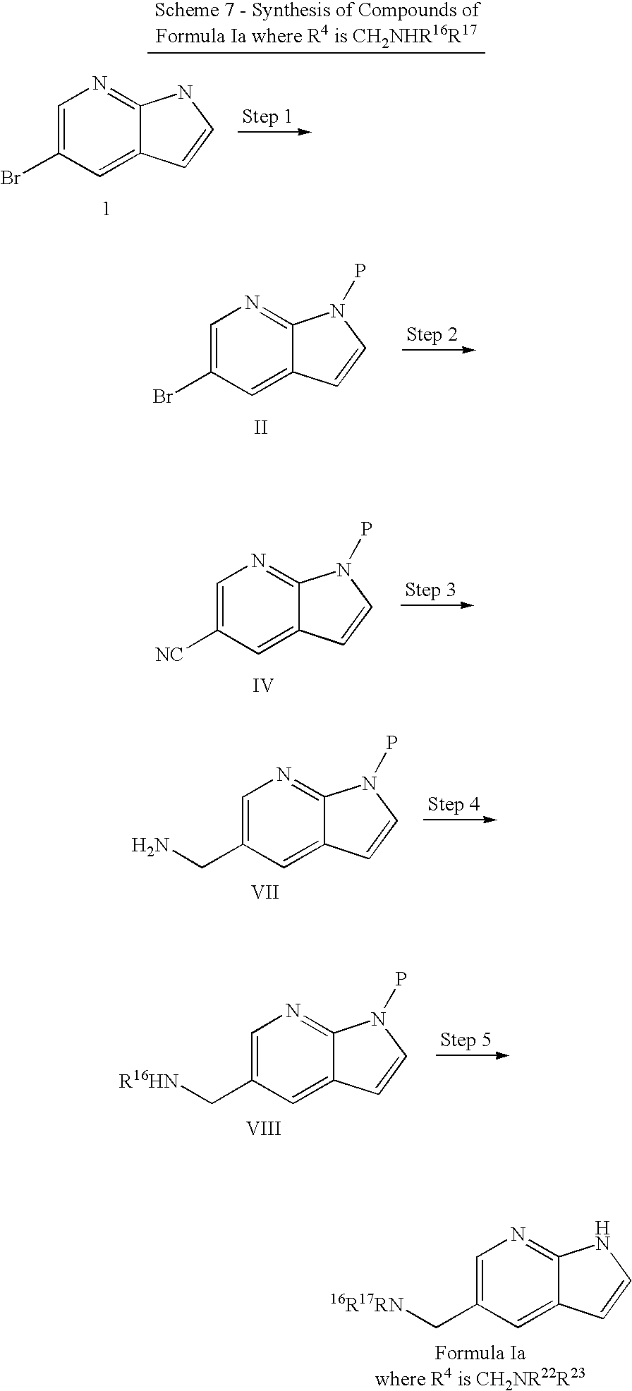
- Compound of formula II, where P is a protecting group, was synthesized by reacting compound 1 with a base (e.g. sodium hydride) in a solvent (e.g. THF), followed by an appropriate reagent (P-X, e.g. triisopropylsilylchloride) for introduction of a protecting group. The reaction was allowed to proceed, typically at room temperature, for 8-12 hours and the desired product was isolated by standard procedures (e.g. extraction and silica gel column chromatography) (Greene, T. W.; Wuts, P. G. M. Protective Groups in Organic Synthesis I, 3rd ed.; John Wiley & Sons: New York, 1981).
- Compound of formula IV, where R4 is CN, was synthesized by reacting compound of formula II with sodium cyanide in a polar aprotic solvent (e.g. DMF) in an inert atmosphere, in presence of catalysts (e.g. Tris(dibenzylideneacetone)dipalladium(0) and cuprous iodide) following the procedure described by Buchwald et. al., J. Am. Chem. Soc., 2003, 125, 2890-2891, by substituting 5-bromo-7-azaindole for 5-bromo-indole.
- Compound of formula VII, where R4 is CH2NH2, can be synthesized from compound of formula IV under hydrogenation condition using a catalyst (e.g. PtO2) in an atmosphere of H2 as described by Secrist III et. al., J. Org. Chem., 1972, 37, 335-336.
- Compound of formula VIII, where R4 is CH2NHR16, can be synthesized from compound of formula VII with an electrophilic reagent (e.g. benzyl bromide, benzenesulfonyl chloride, benzoyl chloride, phenyl isocyanate, phenyl isothiocyanate) in a polar aprotic solvent (e.g. DMF) in an inert atmosphere, in presence of a base (e.g. K2CO3, Et3N). The product was isolated by standard methods (aqueous work up and silica gel column chromatography).
- Compound of formula VIIIa, where R4 is CH2NHR16R17, can be synthesized from compound of formula VIII with an electrophilic reagent (e.g. benzyl bromide, benzenesulfonyl chloride, benzoyl chloride, phenyl isocyanate, phenyl isothiocyanate) in a polar aprotic solvent (e.g. DMF) in an inert atmosphere, in presence of a base (e.g. K2CO3, Et3N), followed by deprotection of the protective group with appropriate conditions.

- Compound of formula II, where P is a protecting group, was synthesized by reacting compound 1 with a base (e.g. sodium hydride) in a solvent (e.g. THF), followed by an appropriate reagent (P-X, e.g. triisopropylsilylchloride) for introduction of a protecting group. The reaction can be allowed to proceed typically at room temperature for 8-12 hours and the desired product isolated by standard procedures (e.g. extraction and silica gel column chromatography) (Greene, T. W.; Wuts, P. G. M. Protective Groups in Organic Synthesis I, 3rd ed.; John Wiley & Sons: New York, 1981).
- An intermediate compound of formula IX, where R4 is OR16, can be synthesized by reacting compound of formula II with a reagent of formula R16OH (e.g. methanol or water) in the presence of base (e.g. sodium methoxide or sodium hydroxide) and copper (I) bromide in a solvent (e.g. N,N-dimethylformamide) typically with heating to reflux for 2-8 hours as described by Mazeas, et. al. in Heterocycles, 1999, 50, 1065. The desired intermediate can be purified by conventional means (e.g. silica gel chromatography). When R16 is hydrogen, this intermediate can be additionally substituted in Step 3 or it can be used directly in Step 4 to provide compound of formula Ia where R4 is OR22 and R22 is not —C(X)R20, —C(X)NR16R17, or —S(O)2R21.
- The intermediate from Step 2 can be further modified when R16 is hydrogen. In this case, the intermediate from Step 2 can be reacted with a base (e.g. sodium hydride) in a solvent (e.g. N,N-dimethylformamide), followed by reaction with an alkylating reagent (e.g. benzyl bromide) or an acylating reagent (e.g. benzoyl chloride, phenyl isocyanate) typically at room temperature or with heating up to 80° C. for 1-12 hours. The desired product can be purified by conventional means (e.g. silica gel chromatography).
- Compound of formula Ia, where R4 is OR22, can be synthesized by reacting compound formula IX with an appropriate reagent to remove the protecting group, P, (e.g. tetrabutylammonium fluoride) in an appropriate solvent (e.g. methanol). The final product can be isolated by standard procedures (e.g. extraction).

- Compound of Formula Ia, where R4 is SR16, can be prepared by reacting compound 1 with a strong base (e.g. potassium hydride or t-butyl lithium) and dialkyldisulfides (e.g. dimethyldisulfane) or thiophenols (e.g. 4-methoxythiophenol) in a polar aprotic solvent (e.g. N,N-dimethylformamide) in an inert atmosphere following the procedure described by Yang et. al., Heterocycles, 1992, 34, 1169, by substituting 5-bromo-7-azaindole for 5-bromo-indole.

- Compounds of Formula Ia, where R4 is S(O)R16, or S(O)2R16 can be prepared by reacting compound 1a where R4 is R16 with 1 or 2 equivalents of Oxone, respectively in a polar solvent, using standard procedures.
C. Synthesis of Compound of Formula Ib, where R1, R2, R4, and R5 are hydrogen:
- Compounds of Formula Ib are Formula I compounds in which R3 is the only substituent on the core structure. Exemplary synthetic schemes for groups of compounds within Formula Ib are shown in
Schemes 
- Compound of formula Ib, where R3 is aryl or heteroaryl, were synthesized from compound 6 under Suzuki reaction conditions using aryl or heteroaryl boronic acids (e.g. Phenyl boronic acid, 3-thienyl boronic acid) (M. Allegretti, Synlett, 2001, 5, p. 609).

- Compound of formula Ib, where R3 is OR22, can be synthesized by heating compound 6 with aqueous base (e.g. aq. NaOH) in presence of an alcohol (e.g. methanol, benzyl alcohol) at higher temperatures (e.g. 150° C.) for required time, typically 12 h, as described by Girgis et al. in J. Heterocyclic Chemistry, 1989, 26, 317. The product can be isolated by following standard work up procedure.

- Compound of Formula Ib, where R3 is NR16R17, was synthesized by heating compound 6 with an amine (e.g. dimethylamine, N-methylaniline) and heated in an oil bath, typically to 180° C., for 1-5 hours. The product can be isolated by following either standard procedures or purifying by silica gel column chromatography (Nabih, et. al. J. Heterocyclic Chemistry, 1989, 26, 317).

- Compound of formula X can be synthesized by reacting compound 6 with a silyl chloride (e.g. triisopropylsilyl chloride) in an inert solvent (e.g. THF), in the presence of a base (e.g. NaH), as described by Greene, T. W.; Wuts, P. G. M. Protective Groups in Organic Synthesis I, 3rd ed.; John Wiley & Sons: New York, 1981. The product can be purified by standard chromatographic techniques.
- Compound of formula XI was synthesized by reacting compound X with a cyanide source (e.g. Zn(CN)2) in a polar aprotic solvent (e.g. DMF), in the presence of a catalyst (e.g. Tetrakistriphenylphosphine palladium) as described in Anderson et al J. Org Chem. 1998; 63, 8224 A standard workup is used and compound of formula XI can be purified by standard crystallization or chromatographic methods.
- Compound of formula XII can be prepared by reacting compound XI with a base (e.g. NaOH) in an inert solvent (e.g. EtOH) as described in Larock, R. C. Comprehensive Organic Transformations; VCH: NY, 1989, p. 993. Standard workup procedures and purifications are performed.
- Compound of formula XIII can be prepared by reacting compound XII with an amine (e.g. benzyl amine) in an inert solvent (e.g. DMF), in the presence of a coupling reagent (e.g. bromotri(pyrrolidino)phosphonium hexafluorophosphate (PyBrop)), and in the presence of a base (e.g. diisopropylethyl amine) as described in Coste, J. et al J. Org. Chem. 1994; 158, 2437. A typical workup is utilized and purification is achieved by standard chromatographic procedures.
- Compound of formula Ib, where R3 is CONR16R17 can be prepared by reacting compound XIII with a fluoride source (e.g. NH4F) in an inert solvent (e.g. THF) as described in Tetrahedron Lett. 2001, 42(44); 7759. Purification is achieved with standard chromatographic techniques.

- Compound of formula X was synthesized by reacting compound 6 with a sulfonyl chloride (e.g. benzene sulfonyl chloride) in an inert solvent (e.g. dichloromethane), in the presence of a base (e.g. sodium hydroxide) as described in Greene, T. W.; Wuts, P. G. M. Protective Groups in Organic Synthesis I, 3rd ed.; John Wiley & Sons: New York, 1981. The product can be isolated by filtration of the resulting slurry over celite.
- Compound of formula XI was synthesized by reacting compound X with a cyanide source (e.g. Zn(CN)2) in a polar aprotic solvent (e.g. DMF), in the presence of a catalyst (e.g. Tetrakis triphenylphosphine palladium) as described in Anderson et al J. Org. Chem. 1998; 63, 8224. A standard workup is used and purified by standard crystallization or chromatographic methods.
- Compound of formula XIV was prepared by reacting compound XI with a reducing agent (e.g. H2) in an inert solvent (e.g. MeOH) with an added catalyst (e.g. palladium on carbon) as described by Stavenger, R. A. et al Pct Int Appl., WO 03/028724
A1 10 Apr. 2003. Purification can be achieved by standard chromatographic procedures. - Compound of formula XV was prepared by reacting compound XIV with an alkylating agent (e.g. benzyl bromide) in an inert solvent (e.g. DMF), in the presence of a base (e.g. Diisopropylethyl amine). A standard workup is used and purified by standard crystallization or chromatographic methods.
- Compound of formula XVI can be prepared by reacting compound XV with with an alkylating agent (e.g. benzyl bromide) in an inert solvent (e.g. DMF), in the presence of a base (e.g. Diisopropylethyl amine). A standard workup is used and purified by standard crystallization or chromatographic methods.
- Compound of formula 1b, where R3 is CH2NR22R23 was synthesized by treating compound of formula XVI with a base (e.g. KOH) in a polar aprotic solvent (e.g. EtOH) while heating from 60-80° C. for typically 1-3 h as described in Greene, T. W.; Wuts, P. G. M. Protective Groups in Organic Synthesis I, 3rd ed.; John Wiley & Sons: New York, 1981. A standard workup is used and purified by standard chromatographic methods.

- Compound 7 was synthesized by reacting compound 2 with 85% meta-chloroperoxybenzoic acid in a solvent (e.g. 1,2-dimethoxyethane) typically at room temperature for 1-4 hours as described (Schneller and Luo, J. Org. Chem., 1980, 45, 4045). The resulting solid can be collected by filtration and washed with ethyl ether. The solid can be suspended in water and basified with an aqueous base (e,g, potassium carbonate). Upon cooling, the precipitate can be collected by filtration and purified by conventional means (e.g. recrystallization) to provide compound 7.
- Compound 8 was synthesized by reacting compound 7 with fuming nitric acid in a solution of trifluoroacetic acid typically at 0° C. as described (Schneller and Luo, J. Org. Chem., 1980, 45, 4045). The reaction mixture is immediately poured onto ice and basified with sodium hydroxide to provide a precipitate that can be collected by filtration. Purification by standard procedures (e.g. recrystallization) can provide compound 8.
- Compound 9 was synthesized by reacting compound 8 with phosphorous trichloride in a solvent (e.g. ethyl acetate) with heating typically at 80° C. for a few minutes as described (Schneller and Luo, J. Org. Chem., 1980, 45, 4045). The reaction mixture is cooled and neutralized with aqueous base (e.g. sodium carbonate) and extracted with an organic solvent (e.g. ethyl acetate). Compound 9 can be isolated from the organic portions and purified by conventional means (e.g. recrystallization).
- Compound of formula XVII, where P is a protecting group, can be synthesized by reacting compound 9 with a base (e.g. sodium hydride) in a solvent (e.g. THF), followed by an appropriate reagent (P-X, e.g. triisopropylsilylchloride) for introduction of a protecting group. The reaction can be allowed to proceed typically at room temperature for 8-12 hours and the desired product isolated by standard procedures (e.g. extraction and silica gel column chromatography).
- Compound of formula XVIII can be synthesized by reacting compound of formula XVII with a reducing agent (e.g. hydrogen gas) in the presence of a catalyst (e.g. Raney nickel) in an appropriate solvent (e.g. methanol) typically at room temperature for 2-4 hours as described (Antonini et. al. J. Med. Chem. 1982, 25, 1258). Compound of formula XVIII can be isolated by standard procedures (e.g. filtration and evaporation).
- Compound of formula XIX can be synthesized by reacting compound of formula XVII with one of many possible alkylating or acylating agents (e.g. isobutyl bromide, benzoyl chloride, phenyl isocyanate, or phenylsulfonyl chloride). The compound of formula XIX can be purified by conventional means (e.g. silica gel chromatography).
- Compound of Formula Ib, where R3 is NR22R23, can be synthesized by reacting compound formula XIX with an appropriate reagent to remove the protecting group, P, (e.g. tetrabutylammonium fluoride) in an appropriate solvent (e.g. methanol). The final product can be isolated by standard procedures (e.g. extraction).
D. Synthesis of Compound of Formula Ic, where R1, R3, R4, and R5 are Hydrogen:
- Compounds of Formula Ic are Formula I compounds in which R2 is the only substituent on the core structure. Exemplary synthetic schemes for groups of compounds within Formula Ic are shown in Schemes 16-32, for different selections of R2.

-
Compound 10 was synthesized from commercially available 7-azaindole following the literature procedure (Robinson, J. Am. Chem. Soc., 1955, 77, p. 457). - Compound of formula XX was synthesized by deprotonation using base (e.g. BuLi, NaH) in aprotic solvent like THF or ether and reacting the anion with a silyl chloride (e.g. TIPS) or an anhydride (e.g. Boc anhydride). The product was isolated by following standard procedure (quenching with ice-cold brine, work up, and purification by flash silica gel chromatography).
- Compounds of Formula Ic was synthesized through the reaction of compounds of formula XX with isopropyl chloroformate (or ethyl chloroformate) at room temperature in toluene to give a 3-chloromethyl intermediate. This intermediate cooled to −78° C. and was immediately reacted with an organocopper reagent, which was generated from the reaction between a Grignard reagent (or organolithium reagent) and a solution of copper cyanide and LiCl. The mixture was stirred at −78° C. for one hour then allowed to warm to room temperature. The reaction was quenched with a solution of 4:1 ammonium chloride: ammonium Hydroxide. The reaction was worked up in the usual manner and purified by flash silica gel chromatography to give the nitrogen-protected product. The final product can be realized through the deprotection of the protecting group (Boc, TIPS) using standard conditions (TFA or NH4F) at room temperature.

- Compound 11 was synthesized by reacting commercially available 7-azaindole, compound 2, with hexamethyltetramine and acetic acid in water with heating to reflux for two hours. After cooling, the desired product precipitated and was collected by filtration.
- Compound of formula XXI, where P is a protecting group, was synthesized by reacting compound 11 with an appropriate reagent to introduce a protecting group (e.g. tert-butyloxycarbonyl di anhydride) and a base (e.g. sodium hydride) in a solvent (e.g. THF) typically at room temperature for 12-18 hours. The product can be isolated by conventional means (e.g. extraction).
- Compound of formula XXII was synthesized by reacting compound of formula XXI in a solvent (e.g. 1,2-dimethoxyethane) with a Grignard reagent of the formula R24MgCl (e.g. phenyl magnesium bromide) or an equivalent nucleophile in a solvent (e.g. THF) under inert atmosphere cooled typically to −10° C. The reaction was typically allowed to warm to room temperature and stirred for 12-18 hours. The desired product was purified by reverse phase high pressure liquid chromatography.
- An intermediate of compound of formula Ic was synthesized by reacting compound of formula XXII with a reducing agent (e.g. sodium borohydride) in a solvent (e.g. ethanol) typically with heating to 80° C. for 1-4 hours. The reaction was quenched with the addition of methanol and concentrated and purified by reverse phase high performance liquid chromatography.
- Compound of formula Ic where R2 is aralkyl or heteroaralkyl and R24 is aryl or heteroaryl was synthesized by reacting the intermediate from Step 4 with an appropriate reagent to remove the protecting group, P, (e.g. hydrochloric acid) in an appropriate solvent (e.g. dioxane). The final product was isolated by standard procedures (e.g. reverse phase preparative high pressure liquid chromatography).

- Compound of formula Ic, where R2 is aralkyl or heteroaralkyl and R24 is aryl or heteroaryl, was synthesized by reacting compound 2 with an activating agent (e.g. methyl magnesium bromide and zinc dichloride or anhydrous aluminum chloride) and an aralkyl bromide (e.g benzyl bromide) or heteroaralkyl bromide (pyridine benzyl bromide) in an inert solvent (e.g. methylene chloride), under inert atmosphere (e.g. argon), at room temperature or with heating up to reflux for 18-24 hours. The product was isolated by standard procedures (e.g. extraction and silica-gel chromatography).

- Compound of the formula XX was prepared from the
compound 10 by deprotonation with a strong base (e.g. NaH, BuLi) at 0° C. in an aprotic solvent (THF), followed by the addition of P-X (e.g. TIPS-Cl, Boc anhydride). The product was isolated by following standard workup procedure. - Compound of the formula XXIII was prepared from the compound of formula XX by addition of chloroformate (e.g., ethyl formate, isopropyl formate) at 25° C. in an aprotic solvent (toluene). The product was isolated by following standard workup and silica gel flash chromatography.
- Compound of the formula XXIIIa was prepared from the compound of formula XXIII by addition of sodium Iodide in acetone at 60° C. After several hours, typically 4 h, concentrated down to dryness and the residue was dissolved in DMF followed by addition of sodium azide. The reaction was stirred, typically at room temperature, for a a short period of time, 1 h. The product was isolated by following standard workup procedure and silica gel flash chromatography yielded azide intermediate.
- Compound of the formula XXIV was prepared from azide of formula XXIIIa by hydrogenation in an inert solvent (e.g. THF) with catalytic amount of acid (e.g., HCl, Acetic acid) and in presence of a catalyst (e.g., Pd/C). The product was isolated by following standard workup procedure.
- Compound of the formula XXV was prepared from the compound of formula XXIV, by using various electrophilic groups (isocyanates, sulfonyl chloride) in presence of a strong base of (e.g., NaH, BuLi, TEA) in an inert solvent (e.g. THF). Products were isolated by following standard workup and silica gel flash chromatography.
- Compound of the formula Ic where R2 is CH2NR22R23 was prepared from the compound of formula XXV, by addition of an acid (e.g., HCl, TFA) in dichloromethane at room temperature. The product was isolated by following standard workup procedure.

- Compound of formula Ic where R2 is CH2NR22R23 was synthesized from compound of formula XXI by reductive amination with sulfonamide, amide, or urea (e.g. benzene sulfonamide, benzamide, phenyl urea) in an inert solvent (e.g. THF) and sodium triacetoxy borohydride and deprotecting the resulting product. The final product was isolated by silica gel column chromatography (Dube and Scholte; Tetrahedron Lett. 1999, 40, 2295).

- Compound 12 was synthesized by reacting compound 2 with a brominating agent (e.g. Bromine) in a co-solvent (e.g.carbon tetrachloride:chloroform) as described in Synthesis 1999, 4; 615-620. A typical phase extraction is performed and the resulting aqueous layer is treated with a base (e.g. NaOH). The resulting solid is isolated by standard filtration techniques.
- Compound of formula XXVI was synthesized by reacting compound 12 with a sulfonyl chloride (e.g. benzene sulfonylchloride) in an inert solvent (e.g. dichloromethane), in the presence of a base (e.g. sodium hydroxide) as described in Greene, T. W.; Wuts, P. G. M. Protective Groups in Organic Synthesis I, 3rd ed.; John Wiley & Sons: New York, 1981. The product can be isolated by filtration of the resulting slurry over celite.
- Compound of formula XXVII was synthesized by reacting compound XXVI with a cyanide source (e.g. Zn(CN)2) in a polar aprotic solvent (e.g. DMF), in the presence of a catalyst (e.g. Tetrakis triphenylphosphine palladium) as described in Anderson et al J. Org. Chem. 1998; 63, 8224. A standard workup is used and purified by standard crystallization or chromatographic methods.
- Compound of formula XXVIIa was prepared from compound of formula XXVII with a reducing agent (e.g. H2) in an inert solvent (e.g. MeOH) with an added catalyst (e.g. palladium on carbon) as described by Stavenger, R. A. et al Pct Int Appl., WO 03/028724. Purification was achieved by standard chromatographic procedures.
- Compound of formula XXVIIb was prepared from compound of formula XXVIIa by treating with an alkylating agent, sulfonylating agent, acid chlorides, or isocyanates and isothiocyanates (e.g. benzyl bromide, benzene sulfonyl chloride, phenyl isocyanate) in an inert solvent (e.g. DMF), in the presence of a base (e.g. diisopropylethyl amine; DMAP). A standard workup is used and purified by standard crystallization or chromatographic methods.
- Compound of 1c, where R2 is CH2NR22R23 can be generated by treating compound of formula XXVIIb with a base (e.g. KOH) in a polar aprotic solvent (e.g. EtOH) while heating from 60-80° C. for typically 1-3 h as described in Greene, T. W.; Wuts, P. G. M. Protective Groups in Organic Synthesis I, 3rd ed.; John Wiley & Sons: New York, 1981. A standard workup is used and purified by standard chromatographic methods.

- Compound of formula XXVIII can be prepared by reacting compound XXVII with a base (e.g. NaOH) in an inert solvent (e.g. EtOH) as described in Larock, R. C. Comprehensive Organic Transformations; VCH: NY, 1989, p. 993. Standard workup procedures and purifications are performed.
- Compound of formula XXIX can be prepared by reacting compound XXVIII with an amine (e.g. benzyl amine) in an inert solvent (e.g. DMF) in the presence of a coupling reagent (e.g. bromotri(pyrrolidino)phosphonium hexafluorophosphate (PyBrop)), and in the presence of a base (e.g. diisopropylethyl amine) as described in Coste, J. et al J. Org. Chem. 1994; 158, 2437. A typical workup is utilized and purification is achieved by standard chromatographic procedures.
- Compound of Formula 1c, where R2 is CONR16R17 can be prepared by reacting compound XIII with a fluoride source (e.g. NH4F) in an inert solvent (e.g. THF) as described in Tetrahedron Lett. 2001, 42(44); 7759. Purification is achieved with standard chromatographic techniques.

- Compound of formula XXI, where R2 is CHO, was synthesized by reacting compound 12 with appropriate protective groups (e.g. Boc, TIPS).
- Compound of formula XXVIII, where R2 is CO2H, was synthesized by reacting compound of formula XXI with an oxidant (e.g. NaClO2 and NH2SO3H) in appropriate solvents (e.g. a mixture of THF and H2O) as described by Merour et. al., Synthesis, 2000, 549-556.
- Compound of formula Ic, where R2 is CONR16R17, was synthesized by reacting compound of formula XXVIII with an amine (e.g. aniline, dimethyl amine) in a polar aprotic solvent (e.g. DMF) in an inert atmosphere in presence of PyBrop (Bromotri(pyrrolidino)phosphonium hexafluorophosphate), followed by deprotection of the protective group with appropriate reagents.

- Compound of formula XXVIII can be prepared by reacting compound of formula XXVI with a lithium reagent (e.g. t-Butyl lithium) at low temperatures, −78° C., in an inert solvent (e.g. THF) and stirring for a short period of time, 1 h, followed by the reaction with gaseous CO2 and worked up in the usual manner. The product can be purified by flash silica gel chromatography.
- Compound of formula Ic, where R2 is CONR16R17, was synthesized by reacting compound of formula XXVIII with an amine (e.g. aniline, dimethyl amine) in a polar aprotic solvent (e.g. DMF) in an inert atmosphere in presence of PyBrop (Bromotri(pyrrolidino)phosphonium hexafluorophosphate), followed by deprotection of the protective group with appropriate reagents.

- Compounds of formula XXX can be prepared from compounds of formula XXIX by reacting it with Lawesson's reagent (or P4S10) as described in the literature. (Bull. Soc. Chim. Belg., 1978, 87, 223).
- Compounds of Formula Ic where R2 is CSNR16R17 can be prepared by deprotection of compound of formula XXX with acid (e.g. HCl; TFA) and purifying the product by column chromatography.

- Compound of formula XXXI was synthesized by reacting compound 2 with a sulfonyl chloride (e.g. benzenesulfonyl chloride) in an inert solvent (e.g. dichloromethane), in the presence of a base (e.g. sodium hydroxide) as described in Greene, T. W.; Wuts, P. G. M. Protective Groups in Organic Synthesis I, 3rd ed.; John Wiley & Sons: New York, 1981. The product can be isolated by filtration of the resulting slurry over celite.
- Compound of formula XXXII was prepared by reacting compound XXXI with a nitrating agent (e.g. fuming HNO3) neat while maintaining the temperature for 1-3 h between 0-25° C. as described in Robinson, B. L. et al J. Am. Chem. Soc. 1959, 81; 743. The mixture can be poured over ice and neutralized with a base (e.g. NH4OH) and the resulting precipitate can be filtered and dried under vacuum. Purification can be achieved through standard chromatographic techniques.
- Compound of formula XXXII was prepared by reacting compound XXXII with a reducing agent (e.g. H2) in a polar solvent (e.g. MeOH) with an added catalyst (e.g. palladium on carbon) as described by Stavenger, R. A. et al Pct Int Appl., WO 03/028724. Purification can be achieved by standard chromatographic procedures.
- The precursor to compound of Formula 1c, where R2 is NR22R23 can be prepared by reacting compound XXXIII with an alkylating agent, sulfonylating agent, acid chlorides, or isocyanates and isothiocyanates (e.g. benzyl bromide, benzene sulfonyl chloride, phenyl isocyanate) in an inert solvent (e.g. DMF), in the presence of a base (e.g. diisopropylethyl amine; DMAP) followed by deprotection and standard workup is used and purification by standard chromatographic methods.

- Compound of formula Ic, where R2 is C(X)R20 and X is ‘O’, was synthesized by reacting compound 2 with an acid chloride (e.g. benzoyl chloride) in the presence of a Lewis acid (e.g. aluminum trichloride) in a inert solvent (e.g. methylene chloride) and under inert atmosphere (e.g. argon) at room temperature or with heating up to reflux for 1-18 hours. The product was isolated by extraction and silica gel column chromatography.

- Compounds of formula XXIX can be prepared from compounds of formula XXVIII by reacting it with N,N-dimethylamine. HCl in the presence of a coupling reagent (BOP or 2-Chloro-1-methylpyridinium iodide) (J. Org. Chem., 1996, 61, 4999; or Synth. Commun., 1995, 25, 1277).
- Compounds of formula XXX can be prepared from compounds of formula XXIX by reacting with Grignard reagents (e.g. phenyl magnesium bromide) or lithium reagents (e.g. Phenyl lithium) in inert solvent (e.g. diethyl ether or THF) at low temperature, typically 0° C., warming up to room temperature, and stirring for a short period of time, typically 1 h. The reaction can be quenched with dilute hydrochloric acid (3N HCl), subjected to standard work up conditions and purified by column chromatography (Olah, et. al. Synthesis, 1984, 3, 228).
- Compound of formula Ic where R2 is C(X)R20 and X is ‘O’ can be prepared by deprotection of compound XXX using appropriate deprotecting agent (e.g. TBAF or aqueous NaOH) and purifying the products by standard conditions.

- Compounds of formula XXXI can be prepared from compounds of formula XXVIII by reacting it with N,O-dimethylhydroxylamine HCl in the presence of a coupling reagent (BOP Reagent or 2-Chloro-1-methylpyridinium iodide). (J. Org. Chem., 1996, 61, 4999; or Synth. Commun., 1995, 25, p. 1277).
- Compounds of formula XXX can be prepared from compounds of formula XXXI by reacting with Grignard reagents according to the reference (Tetrahedron Letters, 1981, 22, 3815).
- Compounds of Formula Ic can be prepared from compound of formula XXX in the same manner as shown above in scheme 24.

- Compound of the formula Ic, where R2 is S(O)nR21 where n=0, was prepared from commercially available compound 2 by deprotonation of a strong base (e.g. NaH) in an inert solvent (e.g. DMF), followed by addition of diaryl disulfides (e.g. PhSSPh). The reaction typically was ran overnight at room temperature and the product was isolated by following standard workup and silica gel flash chromatography (Atkins et al. 1988, 480).
- Compound of the formula Ic, where R2 is S(O)nR where n=1 was prepared from the compound of formula Ic where R2 is S(O)nR21 where n=0 by addition of oxidizing reagents (m-CPBA, Oxone, 0.5 Eqiv.) in dichloromethane. The product was isolated by following standard workup and silica gel flash chromatography.
- Compound of the formula Ic, where R2 is S(O)nR21 where n=2 was prepared from the compound of formula Ic where R2 is S(O)nR21 where n=1 by addition of oxidizing reagents (m-CPBA, Oxone, 2.0 Eqiv.) in dichloromethane. The product was isolated by following standard workup and silica gel flash chromatography.

- Compound of the formula Ic, where R2 is S(O)nR21 where n=2 can be prepared from the compound 2 by reaction with a sulfonyl chloride (e.g. benzne sulfonylchloride) in a polar solvent (e.g. ethanol, acetone) at room temperature as described by Ottoni et al in Tetrahedron, 1998, 54, 13915. The product can be isolated by standard work up procedure.

- Compound of formula XXXI, where P is a protecting group (e.g. phenylsulfonyl), was synthesized by reaction of compound of formula XXXVI with an aryl or heteroaryl boronic acid (e.g. phenyl boronic acid) in the presence of a base (e.g. sodium carbonate) and a catalyst composed of a metal (e.g. Tris(dibenzylideneacetone)dipalladium(0)) and a ligand (tri-tert-butylphosphine), and with heating typically to 75° C. for 8-12, hours.
- Compound of formula Ic, where R2 is aryl or heteroaryl, was synthesized by reacting compound of formula XXXI with an appropriate reagent to remove the protecting group, P, (e.g. potassium hydroxide) in an appropriate solvent (e.g. ethanol). The final product was isolated by standard procedures (e.g. reverse phase preparative high pressure liquid chromatography).
E. Synthesis of Compound of Formula Id, where R′, R3, and R5 are Hydrogen:
- Compounds of Formula Id are Formula I compounds in which R2 and R4 are the only substituents on the core structure. Exemplary synthetic schemes for groups of compounds within Formula Id are shown in Schemes 33-38, for different selections of R2 and R4.

- Compound 13, where R2 is CH2NMe2, was synthesized under Mannich reaction condition using paraformaldehyde and dimethyl amine hydrogen chloride salt in an alkanol solvent (e.g. isopropanol) as described by Robinson, in J. Am. Chem. Soc., 1955, 77, p. 457.
- Compound of formula XXXII, where R2 is CH2NMe2, was synthesized by reacting compound 12 with appropriate protective groups (e.g. Boc, TIPS).
- Compound of formula XXIII, where R4 is aryl or heteroaryl, was synthesized from compound XXXII under Suzuki reaction conditions using aryl or heteroaryl boronic acids (e.g. phenyl boronic acid, 3-thienyl boronic acid), in presence of a catalyst (e.g. Pd(PPh3)4).
- Compound of formula Id, where R4 is aryl or heteroaryl, can be synthesized by reacting compound of formula XXIII with ethyl chloroformate or isopropyl chloroformate in an inert solvent (e.g. toluene).
- Compound of formula Id, where R2 is (CH2)nR24 and R24 is aryl or heteroaryl, was synthesized by reacting compound of formula XXIV with a Grignard reagent (e.g. phenyl magnesium bromide, benzylmagnesium bromide) in an inert solvent (e.g. THF), in presence of a catalyst (e.g. CuCl.2LiCl). The protective group (e.g. TIPS) can either be cleaved with an appropriate reagent (e.g. TBAF), or be cleaved during the reaction (e.g. Boc).

- Compound of the formula XXXV was prepared from the compound of formula 1, under Friedel-Craft alkylation conditions used electrophilic reagents (e.g., aryl halide, heteroaryl halide, etc.), in presence of a Lewis acid (e.g., aluminum chloride) in dichloromethane at room temperature. The product was isolated by following standard workup procedure.
- J Compound of the formula XXXVI was prepared from the compound of formula XXXV, by deprotonation with a strong base (e.g. NaH, BuLi, etc.) at 0° C. in an aprotic solvent (THF), followed by protecting groups (e.g., TIPS-Cl, Boc anhydride, etc.). The product was isolated by following standard workup procedure.
- Compound of the formula XXXVII was prepared from the compound of formula XXXVI, under suzuki reactions using boronic acids (e.g., aryl or heteroaryl), base (e.g., potassium carbonate, Triethylamine, Sodium hydroxide, etc.), and catalysis (e.g. Pd(Ph3P)4) in aqueous/THF solvent system. After 12 hours, the product was isolated by following standard workup and silica gel flash chromatography.
- Compound of the formula Id was prepared from the compound of formula XXXVII, deprotection of protecting groups using acids (e.g. HCl, TFA, etc.) in dichloromethane. The product was isolated by following standard workup and silica gel flash chromatography.

- Compound of the formula XXXVIII was prepared from the compound of formula 1, under friedel-Craft acylation conditions acid chlorides (e.g. aryl, heteroaryl, etc.), in presence of a Lewis acid (e.g., aluminum chloride) in dichloromethane at room temperature. The product was isolated by following standard workup and silica gel flash chromatography.
- Compound of the formula XXXIX was prepared from the compound of formula XXXVIII, by deprotonation with a strong base (e.g. NaH, BuLi, etc.) at 0° C. in an aprotic solvent (THF), followed by addition of protecting groups (TIPS-Cl, Boc anhydride, etc.). The product was isolated by following standard workup procedure.
- Compound of the formula XL was prepared from the compound of formula XXXIX, under suzuki reactions using boronic acid (e.g., aryl or heteroaryl), base (e.g., potassium carbonate, Triethylamine, sodium hydroxide), and catalysis (e.g., Pd(Ph3P)4) in aqueous/THF solvent system. After 12 hour, the product was isolated by following standard workup and silica gel flash chromatography.
- Compound of the formula Id was prepared from the compound of formula XL, deprotection of protecting groups using acids (e.g., HCl, TFA) in dichloromethane. The product was isolated by following standard workup and silica gel flash chromatography.

- Compound of the formula XLI was prepared from the compound of formula 1 by addition of a strong base (e.g., NaH) in dichloromethane followed by addition of disulfide molecules (e.g., PhSSPh). The reaction ran overnight at room temperature. The product was isolated by following standard workup and silica gel flash chromatography.
- Compound of the formula XLII was prepared from the compound of formula XLI by addition of oxidizing reagents (e.g., MCPBA, Oxone, 2.0 Eqiv.) in dichloromethane ran overnight. The product was isolated by following standard workup and silica gel flash chromatography.
- The synthesis of an intermediate of compound of the formula XLIII can be prepared from the compound of formula XLII by deprotonated using a strong base (BuLi, NaH, etc.) followed by addition of protecting groups (e.g., TIPS-Cl, Boc anhydride, etc.) in an inert solvent THF, yielded formula XLIII.
- Compound of the formula XLIII can be prepared from the compound of formula XLII, under suzuki reactions using boronic acids (e.g., aryl or heteroaryl), base (e.g., potassium carbonate, Triethylamine, Sodium hydroxide, etc.), and catalysis (e.g. Pd(Ph3P)4) in aqueous/THF solvent system. After 12 hours, the product was isolated by following standard workup and silica gel flash chromatography.
- Compound of the formula Id can be prepared from the compound of formula XLIII, deprotection of protecting groups using acids (e.g., HCl, TFA, etc.) in dichloromethane. The product was isolated by following standard workup and silica gel flash chromatography.

- Compound of formula XXXVIII was synthesized by reacting compound 1 with a Lewis acid (e.g. aluminum trichloride) in a solvent (e.g. dichloromethane) under an inert atmosphere, typically at room temperature for 1-2 hours, followed by addition of an acid chloride (e.g. benzoyl chloride or nicotinoyl chloride) and reaction for 2-12 hours. The reaction was quenched with methanol, concentrated, and purified by silica gel chromatography.
- Compound of formula XXXIX, where P is a protecting group, was synthesized by reacting compound XXXVIII with a base (e.g. sodium hydride) in a solvent (e.g. THF), followed by an appropriate reagent for introduction of a protecting group (P-X, e.g. triisopropylsilylchloride). The reaction was allowed to proceed typically at room temperature for 8-12 hours and the desired product was isolated by standard procedures (e.g. extraction and silica gel column chromatography).
- An intermediate of compound of formula XLIV, where R20 is aryl or heteroaryl, and R4 is NR16R17 was synthesized by reacting compound of formula XXXIX, where R20 is aryl or heteroaryl with an amine of the formula NHR16R17 (e.g. aniline) in a solvent (e.g. toluene), in presence of a base (e.g. sodium tert-butoxide) and a catalyst composed of a metal (e.g. Tris(dibenzylideneacetone)dipalladium(0)) and a ligand (e.g. tri-tert-butylphosphine) with heating typically to 95° C. for 8-12 hours as described (Thomas, et. al., J. Am. Chem. Soc., 2001, 123, 9404) by substituting compound of formula II for the N-substituted-3,6-dibromocarbazole. The desired compound was purified by silica gel chromatography. This intermediate was used directly in
Step 5 to provide compound of the formula Id where R20 is aryl or heteroaryl, and R4 is NR22R23 and R22 and R23 are not —C(X)R20, —C(X)NR16R17, or —S(O)2R21 or alternatively, it can be additionally substituted in Step 4. - The intermediate from Step 3 can be further modified when at least R16 or R17 is hydrogen. The intermediate from Step 3 can be reacted with a base (e.g. sodium hydride) in a solvent (e.g. N,N-dimethylformamide), followed by reaction with an alkylating reagent (e.g. benzyl bromide) or an acylating reagent (e.g. benzoyl chloride, phenyl isocyanate, or phenylsulfonyl chloride) typically at room temperature or with heating up to 80° C. for 1-12 hours. Compound of formula XLIV, where R20 is aryl or heteroaryl and R4 is NR22R23, can be purified by conventional means (e.g. silica gel chromatography).
- Compound of formula Id, where R2 is C(O)R20 where R20 is aryl or heteroaryl and R4 is NR22R23 was synthesized by reacting compound of formula XLIV with an appropriate reagent to remove the protecting group, P, (e.g. tetrabutylammonium fluoride) in an appropriate solvent (e.g. methanol). The final product can be isolated by standard procedures (e.g. extraction).

- Compound II, where R4 is Br, was synthesized by protecting compound 1 with appropriate protective groups (e.g. TIPS).
- Compound of formula XLV, where R4 is CO2R24, was synthesized by reacting compound of formula II with a strong base (e.g. n-butyllithium) and benzyl chloroformate or methyl chloroformate in an inert solvent (e.g. THF).
- Compound of formula XLVI, where R2 is C(O)R20, can be synthesized from compound XLV under Friedel-Crafts reaction conditions using acyl chloride (e.g. benzoyl chloride) in methylene dichloride, in presence of a Lewis acid (e.g. AlCl3).
- Compound of formula XLVII, where R4 is COOH and R2 is C(O)R20, can be synthesized by reacting compound of formula XLVI with an aqueous base (e.g. NaOH), or by hydrogenating compound of formula XLVII, where R4 is COOBn, under hydrogen in alcohol solvent (e.g. MeOH), in presence of a catalyst (e.g. 5% Pd/C).
- Compound of formula XLVIII, where R2 is C(O)R20 and R4 is C(O)NR16R17, can be synthesized by reacting compound of formula XLVII with an amine (e.g. benzylamine, dimethyl amine) in a nonpolar aprotic solvent (e.g. DMF) in an inert atmosphere in presence of PyBrop (Bromotri(pyrrolidino)phosphonium hexafluorophosphate) following the procedure described by Coste et. al., Journal of Organic Chemistry, 1994, 59, 2437.
- Compound of formula Id, where R2 is C(O)R20 and R4 is C(O)NR16R17, can be synthesized by cleaving the protective group (e.g. TIPS) in compound of formula XLVIII appropriate condition (e.g. TBAF).
- F. Synthesis of Compound of Formula Ig, where R1, R4, and R5 are Hydrogen:
- Compounds of Formula Ig are Formula I compounds in which R2 and R3 are the only substituents on the core structure. Exemplary synthetic schemes for groups of compounds within Formula Ig are shown in Schemes 39-43, for different selections of R2 and R3.

- Compound of formula XLIX, where R20 is aryl or heteroaryl, can be synthesized by reacting
compound 10 with a Lewis acid (e.g. aluminum trichloride) in a solvent (e.g. dichloromethane), typically at room temperature for 1-2 hours, followed by addition of an acid chloride (e.g. benzoyl chloride or nicotinoyl chloride) and reaction for 2-12 hours. The reaction is quenched with methanol, concentrated, and purified by silica gel chromatography. - Compound of Formula Ig, where R3 is NH2, and R2 is C(O)R20 where R20 is aryl or heteroaryl was synthesized by reaction of compound of formula XLIX with a reducing agent (e.g. hydrogen gas) in the presence of a catalyst (e.g. Raney nickel) in an appropriate solvent (e.g. methanol) typically at room temperature for 2-4 hours as described (Antonini et. al. J. Med. Chem. 1982, 25, 1258). The product was isolated by filtration and evaporation.

- An intermediate of compound of formula XLX, where P is a protecting group, can be synthesized by reacting compound of formula XLIX with a base (e.g. sodium hydride) in a solvent (e.g. THF), followed by an appropriate reagent for introduction of a protecting group (P-X, e.g. triisopropylsilylchloride). The reaction is allowed to proceed typically at room temperature for 2-6 hours and the product was isolated by standard procedures (e.g. extraction and silica gel column chromatography).
- Compound of formula XLX can be synthesized by reaction of the intermediate from Step 1 with a reducing agent (e.g. hydrogen gas) in the presence of a catalyst (e.g. Raney nickel) in a solvent (e.g. methanol) typically at room temperature for 2-4 hours as described (Antonini et. al. J. Med. Chem. 1982, 25, 1258). The product can be isolated by standard procedures (e.g. filtration and evaporation).
- Compound of formula XLXI, where R20 is aryl or heteroaryl, can be synthesized by reacting compound of formula XLX with a base (e.g. sodium hydride) in a solvent (e.g. dimethylformamide), followed by reaction with an alkylating reagent (e.g. benzyl bromide) or an acylating reagent (e.g. benzoyl chloride, phenyl isocyanate, phenylsulfonyl chloride) typically at room temperature or with heating up to 80° C. for 1-12 hours. The desired product can be purified by conventional means (e.g. silica gel chromatography).
- J Compound of formula XLXII, where R20 is aryl or heteroaryl, can be synthesized by reacting compound of formula XLXI with a base (e.g. sodium hydride) in a solvent (e.g. dimethylformamide), followed by reaction with an alkylating reagent (e.g. benzyl bromide) or an acylating reagent (e.g. benzoyl chloride, phenyl isocyanate, phenylsulfonyl chloride) typically at room temperature or with heating up to 80° C. for 1-12 hours. The desired product can be purified by conventional means (e.g. silica gel chromatography).
- Compound of formula Ig, where R3 is NR22R23 and R2 is C(O)R20 where R20 is aryl or heteroaryl can be synthesized by reacting compound of formula XLXII with an appropriate reagent to remove the protecting group, P, (e.g. tetrabutylammonium fluoride) in an appropriate solvent (e.g. methanol). The final product can be isolated by standard procedures (e.g. extraction).

- Compound of formula 1b, where R3 is aryl or heteroaryl can be synthesized by reacting compound 6 with a boronic acid (e.g. 3-mthoxyphenylboronic acid) in an inert solvent (e.g. dioxane), in the presence of a salt (e.g. KF), in the presence of a catalyst (e.g. Pd2(dba)3CHCl3). The reaction is carried out under an inert solvent and is typically heated (100° C.) for 4-12 h as described by Allegretti, M. et. al Synlett 2001; 5, 609. Purification is achieved with standard chromatographic techniques.
- Compound of formula 1g where R3 is aryl or heteroaryl and R2 is C(O)R20 where R20 is aryl or heteroaryl can be synthesized by reacting compound of formula 1b where R3 is aryl or heteroaryl with a Lewis acid (e.g. AlCl3) in an inert solvent (e.g. CH2Cl2) in the presence of an acid chloride (e.g. benzoyl chloride) as described by Katritzky, A. R. et al J. Org. Chem. 2003, 68, 5720. Purification is achieved with standard chromatographic techniques.

- Compound of formula XLXIII can be prepared by reacting compound of formula 6 with a Lewis acid (e.g. AlCl3) in an inert solvent (e.g. CH2Cl2) in the presence of an acid chloride (e.g. benzoyl chloride) as described by Katritzky, A. R. et al J. Org. Chem. 2003, 68, 5720. Purification is achieved with standard chromatographic techniques.
- Compound of formula 1g, where R3 is aryl or heteroaryl and R2 is C(O)R20 where R20 is aryl or heteroaryl can be synthesized by reacting compound of formula XLXIII with a boronic acid (e.g. 3-mthoxyphenylboronic acid) in an inert solvent (e.g. dioxane), in the presence of a salt (e.g. KF), in the presence of a catalyst (e.g. Pd2(dba)3CHCl3). The reaction is carried out under an inert solvent and is typically heated (100° C.) for 4-12 h as described by Allegretti, M. et. al Synlett 2001; 5, 609. Purification is achieved with standard chromatographic techniques.

- Compounds of formula XX were prepared according to the procedure outlined in
scheme 16. - Compounds of formula XLIX were prepared from compounds of the formula XX by using base (e.g., n-BuL, t-BuLi, etc.) to affect the deprotonation in aprotic solvent (Et2O, THF, etc.) at −78° C. The anion intermediate was then reacted with a bromide source (NBS, or dibromoethane) and allowed to warm to room temperature. The reaction was worked up in the usual manner. The compound was purified by flash silica gel chromatography.
- Compound of formula L, where R3 is aryl or heteroaryl, were synthesized from compounds of formula XLIX under Suzuki reaction conditions using aryl or heteroaryl boronic acids (e.g. Phenyl boronic acid, 3-thienyl boronic acid) (M. Allegretti, Synlett, 2001, 5, p. 609.) as shown in scheme-10.
- Compounds of Formula Ig can be synthesized through the reaction of compounds of formula XX with isopropyl chloroformate (or ethyl chloroformate) at room temperature in toluene to give a 3-chloromethyl intermediate. This intermediate cooled to −78° C. and was immediately reacted with an organocopper reagent, which was generated from the reaction between a grignard reagent (or organolithium reagent) and a solution of copper cyanide and LiCl. The mixture was stirred at −78° C. for one hour then allowed to warm to room temperature. The reaction was quenched with a solution of 4:1 ammonium chloride: ammonium Hydroxide. The reaction was worked up in the usual manner and purified by flash silica gel chromatography to give the nitrogen protected product. The final product can be realized through the deprotection of the protecting group (Boc, TIPS) using standard conditions (TFA or NH4F) at room temperature.

- Compound 7 was synthesized by reacting compound 2 with 85% meta-chloroperoxybenzoic acid in a solvent (e.g. 1,2-dimethoxyethane) typically at room temperature for 1-4 hours as described (Schneller and Luo, J. Org. Chem., 1980, 45, 4045). The resulting solid can be collected by filtration and washed with ethyl ether. The solid can be suspended in water and basified with an aqueous base (e.g. potassium carbonate). Upon cooling, the precipitate can be collected by filtration and purified by conventional means (e.g. recrystalliztion) to provide compound 7.
- Compound 6a was synthesized by reacting compound 7 with a brominating agent (e.g. tetramethylammonium bromide) in an inert solvent (e.g. DMF). The mixture was cooled to 0° C. and methanesulfonic anhydride was added portionwise. The reaction mixture is stirred at 25° C. for typically 4-6 h as described in Thibault, C.; et al, Organic Letters, 2003, 5, 5023. The reaction mixture was immediately poured onto ice and basified with sodium hydroxide to provide a precipitate that can be collected by filtration. Purification by standard procedures (e.g. recrystallization) can provide compound 6a.
- Compound XLXIIIa was synthesized by reacting compound 6a with an acid chloride (e.g. benzoyl chloride) in the presence of a Lewis acid (e.g. aluminum trichloride) in an inert solvent (e.g. methylene chloride) and under an inert atmosphere (e.g. argon) at room temperature or with heating up to reflux for 1-18 hours. The product was isolated by extraction and silica gel column chromatography as described by Katritzky, A. R.; et al J. Org. Chem., 2003, 68, 5720.
- Compound Ig was synthesized from compound XLXIIIa under Suzuki reaction conditions using aryl or heteroayl boronic acids (e.g. 3-methoxyphenyl boronic acid, phenyl boronic acids), in presence of a catalyst (e.g. Pd(PPh3)4). The product was isolated by following standard procedure (quenching with ice-cold brine, work up, and purification by silica gel chromatography) as described by Allegretti, M. et. al Synlett 2001; 5, 609.

- Compound of the formula XLXV can be prepared from the compound of formula XLXIV by the addition of a primary amine and a coupling agent like DCC in AcOH at ambient temperature. The product can be isolated by following standard workup and silica gel flash chromatography purification. Hyde, Carolyn B., JCS, Perkins Trans 2, 1989, 2011-2016

- The compound of formula XLXVI can be prepared by reacting sulfide XLXIV with the potassium salt of the ditrifluoromethylbenzyl alcohol with the sulfide in CHCl3 at −78° C. The resulting product can be filtered off in quantitative yields. Martin, J C, JACS, 1971, 93, 2341.
- Compound of the formula XLXVII can be prepared by reacting the dialkoxydiarylsulfurane with a primary amine in a polar aprotic solvent (DMF, etc.) at 41° C. The product can be isolated by recrystallization or silica chromatography. Franz J A, JACS, 1973, 95, 6, 2017.
-

- To a solution of 5-Bromo-7-azaindole XXX (300 mg, 1.52 mmol) in DMF (10 ml), sodium cyanide (150 mg, 3.06 mmol), cuprous iodide (45 mg, 0.24 mmol), and Tetrakis (triphenylphosphine) palladium(0) (100 mg, 0087 mmol) were added. The reaction was placed under argon heated at 125° C. for 48 hours after which the reaction was allowed to cool to ambient temperature before diluting with ethyl acetate and saturated sodium bicarbonate solution. The layers were separated and the aqueous layer was extracted with ethyl acetate (2×, 150 ml). The organic layers were then combined and washed with saturated bicarbonate solution (3×, 100 ml), before drying over sodium sulfate and evaporate under reduced pressure. The crude material was purified by preparative TLC, eluting with a solution of 70% hexane, 30% ethyl acetate with triethylamine as an additive to yield the titled compound as an off-white solid. (150 mg, M−1=142.0)
- To a solution of 5-cyano-7-azaindole XXX (50 mg, 0.35 mmol) in ethanol (10 ml), 10% aqueous potassium hydroxide (15 ml) was added. The reaction was heated at 90° C. for two days after which the reaction was allowed to cool to room temperature. The pH was adjusted to 6 with 10% HCl and diluted with ethyl acetate (100 ml). The layers were separated and the aqueous layer was extracted with ethyl acetate (4×, 75 ml). The organic layers were combined and washed once with brine (100 ml) before drying over sodium sulfate. The organic layer was evaporated under reduced pressure to yield the titled product as an off-white solid. (52 mg, M−1, 161.2)
-

- Into a Round bottom flask was added 5-bromo-7-azaindole (540.0 mg, 0.002741 mol) and Dimethylamine hydrochloride (0.24 g, 0.0030 mol) and Paraformaldehyde (0.090 g, 0.0030 mol) and Isopropyl alcohol (40.0 mL, 0.522 mol). The reaction mixture was heated reflux for 17 hours. The reaction mixture was poured into water, followed by adding K2CO3 till PH=9. Then the aqueous layer was extracted with EtOAc. The organic layer was washed with brine, dried over sodium sulfate, concentrated and purified with biotage to give product 13 380.0 mg, together with 180.0 mg starting material recovered.
- Into a Round bottom flask was added compound 13 (380.0 mg, 0.001495 mol) and N,N-Dimethylformamide (10.0 mL, 0.129 mol) and sodium hydride (66 mg, 0.0016 mol). 10 minutes later, was added Di-tert-Butyldicarbonate (650 mg, 0.0030 mol). The reaction mixture was stirred at room temperature for another 2 hours. TLC indicated no starting material. The reaction mixture was poured into water, extracted with EtOAc. The organic layer was washed with brine, dried over sodium sulfate, concentrated and dried with oil pump over weekend to give 540
mg product 16. - Into a Round bottom flask compound 16 (628.0 mg, 0.001773 mol) and 3-thiolphene boronic acid (390.0 mg, 0.003048 mol) and Potassium carbonate (800.0 mg, 0.005788 mol) and Tetrakis(triphenylphosphine)palladium(0) (40.0 mg, 0.0000346 mol) and Tetrahydrofuran (16.0 mL, 0.197 mol) and Water (4.0 mL, 0.22 mol) under an atmosphere of Nitrogen. The reaction was heated to reflux overnight. The reaction mixture was poured into water, extracted with EtOAc. The organic layer was washed with brine, dried over sodium sulfate, concentrated and purified with biotage to give product 17 (600.0 mg).
- Into a Round bottom flask was added compound 17 (120.0 mg, 0.000034 mol) and Toluene (4.0 mL, 0.038 mol) under an atmosphere of Nitrogen. To the reaction mixture was added Ethyl chloroformate (40.0 mg, 0.000037 mol). The reaction mixture was stirred at room temperature for 1 hour gave the desired chloride according to TLC. The reaction mixture was poured into water, extracted with EtOAc. The organic layer was washed with brine, dried over sodium sulfate, concentrated and purified with biotage to give product 18 (74.5 mg).
- Into a round bottom flask was added 1.0 M of 3-methoxylphenyl magnesium bromide in Tetrahydrofuran (1.0 mL) and Tetrahydrofuran (5.0 mL, 0.062 mol) under an atmosphere of Nitrogen. The reaction mixture was cooled to −20 Celsius, followed by addition of 0.7 M of CuCN.2LiCl in Tetrahydrofuran (1 mL). After 10 minutes, Trimethyl Phosphite (120 mg, 0.0010 mol) was added to the reaction mixture. To the reaction mixture, was added compound 18 (60.0 mg, 0.000172 mol). The reaction mixture was stirred at room temperature overnight. The reaction mixture was poured into water, extracted with EtOAc. The organic layer was washed with brine, dried over sodium sulfate, concentrated and purified with biotage to give product 6 (M25 mg).
- Into a Round bottom flask was added compound 14 (20.0 mg, 0.0000624 mol) and Methylene chloride (4.0 mL, 0.062 mol) at room temperature. Into the reaction mixture, was added 0.1 mL BBr3 (1.0M in). The reaction mixture was allowed to room temperature for 5 hours. TLC indicated the reaction was not complete. The reaction mixture was poured into water, extracted with EtOAc. The organic layer was washed with brine, dried over sodium sulfate, concentrated and purified with preparative TLC to give product 15 (5 mg).
-

- Into a Round bottom flask was added 5-bromo-7-azaindole 1 (900.0 mg, 0.004568 mol) and N,N-Dimethylformamide (25.0 mL, 0.323 mol) and Sodium hydride (0.20 g, 0.0050 mol) at room temperature. After 10 minutes, Triisopropylsilyl chloride (1.1 mL, 0.0050 mol) was added to the reaction mixture. The reaction mixture was stirred at room temperature overnight. The reaction mixture was poured into water, extracted with EtOAc. The organic layer was washed with brine, dried over sodium sulfate, concentrated and purified with biotage to give product 20 (1.2 g).
- Into a Round bottom flask was added compound 20 (425.0 mg, 0.001203 mol) and Ether (8.0 mL, 0.076 mol) under an atmosphere of Nitrogen, −78 Celsius. Into the reaction mixture, was added 1.7 M of tert-Butyllithium in Heptane (1.5 mL) slowly. The reaction mixture was stirred at −78 Celsius for 90 minutes, followed by addition of benzyl chloroformate (0.20 mL, 0.0014 mol). 2 hours later at −78 Celsius, the reaction mixture was poured into water, extracted with EtOAc. The organic layer was washed with brine, dried over sodium sulfate, concentrated and purified with biotage to give product 21 (250 mg).
- Into a Round bottom flask was added compound 21 (250.0 mg, 0.0006118 mol) and Tetrahydrofuran (5.0 mL, 0.062 mol) and Tetra-n-butylammonium fluoride (190 mg, 0.00073 mol). The reaction mixture was stirred at room temperature for 30 minutes. The reaction mixture was poured into water, extracted with EtOAc. The organic layer was washed with brine, dried over sodium sulfate, concentrated and purified with biotage to give product 22 (55 mg).
- Into a round bottom flask was added compound 22 (55.0 mg, 0.000218 mol) and palladium hydroxide, 20 wt. % Pd on carbon, wet (20.0 mg, 0.000142 mol) and Methanol (5.0 mL, 0.12 mol) under an atmosphere of Hydrogen. The reaction mixture was stirred at room temperature overnight. Filtration and concentration gave product 15 (35 mg).
- Into a Round bottom flask was added compound 15 (35.0 mg, 0.000216 mol) and benzylamine (0.05 mL, 0.0004 mol) and PyBroP(Bromotri(pyrrolidino)phosphonium hexafluorophosphate, 200.0 mg, 0.0004318 mol) and triethylamine (0.093 mL, 0.00067 mol) and tetrahydrofuran (5.0 mL, 0.062 mol) and N,N-dimethylformamide (10.0 mL, 0.129 mol) and methylene chloride (5.0 mL, 0.078 mol) under an atmosphere of Nitrogen. The reaction mixture was stirred at room temperature overnight. The reaction mixture was poured into water, extracted with EtOAc. The organic layer was washed with brine, dried over sodium sulfate, concentrated and purified with biotage to give product 19 (15 mg).
-

- 5-Bromo-7-azaindole 1 (417 mg, 0.00212 mol) was added to a dry round bottom flask kept under a nitrogen atmosphere in CH2Cl2 (20 mL). Aluminum chloride (1400 mg, 0.010 mol, 5 equiv) was added. The reaction mixture was stirred for 1 h at 25° C. upon which 3-methoxybenzoyl chloride 24 (740 mg, 0.0053 mol, 2.5 equiv) was added. The reaction was continued to stir for an additional h. The solvent was removed at reduced pressure. The resulting residue was dissolved in EtOAc, washed with brine, dried over MgSO4 and concentrated. The desired product was purified by silica gel flash chromatography using a 70:30 Hexane/ETOAc solvent system to yield compound 25. The product 25 was identified by LC/MS and H1-NMR. LRMS (ESI+): (M+H+) 253
- Compound 25 (150 mg, 0.00045 mol) was added to a dry round bottom flask kept under a nitrogen atmosphere in THF (15 mL). Sodium hydride (35 mg, 0.0014 mol, 3.2 equiv) in THF (15 mL) was added. After stirring for 20 min at 25° C. under a nitrogen atmosphere, di-tert-Butyldicarbonate (148 mg, 0.000678 mol, 1.5 equiv) was introduced into the flask. The reaction mixture was stirred for 18 h followed by removing the solvent at reduced pressure. The resulting residue was dissolved in EtOAc, washed with brine, dried over MgSO4 to yield product 26. The desired product was carried on without further purification. The identity of product 26 was identified by H1-NMR.
- Azaindole 26 (33 mg, 0.00076 mol), Potassium carbonate (44 mg, 0.00032 mol), 3-thiophene boronic acid (20 mg, 0.0002 mol), THF (7 mL), water (1.5 mL), and tetrakis(triphenylphosphine)palladium (0) (5 mg, 0.000004 mol) were added to a round bottom flask. The reaction mixture was stirred under nitrogen at 70° C. for overnight. The solvent was removed and the resulting residue was dissolved in EtOAc, washed with brine, dried over MgSO4 and concentrated. The desired product was purified by silica gel flash chromatography using a 90:10 Hexane/ETOAc solvent system. The product 27 was identified by LC/MS and H1-NMR. LRMS (ESI+): (M+H+)+436.5
- Azaindole 27 (14 mg, 0.000032 mol) was added to a dry round bottom flask kept under a nitrogen atmosphere in CH2Cl2 (5 mL). Boron tribromide in heptane (0.06387 mL) was added dropwise. The reaction mixture was stirred for 4 h at 25° C. The solvent was removed and the resulting residue was dissolved in EtOAc, washed with brine, dried over MgSO4 and concentrated. The desired product was purified by silica gel flash chromatography using a 98:2 CH2Cl2/MeOH solvent system. The product 23 was identified by LC/MS and H1-NMR. LRMS (ESI+): (M+H+)+321.
-

- Into a round bottom flask, under an atmosphere of nitrogen, aluminum chloride (1.4 g, 10 mmol) was placed in solution with methylene chloride (20 mL, 0.3 mol) followed by the addition of 7-azaindole (1) (0.250 g, 2.12 mmol). After stirring for one hour at room temperature, 3-methoxy-benzoyl chloride (2) (0.74 mL, 5.3 mmol) was added dropwise. The mixture was stirred at room temperature for an additional two hours. The reaction was quenched with methanol at 0° C. and evaporated to dryness. The residue was dissolved into ethyl acetate and washed with water, 0.1 N HCl, sodium bicarbonate (s.s.), and brine. The organic portions were dried over anhydrous sodium sulfate and evaporated under reduced pressure. Purification by flash chromatography with 30% ethyl acetate/hexane provided
Compound 28 as a white powder (205 mg; M+H=253.2; M−H=251.2). -

- Into a round bottom flask, under an atmosphere of nitrogen, methylmagnesium bromide (0.16 mL, 1.4 mmol) was added to a solution of 7-azaindole (1) (0.150 g, 1.27 mmol) in anhydrous methylene chloride (12 mL, 0.19 mol), at room temperature. The resulting mixture was stirred at room temperature for one hour before zinc dichloride (0.21 g, 1.5 mmol) was added. After stirring for an additional hour, 3,5-dimethoxybenzyl bromide (4) (0.35 g, 1.5 mmol) was added into the reaction mixture. The reaction was stirred overnight at room temperature before it was quenched with methanol. The mixture was evaporated to dryness, dissolved into ethyl acetate and washed with water. The aqueous portion was neutralized with sodium bicarbonate (s.s.) and extracted with ethyl acetate (3×). Organic portions were combined and washed with 0.1N HCl, dried over anhydrous sodium sulfate and evaporated under reduced pressure. Purification by preparative TLC with 50% ethyl acetate/hexane gave Compound 29 as a yellow powder (15 mg; M+H=269.2, M−H=267.2).
-

- 4-Chloro-azaindole 6, Potassium carbonate, 3,5-difluoro boronic acid (20 mg, 0.0002 mol), THF (7 mL), water (1.5 mL), and tetrakis(triphenylphosphine)palladium (0) were added to a round bottom flask. The reaction mixture was stirred under nitrogen at 70° C. for overnight. The solvent was removed and the resulting residue was dissolved in EtOAc, washed with brine, dried over MgSO4 and concentrated. The desired product was purified by silica gel flash chromatography using a 90:10 Hexane/ETOAc solvent system to yield compound 32.
- To a stirring solution of 7-azaindole 32 (100 mg, 0.43 mmol, 1 equiv) in dry methylene chloride (DCM, 2 mL), AlCl3 (405mg, 3.04 mmol, 7 equiv) was added. The reaction mixture was stirred at 25° C. for 1 h and 3-methoxybenzoyl chloride (185 mg, 1.08 mmol, 2.5 equiv) was added. The mixture was stirred at 25° C. for 18 h and methanol (MeOH, 2 mL) was introduced to quench the reaction. The solvents were removed at reduced pressure, and the residual solid was purified by preparative TLC using hexanes:ethyl acetate solvent system (1:1) to give compound 33 as a white solid. (M+H+)+: 365.3.
- To a stirring solution of azaindole (3) (25 mg, 0.69 mmol) in dry tetrahydrofuran (THF, 1 mL) under a N2 atmosphere, BBR3 (170 μL, 1 M in Heptane, 2.4 equiv) was added dropwise and stirred at 25° C. overnight. The reaction mixture was quenched by the addition of water and the product was extracted with ethyl acetate. The organic layer was dried over magnesium sulfate, filtered and concentrated at reduced pressure. The resulting residue was purified by preparative TLC using hexanes:ethyl acetate (1:1) solvent system to give compound 31 as a white solid. (M+H+)+:351.3.
-

- 7-Azagramine (5.0 g, 28.53 mmol), synthesized from 7-azaindole by following Robinson's procedure (J. Am. Chem. Soc., 1955, 77, p. 457), was dissolved in THF (90 mL) and cooled to 0° C. with an ice bath. To this solution was added NaH (1.26 g, 31.5 mmol 60% in mineral oil) in portions. After the addition, the mixture was allowed to warm to room temperature and stirred for 1 hour. The solution was again cooled to 0° C. and triisopropylsilyl chloride (6.25 mL, 29.5 mmol) was added. The mixture was allowed to stir overnight at room temperature. The mixture was poured into ice-cold water and extracted with EtOAc. The organic layer was washed with brine, dried over MgSO4, filtered and evaporated to give a residue. The residue was purified by silica chromatography using 5% EtOAc/Hexanes to give 9.5 g compound 36 as an oil (97% yield. MS:M+1=332.5).
- Compound 36 (5.0 g, 15.08 mmol) was dissolved in Et2O (100 mL) and cooled to −78° C. To this mixture was added t-BuLi (10.0 mL, 16.9 mmol, 1.7 M in Hexanes) drop wise over 10 minutes. The mixture was maintained at −78° C. for 1 hour, and then allowed to warm to room temperature over night. The mixture was cooled to −78° C. and 1,2-dibromoethane (1.5 mL, 17 mmol) was added drop wise. The mixture was allowed to warm to room temperature and was stirred for 2 to 3 hours. The mixture was poured into ice-cold water and extracted with EtOAc. The organic layer was washed with brine, dried over MgSO4, filtered and evaporated. The residue was purified with silica chromatography using 20% EtOAc/Hexanes to give 4.64 g of compound 37, as an oil (75% yield. MS:M+1=411).
- Compound 37 (1.0 g, 2.43 mmol) was dissolved in 1,4-dioxane (20 mL) followed by 3-thiophene boronic acid (625 mg, 4.88 mmol), KF (850 mg, 8.11 mmol), Pd2(dba)3-CHCl3 (39 mg, 0.034 mmol, and tri-t-butylphosphine (58 mg, 0.29 mmol). The resulting mixture was heated at 90° C. overnight. The mixture was allowed to cool to room temperature and the solvent was evaporated. The residue was purified by silica chromatography (5% EtOAc/Hexanes) to give 452 mg of oil, compound 38 (45% yield. MS:M+1=414).
- Compound 38 was dissolved in Toluene (5.0 mL) and a solution of isopropyl chloroformate (0.36 mL, 0.364 mmol) was added. The mixture was allowed to stir at room temperature until TLC indicated the completion of the reaction (solution A). In a separate flask, 2-bromo-6-methoxypyridine (169 mg, 0.91 mmol) was dissolved in THF and cooled to −78° C. To this mixture was added n-BuLi (0.58 mL, 0.93 mmol) and the mixture was allowed to stir for 1.0 hour at −78° C. To this solution at −78° C. was added a solution of CuCN-2LiCl (0.62 mL, 0.364 mmol, 0.59 M in THF). The mixture was stirred for an additional hour at −78° C. (solution B). Keeping the cuprate mixture (solution B) at −78° C., the chloride mixture (solution A) was added. The mixture was allowed to slowly warm to room temperature and stirred overnight. The mixture was poured into a solution of ammonium chloride and ammonium hydroxide (4:1) and extracted with EtOAc. The organic layer was washed with brine, dried over MgSO4, filtered and evaporated to give a residue. The residue was purified by silica gel chromatography (5% EtOAc/Hexanes) to give compound 39 as a solid (25% yield. MS:M+1=478).
- Compound 39 (75 mg, 0.16 mmol) was dissolved in THF (5.0 mL) and a solution of ammonium fluoride (5.0 mL, 0.5 M in MeOH, 2.5 mmol). The mixture was stirred overnight at room temperature. The solvent was evaporated to give a residue. The residue was dissolved in EtOAc and washed with H2O, brine and dried over MgSO4. The organic layer was filtered off and evaporated. Toluene (10 mL) was added the mixture was evaporated to dryness. The product was dried under vacuum to give 30 mg of product 35 (59.5% yield. MS:M+1=322.3).
-

- Compound 41 was prepared from 7-azaindole 2 using aluminum chloride as described previously for the synthesis of Compound 25, with benzoyl chloride substituted for m-methoxy-benzoyl chloride.
- m-Chloroperbenzoic acid (1.45 g, 8.40 mmol) was dissolved in tetrahydrofuran (THF) (20.0 mL). A solution of 3-benzoyl-7-azaindole 41 (1.00 g, 4.50 mmol) THF (40.0 mL) and was added into the reaction dropwise. After several minutes a white precipitate formed. The reaction mixture was stirred for 2 hours. The reaction mixture was filtered. The precipitate was washed with THF and dried to provide Compound 42 as a white powder (870 mg; M+H=239.2).
- Compound 42 (600.0 mg, 2.518 mmol) was added to nitric acid (12.00 mL) and the reaction was cooled to 0° C. Sulfuric acid (1.00 mL) was added to the reaction slowly. The reaction was heated at 70° C. for 1 hour. The reaction was cooled to room temperature. The reaction was poured into ice water and a yellow precipitate formed. The precipitate was collected by filtration and washed with water to provide Compound 43 as a yellow powder (536 mg; M−H=282.1).
- Compound 43 (200 mg, 0.706 mmol) was dissolved in methanol (40.0 mL) and Raney nickel (1 g) was added. The reaction mixture was shaken on a Parr apparatus for 1.5 hours under an atmosphere of hydrogen at 20 psi. Acetic acid (0.500 mL) was added to the reaction mixture. The reaction was hydrogenated under the same conditions for another 30 minutes. The reaction mixture was filtered through Celite. The filtrate was concentrated to dryness. Water was added to the residue followed by 2 N sodium hydroxide. The reaction was extracted with ethyl acetate. The organic portions were combined and adsorbed onto silica. The mixture was purified by flash chromatography, 4% methanol:dichloromethane. The appropriate fractions were combined to provide Compound 43 as a brown solid (29 mg; M+H=238.3).
-

- Compound 1 (500.0 mg, 2.537 mmol) was dissolved in THF (15.0 mL) in a flame-dried flask. The reaction mixture was cooled to 0° C. Sodium hydride, 60% dispersion in mineral oil, (0.102 g, 0.00254 mol) was added. The reaction was stirred at 0° C. for 20 minutes. The reaction was warmed to room temperature for 20 minutes. The reaction was cooled to 0° C. Triisopropylsilyl chloride (0.591 mL, 2.79 mmol) was added to the reaction mixture. The reaction was stirred at 0° C. for 1 hour. The reaction was warmed to room temperature and stirred overnight. The reaction mixture was poured into water and then extracted with ethyl acetate. The organic portions were combined, dried with anhydrous magnesium sulfate, and concentrated to dryness under reduced pressure to provide an oil. The oil was purified by flash chromatography, 100% hexanes, to provide Compound 45 as a white crystalline solid (486 mg).
- Compound 45 (200.6 mg, 0.5676 mmol) was dissolved in toluene (11.0 mL) in a flame-dried flask under an atmosphere of argon. Aniline (0.200 mL, 2.19 mmol) was added to the reaction mixture. Tri-t-butylphosphine (5 mg, 0.02 mmol), Tris(dibenzylideneacetone)dipalladium(0) (5 mg, 0.006 mmol), and sodium t-butoxide (78.3 mg, 0.815 mmol) were added to the reaction. The reaction was heated to 85° C. for 24 hours. By TLC, the reaction was incomplete. Aniline (400.0 uL, 4.390 mmol), Tri-t-butylphosphine (10 mg, 0.05 mol), Sodium tert-butoxide (170.0 mg, 1.769 mmol), and Tris(dibenzylideneacetone)dipalladium(0) (10.0 mg, 0.011 mmol). The reaction was heated at 95° C. for 18 hours. The reaction was added to water and extracted with ethyl acetate. The organic portions were combined, dried with anhydrous magnesium sulfate, and concentrated to dryness under reduced pressure. The residue was redissolved in ethyl acetate, adsorbed onto silica, and purified by flash chromatography, 0% ethyl acetate:hexanes to 2% ethyl acetate:hexanes. The appropriate fractions were combined and concentrated to dryness to provide Compound 46 as a brown oil (45.7 mg).
- Compound 46 (45.7 mg, 0.125 mol) was dissolved in THF (5.0 mL). 0.5 M Ammonium fluoride in methanol (5.0 mL) was added to the reaction. The reaction was stirred at room temperature over the weekend. The reaction was concentrated under reduced pressure. The residue was partitioned between ethyl acetate and saturated sodium bicarbonate. The organic layer was extracted twice more with saturated sodium bicarbonate. The organic portions were combined, dried with anhydrous magnesium sulfate, and concentrated to dryness under reduced pressure to provide Compound 44 as a brown oil (16.7 mg; M+H=210.3).
-

- Compound 25 (130 mg, 0.39 mmol) was dissolved in THF (10 mL). The reaction was cooled to 0° C. Into the reaction was added sodium hydride (60% dispersion in mineral oil, 157 mg, 0.39 mmol). The reaction was stirred at 0° C. for 20 minutes. The reaction was warmed to room temperature for 20 minutes. The reaction was cooled to 0° C. Into the reaction was added triisopropylsilyl chloride (0.091 mL, 0.43 mmol). The reaction was stirred at 0° C. for 1 hour and then room temperature for 1 hour. The reaction was incomplete by TLC. The reaction was cooled to 0° C. Into the reaction was added sodium hydride (60% dispersion in mineral oil, 157 mg, 0.39 mmol). The reaction was stirred at 0° C. for 20 minutes. The reaction was warmed to room temperature for 20 minutes. The reaction was cooled to 0° C. Into the reaction was added triisopropylsilyl chloride (0.091 mL, 0.43 mmol). The reaction was stirred at 0° C. for 1 hour. The reaction was concentrated to dryness. The reaction was partitioned between ethyl acetate and saturated sodium bicarbonate and washed twice more with saturated sodium bicarbonate. The organic portion was dried with anhydrous magnesium sulfate and concentrated to provide a yellow oil. The desired product 292 was identified by 1H-NMR.
- Compound 292 (135 mg, 0.2769 mmol) was dissolved in toluene (4.2 mL), under and atmosphere of argon. Aniline (0.154 mL, 1.69 mmol) and sodium tert-butoxide (57.7 mg, 0.60 mmol) were added to the reaction. Into the reaction was added tri-tert-butyl-phosphine (9.0 mg, 0.040 mmol) and Tris(dibenzylideneacetone)dipalladium(0) (5.0 mg, 0.005 mmol). The reaction was heated at 95° C. for 18 hours. The solution was concentrated under reduced pressure. The remaining oil was partitioned between ethyl acetate and brine. The aqueous portion was extracted twice more with ethyl acetate. The organic portions were combined, dried with anhydrous magnesium sulfate, and concentrated to dryness. The residue was redissolved in ethyl acetate, adsorbed onto silica, and purified by silica gel flash chromatography with a step gradient of 50%-60% ethyl acetate:hexanes. The appropriate fractions were combined and concentrated to dryness to provide a light green solid. The solid was washed with hexanes and collected by filtration to provide compound 293 as a light green solid (26.6 mg, LRMS (ESI) [M+H+]+=344.1.)
- Compound 293 (26.6 mg, 0.079 mmol) was dissolved in methylene chloride (10 mL) under an atmosphere of nitrogen. Into the reaction mixture was added 1.0 M boron tribromide in methylene chloride (0.3 mL). The reaction was stirred at room temperature overnight. The solvent evaporated over time, so methylene chloride (10 mL) was added, followed by 1.0 M boron tribromide in methylene chloride (0.6 mL). The reaction was stirred overnight. The reaction was quenched with methanol (10 mL), concentrated under reduced pressure, and extracted with brine and ethyl acetate. The organic portion was dried with anhydrous magnesium sulfate and concentrated to dryness to provide a yellow solid. The material was purified by prep TLC (5% methanol:methylene chloride). The appropriate band was scraped and the compound eluted from the silica with ethyl acetate. The mixture was filtered and the filtrate concentrated to provide compound 294 as a yellow solid as identified by 1H-NMR and MS (1.2 mg, LRMS (ESI) [M+H+]+=330.1).
-

- 3-fluoro-5-nitrobenzoic acid (2.00 g, 10.8 mmol) was dissolved in thionyl chloride (20.0 mL) and the reaction was heated to reflux overnight. The reaction was cooled and was concentrated to provide a white solid which was dried under vacuum overnight. Compound 2 (512 mg, 4.33 mmol) was dissolved in methylene chloride (10.0 mL), under an atmosphere of argon and aluminum trichloride (2.85 g, 21.4 mmol) was added. The reaction was stirred at room temperature for 1 hour. The 3-fluoro-5-nitrobenzoyl chloride formed above was dissolved in methylene chloride (10.0 mL) and was added to the reaction. The reaction was stirred at room temperature overnight. The reaction was quenched with methanol and concentrated under reduced pressure. The resulting solid was extracted with ethyl acetate and saturated sodium bicarbonate. The organic portion was dried with anhydrous magnesium sulfate, adsorbed onto silica and purified by silica gel flash chromatography with 40%-50% ethyl acetate:hexanes. The appropriate fractions were combined and concentrated to provide compound 295 as a white solid characterized by MS and 1H-NMR (139 mg, LRMS (ESI) [M+H+]+=286.1).
- Compound 295 (130 mg, 0.46 mmol) was suspended in 6 M hydrochloric acid (10.0 mL) and ethanol (5.0 mL) unser an atmosphere of nitrogen. Tetrahydrofuran (5.0 mL) was added to completely dissolve the compound. Iron (229 mg) was added to the mixture and the reaction was heated to reflux for 2.5 hours. The reaction was cooled and concentrated under reduced pressure. The solid was dissolved in ethyl acetate and washed with saturated sodium bicarbonate. The organic portions were dried with anhydrous magnesium sulfate and adsorbed onto silica and purified by silica gel flash chromatography using 3% methanol: dichloromethane. The appropriate fractions were combined and concentrated to provide compound 296 as a pure white solid characterized by MS and 1H-NMR (33.9 mg, LRMS (ESI) [M+H+]+=256.1, [M−H+]−=254.1).
- Compound 296 (33.9 mg, 0.133 mmol) was dissolved in N,N-Dimethylformamide (2.00 mL) under an atmosphere of Argon. Potassium carbonate (22.9 mg, 0.166 mmol) and methanesulfonyl chloride (0.0113 mL, 0.146 mmol) were added to the reaction. The reaction was stirred at room temperature overnight. The reaction was concentrated under reduced pressure. The reaction was extracted with ethyl acetate and saturated sodium bicarbonate. The organic portions were dried with anhydrous magnesium sulfate, filtered and the filtrate was purified by prep TLC using 5% methanol:methylene chloride. The silica gel of the appropriate band was scraped and extracted with ethyl acetate. The solution was filtered and concentrated under reduced pressure to provide compound 297 characterized by MS and 1H-NMR. 1H-NMR in d6-DMSO identified that product as clearly sulfonylated on the phenylamino rather than the pyrrolo nitrogen (1.0 mg, LRMS (ESI) [M+H+]+=334.0, [M−H+] −=332.1).
-

- Into a round bottom flask, under an atmosphere of nitrogen, PdCl2(dppf) (0.04 g, 0.05 mmol) was added to toluene (10 mL) followed by the addition of a solution of compound 45 (0.3 g, 0.8 mmol) in toluene (1 mL). After stirring for 10 minutes at room temperature, a solution of 1 M of ethylmagnesium bromide in THF (3.4 mL, 3.0 mmol) was added dropwise. The mixture was stirred for one hour at 60° C. and 30 minutes at 90° C. After cooling to room temperature, ice-water and 0.1 N citric acid were added and the mixture was extracted with ethyl acetate. The organic portions were washed with brine, decolored with activated carbon, filtered through celite, dried over anhydrous sodium sulfate and concentrated under reduced pressure. The desired product 298 was identified by 1H-NMR and used without further purification (218 mg, 0.72 mmol).
- Into a round bottom flask, under an atmosphere of nitrogen, compound 298 (218 mg, 0.72 mmol) was dissolved in THF (10 mL) followed by the addition of tetrabutylammonium fluoride (226 mg, 0.86 mmol). After stirring for 30 minutes at room temperature, the mixture was poured into water and extracted with ethyl acetate. The organic portions were washed with brine, dried over anhydrous sodium sulfate and concentrated under reduced pressure. The desired product was purified by silica gel flash chromatography using 90:10 hexane:ethyl acetate. The product 299 (86 mg, 0.59 mmol) was identified by MS and 1H-NMR. LRMS (ESI+): (M+H+)+=147.2.
- Into a round bottom flask, under an atmosphere of nitrogen, compound 299 (86 mg, 0.59 mmol) was dissolved in methylene chloride (10 mL) followed by the addition of aluminum chloride (0.4 g, 3 mmol). After stirring for 10 minutes at room temperature, 3-methoxy-benzoyl chloride (0.21 mL, 1.5 mmol) was added dropwise. The mixture was stirred at room temperature for an additional hour. The reaction was quenched with methanol at 0° C. and evaporated to dryness. The residue was dissolved into ethyl acetate and washed with water, 0.1 N HCl and brine. The organic portions were dried with anhydrous sodium sulfate and concentrated. The desired product was purified by silica gel flash chromatography using a gradient 40-70% ethyl acetate:hexane. The product 300 (72.4 mg, 0.24 mmol) was identified by MS and 1H-NMR. (ESI+): (M+H+)+=281.1.
-


- Compound 7 was synthesized by reacting 7-azaindole with 85% meta-chloroperoxybenzoic acid in a solvent (e.g. 1,2-dimethoxyethane) typically at room temperature for 1-4 hours as described (Schneller and Luo, J. Org. Chem., 1980, 45, 4045). The resulting solid can be collected by filtration and washed with ethyl ether. The solid can be suspended in water and basified with an aqueous base (e,g, potassium carbonate). Upon cooling, the precipitate can be collected by filtration and purified by conventional means (e.g. recrystalliztion) to provide compound 7.
- Compound 301 was synthesized from compound 7 following the literature procedure (Minakata, S.; Komatsu, M.; Ohshiro, Y.; SYNTBF; Synthesis; EN; 7; 1992; 661-663).
- Compound 302 was synthesized from compound 2 following the literature procedure (Minakata, S.; Komatsu, M.; Ohshiro, Y.; SYNTBF; Synthesis; EN; 7; 1992; 661-663).
- Compound 303 was synthesized from compound 302 following the literature procedure (Robinson, J. Am. Chem. Soc., 1955, 77, p. 457).
- Compound 304, where P is a protecting group, was synthesized by reacting compound 303 with a base (e.g. sodium hydride) in a solvent (e.g. THF), followed by an appropriate reagent (P-X, e.g. triisopropylsilylchloride) for introduction of a protecting group. The reaction was allowed to proceed, typically at room temperature, for 8-12 hours and the desired product was isolated by standard procedures (e.g. extraction and silica gel column chromatography) (Greene, T. W.; Wuts, P. G. M. Protective Groups in Organic Synthesis I, 3rd ed.; John Wiley & Sons: New York, 1981).
- Compound 305 was synthesized from the reaction of
compound 5 with isopropyl chloroformate (or ethyl chloroformate) at room temperature in toluene to give a 3-chloromethyl intermediate. The product was isolated by following standard procedure (quenching with ice-cold brine, work up, and purification by silica gel chromatography). - Compound 306 was prepared from the compound 305 by addition of sodium iodide in acetone at 60° C. After several hours, typically 4 hours, the mixture was concentrated down to dryness. The resulting product that was obtained was carried on to the next step without further purification.
- Compound 307 was prepared from compound 306 and addition of boc protected sulfonamide deprotonation using a strong base (e.g. NaH, BuLi) in DMF and is stirred for typically 2-3 hours. The product was isolated by following standard procedure (quenching with ice-cold brine, work up, and purification by silica gel chromatography).
- Compound 308 was synthesized from compound 307 under Suzuki reaction conditions using aryl or heteroayl boronic acids (e.g. 3-methoxyphenyl boronic acid, phenyl boronic acids), in presence of a catalyst (e.g. Pd(PPh3)4). The product was isolated by following standard procedure (quenching with ice-cold brine, work up, and purification by silica gel chromatography) as described by Allegretti, M. et. al Synlett 2001; 5, 609.
- Compound 309 was prepared from compound 308 by addition of an acid (e.g., HCl, TFA) in dichloromethane at room temperature, typically for 3-4 hours. The acid is removed in vacuo.
-

- Into a round bottom flask was added aluminum chloride (11.0 g, 0.0825 mol) and CH2Cl2 (100.0 mL) under an atmosphere of nitrogen. Into the reaction mixture was added 5-Bromo-7-azaindole 1 (2.4 g, 0.12 mol) in CH2Cl2 (20 mL). The reaction mixture was stirred for 1 h at 25° C. upon which 2-fluoro-5-methoxybenzoyl chloride 310 (3.6 g, 0.019 mol, 2.5 equiv) was added. The reaction was continued to stir for an additional 3 h. The reaction mixture was poured into water and extracted with EtOAc. The organic layer was washed with brine, dried over MgSO4 and concentrated. The desired product was purified by silica gel flash chromatography using a 70:30 Hexane/ETOAc solvent system to yield compound 311. The product 311 was identified by LC/MS and H1-NMR. LRMS (ESI+): (M+H+) 349.
- Into a high pressure tube was added compound 311 (300.0 mg, 0.67 mmol) and 3-pyridylboronic acid (400 mg, 3.25 mmol) and tetrakis(triphenylphosphine)palladium (0) (100.0 mg, 0.087 mmol) and potassium carbonate (1.92 g, 13.9 mmol) and acetonitrile (60.0 mL) and water (28.0 mL) under an atmosphere of nitrogen. The reaction mixture was heated to 170 Celsius overnight. The reaction mixture was diluted with water and extracted with EtOAc. The organic layer was dried, concentrated and purified with biotage to give product 312.
- Into a round bottom flask was added compound 312 (250 mg, 0.72 mmol) and THF (10.0 mL) under nitrogen, followed by addition of sodium hydride (43.0 mg, 1.1 mmol). After stirring for 15 min at 25° C., di-tert-Butyldicarbonate (310 mg, 1.4 mol) was added to the reaction mixture. 30 min later, the reaction mixture was poured into water, extracted with EtOAc. The organic layer was dried, concentrated to yield product 313. The desired product was carried on without further purification. The identity of product 313 was identified by H1-NMR.
- Into a round bottom flask was added compound 313 (470.0 mg, 1.05 mmol) and CH2Cl2 (40.0 mL) under an atmosphere of nitrogen. Boron tribromide in heptane (1.0 M, 3.0 mL) was added. The reaction mixture was stirred at 25° C. overnight. The reaction mixture was poured into water and extracted with EtOAc. The organic layer was then washed with brine, dried over MgSO4 and concentrated. The desired product 314 (210 mg) was purified by silica gel flash chromatography (M+H+) 334.2.
-

- Into a round bottom flask was added aluminum chloride (2.8 mg, 21 mmol) and CH2Cl2 (25.0 mL) under an atmosphere of nitrogen. Into the reaction mixture was added 5-Bromo-7-azaindole 1 (0.59 g, 3.0 mmol) in CH2Cl2 (20 mL). The reaction mixture was stirred for 1 h at 25° C. upon which 2-chloro-5-methoxybenzoyl chloride 315 (0.63 g, 3.1 mmol) in CH2Cl2 (10 mL) was added. The reaction was continued to stir overnight. The reaction mixture was poured into water and extracted with EtOAc. The organic layer was washed with brine, dried over MgSO4 and concentrated. The desired product was purified by silica gel flash chromatography using a 70:30 Hexane/ETOAc solvent system to yield compound 316 (400.0 mg). The product 316 was identified by LC/MS and H1-NMR. LRMS (ESI+): (M+H+) 367.
- Into a round bottom flask was added compound 316 (300.0 mg, 0.82 mmol) and THF (10.0 mL) under nitrogen, followed by addition of sodium hydride (60.0 mg, 1.5 mmol). After stirring for 15 min at 25° C., di-tert-Butyldicarbonate (240.0 mg, 1.1 mol) was added to the reaction mixture. 30 min later, the reaction mixture was poured into water, extracted with EtOAc. The organic layer was dried, concentrated to yield product 317. The desired product was carried on without further purification. The identity of product 317 was identified by H1-NMR.
- Into a round bottom flask was added compound 317 (70.0 mg, 0.15 mmol) and 2-thiophene boronic acid (24.0 mg, 0.19 mmol) and tetrakis(triphenylphosphine)palladium(0) (10.0 mg, 0.0087 mmol) and potassium carbonate (138.0 mg, 1.0 mmol) and THF (15.0 mL) and water (5.0 mL) under an atmosphere of nitrogen. The reaction mixture was heated to 80 Celsius overnight. The reaction mixture was poured into water and extracted with EtOAc. The organic layer was dried, concentrated and purified with biotage to give product 318.
- Into a round bottom flask was added compound 318 (25.0 mg, 0.068 mmol) and CH2Cl2 (5.0 mL) under an atmosphere of nitrogen. Boron tribromide in heptane (1.0 M, 3.5 mL) was then added. The reaction mixture was stirred at 25° C. overnight. The reaction mixture was poured into water and extracted with EtOAc. The organic layer was then washed with brine, dried over MgSO4 and concentrated. The desired product 319 (5.0 mg) was purified by silica gel flash chromatography. The product 319 was identified by LC/MS and 1H-NMR. LRMS (ESI+): (M+H+) 355.
-

- Into a round bottom flask was added compound 320 (1.2 g, 3.5 mmol), which was prepared by the reaction of
compound 10 with tri-isopropylsilyl chloride under basic conditions, and toluene (10.0 mL) and isopropyl chloroformate (1.0 M in toluene, 3.6 mL). The reaction mixture was stirred at room temperature for 2 hours. Concentration and purification using CH2Cl2/MeOH (100:1) yielded compound 321. The product 321 was identified by LC/MS and 1H-NMR. LRMS (ESI+): (M+H+) 319. - Into a round bottom flask was added compound 321 (20.0 mg, 0.063 mmol) and Tetra-n-butylammonium fluoride (18.0 mg, 0.069) and THF (4.0 mL). The reaction mixture was stirred at room temperature for 30 minutes. Concentration and purification with preparative TLC plate gave product 322. The product 322 was identified by LC/MS and 1H-NMR. LRMS (ESI+): (M+H+) 163.
-

- Into a round bottom flask was added salt 324 (650.0 mg, 1.5 mmol) and THF (30.0 mL) and potassium t-butoxide (180.0 mg, 1.6 mmol) under an atmosphere of nitrogen. The reaction mixture was stirred at room temperature for 1 hour to give Yelid. Into the Yelid solution, was added compound 323 (256.0 mg, 0.85 mmol). After stirring at 60 Celsius for 3 hours, the reaction mixture was poured into water and extracted with EtOAc. The organic layer was washed with brine, dried over sodium sulfate, concentrated and purified with biotage to give a mixture of compound 325 and 326 (290 mg). The mixture was separated with preparative TLC plates.
- Into a round bottom flask was added compound 325 (28.0 mg, 0.0068 mmol) and THF (2.0 mL) and tetra-n-butylammonium fluoride (21.0 mg, 0.082 mmol). The reaction mixture was stirred at room temperature for 30 minutes and then concentrated and purified with biotage to give compound 327. The product 327 was identified by LC/MS and 1H-NMR. LRMS (ESI+): (M+H+) 256.
-

- Into a round bottom flask was added aluminium chloride (30.0 g, 0.225 mol) and methylene chloride (350.0 mL) under an atmosphere of nitrogen. Into the reaction mixture was added 7-azaindole 2 (5.0 g, 0.042 mol) in methylene chloride (20.0 mL). The reaction mixture was stirred at room temperature for 70.0 minutes, followed by addition of compound 328 (9.9 g, 0.056 mol). The reaction mixture was stirred at room temperature for additional 3 hours. The reaction mixture was poured into diluted HCl solution and extracted with EtOAc. The solid in aqueous layer was filtered and dried to give most of the product 329 (8 g). The organic layer was dried and concentrated to provide another portion of product 329 (2 g). The product 329 was identified by LC/MS and 1H-NMR. LRMS (ESI+): (M+H+) 258.
- Into a round bottom flask was added compound 329 (128.0 mg, 0.50 mmol) and isobutylamine (3.0 g, 0.041 mol). The reaction mixture was heated to 180 Celsius overnight. Concentration and purification by biotage provided compound 330 (60 mg). The product 330 was identified by LC/MS and 1H-NMR. LRMS (ESI+): (M+H+) 350.
- Into a round bottom flask was added compound 330 (50.0 mg, 0.14 mmol) and acetic acid (3.0 mL) and water (2.0 mL). The reaction mixture was stirred at room temperature overnight. The reaction mixture was poured into water and basified with potassium carbonate to pH=9, and then extracted with EtOAc. The organic layer was washed with brine, dried, concentrated and purified with biotage to give product 331. The product 331 was identified by LC/MS and 1H-NMR. LRMS (ESI+): (M+H+) 295.
- Into a round bottom flask was added compound 331 (100.0 mg, 0.34 mmol) and lithium tetrahydroaluminate (39 mg, 1.0 mmol) and THF (10.0 mL). The reaction mixture was heated to 50 Celsius for 3 hours. Into the reaction mixture was added Na2SO4.10H2O. After 30 minutes, the reaction mixture was filtered, concentrated and purified with biotage to give product 332. The product 332 was identified by LC/MS and 1H-NMR. LRMS (ESI+): (M+H+) 297.
- Into a round bottom flask was added compound 332 (13.0 mg, 0.044 mmol) and trifluoroacetic acid (1.5 mL) and triethylsilane (1.0 mL) and methylene chloride (1.0 mL). The reaction mixture was stirred at room temperature 2 hours. Concentration and purification provided product 333. The product 333 was identified by LC/MS and 1H-NMR. LRMS (ESI+): (M+H+) 281.
-

- Into a round bottom flask was added azaindole-3-carboxaldehye (106.0 mg, 0.73 mmol) and amide 334 (300.0 mg, 1.7 mmol) and triethylsilane (0.12 mL, 0.75 mmol) an dtrifluoroacetic acid (0.06 mL, 0.8 mmol) and toluene (5.0 mL). The reaction mixture was refluxed overnight. The reaction mixture was then poured into water, extracted with EtOAc. The organic layer was then dried, concentrated and purified with biotage to give product 335. The product 335 was identified by LC/MS and 1H-NMR. LRMS (ESI+): (M+H+) 281.
-

- Compound 336 was prepared as described in Example 15 substituting 3-aminophenylboronic acid for 3-pyridine-boronic acid. MS (M+1)=348.3.
-

- Compound 337 was prepared as described in Example 15 substituting 3-thienylboronic acid for 3-pyridine-boronic acid. MS (M+1)=339.4.
-

- Compound 338 was prepared as described in Example 15 substituting 3-acetamidophenylboronic acid for 3-pyridine-boronic acid. MS (M+1)=390.4.
-

- Compound 338 was prepared as described in Example 15 substituting phenylboronic acid for 3-pyridine-boronic acid. MS (M+1)=333.3.
TABLE 1 Exemplary compounds active on Ret with IC50 ≦ 10 uM (incorporated herein by reference to Table 1 of U.S. Appl. No. 11/016,350, filed Dec. 17, 2004) Example 25: Cloning of soluble Ret kinase domain Construction of the vectors encoding the RETD2 and RETD3. The RETD2 and RETD3 are two lengths of the RET kinase catalytic domain. The RETD2 spans residues S969 through R1012, and the RETD3 spans residues S705-R1012 (numbered according to NCBI file, NM_000323). The RET-encoding DNA with optimal E. coli codon usage was created as an overlapping set of oligonucleotide primers, assembled and ampli- fied using PCR, and ligated to the plasmid pBS KS (Stratagene) through its BamHI and EcoRI restric- tion sites. The oligonucleotide primers used for the synthesis are: 13RE-1A 5′-CACTGTTATCACAAATTCGCACATAAACCGCCGATTTCTTCTGC 13RE-1B 5′-CCGGACGACGAAAGGTCATTTCCGCAGAAGAAATCGGCGGTTTA 13RE-2A 5′-AAATGACCTTTCGTCGTCCGGCTCAGGCATTCCCAGTGTCTTAC 13RE-2B 5′-GCGACGTGCACCAGAGGAAGAGTAAGACACTGGGAATGCCTG 13RE-3A 5′-TTCCTCTGGTGCACGTCGCCCGTCTCTGGACTCCATGGAAAACC 13RE-3B 5′-TTTGAACGCATCAACAGATACCTGGTTTTCCATGGAGTCCAGAG 13RE-4A 5′-GGTATCTGTTGATGCGTTCAAAATCCTGGAAGATCCGAAGTGGG 13RE-4B 5′-GTACCAGGTTCTTACGCGGAAATTCCCACTTCGGATCTTCCAGG 13RE-5A 5′-TTCCGCGTAAGAACCTGGTACTGGGCAAAACCCTGGGTGAAGG 13RE-5B 5′-TAGCTTTCACAACTTTACCAAACTCGCCTTCACCCAGGGTTTTGCC 13RE-6A 5′-GTTTGGTAAAGTTGTGAAAGCTACTGCATTTCACCTGAAAGGCC 13RE-6B 5′-TGCTACAGTGGTGTAACCTGCGCGGCCTTTCAGGTGAAATGCAG 13RE-7A 5′-GCAGGTTACACCACTGTAGCAGTTAAGATGCTGAAAGAAAACGCG 13RE-7B 5′-ATCACGCAGTTCGGATGGAGACGCGTTTTCTTTCAGCATCTTA 13RE-8A 5′-CTCCATCCGAACTGCGTGATCTGCTGTCCGAATTTAATGTTCTG 13RE-8B 5′-ACGTGCGGATGGTTTACCTGTTTCAGAACATTAAATTCGGACAGC 13RE-9A 5′-ACAGGTAAACCATCCGCACGTGATCAAACTGTACGGCGCATGTT 13RE-9B 5′-CAGCAGCGGGCCATCCTGGGAACATGCGCCGTACAGTTTGA 13RE-10A 5′-CAGGATGGCCCGCTGCTGCTGATTGTAGAATATGCGAAATACGGC 13RE-10B 5′-GCAGGAAGCCACGCAGGGAGCCGTATTTCGCATATTCTACA 13RE-11A 5′-CCCTGCGTGGCTTCCTGCGTGAGTCCCGCAAAGTTGGCCCGG 13RE-11B 5′-AGAGCCACCAGAGCCCAGGTAACCCGGGCCAACTTTGCGGGA 13RE-12A 5′-CCTGGGCTCTGGTGGCTCTCGTAACTCTTCCTCTCTGGATCACC 13RE-12B 5′-ATGGTCAGCGCACGCTCATCCGGGTGATCCAGAGAGGAAGAGT 13RE-13A 5′-GATGAGCGTGCGCTGACCATGGGCGATCTGATCTCCTTCGCGT 13RE-13B 5′-CTGCATGCCCTGGGAGATCTGCCACGCGAAGGAGATCAGATCG 13RE-14A 5′-AGATCTCCCAGGGCATGCAGTACCTGGCAGAAATGAAACTGGTG 13RE-14B 5′-CGAGCCGCCAGATCGCGGTGCACCAGTTTCATTTCTGCCAG 13RE-15A 5′-CCGCGATCTGGCGGCTCGTAACATTCTGGTAGCGGAAGGCCGT 13RE-15B 5′-GCCAAAGTCGGAGATCTTCATCTTACGGCCTTCCGCTACCAGAA 13RE-16A 5′-ATGAAGATCTCCGACTTTGGCCTGTCTCGTGATGTGTATGAAGA 13RE-16B 5′-GGGAACGTTTTACATAGGAGTCCTCTTCATACACATCACGAGACA 13RE-17A 5′-GACTCCTATGTAAAACGTTCCCAGGGCCGTATCCCGGTTAAATGG 13RE-17B 5′-ATCAAACAGAGATTCGATTGCCATCCATTTAACCGGGATACGGCC 13RE-18A 5′-GGCAATCGAATCTCTGTTTGATCATATCTACACCACTCAGTCCG 13RE-18B 5′-AGAACGCCGAAGGACCATACATCGGACTGAGTGGTGTAGATAT 13RE-19A 5′-GTATGGTCCTTCGGCGTTCTGCTGTGGGAAATCGTGACTCTGG 13RE-19B 5′-GAATACCTGGGTACGGGTTACCGCCCAGAGTCACGATTTCCCAC 13RE-20A 5′-GTAACCCGTACCCAGGTATTCCGCCAGAACGCCTGTTCAACCTG 13RE-20B 5′-TTCCATACGGTGACCAGTTTTCAGCAGGTTGAACAGGCGTTCTGG 13RE-21A 5′-GAAAACTGGTCACCGTATGGAACGCCCGGATAACTGCTCCGAA 13RE-21B 5′-CTGCAGCATCAGGCGGTACATCTCTTCGGAGCAGTTATCCGGGC 13RE-22A 5′-ATGTACCGCCTGATGCTGCAGTGCTGGAAACAGGAACCGGACAA 13RE-22B 5′-GATGTCCGCAAACACCGGACGTTTGTCCGGTTCCTGTTTCCAG 13RE-23A 5′-GTCCGGTGTTTGCGGACATCTCTAAAGACCTGGAGAAGATGATG 13RE-23B 5′-TCCAGGTAATCGCGACGTTTCACCATCATCTTCTCCAGGTCTTTA 13RE-24A 5′-GAAACGTCGCGATTACCTGGACCTGGCAGCGTCTACCCCGTC 13RE-24B 5′-GCCGTCATCGTAAATCAGAGAATCGGACGGGGTAGACGCTGCCA 13RE-25A 5′-TCTCTGATTTACGATGACGGCCTGTCTGAAGAGGAAACCCCACT 13RE-25B 5′-GCGGAGCATTGTTGCAGTCAACCAGTGGGGTTTCCTCTTCAGAC 13RE-26A 5′-TTGACTGCAACAATGCTCCGCTGCCGCGTGCTCTGCCGTCTAC 13RE-26B 5′-ACCATACAGTTTGTTTTCAATCCAGGTAGACGGCAGAGCACGCG 13RE-27A 5′-GGATTGAAAACAAACTGTATGGTATGTCTGACCCGAACTGGCCG 13RE-27B 5′-GTCAGCGGAACCGGAGATTCGCCCGGCCAGTTCGGGTCAGAC 13RE-28A 5′-GAATCTCCGGTTCCGCTGACTCGTGCAGACGGCACCAACACCG 13RE-28B 5′-AATCGTTCGGGTAACGCGGAAAACCGGTGTTGGTGCCGTCTGC 13RE-29A 5′-TTCCGCGTTACCCGAACGATTCCGTTTACGCGAACTGGATGCTG 13RE-29B 5′-TCAGTTTCGCAGCGGACGGAGACAGCATCCAGTTCGCGTAAAC 13RE-BAM 5′-GTTGGATCCCACTGTTATCACAAATTCGCAC 13RE-RI 5′-GTTGAATTCGGAGTCAAAGGTATCCATCAGTTTCGCAGCGGACGGA - For the RETD2 and RETD3, six mutations were introduced into the DNA that encodes the RET. These include five point mutations: K722D, P766Q, R770S, K989H, K994E, and one deletion mutation, del R820-M848. The PCR-based QuikChange mutagenesis protocol (Stratagene) was used to introduce the mutations. The pairs of oligonucleotide primers used for mutagenesis were synthesized (Invitrogen), and are listed below:
RECOD-KD-1 5′-GGGAATTTCCGCGTGACAACCTGGTACTGG RECOD-KD-2 5′-CCAGTACCAGGTTGTCACGCGGAAATTCCC RECOD-PRQS-1 5′-GAAAGAAAACGCGTCTCAGTCCGAACTGTCTGATCTGCTGTCCG RECOD-PRQS-2 5′-CGGACAGCAGATCAGACAGTTCGGACTGAGACGCGTTTTCTTTC RECOD-KKHE-1 5′-CTGCAGTGCTGGCACCAGGAACCGGACGAACGTCCGGTGTTTG RECOD-KKHE-2 5′-CAAACACCGGACGTTCGTCCGGTTCCTGGTGCCAGCACTGCAG R820-M848-1 5′-CTTCCTGCGTGAGTCCGGCGATCTGATCTCC R820-M848-2 5′-GGAGATCAGATCGCCGGACTCACGCAGGAAG - After introduction of the mutations, the DNA encoding the RET kinase domain was amplified in a PCR reaction using two primers designed to add an NdeI restriction site before the sequence starting either at residue S696 (for RETD2) or residue S705 (for RETD3) and to add a SalI restriction site after the sequence ending at residue R1012. These oligonucleotide primers were synthesized (Invitrogen) and are listed below:
RECOD- S696 5′-GTTCTTCATATGTCTCTGGACTCCATGGAAAAC RECOD-S705- 5′-GTTCTTCATATGTCTGTTGATGCGTTCAAAATCCT N RE-1012- COD 5′-GTTCTTGTCGACACGTTTCACCATCATCTTCTCC - The PCR products that encode the mutated RET sequences were digested with NdeI and Sal I restriction enzymes. A plasmid vector, pET-SF BI-PTP, was also digested with Ndel and SalI restriction enzymes. The RET-containing PCR product was ligated together with the vector DNA using T4 DNA ligase (Invitrogen). The relevant portions of the coding regions of the resultant plasmids was determined (Davis Sequencing). The pET-SF BI-PTP vector is a derivative of the pET-24 vector (Novagen) designed to utilize the T7 RNA polymerase for producing mRNA in strains of E. coli that are engineered to produce that polymerase. The pET-SF BI-PTP vector contains a polylinker that encodes a short amino acid tag having the sequence, VDHHHHHH, that becomes fused to the C-terminus of the RET protein at residue R1012, when the RET kinase domain is introduced using the Sal I site as described above. The pET-SF BI-PTP vector also encodes the catalytic domain of PTPlb, a protein tyrosine phosphatase.
- The RETD2 protein sequence is:
MSLDSMENQVSVDAFKILEDPKWEFP 720 721 RDNLVLGKTLGEGEFGKVVKATAFHLKGRAGYTTVAVKMLKENASQSELSDLLSEFNVLK 780 781 QVNHPHVIKLYGACSQDGPLLLIVEYAKYGSLRGFLRES GDLISFAWQISQGMQYLAEMKLVHRDLAARNILVAEGRKMKISDFGLSRDVY 900 901 EEDSYVKRSQGRIPVKWMAIESLFDHIYTTQSDVWSFGVLLWEIVTLGGNPYPGIPPERL 960 961 FNLLKTGHRMERPDNCSEEMYRLMLQCWHQEPDERPVFADISKDLEKMMVKRVDHHHHHH - The RETD3 protein sequence is:
MSVDAFKILEDPKWEFP 720 721 RDNLVLGKTLGEGEFGKVVKATAFHLKGRAGYTTVAVKMLKENASQSELSDLLSEFNVLK 780 781 QVNHPHVIKLYGACSQDGPLLLIVEYAKYGSLRGFLRES GDLISFAWQISQGMQYLAEMKLVHRDLAARNILVAEGRKMKISDFGLSRDVY 900 901 EEDSYVKRSQGRIPVKWMAIESLFDHIYTTQSDVWSFGVLLWEIVTLGGNPYPGIPPERL 960 961 FNLLKTGHRMERPDNCSEEMYRLMLQCWHQEPDERPVFADISKDLEKMMVKRVDHHHHHH - For protein expression Ret (S705-R1012 (D3) or S696-R1012 (D2) both with deletions Y826-P841) were transformed into E. coli strain BL21 (DE3) CodonPlus and transformants selected for on LB plates containing Kanamycin and Chloramphenicol. Single colonies were grown for 4 hrs at 37° C. in 2×200 ml LB media. 30L of fresh TB media was sterilized in a BioFlow 5000 Fermenter and inoculated with 400 ml of seed culture. The culture continued to grow at 37° C. for ˜4.5 hrs. Once cultures reached an optical density of 2.0-3.0 at 600 nm. The culture was chilled to 12° C. and 0.1 mM IPTG added, the culture was further incubated for 18 hrs at 12° C. Cells were harvested by centrifugation at 15,000×g and pellet frozen at −80° C. until ready for lysis.
- The cell pellet was suspended in lysis buffer containing 0.1 M Potassium phosphate buffer pH 8.00, 250 mM NaCl, 5% Glycerol, 0.1% NP-40, 25 mM Imadazole, 2 mM PMSF. Cells were lysed using a microfluidizer processor (microfluidics Corporation) and insoluble cellular debris removed using centrifugation at 30,000×g
- The cleared supernatant was passed over a pre-equilibrated Ni2+ Chelating column (Amersham) at a flow rate of 20 ml/min. The loaded column washed with 5 column volumes of lysis buffer plus 30 mM and 60 mM Imadazole to remove non-specific bound material.
- The column was re-equilibrated using 20 mM Tris-HCl pH8.0, 250 mM NaCl. Ret was eluted using steps at 30, 60 and 250 mM Imadazole in 20 mM Tris-HCl pH8.0, 100 mM NaCl. Fractions were assayed by SDS-PAGE and those containing Ret were pooled, diluted into Tris buffer pH 8.0, until ˜50 mM NaCl was reached.
- Diluted protein was further purified using Anion Exchange Chromatography. For this step a 16/10 column was packed with Source 30Q media and equilibrated in 20 mM Tris pH8.0 buffer. Following protein binding Ret was eluted using a linear gradient of NaCl (50-500 mM) in Tris pH8.0 buffer. Fractions were collected, pooled, and concentrated ready for a final polishing step. Ret was passed over a
Pharmacia HiLoad 16/60 Superdex 200 sizing column (Pharmacia) pre-equilibrated with 20 mM Tris pH8.0, 100 mM NaCl. Fractions were collected, pooled and concentrated to 16 mg/ml. - Experimental Design and Methods
- Co-crystallography with RetD3: RetD3 crystals routinely grow to a usable size (200×200×700 microns) within two-three days of crystal setup. Crystallization conditions were generally as follows:
- Protein at 12 mg/ml
- Compound at 1 mg/ml
- Crystallization buffer: 30% PEG 2000 MME, 0.15M KBr and 1 mM DTT
- The diffraction data from RetD3 co-crystals were collected at ALS Beamline 831. We collected datasets for only those crystals that diffracted to at least 2.8 Å or better.
- RetD3: Structure Determination and Refinement
- The co-crystal structures of RetD3 with various compounds have been solved by Molecular Replacement method. The data collection, processing, and refinement statistics are shown below in the table. All data were collected at ALS BeamLine 831 and processed and reduced by Mosflm and Scala in CCP4 package. The initial phases for RetD3 were obtained by Molecular Replacement using the FGFR1 model with either program EPMR or Molrep. The refinement of the model was carried out by both CNX and Refmac5. The model building/editing was performed with the program O.
TABLE Data Collection, Processing, and Refinement Statistics Crystal RetD3 + PLX101043 RetD3 + PLX124194 Crystal Parameter Space Group P212121 P41212 Cell (a, b, c, α, β, γ) 57.472 70.249 70.632 94.692 94.692 176.959 90 90 90 90 90 90 Number of molecules/ 1 2 ASU Data Collection Resolution (Å) 1.85 2.50 Unique reflections 24710 27078 Redundancy 7.0 (5.9) 8.9 (8.7) Completeness (%) 98.8 (97.6) 94.8 (87.9) (outer shell) I/σ (outer shell) 13.4 (2.0) 13.9 (2.3) Rsym (outer shell) 0.081 (0.962) 0.111 (0.857) Refinemment Rwork/Rfree 0.203/0.235 0.223/0.264 Number of Atoms 2365 (185 waters) 4511 (128 waters) Rmsd from 0.011 Å/1.625° 0.011 Å/1.387° ideal geometry Mean Protein B-factors 44.93 55.93 (Å2) Mean Water B-factors 49.55 54.54 (Å2) Mean Ligand B-factors 35.23 64.23 (Å2)
Structure Description: - The RetD3 structure has similarities to other kinase structures. Figures of the full length kinase domain protein is shown in
FIG. 2 , and views of the active site in co-crystal complexes with staurosporine and an exemplary compound of Formula I (the compound shown in the description of Table 2) are shown inFIGS. 3A and 3B respectively. The ATP binding site of the protein is the cleft formed between the two domains. -
FIGS. 3A and 3B show the ATP binding site in each of the co-crystal structures of RetD3 with binding compounds. In each case, the binding site is occupied by the corresponding compound that makes interactions with the protein. Water molecules, if present, are shown as spheres. The protein residues that make any interaction with the compound are shown in stick representation. The polar and hydrogen-bond interactions between the compound and the protein and hydrophobic interactions are depicted by dashed lines. In each co-crystal structure, there are numerous interactions between the compound and the protein that account for the binding affinity of the protein for that compound. In each case, the compound makes the canonical hydrogen-bond interactions with the protein residues E805 and A807, besides other polar and non-polar interactions. - Ret cDNA sequence is available from NCBI, e.g., as NM—020630.2. Using this sequence, Ret DNA sequences can be cloned from commercially available libraries by conventional methods.
- Construction of the Vectors Encoding the RET Surrogate I and RET Surrogate
- For the RET surrogate I, six mutations were introduced into DNA that encodes the catalytic domain from FGFR1 Tyrosine kinase. The six mutations are: P483T, C488E, C584S, N568S, E571G, and A640S. For the RET surrogate II, a seventh mutation was introduced, M535L. The PCR-based QuikChange mutagenesis protocol (Stratagene) was used to introduce the mutations. For each of the C584S, A640S, and M535L mutations separate reactions were performed using pairs of complementary oligonucleotides containing the intended mutant sequences. The P483T and C488E mutations were introduced in a single reaction with one pair of complementary primers containing both of these intended mutant sequences. Similarly the N568S and the E571 G mutations were introduced in a single reaction. All six mutations were combined in one by performing the individual mutagenic protocols sequentially. For the seventh mutation, M535L, unique to RET surrogate II, RET surrogate I was used as the starting template in the mutagenic PCR reaction. The five pairs of oligonucleotide primers used synthesized (Invitrogen), and are listed below:
P483TC488E-1 5′ CTGGTCTTAGGCAAAACCCTGGGAGAGGGCGAATTTGGGCAGGTGG P483TC488E-2 5′ CCACCTGCCCAAATTCGCCCTCTCCCAGGGTTTTGCCTAAGACCAG M535L-1 5′ CTCAGAAATGGAGATGCTGAAGATGATCGGG M535L-2 5′ CCCGATCATCTTCAGCATCTCCATTTCTGAG C584S-1 5′ CAGGGCTGGAATACAGCTACAACCCCAGC CS84S-2 5′ GCTGGGGTTGTAGCTGTATTCCAGCCCTG N568SE571G-1 5′ GTATGCCTCCAAGGGCTCTCTGCGGGGTTACCTGCAGGCCC N568SE571G-2 5′ GGGCCTGCAGGTAACCCCGCAGAGAGCCCTTGGAGGCATAC A640S-1 5′ CAATGTGATGAAGATATCTGACTTTGGCCTCG A640S-2 5′ CGAGGCCAAAGTCAGATATCTTCATCACATTG - After introduction of the mutations, the DNA encoding the FGFR1 kinase domain was amplified in a PCR reaction using two primers designed to add an NdeI restriction site before the sequence starting at residue A458 and to add a stop codon and a SalI restriction site after the sequence ending at residue E765. These oligonucleotide primers were synthesized (Invitrogen) and are listed below:
FGFR1- S 5′ GACTCCTCATATGGCAGGGGTCTCTGAGTATGA FGFR- SAL 5′ CAGGTCGTCGACTACTCCTGGTTGGAGGTCAAGG - The PCR product that encodes the mutated FGFR1 sequences, spanning residues A458 through E765, was digested with NdeI and Sal I restriction enzymes. A plasmid vector, pET-N6 BI-PTP, was also digested with NdeI and SalI restriction enzymes. The FGFR1-containing PCR product was ligated together with the vector DNA using T4 DNA ligase (Invitrogen). The relevant portions of the coding regions of the resultant plasmids was determined (Davis Sequencing). The pET-N6 BI-PTP vector is a derivative of the pET-24 vector (Novagen) designed to utilize the T7 RNA polymerase for producing mRNA in strains of E. coli that are engineered to produce that polymerase. The pET-N6 BI-PTP vector contains a polylinker that encodes a short amino acid tag having the sequence, MGHHHHHHM, that becomes fused to the N-terminus of the FGFR1 protein at residue A458, when the FGFR1 kinase domain is introduced using the Nde I site as described above. The pET-N6 BI-PTP vector also encodes the catalytic domain of PTPlb, a protein tyrosine phosphatase.
- The RET surrogate I sequence is:
MGHHHHHHM AGV 480 SEYELPEDPR WELPRDRLVL 481 GKTLGEGEFG QVVLAEAIGL DKDKPNRVTK VAVKMLKSDA 540 TEKDLSDLIS EMEMMKMIGK 541 HKNIINLLGA CTQDGPLYVI VEYASKGSLR GYLQARRPPG 600 LEYSYNPSHN PEEQLSSKDL 601 VSCAYQVARG MEYLASKKCI HRDLAARNVL VTEDNVMKIS 660 DFGLARDIHH IDYYKKTTNG 661 RLPVKWMAPE ALFDRIYTHQ SDVWSFGVLL WEIFTLGGSP 720 YPGVPVEELF KLLKEGHRMD 721 KPSNCTNELY MMMRDCWHAV PSQRPTFKQL VEDLDRIVAL 765 TSNQE - The RET surrogate II protein sequence is:
MGHHHHHHM AGV 480 SEYELPEDPR WELPRDRLVL 481 GKTLGEGEFG QVVLAEAIGL DKDKPNRVTK VAVKMLKSDA 540 TEKDLSDLIS EMEMLKMIGK 541 HKNIINLLGA CTQDGPLYVI VEYASKGSLR GYLQARRPPG 600 LEYSYNPSHN PEEQLSSKDL 601 VSCAYQVARG MEYLASKKCI HRDLAARNVL VTEDNVMKIS 660 DFGLARDIHH IDYYKKTTNG 661 RLPVKWMAPE ALFDRIYTHQ SDVWSFGVLL WEIFTLGGSP 720 YPGVPVEELF KLLKEGHRMD 721 KPSNCTNELY MMMRDCWHAV PSQRPTFKQL VEDLDRIVAL 765 TSNQE - Ret and Ret surrogate is purified as follows:
- Expression and Purification of Ret Surrogate from E. coli
- For protein expression Ret Surrogate (I and II) kinase domain (p1361 and p1362) were transformed into E. coli strain BL21 (DE3) CodonPlus and transformants selected for on LB plates containing Kanamycin and Chloramphenicol. Single colonies were grown for 4 hrs at 37° C. in 2×200 ml LB media. 30 L of fresh TB media were inoculated with 400 ml of seed culture and grown at 37° C. using a Bioflow 5000 45L fermenter. Once culture reaches an optical density of 1.0-2.0 at 600 nm, 0.5 mM IPTG was added and cultures were allowed to grow for a further 18 hrs at 20° C. Cells were harvested by centrifugation at 15000×g and E. coli paste frozen at −80° C.
- 500 g of E. coli paste was suspended in lysis buffer containing 0.1 M Potassium phosphate pH 8.00, 250 mM NaCl, 5% Glycerol, 0.1% NP-40, 25 mM Imadazole, 2 mM PMSF and EDTA free protease inhibitor cocktail tablets (Roche). Cells were lysed using a microfluidizer processor (microfluidics Corporation) and insoluble cellular debris was removed using centrifugation at 30,000×g.
- The cleared supernatant was passed over a pre-equilibrated 50 ml Ni2+ Chelating column at a flow rate of 30 ml/min. The loaded column was washed with 20 column volumes of lysis buffer plus 30 mM Imadazole to remove non-specifically bound material.
- The washed column is now connected to an AKTAfplc and re-equilibrated in 150 ml of 20 mM HEPES pH7.5, 200 mM NaCl, 5 mM mercaptoethanol, 25 mM Imadazole, and then further washed with increasing concentration of Imadazole up to 60 mM. Ret surrogate was eluted using a linear gradient of Imadazole (80-500 mM) in 20 mM HEPES pH7.5, 200 mM NaCl, 5 mM mercaptoethanol. Gradient was run over 20 column volumes and 10 ml fractions collected. Fractions were assayed by SDS-PAGE and those containing Ret surrogate were pooled, concentrated and loaded onto a Pharmacia HiLoad 50/60 Superdex 200 sizing column (Pharmacia) pre-equilibrated with 20 mM Tris pH7.5, 100 mM NaCl, 14 mM mercaptoethanol. Peak fractions were collected and assayed by SDS-PAGE. Fractions containing Ret surrogate were pooled and diluted in Tris buffer pH 7.5, until 30 mM NaCl was reached. Diluted protein was further subjected to anion exchange chromatography using a an
HR 16/10 column packed with Source 30Q sepharose (Pharmacia) equilibrated with 20 mM Tris pH7.5, 14 mM mercaptoethanol. Elution was performed using a linear gradient of sodium chloride (0-500 mM) over 20 column volumes with 5 ml fractions collected. Purified protein was concentrated to ˜50 mg/ml and stored at −80. - Expression and Purification of Soluble Ret from E. coli
- For protein expression Ret (D874N, aa 705-1060) was transformed into E. coli strain BL21 CodonPlus and transformants selected for on LB plates containing Kanamycin and Chloramphenicol. Single colonies were grown for 4 hrs at 37° C. in 2×200 ml LB media. 30 L of fresh TB media was sterilized in a BioFlow 5000 Fermenter and inoculated with 400 ml of seed culture. The culture continued to grow at 37° C. for ˜4.5 hrs. Once cultures reached an optical density of 1.0-2.0 at 600 nm. The culture was chilled to 12° C. and 0.5 mM IPTG added, the culture was further incubated for 18 hrs at 12° C. Cells were harvested by centrifugation at 15,000×g and pellet frozen at −80° C. until ready for lysis.
- The cell pellet was suspended in lysis buffer containing 0.1 M Potassium phosphate buffer pH 8.00, 250 mM NaCl, 5% Glycerol, 0.1% NP-40, 25 mM Imadazole, 2 mM PMSF and EDTA free protease inhibitor cocktail tablets (Roche). Cells were lysed using a microfluidizer processor (Microfluidics Corporation) and insoluble cellular debris removed using centrifugation at 30,000×g
- The cleared supernatant was passed over a pre-equilibrated Ni2+ Chelating column (Amersham) at a flow rate of 20 ml/min. The loaded column washed with 20 column volumes of lysis buffer plus 30 mM and 60 mM Imadazole to remove non-specific bound material.
- The column was re-equilibrated using 20 mM HEPES pH8.0, 250 mM NaCl. Ret was eluted using steps at 30, 60 and 250 mM Imadazole in 20 mM HEPES pH8.0, 250 mM NaCl. Fractions were assayed by SDS-PAGE and those containing Ret were pooled, diluted into Tris buffer pH 8.5, until ˜50 mM NaCl was reached.
- Diluted protein was further purified using Anion Exchange Chromatography. For this step a 16/10 column was packed with Source 30Q media and equilibrated in 20 mM Tris pH8.5 buffer. Following protein binding Ret was eluted using a linear gradient of NaCl (50-500 mM) in Tris pH8.5 buffer. Fractions were collected, pooled, and concentrated ready for a final polishing step. Ret was passed over a
Pharmacia HiLoad 16/60 Superdex 200 sizing column (Pharmacia) pre-equilibrated with 20 mM Tris pH8.0, 100 mM NaCl. Fractions were collected, pooled and concentrated to 16 mg/ml. - Crystals of Ret surrogate 1 were grown in 18% PEG 3350, 0.1M Hepes pH 6.5, 0.2 M (NH4)2SO4, 10% ethylene glycol.
- Crystals of Ret surrogate 2 were grown in 14% PEG 3350, 0.1M Hepes pH 6.5, 0.2 M (NH4)2SO4, 10% ethylene glycol.
- Crystals of both Ret surrogates routinely grew to useable size (approx 200×200×700 microns) within 2-3 days of crystallization set-up.
- Co-crystallography with Ret surrogate (RetS): RetS crystals routinely grow to a usable size (200×200×700 microns) within two-three days of crystal setup. The diffraction data from RetS co-crystals were collected at ALS Beamline 831. We collected datasets for only those crystals that diffracted to at least 2.8 Å or better.
- RetS: Structure Determination and Refinement
- The co-crystal structures of RetS with various compounds have been solved by Molecular Replacement method. The data collection, processing, and refinement statistics are shown below. All data were collected at ALS BeamLine 831 and processed and reduced by Mosflm and Scala in CCP4 package. The initial phases for RetS were obtained by Molecular Replacement using the FGFR1 model with either program EPMR or Molrep. The refinement of the model was carried out by both CNX and Refmac5. The model building/editing was performed with the program O.
Data Collection, Processing, and Refinement Statistics Crystal RetS + Cmpd 68RetS + Cmpd 14RetS + Cmpd 28Crystal Parameters Space Group C2 C2 C2 Cell (a, b, c, α, β, γ) 207.40 57.83 207.58 58.03 206.98 57.99 65.49 90.0 107.63 65.39 90.0 107.44 65.30 90.0 107.35 90.0 90.0 90.0 Number of molecules in ASU 2 2 2 Data Collection and Processing Resolution (Å) 1.80 1.80 1.65 Unique reflections 66544 59209 86509 Redundancy 2.4 (2.4) 2.5 (1.8) 3.5 (2.6) Completeness (%) 97.4 (96.3) % 87.0 (63.1) % 96.9 (91.3) % I/σ (outer shell) 11.2 (2.19) 10.9 (1.71) 14.2 (1.60) Rsym (outer shell) 0.046 (0.596) 0.048 (0.470) 0.042 (0.621) Refinement Rwork/Rfree 0.186/0.217 0.187/0.210 0.175/0.199 Number of Atoms 5203 (429 waters) 5255 (486 waters) 5434 (619 waters) Rmsd from ideal 0.011 Å/1.519° 0.010 Å/1.490° 0.013 Å/1.498° geometry Mean Protein B-factors 18.96 18.07 20.11 (Å2) Mean water B-factors 40.18 39.98 38.81 (Å2) Mean Ligand B-factors 29.86 32.66 22.00 (Å2) - Structure Description
- The RetS structure is very similar to other Kinase structures. Figures of the full length domain structure (co-crystallized with Compound 68) is shown in
FIG. 4 . -
FIG. 5 shows schematics of the ATP binding site in each of the co-crystal structures of RetS with exemplary binding compounds, Compounds 68 (FIG. 5A ), 14 (FIG. 5B ), and 28 (FIG. 5C ). In each case, the binding site is occupied by the corresponding binding compound that makes interactions with the protein. Water molecules, if present, are shown as spheres. Protein residues that make interaction with the compound are shown in stick representation. The polar and hydrogen-bond interactions between the compound and the protein are and the hydrophobic interactions are shown by dashed lines. In each co-crystal structure, there are numerous interactions between the compound and the protein that account for the binding affinity of the protein for that compound. In each case, the compound makes the canonical hydrogen-bond interactions with the protein residues E562 and A564, besides other polar and non-polar interactions. - Binding assays can be performed in a variety of ways, including a variety of ways known in the art. For example, as indicated above, binding assays can be performed using fluorescence resonance energy transfer (FRET) format, or using an AlphaScreen
- Alternatively, any method which can measure binding of a ligand to the ATP-binding site can be used. For example, a fluorescent ligand can be used. When bound to Ret, the emitted fluorescence is polarized. Once displaced by inhibitor binding, the polarization decreases.
- Determination of IC50 for compounds by competitive binding assays. (Note that KIis the dissociation constant for inhibitor binding; KD is the dissociation constant for substrate binding.) For this system, the IC50, inhibitor binding constant and substrate binding constant can be interrelated according to the following formula:
- When using radiolabeled substrate
the IC50˜KI when there is a small amount of labeled substrate. - As an exemplary Ret assay, the effect of potential modulators of kinase activity of Ret and other kinases can be measured in a variety of different assays known in the art. In an exemplary assay, Ret kinase activity can be determined in the following assay format:
- As an exemplary kinase assay, the kinase activity of Ret or Ret surrogate is measured in AlphaScreening (Packard BioScience). The kinase buffer (HMNB) contains HEPES 50 mM at pH7.2, Mg/
Mn 5 mM each, NP-40 0.1%, and BSA at final 50 ug/ml. AlphaScreening is conducted as described by the manufacturer. In brief, the kinase reaction is performed in 384-well plate in 25 ul volume. The substrate is biotin-(E4Y)3 at final concentration of 1 nM. The final concentration of ATP is 10 uM. For compound testing the final DMSO concentration is 1%. The reaction is incubated in 31° C. for 1 hour. - The Ret or Ret surrogate (or kinase domain thereof) is an active kinase in AlphaScreen. Inhibition of exemplary compounds by compounds of Formula I was tested with Ret and ATP at 10 uM.
- Mutagenesis of Ret and other kinases (as well as other sequences of interest) can be carried out according to the following procedure as described in Molecular Biology: Current Innovations and Future Trends. Eds. A. M. Griffin and H. G. Griffin. (1995) ISBN 1-898486-01-8, Horizon Scientific Press, PO Box 1, Wymondham, Norfolk, U.K., among others.
- In vitro site-directed mutagenesis is an invaluable technique for studying protein structure-function relationships, gene expression and vector modification. Several methods have appeared in the literature, but many of these methods require single-stranded DNA as the template. The reason for this, historically, has been the need for separating the complementary strands to prevent reannealing. Use of PCR in site-directed mutagenesis accomplishes strand separation by using a denaturing step to separate the complementing strands and allowing efficient polymerization of the PCR primers. PCR site-directed methods thus allow site-specific mutations to be incorporated in virtually any double-stranded plasmid; eliminating the need for M13-based vectors or single-stranded rescue.
- It is often desirable to reduce the number of cycles during PCR when performing PCR-based site-directed mutagenesis to prevent clonal expansion of any (undesired) second-site mutations. Limited cycling which would result in reduced product yield, is offset by increasing the starting template concentration. A selection is used to reduce the number of parental molecules coming through the reaction. Also, in order to use a single PCR primer set, it is desirable to optimize the long PCR method. Further, because of the extendase activity of some thermostable polymerases it is often necessary to incorporate an end-polishing step into the procedure prior to end-to-end ligation of the PCR-generated product containing the incorporated mutations in one or both PCR primers.
- The following protocol provides a facile method for site-directed mutagenesis and accomplishes the above desired features by the incorporation of the following steps: (i) increasing template concentration approximately 1000-fold over conventional PCR conditions; (ii) reducing the number of cycles from 25-30 to 5-10; (iii) adding the restriction endonuclease DpnI (recognition target sequence: 5-Gm6ATC-3, where the A residue is methylated) to select against parental DNA (note: DNA isolated from almost all common strains of E. coli is Dam-methylated at the sequence 5-GATC-3); (iv) using Taq Extender in the PCR mix for increased reliability for PCR to 10 kb; (v) using Pfu DNA polymerase to polish the ends of the PCR product, and (vi) efficient intramolecular ligation in the presence of T4 DNA ligase.
- Plasmid template DNA (approximately 0.5 pmole) is added to a PCR cocktail containing, in 25 ul of 1×mutagenesis buffer: (20 mM Tris HCl, pH 7.5; 8 mM MgCl2; 40 ug/ml BSA); 12-20 pmole of each primer (one of which must contain a 5-prime phosphate), 250 uM each dNTP, 2.5 U Taq DNA polymerase, 2.5 U of Taq Extender (Stratagene).
- The PCR cycling parameters are 1 cycle of: 4 min at 94 C, 2 min at 50 C and 2 min at 72° C.; followed by 5-10 cycles of 1 min at 94° C., 2 min at 54 C and 1 min at 72° C. (step 1).
- The parental template DNA and the linear, mutagenesis-primer incorporating newly synthesized DNA are treated with DpnI (10 U) and Pfu DNA polymerase (2.5U). This results in the DpnI digestion of the in vivo methylated parental template and hybrid DNA and the removal, by Pfu DNA polymerase, of the Taq DNA polymerase-extended base(s) on the linear PCR product.
- The reaction is incubated at 37° C. for 30 min and then transferred to 72° C. for an additional 30 min (step 2).
- Mutagenesis buffer (1x, 115 ul, containing 0.5 mM ATP) is added to the DpnI-digested, Pfu DNA polymerase-polished PCR products.
- The solution is mixed and 10 ul is removed to a new microfuge tube and T4 DNA ligase (2-4 U) added.
- The ligation is incubated for greater than 60 min at 37° C. (step 3).
- The treated solution is transformed into competent E. coli (step 4).
- In addition to the PCR-based site-directed mutagenesis described above, other methods are available. Examples include those described in Kunkel (1985) Proc. Natl. Acad. Sci. 82:488-492; Eckstein et al. (1985) Nucl. Acids Res. 13:8764-8785; and using the GeneEditor™ Site-Directed Mutageneis System from Promega.
- The mechanism of inhibition of FGFR tyrosine kinase activity was determined by the crystal structure of Compound 33 (see
FIG. 2C for structure) in complex with the catalytic tyrosine kinase domain of FGFR1. The inhibitor, compound 33, binds in the nucleotide binding pocket formed by the cleft between the two lobes of the kinase (FIGS. 2A and 2B ). The inhibitor does not make direct contacts with either the nucleotide binding loop or the catalytic region, but rather lies in the vicinity of both and adopts a position similar to an ATP analog. Compound 33 forms three hydrogen bonds and numerous hydrophobic interactions with the kinase domain. The IH-pyrrolo[2,3-b]pyridine-3-yl ring forms 2 hydrogen bonds with the kinase backbone. The pyridine ring nitrogen makes contact with the amide nitrogen of Ala 564 and the pyrrole ring nitrogen contacts the backbone carbonyl oxygen of Glu 562. A third hydrogen bond is formed between the oxygen of the 3-methoxyphenyl ring of the inhibitor and the amide nitrogen of Asp 641. - We first tested the effect of compound 33 in vitro for blocking the activation of FGFR2. Cells were pretreated for 5 minutes with the compound before stimulation. The result presented in
FIG. 2D shows that compound 33 efficiently inhibits FGFR activation to basal levels at as low as 10 μM by blocking the tyrosine autophosphorylation of FGFR2 following ligand stimulation. We further tested the ability of compound 33 in blocking the FRS2 phosphorylation in mutant cells expressing constitutively activated Crouzon-like mutant Fgfr2c isoform. We here demonstrate significant reduction in the phosphorylation of FRS2α in cells treated with compound 33 (results not shown) as well as attenuated MAP Kinase signaling (results not shown). - All patents and other references cited in the specification are indicative of the level of skill of those skilled in the art to which the invention pertains, and are incorporated by reference in their entireties, including any tables and figures, to the same extent as if each reference had been incorporated by reference in its entirety individually.
- One skilled in the art would readily appreciate that the present invention is well adapted to obtain the ends and advantages mentioned, as well as those inherent therein. The methods, variances, and compositions described herein as presently representative of preferred embodiments are exemplary and are not intended as limitations on the scope of the invention. Changes therein and other uses will occur to those skilled in the art, which are encompassed within the spirit of the invention, are defined by the scope of the claims.
- It will be readily apparent to one skilled in the art that varying substitutions and modifications may be made to the invention disclosed herein without departing from the scope and spirit of the invention. For example, variations can be made to crystallization or co-crystallization conditions for Ret and Ret surrogate proteins and/or various kinase domain sequences can be used. Thus, such additional embodiments are within the scope of the present invention and the following claims.
- The invention illustratively described herein suitably may be practiced in the absence of any element or elements, limitation or limitations which is not specifically disclosed herein. Thus, for example, in each instance herein any of the terms “comprising”, “consisting essentially of” and “consisting of” may be replaced with either of the other two terms. The terms and expressions which have been employed are used as terms of description and not of limitation, and there is no intention that in the use of such terms and expressions of excluding any equivalents of the features shown and described or portions thereof, but it is recognized that various modifications are possible within the scope of the invention claimed. Thus, it should be understood that although the present invention has been specifically disclosed by preferred embodiments and optional features, modification and variation of the concepts herein disclosed may be resorted to by those skilled in the art, and that such modifications and variations are considered to be within the scope of this invention as defined by the appended claims.
- In addition, where features or aspects of the invention are described in terms of Markush groups or other grouping of alternatives, those skilled in the art will recognize that the invention is also thereby described in terms of any individual member or subgroup of members of the Markush group or other group.
- Also, unless indicated to the contrary, where various numerical values are provided for embodiments, additional embodiments are described by taking any 2 different values as the endpoints of a range. Such ranges are also within the scope of the described invention.
- Thus, additional embodiments are within the scope of the invention and within the following claims.
- (incorporated herein by reference to Tables 2-5, respectively, of U.S. application Ser. No. 11/016,350, filed Dec. 17, 2004,
Claims (8)
1. A method of inhibiting FRS2 interaction with and/or phosphorylation by FGFR in a cell, comprising contacting the cell with an agent that blocks FRS2 interaction with and/or phosphorylation by FGFR, under conditions under which the agent enters the cell.
2. The method of claim 1 , wherein the agent is a small molecule compound.
3. The method of claim 2 , wherein the agent is a 3-benzoyl-7-azaindole compound.
4. The method of claim 3 , wherein the agent is substituted with one or more groups selected from the group consisting of nitro, cyano, hydroxyl, thiol, carboxyl, sulfate, and substituted or unsubstituted amino, alkoxy, alkylamino, alkylthio, hydroxyalkyl, alkoxyalkyl, aminoalkyl, thioalkyl, ether, thioether, ester, amide, thioester, carbonate, carbamate, urea, sulfonate, sulfone, sulfoxide, sulfonamide, alkyl, alkenyl, alkynyl, acyl, acyloxy, acylamino, aryl, heteroaryl, carbocyclyl, heterocyclyl, aralkyl, hetaralkyl, carbocyclylalkyl, or heterocyclylalkyl.
5. The method of claim 3 , wherein the agent is:

optionally further substituted with one or more polar or ionic groups.
6. The method of claim 5 , wherein said polar or ionic group is selected from the group consisting of nitro, cyano, hydroxyl, thiol, carboxyl, sulfate, substituted or unsubstituted amino, alkoxy, alkylamino, hydroxyalkyl, alkoxyalkyl, aminoalkyl, ether, ester, amide, carbonate, carbamate, urea, sulfonate, sulfone, sulfoxide, sulfonamide, acyl, acyloxy, and acylamino.
7. The method of claim 1 , wherein the agent is formulated with pharmaceutically acceptable carriers.
8. The method of claim 1 , comprising administering at least two different types of agents, optionally each having different inhibitory mechanisms.
Priority Applications (1)
| Application Number | Priority Date | Filing Date | Title |
|---|---|---|---|
| US11/487,134 US20070066641A1 (en) | 2003-12-19 | 2006-07-14 | Compounds and methods for development of RET modulators |
Applications Claiming Priority (4)
| Application Number | Priority Date | Filing Date | Title |
|---|---|---|---|
| US53128103P | 2003-12-19 | 2003-12-19 | |
| US55858104P | 2004-03-31 | 2004-03-31 | |
| US11/016,350 US7504509B2 (en) | 2003-12-19 | 2004-12-17 | Compounds and methods for development of Ret modulators |
| US11/487,134 US20070066641A1 (en) | 2003-12-19 | 2006-07-14 | Compounds and methods for development of RET modulators |
Related Parent Applications (1)
| Application Number | Title | Priority Date | Filing Date |
|---|---|---|---|
| US11/016,350 Continuation-In-Part US7504509B2 (en) | 2003-12-19 | 2004-12-17 | Compounds and methods for development of Ret modulators |
Publications (1)
| Publication Number | Publication Date |
|---|---|
| US20070066641A1 true US20070066641A1 (en) | 2007-03-22 |
Family
ID=46062854
Family Applications (1)
| Application Number | Title | Priority Date | Filing Date |
|---|---|---|---|
| US11/487,134 Abandoned US20070066641A1 (en) | 2003-12-19 | 2006-07-14 | Compounds and methods for development of RET modulators |
Country Status (1)
| Country | Link |
|---|---|
| US (1) | US20070066641A1 (en) |
Cited By (51)
| Publication number | Priority date | Publication date | Assignee | Title |
|---|---|---|---|---|
| US20070129364A1 (en) * | 2005-12-07 | 2007-06-07 | Han-Qing Dong | Pyrrolopyridine kinase inhibiting compounds |
| US20080312894A1 (en) * | 2007-05-14 | 2008-12-18 | Friesner Richard A | Binding affinity scoring function including factor for environs of ring or bulky rigid group |
| US20110224191A1 (en) * | 2008-11-20 | 2011-09-15 | OSI Pharmaceuticals, LLC | Substituted Pyrrolo[2,3-b]-Pyridines and -Pyrazines |
| WO2011143645A1 (en) | 2010-05-14 | 2011-11-17 | OSI Pharmaceuticals, LLC | Fused bicyclic kinase inhibitors |
| WO2011143646A1 (en) | 2010-05-14 | 2011-11-17 | OSI Pharmaceuticals, LLC | Fused bicyclic kinase inhibitors |
| WO2012158658A1 (en) | 2011-05-16 | 2012-11-22 | OSI Pharmaceuticals, LLC | Fused bicyclic kinase inhibitors |
| US8703808B2 (en) | 2009-06-23 | 2014-04-22 | Centre National De La Recherche Scientifique | Use of derivatives of indoles for the treatment of cancer |
| US8741920B2 (en) | 2009-08-03 | 2014-06-03 | Hoffmann-La Roche, Inc. | Process for the manufacture of pharmaceutically active compounds |
| US8829007B2 (en) | 2009-06-17 | 2014-09-09 | Vertex Pharmaceuticals Incorporated | Inhibitors of influenza viruses replication |
| CN101939324B (en) * | 2008-02-25 | 2014-10-15 | 霍夫曼-拉罗奇有限公司 | Pyrrolopyrazine kinase inhibitors |
| US8871774B2 (en) | 2010-12-16 | 2014-10-28 | Vertex Pharmaceuticals Incorporated | Inhibitors of influenza viruses replication |
| JP2015503546A (en) * | 2011-12-28 | 2015-02-02 | サノフイ | FGF receptor (FGFR) agonist dimer compound, process for its production and therapeutic use thereof |
| US9051319B2 (en) | 2011-08-01 | 2015-06-09 | Vertex Pharmaceuticals Incorporated | Inhibitors of influenza viruses replication |
| US9260437B2 (en) | 2008-05-19 | 2016-02-16 | Plexxikon Inc. | Compounds and methods for kinase modulation, and indications therefor |
| US9358235B2 (en) | 2012-03-19 | 2016-06-07 | Plexxikon Inc. | Kinase modulation, and indications therefor |
| US9440969B2 (en) | 2009-12-23 | 2016-09-13 | Plexxikon Inc. | Compounds and methods for kinase modulation, and indications therefor |
| US9469640B2 (en) | 2007-07-17 | 2016-10-18 | Plexxikon Inc. | Compounds and methods for kinase modulation, and indications therefor |
| US9487515B2 (en) | 2006-11-22 | 2016-11-08 | Plexxikon Inc. | Compounds modulating c-fms and/or c-kit activity and uses therefor |
| US9550768B2 (en) | 2011-05-17 | 2017-01-24 | Plexxikon Inc. | Kinase modulation and indications therefor |
| US9580416B2 (en) | 2014-07-02 | 2017-02-28 | Pharmacyclics Llc | Inhibitors of Bruton's tyrosine kinase |
| US9617267B2 (en) | 2009-11-18 | 2017-04-11 | Plexxikon Inc. | Compounds and methods for kinase modulation, and indications therefor |
| US9624213B2 (en) | 2011-02-07 | 2017-04-18 | Plexxikon Inc. | Compounds and methods for kinase modulation, and indications therefor |
| US9630968B1 (en) | 2015-12-23 | 2017-04-25 | Arqule, Inc. | Tetrahydropyranyl amino-pyrrolopyrimidinone and methods of use thereof |
| US9676748B2 (en) | 2012-12-21 | 2017-06-13 | Plexxikon Inc. | Compounds and methods for kinase modulation, and indications therefor |
| US9682981B2 (en) | 2010-04-21 | 2017-06-20 | Plexxikon Inc. | Compounds and methods for kinase modulation, and indications therefor |
| US9718847B2 (en) | 2013-03-15 | 2017-08-01 | Plexxikon Inc. | Heterocyclic compounds and uses thereof |
| US9771369B2 (en) | 2014-03-04 | 2017-09-26 | Plexxikon Inc. | Compounds and methods for kinase modulation, and indications therefor |
| US9771363B2 (en) | 2015-09-21 | 2017-09-26 | Plexxikon Inc. | Heterocyclic compounds and uses thereof |
| US9771361B2 (en) | 2013-11-13 | 2017-09-26 | Vertex Pharmaceuticals Incorporated | Inhibitors of influenza viruses replication |
| US9822109B2 (en) | 2013-03-15 | 2017-11-21 | Plexxikon Inc. | Heterocyclic compounds and uses thereof |
| US9856259B2 (en) | 2014-09-15 | 2018-01-02 | Plexxikon Inc. | Heterocyclic compounds and uses thereof |
| US9873700B2 (en) | 2013-05-30 | 2018-01-23 | Plexxikon Inc. | Compounds for kinase modulation, and indications therefor |
| US9938273B2 (en) | 2015-12-07 | 2018-04-10 | Plexxikon Inc. | Compounds and methods for kinase modulation, and indications therefor |
| WO2018119039A1 (en) | 2016-12-23 | 2018-06-28 | Felicitex Therapeutics, Inc. | Derivatives of quinolines as inhibitors of dyrk1a and/or dyrk1b kinases |
| US10023569B2 (en) | 2013-11-13 | 2018-07-17 | Vertex Pharmaceuticals Incorporated | Methods of preparing inhibitors of influenza viruses replication |
| US10160747B2 (en) | 2016-03-16 | 2018-12-25 | Plexxikon Inc. | Compounds and methods for kinase modulation, and indications therefor |
| US10160755B2 (en) | 2015-04-08 | 2018-12-25 | Plexxikon Inc. | Compounds and methods for kinase modulation, and indications therefor |
| US10227357B2 (en) | 2012-09-06 | 2019-03-12 | Plexxikon Inc. | Compounds and methods for kinase modulation, and indications therefor |
| US10273233B2 (en) | 2015-05-13 | 2019-04-30 | Vertex Pharmaceuticals Incorporated | Inhibitors of influenza viruses replication |
| US10428067B2 (en) | 2017-06-07 | 2019-10-01 | Plexxikon Inc. | Compounds and methods for kinase modulation |
| US10435404B2 (en) | 2017-07-25 | 2019-10-08 | Plexxikon Inc. | Formulations of a compound modulating kinases |
| US10508085B2 (en) | 2016-09-22 | 2019-12-17 | Plexxikon Inc. | Compounds and methods for IDO and TDO modulation, and indications therefor |
| US10533004B2 (en) | 2015-05-13 | 2020-01-14 | Vertex Pharmaceuticals Incorporated | Methods of preparing inhibitors of influenza viruses replication |
| US10577366B2 (en) | 2017-03-20 | 2020-03-03 | Plexxikon Inc. | Crystalline forms of a compound that inhibits bromodomain |
| US10703757B2 (en) | 2016-12-23 | 2020-07-07 | Plexxikon Inc. | Compounds and methods for CDK8 modulation and indications therefor |
| US10717735B2 (en) | 2017-10-13 | 2020-07-21 | Plexxikon Inc. | Solid forms of a compound for modulating kinases |
| US10829484B2 (en) | 2015-07-28 | 2020-11-10 | Plexxikon Inc. | Compounds and methods for kinase modulation, and indications therefor |
| US11020398B2 (en) | 2016-08-24 | 2021-06-01 | Arqule, Inc. | Amino-pyrrolopyrimidinone compounds and methods of use thereof |
| US11103505B2 (en) | 2017-10-27 | 2021-08-31 | Plexxikon Inc. | Formulations of a compound modulating kinases |
| US11149011B2 (en) | 2018-03-20 | 2021-10-19 | Plexxikon Inc. | Compounds and methods for IDO and TDO modulation, and indications therefor |
| CN115160315A (en) * | 2022-07-28 | 2022-10-11 | 乐威医药(江苏)股份有限公司 | 7-azaindole derivatives and preparation method thereof |
Citations (66)
| Publication number | Priority date | Publication date | Assignee | Title |
|---|---|---|---|---|
| US4150949A (en) * | 1976-04-15 | 1979-04-24 | Technicon Instruments Corporation | Immunoassay for gentamicin |
| US4568649A (en) * | 1983-02-22 | 1986-02-04 | Immunex Corporation | Immediate ligand detection assay |
| US4626513A (en) * | 1983-11-10 | 1986-12-02 | Massachusetts General Hospital | Method and apparatus for ligand detection |
| US5143854A (en) * | 1989-06-07 | 1992-09-01 | Affymax Technologies N.V. | Large scale photolithographic solid phase synthesis of polypeptides and receptor binding screening thereof |
| US5338849A (en) * | 1992-02-14 | 1994-08-16 | Lipha, Lyonnaise Industrielle Pharmaceutique | Azaindoles, preparation processes and medicinal products containing them |
| US5426039A (en) * | 1993-09-08 | 1995-06-20 | Bio-Rad Laboratories, Inc. | Direct molecular cloning of primer extended DNA containing an alkane diol |
| US5432177A (en) * | 1993-03-01 | 1995-07-11 | Merck Sharpe & Dohme Ltd. | Pyrrolo-pyridine derivatives |
| US5434049A (en) * | 1992-02-28 | 1995-07-18 | Hitachi, Ltd. | Separation of polynucleotides using supports having a plurality of electrode-containing cells |
| US5449614A (en) * | 1988-09-01 | 1995-09-12 | Whitehead Institue For Biomedical Research | Recombinant retroviruses with amphotropic and ecotropic host ranges |
| US5474935A (en) * | 1990-05-23 | 1995-12-12 | The United States Of America As Represented By The Department Of Health And Human Services | Adeno-associated virus (AAV)-based eucaryotic vectors |
| US5556752A (en) * | 1994-10-24 | 1996-09-17 | Affymetrix, Inc. | Surface-bound, unimolecular, double-stranded DNA |
| US5576319A (en) * | 1993-03-01 | 1996-11-19 | Merck, Sharp & Dohme Ltd. | Pyrrolo-pyridine derivatives |
| US5580859A (en) * | 1989-03-21 | 1996-12-03 | Vical Incorporated | Delivery of exogenous DNA sequences in a mammal |
| US5631236A (en) * | 1993-08-26 | 1997-05-20 | Baylor College Of Medicine | Gene therapy for solid tumors, using a DNA sequence encoding HSV-Tk or VZV-Tk |
| US5632957A (en) * | 1993-11-01 | 1997-05-27 | Nanogen | Molecular biological diagnostic systems including electrodes |
| US5658775A (en) * | 1988-05-17 | 1997-08-19 | Sloan-Kettering Institute For Cancer Research | Double copy retroviral vector |
| US5681959A (en) * | 1995-02-21 | 1997-10-28 | Merck, Sharp & Dohme Ltd. | Chemical synthesis of azaindoles |
| US5700809A (en) * | 1993-03-01 | 1997-12-23 | Merck Sharp & Dohme, Ltd. | Pyrrolo-pyridine derivatives |
| US5700637A (en) * | 1988-05-03 | 1997-12-23 | Isis Innovation Limited | Apparatus and method for analyzing polynucleotide sequences and method of generating oligonucleotide arrays |
| US5712285A (en) * | 1995-04-07 | 1998-01-27 | Merck, Sharp & Dohme, Ltd. | Pyrrolo-pyridine derivatives |
| US5721116A (en) * | 1995-05-15 | 1998-02-24 | Board Of Trustees Operating Michigan State University | Recombinant asparaginyl-tRNA synthetase from the human filarial parasite, Brugia malayi |
| US5744305A (en) * | 1989-06-07 | 1998-04-28 | Affymetrix, Inc. | Arrays of materials attached to a substrate |
| US5747276A (en) * | 1995-09-15 | 1998-05-05 | The Scripps Research Institute | Screening methods for the identification of novel antibiotics |
| US5763198A (en) * | 1994-07-22 | 1998-06-09 | Sugen, Inc. | Screening assays for compounds |
| US5770456A (en) * | 1989-06-07 | 1998-06-23 | Affymetrix, Inc. | Cyclic nucleic acid and polypeptide arrays |
| US5800992A (en) * | 1989-06-07 | 1998-09-01 | Fodor; Stephen P.A. | Method of detecting nucleic acids |
| US5807522A (en) * | 1994-06-17 | 1998-09-15 | The Board Of Trustees Of The Leland Stanford Junior University | Methods for fabricating microarrays of biological samples |
| US5830645A (en) * | 1994-12-09 | 1998-11-03 | The Regents Of The University Of California | Comparative fluorescence hybridization to nucleic acid arrays |
| US5840485A (en) * | 1993-05-27 | 1998-11-24 | Selectide Corporation | Topologically segregated, encoded solid phase libraries |
| US5856174A (en) * | 1995-06-29 | 1999-01-05 | Affymetrix, Inc. | Integrated nucleic acid diagnostic device |
| US5877007A (en) * | 1988-02-10 | 1999-03-02 | Ict Pharmaceuticals, Inc. | Method of screening for protein inhibitors and activators |
| US5959098A (en) * | 1996-04-17 | 1999-09-28 | Affymetrix, Inc. | Substrate preparation process |
| US5985452A (en) * | 1997-03-18 | 1999-11-16 | Ucar Carbon Technology Corporation | Flexible graphite composite sheet and method |
| US6013440A (en) * | 1996-03-11 | 2000-01-11 | Affymetrix, Inc. | Nucleic acid affinity columns |
| US6022963A (en) * | 1995-12-15 | 2000-02-08 | Affymetrix, Inc. | Synthesis of oligonucleotide arrays using photocleavable protecting groups |
| US6025155A (en) * | 1996-04-10 | 2000-02-15 | Chromos Molecular Systems, Inc. | Artificial chromosomes, uses thereof and methods for preparing artificial chromosomes |
| US6045996A (en) * | 1993-10-26 | 2000-04-04 | Affymetrix, Inc. | Hybridization assays on oligonucleotide arrays |
| US6048695A (en) * | 1998-05-04 | 2000-04-11 | Baylor College Of Medicine | Chemically modified nucleic acids and methods for coupling nucleic acids to solid support |
| US6054270A (en) * | 1988-05-03 | 2000-04-25 | Oxford Gene Technology Limited | Analying polynucleotide sequences |
| US6090912A (en) * | 1993-05-27 | 2000-07-18 | Selectide Corporation | Topologically segregated, encoded solid phase libraries comprising linkers having an enzymatically susceptible bond |
| US6096718A (en) * | 1997-06-05 | 2000-08-01 | Gene Targeting Corp. | Tissue specific adenovirus vectors for breast cancer treatment |
| US6107478A (en) * | 1995-09-08 | 2000-08-22 | Pedersen; Finn Skou | Retroviral vector, a replication system for said vector and avian or mammalian cells transfected with said vector |
| US6110456A (en) * | 1995-06-07 | 2000-08-29 | Yale University | Oral delivery or adeno-associated viral vectors |
| US6110458A (en) * | 1991-07-26 | 2000-08-29 | University Of Rochester | Cancer therapy utilizing ex vivo gene transduction |
| US6113913A (en) * | 1998-06-26 | 2000-09-05 | Genvec, Inc. | Recombinant adenovirus |
| US6178384B1 (en) * | 1997-09-29 | 2001-01-23 | The Trustees Of Columbia University In The City Of New York | Method and apparatus for selecting a molecule based on conformational free energy |
| US6176810B1 (en) * | 1997-04-14 | 2001-01-23 | Zf Friedrichshafen Ag | Starter interlock system for a motor vehicle with an automatic gear box |
| US6235769B1 (en) * | 1997-07-03 | 2001-05-22 | Sugen, Inc. | Methods of preventing and treating neurological disorders with compounds that modulate the function of the C-RET receptor protein tyrosine kinase |
| US6243980B1 (en) * | 1997-03-07 | 2001-06-12 | Tropix, Inc. | Protease inhibitor assay |
| US6258606B1 (en) * | 1996-07-09 | 2001-07-10 | Nanogen, Inc. | Multiplexed active biologic array |
| US20010008765A1 (en) * | 1999-12-06 | 2001-07-19 | Fuji Photo Film Co., Ltd. | DNA chip and reactive solid carrier |
| US20010012537A1 (en) * | 1999-07-30 | 2001-08-09 | Anderson Norman G. | Dry deposition of materials for microarrays using matrix displacement |
| US20010014448A1 (en) * | 1997-09-30 | 2001-08-16 | Surmodics, Inc. | Target molecule attachment to surfaces |
| US20010014449A1 (en) * | 1993-11-01 | 2001-08-16 | Michael I. Nerenberg | Methods for determination of single nucleic acid polymorphisms using bioelectronic microchip |
| US6277628B1 (en) * | 1998-10-02 | 2001-08-21 | Incyte Genomics, Inc. | Linear microarrays |
| US6277489B1 (en) * | 1998-12-04 | 2001-08-21 | The Regents Of The University Of California | Support for high performance affinity chromatography and other uses |
| US20010016322A1 (en) * | 1999-04-27 | 2001-08-23 | Caren Michael P. | Method of performing array-based hybridization assays using thermal inkjet deposition of sample fluids |
| US20010018642A1 (en) * | 1997-07-25 | 2001-08-30 | David Balaban | Method and system for providing a probe array chip design database |
| US20010019827A1 (en) * | 1997-09-11 | 2001-09-06 | Dawson Elliott P. | Method of making high density arrays |
| US6288234B1 (en) * | 1998-06-08 | 2001-09-11 | Advanced Medicine, Inc. | Multibinding inhibitors of microsomal triglyceride transferase protein |
| US6294330B1 (en) * | 1997-01-31 | 2001-09-25 | Odyssey Pharmaceuticals Inc. | Protein fragment complementation assays for the detection of biological or drug interactions |
| US6545014B2 (en) * | 1996-04-15 | 2003-04-08 | Texas Biotechnology Corporation | Method of treating glaucoma |
| US20050085463A1 (en) * | 2003-01-23 | 2005-04-21 | Weiner David M. | Use of N-desmethylclozapine to treat human neuropsychiatric disease |
| US20050154014A1 (en) * | 2003-01-06 | 2005-07-14 | Jason Bloxham | (2-carboxamido)(3-amino) thiophene compounds |
| US20050256151A1 (en) * | 2003-12-24 | 2005-11-17 | Pharmacia Italia S.P.A. | Pyrrolo[2,3-b]pyridine derivatives active as kinase inhibitors, process for their preparation and pharmaceutical compositions comprising them |
| US7259165B2 (en) * | 2003-02-14 | 2007-08-21 | Wyeth | Heterocyclyl-3-sulfonylazaindole or-azaindazole derivatives as 5-hydroxytryptamine-6 ligands |
-
2006
- 2006-07-14 US US11/487,134 patent/US20070066641A1/en not_active Abandoned
Patent Citations (67)
| Publication number | Priority date | Publication date | Assignee | Title |
|---|---|---|---|---|
| US4150949A (en) * | 1976-04-15 | 1979-04-24 | Technicon Instruments Corporation | Immunoassay for gentamicin |
| US4568649A (en) * | 1983-02-22 | 1986-02-04 | Immunex Corporation | Immediate ligand detection assay |
| US4626513A (en) * | 1983-11-10 | 1986-12-02 | Massachusetts General Hospital | Method and apparatus for ligand detection |
| US5877007A (en) * | 1988-02-10 | 1999-03-02 | Ict Pharmaceuticals, Inc. | Method of screening for protein inhibitors and activators |
| US6054270A (en) * | 1988-05-03 | 2000-04-25 | Oxford Gene Technology Limited | Analying polynucleotide sequences |
| US5700637A (en) * | 1988-05-03 | 1997-12-23 | Isis Innovation Limited | Apparatus and method for analyzing polynucleotide sequences and method of generating oligonucleotide arrays |
| US5658775A (en) * | 1988-05-17 | 1997-08-19 | Sloan-Kettering Institute For Cancer Research | Double copy retroviral vector |
| US5449614A (en) * | 1988-09-01 | 1995-09-12 | Whitehead Institue For Biomedical Research | Recombinant retroviruses with amphotropic and ecotropic host ranges |
| US5580859A (en) * | 1989-03-21 | 1996-12-03 | Vical Incorporated | Delivery of exogenous DNA sequences in a mammal |
| US5770456A (en) * | 1989-06-07 | 1998-06-23 | Affymetrix, Inc. | Cyclic nucleic acid and polypeptide arrays |
| US5800992A (en) * | 1989-06-07 | 1998-09-01 | Fodor; Stephen P.A. | Method of detecting nucleic acids |
| US5744305A (en) * | 1989-06-07 | 1998-04-28 | Affymetrix, Inc. | Arrays of materials attached to a substrate |
| US5143854A (en) * | 1989-06-07 | 1992-09-01 | Affymax Technologies N.V. | Large scale photolithographic solid phase synthesis of polypeptides and receptor binding screening thereof |
| US6261776B1 (en) * | 1989-06-07 | 2001-07-17 | Affymetrix, Inc. | Nucleic acid arrays |
| US5474935A (en) * | 1990-05-23 | 1995-12-12 | The United States Of America As Represented By The Department Of Health And Human Services | Adeno-associated virus (AAV)-based eucaryotic vectors |
| US6110458A (en) * | 1991-07-26 | 2000-08-29 | University Of Rochester | Cancer therapy utilizing ex vivo gene transduction |
| US5338849A (en) * | 1992-02-14 | 1994-08-16 | Lipha, Lyonnaise Industrielle Pharmaceutique | Azaindoles, preparation processes and medicinal products containing them |
| US5434049A (en) * | 1992-02-28 | 1995-07-18 | Hitachi, Ltd. | Separation of polynucleotides using supports having a plurality of electrode-containing cells |
| US5700809A (en) * | 1993-03-01 | 1997-12-23 | Merck Sharp & Dohme, Ltd. | Pyrrolo-pyridine derivatives |
| US5576319A (en) * | 1993-03-01 | 1996-11-19 | Merck, Sharp & Dohme Ltd. | Pyrrolo-pyridine derivatives |
| US5432177A (en) * | 1993-03-01 | 1995-07-11 | Merck Sharpe & Dohme Ltd. | Pyrrolo-pyridine derivatives |
| US6090912A (en) * | 1993-05-27 | 2000-07-18 | Selectide Corporation | Topologically segregated, encoded solid phase libraries comprising linkers having an enzymatically susceptible bond |
| US5840485A (en) * | 1993-05-27 | 1998-11-24 | Selectide Corporation | Topologically segregated, encoded solid phase libraries |
| US5631236A (en) * | 1993-08-26 | 1997-05-20 | Baylor College Of Medicine | Gene therapy for solid tumors, using a DNA sequence encoding HSV-Tk or VZV-Tk |
| US5426039A (en) * | 1993-09-08 | 1995-06-20 | Bio-Rad Laboratories, Inc. | Direct molecular cloning of primer extended DNA containing an alkane diol |
| US6045996A (en) * | 1993-10-26 | 2000-04-04 | Affymetrix, Inc. | Hybridization assays on oligonucleotide arrays |
| US5632957A (en) * | 1993-11-01 | 1997-05-27 | Nanogen | Molecular biological diagnostic systems including electrodes |
| US20010014449A1 (en) * | 1993-11-01 | 2001-08-16 | Michael I. Nerenberg | Methods for determination of single nucleic acid polymorphisms using bioelectronic microchip |
| US5807522A (en) * | 1994-06-17 | 1998-09-15 | The Board Of Trustees Of The Leland Stanford Junior University | Methods for fabricating microarrays of biological samples |
| US5763198A (en) * | 1994-07-22 | 1998-06-09 | Sugen, Inc. | Screening assays for compounds |
| US5556752A (en) * | 1994-10-24 | 1996-09-17 | Affymetrix, Inc. | Surface-bound, unimolecular, double-stranded DNA |
| US5830645A (en) * | 1994-12-09 | 1998-11-03 | The Regents Of The University Of California | Comparative fluorescence hybridization to nucleic acid arrays |
| US5681959A (en) * | 1995-02-21 | 1997-10-28 | Merck, Sharp & Dohme Ltd. | Chemical synthesis of azaindoles |
| US5712285A (en) * | 1995-04-07 | 1998-01-27 | Merck, Sharp & Dohme, Ltd. | Pyrrolo-pyridine derivatives |
| US5721116A (en) * | 1995-05-15 | 1998-02-24 | Board Of Trustees Operating Michigan State University | Recombinant asparaginyl-tRNA synthetase from the human filarial parasite, Brugia malayi |
| US6110456A (en) * | 1995-06-07 | 2000-08-29 | Yale University | Oral delivery or adeno-associated viral vectors |
| US5856174A (en) * | 1995-06-29 | 1999-01-05 | Affymetrix, Inc. | Integrated nucleic acid diagnostic device |
| US6107478A (en) * | 1995-09-08 | 2000-08-22 | Pedersen; Finn Skou | Retroviral vector, a replication system for said vector and avian or mammalian cells transfected with said vector |
| US5747276A (en) * | 1995-09-15 | 1998-05-05 | The Scripps Research Institute | Screening methods for the identification of novel antibiotics |
| US6022963A (en) * | 1995-12-15 | 2000-02-08 | Affymetrix, Inc. | Synthesis of oligonucleotide arrays using photocleavable protecting groups |
| US6013440A (en) * | 1996-03-11 | 2000-01-11 | Affymetrix, Inc. | Nucleic acid affinity columns |
| US6025155A (en) * | 1996-04-10 | 2000-02-15 | Chromos Molecular Systems, Inc. | Artificial chromosomes, uses thereof and methods for preparing artificial chromosomes |
| US6545014B2 (en) * | 1996-04-15 | 2003-04-08 | Texas Biotechnology Corporation | Method of treating glaucoma |
| US5959098A (en) * | 1996-04-17 | 1999-09-28 | Affymetrix, Inc. | Substrate preparation process |
| US6258606B1 (en) * | 1996-07-09 | 2001-07-10 | Nanogen, Inc. | Multiplexed active biologic array |
| US6294330B1 (en) * | 1997-01-31 | 2001-09-25 | Odyssey Pharmaceuticals Inc. | Protein fragment complementation assays for the detection of biological or drug interactions |
| US6243980B1 (en) * | 1997-03-07 | 2001-06-12 | Tropix, Inc. | Protease inhibitor assay |
| US5985452A (en) * | 1997-03-18 | 1999-11-16 | Ucar Carbon Technology Corporation | Flexible graphite composite sheet and method |
| US6176810B1 (en) * | 1997-04-14 | 2001-01-23 | Zf Friedrichshafen Ag | Starter interlock system for a motor vehicle with an automatic gear box |
| US6096718A (en) * | 1997-06-05 | 2000-08-01 | Gene Targeting Corp. | Tissue specific adenovirus vectors for breast cancer treatment |
| US6235769B1 (en) * | 1997-07-03 | 2001-05-22 | Sugen, Inc. | Methods of preventing and treating neurological disorders with compounds that modulate the function of the C-RET receptor protein tyrosine kinase |
| US20010018642A1 (en) * | 1997-07-25 | 2001-08-30 | David Balaban | Method and system for providing a probe array chip design database |
| US20010019827A1 (en) * | 1997-09-11 | 2001-09-06 | Dawson Elliott P. | Method of making high density arrays |
| US6178384B1 (en) * | 1997-09-29 | 2001-01-23 | The Trustees Of Columbia University In The City Of New York | Method and apparatus for selecting a molecule based on conformational free energy |
| US20010014448A1 (en) * | 1997-09-30 | 2001-08-16 | Surmodics, Inc. | Target molecule attachment to surfaces |
| US6048695A (en) * | 1998-05-04 | 2000-04-11 | Baylor College Of Medicine | Chemically modified nucleic acids and methods for coupling nucleic acids to solid support |
| US6288234B1 (en) * | 1998-06-08 | 2001-09-11 | Advanced Medicine, Inc. | Multibinding inhibitors of microsomal triglyceride transferase protein |
| US6113913A (en) * | 1998-06-26 | 2000-09-05 | Genvec, Inc. | Recombinant adenovirus |
| US6277628B1 (en) * | 1998-10-02 | 2001-08-21 | Incyte Genomics, Inc. | Linear microarrays |
| US6277489B1 (en) * | 1998-12-04 | 2001-08-21 | The Regents Of The University Of California | Support for high performance affinity chromatography and other uses |
| US20010016322A1 (en) * | 1999-04-27 | 2001-08-23 | Caren Michael P. | Method of performing array-based hybridization assays using thermal inkjet deposition of sample fluids |
| US20010012537A1 (en) * | 1999-07-30 | 2001-08-09 | Anderson Norman G. | Dry deposition of materials for microarrays using matrix displacement |
| US20010008765A1 (en) * | 1999-12-06 | 2001-07-19 | Fuji Photo Film Co., Ltd. | DNA chip and reactive solid carrier |
| US20050154014A1 (en) * | 2003-01-06 | 2005-07-14 | Jason Bloxham | (2-carboxamido)(3-amino) thiophene compounds |
| US20050085463A1 (en) * | 2003-01-23 | 2005-04-21 | Weiner David M. | Use of N-desmethylclozapine to treat human neuropsychiatric disease |
| US7259165B2 (en) * | 2003-02-14 | 2007-08-21 | Wyeth | Heterocyclyl-3-sulfonylazaindole or-azaindazole derivatives as 5-hydroxytryptamine-6 ligands |
| US20050256151A1 (en) * | 2003-12-24 | 2005-11-17 | Pharmacia Italia S.P.A. | Pyrrolo[2,3-b]pyridine derivatives active as kinase inhibitors, process for their preparation and pharmaceutical compositions comprising them |
Cited By (82)
| Publication number | Priority date | Publication date | Assignee | Title |
|---|---|---|---|---|
| US20070129364A1 (en) * | 2005-12-07 | 2007-06-07 | Han-Qing Dong | Pyrrolopyridine kinase inhibiting compounds |
| US9487515B2 (en) | 2006-11-22 | 2016-11-08 | Plexxikon Inc. | Compounds modulating c-fms and/or c-kit activity and uses therefor |
| US20080312894A1 (en) * | 2007-05-14 | 2008-12-18 | Friesner Richard A | Binding affinity scoring function including factor for environs of ring or bulky rigid group |
| US8160820B2 (en) * | 2007-05-14 | 2012-04-17 | Shrodinger, LLC | Binding affinity scoring function including factor for environs of ring or bulky rigid group |
| US9844539B2 (en) | 2007-07-17 | 2017-12-19 | Plexxikon Inc. | Compounds and methods for kinase modulation, and indications therefor |
| US10426760B2 (en) | 2007-07-17 | 2019-10-01 | Plexxikon Inc. | Compounds and methods for kinase modulation, and indications therefor |
| US9469640B2 (en) | 2007-07-17 | 2016-10-18 | Plexxikon Inc. | Compounds and methods for kinase modulation, and indications therefor |
| CN101939324B (en) * | 2008-02-25 | 2014-10-15 | 霍夫曼-拉罗奇有限公司 | Pyrrolopyrazine kinase inhibitors |
| US9260437B2 (en) | 2008-05-19 | 2016-02-16 | Plexxikon Inc. | Compounds and methods for kinase modulation, and indications therefor |
| US20110224191A1 (en) * | 2008-11-20 | 2011-09-15 | OSI Pharmaceuticals, LLC | Substituted Pyrrolo[2,3-b]-Pyridines and -Pyrazines |
| US8592448B2 (en) | 2008-11-20 | 2013-11-26 | OSI Pharmaceuticals, LLC | Substituted pyrrolo[2,3-b]-pyridines and -pyrazines |
| US8829007B2 (en) | 2009-06-17 | 2014-09-09 | Vertex Pharmaceuticals Incorporated | Inhibitors of influenza viruses replication |
| US9518056B2 (en) | 2009-06-17 | 2016-12-13 | Vertex Pharmaceuticals Incorporated | Inhibitors of influenza viruses replication |
| US10874673B2 (en) | 2009-06-17 | 2020-12-29 | Vertex Pharmaceuticals Incorporated | Inhibitors of influenza viruses replication |
| US10039762B2 (en) | 2009-06-17 | 2018-08-07 | Vertex Pharmaceuticals Incorporated | Inhibitors of influenza viruses replication |
| US9808459B2 (en) | 2009-06-17 | 2017-11-07 | Vertex Pharmaceuticals Incorporated | Inhibitors of influenza viruses replication |
| US9345708B2 (en) | 2009-06-17 | 2016-05-24 | Vertex Pharmaceuticals Incorporated | Inhibitors of influenza viruses replication |
| US9212138B2 (en) | 2009-06-23 | 2015-12-15 | Commissariat A L'energie Atomique Et Aux Energies Alternatives | Use of derivatives of indoles for the treatment of cancer |
| US8703808B2 (en) | 2009-06-23 | 2014-04-22 | Centre National De La Recherche Scientifique | Use of derivatives of indoles for the treatment of cancer |
| US8741920B2 (en) | 2009-08-03 | 2014-06-03 | Hoffmann-La Roche, Inc. | Process for the manufacture of pharmaceutically active compounds |
| US9617267B2 (en) | 2009-11-18 | 2017-04-11 | Plexxikon Inc. | Compounds and methods for kinase modulation, and indications therefor |
| US9440969B2 (en) | 2009-12-23 | 2016-09-13 | Plexxikon Inc. | Compounds and methods for kinase modulation, and indications therefor |
| US9682981B2 (en) | 2010-04-21 | 2017-06-20 | Plexxikon Inc. | Compounds and methods for kinase modulation, and indications therefor |
| US8445510B2 (en) | 2010-05-14 | 2013-05-21 | OSI Pharmaceuticals, LLC | Fused bicyclic kinase inhibitors |
| WO2011143645A1 (en) | 2010-05-14 | 2011-11-17 | OSI Pharmaceuticals, LLC | Fused bicyclic kinase inhibitors |
| WO2011143646A1 (en) | 2010-05-14 | 2011-11-17 | OSI Pharmaceuticals, LLC | Fused bicyclic kinase inhibitors |
| US8871774B2 (en) | 2010-12-16 | 2014-10-28 | Vertex Pharmaceuticals Incorporated | Inhibitors of influenza viruses replication |
| US11337976B2 (en) | 2011-02-07 | 2022-05-24 | Plexxikon Inc. | Compounds and methods for kinase modulation, and indications therefor |
| US9624213B2 (en) | 2011-02-07 | 2017-04-18 | Plexxikon Inc. | Compounds and methods for kinase modulation, and indications therefor |
| WO2012158658A1 (en) | 2011-05-16 | 2012-11-22 | OSI Pharmaceuticals, LLC | Fused bicyclic kinase inhibitors |
| US9550768B2 (en) | 2011-05-17 | 2017-01-24 | Plexxikon Inc. | Kinase modulation and indications therefor |
| US9394302B2 (en) | 2011-08-01 | 2016-07-19 | Vertex Pharmaceuticals Incorporated | Inhibitors of influenza viruses replication |
| US10875855B2 (en) | 2011-08-01 | 2020-12-29 | Vertex Pharmaceuticals Incorporated | Inhibitors of influenza viruses replication |
| US9051319B2 (en) | 2011-08-01 | 2015-06-09 | Vertex Pharmaceuticals Incorporated | Inhibitors of influenza viruses replication |
| US9908878B2 (en) | 2011-08-01 | 2018-03-06 | Vertex Pharmaceuticals Incorporated | Inhibitors of influenza viruses replication |
| JP2015503546A (en) * | 2011-12-28 | 2015-02-02 | サノフイ | FGF receptor (FGFR) agonist dimer compound, process for its production and therapeutic use thereof |
| US9730918B2 (en) | 2012-03-19 | 2017-08-15 | Plexxikon Inc. | Kinase modulation, and indications therefor |
| US9358235B2 (en) | 2012-03-19 | 2016-06-07 | Plexxikon Inc. | Kinase modulation, and indications therefor |
| US10123998B2 (en) | 2012-03-19 | 2018-11-13 | Plexxikon Inc. | Kinase modulation, and indications therefor |
| US10227357B2 (en) | 2012-09-06 | 2019-03-12 | Plexxikon Inc. | Compounds and methods for kinase modulation, and indications therefor |
| US9676748B2 (en) | 2012-12-21 | 2017-06-13 | Plexxikon Inc. | Compounds and methods for kinase modulation, and indications therefor |
| US10301280B2 (en) | 2012-12-21 | 2019-05-28 | Plexxikon Inc. | Compounds and methods for kinase modulation, and indications therefor |
| US9718847B2 (en) | 2013-03-15 | 2017-08-01 | Plexxikon Inc. | Heterocyclic compounds and uses thereof |
| US9822109B2 (en) | 2013-03-15 | 2017-11-21 | Plexxikon Inc. | Heterocyclic compounds and uses thereof |
| US9873700B2 (en) | 2013-05-30 | 2018-01-23 | Plexxikon Inc. | Compounds for kinase modulation, and indications therefor |
| US10421761B2 (en) | 2013-05-30 | 2019-09-24 | Plexxikon Inc. | Compounds for kinase modulation, and indications therefor |
| US10640501B2 (en) | 2013-11-13 | 2020-05-05 | Vertex Pharmaceuticals Incorporated | Methods of preparing inhibitors of influenza viruses replication |
| US9771361B2 (en) | 2013-11-13 | 2017-09-26 | Vertex Pharmaceuticals Incorporated | Inhibitors of influenza viruses replication |
| US11345700B2 (en) | 2013-11-13 | 2022-05-31 | Vertex Pharmaceuticals Incorporated | Methods of preparing inhibitors of influenza viruses replication |
| US10023569B2 (en) | 2013-11-13 | 2018-07-17 | Vertex Pharmaceuticals Incorporated | Methods of preparing inhibitors of influenza viruses replication |
| US9771369B2 (en) | 2014-03-04 | 2017-09-26 | Plexxikon Inc. | Compounds and methods for kinase modulation, and indications therefor |
| US9580416B2 (en) | 2014-07-02 | 2017-02-28 | Pharmacyclics Llc | Inhibitors of Bruton's tyrosine kinase |
| US9856259B2 (en) | 2014-09-15 | 2018-01-02 | Plexxikon Inc. | Heterocyclic compounds and uses thereof |
| US10160755B2 (en) | 2015-04-08 | 2018-12-25 | Plexxikon Inc. | Compounds and methods for kinase modulation, and indications therefor |
| US10273233B2 (en) | 2015-05-13 | 2019-04-30 | Vertex Pharmaceuticals Incorporated | Inhibitors of influenza viruses replication |
| US10533004B2 (en) | 2015-05-13 | 2020-01-14 | Vertex Pharmaceuticals Incorporated | Methods of preparing inhibitors of influenza viruses replication |
| US10829484B2 (en) | 2015-07-28 | 2020-11-10 | Plexxikon Inc. | Compounds and methods for kinase modulation, and indications therefor |
| US10647716B2 (en) | 2015-09-21 | 2020-05-12 | Plexxikon Inc. | Heterocyclic compounds and uses thereof |
| US10370374B2 (en) | 2015-09-21 | 2019-08-06 | Plexxikon Inc. | Heterocyclic compounds and uses thereof |
| US9975894B2 (en) | 2015-09-21 | 2018-05-22 | Plexxikon Inc. | Heterocyclic compounds and uses thereof |
| US10899761B2 (en) | 2015-09-21 | 2021-01-26 | Plexxikon Inc. | Heterocyclic compounds and uses thereof |
| US9771363B2 (en) | 2015-09-21 | 2017-09-26 | Plexxikon Inc. | Heterocyclic compounds and uses thereof |
| US9938273B2 (en) | 2015-12-07 | 2018-04-10 | Plexxikon Inc. | Compounds and methods for kinase modulation, and indications therefor |
| US10933065B2 (en) | 2015-12-23 | 2021-03-02 | Arqule Inc. | Tetrahydropyranyl amino-pyrrolopyrimidinone and methods of use thereof |
| US9630968B1 (en) | 2015-12-23 | 2017-04-25 | Arqule, Inc. | Tetrahydropyranyl amino-pyrrolopyrimidinone and methods of use thereof |
| US10245263B2 (en) | 2015-12-23 | 2019-04-02 | Arqule, Inc. | Tetrahydropyranyl amino-pyrrolopyrimidinone and methods of use thereof |
| US11020400B2 (en) | 2015-12-23 | 2021-06-01 | Arqule, Inc. | Tetrahydropyranyl amino-pyrrolopyrimidinone and methods of use thereof |
| US10160747B2 (en) | 2016-03-16 | 2018-12-25 | Plexxikon Inc. | Compounds and methods for kinase modulation, and indications therefor |
| US11020398B2 (en) | 2016-08-24 | 2021-06-01 | Arqule, Inc. | Amino-pyrrolopyrimidinone compounds and methods of use thereof |
| US10508085B2 (en) | 2016-09-22 | 2019-12-17 | Plexxikon Inc. | Compounds and methods for IDO and TDO modulation, and indications therefor |
| US10703757B2 (en) | 2016-12-23 | 2020-07-07 | Plexxikon Inc. | Compounds and methods for CDK8 modulation and indications therefor |
| EP4183786A1 (en) | 2016-12-23 | 2023-05-24 | Felicitex Therapeutics, Inc. | Derivatives of quinolines as inhibitors of dyrk1a and/or dyrk1b kinases |
| WO2018119039A1 (en) | 2016-12-23 | 2018-06-28 | Felicitex Therapeutics, Inc. | Derivatives of quinolines as inhibitors of dyrk1a and/or dyrk1b kinases |
| US10577366B2 (en) | 2017-03-20 | 2020-03-03 | Plexxikon Inc. | Crystalline forms of a compound that inhibits bromodomain |
| US10428067B2 (en) | 2017-06-07 | 2019-10-01 | Plexxikon Inc. | Compounds and methods for kinase modulation |
| US10941142B2 (en) | 2017-07-25 | 2021-03-09 | Plexxikon Inc. | Formulations of a compound modulating kinases |
| US10961240B2 (en) | 2017-07-25 | 2021-03-30 | Plexxikon Inc. | Formulations of a compound modulating kinases |
| US10435404B2 (en) | 2017-07-25 | 2019-10-08 | Plexxikon Inc. | Formulations of a compound modulating kinases |
| US10717735B2 (en) | 2017-10-13 | 2020-07-21 | Plexxikon Inc. | Solid forms of a compound for modulating kinases |
| US11103505B2 (en) | 2017-10-27 | 2021-08-31 | Plexxikon Inc. | Formulations of a compound modulating kinases |
| US11149011B2 (en) | 2018-03-20 | 2021-10-19 | Plexxikon Inc. | Compounds and methods for IDO and TDO modulation, and indications therefor |
| CN115160315A (en) * | 2022-07-28 | 2022-10-11 | 乐威医药(江苏)股份有限公司 | 7-azaindole derivatives and preparation method thereof |
Similar Documents
| Publication | Publication Date | Title |
|---|---|---|
| US20070066641A1 (en) | Compounds and methods for development of RET modulators | |
| US8067434B2 (en) | Compounds and methods for development of Ret modulators | |
| US7947708B2 (en) | Compounds modulating C-kit activity | |
| US7605168B2 (en) | PDE4B inhibitors | |
| JP2008503473A (en) | Compounds that modulate C-KIT activity | |
| KR101415503B1 (en) | Ppar active compounds | |
| US20050164300A1 (en) | Molecular scaffolds for kinase ligand development | |
| US20040142864A1 (en) | Crystal structure of PIM-1 kinase | |
| US20050170431A1 (en) | PYK2 crystal structure and uses | |
| JP2008503446A (en) | PDE4B inhibitor and use thereof | |
| US20050079548A1 (en) | Ligand development using PDE4B crystal structures | |
| US20050048573A1 (en) | PDE5A crystal structure and uses | |
| JP2008545652A (en) | Compounds that modulate c-kit and c-fms activity and uses thereof |
Legal Events
| Date | Code | Title | Description |
|---|---|---|---|
| AS | Assignment |
Owner name: PLEXXIKON, INC., CALIFORNIA Free format text: ASSIGNMENT OF ASSIGNORS INTEREST;ASSIGNORS:IBRAHIM, PRABHA;ARTIS, DEAN RICHARD;HABETS, GASTON;AND OTHERS;REEL/FRAME:018297/0627;SIGNING DATES FROM 20060914 TO 20060919 |
|
| STCB | Information on status: application discontinuation |
Free format text: ABANDONED -- FAILURE TO RESPOND TO AN OFFICE ACTION |