US20100267080A1 - Reagent for blood analysis and method of use thereof - Google Patents
Reagent for blood analysis and method of use thereof Download PDFInfo
- Publication number
- US20100267080A1 US20100267080A1 US12/580,474 US58047409A US2010267080A1 US 20100267080 A1 US20100267080 A1 US 20100267080A1 US 58047409 A US58047409 A US 58047409A US 2010267080 A1 US2010267080 A1 US 2010267080A1
- Authority
- US
- United States
- Prior art keywords
- reagent
- blood analysis
- analysis according
- blood
- cells
- Prior art date
- Legal status (The legal status is an assumption and is not a legal conclusion. Google has not performed a legal analysis and makes no representation as to the accuracy of the status listed.)
- Granted
Links
- 0 [1*]C.[2*]C.[3*][N+]1=C(/C=C/C=C2\C=CN([4*])C3=C2C=CC=C3)CC2=CC=CC=C21.[Y-] Chemical compound [1*]C.[2*]C.[3*][N+]1=C(/C=C/C=C2\C=CN([4*])C3=C2C=CC=C3)CC2=CC=CC=C21.[Y-] 0.000 description 4
- MXPYENNBIPQLGQ-HMMQQKBISA-M C.CCN1=C(/C=C/C=C2\C=CN(CCO)C3=C2C=CC=C3)SC2=CC=C(Br)C=C21.[Br-] Chemical compound C.CCN1=C(/C=C/C=C2\C=CN(CCO)C3=C2C=CC=C3)SC2=CC=C(Br)C=C21.[Br-] MXPYENNBIPQLGQ-HMMQQKBISA-M 0.000 description 1
- WAIUBPXWYGWIAV-HMMQQKBISA-M C.CCN1=C(/C=C/C=C2\C=CN(CCO)C3=C2C=CC=C3)SC2=CC=C(Cl)C=C21.[Br-] Chemical compound C.CCN1=C(/C=C/C=C2\C=CN(CCO)C3=C2C=CC=C3)SC2=CC=C(Cl)C=C21.[Br-] WAIUBPXWYGWIAV-HMMQQKBISA-M 0.000 description 1
- LOLOSUZPLPCWSY-VBTYKCMKSA-M C.OCCN1C=C/C(=C\C=C\C2=N(CCO)C3=CC(Cl)=CC=C3S2)C2=C1C=CC=C2.[Br-] Chemical compound C.OCCN1C=C/C(=C\C=C\C2=N(CCO)C3=CC(Cl)=CC=C3S2)C2=C1C=CC=C2.[Br-] LOLOSUZPLPCWSY-VBTYKCMKSA-M 0.000 description 1
- UBXFXRRXAZMAST-BRCONSPXSA-H CCCN1C=C/C(=C\C=C\C2=N(CCO)C3=CC=CC=C3S2)C2=C1C=C(Br)C=C2.CCN1=C(/C=C/C=C2\C=CN(CCO)C3=C2C=CC=C3)SC2=CC=C(Br)C=C21.CCN1=C(/C=C/C=C2\C=CN(CCO)C3=C2C=CC=C3)SC2=CC=C(Cl)C=C21.CCN1C=C/C(=C\C=C\C2=N(CCO)C3=CC=CC=C3S2)C2=C1C=C(Cl)C=C2.CCN1C=C/C(=C\C=C\C2=N(CCOC(C)=O)C3=CC=CC=C3S2)C2=C1C=C(Cl)C=C2.OCCN1C=C/C(=C\C=C\C2=N(CCO)C3=CC(Cl)=CC=C3S2)C2=C1C=CC=C2.[Br-].[Br-].[Br-].[Br-].[Br-].[Br-] Chemical compound CCCN1C=C/C(=C\C=C\C2=N(CCO)C3=CC=CC=C3S2)C2=C1C=C(Br)C=C2.CCN1=C(/C=C/C=C2\C=CN(CCO)C3=C2C=CC=C3)SC2=CC=C(Br)C=C21.CCN1=C(/C=C/C=C2\C=CN(CCO)C3=C2C=CC=C3)SC2=CC=C(Cl)C=C21.CCN1C=C/C(=C\C=C\C2=N(CCO)C3=CC=CC=C3S2)C2=C1C=C(Cl)C=C2.CCN1C=C/C(=C\C=C\C2=N(CCOC(C)=O)C3=CC=CC=C3S2)C2=C1C=C(Cl)C=C2.OCCN1C=C/C(=C\C=C\C2=N(CCO)C3=CC(Cl)=CC=C3S2)C2=C1C=CC=C2.[Br-].[Br-].[Br-].[Br-].[Br-].[Br-] UBXFXRRXAZMAST-BRCONSPXSA-H 0.000 description 1
- YBNXFSOJHKMYJN-FJMIBALOSA-I CCN1=C(/C=C/C=C2\C=CN(CCO)C3=C2C=CC=C3)SC2=CC=C(Br)C=C21.CCN1=C(/C=C/C=C2\C=CN(CCO)C3=C2C=CC=C3)SC2=CC=C(Cl)C=C21.CCN1C=C/C(=C\C=C\C2=N(CCO)C3=CC=CC=C3S2)C2=C1C=C(Cl)C=C2.CCN1C=C/C(=C\C=C\C2=N(CCOC(C)=O)C3=CC=CC=C3S2)C2=C1C=C(Cl)C=C2.OCCN1C=C/C(=C\C=C\C2=N(CCO)C3=CC(Cl)=CC=C3S2)C2=C1C=CC=C2.OCCN1C=C/C(=C\C=C\C2=N(CCO)C3=CC=CC=C3S2)C2=C1C=C(Br)C=C2.[Br-].[Br-].[Br-].[Br-].[Br-] Chemical compound CCN1=C(/C=C/C=C2\C=CN(CCO)C3=C2C=CC=C3)SC2=CC=C(Br)C=C21.CCN1=C(/C=C/C=C2\C=CN(CCO)C3=C2C=CC=C3)SC2=CC=C(Cl)C=C21.CCN1C=C/C(=C\C=C\C2=N(CCO)C3=CC=CC=C3S2)C2=C1C=C(Cl)C=C2.CCN1C=C/C(=C\C=C\C2=N(CCOC(C)=O)C3=CC=CC=C3S2)C2=C1C=C(Cl)C=C2.OCCN1C=C/C(=C\C=C\C2=N(CCO)C3=CC(Cl)=CC=C3S2)C2=C1C=CC=C2.OCCN1C=C/C(=C\C=C\C2=N(CCO)C3=CC=CC=C3S2)C2=C1C=C(Br)C=C2.[Br-].[Br-].[Br-].[Br-].[Br-] YBNXFSOJHKMYJN-FJMIBALOSA-I 0.000 description 1
Images


Classifications
-
- G—PHYSICS
- G01—MEASURING; TESTING
- G01N—INVESTIGATING OR ANALYSING MATERIALS BY DETERMINING THEIR CHEMICAL OR PHYSICAL PROPERTIES
- G01N33/00—Investigating or analysing materials by specific methods not covered by groups G01N1/00 - G01N31/00
- G01N33/48—Biological material, e.g. blood, urine; Haemocytometers
- G01N33/50—Chemical analysis of biological material, e.g. blood, urine; Testing involving biospecific ligand binding methods; Immunological testing
- G01N33/80—Chemical analysis of biological material, e.g. blood, urine; Testing involving biospecific ligand binding methods; Immunological testing involving blood groups or blood types or red blood cells
-
- C—CHEMISTRY; METALLURGY
- C09—DYES; PAINTS; POLISHES; NATURAL RESINS; ADHESIVES; COMPOSITIONS NOT OTHERWISE PROVIDED FOR; APPLICATIONS OF MATERIALS NOT OTHERWISE PROVIDED FOR
- C09K—MATERIALS FOR MISCELLANEOUS APPLICATIONS, NOT PROVIDED FOR ELSEWHERE
- C09K11/00—Luminescent, e.g. electroluminescent, chemiluminescent materials
- C09K11/06—Luminescent, e.g. electroluminescent, chemiluminescent materials containing organic luminescent materials
-
- G—PHYSICS
- G01—MEASURING; TESTING
- G01N—INVESTIGATING OR ANALYSING MATERIALS BY DETERMINING THEIR CHEMICAL OR PHYSICAL PROPERTIES
- G01N15/00—Investigating characteristics of particles; Investigating permeability, pore-volume, or surface-area of porous materials
- G01N15/10—Investigating individual particles
- G01N15/14—Electro-optical investigation, e.g. flow cytometers
- G01N15/1456—Electro-optical investigation, e.g. flow cytometers without spatial resolution of the texture or inner structure of the particle, e.g. processing of pulse signals
- G01N15/1459—Electro-optical investigation, e.g. flow cytometers without spatial resolution of the texture or inner structure of the particle, e.g. processing of pulse signals the analysis being performed on a sample stream
-
- G—PHYSICS
- G01—MEASURING; TESTING
- G01N—INVESTIGATING OR ANALYSING MATERIALS BY DETERMINING THEIR CHEMICAL OR PHYSICAL PROPERTIES
- G01N33/00—Investigating or analysing materials by specific methods not covered by groups G01N1/00 - G01N31/00
- G01N33/48—Biological material, e.g. blood, urine; Haemocytometers
- G01N33/50—Chemical analysis of biological material, e.g. blood, urine; Testing involving biospecific ligand binding methods; Immunological testing
- G01N33/5005—Chemical analysis of biological material, e.g. blood, urine; Testing involving biospecific ligand binding methods; Immunological testing involving human or animal cells
- G01N33/5094—Chemical analysis of biological material, e.g. blood, urine; Testing involving biospecific ligand binding methods; Immunological testing involving human or animal cells for blood cell populations
-
- G—PHYSICS
- G01—MEASURING; TESTING
- G01N—INVESTIGATING OR ANALYSING MATERIALS BY DETERMINING THEIR CHEMICAL OR PHYSICAL PROPERTIES
- G01N33/00—Investigating or analysing materials by specific methods not covered by groups G01N1/00 - G01N31/00
- G01N33/48—Biological material, e.g. blood, urine; Haemocytometers
- G01N33/50—Chemical analysis of biological material, e.g. blood, urine; Testing involving biospecific ligand binding methods; Immunological testing
- G01N33/58—Chemical analysis of biological material, e.g. blood, urine; Testing involving biospecific ligand binding methods; Immunological testing involving labelled substances
- G01N33/582—Chemical analysis of biological material, e.g. blood, urine; Testing involving biospecific ligand binding methods; Immunological testing involving labelled substances with fluorescent label
-
- C—CHEMISTRY; METALLURGY
- C09—DYES; PAINTS; POLISHES; NATURAL RESINS; ADHESIVES; COMPOSITIONS NOT OTHERWISE PROVIDED FOR; APPLICATIONS OF MATERIALS NOT OTHERWISE PROVIDED FOR
- C09K—MATERIALS FOR MISCELLANEOUS APPLICATIONS, NOT PROVIDED FOR ELSEWHERE
- C09K2211/00—Chemical nature of organic luminescent or tenebrescent compounds
- C09K2211/10—Non-macromolecular compounds
- C09K2211/1003—Carbocyclic compounds
- C09K2211/1011—Condensed systems
-
- C—CHEMISTRY; METALLURGY
- C09—DYES; PAINTS; POLISHES; NATURAL RESINS; ADHESIVES; COMPOSITIONS NOT OTHERWISE PROVIDED FOR; APPLICATIONS OF MATERIALS NOT OTHERWISE PROVIDED FOR
- C09K—MATERIALS FOR MISCELLANEOUS APPLICATIONS, NOT PROVIDED FOR ELSEWHERE
- C09K2211/00—Chemical nature of organic luminescent or tenebrescent compounds
- C09K2211/10—Non-macromolecular compounds
- C09K2211/1018—Heterocyclic compounds
- C09K2211/1025—Heterocyclic compounds characterised by ligands
- C09K2211/1029—Heterocyclic compounds characterised by ligands containing one nitrogen atom as the heteroatom
- C09K2211/1037—Heterocyclic compounds characterised by ligands containing one nitrogen atom as the heteroatom with sulfur
-
- G—PHYSICS
- G01—MEASURING; TESTING
- G01N—INVESTIGATING OR ANALYSING MATERIALS BY DETERMINING THEIR CHEMICAL OR PHYSICAL PROPERTIES
- G01N15/00—Investigating characteristics of particles; Investigating permeability, pore-volume, or surface-area of porous materials
- G01N15/10—Investigating individual particles
- G01N2015/1006—Investigating individual particles for cytology
-
- G—PHYSICS
- G01—MEASURING; TESTING
- G01N—INVESTIGATING OR ANALYSING MATERIALS BY DETERMINING THEIR CHEMICAL OR PHYSICAL PROPERTIES
- G01N15/00—Investigating characteristics of particles; Investigating permeability, pore-volume, or surface-area of porous materials
- G01N15/10—Investigating individual particles
- G01N15/14—Electro-optical investigation, e.g. flow cytometers
- G01N2015/1402—Data analysis by thresholding or gating operations performed on the acquired signals or stored data
Definitions
- the present disclosure relates to the field of blood analysis, and more particularly to differentiating and counting cells in the blood.
- the present disclosure relates to a reagent for blood analysis and a method of use thereof. More particularly, the present disclosure relates to a reagent for blood analysis useful for differentiating and counting cells in the blood and a method of using said reagent to perform blood analysis.
- FIG. 1 is a schematic diagram of an exemplary optical system of a flow cytometer used in the analysis method in the examples of the present disclosure.
- FIG. 2 is a forward scattered light-fluorescence scattergram of a peripheral blood sample measured using the reagent of one example of the present disclosure, comprising fluorescent dye and sodium dodecyl sulfate (SDS).
- SDS sodium dodecyl sulfate
- FIG. 3 is a scattergram formed by plotting forward scattered light intensity against fluorescence intensity of peripheral blood measured using the reagent for blood analysis comprising fluorescent dye and decyltrimethylammonium chloride according to another example of the present disclosure.
- FIG. 4 is a scattergram formed by plotting forward scattered light intensity against fluorescence intensity of peripheral blood measured using the reagent for blood analysis comprising fluorescent dye and cocoamidopropyl betaine according to yet another example of the present disclosure.
- FIG. 5 is a graph which shows the correlation between the measured values of reticulocytes obtained by the analysis method in an example of the present disclosure and those obtained by the new methylene blue staining method recommended by International Committee for Standardization of Hematology (ICSH).
- ICSH International Committee for Standardization of Hematology
- Reticulocytes are cells existing during the transition from the denucleated bone marrow intermediate and late erythroblasts to the fully ripe erythrocytes. Subsequent to their release from bone marrow to peripheral blood, the reticulocytes, as they continue to mature into erythrocytes, show a gradual decrease in RNA content until complete disappearance of RNA in mature erythrocytes. Therefore, the intracellular RNA content represents the maturity of the reticulocytes.
- the assay of reticulocytes constitutes a fundamental test for evaluating the erythrocyte generation capability in hematological diagnosis and provides a basis for diagnosis of anemia, typing and evaluation of therapeutic efficacy, permitting the determination of the curative effects of chemotherapy and transplantation of bone marrow as well as the therapeutic efficacy of EPO (erythropoietin).
- Fluorescent dyes used in early flow cytometry are primarily acridine orange (AO), thioflavin T, chrysaniline and thiazole orange (TO). These dyes have the shortcomings of poor membrane permeability and long incubation time during staining (several minutes to several tens of minutes).
- the novel fluorescent dye auramine O (AuO) later developed is improved in greatly shortening the time required for staining and incubation (minimum up to 30 seconds).
- the problem of orientational noise associated with this fluorescent dye affects the differentiation and counting of reticulocytes and mature erythrocytes.
- the present disclosure provides a reagent and a method that allows for rapid and effective differentiation and counting of cells in the blood, such as reticulocytes.
- a reagent for blood analysis which comprises:
- n 1, 2 or 3;
- X is C(CH 3 ) 2 , O, S or Se;
- R 1 and R 2 are each independently selected from at least one of following: H, a halogen and C 1-18 alkylsulfo, provided that R 1 and R 2 are not all simultaneously H;
- R 3 and R 4 are each independently selected from at least one of the following: C 1-18 alkyl and C 1-18 alkylOR 5 , provided that R 3 and R 4 are not simultaneously alkyls when R 2 is a halogen;
- R 5 is hydrogen, acyl or lower alkyl
- Y ⁇ is an anion
- a surfactant selected from cationic surfactants, zwitterionic surfactants and anionic surfactants.
- a method to perform blood analysis comprising the following steps of: (a) mixing the blood sample with the reagent of the present disclosure to form a cell suspension; (b) detecting the scattered light signals and fluorescence signals from the cells in the cell suspension; and (c) differentiating and counting the cells in the blood in terms of the scattered light signals and fluorescence signals.
- the exemplary reagent for blood analysis according to the present disclosure stains excellently and rapidly.
- the complex formed may have an emission wavelength in the near-infrared region so that interference from the background fluorescence of the organisms per se is avoided and the accuracy of analysis results is improved. It can be used as a staining agent for various biological samples on the flow cytometer.
- alkyl as used herein individually or in combination with other groups refers to straight or branched alkyl groups containing 1-18 carbon atoms, such as 1-12, 1-8, and 1-6 carbon atoms.
- C 1-6 alkyl includes C 1-4 alkyl, C 1-3 alkyl, methyl, ethyl, n-propyl, isopropyl and tent-butyl. The same rules apply to other groups as used throughout the present specification.
- lower alkyl has the conventional meaning as used in the art and refers generally to C 1-6 alkyl.
- acyl refers to “alkyl”, as defined above, attached to the group —CO—, wherein said “alkyl” contains 1-18 carbon atoms, such as 1-12, 1-8, and 1-6 carbon atoms, such as formyl, acetyl, and propionyl etc.
- halogen as used herein includes fluorine, chlorine, bromine and iodine.
- biological sample includes, but is not limited to, nucleic acids, erythroblasts and reticulocytes in the blood.
- a reagent for blood analysis which comprises: (1) a compound having the general formula I as the fluorescent dye; and (2) a surfactant selected from cationic surfactants, zwitterionic surfactants and anionic surfactants.
- Fluorescent dyes commonly used in the prior art are primarily acridine orange (AO), thioflavin T, chrysaniline and thiazole orange (TO). These dyes have the shortcomings of poor membrane permeability and long incubation time from several minutes to several tens of minutes during staining
- One compound useful according to the present disclosure as a fluorescent dye has the following general formula I:
- n 1, 2 or 3;
- X is C(CH 3 ) 2 , O, S or Se;
- R 1 and R 2 are each independently selected from at least one of the following: H, halogen and C 1-6 alkylsulfo, provided that R 1 and R 2 are all not simultaneously H;
- R 3 and R 4 are each independently selected from at least one of the following: C 1-6 alkyl and C 1-6 alkylOR 5 , provided that R 3 and R 4 are not simultaneously alkyls when R 2 is a halogen;
- R 5 is hydrogen, acyl or lower alkyl
- Y ⁇ is an anion
- R 1 and R 2 are each independently selected from at least one of the following: H, halogen and C 1-6 alkylsulfo, provided that R 1 and R 2 are not simultaneously H.
- R 3 is C 1-6 alkyl or C 1-6 alkylOR 5 .
- R 4 is C 1-6 alkyl or C 1-6 alkylOR 5 .
- R 5 is H, C 1-3 alkylCO or C 1-6 alkyl.
- X is C(CH 3 ) 2 , O or S.
- n 1 or 2.
- Y ⁇ is selected from halogen ions, ClO 4 ⁇ , PF 6 ⁇ , CF 3 SO 3 ⁇ , B 4 ⁇ , acetate or p-toluenesulfonate anions.
- a compound of formula I is selected from Dye-1, Dye-2, Dye-3, Dye-4, Dye-5, and Dye-6, wherein such dyes have the following structures:
- the compound according to the present disclosure stains biological samples such as nucleic acids, erythroblasts and reticulocytes.
- the complex formed has an emission wavelength in the near-infrared region so that interference from background fluorescence of the organisms per se may be avoided and the accuracy of analysis results is improved.
- the compound can be used as staining agent for various biological samples on a flow cytometer.
- the compound disclosed herein can be directly used for staining biological samples in the form of salts as described herein.
- the compound disclosed herein can exist in the form of derivatives of the compound having the general formula I, said derivatives including, but not limited to, conjugates.
- conjugates are used in a fluorescence activated cell sorter (FACS).
- FACS fluorescence activated cell sorter
- Conjugate refers to the compounds formed by attaching the compound disclosed herein to other molecules via covalent bonds. Molecules that can be conjugated with the compound disclosed may be those that specifically bind to cells or cell components, including, but not limited to, antibodies, antigens, receptors, ligands, enzymes, substrates, coenzymes or the like.
- the dye is generally used in a concentration in the range of 1-100 mg/L, such as 5-50 mg/L. Too low a concentration of the dye would result in insufficient staining of cells that, in turn, leads to decrease in the precision of the analysis results. On the contrary, too high a concentration of the dye would increase the background fluorescence from mature erythrocytes. In neither case can the differentiation and counting of reticulocytes be favorably performed.
- the reagent for blood analysis according to the present disclosure comprises a surfactant which is selected from cationic surfactants, zwitterionic surfactants and anionic surfactants.
- the surfactant contained in the reagent for blood analysis according to the present disclosure can, on the one hand, make mature erythrocytes and reticulocytes sphericized so that the influence of “orientational noise” on analysis is eliminated, and on the other hand, speed up the entry of the dye into the cells so that the intracellular nucleic acids are rapidly stained.
- zwitterionic surfactants are cocoamidopropyl betaine and dodecyldimethyl betaine. Zwitterionic surfactants are generally used in a concentration in the range of about 20-150 mg/L.
- cationic surfactants are dodecyltrimethylammonium chloride and decyltrimethylammonium chloride.
- Cationic surfactants are generally used in a concentration in the range of about 50-1200 mg/L.
- anionic surfactants are sodium dodecyl sulfate (SDS) and sodium dodecyl benzenesulfonate.
- SDS sodium dodecyl sulfate
- Anionic surfactants are generally used in a concentration in the range of about 1-120 mg/L.
- the surfactant is an anionic surfactant.
- the surfactant is sodium dodecyl sulfate.
- Too low a concentration of the surfactant used would result in insufficient sphericization and poor staining of the cells. While too high a concentration of the surfactant used would increase the background fluorescence from mature erythrocytes or even lead to the lysis of erythrocytes.
- the reagent for blood analysis according to the present disclosure may also comprise a buffering agent for maintaining pH.
- a buffering agent for maintaining pH Common buffering agents such as phosphate, Tris, HEPES and borate can be used alone or in combination in a concentration generally in the range of 0.01-0.1 mmol/L, for example 0.01-0.05 mmol/L.
- the buffering agent is generally used to maintain the pH value of the reagent according to the present disclosure in the range of 6.0-10.0, such as 7.5-9.5. Too low a pH would decrease the binding capability of the cationic dye to nucleic acids, while too high a pH would increase the background fluorescence from mature erythrocytes.
- the reagent for blood analysis according to the present disclosure may also comprise an osmotic regulating agent for regulating osmotic pressure.
- the osmotic pressure of the reagent according to the present disclosure is typically maintained in the range of 170-350 mOsm/kg, such as 200-350 mOsm/kg. Commonly used alkali metal salts, glucose and mannitol can all maintain the osmotic pressure of the reagent according to the present disclosure in a reasonable range.
- the reagent according to the present disclosure may further comprise a preserving agent selected from parabens and isothiazolinones.
- a method to perform blood analysis comprising the following steps of: (a) mixing the blood sample with the reagent for blood analysis according to the present disclosure to form a cell suspension; (b) detecting the scattered light signals and fluorescence signals from the cells in the cell suspension; and (c) differentiating and counting the cells in the blood in terms of the scattered light signals and fluorescence signals.
- the blood sample and the reagent for blood analysis according to the present disclosure are first sufficiently mixed in a certain ratio, typically 1:100-1:500, to prepare a homogenous cell suspension, and then the cell suspension is incubated at a reaction temperature of 35-45° C. for 20-40 seconds. Afterwards the cell suspension is injected into the optical system as shown in FIG. 1 for detection.
- the individual cells are sequentially passed into the flow chamber and irradiated by the laser whose wavelength is about 630 nm emitted from the semiconductor laser.
- the fluorescent dye used in the reagent for blood analysis according to the present disclosure is capable of being excited at about 640 nm and its wavelength can remain stable at 40° C., matching the working wavelength of the semiconductor laser used.
- the scattered light signals emitted from the cells are collected by the photodiode.
- the scattered light can be collected at a low angle (0°-5°) or a high angle (6°-20°).
- the fluorescence signals emitted from the cells are collected by the laterally disposed photomultiplier tube.
- the scattered light signals and the fluorescence signals are inputted into the data processing unit for analysis. Finally, the various cells in the blood are differentiated and counted in terms of the scattered light signals and fluorescence signals from the cells.
- the fluorescent dye used in the reagent of the present disclosure has a certain degree of specific staining capacity for RNA. It is well known that reticulocytes are the precursor cells of mature erythrocytes and contain basophilic substances such as RNA in the cytoplasm. Therefore the reagent of the present disclosure is particularly suited for analyzing reticulocytes in the blood.
- the apparatus used in the following examples for analyzing blood cells is the BC series flow cytometer manufactured by Shenzhen Mindray Bio-Medical Electronics Co. Ltd (Shenzhen, People's Republic of China), with the detection wavelength being 640 nm.
- the schematic diagram of the cell analyzer is as shown in FIG. 1 .
- reagent for blood analysis has the following components:
- the cyanine dye used in this example has the following structure:
- a blood sample was taken and its reticulocyte content was determined to be 1.60% by the new methylene blue staining method recommended by the International Committee for Standardization of Hematology (ICSH).
- 4 ⁇ L of the blood sample was sufficiently mixed with 1 mL of the above reagent to form a homogenous cell suspension and then the suspension was incubated at 42° C. for 40 seconds. Afterwards the cell suspension was passed through the flow cytometer, and the forward (0°) scattered light signals and fluorescence signals from the cells were detected to generate a scattergram as shown in FIG. 2 in which the reticulocytes accounted for 1.65%.
- Another example of the reagent for blood analysis according to the present disclosure has the following components:
- the cyanine dye used in this example has the following structure:
- a blood sample was taken and its reticulocyte content was determined to be 4.10% by the new methylene blue staining method recommended by the International Committee for Standardization of Hematology (ICSH).
- 4 ⁇ L of the blood sample was sufficiently mixed with 1 mL of the above reagent to form a homogeneous cell suspension and then the suspension was incubated at 42° C. for 40 seconds. Afterwards the cell suspension was passed through the flow cytometer, and the forward (0°) scattered light signals and fluorescence signals from the cells were detected to generate a scattergram as shown in FIG. 3 in which the reticulocytes accounted for 4.03%.
- reagent for blood analysis has the following components:
- the cyanine dye used in this example has the following structure:
- a blood sample was taken and its reticulocyte content was determined to be 6.80% by the new methylene blue staining method recommended by the International Committee for Standardization of Hematology (ICSH).
- 4 ⁇ L of the blood sample was sufficiently mixed with 1 mL of the above reagent to form a homogenous cell suspension and then the suspension was incubated at 42° C. for 40 seconds. Afterwards the cell suspension was passed through the flow cytometer and the forward (0°) scattered light signals and fluorescence signals from the cells were detected to generate a scattergram as shown in FIG. 4 in which the reticulocytes accounted for 6.69%.
Abstract

Description
- This application claims priority to Chinese Patent Application No. 200810216864.1, filed Oct. 17, 2008, for “REAGENT FOR BLOOD ANALYSIS AND METHOD OF USE THEREOF,” the disclosure of which is fully incorporated herein by reference.
- The present disclosure relates to the field of blood analysis, and more particularly to differentiating and counting cells in the blood.
- The present disclosure relates to a reagent for blood analysis and a method of use thereof. More particularly, the present disclosure relates to a reagent for blood analysis useful for differentiating and counting cells in the blood and a method of using said reagent to perform blood analysis.
-
FIG. 1 is a schematic diagram of an exemplary optical system of a flow cytometer used in the analysis method in the examples of the present disclosure. -
FIG. 2 is a forward scattered light-fluorescence scattergram of a peripheral blood sample measured using the reagent of one example of the present disclosure, comprising fluorescent dye and sodium dodecyl sulfate (SDS). -
FIG. 3 is a scattergram formed by plotting forward scattered light intensity against fluorescence intensity of peripheral blood measured using the reagent for blood analysis comprising fluorescent dye and decyltrimethylammonium chloride according to another example of the present disclosure. -
FIG. 4 is a scattergram formed by plotting forward scattered light intensity against fluorescence intensity of peripheral blood measured using the reagent for blood analysis comprising fluorescent dye and cocoamidopropyl betaine according to yet another example of the present disclosure. -
FIG. 5 is a graph which shows the correlation between the measured values of reticulocytes obtained by the analysis method in an example of the present disclosure and those obtained by the new methylene blue staining method recommended by International Committee for Standardization of Hematology (ICSH). - Reticulocytes are cells existing during the transition from the denucleated bone marrow intermediate and late erythroblasts to the fully ripe erythrocytes. Subsequent to their release from bone marrow to peripheral blood, the reticulocytes, as they continue to mature into erythrocytes, show a gradual decrease in RNA content until complete disappearance of RNA in mature erythrocytes. Therefore, the intracellular RNA content represents the maturity of the reticulocytes. The assay of reticulocytes constitutes a fundamental test for evaluating the erythrocyte generation capability in hematological diagnosis and provides a basis for diagnosis of anemia, typing and evaluation of therapeutic efficacy, permitting the determination of the curative effects of chemotherapy and transplantation of bone marrow as well as the therapeutic efficacy of EPO (erythropoietin).
- One method currently used for counting reticulocytes is primarily by visual counting under a microscope. However, such a method suffers from the drawbacks of long assay time as well as susceptibility to influences from such factors as staining time, site of observation and skills of the practitioner, and as such it is compromised by a large coefficient of variation and a poor repeatability.
- More and more laboratories have started to use a flow cytometer or a fully automatic blood cell analyzer based on the flow cytometry to analyze reticulocytes.
- Fluorescent dyes used in early flow cytometry are primarily acridine orange (AO), thioflavin T, chrysaniline and thiazole orange (TO). These dyes have the shortcomings of poor membrane permeability and long incubation time during staining (several minutes to several tens of minutes). The novel fluorescent dye auramine O (AuO) later developed is improved in greatly shortening the time required for staining and incubation (minimum up to 30 seconds). However, the problem of orientational noise associated with this fluorescent dye affects the differentiation and counting of reticulocytes and mature erythrocytes.
- Therefore, the present disclosure provides a reagent and a method that allows for rapid and effective differentiation and counting of cells in the blood, such as reticulocytes.
- In one aspect of the present disclosure there is provided a reagent for blood analysis which comprises:
- (1) A compound having the following general formula I as the fluorescent dye:
-
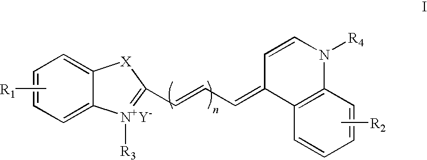
- wherein
- n is 1, 2 or 3;
- X is C(CH3)2, O, S or Se;
- R1 and R2 are each independently selected from at least one of following: H, a halogen and C1-18alkylsulfo, provided that R1 and R2 are not all simultaneously H;
- R3 and R4 are each independently selected from at least one of the following: C1-18alkyl and C1-18alkylOR5, provided that R3 and R4 are not simultaneously alkyls when R2 is a halogen;
- R5 is hydrogen, acyl or lower alkyl; and
- Y− is an anion; and
- (2) a surfactant selected from cationic surfactants, zwitterionic surfactants and anionic surfactants.
- In another aspect of the present disclosure there is provided a method to perform blood analysis, said method comprising the following steps of: (a) mixing the blood sample with the reagent of the present disclosure to form a cell suspension; (b) detecting the scattered light signals and fluorescence signals from the cells in the cell suspension; and (c) differentiating and counting the cells in the blood in terms of the scattered light signals and fluorescence signals.
- The exemplary reagent for blood analysis according to the present disclosure stains excellently and rapidly. The complex formed may have an emission wavelength in the near-infrared region so that interference from the background fluorescence of the organisms per se is avoided and the accuracy of analysis results is improved. It can be used as a staining agent for various biological samples on the flow cytometer.
- Unless otherwise specified, the following terms as used herein have the following meanings.
- The term “alkyl” as used herein individually or in combination with other groups refers to straight or branched alkyl groups containing 1-18 carbon atoms, such as 1-12, 1-8, and 1-6 carbon atoms. Reference to a single straight alkyl such as “n-propyl” specifically means a straight alkyl group, while reference to a single branched alkyl such as “isopropyl” specifically means a branched alkyl group. For example, “C1-6alkyl” includes C1-4alkyl, C1-3alkyl, methyl, ethyl, n-propyl, isopropyl and tent-butyl. The same rules apply to other groups as used throughout the present specification.
- As used herein, the term “lower alkyl” has the conventional meaning as used in the art and refers generally to C1-6alkyl.
- The term “acyl” as used herein refers to “alkyl”, as defined above, attached to the group —CO—, wherein said “alkyl” contains 1-18 carbon atoms, such as 1-12, 1-8, and 1-6 carbon atoms, such as formyl, acetyl, and propionyl etc.
- The term “halogen” as used herein includes fluorine, chlorine, bromine and iodine.
- The term “biological sample” as used herein includes, but is not limited to, nucleic acids, erythroblasts and reticulocytes in the blood.
- The Reagent for Blood Analysis According to the Present Disclosure
- In one aspect of the present disclosure there is provided a reagent for blood analysis which comprises: (1) a compound having the general formula I as the fluorescent dye; and (2) a surfactant selected from cationic surfactants, zwitterionic surfactants and anionic surfactants.
- The Compound Having the General Formula I
- Fluorescent dyes commonly used in the prior art are primarily acridine orange (AO), thioflavin T, chrysaniline and thiazole orange (TO). These dyes have the shortcomings of poor membrane permeability and long incubation time from several minutes to several tens of minutes during staining
- In U.S. Pat. No. 4,981,803 there is disclosed a novel fluorescent dye, auramine O (AuO). This fluorescent dye is improved in shortening the time required for staining and incubation (down to 30 seconds). However, when blood sample is stained with this fluorescent dye, erythrocytes which enter the detection zone may bring the problem of orientational noise affecting the differentiation and counting of reticulocytes and mature erythrocytes.
- One compound useful according to the present disclosure as a fluorescent dye has the following general formula I:
-

- wherein
- n is 1, 2 or 3;
- X is C(CH3)2, O, S or Se;
- R1 and R2 are each independently selected from at least one of the following: H, halogen and C1-6alkylsulfo, provided that R1 and R2 are all not simultaneously H;
- R3 and R4 are each independently selected from at least one of the following: C1-6alkyl and C1-6alkylOR5, provided that R3 and R4 are not simultaneously alkyls when R2 is a halogen;
- R5 is hydrogen, acyl or lower alkyl; and
- Y− is an anion.
- In one embodiment, R1 and R2 are each independently selected from at least one of the following: H, halogen and C1-6alkylsulfo, provided that R1 and R2 are not simultaneously H.
- In one embodiment, R3 is C1-6alkyl or C1-6alkylOR5.
- In one embodiment, R4 is C1-6alkyl or C1-6alkylOR5.
- In one embodiment R5 is H, C1-3alkylCO or C1-6alkyl.
- In one embodiment X is C(CH3)2, O or S.
- In one embodiment n is 1 or 2.
- In one embodiment Y− is selected from halogen ions, ClO4 −, PF6 −, CF3SO3 −, B4 −, acetate or p-toluenesulfonate anions.
- In one embodiment, a compound of formula I is selected from Dye-1, Dye-2, Dye-3, Dye-4, Dye-5, and Dye-6, wherein such dyes have the following structures:
-

- The compound according to the present disclosure stains biological samples such as nucleic acids, erythroblasts and reticulocytes. The complex formed has an emission wavelength in the near-infrared region so that interference from background fluorescence of the organisms per se may be avoided and the accuracy of analysis results is improved. The compound can be used as staining agent for various biological samples on a flow cytometer.
- The compound disclosed herein can be directly used for staining biological samples in the form of salts as described herein. Alternatively, in one embodiment, the compound disclosed herein can exist in the form of derivatives of the compound having the general formula I, said derivatives including, but not limited to, conjugates.
- Typically, conjugates are used in a fluorescence activated cell sorter (FACS). “Conjugate” as used herein refers to the compounds formed by attaching the compound disclosed herein to other molecules via covalent bonds. Molecules that can be conjugated with the compound disclosed may be those that specifically bind to cells or cell components, including, but not limited to, antibodies, antigens, receptors, ligands, enzymes, substrates, coenzymes or the like.
- Specific description about the compound having the general formula I according to the present disclosure can be found in co-pending Chinese Invention Patent Application No. 200810067815.6 of the present applicant entitled “ASYMMETRIC CYANINE COMPOUNDS, THEIR PREPARATION METHODS AND THEIR USES”, which is incorporated herein by reference.
- In order that reticulocytes and leukocytes are sufficiently stained, the dye is generally used in a concentration in the range of 1-100 mg/L, such as 5-50 mg/L. Too low a concentration of the dye would result in insufficient staining of cells that, in turn, leads to decrease in the precision of the analysis results. On the contrary, too high a concentration of the dye would increase the background fluorescence from mature erythrocytes. In neither case can the differentiation and counting of reticulocytes be favorably performed.
- Surfactants
- The reagent for blood analysis according to the present disclosure comprises a surfactant which is selected from cationic surfactants, zwitterionic surfactants and anionic surfactants.
- The surfactant contained in the reagent for blood analysis according to the present disclosure can, on the one hand, make mature erythrocytes and reticulocytes sphericized so that the influence of “orientational noise” on analysis is eliminated, and on the other hand, speed up the entry of the dye into the cells so that the intracellular nucleic acids are rapidly stained.
- Specific examples of zwitterionic surfactants are cocoamidopropyl betaine and dodecyldimethyl betaine. Zwitterionic surfactants are generally used in a concentration in the range of about 20-150 mg/L.
- Specific examples of cationic surfactants are dodecyltrimethylammonium chloride and decyltrimethylammonium chloride. Cationic surfactants are generally used in a concentration in the range of about 50-1200 mg/L.
- Specific examples of anionic surfactants are sodium dodecyl sulfate (SDS) and sodium dodecyl benzenesulfonate. Anionic surfactants are generally used in a concentration in the range of about 1-120 mg/L.
- Anionic surfactants do not increase the background fluorescence from mature erythrocytes, so reticulocytes are more readily distinguishable from mature erythrocytes. Moreover, use of anionic surfactants provides a clear demarcation of reticulocytes and leukocytes so that better results of differentiation and counting of reticulocytes can be obtained. Therefore, in one embodiment, the surfactant is an anionic surfactant. In another embodiment of the reagent for blood analysis according to the present disclosure, the surfactant is sodium dodecyl sulfate.
- Too low a concentration of the surfactant used would result in insufficient sphericization and poor staining of the cells. While too high a concentration of the surfactant used would increase the background fluorescence from mature erythrocytes or even lead to the lysis of erythrocytes.
- Other Components
- The reagent for blood analysis according to the present disclosure may also comprise a buffering agent for maintaining pH. Common buffering agents such as phosphate, Tris, HEPES and borate can be used alone or in combination in a concentration generally in the range of 0.01-0.1 mmol/L, for example 0.01-0.05 mmol/L. The buffering agent is generally used to maintain the pH value of the reagent according to the present disclosure in the range of 6.0-10.0, such as 7.5-9.5. Too low a pH would decrease the binding capability of the cationic dye to nucleic acids, while too high a pH would increase the background fluorescence from mature erythrocytes.
- The reagent for blood analysis according to the present disclosure may also comprise an osmotic regulating agent for regulating osmotic pressure. The osmotic pressure of the reagent according to the present disclosure is typically maintained in the range of 170-350 mOsm/kg, such as 200-350 mOsm/kg. Commonly used alkali metal salts, glucose and mannitol can all maintain the osmotic pressure of the reagent according to the present disclosure in a reasonable range.
- Besides the above components, the reagent according to the present disclosure may further comprise a preserving agent selected from parabens and isothiazolinones.
- The Method of Using the Reagent for Blood Analysis to Perform Blood Analysis
- In another aspect of the present disclosure there is provided a method to perform blood analysis, said method comprising the following steps of: (a) mixing the blood sample with the reagent for blood analysis according to the present disclosure to form a cell suspension; (b) detecting the scattered light signals and fluorescence signals from the cells in the cell suspension; and (c) differentiating and counting the cells in the blood in terms of the scattered light signals and fluorescence signals.
- When performing analysis of blood cells using the reagent for blood analysis according to the present disclosure on the flow cytometer or the fully automatic blood cell analyzer, the blood sample and the reagent for blood analysis according to the present disclosure are first sufficiently mixed in a certain ratio, typically 1:100-1:500, to prepare a homogenous cell suspension, and then the cell suspension is incubated at a reaction temperature of 35-45° C. for 20-40 seconds. Afterwards the cell suspension is injected into the optical system as shown in
FIG. 1 for detection. - In the above-said optical system, the individual cells are sequentially passed into the flow chamber and irradiated by the laser whose wavelength is about 630 nm emitted from the semiconductor laser. The fluorescent dye used in the reagent for blood analysis according to the present disclosure is capable of being excited at about 640 nm and its wavelength can remain stable at 40° C., matching the working wavelength of the semiconductor laser used.
- Subsequently, the scattered light signals emitted from the cells are collected by the photodiode. The scattered light can be collected at a low angle (0°-5°) or a high angle (6°-20°). The fluorescence signals emitted from the cells are collected by the laterally disposed photomultiplier tube.
- Then the scattered light signals and the fluorescence signals are inputted into the data processing unit for analysis. Finally, the various cells in the blood are differentiated and counted in terms of the scattered light signals and fluorescence signals from the cells.
- The fluorescent dye used in the reagent of the present disclosure has a certain degree of specific staining capacity for RNA. It is well known that reticulocytes are the precursor cells of mature erythrocytes and contain basophilic substances such as RNA in the cytoplasm. Therefore the reagent of the present disclosure is particularly suited for analyzing reticulocytes in the blood.
- The present disclosure is further illustrated by the following particular examples to which or by which the present disclosure is not limited.
- Unless otherwise stated, the apparatus used in the following examples for analyzing blood cells is the BC series flow cytometer manufactured by Shenzhen Mindray Bio-Medical Electronics Co. Ltd (Shenzhen, People's Republic of China), with the detection wavelength being 640 nm. The schematic diagram of the cell analyzer is as shown in
FIG. 1 . - One example of the reagent for blood analysis according to the present disclosure has the following components:
-
Cyanine dye 10 mg/L Tris 2.42 g/L Trisodium citrate•2H2O 17.64 g/L SDS 4 mg/L H2O to a volume of 1 L (Adjust the pH value of the reagent to 9.0 with HCl) - The cyanine dye used in this example has the following structure:
-

- A blood sample was taken and its reticulocyte content was determined to be 1.60% by the new methylene blue staining method recommended by the International Committee for Standardization of Hematology (ICSH). 4 μL of the blood sample was sufficiently mixed with 1 mL of the above reagent to form a homogenous cell suspension and then the suspension was incubated at 42° C. for 40 seconds. Afterwards the cell suspension was passed through the flow cytometer, and the forward (0°) scattered light signals and fluorescence signals from the cells were detected to generate a scattergram as shown in
FIG. 2 in which the reticulocytes accounted for 1.65%. - Another example of the reagent for blood analysis according to the present disclosure has the following components:
-
Cyanine dye 10 mg/L Tris 2.42 g/L Trisodium citrate•2H2O 17.64 g/L Decyltrimethylammonium chloride 800 mg/L H2O to a volume of 1 L (Adjust the pH value of the reagent to 9.0 with HCl) - The cyanine dye used in this example has the following structure:
-

- A blood sample was taken and its reticulocyte content was determined to be 4.10% by the new methylene blue staining method recommended by the International Committee for Standardization of Hematology (ICSH). 4 μL of the blood sample was sufficiently mixed with 1 mL of the above reagent to form a homogeneous cell suspension and then the suspension was incubated at 42° C. for 40 seconds. Afterwards the cell suspension was passed through the flow cytometer, and the forward (0°) scattered light signals and fluorescence signals from the cells were detected to generate a scattergram as shown in
FIG. 3 in which the reticulocytes accounted for 4.03%. - Yet another example of the reagent for blood analysis according to the present disclosure has the following components:
-
Cyanine dye 10 mg/L Tris 2.42 g/L Trisodium citrate•2H2O 17.64 g/L Cocoamidopropyl betaine 80 mg/L H2O to a volume of 1 L (Adjust the pH value of the reagent to 9.0 with HCl) - The cyanine dye used in this example has the following structure:
-

- A blood sample was taken and its reticulocyte content was determined to be 6.80% by the new methylene blue staining method recommended by the International Committee for Standardization of Hematology (ICSH). 4 μL of the blood sample was sufficiently mixed with 1 mL of the above reagent to form a homogenous cell suspension and then the suspension was incubated at 42° C. for 40 seconds. Afterwards the cell suspension was passed through the flow cytometer and the forward (0°) scattered light signals and fluorescence signals from the cells were detected to generate a scattergram as shown in
FIG. 4 in which the reticulocytes accounted for 6.69%. - One hundred and forty-eight (148) clinical blood samples were taken, among which 54 samples had normal reticulocytes and 94 samples had abnormal reticulocytes. The reticulocytes in these samples were respectively analyzed by the method as described in Example 1 and the new methylene blue staining method recommended by ICSH. The correlation of the analysis results is as shown in
FIG. 5 . - It can be seen from the above examples that a rapid and stable analysis of various cells in the blood can be achieved by using the reagent for blood analysis according to the present disclosure.
- Although the present disclosure has been illustrated by way of the above embodiments and particular examples thereof, it will be appreciated by those skilled in the art that various changes, alterations and modifications may be made to the present disclosure without departing from the spirit and scope of the present disclosure as defined by the appended claims.
Claims (27)


Applications Claiming Priority (3)
| Application Number | Priority Date | Filing Date | Title |
|---|---|---|---|
| CN200810216864.1A CN101726579B (en) | 2008-10-17 | 2008-10-17 | Blood test reagent and method |
| CN200810216864.1 | 2008-10-17 | ||
| CN200810216864 | 2008-10-17 |
Publications (2)
| Publication Number | Publication Date |
|---|---|
| US20100267080A1 true US20100267080A1 (en) | 2010-10-21 |
| US8940499B2 US8940499B2 (en) | 2015-01-27 |
Family
ID=42447773
Family Applications (1)
| Application Number | Title | Priority Date | Filing Date |
|---|---|---|---|
| US12/580,474 Active 2030-10-11 US8940499B2 (en) | 2008-10-17 | 2009-10-16 | Reagent for blood analysis and method of use thereof |
Country Status (2)
| Country | Link |
|---|---|
| US (1) | US8940499B2 (en) |
| CN (1) | CN101726579B (en) |
Cited By (1)
| Publication number | Priority date | Publication date | Assignee | Title |
|---|---|---|---|---|
| US20180003634A1 (en) * | 2014-12-31 | 2018-01-04 | Shenzhen Mindray Bio-Medical Electronics., Ltd. | Nucleated red blood cell warning method and device, and flow cytometer using the same |
Families Citing this family (20)
| Publication number | Priority date | Publication date | Assignee | Title |
|---|---|---|---|---|
| CN102618060A (en) * | 2012-03-17 | 2012-08-01 | 江南大学 | Method for preparing asymmetrical cyanine dye and method for detecting bovine serum albumin by asymmetrical cyanine dye |
| CN102766346B (en) * | 2012-07-30 | 2014-12-17 | 北京福罗泰科技有限责任公司 | Cyanine fluorescent dye |
| CN104981692B (en) * | 2013-03-12 | 2019-09-20 | 雅培实验室 | For analyzing reagent, system and the method for leucocyte |
| JP6100658B2 (en) * | 2013-03-29 | 2017-03-22 | シスメックス株式会社 | Blood cell analyzer and blood cell analysis method |
| CN104749144A (en) * | 2013-12-31 | 2015-07-01 | 深圳迈瑞生物医疗电子股份有限公司 | Blood cell detection reagent, blood cell processing method and blood cell identification method |
| CN104531834A (en) * | 2014-05-09 | 2015-04-22 | 山东博科生物产业有限公司 | Stable homocysteine detection reagent |
| CN104655542B (en) * | 2015-01-05 | 2017-06-20 | 北京市医疗器械检验所 | A kind of Protection Product detection dextran and its compound method |
| US10634602B2 (en) | 2015-06-12 | 2020-04-28 | Cytochip Inc. | Fluidic cartridge for cytometry and additional analysis |
| US10641698B2 (en) | 2015-06-12 | 2020-05-05 | Cytochip Inc. | Methods for complete blood count measurement |
| US10190961B2 (en) | 2016-03-18 | 2019-01-29 | Shenzhen Mindray Bio-Medical Electronics Co., Ltd. | Sample analyzer and sample analyzing method thereof |
| CN110199186B (en) * | 2016-11-22 | 2023-01-10 | 芯易诊有限公司 | Whole blood cell count measurement method |
| WO2018098142A1 (en) * | 2016-11-22 | 2018-05-31 | Cytochip Inc. | Methods for complete blood count measurement |
| CN110249223B (en) * | 2017-02-17 | 2020-10-20 | 深圳迈瑞生物医疗电子股份有限公司 | Blood cell analysis method and blood cell analyzer |
| CN107167415A (en) * | 2017-05-09 | 2017-09-15 | 成都扬克斯生物科技有限公司 | The method of Flow cytometry pig peripheral blood t lymphocyte subset group |
| WO2019083844A1 (en) | 2017-10-23 | 2019-05-02 | Cytochip Inc. | Devices and methods for measuring analytes and target particles |
| CN108827860A (en) * | 2018-06-22 | 2018-11-16 | 沧州医学高等专科学校 | A method of reticulocyte count is carried out with improvement Neubauer tally |
| CN109239016B (en) * | 2018-09-11 | 2021-03-26 | 深圳市国赛生物技术有限公司 | Blood sample detection method and device and readable storage medium |
| CN110031631B (en) * | 2019-04-02 | 2021-04-23 | 山东博科生物产业有限公司 | Stable high-specificity haptoglobin detection kit |
| CN113008653A (en) * | 2019-12-20 | 2021-06-22 | 深圳市帝迈生物技术有限公司 | Diluent, hemocyte analyzer, reagent for hemocyte analyzer, and kit |
| CN111157431A (en) * | 2020-01-14 | 2020-05-15 | 迪瑞医疗科技股份有限公司 | Reagent for classifying nucleated erythrocytes and basophils |
Citations (93)
| Publication number | Priority date | Publication date | Assignee | Title |
|---|---|---|---|---|
| US3883274A (en) * | 1970-11-13 | 1975-05-13 | Andre Vuaille | Control rods for the flow per stroke of piston machines, especially injection pumps, and in these machines themselves |
| US4146604A (en) * | 1973-05-31 | 1979-03-27 | Block Engineering, Inc. | Differential counting of leukocytes and other cells |
| US4286963A (en) * | 1979-11-23 | 1981-09-01 | Coulter Electronics, Inc. | Differential lymphoid-myeloid determination of leukocytes in whole blood |
| US4325706A (en) * | 1980-08-15 | 1982-04-20 | Ortho Diagnostic Systems Inc. | Automated detection of platelets and reticulocytes in whole blood |
| US4332785A (en) * | 1980-04-09 | 1982-06-01 | University Patents, Inc. | Immunoassay for measurement of reticulocytes, and immunoreactive reagents for use therein |
| US4336029A (en) * | 1980-08-15 | 1982-06-22 | Ortho Diagnostic Systems Inc. | Method and reagents for quantitative determination of reticulocytes and platelets in whole blood |
| US4447547A (en) * | 1980-04-09 | 1984-05-08 | University Patents, Inc. | Immunoassay for measurement of reticulocytes, and immunoreactive reagents for use therein |
| US4528274A (en) * | 1982-07-06 | 1985-07-09 | Coulter Electronics, Inc. | Multi-purpose blood diluent and lysing agent for differential determination of lymphoid-myeloid population of leukocytes |
| US4529705A (en) * | 1983-06-06 | 1985-07-16 | Coulter Electronics, Inc. | Reagent for combined diluting and lysing whole blood |
| US4571368A (en) * | 1983-01-17 | 1986-02-18 | Atlantic Richfield Company | Aluminum and zinc sacrificial alloy |
| US4571388A (en) * | 1983-01-24 | 1986-02-18 | Becton Dickinson And Company | Detection of reticulocytes |
| US4596035A (en) * | 1983-06-27 | 1986-06-17 | Ortho Diagnostic Systems Inc. | Methods for enumerating 3-part white cell differential clusters |
| US4637986A (en) * | 1983-08-22 | 1987-01-20 | Ortho Diagnostic Systems, Inc. | Flow cytometry lysing reagent with leukoprotective agent for producing a 3-part WBC count |
| US4745071A (en) * | 1985-09-05 | 1988-05-17 | Sequoia-Turner Corporation | Method for the volumetric differentiation of blood cells types |
| US4751179A (en) * | 1984-05-31 | 1988-06-14 | Coulter Electronics, Inc. | Method and reagents for differential determination of four populations of leukocytes in blood |
| US4822745A (en) * | 1986-12-05 | 1989-04-18 | Albert Einstein College Of Medicine Of Yeshiva University | Method for quantifying human reticulocytes |
| US4933293A (en) * | 1986-09-10 | 1990-06-12 | Toa Medical Electronics Co., Ltd. | Method of classifying leukocytes by flow cytometry and reagents used in the method |
| US4957870A (en) * | 1985-11-01 | 1990-09-18 | Becton, Dickinson And Company | Detection of Reticulocytes, RNA and DNA |
| US4981803A (en) * | 1987-07-31 | 1991-01-01 | Toa Medical Electronics Co., Ltd. | Reagent for reticulocyte counting by flow cytometry |
| US4985174A (en) * | 1985-06-06 | 1991-01-15 | Toa Medical Electronics Co., Ltd. | Reticulocyte quantitating reagent for flow cytometry |
| US5039613A (en) * | 1986-11-27 | 1991-08-13 | Toa Medical Electronics Co., Ltd. | Reagents used in a method of classifying leukocytes by flow cytometry |
| US5116539A (en) * | 1988-01-27 | 1992-05-26 | Toa Medical Electronics Co., Ltd. | Reagent and method for measuring leukocytes and hemoglobin in blood |
| US5179026A (en) * | 1986-11-27 | 1993-01-12 | Toa Medical Electronics Co., Ltd. | Method of classifying leukocytes by flow cytometry and reagents used in the method |
| US5180677A (en) * | 1988-07-05 | 1993-01-19 | Fisher Scientific Company | Lysing reagent for leukocyte differentiation method |
| US5188935A (en) * | 1984-05-31 | 1993-02-23 | Coulter Electronics, Inc. | Reagent system and method for identification, enumeration and examination of classes and subclasses of blood leukocytes |
| US5227304A (en) * | 1991-01-16 | 1993-07-13 | Sequoia Turner Corporation | Method for counting whole blood diluent and detergent reagent system |
| US5232857A (en) * | 1989-11-20 | 1993-08-03 | Abx | Reagent for use in automatic analyzers for distinguisher leukocyte sub-populations in blood samples |
| US5242832A (en) * | 1990-03-01 | 1993-09-07 | Toa Medical Electronics Co., Ltd. | Reagent for measurement of leukocytes and hemoglobin in blood |
| US5284771A (en) * | 1991-12-05 | 1994-02-08 | Miles Inc. | Reagent compositions and their use in sphering cells |
| US5316725A (en) * | 1991-06-07 | 1994-05-31 | Edward Lawrence Carver, Jr. | Reagent system for the improved determination of white blood cell subpopulations |
| US5316951A (en) * | 1991-06-07 | 1994-05-31 | Carver Jr Edward L | Method for the improved determination of white blood cell subpopulations |
| US5321130A (en) * | 1992-02-10 | 1994-06-14 | Molecular Probes, Inc. | Unsymmetrical cyanine dyes with a cationic side chain |
| US5350695A (en) * | 1991-12-05 | 1994-09-27 | Miles Inc. | Methods for the identification and characterization of reticulocytes in whole blood |
| US5389549A (en) * | 1987-05-29 | 1995-02-14 | Toa Medical Electronics Co., Ltd. | Method for classifying leukocytes and a reagent used therefor |
| US5411891A (en) * | 1991-12-05 | 1995-05-02 | Miles Inc. | Reagent compositions and their use in the identification and characterization of reticulocytes in whole blood |
| US5413938A (en) * | 1993-03-19 | 1995-05-09 | Tao Medical Electronics Co., Ltd. | Reagent for measuring immature leukocytes |
| US5486477A (en) * | 1991-06-13 | 1996-01-23 | Carver, Jr.; Edward L. | Reagent system for improved multiple species blood analysis |
| US5492833A (en) * | 1993-05-14 | 1996-02-20 | Coulter Corporation | Reticulocyte analyzing method and apparatus utilizing light scatter techniques |
| US5496734A (en) * | 1992-11-19 | 1996-03-05 | Toa Medical Electronics Co., Ltd. | Treatment method for blood analysis |
| US5510267A (en) * | 1989-05-15 | 1996-04-23 | Abbott Laboratories | Flow cytometry lytic agent and method enabling 5-part leukocyte differential count |
| US5516695A (en) * | 1993-02-25 | 1996-05-14 | Abbott Laboratories | Multipurpose reagent system for rapid lysis of whole blood |
| US5518928A (en) * | 1984-09-24 | 1996-05-21 | Bayer Corporation | Leukocyte differentiation method |
| US5538893A (en) * | 1994-04-21 | 1996-07-23 | Toa Medical Electronics Co., Ltd. | Reagent for analyzing leukocytes and a method for classifying leukocytes |
| US5559037A (en) * | 1994-12-15 | 1996-09-24 | Abbott Laboratories | Method for rapid and simultaneous analysis of nucleated red blood cells |
| US5618733A (en) * | 1993-12-22 | 1997-04-08 | Toa Medical Electronics Co., Ltd. | Reagent for analyzing leucocytes |
| US5639666A (en) * | 1994-05-23 | 1997-06-17 | Coulter Corporation | Detection of reticulocytes |
| US5639630A (en) * | 1995-05-16 | 1997-06-17 | Bayer Corporation | Method and reagent composition for performing leukocyte differential counts on fresh and aged whole blood samples, based on intrinsic peroxidase activity of leukocytes |
| US5656449A (en) * | 1995-03-06 | 1997-08-12 | Molecular Probes, Inc. | Neutral unsymmetrical cyanine dyes |
| US5731206A (en) * | 1987-03-13 | 1998-03-24 | Coulter Electronics, Inc. | Method and reagent system for isolation, identification and/or analysis of leukocytes from whole blood samples |
| US5733784A (en) * | 1995-11-20 | 1998-03-31 | Abbott Laboratories | Reagent system and method for the differentiation and identification of reticulocytes |
| US5747343A (en) * | 1994-10-31 | 1998-05-05 | Nihon Kohden Corporation | Leukocyte classification reagent |
| US5763280A (en) * | 1997-01-21 | 1998-06-09 | Coulter International Corp. | Cyanide-free lytic reagent composition and method for hemoglobin and cell analysis |
| US5773299A (en) * | 1995-04-21 | 1998-06-30 | Abbott Laboratories | Methods for the rapid analysis of the reticulocytes |
| US5786224A (en) * | 1995-06-29 | 1998-07-28 | Coulter Corporation | Reagent and method for differential determination of leukocytes in blood |
| US5858667A (en) * | 1996-09-06 | 1999-01-12 | Litron Laboratories | Method for the enumeration of micronucleated erythrocyte populations with a single laser flow cytometer |
| US5874311A (en) * | 1997-11-21 | 1999-02-23 | Coulter International Corp. | Method for differentiation of reticulocytes in blood |
| US5879900A (en) * | 1994-12-15 | 1999-03-09 | Abbott Laboratories | Method for simultaneous analysis of cell viability, nucleated red blood cells and white blood cell differentials |
| US5882934A (en) * | 1997-01-21 | 1999-03-16 | Coulter International Corp. | Composition and method for hemoglobin and cell analysis |
| US5891731A (en) * | 1996-04-12 | 1999-04-06 | Toa Medical Electronics Co., Ltd. | Reagent for measuring reticulocytes and a method of measuring them |
| US5928949A (en) * | 1986-09-10 | 1999-07-27 | Toa Medical Electronics Co., Ltd. | Reagent and method for classifying leukocytes by flow cytometry |
| US5958776A (en) * | 1996-11-20 | 1999-09-28 | Sysmex Corporation | Method for classifying and counting immature leukocytes |
| US6060322A (en) * | 1998-10-20 | 2000-05-09 | Coulter International Corp. | Method for identification of reticulated cells |
| US6100038A (en) * | 1996-09-06 | 2000-08-08 | Litron Laboratories Limited | Method for the enumeration of micronucleated erythrocyte populations with a single laser flow cytometer |
| US6114173A (en) * | 1997-04-03 | 2000-09-05 | Bayer Corporation | Fully automated method and reagent composition therefor for rapid identification and characterization of reticulocytes erythrocytes and platelets in whole blood |
| US6114130A (en) * | 1998-08-04 | 2000-09-05 | Abx | Reagent for measurement of the haemoglobin and determination of the leukocytes in a blood sample |
| US6197593B1 (en) * | 1998-10-20 | 2001-03-06 | Coulter International Corp. | Method for enumerating blood cells |
| US6245499B1 (en) * | 1996-04-30 | 2001-06-12 | Fuji Photo Film Co., Ltd. | Photothermographic material |
| US6248319B1 (en) * | 1989-10-16 | 2001-06-19 | Krisztina M. Zsebo | Method for increasing hematopoietic progenitor cells by stem cell factor polypeptides |
| US6271035B1 (en) * | 1998-10-20 | 2001-08-07 | Coulter International Corp. | Methods and compositions for rapid staining of nucleic acids in whole cells |
| US6287791B1 (en) * | 1992-01-22 | 2001-09-11 | Becton Dickinson And Company | Multidimensional cell differential analysis |
| US6350613B1 (en) * | 1998-03-07 | 2002-02-26 | Belton Dickinson & Co. | Determination of white blood cell differential and reticulocyte counts |
| US6368864B1 (en) * | 2000-05-05 | 2002-04-09 | Coulter International Corp. | Dyes and methods of reticulocyte enumeration |
| US6524858B1 (en) * | 1999-03-31 | 2003-02-25 | Bayer Corporation | Single channel, single dilution detection method for the identification and quantification of blood cells and platelets in a whole blood sample using an automated hematology analyzer |
| USRE38131E1 (en) * | 1997-04-18 | 2003-06-03 | Sysmex Corporation | Reagent for measurement of leukocytes and hemoglobin concentration in blood |
| US20030145394A1 (en) * | 1996-12-10 | 2003-08-07 | Nai-Yi Wang | Helium-neon excitable reticulocyte dyes derivable from halolepidines |
| US6794152B2 (en) * | 2000-12-22 | 2004-09-21 | Streck Laboratories Inc. | Flow cytometry reagent and system |
| US6869798B2 (en) * | 2003-04-17 | 2005-03-22 | Clinical Diagnostics Solutions, Inc. | Lytic reagent composition for leukocyte differential analysis |
| US6900023B1 (en) * | 1999-09-02 | 2005-05-31 | Sysmex Corporation | Method for classifying and counting leukocytes |
| US20060177347A1 (en) * | 2002-06-11 | 2006-08-10 | Larsen Ulrik D | Lysing reagent, cartridge and automatic electronic cell counter for simultaneous enumeration of different types of white blood cells |
| US20070111276A1 (en) * | 2001-02-23 | 2007-05-17 | Abx | Reagent and process for the identification and counting of biological cells |
| US7235404B2 (en) * | 2005-05-04 | 2007-06-26 | Beckman Coulter, Inc. | Cyanide-free lytic reagent composition and method of use for hemoglobin and white blood cell measurement |
| US7247484B2 (en) * | 2002-11-01 | 2007-07-24 | Streck, Inc. | Hematology reagent and methods |
| US20070178597A1 (en) * | 2006-01-27 | 2007-08-02 | Sysmex Corporation | Reagent for immature leukocyte analysis and reagent kit |
| US20080026475A1 (en) * | 2000-09-14 | 2008-01-31 | Andre Van Agthoven | Method of Lysis of Erythrocytes |
| US20080176274A1 (en) * | 2002-06-24 | 2008-07-24 | Sysmex Corporation | Method of classifying and counting leucocytes |
| US7405082B2 (en) * | 2002-09-10 | 2008-07-29 | Sysmex Corporation | Methods and devices for measuring reticulocytes |
| US20090017441A1 (en) * | 2007-07-12 | 2009-01-15 | Shenzhen Mindray Bio-Medical Electronics Co., Ltd. | Asymmetric cyanine fluorescent dyes |
| US20090023129A1 (en) * | 2007-07-20 | 2009-01-22 | Shenzhen Mindray Bio-Medical Electronics Co., Ltd. | White blood cell differentiation reagent and method of use thereof |
| US20090176270A1 (en) * | 2008-01-04 | 2009-07-09 | Shenzhen Mindray Bio-Medical Electronics Co., Ltd. | Asymmetric cyanine fluorescent dyes, compositions and their use in staining biological samples |
| US7709653B2 (en) * | 2008-06-10 | 2010-05-04 | Shenzhen Mindray Bio-Medical Electronics Co. Ltd. | Asymmetric cyanine compounds, their preparation methods and their uses |
| US20100151509A1 (en) * | 2008-12-17 | 2010-06-17 | Shenzhen Mindray Bio-Medical Electronics Co., Ltd. | Reagent, kit and method for differentating and counting leukocytes |
| US20100178654A1 (en) * | 2007-06-25 | 2010-07-15 | Sysmex Corporation | Reagent and reagent kit for analysis of immature leukocyte |
| US8367359B1 (en) * | 2009-10-23 | 2013-02-05 | The United States Of America, As Represented By The Secretary Of Agriculture | Metabolic biomarkers for diabetes and insulin resistance |
Family Cites Families (44)
| Publication number | Priority date | Publication date | Assignee | Title |
|---|---|---|---|---|
| GB556266A (en) * | 1940-10-18 | 1943-09-28 | Kodak Ltd | Improvements in and relating to polymethine dye intermediates and to the production of polymethine dyes |
| US3883247A (en) | 1973-10-30 | 1975-05-13 | Bio Physics Systems Inc | Method for fluorescence analysis of white blood cells |
| IL49622A0 (en) | 1976-05-21 | 1976-07-30 | Elscint Ltd | A method and apparatus for classifying biological cells |
| US4544546A (en) | 1980-04-21 | 1985-10-01 | Abbott Laboratories | Fluorescent nucleic acid stains |
| JPS5747493A (en) | 1980-09-02 | 1982-03-18 | Fuji Photo Film Co Ltd | Method of determining a trace amount of enzyme |
| US4485175A (en) | 1983-01-03 | 1984-11-27 | Coulter Electronics, Inc. | Method for three-volume differential determination of lymphocyte, monocyte and granulocyte populations of leukocytes |
| JPS6073356A (en) | 1983-09-29 | 1985-04-25 | Toa Medical Electronics Co Ltd | Reagent for blood analysis |
| US4978624A (en) | 1985-09-06 | 1990-12-18 | Technicon Instruments Corporation | Reagent for the determination of a differential white blood cell count |
| US4707451A (en) | 1985-09-18 | 1987-11-17 | Becton, Dickinson And Company | Detection of reticulocytes |
| US4883867A (en) | 1985-11-01 | 1989-11-28 | Becton, Dickinson And Company | Detection of reticulocytes, RNA or DNA |
| US5155044A (en) | 1987-03-13 | 1992-10-13 | Coulter Electronics, Inc. | Lysing reagent system for isolation, identification and/or analysis of leukocytes from whole blood samples |
| US4882284A (en) | 1987-04-13 | 1989-11-21 | Ortho Pharmaceutical Corporation | Method for quantitating and differentiating white blood cells |
| JPH0746102B2 (en) | 1987-07-31 | 1995-05-17 | 東亜医用電子株式会社 | Reticulocyte assay reagent for flow cytometry |
| JP2836865B2 (en) | 1989-10-23 | 1998-12-14 | 東亜医用電子株式会社 | Reagents for measuring leukocytes and hemoglobin in blood |
| US5075556A (en) | 1989-12-01 | 1991-12-24 | Technicon Instruments Corporation | Acridine orange derivatives and their use in the quantitation of reticulocytes in whole blood |
| JP3048260B2 (en) | 1991-07-29 | 2000-06-05 | シスメックス株式会社 | Sample preparation method for leukocyte classification and counting |
| JP3232145B2 (en) | 1991-12-27 | 2001-11-26 | シスメックス株式会社 | Reticulocyte measurement method |
| US5563070A (en) | 1993-05-28 | 1996-10-08 | Omron Corporation | Method of counting reticulocytes |
| JP3355038B2 (en) | 1994-08-03 | 2002-12-09 | シスメックス株式会社 | Leukocyte classification method |
| US5686308A (en) | 1995-06-08 | 1997-11-11 | Coulter Corporation | Reagent and method for differential determination of leukocytes in blood |
| US5843608A (en) | 1995-06-08 | 1998-12-01 | Coulter International Corp. | Reagent and method for differential determination of leukocytes in blood |
| FR2735579B1 (en) | 1995-06-13 | 1997-09-19 | Hycel Groupe Lisabio | METHOD FOR DISCRIMINATION AND ISOLATION OF SUB-POPULATIONS OF LEUKOCYTES FROM BLOOD SAMPLES BY TREATMENT WITH POLYOXYETHYLENE 9-LAURYL ETHER, AND REAGENT FOR IMPLEMENTATION THEREFOR |
| FR2735578B1 (en) | 1995-06-13 | 1997-09-05 | Hycel Groupe Lisabio | ERYTHROCYTE LYSIS REAGENT, AND USE THEREOF IN LEUKOCYTE ISOLATION AND DISCRIMINATION PROCESSES |
| JP3425830B2 (en) | 1995-10-06 | 2003-07-14 | シスメックス株式会社 | New compounds and their uses |
| US5658735A (en) | 1995-11-09 | 1997-08-19 | Biometric Imaging, Inc. | Cyclized fluorescent nucleic acid intercalating cyanine dyes and nucleic acid detection methods |
| US5817518A (en) | 1995-12-18 | 1998-10-06 | Coulter International Corp. | Reagent and method for differential determination of leukocytes in blood |
| EP0794435B1 (en) | 1996-03-07 | 2003-01-02 | Coulter International Corp. | Cyanide-free reagent and method for hemoglobin determination and leukocyte differentiation |
| CN1149397C (en) | 1996-04-12 | 2004-05-12 | 希森美康株式会社 | Reagent for determining granulophilocyte and determination method |
| FR2759166B1 (en) | 1997-01-31 | 1999-04-23 | Abx Sa | COLOR REAGENT FOR THE DETERMINATION OF BLOOD CELLS |
| US5994089A (en) | 1997-05-16 | 1999-11-30 | Coulter International Corp. | Simultaneous analyses of white blood cell subsets using multi-color, multi-intensity fluorescent markers in flow cytometry |
| JP3783808B2 (en) | 1997-05-19 | 2006-06-07 | シスメックス株式会社 | Leukocyte classification and counting reagent |
| US6074232A (en) | 1998-06-01 | 2000-06-13 | Hon Hai Precision Ind. Co., Ltd. | Board to board electrical connector with releaseable actuator |
| JP3886271B2 (en) | 1998-11-27 | 2007-02-28 | シスメックス株式会社 | Reagent for classification counting of erythroblast and classification counting method |
| US6632676B1 (en) | 1999-09-24 | 2003-10-14 | Clinical Diagnostic Solutions | Multi-purpose reagent system and method for enumeration of red blood cells, white blood cells and thrombocytes and differential determination of white blood cells |
| US20020098589A1 (en) | 2001-01-19 | 2002-07-25 | Crews Harold Richardson | Multi-purpose reagent system and method for enumeration of red blood cells, white blood cells and thrombocytes and differential determination of white blood cells |
| CN1101982C (en) | 1999-10-29 | 2003-02-19 | 富士康(昆山)电脑接插件有限公司 | Socket connector |
| JP3493575B2 (en) | 2000-07-28 | 2004-02-03 | Necエレクトロニクス株式会社 | Semiconductor device, charge pump circuit and PLL circuit |
| US6630990B2 (en) | 2001-06-05 | 2003-10-07 | Abbott Laboratories | Optical method and apparatus for red blood cell differentiation on a cell-by-cell basis, and simultaneous analysis of white blood cell differentiation |
| US6955872B2 (en) | 2003-03-20 | 2005-10-18 | Coulter International Corp. | Dye compositions which provide enhanced differential fluorescence and light scatter characteristics |
| JP4509607B2 (en) | 2004-03-17 | 2010-07-21 | シスメックス株式会社 | Cell analysis apparatus and method |
| US7465584B2 (en) | 2005-01-24 | 2008-12-16 | Sysmex Corporation | Method and reagent for classifying leukocytes in animal blood |
| JP4806334B2 (en) * | 2006-11-27 | 2011-11-02 | シスメックス株式会社 | Method and apparatus for measuring hematological samples |
| CN101231243B (en) * | 2007-01-24 | 2011-11-16 | 深圳迈瑞生物医疗电子股份有限公司 | Agent and method for testing reticulocyte |
| US7449337B2 (en) | 2007-01-24 | 2008-11-11 | Idexx Laboratories, Inc. | Lytic reagent and method for leukocytes differential in whole blood |
-
2008
- 2008-10-17 CN CN200810216864.1A patent/CN101726579B/en active Active
-
2009
- 2009-10-16 US US12/580,474 patent/US8940499B2/en active Active
Patent Citations (99)
| Publication number | Priority date | Publication date | Assignee | Title |
|---|---|---|---|---|
| US3883274A (en) * | 1970-11-13 | 1975-05-13 | Andre Vuaille | Control rods for the flow per stroke of piston machines, especially injection pumps, and in these machines themselves |
| US4146604A (en) * | 1973-05-31 | 1979-03-27 | Block Engineering, Inc. | Differential counting of leukocytes and other cells |
| US4286963A (en) * | 1979-11-23 | 1981-09-01 | Coulter Electronics, Inc. | Differential lymphoid-myeloid determination of leukocytes in whole blood |
| US4447547A (en) * | 1980-04-09 | 1984-05-08 | University Patents, Inc. | Immunoassay for measurement of reticulocytes, and immunoreactive reagents for use therein |
| US4332785A (en) * | 1980-04-09 | 1982-06-01 | University Patents, Inc. | Immunoassay for measurement of reticulocytes, and immunoreactive reagents for use therein |
| US4325706A (en) * | 1980-08-15 | 1982-04-20 | Ortho Diagnostic Systems Inc. | Automated detection of platelets and reticulocytes in whole blood |
| US4336029A (en) * | 1980-08-15 | 1982-06-22 | Ortho Diagnostic Systems Inc. | Method and reagents for quantitative determination of reticulocytes and platelets in whole blood |
| US4528274A (en) * | 1982-07-06 | 1985-07-09 | Coulter Electronics, Inc. | Multi-purpose blood diluent and lysing agent for differential determination of lymphoid-myeloid population of leukocytes |
| US4571368A (en) * | 1983-01-17 | 1986-02-18 | Atlantic Richfield Company | Aluminum and zinc sacrificial alloy |
| US4571388A (en) * | 1983-01-24 | 1986-02-18 | Becton Dickinson And Company | Detection of reticulocytes |
| US4529705A (en) * | 1983-06-06 | 1985-07-16 | Coulter Electronics, Inc. | Reagent for combined diluting and lysing whole blood |
| US4596035A (en) * | 1983-06-27 | 1986-06-17 | Ortho Diagnostic Systems Inc. | Methods for enumerating 3-part white cell differential clusters |
| US4637986A (en) * | 1983-08-22 | 1987-01-20 | Ortho Diagnostic Systems, Inc. | Flow cytometry lysing reagent with leukoprotective agent for producing a 3-part WBC count |
| US4751179A (en) * | 1984-05-31 | 1988-06-14 | Coulter Electronics, Inc. | Method and reagents for differential determination of four populations of leukocytes in blood |
| US5188935A (en) * | 1984-05-31 | 1993-02-23 | Coulter Electronics, Inc. | Reagent system and method for identification, enumeration and examination of classes and subclasses of blood leukocytes |
| US5518928A (en) * | 1984-09-24 | 1996-05-21 | Bayer Corporation | Leukocyte differentiation method |
| US4985174A (en) * | 1985-06-06 | 1991-01-15 | Toa Medical Electronics Co., Ltd. | Reticulocyte quantitating reagent for flow cytometry |
| US4745071A (en) * | 1985-09-05 | 1988-05-17 | Sequoia-Turner Corporation | Method for the volumetric differentiation of blood cells types |
| US4957870A (en) * | 1985-11-01 | 1990-09-18 | Becton, Dickinson And Company | Detection of Reticulocytes, RNA and DNA |
| US4933293A (en) * | 1986-09-10 | 1990-06-12 | Toa Medical Electronics Co., Ltd. | Method of classifying leukocytes by flow cytometry and reagents used in the method |
| US5928949A (en) * | 1986-09-10 | 1999-07-27 | Toa Medical Electronics Co., Ltd. | Reagent and method for classifying leukocytes by flow cytometry |
| US5039613A (en) * | 1986-11-27 | 1991-08-13 | Toa Medical Electronics Co., Ltd. | Reagents used in a method of classifying leukocytes by flow cytometry |
| US5179026A (en) * | 1986-11-27 | 1993-01-12 | Toa Medical Electronics Co., Ltd. | Method of classifying leukocytes by flow cytometry and reagents used in the method |
| US4822745A (en) * | 1986-12-05 | 1989-04-18 | Albert Einstein College Of Medicine Of Yeshiva University | Method for quantifying human reticulocytes |
| US5731206A (en) * | 1987-03-13 | 1998-03-24 | Coulter Electronics, Inc. | Method and reagent system for isolation, identification and/or analysis of leukocytes from whole blood samples |
| US5389549A (en) * | 1987-05-29 | 1995-02-14 | Toa Medical Electronics Co., Ltd. | Method for classifying leukocytes and a reagent used therefor |
| US4981803A (en) * | 1987-07-31 | 1991-01-01 | Toa Medical Electronics Co., Ltd. | Reagent for reticulocyte counting by flow cytometry |
| US5116539A (en) * | 1988-01-27 | 1992-05-26 | Toa Medical Electronics Co., Ltd. | Reagent and method for measuring leukocytes and hemoglobin in blood |
| US5180677A (en) * | 1988-07-05 | 1993-01-19 | Fisher Scientific Company | Lysing reagent for leukocyte differentiation method |
| US5510267A (en) * | 1989-05-15 | 1996-04-23 | Abbott Laboratories | Flow cytometry lytic agent and method enabling 5-part leukocyte differential count |
| US6248319B1 (en) * | 1989-10-16 | 2001-06-19 | Krisztina M. Zsebo | Method for increasing hematopoietic progenitor cells by stem cell factor polypeptides |
| US5232857A (en) * | 1989-11-20 | 1993-08-03 | Abx | Reagent for use in automatic analyzers for distinguisher leukocyte sub-populations in blood samples |
| US5242832A (en) * | 1990-03-01 | 1993-09-07 | Toa Medical Electronics Co., Ltd. | Reagent for measurement of leukocytes and hemoglobin in blood |
| US5227304A (en) * | 1991-01-16 | 1993-07-13 | Sequoia Turner Corporation | Method for counting whole blood diluent and detergent reagent system |
| US5316951A (en) * | 1991-06-07 | 1994-05-31 | Carver Jr Edward L | Method for the improved determination of white blood cell subpopulations |
| US5316725A (en) * | 1991-06-07 | 1994-05-31 | Edward Lawrence Carver, Jr. | Reagent system for the improved determination of white blood cell subpopulations |
| US5486477A (en) * | 1991-06-13 | 1996-01-23 | Carver, Jr.; Edward L. | Reagent system for improved multiple species blood analysis |
| US5350695A (en) * | 1991-12-05 | 1994-09-27 | Miles Inc. | Methods for the identification and characterization of reticulocytes in whole blood |
| US5284771A (en) * | 1991-12-05 | 1994-02-08 | Miles Inc. | Reagent compositions and their use in sphering cells |
| US5411891A (en) * | 1991-12-05 | 1995-05-02 | Miles Inc. | Reagent compositions and their use in the identification and characterization of reticulocytes in whole blood |
| US5438003A (en) * | 1991-12-05 | 1995-08-01 | Miles Inc. | Reagent compositions for use in the identification and characterization of reticulocytes in whole blood |
| US5633167A (en) * | 1991-12-05 | 1997-05-27 | Bayer Corporation | Reagent compositions for use in sphering cells |
| US6287791B1 (en) * | 1992-01-22 | 2001-09-11 | Becton Dickinson And Company | Multidimensional cell differential analysis |
| US5321130A (en) * | 1992-02-10 | 1994-06-14 | Molecular Probes, Inc. | Unsymmetrical cyanine dyes with a cationic side chain |
| US5496734A (en) * | 1992-11-19 | 1996-03-05 | Toa Medical Electronics Co., Ltd. | Treatment method for blood analysis |
| US5516695A (en) * | 1993-02-25 | 1996-05-14 | Abbott Laboratories | Multipurpose reagent system for rapid lysis of whole blood |
| US5413938A (en) * | 1993-03-19 | 1995-05-09 | Tao Medical Electronics Co., Ltd. | Reagent for measuring immature leukocytes |
| US5616501A (en) * | 1993-05-14 | 1997-04-01 | Coulter Corporation | Reticulocyte analyzing method and apparatus utilizing light scatter techniques |
| US5492833A (en) * | 1993-05-14 | 1996-02-20 | Coulter Corporation | Reticulocyte analyzing method and apparatus utilizing light scatter techniques |
| US5618733A (en) * | 1993-12-22 | 1997-04-08 | Toa Medical Electronics Co., Ltd. | Reagent for analyzing leucocytes |
| US5538893A (en) * | 1994-04-21 | 1996-07-23 | Toa Medical Electronics Co., Ltd. | Reagent for analyzing leukocytes and a method for classifying leukocytes |
| US5639666A (en) * | 1994-05-23 | 1997-06-17 | Coulter Corporation | Detection of reticulocytes |
| US5747343A (en) * | 1994-10-31 | 1998-05-05 | Nihon Kohden Corporation | Leukocyte classification reagent |
| US5559037A (en) * | 1994-12-15 | 1996-09-24 | Abbott Laboratories | Method for rapid and simultaneous analysis of nucleated red blood cells |
| US5879900A (en) * | 1994-12-15 | 1999-03-09 | Abbott Laboratories | Method for simultaneous analysis of cell viability, nucleated red blood cells and white blood cell differentials |
| US5656449A (en) * | 1995-03-06 | 1997-08-12 | Molecular Probes, Inc. | Neutral unsymmetrical cyanine dyes |
| US5773299A (en) * | 1995-04-21 | 1998-06-30 | Abbott Laboratories | Methods for the rapid analysis of the reticulocytes |
| US5639630A (en) * | 1995-05-16 | 1997-06-17 | Bayer Corporation | Method and reagent composition for performing leukocyte differential counts on fresh and aged whole blood samples, based on intrinsic peroxidase activity of leukocytes |
| US5786224A (en) * | 1995-06-29 | 1998-07-28 | Coulter Corporation | Reagent and method for differential determination of leukocytes in blood |
| US5733784A (en) * | 1995-11-20 | 1998-03-31 | Abbott Laboratories | Reagent system and method for the differentiation and identification of reticulocytes |
| US5891731A (en) * | 1996-04-12 | 1999-04-06 | Toa Medical Electronics Co., Ltd. | Reagent for measuring reticulocytes and a method of measuring them |
| US6245499B1 (en) * | 1996-04-30 | 2001-06-12 | Fuji Photo Film Co., Ltd. | Photothermographic material |
| US6100038A (en) * | 1996-09-06 | 2000-08-08 | Litron Laboratories Limited | Method for the enumeration of micronucleated erythrocyte populations with a single laser flow cytometer |
| US5858667A (en) * | 1996-09-06 | 1999-01-12 | Litron Laboratories | Method for the enumeration of micronucleated erythrocyte populations with a single laser flow cytometer |
| US5958776A (en) * | 1996-11-20 | 1999-09-28 | Sysmex Corporation | Method for classifying and counting immature leukocytes |
| US7083982B2 (en) * | 1996-12-10 | 2006-08-01 | Abbott Laboratories | Helium-neon excitable reticulocyte dyes derivable from halolepidines |
| US20030145394A1 (en) * | 1996-12-10 | 2003-08-07 | Nai-Yi Wang | Helium-neon excitable reticulocyte dyes derivable from halolepidines |
| US5882934A (en) * | 1997-01-21 | 1999-03-16 | Coulter International Corp. | Composition and method for hemoglobin and cell analysis |
| US5763280A (en) * | 1997-01-21 | 1998-06-09 | Coulter International Corp. | Cyanide-free lytic reagent composition and method for hemoglobin and cell analysis |
| US6114173A (en) * | 1997-04-03 | 2000-09-05 | Bayer Corporation | Fully automated method and reagent composition therefor for rapid identification and characterization of reticulocytes erythrocytes and platelets in whole blood |
| USRE38131E1 (en) * | 1997-04-18 | 2003-06-03 | Sysmex Corporation | Reagent for measurement of leukocytes and hemoglobin concentration in blood |
| US5874311A (en) * | 1997-11-21 | 1999-02-23 | Coulter International Corp. | Method for differentiation of reticulocytes in blood |
| US6350613B1 (en) * | 1998-03-07 | 2002-02-26 | Belton Dickinson & Co. | Determination of white blood cell differential and reticulocyte counts |
| US6114130A (en) * | 1998-08-04 | 2000-09-05 | Abx | Reagent for measurement of the haemoglobin and determination of the leukocytes in a blood sample |
| US6271035B1 (en) * | 1998-10-20 | 2001-08-07 | Coulter International Corp. | Methods and compositions for rapid staining of nucleic acids in whole cells |
| US6197593B1 (en) * | 1998-10-20 | 2001-03-06 | Coulter International Corp. | Method for enumerating blood cells |
| US6060322A (en) * | 1998-10-20 | 2000-05-09 | Coulter International Corp. | Method for identification of reticulated cells |
| US6524858B1 (en) * | 1999-03-31 | 2003-02-25 | Bayer Corporation | Single channel, single dilution detection method for the identification and quantification of blood cells and platelets in a whole blood sample using an automated hematology analyzer |
| US6900023B1 (en) * | 1999-09-02 | 2005-05-31 | Sysmex Corporation | Method for classifying and counting leukocytes |
| US6368864B1 (en) * | 2000-05-05 | 2002-04-09 | Coulter International Corp. | Dyes and methods of reticulocyte enumeration |
| US6551831B2 (en) * | 2000-05-05 | 2003-04-22 | Coulter International Corp. | Dyes and methods of detection of nucleic acid in immature red blood cells |
| US20080026475A1 (en) * | 2000-09-14 | 2008-01-31 | Andre Van Agthoven | Method of Lysis of Erythrocytes |
| US6794152B2 (en) * | 2000-12-22 | 2004-09-21 | Streck Laboratories Inc. | Flow cytometry reagent and system |
| US20070111276A1 (en) * | 2001-02-23 | 2007-05-17 | Abx | Reagent and process for the identification and counting of biological cells |
| US20060177347A1 (en) * | 2002-06-11 | 2006-08-10 | Larsen Ulrik D | Lysing reagent, cartridge and automatic electronic cell counter for simultaneous enumeration of different types of white blood cells |
| US20080176274A1 (en) * | 2002-06-24 | 2008-07-24 | Sysmex Corporation | Method of classifying and counting leucocytes |
| US7405082B2 (en) * | 2002-09-10 | 2008-07-29 | Sysmex Corporation | Methods and devices for measuring reticulocytes |
| US7247484B2 (en) * | 2002-11-01 | 2007-07-24 | Streck, Inc. | Hematology reagent and methods |
| US6869798B2 (en) * | 2003-04-17 | 2005-03-22 | Clinical Diagnostics Solutions, Inc. | Lytic reagent composition for leukocyte differential analysis |
| US7235404B2 (en) * | 2005-05-04 | 2007-06-26 | Beckman Coulter, Inc. | Cyanide-free lytic reagent composition and method of use for hemoglobin and white blood cell measurement |
| US20070178597A1 (en) * | 2006-01-27 | 2007-08-02 | Sysmex Corporation | Reagent for immature leukocyte analysis and reagent kit |
| US20100178654A1 (en) * | 2007-06-25 | 2010-07-15 | Sysmex Corporation | Reagent and reagent kit for analysis of immature leukocyte |
| US20090017441A1 (en) * | 2007-07-12 | 2009-01-15 | Shenzhen Mindray Bio-Medical Electronics Co., Ltd. | Asymmetric cyanine fluorescent dyes |
| US20090023129A1 (en) * | 2007-07-20 | 2009-01-22 | Shenzhen Mindray Bio-Medical Electronics Co., Ltd. | White blood cell differentiation reagent and method of use thereof |
| US7960099B2 (en) * | 2007-07-20 | 2011-06-14 | Shenzhen Mindray Bio-Medical Electronics Co., Ltd. | White blood cell differentiation reagent containing an asymmetric cyanine fluorescent dye and method of use thereof |
| US20090176270A1 (en) * | 2008-01-04 | 2009-07-09 | Shenzhen Mindray Bio-Medical Electronics Co., Ltd. | Asymmetric cyanine fluorescent dyes, compositions and their use in staining biological samples |
| US7709653B2 (en) * | 2008-06-10 | 2010-05-04 | Shenzhen Mindray Bio-Medical Electronics Co. Ltd. | Asymmetric cyanine compounds, their preparation methods and their uses |
| US20100151509A1 (en) * | 2008-12-17 | 2010-06-17 | Shenzhen Mindray Bio-Medical Electronics Co., Ltd. | Reagent, kit and method for differentating and counting leukocytes |
| US8367359B1 (en) * | 2009-10-23 | 2013-02-05 | The United States Of America, As Represented By The Secretary Of Agriculture | Metabolic biomarkers for diabetes and insulin resistance |
Non-Patent Citations (4)
| Title |
|---|
| FEI, X. et al. Solid-Phase Synthesis and Modification of Thiazole Orange and its Derivatives and their Spectral Properties. Journal of Combinatorial Chemistry. 2007, Vol. 9(6), page 945, last two paragraphs * |
| FEI, X. et al. Solid-Phase Synthesis and Modification of Thiazole Orange and its Derivatives and their Spectral Properties. Journal of Combinatorial Chemistry. 2007, Vol. 9(6), page 945. * |
| MASON, SJ. et al. Solid-Phase Catch, Activate, and Release Synthesis of Cyanine Dyes. Organic Letters. 2002, Vol. 4(24), page 4263 * |
| MASON, SJ. et al. Solid-Phase Catch, Activate, and Release Synthesis of Cyanine Dyes. Organic Letters. 2002, Vol. 4(24), page 4263. * |
Cited By (2)
| Publication number | Priority date | Publication date | Assignee | Title |
|---|---|---|---|---|
| US20180003634A1 (en) * | 2014-12-31 | 2018-01-04 | Shenzhen Mindray Bio-Medical Electronics., Ltd. | Nucleated red blood cell warning method and device, and flow cytometer using the same |
| US11105742B2 (en) * | 2014-12-31 | 2021-08-31 | Shenzhen Mindray Bio-Medical Electronics Co., Ltd. | Nucleated red blood cell warning method and device, and flow cytometer using the same |
Also Published As
| Publication number | Publication date |
|---|---|
| US8940499B2 (en) | 2015-01-27 |
| CN101726579B (en) | 2014-06-18 |
| CN101726579A (en) | 2010-06-09 |
Similar Documents
| Publication | Publication Date | Title |
|---|---|---|
| US8940499B2 (en) | Reagent for blood analysis and method of use thereof | |
| US8334109B2 (en) | Reagent for blood analysis and method of using the same | |
| US10774359B2 (en) | Cellular analysis of body fluids | |
| JP4831914B2 (en) | Dye and method for detection of nucleic acids in immature red blood cells | |
| US8367358B2 (en) | Reagent, kit and method for differentiating and counting leukocytes | |
| EP1089078B1 (en) | Reagent and method for analyzing solid components in urine | |
| US8273329B2 (en) | Cyanine compounds, compositions including these compounds and their use in cell analysis | |
| JP3600247B2 (en) | Compositions and methods for rapid analysis of reticulocytes | |
| EP0767382B1 (en) | Fluorescent compounds and their use for measuring reticulocytes | |
| US20070231913A1 (en) | Method and apparatus for measuring hematological sample | |
| CN101008641A (en) | Reagent for immature leukocyte analysis and reagent kit | |
| US8802025B2 (en) | Reagent kit for sample analysis and sample analysis method | |
| JP4567082B2 (en) | Classification and counting method of erythroblasts | |
| US7236236B2 (en) | Methods for detecting malaria parasites and reagent kits therefor | |
| US9310385B2 (en) | Platelet measurement reagent, platelet measurement reagent kit, and platelet measurement method | |
| JPH08170960A (en) | Reagent for analyzing corpuscle component in urine and method for analyzing corpuscle component using reagent | |
| CN106062557B (en) | Urine sample analysis method and urine sample analysis kit | |
| KR20160126065A (en) | Method for urine sample analysis, reagent for urine sample analysis, and reagent kit for urine sample analysis | |
| CN110243651B (en) | Reticulocyte detection kit and application thereof |
Legal Events
| Date | Code | Title | Description |
|---|---|---|---|
| AS | Assignment |
Owner name: SHENZHEN MINDRAY BIO-MEDICAL ELECTRONICS CO., LTD, Free format text: ASSIGNMENT OF ASSIGNORS INTEREST;ASSIGNORS:KUANG, YUJI;ZHANG, BAOHUA;XU, BING;AND OTHERS;REEL/FRAME:023429/0153 Effective date: 20091022 |
|
| STCF | Information on status: patent grant |
Free format text: PATENTED CASE |
|
| MAFP | Maintenance fee payment |
Free format text: PAYMENT OF MAINTENANCE FEE, 4TH YEAR, LARGE ENTITY (ORIGINAL EVENT CODE: M1551) Year of fee payment: 4 |
|
| MAFP | Maintenance fee payment |
Free format text: PAYMENT OF MAINTENANCE FEE, 8TH YEAR, LARGE ENTITY (ORIGINAL EVENT CODE: M1552); ENTITY STATUS OF PATENT OWNER: LARGE ENTITY Year of fee payment: 8 |