US4932972A - Prosthetic ligament - Google Patents
Prosthetic ligament Download PDFInfo
- Publication number
- US4932972A US4932972A US07/186,330 US18633088A US4932972A US 4932972 A US4932972 A US 4932972A US 18633088 A US18633088 A US 18633088A US 4932972 A US4932972 A US 4932972A
- Authority
- US
- United States
- Prior art keywords
- ligament
- prosthetic ligament
- bearing member
- load bearing
- prosthetic
- Prior art date
- Legal status (The legal status is an assumption and is not a legal conclusion. Google has not performed a legal analysis and makes no representation as to the accuracy of the status listed.)
- Expired - Fee Related
Links
Images


Classifications
-
- A—HUMAN NECESSITIES
- A61—MEDICAL OR VETERINARY SCIENCE; HYGIENE
- A61F—FILTERS IMPLANTABLE INTO BLOOD VESSELS; PROSTHESES; DEVICES PROVIDING PATENCY TO, OR PREVENTING COLLAPSING OF, TUBULAR STRUCTURES OF THE BODY, e.g. STENTS; ORTHOPAEDIC, NURSING OR CONTRACEPTIVE DEVICES; FOMENTATION; TREATMENT OR PROTECTION OF EYES OR EARS; BANDAGES, DRESSINGS OR ABSORBENT PADS; FIRST-AID KITS
- A61F2/00—Filters implantable into blood vessels; Prostheses, i.e. artificial substitutes or replacements for parts of the body; Appliances for connecting them with the body; Devices providing patency to, or preventing collapsing of, tubular structures of the body, e.g. stents
- A61F2/02—Prostheses implantable into the body
- A61F2/08—Muscles; Tendons; Ligaments
-
- D—TEXTILES; PAPER
- D04—BRAIDING; LACE-MAKING; KNITTING; TRIMMINGS; NON-WOVEN FABRICS
- D04C—BRAIDING OR MANUFACTURE OF LACE, INCLUDING BOBBIN-NET OR CARBONISED LACE; BRAIDING MACHINES; BRAID; LACE
- D04C1/00—Braid or lace, e.g. pillow-lace; Processes for the manufacture thereof
- D04C1/06—Braid or lace serving particular purposes
- D04C1/12—Cords, lines, or tows
-
- D—TEXTILES; PAPER
- D10—INDEXING SCHEME ASSOCIATED WITH SUBLASSES OF SECTION D, RELATING TO TEXTILES
- D10B—INDEXING SCHEME ASSOCIATED WITH SUBLASSES OF SECTION D, RELATING TO TEXTILES
- D10B2509/00—Medical; Hygiene
Definitions
- the present invention relates generally to prosthetic devices and more particularly to a permanent prosthethic ligament.
- prosthetic ligaments and tendons Recently, interest has increased in the development of prosthetic ligaments and tendons.
- One difficulty encountered in designing such prostheses arises from the strength and flexibility requirements for such devices. Due to the flexible nature of ligaments and tendons, prosthetic devices designed to replace natural ligaments and tendons tend to undergo plastic and constructional deformation, possibly resulting in a change in stiffness characteristics over time.
- the length of the adult anterior cruciate ligament (ACL) ranges between 27 to 39 mm (1.06-1.56 inches).
- the stiffness of the human ACL is reported to decrease roughly 30% with increasing age. Peak loads experienced by the ACL can range between 170 N (about 38 lbs.) for normal walking to 700 N (about 158 lbs.) for occasional jolts, such as tripping or jumping to one side. Under extreme loads, ranging between 501 to 1730 N (113-389 lbs.), the human ACL can tear.
- the maximum force tolerated for tissue obtained from young donors was 1730 ⁇ 66 N (about 389 ⁇ 15 lbs.).
- Extreme loads placed on prosthetic ligaments cause them to experience plastic deformation, an irreversible change in the microstructure of the device, a phenomenon often referred to as "creep". Prosthetic ligaments which have undergone creep are more susceptible to damage.
- Laxity in the human ACL is measured clinically by means of the anterior drawer test (90° flexion) or the Lachman test (20% flexion). It is reported that a knee exhibiting an anterior drawer greater than 10-15 mm (0.39-0.59 inch) under a load of 89 N (20 lbs.) requires repair. It is also reported that stable knees generally have an anterior drawer of less than 6-8 mm (0.23-0.31 inch); and that laxity relative to a patient's other healthy knee of greater than 2 mm (0.08 inch) indicates poor ACL support in the injured knee.
- a prosthetic device should eliminate long term recovery and restore stability to the involved joint. However, it is important that creep and changes in stiffness of the prosthesis be minimized so that laxity does not return to the involved joint over time. A stiffness similar to that of the natural ligament is desirable.
- xenografts As an alternative to natural tissue grafts, xenografts, tissue grafts from a species other than the recipient species, have been implanted to replace natural ligaments. Like the natural tissue grafts and the augmentation devices, xenografts have tended to be unpredictable in the long term for restoring full strength and stability to the involved joint.
- Another type of prosthetic ligament relies on bone ingrowth to aid in the attachment of the ligament to bone. Bone ingrowth strengthens the attachment but requires about six months to complete. In the meantime, the recipient's mobility should be restricted.
- a permanent prosthesis is one which assumes its full strength initially upon implantation, is not intended to gradually resorb or disintegrate over time and does not depend on autographs or "regrown" natural ligament tissue for its ultimate success.
- Dore et al. U.S. Pat. No. 4,301,551, which issued on Nov. 24, 1981 describes a deformable silicone core surrounded by a tensionable wrapping of polymeric or stainless steel threads wound in a helical angle about the core.
- the core is the load bearing member and is capable of large elastic deformation in response to compression by the threads when the device is stretched.
- Two rigid plastic or stainless steel rods one at each end of the core, connect the device to the bones of a joint.
- Treace U.S. Pat. No. 3,953,896, which issued on May 4, 1976 describes a prosthetic ligament made of a flexible, ultra high molecular weight polyethylene rod. Stainless steel sleeves and polyethylene nuts on each end of the flexible rod hold the prosthetic ligament to the bones.
- a third permanent, nonaugmented prosthetic ligament reported by C. Bolton and W. Bruchman, "The GORE-TEXTM Expanded Polytetrafluoroethylene Prosthetic Ligament", CLINICAL ORTHOPAEDICS, supra at 202, is constructed of bundles of Gore-Tex® fibers arranged in a braided configuration. The braid is fixed by bone screws placed through eyelets at each end of the braid. According to a description of the Gore-Tex® ligament in a product brochure published by the manufacturer, the ultimate tensile strength of the device is approximately 1160 lbs. The product brochure indicates that prior to packaging, each Gore-Tex® ligament is tested to 1000 lbs. as a quality control measure.
- Zachariades U.S. Pat. No. 4,587,163 discloses an isotropic semicrystalline morphology of ultra high molecular weight polyethylene for use in making artificial tendons or ligament protheses.
- the artificial tendon can be sutured to a natural tendon segment.
- the polyethylene described by Zachariades is an ultradrawn melt crystallized ribbon-like structure.
- Bokros U.S. Pat. No. 4,149,277 discloses a ligament or tendon prosthesis which includes a loop at the bone joining end of a braided strand.
- the Dore patent referenced above employs rigid plastic or stainless steel rods and the Treace patent employs stainless steel sleeves and polyethylene nuts.
- the Gore-Tex® ligament includes eyelets at each end to connect the ligament to bone.
- one problem often experienced by recipients of prosthetic ligaments is that the braided ligaments undergo constructional deformation after implant and, as a result, becomes too lax over time or after the first load is placed on the joint.
- Constructional deformation occurs when the fibers of the braided ligament prosthesis compact and undergo helical changes.
- the prosthetic ligament lengthens and loses its tensioning capacity for the involved joint.
- An object of the present invention is to provide a permanent nonaugmented prosthetic ligament having parameters of strength and flexibility that at least approximate those of a natural ligament.
- a further object of the present invention is to provide such a prosthetic ligament that does not depend solely upon bone ingrowth for strengthening the attachment to the bones of the involved joint, and thus, does not require long periods of recuperation before the recipient can resume a full range of normal activity. It is a further object of the present invention to provide a permanent ligament prosthesis which will minimize any observed increase in laxity with time and will undergo minimal constructional deformation after the first shock load.
- the present invention provides a prosthetic ligament assembly and a prosthetic ligament for permanently connecting first and second body members, such as the femur and tibia.
- the prosthesis of the present invention is a permanent synthetic replacement for a natural ligament and does not depend solely upon the ingrowth of natural tissue to augment the connection between the two body members.
- the recipient of the prosthetic ligament can return to a normal range of activity much more quickly than the recipient of a prosthetic ligament requiring augmentation with natural tissue.
- the strength provided by the prosthetic ligament of the present invention at least approximates the strength of a natural ligament to such an extent that recipients may resume normal activities.
- the prosthetic ligament assembly includes a prosthetic ligament and one or more bone screws for complimentary engagement with the ligament.
- the prosthetic ligament includes a nonaugmented load bearing member having a gage section for permanently spanning the distance between the first and second body members.
- the load bearing member is formed from a plurality of biocompatible, high strength polyethylene yarns.
- the yarns are intertwined into a braid.
- Each of the yarns includes at least fifty fibers and has a tensile strength greater than or equal to about 689 MPa (100,000 psi).
- Each fiber has an average diameter of less than 100 microns.
- the load bearing member includes a looped extension on at least one end thereof continuous therewith for anchoring the prosthetic ligament to the first and/or second body members.
- the looped extension is preferably collapsible to a diameter approximately equal to the diameter of the gage section of the load bearing member.
- the load bearing member is preferably preloaded to a degree which exceeds the maximum physiological load to which the prosthetic ligament will be exposed in vivo during normal activities and which is sufficient for removing slack from the load bearing member to avoid constructional deformation of the load bearing member without inducing plastic deformation of the fibers.
- the gage section of the load bearing member may define a longitudinal bore therethrough. Means may be optionally disposed within the bore to permit radiographic visualization of the ligament after implantation.
- the visualization permitting means is preferably made of a biocompatible radiopaque material, such as a high strength block copolymer containing silicone oil, and has a diameter less than or equal to the diameter of the bore.
- the prosthetic ligament may further include a sheath encasing the gage section of the load bearing member for preventing damage to the fibers.
- the sheath may be a tube friction fit onto the gage section or may be molded or otherwise put onto the gage section.
- the bone screw of the prosthetic ligament assembly has a head portion, a shank and a tapered section extending beneath the head portion to the shank for tensioning the prosthetic ligament during implantation.
- FIG. 1 is a view of the preferred embodiment of the prosthetic ligament assembly of the present invention joining the femur and tibia of a recipient by one of several known implant techniques;
- FIG. 2 is a view of the load bearing member of the present invention formed into a twill braid
- FIG. 2A is a view of the load bearing member of the present invention formed into a plain braid
- FIG. 3 is a view of the load bearing member of FIG. 2 with an inner core
- FIG. 4 is a view of the load bearing member of FIG. 2 with an inner core and an outer sheath;
- FIG. 5 is a view of one end of the load bearing member of FIG. 2 with a looped extension for anchoring the prosthetic ligament to the recipient;
- FIG. 6 is a view of the bone screw of the prosthetic ligament assembly of the present invention.
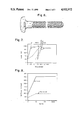
- FIG. 8 is a graph showing the effect of multiple load cycles on the stiffness of polyethylene prosthetic ligaments
- FIG. 9 is a graph showing the effect of multiple load cycles on the constructional deformation of polyethylene prosthetic ligaments.
- FIGS. 10 and 11 are graphs showing the axial elongation due to constructional deformation associated with various loads as a function of the number of cycles applied to the ligament.
- FIGS. 1 through 5 illustrate the preferred embodiments of the prosthetic ligament 10 of the present invention.
- FIG. 6 illustrates the bone screw 52, which together with prosthetic ligament 10 comprises the prosthetic ligament assembly of the present invention.
- the prosthetic ligament 10 is shown as connecting the femur and tibia, it should be recognized that the prosthetic ligament 10 can be used to connect other skeletal members and, depending upon the means of attachment employed, to support soft tissue organs in place.
- the prosthetic ligament 10 is employed to permanently replace the natural anterior cruciate ligament of the human knee.
- the prosthetic ligament 10 includes primarily a major load bearing member which has a gage section 20 and at least one looped extension 50 at one end thereof.
- a core 30 and sheath 40 may optionally be included.
- the load bearing member is formed from a plurality of high strength, biocompatible polyethylene yarns. There are at least fifty (50) fibers in each yarn. Each polyethylene fiber has an average diameter of less than about 100 microns (0.002 inch). Fiber tensile strength is greater than 689 MPa (100,000 psi).
- the yarns are parallel wound and the resulting bundles are intertwined to form a braid.
- a plurality of such high strength yarns, braided at the appropriate picks per inch, as hereinafter defined, provides a high strength load bearing member. Numerous fibers add to the life of the prosthetic ligament 10 because there are numerous surfaces to tolerate stress and abrasion.
- the plain braid type is defined in terms of picks/inch.
- pick shall mean the crossing of one yarn bundle over another created by the braiding technique. Picks are counted along the longitudinal axis of the ligament. The greater the number of picks/inch, the tighter the braid.
- the preferred average diameter of the gage section 20 is about 4-6 mm (about 0.16-0.24 inches).
- a braid of 5-7 picks/inch provides the desired diameter.
- the section designated as "A" in FIG. 2A represents one pick.
- the section designated as B represents six picks.
- the braid shown in FIG. 2 is a twill braid. Comparable sections A, designating one pick and B designating six picks are shown. The best mode of the load bearing member developed to date is formed from a plain braid.
- ultra high molecular weight polyethylene for use in prosthetic ligaments is disclosed in Zachariades U.S. Pat. No. 4,587,163.
- the Zachariades material is not a multifilament like that used in the ligament 10 of the present invention.
- the ribbon like structure of the Zachariades material will not permit tissue ingrowth.
- the Zachariades material is sintered and then drawn to produce a single filament with a strength to weight ratio much less than that of the fibers used to make the yarns of ligament 10 (40 MPa versus 2930 MPa).
- ligament 10 uses commercially available ultra high molecular weight polyethylene fibers, e.g. SPECTRA-900TM or SPECTRA-1000TM
- the preferred embodiment of ligament 10 can be described as having one hundred twenty (120) fibers in each yarn, formed into an 8-strand, 6-parallel wound plain braid ranging from 5-7 picks/inch to provide an average gage section diameter of about 4-6 mm.
- the SPECTRA-1000TM fibers have a tenacity of 35 grams/denier with a specific gravity of about 0.97 and an average diameter of about 27 microns (0.0011 inch).
- the prosthetic ligament 10 can be placed in the recipient by any suitable surgical technique.
- FIG. 1 illustrates an example of the technique employing bone tunnels 16.
- Ligament 10 may optionally include a sheath 40.
- the sheath 40 may be a tube having an internal diameter equal to or less than the diameter of the gage section 20 which is friction fit onto the gage section 20.
- the sheath 40 must be thick enough to withstand the abrasion to which it may be subjected. In one embodiment for use in replacing a cruciate ligament, the sheath 40 is about 1/16 inch thick. The thickness may vary depending upon the desired use of the prosthetic ligament 10.
- the sheath 40 may also be formed onto the gage section 20. Care should be taken, however, to avoid over-heating the load bearing member. Excessive heat can damage the fibers. Regardless of how the sheath 40 is applied to the gage section 20, the sheath 40 must be relatively thick. The thickness of sheath 40 can vary for different materials. While the drawings show ligament 10 as having a circular cross-section, those skilled in the art will recognize that the cross-section can be elliptical or any other shape suitable for the intended end use of the prosthesis. For example, sheath 40 may have an elliptical cross section to match the shape of a natural ligament. The ligament 10 may also be more narrow at its midportion than at its end portions.
- Ligament 10 may also optionally include a core 30.
- the core 30 is disposed within a longitudinal bore 28 through gage section 20.
- the core 30 is preferably made of a biocompatible elastomer to which is added a biocompatible radiopaque material which permits radiographic visualization of the prosthetic ligament 10.
- the material of choice is made of between 5-25%, inclusive, barium sulfate, by weight, in a high strength block copolymer containing silicone oil. Zirconium oxide, titanium oxide or any other suitable radiopaque material may be used in place of barium sulfate.
- a suitable elastomer is described in Sterling U.S. Pat. No. 4,386,179, which is hereby incorporated herein by reference, and sold commercially under the trademark C-FLEX®.
- Sheath 40 is preferably also made of C-FLEX®.
- Fixation of the ligament 10 may be provided by any suitable means, such as screws, nails or staples.
- the preferred means of fixation is by bone screws 52, illustrated in FIG. 6.
- Bone screw 52 includes a large, relatively flat, rounded screw head 54 to prevent disengagement of the loop 50 and a tapered section 56 extending from the perimeter of the screw head 54 to the threaded shank. 58.
- the threads of shank 58 may be cancellous or cortical.
- the screw 52 may be self-tapping or nonself-tapping.
- the shank 58 might typically have a diameter of 3.5-8.0 mm (0.14-0.31 inches).
- the interaction between the loop 50 and the tapered section 56 of bone screw 52 permits tensile loading of the ligament 10 during fixation.
- the self-tensioning effect insures that the ligament 10 is taut when implanted and is the preferred means of providing in vivo stability of the ligament 10.
- the design of the screw head 54 and tapered section 56 cause the loop area to expand and the ligament 10 to undergo subsequent tensioning as the screw 52 is fully inserted. During surgery, such a tensile load is intentionally applied to secure the ligament and attain an appropriate joint response. Studies suggest that the desired tensioning ranges between about 35-90 N (8-20 lbs.) and that about 35-45 N (8-10 lbs.) is optimum.
- the higher preloads would be expected to protect against additional fiber settling which may occur from occasional excessive loading.
- the polyethylene ligament has demonstrated constructional deformation less than the value of 6-8 mm (0.24-0.31 inch) characteristic of a stable knee, this settling can be reduced by preloading the ligament prior to surgery.
- a preload of 1000 N (225 lbs.) has been determined to be advantageous to protect the prosthetic ligament against in vivo settling resulting from occasional excessive loading.
- a SPECTRA-1000TM polyethylene ligament not preloaded, exhibited an average ultimate tensile strength of 9017 ⁇ 468 N (2026 ⁇ 105 lbs.) when tested at 1%/sec. with failure occurring in the loop area. Assuming a constant fiber diameter of 27 microns (0.001 inch) and calculating the number of fibers for the preferred embodiment of the prosthetic ligament (120 fibers/yarn, 6 yarns/parallel wound ply, 8 parallel wound plies/braid, and 2 braids/ligament at the loops) allows for this value of breaking strength to be converted to an ultimate stress value, 1367 MPa (198,000 psi) for the SPECTRA-1000TM material.
- the difference between the reported ultimate stress value of the SPECTRA-1000TM fiber, approximately 2390 MPa (425,000 psi), and the measured value for the prosthetic ligament 10 made from the SPECTRA-1000TM material can be attributed to loop and braid effects associated with ligament 10.
- the doublelooped ligament 10 has an effective ultimate tensile strength which is 47% of the strength of equivalent SPECTRA-1000TM fibers neglecting loop and braid effects.
- a preload of 1000 N (225 lbs.) is 5.2% of the breaking load (i.e., 19,108 N or 4294 lbs.) of the fiber.
- Double-looped SPECTRA-1000TM polyethylene ligaments with no core or sheath were tested to failure under tensile loading to evaluate their axial mechanical properties; breakage load, elongation to breakage and axial stiffness.
- Each ligament in this series of tests was approximately 17 cm in length, preloaded for one hour at 1000 N (225 lbs.) and sterilized in ethylene oxide prior to use. Strain rates of 1, 10 and 100%/per sec. were applied. The results of this series of tests are presented in Table I below.
- the axial modulus of elasticity of the SPECTRA-1000TM fibers is quite high and would not be expected to be flexible enough for use as a prosthetic ligament. Indeed, when tested axially in a braided configuration, the prosthetic ligament can still appear to be quite stiff following cyclic loading. However when the braided ligament is tested in the anatomical position, the modulus of elasticity is about 100,000 psi and the stiffness values come down to the range reported for the human ACL.
- ligaments were cyclically tensile tested on a MTS 810 servo hydraulic test system as follows:
- the first 10 cycles were controlled by hand and the displacements recorded at loads of 89 N, 178 N, 267 N, 358 N and 445 N.
- the ligament was cycled at 1 Hz between 22 N and 445 N and the displacements recorded at cycles of 100, 500, and 1000.
- the test was terminated at the 1000th cycle.
- the axial test arrangement consisted of axially loading the ligament between two pin grips.
- the ligament was secured to a prosthetic knee and loaded in an anterior drawer direction at 0, 20, and 90° of flexion.
- FIGS. 8 and 9 show results of the change in stiffness and constructional deformation, respectively, as a function of load cycles.
- the polyethylene ligament When tested non-anatomically (axially), the polyethylene ligament showed initial (first cycle) stiffnesses of 132 N/mm. At 1000 cycles, the value increased to 700 N/mm. More importantly, the amount of constructional deformation after 1000 cycles was 12.7 mm (0.5 inch). However, under anatomical load condition, the stiffness characteristics decreased significantly while the cyclic settling increased relative to that observed in the axial tests. The stiffness of the ligaments tested anatomically was within the range of values reported for the human ACL.
- the anatomical constructional deformation behavior stabilized after roughly 100 cycles for the polyethylene ligament, reaching a peak of roughly 7 mm (0.27 inch).
- the prosthetic ligaments evaluated in this study showed stabilization of both constructional deformation and stiffness properties after approximately 100 cycles. Importantly, the ligaments showed anatomical stiffness to be comparable to that of the human ACL.
- Prosthetic ligaments like those used in the study reported immediately above, but without the polymer coating on the braid were tested in two separate studies using cadavers. The results indicate that the presence of the coating makes a negligible difference in the stiffness of the system. The results confirm that the anatomical positioning provides a ligament having a stiffness within the range of values reported for the human ACL.
- the first series of tests included low cycle fatigue tests performed to generate data used in producing a stress-life (S-N) curve for the ligament.
- S-N stress-life
- the second series of tests included tests which were conducted at levels of loading comparable to those reported for in vivo conditions including maximum applied loads of 170 N, 445 N and 650 N. From these two series of tests the axial elongation due to constructional deformation associated with each load level was evaluated as a function of the number of load cycles applied to the ligament. The results presented in FIGS. 10 and 11 indicate that, within the regime of physiological loading the elongation of the ligament due to constructional deformation becomes asymptotic after several hundred cycles and remains approximately 1.1, 1.8 and 2.1 mm (0.6, 1.0 and 1.2%) at 170 N, 445 N and 650 N, respectively.
- the S-N data developed in the first series of tests can be used to predict a fatigue life of 8.5 ⁇ 10 9 cycles under a maximum cyclic load of 650 N (146 lbs.).
Abstract
Description
              TABLE I
______________________________________
Tension Test Data
                              Elongation.sup.3
Strain  Lig-                  to      Axial
Rate    aments  Breakage Load.sup.1
                              Breakage
                                      Stiffness
(%/sec) Tested  (N)           (%)     (N/mm)
______________________________________
 1      6       9239 ± 491.sup.2
                              7.1 ± 0.4
                                      230 ± 22
                  (2076 ± 110 lbs)
10      7       9017 ± 468 6.7 ± 0.4
                                      216 ± 40
                  (2026 ± 105 lbs)
100     6       8682 ± 371 6.8 ± 0.6
                                      266 ± 36
                 (1951 ± 83 lbs)
______________________________________
 .sup.1 The maximum force applied to the device during testing.
 .sup.2 Arithmetic mean value ± standard deviation.
 .sup.3 The change in displacement from that associated with 45 N load to
 that associated with the maximum load, assuming 45 N to be the approximat
 minimum tensile load at implant.
              TABLE II
______________________________________
Strength and Ductility Properties of the
Ligaments and Coating
       Fiber Properties (Approximate)
         Tensile Strength,       Ligament
Material MPa          % Elongation
                                 Strength N
______________________________________
Polyethylene
         2,930             3       10,700
Ligament (425,000 psi)             (2,000
                                         lbs)
Coating  14               700      --
______________________________________
              TABLE III
______________________________________
            With C-FLEX ®
            Sheath     Without Sheath
______________________________________
Ultimate Load 8110    N        10,100 N
Ultimate Elongation
              12.8    mm       14.2   mm
Ultimate Stress*
              383     MPa      476    MPa
Ultimate Strain
              6.7%             7.5%
Energy to Failure
              89      N-m      121    N-m
Stiffness (axial)
              634     N/mm     711    N/mm
______________________________________
 *Nominal, using crosssectional area at midsection of 21.2 mm.sup.2, 191 m
 (71/2") with 19 mm (0.75") eyelets Strain Rate = 0.9%/second
              TABLE IV
______________________________________
LOW CYCLE AXIAL FATIGUE TEST RESULTS
MAXIMUM
LOAD (N) FREQUENCY (Hz) CYCLES TO FAILURE
______________________________________
4005     5              12,774,710
4895     4              372,800
4895     4              294,830
4895     4              2,768,915
5785     3              957,410
5785     3              569,030
6675     2               84,800
6675     2               40,470
7565     1               17,382
7565     1               12,973
______________________________________
Claims (7)
Priority Applications (1)
| Application Number | Priority Date | Filing Date | Title |
|---|---|---|---|
| US07/186,330 US4932972A (en) | 1986-03-14 | 1988-04-26 | Prosthetic ligament |
Applications Claiming Priority (2)
| Application Number | Priority Date | Filing Date | Title |
|---|---|---|---|
| US06/840,298 US4731084A (en) | 1986-03-14 | 1986-03-14 | Prosthetic ligament |
| US07/186,330 US4932972A (en) | 1986-03-14 | 1988-04-26 | Prosthetic ligament |
Related Parent Applications (1)
| Application Number | Title | Priority Date | Filing Date |
|---|---|---|---|
| US07/064,634 Division US4790850A (en) | 1986-03-14 | 1987-06-22 | Phosthetic ligament |
Publications (1)
| Publication Number | Publication Date |
|---|---|
| US4932972A true US4932972A (en) | 1990-06-12 |
Family
ID=25281969
Family Applications (3)
| Application Number | Title | Priority Date | Filing Date |
|---|---|---|---|
| US06/840,298 Expired - Lifetime US4731084A (en) | 1986-03-14 | 1986-03-14 | Prosthetic ligament |
| US07/064,634 Expired - Fee Related US4790850A (en) | 1986-03-14 | 1987-06-22 | Phosthetic ligament |
| US07/186,330 Expired - Fee Related US4932972A (en) | 1986-03-14 | 1988-04-26 | Prosthetic ligament |
Family Applications Before (2)
| Application Number | Title | Priority Date | Filing Date |
|---|---|---|---|
| US06/840,298 Expired - Lifetime US4731084A (en) | 1986-03-14 | 1986-03-14 | Prosthetic ligament |
| US07/064,634 Expired - Fee Related US4790850A (en) | 1986-03-14 | 1987-06-22 | Phosthetic ligament |
Country Status (7)
| Country | Link |
|---|---|
| US (3) | US4731084A (en) |
| EP (1) | EP0238263B1 (en) |
| JP (1) | JPS62281940A (en) |
| AU (1) | AU605759B2 (en) |
| CA (1) | CA1251302A (en) |
| DE (1) | DE3773965D1 (en) |
| ES (1) | ES2027689T3 (en) |
Cited By (64)
| Publication number | Priority date | Publication date | Assignee | Title |
|---|---|---|---|---|
| AU613500B2 (en) * | 1988-07-01 | 1991-08-01 | Minnesota Mining And Manufacturing Company | Abrasion resistant prosthetic device |
| US5195542A (en) * | 1989-04-27 | 1993-03-23 | Dominique Gazielly | Reinforcement and supporting device for the rotator cuff of a shoulder joint of a person |
| US5263984A (en) * | 1987-07-20 | 1993-11-23 | Regen Biologics, Inc. | Prosthetic ligaments |
| WO1994015550A1 (en) * | 1993-01-06 | 1994-07-21 | Smith & Nephew Richards Inc. | Load bearing polymeric cable |
| US5391170A (en) * | 1991-12-13 | 1995-02-21 | David A. McGuire | Angled surgical screw driver and methods of arthroscopic ligament reconstruction |
| US5441508A (en) * | 1989-04-27 | 1995-08-15 | Gazielly; Dominique | Reinforcement and supporting device for the rotator cuff of a shoulder joint of a person |
| US5507812A (en) * | 1992-12-28 | 1996-04-16 | Moore; David E. | Modular prosthetic ligament |
| US5540703A (en) * | 1993-01-06 | 1996-07-30 | Smith & Nephew Richards Inc. | Knotted cable attachment apparatus formed of braided polymeric fibers |
| US6203572B1 (en) | 1999-02-09 | 2001-03-20 | Linvatec Corporation | Device and method for ligament reconstruction |
| US20020062152A1 (en) * | 2000-09-15 | 2002-05-23 | Martin Dauner | Medical, bioresorbable implant, process for its production and the use thereof |
| US6589246B1 (en) | 2001-04-26 | 2003-07-08 | Poly-4 Medical, Inc. | Method of applying an active compressive force continuously across a fracture |
| US20030229265A1 (en) * | 2000-06-13 | 2003-12-11 | Girard Michael J. | Cardiac support device |
| US6764513B1 (en) | 2001-11-07 | 2004-07-20 | Brian T. Dowling | Tibia tether |
| US6783527B2 (en) | 2001-10-30 | 2004-08-31 | Sdgi Holdings, Inc. | Flexible spinal stabilization system and method |
| EP1453438A1 (en) * | 2001-12-14 | 2004-09-08 | Celanese Advanced Materials, Inc. | Prosthetic ligament |
| US20050149187A1 (en) * | 2000-08-28 | 2005-07-07 | Ron Clark | Method and implant for securing ligament replacement into the knee |
| US20050192581A1 (en) * | 2004-02-27 | 2005-09-01 | Molz Fred J. | Radiopaque, coaxial orthopedic tether design and method |
| US20070208217A1 (en) * | 2006-03-03 | 2007-09-06 | Acorn Cardiovascular, Inc. | Self-adjusting attachment structure for a cardiac support device |
| US20070270882A1 (en) * | 2006-05-19 | 2007-11-22 | Acorn Cardiovascular, Inc. | Pericardium management method for intra-pericardial surgical procedures |
| US20080004488A1 (en) * | 2006-06-29 | 2008-01-03 | Acorn Cardiovascular, Inc. | Low friction delivery tool for a cardiac support device |
| US20080033235A1 (en) * | 2000-05-10 | 2008-02-07 | Acorn Cardiovascular, Inc. | Cardiac disease treatment and device |
| US20080033234A1 (en) * | 2006-07-17 | 2008-02-07 | Acorn Cardiovascular, Inc. | Cardiac support device delivery tool with release mechanism |
| US20080228271A1 (en) * | 2007-03-13 | 2008-09-18 | Biomet Sports Medicine, Inc. | Method and apparatus for graft fixation |
| US20080300683A1 (en) * | 2007-03-20 | 2008-12-04 | Altman Gregory H | Prosthetic device and method of manufacturing the same |
| WO2009036286A1 (en) * | 2007-09-12 | 2009-03-19 | Surgical Energetics, Inc. | Medical device and tension member for use in a subject |
| US20090204118A1 (en) * | 2008-02-13 | 2009-08-13 | William Ralph Pratt | Surgical cable with malleable leader segment |
| US20090216252A1 (en) * | 2004-02-13 | 2009-08-27 | The University Of Cincinnati | A coupling device enabled by mechanical continuity of cellular scaffolding across tissue boundaries |
| US20090306775A1 (en) * | 2008-04-21 | 2009-12-10 | Javier Macossay-Torres | Artificial ligaments and tendons comprising multifilaments and nanofibers and methods for making |
| US7641608B1 (en) | 2006-09-26 | 2010-01-05 | Acorn Cardiovascular, Inc. | Sectional cardiac support device and method of delivery |
| US20100042197A1 (en) * | 2006-10-02 | 2010-02-18 | Arterion Ab | Preparation of hollow cellulose vessels |
| US20100054926A1 (en) * | 2008-08-29 | 2010-03-04 | General Electric Company | System and method for thermal management of a gas turbine inlet |
| US20100094423A1 (en) * | 2008-10-15 | 2010-04-15 | Warsaw Orthopedic, Inc. | Systems and methods for assessment of tension in an implant |
| US20100121448A1 (en) * | 2001-03-13 | 2010-05-13 | Depuy Mitek, Inc. | Method and apparatus for fixing a graft in a bone tunnel |
| US20100185050A1 (en) * | 2000-06-13 | 2010-07-22 | Acorn Cardiovascular, Inc. | Compliant cardiac support device |
| US20100249930A1 (en) * | 2009-03-31 | 2010-09-30 | Medicinelodge, Inc. Dba Imds Co-Innovation | Double bundle acl repair |
| US20100256677A1 (en) * | 2009-03-31 | 2010-10-07 | Arthrex, Inc. | Integrated adjustable button-suture-graft construct with two fixation devices |
| US20100268273A1 (en) * | 2009-03-31 | 2010-10-21 | Ricardo Albertorio | Adjustable suture button construct and methods of tissue reconstruction |
| US20100286775A1 (en) * | 2007-10-11 | 2010-11-11 | Tavor [I.T.N] Ltd., | Ligament and Tendon Prosthesis |
| US20110004053A1 (en) * | 2007-12-05 | 2011-01-06 | Surgical Energetics, Inc. | bolster for securing a septal splint to a cardiac wall, a method of use thereof, and a system including the same |
| US20110046734A1 (en) * | 2008-03-13 | 2011-02-24 | Tavor (I.T.N) Ltd. | Ligament And Tendon Prosthesis |
| US7896917B2 (en) | 2003-10-15 | 2011-03-01 | Biomet Sports Medicine, Llc | Method and apparatus for graft fixation |
| US20110190886A1 (en) * | 2010-01-29 | 2011-08-04 | Wisconsin Alumni Research Foundation | Braided tertiary nanofibrous structure for ligament, tendon, and muscle tissue implant |
| US8002778B1 (en) | 2004-06-28 | 2011-08-23 | Biomet Sports Medicine, Llc | Crosspin and method for inserting the same during soft ligament repair |
| CN102488570A (en) * | 2011-11-14 | 2012-06-13 | 东华大学 | Material for artificial ligament |
| EP2518208A2 (en) | 2011-04-27 | 2012-10-31 | Polteco Inc. | Abrasion resistant cords and ropes |
| US8591578B2 (en) | 2010-11-17 | 2013-11-26 | Arthrex, Inc. | Adjustable suture-button constructs for ligament reconstruction |
| US8628573B2 (en) | 2009-03-31 | 2014-01-14 | Arthrex, Inc. | Adjustable suture-button construct for knotless stabilization of cranial cruciate deficient ligament stifle |
| US8740985B1 (en) | 2012-11-30 | 2014-06-03 | Smith & Nephew, Inc. | Knee prosthesis |
| USD717954S1 (en) | 2013-10-14 | 2014-11-18 | Mardil, Inc. | Heart treatment device |
| US8968402B2 (en) | 2011-10-18 | 2015-03-03 | Arthrocare Corporation | ACL implants, instruments, and methods |
| US9107653B2 (en) | 2011-09-22 | 2015-08-18 | Arthrex, Inc. | Tensionable knotless anchors with splice and methods of tissue repair |
| US9179950B2 (en) | 2010-11-17 | 2015-11-10 | Arthrex, Inc. | Adjustable suture-button construct for ankle syndesmosis repair |
| US9301745B2 (en) | 2011-07-21 | 2016-04-05 | Arthrex, Inc. | Knotless suture constructs |
| US9332979B2 (en) | 2011-07-22 | 2016-05-10 | Arthrex, Inc. | Tensionable knotless acromioclavicular repairs and constructs |
| US9370425B2 (en) | 2012-10-12 | 2016-06-21 | Mardil, Inc. | Cardiac treatment system and method |
| US9615821B2 (en) | 2011-12-09 | 2017-04-11 | Arthrex, Inc. | Tensionable knotless anchor systems and methods of tissue repair |
| US9737292B2 (en) | 2012-06-22 | 2017-08-22 | Arthrex, Inc. | Knotless suture anchors and methods of tissue repair |
| US10206670B2 (en) | 2002-06-20 | 2019-02-19 | Arthrex, Inc. | Apparatuses and methods for fixation of ankle syndesmosis or acromioclavicular joint dislocations of the shoulder |
| US10245016B2 (en) | 2011-10-12 | 2019-04-02 | Arthrex, Inc. | Adjustable self-locking loop constructs for tissue repairs and reconstructions |
| US10265060B2 (en) | 2015-08-20 | 2019-04-23 | Arthrex, Inc. | Tensionable constructs with multi-limb locking mechanism through single splice and methods of tissue repair |
| US10292821B2 (en) | 2001-09-07 | 2019-05-21 | Phoenix Cardiac Devices, Inc. | Method and apparatus for external stabilization of the heart |
| US10335136B2 (en) | 2015-08-20 | 2019-07-02 | Arthrex, Inc. | Tensionable constructs with multi-limb locking mechanism through single splice and methods of tissue repair |
| US10575842B2 (en) | 2017-02-09 | 2020-03-03 | Arthrex, Inc. | Knotless self-locking anchor constructs and methods of tissue fixation |
| US10646327B2 (en) | 2017-02-09 | 2020-05-12 | Arthrex, Inc. | Self-locking suture constructs and methods of tissue fixation |
Families Citing this family (145)
| Publication number | Priority date | Publication date | Assignee | Title |
|---|---|---|---|---|
| US5180636A (en) * | 1987-09-08 | 1993-01-19 | Mitsui Petrochemical Industries Ltd. | Rope for traction |
| FR2623083B1 (en) * | 1987-11-13 | 1990-11-30 | Implants Medical Systeme Sarl | LIGAMENTARY OR TENDANT PROSTHESIS AND METHOD FOR MANUFACTURING SAID PROSTHESIS |
| US5197983A (en) * | 1988-04-19 | 1993-03-30 | W. L. Gore & Associates, Inc. | Ligament and tendon prosthesis |
| US4917699A (en) * | 1988-05-16 | 1990-04-17 | Zimmer, Inc. | Prosthetic ligament |
| US4883486A (en) * | 1988-05-31 | 1989-11-28 | Indu Kapadia | Prosthetic ligament |
| US5026398A (en) * | 1988-07-01 | 1991-06-25 | The Minnesota Mining And Manufacturing Company | Abrasion resistant prosthetic device |
| FR2642645B1 (en) * | 1989-02-03 | 1992-08-14 | Breard Francis | FLEXIBLE INTERVERTEBRAL STABILIZER AND METHOD AND APPARATUS FOR CONTROLLING ITS VOLTAGE BEFORE PLACEMENT ON THE RACHIS |
| USRE36221E (en) * | 1989-02-03 | 1999-06-01 | Breard; Francis Henri | Flexible inter-vertebral stabilizer as well as process and apparatus for determining or verifying its tension before installation on the spinal column |
| US4927421A (en) * | 1989-05-15 | 1990-05-22 | Marlowe Goble E | Process of endosteal fixation of a ligament |
| US4946377A (en) * | 1989-11-06 | 1990-08-07 | W. L. Gore & Associates, Inc. | Tissue repair device |
| WO1991007145A1 (en) * | 1989-11-22 | 1991-05-30 | Indu Kapadia | Prosthetic ligament |
| US5004474A (en) * | 1989-11-28 | 1991-04-02 | Baxter International Inc. | Prosthetic anterior cruciate ligament design |
| EP0447355A1 (en) * | 1990-03-12 | 1991-09-18 | Gebrüder Sulzer Aktiengesellschaft | Implant for the human body |
| US5860978A (en) | 1990-09-25 | 1999-01-19 | Innovasive Devices, Inc. | Methods and apparatus for preventing migration of sutures through transosseous tunnels |
| US5318575A (en) * | 1992-02-03 | 1994-06-07 | United States Surgical Corporation | Method of using a surgical repair suture product |
| US5425733A (en) * | 1992-02-19 | 1995-06-20 | Arthrex, Inc. | Interference screw with rounded back end and cannulated sheath for endosteal fixation of ligaments |
| FR2687911B1 (en) * | 1992-03-02 | 1997-06-06 | Periode Sa | PROSTHESIS OF PROTECTIVE SHEATH LIGAMENTS. |
| US7101392B2 (en) * | 1992-03-31 | 2006-09-05 | Boston Scientific Corporation | Tubular medical endoprostheses |
| JPH07505316A (en) | 1992-03-31 | 1995-06-15 | ボストン サイエンティフィック コーポレーション | medical wire |
| US6277084B1 (en) | 1992-03-31 | 2001-08-21 | Boston Scientific Corporation | Ultrasonic medical device |
| FR2690073B1 (en) * | 1992-04-15 | 1994-11-18 | Deux C T | Repair prosthesis for the rotator cuff of the shoulder. |
| US5407420A (en) * | 1992-11-12 | 1995-04-18 | Smith & Nephew Donjoy, Inc. | Fully adjustable shoulder brace |
| US5354299A (en) * | 1992-12-07 | 1994-10-11 | Linvatec Corporation | Method of revising a screw in a tunnel |
| US5443509A (en) * | 1992-12-10 | 1995-08-22 | Linvatec Corporation | Interference bone-fixation screw with multiple interleaved threads |
| US20050059889A1 (en) * | 1996-10-16 | 2005-03-17 | Schneider (Usa) Inc., A Minnesota Corporation | Clad composite stent |
| WO1994016646A1 (en) * | 1993-01-19 | 1994-08-04 | Schneider (Usa) Inc. | Clad composite stent |
| US5630840A (en) * | 1993-01-19 | 1997-05-20 | Schneider (Usa) Inc | Clad composite stent |
| US5632748A (en) * | 1993-06-14 | 1997-05-27 | Linvatec Corporation | Endosteal anchoring device for urging a ligament against a bone surface |
| US5450860A (en) * | 1993-08-31 | 1995-09-19 | W. L. Gore & Associates, Inc. | Device for tissue repair and method for employing same |
| US5758562A (en) * | 1995-10-11 | 1998-06-02 | Schneider (Usa) Inc. | Process for manufacturing braided composite prosthesis |
| US5891191A (en) * | 1996-04-30 | 1999-04-06 | Schneider (Usa) Inc | Cobalt-chromium-molybdenum alloy stent and stent-graft |
| US6592617B2 (en) * | 1996-04-30 | 2003-07-15 | Boston Scientific Scimed, Inc. | Three-dimensional braided covered stent |
| US5718159A (en) * | 1996-04-30 | 1998-02-17 | Schneider (Usa) Inc. | Process for manufacturing three-dimensional braided covered stent |
| US6123662A (en) * | 1998-07-13 | 2000-09-26 | Acorn Cardiovascular, Inc. | Cardiac disease treatment and device |
| US20050049702A1 (en) * | 1998-03-10 | 2005-03-03 | The University Of Cincinnati | Article and method for coupling muscle |
| US6099530A (en) * | 1998-04-09 | 2000-08-08 | Smith & Nephew, Inc. | Soft-tissue intra-tunnel fixation device |
| US6221107B1 (en) | 1998-08-03 | 2001-04-24 | Mark E. Steiner | Ligament fixation device and method |
| US7410489B2 (en) * | 1998-09-28 | 2008-08-12 | Daos Limited | Internal cord fixation device |
| AU742502B2 (en) | 1998-11-26 | 2002-01-03 | Synthes Gmbh | Screw |
| YU45502A (en) * | 1999-12-17 | 2003-07-07 | Cartificial A/S. Medico Chemical Lab. Ap S. | A prosthetic device |
| US6524317B1 (en) * | 1999-12-30 | 2003-02-25 | Opus Medical, Inc. | Method and apparatus for attaching connective tissues to bone using a knotless suture anchoring device |
| US6623492B1 (en) | 2000-01-25 | 2003-09-23 | Smith & Nephew, Inc. | Tissue fastener |
| US6746483B1 (en) * | 2000-03-16 | 2004-06-08 | Smith & Nephew, Inc. | Sheaths for implantable fixation devices |
| US6585730B1 (en) * | 2000-08-30 | 2003-07-01 | Opus Medical, Inc. | Method and apparatus for attaching connective tissues to bone using a knotless suture anchoring device |
| GB0024903D0 (en) * | 2000-10-11 | 2000-11-22 | Ellis Dev Ltd | A textile prothesis |
| US7083638B2 (en) * | 2001-02-12 | 2006-08-01 | Arthrocare Corporation | Method and apparatus for attaching connective tissues to bone using a knotless suture anchoring device |
| US6770076B2 (en) * | 2001-02-12 | 2004-08-03 | Opus Medical, Inc. | Method and apparatus for attaching connective tissues to bone using a knotless suture anchoring device |
| US6547800B2 (en) * | 2001-06-06 | 2003-04-15 | Opus Medical, Inc. | Method and apparatus for attaching connective tissues to bone using a cortical bone anchoring device |
| US6951685B1 (en) * | 2001-11-27 | 2005-10-04 | Integrated Textile Systems, Inc. | Ultra high molecular weight polyethylene fibers |
| US6780198B1 (en) * | 2001-12-06 | 2004-08-24 | Opus Medical, Inc. | Bone anchor insertion device |
| US6855157B2 (en) * | 2002-02-04 | 2005-02-15 | Arthrocare Corporation | Method and apparatus for attaching connective tissues to bone using a knotless suture anchoring device |
| US20030236553A1 (en) * | 2002-06-24 | 2003-12-25 | Celanese Advanced Materials, Inc. | Thermotropic polymer heterofilament |
| US7090690B2 (en) * | 2002-11-19 | 2006-08-15 | Arthrocare Corporation | Devices and methods for repairing soft tissue |
| US20040243131A1 (en) * | 2003-02-07 | 2004-12-02 | Dirks Christiaan H.P. | Bone fixing device |
| EP1444959A1 (en) * | 2003-02-07 | 2004-08-11 | DSM IP Assets B.V. | Bone fixing device |
| WO2004082724A2 (en) * | 2003-03-18 | 2004-09-30 | Opus Medical Inc. | Optimized suture braid |
| US7722644B2 (en) | 2003-06-11 | 2010-05-25 | Medicine Lodge, Inc. | Compact line locks and methods |
| US7682374B2 (en) * | 2003-10-21 | 2010-03-23 | Arthrocare Corporation | Knotless suture lock and bone anchor implant method |
| US20080269900A1 (en) * | 2004-05-20 | 2008-10-30 | Christopher Reah | Surgical Implants |
| US8088146B2 (en) | 2004-06-14 | 2012-01-03 | Teleflex Medical Incorporated | High-strength suture |
| US7658751B2 (en) | 2006-09-29 | 2010-02-09 | Biomet Sports Medicine, Llc | Method for implanting soft tissue |
| US7905904B2 (en) | 2006-02-03 | 2011-03-15 | Biomet Sports Medicine, Llc | Soft tissue repair device and associated methods |
| US8118836B2 (en) | 2004-11-05 | 2012-02-21 | Biomet Sports Medicine, Llc | Method and apparatus for coupling soft tissue to a bone |
| US8088130B2 (en) | 2006-02-03 | 2012-01-03 | Biomet Sports Medicine, Llc | Method and apparatus for coupling soft tissue to a bone |
| US8361113B2 (en) | 2006-02-03 | 2013-01-29 | Biomet Sports Medicine, Llc | Method and apparatus for coupling soft tissue to a bone |
| US8137382B2 (en) | 2004-11-05 | 2012-03-20 | Biomet Sports Medicine, Llc | Method and apparatus for coupling anatomical features |
| US7749250B2 (en) | 2006-02-03 | 2010-07-06 | Biomet Sports Medicine, Llc | Soft tissue repair assembly and associated method |
| US8298262B2 (en) | 2006-02-03 | 2012-10-30 | Biomet Sports Medicine, Llc | Method for tissue fixation |
| US7909851B2 (en) | 2006-02-03 | 2011-03-22 | Biomet Sports Medicine, Llc | Soft tissue repair device and associated methods |
| US8303604B2 (en) | 2004-11-05 | 2012-11-06 | Biomet Sports Medicine, Llc | Soft tissue repair device and method |
| US9017381B2 (en) | 2007-04-10 | 2015-04-28 | Biomet Sports Medicine, Llc | Adjustable knotless loops |
| US8128658B2 (en) | 2004-11-05 | 2012-03-06 | Biomet Sports Medicine, Llc | Method and apparatus for coupling soft tissue to bone |
| US9801708B2 (en) | 2004-11-05 | 2017-10-31 | Biomet Sports Medicine, Llc | Method and apparatus for coupling soft tissue to a bone |
| US8753392B2 (en) | 2005-01-07 | 2014-06-17 | University Of Cincinnati | Elements for versatility of a prosthetic anchor |
| US8052753B2 (en) | 2005-01-07 | 2011-11-08 | University Of Cincinnati | Prosthetic anchor and method of making same |
| US9452001B2 (en) * | 2005-02-22 | 2016-09-27 | Tecres S.P.A. | Disposable device for treatment of infections of human limbs |
| GB2441266B (en) * | 2005-06-01 | 2011-03-02 | Arthrocare Corp | Knotless suture anchoring device having deforming section to accommodate sutures of various diameters |
| WO2006133130A2 (en) * | 2005-06-03 | 2006-12-14 | Nuvasive, Inc. | Fibrous spinal implant and method of implantation |
| GB0513686D0 (en) * | 2005-07-04 | 2005-08-10 | Finsbury Dev Ltd | Prosthesis |
| US11311287B2 (en) | 2006-02-03 | 2022-04-26 | Biomet Sports Medicine, Llc | Method for tissue fixation |
| US9538998B2 (en) | 2006-02-03 | 2017-01-10 | Biomet Sports Medicine, Llc | Method and apparatus for fracture fixation |
| US8562645B2 (en) | 2006-09-29 | 2013-10-22 | Biomet Sports Medicine, Llc | Method and apparatus for forming a self-locking adjustable loop |
| US8652171B2 (en) | 2006-02-03 | 2014-02-18 | Biomet Sports Medicine, Llc | Method and apparatus for soft tissue fixation |
| US9408599B2 (en) | 2006-02-03 | 2016-08-09 | Biomet Sports Medicine, Llc | Method and apparatus for coupling soft tissue to a bone |
| US8801783B2 (en) | 2006-09-29 | 2014-08-12 | Biomet Sports Medicine, Llc | Prosthetic ligament system for knee joint |
| US11259792B2 (en) | 2006-02-03 | 2022-03-01 | Biomet Sports Medicine, Llc | Method and apparatus for coupling anatomical features |
| US8597327B2 (en) | 2006-02-03 | 2013-12-03 | Biomet Manufacturing, Llc | Method and apparatus for sternal closure |
| US8968364B2 (en) | 2006-02-03 | 2015-03-03 | Biomet Sports Medicine, Llc | Method and apparatus for fixation of an ACL graft |
| US8562647B2 (en) | 2006-09-29 | 2013-10-22 | Biomet Sports Medicine, Llc | Method and apparatus for securing soft tissue to bone |
| US9149267B2 (en) | 2006-02-03 | 2015-10-06 | Biomet Sports Medicine, Llc | Method and apparatus for coupling soft tissue to a bone |
| US8652172B2 (en) | 2006-02-03 | 2014-02-18 | Biomet Sports Medicine, Llc | Flexible anchors for tissue fixation |
| US9078644B2 (en) | 2006-09-29 | 2015-07-14 | Biomet Sports Medicine, Llc | Fracture fixation device |
| US10517587B2 (en) | 2006-02-03 | 2019-12-31 | Biomet Sports Medicine, Llc | Method and apparatus for forming a self-locking adjustable loop |
| US8936621B2 (en) | 2006-02-03 | 2015-01-20 | Biomet Sports Medicine, Llc | Method and apparatus for forming a self-locking adjustable loop |
| US7615061B2 (en) * | 2006-02-28 | 2009-11-10 | Arthrocare Corporation | Bone anchor suture-loading system, method and apparatus |
| US20070270952A1 (en) * | 2006-04-19 | 2007-11-22 | Spinal Kinetics, Inc. | Prosthetic intervertebral discs implantable by minimally invasive surgical techniques |
| US8133258B2 (en) * | 2006-08-03 | 2012-03-13 | Arthrocare Corporation | Method and apparatus for attaching connective tissues to bone using a knotless suture anchoring device |
| WO2008039497A2 (en) * | 2006-09-25 | 2008-04-03 | Nuvasive, Inc | Embroidery using soluble thread |
| US11259794B2 (en) | 2006-09-29 | 2022-03-01 | Biomet Sports Medicine, Llc | Method for implanting soft tissue |
| US8672969B2 (en) | 2006-09-29 | 2014-03-18 | Biomet Sports Medicine, Llc | Fracture fixation device |
| US9918826B2 (en) | 2006-09-29 | 2018-03-20 | Biomet Sports Medicine, Llc | Scaffold for spring ligament repair |
| US8500818B2 (en) | 2006-09-29 | 2013-08-06 | Biomet Manufacturing, Llc | Knee prosthesis assembly with ligament link |
| US7942104B2 (en) * | 2007-01-22 | 2011-05-17 | Nuvasive, Inc. | 3-dimensional embroidery structures via tension shaping |
| US7946236B2 (en) * | 2007-01-31 | 2011-05-24 | Nuvasive, Inc. | Using zigzags to create three-dimensional embroidered structures |
| WO2008098125A2 (en) * | 2007-02-08 | 2008-08-14 | Nuvasive, Inc. | Medical implants with pre-settled cores and related methods |
| US8753391B2 (en) * | 2007-02-12 | 2014-06-17 | The Trustees Of Columbia University In The City Of New York | Fully synthetic implantable multi-phased scaffold |
| EP2139400B1 (en) | 2007-03-30 | 2011-01-12 | Smith & Nephew, Inc. | Tissue harvesting |
| US8137381B2 (en) * | 2007-04-25 | 2012-03-20 | Arthrocare Corporation | Knotless suture anchor having discrete polymer components and related methods |
| US7963972B2 (en) * | 2007-09-12 | 2011-06-21 | Arthrocare Corporation | Implant and delivery system for soft tissue repair |
| US8591584B2 (en) * | 2007-11-19 | 2013-11-26 | Nuvasive, Inc. | Textile-based plate implant and related methods |
| US20090278484A1 (en) * | 2008-05-09 | 2009-11-12 | Degree Controls, Inc. | Fan conducted noise reduction |
| US8834495B2 (en) * | 2008-06-30 | 2014-09-16 | Arthrocare Corporation | Independent suture tensioning and snaring apparatus |
| EP2349089A4 (en) * | 2008-11-21 | 2014-01-15 | Lifecell Corp | Reinforced biologic material |
| WO2010088229A2 (en) * | 2009-01-27 | 2010-08-05 | Zimmer, Inc. | Total knee implant |
| GB0901779D0 (en) | 2009-02-05 | 2009-03-11 | Mandeco 569 Ltd | An artificial ligament and method of manufacture |
| CA2762903C (en) | 2009-05-22 | 2013-07-30 | Soft Tissue Regeneration, Inc. | Mechanically competent scaffold for ligament and tendon regeneration |
| US8801784B2 (en) * | 2009-06-11 | 2014-08-12 | Linares Medical Devices, Llc | Clamping assemblies for securing ligaments to a bone |
| WO2011003002A2 (en) * | 2009-07-02 | 2011-01-06 | Medicinelodge, Inc. Dba Imds Co-Innovation | Systems and methods for zipknot acl fixation |
| US20110015735A1 (en) * | 2009-07-16 | 2011-01-20 | Artimplant Ab | Implant for soft tissue reconstruction |
| US8226725B2 (en) * | 2009-09-01 | 2012-07-24 | Howmedica Osteonics Corp. | Prosthesis having a soft tissue attachment element |
| US8182542B2 (en) | 2009-09-01 | 2012-05-22 | Howmedica Osteonics Corp. | Soft tissue attachment mechanism |
| US8652153B2 (en) * | 2010-01-11 | 2014-02-18 | Anulex Technologies, Inc. | Intervertebral disc annulus repair system and bone anchor delivery tool |
| WO2012040627A1 (en) | 2010-09-24 | 2012-03-29 | Medicinelodge, Inc. Dba Imds Co-Innovation | System for intra -operative tension and fixation of zipknot acl fixation |
| US8808326B2 (en) | 2010-11-24 | 2014-08-19 | Arthrocare Corporation | Suture |
| US9636101B2 (en) | 2011-09-01 | 2017-05-02 | Arthrocare Corporation | Bone anchor having an integrated stress isolator |
| US9357991B2 (en) | 2011-11-03 | 2016-06-07 | Biomet Sports Medicine, Llc | Method and apparatus for stitching tendons |
| US9381013B2 (en) | 2011-11-10 | 2016-07-05 | Biomet Sports Medicine, Llc | Method for coupling soft tissue to a bone |
| US9357992B2 (en) | 2011-11-10 | 2016-06-07 | Biomet Sports Medicine, Llc | Method for coupling soft tissue to a bone |
| DE102011087404A1 (en) | 2011-11-30 | 2013-06-06 | Mathys Ag Bettlach | Implantable system with elastic components |
| US9226742B2 (en) | 2012-01-27 | 2016-01-05 | Arthrocare Corporation | Restricted wedge suture anchor and method for soft tissue repair |
| US9198649B2 (en) | 2012-01-27 | 2015-12-01 | Arthrocare Corporation | Rotating locking member suture anchor and method for soft tissue repair |
| US9364210B2 (en) | 2012-01-27 | 2016-06-14 | Arthrocare Corporation | Biased wedge suture anchor and method for soft tissue repair |
| US9034014B2 (en) | 2012-01-27 | 2015-05-19 | Arthrocare Corporation | Free floating wedge suture anchor for soft tissue repair |
| US9023083B2 (en) | 2012-01-27 | 2015-05-05 | Arthrocare Corporation | Method for soft tissue repair with free floating suture locking member |
| US9204959B2 (en) | 2012-02-02 | 2015-12-08 | Smith & Nephew, Inc. | Implantable biologic holder |
| US9855028B2 (en) | 2012-04-06 | 2018-01-02 | Arthrocare Corporation | Multi-suture knotless anchor for attaching tissue to bone and related method |
| US9737294B2 (en) | 2013-01-28 | 2017-08-22 | Cartiva, Inc. | Method and system for orthopedic repair |
| EP2948068A4 (en) | 2013-01-28 | 2016-09-28 | Cartiva Inc | Systems and methods for orthopedic repair |
| WO2014121067A1 (en) | 2013-02-01 | 2014-08-07 | Children's Medical Center Corporation | Collagen scaffolds |
| US9918827B2 (en) | 2013-03-14 | 2018-03-20 | Biomet Sports Medicine, Llc | Scaffold for spring ligament repair |
| CN106061435B (en) * | 2014-02-13 | 2019-03-08 | 安东尼奥·桑布塞蒂 | Especially for the reconstructed tissue equipment of the nonabsorable of the tissue of such as ligament |
| RU2708221C2 (en) | 2014-11-04 | 2019-12-04 | Сёрджикал Спешиэлтиз Корпорейшн | Coated woven suture |
| US9931196B2 (en) * | 2015-08-26 | 2018-04-03 | Albert Einstein Healthcare Network | Connector for attaching tissue to bone |
| CN105125319B (en) * | 2015-09-28 | 2017-03-15 | 上海凯利泰医疗科技股份有限公司 | The fixing device and using method of one volume reconstruction of ACL and front lateral ligament |
| US11883243B2 (en) | 2019-10-31 | 2024-01-30 | Orthopediatrics Corp. | Assessment of tension between bone anchors |
Citations (13)
| Publication number | Priority date | Publication date | Assignee | Title |
|---|---|---|---|---|
| US3953896A (en) * | 1974-09-06 | 1976-05-04 | Richards Manufacturing Company, Inc. | Prosthetic ligament |
| US4149277A (en) * | 1977-06-22 | 1979-04-17 | General Atomic Company | Artificial tendon prostheses |
| US4187558A (en) * | 1977-10-25 | 1980-02-12 | Cutter Laboratories, Inc. | Prosthetic ligament |
| US4301551A (en) * | 1979-05-24 | 1981-11-24 | Ecole Polythechnique | Deformable high energy storage tension spring |
| US4413110A (en) * | 1981-04-30 | 1983-11-01 | Allied Corporation | High tenacity, high modulus polyethylene and polypropylene fibers and intermediates therefore |
| US4503568A (en) * | 1981-11-25 | 1985-03-12 | New England Deaconess Hospital | Small diameter vascular bypass and method |
| US4550730A (en) * | 1981-12-07 | 1985-11-05 | Ethicon, Inc. | Flexible monofilament surgical sutures comprising poly(polymethylene terephthalate, isophthalate or cyclohexane-1,4-dicarboxylate-co-dimerate) |
| GB2159846A (en) * | 1984-06-05 | 1985-12-11 | Showell A W | Surgical element |
| US4584722A (en) * | 1982-05-24 | 1986-04-29 | Yeda Research And Development Co., Ltd. | Prosthetic tendon |
| US4587163A (en) * | 1984-03-06 | 1986-05-06 | Zachariades Anagnostis E | Preparation of ultra high molecular weight polyethylene morphologies of totally fused particles with superior mechanical performance |
| US4610688A (en) * | 1983-04-04 | 1986-09-09 | Pfizer Hospital Products Group, Inc. | Triaxially-braided fabric prosthesis |
| US4662886A (en) * | 1984-06-04 | 1987-05-05 | A. W. Showell (Surgicraft) Limited | Surgical element |
| US4712542A (en) * | 1986-06-30 | 1987-12-15 | Medmetric Corporation | System for establishing ligament graft orientation and isometry |
Family Cites Families (6)
| Publication number | Priority date | Publication date | Assignee | Title |
|---|---|---|---|---|
| US4255820A (en) * | 1979-07-24 | 1981-03-17 | Rothermel Joel E | Artificial ligaments |
| WO1982001647A1 (en) * | 1980-11-17 | 1982-05-27 | Robert L Kaster | Vascular graft |
| FI78393C (en) * | 1982-09-10 | 1989-08-10 | Gore & Ass | SYNTHETIC PROCESSES FOR ERSAETTING ELLER REPARATION AV LIGAMENT ELLER SENOR. |
| GB8418018D0 (en) * | 1984-07-16 | 1984-08-22 | Johnson & Johnson | Connective tissue prosthesis |
| FR2573979B1 (en) * | 1984-12-04 | 1987-01-02 | Ceraver | LIGAMENT OR PROSTHETIC TENDON BASED ON CARBON FIBERS |
| CH665768A5 (en) * | 1985-05-03 | 1988-06-15 | Sulzer Ag | ARTIFICIAL TAPE MADE OF TEXTILE HOSE. |
-
1986
- 1986-03-14 US US06/840,298 patent/US4731084A/en not_active Expired - Lifetime
-
1987
- 1987-03-12 AU AU69945/87A patent/AU605759B2/en not_active Ceased
- 1987-03-13 EP EP87302154A patent/EP0238263B1/en not_active Expired - Lifetime
- 1987-03-13 JP JP62058678A patent/JPS62281940A/en active Pending
- 1987-03-13 DE DE8787302154T patent/DE3773965D1/en not_active Expired - Lifetime
- 1987-03-13 ES ES198787302154T patent/ES2027689T3/en not_active Expired - Lifetime
- 1987-03-13 CA CA000531956A patent/CA1251302A/en not_active Expired
- 1987-06-22 US US07/064,634 patent/US4790850A/en not_active Expired - Fee Related
-
1988
- 1988-04-26 US US07/186,330 patent/US4932972A/en not_active Expired - Fee Related
Patent Citations (14)
| Publication number | Priority date | Publication date | Assignee | Title |
|---|---|---|---|---|
| US3953896A (en) * | 1974-09-06 | 1976-05-04 | Richards Manufacturing Company, Inc. | Prosthetic ligament |
| US4149277A (en) * | 1977-06-22 | 1979-04-17 | General Atomic Company | Artificial tendon prostheses |
| US4187558A (en) * | 1977-10-25 | 1980-02-12 | Cutter Laboratories, Inc. | Prosthetic ligament |
| US4301551A (en) * | 1979-05-24 | 1981-11-24 | Ecole Polythechnique | Deformable high energy storage tension spring |
| US4413110A (en) * | 1981-04-30 | 1983-11-01 | Allied Corporation | High tenacity, high modulus polyethylene and polypropylene fibers and intermediates therefore |
| US4503568A (en) * | 1981-11-25 | 1985-03-12 | New England Deaconess Hospital | Small diameter vascular bypass and method |
| US4550730A (en) * | 1981-12-07 | 1985-11-05 | Ethicon, Inc. | Flexible monofilament surgical sutures comprising poly(polymethylene terephthalate, isophthalate or cyclohexane-1,4-dicarboxylate-co-dimerate) |
| US4584722A (en) * | 1982-05-24 | 1986-04-29 | Yeda Research And Development Co., Ltd. | Prosthetic tendon |
| US4610688A (en) * | 1983-04-04 | 1986-09-09 | Pfizer Hospital Products Group, Inc. | Triaxially-braided fabric prosthesis |
| US4587163A (en) * | 1984-03-06 | 1986-05-06 | Zachariades Anagnostis E | Preparation of ultra high molecular weight polyethylene morphologies of totally fused particles with superior mechanical performance |
| US4587163B1 (en) * | 1984-03-06 | 1990-04-03 | E Zachariades Anagnostis | |
| US4662886A (en) * | 1984-06-04 | 1987-05-05 | A. W. Showell (Surgicraft) Limited | Surgical element |
| GB2159846A (en) * | 1984-06-05 | 1985-12-11 | Showell A W | Surgical element |
| US4712542A (en) * | 1986-06-30 | 1987-12-15 | Medmetric Corporation | System for establishing ligament graft orientation and isometry |
Non-Patent Citations (15)
| Title |
|---|
| "New Technology, New Horizons," Allied Corporation, 1985 Brochure for Spectra-900®. |
| A. Ellison, "Studies on the Prosthetic Replacement of the Anterior Cruciate Ligament", Thesis to American Orthopaedic Association, Jan. 1, 1976, Relevant Pages Enclosed. |
| A. Ellison, Studies on the Prosthetic Replacement of the Anterior Cruciate Ligament , Thesis to American Orthopaedic Association, Jan. 1, 1976, Relevant Pages Enclosed. * |
| C. Bolton & W. Bruchman, "The GORE-TEX™ Expanded Polytetrafluoroethylene Prosthetic Ligament, An In Vitro and In Vito Evaluation", Clinical Orthopaedics, supra as 202. |
| C. Bolton & W. Bruchman, The GORE TEX Expanded Polytetrafluoroethylene Prosthetic Ligament, An In Vitro and In Vito Evaluation , Clinical Orthopaedics, supra as 202. * |
| C. Frank et al., Normal Ligament Properties and Ligament Healing 196. Clinical Orthopaedics and Related Research, Synthetic Ligaments and Tendons, (H. Alexander & A. Weiss eds. Jun. 1985) at 15. * |
| C. Peterson et al., A segmented Polyurethane Composite Prosthetic Anterior Cruciate Ligament In Vivo Study 19 Journal of Biomedical Materials Research 589 (1985). * |
| D. Butler et al., "On the Interpretation of Our Anterior Cruciat Ligament Data", Clinical Orthopaedics, supra at 26. |
| D. Butler et al., On the Interpretation of Our Anterior Cruciat Ligament Data , Clinical Orthopaedics, supra at 26. * |
| G. K. McPherson et al., "Experimental Mechanical and Histologic Evaluation of the Kennedy Ligament Augmentation Device", Clinical Orthopaedics, supra at 186. |
| G. K. McPherson et al., Experimental Mechanical and Histologic Evaluation of the Kennedy Ligament Augmentation Device , Clinical Orthopaedics, supra at 186. * |
| G. Strum & Larson, Clinical Experience and Early Results of Carbon Fiber Augmentation of Anterior Cruciate Reconstruction of the Knee, Clinical Orthopaedics, supra at 26. * |
| New Technology, New Horizons, Allied Corporation, 1985 Brochure for Spectra 900 . * |
| Prosthetic Ligament Reconstruciton of the Knee, 2d Annual Symposium Department of Continuing Education in the Health Sciences, UCLA Extension, and the school of Medicine, UCLA (Mar. 21 24, 1985). * |
| Prosthetic Ligament Reconstruciton of the Knee, 2d Annual Symposium Department of Continuing Education in the Health Sciences, UCLA Extension, and the school of Medicine, UCLA (Mar. 21-24, 1985). |
Cited By (149)
| Publication number | Priority date | Publication date | Assignee | Title |
|---|---|---|---|---|
| US5263984A (en) * | 1987-07-20 | 1993-11-23 | Regen Biologics, Inc. | Prosthetic ligaments |
| AU613500B2 (en) * | 1988-07-01 | 1991-08-01 | Minnesota Mining And Manufacturing Company | Abrasion resistant prosthetic device |
| US5195542A (en) * | 1989-04-27 | 1993-03-23 | Dominique Gazielly | Reinforcement and supporting device for the rotator cuff of a shoulder joint of a person |
| US5441508A (en) * | 1989-04-27 | 1995-08-15 | Gazielly; Dominique | Reinforcement and supporting device for the rotator cuff of a shoulder joint of a person |
| US5391170A (en) * | 1991-12-13 | 1995-02-21 | David A. McGuire | Angled surgical screw driver and methods of arthroscopic ligament reconstruction |
| US5507812A (en) * | 1992-12-28 | 1996-04-16 | Moore; David E. | Modular prosthetic ligament |
| WO1994015550A1 (en) * | 1993-01-06 | 1994-07-21 | Smith & Nephew Richards Inc. | Load bearing polymeric cable |
| US5456722A (en) * | 1993-01-06 | 1995-10-10 | Smith & Nephew Richards Inc. | Load bearing polymeric cable |
| US5540703A (en) * | 1993-01-06 | 1996-07-30 | Smith & Nephew Richards Inc. | Knotted cable attachment apparatus formed of braided polymeric fibers |
| US6203572B1 (en) | 1999-02-09 | 2001-03-20 | Linvatec Corporation | Device and method for ligament reconstruction |
| US9005109B2 (en) | 2000-05-10 | 2015-04-14 | Mardil, Inc. | Cardiac disease treatment and device |
| US7938768B2 (en) | 2000-05-10 | 2011-05-10 | Mardil, Inc. | Cardiac disease treatment and device |
| US20080033235A1 (en) * | 2000-05-10 | 2008-02-07 | Acorn Cardiovascular, Inc. | Cardiac disease treatment and device |
| US20100185050A1 (en) * | 2000-06-13 | 2010-07-22 | Acorn Cardiovascular, Inc. | Compliant cardiac support device |
| US20100274075A1 (en) * | 2000-06-13 | 2010-10-28 | Acorn Cardiovascular, Inc. | Cardiac support device |
| US8109868B2 (en) | 2000-06-13 | 2012-02-07 | Mardil, Inc. | Cardiac support device |
| US20050119519A9 (en) * | 2000-06-13 | 2005-06-02 | Girard Michael J. | Cardiac support device |
| US8801598B2 (en) | 2000-06-13 | 2014-08-12 | Mardil, Inc. | Cardiac support device |
| US6951534B2 (en) * | 2000-06-13 | 2005-10-04 | Acorn Cardiovascular, Inc. | Cardiac support device |
| US20030229265A1 (en) * | 2000-06-13 | 2003-12-11 | Girard Michael J. | Cardiac support device |
| US20110166413A1 (en) * | 2000-06-13 | 2011-07-07 | Mardil, Inc. | Compliant cardiac support device |
| US20050149187A1 (en) * | 2000-08-28 | 2005-07-07 | Ron Clark | Method and implant for securing ligament replacement into the knee |
| US7837718B2 (en) | 2000-08-28 | 2010-11-23 | Biomet Sports Medicine, Llc | Method and implant for securing ligament replacement into the knee |
| US20020062152A1 (en) * | 2000-09-15 | 2002-05-23 | Martin Dauner | Medical, bioresorbable implant, process for its production and the use thereof |
| US6773459B2 (en) * | 2000-09-15 | 2004-08-10 | Deutsche Institute Fuer Textil-Und Faserforschung Stuttgart Stiftung Des Oeffentlichen Rechts | Medical, bioresorbable implant, process for its production and the use thereof |
| US20100121448A1 (en) * | 2001-03-13 | 2010-05-13 | Depuy Mitek, Inc. | Method and apparatus for fixing a graft in a bone tunnel |
| US9314332B2 (en) | 2001-03-13 | 2016-04-19 | Depuy Mitek, Llc | Method and apparatus for fixing a graft in a bone tunnel |
| US8591580B2 (en) | 2001-03-13 | 2013-11-26 | Depuy Mitek, Llc | Folded ligament graft |
| US8226716B2 (en) * | 2001-03-13 | 2012-07-24 | Depuy Mitek, Inc. | Method and apparatus for fixing a graft in a bone tunnel |
| US6589246B1 (en) | 2001-04-26 | 2003-07-08 | Poly-4 Medical, Inc. | Method of applying an active compressive force continuously across a fracture |
| US10292821B2 (en) | 2001-09-07 | 2019-05-21 | Phoenix Cardiac Devices, Inc. | Method and apparatus for external stabilization of the heart |
| US7018379B2 (en) | 2001-10-30 | 2006-03-28 | Sdgi Holdings, Inc. | Flexible spinal stabilization system and method |
| US6783527B2 (en) | 2001-10-30 | 2004-08-31 | Sdgi Holdings, Inc. | Flexible spinal stabilization system and method |
| US9358045B2 (en) | 2001-10-30 | 2016-06-07 | Warsaw Orthopedic, Inc. | Flexible spinal stabilization system and method |
| US10206715B2 (en) | 2001-10-30 | 2019-02-19 | Warsaw Orthopedic, Inc. | Flexible spinal stabilization system and method |
| US8142483B2 (en) | 2001-10-30 | 2012-03-27 | Warsaw Orthopedic, Inc. | Flexible spinal stabilization system and method |
| US20040172025A1 (en) * | 2001-10-30 | 2004-09-02 | Drewry Troy D. | Flexible spinal stabilization system and method |
| US7828826B2 (en) | 2001-10-30 | 2010-11-09 | Warsaw Orthopedic, Inc. | Flexible spinal stabilization system and method |
| US10898230B2 (en) | 2001-10-30 | 2021-01-26 | Warsaw Orthopedic, Inc. | Flexible spinal stabilization system and method |
| US20060122599A1 (en) * | 2001-10-30 | 2006-06-08 | Sdgi Holdings, Inc. | Flexible spinal stabilization system and method |
| US20100331890A1 (en) * | 2001-10-30 | 2010-12-30 | Drewry Troy D | Flexible spinal stabilization system and method |
| US6764513B1 (en) | 2001-11-07 | 2004-07-20 | Brian T. Dowling | Tibia tether |
| EP1453438A4 (en) * | 2001-12-14 | 2007-12-26 | Cortland Cable Company Inc | Prosthetic ligament |
| EP1453438A1 (en) * | 2001-12-14 | 2004-09-08 | Celanese Advanced Materials, Inc. | Prosthetic ligament |
| US10390816B2 (en) | 2002-06-20 | 2019-08-27 | Arthrex, Inc. | Apparatuses and methods for fixation of ankle syndesmosis or acromioclavicular joint dislocations of the shoulder |
| US10736622B2 (en) | 2002-06-20 | 2020-08-11 | Arthrex, Inc. | Apparatuses and method for fixation of ankle syndesmosis or acromioclavicular joint dislocations of the shoulder |
| US10918375B2 (en) | 2002-06-20 | 2021-02-16 | Arthrex, Inc. | Apparatuses and methods for fixation of ankle syndesmosis or acromioclavicular joint dislocations of the shoulder |
| US10206670B2 (en) | 2002-06-20 | 2019-02-19 | Arthrex, Inc. | Apparatuses and methods for fixation of ankle syndesmosis or acromioclavicular joint dislocations of the shoulder |
| US10695049B2 (en) | 2002-06-20 | 2020-06-30 | Arthrex, Inc. | Apparatuses and methods for fixation of ankle syndesmosis or acromioclavicular joint dislocations of the shoulder |
| US20110153018A1 (en) * | 2003-10-15 | 2011-06-23 | Biomet Sports Medicine, Llc | Method and Apparatus for Graft Fixation |
| US7896917B2 (en) | 2003-10-15 | 2011-03-01 | Biomet Sports Medicine, Llc | Method and apparatus for graft fixation |
| US8784489B2 (en) | 2003-10-15 | 2014-07-22 | Biomet Sports Medicine, Llc | Method and apparatus for graft fixation |
| US20090216252A1 (en) * | 2004-02-13 | 2009-08-27 | The University Of Cincinnati | A coupling device enabled by mechanical continuity of cellular scaffolding across tissue boundaries |
| US20050192581A1 (en) * | 2004-02-27 | 2005-09-01 | Molz Fred J. | Radiopaque, coaxial orthopedic tether design and method |
| US8002778B1 (en) | 2004-06-28 | 2011-08-23 | Biomet Sports Medicine, Llc | Crosspin and method for inserting the same during soft ligament repair |
| US20070208217A1 (en) * | 2006-03-03 | 2007-09-06 | Acorn Cardiovascular, Inc. | Self-adjusting attachment structure for a cardiac support device |
| US10806580B2 (en) | 2006-03-03 | 2020-10-20 | Mardil, Inc. | Self-adjusting attachment structure for a cardiac support device |
| US9737403B2 (en) | 2006-03-03 | 2017-08-22 | Mardil, Inc. | Self-adjusting attachment structure for a cardiac support device |
| US20110166412A1 (en) * | 2006-03-03 | 2011-07-07 | Mardil, Inc. | Self-adjusting attachment structure for a cardiac support device |
| US20070270654A1 (en) * | 2006-05-19 | 2007-11-22 | Acorn Cardiovascular, Inc. | Pericardium management tool for intra-pericardial surgical procedures |
| US8246539B2 (en) | 2006-05-19 | 2012-08-21 | Mardil, Inc. | Pericardium management method for intra-pericardial surgical procedures |
| US20070270882A1 (en) * | 2006-05-19 | 2007-11-22 | Acorn Cardiovascular, Inc. | Pericardium management method for intra-pericardial surgical procedures |
| US20100152542A1 (en) * | 2006-05-19 | 2010-06-17 | Acorn Cardiovascular, Inc. | Pericardium management method for intra-pericardial surgical procedures |
| US20080004488A1 (en) * | 2006-06-29 | 2008-01-03 | Acorn Cardiovascular, Inc. | Low friction delivery tool for a cardiac support device |
| US8100821B2 (en) | 2006-06-29 | 2012-01-24 | Mardil, Inc. | Low friction delivery tool for a cardiac support device |
| US20090131743A1 (en) * | 2006-06-29 | 2009-05-21 | Acorn Cardiovascular, Inc. | Low friction delivery tool for a cardiac support device |
| US20080097146A1 (en) * | 2006-06-29 | 2008-04-24 | Acorn Cardiovascular, Inc. | Cardiac support device with low friction delivery structures |
| US20080033234A1 (en) * | 2006-07-17 | 2008-02-07 | Acorn Cardiovascular, Inc. | Cardiac support device delivery tool with release mechanism |
| US9737404B2 (en) | 2006-07-17 | 2017-08-22 | Mardil, Inc. | Cardiac support device delivery tool with release mechanism |
| US10307252B2 (en) | 2006-07-17 | 2019-06-04 | Mardil, Inc. | Cardiac support device delivery tool with release mechanism |
| US8617051B2 (en) | 2006-07-17 | 2013-12-31 | Mardil, Inc. | Cardiac support device delivery tool with release mechanism |
| US7651462B2 (en) | 2006-07-17 | 2010-01-26 | Acorn Cardiovascular, Inc. | Cardiac support device delivery tool with release mechanism |
| US7641608B1 (en) | 2006-09-26 | 2010-01-05 | Acorn Cardiovascular, Inc. | Sectional cardiac support device and method of delivery |
| US20100042197A1 (en) * | 2006-10-02 | 2010-02-18 | Arterion Ab | Preparation of hollow cellulose vessels |
| US8147546B2 (en) * | 2007-03-13 | 2012-04-03 | Biomet Sports Medicine, Llc | Method and apparatus for graft fixation |
| US8900301B2 (en) | 2007-03-13 | 2014-12-02 | Biomet Sports Medicine, Llc | Method and apparatus for graft fixation |
| US20080228271A1 (en) * | 2007-03-13 | 2008-09-18 | Biomet Sports Medicine, Inc. | Method and apparatus for graft fixation |
| US20080300683A1 (en) * | 2007-03-20 | 2008-12-04 | Altman Gregory H | Prosthetic device and method of manufacturing the same |
| US8172901B2 (en) | 2007-03-20 | 2012-05-08 | Allergan, Inc. | Prosthetic device and method of manufacturing the same |
| US9060854B2 (en) | 2007-03-20 | 2015-06-23 | Allergan, Inc. | Prosthetic device and method of manufacturing the same |
| US9492270B2 (en) | 2007-09-12 | 2016-11-15 | Alan Joel Melvin | Medical device and tension member for use in a subject |
| US20100298935A1 (en) * | 2007-09-12 | 2010-11-25 | Surgical Energetics, Inc. | Medical Device and Tension Member for Use in a Subject |
| WO2009036286A1 (en) * | 2007-09-12 | 2009-03-19 | Surgical Energetics, Inc. | Medical device and tension member for use in a subject |
| US20100286775A1 (en) * | 2007-10-11 | 2010-11-11 | Tavor [I.T.N] Ltd., | Ligament and Tendon Prosthesis |
| US20110004053A1 (en) * | 2007-12-05 | 2011-01-06 | Surgical Energetics, Inc. | bolster for securing a septal splint to a cardiac wall, a method of use thereof, and a system including the same |
| US20090204118A1 (en) * | 2008-02-13 | 2009-08-13 | William Ralph Pratt | Surgical cable with malleable leader segment |
| WO2009102492A1 (en) * | 2008-02-13 | 2009-08-20 | Kinamed Incorporated | Surgical cable with malleable leader segment |
| US20110046734A1 (en) * | 2008-03-13 | 2011-02-24 | Tavor (I.T.N) Ltd. | Ligament And Tendon Prosthesis |
| US8142501B2 (en) | 2008-04-21 | 2012-03-27 | The Board Of Regents Of The University Of Texas System | Artificial ligaments and tendons comprising multifilaments and nanofibers and methods for making |
| US20090306775A1 (en) * | 2008-04-21 | 2009-12-10 | Javier Macossay-Torres | Artificial ligaments and tendons comprising multifilaments and nanofibers and methods for making |
| US8980159B2 (en) | 2008-04-21 | 2015-03-17 | Board Of Regents, The University Of Texas System | Methods for making artificial ligaments and tendons |
| US20100054926A1 (en) * | 2008-08-29 | 2010-03-04 | General Electric Company | System and method for thermal management of a gas turbine inlet |
| US20100094423A1 (en) * | 2008-10-15 | 2010-04-15 | Warsaw Orthopedic, Inc. | Systems and methods for assessment of tension in an implant |
| US9549811B2 (en) | 2009-03-31 | 2017-01-24 | Imds Llc | Double bundle ACL repair |
| US10238484B2 (en) | 2009-03-31 | 2019-03-26 | Arthrex, Inc. | Adjustable suture button construct |
| US20100249930A1 (en) * | 2009-03-31 | 2010-09-30 | Medicinelodge, Inc. Dba Imds Co-Innovation | Double bundle acl repair |
| US8628573B2 (en) | 2009-03-31 | 2014-01-14 | Arthrex, Inc. | Adjustable suture-button construct for knotless stabilization of cranial cruciate deficient ligament stifle |
| US9216079B2 (en) | 2009-03-31 | 2015-12-22 | Imds Llc | Double bundle ACL repair |
| US20100256677A1 (en) * | 2009-03-31 | 2010-10-07 | Arthrex, Inc. | Integrated adjustable button-suture-graft construct with two fixation devices |
| US8460379B2 (en) | 2009-03-31 | 2013-06-11 | Arthrex, Inc. | Adjustable suture button construct and methods of tissue reconstruction |
| US20100268273A1 (en) * | 2009-03-31 | 2010-10-21 | Ricardo Albertorio | Adjustable suture button construct and methods of tissue reconstruction |
| US8439976B2 (en) * | 2009-03-31 | 2013-05-14 | Arthrex, Inc. | Integrated adjustable button-suture-graft construct with two fixation devices |
| US11284990B2 (en) | 2009-03-31 | 2022-03-29 | Arthrex, Inc. | Adjustable suture button construct |
| US11259912B2 (en) | 2009-03-31 | 2022-03-01 | Arthrex, Inc. | Adjustable suture button construct |
| US8617241B2 (en) * | 2009-03-31 | 2013-12-31 | Imds Corporation | Double bundle ACL repair |
| US9421086B2 (en) | 2009-03-31 | 2016-08-23 | Arthrex, Inc. | Adjustable suture-button construct for knotless stabilization of cranial cruciate deficient ligament stifle |
| US10285801B2 (en) | 2009-03-31 | 2019-05-14 | Arthrex, Inc. | Adjustable suture-button construct for knotless stabilization of cranial cruciate deficient ligament stifle |
| US20110137416A1 (en) * | 2009-03-31 | 2011-06-09 | Thomas H. Myers | Double bundle acl repair |
| US10076407B2 (en) | 2009-03-31 | 2018-09-18 | Arthrex, Inc. | Adjustable suture button construct |
| US8579975B2 (en) * | 2009-03-31 | 2013-11-12 | Imds Corporation | Double bundle ACL repair |
| US9687338B2 (en) | 2009-03-31 | 2017-06-27 | Arthrex, Inc. | Adjustable suture button construct |
| US20110190886A1 (en) * | 2010-01-29 | 2011-08-04 | Wisconsin Alumni Research Foundation | Braided tertiary nanofibrous structure for ligament, tendon, and muscle tissue implant |
| US10251686B2 (en) | 2010-11-17 | 2019-04-09 | Arthrex, Inc. | Adjustable suture-button construct for ankle syndesmosis repair |
| US9179950B2 (en) | 2010-11-17 | 2015-11-10 | Arthrex, Inc. | Adjustable suture-button construct for ankle syndesmosis repair |
| US9204960B2 (en) | 2010-11-17 | 2015-12-08 | Arthrex, Inc. | Adjustable suture-button constructs for ligament reconstruction |
| US8591578B2 (en) | 2010-11-17 | 2013-11-26 | Arthrex, Inc. | Adjustable suture-button constructs for ligament reconstruction |
| US9642610B2 (en) | 2010-11-17 | 2017-05-09 | Arthrex, Inc. | Adjustable suture-button constructs for ligament reconstruction |
| US11701103B2 (en) | 2010-11-17 | 2023-07-18 | Arthrex, Inc. | Adjustable suture-button construct for ankle syndesmosis repair |
| US11129654B2 (en) | 2010-11-17 | 2021-09-28 | Arthrex, Inc. | Adjustable suture-button construct for ankle syndesmosis repair |
| US10864028B2 (en) | 2010-11-17 | 2020-12-15 | Arthrex, Inc. | Adjustable suture-button construct for ankle syndesmosis repair |
| EP2518208A2 (en) | 2011-04-27 | 2012-10-31 | Polteco Inc. | Abrasion resistant cords and ropes |
| US9301745B2 (en) | 2011-07-21 | 2016-04-05 | Arthrex, Inc. | Knotless suture constructs |
| US9332979B2 (en) | 2011-07-22 | 2016-05-10 | Arthrex, Inc. | Tensionable knotless acromioclavicular repairs and constructs |
| US9855029B2 (en) | 2011-09-22 | 2018-01-02 | Arthrex, Inc. | Method of tissue repair using a tensionable knotless anchor with splice |
| US9107653B2 (en) | 2011-09-22 | 2015-08-18 | Arthrex, Inc. | Tensionable knotless anchors with splice and methods of tissue repair |
| US10245016B2 (en) | 2011-10-12 | 2019-04-02 | Arthrex, Inc. | Adjustable self-locking loop constructs for tissue repairs and reconstructions |
| US11109853B2 (en) | 2011-10-12 | 2021-09-07 | Arthrex, Inc. | Adjustable self-locking loop constructs for tissue repairs and reconstructions |
| US8968402B2 (en) | 2011-10-18 | 2015-03-03 | Arthrocare Corporation | ACL implants, instruments, and methods |
| CN102488570A (en) * | 2011-11-14 | 2012-06-13 | 东华大学 | Material for artificial ligament |
| US9615821B2 (en) | 2011-12-09 | 2017-04-11 | Arthrex, Inc. | Tensionable knotless anchor systems and methods of tissue repair |
| USRE47811E1 (en) | 2012-06-22 | 2020-01-14 | Arthrex, Inc. | Knotless suture anchors and methods of tissue repair |
| US9737292B2 (en) | 2012-06-22 | 2017-08-22 | Arthrex, Inc. | Knotless suture anchors and methods of tissue repair |
| US9844437B2 (en) | 2012-10-12 | 2017-12-19 | Mardil, Inc. | Cardiac treatment system and method |
| US11406500B2 (en) | 2012-10-12 | 2022-08-09 | Diaxamed, Llc | Cardiac treatment system and method |
| US11517437B2 (en) | 2012-10-12 | 2022-12-06 | Diaxamed, Llc | Cardiac treatment system |
| US10420644B2 (en) | 2012-10-12 | 2019-09-24 | Mardil, Inc. | Cardiac treatment system and method |
| US10405981B2 (en) | 2012-10-12 | 2019-09-10 | Mardil, Inc. | Cardiac treatment system |
| US9370425B2 (en) | 2012-10-12 | 2016-06-21 | Mardil, Inc. | Cardiac treatment system and method |
| US9421101B2 (en) | 2012-10-12 | 2016-08-23 | Mardil, Inc. | Cardiac treatment system |
| US10064723B2 (en) | 2012-10-12 | 2018-09-04 | Mardil, Inc. | Cardiac treatment system and method |
| US9421102B2 (en) | 2012-10-12 | 2016-08-23 | Mardil, Inc. | Cardiac treatment system and method |
| US8740985B1 (en) | 2012-11-30 | 2014-06-03 | Smith & Nephew, Inc. | Knee prosthesis |
| US9398958B2 (en) | 2012-11-30 | 2016-07-26 | Smith & Nephew, Inc. | Knee prosthesis |
| USD717954S1 (en) | 2013-10-14 | 2014-11-18 | Mardil, Inc. | Heart treatment device |
| US10265060B2 (en) | 2015-08-20 | 2019-04-23 | Arthrex, Inc. | Tensionable constructs with multi-limb locking mechanism through single splice and methods of tissue repair |
| US10335136B2 (en) | 2015-08-20 | 2019-07-02 | Arthrex, Inc. | Tensionable constructs with multi-limb locking mechanism through single splice and methods of tissue repair |
| US10575842B2 (en) | 2017-02-09 | 2020-03-03 | Arthrex, Inc. | Knotless self-locking anchor constructs and methods of tissue fixation |
| US11497484B2 (en) | 2017-02-09 | 2022-11-15 | Arthrex, Inc. | Knotless self-locking anchor constructs and methods of tissue fixation |
| US10646327B2 (en) | 2017-02-09 | 2020-05-12 | Arthrex, Inc. | Self-locking suture constructs and methods of tissue fixation |
Also Published As
| Publication number | Publication date |
|---|---|
| AU6994587A (en) | 1987-09-17 |
| EP0238263A1 (en) | 1987-09-23 |
| US4790850A (en) | 1988-12-13 |
| JPS62281940A (en) | 1987-12-07 |
| DE3773965D1 (en) | 1991-11-28 |
| EP0238263B1 (en) | 1991-10-23 |
| ES2027689T3 (en) | 1992-06-16 |
| US4731084A (en) | 1988-03-15 |
| AU605759B2 (en) | 1991-01-24 |
| CA1251302A (en) | 1989-03-21 |
Similar Documents
| Publication | Publication Date | Title |
|---|---|---|
| US4932972A (en) | Prosthetic ligament | |
| US4773910A (en) | Permanent ligament prosthesis | |
| EP0642773B1 (en) | Biological replacement ligament | |
| US4728329A (en) | Prosthetic band | |
| US5197983A (en) | Ligament and tendon prosthesis | |
| US4255820A (en) | Artificial ligaments | |
| US5024669A (en) | Artificial ligament of differential weave structure | |
| US5026398A (en) | Abrasion resistant prosthetic device | |
| EP0169045B1 (en) | Connective tissue prosthesis | |
| US5004474A (en) | Prosthetic anterior cruciate ligament design | |
| US7905918B2 (en) | Elastic metallic replacement ligament | |
| US5456722A (en) | Load bearing polymeric cable | |
| GB2151487A (en) | Replacements for ligaments and tendons | |
| EP0375729A1 (en) | Soft tissue prosthesis | |
| US11344403B2 (en) | Ligament assembly | |
| AU613500B2 (en) | Abrasion resistant prosthetic device | |
| Laitinen et al. | Mechanical properties of biodegradable poly-L-lactide ligament augmentation device in experimental anterior cruciate ligament reconstruction | |
| Moran | Materials for ligament replacement | |
| Dürselen et al. | Mechanical properties of various ligament prostheses | |
| Claes et al. | The influence of various carbon fibre braiding techniques and methods of fixation on the extensibility of ligament prostheses | |
| Aka et al. | Production of Novel Textile-Based Artificial Anterior Cruciate Ligament |
Legal Events
| Date | Code | Title | Description |
|---|---|---|---|
| AS | Assignment |
Owner name: SMITH & NEPHEW RICHARDS, INC. Free format text: CHANGE OF NAME;ASSIGNOR:RICHARDS MEDICAL COMPANY, A CORP. OF DE;REEL/FRAME:006137/0417 Effective date: 19891020 Owner name: SMITH & NEPHEW RICHARDS, INC., TENNESSEE Free format text: CHANGE OF NAME;ASSIGNOR:RICHARDS MEDICAL COMPANY;REEL/FRAME:006137/0417 Effective date: 19891020 |
|
| RF | Reissue application filed |
Effective date: 19920611 |
|
| CC | Certificate of correction | ||
| FPAY | Fee payment |
Year of fee payment: 4 |
|
| REMI | Maintenance fee reminder mailed | ||
| LAPS | Lapse for failure to pay maintenance fees | ||
| FP | Lapsed due to failure to pay maintenance fee |
Effective date: 19980617 |
|
| STCH | Information on status: patent discontinuation |
Free format text: PATENT EXPIRED DUE TO NONPAYMENT OF MAINTENANCE FEES UNDER 37 CFR 1.362 |