US5019508A - Synovial phospholipases - Google Patents
Synovial phospholipases Download PDFInfo
- Publication number
- US5019508A US5019508A US07/215,726 US21572688A US5019508A US 5019508 A US5019508 A US 5019508A US 21572688 A US21572688 A US 21572688A US 5019508 A US5019508 A US 5019508A
- Authority
- US
- United States
- Prior art keywords
- spla
- pla
- mammalian
- phospholipase
- cells
- Prior art date
- Legal status (The legal status is an assumption and is not a legal conclusion. Google has not performed a legal analysis and makes no representation as to the accuracy of the status listed.)
- Expired - Lifetime
Links
- 102000015439 Phospholipases Human genes 0.000 title claims abstract description 46
- 108010064785 Phospholipases Proteins 0.000 title claims abstract description 46
- ZIIUUSVHCHPIQD-UHFFFAOYSA-N 2,4,6-trimethyl-N-[3-(trifluoromethyl)phenyl]benzenesulfonamide Chemical compound CC1=CC(C)=CC(C)=C1S(=O)(=O)NC1=CC=CC(C(F)(F)F)=C1 ZIIUUSVHCHPIQD-UHFFFAOYSA-N 0.000 claims abstract description 40
- 238000000034 method Methods 0.000 claims abstract description 38
- 210000004027 cell Anatomy 0.000 claims description 60
- 108020004414 DNA Proteins 0.000 claims description 51
- 108091026890 Coding region Proteins 0.000 claims description 50
- 239000000203 mixture Substances 0.000 claims description 40
- 125000003275 alpha amino acid group Chemical group 0.000 claims description 17
- 125000000539 amino acid group Chemical group 0.000 claims description 10
- 150000001413 amino acids Chemical class 0.000 claims description 10
- 102000053602 DNA Human genes 0.000 claims description 9
- 239000013612 plasmid Substances 0.000 claims description 9
- 230000001580 bacterial effect Effects 0.000 claims description 7
- 210000000349 chromosome Anatomy 0.000 claims description 7
- 240000004808 Saccharomyces cerevisiae Species 0.000 claims description 5
- 210000004962 mammalian cell Anatomy 0.000 claims description 4
- 241001515965 unidentified phage Species 0.000 claims 1
- 210000005253 yeast cell Anatomy 0.000 claims 1
- 102000004190 Enzymes Human genes 0.000 abstract description 50
- 108090000790 Enzymes Proteins 0.000 abstract description 50
- 238000002560 therapeutic procedure Methods 0.000 abstract description 4
- 108090000623 proteins and genes Proteins 0.000 description 53
- 230000000694 effects Effects 0.000 description 49
- 102000004169 proteins and genes Human genes 0.000 description 36
- 235000018102 proteins Nutrition 0.000 description 34
- 239000000523 sample Substances 0.000 description 33
- 210000001179 synovial fluid Anatomy 0.000 description 25
- 239000002299 complementary DNA Substances 0.000 description 21
- 239000000758 substrate Substances 0.000 description 21
- 241000588724 Escherichia coli Species 0.000 description 19
- 238000009396 hybridization Methods 0.000 description 18
- 239000013598 vector Substances 0.000 description 17
- 108091028043 Nucleic acid sequence Proteins 0.000 description 16
- 108091034117 Oligonucleotide Proteins 0.000 description 16
- 238000000746 purification Methods 0.000 description 16
- 150000003904 phospholipids Chemical class 0.000 description 15
- DBMJMQXJHONAFJ-UHFFFAOYSA-M Sodium laurylsulphate Chemical compound [Na+].CCCCCCCCCCCCOS([O-])(=O)=O DBMJMQXJHONAFJ-UHFFFAOYSA-M 0.000 description 14
- 239000000499 gel Substances 0.000 description 14
- 238000010367 cloning Methods 0.000 description 13
- 239000012634 fragment Substances 0.000 description 13
- 108020004705 Codon Proteins 0.000 description 12
- 239000000872 buffer Substances 0.000 description 12
- 239000013604 expression vector Substances 0.000 description 12
- 108090000765 processed proteins & peptides Proteins 0.000 description 12
- QKNYBSVHEMOAJP-UHFFFAOYSA-N 2-amino-2-(hydroxymethyl)propane-1,3-diol;hydron;chloride Chemical compound Cl.OCC(N)(CO)CO QKNYBSVHEMOAJP-UHFFFAOYSA-N 0.000 description 11
- 108020004999 messenger RNA Proteins 0.000 description 11
- 210000001519 tissue Anatomy 0.000 description 11
- FAPWRFPIFSIZLT-UHFFFAOYSA-M Sodium chloride Chemical compound [Na+].[Cl-] FAPWRFPIFSIZLT-UHFFFAOYSA-M 0.000 description 10
- 238000003556 assay Methods 0.000 description 10
- 206010039073 rheumatoid arthritis Diseases 0.000 description 10
- 241000894006 Bacteria Species 0.000 description 9
- PEDCQBHIVMGVHV-UHFFFAOYSA-N Glycerine Chemical compound OCC(O)CO PEDCQBHIVMGVHV-UHFFFAOYSA-N 0.000 description 9
- DGVVWUTYPXICAM-UHFFFAOYSA-N β‐Mercaptoethanol Chemical compound OCCS DGVVWUTYPXICAM-UHFFFAOYSA-N 0.000 description 9
- 108700024394 Exon Proteins 0.000 description 8
- 229920005654 Sephadex Polymers 0.000 description 8
- 239000012507 Sephadex™ Substances 0.000 description 8
- 235000001014 amino acid Nutrition 0.000 description 8
- YZXBAPSDXZZRGB-DOFZRALJSA-N arachidonic acid Chemical compound CCCCC\C=C/C\C=C/C\C=C/C\C=C/CCCC(O)=O YZXBAPSDXZZRGB-DOFZRALJSA-N 0.000 description 8
- 239000003112 inhibitor Substances 0.000 description 8
- 239000002773 nucleotide Substances 0.000 description 8
- 125000003729 nucleotide group Chemical group 0.000 description 8
- KILNVBDSWZSGLL-KXQOOQHDSA-N 1,2-dihexadecanoyl-sn-glycero-3-phosphocholine Chemical compound CCCCCCCCCCCCCCCC(=O)OC[C@H](COP([O-])(=O)OCC[N+](C)(C)C)OC(=O)CCCCCCCCCCCCCCC KILNVBDSWZSGLL-KXQOOQHDSA-N 0.000 description 7
- 241000124008 Mammalia Species 0.000 description 7
- 108020005187 Oligonucleotide Probes Proteins 0.000 description 7
- 238000010276 construction Methods 0.000 description 7
- KXGVEGMKQFWNSR-LLQZFEROSA-N deoxycholic acid Chemical compound C([C@H]1CC2)[C@H](O)CC[C@]1(C)[C@@H]1[C@@H]2[C@@H]2CC[C@H]([C@@H](CCC(O)=O)C)[C@@]2(C)[C@@H](O)C1 KXGVEGMKQFWNSR-LLQZFEROSA-N 0.000 description 7
- 238000013461 design Methods 0.000 description 7
- 239000002751 oligonucleotide probe Substances 0.000 description 7
- 239000003981 vehicle Substances 0.000 description 7
- 239000002435 venom Substances 0.000 description 7
- 231100000611 venom Toxicity 0.000 description 7
- 210000001048 venom Anatomy 0.000 description 7
- 108091032973 (ribonucleotides)n+m Proteins 0.000 description 6
- QTBSBXVTEAMEQO-UHFFFAOYSA-N Acetic acid Chemical compound CC(O)=O QTBSBXVTEAMEQO-UHFFFAOYSA-N 0.000 description 6
- 108091029865 Exogenous DNA Proteins 0.000 description 6
- 206010061218 Inflammation Diseases 0.000 description 6
- 239000007983 Tris buffer Substances 0.000 description 6
- JLCPHMBAVCMARE-UHFFFAOYSA-N [3-[[3-[[3-[[3-[[3-[[3-[[3-[[3-[[3-[[3-[[3-[[5-(2-amino-6-oxo-1H-purin-9-yl)-3-[[3-[[3-[[3-[[3-[[3-[[5-(2-amino-6-oxo-1H-purin-9-yl)-3-[[5-(2-amino-6-oxo-1H-purin-9-yl)-3-hydroxyoxolan-2-yl]methoxy-hydroxyphosphoryl]oxyoxolan-2-yl]methoxy-hydroxyphosphoryl]oxy-5-(5-methyl-2,4-dioxopyrimidin-1-yl)oxolan-2-yl]methoxy-hydroxyphosphoryl]oxy-5-(6-aminopurin-9-yl)oxolan-2-yl]methoxy-hydroxyphosphoryl]oxy-5-(6-aminopurin-9-yl)oxolan-2-yl]methoxy-hydroxyphosphoryl]oxy-5-(6-aminopurin-9-yl)oxolan-2-yl]methoxy-hydroxyphosphoryl]oxy-5-(6-aminopurin-9-yl)oxolan-2-yl]methoxy-hydroxyphosphoryl]oxyoxolan-2-yl]methoxy-hydroxyphosphoryl]oxy-5-(5-methyl-2,4-dioxopyrimidin-1-yl)oxolan-2-yl]methoxy-hydroxyphosphoryl]oxy-5-(4-amino-2-oxopyrimidin-1-yl)oxolan-2-yl]methoxy-hydroxyphosphoryl]oxy-5-(5-methyl-2,4-dioxopyrimidin-1-yl)oxolan-2-yl]methoxy-hydroxyphosphoryl]oxy-5-(5-methyl-2,4-dioxopyrimidin-1-yl)oxolan-2-yl]methoxy-hydroxyphosphoryl]oxy-5-(6-aminopurin-9-yl)oxolan-2-yl]methoxy-hydroxyphosphoryl]oxy-5-(6-aminopurin-9-yl)oxolan-2-yl]methoxy-hydroxyphosphoryl]oxy-5-(4-amino-2-oxopyrimidin-1-yl)oxolan-2-yl]methoxy-hydroxyphosphoryl]oxy-5-(4-amino-2-oxopyrimidin-1-yl)oxolan-2-yl]methoxy-hydroxyphosphoryl]oxy-5-(4-amino-2-oxopyrimidin-1-yl)oxolan-2-yl]methoxy-hydroxyphosphoryl]oxy-5-(6-aminopurin-9-yl)oxolan-2-yl]methoxy-hydroxyphosphoryl]oxy-5-(4-amino-2-oxopyrimidin-1-yl)oxolan-2-yl]methyl [5-(6-aminopurin-9-yl)-2-(hydroxymethyl)oxolan-3-yl] hydrogen phosphate Polymers Cc1cn(C2CC(OP(O)(=O)OCC3OC(CC3OP(O)(=O)OCC3OC(CC3O)n3cnc4c3nc(N)[nH]c4=O)n3cnc4c3nc(N)[nH]c4=O)C(COP(O)(=O)OC3CC(OC3COP(O)(=O)OC3CC(OC3COP(O)(=O)OC3CC(OC3COP(O)(=O)OC3CC(OC3COP(O)(=O)OC3CC(OC3COP(O)(=O)OC3CC(OC3COP(O)(=O)OC3CC(OC3COP(O)(=O)OC3CC(OC3COP(O)(=O)OC3CC(OC3COP(O)(=O)OC3CC(OC3COP(O)(=O)OC3CC(OC3COP(O)(=O)OC3CC(OC3COP(O)(=O)OC3CC(OC3COP(O)(=O)OC3CC(OC3COP(O)(=O)OC3CC(OC3COP(O)(=O)OC3CC(OC3COP(O)(=O)OC3CC(OC3CO)n3cnc4c(N)ncnc34)n3ccc(N)nc3=O)n3cnc4c(N)ncnc34)n3ccc(N)nc3=O)n3ccc(N)nc3=O)n3ccc(N)nc3=O)n3cnc4c(N)ncnc34)n3cnc4c(N)ncnc34)n3cc(C)c(=O)[nH]c3=O)n3cc(C)c(=O)[nH]c3=O)n3ccc(N)nc3=O)n3cc(C)c(=O)[nH]c3=O)n3cnc4c3nc(N)[nH]c4=O)n3cnc4c(N)ncnc34)n3cnc4c(N)ncnc34)n3cnc4c(N)ncnc34)n3cnc4c(N)ncnc34)O2)c(=O)[nH]c1=O JLCPHMBAVCMARE-UHFFFAOYSA-N 0.000 description 6
- 238000007792 addition Methods 0.000 description 6
- 230000027455 binding Effects 0.000 description 6
- 230000001413 cellular effect Effects 0.000 description 6
- 238000006243 chemical reaction Methods 0.000 description 6
- 230000000295 complement effect Effects 0.000 description 6
- 230000001079 digestive effect Effects 0.000 description 6
- 239000000194 fatty acid Substances 0.000 description 6
- 238000001727 in vivo Methods 0.000 description 6
- 208000027866 inflammatory disease Diseases 0.000 description 6
- 230000004054 inflammatory process Effects 0.000 description 6
- 210000002540 macrophage Anatomy 0.000 description 6
- 239000000463 material Substances 0.000 description 6
- 239000012528 membrane Substances 0.000 description 6
- 238000007899 nucleic acid hybridization Methods 0.000 description 6
- 150000007523 nucleic acids Chemical group 0.000 description 6
- 238000002264 polyacrylamide gel electrophoresis Methods 0.000 description 6
- 102000004196 processed proteins & peptides Human genes 0.000 description 6
- 239000000047 product Substances 0.000 description 6
- 238000012216 screening Methods 0.000 description 6
- 239000011780 sodium chloride Substances 0.000 description 6
- 241000894007 species Species 0.000 description 6
- LENZDBCJOHFCAS-UHFFFAOYSA-N tris Chemical compound OCC(N)(CO)CO LENZDBCJOHFCAS-UHFFFAOYSA-N 0.000 description 6
- JZNWSCPGTDBMEW-UHFFFAOYSA-N Glycerophosphorylethanolamin Natural products NCCOP(O)(=O)OCC(O)CO JZNWSCPGTDBMEW-UHFFFAOYSA-N 0.000 description 5
- 241000283973 Oryctolagus cuniculus Species 0.000 description 5
- 241000700159 Rattus Species 0.000 description 5
- 241000700605 Viruses Species 0.000 description 5
- 230000015572 biosynthetic process Effects 0.000 description 5
- 229940009976 deoxycholate Drugs 0.000 description 5
- 235000014113 dietary fatty acids Nutrition 0.000 description 5
- 229930195729 fatty acid Natural products 0.000 description 5
- 150000004665 fatty acids Chemical class 0.000 description 5
- 108020001507 fusion proteins Proteins 0.000 description 5
- 102000037865 fusion proteins Human genes 0.000 description 5
- 238000002955 isolation Methods 0.000 description 5
- 150000002632 lipids Chemical class 0.000 description 5
- 238000004519 manufacturing process Methods 0.000 description 5
- WTJKGGKOPKCXLL-RRHRGVEJSA-N phosphatidylcholine Chemical compound CCCCCCCCCCCCCCCC(=O)OC[C@H](COP([O-])(=O)OCC[N+](C)(C)C)OC(=O)CCCCCCCC=CCCCCCCCC WTJKGGKOPKCXLL-RRHRGVEJSA-N 0.000 description 5
- 150000008104 phosphatidylethanolamines Chemical class 0.000 description 5
- 238000002360 preparation method Methods 0.000 description 5
- 238000003127 radioimmunoassay Methods 0.000 description 5
- 238000002415 sodium dodecyl sulfate polyacrylamide gel electrophoresis Methods 0.000 description 5
- 239000000243 solution Substances 0.000 description 5
- 238000010561 standard procedure Methods 0.000 description 5
- 238000003786 synthesis reaction Methods 0.000 description 5
- 238000004809 thin layer chromatography Methods 0.000 description 5
- 229960005486 vaccine Drugs 0.000 description 5
- 108091003079 Bovine Serum Albumin Proteins 0.000 description 4
- UXVMQQNJUSDDNG-UHFFFAOYSA-L Calcium chloride Chemical compound [Cl-].[Cl-].[Ca+2] UXVMQQNJUSDDNG-UHFFFAOYSA-L 0.000 description 4
- HEDRZPFGACZZDS-UHFFFAOYSA-N Chloroform Chemical compound ClC(Cl)Cl HEDRZPFGACZZDS-UHFFFAOYSA-N 0.000 description 4
- 102000004163 DNA-directed RNA polymerases Human genes 0.000 description 4
- 108090000626 DNA-directed RNA polymerases Proteins 0.000 description 4
- 108010076504 Protein Sorting Signals Proteins 0.000 description 4
- 241000700618 Vaccinia virus Species 0.000 description 4
- 239000005557 antagonist Substances 0.000 description 4
- 238000013459 approach Methods 0.000 description 4
- 229940114079 arachidonic acid Drugs 0.000 description 4
- 235000021342 arachidonic acid Nutrition 0.000 description 4
- 229940098773 bovine serum albumin Drugs 0.000 description 4
- 239000001110 calcium chloride Substances 0.000 description 4
- 229910001628 calcium chloride Inorganic materials 0.000 description 4
- 230000008859 change Effects 0.000 description 4
- 238000012512 characterization method Methods 0.000 description 4
- 238000001962 electrophoresis Methods 0.000 description 4
- 239000012530 fluid Substances 0.000 description 4
- 235000013305 food Nutrition 0.000 description 4
- 210000004408 hybridoma Anatomy 0.000 description 4
- 238000003018 immunoassay Methods 0.000 description 4
- 230000002757 inflammatory effect Effects 0.000 description 4
- 230000003993 interaction Effects 0.000 description 4
- 210000000265 leukocyte Anatomy 0.000 description 4
- 239000003550 marker Substances 0.000 description 4
- 238000010369 molecular cloning Methods 0.000 description 4
- 230000036961 partial effect Effects 0.000 description 4
- 229920002401 polyacrylamide Polymers 0.000 description 4
- 229920001184 polypeptide Polymers 0.000 description 4
- 238000012163 sequencing technique Methods 0.000 description 4
- 238000002741 site-directed mutagenesis Methods 0.000 description 4
- 239000000126 substance Substances 0.000 description 4
- 238000013518 transcription Methods 0.000 description 4
- 230000035897 transcription Effects 0.000 description 4
- 238000013519 translation Methods 0.000 description 4
- 238000011144 upstream manufacturing Methods 0.000 description 4
- WEVYAHXRMPXWCK-UHFFFAOYSA-N Acetonitrile Chemical compound CC#N WEVYAHXRMPXWCK-UHFFFAOYSA-N 0.000 description 3
- 229920001817 Agar Polymers 0.000 description 3
- 241000271537 Crotalus atrox Species 0.000 description 3
- 108010016626 Dipeptides Proteins 0.000 description 3
- LFQSCWFLJHTTHZ-UHFFFAOYSA-N Ethanol Chemical compound CCO LFQSCWFLJHTTHZ-UHFFFAOYSA-N 0.000 description 3
- 102100031415 Hepatic triacylglycerol lipase Human genes 0.000 description 3
- 108091092195 Intron Proteins 0.000 description 3
- 108020002496 Lysophospholipase Proteins 0.000 description 3
- OKKJLVBELUTLKV-UHFFFAOYSA-N Methanol Chemical compound OC OKKJLVBELUTLKV-UHFFFAOYSA-N 0.000 description 3
- 241000699670 Mus sp. Species 0.000 description 3
- 108020004511 Recombinant DNA Proteins 0.000 description 3
- 241000187747 Streptomyces Species 0.000 description 3
- 239000013504 Triton X-100 Substances 0.000 description 3
- 229920004890 Triton X-100 Polymers 0.000 description 3
- 206010046865 Vaccinia virus infection Diseases 0.000 description 3
- 239000002671 adjuvant Substances 0.000 description 3
- 239000008272 agar Substances 0.000 description 3
- 150000001412 amines Chemical class 0.000 description 3
- 238000004458 analytical method Methods 0.000 description 3
- 239000000427 antigen Substances 0.000 description 3
- 108091007433 antigens Proteins 0.000 description 3
- 102000036639 antigens Human genes 0.000 description 3
- 206010003246 arthritis Diseases 0.000 description 3
- 108010005774 beta-Galactosidase Proteins 0.000 description 3
- 239000000284 extract Substances 0.000 description 3
- 210000000416 exudates and transudate Anatomy 0.000 description 3
- 238000009472 formulation Methods 0.000 description 3
- 235000021588 free fatty acids Nutrition 0.000 description 3
- 210000003000 inclusion body Anatomy 0.000 description 3
- 238000011534 incubation Methods 0.000 description 3
- 208000015181 infectious disease Diseases 0.000 description 3
- 210000000936 intestine Anatomy 0.000 description 3
- 210000004072 lung Anatomy 0.000 description 3
- 201000008482 osteoarthritis Diseases 0.000 description 3
- 239000003358 phospholipase A2 inhibitor Substances 0.000 description 3
- 238000012545 processing Methods 0.000 description 3
- 230000000770 proinflammatory effect Effects 0.000 description 3
- 230000002285 radioactive effect Effects 0.000 description 3
- 230000001105 regulatory effect Effects 0.000 description 3
- 230000010076 replication Effects 0.000 description 3
- 239000003998 snake venom Substances 0.000 description 3
- 210000002437 synoviocyte Anatomy 0.000 description 3
- 208000007089 vaccinia Diseases 0.000 description 3
- YBJHBAHKTGYVGT-ZKWXMUAHSA-N (+)-Biotin Chemical compound N1C(=O)N[C@@H]2[C@H](CCCCC(=O)O)SC[C@@H]21 YBJHBAHKTGYVGT-ZKWXMUAHSA-N 0.000 description 2
- ZQPPMHVWECSIRJ-JVBZJRCZSA-N (z)-octadec-9-enoic acid Chemical compound CCCCCCCC\C=C/CCCCCCC[14C](O)=O ZQPPMHVWECSIRJ-JVBZJRCZSA-N 0.000 description 2
- HRPVXLWXLXDGHG-UHFFFAOYSA-N Acrylamide Chemical compound NC(=O)C=C HRPVXLWXLXDGHG-UHFFFAOYSA-N 0.000 description 2
- 241000251468 Actinopterygii Species 0.000 description 2
- 206010003445 Ascites Diseases 0.000 description 2
- 108020004635 Complementary DNA Proteins 0.000 description 2
- 108091035707 Consensus sequence Proteins 0.000 description 2
- 206010014824 Endotoxic shock Diseases 0.000 description 2
- 241000701959 Escherichia virus Lambda Species 0.000 description 2
- ZHNUHDYFZUAESO-UHFFFAOYSA-N Formamide Chemical compound NC=O ZHNUHDYFZUAESO-UHFFFAOYSA-N 0.000 description 2
- 206010028980 Neoplasm Diseases 0.000 description 2
- 238000000636 Northern blotting Methods 0.000 description 2
- 108700026244 Open Reading Frames Proteins 0.000 description 2
- ISWSIDIOOBJBQZ-UHFFFAOYSA-N Phenol Chemical compound OC1=CC=CC=C1 ISWSIDIOOBJBQZ-UHFFFAOYSA-N 0.000 description 2
- 201000004681 Psoriasis Diseases 0.000 description 2
- 241000235070 Saccharomyces Species 0.000 description 2
- 206010040070 Septic Shock Diseases 0.000 description 2
- BQCADISMDOOEFD-UHFFFAOYSA-N Silver Chemical compound [Ag] BQCADISMDOOEFD-UHFFFAOYSA-N 0.000 description 2
- 108091081024 Start codon Proteins 0.000 description 2
- QAOWNCQODCNURD-UHFFFAOYSA-N Sulfuric acid Chemical compound OS(O)(=O)=O QAOWNCQODCNURD-UHFFFAOYSA-N 0.000 description 2
- DTQVDTLACAAQTR-UHFFFAOYSA-N Trifluoroacetic acid Chemical compound OC(=O)C(F)(F)F DTQVDTLACAAQTR-UHFFFAOYSA-N 0.000 description 2
- 238000009825 accumulation Methods 0.000 description 2
- 239000008351 acetate buffer Substances 0.000 description 2
- 239000012190 activator Substances 0.000 description 2
- 229940124599 anti-inflammatory drug Drugs 0.000 description 2
- 150000001732 carboxylic acid derivatives Chemical class 0.000 description 2
- 239000003153 chemical reaction reagent Substances 0.000 description 2
- 210000001612 chondrocyte Anatomy 0.000 description 2
- 238000004587 chromatography analysis Methods 0.000 description 2
- 238000003776 cleavage reaction Methods 0.000 description 2
- NKLPQNGYXWVELD-UHFFFAOYSA-M coomassie brilliant blue Chemical compound [Na+].C1=CC(OCC)=CC=C1NC1=CC=C(C(=C2C=CC(C=C2)=[N+](CC)CC=2C=C(C=CC=2)S([O-])(=O)=O)C=2C=CC(=CC=2)N(CC)CC=2C=C(C=CC=2)S([O-])(=O)=O)C=C1 NKLPQNGYXWVELD-UHFFFAOYSA-M 0.000 description 2
- OPTASPLRGRRNAP-UHFFFAOYSA-N cytosine Chemical compound NC=1C=CNC(=O)N=1 OPTASPLRGRRNAP-UHFFFAOYSA-N 0.000 description 2
- 229960003964 deoxycholic acid Drugs 0.000 description 2
- 230000001419 dependent effect Effects 0.000 description 2
- 230000029087 digestion Effects 0.000 description 2
- 201000010099 disease Diseases 0.000 description 2
- 208000037265 diseases, disorders, signs and symptoms Diseases 0.000 description 2
- 150000002066 eicosanoids Chemical class 0.000 description 2
- 238000001952 enzyme assay Methods 0.000 description 2
- 238000011156 evaluation Methods 0.000 description 2
- 235000019688 fish Nutrition 0.000 description 2
- 230000004927 fusion Effects 0.000 description 2
- 230000002068 genetic effect Effects 0.000 description 2
- UYTPUPDQBNUYGX-UHFFFAOYSA-N guanine Chemical compound O=C1NC(N)=NC2=C1N=CN2 UYTPUPDQBNUYGX-UHFFFAOYSA-N 0.000 description 2
- 238000004128 high performance liquid chromatography Methods 0.000 description 2
- 230000007062 hydrolysis Effects 0.000 description 2
- 238000006460 hydrolysis reaction Methods 0.000 description 2
- 238000000338 in vitro Methods 0.000 description 2
- 230000028709 inflammatory response Effects 0.000 description 2
- 230000000977 initiatory effect Effects 0.000 description 2
- 239000012160 loading buffer Substances 0.000 description 2
- 239000003580 lung surfactant Substances 0.000 description 2
- 102000039446 nucleic acids Human genes 0.000 description 2
- 108020004707 nucleic acids Proteins 0.000 description 2
- 125000002924 primary amino group Chemical group [H]N([H])* 0.000 description 2
- 238000002731 protein assay Methods 0.000 description 2
- 239000011541 reaction mixture Substances 0.000 description 2
- 238000011084 recovery Methods 0.000 description 2
- 230000009467 reduction Effects 0.000 description 2
- 238000006722 reduction reaction Methods 0.000 description 2
- 230000002829 reductive effect Effects 0.000 description 2
- 108091008146 restriction endonucleases Proteins 0.000 description 2
- 230000007017 scission Effects 0.000 description 2
- 238000000926 separation method Methods 0.000 description 2
- 229910052709 silver Inorganic materials 0.000 description 2
- 239000004332 silver Substances 0.000 description 2
- 239000011734 sodium Substances 0.000 description 2
- 229910052708 sodium Inorganic materials 0.000 description 2
- 210000000952 spleen Anatomy 0.000 description 2
- 210000005222 synovial tissue Anatomy 0.000 description 2
- 230000009885 systemic effect Effects 0.000 description 2
- RWQNBRDOKXIBIV-UHFFFAOYSA-N thymine Chemical compound CC1=CNC(=O)NC1=O RWQNBRDOKXIBIV-UHFFFAOYSA-N 0.000 description 2
- 230000005030 transcription termination Effects 0.000 description 2
- 238000011282 treatment Methods 0.000 description 2
- XLYOFNOQVPJJNP-UHFFFAOYSA-N water Substances O XLYOFNOQVPJJNP-UHFFFAOYSA-N 0.000 description 2
- BRZYSWJRSDMWLG-DJWUNRQOSA-N (2r,3r,4r,5r)-2-[(1s,2s,3r,4s,6r)-4,6-diamino-3-[(2s,3r,4r,5s,6r)-3-amino-4,5-dihydroxy-6-[(1r)-1-hydroxyethyl]oxan-2-yl]oxy-2-hydroxycyclohexyl]oxy-5-methyl-4-(methylamino)oxane-3,5-diol Chemical compound O1C[C@@](O)(C)[C@H](NC)[C@@H](O)[C@H]1O[C@@H]1[C@@H](O)[C@H](O[C@@H]2[C@@H]([C@@H](O)[C@H](O)[C@@H]([C@@H](C)O)O2)N)[C@@H](N)C[C@H]1N BRZYSWJRSDMWLG-DJWUNRQOSA-N 0.000 description 1
- ASWBNKHCZGQVJV-UHFFFAOYSA-N (3-hexadecanoyloxy-2-hydroxypropyl) 2-(trimethylazaniumyl)ethyl phosphate Chemical compound CCCCCCCCCCCCCCCC(=O)OCC(O)COP([O-])(=O)OCC[N+](C)(C)C ASWBNKHCZGQVJV-UHFFFAOYSA-N 0.000 description 1
- CUKWUWBLQQDQAC-VEQWQPCFSA-N (3s)-3-amino-4-[[(2s)-1-[[(2s)-1-[[(2s)-1-[[(2s,3s)-1-[[(2s)-1-[(2s)-2-[[(1s)-1-carboxyethyl]carbamoyl]pyrrolidin-1-yl]-3-(1h-imidazol-5-yl)-1-oxopropan-2-yl]amino]-3-methyl-1-oxopentan-2-yl]amino]-3-(4-hydroxyphenyl)-1-oxopropan-2-yl]amino]-3-methyl-1-ox Chemical compound C([C@@H](C(=O)N[C@@H]([C@@H](C)CC)C(=O)N[C@@H](CC=1NC=NC=1)C(=O)N1[C@@H](CCC1)C(=O)N[C@@H](C)C(O)=O)NC(=O)[C@@H](NC(=O)[C@H](CCCN=C(N)N)NC(=O)[C@@H](N)CC(O)=O)C(C)C)C1=CC=C(O)C=C1 CUKWUWBLQQDQAC-VEQWQPCFSA-N 0.000 description 1
- WRIDQFICGBMAFQ-UHFFFAOYSA-N (E)-8-Octadecenoic acid Natural products CCCCCCCCCC=CCCCCCCC(O)=O WRIDQFICGBMAFQ-UHFFFAOYSA-N 0.000 description 1
- HBZNVZIRJWODIB-BCTRXSSUSA-N 2-azaniumylethyl [3-hexadecanoyloxy-2-[(9z,12z)-octadeca-9,12-dienoyl]oxypropyl] phosphate Chemical compound CCCCCCCCCCCCCCCC(=O)OCC(COP(O)(=O)OCCN)OC(=O)CCCCCCC\C=C/C\C=C/CCCCC HBZNVZIRJWODIB-BCTRXSSUSA-N 0.000 description 1
- HBZNVZIRJWODIB-UHFFFAOYSA-N 2-linoleylglycerophosphoethanolamine Natural products CCCCCCCCCCCCCCCC(=O)OCC(COP(O)(=O)OCCN)OC(=O)CCCCCCCC=CCC=CCCCCC HBZNVZIRJWODIB-UHFFFAOYSA-N 0.000 description 1
- LQJBNNIYVWPHFW-UHFFFAOYSA-N 20:1omega9c fatty acid Natural products CCCCCCCCCCC=CCCCCCCCC(O)=O LQJBNNIYVWPHFW-UHFFFAOYSA-N 0.000 description 1
- FVFVNNKYKYZTJU-UHFFFAOYSA-N 6-chloro-1,3,5-triazine-2,4-diamine Chemical group NC1=NC(N)=NC(Cl)=N1 FVFVNNKYKYZTJU-UHFFFAOYSA-N 0.000 description 1
- ZCYVEMRRCGMTRW-UHFFFAOYSA-N 7553-56-2 Chemical compound [I] ZCYVEMRRCGMTRW-UHFFFAOYSA-N 0.000 description 1
- QSBYPNXLFMSGKH-UHFFFAOYSA-N 9-Heptadecensaeure Natural products CCCCCCCC=CCCCCCCCC(O)=O QSBYPNXLFMSGKH-UHFFFAOYSA-N 0.000 description 1
- 206010001052 Acute respiratory distress syndrome Diseases 0.000 description 1
- 229930024421 Adenine Natural products 0.000 description 1
- GFFGJBXGBJISGV-UHFFFAOYSA-N Adenine Chemical compound NC1=NC=NC2=C1N=CN2 GFFGJBXGBJISGV-UHFFFAOYSA-N 0.000 description 1
- 241000271043 Agkistrodon piscivorus Species 0.000 description 1
- 102400000344 Angiotensin-1 Human genes 0.000 description 1
- 101800000734 Angiotensin-1 Proteins 0.000 description 1
- 102400000345 Angiotensin-2 Human genes 0.000 description 1
- 101800000733 Angiotensin-2 Proteins 0.000 description 1
- 101710099705 Anti-lipopolysaccharide factor Proteins 0.000 description 1
- YZXBAPSDXZZRGB-DOFZRALJSA-M Arachidonate Chemical compound CCCCC\C=C/C\C=C/C\C=C/C\C=C/CCCC([O-])=O YZXBAPSDXZZRGB-DOFZRALJSA-M 0.000 description 1
- 201000001320 Atherosclerosis Diseases 0.000 description 1
- 241000972773 Aulopiformes Species 0.000 description 1
- 241000193830 Bacillus <bacterium> Species 0.000 description 1
- 244000063299 Bacillus subtilis Species 0.000 description 1
- 235000014469 Bacillus subtilis Nutrition 0.000 description 1
- 241000283690 Bos taurus Species 0.000 description 1
- 241000701822 Bovine papillomavirus Species 0.000 description 1
- 101710117545 C protein Proteins 0.000 description 1
- OYPRJOBELJOOCE-UHFFFAOYSA-N Calcium Chemical compound [Ca] OYPRJOBELJOOCE-UHFFFAOYSA-N 0.000 description 1
- 241000283707 Capra Species 0.000 description 1
- 102000003846 Carbonic anhydrases Human genes 0.000 description 1
- 108090000209 Carbonic anhydrases Proteins 0.000 description 1
- 208000017667 Chronic Disease Diseases 0.000 description 1
- 108700010070 Codon Usage Proteins 0.000 description 1
- 241000699802 Cricetulus griseus Species 0.000 description 1
- 230000004543 DNA replication Effects 0.000 description 1
- 238000001712 DNA sequencing Methods 0.000 description 1
- KCXVZYZYPLLWCC-UHFFFAOYSA-N EDTA Chemical compound OC(=O)CN(CC(O)=O)CCN(CC(O)=O)CC(O)=O KCXVZYZYPLLWCC-UHFFFAOYSA-N 0.000 description 1
- 238000002965 ELISA Methods 0.000 description 1
- LVGKNOAMLMIIKO-UHFFFAOYSA-N Elaidinsaeure-aethylester Natural products CCCCCCCCC=CCCCCCCCC(=O)OCC LVGKNOAMLMIIKO-UHFFFAOYSA-N 0.000 description 1
- 108010042407 Endonucleases Proteins 0.000 description 1
- 102000004533 Endonucleases Human genes 0.000 description 1
- 102000010911 Enzyme Precursors Human genes 0.000 description 1
- 108010062466 Enzyme Precursors Proteins 0.000 description 1
- 241000283073 Equus caballus Species 0.000 description 1
- 241001302584 Escherichia coli str. K-12 substr. W3110 Species 0.000 description 1
- 241001524679 Escherichia virus M13 Species 0.000 description 1
- 208000034454 F12-related hereditary angioedema with normal C1Inh Diseases 0.000 description 1
- WQZGKKKJIJFFOK-GASJEMHNSA-N Glucose Chemical compound OC[C@H]1OC(O)[C@H](O)[C@@H](O)[C@@H]1O WQZGKKKJIJFFOK-GASJEMHNSA-N 0.000 description 1
- 239000012981 Hank's balanced salt solution Substances 0.000 description 1
- 108091006905 Human Serum Albumin Proteins 0.000 description 1
- 102000008100 Human Serum Albumin Human genes 0.000 description 1
- 241000701044 Human gammaherpesvirus 4 Species 0.000 description 1
- 241000257303 Hymenoptera Species 0.000 description 1
- HEFNNWSXXWATRW-UHFFFAOYSA-N Ibuprofen Chemical compound CC(C)CC1=CC=C(C(C)C(O)=O)C=C1 HEFNNWSXXWATRW-UHFFFAOYSA-N 0.000 description 1
- DGAQECJNVWCQMB-PUAWFVPOSA-M Ilexoside XXIX Chemical compound C[C@@H]1CC[C@@]2(CC[C@@]3(C(=CC[C@H]4[C@]3(CC[C@@H]5[C@@]4(CC[C@@H](C5(C)C)OS(=O)(=O)[O-])C)C)[C@@H]2[C@]1(C)O)C)C(=O)O[C@H]6[C@@H]([C@H]([C@@H]([C@H](O6)CO)O)O)O.[Na+] DGAQECJNVWCQMB-PUAWFVPOSA-M 0.000 description 1
- QIVBCDIJIAJPQS-VIFPVBQESA-N L-tryptophane Chemical compound C1=CC=C2C(C[C@H](N)C(O)=O)=CNC2=C1 QIVBCDIJIAJPQS-VIFPVBQESA-N 0.000 description 1
- 108090000942 Lactalbumin Proteins 0.000 description 1
- 102000004407 Lactalbumin Human genes 0.000 description 1
- 241001465754 Metazoa Species 0.000 description 1
- 241000699666 Mus <mouse, genus> Species 0.000 description 1
- KWYHDKDOAIKMQN-UHFFFAOYSA-N N,N,N',N'-tetramethylethylenediamine Chemical compound CN(C)CCN(C)C KWYHDKDOAIKMQN-UHFFFAOYSA-N 0.000 description 1
- 125000001429 N-terminal alpha-amino-acid group Chemical group 0.000 description 1
- 229930193140 Neomycin Natural products 0.000 description 1
- 239000000020 Nitrocellulose Substances 0.000 description 1
- 101710149086 Nuclease S1 Proteins 0.000 description 1
- 239000005642 Oleic acid Substances 0.000 description 1
- ZQPPMHVWECSIRJ-UHFFFAOYSA-N Oleic acid Natural products CCCCCCCCC=CCCCCCCCC(O)=O ZQPPMHVWECSIRJ-UHFFFAOYSA-N 0.000 description 1
- 108010058846 Ovalbumin Proteins 0.000 description 1
- 241000724220 Phage M13mp8 Species 0.000 description 1
- 108010021757 Polynucleotide 5'-Hydroxyl-Kinase Proteins 0.000 description 1
- 102000008422 Polynucleotide 5'-hydroxyl-kinase Human genes 0.000 description 1
- 239000004743 Polypropylene Substances 0.000 description 1
- 230000004570 RNA-binding Effects 0.000 description 1
- 208000013616 Respiratory Distress Syndrome Diseases 0.000 description 1
- 206010038687 Respiratory distress Diseases 0.000 description 1
- 102000005473 Secretory Phospholipases A2 Human genes 0.000 description 1
- 108010031873 Secretory Phospholipases A2 Proteins 0.000 description 1
- 238000012300 Sequence Analysis Methods 0.000 description 1
- 241000270295 Serpentes Species 0.000 description 1
- 238000002105 Southern blotting Methods 0.000 description 1
- QAOWNCQODCNURD-UHFFFAOYSA-L Sulfate Chemical compound [O-]S([O-])(=O)=O QAOWNCQODCNURD-UHFFFAOYSA-L 0.000 description 1
- 108700009124 Transcription Initiation Site Proteins 0.000 description 1
- 101710162629 Trypsin inhibitor Proteins 0.000 description 1
- 229940122618 Trypsin inhibitor Drugs 0.000 description 1
- QIVBCDIJIAJPQS-UHFFFAOYSA-N Tryptophan Natural products C1=CC=C2C(CC(N)C(O)=O)=CNC2=C1 QIVBCDIJIAJPQS-UHFFFAOYSA-N 0.000 description 1
- 241000153444 Verger Species 0.000 description 1
- 238000002441 X-ray diffraction Methods 0.000 description 1
- ATBOMIWRCZXYSZ-XZBBILGWSA-N [1-[2,3-dihydroxypropoxy(hydroxy)phosphoryl]oxy-3-hexadecanoyloxypropan-2-yl] (9e,12e)-octadeca-9,12-dienoate Chemical compound CCCCCCCCCCCCCCCC(=O)OCC(COP(O)(=O)OCC(O)CO)OC(=O)CCCCCCC\C=C\C\C=C\CCCCC ATBOMIWRCZXYSZ-XZBBILGWSA-N 0.000 description 1
- NJUISRMVIKYYCN-UHFFFAOYSA-N acetic acid;chloroform;methanol;hydrate Chemical compound O.OC.CC(O)=O.ClC(Cl)Cl NJUISRMVIKYYCN-UHFFFAOYSA-N 0.000 description 1
- 238000003916 acid precipitation Methods 0.000 description 1
- 230000002378 acidificating effect Effects 0.000 description 1
- 230000004913 activation Effects 0.000 description 1
- 230000001154 acute effect Effects 0.000 description 1
- 208000038016 acute inflammation Diseases 0.000 description 1
- 230000006022 acute inflammation Effects 0.000 description 1
- 239000000654 additive Substances 0.000 description 1
- 229960000643 adenine Drugs 0.000 description 1
- 208000011341 adult acute respiratory distress syndrome Diseases 0.000 description 1
- 201000000028 adult respiratory distress syndrome Diseases 0.000 description 1
- 239000011543 agarose gel Substances 0.000 description 1
- 150000001371 alpha-amino acids Chemical class 0.000 description 1
- 235000008206 alpha-amino acids Nutrition 0.000 description 1
- AWUCVROLDVIAJX-UHFFFAOYSA-N alpha-glycerophosphate Natural products OCC(O)COP(O)(O)=O AWUCVROLDVIAJX-UHFFFAOYSA-N 0.000 description 1
- 230000004075 alteration Effects 0.000 description 1
- ORWYRWWVDCYOMK-HBZPZAIKSA-N angiotensin I Chemical compound C([C@@H](C(=O)N[C@@H]([C@@H](C)CC)C(=O)N[C@@H](CC=1NC=NC=1)C(=O)N1[C@@H](CCC1)C(=O)N[C@@H](CC=1C=CC=CC=1)C(=O)N[C@@H](CC=1NC=NC=1)C(=O)N[C@@H](CC(C)C)C(O)=O)NC(=O)[C@@H](NC(=O)[C@H](CCCN=C(N)N)NC(=O)[C@@H](N)CC(O)=O)C(C)C)C1=CC=C(O)C=C1 ORWYRWWVDCYOMK-HBZPZAIKSA-N 0.000 description 1
- 229950006323 angiotensin ii Drugs 0.000 description 1
- 210000004102 animal cell Anatomy 0.000 description 1
- 230000002946 anti-pancreatic effect Effects 0.000 description 1
- 210000000628 antibody-producing cell Anatomy 0.000 description 1
- 239000002246 antineoplastic agent Substances 0.000 description 1
- 229940041181 antineoplastic drug Drugs 0.000 description 1
- 229940114078 arachidonate Drugs 0.000 description 1
- 210000003567 ascitic fluid Anatomy 0.000 description 1
- 239000012131 assay buffer Substances 0.000 description 1
- 230000002238 attenuated effect Effects 0.000 description 1
- 238000000211 autoradiogram Methods 0.000 description 1
- 210000003719 b-lymphocyte Anatomy 0.000 description 1
- 239000013602 bacteriophage vector Substances 0.000 description 1
- WQZGKKKJIJFFOK-FPRJBGLDSA-N beta-D-galactose Chemical compound OC[C@H]1O[C@@H](O)[C@H](O)[C@@H](O)[C@H]1O WQZGKKKJIJFFOK-FPRJBGLDSA-N 0.000 description 1
- 102000005936 beta-Galactosidase Human genes 0.000 description 1
- 229960002685 biotin Drugs 0.000 description 1
- 235000020958 biotin Nutrition 0.000 description 1
- 239000011616 biotin Substances 0.000 description 1
- 230000000903 blocking effect Effects 0.000 description 1
- 210000004204 blood vessel Anatomy 0.000 description 1
- 210000001124 body fluid Anatomy 0.000 description 1
- 239000010839 body fluid Substances 0.000 description 1
- 210000004556 brain Anatomy 0.000 description 1
- UDSAIICHUKSCKT-UHFFFAOYSA-N bromophenol blue Chemical compound C1=C(Br)C(O)=C(Br)C=C1C1(C=2C=C(Br)C(O)=C(Br)C=2)C2=CC=CC=C2S(=O)(=O)O1 UDSAIICHUKSCKT-UHFFFAOYSA-N 0.000 description 1
- 210000004899 c-terminal region Anatomy 0.000 description 1
- 239000011575 calcium Substances 0.000 description 1
- 229910052791 calcium Inorganic materials 0.000 description 1
- 201000011510 cancer Diseases 0.000 description 1
- 125000003178 carboxy group Chemical group [H]OC(*)=O 0.000 description 1
- 150000001735 carboxylic acids Chemical class 0.000 description 1
- 230000015556 catabolic process Effects 0.000 description 1
- 239000003054 catalyst Substances 0.000 description 1
- 230000003197 catalytic effect Effects 0.000 description 1
- 238000004113 cell culture Methods 0.000 description 1
- 239000013592 cell lysate Substances 0.000 description 1
- 210000002421 cell wall Anatomy 0.000 description 1
- 239000002975 chemoattractant Substances 0.000 description 1
- 210000004978 chinese hamster ovary cell Anatomy 0.000 description 1
- WORJEOGGNQDSOE-UHFFFAOYSA-N chloroform;methanol Chemical compound OC.ClC(Cl)Cl WORJEOGGNQDSOE-UHFFFAOYSA-N 0.000 description 1
- 239000013611 chromosomal DNA Substances 0.000 description 1
- 208000037976 chronic inflammation Diseases 0.000 description 1
- 230000006020 chronic inflammation Effects 0.000 description 1
- 239000013599 cloning vector Substances 0.000 description 1
- 239000000356 contaminant Substances 0.000 description 1
- 230000001276 controlling effect Effects 0.000 description 1
- 238000007796 conventional method Methods 0.000 description 1
- 230000009260 cross reactivity Effects 0.000 description 1
- 238000002425 crystallisation Methods 0.000 description 1
- 230000008025 crystallization Effects 0.000 description 1
- 238000012866 crystallographic experiment Methods 0.000 description 1
- 238000005520 cutting process Methods 0.000 description 1
- 235000018417 cysteine Nutrition 0.000 description 1
- 125000000151 cysteine group Chemical class N[C@@H](CS)C(=O)* 0.000 description 1
- 229940104302 cytosine Drugs 0.000 description 1
- 238000006731 degradation reaction Methods 0.000 description 1
- 238000012217 deletion Methods 0.000 description 1
- 230000037430 deletion Effects 0.000 description 1
- 239000005547 deoxyribonucleotide Substances 0.000 description 1
- 125000002637 deoxyribonucleotide group Chemical group 0.000 description 1
- 239000003599 detergent Substances 0.000 description 1
- 238000011161 development Methods 0.000 description 1
- 229960000633 dextran sulfate Drugs 0.000 description 1
- 238000002405 diagnostic procedure Methods 0.000 description 1
- 238000000502 dialysis Methods 0.000 description 1
- 102000038379 digestive enzymes Human genes 0.000 description 1
- 108091007734 digestive enzymes Proteins 0.000 description 1
- IJKVHSBPTUYDLN-UHFFFAOYSA-N dihydroxy(oxo)silane Chemical compound O[Si](O)=O IJKVHSBPTUYDLN-UHFFFAOYSA-N 0.000 description 1
- 239000000539 dimer Substances 0.000 description 1
- ZGSPNIOCEDOHGS-UHFFFAOYSA-L disodium [3-[2,3-di(octadeca-9,12-dienoyloxy)propoxy-oxidophosphoryl]oxy-2-hydroxypropyl] 2,3-di(octadeca-9,12-dienoyloxy)propyl phosphate Chemical compound [Na+].[Na+].CCCCCC=CCC=CCCCCCCCC(=O)OCC(OC(=O)CCCCCCCC=CCC=CCCCCC)COP([O-])(=O)OCC(O)COP([O-])(=O)OCC(OC(=O)CCCCCCCC=CCC=CCCCCC)COC(=O)CCCCCCCC=CCC=CCCCCC ZGSPNIOCEDOHGS-UHFFFAOYSA-L 0.000 description 1
- 229940079593 drug Drugs 0.000 description 1
- 239000003814 drug Substances 0.000 description 1
- ZQPPMHVWECSIRJ-MDZDMXLPSA-N elaidic acid Chemical compound CCCCCCCC\C=C\CCCCCCCC(O)=O ZQPPMHVWECSIRJ-MDZDMXLPSA-N 0.000 description 1
- 230000008030 elimination Effects 0.000 description 1
- 238000003379 elimination reaction Methods 0.000 description 1
- 238000010828 elution Methods 0.000 description 1
- 239000000839 emulsion Substances 0.000 description 1
- 230000003511 endothelial effect Effects 0.000 description 1
- 238000005516 engineering process Methods 0.000 description 1
- 230000007071 enzymatic hydrolysis Effects 0.000 description 1
- 238000006047 enzymatic hydrolysis reaction Methods 0.000 description 1
- 238000006911 enzymatic reaction Methods 0.000 description 1
- 238000009585 enzyme analysis Methods 0.000 description 1
- 210000003743 erythrocyte Anatomy 0.000 description 1
- LVGKNOAMLMIIKO-QXMHVHEDSA-N ethyl oleate Chemical compound CCCCCCCC\C=C/CCCCCCCC(=O)OCC LVGKNOAMLMIIKO-QXMHVHEDSA-N 0.000 description 1
- 229940093471 ethyl oleate Drugs 0.000 description 1
- 230000001747 exhibiting effect Effects 0.000 description 1
- 238000002474 experimental method Methods 0.000 description 1
- 238000001914 filtration Methods 0.000 description 1
- 239000012847 fine chemical Substances 0.000 description 1
- 238000004108 freeze drying Methods 0.000 description 1
- 230000006870 function Effects 0.000 description 1
- 239000007789 gas Substances 0.000 description 1
- 238000002523 gelfiltration Methods 0.000 description 1
- 239000003365 glass fiber Substances 0.000 description 1
- 244000000058 gram-negative pathogen Species 0.000 description 1
- 210000003714 granulocyte Anatomy 0.000 description 1
- 239000001963 growth medium Substances 0.000 description 1
- 208000016861 hereditary angioedema type 3 Diseases 0.000 description 1
- 229910052739 hydrogen Inorganic materials 0.000 description 1
- 239000001257 hydrogen Substances 0.000 description 1
- 230000003301 hydrolyzing effect Effects 0.000 description 1
- 230000002209 hydrophobic effect Effects 0.000 description 1
- 210000001822 immobilized cell Anatomy 0.000 description 1
- 239000012133 immunoprecipitate Substances 0.000 description 1
- 230000006872 improvement Effects 0.000 description 1
- 239000012535 impurity Substances 0.000 description 1
- 230000006698 induction Effects 0.000 description 1
- 230000002458 infectious effect Effects 0.000 description 1
- 210000004969 inflammatory cell Anatomy 0.000 description 1
- 230000002401 inhibitory effect Effects 0.000 description 1
- 230000005764 inhibitory process Effects 0.000 description 1
- 238000002347 injection Methods 0.000 description 1
- 239000007924 injection Substances 0.000 description 1
- 238000011081 inoculation Methods 0.000 description 1
- 238000003780 insertion Methods 0.000 description 1
- 230000037431 insertion Effects 0.000 description 1
- 230000000968 intestinal effect Effects 0.000 description 1
- 238000010255 intramuscular injection Methods 0.000 description 1
- 239000007927 intramuscular injection Substances 0.000 description 1
- 238000001990 intravenous administration Methods 0.000 description 1
- PNDPGZBMCMUPRI-UHFFFAOYSA-N iodine Chemical compound II PNDPGZBMCMUPRI-UHFFFAOYSA-N 0.000 description 1
- 229910052740 iodine Inorganic materials 0.000 description 1
- 239000011630 iodine Substances 0.000 description 1
- 238000001155 isoelectric focusing Methods 0.000 description 1
- QXJSBBXBKPUZAA-UHFFFAOYSA-N isooleic acid Natural products CCCCCCCC=CCCCCCCCCC(O)=O QXJSBBXBKPUZAA-UHFFFAOYSA-N 0.000 description 1
- 210000001630 jejunum Anatomy 0.000 description 1
- 210000005067 joint tissue Anatomy 0.000 description 1
- 239000007788 liquid Substances 0.000 description 1
- 210000004185 liver Anatomy 0.000 description 1
- 210000005228 liver tissue Anatomy 0.000 description 1
- 206010025135 lupus erythematosus Diseases 0.000 description 1
- 210000004698 lymphocyte Anatomy 0.000 description 1
- 238000013507 mapping Methods 0.000 description 1
- 239000011159 matrix material Substances 0.000 description 1
- 230000010534 mechanism of action Effects 0.000 description 1
- 239000002207 metabolite Substances 0.000 description 1
- 230000011278 mitosis Effects 0.000 description 1
- 238000012986 modification Methods 0.000 description 1
- 230000004048 modification Effects 0.000 description 1
- 231100000219 mutagenic Toxicity 0.000 description 1
- 230000003505 mutagenic effect Effects 0.000 description 1
- 230000035772 mutation Effects 0.000 description 1
- 230000000869 mutational effect Effects 0.000 description 1
- 229960004927 neomycin Drugs 0.000 description 1
- 229920001220 nitrocellulos Polymers 0.000 description 1
- 239000002687 nonaqueous vehicle Substances 0.000 description 1
- 230000009871 nonspecific binding Effects 0.000 description 1
- 231100000252 nontoxic Toxicity 0.000 description 1
- 230000003000 nontoxic effect Effects 0.000 description 1
- 239000000346 nonvolatile oil Substances 0.000 description 1
- 238000002515 oligonucleotide synthesis Methods 0.000 description 1
- 231100000590 oncogenic Toxicity 0.000 description 1
- 230000002246 oncogenic effect Effects 0.000 description 1
- 230000003287 optical effect Effects 0.000 description 1
- 229940092253 ovalbumin Drugs 0.000 description 1
- 210000001672 ovary Anatomy 0.000 description 1
- 210000000496 pancreas Anatomy 0.000 description 1
- 210000004923 pancreatic tissue Anatomy 0.000 description 1
- 238000007911 parenteral administration Methods 0.000 description 1
- 239000008188 pellet Substances 0.000 description 1
- 210000005259 peripheral blood Anatomy 0.000 description 1
- 239000011886 peripheral blood Substances 0.000 description 1
- 230000007505 plaque formation Effects 0.000 description 1
- 230000008488 polyadenylation Effects 0.000 description 1
- -1 polypropylene Polymers 0.000 description 1
- 229920001155 polypropylene Polymers 0.000 description 1
- 230000001323 posttranslational effect Effects 0.000 description 1
- 239000002244 precipitate Substances 0.000 description 1
- 238000004321 preservation Methods 0.000 description 1
- 239000003755 preservative agent Substances 0.000 description 1
- 230000017854 proteolysis Effects 0.000 description 1
- 238000012207 quantitative assay Methods 0.000 description 1
- 230000035484 reaction time Effects 0.000 description 1
- 238000003259 recombinant expression Methods 0.000 description 1
- 238000010188 recombinant method Methods 0.000 description 1
- 238000005932 reductive alkylation reaction Methods 0.000 description 1
- 238000010839 reverse transcription Methods 0.000 description 1
- 238000004007 reversed phase HPLC Methods 0.000 description 1
- 230000002441 reversible effect Effects 0.000 description 1
- 238000012552 review Methods 0.000 description 1
- 210000003705 ribosome Anatomy 0.000 description 1
- 235000019515 salmon Nutrition 0.000 description 1
- 210000002966 serum Anatomy 0.000 description 1
- 239000012679 serum free medium Substances 0.000 description 1
- 230000037432 silent mutation Effects 0.000 description 1
- 239000002356 single layer Substances 0.000 description 1
- BHZOKUMUHVTPBX-UHFFFAOYSA-M sodium acetic acid acetate Chemical compound [Na+].CC(O)=O.CC([O-])=O BHZOKUMUHVTPBX-UHFFFAOYSA-M 0.000 description 1
- 239000001509 sodium citrate Substances 0.000 description 1
- NLJMYIDDQXHKNR-UHFFFAOYSA-K sodium citrate Chemical compound O.O.[Na+].[Na+].[Na+].[O-]C(=O)CC(O)(CC([O-])=O)C([O-])=O NLJMYIDDQXHKNR-UHFFFAOYSA-K 0.000 description 1
- CIJQGPVMMRXSQW-UHFFFAOYSA-M sodium;2-aminoacetic acid;hydroxide Chemical compound O.[Na+].NCC([O-])=O CIJQGPVMMRXSQW-UHFFFAOYSA-M 0.000 description 1
- 230000003381 solubilizing effect Effects 0.000 description 1
- 239000002904 solvent Substances 0.000 description 1
- 238000012289 standard assay Methods 0.000 description 1
- 230000000638 stimulation Effects 0.000 description 1
- 238000006467 substitution reaction Methods 0.000 description 1
- 238000006277 sulfonation reaction Methods 0.000 description 1
- 239000000725 suspension Substances 0.000 description 1
- 238000012360 testing method Methods 0.000 description 1
- 229940113082 thymine Drugs 0.000 description 1
- 239000003053 toxin Substances 0.000 description 1
- 231100000765 toxin Toxicity 0.000 description 1
- 108700012359 toxins Proteins 0.000 description 1
- 238000001890 transfection Methods 0.000 description 1
- 230000009466 transformation Effects 0.000 description 1
- 230000005945 translocation Effects 0.000 description 1
- 238000011269 treatment regimen Methods 0.000 description 1
- YNJBWRMUSHSURL-UHFFFAOYSA-N trichloroacetic acid Chemical compound OC(=O)C(Cl)(Cl)Cl YNJBWRMUSHSURL-UHFFFAOYSA-N 0.000 description 1
- 239000002753 trypsin inhibitor Substances 0.000 description 1
- 230000003612 virological effect Effects 0.000 description 1
- 238000005406 washing Methods 0.000 description 1
Images


Classifications
-
- C—CHEMISTRY; METALLURGY
- C12—BIOCHEMISTRY; BEER; SPIRITS; WINE; VINEGAR; MICROBIOLOGY; ENZYMOLOGY; MUTATION OR GENETIC ENGINEERING
- C12N—MICROORGANISMS OR ENZYMES; COMPOSITIONS THEREOF; PROPAGATING, PRESERVING, OR MAINTAINING MICROORGANISMS; MUTATION OR GENETIC ENGINEERING; CULTURE MEDIA
- C12N9/00—Enzymes; Proenzymes; Compositions thereof; Processes for preparing, activating, inhibiting, separating or purifying enzymes
- C12N9/14—Hydrolases (3)
- C12N9/16—Hydrolases (3) acting on ester bonds (3.1)
- C12N9/18—Carboxylic ester hydrolases (3.1.1)
-
- C—CHEMISTRY; METALLURGY
- C07—ORGANIC CHEMISTRY
- C07K—PEPTIDES
- C07K16/00—Immunoglobulins [IGs], e.g. monoclonal or polyclonal antibodies
- C07K16/40—Immunoglobulins [IGs], e.g. monoclonal or polyclonal antibodies against enzymes
-
- A—HUMAN NECESSITIES
- A61—MEDICAL OR VETERINARY SCIENCE; HYGIENE
- A61K—PREPARATIONS FOR MEDICAL, DENTAL OR TOILETRY PURPOSES
- A61K38/00—Medicinal preparations containing peptides
Definitions
- the present invention relates to the isolation, characterization, and production by recombinant means of proteins More particularly, the present invention is related to synovial phospholipase A 2 .
- Inflammatory disorders presently account for a significant percentage of debilitating diseases Chronic conditions, such as rheumatoid arthritis, systemic lupus, psoriasis, and possibly atherosclerosis, stem from inflammatory reactions in the joints, skin and blood vessels. It is now apparent that a central role in the inflammatory reaction is the production of phospholipid metabolites called eicosanoids. It is generally accepted that in most tissues the synthesis of the eicosanoids is limited by the availability of arachidonic acid (AA) which is liberated from esterified stores in complex lipids The liberation of AA is accomplished by the activity of phospholipases
- Phospholipase A 2 catalyzes the release of fatty acids from the sn 2 position of 1,2-diacyl-sn-glycero-3-phosphocholines.
- the best characterized varieties are the digestive enzymes secreted as zymogens in the pancreas of mammals.
- Amino acid sequences and cDNAs have been cloned for pancreatic PLA 2 enzymes from a variety of mammals. See, e.g , O'Hara et al (1976) J. Biochem. 99:733-739; Walsh et al. (1983) Eur. J. Biochem. 137:537-544; Grataroli et al.
- pancreatic PLA 2 has been used as a model for designing novel PLA 2 inhibitors. This approach, however, has not led to the design of a drug which has proved effective in inhibiting inflammation in vivo.
- PLA 2 plays a central role in mammalian inflammatory disease, however, it probably is not through any of the digestive forms in most instances. Rather, analogous PLA 2 enzymes, referred to as "cellular" PLA 2 enzymes appear to be the likely regulator of AA release during the onset of inflammation. Unfortunately, these cellular PLA 2 enzymes are not well understood This is due to the fact that they are difficult to obtain in quantity and require more extensive purification than the digestive forms of PLA 2 .
- PLA 2 a malignant neoplasm originating from a wide variety of mammalian tissues and cell types, including brain (Gray & Strickland, 1982, Can. J. Biochim. 60:108-117), liver (DeWinter et al., 1982, Biochim. Biophys. Acta 712:332-341), lung (Franson et al. , 1982, Lung 160:275-284; Garcia et al., 1975, Biochim. Biophys Res. Comm 64:128-135; Sahu & Lynn, 1977, Biochim. Biophvs.
- PLA 2 The isolation of the PLA 2 form responsible for rheumatoid arthritis in vivo would provide an important tool useful in the design of anti-inflammatory drugs Based on the work with digestive and venom PLA 2 inhibitors, it is believed that the form(s) of PLA 2 responsible for inflammatory disease, while similar, are sufficiently different in structure such that inhibitors of digestive or venom PLA 2 do not necessarily inhibit the latter form in vivo Thus, to efficiently design specific inhibitors, it is necessary to isolate the specific PLA 2 (s) that are involved in rheumatoid arthritis in sufficient quantity so that it can be structurally characterized. PLA 2 is also generally useful in the food processing industry (Dutilh & Groger, 1981, J. Sci. Food Agric. 32:451-458) and the preservation of fish. Mazeaud & Bilinski (1976) J. Fish Res. Board Can. 33:1297-1302.
- synovial phospholipases A 2 (synovial PLA 2 or sPLA 2 ) are encoded within the mammalian genome, and are substantially different from the known PLA 2 enzymes in both DNA and amino acid sequences.
- the cloning of the genes for sPLA 2 provides for the structural characterization of these new enzymes, as well as methods of producing them in substantial and purified quantities.
- the present invention provides, inter alia, an important tool useful in the design of anti-inflammatory drugs.
- the present invention provides a composition containing double-stranded DNA construct comprising a heterologous region, said region comprising a coding sequence for a mammalian synovial phospholipase A 2 , said composition being substantially free of constructs that do not contain said heterologous region.
- This DNA construct may or may not be contained within a replicon.
- the present invention provides a method of producing a recombinant mammalian synovial phospholipase A 2 comprising: providing a population of transformed cells comprising a replicon functional in said cells, said replicon comprising a coding sequence under the control of a promoter functional in said cells, said coding sequence encoding a mammalian synovial phospholipase A 2 , said population being substantially free of other cells; growing said population under conditions whereby said mammalian synovial phospholipase A 2 is expressed; and recovering said mammalian synovial phospholipase A 2 .
- the method of the present invention can employ any suitable procaryotic or eucaryotic expression system.
- the present invention provides a composition comprising mammalian synovial phospholipase A 2 substantially free of contaminating proteins.
- the present invention provides anti-mammalian synovial phospholipase A 2 antibody, and methods of treating inflammatory disorders employing anti-mammalian synovial phospholipase A 2 antibodies.
- FIG. 1 shows a comparison between the N-terminal amino acid sequences of synovial phospholipases of the present invention and other phospholipases.
- hRASF-Peak A and Peak-B are two synovial PLA 2 s isolated from human synovial fluid NP is the "non-pancreatic" type of PLA 2 described in copending U.S. Pat. application, Ser. No. 946,557, including the human (h), porcine (p) and rat (r) forms.
- the sequence designated "h cln 10" is derived from clone ⁇ SPLA2-10 (FIG. 4) and may be an spLA 2 type B or C sequence or a different PLA 2 .
- pancreatic PLA 2 s porcine intestinal PLA 2 (p Intestine), rabbit ascites PLA 2 (rab Ascites), rat platelet PLA 2 (r platelet); and two snake venoms: Crotalus atrox (C. atrox), and Agkistrodon piscivorus (A. pisc K-49).
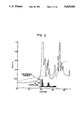
- FIG. 2 is a C 4 reverse phase HPLC profile of partially purified synovial PLA 2 showing the enzyme activity and optical density profile.
- FIG. 3 shows the DNA sequence of two 50-mer oligonucleotide probes used to identify synovial PLA 2 clones.
- FIG. 4 shows the DNA sequences of two human PLA 2 genomic clones, ⁇ SPLA2-6, and ⁇ SPLA2-10, which contain exons of the two PLA 2 enzymes described herein.
- FIG. 5 shows a 60-mer oligonucleotide probe synthesized to match amino acid residues 5-24 of sPLA 2 type A shown in FIG. 1 and based on the nucleotide sequence of clone ⁇ sPLA2-6.
- FIG. 6 shows the nucleotide sequence and deduced amino acid sequence from a cDNA clone for human sPLA 2 type A, designated ⁇ SPLA2cDNA-4.
- FIG. 7 shows the nucleotide sequence of xons 1-5 from genomic clone ⁇ SPLA2-6 of human sPLA 2 type A.
- FIG. 8 shows oligonucleotide linkers useful in recombinant DNA constructs for the expression of sPLA 2 in E. coli.
- FIG. 9 graphically depicts the accumulation of PLA 2 enzyme activity in serum-free medium during infection of CV-1 cells by recombinant vaccinia virus containing the human sPLA 2 type A gene.
- a “replicon” is any genetic element (e.g., plasmid, chromosome, virus) that functions as an autonomous unit of DNA replication in vivo; i.e., capable of replication under its own control.
- a "vector” is a replicon, such as plasmid, phage or cosmid, to which another DNA segment may be attached so as to bring about the replication of the attached segment.
- double-stranded DNA molecule refers to the polymeric form of deoxyribonucleotides (adenine, guanine, thymine, or cytosine) in its normal, double-stranded helix. This term refers only to the primary and secondary structure of the molecule, and does not limit it to any particular tertiary forms. Thus, this term includes double-stranded DNA found, inter alia, in linear DNA molecules (e.g., restriction fragments), viruses, plasmids, and chromosomes.
- linear DNA molecules e.g., restriction fragments
- viruses e.g., plasmids, and chromosomes.
- sequences may be described herein according to the normal convention of giving only the sequence in the 5' to 3' direction along the nontranscribed strand of DNA (i.e., the strand having a sequence homologous to the mRNA).
- a DNA "coding sequence” is a DNA sequence which is transcribed and translated into a polypeptide in vivo when placed under the control of appropriate regulatory sequences. The boundaries of the coding sequence are determined by a start codon at the 5' (amino) terminus and a translation stop codon at the 3' (carboxy) terminus.
- a coding sequence can include, but is not limited to, procaryotic sequences, cDNA from eucaryotic mRNA, genomic DNA sequences from eucaryotic (e.g., mammalian) DNA, and even synthetic DNA sequences.
- a polyadenylation signal and transcription termination sequence will usually be located 3' to the coding sequence.
- a “promoter sequence” is a DNA regulatory region capable of binding RNA polymerase in a cell and initiating transcription of a downstream (3' direction) coding sequence.
- the promoter sequence is bounded at its 3' terminus by the translation start codon of a coding sequence and extends upstream (5' direction) to include the minimum number of bases or elements necessary to initiate transcription at levels detectable above background
- a transcription initiation site (conveniently defined by mapping with nuclease S1), as well as protein binding domains (consensus sequences) responsible for the binding of RNA polymerase.
- Eucaryotic promoters will often, but not always, contain "TATA" boxes and "CAT” boxes.
- Procaryotic promoters contain Shine-Dalgarno sequences in addition to the -10 and -35 consensus sequences.
- a coding sequence is "under the control" of the promoter sequence in a cell when RNA polymerase which binds the promoter sequence transcribes the coding sequence into mRNA which is then in turn translated into the protein encoded by the coding sequence.
- a cell has been "transformed” by exogenous DNA when such exogenous DNA has been introduced inside the cell wall.
- Exogenous DNA may or may not be integrated (covalently linked) to chromosomal DNA making up the genome of the cell.
- the exogenous DNA may be maintained on an episomal element such as a plasmid.
- a stably transformed cell is one in which the exogenous DNA has become integrated into a chromosome so that it is inherited by daughter cells through chromosome replication. This stability is demonstrated by the ability of the eucaryotic cell to establish cell lines or clones comprised of a population of daughter cells containing the exogenous DNA.
- a "clone” is a population of cells derived from a single cell or common ancestor by mitosis.
- a "cell line” is a clone of a primary cell that is capable of stable growth in vitro for many generations.
- Two DNA sequences are "substantially homologous" when at least about 85% (preferably at least about 90%, and most preferably at least about 95%) of the nucleotides match over the defined length of the DNA sequences. Sequences that are substantially homologous can be identified in a Southern hybridization experiment under, for example, stringent conditions as defined for that particular system. Defining appropriate hybridization conditions is within the skill of the art. See, e.g., Maniatis et al., supra; DNA CLONING, Vols. I & II, supra: NUCLEIC ACID HYBRIDIZATION, supra.
- a "heterologous" region of the DNA construct is an identifiable segment of DNA within a larger DNA molecule that is not found in association with the larger molecule in nature.
- the gene when the heterologous region encodes a mammalian gene, the gene will usually be flanked by DNA that does not flank the mammalian genomic DNA in the genome of the source organism.
- Another example of a heterologous coding sequence is a construct where the coding sequence itself is not found in nature (e.g., a cDNA where the genomic coding sequence contains introns, or synthetic sequences having codons different than the native gene). Allelic variations or naturally occurring mutational events do not give rise to a heterologous region of DNA as defined herein.
- a protein composition is "substantially free of contaminating proteins" when at least about 75% by weight of the protein in the composition is the particular protein of interest.
- this protein comprises at least about 90% by weight of the protein in the composition, most preferably at least about 99% by weight. It is also preferred that a protein composition, which is substantially free of contaminating proteins, contain only a single molecular weight species having the activity of the protein of interest.
- Synovial phospholipase A 2 refers to the class of mammalian enzymes exhibiting PLA 2 activity and found in the synovial fluid of a mammal (such as a human) afflicted with rheumatoid arthritis. It is believed that sPLA 2 enzymes are produced by inflamed synovial tissue, or perhaps granulocytes or macrophages in the synovial fluid. PLA 2 enzymes are characterized in having a molecular weight of about 15+3 kD when measured by polyacrylamide gel electrophoresis (PAGE) (12.5% polyacrylamide gel, 0.1% SDS).
- PAGE polyacrylamide gel electrophoresis
- Type A is present in synovial fluid from all types of arthritis examined.
- Type B varies in abundance from complete absence in some rheumatoid samples to about 33% of the total activity in other samples.
- Type B typically appears at higher levels in fluid samples from osteoarthritis patients than in samples from rheumatoid patient, but type A still constitutes the majority of sPLA 2 .
- Type B also shows considerable stimulation in hydrolytic activity relative to type A in the presence of either 0.5 M Tris or 0.1% Na deoxycholate; type A is inhibited by 0.5 M Tris.
- Type C when present, is two- to five-fold less abundant than type B.
- These extracellular enzymes are (i) soluble, (ii) calcium-dependent, (iii) have proinflammatory activity in tissue when injected intradermally or intraarticularly, and (iv) exhibit absolute specificity for the sn-2 acylester bond of dipalmitoylphosphatidylcholine.
- This characterization also includes synthetic and recombinant analogs of sPLA 2 wherein any resulting changes, deletions or additions in the amino acid sequence does not change the above characteristic activities.
- sPLA 2 resembles other PLA 2 sequences in the number and placement of the 14 Cys residues, particularly the "type II" enzymes, of which C. atrox PLA 2 is an example.
- Synovial PLA 2 also lacks a Cys at position 11, which is characteristic of the highly pro-inflammatory type II enzymes (e.g., Viperid snake venom forms, and PLA 2 species described in copending U.S. Ser. No. 946,557).
- the comparison demonstrates that sPLA 2 is distinct from all other known PLA 2 sequences, particularly in the variable regions near the carboxy terminus.
- a twenty residue prepeptide, containing a typical signal for translocation across a cellular membrane is present upstream of the mature enzyme sequence, and is presumably cleaved during or after synthesis.
- a clone, ⁇ SPLA2-6, of genomic DNA encoding sPLA 2 type A has been deposited with the American Type Culture Collection (ATCC), 12301 Parklawn Dr., Rockville, MD, U.S.A. 20852, on 14 Aug. 1987, and given accession no. 40361.
- the coding sequence (FIG. 4) is on a 404 bp AluI fragment which can be isolated from ⁇ SPLA2-6.
- the coding sequence in this clone is contained on an AluI fragment of about 460 bp.
- An expression vector containing an sPLA.sub. 2 coding sequence, p86-1A (discussed below), was also deposited with the ATCC on June 27, 1988 under accession no. 67735. These deposits will be maintained under the terms of the Budapest Treaty.
- the sPLA 2 coding sequences of ⁇ SPLA2-6, ⁇ SPLA2-10 and ⁇ SPLA2cDNA-4, and the expression cassette sequence of pHNF86 are incorporated herein by reference. In the event of any discrepancy between a sequence disclosed herein and the sequence of a deposited clone, the clone's sequence is controlling.
- a DNA sequence encoding sPLA 2 can be isolated by one of several approaches. These methods will rely in part on nucleic acid hybridization using appropriate oligonucleotide probes. Such probes can be constructed synthetically based on the sPLA 2 DNA or amino acid sequences disclosed herein, or isolated from the genomic sPLA 2 clones also described herein.
- the library can consist of a genomic DNA library from a selected mammal, such as a human.
- Human genomic libraries are known in the art. See, e.g., Maniatis et al. (1978) Cell 15:687-701; Lawn et al. (1978) Cell 15:1157-1174.
- DNA libraries can also be constructed of cDNA prepared from poly-A RNA (mRNA) by reverse transcription. See, e.g., U.S. Pat. Nos. 4,446,325; 4,440,859; 4,433,140; 4,431,7400; 4,370,417; 4,363,877.
- the mRNA is isolated from a cell line or tissue believed to express sPLA 2 , such as synovial tissue or inflammatory cells isolated from synovial fluid.
- sPLA 2 such as synovial tissue or inflammatory cells isolated from synovial fluid.
- the preferred source of mRNA for cDNA library constructions is synovial joint tissue.
- the genomic DNA or cDNA is cloned into a vector suitable for construction of a library
- a preferred vector is a bacteriophage vector, such as any of the phage lambda.
- the construction of an appropriate library is within the skill of the art. See, e.q., B. Perbal, supra.
- oligonucleotides are used to probe the library to identify the segment carrying the sPLA 2 coding sequence.
- the probes are preferably based upon known nucleic acid sequences. However, if the later is unknown, it may be desirable to base probes upon an amino acid sequence determined from a purified sPLA 2 . In the latter case, nucleotide sequences are selected so as to correspond to the codons encoding the amino acid sequence. Since the genetic code is redundant, it will usually be necessary to synthesize several oligonucleotides to cover all, or a reasonable number, of the possible nucleotide sequences which encode a particular region of the protein.
- the region not contain amino acids whose codons are highly degenerate. It may not be necessary, however, to prepare probes containing codons whose usage is rare in the mammal from which the library was prepared.
- probes that are fairly long and/or encompass regions of the amino acid sequence which would have a high degree of redundancy in the corresponding nucleic acid sequences. Probes covering the complete gene, or a substantial part of the gene, may also be appropriate, depending upon the expected degree of homology. Due to the highly conserved nature of PLA 2 across species lines, it is likely that full length sPLA2cDNA probes from one species, such as the human clone ⁇ SPLA2cDNA-4, can be readily used to screen libraries prepared from another species. In other cases, it may be desirable to use two sets of probes simultaneously, each to a different region of the gene. While the exact length of any probe employed is not critical, generally it is recognized in the art that probes from about 14 to about 20 base pairs are usually effective. Longer probes of about 25 to about 60 base pairs are also used.
- oligonucleotide probes are labeled with a marker, such as a radionucleotide or biotin, using standard procedures.
- the labeled set of probes is then used in the screening step, which consists of allowing the single-stranded probe to hybridize to isolated ssDNA from the library, according to standard techniques.
- Either stringent or permissive hybridization conditions could be appropriate, depending upon several factors including, but not limited to, the length of the probe, whether the probe and library are from the same species, and whether the species are evolutionarily close or distant. It is within the skill of the art to optimize hybridization conditions so that homologous sequences are isolated and detectable above background hybridizations.
- hybridization conditions be of sufficient stringency so that selective hybridization occurs; i.e., hybridization is due to a minimum degree of nucleic acid homology (e.g., at least about 75%), as opposed to nonspecific binding or hybridization due to a lower degree of homology. See generally, NUCLEIC ACID HYBRIDIZATION, supra.
- Partial genomic clones such as the clone of an exon of sPLA 2 in ⁇ SPLA2-10, can be extended into complete clones by one of several techniques.
- a clone can be extended in either the 5' or 3' direction using "chromosome walking" techniques to ensure inclusion of the entire gene coding region. Restriction fragments of these clones can then be probed with, for example, sPLA 2 DNA. If sufficient homology exists within these exons to pancreatic PLA 2 , other exons of sPLA 2 could be identified with pancreatic sPLA 2 clone, also.
- oligonucleotides which correspond to particularly conserved regions (e.g., amino acid residues 44-52), which would allow prediction of possible differences (e.g., Asp 49 changed to Lys 49 ).
- genomic clones may be rapidly identified by direct sequencing of the DNA downstream of a cloned exon using modern M13-dideoxy sequencing techniques. The sequence is then inspected in all three reading frames to reveal an open reading frame. Other exons will also be apparent since they will be bounded on both sides by intron-splicing signals and should encode conserved amino acids.
- the correct gene coding sequence for an exon of sPLA 2 type B or C can be used to obtain the entire protein coding region of the enzyme by one or more of the following means.
- the exon can be trimmed from the ⁇ clone and placed in a more convenient vector, such as pBR322, so that large quantities of DNA containing only the exon itself can be obtained and used as a hybridization probe.
- a 60-mer oligonucleotide corresponding to the unique regions of the coding region e.g., amino acid residues 6-25) can be synthesized.
- Either can be used as a hybridization probe for northern blots of mRNA obtained from various sources, such as, peritoneal cells and pus, endothelial tissue, and peripheral blood leukocytes, lymphocytes, and macrophages
- mRNA from various cell lines such as differentiated U-937 and HL60 can also be tested. Any tissue or cell source containing detectable levels of hybridizing mRNA is then used to produce a cDNA library which will then be screened with the same probes in order to detect a full-length cDNA encoding sPLA 2 .
- this strategy lead to the cloning of full length cDNA's encoding sPLA type A, such as clone ⁇ SPLA2cDNA-4.
- Mammalian genomic clones (partial or full-length) containing the longest inserts of the sPLA 2 gene can be co-transfected into Chinese hamster ovary (CHO) cells with plasmid DNA containing a marker, such as neomycin and metallothionine resistance genes.
- a marker such as neomycin and metallothionine resistance genes.
- Surviving cells selected in the presence of antibiotic G418 and Cd ++ , and surviving clones can be analyzed for the presence of sPLA 2 -hybridizing transcripts in a Northern blot of extracted RNA. Clones containing the desired transcripts can then be used as an mRNA source for a cDNA library construction.
- Synovial PLA 2 can be purified from human synovial fluid from patients afflicted with rheumatoid arthritis or psoriasis.
- the purification protocols described in detail below, allow for the first time the purification of native sPLA 2 in sufficient quantity and at a high enough purity to permit accurate amino acid sequencing.
- the amino acid sequences derived from the purified sPLA2's allow for the design of probes to aid in the isolation of native sPLA 2 nucleic acid sequence, or the design of synthetic nucleic acid sequences encoding the amino acid sequence of a sPLA 2 .
- Specific anti-sera or monoclonal antibodies can be made to a synthetic sPLA 2 peptide having the sequence of amino acid residues, such as those shown at the NH 2 -terminus in FIG. 1. Particularly preferred is a peptide spanning positions 1 through 26. This is a unique region of the protein, and antibodies thereto can be used to immunoprecipitate any sPLA 2 present in a selected tissue, cell extract, or body fluid. Purified sPLA 2 from this source can then be sequenced and used as a basis for designing specific probes as described above. Antibodies to other regions that diverge from known PLA 2 s can also be used. Also useful as antigens are purified native or recombinant sPLA 2 .
- a DNA sequence encoding sPLA 2 can be prepared synthetically rather than cloned.
- the DNA sequence can be designed with the appropriate codons for the sPLA 2 amino acid sequence. In general, one will select preferred codons for the intended host if the sequence will be used for expression.
- the complete sequence is assembled from overlapping oligonucleotides prepared by standard methods and assembled into a complete coding sequence. See. e.q., Edge (1981) Nature 292:756; Nambair et al. (1984) Science 223:1299; Jay et al. (1984) J. Biol. Chem. 259:6311.
- muteins can be made by site-directed mutagenesis of native sPLA 2 genes or cDNAs, and muteins can be made directly using conventional polypeptide synthesis.
- muteins Of particular interest in the construction of muteins is changing the catalytic His 48 residue in type A to another amino acid, such as Gln.
- Position 48 muteins may act as a PLA 2 inhibitor by binding to endogenous inflammatory PLA 2 enzymes, thereby creating inactive dimers.
- Other potential targets for mutagenic alteration include the three basic residues near the N-terminus (positions 7, 10 and 16), which may be involved in interaction with membrane-associated substrates. Muteins altered in any one or all of these positions by the substitution of acidic residues (e.g., Glu or Asp) could have reduced activity toward membrane-bound or complex substrates.
- Site-directed mutagenesis is conducted using a primer synthetic oligonucleotide complementary to a single stranded phage DNA to be mutagenized except for limited mismatching, representing the desired mutation.
- the synthetic oligonucleotide is used as a primer to direct synthesis of a strand complementary to the phage, and the resulting double-stranded DNA is transformed into a phage-supporting host bacterium. Cultures of the transformed bacteria are plated in top agar, permitting plaque formation from single cells which harbor the phage.
- 50% of the new plaques will contain the phage having, as a single strand, the mutated form; 50% will have the original sequence.
- the resulting plaques are hybridized with kinased synthetic primer at a temperature which permits hybridization of an exact match, but at which the mismatches with the original strand are sufficient to prevent hybridization. Plaques which hybridize with the probe are then picked, cultured, and the DNA recovered.
- a coding sequence for sPLA 2 can be cloned into any suitable vector or replicon and thereby maintained in a composition which is substantially free of vectors that do not contain an sPLA 2 coding sequence (e.g., free of other library clones).
- Numerous cloning vectors are known to those of skill in the art, and the selection of an appropriate cloning vector is a matter of choice. Examples of recombinant DNA vectors for cloning and host cells which they can transform include the various bacteriophage lambda vectors (E. coli), pBR322 (E. coli), pACYC177 (E.
- the coding sequence for mammalian sPLA 2 is placed under the control of a promoter, ribosome binding site (for bacterial expression) and, optionally, an operator (collectively referred to herein as "control" elements), so that the DNA sequence encoding sPLA 2 is transcribed into RNA in the host cell transformed by a vector containing this expression construction.
- the coding sequence may or may not contain a signal peptide or leader sequence. If the coding sequence contains a signal peptide, it may or may not be the sPLA 2 signal sequence.
- mature sPLA 2 is preferably made by the expression of a coding sequence which does not contain the sPLA 2 signal peptide, or by expression of a coding sequence containing a leader sequence which is removed by the bacterial host in post-translational processing. See. e.q., U.S. Pat. Nos. 4,431,739; 4,425,437; 4,338,397.
- An expression vector is constructed according to the present invention so that the sPLA 2 coding sequence is located in the vector with the appropriate regulatory sequences, the positioning and orientation of the coding sequence with respect to the control sequences being such that the coding sequence is transcribed under the "control" of the control sequences (i.e., RNA polymerase which binds to the DNA molecule at the control sequences transcribes the coding sequence).
- the control sequences may be ligated to the coding sequence prior to insertion into a vector, such as the cloning vectors described above.
- the coding sequence can be cloned directly into an expression vector which already contains the control sequences and an appropriate restriction site.
- control sequences will necessarily be heterologous to the coding sequence. If the host cell is a procaryote, it is also necessary that the coding sequence be free of introns (e.g., cDNA). If the selected host cell is a mammalian cell, the control sequences can be heterologous or homologous to the sPLA 2 coding sequence, and the coding sequence can either be genomic DNA containing introns or cDNA. Either genomic or cDNA coding sequences can be expressed in yeast.
- procaryotic expression vectors are known in the art. See, e.g., U.S. Pat. Nos. 4,440,859; 4,436,815; 4,431,740; 4,431,739; 4,428,941; 4,425,437; 4,418,149; 4,411,994; 4,366,246; 4,342,832; see also U. K. Pub. Nos. GB 2,121,054; GB 2,008,123; GB 2,007,675; and European Pub. No. 103,395.
- Preferred procaryotic expression systems are in E. coli.
- Other preferred expression vectors are those for use in eucaryotic systems. See, e.g., commonly owned U.S. Pat. application Ser. No.
- a preferred eucaryotic expression system is that employing vaccinia virus, which is well-known in the art. See, e.g., Mackett et al. (1984) J. Virol. 49:857; DNA CLONING, Vol. II, pp. 191-211, supra: PCT Pub. No. WO 86/07593.
- Yeast expression vectors are known in the art. See, e.g., U.S. Pat. Nos. 4,446,235; 4,443,539; 4,430,428; see also European Pub. Nos. 103,409; 100,561; 96,491.
- vector pHSl transforms Chinese hamster ovary cells.
- the use of the vector is described in PCT Pub. No. WO 87/02062 and commonly owned U.S. Pat. application Ser. No. 804,692, filed 4 Dec. 1985, the disclosure of which is incorporated herein by reference.
- sPLA 2 is produced by growing host cells transformed by an expression vector described above under conditions whereby the sPLA 2 protein is expressed. The enzyme protein is then isolated from the host cells and purified. If the expression system secretes the enzyme into growth media, the protein can be purified directly from cell-free media. If the sPLA 2 protein is not secreted, it is isolated from cell lysates. The selection of the appropriate growth conditions and recovery methods are within the skill of the art.
- Native, recombinant or synthetic sPLA 2 peptides can be used to produce both polyclonal and monoclonal antibodies. If polyclonal antibodies are desired, purified sPLA 2 peptide is used to immunize a selected mammal (e.g., mouse, rabbit, goat, horse, etc.) and serum from the immunized animal later collected and treated according to known procedures. Compositions containing polyclonal antibodies to a variety of antigens in addition to sPLA 2 can be made substantially free of antibodies which are not anti-sPLA 2 by immunoaffinity chromatography.
- Monoclonal anti-sPLA 2 antibodies can also be readily produced by one skilled in the art from the disclosure herein.
- the general methodology for making monoclonal antibodies by hybridomas is well known.
- Immortal, antibody-producing cell lines can also be created by techniques other than fusion, such as direct transformation of B lymphocytes with oncogenic DNA, or transfection with Epstein-Barr virus. See, e.g., M. Schreier et al., HYBRIDOMA TECHNIQUES (1980); Hammerling et al., MONOCLONAL ANTIBODIES AND T-CELL HYBRIDOMAS (1981); Kennett et al., MONOCLONAL ANTIBODIES (1980); see also U.S. Pat. Nos. 4,341,761; 4,399,121; 4,427,783; 4,444,887; 4,451,570; 4,466,917; 4,472,500; 4,491,632; 4,493,890.
- Panels of monoclonal antibodies produced against sPLA 2 peptides can be screened for various properties; i.e., isotype, epitope, affinity, etc. Of particular interest are monoclonal antibodies that neutralize the activity of sPLA 2 . Such monoclonals can be readily identified in PLA 2 activity assays. High affinity antibodies are also useful in immunoaffinity purification of native or recombinant sPLA 2 .
- pancreatic PLA 2 expressed in human lung tissue indicates that the pancreatic form may play a larger role than had been expected in inflammatory disease.
- antibodies to any other PLA 2 forms described herein can be used to treat inflammatory disorders.
- Anti-pancreatic PLA 2 antibody can be produced as described herein for anti-sPLA 2 antibody. If the disease is acute endotoxic shock, for example, the appropriate therapeutic method would be to treat the patient with an effective dose of anti-PLA 2 antibodies (e.g., anti-synovial PLA 2 ) through a conventional intravenous route. In the treatment of local, acute inflammation, treatment with anti-sPLA 2 antibody would be indicated, perhaps by intramuscular injection.
- compositions may also be useful in treating other forms of arthritis, such as osteoarthritis. Since endotoxic shock induces elevated levels of PLA 2 , it may also be desirable to administer anti-PLA 2 antibodies in conjunction with other therapies directed to the gram-negative pathogens and their toxins (e.g., anti-LPS therapy).
- PLA 2 is also known to attack the pulmonary surfactant monolayer, in the case of respiratory distress (e.g., adult respiratory distress syndrome) it may be desirable to administer anti-PLA 2 antibodies by inhalation combined with replacement pulmonary surfactant phospholipid, dipalmitoyl phosphatidylcholine.
- PLA 2 antagonists such as sPLA 2 muteins, could also be used in place of antibodies.
- the antibodies will be formulated in a unit dosage injectable form (solution, suspension, emulsion, etc.) in association with a pharmaceutically acceptable parenteral vehicle.
- a pharmaceutically acceptable parenteral vehicle Such vehicles are usually nontoxic and nontherapeutic. Examples of such vehicles are water, saline, Ringer's solution, dextrose solution, and Hank's solution. Nonaqueous vehicles such as fixed oils and ethyl oleate may also be used.
- a preferred vehicle is 5% (w/w) human albumin in saline.
- the vehicle may contain minor amounts of additives, such as substances that enhance isotonicity and chemical stability, e.g., buffers and preservatives.
- the antibody is typically formulated in such vehicles at concentrations of about 1 ⁇ g/ml to 10 mg/ml.
- Anti-sPLA 2 antibodies will also be useful in diagnostic applications. For example, synovial fluid isolated from rheumatoid arthritis patients shows that it contains primarily, if not completely, PLA 2 of the type A variety. On the other hand, samples from osteoarthritis patients typically contain appreciable amounts of type B as well as type A, usually in a 2:1 ratio of type A to type B based upon activity in the presence of 50 mM Tris.
- the present invention contemplates a method, particularly a diagnostic method, in which a synovial fluid sample from a human (or other mammal) is provided, and the amounts of sPLA 2 type A and type B are quantitatively measured in an assay and compared
- a synovial fluid sample from a human or other mammal
- anti-sPLA 2 antibodies specific to type A or to type B in a quantitative immunoassay could be used to distinguish between the two types of arthritis.
- Antibody specific for type A or type B could be formulated into any conventional immunoassay format; e.g., homogeneous or heterogeneous, radioimmunoassay or ELISA.
- the various formats are well known to those skilled in the art.
- recombinant production of sPLA 2 can provide compositions of that enzyme substantially free of contaminating proteins.
- the ability to obtain high levels of purity is a result of recombinant expression systems which can produce sPLA 2 in substantial quantities vis-a-vis in vivo sources.
- sPLA 2 compositions can be produced that are substantially more pure than the cellular PLA 2 compositions presently available from non-digestive and non-venom sources.
- the purified sPLA 2 compositions of the present invention are useful in several regards. First, they can be used in food processing technology as described in Dutilh & Greger (1981) J. Sci. Food Agric. 32:451-458. In addition, sPLA 2 compositions can be used to delay the onset of rancidity in fish. See, e.g., Mazeaud & Bilinski (1976) J. Fish Res. Board Can. 333:1297-1302.
- milligram amounts of the material are obtainable according to the present invention. Milligram amounts are capable of crystallization to permit three dimensional studies using X-ray diffraction and computer analysis. This may permit deduction concerning the shape of the molecule, thus defining proper shapes for substances useable as inhibitors of the enzyme activity normally exhibited by sPLA 2 . Inhibitors have already been designed for "converting enzyme", the catalyst for the subsequent conversion of angiotensin I into angiotensin II.
- these antagonists have been "dipeptides” whose interactions with converting enzyme are stabilized by modification of the "residues” participating in the peptide bond so as to enhance the ability of the "dipeptide” to interact specifically with converting enzyme.
- the peptide bond joins specifically chosen carboxylic acids and amines (not necessarily amino acids).
- These "dipeptides” are configured in a three dimensional array so as to complement the contours of the intended target, converting enzyme.
- a similar lock and key spatial arrangement may result from molecules designed complementary to the surface contours of the crystallized sPLA 2 of the invention. It is understood that “surface” includes convolutions which may face inward, and specifically includes the active site.
- “complementary” is understood to mean that, in addition to spatial conformations which "fit", interactions between the protein and the molecule which matches its surface contours are attractive and positive. These interactions may be hydrogen bonding, ionic, or hydrophobic affinity.
- the invention contemplates peptide antagonists (2-15 amino acids) to sPLA 2 which are characterized by three dimensional contours complementary to the three dimensional contours on the surface of recombinant sPLA 2 .
- peptide in this context is meant that the antagonist contains carboxylic acid amide bonds corresponding to one less than the number of residues.
- the carboxylic acid and amine participants need not be ⁇ -amino acids.
- purified sPLA 2 of the invention is of significance as a reagent in screening sPLA 2 inhibitors in vitro as an ad hoc approach to evaluation.
- Impure sPLA 2 preparations currently available yield confusing data due to the impact of the impurities on the test results. For example, contaminants which turn out to be themselves inhibitors, activators, or substrates for sPLA 2 will interfere with the evaluation.
- a substantial improvement in current screening techniques for sPLA 2 inhibitors would be effected by the availability of the purified human sPLA 2 protein.
- sPLA 2 compositions described herein may also be useful as an anti-cancer drug.
- direct injection of sPLA 2 into, or in the vicinity of malignant tumors, and optionally in conjunction with tumor excision, will result in high levels of powerful chemoattractants for, and activators of, macrophages. These activated macrophages may then enhance localized tumor reduction or elimination.
- Still another application of purified sPLA 2 according to the present invention is as an adjuvant in a vaccine composition.
- the formulation of vaccines is well known in the art.
- vaccine formulations include the antigen(s) (e.g., attenuated virus, killed virus, viral polypeptide subunits, killed bacteria, bacterial pili, etc.) in a pharmaceutically acceptable parenteral vehicle.
- the improved vaccine composition of the present invention may contain, in addition to an SPLA 2 adjuvant, an additional adjuvant.
- concentration of sPLA 2 in the final vaccine formulations can be readily determined by one of ordinary skill in the art. Typically, but not always, the concentration of sPLA 2 will be from about 1 ng/ml to about 1 ⁇ g/ml.
- Sephadex G-75, CM-Sephadex C-50, and protein standards for gel filtration and electrophoresis were purchased from Pharmacia Fine Chemicals.
- Acrylamide, N,N,N',N'-tetramethylethylenediamine, bromophenol blue, Coomassie brilliant blue R, sodium dodecyl sulfate (SDS), fatty acid free bovine serum albumin (BSA), dipalmitoylphosphatidylcholine, and Lowry protein assay kit were obtained from Sigma.
- Silver stain and Bio-Rad protein assay kit were purchased from Bio-Rad Laboratories.
- Synovial fluids were obtained from patients with active classical or definite rheumatoid arthritis (RA) by arthrocentesis. This material was centrifuged at 4° C. to remove cells and debris, pooled and stored in polypropylene tubes at -70° C. until required.
- Polyacrylamide gel electrophoresis (PAGE) in 15% polyacrylamide gels was performed in the presence of 0.1% SDS as described by Laemmli (1970) Nature 227:680681. Ovalbumin, carbonic anhydrase, trypsin inhibitor, and lactalbumin were used as molecular weight markers. The samples were incubated in 0.0625 M Tris-HCl, pH 6.8, containing 2% SDS and 10% glycerol with 5% 2-mercaptoethanol (2-ME) for 6 min at 100° C. for analytical PAGE or without 2-ME for 1 h at 37° C. for preparative SDS-PAGE, and then applied to the gel. Electrophoresis was performed for 4 h at 30 mA and the protein bands were stained with Coomassie brilliant blue or Bio-Rad silver stain. Switzer et al. (1975) Anal. Biochim. 98:231-237.
- An identical electrophoretic pattern was obtained for the same preparation of PLA 2 , without reduction of disulfate bonds.
- PLA 2 activity was associated with both the 15K and 17K bands.
- the material from the initial purification was loaded onto a reverse-phase C-4 HPLC column and eluted with a 15-60% acetonitrile gradient in the presence of 0.1% trifluoroacetic acid.
- the eluted fractions were assayed for PLA 2 activity (C, below), and the active fractions were pooled and lyophilized overnight in a siliconized Falcon #2059 tube.
- the peaks of activity termed peaks A, B and C, were obtained routinely and were further purified separately (FIG. 2).
- the lyophilized peak material was resuspended in a PAGE loading buffer (2.3% SDS, 50 mM Tris, 10% glycerol), heated at 90° C.
- Peaks A, B and C all eluted from the slices corresponding to 15,000 MW, just ahead of the propancreatic marker. Active fractions were spotted and dried directly onto quaternary amine glass fiber filter paper. The filters then were washed four times in the same buffer, 5 min each, 38 and dried. Sequence analyses were performed via Edman degradation on an Applied Biosystems gas phase sequencer. The NH 2 -terminal sequences of sPLA 2 type A (peak A) and sPLA 2 type B (peak B) are shown in FIG. 1.
- Standard assay conditions for final purification steps consisted of 50 mM Tris, pH 8.0, 150 mM NaCl 5.0 mM CaCl 2 , 0.04% sodium deoxycholate (DOC), and 0.22 nmoles of 1-stearoyl-2-[1- 14 C]Arachidonyl-L-3-phosphatidylcholine (PC, Amersham #CFA.655) as substrate, incubated at 37° C. for 30 min.
- the substrate was prepared by dissolving freshly desiccated PC in 2% DOC, which was then diluted to the appropriate concentration in assay buffer.
- the 50 ⁇ l reaction was started by the addition of prewarmed substrate and terminated by the addition of 10 ⁇ l 8 M acetic acid.
- Example I.A The material prepared in Example I.A. was further characterized as described below.
- Phospholipase activity was quantitated by the modified method [Vadas et al. (1980) Life Sci. 26:1721-1729] of Franson et al. (1978) J. Lipid Res. 19:18-23, using autoclaved Escherichia coli, strain K 12 C 600 , labeled with [ 14 C]Oleic acid, as the substrate. Assays were performed in substrate excess, using 2.8 ⁇ 10 8 E. coli per assay, corresponding to 5.6 nmol of phospholipid with a specific activity of 4,120 cpm/nmol phospholipid.
- the standard reaction mixture of 1.5 ml total volume contained 10 mg BSA, 7 mM CaCl 2 , 0.1 M Tris-HCl buffer, and [ 14 C]Oleate-labeled E. coil. Reactions were allowed to proceed for 30 min at 37° C. and were terminated by filtration through a 0.45 ⁇ m Millipore filter. Enzyme activities were corrected for non-enzymatic hydrolysis. Under conditions of substrate excess, the rate of substrate hydrolysis is linear with reaction times of up to 30 min, over a fivefold range of enzymic concentration.
- the pH dependence of purified PLA 2 was determined against dipalmitoylphosphatidylcholine and 1-palmitoyl-2-linoleoylphosphatidylethanolamine over a range of pH 5-10, using the assay of Shakir, (1981) Anal. Biochim. 114:64-70. Buffers used were of constant ionic strength: 0.1 M sodium acetate-acetic acid (pH 5-6), 0.1 M Tris-HCl (pH 7-8), and 0.1 M glycine-NaOH (pH 9-10).
- the pH dependence of purified PLA 2 was studied over the range of pH 5-10 using two synthetic phospholipid substrates, dipalmitoylphosphatidylcholine, and 2-linoleoyl-1-palmitoylphosphatidylethanolamine.
- Phosphatidylethanolamine was hydrolyzed over a wide range of pH (6-10), with optimal PLA 2 activity evident at pH 7.5-8.0.
- Maximal PLA 2 activity for phosphatidylcholine was seen at pH 7.0 with an abrupt decrease in activity at pH 8-10.
- the positional specificity of the purified enzyme was determined using 1-palmitoyl-2-[1- 14 C]palmitoylphosphatidylcholine as substrate.
- the assay system contained 750 nmol of radioactive phosphatidylcholine dispersed in 0.1 M Tris-HCl buffer, pH 7.5, containing 2 mM CaCl 2 , 2 mM DOC, 0.09% Triton X-100, and 100 ⁇ l of enzyme preparation in a total volume of 400 ⁇ l.
- the reaction was carried out at 37° C. for 3.5 h and was termination by the addition of 8 ml of chloroform-methanol (2:1, v/v).
- Lipids were extracted by the method of Folch et al., (1957) J. Biol. Chem. 6:497-509, and separated by TLC in chloroform-methanol-acetic acid-water (65 :25 :8 :4, v/v/v/v).
- the lipid spots were visualized by exposure to iodine vapor. After sublimination of the iodine, spots corresponding to authentic PC, lyso PC and free fatty acid standards were scraped into scintillation vials containing 10 ml of scintillation fluid and measured for radioactivity in a liquid scintillation spectrometer (Beckman LS7500).
- 1-acyl-2-[1- 14 C]palmitoylphosphatidylcholine was incubated with venom PLA 2 , crude synovial fluid or purified synovial fluid PLA 2 and the reactions products were analyzed by thin-layer chromatography.
- Two 50-mer codon-preference oligos were designed from the RASF peak A sequence, minimizing ambiguity by (a) centering the oligos upon codon groups with minimal ambiguity and (b) allowing for G:T binding.
- the oligos, shown in FIG. 3, were synthesized on an Applied Biosystems oligonucleotide synthesizer.
- the oligos were labeled with ⁇ - 32 P-ATP and polynucleotide kinase, and then used as a hybridization probe for the EMBL3-human leukocyte genomic library obtained from Clonetech Inc. (Mountain View, CA). Then 10 6 total plaques were placed on twenty 150 mm agar plates containing L-broth, using bacterial strain NM538. The plaques were lifted onto nitrocellulose filters, denatured, baked 2 hours at 80° C.
- prehybridization solution 5X Denhardt's, 20% formamide, 6X SSC, 50 mM NaP0 4 , 100 ⁇ g/ml sheared salmon sperm DNA
- Hybridization was overnight at 37° C. in prehybridization solution plus 10% dextran sulfate and 2 ⁇ 10 6 cpm of labeled probe.
- the filters were washed twice at 25° C. in 1X 0.16 M NaCl, 0.016 M sodium citrate (SSC), 0.1% sodium docecyl sulfate (SDS), and then once in the same solution at 50° C. for 1 hour and then exposed to autoradiographic film overnight at -70° C. Later, the same filters were rewashed at 55° C. and reexposed.
- DNA from the two unique clones was digested with endonucleases HaeIII, RsaI, and AluI.
- the completed digest was extracted with phenol/chloroform and precipitated with ethanol.
- the dried pellets were resuspended in 10 mM Tris, pH 8.0, 1 mM EDTA, and 1.0 ⁇ l aliquots were ligated to bacteriophage M13mp8, which had been previously digested with SmaI.
- the transformed E. coli strain JM101 cells were plated onto 150 mm L-agar plates and incubated overnight at 37° C. The resulting M13 recombinant plaques were lifted and the filters hybridized as described above.
- the sPLA 2 coding sequence in ⁇ sPLA2-6 was originally believed to be exon 2 of the human type A gene type A.
- the cDNA sequence identified in III.B, below, was used to identify the remaining exons in the genomic clones. It was found that an unexpected intron existed in the 5'-noncoding region of the gene. Thus, what was originally believed to be exon 2 is actually exon 3.
- the sequence encoding exon 1 is shown in FIG. 7.
- Bases 1016 through 1038 match bases 8 to 27 of the cDNA clone exactly. Although the precise start of transcription has not been determined, its most likely location is at or shortly upstream of base 1012.
- a potential "TATA" sequence can be seen at nucleotides 968 through 974, and a putative "CAAT" sequence lies at nucleotides 904 through 909.
- a 60-mer oligonucleotide probe was synthesized to match the nucleotide sequence for ⁇ sPLA2-6 shown in FIG. 4 and corresponding to the codons for amino acid residues 5-24 shown in FIG. 1.
- This oligonucleotide probe was used to screen RNA blots from various sources, including cell lines HL60 and U937, human synovial cells, human peritoneal inflammatory exudate cells, human pus cells, porcine jejunum tissue, porcine pancreatic tissue, rat spleen tissue, and rat liver tissue. Significant levels of RNA was detected by hybridization in human peritoneal cells and, to a lesser extent, human synovial cells.
- a cDNA library was constructed from polyA+ message from a peritoneal cell RNA prep according to the method of Gubler & Hoffman (1983) Gene 25:263-269. The library was screened with the 60-mer probe, and 17 discrete duplicating signals were obtained after washing the filters in 1X SSC, 0.1% SDS at 60° C. DNA from ten of the clones was subjected to analysis by PAGE. All of the clones contained inserts of 800 to 1,000 bp. Four of the clones, designated 1, 4, 11 and 14, were selected for subcloning into bacteriophage M13 and subsequent DNA sequence analysis by standard techniques. One of these clones, designated ⁇ sPLA 2 cDNA-4 was determined to encode the entire sPLA 2 type A sequence.
- the other clones contained the same sequence or varied slightly in length at the 5' end and had different length polyA tails. Otherwise, the clones were identical except for a C to T change at position 277, a silent mutation with respect to the amino acid specified by the codon.
- a typical translation termination sequence, AATAAA, can be seen beginning at base 116.
- the mature peptide sequence encoded by ⁇ PLA2cDNA-4 contains 124 amino acids, and has a calculated molecular weight and pI of 13,919 and 6.46, respectively.
- Active recombinant sPLA 2 was produced in bacteria, such as E. coli, as a ⁇ -galactosidase fusion protein employing a procedure adapted from de Geus et al. (1987) Nucleic Acids Res. 15:3743-3759. This methodology was adapted to sPLA 2 as follows. First, a single base change from C to G introduced at the C-terminus in nucleotide 588 created a HindIII site 17 bases downstream from the TGA stop codon. This change was made via oligonucleotide-directed mutagenesis of single-stranded M13 DNA, using standard molecular biology methodology.
- E. coli tryptophan promoter a ribosomal binding site, sequences encoding a portion of the amino terminal portion of the E. coli ⁇ -galactosidase gene followed by six Thr residues, an EcoRI and HindIII site, and two strong E. coli transcription termination signals. See, e.g., Sung et al (1986) Proc. Natl. Acad. Sci. USA 83:561-565.
- the resulting expression vector containing the PLA 2 construct was then used to transform E. coli strain W3110 (ATCC accession no. 27325) for expression. After inoculation of a culture of transformed cells of a suitable population density, expression was induced by the addition of 3-B-indoleacrylic acid into the media. After 9 hours of growth in induction media, inclusion bodies were observed in about 90% of the cells. After cell disruption, the inclusion bodies were then pelleted and boiled 5 min in gel loading buffer containing 50 mM ⁇ -mercaptoethanol. By comparison with gels of similar extracts from uninduced and control cultures, a prominent 15 kD band was observed in the induced cultures transformed with the expression construct.
- This band was highly enriched in gels from extracts of purified inclusion bodies, allowing large-scale isolation of this fusion protein from preparative SDS-polyacrylamide gels. Fusion protein prepared in this manner was injected into rabbits, rats and mice for the production of antibodies.
- Purified fusion protein fractions may be activated by the S-sulfonation procedure as described by DeHaas et al. (1987), supra. After activation, the fusion protein can be cleaved at the Trp residue to release the mature human PLA 2 . Alternatively, the two steps in reverse order may give greater yields of active protein See, e.g., Lishchwe & Ochs (1982) Anal. Biochem 127:453-457 A further conventional purification step can then be used to separate the sPLA 2 from the ⁇ -gal leader.
- Recombinant sPLA 2 polypeptides can also be provided in mammalian cells using a vaccinia virus expression vector.
- a vaccinia virus expression vector Such expression vectors are well known in the art. See, e.g., PCT Pub. No. WO 86/07593 (CBI:PB8).
- the vaccinia expression vector pSC-11 described in Chakrabarti (1985) Mol. Cell. Biol. 5:3403, was employed according to the following protocol.
- An sPLA 2 coding fragment was prepared as described above, except that base 127 was changed from A to G via oligonucleotide-directed mutagenesis to produce a SacI site 5 bp upstream from the initiation ATG codon.
- the coding region is thus contained on a 469 bp SacI-HindIII fragment.
- This fragment was then blunt-end ligated into vaccinia vector pSC-11, previously cut with SmaI.
- the resulting DNA was recovered and used to transfect vaccinia-infected monolayers of cultured mammalian CV-1 cells using standard procedures. The resulting plaques were purified through several rounds of infection.
- assays of PLA 2 activity present in both cells and media showed significant accumulation of PLA 2 in the media with time.
- Large quantities of media prepared in a similar manner can be obtained, and the active recombinant PLA 2 enzyme expressed by these cells can thus be obtained by standard purification procedures.
- the infectious virus purified from these cells was used to vaccinate rabbits and mice for the production of antibodies recognizing the active enzyme.
- monoclonal antibodies blocking the enzyme activity can be identified by screening hybridoma clones for the ability to block activity in the PLA 2 assays described above.
Abstract
Description
                                  TABLE 1
__________________________________________________________________________
            Protein
                   Total activity
                          Specific activity Yield
Purification step
            (mg)   (nmol/min)
                          (nmol/min · mg protein)
                                      Purification
                                            (%)
__________________________________________________________________________
Synovial fluid
            21,930 18,717 0.85        1     100
Dialysis    6,528  13,702 2.09        2.5    73
CM-Sephadex C-50
            6.6     3,388 513.33      603.9  18
Sephadex G-75
            1.0     2,046 2,046.00    2,407.1
                                             11
Preparative SDS-PAGE
            0.195    758  3,887.18    4,573.2
                                             4
__________________________________________________________________________
              TABLE II
______________________________________
                    Activity
Phospholipid substrate
                    (nmol/μ protein · h)
______________________________________
E. coli phospholipid
                    122.5
Dipalmitoylphosphatidylcholine
                    33.5
1-Palmitoyl-2-linoleoyl-
                    49.9
phosphatidylethanolamine
______________________________________
 *E. coli membrane phospholipid composition: 48.6%
 phosphatidylethanolamine, 25.0% phosphatidylglycerol, and 11.1%
 cardiolipin. Vadas & Pruzanski, (1984) Adv. Inflam. Res. 7:51-59.
              TABLE III
______________________________________
                PLA.sub.2
                  Enzyme activity
                               RIA
Sample            (nmol/ml · min)
                               (μg/l)
______________________________________
Synovial fluid
 1                8.67         <0.8
 2                30.55        0.8
 3                23.00        0.8
 4                18.23        0.8
 5                11.62        <0.8
 6                16.37        0.9
 7                16.67        4.3
 8                18.08        2.1
 9                30.73        2.0
10                31.02        2.3
Synovial fluid plus
 0 μg pancreatic PLA.sub.2
                  38.48        <0.8
 1 μg pancreatic PLA.sub.2
                  119.93       13.0
15 μg pancreatic PLA.sub.2
                  881.98       51.0
30 μg pancreatic PLA.sub.2
                  1,422.72     64.0
Synovial fluid PLA.sub.2
                  81.82        <0.8
ex-Sephadex G-75
______________________________________
Claims (25)
Priority Applications (11)
| Application Number | Priority Date | Filing Date | Title |
|---|---|---|---|
| US07/215,726 US5019508A (en) | 1987-08-27 | 1988-07-06 | Synovial phospholipases |
| PCT/US1988/002896 WO1989001773A1 (en) | 1987-08-27 | 1988-08-23 | Synovial phospholipases |
| AT88908529T ATE134874T1 (en) | 1987-08-27 | 1988-08-23 | SYNOVIAL PHOSPHOLIPASES |
| DE3855080T DE3855080T2 (en) | 1987-08-27 | 1988-08-23 | SYNOVIAL PHOSPHOLIPASES |
| EP88908529A EP0395653B1 (en) | 1987-08-27 | 1988-08-23 | Synovial phospholipases |
| AU24249/88A AU2424988A (en) | 1987-08-27 | 1988-08-23 | Synovial phospholipases |
| JP63507731A JP2872256B2 (en) | 1987-08-27 | 1988-08-23 | Synovial membrane phospholipase |
| CA000575732A CA1335800C (en) | 1987-08-27 | 1988-08-25 | Synovial phospholipases |
| US08/283,793 US5552530A (en) | 1987-08-27 | 1994-08-01 | Antibodies that specifically bind to and inhibit human synovial phospholipase A2 type A |
| JP9000443A JP3054092B2 (en) | 1987-08-27 | 1997-01-06 | Synovial phospholipase antibody |
| JP11242313A JP2000102399A (en) | 1987-08-27 | 1999-08-27 | Determination of synovial membrane phospholipase |
Applications Claiming Priority (2)
| Application Number | Priority Date | Filing Date | Title |
|---|---|---|---|
| US8988387A | 1987-08-27 | 1987-08-27 | |
| US07/215,726 US5019508A (en) | 1987-08-27 | 1988-07-06 | Synovial phospholipases |
Related Parent Applications (1)
| Application Number | Title | Priority Date | Filing Date |
|---|---|---|---|
| US8988387A Continuation-In-Part | 1987-08-27 | 1987-08-27 |
Related Child Applications (1)
| Application Number | Title | Priority Date | Filing Date |
|---|---|---|---|
| US23186588A Continuation-In-Part | 1987-08-27 | 1988-08-16 |
Publications (1)
| Publication Number | Publication Date |
|---|---|
| US5019508A true US5019508A (en) | 1991-05-28 |
Family
ID=26781041
Family Applications (1)
| Application Number | Title | Priority Date | Filing Date |
|---|---|---|---|
| US07/215,726 Expired - Lifetime US5019508A (en) | 1987-08-27 | 1988-07-06 | Synovial phospholipases |
Country Status (1)
| Country | Link |
|---|---|
| US (1) | US5019508A (en) |
Cited By (26)
| Publication number | Priority date | Publication date | Assignee | Title |
|---|---|---|---|---|
| US5378623A (en) * | 1992-06-16 | 1995-01-03 | Sankyo Company, Limited | Phospholipase A1, process for its preparation and the use thereof |
| US5532152A (en) * | 1993-10-06 | 1996-07-02 | Icos Corporation | Platelet-activating factor acetylhydrolase |
| US5571697A (en) * | 1989-05-05 | 1996-11-05 | Baylor College Of Medicine Texas Medical Center | Expression of processed recombinant lactoferrin and lactoferrin polypeptide fragments from a fusion product in Aspergillus |
| US5571691A (en) * | 1989-05-05 | 1996-11-05 | Baylor College Of Medicine | Production of recombinant lactoferrin and lactoferrin polypeptides using CDNA sequences in various organisms |
| US5605691A (en) * | 1992-09-17 | 1997-02-25 | Ophidian Pharmaceuticals, Inc. | Immunologically active proteins from inclusion bodies |
| US5656431A (en) * | 1993-10-06 | 1997-08-12 | Icos Corporation | Platelet-activating factor acetylhydrolase |
| WO1998005966A1 (en) * | 1996-08-08 | 1998-02-12 | Children's Hospital Medical Center Of North America | Phospholipase a2 as a marker for the presence of pulmonary fat embolism |
| US5766939A (en) * | 1989-05-05 | 1998-06-16 | Baylor College Of Medicine | Production of recombinant lactoferrin and lactoferrin polypeptides using CDNA sequences in various organisms |
| US5847088A (en) * | 1993-10-06 | 1998-12-08 | Icos Corporation | Antibodies specific for platelet-activating factor acetylhydrolase |
| WO1998055504A1 (en) * | 1997-06-05 | 1998-12-10 | Institut Pasteur | HsPLA2 GR II PEPTIDES EXHIBITING AN ANTICOAGULANT EFFECT |
| US5849881A (en) * | 1989-05-05 | 1998-12-15 | Baylor College Medicine | Production of recombinant lactoferrin and lactoferrin polypeptides using cDNA sequences in various organisms |
| US5874079A (en) * | 1995-07-21 | 1999-02-23 | New York, University | Method for killing gram positive bacteria with isolated type II phospholipase A2 |
| US5972677A (en) * | 1993-07-26 | 1999-10-26 | J. Tischfield | Mammalian phospholipase A2 nucleotide sequences low molecular weight amino acid sequences encoded thereby antisense sequences and nucleotide sequences having internal ribosome binding sites |
| US5977308A (en) * | 1993-10-06 | 1999-11-02 | Icos Corporation | Platelet-activating factor acetylhydrolase |
| US6100054A (en) * | 1989-05-05 | 2000-08-08 | Baylor College Of Medicine | Production for recombinant lactoferrin and lactoferrin polypeptides using DNA sequences in various organisms |
| US6099836A (en) * | 1993-10-06 | 2000-08-08 | Icos Corporation | Platelet-activating factor acetylhydrolase (PAF-AH) therapeutic uses |
| US6111081A (en) * | 1996-05-31 | 2000-08-29 | Baylor College Of Medicine | Lactoferrin variants and uses thereof |
| US6228614B1 (en) | 1989-05-05 | 2001-05-08 | Baylor College Of Medicine | Production of recombinant lactoferrin and lactoferrin polypeptides using cDNA sequences in various organisms |
| US6399355B1 (en) * | 1995-06-07 | 2002-06-04 | The Trustees Of Columbia University In The City Of New York | Non-naturally occurring targeted lipolytic compounds and related compositions and methods |
| WO2002097133A1 (en) * | 2001-05-25 | 2002-12-05 | Isis Pharmaceuticals, Inc. | Antisense modulation of phospholipase a2, group iia (synovial) expression |
| US20030148398A1 (en) * | 1993-06-25 | 2003-08-07 | Macphee Colin H. | Lipoprotein associated phospholipase A2, inhibitors thereof and use of the same in diagnosis and therapy |
| US8580744B2 (en) * | 1996-09-11 | 2013-11-12 | Elizabeth Shanahan-Prendergast | Therapeutic formulations containing venom or venom anti-serum either alone or in combination for the therapeutic prophylaxis and therapy of neoplasms |
| US9060981B2 (en) | 2009-10-16 | 2015-06-23 | Mochida Pharmaceutical Co., Ltd. | Marker associated with non-alcoholic steatohepatitis |
| US9486433B2 (en) | 2012-10-12 | 2016-11-08 | Mochida Pharmaceuticals Co. Ltd. | Compositions and methods for treating non-alcoholic steatohepatitis |
| US9889108B2 (en) | 2013-03-15 | 2018-02-13 | Mochida Pharmaceutical Co., Ltd. | Compositions and methods for treating non-alcoholic steatohepatitis |
| US10441560B2 (en) | 2013-03-15 | 2019-10-15 | Mochida Pharmaceutical Co., Ltd. | Compositions and methods for treating non-alcoholic steatohepatitis |
-
1988
- 1988-07-06 US US07/215,726 patent/US5019508A/en not_active Expired - Lifetime
Non-Patent Citations (114)
| Title |
|---|
| Apitz Castro et al., (1979) Biochim. Biophys. Res. Comm. 91(1): 63 71. * |
| Apitz-Castro et al., (1979) Biochim. Biophys. Res. Comm. 91(1): 63-71. |
| Bomalaski et al., (1986) Arthritis and Rheumatism 29(3):312 318. * |
| Bomalaski et al., (1986) Arthritis and Rheumatism 29(3):312-318. |
| Castle et al., (1981) Biochim. Biophys. Acta 666:259 274. * |
| Castle et al., (1981) Biochim. Biophys. Acta 666:259-274. |
| Chang et al., (1986) J. Biochem. 100:1099 1101. * |
| Chang et al., (1986) J. Biochem. 100:1099-1101. |
| Chang et al., (1986) J. Immunol. 136:1283 1287. * |
| Chang et al., (1986) J. Immunol. 136:1283-1287. |
| Chang et al., (1987) J. Biochem. 102:147 154. * |
| Chang et al., (1987) J. Biochem. 102:147-154. |
| DeWinter et al., (1982) Biochim. Biophys. Acta 712:332 341. * |
| DeWinter et al., (1982) Biochim. Biophys. Acta 712:332-341. |
| Dufton et al., (1983) Eur. J. Biochem. 137:537 544. * |
| Dufton et al., (1983) Eur. J. Biochem. 137:537-544. |
| Dutilh et al., (1981) J. Sci. Food Agricul. 32:451 458. * |
| Dutilh et al., (1981) J. Sci. Food Agricul. 32:451-458. |
| Fawzy et al., (1987) Inflammation 11(4):389 400. * |
| Fawzy et al., (1987) Inflammation 11(4):389-400. |
| Forst et al., (1986) Biochemistry 25:8381 8385. * |
| Forst et al., (1986) Biochemistry 25:8381-8385. |
| Franson et al., (1973) Biochim. Biophys. Acta 296:365 373. * |
| Franson et al., (1973) Biochim. Biophys. Acta 296:365-373. |
| Franson et al., (1977) Biochim. J. 167:839 841. * |
| Franson et al., (1977) Biochim. J. 167:839-841. |
| Franson et al., (1978) J. Lipid Res. 19:18 23. * |
| Franson et al., (1978) J. Lipid Res. 19:18-23. |
| Franson et al., (1982) Lung 160:275 284. * |
| Franson et al., (1982) Lung 160:275-284. |
| Garcia et al., (1975) Biochim. Biophys. Res. Comm. 64:128 135. * |
| Garcia et al., (1975) Biochim. Biophys. Res. Comm. 64:128-135. |
| Gattaz et al., (1987) Biol. Psychiatry 22:421 426. * |
| Gattaz et al., (1987) Biol. Psychiatry 22:421-426. |
| Geus et al. Nucleic Acids Research 15(9):3743 3759 (1987). * |
| Geus et al. Nucleic Acids Research 15(9):3743-3759 (1987). |
| Grataroli et al., (1982) Eur. J. Biochem. 122:111 117. * |
| Grataroli et al., (1982) Eur. J. Biochem. 122:111-117. |
| Gray et al., (1982) Can. J. Biochim. 60:108 117. * |
| Gray et al., (1982) Can. J. Biochim. 60:108-117. |
| Han et al., (1987) Biochemistry 26:1617 1625. * |
| Han et al., (1987) Biochemistry 26:1617-1625. |
| Hara et al., (1988) J. Biochem. 104:326 328. * |
| Hara et al., (1988) J. Biochem. 104:326-328. |
| Hayakawa et al., (1987) J. Biochem. 101 1311 1314. * |
| Hayakawa et al., (1987) J. Biochem. 101-1311-1314. |
| Hayakawa et al., (1988) J. Biochem. 103:263 266. * |
| Hayakawa et al., (1988) J. Biochem. 103:263-266. |
| Horigome et al., (1987) J. Biochem. 101:625 631. * |
| Horigome et al., (1987) J. Biochem. 101:625-631. |
| Huruki et al., (1986) J. Biochem. 99:99 109. * |
| Huruki et al., (1986) J. Biochem. 99:99-109. |
| Jesse et al., (1979) Biochim. Biophys. Acta 575:467 470. * |
| Jesse et al., (1979) Biochim. Biophys. Acta 575:467-470. |
| Kramer et al., (1978) Biochim. Biophys. Acta 507:381 394. * |
| Kramer et al., (1978) Biochim. Biophys. Acta 507:381-394. |
| Kramer et al., (1986) Biochim. Biophys. Acta 878:394 403. * |
| Kramer et al., (1986) Biochim. Biophys. Acta 878:394-403. |
| Lanni et al., (1981) Biochim. Biophys. Acta 658:54 63. * |
| Lanni et al., (1981) Biochim. Biophys. Acta 658:54-63. |
| Loeb et al., (1986) Journal of Biological Chemistry 261(23): 10467 10470. * |
| Loeb et al., (1986) Journal of Biological Chemistry 261(23): 10467-10470. |
| Matsuda et al., (1987) Res. Commun. in Chem. Path. Pharmacol. 58(2):281 284. * |
| Matsuda et al., (1987) Res. Commun. in Chem. Path. Pharmacol. 58(2):281-284. |
| Mazeaud et al., (1976) J. Fish Res. Board Can. 33:1297 1303. * |
| Mazeaud et al., (1976) J. Fish Res. Board Can. 33:1297-1303. |
| Nakaguchi et al., (1986) Enzyme 35:2 12. * |
| Nakaguchi et al., (1986) Enzyme 35:2-12. |
| O Hara et al., (1976) J. Biochem . 99:733 739. * |
| O'Hara et al., (1976) J. Biochem. 99:733-739. |
| Ono et al., (1988) J. Biol. Chem 263(12):5732 5738. * |
| Ono et al., (1988) J. Biol. Chem 263(12):5732-5738. |
| Pruzanski et al., (1985) Journal of Rheumatology 12(2):211 216. * |
| Pruzanski et al., (1985) Journal of Rheumatology 12(2):211-216. |
| Pruzanski et al., (1986) J. Invest. Dermatol . 86:380 383. * |
| Pruzanski et al., (1986) J. Invest. Dermatol. 86:380-383. |
| Reneseder et al., (1985) J. Bio. Chem. 260:11627 11634. * |
| Reneseder et al., (1985) J. Bio. Chem. 260:11627-11634. |
| Sahu et al., (1977) Biochim. Biophys. Acta 489:307 317. * |
| Sahu et al., (1977) Biochim. Biophys. Acta 489:307-317. |
| Selihamer et al., (1986) DNA 5(6):519 527. * |
| Selihamer et al., (1986) DNA 5(6):519-527. |
| Silverman et al., American Rheumatism Association: 51st Annual Scientific Meeting (9 13 Jun. 1987, Washington, D. C.). * |
| Silverman et al., American Rheumatism Association: 51st Annual Scientific Meeting (9-13 Jun. 1987, Washington, D. C.). |
| Stefanski et al., (1986) J. Biochem. 100:1297 1303. * |
| Stefanski et al., (1986) J. Biochem. 100:1297-1303. |
| Takano et al., (1987) Scand. J. Gastroenterol. 22:577 584. * |
| Takano et al., (1987) Scand. J. Gastroenterol. 22:577-584. |
| Tanaka et al. Gene 64:257 264 (1988). * |
| Tanaka et al. Gene 64:257-264 (1988). |
| Teramoto et al., (1983) J. Biochim. 93:1353 1360. * |
| Teramoto et al., (1983) J. Biochim. 93:1353-1360. |
| Tojo et al., (1988) J. Biol. Chem. 263(12):5724 5734. * |
| Tojo et al., (1988) J. Biol. Chem. 263(12):5724-5734. |
| Traynor et al., (1981) Biochim. Biophys. Acta 665:571 577. * |
| Traynor et al., (1981) Biochim. Biophys. Acta 665:571-577. |
| Trotter et al., (1986) Neurochem. Res. 11:349 361. * |
| Trotter et al., (1986) Neurochem. Res. 11:349-361. |
| Vadas (1981) Nature 293:583 585. * |
| Vadas (1981) Nature 293:583-585. |
| Vadas et al. (1985) Life Sciences 36:579 1303. * |
| Vadas et al.-(1985) Life Sciences 36:579-1303. |
| Vadas et al., (1980) Life Sciences 26:1721 1729. * |
| Vadas et al., (1980) Life Sciences 26:1721-1729. |
| Vadas et al., (1981) Nature 293:583 585. * |
| Vadas et al., (1981) Nature 293:583-585. |
| Vadas et al., (1984) Adv. Inflammation Res. 7:51 59. * |
| Vadas et al., (1984) Adv. Inflammation Res. 7:51-59. |
| Van Den Bosch (1980) Biochim. Biophys. Acta 604:191 246. * |
| Van Den Bosch (1980) Biochim. Biophys. Acta 604:191-246. |
| Verger et al., (1982) Biochem. 21:6883 6889. * |
| Verger et al., (1982) Biochem. 21:6883-6889. |
| Wightman et al., (1981) Biochim. J. 200:441 444. * |
| Wightman et al., (1981) Biochim. J. 200:441-444. |
Cited By (45)
| Publication number | Priority date | Publication date | Assignee | Title |
|---|---|---|---|---|
| US5766939A (en) * | 1989-05-05 | 1998-06-16 | Baylor College Of Medicine | Production of recombinant lactoferrin and lactoferrin polypeptides using CDNA sequences in various organisms |
| US5571697A (en) * | 1989-05-05 | 1996-11-05 | Baylor College Of Medicine Texas Medical Center | Expression of processed recombinant lactoferrin and lactoferrin polypeptide fragments from a fusion product in Aspergillus |
| US5571691A (en) * | 1989-05-05 | 1996-11-05 | Baylor College Of Medicine | Production of recombinant lactoferrin and lactoferrin polypeptides using CDNA sequences in various organisms |
| US6228614B1 (en) | 1989-05-05 | 2001-05-08 | Baylor College Of Medicine | Production of recombinant lactoferrin and lactoferrin polypeptides using cDNA sequences in various organisms |
| US6100054A (en) * | 1989-05-05 | 2000-08-08 | Baylor College Of Medicine | Production for recombinant lactoferrin and lactoferrin polypeptides using DNA sequences in various organisms |
| US6080559A (en) * | 1989-05-05 | 2000-06-27 | Agennix, Inc. | Expression of processed recombinant lactoferrin and lactoferrin polypeptide fragments from a fusion product in Aspergillus |
| US5849881A (en) * | 1989-05-05 | 1998-12-15 | Baylor College Medicine | Production of recombinant lactoferrin and lactoferrin polypeptides using cDNA sequences in various organisms |
| US5521080A (en) * | 1992-06-16 | 1996-05-28 | Sankyo Company, Limited | Phospholipase A1, process for its preparation |
| US5378623A (en) * | 1992-06-16 | 1995-01-03 | Sankyo Company, Limited | Phospholipase A1, process for its preparation and the use thereof |
| US5538874A (en) * | 1992-06-16 | 1996-07-23 | Sankyo Company, Limited | Phospholipase A1, process for its preparation and the use thereof |
| US5605691A (en) * | 1992-09-17 | 1997-02-25 | Ophidian Pharmaceuticals, Inc. | Immunologically active proteins from inclusion bodies |
| US7052862B2 (en) | 1993-06-25 | 2006-05-30 | Smithkline Beecham | Lipoprotein associated phospholipase A2, inhibitors thereof and use of the same in diagnosis and therapy |
| US8088886B2 (en) | 1993-06-25 | 2012-01-03 | Smith-Kline Beecham P.L.C. | Lipoprotein associated phospholipase A2, inhibitors thereof and use of the same in diagnosis and therapy |
| US20090136969A1 (en) * | 1993-06-25 | 2009-05-28 | Macphee Colin Houston | Lipoprotein Associated Phospholipase A2, Inhibitors Thereof and Use of the Same in Diagnosis and Therapy |
| US7416853B2 (en) | 1993-06-25 | 2008-08-26 | Smithkline Beecham P.L.C. | Lipoprotein associated phospholipase A2, inhibitors thereof and use of the same in diagnosis and therapy |
| US7217535B2 (en) | 1993-06-25 | 2007-05-15 | Smithkline Beecham P.L.C. | Lipoprotein associated phospholipase A2, inhibitors thereof and use of the same in diagnosis and therapy |
| US20070077251A1 (en) * | 1993-06-25 | 2007-04-05 | Macphee Colin H | Lipoprotein associated phospholipase A2, inhibitors thereof and use of the same in diagnosis and therapy |
| US20050106611A1 (en) * | 1993-06-25 | 2005-05-19 | Macphee Colin H. | Lipoprotein associated phospholipase A2, inhibitors thereof and use of the same in diagnosis and therapy |
| US20030148398A1 (en) * | 1993-06-25 | 2003-08-07 | Macphee Colin H. | Lipoprotein associated phospholipase A2, inhibitors thereof and use of the same in diagnosis and therapy |
| US6352849B1 (en) | 1993-07-26 | 2002-03-05 | Jay A. Tischfield | Mammalian phospholipase A2 nucleotide sequences, low molecular weight amino acid sequences encoded thereby, antisense sequences and nucleotide sequences having internal ribosome binding sites |
| US5972677A (en) * | 1993-07-26 | 1999-10-26 | J. Tischfield | Mammalian phospholipase A2 nucleotide sequences low molecular weight amino acid sequences encoded thereby antisense sequences and nucleotide sequences having internal ribosome binding sites |
| US5656431A (en) * | 1993-10-06 | 1997-08-12 | Icos Corporation | Platelet-activating factor acetylhydrolase |
| US5698403A (en) * | 1993-10-06 | 1997-12-16 | Icos Corporation | Methods of detecting platelet-activating factor acetylhydrolase using antibodies |
| US6146625A (en) * | 1993-10-06 | 2000-11-14 | Icos Corporation | Platelet-activating factor acetylhydrolase |
| US6203790B1 (en) | 1993-10-06 | 2001-03-20 | Icos Corporation | Platelet-activating factor acetylhydrolase |
| US5605801A (en) * | 1993-10-06 | 1997-02-25 | Icos Corporation | Methods of detecting lesions in the platelet-activating factor acetylhydrolase gene |
| US6045794A (en) * | 1993-10-06 | 2000-04-04 | Icos Corporation | Platelet-activating factor acetylhydrolase |
| US6099836A (en) * | 1993-10-06 | 2000-08-08 | Icos Corporation | Platelet-activating factor acetylhydrolase (PAF-AH) therapeutic uses |
| US5532152A (en) * | 1993-10-06 | 1996-07-02 | Icos Corporation | Platelet-activating factor acetylhydrolase |
| US5977308A (en) * | 1993-10-06 | 1999-11-02 | Icos Corporation | Platelet-activating factor acetylhydrolase |
| US5847088A (en) * | 1993-10-06 | 1998-12-08 | Icos Corporation | Antibodies specific for platelet-activating factor acetylhydrolase |
| US6399355B1 (en) * | 1995-06-07 | 2002-06-04 | The Trustees Of Columbia University In The City Of New York | Non-naturally occurring targeted lipolytic compounds and related compositions and methods |
| US5874079A (en) * | 1995-07-21 | 1999-02-23 | New York, University | Method for killing gram positive bacteria with isolated type II phospholipase A2 |
| US6280726B1 (en) | 1995-07-21 | 2001-08-28 | New York University | Type II phospholipase A2 and its use in killing Gram-positive bacteria |
| US6111081A (en) * | 1996-05-31 | 2000-08-29 | Baylor College Of Medicine | Lactoferrin variants and uses thereof |
| WO1998005966A1 (en) * | 1996-08-08 | 1998-02-12 | Children's Hospital Medical Center Of North America | Phospholipase a2 as a marker for the presence of pulmonary fat embolism |
| US8580744B2 (en) * | 1996-09-11 | 2013-11-12 | Elizabeth Shanahan-Prendergast | Therapeutic formulations containing venom or venom anti-serum either alone or in combination for the therapeutic prophylaxis and therapy of neoplasms |
| WO1998055504A1 (en) * | 1997-06-05 | 1998-12-10 | Institut Pasteur | HsPLA2 GR II PEPTIDES EXHIBITING AN ANTICOAGULANT EFFECT |
| US20050143331A1 (en) * | 2001-05-25 | 2005-06-30 | Bennett C. F. | Antisense modulation of phospholipase A2, group IIA (synovial) expression |
| WO2002097133A1 (en) * | 2001-05-25 | 2002-12-05 | Isis Pharmaceuticals, Inc. | Antisense modulation of phospholipase a2, group iia (synovial) expression |
| US9060981B2 (en) | 2009-10-16 | 2015-06-23 | Mochida Pharmaceutical Co., Ltd. | Marker associated with non-alcoholic steatohepatitis |
| US9486433B2 (en) | 2012-10-12 | 2016-11-08 | Mochida Pharmaceuticals Co. Ltd. | Compositions and methods for treating non-alcoholic steatohepatitis |
| US10058528B2 (en) | 2012-10-12 | 2018-08-28 | Mochida Pharmaceutical Co., Ltd. | Compositions and methods for treating non-alcoholic steatohepatitis |
| US9889108B2 (en) | 2013-03-15 | 2018-02-13 | Mochida Pharmaceutical Co., Ltd. | Compositions and methods for treating non-alcoholic steatohepatitis |
| US10441560B2 (en) | 2013-03-15 | 2019-10-15 | Mochida Pharmaceutical Co., Ltd. | Compositions and methods for treating non-alcoholic steatohepatitis |
Similar Documents
| Publication | Publication Date | Title |
|---|---|---|
| US5019508A (en) | Synovial phospholipases | |
| EP0673426B1 (en) | Platelet-activating factor acetylhydrolase | |
| JPH0632631B2 (en) | Monoclonal antibody specific for oncostatin-A | |
| EP0395653B1 (en) | Synovial phospholipases | |
| WO1988003170A1 (en) | Pulmonary hydrophobic surfactant-associated proteins | |
| EP0491878B1 (en) | Compositions for the inhibition of protein hormone formation and uses thereof | |
| JP2757987B2 (en) | Human anti-inflammatory phospholipase inhibitor protein | |
| JP2533869B2 (en) | DNA sequence coding for a protein having biological activity of HUSI-type I inhibitor, recombinant cloning vector containing said DNA sequence, and bacterium transformed by said vector | |
| JPH07258293A (en) | New protein and its production | |
| EP0307513A2 (en) | Pulmonary hydrophobic surfactant-associated proteins | |
| WO1994000555A2 (en) | Compositions for the inhibition of protein hormone formation and uses thereof | |
| US5552530A (en) | Antibodies that specifically bind to and inhibit human synovial phospholipase A2 type A | |
| IE912193A1 (en) | Human macrophage inflammatory protein 2-beta | |
| EP0324788B1 (en) | Peptide fragment capable of binding to laminin | |
| EP1133563A2 (en) | Methods and molecules relating to cd39-like polypeptides | |
| US6287838B1 (en) | Cytosolic phospholipase A2-Beta enzymes | |
| WO1995009913A9 (en) | Human monocyte/macrophage derived metalloproteinase inhibitor | |
| JP3232415B2 (en) | Monoclonal antibodies, their production and use | |
| US6899875B1 (en) | Methods and compositions relating to CD39-like polypeptides and nucleic acids | |
| EP0640620A2 (en) | Methods and reagents for inhibiting cholesterol absorption in humans mediated by pancreatic esterase receptor protein | |
| JPH0816063B2 (en) | Tumor cell growth inhibitor |
Legal Events
| Date | Code | Title | Description |
|---|---|---|---|
| AS | Assignment |
Owner name: BIOTECHNOLOGY RESEARCH PARTNERS, LTD., CALIFORNIA Free format text: ASSIGNMENT OF ASSIGNORS INTEREST.;ASSIGNORS:JOHNSON, LORIN K.;SEILHAMER, JEFFREY J.;REEL/FRAME:005023/0337 Effective date: 19880818 Owner name: UNIVERSITY OF TORONTO INNOVATIONS FOUNDATION, THE, Free format text: ASSIGNMENT OF ASSIGNORS INTEREST.;ASSIGNORS:PRUZANSKI, WALDEMAR;VADAS, PETER;REEL/FRAME:004998/0023 Effective date: 19880811 |
|
| AS | Assignment |
Owner name: CALIFORNIA BIOTECHNOLOGY INC., A CORP. OF DE, CALI Free format text: ASSIGNMENT OF ASSIGNORS INTEREST.;ASSIGNOR:BIOTECHOLOGY RESEARCH PARTNERS, LTD.,;REEL/FRAME:005130/0210 Effective date: 19890710 |
|
| STCF | Information on status: patent grant |
Free format text: PATENTED CASE |
|
| FEPP | Fee payment procedure |
Free format text: PAYOR NUMBER ASSIGNED (ORIGINAL EVENT CODE: ASPN); ENTITY STATUS OF PATENT OWNER: LARGE ENTITY |
|
| FEPP | Fee payment procedure |
Free format text: PAT HLDR NO LONGER CLAIMS SMALL ENT STAT AS SMALL BUSINESS (ORIGINAL EVENT CODE: LSM2); ENTITY STATUS OF PATENT OWNER: LARGE ENTITY |
|
| FEPP | Fee payment procedure |
Free format text: PAT HOLDER CLAIMS SMALL ENTITY STATUS - SMALL BUSINESS (ORIGINAL EVENT CODE: SM02); ENTITY STATUS OF PATENT OWNER: LARGE ENTITY |
|
| AS | Assignment |
Owner name: SCIOS NOVA INC., CALIFORNIA Free format text: CHANGE OF NAME;ASSIGNOR:CALIFORNIA BIOTECHNOLOGY INC.;REEL/FRAME:007074/0828 Effective date: 19920228 |
|
| AS | Assignment |
Owner name: ELI LILLY AND COMPANY, INDIANA Free format text: ASSIGNMENT OF ASSIGNORS INTEREST;ASSIGNOR:SCIOS NOVA INC.;REEL/FRAME:007101/0220 Effective date: 19940805 |
|
| AS | Assignment |
Owner name: ELI LILLY AND COMPANY, INDIANA Free format text: ASSIGNMENT OF ASSIGNORS INTEREST;ASSIGNOR:UNIVERSITY OF TORONTO INNOVATIONS FOUNDATION, THE;REEL/FRAME:007149/0721 Effective date: 19940921 |
|
| FPAY | Fee payment |
Year of fee payment: 4 |
|
| FPAY | Fee payment |
Year of fee payment: 8 |
|
| FEPP | Fee payment procedure |
Free format text: PAYER NUMBER DE-ASSIGNED (ORIGINAL EVENT CODE: RMPN); ENTITY STATUS OF PATENT OWNER: LARGE ENTITY Free format text: PAYOR NUMBER ASSIGNED (ORIGINAL EVENT CODE: ASPN); ENTITY STATUS OF PATENT OWNER: LARGE ENTITY |
|
| FPAY | Fee payment |
Year of fee payment: 12 |