CROSS-REFERENCE TO RELATED APPLICATION
This application is a continuation-in-part of David A. Kidwell's Patent Application, U.S. Ser. No. 07/865,526, filed Nov. 10, 1992, incorporated by reference herein.
BACKGROUND OF THE INVENTION
1. Field of the Invention
The present invention relates generally to assays and more specifically to light emission- or absorbance-based binding assays.
2. Background Art
Binding assays, for example, immunoassays and receptor based assays, are widely used in the medical community as diagnostic tests. There are several binding assays that have been produced and are currently on the market since the principle was developed by R. S. Yalow and S. A. Berson, J. Clinical Investigations, 39 1157(1960). An example of a binding assay is Radioimmunoassay (RIA) (D. Monroe, Anal. Chem., 56 920A(1984)). All immunoassays exploit the binding capabilities of antibodies. However other molecules that are capable of recognizing and specifically binding other molecules may be employed. Antibodies are protein molecules which are frequently considered fighters of infections. They fight infections by binding to the infectious material in a specific manner, forming a complex. This is then a signal to the organism to reject that complex. However, antibodies may also be produced to bind to an individual compound, as a key fits a lock. To be useful in an assay, this recognition event must generate a signal that is macroscopically observable. The method employed to generate such a signal is what distinguishes the various types of immunoassays. In the above example, radioactivity is employed. RIA is quite sensitive and widely used, but the expense and restrictions for handling radioactive material makes alternative immunoassays desirable.
Fluorescence and chemiluminescence have been used in various types of assays, such as enzyme assays and immunoassays. In each of these systems, energy-coupling reactions have been exploited.
Carmel et al., FEBS Letters, Vol. 30, No. 1, February 1973, pages 11 through 14, describe the use of fluorescent donors and acceptors which are in close proximity to each other to measure the rate of enzymatic cleavage of a suitable labelled peptide. In their system, a peptide is labelled with two fluorophores. One fluorophore (the donor) accepts excitation light and fluoresces. If the other fluorophore (the acceptor) is in close proximity to the donor, it can accept the emitted light of the donor as excitation light or energy and then emit its own fluorescence. Since Foster, Ann. Physik., 2 (1948) 55, has shown that the probability of the donor exciting the acceptor decreases with the sixth power of the distance between them, if they are separated by enzymatic cleavage of the peptide linker, the fluorescence of the acceptor will decrease substantially. Thus, a measure of the fluorescent intensity of the acceptor is inversely proportional to the rate of enzymatic activity. Although such a system is quite sensitive, it is difficult to find appropriate donors and acceptors such that the donor may be exclusively excited by the incident radiation without exciting the acceptor.
Binding assays have been produced by using the donor/acceptor scheme described above. In this case, the donor is a fluorescently labelled hapten and the acceptor is the antibody with many fluorescent acceptors attached. This large concentration of fluorescent acceptors is needed because the distances are greater than in simple peptide enzymatic substrates. However, the same problems occur with finding appropriate donors and acceptors that occur with enzymatic substrates. Patel et al., Clin. Chem., Vol. 29, No. 9, 1983, 1604-1608, have overcome some of these difficulties by using chemiluminescence to excite the acceptor. To achieve the reported high sensitivities, a very sensitive instrument must be employed.
Similar systems (Pohl et al., Analytical Biochemistry 165, 96-101 (1987)) have used fluorescent quenching to measure the distance between a quencher and a fluorophore, both attached to the same peptide linker. The increase in fluorescence when the peptide linker is cleaved is a measure of the enzymatic activity. However, the quencher is not very efficient in reducing fluorescent such that only a five- to eight-fold increase in fluorescence is observed when the peptide linker is cleaved.
Among the numerous binding assays that are possible, the one of most interest is that for detection of small molecular weight species. For immunoassays, i.e., use of an antibody as the binding molecule, this type of binding assay is termed a competition immunoassay.
Polynucleic acids, such as DNA, RNA, and DNA-RNA complexes form a double helix in solution by recognizing and binding to its complementary strand. This recognition feature can be used to detect organisms and viruses in the environment and to identify nucleic, as in DNA fingerprinting. Polynucleic acid-polynucleic acid recognition is analogous to antibody-antigen recognition. To perform most polynucleic acid assays, a labeled form of polynucleic acid is added to the matrix, allowed to bind its complementary strand, and the double-stranded, helical polynucleic acid separated from the unbound polynucleic acid. Then the label is detected by some means, as described, for example, in J. I. Thornton, Chemical Enmgineering and News, Nov. 20, 1989, pp 18-30. Many of these detection schemes require extensive and laborious procedures to separate the bound, helical polynucleic acid from the unbound polynucleic acid so that detection of the label and hence the complimentary strand of polynucleic acid can be made.
SUMMARY OF THE INVENTION
Accordingly, it is an object of this invention to provide a binding assay where no separation steps are necessary for measurement.
It is another object of the present invention to provide an amplification scheme which is not subject to decrease in activity over time in storage, as are assays using radioactivity or enzymes.
It is a further object of the present invention to replace radioactivity with an environmentally safer detection scheme, yet, at least in some embodiments, maintain the sensitivity that radioactivity permits.
It is yet another object of the present invention to detect the presence of a specific polynucleic acid without requiring the separation of the polynucleic acid bound to a complementary strand from the polynucleic acid which is not bound to a complemenary strand.
These and additional objects of the invention are accomplished by replacing radioactivity with a fluorescent molecule, such as pyrene, which shows a change in spectra with concentration.
BRIEF DESCRIPTION OF THE DRAWINGS
A more complete appreciation of the invention will be readily obtained by reference to the following Description of the Preferred Embodiments and the accompanying drawings in which like numerals in different figures represent the same structures or elements, wherein:
FIG. 1 shows steps used in a radioimmunoassay.
FIG. 2 shows the calculation for the effective concentration of two pyrene molecules bound to an antibody through a linker arm.
FIGS. 3a through 3d show stylized fluorescence spectra (FIGS. 3c and 3d), and the stylized structure of, pyrene-labelled hapten where no unlabelled hapten is present (FIGS. 3a and 3c) and where labelled hapten is present (FIGS. 3b and 3d), respectively.
FIG. 4 is a table showing representative structures of some of the linker arms employed and their code names.
FIG. 5 is a table showing additional representative structures of some of the linker arms employed and their code names.
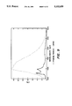
FIG. 6 shows two fluorescent spectra of linker EDR192 with and without streptavidin present, demonstrating production of an excimer. The spectra were normalized to the base peak at 378 nm.
FIG. 7 is a computer simulation of PORSCHA vs. RIA showing increased sensitivity of PORSCHA at low concentrations compared to RIA. Also, the characteristic sigmoidal curve or RIA is generated by PORSCHA.
FIG. 8 is a curve demonstrating the signoidal curve modelled in FIG. 7. The ratio of excimer to monomer areas is plotted with increasing concentration of unlabelled biotin added to the solution.
FIG. 9 shows the interference of endogenous fluorescent materials in some biological media such as urine. This interference may be overcome by either careful background substraction or by use of time-resolved fluorescent spectroscopy to take advantage of the long fluorescent lifetime of pyrene compared to most endogenous fluorescent materials.
FIG. 10 shows several background-subtracted spectra demonstrating the reproducibility of spectral subtraction with 0 and 5 ng of free biotin per cuvette. The buffer has minimal fluorescent background.
FIG. 11 shows the change in excimer intensity with increasing free biotin in solution. All spectra were normalized to one at 378 nm.
FIG. 12 shows that an antibody will elicit an excimer with the appropriate linker arm and sufficient time for equilibrium to occur.
FIGS. 13a and 13b show the use of PORSCHA for the detection of polynucleic acid binding.
FIG. 14 schematically illustrate the preparation of a labelled DNA base for use in automated DNA synthesizers. In this figure, "DMT" is dimethoxytrityl and and "iPr" is isopropyl.
FIG. 15 shows a structure of a DNA oligomer labelled with the deoxyribose base removed and replaced by a carbon chain.
FIG. 16 shows two fluorescent spectra of an oligo, labelled as in FIG. 15, with and without the complentary DNA strand present, demonstrating production of an excimer. The spectra were normalized to the base peak at 378 nm.
DETAILED DESCRIPTION OF THE PREFERRED EMBODIMENT
In the present disclosure and claims, binding assays are defined as assays using molecules, molecular complexes or surfaces that selectively bind two or more other molecules. This definition encompasses but is not limited to antibodies, antibody fragments, streptavidin, avidin, receptors, lectins, surfaces and polynucleotides such as DNA, RNA, and DNA-RNA complexes.
The principles of a competition immunoassay, wherein antibodies are used as the binding species, are outlined in FIG. 1. In the example of FIG. 1, the small molecular weight species is cocaine. Compare two cases where a sample contains or does not contain cocaine. In step one, a measured amount of sample is obtained. To this sample, a radioactive form of cocaine is added. Then, antibodies that are specifically designed to recognize cocaine, are introduced in an amount just sufficient to bind all the labelled cocaine. The antibodies cannot distinguish between the cocaine that may be present and the radioactive form of cocaine added. Thus, the antibody may bind to either form of cocaine. In this competition for the binding sites on the antibody, if there is a large amount of unlabeled cocaine in the sample, then the probability of the labeled drug becoming bound to the antibody is reduced. If there is no unlabeled cocaine in the sample, then all of the labeled cocaine would become bound to the antibody. Then, the unbound cocaine (radio-labelled or not) is separated from that bound to the antibody by any number of techniques. The remaining radioactivity bound to the antibody is then measured. The intensity of this radioactivity is inversely proportional to the amount of unlabelled cocaine in the original sample.
The method according to the present invention relies on a label which varies the wavelength dependance of its spectra (either emission, transmission, or absorbance) depending upon its concentration. An example of such a molecule is pyrene and its derivatives.
As shown in FIGS. 3a and 3b, pyrene possesses different fluorescent spectra at high and low concentrations. At high concentrations two pyrene molecules are close enough for the pi-systems to overlap. It is well-known that the interaction of the pi systems causes emission of light having an emission maximum at a longer wavelength (˜480 nm). At low concentrations the pyrene molecules are too far apart for two pyrene molecules to interact and only an emission at 378 and 396 nm is observed. Thus, the ratio of light emitted at 378 and 396 nm to 480 nm is a measure of the concentration of pyrene in the sample.
It is not generally recognized that molecules can be brought into close proximity to each other by binding with other molecules. The effective concentration of molecules is a calculated value that describes what concentration would need to be prepared to provide a solution in which the molecules are a given average distance apart. For example, as schematically shown in FIGS. 2, two molecules of pyrene are bound via a linker arm to an antibody molecule. These pyrene molecules are approximately 12 nm apart. One would have to prepare a solution that was 1.8×10⁻² M in pyrene for the molecules to be an average of 12 nm apart. Therefore, the effective concentration of these bound molecules is defined as 1.8×10⁻² M. The effective concentration of these molecules (two or more molecules bound to a third) is independent of the macroscopic concentration. In other words, if one were to take one of these complexes of molecules and place it in one liter of water, the effective concentration would remain the same, i.e., 1.8×10⁻² M, whereas the actual concentration would be 1/(6.02×10.sup. 23) or 1.6×10⁻²² M, a very small value. Pyrene shows a substantial excimer at actual concentrations greater than 10-3 M. In FIG. 2, for the pyrene molecules to show a change in spectra that would correspond to an effective concentration of 1.8×10⁻² M, they must be able to interact with each other. Thus, the linker arm shown schematically in FIG. 2 is essential for the interaction to occur. If the antibody, also shown schematically in FIG. 2, were a rigid structure and the distances were as shown in FIG. 2, the linker arm would have to be at least 6 nm long for the pyrene molecules to come into contact. The effective concentration determines the shape of the emission spectrum, whereas the actual, or absolute, concentration of the fluorophore determines the intensity of emission.
This definition of effective concentration is also applicable to the prior art energy-coupling schemes, although these schemes operate on different principles than does the present invention. In the present invention, the labels must approach each other at extremely close distance so that an electronic interaction which alters the available energy levels of the light-emitting electrons occurs. In energy-coupling schemes, energy transfer takes place, but the available energy levels of the light-emitting electrons do not change. A significant advantage of the present invention over chemiluminescent energy transfer is that the bound and unbound fluorophores may be repeatedly probed, since the change in available energy levels, when averaged over a plurality of molecules, is stable over time. On the other hand, chemiluminescence is a transitory phenomenon and must be measured instantaneously, since the energy produced is quickly dissipated. Another advantage of the present invention over acceptor/donor energy transfer is that the present invention has no problem with the overlap of the excitation bands of the donor and acceptor, since no energy transfer is involved.
The principle behind the present invention, which, in a preferred embodiment, is referred to as PORSCHA, for Pi Overlapping Rings Systems Contained in a Homogeneous Assay, is shown, in FIG. 3a through 3d. In this case "homogeneous" refers to an assay where no separation steps are necessary, even if the system may or may not a single phase. In this binding assay, antibodies can bind to two molecules at a time. If the antibody binds to two of the pyrene-labeled antigens or haptens (FIG. 3a), then it will have brought the two separate molecules closer together. Therefore, the effective concentration is much higher than the actual concentration as discussed above. In this instance, the spectra would show an excimer (FIG. 3c). On the other hand, in FIG. 3b, if one or both of the binding sites on the antibody were occupied by unlabelled hapten, then either no or only one pyrene-labelled hapten could bind. Thus, the effective concentration would equal the actual concentration, which is very low. Therefore, no excimer would be observed (FIG. 3d). That is, for the effective concentration to differ from the actual concentration, two or more labelled molecules must be bound in close proximity. Of course, if the actual concentration is very high (in the case of pyrene, greater than 10-3 M), the spectrum would show very little change between the bound and unbound states. However, very few binding assays are performed at these high concentrations.
Pyrene and closely related pyrene derivatives (structures containing the four-membered ring system characteristic of pyrene) are the best choices for indicators according to the present invention because they show strong differences in fluorescent spectra with concentration and have long fluorescent lifetimes if time-resolved spectroscopy is used to resolve background interferences. Other compounds which show similar change spectral absorption or emission maxima dependant upon concentration may also be used. In general, polycyclic aromatic compounds, such as napthalene, perylene, and various fluorescent dyes, such as acridine orange, may be used as labels according to the present invention.
The spectral change can be observed by various means. For example, the emission spectra of the label can be measured after excitation with any form of energy, such as electromagnetic radiation, heat, particle radiation, or chemical energy (chemiluminescence). Alternatively, the spectral absorbance of the label can be monitored, although decreased sensitivity would be expected.
The indicator may be attached to the antigen by any of various known methods using an organic linker. The linker arm is preferably about 20 to about 100 carbons long, depending on the structure of the binding molecule. For example, with streptavidin, one can use a linker armwhich is five atoms long (FIG. 6, compound C3), whereas a closely related molecule, such as avidin, requires a much longer linker arm, for example PEG400, shown in FIG. 4. If the linker arm is too short, the pi rings of the labels will not interact. If the linker arm is extremely long, the indicators will possess too may degrees of freedom to insure proper overlapping and thus the spectral change would be reduced. However, the length requirement past a given length is quite broad, since anything between 5 and 229 atoms has been shown to work for streptavidin. Likewise, Cuniberti et al., Eur. Polymer J., pp 887 to 893 (1980), show that two pyrene molecules may be linked with long linkers (1300 bonds) and still exhibit excimer formation.
The linker is preferably water-soluble and flexible. Examples include, but are not limited to, polyethylene oxides, polyamino acids, polyamides, and DNA. The only requirements are matrix-solubility at the concentration used and appropriate length.
In performing an assay according to the present invention, a buffer is needed to both dilute the components and provide an environment in which the binding of the tracer can occur. Many types of buffers may be employed, including those containing protective proteins, such as bovine serum albumin, gelatin and casein. A number of surfactants above and below their critical micelle concentration such as sodium dodecyl sulfate, TRITON X-100® (a non-ionic polyethylene oxide-based grafted on an octylbenzene), TWEEN 20® (a non-ionic polyethylene oxide grafted to polysorbate) and trimethylhexadecylammonium bromide may be used to potentially reduce non-specific binding of the label to the walls of the measurement system or other artifacts within the measurement matrix. However, in the experiments performed to date, non-specific binding in the absence of surfactant does not appear to be problematic. The excimer intensity varies depending upon the surfactant and is maximal with SDS, below its critical micelle concentration, or without any surfactant.
The binding assay according to the present invention is also useful with inorganic or organic surfaces having specific binding sites for two or more other molecules. For example, the coverage and mobility of molecules on gold surfaces could be measured by exposing the surface to a solution containing an appropriate label, such as pyrene or a derivative thereof, attached to a thiol. Such a labelled material will bind to the gold surface through the thiol functionality only if the surface has free binding sites. If the surface had been previously covered with other thiols, no other binding would occur only if one of these thiols can be displaced in the chosen matrix. Thus, a measurement of the surface coverage, mobility, and ease of displacement of thiols on a gold surface can be made without removing the surface from the matrix solution. Also, in a given system with a known actual concentration of binding sites and a specified label and linker arm, the intensity of the spectral change varies with the distance between the binding sites. Likewise, the minimum distances between binding sites on an uncharacterized molecule or surface may be determined by varying the linker arm length and determining the minimum length of linker arm which results in a spectral change. Thus, the method of the present invention can be used to determine the distance between binding sites for surfaces and for biomolecules.
The binding sites may be on a substance which is composed of two or more molecules in a fixed or oriented spatial relationship such that the average distance between the molecule remains about constant. For example, antibody fragments (such as Fab) can have only one binding site, but these fragments can be oriented by binding to secondary antibodies to provide two binding sites on the complex. Alternatively, these fragments can be oriented by attachment to a surface. Although such an assay would contain two phases, no separation steps would be required to detect the signal change and therefor it would be considered homogeneous within the context of the present invention.
The assay according to the present invention can be performed in aqueous or non-aqueous media. Typically, the matrix within which bioassays according to the present invention is performed is aqueous-based. However, molecules such as antibodies have been shown by others to selectively bind other molecules in substantially non-aqueous media, such as air.
The results with the antibodies shown in FIG. 12 require great care to obtain. In particular, antibody assays according to the present invention, as opposed to other binding assays, are particularly sensitive to variations in methodology, such as time, concentration, reagent purity and temperature.
In the detection of polynucleic acids by PORSCHA, it is essential that the label be attached to the sensing polynucleic acid strand only, such that, when the labeled polynucleic acid strand binds to a complementary polynucleic acid strand, the label is attached to the exterior of the helical chain, i.e., the label should not be intercalated between the two strands of the polynucleic acid helix. Therefore, the labeled polynucleic acid strand should not be labeled at its the amino termini of its bases. Preferably, the label should be attached to the nucleotides at the internucleotide phosphorus atom, or between the phosphorus atoms as shown in FIG. 15.
One method by which PORSCHA could be applied to polynucleic acid detection is outlined in FIGS. 13a and 13b. A sensing strand of polynucleic acid (one complementary to the strand of target polynucleic acid being analyzed) is labeled with nucleotides containing labels with fluorophore moieties, for example pyrene molecules. In solution (FIG. 13a), single-stranded polynucleic acid can take many forms; it has many degrees of freedom. Thus, there is a good probability that two fluorophore moieties could come close enough together to overlap and electronically interact to alter their spectral emission or absorption, for example, by forming a complex such as an excimer. Also, a fluorophore moiety such as a pyrene ring system would be constrained to remain together due to the strong interactions of two pyrene pi systems of about 18 kcal/mol. Therefore, the single stranded polynucleic acid should show a spectral change. When this labeled form of double-stranded polynucleic acid binds to its complementary strand forming a double helix (FIG. 13b), the freedom of motion is greatly constrained. The distance between the labeled phosphates along the labeled polynucleic acid strand is selected such that, in the double helical form, the fluorophore moieties would be pulled apart. Because the motion of the fluorophore moieties would be greatly constrained in the double helical polynucleic acid, they could not interact and hence no electronic interaction would be present. Thus, the absence of, or a decrease of, a variation in the emission or absorbance spectra would indicate the presence of the target polynucleic acid.
In another method for applying PORSCHA to polynucleic acid detection, the strand of polynucleic acid complementary to the target polynucleic acid strand may be more heavily labeled, so that the distance between the labelled phosphate groups is small. In this embodiment, the length of the linker arm between the phosphate group and the fluorophore moiety of the label should be selected to reduce the freedom of motion of the fluorophore moieties, thus minimizing the electronic interaction of fluorophore moieties of the single-stranded polynucleic acid sensing strand in solution. When the target polynucleic acid strand is added to the solution, the sensing strands binds to it and forms a double helix, thus positioning the fluorophore moleties sufficiently close to each other to electronically interact and form and alter their spectral absorption or emission, for example, by forming a complex such as an excimer.
In discussing polynucleic acid detection using PORSCHA, it is convenient and preferred to define the linker as the moiety between the phosphorus atom and the fluorophore moiety of the label. Thus, in FIG. 14, the linker arm is five atoms long.
Of course, some complex formation may always be present in each embodiment useful for polynucleic acid detection, whether the target polynucleic acid strand is present or absent. Accordingly, a significant change in the intensity of the variation in the absorption or emission spectra of the complex would indicate, depending upon the embodiment, the presence or absence of the target polynucleic acid strand. Whether an intensity change is significant will of course depend upon the instrumentation selected, the amount of target polynucleic acid present in the matrix, the label selected, and the precision and error level which the person performing the test considers acceptable.
One advantage of PORSCHA as applied to polynucleic acid detection, is that no separation step need be preformed to determine if the target polynucleic acid is present. Also, a tracer DNA oligomer may be integrated with PCR, as one of the PCR primers, and the change in complex/monomer ratio monitored with time as the PCR progresses. Such a system offers both the amplification advantage of PCR coupled with the monitoring of the reaction with time. Thus if the signal increases very rapidly as the PCR progresses, a large amount of target polynucleic acid would be indicated. If the signal does not change or changes only slowly, no target polynucleic acid or less target polynucleic acid would be indicated. Analogous amplification can be performed on polynucleic acids other than DNA, as is well-known in the art. A homogeneous assay for polynucleic acid coupled to PCR could be a valuable technique for the diagnosis of bacterial or viral infections.
Having described the invention, the following examples are given to illustrate specific applications of the invention including the best mode now known to perform the invention. These specific examples are not intended to limit the scope of the invention described in this application.
EXAMPLES
EXAMPLE 1
The Detection of Cocaine
4-Carboxypyrene butyric acid was synthesized using the method of Cook and Hewett (J. Chem. Soc., 398(1933), incorporated herein by reference). The carboxyl group was removed by the method of Huang-Minlon (Huang-Minlon, J. Amer. Chem. Soc., 71 3301(1949), incorporated herein by reference). The product, pyrene butyric acid (PBA) was purified by the procedure outlined in Cook and Hewett (J. Chem. Soc., 398(1933)).
PBA was conjugated to polyethylene glycol (PEG) by the following procedure. PBA was activated by 1,1'-carbonyldiimidazole (CDI) in dioxane. Then excess PEG was added. The solution was heated at 60° C. for two hours. Water was added to remove the excess PEG and the PEG-PBA conjugate was extracted into chloroform. Benzoylecgonine (a cocaine derivative) was also activated with CDI. The activated benzoylecgonine was added to the PEG-PBA conjugate, which was allowed to react for several days.
When the synthesis of the pyrene-cocaine conjugate was completed, the impure product was purified by High Pressure Liquid Chromatography (HPLC). Fractions were collected at various times and were analyzed with a commercial immunoassay to determine which fraction contained the benzoylecgonine-PEG-PBA conjugate.
The collected fractions were tested on an SLM-8000 spectrofluorometer. The excitation wavelength was set at 343 nm. The emission was scanned from 350 nm to 600 nm in steps of 2 nm with an integration time of 1 sec. All slits were set to 4 nm. A 200 μl quartz cell was used.
The monoclonal antibodies were obtained from either Roche Diagnostic Systems, Nutley, NJ lot 18008-08-09079 or ImmunoSearch, Inc., Toms River, NJ. The antibodies were diluted 1:5 in phosphate buffered saline (PBS), pH 7. The collected fractions were also diluted by a factor which ranged from 1:10 to 1:50. 100 μl of the diluted fractions were then put into the cell. After data was collected, 2-5 μl of antibody was added and another spectral measurement was obtained. Then, after a 5 minute delay, 5 μl of cocaine was added to the cell. Again, a spectral measurement was recorded. After the last spectra, there was usually a 5-10 minute interval before another spectral measurement was taken.
In the initial solution, the pyrene labeled cocaine (benzoylecgonine, a derivative of cocaine) is too dilute to observe any emission at ˜480 nm. When antibody is added to the sample containing cocaine and pyrene labeled cocaine, either will bind to the antibody. If both binding sites of the antibody were occupied by the pyrene labeled cocaine, then the double peak spectra would be seen. If there was free cocaine in the sample, then it would displace either both or one of the labeled cocaine molecules. Thus one peak would be observed rather than two. When both binding sites are occupied by the labeled cocaine, then the pyrene molecules would have a chance to come closer together, thus mimicking a high concentration solution (FIG. 3). If a free cocaine molecule occupied even one of the binding sites, this situation would mimic a dilute solution producing only one emission at 396 nm. Thus, the ratio of 480 nm to 378 and 396 nm would be inversely proportional to the cocaine concentration of the given sample.
Calculations showed that a distance of 42 carbon atoms was optimal for the overlap of two molecules bound to an antibody. Therefore, pyrene butyric acid was linked via a polyethylene glycol chain to benzoylecgonine (a cocaine derivative) to provide at least the 42 carbon chain (see FIG. 4) required. Polyethylene glycol (PEG) was chosen because of its ready availability in different chain lengths. To test this assay, a mixture of chain lengths was employed. Too short a chain length does not allow the two pyrene conjugates to come into contact and hence no excimer results. Too long a chain length decreases the chance that the two pyrene molecules would come into contact and this would result in a decrease in the excimer intensity.
EXAMPLE 2
Testing Using Streptavidin as Antibody Mimic
The principle of the present invention as an assay for a substance in urine was simulated by the determining the effect of concentration of biotinylated pyrene in the presence of another binding compound, streptavidin, on the fluorescent spectrum of the biotinylated pyrene. Pyrene was biotinylated, using conventional techniques, via an amido linker arm, --OC--HN--X--NH--, to a molecule of biotin. The resulting biotinylated biotin was very carefully purified, by conventional techniques, to remove any free biotin.
EXAMPLE 3
Preparation of the Edr148 Linker (FIG. 4)
To a 20 ml vial was added 50 mg of pyrene butyric acid N-hydroxy succinimide (PBA-NHS), 5 ml of methylene chloride and 200 mg of JEFFAMINE® EDR-148 (Texaco) (about 10 fold molar excess), a polyethylene oxide diamine of MW 148. The reaction was allowed to occur overnight at room temperature. Then, the solvent was removed by blowing an air stream into the vial. The residue was dissolved in water and applied to a C18 solid phase extraction cartridge which was prewet with methanol and then water. The pyrene derivative collected as a band, highly fluorescent in the blue region, at the top of the cartridge. The cartridge was washed extensively with water to remove unlabelled EDR-148 and the pyrene derivative eluded with methanol. The methanol was removed by evaporation under an air stream and replaced with dimethylformamide. Excess (about 2 to 5 times) biotin N-hydroxysuccinimide molar over the linker was added. The reaction was allowed to occur over a two-day period at room temperature. Then, the solution was applied in portions (about 4 portions) to 20×20 cm silica preparative TLC plates, one portion per plate. The plate was developed with 90% chloroform, 10% methanol. The product band (blue fluorescence) was scraped off the plate and the product was removed with methanol. Portions (about 100 μl out of 10 ml of methanol solution) of these were further purified on a C18 HPLC column using a 0 to 100% acetonitrile-0.1M ammonium acetate gradient and monitoring the effluent with a fluorimeter. Fractions were collected when the fluorimeter indicated elution of the pyrene labelled material. HPLC was performed to remove potentially free biotin. It may be unnecessary to use HPLC if the thin layer chromatography is sufficient to remove all the free biotin. The other labelled biotin molecules shown in FIGS. 4 and 5 were prepared in an analogous manner.
EXAMPLE 4
Use of the Edr148 Linker of Example 3
If an insufficient concentration of the binding molecule (in this case streptavidin) was used, the excimer would not be at a maximum. Likewise, if too much binding molecule is used, the excimer also will decrease, since on an average, the two binding sites would not be occupied simultaneously. Thus, a balance between the labelled biomolecule and its corresponding binding material is needed. The resultant HPLC purified material of Example 3 was titrated with dilute solutions of streptavidin such that a maximal intensity of the excimer resulted. An example of the spectrum of an excimer produced is shown in FIG. 6. In this Figure, the spectra without streptavidin and with streptavidin is depicted. Both spectra are normalized to the base peak intensity at 378 nm. Otherwise, the spectra with streptavidin would have less absolute fluorescent intensity since the fluorescent intensity is spread over both the monomer and excimer peaks.
EXAMPLE 5
Computer Simulation of Parscha and Ria
A computer program was written to perform the following tasks:
1. A matrix is filled with a thousand "1"'s, which represent a thousand labelled molecules.
2. The computer selects from this matrix, 500 pairs at random, without replacement and then examines both components of the pair. If these components are both "1"'s, then the computer would consider an "excimer" to be produced. If either component "0", which represents an unlabelled molecule, then an excimer would not be produced. During the selection process the computer keeps track of the number of excimers found.
3. In the next step, increasing numbers of "1"'s are replaced with "0"'s (unlabelled molecules) and the selection process described in step two is repeated.
In the first pass through the program, since only "1"'s are present, 500 excimers would be counted. As the ones are replaced by "0"'s, decreasing numbers of excimers are produced. For RIA, a very similar scheme is used, except that a label is considered present if either of the molecules selected in step two are "1"'s. The results of such a simulation are depicted in FIG. 7. As can be seen, the S-shaped curve for PORSCHA is shifted to the left relative to RIA. This indicates that PORSCHA gives a larger signal difference for lower concentration species than does RIA. A linear line may be generated from both curves through the use of the logit function. The "noise" depicted in FIG. 7 is the result of the limited number of points in the matrix and the random nature of the selection process.
EXAMPLE 6
Generation of Binding Curve for Edr148 Linker and Streptavidin
Streptavidin has a very high binding affinity for biotin. This binding affinity implies that the initial binding event occurs very rapidly and that the displacement of the bound biotin molecule occurs very slowly. Thus, to generate a binding curve, one has to carry out a series of experiments rather than just adding free or unlabelled biotin. Solutions of biotin in phosphate buffered saline (PBS) containing 2.5% w/v sodium dodecylsulfate (SDS) were prepared, then 5 μl of these solutions were added to a cuvette containing the optimized EDR148 linker from Example 3 in 100 μl of PBS with SDS. After mixing, the optimal streptavidin concentration, as determined in Example 4, was added and the spectra recorded. These steps were repeated for varying biotin concentrations. The area under the several wavelength ranges corresponding to the monomer and excimer fluorescent bands was calculated and the ratios plotted in FIG. 8 to generate the S-shaped curve. The raw data used to generate this Figure is similar to that shown in FIG. 11. The raw data shown in FIG. 11 was obtained by the same method used to obtain the raw data employed to generate FIG. 8. The curve of FIG. 8 has the characteristics predicted by the computer simulation of Example 5 and demonstrates that the excimer to monomer ratio changes with biotin concentration and can be readily detected.
The position of inflection of the S-shaped curve is determined by the concentrations of label and binding molecule. The curve may be shifted to the left (made more sensitive) by decreasing the concentration of both species. Likewise, the curve may be shifted to the right (made less sensitive) by increasing the concentration of both species. The limit of sensitivity is determined by at least three factors:
1. The binding constant of the binding molecule.
2. The fluorescent background of the matrix.
3. The sensitivity for detection of the fluorescence emitted.
EXAMPLE 7
Fluorescent Background Interferences Observed by Some Environmental Matrices
To obtain maximal sensitivity, the binding molecule and the labelled species must be in as low a concentration as possible. In this manner, small quantities of unlabelled compounds can occupy the majority of binding sites on the binding molecule or species and produce the largest signal difference. However, many environmental matrices possess species with fluorescent properties that can mask the label. At least two methods exist to reduce or eliminate these interferences. One is to take background spectra before the labelled tracer is added in Example 6. Another would be to use time-resolved spectroscopy to take advantage of the long fluorescent lifetime of pyrene. However, the instrumentation to use the latter technique is more expensive than background subtraction, so background subtraction was chosen to demonstrate PORSCHA in real matrices.
FIG. 9 shows the fluorescent background typically produced by urine, using dotted lines. The solid line shows the labelled pyrene in PBS, a matrix without substantial fluorescence. Although this ratio of background to signal is sufficient to generate reasonable precise data, a higher concentration of label, where the fluorescence is at least as intense as the background fluorescence, is desirable for background substraction.
Using the general procedures of Example 6, increasing the volume of the cuvette to ˜2 ml while keeping the concentrations of the various system components as in Example 6, 5 μl of urine was substituted for some of the PBS to prepare the standard concentrations of biotin, using about four times the concentration of pyrene label as used in FIG. 9. In the cuvette is about 2.2×10⁻¹¹ moles of streptavidin, about 7.15×10⁻¹¹ moles of biotin derivative, and for the 5 ng of biotin in the cuvette, about 2×10-11 moles of biotin would be present. FIG. 10 shows four spectra. Two of these spectra shown by the solid lines were obtained in PBS, where fluorescence of the matrix is minimal. The other two, shown by dotted lines, were obtained in urine, where the fluorescence of the matrix is substantial. The substantial correspondence of the two pairs of curves demonstrates that background subtraction is a viable method for reducing interferences from the matrix.
EXAMPLE 8
Demonstration of Excimer Production With an Antibody and Appropriate Linker Arm
Goat anti-biotin was added to a solution of the linker in PBS with SDS as in Example 6, using linker arm PBA-EDR192L-Pro-Bio (FIG. 5). The change in spectra with time was monitored and the production of an excimer was noted, as shown in FIG. 12. Since antibodies have lower binding constants than streptavidin, a longer equilibrium time must be used before recording the spectra. FIG. 12 demonstrates that an appropriate linker arm can generate an excimer with an antibody. This linker arm has a bend (in this case ˜120 °) incorporated in its structure because of the proline functionality. This bend is needed to produce substantial excimer formation because of the tertiary structure of the antibodies employed. With current bioengineering technology, molecules which recognize and bind other molecules can be produced in bacteria (Pack et al., Biochemistry, 31, 1579 (1992)). These bio-engineered molecules are likely to have structures different from conventional antibodies. If the binding sites are closer to each other than in conventional antibodies, a bend may not be necessary in the linker structure. Interestingly enough, this linker (EDR192L) works poorly with streptavidin compared to the corresponding linker without the proline.
EXAMPLE 9
DNA Detection by Porscha
To demonstrate the use of PORSCHA for DNA detection, an oligomer was labeled with pyrene as in FIG. 15, with four nucleotide bases between each pair of labels. Oligomers were prepared with this modified nucleotide and purified by HPLC. Preliminary experiments suggested that when the complementary DNA (M13mp18 in this case) was added to the dual labeled oligomer, a change in excimer intensity, as shown in FIG. 16, was observed.
Obviously, many modifications and variations of the present invention are possible in light of the above teachings. It is therefore to be understood that, within the scope of the appended claims, the invention may be practiced otherwise than as specifically described.