US5336206A - Trocar penetration depth indicator and guide tube positioning device - Google Patents
Trocar penetration depth indicator and guide tube positioning device Download PDFInfo
- Publication number
- US5336206A US5336206A US08/046,328 US4632893A US5336206A US 5336206 A US5336206 A US 5336206A US 4632893 A US4632893 A US 4632893A US 5336206 A US5336206 A US 5336206A
- Authority
- US
- United States
- Prior art keywords
- guide tube
- trocar
- depth indicator
- penetration depth
- penetration
- Prior art date
- Legal status (The legal status is an assumption and is not a legal conclusion. Google has not performed a legal analysis and makes no representation as to the accuracy of the status listed.)
- Expired - Lifetime
Links
Images


Classifications
-
- A—HUMAN NECESSITIES
- A61—MEDICAL OR VETERINARY SCIENCE; HYGIENE
- A61B—DIAGNOSIS; SURGERY; IDENTIFICATION
- A61B17/00—Surgical instruments, devices or methods, e.g. tourniquets
- A61B17/34—Trocars; Puncturing needles
- A61B17/3403—Needle locating or guiding means
-
- A—HUMAN NECESSITIES
- A61—MEDICAL OR VETERINARY SCIENCE; HYGIENE
- A61B—DIAGNOSIS; SURGERY; IDENTIFICATION
- A61B17/00—Surgical instruments, devices or methods, e.g. tourniquets
- A61B17/34—Trocars; Puncturing needles
- A61B17/3417—Details of tips or shafts, e.g. grooves, expandable, bendable; Multiple coaxial sliding cannulas, e.g. for dilating
-
- A—HUMAN NECESSITIES
- A61—MEDICAL OR VETERINARY SCIENCE; HYGIENE
- A61B—DIAGNOSIS; SURGERY; IDENTIFICATION
- A61B17/00—Surgical instruments, devices or methods, e.g. tourniquets
- A61B17/34—Trocars; Puncturing needles
- A61B17/3494—Trocars; Puncturing needles with safety means for protection against accidental cutting or pricking, e.g. limiting insertion depth, pressure sensors
- A61B17/3496—Protecting sleeves or inner probes; Retractable tips
-
- A—HUMAN NECESSITIES
- A61—MEDICAL OR VETERINARY SCIENCE; HYGIENE
- A61B—DIAGNOSIS; SURGERY; IDENTIFICATION
- A61B17/00—Surgical instruments, devices or methods, e.g. tourniquets
- A61B17/34—Trocars; Puncturing needles
- A61B2017/348—Means for supporting the trocar against the body or retaining the trocar inside the body
- A61B2017/3492—Means for supporting the trocar against the body or retaining the trocar inside the body against the outside of the body
-
- A—HUMAN NECESSITIES
- A61—MEDICAL OR VETERINARY SCIENCE; HYGIENE
- A61B—DIAGNOSIS; SURGERY; IDENTIFICATION
- A61B90/00—Instruments, implements or accessories specially adapted for surgery or diagnosis and not covered by any of the groups A61B1/00 - A61B50/00, e.g. for luxation treatment or for protecting wound edges
- A61B90/03—Automatic limiting or abutting means, e.g. for safety
- A61B2090/033—Abutting means, stops, e.g. abutting on tissue or skin
- A61B2090/036—Abutting means, stops, e.g. abutting on tissue or skin abutting on tissue or skin
-
- A—HUMAN NECESSITIES
- A61—MEDICAL OR VETERINARY SCIENCE; HYGIENE
- A61B—DIAGNOSIS; SURGERY; IDENTIFICATION
- A61B90/00—Instruments, implements or accessories specially adapted for surgery or diagnosis and not covered by any of the groups A61B1/00 - A61B50/00, e.g. for luxation treatment or for protecting wound edges
- A61B90/06—Measuring instruments not otherwise provided for
- A61B2090/062—Measuring instruments not otherwise provided for penetration depth
Landscapes
- Health & Medical Sciences (AREA)
- Surgery (AREA)
- Life Sciences & Earth Sciences (AREA)
- Medical Informatics (AREA)
- Nuclear Medicine, Radiotherapy & Molecular Imaging (AREA)
- Engineering & Computer Science (AREA)
- Biomedical Technology (AREA)
- Heart & Thoracic Surgery (AREA)
- Pathology (AREA)
- Molecular Biology (AREA)
- Animal Behavior & Ethology (AREA)
- General Health & Medical Sciences (AREA)
- Public Health (AREA)
- Veterinary Medicine (AREA)
- Surgical Instruments (AREA)
- Endoscopes (AREA)
Abstract
A trocar penetration depth indicator has a first, inner housing including a first threaded section and longitudinally extending gripping fingers, and a second, outer housing including a camming surface and a second threaded section to engage the first threaded section. The first and second housings slidably mount onto a trocar tube and can be secured relative to the guide tube by rotatably tightening the housings to cause the gripping fingers to firmly grip the trocar tube. Depth penetration indicia is provided on the guide tube surface and sufficient guide tube grasping force is obtained to prevent the depth indicator from being dislodged during trocar insertion. A guide tube positioning device is provided to engage the penetration depth indicator and to secure the guide tube relative to the surrounding skin of the patient in order to prevent inadvertent withdrawal or further insertion of the trocar.
Description
This is a continuation of U.S. application Ser. No. 07/861,164, filed Mar. 27, 1992, now U.S. Pat. No. 5,217,441; which is a continuation of application Ser. No. 07/489,482, filed Mar. 6, 1990, now abandoned, which is a continuation-in-part of application Ser. No. 07/394,198, filed Aug. 15, 1989, now abandoned.
The present invention relates generally to a trocar and, more specifically, to improvements for indicating the penetration depth of a trocar and for maintaining the position of the inserted trocar guide tube relative to the body.
Trocars of the type relating to the present invention generally include a stylet having a sharp tip for penetrating through a patient's abdominal wall or other body cavity, a protective shield tube surrounding the stylet, and an outer guide tube surrounding the protective shield tube. Typically, the protective shield tube and stylet extend beyond the distal end of the guide tube, and the protective tube is spring-biased in a locked distal position surrounding the sharp tip of the stylet. To use the trocar, the protective tube is unlocked so that the protective tube moves proximally to expose the stylet tip for penetrating the abdominal wall. Once the abdominal wall has been penetrated, the protective tube slides distally under the force of the spring to again assume the locked, distal position covering over the sharp stylet tip. In this manner the outer guide tube is placed into the body at a desired location. Thereafter, the styler and protective tube may be removed to leave the guide tube in place. Trocars of this type are generally described in Moll U.S. Pat. Nos. 4,601,710 and 4,654,030. After the guide tube has been placed within the body, surgical instruments may be inserted through the guide tube.
Unfortunately, known trocars do not provide any indication of trocar penetration depth to the surgeon, who must estimate the penetration depth during and after penetration of the abdominal wall. Some surgeons accomplish this task by simply resting a finger on the outer guide tube as a visual aid during insertion. Of course, this approach is unreliable, since the surgeon's finger is, at best, a rough approximation of penetration depth. Moreover, the surgeon's finger is susceptible to slipping or moving during insertion of the trocar.
Numerous penetration indicating and limiting devices have heretofore been proposed in relation to various types of surgical devices and instruments. These include graduated scales or other marks, see, for example, U.S. Pat. Nos. 3,459,189 and 3,993,079, and collar-type devices with a set or fixing screw bearing against the apparatus to be inserted, see, for example, U.S. Pat. Nos. 1,213,001 and 2,496,111 and Italian Patent No. 475215. Unfortunately, in the context of a trocar, merely providing a graduated scale or mark would be inadequate since this requires conscious visual attention of the surgeon or an attendant during insertion and provides no protection against over insertion during initial penetration. Moreover, the indicia on the graduated scale or marker may become obscured under surgical conditions, and constant attention to the scale is not possible. Set or fixing screws pose the undesirable possibility that overtightening may dent or damage the device. In the case of modern trocars, such damage could impair the structural integrity of the trocar guide tube and/or interfere with the operation of instruments inserted into the body through the trocar guide tube. Even relatively minor damage to a trocar guide tube may alter the relatively small internal diameter of the tube, e.g., on the order of about 10 millimeters or less, and interfere with insertion, rotation, or operation of sophisticated medical instruments designed to operate closely within the limited trocar guide tube space.
U.S. Pat. No. 3,817,250 discloses a collar for limiting penetration of a tracheostomy device. However, that device permits penetration to only one preset depth and undesirably obscures the insertion site.
U.S. Pat. No. 3,613,684 discloses a trocar catheter having a depth penetration limiting device consisting of a slotted tubular member with a slotted, radially extending collar. In use, the limiting device is grasped to isolate the device relative to the catheter shaft during insertion. Unfortunately, it appears the depth penetrating device may slip accidentally during insertion if not grasped firmly and constantly. In addition, the device there disclosed apparently requires an inconvenient two-handed penetration technique. This would seem to require an extended trocar shaft to allow sufficient room to grasp the shaft during penetration.
Other penetration depth indicators rely upon pressure sensitive devices. These include U.S. Pat. Nos. 2,623,512; 4,186,750; and 4,215,699 and Russian Patent 921554.
Notwithstanding the foregoing disclosures, there presently exists a need for trocar penetration depth indicator which provides convenient, reliable indication of trocar penetration depth during and after one-handed insertion of a trocar, and which does not unduly lengthen the trocar guide tube or present a hazard of damaging the guide tube by overtightening a set or fixing screw.
Once penetration of the body wall has been attained, the stylet is removed, leaving a guide tube penetrating the body adapted to receive surgical instruments. However, the guide tube may be subject to unintentional and undesirable changes in penetration depth or accidental withdrawal from the body. Therefore, there also exists a need for a positioning device to hold the guide tube at the desired penetration depth during use. At the same time, however, the positioning device should accommodate manipulation of the guide tube by the surgeon during the surgical procedure.
Accordingly, it is one object of the present invention to provide a trocar depth indicator.
A further object of the present invention is to provide a trocar depth indicator which does not limit the useful penetration length of existing trocars or require undue extension of the trocar barrel length.
Another object of the present invention is to provide a trocar penetration depth indicator which may be securely positioned relative to the trocar guide tube so as to reduce the likelihood of displacement of the depth indicator along the guide tube during penetration.
It is yet a further object of the invention to provide a trocar depth indicator which may be positioned relative to the trocar guide tube without damaging the guide tube or interfering with the operation of delicate instruments inserted therethrough.
Yet another object of the present invention is to provide a trocar guide tube positioning device for positioning the inserted trocar guide tube relative to the body.
These and other objects and advantages are accomplished in a compact, lightweight and low cost device which provides reliable indication of trocar penetration depth without unduly obstructing the trocar guide tube, and which positions the inserted trocar guide tube relative to the body to prevent accidental removal of the trocar or inadvertent changes in penetration depth.
In accordance with the present invention, a trocar penetration depth indicator is provided having inner and outer housings threadingly coupled to one another.
The inner housing includes an inner housing body having a hand grip portion, a threaded portion and a plurality of distally extending gripping fingers. An axial cylindrical opening through the inner body housing has a minor diameter defined by the proximal end of the gripping fingers configured and dimensioned to conform closely to the outer diameter of a trocar guide tube.
The outer housing includes an outer housing body having a hand grip portion, a threaded portion, and a camming surface adjacent a cylindrical axial aperture. The outer housing is configured and dimensioned so that the outer housing threaded portion may engage the inner housing threaded portion and the outer housing camming surface may engage the inner housing gripping fingers.
In use, the inner and outer housings may be loosely assembled together without tightening the threaded coupling therebetween. So assembled, the penetration depth indicator may be mounted over the trocar guide tube. In this first, untightened position, the trocar depth indicator may be slid along the guide tube to a desired position. Preferably, the inner housing gripping fingers are configured and dimensioned so that, even in the first, unlocked position, the depth indicator frictionally engages the outer surface of the trocar guide tube and will remain in place until moved to a different position.
Once positioned on the guide tube, the inner or outer housings, or both, are rotated relative to one another in order to cause the depth indicator to assume a second, tightened position. In the tightened position the outer housing camming surface exerts radially inward pressure on the inner housing gripping fingers in order to establish sufficient frictional engagement between the gripping fingers and guide tube to prevent movement of the depth indicator relative to the guide tube during insertion and use of the trocar. Preferably, graduated markings are provided on the outer surface of the guide tube which indicate the approximate penetration depth. Of course, the trocar depth indicator can repeatedly be unlocked and locked to adjust the position along the guide tube.
Advantageously, the gripping fingers establish a substantially uniform distribution of gripping forces around the guide tube, thereby preventing damage to the guide tube which might otherwise occur in connection with exertion of equivalent forces over a much smaller area, i.e., by a set screw. Remarkably, however, the gripping forces of the tightened depth indicator on the guide tube are sufficient to withstand substantial forces without the depth indicator being displaced along the guide tube. A further surprising advantage of the invention is that the present trocar depth indicator is very compact, occupying a very short length of the guide tube. Consequently, the depth indicator does not reduce the available penetration length of the trocar during use. Indeed, in the preferred embodiment the housing of the penetration depth indicator proximal to the trocar guide tube housing is configured and dimensioned to receive and nest over the guide tube collar on the guide tube housing, thereby maximizing the effective useful length of existing trocars without modification.
In a further preferred embodiment, a guide tube positioning device is provided for positioning the guide tube relative to the body once the desired penetration depth has been achieved. The positioning device may constitute a generally circular skirt made of a clear plastic material. The skirt has an axial aperture configured and dimensioned to receive the trocar guide tube, a first foam pad ring adhered to the distal side of the skirt surrounding the skirt aperture, and a second, circumferential foam pad ring adhered to the distal side of the skirt at the periphery thereof. The distal side of the second ring is provided with an adhesive covered by a release sheet. A radial slit through the skirt and first and second rings extends from the skirt aperture to the peripheral edge of the skirt, and a latch member is provided for joining the edges of the radial slit.
In use, the trocar guide tube is inserted at a point proximal to the penetration depth indicator through the radial slit so that the guide tube is disposed in the skirt aperture with the skirt proximal to the penetration depth indicator. The latch member closes the radial slit and the release sheet is removed from the second annular ring to adhere the same to the surrounding skin of the patient. The first foam pad ring contacts the proximal surface of the penetration depth indicator, such that the penetration depth indicator prevents further penetration of the trocar guide tube, and the skirt prevents accidental withdrawal of the trocar from the desired penetration depth.
Alternatively, the positioning device may constitute a sleeve member integral with the depth indicator having radially projecting rib members which engage the patient's skin at the incision when the sleeve is inserted into the incision.
It will be understood by those skilled in the art that the foregoing general description and the following detailed description are exemplary and explanatory of the invention but are not restrictive thereof.
The accompanying drawings, referred to herein and constituting a part hereof, illustrate preferred embodiments of the present invention, and together with the description, serve to explain the principles of the invention.
FIG. 1 is a perspective view of a trocar of the type relating to the present invention, illustrating a graduated depth penetration scale on the guide tube;
FIG. 2 is a perspective view of the unassembled trocar penetration depth indicator of the present invention;
FIG. 3 is a perspective view of the penetration depth indicator mounted on the trocar guide tube;
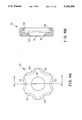
FIG. 4A is a distal end view of the outer housing of the penetration depth indicator shown in FIGS. 2 and 3;
FIG. 4B is a cross-section view of the outer housing taken along lines 4-4 of FIG. 4A;
FIG. 5A is a proximal end view of the inner housing of the penetration depth indicator shown in FIGS. 2 and 3; FIG. 5B is a cross-section view of the inner housing taken along lines 5--5 of FIG. 5A;
FIG. 5C is a distal end view of the inner housing shown in FIGS. 5A and 5B;
FIG. 6 is a cross-section elevation view of the depth penetration indicator mounted on the trocar guide tube and nested over a portion of the guide tube housing;
FIG. 7 is a proximal end view of the guide tube positioning skirt in accordance with the invention;
FIG. 8 is a cross-section view of the guide tube positioning skirt taken along lines 8--8 of FIG. 7;
FIG. 9 is a partial elevation view of the guide tube positioning skirt illustrating the latch member;
FIG. 10 is an elevation view of the penetration depth indicator and guide tube positioning skirt illustrating a trocar guide tube positioned relative to a patient's body;
FIG. 11 is a perspective view of an alternative embodiment of the depth penetration indicator and positioning device; and
FIG. 12 is a perspective view of an alternative embodiment of the present invention.
Referring now to FIG. 1, there is shown a trocar 10 having a guide tube 12 extending distally from a guide tube housing 14, and a stylet protective shield tube 16 slidably mounted within guide tube 12 and supported by a stylet housing 18. Stylet housing 18 also supports a sharp-tipped stylet (not shown) disposed within protective tube 16. Generally, protective tube 16 is spring biased and locked in the extended position shown in FIG. 1 prior to use. In order to use the trocar, guide tube housing 14 and styler housing 18 are urged together to unlock protective tube 16, and the tip of the trocar is pressed against the patient's body. As the trocar tip is pressed against the body, the protective tube is urged proximally within the guide tube to reveal the sharp stylet for penetration of the body wall. After penetration, the protective tube returns to the extended locked position shown in FIG. 1. In this manner, the guide tube is inserted into the patient's body and may be maneuvered to the desired point. The guide tube, with the stylet and protective tube removed, provides access for viewing and/or performing surgery within the body without a traditional incision. The guide tube is held in place by the guide tube positioning skirt (see FIG. 10) or sleeve (see FIG. 11). Preferably, the guide tube and/or guide tube housing at all times provide a seal between the penetrated body cavity and the external atmosphere. As shown in FIG. 1, in accordance with the invention, guide tube 12 is preferably provided with penetration depth indicia, shown in the form of a graduated scale 20, the purpose of which shall become apparent below.
FIGS. 2 and 3 illustrate the trocar penetration depth indicator of the present invention generally denominated by numeral 22 including a first, inner housing 24 and a second, outer housing 26. The first and second housings have axially aligned apertures to receive guide tube 12. The inner and outer housings rotatably engage to assume an unlocked position wherein the trocar penetration depth indicator may be slidably urged along guide tube 12 to a desired position. After the desired position has been attained, the inner and outer housings are rotated relative to one another so that a camming surface on the outer housing constricts a plurality of gripping fingers 28 on the inner housing to frictionally engage and grasp the guide tube, thereby preventing further sliding motion of the depth indicator relative to the guide tube.
FIG. 4A is a distal end view of outer housing 26. As there shown, outer housing 26 is generally cylindrical, with a central aperture 30 and a gripping portion along the outer circumference of the housing. Preferably, the gripping portion includes a number of finger accommodating depressions 34. FIG. 4B is a cross-section view of outer housing 26 taken along line 4--4 of FIG. 4A. As shown in FIG. 4B, outer housing 26 further includes a distally inwardly inclined camming surface 32 adjacent aperture 30 and an inwardly facing outer housing threaded section 36. It will be noted that the outer housing has a substantially open center with the smallest diameter of the outer housing defined by aperture 30 adjacent camming surface 32. By way of example only, the outer housing may be constructed, such as by molding, of glass filled acetal or polycarbonate. Preferably, the outer housing is constructed of glass-filled acetal. One appropriate acetal material is available from LNP Division of ICI America Inc. under the trade specification KFL 4023.
FIG. 5A is a proximal end view of inner housing 24 showing gripping projections 38 disposed on the outer circumference of flange 37 the inner housing. Also shown are a central aperture 40 and a nesting area 42 for accommodating a portion of the guide tube housing. FIG. 5B is a cross-section view of inner housing 24 taken along lines 5--5 of FIG. 5A. As there illustrated, the inner housing is generally cylindrical and includes a gripping portion 44 supporting gripping projections 38. The inner housing also has an outwardly facing threaded section 46 to engage outer housing threaded section 36, and a number of longitudinally extending gripping fingers 48 for frictionally gripping the trocar guide tube under constriction of the outer housing. Each gripping finger has an outer surface 49, an inner surface 51 and a distal end surface 53. Preferably, an inclined gripping finger camming surface 50 is provided between the outer gripping finger surface 49 and distal end surface 50. As explained more fully below, gripping finger camming surface 50 engages the outer housing camming surface 32 to ensure uniform, radially-inward constriction of the gripping fingers to grip the guide tube. Nesting area 42 is illustrated in FIG. 5B as an inclined surface of distally decreasing diameter. The increased proximal open area obtained by the inclined surface of nesting area 42 accommodates a portion of the guide tube housing (see FIG. 6). Of course, the nesting area could also take other forms, such as an area of uniform enlarged diameter sufficient to accommodate the guide tube housing. FIG. 5C, a distal .end view of inner housing 24, illustrates the arrangement of the gripping fingers. As shown, the inner surface 51 of each gripping finger is curved to conform to the radius of curvature of the guide tube. FIG. 5C also illustrates the preferred arrangement of eight gripping fingers. The number and specific arrangement of gripping fingers are not critical, but the preferred arrangement of a plurality of gripping fingers having concave inner surfaces advantageously distributes gripping forces about the circumferential area of contact with the trocar guide tube. By way of example only, inner housing 24 may be constructed, as by molding, of a glass filled acetal or polycarbonate. In the preferred embodiment wherein the outer housing is constructed of glass-filled acetal, the inner housing is preferably constructed of polycarbonate. One suitable polycarbonate material is available from General Electric Company under the trade name LEXAN. The preferred acetal-polycarbonate arrangement reduces binding at the threaded engagement of threaded sections 36, 46. Of course, the same result may be obtained by constructing the outer housing of polycarbonate and the inner housing of acetal, but acetal may slide on the steel cannula or guide tube.
FIG. 6 shows the trocar depth indicator with inner and outer housings 24, 26 fully assembled, mounted to and gripping trocar guide tube 12. In FIG. 6, inner housing 24 is nested over the guide tube collar 52 extending from guide tube housing 14. As will be understood from the Figures, particularly FIG. 6, rotatably tightening the inner and outer housings by engagement of threaded sections 36, 46 causes the inner and outer housings to be longitudinally drawn together. As the housings are drawn together, outer housing camming surface 32 engages gripping finger camming surfaces 50 to exert radially inward force on gripping fingers 48 to securely grasp outer guide tube 12. Advantageously, the plurality of gripping fingers conforming to the guide tube surface uniformly distributes substantial frictional gripping force over virtually the entire guide tube surface. Remarkably, the present invention develops sufficient gripping force to withstand substantial force, on the order of about 25-30 pounds. However, because the gripping force is not concentrated over a small area, as in the case of a set screw, there is no danger of damaging the structural configuration of the guide tube. The depth indicator according to the present invention securely grips the guide tube and provides an indication of penetration depth which may be relied upon with reasonable confidence that the depth indicator will not accidentally be displaced from the desired position. It is also contemplated and desired that the inner and outer housings assume a first, unlocked position with the threaded sections loosely engaged to keep the housings together without constricting the gripping fingers against the guide tube. Of course, in this unlocked position the trocar penetration depth indicator may deliberately be moved along the guide tube and will remain static in any position where the indicator is disposed. The trocar penetration depth indicator is effectively secured relative to the guide tube when the inner and outer housings are rotatably tightened to assume the second, locked gripping position with the outer housing camming surface constricting the gripping fingers.
In order to obtain these desirable features, the inner housing should be configured and dimensioned so that gripping fingers 48 define an opening having a diameter slightly larger than the outer diameter of the corresponding guide tube when the gripping fingers arc not constricted by the outer housing. Similarly, in order to obtain sufficient gripping force, the gripping fingers should be constricted by the outer housing so as to define an opening having a diameter conforming to the outer diameter of the guide tube. Advantageously, the maximum constriction of the gripping fingers may be limited by limiting the travel of the threaded sections of the inner and outer housings. In order to achieve the desired gripping action without overtightening, it is contemplated that the maximum tightened position of the threaded inner and outer housings should constrict the gripping fingers to a maximum constricted position to define an aperture having a diameter slightly smaller than the outer diameter of the guide tube. By way of example, but not limitation, a standard 10 millimeter trocar has a nominal inner diameter of about 10 millimeters and a nominal outer diameter of about 10.3 millimeters. Accordingly, a depth indicator of the present invention for use with a 10 mm trocar should be constructed so that the inner housing gripping fingers define an opening slightly larger than 10.3 millimeters in diameter when not constricted by the outer housing, i.e., on the order of about 10.4 or 10.5 millimeters. Conversely, the maximum constriction of the gripping fingers by the outer housing should define an aperture slightly less than about 10.3 millimeters in diameter such as from about 9.8 to 10.2 millimeters. Of course, the maximum constricted position would actually be achieved only in the absence of a guide tube, but so defining a maximum constricted position makes it impossible to overtighten the depth indicator to cause deformation of the guide tube. It will be understood by those or ordinary skill in the art that these principles also applies to depth indicators to be used with different size trocars.
In use, the trocar penetration depth indicator is mounted onto the trocar guide tube prior to use. The depth indicator, preferably disposed in the unlocked position with the inner and outer housings loosely engaged, is moved longitudinally along the guide tube to the desired penetration depth position. Advantageously, the distal surface of outer housing 26 may be used as a cursor against depth penetration indicia 20 on the guide tube surface (see FIG. 3). Once disposed in the desired position, the inner and outer housings are rotatably tightened together, so that the penetration depth indicator assumes the locked, tightened position firmly and immovably grasping the guide tube. Thereafter, the trocar may be inserted into the patient in the usual fashion with confidence that the depth indicator will remain in the desired position during and after insertion and can be relied upon to accurately indicate trocar tube penetration depth. Advantageously, the distal surface of the outer housing may rest against the patient's skin, as illustrated in FIG. 10.
In a further preferred embodiment, a trocar guide tube positioning skirt is provided for maintaining the position of the guide tube after insertion into the body. Preferably, the guide tube positioning skirt is disposed around the guide tube and adhered to the patient's skin. As will be understood from the following description, the positioning skirt and penetration depth indicator cooperate to prevent inadvertent insertion or withdrawal of the guide tube from the desired penetration depth.
Turning to FIG. 7, there is shown a proximal end view of the positioning device 60 consisting of a clear, generally circular skirt 62 having an axial guide tube aperture 64, a radial slit 66, and a latch member 68. As shown in phantom in FIG. 7, on the distal side of skirt 62 a first annular ring 70 surrounds skirt aperture 64 and a second, circumferential ring is disposed at the outer periphery of the skirt.
As shown in FIG. 8, a cross-section view of positioning device 60 taken along lines 8--8 of FIG. 7, latch member 68 preferably extends from skirt aperture 64 to the outer edge of skirt 62. Skirt aperture ring 70 is disposed adjacent skirt aperture 64 and is adhered to the distal side of skirt 62. Circumferential ring 72 is adhered to the distal side of skirt 62 at the peripheral edge of the skirt. The distal surface of circumferential ring 72 is provided with an adhesive layer 74 covered by a paper release sheet 76.
FIG. 9, a partial elevation view of the positioning device, illustrates the latch member prior to use. As shown, latch member 68 is adhered by an adhesive layer 78 to the proximal surface of skirt 62 on one side of radial slit 66. The portion of latch member 68 extending beyond radial slit 66 has an adhesive layer 80 with a paper release sheet 82 disposed on the side facing skirt 62. FIG. 9 also illustrates circumferential ring 72 with adhesive layer 74 and paper release sheet 76. As shown in exaggerated detail, radial slit 66 extends through circumferential ring 72, adhesive layer 74 and release sheet 76.
Preferably, skirt 62 and latch member 68 are made of a clear plastic material, such as polyurethane or polypropylene. Foam rings 70, 72 may be made of cellular polyethylene. Adhesive layer 74 on circumferential ring 72 should be a medically acceptable adhesive suitable for contacting skin. Adhesive layers 72, 80 on latch member 68 are preferably clear.
After the guide tube is inserted into the patient to the desired penetration depth with the trocar penetration depth indicator in the second, locked position, the positioning device 60 is disposed around the guide tube on the proximal side of the depth indicator by inserting the guide tube through radial slit 66 into axial aperture 64. This is readily accomplished since the materials of skirt 62 and rings 70, 72 are sufficiently flexible to be temporarily deformed, as by bending the rings, to permit passage of the guide tube through the radial slit to be disposed in aperture 64.
After the guide tube is disposed in skirt aperture 64, release sheet 82 is removed from the latch member and adhesive layer 80 joins latch member 68 to skirt 62 on the second side of slit 66. Thus, the latch member becomes adhered to the skirt on both sides of slit 66 to close off the radial slit and prevent inadvertent removal of the skirt from the guide tube. Release sheet 76 is removed from circumferential ring 72 and the ring is fastened by adhesive layer 74 to the patient's skin.
FIG. 10 illustrates the skirt fastened to the patient's skin, generally designated 100, with foam ring 70 resting on the proximal surface of the trocar penetration depth indicator.
As will be readily appreciated, the trocar penetration depth indicator in the second, locked position disposed against skin 100 prevents the guide tube from further insertion beyond the desired penetration depth. The positioning device disposed in the position shown in FIG. 10, cooperates with the penetration depth indicator to prevent inadvertent withdrawal of the guide tube.
Advantageously, the clear skirt provides visibility of the trocar insertion point during use. In addition, the flexible plastic skirt does not unduly inhibit such side to side movement of the guide tube as may be required by the surgeon to perform any given procedure. Because the materials used to construct the penetration depth indicator and positioning device are relatively inexpensive, the penetration depth indicator and positioning device may be disposable, and may constitute part of a disposable trocar kit.
An alternative depth penetration indicator and positioning device configuration is illustrated in FIG. 11. As there shown, second housing 26 includes an integral, distally extending sleeve portion 102 for surrounding the trocar guide tube. Sleeve portion 102 includes radially projecting ribs, shown in FIG. 11 as a helical coil member 104 extending radially from the outer surface of sleeve portion 102. The remainder of the first and second housings 24, 26 are the same as previously discussed. In this embodiment, the depth penetration indicator is mounted on the trocar guide tube and used in the same manner as previously discussed. After penetration, however, sleeve portion 102 is inserted into the incision and helical member 104 engages the skin in order to retain the trocar guide tube at the desired penetration depth. The sleeve may be inserted into the incision by straight lateral pressure or by turning the depth penetration indicator and/or guide tube to effectively "screw" the sleeve portion and helical member into the skin. Once inserted, the skin surrounds the sleeve and extends into the recesses 106 between the individual ribs or coil segments 108.
To the extent not already indicated, it will be understood by those of ordinary skill in the art that any one of the various specific embodiments herein described and illustrated may be further modified to incorporate features shown in other of the specific embodiments, as desired. By way of example only, it will be understood that the finger gripping depressions may be disposed on the inner housing and the gripping projections on the outer housing, rather than as shown herein, as shown in FIG. 12. It is also contemplated that the housing distal to the guide tube housing could comprise the inner housing with gripping fingers, in which case the proximal housing would preferably include a nesting area. Furthermore, the shape, configuration and construction materials for the penetration depth indicator and positioning device may be varied, as appropriate. By way of Further example, although less desirable, the positioning skirt radial slit may be omitted. Of course, this would require that the skirt be applied to the guide tube before the penetration depth indicator, and might be found to be cumbersome during insertion of the trocar.
The invention in its broader aspects therefore is not limited to the specific embodiments herein shown and described, but departures may be made therefrom within the scope of the accompanying claims, without departing from the principles of the invention and without sacrificing its chief advantages.
Claims (5)
1. A stabilizing device for use with a trocar sleeve, comprising:
a member having an axial aperture therethrough, said aperture configured and dimensioned to receive a trocar sleeve, said member being slidably repositionable along the trocar sleeve, said member including a distally extending sleeve portion, said aperture extending through said sleeve portion;
a releasable locking mechanism associated with said member for constricting a portion of said aperture to releasably fix said member with respect to the trocar sleeve; and
at least one outwardly directed rib along said distally extending sleeve portion configured and dimensioned to engage body tissue and stabilize said member with respect to the body tissue.
2. A stabilizing device according to claim 1, wherein said locking mechanism is manually operable.
3. A stabilizing device according to claim 2 wherein said locking mechanism is manually operable through rotational motion of said locking mechanism about the axis of said aperture.
4. A stabilizing device according to claim 2 wherein said rib is a thread.
5. A stabilizing device according to claim 4 wherein said rib is a helical thread.
Priority Applications (1)
| Application Number | Priority Date | Filing Date | Title |
|---|---|---|---|
| US08/046,328 US5336206A (en) | 1989-08-15 | 1993-04-12 | Trocar penetration depth indicator and guide tube positioning device |
Applications Claiming Priority (4)
| Application Number | Priority Date | Filing Date | Title |
|---|---|---|---|
| US39419889A | 1989-08-15 | 1989-08-15 | |
| US48948290A | 1990-03-06 | 1990-03-06 | |
| US07/861,164 US5217441A (en) | 1989-08-15 | 1992-03-27 | Trocar guide tube positioning device |
| US08/046,328 US5336206A (en) | 1989-08-15 | 1993-04-12 | Trocar penetration depth indicator and guide tube positioning device |
Related Parent Applications (1)
| Application Number | Title | Priority Date | Filing Date |
|---|---|---|---|
| US07/861,164 Continuation US5217441A (en) | 1989-08-15 | 1992-03-27 | Trocar guide tube positioning device |
Publications (1)
| Publication Number | Publication Date |
|---|---|
| US5336206A true US5336206A (en) | 1994-08-09 |
Family
ID=46247275
Family Applications (1)
| Application Number | Title | Priority Date | Filing Date |
|---|---|---|---|
| US08/046,328 Expired - Lifetime US5336206A (en) | 1989-08-15 | 1993-04-12 | Trocar penetration depth indicator and guide tube positioning device |
Country Status (1)
| Country | Link |
|---|---|
| US (1) | US5336206A (en) |
Cited By (49)
| Publication number | Priority date | Publication date | Assignee | Title |
|---|---|---|---|---|
| US5571133A (en) * | 1995-06-01 | 1996-11-05 | Yoon; Inbae | Penetrating instrument with sequential indication of entry into anatomical cavities |
| US5817062A (en) * | 1996-03-12 | 1998-10-06 | Heartport, Inc. | Trocar |
| US5855566A (en) * | 1994-01-31 | 1999-01-05 | Urohealth, Inc. (California) | Trocar assembly |
| US5993437A (en) * | 1998-01-15 | 1999-11-30 | Epimed International, Inc. | Catheter connector |
| US6190372B1 (en) * | 1998-08-14 | 2001-02-20 | Epimed International, Inc. | Catheter connector |
| US6200274B1 (en) * | 1997-07-17 | 2001-03-13 | Minrad Inc. | Removable needle rule |
| US20040054374A1 (en) * | 2002-09-18 | 2004-03-18 | David Weber | Methods and apparatus for delivery of ocular implants |
| US20050101967A1 (en) * | 2002-09-18 | 2005-05-12 | Weber David A. | Methods and apparatus for delivery of ocular implants |
| US20060226655A1 (en) * | 2005-04-12 | 2006-10-12 | Smith Robert C | Optical trocar with scope holding assembly |
| USRE39499E1 (en) * | 1998-01-15 | 2007-02-27 | Epimed International, Inc. | Catheter connector |
| US7267124B1 (en) | 2006-02-07 | 2007-09-11 | Roberson Jr Travis Hubert | Emergency tracheostomy kit |
| US20080080593A1 (en) * | 2006-09-28 | 2008-04-03 | Welch Allyn, Inc. | Safety probe for thermometry apparatus |
| US20080097335A1 (en) * | 2006-08-04 | 2008-04-24 | Allergan, Inc. | Ocular implant delivery assemblies |
| US7377278B2 (en) | 2003-06-05 | 2008-05-27 | Portaero, Inc. | Intra-thoracic collateral ventilation bypass system and method |
| US7398782B2 (en) | 2004-11-19 | 2008-07-15 | Portaero, Inc. | Method for pulmonary drug delivery |
| US7406963B2 (en) | 2006-01-17 | 2008-08-05 | Portaero, Inc. | Variable resistance pulmonary ventilation bypass valve and method |
| US7426929B2 (en) | 2003-05-20 | 2008-09-23 | Portaero, Inc. | Intra/extra-thoracic collateral ventilation bypass system and method |
| US7468065B2 (en) | 2002-09-18 | 2008-12-23 | Allergan, Inc. | Apparatus for delivery of ocular implants |
| US7533667B2 (en) | 2003-05-29 | 2009-05-19 | Portaero, Inc. | Methods and devices to assist pulmonary decompression |
| US20090240273A1 (en) * | 2008-03-24 | 2009-09-24 | Tyco Healthcare Group Lp | Surgical Introducer with Indicators |
| US20090264813A1 (en) * | 2006-06-19 | 2009-10-22 | Allergan, Inc. | Apparatus and methods for implanting particulate ocular implants |
| US7682332B2 (en) | 2003-07-15 | 2010-03-23 | Portaero, Inc. | Methods to accelerate wound healing in thoracic anastomosis applications |
| US20100185205A1 (en) * | 2009-01-16 | 2010-07-22 | Allergan, Inc. | Interocular injector |
| US7811274B2 (en) | 2003-05-07 | 2010-10-12 | Portaero, Inc. | Method for treating chronic obstructive pulmonary disease |
| US7824366B2 (en) | 2004-12-10 | 2010-11-02 | Portaero, Inc. | Collateral ventilation device with chest tube/evacuation features and method |
| US7896008B2 (en) | 2003-06-03 | 2011-03-01 | Portaero, Inc. | Lung reduction system |
| US7909803B2 (en) | 2008-02-19 | 2011-03-22 | Portaero, Inc. | Enhanced pneumostoma management device and methods for treatment of chronic obstructive pulmonary disease |
| US7931641B2 (en) | 2007-05-11 | 2011-04-26 | Portaero, Inc. | Visceral pleura ring connector |
| US20110152753A1 (en) * | 2009-12-17 | 2011-06-23 | Tyco Healthcare Group Lp | Obturator tip |
| US20110152910A1 (en) * | 2009-12-17 | 2011-06-23 | Tyco Healthcare Group Lp | Visual obturator with tip openings |
| US20110162661A1 (en) * | 2009-05-27 | 2011-07-07 | St Anne Cora | Method and device for female urinary incontinence |
| US8062315B2 (en) | 2007-05-17 | 2011-11-22 | Portaero, Inc. | Variable parietal/visceral pleural coupling |
| US8104474B2 (en) | 2005-08-23 | 2012-01-31 | Portaero, Inc. | Collateral ventilation bypass system with retention features |
| US8163034B2 (en) | 2007-05-11 | 2012-04-24 | Portaero, Inc. | Methods and devices to create a chemically and/or mechanically localized pleurodesis |
| US8220460B2 (en) | 2004-11-19 | 2012-07-17 | Portaero, Inc. | Evacuation device and method for creating a localized pleurodesis |
| US8336540B2 (en) | 2008-02-19 | 2012-12-25 | Portaero, Inc. | Pneumostoma management device and method for treatment of chronic obstructive pulmonary disease |
| US8347881B2 (en) | 2009-01-08 | 2013-01-08 | Portaero, Inc. | Pneumostoma management device with integrated patency sensor and method |
| US8475389B2 (en) | 2008-02-19 | 2013-07-02 | Portaero, Inc. | Methods and devices for assessment of pneumostoma function |
| US8518053B2 (en) | 2009-02-11 | 2013-08-27 | Portaero, Inc. | Surgical instruments for creating a pneumostoma and treating chronic obstructive pulmonary disease |
| US20140088432A1 (en) * | 2012-09-25 | 2014-03-27 | Boston Scientific Scimed, Inc. | Biopsy Channel Attachment Adaptor |
| USD739527S1 (en) * | 2012-12-14 | 2015-09-22 | Conmed Corporation | Connector |
| US9161839B2 (en) | 2008-12-04 | 2015-10-20 | Pivot Medical, Inc. | Method and apparatus for accessing the interior of a hip joint, including the provision and use of a novel telescoping access cannula and a novel telescoping obturator |
| USD749211S1 (en) * | 2012-12-14 | 2016-02-09 | Conmed Corporation | Keyway portion |
| WO2016164604A1 (en) * | 2015-04-07 | 2016-10-13 | Makey Ian | Chest tube positioning device |
| US9532842B2 (en) | 2010-12-07 | 2017-01-03 | Conmed Corporation | Positioning apparatus for biomedical use |
| US9849027B2 (en) | 2007-11-08 | 2017-12-26 | Alimera Sciences, Inc. | Ocular implantation device |
| US10973546B1 (en) * | 2020-11-09 | 2021-04-13 | Clearam Inc. | Method of reducing insufflation gas leakage from a trocar |
| US11045220B1 (en) | 2020-11-09 | 2021-06-29 | Clearcam Inc | Sealing devices and surgical implements comprising same |
| US11690649B2 (en) | 2019-12-13 | 2023-07-04 | Tautona Group IP Holding Company, LLC | Retractor apparatus and methods for use |
Citations (82)
| Publication number | Priority date | Publication date | Assignee | Title |
|---|---|---|---|---|
| US695470A (en) * | 1901-04-29 | 1902-03-18 | Rudolph Krause | Syringe. |
| US1014128A (en) * | 1910-12-16 | 1912-01-09 | Charles E Crowe | Trocar. |
| US1045906A (en) * | 1911-07-21 | 1912-12-03 | Welcome F Sweet | Bloat-needle. |
| US1155271A (en) * | 1915-01-22 | 1915-09-28 | Ralph S Philips | Therapeutic apparatus. |
| US1213001A (en) * | 1916-05-02 | 1917-01-16 | Ralph S Philips | Therapeutic apparatus. |
| US1380447A (en) * | 1919-06-14 | 1921-06-07 | Protein Products Corp | Trocar |
| US2001638A (en) * | 1932-11-14 | 1935-05-14 | Res Foundation Inc | Surgical needle |
| US2185927A (en) * | 1937-02-16 | 1940-01-02 | Herman A Shelanski | Insufflator |
| US2256942A (en) * | 1938-08-08 | 1941-09-23 | Duffy John James | Surgical instrument |
| US2338800A (en) * | 1941-06-25 | 1944-01-11 | Burke F Vincent | Locking guide on syringe needles |
| US2496111A (en) * | 1947-09-26 | 1950-01-31 | Turkel Henry | Biopsy needle |
| US2623521A (en) * | 1951-03-12 | 1952-12-30 | Rose Shaw | Indicating stylet needle |
| US2705949A (en) * | 1953-08-25 | 1955-04-12 | Silverman Irving | Biopsy needle |
| US2898917A (en) * | 1958-04-07 | 1959-08-11 | American Cystoscope Makers Inc | Surgical retaining device |
| US2923295A (en) * | 1958-05-22 | 1960-02-02 | Federico D C Guerriero | Cannula-directed hypodermic needle |
| US3030959A (en) * | 1959-09-04 | 1962-04-24 | Praemeta | Surgical lancet for blood sampling |
| US3039468A (en) * | 1959-01-07 | 1962-06-19 | Joseph L Price | Trocar and method of treating bloat |
| US3241554A (en) * | 1963-08-14 | 1966-03-22 | Baxter Don Inc | Peritoneal dialysis entry device |
| US3253594A (en) * | 1963-07-30 | 1966-05-31 | Frank E Matthews | Peritoneal cannula |
| US3330268A (en) * | 1963-12-18 | 1967-07-11 | Goldsmith Sidney | Biopsy needle |
| US3347232A (en) * | 1965-04-26 | 1967-10-17 | Ginsburg Abraham | Hypodermic needle with retractable tip |
| US3459175A (en) * | 1966-04-08 | 1969-08-05 | Roscoe E Miller | Medical device for control of enemata |
| US3459189A (en) * | 1965-07-28 | 1969-08-05 | Brunswick Corp | Trocar catheter |
| US3515137A (en) * | 1966-10-26 | 1970-06-02 | Deseret Pharma | Intravenous catheter unit with inserter means for sequential feeding of catheter |
| US3570498A (en) * | 1970-03-09 | 1971-03-16 | Charles Weighton | Trocar and cannula for veterinary use |
| US3605752A (en) * | 1969-03-04 | 1971-09-20 | Robert M Schlesinger | Catheter guard |
| US3613684A (en) * | 1969-09-19 | 1971-10-19 | David S Sheridan | Trocar catheters |
| US3683911A (en) * | 1970-08-13 | 1972-08-15 | Pelam Inc | Protective seal for catheter |
| US3726522A (en) * | 1971-05-17 | 1973-04-10 | Diversified Prod Corp | Combination of a barbell with weight and collet device |
| US3750667A (en) * | 1972-01-31 | 1973-08-07 | N Pshenichny | Device for intraosseous injection of liquid substances |
| US3789852A (en) * | 1972-06-12 | 1974-02-05 | S Kim | Expandable trochar, especially for medical purposes |
| US3809095A (en) * | 1969-10-15 | 1974-05-07 | H Cimber | Aspirator needle injector |
| US3817250A (en) * | 1972-10-24 | 1974-06-18 | Int Medical Devices Inc | Instrument for performing a tracheostomy and other surgical procedures |
| US3817251A (en) * | 1972-10-04 | 1974-06-18 | H Hasson | Laparoscope cannula |
| US3824556A (en) * | 1972-04-13 | 1974-07-16 | American Optical Corp | Extra-corporeal medical instrument electrical connector |
| US3860006A (en) * | 1973-06-25 | 1975-01-14 | Kendall & Co | Suprapubic catheter system using an internal stylet |
| US3884220A (en) * | 1972-07-05 | 1975-05-20 | Leo J Hartnett | Method for injecting X-ray contrast media for venography of the female pelvis |
| US3886946A (en) * | 1973-11-09 | 1975-06-03 | Vernon E Hyde | Trachea instrument |
| US3993079A (en) * | 1974-12-14 | 1976-11-23 | Henriques De Gatztanondo Carlo | Device for percutaneous paracentesis, injection, drainage and catheterization |
| US4077412A (en) * | 1974-12-13 | 1978-03-07 | Moossun Mohamed H | Stomach intubation and catheter placement system |
| US4186750A (en) * | 1978-04-19 | 1980-02-05 | The Kendall Company | Position testing device |
| US4215699A (en) * | 1978-04-03 | 1980-08-05 | The Kendall Company | Position indicating device |
| US4230123A (en) * | 1978-10-31 | 1980-10-28 | Hawkins Jr Irvin F | Needle sheath complex and process for decompression and biopsy |
| US4299230A (en) * | 1979-05-09 | 1981-11-10 | Olympus Optical Co., Ltd. | Stabbing apparatus for diagnosis of living body |
| SU921554A1 (en) * | 1980-06-17 | 1982-04-23 | Целиноградский государственный медицинский институт | Trochar |
| US4360025A (en) * | 1980-04-01 | 1982-11-23 | Kingsdown Medical Consultants, Ltd. | Catheter retainer |
| US4419094A (en) * | 1981-06-08 | 1983-12-06 | The Kendall Company | Suprapubic catheter system |
| US4519793A (en) * | 1983-02-09 | 1985-05-28 | Galindo Eugene R | Catheter holder |
| US4533349A (en) * | 1982-11-08 | 1985-08-06 | Medical Engineering Corporation | Skin mounted drainage catheter retention disc |
| US4535773A (en) * | 1982-03-26 | 1985-08-20 | Inbae Yoon | Safety puncturing instrument and method |
| US4583977A (en) * | 1984-08-15 | 1986-04-22 | Vsesojuzny Nauchno-Issledovatelsky Institut Meditsiuskikh Polimerov | Device for lengthy fixation of a tube introduced into the patient's body |
| US4593681A (en) * | 1985-01-18 | 1986-06-10 | Soni Prasanna L | Stabilizing device for use in arthroscopic and endoscopic surgery |
| US4601710A (en) * | 1983-08-24 | 1986-07-22 | Endotherapeutics Corporation | Trocar assembly |
| US4627838A (en) * | 1983-12-09 | 1986-12-09 | Bard Limited | Stylet actuated winged catheter |
| US4648391A (en) * | 1985-11-18 | 1987-03-10 | Carbomedics, Inc. | Stabilizer for percutaneous medical devices |
| US4654030A (en) * | 1986-02-24 | 1987-03-31 | Endotherapeutics | Trocar |
| US4668222A (en) * | 1984-05-25 | 1987-05-26 | Thermedics Inc. | Percutaneous access device with removable tube |
| US4670008A (en) * | 1985-07-01 | 1987-06-02 | Albertini Beat | High flux threaded needle |
| US4717385A (en) * | 1985-04-12 | 1988-01-05 | The Beth Israel Hospital Association | Surgical tube anchoring device and method for using same |
| US4755173A (en) * | 1986-02-25 | 1988-07-05 | Pacesetter Infusion, Ltd. | Soft cannula subcutaneous injection set |
| US4767411A (en) * | 1987-07-14 | 1988-08-30 | Edmunds L Henry | Protective catheter sleeve |
| US4772261A (en) * | 1987-01-29 | 1988-09-20 | Board Of Regents, The University Of Texas System | Intramedullary catheter |
| US4838881A (en) * | 1984-05-04 | 1989-06-13 | Deseret Medical, Inc. | Multilumen catheter and associated IV tubing |
| US4886502A (en) * | 1986-12-09 | 1989-12-12 | Thermedics, Inc. | Peritoneal access catheter |
| US4894052A (en) * | 1988-08-22 | 1990-01-16 | Becton, Dickinson And Company | Flash detection in an over the needle catheter with a restricted needle bore |
| US4897081A (en) * | 1984-05-25 | 1990-01-30 | Thermedics Inc. | Percutaneous access device |
| US4941882A (en) * | 1987-03-14 | 1990-07-17 | Smith And Nephew Associated Companies, P.L.C. | Adhesive dressing for retaining a cannula on the skin |
| US4976697A (en) * | 1989-10-26 | 1990-12-11 | Becton, Dickinson And Company | Catheter obturator with automatic feed and control |
| US4978342A (en) * | 1987-02-24 | 1990-12-18 | Ken Heimreid | Exudate-absorptive, adhesive-backed dermal patch for use while collecting a blood sample |
| US4985019A (en) * | 1988-03-11 | 1991-01-15 | Michelson Gary K | X-ray marker |
| EP0413493A2 (en) * | 1989-08-15 | 1991-02-20 | United States Surgical Corporation | Trocar penetration depth indicator and guide tube positioning device |
| US5000741A (en) * | 1988-08-22 | 1991-03-19 | Kalt Medical Corporation | Transparent tracheostomy tube dressing |
| US5002557A (en) * | 1989-04-06 | 1991-03-26 | Hasson Harrith M | Laparoscopic cannula |
| US5009227A (en) * | 1989-09-21 | 1991-04-23 | Nieuwstad Peter P | Endotracheal tube holder |
| US5009643A (en) * | 1989-08-09 | 1991-04-23 | Richard Wolf Medical Instruments Corp. | Self-retaining electrically insulative trocar sleeve and trocar |
| US5073169A (en) * | 1990-10-02 | 1991-12-17 | Steve Raiken | Trocar support |
| US5112321A (en) * | 1991-06-07 | 1992-05-12 | Richard Wolf Gmbh | Trocar sleeve having manual closure |
| US5135506A (en) * | 1991-06-10 | 1992-08-04 | Conmed Corporation | Cannula holding device |
| US5137520A (en) * | 1991-04-24 | 1992-08-11 | Wayne Maxson | Cannula skirt |
| US5195981A (en) * | 1989-12-18 | 1993-03-23 | Johnson Melissa C | Holder for elongated members |
| US5224935A (en) * | 1990-05-02 | 1993-07-06 | E. R. Squibb & Sons, Inc. | Catheter retainer |
| US5226890A (en) * | 1991-11-13 | 1993-07-13 | United States Surgical Corporation | Tissue gripping device |
-
1993
- 1993-04-12 US US08/046,328 patent/US5336206A/en not_active Expired - Lifetime
Patent Citations (83)
| Publication number | Priority date | Publication date | Assignee | Title |
|---|---|---|---|---|
| US695470A (en) * | 1901-04-29 | 1902-03-18 | Rudolph Krause | Syringe. |
| US1014128A (en) * | 1910-12-16 | 1912-01-09 | Charles E Crowe | Trocar. |
| US1045906A (en) * | 1911-07-21 | 1912-12-03 | Welcome F Sweet | Bloat-needle. |
| US1155271A (en) * | 1915-01-22 | 1915-09-28 | Ralph S Philips | Therapeutic apparatus. |
| US1213001A (en) * | 1916-05-02 | 1917-01-16 | Ralph S Philips | Therapeutic apparatus. |
| US1380447A (en) * | 1919-06-14 | 1921-06-07 | Protein Products Corp | Trocar |
| US2001638A (en) * | 1932-11-14 | 1935-05-14 | Res Foundation Inc | Surgical needle |
| US2185927A (en) * | 1937-02-16 | 1940-01-02 | Herman A Shelanski | Insufflator |
| US2256942A (en) * | 1938-08-08 | 1941-09-23 | Duffy John James | Surgical instrument |
| US2338800A (en) * | 1941-06-25 | 1944-01-11 | Burke F Vincent | Locking guide on syringe needles |
| US2496111A (en) * | 1947-09-26 | 1950-01-31 | Turkel Henry | Biopsy needle |
| US2623521A (en) * | 1951-03-12 | 1952-12-30 | Rose Shaw | Indicating stylet needle |
| US2705949A (en) * | 1953-08-25 | 1955-04-12 | Silverman Irving | Biopsy needle |
| US2898917A (en) * | 1958-04-07 | 1959-08-11 | American Cystoscope Makers Inc | Surgical retaining device |
| US2923295A (en) * | 1958-05-22 | 1960-02-02 | Federico D C Guerriero | Cannula-directed hypodermic needle |
| US3039468A (en) * | 1959-01-07 | 1962-06-19 | Joseph L Price | Trocar and method of treating bloat |
| US3030959A (en) * | 1959-09-04 | 1962-04-24 | Praemeta | Surgical lancet for blood sampling |
| US3253594A (en) * | 1963-07-30 | 1966-05-31 | Frank E Matthews | Peritoneal cannula |
| US3241554A (en) * | 1963-08-14 | 1966-03-22 | Baxter Don Inc | Peritoneal dialysis entry device |
| US3330268A (en) * | 1963-12-18 | 1967-07-11 | Goldsmith Sidney | Biopsy needle |
| US3347232A (en) * | 1965-04-26 | 1967-10-17 | Ginsburg Abraham | Hypodermic needle with retractable tip |
| US3459189A (en) * | 1965-07-28 | 1969-08-05 | Brunswick Corp | Trocar catheter |
| US3459175A (en) * | 1966-04-08 | 1969-08-05 | Roscoe E Miller | Medical device for control of enemata |
| US3515137A (en) * | 1966-10-26 | 1970-06-02 | Deseret Pharma | Intravenous catheter unit with inserter means for sequential feeding of catheter |
| US3605752A (en) * | 1969-03-04 | 1971-09-20 | Robert M Schlesinger | Catheter guard |
| US3613684A (en) * | 1969-09-19 | 1971-10-19 | David S Sheridan | Trocar catheters |
| US3809095A (en) * | 1969-10-15 | 1974-05-07 | H Cimber | Aspirator needle injector |
| US3570498A (en) * | 1970-03-09 | 1971-03-16 | Charles Weighton | Trocar and cannula for veterinary use |
| US3683911A (en) * | 1970-08-13 | 1972-08-15 | Pelam Inc | Protective seal for catheter |
| US3726522A (en) * | 1971-05-17 | 1973-04-10 | Diversified Prod Corp | Combination of a barbell with weight and collet device |
| US3750667A (en) * | 1972-01-31 | 1973-08-07 | N Pshenichny | Device for intraosseous injection of liquid substances |
| US3824556A (en) * | 1972-04-13 | 1974-07-16 | American Optical Corp | Extra-corporeal medical instrument electrical connector |
| US3789852A (en) * | 1972-06-12 | 1974-02-05 | S Kim | Expandable trochar, especially for medical purposes |
| US3884220A (en) * | 1972-07-05 | 1975-05-20 | Leo J Hartnett | Method for injecting X-ray contrast media for venography of the female pelvis |
| US3817251A (en) * | 1972-10-04 | 1974-06-18 | H Hasson | Laparoscope cannula |
| US3817250A (en) * | 1972-10-24 | 1974-06-18 | Int Medical Devices Inc | Instrument for performing a tracheostomy and other surgical procedures |
| US3860006A (en) * | 1973-06-25 | 1975-01-14 | Kendall & Co | Suprapubic catheter system using an internal stylet |
| US3886946A (en) * | 1973-11-09 | 1975-06-03 | Vernon E Hyde | Trachea instrument |
| US4077412A (en) * | 1974-12-13 | 1978-03-07 | Moossun Mohamed H | Stomach intubation and catheter placement system |
| US3993079A (en) * | 1974-12-14 | 1976-11-23 | Henriques De Gatztanondo Carlo | Device for percutaneous paracentesis, injection, drainage and catheterization |
| US4215699A (en) * | 1978-04-03 | 1980-08-05 | The Kendall Company | Position indicating device |
| US4186750A (en) * | 1978-04-19 | 1980-02-05 | The Kendall Company | Position testing device |
| US4230123A (en) * | 1978-10-31 | 1980-10-28 | Hawkins Jr Irvin F | Needle sheath complex and process for decompression and biopsy |
| US4299230A (en) * | 1979-05-09 | 1981-11-10 | Olympus Optical Co., Ltd. | Stabbing apparatus for diagnosis of living body |
| US4360025A (en) * | 1980-04-01 | 1982-11-23 | Kingsdown Medical Consultants, Ltd. | Catheter retainer |
| SU921554A1 (en) * | 1980-06-17 | 1982-04-23 | Целиноградский государственный медицинский институт | Trochar |
| US4419094A (en) * | 1981-06-08 | 1983-12-06 | The Kendall Company | Suprapubic catheter system |
| US4535773A (en) * | 1982-03-26 | 1985-08-20 | Inbae Yoon | Safety puncturing instrument and method |
| US4533349A (en) * | 1982-11-08 | 1985-08-06 | Medical Engineering Corporation | Skin mounted drainage catheter retention disc |
| US4519793A (en) * | 1983-02-09 | 1985-05-28 | Galindo Eugene R | Catheter holder |
| US4601710B1 (en) * | 1983-08-24 | 1998-05-05 | United States Surgical Corp | Trocar assembly |
| US4601710A (en) * | 1983-08-24 | 1986-07-22 | Endotherapeutics Corporation | Trocar assembly |
| US4627838A (en) * | 1983-12-09 | 1986-12-09 | Bard Limited | Stylet actuated winged catheter |
| US4838881A (en) * | 1984-05-04 | 1989-06-13 | Deseret Medical, Inc. | Multilumen catheter and associated IV tubing |
| US4668222A (en) * | 1984-05-25 | 1987-05-26 | Thermedics Inc. | Percutaneous access device with removable tube |
| US4897081A (en) * | 1984-05-25 | 1990-01-30 | Thermedics Inc. | Percutaneous access device |
| US4583977A (en) * | 1984-08-15 | 1986-04-22 | Vsesojuzny Nauchno-Issledovatelsky Institut Meditsiuskikh Polimerov | Device for lengthy fixation of a tube introduced into the patient's body |
| US4593681A (en) * | 1985-01-18 | 1986-06-10 | Soni Prasanna L | Stabilizing device for use in arthroscopic and endoscopic surgery |
| US4717385A (en) * | 1985-04-12 | 1988-01-05 | The Beth Israel Hospital Association | Surgical tube anchoring device and method for using same |
| US4670008A (en) * | 1985-07-01 | 1987-06-02 | Albertini Beat | High flux threaded needle |
| US4648391A (en) * | 1985-11-18 | 1987-03-10 | Carbomedics, Inc. | Stabilizer for percutaneous medical devices |
| US4654030A (en) * | 1986-02-24 | 1987-03-31 | Endotherapeutics | Trocar |
| US4755173A (en) * | 1986-02-25 | 1988-07-05 | Pacesetter Infusion, Ltd. | Soft cannula subcutaneous injection set |
| US4886502A (en) * | 1986-12-09 | 1989-12-12 | Thermedics, Inc. | Peritoneal access catheter |
| US4772261A (en) * | 1987-01-29 | 1988-09-20 | Board Of Regents, The University Of Texas System | Intramedullary catheter |
| US4978342A (en) * | 1987-02-24 | 1990-12-18 | Ken Heimreid | Exudate-absorptive, adhesive-backed dermal patch for use while collecting a blood sample |
| US4941882A (en) * | 1987-03-14 | 1990-07-17 | Smith And Nephew Associated Companies, P.L.C. | Adhesive dressing for retaining a cannula on the skin |
| US4767411A (en) * | 1987-07-14 | 1988-08-30 | Edmunds L Henry | Protective catheter sleeve |
| US4985019A (en) * | 1988-03-11 | 1991-01-15 | Michelson Gary K | X-ray marker |
| US5000741A (en) * | 1988-08-22 | 1991-03-19 | Kalt Medical Corporation | Transparent tracheostomy tube dressing |
| US4894052A (en) * | 1988-08-22 | 1990-01-16 | Becton, Dickinson And Company | Flash detection in an over the needle catheter with a restricted needle bore |
| US5002557A (en) * | 1989-04-06 | 1991-03-26 | Hasson Harrith M | Laparoscopic cannula |
| US5009643A (en) * | 1989-08-09 | 1991-04-23 | Richard Wolf Medical Instruments Corp. | Self-retaining electrically insulative trocar sleeve and trocar |
| EP0413493A2 (en) * | 1989-08-15 | 1991-02-20 | United States Surgical Corporation | Trocar penetration depth indicator and guide tube positioning device |
| US5009227A (en) * | 1989-09-21 | 1991-04-23 | Nieuwstad Peter P | Endotracheal tube holder |
| US4976697A (en) * | 1989-10-26 | 1990-12-11 | Becton, Dickinson And Company | Catheter obturator with automatic feed and control |
| US5195981A (en) * | 1989-12-18 | 1993-03-23 | Johnson Melissa C | Holder for elongated members |
| US5224935A (en) * | 1990-05-02 | 1993-07-06 | E. R. Squibb & Sons, Inc. | Catheter retainer |
| US5073169A (en) * | 1990-10-02 | 1991-12-17 | Steve Raiken | Trocar support |
| US5137520A (en) * | 1991-04-24 | 1992-08-11 | Wayne Maxson | Cannula skirt |
| US5112321A (en) * | 1991-06-07 | 1992-05-12 | Richard Wolf Gmbh | Trocar sleeve having manual closure |
| US5135506A (en) * | 1991-06-10 | 1992-08-04 | Conmed Corporation | Cannula holding device |
| US5226890A (en) * | 1991-11-13 | 1993-07-13 | United States Surgical Corporation | Tissue gripping device |
Non-Patent Citations (8)
| Title |
|---|
| "5 mm-Instrumente/5mm Instruments," single sheet of promotional literature. |
| "ENDOPATH" Instructions for Use, Ethicon, Inc. copyright 1989 and photo of ENDOPATH 5mm trocar |
| 5 mm Instrumente/5mm Instruments, single sheet of promotional literature. * |
| ENDOPATH Instructions for Use, Ethicon, Inc. copyright 1989 and photo of ENDOPATH 5mm trocar * |
| Karl Storz Endoskope, "For Open Laparoscopy," single sheet of promotional literature. |
| Karl Storz Endoskope, "Trocars, size: 55 mm.--Double Puncture Approach," single sheet of promotional literature. |
| Karl Storz Endoskope, For Open Laparoscopy, single sheet of promotional literature. * |
| Karl Storz Endoskope, Trocars, size: 55 mm. Double Puncture Approach, single sheet of promotional literature. * |
Cited By (101)
| Publication number | Priority date | Publication date | Assignee | Title |
|---|---|---|---|---|
| US5855566A (en) * | 1994-01-31 | 1999-01-05 | Urohealth, Inc. (California) | Trocar assembly |
| US5571133A (en) * | 1995-06-01 | 1996-11-05 | Yoon; Inbae | Penetrating instrument with sequential indication of entry into anatomical cavities |
| US5817062A (en) * | 1996-03-12 | 1998-10-06 | Heartport, Inc. | Trocar |
| US6200274B1 (en) * | 1997-07-17 | 2001-03-13 | Minrad Inc. | Removable needle rule |
| USRE39499E1 (en) * | 1998-01-15 | 2007-02-27 | Epimed International, Inc. | Catheter connector |
| US5993437A (en) * | 1998-01-15 | 1999-11-30 | Epimed International, Inc. | Catheter connector |
| US6254589B1 (en) * | 1998-01-15 | 2001-07-03 | Epimed International, Inc. | Catheter connector |
| US6190372B1 (en) * | 1998-08-14 | 2001-02-20 | Epimed International, Inc. | Catheter connector |
| US20040054374A1 (en) * | 2002-09-18 | 2004-03-18 | David Weber | Methods and apparatus for delivery of ocular implants |
| US7753916B2 (en) | 2002-09-18 | 2010-07-13 | Weber David A | Methods and apparatus for delivery of ocular implants |
| US7090681B2 (en) | 2002-09-18 | 2006-08-15 | Allergan, Inc. | Methods and apparatus for delivery of ocular implants |
| US7468065B2 (en) | 2002-09-18 | 2008-12-23 | Allergan, Inc. | Apparatus for delivery of ocular implants |
| US7147644B2 (en) | 2002-09-18 | 2006-12-12 | Allergan, Inc. | Apparatus for delivery of ocular implants |
| US20040215133A1 (en) * | 2002-09-18 | 2004-10-28 | Allergan, Inc. | Apparatus for delivery of ocular implants |
| US20050101967A1 (en) * | 2002-09-18 | 2005-05-12 | Weber David A. | Methods and apparatus for delivery of ocular implants |
| US7828789B2 (en) | 2003-05-07 | 2010-11-09 | Portaero, Inc. | Device and method for creating a localized pleurodesis and treating a lung through the localized pleurodesis |
| US7811274B2 (en) | 2003-05-07 | 2010-10-12 | Portaero, Inc. | Method for treating chronic obstructive pulmonary disease |
| US8029492B2 (en) | 2003-05-07 | 2011-10-04 | Portaero, Inc. | Method for treating chronic obstructive pulmonary disease |
| US7789083B2 (en) | 2003-05-20 | 2010-09-07 | Portaero, Inc. | Intra/extra thoracic system for ameliorating a symptom of chronic obstructive pulmonary disease |
| US7426929B2 (en) | 2003-05-20 | 2008-09-23 | Portaero, Inc. | Intra/extra-thoracic collateral ventilation bypass system and method |
| US7533667B2 (en) | 2003-05-29 | 2009-05-19 | Portaero, Inc. | Methods and devices to assist pulmonary decompression |
| US7896008B2 (en) | 2003-06-03 | 2011-03-01 | Portaero, Inc. | Lung reduction system |
| US7753052B2 (en) | 2003-06-05 | 2010-07-13 | Portaero, Inc. | Intra-thoracic collateral ventilation bypass system |
| US7377278B2 (en) | 2003-06-05 | 2008-05-27 | Portaero, Inc. | Intra-thoracic collateral ventilation bypass system and method |
| US7682332B2 (en) | 2003-07-15 | 2010-03-23 | Portaero, Inc. | Methods to accelerate wound healing in thoracic anastomosis applications |
| US8323230B2 (en) | 2003-07-15 | 2012-12-04 | Portaero, Inc. | Methods and devices to accelerate wound healing in thoracic anastomosis applications |
| US7398782B2 (en) | 2004-11-19 | 2008-07-15 | Portaero, Inc. | Method for pulmonary drug delivery |
| US8220460B2 (en) | 2004-11-19 | 2012-07-17 | Portaero, Inc. | Evacuation device and method for creating a localized pleurodesis |
| US7824366B2 (en) | 2004-12-10 | 2010-11-02 | Portaero, Inc. | Collateral ventilation device with chest tube/evacuation features and method |
| US20060226655A1 (en) * | 2005-04-12 | 2006-10-12 | Smith Robert C | Optical trocar with scope holding assembly |
| US7824327B2 (en) | 2005-04-12 | 2010-11-02 | Tyco Healthcare Group Llp | Optical trocar with scope holding assembly |
| US10159402B2 (en) | 2005-04-12 | 2018-12-25 | Covidien Lp | Optical trocar with scope holding assembly |
| US8517918B2 (en) | 2005-04-12 | 2013-08-27 | Covidien Lp | Optical trocar with scope holding assembly |
| US20110004061A1 (en) * | 2005-04-12 | 2011-01-06 | Tyco Healthcare Group Lp | Optical trocar with scope holding assembly |
| US8104474B2 (en) | 2005-08-23 | 2012-01-31 | Portaero, Inc. | Collateral ventilation bypass system with retention features |
| US7726305B2 (en) | 2006-01-17 | 2010-06-01 | Portaero, Inc. | Variable resistance pulmonary ventilation bypass valve |
| US7686013B2 (en) | 2006-01-17 | 2010-03-30 | Portaero, Inc. | Variable resistance pulmonary ventilation bypass valve |
| US7406963B2 (en) | 2006-01-17 | 2008-08-05 | Portaero, Inc. | Variable resistance pulmonary ventilation bypass valve and method |
| US7267124B1 (en) | 2006-02-07 | 2007-09-11 | Roberson Jr Travis Hubert | Emergency tracheostomy kit |
| US8668676B2 (en) | 2006-06-19 | 2014-03-11 | Allergan, Inc. | Apparatus and methods for implanting particulate ocular implants |
| US20090264813A1 (en) * | 2006-06-19 | 2009-10-22 | Allergan, Inc. | Apparatus and methods for implanting particulate ocular implants |
| US20080097335A1 (en) * | 2006-08-04 | 2008-04-24 | Allergan, Inc. | Ocular implant delivery assemblies |
| US9039761B2 (en) | 2006-08-04 | 2015-05-26 | Allergan, Inc. | Ocular implant delivery assemblies with distal caps |
| US7484884B2 (en) | 2006-09-28 | 2009-02-03 | Welch Allyn, Inc. | Probe for thermometry apparatus having light passage features to enable safe insertion |
| US20080080593A1 (en) * | 2006-09-28 | 2008-04-03 | Welch Allyn, Inc. | Safety probe for thermometry apparatus |
| US8163034B2 (en) | 2007-05-11 | 2012-04-24 | Portaero, Inc. | Methods and devices to create a chemically and/or mechanically localized pleurodesis |
| US7931641B2 (en) | 2007-05-11 | 2011-04-26 | Portaero, Inc. | Visceral pleura ring connector |
| US8062315B2 (en) | 2007-05-17 | 2011-11-22 | Portaero, Inc. | Variable parietal/visceral pleural coupling |
| US9849027B2 (en) | 2007-11-08 | 2017-12-26 | Alimera Sciences, Inc. | Ocular implantation device |
| US8453637B2 (en) | 2008-02-19 | 2013-06-04 | Portaero, Inc. | Pneumostoma management system for treatment of chronic obstructive pulmonary disease |
| US8464708B2 (en) | 2008-02-19 | 2013-06-18 | Portaero, Inc. | Pneumostoma management system having a cosmetic and/or protective cover |
| US8231581B2 (en) | 2008-02-19 | 2012-07-31 | Portaero, Inc. | Enhanced pneumostoma management device and methods for treatment of chronic obstructive pulmonary disease |
| US8252003B2 (en) | 2008-02-19 | 2012-08-28 | Portaero, Inc. | Surgical instruments for creating a pneumostoma and treating chronic obstructive pulmonary disease |
| US8021320B2 (en) | 2008-02-19 | 2011-09-20 | Portaero, Inc. | Self-sealing device and method for delivery of a therapeutic agent through a pneumostoma |
| US8336540B2 (en) | 2008-02-19 | 2012-12-25 | Portaero, Inc. | Pneumostoma management device and method for treatment of chronic obstructive pulmonary disease |
| US8348906B2 (en) | 2008-02-19 | 2013-01-08 | Portaero, Inc. | Aspirator for pneumostoma management |
| US8347880B2 (en) | 2008-02-19 | 2013-01-08 | Potaero, Inc. | Pneumostoma management system with secretion management features for treatment of chronic obstructive pulmonary disease |
| US7909803B2 (en) | 2008-02-19 | 2011-03-22 | Portaero, Inc. | Enhanced pneumostoma management device and methods for treatment of chronic obstructive pulmonary disease |
| US8365722B2 (en) | 2008-02-19 | 2013-02-05 | Portaero, Inc. | Multi-layer pneumostoma management system and methods for treatment of chronic obstructive pulmonary disease |
| US8430094B2 (en) | 2008-02-19 | 2013-04-30 | Portaero, Inc. | Flexible pneumostoma management system and methods for treatment of chronic obstructive pulmonary disease |
| US7927324B2 (en) | 2008-02-19 | 2011-04-19 | Portaero, Inc. | Aspirator and method for pneumostoma management |
| US8453638B2 (en) | 2008-02-19 | 2013-06-04 | Portaero, Inc. | One-piece pneumostoma management system and methods for treatment of chronic obstructive pulmonary disease |
| US8506577B2 (en) | 2008-02-19 | 2013-08-13 | Portaero, Inc. | Two-phase surgical procedure for creating a pneumostoma to treat chronic obstructive pulmonary disease |
| US8475389B2 (en) | 2008-02-19 | 2013-07-02 | Portaero, Inc. | Methods and devices for assessment of pneumostoma function |
| US8474449B2 (en) | 2008-02-19 | 2013-07-02 | Portaero, Inc. | Variable length pneumostoma management system for treatment of chronic obstructive pulmonary disease |
| US8491602B2 (en) | 2008-02-19 | 2013-07-23 | Portaero, Inc. | Single-phase surgical procedure for creating a pneumostoma to treat chronic obstructive pulmonary disease |
| US20090240273A1 (en) * | 2008-03-24 | 2009-09-24 | Tyco Healthcare Group Lp | Surgical Introducer with Indicators |
| US8968345B2 (en) * | 2008-03-24 | 2015-03-03 | Covidien Lp | Surgical introducer with indicators |
| US11471188B2 (en) | 2008-12-04 | 2022-10-18 | Stryker Corporation | Method and apparatus for accessing the interior of a hip joint, including the provision and use of a novel telescoping access cannula and a novel telescoping obturator |
| US10245070B2 (en) | 2008-12-04 | 2019-04-02 | Pivot Medical, Inc. | Method and apparatus for accessing the interior of a hip joint, including the provision and use of a novel telescoping access cannula and a novel telescoping obturator |
| US9161839B2 (en) | 2008-12-04 | 2015-10-20 | Pivot Medical, Inc. | Method and apparatus for accessing the interior of a hip joint, including the provision and use of a novel telescoping access cannula and a novel telescoping obturator |
| US8347881B2 (en) | 2009-01-08 | 2013-01-08 | Portaero, Inc. | Pneumostoma management device with integrated patency sensor and method |
| US20100185205A1 (en) * | 2009-01-16 | 2010-07-22 | Allergan, Inc. | Interocular injector |
| US8545554B2 (en) | 2009-01-16 | 2013-10-01 | Allergan, Inc. | Intraocular injector |
| US8518053B2 (en) | 2009-02-11 | 2013-08-27 | Portaero, Inc. | Surgical instruments for creating a pneumostoma and treating chronic obstructive pulmonary disease |
| US8684008B2 (en) | 2009-05-27 | 2014-04-01 | Cora St. Anne | Method and device for female urinary incontinence |
| US20110162661A1 (en) * | 2009-05-27 | 2011-07-07 | St Anne Cora | Method and device for female urinary incontinence |
| US8979883B2 (en) | 2009-12-17 | 2015-03-17 | Covidien Lp | Obturator tip |
| US9226774B2 (en) | 2009-12-17 | 2016-01-05 | Covidien Lp | Visual obturator with tip openings |
| US20110152910A1 (en) * | 2009-12-17 | 2011-06-23 | Tyco Healthcare Group Lp | Visual obturator with tip openings |
| US20110152753A1 (en) * | 2009-12-17 | 2011-06-23 | Tyco Healthcare Group Lp | Obturator tip |
| US9301778B2 (en) | 2009-12-17 | 2016-04-05 | Covidien Lp | Obturator tip |
| US10426570B2 (en) | 2010-12-07 | 2019-10-01 | Conmed Corporation | Positioning apparatus for biomedical use |
| US9532842B2 (en) | 2010-12-07 | 2017-01-03 | Conmed Corporation | Positioning apparatus for biomedical use |
| US9386963B2 (en) * | 2012-09-25 | 2016-07-12 | Boston Scientific Scimed, Inc. | Biopsy channel attachment adaptor |
| US20140088432A1 (en) * | 2012-09-25 | 2014-03-27 | Boston Scientific Scimed, Inc. | Biopsy Channel Attachment Adaptor |
| USD749723S1 (en) * | 2012-12-14 | 2016-02-16 | Conmed Corporation | Connector |
| USD749213S1 (en) * | 2012-12-14 | 2016-02-09 | Conmed Corporation | Connector |
| USD761956S1 (en) * | 2012-12-14 | 2016-07-19 | Conmed Corporation | Keyway portion |
| USD749211S1 (en) * | 2012-12-14 | 2016-02-09 | Conmed Corporation | Keyway portion |
| USD759811S1 (en) * | 2012-12-14 | 2016-06-21 | Conmed Corporation | Keyway portion |
| USD749722S1 (en) * | 2012-12-14 | 2016-02-16 | Conmed Corporation | Connector |
| USD739527S1 (en) * | 2012-12-14 | 2015-09-22 | Conmed Corporation | Connector |
| USD759812S1 (en) * | 2012-12-14 | 2016-06-21 | Conmed Corporation | Keyway portion |
| USD749212S1 (en) * | 2012-12-14 | 2016-02-09 | Conmed Corporation | Connector |
| WO2016164604A1 (en) * | 2015-04-07 | 2016-10-13 | Makey Ian | Chest tube positioning device |
| US11690649B2 (en) | 2019-12-13 | 2023-07-04 | Tautona Group IP Holding Company, LLC | Retractor apparatus and methods for use |
| US11871961B2 (en) | 2019-12-13 | 2024-01-16 | Tautona Group IP Holding Company, LLC | Trocar positioning apparatus and methods for use |
| US10973546B1 (en) * | 2020-11-09 | 2021-04-13 | Clearam Inc. | Method of reducing insufflation gas leakage from a trocar |
| US11045220B1 (en) | 2020-11-09 | 2021-06-29 | Clearcam Inc | Sealing devices and surgical implements comprising same |
| US11045222B1 (en) | 2020-11-09 | 2021-06-29 | ClearCam Inc. | Method of reducing insufflation gas leakage from a trocar |
Similar Documents
| Publication | Publication Date | Title |
|---|---|---|
| US5336206A (en) | Trocar penetration depth indicator and guide tube positioning device | |
| US5217441A (en) | Trocar guide tube positioning device | |
| EP0413493B1 (en) | Trocar penetration depth indicator and guide tube positioning device | |
| US6224608B1 (en) | Tissue holding device and method | |
| US5226890A (en) | Tissue gripping device | |
| US5295977A (en) | Trocar catheter for drainage | |
| US5051109A (en) | Protector for catheter needle | |
| US5085648A (en) | Dual diameter needle with a smooth transition | |
| EP0589412B1 (en) | Adapter seal for laparoscopic cannula | |
| US6228059B1 (en) | Endoscopic instrument locks | |
| US5607405A (en) | Surgical insertion device and method | |
| JP2781750B2 (en) | Safety exterior members | |
| US4170993A (en) | Sliding I.V. needle carrier assembly | |
| US5855566A (en) | Trocar assembly | |
| US5697913A (en) | Trocar including cannula with stepped region | |
| US5217438A (en) | Needle stop and safety sheath | |
| US4790312A (en) | Surgical knife | |
| US5250036A (en) | Intravenous catheter placement system | |
| JPH09506022A (en) | Trocar device | |
| US20050096507A1 (en) | Adjustable length cannula | |
| WO1996001074A2 (en) | Method and apparatus for making additional incisions in laparoscopic surgery | |
| US20030163095A1 (en) | Safety indwelling syringe | |
| US6613039B1 (en) | Safety guide for surgical placement of sharp instruments | |
| US5782750A (en) | Endoscopy control end and biopsy channel shield | |
| JPH09629A (en) | Implant type needle |
Legal Events
| Date | Code | Title | Description |
|---|---|---|---|
| STPP | Information on status: patent application and granting procedure in general |
Free format text: APPLICATION UNDERGOING PREEXAM PROCESSING |
|
| FEPP | Fee payment procedure |
Free format text: PAYOR NUMBER ASSIGNED (ORIGINAL EVENT CODE: ASPN); ENTITY STATUS OF PATENT OWNER: LARGE ENTITY |
|
| FPAY | Fee payment |
Year of fee payment: 4 |
|
| FPAY | Fee payment |
Year of fee payment: 8 |
|
| REMI | Maintenance fee reminder mailed | ||
| FPAY | Fee payment |
Year of fee payment: 12 |