US5593451A - Prosthetic device and method of implantation - Google Patents
Prosthetic device and method of implantation Download PDFInfo
- Publication number
- US5593451A US5593451A US08/435,893 US43589395A US5593451A US 5593451 A US5593451 A US 5593451A US 43589395 A US43589395 A US 43589395A US 5593451 A US5593451 A US 5593451A
- Authority
- US
- United States
- Prior art keywords
- prosthesis
- stem
- bone
- proximal
- locking surfaces
- Prior art date
- Legal status (The legal status is an assumption and is not a legal conclusion. Google has not performed a legal analysis and makes no representation as to the accuracy of the status listed.)
- Expired - Lifetime
Links
Images


Classifications
-
- A—HUMAN NECESSITIES
- A61—MEDICAL OR VETERINARY SCIENCE; HYGIENE
- A61B—DIAGNOSIS; SURGERY; IDENTIFICATION
- A61B17/00—Surgical instruments, devices or methods, e.g. tourniquets
- A61B17/16—Bone cutting, breaking or removal means other than saws, e.g. Osteoclasts; Drills or chisels for bones; Trepans
- A61B17/1662—Bone cutting, breaking or removal means other than saws, e.g. Osteoclasts; Drills or chisels for bones; Trepans for particular parts of the body
- A61B17/1664—Bone cutting, breaking or removal means other than saws, e.g. Osteoclasts; Drills or chisels for bones; Trepans for particular parts of the body for the hip
- A61B17/1668—Bone cutting, breaking or removal means other than saws, e.g. Osteoclasts; Drills or chisels for bones; Trepans for particular parts of the body for the hip for the upper femur
-
- A—HUMAN NECESSITIES
- A61—MEDICAL OR VETERINARY SCIENCE; HYGIENE
- A61B—DIAGNOSIS; SURGERY; IDENTIFICATION
- A61B17/00—Surgical instruments, devices or methods, e.g. tourniquets
- A61B17/16—Bone cutting, breaking or removal means other than saws, e.g. Osteoclasts; Drills or chisels for bones; Trepans
- A61B17/1659—Surgical rasps, files, planes, or scrapers
-
- A—HUMAN NECESSITIES
- A61—MEDICAL OR VETERINARY SCIENCE; HYGIENE
- A61F—FILTERS IMPLANTABLE INTO BLOOD VESSELS; PROSTHESES; DEVICES PROVIDING PATENCY TO, OR PREVENTING COLLAPSING OF, TUBULAR STRUCTURES OF THE BODY, e.g. STENTS; ORTHOPAEDIC, NURSING OR CONTRACEPTIVE DEVICES; FOMENTATION; TREATMENT OR PROTECTION OF EYES OR EARS; BANDAGES, DRESSINGS OR ABSORBENT PADS; FIRST-AID KITS
- A61F2/00—Filters implantable into blood vessels; Prostheses, i.e. artificial substitutes or replacements for parts of the body; Appliances for connecting them with the body; Devices providing patency to, or preventing collapsing of, tubular structures of the body, e.g. stents
- A61F2/02—Prostheses implantable into the body
- A61F2/30—Joints
- A61F2/32—Joints for the hip
- A61F2/36—Femoral heads ; Femoral endoprostheses
- A61F2/3662—Femoral shafts
- A61F2/367—Proximal or metaphyseal parts of shafts
-
- A—HUMAN NECESSITIES
- A61—MEDICAL OR VETERINARY SCIENCE; HYGIENE
- A61F—FILTERS IMPLANTABLE INTO BLOOD VESSELS; PROSTHESES; DEVICES PROVIDING PATENCY TO, OR PREVENTING COLLAPSING OF, TUBULAR STRUCTURES OF THE BODY, e.g. STENTS; ORTHOPAEDIC, NURSING OR CONTRACEPTIVE DEVICES; FOMENTATION; TREATMENT OR PROTECTION OF EYES OR EARS; BANDAGES, DRESSINGS OR ABSORBENT PADS; FIRST-AID KITS
- A61F2/00—Filters implantable into blood vessels; Prostheses, i.e. artificial substitutes or replacements for parts of the body; Appliances for connecting them with the body; Devices providing patency to, or preventing collapsing of, tubular structures of the body, e.g. stents
- A61F2/02—Prostheses implantable into the body
- A61F2/30—Joints
- A61F2/46—Special tools or methods for implanting or extracting artificial joints, accessories, bone grafts or substitutes, or particular adaptations therefor
- A61F2/4684—Trial or dummy prostheses
-
- A—HUMAN NECESSITIES
- A61—MEDICAL OR VETERINARY SCIENCE; HYGIENE
- A61B—DIAGNOSIS; SURGERY; IDENTIFICATION
- A61B17/00—Surgical instruments, devices or methods, e.g. tourniquets
- A61B17/16—Bone cutting, breaking or removal means other than saws, e.g. Osteoclasts; Drills or chisels for bones; Trepans
- A61B17/1662—Bone cutting, breaking or removal means other than saws, e.g. Osteoclasts; Drills or chisels for bones; Trepans for particular parts of the body
- A61B17/1684—Bone cutting, breaking or removal means other than saws, e.g. Osteoclasts; Drills or chisels for bones; Trepans for particular parts of the body for the shoulder
-
- A—HUMAN NECESSITIES
- A61—MEDICAL OR VETERINARY SCIENCE; HYGIENE
- A61F—FILTERS IMPLANTABLE INTO BLOOD VESSELS; PROSTHESES; DEVICES PROVIDING PATENCY TO, OR PREVENTING COLLAPSING OF, TUBULAR STRUCTURES OF THE BODY, e.g. STENTS; ORTHOPAEDIC, NURSING OR CONTRACEPTIVE DEVICES; FOMENTATION; TREATMENT OR PROTECTION OF EYES OR EARS; BANDAGES, DRESSINGS OR ABSORBENT PADS; FIRST-AID KITS
- A61F2/00—Filters implantable into blood vessels; Prostheses, i.e. artificial substitutes or replacements for parts of the body; Appliances for connecting them with the body; Devices providing patency to, or preventing collapsing of, tubular structures of the body, e.g. stents
- A61F2/02—Prostheses implantable into the body
- A61F2/30—Joints
- A61F2/30721—Accessories
- A61F2/30734—Modular inserts, sleeves or augments, e.g. placed on proximal part of stem for fixation purposes or wedges for bridging a bone defect
-
- A—HUMAN NECESSITIES
- A61—MEDICAL OR VETERINARY SCIENCE; HYGIENE
- A61F—FILTERS IMPLANTABLE INTO BLOOD VESSELS; PROSTHESES; DEVICES PROVIDING PATENCY TO, OR PREVENTING COLLAPSING OF, TUBULAR STRUCTURES OF THE BODY, e.g. STENTS; ORTHOPAEDIC, NURSING OR CONTRACEPTIVE DEVICES; FOMENTATION; TREATMENT OR PROTECTION OF EYES OR EARS; BANDAGES, DRESSINGS OR ABSORBENT PADS; FIRST-AID KITS
- A61F2/00—Filters implantable into blood vessels; Prostheses, i.e. artificial substitutes or replacements for parts of the body; Appliances for connecting them with the body; Devices providing patency to, or preventing collapsing of, tubular structures of the body, e.g. stents
- A61F2/02—Prostheses implantable into the body
- A61F2/30—Joints
- A61F2/30767—Special external or bone-contacting surface, e.g. coating for improving bone ingrowth
-
- A—HUMAN NECESSITIES
- A61—MEDICAL OR VETERINARY SCIENCE; HYGIENE
- A61F—FILTERS IMPLANTABLE INTO BLOOD VESSELS; PROSTHESES; DEVICES PROVIDING PATENCY TO, OR PREVENTING COLLAPSING OF, TUBULAR STRUCTURES OF THE BODY, e.g. STENTS; ORTHOPAEDIC, NURSING OR CONTRACEPTIVE DEVICES; FOMENTATION; TREATMENT OR PROTECTION OF EYES OR EARS; BANDAGES, DRESSINGS OR ABSORBENT PADS; FIRST-AID KITS
- A61F2/00—Filters implantable into blood vessels; Prostheses, i.e. artificial substitutes or replacements for parts of the body; Appliances for connecting them with the body; Devices providing patency to, or preventing collapsing of, tubular structures of the body, e.g. stents
- A61F2/02—Prostheses implantable into the body
- A61F2/30—Joints
- A61F2002/30001—Additional features of subject-matter classified in A61F2/28, A61F2/30 and subgroups thereof
- A61F2002/30108—Shapes
- A61F2002/3011—Cross-sections or two-dimensional shapes
- A61F2002/30112—Rounded shapes, e.g. with rounded corners
- A61F2002/30125—Rounded shapes, e.g. with rounded corners elliptical or oval
-
- A—HUMAN NECESSITIES
- A61—MEDICAL OR VETERINARY SCIENCE; HYGIENE
- A61F—FILTERS IMPLANTABLE INTO BLOOD VESSELS; PROSTHESES; DEVICES PROVIDING PATENCY TO, OR PREVENTING COLLAPSING OF, TUBULAR STRUCTURES OF THE BODY, e.g. STENTS; ORTHOPAEDIC, NURSING OR CONTRACEPTIVE DEVICES; FOMENTATION; TREATMENT OR PROTECTION OF EYES OR EARS; BANDAGES, DRESSINGS OR ABSORBENT PADS; FIRST-AID KITS
- A61F2/00—Filters implantable into blood vessels; Prostheses, i.e. artificial substitutes or replacements for parts of the body; Appliances for connecting them with the body; Devices providing patency to, or preventing collapsing of, tubular structures of the body, e.g. stents
- A61F2/02—Prostheses implantable into the body
- A61F2/30—Joints
- A61F2/30767—Special external or bone-contacting surface, e.g. coating for improving bone ingrowth
- A61F2/30771—Special external or bone-contacting surface, e.g. coating for improving bone ingrowth applied in original prostheses, e.g. holes or grooves
- A61F2002/30878—Special external or bone-contacting surface, e.g. coating for improving bone ingrowth applied in original prostheses, e.g. holes or grooves with non-sharp protrusions, for instance contacting the bone for anchoring, e.g. keels, pegs, pins, posts, shanks, stems, struts
- A61F2002/30879—Ribs
-
- A—HUMAN NECESSITIES
- A61—MEDICAL OR VETERINARY SCIENCE; HYGIENE
- A61F—FILTERS IMPLANTABLE INTO BLOOD VESSELS; PROSTHESES; DEVICES PROVIDING PATENCY TO, OR PREVENTING COLLAPSING OF, TUBULAR STRUCTURES OF THE BODY, e.g. STENTS; ORTHOPAEDIC, NURSING OR CONTRACEPTIVE DEVICES; FOMENTATION; TREATMENT OR PROTECTION OF EYES OR EARS; BANDAGES, DRESSINGS OR ABSORBENT PADS; FIRST-AID KITS
- A61F2/00—Filters implantable into blood vessels; Prostheses, i.e. artificial substitutes or replacements for parts of the body; Appliances for connecting them with the body; Devices providing patency to, or preventing collapsing of, tubular structures of the body, e.g. stents
- A61F2/02—Prostheses implantable into the body
- A61F2/30—Joints
- A61F2/30767—Special external or bone-contacting surface, e.g. coating for improving bone ingrowth
- A61F2/30907—Nets or sleeves applied to surface of prostheses or in cement
- A61F2002/30909—Nets
-
- A—HUMAN NECESSITIES
- A61—MEDICAL OR VETERINARY SCIENCE; HYGIENE
- A61F—FILTERS IMPLANTABLE INTO BLOOD VESSELS; PROSTHESES; DEVICES PROVIDING PATENCY TO, OR PREVENTING COLLAPSING OF, TUBULAR STRUCTURES OF THE BODY, e.g. STENTS; ORTHOPAEDIC, NURSING OR CONTRACEPTIVE DEVICES; FOMENTATION; TREATMENT OR PROTECTION OF EYES OR EARS; BANDAGES, DRESSINGS OR ABSORBENT PADS; FIRST-AID KITS
- A61F2/00—Filters implantable into blood vessels; Prostheses, i.e. artificial substitutes or replacements for parts of the body; Appliances for connecting them with the body; Devices providing patency to, or preventing collapsing of, tubular structures of the body, e.g. stents
- A61F2/02—Prostheses implantable into the body
- A61F2/30—Joints
- A61F2/30767—Special external or bone-contacting surface, e.g. coating for improving bone ingrowth
- A61F2002/30929—Special external or bone-contacting surface, e.g. coating for improving bone ingrowth having at least two superposed coatings
-
- A—HUMAN NECESSITIES
- A61—MEDICAL OR VETERINARY SCIENCE; HYGIENE
- A61F—FILTERS IMPLANTABLE INTO BLOOD VESSELS; PROSTHESES; DEVICES PROVIDING PATENCY TO, OR PREVENTING COLLAPSING OF, TUBULAR STRUCTURES OF THE BODY, e.g. STENTS; ORTHOPAEDIC, NURSING OR CONTRACEPTIVE DEVICES; FOMENTATION; TREATMENT OR PROTECTION OF EYES OR EARS; BANDAGES, DRESSINGS OR ABSORBENT PADS; FIRST-AID KITS
- A61F2/00—Filters implantable into blood vessels; Prostheses, i.e. artificial substitutes or replacements for parts of the body; Appliances for connecting them with the body; Devices providing patency to, or preventing collapsing of, tubular structures of the body, e.g. stents
- A61F2/02—Prostheses implantable into the body
- A61F2/30—Joints
- A61F2/32—Joints for the hip
- A61F2/36—Femoral heads ; Femoral endoprostheses
- A61F2/3609—Femoral heads or necks; Connections of endoprosthetic heads or necks to endoprosthetic femoral shafts
- A61F2002/3625—Necks
- A61F2002/3631—Necks with an integral complete or partial peripheral collar or bearing shoulder at its base
-
- A—HUMAN NECESSITIES
- A61—MEDICAL OR VETERINARY SCIENCE; HYGIENE
- A61F—FILTERS IMPLANTABLE INTO BLOOD VESSELS; PROSTHESES; DEVICES PROVIDING PATENCY TO, OR PREVENTING COLLAPSING OF, TUBULAR STRUCTURES OF THE BODY, e.g. STENTS; ORTHOPAEDIC, NURSING OR CONTRACEPTIVE DEVICES; FOMENTATION; TREATMENT OR PROTECTION OF EYES OR EARS; BANDAGES, DRESSINGS OR ABSORBENT PADS; FIRST-AID KITS
- A61F2/00—Filters implantable into blood vessels; Prostheses, i.e. artificial substitutes or replacements for parts of the body; Appliances for connecting them with the body; Devices providing patency to, or preventing collapsing of, tubular structures of the body, e.g. stents
- A61F2/02—Prostheses implantable into the body
- A61F2/30—Joints
- A61F2/32—Joints for the hip
- A61F2/36—Femoral heads ; Femoral endoprostheses
- A61F2/3609—Femoral heads or necks; Connections of endoprosthetic heads or necks to endoprosthetic femoral shafts
- A61F2002/365—Connections of heads to necks
-
- A—HUMAN NECESSITIES
- A61—MEDICAL OR VETERINARY SCIENCE; HYGIENE
- A61F—FILTERS IMPLANTABLE INTO BLOOD VESSELS; PROSTHESES; DEVICES PROVIDING PATENCY TO, OR PREVENTING COLLAPSING OF, TUBULAR STRUCTURES OF THE BODY, e.g. STENTS; ORTHOPAEDIC, NURSING OR CONTRACEPTIVE DEVICES; FOMENTATION; TREATMENT OR PROTECTION OF EYES OR EARS; BANDAGES, DRESSINGS OR ABSORBENT PADS; FIRST-AID KITS
- A61F2230/00—Geometry of prostheses classified in groups A61F2/00 - A61F2/26 or A61F2/82 or A61F9/00 or A61F11/00 or subgroups thereof
- A61F2230/0002—Two-dimensional shapes, e.g. cross-sections
- A61F2230/0004—Rounded shapes, e.g. with rounded corners
- A61F2230/0008—Rounded shapes, e.g. with rounded corners elliptical or oval
-
- A—HUMAN NECESSITIES
- A61—MEDICAL OR VETERINARY SCIENCE; HYGIENE
- A61F—FILTERS IMPLANTABLE INTO BLOOD VESSELS; PROSTHESES; DEVICES PROVIDING PATENCY TO, OR PREVENTING COLLAPSING OF, TUBULAR STRUCTURES OF THE BODY, e.g. STENTS; ORTHOPAEDIC, NURSING OR CONTRACEPTIVE DEVICES; FOMENTATION; TREATMENT OR PROTECTION OF EYES OR EARS; BANDAGES, DRESSINGS OR ABSORBENT PADS; FIRST-AID KITS
- A61F2310/00—Prostheses classified in A61F2/28 or A61F2/30 - A61F2/44 being constructed from or coated with a particular material
- A61F2310/00005—The prosthesis being constructed from a particular material
- A61F2310/00011—Metals or alloys
- A61F2310/00023—Titanium or titanium-based alloys, e.g. Ti-Ni alloys
-
- A—HUMAN NECESSITIES
- A61—MEDICAL OR VETERINARY SCIENCE; HYGIENE
- A61F—FILTERS IMPLANTABLE INTO BLOOD VESSELS; PROSTHESES; DEVICES PROVIDING PATENCY TO, OR PREVENTING COLLAPSING OF, TUBULAR STRUCTURES OF THE BODY, e.g. STENTS; ORTHOPAEDIC, NURSING OR CONTRACEPTIVE DEVICES; FOMENTATION; TREATMENT OR PROTECTION OF EYES OR EARS; BANDAGES, DRESSINGS OR ABSORBENT PADS; FIRST-AID KITS
- A61F2310/00—Prostheses classified in A61F2/28 or A61F2/30 - A61F2/44 being constructed from or coated with a particular material
- A61F2310/00005—The prosthesis being constructed from a particular material
- A61F2310/00011—Metals or alloys
- A61F2310/00029—Cobalt-based alloys, e.g. Co-Cr alloys or Vitallium
-
- A—HUMAN NECESSITIES
- A61—MEDICAL OR VETERINARY SCIENCE; HYGIENE
- A61F—FILTERS IMPLANTABLE INTO BLOOD VESSELS; PROSTHESES; DEVICES PROVIDING PATENCY TO, OR PREVENTING COLLAPSING OF, TUBULAR STRUCTURES OF THE BODY, e.g. STENTS; ORTHOPAEDIC, NURSING OR CONTRACEPTIVE DEVICES; FOMENTATION; TREATMENT OR PROTECTION OF EYES OR EARS; BANDAGES, DRESSINGS OR ABSORBENT PADS; FIRST-AID KITS
- A61F2310/00—Prostheses classified in A61F2/28 or A61F2/30 - A61F2/44 being constructed from or coated with a particular material
- A61F2310/00389—The prosthesis being coated or covered with a particular material
- A61F2310/00395—Coating or prosthesis-covering structure made of metals or of alloys
- A61F2310/00413—Coating made of cobalt or of Co-based alloys
-
- A—HUMAN NECESSITIES
- A61—MEDICAL OR VETERINARY SCIENCE; HYGIENE
- A61F—FILTERS IMPLANTABLE INTO BLOOD VESSELS; PROSTHESES; DEVICES PROVIDING PATENCY TO, OR PREVENTING COLLAPSING OF, TUBULAR STRUCTURES OF THE BODY, e.g. STENTS; ORTHOPAEDIC, NURSING OR CONTRACEPTIVE DEVICES; FOMENTATION; TREATMENT OR PROTECTION OF EYES OR EARS; BANDAGES, DRESSINGS OR ABSORBENT PADS; FIRST-AID KITS
- A61F2310/00—Prostheses classified in A61F2/28 or A61F2/30 - A61F2/44 being constructed from or coated with a particular material
- A61F2310/00389—The prosthesis being coated or covered with a particular material
- A61F2310/00592—Coating or prosthesis-covering structure made of ceramics or of ceramic-like compounds
- A61F2310/00796—Coating or prosthesis-covering structure made of a phosphorus-containing compound, e.g. hydroxy(l)apatite
Definitions
- the present invention relates generally to a prosthetic device and in particular, to a prosthetic bone implant which is implanted into a proximal femur in a press-fit manner.
- Prosthetic devices are utilized for replacing load-carrying skeletal members, such as the human hip, which are rendered non-functional due to acute arthritis, fracture, resections for malignancy or malformation. Such procedures have become more commonplace not only in human beings but also in animals such as dogs.
- Hip joints are commonly repaired by total joint replacement with artificial components.
- hip prostheses typically include a femoral portion or component which is implanted in the femur and an acetabular component which is secured to the pelvis.
- the femoral component includes a head which articulates in a socket formed in the acetabular component.
- prostheses especially a hip prosthesis
- prostheses which are designed to be press-fitted, particularly in the case of press-fit hip femoral stems, two issues affect the clinical performance of the implant. These issues are initial implant stability and bone reactions.
- a stem design should focus on the tightest fit to resist subsidence and torsional forces and micromotion.
- the bone should be loaded most proximally in press-fit applications when good bone quality is encountered to stimulate appropriate bone remodeling, for continued implant support, and reduced stress-shielding of that critical region.
- the present invention provides an improved geometry of the stem in the proximal bone region by a modification involving the shape of the implant and its interaction with the preparation instruments.
- an interference fit is provided by a slightly undersized preparation where the anterior and posterior faces of the proximal implant do not compress much bone relative to the medial and lateral portions.
- the anterior and posterior faces on many of the prior art implants are parallel sided providing no resistance to subsidence. Torsionally, the resistance can be minimal due to the lack of bone quality and contact.
- With a slightly tapered anterior and posterior face the bone is slightly compressed in an interference fit.
- the angle defined by the tapered faces of typical prior art implants is approximately 3 degrees. Due to this slight angle, a linear distance corresponding to the implantation axis provides little outward displacement. Hence, the bone is not compressed to any great extent, which in turn, may contribute to less than optimal initial stability.
- the present invention includes a geometry in the proximal region about the stem which provides a greater angle on the anterior and posterior faces of the stem. The angle on the anterior/posterior sides is at least double that of the prior an tapered stems and is located in a more proximal location.
- the improved proximal locking zone of the present invention initially loads the bone more proximally and compresses the bone (in an interference fit) to a greater extent than prior art stems. This provides for densification of bone about the stem, greater resistance to subsidence, and greater resistance to torsional forces. In addition, the densification of bone potentially inhibits the transfer mechanism for implant debris to the boundary surrounding the implant/bone interface. A reduction in the incidence of lysis is also a potential benefit.
- a bio-active material or the like may be applied to the surfaces in the proximal locking zone. This material and such allow for bony ingrowth into the material which enhances the fixation of the prosthesis within the femur.
- an object of the present invention to provide a prosthesis which will transfer the forces generated in the upper portion of the stem to the proximal portion of the femur through an effective angle and thereby eliminate deterioration of the bone in that area.
- a hip prosthesis for implanting into the medullary canal of a femur comprises a stem for implanting into the canal of the femur, the stem having a proximal end and a distal end, the stem also including a proximal locking zone substantially adjacent the proximal end, the proximal locking zone including a proximal locking surface which circumferentially press-fits within the canal of the femur and a neck extending at an angle from the proximal end of the stem for receiving the femoral head of the prosthesis.
- the stem further defines a longitudinal axis.
- the proximal locking surface and the longitudinal axis define a proximal locking angle therebetween.
- This proximal locking angle on the anterior and posterior portions of the proximal locking surface can range anywhere between 5 and 10 degrees, however, 7 degrees is preferred.
- the proximal locking surface extends circumferentially about said stem and can comprise a layer of material which promotes growth of natural bone tissue into the layer of material for enhancing the attachment of the prosthesis to the femur.
- This layer can comprise a synthetic bone material such as hydroxyapatite or in another embodiment of the invention, a layer comprising a plurality of beads coating the proximal locking surface.
- a method for implanting a prosthesis stem into a medullary canal of a femur, the prosthesis having a proximal locking zone which presents a geometric profile for press-fit engagement within the canal comprising the steps of resecting a portion of the bone using a template as a guide, reaming a bore along the medullary canal of the bone, inserting a trial implant into the bore to prepare the bore for the geometric profile of the proximal locking zone of the stem removing the trial implant from the bore, and inserting the prosthesis into the bore, the proximal locking zone of the prosthesis engaging the bore in a press-fit engagement to securely lock the prosthesis into the bore for in the medullary canal of the bone.
- the step of reaming includes a first reaming with a first cylindrical reamer of a given diameter and a second reaming with a tapered reamer after the first reaming, to prepare the bore for the geometric profile of the prosthesis stem.
- the step of reaming can also include a second reaming with a second cylindrical reamer having a diameter substantially greater than the diameter of the first cylindrical reamer.
- the trial implant includes a proximal locking zone which presents a geometric profile substantially like the geometric profile of the locking zone of the stem, the geometric profile of the trial implant being of a size which is slightly less than a size of the geometric profile of the stem to prepare the bone for the interference fit.
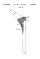
- FIG. 1 is an anterior elevational view of the hip implant prosthesis of the present invention:
- FIG. 2 is a cross-sectional view taken along line 2--2 of FIG. 1:
- FIG. 3 is a cross-sectional view taken along line 3--3 of FIG. 1;
- FIG. 4 is a magnified anterior elevational view of the proximal locking zone
- FIG. 5 is a magnified lateral elevational view of the proximal locking zone
- FIG. 6 is a lateral elevational view of the hip implant prosthesis of the present invention.
- FIG. 7 is an anterior elevational view of an alternative embodiment of the present invention without a plurality of beads
- FIG. 8 is an anterior elevational view of the embodiment shown in FIG. 7 with a plurality of beads attached to the proximal locking surface;
- FIG. 9 is a fragmentary view of the proximal end of a femur showing the neck and head thereof in dashed lines, representing the portion of the femur resected in order to implant the prosthesis of the present invention
- FIG. 10 is an elevational view of the cylindrical reamer
- FIG. 11 is a fragmentary cross-sectional view of the proximal end of the femur illustrating the cylindrical reaming tool of FIG. 10;
- FIG. 12 is a cross-sectional view similar to that of FIG. 11 showing the bore in the medullary canal following the reaming with the cylindrical reamer:
- FIG. 13 is a cross-sectional view similar to FIG. 11 but illustrating the use of a rasp to set the initial geometry of the bore for the prosthesis:
- FIG. 14 is a cross-sectional view similar to FIG. 12 but illustrating the geometry of the bore in the medullary canal following the step of reaming with the rasp;
- FIG. 15 is an elevational view of the rasp shown in FIG. 13;
- FIG. 16 is a cross-sectional view similar to FIG. 13 but illustrating the use of a trial implant to set the final geometry of the bore for the prosthesis;
- FIG. 17 is a cross-sectional view similar to FIG. 14 but illustrating the final geometry of the bore in the medullary canal following the step of inserting the trial implant;
- FIG. 18 is an elevational view of the trial stem implant and trial neck implant shown in FIG. 16;
- FIG. 19 is a cross-sectional view illustrating the prosthesis implanted into the bore formed in the medullary canal of the femur.
- proximal references to the portion of the prothesis positioned closest to the heart
- distal refers to the portion of the prothesis positioned furthest from the heart.
- anterior which references the portion of the prosthesis facing toward the front of the body
- posterior which references the portion of the prosthesis facing toward the rear of the body. It should be understood, however, that because the device may be used in either the right or left femur, the "anterior" portion of the prosthesis can also become the “posterior” portion of the prosthesis and visa versa.
- press-fit is defined as a mechanical engagement formed by two components, at least one of which is deformable, where the adjacent boundary lines of the two components overlap and interface with each other such that the two components must be forced into a position adjacent each other which produces a locking force developed over the area of contact between the two components.
- FIG. 1 there is shown a side elevational view of a prothesis according to the present invention designated by the numeral 10.
- the prothesis of FIG. 1 is designed as a femoral component of a hip prosthesis. It is understood, however, that the prothesis according to the present invention can be configured into any other type of implantable prosthetic device.
- the prosthesis of the present invention can be configured as a humeral component of a shoulder prosthesis.
- the prosthesis shown in FIG. 1 is configured as the femoral component of a hip prosthesis and is dimensioned to be press-fined within a bore formed in the femoral intramedullary canal.
- the prothesis generally includes a stem 12 having a proximal end 16 and a distal end 18, and a neck 36.
- the prosthesis 10 further includes a lateral side 25 and a medial side 27.
- the stem 12 defines a longitudinal axis 14 wherein the neck 36 extends medially, at approximately an angle 16 relative to the longitudinal axis 14, from the proximal end 17 of the stem 12.
- a spherically shaped driver recess 20 is formed in the proximal end 16 of stem 12.
- the driver recess 20 provides an area for the placement of an impaction tool (not shown) which is used to drive the prosthesis into the bore formed in the canal of the femur.
- a tapered portion 22 which defines a proximal locking zone 24 and a recessed medial pocket 50, both of which will be discussed in greater detail below.
- the tapered portion 22 extends distally from the proximal locking zone 24, merging into a cylindrical portion 26.
- the free end of the cylindrical portion 26 forms the distal end 18 of the stem 12.
- the distal end 18 of the stem 12 tapers down from the cylindrical portion 26 to a generally spherical tip portion 19.
- FIGS. 2 and 3 illustrate the changing cross-sectional shape of the tapered portion 22 at respective lines 2--2 and 3--3.
- the cross-sectional shape of the tapered portion 22 of stem 12 at line 2--2 presents a greater medial-lateral dimension 28 as compared with the overall anterior-posterior dimension 30 (as measured on the lateral side).
- the cross-sectional shape of the tapered portion 22 changes significantly such that the medial-lateral dimension 28 and anterior-posterior dimension 30 defines an almost circular cross-section at line 3--3, (FIG. 3) approaching the distal end of the tapered portion 22.
- the anterior-posterior width 30 adjacent the lateral side of the tapered portion 22 becomes substantially greater in width, moving proximally, than the corresponding anterior-posterior width 32 adjacent the medial side.
- the neck 36 of the prosthesis 10 is tapered and includes a peg portion 38.
- the proximal end of the stem 12 merges into the distal end of neck 36 forming a first circumferential fillet at 40.
- the neck 36 converges proximally as it merges into the tapered portion 38 forming a second circumferential fillet at 42.
- the tapered portion 38 is slightly tapered in the distal to proximal direction and includes a chamfer 44 at the proximal end of the tapered portion 38.
- the tapered portion 38 is adapted to receive the femoral bearing head portion of the prosthesis (not shown).
- proximal locking zone 24 portion of the stem 12.
- the proximal locking zone 24 is comprised of four proximal locking surfaces 46A, 46B, 46C, and 46D which extend circumferentially about the proximal portion of the tapered portion 22 of stem 12.
- Proximal locking surfaces 46A, 46B, 46C and 46D step down at circumferential step 48 to meet the remaining portion of tapered portion 22.
- Locking surface 46A extends between locking surfaces 46B and 46C (not visible) and presents a conically shaped surface on the lateral side of the prosthesis.
- Locking surface 46D extends between locking surfaces 46B and 46C and presents a curved surface on the medial side of the prosthesis.
- Locking surfaces 46B and 46C are planar and are located respectively on the anterior and posterior sides of the prosthesis.
- the anteriorly and posteriorly located locking surfaces 46B and 46C each lie in a plane that defines a locking angle 52 which ranges between 5 and 10 degrees and preferably 7 degrees relative to longitudinal axis 14, (see also FIG. 5). Angle 52 is best illustrated in FIG. 5 where the prosthesis has been rotated 4.3° about the longitudinal axis 14.
- the conically shaped surface presented by the laterally located locking surface 46A tapers at the same locking angle 52 as locking surfaces 46B and 46C, i.e., between 5 and 10 degrees and preferably 7 degrees relative to longitudinal axis 14 as stated above.
- the curved surface presented by the medially located locking surface 46D follows the general taper of the medial portion 22 of the stem segment and presents a slightly proud surface. This configuration provides an uninterrupted continuum of locking surfaces 46B and 46C.
- FIG. 6 there is shown the lateral side of the prothesis of the present invention.
- FIG. 6 clearly illustrates the angle defined by proximal locking surfaces 46B and 46C and the anterior-posterior tapering of the remaining portion of the tapered portion 22.
- the prosthesis shown in FIGS. 1-6 is intended to be implanted by a press-fit engagement which does not utilize a cement for locking into place.
- a layer(s) of bio-active material or "synthetic bone” material is deposited on the proximal locking surfaces 46A, 46B, 46C, and 46D which allow the prosthesis to become permanently attached via a mechanism which includes growth of the natural bone tissue within the cortex of the bone into the layer(s) of bio-active material.
- the synthetic bone material is generally plasma-sprayed on the prothesis.
- the preferred choice of synthetic bone material can be any well known ceramic comprising at least one artificial apatite in the from of hydroxyapatite (Ca5OH(PO4)2). This material can be applied in multiple layers and the layers themselves can be composed of different materials.
- the layer of synthetic bone is designed to be highly compatible with natural bone tissue.
- FIG. 7 there is shown an alternative embodiment of the present invention designated by the numeral 60.
- the embodiment of FIG. 7 is substantially identical to the embodiment of FIGS. 1-6 except that proximal locking surfaces 62A, 62B, 62C (not visible), and 62D are recessed and bounded by a circumferentially extending proximal ridge 64 and a circumferentially extending distal ridge 66.
- the proximal locking surfaces are recessed to receive a plurality of beads 67 which act like a porous coating for receiving the ingrowth of the natural bone tissue.
- the beads can be made from any suitable material such as cobalt chromium or the like.
- the beads are bonded to the proximal locking surfaces 62A, 62B, 62C, and 62D by sintering as shown in FIG. 8.
- the medial pocket 50 can also receive a coating of beads if desired, as shown at 68 in FIG. 8, in order to further provide for the ingrowth of natural bone tissue.
- the prosthesis of the present invention can be manufactured from titanium alloy, cobalt-chromium alloy or any other suitable material well known in the art.
- the prosthesis can be made by forging, casting and/or machining operations or any other well known technique.
- FIGS. 9-19 the apparatus employed in the method of implanting the prosthesis of the present invention is illustrated. Each tool of the apparatus will be explained in connection with the description of the method in which the apparatus is used, which follows.
- the proximal end of the femur 70 is presented and the femoral neck 72 is removed using a femoral template (not shown) for determining the area to resect.
- the first cylindrical reamer 82 is inserted in the postero-lateral portion of the resected surface of the femoral neck 72 and enters the medullary canal 74 to form a bore 76 as shown in FIGS. 11 and 12.
- the cutting flutes 84 of the first cylindrical reamer 82 align the reamer within the medullary canal 74.
- Successively greater diameter reamers may be used until the desired diameter bore is achieved which is intended to create a line to line fit with the implant stem.
- Proximal shaping of the bore 76 for the geometry of the stem of the prosthesis is implemented as shown in FIGS. 13 and 14 using a tapered rasp 86 having circumferential cutting teeth 88.
- the tapered rasp 86 has a profile which is substantially like that of the prosthesis but without the flare characterized by proximal locking surfaces 46A, 46B, and 46C of prosthesis 10.
- the tapered rasp 86 is inserted and driven into bore 76 as shown in FIG. 13.
- the neck 92 and stem 94 of the trial stem implant 90 which are manufactured as separate components, each have a shape and profile which is substantially identical to that of the prosthesis.
- the trial stem implant 90 includes a proximal locking zone 96 which includes a proximal locking surface 98 with a knurled finish 100 which is designed to prepare the proximal region 78 of the bore 76 for the proximal locking surface of the prosthesis.
- the trial implant 90 is inserted and driven into the bore 76 as shown in FIG. 16. The illustration of FIG.
- FIG. 17 shows how the proximal locking surface 96 of the trial implant crushes the bone in the proximal region 78 of bore 76 and provides a slightly undersized dimension relative to the prosthesis. This results in a press-fit engagement between the prosthesis and the bore 76 when the prosthesis is driven into the medullary canal 74.
- the prosthesis 10 is ready to be implanted. As illustrated in FIG. 19, the prosthesis 10 is inserted into the bore 76 and driven into the bore 76 using an impaction tool (not shown) the end of which is placed into the driver recess 20. The prosthesis 10 is driven into the bore 76 until the proximal locking surface is firmly engaged with the proximal region 78.
- the improved proximal locking zone of the present invention initially loads the proximal-most portion of the bone and greatly compresses the bone thereat. This provides for densification of bone about the stem, greater resistance to subsidence, and greater resistance to torsional forces. In addition, the densification of bone potentially inhibits the transfer mechanism for implant debris to the boundary surrounding the more distal implant/bone interface. A reduction in the incidence of lysis is also a potential benefit.
- the present invention is a hip prothesis, as described herein, the present invention is not limited to uses as a hip prothesis and consequently, may be used as a prosthetic implant of any type. Further, while the present invention is particularly well suited for press-fit implantation, the present invention is not limited to such, and therefore, embodies implantation in any suitable manner. Further, other methods of creating a porous surface on the prosthesis for providing bone ingrowth may be employed and can include porous fiber metal structures such as metal meshes or porous pads which are wrapped around the prosthesis. These and other such methods are contemplated in the present invention for the purpose of enhancing the fixation of the prosthesis within the femur. Any variations or modifications to the invention described herein are intended to be included within the scope of the invention as defined by the appended claims.
Abstract
Description
Claims (6)
Priority Applications (1)
| Application Number | Priority Date | Filing Date | Title |
|---|---|---|---|
| US08/435,893 US5593451A (en) | 1994-06-01 | 1995-05-05 | Prosthetic device and method of implantation |
Applications Claiming Priority (2)
| Application Number | Priority Date | Filing Date | Title |
|---|---|---|---|
| US25245094A | 1994-06-01 | 1994-06-01 | |
| US08/435,893 US5593451A (en) | 1994-06-01 | 1995-05-05 | Prosthetic device and method of implantation |
Related Parent Applications (1)
| Application Number | Title | Priority Date | Filing Date |
|---|---|---|---|
| US25245094A Division | 1994-06-01 | 1994-06-01 |
Publications (1)
| Publication Number | Publication Date |
|---|---|
| US5593451A true US5593451A (en) | 1997-01-14 |
Family
ID=22956059
Family Applications (3)
| Application Number | Title | Priority Date | Filing Date |
|---|---|---|---|
| US08/435,893 Expired - Lifetime US5593451A (en) | 1994-06-01 | 1995-05-05 | Prosthetic device and method of implantation |
| US08/636,727 Expired - Lifetime US5702487A (en) | 1994-06-01 | 1996-04-23 | Prosthetic device |
| US08/844,443 Expired - Lifetime US5863295A (en) | 1994-06-01 | 1997-04-18 | Prosthetic device and method of implantation |
Family Applications After (2)
| Application Number | Title | Priority Date | Filing Date |
|---|---|---|---|
| US08/636,727 Expired - Lifetime US5702487A (en) | 1994-06-01 | 1996-04-23 | Prosthetic device |
| US08/844,443 Expired - Lifetime US5863295A (en) | 1994-06-01 | 1997-04-18 | Prosthetic device and method of implantation |
Country Status (1)
| Country | Link |
|---|---|
| US (3) | US5593451A (en) |
Cited By (46)
| Publication number | Priority date | Publication date | Assignee | Title |
|---|---|---|---|---|
| US6203575B1 (en) * | 1998-01-16 | 2001-03-20 | Sulzer Orthopaedie Ag | Modular system for shaft prostheses |
| WO2000072785A3 (en) * | 1999-05-27 | 2001-04-05 | Sotereanos Nicholas G | Proximal femoral replacement implant |
| WO2000059410A3 (en) * | 1999-04-07 | 2001-04-26 | Plus Endoprothetik Ag | Flat shaft of a hip joint prosthesis for anchoring in the femur |
| US20030125810A1 (en) * | 1996-08-21 | 2003-07-03 | Sullivan John Martin Patrick | Joint replacement prosthesis |
| US6616697B2 (en) | 2001-03-13 | 2003-09-09 | Nicholas G. Sotereanos | Hip implant assembly |
| US20030171819A1 (en) * | 2002-03-11 | 2003-09-11 | Sotereanos Nicholas G. | Modular hip implants |
| US20030220696A1 (en) * | 2002-05-23 | 2003-11-27 | Levine David Jerome | Implantable porous metal |
| US20060190092A1 (en) * | 2005-02-22 | 2006-08-24 | Natalia Fridshtand | Hip stem prosthesis |
| US20060206212A1 (en) * | 1999-04-13 | 2006-09-14 | Karl Zweymuller | Leaflike shaft of a hip-joint prosthesis for anchoring in the femur |
| US20060235539A1 (en) * | 2003-02-03 | 2006-10-19 | Gordon Blunn | Surgical kit for hemiarthroplasty hip replacement |
| US20060241776A1 (en) * | 2005-04-21 | 2006-10-26 | Biomet Manufacturing Corp. | Method and apparatus for use of porous implants |
| US20060241781A1 (en) * | 2005-04-21 | 2006-10-26 | Biomet Manufacturing Corp. | Method and apparatus for use of porous implants |
| US20060264952A1 (en) * | 2005-05-18 | 2006-11-23 | Nelson Charles L | Methods of Using Minimally Invasive Actuable Bone Fixation Devices |
| US20060276906A1 (en) * | 2005-02-18 | 2006-12-07 | Hoag Stephen H | Fully porous prosthetic hip stem |
| US20060276904A1 (en) * | 2000-04-13 | 2006-12-07 | Karl Zweymuller | Leaflike shaft of a hip-joint prosthesis for anchoring in the femur |
| US20070173948A1 (en) * | 2005-04-21 | 2007-07-26 | Biomet Manufacturing Corp. | Porous metal cup with cobalt bearing surface |
| US20070196230A1 (en) * | 2006-02-17 | 2007-08-23 | Biomet Manufacturing Corp. | Method and apparatus for forming porous metal implants |
| US20070219641A1 (en) * | 2006-03-20 | 2007-09-20 | Zimmer Technology, Inc. | Prosthetic hip implants |
| US20080132896A1 (en) * | 2006-11-22 | 2008-06-05 | Sonoma Orthopedic Products, Inc. | Curved orthopedic tool |
| US20080140078A1 (en) * | 2006-11-22 | 2008-06-12 | Sonoma Orthopedic Products, Inc. | Surgical tools for use in deploying bone repair devices |
| US20080147187A1 (en) * | 2005-04-21 | 2008-06-19 | Biomet Manufacturing Corp. | Method And Apparatus For Use Of Porous Implants |
| US20080149115A1 (en) * | 2006-11-22 | 2008-06-26 | Sonoma Orthopedic Products, Inc. | Surgical station for orthopedic reconstruction surgery |
| US20080161805A1 (en) * | 2006-11-22 | 2008-07-03 | Sonoma Orthopedic Products, Inc. | Fracture fixation device, tools and methods |
| US20080167723A1 (en) * | 2006-03-20 | 2008-07-10 | Zimmer, Inc. | Prosthetic hip implants |
| US20080167722A1 (en) * | 2007-01-10 | 2008-07-10 | Biomet Manufacturing Corp. | Knee Joint Prosthesis System and Method for Implantation |
| US20080200990A1 (en) * | 2007-02-16 | 2008-08-21 | Mctighe Timothy | Tissue sparing implant |
| US20090084491A1 (en) * | 2007-09-25 | 2009-04-02 | Biomet Manufacturing Corp. | Cementless Tibial Tray |
| US20090149964A1 (en) * | 2007-10-10 | 2009-06-11 | Biomet Manufacturing Corp. | Knee joint prosthesis system and method for implantation |
| US20090299482A1 (en) * | 2007-01-10 | 2009-12-03 | Biomet Manufacturing Corp. | Knee Joint Prosthesis System and Method for Implantation |
| US20100094347A1 (en) * | 2005-05-18 | 2010-04-15 | Nelson Charles L | Fracture fixation device, tools and methods |
| US20110166665A1 (en) * | 2006-12-07 | 2011-07-07 | Anatol Podolsky | Methods and systems for total hip replacement |
| US8021432B2 (en) | 2005-12-05 | 2011-09-20 | Biomet Manufacturing Corp. | Apparatus for use of porous implants |
| US8123814B2 (en) | 2001-02-23 | 2012-02-28 | Biomet Manufacturing Corp. | Method and appartus for acetabular reconstruction |
| US8187280B2 (en) | 2007-10-10 | 2012-05-29 | Biomet Manufacturing Corp. | Knee joint prosthesis system and method for implantation |
| US8328873B2 (en) | 2007-01-10 | 2012-12-11 | Biomet Manufacturing Corp. | Knee joint prosthesis system and method for implantation |
| US20130030546A1 (en) * | 2010-03-17 | 2013-01-31 | Kabushiki Kaisha B. I. Tec | Stem Structure For Composite Prosthetic Hip And Method For Manufacturing The Same |
| US8579985B2 (en) | 2006-12-07 | 2013-11-12 | Ihip Surgical, Llc | Method and apparatus for hip replacement |
| US8961516B2 (en) | 2005-05-18 | 2015-02-24 | Sonoma Orthopedic Products, Inc. | Straight intramedullary fracture fixation devices and methods |
| US8974540B2 (en) | 2006-12-07 | 2015-03-10 | Ihip Surgical, Llc | Method and apparatus for attachment in a modular hip replacement or fracture fixation device |
| US9060820B2 (en) | 2005-05-18 | 2015-06-23 | Sonoma Orthopedic Products, Inc. | Segmented intramedullary fracture fixation devices and methods |
| US9155574B2 (en) | 2006-05-17 | 2015-10-13 | Sonoma Orthopedic Products, Inc. | Bone fixation device, tools and methods |
| EP3095419A1 (en) * | 2011-12-16 | 2016-11-23 | Chow IP, LLC | A femoral hip prosthesis |
| US9770278B2 (en) | 2014-01-17 | 2017-09-26 | Arthrex, Inc. | Dual tip guide wire |
| US9814499B2 (en) | 2014-09-30 | 2017-11-14 | Arthrex, Inc. | Intramedullary fracture fixation devices and methods |
| RU2673980C1 (en) * | 2018-05-10 | 2018-12-03 | Денис Игоревич Варфоломеев | Set for hip replacement |
| US10695076B2 (en) | 2017-03-29 | 2020-06-30 | Depuy Ireland Unlimited Company | Guided osteotome |
Families Citing this family (42)
| Publication number | Priority date | Publication date | Assignee | Title |
|---|---|---|---|---|
| US6425922B1 (en) | 2000-01-30 | 2002-07-30 | Diamicron, Inc. | Prosthetic hip joint having at least one sintered polycrystalline diamond compact articulation surface |
| US6494918B1 (en) | 2000-01-30 | 2002-12-17 | Diamicron, Inc. | Component for a prosthetic joint having a diamond load bearing and articulation surface |
| US6793681B1 (en) | 1994-08-12 | 2004-09-21 | Diamicron, Inc. | Prosthetic hip joint having a polycrystalline diamond articulation surface and a plurality of substrate layers |
| US6402787B1 (en) | 2000-01-30 | 2002-06-11 | Bill J. Pope | Prosthetic hip joint having at least one sintered polycrystalline diamond compact articulation surface and substrate surface topographical features in said polycrystalline diamond compact |
| US6290726B1 (en) | 2000-01-30 | 2001-09-18 | Diamicron, Inc. | Prosthetic hip joint having sintered polycrystalline diamond compact articulation surfaces |
| US6596225B1 (en) | 2000-01-31 | 2003-07-22 | Diamicron, Inc. | Methods for manufacturing a diamond prosthetic joint component |
| US6676704B1 (en) | 1994-08-12 | 2004-01-13 | Diamicron, Inc. | Prosthetic joint component having at least one sintered polycrystalline diamond compact articulation surface and substrate surface topographical features in said polycrystalline diamond compact |
| US6410877B1 (en) | 2000-01-30 | 2002-06-25 | Diamicron, Inc. | Methods for shaping and finishing prosthetic joint components including polycrystalline diamond compacts |
| US6709463B1 (en) | 2000-01-30 | 2004-03-23 | Diamicron, Inc. | Prosthetic joint component having at least one solid polycrystalline diamond component |
| US6488715B1 (en) | 2000-01-30 | 2002-12-03 | Diamicron, Inc. | Diamond-surfaced cup for use in a prosthetic joint |
| US6913623B1 (en) | 2000-08-15 | 2005-07-05 | Centerpulse Orthopedics, Inc. | Two piecefused femoral hip stem |
| ES2242490B1 (en) * | 2003-04-01 | 2007-02-01 | Biomet Spain Orthopaedics, S.L. | PROTESICAL DEVICE. |
| US8998919B2 (en) | 2003-06-25 | 2015-04-07 | DePuy Synthes Products, LLC | Assembly tool for modular implants, kit and associated method |
| US7074224B2 (en) * | 2003-06-25 | 2006-07-11 | Depuy Products, Inc. | Modular tapered reamer for bone preparation and associated method |
| US7297166B2 (en) | 2003-06-25 | 2007-11-20 | Depuy Products, Inc. | Assembly tool for modular implants and associated method |
| US7582092B2 (en) | 2003-06-25 | 2009-09-01 | Depuy Products, Inc. | Assembly tool for modular implants and associated method |
| US7785328B2 (en) * | 2003-12-30 | 2010-08-31 | Depuy Products, Inc. | Minimally invasive bone miller apparatus |
| US7534271B2 (en) | 2004-01-22 | 2009-05-19 | Smith + Nephew | Femoral hip prosthesis and method of implantation |
| US20050272153A1 (en) | 2004-01-27 | 2005-12-08 | Zou Xuenong | Bone tissue engineering by ex vivo stem cells ongrowth into three-dimensional trabecular metal |
| US8277457B1 (en) | 2004-12-09 | 2012-10-02 | Greatbatch Medical S.A. | Orthopaedic inserter using a collet mechanism |
| FR2887761B1 (en) * | 2005-06-29 | 2007-09-14 | Zimmer France Soc Par Actions | FIRST INITIAL FEMORAL PROSTHESIS RANGE AND DEVICE FOR PREPARING AN ASSOCIATED FEMALE FUTURE. |
| DE102007020484A1 (en) * | 2006-05-01 | 2007-12-13 | Precimed S.A. | Insertion instrument for minimally invasive joint surgery with exchangeable thread |
| US8597298B2 (en) * | 2006-09-29 | 2013-12-03 | DePuy Synthes Products, LLC | Proximal reamer |
| US8556912B2 (en) | 2007-10-30 | 2013-10-15 | DePuy Synthes Products, LLC | Taper disengagement tool |
| US8518050B2 (en) | 2007-10-31 | 2013-08-27 | DePuy Synthes Products, LLC | Modular taper assembly device |
| US8348953B2 (en) * | 2008-01-10 | 2013-01-08 | Peter Brehm | Method and apparatus for impacting bone material |
| US8167882B2 (en) | 2008-09-30 | 2012-05-01 | Depuy Products, Inc. | Minimally invasive bone miller apparatus |
| US8398650B1 (en) | 2009-01-27 | 2013-03-19 | Greatbatch Medical S.A. | Offset cup impactor with an expandable dome for double mobility implants |
| US8533921B2 (en) | 2010-06-15 | 2013-09-17 | DePuy Synthes Products, LLC | Spiral assembly tool |
| EP2422754B1 (en) | 2010-08-27 | 2014-11-05 | Greatbatch Medical SA | Offset cup impactor with a grasping plate for double mobility implants |
| US9095452B2 (en) | 2010-09-01 | 2015-08-04 | DePuy Synthes Products, Inc. | Disassembly tool |
| US9119731B2 (en) | 2011-01-17 | 2015-09-01 | Greatbach Medical S.A. | Straight cup impactor |
| EP2476397B1 (en) | 2011-01-17 | 2015-10-21 | Greatbatch Medical SA | Straight cup impactor with lever arm |
| CN106974699B (en) | 2011-04-06 | 2019-07-09 | 德普伊新特斯产品有限责任公司 | The device assembly of implantation amendment hip prosthesis |
| EP2561835B1 (en) | 2011-08-26 | 2016-03-16 | Greatbatch Medical SA | Straight cup impactor |
| US9028502B2 (en) | 2011-09-23 | 2015-05-12 | Greatbatch Medical S.A. | Ceramic implant holder |
| US11666447B1 (en) | 2015-03-05 | 2023-06-06 | Taq Ortho, LLC | Bone implant augment and offset device |
| US11678917B1 (en) | 2015-03-05 | 2023-06-20 | Taq Ortho, LLC | Bone insert augment and offset method |
| US10441297B2 (en) | 2015-05-29 | 2019-10-15 | Zimmer, Inc. | Sounder for sizing bone implant |
| US10537661B2 (en) | 2017-03-28 | 2020-01-21 | DePuy Synthes Products, Inc. | Orthopedic implant having a crystalline calcium phosphate coating and methods for making the same |
| US10537658B2 (en) | 2017-03-28 | 2020-01-21 | DePuy Synthes Products, Inc. | Orthopedic implant having a crystalline gallium-containing hydroxyapatite coating and methods for making the same |
| WO2023034019A1 (en) * | 2021-09-01 | 2023-03-09 | Trauner Kenneth B | Bone insert augment and offset device |
Citations (22)
| Publication number | Priority date | Publication date | Assignee | Title |
|---|---|---|---|---|
| US4064567A (en) * | 1976-09-15 | 1977-12-27 | The Sampson Corporation | Prosthesis-to-bone interface system |
| US4514865A (en) * | 1982-04-19 | 1985-05-07 | Harris William H | Stemmed femoral component for the human hip |
| US4530116A (en) * | 1982-10-15 | 1985-07-23 | Sulzer Brothers Limited | Anchoring shank for a bone implant |
| US4536894A (en) * | 1983-08-04 | 1985-08-27 | Galante Jorge O | Hip prosthesis with flared porous bony ingrowth pads |
| US4549319A (en) * | 1982-08-03 | 1985-10-29 | United States Medical Corporation | Artificial joint fixation to bone |
| US4608053A (en) * | 1982-05-03 | 1986-08-26 | Waldemar Link Gmbh & Co. | Femoral hip prosthesis |
| US4718915A (en) * | 1985-03-27 | 1988-01-12 | Epinette Jean Alain | Femoral component of a hip prosthesis |
| US4955912A (en) * | 1987-11-28 | 1990-09-11 | Metalpraecis Berchem+Schaberg Gesellschaft Fur Metallformgebung Mit Beschrankter Haftung | Joint prosthesis |
| US4979958A (en) * | 1988-09-29 | 1990-12-25 | Aisin Seiki Kabushiki Kaisha | Artificial stem for femur of coxa |
| US4983183A (en) * | 1989-02-06 | 1991-01-08 | Horowitz Stephen M | Hip prosthesis and method for implanting the same |
| US5007931A (en) * | 1990-05-04 | 1991-04-16 | Boehringer Mannheim Corporation | Porous coated prosthesis |
| US5018285A (en) * | 1987-08-24 | 1991-05-28 | Zimmer, Inc. | Method of constructing prosthetic implant with wrapped porous surface |
| US5035717A (en) * | 1988-05-12 | 1991-07-30 | Brooks Peter J | Insert and method of using same |
| US5061287A (en) * | 1989-02-13 | 1991-10-29 | Feiler Frederic C | Proximal cement sealing plug for hip prosthesis |
| US5092900A (en) * | 1990-03-13 | 1992-03-03 | Sulzer Brothers Limited | Femur head prosthesis |
| US5092899A (en) * | 1988-03-21 | 1992-03-03 | Mark Forte | Prosthesis with flexible intramedullary stem |
| US5108452A (en) * | 1989-02-08 | 1992-04-28 | Smith & Nephew Richards Inc. | Modular hip prosthesis |
| US5108437A (en) * | 1988-09-14 | 1992-04-28 | Pfizer Hospital Products Group, Inc. | Modular prosthesis |
| US5171275A (en) * | 1990-02-15 | 1992-12-15 | Ling Robin S M | Femoral stem prosthesis |
| US5201767A (en) * | 1991-07-15 | 1993-04-13 | Johnson & Johnson Orthopaedics, Inc. | Fluted-wedge osteal prosthetic component |
| US5211666A (en) * | 1991-04-22 | 1993-05-18 | New York University | Hip joint femoral component endoprosthesis with a lateral load-transferring support surface |
| US5258035A (en) * | 1992-05-29 | 1993-11-02 | Intermedics Orthopedics, Inc. | Femoral prosthesis with wedge having opposed tapers |
Family Cites Families (10)
| Publication number | Priority date | Publication date | Assignee | Title |
|---|---|---|---|---|
| GB8401059D0 (en) * | 1984-01-16 | 1984-02-15 | Exeter University Of | Fixation of implants in bone |
| DE3414924A1 (en) * | 1984-04-19 | 1985-10-31 | Klaus Dr.med. Dr.med.habil. 8000 München Draenert | COATED ANCHORAGE PART FOR IMPLANTS |
| IT1202437B (en) * | 1987-01-28 | 1989-02-09 | Cremascoli Spa G | STRUCTURE OF TOTAL ANCHOR PROSTHESIS, INCLUDING A FEMORAL COMPONENT AND AN ACETABULAR COMPONENT, REALIZED, BOTH, PART IN METAL MATERIAL AND PART IN CERAMIC MATERIAL |
| US4851007A (en) * | 1988-03-18 | 1989-07-25 | Gray Frank B | Femoral component for a hip prosthesis |
| US5147408A (en) * | 1988-10-07 | 1992-09-15 | Pfizer Hospital Products Group, Inc. | Prosthetic device and method of implantation |
| US5108405A (en) * | 1990-08-10 | 1992-04-28 | Mikhail Michael W E | System for performing hip prosethesis revision surgery |
| US5258030A (en) * | 1991-07-08 | 1993-11-02 | The Trustees Of The University Of Pennsylvania | Porous coated implants |
| US5169401A (en) * | 1991-12-20 | 1992-12-08 | Zimmer, Inc. | Surgical reamer assembly |
| US5342363A (en) * | 1992-11-30 | 1994-08-30 | Wright Medical Technology, Inc. | Medical instrument and procedure |
| US5376124A (en) * | 1993-08-03 | 1994-12-27 | Intermedics Orthopedics, Inc. | Collared hip prosthesis with revision spacer |
-
1995
- 1995-05-05 US US08/435,893 patent/US5593451A/en not_active Expired - Lifetime
-
1996
- 1996-04-23 US US08/636,727 patent/US5702487A/en not_active Expired - Lifetime
-
1997
- 1997-04-18 US US08/844,443 patent/US5863295A/en not_active Expired - Lifetime
Patent Citations (22)
| Publication number | Priority date | Publication date | Assignee | Title |
|---|---|---|---|---|
| US4064567A (en) * | 1976-09-15 | 1977-12-27 | The Sampson Corporation | Prosthesis-to-bone interface system |
| US4514865A (en) * | 1982-04-19 | 1985-05-07 | Harris William H | Stemmed femoral component for the human hip |
| US4608053A (en) * | 1982-05-03 | 1986-08-26 | Waldemar Link Gmbh & Co. | Femoral hip prosthesis |
| US4549319A (en) * | 1982-08-03 | 1985-10-29 | United States Medical Corporation | Artificial joint fixation to bone |
| US4530116A (en) * | 1982-10-15 | 1985-07-23 | Sulzer Brothers Limited | Anchoring shank for a bone implant |
| US4536894A (en) * | 1983-08-04 | 1985-08-27 | Galante Jorge O | Hip prosthesis with flared porous bony ingrowth pads |
| US4718915A (en) * | 1985-03-27 | 1988-01-12 | Epinette Jean Alain | Femoral component of a hip prosthesis |
| US5018285A (en) * | 1987-08-24 | 1991-05-28 | Zimmer, Inc. | Method of constructing prosthetic implant with wrapped porous surface |
| US4955912A (en) * | 1987-11-28 | 1990-09-11 | Metalpraecis Berchem+Schaberg Gesellschaft Fur Metallformgebung Mit Beschrankter Haftung | Joint prosthesis |
| US5092899A (en) * | 1988-03-21 | 1992-03-03 | Mark Forte | Prosthesis with flexible intramedullary stem |
| US5035717A (en) * | 1988-05-12 | 1991-07-30 | Brooks Peter J | Insert and method of using same |
| US5108437A (en) * | 1988-09-14 | 1992-04-28 | Pfizer Hospital Products Group, Inc. | Modular prosthesis |
| US4979958A (en) * | 1988-09-29 | 1990-12-25 | Aisin Seiki Kabushiki Kaisha | Artificial stem for femur of coxa |
| US4983183A (en) * | 1989-02-06 | 1991-01-08 | Horowitz Stephen M | Hip prosthesis and method for implanting the same |
| US5108452A (en) * | 1989-02-08 | 1992-04-28 | Smith & Nephew Richards Inc. | Modular hip prosthesis |
| US5061287A (en) * | 1989-02-13 | 1991-10-29 | Feiler Frederic C | Proximal cement sealing plug for hip prosthesis |
| US5171275A (en) * | 1990-02-15 | 1992-12-15 | Ling Robin S M | Femoral stem prosthesis |
| US5092900A (en) * | 1990-03-13 | 1992-03-03 | Sulzer Brothers Limited | Femur head prosthesis |
| US5007931A (en) * | 1990-05-04 | 1991-04-16 | Boehringer Mannheim Corporation | Porous coated prosthesis |
| US5211666A (en) * | 1991-04-22 | 1993-05-18 | New York University | Hip joint femoral component endoprosthesis with a lateral load-transferring support surface |
| US5201767A (en) * | 1991-07-15 | 1993-04-13 | Johnson & Johnson Orthopaedics, Inc. | Fluted-wedge osteal prosthetic component |
| US5258035A (en) * | 1992-05-29 | 1993-11-02 | Intermedics Orthopedics, Inc. | Femoral prosthesis with wedge having opposed tapers |
Cited By (93)
| Publication number | Priority date | Publication date | Assignee | Title |
|---|---|---|---|---|
| US20030125810A1 (en) * | 1996-08-21 | 2003-07-03 | Sullivan John Martin Patrick | Joint replacement prosthesis |
| US6203575B1 (en) * | 1998-01-16 | 2001-03-20 | Sulzer Orthopaedie Ag | Modular system for shaft prostheses |
| WO2000059410A3 (en) * | 1999-04-07 | 2001-04-26 | Plus Endoprothetik Ag | Flat shaft of a hip joint prosthesis for anchoring in the femur |
| US7497875B1 (en) * | 1999-04-07 | 2009-03-03 | Smith & Nephew Orthopaedics Ag | Flat shaft of a hip-joint prosthesis for anchoring in the femur |
| US20060206212A1 (en) * | 1999-04-13 | 2006-09-14 | Karl Zweymuller | Leaflike shaft of a hip-joint prosthesis for anchoring in the femur |
| US7455693B2 (en) | 1999-04-13 | 2008-11-25 | Smith & Nephew Orthopaedics, Ag | Leaflike shaft of a hip-joint prosthesis for anchoring in the femur |
| WO2000072785A3 (en) * | 1999-05-27 | 2001-04-05 | Sotereanos Nicholas G | Proximal femoral replacement implant |
| US6284002B1 (en) | 1999-05-27 | 2001-09-04 | Nicholas G. Sotereanos | Proximal femoral replacement implant and method of implanting the same |
| US7494510B2 (en) | 2000-04-13 | 2009-02-24 | Smith And Nephew Orthopaedics Ag | Leaflike shaft of a hip-joint prosthesis for anchoring in the femur |
| US20060276904A1 (en) * | 2000-04-13 | 2006-12-07 | Karl Zweymuller | Leaflike shaft of a hip-joint prosthesis for anchoring in the femur |
| US8551181B2 (en) | 2001-02-23 | 2013-10-08 | Biomet Manufacturing, Llc | Method and apparatus for acetabular reconstruction |
| US9375316B2 (en) | 2001-02-23 | 2016-06-28 | Biomet Manufacturing, Llc. | Method and apparatus for acetabular reconstruction |
| US8123814B2 (en) | 2001-02-23 | 2012-02-28 | Biomet Manufacturing Corp. | Method and appartus for acetabular reconstruction |
| US6616697B2 (en) | 2001-03-13 | 2003-09-09 | Nicholas G. Sotereanos | Hip implant assembly |
| US7247171B2 (en) | 2002-03-11 | 2007-07-24 | Sotereanos Nicholas G | Modular hip implants |
| US20030171819A1 (en) * | 2002-03-11 | 2003-09-11 | Sotereanos Nicholas G. | Modular hip implants |
| US20030220696A1 (en) * | 2002-05-23 | 2003-11-27 | Levine David Jerome | Implantable porous metal |
| US20060235539A1 (en) * | 2003-02-03 | 2006-10-19 | Gordon Blunn | Surgical kit for hemiarthroplasty hip replacement |
| US10070961B2 (en) | 2005-02-18 | 2018-09-11 | Zimmer, Inc. | Fully porous prosthetic hip stem |
| US20060276906A1 (en) * | 2005-02-18 | 2006-12-07 | Hoag Stephen H | Fully porous prosthetic hip stem |
| US8206455B2 (en) * | 2005-02-22 | 2012-06-26 | Zimmer Technology, Inc. | Hip stem prosthesis |
| US7842096B2 (en) * | 2005-02-22 | 2010-11-30 | Zimmer Technology, Inc. | Hip stem prosthesis |
| US20060190092A1 (en) * | 2005-02-22 | 2006-08-24 | Natalia Fridshtand | Hip stem prosthesis |
| US10426623B2 (en) | 2005-02-22 | 2019-10-01 | Zimmer, Inc. | Hip stem prosthesis |
| US8858646B2 (en) | 2005-02-22 | 2014-10-14 | Zimmer, Inc. | Hip stem prosthesis |
| US20110009977A1 (en) * | 2005-02-22 | 2011-01-13 | Zimmer, Inc. | Hip stem prosthesis |
| US9636227B2 (en) | 2005-02-22 | 2017-05-02 | Zimmer, Inc. | Hip stem prosthesis |
| US8292967B2 (en) | 2005-04-21 | 2012-10-23 | Biomet Manufacturing Corp. | Method and apparatus for use of porous implants |
| US8066778B2 (en) | 2005-04-21 | 2011-11-29 | Biomet Manufacturing Corp. | Porous metal cup with cobalt bearing surface |
| US20080147187A1 (en) * | 2005-04-21 | 2008-06-19 | Biomet Manufacturing Corp. | Method And Apparatus For Use Of Porous Implants |
| US20070173948A1 (en) * | 2005-04-21 | 2007-07-26 | Biomet Manufacturing Corp. | Porous metal cup with cobalt bearing surface |
| US8197550B2 (en) | 2005-04-21 | 2012-06-12 | Biomet Manufacturing Corp. | Method and apparatus for use of porous implants |
| US20060241781A1 (en) * | 2005-04-21 | 2006-10-26 | Biomet Manufacturing Corp. | Method and apparatus for use of porous implants |
| US20060241776A1 (en) * | 2005-04-21 | 2006-10-26 | Biomet Manufacturing Corp. | Method and apparatus for use of porous implants |
| US8266780B2 (en) | 2005-04-21 | 2012-09-18 | Biomet Manufacturing Corp. | Method and apparatus for use of porous implants |
| US20060264952A1 (en) * | 2005-05-18 | 2006-11-23 | Nelson Charles L | Methods of Using Minimally Invasive Actuable Bone Fixation Devices |
| US7942875B2 (en) | 2005-05-18 | 2011-05-17 | Sonoma Orthopedic Products, Inc. | Methods of using minimally invasive actuable bone fixation devices |
| US20100094347A1 (en) * | 2005-05-18 | 2010-04-15 | Nelson Charles L | Fracture fixation device, tools and methods |
| US20060264951A1 (en) * | 2005-05-18 | 2006-11-23 | Nelson Charles L | Minimally Invasive Actuable Bone Fixation Devices Having a Retractable Interdigitation Process |
| US9060820B2 (en) | 2005-05-18 | 2015-06-23 | Sonoma Orthopedic Products, Inc. | Segmented intramedullary fracture fixation devices and methods |
| US7846162B2 (en) | 2005-05-18 | 2010-12-07 | Sonoma Orthopedic Products, Inc. | Minimally invasive actuable bone fixation devices |
| US20060264950A1 (en) * | 2005-05-18 | 2006-11-23 | Nelson Charles L | Minimally Invasive Actuable Bone Fixation Devices |
| US8287539B2 (en) | 2005-05-18 | 2012-10-16 | Sonoma Orthopedic Products, Inc. | Fracture fixation device, tools and methods |
| US7914533B2 (en) | 2005-05-18 | 2011-03-29 | Sonoma Orthopedic Products, Inc. | Minimally invasive actuable bone fixation devices |
| US8961516B2 (en) | 2005-05-18 | 2015-02-24 | Sonoma Orthopedic Products, Inc. | Straight intramedullary fracture fixation devices and methods |
| US20070233105A1 (en) * | 2005-05-18 | 2007-10-04 | Nelson Charles L | Minimally invasive actuable bone fixation devices |
| US8287541B2 (en) | 2005-05-18 | 2012-10-16 | Sonoma Orthopedic Products, Inc. | Fracture fixation device, tools and methods |
| US8021432B2 (en) | 2005-12-05 | 2011-09-20 | Biomet Manufacturing Corp. | Apparatus for use of porous implants |
| US20070196230A1 (en) * | 2006-02-17 | 2007-08-23 | Biomet Manufacturing Corp. | Method and apparatus for forming porous metal implants |
| US20110166668A1 (en) * | 2006-03-20 | 2011-07-07 | Zimmer, Inc. | Prosthetic hip implants |
| US20080167723A1 (en) * | 2006-03-20 | 2008-07-10 | Zimmer, Inc. | Prosthetic hip implants |
| US20070219641A1 (en) * | 2006-03-20 | 2007-09-20 | Zimmer Technology, Inc. | Prosthetic hip implants |
| US8088169B2 (en) | 2006-03-20 | 2012-01-03 | Dorr Lawrence D | Prosthetic hip implants |
| US20100222893A1 (en) * | 2006-03-20 | 2010-09-02 | Zimmer, Inc. | Prosthetic hip implants |
| US9155574B2 (en) | 2006-05-17 | 2015-10-13 | Sonoma Orthopedic Products, Inc. | Bone fixation device, tools and methods |
| US20110144645A1 (en) * | 2006-11-22 | 2011-06-16 | Sonoma Orthopedic Products, Inc. | Fracture Fixation Device, Tools and Methods |
| US8439917B2 (en) | 2006-11-22 | 2013-05-14 | Sonoma Orthopedic Products, Inc. | Fracture fixation device, tools and methods |
| US20080132896A1 (en) * | 2006-11-22 | 2008-06-05 | Sonoma Orthopedic Products, Inc. | Curved orthopedic tool |
| US20080140078A1 (en) * | 2006-11-22 | 2008-06-12 | Sonoma Orthopedic Products, Inc. | Surgical tools for use in deploying bone repair devices |
| US20080149115A1 (en) * | 2006-11-22 | 2008-06-26 | Sonoma Orthopedic Products, Inc. | Surgical station for orthopedic reconstruction surgery |
| US20080161805A1 (en) * | 2006-11-22 | 2008-07-03 | Sonoma Orthopedic Products, Inc. | Fracture fixation device, tools and methods |
| US9259250B2 (en) | 2006-11-22 | 2016-02-16 | Sonoma Orthopedic Products, Inc. | Fracture fixation device, tools and methods |
| US7909825B2 (en) | 2006-11-22 | 2011-03-22 | Sonoma Orthepedic Products, Inc. | Fracture fixation device, tools and methods |
| US8974540B2 (en) | 2006-12-07 | 2015-03-10 | Ihip Surgical, Llc | Method and apparatus for attachment in a modular hip replacement or fracture fixation device |
| US8795381B2 (en) | 2006-12-07 | 2014-08-05 | Ihip Surgical, Llc | Methods and systems for hip replacement |
| US9237949B2 (en) | 2006-12-07 | 2016-01-19 | Ihip Surgical, Llc | Method and apparatus for hip replacement |
| US8029573B2 (en) | 2006-12-07 | 2011-10-04 | Ihip Surgical, Llc | Method and apparatus for total hip replacement |
| US20110166665A1 (en) * | 2006-12-07 | 2011-07-07 | Anatol Podolsky | Methods and systems for total hip replacement |
| US8211183B2 (en) | 2006-12-07 | 2012-07-03 | Ihip Surgical, Llc | Methods and systems for total hip replacement |
| US8579985B2 (en) | 2006-12-07 | 2013-11-12 | Ihip Surgical, Llc | Method and apparatus for hip replacement |
| US20090299482A1 (en) * | 2007-01-10 | 2009-12-03 | Biomet Manufacturing Corp. | Knee Joint Prosthesis System and Method for Implantation |
| US8163028B2 (en) | 2007-01-10 | 2012-04-24 | Biomet Manufacturing Corp. | Knee joint prosthesis system and method for implantation |
| US8936648B2 (en) | 2007-01-10 | 2015-01-20 | Biomet Manufacturing, Llc | Knee joint prosthesis system and method for implantation |
| US8157869B2 (en) | 2007-01-10 | 2012-04-17 | Biomet Manufacturing Corp. | Knee joint prosthesis system and method for implantation |
| US8480751B2 (en) | 2007-01-10 | 2013-07-09 | Biomet Manufacturing, Llc | Knee joint prosthesis system and method for implantation |
| US20080167722A1 (en) * | 2007-01-10 | 2008-07-10 | Biomet Manufacturing Corp. | Knee Joint Prosthesis System and Method for Implantation |
| US8328873B2 (en) | 2007-01-10 | 2012-12-11 | Biomet Manufacturing Corp. | Knee joint prosthesis system and method for implantation |
| US20080200990A1 (en) * | 2007-02-16 | 2008-08-21 | Mctighe Timothy | Tissue sparing implant |
| US20090084491A1 (en) * | 2007-09-25 | 2009-04-02 | Biomet Manufacturing Corp. | Cementless Tibial Tray |
| US20090149964A1 (en) * | 2007-10-10 | 2009-06-11 | Biomet Manufacturing Corp. | Knee joint prosthesis system and method for implantation |
| US8562616B2 (en) | 2007-10-10 | 2013-10-22 | Biomet Manufacturing, Llc | Knee joint prosthesis system and method for implantation |
| US10736747B2 (en) | 2007-10-10 | 2020-08-11 | Biomet Manufacturing, Llc | Knee joint prosthesis system and method for implantation |
| US8187280B2 (en) | 2007-10-10 | 2012-05-29 | Biomet Manufacturing Corp. | Knee joint prosthesis system and method for implantation |
| US9763793B2 (en) | 2007-10-10 | 2017-09-19 | Biomet Manufacturing, Llc | Knee joint prosthesis system and method for implantation |
| US20130030546A1 (en) * | 2010-03-17 | 2013-01-31 | Kabushiki Kaisha B. I. Tec | Stem Structure For Composite Prosthetic Hip And Method For Manufacturing The Same |
| US9061090B2 (en) * | 2010-03-17 | 2015-06-23 | Kabushiki Kaisha B.I. Tec | Stem structure for composite prosthetic hip and method for manufacturing the same |
| US10070963B2 (en) | 2011-12-16 | 2018-09-11 | Chow Ip, Llc | Prosthetic femoral stem for use in high impact hip replacement |
| EP3095419A1 (en) * | 2011-12-16 | 2016-11-23 | Chow IP, LLC | A femoral hip prosthesis |
| US9770278B2 (en) | 2014-01-17 | 2017-09-26 | Arthrex, Inc. | Dual tip guide wire |
| US9814499B2 (en) | 2014-09-30 | 2017-11-14 | Arthrex, Inc. | Intramedullary fracture fixation devices and methods |
| US10548648B2 (en) | 2014-09-30 | 2020-02-04 | Arthrex, Inc. | Intramedullary fracture fixation devices and methods |
| US10695076B2 (en) | 2017-03-29 | 2020-06-30 | Depuy Ireland Unlimited Company | Guided osteotome |
| RU2673980C1 (en) * | 2018-05-10 | 2018-12-03 | Денис Игоревич Варфоломеев | Set for hip replacement |
Also Published As
| Publication number | Publication date |
|---|---|
| US5702487A (en) | 1997-12-30 |
| US5863295A (en) | 1999-01-26 |
Similar Documents
| Publication | Publication Date | Title |
|---|---|---|
| US5593451A (en) | Prosthetic device and method of implantation | |
| JP4838731B2 (en) | Femoral hip prosthesis and transplantation method | |
| US4536894A (en) | Hip prosthesis with flared porous bony ingrowth pads | |
| US5755805A (en) | Tapered prosthesis component | |
| US5725595A (en) | Cannulated cementless hip stem prosthesis | |
| EP1013241B1 (en) | Proximal femoral sleeve for a revision hip prosthesis | |
| US7214246B2 (en) | Prosthesis with feature aligned to trabeculae | |
| CA2205727C (en) | Asymmetric hip stem | |
| US5376124A (en) | Collared hip prosthesis with revision spacer | |
| US20040039451A1 (en) | Prosthetic stem with bearings | |
| US4871369A (en) | Long stem hip implant | |
| AU684653B2 (en) | Proximal conical stem | |
| US7494509B1 (en) | Method and apparatus for providing a short-stemmed hip prosthesis | |
| US6994731B2 (en) | Prosthetic implant | |
| EP0985385A1 (en) | Implantable prosthesis with bone engaging ribs | |
| WO2005117762A2 (en) | Canine femoral stem system | |
| JP2001037792A (en) | Artificial hip joint | |
| AU2011223978A1 (en) | Femoral hip prosthesis and method of implantation | |
| EP1464304A2 (en) | Prosthetic device |
Legal Events
| Date | Code | Title | Description |
|---|---|---|---|
| STCF | Information on status: patent grant |
Free format text: PATENTED CASE |
|
| FPAY | Fee payment |
Year of fee payment: 4 |
|
| FEPP | Fee payment procedure |
Free format text: PAYOR NUMBER ASSIGNED (ORIGINAL EVENT CODE: ASPN); ENTITY STATUS OF PATENT OWNER: LARGE ENTITY |
|
| FPAY | Fee payment |
Year of fee payment: 8 |
|
| AS | Assignment |
Owner name: ZIMMER TRABECULAR METAL TECHNOLOGY, INC., NEW JERS Free format text: CHANGE OF NAME;ASSIGNOR:IMPLEX CORPORATION;REEL/FRAME:015603/0082 Effective date: 20040603 |
|
| FEPP | Fee payment procedure |
Free format text: PAT HOLDER NO LONGER CLAIMS SMALL ENTITY STATUS, ENTITY STATUS SET TO UNDISCOUNTED (ORIGINAL EVENT CODE: STOL); ENTITY STATUS OF PATENT OWNER: LARGE ENTITY |
|
| FEPP | Fee payment procedure |
Free format text: ENTITY STATUS SET TO UNDISCOUNTED (ORIGINAL EVENT CODE: BIG.); ENTITY STATUS OF PATENT OWNER: LARGE ENTITY |
|
| FPAY | Fee payment |
Year of fee payment: 12 |
|
| REMI | Maintenance fee reminder mailed |