US5830670A - Neural thread protein gene expression and detection of Alzheimer's disease - Google Patents
Neural thread protein gene expression and detection of Alzheimer's disease Download PDFInfo
- Publication number
- US5830670A US5830670A US08/454,557 US45455795A US5830670A US 5830670 A US5830670 A US 5830670A US 45455795 A US45455795 A US 45455795A US 5830670 A US5830670 A US 5830670A
- Authority
- US
- United States
- Prior art keywords
- ntp
- protein
- antibody
- kda
- detecting
- Prior art date
- Legal status (The legal status is an assumption and is not a legal conclusion. Google has not performed a legal analysis and makes no representation as to the accuracy of the status listed.)
- Expired - Lifetime
Links
Images


Classifications
-
- C—CHEMISTRY; METALLURGY
- C07—ORGANIC CHEMISTRY
- C07K—PEPTIDES
- C07K14/00—Peptides having more than 20 amino acids; Gastrins; Somatostatins; Melanotropins; Derivatives thereof
- C07K14/435—Peptides having more than 20 amino acids; Gastrins; Somatostatins; Melanotropins; Derivatives thereof from animals; from humans
- C07K14/46—Peptides having more than 20 amino acids; Gastrins; Somatostatins; Melanotropins; Derivatives thereof from animals; from humans from vertebrates
- C07K14/47—Peptides having more than 20 amino acids; Gastrins; Somatostatins; Melanotropins; Derivatives thereof from animals; from humans from vertebrates from mammals
- C07K14/4701—Peptides having more than 20 amino acids; Gastrins; Somatostatins; Melanotropins; Derivatives thereof from animals; from humans from vertebrates from mammals not used
- C07K14/474—Pancreatic thread protein; Reg protein
-
- A—HUMAN NECESSITIES
- A61—MEDICAL OR VETERINARY SCIENCE; HYGIENE
- A61K—PREPARATIONS FOR MEDICAL, DENTAL OR TOILETRY PURPOSES
- A61K49/00—Preparations for testing in vivo
- A61K49/0004—Screening or testing of compounds for diagnosis of disorders, assessment of conditions, e.g. renal clearance, gastric emptying, testing for diabetes, allergy, rheuma, pancreas functions
- A61K49/0008—Screening agents using (non-human) animal models or transgenic animal models or chimeric hosts, e.g. Alzheimer disease animal model, transgenic model for heart failure
-
- C—CHEMISTRY; METALLURGY
- C07—ORGANIC CHEMISTRY
- C07K—PEPTIDES
- C07K14/00—Peptides having more than 20 amino acids; Gastrins; Somatostatins; Melanotropins; Derivatives thereof
- C07K14/435—Peptides having more than 20 amino acids; Gastrins; Somatostatins; Melanotropins; Derivatives thereof from animals; from humans
- C07K14/46—Peptides having more than 20 amino acids; Gastrins; Somatostatins; Melanotropins; Derivatives thereof from animals; from humans from vertebrates
- C07K14/47—Peptides having more than 20 amino acids; Gastrins; Somatostatins; Melanotropins; Derivatives thereof from animals; from humans from vertebrates from mammals
- C07K14/4701—Peptides having more than 20 amino acids; Gastrins; Somatostatins; Melanotropins; Derivatives thereof from animals; from humans from vertebrates from mammals not used
- C07K14/4711—Alzheimer's disease; Amyloid plaque core protein
-
- C—CHEMISTRY; METALLURGY
- C07—ORGANIC CHEMISTRY
- C07K—PEPTIDES
- C07K16/00—Immunoglobulins [IGs], e.g. monoclonal or polyclonal antibodies
- C07K16/18—Immunoglobulins [IGs], e.g. monoclonal or polyclonal antibodies against material from animals or humans
-
- C—CHEMISTRY; METALLURGY
- C12—BIOCHEMISTRY; BEER; SPIRITS; WINE; VINEGAR; MICROBIOLOGY; ENZYMOLOGY; MUTATION OR GENETIC ENGINEERING
- C12Q—MEASURING OR TESTING PROCESSES INVOLVING ENZYMES, NUCLEIC ACIDS OR MICROORGANISMS; COMPOSITIONS OR TEST PAPERS THEREFOR; PROCESSES OF PREPARING SUCH COMPOSITIONS; CONDITION-RESPONSIVE CONTROL IN MICROBIOLOGICAL OR ENZYMOLOGICAL PROCESSES
- C12Q1/00—Measuring or testing processes involving enzymes, nucleic acids or microorganisms; Compositions therefor; Processes of preparing such compositions
- C12Q1/68—Measuring or testing processes involving enzymes, nucleic acids or microorganisms; Compositions therefor; Processes of preparing such compositions involving nucleic acids
- C12Q1/6876—Nucleic acid products used in the analysis of nucleic acids, e.g. primers or probes
- C12Q1/6883—Nucleic acid products used in the analysis of nucleic acids, e.g. primers or probes for diseases caused by alterations of genetic material
-
- G—PHYSICS
- G01—MEASURING; TESTING
- G01N—INVESTIGATING OR ANALYSING MATERIALS BY DETERMINING THEIR CHEMICAL OR PHYSICAL PROPERTIES
- G01N33/00—Investigating or analysing materials by specific methods not covered by groups G01N1/00 - G01N31/00
- G01N33/48—Biological material, e.g. blood, urine; Haemocytometers
- G01N33/50—Chemical analysis of biological material, e.g. blood, urine; Testing involving biospecific ligand binding methods; Immunological testing
- G01N33/68—Chemical analysis of biological material, e.g. blood, urine; Testing involving biospecific ligand binding methods; Immunological testing involving proteins, peptides or amino acids
- G01N33/6878—Chemical analysis of biological material, e.g. blood, urine; Testing involving biospecific ligand binding methods; Immunological testing involving proteins, peptides or amino acids in eptitope analysis
-
- G—PHYSICS
- G01—MEASURING; TESTING
- G01N—INVESTIGATING OR ANALYSING MATERIALS BY DETERMINING THEIR CHEMICAL OR PHYSICAL PROPERTIES
- G01N33/00—Investigating or analysing materials by specific methods not covered by groups G01N1/00 - G01N31/00
- G01N33/48—Biological material, e.g. blood, urine; Haemocytometers
- G01N33/50—Chemical analysis of biological material, e.g. blood, urine; Testing involving biospecific ligand binding methods; Immunological testing
- G01N33/68—Chemical analysis of biological material, e.g. blood, urine; Testing involving biospecific ligand binding methods; Immunological testing involving proteins, peptides or amino acids
- G01N33/6893—Chemical analysis of biological material, e.g. blood, urine; Testing involving biospecific ligand binding methods; Immunological testing involving proteins, peptides or amino acids related to diseases not provided for elsewhere
- G01N33/6896—Neurological disorders, e.g. Alzheimer's disease
-
- A—HUMAN NECESSITIES
- A61—MEDICAL OR VETERINARY SCIENCE; HYGIENE
- A61K—PREPARATIONS FOR MEDICAL, DENTAL OR TOILETRY PURPOSES
- A61K38/00—Medicinal preparations containing peptides
-
- C—CHEMISTRY; METALLURGY
- C12—BIOCHEMISTRY; BEER; SPIRITS; WINE; VINEGAR; MICROBIOLOGY; ENZYMOLOGY; MUTATION OR GENETIC ENGINEERING
- C12Q—MEASURING OR TESTING PROCESSES INVOLVING ENZYMES, NUCLEIC ACIDS OR MICROORGANISMS; COMPOSITIONS OR TEST PAPERS THEREFOR; PROCESSES OF PREPARING SUCH COMPOSITIONS; CONDITION-RESPONSIVE CONTROL IN MICROBIOLOGICAL OR ENZYMOLOGICAL PROCESSES
- C12Q2600/00—Oligonucleotides characterized by their use
- C12Q2600/158—Expression markers
-
- G—PHYSICS
- G01—MEASURING; TESTING
- G01N—INVESTIGATING OR ANALYSING MATERIALS BY DETERMINING THEIR CHEMICAL OR PHYSICAL PROPERTIES
- G01N2800/00—Detection or diagnosis of diseases
- G01N2800/28—Neurological disorders
- G01N2800/2814—Dementia; Cognitive disorders
- G01N2800/2821—Alzheimer
-
- Y—GENERAL TAGGING OF NEW TECHNOLOGICAL DEVELOPMENTS; GENERAL TAGGING OF CROSS-SECTIONAL TECHNOLOGIES SPANNING OVER SEVERAL SECTIONS OF THE IPC; TECHNICAL SUBJECTS COVERED BY FORMER USPC CROSS-REFERENCE ART COLLECTIONS [XRACs] AND DIGESTS
- Y02—TECHNOLOGIES OR APPLICATIONS FOR MITIGATION OR ADAPTATION AGAINST CLIMATE CHANGE
- Y02A—TECHNOLOGIES FOR ADAPTATION TO CLIMATE CHANGE
- Y02A50/00—TECHNOLOGIES FOR ADAPTATION TO CLIMATE CHANGE in human health protection, e.g. against extreme weather
- Y02A50/30—Against vector-borne diseases, e.g. mosquito-borne, fly-borne, tick-borne or waterborne diseases whose impact is exacerbated by climate change
-
- Y—GENERAL TAGGING OF NEW TECHNOLOGICAL DEVELOPMENTS; GENERAL TAGGING OF CROSS-SECTIONAL TECHNOLOGIES SPANNING OVER SEVERAL SECTIONS OF THE IPC; TECHNICAL SUBJECTS COVERED BY FORMER USPC CROSS-REFERENCE ART COLLECTIONS [XRACs] AND DIGESTS
- Y10—TECHNICAL SUBJECTS COVERED BY FORMER USPC
- Y10S—TECHNICAL SUBJECTS COVERED BY FORMER USPC CROSS-REFERENCE ART COLLECTIONS [XRACs] AND DIGESTS
- Y10S530/00—Chemistry: natural resins or derivatives; peptides or proteins; lignins or reaction products thereof
- Y10S530/827—Proteins from mammals or birds
- Y10S530/839—Nerves; brain
Definitions
- the present invention is in the field of genetic engineering and molecular biology.
- This invention is directed to recombinant hosts expressing novel proteins associated with Alzheimer's Disease, neuroectodermal tumors, malignant astrocytomas, and glioblastomas.
- This invention is specifically directed to the recombinant hosts and vectors which contain the genes coding for the neuronal thread proteins.
- This invention is also directed to substantially pure neural thread proteins, immunodiagnostic and molecular diagnostic methods to detect the presence of neural thread proteins, and the use of nucleic acid sequences which code for neural thread proteins in gene therapy.
- AD Alzheimer's Disease
- AD Alzheimer's disease
- the brains of individuals with AD exhibit characteristic pathological accumulations of congophilic fibrous material which occurs as neurofibrillary tangles within neuronal cell bodies, and neuritic (or senile) plaques. Neurofibrillary tangles may also be found in the walls of certain cerebral blood vessels. The major organized structural components of neurofibrillary tangles are paired helical filaments. Qualitatively indistinguishable amyloid deposits also occur in normal aged brains but in much smaller numbers with restricted topographical distribution.
- amyloid protein has been postulated to function as a cell adhesion molecule and as a calcium ion channel protein (Hooper, J. NIH Res. 4:48-54 (1992); Rensberger, Wayward Protein Molecule May Be Elusive Killer of Brain Cells, The Washington Post, Jan. 25, 1993, ⁇ 1, at A3 (1993)).
- the gene coding for A4 is located on chromosome 21 (Kang et al., ibid.; Goldgaber et al., Science 235:877-880 (1987); Tanzi et al., Science 235:880-885 (1987); St. George-Hyslop et al., Science 235:885-889 (1987)) but apparently is not linked to the familial form of the disease (Van Broekhoven et al., Nature 329:153-155 (1987)). There appears to be little, if any, protein sequence homology between amyloid A4 and ⁇ protein, their higher molecular weight precursor, and pancreatic thread protein (PTP) (Gross et al., J. Clin. Invest. 76:2115-2126 (1985)).
- PTP pancreatic thread protein
- AD Alzheimer's disease
- pancreatic thread protein The prototype thread protein molecule is pancreatic thread protein (PTP) which bears the unusual physical property of forming insoluble fibrils at neutral pH, but is highly soluble at acid or alkaline pH (Gross et al., supra). PTP is highly abundant, synthesized by pancreatic acinar cells, and secreted into pancreatic juice in concentrations exceeding 1 mg/ml (Id.). An increased thread protein immunoreactivity has been demonstrated in brains with AD lesions, using monoclonal antibodies to PTP (Ozturk et al., Proc. Natl. Acad. Sci. USA 86:419-423 (1989)).
- pancreatic and neuronal forms of the thread protein are almost certainly distinct since the MnRNA molecules and proteins differ in size, and many of the antigenic epitopes which are present in the pancreatic thread protein are not detectable in brain tissue (de la Monte et. al., J. Clin. Invest. 86:1004-1013 (1990); de la Monte et. al., J. Neurol. Sci. 113:152-164 (1992); de la Monte et al., Ann. Neurol. 32:733-742 (1992)).
- NTP central nervous system form of the thread protein
- AD The central nervous system form of the thread protein
- saline-extractable soluble immunoreactivity shares has a molecular weight of approximately 17 to 20 kD (Id.).
- NTP is localized within cells, within fine processes within the neuropil, or is extracellular in both AD and Down's Syndrome brains (Id.).
- Two types of cell contain NTP: neurons and astrocytes (Id.).
- the affected neurons are the large pyramidal type which typically contain the neurofibrillary tangles well known in AD brain (Id.).
- NTP accumulation within neurons is intrinsically important or integrally related to the evolution of AD lesions is corroborated by the presence of identical patterns of immunolabeling for NTP in Down's Syndrome brains, but not in control brains (Id.). It is important to note that the same structural abnormalities of AD occur in brains of all middle-age individuals with Down's syndrome, whether or not they are demented. There is also a higher incidence of AD in family members of Down's Syndrome patients. Moreover, the regional differences in the densities of NTP-containing neurons parallels the density distributions of neurofibrillary AD and Down's Syndrome. This provides further evidence that NTP is germane to the pathophysiology of AD. Whether NTP accumulates within neuronal perikarya, as a result of aberrant cellular metabolism or transport is not yet known.
- the present invention satisfies such needs and provides further advantages.
- This invention is directed to recombinant hosts expressing novel proteins associated with Alzheimer's Disease, neuroectodermal tumors, malignant astrocytomas, and glioblastomas.
- This invention is specifically directed to the recombinant hosts and vectors which contain the genes coding for the neuronal thread proteins (NTP) having molecular weights of about 8 kDa, 14 kDa, 17 kDa, 21 kDa, 26 kDa or 42 kDa.
- NTP neuronal thread proteins
- This invention is also directed to the substantially pure neural thread proteins, immunodiagnostic and molecular diagnostic methods to detect the presence of neural thread proteins, and the use of nucleic acid sequences which code for neural thread proteins in gene therapy.
- the invention includes a method for detecting and quantitating an NTP in a human subject, comprising:
- the invention additionally includes the method as above, wherein the binding molecule is selected from the group consisting of:
- the invention additionally includes the method as above, wherein the detecting molecule is detectably labeled and where a combination of such binding molecules is used.
- the invention additionally includes a method for detecting the presence of a genetic sequence coding for an NTP in a biological sample using a polynucleotide probe derived from a recombinant human NTP of this invention.
- the invention additionally includes a method for determining the presence of a condition in a human subject, said condition including, but not limited to, the group consisting of Alzheimer's Disease, the presence of neuroectodermal tumors, the presence of malignant astrocytomas, and the presence of gliomas.
- the invention additionally includes a method of diagnosing the presence of AD in a human subject suspected of having AD which comprises:
- the invention additionally includes a method of diagnosing the presence of neuroectodermal tumors in a human subject suspected of having neuroectodermal tumors which comprises:
- the invention additionally includes a method of diagnosing the presence of a malignant astrocytoma in a human subject suspected of having a malignant astrocytoma which comprises:
- the invention additionally includes a method of diagnosing the presence of a glioblastoma in a human subject suspected of having a glioblastoma which comprises:
- the invention additionally includes the methods as above, wherein a biological sample is removed a human subject prior to contacting the sample with the molecule.
- the invention additionally includes the methods as above, wherein detecting any of the molecules bound to the protein is performed by in situ imaging.
- the invention additionally includes the methods as above, wherein detecting of any of the molecule bound to the protein is performed by in vivo imaging.
- the invention additionally includes the methods as above, wherein the biological sample is reacted with the binding molecule in a manner and under such conditions sufficient to determine the presence and the distribution of the protein.
- the invention additionally includes the methods as above, wherein a detectably labeled binding molecule of an. NTP is administered to a human subject.
- the invention additionally includes the methods as above, wherein the binding molecule is bound to the protein in vivo.
- the invention additionally involves an NTP substantially free of any natural impurities and having a molecular weight of about 42 kDa.
- the invention additionally involves an NTP substantially free of any natural impurities and having a molecular weight of about 26 kDa.
- the invention additionally includes an NTP substantially free of any natural impurities and having a molecular weight of about 21 kDa.
- the invention additionally includes an NTP substantially free of any natural impurities and having a molecular weight of about 17 kDa.
- the invention additionally includes an NTP substantially free of any natural impurities and having a molecular weight of about 14 kDa.
- the invention additionally includes an NTP substantially free of any natural impurities and having a molecular weight of about 8 kDa.
- the present invention also particularly relates to the diagnostic methods recited above, wherein the immunoassay comprises two different antibodies bound to a solid phase support combined with a third different detectably labeled antibody in solution.
- the invention is also directed to a method of producing an NTP, said method comprising:
- the invention is directed to a substantially pure NTP obtained by the such a process.
- the invention is also directed to an 15- to 30-mer antisense oligonucleotide which is complementary to an NTP nucleic acid sequence and which is nonhomologous to PTP nucleic acid sequences, as well as pharmaceutical compositions comprising such oligonucleotides and a pharmaceutically acceptable carrier.
- the invention is also directed to ribozymes comprising a target sequence which is complementary to an NTP sequence and nonhomologous to PTP nucleic acid sequences, as well as pharmaceutical compositions comprising such ribozymes and a pharmaceutically acceptable carrier.
- the invention is also directed to a method of achieving pharmaceutical delivery of NTP molecules to the brain through acceptable carriers or expression vectors.
- the invention is also directed to oligodeoxynucleotides that form triple stranded regions with the various NTP genes (nucleic acid sequences) and which are nonhomologous to PTP nucleic acid sequences, as well as pharmaceutical compositions comprising such oligodeoxynucleotides and a pharmaceutically acceptable carrier.
- the invention is also directed to the therapeutic use of NTP-derived molecules or fragments thereof to modify or improve dementias of the Alzheimer's type of neuronal degeneration.
- the invention is also directed to methods for the differential diagnosis of sporadic and familial Alzheimer's disease.
- FIGS. 1A-1J show neural thread protein immunoreactivity in CNS-derived tumors.
- FIG. 2 depicts a graph showing neural thread protein levels in PNET1, PNET2, A172, C6, and Huh7 hepatocellular carcinoma cells measured by a forward sandwich monoclonal antibody-based immunoradiometric assay (M-IRMA).
- M-IRMA forward sandwich monoclonal antibody-based immunoradiometric assay
- FIG. 3 shows molecular size of neural thread proteins in SH-Sy5y, A172, and C6 cells demonstrated by immunoprecipitation and Western blot analysis using the Th9 monoclonal antibody.
- FIG. 4A and 4B shows molecular sizes of neural thread proteins in PNET1 cells (a) and C6 glioblastoma cells (b) demonstrated by pulse-chase metabolic labeling with 35 S-methionine, and immunoprecipitation with Th9 monoclonal antibody (FIG. 4A).
- the molecular weights are 8, 14, 17, 21, 26 and 42 kDa (arrows).
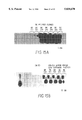
- FIGS. 5A-5E depict a series of five graphs showing the 21 kDa and 17 kDa neural thread proteins in SH-Sy5y, PNET1, A172, and C6 cells and the absence thereof in Huh7 cells by SDS-PAGE/M-IRMA.
- FIG. 6 depicts a gel showing that the 21 kDa neural thread protein in C6 glioblastoma cells is phosphorylated.
- FIG. 7 depicts a bar graph showing altered neural thread protein expression in PNET1 cells with growth phase.
- FIGS. 8A-8F show altered phenotype of PNET1 cells with cessation of cell growth and overnight serum starvation.
- FIG. 9 shows the 1-9a partial cDNA sequence
- FIG. 9A shows a partial sequence of the second 5' anchor PCR product corresponding to the 5' region of the 1-9a cDNA (WP5' Sequence).
- FIG. 10 shows alignment of partial sequences between 1-9a and human PTP and the Reg gene (the nucleic acid sequence corresponding to the genomic clone of human PTP).
- FIG. 10A shows alignment between 1-9a and Exon 2 of the human Reg gene, and between the first 5' anchor PCR product of 1-9a (WP03-417) and Exon 2 and Reg.
- FIG. 10B shows alignment between the 1-9a and its second 5' anchor PCR product (WP5') and AD 3-4 and AD 2-2 cDNAs.
- FIG. 11A shows the partial nucleic acid and deduced amino acid sequences of the HB4 cDNA.
- FIG. 11D shows alignment between HB4 and human PTP.
- FIG. 11E shows alignment between HB4 and human Reg gene.
- FIGS. 12A-12C show the expression of mRNA molecules corresponding to the 1-9a CNS neural thread protein cDNA sequence in neuroectodermal tumor cell lines and in rat pancreas.
- FIGS. 13A and 13B show mRNA transcripts corresponding to the 1-9a CNS neural thread protein cDNA sequence in human brain. This figure also demonstrates higher levels of 1-9a CNS neural thread protein-related mRNAs in AD brains compared with aged-matched controls (FIG. 13A). FIG. 13B demonstrates four different transcripts with greater abundance of the lower molecular size mRNAs in AD compared with aged controls.
- FIGS. 14A-14C show 1-9a Southern blot analysis of RT/PCR-derived cDNAs in neuroectodermal cell lines. A- and B-PCR amplification of 1-9a mRNA sequences in neuroectodermal cell lines, and using mRNA from newborn rat (NB) brain, AD brain, and aged control brain.
- FIG. 14A is a longer exposure of FIG. 14B.
- FIG. 14C shows hybridization of the same blot using the O18 rat PTP probe.
- FIGS. 15A and 15B show hybridization of the 1-9a and O18 probes with several clones isolated from SH-Sy5y cells by reverse transcribing mRNA and amplifying with primers corresponding to the known sequence of the 1-9a partial cDNA.
- FIGS. 16A, 16D and 16E show the partial nucleic acid sequences of the AD 2-2 cDNAs isolated from the AD brain library.
- FIGS. 16F, 16I, 16J and 16K show the partial nucleic acid sequences of the AD 3-4 cDNAs isolated from the AD brain library.
- FIGS. 16L, 16M and 16N show the partial nucleic acid sequences of the AD 4-4 cDNAs isolated from the AD brain library.
- FIG. 16O shows the partial nucleic acid sequences of the AD 16c (also called AD 10-7) cDNAs isolated from the AD brain library.
- FIG. 16R shows the complete nucleotide sequence of the AD10-7 cDNA clone that was isolated from an AD library.
- FIG. 16S shows the complete nucleotide sequence of the AD16c cDNA clone that was isolated from the AD brain library.
- FIG. 17 shows alignment of partial sequences between AD 2-2 and human Reg gene.
- FIG. 17A shows alignment of partial sequences between AD 2-2 and Exon 1 of Reg and rat PTP.
- FIG. 17B shows alignment of partial sequences between AD 2-2 and 1-9a.
- FIG. 17C shows alignment of partial sequences between AD 2-2 and AD 16c.
- FIG. 18 shows alignment of partial sequences between AD 3-4 (also called AD 5-3) and the Reg gene.
- FIG. 18A shows alignment of partial sequences between AD 3-4 and the 5' anchor PCR products of the 1-9a mRNA, termed WPO3-5 and 18-4.
- FIG. 18B shows alignment of partial sequences between AD 3-4 and the G2a-a EcoRIlPstI genomic clone.
- FIG. 19 shows alignment of partial sequences between AD 4-4 and AD 2-2 and 1-9a (also called SE-4 corresponding to the PCR clone which is identical to 1-9a).
- FIG. 20 shows alignment of partial sequences between AD 16c and Reg gene.
- FIG. 20A shows alignment of partial sequences between AD 16c and human PTP.
- FIG. 20B shows alignment of partial sequences between AD 16c and AD 2-2.
- FIGS. 21A-21D show a genomic Southern blot analysis using the AD 3-4 as a probe; FIG. 21B shows a similar pattern of hybridization on a genomic Southern using AD 2-2 as a probe.
- FIGS. 21A-21D show a Northern blot analysis of neuroectodermal tumor cell lines using AD 3-4 as a probe. The four cell lines that exhibit AD 3-4 transcripts are neuronal in phenotype; C6 glioma cell mRNA did not hybridize with the AD 3-4 probe.
- FIG. 21D shows a Northern analysis of human AD and aged control brain temporal lobe tissue using the AD 3-4 probe, and demonstrates over-expression of the corresponding gene in AD (lanes labeled A) compared with aged control brains (lanes labeled C).
- FIGS. 22, 22A, 22B, 22C, 22D, 22E, 22F, 22G and 22H show partial sequences of four genomic clones (isolated using both the 1-9a cDNA and rat PTP 0-18 cDNA as probes.
- FIGS. 23 and 23A show the alignment of the G2a-2 PstI partial sequence with the Reg gene.
- FIG. 23B shows alignment of the G2a-2 PstI-EcoRI sequence and the Reg gene and the rat PTP.
- FIGS. 23C and 23D show the alignment of the G5d-1 PstI sequence and the Reg gene.
- FIGS. 24A-24D show neural thread protein expression by the 1-9a cDNA (FIG. 24A) and the G2a-2 PstI genomic clone (FIG. 24B).
- FIGS. 24C and 24D show negative expression by the G5d-1 EcoRI/PstI genomic clone, and pBluescript which lacks a cloned insert, respectively.
- FIGS. 25A and 25B depict a Northern blot analysis of AD 16c mRNA in AD and aged control brains.
- the data shows elevated levels of AD16c mRNA expression in 6 of 9 AD compared to 1 of 6 age-matched controls.
- FIG. 26 depicts a Western blot analysis of AD10-7 fusion proteins using monoclonal antibodies against the expressed tag protein (T7-tag mouse monoclonal antibodies.
- FIGS. 27A and 27B depict brightfield and darkfield microscopic analysis of the in situ hybridization of sense and antisense cRNA probes to human brain tissue sections of early AD.
- Cloning vector A plasmid or phage DNA or other DNA sequence which is able to replicate autonomously in a host cell, and which is characterized by one or a small number of restriction endonuclease recognition sites at which such DNA sequences may be cut in a determinable fashion without loss of an essential biological function of the vector, and into which a DNA fragment may be spliced in order to bring about its replication and cloning.
- the cloning vector may further contain a marker suitable for use in the identification of cells transformed with the cloning vector. Markers, for example, provide tetracycline resistance or ampicillin resistance.
- Expression vector A vector similar to a cloning vector but which is capable of enhancing the expression of a gene which has been cloned into it, after transformation into a host.
- the cloned gene is usually placed under the control of (i.e., operably linked to) certain control sequences such as promoter sequences.
- Promoter sequences may be either constitutive or inducible.
- Substantially pure As used herein means that the desired purified protein is essentially free from contaminating cellular components, said components being associated with the desired protein in nature, as evidenced by a single band following polyacrylamide-sodium dodecyl sulfate gel electrophoresis. Contaminating cellular components may include, but are not limited to, proteinaceous, carbohydrate, or lipid impurities.
- substantially pure is further meant to describe a molecule which is homogeneous by one or more purity or homogeneity characteristics used by those of skill in the art.
- a substantially pure NTP will show constant and reproducible characteristics within standard experimental deviations for parameters such as the following: molecular weight, chromatographic migration, amino acid composition, amino acid sequence, blocked or unblocked N-terminus, HPLC elution profile, biological activity, and other such parameters.
- the term is not meant to exclude artificial or synthetic mixtures of the factor with other compounds.
- the term is not meant to exclude NTP fusion proteins isolated from a recombinant host.
- a recombinant host may be any prokaryotic or eukaryotic cell which contains the desired cloned genes on an expression vector or cloning vector. This term is also meant to include those prokaryotic or eukaryotic cells that have been genetically engineered to contain the desired gene(s) in the chromosome or genome of that organism.
- Recombinant vector Any cloning vector or expression vector which contains the desired cloned gene(s).
- Any prokaryotic or eukaryotic cell that is the recipient of a replicable expression vector or cloning vector also includes prokaryotic or eukaryotic cells that can be genetically engineered by well known techniques to contain desired gene(s) on its chromosome or genome. For examples of such hosts, see Sambrook et al., Molecular Cloning: A Laboratory Manual, Second Edition, Cold Spring Harbor Laboratory, Cold Spring Harbor, N.Y. (1989).
- Promoter A DNA sequence generally described as the 5' region of a gene, located proximal to the start codon. The transcription of an adjacent gene(s) is initiated at the promoter region. If a promoter is an inducible promoter, then the rate of transcription increases in response to an inducing agent. In contrast, the rate of transcription is not regulated by an inducing agent if the promoter is a constitutive promoter.
- Gene A DNA sequence that contains information needed for expressing a polypeptide or protein.
- Structural gene A DNA sequence that is transcribed into messenger RNA (mRNA) that is then translated into a sequence of amino acids characteristic of a specific polypeptide.
- mRNA messenger RNA
- Antisense RNA gene/Antisense RNA In eukaryotes, mRNA is transcribed by RNA polymerase II. However, it is also known that one may construct a gene containing a RNA polymerase II template wherein a RNA sequence is transcribed which has a sequence complementary to that of a specific mRNA but is not normally translated. Such a gene construct is herein termed an "antisense RNA gene” and such a RNA transcript is termed an "antisense RNA.” Antisense RNAs are not normally translatable due to the presence of translation stop codons in the antisense RNA sequence.
- Antisense oligonucleotide A DNA or RNA molecule containing a nucleotide sequence which is complementary to that of a specific mRNA. An antisense oligonucleotide binds to the complementary sequence in a specific mRNA and inhibits translation of the mRNA.
- Antisense Therapy A method of treatment wherein antisense oligonucleotides are administered to a patient in order to inhibit the expression of the corresponding protein.
- cDNA Complementary DNA
- a "complementary DNA, " or “cDNA” gene includes recombinant genes synthesized by reverse transcription of mRNA and from which intervening sequences (introns) have been removed.
- Expression is the process by which a polypeptide is produced from a structural gene. The process involves transcription of the gene into mRNA and the translation of such mRNA into polypeptide(s).
- Two nucleic acid molecules are considered to be “homologous” if their nucleotide sequences share a similarity of greater than 50%, as determined by HASH-coding algorithms (Wilber, W. J. and Lipman, D. J., Proc. Natl. Acad. Sci. 80:726-730 (1983)).
- Two nucleic acid molecules are considered to be “nonhomologous” if their nucleotide sequences share a similarity of less than 50%.
- Ribozyme is an RNA molecule that contains a catalytic center.
- the term includes RNA enzymes, self-splicing RNAs, and self-cleaving RNAs.
- Ribozyme Therapy A method of treatment wherein ribozyme is administered to a patient in order to inhibit the translation of the target mRNA.
- Fragment of a molecule such as NTP is meant to refer to any polypeptide subset of that molecule.
- Functional Derivative is intended to include the “variants,” “analogues,” or “chemical derivatives” of the molecule.
- a “variant” of a molecule such as NTP is meant to refer to a naturally occurring molecule substantially similar to either the entire molecule, or a fragment thereof.
- An “analogue” of a molecule such as NTP is meant to refer to a non-natural molecule substantially similar to either the entire molecule or a fragment thereof.
- a molecule is said to be "substantially similar” to another molecule if the sequence of amino acids in both molecules is substantially the same, and if both molecules possess a similar biological activity. Thus, provided that two molecules possess a similar activity, they are considered variants as that term is used herein even if one of the molecules contains additional amino acid residues not found in the other, or if the sequence of amino acid residues is not identical.
- a molecule is said to be a "chemical derivative" of another molecule when it contains additional chemical moieties not normally a part of the molecule. Such moieties may improve the molecule's solubility, absorption, biological half-life, etc. The moieties may alternatively decrease the toxicity of the molecule, eliminate or attenuate any undesirable side effect of the molecule, etc. Examples of moieties capable of mediating such effects are disclosed in Remington's Pharmaceutical Sciences (1980) and will be apparent to those of ordinary skill in the art.
- NTP refers to a family of neural thread proteins.
- the NTP family includes proteins with molecular weights of about 8 kDa, 14 kDa, 17 kDa, 21 kDa, 26 kDa and 42 kDa, as described herein.
- Immuno-Polymerase Chain Reaction A method for the detection of antigens using specific antibody-DNA conjugates.
- a linker molecule with bispecific binding affinity for DNA and antibodies is used to attach a DNA molecule specifically to an antigen-antibody complex.
- a specific antigen-antibody-DNA conjugate is formed.
- the attached DNA can be amplified by the polymerase chain reaction (PCR) using appropriate oligonucleotide primers.
- PCR polymerase chain reaction
- the presence of specific PCR products demonstrates that DNA molecules are attached specifically to antigen-antibody complexes, thus indicating the presence of antigen.
- Sano et al., supra constructed a streptavidin-protein A chimera that possesses specific binding affinity for biotin and immunoglobulin G.
- This chimera i.e., the "linker molecule” was used to attach a biotinylated DNA specifically to antigen-monoclonal antibody complexes that had been immobilized on microtiter plate wells. A segment of the attached DNA was subsequently amplified by PCR.
- This invention is directed to neural thread proteins (NTP), genetic sequences coding for an NTP mRNA or antisense mRNA, expression vectors containing the genetic sequences, recombinant hosts transformed therewith, and NTP and antisense RNA produced by such transformed recombinant host expression.
- NTP neural thread proteins
- This invention further relates to NTP ribozymes, and recombinant DNA molecules which code for NTP ribozymes and NTP antisense oligonucleotides.
- This invention further relates to antibodies directed against an NTP, as well as the use of NTP antibodies and NTP nucleic acid sequences for detection of the presence of an NTP in biological samples.
- the invention further relates to the use of NTP coding sequences in gene therapy.
- DNA sequences coding for an NTP are derived from a variety of sources. These sources include genomic DNA, cDNA, synthetic DNA, and combinations thereof.
- Human NTP genomic DNA can be extracted and purified from any human cell or tissue, by means well known in the art (for example, see Sambrook et al., Molecular Cloning: A Laboratory Manual, Second Edition, Cold Spring Harbor Laboratory Press, 1989).
- the NTP genomic DNA of the invention may or may not include naturally occurring introns.
- genomic DNA may be obtained in association with the 5' promoter region of the NTP gene sequences and/or with the 3' translational termination region.
- genomic DNA may be obtained in association with DNA sequences which encode the 5' nontranslated region of the NTP mRNA and/or with the genetic sequences which encode the 3' nontranslated region.
- the 5' and/or 3' nontranscribed regions of the native gene, and/or, the 5' and/or 3' nontranslated regions of the mRNA may be retained and employed for transcriptional and translational regulation.
- an NTP mRNA can be isolated from any cell which expresses an NTP, and used to produce cDNA by means well known in the art (for example, see Sambrook et al., supra).
- the mRNA preparation used will be enriched in mRNA coding for an NTP, either naturally, by isolation from cells which produce large amounts of an NTP, or in vitro, by techniques commonly used to enrich mRNA preparations for specific sequences, such as sucrose gradient centrifugation, or both.
- An NTP mRNA may be obtained from mammalian neuronal tissue, or from cell lines derived therefrom.
- human cDNA libraries are constructed from 17-18 week old fetal brain, 2 year old temporal lobe neocortex, end-stage AD cerebral cortex, or from cell lines derived from human neuronal tissue.
- Such cell lines may include, but are not limited to, central nervous system primitive neuroectodermal tumor cells (such as PNET1 or PNET2, as described herein), neuroblastoma cells (such as SH-Sy5y, as described herein), or human glioma cells (such as A172; ATCC CRL 1620).
- a rat cDNA library can be prepared from mRNA isolated from rat glioma cells, for example, C6 rat glioma cells (ATCC CCL107).
- suitable DNA preparations are randomly sheared or enzymatically cleaved, respectively, and ligated into appropriate vectors to form a recombinant gene (either genomic or cDNA) library.
- a DNA sequence encoding an NTP may be inserted into a vector in accordance with conventional techniques, including blunt-ending or staggered-ending termini for ligation, restriction enzyme digestion to provide appropriate termini, filling in of cohesive ends as appropriate, alkaline phosphatase treatment to avoid undesirable joining, and ligation with appropriate ligases. Techniques for such manipulation are disclosed by Sambrook et al., supra, and are well known in the art.
- NTP clones may be screened and the NTP clones identified by any means which specifically selects for NTP DNA such as, for example: 1) by hybridization with an appropriate nucleic acid probe(s) containing a sequence specific for the DNA of this protein; or, 2) by hybridization-selected translational analysis in which native mRNA hybridizes to the clone in question, is translated in vitro, and the translation products are further characterized; or, 3) if the cloned DNA sequences are themselves capable of expressing mRNA, by immunoprecipitation of a translated NTP product produced by the host containing the clone.
- any means which specifically selects for NTP DNA such as, for example: 1) by hybridization with an appropriate nucleic acid probe(s) containing a sequence specific for the DNA of this protein; or, 2) by hybridization-selected translational analysis in which native mRNA hybridizes to the clone in question, is translated in vitro, and the translation products are further characterized; or, 3) if
- Oligonucleotide probes specific for an NTP which can be used to identify clones to this protein can be designed from knowledge of the amino acid sequence of the corresponding NTP, or homologous regions of the PTP. Alternatively, oligonucleotide probes can be designed from knowledge of the nucleotide sequence of PTP (de la Monte et al., J. Clin. Invest. 86:1004-1013 (1990)).
- the suitable oligonucleotide, or set of oligonucleotides, which is capable of encoding a fragment of the NTP gene (or which is complementary to such an oligonucleotide, or set of oligonucleotides) may be synthesized by means well known in the art (for example, see Sambrook et al., supra). Techniques of nucleic acid hybridization and clone identification are disclosed by Sambrook et al., supra. Those members of the above-described gene library which are found to be capable of such hybridization are then analyzed to determine the extent and nature of the NTP encoding sequences which they contain.
- the above-described DNA probe is labeled with a detectable group.
- detectable group can be any material having a detectable physical or chemical property. Such materials have been well-developed in the field of nucleic acid hybridization and in general most any label useful in such methods can be applied to the present invention. Particularly useful are radioactive labels including 32 P, 3 H, 14 C, 125 I, or the like. Any radioactive label may be employed which provides for an adequate signal and has sufficient half-life.
- the DNA probe may be labeled, for example, by nick-translation, by T4 DNA polymerase replacement synthesis, or by random priming, among other methods well known in the art (see Sambrook et al. supra).
- DNA probes can be labeled with non-radioactive markers such as biotin, an enzyme, or fluorescent group.
- NTP cDNAs are obtained by direct cloning of cDNAs from cell lines and brain tissue, using the 3' - and 5' -RACE methods, as described herein.
- a human neuroectodermal tumor cell line or AD brain tissue is used as a source of mRNA.
- the above-discussed methods are, therefore, capable of identifying DNA sequences which are code for an NTP or fragments thereof.
- NTP transcriptional and translational signals recognizable by an appropriate host are necessary.
- the cloned NTP DNA sequences obtained through the methods described above, and preferably in double-stranded form, may be "operably linked" to sequences controlling transcriptional expression in an expression vector, and introduced into a host cell, either prokaryotic or eukaryotic, to produce recombinant NTP.
- a host cell either prokaryotic or eukaryotic
- the present invention encompasses the expression of an NTP in eukaryotic cells, and especially mammalian, insect, and yeast cells.
- eukaryotic hosts are mammalian cells.
- Mammalian cells provide post-translational modifications to recombinant NTP which include folding and/or phosphorylation.
- mammalian host cells include human CNS primitive neuroectodermal tumor cells, human neuroblastoma cells, human glioma cells, or rat glioma cells.
- Especially preferred primitive neuroectodermal tumor cells include PNET1 and PNET2, especially preferred human glioblastoma cells include Hg16 and Hg17, especially preferred human glioma cells include A172, and especially preferred rat glioma cells include C6 (see Example 1).
- an NTP may be expressed by prokaryotic host cells.
- a recombinant NTP is expressed by such cells as a fusion protein, as described herein.
- An especially preferred prokaryotic host is E. coli. Preferred strains of E. coli include Y1088, Y1089, CSH18, ER1451, and ER1647 (see, for example, Molecular Biology LabFax, Brown, T. A., Ed., Academic Press, New York (1991)).
- Bacillus subtilus including such strains as BR151, YB886, MI119, M1120, and B170 (see, for example, Hardy, "Bacillus Cloning Methods,” in DNA Cloning: A Practical Approach, IRL Press, Washington, D.C. (1985)).
- a nucleic acid molecule such as DNA, is said to be "capable of expressing" a polypeptide if it contains expression control sequences which in turn contain transcriptional regulatory information and such sequences are “operably linked” to the nucleotide sequence which encodes the protein.
- Two sequences of a nucleic acid molecule are said to be operably linked when they are linked to each other in a manner which either permits both sequences to be transcribed onto the same RNA transcript, or permits an RNA transcript, begun in one sequence to be extended into the second sequence.
- two sequences such as a promoter sequence and any other "second" sequence of DNA or RNA are operably linked if transcription commencing in the promoter sequence will produce an RNA transcript of the operably linked second sequence.
- two sequences In order to be operably linked it is not necessary that two sequences be immediately adjacent to one another.
- the promoter sequences of the present invention may be either prokaryotic, eukaryotic or viral. Suitable promoters are repressible, constitutive, or inducible. Examples of suitable prokaryotic promoters include promoters capable of recognizing the T4 polymerases (Malik et al., J. Biol. Chem. 263:1174-1181 (1984); Rosenberg et al., Gene 59:191-200 (1987); Shinedling et al., J. Molec. Biol.
- Streptomyces promoters (Ward et al., Mol. Gen. Genet. 203:468-478 (1986)); the int promoter of bacteriophage lambda; the bla promoter of the ⁇ -lactamase gene of pBR322, and the CAT promoter of the chloramphenicol acetyl transferase gene of pBR325, etc.
- Prokaryotic promoters are reviewed by Glick, J. Ind. Microbiol. 1:277-282 (1987); Cenatiempo, Biochimie 68:505-516 (1986); Watson et al., In: Molecular Biology of the Gene, Fourth Edition, Benjamin Cummins, Menlo Park, Calif. (1987); Gottesman, Ann. Rev. Genet. 18:415-442 (1984); and Sambrook et al., supra.
- Preferred eukaryotic promoters include the promoter of the mouse metallothionein I gene (Hamer et al., J. Mol. Appl. Gen. 1:273-288 (1982)); the TK promoter of Herpes virus (McKnight, Cell 31:355-365 (1982)); the SV40 early promoter (Benoist, et al., Nature (London) 290:304-310 (1981)); and the yeast gal4 gene promoter (Johnston, et al., Proc. Natl. Acad. Sci. (USA) 79:6971-6975 (1982); Silver, et al., Proc. Natl. Acad. Sci. (USA) 81:5951-5955 (1984)). All of the above listed references are incorporated by reference herein.
- Strong promoters are the most preferred promoters of the present invention.
- Examples of such preferred promoters are those which recognize the T3, SP6 and T7 polymerase promoters; the P L promoter of bacteriophage lambda; the recA promoter and the promoter of the mouse metallothionein I gene.
- the most preferred promoter for expression in prokaryotic cells is one which is capable of recognizing the T7 polymerase promoter.
- the sequences of such polymerase recognition sequences are disclosed by Watson, et al. (In: Molecular Biology of the Gene, Fourth Edition, Benjamin Cummins, Menlo Park, Calif. (1987)).
- the most preferred promoter for expression in mammalian cells is SV40 (Gorman, "High Efficiency Gene Transfer into Mammalian cells," in DNA Cloning: A Practical Approach, Volume II, IRL Press, Washington, D.C., pp. 143-190 (1985)).
- This invention is directed towards methods of detecting neurological disease in a human subject, utilizing the nucleic acid probes hybridizable to NTP genes or transcripts, or antibodies specific for an NTP.
- neurological disease is meant Alzheimer's Disease (AD), or other neurodegenerative disorders with the Alzheimer's type pathogenic changes (for example, Parkinson's disease with AD-type neurodegeneration), as well as neuroectodermal tumors, malignant astrocytomas, and glioblastomas.
- human subject is meant any human being or any developmental form thereof, such as a human embryo or fetus, prior to birth.
- the diagnostic methods of the present invention do not require invasive removal of neural tissue.
- the present invention additionally pertains to assays, both nucleic acid hybridization assays and immunoassays, for detecting the presence of NTP in cells or in the biological fluids of a human subject using light or electron microscopic histology, imaging, radioactive or enzyme based assays, and the like.
- RNA can be isolated from tissue by sectioning on a cryostat and lysing the sections with a detergent such as SDS and a chelating agent such as EDTA, optionally with overnight digestion with proteinase K (50 ⁇ g/ml).
- tissue is obtained by autopsy and biopsy.
- a preferred quantity of tissue is in the range of 1-10 milligrams.
- Protein is removed by phenol and chloroform extractions, and nucleic acids are precipitated with ethanol.
- RNA is isolated by chromatography on an oligo dT column and then eluted therefrom. Further fractionation can also be carried out, according to methods well known to those of ordinary skill in the art.
- a number of techniques for molecular hybridization are used for the detection of DNA or RNA sequences in tissues; each has certain advantages and disadvantages.
- analysis of hybridization kinetics provides the opportunity to accurately quantitate the amount of DNA or RNA present, as well as to distinguish sequences that are closely related but not identical to the probe, and determine the percent homology.
- Reactions are run under conditions of hybridization (Tm-25° C.) in which the rate of reassociation of the probe is optimal (Wetmur et al., J. Mol. Biol. 31:349-370 (1968)).
- the kinetics of the reaction are second- order when the sequences in the tissue are identical to those of the probe; however, the reaction exhibits complex kinetics when probe sequences have partial homology to those in the tissue (Sharp et al., J. Mol. Biol. 86:709-726 (1974)).
- the ratio of probe to cell RNA is determined by the sensitivity desired. To detect one transcript per cell would require about 100 pg of probe per tg of total cellular DNA or RNA.
- the nucleic acids are mixed, denatured, brought to the appropriate salt concentration and temperature, and allowed to hybridize for various periods of time. The rate of reassociation can be determined by quantitating the amount of probe hybridized either by hydroxy apatite chromatography (Britten et al., Science 161:529-540 (1968)) or S1 nuclease digestion (Sutton, Biochim. Biophys. Acta 240:522-531 (1971)).
- a more flexible method of hybridization is the northern blot technique. This technique offers variability in the stringency of the hybridization reaction, as well as determination of the state of the retroviral sequences in the specimen under analysis. Northern analysis can be performed as described herein.
- a major consideration associated with hybridization analysis of DNA or RNA sequences is the degree of relatedness the probe has with the sequences present in the specimen under study. This is important with the blotting technique, since a moderate degree of sequence homology under nonstringent conditions of hybridization can yield a strong signal even though the probe and sequences in the sample represent non-homologous genes.
- hybridization can be carried out in a solution containing 6 ⁇ SSC (10 ⁇ SSC: 1.5 M sodium chloride, 0.15 M sodium citrate, pH 7.0), 5 ⁇ Denhardt's (1 Denhardt's: 0.2% bovine serum albumin, 0.2% polyvinylpyrrolidone, 0.02% Ficoll 400), 10 mM EDTA, 0.5% SDS and about 10 7 cpm of nick-translated DNA for 16 hours at 65° C.
- the labeled probes as described above, provide a general diagnostic method for detection of an NTP in tissue.
- the method is reasonably rapid, has a simple protocol, has reagents which can be standardized and provided as commercial kits, and allows for rapid screening of large numbers of samples.
- a clinical isolate containing RNA transcripts is fixed to a support.
- the affixed nucleic acid is contacted with a labeled polynucleotide having a base sequence complementary or homologous to the coding strand of the NTP gene.
- the hybridization assays of the present invention are particularly well suited for preparation and commercialization in kit form, the kit comprising a carrier means compartmentalized to receive one or more container means (vial, test tube, etc.) in close confinement, each of said container means comprising one of the separate elements to be used in hybridization assay.
- NTP cDNA molecules suitable for labeling by "nick translation" (see, for example, Sambrook et al., supra, for standard methodology), or labeled NTP cDNA or RNA molecules.
- Further container means may contain standard solutions for nick translation of NTP cDNA comprising DNA polymerase I/DNase I and unlabeled deoxyribonucleotides (i.e., dCTP, dTTP, dGTP, and dATP).
- NTP RNA The presence of NTP RNA is determined by the variation in the appearance and/or quantity of probe-related RNA in tested tissue.
- the DNA probes of this invention can also be used for differential diagnosis of hereditary or familial AD and non-hereditary or sporadic AD.
- the familial form of AD often occurs at an earlier age and is associated with Down's syndrome in the family.
- a genetic test for familial AD allows for genetic counseling of families. While much effort has been directed toward characterizing a genetic marker for familial AD (Gusella, FASEB J 3:2036-2041 (1989); Hooper, J NIH Res. 4:48-54 (1992)), genetic linkage analysis only identifies a genetic marker sequence without providing the knowledge of the function of the genomic sequence.
- the cDNA probes described herein and obtained from individuals with sporadic AD encode a known protein of known function which is over-expressed in brain tissue of patients with AD.
- a method of differentiating between sporadic and familial AD in a human subject involves obtaining a biological sample from the human subject who is suspected of having Alzheimer's Disease. Then, DNA is purified from the biological sample. Finally, the DNA is contacted with a NTP DNA probe under conditions of hybridization. Familial AD is indicated by the detection of a hybrid of the probe and the DNA, whereas sporadic AD is indicated by the absence of detection of hybridization.
- the biological sample can be a blood sample which is subjected to differential centrifugation to enrich for white blood cells within three days of collection (Park, "PCR in the Diagnosis of Retinoblastoma,” in PCR Protocols, Innis et al., Eds., Academic Press, Inc., New York, pp. 407-415 (1990)).
- the DNA sample can be prepared using the sodium N-lauroylsarcosine-Proteinase K, phenol, and RNase method (Sambrook et al., supra). DNA analysis can be performed by digesting the DNA sample, preferably 5 micrograms, with a restriction endonuclease (such as HindIII).
- Digested DNA is then fractionated using agarose gel electrophoresis, preferably, a 1% horizontal agarose gel, for 18 hours in a buffer preferably containing 89 mM Tris-Hc1 (pH 8), 89 mM sodium borate and 2 mM EDTA (Gusella et al., Nature 306:234-238 (1983)).
- Southern analysis can be performed using conventional techniques (Sambrook et al., supra), and the labelled AD cDNA probes can be hybridized under conditions described above.
- the preferred DNA probes for this differential diagnosis method include 1-9a, AD3-4, AD4-4 and G2-2 PstI.
- Antibodies directed against an NTP can be used, as taught by the present invention, to detect and diagnose AD.
- Various histological staining methods including immunohistochemical staining methods, may also be used effectively according to the teaching of the invention.
- Silver stain is but one method of visualizing NTP.
- Other staining methods useful in the present invention will be obvious to the artisan, the determination of which would not involve undue experimentation (see generally, for example, A Textbook of Histology, Eds. Bloom and Fawcett, W.B. Saunders Co., Philadelphia (1964)).
- One screening method for determining whether a given compound is an NTP functional derivative comprises, for example, immunoassays employing radioimmunoassay (RIA) or enzyme-linked immunosorbant assay (ELISA) methodologies, based on the production of specific antibodies (monoclonal or polyclonal) to an NTP.
- RIA radioimmunoassay
- ELISA enzyme-linked immunosorbant assay
- biological samples are obtained by venepuncture (blood), spinal tap (cerebral spinal fluid (CSF)), urine and other body secretions such as sweat and tears.
- RIA radioimmunoassay
- ELISA enzyme-linked immunosorbant assay
- the present invention also relates to methods of detecting an NTP or functional derivatives in a sample or subject.
- antibodies specific for an NTP, or a functional derivative may be detectably labeled with any appropriate marker, for example, a radioisotope, an enzyme, a fluorescent label, a paramagnetic label, or a free radical.
- antibodies specific for an NTP, or a functional derivative may be detectably labeled with DNA by the technique of immunopolymerase chain reaction (Sano et al., Science 258:120-122 (1992)).
- the polymerase chain reaction (PCR) procedure amplifies specific nucleic acid sequences through a series of manipulations including denaturation, annealing of oligonucleotide primers, and extension of the primers with DNA polymerase (see, for example, Mullis et al., U.S. Pat. No. 4,683,202; Mullis et al., U.S. Pat. No. 4,683,195; Loh et al., Science 243:217 (1988)).
- the steps can be repeated many times, resulting in a large amplification of the number of copies of the original specific sequence. As little as a single copy of a DNA sequence can be amplified to produce hundreds of nanograms of product (Li et al., Nature 335:414 (1988)).
- Other known nucleic acid amplification procedures include transcription-based amplification systems (Kwoh et al., Proc. Natl. Acad. Sci.
- the immuno-PCR assay can be carried out by immobilizing various amounts of the test material on the surface of microtiter wells (see Sanzo et al., supra, page 122, footnote 7).
- the wells are subsequently incubated with an NTP monoclonal antibody, washed, and then incubated with biotinylated with biotinylated NTP DNA molecules which have been conjugated to streptavidin-protein chimera (Id.).
- This chimera binds biotin (via the streptavidin moiety) and the Fc portion of an immunoglobulin G molecule (via the protein A moiety) (Id., at 120; Sanzo et al., Bio/Technology 9:1378 (1991)).
- the wells are then washed to remove unbound conjugates. Any NTP present in the test material will be bound by the NTP monoclonal antibody, which in turn, is bound by the protein A moiety of the biotinylated NTP DNA-- streptavidin-protein A conjugate. Then, the NTP DNA sequences are amplified using PCR. Briefly, the microtiter wells are incubated with deoxyribonucleoside triphosphates, NTP oligonucleotide primers, and Taq DNA polymerase (see Sanzo et al., supra, page 122, footnote 11).
- An automated thermal cycler (such as the PTC-100-96 Thermal Cycler, MJ Research, Inc.) can be used to perform PCR under standard conditions (Id.). The PCR products are then analyzed by agarose gel electrophoresis after staining with ethidium bromide.
- antibody refers both to monoclonal antibodies which are a substantially homogeneous population and to polyclonal antibodies which are heterogeneous populations.
- Polyclonal antibodies are derived from the sera of animals immunized with an antigen.
- Monoclonal antibodies (mAbs) to specific antigens may be obtained by methods known to those skilled in the art. See, for example, Kohler and Milstein, Nature 256:495-497 (1975) and U.S. Pat. No. 4,376,110.
- Such antibodies may be of any immunoglobulin class including IgG, IgM, IgE, IgA, IgD and any subclass thereof.
- the monoclonal antibodies may be prepared as previously described (Gross et al., J. Clin. Invest. 76:2115-2126 (1985); Ozturk et al., Proc. Natl. Acad. Sci. USA 86:419-423 (1989); de la Monte et. al., J. Clin. Invest. 86:1004-1013 (1990); de la Monte et. al., J. Neurol. Sci. 113:152-164 (1992); de la Monte et al., Ann. Neurol. 32:733-742 (1992)).
- the Th monoclonal antibodies were generated against the purified pancreatic form of thread protein (Id.).
- NTP-specific polyclonal and monoclonal antibodies can also be generated against a substantially pure NTP isolated from recombinant hosts (for example, see Carroll et al., "Production and Purification of Polyclonal Antibodies to the Foreign Segment of ⁇ -Galactosidase Fusion Proteins," in DNA Cloning: A Practical Approach, Volume III, IRL Press, Washington, D.C., pp. 89-111 (1987); Mole et al., "Production of Monoclonal Antibodies against Fusion Proteins Produced in Escherichia coli," in DNA Cloning: A Practical Approach, Volume III, IRL Press, Washington, D.C., pp. 113-1139 (1987)).
- NTP-specific polyclonal and monoclonal antibodies can be generated against a substantially pure NTP isolated from biological material such as brain tissue and cell lines, by using well known techniques.
- monoclonal antibodies specific for the various NTP molecules of approximately, 8, 14, 17, 21, 26 kDa and 42 kDa molecular weights may be prepared from recombinant-derived proteins, which are expressed, isolated and purified from the cDNA (i.e., 1-9a), genomic clones (G2-2 PstI) and AD-NTP 3-4 cDNA clones. These NTP molecules are derived from the above cDNA's and genomic clones, inserted and produced in suitable expression vectors (see FIGS. 2A and 2B).
- NTP cDNA and PTP Since there are regions of 60-70% homology in the 5' ends of the 1-9a NTP cDNA and PTP, one can obtain monoclonal antibodies that bind specifically to the NTP recombinant proteins and not to the pancreatic form by performing routine differential screening (see, for example, de la Monte et al., J. Clin. Invest. 86:1004-1013 (1990)). Although there will be monoclonal antibodies that bind to both NTP and PTP, it will be possible to generate NTP-specific monoclonal antibodies because there is a substantial sequence divergence between NTP molecules of various forms (e.g., 8, 14, 17, 21, 26 and 42 kDa) and because an epitope may be defined by as few as 6-8 amino acids.
- antibody is also meant to include both intact molecules as well as fragments thereof, such as, for example, Fab and F(ab') 2 , which are capable of binding antigen.
- Fab and F(ab') 2 fragments lack the Fc fragment of intact antibody, clear more rapidly from the circulation, and may have less non-specific tissue binding than an intact antibody (Wahl et al., J. Nucl. Med. 24:316-325 (1983)).
- Fab and F(ab') 2 and other fragments of the antibodies useful in the present invention may be used for the detection and quantitation of an NTP according to the methods disclosed herein in order to detect and diagnose AD in the same manner as an intact antibody.
- Such fragments are typically produced by proteolytic cleavage, using enzymes such as papain (to produce Fab fragments) or pepsin (to produce F(ab') 2 fragments).
- An antibody is said to be “capable of binding” a molecule if it is capable of specifically reacting with the molecule to thereby bind the molecule to the antibody.
- epitopic is meant to refer to that portion of any molecule capable of being bound by an antibody which can also be recognized by that antibody.
- Epitopic determinants usually consist of chemically active surface groupings of molecules such as amino acids or sugar side chains and have specific three dimensional structural characteristics as well as specific charge characteristics.
- an "antigen” is a molecule capable of being bound by an antibody which is additionally capable of inducing an animal to produce antibody capable of binding to an epitope of that antigen.
- An antigen may have one, or more than one epitope. The specific reaction referred to above is meant to indicate that the antigen will react, in a highly selective manner, with its corresponding antibody and not with the multitude of other antibodies which may be evoked by other antigens.
- the antibodies, or fragments of antibodies, useful in the present invention may be used to quantitatively or qualitatively detect the presence of cells which contain the NTP antigens.
- the antibodies (or fragments thereof) useful in the present invention may be employed histologically to detect or visualize the presence of an NTP.
- Such an assay for an NTP typically comprises incubating a biological sample from said subject suspected of having such a condition in the presence of a detectably labeled binding molecule (e.g., antibody) capable of identifying an NTP, and detecting said binding molecule which is bound in a sample.
- a detectably labeled binding molecule e.g., antibody
- a biological sample may be treated with nitrocellulose, or other solid support which is capable of immobilizing cells, cell particles or soluble proteins.
- the support maybe then be washed with suitable buffers followed by treatment with the detectably labeled NTP-specific antibody.
- the solid phase support may then be washed with the buffer a second time to remove unbound antibody.
- the amount of bound label on said solid support may then be detected by conventional means.
- solid phase support any support capable of binding antigen or antibodies.
- supports, or carriers include glass, polystyrene, polypropylene, polyethylene, dextran, nylon, amylases, natural and modified celluloses, polyacrylamides, agaroses, and magnetite.
- the nature of the carrier can be either soluble to some extent or insoluble for the purposes of the present invention.
- the support material may have virtually any possible structural configuration so long as the coupled molecule is capable of binding to an antigen or antibody.
- the support configuration may be spherical, as in a bead, or cylindrical, as in the inside surface of a test tube, or the external surface of a rod. Alternatively, the surface may be flat such as a sheet, test strip, etc.
- Preferred supports include polystyrene beads.
- One embodiment for carrying out the diagnostic assay of the present invention on a biological sample containing an NTP comprises:
- step (d) separating the solid phase support from the incubation mixture obtained in step (c);
- labeled NTP-specific antibody/NTP complexes in a sample may be separated from a reaction mixture by contacting the complex with an immobilized antibody or protein which is specific for an immunoglobulin, e.g., Staphylococcus protein A, Staphylococcus protein G, anti-IgM or anti-IgG antibodies.
- an immunoglobulin e.g., Staphylococcus protein A, Staphylococcus protein G, anti-IgM or anti-IgG antibodies.
- anti-immunoglobulin antibodies may be polyclonal, but are preferably monoclonal.
- the solid support may then be washed with a suitable buffer to give an immobilized NTP/labeled NTP-specific antibody complex.
- the label may then be detected to give a measure of an NTP.
- This aspect of the invention relates to a method for detecting an NTP or a fragment thereof in a sample comprising:
- the invention also relates to a method of detecting an NTP in a sample, further comprising:
- step (c) contacting the mixture obtained in step (a) with an Fc binding molecule, such as an antibody, Staphylococcus protein A, or Staphylococcus protein G, which is immobilized on a solid phase support and is specific for the NTP-specific antibody to give a NTP/NTP-specific antibody immobilized antibody complex;
- an Fc binding molecule such as an antibody, Staphylococcus protein A, or Staphylococcus protein G
- step (d) washing the solid phase support obtained in step (c) to remove unbound NTP/NTP-specific antibody complex
- the specific concentrations of detectably labeled antibody and NTP, the temperature and time of incubation, as well as other assay conditions may be varied, depending on various factors including the concentration of an NTP in the sample, the nature of the sample, and the like.
- the binding activity of a given lot of anti-NTP antibody may be determined according to well known methods. Those skilled in the art will be able to determine operative and optimal assay conditions for each determination by employing routine experimentation.
- NTP-specific antibody can be detectably labeled is by linking the same to an enzyme.
- This enzyme when later exposed to its substrate, will react with the substrate in such a manner as to produce a chemical moiety which can be detected, for example, by spectrophotometric, fluorometric or by visual means.
- Enzymes which can be used to detectably label the NTP-specific antibody include, but are not limited to, malate dehydrogenase, staphylococcal nuclease, delta-V-steroid isomerase, yeast alcohol dehydrogenase, a-glycerophosphate dehydrogenase, triose phosphate isomerase, horseradish peroxidase, alkaline phosphatase, asparaginase, glucose oxidase, ⁇ -galactosidase, ribonuclease, urease, catalase, glucose-VI-phosphate dehydrogenase, glucoamylase and acetylcholinesterase.
- Detection may be accomplished using any of a variety of immunoassays. For example, by radioactively labeling the NTP-specific antibodies or antibody fragments, it is possible to detect NTP through the use of radioimmune assays.
- radioimmune assays A good description of a radioimmune assay may be found in Laboratory Techniques and Biochemistry in Molecular Biology, by Work, et al., North Holland Publishing Company, N.Y. (1978), with particular reference to the chapter entitled "An Introduction to Radioimmune Assay and Related Techniques" by Chard, incorporated by reference herein.
- the radioactive isotope can be detected by such means as the use of a gamma counter or a scintillation counter or by autoradiography.
- Isotopes which are particularly useful for the purpose of the present invention are: 3 H, 125 I, 131 I, 35 S, 14 C, and preferably 125 I.
- NTP-specific antibody it is also possible to label the NTP-specific antibody with a fluorescent compound.
- fluorescent labelling compounds are fluorescein isothiocyanate, rhodamine, phycoerytherin, phycocyanin, allophycocyanin, o-phthaldehyde and fluorescamine.
- the NTP-specific antibody can also be detectably labeled using fluorescence emitting metals such as 152 Eu, or others of the lanthanide series. These metals can be attached to the NTP-specific antibody using such metal chelating groups as diethylenetriaminepentaacetic acid (DTPA) or ethylenediaminetetraacetic acid (EDTA).
- DTPA diethylenetriaminepentaacetic acid
- EDTA ethylenediaminetetraacetic acid
- the NTP-specific antibody also can be detectably labeled by coupling it to a chemiluminescent compound. The presence of the chemiluminescent-tagged NTP-specific antibody is then determined by detecting the presence of luminescence that arises during the course of a chemical reaction.
- chemiluminescent labeling compounds are luminol, isoluminol, theromatic acridinium ester, imidazole, acridinium salt and oxalate ester.
- the NTP-specific antibody may also be labeled with biotin and then reacted with avidin.
- a biotin-labeled DNA fragment will be linked to the NTP-biotinylated monoclonal antibody by an avidin bridge.
- NTP molecules are then detected by polymerase chain reaction (PCR) amplification of the DNA fragment with specific primers (Sano et al., Science 258:120-122 (1992)).
- a bioluminescent compound may be used to label the NTP-specific antibody of the present invention.
- Bioluminescence is a type of chemiluminescence found in biological systems in which a catalytic protein increases the efficiency of the chemiluminescent reaction. The presence of a bioluminescent protein is determined by detecting the presence of luminescence.
- Important bioluminescent compounds for purposes of labeling are luciferin, luciferase and aequorin.
- Detection of the NTP-specific antibody may be accomplished by a scintillation counter, for example, if the detectable label is a radioactive gamma emitter, or by a fluorometer, for example, if the label is a fluorescent material.
- the detection can be accomplished by colorimetric methods which employ a substrate for the enzyme. Detection may also be accomplished by visual comparison of the extent of enzymatic reaction of a substrate in comparison with similarly prepared standards.
- the NTP which is detected by this assay may be present in a biological sample. Any sample containing an NTP can be used. However, one of the benefits of the present diagnostic invention is that invasive tissue removal may be avoided. Therefore, preferably, the sample is a biological solution such as, for example, cerebrospinal fluid, amniotic fluid, blood, serum, urine and the like. However, the invention is not limited to assays using only these samples, it being possible for one of ordinary skill in the art to determine suitable conditions which allow the use of other samples.
- M-IRMA monoclonal antibody-based immunoradiometric assays
- this examination for AD is accomplished by removing samples of biological fluid and incubating such samples in the presence of detectably labeled antibodies (or antibody fragments).
- this technique is accomplished in a non-invasive manner through the use of magnetic imaging, fluorography, etc.
- the detection of cells which express an NTP may be accomplished by in vivo imaging techniques, in which the labeled antibodies (or fragments thereof) are provided to a subject, and the presence of the NTP is detected without the prior removal of any tissue sample.
- in vivo detection procedures have the advantage of being less invasive than other detection methods, and are, moreover, capable of detecting the presence of NTP in tissue which cannot be easily removed from the patient, such as brain tissue.
- NTP will be detected throughout the brain in an AD patient, while NTP will be localized in discrete deposits in the case of brain tumors.
- NTP will be found in the temporal, parietal and frontal cortices as well as the amygdala and hippocampus.
- astrocytomas include the cerebrum, cerebellum, thalamus, optic chiasma, and pons (Harrison's Principles of Internal Medicine, Petersdorf et al., Eds., Tenth Edition, McGraw-Hill Book Company, New York, p.2076 (1983)), and glioblastoma multiform is predominantly cerebral in location (Id. at 2075).
- radionuclide for in vivo diagnosis is that the half-life of a radionuclide be long enough so that it is still detectable at the time of maximum uptake by the target, but short enough so that deleterious radiation upon the host is minimized.
- a radionuclide used for in vivo imaging will lack a particulate emission, but produce a large number of photons in the 140-200 keV range, which maybe readily detected by conventional gamma cameras.
- radionuclides may be bound to antibody either directly or indirectly by using an intermediary functional group.
- Intermediary functional groups which are often used in binding radioisotopes which exist as metallic ions to immunoglobulins are DTPA and EDTA.
- Typical examples of ions which can be bound to immunoglobulins are 99m Tc, 123 I, 111 In, 131 I, 97 Ru, 67 Cu, 67 Ga, 125 I, 68 Ga, 72 As, 89 Zr, and 201 T1.
- the type of detection instrument available is a major factor in selecting a given radionuclide.
- the radionuclide chosen must have a type of decay which is detectable for a given type of instrument.
- any conventional method for visualizing diagnostic imaging can be utilized in accordance with this invention.
- PET, gamma, beta, and MRI detectors can be used to visualize diagnostic imagining.
- the antibodies useful in the invention can also be labeled with paramagnetic isotopes for purposes of in vivo diagnosis.
- Elements which are particularly useful, as in Magnetic Resonance Imaging (MRI), include 157 Gd, 55 Mn, 62 Dy, and 56 Fe.
- the antibodies (or fragments thereof) useful in the present invention are also particularly suited for use in vitro immunoassays to detect the presence of an NTP in body tissue, fluids (such as CSF), or cellular extracts.
- the antibodies (or antibody fragments) may be utilized in liquid phase or, preferably, bound to a solid-phase carrier, as described above.
- In situ detection may be accomplished by removing a histological specimen from a patient, and providing the combination of labeled antibodies of the present invention to such a specimen.
- the antibody (or fragment) is preferably provided by applying or by overlaying the labeled antibody (or fragment) to a biological sample.
- the binding molecules of the present invention may be adapted for utilization in an immunometric assay, also known as a "two-site” or “sandwich” assay.
- an immunometric assay also known as a "two-site” or “sandwich” assay.
- a quantity of unlabeled antibody (or fragment of antibody) is bound to a solid support that is insoluble in the fluid being tested (i.e., CSF) and a quantity of detectably labeled soluble antibody is added to permit detection and/or quantitation of the ternary complex formed between solid-phase antibody, antigen, and labeled antibody.
- Typical, and preferred, immunometric assays include "forward" assays in which the antibody bound to the solid phase is first contacted with the sample being tested to extract the antigen from the sample by formation of a binary solid phase antibody-antigen complex. After a suitable incubation period, the solid support is washed to remove the residue of the fluid sample, including unreacted antigen, if any, and then contacted with the solution containing an unknown quantity of labeled antibody (which functions as a "reporter molecule"). After a second incubation period to permit the labeled antibody to complex with the antigen bound to the solid support through the unlabeled antibody, the solid support is washed a second time to remove the unreacted labeled antibody.
- This type of forward sandwich assay may be a simple "yes/no” assay to determine whether antigen is present or may be made quantitative by comparing the measure of labeled antibody with that obtained for a standard sample containing known quantities of antigen.
- Such "two-site” or “sandwich” assays are described by Wide at pages 199-206 of Radioimmune Assay Method, edited by Kirkham and Hunter, E. & S. Livingstone, Edinburgh, 1970.
- a simultaneous assay involves a single incubation step as the antibody bound to the solid support and labeled antibody are both added to the sample being tested at the same time. After the incubation is completed, the solid support is washed to remove the residue of fluid sample and uncomplexed labeled antibody. The presence of labeled antibody associated with the solid support is then determined as it would be in a conventional "forward" sandwich assay.
- stepwise addition first of a solution of labeled antibody to the fluid sample followed by the addition of unlabeled antibody bound to a solid support after a suitable incubation period is utilized. After a second incubation, the solid phase is washed in conventional fashion to free it of the residue of the sample being tested and the solution of unreacted labeled antibody. The determination of labeled antibody associated with a solid support is then determined as in the "simultaneous" and "forward" assays.
- the above-described in vitro or in vivo detection methods may be used in the detection and diagnosis of AD without the necessity of removing tissue. Such detection methods may be used to assist in the determination of the stage of neurological deterioration in AD by evaluating and comparing the concentration of an NTP in the biological sample.
- an effective amount of a diagnostic reagent is one capable of achieving the desired diagnostic discrimination and will vary depending on such factors as age, condition, sex, the extent of disease of the subject, counterindications, if any, and other variables to be adjusted by the physician.
- the amount of such materials which are typically used in a diagnostic test are generally between 0.1 to 5 mg, and preferably between 0.1 to 0.5 mg.
- the assay of the present invention is also ideally suited for the preparation of a kit.
- a kit may comprise a carrier means being compartmentalized to receive in close confinement therewith one or more container means such as vials, tubes and the like, each of said container means comprising the separate elements of the immunoassay.
- a container means containing a first antibody immobilized on a solid phase support there may be a container means containing a first antibody immobilized on a solid phase support, and a further container means containing a second detectably labeled antibody in solution.
- Further container means may contain standard solutions comprising serial dilutions of the NTP to be detected.
- the standard solutions of an NTP may be used to prepare a standard curve with the concentration of NTP plotted on the abscissa and the detection signal on the ordinate.
- the results obtained from a sample containing an NTP may be interpolated from such a plot to give the concentration of the NTP.
- NTP proteins or fragments of this invention may be obtained by expression from recombinant DNA as described above.
- an NTP may be purified from biological material.
- one method of purification which is illustrative, without being limiting, consists of the following steps.
- a first step in the purification of an NTP includes extraction of the NTP fraction from a biological sample, such as brain tissue or CSF, in buffers, with or without solubilizing agents such as urea, formic acid, detergent, or thiocyanate.
- a biological sample such as brain tissue or CSF
- solubilizing agents such as urea, formic acid, detergent, or thiocyanate.
- a second step includes subjecting the solubilized material to ionexchange chromatography on Mono-Q or Mono-S columns (Pharmacia LKB Biotechnology, Inc; Piscataway, N.J.).
- the solubilized material may be separated by any other process wherein molecules can be separated according to charge density, charge distribution and molecular size, for example.
- Elution of the NTP from the ion-exchange resin are monitored by an immunoassay, such as M-IRMA, on each fraction. Immunoreactive peaks would are then dialyzed, lyophilized, and subjected to molecular sieve, or gel chromatography.
- Molecular sieve or gel chromatography is a type of partition chromatography in which separation is based on molecular size. Dextran, polyacrylamide, and agarose gels are commonly used for this type of separation.
- One useful gel for the present invention is Sepharose 12 (Pharmacia LKB Biotechnology, Inc.). However, other methods, known to those of skill in the art may be used to effectively separate molecules based on size.
- a fourth step in a purification protocol for an NTP includes analyzing the immunoreactive peaks by sodium dodecyl sulfate-polyacrylamide gel electrophoresis (SDS-PAGE), a further gel chromatographic purification step, and staining, such as, for example, silver staining.
- SDS-PAGE sodium dodecyl sulfate-polyacrylamide gel electrophoresis
- staining such as, for example, silver staining.
- a fifth step in a purification method includes subjecting the NTP obtained after SDS-PAGE to affinity chromatography, or any other procedure based upon affinity between a substance to be isolated and a molecule to which it can specifically bind.
- affinity chromatography on Sepharose conjugated to anti-NTP mAbs can be used.
- Alternative methods, such as reverse-phase HPLC, or any other method characterized by rapid separation with good peak resolution are useful.
- Another method to purify an NTP is to use concentrated CSF obtained from patients with AD. For this procedure, 30-40 milliliters are concentrated by lyophilization or Amicon filtration or the like, and subjected to two dimensional gel electrophoresis. Proteins are separated in one direction by charge in a pH gradient and then, subjected to molecular sieve chromatography in the other direction by polyacrylamide gel electrophoresis. NTP-immunoreactive proteins are identified as spots by the Th monoclonal antibodies (for example, Th 9) using Western blot analysis. The gel is cut and NTP proteins are eluted from the gel. NTP purified in this manner can be sequenced or used to make new monoclonal antibodies.
- Antisense oligonucleotides have been described as naturally occurring biological inhibitors of gene expression in both prokaryotes (Mizuno et al., Proc. Natl. Acad. Sci. USA 81:1966-1970 (1984)) and eukaryotes (Heywood, Nucleic Acids Res. 14:6771-6772 (1986)), and these sequences presumably function by hybridizing to complementary mRNA sequences, resulting in hybridization arrest of translation (Paterson, et al., Proc. Natl. Acad. Sci. USA, 74:4370-4374 (1987)).
- Antisense oligonucleotides are short synthetic DNA or RNA nucleotide molecules formulated to be complementary to a specific gene or RNA message. Through the binding of these oligomers to a target DNA or mRNA sequence, transcription or translation of the gene can be selectively blocked and the disease process generated by that gene can be halted (see, for example, Jack Cohen, Oligodeoxynucleotides, Antisense Inhibitors of Gene Expression, CRC Press (1989)). The cytoplasmic location of mRNA provides a target considered to be readily accessible to antisense oligodeoxynucleotides entering the cell; hence much of the work in the field has focused on RNA as a target.
- Antisense therapy is the administration of exogenous oligonucleotides which bind to a target polynucleotide located within the cells.
- antisense oligonucleotides may be administered systemically for anticancer therapy (Smith, International Application Publication No. WO 90/09180).
- NTP-related proteins are produced by neuroectodermal tumor cells, malignant astrocytoma cells, glioblastoma cells, and in relatively high concentrations (i.e, relative to controls) in brain tissue of AD patients.
- NTP antisense oligonucleotides of the present invention may be active in treatment against AD, as well as neuroectodermal tumors, malignant astrocytomas, and glioblastomas.
- the NTP antisense oligonucleotides of the present invention include derivatives such as S-oligonucleotides (phosphorothioate derivatives or S-oligos, see, Jack Cohen, supra).
- S-oligos are isoelectronic analogs of an oligonucleotide (O-oligo) in which a nonbridging oxygen atom of the phosphate group is replaced by a sulfur atom.
- the S-oligos of the present invention may be prepared by treatment of the corresponding O-oligos with 3H-1,2-benzodithiol-3-one-1,1-dioxide which is a sulfur transfer reagent.
- the NTP antisense oligonucleotides of the present invention may be RNA or DNA which is complementary to and stably hybridizes with such sequences which are specific for an NTP.
- Use of an oligonucleotide complementary to this region allows for the selective hybridization to NTP mRNA and not to mRNA specifying PTP.
- the NTP antisense oligonucleotides of the present invention are a 15 to 30-mer fragment of the antisense DNA molecule coding for the nonhomologous sequences of the AD 3-4 cDNA, such as:
- Preferred antisense oligonucleotides bind to the 5'-end of the AD10-7 mRNA. Such antisense oligonucleotides may be used to down regulate or inhibit expression of the NTP gene. Examples of such antisense oligonucleotides (30-mers) include:
- compositions comprising an effective amount of at least one of the NTP antisense oligonucleotides of the invention in combination with a pharmaceutically acceptable carrier.
- a single NTP antisense oligonucleotide is utilized.
- two NTP antisense oligonucleotides are utilized which are complementary to adjacent regions of the NTP genome.
- Administration of two NTP antisense oligonucleotides which are complementary to adjacent regions of the genome or corresponding mRNA may allow for more efficient inhibition of NTP genomic transcription or mRNA translation, resulting in more effective inhibition of NTP production.
- the NTP antisense oligonucleotide is coadministered with an agent which enhances the uptake of the antisense molecule by the cells.
- the NTP antisense oligonucleotide may be combined with a lipophilic cationic compound which may be in the form of liposomes.
- liposomes to introduce nucleotides into cells is taught, for example, in U.S. Pat. Nos. 4,897,355 and 4,394,448, the disclosures of which are incorporated by reference in their entirety. See also U.S. Pat. Nos. 4,235,871, 4,231,877, 4,224,179, 4,753,788, 4,673,567, 4,247,411, 4,814,270 for general methods of preparing liposomes comprising biological materials.
- the NTP antisense oligonucleotide may be combined with a lipophilic carrier such as any one of a number of sterols including cholesterol, cholate and deoxycholic acid.
- a lipophilic carrier such as any one of a number of sterols including cholesterol, cholate and deoxycholic acid.
- a preferred sterol is cholesterol.
- the NTP antisense oligonucleotide may be conjugated to a peptide that is ingested by cells.
- useful peptides include peptide hormones, antigens or antibodies, and peptide toxins.
- the NTP antisense oligonucleotide may be covalently bound via the 5'OH group by formation of an activated aminoalkyl derivative.
- the peptide of choice may then be covalently attached to the activated NTP antisense oligonucleotide via an amino and sulfhydryl reactive hetero bifunctional reagent.
- the latter is bound to a cysteine residue present in the peptide.
- the peptidyl antisense agent Upon exposure of cells to the NTP antisense oligonucleotide bound to the peptide, the peptidyl antisense agent is endocytosed and the NTP antisense oligonucleotide binds to the target NTP mRNA to inhibit translation (Haralambid et al., WO 8903849; Lebleu et al., EP 0263740).
- NTP antisense oligonucleotides and the pharmaceutical compositions of the present invention may be administered by any means that achieve their intended purpose.
- administration may be by parenteral, subcutaneous, intravenous, intramuscular, intra-peritoneal, or transdermal routes.
- the dosage administered will be dependent upon the age, health, and weight of the recipient, kind of concurrent treatment, if any, frequency of treatment, and the nature of the effect desired.
- compositions within the scope of this invention include all compositions wherein the NTP antisense oligonucleotide is contained in an amount effective to achieve inhibition of proliferation and/or stimulate differentiation of the subject cancer cells, or alleviate AD. While individual needs vary, determination of optimal ranges of effective amounts of each component is with the skill of the art.
- the NTP antisense oligonucleotide may be administered to mammals, e.g. humans, at a dose of 0.005 to 1 mg/kg/day, or an equivalent amount of the pharmaceutically acceptable salt thereof, per day of the body weight of the mammal being treated.
- antisense oligonucleotides can be prepared which are designed to interfere with transcription of the NTP gene by binding transcribed regions of duplex DNA (including introns, exons, or both) and forming triple helices (Froehler et al., WO 91/06626; Toole, WO 92/10590).
- Preferred oligonucleotides for triple helix formation are oligonucleotides which have inverted polarities for at least two regions of the oligonucleotide (Id.).
- Such oligonucleotides comprise tandem sequences of opposite polarity such as 3'---5'-L-5'---3', or 5'---3'-L-3'---5', wherein L represents a 0-10 base oligonucleotide linkage between oligonucleotides.
- L represents a 0-10 base oligonucleotide linkage between oligonucleotides.
- the inverted polarity form stabilizes single-stranded oligonucleotides to exonuclease degradation (Froehler et al., supra).
- Preferred triple helix-forming oligonucleotides are based upon SEQ ID NOs1-3:
- triple helix-forming oligonucleotides 1 and 2 are represented as 3' SEQ ID NO:1!5'-L-5' SEQ ID NO:1!3' and 5' SEQ ID NO:1!3'-L-3' SEQ ID NO:1!5', respectively.
- Triple helix-forming oligonucleotides 3 and 4 are represented as 3' SEQ ID NO:2!5'-L-5' SEQ ID NO:2!3' and 5' SEQ ID NO:2!3'-L-3' SEQ ID NO:2!5', respectively.
- Triple helix-forming oligonucleotides 5 and 6 are represented as 3' SEQ ID NO:3!5'-L-5' SEQ ID NO:3!3' and 5' SEQ ID NO:3!3'-L-3' SEQ ID NO:3!5', respectively.
- similar triple helix-forming oligonucleotide may be prepared with SEQ ID NOs. 4-6, or fragments thereof.
- triple helix-forming oligonucleotides can be formulated in pharmaceutical preparations for a variety of modes of administration, including systemic or localized administration, as described above.
- antisense oligonucleotides of the present invention may be prepared according to any of the methods that are well known to those of ordinary skill in the art, as described above.
- Ribozymes provide an alternative method to inhibit mRNA function. Ribozymes may be RNA enzymes, self-splicing RNAs, and self-cleaving RNAs (Cech et al., Journal of Biological Chemistry 267:17479-17482 (1992)). It is possible to construct de novo ribozymes which have an endonuclease activity directed in trans to a certain target sequence. Since these ribozymes can act on various sequences, ribozymes can be designed for virtually any RNA substrate. Thus, ribozymes are very flexible tools for inhibiting the expression of specific genes and provide an alternative to antisense constructs.
- the ribozyme contains three structural domains: 1) a highly conserved region of nucleotides which flank the cleavage site in the 5' direction; 2) the highly conserved sequences contained in naturally occurring cleavage domains of ribozymes, forming a base-paired stem; and 3) the regions which flank the cleavage site on both sides and ensure the exact arrangement of the ribozyme in relation to the cleavage site and the cohesion of the substrate and enzyme.
- RNA enzymes constructed according to this model have already proved suitable in vitro for the specific cleaving of RNA sequences (Haseloff et al., supra).
- hairpin ribozymes may be used in which the active site is derived from the minus strand of the satellite RNA of tobacco ring spot virus (Hampel et al., Biochemistry 28:4929-4933 (1989)). Recently, a hairpin ribozyme was designed which cleaves human immunodeficiency virus type 1 RNA (Ojwang et al., Proc. Natl. Acad. Sci. USA 89:10802-10806 (1992)). Other self-cleaving RNA activities are associated with hepatitis delta virus (Kuo et al., J. Virol. 62:4429-4444 (1988)).
- NTP ribozymes are the nucleotide sequences which are not homologous with PTP sequences.
- the NTP ribozyme molecule of the present invention is designed based upon the chloramphenicol acetyltransferase ribozyme or hairpin ribozymes, described above.
- NTP ribozyme molecules are designed as described by Eckstein et al. (International Publication No. WO 92/07065) who disclose catalytically active ribozyme constructions which have increased stability against chemical and enzymatic degradation, and thus are useful as therapeutic agents.
- an external guide sequence can be constructed for directing the endogenous ribozyme, RNase P, to intracellular NTP mRNA, which is subsequently cleaved by the cellular ribozyme (Altman et al., U.S. Pat. No. 5,168,053).
- the NTP EGS comprises a ten to fifteen nucleotide sequence complementary to an NTP mRNA and a 3'-NCCA nucleotide sequence, wherein N is preferably a purine (Id.).
- NTP EGS molecules After NTP EGS molecules are delivered to cells, as described below, the molecules bind to the targeted NTP mRNA species by forming base pairs between the NTP mRNA and the complementary NTP EGS sequences, thus promoting cleavage of NTP mRNA by RNase P at the nucleotide at the 5' side of the base-paired region (Id.).
- compositions comprising an effective amount of at least one NTP ribozyme or NTP EGS of the invention in combination with a pharmaceutically acceptable carrier.
- the NTP ribozyme or NTP EGS is coadministered with an agent which enhances the uptake of the ribozyme or NTP EGS molecule by the cells.
- the NTP ribozyme or NTP EGS may be combined with a lipophilic cationic compound which may be in the form of liposomes, as described above.
- the NTP ribozyme or NTP EGS may be combined with a lipophilic carrier such as any one of a number of sterols including cholesterol, cholate and deoxycholic acid.
- a preferred sterol is cholesterol.
- NTP ribozyme or NTP EGS may be administered by any means that achieve their intended purpose.
- administration may be by parenteral, subcutaneous, intravenous, intramuscular, intra-peritoneal, or transdermal routes.
- the dosage administered will be dependent upon the age, health, and weight of the recipient, kind of concurrent treatment, if any, frequency of treatment, and the nature of the effect desired.
- compositions within the scope of this invention include all compositions wherein the NTP ribozyme or NTP EGS is contained in an amount which is effective to achieve inhibition of proliferation and/or stimulate differentiation of the subject cancer cells, or alleviate AD. While individual needs vary, determination of optimal ranges of effective amounts of each component is with the skill of the art.
- the therapeutic molecules may be administered as part of a pharmaceutical preparation containing suitable pharmaceutically acceptable carriers comprising excipients and auxiliaries which facilitate processing of the NTP antisense oligonucleotide, ribozyme, or NTP EGS into preparations which can be used pharmaceutically.
- Suitable formulations for parenteral administration include aqueous solutions of the NTP antisense oligonucleotides, ribozymes, NTP EGS in water-soluble form, for example, water-soluble salts.
- suspensions of the active compounds as appropriate oily injection suspensions may be administered.
- Suitable lipophilic solvents or vehicles include fatty oils, for example, sesame oil, or synthetic fatty acid esters, for example, ethyl oleate or triglycerides.
- Aqueous injection suspensions may contain substances which increase the viscosity of the suspension include, for example, sodium carboxymethyl cellulose, sorbitol, and/or dextran.
- the suspension may also contain stabilizers.
- NTP antisense RNA molecules, NTP ribozymes, and NTP EGS can be coded by DNA constructs which are administered in the form of virions, which are preferably incapable of replicating in vivo (see, for example, Taylor, WO 92/06693).
- DNA constructs may be administered using herpes-based viruses (Gage et al., U.S. Pat. No. 5,082,670).
- NTP antisense RNA sequences, NTP ribozymes, and NTP EGS can be coded by RNA constructs which are administered in the form of virions, such as retroviruses.
- the preparation of retroviral vectors is well known in the art (see, for example, Brown et al., "Retroviral Vectors," in DNA Cloning: A Practical Approach, Volume 3, IRL Press, Washington, D.C. (1987)).
- Specificity for gene expression in the central nervous system can be conferred by using appropriate cell-specific regulatory sequences, such as cell-specific enhancers and promoters.
- cell-specific enhancers and promoters include the sequences that regulate the oligodendroglial-specific expression of JC virus, glial-specific expression of the proteolipid protein, and the glial fibrillary acidic protein genes (Gage et al., supra). Since protein phosphorylation is critical for neuronal regulation (Kennedy, "Second Messengers and Neuronal Function," in An Introduction to Molecular Neurobiology, Hall, Ed., Sinauer Associates, Inc. (1992)), protein kinase promoter sequences can be used to achieve sufficient levels of NTP gene expression.
- gene therapy can be used to alleviate AD by inhibiting the inappropriate expression of a particular form of NTP.
- gene therapy can be used to alleviate AD by providing the appropriate expression level of a particular form of NTP.
- particular NTP nucleic acid sequences may be coded by DNA or RNA constructs which are administered in the form of viruses, as described above.
- donor cells may be modified in vitro using viral or retroviral vectors containing NTP sequences, or using other well known techniques of introducing foreign DNA into cells (see, for example, Sambrook et al., supra). Such donor cells include fibroblast cells, neuronal cells, glial cells, and connective tissue cells (Gage et al., supra). Following genetic manipulation, the donor cells are grafted into the central nervous system and thus, the genetically-modified cells provide the therapeutic form of NTP (Id.).
- virions may be introduced into the blood stream for delivery to the brain. This is accomplished through the osmotic disruption of the blood brain barrier prior to administration of the virions (see, for example, Neuwelt, U.S. Pat. No. 4,866,042).
- the blood brain barrier may be disrupted by administration of a pharmaceutically effective, nontoxic hypertonic solution, such as mannitol, arabinose, or glycerol (Id.).
- G2-2 PstI-DH5 ATCC No. 69257
- G5d-PstI-DH5 ATCC No. 69258
- 1-9a-LX-1 blue ATCC No. 69259
- AD3-4-DH1 ATCC No. 69260
- HB4-XL-blue ATCC No. 69261
- AD10-7-DH1 ATCC No. 69262
- Ad2-2-DH1- ATCC No. 69263
- G5d-1PstI-EcoRI-DH5 ATCC No. 69264
- G2-2PstI-EcoRI-DH5 ATCC No. 69265
- PNET neuroectodermal tumor
- PNET1 and PNET2 two primitive neuroectodermal tumor cell lines designated PNET1 and PNET2; three glioblastoma cell lines Hg1 16, Hg1 17, and C6; the A172 glial cell line; and the SH-Sy5y neuroblastoma cell line.
- the glioblastoma cell lines and the A172 cells were obtained from the American Type Culture Collection (ATCC).
- SH-Sy5y cells were obtained from Dr. Biedler at Sloan-Kettering Memorial Hospital.
- the PNET cell lines have been described previously (The et al., Nature genetics 3:62-66 (1993)), and were obtained from Dr. Rene' Bernards at the MGH Cancer Center. All cell lines were maintained in Earl's Modified Eagle Medium supplemented with 10% fetal calf serum, and without antibiotics.
- the cultures were harvested in phosphate buffered saline (PBS) (137 mM NaCl, 2.7 mM KCI, 4.3 mM Na 2 HPO4, 1.4 mM KH 2 PO 4 , pH 7.3) containing 2 mM EDTA, and cytospin preparations were made using 10 5 cells per slide.
- the cytospin preparations were fixed immediately in 100% methanol (-20° C.), air-dried, and then stored at -80° C. until used. Prior to immunostaining, the slides were equilibrated to room temperature and hydrated in PBS. Nonspecific antibody binding was blocked with 3% nonimmune horse serum.
- Cytospin preparations of each cell line were immunostained with the thread protein monoclonal antibodies Th9, Th7, Th10, Th29, Th34, TH46, Th67, and Th90.
- replicate slides were immunostained with positive (neurofilament, glial fibrillary acidic protein (GFAP), and vimentin) and negative (desmin, Hepatitis B surface antigen-5C3) control monoclonal antibodies.
- positive neuroofilament, glial fibrillary acidic protein (GFAP), and vimentin
- negative (desmin, Hepatitis B surface antigen-5C3) control monoclonal antibodies were purchased (Boehringer-Mannheim).
- PNET1 and PNET2 cells expressed high and middle molecular weight neurofilament proteins and little or no glial fibrillary acidic protein or vimentin.
- the PNET1, PNET2, and SH-Sy5y cells expressed GAP-43, an abundant calmodulin-binding phosphoprotein that is highly expressed in immature neurons and in neurons undergoing regenerative cell growth (Benowitz et al., J. Neurosci. 3:2153-2163 (1983); DeGraan et al., Neurosci. Lett. 61:235-241 (1985); Kalil et al., J. Neurosci. 6:2563-2570 (1986)).
- the A172 and C6 cells expressed GFAP and vimentin.
- A172 also exhibited neurofilament immunoreactivity, raising doubt about its purely glial cell nature. None of the cell lines manifested immunoreactivity with monoclonal antibodies to desmin or to Hepatitis B surface antigen.
- the Huh7 hepatocellular carcinoma cell line was similarly immunostained, and found not to exhibit any immunoreactivity with the above antibodies.
- the Huh cells were immunoreactive with monoclonal antibodies to the insulin receptor substrate protein, IRS-1 (data not shown) which was used as a positive control for this cell line (Sasaki et al., J. Biol. Chem. 268:1-4 (1993)).
- Th9 monoclonal antibody thread protein immunoreactivity was detected in primary PNET (A), primary glioblastoma (F), PNET1 (B), and C6 cells (G), but not in hepatocellular carcinoma cell lines (FIGS. 1A-1J).
- Th9 immunoreactivity was detected in histological sections from 8 of the 9 primary human CNS PNETs, and from all 5 of the primary human glioblastomas studied (FIGS. 1A-1J). Although all 5 cell lines exhibited intense immunoreactivity with the Th9 monoclonal antibody, they differed with respect to immunoreactivity for other Th monoclonal antibodies.
- Th10 C,H
- Th7 D,I
- Th46 monoclonal antibodies was either low-level (C,D) or absent (H,I,E,J) in PNET1 (C-E) and C6 (H-J).
- PNET2 cells exhibited only low levels of immunoreactivity with Th7 and Th29, and they manifested no immunostaining with the other Th monoclonal antibodies.
- A172, C6, and SH-Sy5y cells displayed little or no immunoreactivity with Th monoclonal antibodies other than Th9.
- Huh7 cells exhibited no immunoreactivity with any of the thread protein monoclonal antibodies employed, whereas human pancreatic tissue was immunoreactive with all of the Th antibodies, which had been generated against the purified pancreatic form of thread protein (Gross et al., J. Clin. Invest. 76:2115-2126 (1985)).
- Cultured cells were washed in PBS and recovered in PBS containing 2 mM EDTA. The cells were pelleted by centrifugation at 1000 ⁇ g for 15 min, and then resuspended in lysis buffer containing 50 mM Tris-HCI (pH 7.5), 1% Triton X-100, 2 mM EGTA, 10 mM EDTA, 100 mM NaF, 1 mM Na 4 P 2 O 7 , 2 mM Na 3 VO 4 , 100 ⁇ g/ml phenylmethylsulfonyl fluoride, 1 ⁇ g/ml aprotinin, 1 ⁇ g/ml pepstatin A, and 1 ⁇ g/ml leupeptin.
- the supernatant fractions obtained by centrifugation of the lysates at 14,000 ⁇ g for 10 min were used for the Western blot analysis, immunoprecipitation studies, and M-IRMA. Protein concentration was determined by the Lowry colorimetric assay. The samples were stored at -40° C.
- M-IRMA is a highly sensitive two- or three-site forward sandwich assay which permits quantitation of picomolar NTP in cell lysates, tissue culture medium, tissue homogenates, and body fluids (Ozturk et al., Proc. Natl. Acad. Sci. USA 86:419-423 (1989); de la Monte et. al., J. Clin. Invest. 86:1004-1013 (1990); de la Monte et. al., J. Neurol. Sci. 113:152-164 (1992); de la Monte et al., Ann. Neurol. 32:733-742 (1992); Gross et al., J. Clin. Invest. 76:2115-2126 (1985)).
- M-IRMA can be used to determine molecular size of thread proteins and related species (Ozturk et al., Proc. Natl. Acad. Sci. USA 86:419-423 (1989); de la Monte et. al., J. Clin. Invest. 86:1004-1013 (1990); de la Monte et. al., J. Neurol. Sci. 113:152-164 (1992); de la Monte et al., Ann. Neurol. 32:733-742 (1992)).
- M-IRMA involves capturing the immunoreactive thread proteins present in biological samples using monoclonal antibodies Th7 and Th10 affixed to a solid-phase matrix, and then detecting the captured antigen with a third radiolabelled tracer monoclonal antibody (Th9) to the same protein.
- Th9 radiolabelled tracer monoclonal antibody
- 1/4" polystyrene beads Precision Ball, Inc
- Cell lysates or supernatant fractions of tissue homogenates (Ozturk et al., Proc. Natl. Acad. Sci. USA 86:419-423 (1989); de la Monte et. al., J. Clin. Invest.
- Metabolic labeling experiments were performed using monolayers of cells cultured in 100 mm 2 petri dishes. Prior to labeling, the cells were exposed to methionine- and cysteine-free medium for 2 h. The medium was then replaced with 3 ml of DMEM containing 300 ⁇ Ci each of 35 S! methionine or 35 S! cysteine. After labeling for 3 hours, the cells were incubated for various intervals with complete medium devoid radiolabelled amino acids and supplemented with 10 mM methionine. Cell lysates were prepared as described above. Thread proteins were immunoprecipitated using the Th9 monoclonal antibody and protein A sepharose, and the immunoprecipitation products were analyzed by SDS-PAGE and film autoradiography.
- Th9-immunoreactive proteins were detected in lysates of PNET1, PNET2, SH-Sy5y, C6, and A172 cells by four different methods: Western blot analysis, immunoprecipitation followed by Western blot analysis, metabolic labeling followed by immunoprecipitation, and SDS-PAGE combined with M-IRMA.
- Western blot analysis of crude cellular lysates using 125 I-labeled Th9 demonstrated ⁇ 21 kDa bands in the above cell lines (as indicated by the arrow in FIG. 3), but the signal intensity was low.
- pancreatic tissue In contrast, in lysates of human pancreatic tissue, the expected 17 kDa uncleaved and 14 kDa cleaved forms of pancreatic thread protein were readily detected by Western blot analysis (FIG. 3). Thread proteins were not detected in lysates of human hepatocellular carcinoma cell lines. The strikingly greater abundance of thread proteins in pancreatic tissue compared with neuronal and glial cell lines is consistent with a previous finding of 10 6 -fold higher levels of thread proteins in pancreas and pancreatic juice compared with brain tissue and cerebrospinal fluid (Ozturk et al., Proc. Natl. Acad. Sci. USA 86:419-423 (1989); de la Monte et. al., J.
- Th9-immunoreactive thread proteins were more readily detected in PNET and glial cell lines by first immunoprecipitating from the lysates with either Th7+Th10 or Th9, and then performing Western blot analysis using 125 I-labeled Th9 (direct) (FIG. 3), or unlabeled Th9 with 125 I-labeled Protein A (indirect). Both methods demonstrated 21 kDa thread protein-related species, similar to those detected by Western blot analysis. In addition, ⁇ 17 kDa bands were also observed in both PNET and glial cells, but the signal was inconsistent and low-level, as determined by Western blot analysis.
- PNET1 and C6 cells were metabolically labeled with 32 P or 35 S-methionine, and thread proteins were immunoprecipitated from the lysates using Th9 monoclonal antibody (FIG. 6).
- Th9 monoclonal antibody As a negative control, immunoprecipitation studies were conducted using an equal portion of the cellular lysate and monoclonal antibodies to desmin protein (FIG. 6, right panel).
- Th9-immunoreactive bands were detected at ⁇ 26 kDa and ⁇ 21 kDa (upper arrows), ⁇ 17 kDa (lower arrows), and also at ⁇ 14-15 kDa (FIG. 6).
- a 1.35 kilobase (KY) 1-9a CNS thread protein partial cDNA was isolated in which only a small segment corresponds to an open reading frame, and the remainder, to a 3' untranslated region (FIG. 9).
- the sequence of an additional 150 nucleotides was obtained from 5' anchor PCR amplification products.
- a second round of 5' anchor PCR amplification yielded a further upstream 600 bp product (FIG. 9A).
- a portion of the 1-9a cDNA sequence shares significant homology with the 5' end of the human PTP cDNA and the Reg gene (FIG. 10).
- the initial 5' anchor PCR product has 60% homology with the 5' end of the Reg gene, and 63% homology with Exon 2 of the human Reg gene (FIG. 10A).
- probes generated from the 590 bp 5'-end fragment of 1-9a cDNA hybridized with human brain and pancreas mRNA (FIGS. 12A-12C).
- the 1-9a sequence is also homologous with the AD2-2 and AD3-4 cDNAs in that at one end of their completed sequences, the overlaps are substantial (FIG. 10B).
- the HB4 clone is a 593 base pair partial cDNA that was isolated from a 2-year old temporal cortex library. This cDNA contains an open reading frame at its 5' end and terminates at nucleotide 275. There is a polyadenylation signal beginning at nucleotide 475, and the sequence ends with a poly-A tail (FIG. 11A).
- the deduced amino acid sequence of the partial HB4 clone predicts a protein with a molecular weight of 10.4 kDa, and a pl of 12.1.
- the HB4 cDNA exhibits 50% overall nucleic acid homology with the human PTP cDNA (FIG. 11D), a segment of the human Reg gene (FIG. 11E).
- AD2-2 Using the 018 rat PTP cDNA probe, four related cDNAs were isolated from an AD brain library. These clones were designated: AD2-2, AD3-4, AD4-4 and AD16c (also called AD10-7) (FIGS. 16A-16S).
- the AD 2-2 cDNA is approximately 1.2 kb and it shares significant homology with the 1-9a cDNA, AD16c, rat PTP cDNA, and Exon 1 of the human Reg gene (FIG. 17).
- the AD2-2 probe generates a genomic Southern blot pattern similar to that obtained with the AD3-4 probe.
- FIG. 16E depicts the complete nucleotide sequence of the AD2-2 cDNA clone that was isolated from an AD brain library. Random primer generated probes based on this sequence hybridized with human brain and neuronal samples but not with glial cell lines of with pancreatic RNA.
- FIGS. 16F, 16I, 16J and 16K depict partial nucleotide sequences of the AD3-4 cDNA clones that were isolated from an AD brain library.
- the AD3-4 cDNA 1.6 kb clone is identical to another clone isolated at the same time (AD5-3) (FIG. 18A).
- the AD3-4/AD5-3 cDNA exhibits substantial homology with the 1-9a 5' anchor PCR products (FIG. 18B), as well as with the human Reg gene and the Gen2a-EP genomic clone (FIG. 18B).
- Southern blot analysis of human genomic DNA with the AD 3-4 probe revealed a pattern similar to that obtained with the AD 2-2 probe.
- FIGS. 16L and 16M depict the partial nucleotide sequence of AD4-4 which is a 0.8 kb partial cDNA clone which is identical to another cDNA isolated at the same time (AD3-5). This AD4-4 clone shares substantial sequence homology with AD2-2 and 1-9a cDNAs (FIG. 19).
- FIG. 16N depicts the complete nucleotide sequence of a partial cDNA clone isolated from an AD brain library. This cDNA hybridized with brain and neuronal cell line mRNA, yielding a single 1.4 kB transcript.
- FIG. 16O depicts the nucleotide sequence of the 0.5 kb partial cDNA clone AD 16c (also called AD10-7) that is 72% homologous with AD2-2, and also aligns with human PTP and the human Reg gene sequences (FIGS. 20A and 20B).
- FIG. 16R depicts the complete nucleotide sequence of the AD10-7 clone that was isolated from an AD brain library.
- Hybridization of Northern blots using either antisense cRNA probes or random primer generated DNA probes detected 2.6, 1.9. 1.4 and 0.9 kB mRNA transcripts in neuronal cells.
- Neuronal cell lines expressed only the two largest transcripts, while mature adult human brains expressed predominantly the two smallest transcripts, and either very low or nondetectable levels of the 2.6 kB and 1.9 kB transcripts.
- Using an AD10-7 probe Northern blot analysis of RNA obtained from human liver, ovary, fallopian tube, colon, stomach, spleen, rectum, thyroid, 12 week placenta and kidney was negative.
- FIG. 16S depicts the complete nucleotide sequence of the AD16c cDNA clone that was isolated from an AD brain library. Hybridization of Northern blots using random primer generated DNA probes yielded the same results as obtained with the AD10-7 cDNA clone.
- the AD16c clone shares a 650 bp segment of near identity with AD10-7.
- elevated levels of AD16c mRNA were detected in AD brains compared with aged control brains by Northern blot analysis.
- Thread protein mRNA expression was examined in the following neuroectodermal tumor derived cell lines: central nervous system primitive neuroectodermal tumor cells designated PNET1 and PNET2; HGL-16 and HGL-17 human glioblastoma cells; A172 human glioma cells; C6 rat glioma cells; and SH-Sy5y neuroblastoma cells.
- PNET1 and PNET2 central nervous system primitive neuroectodermal tumor cells designated PNET1 and PNET2
- HGL-16 and HGL-17 human glioblastoma cells A172 human glioma cells
- C6 rat glioma cells C6 rat glioma cells
- SH-Sy5y neuroblastoma cells SH-Sy5y neuroblastoma cells.
- human brain tissue from patients with Alzheimer's disease or no neurological disease (aged controls), and embryonic and postnatally developing rat brain were assayed for thread protein mRNA expression.
- the fragment used for hybridization studies contained the regions most homologous with the human PTP cDNA.
- the probes were labeled with 32 P! ⁇ -dCTP by the random primer method (Amersham Corporation; Arlington Heights, Ill).
- the blots were hybridized overnight at 42° C. with 2 ⁇ 10 6 dpm /ml of probe in buffer containing 50% formamide, 5 ⁇ SSPE, 10 ⁇ Denhardt's (100 ⁇ Denhardt's is 2% Ficoll, 2% bovine serum albumin, 2% polyvinylpyrollidine), 0.5% SDS (sodium dodecyl sulfate), and 100 ⁇ g/ml of sheared denatured salmon sperm DNA.
- the membranes were washed in SSPE containing 0.25% SDS using standard methods. Autoradiograms were generated by exposing the membranes to Kodak XAR film at -80° C. The membranes were subsequently stripped of probe and then rehybridized with a synthetic 30mer corresponding to 18s RNA to evaluate sample loading.
- the AD-NTP 3-4 cDNA isolated from the AD library, hybridizes with RNA from neuronal-derived neuroectodermal tumor cell lines, and human brain tissue. In the cell lines, 1.6 kb and 0.9 kb transcripts as observed with the 1-9a probe were detected (FIG. 21C). However, in human brain, ⁇ 4 kb, 1.6 kb, and 0.9 kb transcripts were detected, and the levels of expression for all three transcripts were higher in AD compared with aged control brains (FIG. 21D).
- AD4-4 cDNA probe hybridized only with a 0.9 kb transcript, and only in neuronal cell lines.
- Thread Protein cDNAs from Neuroectodermal Tumor Cell Lines and Alzheimer's Disease Brain
- Thread protein cDNAs were cloned directly from PNET1, PNET2, SH-Sy5y, and A172 cells, and from Alzheimer's disease and aged control brain RNA using the 3'- and 5'-RACE methods (Frohman et al., Proc. Natl. Acad. Sci. USA 85:8998 (1988); Ohara et al., Proc. Natl. Acad. Sci. USA 86:5673 (1989); Loh et al., Science 243:217 (1989)). Briefly, RNA was reverse transcribed using oligo-dT primers.
- the cDNAs were amplified by polymerase chain reaction (PCR) using a specific 17-mer corresponding to a 5'-region of the 1-9a sequence, and a 17 dT primer.
- PCR polymerase chain reaction
- the resulting PCR products were subjected to another round of amplification using another internal but overlapping 5'-end primer, and a specific 3'-17-mer corresponding to a 3' region of the 1-9a sequence.
- the cDNAs were first tailed with dCTP using terminal deoxynucleotide transferase, and then they were amplified using a specific 17-mer corresponding to nucleotides 781-797 of the 1-9a clone and dG (17mer).
- a second nested PCR amplification was performed using a specific 17mer corresponding to nucleotides 766-792 at the 3' end, and dGTP (17mer) for the 5' end.
- the PCR products were subjected to Southern blot analysis using probes generated from an internal DNA fragment of the 1-9a cDNA clone, and from the 018 rat PTP cDNA clone.
- the PCR products were gel purified and ligated into pAmpl vectors using uracil deoxytransferase.
- the subcloned DNA inserts were sequenced by the dideoxynucleotide chain termination method using T7 DNA polymerase.
- CNS thread protein transcripts were detected in neuroectodermal tumor cell lines and in AD human brain tissue by reverse transcription followed by PCR using specific primers corresponding to the 5' and 3' regions of the 1-9a cDNA sequence.
- Southern blot analysis of the PCR products demonstrated two dominant cross-hybridizing species, 0.8 kb and 1.0 kb (FIGS. 14A and 14B).
- SH-Sy5y cells a larger 1.8 kb PCR product was also detected.
- PNET1, PNET2, SH-Sy5y, and A172 cells a 0.4 kb PCR product that hybridized with the 1-9a probe was observed.
- the hybridization signal was more intense in AD samples compared with aged control samples.
- the PCR products generated from the SH-Sy5y cells were subcloned and sequenced. Southern analysis of the cloned fragments exhibited intense hybridization with the 1-9a cDNA, and less intense but definite hybridization with the 018 cDNA (rat PTP) (FIG. 14C).
- the nucleic acid sequence of the SH-Sy5y PCR clone (Sy-NTP) was identical to the 1-9a cDNA sequence.
- a human genomic DNA library was screened using probes made with a 600 bp fragment of the 1-9a human brain thread protein cDNA that was isolated from the two year-old temporal cortex library.
- the 1-9a cDNA fragment contained a region with 60% nucleic acid sequence homology with human PTP. After colony purification, the putative genomic clones were checked for cross-hybridization with the O18 rat PTP cDNA fragment.
- EcoRI, PstI, and EcoRI/PstI restriction fragments that hybridized with both the 1-9a and 018 probes were subcloned into pBluescript II vectors (Promega, Inc., Madison, Wis.) and then sequenced by the dideoxynucleotide chain termination method using either T7 polymerase (USB Sequenase) or polymerase chain reaction amplification and Vent polymerase.
- G2-2 PstI genomic fragments designated G2-2 PstI, G2-2 PstI-EcoRI, G5d-1 PstI, and G5d-1 PstI-EcoRI were isolated from a human genomic DNA library (FIGS. 22A-22D). These genomic fragments all hybridized with both the 1-9a and O18 cDNA probes, and they ranged in size between 1.5 kb and 3 kb. Partial nucleic acid sequence information demonstrated homology between G2-2PstI and the human Reg gene and human and rat PTP cDNAs (FIG. 23A); between G2-2 PstI-EcoRI and both the Reg gene and rat PTP cDNA (FIG.
- Fusion protein expression in bacteria containing the 1-9a cDNA clone, or one of the four genomic clones was induced with isopropylthio- ⁇ -D-galactoside (IPTG) using standard techniques (Sambrook et al., supra). Crude bacterial lysates from induced and uninduced cultures were subjected to SDS-PAGE and Western blot analysis using the Th9 monoclonal antibody to thread protein (Sasaki et al., J. Biol. Chem. 268:1-4 (1993)), and 125 -I labeled protein A to detect the bound antibody. In addition, bacterial lawns containing cloned DNA were induced to express the fusion protein with IPTG, and replica filters were probed directly with Th9 monoclonal antibody followed by 125 -I labeled protein A.
- IPTG isopropylthio- ⁇ -D-galactoside
- Thread protein immunoreactivity was demonstrated in the bacterial fusion proteins by direct antibody binding to the IPTG-induced colonies (FIGS. 24A-24D). Thread protein immunoreactivity was detected using a cocktail of Th9, Th7, and Th10 monoclonal antibodies to PTP (Sasaki et al., J. Biol. Chem. 268:1-4 (1993), and 125 -I labeled Protein A.
- AD16c mRNA Relative Levels of AD16c mRNA in AD and Aged Control Brains
- AD10-7 cDNA was ligated into pTrcHIS vectors (In Vitrogen, San Diego) in three different reading frames (two incorrect-A and B, and one correct-C). Bacteria transformed with one of the three plasmids were induced with IPTG and bacterial lysates were examined for protein expression 0, 1 and 5 hours later. The proteins were fractionated by SDS-PAGE, and Western blot analysis was performed using monoclonal antibodies against the expressed tag protein (T7-tag mouse monoclonal antibodies; Novogen). The blots were developed using the avidin-biotin, horseradish peroxidase method, with diaminobenzidine as the chromogen (FIG. 26).
- a band corresponding to ⁇ 45 kDA was detected in bacteria that had been transformed with plasmid DNA which contained AD10-7 ligated only in the correct reading frame (C) (arrow).
- the same size protein was observed by in vitro translation of the AD10-7 cDNA in a rabbit reticulocyte lysate assay system.
- the fusion partner peptide was ⁇ 3 kDA, indicating that the cDNA encodes a protein of about ⁇ 42 kDA.
- a ⁇ 42 kDA NPT species is routinely detected by Western Blot analysis of neuronal cell lines and of human brain tissue.
- Sense and antisense cRNA probes were generated from linearized AD10-7 plasmid DNA using SP6 or T7 DNA-dependent RNA polymerase, respectively.
- the antisense probes hybridized with neuronal cell line mRNA as described above for this clone.
- the cRNA sense probes failed to hybridize with RNA by Northern blot analysis.
- cRNA probes labeled with digoxigenin-UTP were hybridized with human brain tissue sections from early AD. After washing the sections extensively (de la Monte et al., J. Clin. Invest.
- the hybridized probes were detected using peroxidase or alkaline phosphatase conjugated monoclonal antibodies to digoxigenin, and the colorimetric reactions were revealed using standard methods. Examination of the sections by brightfield and darkfield microscopy demonstrated hybridization of AD10-7 only in neurons (FIG. 27; dense aggregates of white grains over cell bodies in (FIG. 27A)). In contrast, and similar to the findings by Northern blot analysis, the sense AD 10-7 cRNA probes failed to hybridize with brain tissue (FIG. 27B).
- NTP immunoreactivity was detected in histological sections with the high affinity Th9 monoclonal antibody to PTP, the pancreatic form of thread protein. Although the PTP MoAbs cross-react with NTP, NTP is distinct since it differs in molecular mass, and many antigenic epitopes present in PTP are not shared with NTP. Moreover, hybridization studies demonstrated NTP mRNA transcripts in neurons, indicating that NTP is synthesized in brain. NTP immunoreactivity is localized in neuronal perikarya, and in neuropil and white matter fibers. In AD, the density of NTP immunoreactive neurons, and intensity of immunoreactive staining are increased relative to intact aged control brains.
- NTP immunoreactivity was primarily distributed in Layers III, V, and VI. Although NTP immunoreactivity was detected in neurons with neurofibrillary tangles or granuole vacuolar degeneration, numerous neurons without overt neurodegenerative changes also expressed high levels of NTP. NTP immunoreactivity was not distributed in plaques or extracellular neurofibrillary tangles, and dystrophic neurites were not selectively labeled (de la Monte & Wands, J. Neurol. Sci. 113:152-164 (1992); Ozturk, M. et al., Proc. Natl. Acad. Sci. 82:5627-5631 (1985)).
- AD Alzheimer's disease
- AD+DS AD plus Down's syndrome
- the highest mean densities of NTP immunoreactive neurons were observed in AD and AD+DS, followed by AD+PD, then PD.
- the AD+PD brains had less severe AD lesions compared with AD, and PD had relatively few AD lesions.
- NTP immunoreactive neurons were relatively uniform in different neocortical regions. Elevated levels of NTP immunoreactive expression were detected in PD dementia, but the levels were much lower than in AD or AD+PD (de la Monte & Wands, J. Neurol. Sci. 113:152-164 (1992)).
- AD histopathological lesions may have been evolving at the time of death, and thus account for the modestly elevated levels of NTP in these cases.
- NTP gene expression can be modulated by neuronal injury with attendant reparative or regenerative sprouting.
- M-IRMA monoclonal antibody-based immunoradiometric assay
- AD cerebral tissue significantly high levels were detected in AD cerebral tissue compared with corresponding regions of intact aged control brains.
- concentrations of NTP in AD+DS and AD+PD were significantly elevated relative to control and PD.
- AD elevated levels of NTP were detected in all regions of cerebral cortex and in subcortical nuclei, and the degree of increased expression was correlated more with the patterns of NTP immunohistochemical staining in neurons than with the distribution of neurofibrillary tangles and plaques (de la Monte & Wands, J. Neurol. Sci. 113:152-164 (1992)).
- NTP immunoreactivity was also detected in choroid plexus and ependymal epithelial cells, although corresponding mRNA expression has not been observed.
- NTP was assayed directly in cerebrospinal fluid (CSF) samples using M-IRMA.
- CSF samples containing 100 ⁇ g of protein were fractionated by SDS-PAGE, and proteins eluted from the gel fractions were assayed for NTP by M-IRMA as described above.
- M-IRMA cerebrospinal fluid
- hemorrhagic samples contained a dominant 21 kD peak, and another 14 kD peak, probably corresponding to PTP.
- 21 kD NTP molecules could also be detected by immunoprecipitation followed by Western blot analysis using monoclonal or polyclonal Th antibodies.
- NTP concentrations were measured in paired postmortem samples of clear ventricular fluid (VF) and temporal lobe neocortex using M-IRMA.
- concentrations of NTP in postmortem ventricular fluid from patients with histopathologically proven AD were significantly elevated compared with the levels in similar specimens from aged control patients.
- concentrations of NTP measured in ventricular fluid were positively correlated with the levels of NTP in cerebral tissue, such that the mean values were nearly identical. Intact aged control and PD brain and VF samples contained low levels of NTP.
- the AD7C-NTP clone is a 1.39 kB complete CDNA that encodes a protein with a predicted Mr of 39 kDa (SEQ ID NO:120 and 121, nucleotide and amino acid sequence, respectively).
- the cDNA contains an AUG start codon, 1140 bp of continuous open reading frame, and a 250 bp 3' untranslated segment, followed by an AATAAA poly adenylation signal.
- One unusual feature of the AD7c-NTP cDNA is that it contains a tandemly repeated head-to-tail dimer of a 570 bp sequence within the coding region.
- the deduced amino acid sequence of the 570 bp sequence is 45% homologous with human PTP.
- the AD7c-NTP protein contains a hydrophobic leader sequence with a potential cleavage at amino acid residue #15, and multiple Ser and Thr phosphorylation motifs.
- NTP molecules expressed in primitive neuroectodermal tumor cell lines (PNET1 and PNET2), and in SH-Sy5y neuroblastoma cells are phosphorylated by insulin stimulation or by activation protein kinase C (see below).
- the translated AD7c-NTP protein also has numerous hydrophilic domains.
- Northern blot analysis was performed using 15 ⁇ g samples of total RNA extracted from adult human brain, kidney, liver, spleen, gastrointestinal tract (various regions) ovaries, fallopian tubes, uterus, thyroid, lung, skeletal muscle, and pancreas, and from adult rat brain, kidney, liver, spleen, gastrointestinal tract (various regions), testis, thymus, lung, skeletal muscle, and pancreas.
- Random primer generated 32P!dCTP-labeled DNA probes prepared with the AD7c-NTP cloned insert as the template, hybridized under highly stringent conditions with RNA from human and rat brain and pancreas. Cross-hybridization signals were not detected in the other organs and tissues.
- the AD7c-NTP probes hybridized with 0.9 kB transcripts, corresponding with the size of PTP.
- the AD7c-NTP hybridized with 1.4 kB and 0.9 kB mRNA transcripts.
- the AD7c-NTP probes hybridized with 0.8 kB transcripts.
- AD7c-NTP mRNA expression was examined in AD and aged control brains by Northern blot analysis. RNA was extracted from matched samples of frontal lobe neocortex (Brodmann Area 11), and 15 ⁇ g of total RNA were fractionated. Using random primer generated DNA probes, two AD7c-NTP-related mRNA transcripts, 1.4 kB and 0.9 kB were detected in both AD and control brains. Quantitative assessment of the levels of expression was made by volume densitometric analysis of unsaturated autoradiograms.
- In situ hybridization was used to demonstrate cellular localization of AD7c-NTP-related mRNA transcripts.
- Antisense and sense cRNA probes were prepared from linearized AD7c-NTP cDNA template, and purified from polyacrylamide gel. cRNA probes labeled with 32 P!-UTP were used in Northern blot analysis to demonstrate specificity of hybridization with antisense, and absence of hybridization with sense probes.
- In situ cRNA probes were labeled with digoxigenin-UTP. Hybridized probes were detected with alkaline phosphatase- or horseradish peroxidase-conjugated anti-digoxigenin antibodies, and BCIP/NBT substrate.
- AD7c-NTP gene expression was detected in cortical neurons of both AD and control frontal (Brodmann Area 11) and temporal (Area 21) neocortex using antisense cRNA probes. Hybridization signals were not detected in white matter or glial cells, nor in tissue hybridized with sense cRNA probes (negative control).
- RNA transcripts were incorporated into rabbit reticulocyte lysate in vitro translation assays, and the products analyzed by SDS-PAGE. A single 39 kD protein was generated by translation of sense strand cRNAs. SDS-PAGE analysis of AD7c-NTP recombinant fusion proteins generated in a pTrcHis expression vector (InVitrogen) also demonstrated the translated product to be ⁇ 39 kD. Western blot analysis of AD7c-NTP recombinant proteins demonstrated positive immunoreactivity with polyclonal antibodies to PTP, under non-reducing conditions.
- AD7c-NTP Polyclonal antibodies to AD7c-NTP are immunoreactive with PTP, and polyclonal anti-PTP is immunoreactive with recombinant AD7c-NTP protein
- Rabbit polyclonal antibodies were generated to the AD7c-NTP-pTrcHis recombinant protein.
- the immunoglobulin fraction was precipitated with ammonium sulfate and dialyzed against PBS.
- the polyclonal antibodies were specifically immunoreactive with the recombinant AD7c-NTP at greater than a 1:100,000 dilution of serum.
- Western blot analysis was performed under non-reducing conditions since the antibodies may recognize both conformational and linear epitopes.
- Both anti-AD7c-NTP and anti-PTP exhibited positive immunoreactivity with recombinant AD7c-NTP protein and purified PTP. However, the intensity of cross-reactivity was comparatively low-level for each antibody.
- Immunocytochemical staining demonstrated positive immunoreactivity in PNET cells of neuronal phenotype, and in neurons, neuropil fibers, and axons of mature human brain. Glial cells were not immunoreactive with anti-AD7c-NTP.
- AD cortical neuron labeling
- a M-IRMA has been developed to specifically measure AD7c-NTP concentrations in brain, CSF, and blood. Empirical studies have been performed with a large matrix of MoAbs linked to a solid phase support to determine which antibodies were suitable for capture of AD7c-NTP antigen in biological fluids. MoAbs were then selected for their high binding capability to recombinant AD7c-NTP bound to a solid phase support after labeling with 125 I.
- MoAbs #2 and #5 used in the M-IRMA, were selected from a panel of 25 MoAbs because of the following characteristics: 1) the antibodies were highly reactive to recombinant AD7c-NTP and not PTP when bound to a solid phase support; 2) the MoAbs specifically stained neurons in AD brains; 3) the MoAbs reacted with a 42 kD species in cell lysates of AD brain by Western blot analysis; and 4) the MoAbs were of the IgG1 isotype, and therefore suitable for labeling with 125 I. Finally, competitive inhibition experiments were performed to demonstrate that the MoAbs recognized separate and distinct antigenic determinants on AD7c-NTP molecules.
- AD7c-NTP clone Five related but distinct cDNAs (AD12-1, AD16b, AD19-1, AD11D, AD16c) were isolated from the AD brain library. All 5 cDNAs share either an identical or nearly identical 570 bp sequence with the AD7c-NTP clone. Each of the cDNAs has been subcloned into pTrc-His expression vectors for analysis of the corresponding fusion proteins by SDS-PAGE, Western blot, and M-IRMA. Each of these fusion proteins was immunoreactive with polyclonal AD7c-NTP antibodies. Unique DNA and antibody reagents are made to distinguish expression of the corresponding mRNAs and proteins in neuronal cells and brain tissue. The same reagents are used to analyze function and evaluate expression of distinct NTP genes in normal and pathological states.
- AD7c-NTP-related cDNA clones (FB1-3c, FB1-6C1, FB2-3C2, FB2-6C1, FB8-3B2) were isolated from a 17 week human fetal brain library. These cDNAs have been partially characterized, and like the AD clones, they also contain an identical or nearly identical 570 bp sequence as described for AD7c-NTP. The 5 FB cDNAs all hybridized to 3.2 kB and 1.9 kB mRNA transcripts in developing rat brains and PNET cell lines.
- each of the five FB clones corresponds with at least one of the cDNAs isolated from the AD brain library.
- NTP expression is developmentally regulated
- NTP gene expression was examined in developing and mature rat brains.
- NTP mRNA expression was detected throughout the CNS at embryonic day 13 (E13).
- the density of hybridization grains increased throughout development and peaked on postnatal day 8 (P8).
- P16 NTP mRNA expression was low-level and similar to adult brains.
- NTP immunoreactivity faint widespread labeling of neuropil fibers, and intense focal labeling of ependymal lining cells were observed in E13 brains.
- NTP immunoreactivity was widely distributed in neurons, neuropil fibers, and axons.
- control brains the density of NTP-immunoreactive neurons and the intensity of neuropil fiber labeling reduced substantially within the first decade, generally by 5 or 6 years of age. Thereafter, NTP immunoreactive expression remained low-level in all age groups. Adjacent histological sections were immunostained with a cocktail of MoAbs to neurofilament (SMI31+SMI32+SMI34) to delineate the frequency of AD lesions.
- NTP immunoreactive expression in neuronal perikarya and neuropil fibers increased along with the large-scale proliferation of superficial neurofilament-immunoreactive dystrophic dendrites, and the appearance of neurofibrillary tangles. With increasing age and evolution of AD lesions, NTP expression further increased in Down syndrome. The increased NTP immunoreactivity was not restricted to neurons with neurofibrillary tangles or granuole vacuolar degeneration, nor was it localized in plaques. Increased NTP gene expression in Down syndrome brains begins at least two decades prior to the establishment of clinical and histopathological AD. Thus NTP up-regulated gene expression is an early marker of AD neuronal degeneration, age and development of AD lesions.
- the molecular sizes of the NTP molecules expressed in brain were determined by SDS-PAGE fractionation of 100 ⁇ g samples of protein, followed by radioimmunoassay (M-IRMA) of proteins eluted from gel slices. The results were graphed with respect to distances migrated by simultaneously analyzed molecular weight standards. Regardless of age, the dominant NTP species detected in brain was 21 kDa. In AD, with or without underlying Down syndrome, small 39 kDa and 26 kDa NTP peaks were also detected. In both control and Down syndrome infant brains, a prominent 17 kD NTP peak was also detected.
- PNET human primitive neuroectodermal tumor
- PNET2 and SH-Sy5y cells undergo neuronal differentiation with neuritic sprouting, increased synaptophysin expression, and decreased vimentin expression following treatment with retinoic acid, insulin, or phorbol esther myristate (PMA).
- PMA phorbol esther myristate
- the PNET1 cells are highly primitive, and fail to exhibit growth factor mediated cell growth or differentiation.
- NTP gene expression was detected using Th polyclonal or monoclonal antibodies to PTP. Unlike adult human brain where the dominant NTP species detected was 21 kD, in PNET cells, the 39 kD, 18 kD, and 15 kD NTP molecules were most abundant, while the 21 kD and 26 kD were expressed at low or non-detectable levels.
- Pulse-chase and metabolic labeling studies demonstrated that the 18 kD and 26 kD species were probably derived from other NTP molecules. Additional studies demonstrated phosphorylation of the 39 kD, 26 kD, 21 kD, and 18 kD NTP molecules. Moreover, tyrosyl phosphorylated residues were detected in the 39 kD and 18 kD NTP molecules by Western blot analysis of the immunoprecipitated proteins. Finally, after stimulation with either PMA or insulin, a rapid supershifts in NTP molecular mass from 15 kD to 18 kD with incorporation of 32 P! orthophosphate as observed in SH-Sy5y and PNET2 cells. Glycosylation of NTP has not been detected. Therefore, at least some of the NTP molecules are likely to be phosphoproteins.
- NTP molecules were immunoprecipitated using PTP Th MoAbs. Rapid labeling of the 39 kD, 21 kD, and 15 kD proteins, with subsequent appearance (within 10-30 min) of 26 kD, and 18 kD NTP species occurred. Thus some NTP molecules can be derived rather than synthesized de novo.
- Insulin is an important mediator of growth and differentiation in CNS neurons. Insulin stimulated differentiation of PNET2 cells was associated with rapid (within 10 minutes) but transient increases in the levels of the 39 kD, 18 kD and 15 kD NTP species, followed by sustained increases in synthesis and steady state levels of all five NTP species. In contrast, the failure of insulin to induce differentiation of PNET1 cells was associated with absent insulin modulation of NTP. Analysis of the signal transduction pathways demonstrated that the insulin-induced up-regulation of NTP molecules in PNET2 cells was mediated through phosphorylation of the insulin receptor substrate-1 (IRS-1) and the insulin receptor ⁇ subunit (IR ⁇ s) itself.
- IRS-1 insulin receptor substrate-1
- IR ⁇ s insulin receptor ⁇ subunit
- PNET1 cells In PNET1 cells, the lack of insulin responsiveness was associated with impaired insulin-mediated tyrosyl phosphorylation of IRS-1, but normal insulin receptor phosphorylation. Correspondingly, the insulin-stimulated association between PI3 kinase and phosphorylated IRS-1 was also impaired in PNET1 cells. In essence, impaired insulin-mediated tyrosyl phosphorylation of IRS-1 in PNET1 cells halted activation of the insulin signal transduction cascade, and subsequent events leading to modulated gene (NTP) expression. PNET1 cells lacked insulin responsiveness and failed to phosphorylate IRS-1, but insulin receptor levels and tyrosyl phosphorylation (PY) of the ⁇ -subunit were intact. PNET2 cells responded to insulin stimulation with phosphorylation of IRS-1, up-regulation of NTP, and neuronal differentiation. The results were confirmed by absent association between PI3 kinase and IRS-1-PY in PNET1 cells after insulin stimulation.
- Phorbol esther myristate (PMA) and retinoic acid (RA) modulate NTP expression and neuronal differentiation
- PMA and RA induced PNET2 and SH-Sy5y cells to differentiate into neurofilament-positive, GAP-43-positive, vimentin-negative cells with fine interconnecting neuritic processes.
- PMA stimulation there was immediate phosphorylation of the 15 kD NTP species, with a supershift in molecular mass to 18 kD.
- synthesis of the 21 kD and 26 kD NTP species increased four- to five-fold, followed by intracellular accumulation of these same molecules.
- housekeeping gene expression e.g. GAPDH
- PMA and RA treatment also resulted in a shift from the perikarya to neuritic process localization of NTP immunoreactivity.
- Neuritic sprouting and neuronal differentiation were induced in PNET2 and SH-Sy5y cells by insulin, PMA, or RA stimulation.
- Insulin-mediated neuritic growth was associated with increased expression of the fetal brain and PNET-dominant forms of NTP (15 kD and 18 kD).
- the PMA- and RA-induced neuritic sprouting modulated expression of the 21 kD and 26 kD NTP species, which are primarily expressed in the mature brain, and accumulated in AD brains.
- expression of the immature or fetal forms of NTP are regulated by mechanisms and growth factors distinct from those involved in modulating expression of the 21 kD and 26 kD NTP molecules. Therefore, expression of fetal NTP molecules/genes can be mediated through the IRS-1 cascade, whereas expression of adult brain/AD-associated NTP genes can be regulated mainly through protein kinase C pathways.
- RNA is extracted by the Chomczynski-Sacchi one-step guanidinium isothiocyanate/phenol method (Chomczynski & Sacchi, Anal. Biochem. 162:156-159 (1987)) using a commercially available reagents, e.g. RNAzo1 or TRIzo1.
- RNAzo1 a commercially available reagents
- TRIzo1 a commercially available reagents
- the integrity of RNA is assessed by Northern blot analysis using glyceraldehyde-3-phosphate dehydrogenase (GAPDH) cDNA, and 18s synthetic 30mer (Enoch, T. et al., Mol. Cell. Biol.
- GPDH glyceraldehyde-3-phosphate dehydrogenase
- Protein assays are performed with tissue homogenates prepared in 5 volumes of PBS plus protease inhibitors (Sasaki, Y. et al., J. Biol. Chem. 268:3805-3808 (1993)). The supernatant fractions obtained by centrifugation at 12,000 ⁇ g for 30 minutes at 4° C., and pelleted proteins solubilized in 1% SDS-containing buffer are used for Western blot analysis and M-IRMA. Previous studies demonstrated that a substantial portion of thread proteins can be contained in the pellet fractions due to reduced solubility, particularly in AD brains. Protein content is determined by the Lowry (Lowry, O. H.
- Western blot analysis is employed to determine the size and relative abundance of the AD7c-NTP proteins present in brain homogenates.
- Polyclonal AD7c-NTP antibodies is utilized in these studies in order to simultaneously detect all NTP species. Both supernatant (soluble) and pellet (insoluble) fractions are analyzed. 100 ⁇ g samples of protein is fractionated in Laemmli SDS-PAGE gels, transferred to Imobilon (nylon) or ECL Hybond membranes, and probed for AD7c-NTP expression using rabbit polyclonal antibodies generated to the recombinant fusion protein (Harlow & Lane, Antibodies. A Laboratory Manual, Cold Spring Harbor Laboratory (1988) Cold Spring Harbor, N.Y.).
- Antibody binding is detected with horseradish peroxidase conjugated goat anti-rabbit IgG, and enhanced chemiluminescence reagents (Amersham). Pre-stained molecular weight standards is included on each gel.
- Paraffin sections (Table 2) are de-waxed in xylenes and re-hydrated through graded alcohol solutions.
- AD7c-NTP immunoreactivity is assessed using rabbit polyclonal antibodies generated to the recombinant fusion protein. Antibody binding is detected by the avidin-biotin horseradish peroxidase complex (ABC) method using the Vectastain-Elite kit according to the manufacturer's protocol. Immunoreactivity is revealed with diaminobenzidine.
- the sections are counterstained with hematoxylin, dehydrated through graded alcohols, cleared in xylenes, and preserved under coverglass with permount. The sections are processed in large groups, adhering rigidly to the incubation conditions. The results are analyzed under code to determine the presence and distribution of AD7c-NTP immunoreactivity.
- AD7c-NTP gene expression is assessed by in situ hybridization using paraffin-embedded tissue (Table 2).
- the sections are prepared and prehybridized.
- Antisense and sense (negative control) digoxigenin-UTP labeled cRNA probes are generated from AD7c-NTP plasmid linearized with Kpn1 or Xho1, using T7 or SP6 DNA-dependent RNA polymerase, respectively.
- the probes are gel purified to remove free nucleotides.
- the sections are hybridized overnight at 50° C. with 200 ng/ml of probe (Lee, M.-E. et al., J. Clin. Invest. 86:141-147 (1990).
- hybridized probes are detected with alkaline phosphatase conjugated anti-digoxigenin, and the antibody binding are revealed with a suitable chromogen, e.g. BCIP/NBT.
- a suitable chromogen e.g. BCIP/NBT.
- Sections counterstained with hematoxylin and preserved with aqueous mounting medium are examined and photographed by brightfield and darkfield microscopy. Adjacent sections are evaluated for intactness of RNA by performing in situ hybridization with cRNA probes corresponding to GAPDH.
- RNAse protection assays are used to examine levels of AD7c-NTP mRNA expression because, compared with Northern blot analysis, this technique is relatively insensitive to the small degrees of RNA degradation which frequently exist in postmortem tissue.
- Samples of 5 or 10 ⁇ g of total RNA are hybridized with gel purified antisense 32 P!UTP-labeled cRNA probes (Current Protocols in Molecular Biology. Ausubel et al. Eds., John Wiley & Sons, New York, 1994) corresponding to nucleotides 670 to 910 of the AD7c-NTP cDNA.
- RNAse A and RNAse T1 are digested with RNAse A and RNAse T1 (Current Protocols in Molecular Biology. Ausubel et al. Eds., John Wiley & Sons, New York, 1994, and the protected probe fragments analyzed on denaturing polyacrylamide gels. tRNA and human infant brain RNA are used as negative and positive controls, respectively. Non-saturated autoradiograms are subjected to volume densitometric analysis to quantitate the hybridization signals.
- RNAse protection assays are performed on samples with abundant RNA yields, and RT/PCR are performed with all samples.
- Messenger RNA are reverse transcribed using oligo-dT and random oligonucleotide primers.
- the cDNAs are amplified with primers that flank the sequences contained between nucleotides 670 and 910 of AD7c-NTP.
- results are analyzed using 1-3% Nusieve agarose gels and ethidium bromide staining.
- Southern blot analysis is performed using either 32 P!dATP or fluorescein!dATP end-labeled oligonucleotide probes corresponding to internal sequences of the amplified DNA segment. The minimum number of PCR cycles required to detect AD7c-NTP is determined to ensure the amplified products do not reach saturation.
- Two- or three-site forward sandwich M-IRMAs are used to measure AD7c-NTP concentrations.
- An assay using the #5 and #2 antibodies which specifically recognize NTP (not PTP) exhibits greater degrees of binding to AD compared with aged control brains by immunohistochemical staining.
- the configuration of the prototype two-site M-IRMA is as follows: the #2 MoAb serves as the capture antibody bound to a solid-phase support (0.25" polystyrene beads). After incubating the coated beads with brain tissue homogenates or CSF, the captured AD7c-NTP proteins are detected with 25 I-labeled #5 MoAb, which serves as a tracer. The radioactivity remaining on the beads after extensive washing is measured in a gamma counter.
- the signal-to-noise ratios are calculated, and the concentrations of AD7c-NTP in the samples, determined from a linear standard curve constructed with different amounts of recombinant AD7c-NTP protein. Immunoreactivity is measured in 200 ⁇ l volumes of diluted tissue extract, CSF, or serum. The lower limit of sensitivity is between 1 and 10 pg per ml of purified recombinant AD7c-NTP protein.
- Brain protein extracts corresponding to the supernatant (soluble) and pellet (insoluble) fractions, and samples of postmortem ventricular fluid are assayed in quadruplicate at 1:10, 1:50, and 1:100 dilutions to generate S:N ratios within the linear range of the standard curve.
- the AD7c-NTP protein concentrations are measured in brain tissue by M-IRMA, using the forward sandwich assay described. Other configurations with one- or two-site MoAb capture are also used.
- the M-IRMA is optimized in terms of sensitivity and specificity for detecting NTP molecules that accumulate in AD brain tissue.
- the hybridomas are screened by evaluating the extent of immunoreactive binding in solid phase support immunoassays, Western blot analysis, immunohistochemical staining, as described above with the first 25 AD7c-NTP MoAbs.
- 50 ng of recombinant AD7c-NTP protein are bound to polypropylene surfaces in 96-well plates. Hybridoma supernatant are reacted, and antibody binding detected using 125 I-labeled goat anti-mouse IgG.
- AD7c-NTP Specificity for AD7c-NTP is assessed by demonstrating absent binding to purified PTP, and significant binding above background levels generated with nonrelevant hybridoma supernatant.
- Indirect Western blot analysis are performed using recombinant AD7c-NTP protein and human AD brain homogenates, and immunoreactivity is detected with ECL reagents.
- Western blot analysis permits rapid comparison of the relative sizes of the recombinant protein with the AD7c-NTP-related molecules expressed in brain.
- Immunohistochemical staining of Brodmann Area 11 in the frontal lobe using neet hybridoma supernatant is performed to demonstrate the distributions and relative levels of AD7c-NTP MoAb immunoreactivity in corresponding sections of AD and control brains. Immunohistochemical screening permits the identification of AD7c-NTP MoAbs with specific immunoreactivity in neurons, and greater degrees of binding in AD compared with aged control brains.
- M-IRMA to compare the levels of AD7c-NTP protein in AD, aged control, and neurological disease control samples of postmortem ventricular fluid and antemortem CSF
- Postmortem ventricular fluid samples from patients with AD, PD, Down syndrome, and normal aging have been obtained from the ADRC-MGH brain bank (Table 2).
- Approximately 400 clinical cerebrospinal fluid (CSF) samples (Table 3) from patients with AD, PD, multiple sclerosis, non-dementing psychiatric disease (controls), minor neurological ailments, e.g. back pain (controls), or multi-infarct dementia have also been banked.
- CSF cerebrospinal fluid
- These samples represent discarded clinical material from previously approved studies, or samples obtained for diagnostic purposes.
- the AD and PD samples were obtained from patients enrolled in the ADRC, and therefore long-term clinical follow-up, including autopsy in many instances, is available.
- the psychiatric case samples were obtained from patients hospitalized at the Bedford VA Hospital. These samples also represent discarded specimens from previously approved clinical studies, and long-term clinical follow-up data has already been obtained and incorporated into a computerized database.
- the samples are diluted 1:10, 1:50, and 1:100 in PBS/BSA, and assayed in quadruplicate for AD7c-NTP as described above.
- Samples yielding results with signal:noise (S:N) ratios that do not fall within the linear range of the standard curve generated with purified recombinant AD7c-NTP protein are re-assayed at higher or lower dilutions as required. Results are expressed with respect to volume and protein concentration.
- the molecular size of the AD7c-NTP molecules present in CSF or ventricular fluid is determined by Western blot analysis. These studies determine whether the levels of the 21 kD or another NTP species are increased in AD compared with aged control CSF and ventricular fluid specimens. Samples containing 100 ⁇ g of protein are fractionated in Laemmli SDS-PAGE gels with molecular weight standards. For comparison with results obtained using polyclonal antibodies, the Western blots are re-probed with the tracer (detection) AD7c-NTP MoAb used in the M-IRMA.
- AD antemortem serum samples representing discarded specimens from previously approved clinical studies, are available.
- approximately 250 samples of serum from normal individuals, obtained from previously approved, unrelated studies are also available.
- postmortem serum from patients with confirmed AD has been banked by the ADRC-MGH, and is available.
- Studies are conducted to determine whether AD7c-NTP molecules are detectable in serum by Western blot analysis. Although some of MoAbs generated to recombinant AD7c-NTP cross-react with PTP, which is abundantly present in serum, the antibodies selected for M-IRMA exhibit no detectable binding to purified PTP at concentrations as high as 1 mg/ml.
- the objective is to determine whether AD7c-NTP can be measured in serum samples, and whether elevated levels of AD7c-NTP detected in postmortem ventricular fluid, brain tissue, or CSF are detectable in paired serum samples from the same individuals. Another objective is to determine whether the levels of AD7c-NTP are elevated in sera from probable and definite AD patients for whom CSF samples are not available. Sera diluted 1:10, 1:50, 1:100, and 1:250 in PBS/BSA are assayed for AD7c-NTP by M-IRMA.
- NTP molecules expressed in such non-AD related diseases are 15 kD or 17 kD, rather than 21 kD in size.
- a family of NTP cDNAs was isolated from AD brain, and 17 week human fetal brain (FB) libraries. Analysis of the nucleic acid sequences indicates clear regions of extreme homology or identity, as well as unique domains in each clone.
- Both strands of each clone are sequenced by the dideoxynucleotide chain termination method using T7 polymerase and custom oligonucleotide primers, with incorporation of deazo-G analogue nucleotides to help circumvent compression artifacts.
- the nucleic acid and amino acid sequences are compared with the AD7c-NTP cDNA, and with the published sequences of non-neural thread proteins (PTP and HIP).
- mRNA expression of the different NTP cDNAs is examined using RT/PCR technology with primers designed to amplify 200 to 500 bp unique sequences contained within each cDNA.
- cDNA fragments amplified from plasmid templates are directionally subcloned into pGEM vectors to generate digoxigenin-labeled cRNA probes for in situ hybridization studies.
- RT/PCR and in situ hybridizations are conducted on a battery of test samples (Table 4). NTP gene expression is quantitated by Southern blot analysis of PCR products, relative to the levels of simultaneously amplified GAPDH.
- the PCR amplified cDNA fragments are subcloned into pTrc-His vectors (InVitrogen) to generate recombinant proteins for immunization and analysis of immunoreactivity.
- the recombinant proteins are purified by metal chelate affinity chromatography.
- the purified and concentrated (Centricon filter) recombinant fusion proteins are used to generate monoclonal and polyclonal antibodies for examining protein expression in brain and PNET cell lines.
- DNA sequence analysis suggests a need to generate polyclonal and MoAbs to 3 or 4 distinct recombinant truncated fusion proteins to distinguish among the different NTP molecules expressed in brain and PNET cell lines.
- Polyclonal antibodies are generated first and used to study gene expression. Rabbits are immunized with purified AD-NTP or FB-NTP truncated proteins (described above) (Harlow & Lane, Antibodies. A Laboratory Manual, Cold Spring Harbor Laboratory (1988) Cold Spring Harbor, N.Y.).
- Pre-immune serum, and serum obtained after the second boost are assayed for NTP-specific immunoreactivity by Western blot analysis and solid phase immunoassay using different amounts of recombinant protein, and with AD7c-NTP, as well as the other NTP truncated proteins as negative controls.
- the resulting polyclonal antibodies are used to examine the levels and cellular and tissue distributions of immunoreactivity by Western blot analysis and immunocytochemistry using a battery of testing materials (Table 4).
Abstract
The present invention is directed to recombinant hosts expressing novel proteins associated with Alzheimer's Disease, neuroectodermal tumors, malignant astrocytomas, and glioblastomas. This invention is specifically directed to the recombinant hosts and vectors which contain the genes coding for the neuronal thread proteins. This invention is also directed to substantially pure neural thread protein, immunodiagnostic and molecular diagnostic methods to detect the presence of neural thread proteins, and the use of nucleic acid sequences which code for neural thread proteins in gene therapy.
Description
The present invention was made with U.S. government support. Therefore, the U.S. government has certain rights in the invention.
This application is a divisional of U.S. application Ser. No. 08/340,426 filed Nov. 14, 1994 which is a continuation-in-part of U.S. application Ser. No. 08/230,139 filed Apr. 20, 1994, now abandoned and U.S. application Ser. No. 08/055,778 filed May 3, 1993 U.S. application Ser. No. 08/230,139 is a continuation-in-part of U.S. application Ser. No. 08/050,559 filed Apr. 20, 1993, now abandoned. U.S. application Ser. No. 08/055,778 is a continuation of U.S. application Ser. No. 07/451,975 filed Dec. 20, 1989, now abandoned which is a continuation-in-part of U.S. application Ser. No. 07/287, 207 filed Dec. 21, 1988, now abandoned.
The present invention is in the field of genetic engineering and molecular biology. This invention is directed to recombinant hosts expressing novel proteins associated with Alzheimer's Disease, neuroectodermal tumors, malignant astrocytomas, and glioblastomas. This invention is specifically directed to the recombinant hosts and vectors which contain the genes coding for the neuronal thread proteins. This invention is also directed to substantially pure neural thread proteins, immunodiagnostic and molecular diagnostic methods to detect the presence of neural thread proteins, and the use of nucleic acid sequences which code for neural thread proteins in gene therapy.
Alzheimer's Disease
Alzheimer's Disease (AD) is the most frequent cause of dementia in the United States, affecting over two million individuals each year. It is a degenerative brain disorder characterized clinically by loss of memory, confusion, and gradual physical deterioration. It is the fourth most common cause of death. The etiology of the disease is virtually unknown but has been attributed to various viruses, toxins, heavy metals, as well as genetic defects. The disease is at present incurable.
Until quite recently, AD was thought to account for relatively few of the cases generally classified as senile dementia. Other factors can lead to such a condition, including repetitious mild strokes, thyroid disorders, alcoholism, and deficiencies of certain vitamins, many of which are potentially treatable. It can be appreciated, then, that a diagnostic test specific for AD would be very useful for the clinical diagnosis and proper clinical treatment of subjects presenting with symptoms common to all of these conditions.
The brains of individuals with AD exhibit characteristic pathological accumulations of congophilic fibrous material which occurs as neurofibrillary tangles within neuronal cell bodies, and neuritic (or senile) plaques. Neurofibrillary tangles may also be found in the walls of certain cerebral blood vessels. The major organized structural components of neurofibrillary tangles are paired helical filaments. Qualitatively indistinguishable amyloid deposits also occur in normal aged brains but in much smaller numbers with restricted topographical distribution.
There has been considerable recent investigative activity regarding the characterization of proteins found in neuritic plaques and neurofibrillary tangles of AD and other neurologic diseases. One of the amyloid proteins initially described by Glenner et al. has been cloned and sequenced (Glenner et al., Biochem. Biophys. Res. Commun. 120:1131-1135 (1984); U.S. Pat. No. 4,666,829). The A4 amyloid protein found in neuritic plaques and blood vessels has been determined to be a component of a 695 amino acid precursor; a protein postulated to function as a glycosylated cell surface receptor (Masters et al., Proc. Natl. Acad. Sci. USA 82:4245-4249 (1985), Kang et al., Nature 325:733-736 (1987)). In addition, the amyloid protein has been postulated to function as a cell adhesion molecule and as a calcium ion channel protein (Hooper, J. NIH Res. 4:48-54 (1992); Rensberger, Wayward Protein Molecule May Be Elusive Killer of Brain Cells, The Washington Post, Jan. 25, 1993, §1, at A3 (1993)). The gene coding for A4 is located on chromosome 21 (Kang et al., ibid.; Goldgaber et al., Science 235:877-880 (1987); Tanzi et al., Science 235:880-885 (1987); St. George-Hyslop et al., Science 235:885-889 (1987)) but apparently is not linked to the familial form of the disease (Van Broekhoven et al., Nature 329:153-155 (1987)). There appears to be little, if any, protein sequence homology between amyloid A4 and β protein, their higher molecular weight precursor, and pancreatic thread protein (PTP) (Gross et al., J. Clin. Invest. 76:2115-2126 (1985)).
A number of other proteins thought to be associated with the disease have been described, including Ubiquitin, ALZ-50, microtubular-associated proteins τ and MAP2, and neurofilament protein (see, for example, Manetto et al., Proc. Natl. Acad. Sci. USA 85:4502-4505 (1988); Wolozin et al., Science 232:648-651 (1986); Selkoe, Neurobiol. Aging 7:425-432 (1986); Perry et al., in: Alterations of the Neuronal Cytoskeleton in Alzheimer's Disease, Plenum, New York, pp 137-149 (1987)). More recently, a serine protease inhibitor called α1 -anti-chymotrypsin has been found in AD amyloid deposits (Abraham et al., Cell 52:487-501 (1988)).
There is currently no useful diagnostic test for AD being practiced clinically. A definitive diagnosis is possible only postmortem, or during life through a brain biopsy, to reveal the presence of the characteristic plaques, tangles, paired helical filaments, and other cerebrovascular deposits which characterize the disorder. Such an invasive surgical procedure is inherently dangerous and is therefore rarely utilized. As a result, the clinical misdiagnosis of AD is estimated to be approximately 20%-30%.
Thread Proteins
The prototype thread protein molecule is pancreatic thread protein (PTP) which bears the unusual physical property of forming insoluble fibrils at neutral pH, but is highly soluble at acid or alkaline pH (Gross et al., supra). PTP is highly abundant, synthesized by pancreatic acinar cells, and secreted into pancreatic juice in concentrations exceeding 1 mg/ml (Id.). An increased thread protein immunoreactivity has been demonstrated in brains with AD lesions, using monoclonal antibodies to PTP (Ozturk et al., Proc. Natl. Acad. Sci. USA 86:419-423 (1989)). In addition, a highly sensitive forward sandwich immunoradiometric assay was used to demonstrate that at least three distinct antigenic epitopes were shared between PTP and the related protein in the brain (Id.) Despite similarities, the pancreatic and neuronal forms of the thread protein are almost certainly distinct since the MnRNA molecules and proteins differ in size, and many of the antigenic epitopes which are present in the pancreatic thread protein are not detectable in brain tissue (de la Monte et. al., J. Clin. Invest. 86:1004-1013 (1990); de la Monte et. al., J. Neurol. Sci. 113:152-164 (1992); de la Monte et al., Ann. Neurol. 32:733-742 (1992)).
The central nervous system form of the thread protein, designated hereafter as "neural thread protein" (NTP), has been identified in AD and Down's Syndrome brain tissue (Wands et al., International Application Publication No. WO90/06993). NTP has been found in all AD brains studied where characteristic neuropathologic changes of the disease exist (Id.). The saline-extractable soluble immunoreactivity shares has a molecular weight of approximately 17 to 20 kD (Id.).
Quantitative measurements of NTP immunoreactivity in various regions of AD brains revealed levels varying from 12 to 295 ng/gm tissue (Mean=116 ng/gm tissue) compared to 1-11 ng/gm tissue (Mean=5 ng/gm tissue) in comparable ares of control brains (Id.).
Immunocytochemistry performed with monoclonal antibodies directed against the pancreatic form of PTP demonstrated that NTP is localized within cells, within fine processes within the neuropil, or is extracellular in both AD and Down's Syndrome brains (Id.). Two types of cell contain NTP: neurons and astrocytes (Id.). The affected neurons are the large pyramidal type which typically contain the neurofibrillary tangles well known in AD brain (Id.).
That NTP accumulation within neurons is intrinsically important or integrally related to the evolution of AD lesions is corroborated by the presence of identical patterns of immunolabeling for NTP in Down's Syndrome brains, but not in control brains (Id.). It is important to note that the same structural abnormalities of AD occur in brains of all middle-age individuals with Down's syndrome, whether or not they are demented. There is also a higher incidence of AD in family members of Down's Syndrome patients. Moreover, the regional differences in the densities of NTP-containing neurons parallels the density distributions of neurofibrillary AD and Down's Syndrome. This provides further evidence that NTP is germane to the pathophysiology of AD. Whether NTP accumulates within neuronal perikarya, as a result of aberrant cellular metabolism or transport is not yet known.
A need exists for a definitive diagnostic test which can be performed on individuals suspected of having, or being at risk for AD. The present invention satisfies such needs and provides further advantages.
The manner in which these and other objects are realized by the present invention will be apparent from the summary and detailed description set forth below.
Unless defined otherwise, various terms used herein have the same meaning as is well understood in the art to which the invention belongs. All cited publications are incorporated herein by reference.
This invention is directed to recombinant hosts expressing novel proteins associated with Alzheimer's Disease, neuroectodermal tumors, malignant astrocytomas, and glioblastomas. This invention is specifically directed to the recombinant hosts and vectors which contain the genes coding for the neuronal thread proteins (NTP) having molecular weights of about 8 kDa, 14 kDa, 17 kDa, 21 kDa, 26 kDa or 42 kDa. This invention is also directed to the substantially pure neural thread proteins, immunodiagnostic and molecular diagnostic methods to detect the presence of neural thread proteins, and the use of nucleic acid sequences which code for neural thread proteins in gene therapy.
In particular, the invention includes a method for detecting and quantitating an NTP in a human subject, comprising:
(a) contacting a biological sample from a human subject that is suspected of containing detectable levels of an NTP with a molecule capable of binding to the NTP; and
(b) detecting the molecule bound to the NTP.
The invention additionally includes the method as above, wherein the binding molecule is selected from the group consisting of:
(a) an antibody substantially free of natural impurities;
(b) a monoclonal antibody; and
(c) a fragment of (a) or (b).
The invention additionally includes the method as above, wherein the detecting molecule is detectably labeled and where a combination of such binding molecules is used.
The invention additionally includes a method for detecting the presence of a genetic sequence coding for an NTP in a biological sample using a polynucleotide probe derived from a recombinant human NTP of this invention.
The invention additionally includes a method for determining the presence of a condition in a human subject, said condition including, but not limited to, the group consisting of Alzheimer's Disease, the presence of neuroectodermal tumors, the presence of malignant astrocytomas, and the presence of gliomas.
The invention additionally includes a method of diagnosing the presence of AD in a human subject suspected of having AD which comprises:
(a) incubating a biological sample from said subject suspected of containing an NTP with a molecule capable of identifying an NTP; and
(b) detecting the molecule which is bound in the sample, wherein the detection indicates that the subject has AD.
The invention additionally includes a method of diagnosing the presence of neuroectodermal tumors in a human subject suspected of having neuroectodermal tumors which comprises:
(a) incubating a biological sample from said subject suspected of containing an NTP with a molecule capable of identifying an NTP; and
(b) detecting the molecule which is bound in the sample, wherein the detection indicates that the subject has neuroectodermal tumors.
The invention additionally includes a method of diagnosing the presence of a malignant astrocytoma in a human subject suspected of having a malignant astrocytoma which comprises:
(a) incubating a biological sample from said subject, which is suspected of containing an NTP, in the presence of a binding molecule capable of identifying an NTP; and
(b) detecting molecule which is bound in the sample, wherein the detection indicates that the subject has a malignant astrocytoma.
The invention additionally includes a method of diagnosing the presence of a glioblastoma in a human subject suspected of having a glioblastoma which comprises:
(a) incubating a biological sample from said subject, which is suspected of containing an NTP, in the presence of a binding molecule capable of identifying an NTP; and
(b) detecting molecule which is bound in the sample, wherein the detection indicates that the subject has a glioblastoma.
The invention additionally includes the methods as above, wherein a biological sample is removed a human subject prior to contacting the sample with the molecule.
The invention additionally includes the methods as above, wherein detecting any of the molecules bound to the protein is performed by in situ imaging.
The invention additionally includes the methods as above, wherein detecting of any of the molecule bound to the protein is performed by in vivo imaging.
The invention additionally includes the methods as above, wherein the biological sample is reacted with the binding molecule in a manner and under such conditions sufficient to determine the presence and the distribution of the protein.
The invention additionally includes the methods as above, wherein a detectably labeled binding molecule of an. NTP is administered to a human subject.
The invention additionally includes the methods as above, wherein the binding molecule is bound to the protein in vivo.
The invention additionally involves an NTP substantially free of any natural impurities and having a molecular weight of about 42 kDa.
The invention additionally involves an NTP substantially free of any natural impurities and having a molecular weight of about 26 kDa.
The invention additionally includes an NTP substantially free of any natural impurities and having a molecular weight of about 21 kDa.
The invention additionally includes an NTP substantially free of any natural impurities and having a molecular weight of about 17 kDa.
The invention additionally includes an NTP substantially free of any natural impurities and having a molecular weight of about 14 kDa.
The invention additionally includes an NTP substantially free of any natural impurities and having a molecular weight of about 8 kDa.
The present invention also particularly relates to the diagnostic methods recited above, wherein the immunoassay comprises two different antibodies bound to a solid phase support combined with a third different detectably labeled antibody in solution.
The invention is also directed to a method of producing an NTP, said method comprising:
(a) culturing a recombinant host comprising a human gene coding for said NTP; and
(b) isolating said NTP from said host.
Additionally, the invention is directed to a substantially pure NTP obtained by the such a process.
The invention is also directed to an 15- to 30-mer antisense oligonucleotide which is complementary to an NTP nucleic acid sequence and which is nonhomologous to PTP nucleic acid sequences, as well as pharmaceutical compositions comprising such oligonucleotides and a pharmaceutically acceptable carrier.
The invention is also directed to ribozymes comprising a target sequence which is complementary to an NTP sequence and nonhomologous to PTP nucleic acid sequences, as well as pharmaceutical compositions comprising such ribozymes and a pharmaceutically acceptable carrier.
The invention is also directed to a method of achieving pharmaceutical delivery of NTP molecules to the brain through acceptable carriers or expression vectors.
The invention is also directed to oligodeoxynucleotides that form triple stranded regions with the various NTP genes (nucleic acid sequences) and which are nonhomologous to PTP nucleic acid sequences, as well as pharmaceutical compositions comprising such oligodeoxynucleotides and a pharmaceutically acceptable carrier.
The invention is also directed to the therapeutic use of NTP-derived molecules or fragments thereof to modify or improve dementias of the Alzheimer's type of neuronal degeneration.
The invention is also directed to methods for the differential diagnosis of sporadic and familial Alzheimer's disease.
FIGS. 1A-1J show neural thread protein immunoreactivity in CNS-derived tumors.
FIG. 2 depicts a graph showing neural thread protein levels in PNET1, PNET2, A172, C6, and Huh7 hepatocellular carcinoma cells measured by a forward sandwich monoclonal antibody-based immunoradiometric assay (M-IRMA).
FIG. 3 shows molecular size of neural thread proteins in SH-Sy5y, A172, and C6 cells demonstrated by immunoprecipitation and Western blot analysis using the Th9 monoclonal antibody.
FIG. 4A and 4B shows molecular sizes of neural thread proteins in PNET1 cells (a) and C6 glioblastoma cells (b) demonstrated by pulse-chase metabolic labeling with 35 S-methionine, and immunoprecipitation with Th9 monoclonal antibody (FIG. 4A). The molecular weights are 8, 14, 17, 21, 26 and 42 kDa (arrows).
FIGS. 5A-5E depict a series of five graphs showing the 21 kDa and 17 kDa neural thread proteins in SH-Sy5y, PNET1, A172, and C6 cells and the absence thereof in Huh7 cells by SDS-PAGE/M-IRMA.
FIG. 6 depicts a gel showing that the 21 kDa neural thread protein in C6 glioblastoma cells is phosphorylated.
FIG. 7 depicts a bar graph showing altered neural thread protein expression in PNET1 cells with growth phase.
FIGS. 8A-8F show altered phenotype of PNET1 cells with cessation of cell growth and overnight serum starvation.
FIG. 9 shows the 1-9a partial cDNA sequence, and FIG. 9A shows a partial sequence of the second 5' anchor PCR product corresponding to the 5' region of the 1-9a cDNA (WP5' Sequence).
FIG. 10 shows alignment of partial sequences between 1-9a and human PTP and the Reg gene (the nucleic acid sequence corresponding to the genomic clone of human PTP).
FIG. 10A shows alignment between 1-9a and Exon 2 of the human Reg gene, and between the first 5' anchor PCR product of 1-9a (WP03-417) and Exon 2 and Reg.
FIG. 10B shows alignment between the 1-9a and its second 5' anchor PCR product (WP5') and AD 3-4 and AD 2-2 cDNAs.
FIG. 11A shows the partial nucleic acid and deduced amino acid sequences of the HB4 cDNA. FIGS. 11B and 11C show a protein hydrophilicity window plot. Hydrophilicity Window Size=7; scale=Kyte-Doolittle.
FIG. 11D shows alignment between HB4 and human PTP.
FIG. 11E shows alignment between HB4 and human Reg gene.
FIGS. 12A-12C show the expression of mRNA molecules corresponding to the 1-9a CNS neural thread protein cDNA sequence in neuroectodermal tumor cell lines and in rat pancreas.
FIGS. 13A and 13B show mRNA transcripts corresponding to the 1-9a CNS neural thread protein cDNA sequence in human brain. This figure also demonstrates higher levels of 1-9a CNS neural thread protein-related mRNAs in AD brains compared with aged-matched controls (FIG. 13A). FIG. 13B demonstrates four different transcripts with greater abundance of the lower molecular size mRNAs in AD compared with aged controls.
FIGS. 14A-14C show 1-9a Southern blot analysis of RT/PCR-derived cDNAs in neuroectodermal cell lines. A- and B-PCR amplification of 1-9a mRNA sequences in neuroectodermal cell lines, and using mRNA from newborn rat (NB) brain, AD brain, and aged control brain. FIG. 14A is a longer exposure of FIG. 14B. FIG. 14C shows hybridization of the same blot using the O18 rat PTP probe.
FIGS. 15A and 15B (SE-RT/PCR) show hybridization of the 1-9a and O18 probes with several clones isolated from SH-Sy5y cells by reverse transcribing mRNA and amplifying with primers corresponding to the known sequence of the 1-9a partial cDNA.
FIGS. 16A, 16D and 16E show the partial nucleic acid sequences of the AD 2-2 cDNAs isolated from the AD brain library. FIGS. 16B and 16C show a hydrophilicity window plot of AD2-2 T7. Hydrophilicity Window Size=7; scale=Kyte-Doolittle.
FIGS. 16F, 16I, 16J and 16K show the partial nucleic acid sequences of the AD 3-4 cDNAs isolated from the AD brain library. FIGS. 16G and 16H show a hydrophilicity window plot of AD3-4. Hydrophilicity Window Size=7; scale=Kyte-Doolittle.
FIGS. 16L, 16M and 16N show the partial nucleic acid sequences of the AD 4-4 cDNAs isolated from the AD brain library.
FIG. 16O shows the partial nucleic acid sequences of the AD 16c (also called AD 10-7) cDNAs isolated from the AD brain library. FIGS. 16P and 16Q show a hydrophilicity window plot of AD16c-T7. Hydrophilicity Window Size=7; scale=Kyte-Doolittle.
FIG. 16R shows the complete nucleotide sequence of the AD10-7 cDNA clone that was isolated from an AD library.
FIG. 16S shows the complete nucleotide sequence of the AD16c cDNA clone that was isolated from the AD brain library.
FIG. 17 shows alignment of partial sequences between AD 2-2 and human Reg gene.
FIG. 17A shows alignment of partial sequences between AD 2-2 and Exon 1 of Reg and rat PTP.
FIG. 17B shows alignment of partial sequences between AD 2-2 and 1-9a.
FIG. 17C shows alignment of partial sequences between AD 2-2 and AD 16c.
FIG. 18 shows alignment of partial sequences between AD 3-4 (also called AD 5-3) and the Reg gene.
FIG. 18A shows alignment of partial sequences between AD 3-4 and the 5' anchor PCR products of the 1-9a mRNA, termed WPO3-5 and 18-4.
FIG. 18B shows alignment of partial sequences between AD 3-4 and the G2a-a EcoRIlPstI genomic clone.
FIG. 19 shows alignment of partial sequences between AD 4-4 and AD 2-2 and 1-9a (also called SE-4 corresponding to the PCR clone which is identical to 1-9a).
FIG. 20 shows alignment of partial sequences between AD 16c and Reg gene.
FIG. 20A shows alignment of partial sequences between AD 16c and human PTP.
FIG. 20B shows alignment of partial sequences between AD 16c and AD 2-2.
FIGS. 21A-21D show a genomic Southern blot analysis using the AD 3-4 as a probe; FIG. 21B shows a similar pattern of hybridization on a genomic Southern using AD 2-2 as a probe. FIGS. 21A-21D show a Northern blot analysis of neuroectodermal tumor cell lines using AD 3-4 as a probe. The four cell lines that exhibit AD 3-4 transcripts are neuronal in phenotype; C6 glioma cell mRNA did not hybridize with the AD 3-4 probe. FIG. 21D shows a Northern analysis of human AD and aged control brain temporal lobe tissue using the AD 3-4 probe, and demonstrates over-expression of the corresponding gene in AD (lanes labeled A) compared with aged control brains (lanes labeled C).
FIGS. 22, 22A, 22B, 22C, 22D, 22E, 22F, 22G and 22H show partial sequences of four genomic clones (isolated using both the 1-9a cDNA and rat PTP 0-18 cDNA as probes.
FIGS. 23 and 23A show the alignment of the G2a-2 PstI partial sequence with the Reg gene.
FIG. 23B shows alignment of the G2a-2 PstI-EcoRI sequence and the Reg gene and the rat PTP.
FIGS. 23C and 23D show the alignment of the G5d-1 PstI sequence and the Reg gene.
FIGS. 24A-24D show neural thread protein expression by the 1-9a cDNA (FIG. 24A) and the G2a-2 PstI genomic clone (FIG. 24B). FIGS. 24C and 24D show negative expression by the G5d-1 EcoRI/PstI genomic clone, and pBluescript which lacks a cloned insert, respectively.
FIGS. 25A and 25B depict a Northern blot analysis of AD 16c mRNA in AD and aged control brains. The data shows elevated levels of AD16c mRNA expression in 6 of 9 AD compared to 1 of 6 age-matched controls.
FIG. 26 depicts a Western blot analysis of AD10-7 fusion proteins using monoclonal antibodies against the expressed tag protein (T7-tag mouse monoclonal antibodies.
FIGS. 27A and 27B depict brightfield and darkfield microscopic analysis of the in situ hybridization of sense and antisense cRNA probes to human brain tissue sections of early AD.
In the description that follows, a number of terms used in recombinant DNA technology are utilized extensively. In order to provide a clear and consistent understanding of the specification and claims, including the scope to be given such terms, the following definitions are provided.
Cloning vector. A plasmid or phage DNA or other DNA sequence which is able to replicate autonomously in a host cell, and which is characterized by one or a small number of restriction endonuclease recognition sites at which such DNA sequences may be cut in a determinable fashion without loss of an essential biological function of the vector, and into which a DNA fragment may be spliced in order to bring about its replication and cloning. The cloning vector may further contain a marker suitable for use in the identification of cells transformed with the cloning vector. Markers, for example, provide tetracycline resistance or ampicillin resistance.
Expression vector. A vector similar to a cloning vector but which is capable of enhancing the expression of a gene which has been cloned into it, after transformation into a host. The cloned gene is usually placed under the control of (i.e., operably linked to) certain control sequences such as promoter sequences. Promoter sequences may be either constitutive or inducible.
Substantially pure. As used herein means that the desired purified protein is essentially free from contaminating cellular components, said components being associated with the desired protein in nature, as evidenced by a single band following polyacrylamide-sodium dodecyl sulfate gel electrophoresis. Contaminating cellular components may include, but are not limited to, proteinaceous, carbohydrate, or lipid impurities.
The term "substantially pure" is further meant to describe a molecule which is homogeneous by one or more purity or homogeneity characteristics used by those of skill in the art. For example, a substantially pure NTP will show constant and reproducible characteristics within standard experimental deviations for parameters such as the following: molecular weight, chromatographic migration, amino acid composition, amino acid sequence, blocked or unblocked N-terminus, HPLC elution profile, biological activity, and other such parameters. The term, however, is not meant to exclude artificial or synthetic mixtures of the factor with other compounds. In addition, the term is not meant to exclude NTP fusion proteins isolated from a recombinant host.
Recombinant Host. According to the invention, a recombinant host may be any prokaryotic or eukaryotic cell which contains the desired cloned genes on an expression vector or cloning vector. This term is also meant to include those prokaryotic or eukaryotic cells that have been genetically engineered to contain the desired gene(s) in the chromosome or genome of that organism.
Recombinant vector. Any cloning vector or expression vector which contains the desired cloned gene(s).
Host. Any prokaryotic or eukaryotic cell that is the recipient of a replicable expression vector or cloning vector. A "host," as the term is used herein, also includes prokaryotic or eukaryotic cells that can be genetically engineered by well known techniques to contain desired gene(s) on its chromosome or genome. For examples of such hosts, see Sambrook et al., Molecular Cloning: A Laboratory Manual, Second Edition, Cold Spring Harbor Laboratory, Cold Spring Harbor, N.Y. (1989).
Promoter. A DNA sequence generally described as the 5' region of a gene, located proximal to the start codon. The transcription of an adjacent gene(s) is initiated at the promoter region. If a promoter is an inducible promoter, then the rate of transcription increases in response to an inducing agent. In contrast, the rate of transcription is not regulated by an inducing agent if the promoter is a constitutive promoter.
Gene. A DNA sequence that contains information needed for expressing a polypeptide or protein.
Structural gene. A DNA sequence that is transcribed into messenger RNA (mRNA) that is then translated into a sequence of amino acids characteristic of a specific polypeptide.
Antisense RNA gene/Antisense RNA. In eukaryotes, mRNA is transcribed by RNA polymerase II. However, it is also known that one may construct a gene containing a RNA polymerase II template wherein a RNA sequence is transcribed which has a sequence complementary to that of a specific mRNA but is not normally translated. Such a gene construct is herein termed an "antisense RNA gene" and such a RNA transcript is termed an "antisense RNA." Antisense RNAs are not normally translatable due to the presence of translation stop codons in the antisense RNA sequence.
Antisense oligonucleotide. A DNA or RNA molecule containing a nucleotide sequence which is complementary to that of a specific mRNA. An antisense oligonucleotide binds to the complementary sequence in a specific mRNA and inhibits translation of the mRNA.
Antisense Therapy. A method of treatment wherein antisense oligonucleotides are administered to a patient in order to inhibit the expression of the corresponding protein.
Complementary DNA (cDNA). A "complementary DNA, " or "cDNA" gene includes recombinant genes synthesized by reverse transcription of mRNA and from which intervening sequences (introns) have been removed.
Expression. Expression is the process by which a polypeptide is produced from a structural gene. The process involves transcription of the gene into mRNA and the translation of such mRNA into polypeptide(s).
Homologous/Nonhomologous. Two nucleic acid molecules are considered to be "homologous" if their nucleotide sequences share a similarity of greater than 50%, as determined by HASH-coding algorithms (Wilber, W. J. and Lipman, D. J., Proc. Natl. Acad. Sci. 80:726-730 (1983)). Two nucleic acid molecules are considered to be "nonhomologous" if their nucleotide sequences share a similarity of less than 50%.
Ribozyme. A ribozyme is an RNA molecule that contains a catalytic center. The term includes RNA enzymes, self-splicing RNAs, and self-cleaving RNAs.
Ribozyme Therapy. A method of treatment wherein ribozyme is administered to a patient in order to inhibit the translation of the target mRNA.
Fragment. A "fragment" of a molecule such as NTP is meant to refer to any polypeptide subset of that molecule.
Functional Derivative. The term "functional derivatives" is intended to include the "variants," "analogues," or "chemical derivatives" of the molecule. A "variant" of a molecule such as NTP is meant to refer to a naturally occurring molecule substantially similar to either the entire molecule, or a fragment thereof. An "analogue" of a molecule such as NTP is meant to refer to a non-natural molecule substantially similar to either the entire molecule or a fragment thereof.
A molecule is said to be "substantially similar" to another molecule if the sequence of amino acids in both molecules is substantially the same, and if both molecules possess a similar biological activity. Thus, provided that two molecules possess a similar activity, they are considered variants as that term is used herein even if one of the molecules contains additional amino acid residues not found in the other, or if the sequence of amino acid residues is not identical.
As used herein, a molecule is said to be a "chemical derivative" of another molecule when it contains additional chemical moieties not normally a part of the molecule. Such moieties may improve the molecule's solubility, absorption, biological half-life, etc. The moieties may alternatively decrease the toxicity of the molecule, eliminate or attenuate any undesirable side effect of the molecule, etc. Examples of moieties capable of mediating such effects are disclosed in Remington's Pharmaceutical Sciences (1980) and will be apparent to those of ordinary skill in the art.
NTP. The term "NTP" refers to a family of neural thread proteins. The NTP family includes proteins with molecular weights of about 8 kDa, 14 kDa, 17 kDa, 21 kDa, 26 kDa and 42 kDa, as described herein.
Immuno-Polymerase Chain Reaction. A method for the detection of antigens using specific antibody-DNA conjugates. According to this method, a linker molecule with bispecific binding affinity for DNA and antibodies is used to attach a DNA molecule specifically to an antigen-antibody complex. As a result, a specific antigen-antibody-DNA conjugate is formed. The attached DNA can be amplified by the polymerase chain reaction (PCR) using appropriate oligonucleotide primers. The presence of specific PCR products demonstrates that DNA molecules are attached specifically to antigen-antibody complexes, thus indicating the presence of antigen. (Sano et al., Science 258:120-122 (1992)).
For example, Sano et al., supra, constructed a streptavidin-protein A chimera that possesses specific binding affinity for biotin and immunoglobulin G. This chimera (i.e., the "linker molecule") was used to attach a biotinylated DNA specifically to antigen-monoclonal antibody complexes that had been immobilized on microtiter plate wells. A segment of the attached DNA was subsequently amplified by PCR.
This invention is directed to neural thread proteins (NTP), genetic sequences coding for an NTP mRNA or antisense mRNA, expression vectors containing the genetic sequences, recombinant hosts transformed therewith, and NTP and antisense RNA produced by such transformed recombinant host expression. This invention further relates to NTP ribozymes, and recombinant DNA molecules which code for NTP ribozymes and NTP antisense oligonucleotides. This invention further relates to antibodies directed against an NTP, as well as the use of NTP antibodies and NTP nucleic acid sequences for detection of the presence of an NTP in biological samples. The invention further relates to the use of NTP coding sequences in gene therapy.
I. Isolation of DNA Sequences Coding for Neuronal Thread Proteins
DNA sequences coding for an NTP are derived from a variety of sources. These sources include genomic DNA, cDNA, synthetic DNA, and combinations thereof.
Human NTP genomic DNA can be extracted and purified from any human cell or tissue, by means well known in the art (for example, see Sambrook et al., Molecular Cloning: A Laboratory Manual, Second Edition, Cold Spring Harbor Laboratory Press, 1989). The NTP genomic DNA of the invention may or may not include naturally occurring introns. Moreover, such genomic DNA may be obtained in association with the 5' promoter region of the NTP gene sequences and/or with the 3' translational termination region. Further, such genomic DNA may be obtained in association with DNA sequences which encode the 5' nontranslated region of the NTP mRNA and/or with the genetic sequences which encode the 3' nontranslated region. To the extent that a host cell can recognize the transcriptional and/or translational regulatory signals associated with the expression of the mRNA and protein, then the 5' and/or 3' nontranscribed regions of the native gene, and/or, the 5' and/or 3' nontranslated regions of the mRNA, may be retained and employed for transcriptional and translational regulation.
Alternatively, an NTP mRNA can be isolated from any cell which expresses an NTP, and used to produce cDNA by means well known in the art (for example, see Sambrook et al., supra). Preferably, the mRNA preparation used will be enriched in mRNA coding for an NTP, either naturally, by isolation from cells which produce large amounts of an NTP, or in vitro, by techniques commonly used to enrich mRNA preparations for specific sequences, such as sucrose gradient centrifugation, or both. An NTP mRNA may be obtained from mammalian neuronal tissue, or from cell lines derived therefrom. Preferably, human cDNA libraries are constructed from 17-18 week old fetal brain, 2 year old temporal lobe neocortex, end-stage AD cerebral cortex, or from cell lines derived from human neuronal tissue. Such cell lines may include, but are not limited to, central nervous system primitive neuroectodermal tumor cells (such as PNET1 or PNET2, as described herein), neuroblastoma cells (such as SH-Sy5y, as described herein), or human glioma cells (such as A172; ATCC CRL 1620). Alternatively, a rat cDNA library can be prepared from mRNA isolated from rat glioma cells, for example, C6 rat glioma cells (ATCC CCL107).
For cloning into a vector, suitable DNA preparations (either genomic or cDNA) are randomly sheared or enzymatically cleaved, respectively, and ligated into appropriate vectors to form a recombinant gene (either genomic or cDNA) library. A DNA sequence encoding an NTP may be inserted into a vector in accordance with conventional techniques, including blunt-ending or staggered-ending termini for ligation, restriction enzyme digestion to provide appropriate termini, filling in of cohesive ends as appropriate, alkaline phosphatase treatment to avoid undesirable joining, and ligation with appropriate ligases. Techniques for such manipulation are disclosed by Sambrook et al., supra, and are well known in the art.
Libraries containing NTP clones may be screened and the NTP clones identified by any means which specifically selects for NTP DNA such as, for example: 1) by hybridization with an appropriate nucleic acid probe(s) containing a sequence specific for the DNA of this protein; or, 2) by hybridization-selected translational analysis in which native mRNA hybridizes to the clone in question, is translated in vitro, and the translation products are further characterized; or, 3) if the cloned DNA sequences are themselves capable of expressing mRNA, by immunoprecipitation of a translated NTP product produced by the host containing the clone.
Oligonucleotide probes specific for an NTP which can be used to identify clones to this protein can be designed from knowledge of the amino acid sequence of the corresponding NTP, or homologous regions of the PTP. Alternatively, oligonucleotide probes can be designed from knowledge of the nucleotide sequence of PTP (de la Monte et al., J. Clin. Invest. 86:1004-1013 (1990)).
The suitable oligonucleotide, or set of oligonucleotides, which is capable of encoding a fragment of the NTP gene (or which is complementary to such an oligonucleotide, or set of oligonucleotides) may be synthesized by means well known in the art (for example, see Sambrook et al., supra). Techniques of nucleic acid hybridization and clone identification are disclosed by Sambrook et al., supra. Those members of the above-described gene library which are found to be capable of such hybridization are then analyzed to determine the extent and nature of the NTP encoding sequences which they contain.
To facilitate the detection of the desired NTP coding sequence, the above-described DNA probe is labeled with a detectable group. Such detectable group can be any material having a detectable physical or chemical property. Such materials have been well-developed in the field of nucleic acid hybridization and in general most any label useful in such methods can be applied to the present invention. Particularly useful are radioactive labels including 32 P, 3 H, 14 C, 125 I, or the like. Any radioactive label may be employed which provides for an adequate signal and has sufficient half-life. The DNA probe may be labeled, for example, by nick-translation, by T4 DNA polymerase replacement synthesis, or by random priming, among other methods well known in the art (see Sambrook et al. supra).
Alternatively, DNA probes can be labeled with non-radioactive markers such as biotin, an enzyme, or fluorescent group.
In an alternative method of cloning NTP DNA sequences, NTP cDNAs are obtained by direct cloning of cDNAs from cell lines and brain tissue, using the 3' - and 5' -RACE methods, as described herein. Preferably, a human neuroectodermal tumor cell line or AD brain tissue is used as a source of mRNA.
II. Expressing the Gene Coding for NTP
The above-discussed methods are, therefore, capable of identifying DNA sequences which are code for an NTP or fragments thereof. In order to further characterize such DNA sequences, and in order to produce the recombinant protein, it is desirable to express the proteins which the DNA sequences encode.
To express an NTP, transcriptional and translational signals recognizable by an appropriate host are necessary. The cloned NTP DNA sequences, obtained through the methods described above, and preferably in double-stranded form, may be "operably linked" to sequences controlling transcriptional expression in an expression vector, and introduced into a host cell, either prokaryotic or eukaryotic, to produce recombinant NTP. Depending upon which strand of the NTP coding sequence is operably linked to the sequences controlling transcriptional expression, it is also possible to express an NTP antisense RNA.
Expression of the NTP in different hosts may result in different post-translational modifications which may alter the properties of the NTP. Preferably, the present invention encompasses the expression of an NTP in eukaryotic cells, and especially mammalian, insect, and yeast cells. Especially preferred eukaryotic hosts are mammalian cells. Mammalian cells provide post-translational modifications to recombinant NTP which include folding and/or phosphorylation. Most preferably, mammalian host cells include human CNS primitive neuroectodermal tumor cells, human neuroblastoma cells, human glioma cells, or rat glioma cells. Especially preferred primitive neuroectodermal tumor cells include PNET1 and PNET2, especially preferred human glioblastoma cells include Hg16 and Hg17, especially preferred human glioma cells include A172, and especially preferred rat glioma cells include C6 (see Example 1).
Alternatively, an NTP may be expressed by prokaryotic host cells. Preferably, a recombinant NTP is expressed by such cells as a fusion protein, as described herein. An especially preferred prokaryotic host is E. coli. Preferred strains of E. coli include Y1088, Y1089, CSH18, ER1451, and ER1647 (see, for example, Molecular Biology LabFax, Brown, T. A., Ed., Academic Press, New York (1991)). An alternative preferred host is Bacillus subtilus, including such strains as BR151, YB886, MI119, M1120, and B170 (see, for example, Hardy, "Bacillus Cloning Methods," in DNA Cloning: A Practical Approach, IRL Press, Washington, D.C. (1985)).
A nucleic acid molecule, such as DNA, is said to be "capable of expressing" a polypeptide if it contains expression control sequences which in turn contain transcriptional regulatory information and such sequences are "operably linked" to the nucleotide sequence which encodes the protein.
Two sequences of a nucleic acid molecule are said to be operably linked when they are linked to each other in a manner which either permits both sequences to be transcribed onto the same RNA transcript, or permits an RNA transcript, begun in one sequence to be extended into the second sequence. Thus, two sequences, such as a promoter sequence and any other "second" sequence of DNA or RNA are operably linked if transcription commencing in the promoter sequence will produce an RNA transcript of the operably linked second sequence. In order to be operably linked it is not necessary that two sequences be immediately adjacent to one another.
The promoter sequences of the present invention may be either prokaryotic, eukaryotic or viral. Suitable promoters are repressible, constitutive, or inducible. Examples of suitable prokaryotic promoters include promoters capable of recognizing the T4 polymerases (Malik et al., J. Biol. Chem. 263:1174-1181 (1984); Rosenberg et al., Gene 59:191-200 (1987); Shinedling et al., J. Molec. Biol. 195:471-480 (1987); Hu et al., Gene 42:21-30 (1986)), T3, Sp6, T7 (Chamberlin et al., Nature 228:227-231 (1970); Bailey et al., Proc. Natl. Acad. Sci. (U.S.A.) 80:2814-2818 (1983); Davanloo et al., Proc. Natl. Acad. Sci. (U.S.A.) 81:2035-2039 (1984)); the PR and PL promoters of bacteriophage lambda (The Bacteriophage Lambda, Hershey, A.D., Ed., Cold Spring Harbor Press, Cold Spring Harbor, N.Y. (1973); Lambda II, Hendrix, R. W., Ed., Cold Spring Harbor Press, Cold Spring Harbor, N.Y. (1980)); the trp, recA, heat shock, and lacZ promoters of E. coli; the α-amylase (Ulmanen et al., J. Bacteriol. 162:176-182 (1985)) and the delta-28-specific promoters of B. subtilis (Gilman et al., Gene 32:11-20 (1984)); the promoters of the bacteriophages of Bacillus (Gryczan, In: The Molecular Biology of the Bacilli, Academic Press, Inc., N.Y. (1982)); Streptomyces promoters (Ward et al., Mol. Gen. Genet. 203:468-478 (1986)); the int promoter of bacteriophage lambda; the bla promoter of the β-lactamase gene of pBR322, and the CAT promoter of the chloramphenicol acetyl transferase gene of pBR325, etc. Prokaryotic promoters are reviewed by Glick, J. Ind. Microbiol. 1:277-282 (1987); Cenatiempo, Biochimie 68:505-516 (1986); Watson et al., In: Molecular Biology of the Gene, Fourth Edition, Benjamin Cummins, Menlo Park, Calif. (1987); Gottesman, Ann. Rev. Genet. 18:415-442 (1984); and Sambrook et al., supra.
Preferred eukaryotic promoters include the promoter of the mouse metallothionein I gene (Hamer et al., J. Mol. Appl. Gen. 1:273-288 (1982)); the TK promoter of Herpes virus (McKnight, Cell 31:355-365 (1982)); the SV40 early promoter (Benoist, et al., Nature (London) 290:304-310 (1981)); and the yeast gal4 gene promoter (Johnston, et al., Proc. Natl. Acad. Sci. (USA) 79:6971-6975 (1982); Silver, et al., Proc. Natl. Acad. Sci. (USA) 81:5951-5955 (1984)). All of the above listed references are incorporated by reference herein.
Strong promoters are the most preferred promoters of the present invention. Examples of such preferred promoters are those which recognize the T3, SP6 and T7 polymerase promoters; the PL promoter of bacteriophage lambda; the recA promoter and the promoter of the mouse metallothionein I gene. The most preferred promoter for expression in prokaryotic cells is one which is capable of recognizing the T7 polymerase promoter. The sequences of such polymerase recognition sequences are disclosed by Watson, et al. (In: Molecular Biology of the Gene, Fourth Edition, Benjamin Cummins, Menlo Park, Calif. (1987)). The most preferred promoter for expression in mammalian cells is SV40 (Gorman, "High Efficiency Gene Transfer into Mammalian cells," in DNA Cloning: A Practical Approach, Volume II, IRL Press, Washington, D.C., pp. 143-190 (1985)).
III. Methods of Detecting NTP
This invention is directed towards methods of detecting neurological disease in a human subject, utilizing the nucleic acid probes hybridizable to NTP genes or transcripts, or antibodies specific for an NTP. By "neurological disease" is meant Alzheimer's Disease (AD), or other neurodegenerative disorders with the Alzheimer's type pathogenic changes (for example, Parkinson's disease with AD-type neurodegeneration), as well as neuroectodermal tumors, malignant astrocytomas, and glioblastomas. By "human subject" is meant any human being or any developmental form thereof, such as a human embryo or fetus, prior to birth. The diagnostic methods of the present invention do not require invasive removal of neural tissue.
The present invention additionally pertains to assays, both nucleic acid hybridization assays and immunoassays, for detecting the presence of NTP in cells or in the biological fluids of a human subject using light or electron microscopic histology, imaging, radioactive or enzyme based assays, and the like.
A. Nucleic Acid Hybridization Assays
In testing a tissue sample for an NTP using a nucleic acid hybridization assay, RNA can be isolated from tissue by sectioning on a cryostat and lysing the sections with a detergent such as SDS and a chelating agent such as EDTA, optionally with overnight digestion with proteinase K (50 μg/ml). Such tissue is obtained by autopsy and biopsy. A preferred quantity of tissue is in the range of 1-10 milligrams. Protein is removed by phenol and chloroform extractions, and nucleic acids are precipitated with ethanol. RNA is isolated by chromatography on an oligo dT column and then eluted therefrom. Further fractionation can also be carried out, according to methods well known to those of ordinary skill in the art.
A number of techniques for molecular hybridization are used for the detection of DNA or RNA sequences in tissues; each has certain advantages and disadvantages. When large amounts of tissue are available, analysis of hybridization kinetics provides the opportunity to accurately quantitate the amount of DNA or RNA present, as well as to distinguish sequences that are closely related but not identical to the probe, and determine the percent homology.
Reactions are run under conditions of hybridization (Tm-25° C.) in which the rate of reassociation of the probe is optimal (Wetmur et al., J. Mol. Biol. 31:349-370 (1968)). The kinetics of the reaction are second- order when the sequences in the tissue are identical to those of the probe; however, the reaction exhibits complex kinetics when probe sequences have partial homology to those in the tissue (Sharp et al., J. Mol. Biol. 86:709-726 (1974)).
The ratio of probe to cell RNA is determined by the sensitivity desired. To detect one transcript per cell would require about 100 pg of probe per tg of total cellular DNA or RNA. The nucleic acids are mixed, denatured, brought to the appropriate salt concentration and temperature, and allowed to hybridize for various periods of time. The rate of reassociation can be determined by quantitating the amount of probe hybridized either by hydroxy apatite chromatography (Britten et al., Science 161:529-540 (1968)) or S1 nuclease digestion (Sutton, Biochim. Biophys. Acta 240:522-531 (1971)).
A more flexible method of hybridization is the northern blot technique. This technique offers variability in the stringency of the hybridization reaction, as well as determination of the state of the retroviral sequences in the specimen under analysis. Northern analysis can be performed as described herein.
A major consideration associated with hybridization analysis of DNA or RNA sequences is the degree of relatedness the probe has with the sequences present in the specimen under study. This is important with the blotting technique, since a moderate degree of sequence homology under nonstringent conditions of hybridization can yield a strong signal even though the probe and sequences in the sample represent non-homologous genes.
The particular hybridization technique is not essential to the invention, any technique commonly used in the art being within the scope of the present invention. Typical probe technology is described in U.S. Pat. No. 4,358,535 to Falkow et al., incorporated by reference herein. For example, hybridization can be carried out in a solution containing 6×SSC (10×SSC: 1.5 M sodium chloride, 0.15 M sodium citrate, pH 7.0), 5×Denhardt's (1 Denhardt's: 0.2% bovine serum albumin, 0.2% polyvinylpyrrolidone, 0.02% Ficoll 400), 10 mM EDTA, 0.5% SDS and about 107 cpm of nick-translated DNA for 16 hours at 65° C.
The labeled probes, as described above, provide a general diagnostic method for detection of an NTP in tissue. The method is reasonably rapid, has a simple protocol, has reagents which can be standardized and provided as commercial kits, and allows for rapid screening of large numbers of samples.
In one method for carrying out the procedure, a clinical isolate containing RNA transcripts is fixed to a support. The affixed nucleic acid is contacted with a labeled polynucleotide having a base sequence complementary or homologous to the coding strand of the NTP gene.
The hybridization assays of the present invention are particularly well suited for preparation and commercialization in kit form, the kit comprising a carrier means compartmentalized to receive one or more container means (vial, test tube, etc.) in close confinement, each of said container means comprising one of the separate elements to be used in hybridization assay.
For example, there may be a container means containing NTP cDNA molecules suitable for labeling by "nick translation" (see, for example, Sambrook et al., supra, for standard methodology), or labeled NTP cDNA or RNA molecules. Further container means may contain standard solutions for nick translation of NTP cDNA comprising DNA polymerase I/DNase I and unlabeled deoxyribonucleotides (i.e., dCTP, dTTP, dGTP, and dATP).
The presence of NTP RNA is determined by the variation in the appearance and/or quantity of probe-related RNA in tested tissue.
The DNA probes of this invention can also be used for differential diagnosis of hereditary or familial AD and non-hereditary or sporadic AD. The familial form of AD often occurs at an earlier age and is associated with Down's syndrome in the family. Thus, a genetic test for familial AD allows for genetic counseling of families. While much effort has been directed toward characterizing a genetic marker for familial AD (Gusella, FASEB J 3:2036-2041 (1989); Hooper, J NIH Res. 4:48-54 (1992)), genetic linkage analysis only identifies a genetic marker sequence without providing the knowledge of the function of the genomic sequence. In contrast, the cDNA probes described herein and obtained from individuals with sporadic AD encode a known protein of known function which is over-expressed in brain tissue of patients with AD.
Most cases of the AD disorder appear to be the sporadic form, although there are well-documented familial cases (Gusella, supra; Harrison's Principles of Internal Medicine, Braunwald et al., Eds., Eleventh Edition, Mc-Graw-Hill Book Company, New York, pp. 2012-2013 (1987)). A patient with familial AD, unlike a patient with sporadic AD, inherited the predisposing mutation through the germ cells. Some of the familial cases have been shown to follow an autosomal dominant pattern of inheritance (Id.). Thus, the DNA of a patient with familial AD will contain the inherited genetic alteration which is absent from the DNA of a patient with sporadic AD.
A method of differentiating between sporadic and familial AD in a human subject involves obtaining a biological sample from the human subject who is suspected of having Alzheimer's Disease. Then, DNA is purified from the biological sample. Finally, the DNA is contacted with a NTP DNA probe under conditions of hybridization. Familial AD is indicated by the detection of a hybrid of the probe and the DNA, whereas sporadic AD is indicated by the absence of detection of hybridization.
For example, the biological sample can be a blood sample which is subjected to differential centrifugation to enrich for white blood cells within three days of collection (Park, "PCR in the Diagnosis of Retinoblastoma," in PCR Protocols, Innis et al., Eds., Academic Press, Inc., New York, pp. 407-415 (1990)). The DNA sample can be prepared using the sodium N-lauroylsarcosine-Proteinase K, phenol, and RNase method (Sambrook et al., supra). DNA analysis can be performed by digesting the DNA sample, preferably 5 micrograms, with a restriction endonuclease (such as HindIII). Digested DNA is then fractionated using agarose gel electrophoresis, preferably, a 1% horizontal agarose gel, for 18 hours in a buffer preferably containing 89 mM Tris-Hc1 (pH 8), 89 mM sodium borate and 2 mM EDTA (Gusella et al., Nature 306:234-238 (1983)). Southern analysis can be performed using conventional techniques (Sambrook et al., supra), and the labelled AD cDNA probes can be hybridized under conditions described above. The preferred DNA probes for this differential diagnosis method include 1-9a, AD3-4, AD4-4 and G2-2 PstI.
B. Immunoassays
Antibodies directed against an NTP can be used, as taught by the present invention, to detect and diagnose AD. Various histological staining methods, including immunohistochemical staining methods, may also be used effectively according to the teaching of the invention. Silver stain is but one method of visualizing NTP. Other staining methods useful in the present invention will be obvious to the artisan, the determination of which would not involve undue experimentation (see generally, for example, A Textbook of Histology, Eds. Bloom and Fawcett, W.B. Saunders Co., Philadelphia (1964)).
One screening method for determining whether a given compound is an NTP functional derivative comprises, for example, immunoassays employing radioimmunoassay (RIA) or enzyme-linked immunosorbant assay (ELISA) methodologies, based on the production of specific antibodies (monoclonal or polyclonal) to an NTP. For these assays, biological samples are obtained by venepuncture (blood), spinal tap (cerebral spinal fluid (CSF)), urine and other body secretions such as sweat and tears. For example, in one form of RIA, the substance under test is mixed with diluted antiserum in the presence of radiolabelled antigen. In this method, the concentration of the test substance will be inversely proportional to the amount of labeled antigen bound to the specific antibody and directly related to the amount of free labeled antigen. Other suitable screening methods will be readily apparent to those of skill in the art.
The present invention also relates to methods of detecting an NTP or functional derivatives in a sample or subject. For example, antibodies specific for an NTP, or a functional derivative, may be detectably labeled with any appropriate marker, for example, a radioisotope, an enzyme, a fluorescent label, a paramagnetic label, or a free radical.
Alternatively, antibodies specific for an NTP, or a functional derivative, may be detectably labeled with DNA by the technique of immunopolymerase chain reaction (Sano et al., Science 258:120-122 (1992)). The polymerase chain reaction (PCR) procedure amplifies specific nucleic acid sequences through a series of manipulations including denaturation, annealing of oligonucleotide primers, and extension of the primers with DNA polymerase (see, for example, Mullis et al., U.S. Pat. No. 4,683,202; Mullis et al., U.S. Pat. No. 4,683,195; Loh et al., Science 243:217 (1988)). The steps can be repeated many times, resulting in a large amplification of the number of copies of the original specific sequence. As little as a single copy of a DNA sequence can be amplified to produce hundreds of nanograms of product (Li et al., Nature 335:414 (1988)). Other known nucleic acid amplification procedures include transcription-based amplification systems (Kwoh et al., Proc. Natl. Acad. Sci. USA 86:1173 (1989); Gingeras et al., WO 88/10315), and the "ligase chain reaction" in which two (or more) oligonucleotides are ligated in the presence of a nucleic acid target having the sequence of the resulting "di-oligonucleotide" thereby amplifying the di-oligonucleotide (Wu et al., Genomics 4:560 (1989); Backman et al., EP 320,308; Wallace, EP 336,731; Orgel, WO 89/09835).
For example, the immuno-PCR assay can be carried out by immobilizing various amounts of the test material on the surface of microtiter wells (see Sanzo et al., supra, page 122, footnote 7). The wells are subsequently incubated with an NTP monoclonal antibody, washed, and then incubated with biotinylated with biotinylated NTP DNA molecules which have been conjugated to streptavidin-protein chimera (Id.). This chimera binds biotin (via the streptavidin moiety) and the Fc portion of an immunoglobulin G molecule (via the protein A moiety) (Id., at 120; Sanzo et al., Bio/Technology 9:1378 (1991)). The wells are then washed to remove unbound conjugates. Any NTP present in the test material will be bound by the NTP monoclonal antibody, which in turn, is bound by the protein A moiety of the biotinylated NTP DNA-- streptavidin-protein A conjugate. Then, the NTP DNA sequences are amplified using PCR. Briefly, the microtiter wells are incubated with deoxyribonucleoside triphosphates, NTP oligonucleotide primers, and Taq DNA polymerase (see Sanzo et al., supra, page 122, footnote 11). An automated thermal cycler (such as the PTC-100-96 Thermal Cycler, MJ Research, Inc.) can be used to perform PCR under standard conditions (Id.). The PCR products are then analyzed by agarose gel electrophoresis after staining with ethidium bromide.
Methods of making and detecting such detectably labeled antibodies or their functional derivatives are well known to those of ordinary skill in the art, and are described in more detail below. Standard reference works setting forth the general principles of immunology include the work of Klein (Immunology: The Science of Self-Nonself Discrimination, John Wiley & Sons, New York (1982)); Kennett et al. (Monoclonal Antibodies and Hybridomas: A New Dimension in Biological Analyses, Plenum Press, New York (1980)); Campbell ("Monoclonal Antibody Technology," In: Laboratory Techniques in Biochemistry and Molecular Biology, Volume 13 (Burdon, R., et al., eds.), Elsevier, Amsterdam (1984)); and Eisen (In: Microbiology, 3rd Ed. (Davis, et al., Harper & Row, Philadelphia (1980)).
The term "antibody" refers both to monoclonal antibodies which are a substantially homogeneous population and to polyclonal antibodies which are heterogeneous populations. Polyclonal antibodies are derived from the sera of animals immunized with an antigen. Monoclonal antibodies (mAbs) to specific antigens may be obtained by methods known to those skilled in the art. See, for example, Kohler and Milstein, Nature 256:495-497 (1975) and U.S. Pat. No. 4,376,110. Such antibodies may be of any immunoglobulin class including IgG, IgM, IgE, IgA, IgD and any subclass thereof.
The monoclonal antibodies, particularly iabs Th7, Th9, and Th10 used in the present invention, may be prepared as previously described (Gross et al., J. Clin. Invest. 76:2115-2126 (1985); Ozturk et al., Proc. Natl. Acad. Sci. USA 86:419-423 (1989); de la Monte et. al., J. Clin. Invest. 86:1004-1013 (1990); de la Monte et. al., J. Neurol. Sci. 113:152-164 (1992); de la Monte et al., Ann. Neurol. 32:733-742 (1992)). The Th monoclonal antibodies were generated against the purified pancreatic form of thread protein (Id.). NTP-specific polyclonal and monoclonal antibodies can also be generated against a substantially pure NTP isolated from recombinant hosts (for example, see Carroll et al., "Production and Purification of Polyclonal Antibodies to the Foreign Segment of β-Galactosidase Fusion Proteins," in DNA Cloning: A Practical Approach, Volume III, IRL Press, Washington, D.C., pp. 89-111 (1987); Mole et al., "Production of Monoclonal Antibodies Against Fusion Proteins Produced in Escherichia coli," in DNA Cloning: A Practical Approach, Volume III, IRL Press, Washington, D.C., pp. 113-1139 (1987)). Alternatively, NTP-specific polyclonal and monoclonal antibodies can be generated against a substantially pure NTP isolated from biological material such as brain tissue and cell lines, by using well known techniques.
For example, monoclonal antibodies specific for the various NTP molecules of approximately, 8, 14, 17, 21, 26 kDa and 42 kDa molecular weights may be prepared from recombinant-derived proteins, which are expressed, isolated and purified from the cDNA (i.e., 1-9a), genomic clones (G2-2 PstI) and AD-NTP 3-4 cDNA clones. These NTP molecules are derived from the above cDNA's and genomic clones, inserted and produced in suitable expression vectors (see FIGS. 2A and 2B). Since there are regions of 60-70% homology in the 5' ends of the 1-9a NTP cDNA and PTP, one can obtain monoclonal antibodies that bind specifically to the NTP recombinant proteins and not to the pancreatic form by performing routine differential screening (see, for example, de la Monte et al., J. Clin. Invest. 86:1004-1013 (1990)). Although there will be monoclonal antibodies that bind to both NTP and PTP, it will be possible to generate NTP-specific monoclonal antibodies because there is a substantial sequence divergence between NTP molecules of various forms (e.g., 8, 14, 17, 21, 26 and 42 kDa) and because an epitope may be defined by as few as 6-8 amino acids.
The term "antibody" is also meant to include both intact molecules as well as fragments thereof, such as, for example, Fab and F(ab')2, which are capable of binding antigen. Fab and F(ab')2 fragments lack the Fc fragment of intact antibody, clear more rapidly from the circulation, and may have less non-specific tissue binding than an intact antibody (Wahl et al., J. Nucl. Med. 24:316-325 (1983)).
It will be appreciated that Fab and F(ab')2 and other fragments of the antibodies useful in the present invention may be used for the detection and quantitation of an NTP according to the methods disclosed herein in order to detect and diagnose AD in the same manner as an intact antibody. Such fragments are typically produced by proteolytic cleavage, using enzymes such as papain (to produce Fab fragments) or pepsin (to produce F(ab')2 fragments).
An antibody is said to be "capable of binding" a molecule if it is capable of specifically reacting with the molecule to thereby bind the molecule to the antibody. The term "epitope" is meant to refer to that portion of any molecule capable of being bound by an antibody which can also be recognized by that antibody. Epitopic determinants usually consist of chemically active surface groupings of molecules such as amino acids or sugar side chains and have specific three dimensional structural characteristics as well as specific charge characteristics.
An "antigen" is a molecule capable of being bound by an antibody which is additionally capable of inducing an animal to produce antibody capable of binding to an epitope of that antigen. An antigen may have one, or more than one epitope. The specific reaction referred to above is meant to indicate that the antigen will react, in a highly selective manner, with its corresponding antibody and not with the multitude of other antibodies which may be evoked by other antigens.
The antibodies, or fragments of antibodies, useful in the present invention may be used to quantitatively or qualitatively detect the presence of cells which contain the NTP antigens. Thus, the antibodies (or fragments thereof) useful in the present invention may be employed histologically to detect or visualize the presence of an NTP.
Such an assay for an NTP typically comprises incubating a biological sample from said subject suspected of having such a condition in the presence of a detectably labeled binding molecule (e.g., antibody) capable of identifying an NTP, and detecting said binding molecule which is bound in a sample.
Thus, in this aspect of the invention, a biological sample may be treated with nitrocellulose, or other solid support which is capable of immobilizing cells, cell particles or soluble proteins. The support maybe then be washed with suitable buffers followed by treatment with the detectably labeled NTP-specific antibody. The solid phase support may then be washed with the buffer a second time to remove unbound antibody. The amount of bound label on said solid support may then be detected by conventional means.
By "solid phase support" is intended any support capable of binding antigen or antibodies. Well-known supports, or carriers, include glass, polystyrene, polypropylene, polyethylene, dextran, nylon, amylases, natural and modified celluloses, polyacrylamides, agaroses, and magnetite. The nature of the carrier can be either soluble to some extent or insoluble for the purposes of the present invention. The support material may have virtually any possible structural configuration so long as the coupled molecule is capable of binding to an antigen or antibody. Thus, the support configuration may be spherical, as in a bead, or cylindrical, as in the inside surface of a test tube, or the external surface of a rod. Alternatively, the surface may be flat such as a sheet, test strip, etc. Preferred supports include polystyrene beads. Those skilled in the art will note many other suitable carriers for binding monoclonal antibody or antigen, or will be able to ascertain the same by use of routine experimentation.
One embodiment for carrying out the diagnostic assay of the present invention on a biological sample containing an NTP, comprises:
(a) contacting a detectably labeled NTP-specific antibody with a solid support to effect immobilization of said NTP-specific antibody or a fragment thereof;
(b) contacting a sample suspected of containing an NTP with said solid support;
(c) incubating said detectably labeled NTP-specific antibody with said support for a time sufficient to allow the immobilized NTP-specific antibody to bind to the NTP;
(d) separating the solid phase support from the incubation mixture obtained in step (c); and
(e) detecting the bound label and thereby detecting and quantifying NTP.
Alternatively, labeled NTP-specific antibody/NTP complexes in a sample may be separated from a reaction mixture by contacting the complex with an immobilized antibody or protein which is specific for an immunoglobulin, e.g., Staphylococcus protein A, Staphylococcus protein G, anti-IgM or anti-IgG antibodies. Such anti-immunoglobulin antibodies may be polyclonal, but are preferably monoclonal. The solid support may then be washed with a suitable buffer to give an immobilized NTP/labeled NTP-specific antibody complex. The label may then be detected to give a measure of an NTP.
This aspect of the invention relates to a method for detecting an NTP or a fragment thereof in a sample comprising:
(a) contacting a sample suspected of containing an NTP with an NTP-specific antibody or fragment thereof which binds to NTP; and
(b) detecting whether a complex is formed.
The invention also relates to a method of detecting an NTP in a sample, further comprising:
(c) contacting the mixture obtained in step (a) with an Fc binding molecule, such as an antibody, Staphylococcus protein A, or Staphylococcus protein G, which is immobilized on a solid phase support and is specific for the NTP-specific antibody to give a NTP/NTP-specific antibody immobilized antibody complex;
(d) washing the solid phase support obtained in step (c) to remove unbound NTP/NTP-specific antibody complex;
(e) and detecting the label bound to said solid support.
Of course, the specific concentrations of detectably labeled antibody and NTP, the temperature and time of incubation, as well as other assay conditions may be varied, depending on various factors including the concentration of an NTP in the sample, the nature of the sample, and the like. The binding activity of a given lot of anti-NTP antibody may be determined according to well known methods. Those skilled in the art will be able to determine operative and optimal assay conditions for each determination by employing routine experimentation.
Other such steps as washing, stirring, shaking, filtering and the like may be added to the assays as is customary or necessary for the particular situation.
One of the ways in which the NTP-specific antibody can be detectably labeled is by linking the same to an enzyme. This enzyme, in turn, when later exposed to its substrate, will react with the substrate in such a manner as to produce a chemical moiety which can be detected, for example, by spectrophotometric, fluorometric or by visual means. Enzymes which can be used to detectably label the NTP-specific antibody include, but are not limited to, malate dehydrogenase, staphylococcal nuclease, delta-V-steroid isomerase, yeast alcohol dehydrogenase, a-glycerophosphate dehydrogenase, triose phosphate isomerase, horseradish peroxidase, alkaline phosphatase, asparaginase, glucose oxidase, β-galactosidase, ribonuclease, urease, catalase, glucose-VI-phosphate dehydrogenase, glucoamylase and acetylcholinesterase.
Detection may be accomplished using any of a variety of immunoassays. For example, by radioactively labeling the NTP-specific antibodies or antibody fragments, it is possible to detect NTP through the use of radioimmune assays. A good description of a radioimmune assay may be found in Laboratory Techniques and Biochemistry in Molecular Biology, by Work, et al., North Holland Publishing Company, N.Y. (1978), with particular reference to the chapter entitled "An Introduction to Radioimmune Assay and Related Techniques" by Chard, incorporated by reference herein.
The radioactive isotope can be detected by such means as the use of a gamma counter or a scintillation counter or by autoradiography. Isotopes which are particularly useful for the purpose of the present invention are: 3 H, 125 I, 131 I, 35 S, 14 C, and preferably 125 I.
It is also possible to label the NTP-specific antibody with a fluorescent compound. When the fluorescently labeled antibody is exposed to light of the proper wave length, its presence can then be detected due to fluorescence. Among the most commonly used fluorescent labelling compounds are fluorescein isothiocyanate, rhodamine, phycoerytherin, phycocyanin, allophycocyanin, o-phthaldehyde and fluorescamine.
The NTP-specific antibody can also be detectably labeled using fluorescence emitting metals such as 152 Eu, or others of the lanthanide series. These metals can be attached to the NTP-specific antibody using such metal chelating groups as diethylenetriaminepentaacetic acid (DTPA) or ethylenediaminetetraacetic acid (EDTA).
The NTP-specific antibody also can be detectably labeled by coupling it to a chemiluminescent compound. The presence of the chemiluminescent-tagged NTP-specific antibody is then determined by detecting the presence of luminescence that arises during the course of a chemical reaction. Examples of particularly useful chemiluminescent labeling compounds are luminol, isoluminol, theromatic acridinium ester, imidazole, acridinium salt and oxalate ester.
The NTP-specific antibody may also be labeled with biotin and then reacted with avidin. A biotin-labeled DNA fragment will be linked to the NTP-biotinylated monoclonal antibody by an avidin bridge. NTP molecules are then detected by polymerase chain reaction (PCR) amplification of the DNA fragment with specific primers (Sano et al., Science 258:120-122 (1992)).
Likewise, a bioluminescent compound .may be used to label the NTP-specific antibody of the present invention. Bioluminescence is a type of chemiluminescence found in biological systems in which a catalytic protein increases the efficiency of the chemiluminescent reaction. The presence of a bioluminescent protein is determined by detecting the presence of luminescence. Important bioluminescent compounds for purposes of labeling are luciferin, luciferase and aequorin.
Detection of the NTP-specific antibody may be accomplished by a scintillation counter, for example, if the detectable label is a radioactive gamma emitter, or by a fluorometer, for example, if the label is a fluorescent material. In the case of an enzyme label, the detection can be accomplished by colorimetric methods which employ a substrate for the enzyme. Detection may also be accomplished by visual comparison of the extent of enzymatic reaction of a substrate in comparison with similarly prepared standards.
The detection of foci of such detectably labeled antibodies is indicative of a disease or dysfunctional state as previously described. For the purposes of the present invention, the NTP which is detected by this assay may be present in a biological sample. Any sample containing an NTP can be used. However, one of the benefits of the present diagnostic invention is that invasive tissue removal may be avoided. Therefore, preferably, the sample is a biological solution such as, for example, cerebrospinal fluid, amniotic fluid, blood, serum, urine and the like. However, the invention is not limited to assays using only these samples, it being possible for one of ordinary skill in the art to determine suitable conditions which allow the use of other samples.
For example, the three-site monoclonal antibody-based immunoradiometric assays (M-IRMA) may be used to measure NTP levels in a biological fluid, such as CSF. It is possible to obtain, by spinal tap, on a routine basis, CSF from individuals suspected of having AD. Thus, the diagnosis of AD can be established by a simple, non-invasive immunoassay which reveals NTP levels greatly increased over normal levels.
In one embodiment, as described above, this examination for AD is accomplished by removing samples of biological fluid and incubating such samples in the presence of detectably labeled antibodies (or antibody fragments). In a preferred embodiment, this technique is accomplished in a non-invasive manner through the use of magnetic imaging, fluorography, etc.
Preferably, the detection of cells which express an NTP may be accomplished by in vivo imaging techniques, in which the labeled antibodies (or fragments thereof) are provided to a subject, and the presence of the NTP is detected without the prior removal of any tissue sample. Such in vivo detection procedures have the advantage of being less invasive than other detection methods, and are, moreover, capable of detecting the presence of NTP in tissue which cannot be easily removed from the patient, such as brain tissue.
Using in vivo imaging techniques, it will be possible to differentiate between AD and a brain tumor because NTP will be detected throughout the brain in an AD patient, while NTP will be localized in discrete deposits in the case of brain tumors. For example, in brains of AD patients, NTP will be found in the temporal, parietal and frontal cortices as well as the amygdala and hippocampus. Favored cites for astrocytomas include the cerebrum, cerebellum, thalamus, optic chiasma, and pons (Harrison's Principles of Internal Medicine, Petersdorf et al., Eds., Tenth Edition, McGraw-Hill Book Company, New York, p.2076 (1983)), and glioblastoma multiform is predominantly cerebral in location (Id. at 2075).
There are many different in vivo labels and methods of labeling known to those of ordinary skill in the art. Examples of the types of labels which can be used in the present invention include radioactive isotopes and paramagnetic isotopes. Those of ordinary skill in the art will know of other suitable labels for binding to the antibodies used in the invention, or will be able to ascertain such, using routine experimentation. Furthermore, the binding of these labels to the antibodies can be done using standard techniques common to those of ordinary skill in the art.
An important factor in selecting a radionuclide for in vivo diagnosis is that the half-life of a radionuclide be long enough so that it is still detectable at the time of maximum uptake by the target, but short enough so that deleterious radiation upon the host is minimized. Ideally, a radionuclide used for in vivo imaging will lack a particulate emission, but produce a large number of photons in the 140-200 keV range, which maybe readily detected by conventional gamma cameras.
For in vivo diagnosis radionuclides may be bound to antibody either directly or indirectly by using an intermediary functional group. Intermediary functional groups which are often used in binding radioisotopes which exist as metallic ions to immunoglobulins are DTPA and EDTA. Typical examples of ions which can be bound to immunoglobulins are 99m Tc, 123 I, 111 In, 131 I, 97 Ru, 67 Cu, 67 Ga, 125 I, 68 Ga, 72 As, 89 Zr, and 201 T1.
For diagnostic in vivo imaging, the type of detection instrument available is a major factor in selecting a given radionuclide. The radionuclide chosen must have a type of decay which is detectable for a given type of instrument. In general, any conventional method for visualizing diagnostic imaging can be utilized in accordance with this invention. For example, PET, gamma, beta, and MRI detectors can be used to visualize diagnostic imagining.
The antibodies useful in the invention can also be labeled with paramagnetic isotopes for purposes of in vivo diagnosis. Elements which are particularly useful, as in Magnetic Resonance Imaging (MRI), include 157 Gd, 55 Mn, 62 Dy, and 56 Fe.
The antibodies (or fragments thereof) useful in the present invention are also particularly suited for use in vitro immunoassays to detect the presence of an NTP in body tissue, fluids (such as CSF), or cellular extracts. In such immunoassays, the antibodies (or antibody fragments) may be utilized in liquid phase or, preferably, bound to a solid-phase carrier, as described above.
Those of ordinary skill in the art will know of other suitable labels which may be employed in accordance with the present invention. The binding of these labels to antibodies or fragments thereof can be accomplished using standard techniques commonly known to those of ordinary skill in the art. Typical techniques are described by Kennedy, et al. (Clin. Chim. Acta 70:1-31 (1976)) and Schurs, et al. (Clin. Chim. Acta 81:1-40 (1977)). Coupling techniques mentioned in the latter are the glutaraldehyde method, the periodate method, the dimaleimide method, the m-maleimidobenzyl-N-hydroxy-succinimide ester method, all of which methods are incorporated by reference herein.
In situ detection may be accomplished by removing a histological specimen from a patient, and providing the combination of labeled antibodies of the present invention to such a specimen. The antibody (or fragment) is preferably provided by applying or by overlaying the labeled antibody (or fragment) to a biological sample. Through the use of such a procedure, it is possible to determine not only the presence of an NTP, but also the distribution of an NTP on the examined tissue. Using the present invention, those of ordinary skill will readily perceive that any of a wide variety of histological methods (such as staining procedures) can be modified in order to achieve such in situ detection.
The binding molecules of the present invention may be adapted for utilization in an immunometric assay, also known as a "two-site" or "sandwich" assay. In a typical immunometric assay, a quantity of unlabeled antibody (or fragment of antibody) is bound to a solid support that is insoluble in the fluid being tested (i.e., CSF) and a quantity of detectably labeled soluble antibody is added to permit detection and/or quantitation of the ternary complex formed between solid-phase antibody, antigen, and labeled antibody.
Typical, and preferred, immunometric assays include "forward" assays in which the antibody bound to the solid phase is first contacted with the sample being tested to extract the antigen from the sample by formation of a binary solid phase antibody-antigen complex. After a suitable incubation period, the solid support is washed to remove the residue of the fluid sample, including unreacted antigen, if any, and then contacted with the solution containing an unknown quantity of labeled antibody (which functions as a "reporter molecule"). After a second incubation period to permit the labeled antibody to complex with the antigen bound to the solid support through the unlabeled antibody, the solid support is washed a second time to remove the unreacted labeled antibody. This type of forward sandwich assay may be a simple "yes/no" assay to determine whether antigen is present or may be made quantitative by comparing the measure of labeled antibody with that obtained for a standard sample containing known quantities of antigen. Such "two-site" or "sandwich" assays are described by Wide at pages 199-206 of Radioimmune Assay Method, edited by Kirkham and Hunter, E. & S. Livingstone, Edinburgh, 1970.
In another type of "sandwich" assay, which may also be useful with the antigens of the present invention, the so-called "simultaneous" and "reverse" assays are used. A simultaneous assay involves a single incubation step as the antibody bound to the solid support and labeled antibody are both added to the sample being tested at the same time. After the incubation is completed, the solid support is washed to remove the residue of fluid sample and uncomplexed labeled antibody. The presence of labeled antibody associated with the solid support is then determined as it would be in a conventional "forward" sandwich assay.
In the "reverse" assay, stepwise addition first of a solution of labeled antibody to the fluid sample followed by the addition of unlabeled antibody bound to a solid support after a suitable incubation period is utilized. After a second incubation, the solid phase is washed in conventional fashion to free it of the residue of the sample being tested and the solution of unreacted labeled antibody. The determination of labeled antibody associated with a solid support is then determined as in the "simultaneous" and "forward" assays.
The above-described in vitro or in vivo detection methods may be used in the detection and diagnosis of AD without the necessity of removing tissue. Such detection methods may be used to assist in the determination of the stage of neurological deterioration in AD by evaluating and comparing the concentration of an NTP in the biological sample.
As used herein, an effective amount of a diagnostic reagent (such as an antibody or antibody fragment) is one capable of achieving the desired diagnostic discrimination and will vary depending on such factors as age, condition, sex, the extent of disease of the subject, counterindications, if any, and other variables to be adjusted by the physician. The amount of such materials which are typically used in a diagnostic test are generally between 0.1 to 5 mg, and preferably between 0.1 to 0.5 mg.
The assay of the present invention is also ideally suited for the preparation of a kit. Such a kit may comprise a carrier means being compartmentalized to receive in close confinement therewith one or more container means such as vials, tubes and the like, each of said container means comprising the separate elements of the immunoassay.
For example, there may be a container means containing a first antibody immobilized on a solid phase support, and a further container means containing a second detectably labeled antibody in solution. Further container means may contain standard solutions comprising serial dilutions of the NTP to be detected. The standard solutions of an NTP may be used to prepare a standard curve with the concentration of NTP plotted on the abscissa and the detection signal on the ordinate. The results obtained from a sample containing an NTP may be interpolated from such a plot to give the concentration of the NTP.
IV. Isolation of NTP
The NTP proteins or fragments of this invention may be obtained by expression from recombinant DNA as described above. Alternatively, an NTP may be purified from biological material.
For purposes of the present invention, one method of purification which is illustrative, without being limiting, consists of the following steps.
A first step in the purification of an NTP includes extraction of the NTP fraction from a biological sample, such as brain tissue or CSF, in buffers, with or without solubilizing agents such as urea, formic acid, detergent, or thiocyanate.
A second step includes subjecting the solubilized material to ionexchange chromatography on Mono-Q or Mono-S columns (Pharmacia LKB Biotechnology, Inc; Piscataway, N.J.). Similarly, the solubilized material may be separated by any other process wherein molecules can be separated according to charge density, charge distribution and molecular size, for example. Elution of the NTP from the ion-exchange resin are monitored by an immunoassay, such as M-IRMA, on each fraction. Immunoreactive peaks would are then dialyzed, lyophilized, and subjected to molecular sieve, or gel chromatography.
Molecular sieve or gel chromatography is a type of partition chromatography in which separation is based on molecular size. Dextran, polyacrylamide, and agarose gels are commonly used for this type of separation. One useful gel for the present invention is Sepharose 12 (Pharmacia LKB Biotechnology, Inc.). However, other methods, known to those of skill in the art may be used to effectively separate molecules based on size.
A fourth step in a purification protocol for an NTP includes analyzing the immunoreactive peaks by sodium dodecyl sulfate-polyacrylamide gel electrophoresis (SDS-PAGE), a further gel chromatographic purification step, and staining, such as, for example, silver staining.
A fifth step in a purification method includes subjecting the NTP obtained after SDS-PAGE to affinity chromatography, or any other procedure based upon affinity between a substance to be isolated and a molecule to which it can specifically bind. For further purification of an NTP, affinity chromatography on Sepharose conjugated to anti-NTP mAbs (such as Th9, or specific mABs generated against substantially pure NTP) can be used. Alternative methods, such as reverse-phase HPLC, or any other method characterized by rapid separation with good peak resolution are useful.
Another method to purify an NTP is to use concentrated CSF obtained from patients with AD. For this procedure, 30-40 milliliters are concentrated by lyophilization or Amicon filtration or the like, and subjected to two dimensional gel electrophoresis. Proteins are separated in one direction by charge in a pH gradient and then, subjected to molecular sieve chromatography in the other direction by polyacrylamide gel electrophoresis. NTP-immunoreactive proteins are identified as spots by the Th monoclonal antibodies (for example, Th 9) using Western blot analysis. The gel is cut and NTP proteins are eluted from the gel. NTP purified in this manner can be sequenced or used to make new monoclonal antibodies.
It will be appreciated that other purification steps may be substituted for the preferred method described above. Those of skill in the art will be able to devise alternate purification schemes without undue experimentation.
V. Gene Therapy Using Antisense Oligonucleotides and Ribozymes
Antisense oligonucleotides have been described as naturally occurring biological inhibitors of gene expression in both prokaryotes (Mizuno et al., Proc. Natl. Acad. Sci. USA 81:1966-1970 (1984)) and eukaryotes (Heywood, Nucleic Acids Res. 14:6771-6772 (1986)), and these sequences presumably function by hybridizing to complementary mRNA sequences, resulting in hybridization arrest of translation (Paterson, et al., Proc. Natl. Acad. Sci. USA, 74:4370-4374 (1987)).
Antisense oligonucleotides are short synthetic DNA or RNA nucleotide molecules formulated to be complementary to a specific gene or RNA message. Through the binding of these oligomers to a target DNA or mRNA sequence, transcription or translation of the gene can be selectively blocked and the disease process generated by that gene can be halted (see, for example, Jack Cohen, Oligodeoxynucleotides, Antisense Inhibitors of Gene Expression, CRC Press (1989)). The cytoplasmic location of mRNA provides a target considered to be readily accessible to antisense oligodeoxynucleotides entering the cell; hence much of the work in the field has focused on RNA as a target. Currently, the use of antisense oligodeoxynucleotides provides a useful tool for exploring regulation of gene expression in vitro and in tissue culture (Rothenberg, et al., J. Natl. Cancer Inst. 81:1539-1544 (1989)).
Antisense therapy is the administration of exogenous oligonucleotides which bind to a target polynucleotide located within the cells. For example, antisense oligonucleotides may be administered systemically for anticancer therapy (Smith, International Application Publication No. WO 90/09180). As described herein, NTP-related proteins are produced by neuroectodermal tumor cells, malignant astrocytoma cells, glioblastoma cells, and in relatively high concentrations (i.e, relative to controls) in brain tissue of AD patients. Thus, NTP antisense oligonucleotides of the present invention may be active in treatment against AD, as well as neuroectodermal tumors, malignant astrocytomas, and glioblastomas.
The NTP antisense oligonucleotides of the present invention include derivatives such as S-oligonucleotides (phosphorothioate derivatives or S-oligos, see, Jack Cohen, supra). S-oligos (nucleoside phosphorothioat) are isoelectronic analogs of an oligonucleotide (O-oligo) in which a nonbridging oxygen atom of the phosphate group is replaced by a sulfur atom. The S-oligos of the present invention may be prepared by treatment of the corresponding O-oligos with 3H-1,2-benzodithiol-3-one-1,1-dioxide which is a sulfur transfer reagent. See Iyer et al., J. Org. Chem. 55:4693-4698 (1990); and Iyer et al., J. Am. Chem. Soc. 112:1253-1254 (1990), the disclosures of which are fully incorporated by reference herein.
As described herein, sequence analysis of an NTP cDNA clone shows that NTP contains sequences which are nonhomologous to PTP DNA sequences (see FIG. 9). Thus, the NTP antisense oligonucleotides of the present invention may be RNA or DNA which is complementary to and stably hybridizes with such sequences which are specific for an NTP. Use of an oligonucleotide complementary to this region allows for the selective hybridization to NTP mRNA and not to mRNA specifying PTP. Preferably, the NTP antisense oligonucleotides of the present invention are a 15 to 30-mer fragment of the antisense DNA molecule coding for the nonhomologous sequences of the AD 3-4 cDNA, such as:
1. 5'-CCGATTCCAACAGACCATCAT-3' SEQ ID NO:1!;
2. 5'-CCAACAGACCATCATTCCACC-3' SEQ ID NO:2!; and
3. 5'-CCAAACCGATTCCAACAGACC-3' SEQ ID NO:3!.
Preferred antisense oligonucleotides bind to the 5'-end of the AD10-7 mRNA. Such antisense oligonucleotides may be used to down regulate or inhibit expression of the NTP gene. Examples of such antisense oligonucleotides (30-mers) include:
1. 5'-CCTGGGCAACAAGAGCGAAAACTCCATCTC-3' SEQ ID NO:4!;
2. 5'-ATCGCTTGAACCCGGGAGGCGGAGGTTGCG-3' SEQ ID NO:5!; and
3. 5'-GGGGAGGCTGAGGCAGGAGAATCGCTTGAA-3' SEQ ID NO:6!.
Included as well in the present invention are pharmaceutical compositions comprising an effective amount of at least one of the NTP antisense oligonucleotides of the invention in combination with a pharmaceutically acceptable carrier. In one embodiment, a single NTP antisense oligonucleotide is utilized. In another embodiment, two NTP antisense oligonucleotides are utilized which are complementary to adjacent regions of the NTP genome. Administration of two NTP antisense oligonucleotides which are complementary to adjacent regions of the genome or corresponding mRNA may allow for more efficient inhibition of NTP genomic transcription or mRNA translation, resulting in more effective inhibition of NTP production.
Preferably, the NTP antisense oligonucleotide is coadministered with an agent which enhances the uptake of the antisense molecule by the cells. For example, the NTP antisense oligonucleotide may be combined with a lipophilic cationic compound which may be in the form of liposomes. The use of liposomes to introduce nucleotides into cells is taught, for example, in U.S. Pat. Nos. 4,897,355 and 4,394,448, the disclosures of which are incorporated by reference in their entirety. See also U.S. Pat. Nos. 4,235,871, 4,231,877, 4,224,179, 4,753,788, 4,673,567, 4,247,411, 4,814,270 for general methods of preparing liposomes comprising biological materials.
Alternatively, the NTP antisense oligonucleotide may be combined with a lipophilic carrier such as any one of a number of sterols including cholesterol, cholate and deoxycholic acid. A preferred sterol is cholesterol.
In addition, the NTP antisense oligonucleotide may be conjugated to a peptide that is ingested by cells. Examples of useful peptides include peptide hormones, antigens or antibodies, and peptide toxins. By choosing a peptide that is selectively taken up by the neoplastic cells, specific delivery of the antisense agent may be effected. The NTP antisense oligonucleotide may be covalently bound via the 5'OH group by formation of an activated aminoalkyl derivative. The peptide of choice may then be covalently attached to the activated NTP antisense oligonucleotide via an amino and sulfhydryl reactive hetero bifunctional reagent. The latter is bound to a cysteine residue present in the peptide. Upon exposure of cells to the NTP antisense oligonucleotide bound to the peptide, the peptidyl antisense agent is endocytosed and the NTP antisense oligonucleotide binds to the target NTP mRNA to inhibit translation (Haralambid et al., WO 8903849; Lebleu et al., EP 0263740).
The NTP antisense oligonucleotides and the pharmaceutical compositions of the present invention may be administered by any means that achieve their intended purpose. For example, administration may be by parenteral, subcutaneous, intravenous, intramuscular, intra-peritoneal, or transdermal routes. The dosage administered will be dependent upon the age, health, and weight of the recipient, kind of concurrent treatment, if any, frequency of treatment, and the nature of the effect desired.
Compositions within the scope of this invention include all compositions wherein the NTP antisense oligonucleotide is contained in an amount effective to achieve inhibition of proliferation and/or stimulate differentiation of the subject cancer cells, or alleviate AD. While individual needs vary, determination of optimal ranges of effective amounts of each component is with the skill of the art. Typically, the NTP antisense oligonucleotide may be administered to mammals, e.g. humans, at a dose of 0.005 to 1 mg/kg/day, or an equivalent amount of the pharmaceutically acceptable salt thereof, per day of the body weight of the mammal being treated.
Alternatively, antisense oligonucleotides can be prepared which are designed to interfere with transcription of the NTP gene by binding transcribed regions of duplex DNA (including introns, exons, or both) and forming triple helices (Froehler et al., WO 91/06626; Toole, WO 92/10590). Preferred oligonucleotides for triple helix formation are oligonucleotides which have inverted polarities for at least two regions of the oligonucleotide (Id.). Such oligonucleotides comprise tandem sequences of opposite polarity such as 3'---5'-L-5'---3', or 5'---3'-L-3'---5', wherein L represents a 0-10 base oligonucleotide linkage between oligonucleotides. The inverted polarity form stabilizes single-stranded oligonucleotides to exonuclease degradation (Froehler et al., supra). Preferred triple helix-forming oligonucleotides are based upon SEQ ID NOs1-3:
1. 3'-TACTACCAGACAACCTTAGCC-5'-L-5'-CCGATTCCAACAGACCATCAT-3' SEQ ID NO:7!;
2. 5'-CCGATTCCAACAGACCATCAT-3'-L-3'-TACTACCAGACAACCTTAGCC-5' SEQ ID NO:8!;
3. 3'-CCACCTTACTACCAGACAACC-5'-L-5'-CCAACAGACCATCATTCCACC-3' SEQ ID NO:9!;
4. 5'-CCAACAGACCATCATTCCACC-3'-L-3'-CCACCTTACTACCAGACAACC-5' SEQ ID NO:10!;
5. 3'-CCAGACAACCTTAGCCAAACC-5'-L-5'-CCAAACCGATTCCAACAGACC-3' SEQ ID NO:11!; and
6. 5'-CCAAACCGATTCCAACAGACC-3'-L-3'-CCAGACAACCTTAGCCAAACC-5' SEQ ID NO:12!.
Thus, triple helix-forming oligonucleotides 1 and 2 are represented as 3' SEQ ID NO:1!5'-L-5' SEQ ID NO:1!3' and 5' SEQ ID NO:1!3'-L-3' SEQ ID NO:1!5', respectively. Triple helix-forming oligonucleotides 3 and 4 are represented as 3' SEQ ID NO:2!5'-L-5' SEQ ID NO:2!3' and 5' SEQ ID NO:2!3'-L-3' SEQ ID NO:2!5', respectively. Triple helix-forming oligonucleotides 5 and 6 are represented as 3' SEQ ID NO:3!5'-L-5' SEQ ID NO:3!3' and 5' SEQ ID NO:3!3'-L-3' SEQ ID NO:3!5', respectively. Of course, similar triple helix-forming oligonucleotide may be prepared with SEQ ID NOs. 4-6, or fragments thereof.
In therapeutic application, the triple helix-forming oligonucleotides can be formulated in pharmaceutical preparations for a variety of modes of administration, including systemic or localized administration, as described above.
The antisense oligonucleotides of the present invention may be prepared according to any of the methods that are well known to those of ordinary skill in the art, as described above.
Ribozymes provide an alternative method to inhibit mRNA function. Ribozymes may be RNA enzymes, self-splicing RNAs, and self-cleaving RNAs (Cech et al., Journal of Biological Chemistry 267:17479-17482 (1992)). It is possible to construct de novo ribozymes which have an endonuclease activity directed in trans to a certain target sequence. Since these ribozymes can act on various sequences, ribozymes can be designed for virtually any RNA substrate. Thus, ribozymes are very flexible tools for inhibiting the expression of specific genes and provide an alternative to antisense constructs.
A ribozyme against chloramphenicol acetyltransferase mRNA has been successfully constructed (Haseloff et al., Nature 334:585-591 (1988); Uhlenbeck et al., Nature 328:596-600 (1987)). The ribozyme contains three structural domains: 1) a highly conserved region of nucleotides which flank the cleavage site in the 5' direction; 2) the highly conserved sequences contained in naturally occurring cleavage domains of ribozymes, forming a base-paired stem; and 3) the regions which flank the cleavage site on both sides and ensure the exact arrangement of the ribozyme in relation to the cleavage site and the cohesion of the substrate and enzyme. RNA enzymes constructed according to this model have already proved suitable in vitro for the specific cleaving of RNA sequences (Haseloff et al., supra).
Alternatively, hairpin ribozymes may be used in which the active site is derived from the minus strand of the satellite RNA of tobacco ring spot virus (Hampel et al., Biochemistry 28:4929-4933 (1989)). Recently, a hairpin ribozyme was designed which cleaves human immunodeficiency virus type 1 RNA (Ojwang et al., Proc. Natl. Acad. Sci. USA 89:10802-10806 (1992)). Other self-cleaving RNA activities are associated with hepatitis delta virus (Kuo et al., J. Virol. 62:4429-4444 (1988)).
As discussed above, preferred targets for NTP ribozymes are the nucleotide sequences which are not homologous with PTP sequences. Preferably, the NTP ribozyme molecule of the present invention is designed based upon the chloramphenicol acetyltransferase ribozyme or hairpin ribozymes, described above. Alternatively, NTP ribozyme molecules are designed as described by Eckstein et al. (International Publication No. WO 92/07065) who disclose catalytically active ribozyme constructions which have increased stability against chemical and enzymatic degradation, and thus are useful as therapeutic agents.
In an alternative approach, an external guide sequence (EGS) can be constructed for directing the endogenous ribozyme, RNase P, to intracellular NTP mRNA, which is subsequently cleaved by the cellular ribozyme (Altman et al., U.S. Pat. No. 5,168,053). Preferably, the NTP EGS comprises a ten to fifteen nucleotide sequence complementary to an NTP mRNA and a 3'-NCCA nucleotide sequence, wherein N is preferably a purine (Id.). After NTP EGS molecules are delivered to cells, as described below, the molecules bind to the targeted NTP mRNA species by forming base pairs between the NTP mRNA and the complementary NTP EGS sequences, thus promoting cleavage of NTP mRNA by RNase P at the nucleotide at the 5' side of the base-paired region (Id.).
Included as well in the present invention are pharmaceutical compositions comprising an effective amount of at least one NTP ribozyme or NTP EGS of the invention in combination with a pharmaceutically acceptable carrier. Preferably, the NTP ribozyme or NTP EGS is coadministered with an agent which enhances the uptake of the ribozyme or NTP EGS molecule by the cells. For example, the NTP ribozyme or NTP EGS may be combined with a lipophilic cationic compound which may be in the form of liposomes, as described above. Alternatively, the NTP ribozyme or NTP EGS may be combined with a lipophilic carrier such as any one of a number of sterols including cholesterol, cholate and deoxycholic acid. A preferred sterol is cholesterol.
The NTP ribozyme or NTP EGS, and the pharmaceutical compositions of the present invention may be administered by any means that achieve their intended purpose. For example, administration may be by parenteral, subcutaneous, intravenous, intramuscular, intra-peritoneal, or transdermal routes. The dosage administered will be dependent upon the age, health, and weight of the recipient, kind of concurrent treatment, if any, frequency of treatment, and the nature of the effect desired. For example, as much as 700 milligrams of antisense oligodeoxynucleotide has been administered intravenously to a patient over a course of 10 days (i.e., 0.05 mg/kg/hour) without signs of toxicity (Sterling, "Systemic Antisense Treatment Reported," Genetic Engineering News 12(12):1, 28 (1992)).
Compositions within the scope of this invention include all compositions wherein the NTP ribozyme or NTP EGS is contained in an amount which is effective to achieve inhibition of proliferation and/or stimulate differentiation of the subject cancer cells, or alleviate AD. While individual needs vary, determination of optimal ranges of effective amounts of each component is with the skill of the art.
In addition to administering the NTP antisense oligonucleotides, ribozymes, or NTP EGS as a raw chemical in solution, the therapeutic molecules may be administered as part of a pharmaceutical preparation containing suitable pharmaceutically acceptable carriers comprising excipients and auxiliaries which facilitate processing of the NTP antisense oligonucleotide, ribozyme, or NTP EGS into preparations which can be used pharmaceutically.
Suitable formulations for parenteral administration include aqueous solutions of the NTP antisense oligonucleotides, ribozymes, NTP EGS in water-soluble form, for example, water-soluble salts. In addition, suspensions of the active compounds as appropriate oily injection suspensions may be administered. Suitable lipophilic solvents or vehicles include fatty oils, for example, sesame oil, or synthetic fatty acid esters, for example, ethyl oleate or triglycerides. Aqueous injection suspensions may contain substances which increase the viscosity of the suspension include, for example, sodium carboxymethyl cellulose, sorbitol, and/or dextran. Optionally, the suspension may also contain stabilizers.
Alternatively, NTP antisense RNA molecules, NTP ribozymes, and NTP EGS can be coded by DNA constructs which are administered in the form of virions, which are preferably incapable of replicating in vivo (see, for example, Taylor, WO 92/06693). For example, such DNA constructs may be administered using herpes-based viruses (Gage et al., U.S. Pat. No. 5,082,670). Alternatively, NTP antisense RNA sequences, NTP ribozymes, and NTP EGS can be coded by RNA constructs which are administered in the form of virions, such as retroviruses. The preparation of retroviral vectors is well known in the art (see, for example, Brown et al., "Retroviral Vectors," in DNA Cloning: A Practical Approach, Volume 3, IRL Press, Washington, D.C. (1987)).
Specificity for gene expression in the central nervous system can be conferred by using appropriate cell-specific regulatory sequences, such as cell-specific enhancers and promoters. For example, such sequences include the sequences that regulate the oligodendroglial-specific expression of JC virus, glial-specific expression of the proteolipid protein, and the glial fibrillary acidic protein genes (Gage et al., supra). Since protein phosphorylation is critical for neuronal regulation (Kennedy, "Second Messengers and Neuronal Function," in An Introduction to Molecular Neurobiology, Hall, Ed., Sinauer Associates, Inc. (1992)), protein kinase promoter sequences can be used to achieve sufficient levels of NTP gene expression.
Thus, gene therapy can be used to alleviate AD by inhibiting the inappropriate expression of a particular form of NTP. Moreover, gene therapy can be used to alleviate AD by providing the appropriate expression level of a particular form of NTP. In this case, particular NTP nucleic acid sequences may be coded by DNA or RNA constructs which are administered in the form of viruses, as described above. Alternatively, "donor cells" may be modified in vitro using viral or retroviral vectors containing NTP sequences, or using other well known techniques of introducing foreign DNA into cells (see, for example, Sambrook et al., supra). Such donor cells include fibroblast cells, neuronal cells, glial cells, and connective tissue cells (Gage et al., supra). Following genetic manipulation, the donor cells are grafted into the central nervous system and thus, the genetically-modified cells provide the therapeutic form of NTP (Id.).
Moreover, such virions may be introduced into the blood stream for delivery to the brain. This is accomplished through the osmotic disruption of the blood brain barrier prior to administration of the virions (see, for example, Neuwelt, U.S. Pat. No. 4,866,042). The blood brain barrier may be disrupted by administration of a pharmaceutically effective, nontoxic hypertonic solution, such as mannitol, arabinose, or glycerol (Id.).
The following clones in E. coli were deposited according to the Budapest Treaty with the American Type Culture Collection (10801 University Boulevard, Manassas, Va., 20110-2209): G2-2 PstI-DH5 (ATCC No. 69257); G5d-PstI-DH5 (ATCC No. 69258); 1-9a-LX-1 blue (ATCC No. 69259); AD3-4-DH1 (ATCC No. 69260); HB4-XL-blue (ATCC No. 69261); AD10-7-DH1 (ATCC No. 69262); Ad2-2-DH1- (ATCC No. 69263); G5d-1PstI-EcoRI-DH5 (ATCC No. 69264); and G2-2PstI-EcoRI-DH5 (ATCC No. 69265), on Mar. 16, 1993.
Having now generally described the invention, the same will be more readily understood through reference to the following Examples which are provided by way of illustration, and are not intended to be limiting of the present invention, unless specified.
Seven cell lines of central nervous system origin were identified that express thread protein immunoreactivity using the Th9 monoclonal antibody which was generated to the pancreatic form of the protein (Gross et al., J. Clin. Invest. 76:2115-2126 (1985)), but cross-reacts with thread proteins present in brain tissue and cerebrospinal fluid (Ozturk et al., Proc. Natl. Acad. Sci. USA 86:419-423 (1989); de la Monte et. al., J. Clin. Invest. 86:1004-1013 (1990); de la Monte et. al., J. Neurol. Sci. 113:152-164 (1992); de la Monte et al., Ann. Neurol. 32:733-742 (1992)). Among them were the following: two primitive neuroectodermal tumor (PNET) cell lines designated PNET1 and PNET2; three glioblastoma cell lines Hg1 16, Hg1 17, and C6; the A172 glial cell line; and the SH-Sy5y neuroblastoma cell line. The glioblastoma cell lines and the A172 cells were obtained from the American Type Culture Collection (ATCC). SH-Sy5y cells were obtained from Dr. Biedler at Sloan-Kettering Memorial Hospital. The PNET cell lines have been described previously (The et al., Nature genetics 3:62-66 (1993)), and were obtained from Dr. Rene' Bernards at the MGH Cancer Center. All cell lines were maintained in Earl's Modified Eagle Medium supplemented with 10% fetal calf serum, and without antibiotics.
To examine the cells for thread protein and other immunoreactivities, the cultures were harvested in phosphate buffered saline (PBS) (137 mM NaCl, 2.7 mM KCI, 4.3 mM Na2 HPO4, 1.4 mM KH2 PO4, pH 7.3) containing 2 mM EDTA, and cytospin preparations were made using 105 cells per slide. The cytospin preparations were fixed immediately in 100% methanol (-20° C.), air-dried, and then stored at -80° C. until used. Prior to immunostaining, the slides were equilibrated to room temperature and hydrated in PBS. Nonspecific antibody binding was blocked with 3% nonimmune horse serum. Replicate cytospin preparations from the same cultures were incubated overnight at 4° C. with 5 or 10 μg/ml of primary antibody. Immunoreactivity was revealed by the avidin-biotin horseradish peroxidase method using the Vectastain Elite kit (Vector Laboratories, Burlingame, Calif.) according to the manufacturer's protocol, and with 3-3' diaminobenzidine (0.5 mg/ml plus 0.03% hydrogen peroxide) as the chromogen. The cells then were counterstained with hematoxylin, dehydrated in graded alcohol solutions, cleared in xylenes, and preserved under coverglass with Permount (Fisher Scientific).
Cytospin preparations of each cell line were immunostained with the thread protein monoclonal antibodies Th9, Th7, Th10, Th29, Th34, TH46, Th67, and Th90. In addition, replicate slides were immunostained with positive (neurofilament, glial fibrillary acidic protein (GFAP), and vimentin) and negative (desmin, Hepatitis B surface antigen-5C3) control monoclonal antibodies. Except for 5C3 which was generated in the inventor's laboratory (Fujita et al., Gastroenterology 91:1357-1363 (1986)), the control antibodies were purchased (Boehringer-Mannheim). All serological reagents were diluted in PBS containing 1% bovine serum albumin (BSA), and all incubations except the one with primary antibody were carried out at room temperature in humidified chambers. The slides were washed in 3 changes of PBS between each step.
Both PNET1 and PNET2 cells expressed high and middle molecular weight neurofilament proteins and little or no glial fibrillary acidic protein or vimentin. The PNET1, PNET2, and SH-Sy5y cells expressed GAP-43, an abundant calmodulin-binding phosphoprotein that is highly expressed in immature neurons and in neurons undergoing regenerative cell growth (Benowitz et al., J. Neurosci. 3:2153-2163 (1983); DeGraan et al., Neurosci. Lett. 61:235-241 (1985); Kalil et al., J. Neurosci. 6:2563-2570 (1986)). The A172 and C6 cells expressed GFAP and vimentin. However, A172 also exhibited neurofilament immunoreactivity, raising doubt about its purely glial cell nature. None of the cell lines manifested immunoreactivity with monoclonal antibodies to desmin or to Hepatitis B surface antigen. As a negative control cell line, the Huh7 hepatocellular carcinoma cell line was similarly immunostained, and found not to exhibit any immunoreactivity with the above antibodies. However, the Huh cells were immunoreactive with monoclonal antibodies to the insulin receptor substrate protein, IRS-1 (data not shown) which was used as a positive control for this cell line (Sasaki et al., J. Biol. Chem. 268:1-4 (1993)).
Using the Th9 monoclonal antibody, thread protein immunoreactivity was detected in primary PNET (A), primary glioblastoma (F), PNET1 (B), and C6 cells (G), but not in hepatocellular carcinoma cell lines (FIGS. 1A-1J). In addition, Th9 immunoreactivity was detected in histological sections from 8 of the 9 primary human CNS PNETs, and from all 5 of the primary human glioblastomas studied (FIGS. 1A-1J). Although all 5 cell lines exhibited intense immunoreactivity with the Th9 monoclonal antibody, they differed with respect to immunoreactivity for other Th monoclonal antibodies. The immunostaining reaction generated with the Th10 (C,H), Th7 (D,I), or Th46 monoclonal antibodies was either low-level (C,D) or absent (H,I,E,J) in PNET1 (C-E) and C6 (H-J). PNET2 cells exhibited only low levels of immunoreactivity with Th7 and Th29, and they manifested no immunostaining with the other Th monoclonal antibodies. A172, C6, and SH-Sy5y cells displayed little or no immunoreactivity with Th monoclonal antibodies other than Th9. Huh7 cells exhibited no immunoreactivity with any of the thread protein monoclonal antibodies employed, whereas human pancreatic tissue was immunoreactive with all of the Th antibodies, which had been generated against the purified pancreatic form of thread protein (Gross et al., J. Clin. Invest. 76:2115-2126 (1985)).
Cultured cells were washed in PBS and recovered in PBS containing 2 mM EDTA. The cells were pelleted by centrifugation at 1000 × g for 15 min, and then resuspended in lysis buffer containing 50 mM Tris-HCI (pH 7.5), 1% Triton X-100, 2 mM EGTA, 10 mM EDTA, 100 mM NaF, 1 mM Na4 P2 O7, 2 mM Na3 VO4, 100 μg/ml phenylmethylsulfonyl fluoride, 1 μg/ml aprotinin, 1 μg/ml pepstatin A, and 1 μg/ml leupeptin. The supernatant fractions obtained by centrifugation of the lysates at 14,000 × g for 10 min were used for the Western blot analysis, immunoprecipitation studies, and M-IRMA. Protein concentration was determined by the Lowry colorimetric assay. The samples were stored at -40° C.
M-IRMA is a highly sensitive two- or three-site forward sandwich assay which permits quantitation of picomolar NTP in cell lysates, tissue culture medium, tissue homogenates, and body fluids (Ozturk et al., Proc. Natl. Acad. Sci. USA 86:419-423 (1989); de la Monte et. al., J. Clin. Invest. 86:1004-1013 (1990); de la Monte et. al., J. Neurol. Sci. 113:152-164 (1992); de la Monte et al., Ann. Neurol. 32:733-742 (1992); Gross et al., J. Clin. Invest. 76:2115-2126 (1985)). In addition, when combined with SDS-PAGE, M-IRMA can be used to determine molecular size of thread proteins and related species (Ozturk et al., Proc. Natl. Acad. Sci. USA 86:419-423 (1989); de la Monte et. al., J. Clin. Invest. 86:1004-1013 (1990); de la Monte et. al., J. Neurol. Sci. 113:152-164 (1992); de la Monte et al., Ann. Neurol. 32:733-742 (1992)). M-IRMA involves capturing the immunoreactive thread proteins present in biological samples using monoclonal antibodies Th7 and Th10 affixed to a solid-phase matrix, and then detecting the captured antigen with a third radiolabelled tracer monoclonal antibody (Th9) to the same protein. Briefly, 1/4" polystyrene beads (Precision Ball, Inc) were coated with one or two monoclonal antibodies to thread proteins (usually Th7+Th10 ). Cell lysates or supernatant fractions of tissue homogenates (Ozturk et al., Proc. Natl. Acad. Sci. USA 86:419-423 (1989); de la Monte et. al., J. Clin. Invest. 86:1004-1013 (1990); de la Monte et. al., J. Neurol. Sci. 113:152-164 (1992); de la Monte et al., Ann. Neurol. 32:733-742 (1992)) were incubated over night with the coated beads to capture thread proteins present in the samples. The beads were washed 5x in PBS, and then incubated with 125 ×I labeled Th9 as a tracer to detect the captured thread proteins. The concentration of thread protein in the lysate or tissue homogenate was determined from a standard curve generated with known quantities of purified thread protein. This highly sensitive assay can detect as little as 10 pmol of thread protein in solution. To assay for thread proteins fractionated by SDS-PAGE, the wet gels were sliced at 2 mm intervals, and the proteins were eluted from each fraction into 0.5 ml of PBS by shaking for 24 hours at room temperature. The eluates were assayed directly for thread proteins by M-IRMA.
Corresponding with the widespread immunocytochemical staining of PNET1 cells with Th7, Th10, Th34, and Th29, thread protein immunoreactivity was readily measured in these cells by M-IRMA. In other words, with Th7, Th10, Th34, and Th29 monoclonal antibodies (MoAb) used as capture antibodies, either singularly or with two of them together, and 125 ×I labeled Th9 was used as the tracer, similarly high levels of thread protein were measured (FIG. 2). In contrast, in PNET2, C6, and A172 cells, which exhibited intense immunoreactivity with Th9, but little or no immunocytochemical staining with the Th monoclonal antibodies that were used to capture antigen, the levels of thread protein detected by M-IRMA were much lower than those measured in the PNET1 cells (FIG. 2). Similarly, Huh7 cells, which manifested no immunocytochemical staining with any of the thread protein monoclonal antibodies, had virtually nondetectable levels of thread proteins in the cellular lysates by M-IRMA. The concentrations of thread protein in the cell lysates were computed from a standard curve generated with purified PTP using Th7 and Th10 as capture antibodies. The results expressed as mean S.D. pg/mg of total protein were as follows: PNET1-13.1±0.39; PNET2-2.06±0.10; A172-3.38±0.37; C6-2.52±0.22; and Huh7-0.34±0.05.
In Western Blot analysis, samples containing 100 μg of protein were fractionated by SDS-PAGE, along with pre-labeled molecular weight standards. The proteins were blotted onto nylon membranes (Immobilon-P transfer membrane, Millipore) using a semi-dry transfer apparatus (Integrated Systems). The membranes were washed in Tris buffered saline (TBS; 10 mM Tris, 0.85% sodium chloride, pH 7.5), and then blocked with TBS containing 3% BSA. The blots were incubated overnight at 4° C. with 125 -I labeled Th9 monoclonal antibody. Unspecifically bound probe was removed by washing the membranes at room temperature in TBS-BSA 3×15 min, and 1×30 min. The results were analyzed by autoradiography using Kodak XAR film.
To prepare samples for immunoprecipitation studies, one milliliter samples of cell lysate containing approximately 1 mg/ml of protein were used for immunoprecipitation studies. The lysates were initially pre-cleared with non-relevant antibody (5C3 or antidesmin), and then with Protein A sepharose. Thread proteins were immunoprecipitated using 5-10 μg of Th9 and Protein A sepharose (Sasaki et al., J. Biol. Chem. 268:1-4 (1993)). The immune complexes collected by centrifugation were resuspended in buffer containing 2% SDS and 10 mM β-mercaptoethanol, and then subjected to SDS-PAGE under denaturing and reducing conditions (Id.). Crude cellular lysates (100 μg protein) were analyzed simultaneously. The proteins were blotted onto Immobilon-P membranes and probed with 125 -I labeled (Id.) Th9 to detect thread proteins and related molecules. Negative control experiments were performed simultaneously using either monoclonal antibodies to Hepatitis B surface antigen (5C3) or to desmin.
Metabolic labeling experiments were performed using monolayers of cells cultured in 100 mm2 petri dishes. Prior to labeling, the cells were exposed to methionine- and cysteine-free medium for 2 h. The medium was then replaced with 3 ml of DMEM containing 300 μCi each of 35 S! methionine or 35 S! cysteine. After labeling for 3 hours, the cells were incubated for various intervals with complete medium devoid radiolabelled amino acids and supplemented with 10 mM methionine. Cell lysates were prepared as described above. Thread proteins were immunoprecipitated using the Th9 monoclonal antibody and protein A sepharose, and the immunoprecipitation products were analyzed by SDS-PAGE and film autoradiography.
For the in vivo phosphorylation studies, cells cultured as described for metabolic labeling studies were washed twice with TBS and incubated for 2 h with phosphate-free Dulbecco's MEM containing 10% dialyzed fetal calf serum. Then the cells were washed with TBS and incubated for 3 h with the same medium containing 400 μCi/ml of 32 P! orthophosphoric acid. The cell lysates were analyzed by immunoprecipitation with thread protein, and both positive (p36) and negative (desmin) control monoclonal antibodies, followed by SDS-PAGE.
In order to study the glycosylation state of neural thread proteins, cell culture lysates containing approximately 100 μg or protein were subjected to SDS-PAGE, and the fractionated proteins were transferred to Immobilon-P membranes (Millipore). O- and N-glycans were detected by periodate oxidation followed by biotinylation, and then Western blot analysis with a Streptavidin-alkaline phosphatase probe and NBT/BCIP as the colorimetric substrate. The assays were performed using the GlycoTrack Kit (Oxford Glycosystems, Rosedale, N.Y.) according to the protocol provided by the manufacturer.
Th9-immunoreactive proteins were detected in lysates of PNET1, PNET2, SH-Sy5y, C6, and A172 cells by four different methods: Western blot analysis, immunoprecipitation followed by Western blot analysis, metabolic labeling followed by immunoprecipitation, and SDS-PAGE combined with M-IRMA. Western blot analysis of crude cellular lysates using 125 I-labeled Th9 demonstrated ˜21 kDa bands in the above cell lines (as indicated by the arrow in FIG. 3), but the signal intensity was low. In contrast, in lysates of human pancreatic tissue, the expected 17 kDa uncleaved and 14 kDa cleaved forms of pancreatic thread protein were readily detected by Western blot analysis (FIG. 3). Thread proteins were not detected in lysates of human hepatocellular carcinoma cell lines. The strikingly greater abundance of thread proteins in pancreatic tissue compared with neuronal and glial cell lines is consistent with a previous finding of 106 -fold higher levels of thread proteins in pancreas and pancreatic juice compared with brain tissue and cerebrospinal fluid (Ozturk et al., Proc. Natl. Acad. Sci. USA 86:419-423 (1989); de la Monte et. al., J. Clin. Invest. 86:1004-1013 (1990); de la Monte et. al., J. Neurol. Sci. 113:152-164 (1992); de la Monte et al., Ann. Neurol. 32:733-742 (1992)). Although one would expect that thread proteins synthesized by PNET and glial cells are secreted as is the case for PTP and NTP, thread proteins were not detected in the tissue culture medium by Western blot analysis, even after concentrating the medium four- or five-fold by lyophilization.
Th9-immunoreactive thread proteins were more readily detected in PNET and glial cell lines by first immunoprecipitating from the lysates with either Th7+Th10 or Th9, and then performing Western blot analysis using 125 I-labeled Th9 (direct) (FIG. 3), or unlabeled Th9 with 125 I-labeled Protein A (indirect). Both methods demonstrated 21 kDa thread protein-related species, similar to those detected by Western blot analysis. In addition, ˜17 kDa bands were also observed in both PNET and glial cells, but the signal was inconsistent and low-level, as determined by Western blot analysis. As negative controls, the Huh7, HepG2, and FOCUS (Lun et al., In Vitro (Rockville) 20:493-504 (1984)) human hepatocellular carcinoma cell lines were studied simultaneously under identical conditions, and Th9-immunoreactive proteins were not detected in the cellular lysates.
The molecular sizes of thread proteins present in PNET and glial cells were most prominently demonstrated by metabolical labeling with 35 S-methionine or 35 S-cysteine, followed by immunoprecipitation using Th9 monoclonal antibody. Monoclonal antibodies to desmin or to hepatitis B surface antigen (5C3) were used as negative controls for immunoprecipitation. In both PNET and glial cell lines, ˜26 and ˜21 kDa Th9-immunoreactive proteins were detected by SDS-PAGE analysis of the immunoprecipitated products (FIG. 4B). In PNET1 cells, the 21 kDa band appeared as a doublet (FIG. 4A); the accompanying slightly higher molecular weight species appeared to be less abundant than the dominant band at ˜21 kDa. In addition, in both PNET and glial cell lines, there were also ˜17 kDa Th9-immunoreactive proteins associated with bands of nearly the same intensity as the ˜21 kDa bands. In C6 cells, there were also ˜26 kDa, ˜14-15 kDa and ˜8 kDa Th9-immunoreactive proteins which were not detected in PNET cells (FIGS. 4A and 4B, arrows).
The 21 kDa and 17 kDa thread proteins in SH-Sy5y, PNET1, A172, and C6 cells, and their absence in hepatocellular carcinoma cells were also demonstrated by SDS-PAGE/M-IRMA (FIGS. 5A-5E). Cellular proteins fractionated by SDS-PAGE were eluted from the gels sliced at 2 mm intervals, and assayed directly for thread protein immunoreactivity by M-IRMA using Th7+Th10 as capture antibodies, and 125 I-labeled Th9 as the tracer. Despite low levels, two distinct peaks were evident in all neuroectodermal cell lines, but not in Huh7 hepatocellular carcinoma cells assayed simultaneously and in the same manner. The resolution of these gels did not permit distinction of ˜17 kDa from ˜14-15 kDa proteins which might have been present.
PNET1 and C6 cells were metabolically labeled with 32 P or 35 S-methionine, and thread proteins were immunoprecipitated from the lysates using Th9 monoclonal antibody (FIG. 6). As a negative control, immunoprecipitation studies were conducted using an equal portion of the cellular lysate and monoclonal antibodies to desmin protein (FIG. 6, right panel). In the cells labeled with 35 S methionine, Th9-immunoreactive bands were detected at ˜26 kDa and ˜21 kDa (upper arrows), ˜17 kDa (lower arrows), and also at ˜14-15 kDa (FIG. 6). After 32 P labeling, only the 21 kDa band was observed by immunoprecipitation with Th9 monoclonal antibody; the other molecular weight species did not appear to be phosphorylated (FIG. 6). Phosphorylated Th9-immunoreactive proteins were detected in C6 cells, but not in PNET1 cells, but this might be due to less efficient labeling since PNET1 cells grow slower than C6 cells. No bands in the 14 kDa to 26 kDa range were detected using monoclonal antibodies to desmin for immunoprecipitation (FIG. 6). Carbohydrate moieties were not detected in Th9 immunoprecipitated proteins (data not shown).
The highest concentrations of thread protein were measured in subconfluent cultures of PNET1 cells, i.e. during the log phase of growth, and the lowest concentrations in overnight serum-starved cultures (growth arrest) (FIG. 7). Cultures that were 100% confluent also had lower levels of thread protein expression compared with proliferating cultures. Huh7 hepatocellular carcinoma cells (negative control) were simultaneously studied using identical culture conditions, but the levels of thread protein remained low throughout.
Surprisingly, there was no change in the degree of thread protein immunocytochemical staining of PNET cells cultured under these various conditions. However, the degree to which the levels of thread proteins changed by M-IRMA measurement may not have been detectable by immunocytochemistry. Nevertheless, the reduction in cellular thread protein content induced by serum starvation was associated with a change in the phenotype of the cells. When the cells achieved 100% confluence or after they had been subjected to overnight serum starvation, the cell bodies reduced in size, and they exhibited striking changes in the degree and distribution of immunoreactivity for neurofilament protein, GAP-43, and GFAP (FIG. 8). In PNET cultures that were 50% confluent, the cells exhibited punctate and often a polar distribution of neurofilament and GAP-43 immunoreactivity, whereas 100% confluent and serum-starved PNET cultures exhibited diffuse perikaryal immunoreactivity for both neurofilament and GAP-43. The punctate immunoreactivity may have corresponded with distribution of neurofilament and GAP-43 in neurites. In contrast, 50% confluent PNET cultures were devoid of GFAP immunoreactivity, while 100% confluent and serum-starved cultures contained conspicuous proportions of GFAP-positive cells. Moreover, the proportion of GFAP-immunoreactive cells was greatest in 100% confluent serum-starved cultures, followed by 50% confluent serumstarved cultures, and then 100% confluent cultures with medium containing 10% fetal calf serum. Therefore, the reduction in thread protein levels measured in PNET cells subjected to overnight serum starvation may have been due to differentiation of the cells toward an astrocytic phenotype. C6 cells and other glioblastoma cell lines exhibited intense immunoreactivity with the Th9 monoclonal antibody, but the levels of thread protein measured by M-IRMA were often low, possibly due to low-level immunoreactivity with other thread protein antibodies, including Th7 and TH10 (see FIGS. 1A-1J).
Human brain cDNA libraries made from 17-18 week old fetal brain (Stratagene, Inc., La Jolla, Calif.), 2 year-old temporal lobe neocortex (Stratagene), and end-stage Alzheimer's disease cerebral cortex (In Vitrogen; San Diego, Calif.) were screened using probes generated from a 416 bp DNA fragment corresponding to nucleotides 235-650 of the rat PTP cDNA. The rat PTP cDNA, designated O18, was isolated from a rat pancreatic cDNA library using synthetic 60mer DNA probes corresponding to nucleotides 45-104 and 345-404 of the published sequence (Terazono et al., J. Biol. Chem. 263:2111-2114 (1988); Watanabe et al., J. Biol. Chem. 265:7432-7439 (1990)). Approximately 2×10⁶ plaques or colonies from each library were screened with low-stringency hybridization using standard techniques (see Sambrook et al., supra). Putative clones were plaque/colony purified, and the DNA inserts were sequenced by the dideoxynucleotide chain termination method using T7 polymerase (USB Sequenase; United States Biochemical Corp., Cleveland, Ohio). The sequences were compared with the Genebank database, and aligned with the nucleic acid sequences of other thread protein cDNAs.
a. CNS Neural Thread Protein cDNA Isolated from Human Fetal Brain Library
A 1.35 kilobase (KY) 1-9a CNS thread protein partial cDNA was isolated in which only a small segment corresponds to an open reading frame, and the remainder, to a 3' untranslated region (FIG. 9). The sequence of an additional 150 nucleotides was obtained from 5' anchor PCR amplification products. A second round of 5' anchor PCR amplification yielded a further upstream 600 bp product (FIG. 9A). A portion of the 1-9a cDNA sequence shares significant homology with the 5' end of the human PTP cDNA and the Reg gene (FIG. 10). In addition, the initial 5' anchor PCR product has 60% homology with the 5' end of the Reg gene, and 63% homology with Exon 2 of the human Reg gene (FIG. 10A). Moreover, probes generated from the 590 bp 5'-end fragment of 1-9a cDNA hybridized with human brain and pancreas mRNA (FIGS. 12A-12C). The 1-9a sequence is also homologous with the AD2-2 and AD3-4 cDNAs in that at one end of their completed sequences, the overlaps are substantial (FIG. 10B).
b. CNS Neural Thread Protein cDNA Isolated from a Two-Year Old Temporal Cortex Library
The HB4 clone is a 593 base pair partial cDNA that was isolated from a 2-year old temporal cortex library. This cDNA contains an open reading frame at its 5' end and terminates at nucleotide 275. There is a polyadenylation signal beginning at nucleotide 475, and the sequence ends with a poly-A tail (FIG. 11A). The deduced amino acid sequence of the partial HB4 clone predicts a protein with a molecular weight of 10.4 kDa, and a pl of 12.1. The HB4 cDNA exhibits 50% overall nucleic acid homology with the human PTP cDNA (FIG. 11D), a segment of the human Reg gene (FIG. 11E).
C. Isolation of Neural Thread Protein cDNAs from an Alzheimer's Disease Library
Using the 018 rat PTP cDNA probe, four related cDNAs were isolated from an AD brain library. These clones were designated: AD2-2, AD3-4, AD4-4 and AD16c (also called AD10-7) (FIGS. 16A-16S).
The AD 2-2 cDNA is approximately 1.2 kb and it shares significant homology with the 1-9a cDNA, AD16c, rat PTP cDNA, and Exon 1 of the human Reg gene (FIG. 17). The AD2-2 probe generates a genomic Southern blot pattern similar to that obtained with the AD3-4 probe. FIG. 16E depicts the complete nucleotide sequence of the AD2-2 cDNA clone that was isolated from an AD brain library. Random primer generated probes based on this sequence hybridized with human brain and neuronal samples but not with glial cell lines of with pancreatic RNA.
FIGS. 16F, 16I, 16J and 16K depict partial nucleotide sequences of the AD3-4 cDNA clones that were isolated from an AD brain library. Rnadom primer generated AD3-4 probes yielded two mRNA transcripts, 1.6 kB and 3.4 kB. These mRNA species are over-expressed in AD brains, with an average of two-fold elevation compared with aged matched controls (N=8).
The AD3-4 cDNA 1.6 kb clone is identical to another clone isolated at the same time (AD5-3) (FIG. 18A). The AD3-4/AD5-3 cDNA exhibits substantial homology with the 1-9a 5' anchor PCR products (FIG. 18B), as well as with the human Reg gene and the Gen2a-EP genomic clone (FIG. 18B). Southern blot analysis of human genomic DNA with the AD 3-4 probe revealed a pattern similar to that obtained with the AD 2-2 probe.
FIGS. 16L and 16M depict the partial nucleotide sequence of AD4-4 which is a 0.8 kb partial cDNA clone which is identical to another cDNA isolated at the same time (AD3-5). This AD4-4 clone shares substantial sequence homology with AD2-2 and 1-9a cDNAs (FIG. 19). FIG. 16N depicts the complete nucleotide sequence of a partial cDNA clone isolated from an AD brain library. This cDNA hybridized with brain and neuronal cell line mRNA, yielding a single 1.4 kB transcript.
FIG. 16O depicts the nucleotide sequence of the 0.5 kb partial cDNA clone AD 16c (also called AD10-7) that is 72% homologous with AD2-2, and also aligns with human PTP and the human Reg gene sequences (FIGS. 20A and 20B).
FIG. 16R depicts the complete nucleotide sequence of the AD10-7 clone that was isolated from an AD brain library. Hybridization of Northern blots using either antisense cRNA probes or random primer generated DNA probes detected 2.6, 1.9. 1.4 and 0.9 kB mRNA transcripts in neuronal cells. Neuronal cell lines expressed only the two largest transcripts, while mature adult human brains expressed predominantly the two smallest transcripts, and either very low or nondetectable levels of the 2.6 kB and 1.9 kB transcripts. Using an AD10-7 probe, Northern blot analysis of RNA obtained from human liver, ovary, fallopian tube, colon, stomach, spleen, rectum, thyroid, 12 week placenta and kidney was negative.
FIG. 16S depicts the complete nucleotide sequence of the AD16c cDNA clone that was isolated from an AD brain library. Hybridization of Northern blots using random primer generated DNA probes yielded the same results as obtained with the AD10-7 cDNA clone. The AD16c clone shares a 650 bp segment of near identity with AD10-7. In addition, elevated levels of AD16c mRNA were detected in AD brains compared with aged control brains by Northern blot analysis.
Thread protein mRNA expression was examined in the following neuroectodermal tumor derived cell lines: central nervous system primitive neuroectodermal tumor cells designated PNET1 and PNET2; HGL-16 and HGL-17 human glioblastoma cells; A172 human glioma cells; C6 rat glioma cells; and SH-Sy5y neuroblastoma cells. In addition, human brain tissue from patients with Alzheimer's disease or no neurological disease (aged controls), and embryonic and postnatally developing rat brain were assayed for thread protein mRNA expression. RNA extracted from human and rat pancreas served as positive controls.
RNA was extracted in 5 M guanidinium isothiocyanate, and then isolated by centrifugation through a cesium chloride step gradient (see Sambrook et al., supra). RNA was quantified by measuring the absorbance at 260 nm and 280 nm. The thread protein mRNA transcript sizes were assessed by northern blot analysis, and the levels of expression were evaluated by RNA dot blot hybridization. Northern blot analysis was performed by electrophoresing samples containing 15 μg of total cellular RNA through 1% agarose-formaldehyde gels. The RNA was transferred to nylon membrane, cross-linked with ultraviolet light, and hybridized with probes generated from a 600 bp fragment of the 1-9A cDNA clone. The fragment used for hybridization studies contained the regions most homologous with the human PTP cDNA. The probes were labeled with 32 P! α-dCTP by the random primer method (Amersham Corporation; Arlington Heights, Ill). The blots were hybridized overnight at 42° C. with 2×10⁶ dpm /ml of probe in buffer containing 50% formamide, 5×SSPE, 10×Denhardt's (100×Denhardt's is 2% Ficoll, 2% bovine serum albumin, 2% polyvinylpyrollidine), 0.5% SDS (sodium dodecyl sulfate), and 100 μg/ml of sheared denatured salmon sperm DNA. The membranes were washed in SSPE containing 0.25% SDS using standard methods. Autoradiograms were generated by exposing the membranes to Kodak XAR film at -80° C. The membranes were subsequently stripped of probe and then rehybridized with a synthetic 30mer corresponding to 18s RNA to evaluate sample loading.
Northern analysis of total cellular RNA using probes made from the 1-9a cDNA disclosed two dominant transcripts in central nervous system (CNS) tumor cell lines: one transcript was 1.6 kb, and the other was 0.9 kb (FIG. 12A). In addition, in the SH-Sy5y neuroblastoma and PNETl cell lines, a larger 4.2 kb mRNA transcript was also detected. The 4.2 kb transcript may represent preprocessed mRNA. The same size transcripts were detected in adult (R. Brain) and newborn (NB) rat, but the 0.9 kb transcript was more abundant in the adult brain whereas the 1.6 kb transcript was more abundant in the newborn rat brain. In rat pancreas (R. Panc.), only a 0.9 kB transcript was detected, corresponding to the size of rat PTP mRNA (Terazono et al., J. Biol. Chem. 263:2111-2114 (1988); Watanabe et al., J. Biol. Chem. 265:7432-7439 (1990)). mRNA transcripts were not detected in normal liver (Ni Liver). Using a probe generated from the 3' region of the 1-9a cDNA, the 1.6 kb, but not the 0.9 kb transcript was revealed (FIG. 12B). Using a 30-mer probe corresponding to the most 5'-end of the 1-9a cDNA, the higher molecular weight mRNA transcripts were detected (FIG. 12C). The 0.9 kb transcript was also evident with longer exposure of the blot.
Northern analysis of human brain RNA disclosed a dominant 1.6 kb transcript, but also two and sometimes three smaller transcripts of 1.2 kb, 0.9 kb, and 0.8 kb (FIG. 13B). In contrast to the findings in cell lines, the 4.2 kb mRNA transcript was seldom observed in adult human brain. Hybridization with human pancreas disclosed a 0.8 kb transcript, corresponding with the size of PTP mRNA. The transcripts detected in human brain and pancreas using 1-9a probes were identical in size to the transcripts observed using PTP cDNA probes.
Dot blot RNA hybridization to 5 μg of total RNA using the 600 bp fragment of the 1-9a cDNA (NTP) demonstrated higher levels of expression in AD, compared with aged control brains (FIG. 13A). Rehybridization of the same membrane with a cDNA corresponding to β-actin demonstrated similar loading of RNA in each dot. The observation of elevated levels of 1-9a-related mRNA in AD brain tissue is similar to that reported previously using 60mer probes corresponding to human PTP cDNA (de la Monte et. al., J. Clin. Invest. 86:1004-1013 (1990)). The differences between AD and control brains appeared to be due to differences in the levels of the 1.6 kb, 0.9 kb and 0.8 kb transcripts, as shown in FIGS. 13A and 13B.
The AD-NTP 3-4 cDNA, isolated from the AD library, hybridizes with RNA from neuronal-derived neuroectodermal tumor cell lines, and human brain tissue. In the cell lines, 1.6 kb and 0.9 kb transcripts as observed with the 1-9a probe were detected (FIG. 21C). However, in human brain, ˜4 kb, 1.6 kb, and 0.9 kb transcripts were detected, and the levels of expression for all three transcripts were higher in AD compared with aged control brains (FIG. 21D).
AD4-4 cDNA probe hybridized only with a 0.9 kb transcript, and only in neuronal cell lines.
Thread protein cDNAs were cloned directly from PNET1, PNET2, SH-Sy5y, and A172 cells, and from Alzheimer's disease and aged control brain RNA using the 3'- and 5'-RACE methods (Frohman et al., Proc. Natl. Acad. Sci. USA 85:8998 (1988); Ohara et al., Proc. Natl. Acad. Sci. USA 86:5673 (1989); Loh et al., Science 243:217 (1989)). Briefly, RNA was reverse transcribed using oligo-dT primers. For the 5'-RACE reaction, the cDNAs were amplified by polymerase chain reaction (PCR) using a specific 17-mer corresponding to a 5'-region of the 1-9a sequence, and a 17 dT primer. The resulting PCR products were subjected to another round of amplification using another internal but overlapping 5'-end primer, and a specific 3'-17-mer corresponding to a 3' region of the 1-9a sequence. For the 3'-RACE reactions, the cDNAs were first tailed with dCTP using terminal deoxynucleotide transferase, and then they were amplified using a specific 17-mer corresponding to nucleotides 781-797 of the 1-9a clone and dG (17mer). A second nested PCR amplification was performed using a specific 17mer corresponding to nucleotides 766-792 at the 3' end, and dGTP (17mer) for the 5' end. The PCR products were subjected to Southern blot analysis using probes generated from an internal DNA fragment of the 1-9a cDNA clone, and from the 018 rat PTP cDNA clone. The PCR products were gel purified and ligated into pAmpl vectors using uracil deoxytransferase. The subcloned DNA inserts were sequenced by the dideoxynucleotide chain termination method using T7 DNA polymerase.
CNS thread protein transcripts were detected in neuroectodermal tumor cell lines and in AD human brain tissue by reverse transcription followed by PCR using specific primers corresponding to the 5' and 3' regions of the 1-9a cDNA sequence. Southern blot analysis of the PCR products demonstrated two dominant cross-hybridizing species, 0.8 kb and 1.0 kb (FIGS. 14A and 14B). In addition, in the SH-Sy5y cells, a larger 1.8 kb PCR product was also detected. In the PNET1, PNET2, SH-Sy5y, and A172 cells, a 0.4 kb PCR product that hybridized with the 1-9a probe was observed. Corresponding with the higher levels of thread protein mRNAs in Alzheimer's disease brains, the hybridization signal was more intense in AD samples compared with aged control samples.
The PCR products generated from the SH-Sy5y cells were subcloned and sequenced. Southern analysis of the cloned fragments exhibited intense hybridization with the 1-9a cDNA, and less intense but definite hybridization with the 018 cDNA (rat PTP) (FIG. 14C). The nucleic acid sequence of the SH-Sy5y PCR clone (Sy-NTP) was identical to the 1-9a cDNA sequence.
A human genomic DNA library was screened using probes made with a 600 bp fragment of the 1-9a human brain thread protein cDNA that was isolated from the two year-old temporal cortex library. The 1-9a cDNA fragment contained a region with 60% nucleic acid sequence homology with human PTP. After colony purification, the putative genomic clones were checked for cross-hybridization with the O18 rat PTP cDNA fragment. EcoRI, PstI, and EcoRI/PstI restriction fragments that hybridized with both the 1-9a and 018 probes were subcloned into pBluescript II vectors (Promega, Inc., Madison, Wis.) and then sequenced by the dideoxynucleotide chain termination method using either T7 polymerase (USB Sequenase) or polymerase chain reaction amplification and Vent polymerase.
Four genomic fragments designated G2-2 PstI, G2-2 PstI-EcoRI, G5d-1 PstI, and G5d-1 PstI-EcoRI were isolated from a human genomic DNA library (FIGS. 22A-22D). These genomic fragments all hybridized with both the 1-9a and O18 cDNA probes, and they ranged in size between 1.5 kb and 3 kb. Partial nucleic acid sequence information demonstrated homology between G2-2PstI and the human Reg gene and human and rat PTP cDNAs (FIG. 23A); between G2-2 PstI-EcoRI and both the Reg gene and rat PTP cDNA (FIG. 23B), and also with AD2-2, AD3-4, and the 1-9a cDNAs (data not shown); between G5d-1 PstI and the Reg gene and human PTP (FIG. 23C); and between G5d-1 PstI-EcoRI and Reg gene, human PTP, 1-9a, and AD4-4.
Fusion protein expression in bacteria containing the 1-9a cDNA clone, or one of the four genomic clones was induced with isopropylthio-β-D-galactoside (IPTG) using standard techniques (Sambrook et al., supra). Crude bacterial lysates from induced and uninduced cultures were subjected to SDS-PAGE and Western blot analysis using the Th9 monoclonal antibody to thread protein (Sasaki et al., J. Biol. Chem. 268:1-4 (1993)), and 125 -I labeled protein A to detect the bound antibody. In addition, bacterial lawns containing cloned DNA were induced to express the fusion protein with IPTG, and replica filters were probed directly with Th9 monoclonal antibody followed by 125 -I labeled protein A.
Thread protein immunoreactivity was demonstrated in the bacterial fusion proteins by direct antibody binding to the IPTG-induced colonies (FIGS. 24A-24D). Thread protein immunoreactivity was detected using a cocktail of Th9, Th7, and Th10 monoclonal antibodies to PTP (Sasaki et al., J. Biol. Chem. 268:1-4 (1993), and 125 -I labeled Protein A.
Northern blot analysis was performed using an AD16 cDNA probe. The blots were re-probed to detect 18s ribosomal RNA to evaluate loading of RNA in each lane. The unsaturated autoradiograms were subjected to densitometric analysis using a Molecular Dynamics Image Analyzer. The ratios of the AD16c and 18s RNA hybridization signals were plotted for each case, and the results are depicted graphically in FIGS. 25A and 25B. The mean ratios (relative levels of AD16c) with standard errors are depicted in the smaller right hand graph. The findings confirm that there are elevated levels of AD16c mRNA expression in 6 of 9 AD brains compared to 1 of 6 age-matched controls. The difference between the mean levels is highly statistically significant (P<0.005). Similar results were obtained using AD10-7 probes. Theses results demonstrate that there is a statistically significant increase in levels of expression in AD brains compared to control brains.
AD10-7 cDNA was ligated into pTrcHIS vectors (In Vitrogen, San Diego) in three different reading frames (two incorrect-A and B, and one correct-C). Bacteria transformed with one of the three plasmids were induced with IPTG and bacterial lysates were examined for protein expression 0, 1 and 5 hours later. The proteins were fractionated by SDS-PAGE, and Western blot analysis was performed using monoclonal antibodies against the expressed tag protein (T7-tag mouse monoclonal antibodies; Novogen). The blots were developed using the avidin-biotin, horseradish peroxidase method, with diaminobenzidine as the chromogen (FIG. 26). A band corresponding to ˜45 kDA was detected in bacteria that had been transformed with plasmid DNA which contained AD10-7 ligated only in the correct reading frame (C) (arrow). The same size protein was observed by in vitro translation of the AD10-7 cDNA in a rabbit reticulocyte lysate assay system. In both systems, the fusion partner peptide was ˜3 kDA, indicating that the cDNA encodes a protein of about ˜42 kDA. A ˜42 kDA NPT species is routinely detected by Western Blot analysis of neuronal cell lines and of human brain tissue.
Sense and antisense cRNA probes were generated from linearized AD10-7 plasmid DNA using SP6 or T7 DNA-dependent RNA polymerase, respectively. The antisense probes hybridized with neuronal cell line mRNA as described above for this clone. The cRNA sense probes, on the other hand, failed to hybridize with RNA by Northern blot analysis. cRNA probes labeled with digoxigenin-UTP were hybridized with human brain tissue sections from early AD. After washing the sections extensively (de la Monte et al., J. Clin. Invest. 86:1004-1013 (1990)), the hybridized probes were detected using peroxidase or alkaline phosphatase conjugated monoclonal antibodies to digoxigenin, and the colorimetric reactions were revealed using standard methods. Examination of the sections by brightfield and darkfield microscopy demonstrated hybridization of AD10-7 only in neurons (FIG. 27; dense aggregates of white grains over cell bodies in (FIG. 27A)). In contrast, and similar to the findings by Northern blot analysis, the sense AD 10-7 cRNA probes failed to hybridize with brain tissue (FIG. 27B).
Although the foregoing refers to particular preferred embodiments, it will be understood that the present invention is not so limited. It will occur to those of ordinary skill in the art that various modifications may be made to the disclosed embodiments and that such modifications are intended to be within the scope of the present invention, which is defined by the following Claims.
a. Cellular localization and accumulation of NTP immunoreactivity in AD brains demonstrated by immunohistochemistry
NTP immunoreactivity was detected in histological sections with the high affinity Th9 monoclonal antibody to PTP, the pancreatic form of thread protein. Although the PTP MoAbs cross-react with NTP, NTP is distinct since it differs in molecular mass, and many antigenic epitopes present in PTP are not shared with NTP. Moreover, hybridization studies demonstrated NTP mRNA transcripts in neurons, indicating that NTP is synthesized in brain. NTP immunoreactivity is localized in neuronal perikarya, and in neuropil and white matter fibers. In AD, the density of NTP immunoreactive neurons, and intensity of immunoreactive staining are increased relative to intact aged control brains. Increased neuronal labeling in AD was detected in the cerebral cortex and subcortical nuclei. Within the neocortex, NTP immunoreactivity was primarily distributed in Layers III, V, and VI. Although NTP immunoreactivity was detected in neurons with neurofibrillary tangles or granuole vacuolar degeneration, numerous neurons without overt neurodegenerative changes also expressed high levels of NTP. NTP immunoreactivity was not distributed in plaques or extracellular neurofibrillary tangles, and dystrophic neurites were not selectively labeled (de la Monte & Wands, J. Neurol. Sci. 113:152-164 (1992); Ozturk, M. et al., Proc. Natl. Acad. Sci. 82:5627-5631 (1985)).
b. Semiquantitative estimates of NTP expression in AD and other neurodegenerative diseases
A detailed quantitative immunohistochemical analysis of NTP expression was performed using matched paraffin-embedded blocks of different brain regions from patients with AD (N=25), AD plus Parkinson's disease (PD) (N=8), PD, AD plus Down's syndrome (AD+DS) (N=6), or no neurological disease (aged controls) (N=21). In addition, brains with Huntington's disease (N=5) and multi-infarct dementia (N=2) were studied as disease controls. The highest mean densities of NTP immunoreactive neurons were observed in AD and AD+DS, followed by AD+PD, then PD. The AD+PD brains had less severe AD lesions compared with AD, and PD had relatively few AD lesions. In contrast to neurofibrillary tangles and plaques which showed striking regional variation, the densities of NTP immunoreactive neurons were relatively uniform in different neocortical regions. Elevated levels of NTP immunoreactive expression were detected in PD dementia, but the levels were much lower than in AD or AD+PD (de la Monte & Wands, J. Neurol. Sci. 113:152-164 (1992)). The finding of high densities of neurofilament immunoreactive dystrophic dendrites in PD dementia, similar to AD, suggested that AD histopathological lesions may have been evolving at the time of death, and thus account for the modestly elevated levels of NTP in these cases. Specificity of the elevated NTP gene expression in AD was corroborated by the absence of increased NTP immunoreactivity in brains with Huntington's disease or multi-infarct dementia. However, in the vicinity of subacute cerebral infarction in both control and diseased brains, NTP immunoreactivity was strikingly increased in viable appearing neurons, while in the setting of healed infarction, NTP immunoreactive expression was not elevated. Thus, NTP gene expression can be modulated by neuronal injury with attendant reparative or regenerative sprouting.
C. Demonstration of elevated NTP levels in AD brain tissue by a quantitative radioimmunoassay
A highly sensitive three-site monoclonal antibody-based immunoradiometric assay (M-IRMA) was developed to measure NTP concentrations in biological fluids and tissue homogenates. The M-IRMA was developed using the Th7, Th9, and Th10 MoAbs to PTP, which are cross-reactive with NTP in tissue sections and brain homogenates. Briefly, the Th7 and Th10 antibodies, bound to polystyrene beads, were used to capture NTP in brain homogenates or biological fluids. The captured antigens were detected using 125 I-labeled Th9 high affinity MoAb. The concentrations of NTP present in the samples were computed from a standard curve generated with different amounts of purified PTP. This highly sensitive assay detected as little as 10 pmol of thread proteins.
Homogenates of fresh frozen brain tissue sampled immediately adjacent to the blocks taken for histological and immunohistochemical staining, were used to measure NTP concentrations. The tissue was homogenized in phosphate buffered saline (0.85% NaCl, 10 mM phosphate, pH 7.4) plus protease inhibitors. The supernatant fractions obtained after centrifugation at 12,000 × g was used to measure NTP concentration by M-IRMA. Note that this gentle extraction procedure excluded membrane bound and insoluble NTP which may have been present in the tissue. However, the Th MoAbs bind to conformational rather than linear epitopes, and immunoreactivity was lost to unpredictable degrees in specimens that had been extracted with denaturing or reducing reagents. Using M-IRMA, significantly high levels of NTP were detected in AD cerebral tissue compared with corresponding regions of intact aged control brains. In addition, the concentrations of NTP in AD+DS and AD+PD were significantly elevated relative to control and PD. In AD, elevated levels of NTP were detected in all regions of cerebral cortex and in subcortical nuclei, and the degree of increased expression was correlated more with the patterns of NTP immunohistochemical staining in neurons than with the distribution of neurofibrillary tangles and plaques (de la Monte & Wands, J. Neurol. Sci. 113:152-164 (1992)).
d. Detection of NTP in cerebrospinal fluid
NTP immunoreactivity was also detected in choroid plexus and ependymal epithelial cells, although corresponding mRNA expression has not been observed. NTP was assayed directly in cerebrospinal fluid (CSF) samples using M-IRMA. To determine the molecular mass of NTP present in CSF and demonstrate its distinctiveness from PTP, which is present in high concentrations in blood, CSF samples containing 100 μg of protein were fractionated by SDS-PAGE, and proteins eluted from the gel fractions were assayed for NTP by M-IRMA as described above. Unlike brain tissue in which several different size NTP-related molecules may be expressed, the only species of NTP detected in clear CSF samples had an Mr of ˜21 kD. In contrast, hemorrhagic samples contained a dominant 21 kD peak, and another 14 kD peak, probably corresponding to PTP. In several samples of AD CSF or ventricular fluid, 21 kD NTP molecules could also be detected by immunoprecipitation followed by Western blot analysis using monoclonal or polyclonal Th antibodies.
e. Demonstration of elevated levels of NTP in AD CSF
NTP concentrations were measured in paired postmortem samples of clear ventricular fluid (VF) and temporal lobe neocortex using M-IRMA. The concentrations of NTP in postmortem ventricular fluid from patients with histopathologically proven AD were significantly elevated compared with the levels in similar specimens from aged control patients. In addition, the concentrations of NTP measured in ventricular fluid were positively correlated with the levels of NTP in cerebral tissue, such that the mean values were nearly identical. Intact aged control and PD brain and VF samples contained low levels of NTP. As observed in histological sections, in the setting of subacute cerebral infarction or non-specific injury, during an interval when regenerative neuronal sprouting would be expected, the levels of NTP measured in both cerebral tissue and VF were also elevated. However, in specimens from patients with remote cerebral infarcts and multi-infarct dementia, no elevation of NTP was detected in either brain tissue homogenates or VF samples (de la Monte, S.M. et al., Ann. Neurol. 32:733-742 (1992)).
f. Detection of elevated levels of NTP early in the course of AD dementia
A large clinically-based study was conducted to determine whether the concentrations of NTP in CSF of patients with early manifestations of AD were significantly elevated compared with neurological disease (PD, Multiple sclerosis-MS) and non-demented control patients (back pain, cervical spondylosis, depression, headache, psychosis). NTP concentrations were measured by M-IRMA, and the assays and data analysis were conducted under code. In patients with clinically diagnosed AD, with confirmed follow-up 6 to 10 years later, the concentrations of NTP were significantly elevated compared with both the non-demented and neurological disease control patients (Table 1) (de la Monte, S. M. et al., Ann. Neurol. 32:733-742 (1992)). Comparison of antemortem early AD CSF levels with postmortem temporal neocortex and ventricular fluid end-stage AD levels demonstrated striking increases in mean NTP concentration with progression of disease, as opposed to no significant change over the same intervals in aged control samples. Moreover, in paired samples from 9 patients with AD, the concentrations of NTP in postmortem brain and CSF were 5- to 50-fold higher than in corresponding antemortem CSF samples obtained approximately 6 years earlier. These findings demonstrated that NTP levels were significantly elevated in CSF of individuals with AD, and that NTP levels in CSF may increase strikingly with progression of dementia and neuronal degeneration. Thus, elevated concentrations of NTP in CSF can serve as an in vivo marker for AD neuronal degeneration.
              TABLE 1
______________________________________
Elevated NTP Levels in Antemortem CSF in Early AD Dementia
        AD     PD     MS       Control
                                     P-value
______________________________________
No. of Patients
          84       45     73     73
Age (years)
          76       61     41     55    <0.01
Blessed Score
          15       5      ND     ND    <0.001
CSF NTP (ng/ml)
          4.2      1.9    1.6    1.3   <0.001
______________________________________
a. Strategy for Isolating NTP cDNAs
Using probes prepared with a rat PTP cDNA, a single 1.4 kB mRNA transcript was detected in AD and DS brains (de la Monte, S. M. et al., J. Clin. Invest. 86:1004-1013 (1990)). However, low stringency hybridization with either rat or bovine PTP cDNA probes revealed 4 distinct cross-hybridizing NTP transcripts in human brain. To isolate NTP cDNAs, several human brain cDNA libraries were screened using probes derived from the 3' half of the rat PTP cDNA. Clones were selected for further study based upon positive Southern blot analysis with probes derived from the 5' half of rat PTP. In addition to nucleic acid sequence analysis, final clone selection was based upon detection of the appropriate size mRNA transcripts in pancreas and human brain. An incomplete probable NTP cDNA initially isolated from an AD brain library, was then used to re-screen the AD brain, as well as a 17 week human fetal brain library to obtain full-length and other related cDNA clones.
b. Characteristics of the AD7c-NTP cDNA isolated from an AD brain library
The AD7C-NTP clone is a 1.39 kB complete CDNA that encodes a protein with a predicted Mr of 39 kDa (SEQ ID NO:120 and 121, nucleotide and amino acid sequence, respectively). The cDNA contains an AUG start codon, 1140 bp of continuous open reading frame, and a 250 bp 3' untranslated segment, followed by an AATAAA poly adenylation signal. One unusual feature of the AD7c-NTP cDNA is that it contains a tandemly repeated head-to-tail dimer of a 570 bp sequence within the coding region. The deduced amino acid sequence of the 570 bp sequence is 45% homologous with human PTP. Importantly, there is conservation of the positions of 5 of the 7 Cys residues, a feature that appears to be characteristic of thread proteins (Lasserre, C. et al., Cancer Res. 52:5089-5095 (1992)). The AD7c-NTP protein contains a hydrophobic leader sequence with a potential cleavage at amino acid residue # 15, and multiple Ser and Thr phosphorylation motifs. Correspondingly, several NTP molecules expressed in primitive neuroectodermal tumor cell lines (PNET1 and PNET2), and in SH-Sy5y neuroblastoma cells are phosphorylated by insulin stimulation or by activation protein kinase C (see below). The translated AD7c-NTP protein also has numerous hydrophilic domains.
c. Tissue distribution of AD7c-NTP mRNA by Northern blot analysis
Northern blot analysis was performed using 15 μg samples of total RNA extracted from adult human brain, kidney, liver, spleen, gastrointestinal tract (various regions) ovaries, fallopian tubes, uterus, thyroid, lung, skeletal muscle, and pancreas, and from adult rat brain, kidney, liver, spleen, gastrointestinal tract (various regions), testis, thymus, lung, skeletal muscle, and pancreas. Random primer generated 32P!dCTP-labeled DNA probes, prepared with the AD7c-NTP cloned insert as the template, hybridized under highly stringent conditions with RNA from human and rat brain and pancreas. Cross-hybridization signals were not detected in the other organs and tissues. In the pancreas, the AD7c-NTP probes hybridized with 0.9 kB transcripts, corresponding with the size of PTP. In adult human brain, the AD7c-NTP hybridized with 1.4 kB and 0.9 kB mRNA transcripts. In adult rat brain, the AD7c-NTP probes hybridized with 0.8 kB transcripts.
d. Expression of mature and fetal brain forms of AD7c-NTP-related mRNA transcripts
Northern blot analysis demonstrated 5 distinct AD7c-NTP-related mRNA transcripts. Two of the mRNA transcripts (3.2 kB and 1.9 kB) were mainly expressed in fetal brain and neoplastic neuronal cells, e.g. primitive neuroectodermal tumors cell lines, while the other three (1.4 kB, 1.2 kB, and 0.8 kB) were primarily expressed in postnatal developing and mature brains. With increasing age, there was a progressive decline in the steady-state levels of all AD7c-NTP-related mRNA transcripts, and a shift toward exclusive, very low-level expression of the 0.8 kB transcript in the adult rat brain. The major decline in postnatal NTP gene expression was between days 1 and 8, coincident with the reduction in development-associated cortical neuritic sprouting.
e. Demonstration of AD7c-NTP mRNA up-regulated expression in AD brains
AD7c-NTP mRNA expression was examined in AD and aged control brains by Northern blot analysis. RNA was extracted from matched samples of frontal lobe neocortex (Brodmann Area 11), and 15 μg of total RNA were fractionated. Using random primer generated DNA probes, two AD7c-NTP-related mRNA transcripts, 1.4 kB and 0.9 kB were detected in both AD and control brains. Quantitative assessment of the levels of expression was made by volume densitometric analysis of unsaturated autoradiograms. After correcting for differences in sample loading, based upon corresponding 18s ribosomal RNA hybridization signals (obtained by re-probing the blots with a 30mer corresponding to 18s RNA), it was determined that the steady state levels of both the 0.9 kB and 1.4 kB AD7C-NTP mRNA transcripts were elevated in most of the AD brain samples. In addition, the mean level of AD7c-NTP mRNA in AD brains was two-fold higher than in aged control brains (P<0.01).
f. Cellular localization of AD7c-NTP mRNA expression by in situ hybridization
In situ hybridization was used to demonstrate cellular localization of AD7c-NTP-related mRNA transcripts. Antisense and sense cRNA probes were prepared from linearized AD7c-NTP cDNA template, and purified from polyacrylamide gel. cRNA probes labeled with 32 P!-UTP were used in Northern blot analysis to demonstrate specificity of hybridization with antisense, and absence of hybridization with sense probes. In situ cRNA probes were labeled with digoxigenin-UTP. Hybridized probes were detected with alkaline phosphatase- or horseradish peroxidase-conjugated anti-digoxigenin antibodies, and BCIP/NBT substrate. AD7c-NTP gene expression was detected in cortical neurons of both AD and control frontal (Brodmann Area 11) and temporal (Area 21) neocortex using antisense cRNA probes. Hybridization signals were not detected in white matter or glial cells, nor in tissue hybridized with sense cRNA probes (negative control).
g. In vitro translation and expression of the AD7c-NTP clone
Sense and antisense RNA transcripts were incorporated into rabbit reticulocyte lysate in vitro translation assays, and the products analyzed by SDS-PAGE. A single 39 kD protein was generated by translation of sense strand cRNAs. SDS-PAGE analysis of AD7c-NTP recombinant fusion proteins generated in a pTrcHis expression vector (InVitrogen) also demonstrated the translated product to be ˜39 kD. Western blot analysis of AD7c-NTP recombinant proteins demonstrated positive immunoreactivity with polyclonal antibodies to PTP, under non-reducing conditions.
h. Polyclonal antibodies to AD7c-NTP are immunoreactive with PTP, and polyclonal anti-PTP is immunoreactive with recombinant AD7c-NTP protein
Rabbit polyclonal antibodies were generated to the AD7c-NTP-pTrcHis recombinant protein. The immunoglobulin fraction was precipitated with ammonium sulfate and dialyzed against PBS. In a radioimmunoassay, the polyclonal antibodies were specifically immunoreactive with the recombinant AD7c-NTP at greater than a 1:100,000 dilution of serum. Western blot analysis was performed under non-reducing conditions since the antibodies may recognize both conformational and linear epitopes. Both anti-AD7c-NTP and anti-PTP exhibited positive immunoreactivity with recombinant AD7c-NTP protein and purified PTP. However, the intensity of cross-reactivity was comparatively low-level for each antibody.
i. Tissue and cellular distribution of anti-AD7c-NTP immunoreactivity
Western blot analysis demonstrated binding of anti-AD7c-NTP with pancreas and brain. The protein recognized in the rat pancreas was 17 kD, the same as detected with anti-PTP. Several low intensity AD7c-NTP-immunoreactive bands were detected in adult rat brain, but the dominant species was ˜39 kD. All other rat organs were negative. The same distribution of immunoreactivity was observed with polyclonal anti-PTP, but with brain, the binding intensity was low-level, and most of the bands detected with the AD7c-NTP antibodies were not observed with the PTP antibodies. Immunocytochemical staining demonstrated positive immunoreactivity in PNET cells of neuronal phenotype, and in neurons, neuropil fibers, and axons of mature human brain. Glial cells were not immunoreactive with anti-AD7c-NTP.
j. AD7c-NTP immunoreactivity in AD brain
Studies using postmortem brain tissue demonstrated more abundant and greater intensities of cortical neuron labeling in AD (N=5) compared with intact aged control (N=5) brains. Studies were conducted to examine the molecular sizes of AD7c-NTP-related proteins expressed in AD and aged control brains by either direct Western blot analysis, and by immunoprecipitation followed by Western blot analysis with the same antibodies. 21 kD, 26 kD, and 39 kD AD7c-NTP-related molecules were found in AD (N=6), control (N=7), and infant Down' syndrome (N=1) brains, but higher levels of the 21 kD NTP protein were found in AD relative to control. Although the same size bands were detected with polyclonal anti-PTP, the sensitivity was low, and the relative intensities of the bands were different. For example, the 21 kD and 39 kD NTP molecules were more clearly detected with the AD7c-NTP antibodies. In addition, these studies demonstrated abnormal size AD7c-NTP-related bands in several AD brains.
k. Characteristics of the large library of MoAbs generated to recombinant AD7c-NTP protein
150 mouse MoAbs were generated to the AD7c-NTP recombinant protein. The hybridoma supernatants were screened by western blot analysis and immunohistochemistry using AD and control brain, and radioimmunoassay of recombinant AD7c-NTP. With another radioimmunoassay, the MoAbs were also screened against PTP. The objectives of these studies were to do the following: 1) select high affinity antibodies that recognize AD7c-NTP-related proteins (NTP), but not PTP; 2) identify antibodies that recognize or bind to neurons in AD brains to a greater extent than in control brains; and 3) determine which antibodies bind to molecules in brain tissue or CSF that are the same size as recombinant AD7c-NTP. These reagents enable specific detection of elevated levels of NTP in brain tissue, and also in CSF. Analysis of 25 representative MoAbs demonstrated several with cross-reactivity between PTP and AD7c-NTP, but most with strong binding only to AD7c-NTP. Western blot analysis confirmed high level binding of all 25 MoAbs with recombinant AD7c-NTP. In addition, 6 antibodies were identified that recognized precisely the same size molecules in brain as detected in the fusion protein, 6 others that recognized slightly high molecular weight molecules in brain, and 10 with low-level or absent binding in brain. The three remaining antibodies recognized completely different size bands in brain compared with AD7c-NTP itself. Immunohistochemical staining studies demonstrated 5 MoAbs with similar high-level binding in AD and control brains, 3 with more intense and widely distributed immunoreactivity in AD brains, 6 with low-level binding in both AD and control brains, and 11 with little or no binding to histological sections of brain. The degree of binding by immunohistochemistry correlated with the findings by Western blot analysis.
l. A radioimmunoassay to measure levels of AD7c-NTP-related proteins in brain tissue and biological fluids (CSF, serum, urine)
A M-IRMA has been developed to specifically measure AD7c-NTP concentrations in brain, CSF, and blood. Empirical studies have been performed with a large matrix of MoAbs linked to a solid phase support to determine which antibodies were suitable for capture of AD7c-NTP antigen in biological fluids. MoAbs were then selected for their high binding capability to recombinant AD7c-NTP bound to a solid phase support after labeling with 125 I. MoAbs # 2 and #5, used in the M-IRMA, were selected from a panel of 25 MoAbs because of the following characteristics: 1) the antibodies were highly reactive to recombinant AD7c-NTP and not PTP when bound to a solid phase support; 2) the MoAbs specifically stained neurons in AD brains; 3) the MoAbs reacted with a 42 kD species in cell lysates of AD brain by Western blot analysis; and 4) the MoAbs were of the IgG1 isotype, and therefore suitable for labeling with 125 I. Finally, competitive inhibition experiments were performed to demonstrate that the MoAbs recognized separate and distinct antigenic determinants on AD7c-NTP molecules.
m. Isolation of AD7c-related cDNAs from an AD brain library
In addition to the AD7c-NTP clone, five related but distinct cDNAs (AD12-1, AD16b, AD19-1, AD11D, AD16c) were isolated from the AD brain library. All 5 cDNAs share either an identical or nearly identical 570 bp sequence with the AD7c-NTP clone. Each of the cDNAs has been subcloned into pTrc-His expression vectors for analysis of the corresponding fusion proteins by SDS-PAGE, Western blot, and M-IRMA. Each of these fusion proteins was immunoreactive with polyclonal AD7c-NTP antibodies. Unique DNA and antibody reagents are made to distinguish expression of the corresponding mRNAs and proteins in neuronal cells and brain tissue. The same reagents are used to analyze function and evaluate expression of distinct NTP genes in normal and pathological states.
n. Isolation of AD7c-NTP-related cDNAs from a human fetal brain library
Five AD7c-NTP-related cDNA clones (FB1-3c, FB1-6C1, FB2-3C2, FB2-6C1, FB8-3B2) were isolated from a 17 week human fetal brain library. These cDNAs have been partially characterized, and like the AD clones, they also contain an identical or nearly identical 570 bp sequence as described for AD7c-NTP. The 5 FB cDNAs all hybridized to 3.2 kB and 1.9 kB mRNA transcripts in developing rat brains and PNET cell lines. However, the cDNAs exhibited different degrees (intensities) of hybridization with mature brain, and variability with respect to the number (between 1 and 3) of low molecular weight (0.8-1.2 kB) mRNA transcripts detected in immature brain and PNET cells. Sequence data analysis suggests that each of the five FB clones corresponds with at least one of the cDNAs isolated from the AD brain library.
a. NTP expression is developmentally regulated
Studies with human brain tissue suggested that NTP expression was developmentally regulated. Using the Th9 MoAb to PTP, and 35 S !UTP-labeled cRNA probes generated with the rat PTP cDNA, NTP gene expression was examined in developing and mature rat brains. By in situ hybridization, NTP mRNA expression was detected throughout the CNS at embryonic day 13 (E13). The density of hybridization grains (levels of mRNA expression) increased throughout development and peaked on postnatal day 8 (P8). By P16, NTP mRNA expression was low-level and similar to adult brains. With regard to NTP immunoreactivity, faint widespread labeling of neuropil fibers, and intense focal labeling of ependymal lining cells were observed in E13 brains. Thereafter, was a rostral-to-caudal wave of neuronal perikaryal NTP gene expression, such that olfactory structures were mainly labeled in E15 and E17 brains, while cerebellar cortical neurons were primarily labeled in P8 and P16 brains. Young adult and aged (>15 mos.) rat brains exhibited low, virtually non-detectable levels of NTP immunoreactivity in scattered cerebral cortex neurons.
b. Quantitative assessment of NTP expression in developing rat brain using AD7c-NTP polyclonal antibodies
Immunohistochemical staining studies using AD7c-NTP polyclonal antibodies yielded results similar to those obtained with PTP polyclonal antibodies. The greater specificity of AD7c-NTP antibodies for brain permitted Western blot analysis and quantitation of NTP expression. Western blot analysis disclosed 6 different size NTP-related proteins in rat brain: 15 kD, 17 kD, 21 kD, 26 kD, 39 kD, and 42 kD. Densitometric scanning of the autoradiographs revealed progressive declines in the levels of several NTP proteins with increasing age. Importantly, like human brain, the 21 kD NTP molecules were expressed at high levels during development, and at low levels in the mature brain. The same was true for the 17 kD and 39 kD species. In contrast, levels of the 26 kD NTP molecules increased with age, while expression of the 42 kD species did not appear to be developmentally regulated.
c. Aberrantly increased NTP expression in Down syndrome occurs prior to the establishment of AD histopathology and dementia
In both control and Down syndrome 19-36 week fetus and infant brains, NTP immunoreactivity was widely distributed in neurons, neuropil fibers, and axons. In control brains, the density of NTP-immunoreactive neurons and the intensity of neuropil fiber labeling reduced substantially within the first decade, generally by 5 or 6 years of age. Thereafter, NTP immunoreactive expression remained low-level in all age groups. Adjacent histological sections were immunostained with a cocktail of MoAbs to neurofilament (SMI31+SMI32+SMI34) to delineate the frequency of AD lesions. None of the control brains had neurofibrillary tangles or dystrophic dendrites, but one elderly control had scattered neurofilament immunoreactive plaques. In Down syndrome, neurofilament immunoreactive superficial cortical dystrophic neurites (dendrites) developed and proliferated during early childhood, probably representing one of the earliest histopathological manifestations of AD neuronal degeneration. Neurofibrillary tangles were first detected between ages 10 and 20 years, while neurofilament-immunoreactive plaques were initially detected in the fourth decade of life.
In developing Down syndrome brains, NTP immunoreactive expression in neuronal perikarya and neuropil fibers increased along with the large-scale proliferation of superficial neurofilament-immunoreactive dystrophic dendrites, and the appearance of neurofibrillary tangles. With increasing age and evolution of AD lesions, NTP expression further increased in Down syndrome. The increased NTP immunoreactivity was not restricted to neurons with neurofibrillary tangles or granuole vacuolar degeneration, nor was it localized in plaques. Increased NTP gene expression in Down syndrome brains begins at least two decades prior to the establishment of clinical and histopathological AD. Thus NTP up-regulated gene expression is an early marker of AD neuronal degeneration, age and development of AD lesions.
d. Demonstration of distinct NTP molecules in developing and mature human brains
The molecular sizes of the NTP molecules expressed in brain were determined by SDS-PAGE fractionation of 100 μg samples of protein, followed by radioimmunoassay (M-IRMA) of proteins eluted from gel slices. The results were graphed with respect to distances migrated by simultaneously analyzed molecular weight standards. Regardless of age, the dominant NTP species detected in brain was 21 kDa. In AD, with or without underlying Down syndrome, small 39 kDa and 26 kDa NTP peaks were also detected. In both control and Down syndrome infant brains, a prominent 17 kD NTP peak was also detected.
e. Development of an in vitro model to examine NTP expression during growth and differentiation
Of the human primitive neuroectodermal tumor (PNET) cell lines-- PNET1, PNET2, and SH-Sy5y--PNET1 and PNET2 were of CNS PNET origin, while SH-Sy5y cells were derived from a neuroblastoma. All primary human PNET brain tumors have been found to express NTP. The 3 PNET cell lines used were demonstrated to have neurofilament, synaptophysin, and GAP-43 immunoreactivities, confirming their neuronal phenotypes. In addition, PNET2 and SH-Sy5y cells undergo neuronal differentiation with neuritic sprouting, increased synaptophysin expression, and decreased vimentin expression following treatment with retinoic acid, insulin, or phorbol esther myristate (PMA). In contrast, the PNET1 cells are highly primitive, and fail to exhibit growth factor mediated cell growth or differentiation.
f. Characterization of NTP expression in PNET cells
In all three PNET cell lines, five different NTP species with Mr's of 39-42 kD, 26 kD, 21 kD, 18 kD, and 15 kD were detected by (1) direct Western blot analysis, (2) metabolic labeling followed by immunoprecipitation, or (3) radioimmunoassay (M-IRMA). NTP gene expression was detected using Th polyclonal or monoclonal antibodies to PTP. Unlike adult human brain where the dominant NTP species detected was 21 kD, in PNET cells, the 39 kD, 18 kD, and 15 kD NTP molecules were most abundant, while the 21 kD and 26 kD were expressed at low or non-detectable levels. Pulse-chase and metabolic labeling studies demonstrated that the 18 kD and 26 kD species were probably derived from other NTP molecules. Additional studies demonstrated phosphorylation of the 39 kD, 26 kD, 21 kD, and 18 kD NTP molecules. Moreover, tyrosyl phosphorylated residues were detected in the 39 kD and 18 kD NTP molecules by Western blot analysis of the immunoprecipitated proteins. Finally, after stimulation with either PMA or insulin, a rapid supershifts in NTP molecular mass from 15 kD to 18 kD with incorporation of 32 P! orthophosphate as observed in SH-Sy5y and PNET2 cells. Glycosylation of NTP has not been detected. Therefore, at least some of the NTP molecules are likely to be phosphoproteins.
35 S-Met-labeled NTP molecules were immunoprecipitated using PTP Th MoAbs. Rapid labeling of the 39 kD, 21 kD, and 15 kD proteins, with subsequent appearance (within 10-30 min) of 26 kD, and 18 kD NTP species occurred. Thus some NTP molecules can be derived rather than synthesized de novo.
g. Insulin modulation of NTP expression
Insulin is an important mediator of growth and differentiation in CNS neurons. Insulin stimulated differentiation of PNET2 cells was associated with rapid (within 10 minutes) but transient increases in the levels of the 39 kD, 18 kD and 15 kD NTP species, followed by sustained increases in synthesis and steady state levels of all five NTP species. In contrast, the failure of insulin to induce differentiation of PNET1 cells was associated with absent insulin modulation of NTP. Analysis of the signal transduction pathways demonstrated that the insulin-induced up-regulation of NTP molecules in PNET2 cells was mediated through phosphorylation of the insulin receptor substrate-1 (IRS-1) and the insulin receptor β subunit (IRβs) itself. In PNET1 cells, the lack of insulin responsiveness was associated with impaired insulin-mediated tyrosyl phosphorylation of IRS-1, but normal insulin receptor phosphorylation. Correspondingly, the insulin-stimulated association between PI3 kinase and phosphorylated IRS-1 was also impaired in PNET1 cells. In essence, impaired insulin-mediated tyrosyl phosphorylation of IRS-1 in PNET1 cells halted activation of the insulin signal transduction cascade, and subsequent events leading to modulated gene (NTP) expression. PNET1 cells lacked insulin responsiveness and failed to phosphorylate IRS-1, but insulin receptor levels and tyrosyl phosphorylation (PY) of the β-subunit were intact. PNET2 cells responded to insulin stimulation with phosphorylation of IRS-1, up-regulation of NTP, and neuronal differentiation. The results were confirmed by absent association between PI3 kinase and IRS-1-PY in PNET1 cells after insulin stimulation.
h. Phorbol esther myristate (PMA) and retinoic acid (RA) modulate NTP expression and neuronal differentiation
PMA and RA induced PNET2 and SH-Sy5y cells to differentiate into neurofilament-positive, GAP-43-positive, vimentin-negative cells with fine interconnecting neuritic processes. Following PMA stimulation, there was immediate phosphorylation of the 15 kD NTP species, with a supershift in molecular mass to 18 kD. After 12 hours of PMA or RA stimulation, synthesis of the 21 kD and 26 kD NTP species increased four- to five-fold, followed by intracellular accumulation of these same molecules. At the same time, housekeeping gene expression, e.g. GAPDH, was not affected. PMA and RA treatment also resulted in a shift from the perikarya to neuritic process localization of NTP immunoreactivity. Since the effects of PMA stimulation were mimicked by phosphatidylserine plus diolein treatment, and blocked by inhibitors of protein kinase C, expression of the 21 kD and 26 kD NTP species can be modulated through the protein kinase C cascade.
i. In vitro stimulation studies
Neuritic sprouting and neuronal differentiation were induced in PNET2 and SH-Sy5y cells by insulin, PMA, or RA stimulation. Insulin-mediated neuritic growth was associated with increased expression of the fetal brain and PNET-dominant forms of NTP (15 kD and 18 kD). In contrast, the PMA- and RA-induced neuritic sprouting modulated expression of the 21 kD and 26 kD NTP species, which are primarily expressed in the mature brain, and accumulated in AD brains. Thus, expression of the immature or fetal forms of NTP are regulated by mechanisms and growth factors distinct from those involved in modulating expression of the 21 kD and 26 kD NTP molecules. Therefore, expression of fetal NTP molecules/genes can be mediated through the IRS-1 cascade, whereas expression of adult brain/AD-associated NTP genes can be regulated mainly through protein kinase C pathways.
a. Source of tissue
Matched snap-frozen, and adjacent formalin-fixed paraffin-embedded blocks of cerebral tissue from different brain regions (Table 2) is used to analyze AD7c-NTP gene expression. All specimens are obtained from the Alzheimer's Disease Research Center (ADRC) Brain Bank located at the Massachusetts General Hospital (MGH). Additional fresh tissue is continually harvested by the ADRC Brain Bank. The histopathological sections are reviewed for all cases. Routine neuropathological evaluation includes luxol fast blue-hematoxylin and eosin, Bielchowsky silver, and Congo red staining, and ubiquitin and neurofilament immunostaining to detect neurodegenerative lesions in paraffin-embedded sections.
              TABLE 2
______________________________________
Postmortem Samples for AD7c-NTP Gene Expression Studies
______________________________________
Total Number of Cases
Diagnosis
        Frozen Tissue*
                    Paraffin Blocks**
                                Ventricular Fluid
______________________________________
AD      50          50          50
PD      10          10          5
DLBD    8           10          0
Pick's  4           8           2
ALS     0           4           0
Down + AD
        7           8           4
Aged control
        50          50          50
______________________________________
Frozen Tissue Ventricular Fluid
Assays            Paraffin Tissue Section Studies
______________________________________
RNAse Protection or RT/PCR
                  In situ hybridization
Western blot analysis
                  Immunohistochemistry
M-IRMA
______________________________________
 *Brodmann Areas: 21, 11, 40, 17; amydgala; midbrain, striatum, cerebellar
 cortex, s. cord.
 **Brodmann Areas: 21, 11, 24, 40, 17; amygdala, hippocampus, midbrain,
 cerebellum, s. cord.
b. Tissue Processing
Frozen tissue blocks (approximately 2×2×0.5 cm) are divided for RNA and protein extraction. RNA is extracted by the Chomczynski-Sacchi one-step guanidinium isothiocyanate/phenol method (Chomczynski & Sacchi, Anal. Biochem. 162:156-159 (1987)) using a commercially available reagents, e.g. RNAzo1 or TRIzo1. The integrity of RNA is assessed by Northern blot analysis using glyceraldehyde-3-phosphate dehydrogenase (GAPDH) cDNA, and 18s synthetic 30mer (Enoch, T. et al., Mol. Cell. Biol. 6:801-810 (1986)) probes. Completely degraded samples are not used. Protein assays are performed with tissue homogenates prepared in 5 volumes of PBS plus protease inhibitors (Sasaki, Y. et al., J. Biol. Chem. 268:3805-3808 (1993)). The supernatant fractions obtained by centrifugation at 12,000 × g for 30 minutes at 4° C., and pelleted proteins solubilized in 1% SDS-containing buffer are used for Western blot analysis and M-IRMA. Previous studies demonstrated that a substantial portion of thread proteins can be contained in the pellet fractions due to reduced solubility, particularly in AD brains. Protein content is determined by the Lowry (Lowry, O. H. et al., J. Biol. Chem. 193:265-275 (1951)) or BioRad calorimetric assay. Paraffin-embedded histological sections of tissue adjacent to the blocks processed for the quantitative RNA and protein studies, are used for immunohistochemistry and in situ hybridization studies.
c. Western blot analysis
Western blot analysis is employed to determine the size and relative abundance of the AD7c-NTP proteins present in brain homogenates. Polyclonal AD7c-NTP antibodies is utilized in these studies in order to simultaneously detect all NTP species. Both supernatant (soluble) and pellet (insoluble) fractions are analyzed. 100 μg samples of protein is fractionated in Laemmli SDS-PAGE gels, transferred to Imobilon (nylon) or ECL Hybond membranes, and probed for AD7c-NTP expression using rabbit polyclonal antibodies generated to the recombinant fusion protein (Harlow & Lane, Antibodies. A Laboratory Manual, Cold Spring Harbor Laboratory (1988) Cold Spring Harbor, N.Y.). Antibody binding is detected with horseradish peroxidase conjugated goat anti-rabbit IgG, and enhanced chemiluminescence reagents (Amersham). Pre-stained molecular weight standards is included on each gel. In addition, Western blots generated with different amounts (between 0.1 μg and 10 μg) of purified recombinant AD7c-NTP protein in each lane, is probed simultaneously to provide a basis for normalizing data among different experiments. The autoradiograms are subjected to volume densitometric scanning to quantitate the steady-state levels of AD7c-NTP protein expressed.
d. Immunohistochemical Staining
Paraffin sections (Table 2) are de-waxed in xylenes and re-hydrated through graded alcohol solutions. AD7c-NTP immunoreactivity is assessed using rabbit polyclonal antibodies generated to the recombinant fusion protein. Antibody binding is detected by the avidin-biotin horseradish peroxidase complex (ABC) method using the Vectastain-Elite kit according to the manufacturer's protocol. Immunoreactivity is revealed with diaminobenzidine. The sections are counterstained with hematoxylin, dehydrated through graded alcohols, cleared in xylenes, and preserved under coverglass with permount. The sections are processed in large groups, adhering rigidly to the incubation conditions. The results are analyzed under code to determine the presence and distribution of AD7c-NTP immunoreactivity.
e. In situ hybridization
Cellular localization of AD7c-NTP gene expression is assessed by in situ hybridization using paraffin-embedded tissue (Table 2). The sections are prepared and prehybridized. Antisense and sense (negative control) digoxigenin-UTP labeled cRNA probes are generated from AD7c-NTP plasmid linearized with Kpn1 or Xho1, using T7 or SP6 DNA-dependent RNA polymerase, respectively. The probes are gel purified to remove free nucleotides. The sections are hybridized overnight at 50° C. with 200 ng/ml of probe (Lee, M.-E. et al., J. Clin. Invest. 86:141-147 (1990). After extensive washing and RNAse A digestion to destroy single-stranded RNA, hybridized probes are detected with alkaline phosphatase conjugated anti-digoxigenin, and the antibody binding are revealed with a suitable chromogen, e.g. BCIP/NBT. Sections counterstained with hematoxylin and preserved with aqueous mounting medium are examined and photographed by brightfield and darkfield microscopy. Adjacent sections are evaluated for intactness of RNA by performing in situ hybridization with cRNA probes corresponding to GAPDH.
f. RNAse Protection Assay
RNAse protection assays are used to examine levels of AD7c-NTP mRNA expression because, compared with Northern blot analysis, this technique is relatively insensitive to the small degrees of RNA degradation which frequently exist in postmortem tissue. Samples of 5 or 10 μg of total RNA are hybridized with gel purified antisense 32 P!UTP-labeled cRNA probes (Current Protocols in Molecular Biology. Ausubel et al. Eds., John Wiley & Sons, New York, 1994) corresponding to nucleotides 670 to 910 of the AD7c-NTP cDNA. As a positive control, the same samples are simultaneous hybridized (in the same assay tubes) with identically labeled 316 bp antisense cRNA probes corresponding to exons 5-8 of the GAPDH gene (Sabath, D. et al., Gene 91:185-191 (1990)). After hybridization, single-stranded RNA are digested with RNAse A and RNAse T1 (Current Protocols in Molecular Biology. Ausubel et al. Eds., John Wiley & Sons, New York, 1994, and the protected probe fragments analyzed on denaturing polyacrylamide gels. tRNA and human infant brain RNA are used as negative and positive controls, respectively. Non-saturated autoradiograms are subjected to volume densitometric analysis to quantitate the hybridization signals.
g. Alternative method for examining levels of AD7c-NTP mRNA Expression
Reverse transcription/polymerase chain reaction amplification of RNA has been successfully used to study gene expression. The advantage of RT/PCR over RNAse protection is that multiple genes can be studied simultaneously, utilizing only 1 μg of total RNA as starting material. Low yields of RNA have been problematic in the past, particularly with respect to AD brain tissue. RNAse protection assays are performed on samples with abundant RNA yields, and RT/PCR are performed with all samples. Messenger RNA are reverse transcribed using oligo-dT and random oligonucleotide primers. The cDNAs are amplified with primers that flank the sequences contained between nucleotides 670 and 910 of AD7c-NTP. The results are analyzed using 1-3% Nusieve agarose gels and ethidium bromide staining. In addition, to confirm the authenticity of PCR products, Southern blot analysis is performed using either 32 P!dATP or fluorescein!dATP end-labeled oligonucleotide probes corresponding to internal sequences of the amplified DNA segment. The minimum number of PCR cycles required to detect AD7c-NTP is determined to ensure the amplified products do not reach saturation.
h. Construction of a two- or three-site monoclonal antibody based immunoradiometric assay (M-IRMA) to measure AD7c-NTP concentration
Two- or three-site forward sandwich M-IRMAs are used to measure AD7c-NTP concentrations. An assay using the #5 and #2 antibodies which specifically recognize NTP (not PTP) exhibits greater degrees of binding to AD compared with aged control brains by immunohistochemical staining. The configuration of the prototype two-site M-IRMA is as follows: the #2 MoAb serves as the capture antibody bound to a solid-phase support (0.25" polystyrene beads). After incubating the coated beads with brain tissue homogenates or CSF, the captured AD7c-NTP proteins are detected with 25 I-labeled #5 MoAb, which serves as a tracer. The radioactivity remaining on the beads after extensive washing is measured in a gamma counter. The signal-to-noise ratios are calculated, and the concentrations of AD7c-NTP in the samples, determined from a linear standard curve constructed with different amounts of recombinant AD7c-NTP protein. Immunoreactivity is measured in 200 μl volumes of diluted tissue extract, CSF, or serum. The lower limit of sensitivity is between 1 and 10 pg per ml of purified recombinant AD7c-NTP protein.
Brain protein extracts corresponding to the supernatant (soluble) and pellet (insoluble) fractions, and samples of postmortem ventricular fluid are assayed in quadruplicate at 1:10, 1:50, and 1:100 dilutions to generate S:N ratios within the linear range of the standard curve. The AD7c-NTP protein concentrations are measured in brain tissue by M-IRMA, using the forward sandwich assay described. Other configurations with one- or two-site MoAb capture are also used.
i. Characterization of additional anti-AD7c-NTP MoAbs
The M-IRMA is optimized in terms of sensitivity and specificity for detecting NTP molecules that accumulate in AD brain tissue. The hybridomas are screened by evaluating the extent of immunoreactive binding in solid phase support immunoassays, Western blot analysis, immunohistochemical staining, as described above with the first 25 AD7c-NTP MoAbs. For the solid phase support immunoassay, 50 ng of recombinant AD7c-NTP protein are bound to polypropylene surfaces in 96-well plates. Hybridoma supernatant are reacted, and antibody binding detected using 125 I-labeled goat anti-mouse IgG. Specificity for AD7c-NTP is assessed by demonstrating absent binding to purified PTP, and significant binding above background levels generated with nonrelevant hybridoma supernatant. Indirect Western blot analysis are performed using recombinant AD7c-NTP protein and human AD brain homogenates, and immunoreactivity is detected with ECL reagents. Western blot analysis permits rapid comparison of the relative sizes of the recombinant protein with the AD7c-NTP-related molecules expressed in brain. Immunohistochemical staining of Brodmann Area 11 in the frontal lobe using neet hybridoma supernatant is performed to demonstrate the distributions and relative levels of AD7c-NTP MoAb immunoreactivity in corresponding sections of AD and control brains. Immunohistochemical screening permits the identification of AD7c-NTP MoAbs with specific immunoreactivity in neurons, and greater degrees of binding in AD compared with aged control brains.
a. Source of specimens
Postmortem ventricular fluid samples from patients with AD, PD, Down syndrome, and normal aging have been obtained from the ADRC-MGH brain bank (Table 2). Approximately 400 clinical cerebrospinal fluid (CSF) samples (Table 3) from patients with AD, PD, multiple sclerosis, non-dementing psychiatric disease (controls), minor neurological ailments, e.g. back pain (controls), or multi-infarct dementia have also been banked. These samples represent discarded clinical material from previously approved studies, or samples obtained for diagnostic purposes. The AD and PD samples were obtained from patients enrolled in the ADRC, and therefore long-term clinical follow-up, including autopsy in many instances, is available. The psychiatric case samples were obtained from patients hospitalized at the Bedford VA Hospital. These samples also represent discarded specimens from previously approved clinical studies, and long-term clinical follow-up data has already been obtained and incorporated into a computerized database.
              TABLE 3
______________________________________
Clinical CSF Samples for M-IRMA
Diagnosis        No. of Cases
                           Dementia
______________________________________
AD               154       Yes
PD               56        Yes
Multiple Sclerosis
                 75        No
Multi-infarct dementia
                  5        Yes
Psychiatric      28        No
Control          94        No
______________________________________
b. M-IRMA Assay to measure AD7c-NTP in ventricularfluid and CSF samples
The samples are diluted 1:10, 1:50, and 1:100 in PBS/BSA, and assayed in quadruplicate for AD7c-NTP as described above. Samples yielding results with signal:noise (S:N) ratios that do not fall within the linear range of the standard curve generated with purified recombinant AD7c-NTP protein are re-assayed at higher or lower dilutions as required. Results are expressed with respect to volume and protein concentration.
c. Analysis of the size of AD7c-NTP present in ventricular fluid and CSF
The molecular size of the AD7c-NTP molecules present in CSF or ventricular fluid is determined by Western blot analysis. These studies determine whether the levels of the 21 kD or another NTP species are increased in AD compared with aged control CSF and ventricular fluid specimens. Samples containing 100 μg of protein are fractionated in Laemmli SDS-PAGE gels with molecular weight standards. For comparison with results obtained using polyclonal antibodies, the Western blots are re-probed with the tracer (detection) AD7c-NTP MoAb used in the M-IRMA. Additional planned efforts to generate MoAbs that bind to specific and unique NTP molecules will abrogate the need to perform Western blot analysis, in order to interpret abnormally elevated levels of AD7c-NTP. Complete analysis of samples will be possible with a series of rapid and simple M-IRMAs.
Approximately 100 AD antemortem serum samples, representing discarded specimens from previously approved clinical studies, are available. In addition, approximately 250 samples of serum from normal individuals, obtained from previously approved, unrelated studies are also available. Finally, postmortem serum from patients with confirmed AD has been banked by the ADRC-MGH, and is available. Studies are conducted to determine whether AD7c-NTP molecules are detectable in serum by Western blot analysis. Although some of MoAbs generated to recombinant AD7c-NTP cross-react with PTP, which is abundantly present in serum, the antibodies selected for M-IRMA exhibit no detectable binding to purified PTP at concentrations as high as 1 mg/ml. The objective is to determine whether AD7c-NTP can be measured in serum samples, and whether elevated levels of AD7c-NTP detected in postmortem ventricular fluid, brain tissue, or CSF are detectable in paired serum samples from the same individuals. Another objective is to determine whether the levels of AD7c-NTP are elevated in sera from probable and definite AD patients for whom CSF samples are not available. Sera diluted 1:10, 1:50, 1:100, and 1:250 in PBS/BSA are assayed for AD7c-NTP by M-IRMA.
Data suggest that the NTP molecules expressed in such non-AD related diseases, are 15 kD or 17 kD, rather than 21 kD in size. A family of NTP cDNAs was isolated from AD brain, and 17 week human fetal brain (FB) libraries. Analysis of the nucleic acid sequences indicates clear regions of extreme homology or identity, as well as unique domains in each clone.
Both strands of each clone are sequenced by the dideoxynucleotide chain termination method using T7 polymerase and custom oligonucleotide primers, with incorporation of deazo-G analogue nucleotides to help circumvent compression artifacts. The nucleic acid and amino acid sequences are compared with the AD7c-NTP cDNA, and with the published sequences of non-neural thread proteins (PTP and HIP).
mRNA expression of the different NTP cDNAs is examined using RT/PCR technology with primers designed to amplify 200 to 500 bp unique sequences contained within each cDNA. cDNA fragments amplified from plasmid templates are directionally subcloned into pGEM vectors to generate digoxigenin-labeled cRNA probes for in situ hybridization studies. To quickly assess potential function, RT/PCR and in situ hybridizations are conducted on a battery of test samples (Table 4). NTP gene expression is quantitated by Southern blot analysis of PCR products, relative to the levels of simultaneously amplified GAPDH.
              TABLE 4
______________________________________
Samples for Surveying Expression of Different NTP Genes
______________________________________
Mature Brain
           Developing Brain
                         PNET2 and SH-Sy5y cells
______________________________________
1. AD (N = 4)
           1. 2nd trimester fetus
                         1. overnight serum starved
           (N = 2)
2. normal aging
           2. 3rd trimester fetus
                         2. proliferating
(N = 4)    (N = 2)
3. Acute stroke +
           3. infant (N = 2)
                         3. retinoic acid-induced
intact tissue (N = 3)    differential
4. Remote stroke +
           4. 5 year old (N = 2)
                         4. vehicle-treated negative
intact tissue (N = 2)    control
           5. 16 year old (N = 1)
______________________________________
Methods to Examine NTP Gene Expression:
______________________________________
RNA Studies
In situ hybridization
RT-PCR or RNAse Protection
Protein Methods
Immunocytochemistry
Western Blot analysis
Metabolic Labeling/Immunoprecipitation (cell culture studies
______________________________________
only)
The PCR amplified cDNA fragments are subcloned into pTrc-His vectors (InVitrogen) to generate recombinant proteins for immunization and analysis of immunoreactivity. The recombinant proteins are purified by metal chelate affinity chromatography. The purified and concentrated (Centricon filter) recombinant fusion proteins are used to generate monoclonal and polyclonal antibodies for examining protein expression in brain and PNET cell lines.
DNA sequence analysis suggests a need to generate polyclonal and MoAbs to 3 or 4 distinct recombinant truncated fusion proteins to distinguish among the different NTP molecules expressed in brain and PNET cell lines. Polyclonal antibodies are generated first and used to study gene expression. Rabbits are immunized with purified AD-NTP or FB-NTP truncated proteins (described above) (Harlow & Lane, Antibodies. A Laboratory Manual, Cold Spring Harbor Laboratory (1988) Cold Spring Harbor, N.Y.). Pre-immune serum, and serum obtained after the second boost are assayed for NTP-specific immunoreactivity by Western blot analysis and solid phase immunoassay using different amounts of recombinant protein, and with AD7c-NTP, as well as the other NTP truncated proteins as negative controls. The resulting polyclonal antibodies are used to examine the levels and cellular and tissue distributions of immunoreactivity by Western blot analysis and immunocytochemistry using a battery of testing materials (Table 4).
All publications and patent applications mentioned in this specification are indicative of the level of skill of those in the art to which the invention pertains. All publications and patent applications are herein incorporated by reference to the same extent as if each individual publication or patent application was specifically and individually indicated to be incorporated by reference in their entirety.
__________________________________________________________________________
SEQUENCE LISTING
(1) GENERAL INFORMATION:
(iii) NUMBER OF SEQUENCES: 121
(2) INFORMATION FOR SEQ ID NO:1:
(i) SEQUENCE CHARACTERISTICS:
(A) LENGTH: 21 base pairs
(B) TYPE: nucleic acid
(C) STRANDEDNESS: both
(D) TOPOLOGY: both
(xi) SEQUENCE DESCRIPTION: SEQ ID NO:1:
CCGATTCCAACAGACCATCAT21
(2) INFORMATION FOR SEQ ID NO:2:
(i) SEQUENCE CHARACTERISTICS:
(A) LENGTH: 21 base pairs
(B) TYPE: nucleic acid
(C) STRANDEDNESS: both
(D) TOPOLOGY: both
(xi) SEQUENCE DESCRIPTION: SEQ ID NO:2:
CCAACAGACCATCATTCCACC21
(2) INFORMATION FOR SEQ ID NO:3:
(i) SEQUENCE CHARACTERISTICS:
(A) LENGTH: 21 base pairs
(B) TYPE: nucleic acid
(C) STRANDEDNESS: both
(D) TOPOLOGY: both
(xi) SEQUENCE DESCRIPTION: SEQ ID NO:3:
CCAAACCGATTCCAACAGACC21
(2) INFORMATION FOR SEQ ID NO:4:
(i) SEQUENCE CHARACTERISTICS:
(A) LENGTH: 30 base pairs
(B) TYPE: nucleic acid
(C) STRANDEDNESS: both
(D) TOPOLOGY: both
(xi) SEQUENCE DESCRIPTION: SEQ ID NO:4:
CCTGGGCAACAAGAGCGAAAACTCCATCTC30
(2) INFORMATION FOR SEQ ID NO:5:
(i) SEQUENCE CHARACTERISTICS:
(A) LENGTH: 30 base pairs
(B) TYPE: nucleic acid
(C) STRANDEDNESS: both
(D) TOPOLOGY: both
(xi) SEQUENCE DESCRIPTION: SEQ ID NO:5:
ATCGCTTGAACCCGGGAGGCGGAGGTTGCG30
(2) INFORMATION FOR SEQ ID NO:6:
(i) SEQUENCE CHARACTERISTICS:
(A) LENGTH: 30 base pairs
(B) TYPE: nucleic acid
(C) STRANDEDNESS: both
(D) TOPOLOGY: both
(xi) SEQUENCE DESCRIPTION: SEQ ID NO:6:
GGGGAGGCTGAGGCAGGAGAATCGCTTGAA30
(2) INFORMATION FOR SEQ ID NO:7:
(i) SEQUENCE CHARACTERISTICS:
(A) LENGTH: 52 base pairs
(B) TYPE: nucleic acid
(C) STRANDEDNESS: both
(D) TOPOLOGY: both
(ix) FEATURE:
(A) NAME/KEY: misc.sub.-- feature
(C) OTHER INFORMATION: /label=misc.sub.-- feature
/note= Some "N"'s may be absent.
(xi) SEQUENCE DESCRIPTION: SEQ ID NO:7:
TACTACCAGACAACCTTAGCCNNNNNNNNNNCCGATTCCAACAGACCATCAT52
(2) INFORMATION FOR SEQ ID NO:8:
(i) SEQUENCE CHARACTERISTICS:
(A) LENGTH: 52 base pairs
(B) TYPE: nucleic acid
(C) STRANDEDNESS: both
(D) TOPOLOGY: both
(ix) FEATURE:
(A) NAME/KEY: misc.sub.-- feature
(C) OTHER INFORMATION: /label=misc.sub.-- feature
/note= Some "N"'s may be absent.
(xi) SEQUENCE DESCRIPTION: SEQ ID NO:8:
CCGATTCCAACAGACCATCATNNNNNNNNNNTACTACCAGACAACCTTAGCC52
(2) INFORMATION FOR SEQ ID NO:9:
(i) SEQUENCE CHARACTERISTICS:
(A) LENGTH: 52 base pairs
(B) TYPE: nucleic acid
(C) STRANDEDNESS: both
(D) TOPOLOGY: both
(ix) FEATURE:
(A) NAME/KEY: misc.sub.-- feature
(C) OTHER INFORMATION: /label=misc.sub.-- feature
/note= Some "N"'s may be absent.
(xi) SEQUENCE DESCRIPTION: SEQ ID NO:9:
CCACCTTACTACCAGACAACCNNNNNNNNNNCCAACAGACCATCATTCCACC52
(2) INFORMATION FOR SEQ ID NO:10:
(i) SEQUENCE CHARACTERISTICS:
(A) LENGTH: 52 base pairs
(B) TYPE: nucleic acid
(C) STRANDEDNESS: both
(D) TOPOLOGY: both
(ix) FEATURE:
(A) NAME/KEY: misc.sub.-- feature
(C) OTHER INFORMATION: /label=misc.sub.-- feature
/note= Some "N"'s may be absent.
(xi) SEQUENCE DESCRIPTION: SEQ ID NO:10:
CCAACAGACCATCATTCCACCNNNNNNNNNNCCACCTTACTACCAGACAACC52
(2) INFORMATION FOR SEQ ID NO:11:
(i) SEQUENCE CHARACTERISTICS:
(A) LENGTH: 52 base pairs
(B) TYPE: nucleic acid
(C) STRANDEDNESS: both
(D) TOPOLOGY: both
(ix) FEATURE:
(A) NAME/KEY: misc.sub.-- feature
(C) OTHER INFORMATION: /label=misc.sub.-- feature
/note= Some "N"'s may be absent.
(xi) SEQUENCE DESCRIPTION: SEQ ID NO:11:
CCAGACAACCTTAGCCAAACCNNNNNNNNNNCCAAACCGATTCCAACAGACC52
(2) INFORMATION FOR SEQ ID NO:12:
(i) SEQUENCE CHARACTERISTICS:
(A) LENGTH: 52 base pairs
(B) TYPE: nucleic acid
(C) STRANDEDNESS: both
(D) TOPOLOGY: both
(ix) FEATURE:
(A) NAME/KEY: misc.sub.-- feature
(C) OTHER INFORMATION: /label=misc.sub.-- feature
/note= Some "N"'s may be absent.
(xi) SEQUENCE DESCRIPTION: SEQ ID NO:12:
CCAAACCGATTCCAACAGACCNNNNNNNNNNCCAGACAACCTTAGCCAAACC52
(2) INFORMATION FOR SEQ ID NO:13:
(i) SEQUENCE CHARACTERISTICS:
(A) LENGTH: 1443 base pairs
(B) TYPE: nucleic acid
(C) STRANDEDNESS: both
(D) TOPOLOGY: both
(xi) SEQUENCE DESCRIPTION: SEQ ID NO:13:
CGCTGCGCCCAGGCTGGCTCTGGAAAGCCTGTGCGGTCCTGGCAGGAAGCCCGGCCCGTG60
GAGCAGGTTTTCGTTCTGCTTCAGCAATAAATAAGGGTGACCACAGGGACTTTGCTTTTG120
GTTTCCTTTCCTGTGAAAAGGTTGGTTTTAAAGTGAGATACACTTTTCCGTAGAACAAGT180
GTTCTATCTTTAAAAACCCAAATTGCAGCACCGTGGATTACTGGTCTCAGAACAACTCAT240
TGCGCATCAGATTTGACTCTCTGATTTTCTGTCTATTGGCCAAATTGCCCTTTAACTGCA300
CCTGAATCCTTTGTGTACTGATGCCTTTGAGCTGGGCACCTTGGGAGAGTGTTGTGTTGC360
TGTTTACGGTTCTTCCTTCCCCTTGCTAATTACAGTCTCTGGTGCCCAGCAAGCCCCTTT420
GGCTTCCTTCCGTGACTGGTCACGTTGTCTGCCTGGGCTCAGCGTGGACCTGCCCCATGC480
TGCAGAACCTGGCCTCACCTGGACTTTTACTAGAATTGCCAGCTTCTCAACTTAGCAGAT540
CATCACTCATGCGGGCACAAGCAAAGATCAACACTTTCTTTTTTGGTAAGCTTGAGTTTT600
ACAAGTTATTTTTTGGTGATGCGTAAGACATTGCAGTGGGAAACCATTCAACTTGAGTTT660
ATTGGAGTTTGCTGTTGTAGCAGGTTTTAACTCAGGAACAACTCTTGTCTGATCTCTCGC720
CCCTCTGCCGGGACTACATTACTGTCTCTCGGAGCCGGTAGCGTTGCTGTCGAGTCCCAG780
GACTATCTCTGCAGACTGCTATGCTCAGATCGAAGTATTTCACAAGAATACTTGTGTTTT840
TAACAGCCCTTCCCCTGGACGGTGCGCCATGAGGGCCTCATGTTACGCATTGCCTTTTCT900
TTCTGTGGATCCAGTATCTTCCTCGGCTTTTTAGGGAGCAGGAAAAATGCGTCTGAGAGC960
AACTCTTTTTAAAAACCTGCCCTGTTGTATATAACTGTGTCTGTTTCACCGTGTGACCTC1020
CAAGGGGGTGGGAACTTGATATAAACGTTTAAAGGGGCCACGATTTGCCCGAGGGTTACT1080
CCTTTGCTCTCACCTTGTATGGATGAGGAGATGAAGCCATTTCTTATCCTGTAGATGTGA1140
AGCACTTTCAGTTTTCAGCGATGTTGGAATGTAGCATCAGAAGCTCGTTCCTTCACACTC1200
AGTGGCGTCTGTGCTTGTCCACATGCGCTGGGCGTCTGGACCTTGAATGCCTGCCCTGGT1260
TGTGTGGACTCCTTAATGCCAATCATTTCTTCACTTCTCTGGACACCCAGGGCGCCTGTT1320
GACAAGTGTGGAGAAACTCCTAATTTAAATGTCACAGACAATGTCCTAGTGTTGACTACT1380
ACAATGTTGATGCTACACTGTTGTAATTATTAAACTGATTATTTTTCTTATGTCAAAAAA1440
AAA1443
(2) INFORMATION FOR SEQ ID NO:14:
(i) SEQUENCE CHARACTERISTICS:
(A) LENGTH: 213 base pairs
(B) TYPE: nucleic acid
(C) STRANDEDNESS: both
(D) TOPOLOGY: both
(xi) SEQUENCE DESCRIPTION: SEQ ID NO:14:
GATCCCGTTTGACAGGTGTACCGCCCCAGTCAAACTCCCCACCTGGCACTGTCCCCGGAG60
CGGTCGCGCCCGCGCGACCACGGAGCTCTGGGCGCCAGAAGCGAGAGCCCCTCGCTGCCC120
CCCGCCTCACCGGGTAGTGAAAAAACGATGAGAGTAGTGGTATTTCACCGGCGGCCCGCG180
AGGACCCCCGCCCGACCCAGTGCGGAACGGGGG213
(2) INFORMATION FOR SEQ ID NO:15:
(i) SEQUENCE CHARACTERISTICS:
(A) LENGTH: 358 base pairs
(B) TYPE: nucleic acid
(C) STRANDEDNESS: both
(D) TOPOLOGY: both
(xi) SEQUENCE DESCRIPTION: SEQ ID NO:15:
CACCGTGGATTACTGGTCTCAGAACAACTCATTGCGCATCAGATTTACTCTCTGANTTTC60
TGTCTATTGGCCATTGCCCTTTAACTGCACCTGAATCCTTTGTGTACTGATCCTTTGAGC120
TGGGCACCTTGGGAGAGTGTTGTGTTGCTGTTTACGGTTCTTCCTTCCCCTTGCTAATTA180
CAGTCTCTGGTGCCAGCAAGCCCCTTTGGCTTCCTTCCGTGACTGGTCACGTTGTCTGCC240
TGGGCCAGCGTGGCCCCATGCTGCAGAACCTGGCCTCAGGACTTTTCACTAGAATTGCCC300
TTCCTCAACTTAGCAGATCATTCACTCATGCGGGCACAAGCAAAGATCAACACTTTCT358
(2) INFORMATION FOR SEQ ID NO:16:
(i) SEQUENCE CHARACTERISTICS:
(A) LENGTH: 378 base pairs
(B) TYPE: nucleic acid
(C) STRANDEDNESS: both
(D) TOPOLOGY: both
(xi) SEQUENCE DESCRIPTION: SEQ ID NO:16:
CATTGCAGCTCAGCATGGCTCAGACCAGCTCATACTTCATGCTGATCTCCTGCCTGATGT60
TTCTGTCTCAGAGCCAAGGCCAAGAGGCCCAGACAGAGTTGCCCCAGGCCCGGATCAGCT120
GCCCAGAAGGCACCAATGCCTATCGCTCCTACTGCTACTACTTTAATGAAGACCGTGAGA180
CCTGGGTTGATGCAGATCTCTATTGCCAGAACATGAATTCGGGCAACCTGGTGTCTGTGC240
TNCCCAGGCCGAGGGTGCCTTTGTGGCCTCACTGATTAAGGAGAGTGGCACTGATGACTT300
CAATGTCTGGATTGGCCTCCATGACCCCAAAAAGAACCGCCGCTGGCACTGGAGCAGTGG360
GTCCCTGGTCTCCTACAA378
(2) INFORMATION FOR SEQ ID NO:17:
(i) SEQUENCE CHARACTERISTICS:
(A) LENGTH: 142 base pairs
(B) TYPE: nucleic acid
(C) STRANDEDNESS: both
(D) TOPOLOGY: both
(xi) SEQUENCE DESCRIPTION: SEQ ID NO:17:
TTTTCCTAGAACAAGGGTTCTATCTTTAAAAACCCAAATTGCAGCACCGCTGGTCTCAGA60
ACAACTCATTGCGCATCAGATTTACTCTCTGATTTTTCTGTCTATTTGGCCAAATTGCCC120
TTTTAACTGCACCTGAATCTTT142
(2) INFORMATION FOR SEQ ID NO:18:
(i) SEQUENCE CHARACTERISTICS:
(A) LENGTH: 151 base pairs
(B) TYPE: nucleic acid
(C) STRANDEDNESS: both
(D) TOPOLOGY: both
(xi) SEQUENCE DESCRIPTION: SEQ ID NO:18:
TTCTCCTATAGAGATTGTTGATTTGCCTCTTAGCAAGAGATTCATTGCAGCTCAGCATGG60
CTCAGACCAGCTCATACTTCATGCTGATCTCCTGCCTGATGTTTCTGTCTCAGAGCCAAG120
GTAAGATCTCTTTTCCACCAACCAACTCTTT151
(2) INFORMATION FOR SEQ ID NO:19:
(i) SEQUENCE CHARACTERISTICS:
(A) LENGTH: 75 base pairs
(B) TYPE: nucleic acid
(C) STRANDEDNESS: both
(D) TOPOLOGY: both
(xi) SEQUENCE DESCRIPTION: SEQ ID NO:19:
CACCGTGGATTACTGGTCTCAGAACAACTCATTGCGCATCAGATTTACTCTCTGATTTTT60
CTGTCTATTGGCCAA75
(2) INFORMATION FOR SEQ ID NO:20:
(i) SEQUENCE CHARACTERISTICS:
(A) LENGTH: 77 base pairs
(B) TYPE: nucleic acid
(C) STRANDEDNESS: both
(D) TOPOLOGY: both
(xi) SEQUENCE DESCRIPTION: SEQ ID NO:20:
CATTGCAGCTCAGCATGGCTCAGACCAGCTCATACTTCATGCTGATCTCCTGCCTGATGT60
TTCTGTCTCAGAGCCAA77
(2) INFORMATION FOR SEQ ID NO:21:
(i) SEQUENCE CHARACTERISTICS:
(A) LENGTH: 130 base pairs
(B) TYPE: nucleic acid
(C) STRANDEDNESS: both
(D) TOPOLOGY: both
(xi) SEQUENCE DESCRIPTION: SEQ ID NO:21:
GAATTCCTGGGCTCAAGTGATCCTCTCATGCAGTCTCCCAAAGTGCTGGGATGACAGGCT60
TGAGCCACCACACCAGGCCCATCATCAGTTTATATAAAGAAAAAAAAACCTTAAAATTGT120
TAGGCAAATA130
(2) INFORMATION FOR SEQ ID NO:22:
(i) SEQUENCE CHARACTERISTICS:
(A) LENGTH: 144 base pairs
(B) TYPE: nucleic acid
(C) STRANDEDNESS: both
(D) TOPOLOGY: both
(xi) SEQUENCE DESCRIPTION: SEQ ID NO:22:
GGAGTTTTGTCATCAGGCCAGCCTCATCCCGAGGTCTCCTCCACCATTGGCCGTAGCCAG60
CAGGTTCAGTGCTCACCGAAAGTAAAATCCCCTCCTTCAGCAAGAATAAAGCAATATACA120
CCTTAGGTTCCACTAAGTAACATA144
(2) INFORMATION FOR SEQ ID NO:23:
(i) SEQUENCE CHARACTERISTICS:
(A) LENGTH: 96 base pairs
(B) TYPE: nucleic acid
(C) STRANDEDNESS: both
(D) TOPOLOGY: both
(xi) SEQUENCE DESCRIPTION: SEQ ID NO:23:
GTTCTGTGAGTCTCAATTTGTTCCTTCTTGGAAGCTGTCTGGTGAATCTGTTGGTCCCTC60
TGTCTGCTATTCTGTCTGTCTGTATGTCTGTCCATG96
(2) INFORMATION FOR SEQ ID NO:24:
(i) SEQUENCE CHARACTERISTICS:
(A) LENGTH: 105 base pairs
(B) TYPE: nucleic acid
(C) STRANDEDNESS: both
(D) TOPOLOGY: both
(xi) SEQUENCE DESCRIPTION: SEQ ID NO:24:
GTTGATTTGCCTCTTAAGCAAGAGATTCATTGCAGCTCAGCATGGCTCAGACCAGCTCAT60
ACTTCATGCTGATCTCCTGCCTGATGTTTCTGTCTCAGAGCCAAG105
(2) INFORMATION FOR SEQ ID NO:25:
(i) SEQUENCE CHARACTERISTICS:
(A) LENGTH: 215 base pairs
(B) TYPE: nucleic acid
(C) STRANDEDNESS: both
(D) TOPOLOGY: both
(xi) SEQUENCE DESCRIPTION: SEQ ID NO:25:
ACTACCAAACCTGCATTAAAAAATTTCGGTTGGTCGACCTCGGAGCAGAACCCAACCTCC60
GAGCAGTACATGCTAAGACTTCACCAGTCAAAGCGAACGTACTATACTCAATTGATCCAA120
TAACTTGACCAACGGAACAAGTTACCCTATAACAGCGCAATCCTATTCTAGAGTCCATAT180
CAACAGGGTTTACGACCTCGATGTTGGATCAGGAC215
(2) INFORMATION FOR SEQ ID NO:26:
(i) SEQUENCE CHARACTERISTICS:
(A) LENGTH: 232 base pairs
(B) TYPE: nucleic acid
(C) STRANDEDNESS: both
(D) TOPOLOGY: both
(xi) SEQUENCE DESCRIPTION: SEQ ID NO:26:
ACCACGGAGCTCTGGGCGCCAGAAGCGAGAGCCCCTCGCTGCCCCCCGCCTCACCGGGTA60
GTGAAAAAACGATGAGAGTAGTGGTATTTCACCGGCGGCCCGCGAGGACCCCCGCCCGAC120
CCAGTGCGGAACGGGGGAGTAGTCCCGGGGGCTCACTTATTCTACATTAGTCTCACGTGC180
AGACTAGAGTCAAGCTCAACAGGGTCTTCTTTCCCGCTGATTCCGCCAAGTC232
(2) INFORMATION FOR SEQ ID NO:27:
(i) SEQUENCE CHARACTERISTICS:
(A) LENGTH: 112 base pairs
(B) TYPE: nucleic acid
(C) STRANDEDNESS: both
(D) TOPOLOGY: both
(xi) SEQUENCE DESCRIPTION: SEQ ID NO:27:
AGTTTCACTCTGTTGCCCAGGCTGGAGTGCAATGGCACAATCCTGGCTCACTGCAACCTC60
CGCCTCCCGAGCTCAAGCAATTCTCCTGCCTCAGCCTCGTGAGCCGCTGGGA112
(2) INFORMATION FOR SEQ ID NO:28:
(i) SEQUENCE CHARACTERISTICS:
(A) LENGTH: 120 base pairs
(B) TYPE: nucleic acid
(C) STRANDEDNESS: both
(D) TOPOLOGY: both
(xi) SEQUENCE DESCRIPTION: SEQ ID NO:28:
AGATCTCGCTCTGTCACCCAGGCTGAAGTGCAGTGGCCCAATCTCGGCTCACTGCGAGCT60
CCACCTCCCGGGTTCACTTCATTCTCCTGCCTCACTGCCTCAGCCTCTGAGTAGCTGGGA120
(2) INFORMATION FOR SEQ ID NO:29:
(i) SEQUENCE CHARACTERISTICS:
(A) LENGTH: 594 base pairs
(B) TYPE: nucleic acid
(C) STRANDEDNESS: both
(D) TOPOLOGY: both
(xi) SEQUENCE DESCRIPTION: SEQ ID NO:29:
GAGGCGTATTATACCATGCTCCATCTGCCTACGACAAACAGACCTAAAATCGCTCATTGC60
ATACTCTTCAATCAGCCACATAGCCCTCGTAGTAACAGCCATTCTCATCCAAACCCCCTG120
AAGCTTCACCGGCGCAGTCATTCTCATAATCGCCCACGGGCTTACATCCTCATTACTATT180
CTGCCTAGCAAACTCAAACTACGAACGCACTCACAGTCGCATCATAATCCTCTCTCAAGG240
ACTTCAAACTCTACTCCCACTAATAGCTTTTTGATGACTTCTAGCAAGCCTCGCTAACCT300
CGCCTTACCCCCCACTATTAACCTACTGGGAGAACTCTCTGTGCTAGTAACCACGTTCTC360
CTGATCAAATATCACTCTCCTACTTACAGGACTCAACATACTAGTCACAGCCCTATACTC420
CCTCTACATATTTACCACAACACAATGGGGCTCACTCACCCACCACATTAACAACATAAA480
ACCCTCATTCACACGAGAAAACACCCTCATGTTCATACACCTATCCCCCATTCTCCTCCT540
ATCCCTCAACCCCGACATCATTACCGGGTTTTCCTCTTAAAAAAAAAAAAAAAA594
(2) INFORMATION FOR SEQ ID NO:30:
(i) SEQUENCE CHARACTERISTICS:
(A) LENGTH: 98 amino acids
(B) TYPE: amino acid
(D) TOPOLOGY: linear
(xi) SEQUENCE DESCRIPTION: SEQ ID NO:30:
GluAlaTyrTyrThrMetLeuHisLeuProThrThrAsnArgProLys
151015
IleAlaHisCysIleLeuPheAsnGlnProHisSerProArgSerAsn
202530
SerHisSerHisProAsnProLeuLysLeuHisArgArgSerHisSer
354045
HisAsnArgProArgAlaTyrIleLeuIleThrIleLeuProSerLys
505560
LeuLysLeuArgThrHisSerGlnSerHisHisAsnProLeuSerArg
65707580
ThrSerAsnSerThrProThrAsnSerPheLeuMetThrSerSerLys
859095
ProArg
(2) INFORMATION FOR SEQ ID NO:31:
(i) SEQUENCE CHARACTERISTICS:
(A) LENGTH: 554 base pairs
(B) TYPE: nucleic acid
(C) STRANDEDNESS: both
(D) TOPOLOGY: both
(xi) SEQUENCE DESCRIPTION: SEQ ID NO:31:
ATACCATGCTCCATCTGCCTACGACAAACAGACCTAAAATCGCTCATTGCATACTCTTCA60
ATCAGCACATAGCCCTCGTAGTAACAGCCATTCTCATCCAAACCCCCTGAAGCTTCACCG120
GCGCAGTCATTCTCATAATCGCCCACGGGCTTACATCCTCATTACTATTCTGCCANCAAA180
CTCAAACTACGAACGCACTCACAGTCGCATCATAATCTCTCTCAAGGACTTCAAACTCTA240
CTCCCAAGCTTTGTGACTTCTAGCAACCTCGCTAACCTCGCCTTACCCCCACTATTAACT300
ACTGGGAGAATGTGCTAGTAACCACGTTCTCCTTCAAATATCACTCTCCTACTTACAGGA360
CTCAACATACTAGTCCAGCCCTATACTCCCTCTACATATTTACCACAACACAATGGGCTC420
ACTCACCCACCACATTAACCATAAAACCCTCATTCACACGAGAAAACACCCTCATGTTCA480
TACACCTATCCCCCATTCTTCCTATCCCTCAACCCCGACATCAACCGGGTTTCCTCTTAA540
AAAAAAAAAAAAAA554
(2) INFORMATION FOR SEQ ID NO:32:
(i) SEQUENCE CHARACTERISTICS:
(A) LENGTH: 590 base pairs
(B) TYPE: nucleic acid
(C) STRANDEDNESS: both
(D) TOPOLOGY: both
(xi) SEQUENCE DESCRIPTION: SEQ ID NO:32:
ATGCCTATCGCTCCTACTGCTACTACTTTAATGAAGACCGTGAGACCTGGGTTGATGCAG60
ATCTCTATTGCCAGAACATGAATTCGGGCAACCTGGTGTCTGTGCTCACCCAGGCCGAGG120
GTGCCTTTGTGGCCTCACTGATTAAGGAGAGTGGCACTGATGACTTCAATGTCTGGATTG180
GCCTCCATGACCCCAAAAAGAACCGCCGCTGGCACTGGAGCAGTGGGTCCCTGGTCTCCT240
ACAAGTCCTGGGGCATTGGAGCCCCAAGCAGTGTTAATCCTGGCTACTGTGTGAGCCTGA300
CCTCAAGCACAGGATTCCAGAAATGGAAGGATGTGCCTTGTGAAGACAAGTTCTCCTTTG360
TCTGCAAGTTCAAAAACTAGAGGCAGCTGGAAAATACATGTCTAGAACTGATCCAGCAAT420
TACAACGGAGTCAAAAATTAAACCGGACCATCTCTCCAACTCAACTCAACCTGGACACTC480
TCTTCTCTGCTGAGTTTGCCTTGTTAATCTTCAATAGTTTTACCTACCCCAGTCTTTGGA540
ACCTTAAATAATAAAAATAAACATGTTTCCACTAAAAAAAAAAAAAAAAA590
(2) INFORMATION FOR SEQ ID NO:33:
(i) SEQUENCE CHARACTERISTICS:
(A) LENGTH: 466 base pairs
(B) TYPE: nucleic acid
(C) STRANDEDNESS: both
(D) TOPOLOGY: both
(xi) SEQUENCE DESCRIPTION: SEQ ID NO:33:
CTTCTTTTTCAGGCCAAGAGGCCCAGACAGAGTTGCCCCAGGCCCGGATCAGCTGCCCAG60
AAGGCACCAACCTATCGCTCCTACTGCTACTACTTTAATGAAGACCGCGAGACCTGGGTT120
GATGCAGTGTGAGTGAGGAGAGCGTGTGGGAAGGGAGACTCATGAAGGGAGGGGAAGCTG180
CCACTCTCCAGTGTTCAGTGGCGCAATGAGATGAGACTGAACCCCTTTATACTATCATCA240
GCCCCAAACTTTCCAATCTACTTTATCCCATTATTCAGCACATTCCCAGCACAAAGAACC300
TGGTGGGTGACAGCATCATCACGGACATTACTCTGCTGTCCTTTTTCACCCTCCTCTTGG360
AGGACTCAGTATATCCGTCACAACCCTCCACTGAGTCTCCATTTTCTTCTGCAACAGCTC420
TATTGCCAGAACATGAATTCGGGCAACCTGGTGTCTGTGCTCACCC466
(2) INFORMATION FOR SEQ ID NO:34:
(i) SEQUENCE CHARACTERISTICS:
(A) LENGTH: 501 base pairs
(B) TYPE: nucleic acid
(C) STRANDEDNESS: both
(D) TOPOLOGY: both
(xi) SEQUENCE DESCRIPTION: SEQ ID NO:34:
CGTATTATACCATGCTCCATCTGCCTACGACAAACAGACCTAAAATCGCTCATTGCATAC60
TCTTCAATCAGCCACATAGCCCTCGTAGTAACAGCCATTCTCATCCAAACCCCCTGAAGC120
TTNCCGGCGCAGTCATTCTCATAATCGCCCACGGGCTTACATCCTCATTACTATTCTGCC180
TAGCAAACTCAAACTACGAACGCACTCACAGTCGCATCATAATCCTCTCTCAAGGACTTC240
AAACTCTACTCCCACTAATAGCTTTTTGATGACTTCTAGCAAGCCTCGCTAACCTCGCCT300
TACCCCCCACTATTAACCTACTGGGAGAACTCTCTGTGCTAGTAACCACGTTCTCCTGAT360
CAAATATCACTCTCCTACTTACAGGACTCAACATACTAGTCACAGCCCTATACTCCCTCT420
ACATATTTACCACAACACAATGGGGCTCACTCACCCACCACATTAACAACATAAAACCCT480
CATTCACACGAGAAAACACCC501
(2) INFORMATION FOR SEQ ID NO:35:
(i) SEQUENCE CHARACTERISTICS:
(A) LENGTH: 372 base pairs
(B) TYPE: nucleic acid
(C) STRANDEDNESS: both
(D) TOPOLOGY: both
(xi) SEQUENCE DESCRIPTION: SEQ ID NO:35:
GTTCTTAGTCTATCTCTTGTACAAACGATGTGCTTTGAAGATGTTAGTGTATAACAATTG60
ATGTTTGTTTTCTGTTTGATTTTAAACAGAGAAAAAATAAAAGGGGGTAATAGCTCCTTT120
TTTCTTCTTTCTTTTTTTTTTTCATTTCAAAATTGCTGCCAGTGTTTTCAATGTAGGACA180
ACAGAGGGATATGCTGTAGAGTGTTTTTATTGCCTAGTTGACAAAGCTGCTTTTGAATGC240
TGGTGGTTCTATTCCTTTGCACATCACGACATTTTATAATCATAGTTAAATCGTATATGA300
CAAAAATGCTCTGATCTGATGCCAAAGGTCAATTCAGTGTATATAACCTGAACACACTCA360
TCCATTGCGTTT372
(2) INFORMATION FOR SEQ ID NO:36:
(i) SEQUENCE CHARACTERISTICS:
(A) LENGTH: 68 amino acids
(B) TYPE: amino acid
(D) TOPOLOGY: linear
(xi) SEQUENCE DESCRIPTION: SEQ ID NO:36:
MetPheValPheCysLeuIleLeuAsnArgGluLysIleLysGlyGly
151015
AsnSerSerPhePheLeuLeuSerPhePhePheSerPheGlnAsnCys
202530
CysGlnCysPheGlnCysArgThrThrGluGlyTyrAlaValGluCys
354045
PheTyrCysLeuValAspLysAlaAlaPheGluCysTrpTrpPheTyr
505560
SerPheAspThr
65
(2) INFORMATION FOR SEQ ID NO:37:
(i) SEQUENCE CHARACTERISTICS:
(A) LENGTH: 377 base pairs
(B) TYPE: nucleic acid
(C) STRANDEDNESS: both
(D) TOPOLOGY: both
(xi) SEQUENCE DESCRIPTION: SEQ ID NO:37:
ACTGTCTCCCCCTTTGATAGGGACACTAAAGTGGTCTGTACTTGGGTAGAGGATGGCANG60
TTAAGAATTAAAATCGTCTGGGTGCGGTCTGCACGCTTGTAATCCCAGCACTTTGGGAGG120
CTGAGGCGGGCGGATCACCTGAGGTCAGGAGTTCGACACCAGCCTGATGAACATGGAGAA180
ACCCCATCTCTACTAAAAATACAAATATTAGCTGGGCGTTGTCGCGCGCCTGTAATCCCA240
GCGGCTCACGAGGCTGAGGCAGGAGAATTGCTTGAGCTCGGGATGGCGGAGGTTGCAGTG300
AGCCAGGATTGTGCCATTGCACTCCAGCCTGGGCAACAAGAGTGAAACTCTGTCTCAAAA360
AAAAAAAAAAAAAAAAA377
(2) INFORMATION FOR SEQ ID NO:38:
(i) SEQUENCE CHARACTERISTICS:
(A) LENGTH: 1480 base pairs
(B) TYPE: nucleic acid
(C) STRANDEDNESS: both
(D) TOPOLOGY: both
(xi) SEQUENCE DESCRIPTION: SEQ ID NO:38:
GCGTAAACACATTTTTGTTCTTAGTCTATCTCTTGTACAAACGATGTGCTTTGAAGATGT60
TAGTGTATAACAATTGATGTTTGTTTTCTGTTTGATTTTAAACAGAGAAAAAATAAAAGG120
GGGTAATAGCTCCTTTTTTCTTCTTTCTTTGATTTTAAACAGAGAAAAAATAAAAGGGGG180
TAATAGCTCCTTTTTTCTTCTTTCTTTTTTTTTTTTCATTTCAAAATTGCTGCCAGTGTT240
TTCAATGATGGACAACAGAGGGATATGCTGTAGAGTGTTTTATTGCCTAGTTGACAAAGC300
TGCTTTGAATGCTGGTGGTTCTATTCCTTTGACACTACGCACTTTTATAATACATGTTAA360
TGCTATAGGACAAGATGCTCTGATTCCTGAGTGCCAGAGGTTCAATTCAGTGTATATAAC420
TGAACACACTCATCCATTTGTGCTTTTGTTTTTTTTATGGTGGCTTAAAGGTAAAGAGCC480
CATCCTTTGCAAGTCATCCATGTTGTTACTTAGGCATTTTATCTTGGCTCAAATTGTTGG540
AAGAATGGTGGCTTGTTTCATGGTTTTTGTATTTGTGTCTAATGCACGTTTTAACATGAT600
AGACGCAATGCATTGTGTAGCTAGTTTTCTGGAAAAGTCAACTCTTTTAGGAATTGTTTT660
TCAGATCTTCAATAAATTTTTTCTTTAAATTTCAAAGAACAATGTGCTTGTGTTGATGCC720
TTACAAAAACCATTGTATATTTGTGTATTCCTTCTTGTATTTAGACAGTGGTTTTTCAGG780
TGCGTGCTTTGTTTTCTGGTATGGCCTTTATGGAATGAGACGCTTTAGCTTTGGTACGTA840
GCGCTAATCCATAGCAGCTTTGGCAGTTTCGTGTCTTGAGTCTTAGCTAAAAAGTTAGAA900
GTTTACATGACTGTTTTTTTTATTTTCCCTAAATTATTACTTACTCTGAGCATTAATTAA960
GGGCATTTTCACCTGTGTAAAATTATGGTCAGCTTTTTTCTGTCTATAATTGTTTACTTT1020
TGTGGGTTTACTCTAGAAACATGAGCCAAAAATGTCAATAGACAACACAGTATTAAAATA1080
ACCCAAAAGTTGTAAAGGGCAACGTTTCTCCCCTTTGATAGGGACACTAAAGTGGTCTGT1140
ACTTGGGTAGAGGATGGCAGACGTTAAGAATTAAAATGCGTCTGGGTGCGGTCTCACGCT1200
TGTAATCCCAGCACTTTGGGAGGCTGAGGCGGGCGGATCACCTGAGGTCAGGAGTTCGAC1260
ACCAGCCTGATGAACATGGAGAAACCCCATCTCTACTAAAAATACAAATATTAGCTGGGC1320
GTTGTCGCGCGCCTGTAATCCCAGCGGCTCACGAGGCTGAGGCAGGAGAATTGCTTGAGC1380
TCGGGATGGCGGAGGTTGCAGTGAGCCAGGATTGTGCCATTGCACTCCAGCCTGGGCAAC1440
AAGAGTGAAACTCTGTCTCAAAAAAAAAAAAAAAAAAAAA1480
(2) INFORMATION FOR SEQ ID NO:39:
(i) SEQUENCE CHARACTERISTICS:
(A) LENGTH: 381 base pairs
(B) TYPE: nucleic acid
(C) STRANDEDNESS: both
(D) TOPOLOGY: both
(xi) SEQUENCE DESCRIPTION: SEQ ID NO:39:
TTGGGTGAGGTGGAATGATGGTCTGTTGGAATCGGTTTGGTAAATGGGTTTATTTCATAT60
CCGCTATCTTTAACTTTGGACCGCGTTATCTATATCATGGCGTTCCTTTCTACTTTTTAA120
TATTGGTTCGTATTATATCGTTCCTGATTGGGGATATGGAAGACGTATTACTTAATTGTA180
CTTTATTGAAACGTTCCTCTCGGTTTCGATTCTGGGGGCTTTGGTCTGCTCGATGGATTC240
TTGTCGATTTTCTCGTGTGGCAGTAACATACCGTTTTATCACCCTTCTAAATATCCCATC300
TCCCGCTGTTTGGTAGGCTCGGAACACTATCGACCAACAGGTTCTATCTAGAATCAAGTT360
GGAAATTAAACGGTGTCTTGG381
(2) INFORMATION FOR SEQ ID NO:40:
(i) SEQUENCE CHARACTERISTICS:
(A) LENGTH: 122 amino acids
(B) TYPE: amino acid
(D) TOPOLOGY: linear
(xi) SEQUENCE DESCRIPTION: SEQ ID NO:40:
MetMetValCysTrpAsnArgPheGlyLysTrpValTyrPheIleSer
151015
AlaIlePheAsnPheGlyProArgTyrLeuTyrHisGlyValProPhe
202530
TyrPheLeuIleLeuValArgIleIleSerPheLeuIleGlyAspMet
354045
GluAspValLeuLeuAsnCysThrLeuLeuLysArgSerSerArgPhe
505560
ArgPheTrpGlyAlaLeuValCysSerMetAspSerCysArgPheSer
65707580
ArgValAlaValThrTyrArgPheIleThrLeuLeuAsnIleProSer
859095
ProAlaValTrpMetAlaArgAsnThrIleAspGlnGlnValLeuSer
100105110
ArgIleLysLeuGluIleLysArgCysLeu
115120
(2) INFORMATION FOR SEQ ID NO:41:
(i) SEQUENCE CHARACTERISTICS:
(A) LENGTH: 420 base pairs
(B) TYPE: nucleic acid
(C) STRANDEDNESS: both
(D) TOPOLOGY: both
(xi) SEQUENCE DESCRIPTION: SEQ ID NO:41:
CCCACAGGTCCTAAACTACCAAACCTGCATTAAAAAATTTCGGTTGGTCGACCTCGGAGC60
AGAACCCAACCTCCGAGCAGTACATGCTAAGACTTCACCAGTCAAAGCGAACGTACTATA120
CTCAATTGATCCAATAACTTGACCAACGGAACAAGTTACCCTAGGGATAACAGCGCAATC180
CGATGGTGCAGCCGCTATTAAAGGTTCGTTTGTTCAAACGATTAAAGTCCTCGTGTCTGA240
GTTCAGACCGAAGTAATCCAGGTCGGTTTCTATCTTCTTCAAATTCCTCCCTGTACCGAA300
AGGACTAATGAGAAATAAGGCCTACTTCACAAAGCGGCCTTCCCCCGTAATGATATCATC360
TCAACTTAGTATTATACCCACACCCACCCAAGAACAGGTTTGTTAAAAAAAAAAAAAAAA420
(2) INFORMATION FOR SEQ ID NO:42:
(i) SEQUENCE CHARACTERISTICS:
(A) LENGTH: 381 base pairs
(B) TYPE: nucleic acid
(C) STRANDEDNESS: both
(D) TOPOLOGY: both
(xi) SEQUENCE DESCRIPTION: SEQ ID NO:42:
AACCCACTCCACCTTACTACCAGACAACCTTAGCCAAACCATTTACCCAAATAAAGTATA60
GGCGATAGAAATTGAAACCTGGCGCAATAGATATAGTACCGCAAGGAAAGATGAAAAATT120
ATAACCAAGCATAATATAGCAAGGACTAACCCCTATACCTTCTGCATAATGAATTAACAT180
GAAATAACTTTGCAAGGAGAGCCAAAGCTAAGACCCCCGAAACCAGACGAGCTACCTAAG240
AACAGCTAAAAGAGCACACCGTCATTGTATGGCAAAATAGTGGGAAGATTTATAGGGTAG300
AGGGCGACAAACCATCCGAGCCTTGTGATAGCTGGTTGTCCAAGATAGATCTTAGTTCAA360
CCTTTAATTTGCCACAGAACC381
(2) INFORMATION FOR SEQ ID NO:43:
(i) SEQUENCE CHARACTERISTICS:
(A) LENGTH: 629 base pairs
(B) TYPE: nucleic acid
(C) STRANDEDNESS: both
(D) TOPOLOGY: both
(xi) SEQUENCE DESCRIPTION: SEQ ID NO:43:
TTTTTTTTTTTTTTTAACAAACCCTGTTCTTGGGTGGGTGTGGGTATAATACTAAGTTGA60
GATGATATCATTACGGGGGAAGGCCGCTTTGTGAAGTAGGCCTTATTTCTCATTAGTCCT120
TTCGGTACAGGGAGGAATTTGAAGAAGATAGAAACCGACCTGGATTACTTCGGTCTGAAC180
TCAGACACGAGGACTTTAATCGTTTGAACAAACGAACCTTTAATAGCGGCTGCACCATCG240
GGATGTCCTGATCCAACATCGAGGTCGTAAACCCTATTGTTGATATGGACTCTAGAATAG300
GATTGCGCTGTTATCCCTAGGGTAACTTGTTCCGTTGGTCAAGTTATTGGATCAATTGAG360
TTTAGTAGTCCGCTTGGAGTGGTGAAGTCTAGAATGTCCTGTTCGGGGGTTGGTTTCTGC420
TCCCAGGTCGCCCCAACCGAATTTTTTATTGAAGGTTGGGTAGTTTAGCACCTGTGGGTT480
GGTAAGGTACTGTTGGAATTAATAAATTAAAGCTCCATAGGGTCTCCTCGTCTTGTTGTG540
TAATGCCCCCCTCTCCACGGGAAGGTCAATTCCACTGGTTAAAAGTAAGAGAAAGCTGAA600
CCCTCGGGGAGCCATCCATACAGGTCCCC629
(2) INFORMATION FOR SEQ ID NO:44:
(i) SEQUENCE CHARACTERISTICS:
(A) LENGTH: 256 base pairs
(B) TYPE: nucleic acid
(C) STRANDEDNESS: both
(D) TOPOLOGY: both
(xi) SEQUENCE DESCRIPTION: SEQ ID NO:44:
GCGGGTAAATTGGTTTGTTATTTTTTAAAAAAAACTTGCATGTTTAAAAAAAAGTTGATT60
GCTTCAAATTTCTGCTACTAACTTCAAGCTATGGGAGTTTGGCAGTAGTCACTTGAGGAT120
TTTTTTTCCAATTCTTTTCTTTTTGTTGTTAAAGCTGTACTTCAGTGAACAGAAAAATTG180
CCAAGCAAACTAATGGACTATAAAGCGTAATTTGACTGTGTGGGACTAAACTACAGAGCC240
TACTTGACCAGTGGAT256
(2) INFORMATION FOR SEQ ID NO:45:
(i) SEQUENCE CHARACTERISTICS:
(A) LENGTH: 270 base pairs
(B) TYPE: nucleic acid
(C) STRANDEDNESS: both
(D) TOPOLOGY: both
(xi) SEQUENCE DESCRIPTION: SEQ ID NO:45:
CATGTTTAAAAAAAAGTTGATTGCTTCAAATTACTGCTACTAACTTCAAGCTATGGGAGT60
TTGGCAGTAGTCACTTGAGGATTTTTTTTCCAATTCGTTTTCATTTTTGTTGTTAAAGCT120
CGTACTTCAGTGAGACAGAAAAATTGCCAAGCTAAACTAATGGTCTATAAAAGCGTAATT180
TGCATGTGTGGGCAAAAACTACAGAGCCTCAATTGCCACTGAGGTATAGTACAAAGTTTT240
AATACATTTTGTAAATCAAATTGAAAGAAA270
(2) INFORMATION FOR SEQ ID NO:46:
(i) SEQUENCE CHARACTERISTICS:
(A) LENGTH: 270 base pairs
(B) TYPE: nucleic acid
(C) STRANDEDNESS: both
(D) TOPOLOGY: both
(xi) SEQUENCE DESCRIPTION: SEQ ID NO:46:
CATGTTTAAAAAAAAGTTGATTGCTTCAAATTACTGCTACTAACTTCAAGCTATGGGAGT60
TTGGCAGTAGTCACTTGAGGATTTTTTTTCCAATTCGTTTTCATTTTTGTTGTTAAAGCT120
CGTACTTCAGTGAGACAGAAAAATTGCCAAGCTAAACTAATGGTCTATAAAAGCGTAATT180
TGCATGTGTGGGCAAAAACTACAGAGCCTCAATTGCCACTGAGGTATAGTACAAAGTTTT240
AATACATTTTGTAAATCAAATTGAAAGAAA270
(2) INFORMATION FOR SEQ ID NO:47:
(i) SEQUENCE CHARACTERISTICS:
(A) LENGTH: 184 base pairs
(B) TYPE: nucleic acid
(C) STRANDEDNESS: both
(D) TOPOLOGY: both
(xi) SEQUENCE DESCRIPTION: SEQ ID NO:47:
TCTGCCCAGGCTGGTCTGAAATTCCTGGGCTGAAGTGATCCTCCAGTCTTGGCCTCCCAA60
AGTGCTGGGATTACAGGCATGAGCTACTGAGCCTAGCCTTAATGATTAATTTTAGAGTGA120
TGGCTTGTACCTTCAAGACACATATAGATTGAGACAGAAAATTTCCATCGTCCCCGAGAA180
AACT184
(2) INFORMATION FOR SEQ ID NO:48:
(i) SEQUENCE CHARACTERISTICS:
(A) LENGTH: 75 amino acids
(B) TYPE: amino acid
(D) TOPOLOGY: linear
(xi) SEQUENCE DESCRIPTION: SEQ ID NO:48:
SerSerSerLeuGlyLeuProLysCysTrpAspTyrArgHisGluLeu
151015
LeuSerLeuAlaLeuMetIleAsnPheArgValMetAlaCysThrPhe
202530
LysGlnHisIleGluLeuArgGlnLysIleSerIleValProArgLys
354045
LeuCysCysMetGlyProValCysProValLysIleAlaLeuLeuThr
505560
IleAsnGlyHisCysThrTrpLeuProAlaSer
657075
(2) INFORMATION FOR SEQ ID NO:49:
(i) SEQUENCE CHARACTERISTICS:
(A) LENGTH: 1381 base pairs
(B) TYPE: nucleic acid
(C) STRANDEDNESS: both
(D) TOPOLOGY: both
(xi) SEQUENCE DESCRIPTION: SEQ ID NO:49:
TTTTTTTTTTGAGATGGAGTTTTCGCTCTTGTTGCCCAGGCTGGAGTGCAATGGCGCAAT60
CTCAGCTCACCGCAACCTCCGCCTCCCGGGTTCAAGCGATTCTCCTGCCTCAGCCTCCCC120
AGTAGCTGGGATTACAGGCATGTGCACCACGCTCGGCTAATTTTGTATTTTTTTTTAGTA180
GAGATGGAGTTTAACTCCATGTTGGTCAGGCTGGTCTCGAACTCCCGACCTCAGATGATC240
TCCCGTCTCGGCCTGCCCAAAGTGCTGAGATTACAGGCATGAGCCACCATGCCCGGCCTC300
TGCCTGGCTAATTTTTGTGGTAGAAACAGGGTTTCACTGATGTTGCCCAAGCTGGTCTCC360
TGAGCTCAAGCAGTCCACCTGCCTCAGCCTCCCAAAGTGCTGGGATTACAGGCGTCAGCC420
GTGCCTGGCCTTTTTATTTTATTTTTTTTAAGACACAGGTGTACCACTCTTACCCAGGAT480
GAAGTGCAGTGGTGTGATCACAGCTCACTGCAGCCTTCAACTCCTGAGATCAAGCAATCC540
TCCTGCCTCAGCCTCCCAAGTAGCTGGGACCAAAGACATGCACCACTACACCTGGTAATT600
TTTATTTTTATTTTTAATTTTTTGAGACAGAGTCTCACTCTGTCACCCAGGCTGGAGTGC660
AGTGGCGCAATCTTGGCTCACTGCAACCTCTGCCTCCCGGGTTCAAGTTATTCTCCTGCC720
CCAGCCTCCTGAGTAGCTGGGACTACAGGCGCCCACCACGCCTAGCTAATTTTTTTGTAT780
TTTTAGTAGAGATGGGGTTTCACCATGTTCGCCAGGTTGATCTTGATCTCTTGACCTTGT840
GATCTGCCTGCCTCGGCCTACCCAAAGTGCTGGGATTACAGGTCGTGACTCCACGCCGGC900
CTATTTTTAATTTTTGTTTGTTTGAAATGGAATCTCACTCTGTTACCCAGGTCGGAGTGC960
AATGGCAAATCTCGGCTACTCGCAACCTCTGCCTCCCGGGTCAAGCGATTCTCCTGTCTC1020
AGCCTCCCAAGCAGCTGGGATTACGGGACCTGCACCACACCCCGCTAATTTTTGTATTTT1080
CATTAGAGGCGGGTTTACCATATTTGTCAGGCTGGGTCTCAAACTCCTGACCTCAGGTGA1140
CCCACCTGCCTCAGCCTTCCAAAGTGCTGGGATTACAGGCGTGAGCCACCTCACCCAGCC1200
GGCTAATTTGGAATAAAAAATATGTAGCAATGGGGGTCTGCTATGTTGCCCAGGCTGGTC1260
TCAAACTTCTGGCTTCAGTCAATCCTTCCAAATGAGCCACAACACCCAGCCAGTCACATT1320
TTTTAAACAGTTACATCTTTATTTTAGTATACTAGAAAGTAATACAATAAACATGTCAAA1380
C1381
(2) INFORMATION FOR SEQ ID NO:50:
(i) SEQUENCE CHARACTERISTICS:
(A) LENGTH: 2520 base pairs
(B) TYPE: nucleic acid
(C) STRANDEDNESS: both
(D) TOPOLOGY: both
(xi) SEQUENCE DESCRIPTION: SEQ ID NO:50:
CCATTGTTAGGTTGTCTCTTACCTGTTAAAATCAGGAGCTGACAAGAAATGCTTACCACA60
AAAGGAGAAATGCCAGTCTAGTTAACAGTCAAGGAGAGAAATCAGGAAGATTATGTGGGT120
GGAAGAAGTAGATGATGTGGCTGATGAGTGAGTGAGTGAGCAAGCCTCCGCCCAGCTGAA180
GAAGGAGTCAGAACTGCCCTTTGTTCCCAACTATTTGGCGAACCCCAGCCTTCCCTTTTA240
TCTATACACCCACAGCAGAGGATTCAGCCCAGATGCAGAATGGGGGCCCCTCCACACCCC300
CTGCATCACCCCCTGCAGATGGCTCACCTCCATTGCTTCCCCCTGGGAACCTCCCCTGTT360
AGGGACCTTTCCCCGGGACCACACCTCTTTGGCACTAGTTCAGAATGGTGATGTGTCGGC420
CCCTCTGCCATACTAGAACACCAGAAAGACAAACGGGTGATGTTTGTCAGCTACAGTGAG480
TCTAGAGCCGTCCTGTTTTCTTCTGTCCCGTCCCAAGCCACCATGTCTCTTCGAGCCTCA540
AAATGGGACGTATGCAGGACCAGCGCCCAGATTCCAAGCCATTTTTCTTCACTGGAGCAT600
TTCCATTTAATATGCAAGAGCTGGTACTCAAGGTGAGAATTCAGAACCCATCTCTTCGAG660
AAAATGATTTCATTGAAATTGAACTGGACCGACAGAGCTCACCTACCAAGAGTTGCTCAG720
AGTGTGTTGCTGTGAGCTGGGTGTTAATCCAGATCAAGTGGAGAAGATCAGAAAGTTACC780
CAATACTCTGTTAAGGAAGGACAAGGATGTTGCTCGACTCAAGATTTCAGGAGCTGGAAC840
TGGTTCTGATGATAGTGAAAATAATTTTCTGTTCAGAAATGCTGCATCACACTGACTGAA900
AGGCCTTGCTATACAGGAGAGCTTCAAAACTGACTTACTAATGCAGCAGGGACTTTTATA960
CTGAGTATATGACAGTGTGCATCACCTCTGGGCCAAGGACAAGCCATGATCTAAATGCCT1020
CAGATGCCCGGGCCAGTCTGGTGCACTGCATAGTATATACGAACATCATTCTGCCCAAGG1080
TAGGAAGCCCCATGACCCCCAAGCAGTGGTGTCCACTCTTCCAAGCCTCTTGGTGCACAA1140
TAAACCTTATTGCTTGAAGCTTTGAACGACTGTGAGAATGGTCTGGCGAGGACGAGAACG1200
TGGAATTATATGAGTGTCTTTTGTATCCGAGAATGTAGAGAGTTCTCTGAAGACGACGAC1260
TGAGAGAGAGCGGACGCTATTTCTAGCCACTCCTGTTGACAGTGCACCTGAAGGGCTGGG1320
ATGCGTTTTTCTTGGTGTTGCATGCTCACAACTCTGCTGACATTGGGAACTTATGAGAGA1380
GGAAGACTCGGGAAAGCACAGATACTGGACAGATGGATTCTGGTGTGGGGAAAGCACAGA1440
TACTGGACAGATGGTTCTAGTGTGACTTGTGACTGTGAGGTTTCCTATAACATATTTATA1500
AATGTTCATCAGGTTCAAAAGTCTATAAGAATACAGTTCGAGACTGAATTGCTTCGAAAT1560
ACTTCGATGTTGGGAACCAAAAGAGCTTTCCCTCCCTCACTTTTTCCTTTGTAACACTCA1620
TGACTGCTTCTCTGTCTCGAGTCATCTCTGCATTAACTCCCCTTCGTGGTCACTAGAGGG1680
CTCTCTGATGCTTCTAAGACACTGCTTTTTACATGCCACACCCACCGCGTAGAGACAGGG1740
TCTCACTATGTGGCCCAGGCTGGTCTCAAACTTCTGGCCTTAAGTGATCGTCCTGTCCTT1800
CGCGCTCGGAAGAAAGTCGTGGGGATTACAGGTGTGAGCCACCCGCCCAGCCCCTCCCTT1860
GTGTTTCAACCAATCGGAAGTGAATTTAACTAGATGTAGTAACCTTTTTTTTCTTTGACT1920
TCTAAAAAAGTTACAGTTTACTAATAAAGTTAAGTCTGGTTCTGTCCTAGAGGAAATAAA1980
TTCACTATTAATTCATGTCTTAAGTTACTTGGGTTAAAACACTTTCAGCCACCCAGATTA2040
ATTAAAGTGGAGCAGTGGAGCCCCTGGCTGGGGAGATGGGCCTCCAGAGGAGCAGCTGCA2100
GGCATGTTCTGGCTACACAGAGGCAAGCAAGGGACTGGTGTCTCTGGTGAGAGGTGGGTT2160
TGATGTATCTCTGTCCTATGCTGGTCTCTCTTCTCCTTTATAAATCCTCCTGTGGTCACT2220
GACTATCGTATCGCAGTGATCAGACTGCACATAGTACGGTTAGGCTGAGCTTAATGTCTT2280
AATCATGTCATTCGAGAGAAGACACGTTTTGATTCATGCTTTGTGTAATTAATCAATCAA2340
GGATTCTTTTTTTAGCTTTGTTGACGTGTAATTCACCCCTCCTCCTCCACTGCATATTTA2400
AAGCATGTGTTCACACTGTGTGTATACATTCACTGCGATTTTTTCGTTTGCTGCATTGCT2460
TGGACTGTTCATAACATCACAAGTATTATTCAAATAAAATATTAACTGACCGAAAAAAAA2520
(2) INFORMATION FOR SEQ ID NO:51:
(i) SEQUENCE CHARACTERISTICS:
(A) LENGTH: 141 base pairs
(B) TYPE: nucleic acid
(C) STRANDEDNESS: both
(D) TOPOLOGY: both
(xi) SEQUENCE DESCRIPTION: SEQ ID NO:51:
GAATTCCTGGGCTCAAGTGATCCTCTCATGTCAGTCTCCCAAAGTGCTGGGATGACAGGC60
TTGAGCCACCACACCAGGCCCATCATCAGTTTTTATATAAAGAAAAAAAAACCTTAAAAT120
TGTTAGCAAAATACTATGACA141
(2) INFORMATION FOR SEQ ID NO:52:
(i) SEQUENCE CHARACTERISTICS:
(A) LENGTH: 151 base pairs
(B) TYPE: nucleic acid
(C) STRANDEDNESS: both
(D) TOPOLOGY: both
(xi) SEQUENCE DESCRIPTION: SEQ ID NO:52:
GAACTCCTGACCTCAGGTGATCCGCCCGCCTCAGCCTCCCAAAGTGCTGGGATTACAAGC60
GTGCAGACCGCACCCAGACGATTTTAATTCTTAACNTGCCATCCTCTACCCAAGTACAGA120
CCACTTTAGTGTCCCTATCAAAGGGGAGACA151
(2) INFORMATION FOR SEQ ID NO:53:
(i) SEQUENCE CHARACTERISTICS:
(A) LENGTH: 43 base pairs
(B) TYPE: nucleic acid
(C) STRANDEDNESS: both
(D) TOPOLOGY: both
(xi) SEQUENCE DESCRIPTION: SEQ ID NO:53:
AATTCTCCTGCCTCAGCCTCGTGAGCCGCTGGGATTACAGGCG43
(2) INFORMATION FOR SEQ ID NO:54:
(i) SEQUENCE CHARACTERISTICS:
(A) LENGTH: 45 base pairs
(B) TYPE: nucleic acid
(C) STRANDEDNESS: both
(D) TOPOLOGY: both
(xi) SEQUENCE DESCRIPTION: SEQ ID NO:54:
AAGCCAACTCAGACTCAGCCAACAGGTAAGTGGGCATTACAGGAG45
(2) INFORMATION FOR SEQ ID NO:55:
(i) SEQUENCE CHARACTERISTICS:
(A) LENGTH: 143 base pairs
(B) TYPE: nucleic acid
(C) STRANDEDNESS: both
(D) TOPOLOGY: both
(xi) SEQUENCE DESCRIPTION: SEQ ID NO:55:
CTCAGTTTTCACTTTGATCTGGTAGATAGTTTTCGTTTCAGTTGGGGGAGAAGGATCTGT60
TTGTAAGAACGGAGTGACGGGATACCATAAAAATAGAGGTAATAACATACATTGGGACGT120
GTAAATTTATTTTTATGGAAGTG143
(2) INFORMATION FOR SEQ ID NO:56:
(i) SEQUENCE CHARACTERISTICS:
(A) LENGTH: 157 base pairs
(B) TYPE: nucleic acid
(C) STRANDEDNESS: both
(D) TOPOLOGY: both
(xi) SEQUENCE DESCRIPTION: SEQ ID NO:56:
CTCACAGTTTCCTTATCTTGGTGGTCGTAAGTTTTCGTCGAAACAGTTGATCGTTATTTG60
TGAGATTGTCGTATAGGGAGACTAACAGGTAGTAACTTTTGTGACCGTCGTTAAAACTTT120
ACTTTTTTTTTTCTTTCTTCTTTTTTCCTTCATAATG157
(2) INFORMATION FOR SEQ ID NO:57:
(i) SEQUENCE CHARACTERISTICS:
(A) LENGTH: 60 base pairs
(B) TYPE: nucleic acid
(C) STRANDEDNESS: both
(D) TOPOLOGY: both
(xi) SEQUENCE DESCRIPTION: SEQ ID NO:57:
ACCACGCCCCGCTAATTTTTGTATTTTTAGTAGAGACAGGGTTTCACCGTGTTGGCCAGG60
(2) INFORMATION FOR SEQ ID NO:58:
(i) SEQUENCE CHARACTERISTICS:
(A) LENGTH: 60 base pairs
(B) TYPE: nucleic acid
(C) STRANDEDNESS: both
(D) TOPOLOGY: both
(xi) SEQUENCE DESCRIPTION: SEQ ID NO:58:
ACAACGCCCAGCTAATATTTGTATTTTTAGTAGAGATGGGGTTTCTCCATGTTCATCAGG60
(2) INFORMATION FOR SEQ ID NO:59:
(i) SEQUENCE CHARACTERISTICS:
(A) LENGTH: 60 base pairs
(B) TYPE: nucleic acid
(C) STRANDEDNESS: both
(D) TOPOLOGY: both
(xi) SEQUENCE DESCRIPTION: SEQ ID NO:59:
CTGGTCTGAAATTCCTGGGCTGAAGTGATCCTCCAGTCTTGGCCTCCCAAAGTGCTGGGA60
(2) INFORMATION FOR SEQ ID NO:60:
(i) SEQUENCE CHARACTERISTICS:
(A) LENGTH: 60 base pairs
(B) TYPE: nucleic acid
(C) STRANDEDNESS: both
(D) TOPOLOGY: both
(xi) SEQUENCE DESCRIPTION: SEQ ID NO:60:
CTGGTGTCGAACTCCTGACCTCAGGTGATCCGCCCGCCTCAGCCTCCCAAAGTGCTGGGA60
(2) INFORMATION FOR SEQ ID NO:61:
(i) SEQUENCE CHARACTERISTICS:
(A) LENGTH: 26 base pairs
(B) TYPE: nucleic acid
(C) STRANDEDNESS: both
(D) TOPOLOGY: both
(xi) SEQUENCE DESCRIPTION: SEQ ID NO:61:
AGACACATATAGATTGAGACAGAAAA26
(2) INFORMATION FOR SEQ ID NO:62:
(i) SEQUENCE CHARACTERISTICS:
(A) LENGTH: 30 base pairs
(B) TYPE: nucleic acid
(C) STRANDEDNESS: both
(D) TOPOLOGY: both
(xi) SEQUENCE DESCRIPTION: SEQ ID NO:62:
AGTACAGACCACTTTAGTGTCCCTATCAAA30
(2) INFORMATION FOR SEQ ID NO:63:
(i) SEQUENCE CHARACTERISTICS:
(A) LENGTH: 31 base pairs
(B) TYPE: nucleic acid
(C) STRANDEDNESS: both
(D) TOPOLOGY: both
(xi) SEQUENCE DESCRIPTION: SEQ ID NO:63:
AGATCTCGCTCTGTCACCCAGGCTGAAGTGC31
(2) INFORMATION FOR SEQ ID NO:64:
(i) SEQUENCE CHARACTERISTICS:
(A) LENGTH: 34 base pairs
(B) TYPE: nucleic acid
(C) STRANDEDNESS: both
(D) TOPOLOGY: both
(xi) SEQUENCE DESCRIPTION: SEQ ID NO:64:
AGAGTTTCACTCTTGCTTGCCCAGGCTGGAGTGC34
(2) INFORMATION FOR SEQ ID NO:65:
(i) SEQUENCE CHARACTERISTICS:
(A) LENGTH: 59 base pairs
(B) TYPE: nucleic acid
(C) STRANDEDNESS: both
(D) TOPOLOGY: both
(xi) SEQUENCE DESCRIPTION: SEQ ID NO:65:
AGTGGCCCAATCTCGGCTCACTGCGAGCTCCACCTCCCGGGTTCACTTCATTCTCCTGC59
(2) INFORMATION FOR SEQ ID NO:66:
(i) SEQUENCE CHARACTERISTICS:
(A) LENGTH: 60 base pairs
(B) TYPE: nucleic acid
(C) STRANDEDNESS: both
(D) TOPOLOGY: both
(xi) SEQUENCE DESCRIPTION: SEQ ID NO:66:
AATGGCACAATCCTGGCTCACTGCAACCTCCGCCCTCCCGAGCTCAAGAACTTCTCCTGC60
(2) INFORMATION FOR SEQ ID NO:67:
(i) SEQUENCE CHARACTERISTICS:
(A) LENGTH: 66 base pairs
(B) TYPE: nucleic acid
(C) STRANDEDNESS: both
(D) TOPOLOGY: both
(xi) SEQUENCE DESCRIPTION: SEQ ID NO:67:
CTCAGCCTCTGAGTAGCTGGGACTACAGGCGCCCACCACAAGCCGCTAATTTTTGTATTT60
TTGTAG66
(2) INFORMATION FOR SEQ ID NO:68:
(i) SEQUENCE CHARACTERISTICS:
(A) LENGTH: 67 base pairs
(B) TYPE: nucleic acid
(C) STRANDEDNESS: both
(D) TOPOLOGY: both
(xi) SEQUENCE DESCRIPTION: SEQ ID NO:68:
CTCAGCCTCGTGAGCCGCTGGGATTACAGGCGCGCGCCACAAGCGACTAATATTTGTATT60
TTTGTAG67
(2) INFORMATION FOR SEQ ID NO:69:
(i) SEQUENCE CHARACTERISTICS:
(A) LENGTH: 76 base pairs
(B) TYPE: nucleic acid
(C) STRANDEDNESS: both
(D) TOPOLOGY: both
(xi) SEQUENCE DESCRIPTION: SEQ ID NO:69:
ACCACGCCCCGCTAATTTTTGTATTTTTAGTAGAGACAGGGTTTCACCGTGTTGGCCAGG60
ATGCTCGATCTCCTGA76
(2) INFORMATION FOR SEQ ID NO:70:
(i) SEQUENCE CHARACTERISTICS:
(A) LENGTH: 78 base pairs
(B) TYPE: nucleic acid
(C) STRANDEDNESS: both
(D) TOPOLOGY: both
(xi) SEQUENCE DESCRIPTION: SEQ ID NO:70:
ACAACGCCCAGCTAATATTTGTATTTTTAGTAGAGATGGGGTTTCTCCATGTTCATCAGG60
CTGGTGTCGAACTCCTGA78
(2) INFORMATION FOR SEQ ID NO:71:
(i) SEQUENCE CHARACTERISTICS:
(A) LENGTH: 159 base pairs
(B) TYPE: nucleic acid
(C) STRANDEDNESS: both
(D) TOPOLOGY: both
(xi) SEQUENCE DESCRIPTION: SEQ ID NO:71:
CCCCAAGCAGTGTTAATCCTGGCTACTGTGTGAGCTGACCTCAAGCACAGGTGAAGGCAG60
AGAATCCATCCACCTGTTTCTGTTCTCCCTGCTTAGCTCCAGGGATGGAACTGGGACTGG120
GATAGAGGAAAGGTGAACTCCTCATTAAGGAAATGGATG159
(2) INFORMATION FOR SEQ ID NO:72:
(i) SEQUENCE CHARACTERISTICS:
(A) LENGTH: 170 base pairs
(B) TYPE: nucleic acid
(C) STRANDEDNESS: both
(D) TOPOLOGY: both
(xi) SEQUENCE DESCRIPTION: SEQ ID NO:72:
CCCCTGTTCTTGGGTGGGTTTGGGTATATTCTGGTTGAGATGATATCATTTACGGGGGAA60
GGCGCTTTGTGAAGTAGGCCTTATTTCTCTTGTCCTTTCGTACAGGGAGGATTTGAAGTA120
GTAGAACGCTGTTACTCCGGTCTGAACTCAGTCACGTGGCTTTATCGTTG170
(2) INFORMATION FOR SEQ ID NO:73:
(i) SEQUENCE CHARACTERISTICS:
(A) LENGTH: 52 base pairs
(B) TYPE: nucleic acid
(C) STRANDEDNESS: both
(D) TOPOLOGY: both
(xi) SEQUENCE DESCRIPTION: SEQ ID NO:73:
GATCCAAGCTACGTACGCGTGCATGCACGTCATAGCTCTTCTATAGTGTCAC52
(2) INFORMATION FOR SEQ ID NO:74:
(i) SEQUENCE CHARACTERISTICS:
(A) LENGTH: 53 base pairs
(B) TYPE: nucleic acid
(C) STRANDEDNESS: both
(D) TOPOLOGY: both
(xi) SEQUENCE DESCRIPTION: SEQ ID NO:74:
GATCCGAGCTCGGTACCAAGTTGATGCATAGCTTGAGTATTCTATAGTGTCAC53
(2) INFORMATION FOR SEQ ID NO:75:
(i) SEQUENCE CHARACTERISTICS:
(A) LENGTH: 115 base pairs
(B) TYPE: nucleic acid
(C) STRANDEDNESS: both
(D) TOPOLOGY: both
(xi) SEQUENCE DESCRIPTION: SEQ ID NO:75:
GTATGGGCCCGATAGCTTATTTAGCCTTTAGAGCACACTGGCGGCCGTTACTAGTGGATC60
CGAGCTCGGTACCAACTTGATGCATAGCTTGAGTATTCTATAGTGTCACCTAAAT115
(2) INFORMATION FOR SEQ ID NO:76:
(i) SEQUENCE CHARACTERISTICS:
(A) LENGTH: 120 base pairs
(B) TYPE: nucleic acid
(C) STRANDEDNESS: both
(D) TOPOLOGY: both
(xi) SEQUENCE DESCRIPTION: SEQ ID NO:76:
ATATAGACAATATAACAATATATTGTATACTTTAGAGCACACTGGCAGCCGTTACTAGTG60
GATCCGAGCTCGGTACCAAGTTGATGCATAGCTTGAGTATTCTATAGTGTCACTAATAGT120
(2) INFORMATION FOR SEQ ID NO:77:
(i) SEQUENCE CHARACTERISTICS:
(A) LENGTH: 117 base pairs
(B) TYPE: nucleic acid
(C) STRANDEDNESS: both
(D) TOPOLOGY: both
(xi) SEQUENCE DESCRIPTION: SEQ ID NO:77:
CTTAATAGATAGCTACTTAAAATAACTTACACACTGTTTTAGAGTGCTTGAAAACTATCT60
GATCAGACATAGTATTGAAACCAATGAATACATTATATAAAGTAAAGGAAAGGAGAA117
(2) INFORMATION FOR SEQ ID NO:78:
(i) SEQUENCE CHARACTERISTICS:
(A) LENGTH: 137 base pairs
(B) TYPE: nucleic acid
(C) STRANDEDNESS: both
(D) TOPOLOGY: both
(xi) SEQUENCE DESCRIPTION: SEQ ID NO:78:
CTTACTACCAGACAACCTTAGCCAAACCATTTACCCAAATAAAGTATAGGCGATAGAAAT60
TGAAACCTGGCGCAATAGATATAGTACCGCAAGGAAAGATGAAAAATTATAACCAAGCAT120
AATATAGCAAGGACTAA137
(2) INFORMATION FOR SEQ ID NO:79:
(i) SEQUENCE CHARACTERISTICS:
(A) LENGTH: 198 base pairs
(B) TYPE: nucleic acid
(C) STRANDEDNESS: both
(D) TOPOLOGY: both
(xi) SEQUENCE DESCRIPTION: SEQ ID NO:79:
CTATAAAGGTCGTTGTCAACGATAAAGCACGTGACTGAGTTCAGACCGGAGTAACAGCGT60
TCTACTACTTCAAATCCTCCCTGCGAAAGGCAAGAGAAATAAGGCCTACTTAAGCGCCTT120
CCCCCGTAAATGATATCATCTCAACCAGAATATACCCAAACCCCCCAAGAACAGGGGAGG180
AAAAGAAAAAAAAAAAAA198
(2) INFORMATION FOR SEQ ID NO:80:
(i) SEQUENCE CHARACTERISTICS:
(A) LENGTH: 200 base pairs
(B) TYPE: nucleic acid
(C) STRANDEDNESS: both
(D) TOPOLOGY: both
(xi) SEQUENCE DESCRIPTION: SEQ ID NO:80:
CTATAGAGATTGTTGATTTGCCTCTTAAGCAAGAGATTCATTGCAGCTCAGCATGGCTCA60
GACCAGCTCATACTTCATGCTGATCTCCTGCCTGATGTTTCTGTCTCAGAGCCAAGGTAA120
GATCTCTTTTCCAACTCTTTCTAGCCCTGAAGACTTCACTCTATCCCCAAGCATACGGGT180
CTACTTGAAAAAAAAAAAAA200
(2) INFORMATION FOR SEQ ID NO:81:
(i) SEQUENCE CHARACTERISTICS:
(A) LENGTH: 82 base pairs
(B) TYPE: nucleic acid
(C) STRANDEDNESS: both
(D) TOPOLOGY: both
(xi) SEQUENCE DESCRIPTION: SEQ ID NO:81:
CAGAAAACTAGCTACACAATGCATTGGTCTATCATGTTAAAACGTGCATTAGACACAAAT60
ACAAAAACCATGAAACAAGCCA82
(2) INFORMATION FOR SEQ ID NO:82:
(i) SEQUENCE CHARACTERISTICS:
(A) LENGTH: 90 base pairs
(B) TYPE: nucleic acid
(C) STRANDEDNESS: both
(D) TOPOLOGY: both
(xi) SEQUENCE DESCRIPTION: SEQ ID NO:82:
CAGAAAAATTGCCAAGCTAAACTAATGGTCTATAAAAGCGTAATTTGCATGTGTGGGCAT60
AAACTACAGAGCTCATGCTAGAGTATGCAA90
(2) INFORMATION FOR SEQ ID NO:83:
(i) SEQUENCE CHARACTERISTICS:
(A) LENGTH: 141 base pairs
(B) TYPE: nucleic acid
(C) STRANDEDNESS: both
(D) TOPOLOGY: both
(xi) SEQUENCE DESCRIPTION: SEQ ID NO:83:
AATTGGGTACCGGGCCCCCCCTAGAGGTCGACGGTATCGATAAGCTTGTATCGAATTCCG60
GACTTTGCTTTTGGTTTTCCTTTCCTGTGAAAAGGTTGGTTTTAAAGTGAGATACACTTT120
TCCGTAGAACAAGTGTTCTAT141
(2) INFORMATION FOR SEQ ID NO:84:
(i) SEQUENCE CHARACTERISTICS:
(A) LENGTH: 155 base pairs
(B) TYPE: nucleic acid
(C) STRANDEDNESS: both
(D) TOPOLOGY: both
(xi) SEQUENCE DESCRIPTION: SEQ ID NO:84:
AAGTTGATTGCTTCAAATTTCTGCTACTAACTTCAAGCTATGGGAGTTTGGCAGTAGTCA60
CTTGAGGATTTTTTTTCCAATTCGTTTTCATTTTTGTTGTTAAAGCTCGTACTTCAGTGA120
GACAGAAAAATTGCCAAGCTAAACTAATGGTCTAT155
(2) INFORMATION FOR SEQ ID NO:85:
(i) SEQUENCE CHARACTERISTICS:
(A) LENGTH: 232 base pairs
(B) TYPE: nucleic acid
(C) STRANDEDNESS: both
(D) TOPOLOGY: both
(xi) SEQUENCE DESCRIPTION: SEQ ID NO:85:
GAATTCCTGGGCTCAAGTGATCCTCTCATGTCAGTCTCCCAAAGTGCTGGGATGACAGGC60
TTGAGCCACCACACCAGCCCATCATCAGTTTTTATATAAAGAAAAAAAAACCTTAAAATT120
GTTAGGCAAATAATGACAAATTGTAATATATATTCTTACATTTCAGATTTTTATTTTTTA180
AACTGATAAGAATTGATTAATAAATAAAATTTAGTATTAATCTGTCTTTTAA232
(2) INFORMATION FOR SEQ ID NO:86:
(i) SEQUENCE CHARACTERISTICS:
(A) LENGTH: 245 base pairs
(B) TYPE: nucleic acid
(C) STRANDEDNESS: both
(D) TOPOLOGY: both
(xi) SEQUENCE DESCRIPTION: SEQ ID NO:86:
AAATTCCTGGGCTGAAGTGATCCTCCAGTCTTGGCCTCCCAAAGTGCTGGGATTACAGGC60
ATGAGCTACTGAGCCTAGCCTTAATGATTAATTTTAGAGTGATGGCTTGTACCTTCAAGC120
AACATATAGAGTTGAGACAGAAAATTTCCATCGTCCCGAGAAAACTGTGCTGCATGGGCC180
CCGTGTGCCCTGTGAAGATCGCCCTATTAACTATAAATGGGCATTGCACATGGTTGCCAG240
CTTCA245
(2) INFORMATION FOR SEQ ID NO:87:
(i) SEQUENCE CHARACTERISTICS:
(A) LENGTH: 239 base pairs
(B) TYPE: nucleic acid
(C) STRANDEDNESS: both
(D) TOPOLOGY: both
(xi) SEQUENCE DESCRIPTION: SEQ ID NO:87:
AACGTTTCGAACCTATCGTGAAGCCCGATTTTAGAGTTAATACGGGTGCTTCAAGGGAAC60
GGGGCTATGAGAAGTTTTCTACGGGGAGCATGGAAATTTTCTGTCTCAATATGTGCTTGA120
AGGTACAACCGTATCTAAAATTAATCATTAAGGCTAGGCTCAGTAGCTCTGCCTGTAATC180
CCAGCACTTTCGGGAGGCCAAGACTGGAGGATCACTTCAGCCCAGGAATTTCAGACGCC239
(2) INFORMATION FOR SEQ ID NO:88:
(i) SEQUENCE CHARACTERISTICS:
(A) LENGTH: 260 base pairs
(B) TYPE: nucleic acid
(C) STRANDEDNESS: both
(D) TOPOLOGY: both
(xi) SEQUENCE DESCRIPTION: SEQ ID NO:88:
TACTGTGTGAGCCTGACCTCAAGCACAGGATTCCAGAAATGGAAGGATGTGCCTTGTGAA60
GACAAGTTCTCCTTTGTCTGCAAGTTCAAAAACTAGAGGCAGCTGGAAAATACATGTCTA120
GAACTGATCCAGCAATTACAACGGAGTCAAAAATTAAACCGGACCATCTCTCCAACTCAA180
CTCAACCTGGACACTCTCTTCTCTGCTGAGTTTGCCTTGTTAATCTTCAATAGTTTTACC240
TACCCCAGTCTTTGGAACCT260
(2) INFORMATION FOR SEQ ID NO:89:
(i) SEQUENCE CHARACTERISTICS:
(A) LENGTH: 149 base pairs
(B) TYPE: nucleic acid
(C) STRANDEDNESS: both
(D) TOPOLOGY: both
(xi) SEQUENCE DESCRIPTION: SEQ ID NO:89:
CTTGCTTGCCCAGGCTGGAGTGCAATGGCACAATCCTGGCTCACTGCAACCTCCCCCTCC60
CGAGCTCAAGAACTTCTCCTGCCTCAGCCTCGTGAGCCGCTGGGATTACAGGCGCGCGCC120
ACAAGCGACTAATATTTGTATTTTTGTAG149
(2) INFORMATION FOR SEQ ID NO:90:
(i) SEQUENCE CHARACTERISTICS:
(A) LENGTH: 167 base pairs
(B) TYPE: nucleic acid
(C) STRANDEDNESS: both
(D) TOPOLOGY: both
(xi) SEQUENCE DESCRIPTION: SEQ ID NO:90:
CTCGCTCTGTCACCCAGGCTGAAGTGCAGTGGCCCAATCTCGGCTCACTGCGAGCTCCAC60
CTCCCGGGTTCACTTCATTCTCCTGCCTCACTGCCTCAGCCTCTGAGTAGCTGGGACTAC120
AGGCGCCCACCACCACGTCCCCTGCTAATTTTTTGTATTTTTAGTAG167
(2) INFORMATION FOR SEQ ID NO:91:
(i) SEQUENCE CHARACTERISTICS:
(A) LENGTH: 84 base pairs
(B) TYPE: nucleic acid
(C) STRANDEDNESS: both
(D) TOPOLOGY: both
(xi) SEQUENCE DESCRIPTION: SEQ ID NO:91:
CCATGTTCATCAGGCTGGTGTCGAACTCCTGACCTCGTGATCCGCCCGCCTCAGCCTCCC60
AAAGTGCTGGGATTACAAGCGTGC84
(2) INFORMATION FOR SEQ ID NO:92:
(i) SEQUENCE CHARACTERISTICS:
(A) LENGTH: 85 base pairs
(B) TYPE: nucleic acid
(C) STRANDEDNESS: both
(D) TOPOLOGY: both
(xi) SEQUENCE DESCRIPTION: SEQ ID NO:92:
CCGTGTTGGCCAGGATGGTCTCGATCTCCTGACCTCGTGATCCGCCCGCCTTGGCCACCC60
AAAGAGTTTGGGATTACAGGCGTGC85
(2) INFORMATION FOR SEQ ID NO:93:
(i) SEQUENCE CHARACTERISTICS:
(A) LENGTH: 251 base pairs
(B) TYPE: nucleic acid
(C) STRANDEDNESS: both
(D) TOPOLOGY: both
(xi) SEQUENCE DESCRIPTION: SEQ ID NO:93:
TGCAGCAATGGCAACAACGTCTGCAAACTATTAACTGGCGAACTACTTACTCTAGCTTCC60
CGGCAACAATTAATAGACTGGATGGAGGCGGATAAAGTTGCAGGACCACTTCTGCGCTCG120
GCCCTTCCGGCTGGCTGGTTTATTGCTGATAAATCTGGAGCCGGTCGAGCGTGGGTCTCG180
CGTATCATTCGAGCACTGGGGCCAGATGGTAAGCCCTCCGTATCGTAGTTATCTCACAGC240
AGGGAGTCAGG251
(2) INFORMATION FOR SEQ ID NO:94:
(i) SEQUENCE CHARACTERISTICS:
(A) LENGTH: 242 base pairs
(B) TYPE: nucleic acid
(C) STRANDEDNESS: both
(D) TOPOLOGY: both
(xi) SEQUENCE DESCRIPTION: SEQ ID NO:94:
TGCAGGAGCGGGGAGGCACGATGGCCGCTTTGGTCCGGATCTTTGTGAGGAACCTTACTT60
CTGTGGTGTGACATAATTGGACAAACTACCTACAGAGATTTAAAGCTCTAAGGAAATATA120
AAATTTTTAAGTGTATAATGTGTTAAACTACTGATTCTAATTGTTTGTGTATTTTAGATT180
CCAACCCTATGGAACCTGATGAATGGGAGCCAGTGGTGGAATGCCTTTAATGAGGAAACC240
TG242
(2) INFORMATION FOR SEQ ID NO:95:
(i) SEQUENCE CHARACTERISTICS:
(A) LENGTH: 208 base pairs
(B) TYPE: nucleic acid
(C) STRANDEDNESS: both
(D) TOPOLOGY: both
(xi) SEQUENCE DESCRIPTION: SEQ ID NO:95:
TGCAGCAATCTTTCTTATATACATGCTTAATAGATAGCTACTTAAAATAACTTACACACG60
TTTTAGAGTTGCTTGAAAACTATCTGATCAAGACATAGTAATTGAAACCAATGAATACAT120
TATATAAAGTAAAGGAAAGGAGAAGAGAGGAAAGGGAGGGGAAGAGGAGAGGGAGGGACA180
AGCGAGAAAGGAAAGGGAAGGGAGAAAA208
(2) INFORMATION FOR SEQ ID NO:96:
(i) SEQUENCE CHARACTERISTICS:
(A) LENGTH: 152 base pairs
(B) TYPE: nucleic acid
(C) STRANDEDNESS: both
(D) TOPOLOGY: both
(xi) SEQUENCE DESCRIPTION: SEQ ID NO:96:
CTCACTAAAGGGATCAAGGAATAATTTTGAATTTCAAGTCTTACATTTAATAAATACATT60
CATAAGGCTATAACTACCATACGTTGTGATTTCTCTGATTAATTTAAAAATAAATTAAAA120
CCTGGAAAGAATTTTACCATTCTAGGAAGCCA152
(2) INFORMATION FOR SEQ ID NO:97:
(i) SEQUENCE CHARACTERISTICS:
(A) LENGTH: 338 base pairs
(B) TYPE: nucleic acid
(C) STRANDEDNESS: both
(D) TOPOLOGY: both
(xi) SEQUENCE DESCRIPTION: SEQ ID NO:97:
AATCTATCTTATATACATGCTTAATAGATAGCTACTTAAAATAACTTACACACGTTTTAG60
AGTTGCTTGAAAACTATCTGATCAAGACATAGTAATTGAAACCAATGAATACATTATATA120
AAGTAAAGGAAAGGAGAAGAGAGGAAAGGAGGGGAGAGGAGAGGAGGACAAGCGAGAAAA180
GGAAGGGAAGGGAGAAAAAGGGGGAAAGGGAGGTAGAGAGAGAGAGAAAAAGTGCTGGTC240
ATATAGTAAGTGTACATTTTAACTTTTTAAGAAACTACCCTACTCTATTCCAGAGTGATT300
GTACATGTGCATTTTACTGCATTATAGAGATCATTTTC338
(2) INFORMATION FOR SEQ ID NO:98:
(i) SEQUENCE CHARACTERISTICS:
(A) LENGTH: 169 base pairs
(B) TYPE: nucleic acid
(C) STRANDEDNESS: both
(D) TOPOLOGY: both
(xi) SEQUENCE DESCRIPTION: SEQ ID NO:98:
TGCAGGAGTGGGGAGGCACGATGGCCGCTTTGGTCCGGATCTTTGTGAAGGAACCTTACT60
TCTGTGTGTGACATAATTGGACAAACTACCTACAGAGATTTAAACGTCTAAGGTAAATAT120
AAAATTTTTAGTGTATAGGTTAAACTACTGATTCTAATGTTGTGTATTT169
(2) INFORMATION FOR SEQ ID NO:99:
(i) SEQUENCE CHARACTERISTICS:
(A) LENGTH: 209 base pairs
(B) TYPE: nucleic acid
(C) STRANDEDNESS: both
(D) TOPOLOGY: both
(xi) SEQUENCE DESCRIPTION: SEQ ID NO:99:
CCCCGGGCTGCAGCAATGGCAACAACGTCTGCAAACTATTAACTGGCGAACTCATTCATC60
TAGCTTCCCGGCAACAATTAATGACTGGATGGAGGCGGATAAAGTTGCAGGACCACTTCT120
CGCGTGGCCCTTCCGGCTGGCTGGTTTATTGCTGATAATTGAGCGTGCGAGTGGCTCGCG180
TATCATTCGCGACATGGGCCAGTAGGTAC209
(2) INFORMATION FOR SEQ ID NO:100:
(i) SEQUENCE CHARACTERISTICS:
(A) LENGTH: 272 base pairs
(B) TYPE: nucleic acid
(C) STRANDEDNESS: both
(D) TOPOLOGY: both
(xi) SEQUENCE DESCRIPTION: SEQ ID NO:100:
CTTGCCCTTCATGGAGTCATACAGCCGATCAGCAAAATGCAGGGGCTTGTTCTGAATGCA60
CTGAACCAGGTTCAGGAAAGCATTTTCCAGGTCTCCTTTAACCTCTTTCCTGATGCTTTC120
CAACATGTCATAAGGGCTGTAACTCTTGTACCTATCAAATACTTTCTGGAGGTGGGGACA180
CGCTCGCGTCGGTCATGATGCTGATCCACTTGGGAACATCAGTTCTTTCCTCTTCACTCC240
AGCTGCATAGAGATCCGAGGACTCTTGGTCAA272
(2) INFORMATION FOR SEQ ID NO:101:
(i) SEQUENCE CHARACTERISTICS:
(A) LENGTH: 278 base pairs
(B) TYPE: nucleic acid
(C) STRANDEDNESS: both
(D) TOPOLOGY: both
(xi) SEQUENCE DESCRIPTION: SEQ ID NO:101:
ACGGCCCAGCTTCCTTCAAAATGTCTACTGTTCACGAAATCCTGTGCAAGCTCAGCTTGG60
AGGGTGATCACTCTACACCCCCAAGTGCATATGGGTCTGTCAAAGCCTATACTAACTTTG120
ATGCTGAGCGGGATGCTTTGAACATTGAAACAGCCATCAAGACCAAAGGTGTGGATGAGG180
TCACCATTGTCAACATTTTGACCAACCGCAGCAATGACACGAGACAGGATATTGCCTTCG240
CCTACCAGAGAAGGACCAAAAAAGGAACTTGCATCACA278
(2) INFORMATION FOR SEQ ID NO:102:
(i) SEQUENCE CHARACTERISTICS:
(A) LENGTH: 228 base pairs
(B) TYPE: nucleic acid
(C) STRANDEDNESS: both
(D) TOPOLOGY: both
(xi) SEQUENCE DESCRIPTION: SEQ ID NO:102:
AGCAATAGCAAAGGAAAGGAAACAATATTTAGCAAGGTTTATTCTTCCTTTGTGTCAGCA60
TTTCTGAGTGTGCACACAGGCCCAGTGATTCCATGTATTTTTGAGTGACCACTGCCTCTG120
TCTGGCCCTTCCCCATAGAACCGCCGCTGGTGGAGCGTGGGTCCCTGGTCTCCTACAAGT180
CCTGGGGCATTGGAGCCCCAAGCAGTGTTAATCCTGGCACTGTGTNAG228
(2) INFORMATION FOR SEQ ID NO:103:
(i) SEQUENCE CHARACTERISTICS:
(A) LENGTH: 246 base pairs
(B) TYPE: nucleic acid
(C) STRANDEDNESS: both
(D) TOPOLOGY: both
(xi) SEQUENCE DESCRIPTION: SEQ ID NO:103:
AGCAATGGCAACAACGTCTGCAAACTATTAACTGGCGAACTACTTACTCTTAGCTTCCGG60
CAACAATTAATAGACTGGATGGAGGCGGATAAAGTTGCAGGACCACTTCTGCGCTCGGCC120
CTTCCGGCTGGCTGGTTTATTGCTGATAAATCTGGAGCCGGTCGAGCGTGGGTCTCGCGT180
ATCATTCGAGNCTGGGGCCAGATGGTAAGCCCTCCGTATCGTAGTTATCTCACAGCAGGG240
AGTCAG246
(2) INFORMATION FOR SEQ ID NO:104:
(i) SEQUENCE CHARACTERISTICS:
(A) LENGTH: 86 base pairs
(B) TYPE: nucleic acid
(C) STRANDEDNESS: both
(D) TOPOLOGY: both
(xi) SEQUENCE DESCRIPTION: SEQ ID NO:104:
CTGGCACTGGAGCAGTGGGTCCCTGGTCTCCTACAAGTCCTGGGGCATTGGAGCCCCAAG60
CAGTGTTAATCCTGGCACTGTGTGAG86
(2) INFORMATION FOR SEQ ID NO:105:
(i) SEQUENCE CHARACTERISTICS:
(A) LENGTH: 154 base pairs
(B) TYPE: nucleic acid
(C) STRANDEDNESS: both
(D) TOPOLOGY: both
(xi) SEQUENCE DESCRIPTION: SEQ ID NO:105:
CTGGAGCCGGTCGAGCGTGGGTCTCGCGTATCATTCGAGNCTGGGGCCAGATGGTAAGCC60
CTCCGTATCGTAGTTATCTCACAGCCGTATCATTCGAGNCTGGGGCCAGATGGTAAGCCC120
TCCGTATCGTAGTTATCTCACAGCAGGGAGTCAG154
(2) INFORMATION FOR SEQ ID NO:106:
(i) SEQUENCE CHARACTERISTICS:
(A) LENGTH: 221 base pairs
(B) TYPE: nucleic acid
(C) STRANDEDNESS: both
(D) TOPOLOGY: both
(xi) SEQUENCE DESCRIPTION: SEQ ID NO:106:
AGGAGACTTGTGGTAAAAATCTGCTGCTGTACTGCTCATTTGGGAACCTTAGTATACTAA60
ATAATATAATATATCAACAACTAATGGTCAGCCAATGCTATGCTGGATATGAGGGTCCTG120
GGCCACAAAGACAAAAAATCAGGAACCACTTTTTAAGTGAGATACTTTGGGTCTCTGTCA180
AATTCATAACACTTATTTCTTGGTGGAATACAGTTAATGAG221
(2) INFORMATION FOR SEQ ID NO:107:
(i) SEQUENCE CHARACTERISTICS:
(A) LENGTH: 231 base pairs
(B) TYPE: nucleic acid
(C) STRANDEDNESS: both
(D) TOPOLOGY: both
(xi) SEQUENCE DESCRIPTION: SEQ ID NO:107:
AGGAGCGGGGAGGCACGATGGCCGCTTTGGTCCGGATCTTTGTGAGGAACCTTACTTCTG60
TGGTGTGACATAATTGGACAAACTACCTACAGAGATTTAAAGCTCTAAGGAAATATAAAA120
TTTTTAAGTGTATAATGTGTTAAACTACTGATTCTAATTGTTTGTGTATTTTAGATTCCA180
ACCCTATGGAACCTGATGAATGGGAGCCAGTGGTGGAATGCCTTTAATGAG231
(2) INFORMATION FOR SEQ ID NO:108:
(i) SEQUENCE CHARACTERISTICS:
(A) LENGTH: 102 base pairs
(B) TYPE: nucleic acid
(C) STRANDEDNESS: both
(D) TOPOLOGY: both
(xi) SEQUENCE DESCRIPTION: SEQ ID NO:108:
TAAATCTGGAGCCGGTCGAGCGTGGGTCTCGCGTATCATTCGAGCACTGGGGCCAGATGG60
TAAGCCCTCCGTATCGTAGTTATCTCACAGCAGGGAGTCAGG102
(2) INFORMATION FOR SEQ ID NO:109:
(i) SEQUENCE CHARACTERISTICS:
(A) LENGTH: 110 base pairs
(B) TYPE: nucleic acid
(C) STRANDEDNESS: both
(D) TOPOLOGY: both
(xi) SEQUENCE DESCRIPTION: SEQ ID NO:109:
TAGAACCGCCGCTGGCACTGGAGCAGTGGGTCCCTGGTCTCCTACAAGTCCTGGGGCATT60
GGAGCCCCAAGCAGTGTTAATCCTGGCTACTGTGTGAGCCTGACCTCAAG110
(2) INFORMATION FOR SEQ ID NO:110:
(i) SEQUENCE CHARACTERISTICS:
(A) LENGTH: 85 base pairs
(B) TYPE: nucleic acid
(C) STRANDEDNESS: both
(D) TOPOLOGY: both
(xi) SEQUENCE DESCRIPTION: SEQ ID NO:110:
ATAGATATCTACTTTATTCGATTTAAATTCTGTTTAGTATTTTATTATATTTTGTTAATC60
CATTTGTCCCAATTCATATACTTAT85
(2) INFORMATION FOR SEQ ID NO:111:
(i) SEQUENCE CHARACTERISTICS:
(A) LENGTH: 95 base pairs
(B) TYPE: nucleic acid
(C) STRANDEDNESS: both
(D) TOPOLOGY: both
(xi) SEQUENCE DESCRIPTION: SEQ ID NO:111:
ATAGATAGCTACTTAAAATAACTTACACACTGTTTTAGAGTGCTTGAAAACTATCTGATC60
AGACATAGTAATTGAAACCAATGAATACATTATAT95
(2) INFORMATION FOR SEQ ID NO:112:
(i) SEQUENCE CHARACTERISTICS:
(A) LENGTH: 90 base pairs
(B) TYPE: nucleic acid
(C) STRANDEDNESS: both
(D) TOPOLOGY: both
(xi) SEQUENCE DESCRIPTION: SEQ ID NO:112:
GAAGGATCTGTTTGTAAGAACGGAGCGGGATACCATAAAAATAGAGGTAATAACATACAT60
TGGGACGTGTAAATTTATTTTTATNNAANT90
(2) INFORMATION FOR SEQ ID NO:113:
(i) SEQUENCE CHARACTERISTICS:
(A) LENGTH: 94 base pairs
(B) TYPE: nucleic acid
(C) STRANDEDNESS: both
(D) TOPOLOGY: both
(xi) SEQUENCE DESCRIPTION: SEQ ID NO:113:
GAAGGATCTTACCATTTTAAGAAAGGTCCAAAATTAAATAAAAATTTAATTAGTCTCTTT60
AGTGTTGCATACCATCAATATCGGAATACTAAAT94
(2) INFORMATION FOR SEQ ID NO:114:
(i) SEQUENCE CHARACTERISTICS:
(A) LENGTH: 200 base pairs
(B) TYPE: nucleic acid
(C) STRANDEDNESS: both
(D) TOPOLOGY: both
(xi) SEQUENCE DESCRIPTION: SEQ ID NO:114:
ACCTGGTGCTGTGCTCACCCAGGCCGAGGGTGCCTTTGTGGCCTCACTGATTAAGGAGAG60
TGGCATGATGACTTCAATGTCTGGATTGGCCTCCATGACCCCAAAAAGAACCGCCGCTGG120
GGAGCGTGGGTCCCTGGTCTCCTACAAGTCCTGGGGCATTGGAGCCCCAAGCAGTGTTAA180
TCCTGGCTACTGTGTGAGCC200
(2) INFORMATION FOR SEQ ID NO:115:
(i) SEQUENCE CHARACTERISTICS:
(A) LENGTH: 218 base pairs
(B) TYPE: nucleic acid
(C) STRANDEDNESS: both
(D) TOPOLOGY: both
(xi) SEQUENCE DESCRIPTION: SEQ ID NO:115:
AACTGGCGAACTACTTACTCTAGCTTCCGGCAACAATTAATAGACTGGATGGAGGCGGAT60
AAAGTTGCAGGACCACTTCTGCGCTCGGCCCTTCCGGCTGGCTGGTTTATTGCTGATAAA120
TCTGGAGCCGGTGAGCGTGGGTCTCGCGTATCATTGCAGCACTGGGGCCAGATGGTAAGC180
CCTCCGTATCGTGGTTATCTACACGACGGGGAGTACGC218
(2) INFORMATION FOR SEQ ID NO:116:
(i) SEQUENCE CHARACTERISTICS:
(A) LENGTH: 146 base pairs
(B) TYPE: nucleic acid
(C) STRANDEDNESS: both
(D) TOPOLOGY: both
(xi) SEQUENCE DESCRIPTION: SEQ ID NO:116:
AGGCCCATCATCAGTTTTTATATAAAGAAAAAAAAACCTTAAAATTGTTAGGCAAATACT60
ATGACAAATTGTAATATATATTCTTACATTTCAGATTTTTATTTTTTAAACTGTATAGAA120
TTGATTAATAAATAAAATTTAGTATT146
(2) INFORMATION FOR SEQ ID NO:117:
(i) SEQUENCE CHARACTERISTICS:
(A) LENGTH: 155 base pairs
(B) TYPE: nucleic acid
(C) STRANDEDNESS: both
(D) TOPOLOGY: both
(xi) SEQUENCE DESCRIPTION: SEQ ID NO:117:
AGGCACGATGGCCGCTTTGGTCCGGATCTTTGTGAAGGAACCTTACTTCTGTGTGTGACA60
TAATTGGACAAACTACCTACAGAGATTTAAACGTCTAAGGTAAATATAAAATTTTTAGTG120
TATAGGTTAAACTACTGATTCTAATGTTGTGTATT155
(2) INFORMATION FOR SEQ ID NO:118:
(i) SEQUENCE CHARACTERISTICS:
(A) LENGTH: 102 base pairs
(B) TYPE: nucleic acid
(C) STRANDEDNESS: both
(D) TOPOLOGY: both
(xi) SEQUENCE DESCRIPTION: SEQ ID NO:118:
TAAATCTGGAGCCGGTGAGCGTGGGTCTCGCGTATCATTGCAGCACTGGGGCCAGATGGT60
AAGCCCTCCGTATCGTGGTTATCTACACGACGGGGAGTACGG102
(2) INFORMATION FOR SEQ ID NO:119:
(i) SEQUENCE CHARACTERISTICS:
(A) LENGTH: 116 base pairs
(B) TYPE: nucleic acid
(C) STRANDEDNESS: both
(D) TOPOLOGY: both
(xi) SEQUENCE DESCRIPTION: SEQ ID NO:119:
TAGAACCGCCGCTGGCACTGGAGCAGTGGGTCCCTGGTCTCCTACAAGTCCTGGGGCATT60
GGAGCCCCAAGCAGTGTTAATCCTGGCTACTGTGTGAGCCTGACCTCAAGCACAGG116
(2) INFORMATION FOR SEQ ID NO:120:
(i) SEQUENCE CHARACTERISTICS:
(A) LENGTH: 1442 base pairs
(B) TYPE: nucleic acid
(C) STRANDEDNESS: double
(D) TOPOLOGY: both
(ii) MOLECULE TYPE: cDNA
(ix) FEATURE:
(A) NAME/KEY: CDS
(B) LOCATION: 15..1139
(xi) SEQUENCE DESCRIPTION: SEQ ID NO:120:
TTTTTTTTTTTGAGATGGAGTTTTCGCTCTTGTTGCCCAGGCTGGAGTGC50
MetGluPheSerLeuLeuLeuProArgLeuGluCys
1510
AATGGCGCAATCTCAGCTCACCGCAACCTCCGCCTCCCGGGTTCAAGC98
AsnGlyAlaIleSerAlaHisArgAsnLeuArgLeuProGlySerSer
152025
GATTCTCCTGCCTCAGCCTCCCCAGTAGCTGGGATTACAGGCATGTGC146
AspSerProAlaSerAlaSerProValAlaGlyIleThrGlyMetCys
303540
ACCCACGCTCGGCTAATTTTGTATTTTTTTTTAGTAGAGATGGAGTTT194
ThrHisAlaArgLeuIleLeuTyrPhePheLeuValGluMetGluPhe
45505560
CTCCATGTTGGTCAGGCTGGTCTCGAACTCCCGACCTCAGATGATCCC242
LeuHisValGlyGlnAlaGlyLeuGluLeuProThrSerAspAspPro
657075
TCCGTCTCGGCCTCCCAAAGTGCTAGATACAGGACTGGCCACCATGCC290
SerValSerAlaSerGlnSerAlaArgTyrArgThrGlyHisHisAla
808590
CGGCTCTGCCTGGCTAATTTTTGTGGTAGAAACAGGGTTTCACTGATG338
ArgLeuCysLeuAlaAsnPheCysGlyArgAsnArgValSerLeuMet
95100105
TGCCCAAGCTGGTCTCCTGAGCTCAAGCAGTCCACCTGCCTCAGCCTC386
CysProSerTrpSerProGluLeuLysGlnSerThrCysLeuSerLeu
110115120
CCAAAGTGCTGGGATTACAGGCGTGCAGCCGTGCCTGGCCTTTTTATT434
ProLysCysTrpAspTyrArgArgAlaAlaValProGlyLeuPheIle
125130135140
TTATTTTTTTTAAGACACAGGTGTCCCACTCTTACCCAGGATGAAGTG482
LeuPhePheLeuArgHisArgCysProThrLeuThrGlnAspGluVal
145150155
CAGTGGTGTGATCACAGCTCACTGCAGCCTTCAACTCCTGAGATCAAG530
GlnTrpCysAspHisSerSerLeuGlnProSerThrProGluIleLys
160165170
CATCCTCCTGCCTCAGCCTCCCAAGTAGCTGGGACCAAAGACATGCAC578
HisProProAlaSerAlaSerGlnValAlaGlyThrLysAspMetHis
175180185
CACTACACCTGGCTAATTTTTATTTTTATTTTTAATTTTTTGAGACAG626
HisTyrThrTrpLeuIlePheIlePheIlePheAsnPheLeuArgGln
190195200
AGTCTCAACTCTGTCACCCAGGCTGGAGTGCAGTGGCGCAATCTTGGC674
SerLeuAsnSerValThrGlnAlaGlyValGlnTrpArgAsnLeuGly
205210215220
TCACTGCAACCTCTGCCTCCCGGGTTCAAGTTATTCTCCTGCCCCAGC722
SerLeuGlnProLeuProProGlyPheLysLeuPheSerCysProSer
225230235
CTCCTGAGTAGCTGGGACTACAGGCGCCCACCACGCCTAGCTAATTTT770
LeuLeuSerSerTrpAspTyrArgArgProProArgLeuAlaAsnPhe
240245250
TTTGTATTTTTAGTAGAGATGGGGTTCACCATGTTCGCCAGGTTGATC818
PheValPheLeuValGluMetGlyPheThrMetPheAlaArgLeuIle
255260265
TTGATCTCTGGACCTTGTGATCTGCCTGCCTCGGCCTCCCAAAGTGCT866
LeuIleSerGlyProCysAspLeuProAlaSerAlaSerGlnSerAla
270275280
GGGATTACAGGCGTGAGCCACCACGCCCGGCTTATTTTTAATTTTTGT914
GlyIleThrGlyValSerHisHisAlaArgLeuIlePheAsnPheCys
285290295300
TTGTTTGAAATGGAATCTCACTCTGTTACCCAGGCTGGAGTGCAATGG962
LeuPheGluMetGluSerHisSerValThrGlnAlaGlyValGlnTrp
305310315
CCAAATCTCGGCTCACTGCAACCTCTGCCTCCCGGGCTCAAGCGATTC1010
ProAsnLeuGlySerLeuGlnProLeuProProGlyLeuLysArgPhe
320325330
TCCTGTCTCAGCCTCCCAAGCAGCTGGGATTACGGGCACCTGCCACCA1058
SerCysLeuSerLeuProSerSerTrpAspTyrGlyHisLeuProPro
335340345
CACCCCGCTAATTTTTGTATTTTCATTAGAGGCGGGGTTTCACCATAT1106
HisProAlaAsnPheCysIlePheIleArgGlyGlyValSerProTyr
350355360
TTGTCAGGCTGGTCTCAAACTCCTGACCTCAGGTGACCCACCTGCCTCAGCCT1159
LeuSerGlyTrpSerGlnThrProAspLeuArg
365370375
TCCAAAGTGCTGGGATTACAGGCGTGAGCCACCTCACCCAGCCGGCTAATTTAGATAAAA1219
AAATATGTAGCAATGGGGGGTCTTGCTATGTTGCCCAGGCTGGTCTCAAACTTCTGGCTT1279
CATGCAATCCTTCCAAATGAGCCACAACACCCAGCCAGTCACATTTTTTAAACAGTTACA1339
TCTTTATTTTAGTATACTAGAAAGTAATACAATAAACATGTCAAACCTGCAAATTCAGTA1399
GTAACAGAGTTCTTTTATAACTTTTAAACAAAGCTTTAGAGCA1442
(2) INFORMATION FOR SEQ ID NO:121:
(i) SEQUENCE CHARACTERISTICS:
(A) LENGTH: 375 amino acids
(B) TYPE: amino acid
(D) TOPOLOGY: linear
(ii) MOLECULE TYPE: protein
(xi) SEQUENCE DESCRIPTION: SEQ ID NO:121:
MetGluPheSerLeuLeuLeuProArgLeuGluCysAsnGlyAlaIle
151015
SerAlaHisArgAsnLeuArgLeuProGlySerSerAspSerProAla
202530
SerAlaSerProValAlaGlyIleThrGlyMetCysThrHisAlaArg
354045
LeuIleLeuTyrPhePheLeuValGluMetGluPheLeuHisValGly
505560
GlnAlaGlyLeuGluLeuProThrSerAspAspProSerValSerAla
65707580
SerGlnSerAlaArgTyrArgThrGlyHisHisAlaArgLeuCysLeu
859095
AlaAsnPheCysGlyArgAsnArgValSerLeuMetCysProSerTrp
100105110
SerProGluLeuLysGlnSerThrCysLeuSerLeuProLysCysTrp
115120125
AspTyrArgArgAlaAlaValProGlyLeuPheIleLeuPhePheLeu
130135140
ArgHisArgCysProThrLeuThrGlnAspGluValGlnTrpCysAsp
145150155160
HisSerSerLeuGlnProSerThrProGluIleLysHisProProAla
165170175
SerAlaSerGlnValAlaGlyThrLysAspMetHisHisTyrThrTrp
180185190
LeuIlePheIlePheIlePheAsnPheLeuArgGlnSerLeuAsnSer
195200205
ValThrGlnAlaGlyValGlnTrpArgAsnLeuGlySerLeuGlnPro
210215220
LeuProProGlyPheLysLeuPheSerCysProSerLeuLeuSerSer
225230235240
TrpAspTyrArgArgProProArgLeuAlaAsnPhePheValPheLeu
245250255
ValGluMetGlyPheThrMetPheAlaArgLeuIleLeuIleSerGly
260265270
ProCysAspLeuProAlaSerAlaSerGlnSerAlaGlyIleThrGly
275280285
ValSerHisHisAlaArgLeuIlePheAsnPheCysLeuPheGluMet
290295300
GluSerHisSerValThrGlnAlaGlyValGlnTrpProAsnLeuGly
305310315320
SerLeuGlnProLeuProProGlyLeuLysArgPheSerCysLeuSer
325330335
LeuProSerSerTrpAspTyrGlyHisLeuProProHisProAlaAsn
340345350
PheCysIlePheIleArgGlyGlyValSerProTyrLeuSerGlyTrp
355360365
SerGlnThrProAspLeuArg
370375
__________________________________________________________________________
Claims (25)
1. A method of diagnosing the presence of Alzheimer's Disease in a human subject suspected of having Alzheimer's Disease which comprises:
(a) incubating a biological sample from said subject which is suspected of containing neural thread protein (NTP) having a molecular weight observed by SDS-PAGE of about 8 kDa, about 14 kDa, about 17 kDa, about 21 kDa, about 26 kDa or about 42 kDa, in the presence of at least one antibody that specifically binds said NTP and does not cross-react with pancreatic thread protein; and
(b) detecting said antibody which is bound in said sample, wherein detection of elevated levels of NTP indicates that said subject has Alzheimer's Disease.
2. The diagnostic method of claim 1, wherein said detection is by immunometric assay.
3. The diagnostic method of claim 2, wherein said immunometric assay is a monoclonal antibody-based immunometric assay.
4. The diagnostic method of claim 1, wherein said method comprises:
(a) incubating said biological sample with two different monoclonal antibodies specific for NTP,said monoclonal antibodies bound to a solid phase support; and
(b) detecting NTP bound to said monoclonal antibodies with a third different detectably labeled NTP monoclonal antibody in solution.
5. The diagnostic method of claim 1, wherein said incubating step further includes adding a known quantity of labeled Neural Thread Protein whereby a competitive immunoassay is established.
6. The diagnostic method of claim 1, wherein said detection is by immuno-polymerase chain reaction.
7. A method for detecting the presence of Neural Thread Protein (NTP) having a molecular weight observed by SDS PAGE of about 8 kDa, about 14 kDa, about 17 kDa, about 21 kDa, about 26 kDa or about 42 kDa, in a human subject, said method comprising:
(a) contacting a biological sample from said human subject that is suspected of containing said NTP with at least one antibody that specifically binds said protein and does not cross-react with pancreatic thread protein; and
(b) detecting any of said antibody bound to said protein.
8. The method of claim 7, wherein the detecting of any of said antibody bound to said protein is performed by in situ imaging.
9. The method of claim 7, wherein the detecting of any of said antibody bound to said protein is performed by in vitro imaging.
10. The method of claim 7, wherein said antibody is administered to said human subject.
11. The method of claim 7, wherein said NTP has a molecular weight of about 42 kDa.
12. The method of claim 7, wherein said NTP is identical to that encoded by the DNA molecule present in the DH1 E. coli cells that are on deposit at the American Type Culture Collection, Manassas, Va., under accession number 69262.
13. The method of claim 7, wherein the NTP has a molecular weight of about 26 kDa.
14. The method of claim 7, wherein the NTP has a molecular weight of about 21 kDa.
15. The method of claim 7, wherein the NTP has a molecular weight of about 17 kDa.
16. The method of claim 7, wherein the NTP has a molecular weight of about 14 kDa.
17. The method of claim 7, wherein the NTP has a molecular weight of about 8 kDa.
18. A method for detecting the presence of Neural Thread Protein (NTP) identical to that coded for by the DNA molecule present in the DH5 E. coli cells that are on deposit at the American Type Culture Collection, Manassas, Va., under accession number 69257, in a human subject, said method comprising:
(a) contacting a biological sample from said human subject that is suspected of containing said NTP with at least one antibody that specifically binds said protein and does not cross-react with pancreatic thread protein; and
(b) detecting any of said antibody bound to said protein.
19. A method for detecting the presence of Neural Thread Protein (NTP) identical to that coded for by the DNA molecule present in the DH5 E. coli cells that are on deposit at the American Type Culture Collection, Manassas, Va., under accession number 69258, in a human subject, said method comprising:
(a) contacting a biological sample from said human subject that is suspected of containing said NTP with at least one antibody that specifically binds said protein and does not cross-react with pancreatic thread protein; and
(b) detecting any of said antibody bound to said protein.
20. A method for detecting the presence of Neural Thread Protein (NTP) identical to that coded for by the DNA molecule present in the E. coli cells that are on deposit at the American Type Culture Collection, Manassas, Va., under accession number 69259, in a human subject, said method comprising:
(a) contacting a biological sample from said human subject that is suspected of containing said NTP with at least one antibody that specifically binds said protein and does not cross-react with pancreatic thread protein; and
(b) detecting any of said antibody bound to said protein.
21. A method for detecting the presence of Neural Thread Protein (NTP) identical to that coded for by the DNA molecule present in the DH1 E. coli cells that are on deposit at the American Type Culture Collection, Manassas, Va., under accession number 69260, in a human subject, said method comprising:
(a) contacting a biological sample from said human subject that is suspected of containing said NTP with at least one antibody that specifically binds said protein and does not cross-react with pancreatic thread protein; and
(b) detecting any of said antibody bound to said protein.
22. A method for detecting the presence of Neural Thread Protein (NTP) identical to that coded for by the DNA molecule present in the E. coli cells that are on deposit at the American Type Culture Collection, Manassas, Va., under accession number 69261, in a human subject, said method comprising:
(a) contacting a biological sample from said human subject that is suspected of containing said NTP with at least one antibody that specifically binds said protein and does not cross-react with pancreatic thread protein; and
(b) detecting any of said antibody bound to said protein.
23. A method for detecting the presence of Neural Thread Protein (NTP) identical to that coded for by the DNA molecule present in the DH1 E. coli cells that are on deposit at the American Type Culture Collection, Manassas, Va., under accession number 69263, in a human subject, said method comprising:
(a) contacting a biological sample from said human subject that is suspected of containing said NTP with at least one antibody that specifically binds to said protein and does not cross-react with pancreatic thread protein; and
(b) detecting any of said antibody bound to said protein.
24. A method for detecting the presence of Neural Thread Protein (NTP) identical to that coded for by the DNA molecule present in the DH5 E. coli cells that are on deposit at the American Type Culture Collection, Manassas, Va., under accession number 69264, in a human subject, said method comprising:
(a) contacting a biological sample from said human subject that is suspected of containing said NTP with at least one antibody that specifically binds said protein and does not cross-react with pancreatic thread protein; and
(b) detecting any of said antibody bound to said protein.
25. A method for detecting the presence of Neural Thread Protein (NTP) identical to that coded for by the DNA molecule present in the DH5 E. coli cells that are on deposit at the American Type Culture Collection, Manassas, Va., under accession number 69265, in a human subject, said method comprising:
(a) contacting a biological sample from said human subject that is suspected of containing said NTP with at least one antibody that specifically binds said protein and does not cross-react with pancreatic thread protein; and
(b) detecting any of said antibody bound to said protein.
Priority Applications (1)
| Application Number | Priority Date | Filing Date | Title |
|---|---|---|---|
| US08/454,557 US5830670A (en) | 1988-12-21 | 1995-05-30 | Neural thread protein gene expression and detection of Alzheimer's disease |
Applications Claiming Priority (7)
| Application Number | Priority Date | Filing Date | Title |
|---|---|---|---|
| US28720788A | 1988-12-21 | 1988-12-21 | |
| US45197589A | 1989-12-20 | 1989-12-20 | |
| US5055993A | 1993-04-20 | 1993-04-20 | |
| US5577893A | 1993-05-03 | 1993-05-03 | |
| US23013994A | 1994-04-20 | 1994-04-20 | |
| US08/340,426 US5948634A (en) | 1988-12-21 | 1994-11-14 | Neural thread protein gene expression and detection of alzheimer's disease |
| US08/454,557 US5830670A (en) | 1988-12-21 | 1995-05-30 | Neural thread protein gene expression and detection of Alzheimer's disease |
Related Parent Applications (1)
| Application Number | Title | Priority Date | Filing Date |
|---|---|---|---|
| US08/340,426 Division US5948634A (en) | 1988-12-21 | 1994-11-14 | Neural thread protein gene expression and detection of alzheimer's disease |
Publications (1)
| Publication Number | Publication Date |
|---|---|
| US5830670A true US5830670A (en) | 1998-11-03 |
Family
ID=23333304
Family Applications (3)
| Application Number | Title | Priority Date | Filing Date |
|---|---|---|---|
| US08/340,426 Expired - Lifetime US5948634A (en) | 1988-12-21 | 1994-11-14 | Neural thread protein gene expression and detection of alzheimer's disease |
| US08/454,557 Expired - Lifetime US5830670A (en) | 1988-12-21 | 1995-05-30 | Neural thread protein gene expression and detection of Alzheimer's disease |
| US08/450,673 Expired - Lifetime US5948888A (en) | 1988-12-21 | 1995-05-30 | Neural thread protein gene expression and detection of Alzheimer's disease |
Family Applications Before (1)
| Application Number | Title | Priority Date | Filing Date |
|---|---|---|---|
| US08/340,426 Expired - Lifetime US5948634A (en) | 1988-12-21 | 1994-11-14 | Neural thread protein gene expression and detection of alzheimer's disease |
Family Applications After (1)
| Application Number | Title | Priority Date | Filing Date |
|---|---|---|---|
| US08/450,673 Expired - Lifetime US5948888A (en) | 1988-12-21 | 1995-05-30 | Neural thread protein gene expression and detection of Alzheimer's disease |
Country Status (3)
| Country | Link |
|---|---|
| US (3) | US5948634A (en) |
| AU (1) | AU4650096A (en) |
| WO (1) | WO1996015272A1 (en) |
Cited By (37)
| Publication number | Priority date | Publication date | Assignee | Title |
|---|---|---|---|---|
| WO2000046237A1 (en) * | 1999-02-05 | 2000-08-10 | Research Development Foundation | Brain-derived alkali-soluble immunoregulatory composition |
| US6342350B1 (en) | 1997-09-05 | 2002-01-29 | The General Hospital Corporation | Alpha-2-macroglobulin diagnostic test |
| US20020104108A1 (en) * | 1997-02-26 | 2002-08-01 | De La Monte Suzanne | Transgenic animals and cell lines for screening drugs effective for the treatment or prevention of alzheimer's disease |
| US20030003445A1 (en) * | 2001-05-04 | 2003-01-02 | Averback Paul A. | Method of preventing cell death using antibodies to neural thread proteins |
| US20030004107A1 (en) * | 2001-05-16 | 2003-01-02 | Averback Paul A. | Method of preventing cell death using segments of neural thread proteins |
| US20030033621A1 (en) * | 1997-02-26 | 2003-02-13 | De La Monte Suzanne | Transgenic animals and cell lines for screening drugs effective for the treatment or prevention of Alzheimer's disease |
| US6528297B1 (en) * | 1998-08-31 | 2003-03-04 | Institute Of Genetics Fudan University | Human lysozyme gene, its encoded polypeptide and the method for preparing them |
| US20030054990A1 (en) * | 2001-03-08 | 2003-03-20 | Nymox Pharmaceutical Corporation | Methods of using neural thread proteins to treat tumors and other conditions requiring the removal or destruction of cells |
| US20030096756A1 (en) * | 2001-07-19 | 2003-05-22 | Paul Averback | Peptides effective in the treatment of tumors and other conditions requiring the removal or destruction of cells |
| US20030096350A1 (en) * | 2001-05-25 | 2003-05-22 | Paul Averback | Peptides effective in the treatment of tumors and other conditions requiring the removal or destruction of cells |
| US20030129626A1 (en) * | 2001-09-24 | 2003-07-10 | Nielsen Kirsten Vang | Methods, kits and compositions pertaining to the suppression of detectable probe binding to randomly distributed repeat sequences in genomic nucleic acid |
| US20030166569A1 (en) * | 2001-11-16 | 2003-09-04 | Paul Averback | Peptides effective in the treatment of tumors and other conditions requiring the removal or destruction of cells |
| US20030181684A1 (en) * | 1998-12-30 | 2003-09-25 | Genentech, Inc. | Secreted and transmembrane polypeptides and nucleic acids encoding the same |
| US6781029B2 (en) | 2000-07-13 | 2004-08-24 | University Of South Florida | Transgenic animal and methods |
| US6878515B1 (en) * | 1995-06-16 | 2005-04-12 | Ulf Landegren | Ultrasensitive immunoassays |
| US20060160186A1 (en) * | 1999-12-09 | 2006-07-20 | Eaton Dan L | Nucleic acid underexpressed in stomach tumor and lung tumor |
| US20060188935A1 (en) * | 2005-02-23 | 2006-08-24 | Paul Averback | Protein and its use in diagnosing Alzheimer's disease |
| US7226730B1 (en) | 1997-02-26 | 2007-06-05 | The General Hospital Corporation | Transgenic animals and cell lines for screening drugs effective for the treatment or prevention of Alzheimer's Disease |
| US7259232B1 (en) * | 2000-10-27 | 2007-08-21 | Nymox Pharmaceutical Corporation | Preferred segments of neural thread protein |
| US20070237780A1 (en) * | 2006-03-10 | 2007-10-11 | Paul Averback | Method of preventing or reducing the risk or incidence of cancer |
| US7309589B2 (en) | 2004-08-20 | 2007-12-18 | Vironix Llc | Sensitive detection of bacteria by improved nested polymerase chain reaction targeting the 16S ribosomal RNA gene and identification of bacterial species by amplicon sequencing |
| US7636597B2 (en) | 2002-11-14 | 2009-12-22 | Brainsgate, Ltd. | Surgical tools and techniques for stimulation |
| US7640062B2 (en) | 2000-05-08 | 2009-12-29 | Brainsgate Ltd. | Methods and systems for management of alzheimer's disease |
| US7684859B2 (en) | 2002-04-25 | 2010-03-23 | Brainsgate Ltd. | Stimulation of the OTIC ganglion for treating medical conditions |
| US7860569B2 (en) | 2007-10-18 | 2010-12-28 | Brainsgate, Ltd. | Long-term SPG stimulation therapy for prevention of vascular dementia |
| US20110160623A1 (en) * | 2004-02-20 | 2011-06-30 | Brainsgate Ltd. | External stimulation of the spg |
| US8055347B2 (en) | 2005-08-19 | 2011-11-08 | Brainsgate Ltd. | Stimulation for treating brain events and other conditions |
| US20120053228A1 (en) * | 2000-05-04 | 2012-03-01 | Avi Biopharma, Inc. | Splice-region antisense composition and method |
| US9233245B2 (en) | 2004-02-20 | 2016-01-12 | Brainsgate Ltd. | SPG stimulation |
| US9675796B2 (en) | 2013-11-10 | 2017-06-13 | Brainsgate Ltd. | Implant and delivery system for neural stimulator |
| US10172910B2 (en) | 2016-07-28 | 2019-01-08 | Nymox Corporation | Method of preventing or reducing the incidence of acute urinary retention |
| US10183058B2 (en) | 2016-06-17 | 2019-01-22 | Nymox Corporation | Method of preventing or reducing the progression of prostate cancer |
| US10271907B2 (en) | 2015-05-13 | 2019-04-30 | Brainsgate Ltd. | Implant and delivery system for neural stimulator |
| US10335453B2 (en) | 2017-03-01 | 2019-07-02 | Nymox Corporation | Compositions and methods for improving sexual function |
| US10532081B2 (en) | 2016-09-07 | 2020-01-14 | Nymox Corporation | Method of ameliorating or preventing the worsening or the progression of symptoms of BPH |
| US11628202B2 (en) | 2015-07-24 | 2023-04-18 | Nymox Corporation | Methods of reducing the need for surgery in patients suffering from benign prostatic hyperplasia |
| CN116718780A (en) * | 2023-07-03 | 2023-09-08 | 江西英大生物技术有限公司 | Alzheimer related neurofilament protein detection kit and detection method thereof |
Families Citing this family (25)
| Publication number | Priority date | Publication date | Assignee | Title |
|---|---|---|---|---|
| US6111090A (en) | 1996-08-16 | 2000-08-29 | Schering Corporation | Mammalian cell surface antigens; related reagents |
| EP0856579A1 (en) * | 1997-01-31 | 1998-08-05 | BIOGNOSTIK GESELLSCHAFT FÜR BIOMOLEKULARE DIAGNOSTIK mbH | An antisense oligonucleotide preparation method |
| JP2001514663A (en) | 1997-03-12 | 2001-09-11 | エスモンド,ロバート ダブリュー. | Methods for treating or preventing Alzheimer's disease |
| US6515118B1 (en) * | 1997-04-17 | 2003-02-04 | Board Of Regents, The University Of Texas System | DNA fragmentation factor |
| EP1346999A3 (en) * | 1997-07-16 | 2003-10-01 | Human Genome Sciences | Human secreted protein |
| US6607886B2 (en) * | 2001-02-01 | 2003-08-19 | Biomolex As | Method and apparatus for simultaneous quantification of different radionuclides in a large number of regions on the surface of a biological microarray or similar test objects |
| EP1541166B1 (en) * | 2001-05-16 | 2007-12-19 | Nymox Corporation | Preventing cell death using segments of neural thread proteins |
| US6770797B2 (en) * | 2001-06-01 | 2004-08-03 | Rhode Island Hospital | Non-Transgenic nonhuman model for Alzheimer's Disease using a AD7c-NTP nucleic acid |
| US20030022824A1 (en) * | 2001-07-10 | 2003-01-30 | Applera Corporation | Isolated human secreted proteins, nucleic acid molecules encoding human secreted proteins, and uses thereof |
| EP1847550A3 (en) * | 2001-07-19 | 2008-01-09 | Nymox Corporation | Peptides effective in the treatment of tumors and other conditions requiring the removal or destruction of cells |
| EP1714979A3 (en) * | 2001-11-16 | 2007-04-25 | Nymox Corporation | Peptides effective in the treatment of tumors and other conditions requiring the removal or destruction of cells |
| EP2260874A1 (en) * | 2003-01-06 | 2010-12-15 | Angiochem Inc. | Aprotinin analogs as carriers across the blood-brain barrier |
| US20090016959A1 (en) * | 2005-02-18 | 2009-01-15 | Richard Beliveau | Delivery of antibodies to the central nervous system |
| EP2360258B1 (en) * | 2005-02-18 | 2014-10-08 | Angiochem Inc. | Aprotinin polypeptides for transporting a compound across the blood-brain barrier |
| EP2433653B1 (en) | 2005-07-15 | 2019-06-05 | Angiochem Inc. | Use of aprotinin polypeptides as carriers in pharmaceutical conjugates |
| US9365634B2 (en) * | 2007-05-29 | 2016-06-14 | Angiochem Inc. | Aprotinin-like polypeptides for delivering agents conjugated thereto to tissues |
| US8710013B2 (en) * | 2008-04-18 | 2014-04-29 | Angiochem Inc. | Pharmaceutical compositions of paclitaxel, paclitaxel analogs or paclitaxel conjugates and related methods of preparation and use |
| WO2010043049A1 (en) | 2008-10-15 | 2010-04-22 | Angiochem Inc. | Etoposide and doxorubicin conjugates for drug delivery |
| BRPI0920209A2 (en) | 2008-10-15 | 2015-12-22 | Angiochem Inc | conjugates of glp-1 agonists and their uses |
| CA2745524C (en) * | 2008-12-05 | 2020-06-09 | Angiochem Inc. | Conjugates of neurotensin or neurotensin analogs and uses thereof |
| US8853353B2 (en) | 2008-12-17 | 2014-10-07 | Angiochem, Inc. | Membrane type-1 matrix metalloprotein inhibitors and uses thereof |
| MX2011011023A (en) | 2009-04-20 | 2012-01-20 | Angiochem Inc | Treatment of ovarian cancer using an anticancer agent conjugated to an angiopep-2 analog. |
| BRPI1015918A2 (en) | 2009-07-02 | 2019-09-24 | Angiochem Inc | multimeric peptide conjugates and uses thereof |
| CN107921143B (en) | 2015-06-15 | 2021-11-19 | 安吉奥开米公司 | Method for treating leptomeningeal carcinomatosis |
| US9758786B2 (en) | 2016-02-09 | 2017-09-12 | Autotelic, Llc | Compositions and methods for treating pancreatic cancer |
Citations (17)
| Publication number | Priority date | Publication date | Assignee | Title |
|---|---|---|---|---|
| US4394448A (en) * | 1978-02-24 | 1983-07-19 | Szoka Jr Francis C | Method of inserting DNA into living cells |
| US4624846A (en) * | 1983-07-29 | 1986-11-25 | Immunomedics, Inc. | Method for enhancing target specificity of antibody localization and clearance of non-target diagnostic and therapeutic principles |
| US4666829A (en) * | 1985-05-15 | 1987-05-19 | University Of California | Polypeptide marker for Alzheimer's disease and its use for diagnosis |
| US4735210A (en) * | 1985-07-05 | 1988-04-05 | Immunomedics, Inc. | Lymphographic and organ imaging method and kit |
| EP0263740A1 (en) * | 1986-09-26 | 1988-04-13 | Centre National De La Recherche Scientifique (Cnrs) | Coupling conjugates between RNA or DNA sequences and a protein, method for their preparation and their biological use |
| US4816388A (en) * | 1985-10-30 | 1989-03-28 | Children's Medical Center Corporation | Human prealbumin and related methods and products |
| WO1989003849A1 (en) * | 1987-10-28 | 1989-05-05 | Howard Florey Institute Of Experimental Physiology | Oligonucleotide-polyamide conjugates |
| US4866042A (en) * | 1987-11-18 | 1989-09-12 | Neuwelt Edward A | Method for the delivery of genetic material across the blood brain barrier |
| US4897355A (en) * | 1985-01-07 | 1990-01-30 | Syntex (U.S.A.) Inc. | N[ω,(ω-1)-dialkyloxy]- and N-[ω,(ω-1)-dialkenyloxy]-alk-1-yl-N,N,N-tetrasubstituted ammonium lipids and uses therefor |
| WO1990006993A1 (en) * | 1988-12-21 | 1990-06-28 | The General Hospital Corporation | Method of detecting neurological disease or dysfunction |
| WO1990009180A1 (en) * | 1989-02-15 | 1990-08-23 | Board Of Regents, The University Of Texas System | Methods and compositions for treatment of cancer using oligonucleotides |
| WO1991006626A2 (en) * | 1989-10-23 | 1991-05-16 | Gilead Sciences, Inc. | Triple helix formation in oligonucleotide therapy |
| WO1992007065A1 (en) * | 1990-10-12 | 1992-04-30 | MAX-PLANCK-Gesellschaft zur Förderung der Wissenschaften e.V. | Modified ribozymes |
| WO1992006693A1 (en) * | 1990-10-22 | 1992-04-30 | Fox Chase Cancer Center | Dna construct for providing rna therapy |
| WO1992010590A1 (en) * | 1990-12-10 | 1992-06-25 | Gilead Sciences, Inc. | Inhibition of transcription by formation of triple helixes |
| US5168053A (en) * | 1989-03-24 | 1992-12-01 | Yale University | Cleavage of targeted RNA by RNAase P |
| US5436169A (en) * | 1990-04-20 | 1995-07-25 | Institut National De La Sante Et De La Recherche Medicale | Protein associated with acute pancreatitis agents for the screening of acute pancreatitis |
Family Cites Families (1)
| Publication number | Priority date | Publication date | Assignee | Title |
|---|---|---|---|---|
| CA2045129A1 (en) * | 1989-02-01 | 1990-08-02 | Alfred I. Geller | Herpes simplex virus type i expression vector |
-
1994
- 1994-11-14 US US08/340,426 patent/US5948634A/en not_active Expired - Lifetime
-
1995
- 1995-05-30 US US08/454,557 patent/US5830670A/en not_active Expired - Lifetime
- 1995-05-30 US US08/450,673 patent/US5948888A/en not_active Expired - Lifetime
- 1995-11-14 WO PCT/US1995/017111 patent/WO1996015272A1/en active Application Filing
- 1995-11-14 AU AU46500/96A patent/AU4650096A/en not_active Abandoned
Patent Citations (17)
| Publication number | Priority date | Publication date | Assignee | Title |
|---|---|---|---|---|
| US4394448A (en) * | 1978-02-24 | 1983-07-19 | Szoka Jr Francis C | Method of inserting DNA into living cells |
| US4624846A (en) * | 1983-07-29 | 1986-11-25 | Immunomedics, Inc. | Method for enhancing target specificity of antibody localization and clearance of non-target diagnostic and therapeutic principles |
| US4897355A (en) * | 1985-01-07 | 1990-01-30 | Syntex (U.S.A.) Inc. | N[ω,(ω-1)-dialkyloxy]- and N-[ω,(ω-1)-dialkenyloxy]-alk-1-yl-N,N,N-tetrasubstituted ammonium lipids and uses therefor |
| US4666829A (en) * | 1985-05-15 | 1987-05-19 | University Of California | Polypeptide marker for Alzheimer's disease and its use for diagnosis |
| US4735210A (en) * | 1985-07-05 | 1988-04-05 | Immunomedics, Inc. | Lymphographic and organ imaging method and kit |
| US4816388A (en) * | 1985-10-30 | 1989-03-28 | Children's Medical Center Corporation | Human prealbumin and related methods and products |
| EP0263740A1 (en) * | 1986-09-26 | 1988-04-13 | Centre National De La Recherche Scientifique (Cnrs) | Coupling conjugates between RNA or DNA sequences and a protein, method for their preparation and their biological use |
| WO1989003849A1 (en) * | 1987-10-28 | 1989-05-05 | Howard Florey Institute Of Experimental Physiology | Oligonucleotide-polyamide conjugates |
| US4866042A (en) * | 1987-11-18 | 1989-09-12 | Neuwelt Edward A | Method for the delivery of genetic material across the blood brain barrier |
| WO1990006993A1 (en) * | 1988-12-21 | 1990-06-28 | The General Hospital Corporation | Method of detecting neurological disease or dysfunction |
| WO1990009180A1 (en) * | 1989-02-15 | 1990-08-23 | Board Of Regents, The University Of Texas System | Methods and compositions for treatment of cancer using oligonucleotides |
| US5168053A (en) * | 1989-03-24 | 1992-12-01 | Yale University | Cleavage of targeted RNA by RNAase P |
| WO1991006626A2 (en) * | 1989-10-23 | 1991-05-16 | Gilead Sciences, Inc. | Triple helix formation in oligonucleotide therapy |
| US5436169A (en) * | 1990-04-20 | 1995-07-25 | Institut National De La Sante Et De La Recherche Medicale | Protein associated with acute pancreatitis agents for the screening of acute pancreatitis |
| WO1992007065A1 (en) * | 1990-10-12 | 1992-04-30 | MAX-PLANCK-Gesellschaft zur Förderung der Wissenschaften e.V. | Modified ribozymes |
| WO1992006693A1 (en) * | 1990-10-22 | 1992-04-30 | Fox Chase Cancer Center | Dna construct for providing rna therapy |
| WO1992010590A1 (en) * | 1990-12-10 | 1992-06-25 | Gilead Sciences, Inc. | Inhibition of transcription by formation of triple helixes |
Non-Patent Citations (101)
| Title |
|---|
| A copy of the International Search Report for the corresponding PCT application, International Application No. PCT/US95/17111. (1995). * |
| Abe, M., et al., "Production and Immunodiagnostic Applications of Antihuman Light Chain Monoclonal Antibodies," Am. J. Clin. Pathol. 100:67-74 (Jul. 1993). |
| Abe, M., et al., Production and Immunodiagnostic Applications of Antihuman Light Chain Monoclonal Antibodies, Am. J. Clin. Pathol. 100 :67 74 (Jul. 1993). * |
| Abraham, C.R. et al., "Immunochemical Identification of the Serine Protease Inhibitor α1 -Antichymotrypsin in the Brain Amyloid Deposits of Alzheimer's Disease," Cell 52:487-501 (Feb. 1988). |
| Abraham, C.R. et al., Immunochemical Identification of the Serine Protease Inhibitor 1 Antichymotrypsin in the Brain Amyloid Deposits of Alzheimer s Disease, Cell 52 :487 501 (Feb. 1988). * |
| Bahmanyar, S., et al., "Localization of Amyloid β Protein Messenger RNA in Brains from Pathents with Alzheimer's Disease," Science 237:77-80 (Jul. 3, 1987). |
| Bahmanyar, S., et al., Localization of Amyloid Protein Messenger RNA in Brains from Pathents with Alzheimer s Disease, Science 237 :77 80 (Jul. 3, 1987). * |
| Benowitz, L.I. and Lewis, E.R., "Increased Transport of 44,000- to 49,000-Dalton Acidic Proteins During Regeneration of the Goldfish Optic Nerve: A Two-Dimensional Gel Analysis," J. Neurosci. 3(11):2153-2163 (1983). |
| Benowitz, L.I. and Lewis, E.R., Increased Transport of 44,000 to 49,000 Dalton Acidic Proteins During Regeneration of the Goldfish Optic Nerve: A Two Dimensional Gel Analysis, J. Neurosci. 3 (11):2153 2163 (1983). * |
| Chong, J. et al., "Detection and Measurement of Neural Thread Protein (NTP) in Cerebrospinal Fluid (CSF): A Potential Antemortem Marker for Alzheimer's Disease," Soc. Neurosci. 17--Abstracts from the 21st Annual Meeting:1259, Abstract No. 498.7 (1991). |
| Chong, J. et al., Detection and Measurement of Neural Thread Protein (NTP) in Cerebrospinal Fluid (CSF): A Potential Antemortem Marker for Alzheimer s Disease, Soc. Neurosci. 17 Abstracts from the 21st Annual Meeting :1259, Abstract No. 498.7 (1991). * |
| Chong, J.K. et al., "Automated Microparticle Enzyme Immunoassay for Neural Thread Protein in Cerebrospinal Fluid from Alzheimer's Disease Patients," J. Clin. Lab. Anal. 6:379-383 (1992). |
| Chong, J.K. et al., Automated Microparticle Enzyme Immunoassay for Neural Thread Protein in Cerebrospinal Fluid from Alzheimer s Disease Patients, J. Clin. Lab. Anal. 6 :379 383 (1992). * |
| De Caro et al., "Characterization of a Protein Isolated From Pancreatic Calculi of Men Suffering From Chronic Calcifying Pancreatitis," Biochem. Biophys. Res. Comm. 87(4):1176-1182 (1979). |
| De Caro et al., "Complete amino acid sequence of an immunoreactive form of human pancreatic stone protein isolated from pancreatic juice," Eur. J. Biochem. 168:201-207 (1987). |
| De Caro et al., "The molecular characteristics of a human pancreatic acidic phosphoprotein that inhibits calcium carbonate crystal growth," Biochem. J. 222:669-677 (1984). |
| De Caro et al., Characterization of a Protein Isolated From Pancreatic Calculi of Men Suffering From Chronic Calcifying Pancreatitis, Biochem. Biophys. Res. Comm. 87 (4):1176 1182 (1979). * |
| De Caro et al., Complete amino acid sequence of an immunoreactive form of human pancreatic stone protein isolated from pancreatic juice, Eur. J. Biochem. 168 :201 207 (1987). * |
| De Caro et al., The molecular characteristics of a human pancreatic acidic phosphoprotein that inhibits calcium carbonate crystal growth, Biochem. J. 222 :669 677 (1984). * |
| De Graan et al., "Phosphoprotein B-50 in Nerve Growth Cones from Fetal Rat Brain," Neurosci. Lett. 61:235-241 (1985). |
| De Graan et al., Phosphoprotein B 50 in Nerve Growth Cones from Fetal Rat Brain, Neurosci. Lett. 61 :235 241 (1985). * |
| de la Monte et al., "Enhanced Expression of an Exocrine Pancreatic Protein in Alzheimer's Disease and the Developing Human Brain," J. Clin. Invest. 86:1004-1013 (1990). |
| de la Monte et al., "Increased Levels of Neuronal Thread Protein in Cerebrospinal Fluid of Patients with Alzheimer's Disease," Ann. Neurol. 32:733-742 (Jun. 1992). |
| de la Monte et al., Enhanced Expression of an Exocrine Pancreatic Protein in Alzheimer s Disease and the Developing Human Brain, J. Clin. Invest. 86 :1004 1013 (1990). * |
| de la Monte et al., Increased Levels of Neuronal Thread Protein in Cerebrospinal Fluid of Patients with Alzheimer s Disease, Ann. Neurol. 32 :733 742 (Jun. 1992). * |
| de la Monte, S.M. and J.R. Wands, "Elevated Neuronal Thread Protein Levels in Ventricular Fluid and Temporal Cortex of Alzheimer's Disease," Neurobiol Aging 13, Suppl. 1:S26, Abstract No. 103 (1992). |
| de la Monte, S.M. and J.R. Wands, "Neuronal Thread Protein: A Potential Marker for Alzheimer's Disease," J. Neuropathopl. Exp. Neurol. 50:316, Abstract No. 83 (May 1991). |
| de la Monte, S.M. and J.R. Wands, Elevated Neuronal Thread Protein Levels in Ventricular Fluid and Temporal Cortex of Alzheimer s Disease, Neurobiol Aging 13, Suppl. 1 :S26, Abstract No. 103 (1992). * |
| de la Monte, S.M. and J.R. Wands, Neuronal Thread Protein: A Potential Marker for Alzheimer s Disease, J. Neuropathopl. Exp. Neurol. 50 :316, Abstract No. 83 (May 1991). * |
| de la Monte, S.M. and Wands, J.R. "Neuronal thread protein over-expression in brains with Alzheimer's disease lesions," J. Neurol. Sci. 113:152-164 (Dec. 1992). |
| de la Monte, S.M. and Wands, J.R. Neuronal thread protein over expression in brains with Alzheimer s disease lesions, J. Neurol. Sci. 113 :152 164 (Dec. 1992). * |
| Denman, R.B., et al., "Ribozyme Mediated Degradation of β-amyloid Peptide Precursor mRNA in COS-7 Cells," Nucl. Acids Res. 22(12):2375-2382 (Jun. 25, 1994). |
| Denman, R.B., et al., Ribozyme Mediated Degradation of amyloid Peptide Precursor mRNA in COS 7 Cells, Nucl. Acids Res. 22 ( 12 ):2375 2382 (Jun. 25, 1994). * |
| DePamphilis, M.L., "Origins of DNA Replication in Metazoan Chromosomes," J. Biol. Chem. 268(1):1-4 (Jan. 1993). |
| DePamphilis, M.L., Origins of DNA Replication in Metazoan Chromosomes, J. Biol. Chem. 268 (1):1 4 (Jan. 1993). * |
| Figarella, C. et al., "Pancreatic Stone Protein, Protein X, and Thread Protein," Lancet 1(8526); 222-223 (Jan. 1987). |
| Figarella, C. et al., Pancreatic Stone Protein, Protein X, and Thread Protein, Lancet 1 ( 8526 ); 222 223 (Jan. 1987). * |
| Forstner et al., "Selective Precipitation of 14 kDa Stone/Thread Proteins by Concentration of Pancreaticobiliary Secretions: Relevance to Pancreatic Ductal Obstruction, Pancreatic Failure, and CF," J. Pediatric Gastroenterology and Nutrition 8:313-320 (1989). |
| Forstner et al., Selective Precipitation of 14 kDa Stone/Thread Proteins by Concentration of Pancreaticobiliary Secretions: Relevance to Pancreatic Ductal Obstruction, Pancreatic Failure, and CF, J. Pediatric Gastroenterology and Nutrition 8 :313 320 (1989). * |
| Ghanbari, H. et al., "Detection and Measurement of Alzheimer's Disease Associated Protein (ADAP) in Cerebrospinal Fluid (CSF): An Antemortem Marker for Alzheimer's Disease," Soc. Neurosci. 17--Abstracts from the 21st Annual Meeting:1259, Abstract No. 498.8 (1991). |
| Ghanbari, H. et al., Detection and Measurement of Alzheimer s Disease Associated Protein (ADAP) in Cerebrospinal Fluid (CSF): An Antemortem Marker for Alzheimer s Disease, Soc. Neurosci. 17 Abstracts from the 21st Annual Meeting :1259, Abstract No. 498.8 (1991). * |
| Glenner, G.G. et al., "Alzheimer's Disease: Initial Report of the Purification and Characterization of a Novel Cerebrovascular Amyloid Protein," Biochem. Biophys Res. Comm. 120(3):885-890 (May 1984). |
| Glenner, G.G. et al., Alzheimer s Disease: Initial Report of the Purification and Characterization of a Novel Cerebrovascular Amyloid Protein, Biochem. Biophys Res. Comm. 120 ( 3 ):885 890 (May 1984). * |
| Goldgaber, D. et al., "Characterization and Chromosomal Localization of a cDNA Encoding Brain Amyloid of Alzheimer's Disease," Science 235:877-880 (Feb. 1987). |
| Goldgaber, D. et al., Characterization and Chromosomal Localization of a cDNA Encoding Brain Amyloid of Alzheimer s Disease, Science 235 :877 880 (Feb. 1987). * |
| Gross et al., "An unusual bovine pancreatic protein exhibiting pH-dependent globule-fibril transformation and unique amino acid sequence," PNAS USA 82:5627-5631 (1985). |
| Gross et al., "Isolation, Characterization, and Distribution of an Unusual Pancreatic Human Secretory Protein," J. Clin. Invest. 76:2115-2126 (1985). |
| Gross et al., An unusual bovine pancreatic protein exhibiting pH dependent globule fibril transformation and unique amino acid sequence, PNAS USA 82 :5627 5631 (1985). * |
| Gross et al., Isolation, Characterization, and Distribution of an Unusual Pancreatic Human Secretory Protein, J. Clin. Invest. 76 :2115 2126 (1985). * |
| Gutierrez et al., "Gene therapy for cancer," The Lancet 339:715-721 (Mar. 1992). |
| Gutierrez et al., Gene therapy for cancer, The Lancet 339 :715 721 (Mar. 1992). * |
| Guy Crotte et al., Characterization and N Terminal Sequence of a Degradation Product of 14,000 Molecular Weight Isolated From Human Pancreatic Juice, Biochem. Biophys. Res. Comm. 125 (2) :516 523 (1984). * |
| Guy-Crotte et al., "Characterization and N-Terminal Sequence of a Degradation Product of 14,000 Molecular Weight Isolated From Human Pancreatic Juice," Biochem. Biophys. Res. Comm. 125(2) :516-523 (1984). |
| Ishii et al., "Presence of Neurofilament Protein in Alzheimer's Neurofibrillary Tangles (ANT), An Immunofluorescent Study," Acta Neuropathol. (Berl.) 48:105-112 (1979). |
| Ishii et al., Presence of Neurofilament Protein in Alzheimer s Neurofibrillary Tangles (ANT), An Immunofluorescent Study, Acta Neuropathol. ( Berl. ) 48:105 112 (1979). * |
| Joachim, C.L. et al., "Amyloid β-protein deposition in tissues other than brain in Alzheimer's disease," Nature 341:226-230 (Sep. 1989). |
| Joachim, C.L. et al., Amyloid protein deposition in tissues other than brain in Alzheimer s disease, Nature 341 :226 230 (Sep. 1989). * |
| Johnson, S.A. et al., "Relation of Neuronal APP-751/APP-695 mRNA Ratio and Neuritic Plaque Density in Alzheimer's Disease," Science 248:854-857 (May 1990). |
| Johnson, S.A. et al., Relation of Neuronal APP 751/APP 695 mRNA Ratio and Neuritic Plaque Density in Alzheimer s Disease, Science 248 :854 857 (May 1990). * |
| Kalil, K. and Skene, J.H.P., "Elevated Synthesis of an Axonally Transported Protein Correlates with Axon Outgrowth in Normal and Injured Pyramidal Tracts,"0 J. Neurosci. 6(9) :2563-2570 (1986). |
| Kalil, K. and Skene, J.H.P., Elevated Synthesis of an Axonally Transported Protein Correlates with Axon Outgrowth in Normal and Injured Pyramidal Tracts, 0 J. Neurosci. 6 (9) :2563 2570 (1986). * |
| Kang, J. et al., "The precursor of Alzheimer's disease amyloid A4 protein resembles a cell-surface receptor," Nature 325:733-736 (Feb. 1987). |
| Kang, J. et al., The precursor of Alzheimer s disease amyloid A4 protein resembles a cell surface receptor, Nature 325 :733 736 (Feb. 1987). * |
| Lasserre et al., "A Novel Gene (HIP) Activated in Human Primary Liver Cancer," Cancer Res. 52:5089-5095 (Sep. 1992). |
| Lasserre et al., A Novel Gene (HIP) Activated in Human Primary Liver Cancer, Cancer Res. 52 :5089 5095 (Sep. 1992). * |
| Lathe, R., "Synthetic Oligonucleotide Probes Deduced from Amino Acid Sequence Data, Theoretical and Practical Considerations," J. Molec. Biol. 183:1-12 (1985). |
| Lathe, R., Synthetic Oligonucleotide Probes Deduced from Amino Acid Sequence Data, Theoretical and Practical Considerations, J. Molec. Biol. 183 :1 12 (1985). * |
| Manetto, V. et al., "Ubiquitin is associated with abnormal cytoplasmic filaments characteristic of neurodegenerative diseases," Proc. Natl. Acad. Sci. USA 85:4501-4505 (Jun. 1988). |
| Manetto, V. et al., Ubiquitin is associated with abnormal cytoplasmic filaments characteristic of neurodegenerative diseases, Proc. Natl. Acad. Sci. USA 85 :4501 4505 (Jun. 1988). * |
| Masters, C.L. et al., "Amyloid plaque core protein in Alzheimer disease and Down syndrome," Proc. Natl. Acad. Sci. USA 82:4245-4249 (Jun. 1985). |
| Masters, C.L. et al., Amyloid plaque core protein in Alzheimer disease and Down syndrome, Proc. Natl. Acad. Sci. USA 82 :4245 4249 (Jun. 1985). * |
| Ozturk et al., "Elevated levels of an exocrine pancreatic secretory protein in Alzheimer disease brain," PNAS USA 86:419-423 (1989). |
| Ozturk et al., Elevated levels of an exocrine pancreatic secretory protein in Alzheimer disease brain, PNAS USA 86 :419 423 (1989). * |
| Perry et al., "Alteration of the Neurofilament-Microtubule Network in Alzheimer Disease and Other Neurogenerative Disorders," in: Alterations of the Neuronal Cytoskeleton in Alzheimer's Disease, Plenum Press, pp. 137-149 (1987). |
| Perry et al., Alteration of the Neurofilament Microtubule Network in Alzheimer Disease and Other Neurogenerative Disorders, in: Alterations of the Neuronal Cytoskeleton in Alzheimer s Disease, Plenum Press, pp. 137 149 (1987). * |
| Ponte, P., et al., "A New A4 Amyloid mRNA Contains a Domain Homologous to Serine Proteinase Inhibitors," Nature 331:525-527 (Feb. 11, 1988). |
| Ponte, P., et al., A New A4 Amyloid mRNA Contains a Domain Homologous to Serine Proteinase Inhibitors, Nature 331 :525 527 (Feb. 11, 1988). * |
| Sano et al., "A Streptavidin-Protein A Chimera That Allows One-Step Production of a Variety of Specific Antibody Conjugates," Bio/Technology 9:1378-1381 (1991). |
| Sano et al., "Immuno-PCR: Very Sensitive Antigen Detection by Means of Specific Antibody-DNA Conjugates," Science 258:120-122 (Oct. 1992). |
| Sano et al., A Streptavidin Protein A Chimera That Allows One Step Production of a Variety of Specific Antibody Conjugates, Bio/Technology 9 :1378 1381 (1991). * |
| Sano et al., Immuno PCR: Very Sensitive Antigen Detection by Means of Specific Antibody DNA Conjugates, Science 258 :120 122 (Oct. 1992). * |
| Schmiegel, W. H. et al., Pancreatic Stone Protein in Serum of Patients with Pancreatitis, Lancet 2 ( 8508 ):686 687 (Sep. 1986). * |
| Schmiegel, W.-H. et al., "Pancreatic Stone Protein in Serum of Patients with Pancreatitis," Lancet 2(8508):686-687 (Sep. 1986). |
| Selkoe, D.J., "Altered Structural Proteins in Plagues and Tangles: What Do They Tell Us About the Bioloby of Alzheimer's Disease?" Neurobiology of Aging 7:425-432 (1986). |
| Selkoe, D.J., Altered Structural Proteins in Plagues and Tangles: What Do They Tell Us About the Bioloby of Alzheimer s Disease Neurobiology of Aging 7 :425 432 (1986). * |
| St. George Hyslop, P.H. et al., The Genetic Defect Causing Familial Alzheimer s Disease Maps on Chromosome 21, Science 235 :885 890 (Feb. 1987). * |
| St. George-Hyslop, P.H. et al., "The Genetic Defect Causing Familial Alzheimer's Disease Maps on Chromosome 21," Science 235:885-890 (Feb. 1987). |
| Tanzi, R.E. et al., "Amyloid β Protein Gene: cDNA, mRNA Distribution, and Genetic Linkage Near the Alzheimer Locus," Science 235:880-884 (Feb. 1987). |
| Tanzi, R.E. et al., Amyloid Protein Gene: cDNA, mRNA Distribution, and Genetic Linkage Near the Alzheimer Locus, Science 235 :880 884 (Feb. 1987). * |
| Terazono et al., "A Novel Gene Activated in Regenerating Islets," J. Biol. Chem. 263(5):2111-2114 (1988). |
| Terazono et al., A Novel Gene Activated in Regenerating Islets, J. Biol. Chem. 263 (5):2111 2114 (1988). * |
| Van Broeckhoven, C. et al., "Failure of familial Alzheimer's disease to segregate with the A4-amyloid gene in several European families," Nature 329:153-155 (Sep. 1987). |
| Van Broeckhoven, C. et al., Failure of familial Alzheimer s disease to segregate with the A4 amyloid gene in several European families, Nature 329 :153 155 (Sep. 1987). * |
| Watanabe et al., "Complete Nucleotide Sequence of Human reg Gene and Its Expression in Normal and Tumoral Tissues," J. Biol. Chem. 265(13):7432-7439 (1990). |
| Watanabe et al., Complete Nucleotide Sequence of Human reg Gene and Its Expression in Normal and Tumoral Tissues, J. Biol. Chem. 265 (13):7432 7439 (1990). * |
| Wischik et al., "Structural Characterization of the Core of the Paired Helical Filament of Alzheimer Disease," PNAS USA 85:4884-4888 (1988). |
| Wischik et al., Structural Characterization of the Core of the Paired Helical Filament of Alzheimer Disease, PNAS USA 85 :4884 4888 (1988). * |
| Wischik, C.M. et al., "Structural characterization of the core of the paired helical filament of Alzheimer disease," Proc. Natl. Acad. Sci. USA 85:4884-4888 (Jul. 1988). |
| Wischik, C.M. et al., Structural characterization of the core of the paired helical filament of Alzheimer disease, Proc. Natl. Acad. Sci. USA 85 :4884 4888 (Jul. 1988). * |
| Wolozin et al., "A Neuronal Antigen in the Brains of Alzheimer Patients," Science 232:648-650 (1986). |
| Wolozin et al., A Neuronal Antigen in the Brains of Alzheimer Patients, Science 232 :648 650 (1986). * |
Cited By (73)
| Publication number | Priority date | Publication date | Assignee | Title |
|---|---|---|---|---|
| US6878515B1 (en) * | 1995-06-16 | 2005-04-12 | Ulf Landegren | Ultrasensitive immunoassays |
| US20050233351A1 (en) * | 1995-06-16 | 2005-10-20 | Ulf Landegren | Ultrasensitive immunoassays |
| US7138380B2 (en) | 1997-02-26 | 2006-11-21 | The General Hospital Corporation | Transgenic animals and cell lines for screening drugs effective for the treatment or prevention of Alzheimer's disease |
| US20020104108A1 (en) * | 1997-02-26 | 2002-08-01 | De La Monte Suzanne | Transgenic animals and cell lines for screening drugs effective for the treatment or prevention of alzheimer's disease |
| US20020129391A1 (en) * | 1997-02-26 | 2002-09-12 | De La Monte Suzanne | Transgenic animals and cell lines for screening drugs effective for the treatment or prevention of alzheimer's disease |
| US7291454B2 (en) | 1997-02-26 | 2007-11-06 | The General Hospital Corporation | Transgenic animals and cell lines for screening drugs effective for the treatment or prevention of Alzheimer's disease |
| US7226730B1 (en) | 1997-02-26 | 2007-06-05 | The General Hospital Corporation | Transgenic animals and cell lines for screening drugs effective for the treatment or prevention of Alzheimer's Disease |
| US20030033621A1 (en) * | 1997-02-26 | 2003-02-13 | De La Monte Suzanne | Transgenic animals and cell lines for screening drugs effective for the treatment or prevention of Alzheimer's disease |
| US7045616B2 (en) * | 1997-02-26 | 2006-05-16 | The General Hospital Corporation | Transgenic animals and cell lines for screening drugs effective for the treatment or prevention of Alzheimer's disease |
| US6342350B1 (en) | 1997-09-05 | 2002-01-29 | The General Hospital Corporation | Alpha-2-macroglobulin diagnostic test |
| US6528297B1 (en) * | 1998-08-31 | 2003-03-04 | Institute Of Genetics Fudan University | Human lysozyme gene, its encoded polypeptide and the method for preparing them |
| US20030181684A1 (en) * | 1998-12-30 | 2003-09-25 | Genentech, Inc. | Secreted and transmembrane polypeptides and nucleic acids encoding the same |
| US20030181656A1 (en) * | 1998-12-30 | 2003-09-25 | Genentech, Inc. | Secreted and transmembrane polypeptides and nucleic acids encoding the same |
| US6352723B1 (en) | 1999-02-05 | 2002-03-05 | Research Development Foundation | Brain-derived alkali-soluble immunoregulatory composition |
| WO2000046237A1 (en) * | 1999-02-05 | 2000-08-10 | Research Development Foundation | Brain-derived alkali-soluble immunoregulatory composition |
| US7186801B2 (en) | 1999-03-08 | 2007-03-06 | Genentech, Inc. | Polypeptide encoded by a nucleic acid underexpressed in stomach tumor and lung tumor |
| US7189822B2 (en) * | 1999-03-08 | 2007-03-13 | Genentech, Inc. | Antibodies to a polypeptide encoded by a nucleic acid underexpressed in stomach and lung tumor |
| US7189532B2 (en) | 1999-03-08 | 2007-03-13 | Genentech, Inc. | Nucleic acid underexpressed in stomach tumor and lung tumor |
| US20060160186A1 (en) * | 1999-12-09 | 2006-07-20 | Eaton Dan L | Nucleic acid underexpressed in stomach tumor and lung tumor |
| US8895722B2 (en) | 2000-05-04 | 2014-11-25 | Sarepta Therapeutics, Inc. | Splice-region antisense composition and method |
| US8436163B2 (en) * | 2000-05-04 | 2013-05-07 | Avi Biopharma, Inc. | Splice-region antisense composition and method |
| US9416361B2 (en) | 2000-05-04 | 2016-08-16 | Sarepta Therapeutics, Inc. | Splice-region antisense composition and method |
| US10533174B2 (en) | 2000-05-04 | 2020-01-14 | Sarepta Therapeutics, Inc. | Splice-region antisense composition and method |
| US20120053228A1 (en) * | 2000-05-04 | 2012-03-01 | Avi Biopharma, Inc. | Splice-region antisense composition and method |
| US7640062B2 (en) | 2000-05-08 | 2009-12-29 | Brainsgate Ltd. | Methods and systems for management of alzheimer's disease |
| US6781029B2 (en) | 2000-07-13 | 2004-08-24 | University Of South Florida | Transgenic animal and methods |
| US7259232B1 (en) * | 2000-10-27 | 2007-08-21 | Nymox Pharmaceutical Corporation | Preferred segments of neural thread protein |
| US20030054990A1 (en) * | 2001-03-08 | 2003-03-20 | Nymox Pharmaceutical Corporation | Methods of using neural thread proteins to treat tumors and other conditions requiring the removal or destruction of cells |
| US20030003445A1 (en) * | 2001-05-04 | 2003-01-02 | Averback Paul A. | Method of preventing cell death using antibodies to neural thread proteins |
| US20030004107A1 (en) * | 2001-05-16 | 2003-01-02 | Averback Paul A. | Method of preventing cell death using segments of neural thread proteins |
| US7572450B2 (en) | 2001-05-16 | 2009-08-11 | Nymox Corporation | Composition for preventing cell death and/or tissue necrosis resulting from contact with neural thread proteins |
| US6924266B2 (en) | 2001-05-25 | 2005-08-02 | Nymox Corporation | NTP-peptides and method for removal of tumors |
| US20030096350A1 (en) * | 2001-05-25 | 2003-05-22 | Paul Averback | Peptides effective in the treatment of tumors and other conditions requiring the removal or destruction of cells |
| US20030096756A1 (en) * | 2001-07-19 | 2003-05-22 | Paul Averback | Peptides effective in the treatment of tumors and other conditions requiring the removal or destruction of cells |
| US7192929B2 (en) * | 2001-07-19 | 2007-03-20 | Nymox Corporation | Peptides effective in the treatment of tumors and other conditions requiring the removal or destruction of cells |
| US7241738B2 (en) | 2001-07-19 | 2007-07-10 | Nymox Pharmaceutical Corporation | Peptides effective in the treatment of tumors and other conditions requiring the removal or destruction of cells |
| US20030109437A1 (en) * | 2001-07-19 | 2003-06-12 | Paul Averback | Peptides effective in the treatment of tumors and other conditions requiring the removal or destruction of cells |
| US8754191B2 (en) | 2001-09-24 | 2014-06-17 | Boston Probes, Inc. | Methods, kits and compositions pertaining to the suppression of the detectable probe binding to randomly distributed repeat sequences genomic nucleic acid |
| US9951377B2 (en) | 2001-09-24 | 2018-04-24 | Applied Biosystems, Llc | Methods, kits and compositions pertaining to the suppression of the detectable probe binding to randomly distributed repeat sequences in genomic nucleic acid |
| US11111525B2 (en) | 2001-09-24 | 2021-09-07 | Applied Biosystems, Llc | Methods, kits and compositions pertaining to the suppression of the detectable probe binding to randomly distributed repeat sequences genomic nucleic acid |
| US20030129626A1 (en) * | 2001-09-24 | 2003-07-10 | Nielsen Kirsten Vang | Methods, kits and compositions pertaining to the suppression of detectable probe binding to randomly distributed repeat sequences in genomic nucleic acid |
| US8569446B2 (en) | 2001-11-16 | 2013-10-29 | Nymox Corporation | Peptides effective in the treatment of tumors and other conditions requiring the removal or destruction of cells |
| US7408021B2 (en) | 2001-11-16 | 2008-08-05 | Nymox Pharmaceutical Corporation | Peptides effective in the treatment of tumors and other conditions requiring the removal or destruction of cells |
| US7317077B2 (en) | 2001-11-16 | 2008-01-08 | Nymox Pharmaceutical Corporation | Peptides effective in the treatment of tumors and other conditions requiring the removal or destruction of cells |
| US20090022743A1 (en) * | 2001-11-16 | 2009-01-22 | Nymox Pharmaceutical Corporation | Peptides Effective in the Treatment of Tumors and Other Conditions Requiring the Removal or Destruction of Cells |
| US7745572B2 (en) | 2001-11-16 | 2010-06-29 | Qualcomm Incorporated | Peptides effective in the treatment of tumors and other conditions requiring the removal or destruction of cells |
| US20050032704A1 (en) * | 2001-11-16 | 2005-02-10 | Nymox Pharmaceutical Corporation | Peptides effective in the treatment of tumors and other conditions requiring the removal or destruction of cells |
| US20030166569A1 (en) * | 2001-11-16 | 2003-09-04 | Paul Averback | Peptides effective in the treatment of tumors and other conditions requiring the removal or destruction of cells |
| US20090286742A1 (en) * | 2001-11-16 | 2009-11-19 | Nymox Pharmaceutical Corporation | Peptides Effective in the Treatment of Tumors and Other Conditions Requiring the Removal or Destruction of Cells |
| US8293703B2 (en) | 2001-11-16 | 2012-10-23 | Nymox Corporation | Peptides effective in the treatment of tumors and other conditions requiring the removal or destruction of cells |
| US7684859B2 (en) | 2002-04-25 | 2010-03-23 | Brainsgate Ltd. | Stimulation of the OTIC ganglion for treating medical conditions |
| US8229571B2 (en) | 2002-11-14 | 2012-07-24 | Brainsgate Ltd. | Greater palatine canal stylet |
| US7636597B2 (en) | 2002-11-14 | 2009-12-22 | Brainsgate, Ltd. | Surgical tools and techniques for stimulation |
| US20110160623A1 (en) * | 2004-02-20 | 2011-06-30 | Brainsgate Ltd. | External stimulation of the spg |
| US8954149B2 (en) | 2004-02-20 | 2015-02-10 | Brainsgate Ltd. | External stimulation of the SPG |
| US9233245B2 (en) | 2004-02-20 | 2016-01-12 | Brainsgate Ltd. | SPG stimulation |
| US7309589B2 (en) | 2004-08-20 | 2007-12-18 | Vironix Llc | Sensitive detection of bacteria by improved nested polymerase chain reaction targeting the 16S ribosomal RNA gene and identification of bacterial species by amplicon sequencing |
| US7544771B2 (en) | 2005-02-23 | 2009-06-09 | Nymox Corporation | Protein and its use in diagnosing Alzheimer's disease |
| US20060188935A1 (en) * | 2005-02-23 | 2006-08-24 | Paul Averback | Protein and its use in diagnosing Alzheimer's disease |
| US8958881B2 (en) | 2005-08-19 | 2015-02-17 | Brainsgate Ltd. | Neuroprotective electrical stimulation |
| US8406869B2 (en) | 2005-08-19 | 2013-03-26 | Brainsgate, Ltd. | Post-acute electrical stimulation treatment of adverse cerebrovascular events |
| US8055347B2 (en) | 2005-08-19 | 2011-11-08 | Brainsgate Ltd. | Stimulation for treating brain events and other conditions |
| US20070237780A1 (en) * | 2006-03-10 | 2007-10-11 | Paul Averback | Method of preventing or reducing the risk or incidence of cancer |
| US7860569B2 (en) | 2007-10-18 | 2010-12-28 | Brainsgate, Ltd. | Long-term SPG stimulation therapy for prevention of vascular dementia |
| US10512771B2 (en) | 2013-11-10 | 2019-12-24 | Brainsgate Ltd. | Implant and delivery system for neural stimulator |
| US9675796B2 (en) | 2013-11-10 | 2017-06-13 | Brainsgate Ltd. | Implant and delivery system for neural stimulator |
| US10271907B2 (en) | 2015-05-13 | 2019-04-30 | Brainsgate Ltd. | Implant and delivery system for neural stimulator |
| US11628202B2 (en) | 2015-07-24 | 2023-04-18 | Nymox Corporation | Methods of reducing the need for surgery in patients suffering from benign prostatic hyperplasia |
| US10183058B2 (en) | 2016-06-17 | 2019-01-22 | Nymox Corporation | Method of preventing or reducing the progression of prostate cancer |
| US10172910B2 (en) | 2016-07-28 | 2019-01-08 | Nymox Corporation | Method of preventing or reducing the incidence of acute urinary retention |
| US10532081B2 (en) | 2016-09-07 | 2020-01-14 | Nymox Corporation | Method of ameliorating or preventing the worsening or the progression of symptoms of BPH |
| US10335453B2 (en) | 2017-03-01 | 2019-07-02 | Nymox Corporation | Compositions and methods for improving sexual function |
| CN116718780A (en) * | 2023-07-03 | 2023-09-08 | 江西英大生物技术有限公司 | Alzheimer related neurofilament protein detection kit and detection method thereof |
Also Published As
| Publication number | Publication date |
|---|---|
| AU4650096A (en) | 1996-06-06 |
| WO1996015272A1 (en) | 1996-05-23 |
| US5948888A (en) | 1999-09-07 |
| US5948634A (en) | 1999-09-07 |
Similar Documents
| Publication | Publication Date | Title |
|---|---|---|
| US5830670A (en) | Neural thread protein gene expression and detection of Alzheimer's disease | |
| WO1996015272A9 (en) | Neural thread protein gene expression and detection of alzheimer's disease | |
| US6071705A (en) | Method of detecting neurological disease or dysfunction | |
| EP0697893B1 (en) | Neural thread protein gene expression and detection of alzheimer's disease | |
| EP1776591B1 (en) | Diagnostic and therapeutic use of a plasma membrane atpase | |
| US20070199080A1 (en) | Diagnostic and therapeutic use of F-box proteins for Alzheimer's disease and related neurodegenerative disorders | |
| AU674581C (en) | Neural thread protein gene expression and detection of alzheimer's disease | |
| WO2004038411A2 (en) | Diagnostic and therapeutic use of ensadin-0289 gene and protein for neurodegenerative diseases | |
| US20060099585A1 (en) | Diagnostic and therapeutic use of cyp11a1 for neurodegenerative diseases | |
| US20040157225A1 (en) | Diagnostic and therapeutic use of a nuclear restricted protein for alzheimer's disease and related neurodegenerative disorders | |
| US20060141459A1 (en) | Diagnostic and therapeutic use of foap-13 polynucleotides and polypeptides for neurodegenerative diseases | |
| JP2005531305A (en) | Diagnostic and therapeutic uses of steroidogenic acute regulatory proteins for neurodegenerative diseases | |
| US20050106569A1 (en) | Diagnostic and therapeutic use of ma onconeuronal antigents for neurodegenerative diseases | |
| US20070118913A1 (en) | Diagnostic and therapeutic use of steroidogenic acute regulatory protein for neurodegenerative diseases | |
| US20060160728A1 (en) | Diagnostic and therapeutic use of ensandin-0138 gene and protein for neurodegenerative diseases | |
| EP1561117A1 (en) | Diagnostic and therapeutic use of arl7 for alzheimer's disease |
Legal Events
| Date | Code | Title | Description |
|---|---|---|---|
| AS | Assignment |
Owner name: GENERAL HOSPITAL CORPORATION, THE, MASSACHUSETTS Free format text: ASSIGNMENT OF ASSIGNORS INTEREST;ASSIGNORS:DE LA MONTE, SUZANNE;WANDS, JACK R.;REEL/FRAME:007731/0801 Effective date: 19950809 |
|
| STCF | Information on status: patent grant |
Free format text: PATENTED CASE |
|
| FPAY | Fee payment |
Year of fee payment: 4 |
|
| FPAY | Fee payment |
Year of fee payment: 8 |
|
| FPAY | Fee payment |
Year of fee payment: 12 |