US5941251A - Systems for locating and guiding operative elements within interior body regions - Google Patents
Systems for locating and guiding operative elements within interior body regions Download PDFInfo
- Publication number
- US5941251A US5941251A US08/745,795 US74579596A US5941251A US 5941251 A US5941251 A US 5941251A US 74579596 A US74579596 A US 74579596A US 5941251 A US5941251 A US 5941251A
- Authority
- US
- United States
- Prior art keywords
- locating
- electrode
- probe
- waveform
- sensing element
- Prior art date
- Legal status (The legal status is an assumption and is not a legal conclusion. Google has not performed a legal analysis and makes no representation as to the accuracy of the status listed.)
- Expired - Lifetime
Links
Images


Classifications
-
- A—HUMAN NECESSITIES
- A61—MEDICAL OR VETERINARY SCIENCE; HYGIENE
- A61B—DIAGNOSIS; SURGERY; IDENTIFICATION
- A61B5/00—Measuring for diagnostic purposes; Identification of persons
- A61B5/0059—Measuring for diagnostic purposes; Identification of persons using light, e.g. diagnosis by transillumination, diascopy, fluorescence
- A61B5/0062—Arrangements for scanning
- A61B5/0066—Optical coherence imaging
-
- A—HUMAN NECESSITIES
- A61—MEDICAL OR VETERINARY SCIENCE; HYGIENE
- A61B—DIAGNOSIS; SURGERY; IDENTIFICATION
- A61B18/00—Surgical instruments, devices or methods for transferring non-mechanical forms of energy to or from the body
- A61B18/04—Surgical instruments, devices or methods for transferring non-mechanical forms of energy to or from the body by heating
- A61B18/12—Surgical instruments, devices or methods for transferring non-mechanical forms of energy to or from the body by heating by passing a current through the tissue to be heated, e.g. high-frequency current
- A61B18/14—Probes or electrodes therefor
- A61B18/1492—Probes or electrodes therefor having a flexible, catheter-like structure, e.g. for heart ablation
-
- A—HUMAN NECESSITIES
- A61—MEDICAL OR VETERINARY SCIENCE; HYGIENE
- A61B—DIAGNOSIS; SURGERY; IDENTIFICATION
- A61B34/00—Computer-aided surgery; Manipulators or robots specially adapted for use in surgery
- A61B34/20—Surgical navigation systems; Devices for tracking or guiding surgical instruments, e.g. for frameless stereotaxis
-
- A—HUMAN NECESSITIES
- A61—MEDICAL OR VETERINARY SCIENCE; HYGIENE
- A61B—DIAGNOSIS; SURGERY; IDENTIFICATION
- A61B5/00—Measuring for diagnostic purposes; Identification of persons
- A61B5/06—Devices, other than using radiation, for detecting or locating foreign bodies ; determining position of probes within or on the body of the patient
-
- A—HUMAN NECESSITIES
- A61—MEDICAL OR VETERINARY SCIENCE; HYGIENE
- A61B—DIAGNOSIS; SURGERY; IDENTIFICATION
- A61B5/00—Measuring for diagnostic purposes; Identification of persons
- A61B5/24—Detecting, measuring or recording bioelectric or biomagnetic signals of the body or parts thereof
- A61B5/25—Bioelectric electrodes therefor
- A61B5/279—Bioelectric electrodes therefor specially adapted for particular uses
- A61B5/28—Bioelectric electrodes therefor specially adapted for particular uses for electrocardiography [ECG]
- A61B5/283—Invasive
- A61B5/287—Holders for multiple electrodes, e.g. electrode catheters for electrophysiological study [EPS]
-
- A—HUMAN NECESSITIES
- A61—MEDICAL OR VETERINARY SCIENCE; HYGIENE
- A61B—DIAGNOSIS; SURGERY; IDENTIFICATION
- A61B5/00—Measuring for diagnostic purposes; Identification of persons
- A61B5/68—Arrangements of detecting, measuring or recording means, e.g. sensors, in relation to patient
- A61B5/6846—Arrangements of detecting, measuring or recording means, e.g. sensors, in relation to patient specially adapted to be brought in contact with an internal body part, i.e. invasive
- A61B5/6847—Arrangements of detecting, measuring or recording means, e.g. sensors, in relation to patient specially adapted to be brought in contact with an internal body part, i.e. invasive mounted on an invasive device
- A61B5/6852—Catheters
- A61B5/6853—Catheters with a balloon
-
- A—HUMAN NECESSITIES
- A61—MEDICAL OR VETERINARY SCIENCE; HYGIENE
- A61B—DIAGNOSIS; SURGERY; IDENTIFICATION
- A61B5/00—Measuring for diagnostic purposes; Identification of persons
- A61B5/68—Arrangements of detecting, measuring or recording means, e.g. sensors, in relation to patient
- A61B5/6846—Arrangements of detecting, measuring or recording means, e.g. sensors, in relation to patient specially adapted to be brought in contact with an internal body part, i.e. invasive
- A61B5/6847—Arrangements of detecting, measuring or recording means, e.g. sensors, in relation to patient specially adapted to be brought in contact with an internal body part, i.e. invasive mounted on an invasive device
- A61B5/6852—Catheters
- A61B5/6858—Catheters with a distal basket, e.g. expandable basket
-
- A—HUMAN NECESSITIES
- A61—MEDICAL OR VETERINARY SCIENCE; HYGIENE
- A61B—DIAGNOSIS; SURGERY; IDENTIFICATION
- A61B8/00—Diagnosis using ultrasonic, sonic or infrasonic waves
- A61B8/12—Diagnosis using ultrasonic, sonic or infrasonic waves in body cavities or body tracts, e.g. by using catheters
-
- A—HUMAN NECESSITIES
- A61—MEDICAL OR VETERINARY SCIENCE; HYGIENE
- A61B—DIAGNOSIS; SURGERY; IDENTIFICATION
- A61B8/00—Diagnosis using ultrasonic, sonic or infrasonic waves
- A61B8/44—Constructional features of the ultrasonic, sonic or infrasonic diagnostic device
- A61B8/4444—Constructional features of the ultrasonic, sonic or infrasonic diagnostic device related to the probe
- A61B8/4461—Features of the scanning mechanism, e.g. for moving the transducer within the housing of the probe
-
- A—HUMAN NECESSITIES
- A61—MEDICAL OR VETERINARY SCIENCE; HYGIENE
- A61B—DIAGNOSIS; SURGERY; IDENTIFICATION
- A61B90/00—Instruments, implements or accessories specially adapted for surgery or diagnosis and not covered by any of the groups A61B1/00 - A61B50/00, e.g. for luxation treatment or for protecting wound edges
- A61B90/36—Image-producing devices or illumination devices not otherwise provided for
-
- A—HUMAN NECESSITIES
- A61—MEDICAL OR VETERINARY SCIENCE; HYGIENE
- A61N—ELECTROTHERAPY; MAGNETOTHERAPY; RADIATION THERAPY; ULTRASOUND THERAPY
- A61N1/00—Electrotherapy; Circuits therefor
- A61N1/02—Details
- A61N1/04—Electrodes
- A61N1/06—Electrodes for high-frequency therapy
-
- A—HUMAN NECESSITIES
- A61—MEDICAL OR VETERINARY SCIENCE; HYGIENE
- A61B—DIAGNOSIS; SURGERY; IDENTIFICATION
- A61B18/00—Surgical instruments, devices or methods for transferring non-mechanical forms of energy to or from the body
-
- A—HUMAN NECESSITIES
- A61—MEDICAL OR VETERINARY SCIENCE; HYGIENE
- A61B—DIAGNOSIS; SURGERY; IDENTIFICATION
- A61B18/00—Surgical instruments, devices or methods for transferring non-mechanical forms of energy to or from the body
- A61B18/18—Surgical instruments, devices or methods for transferring non-mechanical forms of energy to or from the body by applying electromagnetic radiation, e.g. microwaves
- A61B18/1815—Surgical instruments, devices or methods for transferring non-mechanical forms of energy to or from the body by applying electromagnetic radiation, e.g. microwaves using microwaves
-
- A—HUMAN NECESSITIES
- A61—MEDICAL OR VETERINARY SCIENCE; HYGIENE
- A61B—DIAGNOSIS; SURGERY; IDENTIFICATION
- A61B17/00—Surgical instruments, devices or methods, e.g. tourniquets
- A61B2017/00017—Electrical control of surgical instruments
- A61B2017/00022—Sensing or detecting at the treatment site
-
- A—HUMAN NECESSITIES
- A61—MEDICAL OR VETERINARY SCIENCE; HYGIENE
- A61B—DIAGNOSIS; SURGERY; IDENTIFICATION
- A61B17/00—Surgical instruments, devices or methods, e.g. tourniquets
- A61B17/00234—Surgical instruments, devices or methods, e.g. tourniquets for minimally invasive surgery
- A61B2017/00292—Surgical instruments, devices or methods, e.g. tourniquets for minimally invasive surgery mounted on or guided by flexible, e.g. catheter-like, means
- A61B2017/003—Steerable
-
- A—HUMAN NECESSITIES
- A61—MEDICAL OR VETERINARY SCIENCE; HYGIENE
- A61B—DIAGNOSIS; SURGERY; IDENTIFICATION
- A61B18/00—Surgical instruments, devices or methods for transferring non-mechanical forms of energy to or from the body
- A61B2018/00053—Mechanical features of the instrument of device
- A61B2018/00107—Coatings on the energy applicator
- A61B2018/00148—Coatings on the energy applicator with metal
-
- A—HUMAN NECESSITIES
- A61—MEDICAL OR VETERINARY SCIENCE; HYGIENE
- A61B—DIAGNOSIS; SURGERY; IDENTIFICATION
- A61B18/00—Surgical instruments, devices or methods for transferring non-mechanical forms of energy to or from the body
- A61B2018/00053—Mechanical features of the instrument of device
- A61B2018/0016—Energy applicators arranged in a two- or three dimensional array
-
- A—HUMAN NECESSITIES
- A61—MEDICAL OR VETERINARY SCIENCE; HYGIENE
- A61B—DIAGNOSIS; SURGERY; IDENTIFICATION
- A61B18/00—Surgical instruments, devices or methods for transferring non-mechanical forms of energy to or from the body
- A61B2018/00053—Mechanical features of the instrument of device
- A61B2018/00214—Expandable means emitting energy, e.g. by elements carried thereon
-
- A—HUMAN NECESSITIES
- A61—MEDICAL OR VETERINARY SCIENCE; HYGIENE
- A61B—DIAGNOSIS; SURGERY; IDENTIFICATION
- A61B18/00—Surgical instruments, devices or methods for transferring non-mechanical forms of energy to or from the body
- A61B2018/00636—Sensing and controlling the application of energy
- A61B2018/00696—Controlled or regulated parameters
- A61B2018/0075—Phase
-
- A—HUMAN NECESSITIES
- A61—MEDICAL OR VETERINARY SCIENCE; HYGIENE
- A61B—DIAGNOSIS; SURGERY; IDENTIFICATION
- A61B18/00—Surgical instruments, devices or methods for transferring non-mechanical forms of energy to or from the body
- A61B2018/00636—Sensing and controlling the application of energy
- A61B2018/00696—Controlled or regulated parameters
- A61B2018/00755—Resistance or impedance
-
- A—HUMAN NECESSITIES
- A61—MEDICAL OR VETERINARY SCIENCE; HYGIENE
- A61B—DIAGNOSIS; SURGERY; IDENTIFICATION
- A61B18/00—Surgical instruments, devices or methods for transferring non-mechanical forms of energy to or from the body
- A61B2018/00636—Sensing and controlling the application of energy
- A61B2018/00773—Sensed parameters
- A61B2018/00839—Bioelectrical parameters, e.g. ECG, EEG
-
- A—HUMAN NECESSITIES
- A61—MEDICAL OR VETERINARY SCIENCE; HYGIENE
- A61B—DIAGNOSIS; SURGERY; IDENTIFICATION
- A61B18/00—Surgical instruments, devices or methods for transferring non-mechanical forms of energy to or from the body
- A61B2018/00636—Sensing and controlling the application of energy
- A61B2018/00773—Sensed parameters
- A61B2018/00875—Resistance or impedance
-
- A—HUMAN NECESSITIES
- A61—MEDICAL OR VETERINARY SCIENCE; HYGIENE
- A61B—DIAGNOSIS; SURGERY; IDENTIFICATION
- A61B34/00—Computer-aided surgery; Manipulators or robots specially adapted for use in surgery
- A61B34/20—Surgical navigation systems; Devices for tracking or guiding surgical instruments, e.g. for frameless stereotaxis
- A61B2034/2046—Tracking techniques
- A61B2034/2051—Electromagnetic tracking systems
-
- A—HUMAN NECESSITIES
- A61—MEDICAL OR VETERINARY SCIENCE; HYGIENE
- A61B—DIAGNOSIS; SURGERY; IDENTIFICATION
- A61B34/00—Computer-aided surgery; Manipulators or robots specially adapted for use in surgery
- A61B34/20—Surgical navigation systems; Devices for tracking or guiding surgical instruments, e.g. for frameless stereotaxis
- A61B2034/2072—Reference field transducer attached to an instrument or patient
-
- A—HUMAN NECESSITIES
- A61—MEDICAL OR VETERINARY SCIENCE; HYGIENE
- A61B—DIAGNOSIS; SURGERY; IDENTIFICATION
- A61B90/00—Instruments, implements or accessories specially adapted for surgery or diagnosis and not covered by any of the groups A61B1/00 - A61B50/00, e.g. for luxation treatment or for protecting wound edges
- A61B90/36—Image-producing devices or illumination devices not otherwise provided for
- A61B90/37—Surgical systems with images on a monitor during operation
- A61B2090/378—Surgical systems with images on a monitor during operation using ultrasound
- A61B2090/3782—Surgical systems with images on a monitor during operation using ultrasound transmitter or receiver in catheter or minimal invasive instrument
-
- A—HUMAN NECESSITIES
- A61—MEDICAL OR VETERINARY SCIENCE; HYGIENE
- A61B—DIAGNOSIS; SURGERY; IDENTIFICATION
- A61B90/00—Instruments, implements or accessories specially adapted for surgery or diagnosis and not covered by any of the groups A61B1/00 - A61B50/00, e.g. for luxation treatment or for protecting wound edges
- A61B90/39—Markers, e.g. radio-opaque or breast lesions markers
- A61B2090/3954—Markers, e.g. radio-opaque or breast lesions markers magnetic, e.g. NMR or MRI
- A61B2090/3958—Markers, e.g. radio-opaque or breast lesions markers magnetic, e.g. NMR or MRI emitting a signal
-
- A—HUMAN NECESSITIES
- A61—MEDICAL OR VETERINARY SCIENCE; HYGIENE
- A61B—DIAGNOSIS; SURGERY; IDENTIFICATION
- A61B2562/00—Details of sensors; Constructional details of sensor housings or probes; Accessories for sensors
- A61B2562/04—Arrangements of multiple sensors of the same type
- A61B2562/043—Arrangements of multiple sensors of the same type in a linear array
-
- A—HUMAN NECESSITIES
- A61—MEDICAL OR VETERINARY SCIENCE; HYGIENE
- A61B—DIAGNOSIS; SURGERY; IDENTIFICATION
- A61B2562/00—Details of sensors; Constructional details of sensor housings or probes; Accessories for sensors
- A61B2562/04—Arrangements of multiple sensors of the same type
- A61B2562/046—Arrangements of multiple sensors of the same type in a matrix array
-
- A—HUMAN NECESSITIES
- A61—MEDICAL OR VETERINARY SCIENCE; HYGIENE
- A61B—DIAGNOSIS; SURGERY; IDENTIFICATION
- A61B2562/00—Details of sensors; Constructional details of sensor housings or probes; Accessories for sensors
- A61B2562/08—Sensors provided with means for identification, e.g. barcodes or memory chips
-
- A—HUMAN NECESSITIES
- A61—MEDICAL OR VETERINARY SCIENCE; HYGIENE
- A61B—DIAGNOSIS; SURGERY; IDENTIFICATION
- A61B8/00—Diagnosis using ultrasonic, sonic or infrasonic waves
- A61B8/44—Constructional features of the ultrasonic, sonic or infrasonic diagnostic device
- A61B8/4483—Constructional features of the ultrasonic, sonic or infrasonic diagnostic device characterised by features of the ultrasound transducer
- A61B8/4488—Constructional features of the ultrasonic, sonic or infrasonic diagnostic device characterised by features of the ultrasound transducer the transducer being a phased array
Definitions
- the invention generally relates to systems and methods for guiding or locating diagnostic or therapeutic elements in interior regions of the body.
- This invention has as its principal objective the realization of safe and efficacious systems and methods for remotely locating operative elements at precise locations within the body.
- the invention provides systems and methods for locating an operative element within an interior body space.
- the systems and methods cause a locating probe, which includes at least one transmitting element to transmit an electric waveform output within at least a portion of the space.
- the systems and methods also use a sensing element, which is adapted to be carried by the operative element to sense a local electric waveform within the space.
- a processing element coupled to the sensing element generates a processed output that locates the sensing element relative to the locating probe based, at least in part, upon a differential comparison of the waveform output and the sensed local waveform.
- FIG. 1 is a perspective view, somewhat diagrammatic in form, of a system to locate the position of an operative element within a space by generating a waveform energy field from a single locating probe;
- FIG. 2 is a diagrammatic plan view of the system shown in FIG. 1, showing a representative position of the operative element relative to waveform phase iso-potential surfaces generated within the space;
- FIG. 3 is a schematic view of an assembly of electrical components that the system shown in FIG. 1 can employ in carrying out its locating functions;
- FIG. 4 is a diagrammatic plan view of a system to locate the position of an operative element within a space by generating a waveform energy field from multiple locating probes, showing a representative position of the operative element relative to the intersecting waveform phase iso-potential surfaces generated within the space;
- FIG. 5 is a perspective view, somewhat diagrammatic in form, of the system shown in FIG. 4;
- FIG. 6 is a side view of an assemblage of multiple locating probes in a composite structure, which is shown in an expanded condition ready for use;
- FIG. 7 is the composite locating probe structure shown in FIG. 6, except shown in a collapsed condition for deployment into a body region;
- FIG. 8 is a diagrammatic plan view of a system to locate the position of an operative element within a space using voltage differential comparisons between two locating probes;
- FIG. 9 is a diagrammatic view of a three-dimensional system for locating the position and guiding movement of an operative element within a heart
- FIG. 10 is a diagrammatic view of a portion of the system shown in FIG. 9, showing the inputs which set the system parameters to guide the creation of a position-identifying output;
- FIGS. 11 and 12 are plan views, somewhat diagrammatic in form, showing alternative implementations of a code to identify the geometry of a locating probe, which code serves as one of the inputs shown in FIG. 10;
- FIG. 13 is a representative virtual image that the system shown in FIG. 10 generates from the position-identifying output
- FIG. 14 is a diagrammatic view of a three-dimensional system for locating the position and guiding movement of ablation elements within a heart
- FIG. 15 is a plan view of a representative continuous lesion pattern
- FIG. 16 is a plan view of an representative interrupted lesion pattern
- FIG. 17 is a perspective and somewhat diagrammatic view of a composite three-dimensional basket structure of multiple locating probes usable in association with a central processing unit to derive a location-indicating output using an iterative voltage distribution analysis;
- FIG. 18 is a flow chart showing the steps of an algorithm that the central processing unit shown in FIG. 17 can use to derive a location-indicating output using an iterative voltage distribution analysis;
- FIG. 19 shows voltage distribution patterns, one actual and the other estimated, which the algorithm shown in FIG. 18 iteratively matches in deriving a location-indicating output;
- FIG. 20 is a diagrammatic plan view of a system to locate the position of an operative element within a space by generating multiple frequency waveforms from multiple locating probes;
- FIG. 21 is a diagrammatic plan view of a system to locate the position of an operative element within a space by generating multiple frequency waveforms from a single locating probe;
- FIG. 22 is a perspective and somewhat diagrammatic view of a composite three-dimensional basket structure of multiple locating probes usable in association an operative element that carries two electrodes for transmitting different frequency waveforms for sensing by the locating probes.
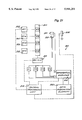
- FIG. 1 shows a system 10, which locates the position of an operative element 12 within a space (designated S).
- the system 10 is well adapted for use inside body lumens, chambers or cavities for either diagnostic or therapeutic purposes. For this reason, the system 10 will be described in the context of its use within a living body.
- the system 10 particularly lends itself to catheter-based procedures, where access to the interior body region is obtained, for example, through the vascular system or alimentary canal, without complex, invasive surgical procedures.
- the system 10 can be used during the diagnosis and treatment of arrhythmia conditions within the heart, such as ventricular tachycardia or atrial fibrillation.
- the system 10 also can be used during the diagnosis or treatment of intravascular ailments, in association, for example, with angioplasty or atherectomy techniques.
- the system 10 also can be used during the diagnosis or treatment of ailments in the gastrointestinal tract, the prostrate, brain, gall bladder, uterus, and other regions of the body.
- the operative element 12 is carried in the illustrated embodiment at the distal end of a catheter tube 44. Nevertheless, the system 10 can also be used in association with systems and methods that are not necessarily catheter-based.
- the operative element 12 can take different forms and can be used for either therapeutic purposes, or diagnostic purposes, or both.
- the operative element 12 can comprise, for example, a device for imaging body tissue, such as an ultrasound transducer or an array of ultrasound transducers, or an optic fiber element.
- the operative element 12 can comprise a device to deliver a drug or therapeutic material to body tissue.
- the operative element 12 can comprise a device, e.g., an electrode, for sensing a physiological characteristic in tissue, such as electrical activity in heart tissue, or for transmitting energy to stimulate or ablate tissue.
- the system 10 includes a locating probe 14, which, like the operative element 12, is carried at the distal end of a catheter tube 45 for introduction into the body space S.
- the locating probe 14 establishes a localized field 20 comprising waveform energy in at least a portion of the space S.
- the system 10 provides a sensing element 16 on the operative element 12.
- the sensing element 16 acquires local characteristics of the energy field 20 surrounding it.
- the sensing element 16 may be a component added to the operative element 12, or it may comprise a component already on the operative element 12, but used for an additional purpose.
- the system 10 further includes a central processing unit 18.
- the central processing unit 18 receives as input the energy field characteristic acquired by the sensing element 16.
- the central processing unit 18 derives a position-indicating output 42, which locates the position of the sensing element 16, and thus the operative element 12 itself, relative to the locating probe 14 within the space S.
- the central processing unit 18 includes an output display device 36 (e.g., a CRT, LED display, or a printer).
- the device 36 presents the position-indicating output 42 in a visual format useful to the physician for remotely locating and guiding the operative element 12 within the localized energy field 20 generated by the locating probe 14. Further details for processing the position-indicating output 42 for display will be described in greater detail later.
- the system 10 includes an oscillator 22, which generates the waveform comprising the energy field 20.
- AC electrical alternating current
- the selected current amplitude of the oscillator output can vary between 0.1 mAmp to about 5 mAmp.
- the frequency selected can also vary from about 5 kHz to about 100 kHz.
- the maximum current is a function of the frequency, as expressed in the following equation:
- I current in ⁇ amp
- f frequency in kHz
- the shape of the waveform can also vary.
- the waveform is sinusoidal.
- square wave shapes or pulses can also be used, although harmonics may be encountered if capacitive coupling is present.
- the waveform need not be continuous.
- the oscillator 22 may generate pulsed waveforms.
- the locating probe 14 carries at least one electrode 26(1) capable of transmitting energy and at least one energy return electrode 28 capable of returning the energy to ground. These electrodes 26(1) and 28 are electrically coupled to the oscillator 22 through an electronic switch unit 30.
- the locating probe 14 also carries at least one sensing electrode (four such electrodes 26(2) to 26(5)are shown in FIG. 1), which are located between the transmitting electrode 26(1) and the return electrode 28.
- the sensing electrode(s) 26(2) to 26(5) are also capable of becoming a transmitting electrode in place of the electrode 26(1), to change the point of energy transmission, if desired.
- the illustrated embodiment shows the one return electrode 28 carried at the distal region 32 of the locating probe 14 and the other five electrodes 26(1) to 26(5) carried in a spaced-apart relationship along the probe axis 34, proximal of the return electrode 28, with the transmitting electrode 26(1) being the most proximal.
- the number and placement of the electrode(s) 26 and return electrode(s) 28 on the locating probe 14 can vary. Generally speaking, the position-resolution capability of the system 10 improves with increased number of electrodes 26. Also generally speaking, the position-resolution capability of the system 10 improves as the spacing between adjacent intermediate electrodes 26(2) to 26 (5) and the spacing between the transmitting electrode 26(1) and the return electrode 28 decreases.
- the geometry of the locating probe 14 itself can also vary.
- the locating probe 14 takes the elongated, cylindrical form of a conventional diagnostic catheter, which is well suited for deployment in interior body regions.
- the central processing unit 18 is capable of connecting the waveform output of the oscillator 22 through the switch unit 30 between the transmitting electrode 26(1) and the return electrode 28, which is coupled to isolated ground or patient ground 38. This creates an energy waveform field 20 emanating into at least a portion of the space S.
- the central processing unit 18 is also capable of acquiring a differential voltage between electrodes 26(1) to 26 (5) and the sensing electrode 16 through another switch element 72 and a data acquisition element DAQ 68.
- the differential voltage measurements are taken along iso-potential surfaces 40(1) to 40(5) in the energy waveform field 20.
- FIG. 1 shows the iso-potential surfaces associated with electrodes 26(1), 26(2), 26(3), 26(4), and 26(5) as, respectively, planes 40(1), 40(2), 40(3), 40(4), and 40(5).
- FIG. 2 shows the energy field 20 and the iso-potential surfaces 40(1) to 40(5) in plan view.
- the iso-potential surfaces 40 are shown as planar surfaces or planes. Actually, the iso-potential surfaces typically will take the form of more complex, closed curvilinear surfaces, which are orthogonal to the probe axis 34 near the probe, but which deviate significantly from planar with increasing distance from the probe.
- the depiction of the surfaces 40 in the drawings aids in the understanding of the invention, as coordinate locations in and intersections of the more complex iso-potential surfaces 40 can generally be treated equivalent to coordinate locations and intersections of planar surfaces.
- the differential comparison along the iso-potential surfaces 40(1) to 40(5) derives either an in-phase relationship or an out-of-phase relationship between the voltage sensed by the element 16 (W S ) and the voltage at the plane of the sensing electrode (W 0 ), depending upon the location of the sensing element 16 relative to the iso-potential surface 40 of the electrode 26 along which the differential measurement is acquired.
- FIG. 2 shows the sensing element 16 to be located to the right of iso-potential surfaces 40(1), 40(2), and 40(3) and to the left of the iso-potential surfaces 40(4) and 40(5).
- the differential comparison of W S and W 0 indicates an out-of-phase relationship between the two waveforms.
- the out-of-phase relationship indicates that the iso-potential surfaces 40(1), 40(2), or 40(3) are located in a proximal direction relative to the sensing element 16, meaning that the sensing element 16 is located between these iso-potential surfaces and the return electrode 28.
- the differential comparison of W S and W 0 indicates an in-phase relationship between the two waveforms.
- the in-phase relationship indicates that the iso-potential surfaces 40(4) or 40(5) are located in a distal direction relative to the sensing element 16, meaning that the these iso-potential surfaces are located between the sensing element 16 and the return electrode 28.
- the central processing unit 18 controls the switch unit 72 to electronically switch the electrodes 26(2) to 26(5) to perform a differential comparison of the waveform W S of the sensing electrode 16 and the waveform W 0 of the switched-on electrode 26.
- the differential comparison of W S and W 0 will shift from an out-of-phase condition to an in-phase condition when the measurement is acquired along the iso-potential surface 40(4).
- the switch point between out-of-phase and in-phase conditions marks the longitudinal orientation of the sensing element 16 (and thus the operative element 12) along the axis 34 of the locating probe 14, i.e., between iso-potential surface 40(3) and iso-potential surface 40(4).
- the central processing unit 18 can also perform a differential comparison between the signal amplitude of the acquired waveform A S and the signal amplitude of the waveform A 0 at the switched-on sensing electrode 26. From the differential amplitude comparison, the central processing unit 18 derives the latitudinal orientation of the operative element 12 perpendicular to the axis 34 of the locating probe 14, i.e., the vertical distance within the space S between the operative element 12 and the probe axis 34. The magnitude of the difference between A S and A 0 increases as a function of increasing distance between the sensing element 16 and the plane of the switched-on electrode 26.
- the function governing the increase of the amplitude differential over distance can be empirically determined, or be determined by finite element analysis.
- FIG. 3 shows one representative implementation.
- the system 10 includes an address bus 64, which couples the central processing unit 18 to the first-described switch unit 30.
- the first switch unit 30 is also coupled to a transmitting electrode, e.g. electrode 26(1), and return electrode 28.
- the central processing unit 18 conditions the first switch unit 30 via the bus 64 to distribute the alternating current output of the oscillator 22 in a prescribed fashion in parallel to at least the electrodes 26 (1) for return through the return electrode 28.
- the system 10 also includes a data acquisition system (DAQ) 68.
- the DAQ 68 includes a differential amplifier 70.
- the sensing element 16 is coupled to the noninverting (+) input of the amplifier 70.
- the DAQ 68 further includes the second electronic switch unit 72, which is independently coupled to the electrodes 26(1) to 26(5).
- the central processing unit 18 conditions the second switch unit 72 via a second address bus 74 to couple a selected one transmitting electrode 26 on the locating probe 14 to the inverting (-) input of the amplifier 70.
- the differential amplifier 70 reads the electrical potential of the sensing element 16 with respect to that of the switched-on transmitting electrode 26, then coupled to the amplifier 70 by the switch unit 72.
- the output 71 of the amplifier 70 is an AC voltage signal.
- the DAQ 68 also includes a synchronized rectifier 76 and peak detector 78.
- the rectifier 76 receives the AC signal voltage output of the amplifier 70 and acquires its phase relative to the phase at the output of the oscillator 22.
- the detector 78 determines the peak amplitude of the AC voltage signal output 71 of the amplifier 70.
- the rectifier 76 and detector 78 can take the form of a synchronized phase detector, or any other element that detects phase and amplitude (whether as an RMS value, peak value, average rectified value, or otherwise).
- the output of the detector 78 is an analog signal having a value corresponding to the peak amplitude of the AC output of the amplifier 70, and a sign (+ or -) denoting whether the AC voltage output is in phase with the oscillator 22 (+) or out of phase with the oscillator 22 (-).
- the DAQ 68 registers this analog signal in association with the switched-on electrode 26 then-coupled to the amplifier 70 in a sample and hold element 80.
- An analog to digital converter 82 converts the analog signals to digital signals for processing by the central processing unit 18.
- a suitable control bus 54 couples the sample and hold element 80, converter 82, and differential amplifier 70 to the central processing unit 18 for coordination and control functions.
- the central processing unit 18 can set the sampling rate of the sample and hold element 80, the input range of the converter 82, and the amplification of the amplifier 70.
- the central processing unit 18 In determining the longitudinal location of the sensing element 16, the central processing unit 18 conditions the first switch unit 30 to connect the return electrode 28 to the isolated ground 38 of the oscillator 22.
- the central processing unit 18 also conditions the first switch element 30 to direct AC current flow from the oscillator 22 in parallel to the most proximal transmitting electrode 26(1), while also conditioning the second switch unit 72 to couple the switched-on transmitting electrode 26(1) to the inverting input of the differential amplifier 70.
- the amplifier 70 subtracts the electrical potential measured at the switched-on electrode 26(1) from the electrical potential measured by the sensing element 16.
- the differential potential times the gain of the amplifier 70 constitutes the input to the rectifier 76.
- the rectifier 76 senses the synchronization of the phase of its input voltage relative to the phase of the oscillator 22, while the detector 78 senses the peak voltage.
- This signed analog value is passed through the sample and hold element 80, converted to a digital format by the converter 82 and registered by the central processing unit 18 in association with the identity of the switched-on transmitting electrode 26(1).
- the central processing unit 18 next conditions the second switch unit 72 to couple the electrode 26(2) to the inverting input of the differential amplifier 70.
- the central processing unit 18 processes the signal obtained for the switched-on electrode 26(2) in the same fashion as the output voltage signal for the first switched-on electrode 26(1).
- the central processing unit 18 proceeds in like fashion sequentially through all the remaining electrodes 26 (3), 26(4), and 26(5), deriving and processing the output voltage signal for each switched-on electrode 26.
- the processor 18 registers the digitally converted peak voltages and phase synchronization for each switched-on transmitting electrode 26(1) to 26(5).
- the synchronization of the phase of the output voltage signal of the amplifier 70 relative to the phase of the oscillator 22 will vary depending upon whether the sensing element 16 is located to the left or to the right of the transmitting electrode 26 then-coupled to the inverting input of the amplifier 70 (as FIG. 2 shows).
- the output voltage signal of the amplifier 70 will be out of phase with respect to the phase of the oscillator 22 (i.e., that analog signal received by the sample and hold element 80 will have a (-) sign). This is because the potential of the sensing element 16 acquired at the noninverting input of the amplifier 70 (during the positive phase of oscillator output) will be more negative than the potential acquired at the electrodes 26(1), 26(2), and 26(3), which are sensed at the inverting input of the amplifier 70. As long as the potential of the sensing element 16 remains more negative under these conditions, the output voltage signal of the amplifier 70 remains negative, indicating an out of phase condition.
- the output voltage signal of the amplifier 70 will be in phase with respect to the phase of the oscillator 22. This is because the potential of the sensing element 16 acquired at the noninverting input of the amplifier 70 (during the positive phase of oscillator output) will be more positive than the potential at the electrodes 26(4) and 26(5) sensed at the inverting input of the amplifier 70. As long as the potential of the sensing element 16 remains more positive under these conditions, the output voltage signal of the amplifier 70 remains positive, indicating an in phase condition.
- the central processing unit 18 monitors the output of the peak detector 78 to determine where the output changes sign, by turning from (-) to (+) or vice versa. In FIG. 2, this transition occurs between switched-on electrode 26(3) and switched-on electrode 26(4).
- the iso-potential surface 40(3) associated with the electrode 26(3) sets the longitudinal coordinate of the sensing element 16, and thus the operative element 12.
- the central processing unit 18 conditions the first switch unit 30 to direct AC current flow from the oscillator 22 to the particular switched-on electrode 26(3) at which the phase transition occurred.
- the central processing unit 18 conditions the second switch unit 72 to couple the particular phase transition electrode 26(3) to the inverting input of the differential amplifier 70 while sensing element 16 is coupled to the noninverting input of the amplifier 70.
- the amplifier subtracts the electrical potential measured at the phase-transition electrode 26(3) from the electrical potential measured at the sensing element 16.
- the differential potential times the gain of the amplifier 70 constitutes the input to the rectifier 76.
- the detector 78 senses the peak voltage amplitude of the signal.
- the output of the peak detector 78 is passed through the sample and hold element 80 and converted to digital format by the converter 82.
- This digitally converted peak voltage amplitude is registered by the central processing unit 18.
- the central processing unit 18 compares the peak voltage amplitude to a voltage amplitude variation table stored in memory, which lists variations in peak voltage amplitude as a function of distance from the plane of the transmitting electrode.
- the voltage amplitude variation table can be empirically determined or based upon finite element analysis, taking into account the physical and electrical parameters of the space S.
- a predetermined threshold amplitude is established, which corresponds to a nominal distance from the transmitting electrode, which differentiates between a "close condition” (i.e., equal to or less than the nominal distance) and a "far condition” (i.e., greater than the nominal distance).
- the central processing unit 18 When the sensed peak voltage amplitude is equal to or less than the threshold amplitude, the central processing unit 18 generates an output that notifies the physician of the "close condition" between the sensing element 16 and the switched-on transmitting electrode 26.
- the central processing unit 18 When the sensed peak voltage amplitude is less than the threshold amplitude, the central processing unit 18 generates an output that notifies the physician of the "far condition" between the sensing element 16 and the switched-on transmitting electrode 26.
- the physician has at least a qualitative indication of the position of the sensing element 16 relative to the switched-on transmitting electrode 26.
- the physician can indicate through input to the central processing unit 18 the magnitude of the nominal distance, or, alternatively, establish a range of distances that progressively indicate a "closest", “closer” and “close” variation of positions.
- the sensing of the voltage amplitude is accomplished in a way that also provides information regarding the orientation of the sensing element 16 relative to the switched-on transmitting electrode 26. More particularly, as shown in FIG. 1, the operative element 12 can carry a second sensing element 16' spaced a known distance apart from the first mentioned sensing element 16. In this arrangement, one or more transmitting electrodes on one probe are switched on in sequence or simultaneously to transmit the energy field to an indifferent patch electrode, which serves as a return path. Sensing individually at each sensing element 16 and 16' provides, not only a peak voltage amplitude, but also, through a comparison of relative phases and amplitudes at each element 16 and 16', information regarding the orientation of the operative element 12 itself.
- the central processing unit 18 can differentially compare the amplitude at sensing element 16' with the amplitude at sensing element 16 to determine that element 16 is further away from the transmitting electrodes than element 16'. This indicates that the orientation of the operative element 12 is skewed within the space S.
- the second sensing element 16' can comprise the return path for the transmitting electrode 26, instead of a return path electrode 28 carried by the locating probe 14.
- the energy field can be transmitted by one of the elements 16 or 16' and returned by the other one of the element 16' or 16. In either of theses arrangements, the peak voltage amplitude is sensed by an electrode on one of the locating probes.
- FIGS. 4 and 5 show a system 100 that locates an operative element 102 within a space (designated S) by generating an energy waveform field 110 using two locating probes 106 and 108.
- Each locating probe 106 and 108 is generally like the locating probe 14 shown in FIGS. 1 and 2, having at least one transmitting electrode and at least one return electrode.
- the locating probes 106 and 108 each carry more electrodes than the probe 14.
- the electrodes carried by the locating probe 106 are designated X(1) to X(6) and the electrodes carried by the locating probe 108 are designated Y(1) to Y(5).
- Each locating probe 106 and 108 also includes a return electrode, designated RX for probe 106 and RY for probe 108.
- the locating probes 106 and 108 are positioned relative to each other in or near the space, such that their elongated axes, respectively 120 and 122, are not parallel, but extend at an angle. In the illustrated embodiment, the angle is about 90°, but other smaller or larger angles can be used. Furthermore, the locating probes 106 and 108 need not lie in the same plane.
- the operating element 102 carries a sensing element 104.
- the central processing unit 112 connects the waveform output of an oscillator 114 through a switch unit 116 between the selected transmitting electrode Y(1) and X(1) on the locating probes 106 and 108 and the respective return electrode RY and RX, which is also couple to isolated ground or patient ground 118.
- the central processing unit 112 also couples the sensing element 104 to the electrodes of the probes 106 and 108 (via the switch unit 117 and DAQ 119) along the iso-potential surfaces TX(1) to TX(6) and TY(1) to TY(5) in the energy waveform field 110.
- FIG. 4 shows the intersecting iso-potential surfaces TX and TY in side view.
- FIG. 5 shows the intersecting iso-potential surfaces TX and TY in perspective view.
- the central processing unit 112 performs a differential comparison of the waveform W S to the waveform output W 0 when each of the transmitting electrodes X(1) to X(6) and Y(1) to Y(5) are switched on.
- the differential comparison derives either an in-phase or relationship an out-of-phase relationship between W S and W 0 , depending upon the location of the sensing element 104 relative to the iso-potential surface TX(N) or TY(N) of the switched-on voltage sensing electrode X(N) or Y(N).
- FIG. 4 shows the sensing element 104 to be located to the right of (or above, in the vertical orientation shown in FIG. 4) the iso-potential surfaces TX(1) to TX(4) and to the left of (or below, from the vertical orientation shown in FIG. 4) the iso-potential surfaces TX(5) and TX(6).
- the differential comparison of W S and W 0 indicates an out-of-phase relationship between the two waveforms. This means that the sensing element 104 is located between these planes and the return electrode RX.
- the central processing unit 112 controls the switch unit 116 to electronically switch the electrodes on, sequentially from most proximal to most distal, i.e., sequentially from left to right (or from bottom to top, in the vertical orientation shown in FIG. 4) from X(1) to X(6). This sequentially switches on differential sensing along the iso-potential surfaces TX(1) to TX(6).
- the central processing unit 112 For each switched-on electrode X(1) to X(6), the central processing unit 112 performs (via the DAQ 119) a differential comparison of the waveform W S of the sensing electrode 104 and the waveform W 0 of the switched-on electrode X(N).
- the differential comparison of W S and W 0 will shift from an out-of-phase condition to an in-phase condition when measurement occurs along the iso-potential surface TX(5).
- the switch point between out-of-phase and in-phase conditions marks the longitudinal orientation of the sensing element 104 (and thus the operative element 102) along the axis 120 of the locating probe 106, i.e., between iso-potential surface TX(4) and iso-potential surface TX(5).
- the central processing unit 112 can also perform a differential comparison between the signal amplitude of the sensed waveform A S and the signal amplitude of the waveform at the switched-on transmitting electrode A 0 . From the differential amplitude comparison, the central processing unit 112 derives the latitudinal orientation of the operative element 102 perpendicular to the axis 120 of the probe 106, i.e., the vertical distance within the space S between the operative element 102 and the probe axis 120.
- FIG. 4 shows the sensing element 104 to be located to the right of the iso-potential surfaces TY(1) to TY(2) and to the left of the iso-potential surfaces TY(3), TY(4), and TY(5).
- the central processing unit 112 controls the switch unit 117 to electronically switch on the transmitting electrodes, sequentially from most proximal to most distal, i.e., sequentially from left to right, Y(1) to Y(5). This sequentially switches on differential sensing along the iso-potential surfaces TY(1) to TY(5).
- the central processing unit 112 For each switched-on electrode Y(1) to Y(5), the central processing unit 112 performs (via the DAQ 119) a differential comparison of the waveform W S of the sensing element 104 and the waveform W 0 of the switched-on transmitting electrode Y(N).
- the differential comparison of W S and W 0 along the probe 108 will shift from an out-of-phase condition to an in-phase condition when iso-potential surface TY(3) is switched on.
- the switch point between out-of-phase and in-phase conditions marks the longitudinal orientation of the sensing element 104 (and thus the operative element 102) along the axis 122 of the locating probe 108, i.e., between iso-potential surface TY(2) and iso-potential surface TY(3).
- the central processing unit 112 can also perform a differential comparison between the signal amplitude of the sensed waveform A S and the signal amplitude of the waveform at the switched-on transmitting electrode A 0 to derive the latitudinal orientation of the operative element 102 perpendicular to the axis 122 of the probe 108, i.e., the vertical distance within the space S between the operative element 102 and the probe axis 122.
- the component parts of the system 100 can incorporate the particular electrical configuration shown in FIG. 3, or another analog or digital configuration, to carry out the above differential comparisons.
- the central processing unit 112 provides a position-indicating output 124, which correlates the position of the sensing element 104 (and thus the operative element 102) within the grid of intersecting iso-potential surfaces TX(N) and TY(N).
- the position-indicating output 124 is presented to the physician on a display device 126.
- the individual identification probes 106 and 108 shown in FIGS. 4 and 5 can be assembled into a composite structure 150, as shown in FIG. 6.
- the structure 150 comprises an array of flexible spline elements 152 extending longitudinally between a distal hub 154 and a proximal base 156.
- the structure 150 includes four spline elements 152(1) to 152(4) (only 3 spline elements are visible in FIG. 6). A greater or lesser number of spline elements 152 can be present.
- Each spline element 152 preferably comprises a flexible body made from resilient, inert wire or plastic. Elastic memory material such as nickel titanium (commercially available as NITINOLTM material) can be used. Resilient injection molded plastic or stainless steel can also be used. Each spline element 152 is preferably preformed with a convex bias, creating a normally open three-dimensional basket structure.
- the structure 150 is carried at the end of a catheter tube 158.
- An outer sheath 160 slidably advances forward along the catheter tube 158 to compress and collapses the structure 150 (see FIG. 7) for introduction into the body region. Rearward movement retracts the slidable sheath 160 away from the structure 150, which springs open and assumes its three-dimensional shape (as FIG. 6 shows).
- spline elements 152 is both radially and axially symmetric.
- Asymmetric structures either radially or axially or both, can also be used. Examples of asymmetric arrays of spline structures are shown in copending U.S. application Ser. No. 08/742,569, filed Oct. 28, 1996 and entitled “Asymmetric Multiple Electrode Support Structures,” which is incorporated herein by reference.
- Each spline element 152 carries an array of multiple transmitting electrodes TE and at least one return electrode RE, as previously described. Each spline element 152 thus comprises a locating probe.
- the structure 150 comprises an ordered array of multiple location probes, which, in use, create a waveform field 162 about the space bounded by the spline elements 152.
- FIG. 6 shows an operative element 172 movable within the energy waveform field 162.
- the operative element 172 carries a sensing element 174.
- a central processing unit 164 sequentially connects the waveform output of an oscillator 166 through a switch unit 168 to the transmitting electrodes TE on each spline element 152 (for example, beginning with the most proximal and moving distally), while coupling the respective most distal return electrode RE of the spline element 152 to isolated ground or patient ground 170.
- the central processing unit 164 also sequentially couples the electrodes TE and the sensing electrode 174 on the operative element 172 through a switch unit 169 and a DAQ 171 to acquire a differential voltage along a grid of intersecting iso-potential surfaces TP in the energy waveform field 162, in the same manner shown for the probes 106 and 108 in FIGS. 4 and 5.
- the differential comparison derives either an in-phase relationship or an out-of-phase relationship between W S and W 0 , depending upon the location of the sensing element 174 relative to the transmitting electrodes along each elongated spline element 152.
- the central processing unit 164 can also perform a differential comparison between the signal amplitude of the sensed waveform A S and the signal amplitude of the waveform at the switched-on electrode A 0 where the phase transition occurs, to derive the latitudinal orientation of the sensing element 174 perpendicular to each spline element 152.
- FIG. 8 shows an alternative embodiment of system 300 that locates an operative element 302 within a space (designated S), using differential voltage analysis instead of differential waveform analysis.
- the system generates an energy waveform field 310 between two locating probes 306 and 308.
- Each locating probe 306 and 308 includes at least one transmitting electrode, which are designated X(1) to X(6) for probe 106 and Y(1) to Y(6) for probe 108.
- the operative element 302 carries a sensing element 304.
- the locating probes 306 and 308 are positioned so that their elongated axes, respectively 320 and 322, are not parallel, but extend at some angle. In the illustrated embodiment, the angle is about 90°, but other smaller or larger angles can be used. Alternatively, because differential voltage analysis is employed, the locating probes 306 and 308 in this embodiment can be located in a parallel, mutually facing relationship.
- the operation of the system 300 is governed by a central processing unit 312.
- the central processing unit 312 connects the waveform output of an oscillator 314 through a first switch unit 316 to transmit the waveform from all transmitting electrodes on one probe 306 to all the electrodes on the other probe 308, which are coupled to the isolated patient ground 318.
- the probe 306 will be called the "transmitting probe” and the probe 308 will be called the "receiving probe.”
- the receiving and transmitting functions of the probes 306 and 398 can be reversed.
- the generated waveform field 310 extends between the transmitting probe 306 and the receiving probe 308.
- the waveform can be generated simultaneously between all electrodes or sequentially along the axis of the probes 306 and 308.
- the waveform field 310 includes iso-potential surfaces T(1) to T(6), which extend between the transmitting-receiving electrode pairs X(1)-Y(1) to X(6)-Y(6).
- the central processing unit 312 conditions a second switch element 330 to couple each switched-on electrode on the transmitting probe 306 in succession to inverting (-) input of a differential amplifier 332, while coupling the sensing element 304 to the noninverting (+) input.
- the amplifier subtracts the electrical potential measured by the electrode coupled to the inverting input from the electrical potential measured by the sensing element 304.
- the differential potential times the gain of the amplifier 332 constitutes the input to a rectifier 334.
- a detector 336 senses the peak voltage, and the rectifier 334 senses the synchronization of the phase of the voltage signal relative to the phase of the oscillator 314.
- the central processing unit 312 registers the peak voltage and the synchronization in association.
- the synchronization of the phase of the output voltage signal of the amplifier 332 relative to the phase of the oscillator 314 will vary depending upon the location of the most immediately distal iso-potential surface to the sensing electrode 304.
- the output voltage signal of the amplifier 332 will be in-phase with respect to the phase of the oscillator 314 only when the differential amplitude is measured along the iso-potential surface which is most immediately distal to the sensing electrode 304.
- the most immediate distal iso-potential surface to the sensing electrode 304 is T(6), which lies between electrode pairs X(6)-Y(6).
- the output voltage signal of the amplifier 332 will be out-of-phase with respect to the phase of the oscillator 314 for the differential amplitudes measured along the most immediately proximal iso-potential surface to the sensing electrode 304, and along all other more proximal iso-potential surfaces.
- the most immediate proximal iso-potential surface is T(5), which lies between electrode pairs X(5)-Y(5) and the remaining more proximal surfaces T(4) to T(1) lie between electrode pairs X(4)-Y(4) to X(1)-Y(1).
- the output voltage signal of the amplifier 332 will be in-phase with respect to the phase of the oscillator 314 only when the differential amplitude is measured along the iso-potential surface T(4), which is the most immediately distal to the sensing electrode 304'.
- the output voltage signal of the amplifier 332 will be out-of-phase with respect to the phase of the oscillator 314 for the differential amplitudes measured along the most immediate proximal iso-potential surface T(3) and all other more proximal iso-potential surfaces T(2) and T(1).
- Differential voltage analysis can also be used in association with the composite probe structure 150 shown in FIG. 6 or any of the structures shown earlier.
- FIG. 9 shows a representative implementation of a three-dimensional navigation system 200, which includes three locating probes 204, 206, and 208 positioned within a space S.
- the space S comprises the interior of a heart.
- the system 200 locates and guides an operative element 202 within the heart.
- the operative element 202 can serve to sense electrical activity in the heart to locate potential ablation sites, or to transmit energy to pace heart tissue, measure impedance, or to ablate.
- the operative element 202 can include an imaging element to image tissue, anatomic structures, or lesions formed within the heart.
- the operative element can include a cannula to penetrate heart tissue for the purpose of injecting an ablation media, or to inject a drug or gene therapy agent.
- the three locating probes 204, 206, and 208 are purposely situated within the heart to provide spaced-apart navigational points for locating the operative element 202. Furthermore, the probes 204, 206, and 208 are located at different coordinate planes, to create a three-dimensional navigational grid and make triangulation possible.
- the probes 204, 206, and 208 are individually placed at or near known anatomic regions of the heart using, for example, fluoroscopy or another imaging technology, such as ultrasound. This is because potential ablation sites within the atria are typically identified by reference to an anatomic landmark within the heart.
- a single locating probe or multiple locating probes may be positioned essentially in any region within the heart or in any tissue or vascular region surrounding the heart for purposes of establishing navigational points of reference to locate the operative element 202.
- Any region of placement with the body that can be imaged by fluoroscopic or other imaging technology can be selected as a potential navigational site. The region of placement therefore does not have to represent a particular fixed anatomic site. For example, establishing a three-dimensional navigation system for use within a given heart chamber, one or more locating probes can be located within the heart chamber, another one or more probes may be located in a different chamber, and yet another one or more locating probes can be located at an epicardial location outside the interior of the heart.
- the first locating probe 204 is positioned in region of the high right atrium; the second locating probe 206 is positioned in the region of the right ventricular apex; and the third locating probe 208 is positioned in the region of the coronary sinus.
- the three probes 204, 206, and 208 are located on different coordinate planes, so that the probe axes extend in mutually nonparallel relationships.
- Each locating probe 204, 206, and 208 includes multiple transmitting electrodes TE and a distal return electrode TR, which function in the manner previously described and shown in FIG. 1.
- a transmitting electrode TE and the return electrode TR on each probe 204, 206, and 208 are coupled via electronic switch units 210 to an oscillator 212 to create an energy waveform field 216.
- the operative element 202 carries a sensing element 218, which can also can serve as an ablation electrode or as sensing electrode.
- the sensing element 218 is coupled to the central processing unit 214 in the manner previously described to sense the waveform quantity W S within the field 216.
- a DAQ 68 acquires differential waveforms along multiple iso-potential surfaces TP, one associated with each electrode TE on each probe 204, 206, and 208. As shown in FIG. 9, because the probes 204, 206, and 208 are located at different coordinate planes, the multiple iso-potential surfaces TP form intersection points within the field 216.
- the central processing unit 214 employs the DAQ 68 previously described (see FIG. 3) to differentially compare W S to W 0 for each switched-on electrode TE and locate regions of phase transitions relative to each probe 204, 206, and 208.
- the central processing unit 214 can also perform a differential comparison between the signal amplitude of the sensed waveform A S and the signal amplitude of the waveform at the switched-on transmitting electrode A 0 where the phase transition occurs to derive the latitudinal orientation of the sensing element 218 perpendicular to the axis of each probe 204, 206, 208.
- the central processing unit 214 generates a position-indicating output 220, which locates the sensing element 218 (and thus the operative element 202 itself) within the matrix of intersecting iso-potential surfaces TP generated by the three probes 204, 206, and 208.
- FIG. 17 shows a three dimensional system 500, which conducts an iterative differential voltage analysis to determine the location of an operative element 502 within a space S peripherally bounded by multiple locating probes 504.
- the multiple locating probes 504 are assembled together by a distal hub 506 and a proximal base 508 into a composite, three-dimensional basket structure 510 of the type previously shown and described in FIG. 6.
- the multiple locating probes 504 need not be assembled together in a composite structure, but exist as separate probes located about the space S, in the manner shown in FIG. 9, as previously described.
- the composite structure 510 is well suited for use within the heart and can perform other functions in addition to navigation.
- the composite structure 510 can serve to transmit electrical signals to pace heart tissue or to characterize the electrical characteristics of the tissue by acquiring tissue impedance measurements.
- the composite structure can also serve to sense electrical activity in myocardial tissue to acquire electrograms for heart mapping procedures.
- the composite structure 510 shown in FIG. 17 includes eight locating probes 504, and each probe, in turn, carries eight electrodes 505, for a total of sixty-four electrodes 505 positioned about the space S.
- the system 500 includes a central processing unit 512, which couples a voltage source 514 to a transmitting electrode 516 carried by the operative element 502.
- a transmitting electrode 516 carried by the operative element 502.
- an indifferent electrode 518 carried as a patch on the exterior of the patient, comprises the voltage return, which is, in turn, coupled to isolated or patient ground 520.
- another electrode carried by the operative element 502 can serve as the voltage return.
- the electrode 516 creates a voltage field 517 within the space S, which varies in detected amplitude at each probe electrode 505 according to its distance from the transmitting electrode 516.
- the system 500 includes a data acquisition element 522 coupled to the central processing unit 512 and to a switch element 524.
- the switch element 524 individually conditions each electrode (A,B) to sense voltage existing at its location within the field 517, which the data acquisition element 522 samples and holds, in the manner previously described, e.g., see FIG. 3.
- the central processing unit 512 includes a processing component 526 which derives a position-indicating output 528 based upon the voltage distribution sensed by the electrodes (A,B) on the probes 504.
- FIG. 18 shows the steps of a preferred algorithm 530 for deriving the output 528.
- the algorithm 530 includes, as a first step 532, establishing an estimated coordinate position P(x, y, z) EST for the transmitting electrode 516 on the operative element 502 within the space S, where x is the x-field coordinate, y is the y-field coordinate, and z is the z-field coordinate.
- P (x, y, z) EST can be initially arbitrarily set at P(0,0,0), which is at the geometric center of the voltage field 517 (designated as GC in FIG. 17).
- differential waveform analysis or differential voltage analysis, or amplitude analysis, as described above, alone or in combination, can also be used to more accurately estimate P (x, y, z) EST .
- position indicating methodologies disclosed in copending patent application Ser. No. 08/320,301, filed Oct. 11, 1994 and entitled “Systems and Methods for Guiding Movable Electrode Elements Within Multiple Electrode Structures” can also be used to provide a more accurate initial position estimate P(x, y, z) EST .
- multiple signals that are orthogonal from a signal processing standpoint may be transmitted simultaneously in the manner shown in FIG. 22 (as will be described in greater detail later).
- the algorithm 530 computes the distance ⁇ D(A,B) between each probe electrode (A,B) and the transmitting electrode 516 at P(x,y,z) EST .
- the distances ⁇ D(A,B) can be normalized to facilitate analysis.
- the algorithm then applies a preestablished, mathematical voltage-to-distance function 534 to derive the estimated voltage V(A,B) EST at each electrode (A,B), based upon ⁇ D(A,B).
- the algorithm 530 constructs an estimated voltage distribution matrix, which would exist, according to the function 534, if P (x, y, z) EST was the actual voltage transmission point.
- the voltage-to-distance function 534 can be empirically determined or be based upon finite element analysis and stored in memory accessible to the central processing unit 512.
- the algorithm 530 derives an estimated or expected voltage differential V(A,B) EST for each electrode 505.
- the algorithm 530 receives as input V(A, B) ACT , where V(A, B) ACT is the measured voltage value acquired by operation of the data acquisition element 522 at each probe electrode (A,B).
- V(A, B) ACT is the measured voltage value acquired by operation of the data acquisition element 522 at each probe electrode (A,B).
- the algorithm 530 in this step 540, creates a measured voltage distribution pattern 560 based upon the values for V (A, B) ACT , which plots (on the Y-axis) the sensed voltage values for each electrode (numbered 1 to 64 on the X-axis).
- the algorithm 530 creates an estimated voltage distribution pattern 562 based upon the values for V (A, B) EST , which plots (on the Y-axis) the estimated voltage values for each electrode (again numbered 1 to 64 on the X-axis).
- the algorithm 530 matches the voltage distribution pattern 560 with the voltage distribution pattern 562 to derive a voltage matching coefficient VM COEF .
- the value of the voltage matching coefficient VM COEF for a given P(x, y, z) EST increases as P(x, y, z) EST coincides with the actual location of the transmitting electrode 516. That is, the value of the voltage matching coefficient increases in relation to the proximity of the transmitting electrode 516 to the estimated position P (x, y, z) EST .
- the central processing unit 512 can derive the matching coefficient VM COEF in various conventional ways, for example, by employing pattern matching; matched filtering; or cross correlation. Examples of using these techniques to derive matching coefficients appear in copending U.S. patent application Ser. No. 08/390,383, filed Feb. 17, 1995 and entitled "Systems and Methods for Examining Heart Tissue Employing Multiple Electrode Structures and Riving Electrodes," which is incorporated herein by reference.
- the algorithm 530 determines whether VM COEF is the "best", i.e., whether it is maximized under the processing rules applied. For the first iteration, and for all subsequent iterations were VM COEF is not maximized, the algorithm 530 applies (in step 546) a preselected incremental correction factor ⁇ x to the x coordinate, factor ⁇ y to the y coordinate, and factor ⁇ z to the z coordinate of the estimated position of the transmitting electrode 516 to create a new estimated position P(x+ ⁇ x, y+ ⁇ y, z+ ⁇ z)), which become the new coordinates for an estimated position P(z, y, z) EST .
- the algorithm 530 then loops through the foregoing steps 536, 538, 540, 542, and 544, to derive an iterated voltage matching coefficient VM COEF based upon the new estimated location.
- the algorithm 530 iteratively selects ⁇ x, ⁇ y, and ⁇ z until a best (maximum value) voltage matching coefficient VM COEF is achieved in step 544.
- the coordinates P(x,y,z) EST at the best, maximum voltage matching coefficient VM COEF become the position-indicating output 528, as shown in step 548 in FIG. 18.
- the algorithm 530 can iterate the x-coordinate alone (keeping the y- and z-coordinates constant) until a best voltage matching coefficient VM COEF is achieved, then fix the x-coordinate at that value and iterate the y-coordinate alone (while also keeping the z-coordinate constant) until another best voltage matching coefficient VM COEF is achieved, and then fix the y-coordinate at that value and iterate the z-coordinate alone (keeping the previously fixed x- and y-coordinates constant), until another best voltage matching coefficient VM COEF is achieved.
- the algorithm 530 then loops back through this process, until the best voltage matching coefficient VM COEF is obtained for each local x-, y-, and z-coordinate, as well as for P(x, y, z) EST overall.
- the x-, y-, and z-coordinates can be simultaneously incremented to maximize the voltage matching coefficient VM COEF for P(x, y, z) EST , using, for example, a conventional maximum gradient method.
- the algorithm 530 shown in FIG. 18 corrects for distortion of the locating probes caused by exposure to dynamic conditions within a body cavity, such as within a beating heart chamber.
- the iterative nature of the algorithm 530 also corrects for electrical "noise" caused, for example, by the inherent electrical resistance of the electrodes and associated electrical wiring.
- the iterative differential voltage analysis just described also makes possible the generation of an error signal, should the position of the operative element 502 stray beyond the energy field 517. Should this event occur, the estimated voltage and the actual voltage become mirror images. This outcome, when sensed by the central processing unit 512, can command the generation of an out-of-field error signal.
- the central processing unit 512 can incorporate a neural network 600 (see FIG. 17), which has been trained on experimentally acquired sets of voltage distribution data correlated with known positions of the transmitting electrode 516. Once the training phase is completed, the network 600 can instantaneously output the position-indicating output 528, based upon input from the data acquisition element 522 of voltage distribution data sensed by the probe electrodes 505 during transmission of voltage by the electrode 516.
- a neural network 600 see FIG. 17
- the position-indicating output 220 (or, in the embodiment shown in FIG. 17, the output 528) is preferably processed for viewing on a display device 221.
- the central processing unit 214 includes an input 222 that receives information pertaining to the position and orientation of the locating probes 204, 206, and 208 within the heart.
- the input 222 also receives information pertaining to the shape and size of each locating probe 204, 206, and 208.
- the central processing unit 214 includes functional algorithms 224, which set guidance parameters based upon the input information. These guidance parameters are used by the central processing unit 214 to analyze the spatial variations of the electric waveform field generated by the locating probes 204, 206, and 208.
- the guidance parameters govern the processing of differential comparison data to create the position-indicating output 220 for display on the device 221.
- the processed position-identifying output aids the physician in locating and guiding the operative element 202 in real time.
- the probes 204, 206, and 208 of the system 200 are members of a family 209 of locating probes.
- the various probes comprising the family 209 are characterized by different geometries, different densities of transmitting and return electrodes, and other structural and functional differences.
- each probe 204, 206, and 208 within the family 209 includes an identification component 270.
- the identification component 270 carries an assigned identification code XYZ.
- the code XYZ identifies the shape and size of the electrode-supporting part of the probe and the distribution of electrodes carried thereon, in terms of the number of electrodes and their spatial arrangement.
- the structure-specific information contained in the code XYZ aids the central processing unit 214 in creating a positioning matrix based upon the locating probes when deployed.
- the coded component 270 is located within the handle 230 attached to the proximal end of the catheter tube 232 that carries the locating probe 204, 206, and 208.
- the component 270 could be located elsewhere in relation to the locating probe.
- the coded component 270 is electrically coupled to an external interpreter 278 when the probe is coupled to the central processing unit 214 for use.
- the interpreter 278 inputs the code XYZ that the coded component 270 contains.
- the interpreter 278 electronically compares the input code XYZ to, for example, a preestablished master table 280 of codes contained in memory.
- the master table 280 lists, for each code XYZ, the structure-specific information required to create the positioning matrix to locate and guide the operative element 202 within the waveform field 216.
- the functional algorithms 224 of the central processing unit 214 set location and guidance parameters based upon the code XYZ.
- the algorithms 224 preferably disable the central processing unit 214 in the absence of a recognizable code XYZ. Thus, only probes of the family 209 possessing a coded component 270 carrying the appropriate identification code XYZ can be used in association with the processing element 214.
- the coded component 270 can be variously constructed. It can, for example, take the form of an integrated circuit 284 (see FIG. 11), which expresses in digital form the code XYZ for input in ROM chips, EPROM chips, RAM chips, resistors, capacitors, programmed logic devices (PLD's), or diodes. Examples of catheter identification techniques of this type are shown in Jackson et al. U.S. Pat. No. 5,383,874, which is incorporated herein by reference.
- the coded component 270 can comprise separate electrical elements 286 (see FIG. 12), each one of which expresses an individual characteristic.
- the electrical elements 286 can comprise resistors (R1 to R4), comprising different resistance values, coupled in parallel.
- the interpreter 278 measures the resistance value of each resistor R1 to R4.
- the resistance value of the first resistor R1 expresses in preestablished code, for example, the number of electrodes on the probe.
- the resistance value of the second resistor R2 expresses in preestablished code, for example, the distribution of electrodes on the probe.
- the resistance value of the third resistor R3 expresses in preestablished code, for example, the size of the probe.
- the resistance value of the fourth resistor R4 expresses in preestablished code, for example, the shape of the probe.
- the three-dimensional basket structure 510 shown in FIG. 17 can also carry an identification component 270 having an assigned identification code XYZ to identify the shape and size of the multiple probe structure 510 and the distribution of electrodes carried thereon.
- the structure-specific information contained in the code XYZ aids the position derivation component 528 and algorithm 530 in FIG. 18 to construct the estimated voltage distribution matrix and analyze sensed voltage differentials.
- the central processing unit 512 can also include a component 550 (see FIG. 17), which electronically determines structure-specific information to construct the estimated voltage distribution matrix and analyze sensed voltage differentials.
- the component 550 commands, in sequence, the transmission of voltage from the source 514 through a switch unit 554 from each probe electrode (A,B) to the indifferent electrode 518, while sensing voltage with the remaining probe electrodes through the switch 524 and data acquisition element 522.
- the component 550 thereby acquires a first set of data from which the voltage differential between every electrode (A,B) can be obtained.
- the component 550 includes an input 552, through which the component 550 acquires data relating to the linear distance between adjacent electrodes on each probe 504.
- the electrodes 505 on each probe 504 will be spaced apart by manufacturer at the same linear distance, so that will typically be only a single linear distance to input.
- the physician can manually enter the linear distance information through the input 522.
- the input 552 of linear distance information can be carried by a coded component 270 as earlier described as shown in FIG. 10, which is inputted automatically upon coupling the probe structure 510 to the central processing unit 512. In this arrangement, more complex linear distance information can be readily inputted.
- the linear distance information comprises a second set of data.
- the component 520 then derives using conventional estimating techniques the distances between other, nonadjacent electrodes 505, both along a probe 504 and between probes 504.
- the component 550 generates a geometric output 556, which, like the code XYZ, the output 556 identifies the shape and size of the multiple probe structure 510 and the distribution of electrodes 505 carried thereon.
- the output 556 also provides the basis for calculating the interior volume of the structure 510.
- the interior volume of the structure 510 typically will conform to the interior volume of the heart chamber it occupies.
- the interior volume will also typically dynamically adjust to the changing heart chamber volumes during systole and diastole.
- the component 550 therefore makes possible the electrical analysis, for therapeutic or diagnostic purposes, of heart chamber volumes and the changes in heart chamber volume during systole and diastole.
- the component 550 can thereby be used, independent of or in association with a navigation function, to characterize heart morphology and function.
- the component 550 can incorporate a neural network 558 (see FIG. 17) to generate in situ the distance-to-voltage function 534 particular to a given structure 510, based upon the electrically sensed geometry and distribution of electrodes on the structure 510.
- the neural network 558 is first trained on a known set of data that have been previously acquired experimentally. For example, using a back-propagation model, the network 558 can be trained to predict a voltage-to-distance function 534 based upon structure-specific information. Once the training phase is completed, the network 558 can be used to predict the voltage-to-distance function in situ.
- the central processing unit 214 (or 512 in FIG. 17) electronically constructs a three-dimensional coordinate system representing a virtual image 290 of the energy field 216 (or 217 in FIG. 17) and surrounding tissue mass T.
- FIG. 13 shows a representative virtual image 290 based upon two locating probes.
- the virtual image 290 indicates the position of the locating probes (designated “Probe X” and “Probe Y” in FIG. 13), as well shows the geometry and location of the iso-potential surfaces (designated "X(1) to X(3)” and "Y(1) to Y(3)” in FIG. 13).
- the virtual image 290 shows the position of the operative element 202 (designated "Device" in FIG.
- the central processing unit 214 continuously performs the differential comparisons and updates the virtual image 290 to provide a real time display for viewing by the physician.
- the locating and navigation systems of the type previously described create an energy field by applying a single waveform. Multiple waveforms can be simultaneously applied to gain processing efficiencies, provided the different waveforms are orthogonal from a signal processing standpoint. Examples of different, orthogonal processing signals includes waveform signals of different frequencies, waveform signals of the same frequency but which differ by 90° in phase, and waveforms from uncorrelated white noise sources.
- FIG. 20 shows a system 700 that locates an operative element 702 within a space S by generating different waveforms using two probes 706 and 708.
- the system 700 shares common elements with the system 100 shown in FIG. 4.
- the locating probe 706 and 708 are generally like the locating probes 106 and 108 shown in FIG. 4.
- the electrodes carried by the locating probe 706 are designated X(1) to X(6) and the electrodes carried by the locating probe 708 are designated Y(1) to Y(5).
- Each locating probe 706 and 708 also includes a return electrode, designated RX for probe 706 and RY for probe 708.
- the locating probes 706 and 708 are positioned relative to each other in a non-parallel relationship.
- the operating element 702 carries a sensing element 704.
- the system 700 includes a first waveform source WF1, which is coupled to the probe 708.
- the system also includes a second waveform source WF2.
- the first waveform WF1 is different than but orthogonal to the second waveform WF2.
- the waveforms WF1 and WF2 have different frequencies, and the sources comprise separate oscillators 720 and 722.
- the probe 708 is coupled via a switching unit 710 and a first filter F1 for the waveform WF1 to the inverting (-) input of a differential amplifier 712.
- the probe 706 is also coupled by a second switching unit 714 and a second filter F2 for the WF2 is also coupled to the inverting (-) input of the differential amplifier 712.
- the sensing element 704 carried by the operative element 702 is coupled to the noninverting (+) input of the differential amplifier 712.
- the output of the differential amplifier 712 is coupled to a data acquisition element 716.
- the data acquisition element 716 includes a rectifier, peak detector, sample and hold element, and analog-to-digital converter coupled as shown in FIG. 3 to process the differential output in the manner previously described, under the control of a central processing unit 718.
- the multiple oscillators 720 to 722 simultaneous apply the waveform WF1 to the electrode Y(1), for return through the return electrode RY, and the different waveform WF2 to the electrode X(1), for return through the return electrode RX.
- the central processing unit 718 operates the switch units 710 and 714 to simultaneously acquire two differential voltages, one for waveform WF1 between the sensing element 704 and the electrode Y(1) and the other for waveform WF2 between the sensing element 704 and the electrode X(1).
- the differential amplifier 712 thus acquires phase information for two waveforms simultaneously along iso-potential surfaces TX(1) and TY(1).
- the central processing unit 718 operates the switch units 710 and 714 to simultaneously acquire two differential voltages for the waveforms WF1 and WF2 between the sensing element 704 and the electrodes Y(2)/X(2), then Y(3)/X(3), and so on.
- the differential amplifier 712 acquires phase information for two waveforms simultaneously along iso-potential surfaces TX(2)/TY(2), then TX(3)/TY(3), etc.
- This simultaneously acquired phase information from two waveforms WF1 and WF2 is processed by the data acquisition element 716 to provide a position-indicating output. Greater processing efficiencies can therefore be obtained.
- FIG. 21 shows a system 800 in which multiple oscillators 802, 804, 806, and 808 apply different waveforms WF1, WF2, WF3, and WF4 simultaneously to multiple electrodes, respectively E(1), E(2), E(3), and E(4), of a single probe 810, through an indifferent return electrode 830.
- the different waveforms WF1, WF2, WF3, and WF4 are orthogonal in a signal processing sense possessing, for example, they possess different frequencies. Since the waveforms are applied simultaneously to all electrodes E(1) to E(4), no input switching is required.
- All electrodes E(1) to E(4) of the probe 810 are coupled to an output switch 812.
- the output switch 810 is, in turn, coupled to filters F1, F2, F3, and F4 for the frequencies of, respectively, WF1, WF2, WF3, and WF4.
- the output of the filters F1, F2, F3, and F4 are coupled to the inverting(-) input of a differential amplifier 814.
- the sensing element 816 carried by an operative element 818 is coupled to the noninverting (+) input of the differential amplifier 814.
- the output of the differential amplifier 814 is coupled to a data acquisition element 820.
- the data acquisition element 820 includes a rectifier, peak detector, sample and hold element, and analog-to-digital converter coupled as shown in FIG. 3 to process the differential output in the manner previously described, under the control of a central processing unit 818.
- the data acquisition element 820 simultaneously acquires the differential amplitude of waveform WF1 between the sensing element 816 and the electrode E(1), the differential amplitude of waveform WF2 between the sensing element 816 and the electrode E(2), the differential amplitude of waveform WF3 between the sensing element 816 and the electrode E(3), and the differential amplitude of waveform WF4 between the sensing element 816 and the electrode E(4).
- the data acquisition element 816 is able to simultaneously infer distance with respect to each probe electrode E(1), E(2), E(3), and E(4).
- FIG. 22 shows a system 900 for conducting an iterative voltage analysis using multiple waveforms to determine the location of an operative element 902 within a space S peripherally bounded by a composite, three-dimensional basket structure 910, like that shown in FIG. 17.
- the composite structure 910 in FIG. 22 includes eight locating probes 904, and each probe, in turn, carries eight electrodes 905, for a total of sixty-four electrodes 905 positioned about the space S.
- the operative element 902 carries two energy transmitting electrodes 912 and 914.
- Multiple oscillators 916 and 918 apply different waveforms WF1 and WF2 simultaneously to the electrodes 912 and 914.
- the different waveforms WF1 and WF2 are orthogonal in a signal processing sense possessing, for example, they possess different frequencies. As in FIG. 21, since the waveforms are applied simultaneously to both electrodes 912 and 914, no input switching is required.
- a central processing unit 920 conditions the electrode 912 and the electrode 914 to simultaneously transmit waveform energy WF1 and WF2 to a patch return electrode 922.
- Each probe electrode (A,B) is coupled via a switch 924 to two filters F1 and F2 for the frequencies of the waveforms, respectively, WF1 and WF2.
- a data acquisition element 926 thereby receives simultaneous inputs from two waveforms WF1 and WF2.
- the input for the waveform WF1 could provide a sensed voltage, for use by the algorithm 530 (shown in FIG. 18) in deriving the position-indicating output 528.
- the input for the waveform WF2 could provide phase and amplitude information for comparison to the phase and amplitude information of waveform WF1, from which the orientation of the operative element 12 can be ascertained.
- the system 900 also make possible the derivation of both location and orientation out.
- a second operative element 902' could be present within the space S bounded by the basket structure 910.
- the second operative element 902' carries at least one transmitting electrode 912'.
- the electrode 912 of the first operative element 902 transmits the first waveform WF1
- the electrode 912' of the second operative element 902' transmits the second waveform WF2.
- a data acquisition element 926 thereby receives simultaneous inputs from two waveforms WF1 and WF2, via the filters F1 and F2.
- the input for the waveform WF1 could provides a sensed voltage, for use by the algorithm 530 (shown in FIG.
- the system 900 is thereby able to provide locating information for multiple operative elements.
- FIG. 14 shows a multiple electrode structure 400 located in the right atrium of a heart.
- the structure 400 is flexible and carries a steering mechanism (not shown), use of which flexes the structure 400 into curvilinear shapes.
- the structure 400 carries an array of electrodes 402, which transmit radio frequency energy to ablate myocardial tissue.
- the electrodes 402 are preferably operated in a uni-polar mode, in which the radio frequency ablation energy transmitted by the electrodes 402 is returned through an indifferent patch electrode 404 externally attached to the skin of the patient.
- the electrodes 402 can be operated in a bi-polar mode, in which ablation energy emitted by one or more electrodes 402 is returned an adjacent electrode 402 carried in the structure 400.
- FIG. 15 shows a representative long, continuous lesion pattern 406 in tissue T.
- the long continuous lesion pattern 406 is created by additive heating effects between the electrodes 402. The additive heating effects cause the lesion pattern 406 to span adjacent, spaced apart electrodes 402.
- Additive heating effects occur either when the spacing between the electrodes 402 is equal to or less than about 3 times the smallest of the diameters of the electrodes 402, or when the spacing between the electrodes 402 is equal to or less than about 2 times the longest of the lengths of the electrodes 402.
- the simultaneous application of radio frequency energy by the electrodes 402 in either a bipolar or unipolar mode, creates the elongated continuous lesion pattern 406 typified in FIG. 15.
- FIG. 16 shows a representative interrupted lesion pattern 408 in tissue T.
- the interrupted lesion pattern 408 is characterized lesion areas 412 separated by gaps 410 of tissue free of lesions.
- An interrupted lesion pattern 408 can also occur, even with proper spacing between electrodes 402, because of insufficient contact between electrodes 402 and tissue, or due to other localized effects not within the immediate control of the physician.
- intracardiac electrogram analysis or intercardiac imaging of the ablation region, or both used in tandem can be used to uncover the existence of an unintended interrupted lesion pattern 408.
- the physician can deploy an auxiliary ablation electrode 414 (shown in FIG. 14), to ablate tissue in the gaps 410 and thereby complete the desired lesion pattern.
- FIG. 14 includes the three-dimensional locating system 200, which was previously described and is shown in greater detail in FIG. 9. Under the control of the central processing unit 214 (previously described), the system 200 locates and helps the physician guide the multiple electrode structure 400 within the right atrium, both before and during the ablation procedure.
- the central processing unit 214 includes a component 416, which records the location of each ablation electrode 402 when ablating.
- the position of each electrode 402 is recorded in the same manner as the position of the sensing element 218 of FIG. 9 is derived, using differential comparison of waveform phases between each ablation electrode 402 and the sequentially switched-on transmitting electrodes carried by the locating probes 204, 206, and 208.
- the system 200 When a lesion gap 410 is detected, the system 200 is operated to recall the recorded ablation electrode coordinates from the component 416. From the ablation electrode coordinates, the coordinates of the gap 410 itself can be determined. Knowing the gap coordinates, the system 200 can be used to guide the auxiliary ablation electrode 414 into the gap 410. This feedback, which is preferably updated continuously in real time as the physician moves the auxiliary ablation electrode 414, guides the physician in locating the ablation electrode 414 at the chosen gap ablation site, to thereby complete the desired lesion pattern.
Abstract
Systems and methods for locating an operative element within an interior body space use a locating probe, which includes at least one transmitting element to transmit an electric waveform output within at least a portion of the space. The systems and methods also use a sensing element, which is adapted to be carried by the operative element to sense a local electric waveform within the space. A processing element coupled to the sensing element generates a processed output that locates the sensing element relative to the locating probe based, at least in part, upon a differential comparison of the waveform output and the sensed local waveform.
Description
This application is a continuation-in-part of patent application Ser. No. 08/679,156, filed Jul. 12, 1996 (now U.S. Pat. No. 5,722,402) and entitled "Systems and Methods for Guiding Movable Electrode Elements Within Multiple Electrode Structures," which is a continuation of patent application Ser. No. 08/320,301, filed Oct. 11, 1994 (now abandoned) and entitled "Systems and Methods for Guiding Movable Electrode Elements Within Multiple Electrode Structures". This application is also a continuation-in-part of application Ser. No. 08/739,508, filed Oct. 28, 1996 (now U.S. Pat. No. 5,740,808) and entitled "Systems and Methods for Guiding Diagnostic or Therapeutic Devices in Interior Tissue Regions."
The invention generally relates to systems and methods for guiding or locating diagnostic or therapeutic elements in interior regions of the body.
Physicians make use of catheters today in medical procedures to gain access into interior regions of the body for diagnostic and therapeutic purposes. It is important for the physician to be able to reliably and precisely position in proximity to desired tissue locations. For example, the need for precise control over the catheter is especially critical during procedures that ablate myocardial tissue from within the heart. These procedures, called ablation therapy, are used to treat cardiac rhythm disturbances.
This invention has as its principal objective the realization of safe and efficacious systems and methods for remotely locating operative elements at precise locations within the body.
The invention provides systems and methods for locating an operative element within an interior body space. The systems and methods cause a locating probe, which includes at least one transmitting element to transmit an electric waveform output within at least a portion of the space. The systems and methods also use a sensing element, which is adapted to be carried by the operative element to sense a local electric waveform within the space. A processing element coupled to the sensing element generates a processed output that locates the sensing element relative to the locating probe based, at least in part, upon a differential comparison of the waveform output and the sensed local waveform.
Other features and advantages of the inventions are set forth in the following Description and Drawings, as well as in the appended Claims.
FIG. 1 is a perspective view, somewhat diagrammatic in form, of a system to locate the position of an operative element within a space by generating a waveform energy field from a single locating probe;
FIG. 2 is a diagrammatic plan view of the system shown in FIG. 1, showing a representative position of the operative element relative to waveform phase iso-potential surfaces generated within the space;
FIG. 3 is a schematic view of an assembly of electrical components that the system shown in FIG. 1 can employ in carrying out its locating functions;
FIG. 4 is a diagrammatic plan view of a system to locate the position of an operative element within a space by generating a waveform energy field from multiple locating probes, showing a representative position of the operative element relative to the intersecting waveform phase iso-potential surfaces generated within the space;
FIG. 5 is a perspective view, somewhat diagrammatic in form, of the system shown in FIG. 4;
FIG. 6 is a side view of an assemblage of multiple locating probes in a composite structure, which is shown in an expanded condition ready for use;
FIG. 7 is the composite locating probe structure shown in FIG. 6, except shown in a collapsed condition for deployment into a body region;
FIG. 8 is a diagrammatic plan view of a system to locate the position of an operative element within a space using voltage differential comparisons between two locating probes;
FIG. 9 is a diagrammatic view of a three-dimensional system for locating the position and guiding movement of an operative element within a heart;
FIG. 10 is a diagrammatic view of a portion of the system shown in FIG. 9, showing the inputs which set the system parameters to guide the creation of a position-identifying output;
FIGS. 11 and 12 are plan views, somewhat diagrammatic in form, showing alternative implementations of a code to identify the geometry of a locating probe, which code serves as one of the inputs shown in FIG. 10;
FIG. 13 is a representative virtual image that the system shown in FIG. 10 generates from the position-identifying output;
FIG. 14 is a diagrammatic view of a three-dimensional system for locating the position and guiding movement of ablation elements within a heart;
FIG. 15 is a plan view of a representative continuous lesion pattern;
FIG. 16 is a plan view of an representative interrupted lesion pattern;
FIG. 17 is a perspective and somewhat diagrammatic view of a composite three-dimensional basket structure of multiple locating probes usable in association with a central processing unit to derive a location-indicating output using an iterative voltage distribution analysis;
FIG. 18 is a flow chart showing the steps of an algorithm that the central processing unit shown in FIG. 17 can use to derive a location-indicating output using an iterative voltage distribution analysis;
FIG. 19 shows voltage distribution patterns, one actual and the other estimated, which the algorithm shown in FIG. 18 iteratively matches in deriving a location-indicating output;
FIG. 20 is a diagrammatic plan view of a system to locate the position of an operative element within a space by generating multiple frequency waveforms from multiple locating probes;
FIG. 21 is a diagrammatic plan view of a system to locate the position of an operative element within a space by generating multiple frequency waveforms from a single locating probe; and
FIG. 22 is a perspective and somewhat diagrammatic view of a composite three-dimensional basket structure of multiple locating probes usable in association an operative element that carries two electrodes for transmitting different frequency waveforms for sensing by the locating probes.
The invention may be embodied in several forms without departing from its spirit or essential characteristics. The scope of the invention is defined in the appended claims, rather than in the specific description preceding them. All embodiments that fall within the meaning and range of equivalency of the claims are therefore intended to be embraced by the claims.
A. Single Locating Probe
FIG. 1 shows a system 10, which locates the position of an operative element 12 within a space (designated S). The system 10 is well adapted for use inside body lumens, chambers or cavities for either diagnostic or therapeutic purposes. For this reason, the system 10 will be described in the context of its use within a living body. The system 10 particularly lends itself to catheter-based procedures, where access to the interior body region is obtained, for example, through the vascular system or alimentary canal, without complex, invasive surgical procedures.
For example, the system 10 can be used during the diagnosis and treatment of arrhythmia conditions within the heart, such as ventricular tachycardia or atrial fibrillation. The system 10 also can be used during the diagnosis or treatment of intravascular ailments, in association, for example, with angioplasty or atherectomy techniques. The system 10 also can be used during the diagnosis or treatment of ailments in the gastrointestinal tract, the prostrate, brain, gall bladder, uterus, and other regions of the body.
For deployment into an interior body space S, the operative element 12 is carried in the illustrated embodiment at the distal end of a catheter tube 44. Nevertheless, the system 10 can also be used in association with systems and methods that are not necessarily catheter-based. The operative element 12 can take different forms and can be used for either therapeutic purposes, or diagnostic purposes, or both. The operative element 12 can comprise, for example, a device for imaging body tissue, such as an ultrasound transducer or an array of ultrasound transducers, or an optic fiber element. Alternatively, the operative element 12 can comprise a device to deliver a drug or therapeutic material to body tissue. Still alternatively, the operative element 12 can comprise a device, e.g., an electrode, for sensing a physiological characteristic in tissue, such as electrical activity in heart tissue, or for transmitting energy to stimulate or ablate tissue.
The system 10 includes a locating probe 14, which, like the operative element 12, is carried at the distal end of a catheter tube 45 for introduction into the body space S. In use, the locating probe 14 establishes a localized field 20 comprising waveform energy in at least a portion of the space S.
The system 10 provides a sensing element 16 on the operative element 12. When located within the energy field 20, the sensing element 16 acquires local characteristics of the energy field 20 surrounding it. The sensing element 16 may be a component added to the operative element 12, or it may comprise a component already on the operative element 12, but used for an additional purpose.
The system 10 further includes a central processing unit 18. The central processing unit 18 receives as input the energy field characteristic acquired by the sensing element 16. The central processing unit 18 derives a position-indicating output 42, which locates the position of the sensing element 16, and thus the operative element 12 itself, relative to the locating probe 14 within the space S.
In the illustrated embodiment, the central processing unit 18 includes an output display device 36 (e.g., a CRT, LED display, or a printer). The device 36 presents the position-indicating output 42 in a visual format useful to the physician for remotely locating and guiding the operative element 12 within the localized energy field 20 generated by the locating probe 14. Further details for processing the position-indicating output 42 for display will be described in greater detail later.
The system 10 includes an oscillator 22, which generates the waveform comprising the energy field 20. In the illustrated embodiment, the central processing unit 18, which is coupled to the oscillator 22 by a control bus 24, conditions the oscillator 22 to generate an electrical alternating current (AC) waveform at a predetermined amplitude and frequency.
For use within a living body space, the selected current amplitude of the oscillator output can vary between 0.1 mAmp to about 5 mAmp. The frequency selected can also vary from about 5 kHz to about 100 kHz. When the space S is adjacent heart tissue, currents substantially above about 5 mAmp and frequencies substantially below 5 kHz should be avoided, as they pose the danger of inducing fibrillation. The maximum current is a function of the frequency, as expressed in the following equation:
I=f×10
where I is current in μamp, and f is frequency in kHz.
The shape of the waveform can also vary. In the illustrated and preferred embodiment, the waveform is sinusoidal. However, square wave shapes or pulses can also be used, although harmonics may be encountered if capacitive coupling is present. Furthermore, the waveform need not be continuous. The oscillator 22 may generate pulsed waveforms.
The locating probe 14 carries at least one electrode 26(1) capable of transmitting energy and at least one energy return electrode 28 capable of returning the energy to ground. These electrodes 26(1) and 28 are electrically coupled to the oscillator 22 through an electronic switch unit 30. The locating probe 14 also carries at least one sensing electrode (four such electrodes 26(2) to 26(5)are shown in FIG. 1), which are located between the transmitting electrode 26(1) and the return electrode 28. Preferably, the sensing electrode(s) 26(2) to 26(5) are also capable of becoming a transmitting electrode in place of the electrode 26(1), to change the point of energy transmission, if desired.
For purposes of description, the illustrated embodiment shows the one return electrode 28 carried at the distal region 32 of the locating probe 14 and the other five electrodes 26(1) to 26(5) carried in a spaced-apart relationship along the probe axis 34, proximal of the return electrode 28, with the transmitting electrode 26(1) being the most proximal.
The number and placement of the electrode(s) 26 and return electrode(s) 28 on the locating probe 14 can vary. Generally speaking, the position-resolution capability of the system 10 improves with increased number of electrodes 26. Also generally speaking, the position-resolution capability of the system 10 improves as the spacing between adjacent intermediate electrodes 26(2) to 26 (5) and the spacing between the transmitting electrode 26(1) and the return electrode 28 decreases.
The geometry of the locating probe 14 itself can also vary. In the illustrated embodiment, the locating probe 14 takes the elongated, cylindrical form of a conventional diagnostic catheter, which is well suited for deployment in interior body regions.
In the illustrated embodiment, the central processing unit 18 is capable of connecting the waveform output of the oscillator 22 through the switch unit 30 between the transmitting electrode 26(1) and the return electrode 28, which is coupled to isolated ground or patient ground 38. This creates an energy waveform field 20 emanating into at least a portion of the space S.
The central processing unit 18 is also capable of acquiring a differential voltage between electrodes 26(1) to 26 (5) and the sensing electrode 16 through another switch element 72 and a data acquisition element DAQ 68. The differential voltage measurements are taken along iso-potential surfaces 40(1) to 40(5) in the energy waveform field 20.
FIG. 1 shows the iso-potential surfaces associated with electrodes 26(1), 26(2), 26(3), 26(4), and 26(5) as, respectively, planes 40(1), 40(2), 40(3), 40(4), and 40(5). FIG. 2 shows the energy field 20 and the iso-potential surfaces 40(1) to 40(5) in plan view.
For the purpose of illustration, the iso-potential surfaces 40 are shown as planar surfaces or planes. Actually, the iso-potential surfaces typically will take the form of more complex, closed curvilinear surfaces, which are orthogonal to the probe axis 34 near the probe, but which deviate significantly from planar with increasing distance from the probe. The depiction of the surfaces 40 in the drawings aids in the understanding of the invention, as coordinate locations in and intersections of the more complex iso-potential surfaces 40 can generally be treated equivalent to coordinate locations and intersections of planar surfaces.
As FIG. 2 shows, the differential comparison along the iso-potential surfaces 40(1) to 40(5) derives either an in-phase relationship or an out-of-phase relationship between the voltage sensed by the element 16 (WS) and the voltage at the plane of the sensing electrode (W0), depending upon the location of the sensing element 16 relative to the iso-potential surface 40 of the electrode 26 along which the differential measurement is acquired.
More particularly, FIG. 2 shows the sensing element 16 to be located to the right of iso-potential surfaces 40(1), 40(2), and 40(3) and to the left of the iso-potential surfaces 40(4) and 40(5). In this orientation, when either surface 40(1) or 40(2) or 40(3) is the surface along which the differential measurement is taken, the differential comparison of WS and W0 indicates an out-of-phase relationship between the two waveforms. The out-of-phase relationship indicates that the iso-potential surfaces 40(1), 40(2), or 40(3) are located in a proximal direction relative to the sensing element 16, meaning that the sensing element 16 is located between these iso-potential surfaces and the return electrode 28.
Conversely, when the differential measurement is acquired along either surface 40(4) or 40(5), the differential comparison of WS and W0 indicates an in-phase relationship between the two waveforms. The in-phase relationship indicates that the iso-potential surfaces 40(4) or 40(5) are located in a distal direction relative to the sensing element 16, meaning that the these iso-potential surfaces are located between the sensing element 16 and the return electrode 28.
The central processing unit 18 controls the switch unit 72 to electronically switch the electrodes 26(2) to 26(5) to perform a differential comparison of the waveform WS of the sensing electrode 16 and the waveform W0 of the switched-on electrode 26. In FIG. 2, the differential comparison of WS and W0 will shift from an out-of-phase condition to an in-phase condition when the measurement is acquired along the iso-potential surface 40(4). The switch point between out-of-phase and in-phase conditions marks the longitudinal orientation of the sensing element 16 (and thus the operative element 12) along the axis 34 of the locating probe 14, i.e., between iso-potential surface 40(3) and iso-potential surface 40(4).
The central processing unit 18 can also perform a differential comparison between the signal amplitude of the acquired waveform AS and the signal amplitude of the waveform A0 at the switched-on sensing electrode 26. From the differential amplitude comparison, the central processing unit 18 derives the latitudinal orientation of the operative element 12 perpendicular to the axis 34 of the locating probe 14, i.e., the vertical distance within the space S between the operative element 12 and the probe axis 34. The magnitude of the difference between AS and A0 increases as a function of increasing distance between the sensing element 16 and the plane of the switched-on electrode 26. The function governing the increase of the amplitude differential over distance can be empirically determined, or be determined by finite element analysis.
There are various electrical configurations, analog or digital, that can be used to carry out the above differential comparisons. FIG. 3 shows one representative implementation.
In FIG. 3, the system 10 includes an address bus 64, which couples the central processing unit 18 to the first-described switch unit 30. The first switch unit 30 is also coupled to a transmitting electrode, e.g. electrode 26(1), and return electrode 28. The central processing unit 18 conditions the first switch unit 30 via the bus 64 to distribute the alternating current output of the oscillator 22 in a prescribed fashion in parallel to at least the electrodes 26 (1) for return through the return electrode 28.
In this arrangement, the system 10 also includes a data acquisition system (DAQ) 68. The DAQ 68 includes a differential amplifier 70. The sensing element 16 is coupled to the noninverting (+) input of the amplifier 70.
The DAQ 68 further includes the second electronic switch unit 72, which is independently coupled to the electrodes 26(1) to 26(5). The central processing unit 18 conditions the second switch unit 72 via a second address bus 74 to couple a selected one transmitting electrode 26 on the locating probe 14 to the inverting (-) input of the amplifier 70.
In this arrangement, the differential amplifier 70 reads the electrical potential of the sensing element 16 with respect to that of the switched-on transmitting electrode 26, then coupled to the amplifier 70 by the switch unit 72. The output 71 of the amplifier 70 is an AC voltage signal.
The DAQ 68 also includes a synchronized rectifier 76 and peak detector 78. The rectifier 76 receives the AC signal voltage output of the amplifier 70 and acquires its phase relative to the phase at the output of the oscillator 22. The detector 78 determines the peak amplitude of the AC voltage signal output 71 of the amplifier 70. In an alternative implementation, the rectifier 76 and detector 78 can take the form of a synchronized phase detector, or any other element that detects phase and amplitude (whether as an RMS value, peak value, average rectified value, or otherwise).
The output of the detector 78 is an analog signal having a value corresponding to the peak amplitude of the AC output of the amplifier 70, and a sign (+ or -) denoting whether the AC voltage output is in phase with the oscillator 22 (+) or out of phase with the oscillator 22 (-).
The DAQ 68 registers this analog signal in association with the switched-on electrode 26 then-coupled to the amplifier 70 in a sample and hold element 80. An analog to digital converter 82 converts the analog signals to digital signals for processing by the central processing unit 18. A suitable control bus 54 couples the sample and hold element 80, converter 82, and differential amplifier 70 to the central processing unit 18 for coordination and control functions. For example, the central processing unit 18 can set the sampling rate of the sample and hold element 80, the input range of the converter 82, and the amplification of the amplifier 70.
In determining the longitudinal location of the sensing element 16, the central processing unit 18 conditions the first switch unit 30 to connect the return electrode 28 to the isolated ground 38 of the oscillator 22.
The central processing unit 18 also conditions the first switch element 30 to direct AC current flow from the oscillator 22 in parallel to the most proximal transmitting electrode 26(1), while also conditioning the second switch unit 72 to couple the switched-on transmitting electrode 26(1) to the inverting input of the differential amplifier 70. The amplifier 70 subtracts the electrical potential measured at the switched-on electrode 26(1) from the electrical potential measured by the sensing element 16. The differential potential times the gain of the amplifier 70 constitutes the input to the rectifier 76.
The rectifier 76 senses the synchronization of the phase of its input voltage relative to the phase of the oscillator 22, while the detector 78 senses the peak voltage. This signed analog value is passed through the sample and hold element 80, converted to a digital format by the converter 82 and registered by the central processing unit 18 in association with the identity of the switched-on transmitting electrode 26(1).
The central processing unit 18 next conditions the second switch unit 72 to couple the electrode 26(2) to the inverting input of the differential amplifier 70. The central processing unit 18 processes the signal obtained for the switched-on electrode 26(2) in the same fashion as the output voltage signal for the first switched-on electrode 26(1). The central processing unit 18 proceeds in like fashion sequentially through all the remaining electrodes 26 (3), 26(4), and 26(5), deriving and processing the output voltage signal for each switched-on electrode 26. The processor 18 registers the digitally converted peak voltages and phase synchronization for each switched-on transmitting electrode 26(1) to 26(5).
Typically, it can be expected that the electrical capacitances and inductances of tissue in and about the space S are minimal. Therefore, the synchronization of the phase of the output voltage signal of the amplifier 70 relative to the phase of the oscillator 22 will vary depending upon whether the sensing element 16 is located to the left or to the right of the transmitting electrode 26 then-coupled to the inverting input of the amplifier 70 (as FIG. 2 shows).
If the switched-on electrode 26 is located to the left of the sensing element 16 (as FIG. 2 shows for electrodes 26(1), 26(2), and 26(3)), the output voltage signal of the amplifier 70 will be out of phase with respect to the phase of the oscillator 22 (i.e., that analog signal received by the sample and hold element 80 will have a (-) sign). This is because the potential of the sensing element 16 acquired at the noninverting input of the amplifier 70 (during the positive phase of oscillator output) will be more negative than the potential acquired at the electrodes 26(1), 26(2), and 26(3), which are sensed at the inverting input of the amplifier 70. As long as the potential of the sensing element 16 remains more negative under these conditions, the output voltage signal of the amplifier 70 remains negative, indicating an out of phase condition.
If the switched-on electrode 26 is located to the right of the sensing element 16, (as FIG. 2 shows for transmitting electrode 26(4) and 26(5)), the output voltage signal of the amplifier 70 will be in phase with respect to the phase of the oscillator 22. This is because the potential of the sensing element 16 acquired at the noninverting input of the amplifier 70 (during the positive phase of oscillator output) will be more positive than the potential at the electrodes 26(4) and 26(5) sensed at the inverting input of the amplifier 70. As long as the potential of the sensing element 16 remains more positive under these conditions, the output voltage signal of the amplifier 70 remains positive, indicating an in phase condition.
The central processing unit 18 monitors the output of the peak detector 78 to determine where the output changes sign, by turning from (-) to (+) or vice versa. In FIG. 2, this transition occurs between switched-on electrode 26(3) and switched-on electrode 26(4). The iso-potential surface 40(3) associated with the electrode 26(3) sets the longitudinal coordinate of the sensing element 16, and thus the operative element 12.
To determine the latitudinal coordinate of the sensing element 16 using differential amplitude sensing, the central processing unit 18 conditions the first switch unit 30 to direct AC current flow from the oscillator 22 to the particular switched-on electrode 26(3) at which the phase transition occurred. The central processing unit 18 conditions the second switch unit 72 to couple the particular phase transition electrode 26(3) to the inverting input of the differential amplifier 70 while sensing element 16 is coupled to the noninverting input of the amplifier 70. The amplifier subtracts the electrical potential measured at the phase-transition electrode 26(3) from the electrical potential measured at the sensing element 16. The differential potential times the gain of the amplifier 70 constitutes the input to the rectifier 76.
The detector 78 senses the peak voltage amplitude of the signal. The output of the peak detector 78 is passed through the sample and hold element 80 and converted to digital format by the converter 82. This digitally converted peak voltage amplitude is registered by the central processing unit 18. The central processing unit 18 compares the peak voltage amplitude to a voltage amplitude variation table stored in memory, which lists variations in peak voltage amplitude as a function of distance from the plane of the transmitting electrode. The voltage amplitude variation table can be empirically determined or based upon finite element analysis, taking into account the physical and electrical parameters of the space S.
In a preferred embodiment, a predetermined threshold amplitude is established, which corresponds to a nominal distance from the transmitting electrode, which differentiates between a "close condition" (i.e., equal to or less than the nominal distance) and a "far condition" (i.e., greater than the nominal distance). When the sensed peak voltage amplitude is equal to or less than the threshold amplitude, the central processing unit 18 generates an output that notifies the physician of the "close condition" between the sensing element 16 and the switched-on transmitting electrode 26. When the sensed peak voltage amplitude is less than the threshold amplitude, the central processing unit 18 generates an output that notifies the physician of the "far condition" between the sensing element 16 and the switched-on transmitting electrode 26. In this way, the physician has at least a qualitative indication of the position of the sensing element 16 relative to the switched-on transmitting electrode 26. In one embodiment, the physician can indicate through input to the central processing unit 18 the magnitude of the nominal distance, or, alternatively, establish a range of distances that progressively indicate a "closest", "closer" and "close" variation of positions.
In another embodiment, the sensing of the voltage amplitude is accomplished in a way that also provides information regarding the orientation of the sensing element 16 relative to the switched-on transmitting electrode 26. More particularly, as shown in FIG. 1, the operative element 12 can carry a second sensing element 16' spaced a known distance apart from the first mentioned sensing element 16. In this arrangement, one or more transmitting electrodes on one probe are switched on in sequence or simultaneously to transmit the energy field to an indifferent patch electrode, which serves as a return path. Sensing individually at each sensing element 16 and 16' provides, not only a peak voltage amplitude, but also, through a comparison of relative phases and amplitudes at each element 16 and 16', information regarding the orientation of the operative element 12 itself. For example, the central processing unit 18 can differentially compare the amplitude at sensing element 16' with the amplitude at sensing element 16 to determine that element 16 is further away from the transmitting electrodes than element 16'. This indicates that the orientation of the operative element 12 is skewed within the space S.
In an alternative embodiment, the second sensing element 16' can comprise the return path for the transmitting electrode 26, instead of a return path electrode 28 carried by the locating probe 14. In yet another alternative embodiment, the energy field can be transmitted by one of the elements 16 or 16' and returned by the other one of the element 16' or 16. In either of theses arrangements, the peak voltage amplitude is sensed by an electrode on one of the locating probes.
B. Multiple Locating Probes
FIGS. 4 and 5 show a system 100 that locates an operative element 102 within a space (designated S) by generating an energy waveform field 110 using two locating probes 106 and 108. Each locating probe 106 and 108 is generally like the locating probe 14 shown in FIGS. 1 and 2, having at least one transmitting electrode and at least one return electrode. For purpose of illustration, the locating probes 106 and 108 each carry more electrodes than the probe 14. The electrodes carried by the locating probe 106 are designated X(1) to X(6) and the electrodes carried by the locating probe 108 are designated Y(1) to Y(5). Each locating probe 106 and 108 also includes a return electrode, designated RX for probe 106 and RY for probe 108.
The locating probes 106 and 108 are positioned relative to each other in or near the space, such that their elongated axes, respectively 120 and 122, are not parallel, but extend at an angle. In the illustrated embodiment, the angle is about 90°, but other smaller or larger angles can be used. Furthermore, the locating probes 106 and 108 need not lie in the same plane.
As in the FIGS. 1 and 2 embodiment, the operating element 102 carries a sensing element 104.
Like the system 10 described in FIGS. 1 and 2, the operation of the system 100 is governed by a central processing unit 112. The central processing unit 112 connects the waveform output of an oscillator 114 through a switch unit 116 between the selected transmitting electrode Y(1) and X(1) on the locating probes 106 and 108 and the respective return electrode RY and RX, which is also couple to isolated ground or patient ground 118. The central processing unit 112 also couples the sensing element 104 to the electrodes of the probes 106 and 108 (via the switch unit 117 and DAQ 119) along the iso-potential surfaces TX(1) to TX(6) and TY(1) to TY(5) in the energy waveform field 110. Due to the angular placement of the locating probes 106 and 108, the iso-potential surfaces TX(1) to TX(6) of the probe 106 intersect the iso-potential surfaces TY(1) to TY(5) of the probe 108. FIG. 4 shows the intersecting iso-potential surfaces TX and TY in side view. FIG. 5 shows the intersecting iso-potential surfaces TX and TY in perspective view.
As previously described, the central processing unit 112 performs a differential comparison of the waveform WS to the waveform output W0 when each of the transmitting electrodes X(1) to X(6) and Y(1) to Y(5) are switched on. The differential comparison derives either an in-phase or relationship an out-of-phase relationship between WS and W0, depending upon the location of the sensing element 104 relative to the iso-potential surface TX(N) or TY(N) of the switched-on voltage sensing electrode X(N) or Y(N).
More particularly, FIG. 4 shows the sensing element 104 to be located to the right of (or above, in the vertical orientation shown in FIG. 4) the iso-potential surfaces TX(1) to TX(4) and to the left of (or below, from the vertical orientation shown in FIG. 4) the iso-potential surfaces TX(5) and TX(6). In this orientation, when either plane TX(1) or TX(2) or TX(3) or TX(4) is switched-on for sensing, the differential comparison of WS and W0 indicates an out-of-phase relationship between the two waveforms. This means that the sensing element 104 is located between these planes and the return electrode RX. Conversely, when either plane TX(5) or TX(6) is switched-on for sensing, the differential comparison of WS and W0 indicates an in-phase relationship between the two waveforms. This means that these planes are located between the sensing electrode 104 and the return electrode RX.
The central processing unit 112 controls the switch unit 116 to electronically switch the electrodes on, sequentially from most proximal to most distal, i.e., sequentially from left to right (or from bottom to top, in the vertical orientation shown in FIG. 4) from X(1) to X(6). This sequentially switches on differential sensing along the iso-potential surfaces TX(1) to TX(6).
For each switched-on electrode X(1) to X(6), the central processing unit 112 performs (via the DAQ 119) a differential comparison of the waveform WS of the sensing electrode 104 and the waveform W0 of the switched-on electrode X(N). In FIG. 4, the differential comparison of WS and W0 will shift from an out-of-phase condition to an in-phase condition when measurement occurs along the iso-potential surface TX(5). The switch point between out-of-phase and in-phase conditions marks the longitudinal orientation of the sensing element 104 (and thus the operative element 102) along the axis 120 of the locating probe 106, i.e., between iso-potential surface TX(4) and iso-potential surface TX(5).
The central processing unit 112 can also perform a differential comparison between the signal amplitude of the sensed waveform AS and the signal amplitude of the waveform at the switched-on transmitting electrode A0. From the differential amplitude comparison, the central processing unit 112 derives the latitudinal orientation of the operative element 102 perpendicular to the axis 120 of the probe 106, i.e., the vertical distance within the space S between the operative element 102 and the probe axis 120.
The same methodology is repeated along the locating probe 108. FIG. 4 shows the sensing element 104 to be located to the right of the iso-potential surfaces TY(1) to TY(2) and to the left of the iso-potential surfaces TY(3), TY(4), and TY(5). The central processing unit 112 controls the switch unit 117 to electronically switch on the transmitting electrodes, sequentially from most proximal to most distal, i.e., sequentially from left to right, Y(1) to Y(5). This sequentially switches on differential sensing along the iso-potential surfaces TY(1) to TY(5).
For each switched-on electrode Y(1) to Y(5), the central processing unit 112 performs (via the DAQ 119) a differential comparison of the waveform WS of the sensing element 104 and the waveform W0 of the switched-on transmitting electrode Y(N). In FIG. 4, the differential comparison of WS and W0 along the probe 108 will shift from an out-of-phase condition to an in-phase condition when iso-potential surface TY(3) is switched on. The switch point between out-of-phase and in-phase conditions marks the longitudinal orientation of the sensing element 104 (and thus the operative element 102) along the axis 122 of the locating probe 108, i.e., between iso-potential surface TY(2) and iso-potential surface TY(3).
The central processing unit 112 can also perform a differential comparison between the signal amplitude of the sensed waveform AS and the signal amplitude of the waveform at the switched-on transmitting electrode A0 to derive the latitudinal orientation of the operative element 102 perpendicular to the axis 122 of the probe 108, i.e., the vertical distance within the space S between the operative element 102 and the probe axis 122.
The component parts of the system 100 can incorporate the particular electrical configuration shown in FIG. 3, or another analog or digital configuration, to carry out the above differential comparisons.
The central processing unit 112 provides a position-indicating output 124, which correlates the position of the sensing element 104 (and thus the operative element 102) within the grid of intersecting iso-potential surfaces TX(N) and TY(N). Preferably, the position-indicating output 124 is presented to the physician on a display device 126.
The individual identification probes 106 and 108 shown in FIGS. 4 and 5 can be assembled into a composite structure 150, as shown in FIG. 6. In this arrangement, the structure 150 comprises an array of flexible spline elements 152 extending longitudinally between a distal hub 154 and a proximal base 156. For purpose of illustration, the structure 150 includes four spline elements 152(1) to 152(4) (only 3 spline elements are visible in FIG. 6). A greater or lesser number of spline elements 152 can be present.
Each spline element 152 preferably comprises a flexible body made from resilient, inert wire or plastic. Elastic memory material such as nickel titanium (commercially available as NITINOL™ material) can be used. Resilient injection molded plastic or stainless steel can also be used. Each spline element 152 is preferably preformed with a convex bias, creating a normally open three-dimensional basket structure.
The structure 150 is carried at the end of a catheter tube 158. An outer sheath 160 slidably advances forward along the catheter tube 158 to compress and collapses the structure 150 (see FIG. 7) for introduction into the body region. Rearward movement retracts the slidable sheath 160 away from the structure 150, which springs open and assumes its three-dimensional shape (as FIG. 6 shows).
In FIG. 6, the geometry of spline elements 152 is both radially and axially symmetric. Asymmetric structures, either radially or axially or both, can also be used. Examples of asymmetric arrays of spline structures are shown in copending U.S. application Ser. No. 08/742,569, filed Oct. 28, 1996 and entitled "Asymmetric Multiple Electrode Support Structures," which is incorporated herein by reference.
Each spline element 152 carries an array of multiple transmitting electrodes TE and at least one return electrode RE, as previously described. Each spline element 152 thus comprises a locating probe. The structure 150 comprises an ordered array of multiple location probes, which, in use, create a waveform field 162 about the space bounded by the spline elements 152.
FIG. 6 shows an operative element 172 movable within the energy waveform field 162. The operative element 172 carries a sensing element 174.
As before described, a central processing unit 164 sequentially connects the waveform output of an oscillator 166 through a switch unit 168 to the transmitting electrodes TE on each spline element 152 (for example, beginning with the most proximal and moving distally), while coupling the respective most distal return electrode RE of the spline element 152 to isolated ground or patient ground 170. The central processing unit 164 also sequentially couples the electrodes TE and the sensing electrode 174 on the operative element 172 through a switch unit 169 and a DAQ 171 to acquire a differential voltage along a grid of intersecting iso-potential surfaces TP in the energy waveform field 162, in the same manner shown for the probes 106 and 108 in FIGS. 4 and 5. The differential comparison derives either an in-phase relationship or an out-of-phase relationship between WS and W0, depending upon the location of the sensing element 174 relative to the transmitting electrodes along each elongated spline element 152.
The central processing unit 164 can also perform a differential comparison between the signal amplitude of the sensed waveform AS and the signal amplitude of the waveform at the switched-on electrode A0 where the phase transition occurs, to derive the latitudinal orientation of the sensing element 174 perpendicular to each spline element 152.
A. Relative Proximity Derivation
FIG. 8 shows an alternative embodiment of system 300 that locates an operative element 302 within a space (designated S), using differential voltage analysis instead of differential waveform analysis. The system generates an energy waveform field 310 between two locating probes 306 and 308. Each locating probe 306 and 308 includes at least one transmitting electrode, which are designated X(1) to X(6) for probe 106 and Y(1) to Y(6) for probe 108. The operative element 302 carries a sensing element 304.
In the illustrated embodiment, the locating probes 306 and 308 are positioned so that their elongated axes, respectively 320 and 322, are not parallel, but extend at some angle. In the illustrated embodiment, the angle is about 90°, but other smaller or larger angles can be used. Alternatively, because differential voltage analysis is employed, the locating probes 306 and 308 in this embodiment can be located in a parallel, mutually facing relationship.
The operation of the system 300 is governed by a central processing unit 312. The central processing unit 312 connects the waveform output of an oscillator 314 through a first switch unit 316 to transmit the waveform from all transmitting electrodes on one probe 306 to all the electrodes on the other probe 308, which are coupled to the isolated patient ground 318. For this reason, the probe 306 will be called the "transmitting probe" and the probe 308 will be called the "receiving probe." The receiving and transmitting functions of the probes 306 and 398 can be reversed. The generated waveform field 310 extends between the transmitting probe 306 and the receiving probe 308. The waveform can be generated simultaneously between all electrodes or sequentially along the axis of the probes 306 and 308.
As FIG. 8 shows, the waveform field 310 includes iso-potential surfaces T(1) to T(6), which extend between the transmitting-receiving electrode pairs X(1)-Y(1) to X(6)-Y(6).
The central processing unit 312 conditions a second switch element 330 to couple each switched-on electrode on the transmitting probe 306 in succession to inverting (-) input of a differential amplifier 332, while coupling the sensing element 304 to the noninverting (+) input. The amplifier subtracts the electrical potential measured by the electrode coupled to the inverting input from the electrical potential measured by the sensing element 304. The differential potential times the gain of the amplifier 332 constitutes the input to a rectifier 334.
A detector 336 senses the peak voltage, and the rectifier 334 senses the synchronization of the phase of the voltage signal relative to the phase of the oscillator 314. The central processing unit 312 registers the peak voltage and the synchronization in association.
The synchronization of the phase of the output voltage signal of the amplifier 332 relative to the phase of the oscillator 314 will vary depending upon the location of the most immediately distal iso-potential surface to the sensing electrode 304.
More particularly, the output voltage signal of the amplifier 332 will be in-phase with respect to the phase of the oscillator 314 only when the differential amplitude is measured along the iso-potential surface which is most immediately distal to the sensing electrode 304. In FIG. 8, the most immediate distal iso-potential surface to the sensing electrode 304 is T(6), which lies between electrode pairs X(6)-Y(6). The output voltage signal of the amplifier 332 will be out-of-phase with respect to the phase of the oscillator 314 for the differential amplitudes measured along the most immediately proximal iso-potential surface to the sensing electrode 304, and along all other more proximal iso-potential surfaces. In FIG. 8, the most immediate proximal iso-potential surface is T(5), which lies between electrode pairs X(5)-Y(5) and the remaining more proximal surfaces T(4) to T(1) lie between electrode pairs X(4)-Y(4) to X(1)-Y(1).
By way of another example, assuming another position of the sensing element 304' (shown in phantom lines in FIG. 8), the output voltage signal of the amplifier 332 will be in-phase with respect to the phase of the oscillator 314 only when the differential amplitude is measured along the iso-potential surface T(4), which is the most immediately distal to the sensing electrode 304'. The output voltage signal of the amplifier 332 will be out-of-phase with respect to the phase of the oscillator 314 for the differential amplitudes measured along the most immediate proximal iso-potential surface T(3) and all other more proximal iso-potential surfaces T(2) and T(1).
Differential voltage analysis can also be used in association with the composite probe structure 150 shown in FIG. 6 or any of the structures shown earlier.
A. Establishing a Three-Dimensional Navigation System (Using a Waveform Differential Analysis)
FIG. 9 shows a representative implementation of a three-dimensional navigation system 200, which includes three locating probes 204, 206, and 208 positioned within a space S. In the illustrated embodiment, the space S comprises the interior of a heart. In use, the system 200 locates and guides an operative element 202 within the heart. The operative element 202 can serve to sense electrical activity in the heart to locate potential ablation sites, or to transmit energy to pace heart tissue, measure impedance, or to ablate. Alternatively, the operative element 202 can include an imaging element to image tissue, anatomic structures, or lesions formed within the heart. Also, the operative element can include a cannula to penetrate heart tissue for the purpose of injecting an ablation media, or to inject a drug or gene therapy agent.
For purpose of illustration, the three locating probes 204, 206, and 208 are purposely situated within the heart to provide spaced-apart navigational points for locating the operative element 202. Furthermore, the probes 204, 206, and 208 are located at different coordinate planes, to create a three-dimensional navigational grid and make triangulation possible.
In the illustrated embodiment, the probes 204, 206, and 208 are individually placed at or near known anatomic regions of the heart using, for example, fluoroscopy or another imaging technology, such as ultrasound. This is because potential ablation sites within the atria are typically identified by reference to an anatomic landmark within the heart.
It should be appreciated that a single locating probe or multiple locating probes may be positioned essentially in any region within the heart or in any tissue or vascular region surrounding the heart for purposes of establishing navigational points of reference to locate the operative element 202. Any region of placement with the body that can be imaged by fluoroscopic or other imaging technology can be selected as a potential navigational site. The region of placement therefore does not have to represent a particular fixed anatomic site. For example, establishing a three-dimensional navigation system for use within a given heart chamber, one or more locating probes can be located within the heart chamber, another one or more probes may be located in a different chamber, and yet another one or more locating probes can be located at an epicardial location outside the interior of the heart.
In the illustrated embodiment, the first locating probe 204 is positioned in region of the high right atrium; the second locating probe 206 is positioned in the region of the right ventricular apex; and the third locating probe 208 is positioned in the region of the coronary sinus. The three probes 204, 206, and 208 are located on different coordinate planes, so that the probe axes extend in mutually nonparallel relationships.
Each locating probe 204, 206, and 208 includes multiple transmitting electrodes TE and a distal return electrode TR, which function in the manner previously described and shown in FIG. 1. A transmitting electrode TE and the return electrode TR on each probe 204, 206, and 208 are coupled via electronic switch units 210 to an oscillator 212 to create an energy waveform field 216.
The operative element 202 carries a sensing element 218, which can also can serve as an ablation electrode or as sensing electrode. The sensing element 218 is coupled to the central processing unit 214 in the manner previously described to sense the waveform quantity WS within the field 216.
A DAQ 68 acquires differential waveforms along multiple iso-potential surfaces TP, one associated with each electrode TE on each probe 204, 206, and 208. As shown in FIG. 9, because the probes 204, 206, and 208 are located at different coordinate planes, the multiple iso-potential surfaces TP form intersection points within the field 216.
The central processing unit 214 employs the DAQ 68 previously described (see FIG. 3) to differentially compare WS to W0 for each switched-on electrode TE and locate regions of phase transitions relative to each probe 204, 206, and 208. In addition, the central processing unit 214 can also perform a differential comparison between the signal amplitude of the sensed waveform AS and the signal amplitude of the waveform at the switched-on transmitting electrode A0 where the phase transition occurs to derive the latitudinal orientation of the sensing element 218 perpendicular to the axis of each probe 204, 206, 208.
The central processing unit 214 generates a position-indicating output 220, which locates the sensing element 218 (and thus the operative element 202 itself) within the matrix of intersecting iso-potential surfaces TP generated by the three probes 204, 206, and 208.
B. Establishing a Three-Dimensional Navigation System (Using an Iterative Voltage Analysis)
FIG. 17 shows a three dimensional system 500, which conducts an iterative differential voltage analysis to determine the location of an operative element 502 within a space S peripherally bounded by multiple locating probes 504. In FIG. 17, the multiple locating probes 504 are assembled together by a distal hub 506 and a proximal base 508 into a composite, three-dimensional basket structure 510 of the type previously shown and described in FIG. 6. However, it should be appreciated that the multiple locating probes 504 need not be assembled together in a composite structure, but exist as separate probes located about the space S, in the manner shown in FIG. 9, as previously described.
The composite structure 510, however, is well suited for use within the heart and can perform other functions in addition to navigation. For example, the composite structure 510 can serve to transmit electrical signals to pace heart tissue or to characterize the electrical characteristics of the tissue by acquiring tissue impedance measurements. The composite structure can also serve to sense electrical activity in myocardial tissue to acquire electrograms for heart mapping procedures.
The composite structure 510 shown in FIG. 17 includes eight locating probes 504, and each probe, in turn, carries eight electrodes 505, for a total of sixty-four electrodes 505 positioned about the space S. FIG. 17 identifies the electrodes 505 by the designation (A,B), where A=1 to p and B=1 to e, where p is the total number of probes 504 and e is the number of electrodes 505 on each probe 504 (in the illustrated embodiment, p=8 and e=8).
The system 500 includes a central processing unit 512, which couples a voltage source 514 to a transmitting electrode 516 carried by the operative element 502. In FIG. 17, an indifferent electrode 518, carried as a patch on the exterior of the patient, comprises the voltage return, which is, in turn, coupled to isolated or patient ground 520. Alternatively, another electrode carried by the operative element 502 can serve as the voltage return. The electrode 516 creates a voltage field 517 within the space S, which varies in detected amplitude at each probe electrode 505 according to its distance from the transmitting electrode 516.
The system 500 includes a data acquisition element 522 coupled to the central processing unit 512 and to a switch element 524. The switch element 524 individually conditions each electrode (A,B) to sense voltage existing at its location within the field 517, which the data acquisition element 522 samples and holds, in the manner previously described, e.g., see FIG. 3.
The central processing unit 512 includes a processing component 526 which derives a position-indicating output 528 based upon the voltage distribution sensed by the electrodes (A,B) on the probes 504. FIG. 18 shows the steps of a preferred algorithm 530 for deriving the output 528.
As FIG. 18 shows, the algorithm 530 includes, as a first step 532, establishing an estimated coordinate position P(x, y, z)EST for the transmitting electrode 516 on the operative element 502 within the space S, where x is the x-field coordinate, y is the y-field coordinate, and z is the z-field coordinate. For example, P (x, y, z)EST can be initially arbitrarily set at P(0,0,0), which is at the geometric center of the voltage field 517 (designated as GC in FIG. 17). Alternatively, differential waveform analysis, or differential voltage analysis, or amplitude analysis, as described above, alone or in combination, can also be used to more accurately estimate P (x, y, z)EST. By way of another example, position indicating methodologies disclosed in copending patent application Ser. No. 08/320,301, filed Oct. 11, 1994 and entitled "Systems and Methods for Guiding Movable Electrode Elements Within Multiple Electrode Structures" can also be used to provide a more accurate initial position estimate P(x, y, z)EST. To increase processing efficiencies, multiple signals that are orthogonal from a signal processing standpoint (for example, waveform signals of different frequencies, waveform signals of the same frequency but which differ by 90° in phase, and waveforms from uncorrelated white noise sources) may be transmitted simultaneously in the manner shown in FIG. 22 (as will be described in greater detail later).
In the next step 536, the algorithm 530 computes the distance ΔD(A,B) between each probe electrode (A,B) and the transmitting electrode 516 at P(x,y,z)EST. The distances ΔD(A,B) can be normalized to facilitate analysis. The algorithm then applies a preestablished, mathematical voltage-to-distance function 534 to derive the estimated voltage V(A,B)EST at each electrode (A,B), based upon ΔD(A,B). In effect, the algorithm 530 constructs an estimated voltage distribution matrix, which would exist, according to the function 534, if P (x, y, z)EST was the actual voltage transmission point. The voltage-to-distance function 534 can be empirically determined or be based upon finite element analysis and stored in memory accessible to the central processing unit 512. As a next step 538, the algorithm 530 derives an estimated or expected voltage differential V(A,B)EST for each electrode 505.
In the next step 540, the algorithm 530 receives as input V(A, B)ACT, where V(A, B)ACT is the measured voltage value acquired by operation of the data acquisition element 522 at each probe electrode (A,B). As FIG. 19 shows, the algorithm 530, in this step 540, creates a measured voltage distribution pattern 560 based upon the values for V (A, B)ACT, which plots (on the Y-axis) the sensed voltage values for each electrode (numbered 1 to 64 on the X-axis). The algorithm 530 creates an estimated voltage distribution pattern 562 based upon the values for V (A, B)EST, which plots (on the Y-axis) the estimated voltage values for each electrode (again numbered 1 to 64 on the X-axis).
As a next step 542, The algorithm 530 matches the voltage distribution pattern 560 with the voltage distribution pattern 562 to derive a voltage matching coefficient VMCOEF.
The value of the voltage matching coefficient VMCOEF for a given P(x, y, z)EST increases as P(x, y, z)EST coincides with the actual location of the transmitting electrode 516. That is, the value of the voltage matching coefficient increases in relation to the proximity of the transmitting electrode 516 to the estimated position P (x, y, z)EST.
The central processing unit 512 can derive the matching coefficient VMCOEF in various conventional ways, for example, by employing pattern matching; matched filtering; or cross correlation. Examples of using these techniques to derive matching coefficients appear in copending U.S. patent application Ser. No. 08/390,383, filed Feb. 17, 1995 and entitled "Systems and Methods for Examining Heart Tissue Employing Multiple Electrode Structures and Riving Electrodes," which is incorporated herein by reference.
In the next step 544, the algorithm 530 determines whether VMCOEF is the "best", i.e., whether it is maximized under the processing rules applied. For the first iteration, and for all subsequent iterations were VMCOEF is not maximized, the algorithm 530 applies (in step 546) a preselected incremental correction factor Δx to the x coordinate, factor Δy to the y coordinate, and factor Δz to the z coordinate of the estimated position of the transmitting electrode 516 to create a new estimated position P(x+Δx, y+Δy, z+Δz)), which become the new coordinates for an estimated position P(z, y, z)EST. The algorithm 530 then loops through the foregoing steps 536, 538, 540, 542, and 544, to derive an iterated voltage matching coefficient VMCOEF based upon the new estimated location. The algorithm 530 iteratively selects Δx, Δy, and Δz until a best (maximum value) voltage matching coefficient VMCOEF is achieved in step 544. The coordinates P(x,y,z)EST at the best, maximum voltage matching coefficient VMCOEF become the position-indicating output 528, as shown in step 548 in FIG. 18.
There are various ways in which the iteration of the x-, y-, and z- coordinates can be accomplished. For example, the algorithm 530 can iterate the x-coordinate alone (keeping the y- and z-coordinates constant) until a best voltage matching coefficient VMCOEF is achieved, then fix the x-coordinate at that value and iterate the y-coordinate alone (while also keeping the z-coordinate constant) until another best voltage matching coefficient VMCOEF is achieved, and then fix the y-coordinate at that value and iterate the z-coordinate alone (keeping the previously fixed x- and y-coordinates constant), until another best voltage matching coefficient VMCOEF is achieved. The algorithm 530 then loops back through this process, until the best voltage matching coefficient VMCOEF is obtained for each local x-, y-, and z-coordinate, as well as for P(x, y, z)EST overall.
Alternatively, the x-, y-, and z-coordinates can be simultaneously incremented to maximize the voltage matching coefficient VMCOEF for P(x, y, z)EST, using, for example, a conventional maximum gradient method.
Due to its iterative nature, the algorithm 530 shown in FIG. 18 corrects for distortion of the locating probes caused by exposure to dynamic conditions within a body cavity, such as within a beating heart chamber. The iterative nature of the algorithm 530 also corrects for electrical "noise" caused, for example, by the inherent electrical resistance of the electrodes and associated electrical wiring.
Furthermore, the iterative differential voltage analysis just described also makes possible the generation of an error signal, should the position of the operative element 502 stray beyond the energy field 517. Should this event occur, the estimated voltage and the actual voltage become mirror images. This outcome, when sensed by the central processing unit 512, can command the generation of an out-of-field error signal.
In an alternative embodiment, the central processing unit 512 can incorporate a neural network 600 (see FIG. 17), which has been trained on experimentally acquired sets of voltage distribution data correlated with known positions of the transmitting electrode 516. Once the training phase is completed, the network 600 can instantaneously output the position-indicating output 528, based upon input from the data acquisition element 522 of voltage distribution data sensed by the probe electrodes 505 during transmission of voltage by the electrode 516.
C. Displaying Three-Dimensional Navigational Information
As FIG. 9 shows, the position-indicating output 220 (or, in the embodiment shown in FIG. 17, the output 528) is preferably processed for viewing on a display device 221. In a preferred embodiment (see FIG. 10), the central processing unit 214 includes an input 222 that receives information pertaining to the position and orientation of the locating probes 204, 206, and 208 within the heart. The input 222 also receives information pertaining to the shape and size of each locating probe 204, 206, and 208. The central processing unit 214 includes functional algorithms 224, which set guidance parameters based upon the input information. These guidance parameters are used by the central processing unit 214 to analyze the spatial variations of the electric waveform field generated by the locating probes 204, 206, and 208. The guidance parameters govern the processing of differential comparison data to create the position-indicating output 220 for display on the device 221. The processed position-identifying output aids the physician in locating and guiding the operative element 202 in real time.
In a preferred embodiment (see FIG. 10), the probes 204, 206, and 208 of the system 200 are members of a family 209 of locating probes. The various probes comprising the family 209 are characterized by different geometries, different densities of transmitting and return electrodes, and other structural and functional differences. In this embodiment, each probe 204, 206, and 208 within the family 209 includes an identification component 270. The identification component 270 carries an assigned identification code XYZ. The code XYZ identifies the shape and size of the electrode-supporting part of the probe and the distribution of electrodes carried thereon, in terms of the number of electrodes and their spatial arrangement. The structure-specific information contained in the code XYZ aids the central processing unit 214 in creating a positioning matrix based upon the locating probes when deployed.
In the illustrated embodiment (see FIG. 10), the coded component 270 is located within the handle 230 attached to the proximal end of the catheter tube 232 that carries the locating probe 204, 206, and 208. However, the component 270 could be located elsewhere in relation to the locating probe.
The coded component 270 is electrically coupled to an external interpreter 278 when the probe is coupled to the central processing unit 214 for use. The interpreter 278 inputs the code XYZ that the coded component 270 contains. The interpreter 278 electronically compares the input code XYZ to, for example, a preestablished master table 280 of codes contained in memory. The master table 280 lists, for each code XYZ, the structure-specific information required to create the positioning matrix to locate and guide the operative element 202 within the waveform field 216. The functional algorithms 224 of the central processing unit 214 set location and guidance parameters based upon the code XYZ.
Because knowledge of the physical characteristic of the locating probe and the spatial relationship of the electrodes it carries is important in setting accurate location and guidance parameters, the algorithms 224 preferably disable the central processing unit 214 in the absence of a recognizable code XYZ. Thus, only probes of the family 209 possessing a coded component 270 carrying the appropriate identification code XYZ can be used in association with the processing element 214.
The coded component 270 can be variously constructed. It can, for example, take the form of an integrated circuit 284 (see FIG. 11), which expresses in digital form the code XYZ for input in ROM chips, EPROM chips, RAM chips, resistors, capacitors, programmed logic devices (PLD's), or diodes. Examples of catheter identification techniques of this type are shown in Jackson et al. U.S. Pat. No. 5,383,874, which is incorporated herein by reference.
Alternatively, the coded component 270 can comprise separate electrical elements 286 (see FIG. 12), each one of which expresses an individual characteristic. For example, the electrical elements 286 can comprise resistors (R1 to R4), comprising different resistance values, coupled in parallel. The interpreter 278 measures the resistance value of each resistor R1 to R4. The resistance value of the first resistor R1 expresses in preestablished code, for example, the number of electrodes on the probe. The resistance value of the second resistor R2 expresses in preestablished code, for example, the distribution of electrodes on the probe. The resistance value of the third resistor R3 expresses in preestablished code, for example, the size of the probe. The resistance value of the fourth resistor R4 expresses in preestablished code, for example, the shape of the probe.
It should be appreciated that the three-dimensional basket structure 510 shown in FIG. 17 can also carry an identification component 270 having an assigned identification code XYZ to identify the shape and size of the multiple probe structure 510 and the distribution of electrodes carried thereon. In this arrangement, the structure-specific information contained in the code XYZ aids the position derivation component 528 and algorithm 530 in FIG. 18 to construct the estimated voltage distribution matrix and analyze sensed voltage differentials.
The central processing unit 512 can also include a component 550 (see FIG. 17), which electronically determines structure-specific information to construct the estimated voltage distribution matrix and analyze sensed voltage differentials. In this arrangement, the component 550 commands, in sequence, the transmission of voltage from the source 514 through a switch unit 554 from each probe electrode (A,B) to the indifferent electrode 518, while sensing voltage with the remaining probe electrodes through the switch 524 and data acquisition element 522. The component 550 thereby acquires a first set of data from which the voltage differential between every electrode (A,B) can be obtained.
The component 550 includes an input 552, through which the component 550 acquires data relating to the linear distance between adjacent electrodes on each probe 504. Typically, the electrodes 505 on each probe 504 will be spaced apart by manufacturer at the same linear distance, so that will typically be only a single linear distance to input. The physician can manually enter the linear distance information through the input 522. Alternatively, the input 552 of linear distance information can be carried by a coded component 270 as earlier described as shown in FIG. 10, which is inputted automatically upon coupling the probe structure 510 to the central processing unit 512. In this arrangement, more complex linear distance information can be readily inputted. The linear distance information comprises a second set of data.
Knowing the linear distance information between adjacent electrode 505 contained in the second set of data, and the sensed voltage differentials between these electrodes 505 contained in the second set of data, the component 520 then derives using conventional estimating techniques the distances between other, nonadjacent electrodes 505, both along a probe 504 and between probes 504. The component 550 generates a geometric output 556, which, like the code XYZ, the output 556 identifies the shape and size of the multiple probe structure 510 and the distribution of electrodes 505 carried thereon.
The output 556 also provides the basis for calculating the interior volume of the structure 510. In the heart, the interior volume of the structure 510 typically will conform to the interior volume of the heart chamber it occupies. The interior volume will also typically dynamically adjust to the changing heart chamber volumes during systole and diastole. The component 550 therefore makes possible the electrical analysis, for therapeutic or diagnostic purposes, of heart chamber volumes and the changes in heart chamber volume during systole and diastole. The component 550 can thereby be used, independent of or in association with a navigation function, to characterize heart morphology and function.
In another embodiment, the component 550 can incorporate a neural network 558 (see FIG. 17) to generate in situ the distance-to-voltage function 534 particular to a given structure 510, based upon the electrically sensed geometry and distribution of electrodes on the structure 510. The neural network 558 is first trained on a known set of data that have been previously acquired experimentally. For example, using a back-propagation model, the network 558 can be trained to predict a voltage-to-distance function 534 based upon structure-specific information. Once the training phase is completed, the network 558 can be used to predict the voltage-to-distance function in situ.
Based upon information received by the input 222, the central processing unit 214 (or 512 in FIG. 17) electronically constructs a three-dimensional coordinate system representing a virtual image 290 of the energy field 216 (or 217 in FIG. 17) and surrounding tissue mass T. FIG. 13 shows a representative virtual image 290 based upon two locating probes. In FIG. 13, the virtual image 290 indicates the position of the locating probes (designated "Probe X" and "Probe Y" in FIG. 13), as well shows the geometry and location of the iso-potential surfaces (designated "X(1) to X(3)" and "Y(1) to Y(3)" in FIG. 13). The virtual image 290 shows the position of the operative element 202 (designated "Device" in FIG. 10) within the energy field 216, as well as displays the coordinates of the operative element (designated "Coordinates: X(2) Y(2)" in FIG. 10). The central processing unit 214 continuously performs the differential comparisons and updates the virtual image 290 to provide a real time display for viewing by the physician.
The locating and navigation systems of the type previously described create an energy field by applying a single waveform. Multiple waveforms can be simultaneously applied to gain processing efficiencies, provided the different waveforms are orthogonal from a signal processing standpoint. Examples of different, orthogonal processing signals includes waveform signals of different frequencies, waveform signals of the same frequency but which differ by 90° in phase, and waveforms from uncorrelated white noise sources.
A. Differential Waveform Analysis Using Different Waveforms
FIG. 20 shows a system 700 that locates an operative element 702 within a space S by generating different waveforms using two probes 706 and 708.
In many respects, the system 700 shares common elements with the system 100 shown in FIG. 4. The locating probe 706 and 708 are generally like the locating probes 106 and 108 shown in FIG. 4. The electrodes carried by the locating probe 706 are designated X(1) to X(6) and the electrodes carried by the locating probe 708 are designated Y(1) to Y(5). Each locating probe 706 and 708 also includes a return electrode, designated RX for probe 706 and RY for probe 708. As in FIG. 4, the locating probes 706 and 708 are positioned relative to each other in a non-parallel relationship. As in the FIG. 4, the operating element 702 carries a sensing element 704.
The system 700 includes a first waveform source WF1, which is coupled to the probe 708. The system also includes a second waveform source WF2. The first waveform WF1 is different than but orthogonal to the second waveform WF2. In the illustrated embodiment, the waveforms WF1 and WF2 have different frequencies, and the sources comprise separate oscillators 720 and 722.
The probe 708 is coupled via a switching unit 710 and a first filter F1 for the waveform WF1 to the inverting (-) input of a differential amplifier 712. The probe 706 is also coupled by a second switching unit 714 and a second filter F2 for the WF2 is also coupled to the inverting (-) input of the differential amplifier 712. The sensing element 704 carried by the operative element 702 is coupled to the noninverting (+) input of the differential amplifier 712. The output of the differential amplifier 712 is coupled to a data acquisition element 716. The data acquisition element 716 includes a rectifier, peak detector, sample and hold element, and analog-to-digital converter coupled as shown in FIG. 3 to process the differential output in the manner previously described, under the control of a central processing unit 718.
Under the control of the central processing unit 718, the multiple oscillators 720 to 722 simultaneous apply the waveform WF1 to the electrode Y(1), for return through the return electrode RY, and the different waveform WF2 to the electrode X(1), for return through the return electrode RX.
The central processing unit 718 operates the switch units 710 and 714 to simultaneously acquire two differential voltages, one for waveform WF1 between the sensing element 704 and the electrode Y(1) and the other for waveform WF2 between the sensing element 704 and the electrode X(1). The differential amplifier 712 thus acquires phase information for two waveforms simultaneously along iso-potential surfaces TX(1) and TY(1).
In like fashion, the central processing unit 718 operates the switch units 710 and 714 to simultaneously acquire two differential voltages for the waveforms WF1 and WF2 between the sensing element 704 and the electrodes Y(2)/X(2), then Y(3)/X(3), and so on. In this way, the differential amplifier 712 acquires phase information for two waveforms simultaneously along iso-potential surfaces TX(2)/TY(2), then TX(3)/TY(3), etc. This simultaneously acquired phase information from two waveforms WF1 and WF2 is processed by the data acquisition element 716 to provide a position-indicating output. Greater processing efficiencies can therefore be obtained.
B. Signal Amplitude Analysis Using Different Waveforms
FIG. 21 shows a system 800 in which multiple oscillators 802, 804, 806, and 808 apply different waveforms WF1, WF2, WF3, and WF4 simultaneously to multiple electrodes, respectively E(1), E(2), E(3), and E(4), of a single probe 810, through an indifferent return electrode 830. As above described, the different waveforms WF1, WF2, WF3, and WF4 are orthogonal in a signal processing sense possessing, for example, they possess different frequencies. Since the waveforms are applied simultaneously to all electrodes E(1) to E(4), no input switching is required.
All electrodes E(1) to E(4) of the probe 810 are coupled to an output switch 812. The output switch 810 is, in turn, coupled to filters F1, F2, F3, and F4 for the frequencies of, respectively, WF1, WF2, WF3, and WF4. The output of the filters F1, F2, F3, and F4 are coupled to the inverting(-) input of a differential amplifier 814. The sensing element 816 carried by an operative element 818 is coupled to the noninverting (+) input of the differential amplifier 814.
The output of the differential amplifier 814 is coupled to a data acquisition element 820. The data acquisition element 820 includes a rectifier, peak detector, sample and hold element, and analog-to-digital converter coupled as shown in FIG. 3 to process the differential output in the manner previously described, under the control of a central processing unit 818.
Under the control of the central processing unit 818, the data acquisition element 820 simultaneously acquires the differential amplitude of waveform WF1 between the sensing element 816 and the electrode E(1), the differential amplitude of waveform WF2 between the sensing element 816 and the electrode E(2), the differential amplitude of waveform WF3 between the sensing element 816 and the electrode E(3), and the differential amplitude of waveform WF4 between the sensing element 816 and the electrode E(4). As the magnitude of the difference increases as a function of increasing distance between the probe electrodes and the sensing element 816, the data acquisition element 816 is able to simultaneously infer distance with respect to each probe electrode E(1), E(2), E(3), and E(4).
C. Iterative Voltage Analysis Using Multiple Waveforms
FIG. 22 shows a system 900 for conducting an iterative voltage analysis using multiple waveforms to determine the location of an operative element 902 within a space S peripherally bounded by a composite, three-dimensional basket structure 910, like that shown in FIG. 17.
As in FIG. 17, the composite structure 910 in FIG. 22 includes eight locating probes 904, and each probe, in turn, carries eight electrodes 905, for a total of sixty-four electrodes 905 positioned about the space S. As in FIG. 17, FIG. 22 identifies the electrodes 905 by the designation (A,B), where A=1 to p and B=1 to e, where p is the total number of probes 904 and e is the number of electrodes 905 on each probe 504 (in the illustrated embodiment, p=8 and e=8).
Unlike FIG. 17, the operative element 902 carries two energy transmitting electrodes 912 and 914. Multiple oscillators 916 and 918 apply different waveforms WF1 and WF2 simultaneously to the electrodes 912 and 914. The different waveforms WF1 and WF2 are orthogonal in a signal processing sense possessing, for example, they possess different frequencies. As in FIG. 21, since the waveforms are applied simultaneously to both electrodes 912 and 914, no input switching is required.
In the manner described with respect to the system 500 shown in FIG. 17, a central processing unit 920 conditions the electrode 912 and the electrode 914 to simultaneously transmit waveform energy WF1 and WF2 to a patch return electrode 922. Each probe electrode (A,B) is coupled via a switch 924 to two filters F1 and F2 for the frequencies of the waveforms, respectively, WF1 and WF2. A data acquisition element 926 thereby receives simultaneous inputs from two waveforms WF1 and WF2.
For example, the input for the waveform WF1 could provide a sensed voltage, for use by the algorithm 530 (shown in FIG. 18) in deriving the position-indicating output 528. The input for the waveform WF2 could provide phase and amplitude information for comparison to the phase and amplitude information of waveform WF1, from which the orientation of the operative element 12 can be ascertained. By using multiple waveforms, the system 900 also make possible the derivation of both location and orientation out.
As shown in phantom lines in FIG. 22, a second operative element 902' could be present within the space S bounded by the basket structure 910. The second operative element 902' carries at least one transmitting electrode 912'. Under the control of the central processing unit 920, the electrode 912 of the first operative element 902 transmits the first waveform WF1, while the electrode 912' of the second operative element 902' transmits the second waveform WF2. A data acquisition element 926 thereby receives simultaneous inputs from two waveforms WF1 and WF2, via the filters F1 and F2. The input for the waveform WF1 could provides a sensed voltage, for use by the algorithm 530 (shown in FIG. 18) in deriving the position-indicating output 528 for the first operative element 902, while the input for the waveform WF2 provides a sensed voltage, for use by the algorithm 530 (shown in FIG. 18) in deriving the position-indicating output 528 for the second operative element 902'. Using multiple waveforms, the system 900 is thereby able to provide locating information for multiple operative elements.
With respect to all embodiments in this Specification, which show a data acquisition element coupled by a switch unit to multiple probe electrodes, it should be appreciated that parallel, independent data acquisition channels, each with its own processing components and directly coupled to a single probe electrode, could be substituted.
FIG. 14 shows a multiple electrode structure 400 located in the right atrium of a heart. The structure 400 is flexible and carries a steering mechanism (not shown), use of which flexes the structure 400 into curvilinear shapes. The structure 400 carries an array of electrodes 402, which transmit radio frequency energy to ablate myocardial tissue.
The electrodes 402 are preferably operated in a uni-polar mode, in which the radio frequency ablation energy transmitted by the electrodes 402 is returned through an indifferent patch electrode 404 externally attached to the skin of the patient. Alternatively, the electrodes 402 can be operated in a bi-polar mode, in which ablation energy emitted by one or more electrodes 402 is returned an adjacent electrode 402 carried in the structure 400.
The size and spacing of the electrodes 402 are purposely set for creating continuous, long lesion patterns in tissue, which are capable of treating atrial fibrillation. FIG. 15 shows a representative long, continuous lesion pattern 406 in tissue T. The long continuous lesion pattern 406 is created by additive heating effects between the electrodes 402. The additive heating effects cause the lesion pattern 406 to span adjacent, spaced apart electrodes 402.
Additive heating effects occur either when the spacing between the electrodes 402 is equal to or less than about 3 times the smallest of the diameters of the electrodes 402, or when the spacing between the electrodes 402 is equal to or less than about 2 times the longest of the lengths of the electrodes 402. When the electrodes 402 are spaced in one or both of these manners, the simultaneous application of radio frequency energy by the electrodes 402, in either a bipolar or unipolar mode, creates the elongated continuous lesion pattern 406 typified in FIG. 15.
U.S. patent application Ser. No. 08/566,291, filed Dec. 1, 1995, and entitled "Systems and Methods for Creating Complex Lesion Patterns in Body Tissue" discloses further details regarding systems and methods that create complex long lesion patterns in myocardial tissue. This application is incorporated herein by reference.
When the predetermined spacing requirements set forth above are not met, the additive heating effects do not occur, and a segmented, or interrupted, lesion pattern 408 is created. FIG. 16 shows a representative interrupted lesion pattern 408 in tissue T. The interrupted lesion pattern 408 is characterized lesion areas 412 separated by gaps 410 of tissue free of lesions.
An interrupted lesion pattern 408 can also occur, even with proper spacing between electrodes 402, because of insufficient contact between electrodes 402 and tissue, or due to other localized effects not within the immediate control of the physician. After ablation, intracardiac electrogram analysis or intercardiac imaging of the ablation region, or both used in tandem, can be used to uncover the existence of an unintended interrupted lesion pattern 408. In this situation, the physician can deploy an auxiliary ablation electrode 414 (shown in FIG. 14), to ablate tissue in the gaps 410 and thereby complete the desired lesion pattern.
FIG. 14 includes the three-dimensional locating system 200, which was previously described and is shown in greater detail in FIG. 9. Under the control of the central processing unit 214 (previously described), the system 200 locates and helps the physician guide the multiple electrode structure 400 within the right atrium, both before and during the ablation procedure.
In FIG. 14, the central processing unit 214 includes a component 416, which records the location of each ablation electrode 402 when ablating. The position of each electrode 402 is recorded in the same manner as the position of the sensing element 218 of FIG. 9 is derived, using differential comparison of waveform phases between each ablation electrode 402 and the sequentially switched-on transmitting electrodes carried by the locating probes 204, 206, and 208.
When a lesion gap 410 is detected, the system 200 is operated to recall the recorded ablation electrode coordinates from the component 416. From the ablation electrode coordinates, the coordinates of the gap 410 itself can be determined. Knowing the gap coordinates, the system 200 can be used to guide the auxiliary ablation electrode 414 into the gap 410. This feedback, which is preferably updated continuously in real time as the physician moves the auxiliary ablation electrode 414, guides the physician in locating the ablation electrode 414 at the chosen gap ablation site, to thereby complete the desired lesion pattern.
Various features of the invention are set forth in the following claims.
Claims (9)
1. A system for locating an operative element within an interior body space comprising
a locating probe including at least one transmitting element to transmit an electric waveform output within at least a portion of the space,
a sensing element adapted to be carried by the operative element to sense a local electric waveform within the space, and
a processing element coupled to the sensing element to generate a processed output that locates the sensing element relative to the locating probe based, at least in part, upon a differential comparison of the waveform output and the sensed local waveform.
2. A system for locating an operative element within an interior body space, comprising;
a locating probe including at least one transmitting element to generate an electric waveform in the space, the locating probe carrying a return element comprising a return path for the electric waveform, and
a sensing element adapted to be carried by the operative element to sense spatial variations in the electric waveform,
wherein the locating probe and sensing element are configured for being coupled to a common processing element.
3. A system for locating an operative element within an interior body space, comprising;
a first locating probe including at least one transmitting element to generate a first electric waveform in the space, the first locating probe including a return path for the first electric waveform,
a second locating probe including at least one transmitting element to generate a second electric waveform in the space that, at least in part, intersects the first electric waveform, the second locating probe including a return path for the second electric waveform,
a sensing element adapted to be carried by the operative element to sense spatial variations in the intersecting first and second electric waveforms,
wherein the first locating probe, second locating probe and sensing element are configured to be coupled to a common processing element.
4. A system according to claim 3
and further including a processing element coupled to the at least one transmitting elements of the first and second locating probes and the sensing element to generate a position-indicating output that locates the sensing element relative to the first and second locating probes based, at least in part, upon analysis of the sensed spatial variations.
5. A system according to claim 3, wherein the first and second probes are in a spaced apart unattached condition.
6. A system according to claim 3, wherein the sensing element includes a first and a second electrode.
7. A system according to claim 6, further including a processing element coupled to the at least one transmitting elements of the first and second locating probes and the sensing element to generate a position-indicating output that locates the first electrode relative to the first and second locating probes based, at least in part, upon analysis of the sensed spatial variations and that derives an orientation-indicating output for the operative element based, at least in part, upon electrical sensing by the second electrode.
8. A system for locating an operative element within an interior body space comprising
a locating probe including at least one transmitting element to generate an electric waveform in the space and at least one element spaced from the at least one transmitting element comprising a return path for the electric waveform,
a sensing element adapted to be carried by the operative element to sense phase of the electric waveform within the space, and
a processing element coupled to the at least one transmitting element and the sensing element to generate a position-indicating output that locates the sensing element in the space relative to the locating probe based upon analysis of the sensed phase.
9. A system for locating an operative element within an interior body space, comprising:
a first and second locating probes each including at least one transmitting element to generate, respectively, first and second electric waveforms in the space and at least one element spaced from the at least one transmitting element comprising a return path for the electric waveform,
a sensing element adapted to be carried by the operative element to sense phase of the electric waveform within the space, and
a processing element coupled to the at least one transmitting element of each of the respective first and second locating probes and the sensing element to generate a position-indicating output that locates the sensing element in the space relative to the respective first and second locating probes based upon analysis of the sensed phase.
Priority Applications (10)
| Application Number | Priority Date | Filing Date | Title |
|---|---|---|---|
| US08/745,795 US5941251A (en) | 1994-10-11 | 1996-11-08 | Systems for locating and guiding operative elements within interior body regions |
| ES97947560T ES2216173T3 (en) | 1996-11-08 | 1997-11-06 | SYSTEM FOR THE LOCATION AND GUIDANCE OF OPERATING ELEMENTS WITHIN INTERNAL PARTS OF THE BODY. |
| AT97947560T ATE262847T1 (en) | 1996-11-08 | 1997-11-06 | ARRANGEMENT FOR GUIDING TREATMENT INSTRUMENTS IN ZONES OF THE INSIDE OF THE BODY |
| DE69728446T DE69728446T2 (en) | 1996-11-08 | 1997-11-06 | ARRANGEMENT FOR LEADING TREATMENT INSTRUMENTS IN ZONES OF THE BODY'S INSECTS |
| CA002270918A CA2270918C (en) | 1996-11-08 | 1997-11-06 | Systems and methods for locating and guiding operative elements within interior body regions |
| PCT/US1997/021006 WO1998019619A1 (en) | 1996-11-08 | 1997-11-06 | Systems and methods for locating and guiding operative elements within interior body regions |
| EP97947560A EP1011510B1 (en) | 1996-11-08 | 1997-11-06 | System for locating and guiding operative elements within interior body regions |
| JP52190698A JP2001504010A (en) | 1996-11-08 | 1997-11-06 | System and method for positioning and guiding motion elements in a body region |
| US09/314,248 US6095150A (en) | 1994-10-11 | 1999-05-18 | Systems and methods for locating and guiding operative elements within interior body regions |
| US09/585,598 US6516807B1 (en) | 1994-10-11 | 2000-06-02 | System and methods for locating and guiding operative elements within interior body regions |
Applications Claiming Priority (4)
| Application Number | Priority Date | Filing Date | Title |
|---|---|---|---|
| US32030194A | 1994-10-11 | 1994-10-11 | |
| US08/679,156 US5722402A (en) | 1994-10-11 | 1996-07-12 | Systems and methods for guiding movable electrode elements within multiple-electrode structures |
| US08/739,508 US5740808A (en) | 1996-10-28 | 1996-10-28 | Systems and methods for guilding diagnostic or therapeutic devices in interior tissue regions |
| US08/745,795 US5941251A (en) | 1994-10-11 | 1996-11-08 | Systems for locating and guiding operative elements within interior body regions |
Related Parent Applications (2)
| Application Number | Title | Priority Date | Filing Date |
|---|---|---|---|
| US08/679,156 Continuation-In-Part US5722402A (en) | 1994-10-11 | 1996-07-12 | Systems and methods for guiding movable electrode elements within multiple-electrode structures |
| US08/739,508 Continuation-In-Part US5740808A (en) | 1994-10-11 | 1996-10-28 | Systems and methods for guilding diagnostic or therapeutic devices in interior tissue regions |
Related Child Applications (1)
| Application Number | Title | Priority Date | Filing Date |
|---|---|---|---|
| US09/314,248 Continuation US6095150A (en) | 1994-10-11 | 1999-05-18 | Systems and methods for locating and guiding operative elements within interior body regions |
Publications (1)
| Publication Number | Publication Date |
|---|---|
| US5941251A true US5941251A (en) | 1999-08-24 |
Family
ID=24998291
Family Applications (3)
| Application Number | Title | Priority Date | Filing Date |
|---|---|---|---|
| US08/745,795 Expired - Lifetime US5941251A (en) | 1994-10-11 | 1996-11-08 | Systems for locating and guiding operative elements within interior body regions |
| US09/314,248 Expired - Lifetime US6095150A (en) | 1994-10-11 | 1999-05-18 | Systems and methods for locating and guiding operative elements within interior body regions |
| US09/585,598 Expired - Fee Related US6516807B1 (en) | 1994-10-11 | 2000-06-02 | System and methods for locating and guiding operative elements within interior body regions |
Family Applications After (2)
| Application Number | Title | Priority Date | Filing Date |
|---|---|---|---|
| US09/314,248 Expired - Lifetime US6095150A (en) | 1994-10-11 | 1999-05-18 | Systems and methods for locating and guiding operative elements within interior body regions |
| US09/585,598 Expired - Fee Related US6516807B1 (en) | 1994-10-11 | 2000-06-02 | System and methods for locating and guiding operative elements within interior body regions |
Country Status (8)
| Country | Link |
|---|---|
| US (3) | US5941251A (en) |
| EP (1) | EP1011510B1 (en) |
| JP (1) | JP2001504010A (en) |
| AT (1) | ATE262847T1 (en) |
| CA (1) | CA2270918C (en) |
| DE (1) | DE69728446T2 (en) |
| ES (1) | ES2216173T3 (en) |
| WO (1) | WO1998019619A1 (en) |
Cited By (64)
| Publication number | Priority date | Publication date | Assignee | Title |
|---|---|---|---|---|
| US6095150A (en) * | 1994-10-11 | 2000-08-01 | Ep Technologies, Inc. | Systems and methods for locating and guiding operative elements within interior body regions |
| US20020042571A1 (en) * | 1998-08-02 | 2002-04-11 | Super Dimension Ltd. | Navigable catheter |
| US6517536B2 (en) | 2000-04-27 | 2003-02-11 | Atricure, Inc. | Transmural ablation device and method |
| US6718196B1 (en) * | 1997-02-04 | 2004-04-06 | The United States Of America As Represented By The National Aeronautics And Space Administration | Multimodality instrument for tissue characterization |
| WO2004037319A2 (en) * | 2002-10-24 | 2004-05-06 | Furnary Anthony P | Method and apparatus for monitoring blood condition and cardiopulmonary function |
| US20060224037A1 (en) * | 2005-04-04 | 2006-10-05 | Isaac Ostrovsky | Device and method for positioning of a therapeutic device |
| US20070027505A1 (en) * | 2005-07-29 | 2007-02-01 | Codman Neuro Sciences Sarl | System and method for locating an internal device in a closed system |
| US20070060833A1 (en) * | 2005-09-15 | 2007-03-15 | Hauck John A | Method of scaling navigation signals to account for impedance drift in tissue |
| US20070066880A1 (en) * | 2005-09-09 | 2007-03-22 | Warren Lee | Image-based probe guidance system |
| US20090131930A1 (en) * | 2007-11-16 | 2009-05-21 | Daniel Gelbart | Medical device for use in bodily lumens, for example an atrium |
| US20100023003A1 (en) * | 2005-12-22 | 2010-01-28 | Spamedica International Srl | Skin rejuvination resurfacing device and method of use |
| US7678111B2 (en) | 1997-07-18 | 2010-03-16 | Medtronic, Inc. | Device and method for ablating tissue |
| US7740623B2 (en) | 2001-01-13 | 2010-06-22 | Medtronic, Inc. | Devices and methods for interstitial injection of biologic agents into tissue |
| US7744562B2 (en) | 2003-01-14 | 2010-06-29 | Medtronics, Inc. | Devices and methods for interstitial injection of biologic agents into tissue |
| US7875028B2 (en) | 2004-06-02 | 2011-01-25 | Medtronic, Inc. | Ablation device with jaws |
| US7901407B2 (en) | 2002-08-02 | 2011-03-08 | Boston Scientific Scimed, Inc. | Media delivery device for bone structures |
| US7967816B2 (en) | 2002-01-25 | 2011-06-28 | Medtronic, Inc. | Fluid-assisted electrosurgical instrument with shapeable electrode |
| US8277381B2 (en) | 2007-12-21 | 2012-10-02 | Boston Scientific Scimed, Inc. | Low profile intravascular ultrasound catheter |
| US8452068B2 (en) | 2008-06-06 | 2013-05-28 | Covidien Lp | Hybrid registration method |
| US8473032B2 (en) | 2008-06-03 | 2013-06-25 | Superdimension, Ltd. | Feature-based registration method |
| EP2666514A1 (en) * | 2005-11-22 | 2013-11-27 | Mayo Foundation For Medical Education And Research Of The State Of Minnesota | Detecting and treating nervous system disorders |
| US8920411B2 (en) | 2006-06-28 | 2014-12-30 | Kardium Inc. | Apparatus and method for intra-cardiac mapping and ablation |
| US8940002B2 (en) | 2010-09-30 | 2015-01-27 | Kardium Inc. | Tissue anchor system |
| US9011423B2 (en) | 2012-05-21 | 2015-04-21 | Kardium, Inc. | Systems and methods for selecting, activating, or selecting and activating transducers |
| US9072511B2 (en) | 2011-03-25 | 2015-07-07 | Kardium Inc. | Medical kit for constricting tissue or a bodily orifice, for example, a mitral valve |
| US9119633B2 (en) | 2006-06-28 | 2015-09-01 | Kardium Inc. | Apparatus and method for intra-cardiac mapping and ablation |
| US9192468B2 (en) | 2006-06-28 | 2015-11-24 | Kardium Inc. | Method for anchoring a mitral valve |
| US9198592B2 (en) | 2012-05-21 | 2015-12-01 | Kardium Inc. | Systems and methods for activating transducers |
| US20150342662A1 (en) * | 2014-06-02 | 2015-12-03 | Biosense Webster (Israel) Ltd. | Identification and visualization of gaps between cardiac ablation sites |
| US9204964B2 (en) | 2009-10-01 | 2015-12-08 | Kardium Inc. | Medical device, kit and method for constricting tissue or a bodily orifice, for example, a mitral valve |
| US9452016B2 (en) | 2011-01-21 | 2016-09-27 | Kardium Inc. | Catheter system |
| US9480525B2 (en) | 2011-01-21 | 2016-11-01 | Kardium, Inc. | High-density electrode-based medical device system |
| US9492228B2 (en) | 2011-01-21 | 2016-11-15 | Kardium Inc. | Enhanced medical device for use in bodily cavities, for example an atrium |
| US9510905B2 (en) | 2014-11-19 | 2016-12-06 | Advanced Cardiac Therapeutics, Inc. | Systems and methods for high-resolution mapping of tissue |
| US9517103B2 (en) | 2014-11-19 | 2016-12-13 | Advanced Cardiac Therapeutics, Inc. | Medical instruments with multiple temperature sensors |
| US9532725B2 (en) | 2014-03-07 | 2017-01-03 | Boston Scientific Scimed Inc. | Medical devices for mapping cardiac tissue |
| USD777925S1 (en) | 2012-01-20 | 2017-01-31 | Kardium Inc. | Intra-cardiac procedure device |
| USD777926S1 (en) | 2012-01-20 | 2017-01-31 | Kardium Inc. | Intra-cardiac procedure device |
| US9572557B2 (en) | 2006-02-21 | 2017-02-21 | Kardium Inc. | Method and device for closing holes in tissue |
| US9575140B2 (en) | 2008-04-03 | 2017-02-21 | Covidien Lp | Magnetic interference detection system and method |
| US9636164B2 (en) | 2015-03-25 | 2017-05-02 | Advanced Cardiac Therapeutics, Inc. | Contact sensing systems and methods |
| US9687167B2 (en) | 2014-03-11 | 2017-06-27 | Boston Scientific Scimed, Inc. | Medical devices for mapping cardiac tissue |
| US9730600B2 (en) | 2013-10-31 | 2017-08-15 | Boston Scientific Scimed, Inc. | Medical device for high resolution mapping using localized matching |
| US9744038B2 (en) | 2008-05-13 | 2017-08-29 | Kardium Inc. | Medical device for constricting tissue or a bodily orifice, for example a mitral valve |
| US9993178B2 (en) | 2016-03-15 | 2018-06-12 | Epix Therapeutics, Inc. | Methods of determining catheter orientation |
| US10028783B2 (en) | 2006-06-28 | 2018-07-24 | Kardium Inc. | Apparatus and method for intra-cardiac mapping and ablation |
| US10076258B2 (en) | 2013-11-01 | 2018-09-18 | Boston Scientific Scimed, Inc. | Cardiac mapping using latency interpolation |
| US10166062B2 (en) | 2014-11-19 | 2019-01-01 | Epix Therapeutics, Inc. | High-resolution mapping of tissue with pacing |
| US10368936B2 (en) | 2014-11-17 | 2019-08-06 | Kardium Inc. | Systems and methods for selecting, activating, or selecting and activating transducers |
| US10418705B2 (en) | 2016-10-28 | 2019-09-17 | Covidien Lp | Electromagnetic navigation antenna assembly and electromagnetic navigation system including the same |
| US10446931B2 (en) | 2016-10-28 | 2019-10-15 | Covidien Lp | Electromagnetic navigation antenna assembly and electromagnetic navigation system including the same |
| US10517505B2 (en) | 2016-10-28 | 2019-12-31 | Covidien Lp | Systems, methods, and computer-readable media for optimizing an electromagnetic navigation system |
| US10615500B2 (en) | 2016-10-28 | 2020-04-07 | Covidien Lp | System and method for designing electromagnetic navigation antenna assemblies |
| US10638952B2 (en) | 2016-10-28 | 2020-05-05 | Covidien Lp | Methods, systems, and computer-readable media for calibrating an electromagnetic navigation system |
| US10722184B2 (en) | 2014-11-17 | 2020-07-28 | Kardium Inc. | Systems and methods for selecting, activating, or selecting and activating transducers |
| US10722311B2 (en) | 2016-10-28 | 2020-07-28 | Covidien Lp | System and method for identifying a location and/or an orientation of an electromagnetic sensor based on a map |
| US10751126B2 (en) | 2016-10-28 | 2020-08-25 | Covidien Lp | System and method for generating a map for electromagnetic navigation |
| US10792106B2 (en) | 2016-10-28 | 2020-10-06 | Covidien Lp | System for calibrating an electromagnetic navigation system |
| US10827977B2 (en) | 2012-05-21 | 2020-11-10 | Kardium Inc. | Systems and methods for activating transducers |
| US10888373B2 (en) | 2017-04-27 | 2021-01-12 | Epix Therapeutics, Inc. | Contact assessment between an ablation catheter and tissue |
| US11033392B2 (en) | 2006-08-02 | 2021-06-15 | Kardium Inc. | System for improving diastolic dysfunction |
| US11195609B2 (en) * | 2017-12-14 | 2021-12-07 | Boston Scientific Neuromodulation Corporation | Systems and methods for clinical effect-based neurostimulation |
| US11259867B2 (en) | 2011-01-21 | 2022-03-01 | Kardium Inc. | High-density electrode-based medical device system |
| US11389232B2 (en) | 2006-06-28 | 2022-07-19 | Kardium Inc. | Apparatus and method for intra-cardiac mapping and ablation |
Families Citing this family (102)
| Publication number | Priority date | Publication date | Assignee | Title |
|---|---|---|---|---|
| JP2002511293A (en) | 1998-04-09 | 2002-04-16 | ボストン サイエンティフィック リミテッド | Self-calibration system and method for positioning and guiding operating elements in vivo |
| US6206831B1 (en) | 1999-01-06 | 2001-03-27 | Scimed Life Systems, Inc. | Ultrasound-guided ablation catheter and methods of use |
| US7194294B2 (en) * | 1999-01-06 | 2007-03-20 | Scimed Life Systems, Inc. | Multi-functional medical catheter and methods of use |
| EP1174076A3 (en) * | 2000-07-18 | 2002-10-16 | BIOTRONIK Mess- und Therapiegeräte GmbH & Co Ingenieurbüro Berlin | Device for automatically performing diagnostic and/or therapeutic actions in body cavities |
| US6895267B2 (en) | 2001-10-24 | 2005-05-17 | Scimed Life Systems, Inc. | Systems and methods for guiding and locating functional elements on medical devices positioned in a body |
| US6774624B2 (en) | 2002-03-27 | 2004-08-10 | Ge Medical Systems Global Technology Company, Llc | Magnetic tracking system |
| US20040068178A1 (en) * | 2002-09-17 | 2004-04-08 | Assaf Govari | High-gradient recursive locating system |
| US7001383B2 (en) * | 2002-10-21 | 2006-02-21 | Biosense, Inc. | Real-time monitoring and mapping of ablation lesion formation in the heart |
| US7306593B2 (en) * | 2002-10-21 | 2007-12-11 | Biosense, Inc. | Prediction and assessment of ablation of cardiac tissue |
| US20050062469A1 (en) * | 2003-09-23 | 2005-03-24 | Anderson Peter Traneus | System and method for hemisphere disambiguation in electromagnetic tracking systems |
| US20050154282A1 (en) * | 2003-12-31 | 2005-07-14 | Wenguang Li | System and method for registering an image with a representation of a probe |
| US20050154279A1 (en) * | 2003-12-31 | 2005-07-14 | Wenguang Li | System and method for registering an image with a representation of a probe |
| US7966058B2 (en) | 2003-12-31 | 2011-06-21 | General Electric Company | System and method for registering an image with a representation of a probe |
| US20050154286A1 (en) * | 2004-01-02 | 2005-07-14 | Neason Curtis G. | System and method for receiving and displaying information pertaining to a patient |
| US20050154285A1 (en) * | 2004-01-02 | 2005-07-14 | Neason Curtis G. | System and method for receiving and displaying information pertaining to a patient |
| US20050209524A1 (en) * | 2004-03-10 | 2005-09-22 | General Electric Company | System and method for receiving and storing information pertaining to a patient |
| US20050228251A1 (en) * | 2004-03-30 | 2005-10-13 | General Electric Company | System and method for displaying a three-dimensional image of an organ or structure inside the body |
| US20050222509A1 (en) * | 2004-04-02 | 2005-10-06 | General Electric Company | Electrophysiology system and method |
| US20050228252A1 (en) * | 2004-04-02 | 2005-10-13 | General Electric Company | Electrophysiology system and method |
| US20050256541A1 (en) * | 2004-04-30 | 2005-11-17 | Medtronic, Inc. | Catheter with temporary stimulation electrode |
| US7869865B2 (en) | 2005-01-07 | 2011-01-11 | Biosense Webster, Inc. | Current-based position sensing |
| US7684850B2 (en) * | 2005-01-07 | 2010-03-23 | Biosense Webster, Inc. | Reference catheter for impedance calibration |
| CN101217945B (en) | 2005-05-20 | 2012-07-11 | 陶氏环球技术有限责任公司 | Oral drug compliance monitoring using radio frequency identification tags |
| US7848787B2 (en) | 2005-07-08 | 2010-12-07 | Biosense Webster, Inc. | Relative impedance measurement |
| US8784336B2 (en) | 2005-08-24 | 2014-07-22 | C. R. Bard, Inc. | Stylet apparatuses and methods of manufacture |
| US7756576B2 (en) * | 2005-08-26 | 2010-07-13 | Biosense Webster, Inc. | Position sensing and detection of skin impedance |
| KR101290490B1 (en) * | 2005-09-22 | 2013-07-26 | 가부시키가이샤 구라레 | Polymer material, foam obtained from same, and polishing pad using those |
| US7515954B2 (en) * | 2006-06-13 | 2009-04-07 | Rhythmia Medical, Inc. | Non-contact cardiac mapping, including moving catheter and multi-beat integration |
| US7729752B2 (en) | 2006-06-13 | 2010-06-01 | Rhythmia Medical, Inc. | Non-contact cardiac mapping, including resolution map |
| US7505810B2 (en) * | 2006-06-13 | 2009-03-17 | Rhythmia Medical, Inc. | Non-contact cardiac mapping, including preprocessing |
| EP2745773A3 (en) * | 2006-06-13 | 2014-07-02 | Rhythmia Medical, Inc. | Non-contact cardiac mapping, including moving catheter and multi-beat integration |
| US7794407B2 (en) | 2006-10-23 | 2010-09-14 | Bard Access Systems, Inc. | Method of locating the tip of a central venous catheter |
| WO2009100158A1 (en) * | 2008-02-05 | 2009-08-13 | Rothenberg Peter M | Method of locating the tip of a central venous catheter |
| US8388546B2 (en) | 2006-10-23 | 2013-03-05 | Bard Access Systems, Inc. | Method of locating the tip of a central venous catheter |
| DE102006059225A1 (en) * | 2006-12-13 | 2008-06-26 | Wittenstein Ag | Medical device for determining the position of intracorporeal implants |
| US20080190438A1 (en) * | 2007-02-08 | 2008-08-14 | Doron Harlev | Impedance registration and catheter tracking |
| US10433929B2 (en) | 2007-03-09 | 2019-10-08 | St. Jude Medical, Atrial Fibrillation Division, Inc. | System and method for local deformable registration of a catheter navigation system to image data or a model |
| US9549689B2 (en) | 2007-03-09 | 2017-01-24 | St. Jude Medical, Atrial Fibrillation Division, Inc. | System and method for correction of inhomogeneous fields |
| ES2832713T3 (en) | 2007-11-26 | 2021-06-11 | Bard Inc C R | Integrated system for intravascular catheter placement |
| US10449330B2 (en) | 2007-11-26 | 2019-10-22 | C. R. Bard, Inc. | Magnetic element-equipped needle assemblies |
| US10524691B2 (en) | 2007-11-26 | 2020-01-07 | C. R. Bard, Inc. | Needle assembly including an aligned magnetic element |
| US8781555B2 (en) | 2007-11-26 | 2014-07-15 | C. R. Bard, Inc. | System for placement of a catheter including a signal-generating stylet |
| US10751509B2 (en) | 2007-11-26 | 2020-08-25 | C. R. Bard, Inc. | Iconic representations for guidance of an indwelling medical device |
| US8849382B2 (en) | 2007-11-26 | 2014-09-30 | C. R. Bard, Inc. | Apparatus and display methods relating to intravascular placement of a catheter |
| US9649048B2 (en) * | 2007-11-26 | 2017-05-16 | C. R. Bard, Inc. | Systems and methods for breaching a sterile field for intravascular placement of a catheter |
| US9521961B2 (en) | 2007-11-26 | 2016-12-20 | C. R. Bard, Inc. | Systems and methods for guiding a medical instrument |
| US8103327B2 (en) | 2007-12-28 | 2012-01-24 | Rhythmia Medical, Inc. | Cardiac mapping catheter |
| US8478382B2 (en) | 2008-02-11 | 2013-07-02 | C. R. Bard, Inc. | Systems and methods for positioning a catheter |
| US8538509B2 (en) * | 2008-04-02 | 2013-09-17 | Rhythmia Medical, Inc. | Intracardiac tracking system |
| EP2138095A1 (en) * | 2008-06-25 | 2009-12-30 | BrainLAB AG | Method for determining the position of a medical instrument in a body |
| US9901714B2 (en) | 2008-08-22 | 2018-02-27 | C. R. Bard, Inc. | Catheter assembly including ECG sensor and magnetic assemblies |
| US8437833B2 (en) | 2008-10-07 | 2013-05-07 | Bard Access Systems, Inc. | Percutaneous magnetic gastrostomy |
| US8167876B2 (en) | 2008-10-27 | 2012-05-01 | Rhythmia Medical, Inc. | Tracking system using field mapping |
| US9398862B2 (en) * | 2009-04-23 | 2016-07-26 | Rhythmia Medical, Inc. | Multi-electrode mapping system |
| US8571647B2 (en) | 2009-05-08 | 2013-10-29 | Rhythmia Medical, Inc. | Impedance based anatomy generation |
| US8103338B2 (en) | 2009-05-08 | 2012-01-24 | Rhythmia Medical, Inc. | Impedance based anatomy generation |
| EP2440131B1 (en) | 2009-06-08 | 2018-04-04 | MRI Interventions, Inc. | Mri-guided interventional systems that can track and generate dynamic visualizations of flexible intrabody devices in near real time |
| CN102802514B (en) | 2009-06-12 | 2015-12-02 | 巴德阿克塞斯系统股份有限公司 | Catheter tip positioning equipment |
| US9445734B2 (en) | 2009-06-12 | 2016-09-20 | Bard Access Systems, Inc. | Devices and methods for endovascular electrography |
| US9532724B2 (en) | 2009-06-12 | 2017-01-03 | Bard Access Systems, Inc. | Apparatus and method for catheter navigation using endovascular energy mapping |
| US8396532B2 (en) | 2009-06-16 | 2013-03-12 | MRI Interventions, Inc. | MRI-guided devices and MRI-guided interventional systems that can track and generate dynamic visualizations of the devices in near real time |
| AU2010300677B2 (en) | 2009-09-29 | 2014-09-04 | C.R. Bard, Inc. | Stylets for use with apparatus for intravascular placement of a catheter |
| US11103213B2 (en) * | 2009-10-08 | 2021-08-31 | C. R. Bard, Inc. | Spacers for use with an ultrasound probe |
| US10639008B2 (en) | 2009-10-08 | 2020-05-05 | C. R. Bard, Inc. | Support and cover structures for an ultrasound probe head |
| CN102821679B (en) | 2010-02-02 | 2016-04-27 | C·R·巴德股份有限公司 | For the apparatus and method that catheter navigation and end are located |
| US9131869B2 (en) | 2010-05-11 | 2015-09-15 | Rhythmia Medical, Inc. | Tracking using field mapping |
| EP4122385A1 (en) | 2010-05-28 | 2023-01-25 | C. R. Bard, Inc. | Insertion guidance system for needles and medical components |
| EP2912999B1 (en) | 2010-05-28 | 2022-06-29 | C. R. Bard, Inc. | Apparatus for use with needle insertion guidance system |
| KR101856267B1 (en) | 2010-08-20 | 2018-05-09 | 씨. 알. 바드, 인크. | Reconfirmation of ecg-assisted catheter tip placement |
| US8965482B2 (en) | 2010-09-30 | 2015-02-24 | Nevro Corporation | Systems and methods for positioning implanted devices in a patient |
| US8805519B2 (en) | 2010-09-30 | 2014-08-12 | Nevro Corporation | Systems and methods for detecting intrathecal penetration |
| WO2012058461A1 (en) | 2010-10-29 | 2012-05-03 | C.R.Bard, Inc. | Bioimpedance-assisted placement of a medical device |
| US9002442B2 (en) | 2011-01-13 | 2015-04-07 | Rhythmia Medical, Inc. | Beat alignment and selection for cardiac mapping |
| US8428700B2 (en) | 2011-01-13 | 2013-04-23 | Rhythmia Medical, Inc. | Electroanatomical mapping |
| EP2729073A4 (en) | 2011-07-06 | 2015-03-11 | Bard Inc C R | Needle length determination and calibration for insertion guidance system |
| USD724745S1 (en) | 2011-08-09 | 2015-03-17 | C. R. Bard, Inc. | Cap for an ultrasound probe |
| USD699359S1 (en) | 2011-08-09 | 2014-02-11 | C. R. Bard, Inc. | Ultrasound probe head |
| AU2012318586B2 (en) * | 2011-10-04 | 2017-06-08 | Nevro Corporation | Modeling positions of implanted devices in a patient |
| US9211107B2 (en) | 2011-11-07 | 2015-12-15 | C. R. Bard, Inc. | Ruggedized ultrasound hydrogel insert |
| US8676331B2 (en) | 2012-04-02 | 2014-03-18 | Nevro Corporation | Devices for controlling spinal cord modulation for inhibiting pain, and associated systems and methods, including controllers for automated parameter selection |
| US10588543B2 (en) | 2012-05-23 | 2020-03-17 | Biosense Webster (Israel), Ltd. | Position sensing using electric dipole fields |
| WO2013188833A2 (en) | 2012-06-15 | 2013-12-19 | C.R. Bard, Inc. | Apparatus and methods for detection of a removable cap on an ultrasound probe |
| US9308022B2 (en) | 2012-12-10 | 2016-04-12 | Nevro Corporation | Lead insertion devices and associated systems and methods |
| JP6240751B2 (en) | 2013-05-06 | 2017-11-29 | ボストン サイエンティフィック サイムド,インコーポレイテッドBoston Scientific Scimed,Inc. | Anatomic mapping system for continuous display of recent heart rate characteristics during real-time or playback electrophysiological data visualization |
| US9918649B2 (en) | 2013-05-14 | 2018-03-20 | Boston Scientific Scimed Inc. | Representation and identification of activity patterns during electro-physiology mapping using vector fields |
| CN105592778B (en) | 2013-10-14 | 2019-07-23 | 波士顿科学医学有限公司 | High-resolution cardiac mapping electrod-array conduit |
| EP3073910B1 (en) | 2014-02-06 | 2020-07-15 | C.R. Bard, Inc. | Systems for guidance and placement of an intravascular device |
| WO2015187386A1 (en) | 2014-06-03 | 2015-12-10 | Boston Scientific Scimed, Inc. | Electrode assembly having an atraumatic distal tip |
| WO2015187430A2 (en) | 2014-06-04 | 2015-12-10 | Boston Scientific Scimed, Inc. | Electrode assembly |
| US10973584B2 (en) | 2015-01-19 | 2021-04-13 | Bard Access Systems, Inc. | Device and method for vascular access |
| US9789321B2 (en) | 2015-04-03 | 2017-10-17 | Nevro Corp. | Couplings for implanted leads and external stimulators, and associated systems and methods |
| US10349890B2 (en) | 2015-06-26 | 2019-07-16 | C. R. Bard, Inc. | Connector interface for ECG-based catheter positioning system |
| CN107920740B (en) | 2015-08-20 | 2020-11-24 | 波士顿科学医学有限公司 | Flexible electrodes for cardiac sensing and methods of making the same |
| US10271758B2 (en) | 2015-09-26 | 2019-04-30 | Boston Scientific Scimed, Inc. | Intracardiac EGM signals for beat matching and acceptance |
| EP3353753A1 (en) | 2015-09-26 | 2018-08-01 | Boston Scientific Scimed Inc. | Systems and methods for anatomical shell editing |
| US10405766B2 (en) | 2015-09-26 | 2019-09-10 | Boston Scientific Scimed, Inc. | Method of exploring or mapping internal cardiac structures |
| EP3352648B1 (en) | 2015-09-26 | 2022-10-26 | Boston Scientific Scimed Inc. | Multiple rhythm template monitoring |
| US11000207B2 (en) | 2016-01-29 | 2021-05-11 | C. R. Bard, Inc. | Multiple coil system for tracking a medical device |
| AU2018231031B2 (en) | 2017-03-09 | 2023-11-02 | Nevro Corp. | Paddle leads and delivery tools, and associated systems and methods |
| AU2019242906A1 (en) | 2018-03-29 | 2020-10-15 | Nevro Corp. | Leads having sidewall openings, and associated systems and methods |
| EP3852622A1 (en) | 2018-10-16 | 2021-07-28 | Bard Access Systems, Inc. | Safety-equipped connection systems and methods thereof for establishing electrical connections |
| CN109171998B (en) * | 2018-10-22 | 2020-07-21 | 西安交通大学 | Thermal ablation region identification monitoring imaging method and system based on ultrasonic deep learning |
Citations (30)
| Publication number | Priority date | Publication date | Assignee | Title |
|---|---|---|---|---|
| US4522212A (en) * | 1983-11-14 | 1985-06-11 | Mansfield Scientific, Inc. | Endocardial electrode |
| US4628937A (en) * | 1984-08-02 | 1986-12-16 | Cordis Corporation | Mapping electrode assembly |
| US4649924A (en) * | 1984-08-14 | 1987-03-17 | Consiglio Nazionale Delle Ricerche | Method for the detection of intracardiac electrical potential fields |
| US4674518A (en) * | 1985-09-06 | 1987-06-23 | Cardiac Pacemakers, Inc. | Method and apparatus for measuring ventricular volume |
| US4699147A (en) * | 1985-09-25 | 1987-10-13 | Cordis Corporation | Intraventricular multielectrode cardial mapping probe and method for using same |
| US4852580A (en) * | 1986-09-17 | 1989-08-01 | Axiom Medical, Inc. | Catheter for measuring bioimpedance |
| EP0399536A1 (en) * | 1989-05-24 | 1990-11-28 | Micronix Pty Ltd | Medical instrument location means |
| US5058583A (en) * | 1990-07-13 | 1991-10-22 | Geddes Leslie A | Multiple monopolar system and method of measuring stroke volume of the heart |
| US5092339A (en) * | 1990-07-23 | 1992-03-03 | Geddes Leslie A | Method and apparatus for electrically compensated measurement of cardiac output |
| US5156151A (en) * | 1991-02-15 | 1992-10-20 | Cardiac Pathways Corporation | Endocardial mapping and ablation system and catheter probe |
| US5282840A (en) * | 1992-03-26 | 1994-02-01 | Medtronic, Inc. | Multiple frequency impedance measurement system |
| US5297549A (en) * | 1992-09-23 | 1994-03-29 | Endocardial Therapeutics, Inc. | Endocardial mapping system |
| US5309910A (en) * | 1992-09-25 | 1994-05-10 | Ep Technologies, Inc. | Cardiac mapping and ablation systems |
| US5311873A (en) * | 1992-08-28 | 1994-05-17 | Ecole Polytechnique | Comparative analysis of body surface potential distribution during cardiac pacing |
| US5324234A (en) * | 1992-07-09 | 1994-06-28 | Kay Seven Co., Ltd. | Shaft coupling |
| US5323781A (en) * | 1992-01-31 | 1994-06-28 | Duke University | Methods for the diagnosis and ablation treatment of ventricular tachycardia |
| US5327889A (en) * | 1992-12-01 | 1994-07-12 | Cardiac Pathways Corporation | Mapping and ablation catheter with individually deployable arms and method |
| US5341807A (en) * | 1992-06-30 | 1994-08-30 | American Cardiac Ablation Co., Inc. | Ablation catheter positioning system |
| US5383917A (en) * | 1991-07-05 | 1995-01-24 | Jawahar M. Desai | Device and method for multi-phase radio-frequency ablation |
| US5391199A (en) * | 1993-07-20 | 1995-02-21 | Biosense, Inc. | Apparatus and method for treating cardiac arrhythmias |
| US5409000A (en) * | 1993-09-14 | 1995-04-25 | Cardiac Pathways Corporation | Endocardial mapping and ablation system utilizing separately controlled steerable ablation catheter with ultrasonic imaging capabilities and method |
| US5411025A (en) * | 1992-06-30 | 1995-05-02 | Cordis Webster, Inc. | Cardiovascular catheter with laterally stable basket-shaped electrode array |
| EP0659388A1 (en) * | 1993-12-20 | 1995-06-28 | Terumo Kabushiki Kaisha | Accessory pathway detecting/cauterizing apparatus |
| US5433198A (en) * | 1993-03-11 | 1995-07-18 | Desai; Jawahar M. | Apparatus and method for cardiac ablation |
| US5450846A (en) * | 1993-01-08 | 1995-09-19 | Goldreyer; Bruce N. | Method for spatially specific electrophysiological sensing for mapping, pacing and ablating human myocardium and a catheter for the same |
| US5465717A (en) * | 1991-02-15 | 1995-11-14 | Cardiac Pathways Corporation | Apparatus and Method for ventricular mapping and ablation |
| US5476495A (en) * | 1993-03-16 | 1995-12-19 | Ep Technologies, Inc. | Cardiac mapping and ablation systems |
| US5487391A (en) * | 1994-01-28 | 1996-01-30 | Ep Technologies, Inc. | Systems and methods for deriving and displaying the propagation velocities of electrical events in the heart |
| US5697377A (en) * | 1995-11-22 | 1997-12-16 | Medtronic, Inc. | Catheter mapping system and method |
| US5722402A (en) * | 1994-10-11 | 1998-03-03 | Ep Technologies, Inc. | Systems and methods for guiding movable electrode elements within multiple-electrode structures |
Family Cites Families (7)
| Publication number | Priority date | Publication date | Assignee | Title |
|---|---|---|---|---|
| EP0419729A1 (en) * | 1989-09-29 | 1991-04-03 | Siemens Aktiengesellschaft | Position finding of a catheter by means of non-ionising fields |
| US5645065A (en) * | 1991-09-04 | 1997-07-08 | Navion Biomedical Corporation | Catheter depth, position and orientation location system |
| US5311866A (en) * | 1992-09-23 | 1994-05-17 | Endocardial Therapeutics, Inc. | Heart mapping catheter |
| US5425382A (en) * | 1993-09-14 | 1995-06-20 | University Of Washington | Apparatus and method for locating a medical tube in the body of a patient |
| WO1995010322A1 (en) | 1993-10-15 | 1995-04-20 | Ep Technologies, Inc. | Creating complex lesion patterns in body tissue |
| CA2197986C (en) * | 1994-08-19 | 2008-03-18 | Shlomo Ben-Haim | Medical diagnosis, treatment and imaging systems |
| US5941251A (en) * | 1994-10-11 | 1999-08-24 | Ep Technologies, Inc. | Systems for locating and guiding operative elements within interior body regions |
-
1996
- 1996-11-08 US US08/745,795 patent/US5941251A/en not_active Expired - Lifetime
-
1997
- 1997-11-06 JP JP52190698A patent/JP2001504010A/en not_active Withdrawn
- 1997-11-06 DE DE69728446T patent/DE69728446T2/en not_active Expired - Lifetime
- 1997-11-06 AT AT97947560T patent/ATE262847T1/en not_active IP Right Cessation
- 1997-11-06 CA CA002270918A patent/CA2270918C/en not_active Expired - Fee Related
- 1997-11-06 EP EP97947560A patent/EP1011510B1/en not_active Expired - Lifetime
- 1997-11-06 ES ES97947560T patent/ES2216173T3/en not_active Expired - Lifetime
- 1997-11-06 WO PCT/US1997/021006 patent/WO1998019619A1/en active IP Right Grant
-
1999
- 1999-05-18 US US09/314,248 patent/US6095150A/en not_active Expired - Lifetime
-
2000
- 2000-06-02 US US09/585,598 patent/US6516807B1/en not_active Expired - Fee Related
Patent Citations (30)
| Publication number | Priority date | Publication date | Assignee | Title |
|---|---|---|---|---|
| US4522212A (en) * | 1983-11-14 | 1985-06-11 | Mansfield Scientific, Inc. | Endocardial electrode |
| US4628937A (en) * | 1984-08-02 | 1986-12-16 | Cordis Corporation | Mapping electrode assembly |
| US4649924A (en) * | 1984-08-14 | 1987-03-17 | Consiglio Nazionale Delle Ricerche | Method for the detection of intracardiac electrical potential fields |
| US4674518A (en) * | 1985-09-06 | 1987-06-23 | Cardiac Pacemakers, Inc. | Method and apparatus for measuring ventricular volume |
| US4699147A (en) * | 1985-09-25 | 1987-10-13 | Cordis Corporation | Intraventricular multielectrode cardial mapping probe and method for using same |
| US4852580A (en) * | 1986-09-17 | 1989-08-01 | Axiom Medical, Inc. | Catheter for measuring bioimpedance |
| EP0399536A1 (en) * | 1989-05-24 | 1990-11-28 | Micronix Pty Ltd | Medical instrument location means |
| US5058583A (en) * | 1990-07-13 | 1991-10-22 | Geddes Leslie A | Multiple monopolar system and method of measuring stroke volume of the heart |
| US5092339A (en) * | 1990-07-23 | 1992-03-03 | Geddes Leslie A | Method and apparatus for electrically compensated measurement of cardiac output |
| US5156151A (en) * | 1991-02-15 | 1992-10-20 | Cardiac Pathways Corporation | Endocardial mapping and ablation system and catheter probe |
| US5465717A (en) * | 1991-02-15 | 1995-11-14 | Cardiac Pathways Corporation | Apparatus and Method for ventricular mapping and ablation |
| US5383917A (en) * | 1991-07-05 | 1995-01-24 | Jawahar M. Desai | Device and method for multi-phase radio-frequency ablation |
| US5323781A (en) * | 1992-01-31 | 1994-06-28 | Duke University | Methods for the diagnosis and ablation treatment of ventricular tachycardia |
| US5282840A (en) * | 1992-03-26 | 1994-02-01 | Medtronic, Inc. | Multiple frequency impedance measurement system |
| US5411025A (en) * | 1992-06-30 | 1995-05-02 | Cordis Webster, Inc. | Cardiovascular catheter with laterally stable basket-shaped electrode array |
| US5341807A (en) * | 1992-06-30 | 1994-08-30 | American Cardiac Ablation Co., Inc. | Ablation catheter positioning system |
| US5324234A (en) * | 1992-07-09 | 1994-06-28 | Kay Seven Co., Ltd. | Shaft coupling |
| US5311873A (en) * | 1992-08-28 | 1994-05-17 | Ecole Polytechnique | Comparative analysis of body surface potential distribution during cardiac pacing |
| US5297549A (en) * | 1992-09-23 | 1994-03-29 | Endocardial Therapeutics, Inc. | Endocardial mapping system |
| US5309910A (en) * | 1992-09-25 | 1994-05-10 | Ep Technologies, Inc. | Cardiac mapping and ablation systems |
| US5327889A (en) * | 1992-12-01 | 1994-07-12 | Cardiac Pathways Corporation | Mapping and ablation catheter with individually deployable arms and method |
| US5450846A (en) * | 1993-01-08 | 1995-09-19 | Goldreyer; Bruce N. | Method for spatially specific electrophysiological sensing for mapping, pacing and ablating human myocardium and a catheter for the same |
| US5433198A (en) * | 1993-03-11 | 1995-07-18 | Desai; Jawahar M. | Apparatus and method for cardiac ablation |
| US5476495A (en) * | 1993-03-16 | 1995-12-19 | Ep Technologies, Inc. | Cardiac mapping and ablation systems |
| US5391199A (en) * | 1993-07-20 | 1995-02-21 | Biosense, Inc. | Apparatus and method for treating cardiac arrhythmias |
| US5409000A (en) * | 1993-09-14 | 1995-04-25 | Cardiac Pathways Corporation | Endocardial mapping and ablation system utilizing separately controlled steerable ablation catheter with ultrasonic imaging capabilities and method |
| EP0659388A1 (en) * | 1993-12-20 | 1995-06-28 | Terumo Kabushiki Kaisha | Accessory pathway detecting/cauterizing apparatus |
| US5487391A (en) * | 1994-01-28 | 1996-01-30 | Ep Technologies, Inc. | Systems and methods for deriving and displaying the propagation velocities of electrical events in the heart |
| US5722402A (en) * | 1994-10-11 | 1998-03-03 | Ep Technologies, Inc. | Systems and methods for guiding movable electrode elements within multiple-electrode structures |
| US5697377A (en) * | 1995-11-22 | 1997-12-16 | Medtronic, Inc. | Catheter mapping system and method |
Cited By (182)
| Publication number | Priority date | Publication date | Assignee | Title |
|---|---|---|---|---|
| US6095150A (en) * | 1994-10-11 | 2000-08-01 | Ep Technologies, Inc. | Systems and methods for locating and guiding operative elements within interior body regions |
| US6516807B1 (en) | 1994-10-11 | 2003-02-11 | Ep Technologies, Inc. | System and methods for locating and guiding operative elements within interior body regions |
| US6718196B1 (en) * | 1997-02-04 | 2004-04-06 | The United States Of America As Represented By The National Aeronautics And Space Administration | Multimodality instrument for tissue characterization |
| US7678111B2 (en) | 1997-07-18 | 2010-03-16 | Medtronic, Inc. | Device and method for ablating tissue |
| US20020042571A1 (en) * | 1998-08-02 | 2002-04-11 | Super Dimension Ltd. | Navigable catheter |
| US6947788B2 (en) * | 1998-08-02 | 2005-09-20 | Super Dimension Ltd. | Navigable catheter |
| US6517536B2 (en) | 2000-04-27 | 2003-02-11 | Atricure, Inc. | Transmural ablation device and method |
| US6546935B2 (en) | 2000-04-27 | 2003-04-15 | Atricure, Inc. | Method for transmural ablation |
| US7740623B2 (en) | 2001-01-13 | 2010-06-22 | Medtronic, Inc. | Devices and methods for interstitial injection of biologic agents into tissue |
| US7967816B2 (en) | 2002-01-25 | 2011-06-28 | Medtronic, Inc. | Fluid-assisted electrosurgical instrument with shapeable electrode |
| US7901407B2 (en) | 2002-08-02 | 2011-03-08 | Boston Scientific Scimed, Inc. | Media delivery device for bone structures |
| US20060149145A1 (en) * | 2002-10-24 | 2006-07-06 | Furnary Anthony P | Method and apparatus for monitoring blood condition and cardiopulmonary function |
| US8078249B2 (en) | 2002-10-24 | 2011-12-13 | Sensicor, Llc | Method and apparatus for monitoring blood condition and cardiopulmonary function |
| WO2004037319A3 (en) * | 2002-10-24 | 2004-07-08 | Anthony P Furnary | Method and apparatus for monitoring blood condition and cardiopulmonary function |
| WO2004037319A2 (en) * | 2002-10-24 | 2004-05-06 | Furnary Anthony P | Method and apparatus for monitoring blood condition and cardiopulmonary function |
| US8273072B2 (en) | 2003-01-14 | 2012-09-25 | Medtronic, Inc. | Devices and methods for interstitial injection of biologic agents into tissue |
| US7744562B2 (en) | 2003-01-14 | 2010-06-29 | Medtronics, Inc. | Devices and methods for interstitial injection of biologic agents into tissue |
| US8162941B2 (en) | 2004-06-02 | 2012-04-24 | Medtronic, Inc. | Ablation device with jaws |
| US7875028B2 (en) | 2004-06-02 | 2011-01-25 | Medtronic, Inc. | Ablation device with jaws |
| US20060224037A1 (en) * | 2005-04-04 | 2006-10-05 | Isaac Ostrovsky | Device and method for positioning of a therapeutic device |
| US7837682B2 (en) * | 2005-04-04 | 2010-11-23 | Boston Scientific Scimed, Inc. | Device and method for positioning of a therapeutic device |
| US20070027505A1 (en) * | 2005-07-29 | 2007-02-01 | Codman Neuro Sciences Sarl | System and method for locating an internal device in a closed system |
| US8186358B2 (en) * | 2005-07-29 | 2012-05-29 | Codman Neuro Sciences Sárl | System and method for locating an internal device in a closed system |
| US20070066880A1 (en) * | 2005-09-09 | 2007-03-22 | Warren Lee | Image-based probe guidance system |
| US8805490B2 (en) | 2005-09-15 | 2014-08-12 | St. Jude Medical, Atrial Fibrillation Division, Inc. | Method of scaling navigation signals to account for impedance drift in tissue |
| US7885707B2 (en) | 2005-09-15 | 2011-02-08 | St. Jude Medical, Atrial Fibrillation Division, Inc. | Method of scaling navigation signals to account for impedance drift in tissue |
| US20070060833A1 (en) * | 2005-09-15 | 2007-03-15 | Hauck John A | Method of scaling navigation signals to account for impedance drift in tissue |
| US8812099B2 (en) | 2005-11-22 | 2014-08-19 | Mayo Foundation For Medical Education And Research | Detecting and treating nervous system disorders |
| US9414762B2 (en) | 2005-11-22 | 2016-08-16 | Mayo Foundation For Medical Education And Research | Detecting and treating nervous system disorders |
| EP2666514A1 (en) * | 2005-11-22 | 2013-11-27 | Mayo Foundation For Medical Education And Research Of The State Of Minnesota | Detecting and treating nervous system disorders |
| US20100023003A1 (en) * | 2005-12-22 | 2010-01-28 | Spamedica International Srl | Skin rejuvination resurfacing device and method of use |
| US10799285B2 (en) * | 2005-12-22 | 2020-10-13 | Inmode Ltd. | Skin rejuvenation resurfacing device and method of use |
| US9572557B2 (en) | 2006-02-21 | 2017-02-21 | Kardium Inc. | Method and device for closing holes in tissue |
| US9119634B2 (en) | 2006-06-28 | 2015-09-01 | Kardium Inc. | Apparatus and method for intra-cardiac mapping and ablation |
| US11389231B2 (en) | 2006-06-28 | 2022-07-19 | Kardium Inc. | Apparatus and method for intra-cardiac mapping and ablation |
| US10828093B2 (en) | 2006-06-28 | 2020-11-10 | Kardium Inc. | Apparatus and method for intra-cardiac mapping and ablation |
| US10820941B2 (en) | 2006-06-28 | 2020-11-03 | Kardium Inc. | Apparatus and method for intra-cardiac mapping and ablation |
| US10828094B2 (en) | 2006-06-28 | 2020-11-10 | Kardium Inc. | Apparatus and method for intra-cardiac mapping and ablation |
| US8920411B2 (en) | 2006-06-28 | 2014-12-30 | Kardium Inc. | Apparatus and method for intra-cardiac mapping and ablation |
| US10028783B2 (en) | 2006-06-28 | 2018-07-24 | Kardium Inc. | Apparatus and method for intra-cardiac mapping and ablation |
| US9987084B2 (en) | 2006-06-28 | 2018-06-05 | Kardium Inc. | Apparatus and method for intra-cardiac mapping and ablation |
| US9192468B2 (en) | 2006-06-28 | 2015-11-24 | Kardium Inc. | Method for anchoring a mitral valve |
| US11399890B2 (en) | 2006-06-28 | 2022-08-02 | Kardium Inc. | Apparatus and method for intra-cardiac mapping and ablation |
| US9987083B2 (en) | 2006-06-28 | 2018-06-05 | Kardium Inc. | Apparatus and method for intra-cardiac mapping and ablation |
| US11389232B2 (en) | 2006-06-28 | 2022-07-19 | Kardium Inc. | Apparatus and method for intra-cardiac mapping and ablation |
| US9119633B2 (en) | 2006-06-28 | 2015-09-01 | Kardium Inc. | Apparatus and method for intra-cardiac mapping and ablation |
| US11033392B2 (en) | 2006-08-02 | 2021-06-15 | Kardium Inc. | System for improving diastolic dysfunction |
| US8906011B2 (en) * | 2007-11-16 | 2014-12-09 | Kardium Inc. | Medical device for use in bodily lumens, for example an atrium |
| US11633231B2 (en) | 2007-11-16 | 2023-04-25 | Kardium Inc. | Medical device for use in bodily lumens, for example an atrium |
| US11331141B2 (en) | 2007-11-16 | 2022-05-17 | Kardium Inc. | Medical device for use in bodily lumens, for example an atrium |
| US11304751B2 (en) | 2007-11-16 | 2022-04-19 | Kardium Inc. | Medical device for use in bodily lumens, for example an atrium |
| US11076913B2 (en) | 2007-11-16 | 2021-08-03 | Kardium Inc. | Medical device for use in bodily lumens, for example an atrium |
| US11432874B2 (en) | 2007-11-16 | 2022-09-06 | Kardium Inc. | Medical device for use in bodily lumens, for example an atrium |
| US10828097B2 (en) | 2007-11-16 | 2020-11-10 | Kardium Inc. | Medical device for use in bodily lumens, for example an atrium |
| US8932287B2 (en) | 2007-11-16 | 2015-01-13 | Kardium Inc. | Medical device for use in bodily lumens, for example an atrium |
| US10828095B2 (en) | 2007-11-16 | 2020-11-10 | Kardium Inc. | Medical device for use in bodily lumens, for example an atrium |
| US10828096B2 (en) | 2007-11-16 | 2020-11-10 | Kardium Inc. | Medical device for use in bodily lumens, for example an atrium |
| US9585717B2 (en) | 2007-11-16 | 2017-03-07 | Kardium Inc. | Medical device for use in bodily lumens, for example an atrium |
| US10828098B2 (en) | 2007-11-16 | 2020-11-10 | Kardium Inc. | Medical device for use in bodily lumens, for example an atrium |
| US20090131930A1 (en) * | 2007-11-16 | 2009-05-21 | Daniel Gelbart | Medical device for use in bodily lumens, for example an atrium |
| US11413091B2 (en) | 2007-11-16 | 2022-08-16 | Kardium Inc. | Medical device for use in bodily lumens, for example an atrium |
| US11751940B2 (en) | 2007-11-16 | 2023-09-12 | Kardium Inc. | Medical device for use in bodily lumens, for example an atrium |
| US10499986B2 (en) | 2007-11-16 | 2019-12-10 | Kardium Inc. | Medical device for use in bodily lumens, for example an atrium |
| US11801091B2 (en) | 2007-11-16 | 2023-10-31 | Kardium Inc. | Medical device for use in bodily lumens, for example an atrium |
| US9603661B2 (en) | 2007-11-16 | 2017-03-28 | Kardium Inc. | Medical device for use in bodily lumens, for example an atrium |
| US20110172658A1 (en) * | 2007-11-16 | 2011-07-14 | Kardium Inc. | Medical device for use in bodily lumens, for example an atrium |
| US9877779B2 (en) | 2007-11-16 | 2018-01-30 | Kardium Inc. | Medical device for use in bodily lumens, for example an atrium |
| US9839474B2 (en) | 2007-11-16 | 2017-12-12 | Kardium Inc. | Medical device for use in bodily lumens, for example an atrium |
| US9820810B2 (en) | 2007-11-16 | 2017-11-21 | Kardium Inc. | Medical device for use in bodily lumens, for example an atrium |
| US9750569B2 (en) | 2007-11-16 | 2017-09-05 | Kardium Inc. | Medical device for use in bodily lumens, for example an atrium |
| US8277381B2 (en) | 2007-12-21 | 2012-10-02 | Boston Scientific Scimed, Inc. | Low profile intravascular ultrasound catheter |
| US8814799B2 (en) | 2007-12-21 | 2014-08-26 | Boston Scientific Scimed, Inc. | Low profile intravascular ultrasound catheter |
| US9575140B2 (en) | 2008-04-03 | 2017-02-21 | Covidien Lp | Magnetic interference detection system and method |
| US9744038B2 (en) | 2008-05-13 | 2017-08-29 | Kardium Inc. | Medical device for constricting tissue or a bodily orifice, for example a mitral valve |
| US9117258B2 (en) | 2008-06-03 | 2015-08-25 | Covidien Lp | Feature-based registration method |
| US10096126B2 (en) | 2008-06-03 | 2018-10-09 | Covidien Lp | Feature-based registration method |
| US11074702B2 (en) | 2008-06-03 | 2021-07-27 | Covidien Lp | Feature-based registration method |
| US8473032B2 (en) | 2008-06-03 | 2013-06-25 | Superdimension, Ltd. | Feature-based registration method |
| US9659374B2 (en) | 2008-06-03 | 2017-05-23 | Covidien Lp | Feature-based registration method |
| US11783498B2 (en) | 2008-06-03 | 2023-10-10 | Covidien Lp | Feature-based registration method |
| US8452068B2 (en) | 2008-06-06 | 2013-05-28 | Covidien Lp | Hybrid registration method |
| US8467589B2 (en) | 2008-06-06 | 2013-06-18 | Covidien Lp | Hybrid registration method |
| US10674936B2 (en) | 2008-06-06 | 2020-06-09 | Covidien Lp | Hybrid registration method |
| US10285623B2 (en) | 2008-06-06 | 2019-05-14 | Covidien Lp | Hybrid registration method |
| US9271803B2 (en) | 2008-06-06 | 2016-03-01 | Covidien Lp | Hybrid registration method |
| US11931141B2 (en) | 2008-06-06 | 2024-03-19 | Covidien Lp | Hybrid registration method |
| US10478092B2 (en) | 2008-06-06 | 2019-11-19 | Covidien Lp | Hybrid registration method |
| US10687941B2 (en) | 2009-10-01 | 2020-06-23 | Kardium Inc. | Medical device, kit and method for constricting tissue or a bodily orifice, for example, a mitral valve |
| US10813758B2 (en) | 2009-10-01 | 2020-10-27 | Kardium Inc. | Medical device, kit and method for constricting tissue or a bodily orifice, for example, a mitral valve |
| US9204964B2 (en) | 2009-10-01 | 2015-12-08 | Kardium Inc. | Medical device, kit and method for constricting tissue or a bodily orifice, for example, a mitral valve |
| US9867703B2 (en) | 2009-10-01 | 2018-01-16 | Kardium Inc. | Medical device, kit and method for constricting tissue or a bodily orifice, for example, a mitral valve |
| US8940002B2 (en) | 2010-09-30 | 2015-01-27 | Kardium Inc. | Tissue anchor system |
| US11298173B2 (en) | 2011-01-21 | 2022-04-12 | Kardium Inc. | Enhanced medical device for use in bodily cavities, for example an atrium |
| US11259867B2 (en) | 2011-01-21 | 2022-03-01 | Kardium Inc. | High-density electrode-based medical device system |
| US11350989B2 (en) | 2011-01-21 | 2022-06-07 | Kardium Inc. | Catheter system |
| US9526573B2 (en) | 2011-01-21 | 2016-12-27 | Kardium Inc. | Enhanced medical device for use in bodily cavities, for example an atrium |
| US9452016B2 (en) | 2011-01-21 | 2016-09-27 | Kardium Inc. | Catheter system |
| US9480525B2 (en) | 2011-01-21 | 2016-11-01 | Kardium, Inc. | High-density electrode-based medical device system |
| US11399881B2 (en) | 2011-01-21 | 2022-08-02 | Kardium Inc. | Enhanced medical device for use in bodily cavities, for example an atrium |
| US9486273B2 (en) | 2011-01-21 | 2016-11-08 | Kardium Inc. | High-density electrode-based medical device system |
| US9492228B2 (en) | 2011-01-21 | 2016-11-15 | Kardium Inc. | Enhanced medical device for use in bodily cavities, for example an atrium |
| US11596463B2 (en) | 2011-01-21 | 2023-03-07 | Kardium Inc. | Enhanced medical device for use in bodily cavities, for example an atrium |
| US11607261B2 (en) | 2011-01-21 | 2023-03-21 | Kardium Inc. | Enhanced medical device for use in bodily cavities, for example an atrium |
| US9492227B2 (en) | 2011-01-21 | 2016-11-15 | Kardium Inc. | Enhanced medical device for use in bodily cavities, for example an atrium |
| US9675401B2 (en) | 2011-01-21 | 2017-06-13 | Kardium Inc. | Enhanced medical device for use in bodily cavities, for example an atrium |
| US11896295B2 (en) | 2011-01-21 | 2024-02-13 | Kardium Inc. | High-density electrode-based medical device system |
| US10485608B2 (en) | 2011-01-21 | 2019-11-26 | Kardium Inc. | Catheter system |
| US9072511B2 (en) | 2011-03-25 | 2015-07-07 | Kardium Inc. | Medical kit for constricting tissue or a bodily orifice, for example, a mitral valve |
| US10058318B2 (en) | 2011-03-25 | 2018-08-28 | Kardium Inc. | Medical kit for constricting tissue or a bodily orifice, for example, a mitral valve |
| USD777926S1 (en) | 2012-01-20 | 2017-01-31 | Kardium Inc. | Intra-cardiac procedure device |
| USD777925S1 (en) | 2012-01-20 | 2017-01-31 | Kardium Inc. | Intra-cardiac procedure device |
| US9439713B2 (en) | 2012-05-21 | 2016-09-13 | Kardium Inc. | Systems and methods for activating transducers |
| US10568576B2 (en) | 2012-05-21 | 2020-02-25 | Kardium Inc. | Systems and methods for activating transducers |
| US10470826B2 (en) | 2012-05-21 | 2019-11-12 | Kardium Inc. | Systems and methods for selecting, activating, or selecting and activating transducers |
| US9011423B2 (en) | 2012-05-21 | 2015-04-21 | Kardium, Inc. | Systems and methods for selecting, activating, or selecting and activating transducers |
| US9259264B2 (en) | 2012-05-21 | 2016-02-16 | Kardium Inc. | Systems and methods for activating transducers |
| US9532831B2 (en) | 2012-05-21 | 2017-01-03 | Kardium Inc. | Systems and methods for activating transducers |
| US11672485B2 (en) | 2012-05-21 | 2023-06-13 | Kardium Inc. | Systems and methods for activating transducers |
| US10918446B2 (en) | 2012-05-21 | 2021-02-16 | Kardium Inc. | Systems and methods for selecting, activating, or selecting and activating transducers |
| US11633238B2 (en) | 2012-05-21 | 2023-04-25 | Kardium Inc. | Systems and methods for selecting, activating, or selecting and activating transducers |
| US11805974B2 (en) | 2012-05-21 | 2023-11-07 | Kardium Inc. | Systems and methods for selecting, activating, or selecting and activating transducers |
| US9198592B2 (en) | 2012-05-21 | 2015-12-01 | Kardium Inc. | Systems and methods for activating transducers |
| US9888972B2 (en) | 2012-05-21 | 2018-02-13 | Kardium Inc. | Systems and methods for selecting, activating, or selecting and activating transducers |
| US9017321B2 (en) | 2012-05-21 | 2015-04-28 | Kardium, Inc. | Systems and methods for activating transducers |
| US11589821B2 (en) | 2012-05-21 | 2023-02-28 | Kardium Inc. | Systems and methods for activating transducers |
| US11154248B2 (en) | 2012-05-21 | 2021-10-26 | Kardium Inc. | Systems and methods for activating transducers |
| US9445862B2 (en) | 2012-05-21 | 2016-09-20 | Kardium Inc. | Systems and methods for selecting, activating, or selecting and activating transducers |
| US9980679B2 (en) | 2012-05-21 | 2018-05-29 | Kardium Inc. | Systems and methods for activating transducers |
| US9693832B2 (en) | 2012-05-21 | 2017-07-04 | Kardium Inc. | Systems and methods for selecting, activating, or selecting and activating transducers |
| US11690684B2 (en) | 2012-05-21 | 2023-07-04 | Kardium Inc. | Systems and methods for selecting, activating, or selecting and activating transducers |
| US9017320B2 (en) | 2012-05-21 | 2015-04-28 | Kardium, Inc. | Systems and methods for activating transducers |
| US9572509B2 (en) | 2012-05-21 | 2017-02-21 | Kardium Inc. | Systems and methods for activating transducers |
| US10827977B2 (en) | 2012-05-21 | 2020-11-10 | Kardium Inc. | Systems and methods for activating transducers |
| US9730600B2 (en) | 2013-10-31 | 2017-08-15 | Boston Scientific Scimed, Inc. | Medical device for high resolution mapping using localized matching |
| US10076258B2 (en) | 2013-11-01 | 2018-09-18 | Boston Scientific Scimed, Inc. | Cardiac mapping using latency interpolation |
| US9532725B2 (en) | 2014-03-07 | 2017-01-03 | Boston Scientific Scimed Inc. | Medical devices for mapping cardiac tissue |
| US9687167B2 (en) | 2014-03-11 | 2017-06-27 | Boston Scientific Scimed, Inc. | Medical devices for mapping cardiac tissue |
| US10568679B2 (en) | 2014-06-02 | 2020-02-25 | Biosense Webster (Israel) Ltd | Identification and visualization of gaps between cardiac ablation sites |
| US20150342662A1 (en) * | 2014-06-02 | 2015-12-03 | Biosense Webster (Israel) Ltd. | Identification and visualization of gaps between cardiac ablation sites |
| US9757182B2 (en) * | 2014-06-02 | 2017-09-12 | Biosense Webster (Israel) Ltd. | Identification and visualization of gaps between cardiac ablation sites |
| US10722184B2 (en) | 2014-11-17 | 2020-07-28 | Kardium Inc. | Systems and methods for selecting, activating, or selecting and activating transducers |
| US11026638B2 (en) | 2014-11-17 | 2021-06-08 | Kardium Inc. | Systems and methods for selecting, activating, or selecting and activating transducers |
| US11026637B2 (en) | 2014-11-17 | 2021-06-08 | Kardium Inc. | Systems and methods for selecting, activating, or selecting and activating transducers |
| US10751006B2 (en) | 2014-11-17 | 2020-08-25 | Kardium Inc. | Systems and methods for selecting, activating, or selecting and activating transducers |
| US10368936B2 (en) | 2014-11-17 | 2019-08-06 | Kardium Inc. | Systems and methods for selecting, activating, or selecting and activating transducers |
| US10758191B2 (en) | 2014-11-17 | 2020-09-01 | Kardium Inc. | Systems and methods for selecting, activating, or selecting and activating transducers |
| US11135009B2 (en) | 2014-11-19 | 2021-10-05 | Epix Therapeutics, Inc. | Electrode assembly with thermal shunt member |
| US10660701B2 (en) | 2014-11-19 | 2020-05-26 | Epix Therapeutics, Inc. | Methods of removing heat from an electrode using thermal shunting |
| US11534227B2 (en) | 2014-11-19 | 2022-12-27 | Epix Therapeutics, Inc. | High-resolution mapping of tissue with pacing |
| US9522037B2 (en) | 2014-11-19 | 2016-12-20 | Advanced Cardiac Therapeutics, Inc. | Treatment adjustment based on temperatures from multiple temperature sensors |
| US9522036B2 (en) | 2014-11-19 | 2016-12-20 | Advanced Cardiac Therapeutics, Inc. | Ablation devices, systems and methods of using a high-resolution electrode assembly |
| US9510905B2 (en) | 2014-11-19 | 2016-12-06 | Advanced Cardiac Therapeutics, Inc. | Systems and methods for high-resolution mapping of tissue |
| US10499983B2 (en) | 2014-11-19 | 2019-12-10 | Epix Therapeutics, Inc. | Ablation systems and methods using heat shunt networks |
| US11701171B2 (en) | 2014-11-19 | 2023-07-18 | Epix Therapeutics, Inc. | Methods of removing heat from an electrode using thermal shunting |
| US9592092B2 (en) | 2014-11-19 | 2017-03-14 | Advanced Cardiac Therapeutics, Inc. | Orientation determination based on temperature measurements |
| US10413212B2 (en) | 2014-11-19 | 2019-09-17 | Epix Therapeutics, Inc. | Methods and systems for enhanced mapping of tissue |
| US9517103B2 (en) | 2014-11-19 | 2016-12-13 | Advanced Cardiac Therapeutics, Inc. | Medical instruments with multiple temperature sensors |
| US10166062B2 (en) | 2014-11-19 | 2019-01-01 | Epix Therapeutics, Inc. | High-resolution mapping of tissue with pacing |
| US10231779B2 (en) | 2014-11-19 | 2019-03-19 | Epix Therapeutics, Inc. | Ablation catheter with high-resolution electrode assembly |
| US10383686B2 (en) | 2014-11-19 | 2019-08-20 | Epix Therapeutics, Inc. | Ablation systems with multiple temperature sensors |
| US11642167B2 (en) | 2014-11-19 | 2023-05-09 | Epix Therapeutics, Inc. | Electrode assembly with thermal shunt member |
| US9636164B2 (en) | 2015-03-25 | 2017-05-02 | Advanced Cardiac Therapeutics, Inc. | Contact sensing systems and methods |
| US11576714B2 (en) | 2015-03-25 | 2023-02-14 | Epix Therapeutics, Inc. | Contact sensing systems and methods |
| US10675081B2 (en) | 2015-03-25 | 2020-06-09 | Epix Therapeutics, Inc. | Contact sensing systems and methods |
| US11389230B2 (en) | 2016-03-15 | 2022-07-19 | Epix Therapeutics, Inc. | Systems for determining catheter orientation |
| US11179197B2 (en) | 2016-03-15 | 2021-11-23 | Epix Therapeutics, Inc. | Methods of determining catheter orientation |
| US9993178B2 (en) | 2016-03-15 | 2018-06-12 | Epix Therapeutics, Inc. | Methods of determining catheter orientation |
| US11786314B2 (en) | 2016-10-28 | 2023-10-17 | Covidien Lp | System for calibrating an electromagnetic navigation system |
| US10615500B2 (en) | 2016-10-28 | 2020-04-07 | Covidien Lp | System and method for designing electromagnetic navigation antenna assemblies |
| US10792106B2 (en) | 2016-10-28 | 2020-10-06 | Covidien Lp | System for calibrating an electromagnetic navigation system |
| US11672604B2 (en) | 2016-10-28 | 2023-06-13 | Covidien Lp | System and method for generating a map for electromagnetic navigation |
| US10751126B2 (en) | 2016-10-28 | 2020-08-25 | Covidien Lp | System and method for generating a map for electromagnetic navigation |
| US10638952B2 (en) | 2016-10-28 | 2020-05-05 | Covidien Lp | Methods, systems, and computer-readable media for calibrating an electromagnetic navigation system |
| US10446931B2 (en) | 2016-10-28 | 2019-10-15 | Covidien Lp | Electromagnetic navigation antenna assembly and electromagnetic navigation system including the same |
| US10418705B2 (en) | 2016-10-28 | 2019-09-17 | Covidien Lp | Electromagnetic navigation antenna assembly and electromagnetic navigation system including the same |
| US11759264B2 (en) | 2016-10-28 | 2023-09-19 | Covidien Lp | System and method for identifying a location and/or an orientation of an electromagnetic sensor based on a map |
| US10517505B2 (en) | 2016-10-28 | 2019-12-31 | Covidien Lp | Systems, methods, and computer-readable media for optimizing an electromagnetic navigation system |
| US10722311B2 (en) | 2016-10-28 | 2020-07-28 | Covidien Lp | System and method for identifying a location and/or an orientation of an electromagnetic sensor based on a map |
| US10893903B2 (en) | 2017-04-27 | 2021-01-19 | Epix Therapeutics, Inc. | Medical instruments having contact assessment features |
| US11617618B2 (en) | 2017-04-27 | 2023-04-04 | Epix Therapeutics, Inc. | Contact assessment between an ablation catheter and tissue |
| US10888373B2 (en) | 2017-04-27 | 2021-01-12 | Epix Therapeutics, Inc. | Contact assessment between an ablation catheter and tissue |
| US11195609B2 (en) * | 2017-12-14 | 2021-12-07 | Boston Scientific Neuromodulation Corporation | Systems and methods for clinical effect-based neurostimulation |
Also Published As
| Publication number | Publication date |
|---|---|
| EP1011510A4 (en) | 2001-08-29 |
| JP2001504010A (en) | 2001-03-27 |
| EP1011510B1 (en) | 2004-03-31 |
| EP1011510A1 (en) | 2000-06-28 |
| ATE262847T1 (en) | 2004-04-15 |
| US6095150A (en) | 2000-08-01 |
| WO1998019619A1 (en) | 1998-05-14 |
| CA2270918A1 (en) | 1998-05-14 |
| ES2216173T3 (en) | 2004-10-16 |
| DE69728446T2 (en) | 2005-01-20 |
| DE69728446D1 (en) | 2004-05-06 |
| CA2270918C (en) | 2007-07-03 |
| US6516807B1 (en) | 2003-02-11 |
Similar Documents
| Publication | Publication Date | Title |
|---|---|---|
| US5941251A (en) | Systems for locating and guiding operative elements within interior body regions | |
| US6456864B1 (en) | Systems and methods for guiding movable electrode elements within multiple-electrode structures | |
| US5722402A (en) | Systems and methods for guiding movable electrode elements within multiple-electrode structures | |
| US6895267B2 (en) | Systems and methods for guiding and locating functional elements on medical devices positioned in a body | |
| US6735465B2 (en) | Systems and processes for refining a registered map of a body cavity | |
| KR100789117B1 (en) | Catheter, method and apparatus for generating an electrical map of a chamber of the heart | |
| US8175680B2 (en) | Systems and methods for guiding catheters using registered images | |
| CA2304607C (en) | Systems for recording the use of cardiac devices | |
| US5908445A (en) | Systems for visualizing interior tissue regions including an actuator to move imaging element | |
| US5848969A (en) | Systems and methods for visualizing interior tissue regions using expandable imaging structures | |
| JP4733300B2 (en) | System and method for electrode-tissue contact detection | |
| US5740808A (en) | Systems and methods for guilding diagnostic or therapeutic devices in interior tissue regions | |
| US5752518A (en) | Systems and methods for visualizing interior regions of the body | |
| US20030093067A1 (en) | Systems and methods for guiding catheters using registered images | |
| JP2002306435A (en) | Instrument and method for measuring a plurality of electric signals from body of patient | |
| IL275932B1 (en) | Visual guidance for positioning a distal end of a medical probe | |
| WO1999052430A1 (en) | Self-calibrating systems and methods for locating and guiding operative elements within the interior of living bodies |
Legal Events
| Date | Code | Title | Description |
|---|---|---|---|
| AS | Assignment |
Owner name: EP TECHNOLOGIES, INC., CALIFORNIA Free format text: ASSIGNMENT OF ASSIGNORS INTEREST;ASSIGNORS:PANESCU, DORIN;WHAYNE, JAMES G.;SWANSON, DAVID K.;AND OTHERS;REEL/FRAME:008582/0361;SIGNING DATES FROM 19970213 TO 19970219 |
|
| STCF | Information on status: patent grant |
Free format text: PATENTED CASE |
|
| FPAY | Fee payment |
Year of fee payment: 4 |
|
| FEPP | Fee payment procedure |
Free format text: PAYOR NUMBER ASSIGNED (ORIGINAL EVENT CODE: ASPN); ENTITY STATUS OF PATENT OWNER: LARGE ENTITY |
|
| FPAY | Fee payment |
Year of fee payment: 8 |
|
| FPAY | Fee payment |
Year of fee payment: 12 |