US6152936A - Surgical loop delivery device - Google Patents
Surgical loop delivery device Download PDFInfo
- Publication number
- US6152936A US6152936A US09/241,394 US24139499A US6152936A US 6152936 A US6152936 A US 6152936A US 24139499 A US24139499 A US 24139499A US 6152936 A US6152936 A US 6152936A
- Authority
- US
- United States
- Prior art keywords
- suture
- loop
- suture loop
- tissue
- device defined
- Prior art date
- Legal status (The legal status is an assumption and is not a legal conclusion. Google has not performed a legal analysis and makes no representation as to the accuracy of the status listed.)
- Expired - Lifetime
Links
Images
Classifications
-
- A—HUMAN NECESSITIES
- A61—MEDICAL OR VETERINARY SCIENCE; HYGIENE
- A61B—DIAGNOSIS; SURGERY; IDENTIFICATION
- A61B17/00—Surgical instruments, devices or methods, e.g. tourniquets
- A61B17/12—Surgical instruments, devices or methods, e.g. tourniquets for ligaturing or otherwise compressing tubular parts of the body, e.g. blood vessels, umbilical cord
- A61B17/12009—Implements for ligaturing other than by clamps or clips, e.g. using a loop with a slip knot
- A61B17/12013—Implements for ligaturing other than by clamps or clips, e.g. using a loop with a slip knot for use in minimally invasive surgery, e.g. endoscopic surgery
-
- A—HUMAN NECESSITIES
- A61—MEDICAL OR VETERINARY SCIENCE; HYGIENE
- A61B—DIAGNOSIS; SURGERY; IDENTIFICATION
- A61B17/00—Surgical instruments, devices or methods, e.g. tourniquets
- A61B17/04—Surgical instruments, devices or methods, e.g. tourniquets for suturing wounds; Holders or packages for needles or suture materials
- A61B17/0483—Hand-held instruments for holding sutures
-
- A—HUMAN NECESSITIES
- A61—MEDICAL OR VETERINARY SCIENCE; HYGIENE
- A61B—DIAGNOSIS; SURGERY; IDENTIFICATION
- A61B17/00—Surgical instruments, devices or methods, e.g. tourniquets
- A61B17/30—Surgical pincettes without pivotal connections
- A61B2017/303—Surgical pincettes without pivotal connections with four or more legs
Definitions
- the present invention relates to the general art of surgery, and to the particular field of ligating tissue targets.
- One surgical technique delivers a suture loop to a tissue target, such as a polyp or the like.
- the loop is then placed around the target and the target is garrotted.
- the loop must be held open long enough and with sufficient size and stability to encircle the target and then must be managed to be efficiently tightened around the target. This is difficult to carry out in the environment associated with minimally invasive surgery. In some cases, it requires more than one person to effect the target garrotting, and even then can be onerous since the loop must remain open and in a stable configuration suitable for carrying out the procedure.
- the parent disclosures discuss several prior art devices and techniques for effecting a ligating surgical technique. However, these prior techniques are difficult to carry out due to the problems mentioned above. The devices disclosed in the parent disclosures overcome these problems.
- the instruments used in the minimally invasive surgery are usually quite small; in fact, a five millimeter size is common. As the instruments get smaller, new problems are presented. Some tissue targets may actually be larger than the instrument. It may be difficult to capture large targets, especially using the prior devices such as discussed in the parent disclosures.
- Ligature loop stability is achieved in the devices disclosed in the parent disclosures by locating the suture loop inside a hollow device. This works well when the target tissue is smaller than the device or can be efficiently drawn into the device.
- the device for ligating anatomical tissue can accommodate tissue targets larger than itself, its range of applications will be concomitantly increased. However, other considerations are required for targets that are larger than the device. For example, if the target is too large, it cannot be efficiently drawn into the device.
- the instrument it is desirable to have the instrument as small as possible. As the device, or instrument, gets smaller, the number of tissue targets larger than the device becomes larger, and the number of targets that may not be efficiently drawn into the device may also increase.
- suture ligating instrument which can be used in minimally invasive surgery and can efficiently accommodate large tissue targets, including targets that are larger than the device and targets that may not be drawn into the device in an efficient manner.
- suture loop must accommodate a tissue target larger than the device, manipulation of the loop, including tightening the loop and cutting the suture material, must be carried out outside of the device. Since the prior devices do not accommodate such large tissue targets, they have no means for such outside loop manipulation.
- the thin, flexible and flaccid suture loop is extremely susceptible to collapsing prior to target acquisition. This presents a very difficult and frustrating problem to the surgeon. Furthermore, ligature loop stability becomes more difficult the larger the loop. Loop stability is not a problem in the devices disclosed in the parent disclosures since the loops are supported on the inside of the device. However, if the loop is to be used in connection with tissue larger than the device, the loop must be moved outside the device. Once the loop is moved outside the device, it cannot be supported in the manner disclosed in the parent disclosures. Loop support thus becomes a problem.
- the device must be able to accommodate both large and small targets.
- the loop since the target size may vary from extremely small to large and inflamed, the loop must be amenable to capturing a target that may have a size varying over a fairly large range. Therefore, there is a need for device for ligating anatomical tissue in which the loop and suture material is sized and adapted to accommodate both large and small tissue targets as well as targets in between the end size targets.
- the loop be drawn down to the target size rather than enlarged to encircle the target. If the surgical loop is to be used with both small and large targets, if the loop is enlarged to encircle the large targets, its un-enlarged size may be too large for very small targets. This may degrade the results of the procedure, alternatively, it may reduce the size of the largest target that can be encircled by the loop since loops cannot be enlarged beyond a certain ratio. That is, the ratio between the smallest size of the loop and the largest size that the loop can be enlarged to will be limited by the elastic ratio of the material and the largest size loop cannot exceed a certain limit. Thus, the smallest size target will limit the range of target sizes.
- margins In most surgical procedures, it is important to maintain what is known as margins. That is, a margin of extra tissue surrounding an operative site. In some cases, it means an extra margin of tissue around the excision of a lump or tumor. In the case of ligating anatomical structures, it means an extra margin of tissue left behind as a stump beyond the ligation site. This ensures that even if there is some physical stress induced at the site, the ligated ends of the tissue will not pull through the tightened suture loop and have a failure.
- a device for ligating anatomical tissue which has a suture loop controlling means that can be expanded to a size larger than the main body of the instrument.
- the loop controlling means also includes means for cutting the suture material and for accommodating a wide range of surgical target sizes.
- the suture loop size will differ for different target sizes; therefore, the device includes means for automatically accommodating varying suture tail lengths associated with different suture loop sizes and for automatically maintaining a pre-set amount of tension on the loop for a wide range of tissue target sizes.
- the device for ligating anatomical tissue embodying the present invention can be used for a wide range of surgical tissue targets, and will maintain a stable loop that can be accurately manipulated and controlled outside of the main body by one operator.
- the loop will be held in a stable, accurately locatable position from the time it is initially inserted into the patient's body to and through the time it is positioned around the target. This increases the range of surgical procedures that can be efficiently achieved by minimally invasive techniques.
- parts of the device of the present invention can be detached from the main body, and thus replaced. Therefore, some parts of the device can be re-used thereby increasing the cost-effectiveness of the device.
- the device of the present invention provides a sight path between the surgeon operating the device and the tissue by providing a plurality of spaced apart fingers holding the surgical loop. This allows the surgeon to look between the fingers to see the tissue and to visually adjust the tissue margin by moving the grasped tissue in or out of the ligating loop.
- the tissue and the loop can be properly managed whereby a single surgeon can orient the tissue and the loop with respect to each other for proper ligating.
- the surgeon using the system of the present invention can make the combination and complete a rather difficult procedure with ease.
- the system of the present invention makes this difficult procedure easy with respect to prior art devices.
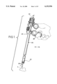
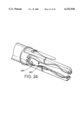
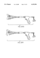
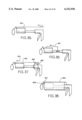
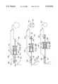
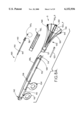
- FIG. 1 shows an assembled device of the parent disclosures.
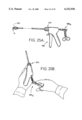
- FIG. 2 illustrates the use of the device of the parent disclosures in connection with a tissue target.
- FIGS. 3 and 4 further illustrate operation of the device of the parent disclosures.
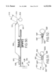
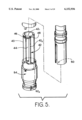
- FIG. 5 is an exploded view illustrating operation of the device of the parent disclosures.
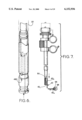
- FIG. 6 is an assembled view of part of the device of the parent disclosures.
- FIG. 7 illustrates operation of the device of the parent disclosures.
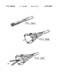
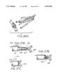
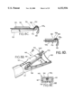
- FIG. 8A is an exploded perspective view of a device for ligating anatomical tissue embodying the present invention in an open configuration.
- FIG. 8B is a perspective view of the distal end of the device of the present invention with a surgical loop partially open.
- FIG. 8C is a side elevational view of one finger of the device of the present invention with a cutter thereon with the cutter open.
- FIG. 8D is a side elevational view of one finder of the device of the present invention with the cutter in position to cut the tail off of the surgical loop.
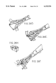
- FIG. 8E is a view taken along section 8E--8E of FIG. 8B.
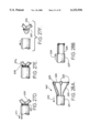
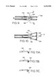
- FIG. 9 illustrates the distal end of the device of the present invention in the closed configuration.
- FIG. 10 illustrates the distal end of the device of the present invention in the open configuration.
- FIGS. 11A-11C illustrate a suture loop holding element of the present invention.
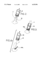
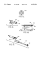
- FIG. 12 is a section along line 12--12 of FIG. 9.
- FIG. 13 is a disassembled view of a cutter used to cut suture material in the device of the present invention.
- FIG. 14 is a view of a distal end of the device of the present invention illustrating the cutter element.
- FIG. 15 is an exploded view of a cutter element of the present invention.
- FIGS. 16A and 16B illustrate cutter elements of the present invention.
- FIGS. 17A and 17B illustrate operation of the device of the present invention to accommodate different size tissue targets.
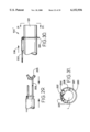
- FIG. 18 illustrates an alternative form of a cutter for the device of the present invention.
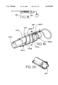
- FIG. 19 illustrates an alternative form of a cutter of the present invention.
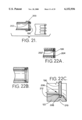
- FIG. 20 illustrates an alternative form of a suture loop mounting system.
- FIG. 21 illustrates an alternative form of a suture loop mounting system.
- FIGS. 22A-22C illustrate a suture loop mounting system.
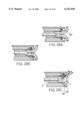
- FIGS. 23A-23C illustrate operation of an alternative form of a suture loop mounting system.
- FIG. 24 illustrates a suture retaining means that can be used with the device of the present invention.
- FIGS. 25A-25H illustrate operation of the device of the present invention.
- FIGS. 26A-26H are perspective views illustrating operation of the distal end of the device of the present invention.
- FIGS. 27A-27F show an alternative form of the device of the present invention in which the suture loop is supported on the outside of the body.
- FIGS. 28A and 28B show another form of the device of the present invention in which the suture loop is supported on flexible fingers that are biased outward and are held inwardly by a tube.
- FIG. 29 shows a J-shaped tissue grasping element in combination with the device shown in FIGS. 27A-27F.
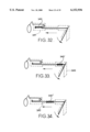
- FIGS. 30 and 31 show yet another form of the device of the present invention, with FIG. 31 being a sectional view taken along line 31--31 of FIG. 30.
- FIGS. 32-34 show various locations for a tolerance take-up mechanism used in the device of the present invention.
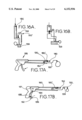
- FIGS. 35-37 show various suture cutting elements for use in the device of the present invention.
- FIG. 38 shows a manual cutting sleeve for use with the device of the present invention.
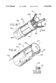
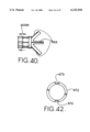
- FIG. 39 is a perspective view of the distal end of another form of the device in which the loop is located on the outside of the body.
- FIG. 40 is an elevational view of the distal end of the device shown in FIG. 39.
- FIG. 41 is a perspective view of yet another form of the invention.
- FIG. 42 is an elevational view of the body of the device shown in FIG. 41.
- FIG. 43 is a view taken along line 43--43 of FIG. 41.
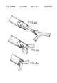
- FIGS. 44-46 are perspective views of the distal end of an alternative form of the device of the present invention with a surgical loop on the outside of the body.
- FIGS. 47-50 are schematic views illustrating operation of a system for automatically maintaining a pre-set tension on the suture loop over a wide range of tissue target sizes.
- FIGS. 51 and 52 illustrate alternative forms of a running element for attaching the suture loop to the suture tail.
- FIG. 1 of the parent disclosure A surgical loop delivery device 10 disclosed in FIG. 1 of the parent disclosure is also shown in FIG. 1 herein and reference is made thereto, along with FIGS. 2-7 herein.
- Device 10 includes a main body 20 having a distal end 22 that will be located in a patient during the procedure and a proximal end 24 that will be accessed by a surgeon during a procedure.
- a hand-grip 26 is mounted near proximal end 24 to be available to a user's hand.
- a tissue grasping instrument 30 extends through device 10 to engage and grasp tissue T and draw that tissue into the instrument as indicated in FIGS. 1-3.
- device 10 includes a suture supporting element 40 on which suture 42 is mounted to extend across a slot 44 and be anchored thereto by element 46 and groove 48.
- Tube 20 is movable relative to element 40, and device 10 includes a projection 50 that moves into slot 44 when device 10 is operated and engages suture portion 42 1 in the distal direction.
- Suture 42 is slidably held on element 40 so portion 42 2 of suture 42 slides in the proximal direction when device 10 is operated to ligate tissue T.
- Device 10 further includes a suture cutter element 54 which cuts suture portion 42 2 adjacent to a loop around tissue T after ligating that tissue. As discussed in the parent disclosures, and indicated in FIG. 5, the suture extends out of device 10 adjacent to the suture loop, then extends back into the device to intersect the cutter path. Suture portion 43 3 is shown in FIG. 5 to indicate this suture configuration.
- suture 42 includes a suture loop 56 which is mounted on an inside surface of device 10 to surround tissue T when that tissue is drawn into device 10 by tissue grasper 30.
- suture loop 56 is releasably held on device 10 to have a size essentially matching the internal size of device 10, as indicated in phantom lines in FIG. 7.
- Suture 42 includes a running element that slidably attaches the loop to the tail, such as slip knot 42 k , and operation of device 10 to move projection 50 distally with respect to suture portion 42 1 draws suture portion 42 2 proximally.
- Loop 56 is mounted on device 10 so slip knot 42 k remains stationary with respect to device 10.
- proximal movement of suture portion 42 2 draws the suture through slip knot 42 k thereby tightening loop 56 from the phantom line configuration in FIG. 7 to the full line configuration in FIG. 7 to garrote tissue encircled by the loop.
- the device is then operated to cut suture 42 adjacent to slip knot 42 k .
- Device 10 is then withdrawn from the patient as indicated in FIG. 4.
- device 10 works well, but could be improved to, among other things, accommodate tissue that is larger than the outside dimension of the device and to improve the ability of the surgeon to visualize the tissue garroting operation while still maintaining a staple suture loop.
- the invention disclosed herein achieves these goals.
- Device 100 embodying the present invention is shown.
- Device 100 includes a main body 102 having a first handle element 104 on a proximal end 106 thereof and a distal end 108.
- a suture loop operating element 110 is slidably received in main body 102 and includes a second handle element 112 which is movable toward and away from first handle element to operate device 100 and the elements thereof.
- Device 100 accommodates a tissue grasping element whereby a tissue manipulator and a suture loop holder and manipulator are integrated into a single unit.
- Device 100 is hollow and has a bore 114 extending therethrough which receives a tissue grasper for grasping tissue and orienting the device and the tissue relative to each other so a suture loop will be positioned relative to the tissue so the tissue is encircled for garroting.
- tissue graspers can be used, and the particular form of tissue grasper does not form a part of the instant invention. However, for the sake of completeness, several tissue graspers will be disclosed hereinbelow, see, e.g., FIGS. 26, 27A and 29.
- Distal end 114 of suture loop operating element 110 extends out of end 108 of main body 102 and is moved in a distal direction when handles 104 and 112 are moved toward each other.
- Device 100 further includes a suture loop supporting unit 120.
- Unit 120 includes a collar 121 which releasably engages distal ends 114 and 108 of operating element 110 and main body 102 respectively to removably mount unit 120 on device 100.
- a plurality of flexible fingers 122 are mounted adjacent to their proximal ends to collar 121 and extend therefrom to a distal end 124 of each finger. All fingers 122 are adapted to move distal ends 124 between a first configuration F located to be sized to be essentially equal to the outer size of the main body 102 as indicated by phantom line 102 1 in FIG. 8A and a second configuration S located to be sized larger than the outer dimension of main body 102 as indicated by solid line 102 2 in FIG. 8A.
- Various means for moving distal ends 124 can be used and several examples of these means will be discussed hereinbelow.
- a suture loop 130 is releasably mounted on distal ends 124 to be moved from a first size associated with first configuration F to a second size associated with second configuration S whereby the suture loop can be moved into a size larger than the size of body 102 to capture tissue which has a size larger than the outer dimension of body 102.
- any size tissue between, and including, sizes 102 1 and 102 2 can be accommodated by device 100 thereby providing great versatility to device 100.
- the suture loop is held in a stable manner so it can be accurately positioned around the target tissue.
- Open areas V provide a sight path for a surgeon located proximally of loop 130 whereby the surgeon can see the loop relative to tissue being ligated during the ligating procedure. This will permit the surgeon to locate the tissue and the loop relative to each other as necessary for proper margins, and the like.
- a suture cutter 140 is mounted on one finger 122 and a suture control mechanism 142 is also mounted on device 100 for controlling the length of suture portion 130 1 .
- suture loop 130 can have a variety of sizes
- the remaining suture portion 130 1 can have a variety of lengths, i.e., suture portion 130 1 associated with first configuration F and size 102 1 is longer than suture portion 130 1 associated with second configuration S and size 102 2 .
- suture control mechanism 142 automatically adjusts the length of suture portion 130 1 based on the size of suture loop 130.
- suture control mechanism 142 can be used in device 100 without departing from the scope of the present invention. Several examples of such mechanisms will be presented hereinbelow.
- cutter 140 cuts the suture adjacent to a slip knot in the suture after the suture loop has garrotted the tissue.
- cutter 140 is shown in FIGS. 8A-8E as including a body 150 slidably mounted on one finger 122 to move in a distal direction with respect to device 100 when operated.
- Cutter 140 further includes a cutting edge 152 on a distal end thereof and has a groove 154 defined therethrough through which suture portion 130 1 is received.
- Sliding element 150 is slidably mounted on finger 122. As can be understood from FIGS.
- operation of the cutter device drives cutting edge 152 against an anvil 159 located on the finger associated with the cutting edge 152.
- Suture portion 130 1 is located between cutting edge 152 and anvil 159 so cutting edge 152 cuts suture portion 130.sub. adjacent to slip knot 130 k to define a garroting loop similar to the loop shown in FIG. 4, with a tail 130 t .
- each finger 122 includes a cam lobe 160 near a proximal end thereof, with a living hinge 162 located near the cam lobe.
- the cam lobes define a small passageway 164 therebetween when unit 120 is in first configuration F. Passageway 164 is smaller than the outside dimension of distal end of the grasping element 114. Thus, movement of distal end 114 toward the distal end of device 100 is blocked by cam lobes 160.
- An alternative form of the device can include a band of elastic material encircling legs 122 to bias those legs toward the FIG. 9 configuration.
- the grasper forcing the legs apart by means of engagement with the cam lobes will overcome this bias when the grasper is in the tissue grasping position, but the legs will be returned to the relaxed FIG. 9 configuration as soon as the tissue grasper is withdrawn from engagement with lobes 160.
- suture loop 130 will be enlarged as fingers 122 move from the FIG. 9 configuration towards the FIG. 10 configuration.
- a tissue grasper can grasp tissue and the grasped tissue and the loop can be oriented with respect to each other so the suture loop encircles the tissue.
- a mechanism such as the suture loop tightening mechanism described above with regard to device 10 disclosed in the parent disclosures, can be used to tighten the suture loop.
- Another loop tightening mechanism will be described below in relation to FIGS. 47-50.
- Slip knot 130 k is held stationary with respect to device 100 while suture portion 130 1 is pulled toward the proximal end of device 100.
- a suture knot holder 170 can extend through cutter body 150 adjacent to finger 122 and includes a distal shoe 172 on a distal end of body 174. A proximal end of body 174 is located near handles 104 and 112 so body 174 can be moved distalward of device 100 into engagement with knot 130 k to hold that knot in place while cutter edge 152 is driven against the suture to cut that suture.
- cutter body 150 is C-shaped and has a gap 176 defined therein. Gap 176 can be used to accommodate a finger 122 or a suture portion. The elements used to move the cutter are shown in FIGS. 25 and 35-37.
- mechanism 142 includes a suture length adjusting element which accounts for the varying length of suture portion 130 1 as discussed above so that longer portions 130 1 ' shown in FIG. 17B are accommodated for smaller tissue sizes T' are shorter portions 130 1 " for larger tissue sizes T" shown in FIG. 17A.
- suture length adjusting element includes a spring 180 connected at one end thereof to the distalmost end of suture portion 130 1 and at another end thereof to cutter 140.
- Spring 180 expands or contracts to take up the lengths of portion 130 1 associated with different tissue sizes.
- Suture portion 130 1 is trained around a pulley 182 mounted on body 102.
- the cutter in FIGS. 17A and 17B will travel the same distance every stroke cutting the tail of the suture against anvil 159.
- the extension spring 180' acts as a clutch, elongating more when a large piece of tissue is in the loop and undergoing less elongation when a smaller piece of tissue is in the loop.
- Cutter 140 can be coupled to the spring in various ways, such as to an expansion spring 180' in FIGS.
- FIGS. 14, 16A and 16B are shown in FIGS. 14, 16A and 16B and is mounted on the finger 122 adjacent to the cutter edge 152.
- Other forms of tolerance take-up means are possible, and several examples thereof are shown in FIGS. 32-34 as well as in FIGS. 47-50 and will be discussed later in connection with those figures.
- the suture extends out of the suture loop support and is wound back across anvil 159 in front of cutter edge 152.
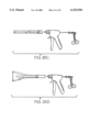
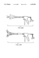
- handles 104 and 112 can be replaced by an operating mechanism 185.
- Operating mechanism 185 includes a trigger 186 and a grip 188, with trigger 186 being pivotally connected to grip 188 to move with respect to the grip when operating mechanism 185 is actuated.
- a lever arm 190 is connected at one end thereof to trigger 186 on end 191 of the trigger.
- Actuating end 192 of the trigger is on one side of a pivot 194 and end 191 is on the other end of the trigger whereby moving trigger end 192 toward grip 188 moves lever 190 in a distal direction of device 100 to force cutter edge 152 against anvil 159 with suture portion 130 1 interposed therebetween.
- Spring-like element 180 maintains the suture portion taut and also takes up excess suture when a smaller loop is used to ligate tissue.
- the cutter could be located proximally of the position shown for cutter 140 in FIG. 8. This location can be on the finger near the proximal end of the finger and adjacent to collar 121, or even on the collar 121 if desired. This positioning of the cutter will create a longer suture tail than the distal location of the cutter shown in FIG. 8. However, such proximal location of the cutter will permit the cutter to generate higher cutting forces than the distal location shown in FIG. 8 because the cutter will be on a more stable support.
- FIG. 19 Another form of cutting and tightening device is shown in FIG. 19 as element 140' as including a body 140'A is mounted to rotated in direction 140'B about an inner tube 140'C that is on the device, either on the main body or on the suture holding mechanism.
- the rotation of body 140'A takes up excess slack similar to a winch winding the suture on the outside surface of a spool 140'D.
- Suture tail 130' is fixed to body 140'A at an anchor 140'E and is trained around a pulley 140'F to change take-up movement of the suture between rotational and linear. After the suture is tensioned, the body is indexed distally, or the spool is moved, thereby trapping and cutting the suture between flange 140'G and cutter blade 140'H on the spool.
- FIG. 21 Other means for supporting the suture loop on device 100 includes mounting the suture loop on the outside of the device.
- suture loop 130 is supported on the outside of fingers 122' in grooves 200.
- a neck 202 is formed at the base of each groove.
- Neck 202 is thin and forms a break joint which is broken when the loop is tightened about the tissue.
- the break-joint concept is also shown in FIG. 22A where break joint 202' is oriented to locate suture loop 130 on the distalmost end of the fingers and breaks to remove barrier portion 204 when the loop is tightened.
- FIGS. 22B and 22C A modified form of this break joint is shown in FIGS. 22B and 22C where joint 206 includes a groove 208 and a flexible neck 210.
- FIG. 20 Yet another outside-positioned suture loop supporting means is shown in FIG. 20 where outwardly curved castellations 220 support the suture loop and flex in the manner just described for wall 212 as the suture loop is tightened to release the loop.
- FIGS. 23A-23C Yet another means for retaining the suture loop on the finger ends is shown in FIGS. 23A-23C and includes flexible filaments 224 inwardly adjacent to grooves 226. Filaments 224 replace walls 212 and operate in a similar manner to retain the suture loop in grooves 226 until the loop is tightened about the target tissue which has been oriented within the loop by a tissue grasper G. Movement of the suture loop is indicated by the phantom lines in FIG. 23C.
- suture portion 130 1 When the suture is expanded beyond a first size to open and accept larger tissue, suture portion 130 1 is retained within device 100 by a retainer element 250 attached to the device adjacent to collar 121 and interposed between two adjacent fingers and shown in FIG. 24.
- Element 250 is in the shape of a hook and suture portion 130 1 is wound around element 250 to keep it out of the way while providing sufficient suture length to permit the fingers to completely spread out when opened. Thus, when opening the suture loop the tail does not need to be drawn back through the knot.
- FIGS. 25A-25H and 26A-26G Operation of device 100 is indicated in FIGS. 25A-25H and 26A-26G.
- device 100 is inserted into a patient with the distal end thereof located adjacent to a target tissue, see, e.g., FIG. 26A.
- the device is operated to expand the suture loop 130 and a tissue grasper G, which is inserted through the proximal end of device 100, is used to grasp tissue and pull that tissue into the device, FIGS. 25A, 26B and 26C.
- Tissue grasper G includes an operating mechanism 185 G which is manipulated by a surgeon as indicated in FIGS. 25B and 25E. In the device discussed above, inserting the tissue grasper spreads the fingers into the open configuration.
- the elements of the device are operated to orient the tissue with respect to loop 130 so the tissue is encircled by the suture loop. Once the tissue is encircled, the device is operated to tighten the loop about the tissue and operate the cutter mechanism to cut the suture portion 130 1 adjacent to slip knot 130 k . Once the tissue is ligated with the suture loop, the grasping device is removed and endoscopic scissors are placed through the device to cut the tissue between the ligated loop as indicated in FIGS. 26F and 26G. If the surgeon desires, he can also take a sample of the tissue at this time by cutting off a portion and removing it with the grasper.
- FIG. 27A shows a suture loop 201 fixed on the outside surface 202 of a rigid body 203.
- a grasping device 204 is shown holding tissue 205 that will be ligated. Once the target tissue 205 is identified and stabilized, tissue 205 is placed within the suture loop 201.
- a second method includes holding grasping device 204 stationary and moving rigid tube 203 towards the distal end of grasper 204.
- sliding tube 206 is deployed to push suture loop 201 off the rigid body 203 onto tissue 205.
- FIG. 27B shows tissue 205 within rigid body 203.
- FIG. 27C shows sliding tube 206 pushing in direction 210 to force suture loop 201 off rigid tube 203 onto tissue 205.
- suture loop 201 is at the desired location with respect to tissue 205, the surgeon will tighten loop 201 by operating the operating mechanism to pull the suture in direction 211 shown in FIG. 27D.
- cutter 208 is deployed to cut tail 201 t off suture loop 201.
- FIG. 27F the trimmed suture loop 201' remains on tissue 205.
- FIGS. 28A and 28B Yet another form of the suture loop controlling means is shown in FIGS. 28A and 28B.
- arms 220 are made from springy material such as nitenol, spring steel, or some other type of metal or plastic that has a material memory and attempts to return to an initial position after it has been deformed from that initial position.
- a plastic tip 225 is located at a distal tip of each arm 220 to hold the suture loop. Tips 225 can be molded or bonded to each arm. Arms 220 are biased toward the position shown in FIG. 28A, and are held in a collapsed position, shown in FIG. 28B, by being pulled into outer tube 221 when tube 221 is moved in direction 224 with respect to the arms.
- FIG. 29 shows a J-shaped tissue grasping element 308 in connection with the device shown in FIGS. 27A-27F.
- FIGS. 30 and 31 show another device 100" for pushing the suture loop off the rigid body onto the tissue.
- Suture loop 330 is placed on the outside surface of rigid tube 331.
- Tail 333 extends through slider 332.
- Rigid tube 331 is not a completed closed shape as indicated in FIG. 31. While FIG. 31 indicates that the tube is circular in shape, it can be other shapes, including octagon, hexagon or other polygonal shape without departing from the scope of this invention.
- An internal lumen L is shown in FIG. 31.
- Slider 332 has a protrusion 334 on both sides thereof as shown in FIG. 31, which lock with mating slots 335 on rigid tube 331. Slider 332 is moved in direction 336 to push suture loop 330 off rigid tube 331.
- a cutter 336 is also housed in slider 332. Once suture loop 330 has been tightened around the tissue, cutter 336 is deployed to cut the tail of the suture adjacent to the slip knot as above discussed.
- FIGS. 32-34 show various locations for a compliant member 340, 340' and 340" for accommodating different lengths of suture portion 130 1 as discussed above.
- a compliant member can be positioned in a disposable cartridge 341, or in a handle 343 or in a shaft 342 as shown in FIGS. 32, 33 and 34 respectively.
- the function of the compliant member is to allow the same stroke at the trigger whether the instrument is fired on a large or a small tissue structure and to impart the same force on the loop to prevent formation of a loop which is overtight and which can cut the tissue or formation of a loop which is not tight enough and which can cause problems such as pregnancy in a tubal sterilization.
- the tail of the loop When fired on a small tissue structure, the tail of the loop is pulled to a longer length. Conversely, when the device is fired on a large tissue, the tail of the suture loop is pulled to a shorter length. Therefore, a tolerance take-up mechanism is placed in the device. While a spring is shown for the compliant member, other forms can be used, such as an elastomeric bushing or the like.
- cutter 350 is a manual linear cutter that is activated by the surgeon.
- Cutter 351 shown in FIG. 36 is a timed linear cutter that is attached to the tightening means and which cuts at a predetermined setting. The surgeon will have no input on when the suture is cut with the device 351 automatically activating.
- Cutter 352 shown in FIG. 37 is a manual rotational cutter. Upon rotation, a cam 353 on a rotation knob will make contact with the end of rod 354 thereby pushing the rod forward in direction 355 to cut the suture.
- a manual cutting sleeve 356 is shown in FIG. 38.
- the sleeve 356 has a sharp edge 357 on a cam surface 358 at the distal end of the device. Upon rotation, the cutter will be activated and will cut the suture.
- the device of the present invention permits the surgical loop to be expanded between a first size and a second size in which one of those sizes is larger than the outer dimension of the main body whereby a wide range of tissue sizes can be accommodated, including tissues having sizes that are actually larger than the instrument itself.
- tissue sizes including tissues having sizes that are actually larger than the instrument itself.
- FIGS. 27A-27E where the loop rests on the outside surface of the device, and thus is larger than the outside dimension of the device.
- this disclosure is not intended to be limited by these exact forms.
- FIGS. 39-43 other forms can also be used without departing from the scope of the present disclosure. As shown in FIG.
- a device 400 includes a slidable ring 450 on the outside of a main body and is connected to fingers 451 through a stationary collar 452 by tensioning bands 453, such as elastomeric bands, metal wire, fiber or the like.
- tensioning bands 453, such as elastomeric bands, metal wire, fiber or the like When slidable ring 450 is moved proximally, by a trigger mechanism having a rod 400R, tension is applied to bands 453 which causes fingers 451 to pivot at a living hinge 454 outwardly in direction 400D.
- the fingers can be adjusted to an infinite size between a first and a second size with one of the sizes being shown in FIG. 39 as being larger than the outer dimension of the main body of device 400. Size adjustment of the fingers is effected by moving sliding ring 450 either proximally or distally as indicated in FIG. 40 by double-headed arrow 400M.
- Device 470 includes a suture loop 471 which is pushed off outer tube 472 once the target tissue and the device have been oriented relative to each other so the loop will encircle the tissue.
- outer tube 472 includes two dovetail grooves 473 in which a top sliding element 474 and a bottom sliding element 475 are received.
- Sliding elements 474 and 475 are attached to a handle on the proximal end of the device.
- Suture tail 476 operates as discussed above. Operation of the sliding elements moves the suture loop 471 off of the main body of the device after the device and the tissue target have been oriented relative to each other so the loop will encircle the tissue.
- the tension applied to the suture loop must be controlled so that a loop associated with a large tissue will not receive too much tension and a loop associated with a small tissue will receive sufficient tension to accomplish the goals of properly ligating the tissue target.
- One system for adjusting the tension on the suture loop is shown in FIGS. 47-50 as device 490.
- One trigger actuates both the tensioning mechanism and the cutting mechanism in device 490.
- the tensioning mechanism will pull the tail of the suture thereby causing the loop to constrict on the targeted tissue.
- the cutting mechanism will be activated to cut the tail of the suture loop.
- Device 490 is shown in the open condition in FIG. 47.
- a trigger 491 is fully open in this configuration.
- Trigger 491 is attached to a drive bar 492 which is attached to a tension coupler 493.
- Tension coupler 493 includes a proximal coupler part 494, a distal coupler part 495 and an elastic element such as a spring 496. The coupler parts are maintained together by a protrusion 497 on distal coupler part 495 that pops into a slot 498 on proximal coupler part 494.
- a suture drive bar 499 is attached to distal coupler part 495.
- Trigger 491 is shown in FIG. 48 as being activated in direction 500.
- Activation of trigger 491 in direction 500 forces drive bar 492 to push tension coupler 493 and suture drive bar 499 forward where suture drive bar 499 will push tail 501 of the suture loop.
- the proximal end of the suture tail is immobilized by anchor 502 so that tension is applied to the suture tail.
- Suture tail 501 is trained around a pivot 503 which thereby act as a pulley. With tension being applied to the suture tail 501, the diameter of the suture loop begins to decrease in direction 505. Once a set mount of resistance is met in the tension system, the tension coupler 493 begins to compress in direction 506 allowing uniform tightness on suture loop 504. This permits the same amount of tension on a large or small amount of tissue in the loop.
- the spring 496 is sized to get the proper amount of tension that is needed as the pre-set amount of tension.
- a protrusion 507 on the drive bar is nested in a slot 508 and the forward motion of the drive bar 492, indicated by arrow 510 in FIG. 50, caused by rotation of trigger 491 in direction 511 pushes cutter bar 509 in forward direction 510.
- This forward motion causes blade 512 of cutter bar 509 to cut suture tail 501.
- the distance 514 between the knot 513 of the suture loop 504 and the cut will remain the same on either a small or a large amount of tissue captured in the loop.
- a slip knot is only one form of running element that can be used on the suture loop. Any element that will permit the loop to be tightened can be used, and two additional forms of the running element are shown in FIGS. 51 and 52.
- a running element 520 includes an absorbable chuck 522 having a tortuous path 524 defined therethrough from one end 526 to the other end 528 and which receives suture tail 520T of loop 520S.
- a chock 530 is located in the chuck to contact the suture tail in a one-way manner. That is, the cock is angled to permit movement of the suture past the chock in direction 531 but not in direction 532.
- the tortuous path is self-cinching in that the suture can move in direction 531 but not in direction 532.
- Another form of the chuck is shown in FIG. 52 as chock 530' with a tortuous path 528' and a chock 530' on end 526'.
- a molded chock will be smaller and more repeatable in tightening friction tan a knot in the size ranges of interest. Hand tied knots can be either too tight or too loose thus creating a possibility for non-repeatable characteristics in the instrument.
- Typical devices such as Biopsy shears, Rounger, electrosurgical bovie, ultrasonic scalpel, wire shear or the like can be used separately after the tissue has been ligated.
- Combination devices can also be used which have a tissue grasping jaw in combination with tissue severing abilities that will decrease operative time. It is even possible to include a tissue severing system within the body of the instrument separate from the ligating loop. This will allow the surgeon to independently manipulate the tissue after ligation into the tissue severing means withot needing to remove the grasper from the body of the instrument.
Abstract
Description
Claims (35)
Priority Applications (2)
| Application Number | Priority Date | Filing Date | Title |
|---|---|---|---|
| US09/241,394 US6152936A (en) | 1996-09-23 | 1999-02-02 | Surgical loop delivery device |
| US09/722,875 US6610072B1 (en) | 1996-09-23 | 2000-11-27 | Surgical loop delivery device |
Applications Claiming Priority (3)
| Application Number | Priority Date | Filing Date | Title |
|---|---|---|---|
| US08/717,990 US5766217A (en) | 1996-09-23 | 1996-09-23 | Surgical loop delivery device and method |
| US09/071,811 US5873876A (en) | 1996-09-23 | 1998-05-05 | Surgical loop delivery device and method |
| US09/241,394 US6152936A (en) | 1996-09-23 | 1999-02-02 | Surgical loop delivery device |
Related Parent Applications (1)
| Application Number | Title | Priority Date | Filing Date |
|---|---|---|---|
| US09/071,811 Continuation-In-Part US5873876A (en) | 1996-09-23 | 1998-05-05 | Surgical loop delivery device and method |
Related Child Applications (1)
| Application Number | Title | Priority Date | Filing Date |
|---|---|---|---|
| US09/722,875 Continuation US6610072B1 (en) | 1996-09-23 | 2000-11-27 | Surgical loop delivery device |
Publications (1)
| Publication Number | Publication Date |
|---|---|
| US6152936A true US6152936A (en) | 2000-11-28 |
Family
ID=46255372
Family Applications (2)
| Application Number | Title | Priority Date | Filing Date |
|---|---|---|---|
| US09/241,394 Expired - Lifetime US6152936A (en) | 1996-09-23 | 1999-02-02 | Surgical loop delivery device |
| US09/722,875 Expired - Fee Related US6610072B1 (en) | 1996-09-23 | 2000-11-27 | Surgical loop delivery device |
Family Applications After (1)
| Application Number | Title | Priority Date | Filing Date |
|---|---|---|---|
| US09/722,875 Expired - Fee Related US6610072B1 (en) | 1996-09-23 | 2000-11-27 | Surgical loop delivery device |
Country Status (1)
| Country | Link |
|---|---|
| US (2) | US6152936A (en) |
Cited By (196)
| Publication number | Priority date | Publication date | Assignee | Title |
|---|---|---|---|---|
| US20020049457A1 (en) * | 1999-05-20 | 2002-04-25 | Kaplan Aaron V. | Methods and apparatus for transpericardial left atrial appendage closure |
| WO2002039910A2 (en) | 2000-11-14 | 2002-05-23 | Esd Medical, Llc | Device for laparoscopic tubal ligation |
| WO2002094109A1 (en) * | 2001-05-24 | 2002-11-28 | Nicola Picardi | Mechanical surgical device for use in anastomotic operations |
| US6610072B1 (en) | 1996-09-23 | 2003-08-26 | Esd Medical, L.L.C. | Surgical loop delivery device |
| US20040116115A1 (en) * | 2002-12-06 | 2004-06-17 | Ertel Lawrence R. | Systems and methods for providing interactive guest resources |
| US20040215217A1 (en) * | 2001-10-01 | 2004-10-28 | The Cleveland Clinic Foundation | Skin lesion exciser and skin-closure device therefor |
| US20050033325A1 (en) * | 2001-02-16 | 2005-02-10 | Ethicon, Inc. | Surgical knot pusher and method of use |
| US20050043746A1 (en) * | 2003-08-21 | 2005-02-24 | Pollak Stanley B. | Methods and instruments for closing laparoscopic trocar puncture wounds |
| WO2005034802A2 (en) | 2003-10-09 | 2005-04-21 | Sentreheart, Inc. | Apparatus and method for the ligation of tissue |
| US20050113848A1 (en) * | 2003-11-26 | 2005-05-26 | Karl Reinitz | Surgical suturing apparatus |
| WO2006009729A2 (en) | 2004-06-18 | 2006-01-26 | Medtronic, Inc. | Methods and devices for occlusion of an atrial appendage |
| US20060241748A1 (en) * | 2005-03-25 | 2006-10-26 | Lee Leonard Y | Methods and apparatus for controlling the internal circumference of an anatomic orifice or lumen |
| US7153312B1 (en) * | 1999-12-02 | 2006-12-26 | Smith & Nephew Inc. | Closure device and method for tissue repair |
| US20070016287A1 (en) * | 2005-03-25 | 2007-01-18 | Cartledge Richard G | Methods and apparatus for controlling the internal circumference of an anatomic orifice or lumen |
| US20070027456A1 (en) * | 2005-08-01 | 2007-02-01 | Ension, Inc. | Integrated medical apparatus for non-traumatic grasping, manipulating and closure of tissue |
| US20070060895A1 (en) * | 2005-08-24 | 2007-03-15 | Sibbitt Wilmer L Jr | Vascular closure methods and apparatuses |
| US20070088431A1 (en) * | 2005-10-18 | 2007-04-19 | Henry Bourang | Heart valve delivery system with valve catheter |
| US20070167959A1 (en) * | 1999-03-04 | 2007-07-19 | Abbott Laboratories | Articulating suturing device and method |
| US20070299543A1 (en) * | 2002-08-29 | 2007-12-27 | Mitralsolutions, Inc. | Implantable devices for controlling the internal circumference of an anatomic orifice or lumen |
| US20080027483A1 (en) * | 2002-08-29 | 2008-01-31 | Mitralsoluations, Inc. | Implantable devices for controlling the size and shape of an anatomical structure or lumen |
| US20080033457A1 (en) * | 2004-06-18 | 2008-02-07 | Francischelli David E | Methods and devices for occlusion of an atrial appendage |
| EP1928522A2 (en) * | 2005-08-24 | 2008-06-11 | Wilmer L. Sibbitt | Vascular closure methods and apparatuses |
| US20080275306A1 (en) * | 2005-10-26 | 2008-11-06 | Carlo Rebuffat | Anoscope for Ano-Rectal Diagnostic and Surgery |
| US20090281560A1 (en) * | 2008-05-06 | 2009-11-12 | Wexner Steven D | Method for anastomosis surgery using zip-ties |
| US7651509B2 (en) | 1999-12-02 | 2010-01-26 | Smith & Nephew, Inc. | Methods and devices for tissue repair |
| US7658751B2 (en) | 2006-09-29 | 2010-02-09 | Biomet Sports Medicine, Llc | Method for implanting soft tissue |
| US20100145148A1 (en) * | 2008-12-09 | 2010-06-10 | Tyco Healthcare Group, Lp | Anoscope |
| US7749250B2 (en) | 2006-02-03 | 2010-07-06 | Biomet Sports Medicine, Llc | Soft tissue repair assembly and associated method |
| US7806904B2 (en) | 2000-12-07 | 2010-10-05 | Integrated Vascular Systems, Inc. | Closure device |
| US7837696B2 (en) | 1999-03-04 | 2010-11-23 | Abbott Laboratories | Articulating suturing device and method |
| US7842068B2 (en) | 2000-12-07 | 2010-11-30 | Integrated Vascular Systems, Inc. | Apparatus and methods for providing tactile feedback while delivering a closure device |
| US7842047B2 (en) | 1999-03-04 | 2010-11-30 | Abbott Laboratories | Articulating suturing device and method |
| US7841502B2 (en) | 2007-12-18 | 2010-11-30 | Abbott Laboratories | Modular clip applier |
| US7842049B2 (en) | 2002-12-31 | 2010-11-30 | Abbott Laboratories | Systems for anchoring a medical device in a body lumen |
| US7842048B2 (en) | 2006-08-18 | 2010-11-30 | Abbott Laboratories | Articulating suture device and method |
| US7850797B2 (en) | 2002-12-31 | 2010-12-14 | Integrated Vascular Systems, Inc. | Methods for manufacturing a clip and clip |
| US7850709B2 (en) | 2002-06-04 | 2010-12-14 | Abbott Vascular Inc. | Blood vessel closure clip and delivery device |
| US7857830B2 (en) | 2006-02-03 | 2010-12-28 | Biomet Sports Medicine, Llc | Soft tissue repair and conduit device |
| US7857828B2 (en) | 2003-01-30 | 2010-12-28 | Integrated Vascular Systems, Inc. | Clip applier and methods of use |
| US7867249B2 (en) | 2003-01-30 | 2011-01-11 | Integrated Vascular Systems, Inc. | Clip applier and methods of use |
| US7879071B2 (en) | 2000-12-07 | 2011-02-01 | Integrated Vascular Systems, Inc. | Closure device and methods for making and using them |
| US7883517B2 (en) | 2005-08-08 | 2011-02-08 | Abbott Laboratories | Vascular suturing device |
| US7887563B2 (en) | 2001-06-07 | 2011-02-15 | Abbott Vascular Inc. | Surgical staple |
| US7901428B2 (en) | 2000-01-05 | 2011-03-08 | Integrated Vascular Systems, Inc. | Vascular sheath with bioabsorbable puncture site closure apparatus and methods of use |
| US7905904B2 (en) | 2006-02-03 | 2011-03-15 | Biomet Sports Medicine, Llc | Soft tissue repair device and associated methods |
| US7905903B2 (en) | 2006-02-03 | 2011-03-15 | Biomet Sports Medicine, Llc | Method for tissue fixation |
| US20110066231A1 (en) * | 2007-01-03 | 2011-03-17 | Cartledge Richard G | Implantable devices for controlling the size and shape of an anatomical structure or lumen |
| US7909851B2 (en) | 2006-02-03 | 2011-03-22 | Biomet Sports Medicine, Llc | Soft tissue repair device and associated methods |
| US7918865B2 (en) | 2005-04-07 | 2011-04-05 | Sentreheart, Inc. | Apparatus and method for the ligation of tissue |
| US7931669B2 (en) | 2000-01-05 | 2011-04-26 | Integrated Vascular Systems, Inc. | Integrated vascular device with puncture site closure component and sealant and methods of use |
| US7959650B2 (en) | 2006-09-29 | 2011-06-14 | Biomet Sports Medicine, Llc | Adjustable knotless loops |
| US20110208210A1 (en) * | 2009-10-27 | 2011-08-25 | Ovesco Endoscopy Ag | Resection Device |
| US8007512B2 (en) | 2002-02-21 | 2011-08-30 | Integrated Vascular Systems, Inc. | Plunger apparatus and methods for delivering a closure device |
| US8038688B2 (en) | 1999-03-04 | 2011-10-18 | Abbott Laboratories | Articulating suturing device and method |
| US8083754B2 (en) | 2005-08-08 | 2011-12-27 | Abbott Laboratories | Vascular suturing device with needle capture |
| US8088130B2 (en) | 2006-02-03 | 2012-01-03 | Biomet Sports Medicine, Llc | Method and apparatus for coupling soft tissue to a bone |
| US8118836B2 (en) | 2004-11-05 | 2012-02-21 | Biomet Sports Medicine, Llc | Method and apparatus for coupling soft tissue to a bone |
| US8128658B2 (en) | 2004-11-05 | 2012-03-06 | Biomet Sports Medicine, Llc | Method and apparatus for coupling soft tissue to bone |
| US8137364B2 (en) | 2003-09-11 | 2012-03-20 | Abbott Laboratories | Articulating suturing device and method |
| US8137382B2 (en) | 2004-11-05 | 2012-03-20 | Biomet Sports Medicine, Llc | Method and apparatus for coupling anatomical features |
| US8181838B2 (en) | 2008-09-10 | 2012-05-22 | Tyco Healthcare Group Lp | Surgical stapling device |
| US8202293B2 (en) | 2003-01-30 | 2012-06-19 | Integrated Vascular Systems, Inc. | Clip applier and methods of use |
| US8211122B2 (en) | 2003-09-26 | 2012-07-03 | Abbott Laboratories | Device for suturing intracardiac defects |
| US8221454B2 (en) | 2004-02-20 | 2012-07-17 | Biomet Sports Medicine, Llc | Apparatus for performing meniscus repair |
| US8226681B2 (en) | 2007-06-25 | 2012-07-24 | Abbott Laboratories | Methods, devices, and apparatus for managing access through tissue |
| US8251998B2 (en) | 2006-08-16 | 2012-08-28 | Biomet Sports Medicine, Llc | Chondral defect repair |
| US8267947B2 (en) | 2005-08-08 | 2012-09-18 | Abbott Laboratories | Vascular suturing device |
| US8272555B2 (en) | 2007-03-07 | 2012-09-25 | Tyco Healthcare Group Lp | Stapler for mucosectomy |
| US8298262B2 (en) | 2006-02-03 | 2012-10-30 | Biomet Sports Medicine, Llc | Method for tissue fixation |
| US8303624B2 (en) | 2010-03-15 | 2012-11-06 | Abbott Cardiovascular Systems, Inc. | Bioabsorbable plug |
| US8303604B2 (en) | 2004-11-05 | 2012-11-06 | Biomet Sports Medicine, Llc | Soft tissue repair device and method |
| US8313497B2 (en) | 2005-07-01 | 2012-11-20 | Abbott Laboratories | Clip applier and methods of use |
| US8317825B2 (en) | 2004-11-09 | 2012-11-27 | Biomet Sports Medicine, Llc | Soft tissue conduit device and method |
| US8323312B2 (en) | 2008-12-22 | 2012-12-04 | Abbott Laboratories | Closure device |
| US8323315B2 (en) | 1998-12-30 | 2012-12-04 | Depuy Mitek, Inc. | Suture locking device |
| US8328877B2 (en) | 2002-03-19 | 2012-12-11 | Boston Scientific Scimed, Inc. | Stent retention element and related methods |
| US8343227B2 (en) | 2009-05-28 | 2013-01-01 | Biomet Manufacturing Corp. | Knee prosthesis assembly with ligament link |
| US8361113B2 (en) | 2006-02-03 | 2013-01-29 | Biomet Sports Medicine, Llc | Method and apparatus for coupling soft tissue to a bone |
| US8398656B2 (en) | 2003-01-30 | 2013-03-19 | Integrated Vascular Systems, Inc. | Clip applier and methods of use |
| US8398676B2 (en) | 2008-10-30 | 2013-03-19 | Abbott Vascular Inc. | Closure device |
| US8409219B2 (en) | 2004-06-18 | 2013-04-02 | Medtronic, Inc. | Method and system for placement of electrical lead inside heart |
| US8419753B2 (en) | 2003-12-23 | 2013-04-16 | Abbott Laboratories | Suturing device with split arm and method of suturing tissue |
| US8469983B2 (en) | 2007-09-20 | 2013-06-25 | Sentreheart, Inc. | Devices and methods for remote suture management |
| US20130165950A1 (en) * | 2011-12-27 | 2013-06-27 | Boston Scientific Scimed, Inc. | System and method to achieve reciprocating rotational motion of the distal member of a device by applying translational forces |
| US8500818B2 (en) | 2006-09-29 | 2013-08-06 | Biomet Manufacturing, Llc | Knee prosthesis assembly with ligament link |
| US8506597B2 (en) | 2011-10-25 | 2013-08-13 | Biomet Sports Medicine, Llc | Method and apparatus for interosseous membrane reconstruction |
| US8556930B2 (en) | 2006-06-28 | 2013-10-15 | Abbott Laboratories | Vessel closure device |
| US8562645B2 (en) | 2006-09-29 | 2013-10-22 | Biomet Sports Medicine, Llc | Method and apparatus for forming a self-locking adjustable loop |
| US8562647B2 (en) | 2006-09-29 | 2013-10-22 | Biomet Sports Medicine, Llc | Method and apparatus for securing soft tissue to bone |
| US8574244B2 (en) | 2007-06-25 | 2013-11-05 | Abbott Laboratories | System for closing a puncture in a vessel wall |
| US8574235B2 (en) | 2006-02-03 | 2013-11-05 | Biomet Sports Medicine, Llc | Method for trochanteric reattachment |
| US20130304097A1 (en) * | 2008-01-15 | 2013-11-14 | Covidien Lp | Surgical stapling apparatus |
| US8590760B2 (en) | 2004-05-25 | 2013-11-26 | Abbott Vascular Inc. | Surgical stapler |
| US8597327B2 (en) | 2006-02-03 | 2013-12-03 | Biomet Manufacturing, Llc | Method and apparatus for sternal closure |
| US8603116B2 (en) | 2010-08-04 | 2013-12-10 | Abbott Cardiovascular Systems, Inc. | Closure device with long tines |
| US20130338682A1 (en) * | 2012-06-15 | 2013-12-19 | Cook Medical Technologies Llc | Tissue Ligation Devices and Methods |
| US8623051B2 (en) | 2005-06-24 | 2014-01-07 | Smith & Nephew, Inc. | Tissue repair device |
| US8652171B2 (en) | 2006-02-03 | 2014-02-18 | Biomet Sports Medicine, Llc | Method and apparatus for soft tissue fixation |
| US8652172B2 (en) | 2006-02-03 | 2014-02-18 | Biomet Sports Medicine, Llc | Flexible anchors for tissue fixation |
| US8663252B2 (en) | 2010-09-01 | 2014-03-04 | Abbott Cardiovascular Systems, Inc. | Suturing devices and methods |
| US8672953B2 (en) | 2007-12-17 | 2014-03-18 | Abbott Laboratories | Tissue closure system and methods of use |
| US8672969B2 (en) | 2006-09-29 | 2014-03-18 | Biomet Sports Medicine, Llc | Fracture fixation device |
| US8690910B2 (en) | 2000-12-07 | 2014-04-08 | Integrated Vascular Systems, Inc. | Closure device and methods for making and using them |
| US8758397B2 (en) | 2005-08-24 | 2014-06-24 | Abbott Vascular Inc. | Vascular closure methods and apparatuses |
| US8758400B2 (en) | 2000-01-05 | 2014-06-24 | Integrated Vascular Systems, Inc. | Closure system and methods of use |
| US8758398B2 (en) | 2006-09-08 | 2014-06-24 | Integrated Vascular Systems, Inc. | Apparatus and method for delivering a closure element |
| US8758399B2 (en) | 2010-08-02 | 2014-06-24 | Abbott Cardiovascular Systems, Inc. | Expandable bioabsorbable plug apparatus and method |
| US8771297B2 (en) | 2007-03-30 | 2014-07-08 | Sentreheart, Inc. | Devices, systems, and methods for closing the left atrial appendage |
| US8771352B2 (en) | 2011-05-17 | 2014-07-08 | Biomet Sports Medicine, Llc | Method and apparatus for tibial fixation of an ACL graft |
| US8784447B2 (en) | 2000-09-08 | 2014-07-22 | Abbott Vascular Inc. | Surgical stapler |
| US8801783B2 (en) | 2006-09-29 | 2014-08-12 | Biomet Sports Medicine, Llc | Prosthetic ligament system for knee joint |
| US8808310B2 (en) | 2006-04-20 | 2014-08-19 | Integrated Vascular Systems, Inc. | Resettable clip applier and reset tools |
| US20140236185A1 (en) * | 2007-09-18 | 2014-08-21 | Boston Scientific Scimed, Inc. | Compression, Banding and Percutaneous Airway Ligation of Emphysematous Lung Tissue |
| US8821534B2 (en) | 2010-12-06 | 2014-09-02 | Integrated Vascular Systems, Inc. | Clip applier having improved hemostasis and methods of use |
| US8840645B2 (en) | 2004-11-05 | 2014-09-23 | Biomet Sports Medicine, Llc | Method and apparatus for coupling soft tissue to a bone |
| US8852218B2 (en) | 2008-07-21 | 2014-10-07 | AtriCore, Inc. | Apparatus and methods for occluding an anatomical structure |
| US8858594B2 (en) | 2008-12-22 | 2014-10-14 | Abbott Laboratories | Curved closure device |
| US8858573B2 (en) | 2012-04-10 | 2014-10-14 | Abbott Cardiovascular Systems, Inc. | Apparatus and method for suturing body lumens |
| US8864778B2 (en) | 2012-04-10 | 2014-10-21 | Abbott Cardiovascular Systems, Inc. | Apparatus and method for suturing body lumens |
| US8893947B2 (en) | 2007-12-17 | 2014-11-25 | Abbott Laboratories | Clip applier and methods of use |
| US8905937B2 (en) | 2009-02-26 | 2014-12-09 | Integrated Vascular Systems, Inc. | Methods and apparatus for locating a surface of a body lumen |
| US8920442B2 (en) | 2005-08-24 | 2014-12-30 | Abbott Vascular Inc. | Vascular opening edge eversion methods and apparatuses |
| US8926633B2 (en) | 2005-06-24 | 2015-01-06 | Abbott Laboratories | Apparatus and method for delivering a closure element |
| US8926635B2 (en) | 2004-06-18 | 2015-01-06 | Medtronic, Inc. | Methods and devices for occlusion of an atrial appendage |
| US8936621B2 (en) | 2006-02-03 | 2015-01-20 | Biomet Sports Medicine, Llc | Method and apparatus for forming a self-locking adjustable loop |
| CN104323814A (en) * | 2014-11-27 | 2015-02-04 | 李洪湘 | Push expansion type ring device |
| US8968364B2 (en) | 2006-02-03 | 2015-03-03 | Biomet Sports Medicine, Llc | Method and apparatus for fixation of an ACL graft |
| WO2015034832A1 (en) * | 2013-09-03 | 2015-03-12 | Boston Scientific Scimed, Inc. | Medical retrieval devices and related methods of use |
| US8998949B2 (en) | 2004-11-09 | 2015-04-07 | Biomet Sports Medicine, Llc | Soft tissue conduit device |
| US9017381B2 (en) | 2007-04-10 | 2015-04-28 | Biomet Sports Medicine, Llc | Adjustable knotless loops |
| US20150190190A1 (en) * | 2014-01-03 | 2015-07-09 | Boston Scientific Scimed, Inc. | Electrosurgery devices and methods for providing electric energy treatment |
| US9078644B2 (en) | 2006-09-29 | 2015-07-14 | Biomet Sports Medicine, Llc | Fracture fixation device |
| US9089674B2 (en) | 2000-10-06 | 2015-07-28 | Integrated Vascular Systems, Inc. | Apparatus and methods for positioning a vascular sheath |
| US9089311B2 (en) | 2009-01-09 | 2015-07-28 | Abbott Vascular Inc. | Vessel closure devices and methods |
| US9149276B2 (en) | 2011-03-21 | 2015-10-06 | Abbott Cardiovascular Systems, Inc. | Clip and deployment apparatus for tissue closure |
| US9149267B2 (en) | 2006-02-03 | 2015-10-06 | Biomet Sports Medicine, Llc | Method and apparatus for coupling soft tissue to a bone |
| US9173644B2 (en) | 2009-01-09 | 2015-11-03 | Abbott Vascular Inc. | Closure devices, systems, and methods |
| US9198664B2 (en) | 2009-04-01 | 2015-12-01 | Sentreheart, Inc. | Tissue ligation devices and controls therefor |
| US9204789B2 (en) | 2009-10-08 | 2015-12-08 | Covidien Lp | Asymmetrical anoscope |
| US9241707B2 (en) | 2012-05-31 | 2016-01-26 | Abbott Cardiovascular Systems, Inc. | Systems, methods, and devices for closing holes in body lumens |
| US9259217B2 (en) | 2012-01-03 | 2016-02-16 | Biomet Manufacturing, Llc | Suture Button |
| US9271713B2 (en) | 2006-02-03 | 2016-03-01 | Biomet Sports Medicine, Llc | Method and apparatus for tensioning a suture |
| US9282965B2 (en) | 2008-05-16 | 2016-03-15 | Abbott Laboratories | Apparatus and methods for engaging tissue |
| US9314235B2 (en) | 2003-02-05 | 2016-04-19 | Smith & Nephew, Inc. | Tissue anchor and insertion tool |
| US9314230B2 (en) | 2009-01-09 | 2016-04-19 | Abbott Vascular Inc. | Closure device with rapidly eroding anchor |
| US9314241B2 (en) | 2011-11-10 | 2016-04-19 | Biomet Sports Medicine, Llc | Apparatus for coupling soft tissue to a bone |
| US9332976B2 (en) | 2011-11-30 | 2016-05-10 | Abbott Cardiovascular Systems, Inc. | Tissue closure device |
| US9357991B2 (en) | 2011-11-03 | 2016-06-07 | Biomet Sports Medicine, Llc | Method and apparatus for stitching tendons |
| US9364209B2 (en) | 2012-12-21 | 2016-06-14 | Abbott Cardiovascular Systems, Inc. | Articulating suturing device |
| US9370350B2 (en) | 2011-11-10 | 2016-06-21 | Biomet Sports Medicine, Llc | Apparatus for coupling soft tissue to a bone |
| US9370353B2 (en) | 2010-09-01 | 2016-06-21 | Abbott Cardiovascular Systems, Inc. | Suturing devices and methods |
| US9381013B2 (en) | 2011-11-10 | 2016-07-05 | Biomet Sports Medicine, Llc | Method for coupling soft tissue to a bone |
| US9393023B2 (en) | 2009-01-13 | 2016-07-19 | Atricure, Inc. | Apparatus and methods for deploying a clip to occlude an anatomical structure |
| US9408608B2 (en) | 2013-03-12 | 2016-08-09 | Sentreheart, Inc. | Tissue ligation devices and methods therefor |
| US9414824B2 (en) | 2009-01-16 | 2016-08-16 | Abbott Vascular Inc. | Closure devices, systems, and methods |
| US9414822B2 (en) | 2011-05-19 | 2016-08-16 | Abbott Cardiovascular Systems, Inc. | Tissue eversion apparatus and tissue closure device and methods for use thereof |
| US9414820B2 (en) | 2009-01-09 | 2016-08-16 | Abbott Vascular Inc. | Closure devices, systems, and methods |
| US20160310144A1 (en) * | 2014-02-28 | 2016-10-27 | Olympus Corporation | Atrial-appendage ligation surgical tool |
| US9486281B2 (en) | 2010-04-13 | 2016-11-08 | Sentreheart, Inc. | Methods and devices for accessing and delivering devices to a heart |
| US9486191B2 (en) | 2009-01-09 | 2016-11-08 | Abbott Vascular, Inc. | Closure devices |
| US9498206B2 (en) | 2011-06-08 | 2016-11-22 | Sentreheart, Inc. | Tissue ligation devices and tensioning devices therefor |
| US9538998B2 (en) | 2006-02-03 | 2017-01-10 | Biomet Sports Medicine, Llc | Method and apparatus for fracture fixation |
| US9579091B2 (en) | 2000-01-05 | 2017-02-28 | Integrated Vascular Systems, Inc. | Closure system and methods of use |
| US9585647B2 (en) | 2009-08-26 | 2017-03-07 | Abbott Laboratories | Medical device for repairing a fistula |
| US9615822B2 (en) | 2014-05-30 | 2017-04-11 | Biomet Sports Medicine, Llc | Insertion tools and method for soft anchor |
| US9700291B2 (en) | 2014-06-03 | 2017-07-11 | Biomet Sports Medicine, Llc | Capsule retractor |
| US9757119B2 (en) | 2013-03-08 | 2017-09-12 | Biomet Sports Medicine, Llc | Visual aid for identifying suture limbs arthroscopically |
| US9801708B2 (en) | 2004-11-05 | 2017-10-31 | Biomet Sports Medicine, Llc | Method and apparatus for coupling soft tissue to a bone |
| US20180064528A1 (en) * | 2010-12-23 | 2018-03-08 | Mayo Foundation For Medical Education And Research | Vessel dissection and harvesting apparatus, systems and methods |
| US9918827B2 (en) | 2013-03-14 | 2018-03-20 | Biomet Sports Medicine, Llc | Scaffold for spring ligament repair |
| US9918826B2 (en) | 2006-09-29 | 2018-03-20 | Biomet Sports Medicine, Llc | Scaffold for spring ligament repair |
| US9936956B2 (en) | 2015-03-24 | 2018-04-10 | Sentreheart, Inc. | Devices and methods for left atrial appendage closure |
| US9955980B2 (en) | 2015-02-24 | 2018-05-01 | Biomet Sports Medicine, Llc | Anatomic soft tissue repair |
| US10039543B2 (en) | 2014-08-22 | 2018-08-07 | Biomet Sports Medicine, Llc | Non-sliding soft anchor |
| US10130369B2 (en) | 2015-03-24 | 2018-11-20 | Sentreheart, Inc. | Tissue ligation devices and methods therefor |
| US10136886B2 (en) | 2013-12-20 | 2018-11-27 | Biomet Sports Medicine, Llc | Knotless soft tissue devices and techniques |
| US10182824B2 (en) | 2010-11-11 | 2019-01-22 | Atricure, Inc. | Clip applicator |
| US10258408B2 (en) | 2013-10-31 | 2019-04-16 | Sentreheart, Inc. | Devices and methods for left atrial appendage closure |
| US10292710B2 (en) | 2016-02-26 | 2019-05-21 | Sentreheart, Inc. | Devices and methods for left atrial appendage closure |
| US10426449B2 (en) | 2017-02-16 | 2019-10-01 | Abbott Cardiovascular Systems, Inc. | Articulating suturing device with improved actuation and alignment mechanisms |
| US10433854B2 (en) | 2010-10-27 | 2019-10-08 | Atricure, Inc. | Appendage clamp deployment assist device |
| US10517587B2 (en) | 2006-02-03 | 2019-12-31 | Biomet Sports Medicine, Llc | Method and apparatus for forming a self-locking adjustable loop |
| EP3476314A4 (en) * | 2016-06-28 | 2020-02-26 | Wellcare (WUHAN) Medical Technology Co., Ltd. | Ligator setup method for automatically tightening elastic line and self-tightening elastic line ligator |
| US20200315649A1 (en) * | 2019-03-27 | 2020-10-08 | Oregon Health & Science University | Endarterectomy device |
| WO2021016241A1 (en) * | 2019-07-22 | 2021-01-28 | Boston Scientific Scimed, Inc. | System, device and method for treatment of hemorrhoids |
| US10912551B2 (en) | 2015-03-31 | 2021-02-09 | Biomet Sports Medicine, Llc | Suture anchor with soft anchor of electrospun fibers |
| US20210298760A1 (en) * | 2018-08-17 | 2021-09-30 | Empress Medical, Inc. | Devices and methods for compressing tumors |
| US11224435B2 (en) | 2016-09-23 | 2022-01-18 | Sentreheart Llc | Devices and Methods for left atrial appendage closure |
| US11259794B2 (en) | 2006-09-29 | 2022-03-01 | Biomet Sports Medicine, Llc | Method for implanting soft tissue |
| US11259792B2 (en) | 2006-02-03 | 2022-03-01 | Biomet Sports Medicine, Llc | Method and apparatus for coupling anatomical features |
| US11266389B2 (en) * | 2011-07-11 | 2022-03-08 | Tel Hashomer Medical Research Infrastructure And Services Ltd. | Body part repositioning apparatus and method |
| US11311287B2 (en) | 2006-02-03 | 2022-04-26 | Biomet Sports Medicine, Llc | Method for tissue fixation |
| US20220142648A1 (en) * | 2020-11-09 | 2022-05-12 | The Cleveland Clinic Foundation | Tissue ligation systems and methods of ligating tissue |
| US11547416B2 (en) | 2017-03-27 | 2023-01-10 | Append Medical Ltd. | Left atrial appendage closure |
| US11844526B2 (en) | 2018-03-27 | 2023-12-19 | Atricure, Inc. | Devices and methods for left atrial appendage closure |
| US11957351B2 (en) * | 2021-11-09 | 2024-04-16 | The Cleveland Clinic Foundation | Tissue ligation systems and methods of ligating tissue |
Families Citing this family (75)
| Publication number | Priority date | Publication date | Assignee | Title |
|---|---|---|---|---|
| US8038687B2 (en) * | 2005-05-17 | 2011-10-18 | St. Jude Medical Puerto Rico Llc | Suture loop closure device |
| US8252005B2 (en) * | 2005-06-30 | 2012-08-28 | Edwards Lifesciences Corporation | System, apparatus, and method for fastening tissue |
| US7367983B2 (en) | 2005-09-15 | 2008-05-06 | Dziadik Stephen P | Vessel harvesting apparatus |
| US7655004B2 (en) | 2007-02-15 | 2010-02-02 | Ethicon Endo-Surgery, Inc. | Electroporation ablation apparatus, system, and method |
| WO2008128186A1 (en) | 2007-04-12 | 2008-10-23 | Applied Medical Resources Corporation | Method and apparatus for tissue morcellation |
| US8075572B2 (en) | 2007-04-26 | 2011-12-13 | Ethicon Endo-Surgery, Inc. | Surgical suturing apparatus |
| US8100922B2 (en) | 2007-04-27 | 2012-01-24 | Ethicon Endo-Surgery, Inc. | Curved needle suturing tool |
| US8623046B2 (en) * | 2007-08-10 | 2014-01-07 | Donald Lee Sturtevant | Treatment for patients after removal of saphenous vascular material |
| US8262655B2 (en) | 2007-11-21 | 2012-09-11 | Ethicon Endo-Surgery, Inc. | Bipolar forceps |
| US8579897B2 (en) | 2007-11-21 | 2013-11-12 | Ethicon Endo-Surgery, Inc. | Bipolar forceps |
| US8568410B2 (en) | 2007-08-31 | 2013-10-29 | Ethicon Endo-Surgery, Inc. | Electrical ablation surgical instruments |
| US8480657B2 (en) | 2007-10-31 | 2013-07-09 | Ethicon Endo-Surgery, Inc. | Detachable distal overtube section and methods for forming a sealable opening in the wall of an organ |
| US20090112059A1 (en) | 2007-10-31 | 2009-04-30 | Nobis Rudolph H | Apparatus and methods for closing a gastrotomy |
| US8262680B2 (en) | 2008-03-10 | 2012-09-11 | Ethicon Endo-Surgery, Inc. | Anastomotic device |
| US8652150B2 (en) | 2008-05-30 | 2014-02-18 | Ethicon Endo-Surgery, Inc. | Multifunction surgical device |
| US8317806B2 (en) | 2008-05-30 | 2012-11-27 | Ethicon Endo-Surgery, Inc. | Endoscopic suturing tension controlling and indication devices |
| US8114072B2 (en) | 2008-05-30 | 2012-02-14 | Ethicon Endo-Surgery, Inc. | Electrical ablation device |
| US8679003B2 (en) | 2008-05-30 | 2014-03-25 | Ethicon Endo-Surgery, Inc. | Surgical device and endoscope including same |
| US8771260B2 (en) | 2008-05-30 | 2014-07-08 | Ethicon Endo-Surgery, Inc. | Actuating and articulating surgical device |
| US8070759B2 (en) | 2008-05-30 | 2011-12-06 | Ethicon Endo-Surgery, Inc. | Surgical fastening device |
| US8906035B2 (en) | 2008-06-04 | 2014-12-09 | Ethicon Endo-Surgery, Inc. | Endoscopic drop off bag |
| US8403926B2 (en) | 2008-06-05 | 2013-03-26 | Ethicon Endo-Surgery, Inc. | Manually articulating devices |
| US8361112B2 (en) | 2008-06-27 | 2013-01-29 | Ethicon Endo-Surgery, Inc. | Surgical suture arrangement |
| US8888792B2 (en) | 2008-07-14 | 2014-11-18 | Ethicon Endo-Surgery, Inc. | Tissue apposition clip application devices and methods |
| US8262563B2 (en) | 2008-07-14 | 2012-09-11 | Ethicon Endo-Surgery, Inc. | Endoscopic translumenal articulatable steerable overtube |
| US8211125B2 (en) | 2008-08-15 | 2012-07-03 | Ethicon Endo-Surgery, Inc. | Sterile appliance delivery device for endoscopic procedures |
| US8529563B2 (en) | 2008-08-25 | 2013-09-10 | Ethicon Endo-Surgery, Inc. | Electrical ablation devices |
| US8241204B2 (en) | 2008-08-29 | 2012-08-14 | Ethicon Endo-Surgery, Inc. | Articulating end cap |
| US8480689B2 (en) * | 2008-09-02 | 2013-07-09 | Ethicon Endo-Surgery, Inc. | Suturing device |
| US8409200B2 (en) | 2008-09-03 | 2013-04-02 | Ethicon Endo-Surgery, Inc. | Surgical grasping device |
| US8114119B2 (en) | 2008-09-09 | 2012-02-14 | Ethicon Endo-Surgery, Inc. | Surgical grasping device |
| US8337394B2 (en) | 2008-10-01 | 2012-12-25 | Ethicon Endo-Surgery, Inc. | Overtube with expandable tip |
| US8157834B2 (en) | 2008-11-25 | 2012-04-17 | Ethicon Endo-Surgery, Inc. | Rotational coupling device for surgical instrument with flexible actuators |
| US8172772B2 (en) | 2008-12-11 | 2012-05-08 | Ethicon Endo-Surgery, Inc. | Specimen retrieval device |
| US8828031B2 (en) | 2009-01-12 | 2014-09-09 | Ethicon Endo-Surgery, Inc. | Apparatus for forming an anastomosis |
| US8361066B2 (en) | 2009-01-12 | 2013-01-29 | Ethicon Endo-Surgery, Inc. | Electrical ablation devices |
| US8252057B2 (en) | 2009-01-30 | 2012-08-28 | Ethicon Endo-Surgery, Inc. | Surgical access device |
| US9226772B2 (en) | 2009-01-30 | 2016-01-05 | Ethicon Endo-Surgery, Inc. | Surgical device |
| US8037591B2 (en) | 2009-02-02 | 2011-10-18 | Ethicon Endo-Surgery, Inc. | Surgical scissors |
| WO2011039732A1 (en) * | 2009-10-01 | 2011-04-07 | Fasttack Medical Ltd | Suture device and method and anchor unit therefor |
| US20110098704A1 (en) | 2009-10-28 | 2011-04-28 | Ethicon Endo-Surgery, Inc. | Electrical ablation devices |
| US8608652B2 (en) | 2009-11-05 | 2013-12-17 | Ethicon Endo-Surgery, Inc. | Vaginal entry surgical devices, kit, system, and method |
| US8353487B2 (en) | 2009-12-17 | 2013-01-15 | Ethicon Endo-Surgery, Inc. | User interface support devices for endoscopic surgical instruments |
| US8496574B2 (en) | 2009-12-17 | 2013-07-30 | Ethicon Endo-Surgery, Inc. | Selectively positionable camera for surgical guide tube assembly |
| US20110152923A1 (en) * | 2009-12-18 | 2011-06-23 | Ethicon Endo-Surgery, Inc. | Incision closure device |
| US9028483B2 (en) | 2009-12-18 | 2015-05-12 | Ethicon Endo-Surgery, Inc. | Surgical instrument comprising an electrode |
| US8506564B2 (en) | 2009-12-18 | 2013-08-13 | Ethicon Endo-Surgery, Inc. | Surgical instrument comprising an electrode |
| US9005198B2 (en) | 2010-01-29 | 2015-04-14 | Ethicon Endo-Surgery, Inc. | Surgical instrument comprising an electrode |
| EP2552322B1 (en) * | 2010-03-30 | 2016-11-16 | Martin L. Flatland | Tissue excision device |
| US10092291B2 (en) | 2011-01-25 | 2018-10-09 | Ethicon Endo-Surgery, Inc. | Surgical instrument with selectively rigidizable features |
| US9233241B2 (en) | 2011-02-28 | 2016-01-12 | Ethicon Endo-Surgery, Inc. | Electrical ablation devices and methods |
| US9314620B2 (en) | 2011-02-28 | 2016-04-19 | Ethicon Endo-Surgery, Inc. | Electrical ablation devices and methods |
| US9254169B2 (en) | 2011-02-28 | 2016-02-09 | Ethicon Endo-Surgery, Inc. | Electrical ablation devices and methods |
| US9049987B2 (en) | 2011-03-17 | 2015-06-09 | Ethicon Endo-Surgery, Inc. | Hand held surgical device for manipulating an internal magnet assembly within a patient |
| US8715302B2 (en) | 2011-06-17 | 2014-05-06 | Estech, Inc. (Endoscopic Technologies, Inc.) | Left atrial appendage treatment systems and methods |
| US9936941B2 (en) | 2012-02-07 | 2018-04-10 | Arthrocare Corporation | Surgical instrument for manipulating and passing suture |
| US8986199B2 (en) | 2012-02-17 | 2015-03-24 | Ethicon Endo-Surgery, Inc. | Apparatus and methods for cleaning the lens of an endoscope |
| WO2013130658A1 (en) | 2012-02-27 | 2013-09-06 | Kamler Jan | Banding apparatus and method of use |
| US9427255B2 (en) | 2012-05-14 | 2016-08-30 | Ethicon Endo-Surgery, Inc. | Apparatus for introducing a steerable camera assembly into a patient |
| US10052168B2 (en) | 2012-06-19 | 2018-08-21 | Subramaniam Chitoor Krishnan | Methods and systems for preventing bleeding from the left atrial appendage |
| US9427235B2 (en) * | 2012-06-19 | 2016-08-30 | Subramaniam Chitoor Krishnan | Apparatus and method for treating bleeding arising from left atrial appendage |
| US9078662B2 (en) | 2012-07-03 | 2015-07-14 | Ethicon Endo-Surgery, Inc. | Endoscopic cap electrode and method for using the same |
| US9545290B2 (en) | 2012-07-30 | 2017-01-17 | Ethicon Endo-Surgery, Inc. | Needle probe guide |
| US9572623B2 (en) | 2012-08-02 | 2017-02-21 | Ethicon Endo-Surgery, Inc. | Reusable electrode and disposable sheath |
| US10314649B2 (en) | 2012-08-02 | 2019-06-11 | Ethicon Endo-Surgery, Inc. | Flexible expandable electrode and method of intraluminal delivery of pulsed power |
| US9277957B2 (en) | 2012-08-15 | 2016-03-08 | Ethicon Endo-Surgery, Inc. | Electrosurgical devices and methods |
| EP2887905B1 (en) | 2012-08-22 | 2020-03-11 | Krishnan, Subramaniam, Chitoor | Systems for accessing a pericardial space and preventing strokes arising from the left atrial appendage |
| US8974474B2 (en) | 2012-09-14 | 2015-03-10 | Alpine Medical Devices, Llc | Ligator and method of use |
| US10098527B2 (en) | 2013-02-27 | 2018-10-16 | Ethidcon Endo-Surgery, Inc. | System for performing a minimally invasive surgical procedure |
| KR102093174B1 (en) | 2013-09-17 | 2020-04-24 | 고디안 서지컬 리미티드 | Trocar and wound closure device |
| US11324510B2 (en) | 2013-12-30 | 2022-05-10 | Atricure, Inc. | Systems and methods for left atrial appendage closure |
| US10799358B2 (en) | 2016-04-12 | 2020-10-13 | Lars Erickson | Catheter system for selectively manipulating and connecting cardiac tissues |
| US10159569B2 (en) | 2016-04-12 | 2018-12-25 | Lars Erickson | Minimally invasive atrio-ventricular valve treatment by chordae adjustment |
| RU2746829C2 (en) * | 2016-11-14 | 2021-04-21 | БЕЛЬМАН Анна | Cartridge with dissection and ligation instruments |
| KR102360136B1 (en) * | 2019-12-17 | 2022-02-08 | 사회복지법인 삼성생명공익재단 | Medical snare apparatus |
Citations (9)
| Publication number | Priority date | Publication date | Assignee | Title |
|---|---|---|---|---|
| US5226908A (en) * | 1989-12-05 | 1993-07-13 | Inbae Yoon | Multi-functional instruments and stretchable ligating and occluding devices |
| US5320629A (en) * | 1991-01-07 | 1994-06-14 | Laparomed Corporation | Device and method for applying suture |
| US5643293A (en) * | 1993-12-29 | 1997-07-01 | Olympus Optical Co., Ltd. | Suturing instrument |
| US5766216A (en) * | 1996-05-30 | 1998-06-16 | Gangal; Hanamraddi T. | Band applicator for appendicular and meso-appendicular stumps |
| US5766217A (en) * | 1996-09-23 | 1998-06-16 | Christy; William J. | Surgical loop delivery device and method |
| US5908435A (en) * | 1997-10-23 | 1999-06-01 | Samuels; Shaun L. W. | Expandable lumen device and method of use |
| US5921993A (en) * | 1997-05-01 | 1999-07-13 | Yoon; Inbae | Methods of endoscopic tubal ligation |
| US5972002A (en) * | 1998-06-02 | 1999-10-26 | Cabot Technology Corporation | Apparatus and method for surgical ligation |
| US5976158A (en) * | 1997-06-02 | 1999-11-02 | Boston Scientific Corporation | Method of using a textured ligating band |
Family Cites Families (45)
| Publication number | Priority date | Publication date | Assignee | Title |
|---|---|---|---|---|
| US1461864A (en) | 1919-12-24 | 1923-07-17 | Day Emory Clapp | Surgical snare |
| US1400653A (en) | 1920-09-30 | 1921-12-20 | Barbour Robert | Forceps |
| US1625602A (en) | 1926-04-06 | 1927-04-19 | Harold G Gould | Surgical appliance |
| AT128590B (en) | 1930-07-12 | 1932-06-10 | Wolf Gmbh Georg | Device for examining body cavities, body tubes and the like like |
| US2227270A (en) | 1939-01-23 | 1940-12-31 | John T Moore | Tonsillotome |
| US2455833A (en) | 1946-09-09 | 1948-12-07 | Trombetta Louis | Clamping and knot tying instrument |
| US2610631A (en) | 1949-11-02 | 1952-09-16 | David J Calicchio | Ligator |
| US3033204A (en) | 1958-05-05 | 1962-05-08 | Adolph M Brown | Ligature-tier |
| US2942604A (en) | 1958-06-02 | 1960-06-28 | Parker Robert | Apparatus for applying an elastic tie |
| US3040747A (en) | 1959-03-10 | 1962-06-26 | Adolph M Brown | Ligature-tier |
| NL133643C (en) | 1967-01-10 | 1900-01-01 | ||
| US3476114A (en) | 1967-01-10 | 1969-11-04 | Amp Inc | Ligating implement |
| DE2157911C2 (en) | 1970-12-11 | 1982-02-04 | Marc Edmond Jean van Bruxelles Hoorn | Surgical device for ligating internal structures |
| US3802074A (en) | 1971-05-21 | 1974-04-09 | C Hoppe | Surgical suture extractor |
| US3985138A (en) | 1972-08-25 | 1976-10-12 | Jarvik Robert K | Preformed ligatures for bleeders and methods of applying such ligatures |
| DE2300840C3 (en) | 1973-01-09 | 1975-08-28 | Richard Wolf Gmbh, 7134 Knittlingen | Device for preparing ligatures of the fallopian tubes |
| DE2326988B2 (en) | 1973-05-26 | 1977-02-24 | Drägerwerk AG, 2400 Lübeck | SURGICAL INSTRUMENT FOR STERILIZATION OF WOMEN THROUGH TUBE LIGATURE |
| US3967625A (en) | 1973-07-30 | 1976-07-06 | In Bae Yoon | Device for sterilizing the human female or male by ligation |
| US4018229A (en) | 1974-09-13 | 1977-04-19 | Olympus Optical Co., Ltd. | Apparatus for ligation of affected part in coeloma |
| US4374523A (en) | 1974-10-29 | 1983-02-22 | Yoon In B | Occlusion ring applicator |
| US4069825A (en) | 1976-01-28 | 1978-01-24 | Taichiro Akiyama | Surgical thread and cutting apparatus for the same |
| US4146019A (en) | 1976-09-30 | 1979-03-27 | University Of Southern California | Multichannel endoscope |
| US4226239A (en) | 1978-01-31 | 1980-10-07 | Kli, Inc. | Surgical ligating instrument and method |
| US4718419A (en) | 1985-08-05 | 1988-01-12 | Olympus Optical Co., Ltd. | Snare assembly for endoscope |
| US5922002A (en) | 1989-12-05 | 1999-07-13 | Yoon; Inbae | Surgical instrument with jaws and movable internal biopsy device and method for use thereof |
| US5181919A (en) | 1991-04-23 | 1993-01-26 | Arieh Bergman | Suture ligating device for use with an endoscope |
| US5217471A (en) | 1991-05-30 | 1993-06-08 | Burkhart Stephen S | Endoscopic suture knotting instrument |
| US5281238A (en) | 1991-11-22 | 1994-01-25 | Chin Albert K | Endoscopic ligation instrument |
| US5318578A (en) | 1992-03-17 | 1994-06-07 | Harrith M. Hasson | Apparatus for delivering a suture into a body cavity and method of using the apparatus |
| US5290284A (en) | 1992-05-01 | 1994-03-01 | Adair Edwin Lloyd | Laparoscopic surgical ligation and electrosurgical coagulation and cutting device |
| US5336231A (en) | 1992-05-01 | 1994-08-09 | Adair Edwin Lloyd | Parallel channel fixation, repair and ligation suture device |
| US5242459A (en) | 1992-07-10 | 1993-09-07 | Laparomed Corporation | Device and method for applying a ligating loop |
| US5334199A (en) | 1992-08-17 | 1994-08-02 | Inbae Yoon | Ligating instrument and methods of ligating tissue in endoscopic operative procedures |
| US5330491A (en) | 1992-09-18 | 1994-07-19 | Ethicon, Inc. | Endoscopic suturing device |
| US5373840A (en) | 1992-10-02 | 1994-12-20 | Knighton; David R. | Endoscope and method for vein removal |
| US5489288A (en) | 1992-10-09 | 1996-02-06 | Advanced Surgical, Inc. | Device and method for applying large-diameter ligating loop |
| US5259366A (en) | 1992-11-03 | 1993-11-09 | Boris Reydel | Method of using a catheter-sleeve assembly for an endoscope |
| DE4240533C1 (en) | 1992-11-27 | 1994-04-07 | Ethicon Gmbh | Endoscopic loop with application instrument - has knife on distal end of actuating member acting against free portion of surgical thread |
| US5391176A (en) | 1993-06-02 | 1995-02-21 | General Surgical Innovations, Inc. | Surgical instrument for tying a knot in a length of suture at a remote location |
| US5454820A (en) | 1993-07-14 | 1995-10-03 | Ethicon, Inc. | Method of tying knots using a tube knot applicator |
| US5423837A (en) | 1993-12-14 | 1995-06-13 | Advanced Surgical, Inc. | Surgical knot pusher |
| US5817111A (en) | 1996-03-28 | 1998-10-06 | Riza; Erol D. | Open loop suture snare |
| US6152936A (en) | 1996-09-23 | 2000-11-28 | Esd Medical, Llc | Surgical loop delivery device |
| US5908429A (en) | 1997-05-01 | 1999-06-01 | Yoon; Inbae | Methods of anatomical tissue ligation |
| US5992002A (en) | 1998-01-09 | 1999-11-30 | Hung; Ying-Teh | Apparatus for the cutting, stripping and crimping of a plug onto a telco cord |
-
1999
- 1999-02-02 US US09/241,394 patent/US6152936A/en not_active Expired - Lifetime
-
2000
- 2000-11-27 US US09/722,875 patent/US6610072B1/en not_active Expired - Fee Related
Patent Citations (10)
| Publication number | Priority date | Publication date | Assignee | Title |
|---|---|---|---|---|
| US5226908A (en) * | 1989-12-05 | 1993-07-13 | Inbae Yoon | Multi-functional instruments and stretchable ligating and occluding devices |
| US5320629A (en) * | 1991-01-07 | 1994-06-14 | Laparomed Corporation | Device and method for applying suture |
| US5320629B1 (en) * | 1991-01-07 | 2000-05-02 | Advanced Surgical Inc | Device and method for applying suture |
| US5643293A (en) * | 1993-12-29 | 1997-07-01 | Olympus Optical Co., Ltd. | Suturing instrument |
| US5766216A (en) * | 1996-05-30 | 1998-06-16 | Gangal; Hanamraddi T. | Band applicator for appendicular and meso-appendicular stumps |
| US5766217A (en) * | 1996-09-23 | 1998-06-16 | Christy; William J. | Surgical loop delivery device and method |
| US5921993A (en) * | 1997-05-01 | 1999-07-13 | Yoon; Inbae | Methods of endoscopic tubal ligation |
| US5976158A (en) * | 1997-06-02 | 1999-11-02 | Boston Scientific Corporation | Method of using a textured ligating band |
| US5908435A (en) * | 1997-10-23 | 1999-06-01 | Samuels; Shaun L. W. | Expandable lumen device and method of use |
| US5972002A (en) * | 1998-06-02 | 1999-10-26 | Cabot Technology Corporation | Apparatus and method for surgical ligation |
Cited By (494)
| Publication number | Priority date | Publication date | Assignee | Title |
|---|---|---|---|---|
| US6610072B1 (en) | 1996-09-23 | 2003-08-26 | Esd Medical, L.L.C. | Surgical loop delivery device |
| US8512374B2 (en) | 1998-12-30 | 2013-08-20 | Depuy Mitek, Llc | Soft tissue locking device |
| US8323315B2 (en) | 1998-12-30 | 2012-12-04 | Depuy Mitek, Inc. | Suture locking device |
| US7850701B2 (en) | 1999-03-04 | 2010-12-14 | Abbott Laboratories | Articulating suturing device and method |
| US9993237B2 (en) | 1999-03-04 | 2018-06-12 | Abbott Laboratories | Articulating suturing device and method |
| US8057491B2 (en) | 1999-03-04 | 2011-11-15 | Abbott Laboratories | Articulating suturing device and method |
| US8048092B2 (en) | 1999-03-04 | 2011-11-01 | Abbott Laboratories | Articulating suturing device and method |
| US8038688B2 (en) | 1999-03-04 | 2011-10-18 | Abbott Laboratories | Articulating suturing device and method |
| US8172860B2 (en) | 1999-03-04 | 2012-05-08 | Abbott Laboratories | Articulating suturing device and method |
| US8323298B2 (en) | 1999-03-04 | 2012-12-04 | Abbott Laboratories | Articulating suturing device and method |
| US8663248B2 (en) | 1999-03-04 | 2014-03-04 | Abbott Laboratories | Articulating suturing device and method |
| US7846170B2 (en) | 1999-03-04 | 2010-12-07 | Abbott Laboratories | Articulating suturing device and method |
| US20070167959A1 (en) * | 1999-03-04 | 2007-07-19 | Abbott Laboratories | Articulating suturing device and method |
| US7842047B2 (en) | 1999-03-04 | 2010-11-30 | Abbott Laboratories | Articulating suturing device and method |
| US7837696B2 (en) | 1999-03-04 | 2010-11-23 | Abbott Laboratories | Articulating suturing device and method |
| US9301747B2 (en) | 1999-03-04 | 2016-04-05 | Abbott Laboratories | Articulating suturing device and method |
| US9282960B2 (en) | 1999-03-04 | 2016-03-15 | Abbott Laboratories | Articulating suturing device and method |
| US8721663B2 (en) | 1999-05-20 | 2014-05-13 | Sentreheart, Inc. | Methods and apparatus for transpericardial left atrial appendage closure |
| US8974473B2 (en) | 1999-05-20 | 2015-03-10 | Sentreheart, Inc. | Methods and apparatus for transpericardial left atrial appendage closure |
| US20020049457A1 (en) * | 1999-05-20 | 2002-04-25 | Kaplan Aaron V. | Methods and apparatus for transpericardial left atrial appendage closure |
| US7226458B2 (en) | 1999-05-20 | 2007-06-05 | Aaron V. Kaplan | Methods and apparatus for transpericardial left atrial appendage closure |
| US7318829B2 (en) | 1999-05-20 | 2008-01-15 | Aaron V. Kaplan | Methods and apparatus for transpericardial left atrial appendage closure |
| US9724105B2 (en) | 1999-05-20 | 2017-08-08 | Sentreheart, Inc. | Methods and apparatus for transpericardial left atrial appendage closure |
| US8512375B2 (en) | 1999-12-02 | 2013-08-20 | Smith & Nephew, Inc. | Closure device and method for tissue repair |
| US7887551B2 (en) | 1999-12-02 | 2011-02-15 | Smith & Nephew, Inc. | Soft tissue attachment and repair |
| US9492160B2 (en) | 1999-12-02 | 2016-11-15 | Smith & Nephew, Inc. | Closure device and method for tissue repair |
| US9295461B2 (en) | 1999-12-02 | 2016-03-29 | Smith & Nephew, Inc. | Methods for tissue repair |
| US7153312B1 (en) * | 1999-12-02 | 2006-12-26 | Smith & Nephew Inc. | Closure device and method for tissue repair |
| US7651509B2 (en) | 1999-12-02 | 2010-01-26 | Smith & Nephew, Inc. | Methods and devices for tissue repair |
| US9833231B2 (en) | 1999-12-02 | 2017-12-05 | Smith & Nephew, Inc. | Apparatus for tissue repair |
| US9220494B2 (en) | 1999-12-02 | 2015-12-29 | Smith & Nephew, Inc. | Methods for tissue repair |
| US8366744B2 (en) | 1999-12-02 | 2013-02-05 | Smith & Nephew, Inc. | Apparatus for tissue repair |
| US9545251B2 (en) | 1999-12-02 | 2017-01-17 | Smith & Nephew, Inc. | Apparatus for tissue repair |
| US7931669B2 (en) | 2000-01-05 | 2011-04-26 | Integrated Vascular Systems, Inc. | Integrated vascular device with puncture site closure component and sealant and methods of use |
| US8758400B2 (en) | 2000-01-05 | 2014-06-24 | Integrated Vascular Systems, Inc. | Closure system and methods of use |
| US9579091B2 (en) | 2000-01-05 | 2017-02-28 | Integrated Vascular Systems, Inc. | Closure system and methods of use |
| US10111664B2 (en) | 2000-01-05 | 2018-10-30 | Integrated Vascular Systems, Inc. | Closure system and methods of use |
| US8758396B2 (en) | 2000-01-05 | 2014-06-24 | Integrated Vascular Systems, Inc. | Vascular sheath with bioabsorbable puncture site closure apparatus and methods of use |
| US7901428B2 (en) | 2000-01-05 | 2011-03-08 | Integrated Vascular Systems, Inc. | Vascular sheath with bioabsorbable puncture site closure apparatus and methods of use |
| US8956388B2 (en) | 2000-01-05 | 2015-02-17 | Integrated Vascular Systems, Inc. | Integrated vascular device with puncture site closure component and sealant |
| US9050087B2 (en) | 2000-01-05 | 2015-06-09 | Integrated Vascular Systems, Inc. | Integrated vascular device with puncture site closure component and sealant and methods of use |
| US9402625B2 (en) | 2000-09-08 | 2016-08-02 | Abbott Vascular Inc. | Surgical stapler |
| US9060769B2 (en) | 2000-09-08 | 2015-06-23 | Abbott Vascular Inc. | Surgical stapler |
| US8784447B2 (en) | 2000-09-08 | 2014-07-22 | Abbott Vascular Inc. | Surgical stapler |
| US9089674B2 (en) | 2000-10-06 | 2015-07-28 | Integrated Vascular Systems, Inc. | Apparatus and methods for positioning a vascular sheath |
| WO2002039910A2 (en) | 2000-11-14 | 2002-05-23 | Esd Medical, Llc | Device for laparoscopic tubal ligation |
| US7879071B2 (en) | 2000-12-07 | 2011-02-01 | Integrated Vascular Systems, Inc. | Closure device and methods for making and using them |
| US8257390B2 (en) | 2000-12-07 | 2012-09-04 | Integrated Vascular Systems, Inc. | Closure device and methods for making and using them |
| US8182497B2 (en) | 2000-12-07 | 2012-05-22 | Integrated Vascular Systems, Inc. | Closure device |
| US7842068B2 (en) | 2000-12-07 | 2010-11-30 | Integrated Vascular Systems, Inc. | Apparatus and methods for providing tactile feedback while delivering a closure device |
| US9320522B2 (en) | 2000-12-07 | 2016-04-26 | Integrated Vascular Systems, Inc. | Closure device and methods for making and using them |
| US8486108B2 (en) | 2000-12-07 | 2013-07-16 | Integrated Vascular Systems, Inc. | Closure device and methods for making and using them |
| US9585646B2 (en) | 2000-12-07 | 2017-03-07 | Integrated Vascular Systems, Inc. | Closure device and methods for making and using them |
| US8603136B2 (en) | 2000-12-07 | 2013-12-10 | Integrated Vascular Systems, Inc. | Apparatus and methods for providing tactile feedback while delivering a closure device |
| US10245013B2 (en) | 2000-12-07 | 2019-04-02 | Integrated Vascular Systems, Inc. | Closure device and methods for making and using them |
| US8236026B2 (en) | 2000-12-07 | 2012-08-07 | Integrated Vascular Systems, Inc. | Closure device and methods for making and using them |
| US8486092B2 (en) | 2000-12-07 | 2013-07-16 | Integrated Vascular Systems, Inc. | Closure device and methods for making and using them |
| US7806904B2 (en) | 2000-12-07 | 2010-10-05 | Integrated Vascular Systems, Inc. | Closure device |
| US8597325B2 (en) | 2000-12-07 | 2013-12-03 | Integrated Vascular Systems, Inc. | Apparatus and methods for providing tactile feedback while delivering a closure device |
| US9554786B2 (en) | 2000-12-07 | 2017-01-31 | Integrated Vascular Systems, Inc. | Closure device and methods for making and using them |
| US8690910B2 (en) | 2000-12-07 | 2014-04-08 | Integrated Vascular Systems, Inc. | Closure device and methods for making and using them |
| US7887555B2 (en) | 2000-12-07 | 2011-02-15 | Integrated Vascular Systems, Inc. | Closure device and methods for making and using them |
| US8128644B2 (en) | 2000-12-07 | 2012-03-06 | Integrated Vascular Systems, Inc. | Closure device and methods for making and using them |
| US20050033325A1 (en) * | 2001-02-16 | 2005-02-10 | Ethicon, Inc. | Surgical knot pusher and method of use |
| US8075574B2 (en) * | 2001-02-16 | 2011-12-13 | Depuy Mitek, Inc. | Surgical knot pusher and method of use |
| WO2002094109A1 (en) * | 2001-05-24 | 2002-11-28 | Nicola Picardi | Mechanical surgical device for use in anastomotic operations |
| US7918873B2 (en) | 2001-06-07 | 2011-04-05 | Abbott Vascular Inc. | Surgical staple |
| US7887563B2 (en) | 2001-06-07 | 2011-02-15 | Abbott Vascular Inc. | Surgical staple |
| US8728119B2 (en) | 2001-06-07 | 2014-05-20 | Abbott Vascular Inc. | Surgical staple |
| US20040215217A1 (en) * | 2001-10-01 | 2004-10-28 | The Cleveland Clinic Foundation | Skin lesion exciser and skin-closure device therefor |
| US7513902B2 (en) * | 2001-10-01 | 2009-04-07 | The Cleveland Clinic Foundation | Skin lesion exciser and skin-closure device therefor |
| US20080249549A1 (en) * | 2001-10-01 | 2008-10-09 | The Cleveland Clinic Foundation | Skin lesion exciser and skin-closure device therefor |
| US8579932B2 (en) | 2002-02-21 | 2013-11-12 | Integrated Vascular Systems, Inc. | Sheath apparatus and methods for delivering a closure device |
| US10201340B2 (en) | 2002-02-21 | 2019-02-12 | Integrated Vascular Systems, Inc. | Sheath apparatus and methods for delivering a closure device |
| US8007512B2 (en) | 2002-02-21 | 2011-08-30 | Integrated Vascular Systems, Inc. | Plunger apparatus and methods for delivering a closure device |
| US9498196B2 (en) | 2002-02-21 | 2016-11-22 | Integrated Vascular Systems, Inc. | Sheath apparatus and methods for delivering a closure device |
| US8328877B2 (en) | 2002-03-19 | 2012-12-11 | Boston Scientific Scimed, Inc. | Stent retention element and related methods |
| US8469995B2 (en) | 2002-06-04 | 2013-06-25 | Abbott Vascular Inc. | Blood vessel closure clip and delivery device |
| US7850709B2 (en) | 2002-06-04 | 2010-12-14 | Abbott Vascular Inc. | Blood vessel closure clip and delivery device |
| US9980728B2 (en) | 2002-06-04 | 2018-05-29 | Abbott Vascular Inc | Blood vessel closure clip and delivery device |
| US8192459B2 (en) | 2002-06-04 | 2012-06-05 | Abbott Vascular Inc. | Blood vessel closure clip and delivery device |
| US9295469B2 (en) | 2002-06-04 | 2016-03-29 | Abbott Vascular Inc. | Blood vessel closure clip and delivery device |
| US20070299543A1 (en) * | 2002-08-29 | 2007-12-27 | Mitralsolutions, Inc. | Implantable devices for controlling the internal circumference of an anatomic orifice or lumen |
| US8882830B2 (en) * | 2002-08-29 | 2014-11-11 | StJude Medical, Cardiology Division, Inc. | Implantable devices for controlling the internal circumference of an anatomic orifice or lumen |
| US8945210B2 (en) | 2002-08-29 | 2015-02-03 | StJude Medical, Cardiology Division, Inc. | Implantable devices for controlling the internal circumference of an anatomic orifice or lumen |
| US20080027483A1 (en) * | 2002-08-29 | 2008-01-31 | Mitralsoluations, Inc. | Implantable devices for controlling the size and shape of an anatomical structure or lumen |
| US8758372B2 (en) | 2002-08-29 | 2014-06-24 | St. Jude Medical, Cardiology Division, Inc. | Implantable devices for controlling the size and shape of an anatomical structure or lumen |
| US20040116115A1 (en) * | 2002-12-06 | 2004-06-17 | Ertel Lawrence R. | Systems and methods for providing interactive guest resources |
| US9889276B2 (en) | 2002-12-31 | 2018-02-13 | Abbott Laboratories | Systems for anchoring a medical device in a body lumen |
| US7854810B2 (en) | 2002-12-31 | 2010-12-21 | Integrated Vascular Systems, Inc. | Methods for manufacturing a clip and clip |
| US8585836B2 (en) | 2002-12-31 | 2013-11-19 | Integrated Vascular Systems, Inc. | Methods for manufacturing a clip and clip |
| US8202281B2 (en) | 2002-12-31 | 2012-06-19 | Abbott Laboratories | Systems for anchoring a medical device in a body lumen |
| US8202283B2 (en) | 2002-12-31 | 2012-06-19 | Integrated Vascular Systems, Inc. | Methods for manufacturing a clip and clip |
| US7850797B2 (en) | 2002-12-31 | 2010-12-14 | Integrated Vascular Systems, Inc. | Methods for manufacturing a clip and clip |
| US8998932B2 (en) | 2002-12-31 | 2015-04-07 | Abbott Laboratories | Systems for anchoring a medical device in a body lumen |
| US7842049B2 (en) | 2002-12-31 | 2010-11-30 | Abbott Laboratories | Systems for anchoring a medical device in a body lumen |
| US8529587B2 (en) | 2003-01-30 | 2013-09-10 | Integrated Vascular Systems, Inc. | Methods of use of a clip applier |
| US20110166584A1 (en) * | 2003-01-30 | 2011-07-07 | Integrated Vascular Systems, Inc. | Clip applier and methods of use |
| US8202294B2 (en) | 2003-01-30 | 2012-06-19 | Integrated Vascular Systems, Inc. | Clip applier and methods of use |
| US8398656B2 (en) | 2003-01-30 | 2013-03-19 | Integrated Vascular Systems, Inc. | Clip applier and methods of use |
| US8202293B2 (en) | 2003-01-30 | 2012-06-19 | Integrated Vascular Systems, Inc. | Clip applier and methods of use |
| US10398418B2 (en) | 2003-01-30 | 2019-09-03 | Integrated Vascular Systems, Inc. | Clip applier and methods of use |
| US7905900B2 (en) | 2003-01-30 | 2011-03-15 | Integrated Vascular Systems, Inc. | Clip applier and methods of use |
| US7867249B2 (en) | 2003-01-30 | 2011-01-11 | Integrated Vascular Systems, Inc. | Clip applier and methods of use |
| US7857828B2 (en) | 2003-01-30 | 2010-12-28 | Integrated Vascular Systems, Inc. | Clip applier and methods of use |
| US8926656B2 (en) | 2003-01-30 | 2015-01-06 | Integated Vascular Systems, Inc. | Clip applier and methods of use |
| US9398914B2 (en) | 2003-01-30 | 2016-07-26 | Integrated Vascular Systems, Inc. | Methods of use of a clip applier |
| US11589856B2 (en) | 2003-01-30 | 2023-02-28 | Integrated Vascular Systems, Inc. | Clip applier and methods of use |
| US9271707B2 (en) | 2003-01-30 | 2016-03-01 | Integrated Vascular Systems, Inc. | Clip applier and methods of use |
| US9314235B2 (en) | 2003-02-05 | 2016-04-19 | Smith & Nephew, Inc. | Tissue anchor and insertion tool |
| US7320693B2 (en) | 2003-08-21 | 2008-01-22 | Pollak Stanley B | Methods and instruments for closing laparoscopic trocar puncture wounds |
| US20050043746A1 (en) * | 2003-08-21 | 2005-02-24 | Pollak Stanley B. | Methods and instruments for closing laparoscopic trocar puncture wounds |
| US8137364B2 (en) | 2003-09-11 | 2012-03-20 | Abbott Laboratories | Articulating suturing device and method |
| US9155535B2 (en) | 2003-09-26 | 2015-10-13 | Abbott Laboratories | Device and method for suturing intracardiac defects |
| US8361088B2 (en) | 2003-09-26 | 2013-01-29 | Abbott Laboratories | Device and method for suturing intracardiac defects |
| US8257368B2 (en) | 2003-09-26 | 2012-09-04 | Abbott Laboratories | Device for suturing intracardiac defects |
| US8211122B2 (en) | 2003-09-26 | 2012-07-03 | Abbott Laboratories | Device for suturing intracardiac defects |
| US10245022B2 (en) | 2003-09-26 | 2019-04-02 | Abbott Laboratories | Device and method for suturing intracardiac defects |
| JP2007534355A (en) * | 2003-10-09 | 2007-11-29 | センターハート・インコーポレイテッド | Apparatus and method for tissue ligation |
| US7846168B2 (en) * | 2003-10-09 | 2010-12-07 | Sentreheart, Inc. | Apparatus and method for the ligation of tissue |
| US10806460B2 (en) * | 2003-10-09 | 2020-10-20 | Sentreheart Llc | Apparatus and method for the ligation of tissue |
| US10327780B2 (en) | 2003-10-09 | 2019-06-25 | Sentreheart, Inc. | Apparatus and method for the ligation of tissue |
| EP1682034A4 (en) * | 2003-10-09 | 2011-04-13 | Sentreheart Inc | Apparatus and method for the ligation of tissue |
| EP1682034A2 (en) * | 2003-10-09 | 2006-07-26 | John R. Liddicoat | Apparatus and method for the ligation of tissue |
| US11350944B2 (en) | 2003-10-09 | 2022-06-07 | Sentreheart Llc | Apparatus and method for the ligation of tissue |
| US7828810B2 (en) * | 2003-10-09 | 2010-11-09 | Sentreheart, Inc. | Apparatus and method for the ligation of tissue |
| WO2005034802A3 (en) * | 2003-10-09 | 2007-06-07 | Sentreheart Inc | Apparatus and method for the ligation of tissue |
| WO2005034802A2 (en) | 2003-10-09 | 2005-04-21 | Sentreheart, Inc. | Apparatus and method for the ligation of tissue |
| US8795297B2 (en) * | 2003-10-09 | 2014-08-05 | Sentreheart, Inc. | Apparatus and method for the ligation of tissue |
| US20080221593A1 (en) * | 2003-10-09 | 2008-09-11 | Sentreheart, Inc. | Apparatus and method for the ligation of tissue |
| US9271819B2 (en) * | 2003-10-09 | 2016-03-01 | Sentreheart, Inc. | Apparatus and method for the ligation of tissue |
| US20170290591A1 (en) * | 2003-10-09 | 2017-10-12 | Sentreheart, Inc. | Apparatus and method for the ligation of tissue |
| US7316694B2 (en) | 2003-11-26 | 2008-01-08 | Karl Reinitz | Surgical suturing apparatus |
| US20050113848A1 (en) * | 2003-11-26 | 2005-05-26 | Karl Reinitz | Surgical suturing apparatus |
| US8597309B2 (en) | 2003-12-23 | 2013-12-03 | Abbott Laboratories | Suturing device with split arm and method of suturing tissue |
| US9375211B2 (en) | 2003-12-23 | 2016-06-28 | Abbott Laboratories | Suturing device with split arm and method of suturing tissue |
| US8419753B2 (en) | 2003-12-23 | 2013-04-16 | Abbott Laboratories | Suturing device with split arm and method of suturing tissue |
| US10413288B2 (en) | 2003-12-23 | 2019-09-17 | Abbott Laboratories | Suturing device with split arm and method of suturing tissue |
| US8221454B2 (en) | 2004-02-20 | 2012-07-17 | Biomet Sports Medicine, Llc | Apparatus for performing meniscus repair |
| US8590760B2 (en) | 2004-05-25 | 2013-11-26 | Abbott Vascular Inc. | Surgical stapler |
| WO2006009729A2 (en) | 2004-06-18 | 2006-01-26 | Medtronic, Inc. | Methods and devices for occlusion of an atrial appendage |
| US8409219B2 (en) | 2004-06-18 | 2013-04-02 | Medtronic, Inc. | Method and system for placement of electrical lead inside heart |
| US8663245B2 (en) | 2004-06-18 | 2014-03-04 | Medtronic, Inc. | Device for occlusion of a left atrial appendage |
| US20080033457A1 (en) * | 2004-06-18 | 2008-02-07 | Francischelli David E | Methods and devices for occlusion of an atrial appendage |
| WO2006009729A3 (en) * | 2004-06-18 | 2007-05-24 | Medtronic Inc | Methods and devices for occlusion of an atrial appendage |
| EP1768575A2 (en) * | 2004-06-18 | 2007-04-04 | Medtronic, Inc. | Methods and devices for occlusion of an atrial appendage |
| US9656063B2 (en) | 2004-06-18 | 2017-05-23 | Medtronic, Inc. | Method and system for placement of electrical lead inside heart |
| EP1768575A4 (en) * | 2004-06-18 | 2009-04-22 | Medtronic Inc | Methods and devices for occlusion of an atrial appendage |
| US8926635B2 (en) | 2004-06-18 | 2015-01-06 | Medtronic, Inc. | Methods and devices for occlusion of an atrial appendage |
| US8303604B2 (en) | 2004-11-05 | 2012-11-06 | Biomet Sports Medicine, Llc | Soft tissue repair device and method |
| US9504460B2 (en) | 2004-11-05 | 2016-11-29 | Biomet Sports Medicine, LLC. | Soft tissue repair device and method |
| US8840645B2 (en) | 2004-11-05 | 2014-09-23 | Biomet Sports Medicine, Llc | Method and apparatus for coupling soft tissue to a bone |
| US8551140B2 (en) | 2004-11-05 | 2013-10-08 | Biomet Sports Medicine, Llc | Method and apparatus for coupling soft tissue to bone |
| US8118836B2 (en) | 2004-11-05 | 2012-02-21 | Biomet Sports Medicine, Llc | Method and apparatus for coupling soft tissue to a bone |
| US9801708B2 (en) | 2004-11-05 | 2017-10-31 | Biomet Sports Medicine, Llc | Method and apparatus for coupling soft tissue to a bone |
| US11109857B2 (en) | 2004-11-05 | 2021-09-07 | Biomet Sports Medicine, Llc | Soft tissue repair device and method |
| US8128658B2 (en) | 2004-11-05 | 2012-03-06 | Biomet Sports Medicine, Llc | Method and apparatus for coupling soft tissue to bone |
| US10265064B2 (en) | 2004-11-05 | 2019-04-23 | Biomet Sports Medicine, Llc | Soft tissue repair device and method |
| US8137382B2 (en) | 2004-11-05 | 2012-03-20 | Biomet Sports Medicine, Llc | Method and apparatus for coupling anatomical features |
| US9572655B2 (en) | 2004-11-05 | 2017-02-21 | Biomet Sports Medicine, Llc | Method and apparatus for coupling soft tissue to a bone |
| US8317825B2 (en) | 2004-11-09 | 2012-11-27 | Biomet Sports Medicine, Llc | Soft tissue conduit device and method |
| US8998949B2 (en) | 2004-11-09 | 2015-04-07 | Biomet Sports Medicine, Llc | Soft tissue conduit device |
| US9492276B2 (en) | 2005-03-25 | 2016-11-15 | St. Jude Medical, Cardiology Division, Inc. | Methods and apparatus for controlling the internal circumference of an anatomic orifice or lumen |
| US8864823B2 (en) | 2005-03-25 | 2014-10-21 | StJude Medical, Cardiology Division, Inc. | Methods and apparatus for controlling the internal circumference of an anatomic orifice or lumen |
| US20070016287A1 (en) * | 2005-03-25 | 2007-01-18 | Cartledge Richard G | Methods and apparatus for controlling the internal circumference of an anatomic orifice or lumen |
| US20060241748A1 (en) * | 2005-03-25 | 2006-10-26 | Lee Leonard Y | Methods and apparatus for controlling the internal circumference of an anatomic orifice or lumen |
| US7918865B2 (en) | 2005-04-07 | 2011-04-05 | Sentreheart, Inc. | Apparatus and method for the ligation of tissue |
| US9522006B2 (en) | 2005-04-07 | 2016-12-20 | Sentreheart, Inc. | Apparatus and method for the ligation of tissue |
| US8623051B2 (en) | 2005-06-24 | 2014-01-07 | Smith & Nephew, Inc. | Tissue repair device |
| US9173653B2 (en) | 2005-06-24 | 2015-11-03 | Smith & Nephew, Inc. | Tissue repair device |
| US8926633B2 (en) | 2005-06-24 | 2015-01-06 | Abbott Laboratories | Apparatus and method for delivering a closure element |
| US8518057B2 (en) | 2005-07-01 | 2013-08-27 | Abbott Laboratories | Clip applier and methods of use |
| US10085753B2 (en) | 2005-07-01 | 2018-10-02 | Abbott Laboratories | Clip applier and methods of use |
| US11344304B2 (en) | 2005-07-01 | 2022-05-31 | Abbott Laboratories | Clip applier and methods of use |
| US9050068B2 (en) | 2005-07-01 | 2015-06-09 | Abbott Laboratories | Clip applier and methods of use |
| US8313497B2 (en) | 2005-07-01 | 2012-11-20 | Abbott Laboratories | Clip applier and methods of use |
| US8157818B2 (en) | 2005-08-01 | 2012-04-17 | Ension, Inc. | Integrated medical apparatus for non-traumatic grasping, manipulating and closure of tissue |
| US20070027456A1 (en) * | 2005-08-01 | 2007-02-01 | Ension, Inc. | Integrated medical apparatus for non-traumatic grasping, manipulating and closure of tissue |
| US8083754B2 (en) | 2005-08-08 | 2011-12-27 | Abbott Laboratories | Vascular suturing device with needle capture |
| US9592038B2 (en) | 2005-08-08 | 2017-03-14 | Abbott Laboratories | Vascular suturing device |
| US8267947B2 (en) | 2005-08-08 | 2012-09-18 | Abbott Laboratories | Vascular suturing device |
| US7883517B2 (en) | 2005-08-08 | 2011-02-08 | Abbott Laboratories | Vascular suturing device |
| US8313498B2 (en) | 2005-08-08 | 2012-11-20 | Abbott Laboratories | Vascular suturing device |
| US20100130965A1 (en) * | 2005-08-24 | 2010-05-27 | Abbott Vascular Inc. | Redundant Tissue Closure Methods and Apparatuses |
| EP1928522A2 (en) * | 2005-08-24 | 2008-06-11 | Wilmer L. Sibbitt | Vascular closure methods and apparatuses |
| US8920442B2 (en) | 2005-08-24 | 2014-12-30 | Abbott Vascular Inc. | Vascular opening edge eversion methods and apparatuses |
| EP1928522A4 (en) * | 2005-08-24 | 2013-06-19 | Abbott Vascular Inc | Vascular closure methods and apparatuses |
| US8758397B2 (en) | 2005-08-24 | 2014-06-24 | Abbott Vascular Inc. | Vascular closure methods and apparatuses |
| US8048108B2 (en) | 2005-08-24 | 2011-11-01 | Abbott Vascular Inc. | Vascular closure methods and apparatuses |
| US9456811B2 (en) | 2005-08-24 | 2016-10-04 | Abbott Vascular Inc. | Vascular closure methods and apparatuses |
| US8932324B2 (en) * | 2005-08-24 | 2015-01-13 | Abbott Vascular Inc. | Redundant tissue closure methods and apparatuses |
| US20070060895A1 (en) * | 2005-08-24 | 2007-03-15 | Sibbitt Wilmer L Jr | Vascular closure methods and apparatuses |
| US10624739B2 (en) | 2005-10-18 | 2020-04-21 | Edwards Lifesciences Corporation | Heart valve delivery system with valve catheter |
| US9839514B2 (en) | 2005-10-18 | 2017-12-12 | Edwards Lifesciences Corporation | Heart valve delivery system with valve catheter |
| US8167932B2 (en) * | 2005-10-18 | 2012-05-01 | Edwards Lifesciences Corporation | Heart valve delivery system with valve catheter |
| US20070088431A1 (en) * | 2005-10-18 | 2007-04-19 | Henry Bourang | Heart valve delivery system with valve catheter |
| US9539092B2 (en) | 2005-10-18 | 2017-01-10 | Edwards Lifesciences Corporation | Heart valve delivery system with valve catheter |
| US9307896B2 (en) | 2005-10-26 | 2016-04-12 | Covidien Ag | Anoscope for ano-rectal diagnostic and surgery |
| US8956282B2 (en) | 2005-10-26 | 2015-02-17 | Covidien Ag | Anoscope for ano-rectal diagnostic and surgery |
| US8337401B2 (en) | 2005-10-26 | 2012-12-25 | Covidien Ag | Anoscope for ano-rectal diagnostic and surgery |
| US20080275306A1 (en) * | 2005-10-26 | 2008-11-06 | Carlo Rebuffat | Anoscope for Ano-Rectal Diagnostic and Surgery |
| US8740781B2 (en) | 2005-10-26 | 2014-06-03 | Covidien Ag | Anoscope for ano-rectal diagnostic and surgery |
| US9149267B2 (en) | 2006-02-03 | 2015-10-06 | Biomet Sports Medicine, Llc | Method and apparatus for coupling soft tissue to a bone |
| US10675073B2 (en) | 2006-02-03 | 2020-06-09 | Biomet Sports Medicine, Llc | Method and apparatus for sternal closure |
| US11896210B2 (en) | 2006-02-03 | 2024-02-13 | Biomet Sports Medicine, Llc | Method and apparatus for coupling soft tissue to a bone |
| US11819205B2 (en) | 2006-02-03 | 2023-11-21 | Biomet Sports Medicine, Llc | Soft tissue repair device and associated methods |
| US11786236B2 (en) | 2006-02-03 | 2023-10-17 | Biomet Sports Medicine, Llc | Method and apparatus for coupling anatomical features |
| US11730464B2 (en) | 2006-02-03 | 2023-08-22 | Biomet Sports Medicine, Llc | Soft tissue repair assembly and associated method |
| US11723648B2 (en) | 2006-02-03 | 2023-08-15 | Biomet Sports Medicine, Llc | Method and apparatus for soft tissue fixation |
| US11617572B2 (en) | 2006-02-03 | 2023-04-04 | Biomet Sports Medicine, Llc | Soft tissue repair device and associated methods |
| US8273106B2 (en) | 2006-02-03 | 2012-09-25 | Biomet Sports Medicine, Llc | Soft tissue repair and conduit device |
| US9763656B2 (en) | 2006-02-03 | 2017-09-19 | Biomet Sports Medicine, Llc | Method and apparatus for soft tissue fixation |
| US8292921B2 (en) | 2006-02-03 | 2012-10-23 | Biomet Sports Medicine, Llc | Soft tissue repair device and associated methods |
| US11589859B2 (en) | 2006-02-03 | 2023-02-28 | Biomet Sports Medicine, Llc | Method and apparatus for coupling soft tissue to bone |
| US11471147B2 (en) | 2006-02-03 | 2022-10-18 | Biomet Sports Medicine, Llc | Method and apparatus for coupling soft tissue to a bone |
| US8771316B2 (en) | 2006-02-03 | 2014-07-08 | Biomet Sports Medicine, Llc | Method and apparatus for coupling anatomical features |
| US8932331B2 (en) | 2006-02-03 | 2015-01-13 | Biomet Sports Medicine, Llc | Method and apparatus for coupling soft tissue to bone |
| US8936621B2 (en) | 2006-02-03 | 2015-01-20 | Biomet Sports Medicine, Llc | Method and apparatus for forming a self-locking adjustable loop |
| US9642661B2 (en) | 2006-02-03 | 2017-05-09 | Biomet Sports Medicine, Llc | Method and Apparatus for Sternal Closure |
| US9622736B2 (en) | 2006-02-03 | 2017-04-18 | Biomet Sports Medicine, Llc | Soft tissue repair device and associated methods |
| US9603591B2 (en) | 2006-02-03 | 2017-03-28 | Biomet Sports Medicine, Llc | Flexible anchors for tissue fixation |
| US8298262B2 (en) | 2006-02-03 | 2012-10-30 | Biomet Sports Medicine, Llc | Method for tissue fixation |
| US9993241B2 (en) | 2006-02-03 | 2018-06-12 | Biomet Sports Medicine, Llc | Method and apparatus for forming a self-locking adjustable loop |
| US11446019B2 (en) | 2006-02-03 | 2022-09-20 | Biomet Sports Medicine, Llc | Method and apparatus for coupling soft tissue to a bone |
| US10004489B2 (en) | 2006-02-03 | 2018-06-26 | Biomet Sports Medicine, Llc | Method and apparatus for coupling soft tissue to a bone |
| US8337525B2 (en) | 2006-02-03 | 2012-12-25 | Biomet Sports Medicine, Llc | Soft tissue repair device and associated methods |
| US8968364B2 (en) | 2006-02-03 | 2015-03-03 | Biomet Sports Medicine, Llc | Method and apparatus for fixation of an ACL graft |
| US8721684B2 (en) | 2006-02-03 | 2014-05-13 | Biomet Sports Medicine, Llc | Method and apparatus for coupling anatomical features |
| US9561025B2 (en) | 2006-02-03 | 2017-02-07 | Biomet Sports Medicine, Llc | Soft tissue repair device and associated methods |
| US10004588B2 (en) | 2006-02-03 | 2018-06-26 | Biomet Sports Medicine, Llc | Method and apparatus for fixation of an ACL graft |
| US10022118B2 (en) | 2006-02-03 | 2018-07-17 | Biomet Sports Medicine, Llc | Method and apparatus for coupling soft tissue to a bone |
| US8088130B2 (en) | 2006-02-03 | 2012-01-03 | Biomet Sports Medicine, Llc | Method and apparatus for coupling soft tissue to a bone |
| US9005287B2 (en) | 2006-02-03 | 2015-04-14 | Biomet Sports Medicine, Llc | Method for bone reattachment |
| US11317907B2 (en) | 2006-02-03 | 2022-05-03 | Biomet Sports Medicine, Llc | Method and apparatus for forming a self-locking adjustable loop |
| US9538998B2 (en) | 2006-02-03 | 2017-01-10 | Biomet Sports Medicine, Llc | Method and apparatus for fracture fixation |
| US11311287B2 (en) | 2006-02-03 | 2022-04-26 | Biomet Sports Medicine, Llc | Method for tissue fixation |
| US11284884B2 (en) | 2006-02-03 | 2022-03-29 | Biomet Sports Medicine, Llc | Method and apparatus for coupling soft tissue to a bone |
| US11259792B2 (en) | 2006-02-03 | 2022-03-01 | Biomet Sports Medicine, Llc | Method and apparatus for coupling anatomical features |
| US9532777B2 (en) | 2006-02-03 | 2017-01-03 | Biomet Sports Medicine, Llc | Method and apparatus for coupling soft tissue to a bone |
| US8361113B2 (en) | 2006-02-03 | 2013-01-29 | Biomet Sports Medicine, Llc | Method and apparatus for coupling soft tissue to a bone |
| US11116495B2 (en) | 2006-02-03 | 2021-09-14 | Biomet Sports Medicine, Llc | Soft tissue repair assembly and associated method |
| US9510821B2 (en) | 2006-02-03 | 2016-12-06 | Biomet Sports Medicine, Llc | Method and apparatus for coupling anatomical features |
| US7749250B2 (en) | 2006-02-03 | 2010-07-06 | Biomet Sports Medicine, Llc | Soft tissue repair assembly and associated method |
| US9510819B2 (en) | 2006-02-03 | 2016-12-06 | Biomet Sports Medicine, Llc | Soft tissue repair device and associated methods |
| US10092288B2 (en) | 2006-02-03 | 2018-10-09 | Biomet Sports Medicine, Llc | Method and apparatus for coupling soft tissue to a bone |
| US8652172B2 (en) | 2006-02-03 | 2014-02-18 | Biomet Sports Medicine, Llc | Flexible anchors for tissue fixation |
| US8652171B2 (en) | 2006-02-03 | 2014-02-18 | Biomet Sports Medicine, Llc | Method and apparatus for soft tissue fixation |
| US11065103B2 (en) | 2006-02-03 | 2021-07-20 | Biomet Sports Medicine, Llc | Method and apparatus for fixation of an ACL graft |
| US8632569B2 (en) | 2006-02-03 | 2014-01-21 | Biomet Sports Medicine, Llc | Soft tissue repair device and associated methods |
| US9173651B2 (en) | 2006-02-03 | 2015-11-03 | Biomet Sports Medicine, Llc | Soft tissue repair device and associated methods |
| US11039826B2 (en) | 2006-02-03 | 2021-06-22 | Biomet Sports Medicine, Llc | Method and apparatus for forming a self-locking adjustable loop |
| US10098629B2 (en) | 2006-02-03 | 2018-10-16 | Biomet Sports Medicine, Llc | Method and apparatus for coupling soft tissue to a bone |
| US10987099B2 (en) | 2006-02-03 | 2021-04-27 | Biomet Sports Medicine, Llc | Method for tissue fixation |
| US10973507B2 (en) | 2006-02-03 | 2021-04-13 | Biomet Sports Medicine, Llc | Method and apparatus for coupling soft tissue to a bone |
| US10154837B2 (en) | 2006-02-03 | 2018-12-18 | Biomet Sports Medicine, Llc | Method and apparatus for coupling soft tissue to a bone |
| US10932770B2 (en) | 2006-02-03 | 2021-03-02 | Biomet Sports Medicine, Llc | Soft tissue repair device and associated methods |
| US9498204B2 (en) | 2006-02-03 | 2016-11-22 | Biomet Sports Medicine, Llc | Method and apparatus for coupling anatomical features |
| US8409253B2 (en) | 2006-02-03 | 2013-04-02 | Biomet Sports Medicine, Llc | Soft tissue repair assembly and associated method |
| US10729430B2 (en) | 2006-02-03 | 2020-08-04 | Biomet Sports Medicine, Llc | Method and apparatus for coupling soft tissue to a bone |
| US9271713B2 (en) | 2006-02-03 | 2016-03-01 | Biomet Sports Medicine, Llc | Method and apparatus for tensioning a suture |
| US8608777B2 (en) | 2006-02-03 | 2013-12-17 | Biomet Sports Medicine | Method and apparatus for coupling soft tissue to a bone |
| US10729421B2 (en) | 2006-02-03 | 2020-08-04 | Biomet Sports Medicine, Llc | Method and apparatus for soft tissue fixation |
| US8597327B2 (en) | 2006-02-03 | 2013-12-03 | Biomet Manufacturing, Llc | Method and apparatus for sternal closure |
| US10716557B2 (en) | 2006-02-03 | 2020-07-21 | Biomet Sports Medicine, Llc | Method and apparatus for coupling anatomical features |
| US10702259B2 (en) | 2006-02-03 | 2020-07-07 | Biomet Sports Medicine, Llc | Soft tissue repair assembly and associated method |
| US8574235B2 (en) | 2006-02-03 | 2013-11-05 | Biomet Sports Medicine, Llc | Method for trochanteric reattachment |
| US10695052B2 (en) | 2006-02-03 | 2020-06-30 | Biomet Sports Medicine, Llc | Method and apparatus for coupling soft tissue to a bone |
| US9492158B2 (en) | 2006-02-03 | 2016-11-15 | Biomet Sports Medicine, Llc | Method and apparatus for coupling soft tissue to a bone |
| US7909851B2 (en) | 2006-02-03 | 2011-03-22 | Biomet Sports Medicine, Llc | Soft tissue repair device and associated methods |
| US9468433B2 (en) | 2006-02-03 | 2016-10-18 | Biomet Sports Medicine, Llc | Method and apparatus for forming a self-locking adjustable loop |
| US10687803B2 (en) | 2006-02-03 | 2020-06-23 | Biomet Sports Medicine, Llc | Method and apparatus for coupling soft tissue to a bone |
| US9801620B2 (en) | 2006-02-03 | 2017-10-31 | Biomet Sports Medicine, Llc | Method and apparatus for coupling soft tissue to bone |
| US10251637B2 (en) | 2006-02-03 | 2019-04-09 | Biomet Sports Medicine, Llc | Soft tissue repair device and associated methods |
| US10321906B2 (en) | 2006-02-03 | 2019-06-18 | Biomet Sports Medicine, Llc | Method for tissue fixation |
| US9414833B2 (en) | 2006-02-03 | 2016-08-16 | Biomet Sports Medicine, Llc | Soft tissue repair assembly and associated method |
| US7905903B2 (en) | 2006-02-03 | 2011-03-15 | Biomet Sports Medicine, Llc | Method for tissue fixation |
| US10603029B2 (en) | 2006-02-03 | 2020-03-31 | Biomet Sports Medicine, Llc | Method and apparatus for coupling soft tissue to bone |
| US10595851B2 (en) | 2006-02-03 | 2020-03-24 | Biomet Sports Medicine, Llc | Method and apparatus for coupling soft tissue to a bone |
| US10398428B2 (en) | 2006-02-03 | 2019-09-03 | Biomet Sports Medicine, Llc | Method and apparatus for coupling anatomical features |
| US10542967B2 (en) | 2006-02-03 | 2020-01-28 | Biomet Sports Medicine, Llc | Method and apparatus for coupling soft tissue to a bone |
| US9402621B2 (en) | 2006-02-03 | 2016-08-02 | Biomet Sports Medicine, LLC. | Method for tissue fixation |
| US10517587B2 (en) | 2006-02-03 | 2019-12-31 | Biomet Sports Medicine, Llc | Method and apparatus for forming a self-locking adjustable loop |
| US10441264B2 (en) | 2006-02-03 | 2019-10-15 | Biomet Sports Medicine, Llc | Soft tissue repair assembly and associated method |
| US7857830B2 (en) | 2006-02-03 | 2010-12-28 | Biomet Sports Medicine, Llc | Soft tissue repair and conduit device |
| US7905904B2 (en) | 2006-02-03 | 2011-03-15 | Biomet Sports Medicine, Llc | Soft tissue repair device and associated methods |
| US8808310B2 (en) | 2006-04-20 | 2014-08-19 | Integrated Vascular Systems, Inc. | Resettable clip applier and reset tools |
| US8556930B2 (en) | 2006-06-28 | 2013-10-15 | Abbott Laboratories | Vessel closure device |
| US9962144B2 (en) | 2006-06-28 | 2018-05-08 | Abbott Laboratories | Vessel closure device |
| US8777956B2 (en) | 2006-08-16 | 2014-07-15 | Biomet Sports Medicine, Llc | Chondral defect repair |
| US8251998B2 (en) | 2006-08-16 | 2012-08-28 | Biomet Sports Medicine, Llc | Chondral defect repair |
| US8252008B2 (en) | 2006-08-18 | 2012-08-28 | Abbott Laboratories | Articulating suturing device and method |
| US7842048B2 (en) | 2006-08-18 | 2010-11-30 | Abbott Laboratories | Articulating suture device and method |
| US8430893B2 (en) | 2006-08-18 | 2013-04-30 | Abbott Laboratories | Articulating suturing device and method |
| US8758398B2 (en) | 2006-09-08 | 2014-06-24 | Integrated Vascular Systems, Inc. | Apparatus and method for delivering a closure element |
| US8672969B2 (en) | 2006-09-29 | 2014-03-18 | Biomet Sports Medicine, Llc | Fracture fixation device |
| US8672968B2 (en) | 2006-09-29 | 2014-03-18 | Biomet Sports Medicine, Llc | Method for implanting soft tissue |
| US8562645B2 (en) | 2006-09-29 | 2013-10-22 | Biomet Sports Medicine, Llc | Method and apparatus for forming a self-locking adjustable loop |
| US8562647B2 (en) | 2006-09-29 | 2013-10-22 | Biomet Sports Medicine, Llc | Method and apparatus for securing soft tissue to bone |
| US7959650B2 (en) | 2006-09-29 | 2011-06-14 | Biomet Sports Medicine, Llc | Adjustable knotless loops |
| US9833230B2 (en) | 2006-09-29 | 2017-12-05 | Biomet Sports Medicine, Llc | Fracture fixation device |
| US9486211B2 (en) | 2006-09-29 | 2016-11-08 | Biomet Sports Medicine, Llc | Method for implanting soft tissue |
| US10695045B2 (en) | 2006-09-29 | 2020-06-30 | Biomet Sports Medicine, Llc | Method and apparatus for attaching soft tissue to bone |
| US10349931B2 (en) | 2006-09-29 | 2019-07-16 | Biomet Sports Medicine, Llc | Fracture fixation device |
| US10743925B2 (en) | 2006-09-29 | 2020-08-18 | Biomet Sports Medicine, Llc | Fracture fixation device |
| US10835232B2 (en) | 2006-09-29 | 2020-11-17 | Biomet Sports Medicine, Llc | Fracture fixation device |
| US9788876B2 (en) | 2006-09-29 | 2017-10-17 | Biomet Sports Medicine, Llc | Fracture fixation device |
| US9681940B2 (en) | 2006-09-29 | 2017-06-20 | Biomet Sports Medicine, Llc | Ligament system for knee joint |
| US10517714B2 (en) | 2006-09-29 | 2019-12-31 | Biomet Sports Medicine, Llc | Ligament system for knee joint |
| US11096684B2 (en) | 2006-09-29 | 2021-08-24 | Biomet Sports Medicine, Llc | Method and apparatus for forming a self-locking adjustable loop |
| US11672527B2 (en) | 2006-09-29 | 2023-06-13 | Biomet Sports Medicine, Llc | Method for implanting soft tissue |
| US8801783B2 (en) | 2006-09-29 | 2014-08-12 | Biomet Sports Medicine, Llc | Prosthetic ligament system for knee joint |
| US11259794B2 (en) | 2006-09-29 | 2022-03-01 | Biomet Sports Medicine, Llc | Method for implanting soft tissue |
| US9078644B2 (en) | 2006-09-29 | 2015-07-14 | Biomet Sports Medicine, Llc | Fracture fixation device |
| US8500818B2 (en) | 2006-09-29 | 2013-08-06 | Biomet Manufacturing, Llc | Knee prosthesis assembly with ligament link |
| US9539003B2 (en) | 2006-09-29 | 2017-01-10 | Biomet Sports Medicine, LLC. | Method and apparatus for forming a self-locking adjustable loop |
| US8231654B2 (en) | 2006-09-29 | 2012-07-31 | Biomet Sports Medicine, Llc | Adjustable knotless loops |
| US9918826B2 (en) | 2006-09-29 | 2018-03-20 | Biomet Sports Medicine, Llc | Scaffold for spring ligament repair |
| US10610217B2 (en) | 2006-09-29 | 2020-04-07 | Biomet Sports Medicine, Llc | Method and apparatus for forming a self-locking adjustable loop |
| US7658751B2 (en) | 2006-09-29 | 2010-02-09 | Biomet Sports Medicine, Llc | Method for implanting soft tissue |
| US9414925B2 (en) | 2006-09-29 | 2016-08-16 | Biomet Manufacturing, Llc | Method of implanting a knee prosthesis assembly with a ligament link |
| US11376115B2 (en) | 2006-09-29 | 2022-07-05 | Biomet Sports Medicine, Llc | Prosthetic ligament system for knee joint |
| US10004493B2 (en) | 2006-09-29 | 2018-06-26 | Biomet Sports Medicine, Llc | Method for implanting soft tissue |
| US10398430B2 (en) | 2006-09-29 | 2019-09-03 | Biomet Sports Medicine, Llc | Method for implanting soft tissue |
| US9724090B2 (en) | 2006-09-29 | 2017-08-08 | Biomet Manufacturing, Llc | Method and apparatus for attaching soft tissue to bone |
| US9107750B2 (en) | 2007-01-03 | 2015-08-18 | St. Jude Medical, Cardiology Division, Inc. | Implantable devices for controlling the size and shape of an anatomical structure or lumen |
| US20110066231A1 (en) * | 2007-01-03 | 2011-03-17 | Cartledge Richard G | Implantable devices for controlling the size and shape of an anatomical structure or lumen |
| US9326857B2 (en) | 2007-01-03 | 2016-05-03 | St. Jude Medical, Cardiology Division, Inc. | Implantable devices for controlling the size and shape of an anatomical structure or lumen |
| US20110208295A1 (en) * | 2007-01-03 | 2011-08-25 | Cartledge Richard G | Implantable devices for controlling the size and shape of an anatomical structure or lumen |
| US11612391B2 (en) | 2007-01-16 | 2023-03-28 | Biomet Sports Medicine, Llc | Soft tissue repair device and associated methods |
| US10568631B2 (en) | 2007-03-07 | 2020-02-25 | Covidien Lp | Stapler for mucosectomy |
| US8684251B2 (en) | 2007-03-07 | 2014-04-01 | Covidien Ag | Stapler for mucosectomy |
| US8875974B2 (en) | 2007-03-07 | 2014-11-04 | Covidien Ag | Stapler for mucosectomy |
| US9883862B2 (en) | 2007-03-07 | 2018-02-06 | Covidien Lp | Stapler for mucosectomy |
| US8272555B2 (en) | 2007-03-07 | 2012-09-25 | Tyco Healthcare Group Lp | Stapler for mucosectomy |
| US9119621B2 (en) | 2007-03-07 | 2015-09-01 | Covidien Ag | Stapler for mucosectomy |
| US9498223B2 (en) | 2007-03-30 | 2016-11-22 | Sentreheart, Inc. | Devices for closing the left atrial appendage |
| US11826050B2 (en) | 2007-03-30 | 2023-11-28 | Atricure, Inc. | Devices, systems, and methods for closing the left atrial appendage |
| US8771297B2 (en) | 2007-03-30 | 2014-07-08 | Sentreheart, Inc. | Devices, systems, and methods for closing the left atrial appendage |
| US8986325B2 (en) | 2007-03-30 | 2015-03-24 | Sentreheart, Inc. | Devices, systems, and methods for closing the left atrial appendage |
| US11020122B2 (en) | 2007-03-30 | 2021-06-01 | Sentreheart Llc | Methods for closing the left atrial appendage |
| US10966725B2 (en) | 2007-03-30 | 2021-04-06 | Sentreheart Llc | Devices and systems for closing the left atrial appendage |
| US9861351B2 (en) | 2007-04-10 | 2018-01-09 | Biomet Sports Medicine, Llc | Adjustable knotless loops |
| US11185320B2 (en) | 2007-04-10 | 2021-11-30 | Biomet Sports Medicine, Llc | Adjustable knotless loops |
| US9017381B2 (en) | 2007-04-10 | 2015-04-28 | Biomet Sports Medicine, Llc | Adjustable knotless loops |
| US10729423B2 (en) | 2007-04-10 | 2020-08-04 | Biomet Sports Medicine, Llc | Adjustable knotless loops |
| US8226681B2 (en) | 2007-06-25 | 2012-07-24 | Abbott Laboratories | Methods, devices, and apparatus for managing access through tissue |
| US8574244B2 (en) | 2007-06-25 | 2013-11-05 | Abbott Laboratories | System for closing a puncture in a vessel wall |
| US9592057B2 (en) * | 2007-09-18 | 2017-03-14 | Boston Scientific Scimed, Inc. | Compression, banding and percutaneous airway ligation of emphysematous lung tissue |
| US20140236185A1 (en) * | 2007-09-18 | 2014-08-21 | Boston Scientific Scimed, Inc. | Compression, Banding and Percutaneous Airway Ligation of Emphysematous Lung Tissue |
| US8469983B2 (en) | 2007-09-20 | 2013-06-25 | Sentreheart, Inc. | Devices and methods for remote suture management |
| US8672953B2 (en) | 2007-12-17 | 2014-03-18 | Abbott Laboratories | Tissue closure system and methods of use |
| US8893947B2 (en) | 2007-12-17 | 2014-11-25 | Abbott Laboratories | Clip applier and methods of use |
| US7841502B2 (en) | 2007-12-18 | 2010-11-30 | Abbott Laboratories | Modular clip applier |
| US8820602B2 (en) | 2007-12-18 | 2014-09-02 | Abbott Laboratories | Modular clip applier |
| US9706990B2 (en) * | 2008-01-15 | 2017-07-18 | Covidien Lp | Surgical stapling apparatus |
| US20130304097A1 (en) * | 2008-01-15 | 2013-11-14 | Covidien Lp | Surgical stapling apparatus |
| US20090281560A1 (en) * | 2008-05-06 | 2009-11-12 | Wexner Steven D | Method for anastomosis surgery using zip-ties |
| US8211129B2 (en) | 2008-05-06 | 2012-07-03 | Unique Surgical Innovations Llc | Method for anastomosis surgery using zip-ties |
| US9282965B2 (en) | 2008-05-16 | 2016-03-15 | Abbott Laboratories | Apparatus and methods for engaging tissue |
| US10413295B2 (en) | 2008-05-16 | 2019-09-17 | Abbott Laboratories | Engaging element for engaging tissue |
| US8852218B2 (en) | 2008-07-21 | 2014-10-07 | AtriCore, Inc. | Apparatus and methods for occluding an anatomical structure |
| US11534159B2 (en) | 2008-08-22 | 2022-12-27 | Biomet Sports Medicine, Llc | Method and apparatus for coupling soft tissue to a bone |
| US8343185B2 (en) | 2008-09-10 | 2013-01-01 | Covidien Lp | Surgical stapling device |
| US9358006B2 (en) | 2008-09-10 | 2016-06-07 | Covidien Lp | Surgical stapling device |
| US8181838B2 (en) | 2008-09-10 | 2012-05-22 | Tyco Healthcare Group Lp | Surgical stapling device |
| US8360295B2 (en) | 2008-09-10 | 2013-01-29 | Covidien Lp | Surgical stapling device |
| US9113871B2 (en) | 2008-09-10 | 2015-08-25 | Covidien Lp | Surgical stapling device |
| US8657852B2 (en) | 2008-10-30 | 2014-02-25 | Abbott Vascular Inc. | Closure device |
| US8398676B2 (en) | 2008-10-30 | 2013-03-19 | Abbott Vascular Inc. | Closure device |
| US9241696B2 (en) | 2008-10-30 | 2016-01-26 | Abbott Vascular Inc. | Closure device |
| US9636010B2 (en) * | 2008-12-09 | 2017-05-02 | Covidien Lp | Anoscope |
| US9192291B2 (en) | 2008-12-09 | 2015-11-24 | Covidien Lp | Anoscope |
| US20160038015A1 (en) * | 2008-12-09 | 2016-02-11 | Covidien Lp | Anoscope |
| US20100145148A1 (en) * | 2008-12-09 | 2010-06-10 | Tyco Healthcare Group, Lp | Anoscope |
| US8348837B2 (en) | 2008-12-09 | 2013-01-08 | Covidien Lp | Anoscope |
| US8926505B2 (en) | 2008-12-09 | 2015-01-06 | Covidien Lp | Anoscope |
| US8858594B2 (en) | 2008-12-22 | 2014-10-14 | Abbott Laboratories | Curved closure device |
| US8323312B2 (en) | 2008-12-22 | 2012-12-04 | Abbott Laboratories | Closure device |
| US10537313B2 (en) | 2009-01-09 | 2020-01-21 | Abbott Vascular, Inc. | Closure devices and methods |
| US9414820B2 (en) | 2009-01-09 | 2016-08-16 | Abbott Vascular Inc. | Closure devices, systems, and methods |
| US9089311B2 (en) | 2009-01-09 | 2015-07-28 | Abbott Vascular Inc. | Vessel closure devices and methods |
| US9173644B2 (en) | 2009-01-09 | 2015-11-03 | Abbott Vascular Inc. | Closure devices, systems, and methods |
| US11439378B2 (en) | 2009-01-09 | 2022-09-13 | Abbott Cardiovascular Systems, Inc. | Closure devices and methods |
| US9486191B2 (en) | 2009-01-09 | 2016-11-08 | Abbott Vascular, Inc. | Closure devices |
| US9314230B2 (en) | 2009-01-09 | 2016-04-19 | Abbott Vascular Inc. | Closure device with rapidly eroding anchor |
| US9393023B2 (en) | 2009-01-13 | 2016-07-19 | Atricure, Inc. | Apparatus and methods for deploying a clip to occlude an anatomical structure |
| US9414824B2 (en) | 2009-01-16 | 2016-08-16 | Abbott Vascular Inc. | Closure devices, systems, and methods |
| US8905937B2 (en) | 2009-02-26 | 2014-12-09 | Integrated Vascular Systems, Inc. | Methods and apparatus for locating a surface of a body lumen |
| US9198664B2 (en) | 2009-04-01 | 2015-12-01 | Sentreheart, Inc. | Tissue ligation devices and controls therefor |
| EP3488797A1 (en) * | 2009-04-01 | 2019-05-29 | Sentreheart, Inc. | Tissue ligation devices and controls therefor |
| EP2413815A4 (en) * | 2009-04-01 | 2017-03-15 | Sentreheart, Inc. | Tissue ligation devices and controls therefor |
| US11950784B2 (en) | 2009-04-01 | 2024-04-09 | Atricure, Inc. | Tissue ligation devices and controls therefor |
| US10799241B2 (en) | 2009-04-01 | 2020-10-13 | Sentreheart Llc | Tissue ligation devices and controls therefor |
| US8900314B2 (en) | 2009-05-28 | 2014-12-02 | Biomet Manufacturing, Llc | Method of implanting a prosthetic knee joint assembly |
| US10149767B2 (en) | 2009-05-28 | 2018-12-11 | Biomet Manufacturing, Llc | Method of implanting knee prosthesis assembly with ligament link |
| US8343227B2 (en) | 2009-05-28 | 2013-01-01 | Biomet Manufacturing Corp. | Knee prosthesis assembly with ligament link |
| US9585647B2 (en) | 2009-08-26 | 2017-03-07 | Abbott Laboratories | Medical device for repairing a fistula |
| US9204789B2 (en) | 2009-10-08 | 2015-12-08 | Covidien Lp | Asymmetrical anoscope |
| US9295470B2 (en) * | 2009-10-27 | 2016-03-29 | Ovesco Endoscopy Ag | Resection device |
| US20110208210A1 (en) * | 2009-10-27 | 2011-08-25 | Ovesco Endoscopy Ag | Resection Device |
| US8303624B2 (en) | 2010-03-15 | 2012-11-06 | Abbott Cardiovascular Systems, Inc. | Bioabsorbable plug |
| US9486281B2 (en) | 2010-04-13 | 2016-11-08 | Sentreheart, Inc. | Methods and devices for accessing and delivering devices to a heart |
| US10405919B2 (en) | 2010-04-13 | 2019-09-10 | Sentreheart, Inc. | Methods and devices for treating atrial fibrillation |
| US8758399B2 (en) | 2010-08-02 | 2014-06-24 | Abbott Cardiovascular Systems, Inc. | Expandable bioabsorbable plug apparatus and method |
| US8603116B2 (en) | 2010-08-04 | 2013-12-10 | Abbott Cardiovascular Systems, Inc. | Closure device with long tines |
| US11647997B2 (en) | 2010-09-01 | 2023-05-16 | Abbott Cardiovascular Systems, Inc. | Suturing devices and methods |
| US8663252B2 (en) | 2010-09-01 | 2014-03-04 | Abbott Cardiovascular Systems, Inc. | Suturing devices and methods |
| US10463353B2 (en) | 2010-09-01 | 2019-11-05 | Abbott Cardiovascular Systems, Inc. | Suturing devices and methods |
| US9370353B2 (en) | 2010-09-01 | 2016-06-21 | Abbott Cardiovascular Systems, Inc. | Suturing devices and methods |
| US11883035B2 (en) | 2010-10-27 | 2024-01-30 | Atricure, Inc. | Appendage clamp deployment assist device |
| US10433854B2 (en) | 2010-10-27 | 2019-10-08 | Atricure, Inc. | Appendage clamp deployment assist device |
| US10182824B2 (en) | 2010-11-11 | 2019-01-22 | Atricure, Inc. | Clip applicator |
| US8821534B2 (en) | 2010-12-06 | 2014-09-02 | Integrated Vascular Systems, Inc. | Clip applier having improved hemostasis and methods of use |
| US20180064528A1 (en) * | 2010-12-23 | 2018-03-08 | Mayo Foundation For Medical Education And Research | Vessel dissection and harvesting apparatus, systems and methods |
| US10835365B2 (en) * | 2010-12-23 | 2020-11-17 | Mayo Foundation For Medical Education And Research | Vessel dissection and harvesting apparatus, systems and methods |
| US9149276B2 (en) | 2011-03-21 | 2015-10-06 | Abbott Cardiovascular Systems, Inc. | Clip and deployment apparatus for tissue closure |
| US8771352B2 (en) | 2011-05-17 | 2014-07-08 | Biomet Sports Medicine, Llc | Method and apparatus for tibial fixation of an ACL graft |
| US9216078B2 (en) | 2011-05-17 | 2015-12-22 | Biomet Sports Medicine, Llc | Method and apparatus for tibial fixation of an ACL graft |
| US9414822B2 (en) | 2011-05-19 | 2016-08-16 | Abbott Cardiovascular Systems, Inc. | Tissue eversion apparatus and tissue closure device and methods for use thereof |
| US9498206B2 (en) | 2011-06-08 | 2016-11-22 | Sentreheart, Inc. | Tissue ligation devices and tensioning devices therefor |
| US11026690B2 (en) | 2011-06-08 | 2021-06-08 | Sentreheart Llc | Tissue ligation devices and tensioning devices therefor |
| US11266389B2 (en) * | 2011-07-11 | 2022-03-08 | Tel Hashomer Medical Research Infrastructure And Services Ltd. | Body part repositioning apparatus and method |
| US8506597B2 (en) | 2011-10-25 | 2013-08-13 | Biomet Sports Medicine, Llc | Method and apparatus for interosseous membrane reconstruction |
| US9445827B2 (en) | 2011-10-25 | 2016-09-20 | Biomet Sports Medicine, Llc | Method and apparatus for intraosseous membrane reconstruction |
| US10265159B2 (en) | 2011-11-03 | 2019-04-23 | Biomet Sports Medicine, Llc | Method and apparatus for stitching tendons |
| US11241305B2 (en) | 2011-11-03 | 2022-02-08 | Biomet Sports Medicine, Llc | Method and apparatus for stitching tendons |
| US9357991B2 (en) | 2011-11-03 | 2016-06-07 | Biomet Sports Medicine, Llc | Method and apparatus for stitching tendons |
| US9314241B2 (en) | 2011-11-10 | 2016-04-19 | Biomet Sports Medicine, Llc | Apparatus for coupling soft tissue to a bone |
| US9381013B2 (en) | 2011-11-10 | 2016-07-05 | Biomet Sports Medicine, Llc | Method for coupling soft tissue to a bone |
| US10368856B2 (en) | 2011-11-10 | 2019-08-06 | Biomet Sports Medicine, Llc | Apparatus for coupling soft tissue to a bone |
| US10363028B2 (en) | 2011-11-10 | 2019-07-30 | Biomet Sports Medicine, Llc | Method for coupling soft tissue to a bone |
| US9370350B2 (en) | 2011-11-10 | 2016-06-21 | Biomet Sports Medicine, Llc | Apparatus for coupling soft tissue to a bone |
| US11534157B2 (en) | 2011-11-10 | 2022-12-27 | Biomet Sports Medicine, Llc | Method for coupling soft tissue to a bone |
| US9357992B2 (en) | 2011-11-10 | 2016-06-07 | Biomet Sports Medicine, Llc | Method for coupling soft tissue to a bone |
| US9332976B2 (en) | 2011-11-30 | 2016-05-10 | Abbott Cardiovascular Systems, Inc. | Tissue closure device |
| US20130165950A1 (en) * | 2011-12-27 | 2013-06-27 | Boston Scientific Scimed, Inc. | System and method to achieve reciprocating rotational motion of the distal member of a device by applying translational forces |
| US9433407B2 (en) | 2012-01-03 | 2016-09-06 | Biomet Manufacturing, Llc | Method of implanting a bone fixation assembly |
| US9259217B2 (en) | 2012-01-03 | 2016-02-16 | Biomet Manufacturing, Llc | Suture Button |
| US8864778B2 (en) | 2012-04-10 | 2014-10-21 | Abbott Cardiovascular Systems, Inc. | Apparatus and method for suturing body lumens |
| US8858573B2 (en) | 2012-04-10 | 2014-10-14 | Abbott Cardiovascular Systems, Inc. | Apparatus and method for suturing body lumens |
| US11154293B2 (en) | 2012-04-10 | 2021-10-26 | Abbott Cardiovascular Systems, Inc. | Apparatus and method for suturing body lumens |
| US9241707B2 (en) | 2012-05-31 | 2016-01-26 | Abbott Cardiovascular Systems, Inc. | Systems, methods, and devices for closing holes in body lumens |
| US11839351B2 (en) | 2012-05-31 | 2023-12-12 | Abbott Cardiovascular Systems, Inc. | Systems, methods, and devices for closing holes in body lumens |
| US10111653B2 (en) | 2012-05-31 | 2018-10-30 | Abbott Cardiovascular Systems, Inc. | Systems, methods, and devices for closing holes in body lumens |
| US10980531B2 (en) | 2012-05-31 | 2021-04-20 | Abbott Cardiovascular Systems, Inc. | Systems, methods, and devices for closing holes in body lumens |
| US20130338682A1 (en) * | 2012-06-15 | 2013-12-19 | Cook Medical Technologies Llc | Tissue Ligation Devices and Methods |
| US9364209B2 (en) | 2012-12-21 | 2016-06-14 | Abbott Cardiovascular Systems, Inc. | Articulating suturing device |
| US10537312B2 (en) | 2012-12-21 | 2020-01-21 | Abbott Cardiovascular Systems, Inc. | Articulating suturing device |
| US11672518B2 (en) | 2012-12-21 | 2023-06-13 | Abbott Cardiovascular Systems, Inc. | Articulating suturing device |
| US9757119B2 (en) | 2013-03-08 | 2017-09-12 | Biomet Sports Medicine, Llc | Visual aid for identifying suture limbs arthroscopically |
| US11207073B2 (en) | 2013-03-12 | 2021-12-28 | Sentreheart Llc | Tissue ligation devices and methods therefor |
| US10251650B2 (en) | 2013-03-12 | 2019-04-09 | Sentreheart, Inc. | Tissue litigation devices and methods therefor |
| US9408608B2 (en) | 2013-03-12 | 2016-08-09 | Sentreheart, Inc. | Tissue ligation devices and methods therefor |
| US10758221B2 (en) | 2013-03-14 | 2020-09-01 | Biomet Sports Medicine, Llc | Scaffold for spring ligament repair |
| US9918827B2 (en) | 2013-03-14 | 2018-03-20 | Biomet Sports Medicine, Llc | Scaffold for spring ligament repair |
| US10582938B2 (en) | 2013-09-03 | 2020-03-10 | Bosotn Scientific Scimed, Inc. | Medical retrieval devices and related methods of use |
| WO2015034832A1 (en) * | 2013-09-03 | 2015-03-12 | Boston Scientific Scimed, Inc. | Medical retrieval devices and related methods of use |
| US10258408B2 (en) | 2013-10-31 | 2019-04-16 | Sentreheart, Inc. | Devices and methods for left atrial appendage closure |
| US10799288B2 (en) | 2013-10-31 | 2020-10-13 | Sentreheart Llc | Devices and methods for left atrial appendage closure |
| US11844566B2 (en) | 2013-10-31 | 2023-12-19 | Atricure, Inc. | Devices and methods for left atrial appendage closure |
| US10136886B2 (en) | 2013-12-20 | 2018-11-27 | Biomet Sports Medicine, Llc | Knotless soft tissue devices and techniques |
| US10806443B2 (en) | 2013-12-20 | 2020-10-20 | Biomet Sports Medicine, Llc | Knotless soft tissue devices and techniques |
| US11648004B2 (en) | 2013-12-20 | 2023-05-16 | Biomet Sports Medicine, Llc | Knotless soft tissue devices and techniques |
| US11241268B2 (en) | 2014-01-03 | 2022-02-08 | Boston Scientific Scimed. Inc. | Electrosurgery devices and methods for providing electric energy treatment |
| US20150190190A1 (en) * | 2014-01-03 | 2015-07-09 | Boston Scientific Scimed, Inc. | Electrosurgery devices and methods for providing electric energy treatment |
| US9974553B2 (en) * | 2014-01-03 | 2018-05-22 | Boston Scientific Scimed, Inc. | Electrosurgery devices and methods for providing electric energy treatment |
| US20160310144A1 (en) * | 2014-02-28 | 2016-10-27 | Olympus Corporation | Atrial-appendage ligation surgical tool |
| US9615822B2 (en) | 2014-05-30 | 2017-04-11 | Biomet Sports Medicine, Llc | Insertion tools and method for soft anchor |
| US9700291B2 (en) | 2014-06-03 | 2017-07-11 | Biomet Sports Medicine, Llc | Capsule retractor |
| US10039543B2 (en) | 2014-08-22 | 2018-08-07 | Biomet Sports Medicine, Llc | Non-sliding soft anchor |
| US11219443B2 (en) | 2014-08-22 | 2022-01-11 | Biomet Sports Medicine, Llc | Non-sliding soft anchor |
| US10743856B2 (en) | 2014-08-22 | 2020-08-18 | Biomet Sports Medicine, Llc | Non-sliding soft anchor |
| CN104323814A (en) * | 2014-11-27 | 2015-02-04 | 李洪湘 | Push expansion type ring device |
| US9955980B2 (en) | 2015-02-24 | 2018-05-01 | Biomet Sports Medicine, Llc | Anatomic soft tissue repair |
| US9936956B2 (en) | 2015-03-24 | 2018-04-10 | Sentreheart, Inc. | Devices and methods for left atrial appendage closure |
| US10959734B2 (en) | 2015-03-24 | 2021-03-30 | Sentreheart Llc | Tissue ligation devices and methods therefor |
| US10716571B2 (en) | 2015-03-24 | 2020-07-21 | Sentreheart Llc | Devices and methods for left atrial appendage closure |
| US10130369B2 (en) | 2015-03-24 | 2018-11-20 | Sentreheart, Inc. | Tissue ligation devices and methods therefor |
| US10912551B2 (en) | 2015-03-31 | 2021-02-09 | Biomet Sports Medicine, Llc | Suture anchor with soft anchor of electrospun fibers |
| US11389167B2 (en) | 2016-02-26 | 2022-07-19 | Atricure, Inc. | Devices and methods for left atrial appendage closure |
| US10292710B2 (en) | 2016-02-26 | 2019-05-21 | Sentreheart, Inc. | Devices and methods for left atrial appendage closure |
| EP3476314A4 (en) * | 2016-06-28 | 2020-02-26 | Wellcare (WUHAN) Medical Technology Co., Ltd. | Ligator setup method for automatically tightening elastic line and self-tightening elastic line ligator |
| US11224435B2 (en) | 2016-09-23 | 2022-01-18 | Sentreheart Llc | Devices and Methods for left atrial appendage closure |
| US10426449B2 (en) | 2017-02-16 | 2019-10-01 | Abbott Cardiovascular Systems, Inc. | Articulating suturing device with improved actuation and alignment mechanisms |
| US11547416B2 (en) | 2017-03-27 | 2023-01-10 | Append Medical Ltd. | Left atrial appendage closure |
| US11844526B2 (en) | 2018-03-27 | 2023-12-19 | Atricure, Inc. | Devices and methods for left atrial appendage closure |
| US20210298760A1 (en) * | 2018-08-17 | 2021-09-30 | Empress Medical, Inc. | Devices and methods for compressing tumors |
| US11571238B2 (en) * | 2019-03-27 | 2023-02-07 | Oregon Health & Science University | Endarterectomy device |
| US20200315649A1 (en) * | 2019-03-27 | 2020-10-08 | Oregon Health & Science University | Endarterectomy device |
| US11457929B2 (en) * | 2019-07-22 | 2022-10-04 | Boston Scientific Scimed, Inc. | System, device and method for treatment of hemorrhoids |
| CN114144127A (en) * | 2019-07-22 | 2022-03-04 | 波士顿科学国际有限公司 | Systems, devices, and methods for treating hemorrhoids |
| WO2021016241A1 (en) * | 2019-07-22 | 2021-01-28 | Boston Scientific Scimed, Inc. | System, device and method for treatment of hemorrhoids |
| US20220142648A1 (en) * | 2020-11-09 | 2022-05-12 | The Cleveland Clinic Foundation | Tissue ligation systems and methods of ligating tissue |
| US11957351B2 (en) * | 2021-11-09 | 2024-04-16 | The Cleveland Clinic Foundation | Tissue ligation systems and methods of ligating tissue |
Also Published As
| Publication number | Publication date |
|---|---|
| US6610072B1 (en) | 2003-08-26 |
Similar Documents
| Publication | Publication Date | Title |
|---|---|---|
| US6152936A (en) | Surgical loop delivery device | |
| US7967832B2 (en) | Tying knots | |
| EP1463451B1 (en) | Device for endoscopic suturing | |
| US5792151A (en) | Method and apparatus for ligating a blood vessel, tissue or other bodily duct | |
| US10729442B2 (en) | Surgical stapler for aortic anastomosis | |
| US6921408B2 (en) | Apparatus for sewing tissue and method of use | |
| EP0955904B1 (en) | Suturing system | |
| US8915934B2 (en) | Anastomosis system with anvil entry hole sealer | |
| AU735268B2 (en) | Suture cartridge assembly for a surgical knot | |
| US6086601A (en) | Instrument and method for suturing anatomical tissue and tying suture material | |
| JP5208944B2 (en) | Suture device | |
| US9622748B2 (en) | Anastomosis system with flexible shaft | |
| US5437680A (en) | Suturing method, apparatus and system for use in endoscopic procedures | |
| US5797928A (en) | Ligating apparatus | |
| EP0717957A1 (en) | Endoscopic needle with suture retriever | |
| US20100069934A1 (en) | Anastomosis Method Utilizing Tool with Fluid-Driven Actuator | |
| JP2005537872A (en) | Tissue capture device | |
| AU7468898A (en) | Suturing instrument with multiple rotatably mounted offset needle holders and method of using the same | |
| JP7191961B2 (en) | Tissue alignment for surgical closure | |
| US20030163143A1 (en) | Apparatus and method for suturing in restricted space | |
| WO2013036893A1 (en) | Surgical stapler for aortic anastomosis |
Legal Events
| Date | Code | Title | Description |
|---|---|---|---|
| STCF | Information on status: patent grant |
Free format text: PATENTED CASE |
|
| FPAY | Fee payment |
Year of fee payment: 4 |
|
| REMI | Maintenance fee reminder mailed | ||
| FPAY | Fee payment |
Year of fee payment: 8 |
|
| AS | Assignment |
Owner name: ESD MEDICAL, L.L.C., FLORIDA Free format text: ASSIGNMENT OF ASSIGNORS INTEREST;ASSIGNORS:CHRISTY, WILLIAM J.;CHRISTY, JAMES R.;WILLIAMSON, WARREN P., IV;AND OTHERS;REEL/FRAME:022012/0196;SIGNING DATES FROM 20081215 TO 20081219 |
|
| AS | Assignment |
Owner name: WW7, LLC, OHIO Free format text: ASSIGNMENT OF ASSIGNORS INTEREST;ASSIGNOR:ESD MEDICAL, LLC;REEL/FRAME:022191/0649 Effective date: 20090129 |
|
| FPAY | Fee payment |
Year of fee payment: 12 |