US6210440B1 - Anterior cruciate ligament xenografts - Google Patents
Anterior cruciate ligament xenografts Download PDFInfo
- Publication number
- US6210440B1 US6210440B1 US09/036,087 US3608798A US6210440B1 US 6210440 B1 US6210440 B1 US 6210440B1 US 3608798 A US3608798 A US 3608798A US 6210440 B1 US6210440 B1 US 6210440B1
- Authority
- US
- United States
- Prior art keywords
- xenograft
- ligament
- article
- manufacture
- carbohydrate moieties
- Prior art date
- Legal status (The legal status is an assumption and is not a legal conclusion. Google has not performed a legal analysis and makes no representation as to the accuracy of the status listed.)
- Expired - Lifetime
Links
Images

Classifications
-
- A—HUMAN NECESSITIES
- A61—MEDICAL OR VETERINARY SCIENCE; HYGIENE
- A61L—METHODS OR APPARATUS FOR STERILISING MATERIALS OR OBJECTS IN GENERAL; DISINFECTION, STERILISATION OR DEODORISATION OF AIR; CHEMICAL ASPECTS OF BANDAGES, DRESSINGS, ABSORBENT PADS OR SURGICAL ARTICLES; MATERIALS FOR BANDAGES, DRESSINGS, ABSORBENT PADS OR SURGICAL ARTICLES
- A61L27/00—Materials for grafts or prostheses or for coating grafts or prostheses
- A61L27/36—Materials for grafts or prostheses or for coating grafts or prostheses containing ingredients of undetermined constitution or reaction products thereof, e.g. transplant tissue, natural bone, extracellular matrix
- A61L27/3683—Materials for grafts or prostheses or for coating grafts or prostheses containing ingredients of undetermined constitution or reaction products thereof, e.g. transplant tissue, natural bone, extracellular matrix subjected to a specific treatment prior to implantation, e.g. decellularising, demineralising, grinding, cellular disruption/non-collagenous protein removal, anti-calcification, crosslinking, supercritical fluid extraction, enzyme treatment
- A61L27/3691—Materials for grafts or prostheses or for coating grafts or prostheses containing ingredients of undetermined constitution or reaction products thereof, e.g. transplant tissue, natural bone, extracellular matrix subjected to a specific treatment prior to implantation, e.g. decellularising, demineralising, grinding, cellular disruption/non-collagenous protein removal, anti-calcification, crosslinking, supercritical fluid extraction, enzyme treatment characterised by physical conditions of the treatment, e.g. applying a compressive force to the composition, pressure cycles, ultrasonic/sonication or microwave treatment, lyophilisation
-
- A—HUMAN NECESSITIES
- A61—MEDICAL OR VETERINARY SCIENCE; HYGIENE
- A61L—METHODS OR APPARATUS FOR STERILISING MATERIALS OR OBJECTS IN GENERAL; DISINFECTION, STERILISATION OR DEODORISATION OF AIR; CHEMICAL ASPECTS OF BANDAGES, DRESSINGS, ABSORBENT PADS OR SURGICAL ARTICLES; MATERIALS FOR BANDAGES, DRESSINGS, ABSORBENT PADS OR SURGICAL ARTICLES
- A61L27/00—Materials for grafts or prostheses or for coating grafts or prostheses
- A61L27/36—Materials for grafts or prostheses or for coating grafts or prostheses containing ingredients of undetermined constitution or reaction products thereof, e.g. transplant tissue, natural bone, extracellular matrix
- A61L27/3604—Materials for grafts or prostheses or for coating grafts or prostheses containing ingredients of undetermined constitution or reaction products thereof, e.g. transplant tissue, natural bone, extracellular matrix characterised by the human or animal origin of the biological material, e.g. hair, fascia, fish scales, silk, shellac, pericardium, pleura, renal tissue, amniotic membrane, parenchymal tissue, fetal tissue, muscle tissue, fat tissue, enamel
-
- A—HUMAN NECESSITIES
- A61—MEDICAL OR VETERINARY SCIENCE; HYGIENE
- A61L—METHODS OR APPARATUS FOR STERILISING MATERIALS OR OBJECTS IN GENERAL; DISINFECTION, STERILISATION OR DEODORISATION OF AIR; CHEMICAL ASPECTS OF BANDAGES, DRESSINGS, ABSORBENT PADS OR SURGICAL ARTICLES; MATERIALS FOR BANDAGES, DRESSINGS, ABSORBENT PADS OR SURGICAL ARTICLES
- A61L27/00—Materials for grafts or prostheses or for coating grafts or prostheses
- A61L27/36—Materials for grafts or prostheses or for coating grafts or prostheses containing ingredients of undetermined constitution or reaction products thereof, e.g. transplant tissue, natural bone, extracellular matrix
- A61L27/3641—Materials for grafts or prostheses or for coating grafts or prostheses containing ingredients of undetermined constitution or reaction products thereof, e.g. transplant tissue, natural bone, extracellular matrix characterised by the site of application in the body
- A61L27/3645—Connective tissue
- A61L27/3662—Ligaments, tendons
-
- A—HUMAN NECESSITIES
- A61—MEDICAL OR VETERINARY SCIENCE; HYGIENE
- A61L—METHODS OR APPARATUS FOR STERILISING MATERIALS OR OBJECTS IN GENERAL; DISINFECTION, STERILISATION OR DEODORISATION OF AIR; CHEMICAL ASPECTS OF BANDAGES, DRESSINGS, ABSORBENT PADS OR SURGICAL ARTICLES; MATERIALS FOR BANDAGES, DRESSINGS, ABSORBENT PADS OR SURGICAL ARTICLES
- A61L27/00—Materials for grafts or prostheses or for coating grafts or prostheses
- A61L27/36—Materials for grafts or prostheses or for coating grafts or prostheses containing ingredients of undetermined constitution or reaction products thereof, e.g. transplant tissue, natural bone, extracellular matrix
- A61L27/3683—Materials for grafts or prostheses or for coating grafts or prostheses containing ingredients of undetermined constitution or reaction products thereof, e.g. transplant tissue, natural bone, extracellular matrix subjected to a specific treatment prior to implantation, e.g. decellularising, demineralising, grinding, cellular disruption/non-collagenous protein removal, anti-calcification, crosslinking, supercritical fluid extraction, enzyme treatment
- A61L27/3687—Materials for grafts or prostheses or for coating grafts or prostheses containing ingredients of undetermined constitution or reaction products thereof, e.g. transplant tissue, natural bone, extracellular matrix subjected to a specific treatment prior to implantation, e.g. decellularising, demineralising, grinding, cellular disruption/non-collagenous protein removal, anti-calcification, crosslinking, supercritical fluid extraction, enzyme treatment characterised by the use of chemical agents in the treatment, e.g. specific enzymes, detergents, capping agents, crosslinkers, anticalcification agents
-
- A—HUMAN NECESSITIES
- A61—MEDICAL OR VETERINARY SCIENCE; HYGIENE
- A61F—FILTERS IMPLANTABLE INTO BLOOD VESSELS; PROSTHESES; DEVICES PROVIDING PATENCY TO, OR PREVENTING COLLAPSING OF, TUBULAR STRUCTURES OF THE BODY, e.g. STENTS; ORTHOPAEDIC, NURSING OR CONTRACEPTIVE DEVICES; FOMENTATION; TREATMENT OR PROTECTION OF EYES OR EARS; BANDAGES, DRESSINGS OR ABSORBENT PADS; FIRST-AID KITS
- A61F2/00—Filters implantable into blood vessels; Prostheses, i.e. artificial substitutes or replacements for parts of the body; Appliances for connecting them with the body; Devices providing patency to, or preventing collapsing of, tubular structures of the body, e.g. stents
- A61F2/02—Prostheses implantable into the body
- A61F2/08—Muscles; Tendons; Ligaments
-
- A—HUMAN NECESSITIES
- A61—MEDICAL OR VETERINARY SCIENCE; HYGIENE
- A61L—METHODS OR APPARATUS FOR STERILISING MATERIALS OR OBJECTS IN GENERAL; DISINFECTION, STERILISATION OR DEODORISATION OF AIR; CHEMICAL ASPECTS OF BANDAGES, DRESSINGS, ABSORBENT PADS OR SURGICAL ARTICLES; MATERIALS FOR BANDAGES, DRESSINGS, ABSORBENT PADS OR SURGICAL ARTICLES
- A61L2430/00—Materials or treatment for tissue regeneration
- A61L2430/10—Materials or treatment for tissue regeneration for reconstruction of tendons or ligaments
-
- Y—GENERAL TAGGING OF NEW TECHNOLOGICAL DEVELOPMENTS; GENERAL TAGGING OF CROSS-SECTIONAL TECHNOLOGIES SPANNING OVER SEVERAL SECTIONS OF THE IPC; TECHNICAL SUBJECTS COVERED BY FORMER USPC CROSS-REFERENCE ART COLLECTIONS [XRACs] AND DIGESTS
- Y10—TECHNICAL SUBJECTS COVERED BY FORMER USPC
- Y10S—TECHNICAL SUBJECTS COVERED BY FORMER USPC CROSS-REFERENCE ART COLLECTIONS [XRACs] AND DIGESTS
- Y10S623/00—Prosthesis, i.e. artificial body members, parts thereof, or aids and accessories therefor
- Y10S623/901—Method of manufacturing prosthetic device
Definitions
- the present invention relates to the field of surgical repair of injuries of the anterior cruciate ligament in the human knee using a substantially immunologically compatible ligament or tendon from a non-human animal to replace the damaged human anterior cruciate ligament.
- the anterior cruciate ligament of the knee functions to resist anterior displacement of the tibia from the femur at all flexion positions.
- the ACL also resists hyperextension and contributes to rotational stability of the fully extended knee during internal and external tibial rotation.
- the ACL may play a role in proprioception.
- the ACL is made up of connective tissue structures composed of cells, water, collagen, proteoglycans, fibronectin, elastin, and other glycoproteins. Cyril Frank , M.D. et al., Normal Ligament: Structure, Function, and Composition. Injury and Repair of the Musculoskeletal Soft Tissues , 2:45-101. Structurally, the ACL attaches to a depression in the front of the intercondyloid eminence of the tibia extending postero-superiorly to the medial wall of the lateral femoral condyle.
- Partial or complete tears of the ACL are very common, comprising about 30,000 outpatient procedures in the U.S. each year.
- the preferred treatment of the torn ACL is ligament reconstruction, using a bone-ligament-bone autograft.
- Cruciate ligament reconstruction has the advantage of immediate stability and a potential for immediate vigorous rehabilitation.
- the disadvantages to ACL reconstruction are significant: for example, normal anatomy is disrupted when the patellar tendon or hamstring tendons are used for the reconstruction; placement of intraarticular hardware is required for ligament fixation; and anterior knee pain frequently occurs.
- recent reviews of cruciate ligament reconstruction indicate an increased risk of degenerative arthritis with intraarticular ACL reconstruction in large groups of patients.
- a second method of treating ACL injuries involves suturing the torn structure back into place.
- Primary ACL repair has the potential advantages of a limited arthroscopic approach, minimal disruption of normal anatomy, and an out-patient procedure under a local anesthetic.
- the potential disadvantage of primary cruciate ligament repair is the perception that over the long term ACL repairs do not provide stability in a sufficient number of patients, and that subsequent reconstruction may be required at a later date.
- the success rate of anterior cruciate ligament repair has generally hovered in the 60% to 70% range.
- xenograft or heterograft materials that is, tissue from a different species than the graft recipient.
- tissue from a different species than the graft recipient For example, tendons or ligaments from cows or other animals are covered with a synthetic mesh and transplanted into a heterologous host in U.S. Pat. No. 4,400,833.
- Flat tissues such as pig pericardia are also disclosed as being suitable for heterologous transplantation in U.S. Pat. No. 4,400,833.
- Bovine peritoneum fabricated into a biomaterial suitable for prosthetic heart valves, vascular grafts, burn and other wound dressings is disclosed in U.S. Pat. No. 4,755,593.
- Bovine, ovine, or porcine blood vessel xenografts are disclosed in WO 84/03036. However, none of these disclosures describe the use of a xenograft for ACL replacement.
- a xenograft provokes immunogenic reactions such as chronic and hyperacute rejection of the xenograft.
- immunogenic reactions such as chronic and hyperacute rejection of the xenograft.
- chronic rejection refers to an immunological reaction in an individual against a xenograft being implanted into the individual.
- chronic rejection is mediated by the interaction of IgG natural antibodies in the serum of the individual receiving the xenograft and carbohydrate moieties expressed on cells, and/or extracellular components.
- Galili et al. Porcine and bovine cartilage transplants in cynomolgus monkey: II. Changes in anti-Gal response during chronic rejection, 63 Transplantation 646-651 (1997). In chronic rejection, the immune system typically responds within one to two weeks of implantation of the xenograft.
- chronic rejection In contrast with “chronic rejection”, “hyper acute rejection” as used herein, refers to the immunological reaction in an individual against a xenograft being implanted into the individual, where the rejection is typically mediated by the interaction of IgM natural antibodies in the serum of the individual receiving the xenograft and carbohydrate moieties expressed on cells. This interaction activates the complement system causing lysis of the vascular bed and stoppage of blood flow in the receiving individual within minutes to two to three hours.
- extracellular components refers to extracellular water, collagen, proteoglycans, fibronectin, elastin, and other glycoproteins present in the ligament or tendon.
- Xenograft materials may be chemically treated to reduce immunogenicity prior to implantation into a recipient.
- glutaraldehyde is used to cross-link or “tan” xenograft tissue in order to reduce its antigenicity, as described in detail in U.S. Pat. No. 4,755,593.
- Other agents such as aliphatic and aromatic diamine compounds may provide additional crosslinking through the side chain carboxyl groups of aspartic and glutamic acid residues of the collagen polypeptide.
- Glutaraldehyde and diamine tanning also increases the stability of the xenograft tissue.
- Xenograft tissues may also be subjected to various physical treatments in preparation for implantation.
- U.S. Pat. No. 4,755,593 discloses subjecting xenograft tissue to mechanical strain by stretching to produce a thinner and stiffer biomaterial for grafting. Tissue for allograft transplantation is commonly cryopreserved to optimize cell viability during storage, as disclosed, for example, in U.S. Pat. No. 5,071,741; U.S. Pat. No. 5,131,850; U.S. Pat. No. 5,160,313; and U.S. Pat. No. 5,171,660.
- U.S. Pat. No. 5,071,741 discloses that freezing tissues causes mechanical injuries to cells therein because of extracellular or intracellular ice crystal formation and osmotic dehydration.
- the present invention provides a substantially non-immunogenic ligament or tendon xenograft for implantation into a human in need of ACL repair or replacement.
- the invention further provides methods for processing xenogeneic ligaments or tendons with reduced immunogenicity but with substantially native elasticity and load-bearing capabilities for xenografting into humans.
- ligament also includes tendons.
- xenograft is synonymous with the term “heterograft” and refers to a graft transferred from an animal of one species to one of another species. Stedman's Medical Dictionary, Williams & Wilkins, Baltimore, Md. (1995).
- xenogeneic as in xenogeneic graft ligament, etc., refers to a graft, ligament, etc., transferred from an animal of one species to one of another species. Id.
- the methods of the invention include, alone or in combination, treatment with radiation, one or more cycles of freezing and thawing, treatment with a chemical cross-linking agent, treatment with alcohol or ozonation.
- the methods of the invention include a cellular disruption treatment and digestion of the carbodydrate moieties of the xenograft with a glycosidase in a concentration range of about 1 mU/ml to about 1000 U/ml or glycosidase digestion followed by capping of carbohydrate moieties of the xenograft with sialic acid.
- cellular disruption refers to a treatment for killing cells.
- mapping molecules refers to molecules which link with carbohydrate chains such that the xenograft is no longer recognized as foreign by the subject's immune system.
- the invention provides an article of manufacture comprising a substantially non-immunogenic ligament xenograft for implantation into a human.
- the invention provides a method of preparing a ligament xenograft for implantation into a human, which includes removing at least a portion of a ligament from a non-human animal to provide a xenograft; washing the xenograft in water and alcohol; and subjecting the xenograft to at least one treatment selected from the group consisting of exposure to ultraviolet radiation, immersion in alcohol, ozonation, and freeze/thaw cycling, whereby the xenograft has substantially the same mechanical properties as a corresponding portion of a native ligament
- portion refers to all or less than all of the respective ligament or surface carbohydrate moieties.
- the invention provides a method of preparing a ligament xenograft for implantation into a human, which includes removing at least a portion of a ligament from a non-human animal to provide a xenograft; washing the xenograft in water and alcohol; subjecting the xenograft to a cellular disruption treatment; digesting the xenograft with a glycosidase in a concentration range of about 1 mU/ml to about 1000 U/ml to remove substantially first surface carbohydrate moieties from the xenograft, whereby the xenograft is substantially non-immunogenic and has substantially the same mechanical properties as a corresponding portion of a native ligament.
- the invention provides a method of preparing a ligament xenograft for implantation into a human, which includes removing at least a portion of a ligament from a non-human animal to provide a xenograft; washing the xenograft in water and alcohol; subjecting the xenograft to a cellular disruption treatment; digesting the xenograft with a glycosidase to remove substantially first surface carbohydrate moieties from the xenograft; and treating second surface carbohydrate moieties on the xenograft with sialic acid to cap at least a portion of the second surface carbohydrate moieties, whereby the xenograft is substantially non-immunogenic and has substantially the same mechanical properties as a corresponding portion of a native ligament.
- to cap or “capping”, refer to linking a carboydrate unit to the end of a carbohydrate chain, as in, for example, covalently linking salic acid to surface carbohydrate moieties on the xenograft.
- the invention provides articles of manufacture including substantially non-immunogenic ligament xenografts for implantation into humans produced by one or more of the above-identified methods of the invention.
- the invention provides a ligament xenograft for implantation into a human which includes a portion of a ligament from a non-human animal, wherein the portion includes a extracellular components and substantially only dead cells, the extracellular matrix and the dead cells having substantially no surface ⁇ -galactosyl moieties and having sialic acid molecules linked to at least a portion of surface carbohydrate moieties.
- the ligament portion is substantially non-immunogenic and has substantially the same mechanical properties as as a corresponding portion of a native ligament.
- FIG. 1 is a graphical representation of the specificity of monoclonal anti-Gal antibodies for ⁇ -galactosyl epitopes on bovine serum albumin (BSA), bovine thyroglobulin, mouse laminin, Gal ⁇ 1-4 G1cNAc-BSA (N-acetyllactosamine-BSA), Gal ⁇ 1-4Gal ⁇ 1-4G1cNAc-BSA (P1 antigen linked to BSA), and human thyroglobulin or human laminin.
- BSA bovine serum albumin
- BSA bovine thyroglobulin
- mouse laminin Gal ⁇ 1-4 G1cNAc-BSA (N-acetyllactosamine-BSA)
- Gal ⁇ 1-4Gal ⁇ 1-4G1cNAc-BSA P1 antigen linked to BSA
- human thyroglobulin or human laminin human laminin.
- the present invention is directed against the chronic rejection of xenografts for implantation into humans. Accordingly, the ligament xenograft produced in accordance with the method of the invention is substantially non-immunogenic, while generally maintaining the mechanical properties of a native ligament. While the ligament may undergo some shrinkage during processing, a ligament xenograft prepared in accordance with the invention will have the general appearance of a native ligament. The ligament xenograft may also be cut into segments, each of which may be implanted into the knee of a recipient as set forth below.
- the invention provides, in one embodiment, a method for preparing or processing a xenogeneic ligament for engraftment into humans.
- the ligament may be harvested from any non-human animal to prepare the xenograft of the invention.
- Ligaments from transgenic non-human animals or from genetically altered non-human animals may also be used as xenografts in accordance with the present invention.
- bovine joints serve as sources of the ligament used to prepare the xenografts.
- immature joints from immature animals are the sources of the ligament, since the tissue of younger animals may be inherently more elastic and engraftable than that of older animals.
- the age of the source animal is between six and eighteen months at time of slaughter.
- the patellar tendon, the anterior or posterior cruciate ligaments, the Achilles tendon, or the hamstring tendons may be harvested from the animal source and used as a donor ligament.
- an intact ligament is removed from the knee of a non-human animal.
- the joint which serves as the source of the ligament should be collected from freshly killed animals and preferably immediately placed in a suitable sterile isotonic or other tissue preserving solution.
- Harvesting of the joints should occur as soon as possible after slaughter of the animal and preferably should be performed in the cold, i.e., in the approximate range of about 5° C. to about 20° C., to minimize enzymatic degradation of the ligament tissue.
- the ligaments are harvested from the joints in the cold, under strict sterile technique.
- the joint is opened by standard surgical technique.
- the ligament is harvested with a block of bone attached to one or both ends, although in some forms of the invention the ligament alone is harvested.
- a block of bone representing a substantially cylindrical plug of approximately 9-10 mm in diameter by 20-40 mm in length may be left attached to the ligament.
- the ligament is carefully identified and dissected free of adhering tissue, thereby forming the xenograft.
- the xenograft is then washed in about ten volumes of sterile cold water to remove residual blood proteins and water soluble materials.
- the xenograft is then immersed in alcohol at room temperature for about five minutes, to sterilize the tissue and to remove non-collagenous materials.
- the xenograft may be directly implanted into a knee.
- the xenograft may be subjected to at least one of the following treatments: radiation treatment, treatment with alcohol or ozonation, one or more cycles of freezing and thawing, and treatment with a chemical cross-linking agent.
- radiation treatment treatment with alcohol or ozonation
- one or more cycles of freezing and thawing treatment with a chemical cross-linking agent.
- the xenograft may be treated by exposure to ultraviolet radiation for about fifteen minutes or gamma radiation in an amount of about 0.5 to 3 MegaRad.
- the xenograft may be treated by again being placed in an alcohol solution. Any alcohol solution may be used to perform this treatment.
- the xenograft is placed in a 70% solution of isopropanol at room temperature.
- the xenograft may be subjected to ozonation.
- the xenograft may be treated by freeze/thaw cycling.
- the xenograft may be frozen using any method of freezing, so long as the xenograft is completely frozen, i.e., no interior warm spots remain which contain unfrozen tissue.
- the xenograft is dipped into liquid nitrogen for about five minutes to perform this step of the method. More preferably, the xenograft is frozen slowly by placing it in a freezer.
- the xenograft is thawed by immersion in an isotonic saline bath at room temperature (about 25° C.) for about ten minutes. No external heat or radiation source is used, in order to minimize fiber degradation.
- the xenograft may optionally be exposed to a chemical agent to tan or crosslink the proteins within the extracellular proteins, to further diminish or reduce the immunogenic determinants present in the xenograft.
- Any tanning or crosslinking agent may be used for this treatment, and more than one crosslinking step may be performed or more than one crosslinking agent may be used in order to ensure complete crosslinking and thus optimally reduce the immunogenicity of the xenograft.
- aldehydes such as glutaraldehyde, formaldehyde, adipic dialdehyde, and the like, may be used to crosslink the extracellular collagen in accordance with the method of the invention.
- crosslinking agents include aliphatic and aromatic diamines, carbodiimides, diisocyanates, and the like.
- glutaraldehyde is used as the crosslinking agent
- the xenograft may be placed in a buffered solution containing about 0.05 to about 5.0% glutaraldehyde and having a pH of about 7.4.
- Any suitable buffer may be used, such as phosphate buffered saline or trishydroxymethylaminomethane, and the like, so long as it is possible to maintain control over the pH of the solution for the duration of the crosslinking reaction, which may be from one to fourteen days, and preferably from three to five days.
- the xenograft can be exposed to a crosslinking agent in a vapor form, including, but not limited to, a vaporized aldehyde crosslinking agent, such as, for example, vaporized formaldehyde.
- a vaporized crosslinking agent can have a concentration and a pH and the xenograft can be exposed to the vaporized crosslinking agent for a period of time suitable to permit the crosslinking reaction to occur.
- the xenograft can be exposed to vaporized crosslinking agent having a concentration of about 0.05 to about 5.0% and a pH of about 7.4, for a period of time which can be from one to fourteen days, and preferably from three to five days.
- Exposure to vaporized crosslinking agent can result in reduced residual chemicals in the xenograft from the crosslinking agent exposure.
- the crosslinking reaction should continue until the immunogenic determinants are substantially removed from the xenogeneic tissue, but the reaction should be terminated prior to significant alterations of the mechanical properties of the xenograft.
- diamines are also used as crosslinking agents
- the glutaraldehyde crosslinking should occur after the diamine crosslinking, so that any unreacted diamines are capped.
- the xenograft should be rinsed to remove residual chemicals, and 0.01-0.05 M glycine may be added to cap any unreacted aldehyde groups which remain.
- the xenograft can be subjected to a cellular disruption treatment to kill the xenograft's cells, which precedes or follows digestion of the xenograft with glycosidases to remove surface carbohydrate moieties from the xenograft.
- the glycosidase concentration is in a range about 1 mU/ml to about 1000 U/ml, and preferably, in the range of about 10 U/ml to about 500 U/ml, and most preferably, in the range of about 100 U/ml to 200 U/ml.
- the glycosidase digestion in turn can be followed by linkage with capping molecules such as sialic acid to cap surface N-acetyllactosamine ends of carbohydrate chains of the xenograft.
- the xenograft is subjected to a cellular disruption treatment to kill the cells of the ligament prior to in vitro digestion of the xenograft with glycosidases.
- nucleated cells i.e., living cells reexpress the surface carbohydrate moieties.
- Reexpression of antigenic moieties of a xenograft can provoke continued immunogenic rejection of the xenograft.
- non-nucleated i.e., dead cells, are unable to reexpress surface carbohydrate moieties. Removal of antigenic surface carbohydrate moieties from the non-nucleated cells and extracellular components of a xenograft substantially permanently eliminates antigenic surface carbohydrate moieties as a source of immunogenic rejection of the xenograft.
- the xenograft of the present invention is subjected to freeze/thaw cycling as discussed above to disrupt, i.e., to kill the cells of the ligament.
- the xenograft of the present invention is treated with gamma radiation having an amount of 0.2 MegaRad up to about 3 MegaRad. Such radiation kills the ligament cells and sterilizes the xenograft. Once killed, the ligament cells are no longer able to reexpress antigenic surface carbohydrate moieties such ⁇ -gal epitopes which are factors in the immunogenic rejection of the transplanted xenografts.
- the xenograft is subjected to in vitro digestion of the xenograft with glycosidases, and specifically galactosidases, such as ⁇ -galactosidase, to enzymatically eliminate antigenic surface carbohydrate moieties.
- glycosidases and specifically galactosidases, such as ⁇ -galactosidase, to enzymatically eliminate antigenic surface carbohydrate moieties.
- galactosidases such as ⁇ -galactosidase
- the N-acetyllactosamine residues are epitopes that are normally expressed on human and mammalian cells and thus are not immunogenic.
- the in vitro digestion of the xenograft with glycosidases is accomplished by various methods.
- the xenograft can be soaked or incubated in a buffer solution containing glycosidase.
- the xenograft can be pierced to increase permeability, as further described below.
- a buffer solution containing the glycosidase can be forced under pressure into the xenograft via a pulsatile lavage process.
- ⁇ -gal epitopes Elimination of the ⁇ -gal epitopes from the xenograft diminishes the immune response against the xenograft.
- the ⁇ -gal epitope is expressed in nonprimate mammal and in New World monkeys (monkeys of South America) as 1 ⁇ 10 6 -35 ⁇ 10 6 epitopes per cell, as well as on macromolecules such as proteoglycans of the extracellular components.
- U. Galili et al., Man, apes, and Old World monkeys differ from other mammals in the expression of ⁇ -galactosyl epitopes on nucleated cells, 263 J. Biol. Chem. 17755 (1988).
- the substantial elimination of ⁇ -gal epitopes from cells and from extracellular components of the xenograft, and the prevention of reexpression of ⁇ -gal epitopes can diminish the immune response against the xenograft associated with anti-Gal antibody binding with ⁇ -gal epitopes.
- the remaining carbohydrate chains (e.g., glycosaminoglycans) of the xenograft are optionally treated with capping molecules to cap at least a portion of the remaining carbohydrate chains.
- This capping treatment involves capping molecules having a concentration range of about 0.01 mM to about 100 mM, and preferably, a concentration of about 0.1 mM to about 10 mM, and most preferably, a concentration of about 1 mM to about 4 mM.
- Treatment with capping molecules is applicable to both glycosidase-treated and non-glycosidase-treated xenografts.
- xenografts from knock out animals which may lack ⁇ -gal epitopes may be treated with capping molecules to cap carbohydrate moieties on the xenograft, thereby reducing the xenograft's immunogenicity.
- capping molecules used in the present invention include fucosyl, n-acetyl glucosamine and sialic acid.
- capping molecules such as sialic acid
- ⁇ -gal epitopes are negatively charged.
- the replacement of ⁇ -gal epitopes with negatively charged molecules can further diminish immunogenic rejection of the xenograft. It is theorized that the decreased immunogenicity of the xenograft results because the negative charges conferred by the capping molecules repel negatively charged antibody molecules and/or cells of the immune system, thereby masking immunogenic regions of the xenograft.
- zeta potential prevents the interaction between molecules, other than ligands and their corresponding receptors, in the body, and serves as a barrier against nonspecific interactions.
- sialic acid on carbohydrate chains of envelope glycoproteins helps infectious viruses to evade effective recognition by antibodies and by antigen presenting cells.
- Bacteria such as Neisseria gonorrhea can prevent their immune destruction by coating themselves with sialic acid using a bacterial sialyltransferase.
- the same strategy for prevention of immune recognition can be implemented by treatment of ⁇ -galactosidase treated xenografts with negatively charged molecules.
- the addition of negatively charged molecules to the ends of the carbohydrate chains on the cells and/or on the extracellular molecules of the ⁇ -galactosidase treated xenografts can mask the non- ⁇ -Gal antigens of the xenograft and diminish immunogenic rejection of the xenograft.
- Sialic acid is a non-limiting example of a negatively charged capping molecule used to cap the carbohydrate chains of the xenograft of the present invention.
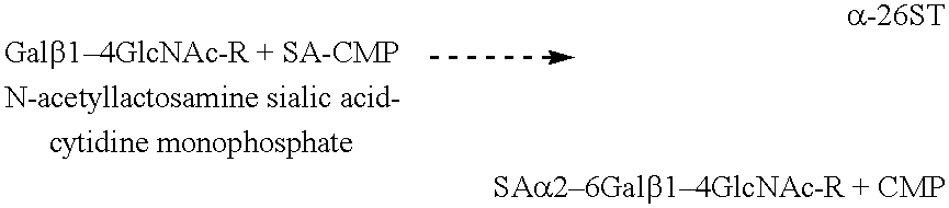
- Sialic acid can be linked in vitro to the carbohydrate chains of the xenograft by sialyltransferase (ST), preferably in a concentration of about 1 mU/ml to about 1000 U/ml, and more preferably in a concentration of about 10 U/ml to about 200 U/ml, in the following exemplary reaction:
- ST sialyltransferase
- Sialic acid can also be linked in vitro to the carbohydrate chains of the xenograft by recombinant trans-sialidase (TS), preferably in a concentration of about 1 mU/ml to about 1000 U/ml, and more preferably in a concentration of about 10 U/ml to about 200 U/ml, in the following exemplary reaction:
- TS trans-sialidase
- the outer surface of the xenograft may optionally be pierced to increase permeability to agents used to render the xenograft substantially non-immunogenic.
- a sterile surgical needle such as an 18 gauge needle may be used to perform this piercing step, or, alternatively a comb-like apparatus containing a plurality of needles may be used.
- the piercing may be performed with various patterns, and with various pierce-to-pierce spacings, in order to establish a desired access to the interior of the xenograft. Piercing may also be performed with a laser. In one form of the invention, one or more straight lines of punctures about three millimeters apart are established circumferentially in the outer surface of the xenograft.
- the ligament xenograft of the invention Prior to implantation, the ligament xenograft of the invention may be treated with limited digestion by proteolytic enzymes such as ficin or trypsin to increase tissue flexibility or coated with anticalcification agents, antithrombotic coatings, antibiotics, growth factors, or other drugs which may enhance the incorporation of the xenograft into the recipient knee joint.
- the ligament xenograft of the invention may be further sterilized using known methods, for example, with additional glutaraldehyde or formaldehyde treatment, ethylene oxide sterilization, propylene oxide sterilization, or the like.
- the xenograft may be stored frozen until required for use.
- the ligament xenograft of the invention may be implanted into a damaged human knee joint by those of skill in the art using known arthroscopic surgical techniques. Specific instruments for performing arthroscopic techniques are known to those of skill in the art, which ensure accurate and reproducible placement of ligament implants. Initially, complete diagnostic arthroscopy of the knee joint is accomplished using known methods. The irreparably damaged ligament is removed with a surgical shaver. The anatomic insertion sites for the ligament are identified and drilled to accommodate a bone plug. The size of the bone plug can be about 9-10 mm in width by about 9-10 mm in depth by 20-40 mm in length. The xenogeneic ligament is brought through the drill holes and affixed with interference screws. Routine closure is performed.
- an ELISA assay for assessing the elimination of ⁇ -gal epitopes from ligament is conducted.
- a monoclonal anti-Gal antibody (designated M86) which is highly specific for ⁇ -gal epitopes on glycoproteins is produced by fusion of splenocytes from anti-Gal producing knock-out mice for ⁇ 1,3 galactosyltransferase, and a mouse hybridoma fusion partner.
- M86 binds to synthetic ⁇ -gal epitopes linked to ⁇ -bovine serum albumin (BSA), to ⁇ -bovine thyroglobulin which has 11 ⁇ -gal epitopes, R. G. Spiro et al., Occurrence of ⁇ -D-galactosyl residues in the thyroglobulin from several species. Localization in the saccharide chains of complex carbohydrates, 259 J. Biol. Chem. 9858 (1984); or to ⁇ -mouse laminin which has 50 ⁇ -gal epitopes, R. G.
- BSA ⁇ -bovine serum albumin
- Binding is measured at different dilutions of the M86 tissue culture medium.
- the monoclonal antibody is diluted from about 1:20 to about 1:160, and preferably diluted from about 1:50 to about 1:130.
- the antibody is incubated for a predetermined period of time ranging between about 5 hr to about 24 hr, at a predetermined temperature ranging from about 3° C. to about 8° C.
- the antibody is maintained in constant rotation with fragments of ligament about 5 ⁇ m to about 100 ⁇ m in size, and more preferably with ligament fragments ranging from about 10 ⁇ m to about 50 ⁇ m in size, at various ligament concentrations ranging from about 200 mg/ml to about 1.5 mg/ml.
- the ligament fragments are removed by centrifugation at centrifugation rate ranging from about 20,000 ⁇ g to about 50,000 ⁇ g.
- the proportion of M86 bound to the ligament is assessed by measuring the remaining M86 activity in the supernatant, in ELISA with ⁇ -gal-BSA as described in the prior art in, for example, U. Galili et al., Porcine and bovine cartilage transplants in cynomolgus monkey: II. Changes in anti-Gal response during chronic rejection, 63 Transplantation 645-651 (1997).
- the extent of binding of M86 to the ligament is defined as a percentage inhibition of subsequent binding to ⁇ -gal-BSA.
- bovine ligament implants are treated with ⁇ -galactosidase to eliminate ⁇ -galactosyl epitopes, the implants are transplanted into cynomolgus monkeys, and the primate response to the ligament implants is assessed.
- a bovine stifle joint is sterilely prepared and a ligament with a block of bone attached to one or both ends is removed in the cold, under strict sterile technique.
- a block of bone representing a substantially cylindrical plug of approximately 9 mm in diameter by 40 mm in length is left attached to the ligament.
- the ligament is carefully identified and dissected free of adhering tissue, thereby forming the xenograft.
- the xenograft is then washed for at least five minutes with an alcohol, such as ethanol or isopropanol, to remove synovial fluid and lipid soluble contaminants.
- an alcohol such as ethanol or isopropanol
- the ligament specimen is frozen at a temperature of about ⁇ 70° C. to disrupt, that, is to kill, the ligament specimen's cells.
- Each ligament specimen is cut into two portions.
- the first portion is immersed in a buffer, such as citrate buffer solution, with a pH ranging from about 5 to about 6.
- the buffer contains ⁇ -galactosidase at a concentration ranging from about 50 U/ml to about 300 U/ml and an additive, such as PEG, ranging in a concentration of about 2% to about 6%.
- the ligament/ ⁇ -galactosidase buffer solution is incubated at a temperature ranging from about 25° C. to about 32° C. for a predetermined period of time ranging from about one hr to about six hr.
- the first portion is washed under conditions which allow the enzyme to diffuse out.
- the ligament is washed twice with citrate buffer and three times with phosphate-buffered saline (PBS) pH 7.5.
- PBS phosphate-buffered saline
- Each wash can include incubation in 50 ml of buffer solution for 10 min with gentle rocking at 24° C. Other washing procedures known to those of ordinary skill in the art can also be used.
- Assays are performed to confirm the complete removal of the ⁇ -gal epitopes.
- ⁇ -galactosidase is produced according to the methods known in the prior art, such as, for example, the methods described in A. Zhu et al., Characterization of recombinant ⁇ -galactosidase for use in seroconversion from blood group B to O of human erythrocytes, 827 Arch. Biochem. Biophysics 324 (1996); A. Zhu et al., High-level expression and purification of coffee bean ⁇ -galactosidase produced in the yeast Pichia pastoris, 827 Arch. Biochem. Biophysics 324 (1996).
- Each ligament sample is implanted in the supra patellar pouch of six cynomolgus monkeys. With the animals under general inhalation anesthesia, the anatomic insertion sites for the xenogeneic ligament are identified and drilled to accommodate a substantially 9 mm in diameter by 40 mm in length bone plug. The xenogeneic ligament is brought through the drill holes and affixed with interference screws. The procedure is performed under sterile surgical technique, and the wounds are closed with 3-0 vicryl or a suitable equivalent known to those of ordinary skill in the art. The animals are permitted unrestricted cage activity and monitored for any sign of discomfort, swelling, infection, or rejection. Blood samples (e.g., 2 ml) are drawn periodically (e.g., every two weeks) for monitoring of antibodies.
- Blood samples e.g., 2 ml
- Blood samples are drawn periodically (e.g., every two weeks) for monitoring of antibodies.
- the occurrence of an immune response against the xenograft is assessed by determining anti-Gal and non-anti-Gal anti-cartilage antibodies (i.e., antibodies binding to cartilage antigens other than the ⁇ -gal epitopes) in serum samples from the transplanted monkeys. At least two ml blood samples are drawn from the transplanted monkeys on the day of implant surgery and at periodic (e.g., two week) intervals post-transplantation. The blood samples are centrifuged and the serum samples are frozen and evaluated for the anti-Gal and other non-anti-Gal anti-cartilage antibody activity.
- anti-Gal and non-anti-Gal anti-cartilage antibodies i.e., antibodies binding to cartilage antigens other than the ⁇ -gal epitopes
- Anti-Gal activity is determined in the serum samples in ELISA with ⁇ -gal-BSA as solid phase antigen, according to methods known in the prior art, such as, for example, the methods described in Galili et al., Porcine and bovine cartilage transplants in cynomolgus monkey: II. Changes in anti-Gal response during chronic rejection, 63 Transplantation 645-651 (1997).
- the ⁇ -gal-BSA antigen is used to coat ELISA microtiter wells. Subsequent to blocking of the wells with 1% BSA in PBS, sera is added to the wells in two fold serial dilutions, and incubated for 2 hr at room temperature.
- the plates are washed, and incubated with secondary anti-IgG antibody conjugated to peroxidase. Color reaction is performed with o-phenylenediamine. Anti-Gal activity at the various post-transplantation serum dilutions are compared with the baseline pretransplantation serum.
- Assays are conducted to determine whether ⁇ -galactosidase treated xenografts induce the formation of anti-ligament antibodies.
- an ELISA assay is performed according to methods known in the prior art, such as, for example, the methods described in K. R. Stone et al., Porcine and bovine cartilage transplants in cynomolgus monkey: I. A model for chronic xenograft rejection, 63 Transplantation 640-645 (1997).
- a solution of ligament homogenate at 100 ⁇ g/ml in carbonate buffer is used as solid phase antigen.
- Other buffers known to those of ordinary skill in the art can also be used.
- Galili et al. Contribution of anti-Gal to primate and human IgG binding to porcine endothelial cells, 60 Transplantation 210 (1995).
- the adsorbed sera at various dilutions are analyzed for anti-ligament antibodies by ELISA, and the post-transplantation production of such antibodies are assessed by comparing this antibody activity with that observed in the pretransplantation serum.
- the ligament xenograft is optionally explanted at one to two months post-transplantation, sectioned and stained for histological evaluation of inflammatory infiltrates.
- Post-transplantation changes in anti-Gal and other anti-ligament antibody activities are correlated with the inflammatory histologic characteristics (i.e., granulocytes or mononuclear cell infiltrates) within the explanted ligament, one to two months post-transplantation, using methods known in the art, as, for example, the methods described in K. R. Stone et al., Porcine and bovine cartilage transplants in cynomolgus monkey: I. A model for chronic xenograft rejection, 63 Transplantation 640-645 (1997).
- the ligament xenograft is aseptically harvested.
- joint fluid if present in amounts sufficient to aspirate, is collected from the stifle joints for possible immunologic testing if the gross and histopathologic evaluation of the transplants indicate good performance of the transplanted ligament.
- a portion of the implant and surrounding tissue is frozen in an embedding medium for frozen tissue specimens in embedding molds for immunohistochemistry evaluation according to the methods known in the prior art.
- “TISSUE-TEK®” O.C.T. compound which includes 10.24% w/w polyvinyl alcohol, 4.26% w/w polyethylene glycol, and 86.60% w/w nonreactive ingredients, and is manufactured by Sakura FinTek, Torrence, Calif., is a non-limiting example of a possible embedding medium for use with the present invention. Other embedding mediums known to those of ordinary skill in the art may also be used.
- the remaining implant and surrounding tissue is collected in 10% neutral buffered formalin for histopathologic examination.
- bovine ligament implants are treated with ⁇ -galactosidase to eliminate ⁇ -gal epitopes, as described in Example 2.
- the implants are further treated with sialic acid-cytosine monophosphate (SA-CMP) and sialyltransferase to cap carbohydrate chains with sialic acid.
- SA-CMP sialic acid-cytosine monophosphate
- Sialytransferase facilitates the transfer of the sialic acid from the SA-CMP compound to the xenograft.
- the sialic acid links to and thus caps the carbohydrate chains.
- the cytosine monophosphate provides the necessary energetic level to the sialic acid for such linking and capping. Capping with sialic acid interferes with the ability of the the subject's immune system to recognize the xenograft as foreign.
- the negative charge of the sialic acid further interferes with the ability of the ligament antigens to bind with non-anti-Gal anti-cartilage antibodies (i.e., antibodies binding to ligament antigens other than the ⁇ -gal epitopes.)
- the implants are transplanted into cynomolgus monkeys, and the primate response to the ligament implants is assessed.
- Bovine ligament stifle joints are prepared as described in Example 2 including the ⁇ -galactosidase treatment. Prior to implantation into the monkeys, however, the implants are further treated with a predetermined amount of a predetermined amount of SA-CMP and sialyltransferase, at specified concentrations for a predetermined time and at a predetermined temperature, to cap carbohydrate chains with sialic acid.
- the sample is immersed in 10 ml buffer solution at a pH of about 5.5 to 7.0, and preferably a pH of about 6.0-6.5, and most preferably a pH of about 6.2, containing SA-CMP at a concentration of approximately about 1 mM to about 10 mM, and sialyltransferase at a concentration of about 100 U/ml.
- the sample is incubated at a temperature range of about 26° C. to about 37° C. for a predetermined time period of about one hr to about four hr.
- enzymes such as recombinant transialidase can be used to facilitate the transfer of sialic acid from compounds such as sialylated lactose to the xenograft.
Abstract
Description


Claims (37)
Priority Applications (10)
| Application Number | Priority Date | Filing Date | Title |
|---|---|---|---|
| US09/036,087 US6210440B1 (en) | 1995-09-15 | 1998-03-06 | Anterior cruciate ligament xenografts |
| JP2000534141A JP2002505144A (en) | 1998-03-06 | 1998-05-27 | Soft tissue xenograft |
| PCT/US1998/010742 WO1999044533A1 (en) | 1998-03-06 | 1998-05-27 | Soft tissue xenografts |
| CA002349562A CA2349562A1 (en) | 1998-03-06 | 1998-05-27 | Soft tissue xenografts |
| US09/623,729 US6758865B1 (en) | 1998-03-06 | 1998-05-27 | Soft tissue xenografts |
| EP98923796A EP1059891A4 (en) | 1998-03-06 | 1998-05-27 | Soft tissue xenografts |
| AU76004/98A AU759086B2 (en) | 1998-03-06 | 1998-05-27 | Soft tissue xenografts |
| US10/062,341 US20020087211A1 (en) | 1995-09-15 | 2002-02-01 | Anterior cruciate ligament xenografts |
| US10/665,844 US20040081954A1 (en) | 1995-09-15 | 2003-09-19 | Anterior cruciate ligament xenografts |
| US10/884,415 US20040243250A1 (en) | 1998-03-06 | 2004-07-02 | Soft tissue xenografts |
Applications Claiming Priority (2)
| Application Number | Priority Date | Filing Date | Title |
|---|---|---|---|
| US08/529,199 US5902338A (en) | 1995-09-15 | 1995-09-15 | Anterior cruciate ligament heterograft |
| US09/036,087 US6210440B1 (en) | 1995-09-15 | 1998-03-06 | Anterior cruciate ligament xenografts |
Related Parent Applications (1)
| Application Number | Title | Priority Date | Filing Date |
|---|---|---|---|
| US08/529,199 Continuation-In-Part US5902338A (en) | 1995-09-15 | 1995-09-15 | Anterior cruciate ligament heterograft |
Related Child Applications (1)
| Application Number | Title | Priority Date | Filing Date |
|---|---|---|---|
| US09/824,327 Continuation US20010051828A1 (en) | 1995-09-15 | 2001-04-02 | Anterior cruciate ligament xenografts |
Publications (1)
| Publication Number | Publication Date |
|---|---|
| US6210440B1 true US6210440B1 (en) | 2001-04-03 |
Family
ID=46255925
Family Applications (1)
| Application Number | Title | Priority Date | Filing Date |
|---|---|---|---|
| US09/036,087 Expired - Lifetime US6210440B1 (en) | 1995-09-15 | 1998-03-06 | Anterior cruciate ligament xenografts |
Country Status (1)
| Country | Link |
|---|---|
| US (1) | US6210440B1 (en) |
Cited By (15)
| Publication number | Priority date | Publication date | Assignee | Title |
|---|---|---|---|---|
| US20030023304A1 (en) * | 2000-01-11 | 2003-01-30 | Carter Kevin C. | Materials and methods for improved bone tendon bone transplantation |
| US20030039678A1 (en) * | 1998-03-16 | 2003-02-27 | Stone Kevin R. | Xenograft bone matrix for orthopedic applications |
| US20030074065A1 (en) * | 1998-03-16 | 2003-04-17 | Stone Kevin R. | Bone xenografts |
| US20030097179A1 (en) * | 2000-01-11 | 2003-05-22 | Carter Kevin C. | Materials and methods for improved bone tendon bone transplantation |
| US20040057936A1 (en) * | 2002-09-23 | 2004-03-25 | Cheung David T. | Method to treat collagenous connective tissue for implant remodeled by host cells into living tissue |
| US20040081954A1 (en) * | 1995-09-15 | 2004-04-29 | Stone Kevin R. | Anterior cruciate ligament xenografts |
| US20040098135A1 (en) * | 1998-04-02 | 2004-05-20 | Stone Kevin R. | Bone xenografts |
| US6758865B1 (en) * | 1998-03-06 | 2004-07-06 | Crosscart, Inc. | Soft tissue xenografts |
| US20040210308A1 (en) * | 2000-01-11 | 2004-10-21 | Regeneration Technologies, Inc. | Materials and methods for improved bone tendon bone transplantation |
| US20080306610A1 (en) * | 2007-06-07 | 2008-12-11 | Zimmer Orthobiologics, Inc. | Tissue processing for nonimmunogenic implants |
| US20090181807A1 (en) * | 2008-01-16 | 2009-07-16 | Jason Miguel De Los Santos | Golfing aid |
| US20140154663A1 (en) * | 2000-09-12 | 2014-06-05 | Lifenet Health | Process for Devitalizing Soft-Tissue Engineered Medical Implants, and Devitalized Soft-Tissue Medical Implants Produced |
| US8753406B2 (en) | 2010-08-31 | 2014-06-17 | Zimmer Inc. | Osteochondral graft delivery device and uses thereof |
| US9744043B2 (en) | 2007-07-16 | 2017-08-29 | Lifenet Health | Crafting of cartilage |
| US20210260247A1 (en) * | 2018-09-19 | 2021-08-26 | Venus Medtech (Hangzhou), Inc. | Pre-Loadable Dried Biological Heart Valve and Preparation Method Thereof |
Citations (40)
| Publication number | Priority date | Publication date | Assignee | Title |
|---|---|---|---|---|
| US4034418A (en) | 1975-05-26 | 1977-07-12 | The Governing Council Of The University Of Toronto | Artificial knee joint |
| US4344193A (en) | 1980-11-28 | 1982-08-17 | Kenny Charles H | Meniscus prosthesis |
| US4400833A (en) | 1981-06-10 | 1983-08-30 | Kurland Kenneth Z | Means and method of implanting bioprosthetics |
| WO1984003036A1 (en) | 1983-02-03 | 1984-08-16 | Wallsten Hans Ivar | Blood vessel prosthesis |
| US4502161A (en) | 1981-09-21 | 1985-03-05 | Wall W H | Prosthetic meniscus for the repair of joints |
| US4597266A (en) | 1985-05-28 | 1986-07-01 | Cryolife, Inc. | Freezing agent and container |
| US4609627A (en) | 1983-08-01 | 1986-09-02 | New York Blood Center, Inc. | Enzymatic conversion of certain sub-type A and AB erythrocytes |
| US4627853A (en) | 1985-05-29 | 1986-12-09 | American Hospital Supply Corporation | Method of producing prostheses for replacement of articular cartilage and prostheses so produced |
| US4642120A (en) | 1983-03-23 | 1987-02-10 | Ramot University Authority For Applied Research And Industrial Development Ltd. | Repair of cartilage and bones |
| US4678470A (en) | 1985-05-29 | 1987-07-07 | American Hospital Supply Corporation | Bone-grafting material |
| US4755593A (en) | 1985-07-24 | 1988-07-05 | Lauren Mark D | Novel biomaterial of cross-linked peritoneal tissue |
| US4776853A (en) | 1986-07-28 | 1988-10-11 | Hsc Research Development Corporation | Process for preparing biological mammalian implants |
| US4789663A (en) | 1984-07-06 | 1988-12-06 | Collagen Corporation | Methods of bone repair using collagen |
| US4801299A (en) | 1983-06-10 | 1989-01-31 | University Patents, Inc. | Body implants of extracellular matrix and means and methods of making and using such implants |
| US4846835A (en) | 1987-06-15 | 1989-07-11 | Grande Daniel A | Technique for healing lesions in cartilage |
| US4880429A (en) | 1987-07-20 | 1989-11-14 | Stone Kevin R | Prosthetic meniscus |
| US4932973A (en) | 1983-09-30 | 1990-06-12 | El Gendler | Cartilage and bone induction by artificially perforated organic bone matrix |
| US5007934A (en) | 1987-07-20 | 1991-04-16 | Regen Corporation | Prosthetic meniscus |
| US5067962A (en) | 1989-04-18 | 1991-11-26 | Baxter International Inc. | Bioprosthetic ligament |
| US5071741A (en) | 1988-04-18 | 1991-12-10 | Cryolife, Inc. | Cryoprotective agent and its use in cryopreservation of cellular matter |
| US5078744A (en) | 1987-09-04 | 1992-01-07 | Bio-Products, Inc. | Method of using tendon/ligament substitutes composed of long, parallel, non-antigenic tendon/ligament fibers |
| US5092894A (en) | 1990-02-13 | 1992-03-03 | Kenny Charles H | Stabilized meniscus prosthesis |
| US5116374A (en) | 1989-03-02 | 1992-05-26 | Regen Corporation | Prosthetic meniscus |
| US5131850A (en) | 1989-11-03 | 1992-07-21 | Cryolife, Inc. | Method for cryopreserving musculoskeletal tissues |
| US5158574A (en) | 1987-07-20 | 1992-10-27 | Regen Corporation | Prosthetic meniscus |
| US5160313A (en) | 1991-05-14 | 1992-11-03 | Cryolife, Inc. | Process for preparing tissue for transplantation |
| US5171660A (en) | 1989-04-26 | 1992-12-15 | Cryolife, Inc. | Process of revitalizing cells and tissue prior to cryopreservation |
| US5171322A (en) | 1990-02-13 | 1992-12-15 | Kenny Charles H | Stabilized meniscus prosthesis |
| US5171273A (en) | 1989-01-13 | 1992-12-15 | University Of Medicine And Dentistry Of New Jersey | Synthetic collagen orthopaedic structures such as grafts, tendons and other structures |
| US5192312A (en) | 1991-03-05 | 1993-03-09 | Colorado State University Research Foundation | Treated tissue for implantation and methods of treatment and use |
| US5216126A (en) | 1991-06-19 | 1993-06-01 | Genentech, Inc. | Receptor polypeptides and their production and uses |
| US5306311A (en) | 1987-07-20 | 1994-04-26 | Regen Corporation | Prosthetic articular cartilage |
| US5306304A (en) | 1990-10-31 | 1994-04-26 | El Gendler | Flexible membranes produced from organic bone matrix for skeletal repair and reconstruction |
| US5358525A (en) | 1992-12-28 | 1994-10-25 | Fox John E | Bearing surface for prosthesis and replacement of meniscal cartilage |
| WO1995026740A1 (en) | 1994-03-31 | 1995-10-12 | Diacrin, Inc. | Improved methods for transplantation using modified cells and t cell inhibitory agents |
| WO1995028412A1 (en) | 1994-04-13 | 1995-10-26 | Biotransplant, Inc. | α(1,3) GALACTOSYLTRANSFERASE NEGATIVE SWINE |
| WO1995033828A1 (en) | 1994-06-03 | 1995-12-14 | Diacrin, Inc. | Modified cells and methods for inhibiting hyperacute rejection of xenogeneic transplants |
| US5507810A (en) | 1991-10-07 | 1996-04-16 | Osteotech, Inc. | Processing of fibrous connective tissue |
| US5613982A (en) | 1994-03-14 | 1997-03-25 | Cryolife, Inc. | Method of preparing transplant tissue to reduce immunogenicity upon implantation |
| US5944755A (en) * | 1995-09-15 | 1999-08-31 | Crosscart, Inc. | Articular cartilage xenografts |
-
1998
- 1998-03-06 US US09/036,087 patent/US6210440B1/en not_active Expired - Lifetime
Patent Citations (41)
| Publication number | Priority date | Publication date | Assignee | Title |
|---|---|---|---|---|
| US4034418A (en) | 1975-05-26 | 1977-07-12 | The Governing Council Of The University Of Toronto | Artificial knee joint |
| US4344193A (en) | 1980-11-28 | 1982-08-17 | Kenny Charles H | Meniscus prosthesis |
| US4400833A (en) | 1981-06-10 | 1983-08-30 | Kurland Kenneth Z | Means and method of implanting bioprosthetics |
| US4502161A (en) | 1981-09-21 | 1985-03-05 | Wall W H | Prosthetic meniscus for the repair of joints |
| US4502161B1 (en) | 1981-09-21 | 1989-07-25 | ||
| WO1984003036A1 (en) | 1983-02-03 | 1984-08-16 | Wallsten Hans Ivar | Blood vessel prosthesis |
| US4642120A (en) | 1983-03-23 | 1987-02-10 | Ramot University Authority For Applied Research And Industrial Development Ltd. | Repair of cartilage and bones |
| US4801299A (en) | 1983-06-10 | 1989-01-31 | University Patents, Inc. | Body implants of extracellular matrix and means and methods of making and using such implants |
| US4609627A (en) | 1983-08-01 | 1986-09-02 | New York Blood Center, Inc. | Enzymatic conversion of certain sub-type A and AB erythrocytes |
| US4932973A (en) | 1983-09-30 | 1990-06-12 | El Gendler | Cartilage and bone induction by artificially perforated organic bone matrix |
| US4789663A (en) | 1984-07-06 | 1988-12-06 | Collagen Corporation | Methods of bone repair using collagen |
| US4597266A (en) | 1985-05-28 | 1986-07-01 | Cryolife, Inc. | Freezing agent and container |
| US4678470A (en) | 1985-05-29 | 1987-07-07 | American Hospital Supply Corporation | Bone-grafting material |
| US4627853A (en) | 1985-05-29 | 1986-12-09 | American Hospital Supply Corporation | Method of producing prostheses for replacement of articular cartilage and prostheses so produced |
| US4755593A (en) | 1985-07-24 | 1988-07-05 | Lauren Mark D | Novel biomaterial of cross-linked peritoneal tissue |
| US4776853A (en) | 1986-07-28 | 1988-10-11 | Hsc Research Development Corporation | Process for preparing biological mammalian implants |
| US4846835A (en) | 1987-06-15 | 1989-07-11 | Grande Daniel A | Technique for healing lesions in cartilage |
| US4880429A (en) | 1987-07-20 | 1989-11-14 | Stone Kevin R | Prosthetic meniscus |
| US5007934A (en) | 1987-07-20 | 1991-04-16 | Regen Corporation | Prosthetic meniscus |
| US5306311A (en) | 1987-07-20 | 1994-04-26 | Regen Corporation | Prosthetic articular cartilage |
| US5158574A (en) | 1987-07-20 | 1992-10-27 | Regen Corporation | Prosthetic meniscus |
| US5078744A (en) | 1987-09-04 | 1992-01-07 | Bio-Products, Inc. | Method of using tendon/ligament substitutes composed of long, parallel, non-antigenic tendon/ligament fibers |
| US5071741A (en) | 1988-04-18 | 1991-12-10 | Cryolife, Inc. | Cryoprotective agent and its use in cryopreservation of cellular matter |
| US5171273A (en) | 1989-01-13 | 1992-12-15 | University Of Medicine And Dentistry Of New Jersey | Synthetic collagen orthopaedic structures such as grafts, tendons and other structures |
| US5116374A (en) | 1989-03-02 | 1992-05-26 | Regen Corporation | Prosthetic meniscus |
| US5067962A (en) | 1989-04-18 | 1991-11-26 | Baxter International Inc. | Bioprosthetic ligament |
| US5171660A (en) | 1989-04-26 | 1992-12-15 | Cryolife, Inc. | Process of revitalizing cells and tissue prior to cryopreservation |
| US5131850A (en) | 1989-11-03 | 1992-07-21 | Cryolife, Inc. | Method for cryopreserving musculoskeletal tissues |
| US5092894A (en) | 1990-02-13 | 1992-03-03 | Kenny Charles H | Stabilized meniscus prosthesis |
| US5171322A (en) | 1990-02-13 | 1992-12-15 | Kenny Charles H | Stabilized meniscus prosthesis |
| US5306304A (en) | 1990-10-31 | 1994-04-26 | El Gendler | Flexible membranes produced from organic bone matrix for skeletal repair and reconstruction |
| US5192312A (en) | 1991-03-05 | 1993-03-09 | Colorado State University Research Foundation | Treated tissue for implantation and methods of treatment and use |
| US5160313A (en) | 1991-05-14 | 1992-11-03 | Cryolife, Inc. | Process for preparing tissue for transplantation |
| US5216126A (en) | 1991-06-19 | 1993-06-01 | Genentech, Inc. | Receptor polypeptides and their production and uses |
| US5507810A (en) | 1991-10-07 | 1996-04-16 | Osteotech, Inc. | Processing of fibrous connective tissue |
| US5358525A (en) | 1992-12-28 | 1994-10-25 | Fox John E | Bearing surface for prosthesis and replacement of meniscal cartilage |
| US5613982A (en) | 1994-03-14 | 1997-03-25 | Cryolife, Inc. | Method of preparing transplant tissue to reduce immunogenicity upon implantation |
| WO1995026740A1 (en) | 1994-03-31 | 1995-10-12 | Diacrin, Inc. | Improved methods for transplantation using modified cells and t cell inhibitory agents |
| WO1995028412A1 (en) | 1994-04-13 | 1995-10-26 | Biotransplant, Inc. | α(1,3) GALACTOSYLTRANSFERASE NEGATIVE SWINE |
| WO1995033828A1 (en) | 1994-06-03 | 1995-12-14 | Diacrin, Inc. | Modified cells and methods for inhibiting hyperacute rejection of xenogeneic transplants |
| US5944755A (en) * | 1995-09-15 | 1999-08-31 | Crosscart, Inc. | Articular cartilage xenografts |
Non-Patent Citations (13)
| Title |
|---|
| Collins et al., Xenotransplantation, Characterization of Porcine Endothelial Cell Determinants Recognized by Human Natural Antibodies, 1:36-46 (1994). |
| Cotterell et al., Transplantation, The Humoral Immune Response in Humans Following Cross-Perfusion of Porcine Organs, 60-861-868 (1995). |
| Elves et al., An Investigation Into The Immunogenicity Of Various Components of Osteoarticular Grafts, The British Journal of Experimental Pathology, 55:344-351 (1974). |
| Engkvist, Ove, Scand. J. Plast. Reconstr. Surg., 13:361-369 (1982). |
| Galili, Immunology Today, 14:480-482 (1993). |
| LaVecchio et al., Transplanatation, Enzymatic Removal of Alpha-Galactosyl Epitopes From Porcine Endothelial Cells Diminishes The CytotoxicEffect of Natural Antibodies, 60-841-847. |
| Rodrigo et al., Clinical Orthopedics and Related Research, 134:342-349 (1978). |
| Rubak et al., Acta Orthop. Scand, 53:181-186 (1982). |
| Satake et al., Xenotransplanation, Limited Specificity of Xenoantibodies In Diabetic Patients Transplanted With Fetal Porcine Islet Cell Clusters. Main Antibody Reactivity Against alpha-linked Galactose-Containing Epitopes, 1:89-101 (1994). |
| Satake et al., Xenotransplanation, Limited Specificity of Xenoantibodies In Diabetic Patients Transplanted With Fetal Porcine Islet Cell Clusters. Main Antibody Reactivity Against α-linked Galactose-Containing Epitopes, 1:89-101 (1994). |
| Sengupta et al., The Journal of Bone and Joint Surgery, 56B:167-177 (1974). |
| Stone et al., Arthroscopy: The Journal of Arthroscopic and Related Surgery, 9:234-237 (1993). |
| Webber et al., Journal of Orthopedic Research, 3:36-42 (1985). |
Cited By (22)
| Publication number | Priority date | Publication date | Assignee | Title |
|---|---|---|---|---|
| US20040081954A1 (en) * | 1995-09-15 | 2004-04-29 | Stone Kevin R. | Anterior cruciate ligament xenografts |
| US6758865B1 (en) * | 1998-03-06 | 2004-07-06 | Crosscart, Inc. | Soft tissue xenografts |
| US20030039678A1 (en) * | 1998-03-16 | 2003-02-27 | Stone Kevin R. | Xenograft bone matrix for orthopedic applications |
| US20030074065A1 (en) * | 1998-03-16 | 2003-04-17 | Stone Kevin R. | Bone xenografts |
| US7722672B2 (en) * | 1998-03-16 | 2010-05-25 | Stone Kevin R | Bone xenografts |
| US20110104229A1 (en) * | 1998-04-02 | 2011-05-05 | Stone Kevin R | Bone Xenograft |
| US20040098135A1 (en) * | 1998-04-02 | 2004-05-20 | Stone Kevin R. | Bone xenografts |
| US20030097179A1 (en) * | 2000-01-11 | 2003-05-22 | Carter Kevin C. | Materials and methods for improved bone tendon bone transplantation |
| US20040210308A1 (en) * | 2000-01-11 | 2004-10-21 | Regeneration Technologies, Inc. | Materials and methods for improved bone tendon bone transplantation |
| US9615913B2 (en) | 2000-01-11 | 2017-04-11 | RTI Surgical, Inc | Materials and methods for improved bone tendon bone transplantation |
| US20030023304A1 (en) * | 2000-01-11 | 2003-01-30 | Carter Kevin C. | Materials and methods for improved bone tendon bone transplantation |
| US8167943B2 (en) | 2000-01-11 | 2012-05-01 | Rti Biologics, Inc. | Materials and methods for improved bone tendon bone transplantation |
| US10188505B2 (en) | 2000-01-11 | 2019-01-29 | Rti Surgical, Inc. | Materials and methods for improved bone tendon bone transplantation |
| US20140154663A1 (en) * | 2000-09-12 | 2014-06-05 | Lifenet Health | Process for Devitalizing Soft-Tissue Engineered Medical Implants, and Devitalized Soft-Tissue Medical Implants Produced |
| US7008763B2 (en) | 2002-09-23 | 2006-03-07 | Cheung David T | Method to treat collagenous connective tissue for implant remodeled by host cells into living tissue |
| US20040057936A1 (en) * | 2002-09-23 | 2004-03-25 | Cheung David T. | Method to treat collagenous connective tissue for implant remodeled by host cells into living tissue |
| US20080306610A1 (en) * | 2007-06-07 | 2008-12-11 | Zimmer Orthobiologics, Inc. | Tissue processing for nonimmunogenic implants |
| US9744043B2 (en) | 2007-07-16 | 2017-08-29 | Lifenet Health | Crafting of cartilage |
| US11147674B2 (en) | 2007-07-16 | 2021-10-19 | Lifenet Health | Crafting of cartilage |
| US20090181807A1 (en) * | 2008-01-16 | 2009-07-16 | Jason Miguel De Los Santos | Golfing aid |
| US8753406B2 (en) | 2010-08-31 | 2014-06-17 | Zimmer Inc. | Osteochondral graft delivery device and uses thereof |
| US20210260247A1 (en) * | 2018-09-19 | 2021-08-26 | Venus Medtech (Hangzhou), Inc. | Pre-Loadable Dried Biological Heart Valve and Preparation Method Thereof |
Similar Documents
| Publication | Publication Date | Title |
|---|---|---|
| US20010051828A1 (en) | Anterior cruciate ligament xenografts | |
| US6049025A (en) | Articular cartilage xenografts | |
| US6402783B1 (en) | Anterior cruciate ligament xenografts | |
| US6046379A (en) | Meniscal xenografts | |
| US6758865B1 (en) | Soft tissue xenografts | |
| US5944755A (en) | Articular cartilage xenografts | |
| US6455309B2 (en) | Proteoglycan-reduced soft tissue xenografts | |
| US6383732B1 (en) | Method of preparing xenograft heart valves | |
| US6231608B1 (en) | Aldehyde and glycosidase-treated soft and bone tissue xenografts | |
| US20110104229A1 (en) | Bone Xenograft | |
| US5984858A (en) | Meniscal xenografts | |
| US6210440B1 (en) | Anterior cruciate ligament xenografts | |
| CA2323866A1 (en) | Bone xenografts | |
| US6972041B1 (en) | Bone xenografts | |
| EP1065997B1 (en) | Bone xenografts |
Legal Events
| Date | Code | Title | Description |
|---|---|---|---|
| AS | Assignment |
Owner name: CROSSCART, INC., CALIFORNIA Free format text: ASSIGNMENT OF ASSIGNORS INTEREST;ASSIGNOR:STONE, KEVIN R.;REEL/FRAME:009070/0533 Effective date: 19980302 Owner name: ALLEGHENY UNIVERSITY OF HEALTH SCIENCE, PENNSYLVAN Free format text: ASSIGNMENT OF ASSIGNORS INTEREST;ASSIGNOR:GALILI, URI;REEL/FRAME:009069/0153 Effective date: 19980304 |
|
| STCF | Information on status: patent grant |
Free format text: PATENTED CASE |
|
| FEPP | Fee payment procedure |
Free format text: PAYOR NUMBER ASSIGNED (ORIGINAL EVENT CODE: ASPN); ENTITY STATUS OF PATENT OWNER: SMALL ENTITY |
|
| FPAY | Fee payment |
Year of fee payment: 4 |
|
| FEPP | Fee payment procedure |
Free format text: PAYOR NUMBER ASSIGNED (ORIGINAL EVENT CODE: ASPN); ENTITY STATUS OF PATENT OWNER: SMALL ENTITY Free format text: PAYER NUMBER DE-ASSIGNED (ORIGINAL EVENT CODE: RMPN); ENTITY STATUS OF PATENT OWNER: SMALL ENTITY |
|
| FPAY | Fee payment |
Year of fee payment: 8 |
|
| AS | Assignment |
Owner name: APERION BIOLOGICS, INC., TEXAS Free format text: CHANGE OF NAME;ASSIGNOR:CROSSCART, INC.;REEL/FRAME:023319/0826 Effective date: 20090610 |
|
| AS | Assignment |
Owner name: APERION BIOLOGICS, INC., TEXAS Free format text: INTELLECTUAL PROPERTY TRANSFER AGREEMENT;ASSIGNOR:PHILADELPHIA HEALTH AND EDUCATION CORP. D/B/A DREXEL UNIVERSITY COLLEGE OF MEDICINE;REEL/FRAME:027151/0539 Effective date: 20110915 |
|
| FEPP | Fee payment procedure |
Free format text: PAYER NUMBER DE-ASSIGNED (ORIGINAL EVENT CODE: RMPN); ENTITY STATUS OF PATENT OWNER: SMALL ENTITY Free format text: PAYOR NUMBER ASSIGNED (ORIGINAL EVENT CODE: ASPN); ENTITY STATUS OF PATENT OWNER: SMALL ENTITY |
|
| FPAY | Fee payment |
Year of fee payment: 12 |
|
| SULP | Surcharge for late payment |
Year of fee payment: 11 |