US6612983B1 - Pancreatic secretion response to stimulation test protocol - Google Patents
Pancreatic secretion response to stimulation test protocol Download PDFInfo
- Publication number
- US6612983B1 US6612983B1 US09/535,840 US53584000A US6612983B1 US 6612983 B1 US6612983 B1 US 6612983B1 US 53584000 A US53584000 A US 53584000A US 6612983 B1 US6612983 B1 US 6612983B1
- Authority
- US
- United States
- Prior art keywords
- pancreatic
- patient
- stimulation
- gastroelectric
- response
- Prior art date
- Legal status (The legal status is an assumption and is not a legal conclusion. Google has not performed a legal analysis and makes no representation as to the accuracy of the status listed.)
- Expired - Fee Related
Links
Images


Classifications
-
- A—HUMAN NECESSITIES
- A61—MEDICAL OR VETERINARY SCIENCE; HYGIENE
- A61N—ELECTROTHERAPY; MAGNETOTHERAPY; RADIATION THERAPY; ULTRASOUND THERAPY
- A61N1/00—Electrotherapy; Circuits therefor
- A61N1/18—Applying electric currents by contact electrodes
- A61N1/32—Applying electric currents by contact electrodes alternating or intermittent currents
- A61N1/36—Applying electric currents by contact electrodes alternating or intermittent currents for stimulation
- A61N1/36007—Applying electric currents by contact electrodes alternating or intermittent currents for stimulation of urogenital or gastrointestinal organs, e.g. for incontinence control
Definitions
- This disclosure relates to a medical test and more specifically to a test protocol for assessing a patient's pancreatic response to natural and electrical stimulation.
- the medical device industry produces a wide variety of electronic and mechanical devices for treating patient medical conditions.
- medical devices can be surgically implanted or connected externally to the patient receiving treatment.
- Clinicians use medical devices alone or in combination with drug therapies and surgery to treat patient medical conditions.
- medical devices provide the best, and sometimes the only, therapy to restore an individual to a more healthful condition and a fuller life.
- One type of medical device that can be used to treat pancreatic conditions is a neurostimulator.
- Neurostimulator treatment for pancreatic conditions involves placing electrical leads typically within the gastrointestinal tract of a patient and applying an electrical signal with an implanted or external neurostimulator. When considering whether neurostimulation would be suitable to treat a condition in a patient, patient testing is helpful.
- Pancreatic testing is challenging because the pancreas is a complex organ with a multitude of functions and many pancreatic conditions manifest themselves indirectly through symptoms that may not initially be attributed to a pancreatic condition. Pancreatic testing is also challenging because the pancreas is not a static organ that secretes at a constant rate. Pancreatic secretion varies in response to a variety of factors that may stimulate or decrease pancreatic secretions. Some conditions attributable to pancreatic dysfunction are: pancreatitis, diabetes, inflammation of the pancreas, malabsorbtion syndrome, and certain cancers. In addition to these conditions, there are a variety of other conditions that may be attributable to pancreatic dysfunction such as pain, nausea, and vomiting.
- Previous pancreatic diagnostic tests are static and involve measuring one or more pancreatic function indicators and then correlating the measurements to measurements expected for various types of pancreatic disease.
- An example such static pancreatic testing is described in the following publication: Frank Netter, “The Ciba Collection of Medical Illustrations”, Vol. 3 Digestive System, Part III Liver, Billary Tract And Pancreas (1964). Static testing does not evaluate pancreatic response to stimulation so some pancreatic conditions may not be identified.
- pancreatic testing in response to stimulation would provide additionally information that would be helpful in selecting a therapy such as electrical stimulation to treat a pancreatic condition and avoid inappropriate therapies, so patients' benefit from decreased risk of medical complications, reduced pain, more rapid treatment, and reduced costs.
- a screening test is used on a patient to determine if pancreatic secretion is responsive to stimulation.
- the screening test compares measurements of a pancreatic indicator both before and after natural stimulation.
- the pancreatic indicator is measured before natural stimulation and then the patient's pancreas is naturally stimulated. After waiting a time period from when the natural stimulation began, the pancreatic indicator is once again measured.
- the difference between the pancreatic indicator measured before ingesting the meal and the pancreatic response indicator measured after ingesting and waiting a time period is calculated and a decision is made whether the patient has a pancreatic condition suitable for treatment with electrical stimulation.
- an efficacy test is used to evaluate the efficacy of employing electrical stimulation to treat a pancreatic condition.
- the efficacy test compares a pancreatic response indicator before electrical stimulation and after electrical stimulation to decides whether there is efficacy in using electrical stimulation to treat a patient's pancreatic condition.
- an efficacy test is used to evaluate the efficacy of using electrical stimulation to treat nausea and vomiting.
- FIG. 1 shows an environment of a pancreatic test
- FIG. 2 shows a more detailed environment for a pancreatic test
- FIG. 3 shows a flow diagram for a screening test for pancreatic secretion in response to stimulation
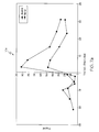
- FIG. 4 shows a graph of results from the screening test
- FIG. 5 shows a diagram of a gastroelectric stimulator
- FIG. 6 shows a flow diagram for an efficacy test for gastroelectric stimulator to affect pancreatic secretion
- FIGS. 7 a-b show graphs of results from the efficacy test for gastroelectric stimulation to affect pancreatic secretion
- FIG. 8 shows a flow diagram for an efficacy test to use a gastroelectric stimulator to treat emesis
- FIG. 9 shows a catheter positioned to perform the efficacy test to treat emesis.
- FIG. 10 shows a timing diagram of the efficacy test for treating emesis.
- FIGS. 1 and 2 show the general environment for pancreatic testing.
- a patient's 20 digestive system 22 includes the esophagus 24 , stomach 26 , duodenum 28 , and pancreas 30 .
- the stomach 26 plays a role in influencing the digestive system 22 by acting as a pacemaker through myoelectrical activity as described in the following publication: Kenneth Koch et al., Electrogastrography, An Illustrated Guide To Gastrointestinal Motility 2 nd Ed., pp. 290-307 (1993).
- the duodenum 28 is the first division of the small intestine that is about 25.0 cm in length and includes the pars descendens into which the pancreatic 30 ducts open.
- Duodenal chyme 34 is a semi-fluid mass of partly digested food with a typical normal daily volume of about 4.0 to 7.0 liters that passes from the stomach 26 into the duodenum 28 .
- the duodenum 28 has osmoreceptors 35 that respond to osmotic pressure in the duodenum 28 by sending nerve impulses that can cause nausea and vomiting when the osmoreceptors 35 sense a significant osmotic imbalance in the duodenum 28 .
- the pancreas 30 has a tail 36 , a body 38 , a head 41 , a duct of Santorini and a duct 39 of Wirsung.
- the duct 39 of Wirsung is the principal duct that drains most of the gland that starts in the tail 36 by the confluence of several small ducts and extends into the head 41 where it terminates at the duodenum 28 papilla.
- the duct of Santorini is an accessory duct that drains a small upper anterior part of the head 41 and terminates in the duodenum 28 at a small accessory papilla.
- the exocrine pancreas 30 has acini cells and a ductal system of intralobular and intercalated ducts organized into lobules that secrete enzymes and bicarbonate into the duodenum 28 where they aid in digestion and absorption of nutrients.
- Pancreatic 30 interdigestive secretion cycles are in temporal coordination with gastrointestinal motility.
- Pancreatic 30 secretions include exocrine and endocrine secretions that are dominantly isotonic.
- Pancreatic 30 exocrine secretions mainly assist in digestion and include bicarbonate and enzymes such as trypsin, chymotrypsin, amylase, and lipase. Daily pancreatic exocrine secretion is typically in the range from 1.0 to 2.0 liters. Pancreatic 30 endocrine secretion regulate metabolism and include enzymes such as insulin, glucagons, somatostatin, and pancreatic polypeptide. Pancreatic 30 secretions are responsive to nervous control and gut hormones.
- pancreas 30 and other intra-abdominal organs are innervated by the sympathetic and parasympathetic branches of the autonomic nervous system.
- Sympathetic innervation is supplied by the splanchnic nerves
- parasympathetic innervation is supplied by the vagus nerves 40 .
- Pancreatic 30 acinar and islet cells are innervated only directly through parasympathetic nerves, and pancreatic 30 blood vessels are innervated solely by sympathetic nerves. All nerves of the pancreas 30 , both afferent and efferent, pass through the celiac plexus.
- Pancreatic 30 secretion can be affected by both natural simulation and electrical stimulation.
- Pancreatic 30 exocrine and endocrine secretion are controlled by the central and enteric nervous system and by gut hormones that are released after meal ingestion. Pancreatic 30 innervation is further described in the following article by Richins, “The innervation of the pancreas”, J. Comp. Neurol 82:223-236 (1945). Pancreatic 30 secretion can also be influenced by electrical stimulation.
- pancreatic secretions When electrical stimulation is applied to nerves, existing natural signals can be blocked or altered as described in Durand, Anthony M., “Electric Stimulation of Excitable Tissue”, The Biomedical Engineering Handbook, Chapter. 17, pp. 229-251 (1995).
- One location to apply electrical stimulation to influence pancreatic 30 secretions is the stomach 26 because the stomach 26 shares significant common innervation with the pancreas 30 .
- FIG. 3 shows a flow diagram depicting a screening test method 42 for using gastroelectric stimulation to affect pancreatic 30 (FIG. 2) secretion in a patient 20 .
- the purpose of the screening test 42 is to determine whether there is potential efficacy in using a gastroelectric stimulator 44 (FIG. 1) to affect pancreatic 30 secretions in a patient 20 , so ineffective therapies can be avoided. Patients 20 benefit from avoiding inappropriate therapies from a decreased risk of medical complications, reduced pain, more rapid treatment, and lower costs.
- the method 42 comprises the following elements. First, a initial pancreatic 30 response is measured at step 43 to establish a starting point. Pancreatic 30 response can be measured directly from duodenal pancreatic 30 secretion or indirectly with a pancreatic 30 secretion indicator.
- Duodenal pancreatic 30 secretion can be measured with a catheter such as shown in PCT Patent Application No. WO 88/03389 by Haillgren et al. titled “Catheter And Method For Intestinal Aspiration, Perfusion and Provocation And Its Use In The Diagnosis Of Intestinal Disease/Allergy” (1988).
- FIG. 9 shows the patient's duodenum 28 isolated with an upstream occlusion 46 and a downstream occlusion 48 that create a duodenal isolation area 50 in the duodenum 28 .
- Pancreatic 30 secretion that occurs in the isolation area 50 is evacuated and the volume is measured to calculate pancreatic 30 response.
- Duodenal pancreatic 30 secretion measurement is discussed in more detail in below under FIGS. 8-9.
- Pancreatic 30 secretion response can also be measured with a pancreatic 30 secretion indicator.
- a pancreatic 30 secretion indicator is a substance that varies in the body in relation to pancreatic 30 stimulation and are typically found in a patient's blood and/or stool.
- pancreatic 30 exocrine and endocrine indicators that underlie numerous pancreatic 30 disorders, so measurement of an exocrine indicator can reflect on both pancreatic 30 exocrine and endocrine function; likewise, measurement of an endocrine indicator can reflect on both pancreatic 30 endocrine and exocrine function.
- This interrelationship is further described in the following article: Sharon Kang et al., “Pancreatic Exocrine-Endocrine Interrelationship, Clinical Implications”, Pancreas Update 0889-8553 (1999).
- pancreatic 30 exocrine or endocrine secretions such as pancreatic polypeptide and elastase.
- a response indicator can be a substance that is not an exocrine or endocrine secretion but still varies according to pancreatic 30 secretion such as stool fat.
- Pancreatic 30 polypeptide is an endocrine hormone measured by blood sampling that has been found to be a good indicator of the level of vagal 40 (FIG. 1) stimulation of the pancreas 30 .
- pancreatic 30 polypeptide oscillates at baseline level, increases by four to five-fold after a meal and then returns to baseline level about three to four hours after the meal.
- pancreas 30 Ingestion of food causes stimulation of the pancreas 30 perhaps by the vagal nerve 40 .
- a patient with a vagatomy, surgical severing of the vagal nerve 40 would not be expected to have an increase in pancreatic 30 polypeptide after ingesting food. After measuring initial pancreatic 30 response, pancreatic 30 secretion is stimulated naturally.
- Naturally stimulating 52 the patient's pancreas 30 is done with sensory stimulation that naturally occurs to cause an increase in pancreatic 30 secretion such as looking at food, smelling food, or eating food.
- a natural response time 54 is waited to allow for the lag between pancreatic 30 stimulation and a change in the pancreatic 30 response.
- the natural response time 54 is the time necessary for the pancreatic 30 response to accurately represent pancreatic 30 secretion such as a time period of about 15.0 minutes. After the natural response time 54 has been waited, the pancreatic 30 response can be measured.
- the pancreatic response is once again measured 56 after waiting the natural response time 54 .
- the difference between the initial pancreatic response 44 and the subsequent response 56 are calculated 58 .
- previously established predetermined screening data 60 can be considered to improve the accuracy of the measurement.
- the predetermined screening data 60 is data that will improve the accuracy or reliability of the pancreatic 30 response measurement such as statistically data from previous testing, or a previous screening test 42 run on the current patient being tested.
- a decision 62 about the potential for efficacy in using a gastroelectric stimulator 44 can be made.
- there is potential stimulation efficacy 64 if natural stimulation 52 increases pancreatic 30 response, but the increased response is less that what would normally be expected under the conditions.
- there is little or no potential stimulation efficacy 66 if natural stimulation increases pancreatic 30 response to a normal level that would be expected under the conditions.
- there is little or no potential stimulation efficacy 66 if natural stimulation 52 does not increase pancreatic 30 response.
- FIG. 4 shows polypeptide level for two different patients 20 undergoing the screening test 42 for using gastroelectric stimulation to affect pancreatic 30 secretion as discussed under FIG. 3 .
- the first patient 68 shows a normal polypeptide excursion after natural stimulation 52 .
- the second patient 70 shows an abnormal polypeptide excursion after natural stimulation 52 .
- the first patient's 68 polypeptide levels are normal because the levels increased after natural stimulation 52 and then for an additional two to three hours.
- the second patient's 70 polypeptide levels are abnormal because the levels are generally flat.
- FIG. 5 shows a diagram of a gastroelectric stimulator 44 that includes a neuroelectrical stimulator 72 , an electrical lead 74 , and electrical contacts 76 also known as electrodes 76 .
- the neurostimulator 72 produces a stimulation signal 78 that can be used to perform neuromodulation at to reduce pain, decrease tremors, control incontinence, and perform other medical therapies.
- the neurostimulator 72 can be programmed to adjust stimulation signal 78 parameters such as pulse width, frequency, amplitude, polarization, and duration.
- the neuroelectrical stimulator 72 can be surgically implanted in a subcutaneous pocket in the abdomen or attached to the patient 20 .
- the neuroelectrical stimulator 72 can be a device such as an Itrel® 3 Model 7425 available from Medtronic, Inc. in Minneapolis, Minn.
- the neurostimulator 72 is typically programmed with a physician programmer such as a Model 7432 also available from Medtronic, Inc.
- the electrical lead 74 provides an electrical path for the stimulation signal 78 from the neurostimulator 72 to the electrodes 76 .
- the electrical lead 74 is a lead suitable for a neurostimulator 72 such as Model 4300 leads available from Medtronic, Inc.
- the electric leads 74 can be surgically implanted into the muscle wall of the stomach 26 such as 0.5-10.0 cm apart on the greater curvature at the limit of the corpus-antrum using a surgical technique such as laparotomy or laparoscopy.
- the lead 74 can be placed in the lesser curvature of the corpus via a preexisting percutaneous gastrostomy site.
- the electrical lead 74 and electrical contacts 76 are selected with an impedance to produce a pulse current in the range from about 1.0 to 10.0 mA.
- the distal end the electrical lead 74 carries electrical contacts 76 .
- the electrical contacts 76 comprise at least two electrodes 76 and additional electrodes 76 can be used.
- the electrodes 76 are carried on the electrical lead 74 near the distal end.
- the electrodes 76 are electrically connected through the electrical lead 74 to the neuroelectrical stimulator 72 .
- the electrodes 76 receive a stimulation signal 78 from the neuroelectrical stimulator 72 and convey this to an electrode 76 position within the patient's digestive system 22 .
- the electrodes 76 can be configured as monopolar electrodes 76 with one electrode 76 per lead or as multipolar electrodes 76 with more than one electrode 76 per lead 74 .
- the electrodes 76 are attached to the electrical lead 74 prior to implantation and navigated to a point near the desired stimulation site.
- the electrodes 76 are made from a biocompatible conductive material such as platinum-iridium.
- the electrodes 76 are implanted into the muscle wall of the stomach 26 such as 0.5 cm to 10.0 cm apart on the greater curvature at the limit of the corpus-antrum.
- the electrical lead 74 and electrical contacts 76 are selected with an impedance to produce a pulse current of about 5.0 mA.
- the stimulation signal 78 has a frequency selected to increase pancreatic 30 secretion such as from about 3.0 pulses per minute to 6,000 pulses per minute.
- the stimulation signal 78 has a pulse width selected to influence pancreatic 30 secretion such as from about 0.01 mSec to 500.0 mSec.
- the stimulation signal 78 has a peak amplitude selected to influence pancreatic 30 secretion such as from about 0.01 mA to 100.0 mA.
- the stimulation signal 78 is charge balanced for biocompatibility.
- the stimulation signal 78 can have the following parameters: amplitude necessary to obtain a current of about 5.0 mA; a pulse width of about 330 ⁇ Sec; a pulse frequency of about 14.0 Hz; and a cycle “on” of about 0.1 seconds and “off” of about 5.0 seconds.
- FIG. 6 shows a flow diagram of an efficacy test method for gastroelectric stimulation 80 .
- the method 80 begins by naturally stimulating 82 the pancreatic 30 secretion of the patient 20 which is discussed in more detail above in the description under FIG. 3 .
- a natural response time is waited 84 which also is discussed in more detailed under FIG. 3 .
- pancreatic 30 response is measured 86 after waiting 84 the natural response time 84 which again is discussed in more detail under FIG. 3 .
- electrical stimulation 88 and natural stimulation 82 of the patient's pancreas 30 is done for a predetermined stimulation time such as in the range from 15.0 minutes to 24.0 hours. Electrical stimulation 88 is discussed in more detail earlier under FIG. 5 .
- measurements 94 after natural stimulation 82 and after electrical stimulation 88 can be made after waiting predetermined time periods to establish more data points.
- Calculations 96 can be simple or complex. Simple calculations 96 include such mathematical techniques as averaging the response indicator measurements 86 , 92 over selected period of time or selecting the maximum value. Complex calculations 96 include such mathematical techniques as calculating the integral to find the area under the curve of the measurements 86 , 92 . Calculations 96 can also include the consideration of predetermined efficacy data 97 from previous test results from the patient undergoing the efficacy test 80 , or predetermined efficacy data 97 can be available statistical information. Once calculations 96 are completed, a decision 98 can be made.
- Deciding 98 whether there is potential efficacy 100 or low potential efficacy 102 in using gastroelectrical stimulation to treat pancreatic 30 conditions is done by examining the calculations 96 . If the calculations 96 show increased pancreatic 30 secretion with electrical stimulation 88 , then there is potential efficacy 100 . If the calculations 96 show little or no increase in pancreatic 30 secretion with electrical stimulation 88 , then there is low potential efficacy 102 . As an example, electrical vagal stimulation on pigs has been found to increase pancreatic secretion by up to 5,200% in a study by Jens Holst et al., Nervous Control of Pancreatic Secretion in Pigs, Acta Physiol. scand. 105, 33-51 (1979).
- FIG. 7 a shows results from a third patient 104 who underwent the screening test 42 in a first sequence 108 without electrical stimulation, and the efficacy test 80 in a second sequence 110 with electrical stimulation 88 .
- FIG. 7 b shows results from a fourth patient 106 who underwent the screening test 42 in a first sequence 108 without electrical stimulation, and the efficacy test 80 in a second in a second sequence 110 .
- the electrical stimulation 88 was generally applied as described above under FIG. 5 . More specifically, the electrodes 76 were applied on the stomach 26 , and the stimulation signal 78 current was 5.0 mA with a pulse width of 330.0 ⁇ Sec. In FIG. 7 b , the screening test 42 without gastroelectric stimulation, the elevation of polypeptide at about 155.0 minutes is not understood. The recorded elevation was likely a data collection error.
- FIG. 8 shows a flow diagram of an efficacy test 112 using a gastroelectric stimulator 44 to treat duodenal osmotic imbalance in a patient 20 .
- the test 112 begins with isolating the patient's duodenum 114 near the pancreas 30 to create a duodenum isolation area 50 (FIG. 9) that substantially prevents fluid flow into or out of the isolation area 50 .
- the duodenal isolation area 50 includes the hepatopancreatic ampulla 136 (FIG. 9 ).
- FIG. 9 A detailed discussion of creating the duodenum isolation area 50 along with perfusion and draining with a catheter 134 is provided below under FIG. 9 .
- a solution is perfused 116 of increasing osmolity at predetermined infusion times into the duodenum isolation area 50 .
- the perfused solution is composed of a carrier fluid such as distilled water and a chemical to reach the desired osmolity such as sodium chloride (NaCl) or glucose.
- the perfused solution can also include a non-absorbable marker such as polyethyleneglycol.
- the solution should typically increase in osmolity from about 100.0 mOsm/L to 1,000.0 mOsm/L in increments from about 50.0 ml to 500.0 ml.
- the perfusion 116 into the duodenal isolation area 50 will typically be from about 1.0 ml/minute to 10.0 ml/minute using an infusion pump. Osmolity should typically be increased by an osmotic increase time period of about every 15.0 minutes to 60.0 minutes. Before increasing osmolity, gastric juices should be aspirated, so osmolity can be better controlled by the infusate.
- the duodenum isolated area 50 Prior to beginning perfusion 116 , can be washed with an isotonic solution to establish a baseline of osmolality. A marking solution can be perfused above the duodenal isolation area 50 to signal if fluid flows from above the duodenal isolation area 50 into the duodenal isolation area 50 to affect measurements. A more detailed discussion of the solution of increasing osmolity and perfusion is discussed below under FIG. 10 .
- a first duodenum osmolality level that causes nausea in the patient is measured 118 and recorded.
- an osmotic decrease time 120 is waited that is sufficient for the osmolality of the duodenum isolation area 50 to reduce below the first duodenum osmolality level.
- the patient's pancreas 30 is stimulated 122 using a gastroelectric stimulator 44 configured similar to that discussed under FIG. 5 .
- a solution of increasing osmolality is perfused 116 at the osmotic increase time period into the duodenum isolation area 50 .
- a second duodenum osmolality level that causes nausea in the patient is measured 124 .
- the difference between the first duodenal osmolality level 118 and the second duodenal osmolality level 124 is calculated.
- a decision 128 is made whether gastroelectric stimulation has efficacy according to the difference between the first 118 and second 124 duodenal osmolality levels.
- Deciding whether there is potential efficacy 130 or low potential efficacy 132 in using gastroelectrical stimulation to treat pancreatic 30 conditions is done by examining the calculations 126 . If the calculations 126 show increased pancreatic 30 secretion with electrical stimulation, then there is potential efficacy 130 . If the calculations 126 show little or no increase in pancreatic 30 secretion with electrical stimulation 122 , then there is low potential efficacy 132 .
- FIG. 9 shows a catheter 134 positioned to perform the efficacy test 112 to treat emesis.
- the patient is placed in a comfortable upright position and asked to refrain from swallowing saliva.
- a drainage tube is inserted into the stomach and the gastric juices are aspirated.
- the catheter 134 is inserted into the duodenum 28 with the help of an endoscope.
- the catheter's 134 tip and the two balloons 46 , 48 are lubricated with lignocaine/lidocaine gel (2.0%) and a local anesthetic is used for the passage of the phyrynex.
- the catheter 134 is properly position when the hepatopancreatic ampulla 136 is between the two balloons 46 , 48 .
- the balloons 46 , 48 are inflated under endoscopic control to a pressure adequate to isolate the duodenum area 50 but not inflated enough to cause abdominal pain and pressure should remain below blood pressure. Once the catheter 134 is properly inserted, the endoscope is removed.
- FIG. 10 shows a timing diagram of the efficacy test 112 to treat emesis. Blood pressure is monitored during the test 112 .
- Seven syringes of about 150.0 ml are prepared with the following composition: distilled water, non-absorbable marker such as polyethyleneglycol, and NaCl concentrations varying such as 280.0, 280.0, 380.0, 480.0, 580.0, 680.0, and 780.0 mOsm/L. The contents of these syringes will be infused into the closed duodenal segment at a rate of 5.0 ml/min using an infusion pump.
- the non-absorbable marker can also be a radiolabeled sodium to assist in distinguishing the how much perfusate is absorbed in the duodenal isolation area 50 .
- the duodenal isolation area 50 is washed with an isotonic solution (280.0 mOsml/L NaCl).
- the first syringe is then infused into the isolation area 50 (osmolality of 280.0 mOsml/L) to establish steady state conditions and to avoid a potential effect of the duodenum 28 distention on gallbladder emptying such as described in Fiorucci et al., “Duodenal Osmolality Drives Gallbladder Emptying in Humans, Digestive Diseases and Sciences”, Vol. 35, No. 6, pp. 698-704 (June 1990).
- After each new syringe is infused 138 every 30.0 minutes, a blood sample is take, and duodenal samples are taken every 5.0 minutes 140 by gravity drainage and are labeled and frozen.
- a 154.0 mM NaCl solution containing Phenol Red is perfused and drained to serve as a marker for potential infiltration of stomach 26 chyme into the isolated duodenal area 50 that could affect the measurement of duodenal 28 juice.
- the pressure in the proximal balloon 46 can be adjusted if necessary to maintain isolation.
- distal balloon 48 pressure can be increased. When adjusting balloon 46 , 48 pressures, the patient should be asked to confirm that the pressure is not causing abdominal pain.
- the patient 20 is asked to indicate when she feels slight nausea (barely noticeable nausea), nausea, and severe nausea (imminent vomiting).
- the perfusion is stopped and the time and osmolity of the solution is recorded. Additionally, a blood sample can be taken when the perfusion is stopped.
- an X-Ray is taken to confirm that the catheter 134 has not moved.
- a gastroelectric stimulator 44 is used with a signal 78 similar to that discussed above under FIG. 5 .
- the second day test 144 follows the protocol of the first day test 142 except that the gastroelectric stimulator 44 is turned on near the beginning of the test 144 when a steady state osmolality has been reached. It is believed that use of the gastroelectric stimulator 44 will delay the onset of slight nausea, nausea, and severe nausea until a substantially higher level of osmolality has been infused.
- the blood and duodenal 28 samples should be performed as follows.
- the blood should be tested to determine the level of substances such as antidiuretic hormone (ADH), electrolytes, glucose (HbAlc), and polypeptide.
- the duodenal 28 samples should be tested to determine their osmolality and identify substances that assist in identifying the quantity of pancreatic 30 output and distinguish between pancreatic 30 output and biliary output such as polyethyleneglycol (to determine the volume secreted by the pancreas/bile), phenol red (to determine if the proximal balloon 46 seal was adequate), sodium, trypsin, and bile acids.
- pancreatic 30 aspirate can be evaluated to determine if the pancreas 30 has been stimulated.
- the testing protocol 112 can include other tests to relate physiological conditions to duodenal 28 osmolality.
- stomach chyme could also be collected and analyzed to provide data on the normal osmolality of chyme that reaches the duodenal isolated area 50 .
- An electrogastrogram (EGG) could be recorded to identify whether there is a correlation between nausea and gastric dysrhythmia.
Abstract
Description
Claims (32)
Priority Applications (1)
| Application Number | Priority Date | Filing Date | Title |
|---|---|---|---|
| US09/535,840 US6612983B1 (en) | 2000-03-28 | 2000-03-28 | Pancreatic secretion response to stimulation test protocol |
Applications Claiming Priority (1)
| Application Number | Priority Date | Filing Date | Title |
|---|---|---|---|
| US09/535,840 US6612983B1 (en) | 2000-03-28 | 2000-03-28 | Pancreatic secretion response to stimulation test protocol |
Publications (1)
| Publication Number | Publication Date |
|---|---|
| US6612983B1 true US6612983B1 (en) | 2003-09-02 |
Family
ID=27766389
Family Applications (1)
| Application Number | Title | Priority Date | Filing Date |
|---|---|---|---|
| US09/535,840 Expired - Fee Related US6612983B1 (en) | 2000-03-28 | 2000-03-28 | Pancreatic secretion response to stimulation test protocol |
Country Status (1)
| Country | Link |
|---|---|
| US (1) | US6612983B1 (en) |
Cited By (97)
| Publication number | Priority date | Publication date | Assignee | Title |
|---|---|---|---|---|
| US20030208242A1 (en) * | 2000-05-31 | 2003-11-06 | Tamar Harel | Electropancreatography |
| US20040059393A1 (en) * | 2001-01-05 | 2004-03-25 | Shai Policker | Regulation of eating habits |
| US20040172086A1 (en) * | 2003-02-03 | 2004-09-02 | Beta Medical, Inc. | Nerve conduction block treatment |
| US20040236382A1 (en) * | 2003-05-19 | 2004-11-25 | Medtronic, Inc. | Gastro-electric stimulation for increasing the acidity of gastric secretions or increasing the amounts thereof |
| US20040236381A1 (en) * | 2003-05-19 | 2004-11-25 | Medtronic, Inc. | Gastro-electric stimulation for reducing the acidity of gastric secretions or reducing the amounts thereof |
| US20050075669A1 (en) * | 2003-10-02 | 2005-04-07 | King Gary W. | Patient sensory response evaluation for neuromodulation efficacy rating |
| US20050251220A1 (en) * | 2001-07-28 | 2005-11-10 | Barrett Burke T | Treatment of neuropsychiatric disorders by near-diaphragmatic nerve stimulation |
| US7006871B1 (en) | 1997-07-16 | 2006-02-28 | Metacure N.V. | Blood glucose level control |
| US20060200219A1 (en) * | 2005-03-01 | 2006-09-07 | Ndi Medical, Llc | Systems and methods for differentiating and/or identifying tissue regions innervated by targeted nerves for diagnostic and/or therapeutic purposes |
| US20060200207A1 (en) * | 2005-03-01 | 2006-09-07 | Ndi Medical, Llc | Systems and methods for intra-operative stimulation |
| US20070060971A1 (en) * | 2003-07-21 | 2007-03-15 | Ofer Glasberg | Hepatic device for treatment or glucose detection |
| US20070060812A1 (en) * | 2001-11-29 | 2007-03-15 | Metacure N.V. | Sensing of pancreatic electrical activity |
| US20070173902A1 (en) * | 2006-01-26 | 2007-07-26 | Cyberonics, Inc. | Medical imaging feedback for an implantable medical device |
| US20070179556A1 (en) * | 2003-06-20 | 2007-08-02 | Shlomo Ben Haim | Gastrointestinal methods and apparatus for use in treating disorders |
| US20070191915A1 (en) * | 2005-03-01 | 2007-08-16 | Ndi Medical, Inc. | Systems and methods for intra-operative stimulation |
| US20070255321A1 (en) * | 2006-04-28 | 2007-11-01 | Medtronic, Inc. | Efficacy visualization |
| US20070255346A1 (en) * | 2006-04-28 | 2007-11-01 | Medtronic, Inc. | Tree-based electrical stimulator programming |
| US7330753B2 (en) | 2001-04-18 | 2008-02-12 | Metacure N.V. | Analysis of eating habits |
| US20080183237A1 (en) * | 2006-04-18 | 2008-07-31 | Electrocore, Inc. | Methods And Apparatus For Treating Ileus Condition Using Electrical Signals |
| US20080183226A1 (en) * | 2007-01-25 | 2008-07-31 | Cyberonics, Inc. | Modulation of drug effects by vagus nerve stimulation |
| US20080281365A1 (en) * | 2007-05-09 | 2008-11-13 | Tweden Katherine S | Neural signal duty cycle |
| US7477944B1 (en) * | 2000-11-21 | 2009-01-13 | Boston Scientific Neuromodulation | Systems and methods for modulation of pancreatic endocrine secretion and treatment of diabetes |
| US20090177223A1 (en) * | 2008-01-03 | 2009-07-09 | Tara Chand Singhal | System and method for management of type 2 diabetes |
| US7613515B2 (en) | 2003-02-03 | 2009-11-03 | Enteromedics Inc. | High frequency vagal blockage therapy |
| US7620455B2 (en) | 2005-10-25 | 2009-11-17 | Cyberonics, Inc. | Cranial nerve stimulation to treat eating disorders |
| US7630769B2 (en) | 2003-02-03 | 2009-12-08 | Enteromedics Inc. | GI inflammatory disease treatment |
| US20090305317A1 (en) * | 2008-06-05 | 2009-12-10 | Brauer Jacob S | User interface for testing device |
| US7657310B2 (en) | 2006-01-26 | 2010-02-02 | Cyberonics, Inc. | Treatment of reproductive endocrine disorders by vagus nerve stimulation |
| US20100049013A1 (en) * | 2007-01-23 | 2010-02-25 | Kevin Chang | Analyte-testing device |
| US7672727B2 (en) | 2005-08-17 | 2010-03-02 | Enteromedics Inc. | Neural electrode treatment |
| US7725188B2 (en) | 2006-02-10 | 2010-05-25 | Electrocore Llc | Electrical stimulation treatment of hypotension |
| US7822486B2 (en) | 2005-08-17 | 2010-10-26 | Enteromedics Inc. | Custom sized neural electrodes |
| US7840262B2 (en) | 2003-03-10 | 2010-11-23 | Impulse Dynamics Nv | Apparatus and method for delivering electrical signals to modify gene expression in cardiac tissue |
| US7844338B2 (en) * | 2003-02-03 | 2010-11-30 | Enteromedics Inc. | High frequency obesity treatment |
| US7856273B2 (en) | 2005-07-28 | 2010-12-21 | Cyberonics, Inc. | Autonomic nerve stimulation to treat a gastrointestinal disorder |
| US7869885B2 (en) | 2006-04-28 | 2011-01-11 | Cyberonics, Inc | Threshold optimization for tissue stimulation therapy |
| US7869884B2 (en) | 2007-04-26 | 2011-01-11 | Cyberonics, Inc. | Non-surgical device and methods for trans-esophageal vagus nerve stimulation |
| US7869867B2 (en) | 2006-10-27 | 2011-01-11 | Cyberonics, Inc. | Implantable neurostimulator with refractory stimulation |
| US20110054346A1 (en) * | 2005-03-01 | 2011-03-03 | Checkpoint Surgical, Llc | Systems and methods for Intra-operative semi-quantitative threshold neural response testing related applications |
| US7904175B2 (en) | 2007-04-26 | 2011-03-08 | Cyberonics, Inc. | Trans-esophageal vagus nerve stimulation |
| US20110060238A1 (en) * | 2005-03-01 | 2011-03-10 | Checkpoint Surgical, Llc | Systems and methods for intra-operative physiological functional stimulation |
| US20110060243A1 (en) * | 2005-03-01 | 2011-03-10 | Checkpoint Surgical, Llc | Systems and methods for intra-operative regional neural stimulation |
| US20110060242A1 (en) * | 2005-03-01 | 2011-03-10 | Checkpoint Surgical, Llc | Systems and methods for intra-operative stimulation within a surgical field |
| US7962214B2 (en) | 2007-04-26 | 2011-06-14 | Cyberonics, Inc. | Non-surgical device and methods for trans-esophageal vagus nerve stimulation |
| US7962220B2 (en) | 2006-04-28 | 2011-06-14 | Cyberonics, Inc. | Compensation reduction in tissue stimulation therapy |
| US7974701B2 (en) | 2007-04-27 | 2011-07-05 | Cyberonics, Inc. | Dosing limitation for an implantable medical device |
| US7996079B2 (en) | 2006-01-24 | 2011-08-09 | Cyberonics, Inc. | Input response override for an implantable medical device |
| US8019421B2 (en) | 1999-03-05 | 2011-09-13 | Metacure Limited | Blood glucose level control |
| US8041428B2 (en) | 2006-02-10 | 2011-10-18 | Electrocore Llc | Electrical stimulation treatment of hypotension |
| US8150508B2 (en) | 2006-03-29 | 2012-04-03 | Catholic Healthcare West | Vagus nerve stimulation method |
| US8204603B2 (en) | 2008-04-25 | 2012-06-19 | Cyberonics, Inc. | Blocking exogenous action potentials by an implantable medical device |
| US8244371B2 (en) | 2005-03-18 | 2012-08-14 | Metacure Limited | Pancreas lead |
| US8260416B2 (en) | 1996-01-08 | 2012-09-04 | Impulse Dynamics, N.V. | Electrical muscle controller |
| US8260426B2 (en) | 2008-01-25 | 2012-09-04 | Cyberonics, Inc. | Method, apparatus and system for bipolar charge utilization during stimulation by an implantable medical device |
| US8295932B2 (en) | 2005-12-05 | 2012-10-23 | Metacure Limited | Ingestible capsule for appetite regulation |
| US8301256B2 (en) | 2005-06-02 | 2012-10-30 | Metacure Limited | GI lead implantation |
| US8306624B2 (en) | 2006-04-28 | 2012-11-06 | Medtronic, Inc. | Patient-individualized efficacy rating |
| US8321013B2 (en) | 1996-01-08 | 2012-11-27 | Impulse Dynamics, N.V. | Electrical muscle controller and pacing with hemodynamic enhancement |
| US8346363B2 (en) * | 1999-03-05 | 2013-01-01 | Metacure Limited | Blood glucose level control |
| US8352031B2 (en) | 2004-03-10 | 2013-01-08 | Impulse Dynamics Nv | Protein activity modification |
| US8442841B2 (en) | 2005-10-20 | 2013-05-14 | Matacure N.V. | Patient selection method for assisting weight loss |
| US8457747B2 (en) | 2008-10-20 | 2013-06-04 | Cyberonics, Inc. | Neurostimulation with signal duration determined by a cardiac cycle |
| US8463404B2 (en) | 2005-03-24 | 2013-06-11 | Metacure Limited | Electrode assemblies, tools, and methods for gastric wall implantation |
| US8548583B2 (en) | 2004-03-10 | 2013-10-01 | Impulse Dynamics Nv | Protein activity modification |
| US8565867B2 (en) | 2005-01-28 | 2013-10-22 | Cyberonics, Inc. | Changeable electrode polarity stimulation by an implantable medical device |
| US8612016B2 (en) | 2004-08-18 | 2013-12-17 | Metacure Limited | Monitoring, analysis, and regulation of eating habits |
| US8655444B2 (en) | 1996-01-08 | 2014-02-18 | Impulse Dynamics, N.V. | Electrical muscle controller |
| US8666495B2 (en) | 1999-03-05 | 2014-03-04 | Metacure Limited | Gastrointestinal methods and apparatus for use in treating disorders and controlling blood sugar |
| US8700161B2 (en) | 1999-03-05 | 2014-04-15 | Metacure Limited | Blood glucose level control |
| US8792985B2 (en) | 2003-07-21 | 2014-07-29 | Metacure Limited | Gastrointestinal methods and apparatus for use in treating disorders and controlling blood sugar |
| US8825152B2 (en) | 1996-01-08 | 2014-09-02 | Impulse Dynamics, N.V. | Modulation of intracellular calcium concentration using non-excitatory electrical signals applied to the tissue |
| US8825164B2 (en) | 2010-06-11 | 2014-09-02 | Enteromedics Inc. | Neural modulation devices and methods |
| US8849406B2 (en) * | 2001-10-23 | 2014-09-30 | The Cleveland Clinic Foundation | Electrical stimulation of the sympathetic nerve chain |
| US8934975B2 (en) | 2010-02-01 | 2015-01-13 | Metacure Limited | Gastrointestinal electrical therapy |
| US9101765B2 (en) | 1999-03-05 | 2015-08-11 | Metacure Limited | Non-immediate effects of therapy |
| US9289618B1 (en) | 1996-01-08 | 2016-03-22 | Impulse Dynamics Nv | Electrical muscle controller |
| US9314633B2 (en) | 2008-01-25 | 2016-04-19 | Cyberonics, Inc. | Contingent cardio-protection for epilepsy patients |
| US9345879B2 (en) | 2006-10-09 | 2016-05-24 | Endostim, Inc. | Device and implantation system for electrical stimulation of biological systems |
| US9381344B2 (en) | 2010-03-05 | 2016-07-05 | Endostim, Inc. | Systems and methods for treating gastroesophageal reflux disease |
| US9498619B2 (en) | 2013-02-26 | 2016-11-22 | Endostim, Inc. | Implantable electrical stimulation leads |
| US9616225B2 (en) | 2006-05-18 | 2017-04-11 | Endostim, Inc. | Device and implantation system for electrical stimulation of biological systems |
| US9623238B2 (en) | 2012-08-23 | 2017-04-18 | Endostim, Inc. | Device and implantation system for electrical stimulation of biological systems |
| US9682234B2 (en) | 2014-11-17 | 2017-06-20 | Endostim, Inc. | Implantable electro-medical device programmable for improved operational life |
| US9713723B2 (en) | 1996-01-11 | 2017-07-25 | Impulse Dynamics Nv | Signal delivery through the right ventricular septum |
| US9724510B2 (en) | 2006-10-09 | 2017-08-08 | Endostim, Inc. | System and methods for electrical stimulation of biological systems |
| US9789309B2 (en) | 2010-03-05 | 2017-10-17 | Endostim, Inc. | Device and implantation system for electrical stimulation of biological systems |
| US9827425B2 (en) | 2013-09-03 | 2017-11-28 | Endostim, Inc. | Methods and systems of electrode polarity switching in electrical stimulation therapy |
| US9925367B2 (en) | 2011-09-02 | 2018-03-27 | Endostim, Inc. | Laparoscopic lead implantation method |
| US9931503B2 (en) | 2003-03-10 | 2018-04-03 | Impulse Dynamics Nv | Protein activity modification |
| US10154792B2 (en) | 2005-03-01 | 2018-12-18 | Checkpoint Surgical, Inc. | Stimulation device adapter |
| US10426955B2 (en) | 2006-10-09 | 2019-10-01 | Endostim, Inc. | Methods for implanting electrodes and treating a patient with gastreosophageal reflux disease |
| US10653883B2 (en) | 2009-01-23 | 2020-05-19 | Livanova Usa, Inc. | Implantable medical device for providing chronic condition therapy and acute condition therapy using vagus nerve stimulation |
| US11439815B2 (en) | 2003-03-10 | 2022-09-13 | Impulse Dynamics Nv | Protein activity modification |
| US11577077B2 (en) | 2006-10-09 | 2023-02-14 | Endostim, Inc. | Systems and methods for electrical stimulation of biological systems |
| US11717681B2 (en) | 2010-03-05 | 2023-08-08 | Endostim, Inc. | Systems and methods for treating gastroesophageal reflux disease |
| US11779768B2 (en) | 2004-03-10 | 2023-10-10 | Impulse Dynamics Nv | Protein activity modification |
| US11819683B2 (en) | 2016-11-17 | 2023-11-21 | Endostim, Inc. | Modular stimulation system for the treatment of gastrointestinal disorders |
Citations (12)
| Publication number | Priority date | Publication date | Assignee | Title |
|---|---|---|---|---|
| US3719183A (en) | 1970-03-05 | 1973-03-06 | H Schwartz | Method for detecting blockage or insufficiency of pancreatic exocrine function |
| US4279886A (en) | 1979-01-02 | 1981-07-21 | University Patents, Inc. | Test for pancreatic exocrine function |
| WO1988003389A1 (en) | 1986-11-11 | 1988-05-19 | Haellgren Roger | Catheter and method for intestinal aspiration, perfusion and provocation and its use in the diagnosis of intestinal disease/allergy |
| US5188104A (en) | 1991-02-01 | 1993-02-23 | Cyberonics, Inc. | Treatment of eating disorders by nerve stimulation |
| US5231988A (en) | 1991-08-09 | 1993-08-03 | Cyberonics, Inc. | Treatment of endocrine disorders by nerve stimulation |
| US5263480A (en) | 1991-02-01 | 1993-11-23 | Cyberonics, Inc. | Treatment of eating disorders by nerve stimulation |
| US5425751A (en) | 1993-07-30 | 1995-06-20 | Medtronic, Inc. | Method and apparatus for optimum positioning of a muscle stimulating implant |
| US5699739A (en) * | 1996-10-31 | 1997-12-23 | Macdermid Imaging Technology | Assembly and method for reclaiming incompatible resins from printing plates |
| US5716392A (en) | 1996-01-05 | 1998-02-10 | Medtronic, Inc. | Minimally invasive medical electrical lead |
| US5836994A (en) | 1997-04-30 | 1998-11-17 | Medtronic, Inc. | Method and apparatus for electrical stimulation of the gastrointestinal tract |
| US5861014A (en) | 1997-04-30 | 1999-01-19 | Medtronic, Inc. | Method and apparatus for sensing a stimulating gastrointestinal tract on-demand |
| US5919216A (en) | 1997-06-16 | 1999-07-06 | Medtronic, Inc. | System and method for enhancement of glucose production by stimulation of pancreatic beta cells |
-
2000
- 2000-03-28 US US09/535,840 patent/US6612983B1/en not_active Expired - Fee Related
Patent Citations (12)
| Publication number | Priority date | Publication date | Assignee | Title |
|---|---|---|---|---|
| US3719183A (en) | 1970-03-05 | 1973-03-06 | H Schwartz | Method for detecting blockage or insufficiency of pancreatic exocrine function |
| US4279886A (en) | 1979-01-02 | 1981-07-21 | University Patents, Inc. | Test for pancreatic exocrine function |
| WO1988003389A1 (en) | 1986-11-11 | 1988-05-19 | Haellgren Roger | Catheter and method for intestinal aspiration, perfusion and provocation and its use in the diagnosis of intestinal disease/allergy |
| US5188104A (en) | 1991-02-01 | 1993-02-23 | Cyberonics, Inc. | Treatment of eating disorders by nerve stimulation |
| US5263480A (en) | 1991-02-01 | 1993-11-23 | Cyberonics, Inc. | Treatment of eating disorders by nerve stimulation |
| US5231988A (en) | 1991-08-09 | 1993-08-03 | Cyberonics, Inc. | Treatment of endocrine disorders by nerve stimulation |
| US5425751A (en) | 1993-07-30 | 1995-06-20 | Medtronic, Inc. | Method and apparatus for optimum positioning of a muscle stimulating implant |
| US5716392A (en) | 1996-01-05 | 1998-02-10 | Medtronic, Inc. | Minimally invasive medical electrical lead |
| US5699739A (en) * | 1996-10-31 | 1997-12-23 | Macdermid Imaging Technology | Assembly and method for reclaiming incompatible resins from printing plates |
| US5836994A (en) | 1997-04-30 | 1998-11-17 | Medtronic, Inc. | Method and apparatus for electrical stimulation of the gastrointestinal tract |
| US5861014A (en) | 1997-04-30 | 1999-01-19 | Medtronic, Inc. | Method and apparatus for sensing a stimulating gastrointestinal tract on-demand |
| US5919216A (en) | 1997-06-16 | 1999-07-06 | Medtronic, Inc. | System and method for enhancement of glucose production by stimulation of pancreatic beta cells |
Non-Patent Citations (7)
| Title |
|---|
| Chen et al., Serosal and Cutaneous Recordings of Gastrc Myoelectrical Activity in Patients with Gastroparesis, Jan. 1994, American Journal of Physiology, vol. 266, Pt. 1, p90-98.* * |
| Fiorucci et al., "Duodenal Osmolality Drives Gallbladder Emptying in Humans, Digestive Diseases and Sciences", vol. 35. No. 6, pp. 698-704 (Jun. 1990). |
| Holst, Jens et al., "Nervous Control of Pancreatic Secretion in Pigs", Acta Physiol. Scand. 105, 33-51 (1979). |
| Kang, Sharon Y., et al., "Pancreatic Exocrine-Endocrine Interrelationship, Clinical Implications", Pancreas Update 0889-8553 (Sep., 1999), vol. 28, No. 3. |
| Koch, Kenneth et al., Electrogastrography, An Illustrated Guide to Gastrointestinal Motility 2nd Ed., pp. 290-307 (1993). |
| Netter, Frank, "The Ciba Collection of Medical Illustrations," vol. 3 Digestive System, Part III Liver, Billary Tract and Pancreas (1964). |
| Richins, "the Innervation of the Pancreas," J. Comp. Neurol 82:223-236 (1945). |
Cited By (177)
| Publication number | Priority date | Publication date | Assignee | Title |
|---|---|---|---|---|
| US9289618B1 (en) | 1996-01-08 | 2016-03-22 | Impulse Dynamics Nv | Electrical muscle controller |
| US8321013B2 (en) | 1996-01-08 | 2012-11-27 | Impulse Dynamics, N.V. | Electrical muscle controller and pacing with hemodynamic enhancement |
| US8306617B2 (en) | 1996-01-08 | 2012-11-06 | Impulse Dynamics N.V. | Electrical muscle controller |
| US9186514B2 (en) | 1996-01-08 | 2015-11-17 | Impulse Dynamics Nv | Electrical muscle controller |
| US8260416B2 (en) | 1996-01-08 | 2012-09-04 | Impulse Dynamics, N.V. | Electrical muscle controller |
| US8958872B2 (en) | 1996-01-08 | 2015-02-17 | Impulse Dynamics, N.V. | Electrical muscle controller |
| US8301247B2 (en) | 1996-01-08 | 2012-10-30 | Impulse Dynamics, N.V. | Electrical muscle controller |
| US8306616B2 (en) | 1996-01-08 | 2012-11-06 | Impulse Dynamics, N.V. | Electrical muscle controller |
| US8825152B2 (en) | 1996-01-08 | 2014-09-02 | Impulse Dynamics, N.V. | Modulation of intracellular calcium concentration using non-excitatory electrical signals applied to the tissue |
| US8655444B2 (en) | 1996-01-08 | 2014-02-18 | Impulse Dynamics, N.V. | Electrical muscle controller |
| US8311629B2 (en) | 1996-01-08 | 2012-11-13 | Impulse Dynamics, N.V. | Electrical muscle controller |
| US9713723B2 (en) | 1996-01-11 | 2017-07-25 | Impulse Dynamics Nv | Signal delivery through the right ventricular septum |
| US7006871B1 (en) | 1997-07-16 | 2006-02-28 | Metacure N.V. | Blood glucose level control |
| US8346363B2 (en) * | 1999-03-05 | 2013-01-01 | Metacure Limited | Blood glucose level control |
| US8666495B2 (en) | 1999-03-05 | 2014-03-04 | Metacure Limited | Gastrointestinal methods and apparatus for use in treating disorders and controlling blood sugar |
| US8700161B2 (en) | 1999-03-05 | 2014-04-15 | Metacure Limited | Blood glucose level control |
| US9101765B2 (en) | 1999-03-05 | 2015-08-11 | Metacure Limited | Non-immediate effects of therapy |
| US8019421B2 (en) | 1999-03-05 | 2011-09-13 | Metacure Limited | Blood glucose level control |
| US20030208242A1 (en) * | 2000-05-31 | 2003-11-06 | Tamar Harel | Electropancreatography |
| US7477944B1 (en) * | 2000-11-21 | 2009-01-13 | Boston Scientific Neuromodulation | Systems and methods for modulation of pancreatic endocrine secretion and treatment of diabetes |
| US20040059393A1 (en) * | 2001-01-05 | 2004-03-25 | Shai Policker | Regulation of eating habits |
| US7437195B2 (en) | 2001-01-05 | 2008-10-14 | Metalure N.V. | Regulation of eating habits |
| US7330753B2 (en) | 2001-04-18 | 2008-02-12 | Metacure N.V. | Analysis of eating habits |
| US20050251220A1 (en) * | 2001-07-28 | 2005-11-10 | Barrett Burke T | Treatment of neuropsychiatric disorders by near-diaphragmatic nerve stimulation |
| US8849406B2 (en) * | 2001-10-23 | 2014-09-30 | The Cleveland Clinic Foundation | Electrical stimulation of the sympathetic nerve chain |
| US20070060812A1 (en) * | 2001-11-29 | 2007-03-15 | Metacure N.V. | Sensing of pancreatic electrical activity |
| US7720540B2 (en) | 2003-02-03 | 2010-05-18 | Enteromedics, Inc. | Pancreatitis treatment |
| US7630769B2 (en) | 2003-02-03 | 2009-12-08 | Enteromedics Inc. | GI inflammatory disease treatment |
| US7986995B2 (en) | 2003-02-03 | 2011-07-26 | Enteromedics Inc. | Bulimia treatment |
| US7489969B2 (en) | 2003-02-03 | 2009-02-10 | Enteromedics Inc. | Vagal down-regulation obesity treatment |
| US9586046B2 (en) | 2003-02-03 | 2017-03-07 | Enteromedics, Inc. | Electrode band system and methods of using the system to treat obesity |
| US8538533B2 (en) | 2003-02-03 | 2013-09-17 | Enteromedics Inc. | Controlled vagal blockage therapy |
| US7613515B2 (en) | 2003-02-03 | 2009-11-03 | Enteromedics Inc. | High frequency vagal blockage therapy |
| US8538542B2 (en) | 2003-02-03 | 2013-09-17 | Enteromedics Inc. | Nerve stimulation and blocking for treatment of gastrointestinal disorders |
| US9682233B2 (en) | 2003-02-03 | 2017-06-20 | Enteromedics Inc. | Nerve stimulation and blocking for treatment of gastrointestinal disorders |
| US9174040B2 (en) | 2003-02-03 | 2015-11-03 | Enteromedics Inc. | Nerve stimulation and blocking for treatment of gastrointestinal disorders |
| US8862233B2 (en) | 2003-02-03 | 2014-10-14 | Enteromedics Inc. | Electrode band system and methods of using the system to treat obesity |
| US8369952B2 (en) | 2003-02-03 | 2013-02-05 | Enteromedics, Inc. | Bulimia treatment |
| US8010204B2 (en) | 2003-02-03 | 2011-08-30 | Enteromedics Inc. | Nerve blocking for treatment of gastrointestinal disorders |
| US8046085B2 (en) | 2003-02-03 | 2011-10-25 | Enteromedics Inc. | Controlled vagal blockage therapy |
| US7693577B2 (en) | 2003-02-03 | 2010-04-06 | Enteromedics Inc. | Irritable bowel syndrome treatment |
| US7844338B2 (en) * | 2003-02-03 | 2010-11-30 | Enteromedics Inc. | High frequency obesity treatment |
| US20040172086A1 (en) * | 2003-02-03 | 2004-09-02 | Beta Medical, Inc. | Nerve conduction block treatment |
| US9162062B2 (en) | 2003-02-03 | 2015-10-20 | Enteromedics Inc. | Controlled vagal blockage therapy |
| US7729771B2 (en) | 2003-02-03 | 2010-06-01 | Enteromedics Inc. | Nerve stimulation and blocking for treatment of gastrointestinal disorders |
| US8326416B2 (en) | 2003-03-10 | 2012-12-04 | Impulse Dynamics Nv | Apparatus and method for delivering electrical signals to modify gene expression in cardiac tissue |
| US9931503B2 (en) | 2003-03-10 | 2018-04-03 | Impulse Dynamics Nv | Protein activity modification |
| US7840262B2 (en) | 2003-03-10 | 2010-11-23 | Impulse Dynamics Nv | Apparatus and method for delivering electrical signals to modify gene expression in cardiac tissue |
| US11439815B2 (en) | 2003-03-10 | 2022-09-13 | Impulse Dynamics Nv | Protein activity modification |
| US7742818B2 (en) * | 2003-05-19 | 2010-06-22 | Medtronic, Inc. | Gastro-electric stimulation for increasing the acidity of gastric secretions or increasing the amounts thereof |
| US20040236381A1 (en) * | 2003-05-19 | 2004-11-25 | Medtronic, Inc. | Gastro-electric stimulation for reducing the acidity of gastric secretions or reducing the amounts thereof |
| US20040236382A1 (en) * | 2003-05-19 | 2004-11-25 | Medtronic, Inc. | Gastro-electric stimulation for increasing the acidity of gastric secretions or increasing the amounts thereof |
| US7620454B2 (en) | 2003-05-19 | 2009-11-17 | Medtronic, Inc. | Gastro-electric stimulation for reducing the acidity of gastric secretions or reducing the amounts thereof |
| US20070179556A1 (en) * | 2003-06-20 | 2007-08-02 | Shlomo Ben Haim | Gastrointestinal methods and apparatus for use in treating disorders |
| US7502649B2 (en) | 2003-06-20 | 2009-03-10 | Metacure Ltd. | Gastrointestinal methods and apparatus for use in treating disorders |
| US20070060971A1 (en) * | 2003-07-21 | 2007-03-15 | Ofer Glasberg | Hepatic device for treatment or glucose detection |
| US8792985B2 (en) | 2003-07-21 | 2014-07-29 | Metacure Limited | Gastrointestinal methods and apparatus for use in treating disorders and controlling blood sugar |
| US7206632B2 (en) | 2003-10-02 | 2007-04-17 | Medtronic, Inc. | Patient sensory response evaluation for neuromodulation efficacy rating |
| US20060270944A1 (en) * | 2003-10-02 | 2006-11-30 | Medtronic, Inc. | Patient sensory response evaluation for neuromodulation efficacy rating |
| US20050075669A1 (en) * | 2003-10-02 | 2005-04-07 | King Gary W. | Patient sensory response evaluation for neuromodulation efficacy rating |
| US7367956B2 (en) | 2003-10-02 | 2008-05-06 | Medtronic, Inc. | Patient sensory response evaluation for neuromodulation efficacy rating |
| WO2005039688A2 (en) * | 2003-10-02 | 2005-05-06 | Medtronic, Inc. | Patient sensory response evaluation for neuromodulation efficacy rating |
| WO2005039688A3 (en) * | 2003-10-02 | 2005-06-23 | Medtronic Inc | Patient sensory response evaluation for neuromodulation efficacy rating |
| US11779768B2 (en) | 2004-03-10 | 2023-10-10 | Impulse Dynamics Nv | Protein activity modification |
| US9440080B2 (en) | 2004-03-10 | 2016-09-13 | Impulse Dynamics Nv | Protein activity modification |
| US10352948B2 (en) | 2004-03-10 | 2019-07-16 | Impulse Dynamics Nv | Protein activity modification |
| US8977353B2 (en) | 2004-03-10 | 2015-03-10 | Impulse Dynamics Nv | Protein activity modification |
| US8352031B2 (en) | 2004-03-10 | 2013-01-08 | Impulse Dynamics Nv | Protein activity modification |
| US8548583B2 (en) | 2004-03-10 | 2013-10-01 | Impulse Dynamics Nv | Protein activity modification |
| US8612016B2 (en) | 2004-08-18 | 2013-12-17 | Metacure Limited | Monitoring, analysis, and regulation of eating habits |
| US8565867B2 (en) | 2005-01-28 | 2013-10-22 | Cyberonics, Inc. | Changeable electrode polarity stimulation by an implantable medical device |
| US9586047B2 (en) | 2005-01-28 | 2017-03-07 | Cyberonics, Inc. | Contingent cardio-protection for epilepsy patients |
| US20110060238A1 (en) * | 2005-03-01 | 2011-03-10 | Checkpoint Surgical, Llc | Systems and methods for intra-operative physiological functional stimulation |
| US20060200219A1 (en) * | 2005-03-01 | 2006-09-07 | Ndi Medical, Llc | Systems and methods for differentiating and/or identifying tissue regions innervated by targeted nerves for diagnostic and/or therapeutic purposes |
| US20110060243A1 (en) * | 2005-03-01 | 2011-03-10 | Checkpoint Surgical, Llc | Systems and methods for intra-operative regional neural stimulation |
| US11576599B2 (en) | 2005-03-01 | 2023-02-14 | Checkpoint Surgical, Llc | Stimulation device adapter |
| US7878981B2 (en) | 2005-03-01 | 2011-02-01 | Checkpoint Surgical, Llc | Systems and methods for intra-operative stimulation |
| US10470678B2 (en) | 2005-03-01 | 2019-11-12 | Checkpoint Surgical, Inc. | Systems and methods for intra-operative stimulation |
| US10154792B2 (en) | 2005-03-01 | 2018-12-18 | Checkpoint Surgical, Inc. | Stimulation device adapter |
| US7896815B2 (en) | 2005-03-01 | 2011-03-01 | Checkpoint Surgical, Llc | Systems and methods for intra-operative stimulation |
| US20070191915A1 (en) * | 2005-03-01 | 2007-08-16 | Ndi Medical, Inc. | Systems and methods for intra-operative stimulation |
| US20060200207A1 (en) * | 2005-03-01 | 2006-09-07 | Ndi Medical, Llc | Systems and methods for intra-operative stimulation |
| US20110054346A1 (en) * | 2005-03-01 | 2011-03-03 | Checkpoint Surgical, Llc | Systems and methods for Intra-operative semi-quantitative threshold neural response testing related applications |
| US20110060242A1 (en) * | 2005-03-01 | 2011-03-10 | Checkpoint Surgical, Llc | Systems and methods for intra-operative stimulation within a surgical field |
| US8244371B2 (en) | 2005-03-18 | 2012-08-14 | Metacure Limited | Pancreas lead |
| US8463404B2 (en) | 2005-03-24 | 2013-06-11 | Metacure Limited | Electrode assemblies, tools, and methods for gastric wall implantation |
| US8301256B2 (en) | 2005-06-02 | 2012-10-30 | Metacure Limited | GI lead implantation |
| US7856273B2 (en) | 2005-07-28 | 2010-12-21 | Cyberonics, Inc. | Autonomic nerve stimulation to treat a gastrointestinal disorder |
| US8103349B2 (en) | 2005-08-17 | 2012-01-24 | Enteromedics Inc. | Neural electrode treatment |
| US7822486B2 (en) | 2005-08-17 | 2010-10-26 | Enteromedics Inc. | Custom sized neural electrodes |
| US7672727B2 (en) | 2005-08-17 | 2010-03-02 | Enteromedics Inc. | Neural electrode treatment |
| US8442841B2 (en) | 2005-10-20 | 2013-05-14 | Matacure N.V. | Patient selection method for assisting weight loss |
| US7620455B2 (en) | 2005-10-25 | 2009-11-17 | Cyberonics, Inc. | Cranial nerve stimulation to treat eating disorders |
| US8295932B2 (en) | 2005-12-05 | 2012-10-23 | Metacure Limited | Ingestible capsule for appetite regulation |
| WO2007117344A3 (en) * | 2006-01-23 | 2008-11-13 | Ndi Medical Llc | Differentiating and/or identifying tissue regions innervated by targeted nerves |
| CN101528123B (en) * | 2006-01-23 | 2012-03-14 | 赤克邦外科有限公司 | Systems and methods for differentiating and/or identifying tissue regions innervated by targeted nerves for diagnostic and/or therapeutic purposes |
| US7996079B2 (en) | 2006-01-24 | 2011-08-09 | Cyberonics, Inc. | Input response override for an implantable medical device |
| US7974697B2 (en) | 2006-01-26 | 2011-07-05 | Cyberonics, Inc. | Medical imaging feedback for an implantable medical device |
| US7657310B2 (en) | 2006-01-26 | 2010-02-02 | Cyberonics, Inc. | Treatment of reproductive endocrine disorders by vagus nerve stimulation |
| US20070173902A1 (en) * | 2006-01-26 | 2007-07-26 | Cyberonics, Inc. | Medical imaging feedback for an implantable medical device |
| US8612004B2 (en) | 2006-02-10 | 2013-12-17 | ElectroCore, LLC | Electrical stimulation treatment of hypotension |
| US8233988B2 (en) | 2006-02-10 | 2012-07-31 | Electrocore Llc | Electrical stimulation treatment of hypotension |
| US7725188B2 (en) | 2006-02-10 | 2010-05-25 | Electrocore Llc | Electrical stimulation treatment of hypotension |
| US7869879B2 (en) | 2006-02-10 | 2011-01-11 | Electrocore Llc | Electrical stimulation treatment of hypotension |
| US8041428B2 (en) | 2006-02-10 | 2011-10-18 | Electrocore Llc | Electrical stimulation treatment of hypotension |
| US8219188B2 (en) | 2006-03-29 | 2012-07-10 | Catholic Healthcare West | Synchronization of vagus nerve stimulation with the cardiac cycle of a patient |
| US8150508B2 (en) | 2006-03-29 | 2012-04-03 | Catholic Healthcare West | Vagus nerve stimulation method |
| US9108041B2 (en) | 2006-03-29 | 2015-08-18 | Dignity Health | Microburst electrical stimulation of cranial nerves for the treatment of medical conditions |
| US8615309B2 (en) | 2006-03-29 | 2013-12-24 | Catholic Healthcare West | Microburst electrical stimulation of cranial nerves for the treatment of medical conditions |
| US8738126B2 (en) | 2006-03-29 | 2014-05-27 | Catholic Healthcare West | Synchronization of vagus nerve stimulation with the cardiac cycle of a patient |
| US8660666B2 (en) | 2006-03-29 | 2014-02-25 | Catholic Healthcare West | Microburst electrical stimulation of cranial nerves for the treatment of medical conditions |
| US9289599B2 (en) | 2006-03-29 | 2016-03-22 | Dignity Health | Vagus nerve stimulation method |
| US8280505B2 (en) | 2006-03-29 | 2012-10-02 | Catholic Healthcare West | Vagus nerve stimulation method |
| US9533151B2 (en) | 2006-03-29 | 2017-01-03 | Dignity Health | Microburst electrical stimulation of cranial nerves for the treatment of medical conditions |
| US20080183237A1 (en) * | 2006-04-18 | 2008-07-31 | Electrocore, Inc. | Methods And Apparatus For Treating Ileus Condition Using Electrical Signals |
| US7962220B2 (en) | 2006-04-28 | 2011-06-14 | Cyberonics, Inc. | Compensation reduction in tissue stimulation therapy |
| US20070255321A1 (en) * | 2006-04-28 | 2007-11-01 | Medtronic, Inc. | Efficacy visualization |
| US7706889B2 (en) | 2006-04-28 | 2010-04-27 | Medtronic, Inc. | Tree-based electrical stimulator programming |
| US8380300B2 (en) | 2006-04-28 | 2013-02-19 | Medtronic, Inc. | Efficacy visualization |
| US7715920B2 (en) | 2006-04-28 | 2010-05-11 | Medtronic, Inc. | Tree-based electrical stimulator programming |
| US7801619B2 (en) | 2006-04-28 | 2010-09-21 | Medtronic, Inc. | Tree-based electrical stimulator programming for pain therapy |
| US7869885B2 (en) | 2006-04-28 | 2011-01-11 | Cyberonics, Inc | Threshold optimization for tissue stimulation therapy |
| US8311636B2 (en) | 2006-04-28 | 2012-11-13 | Medtronic, Inc. | Tree-based electrical stimulator programming |
| US8306624B2 (en) | 2006-04-28 | 2012-11-06 | Medtronic, Inc. | Patient-individualized efficacy rating |
| US20070255346A1 (en) * | 2006-04-28 | 2007-11-01 | Medtronic, Inc. | Tree-based electrical stimulator programming |
| US11517750B2 (en) | 2006-05-18 | 2022-12-06 | Endostim, Inc. | Device and implantation system for electrical stimulation of biological systems |
| US10272242B2 (en) | 2006-05-18 | 2019-04-30 | Endostim, Inc. | Device and implantation system for electrical stimulation of biological systems |
| US9616225B2 (en) | 2006-05-18 | 2017-04-11 | Endostim, Inc. | Device and implantation system for electrical stimulation of biological systems |
| US10426955B2 (en) | 2006-10-09 | 2019-10-01 | Endostim, Inc. | Methods for implanting electrodes and treating a patient with gastreosophageal reflux disease |
| US9724510B2 (en) | 2006-10-09 | 2017-08-08 | Endostim, Inc. | System and methods for electrical stimulation of biological systems |
| US10406356B2 (en) | 2006-10-09 | 2019-09-10 | Endostim, Inc. | Systems and methods for electrical stimulation of biological systems |
| US11577077B2 (en) | 2006-10-09 | 2023-02-14 | Endostim, Inc. | Systems and methods for electrical stimulation of biological systems |
| US9561367B2 (en) | 2006-10-09 | 2017-02-07 | Endostim, Inc. | Device and implantation system for electrical stimulation of biological systems |
| US11786726B2 (en) | 2006-10-09 | 2023-10-17 | Endostim, Inc. | Device and implantation system for electrical stimulation of biological systems |
| US9345879B2 (en) | 2006-10-09 | 2016-05-24 | Endostim, Inc. | Device and implantation system for electrical stimulation of biological systems |
| US7869867B2 (en) | 2006-10-27 | 2011-01-11 | Cyberonics, Inc. | Implantable neurostimulator with refractory stimulation |
| US8309357B2 (en) | 2007-01-23 | 2012-11-13 | Bayer Healthcare, Llc | Analyte-testing device |
| US9305138B2 (en) | 2007-01-23 | 2016-04-05 | Ascensia Diabetes Care Holdings Ag | Analyte-testing device |
| US20100049013A1 (en) * | 2007-01-23 | 2010-02-25 | Kevin Chang | Analyte-testing device |
| US8679847B2 (en) | 2007-01-23 | 2014-03-25 | Bayer Healthcare Llc | Analyte-testing device |
| US9092557B2 (en) | 2007-01-23 | 2015-07-28 | Bayer Healthcare Llc | Analyte-testing device |
| US8945930B2 (en) | 2007-01-23 | 2015-02-03 | Bayer Healthcare Llc | Analyte-testing device |
| US20080183226A1 (en) * | 2007-01-25 | 2008-07-31 | Cyberonics, Inc. | Modulation of drug effects by vagus nerve stimulation |
| US7706875B2 (en) | 2007-01-25 | 2010-04-27 | Cyberonics, Inc. | Modulation of drug effects by vagus nerve stimulation |
| US7904175B2 (en) | 2007-04-26 | 2011-03-08 | Cyberonics, Inc. | Trans-esophageal vagus nerve stimulation |
| US7962214B2 (en) | 2007-04-26 | 2011-06-14 | Cyberonics, Inc. | Non-surgical device and methods for trans-esophageal vagus nerve stimulation |
| US7869884B2 (en) | 2007-04-26 | 2011-01-11 | Cyberonics, Inc. | Non-surgical device and methods for trans-esophageal vagus nerve stimulation |
| US7974701B2 (en) | 2007-04-27 | 2011-07-05 | Cyberonics, Inc. | Dosing limitation for an implantable medical device |
| US8306627B2 (en) | 2007-04-27 | 2012-11-06 | Cyberonics, Inc. | Dosing limitation for an implantable medical device |
| US20080281365A1 (en) * | 2007-05-09 | 2008-11-13 | Tweden Katherine S | Neural signal duty cycle |
| US20090177223A1 (en) * | 2008-01-03 | 2009-07-09 | Tara Chand Singhal | System and method for management of type 2 diabetes |
| US8260426B2 (en) | 2008-01-25 | 2012-09-04 | Cyberonics, Inc. | Method, apparatus and system for bipolar charge utilization during stimulation by an implantable medical device |
| US9314633B2 (en) | 2008-01-25 | 2016-04-19 | Cyberonics, Inc. | Contingent cardio-protection for epilepsy patients |
| US8204603B2 (en) | 2008-04-25 | 2012-06-19 | Cyberonics, Inc. | Blocking exogenous action potentials by an implantable medical device |
| US20090305317A1 (en) * | 2008-06-05 | 2009-12-10 | Brauer Jacob S | User interface for testing device |
| US8874218B2 (en) | 2008-10-20 | 2014-10-28 | Cyberonics, Inc. | Neurostimulation with signal duration determined by a cardiac cycle |
| US8457747B2 (en) | 2008-10-20 | 2013-06-04 | Cyberonics, Inc. | Neurostimulation with signal duration determined by a cardiac cycle |
| US10653883B2 (en) | 2009-01-23 | 2020-05-19 | Livanova Usa, Inc. | Implantable medical device for providing chronic condition therapy and acute condition therapy using vagus nerve stimulation |
| US8934975B2 (en) | 2010-02-01 | 2015-01-13 | Metacure Limited | Gastrointestinal electrical therapy |
| US9381344B2 (en) | 2010-03-05 | 2016-07-05 | Endostim, Inc. | Systems and methods for treating gastroesophageal reflux disease |
| US10058703B2 (en) | 2010-03-05 | 2018-08-28 | Endostim, Inc. | Methods of treating gastroesophageal reflux disease using an implanted device |
| US10420934B2 (en) | 2010-03-05 | 2019-09-24 | Endostim, Inc. | Systems and methods for treating gastroesophageal reflux disease |
| US9789309B2 (en) | 2010-03-05 | 2017-10-17 | Endostim, Inc. | Device and implantation system for electrical stimulation of biological systems |
| US11717681B2 (en) | 2010-03-05 | 2023-08-08 | Endostim, Inc. | Systems and methods for treating gastroesophageal reflux disease |
| US11058876B2 (en) | 2010-03-05 | 2021-07-13 | Endostim (Abc), Llc | Device and implantation system for electrical stimulation of biological systems |
| US9968778B2 (en) | 2010-06-11 | 2018-05-15 | Reshape Lifesciences Inc. | Neural modulation devices and methods |
| US8825164B2 (en) | 2010-06-11 | 2014-09-02 | Enteromedics Inc. | Neural modulation devices and methods |
| US9358395B2 (en) | 2010-06-11 | 2016-06-07 | Enteromedics Inc. | Neural modulation devices and methods |
| US9925367B2 (en) | 2011-09-02 | 2018-03-27 | Endostim, Inc. | Laparoscopic lead implantation method |
| US11052243B2 (en) | 2011-09-02 | 2021-07-06 | Endostim (Abc), Llc | Laparoscopic lead for esophageal sphincter implantation |
| US11052248B2 (en) | 2012-08-23 | 2021-07-06 | Endostim (Abc), Llc | Device and implantation system for electrical stimulation of biological systems |
| US9623238B2 (en) | 2012-08-23 | 2017-04-18 | Endostim, Inc. | Device and implantation system for electrical stimulation of biological systems |
| US9498619B2 (en) | 2013-02-26 | 2016-11-22 | Endostim, Inc. | Implantable electrical stimulation leads |
| US11052254B2 (en) | 2013-09-03 | 2021-07-06 | Endostim (Abc), Llc | Methods and systems of electrode polarity switching in electrical stimulation therapy |
| US9827425B2 (en) | 2013-09-03 | 2017-11-28 | Endostim, Inc. | Methods and systems of electrode polarity switching in electrical stimulation therapy |
| US9682234B2 (en) | 2014-11-17 | 2017-06-20 | Endostim, Inc. | Implantable electro-medical device programmable for improved operational life |
| US11819683B2 (en) | 2016-11-17 | 2023-11-21 | Endostim, Inc. | Modular stimulation system for the treatment of gastrointestinal disorders |
Similar Documents
| Publication | Publication Date | Title |
|---|---|---|
| US6612983B1 (en) | Pancreatic secretion response to stimulation test protocol | |
| US7076306B2 (en) | Gastroelectric stimulation for influencing pancreatic secretions | |
| US8095218B2 (en) | GI and pancreatic device for treating obesity and diabetes | |
| KR100990414B1 (en) | Autonomic nerve stimulation to treat a pancreatic disorder | |
| CN105268100B (en) | Nerve modulation apparatus and method | |
| EP2254659B1 (en) | Treatment of excess weight by neural downregulation in combination with compositions | |
| CN1697667B (en) | Blood glucose level control | |
| JP4831755B2 (en) | Control of blood glucose levels | |
| US8666495B2 (en) | Gastrointestinal methods and apparatus for use in treating disorders and controlling blood sugar | |
| US20070060971A1 (en) | Hepatic device for treatment or glucose detection | |
| US6950707B2 (en) | Systems and methods for treatment of obesity and eating disorders by electrical brain stimulation and/or drug infusion | |
| US8792985B2 (en) | Gastrointestinal methods and apparatus for use in treating disorders and controlling blood sugar | |
| Nashold et al. | Electromicturition in paraplegia: implantation of a spinal neuroprosthesis | |
| US7493171B1 (en) | Treatment of pathologic craving and aversion syndromes and eating disorders by electrical brain stimulation and/or drug infusion | |
| US20150142074A1 (en) | Stimulation of the urinary system | |
| JP2005510312A (en) | Method and apparatus for detecting electrical activity of pancreas | |
| US20030149450A1 (en) | Brainstem and cerebellar modulation of cardiovascular response and disease | |
| JPH07503865A (en) | Treatment of endocrine disorders with nerve stimulation | |
| WO2005087310A2 (en) | Gastrointestinal methods and apparatus for use in treating disorders and controlling blood sugar | |
| JP2007503907A5 (en) | ||
| Dworkin et al. | Learning of physiological responses: II. Classical conditioning of the baroreflex. | |
| WO2004112883A2 (en) | Hepatic device for treatment or glucose detection | |
| US8914112B2 (en) | Methods and systems of treating pancreatitis pain caused by sphincter of Oddi dysfunction | |
| AU2015202725B2 (en) | Neural modulation devices and methods | |
| Claeys | Spinal cord stimulation in the treatment of chronic critical limb ischemia: indications, clinical results and review of randomized studies |
Legal Events
| Date | Code | Title | Description |
|---|---|---|---|
| AS | Assignment |
Owner name: MEDTRONIC, INC., MINNESOTA Free format text: ASSIGNMENT OF ASSIGNORS INTEREST;ASSIGNOR:MARCHAL, BENOIT;REEL/FRAME:010688/0311 Effective date: 20000316 |
|
| FEPP | Fee payment procedure |
Free format text: PAYOR NUMBER ASSIGNED (ORIGINAL EVENT CODE: ASPN); ENTITY STATUS OF PATENT OWNER: LARGE ENTITY |
|
| FPAY | Fee payment |
Year of fee payment: 4 |
|
| FPAY | Fee payment |
Year of fee payment: 8 |
|
| REMI | Maintenance fee reminder mailed | ||
| LAPS | Lapse for failure to pay maintenance fees | ||
| STCH | Information on status: patent discontinuation |
Free format text: PATENT EXPIRED DUE TO NONPAYMENT OF MAINTENANCE FEES UNDER 37 CFR 1.362 |
|
| FP | Lapsed due to failure to pay maintenance fee |
Effective date: 20150902 |