US6652818B1 - Implant sterilization apparatus - Google Patents
Implant sterilization apparatus Download PDFInfo
- Publication number
- US6652818B1 US6652818B1 US09/378,527 US37852799A US6652818B1 US 6652818 B1 US6652818 B1 US 6652818B1 US 37852799 A US37852799 A US 37852799A US 6652818 B1 US6652818 B1 US 6652818B1
- Authority
- US
- United States
- Prior art keywords
- implant
- tissue
- bone
- cleaning
- reaction chamber
- Prior art date
- Legal status (The legal status is an assumption and is not a legal conclusion. Google has not performed a legal analysis and makes no representation as to the accuracy of the status listed.)
- Expired - Lifetime
Links
Images


Classifications
-
- A—HUMAN NECESSITIES
- A61—MEDICAL OR VETERINARY SCIENCE; HYGIENE
- A61L—METHODS OR APPARATUS FOR STERILISING MATERIALS OR OBJECTS IN GENERAL; DISINFECTION, STERILISATION OR DEODORISATION OF AIR; CHEMICAL ASPECTS OF BANDAGES, DRESSINGS, ABSORBENT PADS OR SURGICAL ARTICLES; MATERIALS FOR BANDAGES, DRESSINGS, ABSORBENT PADS OR SURGICAL ARTICLES
- A61L2/00—Methods or apparatus for disinfecting or sterilising materials or objects other than foodstuffs or contact lenses; Accessories therefor
- A61L2/0005—Methods or apparatus for disinfecting or sterilising materials or objects other than foodstuffs or contact lenses; Accessories therefor for pharmaceuticals, biologicals or living parts
- A61L2/0082—Methods or apparatus for disinfecting or sterilising materials or objects other than foodstuffs or contact lenses; Accessories therefor for pharmaceuticals, biologicals or living parts using chemical substances
- A61L2/0088—Liquid substances
-
- A—HUMAN NECESSITIES
- A61—MEDICAL OR VETERINARY SCIENCE; HYGIENE
- A61L—METHODS OR APPARATUS FOR STERILISING MATERIALS OR OBJECTS IN GENERAL; DISINFECTION, STERILISATION OR DEODORISATION OF AIR; CHEMICAL ASPECTS OF BANDAGES, DRESSINGS, ABSORBENT PADS OR SURGICAL ARTICLES; MATERIALS FOR BANDAGES, DRESSINGS, ABSORBENT PADS OR SURGICAL ARTICLES
- A61L2/00—Methods or apparatus for disinfecting or sterilising materials or objects other than foodstuffs or contact lenses; Accessories therefor
- A61L2/02—Methods or apparatus for disinfecting or sterilising materials or objects other than foodstuffs or contact lenses; Accessories therefor using physical phenomena
- A61L2/025—Ultrasonics
Definitions
- This invention is a novel method for pooling tissue.
- the process includes the steps of perfusion of a porous implant which achieves efficient interpenetration of desired factors into the pores or channels of the implant, cleaning of the implant, efficient passivation of the implant (inactivation of pathogens, microorganisms, cells, viruses and the like and reduction in antigenicity thereof), and the novel implant produced by such treatment.
- This invention also provides a method for ex vivo treatment of diseased tissue, which may be re-implanted, free of diseased tissue.
- the term “implant” refers to any material the implantation of which into a human or an animal is considered to be beneficial.
- the implant may be tissue-derived material, such as bone, skin, and the like, or it may be a metallic or synthetic material having an internal structure that may require cleaning or sterilization.
- tissue-derived material such as bone, skin, and the like
- metallic or synthetic material having an internal structure that may require cleaning or sterilization.
- U.S. Pat. No. 5,333,626 “Preparation of Bone for Transplantation”
- a method of preparing bone for transplantation by maintaining the internal matrix of the bone to be implanted, preferably at high pressure, in the presence of a decontaminating agent, preferably polyvinyl pyrrolidine-iodine (PVP-I) optionally in the presence of a detergent, in solution.
- a decontaminating agent preferably polyvinyl pyrrolidine-iodine (PVP-I) optionally in the presence of a detergent, in solution.
- PVP-I polyvinyl pyrrolidine-iodine
- the “high pressure” feature of this patent is described at column 5, lines 10-31: “High pressure washing conditions should provide a force sufficient to drive the cleaning solution into internal matrix of the bone.
- Such high pressure washing conditions include, for example, vigorous agitation, such as with a paint can shaker, or high pressure lavage such as with a high pressure liquid jet stream . . .
- the pressure of the liquid jet stream is preferably 100 to 3,000 psi and most preferably 500 to 1,500 psi.”
- the patent does not disclose or suggest exposure of an implant to an oscillating atmospheric pressure, the referenced patent requires pressures significantly higher than those required according to the present invention, and it is only applicable to bone, while the present invention is applicable to bone or soft tissue.
- the claimed process requires approximately 1-2 days to complete.
- U.S. Pat. No. 5,513,662 (Osteotech)—“Preparation of Bone for Transplantation”, relates to a method of preparing bone for transplantation in which the internal matrix of the bone is maintained at a pressure below one atmosphere. It is disclosed (column 10, lines 13-19) that “optimum times for maintaining pressure below ambient are generally in the range of 30 to 60 minutes but can be determined for each application by monitoring progress of blood and lipid extraction (see Example 10).” It is further disclosed that generally use of gas pressure below ambient for less than two minutes will be ineffective and use for longer than five hours will confer no further benefit. Thus, the '662 patent requires that the bone be maintained for substantial periods of time at pressures below one atmosphere.
- the present invention discloses a process wherein transient and cyclical exposure of an implant material to a given pressure achieves the desired result of implant cleaning, perfusion or passivation.
- the process involves applying a vacuum to the bone graft to draw solution capable of solubilizing bone marrow through articulating cartilaginous surfaces and through the intact bone's intramedullary canal or other bone cavity.
- the patent neither discloses nor suggests a method in which oscillating pressures are used to clean a bone graft.
- U.S. Pat. No. 5,380,826 (Aphios Corporation)—“Supercritical Fluid Disruption of and Extraction from Microbial Cells”, relates to a method for harvesting intracellular components by exposing cells to an elevated pressure in the presence of a solvent, and then rapidly and suddenly releasing the pressure to effect disruption of the cells.
- the patent also discloses an apparatus for carrying out this process continuously.
- this patent neither discloses nor suggests applying the cell disruption method to allograft bone.
- U.S. Pat. No. 5,725,579 (“Process for Treating Bone Tissue and corresponding Implantable Biomaterials”, is directed to a method of cleaning bone by exposing the bone to a supercritical fluid. As best as can be understood from this patent, this involves exposing bone to carbon dioxide at elevated pressures, in order to solubilize lipids.
- Tissue sterilization methods known in the art have undesirable attributes.
- Gamma irradiation in order to ensure destruction of pathogens, such as the human immunodeficiency virus (HIV), has to be used at doses that result in tissue destruction (e.g. 3.5 Mrad; see, for example, Rasmussen, et al., J. Arthroscopic and Related Surgery, 10(2):188-197, (1994); Goertzen, et al., British Soc. of Bone and Joint Surg., 77:204-211 (1005); Loty, et al., International Orthopaedics, 14:237-242, (1990)).
- pathogens such as the human immunodeficiency virus (HIV)
- the present invention provides a long needed improvement in that no absolute temperatures or pressures are required to achieve efficient implant cleaning, perfusion, or passivation.
- the instant method does not require drilling of holes in implant materials or any other manipulation or modification in order to achieve efficient implant cleaning and sterilization.
- the present method permits safe pooling of donor tissue for implant production at economies of scale, without at the same time diminishing the desirable biological properties of the pooled implant materials.
- the instant process includes a number of methodologies, the additive effect of which is the production of highly cleansed, sterilized (passivated) tissues, which may be implanted, without causing toxicity to the recipient.
- Various embodiments of the method of this invention include all of the above listed features, namely: effective removal or inactivation of a wide range of bacterial and viral pathogens; absence of graft toxicity; retention of desirable tissue characteristics, such as biomechanical strength or growth-inducing properties; effectiveness across a wide range of operating modifications and for a wide variety of tissue types; ability to conclude the process in a final implant tissue container, to ensure sterile packaging and delivery for implantation.
- osteogenic factors, chondrogenic factors, antibiotics, antineoplastics, antiinflammatories, or other biologically active agents, or combinations of such agents are infused into implants.
- the infused agent is a bone morphogenic protein.
- the infused agent is a nucleic acid which actively encodes an osteogenic, chondrogenic or other growth factor.
- the process of this invention is used to treat autograft material ex vivo for reimplantation.
- the invention provides a process whereby tissue originating from one or more donors is safely combined with tissue form one or more other donors.
- the invention comprises a process wherein an oscillation of pressure is created in a chamber containing an implant material in the presence of various cleaning solutions (0.5% tri(n-butyl)phosphate, TNBP; hydrogen peroxide and the like).
- the process essentially comprises the following steps, assuming a metallic or synthetic material having an internal matrix or space, or cleaned (debrided) graft material, which may or may not have undergone initial machining, is used as the starting material:
- the absolute pressures of the system do not appear to be extremely critical to achieving deep, penetrating cleaning of the implant or graft materials. Rather, it is the rate of pressure cycling, the fact of cycling, and possibly the amplitude of pressure cycling, that appears to be critical to the success of this method. Accordingly, the entire process may be successfully conducted at pressures above or below one atmosphere. Evacuation pressures of 25 inches of mercury to the vapor pressure of the solutions in the chamber are adequate. Backfill pressures of between about 40 and 100 PSI are also adequate. In one embodiment, the entire process is conducted in a chamber which permits for sonication of the contents throughout or at particular stages of the process.
- nucleic acid is to be infused into the implant
- this is conducted in the absence of sonication, which could disrupt the nucleic acid.
- the entire process is conducted in a programmable system under computer or programmable logic circuit control, so that manual processing is minimized and reproducibility of the process is maximized.
- the processed tissue is a bone implant or any form of allograft or xenograft tissue
- election of appropriate solvents such as urea (preferably about 6 M), or other chaotropic reagents, (e.g. 4 M guanidine hydrochloride, or the like)
- urea preferably about 6 M
- other chaotropic reagents e.g. 4 M guanidine hydrochloride, or the like
- Such treatments and desirable results may also be applied to treatment of diseased tissue which may be harvested, treated ex livo, and re-implanted.
- a further object of this invention is to provide a method for production of safe and effective allograft, autograft, xenograft, metallic or synthetic implants in an efficient, economical manner.
- Another object of this invention is to provide a method for cleaning, perfusing or passivating implant materials without at the same time compromising the desirable biological properties of the starting implant materials.
- a further object of this invention is to produce implant materials of reduced antigenicity.
- a further object of this invention is to provide implants perfused with desirable biologically active substances, including but not limited to nucleic acids, growth factors, antibiotics and the like.
- a further object of this invention is to provide a therapeutic method for harvesting of diseased tissue, ex vivo treatment, and re-implantation thereof.
- FIG. 1A provides a schematic in which the cyclic perfusion passivation process of the invention through seven cycles is shown, while FIG. 1B shows the cyclic pressure and fluid exposure to implant materials treated according to the method of this invention.
- FIG. 2 shows a schematic of one embodiment of an apparatus that may be employed to effect the method according to this invention.
- FIG. 3 shows a schematic representation of a further embodiment of an apparatus layout for conducting the method according to this invention.
- FIG. 4 provides an overall flow-chart of the various stages of processing an implant according to the cyclic perfusion passivation process of this invention from donor tissue acquisition through final sterile product packaging.
- FIG. 5 provides one embodiment of a detailed processing containment layout for conducting the method according to this invention.
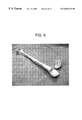
- FIG. 6 is a photograph of a whole humerus after being treated according to the method of this invention. a post-cleaning coronal section through the head of the humerus reveals the cleanliness of the inner bone matrix.
- FIG. 7 is a photograph of an intact knee, including proximal tibia, distal femur and patella, along with articulating tendons and ligaments, before treatment according to the method of this invention.
- FIG. 8 is a photograph of the intact knee shown in FIG. 7, after treatment according to the method of this invention, showing cleanliness of the implant, and preservation of the articulating tendons and ligaments.
- FIG. 9 is a photograph of an anterior aspect of a coronal section through the proximal femur prior to treatment according to the method of this invention.
- FIG. 10 is a photograph of the posterior aspect of the coronal section through the proximal femur shown in FIG. 9, after treatment according to the method of this invention.
- FIG. 11 is a photograph of the sections shown in FIGS. 9 and 10, side-by-side, demonstrating the effectiveness of the treatment according to this invention for removal of endogenous substances.
- FIG. 12 is a photomicrograph of an osteon from cortical bone without fluoroisothiocyanate (FITC) fluorescent dye treatment (400 ⁇ magnification).
- FITC fluoroisothiocyanate
- FIG. 13 is a photomicrograph of an osteon from cortical bone after inclusion of FITC in one of the cleaning solutions of this invention, demonstrating deep interpenetration of the dye into the smallest of bone interstices—bright green areas indicating structures containing FITC, including the large haversian canal (right margin) and smaller satellite lacunae (central area; 400 ⁇ magnification).
- FIG. 14 provides a model system for testing the efficacy of a liquid sterilization process for cortical bone.
- FIG. 15 provides results from treatment of bone according to the method of this invention as compared with irradiative or lyophilization treatment alone from a bone compression testing objective.
- FIG. 16 provides the results of cortical bone rehydration under ambient pressure, negative pressure, positive pressure, or cyclic negative and positive pressure conditions, according to the method of this invention.
- FIG. 17 provides the results of an analysis of release of perfused biomolecules from a bone matrix.
- the term “passivate” is intended to refer to the elimination of potentially pathogenic organisms and immunogenic substances from an implant. Thus, both sterility and reduced antigenicity is intended by this term, although elimination of beneficial biological properties of the implant, such as osteogenic properties (osteoconduction or osteoinduction; bone fusion), natural tissue functionality, and desirable structural strength of an implant are not intended by this term.
- the term “passivation” is preferred to the term “sterilize” because, while sterilization is a goal, that term has an absolute connotation which can rarely, if ever, be completely achieved without attendant tissue destruction.
- the implants produced according to the method of this invention may not be completely devoid of any antigenicity or pyrogenicity, these undesirable aspects are greatly reduced, and this too is intended by the term “passivation,” as used herein.
- perfused or “perfusion,” as used herein, are intended to imply efficient interpenetration of cleaning solutions or biologically active substances into and through the channels and crevices of materials intended for implantation into a recipient.
- rapid or “rapidly” as they are applied to the process of pressure cycling according to this invention mean time frames on the order of seconds to minutes, rather than hours or days.
- sonicate or “sonication” as used herein mean the application of sonic or ultrasonic energy via a container of an implant undergoing processing according to the method of this invention under conditions that permit efficient transfer of the sonic energy to the implant.
- sonication or “sonication” as used herein mean the application of sonic or ultrasonic energy via a container of an implant undergoing processing according to the method of this invention under conditions that permit efficient transfer of the sonic energy to the implant.
- Those skilled in the art are familiar with the process of sonication and conditions whereby sonic energy may be transferred through a fluid to a workpiece such that efficient cleaning and bacterial or cellular disruption is achieved, without resulting in gross, ultrastructural damage to the workpiece.
- This invention provides a novel method for processing implant materials including, but not limited to, metallic implants, synthetic implants, ceramic implants, autograft, allograft or xenograft materials, including bone and s oft tissue.
- implant materials including, but not limited to, metallic implants, synthetic implants, ceramic implants, autograft, allograft or xenograft materials, including bone and s oft tissue.
- soft tissue or allograft bone materials treated according to the method of this invention permit soft tissue or debrided allograft, autograft or xenograft bone to be thoroughly cleaned, machined, sterilized, packaged and then implanted at economies of scale heretofore not possible.
- tissue banks have attempted, as much as possible, to process tissue from single donors, without permitting contact between tissue derived from different donors. The concern has been that any given donor tissue may contaminate other donor tissue.
- the risk of a large batch of donor tissues being found to be contaminated has been considered an unreasonable risk.
- the resultant graft material available for implantation is safe for implantation.
- donor qualification Methods for minimizing the risk that donor tissue will be harvested and processed by a tissue bank, referred to herein as “donor qualification”, are known in the art. Accordingly, thorough donor screening, and tissue testing by enzymatic, immunological, biochemical and molecular biological techniques are applied to minimize the risk that tissue carrying pathogens (viruses, bacteria, and the like) will be included in the materials processed and made available for implantation. Testing for contamination by human immunodeficiency virus, HIV, hepatitis B virus, HBV, hepatitis C virus, HCV, has now become routine in the art.
- Known screening and qualification methods are desirably included as an initial step preceding processing of the implant material according to the present method.
- allograft bone is referred to as an exemplary tissue that may be processed according to the present method.
- tissues including but not limited to autograft bone, xenograft bone, other porous tissues, synthetic porous materials, and various soft tissues, may be processed according to the principles defined herein, without departing from the spirit of the invention exemplified herein by reference to allograft bone material.
- allograft bone material from qualified donors is first treated by various known bioburden reducing methods, as in cleaning by debriding adventitious tissue according to methods known in the art.
- Manual dissection may be employed for removal from the bone surfaces of ligaments, tendons, skin, fat, muscle, loose bone marrow, and any other non-bone tissue.
- automated or semi-automated methods known in the art may be employed for initial cleaning of the donor bone material.
- the cleaned allograft, autograft or xenograft materials from individual donors may be pooled and further cleaned as described below.
- the allograft, autograft or xenograft bone may be machined to the final implant dimensions, followed by pooling with a batch of similarly processed, dimensioned implants for further cleaning as described below.
- the instant method also provides for production of composite implants wherein a first tissue from a first donor is combined with a second tissue from the same or a different donor, to produce a unitary composite implant.
- a batch number is defined for further tracking, with records being maintained of all of the donors that have contributed to the batch.
- implant materials from individual donors may first be treated as described below, prior to pooling with implant materials from different donors. In this event, the implant material form individual donors may be further cleaned whole or first machined to desired final dimensions.
- the method of this invention provides for further processing whereby bone marrow, blood, proteins, and particulate matter is efficiently removed, such that what remains is essentially a mineralized collagen matrix, in which about a 5 to 6 log reduction in any form of viable organisms (viruses, bacteria, amoebae, rickettsia, fungi) is achieved.
- viable organisms viruses, bacteria, amoebae, rickettsia, fungi
- this is achieved by a process of pressure cycling or oscillation, employing a variety of cleaning and sterilization solutions which are caused to efficiently interpenetrate the matrix.
- the channels of essentially any porous matrix are unclogged, and cleansed.
- a pre-defined, pre-programmed cycle of washes is employed, preferably with concurrent ultrasonic bombardment, to achieve penetrating sterilization of the implant.
- the invention includes a method which comprises the following steps:
- a chamber containing the implant such as porous metallic or synthetic materials, autograft, allograft or xenograft;
- FIG. 1 A This schematic shows an implant 100 comprising solid structural constituents 110 , channels 120 , and adventitious materials 130 embedded within the channels 120 .
- the structural constituents 110 may be synthetic materials, as in man-made polymeric material, (e.g. poly-L-lactic acid, acrylic acids, and the like), metallic structural materials, or natural materials, such as a mineralized or demineralized collagen matrix, autograft, allograft or xenograft bone or other tissue.
- the channels 120 may be man-made channels, defined by the polymerization, molding, melting or other manufacturing process, or may be natural channels, such as those found in mineralized or demineralized cancellous or cortical bone matrices.
- the adventitious materials 130 may be cellular debris, bone marrow, cells, lipids, carbohydrates, proteins, viruses, bacteria, rickettsia, amoebae, fungi and the like.
- panels (1) and (2) relate to the first step described above.
- the channels 120 are primed for back-filling with cleaning solutions by exposing the tissue to decreased pressures.
- the cleared channels 120 are shown to be substantially clear of adventitious materials 130 .
- Panel (3) relates to steps 2 and 3, wherein molecules of cleaning solution 140 are introduced into a sealed chamber and are driven into the channels 120 by elevated pressures.
- Panel (4) relates to the fourth step described above, wherein decreased pressure removes remaining cellular debris, cleaning solution 140 , and other remaining adventitious materials from the channels 120 , and again primes the matrix for deep penetration, now possible due to the clarity of the channels 120 .
- panels (5)-(7) a one cycle repeat according to the fourth step described above is shown, whereby upon repressurizing with clean solvents, full interpenetration of the solvents into the implant matrix is achieved.
- reduced pressure draws the remaining solution from the implant, which may then be dried, as shown in panel (7), prior to further processing (e.g. machining according to step 5 above, further cleaning, according to step 6 above), and final packaging of the cleaned tissue.
- the cycle depicted in FIG. 1A may be repeated as many times as desired to ensure complete internal cleaning of the matrix interior.
- FIG. 1B a representation of the pressure and fluid oscillation throughout the various steps of the above described process is represented.
- donor tissue is cleaned of any extraneous or adventitious tissue.
- the thus-cleaned tissue is loaded into a sealable reaction chamber.
- a preferably pre-programmed tissue cleaning process is then initiated comprising a plurality of wash steps. Deep tissue interpenetration by cleaning solutions is achieved by oscillating the pressure in the chamber while adding and removing various cleaning solvents. Ultrasonic energy is applied at various stages of the cleaning process to accelerate solution penetration and removal of unwanted contaminants or endogenous substances, including blood, lipid, and non-structural or undesired proteins.
- steps (1-4) of the claimed process are carried out according to a protocol similar to that defined in the following table to remove blood, fat, bacterial, viral, fungal or other contamination:
- a pressurizable chamber in which the process may be conducted is loaded with metallic, synthetic or other man-made implant materials, autograft or allograft bone or soft tissue, xenograft bone or soft tissue, from an individual qualified donor.
- the treated implant may comprise a combination of tissue and synthetic materials, such as, for example, biopolysulphone and the like.
- the implant is a tissue
- the tissue is preferably first cleaned of surface adventitious tissue, prior to initiating the steps shown in table I.
- step 1 under negative pressure (vacuum), for a period of about two minutes, the matrix of the implant or implants is primed (i.e. see FIG.
- step 1 to remove trapped air, cellular and other loose debris by vacuum.
- step 2 under negative pressure, cleaning fluid is introduced with sonication, to aid in penetration of the fluid and to ensure gas is removed from the introduced fluid.
- the cleaning fluid is removed to waste under positive pressure, the tissue is dried under negative pressure, and is rinsed several times under oscillating positive and negative or elevated and decreased pressure using sterile water or physiological saline (e.g. phosphate buffered saline, PBS), with or without accompanying sonication.
- physiological saline e.g. phosphate buffered saline, PBS
- the number of rinse cycles may be from 1-150 times, and is preferably about 1-50 times.
- the rinse solution is drained under positive pressure, and the tissue is again dried under negative pressure.
- the tissue in-process may be machined into dimensionally finished grafts if such processing has not previously been accomplished, (step 5 of the instant process, as defined above), and then loaded into a reaction chamber, same or different than that used to carry out the steps according to Table I.
- a deep-penetrating cleaning, passivation or sterilization cycle preferably under programmable logic control, is then conducted according to a protocol similar to that defined in Table II (see step 6 defined above, which represent a repeat of steps 1-4 of Table I, optionally using different cleaning solvents; these steps are distinguished by indicating the steps as 0′-4′):
- the cleaning fluid is preferably retained in a positively pressurized reaction chamber for an extended period to ensure complete killing of any residual contaminating pathogens or other organisms. A period of from one to sixty minutes, and preferably about ten minutes, is sufficient for this purpose.
- the cleaning fluid is then removed to waste under positive pressure, the tissue is dried under negative pressure, and is rinsed several times under oscillating positive and negative pressure using sterile water or physiological saline (e.g. phosphate buffered saline, PBS, or the like), with or without accompanying sonication.
- the rinse solution is drained under positive pressure, and the implant is again dried under negative pressure.
- Tissues cleaned according to this procedure include, but are not limited to: cortical bone, cancellous bone, fascia, whole joints, tendons, ligaments, dura, pericardia, heart valves, veins, neural tissue, submucoal tissue, (e.g. intestinal tissue), and cartilage.
- Bone treated according to this method and subsequently tested for retained biomechanical strength and ability to induce new bone formation (osteoconduction and osteoinduction, collectively referred to as osteogenic activity) retains good biomechanical strength and is expected to retain osteogenic activity.
- bone treated according to one embodiment of this method and implanted as a xenograft was found to induce little or no adverse immunological reactivity, indicating reduction in antigenicity of the material. This is particularly true where urea or other chaotropic agents (e.g. guanidine hydrochloride), is used as one of the cleaning fluids or is included in a mixture of cleaning fluids.
- urea or other chaotropic agents e.g. gu
- a device such as that shown schematically in FIG. 2 may be employed for semi-manual implementation of the cyclic perfusion passivation process of this invention.
- a chamber 200 comprising a lid 210 and a trough 220 is adapted for cyclic perfusion passivation of implants.
- a series of posts 230 onto which a series of bolts 240 may be tightened are provided for securing the lid 210 to the trough 220 .
- a grating 250 is provided inside the chamber 200 for receiving implant material to be treated.
- Access port 260 is a sterile water input line.
- Access port 261 is an input line for other fluids.
- Access port 262 is a vacuum line.
- Access port 263 is a line for pressure input.
- a port 264 is provided for insertion of a temperature probe.
- Port 265 is a port for supplying power to a sonicator built into the walls 225 of the chamber 200 .
- Port 266 is a drain. Accordingly, a device such as that shown in FIG. 2 could be used carrying out the cyclic perfusion passivation process according to this invention.
- an automated or semi-automated apparatus 300 may be defined for carrying out the instant process.
- programmable logic controllers activate or deactivate valves or solenoids 301 a-h at pre-determined times in the cleaning cycle.
- An implant is placed in a reaction chamber 310 which is sealed.
- An atmospheric vent 320 is provided to permit entrance and removal of filtered air, and a drain 321 is provided to remove waste or solvents.
- Cleaning fluids are introduced into reaction chamber 310 from a chemical mixing tank 330 which has a filtered vent to atmosphere 335 , to avoid formation of a vacuum in the tank 330 .
- Chemical feed lines 340 lead from fluid reservoirs 341 to the chemical mixing tank 330 via a common conduit 345 .
- a programmably controlled pump 350 is operated to pump appropriately mixed fluids from the tank 330 into the reaction vessel 310 .
- Vacuum or negative pressure is applied to the reaction vessel 310 by means of a vacuum receiver tank 360 , in which a source of negative pressure is created by vacuum pump 365 .
- the inclusion of a vacuum reservoir 360 is desirable so that essentially instantaneous vacuum of known dimensions may be applied to the reaction chamber 310 , without the need for a vacuum pump such as 365 having to gradually develop the negative pressure.
- Vacuum receiver tank 360 may be evacuated by pump 365 while reaction tank 310 is under positive pressure.
- a source of sterile water, physiological saline, or like aqueous solution is provided in storage tank 370 , which has a filtered vent 375 to prevent formation of a vacuum in tank 370 .
- Pump 376 provides for rapid infusion of aqueous solution into chemical mixing tank 330 for introduction into the reaction chamber 310 .
- the water from tank 370 may also be directly introduced into reaction tank 310 , without having to first be introduced into chemical mixing tank 330 .
- Positive pressure is stored in pressure tank 380 which is pressurized by a compressor of filtered gas, to retain sterility in the reaction tank 310 .
- an appropriately programmed computer or programmable logic controllers permit venting of the reaction chamber 310 , to permit loading of tissue.
- the chamber is then sealed, evacuated, pressurized, and fluid is introduced and removed, as outlined, for example, in Table I and Table II above, to complete the implant cleaning process.
- a source of filtered sterile steam 322 to rapidly sterilize the internal, filtered and sterile zone of the device is provided. It is also desirable to include a heat exchange means 323 to rapidly equilibrate the system temperature. Water cooled, air cooled, nitrogen cooled, water heated, thermocouple heated or like radiative means are all acceptable, depending on the internal temperatures desired.
- the penetrating passivation process of this invention is so efficient that for certain types of implants in which the initial prospect of encountering a contaminated implant is sufficiently low, it may be possible to simply batch process implant materials according to Table I and Table II, rather than first cleaning implants from an individual donor according to the Table I program, prior to combining such implant materials from different donors and processing the pooled implants according to the Table II program.
- an initial bioburden reducing step for implant materials derived from individual donors is considered prudent, individual donor tissues are processed according to the Table I program, and are then quarantined until all quality control criteria are passed. Only the individual donor tissues that pass such quality control after initial bioburden reduction are pooled for processing according to the Table II protocol.
- a combination of TritonX-100 and TNBP may be used as a first solvent to remove debris and to inactivate bacteria and viruses.
- a second solvent may be a 3% hydrogen peroxide solution to remove cellular debris and to further reduce bioburden.
- a third solvent may be povidone iodine solution to yet further reduce bioburden.
- ascorbic acid solution may be employed to decolorize the implant or remove any residual iodine.
- solutions may be employed in a different order, and indeed, different solutions may be used to similar effect.
- the particular solutions listed are preferred, however, due to their low toxicity, and our discovery that the defined combination of solutions results in efficient reduction in bioburden, implant cleaning, passivation and interpenetration.
- the solutions of Table I are typically employed in a cycle such as that shown in Table I, steps 0-4.
- cleaned allograft or xenograft tissue from individual donors or previously pooled donors is optionally pooled and further cleaned as described below.
- the tissue is first dimensioned by machining, trimming and the like, to achieve the final implant dimensions.
- the dimensioned tissue is further processed individually or is pooled with a batch of similarly or differently processed, dimensioned implants for further cleaning as described below.
- different tissues from the same or different donors may be combined prior to, during or after treatment according to the method of this invention in order to produce an individual composite product.
- collagen gelatin or like material such as that disclosed in WO98/40113 (hereby incorporated by reference) may be combined with bioactive ceramics, such as bioactive glass and the like, hydroxyl apatite or the like, bone chips or the like and then packed into an implant from yet another donor or the same donor, such as the device of U.S. Pat. No. 5,814,084 (hereby incorporated by reference).
- the device as disclosed according to the U.S. Pat. No. 5,814,084 patent may be produced from an individual donor or a pool of donor tissues as treated according to the present invention.
- the final composite material would thus be derived from potentially multiple donor tissues, each of which may have been derived from multiple donors, and treated according to the method of this invention.
- a batch number is defined for further tracking, with records being maintained of all of the donors that have contributed to a given batch.
- Table II a set of solutions is described for achieving penetrating sterilization of individual tissues or tissues pooled from different donors which have already been treated according to the program outlined in Table I.
- a first solution of 6% hydrogen peroxide, followed by a second solution of 1% sodium hypochlorite, followed by a solution of 1 N sodium hydroxide may be used to achieve sterilization.
- a 70% solution of isopropanol may be used as a broad spectrum germicide.
- the solutions of Table I and Table II may be employed according to the program shown, or modified as needed.
- penetrating sterilants may be employed or that mixtures of the described sterilants may be possible.
- the individual or pooled batch of implants has been thoroughly cleaned, passivated (if not sterilized), and interpenetrated by cleaning solutions.
- Reductions in enveloped virus, vegetative bacteria, and fungal contamination of up to twelve logs or higher and of non-enveloped viruses and spores of up to about five logs are achieved according to the process described herein.
- about a two to ten-fold reduction in endotoxin levels is achieved, along with significant elimination of blood, lipid, nucleic acid, and non-structural protein.
- this process retains the beneficial structural and other desirable biological properties of the implant material.
- Significant enhancements in production yields, through the ability to batch process implant from pooled donors, are also achieved.
- the implant materials are placed in their final packing. Preferably, this is achieved in a sterile environment to avoid introduction of any adventitious bioburden.
- the implants are exposed to a vapor-phase hydrogen peroxide/peracetic acid or like vapor-phase sterilizing environment.
- the packages are then closed to ensure that no contamination may occur upon removal of the implants from the sterile field for storage or shipment to surgeons.
- the sealed packages may then, optionally, be subjected to levels of gamma or other types of irradiation known to not adversely affect tissue properties (e.g.
- a solution containing desired antibiotics, anti-inflammatory drugs or other biologically active agents may be employed to infuse antibiotic or other desired bioactive substances into the cleaned, passivated tissues.
- growth factors such as bone morphogenetic proteins, cartilage derived growth factors, tissue growth factors, natural (autograft, allograft or xenograft) or recombinant, and the like known in the art may be perfused into the implant.
- a solution containing expressible nucleic acids in plasmid, viral or linear DNA or RNA vector form is infused into the implant.
- the nucleic acid preferably encodes an appropriate growth factor, antineoplastic agent, peptide or protein, depending on the nature of the tissue into which the nucleic acid is perfused.
- antineoplastic agent e.g., a CMV promoter or the like
- peptide or protein e.g., a CMV promoter or the like
- the nucleic acid may encode a cartilage derived morphogenic protein.
- the nucleic acid may encode tissue growth factors (beta and the like), peptides (eg. P/5 and the like) or any other desirable gene product.
- the nucleic acid may be DNA, RNA or it may be combinations thereof, optionally including synthetic nucleotides or markers to track nucleic acid penetration and concentration.
- DBM demineralized bone matrix
- implants may be perfused with the DBM which contains a complex mixture of growth factors.
- a bone implant may be partially demineralized to expose growth factors prior to implantation.
- bone marrow or bone marrow extracts may likewise be perfused into the matrix of an appropriate implant.
- the cleaning process is applied to rid a tissue of a pathogenic organism or condition.
- This can be achieved, for example, by harvesting a diseased mandible or any other bone or tissue, ravaged by cancerous cell growth.
- the section of autograft is cleaned and passivated ex vivo, perfused with growth factors, nucleic acids encoding growth factors, antibiotics, antineoplastics, antiinflammatories analgesics or any after desired biologically active substance, and then re-implanted into the same or a different patient, to provide a non-pathogenic tissue.
- the process of the present invention may be carried out at any stage of implant production, and it does not require special preparations such as removal of cartilage, or potentially implant damaging steps such as drilling of holes.
- the method of this invention broadly discloses a process for tissue inventory production wherein, in a first phase, tissue from a plurality of donors is pooled and processed according to the method of this invention. The result is a stockpile of useable materials of fundamental units, available for further processing as needed. In a second phase, as the need arises, the fundamental tissue units available in inventory are further processed, if necessary, in order to produce the final product required for implantation.
- this invention provides a significant and fundamental advance to the art whereby tremendous enhancements in efficiency are achieved, since a single processing episode is implemented to generate a large volume of tissue available for implantation or further processing. As compared to the existing situation in donor tissue processing, whereby for each donor and for each tissue, a separate processing episode may be required to derive a single product, following which all processing equipment and personnel are required to be exchanged to intake a new tissue from a new donor. Accordingly, the method of this invention lends itself to much enhanced quality control, inventory control and efficiency.
- the process efficiently removes, dilutes or denatures endogenous enzymes which otherwise might result in degradation or autolysis of bone matrix or tissue matrix. This is achieved by, for example, cyclically exposing the tissue to detergents, reducing agents (e.g. dithiothreitol, DTT, and the like known in the art for disruption of protein disulfide bonds), peroxide, isopropanol and the like as disclosed herein.
- reducing agents e.g. dithiothreitol, DTT, and the like known in the art for disruption of protein disulfide bonds
- peroxide isopropanol and the like as disclosed herein.
- stage 1 donor materials are introduced into the donor tissue processing facility and are held in quarantine until the donor from which the tissue was derived is qualified.
- stage 2 released donor materials are surface cleaned by debridement.
- stage 3 surface cleaned tissue is machined to produce implants of the desired final dimensions, and are introduced into an automated cyclic perfusion passivation chamber according to the present invention.
- stage 4 implants that have been passivated are introduced into their final packing containers and are terminally sterilized by gamma irradiation, vapor-phase exposure to decontaminants, and the like.
- stage 5 the passivated and packaged grafts are stored and released after verification of the sterilization cycles.
- a process layout similar to that shown schematically in FIG. 5 may be employed.
- a processing facility 500 shows three parallel and identical tissue processing facilities A-C.
- tissue to be treated according to this invention is cleaned and debrided of gross, adventitious and unwanted tissues.
- the cleaned tissue is then introduced, via sealable port 515 A-C into a reaction chamber 310 A-C, to which are connected all of the process control and input/output devices shown in FIG. 3 .
- tissue is removed via sealable port 516 A-C.
- the cleaned tissue is sorted and stored in quarantine freezers 520 A-C, until quality control demonstrates that the tissue is fit for further processing.
- the released tissues are then transferred to graft-production rooms 530 A-C, where final implant dimensioning and machining is conducted.
- the thus processed tissues are loaded into reaction chambers 310 ′A-C via sealable port 535 .
- Not shown but connected to reaction chamber 310 ′A-C are all the process control and input/output devices shown in FIG. 3 .
- the deeply sterilized tissues are removed from sealable port 536 A-C, and are placed in final packaging. Terminal sterilization is conducted at stations 540 A-C, and the terminally sterilized tissues are sealed in the final packaging.
- the sealed packages of terminally sterilized tissues are quarantined in freezers 545 until final quality control testing permits tissue release to surgeons.
- the process layout provided in FIG. 5 is preferred, it is suggestive only, and the process according to the instant invention may be conducted in other layout formats. Further, it will be appreciated that according to the layout shown according to FIG. 5, it is desirable for the level of ambient particulates to be reduced as tissue is processed through the various stages shown. Thus, while it is adequate for the chamber 510 to be of class 100,000 (100,000 particles per billion), it is desirable for areas 520 and 530 to be class 10,000 or lower.
- the final packaging area 540 is preferably about a class 1000 area.
- an intact or machined bone implant is cleaned by treatment sequentially with povidone-I, water, ascorbic acid, TNBP/hydrogen peroxide, water, diethanolamine, water, 6 M urea, water.
- the sequence of sonication, and pressure fluctuations is conducted according to the sequence defined in Table I or Table II. It will be appreciated from this disclosure, however, that a wide variety of different cleaning solutions and combinations thereof may be employed according to the method of this invention.
- the cleaning solutions may include: sterile water, Triton X-100, TNBP, 3% hydrogen peroxide, a water-miscible alcohol, saline solution, povidone iodine, ascorbic acid solution, aromatic or aliphatic hydrocarbons, ethers, ketones, amines, urea, guanidine hydrochloride, esters, glycoproteins, proteins, saccharides, enzymes such as proteases (trypsin, pepsin, subtilisin), lipases, sachrases, and the like, gasseous acids or peroxides, and mixtures thereof.
- the process is conducted at ambient temperatures, elevated temperatures (eighty degrees centigrade) or decreased temperatures. Thus, cleaning of implants in a liquid nitrogen phase (negative eighty degrees centigrade) is contemplated by this invention.
- FIG. 6 is a photograph of a whole humerus after being treated according to the method of this invention. a coronal section through the head of the humerus reveals the cleanliness of the inner bone matrix.
- FIG. 7 is a photograph of an intact knee, including proximal tibia, distal femur and patella, along with articulating tendons and ligaments, before treatment according to the method of this invention.
- FIG. 8 is a photograph of the intact knee shown in FIG. 7, after treatment according to the method of this invention, showing cleanliness of the implant, and preservation of the articulating tendons and ligaments.
- implant materials and tissues that may be effectively cleaned according to this procedure include, but are not limited to metallic implants, synthetic implants, ceramic implants, allograft, autograft or xenograft tissues.
- Such tissues may be selected from tissues comprising: cortical bone, cancellous bone, fascia, whole joints, tendons, ligaments, dura, pericardia, heart valves, veins, neural tissue, submucoal tissue, (e.g. intestinal tissue), and cartilage.
- any implantable material having an internal matrix that is required to be cleaned may be treated to advantage according to the method of this invention.
- FIG. 9 is a photograph of an anterior aspect of a coronal section through the proximal femur prior to treatment according to the method of this invention.
- FIG. 10 is a photograph of the posterior aspect of the coronal section through the proximal femur shown in FIG. 9, after treatment according to the method of this invention.
- FIG. 11 is a photograph of the sections shown in FIGS. 9 and 10, side-by-side, demonstrating the effectiveness of the treatment according to this invention for removal of endogenous substances and deep, penetrating implant cleaning.
- FIG. 12 is a photomicrograph of an osteon from cortical bone without fluoroisothiocyanate (FITC) fluorescent dye treatment (100 ⁇ magnification).
- FITC fluoroisothiocyanate
- FIG. 13 is a photomicrograph of an osteon from cortical bone after inclusion of FITC in one of the cleaning solutions of this invention, demonstrating deep interpenetration of the dye into the smallest of bone interstices—bright green areas indicating structures containing FITC, including the large haversian canal (right margin) and smaller satellite lacunae (central area; 100 ⁇ magnification).
- photomicrographs demonstrate that the FITC dye is forced into the smallest implant interstices, thereby revealing the ability to achieve deep penetrating cleaning.
- biologically active substances such as antibiotics, antiviral compounds, anti-inflammatory compounds, growth factors, osteo-inductive substances (e.g. bone morphogenetic protein, cartilage derived morphogenetic protein, natural or recombinant, and the like), when included in solutions employed according to the method of this invention, may be effectively imbedded deeply into implant materials.
- biologically active substances for permeation into implants are selected from the group consisting of bone morphogenetic protein, tissue growth factor beta or member of the tissue growth factor beta family of growth factors, cartilage derived morphogenetic proteins I or II or both, and any related cartilage derived growth factors, angiogenic factors, platelet derived growth factor.
- Any of the proteins selected for permeation into implants may -be natural or recombinant proteins.
- a solution of 0.1 to 0.5N Acetic or other mild acid (EDTA, Citric, Formic, dilute HCl, H 3 PO 4 etc.) is contacted under cyclic elevated and reduced pressure for 20 minutes. This removes 2-3% of the calcium in the bone, making the modulus go down for increased strength (tortional and sheer) and exposes some of the collagen so growth factors can more easily attach. It also helps to remove some of the protein bound to the mineral. Finally, demineralization has been shown to reduce the immunogenicity of bone.
- EDTA Citric, Formic, dilute HCl, H 3 PO 4 etc.
- the thus treated bone was then defatted with 99% isopropanol, hexane or the like in cyclic pressure treatment as previously described, followed by TNBP/Tritonix-100, UREA, GuHcl, or the like, followed by several rinse cycles and addition of growth factors, nucleic acids or the like.
- the thus treated implant is then be directly implanted or is frozen or lyophilized for subsequent implantation.
- the D-value obtained for the samples run in the presence of ultrasonic energy was 0.83-1.66 minutes (Table III). This compared.favorably to the D-value obtained for the samples run in the absence of ultrasonic energy (>10 min). This reduction in the time required for the inactivation of spores allows for a practical method of sterilizing allografts without adversely effecting their desired attributes.
- a second segment of cortical bone was machined into a cylindrical pin, slightly oversized as compared with the diameter of the hole.
- a partial slit was cut into the pin, allowing a biological indicator to be placed therein.
- the pin was then forced under compression into the machined hole and exposed to the sterilization process.
- a control was also run using only sterile water to evaluate whether the spores appreciably were being washed off the strip.
- a tracing dye was used to evaluate the path of the liquid through the device.
- Axial compression testing was adapted from ASTM D695-91 and performed on an MTS 858 (Eden Prairie, Minn.) servohydraulic mechanical test apparatus (American Society for Testing and Materials. ASTM D 695-91: Standard Test Method for Compressive Properties of Rigid Plastics. Philadelphia, Pa.: ASTM; 1991).
- Cortical bone cylinders were prepared as described above. Each cylinder was then dried in a vacuum oven for 24 h at 60° C. The specimens were weighed prior to the introduction of water, and at regular intervals throughout the run, in order to determine their percent rehydration. Reconstitution conditions were (1) vacuum for 1 min; (2) positive pressure for 1 min; (3) negative pressure for 30 sec followed by positive pressure for 30 sec; (4) reconstitution under normal atmospheric pressure.
- the initial (2.5 min) time point showed a significant increase in rehydration for both the positive and negative pressure treatments as compared with standard atmospheric pressure (FIG. 16, Left Panel—The rehydration of cortical bone under different atmospheric pressures. Positive—100 PSI, Negative— ⁇ 25 in Hg, Neg/Pos—negative pressure followed by positive pressure; Right Panel—Enlarged view of the earliest time points).
- the negative to positive treatment showed greatest increase in rehydration and was significant to all other treatments at this timepoint. This data demonstrates the effectiveness of differential pressure to induce tissue matrix penetration.
- Cortical bone segments were machined from diaphyseal sections of human cadaveric femora and tibiae. The bone segments were then cleaned and impregnated with compounds of various molecular weights. The samples were then placed in baths that maintained sink conditions and the release of drug over time was measured. In addition, the pattern of drug remaining in the matrix was examined histologically.
Abstract
Description
| TABLE I | ||||||
| Soni- | Duration | |||||
| Step | Pressure | Fluids* | cation | (min) | |
|
| 0 | Atmospheric | None | Off | NA | Load tissue into | |
| |
||||||
| 1 | Negative | None | Off | 2 | Prime tissue | |
| (60-100 torr) | matrix, remove | |||||
| included air and | ||||||
| |
||||||
| 2 | Negative | B,C,D,E, | On | 1 | De-gas cleaning | |
| (60-100 torr) | | fluids | ||||
| 3 | Positive (5-8 | B,C,D,E, | On | 1 | Force fluids into | |
| atmospheres) | | tissue matrix | ||||
| 4 | Negative/ | B,C,D,E, | On | (1 × n) | Remove debris | |
| Positive | mixtures | loosened by | ||||
| fluids, pressure | ||||||
| oscillation and | ||||||
| sonication | ||||||
| *Fluids: | ||||||
| B = Triton X-100/TNBP, a solvent/detergent to remove debris and kill viruses and bacteria; | ||||||
| C = 3% hydrogen peroxide, to remove cellular debris, inactivate viruses and bacteria; | ||||||
| D = mixture of B and C; | ||||||
| E = water-miscible alcohol, such as ethanol or isopropanol; | ||||||
| mixtures = B,C,D,E in any desirable proportions. | ||||||
| TABLE II | ||||||
| Soni- | Duration | |||||
| Step | Pressure | Fluids* | cation | (min) | |
|
| 0′ | Atmospheric | None | Off | NA | Load tissue into | |
| |
||||||
| 1′ | Negative | None | Off | 2 | Prime tissue | |
| (60-100 torr) | matrix, remove | |||||
| included air and | ||||||
| |
||||||
| 2′ | Negative | F,G,H,I,J, | On | 1 | De-gas cleaning | |
| (60-100 torr) | | fluids | ||||
| 3′ | Positive | F,G,H,I,J, | On | 1 | Force fluids into | |
| (8-10 | mixtures | tissue matrix | ||||
| atmospheres) | ||||||
| 4′ | Negative/ | F,G,H,I,J, | On | (1 × n) | Remove debris | |
| Positive | mixtures | loosened by | ||||
| fluids, pressure | ||||||
| oscillation and | ||||||
| sonication | ||||||
| *Fluids: | ||||||
| F = 6M urea or other chaotropic agents, e.g. 4M guanidine HCl, to reduce implant antigenicity; | ||||||
| G = 1% sodium hypochlorite, to inactivate viruses, bacteria, fungi or other residual contaminants; | ||||||
| H = 1N sodium hydroxide, to inactivate viruses and bacteria; | ||||||
| I = 6% hydrogen peroxide, as a sterilant; | ||||||
| J = hexane, ether, diethanolamine (DEA), toluene, xylene, butane, CO2 (supercritical), isobutane, propane, acetone, isopropanol, methanol, ketones, ethers, aliphatic or aromatic hydrocarbons, HCl, gasseous HCl. | ||||||
| mixtures = F,G,H,I,J in any desirable proportions. | ||||||
| TABLE III |
| Comparison of spore inactivation with 6% hydrogen peroxide in the presence and |
| absence of ultrasonic energy. Positive (+) results were identified by turbidity of the media and |
| confirmed by subculturing the broth to solid media. Negative (−) results were determined by the |
| media retaining clarity over the seven days of incubation. |
| Aprox. | ||
| Treatment Time (min) | D- |
| |
0 | 5 | 10 | 15 | 20 | 30 | 40 | 50 | 60 | value |
| Sonication | + | + | + | + | − | − | − | − | − | − | − | − | − | − | − | − | − | − | − | − | − | − | − | − | − | − | − | >1.66 |
| No | + | + | + | + | + | + | + | + | + | + | + | + | + | + | + | + | + | + | + | + | + | + | + | + | + | − | + | >10 |
| sonication | ||||||||||||||||||||||||||||
| TABLE IV |
| Approximate D-values for B. sterothermophilus as a function of residual fat content |
| remaining in a homogenized bone slurry, when sterilized in a 6% hydrogen peroxide solution at 42° C. |
| in the presence of ultrasonic energy. Positive (+) results were identified by turbidity of the media |
| and confirmed by subculturing the broth to solid media. Negative (−) results were determined by the |
| media retaining clarity over the seven days of incubation. |
| Aprox. | ||
| Treatment Time (min) | D- |
| |
0 | 5 | 10 | 15 | 20 | 30 | 40 | 50 | 60 | value |
| Neg | − | − | − | − | − | − | − | − | − | − | − | − | − | − | − | − | − | − | − | − | − | − | − | − | − | − | − | NA |
| Control | ||||||||||||||||||||||||||||
| Pos | + | + | + | + | + | + | + | + | + | + | + | + | + | + | + | + | + | + | + | + | + | + | + | + | + | + | + | >10 |
| Control | ||||||||||||||||||||||||||||
| No bone | + | + | + | + | + | − | − | − | − | − | − | − | − | − | − | − | − | − | − | − | − | − | − | − | − | − | − | 1.66 |
| 0% fat | + | + | + | + | + | + | + | + | − | − | − | − | − | − | − | − | − | − | − | − | − | − | − | − | − | − | − | 2.5 |
| 10% fat | + | + | + | + | + | + | + | + | + | + | + | + | − | − | − | − | − | − | − | − | − | − | − | − | − | − | − | 3.33 |
| 30% fat | + | + | + | + | + | + | + | + | + | + | + | − | − | − | − | − | − | − | − | − | − | − | − | − | − | − | − | 3.33 |
| 60% fat | + | + | + | + | + | + | + | + | + | + | + | + | + | + | + | + | + | − | − | − | − | − | − | − | − | − | − | 6.66 |
Claims (2)
Priority Applications (15)
| Application Number | Priority Date | Filing Date | Title |
|---|---|---|---|
| US09/378,527 US6652818B1 (en) | 1998-11-13 | 1999-08-20 | Implant sterilization apparatus |
| US09/390,174 US6613278B1 (en) | 1998-11-13 | 1999-09-07 | Tissue pooling process |
| PCT/US1999/026407 WO2000029037A1 (en) | 1998-11-13 | 1999-11-08 | Tissue pooling process |
| DE69935079T DE69935079T2 (en) | 1998-11-13 | 1999-11-08 | METHOD FOR CONVERTING WEAVES |
| AT99967099T ATE353227T1 (en) | 1998-11-13 | 1999-11-08 | METHOD FOR MIXING TISSUES |
| JP2000582082A JP2002529201A (en) | 1998-11-13 | 1999-11-08 | How to pool organizations |
| CA2350509A CA2350509C (en) | 1998-11-13 | 1999-11-08 | Cyclic implant perfusion, cleaning and passivation process and implant produced thereby |
| AU23445/00A AU770935B2 (en) | 1998-11-13 | 1999-11-08 | Tissue pooling process |
| EP07002028.4A EP1779868B1 (en) | 1998-11-13 | 1999-11-08 | Implant cleaning, perfusion and passivation process |
| DK99967099T DK1128849T3 (en) | 1998-11-13 | 1999-11-08 | Tissue pool assembly method |
| EP99967099A EP1128849B1 (en) | 1998-11-13 | 1999-11-08 | Tissue pooling process |
| ES99967099T ES2281980T3 (en) | 1998-11-13 | 1999-11-08 | PROCEDURE TO REGRET FABRICS. |
| US09/782,594 US20010031254A1 (en) | 1998-11-13 | 2001-02-12 | Assembled implant |
| US10/387,322 US20040115172A1 (en) | 1998-11-13 | 2002-12-23 | Assembled implant, including mixed-composition segment |
| US12/690,074 US9763787B2 (en) | 1997-08-27 | 2010-01-19 | Assembled implant |
Applications Claiming Priority (2)
| Application Number | Priority Date | Filing Date | Title |
|---|---|---|---|
| US09/191,232 US6482584B1 (en) | 1998-11-13 | 1998-11-13 | Cyclic implant perfusion cleaning and passivation process |
| US09/378,527 US6652818B1 (en) | 1998-11-13 | 1999-08-20 | Implant sterilization apparatus |
Related Parent Applications (2)
| Application Number | Title | Priority Date | Filing Date |
|---|---|---|---|
| US09/191,232 Continuation-In-Part US6482584B1 (en) | 1997-08-27 | 1998-11-13 | Cyclic implant perfusion cleaning and passivation process |
| US09/722,205 Continuation-In-Part US7048765B1 (en) | 1997-08-27 | 2000-11-25 | Intervertebral spacers |
Related Child Applications (4)
| Application Number | Title | Priority Date | Filing Date |
|---|---|---|---|
| US09/191,232 Continuation-In-Part US6482584B1 (en) | 1997-08-27 | 1998-11-13 | Cyclic implant perfusion cleaning and passivation process |
| US09/390,174 Continuation-In-Part US6613278B1 (en) | 1997-08-27 | 1999-09-07 | Tissue pooling process |
| US09/782,594 Continuation-In-Part US20010031254A1 (en) | 1997-08-27 | 2001-02-12 | Assembled implant |
| US09/941,154 Continuation-In-Part US20020106393A1 (en) | 1998-11-13 | 2001-08-27 | Assembled implant, including mixed-composition segment |
Publications (1)
| Publication Number | Publication Date |
|---|---|
| US6652818B1 true US6652818B1 (en) | 2003-11-25 |
Family
ID=27767469
Family Applications (1)
| Application Number | Title | Priority Date | Filing Date |
|---|---|---|---|
| US09/378,527 Expired - Lifetime US6652818B1 (en) | 1997-08-27 | 1999-08-20 | Implant sterilization apparatus |
Country Status (1)
| Country | Link |
|---|---|
| US (1) | US6652818B1 (en) |
Cited By (50)
| Publication number | Priority date | Publication date | Assignee | Title |
|---|---|---|---|---|
| US20020106393A1 (en) * | 2000-02-10 | 2002-08-08 | Bianchi John R. | Assembled implant, including mixed-composition segment |
| US20030072677A1 (en) * | 2001-10-17 | 2003-04-17 | Ralph Kafesjian | Supercritical fluid extraction process for tissue preparation |
| US20040219505A1 (en) * | 2000-08-21 | 2004-11-04 | Nuvasive, Inc. | Bone allograft packaging system and related methods |
| FR2866237A1 (en) * | 2004-02-13 | 2005-08-19 | Biobank | Treating bone tissue, to form lipid- and virus-free implantable biomaterial, by treating successively with supercritical carbon dioxide, aqueous hydrogen peroxide, aqueous sodium hydroxide and ethanol |
| US20050214895A1 (en) * | 2003-10-28 | 2005-09-29 | Chad Ronholdt | Methods for determining microbial contamination of allograft products |
| US20050229323A1 (en) * | 2004-04-20 | 2005-10-20 | Mills C R | Process and apparatus for treating implants comprising soft tissue |
| US20060200235A1 (en) * | 2005-03-04 | 2006-09-07 | Regeneration Technologies, Inc. | Assembled bone-tendon-bone grafts |
| US20060212036A1 (en) * | 2005-03-04 | 2006-09-21 | Regeneration Technologies, Inc. | Bone block assemblies and their use in assembled bone-tendon-bone grafts |
| US20060228252A1 (en) * | 2004-04-20 | 2006-10-12 | Mills C R | Process and apparatus for treating implants comprising soft tissue |
| US20060229722A1 (en) * | 2005-03-04 | 2006-10-12 | Bianchi John R | Adjustable and fixed assembled bone-tendon-bone graft |
| US20070260326A1 (en) * | 2006-05-08 | 2007-11-08 | Williams Michelle L | Cancellous bone treated with collagenase and essentially free of blood cells |
| US20080078447A1 (en) * | 2006-09-29 | 2008-04-03 | Christos Sarigiannidis | Low vapor pressure high purity gas delivery system |
| US20080262633A1 (en) * | 2006-05-08 | 2008-10-23 | Williams Michelle Leroux | Cancellous bone treated with collagenase and essentially free of blood cells |
| US20100015321A1 (en) * | 2006-08-11 | 2010-01-21 | The University Of Queensland | Scaffold treatment - device and method |
| US7727278B2 (en) | 2005-03-04 | 2010-06-01 | Rti Biologics, Inc. | Self fixing assembled bone-tendon-bone graft |
| US20100268349A1 (en) * | 1997-08-27 | 2010-10-21 | Bianchi John R | Assembled implant |
| US20110150963A1 (en) * | 2009-12-21 | 2011-06-23 | Clineff Theodore D | Bioactive antibacterial bone graft materials |
| US20120141555A1 (en) * | 2007-03-08 | 2012-06-07 | Arne Briest | Compound and device for treating bone and/or cartilage defects |
| US8343219B2 (en) | 2007-06-08 | 2013-01-01 | Ldr Medical | Intersomatic cage, intervertebral prosthesis, anchoring device and implantation instruments |
| US8409288B2 (en) | 2006-02-15 | 2013-04-02 | Ldr Medical | Transforaminal intersomatic cage for an intervertebral fusion graft and an instrument for implanting the cage |
| WO2013169366A1 (en) | 2012-05-11 | 2013-11-14 | Rti Biologics, Inc. | Xenograft soft tissue implants and methods of making and using |
| US8669043B2 (en) | 1998-11-13 | 2014-03-11 | Rti Surgical, Inc. | Cyclic implant perfusion, cleaning and passivation process and implant produced thereby |
| US8901078B2 (en) | 2011-07-28 | 2014-12-02 | Harbor Medtech, Inc. | Crosslinked human or animal tissue products and their methods of manufacture and use |
| US9039774B2 (en) | 2012-02-24 | 2015-05-26 | Ldr Medical | Anchoring device and system for an intervertebral implant, intervertebral implant and implantation instrument |
| US9044337B2 (en) | 2009-12-31 | 2015-06-02 | Ldr Medical | Anchoring device and system for an intervertebral implant, intervertebral implant and implantation instrument |
| US9078765B2 (en) | 2001-07-13 | 2015-07-14 | Ldr Medical | Vertebral cage device with modular fixation |
| US9125971B2 (en) | 1998-06-30 | 2015-09-08 | Lifenet Health | Plasticized bone and soft tissue grafts and methods of making and using same |
| EP2942069A1 (en) | 2014-05-09 | 2015-11-11 | Tournois Dynamic Innovations B.V. | Bone material reuse process |
| US9463091B2 (en) | 2009-09-17 | 2016-10-11 | Ldr Medical | Intervertebral implant having extendable bone fixation members |
| US9566369B2 (en) | 2010-02-26 | 2017-02-14 | Decell Technologies Inc. | Methods for tissue decellularization |
| US9597194B2 (en) | 2005-09-23 | 2017-03-21 | Ldr Medical | Intervertebral disc prosthesis |
| US9675389B2 (en) | 2009-12-07 | 2017-06-13 | Samy Abdou | Devices and methods for minimally invasive spinal stabilization and instrumentation |
| US9744043B2 (en) | 2007-07-16 | 2017-08-29 | Lifenet Health | Crafting of cartilage |
| WO2017161196A1 (en) * | 2016-03-17 | 2017-09-21 | Dorotea, Llc | Biological tissue processing apparatus and uses thereof |
| CN107260348A (en) * | 2017-06-01 | 2017-10-20 | 成都贝施美生物科技有限公司 | Planting body surfaces externally and internally ultrasonic cleaning equipment |
| US9936688B2 (en) | 2000-09-12 | 2018-04-10 | Lifenet Health | Process for devitalizing soft-tissue engineered medical implants, and devitalized soft-tissue medical implants produced |
| US10004819B2 (en) | 2014-06-10 | 2018-06-26 | Vivex Biomedical, Inc. | Tissue allograft sterilization method |
| US10130736B1 (en) | 2010-05-14 | 2018-11-20 | Musculoskeletal Transplant Foundation | Tissue-derived tissuegenic implants, and methods of fabricating and using same |
| US10307510B2 (en) | 2013-11-04 | 2019-06-04 | Lifecell Corporation | Methods of removing alpha-galactose |
| US10548740B1 (en) | 2016-10-25 | 2020-02-04 | Samy Abdou | Devices and methods for vertebral bone realignment |
| US10575961B1 (en) | 2011-09-23 | 2020-03-03 | Samy Abdou | Spinal fixation devices and methods of use |
| US10695105B2 (en) | 2012-08-28 | 2020-06-30 | Samy Abdou | Spinal fixation devices and methods of use |
| US10780197B1 (en) | 2012-10-29 | 2020-09-22 | Nuvasive, Inc. | Malleable, cryopreserved osteogenic compositions with viable cells |
| US10857003B1 (en) | 2015-10-14 | 2020-12-08 | Samy Abdou | Devices and methods for vertebral stabilization |
| US10918498B2 (en) | 2004-11-24 | 2021-02-16 | Samy Abdou | Devices and methods for inter-vertebral orthopedic device placement |
| US10973648B1 (en) | 2016-10-25 | 2021-04-13 | Samy Abdou | Devices and methods for vertebral bone realignment |
| US11006982B2 (en) | 2012-02-22 | 2021-05-18 | Samy Abdou | Spinous process fixation devices and methods of use |
| US11173040B2 (en) | 2012-10-22 | 2021-11-16 | Cogent Spine, LLC | Devices and methods for spinal stabilization and instrumentation |
| US11179248B2 (en) | 2018-10-02 | 2021-11-23 | Samy Abdou | Devices and methods for spinal implantation |
| US11305035B2 (en) | 2010-05-14 | 2022-04-19 | Musculoskeletal Transplant Foundatiaon | Tissue-derived tissuegenic implants, and methods of fabricating and using same |
Citations (24)
| Publication number | Priority date | Publication date | Assignee | Title |
|---|---|---|---|---|
| US3291640A (en) | 1963-05-27 | 1966-12-13 | Chemclean Corp | Ultrasonic cleaning process |
| US4193818A (en) | 1978-05-05 | 1980-03-18 | American Sterilizer Company | Combined ultrasonic cleaning and biocidal treatment in a single pressure vessel |
| US4294753A (en) | 1980-08-04 | 1981-10-13 | The Regents Of The University Of California | Bone morphogenetic protein process |
| EP0424159A2 (en) | 1989-10-19 | 1991-04-24 | Osteotech, Inc., | Aseptic processing of allograft bone and tissue |
| JPH03224570A (en) | 1990-01-31 | 1991-10-03 | Chiyoda Seisakusho:Kk | Sterilizing method |
| US5288462A (en) | 1992-05-18 | 1994-02-22 | Stephen D. Carter | Sterilization apparatus and method |
| US5298222A (en) | 1989-08-09 | 1994-03-29 | Osteotech, Inc. | Process for disinfecting musculoskeletal tissue and tissues prepared thereby |
| WO1994011035A1 (en) | 1992-11-12 | 1994-05-26 | American Sterilizer Company | A method of enhanced penetration of low vapor pressure chemical vapor sterilants during sterilization |
| US5333626A (en) | 1991-12-31 | 1994-08-02 | Cryolife, Inc. | Preparation of bone for transplantation |
| WO1994027882A1 (en) | 1992-04-29 | 1994-12-08 | Frimec Fritz Meckenstock Gmbh & Co. | Self-sealing closure |
| US5380826A (en) | 1989-07-20 | 1995-01-10 | Aphios Corporation | Supercritical fluid disruption of and extraction from microbial cells |
| US5429810A (en) | 1992-08-21 | 1995-07-04 | Olaf Tulaszowski | Apparatus for sterilizing bone grafts |
| US5509968A (en) | 1994-02-03 | 1996-04-23 | New York Society For The Ruptured And Crippled Maintaining The Hospital For Special Surgery | Decontamination of orthopaedic implants |
| US5513662A (en) | 1991-12-31 | 1996-05-07 | Osteotech, Inc. | Preparation of bone for transplantation |
| US5556379A (en) * | 1994-08-19 | 1996-09-17 | Lifenet Research Foundation | Process for cleaning large bone grafts and bone grafts produced thereby |
| US5716454A (en) | 1994-02-03 | 1998-02-10 | New York Society For The Ruptured And Crippled Maintaining The Hospital For Special Surgery | Decontamination of devices and instruments contacted with body tissues |
| US5725579A (en) | 1992-12-21 | 1998-03-10 | Bioland | Process for treating bone tissue and corresponding implantable biomaterials |
| US5753195A (en) * | 1996-01-02 | 1998-05-19 | Kew Import/Export Inc. | Cleaning and sterilizing mechanism |
| US5797871A (en) | 1994-08-19 | 1998-08-25 | Lifenet Research Foundation | Ultrasonic cleaning of allograft bone |
| WO1998041245A1 (en) | 1997-03-20 | 1998-09-24 | Osteotech, Inc. | Pressure flow system and method for treating a fluid permeable workpiece such as bone |
| WO1999047080A1 (en) | 1998-03-16 | 1999-09-23 | Crosscart, Inc. | Bone xenografts |
| US5993844A (en) | 1997-05-08 | 1999-11-30 | Organogenesis, Inc. | Chemical treatment, without detergents or enzymes, of tissue to form an acellular, collagenous matrix |
| US6024735A (en) | 1996-03-20 | 2000-02-15 | Lifenet Research Foundation | Process and composition for cleaning soft tissue grafts optionally attached to bone and soft tissue and bone grafts produced thereby |
| US6102056A (en) | 1998-08-18 | 2000-08-15 | Kotsopey; Omelan | Cleaning apparatus |
-
1999
- 1999-08-20 US US09/378,527 patent/US6652818B1/en not_active Expired - Lifetime
Patent Citations (25)
| Publication number | Priority date | Publication date | Assignee | Title |
|---|---|---|---|---|
| US3291640A (en) | 1963-05-27 | 1966-12-13 | Chemclean Corp | Ultrasonic cleaning process |
| US4193818A (en) | 1978-05-05 | 1980-03-18 | American Sterilizer Company | Combined ultrasonic cleaning and biocidal treatment in a single pressure vessel |
| US4294753A (en) | 1980-08-04 | 1981-10-13 | The Regents Of The University Of California | Bone morphogenetic protein process |
| US5380826A (en) | 1989-07-20 | 1995-01-10 | Aphios Corporation | Supercritical fluid disruption of and extraction from microbial cells |
| US5298222A (en) | 1989-08-09 | 1994-03-29 | Osteotech, Inc. | Process for disinfecting musculoskeletal tissue and tissues prepared thereby |
| EP0424159A2 (en) | 1989-10-19 | 1991-04-24 | Osteotech, Inc., | Aseptic processing of allograft bone and tissue |
| JPH03224570A (en) | 1990-01-31 | 1991-10-03 | Chiyoda Seisakusho:Kk | Sterilizing method |
| US5333626A (en) | 1991-12-31 | 1994-08-02 | Cryolife, Inc. | Preparation of bone for transplantation |
| US5513662A (en) | 1991-12-31 | 1996-05-07 | Osteotech, Inc. | Preparation of bone for transplantation |
| WO1994027882A1 (en) | 1992-04-29 | 1994-12-08 | Frimec Fritz Meckenstock Gmbh & Co. | Self-sealing closure |
| US5288462A (en) | 1992-05-18 | 1994-02-22 | Stephen D. Carter | Sterilization apparatus and method |
| US5429810A (en) | 1992-08-21 | 1995-07-04 | Olaf Tulaszowski | Apparatus for sterilizing bone grafts |
| WO1994011035A1 (en) | 1992-11-12 | 1994-05-26 | American Sterilizer Company | A method of enhanced penetration of low vapor pressure chemical vapor sterilants during sterilization |
| US5725579A (en) | 1992-12-21 | 1998-03-10 | Bioland | Process for treating bone tissue and corresponding implantable biomaterials |
| US5509968A (en) | 1994-02-03 | 1996-04-23 | New York Society For The Ruptured And Crippled Maintaining The Hospital For Special Surgery | Decontamination of orthopaedic implants |
| US5716454A (en) | 1994-02-03 | 1998-02-10 | New York Society For The Ruptured And Crippled Maintaining The Hospital For Special Surgery | Decontamination of devices and instruments contacted with body tissues |
| US5556379A (en) * | 1994-08-19 | 1996-09-17 | Lifenet Research Foundation | Process for cleaning large bone grafts and bone grafts produced thereby |
| US5797871A (en) | 1994-08-19 | 1998-08-25 | Lifenet Research Foundation | Ultrasonic cleaning of allograft bone |
| US5753195A (en) * | 1996-01-02 | 1998-05-19 | Kew Import/Export Inc. | Cleaning and sterilizing mechanism |
| US6024735A (en) | 1996-03-20 | 2000-02-15 | Lifenet Research Foundation | Process and composition for cleaning soft tissue grafts optionally attached to bone and soft tissue and bone grafts produced thereby |
| WO1998041245A1 (en) | 1997-03-20 | 1998-09-24 | Osteotech, Inc. | Pressure flow system and method for treating a fluid permeable workpiece such as bone |
| US5846484A (en) * | 1997-03-20 | 1998-12-08 | Osteotech, Inc. | Pressure flow system and method for treating a fluid permeable workpiece such as a bone |
| US5993844A (en) | 1997-05-08 | 1999-11-30 | Organogenesis, Inc. | Chemical treatment, without detergents or enzymes, of tissue to form an acellular, collagenous matrix |
| WO1999047080A1 (en) | 1998-03-16 | 1999-09-23 | Crosscart, Inc. | Bone xenografts |
| US6102056A (en) | 1998-08-18 | 2000-08-15 | Kotsopey; Omelan | Cleaning apparatus |
Non-Patent Citations (20)
| Title |
|---|
| American National Standard. "Good Hospital Practice: Handling and Biological Decontamination of Reusable Medical Devices." Association for the Advancement of Medical Instrumentation, 1991. 1-14. |
| American Red Cross, "Aseptically-Processes Bone and Connective Tissue." ARC, North Central Region, 1995. |
| Bloom, D.S., et al. "Effects of Multiple Sterilization in the Monocortical Screw-Bone Interface." J Dent Res, 1996. 75 (IADR Abstracts) 335. |
| Brown, John Stuart. "An Ultrasonic Cleaning Bath in General Practice." Practioner, Apr. 8, 1988. 232: 377. |
| Carr, James, et al. "A Feasability Study for Removing Tissue Contamination from Porous Implants." Biomedical Instrumentation and Technology. May/Jun. 1995. 220-225. |
| Database WPI, Section PQ, Week 199429, Dewetn Publications, Ltd., London, GB; Class P32, AN 1994-239698, XP002135434 & SU 1 811 817 A (Traumatology Orthopaedy Res Inst.), Apr. 30, 1993 abstract. |
| Evanoff, Jean. "Sterilizing and Preserving Human Bone." AORN Journal, Apr. 1983. 37(5): 972-980. |
| Grafton Deminineralized Bone Matrix (DBM) "First Consider the Source: Musculoskeletal Transplant Foundation." Osteotech, Inc., 1995. |
| Hellstern, et al., Vox Sang, 1992:63:178-185, Manufacture and in vitro Characterization of Solvent/Detergent-Treated Human Plasma. |
| Horowitz, B. et al., Solvent/Detergent-Treated Plasma: A Virus-Inactivated Substitute for Fresh Frozen Plasma. |
| Levine, David, et al. "Cost Awareness and Containment for the 1990's: Recycling Orthopaedic Implants." Contemporary Orthopaedics, Oct. 1992. 25(4): 376-381. |
| Liu, Yu et al. "Detergent Treatment on Canine Tracheal Allograft." Transplantation, 1999. 67(9): S649. |
| Maddox, EWA, et al. "Optimizing Human Demineralized Bone Matrix for Clinical Application" Burgess, 529-534. |
| Mschvidobadse, Von M.V. "Allogenic Transplantation of Sterilized Bones and Halfjoints." Zbl. Chirurgie, 1978. 103: 1138-1148. |
| Petri, W.H., et al. "Demineralized Bone Sterilization." J Dent Res, 1993. 72 (IADR Abstracts) 417. |
| Piet, M.P.J., et al., The use of tri(n-butyl) phosphate detergent mixtures to inactivate hepatitis viruses and human immunodeficiency virus in plasma and plasma's subsequent fractionation, Transfusion vol. 30, No.: 7, 1990, pp. 591-598. |
| Stanford, C.M. et al. "Bone Cell Expression on Titanium Surfaces is Altered by Sterilization Treatments." J Dent Res, May 1994. 73(5): 1061-1071. |
| University of Florida Tissue Bank, Inc. "Threaded Cotical Dowel: Processing and Screening Methods." Technical Monograph, 1996. 1-6. |
| Voggenreiter, G. et al. "Effects of Preservation and Sterilization on the Biomechanical Properties of Cortical Bone Grafts." J. Biomechanics, 1991. 24(6): 458. |
| Wolfinbarger, et al. "A Comprehensive Study of Physical Parameters, Biomechanical Properties, and Statistical Correlations of Iliac Crest Bone Wedges Used in Spinal Fusion Surgery." Spine, 1994. 19(3): 277-283. |
Cited By (108)
| Publication number | Priority date | Publication date | Assignee | Title |
|---|---|---|---|---|
| US9763787B2 (en) | 1997-08-27 | 2017-09-19 | Rti Surgical, Inc. | Assembled implant |
| US20100268349A1 (en) * | 1997-08-27 | 2010-10-21 | Bianchi John R | Assembled implant |
| US9125971B2 (en) | 1998-06-30 | 2015-09-08 | Lifenet Health | Plasticized bone and soft tissue grafts and methods of making and using same |
| US9579420B2 (en) | 1998-06-30 | 2017-02-28 | Lifenet Health | Plasticized bone and soft tissue grafts and methods of making and using same |
| US9585986B2 (en) | 1998-06-30 | 2017-03-07 | Lifenet Health | Plasticized bone and soft tissue grafts and methods of making and using same |
| US8669043B2 (en) | 1998-11-13 | 2014-03-11 | Rti Surgical, Inc. | Cyclic implant perfusion, cleaning and passivation process and implant produced thereby |
| US9332750B2 (en) | 1998-11-13 | 2016-05-10 | Rti Surgical, Inc. | Cyclic implant perfusion, cleaning and passivation process and implant produced thereby |
| US20020106393A1 (en) * | 2000-02-10 | 2002-08-08 | Bianchi John R. | Assembled implant, including mixed-composition segment |
| US20040219505A1 (en) * | 2000-08-21 | 2004-11-04 | Nuvasive, Inc. | Bone allograft packaging system and related methods |
| US7162850B2 (en) * | 2000-08-21 | 2007-01-16 | Nuvasive, Inc. | Method of packaging a bone allograft intended for a spinal fusion procedure |
| US9936688B2 (en) | 2000-09-12 | 2018-04-10 | Lifenet Health | Process for devitalizing soft-tissue engineered medical implants, and devitalized soft-tissue medical implants produced |
| US9078765B2 (en) | 2001-07-13 | 2015-07-14 | Ldr Medical | Vertebral cage device with modular fixation |
| US7008591B2 (en) * | 2001-10-17 | 2006-03-07 | Edwards Lifesciences Corporation | Supercritical fluid extraction process for tissue preparation |
| US20030072677A1 (en) * | 2001-10-17 | 2003-04-17 | Ralph Kafesjian | Supercritical fluid extraction process for tissue preparation |
| US8158379B2 (en) | 2003-10-28 | 2012-04-17 | Allosource | Methods for determining microbial contamination of allograft products |
| US20050214895A1 (en) * | 2003-10-28 | 2005-09-29 | Chad Ronholdt | Methods for determining microbial contamination of allograft products |
| WO2006046955A1 (en) * | 2003-10-28 | 2006-05-04 | Allosource | Methods for determining microbial contamination of allograft products |
| US9186426B2 (en) | 2003-10-28 | 2015-11-17 | Allosource | Methods for determining microbial contamination of allograft products |
| WO2005082432A1 (en) * | 2004-02-13 | 2005-09-09 | Biobank | Method for treating bone tissue and implantable biomaterials |
| FR2866237A1 (en) * | 2004-02-13 | 2005-08-19 | Biobank | Treating bone tissue, to form lipid- and virus-free implantable biomaterial, by treating successively with supercritical carbon dioxide, aqueous hydrogen peroxide, aqueous sodium hydroxide and ethanol |
| US7648676B2 (en) | 2004-04-20 | 2010-01-19 | Rti Biologics, Inc. | Process and apparatus for treating implants comprising soft tissue |
| US20060228252A1 (en) * | 2004-04-20 | 2006-10-12 | Mills C R | Process and apparatus for treating implants comprising soft tissue |
| US20050229323A1 (en) * | 2004-04-20 | 2005-10-20 | Mills C R | Process and apparatus for treating implants comprising soft tissue |
| US11096799B2 (en) | 2004-11-24 | 2021-08-24 | Samy Abdou | Devices and methods for inter-vertebral orthopedic device placement |
| US10918498B2 (en) | 2004-11-24 | 2021-02-16 | Samy Abdou | Devices and methods for inter-vertebral orthopedic device placement |
| US7776089B2 (en) | 2005-03-04 | 2010-08-17 | Rti Biologics, Inc. | Assembled bone-tendon-bone grafts |
| US20060229722A1 (en) * | 2005-03-04 | 2006-10-12 | Bianchi John R | Adjustable and fixed assembled bone-tendon-bone graft |
| US20060200235A1 (en) * | 2005-03-04 | 2006-09-07 | Regeneration Technologies, Inc. | Assembled bone-tendon-bone grafts |
| US9717586B2 (en) | 2005-03-04 | 2017-08-01 | Rti Surgical, Inc. | Adjustable and fixed assembled bone-tendon-bone graft |
| US20060212036A1 (en) * | 2005-03-04 | 2006-09-21 | Regeneration Technologies, Inc. | Bone block assemblies and their use in assembled bone-tendon-bone grafts |
| US7763071B2 (en) | 2005-03-04 | 2010-07-27 | Rti Biologics, Inc. | Bone block assemblies and their use in assembled bone-tendon-bone grafts |
| US7763072B2 (en) | 2005-03-04 | 2010-07-27 | Rti Biologics, Inc. | Intermediate bone block and its use in bone block assemblies and assembled bone-tendon-bone grafts |
| US8470038B2 (en) | 2005-03-04 | 2013-06-25 | Rti Biologics, Inc. | Adjustable and fixed assembled bone-tendon-bone graft |
| US7727278B2 (en) | 2005-03-04 | 2010-06-01 | Rti Biologics, Inc. | Self fixing assembled bone-tendon-bone graft |
| US11872138B2 (en) | 2005-09-23 | 2024-01-16 | Ldr Medical | Intervertebral disc prosthesis |
| US9597194B2 (en) | 2005-09-23 | 2017-03-21 | Ldr Medical | Intervertebral disc prosthesis |
| US10492919B2 (en) | 2005-09-23 | 2019-12-03 | Ldr Medical | Intervertebral disc prosthesis |
| US10758363B2 (en) | 2006-02-15 | 2020-09-01 | Ldr Medical | Transforaminal intersomatic cage for an intervertebral fusion graft and an instrument for implanting the cage |
| US8409288B2 (en) | 2006-02-15 | 2013-04-02 | Ldr Medical | Transforaminal intersomatic cage for an intervertebral fusion graft and an instrument for implanting the cage |
| US9713535B2 (en) | 2006-02-15 | 2017-07-25 | Ldr Medical | Transforaminal intersomatic cage for an intervertebral fusion graft and an instrument for implanting the cage |
| US10507265B2 (en) | 2006-05-08 | 2019-12-17 | Nuvasive, Inc. | Cancellous bone product including viable osteogenic cells |
| US10898611B2 (en) | 2006-05-08 | 2021-01-26 | Nuvasive, Inc. | Cancellous bone product including viable osteogenic cells |
| US8460860B2 (en) | 2006-05-08 | 2013-06-11 | Nuvasive, Inc. | Cancellous bone treated with collagenase and essentially free of blood cells |
| US11865227B2 (en) | 2006-05-08 | 2024-01-09 | Nuvasive, Inc. | Cancellous bone product including viable osteogenic cells |
| US20080262633A1 (en) * | 2006-05-08 | 2008-10-23 | Williams Michelle Leroux | Cancellous bone treated with collagenase and essentially free of blood cells |
| US20070260326A1 (en) * | 2006-05-08 | 2007-11-08 | Williams Michelle L | Cancellous bone treated with collagenase and essentially free of blood cells |
| US20100015321A1 (en) * | 2006-08-11 | 2010-01-21 | The University Of Queensland | Scaffold treatment - device and method |
| US20100326537A1 (en) * | 2006-09-29 | 2010-12-30 | Christos Sarigiannidis | Low vapor pressure high purity gas delivery system |
| US7813627B2 (en) * | 2006-09-29 | 2010-10-12 | Praxair Technology, Inc. | Low vapor pressure high purity gas delivery system |
| US20080078447A1 (en) * | 2006-09-29 | 2008-04-03 | Christos Sarigiannidis | Low vapor pressure high purity gas delivery system |
| US20120141555A1 (en) * | 2007-03-08 | 2012-06-07 | Arne Briest | Compound and device for treating bone and/or cartilage defects |
| US8586070B2 (en) * | 2007-03-08 | 2013-11-19 | Sbf Synthetic Bone Factory Gmbh | Composition and device for treating bone and/or cartilage defects |
| US10751187B2 (en) | 2007-06-08 | 2020-08-25 | Ldr Medical | Intersomatic cage, intervertebral prosthesis, anchoring device and implantation instruments |
| US8343219B2 (en) | 2007-06-08 | 2013-01-01 | Ldr Medical | Intersomatic cage, intervertebral prosthesis, anchoring device and implantation instruments |
| US9744043B2 (en) | 2007-07-16 | 2017-08-29 | Lifenet Health | Crafting of cartilage |
| US11147674B2 (en) | 2007-07-16 | 2021-10-19 | Lifenet Health | Crafting of cartilage |
| US9463091B2 (en) | 2009-09-17 | 2016-10-11 | Ldr Medical | Intervertebral implant having extendable bone fixation members |
| US10610380B2 (en) | 2009-12-07 | 2020-04-07 | Samy Abdou | Devices and methods for minimally invasive spinal stabilization and instrumentation |
| US10857004B2 (en) | 2009-12-07 | 2020-12-08 | Samy Abdou | Devices and methods for minimally invasive spinal stabilization and instrumentation |
| US11918486B2 (en) | 2009-12-07 | 2024-03-05 | Samy Abdou | Devices and methods for minimally invasive spinal stabilization and instrumentation |
| US10543107B2 (en) | 2009-12-07 | 2020-01-28 | Samy Abdou | Devices and methods for minimally invasive spinal stabilization and instrumentation |
| US9675389B2 (en) | 2009-12-07 | 2017-06-13 | Samy Abdou | Devices and methods for minimally invasive spinal stabilization and instrumentation |
| US10945861B2 (en) | 2009-12-07 | 2021-03-16 | Samy Abdou | Devices and methods for minimally invasive spinal stabilization and instrumentation |
| US8778378B2 (en) | 2009-12-21 | 2014-07-15 | Orthovita, Inc. | Bioactive antibacterial bone graft materials |
| US20110150963A1 (en) * | 2009-12-21 | 2011-06-23 | Clineff Theodore D | Bioactive antibacterial bone graft materials |
| US10195046B2 (en) | 2009-12-31 | 2019-02-05 | Ldr Medical | Instruments and methods for removing fixation devices from intervertebral implants |
| US9833331B2 (en) | 2009-12-31 | 2017-12-05 | Ldr Medical | Anchoring device and system for an intervertebral implant, intervertebral implant and implantation instrument |
| US9044337B2 (en) | 2009-12-31 | 2015-06-02 | Ldr Medical | Anchoring device and system for an intervertebral implant, intervertebral implant and implantation instrument |
| US10531961B2 (en) | 2009-12-31 | 2020-01-14 | Ldr Medical | Anchoring device and system for an intervertebral implant, intervertebral implant and implantation instrument |
| US11246715B2 (en) | 2009-12-31 | 2022-02-15 | Ldr Medical | Anchoring device and system for an intervertebral implant, intervertebral implant and implantation instrument |
| US9566369B2 (en) | 2010-02-26 | 2017-02-14 | Decell Technologies Inc. | Methods for tissue decellularization |
| US10130736B1 (en) | 2010-05-14 | 2018-11-20 | Musculoskeletal Transplant Foundation | Tissue-derived tissuegenic implants, and methods of fabricating and using same |
| US11305035B2 (en) | 2010-05-14 | 2022-04-19 | Musculoskeletal Transplant Foundatiaon | Tissue-derived tissuegenic implants, and methods of fabricating and using same |
| US10611822B2 (en) | 2011-07-28 | 2020-04-07 | Harbor Medtech, Inc. | Crosslinked human or animal tissue products and their methods of manufacture and use |
| US9592320B2 (en) | 2011-07-28 | 2017-03-14 | Harbor Medtech, Inc. | Crosslinked human or animal tissue products and their methods of manufacture and use |
| US8901078B2 (en) | 2011-07-28 | 2014-12-02 | Harbor Medtech, Inc. | Crosslinked human or animal tissue products and their methods of manufacture and use |
| US9220808B2 (en) | 2011-07-28 | 2015-12-29 | Harbor Medtech, Inc. | Crosslinked human or animal tissue products and their methods of manufacture and use |
| US9399084B2 (en) | 2011-07-28 | 2016-07-26 | Harbor Medtech, Inc. | Crosslinked human or animal tissue products and their methods of manufacture and use |
| US10575961B1 (en) | 2011-09-23 | 2020-03-03 | Samy Abdou | Spinal fixation devices and methods of use |
| US11324608B2 (en) | 2011-09-23 | 2022-05-10 | Samy Abdou | Spinal fixation devices and methods of use |
| US11517449B2 (en) | 2011-09-23 | 2022-12-06 | Samy Abdou | Spinal fixation devices and methods of use |
| US11006982B2 (en) | 2012-02-22 | 2021-05-18 | Samy Abdou | Spinous process fixation devices and methods of use |
| US11839413B2 (en) | 2012-02-22 | 2023-12-12 | Samy Abdou | Spinous process fixation devices and methods of use |
| US10245156B2 (en) | 2012-02-24 | 2019-04-02 | Ldr Medical | Anchoring device and system for an intervertebral implant, intervertebral implant and implantation instrument |
| US10350083B2 (en) | 2012-02-24 | 2019-07-16 | Ldr Medical | Anchoring device and system for an intervertebral implant, intervertebral implant and implantation instrument |
| US9039774B2 (en) | 2012-02-24 | 2015-05-26 | Ldr Medical | Anchoring device and system for an intervertebral implant, intervertebral implant and implantation instrument |
| US11273056B2 (en) | 2012-02-24 | 2022-03-15 | Ldr Medical | Anchoring device and system for an intervertebral implant, intervertebral implant and implantation instrument |
| WO2013169366A1 (en) | 2012-05-11 | 2013-11-14 | Rti Biologics, Inc. | Xenograft soft tissue implants and methods of making and using |
| US10695105B2 (en) | 2012-08-28 | 2020-06-30 | Samy Abdou | Spinal fixation devices and methods of use |
| US11559336B2 (en) | 2012-08-28 | 2023-01-24 | Samy Abdou | Spinal fixation devices and methods of use |
| US11918483B2 (en) | 2012-10-22 | 2024-03-05 | Cogent Spine Llc | Devices and methods for spinal stabilization and instrumentation |
| US11173040B2 (en) | 2012-10-22 | 2021-11-16 | Cogent Spine, LLC | Devices and methods for spinal stabilization and instrumentation |
| US10780197B1 (en) | 2012-10-29 | 2020-09-22 | Nuvasive, Inc. | Malleable, cryopreserved osteogenic compositions with viable cells |
| US11633522B1 (en) | 2012-10-29 | 2023-04-25 | Nuvasive, Inc. | Malleable, cryopreserved osteogenic compositions with viable cells |
| US10307510B2 (en) | 2013-11-04 | 2019-06-04 | Lifecell Corporation | Methods of removing alpha-galactose |
| EP2942069A1 (en) | 2014-05-09 | 2015-11-11 | Tournois Dynamic Innovations B.V. | Bone material reuse process |
| US10004819B2 (en) | 2014-06-10 | 2018-06-26 | Vivex Biomedical, Inc. | Tissue allograft sterilization method |
| US11246718B2 (en) | 2015-10-14 | 2022-02-15 | Samy Abdou | Devices and methods for vertebral stabilization |
| US10857003B1 (en) | 2015-10-14 | 2020-12-08 | Samy Abdou | Devices and methods for vertebral stabilization |
| WO2017161196A1 (en) * | 2016-03-17 | 2017-09-21 | Dorotea, Llc | Biological tissue processing apparatus and uses thereof |
| US10973648B1 (en) | 2016-10-25 | 2021-04-13 | Samy Abdou | Devices and methods for vertebral bone realignment |
| US10744000B1 (en) | 2016-10-25 | 2020-08-18 | Samy Abdou | Devices and methods for vertebral bone realignment |
| US11259935B1 (en) | 2016-10-25 | 2022-03-01 | Samy Abdou | Devices and methods for vertebral bone realignment |
| US10548740B1 (en) | 2016-10-25 | 2020-02-04 | Samy Abdou | Devices and methods for vertebral bone realignment |
| US11752008B1 (en) | 2016-10-25 | 2023-09-12 | Samy Abdou | Devices and methods for vertebral bone realignment |
| US11058548B1 (en) | 2016-10-25 | 2021-07-13 | Samy Abdou | Devices and methods for vertebral bone realignment |
| CN107260348A (en) * | 2017-06-01 | 2017-10-20 | 成都贝施美生物科技有限公司 | Planting body surfaces externally and internally ultrasonic cleaning equipment |
| US11179248B2 (en) | 2018-10-02 | 2021-11-23 | Samy Abdou | Devices and methods for spinal implantation |
Similar Documents
| Publication | Publication Date | Title |
|---|---|---|
| US6652818B1 (en) | Implant sterilization apparatus | |
| US6613278B1 (en) | Tissue pooling process | |
| US9332750B2 (en) | Cyclic implant perfusion, cleaning and passivation process and implant produced thereby | |
| CA2180447C (en) | Preparation of bone for transplantation | |
| US5788941A (en) | Method of sterilization of bone tussue | |
| US20080188939A1 (en) | Allograft tissue purification process for cleaning bone | |
| US20050229323A1 (en) | Process and apparatus for treating implants comprising soft tissue | |
| EP1755379B1 (en) | Process and apparatus for treating implants | |
| US20060228252A1 (en) | Process and apparatus for treating implants comprising soft tissue | |
| MXPA96002719A (en) | Preparation of bone for transplantac |
Legal Events
| Date | Code | Title | Description |
|---|---|---|---|
| AS | Assignment |
Owner name: REGENERATION TECHNOLOGIES, INC., FLORIDA Free format text: ASSIGNMENT OF ASSIGNORS INTEREST;ASSIGNORS:MILLS, C. RANDAL;WIRONEN, JOHN F.;DONDA, RUSSELL S.;AND OTHERS;REEL/FRAME:010370/0065;SIGNING DATES FROM 19990825 TO 19990831 |
|
| STCF | Information on status: patent grant |
Free format text: PATENTED CASE |
|
| AS | Assignment |
Owner name: MERRILL LYNCH BUSINESS FINANCIAL SERVICES, INC., T Free format text: SECURITY AGREEMENT;ASSIGNORS:REGENERATION TECHNOLOGIES, INC.;ALABAMA TISSUE CENTER, INC.;RTI SERVICES, INC.;AND OTHERS;REEL/FRAME:015116/0841 Effective date: 20040323 |
|
| AS | Assignment |
Owner name: REGENERATION TECHNOLIOGIES, INC., FLORIDA Free format text: ASSIGNMENT OF ASSIGNORS INTEREST;ASSIGNOR:REGENERATON TECHNOLOGIES, INC.;REEL/FRAME:016651/0132 Effective date: 20050805 |
|
| FPAY | Fee payment |
Year of fee payment: 4 |
|
| AS | Assignment |
Owner name: RTI BIOLOGICS, INC., FLORIDA Free format text: CHANGE OF NAME;ASSIGNOR:REGENERATION TECHNOLOGIES, INC.;REEL/FRAME:020690/0942 Effective date: 20080227 Owner name: RTI BIOLOGICS, INC.,FLORIDA Free format text: CHANGE OF NAME;ASSIGNOR:REGENERATION TECHNOLOGIES, INC.;REEL/FRAME:020690/0942 Effective date: 20080227 |
|
| AS | Assignment |
Owner name: RTI SERVICES, INC., FLORIDA Free format text: RECORD OF RELEASE OF SECURITY INTEREST;ASSIGNOR:GE BUSINESS FINANCIAL SERVICES INC.;REEL/FRAME:022151/0633 Effective date: 20081230 Owner name: RTI BIOLOGICS, INC. (F/K/A) REGENERATION TECHNOLOG Free format text: RECORD OF RELEASE OF SECURITY INTEREST;ASSIGNOR:GE BUSINESS FINANCIAL SERVICES INC.;REEL/FRAME:022151/0633 Effective date: 20081230 Owner name: REGENERATION TECHNOLOGIES, INC.-CARDIOVASCULAR (F/ Free format text: RECORD OF RELEASE OF SECURITY INTEREST;ASSIGNOR:GE BUSINESS FINANCIAL SERVICES INC.;REEL/FRAME:022151/0633 Effective date: 20081230 Owner name: BIOLOGICAL RECOVERY GROUP, INC., FLORIDA Free format text: RECORD OF RELEASE OF SECURITY INTEREST;ASSIGNOR:GE BUSINESS FINANCIAL SERVICES INC.;REEL/FRAME:022151/0633 Effective date: 20081230 |
|
| FPAY | Fee payment |
Year of fee payment: 8 |
|
| AS | Assignment |
Owner name: TD BANK, N.A., AS ADMINISTRATIVE AGENT, FLORIDA Free format text: SECURITY AGREEMENT;ASSIGNOR:RTI SURGICAL, INC.;REEL/FRAME:030888/0001 Effective date: 20130716 |
|
| AS | Assignment |
Owner name: RTI SURGICAL, INC., FLORIDA Free format text: CHANGE OF NAME;ASSIGNOR:RTI BIOLOGICS, INC.;REEL/FRAME:031012/0159 Effective date: 20130716 |
|
| FPAY | Fee payment |
Year of fee payment: 12 |
|
| AS | Assignment |
Owner name: TD BANK, N.A., NEW JERSEY Free format text: SECURITY INTEREST;ASSIGNOR:RTI SURGICAL, INC.;REEL/FRAME:043608/0874 Effective date: 20170718 |
|
| AS | Assignment |
Owner name: JPMORGAN CHASE BANK, N.A., AS THE ADMINISTRATIVE AGENT, ILLINOIS Free format text: SECURITY INTEREST;ASSIGNORS:RTI SURGICAL, INC.;PIONEER SURGICAL TECHNOLOGY, INC.;TUTOGEN MEDICAL, INC.;REEL/FRAME:046303/0926 Effective date: 20180605 Owner name: JPMORGAN CHASE BANK, N.A., AS THE ADMINISTRATIVE A Free format text: SECURITY INTEREST;ASSIGNORS:RTI SURGICAL, INC.;PIONEER SURGICAL TECHNOLOGY, INC.;TUTOGEN MEDICAL, INC.;REEL/FRAME:046303/0926 Effective date: 20180605 |
|
| AS | Assignment |
Owner name: RTI SURGICAL, INC., FLORIDA Free format text: RELEASE BY SECURED PARTY;ASSIGNOR:TD BANK, N.A., AS ADMINISTRATIVE AGENT;REEL/FRAME:046327/0184 Effective date: 20180605 Owner name: RTI SURGICAL, INC., FLORIDA Free format text: RELEASE BY SECURED PARTY;ASSIGNOR:TD BANK, N.A., AS ADMINISTRATIVE AGENT;REEL/FRAME:046327/0864 Effective date: 20180605 |
|
| AS | Assignment |
Owner name: ARES CAPITAL CORPORATION, AS ADMINISTRATIVE AGENT, Free format text: SECURITY INTEREST;ASSIGNORS:RTI SURGICAL, INC.;PIONEER SURGICAL TECHNOLOGY, INC.;TUTOGEN MEDICAL, INC.;AND OTHERS;REEL/FRAME:048543/0505 Effective date: 20190308 Owner name: ARES CAPITAL CORPORATION, AS ADMINISTRATIVE AGENT, NEW YORK Free format text: SECURITY INTEREST;ASSIGNORS:RTI SURGICAL, INC.;PIONEER SURGICAL TECHNOLOGY, INC.;TUTOGEN MEDICAL, INC.;AND OTHERS;REEL/FRAME:048543/0505 Effective date: 20190308 |
|
| AS | Assignment |
Owner name: FOURTH DIMENSION SPINE, LLC, NEW YORK Free format text: RELEASE BY SECURED PARTY;ASSIGNOR:ARES CAPITAL CORPORATION, AS AGENT;REEL/FRAME:053257/0652 Effective date: 20200720 Owner name: RTI SURGICAL, INC., FLORIDA Free format text: RELEASE BY SECURED PARTY;ASSIGNOR:ARES CAPITAL CORPORATION, AS AGENT;REEL/FRAME:053257/0652 Effective date: 20200720 Owner name: TUTOGEN MEDICAL, INC., FLORIDA Free format text: RELEASE BY SECURED PARTY;ASSIGNOR:ARES CAPITAL CORPORATION, AS AGENT;REEL/FRAME:053257/0652 Effective date: 20200720 Owner name: PARADIGM SPINE, LLC, NEW YORK Free format text: RELEASE BY SECURED PARTY;ASSIGNOR:ARES CAPITAL CORPORATION, AS AGENT;REEL/FRAME:053257/0652 Effective date: 20200720 Owner name: PIONEER SURGICAL TECHNOLOGY, INC., FLORIDA Free format text: RELEASE BY SECURED PARTY;ASSIGNOR:ARES CAPITAL CORPORATION, AS AGENT;REEL/FRAME:053257/0652 Effective date: 20200720 |
|
| AS | Assignment |
Owner name: TUTOGEN MEDICAL, INC., FLORIDA Free format text: RELEASE BY SECURED PARTY;ASSIGNOR:JPMORGAN CHASE BANK, N.A., AS ADMINISTRATIVE AGENT;REEL/FRAME:053260/0398 Effective date: 20200720 Owner name: PIONEER SURGICAL TECHNOLOGY, INC., FLORIDA Free format text: RELEASE BY SECURED PARTY;ASSIGNOR:JPMORGAN CHASE BANK, N.A., AS ADMINISTRATIVE AGENT;REEL/FRAME:053260/0398 Effective date: 20200720 Owner name: RTI SURGICAL, INC., FLORIDA Free format text: RELEASE BY SECURED PARTY;ASSIGNOR:JPMORGAN CHASE BANK, N.A., AS ADMINISTRATIVE AGENT;REEL/FRAME:053260/0398 Effective date: 20200720 |