US7551955B2 - Device, system and method for image based size analysis - Google Patents
Device, system and method for image based size analysis Download PDFInfo
- Publication number
- US7551955B2 US7551955B2 US10/402,245 US40224503A US7551955B2 US 7551955 B2 US7551955 B2 US 7551955B2 US 40224503 A US40224503 A US 40224503A US 7551955 B2 US7551955 B2 US 7551955B2
- Authority
- US
- United States
- Prior art keywords
- images
- imager
- distance
- capsule
- size
- Prior art date
- Legal status (The legal status is an assumption and is not a legal conclusion. Google has not performed a legal analysis and makes no representation as to the accuracy of the status listed.)
- Expired - Fee Related, expires
Links
Images


Classifications
-
- A—HUMAN NECESSITIES
- A61—MEDICAL OR VETERINARY SCIENCE; HYGIENE
- A61B—DIAGNOSIS; SURGERY; IDENTIFICATION
- A61B1/00—Instruments for performing medical examinations of the interior of cavities or tubes of the body by visual or photographical inspection, e.g. endoscopes; Illuminating arrangements therefor
- A61B1/04—Instruments for performing medical examinations of the interior of cavities or tubes of the body by visual or photographical inspection, e.g. endoscopes; Illuminating arrangements therefor combined with photographic or television appliances
- A61B1/041—Capsule endoscopes for imaging
-
- A—HUMAN NECESSITIES
- A61—MEDICAL OR VETERINARY SCIENCE; HYGIENE
- A61B—DIAGNOSIS; SURGERY; IDENTIFICATION
- A61B5/00—Measuring for diagnostic purposes; Identification of persons
- A61B5/103—Detecting, measuring or recording devices for testing the shape, pattern, colour, size or movement of the body or parts thereof, for diagnostic purposes
- A61B5/107—Measuring physical dimensions, e.g. size of the entire body or parts thereof
- A61B5/1076—Measuring physical dimensions, e.g. size of the entire body or parts thereof for measuring dimensions inside body cavities, e.g. using catheters
-
- A—HUMAN NECESSITIES
- A61—MEDICAL OR VETERINARY SCIENCE; HYGIENE
- A61B—DIAGNOSIS; SURGERY; IDENTIFICATION
- A61B1/00—Instruments for performing medical examinations of the interior of cavities or tubes of the body by visual or photographical inspection, e.g. endoscopes; Illuminating arrangements therefor
- A61B1/04—Instruments for performing medical examinations of the interior of cavities or tubes of the body by visual or photographical inspection, e.g. endoscopes; Illuminating arrangements therefor combined with photographic or television appliances
Definitions
- the present invention relates to a method and system for size analysis based on images captured by a camera system.
- a physician has for analyzing a pathological condition is to examine the dimensions of the pathological entity.
- determination of size of an object within the tract can provide important information useful in diagnosing a condition and prescribing treatment.
- GI gastrointestinal
- a device, system and method for calculating a size of an object using images acquired by a moving imager are provided, in accordance with one embodiment of the present invention.
- a device, system and method for calculating a size of an object in vivo, for example in the GI tract, using in vivo images acquired by a moving imager includes a single camera.
- the imager may include more than one camera or imaging device.
- a device, system and method for calculation of object size by conversion of two-dimensional images where the two-dimensional images are acquired by a moving imager.
- the embodiment includes a distance-detecting unit for determining a distance traveled by the moving imager during the capture of two of the images, and at least one processor for generating spatial coordinates of objects within the images.
- the processor uses the distance obtained by the distance-detecting unit, and converts the spatial coordinates into a size calculation of the object.
- the distance-detecting unit uses data provided by a sensor.
- the sensor is a position sensor which has three receivers which receive signals from a transmitter in communication with the camera system, the receiver in communication with a unit for determining the position of the camera system.
- the position sensor may be an induction coil.
- the sensor is an imager providing data to an image analyzer which can analyze, for example, the optical flow of an image.
- the sensor is a velocity sensor, which may be an accelerometer or an ultrasound transducer.
- Certain functions and units such as a distance detecting unit or a size calculation unit, may be completely or partially located externally to the capsule, for example in a display station or workstation.
- a method for calculating a size of an object in vivo using images acquired by an imager device includes: determining a distance traveled by said imager during capture of two of said images; calculating relative spatial coordinates of a set of objects within said images using said distance; and calculating the size of one of the set of objects from said spatial coordinates.
- the distance traveled may be non-negligible as compared to a distance between said imager and said objects.
- the moving imager is an in vivo imager.
- the method may include determining the position of the imager, determining the velocity of the imager, determining an optical flow of the images, determining the distance traveled by analyzing the images, and/or other calculation.
- the object may be in a gastrointestinal tract.
- the imager may be within, for example, an endoscope, a swallowable capsule, or other device.
- a system for calculation of object size using images acquired by an imager includes a sensor capable of providing data used for determining a distance traveled by said imager during the capture of two of said images; and at least one processor for generating spatial coordinates of objects within said images, said processor calculating a distance using data obtained by said sensor, wherein said at least one processor is capable of converting said spatial coordinates into a size calculation of an object.
- a system for calculation of object size using images acquired by an in-vivo imager device includes: at least one processor capable of receiving data from a sensor, said processor capable of generating spatial coordinates of objects within said images and calculating a distance using data obtained by said sensor, wherein said at least one processor is capable of converting said spatial coordinates into a size calculation of said object.
- the sensor may be located in the imager device or, alternately, outside a patient's body.
- the sensor may include an accelerometer, an ultrasound transducer, a camera, a position sensor, an induction coil, and/or other equipment.
- the processor may include an image analyzer which is capable of analyzing the optical flow of an image and/or which is capable of determining a distance traveled by analyzing the images.
- the data obtained by said sensor may be capable of being used by said processor to calculate a velocity.
- a swallowable capsule includes an image sensor capable of obtaining images from within the gastrointestinal tract; a distance-detecting sensor capable of generating data for determining a distance traveled by said capsule during reception of at least two of said images, wherein said data may be used by a processor for generating spatial coordinates of at least one object found within said two images and for converting said spatial coordinates into a size calculation of said at least one object.
- the sensor may include an accelerometer, an ultrasound transducer, a camera, a position sensor, an induction coil, and/or other equipment.
- FIG. 1 is a schematic illustration of a prior art in vivo camera system
- FIG. 2 is schematic illustration of an in vivo camera system transiting part of the GI lumen according to one embodiment of the present invention
- FIG. 3 is a block diagram illustration of a system according to one embodiment of the present invention.
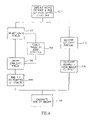
- FIG. 4 is a flow chart illustration of the method used by the system shown in FIG. 3 , according to an embodiment of the invention.
- FIG. 5 is a schematic illustration showing how spatial coordinates are determined according to an embodiment of the present invention.
- An in vivo video camera system captures and transmits images of, for example, the GI tract while the capsule passes through the GI lumen.
- Some embodiments may be contained within a capsule; alternate configurations, such as within an endoscope, are possible.
- embodiments contain an optical system for imaging an area of interest onto the camera system and a transmitter for transmitting image output of the camera.
- a capsule including such components can, for example, pass through the entire digestive tract and operate as an autonomous video endoscope. It may image difficult to reach areas of the small intestine.
- Embodiments of U.S. Pat. No. 5,604,531 assigned to the common assignee of the present application and incorporated herein by reference, describe an in vivo camera system, which is carried by a swallowable capsule.
- FIG. 1 shows a schematic diagram of an in-vivo imaging system.
- the system includes a capsule 40 having, for example, an imager 46 , an illumination source 42 , and a transmitter 41 .
- an image receiver 12 for example an antenna array
- a storage unit 19 outside the patient's body
- a data processor 14 for example an antenna array
- an image monitor 18 for example an image monitor
- a position monitor 16 a position monitor. While FIG. 1 shows separate monitors, both an image and its position can be presented on a single monitor.
- Imager 46 in capsule 40 may be connected to transmitter 41 also located in capsule 40 .
- Transmitter 41 transmits images to image receiver 12 , which sends the data to, for example, a data processor 14 and a storage unit 19 .
- Data processor 14 may, for example, analyze the data and may be in communication with storage unit 19 , transferring frame data to and from storage unit 19 .
- Data processor 14 also may provide the analyzed data to image monitor 18 and position monitor 16 where the physician views the data.
- the image monitor may present an image of the GI lumen and the position monitor may present the position in the GI tract at which the image was taken.
- the data can be viewed in real time or at some later date.
- the system can provide information about the location of these pathologies.
- the imaging device may include more than one camera or imaging device.
- Embodiments of the present invention relate to a method, device and system of size analysis by converting two-dimensional images, captured by a moving in-vivo video camera system, such as that of FIG. 1 , into three-dimensional representations. This conversion is typically done using only one camera or imager, and is typically based on the velocity of the camera system when it captures the frames being converted.
- system 15 is located in an external processing and display system, such as a personal computer or workstation, including conventional equipment and software such as a microprocessor or CPU, a memory, storage, etc.
- system 15 may be located in a display system described in embodiments of U.S. Pat. No. 5,604,531 and/or International Application Publication No WO0165995 published Sep. 13, 2001.
- all or part of system 15 or the functionality of system 15 may be located in another location or in other equipment. For example, some functionality may be in capsule 40 .
- video capsule 40 is shown approaching a first object 401 and a second object 402 , in GI lumen 403 .
- size analysis based on three dimensional representations of objects 401 and 402 can be done, as will be discussed with regard to FIG. 5 below.
- system 15 includes, for example, a distance-detecting unit 20 , an image receiver 12 and a processor 14 .
- Processor 14 includes, for example, a spatial coordinate generator 26 , a cross correlation 28 and a size generator 30 .
- distance-detecting unit 20 is a position detector.
- distance-detecting unit 20 obtains a distance measurement d by measuring and integrating a velocity, as will be described hereinbelow.
- Processor 14 may include, for example, a standard PC accelerator board, high performance PC, multiprocessor PC or any other serial or parallel high performance processing machine, and appropriate software.
- system 15 may include, for example, an edge detector 22 .
- distance-detecting unit 20 may contain or controlled by processor 14 .
- spatial coordinate generator 26 may contain or controlled by processor 14 .
- cross correlator 28 may be used, such edge detection capability using the following sliding window filter:
- FIG. 4 is a flow chart diagram illustrating a
- imager 46 within a moving in vivo video camera system such as the one described in FIG. 1 ) captures (step 101 ) images periodically, such as every 100-1000 ms. In one embodiment, the images are captured every 500 ms.
- Image data is transmitted to image receiver 12 ( FIG. 1 ).
- Data processor 14 divides received images into a grid of pixels, and selects (step 102 ) pixels for analysis. As in other imaging applications, the number of pixels determines the resolution of the image. For purposes of this discussion, the images are divided into m ⁇ n pixels.
- a user may choose which object or objects are to be tracked for size analysis. For example, a user may select an object, objects, or region being displayed in a moving or still image on a monitor using, for example, a pointing device such as a mouse. According to one embodiment an object is selected in a manner similar to selecting an object during ultrasound procedures. In another embodiment the system may automatically chose an object(s) or region for size analysis. In one embodiment, the calculations are performed on a workstation with stored data, and a user is able to move forward and backwards through a moving image and see or request size information for the various objects displayed during the portion viewed. In another embodiment, size analysis may be performed in real time. Size information may be, for example, stored and/or displayed. Other object or region selecting methods may be used.
- cross correlator 28 calculates (step 104 ) an xy cross correlation function between the intensities l j and l j+n of image j and image j+n, thereby identifying corresponding pixels in images j and j+n.
- the value n is typically, but not necessarily, 1.
- the second frame will be designated as j+1, with the understanding that n can also be greater than 1.
- edge detector 22 selects (step 106 ) pixels for cross correlation, thereby selecting an object. In one embodiment, only pixels whose edges exceed a certain predetermined threshold value are selected for correlation. Other suitable methods for image processing may be used.
- the cross correlation can be done on a pixel by pixel basis, more typically, it is performed on parts of the image, such as sets of 8 ⁇ 8 pixels. The latter approach can be used to minimize computation time.
- the cross correlation coefficient C xy is given by:
- C xy ⁇ ⁇ ⁇ ⁇ ⁇ ⁇ I j m ⁇ ( m , n ) n ⁇ ⁇ I j + 1 ⁇ ( m + x , n + y )
- l j (m,n) and l j+1 (m,n) are the intensity values of pixel (m,n) in images j and j+1 respectively.
- the vector (x, y) can be considered the displacement vector from pixel (m,n) in going from pixel (m,n) to pixel (m+x, n+y).
- the maximum of the cross correlation function indicates the most probable location of correspondence between the pixels of images j and j+1.
- a suitable cross correlation function is included in Matlab, a standard mathematics package for computers; other functions may be used.
- the results of the cross correlation may provide, for example, x and y coordinates for a specific point. If the cross correlation is performed for, for example, four edges of an object on images j and j+1, an entire two-dimensional set of spatial coordinates is obtained (step 108 ). Thus, for object A, x 1A , x 2A , y 1A and y 2A are known.
- the determination of the z coordinates for object A is typically based on the distance traversed by imager 46 while it moves through the GI tract capturing images j and j+1.
- distance traveled is determined from data captured by a sensor which is typically located within the device containing the capsule 40 ; the sensor may alternately be external to the patient.
- distance-measuring unit 20 measures the velocity of imager 46 using an accelerometer and an integrator.
- the accelerometer may be, for example, the ADXL50 model from Analog Devices. It is readily evident that, in addition to an accelerometer, any sensor that can provide data to determine the velocity of the capsule could also be used. Such sensors include, but are not limited to, induction coils (as described in U.S. Pat. No. 4,431,005, incorporated herein by reference) and ultrasound transducers. For example, if an induction coil is located in the capsule and the patient is placed in a magnetic field, a current would be produced by the coil with a magnitude proportional to the velocity of the capsule. Similarly, ultrasound transducers, such as those used in conventional medical ultrasound devices, can be used as an external sensor to track the movement of the capsule and standard electronics could be used to convert the data to velocities. Other distance measurement systems and methods may be used.
- the change of position of the capsule while capturing two images can be used to determine the distance traveled by the capsule during the time interval between the images.
- Signals sent by a transmitter within the capsule and received by receivers outside the body can be used to locate the position of the capsule.
- One suitable system for determining capsule location is one described in Published U.S. Application Number US-2002-0173718-A1 assigned to the common assignee of the present application and incorporated herein by reference; other suitable location determining systems may be used.
- conventional image analysis techniques can be used to analyze the optical flow of the images. For example, on the basis of the smear pattern of the images, velocity or distance can be determined. Once the velocity is known, a, for example, integrator calculates (step 112 ) the distance traveled by imager 46 from the time of capture of image j to the time of capture of image j+1. This distance value is used in determining (step 116 ) the z coordinate of object A, as described in the methods provided as examples hereinbelow. Other methods may be used within the scope of the present invention.
- FIG. 5 shows a geometric illustration of the basis for calculating the z coordinate of a set of objects A and B (where set can include one item), according to one embodiment of the present invention.
- the z coordinate represents the distance from imager 46 to each of the objects, denoted z A and z B respectively.
- imager 46 typically moves a certain distance d from the capture of the first image 202 to the capture of the second image 204 (of course periods of little or no movement are possible).
- the distance between images 202 and 204 is distance d.
- focal length f which is the lens focal length. While in one embodiment focal length f is used in the derivation of the following equations, it is typically eventually eliminated and in such a case its value does not need to be known explicitly.
- a 1 , b 1 , a 2 and b 2 The projections of objects A and B on each of the images 202 and 204 in the y direction are shown in FIG. 5 and are denoted a 1 , b 1 , a 2 and b 2 , respectively. These values are obtained from, for example, the pixel information stored in storage unit 19 , and correspond to the n value of each m ⁇ n pixel.
- a 1 represents the X value of object A as it was acquired in time t1 (X 1A )
- a 2 represents the X value of object A as it was acquired in time t2 (X 2A ).
- b 1 represents the X value of object B as it was acquired in time t1 (X 1B )
- b 2 represents the X value of object B as it was acquired in time t2 (X 2B ).
- the actual values for a 1 , a 2 , b 1 , and b 2 may be calculated by, for example, image processor 14 (step 108 of FIG. 4 ) from, for example, the size of the image sensor in imager 46 and image pixel data stored in storage unit 18 .
- image processor 14 step 108 of FIG. 4
- an object whose length is p pixels will have an actual size of: L*P/m.
- the z coordinate for object A as a function of the z coordinate for object B can be obtained.
- Spatial coordinate processor 26 calculates (step 116 ) the z values for two points on object A (Z 1A and z 2A ) corresponding to the two edges of object A. Accordingly, xyz spatial coordinates are known for object A.
- Size analyzer 30 then calculates (step 118 ) the size of object A by, for example, subtracting each of the axis coordinates from each other.
- Image processor 14 may send any selected size-data to image monitor 18 for display.
- size data may be displayed along with, for example, image or moving image data on image monitor 18 .
- Various methods of displaying the size data may be used.
- the procedure described hereinabove can be performed as a post-processing step, or, with adequate computational capability, it can be done in real time, allowing the user to choose specific images for processing.
- FIG. 5 shows a one-dimensional object, (e.g. a line), here positioned along the X-axis, symmetry considerations can be used in an analogous manner to obtain the Y coordinate, where the Y-axis is perpendicular to the plane of the paper.
- a one-dimensional object e.g. a line
- symmetry considerations can be used in an analogous manner to obtain the Y coordinate, where the Y-axis is perpendicular to the plane of the paper.
Abstract
Description
general method for generating size measurements from two-dimensional images according to one embodiment of the present invention. Steps of
where lj(m,n) and lj+1(m,n) are the intensity values of pixel (m,n) in images j and j+1 respectively. The vector (x, y) can be considered the displacement vector from pixel (m,n) in going from pixel (m,n) to pixel (m+x, n+y). The maximum of the cross correlation function indicates the most probable location of correspondence between the pixels of images j and j+1. A suitable cross correlation function is included in Matlab, a standard mathematics package for computers; other functions may be used.
Z b(1−T b)=Z a(1−T a)+d(T b −T a)
where Ta and Tb are defined as:
T a =a 1 /a 2
T b =b 1 /b 2
A/a 1=(Z A +d+f)/f
A/a 2=(Z A +f)/f
From those two equations the following can be calculated:
A*f=(Z A +d+f)/a 1+(Z A +f)*a 2
Leading to
Z A *a 1 −Z A *a 2 =f*a 2−(d+f)*a 1 =f*(a 2 −a 1)−d*a 1
Finally,
ZA =d*a 1/(a 2 −a 1)−f
Thus, if the focal length of the camera is known, only one object is needed for calculation. The size of the object may calculated, for example, as described above.
Claims (13)
Applications Claiming Priority (3)
| Application Number | Priority Date | Filing Date | Title |
|---|---|---|---|
| IL147221A IL147221A (en) | 2001-12-20 | 2001-12-20 | Device, system and method for image based size analysis |
| IL147221 | 2001-12-20 | ||
| PCT/IL2002/001025 WO2003053241A2 (en) | 2001-12-20 | 2002-12-19 | Device, system and method for image based size analysis |
Related Parent Applications (1)
| Application Number | Title | Priority Date | Filing Date |
|---|---|---|---|
| PCT/IL2002/001025 Continuation WO2003053241A2 (en) | 2001-12-20 | 2002-12-19 | Device, system and method for image based size analysis |
Publications (2)
| Publication Number | Publication Date |
|---|---|
| US20040258328A1 US20040258328A1 (en) | 2004-12-23 |
| US7551955B2 true US7551955B2 (en) | 2009-06-23 |
Family
ID=11075906
Family Applications (1)
| Application Number | Title | Priority Date | Filing Date |
|---|---|---|---|
| US10/402,245 Expired - Fee Related US7551955B2 (en) | 2001-12-20 | 2003-03-31 | Device, system and method for image based size analysis |
Country Status (5)
| Country | Link |
|---|---|
| US (1) | US7551955B2 (en) |
| EP (1) | EP1465526B1 (en) |
| AU (1) | AU2002360203A1 (en) |
| IL (1) | IL147221A (en) |
| WO (1) | WO2003053241A2 (en) |
Cited By (5)
| Publication number | Priority date | Publication date | Assignee | Title |
|---|---|---|---|---|
| US20060004285A1 (en) * | 1998-10-22 | 2006-01-05 | Gavriel Meron | Method for delivering a device to a target location |
| US20100092054A1 (en) * | 2007-04-17 | 2010-04-15 | Harvey Hensley | Method and Apparatus for Endoscopic Examination of Lesions |
| US20150313446A1 (en) * | 2013-02-05 | 2015-11-05 | Olympus Corporation | Robotic-assisted surgical system and control method thereof |
| US20160217591A1 (en) * | 2013-10-02 | 2016-07-28 | Given Imaging Ltd. | System and method for size estimation of in-vivo objects |
| US10070932B2 (en) | 2013-08-29 | 2018-09-11 | Given Imaging Ltd. | System and method for maneuvering coils power optimization |
Families Citing this family (42)
| Publication number | Priority date | Publication date | Assignee | Title |
|---|---|---|---|---|
| EP1418833B1 (en) | 2001-06-18 | 2008-08-13 | Given Imaging Ltd. | Swallowable in vivo sensing capsule with a circuit board having rigid sections and flexible sections |
| US7662093B2 (en) | 2002-09-30 | 2010-02-16 | Given Imaging, Ltd. | Reduced size imaging device |
| US8449452B2 (en) | 2002-09-30 | 2013-05-28 | Given Imaging Ltd. | In-vivo sensing system |
| JP4746876B2 (en) | 2002-10-15 | 2011-08-10 | ギブン イメージング リミテッド | Apparatus, system and method for transferring a signal to a mobile device |
| WO2004044664A1 (en) * | 2002-11-06 | 2004-05-27 | Julius Lin | Virtual workstation |
| US7774075B2 (en) * | 2002-11-06 | 2010-08-10 | Lin Julius J Y | Audio-visual three-dimensional input/output |
| US7634305B2 (en) | 2002-12-17 | 2009-12-15 | Given Imaging, Ltd. | Method and apparatus for size analysis in an in vivo imaging system |
| EP1620012B1 (en) * | 2003-05-01 | 2012-04-18 | Given Imaging Ltd. | Panoramic field of view imaging device |
| JP2004350963A (en) * | 2003-05-29 | 2004-12-16 | Olympus Corp | Capsule type medical treatment apparatus |
| US7011625B1 (en) * | 2003-06-13 | 2006-03-14 | Albert Shar | Method and system for accurate visualization and measurement of endoscopic images |
| DE10359981A1 (en) * | 2003-12-19 | 2005-07-21 | Siemens Ag | System and method for in vivo positioning and orientation determination of an endoscopy capsule or an endo-robot in the context of a wireless endoscopy |
| US20050171418A1 (en) * | 2004-01-08 | 2005-08-04 | Tah-Yeong Lin | Capsule endoscopy system |
| WO2005074785A1 (en) * | 2004-02-06 | 2005-08-18 | Olympus Corporation | Receiver |
| KR100615881B1 (en) * | 2004-06-21 | 2006-08-25 | 한국과학기술연구원 | Capsule Type Endoscope Control System |
| US7336833B2 (en) * | 2004-06-30 | 2008-02-26 | Given Imaging, Ltd. | Device, system, and method for reducing image data captured in-vivo |
| US8500630B2 (en) | 2004-06-30 | 2013-08-06 | Given Imaging Ltd. | In vivo device with flexible circuit board and method for assembly thereof |
| US20080262304A1 (en) * | 2004-06-30 | 2008-10-23 | Micha Nisani | In-Vivo Sensing System Device and Method for Real Time Viewing |
| US20070252892A1 (en) * | 2004-07-16 | 2007-11-01 | Manabu Fujita | Moving-State Detecting Apparatus and Moving-State Detecting System |
| US8005279B2 (en) * | 2005-03-22 | 2011-08-23 | Osaka University | Capsule endoscope image display controller |
| KR20080082002A (en) * | 2005-12-29 | 2008-09-10 | 기븐 이미징 리미티드 | System device and method for estimating the size of an object in a body lumen |
| ATE527610T1 (en) * | 2006-08-23 | 2011-10-15 | Hewlett Packard Development Co | MULTIPLE SCREEN SIZE DISPLAY MACHINE |
| US20080112885A1 (en) | 2006-09-06 | 2008-05-15 | Innurvation, Inc. | System and Method for Acoustic Data Transmission |
| EP2063766B1 (en) | 2006-09-06 | 2017-01-18 | Innurvation, Inc. | Ingestible low power sensor device and system for communicating with same |
| US8496575B2 (en) * | 2006-11-14 | 2013-07-30 | Olympus Corporation | Measuring endoscope apparatus, program and recording medium |
| US20090088618A1 (en) | 2007-10-01 | 2009-04-02 | Arneson Michael R | System and Method for Manufacturing a Swallowable Sensor Device |
| US9197470B2 (en) | 2007-10-05 | 2015-11-24 | Innurvation, Inc. | Data transmission via multi-path channels using orthogonal multi-frequency signals with differential phase shift keying modulation |
| US8617058B2 (en) | 2008-07-09 | 2013-12-31 | Innurvation, Inc. | Displaying image data from a scanner capsule |
| US8516691B2 (en) | 2009-06-24 | 2013-08-27 | Given Imaging Ltd. | Method of assembly of an in vivo imaging device with a flexible circuit board |
| US9192353B2 (en) * | 2009-10-27 | 2015-11-24 | Innurvation, Inc. | Data transmission via wide band acoustic channels |
| US8647259B2 (en) | 2010-03-26 | 2014-02-11 | Innurvation, Inc. | Ultrasound scanning capsule endoscope (USCE) |
| US9264218B2 (en) * | 2014-06-10 | 2016-02-16 | Lenovo Enterprise Solutions (Singapore) Pte. Ltd. | Rising and falling edge detection and re-assembly for high speed serial data communications |
| US9479148B2 (en) | 2015-02-26 | 2016-10-25 | Lenovo Enterprise Solutions (Singapore) Pte. Ltd. | Serial data signal edge detection |
| US10624533B2 (en) | 2015-10-16 | 2020-04-21 | Capsovision Inc | Endoscope with images optimized based on depth map derived from structured light images |
| US10943333B2 (en) | 2015-10-16 | 2021-03-09 | Capsovision Inc. | Method and apparatus of sharpening of gastrointestinal images based on depth information |
| US11354783B2 (en) | 2015-10-16 | 2022-06-07 | Capsovision Inc. | Method and apparatus of sharpening of gastrointestinal images based on depth information |
| ES2742101T3 (en) | 2015-11-25 | 2020-02-13 | Ovesco Endoscopy Ag | Passive capsule endoscope for the intestine |
| US10136959B2 (en) * | 2016-12-28 | 2018-11-27 | Auris Health, Inc. | Endolumenal object sizing |
| CN107049211B (en) * | 2017-03-13 | 2019-02-12 | 重庆金山医疗器械有限公司 | A kind of device measuring capsule endoscopic and stomach wall distance |
| US20190015070A1 (en) * | 2017-07-11 | 2019-01-17 | The Board Of Trustees Of The Leland Stanford Junior University | Ultrasonic Capsule Endoscopy Device having Image-based Relative Motion Estimation |
| US10580157B2 (en) | 2017-08-04 | 2020-03-03 | Capsovision Inc | Method and apparatus for estimating area or volume of object of interest from gastrointestinal images |
| US10736559B2 (en) | 2017-08-04 | 2020-08-11 | Capsovision Inc | Method and apparatus for estimating area or volume of object of interest from gastrointestinal images |
| US10346978B2 (en) * | 2017-08-04 | 2019-07-09 | Capsovision Inc. | Method and apparatus for area or volume of object of interest from gastrointestinal images |
Citations (40)
| Publication number | Priority date | Publication date | Assignee | Title |
|---|---|---|---|---|
| US3971362A (en) | 1972-10-27 | 1976-07-27 | The United States Of America As Represented By The Administrator Of The National Aeronautics And Space Administration | Miniature ingestible telemeter devices to measure deep-body temperature |
| US4217045A (en) | 1978-12-29 | 1980-08-12 | Ziskind Stanley H | Capsule for photographic use in a walled organ of the living body |
| US4278077A (en) | 1978-07-27 | 1981-07-14 | Olympus Optical Co., Ltd. | Medical camera system |
| US4431005A (en) | 1981-05-07 | 1984-02-14 | Mccormick Laboratories, Inc. | Method of and apparatus for determining very accurately the position of a device inside biological tissue |
| DE3440177A1 (en) | 1984-11-02 | 1986-05-15 | Friedrich Dipl.-Ing. 8031 Eichenau Hilliges | Television recording and replay device for endoscopy on human and animal bodies |
| US4651201A (en) | 1984-06-01 | 1987-03-17 | Arnold Schoolman | Stereoscopic endoscope arrangement |
| US4656508A (en) | 1984-06-08 | 1987-04-07 | Olympus Optical Co., Ltd. | Measuring endoscope |
| US4689621A (en) | 1986-03-31 | 1987-08-25 | The United States Of America As Represented By The Administrator Of The National Aeronautics And Space Administration | Temperature responsive transmitter |
| US4714319A (en) | 1983-09-30 | 1987-12-22 | Zeevi Yehoshua Y | Apparatus for relief illusion |
| US4844076A (en) | 1988-08-26 | 1989-07-04 | The Johns Hopkins University | Ingestible size continuously transmitting temperature monitoring pill |
| US4881032A (en) | 1988-10-21 | 1989-11-14 | General Electric Company | Method of, and apparatus for, NMR spectroscopic metabolite imaging and quantification |
| US4895431A (en) | 1986-11-13 | 1990-01-23 | Olympus Optical Co., Ltd. | Method of processing endoscopic images |
| US4980763A (en) * | 1989-06-12 | 1990-12-25 | Welch Allyn, Inc. | System for measuring objects viewed through a borescope |
| JPH04109927A (en) | 1990-08-31 | 1992-04-10 | Toshiba Corp | Electronic endoscope apparatus |
| JPH0515515A (en) | 1991-02-19 | 1993-01-26 | Nissin Electric Co Ltd | Digestive organ system diagnosing apparatus |
| US5279607A (en) | 1991-05-30 | 1994-01-18 | The State University Of New York | Telemetry capsule and process |
| US5575754A (en) | 1995-02-24 | 1996-11-19 | Olympus Optical Co., Ltd. | Endoscopic apparatus for three dimensional instrumentation |
| US5604531A (en) | 1994-01-17 | 1997-02-18 | State Of Israel, Ministry Of Defense, Armament Development Authority | In vivo video camera system |
| US5728044A (en) | 1995-03-10 | 1998-03-17 | Shan; Yansong | Sensor device for spacial imaging of endoscopes |
| US5819736A (en) | 1994-03-24 | 1998-10-13 | Sightline Technologies Ltd. | Viewing method and apparatus particularly useful for viewing the interior of the large intestine |
| US5833603A (en) | 1996-03-13 | 1998-11-10 | Lipomatrix, Inc. | Implantable biosensing transponder |
| US5853005A (en) | 1996-05-02 | 1998-12-29 | The United States Of America As Represented By The Secretary Of The Army | Acoustic monitoring system |
| US5944655A (en) | 1994-07-08 | 1999-08-31 | Forschunjszentrum Karlsruhe Gmbh | 3D endoscope with optical switch and prism arrangement |
| US5967968A (en) * | 1998-06-25 | 1999-10-19 | The General Hospital Corporation | Apparatus and method for determining the size of an object during endoscopy |
| US6009189A (en) | 1996-08-16 | 1999-12-28 | Schaack; David F. | Apparatus and method for making accurate three-dimensional size measurements of inaccessible objects |
| US6074349A (en) | 1994-11-30 | 2000-06-13 | Boston Scientific Corporation | Acoustic imaging and doppler catheters and guidewires |
| US6165128A (en) | 1997-10-06 | 2000-12-26 | Endosonics Corporation | Method and apparatus for making an image of a lumen or other body cavity and its surrounding tissue |
| WO2001008548A1 (en) | 1999-08-03 | 2001-02-08 | The University College London Hospitals Nhs Trust | Improved passage-travelling device |
| US6240312B1 (en) | 1997-10-23 | 2001-05-29 | Robert R. Alfano | Remote-controllable, micro-scale device for use in in vivo medical diagnosis and/or treatment |
| US6245057B1 (en) | 1997-04-23 | 2001-06-12 | Micronas Intermetall Gmbh | Device for treating malignant, tumorous tissue areas |
| WO2001050941A2 (en) | 2000-01-13 | 2001-07-19 | Capsule View Inc. | Encapsulated medical imaging device and method |
| JP2001224553A (en) | 2000-02-17 | 2001-08-21 | Asahi Optical Co Ltd | Imaging instrument for capusle endoscope |
| US6289232B1 (en) | 1998-03-30 | 2001-09-11 | Beth Israel Deaconess Medical Center, Inc. | Coil array autocalibration MR imaging |
| WO2001065995A2 (en) | 2000-03-08 | 2001-09-13 | Given Imaging Ltd. | A device and system for in vivo imaging |
| US20010051766A1 (en) | 1999-03-01 | 2001-12-13 | Gazdzinski Robert F. | Endoscopic smart probe and method |
| US20020107444A1 (en) * | 2000-12-19 | 2002-08-08 | Doron Adler | Image based size analysis |
| US20020173718A1 (en) * | 2001-05-20 | 2002-11-21 | Mordechai Frisch | Array system and method for locating an in vivo signal source |
| US20030139661A1 (en) * | 2001-01-22 | 2003-07-24 | Yoav Kimchy | Ingestible device |
| US6944316B2 (en) * | 2001-06-20 | 2005-09-13 | Given Imaging Ltd | Motility analysis within a gastrointestinal tract |
| US6950690B1 (en) * | 1998-10-22 | 2005-09-27 | Given Imaging Ltd | Method for delivering a device to a target location |
-
2001
- 2001-12-20 IL IL147221A patent/IL147221A/en not_active IP Right Cessation
-
2002
- 2002-12-19 EP EP02795412.2A patent/EP1465526B1/en not_active Expired - Lifetime
- 2002-12-19 AU AU2002360203A patent/AU2002360203A1/en not_active Abandoned
- 2002-12-19 WO PCT/IL2002/001025 patent/WO2003053241A2/en not_active Application Discontinuation
-
2003
- 2003-03-31 US US10/402,245 patent/US7551955B2/en not_active Expired - Fee Related
Patent Citations (40)
| Publication number | Priority date | Publication date | Assignee | Title |
|---|---|---|---|---|
| US3971362A (en) | 1972-10-27 | 1976-07-27 | The United States Of America As Represented By The Administrator Of The National Aeronautics And Space Administration | Miniature ingestible telemeter devices to measure deep-body temperature |
| US4278077A (en) | 1978-07-27 | 1981-07-14 | Olympus Optical Co., Ltd. | Medical camera system |
| US4217045A (en) | 1978-12-29 | 1980-08-12 | Ziskind Stanley H | Capsule for photographic use in a walled organ of the living body |
| US4431005A (en) | 1981-05-07 | 1984-02-14 | Mccormick Laboratories, Inc. | Method of and apparatus for determining very accurately the position of a device inside biological tissue |
| US4714319A (en) | 1983-09-30 | 1987-12-22 | Zeevi Yehoshua Y | Apparatus for relief illusion |
| US4651201A (en) | 1984-06-01 | 1987-03-17 | Arnold Schoolman | Stereoscopic endoscope arrangement |
| US4656508A (en) | 1984-06-08 | 1987-04-07 | Olympus Optical Co., Ltd. | Measuring endoscope |
| DE3440177A1 (en) | 1984-11-02 | 1986-05-15 | Friedrich Dipl.-Ing. 8031 Eichenau Hilliges | Television recording and replay device for endoscopy on human and animal bodies |
| US4689621A (en) | 1986-03-31 | 1987-08-25 | The United States Of America As Represented By The Administrator Of The National Aeronautics And Space Administration | Temperature responsive transmitter |
| US4895431A (en) | 1986-11-13 | 1990-01-23 | Olympus Optical Co., Ltd. | Method of processing endoscopic images |
| US4844076A (en) | 1988-08-26 | 1989-07-04 | The Johns Hopkins University | Ingestible size continuously transmitting temperature monitoring pill |
| US4881032A (en) | 1988-10-21 | 1989-11-14 | General Electric Company | Method of, and apparatus for, NMR spectroscopic metabolite imaging and quantification |
| US4980763A (en) * | 1989-06-12 | 1990-12-25 | Welch Allyn, Inc. | System for measuring objects viewed through a borescope |
| JPH04109927A (en) | 1990-08-31 | 1992-04-10 | Toshiba Corp | Electronic endoscope apparatus |
| JPH0515515A (en) | 1991-02-19 | 1993-01-26 | Nissin Electric Co Ltd | Digestive organ system diagnosing apparatus |
| US5279607A (en) | 1991-05-30 | 1994-01-18 | The State University Of New York | Telemetry capsule and process |
| US5604531A (en) | 1994-01-17 | 1997-02-18 | State Of Israel, Ministry Of Defense, Armament Development Authority | In vivo video camera system |
| US5819736A (en) | 1994-03-24 | 1998-10-13 | Sightline Technologies Ltd. | Viewing method and apparatus particularly useful for viewing the interior of the large intestine |
| US5944655A (en) | 1994-07-08 | 1999-08-31 | Forschunjszentrum Karlsruhe Gmbh | 3D endoscope with optical switch and prism arrangement |
| US6074349A (en) | 1994-11-30 | 2000-06-13 | Boston Scientific Corporation | Acoustic imaging and doppler catheters and guidewires |
| US5575754A (en) | 1995-02-24 | 1996-11-19 | Olympus Optical Co., Ltd. | Endoscopic apparatus for three dimensional instrumentation |
| US5728044A (en) | 1995-03-10 | 1998-03-17 | Shan; Yansong | Sensor device for spacial imaging of endoscopes |
| US5833603A (en) | 1996-03-13 | 1998-11-10 | Lipomatrix, Inc. | Implantable biosensing transponder |
| US5853005A (en) | 1996-05-02 | 1998-12-29 | The United States Of America As Represented By The Secretary Of The Army | Acoustic monitoring system |
| US6009189A (en) | 1996-08-16 | 1999-12-28 | Schaack; David F. | Apparatus and method for making accurate three-dimensional size measurements of inaccessible objects |
| US6245057B1 (en) | 1997-04-23 | 2001-06-12 | Micronas Intermetall Gmbh | Device for treating malignant, tumorous tissue areas |
| US6165128A (en) | 1997-10-06 | 2000-12-26 | Endosonics Corporation | Method and apparatus for making an image of a lumen or other body cavity and its surrounding tissue |
| US6240312B1 (en) | 1997-10-23 | 2001-05-29 | Robert R. Alfano | Remote-controllable, micro-scale device for use in in vivo medical diagnosis and/or treatment |
| US6289232B1 (en) | 1998-03-30 | 2001-09-11 | Beth Israel Deaconess Medical Center, Inc. | Coil array autocalibration MR imaging |
| US5967968A (en) * | 1998-06-25 | 1999-10-19 | The General Hospital Corporation | Apparatus and method for determining the size of an object during endoscopy |
| US6950690B1 (en) * | 1998-10-22 | 2005-09-27 | Given Imaging Ltd | Method for delivering a device to a target location |
| US20010051766A1 (en) | 1999-03-01 | 2001-12-13 | Gazdzinski Robert F. | Endoscopic smart probe and method |
| WO2001008548A1 (en) | 1999-08-03 | 2001-02-08 | The University College London Hospitals Nhs Trust | Improved passage-travelling device |
| WO2001050941A2 (en) | 2000-01-13 | 2001-07-19 | Capsule View Inc. | Encapsulated medical imaging device and method |
| JP2001224553A (en) | 2000-02-17 | 2001-08-21 | Asahi Optical Co Ltd | Imaging instrument for capusle endoscope |
| WO2001065995A2 (en) | 2000-03-08 | 2001-09-13 | Given Imaging Ltd. | A device and system for in vivo imaging |
| US20020107444A1 (en) * | 2000-12-19 | 2002-08-08 | Doron Adler | Image based size analysis |
| US20030139661A1 (en) * | 2001-01-22 | 2003-07-24 | Yoav Kimchy | Ingestible device |
| US20020173718A1 (en) * | 2001-05-20 | 2002-11-21 | Mordechai Frisch | Array system and method for locating an in vivo signal source |
| US6944316B2 (en) * | 2001-06-20 | 2005-09-13 | Given Imaging Ltd | Motility analysis within a gastrointestinal tract |
Non-Patent Citations (11)
| Title |
|---|
| BBC News Online-Pill camera to 'broadcast from the gut', Feb 21, 2000, www.news.bbc.co.uk. |
| Machine Vision, Theory, Algorithms, Practicalities E.R. Davies, Academic Press 1996, pp. 441-444. |
| Robust shape reconstruction from combined shading and stereo information-Lee, et al., SPIE vol. 1771 Applications of Digital Image Processing XV (1992), pp. 171-182. |
| Shedding light on cancer diagnosis-Powell (Ed.), May 2000, Laser Focus World. |
| Simulation of images by photometric stereo modeling, Russell, et al., Optical Engineering, Sep. 1991, vol. 30, No. 9, pp. 1337-1345. |
| The Radio Pill, Rowlands, et al., British Communications and Electronics, Aug. 1960, pp. 598-601. |
| Two Image Photometric Stereo Method, Yang et al., SPIE vol. 1826, Intelligent Robots and Computer Vision XI (1992). |
| Video Camera to "Take"-RF System Lab, Dec. 25, 2001. |
| Wellesley company sends body monitors into space-Crum, Apr. 1998. |
| Wireless transmission of a color television moving image from the stomach using a miniature CCD camera, light source and microwave transmitter. Swain CP, Gong F, Mills TN, Gastrointest Endosc 1997;45:AB40. |
| www.oceanoptics.com-pH Sensor & Accessories, (C) 2001. |
Cited By (9)
| Publication number | Priority date | Publication date | Assignee | Title |
|---|---|---|---|---|
| US20060004285A1 (en) * | 1998-10-22 | 2006-01-05 | Gavriel Meron | Method for delivering a device to a target location |
| US20100092054A1 (en) * | 2007-04-17 | 2010-04-15 | Harvey Hensley | Method and Apparatus for Endoscopic Examination of Lesions |
| US9451904B2 (en) * | 2007-04-17 | 2016-09-27 | Fox Chase Cancer Center | Method and apparatus for endoscopic examination of lesions |
| US20150313446A1 (en) * | 2013-02-05 | 2015-11-05 | Olympus Corporation | Robotic-assisted surgical system and control method thereof |
| US10070932B2 (en) | 2013-08-29 | 2018-09-11 | Given Imaging Ltd. | System and method for maneuvering coils power optimization |
| US20160217591A1 (en) * | 2013-10-02 | 2016-07-28 | Given Imaging Ltd. | System and method for size estimation of in-vivo objects |
| US9911203B2 (en) * | 2013-10-02 | 2018-03-06 | Given Imaging Ltd. | System and method for size estimation of in-vivo objects |
| US20180096491A1 (en) * | 2013-10-02 | 2018-04-05 | Given Imaging Ltd. | System and method for size estimation of in-vivo objects |
| US10521924B2 (en) * | 2013-10-02 | 2019-12-31 | Given Imaging Ltd. | System and method for size estimation of in-vivo objects |
Also Published As
| Publication number | Publication date |
|---|---|
| US20040258328A1 (en) | 2004-12-23 |
| EP1465526A2 (en) | 2004-10-13 |
| AU2002360203A1 (en) | 2003-07-09 |
| WO2003053241A2 (en) | 2003-07-03 |
| IL147221A (en) | 2010-11-30 |
| WO2003053241A3 (en) | 2003-10-23 |
| IL147221A0 (en) | 2002-08-14 |
| EP1465526A4 (en) | 2009-01-14 |
| EP1465526B1 (en) | 2014-12-17 |
Similar Documents
| Publication | Publication Date | Title |
|---|---|---|
| US7551955B2 (en) | Device, system and method for image based size analysis | |
| US20020107444A1 (en) | Image based size analysis | |
| US11678804B2 (en) | Methods and systems for tracking and guiding sensors and instruments | |
| US7200253B2 (en) | Motility analysis within a gastrointestinal tract | |
| US7865229B2 (en) | System and method for determining path lengths through a body lumen | |
| JP4864534B2 (en) | System for controlling the capture rate and display rate of an in vivo camera | |
| US20080027329A1 (en) | System, apparatus and method for measurement of motion parameters of an in-vivo device | |
| EP1676522A1 (en) | System for locating an in-vivo signal source | |
| US10402992B2 (en) | Method and apparatus for endoscope with distance measuring for object scaling | |
| US20100217139A1 (en) | Heart rate measurement | |
| US10835113B2 (en) | Method and apparatus for travelled distance measuring by a capsule camera in the gastrointestinal tract | |
| WO2016039915A2 (en) | Systems and methods using spatial sensor data in full-field three-dimensional surface measurment | |
| JP5116070B2 (en) | System for motility measurement and analysis | |
| US20050123179A1 (en) | Method and system for automatic axial rotation correction in vivo images | |
| US20080062124A1 (en) | Mouse interface apparatus using camera, system and method using the same, and computer recordable medium for implementing the same | |
| JP2009089910A (en) | Photographing direction discriminating apparatus, photographing direction discriminating method, photographing direction discriminating program, and computer-readable recording medium on which photographing direction discriminating program is recorded | |
| WO2002069807A1 (en) | Three-dimensional image detecting apparatus using position sensor | |
| IL159451A (en) | Motility analysis within a gastrointestinaltract |
Legal Events
| Date | Code | Title | Description |
|---|---|---|---|
| AS | Assignment |
Owner name: GIVEN IMAGING LTD., ISRAEL Free format text: ASSIGNMENT OF ASSIGNORS INTEREST;ASSIGNOR:ADLER, DORON;REEL/FRAME:015772/0778 Effective date: 20040308 |
|
| STCF | Information on status: patent grant |
Free format text: PATENTED CASE |
|
| FPAY | Fee payment |
Year of fee payment: 4 |
|
| FPAY | Fee payment |
Year of fee payment: 8 |
|
| FEPP | Fee payment procedure |
Free format text: MAINTENANCE FEE REMINDER MAILED (ORIGINAL EVENT CODE: REM.); ENTITY STATUS OF PATENT OWNER: LARGE ENTITY |
|
| LAPS | Lapse for failure to pay maintenance fees |
Free format text: PATENT EXPIRED FOR FAILURE TO PAY MAINTENANCE FEES (ORIGINAL EVENT CODE: EXP.); ENTITY STATUS OF PATENT OWNER: LARGE ENTITY |
|
| STCH | Information on status: patent discontinuation |
Free format text: PATENT EXPIRED DUE TO NONPAYMENT OF MAINTENANCE FEES UNDER 37 CFR 1.362 |
|
| FP | Lapsed due to failure to pay maintenance fee |
Effective date: 20210623 |