US7575887B2 - Detection of proteases secreted from pathogenic microorganisms - Google Patents

Detection of proteases secreted from pathogenic microorganisms Download PDF

Info

Publication number
US7575887B2
US7575887B2 US11/364,810 US36481006A US7575887B2 US 7575887 B2 US7575887 B2 US 7575887B2 US 36481006 A US36481006 A US 36481006A US 7575887 B2 US7575887 B2 US 7575887B2
Authority
US
United States
Prior art keywords
protease
test sample
substrate
zone
detection zone
Prior art date
Legal status (The legal status is an assumption and is not a legal conclusion. Google has not performed a legal analysis and makes no representation as to the accuracy of the status listed.)
Expired - Fee Related, expires
Application number
US11/364,810
Other versions
US20070048816A1 (en
Inventor
Xuedong Song
Current Assignee (The listed assignees may be inaccurate. Google has not performed a legal analysis and makes no representation or warranty as to the accuracy of the list.)
Kimberly Clark Worldwide Inc
Original Assignee
Kimberly Clark Worldwide Inc
Priority date (The priority date is an assumption and is not a legal conclusion. Google has not performed a legal analysis and makes no representation as to the accuracy of the date listed.)
Filing date
Publication date
Application filed by Kimberly Clark Worldwide Inc filed Critical Kimberly Clark Worldwide Inc
Priority to US11/364,810 priority Critical patent/US7575887B2/en
Assigned to KIMBERLY-CLARK WORLDWIDE, INC. reassignment KIMBERLY-CLARK WORLDWIDE, INC. ASSIGNMENT OF ASSIGNORS INTEREST (SEE DOCUMENT FOR DETAILS). Assignors: SONG, XUEDONG
Priority to PCT/US2006/030107 priority patent/WO2007027353A2/en
Priority to EP06789202A priority patent/EP1920253A2/en
Publication of US20070048816A1 publication Critical patent/US20070048816A1/en
Priority to US12/542,278 priority patent/US8609401B2/en
Application granted granted Critical
Publication of US7575887B2 publication Critical patent/US7575887B2/en
Assigned to KIMBERLY-CLARK WORLDWIDE, INC. reassignment KIMBERLY-CLARK WORLDWIDE, INC. NAME CHANGE Assignors: KIMBERLY-CLARK WORLDWIDE, INC.
Expired - Fee Related legal-status Critical Current
Adjusted expiration legal-status Critical

Links

Images

Classifications

    • CCHEMISTRY; METALLURGY
    • C12BIOCHEMISTRY; BEER; SPIRITS; WINE; VINEGAR; MICROBIOLOGY; ENZYMOLOGY; MUTATION OR GENETIC ENGINEERING
    • C12QMEASURING OR TESTING PROCESSES INVOLVING ENZYMES, NUCLEIC ACIDS OR MICROORGANISMS; COMPOSITIONS OR TEST PAPERS THEREFOR; PROCESSES OF PREPARING SUCH COMPOSITIONS; CONDITION-RESPONSIVE CONTROL IN MICROBIOLOGICAL OR ENZYMOLOGICAL PROCESSES
    • C12Q1/00Measuring or testing processes involving enzymes, nucleic acids or microorganisms; Compositions therefor; Processes of preparing such compositions
    • C12Q1/34Measuring or testing processes involving enzymes, nucleic acids or microorganisms; Compositions therefor; Processes of preparing such compositions involving hydrolase
    • C12Q1/44Measuring or testing processes involving enzymes, nucleic acids or microorganisms; Compositions therefor; Processes of preparing such compositions involving hydrolase involving esterase
    • GPHYSICS
    • G01MEASURING; TESTING
    • G01NINVESTIGATING OR ANALYSING MATERIALS BY DETERMINING THEIR CHEMICAL OR PHYSICAL PROPERTIES
    • G01N33/00Investigating or analysing materials by specific methods not covered by groups G01N1/00 - G01N31/00
    • G01N33/48Biological material, e.g. blood, urine; Haemocytometers
    • G01N33/50Chemical analysis of biological material, e.g. blood, urine; Testing involving biospecific ligand binding methods; Immunological testing
    • G01N33/52Use of compounds or compositions for colorimetric, spectrophotometric or fluorometric investigation, e.g. use of reagent paper and including single- and multilayer analytical elements
    • GPHYSICS
    • G01MEASURING; TESTING
    • G01NINVESTIGATING OR ANALYSING MATERIALS BY DETERMINING THEIR CHEMICAL OR PHYSICAL PROPERTIES
    • G01N33/00Investigating or analysing materials by specific methods not covered by groups G01N1/00 - G01N31/00
    • G01N33/48Biological material, e.g. blood, urine; Haemocytometers
    • G01N33/50Chemical analysis of biological material, e.g. blood, urine; Testing involving biospecific ligand binding methods; Immunological testing
    • G01N33/53Immunoassay; Biospecific binding assay; Materials therefor
    • G01N33/558Immunoassay; Biospecific binding assay; Materials therefor using diffusion or migration of antigen or antibody
    • GPHYSICS
    • G01MEASURING; TESTING
    • G01NINVESTIGATING OR ANALYSING MATERIALS BY DETERMINING THEIR CHEMICAL OR PHYSICAL PROPERTIES
    • G01N33/00Investigating or analysing materials by specific methods not covered by groups G01N1/00 - G01N31/00
    • G01N33/48Biological material, e.g. blood, urine; Haemocytometers
    • G01N33/50Chemical analysis of biological material, e.g. blood, urine; Testing involving biospecific ligand binding methods; Immunological testing
    • G01N33/53Immunoassay; Biospecific binding assay; Materials therefor
    • G01N33/573Immunoassay; Biospecific binding assay; Materials therefor for enzymes or isoenzymes

Definitions

  • vaginal infections include vulvovaginal candidiasis, bacterial vaginosis, and trichomoneasis. While symptomatically these infections may appear quite similar, the particular pathogenic microorganism underlying each may vary widely. For instance, candidiases are caused by pathogenic strains of yeast belonging to the genera Candida or related strains belonging mainly to the genera of Torulopsis . Trichomoneasis, on the other hand, is caused by the parasite Trichomonas vaginalis , and the underlying cause of bacterial vaginosis often cannot be attributed to one specific etiologic agent. Moreover, often these pathogens are common and/or benign, and only under opportune conditions will they become pathogenic.
  • Candida albicans is the most common fungal pathogen of humans and one of the most common microorganisms isolated from blood cultures.
  • benign C. albicans may become virulent and cause infections ranging from vulvovaginal candidiasis to life-threatening disseminated candidiasis that is able to infect virtually every organ of the host.
  • pathogens causing these types of vaginal infections may vary widely, they often share common traits. For instance, many of these pathogens express common enzymes including a variety of proteases, i.e., peptidases and proteinases. Moreover, expression of particular proteases is often upregulated by the pathogenic form of the causative agent (see, e.g., Lorenz, et al., Eukaryotic Cell, 3, pp. 1076-1087 (2004)).
  • a diagnostic test kit for detecting within a test sample the presence or absence of a protease secreted by a pathogenic organism.
  • the test kit comprises a substrate that is capable of being cleaved in the presence of the protease to release a chromogenic product.
  • the substrate comprises a peptide bond.
  • the kit also includes a lateral flow device that comprises a chromatographic medium.
  • the chromatographic medium defines a detection zone within which is contained a first reagent (e.g., diazonium ion) that is capable of reacting with the chromogenic product to form a second reagent (e.g., aromatic azo compound).
  • the second reagent exhibits a color that is different than the color of the first reagent.
  • the lateral flow device also includes an absorbent material that receives the test sample after flowing through the chromatographic medium.
  • the present invention is directed to a method for detecting a protease in a test sample.
  • the method may include contacting a lateral flow device of the invention, for example, a lateral flow device such as that found in the test kits described herein, with the test sample and generating a detectable signal at the detection zone that corresponds to the presence or absence of the protease.
  • FIG. 1 is a perspective view of one embodiment of a lateral flow device that may be used in the present invention.
  • FIG. 2 is a perspective view of another embodiment of a lateral flow device that may be used in the present invention.
  • test sample generally refers to any material suspected of containing a protease.
  • the test sample may be derived from any biological source, such as a physiological fluid, including, blood, interstitial fluid, saliva, ocular lens fluid, cerebral spinal fluid, sweat, urine, milk, ascites fluid, mucous, nasal fluid, sputum, synovial fluid, peritoneal fluid, vaginal fluid, menses, amniotic fluid, semen, and so forth.
  • physiological fluids including, blood, interstitial fluid, saliva, ocular lens fluid, cerebral spinal fluid, sweat, urine, milk, ascites fluid, mucous, nasal fluid, sputum, synovial fluid, peritoneal fluid, vaginal fluid, menses, amniotic fluid, semen, and so forth.
  • other liquid samples may be used such as water, food products, and so forth, for the performance of environmental or food production assays.
  • a solid material suspected of containing the protease may be used as the test sample.
  • the test sample may be used directly as obtained from the biological source or following a pretreatment to modify the character of the sample.
  • pretreatment may include preparing plasma from blood, diluting viscous fluids, and so forth.
  • Methods of pretreatment may also involve filtration, precipitation, dilution, distillation, mixing, concentration, inactivation of interfering components, the addition of reagents, lysing, etc.
  • it may also be beneficial to modify a solid test sample to form a liquid medium or to release the protease.
  • protein refers to any molecular chain of amino acids that is capable of interacting structurally, enzymatically or otherwise with other proteins, polypeptides or any other organic or inorganic molecule.
  • polypeptide refers to a molecular chain of amino acids and does not refer to a specific length of the product. Thus, peptides, oligopeptides and proteins are included within the definition of polypeptide. This term is also intended to include polypeptides that have been subjected to post-expression modifications such as, for example, glycosylations, acetylations, phosphorylations, and so forth.
  • proteases refers to any of various enzymes that catalyze the hydrolytic breakdown of proteins into smaller segments, including polypeptides and/or amino acids. Accordingly, included in the definition are proteinases and peptidases.
  • proteinase refers to a protease that catalyzes the hydrolytic breakdown of proteins by splitting them into smaller peptide fractions, i.e., polypeptides or amino acids.
  • peptidase refers to a protease that catalyzes the hydrolytic breakdown of peptides via cleavage of an individual amino acid from the peptide chain.
  • the present invention is directed to a device for detecting the presence of a protease in a test sample.
  • the protease may be expressed and/or upregulated by one or more pathogens responsible for common vaginal infections, such as bacterial vaginosis, trichomoneasis, and vulvovaginal candidiasis.
  • pathogens responsible for common vaginal infections, such as bacterial vaginosis, trichomoneasis, and vulvovaginal candidiasis.
  • the expression of the detected protease may be upregulated by the pathogenic form of a Candida species (e.g., C. albicans ), which may aid in the diagnosis of a yeast infection or disseminated candidiasis.
  • Proteases e.g., proline iminopeptidase
  • the technique of the present invention employs a variety of reagents for detecting the protease.
  • One such reagent is a substrate that includes a peptide bond.
  • the substrate is chemically acted upon or “cleaved” by the protease of interest to release a chromogenic product.
  • the chromogenic product thus released is capable of conversion to a secondary product that is then recognizable.
  • the released chromogenic product may react with a first reagent to form a second reagent that has a discernable color. Examples of such second reagents and their reaction products are further described below.
  • the substrates of the present invention can include one or more aromatic rings, in addition to the peptide bond.
  • the aromatic groups may be substituted or unsubstituted.
  • Specific examples of possible aromatic groups may include, for instance, substituted or unsubstituted 4-aminoantipyrine, tetramethylbenzine, 2,2′-azino-bis(3-ethyl-benzthiazoline-6-sulfonic) acid, and so forth.
  • the invention is directed to the detection of secreted aminoproteases.
  • the substrate may include a peptide or polypeptide that is catalytically hydrolyzed in the presence of the aminoprotease to yield an aromatic compound.
  • the aromatic substrates may include, for instance, ⁇ -naphthylamide substrates having the following general formula:
  • R1 may be a monoamino acid residue or a peptide residue
  • R2 may be a hydrogen atom or any other suitable group blocking the N-terminal end of the substrate.
  • R2 may be substituted or unsubstituted, and may be an alkyl group, an alkyoxy group, a hydroxyalkyl group, an alkylene group, a fatty acid group, and so forth.
  • amino acid or peptide derivatized substrates are hydrolyzed by the protease.
  • a ⁇ -naphthylamide substrate may be hydrolyzed by an aminoprotease to form a ⁇ -naphthylamine, which has the following structure:
  • the monoamino acid or peptide residue of the substrate may be selected as one that is susceptible to cleavage by an aminoprotease of interest.
  • the reagents utilized in the disclosed devices may thus be selected to specifically target an protease of interest.
  • certain aminopeptidases e.g., homologs of aminopeptidase I (ScLAP4) are upregulated upon the transition of C. albicans from the benign commensal form to the pathogenic hyphal form (see, e.g., Lorenz, et al., Eukaryotic Cell, 3, pp. 1076-1087 (2004).
  • a substrate may be utilized including a residue susceptible to cleavage by one of these upregulated aminopeptidases, in order to more accurately diagnose the presence of hyphal C. albicans in a host.
  • suitable substrates for proteases include, for instance, proteins or glycoproteins, peptides, and so forth.
  • suitable substrates for peptidases and/or proteinases may include peptides, proteins, and/or glycoproteins such as casein (e.g., ⁇ -casein, azocasein, etc.), albumin (e.g., bovine serum albumin), hemoglobin, myoglobin, keratin, gelatin, insulin, proteoglycan, fibronectin, laminin, collagen, elastin, and so forth. Still other suitable substrates are described in U.S. Pat. Nos.
  • the rate of the protease-catalyzed reaction may be selectively controlled using techniques known in the art, such as controlling the temperature, pH, substrate concentration, the presence of accelerators, the presence of inhibitors (competitive (binds to protease), uncompetitive (binds to protease-substrate complex), or noncompetitive (binds to protease and/or protease-substrate complex)), and so forth. These factors may be selectively controlled as desired to increase or decrease the reaction time.
  • an accelerator may be employed to enhance protease activity.
  • Suitable accelerators for hydrolytic proteases may include, for instance, pyridine, imidazole and their derivatives, metal complexes, and alcohols.
  • the pH may also be selectively controlled to enhance protease activity and to inhibit unwanted side reactions. For instance, many substrates are unstable in highly basic or highly acidic conditions due to their tendency to hydrolyze. Thus, the pH is typically maintained at a relatively neutral level, such as from about 6 to about 9, and in some embodiments, about 7.
  • Some biologically compatible buffers that may be used to maintain the desired pH include borate buffers, phosphate-buffered saline (PBS), 2-(N-morpholino) ethane sulfonic acid (“MES”), tris-hydroxymethylaminomethane (“Tris”), citrate buffers, and so forth.
  • the released chromogenic product may be an aromatic compound that is a nucleophile in that it contains a group that is electron rich (e.g., amine) and that may form bonds with electron deficient groups.
  • ⁇ -naphthylamides may be hydrolyzed by a protease to form ⁇ -naphthylamine.
  • ⁇ -naphthylamine contains an electron-rich, aromatic ring system that is capable of undergoing electrophilic attack by a diazonium ion having the generic formula:
  • the diazonium ion may be zwitterionic in that the counterion of the diazonium moiety is covalently bound to the ring system.
  • the ring system of the diazonium ion may be substituted or unsubstituted.
  • the ion may be provided by a variety of suitable diazonium salts, such as diazonium chlorides, diazonium acid sulphates, diazonium alkyl sulphates, diazonium fluoborates, diazonium benzenesulphonates, diazonium acid 1,5-naphthalenedisulphonates, and so forth.
  • diazonium salts are 1-diazo-2-naphthol-4-sulfonate; 1-diazophenyl-3-carbonate; 4-diazo-3-hydroxy-1-naphthylsulfonate (DNSA); 4-diazo-3-hydroxy-7-nitro-1-naphthylsulfonate (NDNSA); 4-diazo-3-hydroxy-1,7-naphthyldisulfonate; 2-methoxy-4-(N-morpholinyl) benzene diazonium chloride; 4-diazo-3-hydroxy-7-bromo-1-naphthylsulfonate; and 4-diazo-3-hydroxy-7-[1,oxopropyl]-1-naphthylsulfonate.
  • DNSA 4-diazo-3-hydroxy-1-naphthylsulfonate
  • NDNSA 4-diazo-3-hydroxy-7-nitro-1-naphthylsul
  • One particularly desired diazonium salt is 5-chloro-2-methoxybenzenediazonium chloride, which has a yellow color and is classified under the name “Diazo Red RC” or “Fast Red RC.” More specifically, “Fast Red RC” has the following structure:
  • Suitable diazonium salts are classified by the common names “Fast Red B” and “Fast Blue B.” Still other suitable diazonium salts may be described in U.S. Pat. Nos. 4,637,979 to Skjold, et al.; 4,806,423 to Hugh, et al.; and 4,814,271 to Hugl, et al., which are incorporated herein in their entirety by reference thereto for all purposes.
  • macromolecular reagents e.g., polymers, oligomers, dendrimers, particles, etc.
  • macromolecular reagents may be particularly useful in the present invention.
  • macromolecular reagents contain at least two functionalities, i.e., a reactive moiety (e.g., diazonium ion) and a macromolecular moiety, which are covalently or noncovalently joined.
  • the macromolecular moiety may include, for instance, a polymeric moiety, such as a linear or branched, homopolymer or copolymer.
  • the polymeric moieties may be natural, synthetic, or combinations thereof.
  • natural polymeric moieties include, for instance, peptides, proteins, DNA/RNA and polysaccharides (e.g., glucose-based polymers).
  • synthetic polymeric moieties include, instance, polyacrylic acid and polyvinyl alcohols.
  • the macromolecular moiety may also be a particle (sometimes referred to as a “bead” or “microbead”).
  • Naturally occurring particles such as nuclei, mycoplasma, plasmids, plastids, mammalian cells (e.g., erythrocyte ghosts), unicellular microorganisms (e.g., bacteria), polysaccharides (e.g., agarose), etc.
  • synthetic particles may also be utilized.
  • latex microparticles that are labeled with a fluorescent or colored dye are utilized.
  • the particles are typically formed from polystyrene, butadiene styrenes, styreneacrylic-vinyl terpolymer, polymethylmethacrylate, polyethylmethacrylate, styrene-maleic anhydride copolymer, polyvinyl acetate, polyvinylpyridine, polydivinylbenzene, polybutyleneterephthalate, acrylonitrile, vinylchloride-acrylates, and so forth, or an aldehyde, carboxyl, amino, hydroxyl, or hydrazide derivative thereof.
  • the shape of the particles may generally vary.
  • the particles are spherical in shape.
  • other shapes are also contemplated by the present invention, such as plates, rods, discs, bars, tubes, irregular shapes, etc.
  • the size of the particles may also vary.
  • the average size (e.g., diameter) of the particles may range from about 0.1 nanometers to about 1,000 microns, in some embodiments, from about 0.1 nanometers to about 100 microns, and in some embodiments, from about 1 nanometer to about 10 microns.
  • the particle may generally be joined to a reactive moiety using any of a variety of well-known techniques. For instance, covalent attachment of a particle to a substrate may be accomplished using carboxylic, amino, aldehyde, bromoacetyl, iodoacetyl, thiol, epoxy or other reactive functional groups, as well as residual free radicals and radical cations, through which a coupling reaction may be accomplished.
  • a surface functional group may also be incorporated as a functionalized co-monomer because the surface of the particle may contain a relatively high surface concentration of polar groups.
  • the particle may be capable of direct covalent bonding to a substrate without the need for further modification. It should also be understood that, besides covalent bonding, other attachment techniques, such as physical adsorption, may also be utilized in the present invention.
  • the nucleophilic aromatic compounds released by the hydrolysis of the substrate are capable of undergoing electrophilic attack by a reagent (e.g., diazonium ion).
  • a reagent e.g., diazonium ion
  • This reaction is often referred to as “coupling” and results in the formation of another reagent having a different color.
  • diazonium ions may react with aromatic compounds to form an aromatic azo compound having the generic formula, R—N ⁇ N—R′, wherein “R” and “R′” are aryl groups. Without intending to be limited by theory, it is believed that this reaction induces either a shift of the absorption maxima towards the red end of the spectrum (“bathochromic shift”) or towards the blue end of the spectrum (“hypsochromic shift”).
  • the type of absorption shift depends on the nature of the resulting azo molecule and whether it functions as an electron acceptor (oxidizing agent), in which a hypsochromic shift results, or whether it functions as an electron donor (reducing agent), in which a bathochromic shift results.
  • the absorption shift provides a color difference that is detectable, either visually or through instrumentation, to indicate the presence of protease within the test sample.
  • the diazonium ion may be colorless or it may possess a certain color.
  • an aromatic azo compound will form that exhibits a color that is different than the initial color of the diazonium ion.
  • Exemplary aromatic azo compounds that may be formed include dimethyldiazene, diphenydiazene, 1-naphthyl-2-naphthyl diazene, 3-chlorophenyl-4-chlorophenyl diazene, methylvinyl diazene, and 2-naphthylphenyl diazene.
  • “Fast Red RC” (yellow), a diazonium ion, may react with ⁇ -naphylamine to form an aromatic azo compound that is red and has the following general structure (may be substituted or unsubstituted):
  • the chromogenic products released due to the hydrolysis of the substrate by the protease are not limited to aromatics.
  • the released chromogenic products may be any material that may be discernable following reaction with a reagent.
  • the chromogenic product may include one or more amino acids.
  • a chromogenic system that may be used when the chromogenic product includes an amino acid is described in U.S. Pat. No. 5,571,684 to Lawrence, et al., previously incorporated by reference.
  • Such a chromogenic system may comprise an amino acid oxidase, a chromogen selected from the group consisting of guaiac, 2,2′-azino-bis(3-ethyl-benzthiazoline-6-sulfonic acid), tetramethylbenzidine, phenol, 4-aminoantipyrine and 4,5-dihydroxynaphthalene, a redox catalyst selected from the group consisting of peroxidases, iron protoporphyrin and metal ions, and oxygen.
  • this system may be illustrated as follows:
  • the most appropriate reagent for any given released chromogenic product will depend at least upon the substrate specificity of the protease, the actual chromogenic product released, and the reaction conditions needed for a given test. The selection in any given case will be readily apparent to those skilled in the art.
  • the presence of the protease in a test sample may be readily detected.
  • the extent of the color change may be selectively controlled in accordance with the present invention to limit “false positives.” More specifically, a small and perhaps normal amount of protease within a test sample may hydrolyze a substrate to release a nucleophilic aromatic compound.
  • the released aromatic compound may undergo an oxidation reaction if left in air or other oxidizing environment for too great a period of time.
  • the resulting color of the oxidized compound may indicate a “false positive” or at the very least, adversely affect the ability to semi-quantitatively or quantitatively determine the presence of the protease.
  • the reaction is advantageously stopped following a desired reaction time so as to avoid generation of a false positive.
  • the present inventor has discovered a technique for reducing the problem of such “false positives.”
  • the technique requires no additional reagents, for instance to stop the reaction.
  • the desired reaction time may be achieved by selectively controlling the medium in which the reaction occurs.
  • the reaction medium is chromatographic in nature such that the protease and substrate are allowed to flow in a consistent and controllable manner. While flowing through the medium, the protease and substrate react to release chromogenic product that subsequently couples with a reagent such as a diazonium ion to form a detectable reagent, e.g., an aromatic azo compound.
  • a detectable reagent e.g., an aromatic azo compound.
  • the aromatic azo compound is immobilized within a discrete detection region for analysis. Due to the nature of the controlled fluid flow, any unreacted substrate travels to the end of the reaction medium so that it is unable to adversely interfere with observance of the aromatic azo compound in the detection region. At the same time, the aromatic compounds that are released from the end of the reaction medium will not react with the reagents in the detection zone to generate potential false positives.
  • the device 20 contains a chromatographic medium 23 optionally supported by a rigid support material 21 .
  • the chromatographic medium 23 may be made from any of a variety of materials through which the test sample is capable of passing.
  • the chromatographic medium 23 may be a porous membrane formed from synthetic or naturally occurring materials, such as polysaccharides (e.g., cellulose materials such as paper and cellulose derivatives, such as cellulose acetate and nitrocellulose); polyether sulfone; polyethylene; nylon; polyvinylidene fluoride (PVDF); polyester; polypropylene; silica; inorganic materials, such as deactivated alumina, diatomaceous earth, MgSO 4 , or other inorganic finely divided material uniformly dispersed in a porous polymer matrix, with polymers such as vinyl chloride, vinyl chloride-propylene copolymer, and vinyl chloride-vinyl acetate copolymer; cloth, both naturally occurring (e.g., cotton) and synthetic (e.g., nylon or rayon); porous gels, such as silica gel, agarose, dextran, and gelatin; polymeric films, such as polyacrylamide; and so forth.
  • polysaccharides e
  • the chromatographic medium 23 is formed from nitrocellulose and/or polyether sulfone materials.
  • nitrocellulose refers to nitric acid esters of cellulose, which may be nitrocellulose alone, or a mixed ester of nitric acid and other acids, such as aliphatic carboxylic acids having from 1 to 7 carbon atoms.
  • a porous membrane strip may have a length of from about 10 to about 100 millimeters, in some embodiments from about 20 to about 80 millimeters, and in some embodiments, from about 40 to about 60 millimeters.
  • the width of the membrane strip may also range from about 0.5 to about 20 millimeters, in some embodiments from about 1 to about 15 millimeters, and in some embodiments, from about 2 to about 10 millimeters.
  • the thickness of the membrane strip is generally small enough to allow transmission-based detection.
  • the membrane strip may have a thickness less than about 500 micrometers, in some embodiments less than about 250 micrometers, and in some embodiments, less than about 150 micrometers.
  • the support 21 carries the chromatographic medium 23 .
  • the support 21 may be positioned directly adjacent to the chromatographic medium 23 as shown in FIG. 1 , or one or more intervening layers may be positioned between the chromatographic medium 23 and the support 21 .
  • the support 21 may generally be formed from any material able to carry the chromatographic medium 23 .
  • the support 21 may be formed from a material that is transmissive to light, such as transparent or optically diffuse (e.g., transluscent) materials.
  • it is generally desired that the support 21 is liquid-impermeable so that fluid flowing through the medium 23 does not leak through the support 21 .
  • suitable materials for the support include, but are not limited to, glass; polymeric materials, such as polystyrene, polypropylene, polyester (e.g., Mylar® film), polybutadiene, polyvinylchloride, polyamide, polycarbonate, epoxides, methacrylates, and polymelamine; and so forth.
  • the support 21 is generally selected to have a certain minimum thickness. Likewise, the thickness of the support 21 is typically not so large as to adversely affect its optical properties.
  • the support 21 may have a thickness that ranges from about 100 to about 5,000 micrometers, in some embodiments from about 150 to about 2,000 micrometers, and in some embodiments, from about 250 to about 1,000 micrometers.
  • one suitable membrane strip having a thickness of about 125 micrometers may be obtained from Millipore Corp. of Bedford, Mass. under the name “SHF180UB25.”
  • the chromatographic medium 23 may be cast onto the support 21 , wherein the resulting laminate may be die-cut to the desired size and shape.
  • the chromatographic medium 23 may simply be laminated to the support 21 with, for example, an adhesive.
  • a nitrocellulose or nylon porous membrane is adhered to a Mylar® film.
  • An adhesive is used to bind the porous membrane to the Mylar® film, such as a pressure-sensitive adhesive.
  • Laminate structures of this type are believed to be commercially available from Millipore Corp. of Bedford, Mass. Still other examples of suitable laminate device structures are described in U.S. Pat. No. 5,075,077 to Durley, III, et al., which is incorporated herein in its entirety by reference thereto for all purposes.
  • the device 20 also contains an absorbent material 28 that is positioned adjacent to the medium 23 .
  • the absorbent material 28 assists in promoting capillary action and fluid flow through the medium 23 .
  • the absorbent material 28 receives fluid that has migrated through the entire chromatographic medium 23 and thus draws any unreacted components away from the detection region to help reduce the likelihood of “false positives.”
  • Some suitable absorbent materials that may be used in the present invention include, but are not limited to, nitrocellulose, cellulosic materials, porous polyethylene pads, glass fiber filter paper, and so forth.
  • the absorbent material may be wet or dry prior to being incorporated into the device. Pre-wetting may facilitate capillary flow for some fluids, but is not typically required. Also, as is well known in the art, the absorbent material may be treated with a surfactant to assist the wicking process.
  • a user may directly apply the test sample to a portion of the chromatographic medium 23 through which it may then travel in the direction illustrated by arrow “L” in FIG. 1 .
  • the test sample may first be applied to a sample application zone 24 that is in fluid communication with the chromatographic medium 23 .
  • the sample application zone 24 may be formed on the medium 23 .
  • the sample application zone 24 may be formed by a separate material, such as a pad.
  • suitable materials that may be used to form such sample pads include, but are not limited to, nitrocellulose, cellulose, porous polyethylene pads, and glass fiber filter paper.
  • the sample application zone 24 may also contain one or more pretreatment reagents, either diffusively or non-diffusively attached thereto.
  • the test sample travels from the sample application zone 24 to a reagent zone 22 that is in communication with the sample application zone 24 .
  • the reagent zone 22 may be formed on the medium 23 .
  • the reagent zone 22 is formed from a separate material or pad. Such a reagent pad may be formed from any material through which the test sample is capable of passing, such as glass fibers.
  • a substrate is employed.
  • the substrate may be mixed with the test sample prior to application to the device 20 .
  • the substrate may be diffusively immobilized on the device 20 prior to application of the test sample.
  • Such pre-application provides a variety of benefits, including the elimination of the need for a subsequent user to handle and mix the reagents with the test sample or a diluent. This is particularly useful in point-of-care applications when the user is not generally a trained lab technician or medical professional.
  • the substrate may be disposed downstream from the sample application zone 24 . In this manner, the test sample is capable of mixing with the substrate upon application. Alternatively, the substrate may be positioned upstream from the sample application zone 24 . For instance, a diluent may be employed to induce mixing between the substrate and test sample.
  • the pH may be maintained at a relatively neutral level to facilitate the desired protease-catalyzed reaction, such as described above.
  • a buffer may be mixed with the substrate prior to application to the device 20 , mixed with the test sample, or both.
  • the buffer may be separately applied to the lateral flow device 20 so that it is capable of mixing with the reagents upon application to the test sample.
  • the lateral flow device 20 includes a detection zone 31 within which is immobilized a second reagent, such as a diazonium ion described above.
  • a second reagent such as a diazonium ion described above.
  • the diazonium ion may be applied directly to the medium 23 or first formed into a solution prior to application.
  • Various solvents may be utilized to form the solution, such as, but not limited to, acetonitrile, dimethylsulfoxide (DMSO), ethyl alcohol, dimethylformamide (DMF), and other polar organic solvents.
  • the amount of a diazonium salt in the solution may range from about 0.001 to about 100 milligrams per milliliter of solvent, and in some embodiments, from about 0.1 to about 10 milligrams per milliliter of solvent.
  • the detection zone 31 is defined by the chromatographic medium 23 and formed by coating a solution thereon using well-known techniques and then dried. The diazonium ion concentration may be selectively controlled to provide the desired level of detection sensitivity.
  • the diazonium ion be applied in a manner so that it does not substantially diffuse through the matrix of the chromatographic medium 23 (i.e., non-diffusively immobilized).
  • This enables a user to readily detect the change in color that occurs upon reaction of the diazonium ion with a nucleophilic aromatic compound.
  • the diazonium ion may form an ionic and/or covalent bond with functional groups present on the surface of the chromatographic medium 23 so that it remains immobilized thereon. For instance, particles, such as described herein, may facilitate the immobilization of the diazonium ion at the detection zone 31 .
  • the diazonium ion may be coated onto particles, which are then immobilized on the chromatographic medium 23 of the device 20 .
  • the diazonium ion is able to readily contact nucleophilic aromatic compounds flowing through the medium 23 .
  • the lateral flow device may include a plurality of substrates, each of which may be susceptible to cleavage by a different protease.
  • the lateral flow device may be designed to examine a single test sample for more than one protease so as to identify a pathogen in the test sample with more particularity.
  • the lateral flow device 30 employs a first substrate susceptible to cleavage by a first protease, e.g., an aminopeptidase.
  • the lateral flow device also includes a first detection zone 31 , as described above, and within which is immobilized a first reagent that may interact with the cleaved chromogenic product released by the aminopeptidase.
  • the lateral flow device of FIG. 2 may employ a second substrate susceptible to cleavage by a second protease, e.g., a second aminopeptidase, an aminoproteinase, etc.
  • the second substrate may be mixed with the test sample prior to application to the device or the second substrate may be diffusively immobilized on the device either upstream or down stream of the first detection zone 31 prior to application of the test sample.
  • the lateral flow device may also include a second detection zone 36 . Within the second detection zone 36 may be immobilized a second reagent that may interact with the chromogenic product released by the second protease of interest.
  • the first and second reagents may be selected to be particular to the first and second chromogenic cleavage products, respectively, so as to prevent any cross reaction.
  • the first reagent, immobilized in detection zone 31 will not interact with the second chromogenic product to produce a detectable color change
  • the second reagent, immobilized in detection zone 36 will not interact with the first chromogenic product to produce a detectable color change in detection zone 31 .
  • the first detection system may be an aromatic azo system and the second detection system may be an amino acid oxidase detection system.
  • a positive result at first detection zone 31 may indicate the presence in the test sample of the first protease
  • a positive result at second detection zone 36 may indicate the presence of the second protease.
  • the combination of results may be utilized to identify the pathogen in the test sample with more particularity.
  • One benefit of the lateral flow device of the present invention is the ability to readily incorporate one or more additional reagent zones to facilitate the above-described protease-catalyzed reaction.
  • one such zone is a quenching zone 35 .
  • the quenching zone 35 is configured to remove compounds from the test sample that would otherwise interfere with the accuracy of the detection system.
  • contaminants within the test sample e.g., phenolics, bilirubin, urobilinogen, etc.
  • the quenching zone 35 may contain a quenching agent, such as a diazonium ion, that is capable of reacting with the reaction contaminants.
  • the quenching agent may be the same or different than the detection agent used within the detection zone 31 .
  • the quenching agent is non-diffusively immobilized within the quenching zone 35 in the manner described above so that it does not flow through the medium 23 and interfere with testing.
  • the location of the quenching zone 35 may vary, but is typically positioned upstream from the detection zone 31 and the location at which the substrate is applied to avoid interference with protease detection.
  • the quenching zone 35 is positioned between the sample application zone 24 and the reagent zone 22 .
  • the quenching zone 35 may be positioned upstream from the sample application zone 24 .
  • control zone 32 Another zone that may be employed in the lateral flow device 20 for improving detection accuracy is a control zone 32 .
  • the control zone 32 gives a signal to the user that the test is performing properly. More specifically, control reagents may be employed that flow through the chromatographic medium 23 upon contact with a sufficient volume of the test sample. These control reagents may then be observed, either visually or with an instrument, within the control zone 32 .
  • the control reagents generally contain a detectable substance, such as luminescent compounds (e.g., fluorescent, phosphorescent, etc.); radioactive compounds; visual compounds (e.g., colored dye or metallic substance, such as gold); liposomes or other vesicles containing signal-producing substances; enzymes and/or substrates, and so forth.
  • detectable substances may be described in U.S. Pat. Nos. 5,670,381 to Jou, et al. and 5,252,459 to Tarcha, et al., which are incorporated herein in their entirety by reference thereto for all purposes.
  • the detectable substances may be disposed on particles (sometimes referred to as “beads” or “microbeads”), such as described above.
  • the particles enhance the ability of the detectable substance to travel through a chromatographic medium.
  • suitable particles include fluorescent carboxylated microspheres sold by Molecular Probes, Inc.
  • suitable colored, latex microparticles include carboxylated latex beads sold by Bang's Laboratory, Inc.
  • control zone 32 may vary based on the nature of the test being performed.
  • the control zone 32 is defined by the chromatographic medium 23 and positioned downstream from the detection zone 31 .
  • the control zone 32 may contain a material that is non-diffusively immobilized in the manner described above and forms a chemical and/or physical bond with the control reagents.
  • the control zone 32 may include a polyelectrolyte that binds to the particles.
  • polyelectrolytic binding systems are described, for instance, in U.S. Patent App. Publication No.
  • control zone 32 may simply be defined by a region of the absorbent material 28 to which the control reagents flow after traversing through the chromatographic medium 23 .
  • the application of a sufficient volume of the test sample to the device 20 will cause a detectable signal to form within the control zone 32 , whether or not the protease is present.
  • a control zone is that the user is informed that a sufficient volume of test sample has been added without requiring careful measurement or calculation. This provides the ability to use the lateral flow device 20 without the need for externally controlling the reaction time, test sample volume, etc.
  • the lateral flow device 20 may also include other optional zones.
  • the lateral flow device 20 may include an accelerator zone (not shown) in which is contained an accelerator for the protease-catalyzed substrate reaction.
  • the accelerator is diffusively immobilized within the accelerator zone in the manner described above so that it may flow through the medium 23 upon contact with the test sample.
  • the location of the accelerator zone may generally vary, so long as it positioned upstream from the detection zone 31 .
  • the accelerator zone may be positioned at a location (e.g., sample application zone 24 ) that is upstream from the application of the substrate (e.g., reagent zone 24 ). Due to the separation provided between the substrate and accelerator, the likelihood of any premature reaction therebetween is thus reduced. It should be understood, however, that the accelerator may nevertheless be combined with the substrate in some applications.
  • the detection zone 31 , quenching zone 35 , control zone 32 , accelerator zone, and any other zone employed in the lateral flow device 20 may generally provide any number of distinct detection regions so that a user may better determine the concentration of the protease within the test sample.
  • Each region may contain the same or different materials.
  • the zones may include two or more distinct regions (e.g., lines, dots, etc.).
  • the regions may be disposed in the form of lines in a direction that is substantially perpendicular to the flow of the test sample through the device 20 .
  • the regions may be disposed in the form of lines in a direction that is substantially parallel to the flow of the test sample through the device 20 .
  • a test sample containing an aminopeptidase is applied to the sample application zone 24 and travels in the direction “L” to the reagent zone 22 .
  • the aminopeptidase is able to mix with and begin to initiate the catalytic reaction.
  • the aminopeptidase hydrolyzes the peptide bonds of the substrate and releases chromogenic products, such as nucleophilic aromatic compounds.
  • the aromatic compounds then flow to the detection zone 31 where they react with a diazonium ion to form a colored azo compound.
  • the detection zone 31 changes color.
  • the color or color intensity of the detection zone 31 may be determined, either visually or with instrumentation. If desired, the intensity of the color at the detection zone 31 may be measured to quantitatively or semi-quantitatively determine the level of the aminopeptidase present in the test sample.
  • the intensity of the color at the detection zone 31 is typically directly proportional to protease concentration.
  • the intensity of the detection signal “I s ” produced at the detection zone 31 may also be compared to a predetermined detection curve developed for a plurality of known protease concentrations. To determine the quantity of protease in an unknown test sample, the signal may simply be converted to protease concentration according to the detection curve.
  • the protease e.g., the aminopeptidase
  • any unreacted substrate and/or aromatic compounds then travel past the detection zone 31 until they reach the absorbent material 28 .
  • the aromatic compounds will oxidize over a period of time in air to form colored compounds.
  • such colored compounds are not located at the detection region 31 , they generally do not interfere with the detection accuracy.
  • the present invention provides a relatively simple, compact and cost-efficient device for accurately detecting the presence of protease within a test sample (e.g., vaginal fluid).
  • the test result may be visible so that it is readily observed by the person performing the test in a prompt manner and under test conditions conducive to highly reliable and consistent test results.
  • the test is also less time-dependent. Accordingly, the present invention may provide a convenient, accurate determination of the presence of a pathogenic agent in the source of the test sample.
  • HF12002 nitrocellulose membrane laminated to a support card (Millipore, Inc.) was provided as the chromatographic medium.
  • Diazo Red DC DR
  • the Diazo Red DC was initially striped by hand onto the membrane.
  • the Diazo Red DC was in the form of a solution containing water and approximately 10 wt. % dimethylformamide (“DMF”).
  • DMF dimethylformamide
  • a cellulose pad was laminated to the one end of the membrane card to provide an absorbent or wicking pad.
  • the aminopeptidase was derived from Streptomyces griseus and obtained from Sigma. 150 microliters of each sample were then applied to the sample application zone of two separate devices. After approximately 15 minutes, each sample was visually observed. For the control sample, the detection zone remained yellow; however, for the positive aminopeptidase sample, the detection zone was red.

Abstract

A diagnostic test kit for detecting the presence or absence of a protease (e.g., aminopeptidase) within a test sample is provided. The test kit comprises a substrate that is capable of being cleaved in the presence of the protease to release a compound. The kit also comprises a lateral flow device that comprises a chromatographic medium. The chromatographic medium defines a detection zone within which is contained a first reagent (e.g., diazonium ion) that is capable of reacting with the compound to form a second reagent (e.g., aromatic azo compound). The second reagent exhibits a color that is different than the color of the first reagent. The lateral flow device also includes an absorbent material located adjacent to the chromatographic medium, the absorbent material receiving the test sample after flowing through the chromatographic medium.

Description

RELATED APPLICATIONS
The present application is a continuation-in-part application of U.S. patent application Ser. No. 11/217,097, filed on Aug. 31, 2005.
BACKGROUND OF THE INVENTION
The most common vaginal infections include vulvovaginal candidiasis, bacterial vaginosis, and trichomoneasis. While symptomatically these infections may appear quite similar, the particular pathogenic microorganism underlying each may vary widely. For instance, candidiases are caused by pathogenic strains of yeast belonging to the genera Candida or related strains belonging mainly to the genera of Torulopsis. Trichomoneasis, on the other hand, is caused by the parasite Trichomonas vaginalis, and the underlying cause of bacterial vaginosis often cannot be attributed to one specific etiologic agent. Moreover, often these pathogens are common and/or benign, and only under opportune conditions will they become pathogenic. For instance, the species Candida albicans is the most common fungal pathogen of humans and one of the most common microorganisms isolated from blood cultures. Depending upon underlying host health and condition, benign C. albicans may become virulent and cause infections ranging from vulvovaginal candidiasis to life-threatening disseminated candidiasis that is able to infect virtually every organ of the host.
While the pathogens causing these types of vaginal infections may vary widely, they often share common traits. For instance, many of these pathogens express common enzymes including a variety of proteases, i.e., peptidases and proteinases. Moreover, expression of particular proteases is often upregulated by the pathogenic form of the causative agent (see, e.g., Lorenz, et al., Eukaryotic Cell, 3, pp. 1076-1087 (2004)).
In many cases, self-treatment of these diseases is possible, but this requires an accurate diagnosis early in the disease process. What is needed in the art are accurate yet relatively simple methods and devices for recognizing the presence of pathogens that cause such diseases. For instance, a device that could provide a self-diagnosis route for the presence of opportunistic C. albicans could be of great benefit to consumers.
SUMMARY OF THE INVENTION
In accordance with one embodiment of the present invention, disclosed is a diagnostic test kit for detecting within a test sample the presence or absence of a protease secreted by a pathogenic organism. The test kit comprises a substrate that is capable of being cleaved in the presence of the protease to release a chromogenic product. In particular, the substrate comprises a peptide bond. The kit also includes a lateral flow device that comprises a chromatographic medium. The chromatographic medium defines a detection zone within which is contained a first reagent (e.g., diazonium ion) that is capable of reacting with the chromogenic product to form a second reagent (e.g., aromatic azo compound). The second reagent exhibits a color that is different than the color of the first reagent. The lateral flow device also includes an absorbent material that receives the test sample after flowing through the chromatographic medium.
In another embodiment, the present invention is directed to a method for detecting a protease in a test sample. For example, the method may include contacting a lateral flow device of the invention, for example, a lateral flow device such as that found in the test kits described herein, with the test sample and generating a detectable signal at the detection zone that corresponds to the presence or absence of the protease.
Other features and aspects of the present invention are discussed in greater detail below.
BRIEF DESCRIPTION OF THE DRAWINGS
A full and enabling disclosure of the present invention, including the best mode thereof, directed to one of ordinary skill in the art, is set forth more particularly in the remainder of the specification, which makes reference to the appended figure in which:
FIG. 1 is a perspective view of one embodiment of a lateral flow device that may be used in the present invention; and
FIG. 2 is a perspective view of another embodiment of a lateral flow device that may be used in the present invention.
Repeat use of reference characters in the present specification and drawings is intended to represent same or analogous features or elements of the invention.
DETAILED DESCRIPTION OF REPRESENTATIVE EMBODIMENTS Definitions
As used herein, the term “test sample” generally refers to any material suspected of containing a protease. The test sample may be derived from any biological source, such as a physiological fluid, including, blood, interstitial fluid, saliva, ocular lens fluid, cerebral spinal fluid, sweat, urine, milk, ascites fluid, mucous, nasal fluid, sputum, synovial fluid, peritoneal fluid, vaginal fluid, menses, amniotic fluid, semen, and so forth. Besides physiological fluids, other liquid samples may be used such as water, food products, and so forth, for the performance of environmental or food production assays. In addition, a solid material suspected of containing the protease may be used as the test sample. The test sample may be used directly as obtained from the biological source or following a pretreatment to modify the character of the sample. For example, such pretreatment may include preparing plasma from blood, diluting viscous fluids, and so forth. Methods of pretreatment may also involve filtration, precipitation, dilution, distillation, mixing, concentration, inactivation of interfering components, the addition of reagents, lysing, etc. Moreover, it may also be beneficial to modify a solid test sample to form a liquid medium or to release the protease.
As used herein, the term “protein” refers to any molecular chain of amino acids that is capable of interacting structurally, enzymatically or otherwise with other proteins, polypeptides or any other organic or inorganic molecule.
As used herein, the term “polypeptide” refers to a molecular chain of amino acids and does not refer to a specific length of the product. Thus, peptides, oligopeptides and proteins are included within the definition of polypeptide. This term is also intended to include polypeptides that have been subjected to post-expression modifications such as, for example, glycosylations, acetylations, phosphorylations, and so forth.
As used herein, the term “protease” refers to any of various enzymes that catalyze the hydrolytic breakdown of proteins into smaller segments, including polypeptides and/or amino acids. Accordingly, included in the definition are proteinases and peptidases.
As used herein, the term “proteinase” refers to a protease that catalyzes the hydrolytic breakdown of proteins by splitting them into smaller peptide fractions, i.e., polypeptides or amino acids.
As used herein, the term “peptidase” refers to a protease that catalyzes the hydrolytic breakdown of peptides via cleavage of an individual amino acid from the peptide chain.
DETAILED DESCRIPTION
Reference now will be made in detail to various embodiments of the invention, one or more examples of which are set forth below. Each example is provided by way of explanation of the invention, not limitation of the invention. In fact, it will be apparent to those skilled in the art that various modifications and variations may be made in the present invention without departing from the scope or spirit of the invention. For instance, features illustrated or described as part of one embodiment, may be used on another embodiment to yield a still further embodiment. Thus, it is intended that the present invention covers such modifications and variations as come within the scope of the appended claims and their equivalents.
Generally speaking, the present invention is directed to a device for detecting the presence of a protease in a test sample. The protease may be expressed and/or upregulated by one or more pathogens responsible for common vaginal infections, such as bacterial vaginosis, trichomoneasis, and vulvovaginal candidiasis. For example, the expression of the detected protease may be upregulated by the pathogenic form of a Candida species (e.g., C. albicans), which may aid in the diagnosis of a yeast infection or disseminated candidiasis. Proteases (e.g., proline iminopeptidase) may also be detected in accordance with the present invention that are expressed and/or upregulated by bacterial pathogens found in vaginal fluids.
The technique of the present invention employs a variety of reagents for detecting the protease. One such reagent is a substrate that includes a peptide bond. The substrate is chemically acted upon or “cleaved” by the protease of interest to release a chromogenic product. The chromogenic product thus released is capable of conversion to a secondary product that is then recognizable. For example, the released chromogenic product may react with a first reagent to form a second reagent that has a discernable color. Examples of such second reagents and their reaction products are further described below.
In one embodiment, the substrates of the present invention can include one or more aromatic rings, in addition to the peptide bond. According to this embodiment, the aromatic groups may be substituted or unsubstituted. Specific examples of possible aromatic groups may include, for instance, substituted or unsubstituted 4-aminoantipyrine, tetramethylbenzine, 2,2′-azino-bis(3-ethyl-benzthiazoline-6-sulfonic) acid, and so forth.
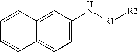
In one particular embodiment, the invention is directed to the detection of secreted aminoproteases. When detecting an aminoprotease, for example, the substrate may include a peptide or polypeptide that is catalytically hydrolyzed in the presence of the aminoprotease to yield an aromatic compound. The aromatic substrates may include, for instance, β-naphthylamide substrates having the following general formula:
Figure US07575887-20090818-C00001
wherein,
R1 may be a monoamino acid residue or a peptide residue, and
R2 may be a hydrogen atom or any other suitable group blocking the N-terminal end of the substrate. For example, R2 may be substituted or unsubstituted, and may be an alkyl group, an alkyoxy group, a hydroxyalkyl group, an alkylene group, a fatty acid group, and so forth.
The amino acid or peptide derivatized substrates are hydrolyzed by the protease. For instance, a β-naphthylamide substrate may be hydrolyzed by an aminoprotease to form a β-naphthylamine, which has the following structure:
Figure US07575887-20090818-C00002
The monoamino acid or peptide residue of the substrate may be selected as one that is susceptible to cleavage by an aminoprotease of interest. The reagents utilized in the disclosed devices may thus be selected to specifically target an protease of interest. For example, certain aminopeptidases, e.g., homologs of aminopeptidase I (ScLAP4) are upregulated upon the transition of C. albicans from the benign commensal form to the pathogenic hyphal form (see, e.g., Lorenz, et al., Eukaryotic Cell, 3, pp. 1076-1087 (2004). Accordingly, a substrate may be utilized including a residue susceptible to cleavage by one of these upregulated aminopeptidases, in order to more accurately diagnose the presence of hyphal C. albicans in a host.
Other suitable substrates for proteases include, for instance, proteins or glycoproteins, peptides, and so forth. For instance, some suitable substrates for peptidases and/or proteinases may include peptides, proteins, and/or glycoproteins such as casein (e.g., β-casein, azocasein, etc.), albumin (e.g., bovine serum albumin), hemoglobin, myoglobin, keratin, gelatin, insulin, proteoglycan, fibronectin, laminin, collagen, elastin, and so forth. Still other suitable substrates are described in U.S. Pat. Nos. 5,449,612 to Lepargneur, et al.; 4,874,695 to Pincus; 6,022,698 to Chen, et al.; and 5,571,684 to Lawrence, et al., which are incorporated herein in their entirety by reference thereto for all purposes.
If desired, the rate of the protease-catalyzed reaction may be selectively controlled using techniques known in the art, such as controlling the temperature, pH, substrate concentration, the presence of accelerators, the presence of inhibitors (competitive (binds to protease), uncompetitive (binds to protease-substrate complex), or noncompetitive (binds to protease and/or protease-substrate complex)), and so forth. These factors may be selectively controlled as desired to increase or decrease the reaction time. For example, an accelerator may be employed to enhance protease activity. Suitable accelerators for hydrolytic proteases may include, for instance, pyridine, imidazole and their derivatives, metal complexes, and alcohols. The pH may also be selectively controlled to enhance protease activity and to inhibit unwanted side reactions. For instance, many substrates are unstable in highly basic or highly acidic conditions due to their tendency to hydrolyze. Thus, the pH is typically maintained at a relatively neutral level, such as from about 6 to about 9, and in some embodiments, about 7. Some biologically compatible buffers that may be used to maintain the desired pH include borate buffers, phosphate-buffered saline (PBS), 2-(N-morpholino) ethane sulfonic acid (“MES”), tris-hydroxymethylaminomethane (“Tris”), citrate buffers, and so forth.
Regardless of the manner in which the reaction is conducted, through cleavage of the substrate a chromogenic product is released that is capable of inducing a color change in the presence of certain reagents. In one particular embodiment, the released chromogenic product may be an aromatic compound that is a nucleophile in that it contains a group that is electron rich (e.g., amine) and that may form bonds with electron deficient groups. For example, β-naphthylamides may be hydrolyzed by a protease to form β-naphthylamine. β-naphthylamine contains an electron-rich, aromatic ring system that is capable of undergoing electrophilic attack by a diazonium ion having the generic formula:
Figure US07575887-20090818-C00003
The diazonium ion may be zwitterionic in that the counterion of the diazonium moiety is covalently bound to the ring system. The ring system of the diazonium ion may be substituted or unsubstituted. The ion may be provided by a variety of suitable diazonium salts, such as diazonium chlorides, diazonium acid sulphates, diazonium alkyl sulphates, diazonium fluoborates, diazonium benzenesulphonates, diazonium acid 1,5-naphthalenedisulphonates, and so forth. Specific examples of diazonium salts are 1-diazo-2-naphthol-4-sulfonate; 1-diazophenyl-3-carbonate; 4-diazo-3-hydroxy-1-naphthylsulfonate (DNSA); 4-diazo-3-hydroxy-7-nitro-1-naphthylsulfonate (NDNSA); 4-diazo-3-hydroxy-1,7-naphthyldisulfonate; 2-methoxy-4-(N-morpholinyl) benzene diazonium chloride; 4-diazo-3-hydroxy-7-bromo-1-naphthylsulfonate; and 4-diazo-3-hydroxy-7-[1,oxopropyl]-1-naphthylsulfonate. One particularly desired diazonium salt is 5-chloro-2-methoxybenzenediazonium chloride, which has a yellow color and is classified under the name “Diazo Red RC” or “Fast Red RC.” More specifically, “Fast Red RC” has the following structure:
Figure US07575887-20090818-C00004
Other suitable diazonium salts are classified by the common names “Fast Red B” and “Fast Blue B.” Still other suitable diazonium salts may be described in U.S. Pat. Nos. 4,637,979 to Skjold, et al.; 4,806,423 to Hugh, et al.; and 4,814,271 to Hugl, et al., which are incorporated herein in their entirety by reference thereto for all purposes.
In some cases, it may be desirable to select the chromogenic product based on its ability to be readily immobilized onto a solid substrate. In this regard, certain macromolecular reagents (e.g., polymers, oligomers, dendrimers, particles, etc.) may be particularly useful in the present invention. Generally speaking, such macromolecular reagents contain at least two functionalities, i.e., a reactive moiety (e.g., diazonium ion) and a macromolecular moiety, which are covalently or noncovalently joined. The macromolecular moiety may include, for instance, a polymeric moiety, such as a linear or branched, homopolymer or copolymer. The polymeric moieties may be natural, synthetic, or combinations thereof. Examples of natural polymeric moieties include, for instance, peptides, proteins, DNA/RNA and polysaccharides (e.g., glucose-based polymers). Examples of synthetic polymeric moieties include, instance, polyacrylic acid and polyvinyl alcohols.
As indicated, the macromolecular moiety may also be a particle (sometimes referred to as a “bead” or “microbead”). Naturally occurring particles, such as nuclei, mycoplasma, plasmids, plastids, mammalian cells (e.g., erythrocyte ghosts), unicellular microorganisms (e.g., bacteria), polysaccharides (e.g., agarose), etc., may be used. Further, synthetic particles may also be utilized. For example, in one embodiment, latex microparticles that are labeled with a fluorescent or colored dye are utilized. Although any synthetic particle may be used in the present invention, the particles are typically formed from polystyrene, butadiene styrenes, styreneacrylic-vinyl terpolymer, polymethylmethacrylate, polyethylmethacrylate, styrene-maleic anhydride copolymer, polyvinyl acetate, polyvinylpyridine, polydivinylbenzene, polybutyleneterephthalate, acrylonitrile, vinylchloride-acrylates, and so forth, or an aldehyde, carboxyl, amino, hydroxyl, or hydrazide derivative thereof. When utilized, the shape of the particles may generally vary. In one particular embodiment, for instance, the particles are spherical in shape. However, it should be understood that other shapes are also contemplated by the present invention, such as plates, rods, discs, bars, tubes, irregular shapes, etc. In addition, the size of the particles may also vary. For instance, the average size (e.g., diameter) of the particles may range from about 0.1 nanometers to about 1,000 microns, in some embodiments, from about 0.1 nanometers to about 100 microns, and in some embodiments, from about 1 nanometer to about 10 microns.
The particle may generally be joined to a reactive moiety using any of a variety of well-known techniques. For instance, covalent attachment of a particle to a substrate may be accomplished using carboxylic, amino, aldehyde, bromoacetyl, iodoacetyl, thiol, epoxy or other reactive functional groups, as well as residual free radicals and radical cations, through which a coupling reaction may be accomplished. A surface functional group may also be incorporated as a functionalized co-monomer because the surface of the particle may contain a relatively high surface concentration of polar groups. In certain cases, the particle may be capable of direct covalent bonding to a substrate without the need for further modification. It should also be understood that, besides covalent bonding, other attachment techniques, such as physical adsorption, may also be utilized in the present invention.
As indicated above, the nucleophilic aromatic compounds released by the hydrolysis of the substrate are capable of undergoing electrophilic attack by a reagent (e.g., diazonium ion). This reaction is often referred to as “coupling” and results in the formation of another reagent having a different color. For example, diazonium ions may react with aromatic compounds to form an aromatic azo compound having the generic formula, R—N═N—R′, wherein “R” and “R′” are aryl groups. Without intending to be limited by theory, it is believed that this reaction induces either a shift of the absorption maxima towards the red end of the spectrum (“bathochromic shift”) or towards the blue end of the spectrum (“hypsochromic shift”). The type of absorption shift depends on the nature of the resulting azo molecule and whether it functions as an electron acceptor (oxidizing agent), in which a hypsochromic shift results, or whether it functions as an electron donor (reducing agent), in which a bathochromic shift results. Regardless, the absorption shift provides a color difference that is detectable, either visually or through instrumentation, to indicate the presence of protease within the test sample. For example, prior to contact with an infected test sample, the diazonium ion may be colorless or it may possess a certain color. However, after contacting the test sample and reacting with an aromatic compound released by hydrolysis of the substrate, an aromatic azo compound will form that exhibits a color that is different than the initial color of the diazonium ion. Exemplary aromatic azo compounds that may be formed include dimethyldiazene, diphenydiazene, 1-naphthyl-2-naphthyl diazene, 3-chlorophenyl-4-chlorophenyl diazene, methylvinyl diazene, and 2-naphthylphenyl diazene. In one particular embodiment, for instance, “Fast Red RC” (yellow), a diazonium ion, may react with β-naphylamine to form an aromatic azo compound that is red and has the following general structure (may be substituted or unsubstituted):
Figure US07575887-20090818-C00005
It should be understood that the chromogenic products released due to the hydrolysis of the substrate by the protease are not limited to aromatics. In particular, the released chromogenic products may be any material that may be discernable following reaction with a reagent. For example, in one embodiment, the chromogenic product may include one or more amino acids. One embodiment of a chromogenic system that may be used when the chromogenic product includes an amino acid is described in U.S. Pat. No. 5,571,684 to Lawrence, et al., previously incorporated by reference. Such a chromogenic system may comprise an amino acid oxidase, a chromogen selected from the group consisting of guaiac, 2,2′-azino-bis(3-ethyl-benzthiazoline-6-sulfonic acid), tetramethylbenzidine, phenol, 4-aminoantipyrine and 4,5-dihydroxynaphthalene, a redox catalyst selected from the group consisting of peroxidases, iron protoporphyrin and metal ions, and oxygen. Schematically, this system may be illustrated as follows:
Figure US07575887-20090818-C00006
Of course, the most appropriate reagent for any given released chromogenic product will depend at least upon the substrate specificity of the protease, the actual chromogenic product released, and the reaction conditions needed for a given test. The selection in any given case will be readily apparent to those skilled in the art.
As a result of the color change following cleavage of the substrate by the protease, the presence of the protease in a test sample may be readily detected. The extent of the color change may be selectively controlled in accordance with the present invention to limit “false positives.” More specifically, a small and perhaps normal amount of protease within a test sample may hydrolyze a substrate to release a nucleophilic aromatic compound.
In some cases, however, the released aromatic compound may undergo an oxidation reaction if left in air or other oxidizing environment for too great a period of time. The resulting color of the oxidized compound may indicate a “false positive” or at the very least, adversely affect the ability to semi-quantitatively or quantitatively determine the presence of the protease. In other cases, the reaction is advantageously stopped following a desired reaction time so as to avoid generation of a false positive. Thus, the present inventor has discovered a technique for reducing the problem of such “false positives.” Beneficially, the technique requires no additional reagents, for instance to stop the reaction. Instead of simply measuring the results after a certain period of time, the desired reaction time may be achieved by selectively controlling the medium in which the reaction occurs. That is, the reaction medium is chromatographic in nature such that the protease and substrate are allowed to flow in a consistent and controllable manner. While flowing through the medium, the protease and substrate react to release chromogenic product that subsequently couples with a reagent such as a diazonium ion to form a detectable reagent, e.g., an aromatic azo compound. The aromatic azo compound is immobilized within a discrete detection region for analysis. Due to the nature of the controlled fluid flow, any unreacted substrate travels to the end of the reaction medium so that it is unable to adversely interfere with observance of the aromatic azo compound in the detection region. At the same time, the aromatic compounds that are released from the end of the reaction medium will not react with the reagents in the detection zone to generate potential false positives.
Various embodiments for accomplishing the detection of the protease using fluid flow control techniques will now be described in more detail. Referring to FIG. 1, for instance, one embodiment of a lateral flow device 20 that may be formed according to the present invention will now be described in more detail. As shown, the device 20 contains a chromatographic medium 23 optionally supported by a rigid support material 21. In general, the chromatographic medium 23 may be made from any of a variety of materials through which the test sample is capable of passing. For example, the chromatographic medium 23 may be a porous membrane formed from synthetic or naturally occurring materials, such as polysaccharides (e.g., cellulose materials such as paper and cellulose derivatives, such as cellulose acetate and nitrocellulose); polyether sulfone; polyethylene; nylon; polyvinylidene fluoride (PVDF); polyester; polypropylene; silica; inorganic materials, such as deactivated alumina, diatomaceous earth, MgSO4, or other inorganic finely divided material uniformly dispersed in a porous polymer matrix, with polymers such as vinyl chloride, vinyl chloride-propylene copolymer, and vinyl chloride-vinyl acetate copolymer; cloth, both naturally occurring (e.g., cotton) and synthetic (e.g., nylon or rayon); porous gels, such as silica gel, agarose, dextran, and gelatin; polymeric films, such as polyacrylamide; and so forth. In one particular embodiment, the chromatographic medium 23 is formed from nitrocellulose and/or polyether sulfone materials. It should be understood that the term “nitrocellulose” refers to nitric acid esters of cellulose, which may be nitrocellulose alone, or a mixed ester of nitric acid and other acids, such as aliphatic carboxylic acids having from 1 to 7 carbon atoms.
The size and shape of the chromatographic medium 23 may generally vary as is readily recognized by those skilled in the art. For instance, a porous membrane strip may have a length of from about 10 to about 100 millimeters, in some embodiments from about 20 to about 80 millimeters, and in some embodiments, from about 40 to about 60 millimeters. The width of the membrane strip may also range from about 0.5 to about 20 millimeters, in some embodiments from about 1 to about 15 millimeters, and in some embodiments, from about 2 to about 10 millimeters. Likewise, the thickness of the membrane strip is generally small enough to allow transmission-based detection. For example, the membrane strip may have a thickness less than about 500 micrometers, in some embodiments less than about 250 micrometers, and in some embodiments, less than about 150 micrometers.
As stated above, the support 21 carries the chromatographic medium 23. For example, the support 21 may be positioned directly adjacent to the chromatographic medium 23 as shown in FIG. 1, or one or more intervening layers may be positioned between the chromatographic medium 23 and the support 21. Regardless, the support 21 may generally be formed from any material able to carry the chromatographic medium 23. The support 21 may be formed from a material that is transmissive to light, such as transparent or optically diffuse (e.g., transluscent) materials. Also, it is generally desired that the support 21 is liquid-impermeable so that fluid flowing through the medium 23 does not leak through the support 21. Examples of suitable materials for the support include, but are not limited to, glass; polymeric materials, such as polystyrene, polypropylene, polyester (e.g., Mylar® film), polybutadiene, polyvinylchloride, polyamide, polycarbonate, epoxides, methacrylates, and polymelamine; and so forth. To provide a sufficient structural backing for the chromatographic medium 23, the support 21 is generally selected to have a certain minimum thickness. Likewise, the thickness of the support 21 is typically not so large as to adversely affect its optical properties. Thus, for example, the support 21 may have a thickness that ranges from about 100 to about 5,000 micrometers, in some embodiments from about 150 to about 2,000 micrometers, and in some embodiments, from about 250 to about 1,000 micrometers. For instance, one suitable membrane strip having a thickness of about 125 micrometers may be obtained from Millipore Corp. of Bedford, Mass. under the name “SHF180UB25.”
As is well known the art, the chromatographic medium 23 may be cast onto the support 21, wherein the resulting laminate may be die-cut to the desired size and shape. Alternatively, the chromatographic medium 23 may simply be laminated to the support 21 with, for example, an adhesive. In some embodiments, a nitrocellulose or nylon porous membrane is adhered to a Mylar® film. An adhesive is used to bind the porous membrane to the Mylar® film, such as a pressure-sensitive adhesive. Laminate structures of this type are believed to be commercially available from Millipore Corp. of Bedford, Mass. Still other examples of suitable laminate device structures are described in U.S. Pat. No. 5,075,077 to Durley, III, et al., which is incorporated herein in its entirety by reference thereto for all purposes.
The device 20 also contains an absorbent material 28 that is positioned adjacent to the medium 23. The absorbent material 28 assists in promoting capillary action and fluid flow through the medium 23. In addition, the absorbent material 28 receives fluid that has migrated through the entire chromatographic medium 23 and thus draws any unreacted components away from the detection region to help reduce the likelihood of “false positives.” Some suitable absorbent materials that may be used in the present invention include, but are not limited to, nitrocellulose, cellulosic materials, porous polyethylene pads, glass fiber filter paper, and so forth. The absorbent material may be wet or dry prior to being incorporated into the device. Pre-wetting may facilitate capillary flow for some fluids, but is not typically required. Also, as is well known in the art, the absorbent material may be treated with a surfactant to assist the wicking process.
To initiate the detection of a protease within the test sample, a user may directly apply the test sample to a portion of the chromatographic medium 23 through which it may then travel in the direction illustrated by arrow “L” in FIG. 1. Alternatively, the test sample may first be applied to a sample application zone 24 that is in fluid communication with the chromatographic medium 23. The sample application zone 24 may be formed on the medium 23. Alternatively, as shown in FIG. 1, the sample application zone 24 may be formed by a separate material, such as a pad. Some suitable materials that may be used to form such sample pads include, but are not limited to, nitrocellulose, cellulose, porous polyethylene pads, and glass fiber filter paper. If desired, the sample application zone 24 may also contain one or more pretreatment reagents, either diffusively or non-diffusively attached thereto. In the illustrated embodiment, the test sample travels from the sample application zone 24 to a reagent zone 22 that is in communication with the sample application zone 24. As described above, the reagent zone 22 may be formed on the medium 23. Alternatively, as shown in FIG. 1, the reagent zone 22 is formed from a separate material or pad. Such a reagent pad may be formed from any material through which the test sample is capable of passing, such as glass fibers.
To facilitate detection of the targeted proteases in the manner described above, a substrate is employed. In some embodiments, the substrate may be mixed with the test sample prior to application to the device 20. Alternatively, the substrate may be diffusively immobilized on the device 20 prior to application of the test sample. Such pre-application provides a variety of benefits, including the elimination of the need for a subsequent user to handle and mix the reagents with the test sample or a diluent. This is particularly useful in point-of-care applications when the user is not generally a trained lab technician or medical professional. The substrate may be disposed downstream from the sample application zone 24. In this manner, the test sample is capable of mixing with the substrate upon application. Alternatively, the substrate may be positioned upstream from the sample application zone 24. For instance, a diluent may be employed to induce mixing between the substrate and test sample.
If desired, the pH may be maintained at a relatively neutral level to facilitate the desired protease-catalyzed reaction, such as described above. To accomplish the desired pH level, a buffer may be mixed with the substrate prior to application to the device 20, mixed with the test sample, or both. Alternatively, the buffer may be separately applied to the lateral flow device 20 so that it is capable of mixing with the reagents upon application to the test sample.
Referring again to FIG. 1, the lateral flow device 20 includes a detection zone 31 within which is immobilized a second reagent, such as a diazonium ion described above. The diazonium ion may be applied directly to the medium 23 or first formed into a solution prior to application. Various solvents may be utilized to form the solution, such as, but not limited to, acetonitrile, dimethylsulfoxide (DMSO), ethyl alcohol, dimethylformamide (DMF), and other polar organic solvents. For instance, the amount of a diazonium salt in the solution may range from about 0.001 to about 100 milligrams per milliliter of solvent, and in some embodiments, from about 0.1 to about 10 milligrams per milliliter of solvent. In one particular embodiment, the detection zone 31 is defined by the chromatographic medium 23 and formed by coating a solution thereon using well-known techniques and then dried. The diazonium ion concentration may be selectively controlled to provide the desired level of detection sensitivity.
Typically, it is desired that the diazonium ion be applied in a manner so that it does not substantially diffuse through the matrix of the chromatographic medium 23 (i.e., non-diffusively immobilized). This enables a user to readily detect the change in color that occurs upon reaction of the diazonium ion with a nucleophilic aromatic compound. The diazonium ion may form an ionic and/or covalent bond with functional groups present on the surface of the chromatographic medium 23 so that it remains immobilized thereon. For instance, particles, such as described herein, may facilitate the immobilization of the diazonium ion at the detection zone 31. Namely, the diazonium ion may be coated onto particles, which are then immobilized on the chromatographic medium 23 of the device 20. In this manner, the diazonium ion is able to readily contact nucleophilic aromatic compounds flowing through the medium 23.
In one embodiment, the lateral flow device may include a plurality of substrates, each of which may be susceptible to cleavage by a different protease. According to this embodiment, the lateral flow device may be designed to examine a single test sample for more than one protease so as to identify a pathogen in the test sample with more particularity. For example, referring to FIG. 2, the lateral flow device 30 employs a first substrate susceptible to cleavage by a first protease, e.g., an aminopeptidase. The lateral flow device also includes a first detection zone 31, as described above, and within which is immobilized a first reagent that may interact with the cleaved chromogenic product released by the aminopeptidase.
In addition, the lateral flow device of FIG. 2 may employ a second substrate susceptible to cleavage by a second protease, e.g., a second aminopeptidase, an aminoproteinase, etc. The second substrate may be mixed with the test sample prior to application to the device or the second substrate may be diffusively immobilized on the device either upstream or down stream of the first detection zone 31 prior to application of the test sample. The lateral flow device may also include a second detection zone 36. Within the second detection zone 36 may be immobilized a second reagent that may interact with the chromogenic product released by the second protease of interest. Moreover, the first and second reagents may be selected to be particular to the first and second chromogenic cleavage products, respectively, so as to prevent any cross reaction. In other words, the first reagent, immobilized in detection zone 31 will not interact with the second chromogenic product to produce a detectable color change, and the second reagent, immobilized in detection zone 36, will not interact with the first chromogenic product to produce a detectable color change in detection zone 31. For example, the first detection system may be an aromatic azo system and the second detection system may be an amino acid oxidase detection system. Accordingly, a positive result at first detection zone 31 may indicate the presence in the test sample of the first protease, and a positive result at second detection zone 36 may indicate the presence of the second protease. The combination of results may be utilized to identify the pathogen in the test sample with more particularity.
One benefit of the lateral flow device of the present invention is the ability to readily incorporate one or more additional reagent zones to facilitate the above-described protease-catalyzed reaction. For example, referring again to FIG. 1, one such zone is a quenching zone 35. The quenching zone 35 is configured to remove compounds from the test sample that would otherwise interfere with the accuracy of the detection system. For example, contaminants within the test sample (e.g., phenolics, bilirubin, urobilinogen, etc.) may react with the diazonium ion within the detection zone 31 and form an aromatic azo compound, thereby producing a “false negative” result. Thus, the quenching zone 35 may contain a quenching agent, such as a diazonium ion, that is capable of reacting with the reaction contaminants. The quenching agent may be the same or different than the detection agent used within the detection zone 31. Typically, the quenching agent is non-diffusively immobilized within the quenching zone 35 in the manner described above so that it does not flow through the medium 23 and interfere with testing. The location of the quenching zone 35 may vary, but is typically positioned upstream from the detection zone 31 and the location at which the substrate is applied to avoid interference with protease detection. For example, in the illustrated embodiment, the quenching zone 35 is positioned between the sample application zone 24 and the reagent zone 22. Alternatively, the quenching zone 35 may be positioned upstream from the sample application zone 24.
Another zone that may be employed in the lateral flow device 20 for improving detection accuracy is a control zone 32. The control zone 32 gives a signal to the user that the test is performing properly. More specifically, control reagents may be employed that flow through the chromatographic medium 23 upon contact with a sufficient volume of the test sample. These control reagents may then be observed, either visually or with an instrument, within the control zone 32. The control reagents generally contain a detectable substance, such as luminescent compounds (e.g., fluorescent, phosphorescent, etc.); radioactive compounds; visual compounds (e.g., colored dye or metallic substance, such as gold); liposomes or other vesicles containing signal-producing substances; enzymes and/or substrates, and so forth. Other suitable detectable substances may be described in U.S. Pat. Nos. 5,670,381 to Jou, et al. and 5,252,459 to Tarcha, et al., which are incorporated herein in their entirety by reference thereto for all purposes. If desired, the detectable substances may be disposed on particles (sometimes referred to as “beads” or “microbeads”), such as described above. Among other things, the particles enhance the ability of the detectable substance to travel through a chromatographic medium. Commercially available examples of suitable particles include fluorescent carboxylated microspheres sold by Molecular Probes, Inc. under the trade names “FluoSphere” (Red 580/605) and “TransfluoSphere” (543/620), as well as “Texas Red” and 5- and 6-carboxytetramethylrhodamine, which are also sold by Molecular Probes, Inc. In addition, commercially available examples of suitable colored, latex microparticles include carboxylated latex beads sold by Bang's Laboratory, Inc.
The location of the control zone 32 may vary based on the nature of the test being performed. In the illustrated embodiment, for example, the control zone 32 is defined by the chromatographic medium 23 and positioned downstream from the detection zone 31. In such embodiments, the control zone 32 may contain a material that is non-diffusively immobilized in the manner described above and forms a chemical and/or physical bond with the control reagents. When the control reagents contain latex particles, for instance, the control zone 32 may include a polyelectrolyte that binds to the particles. Various polyelectrolytic binding systems are described, for instance, in U.S. Patent App. Publication No. 2003/0124739 to Song, et al., which is incorporated herein in it entirety by reference thereto for all purposes. In alternative embodiments, however, the control zone 32 may simply be defined by a region of the absorbent material 28 to which the control reagents flow after traversing through the chromatographic medium 23.
Regardless of the particular control technique selected, the application of a sufficient volume of the test sample to the device 20 will cause a detectable signal to form within the control zone 32, whether or not the protease is present. Among the benefits provided by such a control zone is that the user is informed that a sufficient volume of test sample has been added without requiring careful measurement or calculation. This provides the ability to use the lateral flow device 20 without the need for externally controlling the reaction time, test sample volume, etc.
In addition to the zones specified above, the lateral flow device 20 may also include other optional zones. For example, in one embodiment, the lateral flow device 20 may include an accelerator zone (not shown) in which is contained an accelerator for the protease-catalyzed substrate reaction. Typically, the accelerator is diffusively immobilized within the accelerator zone in the manner described above so that it may flow through the medium 23 upon contact with the test sample. The location of the accelerator zone may generally vary, so long as it positioned upstream from the detection zone 31. For example, in some embodiments, the accelerator zone may be positioned at a location (e.g., sample application zone 24) that is upstream from the application of the substrate (e.g., reagent zone 24). Due to the separation provided between the substrate and accelerator, the likelihood of any premature reaction therebetween is thus reduced. It should be understood, however, that the accelerator may nevertheless be combined with the substrate in some applications.
The detection zone 31, quenching zone 35, control zone 32, accelerator zone, and any other zone employed in the lateral flow device 20 may generally provide any number of distinct detection regions so that a user may better determine the concentration of the protease within the test sample. Each region may contain the same or different materials. For example, the zones may include two or more distinct regions (e.g., lines, dots, etc.). The regions may be disposed in the form of lines in a direction that is substantially perpendicular to the flow of the test sample through the device 20. Likewise, in some embodiments, the regions may be disposed in the form of lines in a direction that is substantially parallel to the flow of the test sample through the device 20.
One particular embodiment of a method for detecting the presence of a protease within a test sample using the device 20 of FIG. 1 will now be described in more detail. Initially, a test sample containing an aminopeptidase is applied to the sample application zone 24 and travels in the direction “L” to the reagent zone 22. At the reagent zone 22, the aminopeptidase is able to mix with and begin to initiate the catalytic reaction. As the mixture flows through the device 20, the aminopeptidase hydrolyzes the peptide bonds of the substrate and releases chromogenic products, such as nucleophilic aromatic compounds. The aromatic compounds then flow to the detection zone 31 where they react with a diazonium ion to form a colored azo compound. After the reaction, the detection zone 31 changes color. Thus, the color or color intensity of the detection zone 31 may be determined, either visually or with instrumentation. If desired, the intensity of the color at the detection zone 31 may be measured to quantitatively or semi-quantitatively determine the level of the aminopeptidase present in the test sample. The intensity of the color at the detection zone 31 is typically directly proportional to protease concentration. The intensity of the detection signal “Is” produced at the detection zone 31 may also be compared to a predetermined detection curve developed for a plurality of known protease concentrations. To determine the quantity of protease in an unknown test sample, the signal may simply be converted to protease concentration according to the detection curve. Regardless, the protease, e.g., the aminopeptidase, and any unreacted substrate and/or aromatic compounds then travel past the detection zone 31 until they reach the absorbent material 28. In some cases, the aromatic compounds will oxidize over a period of time in air to form colored compounds. However, because such colored compounds are not located at the detection region 31, they generally do not interfere with the detection accuracy.
The present invention provides a relatively simple, compact and cost-efficient device for accurately detecting the presence of protease within a test sample (e.g., vaginal fluid). The test result may be visible so that it is readily observed by the person performing the test in a prompt manner and under test conditions conducive to highly reliable and consistent test results. The test is also less time-dependent. Accordingly, the present invention may provide a convenient, accurate determination of the presence of a pathogenic agent in the source of the test sample.
The present invention may be better understood with reference to the following example.
EXAMPLE
The ability to detect aminopeptidase in accordance with the present invention was demonstrated. Initially, an HF12002 nitrocellulose membrane laminated to a support card (Millipore, Inc.) was provided as the chromatographic medium. To form a detection zone, Diazo Red DC (DR) was initially striped by hand onto the membrane. The Diazo Red DC was in the form of a solution containing water and approximately 10 wt. % dimethylformamide (“DMF”). The membrane was then dried at 37° C. for two hours.
A cellulose pad was laminated to the one end of the membrane card to provide an absorbent or wicking pad.
A reagent pad was fabricated from a glass fiber pad having a length of 15 centimeters (Millipore Inc.). More specifically, the pad was soaked with 15 milliliters of a mixture containing (i) 1 wt. % Tween 20 (available from Sigma-Aldrich Chemical Co.) in 20 millimolar Tris buffer (pH=7.4); (ii) 0.5 milliliters of a solution containing 20 wt. % sucrose; and (iii) 0.2 milliliters of L-leucine β-naphthylamide (0.1 molar in DMF). The glass fiber pad was then dried at 37° C. for two hours and laminated to the other end of the membrane card. A cellulose sample application pad was then laminated to the glass fiber pad to provide a sample application zone. The fully assembled card was cut into 4-millimeter wide strip devices.
To test the device, a control sample (in Tris buffer, pH=7.4, 20 millimolar) and a positive aminopeptidase sample (100 micrograms per milliliter, in Tris buffer, pH=7.4, 20 millimolar) were prepared. The aminopeptidase was derived from Streptomyces griseus and obtained from Sigma. 150 microliters of each sample were then applied to the sample application zone of two separate devices. After approximately 15 minutes, each sample was visually observed. For the control sample, the detection zone remained yellow; however, for the positive aminopeptidase sample, the detection zone was red.
While the invention has been described in detail with respect to the specific embodiments thereof, it will be appreciated that those skilled in the art, upon attaining an understanding of the foregoing, may readily conceive of alterations to, variations of, and equivalents to these embodiments. Accordingly, the scope of the present invention should be assessed as that of the appended claims and any equivalents thereto.

Claims (12)

1. A method for detecting the presence of a protease within a test sample, the method comprising:
i) providing a lateral flow assay device that comprises a fluidic medium, the lateral flow assay device comprising a sample application zone, the fluidic medium defining a detection zone within which is immobilized a first reagent, the detection zone being in fluid communication with a substrate, wherein the substrate is capable of cleavage by the protease and a chromogenic product is released upon the cleavage of the substrate by the protease;
ii) contacting the sample application zone with the test sample, wherein the test sample travels through the fluidic medium from the sample application zone to the detection zone following contacting of the sample application zone with the test sample; and
iii) generating a color within the detection zone that corresponds to the presence or absence of the protease in the test sample.
2. The method of claim 1, further comprising obtaining the test sample from a host, wherein the generated color indicates the presence of a pathogen in the host, wherein the pathogen expresses the protease.
3. The method of claim 2, wherein the pathogen is a yeast, bacteria, or parasite.
4. The method of claim 2, wherein the pathogen is pathogenic Candida albicans.
5. The method of claim 2, wherein the test sample is obtained from saliva.
6. The method of claim 2, wherein the test sample is obtained from vaginal fluid.
7. The method of claim 1, further comprising measuring the intensity of the generated color, wherein the amount of the protease in the test sample is proportional to the intensity of the generated color.
8. The method of claim 1, wherein the protease is a peptidase.
9. The method of claim 8, wherein the peptidase is an aminopeptidase.
10. The method of claim 8, wherein the peptidase is proline iminopeptidase.
11. The method of claim 1, wherein the fluidic medium further defines a second detection zone, the second detection zone being in fluid communication with a second substrate, wherein the second substrate is capable of cleavage by a second protease and a second chromogenic product is released upon the cleavage of the second substrate by the second protease, the method further comprising generating a second color within the second detection zone that corresponds to the presence or absence of the second protease in the test sample.
12. The method of claim 1, wherein the expression of the protease is upregulated by a hyphal form of Candida albicans.
US11/364,810 2005-08-31 2006-02-28 Detection of proteases secreted from pathogenic microorganisms Expired - Fee Related US7575887B2 (en)

Priority Applications (4)

Application Number Priority Date Filing Date Title
US11/364,810 US7575887B2 (en) 2005-08-31 2006-02-28 Detection of proteases secreted from pathogenic microorganisms
PCT/US2006/030107 WO2007027353A2 (en) 2005-08-31 2006-08-01 Detection of proteases secreted from pathogenic microorganisms
EP06789202A EP1920253A2 (en) 2005-08-31 2006-08-01 Detection of proteases secreted from pathogenic microorganisms
US12/542,278 US8609401B2 (en) 2005-08-31 2009-08-17 Detection of proteases secreted from a pathogenic microorganisms

Applications Claiming Priority (2)

Application Number Priority Date Filing Date Title
US11/217,097 US7504235B2 (en) 2005-08-31 2005-08-31 Enzyme detection technique
US11/364,810 US7575887B2 (en) 2005-08-31 2006-02-28 Detection of proteases secreted from pathogenic microorganisms

Related Parent Applications (1)

Application Number Title Priority Date Filing Date
US11/217,097 Continuation-In-Part US7504235B2 (en) 2005-08-31 2005-08-31 Enzyme detection technique

Related Child Applications (1)

Application Number Title Priority Date Filing Date
US12/542,278 Division US8609401B2 (en) 2005-08-31 2009-08-17 Detection of proteases secreted from a pathogenic microorganisms

Publications (2)

Publication Number Publication Date
US20070048816A1 US20070048816A1 (en) 2007-03-01
US7575887B2 true US7575887B2 (en) 2009-08-18

Family

ID=36850080

Family Applications (4)

Application Number Title Priority Date Filing Date
US11/217,097 Expired - Fee Related US7504235B2 (en) 2005-08-31 2005-08-31 Enzyme detection technique
US11/364,810 Expired - Fee Related US7575887B2 (en) 2005-08-31 2006-02-28 Detection of proteases secreted from pathogenic microorganisms
US12/405,499 Abandoned US20090181416A1 (en) 2005-08-31 2009-03-17 Enzyme Detection Technique
US12/542,278 Expired - Fee Related US8609401B2 (en) 2005-08-31 2009-08-17 Detection of proteases secreted from a pathogenic microorganisms

Family Applications Before (1)

Application Number Title Priority Date Filing Date
US11/217,097 Expired - Fee Related US7504235B2 (en) 2005-08-31 2005-08-31 Enzyme detection technique

Family Applications After (2)

Application Number Title Priority Date Filing Date
US12/405,499 Abandoned US20090181416A1 (en) 2005-08-31 2009-03-17 Enzyme Detection Technique
US12/542,278 Expired - Fee Related US8609401B2 (en) 2005-08-31 2009-08-17 Detection of proteases secreted from a pathogenic microorganisms

Country Status (3)

Country Link
US (4) US7504235B2 (en)
EP (1) EP1920067B1 (en)
WO (1) WO2007027234A2 (en)

Cited By (3)

* Cited by examiner, † Cited by third party
Publication number Priority date Publication date Assignee Title
US20070238102A1 (en) * 2006-04-06 2007-10-11 Kimberly-Clark Worldwide, Inc. Enzymatic detection techniques
US20090181416A1 (en) * 2005-08-31 2009-07-16 Kimberly-Clark Worldwide, Inc. Enzyme Detection Technique
US10598656B2 (en) 2011-09-16 2020-03-24 Credo Biomedical Pte Ltd. Method of selecting analyte to samples using a lateral flow device

Families Citing this family (34)

* Cited by examiner, † Cited by third party
Publication number Priority date Publication date Assignee Title
US7796266B2 (en) 2004-04-30 2010-09-14 Kimberly-Clark Worldwide, Inc. Optical detection system using electromagnetic radiation to detect presence or quantity of analyte
US20070121113A1 (en) * 2004-12-22 2007-05-31 Cohen David S Transmission-based optical detection systems
US8241263B2 (en) 2005-08-26 2012-08-14 Medline Industries, Inc. Absorbent article
WO2007027353A2 (en) * 2005-08-31 2007-03-08 Kimberly-Clark Worldwide, Inc. Detection of proteases secreted from pathogenic microorganisms
US8789756B2 (en) * 2006-02-25 2014-07-29 Roche Diagnostics Operations, Inc. Test element coding apparatuses, systems and methods
GB2437311A (en) * 2006-04-07 2007-10-24 Mologic Ltd A protease detection product
US8044257B2 (en) * 2006-10-30 2011-10-25 Kimberly-Clark Worldwide, Inc. Absorbent article containing lateral flow assay device
US7897360B2 (en) 2006-12-15 2011-03-01 Kimberly-Clark Worldwide, Inc. Enzyme detection techniques
US7935538B2 (en) * 2006-12-15 2011-05-03 Kimberly-Clark Worldwide, Inc. Indicator immobilization on assay devices
WO2009121079A2 (en) * 2008-03-26 2009-10-01 Kapp John Test device and method for testing for vaginal infections
US20100290948A1 (en) * 2009-05-15 2010-11-18 Xuedong Song Absorbent articles capable of indicating the presence of urinary tract infections
MX344365B (en) 2010-10-19 2016-12-14 Medline Ind Inc * Absorbent articles and methods of manufacturing the same.
BR112014001544B1 (en) 2011-07-22 2020-10-06 Access Bio, Inc TAPE WITH INDIVIDUAL PILLOW FOR AN IMPROVED SIDE FLOW TEST AND CHROMATOGRAPHIC TEST
USD716938S1 (en) 2011-10-19 2014-11-04 Medline Industries, Inc. Absorbent core
US9486368B2 (en) 2013-12-05 2016-11-08 Medline Industries, Inc. Disposable hygienic article with means for diagnostic testing
US10226388B2 (en) 2014-04-21 2019-03-12 Medline Industries, Inc. Stretch breathable protective absorbent article using tri-laminate
US9622922B2 (en) 2014-04-21 2017-04-18 Medline Industries, Inc. Stretch breathable protective absorbent article using bilaminate
TWI526129B (en) 2014-11-05 2016-03-11 Elite Material Co Ltd Multilayer printed circuit boards with dimensional stability
US9885663B2 (en) * 2015-10-25 2018-02-06 Adriel Sumathipala Portable, rapid, and inexpensive diagnostic tests for cardiac disease risk
USD825075S1 (en) 2016-02-23 2018-08-07 Flora Bioscience, Inc. Test strip holding device
US11104933B1 (en) 2016-03-22 2021-08-31 Cleu Diagnostics, Llc Compositions and methods for determining the presence of active leukocyte cells using an electrochemical assay
USD827857S1 (en) * 2016-12-29 2018-09-04 Euroimmun Medizinische Labordiagnostika Ag Pathology tissue slide
USD821606S1 (en) * 2016-12-29 2018-06-26 Euroimmun Medizinische Labordiagnostika Ag Pathology tissue slide
WO2019071240A1 (en) 2017-10-06 2019-04-11 The Research Foundation For The State University For The State Of New York Selective optical aqueous and non-aqueous detection of free sulfites
CN107884575A (en) * 2017-11-01 2018-04-06 上海凯创生物技术有限公司 Amino terminal-pro brain natriuretic peptide detection reagent card, kit and application thereof
TWI666439B (en) * 2018-09-04 2019-07-21 東吳大學 Detection device for detecting leukocyte esterase
US11828210B2 (en) 2020-08-20 2023-11-28 Denso International America, Inc. Diagnostic systems and methods of vehicles using olfaction
US11813926B2 (en) 2020-08-20 2023-11-14 Denso International America, Inc. Binding agent and olfaction sensor
US11932080B2 (en) 2020-08-20 2024-03-19 Denso International America, Inc. Diagnostic and recirculation control systems and methods
US11760169B2 (en) 2020-08-20 2023-09-19 Denso International America, Inc. Particulate control systems and methods for olfaction sensors
US11881093B2 (en) 2020-08-20 2024-01-23 Denso International America, Inc. Systems and methods for identifying smoking in vehicles
US11760170B2 (en) 2020-08-20 2023-09-19 Denso International America, Inc. Olfaction sensor preservation systems and methods
US11636870B2 (en) 2020-08-20 2023-04-25 Denso International America, Inc. Smoking cessation systems and methods
EP4275043A1 (en) 2021-01-08 2023-11-15 Alpspitz Bioscience GmbH Use of biomarkers for predicting the clinical and/or treatment outcome of a human subject

Citations (93)

* Cited by examiner, † Cited by third party
Publication number Priority date Publication date Assignee Title
US3700623A (en) 1970-04-22 1972-10-24 Hercules Inc Reaction products of epihalohydrin and polymers of diallylamine and their use in paper
US3772076A (en) 1970-01-26 1973-11-13 Hercules Inc Reaction products of epihalohydrin and polymers of diallylamine and their use in paper
US4140580A (en) 1976-05-03 1979-02-20 Mcdonnell Douglas Corporation Broth for indicating presence of candida yeast and other yeasts and fungi
US4275149A (en) 1978-11-24 1981-06-23 Syva Company Macromolecular environment control in specific receptor assays
US4537657A (en) 1983-08-26 1985-08-27 Hercules Incorporated Wet strength resins
US4614723A (en) 1983-06-03 1986-09-30 Hoffmann-La Roche Inc. Porphyrin derivatives as fluorescent markers for immunoassays
US4637979A (en) 1984-04-06 1987-01-20 Miles Laboratories, Inc. Composition and test device for determining the presence of leukocytes containing a zwitterion coupling agent
US4748116A (en) 1980-08-25 1988-05-31 Kabivitrum Ab Peptide substrates for determination of protease activity
US4806423A (en) 1984-04-06 1989-02-21 Miles Inc. Agents and process for the detection of esterolytic and/or proteolytic enzymes
US4814271A (en) 1984-04-06 1989-03-21 Miles Inc. Method for detecting esterolytic and proteolytic enzymes
US4859581A (en) 1986-03-10 1989-08-22 Board Of Regents, The University Of Texas System Endoglycosidase assay
US4874695A (en) 1983-03-08 1989-10-17 American Home Products Corp. Rapid indentification of yeast and other fungal microorganisms by enzyme detection
US5075077A (en) 1988-08-02 1991-12-24 Abbott Laboratories Test card for performing assays
US5124254A (en) 1988-02-08 1992-06-23 University College Cardiff Consultants Limited Detection of diamines in biological fluids
US5252459A (en) 1988-09-23 1993-10-12 Abbott Laboratories Indicator reagents, diagnostic assays and test kits employing organic polymer latex particles
US5292652A (en) 1990-10-05 1994-03-08 Athena Neurosciences, Inc. Amyloidin protease and uses thereof
US5328831A (en) 1990-05-25 1994-07-12 Becton, Dickinson And Company Substrate composition for solid phase urease immunoassay
US5449612A (en) 1990-03-23 1995-09-12 Serbio Process for identifying candida by means of chromogenic substances
US5464739A (en) 1994-08-22 1995-11-07 Bayer Corporation Composition method for determining the presence of leukocyte cells, esterase or protease in a test sample
US5464741A (en) 1993-10-08 1995-11-07 Henwell, Inc. Palladium (II) octaethylporphine alpha-isothiocyanate as a phosphorescent label for immunoassays
US5518883A (en) 1992-07-02 1996-05-21 Soini; Erkki J. Biospecific multiparameter assay method
US5571684A (en) 1994-11-07 1996-11-05 Litmus Concepts, Inc. Assay for proline iminopeptidase and other hydrolytic activities
US5573909A (en) 1992-05-13 1996-11-12 Molecular Probes, Inc. Fluorescent labeling using microparticles with controllable stokes shift
US5585279A (en) 1986-01-23 1996-12-17 Davidson; Robert S. Time-resolved luminescence binding assays using a fluorescent transition metal label other than ruthenium
US5585273A (en) 1993-04-14 1996-12-17 Litmus Concepts, Inc. Test device for assays for hydrolytic enzyme activity
US5591581A (en) 1986-04-30 1997-01-07 Igen, Inc. Electrochemiluminescent rhenium moieties and methods for their use
WO1997014028A2 (en) 1995-10-11 1997-04-17 Luminex Corporation Multiplexed analysis of clinical specimens apparatus and method
US5637509A (en) 1987-09-24 1997-06-10 Wallac Oy Modulated homogeneous fluorescence biospecific affinity assay employing fluorescing lanthanide chelates as covalent labels
US5670381A (en) 1988-01-29 1997-09-23 Abbott Laboratories Devices for performing ion-capture binding assays
US5731147A (en) 1984-10-31 1998-03-24 Igen International, Inc. Luminescent metal chelate labels and means for detection
US5786137A (en) 1994-12-29 1998-07-28 Research Foundation Of State University Of New York Method for assaying components in an enzyme-protein substrate system
EP0864864A1 (en) 1997-03-12 1998-09-16 Johnson & Johnson Medical Ltd. Method and apparatus for mapping the condition of a wound
US5922537A (en) 1996-11-08 1999-07-13 N.o slashed.AB Immunoassay, Inc. Nanoparticles biosensor
US6004530A (en) 1996-06-04 1999-12-21 Roche Diagnostics Gmbh Use of metallo-porphyrin conjugates for the detection of biological substances
US6022698A (en) 1996-12-19 2000-02-08 Idexx Laboratories, Incorporated Method and components for the detection of yeasts and/or molds in a sample
US6030840A (en) 1998-06-15 2000-02-29 Nen Life Sciences, Inc. Neutral enhancement of lanthanides for time resolved fluorescence
US6197537B1 (en) 1998-03-27 2001-03-06 Long Island Jewish Medical Center Method for detecting proteases and uses thereof
US6235464B1 (en) 1988-11-17 2001-05-22 Glenn L. Henderson Immunoassay on a preblocked solid surface
US6242268B1 (en) 1985-03-18 2001-06-05 Eg&G Wallac Homogeneous fluorassay methods employing fluorescent background rejection and water-soluble rare earth metal chelates
US6243980B1 (en) 1997-03-07 2001-06-12 Tropix, Inc. Protease inhibitor assay
US6261779B1 (en) 1998-11-10 2001-07-17 Bio-Pixels Ltd. Nanocrystals having polynucleotide strands and their use to form dendrimers in a signal amplification system
US6287798B1 (en) 1996-05-01 2001-09-11 Idg (Uk) Limited Esculetin derivatives
US6306642B1 (en) 1997-11-24 2001-10-23 Quidel Corporation Enzyme substrate delivery and product registration in one step enzyme immunoassays
US20010046668A1 (en) 1994-10-21 2001-11-29 G.D. Searle & Co. Fluorescence polarization method for determining protease activity
US6348319B1 (en) 1996-02-08 2002-02-19 Australian Membrane And Biotechnology Research Institute Enzyme detection biosensors
US6362011B1 (en) 1994-12-08 2002-03-26 Meso Scale Technologies, Llc Graphitic nanotubes in luminescence assays
US6402918B1 (en) 1998-11-16 2002-06-11 Joseph B. Schlenoff Apparatus for capillary electrophoresis and associated method
US6444423B1 (en) 1996-06-07 2002-09-03 Molecular Dynamics, Inc. Nucleosides comprising polydentate ligands
US20020127623A1 (en) 2000-07-31 2002-09-12 Maxygen, Inc. Biosensors, reagents and diagnostic applications of directed evolution
US6451619B1 (en) 1994-06-29 2002-09-17 Inverness Medical Switzerland Gmbh Monitoring methods and devices for use therein
US6472141B2 (en) 1999-05-21 2002-10-29 Caliper Technologies Corp. Kinase assays using polycations
US6485926B2 (en) 1999-12-22 2002-11-26 Fuji Photo Film Co., Ltd. Method for measuring protease activity
US6528321B1 (en) 2000-06-26 2003-03-04 Beckman Coulter, Inc. Opposable-element chromatographic assay device for detection of analytes in whole blood samples
WO2003023051A2 (en) 2001-09-11 2003-03-20 Merck Patent Gmbh Lateral flow test format for enzyme assays
US20030073147A1 (en) 2001-07-03 2003-04-17 Alderete John P. Method and device for trichomonas detection
US20030108978A1 (en) 2001-10-25 2003-06-12 Ciambrone Gary J. Whole cell assay systems for cell surface proteases
US6582930B1 (en) 1998-11-09 2003-06-24 Arctic Diagnostics Oy Porphyrin compounds, their conjugates and assay methods based on the use of said conjugates
US20030119204A1 (en) 2001-12-24 2003-06-26 Kimberly-Clark Worldwide, Inc. Internal calibration system for flow-through assays
US20030119073A1 (en) 2001-12-21 2003-06-26 Stephen Quirk Sensors and methods of detection for proteinase enzymes
US20030119202A1 (en) 2001-12-24 2003-06-26 Kimberly-Clark Worldwide, Inc. Reading device, method, and system for conducting lateral flow assays
US6585939B1 (en) 1999-02-26 2003-07-01 Orchid Biosciences, Inc. Microstructures for use in biological assays and reactions
US6613583B1 (en) 1997-06-27 2003-09-02 Igen International, Inc. Electrochemiluminescent label based on multimetallic assemblies
US6623955B2 (en) 1988-11-29 2003-09-23 3M Innovative Properties Company Rapid read-out biological indicator
US20040002110A1 (en) 2002-06-26 2004-01-01 Kimberly-Clark Worldwide, Inc. Enhanced diffraction-based biosensor devices
US20040029205A1 (en) 2002-07-30 2004-02-12 Small Parker Adams Diagnostic system for differentiating sputum from saliva
US20040043507A1 (en) 2002-08-27 2004-03-04 Kimberly-Clark Worldwide, Inc. Fluidics-based assay devices
US20040043511A1 (en) 2002-08-27 2004-03-04 Kimberly-Clark Worldwide, Inc. Membrane-based assay devices
US20040043512A1 (en) 2002-08-27 2004-03-04 Kimberly-Clark Worldwide, Inc. Self-calibration system for a magnetic binding assay
US20040081971A1 (en) 2000-06-30 2004-04-29 Henry Yue Protein modification and maintenance molecules
US20040106190A1 (en) 2002-12-03 2004-06-03 Kimberly-Clark Worldwide, Inc. Flow-through assay devices
US20040121480A1 (en) 2002-12-19 2004-06-24 Kimberly-Clark Worldwide, Inc. Reduction of the hook effect in membrane-based assay devices
US20040121334A1 (en) 2002-12-19 2004-06-24 Kimberly-Clark Worldwide, Inc. Self-calibrated flow-through assay devices
US20050112780A1 (en) 2003-11-21 2005-05-26 Kimberly-Clark Worldwide, Inc. Method for extending the dynamic detection range of assay devices
US20050112703A1 (en) 2003-11-21 2005-05-26 Kimberly-Clark Worldwide, Inc. Membrane-based lateral flow assay devices that utilize phosphorescent detection
US20050124072A1 (en) 2003-12-05 2005-06-09 Kimberly-Clark Worldwide, Inc. Personal care products with visual indicator of vaginitis
US20050136529A1 (en) 2003-12-19 2005-06-23 Kimberly-Clark Worldwide, Inc. Laminated assay devices
US20050136550A1 (en) 2003-12-19 2005-06-23 Kimberly-Clark Worldwide, Inc. Flow control of electrochemical-based assay devices
WO2005066359A1 (en) 2003-12-31 2005-07-21 Applera Corporation Multiplex protease profiling
US20050191704A1 (en) 2004-03-01 2005-09-01 Kimberly-Clark Worldwide, Inc. Assay devices utilizing chemichromic dyes
US20050220712A1 (en) 2004-04-01 2005-10-06 Wright Daniel G Method for determining mucosal neutrophil counts in neutropenia patents
US20050233368A1 (en) 2004-04-16 2005-10-20 Idexx Laboratories, Inc. Canine pancreatic lipase
US20050244643A1 (en) 2004-04-30 2005-11-03 Xuedong Song Polymeric matrices for the encapsulation of phosphorescent molecules for analytical applications
US20050243321A1 (en) 2004-04-30 2005-11-03 Kimberly-Clark Worldwide, Inc. Transmission-based optical detection systems
US20060003394A1 (en) 2004-06-30 2006-01-05 Kimberly-Clark Worldwide, Inc. Enzymatic detection techniques
US20060003336A1 (en) 2004-06-30 2006-01-05 Kimberly-Clark Worldwide, Inc. One-step enzymatic and amine detection technique
US20060019265A1 (en) 2004-04-30 2006-01-26 Kimberly-Clark Worldwide, Inc. Transmission-based luminescent detection systems
US20060057661A1 (en) 2004-06-30 2006-03-16 Xuedong Song Magnetic enzyme detection techniques
US7041469B2 (en) 2002-10-10 2006-05-09 Quidel Corporation Assays for trichomonal and other hydrolases
US20060127459A1 (en) 2004-12-15 2006-06-15 Lei Huang Urogenital infection inhibition
US20070048816A1 (en) 2005-08-31 2007-03-01 Kimberly-Clark Worldwide, Inc. Detection of proteases secreted from pathogenic microorganisms
US20070048807A1 (en) 2005-08-31 2007-03-01 Kimberly-Clark Worldwide, Inc. Diagnostic test kits with improved detection accuracy
US20070048182A1 (en) 2005-08-31 2007-03-01 Kimberly-Clark Worldwide, Inc. Nitrite detection technique
US20070134747A1 (en) 2005-12-14 2007-06-14 Kimberly-Clark Worldwide, Inc. Detection of secreted lipase proteins from Candida species

Family Cites Families (101)

* Cited by examiner, † Cited by third party
Publication number Priority date Publication date Assignee Title
US5622871A (en) 1987-04-27 1997-04-22 Unilever Patent Holdings B.V. Capillary immunoassay and device therefor comprising mobilizable particulate labelled reagents
GB1128371A (en) * 1965-10-04 1968-09-25 Miles Lab Diagnostic composition
US3634198A (en) 1968-02-27 1972-01-11 Andrew Truhan Detection of urinary tract infections
BE787605A (en) 1971-08-19 1973-02-16 Merck Patent Gmbh NITRITE CLEARANCE AGENT
US4275785A (en) * 1977-03-28 1981-06-30 Industrial Blast Coil Corporation Heat exchange tubing blade assembly
DE2854987A1 (en) * 1978-12-20 1980-06-26 Boehringer Mannheim Gmbh DIAGNOSTIC AGENTS FOR DETECTING PROTEOLYTIC ENZYMS AND CHROMOGENS SUITABLE FOR THIS
DE2905531A1 (en) 1979-02-14 1981-01-08 Boehringer Mannheim Gmbh DIAGNOSTIC AGENT FOR DETECTING LEUCOCYTES IN BODY LIQUIDS
EP0146654A3 (en) 1980-06-20 1986-08-20 Unilever Plc Processes and apparatus for carrying out specific binding assays
IL61951A (en) 1981-01-21 1984-01-31 Univ Ben Gurion Method and apparatus for detecting nitrite ions in fluids
GB8406752D0 (en) 1984-03-15 1984-04-18 Unilever Plc Chemical and clinical tests
US4743560A (en) 1984-03-26 1988-05-10 Becton Dickinson And Company Solid phase assay
US4657855A (en) 1984-04-06 1987-04-14 Miles Laboratories, Inc. Composition and test device for determining the presence of leukocytes, esterase and protease in a test sample
US4704460A (en) 1984-04-06 1987-11-03 Miles Laboratories, Inc. Novel compounds for detecting the presence of hydrolytic analytes in a test sample
JPS6130768A (en) 1984-07-23 1986-02-13 Eiken Kagaku Kk Composition for measuring nitrite
US4622298A (en) 1984-08-09 1986-11-11 Becton, Dickinson And Company Detection and quantitation of microorganisms, leukocytes and squamous epithelial cells in urine
TW203120B (en) 1985-10-04 1993-04-01 Abbott Lab
US4835099A (en) 1986-11-20 1989-05-30 Becton, Dickinson And Company Signal enhancement in immunoassay by modulation of enzymatic catalysis
US4954435A (en) 1987-01-12 1990-09-04 Becton, Dickinson And Company Indirect colorimetric detection of an analyte in a sample using ratio of light signals
US4920046A (en) 1987-02-20 1990-04-24 Becton, Dickinson And Company Process, test device, and test kit for a rapid assay having a visible readout
USRE38430E1 (en) 1987-03-27 2004-02-17 Becton, Dickinson And Company Solid phase chromatographic immunoassay
CA1303983C (en) 1987-03-27 1992-06-23 Robert W. Rosenstein Solid phase assay
JPH0684970B2 (en) * 1987-03-31 1994-10-26 株式会社京都医科学研究所 Method of detecting occult blood in feces
ES2050704T5 (en) 1987-04-27 2004-04-16 Inverness Medical Switzerland Gmbh IMMUNOENSAYS AND DEVICES FOR THEIR REALIZATION.
US4904583A (en) 1987-05-26 1990-02-27 Becton, Dickinson And Company Cascade immunoassay by multiple binding reactions
US5120643A (en) 1987-07-13 1992-06-09 Abbott Laboratories Process for immunochromatography with colloidal particles
US4956302A (en) 1987-09-11 1990-09-11 Abbott Laboratories Lateral flow chromatographic binding assay device
US5073340A (en) 1987-10-08 1991-12-17 Becton, Dickinson And Company Depositing a binder on a solid support
US4978625A (en) 1987-10-19 1990-12-18 Becton, Dickinson And Company Fluorescence immunoassay using water insoluble dyes
US5275785A (en) 1987-10-30 1994-01-04 Unilever Patent Holdings B.V. Test device for detecting an analyte in a liquid sample
US5573919A (en) 1988-06-02 1996-11-12 Carter-Wallace Assay using an absorbent material
US6124107A (en) 1988-06-10 2000-09-26 Merck & Co., Inc. Assay for marker of human polymorphonuclear leukocyte elastase activity
AU2684488A (en) 1988-06-27 1990-01-04 Carter-Wallace, Inc. Test device and method for colored particle immunoassay
US5208143A (en) 1988-11-17 1993-05-04 Becton, Dickinson And Company Immunoassay on a preblocked solid surface
US6352862B1 (en) 1989-02-17 2002-03-05 Unilever Patent Holdings B.V. Analytical test device for imuno assays and methods of using same
US5185127A (en) 1989-09-21 1993-02-09 Becton, Dickinson And Company Test device including flow control means
US5075078A (en) 1989-10-05 1991-12-24 Abbott Laboratories Self-performing immunochromatographic device
US6274324B1 (en) 1989-12-01 2001-08-14 Unilever Patent Holdings B.V. Specific binding reagent comprising a variable domain protein linked to a support or tracer
US5428690A (en) 1991-09-23 1995-06-27 Becton Dickinson And Company Method and apparatus for automated assay of biological specimens
FI92882C (en) * 1992-12-29 1995-01-10 Medix Biochemica Ab Oy Disposable test strip and process for its manufacture
US5516700A (en) 1993-05-28 1996-05-14 Chimera Research And Chemical, Inc. Automated urinalysis method
US5468236A (en) 1993-06-09 1995-11-21 Kimberly-Clark Corporation Disposable absorbent product incorporating chemically reactive substance
US5415994A (en) 1993-08-02 1995-05-16 Quidel Corporation Lateral flow medical diagnostic assay device with sample extraction means
GB9419267D0 (en) 1994-09-23 1994-11-09 Unilever Plc Assay devices
CA2161574A1 (en) 1994-11-15 1996-05-16 James Noffsinger Methodology for colorimetrically determining the concentration of white blood cells in a biological fluid
AU4117696A (en) 1994-11-24 1996-06-17 Unipath Limited Recovery of and uses of specific binding agents
GB9505425D0 (en) 1995-03-17 1995-05-03 Unilever Plc Assay devices
US20010051350A1 (en) * 1995-05-02 2001-12-13 Albert Nazareth Diagnostic detection device and method
US5788863A (en) 1995-12-13 1998-08-04 Becton Dickinson And Company Apparatus and method for conducting an assay using reverse flow through a membrane
US6551791B1 (en) 1995-12-21 2003-04-22 University Of Florida Rapid diagnostic method for distinguishing allergies and infections and nasal secretion collection unit
US5945281A (en) 1996-02-02 1999-08-31 Becton, Dickinson And Company Method and apparatus for determining an analyte from a sample fluid
WO1997034014A1 (en) 1996-03-15 1997-09-18 Yale University Methods and means of detecting nitric oxide synthase
WO1997044664A1 (en) 1996-05-23 1997-11-27 Unilever Plc Improvements in or relating to specific binding assays
KR980010425A (en) 1996-07-30 1998-04-30 안홍원 Compositions and measuring instruments for the detection of urine leukocytes and proteolytic enzymes
US6194220B1 (en) 1996-09-25 2001-02-27 Becton, Dickinson And Company Non-instrumented assay with quantitative and qualitative results
US5798273A (en) 1996-09-25 1998-08-25 Becton Dickinson And Company Direct read lateral flow assay for small analytes
US5998221A (en) 1996-09-25 1999-12-07 Becton, Dickinson And Company Non-instrumented assay with quantitative and qualitative results
ATE231971T1 (en) 1996-09-27 2003-02-15 Inverness Medical Switzerland TEST KIT AND DEVICES
ATE209786T1 (en) 1996-09-27 2001-12-15 Unilever Nv PREPARATION OF TEST STRIPS
EP0833157B1 (en) 1996-09-27 2002-11-20 Inverness Medical Switzerland GmbH Assay reagents and devices
US6979576B1 (en) 1997-07-25 2005-12-27 Shu-Ching Cheng Methods of use of one step immunochromatographic device for Streptococcus A antigen
US6194221B1 (en) 1996-11-19 2001-02-27 Wyntek Diagnostics, Inc. Hybrid one-step immunochromatographic device and method of use
GB9700759D0 (en) 1997-01-15 1997-03-05 Carbury Herne Limited Assay device
US6057165A (en) 1997-02-07 2000-05-02 Becton, Dickinson And Company Quality control procedure for membrane flow-through diagnostic assay devices
GB2322192B (en) 1997-02-14 2001-01-31 Unilever Plc Assay devices
US5989924A (en) 1997-09-30 1999-11-23 Becton, Dickinson And Company Device for determining an analyte in a sample
US6077669A (en) 1997-11-04 2000-06-20 Becton Dickinson And Company Kit and method for fluorescence based detection assay
US6004821A (en) 1998-03-07 1999-12-21 Levine; Robert A. Method and apparatus for performing chemical, qualitative, quantitative, and semi-quantitative analyses of a urine sample
GB9807134D0 (en) 1998-04-02 1998-06-03 Unilever Plc Test methods devices and test kits
US6224107B1 (en) * 1998-05-04 2001-05-01 Reba Hall Smith Pocketbook book
US6348324B1 (en) 1999-01-21 2002-02-19 Hypoguard America Limited Composition and device for detecting leukocytes in urine
US6528652B1 (en) 1999-01-21 2003-03-04 Chronimed Composition and device for detecting leukocytes in urine
US6514769B2 (en) 1999-07-29 2003-02-04 Jin Po Lee Multiple analyte assay device with sample integrity monitoring system
US6203496B1 (en) 1999-08-12 2001-03-20 Michael R. Gael Apparatus with reagents for detection of medical conditions
US6689618B1 (en) 2000-04-03 2004-02-10 Shuenn Tzong Chen Method and test strip of detecting oxidizing adulterant in urine
JP2001349892A (en) 2000-04-03 2001-12-21 Unilever Nv Test method and device
JP2001349891A (en) 2000-04-03 2001-12-21 Unilever Nv Test method and device
US6627459B1 (en) 2000-04-19 2003-09-30 Applied Biotech, Inc. Immunoassay controls
US6537823B1 (en) 2000-04-24 2003-03-25 Sciteck Diagnostics, Inc. Method for detection of bromine in urine using liquid chemistry dry chemistry test pads and lateral flow
US7052831B2 (en) 2000-09-29 2006-05-30 Becton Dickinson And Company Detection of multiple analytes from a single sample using a multi-well, multi-analyte flow-through diagnostic test device
US6653149B1 (en) 2000-10-16 2003-11-25 Applied Biotech Inc. Specimen collection device and method
US6524864B2 (en) 2000-12-28 2003-02-25 Aurora L. Fernandez Decastro Test strip for simultaneous detection of a plurality of analytes
US6818452B2 (en) 2001-04-23 2004-11-16 Branan Medical Corp. Lateral flow contact test apparatus
US6669908B2 (en) 2001-07-25 2003-12-30 Applied Biotech, Inc. Urine test device
AU2002360205A1 (en) * 2001-12-27 2003-07-30 Inverness Medical Switzerland Gmbh System and method for fluorescence detection
US20040151632A1 (en) 2002-06-27 2004-08-05 Badley Robert Andrew Luminescence assays and assay readers
US6875185B2 (en) 2002-07-29 2005-04-05 Branan Medical Corporation Integrated confirmation sample in a body fluid test device and method of using
US7459314B2 (en) 2003-02-13 2008-12-02 Inverness Medical Switzerland Gmbh Lateral flow immunoassay controls
US7239394B2 (en) * 2003-06-04 2007-07-03 Inverness Medical Switzerland Gmbh Early determination of assay results
US7315378B2 (en) * 2003-06-04 2008-01-01 Inverness Medical Switzerland Gmbh Optical arrangement for assay reading device
JP2005056604A (en) 2003-08-06 2005-03-03 Hitachi Displays Ltd Self-luminous flat display device
JP2007524844A (en) * 2003-09-22 2007-08-30 ベクトン・ディキンソン・アンド・カンパニー Quantification of biomolecular analytes by mass spectrometry using a dendrimer internal standard
US7837663B2 (en) * 2003-10-16 2010-11-23 Kimberly-Clark Worldwide, Inc. Odor controlling article including a visual indicating device for monitoring odor absorption
GB2422664A (en) 2005-01-28 2006-08-02 Ethicon Inc Device for detecting an enzyme in a sample
US7662562B2 (en) * 2004-08-10 2010-02-16 Becton, Dickinson And Company Method for rapid identification of microorganisms
US7939342B2 (en) 2005-03-30 2011-05-10 Kimberly-Clark Worldwide, Inc. Diagnostic test kits employing an internal calibration system
US7439079B2 (en) 2005-04-29 2008-10-21 Kimberly-Clark Worldwide, Inc. Assay devices having detection capabilities within the hook effect region
WO2007027353A2 (en) * 2005-08-31 2007-03-08 Kimberly-Clark Worldwide, Inc. Detection of proteases secreted from pathogenic microorganisms
US7279136B2 (en) 2005-12-13 2007-10-09 Takeuchi James M Metering technique for lateral flow assay devices
GB2435511A (en) 2006-02-23 2007-08-29 Mologic Ltd Protease detection
US8758989B2 (en) 2006-04-06 2014-06-24 Kimberly-Clark Worldwide, Inc. Enzymatic detection techniques
GB2437311A (en) 2006-04-07 2007-10-24 Mologic Ltd A protease detection product

Patent Citations (99)

* Cited by examiner, † Cited by third party
Publication number Priority date Publication date Assignee Title
US3772076A (en) 1970-01-26 1973-11-13 Hercules Inc Reaction products of epihalohydrin and polymers of diallylamine and their use in paper
US3700623A (en) 1970-04-22 1972-10-24 Hercules Inc Reaction products of epihalohydrin and polymers of diallylamine and their use in paper
US4140580A (en) 1976-05-03 1979-02-20 Mcdonnell Douglas Corporation Broth for indicating presence of candida yeast and other yeasts and fungi
US4275149A (en) 1978-11-24 1981-06-23 Syva Company Macromolecular environment control in specific receptor assays
US4748116A (en) 1980-08-25 1988-05-31 Kabivitrum Ab Peptide substrates for determination of protease activity
US4874695A (en) 1983-03-08 1989-10-17 American Home Products Corp. Rapid indentification of yeast and other fungal microorganisms by enzyme detection
US4614723A (en) 1983-06-03 1986-09-30 Hoffmann-La Roche Inc. Porphyrin derivatives as fluorescent markers for immunoassays
US4537657A (en) 1983-08-26 1985-08-27 Hercules Incorporated Wet strength resins
US4637979A (en) 1984-04-06 1987-01-20 Miles Laboratories, Inc. Composition and test device for determining the presence of leukocytes containing a zwitterion coupling agent
US4814271A (en) 1984-04-06 1989-03-21 Miles Inc. Method for detecting esterolytic and proteolytic enzymes
US4806423A (en) 1984-04-06 1989-02-21 Miles Inc. Agents and process for the detection of esterolytic and/or proteolytic enzymes
US5731147A (en) 1984-10-31 1998-03-24 Igen International, Inc. Luminescent metal chelate labels and means for detection
US6242268B1 (en) 1985-03-18 2001-06-05 Eg&G Wallac Homogeneous fluorassay methods employing fluorescent background rejection and water-soluble rare earth metal chelates
US5585279A (en) 1986-01-23 1996-12-17 Davidson; Robert S. Time-resolved luminescence binding assays using a fluorescent transition metal label other than ruthenium
US4859581A (en) 1986-03-10 1989-08-22 Board Of Regents, The University Of Texas System Endoglycosidase assay
US6468741B1 (en) 1986-04-30 2002-10-22 Igen International, Inc. Electrochemiluminescent rhenium moieties
US5591581A (en) 1986-04-30 1997-01-07 Igen, Inc. Electrochemiluminescent rhenium moieties and methods for their use
US5637509A (en) 1987-09-24 1997-06-10 Wallac Oy Modulated homogeneous fluorescence biospecific affinity assay employing fluorescing lanthanide chelates as covalent labels
US5670381A (en) 1988-01-29 1997-09-23 Abbott Laboratories Devices for performing ion-capture binding assays
US5124254A (en) 1988-02-08 1992-06-23 University College Cardiff Consultants Limited Detection of diamines in biological fluids
US5075077A (en) 1988-08-02 1991-12-24 Abbott Laboratories Test card for performing assays
US5252459A (en) 1988-09-23 1993-10-12 Abbott Laboratories Indicator reagents, diagnostic assays and test kits employing organic polymer latex particles
US6235464B1 (en) 1988-11-17 2001-05-22 Glenn L. Henderson Immunoassay on a preblocked solid surface
US6623955B2 (en) 1988-11-29 2003-09-23 3M Innovative Properties Company Rapid read-out biological indicator
US5449612A (en) 1990-03-23 1995-09-12 Serbio Process for identifying candida by means of chromogenic substances
US5328831A (en) 1990-05-25 1994-07-12 Becton, Dickinson And Company Substrate composition for solid phase urease immunoassay
US5292652A (en) 1990-10-05 1994-03-08 Athena Neurosciences, Inc. Amyloidin protease and uses thereof
US5573909A (en) 1992-05-13 1996-11-12 Molecular Probes, Inc. Fluorescent labeling using microparticles with controllable stokes shift
US5518883A (en) 1992-07-02 1996-05-21 Soini; Erkki J. Biospecific multiparameter assay method
US5585273A (en) 1993-04-14 1996-12-17 Litmus Concepts, Inc. Test device for assays for hydrolytic enzyme activity
US5464741A (en) 1993-10-08 1995-11-07 Henwell, Inc. Palladium (II) octaethylporphine alpha-isothiocyanate as a phosphorescent label for immunoassays
US6451619B1 (en) 1994-06-29 2002-09-17 Inverness Medical Switzerland Gmbh Monitoring methods and devices for use therein
EP0698600A1 (en) * 1994-08-22 1996-02-28 Bayer Corporation Composition, method and test device for determining the presence of leukocyte cells, esterase or protease in a test sample
US5464739A (en) 1994-08-22 1995-11-07 Bayer Corporation Composition method for determining the presence of leukocyte cells, esterase or protease in a test sample
US20010046668A1 (en) 1994-10-21 2001-11-29 G.D. Searle & Co. Fluorescence polarization method for determining protease activity
US5571684A (en) 1994-11-07 1996-11-05 Litmus Concepts, Inc. Assay for proline iminopeptidase and other hydrolytic activities
US6362011B1 (en) 1994-12-08 2002-03-26 Meso Scale Technologies, Llc Graphitic nanotubes in luminescence assays
US5786137A (en) 1994-12-29 1998-07-28 Research Foundation Of State University Of New York Method for assaying components in an enzyme-protein substrate system
WO1997014028A2 (en) 1995-10-11 1997-04-17 Luminex Corporation Multiplexed analysis of clinical specimens apparatus and method
US6348319B1 (en) 1996-02-08 2002-02-19 Australian Membrane And Biotechnology Research Institute Enzyme detection biosensors
US6287798B1 (en) 1996-05-01 2001-09-11 Idg (Uk) Limited Esculetin derivatives
US6004530A (en) 1996-06-04 1999-12-21 Roche Diagnostics Gmbh Use of metallo-porphyrin conjugates for the detection of biological substances
US6444423B1 (en) 1996-06-07 2002-09-03 Molecular Dynamics, Inc. Nucleosides comprising polydentate ligands
US5922537A (en) 1996-11-08 1999-07-13 N.o slashed.AB Immunoassay, Inc. Nanoparticles biosensor
US6022698A (en) 1996-12-19 2000-02-08 Idexx Laboratories, Incorporated Method and components for the detection of yeasts and/or molds in a sample
US6243980B1 (en) 1997-03-07 2001-06-12 Tropix, Inc. Protease inhibitor assay
EP0864864A1 (en) 1997-03-12 1998-09-16 Johnson & Johnson Medical Ltd. Method and apparatus for mapping the condition of a wound
US6613583B1 (en) 1997-06-27 2003-09-02 Igen International, Inc. Electrochemiluminescent label based on multimetallic assemblies
US6306642B1 (en) 1997-11-24 2001-10-23 Quidel Corporation Enzyme substrate delivery and product registration in one step enzyme immunoassays
US6197537B1 (en) 1998-03-27 2001-03-06 Long Island Jewish Medical Center Method for detecting proteases and uses thereof
US6030840A (en) 1998-06-15 2000-02-29 Nen Life Sciences, Inc. Neutral enhancement of lanthanides for time resolved fluorescence
US6582930B1 (en) 1998-11-09 2003-06-24 Arctic Diagnostics Oy Porphyrin compounds, their conjugates and assay methods based on the use of said conjugates
US6261779B1 (en) 1998-11-10 2001-07-17 Bio-Pixels Ltd. Nanocrystals having polynucleotide strands and their use to form dendrimers in a signal amplification system
US6402918B1 (en) 1998-11-16 2002-06-11 Joseph B. Schlenoff Apparatus for capillary electrophoresis and associated method
US6585939B1 (en) 1999-02-26 2003-07-01 Orchid Biosciences, Inc. Microstructures for use in biological assays and reactions
US6472141B2 (en) 1999-05-21 2002-10-29 Caliper Technologies Corp. Kinase assays using polycations
US6485926B2 (en) 1999-12-22 2002-11-26 Fuji Photo Film Co., Ltd. Method for measuring protease activity
US6528321B1 (en) 2000-06-26 2003-03-04 Beckman Coulter, Inc. Opposable-element chromatographic assay device for detection of analytes in whole blood samples
US20040081971A1 (en) 2000-06-30 2004-04-29 Henry Yue Protein modification and maintenance molecules
US20020127623A1 (en) 2000-07-31 2002-09-12 Maxygen, Inc. Biosensors, reagents and diagnostic applications of directed evolution
US20030073147A1 (en) 2001-07-03 2003-04-17 Alderete John P. Method and device for trichomonas detection
WO2003023051A2 (en) 2001-09-11 2003-03-20 Merck Patent Gmbh Lateral flow test format for enzyme assays
US20030108978A1 (en) 2001-10-25 2003-06-12 Ciambrone Gary J. Whole cell assay systems for cell surface proteases
US20030119073A1 (en) 2001-12-21 2003-06-26 Stephen Quirk Sensors and methods of detection for proteinase enzymes
US20030119204A1 (en) 2001-12-24 2003-06-26 Kimberly-Clark Worldwide, Inc. Internal calibration system for flow-through assays
US20030124739A1 (en) 2001-12-24 2003-07-03 Kimberly-Clark Worldwide, Inc. Polyelectrolytic internal calibration system of a flow-through assay
US20030119202A1 (en) 2001-12-24 2003-06-26 Kimberly-Clark Worldwide, Inc. Reading device, method, and system for conducting lateral flow assays
US20040002110A1 (en) 2002-06-26 2004-01-01 Kimberly-Clark Worldwide, Inc. Enhanced diffraction-based biosensor devices
US20040029205A1 (en) 2002-07-30 2004-02-12 Small Parker Adams Diagnostic system for differentiating sputum from saliva
US20040043507A1 (en) 2002-08-27 2004-03-04 Kimberly-Clark Worldwide, Inc. Fluidics-based assay devices
US20040043511A1 (en) 2002-08-27 2004-03-04 Kimberly-Clark Worldwide, Inc. Membrane-based assay devices
US20040043512A1 (en) 2002-08-27 2004-03-04 Kimberly-Clark Worldwide, Inc. Self-calibration system for a magnetic binding assay
US20040043502A1 (en) 2002-08-27 2004-03-04 Kimberly-Clark Worldwide, Inc. Membrane-based assay devices that utilize time-resolved fluorescence
US7041469B2 (en) 2002-10-10 2006-05-09 Quidel Corporation Assays for trichomonal and other hydrolases
US20040106190A1 (en) 2002-12-03 2004-06-03 Kimberly-Clark Worldwide, Inc. Flow-through assay devices
US20040121480A1 (en) 2002-12-19 2004-06-24 Kimberly-Clark Worldwide, Inc. Reduction of the hook effect in membrane-based assay devices
US20040121334A1 (en) 2002-12-19 2004-06-24 Kimberly-Clark Worldwide, Inc. Self-calibrated flow-through assay devices
US20050112780A1 (en) 2003-11-21 2005-05-26 Kimberly-Clark Worldwide, Inc. Method for extending the dynamic detection range of assay devices
US20050112703A1 (en) 2003-11-21 2005-05-26 Kimberly-Clark Worldwide, Inc. Membrane-based lateral flow assay devices that utilize phosphorescent detection
US20050124072A1 (en) 2003-12-05 2005-06-09 Kimberly-Clark Worldwide, Inc. Personal care products with visual indicator of vaginitis
US20050136529A1 (en) 2003-12-19 2005-06-23 Kimberly-Clark Worldwide, Inc. Laminated assay devices
US20050136550A1 (en) 2003-12-19 2005-06-23 Kimberly-Clark Worldwide, Inc. Flow control of electrochemical-based assay devices
WO2005066359A1 (en) 2003-12-31 2005-07-21 Applera Corporation Multiplex protease profiling
US20050191704A1 (en) 2004-03-01 2005-09-01 Kimberly-Clark Worldwide, Inc. Assay devices utilizing chemichromic dyes
US20050220712A1 (en) 2004-04-01 2005-10-06 Wright Daniel G Method for determining mucosal neutrophil counts in neutropenia patents
US20050233368A1 (en) 2004-04-16 2005-10-20 Idexx Laboratories, Inc. Canine pancreatic lipase
US20060019265A1 (en) 2004-04-30 2006-01-26 Kimberly-Clark Worldwide, Inc. Transmission-based luminescent detection systems
US20050244643A1 (en) 2004-04-30 2005-11-03 Xuedong Song Polymeric matrices for the encapsulation of phosphorescent molecules for analytical applications
US20050243321A1 (en) 2004-04-30 2005-11-03 Kimberly-Clark Worldwide, Inc. Transmission-based optical detection systems
US20060003336A1 (en) 2004-06-30 2006-01-05 Kimberly-Clark Worldwide, Inc. One-step enzymatic and amine detection technique
US20060057661A1 (en) 2004-06-30 2006-03-16 Xuedong Song Magnetic enzyme detection techniques
US20060003394A1 (en) 2004-06-30 2006-01-05 Kimberly-Clark Worldwide, Inc. Enzymatic detection techniques
US7094528B2 (en) 2004-06-30 2006-08-22 Kimberly-Clark Worldwide, Inc. Magnetic enzyme detection techniques
US20060127459A1 (en) 2004-12-15 2006-06-15 Lei Huang Urogenital infection inhibition
US20070048816A1 (en) 2005-08-31 2007-03-01 Kimberly-Clark Worldwide, Inc. Detection of proteases secreted from pathogenic microorganisms
US20070048807A1 (en) 2005-08-31 2007-03-01 Kimberly-Clark Worldwide, Inc. Diagnostic test kits with improved detection accuracy
US20070048182A1 (en) 2005-08-31 2007-03-01 Kimberly-Clark Worldwide, Inc. Nitrite detection technique
US20070048815A1 (en) 2005-08-31 2007-03-01 Kimberly-Clark Worldwide, Inc. Enzyme detection technique
US20070134747A1 (en) 2005-12-14 2007-06-14 Kimberly-Clark Worldwide, Inc. Detection of secreted lipase proteins from Candida species

Non-Patent Citations (26)

* Cited by examiner, † Cited by third party
Title
A.W. Kusterbeck et al.- Use of the USDT flow immunosensor for quantitation of benzoylecgonine in urine-Published, Elsevier Science Limited, Biosensors & Bioelectronics vol. 11 No. 8 pp. 725-734, 1996.
Article-Identification of Different Candida Species Isolated in Various Hospitals in Ankara by Fungichrom Test Kit and Their Differentiation by SED-PAGE, Osmana{hacek over (g)}ao{hacek over (g)}lu et al., Turk. J. Med. Sci., vol. 30, 2000, pp. 355-358.
Article-Transcriptional Response of Candida ablicans upon Internalization by Macrophages, Lorenz et al., Eukaryotic Cell, vol. 3. No. 5, Oct. 2004, pp. 1076-1087.
B. Stratmann et al.-MMP-TIMP interaction depends on residue 2 in TIMP-4-Published, FEBS Letters 507 (2001) 285-287.
Carricajo et al. "Comparative eveluation of five chromogenic media for detection, enumeration and identification of urinary tract pathogens" Eur J Clin Microbiol Infect Dis 1999, 18:796-803. *
D.A. Schofield et al.-Differential Candida albicans lipase gene expression during alimentary tract colonization and infection-Published, FEMS Microbiology Letters 244 (2005) 359-365.
D.J. Pritchard et al.-Simultaneous determination of follicle stimulating hormone and luteinising hormone using multianalyte immunosensor-Published, Elsevier, Analytica Chimica Acta 310 (1995) 251-256.
Englert et al.-Layered Expression Scanning: Rapid Molecular Profiling of Tumor Samples-Published, Cancer Research 60, 1526-1530, Mar. 15, 2000.
F. Stehr et al.,-Expression analysis of the Candida albicans lipase gene family during experimental infections and in patient samples-Published, FEMS Yeast Research 4 (2004) 401-408.
H. Rahimi et al.-Monoclonal antibodies against Candida rugosa lipase-Published, Journal of Molecular Catalysts B: Enzymatic 28 (2004) 71-74.
J.M. Steiner et al.-Development and analytic validationof an enzyme-linked immunosorbent assay for the measurement of canine pancreatic lipase immunoreactivity in serum-Published, The Canadian Journal of Veterinary Research 2003; 67:175-182.
K. Brew et al.-Tissue inhibitors of metalloproteinases: evolution, structure and function-Published, Elsevier-Biochimica et Biophysica Acta 1477 (2000) 267-283.
L.J. Jones et al.-Quenched BODIPY Dye-Labeled Casein Substrates for the Assay of Protease Activity by Direct Fluorescence Measurement-Published, Analytical Biochemistry 251, 144-152 (1997) Article No. AB972259.
L.M. Golub et al.-A matrix metalloproteinase inhibitor reduces bone-type collagen degradation fragments and specific collagenases in gingival crevicular fluid during adult periodontitis-Published, Inflamm. res. 46 (1997) 310-319.
Lammers et al. "Comparison of test characteristics of urine dipstick and urinalysis at various test cutoff points" Annals of Emergency Medicine, 2001, 38(5):505-512. *
Lövgren et al-One-step all-in-one dry reagent immunoassays with fluorescent europium chelate label and time-resolved fluorometry,. Clinical Chemistry, vol. 42, No. 8, 1996, pp. 1196-1201.
Naglik et al. "Candida albicans proteinases and host/pathogen interactions" Cellular Microbiology, 2004, 6(10):915-926. *
Search Report and Written Opinion for PCT/US2006/030107, Apr. 18, 2007.
Song et al., U.S. Appl. No. 11/094,498, filed Mar. 30, 2005, Diagnostic Test Kits Employing an Internal Calibration System.
Song et al., U.S. Appl. No. 11/119,262, filed Apr. 29, 2005, Assay Devices Having Detection Capabilities within the Hook Effect Region.
Song et al., U.S. Appl. No. 11/217,099, filed Aug. 31, 2005, Nitrite Detection Technique.
Takeuchi et al., U.S. Appl. No. 11/301,631, filed Dec. 13, 2005, Metering Technique for Lateral Flow Assay Devices.
Xuedong Song, U.S. Appl. No. 11/217,097, filed Aug. 31, 2005, Enzyme Detection Technique.
Xuedong Song, U.S. Appl. No. 11/217,112, filed Aug. 31, 2005, Diagnostic Test Kits with Improved Detection Accuracy.
Xuedong Song., U.S. Appl. No. 11/399,687, filed Apr. 6, 2006, Enzymatic Detection Techniques.
Yuan et al.-A New Tetradentate beta-Diketonate-Europium Chelate That Can Be Covalently Bound to Proteins for Time-Resolved Fluoroimmunoassay, Analytical Chemistry, vol. 70, No. 3, Feb. 1, 1998, pp. 596-601.

Cited By (4)

* Cited by examiner, † Cited by third party
Publication number Priority date Publication date Assignee Title
US20090181416A1 (en) * 2005-08-31 2009-07-16 Kimberly-Clark Worldwide, Inc. Enzyme Detection Technique
US20070238102A1 (en) * 2006-04-06 2007-10-11 Kimberly-Clark Worldwide, Inc. Enzymatic detection techniques
US8758989B2 (en) * 2006-04-06 2014-06-24 Kimberly-Clark Worldwide, Inc. Enzymatic detection techniques
US10598656B2 (en) 2011-09-16 2020-03-24 Credo Biomedical Pte Ltd. Method of selecting analyte to samples using a lateral flow device

Also Published As

Publication number Publication date
US7504235B2 (en) 2009-03-17
US20090305395A1 (en) 2009-12-10
US20070048815A1 (en) 2007-03-01
US8609401B2 (en) 2013-12-17
EP1920067B1 (en) 2015-06-03
US20070048816A1 (en) 2007-03-01
US20090181416A1 (en) 2009-07-16
WO2007027234A2 (en) 2007-03-08
EP1920067A2 (en) 2008-05-14
WO2007027234A3 (en) 2007-06-07

Similar Documents

Publication Publication Date Title
US7575887B2 (en) Detection of proteases secreted from pathogenic microorganisms
US7291481B2 (en) Assays for trichomonal and other hydrolases
EP1920251B1 (en) Nitrite detection technique
US7964340B2 (en) One-step enzymatic and amine detection technique
US8614101B2 (en) In situ lysis of cells in lateral flow immunoassays
US7897360B2 (en) Enzyme detection techniques
US8758989B2 (en) Enzymatic detection techniques
Yang et al. based human neutrophil elastase detection device for clinical wound monitoring
EP1920253A2 (en) Detection of proteases secreted from pathogenic microorganisms
US20060057661A1 (en) Magnetic enzyme detection techniques
CN109900889A (en) A kind of three detection cards of automatic hemofiltration dry type photochemical method microchannel liver function
JP2022529552A (en) Analysis system for body fluids and wound-related biomolecules
JP2009210411A (en) Skin cathepsin analysis method, skin optical-stress determining method, and kit for the same
CN113866426A (en) Kit for detecting D-dimer and preparation method thereof
GB2494934A (en) Wound prognosis

Legal Events

Date Code Title Description
AS Assignment

Owner name: KIMBERLY-CLARK WORLDWIDE, INC., WISCONSIN

Free format text: ASSIGNMENT OF ASSIGNORS INTEREST;ASSIGNOR:SONG, XUEDONG;REEL/FRAME:017843/0490

Effective date: 20060425

STCF Information on status: patent grant

Free format text: PATENTED CASE

CC Certificate of correction
FPAY Fee payment

Year of fee payment: 4

AS Assignment

Owner name: KIMBERLY-CLARK WORLDWIDE, INC., WISCONSIN

Free format text: NAME CHANGE;ASSIGNOR:KIMBERLY-CLARK WORLDWIDE, INC.;REEL/FRAME:034880/0704

Effective date: 20150101

FPAY Fee payment

Year of fee payment: 8

FEPP Fee payment procedure

Free format text: MAINTENANCE FEE REMINDER MAILED (ORIGINAL EVENT CODE: REM.); ENTITY STATUS OF PATENT OWNER: LARGE ENTITY

LAPS Lapse for failure to pay maintenance fees

Free format text: PATENT EXPIRED FOR FAILURE TO PAY MAINTENANCE FEES (ORIGINAL EVENT CODE: EXP.); ENTITY STATUS OF PATENT OWNER: LARGE ENTITY

STCH Information on status: patent discontinuation

Free format text: PATENT EXPIRED DUE TO NONPAYMENT OF MAINTENANCE FEES UNDER 37 CFR 1.362

FP Lapsed due to failure to pay maintenance fee

Effective date: 20210818