US8150128B2 - Systems and method for composite elastography and wave imaging - Google Patents
Systems and method for composite elastography and wave imaging Download PDFInfo
- Publication number
- US8150128B2 US8150128B2 US11/899,004 US89900407A US8150128B2 US 8150128 B2 US8150128 B2 US 8150128B2 US 89900407 A US89900407 A US 89900407A US 8150128 B2 US8150128 B2 US 8150128B2
- Authority
- US
- United States
- Prior art keywords
- ultrasound
- series
- sectors
- images
- ecg
- Prior art date
- Legal status (The legal status is an assumption and is not a legal conclusion. Google has not performed a legal analysis and makes no representation as to the accuracy of the status listed.)
- Active, expires
Links
- 238000000034 method Methods 0.000 title claims abstract description 89
- 239000002131 composite material Substances 0.000 title claims abstract description 46
- 238000003384 imaging method Methods 0.000 title claims abstract description 32
- 238000002091 elastography Methods 0.000 title abstract description 12
- 238000006073 displacement reaction Methods 0.000 claims abstract description 98
- 238000002604 ultrasonography Methods 0.000 claims abstract description 87
- 230000000747 cardiac effect Effects 0.000 claims abstract description 53
- 230000033001 locomotion Effects 0.000 claims abstract description 36
- 210000002216 heart Anatomy 0.000 claims abstract description 28
- 230000000737 periodic effect Effects 0.000 claims abstract description 25
- 210000004165 myocardium Anatomy 0.000 claims abstract description 18
- 230000001960 triggered effect Effects 0.000 claims abstract description 9
- 238000004590 computer program Methods 0.000 claims description 16
- 230000008569 process Effects 0.000 claims description 15
- 210000000709 aorta Anatomy 0.000 claims description 5
- 238000004422 calculation algorithm Methods 0.000 claims description 4
- 238000004364 calculation method Methods 0.000 claims description 4
- 230000003205 diastolic effect Effects 0.000 claims description 4
- 210000004185 liver Anatomy 0.000 claims description 2
- 210000004204 blood vessel Anatomy 0.000 claims 1
- 230000002123 temporal effect Effects 0.000 abstract description 10
- 238000012285 ultrasound imaging Methods 0.000 abstract description 10
- 230000001360 synchronised effect Effects 0.000 abstract description 6
- 210000001519 tissue Anatomy 0.000 abstract description 6
- 210000000056 organ Anatomy 0.000 abstract description 2
- 230000000875 corresponding effect Effects 0.000 description 29
- 239000011159 matrix material Substances 0.000 description 26
- 230000006870 function Effects 0.000 description 15
- 210000005240 left ventricle Anatomy 0.000 description 14
- 230000001186 cumulative effect Effects 0.000 description 12
- 230000002107 myocardial effect Effects 0.000 description 9
- 230000008602 contraction Effects 0.000 description 8
- 238000002592 echocardiography Methods 0.000 description 7
- 238000002565 electrocardiography Methods 0.000 description 7
- 230000001902 propagating effect Effects 0.000 description 7
- 206010061216 Infarction Diseases 0.000 description 6
- 238000001727 in vivo Methods 0.000 description 6
- 230000007574 infarction Effects 0.000 description 6
- 239000000523 sample Substances 0.000 description 6
- 238000001514 detection method Methods 0.000 description 5
- 238000012545 processing Methods 0.000 description 5
- 238000005070 sampling Methods 0.000 description 5
- 230000002159 abnormal effect Effects 0.000 description 4
- 230000000694 effects Effects 0.000 description 4
- 239000007787 solid Substances 0.000 description 4
- 230000004913 activation Effects 0.000 description 3
- 230000008901 benefit Effects 0.000 description 3
- 238000002474 experimental method Methods 0.000 description 3
- 208000019622 heart disease Diseases 0.000 description 3
- 230000006798 recombination Effects 0.000 description 3
- 238000005215 recombination Methods 0.000 description 3
- 230000001052 transient effect Effects 0.000 description 3
- 239000008280 blood Substances 0.000 description 2
- 210000004369 blood Anatomy 0.000 description 2
- 238000006243 chemical reaction Methods 0.000 description 2
- 238000007906 compression Methods 0.000 description 2
- 230000006835 compression Effects 0.000 description 2
- 230000002596 correlated effect Effects 0.000 description 2
- 230000003247 decreasing effect Effects 0.000 description 2
- 238000003745 diagnosis Methods 0.000 description 2
- 230000001788 irregular Effects 0.000 description 2
- 208000028867 ischemia Diseases 0.000 description 2
- 238000002595 magnetic resonance imaging Methods 0.000 description 2
- 210000003205 muscle Anatomy 0.000 description 2
- 208000010125 myocardial infarction Diseases 0.000 description 2
- 210000003540 papillary muscle Anatomy 0.000 description 2
- 230000000241 respiratory effect Effects 0.000 description 2
- 230000035945 sensitivity Effects 0.000 description 2
- 230000009466 transformation Effects 0.000 description 2
- 230000007704 transition Effects 0.000 description 2
- 230000002792 vascular Effects 0.000 description 2
- 239000013598 vector Substances 0.000 description 2
- 238000012800 visualization Methods 0.000 description 2
- 201000001320 Atherosclerosis Diseases 0.000 description 1
- 206010006322 Breath holding Diseases 0.000 description 1
- 208000020446 Cardiac disease Diseases 0.000 description 1
- 208000024172 Cardiovascular disease Diseases 0.000 description 1
- 208000032544 Cicatrix Diseases 0.000 description 1
- 102100028188 Cystatin-F Human genes 0.000 description 1
- 101710169749 Cystatin-F Proteins 0.000 description 1
- 206010019280 Heart failures Diseases 0.000 description 1
- 241000282412 Homo Species 0.000 description 1
- 241001465754 Metazoa Species 0.000 description 1
- 206010050031 Muscle strain Diseases 0.000 description 1
- 230000001133 acceleration Effects 0.000 description 1
- 230000001154 acute effect Effects 0.000 description 1
- 230000003321 amplification Effects 0.000 description 1
- 210000003484 anatomy Anatomy 0.000 description 1
- 230000002763 arrhythmic effect Effects 0.000 description 1
- 210000001367 artery Anatomy 0.000 description 1
- 230000009286 beneficial effect Effects 0.000 description 1
- 230000005540 biological transmission Effects 0.000 description 1
- 230000017531 blood circulation Effects 0.000 description 1
- 238000009534 blood test Methods 0.000 description 1
- 230000001684 chronic effect Effects 0.000 description 1
- 238000007796 conventional method Methods 0.000 description 1
- 238000012937 correction Methods 0.000 description 1
- 238000005314 correlation function Methods 0.000 description 1
- 230000008878 coupling Effects 0.000 description 1
- 238000010168 coupling process Methods 0.000 description 1
- 238000005859 coupling reaction Methods 0.000 description 1
- 238000009795 derivation Methods 0.000 description 1
- 238000011161 development Methods 0.000 description 1
- 238000002059 diagnostic imaging Methods 0.000 description 1
- 230000004069 differentiation Effects 0.000 description 1
- 208000037265 diseases, disorders, signs and symptoms Diseases 0.000 description 1
- 238000013399 early diagnosis Methods 0.000 description 1
- 230000002526 effect on cardiovascular system Effects 0.000 description 1
- 230000005662 electromechanics Effects 0.000 description 1
- 210000001174 endocardium Anatomy 0.000 description 1
- 230000005284 excitation Effects 0.000 description 1
- 230000002349 favourable effect Effects 0.000 description 1
- 238000010304 firing Methods 0.000 description 1
- 230000036541 health Effects 0.000 description 1
- 230000010247 heart contraction Effects 0.000 description 1
- 230000004217 heart function Effects 0.000 description 1
- 230000000302 ischemic effect Effects 0.000 description 1
- 210000003734 kidney Anatomy 0.000 description 1
- 230000007257 malfunction Effects 0.000 description 1
- 238000005259 measurement Methods 0.000 description 1
- 238000002156 mixing Methods 0.000 description 1
- 239000000203 mixture Substances 0.000 description 1
- 230000010016 myocardial function Effects 0.000 description 1
- 238000003199 nucleic acid amplification method Methods 0.000 description 1
- 210000000496 pancreas Anatomy 0.000 description 1
- 230000001575 pathological effect Effects 0.000 description 1
- 210000002307 prostate Anatomy 0.000 description 1
- 230000010349 pulsation Effects 0.000 description 1
- 238000011160 research Methods 0.000 description 1
- 230000029058 respiratory gaseous exchange Effects 0.000 description 1
- 230000002441 reversible effect Effects 0.000 description 1
- 102220227728 rs1064795458 Human genes 0.000 description 1
- 102220012974 rs139794370 Human genes 0.000 description 1
- 102220012970 rs199742269 Human genes 0.000 description 1
- 102220056701 rs730880971 Human genes 0.000 description 1
- 231100000241 scar Toxicity 0.000 description 1
- 230000037387 scars Effects 0.000 description 1
- 238000012216 screening Methods 0.000 description 1
- 238000012163 sequencing technique Methods 0.000 description 1
- 210000004872 soft tissue Anatomy 0.000 description 1
- 238000010408 sweeping Methods 0.000 description 1
- 230000001225 therapeutic effect Effects 0.000 description 1
- 210000001685 thyroid gland Anatomy 0.000 description 1
- 238000012546 transfer Methods 0.000 description 1
- 230000002861 ventricular Effects 0.000 description 1
Images


Classifications
-
- G—PHYSICS
- G01—MEASURING; TESTING
- G01S—RADIO DIRECTION-FINDING; RADIO NAVIGATION; DETERMINING DISTANCE OR VELOCITY BY USE OF RADIO WAVES; LOCATING OR PRESENCE-DETECTING BY USE OF THE REFLECTION OR RERADIATION OF RADIO WAVES; ANALOGOUS ARRANGEMENTS USING OTHER WAVES
- G01S7/00—Details of systems according to groups G01S13/00, G01S15/00, G01S17/00
- G01S7/52—Details of systems according to groups G01S13/00, G01S15/00, G01S17/00 of systems according to group G01S15/00
- G01S7/52017—Details of systems according to groups G01S13/00, G01S15/00, G01S17/00 of systems according to group G01S15/00 particularly adapted to short-range imaging
- G01S7/52053—Display arrangements
- G01S7/52057—Cathode ray tube displays
-
- A—HUMAN NECESSITIES
- A61—MEDICAL OR VETERINARY SCIENCE; HYGIENE
- A61B—DIAGNOSIS; SURGERY; IDENTIFICATION
- A61B8/00—Diagnosis using ultrasonic, sonic or infrasonic waves
- A61B8/08—Detecting organic movements or changes, e.g. tumours, cysts, swellings
-
- A—HUMAN NECESSITIES
- A61—MEDICAL OR VETERINARY SCIENCE; HYGIENE
- A61B—DIAGNOSIS; SURGERY; IDENTIFICATION
- A61B8/00—Diagnosis using ultrasonic, sonic or infrasonic waves
- A61B8/08—Detecting organic movements or changes, e.g. tumours, cysts, swellings
- A61B8/0883—Detecting organic movements or changes, e.g. tumours, cysts, swellings for diagnosis of the heart
-
- A—HUMAN NECESSITIES
- A61—MEDICAL OR VETERINARY SCIENCE; HYGIENE
- A61B—DIAGNOSIS; SURGERY; IDENTIFICATION
- A61B8/00—Diagnosis using ultrasonic, sonic or infrasonic waves
- A61B8/46—Ultrasonic, sonic or infrasonic diagnostic devices with special arrangements for interfacing with the operator or the patient
- A61B8/461—Displaying means of special interest
- A61B8/463—Displaying means of special interest characterised by displaying multiple images or images and diagnostic data on one display
-
- A—HUMAN NECESSITIES
- A61—MEDICAL OR VETERINARY SCIENCE; HYGIENE
- A61B—DIAGNOSIS; SURGERY; IDENTIFICATION
- A61B8/00—Diagnosis using ultrasonic, sonic or infrasonic waves
- A61B8/48—Diagnostic techniques
- A61B8/485—Diagnostic techniques involving measuring strain or elastic properties
-
- G—PHYSICS
- G01—MEASURING; TESTING
- G01S—RADIO DIRECTION-FINDING; RADIO NAVIGATION; DETERMINING DISTANCE OR VELOCITY BY USE OF RADIO WAVES; LOCATING OR PRESENCE-DETECTING BY USE OF THE REFLECTION OR RERADIATION OF RADIO WAVES; ANALOGOUS ARRANGEMENTS USING OTHER WAVES
- G01S7/00—Details of systems according to groups G01S13/00, G01S15/00, G01S17/00
- G01S7/52—Details of systems according to groups G01S13/00, G01S15/00, G01S17/00 of systems according to group G01S15/00
- G01S7/52017—Details of systems according to groups G01S13/00, G01S15/00, G01S17/00 of systems according to group G01S15/00 particularly adapted to short-range imaging
- G01S7/52023—Details of receivers
- G01S7/52036—Details of receivers using analysis of echo signal for target characterisation
- G01S7/52042—Details of receivers using analysis of echo signal for target characterisation determining elastic properties of the propagation medium or of the reflective target
-
- G—PHYSICS
- G01—MEASURING; TESTING
- G01S—RADIO DIRECTION-FINDING; RADIO NAVIGATION; DETERMINING DISTANCE OR VELOCITY BY USE OF RADIO WAVES; LOCATING OR PRESENCE-DETECTING BY USE OF THE REFLECTION OR RERADIATION OF RADIO WAVES; ANALOGOUS ARRANGEMENTS USING OTHER WAVES
- G01S7/00—Details of systems according to groups G01S13/00, G01S15/00, G01S17/00
- G01S7/52—Details of systems according to groups G01S13/00, G01S15/00, G01S17/00 of systems according to group G01S15/00
- G01S7/52017—Details of systems according to groups G01S13/00, G01S15/00, G01S17/00 of systems according to group G01S15/00 particularly adapted to short-range imaging
- G01S7/52053—Display arrangements
- G01S7/52057—Cathode ray tube displays
- G01S7/52074—Composite displays, e.g. split-screen displays; Combination of multiple images or of images and alphanumeric tabular information
-
- G—PHYSICS
- G01—MEASURING; TESTING
- G01S—RADIO DIRECTION-FINDING; RADIO NAVIGATION; DETERMINING DISTANCE OR VELOCITY BY USE OF RADIO WAVES; LOCATING OR PRESENCE-DETECTING BY USE OF THE REFLECTION OR RERADIATION OF RADIO WAVES; ANALOGOUS ARRANGEMENTS USING OTHER WAVES
- G01S7/00—Details of systems according to groups G01S13/00, G01S15/00, G01S17/00
- G01S7/52—Details of systems according to groups G01S13/00, G01S15/00, G01S17/00 of systems according to group G01S15/00
- G01S7/52017—Details of systems according to groups G01S13/00, G01S15/00, G01S17/00 of systems according to group G01S15/00 particularly adapted to short-range imaging
- G01S7/52085—Details related to the ultrasound signal acquisition, e.g. scan sequences
- G01S7/52087—Details related to the ultrasound signal acquisition, e.g. scan sequences using synchronization techniques
- G01S7/52088—Details related to the ultrasound signal acquisition, e.g. scan sequences using synchronization techniques involving retrospective scan line rearrangements
-
- A—HUMAN NECESSITIES
- A61—MEDICAL OR VETERINARY SCIENCE; HYGIENE
- A61B—DIAGNOSIS; SURGERY; IDENTIFICATION
- A61B8/00—Diagnosis using ultrasonic, sonic or infrasonic waves
- A61B8/08—Detecting organic movements or changes, e.g. tumours, cysts, swellings
- A61B8/0891—Detecting organic movements or changes, e.g. tumours, cysts, swellings for diagnosis of blood vessels
-
- A—HUMAN NECESSITIES
- A61—MEDICAL OR VETERINARY SCIENCE; HYGIENE
- A61B—DIAGNOSIS; SURGERY; IDENTIFICATION
- A61B8/00—Diagnosis using ultrasonic, sonic or infrasonic waves
- A61B8/52—Devices using data or image processing specially adapted for diagnosis using ultrasonic, sonic or infrasonic waves
- A61B8/5284—Devices using data or image processing specially adapted for diagnosis using ultrasonic, sonic or infrasonic waves involving retrospective matching to a physiological signal
-
- A—HUMAN NECESSITIES
- A61—MEDICAL OR VETERINARY SCIENCE; HYGIENE
- A61B—DIAGNOSIS; SURGERY; IDENTIFICATION
- A61B8/00—Diagnosis using ultrasonic, sonic or infrasonic waves
- A61B8/54—Control of the diagnostic device
- A61B8/543—Control of the diagnostic device involving acquisition triggered by a physiological signal
Definitions
- a computer program listing appendix is included pursuant to 37 C.F.R. 1.52(c) and is hereby incorporated by reference in its entirety.
- the computer program listing appendix was submitted via EFS on Sep. 23, 2011.
- the computer program listing appendix includes the following 14 files: a table of contents, submitted as the ASCII text file toc.txt, is 242 bytes; analyzeNrf.m, submitted as the ASCII text file analyzeNrf_m.txt, is 8,762 bytes; cutECG.m, submitted as the ASCII text file cutECG_m.txt, is 1,634 bytes; findSectorOverlap_m, submitted as the ASCII text file findSectorOverlap_m.txt, is 3,714 bytes; matchbestECG.m, submitted as the ASCII text file matchbestECG_m.txt, is 4,770 bytes; data2rgb.m, submitted as the ASCII text file data2rgb_m.
- the present invention relates to medical imaging, and in particular to increasing the frame rate of ultrasound imaging by dividing the field of view into sectors, obtaining a series of ultrasound images for each sector, synchronizing the images and combining them to form a composite high-frame rate image.
- Ultrasound imaging can be a useful tool in cardiology, such as, for example, in the diagnosis of myocardial infarctions.
- Ultrasound imaging of the heart known as echocardiography
- current methods of real-time raw data acquisition of a full view of the heart limit the data acquisition rate to 50 frames per second (fps).
- fps frames per second
- a “full view” is actually the default size of an image plane in a given system, which can be defined by a spanned angle (i.e., arc length according to the center of an imaging probe) and a chosen depth (beam direction).
- strain image results tend to be both noisy and unreliable This is because the lower the frame rate, the less correlated any two consecutive frames are, which makes radio-frequency (RF)-cross-correlation based motion estimation techniques less accurate.
- RF radio-frequency
- ischemic region will undergo abnormal, i.e., smaller or reverse, motion due to its reduced contractility. Estimation of the resulting smaller motion and/or strain (compared to the normal case) also requires higher precision of the method used.
- RF-based tracking (as opposed to the faster and more commonly used B-mode tracking) will yield the highest precision estimate and thus highest quality images. Due to the higher sensitivity of RF-based tracking, i.e., the higher decorrelation rate, RF tracking is best used at the highest frame rates, where consecutively acquired RF echoes are best matched because they are recorded at small incremental time intervals.
- the same invention can be applied for visualization of all transient motion effects in tissues or vessels, such as the pulse wave traveling in the arterial tree at each heartbeat, respiratory motion, or the pulsation of internal vessels in organs, such as the liver, pancreas, kidney, thyroid or prostate.
- an imaging modality field of view such as, for example, that of ultrasound
- a temporal series of 2D ultrasound images for each of the N sectors can be acquired over a duration of one or more periods of a periodic signal.
- a periodic signal can also be acquired, wherein each of said series of 2D ultrasound images for each sector can be triggered or gated using said periodic signal.
- an ECG signal can function as such a periodic signal.
- the data from the various N sectors can be synchronized in time using the ECG signals, and the ultrasound signals from each of the N sectors combined to generate a series of composite ultrasound images at the frame rate of the individual sectors.
- such a composite image can be further processed to estimate displacements between consecutive frames, remove noise, accumulate displacements with time for an entire cardiac cycle, and derive strain in the cardiac muscle.
- the derived strain data can be overlaid onto all or part of the composite ultrasound images, and one or more of such overlaid images can be displayed to a user.
- FIG. 1 illustrates exemplary sector data acquisition according to an exemplary embodiment of the present invention
- FIG. 2 illustrates exemplary ECG-gated sector data acquisition according to an exemplary embodiment of the present invention
- FIG. 3 depicts an exemplary overall process flow according to an exemplary embodiment of the present invention
- FIGS. 4-14 depict the intermediary outputs of various sub-processes of the exemplary process flow of FIG. 3 according to an exemplary embodiment of the present invention
- FIGS. 12 A( a )-( d ) and 13 A( a )-( d ) depict the underlying image and the overlay separately, in grayscale, and correspond to FIGS. 12( a )-( d ) and 13 ( a )-( d ), respectively;
- FIG. 15 depicts an exemplary B-mode image obtained according to an exemplary embodiment of the present invention.
- FIGS. 16( a )-( n ) depict incremental axial displacements during systole at 50 frames per second according to an exemplary embodiment of the present invention
- FIGS. 16 A(a 1 ),(a 2 )-(n 1 ),(n 2 ) are grayscale images corresponding to FIGS. 16( a )-( n ) which show the displacement separately from the B-mode image;
- FIGS. 17( a )-( k ) depict incremental axial displacements during diastole at 50 frames per second according to an exemplary embodiment of the present invention
- FIGS. 17 A(a 1 ), (a 2 )-(k 1 ),(k 2 ) are grayscale images corresponding to FIGS. 17( a )-( k ) which show the displacement separately from the B-mode image;
- FIGS. 18( a )-( n ) depict incremental strain images during systole at 50 frames per second according to an exemplary embodiment of the present invention
- FIGS. 18 A(a 1 ),(a 2 )-(n 1 ),(n 2 ) are grayscale images corresponding to FIGS. 18( a )-( n ) which show the strain separately from the B-mode image;
- FIGS. 19( a )-( k ) depict incremental strain images during diastole at 50 frames per second according to an exemplary embodiment of the present invention
- FIGS. 19 A(a 1 ),(a 2 )-(k 1 ),(k 2 ) are grayscale images corresponding to FIGS. 19( a )-( k ) which show the strain separately from the B-mode image;
- FIGS. 20( a )-( n ) depict incremental axial displacement during systole at 136 frames per second according to an exemplary embodiment of the present invention
- FIGS. 20 A(a 1 ),(a 2 )-(n 1 ),(n 2 ) are grayscale images corresponding to FIGS. 20( a )-( n ) which show the displacement separately from the B-mode image;
- FIGS. 21( a )-( k ) depict incremental axial displacement during diastole at 136 frames per second according to an exemplary embodiment of the present invention
- FIGS. 21 A(a 1 ),(a 2 )-(k 1 ),(k 2 ) are grayscale images corresponding to FIGS. 21( a )-( k ) which show the displacement separately from the B-mode image;
- FIGS. 22( a )-( n ) depict incremental axial strain images during systole at 136 frames per second according to an exemplary embodiment of the present invention
- FIGS. 22 A(a 1 ),(a 2 )-(n 1 ),(n 2 ) are grayscale images corresponding to FIGS. 22( a )-( n ) which show the strain separately from the B-mode image;
- FIGS. 23( a )-( n ) depict incremental axial strain images during diastole at 136 frames per second according to an exemplary embodiment of the present invention
- FIGS. 23 A(a 1 ),(a 2 )-(n 1 ),(n 2 ) are grayscale images corresponding to FIGS. 23( a )-( n ) which show the strain separately from the B-mode image;
- FIG. 24 depicts exemplary process flow charts for an exemplary implementation of the present invention on a programmable ultrasound machine
- FIG. 25 illustrates an exemplary multi-sector combination technique for high frame rate full view ultrasound according to an exemplary embodiment of the present invention
- FIG. 26 illustrates irregular ECG interpolation according to an exemplary embodiment of the present invention
- FIG. 27 depict a comparison of image quality before and after overlap processing according to an exemplary embodiment of the present invention.
- FIG. 28 depict a comparison of 7 sector B-mode composite images of an exemplary heart's long axis view according to an exemplary embodiment of the present invention with 100% B-mode images without comparison;
- FIGS. 29( a )-( l ) depict propagation of an exemplary EM wave propagating from lateral, anterior to septal walls during late diastole captured using imaging techniques according to an exemplary embodiment of the present invention
- FIGS. 29 A(a 1 ),(a 2 )-(l 1 ),(l 2 ) are grayscale images corresponding to FIGS. 29( a )-( l ) which show the displacement separately from the B-mode image;
- FIGS. 30( a )-( l ) depict propagation of an exemplary EM wave propagating from posterior to lateral wall during diastole captured using imaging techniques according to an exemplary embodiment of the present invention.
- FIGS. 30 A(a 1 ),(a 2 )-(l 1 ),(l 2 ) are grayscale images corresponding to FIGS. 30( a )-( l ) which show the displacement separately from the B-mode image;
- FIGS. 31( a )-( l ) depict propagation of an exemplary electromechanical wave along the posterior wall in a human left-ventricle during systole, in a long axis view of an exemplary subject.
- the present invention involves increasing the effective frame rate of an imaging modality by utilizing a periodic signal to synchronize various stored smaller portions of full scan frames and combine them in a temporally correct manner.
- the imaging modality can be, for example, ultrasound
- the periodic signal can be, for example, an electrocardiogram
- the imaged anatomical area can be, for example, the heart, such imaging results being used, in particular, to analyze cardiac muscle strain.
- a technique known as Composite Myocardial Elastography can be performed to increase the frame rate of conventional cardiac ultrasound imaging.
- This technique utilizes ECG-gating to acquire several narrow views (sectors) at high frame rates (e.g., 136 fps) over several cardiac cycles.
- an ECG-gated elastocardiographic method can include, for example, combining displacements and strains obtained at smaller fields-of-view and aligning them based on a simultaneously acquired ECG signal and spatial information into a final composite image.
- Such a composite image can thus have a full field of view, a significantly higher frame rate, and a significantly higher SNR e .
- the SNR e is directly proportional to the correlation coefficient ⁇ according to the Cramér-Rao Lower Bound (CRLB) is
- the frame rate FR is equal to c/(2(N′/N)D), where c is the speed of sound, N′ is the number of RF signals, N is the number of sectors and D is the depth.
- the speed of sound in soft tissues is equal to 1540 m/s and the depth in human echocardiography is typically 10-12 cm.
- the lowest frame rate will be achieved, i.e., 48.83 frames/s (or, 0.049 kHz).
- a full field image can be reconstructed by combining the data from such sectors, or narrow views, into a combined image.
- the combined image will thus have the high frame rate of the individual sectors from which it is composed.
- Strain images produced at such a higher frame rate are less-affected by noise relative to lower frame rates. This is because a higher cross-correlation coefficient can be obtained at higher frame rates.
- Systems and methods according to exemplary embodiments of the present invention can be utilized in various analytic, diagnostic and therapeutic applications.
- One exemplary application is, for example, detecting and quantifying the extent of ischemia and infarction in the myocardium at and beyond its onset due to the associated significantly alerted stiffness of the muscle.
- the series of sensors can be divided into a number, N, of sectors.
- a number, M, of frames per second can be obtained by firing only the ultrasound emitters/detectors associated with that particular sector.
- M frames of imaging data can be obtained per second.
- each sector data acquisition takes one cardiac cycle
- data from Q cardiac cycles can be combined to form a composite image.
- This process results in a significantly higher data rate, equal to M frames per second as opposed to M/N frames per second for conventional full view ultrasound imaging.
- This increased frame rate is due to the fact that only a subset of the available transducers on the ultrasound probe need to fire for each frame.
- the tradeoff is that each acquired frame is considerably narrower than a full view, and these narrow frames must be somehow synchronically combined into a set of full view frames.
- the key to combining various narrow view images respectively corresponding to each of the N sectors into a combined full view is the ability to synchronize the various sectors in time and in space.
- such synchronization can be accomplished, for example, by gating data sector acquisition using a periodic signal, such as, for example, an electrocardiogram signal (ECG), which can also be stored and later used to temporally synchronize the various data sectors.
- ECG electrocardiogram signal
- Such composite images are equivalent to a full field of view image at the higher frame rate of the narrow sectors.
- Sector data acquisition is illustrated in FIG. 1 .
- each raw data sector can contain, for example, a series of 2D images over a defined period of time, such as, for example one or more cardiac cycles.
- each sector contains 1/N of the area of a full field of view.
- FIG. 2 illustrates ECG-gated data acquisition.
- the ultrasound sensors have been divided into six (6) different sectors. These constitute raw data sectors 210 .
- Each sector can be synchronized relative to the beginning of a simultaneously acquired ECG signal 220 , by, for example, identifying the QRS peak using known techniques.
- the ECG signal can then be used to synchronize the various data sectors in time, such as, for example, by having each sector begin at a defined time interval at or after the QRS peak.
- sector 1 can be assigned to elements 1-21
- sector 2 can assigned to elements 22-42
- sector 3 can be assigned to elements 43-63
- sector 4 can be assigned to elements 64-84
- sector 5 can be assigned to elements 85-105
- sector six can be assigned to elements 106-126.
- a series of images (frames) can be taken over one or more cardiac cycles, for example, and the data from each sector can be combined to make a set of full view ultrasound images over the same time duration.
- sector data from a number of different cardiac cycles can, for example, be obtained, and the best matches for each sector can be combined into a composite series of ultrasound images representing one best-fit “composite” cardiac cycle.
- This can be done, for example, by picking one cardiac cycle's worth of data for each sector from the multiple cardiac cycle data acquired for each sector.
- the best matching segments of ECG based on length and shape can be used in such a calculation.
- this is possible because the images for each sector can be acquired over a time span of multiple (for example, three) cardiac cycles.
- This “mix and match” sector combination can be done, for example, using the exemplary “matchbestECG.m” program provided in the computer program listing appendix.
- FIG. 3 depicts an overall process flow according to an exemplary embodiment of the present invention.
- some matching criteria such as, for example, the correlation coefficient obtained from matching, for example, the R-wave peaks of the ECG signal, as described above
- the best one ECG cycle worth of data for each sector can be chosen and, at 330 the sector raw data from the various sectors can be combined using spatial (angle, depth) and temporal (ECG signal) parameters.
- the sectors can have an overlap of, for example, 10-20% for registration purposes.
- the starting depth (i.e., beam direction), starting angle and angular increment (i.e., azimuthal direction) of each sector are recorded and used to combine multiple sectors.
- the first sector can span from 0-11 degrees, the second 9-21, the third 19-31, etc.
- the spatial registration can thus be performed using the angle and depth information and temporal registration can be obtained using the ECG signal.
- the result is a series of composite images 325 such as is shown, for example, in FIG. 15 .
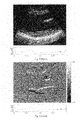
- the B-Mode image of FIG. 15 is a standard full view ultrasound image, and the series obtained in this manner has a higher temporal sampling rate than otherwise possible with full field of view imaging.
- the displacements can be estimated, for example, between consecutive time frames.
- displacements are estimated for the entire composite FOV.
- a noise removal algorithm can be implemented, such as, for example, the CLEAN_NOISE pseudocode provided in the exemplary code provided below. Noise removal utilizes the information of correlation coefficients. Only the estimates with high correlation coefficients above 0.7 are deemed reliable. Those with lower correlation coefficients will be replaced by the average of the surrounding estimated values.
- the displacement can be accumulated with time so as to track motion for an entire cardiac cycle, for example.
- strains in the cardiac muscle can, for example, be derived from the accumulated displacements generated at 360 .
- Strains can be defined in terms of the gradient of the displacements.
- the cumulative 2D displacement gradient tensor, ⁇ u can be defined as:
- ⁇ u _ [ ⁇ u x ⁇ x ⁇ u x ⁇ y ⁇ u y ⁇ x ⁇ u y ⁇ y ] , ( 3 ) and the strain tensor, E, can be defined as
- E 1 2 ⁇ ( ⁇ u _ + ( ⁇ u _ ) T + ( ⁇ u _ ) T ⁇ ⁇ u _ ) , ( 4 ) , where ( ⁇ u ) T is the transpose of ⁇ u .
- the lateral and axial strains are the diagonal components of E, i.e., E xx and E yy , respectively.
- the derived strain can be overlaid onto B-Mode image 325 generated at 330 .
- this overlay can be displayed on the screen, as shown in FIGS. 18-19 and 22 - 23 , and process flow ends.
- OPENCLP filename fid (provided by EchoPAC) Open the DICOM format files saved in GE Vivid 5 RDTISSUE Filename alltiss (3D matrix) (provided by EchoPAC) infoTISS (1-by-Nf) Read the b-mode data startframe (2*M-by-2) stopframe (2*M-by-2) Readiq Filename tiq (2D matrix) (provided by EchoPAC) infoIQ (1-by-Nf) Read the in-phase quadrature startframe (2*M-by-2) (IQ) data (i.e., raw data) nFrames (2*M-by-2) IQ2RF tiq allrf (3D matrix) (provided by EchoPAC) infoIQ Convert IQ data to Radial- frequency (RF) data 310 readecg Filename ecgTISS (1-by-N) (provided by EchoPAC) info
- the main script to make the ecgIQ1 movie displaying overlaid syncIQ1 displacement image parameters scanconvert (true or false) getPolTransformMap prpol (2-by-4) mrows (2D matrix) Computes a polar-to- prcart (2-by-4) mcols (2D matrix) cartesian coordinate mmask (2D matrix) transformation map defined by prpol and prcart.
- appPolTransform Data overlaysc (4D matrix) applies a polar-to-cartesian mrows (2D matrix) coordinate transformation to mcols (2D matrix) imgpol.
- mmask (2D matrix) flag 1 (linear interpolation) 330 readinfo fid 0 tinfo (provided by EchoPAC)
- FIGS. 4-14 depict exemplary intermediate outputs of various exemplary sub-processes of FIG. 3 , generated using the exemplary MatlabTM source code provided in the computer program listing appendix. These intermediate results, and how the various modules in the exemplary source code can be used to generate them, are next described with reference to FIGS. 4-14 .
- the number of sectors N was, for example, six, in the exemplary images of FIGS. 4 and 5 only five sectors were used. In general, the number of sectors depends on the individual sector size selected and on the size of the left ventricle imaged.
- FIG. 4 depicts five exemplary sector outputs.
- the sectors were used to image the whole long axis view of the left ventricle.
- Each sector shows the raw data.
- FIG. 5 shows an exemplary 3 cardiac cycles (ECG signals) obtained while each sector was being imaged. From this data,
- FIG. 6 shows the best match cardiac cycle (ECG signal) from each sector. The best matched cardiac cycle can be determined according to the highest cross-correlation coefficient obtained.
- FIG. 7 shows the combined raw data of the long axis view of the left ventricle
- FIG. 8 shows incremental lateral and axial displacements before noise removal
- FIG. 9 shows incremental lateral and axial displacements after noise removal.
- FIG. 10 shows cumulative lateral and axial displacements from end-diastole (ED) to end-systole (ES) in a long-axis view.
- FIG. 11 shows cumulative lateral and axial strains from ED to ES in a long-axis view.
- FIG. 12 depicts exemplary cumulative (a) lateral and (c) axial displacements from tagged MRI (tMRI) imaging between end-diastole (ED) and end-systole (ES), respectively; and cumulative (b) lateral and (d) axial displacements from 2D myocardial elastography (2DME) between ED and ES, respectively.
- tMRI tagged MRI
- 2DME 2D myocardial elastography
- FIG. 13 depicts cumulative (a) lateral and (c) axial systolic strains from tMRI between ED and ES, respectively, and cumulative (b) lateral and (d) axial systolic strains from 2DME between ED and ES, respectively. All the short-axis images were acquired approximately at the papillary muscle level and shown at end-systolic configuration.
- FIGS. 12 and 13 show that the estimates from myocardial elastography and tMRI are in good agreement.
- FIGS. 12 A(a)-(d) and 13 A(a)-(d) depict the underlying image and the overlay separately, in grayscale, and correspond to FIGS. 12( a )-( d ) and 13 ( a )-( d ), respectively.
- FIG. 14 shows an exemplary B-mode long axis view of the left ventricle before and after scan conversion.
- FIG. 15 is an exemplary B-Mode image such as, for example, that generated at 325 in FIG. 3 from the combined sector data.
- FIGS. 16( a )-( n ) and 17 ( a )-( k ) illustrate exemplary displacement results obtained during systole (contraction) and diastole (expansion), respectively, for a series of time points, wherein displacement has been color coded in each image according to the color coded bar key appearing at the right of each image.
- the time point within the cardiac cycle at which each image has been acquired is indicated by the solid ball below each image.
- FIGS. 16 and 17 are conventional full view images acquired in a conventional manner; they thus represent a frame rate of 50 frames per second. The displacement is overlaid in color on the B-mode images.
- FIGS. 16A and 17A are grayscale images corresponding to FIGS. 16 and 17 , respectively.
- each image in FIGS. 16 and 17 corresponds to two images in FIGS. 16 A and 17 A—one for the B-mode image, the other for the displacement.
- FIG. 16( a ) corresponds to FIGS. 16 A(a 1 ) and (a 2 );
- FIG. 16 A(a 1 ) presenting the B-mode image, and
- FIG. 16 A(a 2 ) the overlay.
- FIGS. 17-23 and FIGS. 17A-23A respectively, described below.
- FIGS. 18( a )-( n ) and 19 ( a )-( k ) show a series of strain images obtained during systole (contraction) and diastole (expansion), respectively, for a series of time points, wherein the strain has been color coded in each image according to the color coded bar key appearing at the right of each image.
- the time point within the cardiac cycle at which each image has been acquired is indicated by the solid ball below each image.
- FIGS. 18 and 19 are conventional full view images acquired in a conventional manner; they thus represent a frame rate of 50 frames per second. The strain is overlaid in color on the B-mode images.
- FIGS. 18A and 19A are grayscale images corresponding to FIGS.
- each image in FIGS. 18 and 19 corresponds to two images in FIGS. 18 A and 19 A—one for the B-mode image, the other for the displacement.
- FIGS. 20( a )-( n ) and 21 ( a )-( k ) show a series of composite displacement images made by combining various sectors according to an exemplary embodiment of the present invention, thus obtaining a high frame rate of, for example, 136 frames per second.
- the time point within the cardiac cycle at which each image has been acquired is indicated by the solid ball below each image.
- the displacement is overlaid in color on the B-mode images.
- FIGS. 20A and 21A are grayscale images corresponding to FIGS. 20 and 21 , respectively.
- the displacement has been separated from the B-mode images for ease of viewing.
- each image in FIGS. 20 and 21 corresponds to two images in each of FIGS. 20 A and 21 A—one for the B-mode image, the other for the displacement.
- FIGS. 22( a )-( n ) and 23 ( a )-( n ) show a series of composite strain images made by combining various sectors according to an exemplary embodiment of the present invention, thus obtaining a high frame rate of, for example, 136 frames per second.
- the time point within the cardiac cycle at which each image has been acquired is indicated by the solid ball below each image.
- the strain is overlaid in color on the B-mode images.
- FIGS. 22A and 23A are grayscale images corresponding to FIGS. 22 and 23 , respectively.
- the strain has been separated from the B-mode images for ease of viewing.
- each image in FIGS. 22 and 23 corresponds to two images in FIGS. 22 A and 23 A—one for the B-mode image, the other for the strain.
- the computer program listing appendix includes a set of exemplary source code files implementing an exemplary embodiment of the present invention.
- the exemplary code is written in MatlabTM, and implements various sub-processes depicted in FIG. 3 , and was used to generate the exemplary images depicted in FIGS. 4-14 , as described above.
- the code was implemented on a conventional ultrasound machine, and can be implemented or adapted to process signals obtained from most standard ultrasound machines using known techniques.
- the exemplary code provided in the computer program listing appendix can, for example, be adapted for use in a programmable ultrasound machine, such as for example, the Ultrasonix Sonix RP system.
- the Sonix RP system offers frame rate capabilities up to 700 fps as well as access to the beamformer, i.e., automatic sector acquisition compared with manual sector acquisition described in the previous section. This higher frame rate can, for example, ensure higher strain quality, as has been seen in a preliminary in vivo human study performed by the inventors.
- access to the beamformer not only allows for the selection of optimal acoustic parameters, such as, for example, frequency, aperture and beamwidth, but it can also, for example, allow for further automation of the methods of the present invention.
- An exemplary implementation of the present invention on such a platform was performed by the inventors, as next described.
- RF Radio-Frequency
- a normal human heart left ventricle and a normal aorta were imaged using the same technique in vivo.
- Composite RF and B-scan full view frames were reconstructed by retrospectively combining all small-sector RF signals.
- the in-plane (lateral and axial) displacements of both long-axis and short-axis views of a healthy human left ventricle were calculated using an RF-based elastographic technique comprising 1D cross-correlation and recorrelation methods (windows size 6.9 mm, overlap 80%).
- a sequence of the electromechanical activation of the heart was observed through mechanical pulse waves propagating along septum (from base to apex) and posterior wall (from apex to base) during systole in human in vivo.
- Exemplary embodiments of this technique can, for example, expand the potential of echocardiography for quantitatively noninvasive diagnosis of cardiovascular diseases such as, for example, myocardial infarction, aneurism and early stage atherosclerosis.
- Heart diseases such as ischemia and infarction, are a growing problem world wide. It is highly useful for the early diagnosis of such cardiac disease to noninvasively detect abnormal patterns of regional myocardial deformation caused by malfunction of the electromechanical conduction. Magnetic resonance (MR) cardiac tagging has been shown capable of quantifying the mechanical properties of the myocardium at high precision.
- MR Magnetic resonance
- tMRI tagged magnetic resonance imaging
- echocardiography has been the predominant imaging modality in diagnostic cardiology owning to its real time, high temporal resolution capability.
- Tissue doppler imaging Tissue doppler imaging
- SRI strain rate imaging
- elastography imaging Tissue doppler imaging
- TDI Tissue doppler imaging
- SRI strain rate imaging
- elastography imaging elastography imaging
- a high temporal resolution typically ⁇ 5 ms, is required to observe the detailed myocardium activities, such as, for example, the fast electrical conductive sequencing pattern for early detection of cardiac diseases.
- the electrical excitation which induces the contraction and relaxation of the cardiac muscle, results in a strong electromechanical wave that propagates in the myocardium at a speed up to 5 m/s.
- Several methods had been developed to increase the ultrasound frame rate such as coded-excitation ultrasound imaging and parallel processing techniques. Most often these methods sacrificed the field of view or ultrasound beam number to increase the frame rate. This is not favorable in clinical study and is not optimal in general.
- ECG triggering or gating can be used to achieve high frame rates by reconstructing RF lines at different cardiac cycle especially for large field of view and high spatial resolution.
- the assumption of ECG triggering or gating lies in that the heart rate does not vary significantly, and that the myocardial function is effectively identical at every cardiac cycle.
- ECG signals were very similar during systole for multiple cycles but could have up to a 10% length difference during diastole. Thus, all ECGs and corresponding RF frames taken for different sectors were interpolated to the same length to get the maximum similarity for each cardiac cycle.
- High frequency, high resolution small animal ultrasound systems have become commercially available, such as, for example, the Vevo 770 system (VisualSonics Inc. Toronto, Ontario, Canada).
- Vevo 770 system VisualSonics Inc. Toronto, Ontario, Canada
- Valuable frequency and speckle information carried by the RF echo signals is lost during conversion and compression, which occurs internally in the system.
- the Sonix RP system (Ultrasonix Medical Corporation, Burnaby, BC, Canada) is an open architecture system which can allow developers to easily control system parameters such as beam line density, sector size, and digitized RF signal acquisition etc.
- an Ultrasonix 500RP research platform was used to measure the ultrasound backscatter and attenuation coefficient.
- Displacements were then estimated along the beam axis and displayed as an image referred to as an elastogram.
- the results obtained clearly showed electromechanical wave propagation in human heart during systole and a pulse wave propagating along a human aorta.
- a clinical phased array transducer (Ultrasonix model # P 4 -2/20) operating at 3.3 MHz was used for human cardiac and vascular imaging. In a phased array transducer, more than one line can be acquired at the same time rather than line-by-line data acquisition by a signal element transducer to achieve high data consistency. For further development, if sector size is decreased to only one transmission line, the method could be reduced to single element scan imaging with an even higher frame rate.
- a separate ECG module (MCC Weg für in Medizin undtechnik mbH & Co. KG) was connected to the Sonix RP computer base running windows XP with RS232 serial interface. Two channels were recorded, from which three Einthoven and Goldberger leads were depicted. The signals were recorded digitally, processed and transmitted to the host via PC serial interface with a baud rate of 9600 baud (1 start bit, 8 data bits, 1 stop bit, no parity). The maximum sampling frequency rate for this ECG module is 300 points per second.
- FIG. 24 provides exemplary process flow charts from which an exemplary C++ program that was created.
- the flow charts illustrate functionality for RF and ECG signal acquisition: a) Ultrasonix RF data acquisition. b) ECG module data acquisition. c) ECG and ECG time stamp buffer. d) RF frame and RF frame time stamp buffer.
- ECG cycle corresponding to each sector can be of identical duration to those of other sectors.
- heart rate variability one challenge for any ECG gated/triggered retrospective high frame rate ultrasound B mode imaging is heart rate variability.
- the duration of an ECG can vary by up to 10% per cycle, and the number of frames for each sector varies accordingly. As shown in FIG. 26 , ECG signals during systole have very little variation.
- an accurate method to solve the ECG arrhythmic is to stretch the diastolic part of the ECGs and the corresponding RF frames to the same length to achieve high similarity.
- FIG. 25 illustrates an ECG-gated multi-sector combination technique for high frame rate, full-view ultrasound imaging.
- a total of seven sectors at different angles were acquired in a continuous sequence during each experiment.
- ECG and RF frame data are shortened according to the time stamp associated with each data point or frame for one cardiac cycle.
- Corresponding small sector frames were, for example, recombined to generate full view ultrasound images.
- FIG. 26 illustrates irregular ECG interpolation.
- the axial displacement was estimated off-line using the normalized cross-correlation.
- the RF window size was equal to 6.9 mm and the window overlap was equal to 80%, deemed enough to retain good axial resolution.
- the parabolic interpolation was applied to the cross-correlation function in order to further improve the precision.
- the displacements were then estimated using pairs of consecutive RF frames.
- a linear Savitzky-Golay differentiation filter with a length of seven samples (140 um) was used to estimate the axial strains from the displacements.
- the aforementioned displacements and strains were the instantaneous or incremental displacements and strains occurring between two consecutive frames.
- the preset points in the LV wall could be tracked automatically. Therefore, the incremental displacements and strains corresponding to the preset points could be extracted.
- the cumulative displacement and strains were obtained and represented the total motion and deformation from the first frame (corresponding to the first R-wave of the ECG), respectively.
- the displacements were color-coded and superimposed onto the grayscale B-mode images using an overlay blending mode. In the displacement images, only the estimates in the region of interest (ROI) are shown for better interpretation.
- ROI was first determined through a 40- to 50-point selection performed manually in the first frame of a B-mode cine-loop (reconstructed from the RF image sequence).
- the selected points coincided with the myocardial boundaries (i.e., endocardium and epicardium). Using the estimated displacement field, these points could then, for example, be tracked over the entire cardiac cycle, providing the updated ROIs corresponding to different phases.
- the cumulative strain curve in myocardial elastography may undergo a drift, i.e., the cumulative strain does not return to zero at the end of the cardiac cycle. Thus, the drift in the cumulative displacements and strains was corrected on the assumption that the drift increases linearly with time over the duration of a cardiac cycle. Elastographic methods were implemented in MATLAB 7.1 (MathWorks, Inc., Natick, Mass., USA). The total processing time for a full cardiac cycle in the exemplary implementation was 2 to 3 hours on a PC workstation (Pentium 4 CPU 2.80 GHz, 2 GB RAM).
- An adult healthy female heart was scanned both long axis and short axis view and the aorta with a frame rate of 481 Hz per sector through the custom automated program.
- the scan was performed with regular clinical ultrasound B-mode scan procedure by an experienced medical sonographer.
- the system parameters were set at 11 cm acquisition depth and a total of 64 lines for full 100% view.
- Ultrasound probe frequency was set at 3.3 MHz.
- Seven sectors were scanned with each sector of a 2-sec scanning time. At this time period, one or two cardiac cycles were recorded since the volunteer's heart rate ranged from 60 to 80 cycles per second. A total of 21-sec was needed for the entire experiment including scanning and data saving. Because respiratory motion can affect the heart position, breath holding was required for better composite images quality during the sector scanning.
- the patient's heart recovers to the original condition as much as possible during each cardiac cycle and the operator's hand keep still is essential to reconstruct a smooth transition from sector to sector. It is noted that although the total scanning time for a 100% 90 degree B-mode view is minimized by automatically sweeping different sectors, a patient's heart rate variability, breathing and the freehand motion of the transducer probe can pose some issues for accurate combination.
- FIG. 27 depict a comparison of image quality before and after overlap processing of different sectors.
- FIG. 28 show a comparison of a seven sector B-mode composite image of heart long axis view ( FIG. 28( a )) with a 100% B-mode image without combination.
- the benefits of FIG. 28( a ), created using methods according to the present invention, are clearly seen.
- the motion of the tissue was estimated off-line using an established classical speckle-tracking method.
- This technique was based on detecting the small local displacement of the tissue that occurs between tow consecutive frames. With the current method, only axial displacements (along the axis of the transducer beam), which coincided with the radial displacement in a long-axis view, were estimated. In our algorithm, the time shifts in the backscattered signals were determined between the two consecutive frames through cross-correlation of small sliding windows over the entire RF-line. This technique allowed the detection of very small displacement on the order of 0.1 um or less (correlation window of 6.9 mm, overlapping 80%). Finally, the cine-loop of the axial displacement was generated at a frame rate 481 Hz for the entire in vivo human cardiac cycle.
- FIGS. 29( a )-( l ) depict a sequence of color-coded axial displacements overlaid onto gray-scale B-mode images at different occurrence times during systole on a human left ventricle.
- a “well-organized” heterogeneity in electromechanical coupling is thus a characteristic feature and may be a prerequisite for normal performance of the cardiac muscle.
- FIGS. 30( a )-( l ) In short axis view of the subject, another clear wave was also found propagating counter clockwise from posterior to lateral wall during diastole phase as shown in FIGS. 30( a )-( l ). The wave front is indicated by white arrows.
- an electromechanical wave was found propagating along the posterior wall in a human left-ventricle during systole. This is depicted in FIGS. 31( a )-( l ).
- the first frame RF data and the first ECG data point are start at the same time as describe in the previous session. which could result in a maximum latency of 3.3 ms between the two data sets. This latency is determined by the maximum time interval of ECG sampling rate and the RF frame rate. In the worst case the latency between ECG and rf frames is min(1/ECG frame rate, 1/rf frame rate) since the first point of ECG data and the first frame of rf data is forced to be aligned.
- L Max(1 /r ECG ,1 /r RF ) ⁇ 3.3 ms
- L latency of first RF frame and first ECG data rECG—ECG sampling rate, 300 points/second rRF—RF frame rate, >360 Hz
- ventricle mechanics and electromechanics are very useful clinically. It is also expected that this type of contraction synchronization would be the first to fail for heart failure diseases. This may be due to the fact that in the heart systolic contraction is an electrically triggered active event whereas diastole is a passive relaxation process.
- the minimum frame rate for reliable strain information is approximately 250 fps. This is because the correlation coefficient surpasses 0.9, when the SNR e is high enough (above 10 dB) for best images. This agrees with what various researchers have reported for cardiac RF speckle tracking, i.e., that the optimal frame rate is within the range of 200-300.
- the minimum frame rate is approximately 500 fps. The optimal frame rate is directly proportional to the strain and strain rate amplitudes to be estimated.
- the strain rate is 2-3 times higher, therefore, the optimal frame rate needs to be accordingly adjusted; and (3) the Ultrasonix system can provide sufficiently high correlation coefficients ( ⁇ >0.985), both for systolic and diastolic estimates. This result thus indicates that high correlation in a human heart is possible and that the most reliable strains are obtained at and beyond 250 fps for systole, and 500 fps for diastole, respectively.
Abstract
Description
where ρ=ρxρyρz is the correlation coefficient (equal to the product of the correlation coefficients associated with motion in each direction), SNRs is the sonographic signal-to-noise ratio, B is the bandwidth and f0 is the frequency (Varghese and Ophir 1997). The frame rate FR is equal to c/(2(N′/N)D), where c is the speed of sound, N′ is the number of RF signals, N is the number of sectors and D is the depth. The speed of sound in soft tissues is equal to 1540 m/s and the depth in human echocardiography is typically 10-12 cm. The highest obtainable frame rate will be achieved when the smallest number of RF signals (or, largest number of sectors) is used, i.e., N′/N=1. In that case, the frame rate would be equal to 6250 frames/s (or, 6.25 kHz). When N′/N=128 (i.e., the conventional number of RF signals used), the lowest frame rate will be achieved, i.e., 48.83 frames/s (or, 0.049 kHz). For N={6, 12, 20}, the corresponding frame rates are FR={1.04, 0.52, 0.26} kHz.
and the strain tensor, E, can be defined as
where (∇u)T is the transpose of ∇u. The lateral and axial strains are the diagonal components of E, i.e., Exx and Eyy, respectively.
-
- M=number of sectors;
- N=length of ECG signals for three cardiac cycles;
- N1=length of ECG signals for the cardiac cycle with maximum length;
- Nf=number of total frames in the beginning; and
- nFrames=number of frames per sector per cardiac cycle.
| Function name | Input | Output | ||
| Main | analyzeNrf | N/A | N/A | |
| script | Reconstruct several | |||
| overlapping small sector RF | ||||
| acquisitions. | ||||
| 305 | OPENCLP | filename | fid | |
| (provided by EchoPAC) | ||||
| Open the DICOM format files | ||||
| saved in GE Vivid 5 | ||||
| RDTISSUE | Filename | alltiss (3D matrix) | ||
| (provided by EchoPAC) | infoTISS (1-by-Nf) | |||
| Read the b-mode data | startframe (2*M-by-2) | |||
| stopframe (2*M-by-2) | ||||
| Readiq | Filename | tiq (2D matrix) | ||
| (provided by EchoPAC) | infoIQ (1-by-Nf) | |||
| Read the in-phase quadrature | startframe (2*M-by-2) | |||
| (IQ) data (i.e., raw data) | nFrames (2*M-by-2) | |||
| IQ2RF | tiq | allrf (3D matrix) | ||
| (provided by EchoPAC) | infoIQ | |||
| Convert IQ data to Radial- | ||||
| frequency (RF) data | ||||
| 310 | readecg | Filename | ecgTISS (1-by-N) | |
| (provided by EchoPAC) | infoTISS (1-by-Nf) | syncTISS (1-by-N) | ||
| Read the ECG data stored in | ||||
| GE Vivid 5 | ||||
| 320 | matchbestECG | ecgTISS (1-by-M cell) | ecgbest (M-by-2) | |
| Finds the best matching | {ecgTISS1, ecgTISS2, | |||
| segments of ECG based on | ecgTISS3, ecgTISS4, | |||
| length and shape | ecgTISS5} (1-by-M cell) | |||
| cutECG | ECGfull (1-by-N) | ECGparts (3-by-N1) | ||
| This function cuts up an ECG | ECGtrigger (1-by-N1) | |||
| signal into N parts. | ECGpeakshift (1-by-M) | |||
| syncTISS | startframe (2*M-by-2) | |||
| syncIQ | stopframe (2*M-by-2) | |||
| ecgbest (2*M-by-2) | ||||
| 325 | makeCardiacMovie | overlayAll | Movie | |
| The main script to make the | ecgIQ1 | |||
| movie displaying overlaid | syncIQ1 | |||
| displacement image | parameters | |||
| scanconvert (true or false) | ||||
| getPolTransformMap | prpol (2-by-4) | mrows (2D matrix) | ||
| Computes a polar-to- | prcart (2-by-4) | mcols (2D matrix) | ||
| cartesian coordinate | mmask (2D matrix) | |||
| transformation map defined | ||||
| by prpol and prcart. | ||||
| appPolTransform | Data | overlaysc (4D matrix) | ||
| applies a polar-to-cartesian | mrows (2D matrix) | |||
| coordinate transformation to | mcols (2D matrix) | |||
| imgpol. | mmask (2D matrix) | |||
| flag = 1 (linear interpolation) | ||||
| 330 | | fid | 0 | tinfo |
| (provided by EchoPAC) | ||||
| Read all EchoPAC file | ||||
| information | ||||
| getmyparams | tinfo | tparams-> | ||
| get parameters from each file | paramnamesTISS or | paramsIQ (M-by-6) | ||
| paramnamesIQ | paramsTISS (M-by-6) | |||
| findSectorOverlap | paramsIQ or paramsTISS | sectorBMbeams (M-by-8) or | ||
| Utilizes params matrix to find | (M-by-6) | sectorIQbeams (M-by-8) | ||
| overlapping regions of | centerAngle | |||
| sectors, and returns | iSA = 3 | |||
| sectorBeams, a matrix | iAU = 4 | |||
| containing information about | iNB = 6 | |||
| overlap and which data to | iSD = 1 | |||
| utilize from each sector for | iDU = 2 | |||
| the reconstructed whole | iNS = 5 | |||
| sector. |
| 340 | See pseudocode below |
| 350 | See pseudocode below |
| 360 | See pseudocode below |
| 365 | See pseudocode below |
| 370 | initOverlay.m | paramsIQ (2D matrix) | oparams (structure) |
| paramsTISS (2D matrix) | |||
| overlayData.m | tiss (3D matrix) | overlayAll (4D matrix) | |
| data (3D matrix) | |||
| oparams (structure) | |||
| TISSISRF (flag) | |||
| overlayimage.m | tissRGB (3D matrix) | overlay(3D matrix) | |
| dataRGB (3D matrix) | |||
| mask (3D matrix) | |||
| tiss2rgb.m | tiss (2D matrix) | tissRGB (3D matrix) | |
| paramsTISS (2D matrix) | |||
| DYN (scalar) | |||
| GAIN (scalar) | |||
| data2rgb.m | dataResize (2D matrix) | dataRGB (3D matrix) | |
| CMAP | |||
| dataLIMS (1-by 2-vector) | |||
Pseudocode Referred to in Above Table for 340, 350, 360 and 365 of
| 340 |
| Read raw data |
| Initialize window_size, overlap, interp_factor |
| Set sample_count to (1-overlap)*window_size |
| FOR 1 : shift : total_sample_points |
| FOR lateral_beam_count |
| 2D interpolation of RF signals |
| Calculate cross-correlation coefficient between signals |
| Find the maximal coefficient |
| Cosine interpolation around the maximal coefficient |
| Store the location with the interpolated maximal coefficient |
| Calculate lateral and axial displacements based the store location |
| ENDFOR |
| ENDFOR |
| Generate RF signals with the removal the axial displacement and recalculate the lateral |
| displacement |
| lateral_disp= lateral displacement after axial displacement correction |
| axial_disp= the initial estimated axial displacement |
| Return lateral_disp, axial_disp, cross-correlation_coefficient |
| 350 |
| Function CLEAN_NOISE (displacement, cross_correlation_coefficients) |
| FOR each displacement |
| IF cross_correlation_coefficient < threshold |
| The displacement value is updated with the average of the neighboring values |
| ENDIF |
| ENDFOR |
| 360 |
| function [cum_lateral_disp, cum_axial_disp]= CUM_DISP (lateral_disp, axial_disp) |
| 365 |
| function strain_tensor_2D=CALC_STRAIN (lateral_disp, axial_disp) |
| G11= the gradient of lateral_disp along the lateral direction |
| G12= the gradient of lateral_disp along the axial direction |
| G21= the gradient of axial_disp along the lateral direction |
| G22= the gradient of axial_disp along the axial direction |
| G(1, 1)=G11; G(1, 2)=G12; G(2, 1)=G21; G(2, 2)=G22; |
| strain_tensor_2D =1/2*(G + transpose (G) + transpose (G)*G) |
| Return strain_tesnsor_2D |
Min(ABS(Time_stampRF(i)−Time_stampECG
min(abs(Time_StampRF(i)−Time_StampECG
where Time_StampECG
L=Max(1/r ECG,1/r RF)˜3.3 ms
Where L—latency of first RF frame and first ECG data
rECG—ECG sampling rate, 300 points/second
rRF—RF frame rate, >360 Hz
P rf =P r×round(N rf /N ECG)
Where
Prf—the position of rf frame corresponding to ECG R wave peak
Pr—the ECG R wave peak position
Nrf—total number of rf frames.
NECG—total number of ECG points.
round—the matlab function to get the closest integer of Nrf/NECG
| //main program | ||
| program starts | ||
| parameter initializing; | ||
| declare message map; | ||
| if (!connected_to_Sonix RP host) | ||
| connect_to_host; | ||
| endif | ||
| detect ECG connection; | ||
| create ECG acquisition thread; | ||
| while(!exit) wait for user message; | ||
| program ends | ||
| // on catch message RUN_SCAN | ||
| retrieve scan parameter from panel; | ||
| if(!connected_to_host) error; | ||
| if(!ECG_running) error; | ||
| set scan parameter; | ||
| clear memory; | ||
| set_data_to_acquire(RF); | ||
| if(thread_scan exit) | ||
| terminate(thread_scan); | ||
| else | ||
| create_thread(thread_scan); | ||
| endif | ||
| //pseudo code for thread_scan | ||
| calculate total number of sectors; | ||
| for(i=starting_angle;i<stoping angle; angle_increment) | ||
| set sector angle; | ||
| post message starting scan | ||
| wait 2 s | ||
| post message stop scan | ||
| save ECG data in memory; | ||
| save RF data in memory; | ||
| endif | ||
Claims (34)
Priority Applications (2)
| Application Number | Priority Date | Filing Date | Title |
|---|---|---|---|
| US11/899,004 US8150128B2 (en) | 2006-08-30 | 2007-08-30 | Systems and method for composite elastography and wave imaging |
| US13/353,148 US20130066211A1 (en) | 2006-08-30 | 2012-01-18 | Systems and methods for composite myocardial elastography |
Applications Claiming Priority (2)
| Application Number | Priority Date | Filing Date | Title |
|---|---|---|---|
| US84192606P | 2006-08-30 | 2006-08-30 | |
| US11/899,004 US8150128B2 (en) | 2006-08-30 | 2007-08-30 | Systems and method for composite elastography and wave imaging |
Related Child Applications (1)
| Application Number | Title | Priority Date | Filing Date |
|---|---|---|---|
| US13/353,148 Continuation US20130066211A1 (en) | 2006-08-30 | 2012-01-18 | Systems and methods for composite myocardial elastography |
Publications (2)
| Publication Number | Publication Date |
|---|---|
| US20080285819A1 US20080285819A1 (en) | 2008-11-20 |
| US8150128B2 true US8150128B2 (en) | 2012-04-03 |
Family
ID=39136608
Family Applications (2)
| Application Number | Title | Priority Date | Filing Date |
|---|---|---|---|
| US11/899,004 Active 2029-12-28 US8150128B2 (en) | 2006-08-30 | 2007-08-30 | Systems and method for composite elastography and wave imaging |
| US13/353,148 Abandoned US20130066211A1 (en) | 2006-08-30 | 2012-01-18 | Systems and methods for composite myocardial elastography |
Family Applications After (1)
| Application Number | Title | Priority Date | Filing Date |
|---|---|---|---|
| US13/353,148 Abandoned US20130066211A1 (en) | 2006-08-30 | 2012-01-18 | Systems and methods for composite myocardial elastography |
Country Status (2)
| Country | Link |
|---|---|
| US (2) | US8150128B2 (en) |
| WO (1) | WO2008027520A2 (en) |
Cited By (21)
| Publication number | Priority date | Publication date | Assignee | Title |
|---|---|---|---|---|
| US20100256530A1 (en) * | 2009-04-01 | 2010-10-07 | Tomy Varghese | Method and apparatus for monitoring tissue ablation |
| US20110074409A1 (en) * | 2009-09-25 | 2011-03-31 | Nellcor Puritan Bennett Ireland | Systems And Methods For Gating An Imaging Device |
| US20120121151A1 (en) * | 2010-11-12 | 2012-05-17 | Siemens Aktiengesellschaft | Device And Computed Tomography Scanner For Determining And Visualizing The Perfusion Of The Myocardial Muscle |
| US20120262165A1 (en) * | 2011-04-13 | 2012-10-18 | Case Western Reserve University | Relaxometry |
| US20130128691A1 (en) * | 2010-07-29 | 2013-05-23 | B-K Medical Aps | Motion-compensated ultrasound images |
| US20130289402A1 (en) * | 2010-12-08 | 2013-10-31 | Hitachi Medical Corporation | Ultrasound diagnosis apparatus |
| US9247921B2 (en) | 2013-06-07 | 2016-02-02 | The Trustees Of Columbia University In The City Of New York | Systems and methods of high frame rate streaming for treatment monitoring |
| US9320491B2 (en) | 2011-04-18 | 2016-04-26 | The Trustees Of Columbia University In The City Of New York | Ultrasound devices methods and systems |
| US9358023B2 (en) | 2008-03-19 | 2016-06-07 | The Trustees Of Columbia University In The City Of New York | Systems and methods for opening of a tissue barrier |
| US9514358B2 (en) | 2008-08-01 | 2016-12-06 | The Trustees Of Columbia University In The City Of New York | Systems and methods for matching and imaging tissue characteristics |
| US9726647B2 (en) | 2015-03-17 | 2017-08-08 | Hemosonics, Llc | Determining mechanical properties via ultrasound-induced resonance |
| US9826958B2 (en) | 2009-11-27 | 2017-11-28 | QView, INC | Automated detection of suspected abnormalities in ultrasound breast images |
| US20180110425A1 (en) * | 2015-04-23 | 2018-04-26 | Hitachi, Ltd. | Ultrasound diagnostic device |
| WO2018087752A1 (en) * | 2016-11-10 | 2018-05-17 | ContinUse Biometrics Ltd. | System and method for monitoring periodic signals |
| CN109982643A (en) * | 2016-11-14 | 2019-07-05 | 皇家飞利浦有限公司 | Three mode ultrasounds imaging for the imaging of anatomical structure, function and Hemodynamics |
| US10517564B2 (en) | 2012-10-10 | 2019-12-31 | The Trustees Of Columbia University In The City Of New York | Systems and methods for mechanical mapping of cardiac rhythm |
| US10687785B2 (en) | 2005-05-12 | 2020-06-23 | The Trustees Of Columbia Univeristy In The City Of New York | System and method for electromechanical activation of arrhythmias |
| US10962524B2 (en) | 2011-02-15 | 2021-03-30 | HomoSonics LLC | Characterization of blood hemostasis and oxygen transport parameters |
| US11109832B2 (en) | 2015-01-29 | 2021-09-07 | Koninklijke Philips N.V. | Evaluation of cardiac infarction by real time ultrasonic strain imaging |
| RU2756367C1 (en) * | 2018-02-15 | 2021-09-29 | Университа' Дельи Студи Ди Рома "Ла Сапиенца" | Method and system for measuring the indicator of the current peripheral resistance |
| US20220317219A1 (en) * | 2021-04-01 | 2022-10-06 | Siemens Healthcare Gmbh | Method and System for Magnetic Resonance Elastography |
Families Citing this family (64)
| Publication number | Priority date | Publication date | Assignee | Title |
|---|---|---|---|---|
| WO2006044997A2 (en) * | 2004-10-15 | 2006-04-27 | The Trustees Of Columbia University In The City Of New York | System and method for localized measurement and imaging of viscosity of tissues |
| US8858441B2 (en) * | 2005-05-12 | 2014-10-14 | The Trustees Of Columbia University In The City Of New York | System and method for electromechanical wave imaging of body structures |
| WO2008027520A2 (en) | 2006-08-30 | 2008-03-06 | The Trustees Of Columbia University In The City Of New York | Systems and methods for composite elastography and wave imaging |
| WO2008042905A2 (en) * | 2006-10-02 | 2008-04-10 | University Of Washington | Ultrasonic estimation of strain induced by in vivo compression |
| WO2008051639A2 (en) | 2006-10-25 | 2008-05-02 | Maui Imaging, Inc. | Method and apparatus to produce ultrasonic images using multiple apertures |
| JP5238201B2 (en) * | 2007-08-10 | 2013-07-17 | 株式会社東芝 | Ultrasonic diagnostic apparatus, ultrasonic image processing apparatus, and ultrasonic image processing program |
| WO2009031327A1 (en) * | 2007-09-06 | 2009-03-12 | Hitachi Medical Corporation | Ultrasonograph |
| US9282945B2 (en) * | 2009-04-14 | 2016-03-15 | Maui Imaging, Inc. | Calibration of ultrasound probes |
| DE102008005071B4 (en) * | 2008-01-18 | 2013-11-14 | Siemens Aktiengesellschaft | Method for time registration of image series records |
| US8502821B2 (en) * | 2008-02-04 | 2013-08-06 | C Speed, Llc | System for three-dimensional rendering of electrical test and measurement signals |
| WO2009104525A1 (en) * | 2008-02-18 | 2009-08-27 | 株式会社 日立メディコ | Ultrasonographic device, ultrasonic elasticity information processing method, and ultrasonic elasticity information processing program |
| JP5209351B2 (en) * | 2008-03-21 | 2013-06-12 | 株式会社東芝 | Ultrasonic diagnostic apparatus and control method thereof |
| KR101014564B1 (en) * | 2008-06-26 | 2011-02-16 | 주식회사 메디슨 | Ultrasound system and method for forming an elastic image |
| WO2010017445A2 (en) | 2008-08-08 | 2010-02-11 | Maui Imaging, Inc. | Imaging with multiple aperture medical ultrasound and synchronization of add-on systems |
| WO2010030819A1 (en) | 2008-09-10 | 2010-03-18 | The Trustees Of Columbia University In The City Of New York | Systems and methods for opening a tissue |
| FR2938957B1 (en) * | 2008-11-21 | 2011-01-21 | Univ Joseph Fourier Grenoble I | IMAGE PROCESSING METHOD FOR ESTIMATING HAZARD OF ATENOME PLATE BREAK |
| KR101659723B1 (en) | 2009-04-14 | 2016-09-26 | 마우이 이미징, 인코포레이티드 | Multiple aperture ultrasound array alignment fixture |
| US8366619B2 (en) * | 2009-05-13 | 2013-02-05 | University Of Washington | Nodule screening using ultrasound elastography |
| US20100305438A1 (en) * | 2009-05-29 | 2010-12-02 | Kenneth Wayne Rigby | System and method for scaling strain image data |
| EP2470287A4 (en) | 2009-08-28 | 2015-01-21 | Univ Columbia | Systems, methods, and devices for production of gas-filled microbubbles |
| WO2011028690A1 (en) | 2009-09-01 | 2011-03-10 | The Trustees Of Columbia University In The City Of New York | Microbubble devices, methods and systems |
| JP5647990B2 (en) * | 2009-10-28 | 2015-01-07 | 株式会社日立メディコ | Ultrasonic diagnostic apparatus and image construction method |
| EP2512588A4 (en) | 2009-12-16 | 2014-06-04 | Univ Columbia | Methods, devices, and systems for on-demand ultrasound-triggered drug delivery |
| JP6274724B2 (en) | 2010-02-18 | 2018-02-07 | マウイ イマギング,インコーポレーテッド | Point source transmission and sound velocity correction using multi-aperture ultrasound imaging |
| US9585631B2 (en) | 2010-06-01 | 2017-03-07 | The Trustees Of Columbia University In The City Of New York | Devices, methods, and systems for measuring elastic properties of biological tissues using acoustic force |
| WO2012019172A1 (en) | 2010-08-06 | 2012-02-09 | The Trustees Of Columbia University In The City Of New York | Medical imaging contrast devices, methods, and systems |
| WO2012051305A2 (en) | 2010-10-13 | 2012-04-19 | Mau Imaging, Inc. | Multiple aperture probe internal apparatus and cable assemblies |
| EP2627257B1 (en) | 2010-10-13 | 2019-04-17 | Maui Imaging, Inc. | Concave ultrasound transducers and 3d arrays |
| US8971602B2 (en) * | 2011-04-22 | 2015-03-03 | Mayo Foundation For Medical Education And Research | Method for magnetic resonance elastography using transient waveforms |
| WO2012162664A1 (en) | 2011-05-26 | 2012-11-29 | The Trustees Of Columbia University In The City Of New York | Systems and methods for opening of a tissue barrier in primates |
| EP2785253B1 (en) | 2011-12-01 | 2023-11-15 | Maui Imaging, Inc. | Motion detection using ping-based and multiple aperture doppler ultrasound |
| CN104080407B (en) | 2011-12-29 | 2017-03-01 | 毛伊图像公司 | The M-mode ultra sonic imaging of free routing |
| JP6438769B2 (en) * | 2012-02-21 | 2018-12-19 | マウイ イマギング,インコーポレーテッド | Determination of material hardness using multiple aperture ultrasound. |
| CN104203110B (en) | 2012-03-26 | 2017-06-06 | 毛伊图像公司 | System and method for improving ultrasonoscopy quality by the application weighting factor |
| JP6257935B2 (en) * | 2012-07-02 | 2018-01-10 | 東芝メディカルシステムズ株式会社 | Ultrasonic diagnostic apparatus, biological signal acquisition apparatus, and control program for ultrasonic diagnostic apparatus |
| EP2883079B1 (en) | 2012-08-10 | 2017-09-27 | Maui Imaging, Inc. | Calibration of multiple aperture ultrasound probes |
| EP3893022A1 (en) | 2012-08-21 | 2021-10-13 | Maui Imaging, Inc. | Ultrasound imaging system memory architecture |
| US10368753B2 (en) | 2012-11-01 | 2019-08-06 | The Johns Hopkins University | Method and system for determining strain relaxation of left ventricular diastolic function |
| US20140128738A1 (en) * | 2012-11-05 | 2014-05-08 | Fujifilm Visualsonics, Inc. | System and methods for forming ultrasound images |
| US9510806B2 (en) | 2013-03-13 | 2016-12-06 | Maui Imaging, Inc. | Alignment of ultrasound transducer arrays and multiple aperture probe assembly |
| US9339210B2 (en) * | 2013-05-08 | 2016-05-17 | Koninklijke Philips N.V. | Device for obtaining a vital sign of a subject |
| US10322178B2 (en) | 2013-08-09 | 2019-06-18 | The Trustees Of Columbia University In The City Of New York | Systems and methods for targeted drug delivery |
| US10028723B2 (en) | 2013-09-03 | 2018-07-24 | The Trustees Of Columbia University In The City Of New York | Systems and methods for real-time, transcranial monitoring of blood-brain barrier opening |
| US9883848B2 (en) | 2013-09-13 | 2018-02-06 | Maui Imaging, Inc. | Ultrasound imaging using apparent point-source transmit transducer |
| US20170124701A1 (en) * | 2014-03-17 | 2017-05-04 | Arizona Board Of Regents On Behalf Of Arizona State University | System and method for measuring artery thickness using ultrasound imaging |
| GB201410743D0 (en) * | 2014-06-17 | 2014-07-30 | The Technology Partnership Plc | Ablation treatment device sensor |
| US10401493B2 (en) | 2014-08-18 | 2019-09-03 | Maui Imaging, Inc. | Network-based ultrasound imaging system |
| EP3230848A1 (en) * | 2014-12-10 | 2017-10-18 | Sparkbio S.R.L. | System for the capture and combined display of video and analog signals coming from electromedical instruments and equipment |
| CN107427278A (en) * | 2015-01-02 | 2017-12-01 | 百胜集团 | Pass through the elastic method of ultrasound quantification material |
| US10182790B2 (en) * | 2015-03-30 | 2019-01-22 | Siemens Medical Solutions Usa, Inc. | Adaptive timing guidance in stress echocardiography |
| EP3408037A4 (en) | 2016-01-27 | 2019-10-23 | Maui Imaging, Inc. | Ultrasound imaging with sparse array probes |
| US11304681B2 (en) * | 2016-03-03 | 2022-04-19 | Canon Medical Systems Corporation | Ultrasonic diagnostic apparatus and image processing method |
| US10338197B2 (en) * | 2016-04-28 | 2019-07-02 | Accenture Global Solutions Limited | System and method for use of qualitative modeling for signal analysis |
| US11446003B2 (en) * | 2017-03-27 | 2022-09-20 | Vave Health, Inc. | High performance handheld ultrasound |
| US10152798B2 (en) * | 2017-04-10 | 2018-12-11 | Wisconsin Alumni Research Foundation | Systems, methods and, media for determining object motion in three dimensions using speckle images |
| CN107516075B (en) * | 2017-08-03 | 2020-10-09 | 安徽华米智能科技有限公司 | Electrocardiosignal detection method and device and electronic equipment |
| CN111093521B (en) | 2018-04-13 | 2022-06-17 | 深圳迈瑞生物医疗电子股份有限公司 | Ultrasonic imaging method and ultrasonic imaging apparatus |
| CN110575198B (en) * | 2018-06-08 | 2022-07-01 | 佳能医疗系统株式会社 | Analysis device and analysis method |
| WO2020044523A1 (en) * | 2018-08-30 | 2020-03-05 | オリンパス株式会社 | Recording device, image observation device, observation system, observation system control method, and observation system operating program |
| US20210059644A1 (en) * | 2019-04-16 | 2021-03-04 | Shenzhen Mindray Bio-Medical Electronics Co., Ltd. | Retrospective multimodal high frame rate imaging |
| CN111772677B (en) * | 2020-07-07 | 2023-06-13 | 意领科技有限公司 | Biological tissue elasticity detection method with dimension, detection system and storage medium |
| WO2022241155A1 (en) * | 2021-05-14 | 2022-11-17 | Caption Health, Inc. | Circuitless heart cycle determination |
| WO2023102441A1 (en) * | 2021-11-30 | 2023-06-08 | Bioventures, Llc | Personalized motion-gated coronary cta scanning systems and methods |
| US20230255598A1 (en) * | 2022-02-16 | 2023-08-17 | GE Precision Healthcare LLC | Methods and systems for visualizing cardiac electrical conduction |
Citations (85)
| Publication number | Priority date | Publication date | Assignee | Title |
|---|---|---|---|---|
| US3598111A (en) | 1968-12-09 | 1971-08-10 | Health Technology Corp | Technique and apparatus for measuring and monitoring the mechanical impedance of body tissues and organ systems |
| US4463608A (en) | 1979-05-07 | 1984-08-07 | Yokogawa Hokushin Electric Corp. | Ultrasound imaging system |
| US4777599A (en) | 1985-02-26 | 1988-10-11 | Gillette Company | Viscoelastometry of skin using shear wave propagation |
| US4882679A (en) | 1987-11-27 | 1989-11-21 | Picker International, Inc. | System to reformat images for three-dimensional display |
| US5038787A (en) | 1988-08-10 | 1991-08-13 | The Board Of Regents, The University Of Texas System | Method and apparatus for analyzing material properties using reflected ultrasound |
| US5107837A (en) | 1989-11-17 | 1992-04-28 | Board Of Regents, University Of Texas | Method and apparatus for measurement and imaging of tissue compressibility or compliance |
| US5309914A (en) | 1991-04-17 | 1994-05-10 | Kabushiki Kaisha Toshiba | Ultrasonic imaging apparatus |
| US5433708A (en) | 1991-05-17 | 1995-07-18 | Innerdyne, Inc. | Method and device for thermal ablation having improved heat transfer |
| US5435310A (en) | 1993-06-23 | 1995-07-25 | University Of Washington | Determining cardiac wall thickness and motion by imaging and three-dimensional modeling |
| US5457754A (en) | 1990-08-02 | 1995-10-10 | University Of Cincinnati | Method for automatic contour extraction of a cardiac image |
| US5601084A (en) | 1993-06-23 | 1997-02-11 | University Of Washington | Determining cardiac wall thickness and motion by imaging and three-dimensional modeling |
| US5606971A (en) | 1995-11-13 | 1997-03-04 | Artann Corporation, A Nj Corp. | Method and device for shear wave elasticity imaging |
| US5662113A (en) | 1995-06-30 | 1997-09-02 | Siemens Medical Systems, Inc | Edge enhancement system for ultrasound images |
| US5752515A (en) | 1996-08-21 | 1998-05-19 | Brigham & Women's Hospital | Methods and apparatus for image-guided ultrasound delivery of compounds through the blood-brain barrier |
| US5810731A (en) | 1995-11-13 | 1998-09-22 | Artann Laboratories | Method and apparatus for elasticity imaging using remotely induced shear wave |
| US5840028A (en) | 1996-06-24 | 1998-11-24 | Japan Science And Technology Corporation | Ultrasonic diagnostic equipment |
| US6026173A (en) | 1997-07-05 | 2000-02-15 | Svenson; Robert H. | Electromagnetic imaging and therapeutic (EMIT) systems |
| US6102865A (en) * | 1996-02-29 | 2000-08-15 | Acuson Corporation | Multiple ultrasound image registration system, method and transducer |
| US6106465A (en) | 1997-08-22 | 2000-08-22 | Acuson Corporation | Ultrasonic method and system for boundary detection of an object of interest in an ultrasound image |
| US6246895B1 (en) * | 1998-12-18 | 2001-06-12 | Sunnybrook Health Science Centre | Imaging of ultrasonic fields with MRI |
| US6309355B1 (en) | 1998-12-22 | 2001-10-30 | The Regents Of The University Of Michigan | Method and assembly for performing ultrasound surgery using cavitation |
| US20020038086A1 (en) | 2000-07-27 | 2002-03-28 | Hynynen Kullervo H. | Blood-brain barrier opening |
| US20020065461A1 (en) | 1991-01-28 | 2002-05-30 | Cosman Eric R. | Surgical positioning system |
| US20020095081A1 (en) | 1995-09-28 | 2002-07-18 | Brainlab Med. Computersysteme Gmbh | Neuro-navigation system |
| US6425867B1 (en) | 1998-09-18 | 2002-07-30 | University Of Washington | Noise-free real time ultrasonic imaging of a treatment site undergoing high intensity focused ultrasound therapy |
| US20020151792A1 (en) | 1998-02-06 | 2002-10-17 | Conston Stanley R. | Method for ultrasound triggered drug delivery using hollow microbubbles with controlled fragility |
| US6488629B1 (en) * | 2001-07-31 | 2002-12-03 | Ge Medical Systems Global Technology Company, Llc | Ultrasound image acquisition with synchronized reference image |
| US6491636B2 (en) | 2000-12-07 | 2002-12-10 | Koninklijke Philips Electronics N.V. | Automated border detection in ultrasonic diagnostic images |
| US20020193784A1 (en) | 2001-03-07 | 2002-12-19 | Mchale Anthony Patrick | Ultrasound therapy for selective cell ablation |
| US6508768B1 (en) | 2000-11-22 | 2003-01-21 | University Of Kansas Medical Center | Ultrasonic elasticity imaging |
| US6529770B1 (en) | 2000-11-17 | 2003-03-04 | Valentin Grimblatov | Method and apparatus for imaging cardiovascular surfaces through blood |
| US20030097068A1 (en) | 1998-06-02 | 2003-05-22 | Acuson Corporation | Medical diagnostic ultrasound system and method for versatile processing |
| US20030171672A1 (en) | 2002-03-08 | 2003-09-11 | Tomy Varghese | Elastographic imaging of in vivo soft tissue |
| US20030220556A1 (en) | 2002-05-20 | 2003-11-27 | Vespro Ltd. | Method, system and device for tissue characterization |
| US6671541B2 (en) * | 2000-12-01 | 2003-12-30 | Neomed Technologies, Inc. | Cardiovascular imaging and functional analysis system |
| US6683454B2 (en) * | 2002-03-28 | 2004-01-27 | Ge Medical Systems Global Technology Company, Llc | Shifting of artifacts by reordering of k-space |
| US6689060B2 (en) | 2001-02-28 | 2004-02-10 | Siemens Medical Solutions Usa, Inc | System and method for re-orderable nonlinear echo processing |
| US6701341B1 (en) * | 1998-12-31 | 2004-03-02 | U-Systems, Inc. | Scalable real-time ultrasound information processing system |
| US20040049134A1 (en) | 2002-07-02 | 2004-03-11 | Tosaya Carol A. | System and methods for treatment of alzheimer's and other deposition-related disorders of the brain |
| US20040054357A1 (en) | 2002-06-26 | 2004-03-18 | The Regents Of The University Of Michigan | Method and system to create and acoustically manipulate a microbubble |
| US20040059224A1 (en) | 2002-09-19 | 2004-03-25 | Tomy Varghese | Method and apparatus for cardiac elastography |
| US20040097805A1 (en) | 2002-11-19 | 2004-05-20 | Laurent Verard | Navigation system for cardiac therapies |
| US6770033B1 (en) | 1999-03-15 | 2004-08-03 | Societe D'elastographie Impulsionnelle Pour Les Systemes De Mesure De L'elasticite (Seisme) | Imaging method and device using shearing waves |
| US20040210135A1 (en) | 2003-04-17 | 2004-10-21 | Kullervo Hynynen | Shear mode diagnostic ultrasound |
| US20040236219A1 (en) | 2003-05-09 | 2004-11-25 | Godwin Liu | System for producing an ultrasound image using line-based image reconstruction |
| US20040249580A1 (en) | 2001-10-16 | 2004-12-09 | Philippe Pourcelot | Method for measuring tension state of a material and uses thereof |
| US20040258760A1 (en) | 2003-03-20 | 2004-12-23 | Wheatley Margaret A. | Isolated nanocapsule populations and surfactant-stabilized microcapsules and nanocapsules for diagnostic imaging and drug delivery and methods for their production |
| US20050004466A1 (en) | 2003-07-02 | 2005-01-06 | Hynynen Kullvero H. | Harmonic motion imaging |
| US20050059876A1 (en) | 2003-06-25 | 2005-03-17 | Sriram Krishnan | Systems and methods for providing automated regional myocardial assessment for cardiac imaging |
| US6875176B2 (en) | 2000-11-28 | 2005-04-05 | Aller Physionix Limited | Systems and methods for making noninvasive physiological assessments |
| US20050080336A1 (en) | 2002-07-22 | 2005-04-14 | Ep Medsystems, Inc. | Method and apparatus for time gating of medical images |
| US20050201942A1 (en) | 1996-02-19 | 2005-09-15 | Harald Dugstad | Contrast agents |
| US20050259864A1 (en) | 2004-05-05 | 2005-11-24 | Mary Dickinson | Four-dimensional imaging of periodically moving objects via post-acquisition synchronization of nongated slice-sequences |
| US20050267695A1 (en) | 2004-03-29 | 2005-12-01 | Peter German | Systems and methods to determine elastic properties of materials |
| US20050277835A1 (en) * | 2003-05-30 | 2005-12-15 | Angelsen Bjorn A | Ultrasound imaging by nonlinear low frequency manipulation of high frequency scattering and propagation properties |
| US20060058673A1 (en) | 2004-08-24 | 2006-03-16 | General Electric Company | Method and apparatus for detecting cardiac events |
| US20060058671A1 (en) | 2004-08-11 | 2006-03-16 | Insightec-Image Guided Treatment Ltd | Focused ultrasound system with adaptive anatomical aperture shaping |
| US20060074315A1 (en) | 2004-10-04 | 2006-04-06 | Jianming Liang | Medical diagnostic ultrasound characterization of cardiac motion |
| US20060078501A1 (en) | 2004-01-20 | 2006-04-13 | Goertz David E | High frequency ultrasound imaging using contrast agents |
| US7055378B2 (en) | 2003-08-11 | 2006-06-06 | Veeco Instruments, Inc. | System for wide frequency dynamic nanomechanical analysis |
| US20060173320A1 (en) | 2004-12-16 | 2006-08-03 | Aloka Co., Ltd. | Method and apparatus for elasticity imaging |
| US20060241529A1 (en) | 2005-03-07 | 2006-10-26 | Kullervo Hynynen | Adaptive ultrasound delivery system |
| US20070049824A1 (en) | 2005-05-12 | 2007-03-01 | Konofagou Elisa E | System and method for electromechanical wave imaging of body structures |
| US20070055179A1 (en) | 2005-09-07 | 2007-03-08 | Deem Mark E | Method for treating subcutaneous tissues |
| US7257244B2 (en) | 2003-02-24 | 2007-08-14 | Vanderbilt University | Elastography imaging modalities for characterizing properties of tissue |
| US20070219447A1 (en) | 2003-12-10 | 2007-09-20 | Hiroshi Kanai | Ultrasonograph and ultrasonography |
| US20070239001A1 (en) | 2005-11-02 | 2007-10-11 | James Mehi | High frequency array ultrasound system |
| US20070276245A1 (en) | 2004-10-15 | 2007-11-29 | Konofagou Elisa E | System And Method For Automated Boundary Detection Of Body Structures |
| US7331926B2 (en) * | 2004-01-27 | 2008-02-19 | Wisconsin Alumni Research Foundation | Ultrasonic elastography providing axial, orthogonal, and shear strain |
| WO2008027520A2 (en) | 2006-08-30 | 2008-03-06 | The Trustees Of Columbia University In The City Of New York | Systems and methods for composite elastography and wave imaging |
| US7344509B2 (en) * | 2003-04-17 | 2008-03-18 | Kullervo Hynynen | Shear mode therapeutic ultrasound |
| WO2008062342A2 (en) | 2006-11-20 | 2008-05-29 | Koninklijke Philips Electronics, N.V. | Control and display of ultrasonic microbubble cavitation |
| US7421101B2 (en) | 2003-10-02 | 2008-09-02 | Siemens Medical Solutions Usa, Inc. | System and method for local deformable motion analysis |
| US7429249B1 (en) * | 1999-06-14 | 2008-09-30 | Exogen, Inc. | Method for cavitation-induced tissue healing with low intensity ultrasound |
| US20080269668A1 (en) | 2006-10-27 | 2008-10-30 | Keenan James A | Medical Microbubble Generation |
| WO2008131302A2 (en) | 2007-04-19 | 2008-10-30 | The Foundry, Inc. | Methods and apparatus for reducing sweat production |
| US7449306B2 (en) | 2001-08-14 | 2008-11-11 | Washington University In St. Louis | Systems and methods for screening pharmaceutical chemicals |
| WO2008157422A1 (en) | 2007-06-13 | 2008-12-24 | Charles Thomas Hardy | Materials, methods, and systems for cavitation-mediated ultrasonic drug delivery |
| US20080319375A1 (en) | 2007-06-06 | 2008-12-25 | Biovaluation & Analysis, Inc. | Materials, Methods, and Systems for Cavitation-mediated Ultrasonic Drug Delivery in vivo |
| US20090005711A1 (en) | 2005-09-19 | 2009-01-01 | Konofagou Elisa E | Systems and methods for opening of the blood-brain barrier of a subject using ultrasound |
| US20090270790A1 (en) | 2008-04-23 | 2009-10-29 | Raghu Raghavan | Device, methods, and control for sonic guidance of molecules and other material utilizing time-reversal acoustics |
| WO2010044385A1 (en) | 2008-10-14 | 2010-04-22 | 株式会社 日立メディコ | Ultrasonographic device and ultrasonographic display method |
| WO2010063951A1 (en) | 2008-12-04 | 2010-06-10 | Echosens | Elastography device and method |
| WO2011035312A1 (en) | 2009-09-21 | 2011-03-24 | The Trustees Of Culumbia University In The City Of New York | Systems and methods for opening of a tissue barrier |
| US20110208038A1 (en) | 2008-08-01 | 2011-08-25 | The Trustees Of Columbia University In The City Of New York | Systems And Methods For Matching And Imaging Tissue Characteristics |
Family Cites Families (11)
| Publication number | Priority date | Publication date | Assignee | Title |
|---|---|---|---|---|
| US4822679A (en) * | 1985-08-26 | 1989-04-18 | Stemcor Corporation | Spray-applied ceramic fiber insulation |
| US6352507B1 (en) * | 1999-08-23 | 2002-03-05 | G.E. Vingmed Ultrasound As | Method and apparatus for providing real-time calculation and display of tissue deformation in ultrasound imaging |
| US6447450B1 (en) * | 1999-11-02 | 2002-09-10 | Ge Medical Systems Global Technology Company, Llc | ECG gated ultrasonic image compounding |
| US6537221B2 (en) * | 2000-12-07 | 2003-03-25 | Koninklijke Philips Electronics, N.V. | Strain rate analysis in ultrasonic diagnostic images |
| US6537217B1 (en) * | 2001-08-24 | 2003-03-25 | Ge Medical Systems Global Technology Company, Llc | Method and apparatus for improved spatial and temporal resolution in ultrasound imaging |
| US7819806B2 (en) * | 2002-06-07 | 2010-10-26 | Verathon Inc. | System and method to identify and measure organ wall boundaries |
| US7103400B2 (en) * | 2002-11-08 | 2006-09-05 | Koninklijke Philips Electronics, N.V. | Artifact elimination in time-gated anatomical imaging |
| US6994673B2 (en) * | 2003-01-16 | 2006-02-07 | Ge Ultrasound Israel, Ltd | Method and apparatus for quantitative myocardial assessment |
| EP1520517B1 (en) * | 2003-09-30 | 2005-12-14 | Esaote S.p.A. | Method for estimating tissue velocity vectors and tissue deformation from ultrasonic diagnostic imaging data |
| US7809426B2 (en) * | 2004-04-29 | 2010-10-05 | The Cleveland Clinic Foundation | Acquiring contrast-enhanced, T1 weighted, cine magnetic resonance images |
| US20060058651A1 (en) * | 2004-08-13 | 2006-03-16 | Chiao Richard Y | Method and apparatus for extending an ultrasound image field of view |
-
2007
- 2007-08-30 WO PCT/US2007/019149 patent/WO2008027520A2/en active Application Filing
- 2007-08-30 US US11/899,004 patent/US8150128B2/en active Active
-
2012
- 2012-01-18 US US13/353,148 patent/US20130066211A1/en not_active Abandoned
Patent Citations (89)
| Publication number | Priority date | Publication date | Assignee | Title |
|---|---|---|---|---|
| US3598111A (en) | 1968-12-09 | 1971-08-10 | Health Technology Corp | Technique and apparatus for measuring and monitoring the mechanical impedance of body tissues and organ systems |
| US4463608A (en) | 1979-05-07 | 1984-08-07 | Yokogawa Hokushin Electric Corp. | Ultrasound imaging system |
| US4777599A (en) | 1985-02-26 | 1988-10-11 | Gillette Company | Viscoelastometry of skin using shear wave propagation |
| US4882679A (en) | 1987-11-27 | 1989-11-21 | Picker International, Inc. | System to reformat images for three-dimensional display |
| US5038787A (en) | 1988-08-10 | 1991-08-13 | The Board Of Regents, The University Of Texas System | Method and apparatus for analyzing material properties using reflected ultrasound |
| US5178147A (en) | 1989-11-17 | 1993-01-12 | Board Of Regents, The University Of Texas System | Method and apparatus for elastographic measurement and imaging |
| US5107837A (en) | 1989-11-17 | 1992-04-28 | Board Of Regents, University Of Texas | Method and apparatus for measurement and imaging of tissue compressibility or compliance |
| US5457754A (en) | 1990-08-02 | 1995-10-10 | University Of Cincinnati | Method for automatic contour extraction of a cardiac image |
| US20020065461A1 (en) | 1991-01-28 | 2002-05-30 | Cosman Eric R. | Surgical positioning system |
| US5309914A (en) | 1991-04-17 | 1994-05-10 | Kabushiki Kaisha Toshiba | Ultrasonic imaging apparatus |
| US5433708A (en) | 1991-05-17 | 1995-07-18 | Innerdyne, Inc. | Method and device for thermal ablation having improved heat transfer |
| US5435310A (en) | 1993-06-23 | 1995-07-25 | University Of Washington | Determining cardiac wall thickness and motion by imaging and three-dimensional modeling |
| US5601084A (en) | 1993-06-23 | 1997-02-11 | University Of Washington | Determining cardiac wall thickness and motion by imaging and three-dimensional modeling |
| US5662113A (en) | 1995-06-30 | 1997-09-02 | Siemens Medical Systems, Inc | Edge enhancement system for ultrasound images |
| US20020095081A1 (en) | 1995-09-28 | 2002-07-18 | Brainlab Med. Computersysteme Gmbh | Neuro-navigation system |
| US5810731A (en) | 1995-11-13 | 1998-09-22 | Artann Laboratories | Method and apparatus for elasticity imaging using remotely induced shear wave |
| US5606971A (en) | 1995-11-13 | 1997-03-04 | Artann Corporation, A Nj Corp. | Method and device for shear wave elasticity imaging |
| US20050201942A1 (en) | 1996-02-19 | 2005-09-15 | Harald Dugstad | Contrast agents |
| US6102865A (en) * | 1996-02-29 | 2000-08-15 | Acuson Corporation | Multiple ultrasound image registration system, method and transducer |
| US5840028A (en) | 1996-06-24 | 1998-11-24 | Japan Science And Technology Corporation | Ultrasonic diagnostic equipment |
| US5752515A (en) | 1996-08-21 | 1998-05-19 | Brigham & Women's Hospital | Methods and apparatus for image-guided ultrasound delivery of compounds through the blood-brain barrier |
| US6026173A (en) | 1997-07-05 | 2000-02-15 | Svenson; Robert H. | Electromagnetic imaging and therapeutic (EMIT) systems |
| US6106465A (en) | 1997-08-22 | 2000-08-22 | Acuson Corporation | Ultrasonic method and system for boundary detection of an object of interest in an ultrasound image |
| US20020151792A1 (en) | 1998-02-06 | 2002-10-17 | Conston Stanley R. | Method for ultrasound triggered drug delivery using hollow microbubbles with controlled fragility |
| US20030097068A1 (en) | 1998-06-02 | 2003-05-22 | Acuson Corporation | Medical diagnostic ultrasound system and method for versatile processing |
| US6425867B1 (en) | 1998-09-18 | 2002-07-30 | University Of Washington | Noise-free real time ultrasonic imaging of a treatment site undergoing high intensity focused ultrasound therapy |
| US6246895B1 (en) * | 1998-12-18 | 2001-06-12 | Sunnybrook Health Science Centre | Imaging of ultrasonic fields with MRI |
| US6309355B1 (en) | 1998-12-22 | 2001-10-30 | The Regents Of The University Of Michigan | Method and assembly for performing ultrasound surgery using cavitation |
| US6413216B1 (en) * | 1998-12-22 | 2002-07-02 | The Regents Of The University Of Michigan | Method and assembly for performing ultrasound surgery using cavitation |
| US6701341B1 (en) * | 1998-12-31 | 2004-03-02 | U-Systems, Inc. | Scalable real-time ultrasound information processing system |
| US6770033B1 (en) | 1999-03-15 | 2004-08-03 | Societe D'elastographie Impulsionnelle Pour Les Systemes De Mesure De L'elasticite (Seisme) | Imaging method and device using shearing waves |
| US7429249B1 (en) * | 1999-06-14 | 2008-09-30 | Exogen, Inc. | Method for cavitation-induced tissue healing with low intensity ultrasound |
| US20020038086A1 (en) | 2000-07-27 | 2002-03-28 | Hynynen Kullervo H. | Blood-brain barrier opening |
| US6529770B1 (en) | 2000-11-17 | 2003-03-04 | Valentin Grimblatov | Method and apparatus for imaging cardiovascular surfaces through blood |
| US6508768B1 (en) | 2000-11-22 | 2003-01-21 | University Of Kansas Medical Center | Ultrasonic elasticity imaging |
| US6875176B2 (en) | 2000-11-28 | 2005-04-05 | Aller Physionix Limited | Systems and methods for making noninvasive physiological assessments |
| US6671541B2 (en) * | 2000-12-01 | 2003-12-30 | Neomed Technologies, Inc. | Cardiovascular imaging and functional analysis system |
| US6491636B2 (en) | 2000-12-07 | 2002-12-10 | Koninklijke Philips Electronics N.V. | Automated border detection in ultrasonic diagnostic images |
| US6689060B2 (en) | 2001-02-28 | 2004-02-10 | Siemens Medical Solutions Usa, Inc | System and method for re-orderable nonlinear echo processing |
| US20020193784A1 (en) | 2001-03-07 | 2002-12-19 | Mchale Anthony Patrick | Ultrasound therapy for selective cell ablation |
| US6488629B1 (en) * | 2001-07-31 | 2002-12-03 | Ge Medical Systems Global Technology Company, Llc | Ultrasound image acquisition with synchronized reference image |
| US7449306B2 (en) | 2001-08-14 | 2008-11-11 | Washington University In St. Louis | Systems and methods for screening pharmaceutical chemicals |
| US20040249580A1 (en) | 2001-10-16 | 2004-12-09 | Philippe Pourcelot | Method for measuring tension state of a material and uses thereof |
| US20030171672A1 (en) | 2002-03-08 | 2003-09-11 | Tomy Varghese | Elastographic imaging of in vivo soft tissue |
| US6683454B2 (en) * | 2002-03-28 | 2004-01-27 | Ge Medical Systems Global Technology Company, Llc | Shifting of artifacts by reordering of k-space |
| US20030220556A1 (en) | 2002-05-20 | 2003-11-27 | Vespro Ltd. | Method, system and device for tissue characterization |
| US20040054357A1 (en) | 2002-06-26 | 2004-03-18 | The Regents Of The University Of Michigan | Method and system to create and acoustically manipulate a microbubble |
| US20040049134A1 (en) | 2002-07-02 | 2004-03-11 | Tosaya Carol A. | System and methods for treatment of alzheimer's and other deposition-related disorders of the brain |
| US20050080336A1 (en) | 2002-07-22 | 2005-04-14 | Ep Medsystems, Inc. | Method and apparatus for time gating of medical images |
| US20040059224A1 (en) | 2002-09-19 | 2004-03-25 | Tomy Varghese | Method and apparatus for cardiac elastography |
| US20040097805A1 (en) | 2002-11-19 | 2004-05-20 | Laurent Verard | Navigation system for cardiac therapies |
| US7257244B2 (en) | 2003-02-24 | 2007-08-14 | Vanderbilt University | Elastography imaging modalities for characterizing properties of tissue |
| US20040258760A1 (en) | 2003-03-20 | 2004-12-23 | Wheatley Margaret A. | Isolated nanocapsule populations and surfactant-stabilized microcapsules and nanocapsules for diagnostic imaging and drug delivery and methods for their production |
| US20040210135A1 (en) | 2003-04-17 | 2004-10-21 | Kullervo Hynynen | Shear mode diagnostic ultrasound |
| US7344509B2 (en) * | 2003-04-17 | 2008-03-18 | Kullervo Hynynen | Shear mode therapeutic ultrasound |
| US7601122B2 (en) | 2003-04-22 | 2009-10-13 | Wisconsin Alumni Research Foundation | Ultrasonic elastography with angular compounding |
| US20040236219A1 (en) | 2003-05-09 | 2004-11-25 | Godwin Liu | System for producing an ultrasound image using line-based image reconstruction |
| US20050277835A1 (en) * | 2003-05-30 | 2005-12-15 | Angelsen Bjorn A | Ultrasound imaging by nonlinear low frequency manipulation of high frequency scattering and propagation properties |
| US20050059876A1 (en) | 2003-06-25 | 2005-03-17 | Sriram Krishnan | Systems and methods for providing automated regional myocardial assessment for cardiac imaging |
| US20050004466A1 (en) | 2003-07-02 | 2005-01-06 | Hynynen Kullvero H. | Harmonic motion imaging |
| US7055378B2 (en) | 2003-08-11 | 2006-06-06 | Veeco Instruments, Inc. | System for wide frequency dynamic nanomechanical analysis |
| US7421101B2 (en) | 2003-10-02 | 2008-09-02 | Siemens Medical Solutions Usa, Inc. | System and method for local deformable motion analysis |
| US20070219447A1 (en) | 2003-12-10 | 2007-09-20 | Hiroshi Kanai | Ultrasonograph and ultrasonography |
| US20060078501A1 (en) | 2004-01-20 | 2006-04-13 | Goertz David E | High frequency ultrasound imaging using contrast agents |
| US7331926B2 (en) * | 2004-01-27 | 2008-02-19 | Wisconsin Alumni Research Foundation | Ultrasonic elastography providing axial, orthogonal, and shear strain |
| US20050267695A1 (en) | 2004-03-29 | 2005-12-01 | Peter German | Systems and methods to determine elastic properties of materials |
| US20050259864A1 (en) | 2004-05-05 | 2005-11-24 | Mary Dickinson | Four-dimensional imaging of periodically moving objects via post-acquisition synchronization of nongated slice-sequences |
| US20060058671A1 (en) | 2004-08-11 | 2006-03-16 | Insightec-Image Guided Treatment Ltd | Focused ultrasound system with adaptive anatomical aperture shaping |
| US20060058673A1 (en) | 2004-08-24 | 2006-03-16 | General Electric Company | Method and apparatus for detecting cardiac events |
| US20060074315A1 (en) | 2004-10-04 | 2006-04-06 | Jianming Liang | Medical diagnostic ultrasound characterization of cardiac motion |
| US20070276245A1 (en) | 2004-10-15 | 2007-11-29 | Konofagou Elisa E | System And Method For Automated Boundary Detection Of Body Structures |
| US20060173320A1 (en) | 2004-12-16 | 2006-08-03 | Aloka Co., Ltd. | Method and apparatus for elasticity imaging |
| US20060241529A1 (en) | 2005-03-07 | 2006-10-26 | Kullervo Hynynen | Adaptive ultrasound delivery system |
| US20070049824A1 (en) | 2005-05-12 | 2007-03-01 | Konofagou Elisa E | System and method for electromechanical wave imaging of body structures |
| US20070055179A1 (en) | 2005-09-07 | 2007-03-08 | Deem Mark E | Method for treating subcutaneous tissues |
| US20090005711A1 (en) | 2005-09-19 | 2009-01-01 | Konofagou Elisa E | Systems and methods for opening of the blood-brain barrier of a subject using ultrasound |
| US20070239001A1 (en) | 2005-11-02 | 2007-10-11 | James Mehi | High frequency array ultrasound system |
| WO2008027520A2 (en) | 2006-08-30 | 2008-03-06 | The Trustees Of Columbia University In The City Of New York | Systems and methods for composite elastography and wave imaging |
| US20080285819A1 (en) | 2006-08-30 | 2008-11-20 | The Trustees Of Columbia University In The City Of New York | Systems and method for composite elastography and wave imaging |
| US20080269668A1 (en) | 2006-10-27 | 2008-10-30 | Keenan James A | Medical Microbubble Generation |
| WO2008062342A2 (en) | 2006-11-20 | 2008-05-29 | Koninklijke Philips Electronics, N.V. | Control and display of ultrasonic microbubble cavitation |
| WO2008131302A2 (en) | 2007-04-19 | 2008-10-30 | The Foundry, Inc. | Methods and apparatus for reducing sweat production |
| US20080319375A1 (en) | 2007-06-06 | 2008-12-25 | Biovaluation & Analysis, Inc. | Materials, Methods, and Systems for Cavitation-mediated Ultrasonic Drug Delivery in vivo |
| WO2008157422A1 (en) | 2007-06-13 | 2008-12-24 | Charles Thomas Hardy | Materials, methods, and systems for cavitation-mediated ultrasonic drug delivery |
| US20090270790A1 (en) | 2008-04-23 | 2009-10-29 | Raghu Raghavan | Device, methods, and control for sonic guidance of molecules and other material utilizing time-reversal acoustics |
| US20110208038A1 (en) | 2008-08-01 | 2011-08-25 | The Trustees Of Columbia University In The City Of New York | Systems And Methods For Matching And Imaging Tissue Characteristics |
| WO2010044385A1 (en) | 2008-10-14 | 2010-04-22 | 株式会社 日立メディコ | Ultrasonographic device and ultrasonographic display method |
| WO2010063951A1 (en) | 2008-12-04 | 2010-06-10 | Echosens | Elastography device and method |
| WO2011035312A1 (en) | 2009-09-21 | 2011-03-24 | The Trustees Of Culumbia University In The City Of New York | Systems and methods for opening of a tissue barrier |
Non-Patent Citations (254)
| Title |
|---|
| A. M. Gañán-Calvo and J. M. Gordillo, "Perfectly monodisperse microbubbling by capillary flow focusing," Phys. Rev. Lett., vol. 87, No. 27 Pt 1, pp. 274501-1—274501-4, Dec. 31, 2001. |
| Abbas Sabraoui, Claude Inserra, Bruno Gilles, Jean-Christopher Bera, Jean-Louis Mestas, "Feedback loop process to control acoustic cavitation" Ultrasonics Sonochemistry 18(2): 589-594, Mar. 2011. |
| Anthony Ws Chan, "Transgenic nonhuman primates for neurodegenerative diseases," Reproductive Biology and Endocrinology, vol. 2, p. 39, 2004. |
| Ashikaga H et al. (2007) Transmural Dispersion of Myofiber Mechanics: Implications for Electrical Heterogeneity in Vivo. Journal of the American College of Cardiology 49:909-916. |
| Avolio, A. P., S. G. Chen, R. P. Wang, C. L. Zhang, M. F. Li and M. F. O'Rourke. Effects of aging on changing arterial compliance and left ventricular load in a northern Chinese urban community. Circulation (1983) 68(1): 50-8. |
| Azzdine Y. Ammi, Robin O. Cleveland, Jonathan Mamou, Member, Grace I. Wang, S.Lori Bridal, Member, William D. O'Brien, Jr., " Ultrasonic contrast agent shell rupture.detected by inertial cavitation and rebound signals", IEEE Transactions, 53(1): 126-136, Jan. 2006. |
| B K Fiske, M A Frasier, and T B Sherer, "Special focus section: Gene therapy for Parkinsons disease," Experimental Neurology, vol. 209, pp. 28-29, 2008. |
| B. Baseri, J. J. Choi, Y. S. Tung, and E. E. Konofagou, "Multi-Modality Safety assessment of blood-brain barrier opening using focused ultrasound and definity microbubbles: A short-term study," Ultrasound Med. Biol., 6(9): 1445-1459, 2010. |
| B. Klein, W. Kuschinsky, H. Schrock, and F. Vetterlein, "Interdependency of local capillary density, blood flow, andmetabolism in rat brains," Amer. J. Physiol., vol. 251, No. 6 Pt 2, pp. H1333-H1340, 1986. |
| Badke FR, Boinay P, Covell JW (1980) Effects of ventricular pacing on regional left ventricular performance in the dog. Am J Physiol Heart Circ Physiol 238:H858-867. |
| Bercoff, J., Tanter, M., and Fink, M. (2004). Supersonic shear imaging: A new technique for soft tissue elasticity mapping. IEEE Transactions on Ultrasonics Ferroelectrics and Frequency Control 51, 396-409. |
| Berger T et al. (2006) Single-Beat Noninvasive Imaging of Cardiac Electrophysiology of Ventricular Pre-Excitation. Journal of the American College of Cardiology 48(10):2045-2052. |
| Bers, D.M., "Cardiac excitation-contraction coupling", Nature, Jan. 10, 2002, vol. 415:198-205. |
| Bonnefous, O. and P. Pesque. Time domain formulation of pulse-Doppler ultrasound and blood velocity estimation by cross correlation. Ultrason Imaging (1986) 8(2): 73-85. |
| Brekke, S.; Tegnander, E.; Torp, H. G.; Eik-Nes, S. H.; "Tissue Doppler gated (TDOG) dynamic three-dimensional ultrasound imaging of the fetal heart," Ultrasound Obstet Gynecol 2004 vol. 24(2); pp. 192-198. |
| Brooks, D. H., and MacLeod, R. S. (1997). Electrical imaging of the heart. IEEE Signal Processing Magazine 14, 24-42. |
| C. Baron, J. F. Aubry, M. Tanter, S. Meairs, and M. Fink, "Simulation of intracranial acoustic fields in clinical trials of sonothrombolysis," Ultrasound Med. Biol., vol. 35, No. 7, pp. 1148-1158, 2009. |
| C. F. Caskey, D. E. Kruse, P. A. Dayton, T. K. Kitano, and K. W. Ferrara, "Microbubble oscillation in tubes with diameters of 12, 25, and 195 microns," Appl. Phys. Lett., vol. 88, No. 3, pp. 033902-1—033902-3, 2006. |
| C. F. Caskey, S.M. Stieger, S. Qin, P. A. Dayton, andK.W. Ferrara, "Direct observations of ultrasound microbubble contrast agent interaction with the microvessel wall," J. Acoust. Soc. Amer., vol. 122, No. 2, pp. 1191-1200, 2007. |
| Campbell SG, Flaim SN, Leem CH, McCulloch AD, "Mechanisms of transmurally varying myocyte electromechanics in an integrated computational model", Phl. Trans. R. Soc. A., 366:3361-3380, Jul. 1, 2008. |
| Chang et al. 3-D US Frame Positioning Using Speckle Decorrelation and Image Registration, Jun. 2003, Ultrasound in Medicine and Biology, pp. 801-812. |
| Chen, Q. et al. "Estimation of Displacement Vectors and Strain Tensors in Elastography Using Angular Insonifications." IEEE Transactions on Medical Imaging, vol. 23, No. 12, pp. 1479-1489 (Dec. 1, 2004). |
| Choi et al., "Focused Ultrasound-Induced Molecular Delivery Through the Blood-Brain Barrier", presented at the IEEE Symp. Ultrason. Ferroelect. Freq. Control, New York, NY, Oct. 28-31, 2007. |
| Choi JJ, Feshitan JA, Baseri B, Wang S, Tung Y-S, Borden MA, Konofagou EE. Brain region and microbubble-size dependence of the focused ultrasound-induced blood-brain barrier opening in mice in vivo. IEEE International Ultrasonics Symposium, Rome, ITA, Sep. 20-23, 2009. |
| Choi JJ, Wang S, Brown TR, Small SA, Duff KE and Konofagou EE, Noninvasive and Transient Blood-Brain Barrier Opening in the Hippocampus of Alzheimer's Double Transgenic Mice Using Pulsed Focused Ultrasound, Ultrasonic Imaging, 189-200, 2008. |
| Choi JJ, Wang S, Tung Y-S, Baseri B, Morrison B 3rd, Konofagou EE. Delivery of pharmacologically-relevant sized molecules through the ultrasound-induced blood-brain barrier opening in vivo. Neuroscience, Chicago, IL, USA, Oct. 17-21, 2009. |
| Choi, J.J. et al., Optimization of Blood-Brain Barrier Opening in Mice using Focused Ultrasound. 2006 IEEE Ultrasounics Symposium [online], Jun. 2007. |
| Cordeiro JM, Greene L, Heilmann C, Antzelevitch D, Antzelevitch C (2004) Transmural heterogeneity of calcium activity and mechanical function in the canine left ventricle. Am J Physiol Heart Circ Physiol 286:H1471-1479. |
| Cutnell, J. and W. Kenneth (1998). Physics, Fourth Edition. New York. Table of Contents. |
| CW Connor, "Simulation methods and tissue property models for non-invasive transcranial focused ultrasound surgery," Ph.D. Thesis 2005. |
| CW Connor, G T Clement, and K Hynynen, "A unified model for the speed of sound in cranial bone based on genetic algorithm optimization," Physics in Medicine and Biology, vol. 47, pp. 3925-3944, 2002. |
| D Melodelima et al., "Thermal Ablation by High-Intensity-Focused Ultrasound Using a Toroid Transducer Increases the Coagulated Volume. Results of Animal Experiments," Ultrasound in Medicine & Biology, vol. 35(3), pp. 425-435, 2009. |
| D Schenk et al., "Immunization with amyloid-beta attenuates Alzheimer-disease-like pathology in the PDAPP mouse," Nature, vol. 400, pp. 173-177, Jul. 8, 1999. |
| Declerck, J., T. S. Denney, C. Ozturk, W. O'Dell and E. R. McVeigh. Left ventricular motion reconstruction from planar tagged MR images: a comparison. Phys Med Biol (2000) 45(6): 1611-1632. |
| Durrer D et al. (1970) Total Excitation of the Isolated Human Heart. Circulation 41:899912. |
| E C Everbach and C W Francis, "Cavitational mechanisms in ultrasound-accelerated thrombolysis at 1 MHz," Ultrasound in medicine & biology, vol. 26(7), pp. 1153-1160, 2000. |
| E Giacobini, "Alzheimer disease, from molecular biology to therapy.," Advances in experimental medicine and biology, vol. 429, p. 235-245, 1997. |
| E. Sykova and C. Nicholson, "Diffusion in brain extracellular space," Physiol. Rev., vol. 88, No. 4, pp. 1277-1340, 2008. |
| E. Talu, K. Hettiarachchi, S. Zhao, R. L. Powell, A. P. Lee, M. L. Longo, and P. A. Dayton, "Tailoring the size distribution of ultrasound contrast agents: Possible method for improving sensitivity in molecular imaging," Mol. Imag., vol. 6, No. 6, pp. 384-392, 2007. |
| Edwards, C. H., Rankin, J. S., Mchale, P. A., Ling, D., and Anderson, R. W. (1981). Effects of Ischemia on Left-Ventricular Regional Function in the Conscious Dog. American Journal of Physiology 240, H413-H420. |
| Emmanuel Gaud, Peter J. A. Frinking, Marcel Arditi, "Acoustic characterization of single ultrasound contrast agent microbubbles", The Journal of the Acoustic Society of America, 124(6): 4091, 2008. |
| Epo Search Report & Opinion and Office Action for EP0684017.2 dated Dec. 7, 2009 & Mar. 8, 2010. |
| F Duck, "Physical properties of tissue: a comprehensive reference book. 1990 Academic Press," London, UK. |
| F Marquet et al., "Non-invasive transcranial ultrasound therapy based on a 3D CT scan: protocol validation and in vitro results," Phys. Med. Biol, vol. 54, pp. 2597-2613, 2009. |
| F W Kremkau, R W Barnes, and C P McGraw, "Ultrasonic attenuation and propagation speed in normal human brain," The Journal of the Acoustical Society of America, vol. 70(1), pp. 29-38, 1981. |
| Faris Op et al. (2003) Novel Technique for Cardiac Electromechanical Mapping with Magnetic Resonance Imaging Tagging and an Epicardial Electrode Sock. Ann Biomed Eng. 31:430-440. |
| Feshitan, J.A. et al., Microbubble size isolation by differential centrifugation, Journal of Colloid and Interface Science 329 (2009) 316-324. |
| FG Mitri, JF Greenleaf, and M Fatemi, "Chirp imaging vibro-acoustography for removing the ultrasound standing wave artifact," IEEE transactions on medical imaging, vol. 24(10), pp. 1249-1255, Oct. 2005. |
| FJ Fry et al., "A focused ultrasound system for tissue vol. ablation in deep seated brain sites," IEEE 1986 Ultrasonics Symposium, pp. 1001-1004, 1986. |
| FJ Fry, "Transkull transmission of an intense focused ultrasonic beam," Ultrasound in medicine & biology, vol. 3, p. 179-184, 1977. |
| Fung, Y. C. (1993). Biomechanics-Mechanical Properties of Living Tissues. New York, Table of Contents. |
| G J Siegel and N B Chauhan, "Neurotrophic factors in Alzheimers and Parkinsons disease.brain," Brain Research Reviews, vol. 33, pp. 199-227, 2000. |
| G L Wenk, "A primate model of Alzheimers disease," Behavioural brain research, vol. 57, pp. 117-122, 1993. |
| G Mychaskiw, A E Badr, R Tibbs, B R Clower, and J H Zhang, "Optison (FS069) disrupts the blood-brain barrier in rats," Anesthesia & Analgesia, vol. 91, pp. 798-803, 2000. |
| G T Clement, J Sun, T Giesecke, and K Hynynen, "A hemisphere array for non-invasive ultrasound brain therapy and surgery," Phys Med Biol, vol. 45, pp. 3707-3719, Dec. 2000. |
| Ghosh S, Rhee EK, Avari JN, Woodard PK, Rudy Y (2008) Cardiac Memory in Patients With Wolff-Parkinson-White Syndrome: Noninvasive Imaging of Activation and Repolarization Before and After Catheter Ablation. Circulation 118:907-915. |
| Greenstein JL, Hinch R, Winslow R (2006) Mechanisms of Excitation-Contraction Coupling in an Integrative Model of the Cardiac Ventricular Myocyte. Biophysical Journal 90:77-91. |
| Greenwald, S. E. Pulse pressure and arterial elasticity. Qjm-an International Journal of Medicine (2002) 95(2): 107-112. |
| Gupta, K. B., Ratcliffe, M. B., Fallert, M. A., Edmunds, L. H., and Bogen, D. K. (1994) Changes in Passive Mechanical Stiffness of Myocardial Tissue with Aneurysm Formation. Circulation 89, 2315-2326. |
| Gurev V, Constantino J, Rice JJ, Trayanova N., "Distribution of Electromechanical Delay in the Heart: Insights from a Three-Dimensional Electromechanical Model", Biophysical Journal 99:745-754, Aug. 2010. |
| Gurev V, Provost J, Konofagou EE, Trayanova N (2009) in silico characterization of ventricular activation pattern by electromechanical wave imaging. Supplement to Heart Rhythm 6:S357. |
| H. Zheng, P. A. Dayton, C. Caskey, S. Zhao, S. Qin, and K. W. Ferrara, "Ultrasound-driven microbubble oscillation and translation within small phantom vessels," Ultrasound Med. Biol., vol. 33, No. 12, pp. 1978-1987, 2007. |
| H.L. Liu, P.H. Hsu, P.C. Chu, Y.Y. Wai, J.C. Chen, C.R. Shen, T.C. Yen, and J.J. Wang,."Magnetic resonance imaging enhanced by superparamagnetic iron oxide particles: usefulness for distinguishing between focused ultrasound-induced blood-brain barrier disruption and brain hemorrhage," J. Magn. Reson. Imaging, vol. 29(1), pp. 31-38, Jan. 2009. |
| H.L. Liu, Y.Y. Wai, W.S. Chen, J.C. Chen, P.H. Hsu, X.Y. Wu, W.C. Huang, T.C. Yen, and J.J. Wang, "Hemorrhage detection during focused-ultrasound induced blood-brain-barrier opening by using suscepitbility-weighted magnetic resonance imagin," Ultrasound Med Biol, vol. 34(4), pp. 598-606, Apr. 2008. |
| Heimdal, A., A. Stoylen, H. Torp and T. Skjaerpe. Real-time strain rate imaging of the left ventricle by ultrasound. J Am Soc Echocardiog (1998) 11(11): 1013-1019. |
| Henderson, A., Parmley, W. W., and Sonnenbl, E. (1971). Series Elasticity of Heart Muscle During Hypoxia. Cardiovascular Research 5, 10-14. |
| Huang et al. Watershed Segmentation for Breast Tumor in 2-D Sonography, May 2004, Ultrasound in Medicine and Biology, pp. 625-632. |
| I H Philippens, "Non-human primate models for Parkinsons disease," Drug Discovery Today: Disease Models, vol. 5(2), pp. 105-111, 2008. |
| International Preliminary Report on Patentability for PCT/US05/037669 dated Apr. 17, 2007, including the Written Opinion of the International Searching Authority dated Jun. 13, 2006. |
| International Preliminary Report on Patentability for PCT/US05/037670 dated Apr. 17, 2007, including the Written Opinion of the International Searching Authority dated Nov. 22, 2006. |
| International Preliminary Report on Patentability for PCT/US06/018454 dated Nov. 14, 2007, including the Written Opinion of the International Searching Authority dated Aug. 9, 2007. |
| International Preliminary Report on Patentability for PCT/US06/061809 dated Jun. 11, 2008, including the Written Opinion of the International Searching Authority dated Oct. 4, 2007. |
| International Preliminary Report on Patentability for PCT/US07/019149 dated Mar. 3, 2009, including the Written Opinion of the International Searching Authority dated Feb. 29, 2008. |
| International Search Report and Written Opinion for PCT/US09/056513, dated Oct. 30, 2009. |
| International Search Report and Written Opinion for PCT/US09/056565, dated Nov. 2, 2009. |
| International Search Report and Written Opinion for PCT/US10/049681, dated Dec. 7, 2010. |
| International Search Report and Written Opinion for PCT/US10/061742, dated Mar. 1, 2011. |
| International Search Report and Written Opinion of the International Searching Authority for PCT/US09/052563 dated Oct. 8, 2009. |
| International Search Report for PCT/US05/037669 dated Jun. 13, 2006. |
| International Search Report for PCT/US05/037670 dated Nov. 22, 2006. |
| International Search Report for PCT/US06/018454 dated Aug. 9, 2007. |
| International Search Report for PCT/US06/061809 dated Oct. 4, 2007. |
| International Search Report for PCT/US06/36460 (Sep. 25, 2007); International Preliminary Report (Mar. 26, 2008) Written Opinion (Sep. 25, 2007). |
| International Search Report for PCT/US07/019149 dated Feb. 29, 2008. |
| J A Korecka, J Verhaagen, and E M Hol, "Cell-replacement and gene-therapy strategies for Parkinsons and Alzheimers disease," Regen. Med., vol. 2(4), pp. 425-446, 2007. |
| J C Mazziotta, A W Toga, A Evans, P Fox, and J Lancaster, "A probabilistic atlas of the human brain: theory and rationale for its development the international consortium for brain mapping (ICBM)," Neuroimage, vol. 2, pp. 89-101, 1995. |
| J D Lee, C H Huang, and S T Lee, "Improving stereotactic surgery using 3-D reconstruction," IEEE Engineering in Medicine and Biology Magazine, vol. 21, pp. 109-116, 2002. |
| J F Aubry, M Tanter, M Pernot, J L Thomas, and M Fink, "Experimental demonstration of noninvasive transskull adaptive focusing based on prior computed tomography scans," The Journal of the Acoustical Society of America, vol. 113, p. 84, 2003. |
| J J Choi, M Pernot, S A Small, and E E Konofagou, "Feasibility of transcranial, localized drug-delivery in the brain of Alzheimers-model mice using focused ultrasound," Ultrasonics Symposium, 2005 IEEE, 1Sep. 8-21, 2005, pp. 988-991. |
| J J Choi, M Pernot, S A Small, and E E Konofagou, "Noninvasive, transcranial and localized opening of the blood-brain barrier using focused ultrasound in mice,", Ultrasound in Medicine & Biology, vol. 33(1), pp. 95-104, 2007. |
| J J Choi, S Wang, Y S Tung, B Morrison, and E E Konofagou, "Molecules of Various Pharmacologically-Relevant Sizes Can Cross the Ultrasound-induced Blood-brain Barrier Opening in vivo," Ultrasound in Medicine & Biology, 36(1):58-67, 2010. |
| J J Kaufman, G Luo, and R S Siffert, "Ultrasound simulation in bone," IEEE Transactions on Ultrasonics Ferroelectrics and Frequency Control, vol. 55(6), pp. 1205-1218, Jun. 2008. |
| J Jagannathan et al., "High-Intensity Focused Ultrasound Surgery of the Brain: Part 1-A Historical Perspective With Modern Applications," Neurosurgery, vol. 64(2), pp. 201-211, 2009. |
| J. E. Chomas, P. Dayton, D. May, and K. Ferrara, "Threshold of fragmentation for ultrasonic contrast agents," J. Biomed. Opt., vol. 6, No. 2, pp. 141-150, 2001. |
| J. H. Xu, S. W. Li, Y. J. Wang, and G. S. Luo, "Controllable gas-liquid phase flow patterns and monodisperse microbubbles in a microfluidic T-junction device," Appl. Phys. Lett., vol. 88, No. 13, pp. 133506-1—133506-3, 2006. |
| J.A. Feshitan et al., "Microbubble size isolation by differential centrifugation," Journal of Colloid and Interface Science 329 (2009) 316-324. |
| JA Jensen and NB Svendsen, "Calculation of pressure fields from arbitrarily shaped, apodized, and excited ultrasound transducers," IEEE Transactions on Ultrasonics, Ferroelectrics and Frequency Control, vol. 39(2), pp. 262-267, Mar. 1992. |
| JJ Choi, M Pernot, TR Brown, SA Small, and EE Konofagou, "Spatio-temporal analysis of molecular delivery through the blood-brain barrier," Physics in Medicine and Biology, vol. 52, pp. 5509-5530, 2007. |
| K Hynynen and F A Jolesz, "Demonstration of potential noninvasive ultrasound brain therapy through an intact skull," Ultrasound in medicine & biology, vol. 24(2), pp. 275-283, 1998. |
| K Hynynen and J Sun, "Trans-skull ultrasound therapy: the feasibility of usingimage- derived skull thickness information to correct the phasedistortion," IEEE Transactions on Ultrasonics, Ferroelectrics and Frequency Control, vol. 46(3), pp. 752-755, May 1999. |
| K Hynynen, n. McDannold, n. Vykhodtseva, and F a Jolesz, "Noninvasive Mr Imaging--guided Focal Opening of the Blood-Brain Barrier in Rabbits," Radiology, vol. 220, pp. 640-641, Aug. 21, 2001. |
| K S Kunz, and R J Luebbers, "The finite difference time domain method for electromagnetics. 1993 CRC Press," Boca Raton, USA, Table of Contents. |
| K.Hynynen, N. McDannold, N. Vykhodtseva, S.Raymond, R.Weissleder, F. A. Jolesz, and N. Sheikov, "Focal disruption of the blood-brain barrier due to 260-kHz ultrasound.bursts: A method for molecular imaging and targeted drug delivery," J. Neurosurg., vol.105, No. 3, pp. 445-454, 2006. |
| Kallel F, Ophir J (1997) "A least-squares strain estimator for elastography", Ultrason. Imaging 19:195-208. |
| Kanai, H. and Y. Koiwa. Myocardial rapid velocity distribution. Ultrasound Med Biol (2001) 27(4): 481-498 |
| Kanai, H. Propagation of spontaneously actuated pulsive vibration in human heart wall and in vivo viscoelasticity estimation. IEEE T Ultrason Ferr (2005) 52(11): 1931-1942. |
| Kanai, H., A. Umezawa and Y. Koiwa (2000). Transcutaneous measurement of frequency dispersion in the regional pulse wave velocity. IEEE Ultrasonics symposium. |
| Kanai, H., H. Satoh, K. Hirose and N. Chubachi. A New Method for Measuring Small Local Vibrations in the Heart Using Ultrasound. IEEE T Bio-Med Eng (1993) 40(12): 1233-1242. |
| Kimber S et al. (1996) A Comparison of Unipolar and Bipolar Electrodes During Cardiac Mapping Studies. Pacing Clin Electra 19:1196-1204. |
| Klempner et al. "Neutrophil Plasma Membranes I. High-Yield Purification of Human Neutrophil Plasma Membrane Vesicles by Nitrogen Cavitation and Differential Centrifugation," Journal of Cell Biology, Jul. 1, 1980, vol. 86, pp. 21-28. |
| Konofagou E E et al. "Elastographic Imaging of the Strain Distribution at the Anterior Cruciate Ligament and ACL-Bone Insertions" 27th Annual International Conference of the Engineering In Medicine and Biology Society, pp. 972-975 (Shanghai, China Sep. 1-4, 2005). |
| Konofagou E.E. and Ophir, J., (1998) A New Elastographic Method for Estimation and Imaging of Lateral Strains, Corrected Axial Strains and Poison's Ratios in Tissues, Ultrasound in Medicine and Biology 24(8), 1183-1199. |
| Konofagou E.E., D'Hooge J.D., Ophir, J Myocardial Elastography-Feasibility Study In Vivo. Ultrasound Med & Biol., vol. 28, No. 4, pp. 475-482 (2002). |
| Konofagou E.E., Kallel F. and Ophir J., (1998) Three-dimensional Motion estimation in Elastography, IEEE Proceedings of the Symposium of Ultrasonics, Ferroelectrics and Frequency Control in Sendai, Japan, 1745-1748. |
| Konofagou et al. "Mechanism and safety at the Threshold of Blood-Brain Barrier Opening In Vivo," International Society on Therapeutic Ultrasound (ISTU), Aix-en-Provence, France, Sep. 24-26, 2009. |
| Konofagou et al., "noninvasive Electromechanical Wave Imaging and Conduction Velocity Estimation In Vivo", Ultrasonics Symposium, 2007 IEEE, pp. 969-972, 2007. |
| Konofagou et al., "Three-dimensional Motion estimation in Elastography," IEEE Proceedings of the Symposium of Ultrasonics, Ferroelectrics, and Frequency Control in Sendai Japan, pp. 1745-48, vol. 2, 1998. |
| L A Crum, "Bjerknes forces on bubbles in a stationary sound field," The Journal of the Acoustical Society of America, vol. 57, p. 1363, 1975. |
| L. H. Treat, N. McDannold, N. Vykhodtseva, Y. Zhang, K. Tam, and K. Hynynen, "Targeted delivery of doxorubicin to the rat brain at therapeutic levels using MRI-guided focused ultrasound," Int. J. Cancer, vol. 121, No. 4, pp. 901-907, 2007. |
| Lai Wm, Rubin D, Kxempl E (1993) Introduction to Continuum Mechanics (Pergamon.Pr). 3rd Ed., Contents. |
| Lee W et al., "Theoretical Quality Assessment of Myocardial Elastography with in Vivo Validation", Ultrasonics, Ferroelectrics and Frequency Control, IEEE Transactions on 54(1):2233-2245, Nov. 11, 2007. |
| Luo and Konofagou, A fast normalized cross-correlation method for motion estimation, IEEE Trans. Ultrason. Ferroelectr. Control 57(6): 1347-1357, Jun. 2010. |
| Luo et al., "Pulse wave imaging of normal and aneurysmal abdominal aortas in vivo", IEEE Trans. Med. Imaging 28(4): 477-486, 2009. |
| Luo J, Fujikura K., Homma S, Konofagou EE (Aug. 2007). Myocardial elastography at both high temporal and spatial resolution for the detection of infarcts. Ultrasound in Medicine & Biology 33(8): 1206-23. |
| Luo J, Konofagou EE., "High-frame rate, full-view myocardial elastography with automated contour tracking in murine left ventricles in vivo", IEEE Transactions on Ultrasonics, Fenoelectrics and Frequency Control, 55(1):240-248, Jan. 2008. |
| Luo, J et al. "Myocardial elastography at both high temporal and spatial resolution for the detection of infarcts," Ultrasound in Med. & Bio, vol. 33(8), pp. 1206-1223, Aug. 2007. |
| M Daffertshofer et al., "Transcranial low-frequency ultrasound-mediated thrombolysis in brain ischemia: increased risk of hemorrhage with combined ultrasound and tissue plasminogen activator: results of a phase II clinical trial," Stroke, vol. 36, p. 1441-1446, Jun. 9, 2005. |
| M H Tuszynski, "Nerve growth factor gene therapy in Alzheimer disease," Alzheimer Disease & Associated Disorders, vol. 21(2), p. 179-189, 2007. |
| M Kinoshita, N McDannold, F A Jolesz, and K Hynynen, "Noninvasive localized delivery of Herceptin to the mouse brain by MRI-guided focused ultrasound-induced blood-brain.barrier disruption," Proceedings of the National Academy of Sciences, vol. 103(31), p. 11719-11723, Aug. 1, 2006. |
| M Kinoshita, N McDannold, F A Jolesz, and K Hynynen, "Targeted delivery of.antibodies through the blood—brain barrier by MRI-guided focused ultrasound,", Biochemical and Biophysical Research Communications, vol. 340, pp. 1085-1090, 2006. |
| M Lu, M Wan, F Xu, X Wang, and X Chang, "Design and experiment of 256-element ultrasound phased array for noninvasive focused ultrasound surgery," Ultrasonics, vol. 44, pp. e325-e330, 2006. |
| M R DeLong, "Primate models of movement disorders of basal ganglia origin," Trends Neurosci, vol. 13, pp. 281-285, 1990. |
| M Styner et al., "Automatic brain segmentation in rhesus monkeys," Medical imaging, 2007. |
| M Tanter, J L Thomas, and M Fink, "Focusing and steering through absorbing and aberrating layers: Application to ultrasonic propagation through the skull," The Journal of the Acoustical Society of America, vol. 103, p. 2403-2410, 1998. |
| M. Cavaglia, S. M. Dombrowski, J. Drazba, A. Vasanji, P. M. Bokesch, and D. Janigro, "Regional variation in brain capillary density and vascular response to ischemia," Brain Res., vol. 910, No. 1-2, pp. 81-93, 2001. |
| Maleke et al., "In Vivo Feasibility of Real-time Monitoring of Focused Ultrasound Surgery (FUS) Using Harmonic Motion Imaging (HMI)" IEEE Trans. Biomed. Eng., vol. 57(1), pp. 7-11, Jan. 2010. |
| Maleke et al., "Single-Element focused Ultrasound Transducer Method for Harmonic Motion Imaging" Ultrason. Imagin, vol. 28, No. 3, pp. 144-158, 2006. |
| McDannold, N. et al., Blood-Brain Barrier Disruption Induced by Focused Ultrasound and Circulating Preformed Microbubbles Appears to be Characterized by the Mechnical Index Ultrasound Med Biol. Jan. 2008, v. 34(5), pp. 834-840. |
| McDannold, N. Vykhodtseva, S. Raymond, F. A. Jolesz, and K. Hynynen, "MRI-guided targeted blood-brain barrier disruption with focused ultrasound: Histological findings in rabbits," Ultrasound Med. Biol., vol. 31, No. 11, pp. 1527-1537, 2005. |
| McLaughlin, J., M. McNeill, B. Braun and P. D. McCormack. Piezoelectric sensor determination of arterial pulse wave velocity. Physiol Meas (2003) 24(3): 693-702. |
| McNally, D. et al. "Computer Vision Elastography: Speckle Adaptive Motion Estimation for Elastography Using Ultrasound Sequences." IEEE Transactions on Medical Imaging, vol. 24, No, 6, pp. 755-766 (2005). |
| N McDannold, N Vykhodtseva, and K Hynynen, "Targeted disruption of the blood-brain barrier with focused ultrasound: association with cavitation activity," Physics in Medicine and Biology, vol. 51, pp. 793-808, 2006. |
| N McDannold, N Vykhodtseva, and K Hynynen, "Use of ultrasound pulses combined.with definity for targeted blood-brain barrier disruption: A feasibility study," Ultrasound in medicine & biology, vol. 33(4), pp. 584-590, 2007. |
| N. J. Abbott, L. Ronnback, and E. Hansson, "Astrocyte-endothelial interactions at the.blood-brain barrier," Nat. Rev. Neurosci., vol. 7, No. 1, pp. 41-53, 2006. |
| N. Sheikov, N. McDannold, F. Jolesz, Y. Z. Zhang, K. Tam, and K. Hynynen, "Brain arterioles show more active vesicular transport of blood-borne tracer molecules than.capillaries and venules after focused ultrasound-evoked opening of the blood-brain.barrier," Ultrasound Med. Biol., vol. 32, No. 9, pp. 1399-1409, 2006. |
| N. Sheikov, N. McDannold, N. Vykhodtseva, F. Jolesz, and K. Hynynen, "Cellular.mechanisms of the blood-brain barrier opening induced by ultrasound in presence of.microbubbles," Ultrasound Med. Biol., vol. 30, No. 7, pp. 979-989, 2004. |
| N. Sheikov, N. McDannold, S. Sharma, and K. Hynynen, "Effect of focused ultrasound.applied with an ultrasound contrast agent on the tight junctional integrity of the brain microvascular endothelium," Ultrasound Med. Biol., vol. 34, No. 7, pp. 1093-1104, 2008. |
| Nichols, W. and M. F. O'Rourke (1998). Vascular impedance.In McDonald's: blood flow in arteries: theoretical, experimental and clinical principles. E. Arnold. London. Table of Contents. |
| NK Patel and SS Gill, "GDNF delivery for Parkinsons disease," ACTA Neurochirurgica-Supplementum Then Supplement-Wien-, vol. 97(2), pp. 135-154, 2007. |
| Ophir et al., "Elastography: A quantitative method for imaging the elasticity of biological tissues," Ultrasonic Imaging, vol. 13(2), pp. 111-134, 1991. |
| P Brundin and C W Olanow, Restorative Therapies in Parkinsons Disease.: Springer Verlag, 2006. |
| P. A. Stewart and U. I. Tuor, "Blood-eye barriers in the rat: Correlation of ultrastructure.with function," J. Comp. Neurol., vol. 340, No. 4, pp. 566-576, 1994. |
| P. Coyle, "Arterial patterns of the rat rhinencephalon and related structures," Exp. Neurol., vol. 49, No. 3, pp. 671-690, 1975. |
| P. Coyle, "Spatial features of the rat hippocampal vascular system," Exp. Neurol., vol. 58, No. 3, pp. 549-561, 1978. |
| P. Coyle, "Vascular patterns of the rat hippocampal formation," Exp. Neurol., vol. 52, No. 3, pp. 447-458, 1976. |
| Pernot et al., "Electromechanical Imaging of the Myocardium at Normal and Pathological States", Ultrasonics Symposium, 2005 IEEE, pp. 1091-1094, 2005. |
| Pernot M, Fujikura K, Fung-Kee-Fung SD, Konofagou EE (2007) ECG-gated, Mechanical and Electromechanical Wave Imaging of Cardiovascular Tissues in Vivo Ultrasound in Medicine & Biology 33(7):1075-1085. |
| PJ White, GT Clement, and K Hynynen, "Longitudinal and shear mode ultrasound propagation in human skull bone," Ultrasound in medicine & biology, vol. 32(7), pp. 1085-1096, 2006. |
| Prinzen FW et al. (1992) The time sequence of electrical and mechanical activation during spontaneous beating and ectopic stimulation. Eur Heart J 13:535-543. |
| Provost J, Gurev V, Trayanova N, Konofagou EE (2008) in 2008 IEEE International Ultrasonics Symposium (Beijing, China). |
| Provost J, Lee W, Fujikura K, Konafagou E (2010) Electromechanical Wave Imaging of Normal and Ischemic Hearts in Vivo. IEEE Trans. Med. Imaging 29(3):625-635. |
| Qin, S. and Ferrara, K.W., Acoustic response of compliable microvessels containing ultrasound contrast agents, Phys. Med. Biol. 51 (2006) 5065-5088. |
| Qin, S. and Ferrara, K.W., The Natural Frequency of Nonliner Oscillation of Ultrasound Contrast Agents in Microvessels, Ultrasound in Med. & Biol., vol. 33, No. 7, pp. 1140-1148, 2007. |
| Qin, S.,et al., "Acoustic response of compliable microvessels containing ultrasound.contrast agents," Phys. Med. Biol. 51 (2006) 5065-5088. |
| Ramanathan C, Ghanem RN, Jia P, Ryu K, Rudy Y (2004) Noninvasive electrocardiographic imaging for cardiac electrophysiology and arrhythmia. Nat Med 10(4):422-428. |
| Ramanathan C, Jia P, Ghanem R, Ryu K, Rudy Y., "Activation and repolarization of the normal human heart under complete physiological conditions", Proceedings of the National Academy of Sciences 103(16):6309 -6314, Apr. 18, 2006. |
| Rice JJ, Wang F, Bers DM, de Tombe PP, "Approximate model of cooperative activation and crossbridge cycling in cardiac muscle using ordinary differential equations" Biophys. J 95:2368-2390, Sep. 2008. |
| Rockenstein, L. Crews, and E. Masliah, "Transgenic animal models of neurodegenerative diseases and their application to treatment development," Adv. Drug Del. Rev., vol. 59, No. 11, pp. 1093-1102, 2007. |
| Rogers, W. J., Y. L. Hu, D. Coast, D. A. Vido, C. M. Kramer, R. E. Pyeritz and N. Reichek Age-associated changes in regional aortic pulse wave velocity. J Am Coll Cardiol (2001) 38(4): 1123-9. |
| Roth, B. J. (2000). Influence of a perfusing bath on the foot of the cardiac action potential. Circulation Research 86, E19-E22. |
| S B Raymond et al., "Ultrasound Enhanced Delivery of Molecular Imaging and Therapeutic Agents in Alzheimers Disease Mouse Models," PLoS One, vol. 3(5): e2175, 2008. |
| S Behrens, M Daffertshofer, D Spiegel, and M Hennerici, "Low-frequency, low-intensity ultrasound accelerates thrombolysis through the skull," Ultrasound in medicine & biology, vol. 25(2), pp. 269-273, 1999. |
| S C Tang and G T Clement, " Standing-Wave Suppression for Transcranial Ultrasound by Random Modulation," IEEE transactions on Biomedical Engineering, vol. 57, issue 1, 2010, p. 203-205. |
| S Datta et al., "Correlation of cavitation with ultrasound enhancement of thrombolysis," Ultrasound in medicine & biology, vol. 32(8), pp. 1257-1267, 2006. |
| S Pichardo and K Hynynen, "Multi frequency characterization of speed of sound for longitudinal transmission on freshly exised human skulls," in 9th International Society on Therapeutic Ultrasound, 2009, p. 136. |
| S. Chen, R.V. Shohet, R. Bekeredjian, P. Frenkel, and P.A.Grayburn, "Optimization of ultrasound parameters for cardiac gene delivery of adenoviral or plasmid deoxyribonucleic acid by ultrasound-targeted microbubble destruction," J. Amer. Coll. Cardiol., vol. 42, No. 2, pp. 301-308, 2003. |
| S. Kvale, H. A. Jakobsen, O. A. Asbjornsen, and T. Omtveit, "Size fractionation of gas-filled microspheres by flotation," Separations Technol., vol. 6, No. 4, pp. 219-226, 1996. |
| S. M. Stieger, C. F. Caskey, R. H. Adamson, S. Qin, F. R. Curry, E. R. Wisner, and K. W. Ferrara, "Enhancement of vascular permeability with low-frequency contrast-enhanced ultrasound in the chorioallantoic membrane model," Radiology, vol. 243, No. 1, pp. 112- 121, 2007. |
| S. Samuel, M. A. Cooper, J. L. Bull, J. B. Fowlkes, and D. L. Miller, "An ex vivo study of the correlation between acoustic emission and microvascular damage," Ultrasound Med. Biol., vol. 35, No. 9, pp. 1574-1586, 2009. |
| S. Wang, J. J. Choi, Y.-S. Tung, B. Morrison III, and E. E. Konofagou, "Qualitative and quantitative analysis of the molecular delivery through the ultrasound-enhanced blood-brain barrier opening in the murine brain," presented at the IEEE Symp. Ultrason. Ferroelectr. Freq. Control, Beijing, China, 2008. |
| Sandberg et al., "Brief Communicatoin: Neural transplants disrupt the blood-brain barrier and allow peripherally acting drugs to exert a centrally mediated behavioral effect", Experimental Neurology, vol. 102, pp. 149-152, 1988. |
| Sandrin, L., S. Catheline, M. Tanter, X. Hennequin and M. Fink. Time-resolved pulsed elastography with ultrafast ultrasonic imaging. Ultrason Imaging (1999) 21(4):259-72. |
| Sarvazyan, A. P., O. V. Rudenko, S. D. Swanson, J. B. Fowlkes and S. Y. Emelianov. Shear wave elasticity imaging: A new ultrasonic technology of medical diagnostics. Ultrasound Med Biol (1998) 24(9): 1419-1435. |
| Sassaroli, E. and Hynynen, K., Cavitation Threshold of Microbubbles in Gel Tunnels by Focused Ultrasound, Ultrasound in Med. & Biol., vol. 33, No. 10, pp. 1651-1660, 2007. |
| Sassaroli, E. and Hynynen, K., Forced linear oscillations of microbubbles in blood capillaries, J. Acoust. Soc. Am. 115 (6), Jun. 2004. |
| Sassaroli, E. and Hynynen, K., Resonance frequency of microbubbles in small blood vessels: a numerical study, Phys. Med. Biol. 50 (2005) 5293-5305. |
| Scher Am, Young AC (1956) the pathway of ventricular depolarization in the dog. Circ Res 4:461-469. |
| Schilling RJ, Peters NS, Davies DW (1998) Simultaneous endocardial mapping in the human left ventricle using a noncontact catheter: comparison of contact and reconstructed electrograms during sinus rhythm. Circulation 98:887-98. |
| Sengupta Pp, Tondato F, Khandheria Bk, Belohlavek M, Jahangir A (2008) Electromechanical activation sequence in normal heart. Heart Fail Clin 4:303-14. |
| Shehata M, Cheng S, Osman N, Bluemke D, Lima J (2009) Myocardial tissue tagging with cardiovascular magnetic resonance. Journal of Cardiovascular Magnetic Resonance 11:55. |
| Silva, G.A. Nanotechnology approaches to crossing the blood-brain barrier and drug delivery to the CNS, BMC Neruosci. 9(Suppl 3): S4, 2008. |
| Sinkus, R., J. Lorenzen, D. Schrader, M. Lorenzen, M. Dargatz and D. Holz. High-resolution tensor MR elastography for breast tumour detection. Phys Med Biol (2000) 45(6): 1649-1664. |
| Sirsi et al. "Effect of microbubble size on fundamental mode high frequency ultrasound imaging in mice," Ultrasound in Med. & Bio., vol. 36, No. 6, pp. 935-948, 2010. |
| Spach, M. S., Heidlage, J. F., Dolber, P. C., and Barr, R. C. (1998). Extracellular discontinuities in cardiac muscle-Evidence for capillary effects on the action potential foot. Circulation Research 83, 1144-1164. |
| Sutherland, G. R. Color Doppler Myocardial Imaging-Potential Applications in Acquired and Congenital Heart-Disease. Acta Paediatr (1995) 84: 40-48. |
| T Azuma et al., "Bubble generation by standing wave in water surrounded by cranium with transcranial ultrasonic beam," Japanese Journal of Applied Physics, vol. 44, pp. 4625-4630, 2005. |
| T N Erpelding, K W Hollman, and M O'Donnell, "Bubble-based acoustic radiation force using chirp insonation to reduce standing wave effects," Ultrasound in medicine & biology, vol. 33(2), pp. 263-269, 2007. |
| T. Deffieux and E. E. Konofagou, "Transcranial focused ultrasound for blood-brain barrier opening—Numerical simulations with in vitro validation in human and monkey skulls," abstract for the AIUM Annual Convention, San Diego, CA, 2010. |
| Tanter, M., J. Bercoff, L. Sandrin and M. Fink. Ultrafast compound imaging for 2-D motion vector estimation: application to transient elastography. IEEE Trans Ultrason Ferroelectr Freq Control (2002) 49(10): 1363-74. |
| Tavarozzi I et al. (2002) Magnetocardiography: current status and perspectives. Part II: Clinical applications. Ital Heart J 3:151-165. |
| Tuszynski et al., "A phase 1 clinical trial of nerve growth factor gene therapy for Alzheimer disease," Nature medicine, vol. 11(5), pp. 551-555, 2005. |
| U. Farook, H. B. Zhang, M. J. Edirisinghe, E. Stride, and N. Saffari, "Preparation of microbubble suspensions by co-axial electrohydrodynamic atomization," Med. Eng. Phys., vol. 29, No. 7, pp. 749-754, 2007. |
| U.S. Appl. No. 11/433,510, Dated Jan. 21, 2011 Non-Final Office Action |
| U.S. Appl. No. 11/433,510, Dated May 23, 2011 Response to Non-Final Office Action. |
| U.S. Appl. No. 11/433,510, Dated Oct. 28, 2010 Response to Final Office Action. |
| U.S. Appl. No. 11/433,510, filed May 12, 2006. |
| U.S. Appl. No. 11/433,510; Apr. 28, 2010, Office Action. |
| U.S. Appl. No. 11/433,510; Aug. 6, 2009, Response to Non-Final Rejection. |
| U.S. Appl. No. 11/433,510; Mar. 17, 2009, Non-Final Rejection. |
| U.S. Appl. No. 11/433,510; Nov. 12, 2009, Final Rejection. |
| U.S. Appl. No. 11/697,573, Dated Aug. 18, 2011 Amendment and Request for Continued Examination (RCE). |
| U.S. Appl. No. 11/697,573, Dated Mar. 18, 2011 Final Office Action. |
| U.S. Appl. No. 11/697,573, filed Apr. 6, 2007. |
| U.S. Appl. No. 11/697,573; Jun. 23, 2010, Office Action. |
| U.S. Appl. No. 11/697,579, Dated Apr. 29, 2011 Final Office Action. |
| U.S. Appl. No. 11/697,579, filed Apr. 6, 2007. |
| U.S. Appl. No. 11/697,579; Apr. 15, 2009, Final Rejection. |
| U.S. Appl. No. 11/697,579; Jan. 16, 2009, Response to Non-Final Rejection. |
| U.S. Appl. No. 11/697,579; Jul. 15, 2009, Response to Final Rejection. |
| U.S. Appl. No. 11/697,579; Jul. 18, 2008, Non-Final Rejection. |
| U.S. Appl. No. 11/697,579; May 17, 2010, Response. |
| U.S. Appl. No. 11/697,579; Nov. 17, 2009, Non-Final Rejection. |
| U.S. Appl. No. 11/697,579; Oct. 15, 2009, Response to Final Rejection. |
| U.S. Appl. No. 11/697,597 Aug. 6, 2010 Office Action. |
| U.S. Appl. No. 11/6975,573, Dated Dec. 22, 2010 Response to Non-Final Office Action. |
| U.S. Appl. No. 112/077,612, Dated Oct. 26, 2011 Amendment and Request for Continued Examination (RCE). |
| U.S. Appl. No. 12/077,612, Dated Mar. 23, 2011 Response to Non-Final Office Action. |
| U.S. Appl. No. 12/077,612, Dated May 26, 2011 Final Office Action. |
| U.S. Appl. No. 12/077,612, Dated Nov. 16, 2010 Non-Final Office Action. |
| U.S. Appl. No. 12/096,254, Dated Oct. 5, 2011 Non-Final Office Action. |
| U.S. Appl. No. 12/096,254, filed Nov. 26, 2008. |
| U.S. Appl. No. 13/044,224, filed Mar. 9, 2011. |
| U.S. Appl. No. 13/045070, filed Mar. 10, 2011. |
| Unger, E.C. et al., Therapeutic Applications of Lipid-Coated Microbubbles. Advanced Drug. Delivery Reviews. May 2004, vol. 56(9), pp. 1291-1314. |
| V. Zlokovic, "The blood-brain barrier in health and chronic neurodegenerative disorders," Neuron, vol. 57, No. 2, pp. 178-201, 2008. |
| Vappou et al., "Quantitive Viscoelastic Parameters Measured by Harmonic Motion Imaging", Phys. Med. Biol., vol. 54, pp. 3579-3595, Mar. 2009. |
| W M Pardridge, "Drug targeting to the brain," Pharmaceutical research, vol. 24(9), pp. 1733-1744, Sep. 2007. |
| W M Pardridge, "The blood-brain barrier: bottleneck in brain drug development," NeuroRx, vol. 2, pp. 3-14, 2005. |
| Walker W, Trahey G (1994) A fundamental limit on the performance of correlation based phase correction and flow estimation techniques. Ultrasonics, Ferroelectrics and Frequency Control, IEEE Transactions on 41(5):644-654, Sep. 1994. |
| Walker, W. F. and G. E. Trahey. A Fundamental Limit on Delay Estimation Using Partially Correlated Speckle Signals. IEEE T Ultrason Ferr (1995) 42(2): 301-308. |
| Wang, Shougang; Lee, Wei-Ning; Luo, Jianwen; Konofagou, Elisa E.; "A composite imaging technique for high frame-rate and full-view cardiovascular ultrasound and elasticity imaging," IEEE International Ultrasonics Symposium, New York, NY, Oct. 28-31, 2007. |
| Wang, Shougang; Lee, Wei-Ning; Luo, Jianwen; Konofagou, Elisa E.; "A composite imaging technique for high frame-rate and full-view cardiovascular ultrasound and elasticity imaging,"IEEE Transactions on Ultrasonics, Ferrorelectrics, and Frequency Control 2008 vol. 55(10); pp. 2221-2233. |
| Wang, Y. X., M. Halks-Miller, R. Vergona, M. E. Sullivan, R. Fitch, C. Mallari, B. Martin-McNulty, V. da Cunha, A. Freay, G. M. Rubanyi and K. Kauser. Increased aortic stiffness assessed by pulse wave velocity in apolipoprotein E-deficient mice. Am J Physiol Heart Circ Physiol (2000) 278(2): H428-34. |
| Wyman BT, Hunter WC, Prinzen FW, McVeigh ER (1999) Mapping propagation of mechanical activation in the paced heart with MRI tagging. Am J Physiol Heart Circ Physiol 276:H881-891. |
| X Yin and K Hynynen, "A numerical study of transcranial focused ultrasound beam propagation at low frequency," Physics in Medicine and Biology, vol. 50, pp. 1821-1836, 2005. |
| Y S Tung et al., "Identifying the inertial cavitation threshold in a vessel phantom using focused ultrasound and microbubbles.," The Journal of the Acoustical Society of America, vol. 124, p. 2486, 2008. |
| Yao-Sheng Tung, Fabrice Marquet, Tobias Teichert, Vincent Ferrera, Elisa E. Konofagou, "Feasibility of noninvasive cavitation-guided blood-brain barrier opening using focused ultrasound and microbubbles in nonhuman primates", Applied Physics Letters 98, No. 16, 2001, 163704. |
| Yao-Sheng Tung, Fotis Vlachos, Thomas Deffieux, James Choi, "Noninvasive in vivo cavitation threshold detection during blood-brain barrier opening using focused ultrasound and the contrast agent and definity", Joint 159th Meeting of the Acoustic Society of America, Apr. 19, 2010. |
| Yao-Sheng Tung, James J. Choi, Babak Baseri, Elisa E. Konofagou, "Identifying the.inertial cavitation threshold and skull effects in a vessel phantom using focused ultrasound and microbubbles", Ultrasound in Medicine & Biology, 36(5): 840-852, 2010. |
| Yuh, El, et. al. Delivery of Systemic Chemotherapeutic Agent to Tumors by Using Focused Ultrasound: Study in a Murine Model. Radiology, 234(2): 431-437, 2005. |
| Zerhouni, E. A., D. M. Parish, W. J. Rogers, A. Yang and E. P. Shapiro. Human heart: tagging with MR imaging-a method for noninvasive assessment of myocardial motion. Radiology (1988) 169(1): 59-63. |
| Zhang X et al. (2005) Noninvasive three-dimensional electrocardiographic imaging of ventricular activation sequence. Am J Physiol Heart Circ Physiol 289:H2724-2732. |
| Zheng, Y.P. et al. "High Resolution ultrasound elastomicroscopy imaging of soft tissues: system development and feasibility: Ultrasound elastomicroscopy," Physics in Medicine and Biology, vol. 49, No. 17, pp. 3925-3938 (Sep. 7, 2004). |
| Zwanenburg JJM et al. (2004) Timing of cardiac contraction in humans mapped by high-temporal-resolution MRI tagging: early onset and late peak of shortening in lateral wall. Am J Physiol Heart Circ Physiol 286:H1872-1880. |
Cited By (38)
| Publication number | Priority date | Publication date | Assignee | Title |
|---|---|---|---|---|
| US10687785B2 (en) | 2005-05-12 | 2020-06-23 | The Trustees Of Columbia Univeristy In The City Of New York | System and method for electromechanical activation of arrhythmias |
| US9358023B2 (en) | 2008-03-19 | 2016-06-07 | The Trustees Of Columbia University In The City Of New York | Systems and methods for opening of a tissue barrier |
| US9514358B2 (en) | 2008-08-01 | 2016-12-06 | The Trustees Of Columbia University In The City Of New York | Systems and methods for matching and imaging tissue characteristics |
| US20100256530A1 (en) * | 2009-04-01 | 2010-10-07 | Tomy Varghese | Method and apparatus for monitoring tissue ablation |
| US8328726B2 (en) * | 2009-04-01 | 2012-12-11 | Tomy Varghese | Method and apparatus for monitoring tissue ablation |
| US20110074409A1 (en) * | 2009-09-25 | 2011-03-31 | Nellcor Puritan Bennett Ireland | Systems And Methods For Gating An Imaging Device |
| US8400149B2 (en) * | 2009-09-25 | 2013-03-19 | Nellcor Puritan Bennett Ireland | Systems and methods for gating an imaging device |
| US9826958B2 (en) | 2009-11-27 | 2017-11-28 | QView, INC | Automated detection of suspected abnormalities in ultrasound breast images |
| US20200341142A1 (en) * | 2010-07-29 | 2020-10-29 | B-K Medical Aps | Motion-compensated processing |
| US20130128691A1 (en) * | 2010-07-29 | 2013-05-23 | B-K Medical Aps | Motion-compensated ultrasound images |
| US10746873B2 (en) * | 2010-07-29 | 2020-08-18 | B-K Medical Aps | Motion-compensated ultrasound images |
| US9002091B2 (en) | 2010-11-12 | 2015-04-07 | Siemens Aktiengesellschaft | Device and computed tomography scanner for determining and visualizing the perfusion of the myocardial muscle |
| US9323887B2 (en) | 2010-11-12 | 2016-04-26 | Siemens Aktiengesellschaft | Device and computed tomography scanner for determining and visualizing the perfusion of the myocardial muscle |
| US8831320B2 (en) * | 2010-11-12 | 2014-09-09 | Siemens Aktiengesellschaft | Device and computed tomography scanner for determining and visualizing the perfusion of the myocardial muscle |
| US20120121151A1 (en) * | 2010-11-12 | 2012-05-17 | Siemens Aktiengesellschaft | Device And Computed Tomography Scanner For Determining And Visualizing The Perfusion Of The Myocardial Muscle |
| US20130289402A1 (en) * | 2010-12-08 | 2013-10-31 | Hitachi Medical Corporation | Ultrasound diagnosis apparatus |
| US9439620B2 (en) * | 2010-12-08 | 2016-09-13 | Hitachi, Ltd. | Ultrasound diagnosis apparatus |
| US11680940B2 (en) | 2011-02-15 | 2023-06-20 | Hemosonics Llc | Characterization of blood hemostasis and oxygen transport parameters |
| US10962524B2 (en) | 2011-02-15 | 2021-03-30 | HomoSonics LLC | Characterization of blood hemostasis and oxygen transport parameters |
| US20120262165A1 (en) * | 2011-04-13 | 2012-10-18 | Case Western Reserve University | Relaxometry |
| US8558546B2 (en) * | 2011-04-13 | 2013-10-15 | Mark Griswold | Relaxometry |
| US11096660B2 (en) | 2011-04-18 | 2021-08-24 | The Trustees Of Columbia University In The City Of New York | Ultrasound devices methods and systems |
| US9320491B2 (en) | 2011-04-18 | 2016-04-26 | The Trustees Of Columbia University In The City Of New York | Ultrasound devices methods and systems |
| US10517564B2 (en) | 2012-10-10 | 2019-12-31 | The Trustees Of Columbia University In The City Of New York | Systems and methods for mechanical mapping of cardiac rhythm |
| US9247921B2 (en) | 2013-06-07 | 2016-02-02 | The Trustees Of Columbia University In The City Of New York | Systems and methods of high frame rate streaming for treatment monitoring |
| US11744543B2 (en) | 2015-01-29 | 2023-09-05 | Koninklijke Philips N.V. | Evaluation of cardiac infarction by real time ultrasonic strain imaging |
| US11109832B2 (en) | 2015-01-29 | 2021-09-07 | Koninklijke Philips N.V. | Evaluation of cardiac infarction by real time ultrasonic strain imaging |
| US11656206B2 (en) | 2015-03-17 | 2023-05-23 | Hemosonics Llc | Determining mechanical properties via ultrasound-induced resonance |
| US9726647B2 (en) | 2015-03-17 | 2017-08-08 | Hemosonics, Llc | Determining mechanical properties via ultrasound-induced resonance |
| US11002712B2 (en) | 2015-03-17 | 2021-05-11 | Hemosonics Llc | Determining mechanical properties via ultrasound-induced resonance |
| US10495613B2 (en) | 2015-03-17 | 2019-12-03 | Hemosonics, Llc | Determining mechanical properties via ultrasound-induced resonance |
| US20180110425A1 (en) * | 2015-04-23 | 2018-04-26 | Hitachi, Ltd. | Ultrasound diagnostic device |
| US10667708B2 (en) * | 2015-04-23 | 2020-06-02 | Hitachi, Ltd. | Ultrasound diagnostic device |
| WO2018087752A1 (en) * | 2016-11-10 | 2018-05-17 | ContinUse Biometrics Ltd. | System and method for monitoring periodic signals |
| CN109982643A (en) * | 2016-11-14 | 2019-07-05 | 皇家飞利浦有限公司 | Three mode ultrasounds imaging for the imaging of anatomical structure, function and Hemodynamics |
| RU2756367C1 (en) * | 2018-02-15 | 2021-09-29 | Университа' Дельи Студи Ди Рома "Ла Сапиенца" | Method and system for measuring the indicator of the current peripheral resistance |
| US20220317219A1 (en) * | 2021-04-01 | 2022-10-06 | Siemens Healthcare Gmbh | Method and System for Magnetic Resonance Elastography |
| US11740312B2 (en) * | 2021-04-01 | 2023-08-29 | Siemens Healthcare Gmbh | Method and system for magnetic resonance elastography |
Also Published As
| Publication number | Publication date |
|---|---|
| US20130066211A1 (en) | 2013-03-14 |
| WO2008027520A2 (en) | 2008-03-06 |
| WO2008027520A3 (en) | 2008-05-08 |
| US20080285819A1 (en) | 2008-11-20 |
Similar Documents
| Publication | Publication Date | Title |
|---|---|---|
| US8150128B2 (en) | Systems and method for composite elastography and wave imaging | |
| US8565504B2 (en) | Ultrasonic image processing apparatus and ultrasonic image processing method | |
| JP5715123B2 (en) | Ultrasound diagnostic imaging system and method using three-dimensional fetal heart imaging method with non-ECG biological synchronization collection | |
| Wang et al. | A composite high-frame-rate system for clinical cardiovascular imaging | |
| US9241684B2 (en) | Ultrasonic diagnosis arrangements for comparing same time phase images of a periodically moving target | |
| JP4805669B2 (en) | Ultrasonic image processing apparatus and control program for ultrasonic image processing apparatus | |
| JP5889886B2 (en) | Automatic heart rate detection for 3D ultrasound fetal imaging | |
| US8858441B2 (en) | System and method for electromechanical wave imaging of body structures | |
| JP5764058B2 (en) | Ultrasound assessment of cardiac synchrony and viability | |
| US20140108053A1 (en) | Medical image processing apparatus, a medical image processing method, and ultrasonic diagnosis apparatus | |
| JP2005074225A (en) | Method and device for obtaining volumetric scan to periodically moving object | |
| JP5944633B2 (en) | Ultrasonic diagnostic apparatus, image processing apparatus, and program | |
| US9877698B2 (en) | Ultrasonic diagnosis apparatus and ultrasonic image processing apparatus | |
| US8323198B2 (en) | Spatial and temporal alignment for volume rendering in medical diagnostic ultrasound | |
| JP4870449B2 (en) | Ultrasonic diagnostic apparatus and ultrasonic image processing method | |
| Byram et al. | 3-D phantom and in vivo cardiac speckle tracking using a matrix array and raw echo data | |
| Deng et al. | Online motion-gated dynamic three-dimensional echocardiography in the fetus—preliminary results | |
| JP4795672B2 (en) | Ultrasonic diagnostic equipment | |
| JP5331313B2 (en) | Ultrasonic diagnostic equipment | |
| JP4921816B2 (en) | Ultrasonic diagnostic apparatus and control program therefor | |
| Wang et al. | 10B-6 A Composite Imaging Technique for High Frame-Rate and Full-View Cardiovascular Ultrasound and Elasticity Imaging | |
| EP3383279A1 (en) | Ultrasonic cardiac assessment of hearts with medial axis curvature and transverse eccentricity | |
| Garson et al. | 3D cardiac motion estimation using RF signal decorrelation |
Legal Events
| Date | Code | Title | Description |
|---|---|---|---|
| AS | Assignment |
Owner name: THE TRUSTEES OF COLUMBIA UNIVERSITY IN THE CITY OF Free format text: ASSIGNMENT OF ASSIGNORS INTEREST;ASSIGNORS:KONOFAGOU, ELISA E.;FUNG-KEE-FUNG, SIMON;WANG, SHOUGANG;AND OTHERS;REEL/FRAME:021327/0620;SIGNING DATES FROM 20080701 TO 20080708 Owner name: THE TRUSTEES OF COLUMBIA UNIVERSITY IN THE CITY OF Free format text: ASSIGNMENT OF ASSIGNORS INTEREST;ASSIGNORS:KONOFAGOU, ELISA E.;FUNG-KEE-FUNG, SIMON;WANG, SHOUGANG;AND OTHERS;SIGNING DATES FROM 20080701 TO 20080708;REEL/FRAME:021327/0620 |
|
| AS | Assignment |
Owner name: NATIONAL INSTITUTES OF HEALTH (NIH), U.S. DEPT. OF Free format text: CONFIRMATORY LICENSE;ASSIGNOR:COLUMBIA UNIV NEW YORK MORNINGSIDE;REEL/FRAME:022243/0989 Effective date: 20081215 |
|
| STCF | Information on status: patent grant |
Free format text: PATENTED CASE |
|
| FPAY | Fee payment |
Year of fee payment: 4 |
|
| MAFP | Maintenance fee payment |
Free format text: PAYMENT OF MAINTENANCE FEE, 8TH YR, SMALL ENTITY (ORIGINAL EVENT CODE: M2552); ENTITY STATUS OF PATENT OWNER: SMALL ENTITY Year of fee payment: 8 |
|
| FEPP | Fee payment procedure |
Free format text: MAINTENANCE FEE REMINDER MAILED (ORIGINAL EVENT CODE: REM.); ENTITY STATUS OF PATENT OWNER: SMALL ENTITY |
|
| FEPP | Fee payment procedure |
Free format text: 11.5 YR SURCHARGE- LATE PMT W/IN 6 MO, SMALL ENTITY (ORIGINAL EVENT CODE: M2556); ENTITY STATUS OF PATENT OWNER: SMALL ENTITY |
|
| MAFP | Maintenance fee payment |
Free format text: PAYMENT OF MAINTENANCE FEE, 12TH YR, SMALL ENTITY (ORIGINAL EVENT CODE: M2553); ENTITY STATUS OF PATENT OWNER: SMALL ENTITY Year of fee payment: 12 |