US8206904B2 - Detection of nucleic acids - Google Patents
Detection of nucleic acids Download PDFInfo
- Publication number
- US8206904B2 US8206904B2 US11/809,567 US80956707A US8206904B2 US 8206904 B2 US8206904 B2 US 8206904B2 US 80956707 A US80956707 A US 80956707A US 8206904 B2 US8206904 B2 US 8206904B2
- Authority
- US
- United States
- Prior art keywords
- oligonucleotide
- mirna
- region
- target
- probe
- Prior art date
- Legal status (The legal status is an assumption and is not a legal conclusion. Google has not performed a legal analysis and makes no representation as to the accuracy of the status listed.)
- Expired - Lifetime
Links
- 238000001514 detection method Methods 0.000 title claims abstract description 189
- 150000007523 nucleic acids Chemical class 0.000 title abstract description 155
- 102000039446 nucleic acids Human genes 0.000 title abstract description 134
- 108020004707 nucleic acids Proteins 0.000 title abstract description 134
- 239000002679 microRNA Substances 0.000 claims abstract description 300
- 108091070501 miRNA Proteins 0.000 claims abstract description 238
- 238000000034 method Methods 0.000 claims abstract description 97
- 108091034117 Oligonucleotide Proteins 0.000 claims description 438
- 239000000523 sample Substances 0.000 claims description 326
- 238000006243 chemical reaction Methods 0.000 claims description 221
- 238000003776 cleavage reaction Methods 0.000 claims description 163
- 230000007017 scission Effects 0.000 claims description 149
- 230000000295 complement effect Effects 0.000 claims description 71
- 238000002866 fluorescence resonance energy transfer Methods 0.000 claims description 64
- 238000003752 polymerase chain reaction Methods 0.000 claims description 49
- 108091023663 let-7 stem-loop Proteins 0.000 claims description 39
- 108091063478 let-7-1 stem-loop Proteins 0.000 claims description 39
- 108091049777 let-7-2 stem-loop Proteins 0.000 claims description 39
- 230000000694 effects Effects 0.000 claims description 34
- 108091028606 miR-1 stem-loop Proteins 0.000 claims description 22
- 101710163270 Nuclease Proteins 0.000 claims description 18
- 108091027943 miR-16 stem-loop Proteins 0.000 claims description 17
- 108091043249 miR-135-1 stem-loop Proteins 0.000 claims description 16
- 108091064876 miR-135-2 stem-loop Proteins 0.000 claims description 16
- 241000894007 species Species 0.000 claims description 16
- 108010014303 DNA-directed DNA polymerase Proteins 0.000 claims description 14
- 102000016928 DNA-directed DNA polymerase Human genes 0.000 claims description 14
- 108010092799 RNA-directed DNA polymerase Proteins 0.000 claims description 12
- 239000002299 complementary DNA Substances 0.000 claims description 12
- 108091057645 miR-15 stem-loop Proteins 0.000 claims description 11
- 108091091360 miR-125b stem-loop Proteins 0.000 claims description 9
- 108090000652 Flap endonucleases Proteins 0.000 claims description 8
- 108091055434 miR-124a stem-loop Proteins 0.000 claims description 7
- 108091082871 miR-124a-2 stem-loop Proteins 0.000 claims description 7
- 108091050112 miR-124a-4 stem-loop Proteins 0.000 claims description 7
- 108091054623 miR-124a-5 stem-loop Proteins 0.000 claims description 7
- 108091024680 miR-124a-6 stem-loop Proteins 0.000 claims description 7
- 108091027597 miR-1d stem-loop Proteins 0.000 claims description 5
- 102100026121 Flap endonuclease 1 Human genes 0.000 claims description 3
- 102100031780 Endonuclease Human genes 0.000 claims 4
- 241000205042 Archaeoglobus fulgidus Species 0.000 claims 1
- 241000593342 Archaeoglobus veneficus Species 0.000 claims 1
- 241000203407 Methanocaldococcus jannaschii Species 0.000 claims 1
- 241000205156 Pyrococcus furiosus Species 0.000 claims 1
- 108091032973 (ribonucleotides)n+m Proteins 0.000 abstract description 121
- 108020004459 Small interfering RNA Proteins 0.000 abstract description 42
- 239000000203 mixture Substances 0.000 abstract description 41
- 230000014509 gene expression Effects 0.000 abstract description 26
- 108091032955 Bacterial small RNA Proteins 0.000 abstract description 9
- 238000012512 characterization method Methods 0.000 abstract description 7
- 238000011002 quantification Methods 0.000 abstract description 3
- 238000003556 assay Methods 0.000 description 235
- 108091028376 let-7a-5 stem-loop Proteins 0.000 description 144
- 108091091807 let-7a stem-loop Proteins 0.000 description 139
- 108091057746 let-7a-4 stem-loop Proteins 0.000 description 139
- 108091024393 let-7a-6 stem-loop Proteins 0.000 description 139
- 108091091174 let-7a-7 stem-loop Proteins 0.000 description 139
- JLCPHMBAVCMARE-UHFFFAOYSA-N [3-[[3-[[3-[[3-[[3-[[3-[[3-[[3-[[3-[[3-[[3-[[5-(2-amino-6-oxo-1H-purin-9-yl)-3-[[3-[[3-[[3-[[3-[[3-[[5-(2-amino-6-oxo-1H-purin-9-yl)-3-[[5-(2-amino-6-oxo-1H-purin-9-yl)-3-hydroxyoxolan-2-yl]methoxy-hydroxyphosphoryl]oxyoxolan-2-yl]methoxy-hydroxyphosphoryl]oxy-5-(5-methyl-2,4-dioxopyrimidin-1-yl)oxolan-2-yl]methoxy-hydroxyphosphoryl]oxy-5-(6-aminopurin-9-yl)oxolan-2-yl]methoxy-hydroxyphosphoryl]oxy-5-(6-aminopurin-9-yl)oxolan-2-yl]methoxy-hydroxyphosphoryl]oxy-5-(6-aminopurin-9-yl)oxolan-2-yl]methoxy-hydroxyphosphoryl]oxy-5-(6-aminopurin-9-yl)oxolan-2-yl]methoxy-hydroxyphosphoryl]oxyoxolan-2-yl]methoxy-hydroxyphosphoryl]oxy-5-(5-methyl-2,4-dioxopyrimidin-1-yl)oxolan-2-yl]methoxy-hydroxyphosphoryl]oxy-5-(4-amino-2-oxopyrimidin-1-yl)oxolan-2-yl]methoxy-hydroxyphosphoryl]oxy-5-(5-methyl-2,4-dioxopyrimidin-1-yl)oxolan-2-yl]methoxy-hydroxyphosphoryl]oxy-5-(5-methyl-2,4-dioxopyrimidin-1-yl)oxolan-2-yl]methoxy-hydroxyphosphoryl]oxy-5-(6-aminopurin-9-yl)oxolan-2-yl]methoxy-hydroxyphosphoryl]oxy-5-(6-aminopurin-9-yl)oxolan-2-yl]methoxy-hydroxyphosphoryl]oxy-5-(4-amino-2-oxopyrimidin-1-yl)oxolan-2-yl]methoxy-hydroxyphosphoryl]oxy-5-(4-amino-2-oxopyrimidin-1-yl)oxolan-2-yl]methoxy-hydroxyphosphoryl]oxy-5-(4-amino-2-oxopyrimidin-1-yl)oxolan-2-yl]methoxy-hydroxyphosphoryl]oxy-5-(6-aminopurin-9-yl)oxolan-2-yl]methoxy-hydroxyphosphoryl]oxy-5-(4-amino-2-oxopyrimidin-1-yl)oxolan-2-yl]methyl [5-(6-aminopurin-9-yl)-2-(hydroxymethyl)oxolan-3-yl] hydrogen phosphate Polymers Cc1cn(C2CC(OP(O)(=O)OCC3OC(CC3OP(O)(=O)OCC3OC(CC3O)n3cnc4c3nc(N)[nH]c4=O)n3cnc4c3nc(N)[nH]c4=O)C(COP(O)(=O)OC3CC(OC3COP(O)(=O)OC3CC(OC3COP(O)(=O)OC3CC(OC3COP(O)(=O)OC3CC(OC3COP(O)(=O)OC3CC(OC3COP(O)(=O)OC3CC(OC3COP(O)(=O)OC3CC(OC3COP(O)(=O)OC3CC(OC3COP(O)(=O)OC3CC(OC3COP(O)(=O)OC3CC(OC3COP(O)(=O)OC3CC(OC3COP(O)(=O)OC3CC(OC3COP(O)(=O)OC3CC(OC3COP(O)(=O)OC3CC(OC3COP(O)(=O)OC3CC(OC3COP(O)(=O)OC3CC(OC3COP(O)(=O)OC3CC(OC3CO)n3cnc4c(N)ncnc34)n3ccc(N)nc3=O)n3cnc4c(N)ncnc34)n3ccc(N)nc3=O)n3ccc(N)nc3=O)n3ccc(N)nc3=O)n3cnc4c(N)ncnc34)n3cnc4c(N)ncnc34)n3cc(C)c(=O)[nH]c3=O)n3cc(C)c(=O)[nH]c3=O)n3ccc(N)nc3=O)n3cc(C)c(=O)[nH]c3=O)n3cnc4c3nc(N)[nH]c4=O)n3cnc4c(N)ncnc34)n3cnc4c(N)ncnc34)n3cnc4c(N)ncnc34)n3cnc4c(N)ncnc34)O2)c(=O)[nH]c1=O JLCPHMBAVCMARE-UHFFFAOYSA-N 0.000 description 138
- 238000013461 design Methods 0.000 description 90
- 239000013615 primer Substances 0.000 description 78
- 102000004190 Enzymes Human genes 0.000 description 65
- 108090000790 Enzymes Proteins 0.000 description 65
- 108700011259 MicroRNAs Proteins 0.000 description 59
- 238000010839 reverse transcription Methods 0.000 description 56
- 108020004414 DNA Proteins 0.000 description 53
- 238000009396 hybridization Methods 0.000 description 52
- 125000003729 nucleotide group Chemical group 0.000 description 50
- 239000003153 chemical reaction reagent Substances 0.000 description 48
- 108010041758 cleavase Proteins 0.000 description 48
- 239000002773 nucleotide Substances 0.000 description 48
- 238000010517 secondary reaction Methods 0.000 description 46
- 238000002474 experimental method Methods 0.000 description 44
- 240000006670 Chlorogalum pomeridianum Species 0.000 description 36
- 235000007836 Chlorogalum pomeridianum Nutrition 0.000 description 36
- 108090000623 proteins and genes Proteins 0.000 description 36
- 239000009895 amole Substances 0.000 description 35
- TWRXJAOTZQYOKJ-UHFFFAOYSA-L Magnesium chloride Chemical compound [Mg+2].[Cl-].[Cl-] TWRXJAOTZQYOKJ-UHFFFAOYSA-L 0.000 description 30
- 210000004027 cell Anatomy 0.000 description 30
- 239000000047 product Substances 0.000 description 30
- 239000007993 MOPS buffer Substances 0.000 description 29
- 230000006870 function Effects 0.000 description 26
- 108091028043 Nucleic acid sequence Proteins 0.000 description 23
- 238000005516 engineering process Methods 0.000 description 21
- 239000011541 reaction mixture Substances 0.000 description 21
- 230000010076 replication Effects 0.000 description 20
- 238000012360 testing method Methods 0.000 description 20
- CSNNHWWHGAXBCP-UHFFFAOYSA-L Magnesium sulfate Chemical compound [Mg+2].[O-][S+2]([O-])([O-])[O-] CSNNHWWHGAXBCP-UHFFFAOYSA-L 0.000 description 18
- 241000713869 Moloney murine leukemia virus Species 0.000 description 18
- 108020005187 Oligonucleotide Probes Proteins 0.000 description 16
- 108010025514 cleavase VIII Proteins 0.000 description 16
- 239000002751 oligonucleotide probe Substances 0.000 description 16
- 238000005096 rolling process Methods 0.000 description 16
- 210000001519 tissue Anatomy 0.000 description 16
- 108020004566 Transfer RNA Proteins 0.000 description 15
- 239000000872 buffer Substances 0.000 description 15
- 239000013592 cell lysate Substances 0.000 description 15
- 229910001629 magnesium chloride Inorganic materials 0.000 description 15
- 239000011535 reaction buffer Substances 0.000 description 15
- 239000003795 chemical substances by application Substances 0.000 description 14
- 238000005457 optimization Methods 0.000 description 14
- 230000003321 amplification Effects 0.000 description 13
- 230000015572 biosynthetic process Effects 0.000 description 13
- 238000003199 nucleic acid amplification method Methods 0.000 description 13
- -1 (e.g. Proteins 0.000 description 12
- 108700028369 Alleles Proteins 0.000 description 12
- 241000606545 Biplex Species 0.000 description 12
- 206010028980 Neoplasm Diseases 0.000 description 12
- 201000011510 cancer Diseases 0.000 description 12
- 239000002480 mineral oil Substances 0.000 description 12
- 235000010446 mineral oil Nutrition 0.000 description 12
- 102100031181 Glyceraldehyde-3-phosphate dehydrogenase Human genes 0.000 description 11
- 239000000975 dye Substances 0.000 description 11
- 230000005284 excitation Effects 0.000 description 11
- 108020004445 glyceraldehyde-3-phosphate dehydrogenase Proteins 0.000 description 11
- 108091093088 Amplicon Proteins 0.000 description 10
- 229920001213 Polysorbate 20 Polymers 0.000 description 10
- 238000012803 optimization experiment Methods 0.000 description 10
- 239000000256 polyoxyethylene sorbitan monolaurate Substances 0.000 description 10
- 235000010486 polyoxyethylene sorbitan monolaurate Nutrition 0.000 description 10
- 239000002243 precursor Substances 0.000 description 10
- 235000018102 proteins Nutrition 0.000 description 10
- 102000004169 proteins and genes Human genes 0.000 description 10
- 239000012634 fragment Substances 0.000 description 9
- 229910052943 magnesium sulfate Inorganic materials 0.000 description 9
- 230000035945 sensitivity Effects 0.000 description 9
- 102000040650 (ribonucleotides)n+m Human genes 0.000 description 8
- 102100034343 Integrase Human genes 0.000 description 8
- 108091007772 MIRLET7C Proteins 0.000 description 8
- 230000009260 cross reactivity Effects 0.000 description 8
- 230000003993 interaction Effects 0.000 description 8
- 108091024449 let-7e stem-loop Proteins 0.000 description 8
- 108091044227 let-7e-1 stem-loop Proteins 0.000 description 8
- 108091071181 let-7e-2 stem-loop Proteins 0.000 description 8
- 108091063986 let-7f stem-loop Proteins 0.000 description 8
- 230000000670 limiting effect Effects 0.000 description 8
- 230000007246 mechanism Effects 0.000 description 8
- 238000003786 synthesis reaction Methods 0.000 description 8
- BHNQPLPANNDEGL-UHFFFAOYSA-N 2-(4-octylphenoxy)ethanol Chemical compound CCCCCCCCC1=CC=C(OCCO)C=C1 BHNQPLPANNDEGL-UHFFFAOYSA-N 0.000 description 7
- 239000011324 bead Substances 0.000 description 7
- 108020004999 messenger RNA Proteins 0.000 description 7
- 239000000243 solution Substances 0.000 description 7
- 238000011144 upstream manufacturing Methods 0.000 description 7
- YBJHBAHKTGYVGT-ZKWXMUAHSA-N (+)-Biotin Chemical group N1C(=O)N[C@@H]2[C@H](CCCCC(=O)O)SC[C@@H]21 YBJHBAHKTGYVGT-ZKWXMUAHSA-N 0.000 description 6
- PEDCQBHIVMGVHV-UHFFFAOYSA-N Glycerine Chemical compound OCC(O)CO PEDCQBHIVMGVHV-UHFFFAOYSA-N 0.000 description 6
- 108010006785 Taq Polymerase Proteins 0.000 description 6
- 230000000692 anti-sense effect Effects 0.000 description 6
- 230000001351 cycling effect Effects 0.000 description 6
- 230000009089 cytolysis Effects 0.000 description 6
- 230000001419 dependent effect Effects 0.000 description 6
- 230000007274 generation of a signal involved in cell-cell signaling Effects 0.000 description 6
- 238000011534 incubation Methods 0.000 description 6
- 230000002452 interceptive effect Effects 0.000 description 6
- 238000012986 modification Methods 0.000 description 6
- 230000004048 modification Effects 0.000 description 6
- 239000001044 red dye Substances 0.000 description 6
- 239000007787 solid Substances 0.000 description 6
- 239000000758 substrate Substances 0.000 description 6
- 108091026890 Coding region Proteins 0.000 description 5
- 241001465754 Metazoa Species 0.000 description 5
- 229910019142 PO4 Inorganic materials 0.000 description 5
- 108010083644 Ribonucleases Proteins 0.000 description 5
- 102000006382 Ribonucleases Human genes 0.000 description 5
- 238000003491 array Methods 0.000 description 5
- 230000007423 decrease Effects 0.000 description 5
- 230000002255 enzymatic effect Effects 0.000 description 5
- 235000019341 magnesium sulphate Nutrition 0.000 description 5
- 125000002496 methyl group Chemical group [H]C([H])([H])* 0.000 description 5
- 230000035772 mutation Effects 0.000 description 5
- 239000010452 phosphate Substances 0.000 description 5
- 102000040430 polynucleotide Human genes 0.000 description 5
- 108091033319 polynucleotide Proteins 0.000 description 5
- 239000002157 polynucleotide Substances 0.000 description 5
- 239000000376 reactant Substances 0.000 description 5
- XLYOFNOQVPJJNP-UHFFFAOYSA-N water Substances O XLYOFNOQVPJJNP-UHFFFAOYSA-N 0.000 description 5
- 208000010839 B-cell chronic lymphocytic leukemia Diseases 0.000 description 4
- 239000003298 DNA probe Substances 0.000 description 4
- 208000031422 Lymphocytic Chronic B-Cell Leukemia Diseases 0.000 description 4
- 238000012228 RNA interference-mediated gene silencing Methods 0.000 description 4
- 230000009471 action Effects 0.000 description 4
- 208000032852 chronic lymphocytic leukemia Diseases 0.000 description 4
- 238000012217 deletion Methods 0.000 description 4
- 230000037430 deletion Effects 0.000 description 4
- 230000007613 environmental effect Effects 0.000 description 4
- 238000001704 evaporation Methods 0.000 description 4
- 230000008020 evaporation Effects 0.000 description 4
- 230000009368 gene silencing by RNA Effects 0.000 description 4
- 210000004185 liver Anatomy 0.000 description 4
- 239000000463 material Substances 0.000 description 4
- 238000002156 mixing Methods 0.000 description 4
- HMFHBZSHGGEWLO-UHFFFAOYSA-N pentofuranose Chemical group OCC1OC(O)C(O)C1O HMFHBZSHGGEWLO-UHFFFAOYSA-N 0.000 description 4
- 102000054765 polymorphisms of proteins Human genes 0.000 description 4
- 230000004044 response Effects 0.000 description 4
- 230000002441 reversible effect Effects 0.000 description 4
- 238000000926 separation method Methods 0.000 description 4
- 239000000126 substance Substances 0.000 description 4
- 238000013519 translation Methods 0.000 description 4
- 241000894006 Bacteria Species 0.000 description 3
- 108020004635 Complementary DNA Proteins 0.000 description 3
- 102000053602 DNA Human genes 0.000 description 3
- 238000000018 DNA microarray Methods 0.000 description 3
- 241000251539 Vertebrata <Metazoa> Species 0.000 description 3
- 238000007792 addition Methods 0.000 description 3
- 238000013459 approach Methods 0.000 description 3
- QVGXLLKOCUKJST-UHFFFAOYSA-N atomic oxygen Chemical compound [O] QVGXLLKOCUKJST-UHFFFAOYSA-N 0.000 description 3
- 239000012472 biological sample Substances 0.000 description 3
- 229960002685 biotin Drugs 0.000 description 3
- 235000020958 biotin Nutrition 0.000 description 3
- 239000011616 biotin Substances 0.000 description 3
- 210000004369 blood Anatomy 0.000 description 3
- 239000008280 blood Substances 0.000 description 3
- 238000011161 development Methods 0.000 description 3
- 230000018109 developmental process Effects 0.000 description 3
- 238000010790 dilution Methods 0.000 description 3
- 239000012895 dilution Substances 0.000 description 3
- 239000003814 drug Substances 0.000 description 3
- 239000000284 extract Substances 0.000 description 3
- 239000000835 fiber Substances 0.000 description 3
- 239000012530 fluid Substances 0.000 description 3
- 239000001257 hydrogen Substances 0.000 description 3
- 229910052739 hydrogen Inorganic materials 0.000 description 3
- 108091073704 let-7c stem-loop Proteins 0.000 description 3
- 238000007834 ligase chain reaction Methods 0.000 description 3
- 239000006166 lysate Substances 0.000 description 3
- 239000012139 lysis buffer Substances 0.000 description 3
- 238000002844 melting Methods 0.000 description 3
- 230000008018 melting Effects 0.000 description 3
- 238000003253 miRNA assay Methods 0.000 description 3
- 244000005700 microbiome Species 0.000 description 3
- 239000001301 oxygen Substances 0.000 description 3
- 229910052760 oxygen Inorganic materials 0.000 description 3
- 230000036961 partial effect Effects 0.000 description 3
- NBIIXXVUZAFLBC-UHFFFAOYSA-K phosphate Chemical compound [O-]P([O-])([O-])=O NBIIXXVUZAFLBC-UHFFFAOYSA-K 0.000 description 3
- 238000002360 preparation method Methods 0.000 description 3
- 230000008569 process Effects 0.000 description 3
- 108090000765 processed proteins & peptides Proteins 0.000 description 3
- 230000001737 promoting effect Effects 0.000 description 3
- 238000003757 reverse transcription PCR Methods 0.000 description 3
- HRPVXLWXLXDGHG-UHFFFAOYSA-N Acrylamide Chemical compound NC(=O)C=C HRPVXLWXLXDGHG-UHFFFAOYSA-N 0.000 description 2
- 108010017826 DNA Polymerase I Proteins 0.000 description 2
- 102000004594 DNA Polymerase I Human genes 0.000 description 2
- 241000196324 Embryophyta Species 0.000 description 2
- 241000588724 Escherichia coli Species 0.000 description 2
- 241000282412 Homo Species 0.000 description 2
- 238000009015 Human TaqMan MicroRNA Assay kit Methods 0.000 description 2
- 102000003960 Ligases Human genes 0.000 description 2
- 108090000364 Ligases Proteins 0.000 description 2
- 108091033773 MiR-155 Proteins 0.000 description 2
- 238000000636 Northern blotting Methods 0.000 description 2
- 108091005461 Nucleic proteins Proteins 0.000 description 2
- 108010029485 Protein Isoforms Proteins 0.000 description 2
- 102000001708 Protein Isoforms Human genes 0.000 description 2
- 239000013614 RNA sample Substances 0.000 description 2
- 102000000574 RNA-Induced Silencing Complex Human genes 0.000 description 2
- 108010016790 RNA-Induced Silencing Complex Proteins 0.000 description 2
- 108091028664 Ribonucleotide Proteins 0.000 description 2
- 240000004808 Saccharomyces cerevisiae Species 0.000 description 2
- 108091060271 Small temporal RNA Proteins 0.000 description 2
- FAPWRFPIFSIZLT-UHFFFAOYSA-M Sodium chloride Chemical compound [Na+].[Cl-] FAPWRFPIFSIZLT-UHFFFAOYSA-M 0.000 description 2
- 239000007983 Tris buffer Substances 0.000 description 2
- 241000700605 Viruses Species 0.000 description 2
- 210000004100 adrenal gland Anatomy 0.000 description 2
- 125000003275 alpha amino acid group Chemical group 0.000 description 2
- 150000001413 amino acids Chemical class 0.000 description 2
- 238000004458 analytical method Methods 0.000 description 2
- 238000000137 annealing Methods 0.000 description 2
- 230000001580 bacterial effect Effects 0.000 description 2
- 210000001185 bone marrow Anatomy 0.000 description 2
- 239000007853 buffer solution Substances 0.000 description 2
- 238000004364 calculation method Methods 0.000 description 2
- 230000015556 catabolic process Effects 0.000 description 2
- 238000006555 catalytic reaction Methods 0.000 description 2
- 239000012141 concentrate Substances 0.000 description 2
- 235000013365 dairy product Nutrition 0.000 description 2
- 238000006731 degradation reaction Methods 0.000 description 2
- 239000005547 deoxyribonucleotide Substances 0.000 description 2
- 125000002637 deoxyribonucleotide group Chemical group 0.000 description 2
- 238000006073 displacement reaction Methods 0.000 description 2
- 230000003828 downregulation Effects 0.000 description 2
- 229940079593 drug Drugs 0.000 description 2
- 238000001952 enzyme assay Methods 0.000 description 2
- 230000001605 fetal effect Effects 0.000 description 2
- GNBHRKFJIUUOQI-UHFFFAOYSA-N fluorescein Chemical compound O1C(=O)C2=CC=CC=C2C21C1=CC=C(O)C=C1OC1=CC(O)=CC=C21 GNBHRKFJIUUOQI-UHFFFAOYSA-N 0.000 description 2
- 238000002875 fluorescence polarization Methods 0.000 description 2
- 239000007850 fluorescent dye Substances 0.000 description 2
- 235000013305 food Nutrition 0.000 description 2
- 238000011223 gene expression profiling Methods 0.000 description 2
- PCHJSUWPFVWCPO-UHFFFAOYSA-N gold Chemical compound [Au] PCHJSUWPFVWCPO-UHFFFAOYSA-N 0.000 description 2
- 239000010931 gold Substances 0.000 description 2
- 229910052737 gold Inorganic materials 0.000 description 2
- 239000004615 ingredient Substances 0.000 description 2
- 210000003734 kidney Anatomy 0.000 description 2
- 239000007788 liquid Substances 0.000 description 2
- 210000004072 lung Anatomy 0.000 description 2
- 230000014759 maintenance of location Effects 0.000 description 2
- 238000004519 manufacturing process Methods 0.000 description 2
- 235000013372 meat Nutrition 0.000 description 2
- 230000010534 mechanism of action Effects 0.000 description 2
- 238000007069 methylation reaction Methods 0.000 description 2
- 108091049679 miR-20a stem-loop Proteins 0.000 description 2
- 108091073490 miR-427 stem-loop Proteins 0.000 description 2
- 108091036728 miR-427-1 stem-loop Proteins 0.000 description 2
- 108091028935 miR-427-2 stem-loop Proteins 0.000 description 2
- 108091043044 miR-427-3 stem-loop Proteins 0.000 description 2
- 239000002777 nucleoside Substances 0.000 description 2
- 238000006116 polymerization reaction Methods 0.000 description 2
- 229920001184 polypeptide Polymers 0.000 description 2
- 239000002987 primer (paints) Substances 0.000 description 2
- 102000004196 processed proteins & peptides Human genes 0.000 description 2
- 238000012545 processing Methods 0.000 description 2
- 210000002307 prostate Anatomy 0.000 description 2
- 235000004252 protein component Nutrition 0.000 description 2
- 150000003212 purines Chemical class 0.000 description 2
- 230000002285 radioactive effect Effects 0.000 description 2
- 230000001105 regulatory effect Effects 0.000 description 2
- 125000006853 reporter group Chemical group 0.000 description 2
- 238000011160 research Methods 0.000 description 2
- 239000002336 ribonucleotide Substances 0.000 description 2
- 125000002652 ribonucleotide group Chemical group 0.000 description 2
- 210000003079 salivary gland Anatomy 0.000 description 2
- 239000004065 semiconductor Substances 0.000 description 2
- 238000002864 sequence alignment Methods 0.000 description 2
- 238000012163 sequencing technique Methods 0.000 description 2
- 210000002027 skeletal muscle Anatomy 0.000 description 2
- 210000001550 testis Anatomy 0.000 description 2
- 210000001541 thymus gland Anatomy 0.000 description 2
- 210000001685 thyroid gland Anatomy 0.000 description 2
- 210000003437 trachea Anatomy 0.000 description 2
- 238000001890 transfection Methods 0.000 description 2
- LENZDBCJOHFCAS-UHFFFAOYSA-N tris Chemical compound OCC(N)(CO)CO LENZDBCJOHFCAS-UHFFFAOYSA-N 0.000 description 2
- 210000004291 uterus Anatomy 0.000 description 2
- 239000013598 vector Substances 0.000 description 2
- 235000012431 wafers Nutrition 0.000 description 2
- STGXGJRRAJKJRG-JDJSBBGDSA-N (3r,4r,5r)-5-(hydroxymethyl)-3-methoxyoxolane-2,4-diol Chemical compound CO[C@H]1C(O)O[C@H](CO)[C@H]1O STGXGJRRAJKJRG-JDJSBBGDSA-N 0.000 description 1
- MPXDAIBTYWGBSL-UHFFFAOYSA-N 2,4-difluoro-1-methylbenzene Chemical compound CC1=CC=C(F)C=C1F MPXDAIBTYWGBSL-UHFFFAOYSA-N 0.000 description 1
- LOJNBPNACKZWAI-UHFFFAOYSA-N 3-nitro-1h-pyrrole Chemical compound [O-][N+](=O)C=1C=CNC=1 LOJNBPNACKZWAI-UHFFFAOYSA-N 0.000 description 1
- SIPNIVSCAYGONU-UHFFFAOYSA-N 4-[(7-oxo-6,8-dihydropyrrolo[2,3-g][1,3]benzothiazol-8-yl)methylideneamino]-N-pyridin-2-ylbenzenesulfonamide Chemical compound O=C1NC2=CC=C3N=CSC3=C2C1C=NC(C=C1)=CC=C1S(=O)(=O)NC1=CC=CC=N1 SIPNIVSCAYGONU-UHFFFAOYSA-N 0.000 description 1
- FWMNVWWHGCHHJJ-SKKKGAJSSA-N 4-amino-1-[(2r)-6-amino-2-[[(2r)-2-[[(2r)-2-[[(2r)-2-amino-3-phenylpropanoyl]amino]-3-phenylpropanoyl]amino]-4-methylpentanoyl]amino]hexanoyl]piperidine-4-carboxylic acid Chemical compound C([C@H](C(=O)N[C@H](CC(C)C)C(=O)N[C@H](CCCCN)C(=O)N1CCC(N)(CC1)C(O)=O)NC(=O)[C@H](N)CC=1C=CC=CC=1)C1=CC=CC=C1 FWMNVWWHGCHHJJ-SKKKGAJSSA-N 0.000 description 1
- OZFPSOBLQZPIAV-UHFFFAOYSA-N 5-nitro-1h-indole Chemical compound [O-][N+](=O)C1=CC=C2NC=CC2=C1 OZFPSOBLQZPIAV-UHFFFAOYSA-N 0.000 description 1
- LOSIULRWFAEMFL-UHFFFAOYSA-N 7-deazaguanine Chemical compound O=C1NC(N)=NC2=C1CC=N2 LOSIULRWFAEMFL-UHFFFAOYSA-N 0.000 description 1
- 241000251468 Actinopterygii Species 0.000 description 1
- 102000007469 Actins Human genes 0.000 description 1
- 108010085238 Actins Proteins 0.000 description 1
- 241000219194 Arabidopsis Species 0.000 description 1
- 102000008682 Argonaute Proteins Human genes 0.000 description 1
- 108010088141 Argonaute Proteins Proteins 0.000 description 1
- 241000244203 Caenorhabditis elegans Species 0.000 description 1
- 241000282472 Canis lupus familiaris Species 0.000 description 1
- 241001227713 Chiron Species 0.000 description 1
- 241000223782 Ciliophora Species 0.000 description 1
- 241001112695 Clostridiales Species 0.000 description 1
- HMFHBZSHGGEWLO-SOOFDHNKSA-N D-ribofuranose Chemical compound OC[C@H]1OC(O)[C@H](O)[C@@H]1O HMFHBZSHGGEWLO-SOOFDHNKSA-N 0.000 description 1
- 108020001019 DNA Primers Proteins 0.000 description 1
- 102100031866 DNA excision repair protein ERCC-5 Human genes 0.000 description 1
- 239000003155 DNA primer Substances 0.000 description 1
- 230000004543 DNA replication Effects 0.000 description 1
- 101710088194 Dehydrogenase Proteins 0.000 description 1
- 108010053770 Deoxyribonucleases Proteins 0.000 description 1
- 102000016911 Deoxyribonucleases Human genes 0.000 description 1
- 108060002716 Exonuclease Proteins 0.000 description 1
- 241000282326 Felis catus Species 0.000 description 1
- 241000233866 Fungi Species 0.000 description 1
- 101000907904 Homo sapiens Endoribonuclease Dicer Proteins 0.000 description 1
- 101000913035 Homo sapiens Flap endonuclease 1 Proteins 0.000 description 1
- UGQMRVRMYYASKQ-KQYNXXCUSA-N Inosine Chemical compound O[C@@H]1[C@H](O)[C@@H](CO)O[C@H]1N1C2=NC=NC(O)=C2N=C1 UGQMRVRMYYASKQ-KQYNXXCUSA-N 0.000 description 1
- 229930010555 Inosine Natural products 0.000 description 1
- 241000283953 Lagomorpha Species 0.000 description 1
- 241000124008 Mammalia Species 0.000 description 1
- 108700005443 Microbial Genes Proteins 0.000 description 1
- 241000233855 Orchidaceae Species 0.000 description 1
- 102000004861 Phosphoric Diester Hydrolases Human genes 0.000 description 1
- 108090001050 Phosphoric Diester Hydrolases Proteins 0.000 description 1
- ABLZXFCXXLZCGV-UHFFFAOYSA-N Phosphorous acid Chemical group OP(O)=O ABLZXFCXXLZCGV-UHFFFAOYSA-N 0.000 description 1
- 239000004743 Polypropylene Substances 0.000 description 1
- 206010036790 Productive cough Diseases 0.000 description 1
- 108020005093 RNA Precursors Proteins 0.000 description 1
- 108020004511 Recombinant DNA Proteins 0.000 description 1
- 108010057163 Ribonuclease III Proteins 0.000 description 1
- 102000003661 Ribonuclease III Human genes 0.000 description 1
- PYMYPHUHKUWMLA-LMVFSUKVSA-N Ribose Natural products OC[C@@H](O)[C@@H](O)[C@@H](O)C=O PYMYPHUHKUWMLA-LMVFSUKVSA-N 0.000 description 1
- 241000283984 Rodentia Species 0.000 description 1
- 108091081021 Sense strand Proteins 0.000 description 1
- 108020004682 Single-Stranded DNA Proteins 0.000 description 1
- RYYWUUFWQRZTIU-UHFFFAOYSA-N Thiophosphoric acid Chemical group OP(O)(S)=O RYYWUUFWQRZTIU-UHFFFAOYSA-N 0.000 description 1
- 108091036066 Three prime untranslated region Proteins 0.000 description 1
- 108091023045 Untranslated Region Proteins 0.000 description 1
- 241000282458 Ursus sp. Species 0.000 description 1
- 108020000999 Viral RNA Proteins 0.000 description 1
- 238000002441 X-ray diffraction Methods 0.000 description 1
- AZJLCKAEZFNJDI-DJLDLDEBSA-N [[(2r,3s,5r)-5-(4-aminopyrrolo[2,3-d]pyrimidin-7-yl)-3-hydroxyoxolan-2-yl]methoxy-hydroxyphosphoryl] phosphono hydrogen phosphate Chemical compound C1=CC=2C(N)=NC=NC=2N1[C@H]1C[C@H](O)[C@@H](COP(O)(=O)OP(O)(=O)OP(O)(O)=O)O1 AZJLCKAEZFNJDI-DJLDLDEBSA-N 0.000 description 1
- 230000002159 abnormal effect Effects 0.000 description 1
- 238000002835 absorbance Methods 0.000 description 1
- 230000009102 absorption Effects 0.000 description 1
- 238000010521 absorption reaction Methods 0.000 description 1
- 239000000443 aerosol Substances 0.000 description 1
- PPQRONHOSHZGFQ-LMVFSUKVSA-N aldehydo-D-ribose 5-phosphate Chemical compound OP(=O)(O)OC[C@@H](O)[C@@H](O)[C@@H](O)C=O PPQRONHOSHZGFQ-LMVFSUKVSA-N 0.000 description 1
- 230000004075 alteration Effects 0.000 description 1
- 125000000539 amino acid group Chemical group 0.000 description 1
- 239000012491 analyte Substances 0.000 description 1
- 239000007864 aqueous solution Substances 0.000 description 1
- PYMYPHUHKUWMLA-WDCZJNDASA-N arabinose Chemical compound OC[C@@H](O)[C@@H](O)[C@H](O)C=O PYMYPHUHKUWMLA-WDCZJNDASA-N 0.000 description 1
- PYMYPHUHKUWMLA-UHFFFAOYSA-N arabinose Natural products OCC(O)C(O)C(O)C=O PYMYPHUHKUWMLA-UHFFFAOYSA-N 0.000 description 1
- 230000004888 barrier function Effects 0.000 description 1
- 230000037429 base substitution Effects 0.000 description 1
- 230000009286 beneficial effect Effects 0.000 description 1
- 230000008901 benefit Effects 0.000 description 1
- SRBFZHDQGSBBOR-UHFFFAOYSA-N beta-D-Pyranose-Lyxose Natural products OC1COC(O)C(O)C1O SRBFZHDQGSBBOR-UHFFFAOYSA-N 0.000 description 1
- 238000001574 biopsy Methods 0.000 description 1
- 239000006227 byproduct Substances 0.000 description 1
- 238000010804 cDNA synthesis Methods 0.000 description 1
- 238000005251 capillar electrophoresis Methods 0.000 description 1
- 150000001768 cations Chemical class 0.000 description 1
- 230000011712 cell development Effects 0.000 description 1
- 230000006037 cell lysis Effects 0.000 description 1
- 230000001413 cellular effect Effects 0.000 description 1
- 210000001175 cerebrospinal fluid Anatomy 0.000 description 1
- 230000008859 change Effects 0.000 description 1
- 238000010367 cloning Methods 0.000 description 1
- 238000000576 coating method Methods 0.000 description 1
- 238000004737 colorimetric analysis Methods 0.000 description 1
- 239000000356 contaminant Substances 0.000 description 1
- 238000011109 contamination Methods 0.000 description 1
- 238000012937 correction Methods 0.000 description 1
- 230000002596 correlated effect Effects 0.000 description 1
- 230000000875 corresponding effect Effects 0.000 description 1
- 239000002537 cosmetic Substances 0.000 description 1
- 230000007123 defense Effects 0.000 description 1
- 238000004925 denaturation Methods 0.000 description 1
- 230000036425 denaturation Effects 0.000 description 1
- 239000002274 desiccant Substances 0.000 description 1
- 238000010586 diagram Methods 0.000 description 1
- 239000005546 dideoxynucleotide Substances 0.000 description 1
- 238000007865 diluting Methods 0.000 description 1
- 201000010099 disease Diseases 0.000 description 1
- 208000037265 diseases, disorders, signs and symptoms Diseases 0.000 description 1
- 238000007876 drug discovery Methods 0.000 description 1
- 239000003596 drug target Substances 0.000 description 1
- 230000009977 dual effect Effects 0.000 description 1
- 238000000295 emission spectrum Methods 0.000 description 1
- 239000003623 enhancer Substances 0.000 description 1
- 239000005447 environmental material Substances 0.000 description 1
- 210000003527 eukaryotic cell Anatomy 0.000 description 1
- 102000013165 exonuclease Human genes 0.000 description 1
- 239000013604 expression vector Substances 0.000 description 1
- 238000000605 extraction Methods 0.000 description 1
- HJUFTIJOISQSKQ-UHFFFAOYSA-N fenoxycarb Chemical compound C1=CC(OCCNC(=O)OCC)=CC=C1OC1=CC=CC=C1 HJUFTIJOISQSKQ-UHFFFAOYSA-N 0.000 description 1
- 210000002950 fibroblast Anatomy 0.000 description 1
- 238000001917 fluorescence detection Methods 0.000 description 1
- 238000001506 fluorescence spectroscopy Methods 0.000 description 1
- 238000009472 formulation Methods 0.000 description 1
- 238000001502 gel electrophoresis Methods 0.000 description 1
- 238000012252 genetic analysis Methods 0.000 description 1
- 230000002068 genetic effect Effects 0.000 description 1
- 230000007614 genetic variation Effects 0.000 description 1
- 239000011521 glass Substances 0.000 description 1
- 238000000892 gravimetry Methods 0.000 description 1
- 238000010438 heat treatment Methods 0.000 description 1
- 238000013537 high throughput screening Methods 0.000 description 1
- 230000006872 improvement Effects 0.000 description 1
- 238000000338 in vitro Methods 0.000 description 1
- 230000000415 inactivating effect Effects 0.000 description 1
- 238000010348 incorporation Methods 0.000 description 1
- 239000003112 inhibitor Substances 0.000 description 1
- 238000013383 initial experiment Methods 0.000 description 1
- 230000000977 initiatory effect Effects 0.000 description 1
- 238000007641 inkjet printing Methods 0.000 description 1
- 229960003786 inosine Drugs 0.000 description 1
- 238000003780 insertion Methods 0.000 description 1
- 230000037431 insertion Effects 0.000 description 1
- 239000000543 intermediate Substances 0.000 description 1
- 244000000056 intracellular parasite Species 0.000 description 1
- 238000002372 labelling Methods 0.000 description 1
- 108091053735 lin-4 stem-loop Proteins 0.000 description 1
- 108091032363 lin-4-1 stem-loop Proteins 0.000 description 1
- 108091028008 lin-4-2 stem-loop Proteins 0.000 description 1
- 235000021056 liquid food Nutrition 0.000 description 1
- 244000144972 livestock Species 0.000 description 1
- 230000033001 locomotion Effects 0.000 description 1
- 210000002751 lymph Anatomy 0.000 description 1
- 230000002934 lysing effect Effects 0.000 description 1
- 230000005389 magnetism Effects 0.000 description 1
- 238000000816 matrix-assisted laser desorption--ionisation Methods 0.000 description 1
- 238000005259 measurement Methods 0.000 description 1
- 230000001404 mediated effect Effects 0.000 description 1
- 230000004060 metabolic process Effects 0.000 description 1
- 108091052785 miR-1b stem-loop Proteins 0.000 description 1
- 108091074487 miR-34 stem-loop Proteins 0.000 description 1
- 108091092493 miR-34-1 stem-loop Proteins 0.000 description 1
- 108091059780 miR-34-2 stem-loop Proteins 0.000 description 1
- 108091031080 miR-60 stem-loop Proteins 0.000 description 1
- 108091090556 miR-60-1 stem-loop Proteins 0.000 description 1
- 108091073509 miR-60-2 stem-loop Proteins 0.000 description 1
- 108091092625 miR-87 stem-loop Proteins 0.000 description 1
- 238000002493 microarray Methods 0.000 description 1
- 238000009629 microbiological culture Methods 0.000 description 1
- 238000004377 microelectronic Methods 0.000 description 1
- 239000004005 microsphere Substances 0.000 description 1
- 235000013336 milk Nutrition 0.000 description 1
- 239000008267 milk Substances 0.000 description 1
- 210000004080 milk Anatomy 0.000 description 1
- 230000003278 mimic effect Effects 0.000 description 1
- 239000002991 molded plastic Substances 0.000 description 1
- 238000003541 multi-stage reaction Methods 0.000 description 1
- 238000007837 multiplex assay Methods 0.000 description 1
- 239000013642 negative control Substances 0.000 description 1
- 230000001537 neural effect Effects 0.000 description 1
- 230000007935 neutral effect Effects 0.000 description 1
- 231100000989 no adverse effect Toxicity 0.000 description 1
- 108091027963 non-coding RNA Proteins 0.000 description 1
- 102000042567 non-coding RNA Human genes 0.000 description 1
- 238000010899 nucleation Methods 0.000 description 1
- 238000007899 nucleic acid hybridization Methods 0.000 description 1
- 238000001668 nucleic acid synthesis Methods 0.000 description 1
- 230000001293 nucleolytic effect Effects 0.000 description 1
- 238000002515 oligonucleotide synthesis Methods 0.000 description 1
- 230000003287 optical effect Effects 0.000 description 1
- 201000008968 osteosarcoma Diseases 0.000 description 1
- 239000008188 pellet Substances 0.000 description 1
- 230000000737 periodic effect Effects 0.000 description 1
- 239000008177 pharmaceutical agent Substances 0.000 description 1
- 150000004713 phosphodiesters Chemical class 0.000 description 1
- 239000004033 plastic Substances 0.000 description 1
- 210000004910 pleural fluid Anatomy 0.000 description 1
- 229920000642 polymer Polymers 0.000 description 1
- 229920001155 polypropylene Polymers 0.000 description 1
- 239000013641 positive control Substances 0.000 description 1
- 230000032361 posttranscriptional gene silencing Effects 0.000 description 1
- 238000000746 purification Methods 0.000 description 1
- 150000003230 pyrimidines Chemical class 0.000 description 1
- 238000010791 quenching Methods 0.000 description 1
- 230000009467 reduction Effects 0.000 description 1
- 230000002829 reductive effect Effects 0.000 description 1
- 230000014493 regulation of gene expression Effects 0.000 description 1
- 230000000717 retained effect Effects 0.000 description 1
- 108010018600 ribonucleotide polymerase Proteins 0.000 description 1
- 150000003290 ribose derivatives Chemical group 0.000 description 1
- 108020004418 ribosomal RNA Proteins 0.000 description 1
- 210000003705 ribosome Anatomy 0.000 description 1
- 210000003296 saliva Anatomy 0.000 description 1
- 238000007789 sealing Methods 0.000 description 1
- 238000005204 segregation Methods 0.000 description 1
- 210000000582 semen Anatomy 0.000 description 1
- 239000011780 sodium chloride Substances 0.000 description 1
- 239000002689 soil Substances 0.000 description 1
- 235000021055 solid food Nutrition 0.000 description 1
- 239000007790 solid phase Substances 0.000 description 1
- 230000003595 spectral effect Effects 0.000 description 1
- 238000009987 spinning Methods 0.000 description 1
- 210000003802 sputum Anatomy 0.000 description 1
- 208000024794 sputum Diseases 0.000 description 1
- 230000006641 stabilisation Effects 0.000 description 1
- 238000011105 stabilization Methods 0.000 description 1
- 238000003860 storage Methods 0.000 description 1
- 239000012536 storage buffer Substances 0.000 description 1
- 238000006467 substitution reaction Methods 0.000 description 1
- 230000001629 suppression Effects 0.000 description 1
- 238000002560 therapeutic procedure Methods 0.000 description 1
- 238000001269 time-of-flight mass spectrometry Methods 0.000 description 1
- 238000004448 titration Methods 0.000 description 1
- 238000012546 transfer Methods 0.000 description 1
- 230000009261 transgenic effect Effects 0.000 description 1
- 239000001226 triphosphate Substances 0.000 description 1
- 235000011178 triphosphate Nutrition 0.000 description 1
- 238000013024 troubleshooting Methods 0.000 description 1
- 230000007306 turnover Effects 0.000 description 1
- 238000007039 two-step reaction Methods 0.000 description 1
- 235000013311 vegetables Nutrition 0.000 description 1
- 210000003905 vulva Anatomy 0.000 description 1
- 239000002699 waste material Substances 0.000 description 1
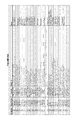
Images


Classifications
-
- C—CHEMISTRY; METALLURGY
- C12—BIOCHEMISTRY; BEER; SPIRITS; WINE; VINEGAR; MICROBIOLOGY; ENZYMOLOGY; MUTATION OR GENETIC ENGINEERING
- C12Q—MEASURING OR TESTING PROCESSES INVOLVING ENZYMES, NUCLEIC ACIDS OR MICROORGANISMS; COMPOSITIONS OR TEST PAPERS THEREFOR; PROCESSES OF PREPARING SUCH COMPOSITIONS; CONDITION-RESPONSIVE CONTROL IN MICROBIOLOGICAL OR ENZYMOLOGICAL PROCESSES
- C12Q1/00—Measuring or testing processes involving enzymes, nucleic acids or microorganisms; Compositions therefor; Processes of preparing such compositions
- C12Q1/68—Measuring or testing processes involving enzymes, nucleic acids or microorganisms; Compositions therefor; Processes of preparing such compositions involving nucleic acids
- C12Q1/6813—Hybridisation assays
- C12Q1/6816—Hybridisation assays characterised by the detection means
-
- C—CHEMISTRY; METALLURGY
- C07—ORGANIC CHEMISTRY
- C07H—SUGARS; DERIVATIVES THEREOF; NUCLEOSIDES; NUCLEOTIDES; NUCLEIC ACIDS
- C07H21/00—Compounds containing two or more mononucleotide units having separate phosphate or polyphosphate groups linked by saccharide radicals of nucleoside groups, e.g. nucleic acids
- C07H21/02—Compounds containing two or more mononucleotide units having separate phosphate or polyphosphate groups linked by saccharide radicals of nucleoside groups, e.g. nucleic acids with ribosyl as saccharide radical
-
- C—CHEMISTRY; METALLURGY
- C12—BIOCHEMISTRY; BEER; SPIRITS; WINE; VINEGAR; MICROBIOLOGY; ENZYMOLOGY; MUTATION OR GENETIC ENGINEERING
- C12Q—MEASURING OR TESTING PROCESSES INVOLVING ENZYMES, NUCLEIC ACIDS OR MICROORGANISMS; COMPOSITIONS OR TEST PAPERS THEREFOR; PROCESSES OF PREPARING SUCH COMPOSITIONS; CONDITION-RESPONSIVE CONTROL IN MICROBIOLOGICAL OR ENZYMOLOGICAL PROCESSES
- C12Q1/00—Measuring or testing processes involving enzymes, nucleic acids or microorganisms; Compositions therefor; Processes of preparing such compositions
- C12Q1/68—Measuring or testing processes involving enzymes, nucleic acids or microorganisms; Compositions therefor; Processes of preparing such compositions involving nucleic acids
Definitions
- the present invention relates to compositions and methods for the detection and characterization of nucleic acid molecules (e.g., RNA (e.g., small RNAs such as micro RNAs (miRNAs) and small interfering RNAs (siRNAs)) and other short nucleic acid molecules). More particularly, the present invention relates to methods for the detection and quantification of RNA expression. The present invention further provides for the detection of miRNA and siRNA mutants (e.g., deletion mutants) and variants.
- RNA e.g., small RNAs such as micro RNAs (miRNAs) and small interfering RNAs (siRNAs)
- miRNAs small RNAs
- siRNAs small interfering RNAs
- MicroRNAs are a new class of noncoding RNAs, which are encoded as short inverted repeats in the genomes of invertebrates and vertebrates (Ambros, (2001) Cell 107, 823-826; Moss (2002) Curr. Biol. 12, R138-R140). miRNAs are modulators of target mRNA translation and stability, although most target mRNAs remain to be identified.
- miRNAs sequence-specifically control translation of target mRNAs by binding to sites of antisense complementarity in 3′ untranslated regions (UTRs) (Ambros, supra; Moss, supra; Lagos-Quintana et al., (2001) Science 294, 853-858; Lau et al., (2001) Science 294, 858-862; Lee et al., (2001) Science 294, 862-864).
- mRNAs may also inhibit gene expression by other mechanisms (See, e.g., Pillai et al., Science 309, 1573-1576 (2005); Humphreys et al., Proc Natl Acad Sci USA 102, 16961-16966 (2005)).
- miRNAs such as let-7 RNA, miR-1, miR-34, miR-60, and miR-87, are highly conserved between invertebrates and vertebrates, implicating that they may recognize multiple sites and/or multiple targets of presumably conserved function (Lagos-Quintana et al., supra; Lau et al., supra; Lee et al., supra; Pasquinelli et al., (2000) Nature 408:86).
- the small temporal RNAs (stRNAs) lin-4 and let-7 represent a subclass of miRNAs identified by genetic analysis in Caenorhabditis elegans , which are developmentally regulated and themselves control developmental programs, such as timing of neuronal rewiring, Treasure larva formation, vulva formation, and the terminal differentiation of hypodermal cells.
- miRNAs are typically excised from 60- to 70-nucleotide foldback RNA precursor structures, which are sometimes detected at the onset of miRNA precursor expression (Grishok et al., (2001) Cell 106, 23-34; Hutvagner et al. (2001) Science 93, 834-838; Ketting et al., (2001) Genes Dev. 15, 2654-2659) or during expression of very abundant miRNAs (Lagos-Quintana et al., supra; Lau et al., supra; Lee et al., supra). Generally, only one of the strands of the hairpin precursor molecule is excised and accumulates, presumably because it is protected by associated proteins from RNA degradation.
- the miRNA precursor processing reaction requires Dicer RNase III and Argonaute family members (Grishok et al., supra; Hutvagner et al., supra; Ketting et al., supra).
- small RNAs may find utility in areas of therapeutics and drug discovery (e.g. as drug targets or as pharmaceutical agents).
- drug discovery e.g. as drug targets or as pharmaceutical agents.
- miRNA genes have been associated with cancer (e.g., B-cell chronic lymphocytic leukemia (CLL)), providing a need in the art to be able to detect and characterize miRNA expression (See, e.g., Calin et al., Proc Natl Acad Sci USA, 99, 15524-15529 (2002). In some cases, it may also be important to compare levels of miRNA in different tissue types or before and after application of a stimulus, e.g. a chemical or physical intervention.
- CLL B-cell chronic lymphocytic leukemia
- siRNAs and miRNAs may be present in low amounts in cells, it is desirable that methods of detection be both sensitive and specific. Moreover, for certain applications, it may be beneficial to identify methods suitable for high throughput screening, e.g. homogeneous methods, multiplexed methods, or those suitable to highly parallel automated manipulation and limited temperature changes.
- miRNAs play important roles in the regulation of gene expression, effective techniques for the detection and quantitation of miRNA expression are lacking. Methods used for quantitation of miRNAs have been based on gel electrophoresis. The miRNAs are detected either by Northern blotting or by the presence of radioactive RNase-resistant duplexes. Northern blotting and chip hybridization methods have relatively low analytical sensitivity (Krichevsky et al. 2003), so microgram quantities of RNA are needed for analyses; moreover, transfer of small RNAs to filters can introduce problems with reproducibility of quantitation and is not typically amendable to high-throughput. Moreover, detection methods based on RNase resistance require highly radioactive probes.
- assays based solely on probe hybridization may not provide adequate discrimination between isotypes closely related in sequence.
- Alternative approaches involve cloning the miRNAs and then sequencing the inserts. While this approach may be suitable for discriminating single-base differences between closely related miRNA species, it is time consuming and laborious.
- siRNAs small interfering RNAs
- RNAi RNA interference
- Dicer enzyme RNA-induced silencing complex
- RISC RNA-induced silencing complex
- siRNAs Another class of siRNAs is synthetic and encompasses short duplexes, usually 21-23 nt with characteristic dinucleotide overhangs (Elbashir, S. M. et al., EMBO J. 20: 6877-6888 (2001)) introduced directly into cells via transfection or expression from an introduced vector (Paul, C. P. et al., Nature Biotechnology 20: 505-508 (2002), US Patent Application Publication No. 2003/0148519A1, herein incorporated by reference in its entirety for all purposes).
- siRNAs appear to persist as defined sequences, making them analogous in function and composition to miRNAs (Elbashir, S. M. et al., supra). Efficient and accurate methods of detecting and characterizing (e.g., quantitating) miRNA and siRNA levels are needed.
- the present invention relates to compositions and methods for the detection and characterization of nucleic acid molecules (e.g., RNA (e.g., small RNAs such as micro RNAs (miRNAs) and small interfering RNAs (siRNAs)) and other short nucleic acid molecules. More particularly, the present invention relates to improved methods for the detection and characterization (e.g., quantification) of RNA expression.
- nucleic acid molecules e.g., RNA (e.g., small RNAs such as micro RNAs (miRNAs) and small interfering RNAs (siRNAs)
- siRNAs small interfering RNAs
- the present invention provides a method, comprising: hybridizing at least one nucleic acid (e.g., that contains sequence that is not complementary to the interfering RNA) to a interfering RNA target to generate a detection structure and detecting the detection structure.
- the interfering RNA target is an miRNA.
- the interfering RNA target is an siRNA.
- the siRNA is double stranded.
- the detection structure comprises an invasive cleavage structure.
- the nucleic acid comprises first and second oligonucleotides configured to form an invasive cleavage structure in combination with the miRNA.
- the nucleic acid comprises a first oligonucleotide configured to form an invasive cleavage structure in combination with said miRNA.
- the first oligonucleotide comprises a 5′ portion and a 3′ portion, wherein said 3′ portion is configured to hybridize to said target sequence, and wherein said 5′ portion is configured to not hybridize to said target sequence.
- the second oligonucleotide comprises a 5′ portion and a 3′ portion, wherein said 5′ portion is configured to hybridize to said target sequence, and wherein said 3′ portion is configured to not hybridize to said target sequence.
- the detecting step comprises use of an INVADER assay.
- the detection structure comprises a circular oligonucleotide hybridized to said small RNA to generate a circular detection structure.
- the detecting step comprises use of a rolling circle replication assay.
- the detection structure comprises a nucleic acid molecule with a free 3′-OH group that is extended by a polymerase (e.g., template dependent extension) and the extended sequence is directly or indirectly detected.
- a polymerase e.g., template dependent extension
- the detecting step(s) comprises use of a detection assay including, but not limited to, sequencing assays, polymerase chain reaction assays, hybridization assays, hybridization assays employing a probe complementary to a mutation, microarray assays, bead array assays, primer extension assays, enzyme mismatch cleavage assays, branched hybridization assays, NASBA assays, molecular beacon assays, cycling probe assays, ligase chain reaction assays, invasive cleavage structure assays, ARMS assays, and sandwich hybridization assays.
- the detecting step is carried out in cell lysate.
- the methods of the present invention comprise detecting a second nucleic acid target.
- the second nucleic acid target is RNA.
- the second nucleic acid target is U6 RNA or GAPDH mRNA.
- the nucleic acid used to form the detection structure comprises a template with one or more sites sufficiently complementary to the small RNA so as to allow the RNA to hybridize to the template and be extended in an extension reaction.
- the extension reaction is a polymerase chain reaction wherein one or more RNAs are used as primers in the polymerase chain reaction.
- a single type of RNA binds to two locations on the template to provide the polymerase chain reaction primers.
- two or more RNAs are used as primers.
- the detection of an amplification product signifies the presence of the two or more RNAs in the sample (i.e., an miRNA multiplex assay).
- RNA is used as a template for modification of a detection complex by extension of a primer across at least part of the RNA template.
- the method comprises detection of a plurality of miRNAs.
- the plurality of miRNAs comprises polymorphisms of the same miRNA.
- the plurality of miRNAs comprises different miRNAs (e.g., Let-7, miR-1, miR-1d, miR-135, miR-15, miR-16, miR-124a, or miR125b).
- kits for conducting any of the above methods.
- the present invention provides kits comprising a nucleic acid configured for forming a detection structure when hybridized to an RNA target sequence.
- the kits are configured to detect an miRNA.
- kits are configured to detect a Let-7, miR-1, miR-135, miR-15, miR-16, miR-1b, miR-124a, or miR125b miRNA.
- kits are configured to co-detect a second RNA target with an miRNA target.
- the present invention also provides a method for detecting a miRNA target, comprising providing (i) a miRNA target; (ii) a first unlabled oligonucleotide; (iii) a second unlabeled oligonucleotide; (iv) a reverse transcriptase; (v) a polymerase; and (vi) a probe oligonucleotide; incubating (i) through (vi) under conditions such that a detection structure forms; and detecting the detection structure.
- the first unlabeled oligonucleotide comprises a first region that is complementary to the miRNA target and a second region that is not complementary to the miRNA target.
- the second unlabeled oligonucleotide comprises a first region that is complementary to a second region of the miRNA target and a second region that is not complementary to the second region of the miRNA target.
- detecting comprises forming an invasive cleavage structure, cleaving the invasive cleavage structure, and detecting the cleavage of the invasive cleavage structure.
- said incubating further comprises incubating with an enzyme capable of cleaving a detection structure and lacking polymerase activity.
- the enzyme is a 5′ nuclease, while in some embodiments, the enzyme comprises a FEN-1 nuclease.
- cleaving the invasive cleavage structure occurs at a temperature of between 45° C. and 60° C. In some embodiments, cleaving the invasive cleavage structure occurs at a temperature of approximately 50° C.
- the first unlabled oligonucleotide is used as a primer for reverse transcription. In some embodiments, the first unlabled oligonucleotide is used as an INVADER oligonucleotide in an invasive cleavage reaction. In some embodiments, (i) through (vi) are present within the same reaction vessel. In some embodiments, the method further comprises providing (vii) a second probe oligonucleotide.
- the first unlabeled oligonucleotide and the reverse transcriptase reverse transcribe the miRNA target.
- the reverse transcribed miRNA target i.e., an miRNA cDNA target
- the amplified reverse transcribed miRNA target forms a detection structure in the presence of the probe oligonucleotide.
- the first unlabeled oligonucleotide comprises nucleic acid sequence such that a duplex of about 6-7 base pairs is formed between the oligonucleotide and the miRNA target.
- the present invention is capable of detecting miRNA present in very small copy numbers. For example, in some embodiments, less than 200 copies of miRNA in a sample are detected. In some embodiments, less than 100 copies of miRNA in a sample are detected.
- the second unlabeled oligonucleotide comprises nucleic acid sequence such that a duplex of about 9 base-pairs is formed between the oligonucleotide and the miRNA target.
- the probe oligonucleotide comprises nucleic acid sequence such that a duplex of about 8-10 base-pairs is formed between the oligonucleotide and the miRNA target or the amplified copy of the miRNA target.
- the second region of the first unlabeled oligonucleotide probe comprises a first portion and a second portion, wherein the first portion and the second portion can hybridize to each other.
- a hairpin structure is formed in the first unlabeled oligonucleotide probe when the first portion and the second portion hybridize to each other.
- the second region of the second unlabeled oligonucleotide probe comprises a first portion and a second portion, wherein the first portion and the second portion can hybridize to each other.
- a hairpin structure is formed in the second unlabeled oligonucleotide probe when the first portion and the second portion hybridize to each other.
- the method further comprises providing (vii) an oligonucleotide complementary to a region of the first unlabeled oligonucleotide probe.
- the method further comprises providing (vii) an oligonucleotide complementary to a region of the second unlabeled oligonucleotide probe.
- cleaving the invasive cleavage structure at a temperature of approximately 50° C. permits high fidelity discrimination of target sequences, although other temperatures may be selected based on sequence, buffer components, etc. for optimum performance.
- the target sequences comprise variant miRNAs of a single species.
- increasing the concentration of the probe oligonucleotide increases the sensitivity of detecting the miRNA target.
- two or more miRNAs are detected.
- detecting comprises use of a labeled probe.
- the labeled probe is fluorescently labeled.
- the labeled probe is configured for FRET detection.
- the labeled probe has a first conformation when not hybridized in a duplex and a second conformation when hybridized in a duplex. In some embodiments, the labeled probe exhibits increased fluorescence when hybridized in a duplex.
- the present invention is not limited by the miRNA detected. Indeed, a variety of miRNAs can be detected using the compositions and methods of the present invention including, but not limited to, Let-7, miR-1, miR-135, miR-15, miR-16, miR125b, miR-1d, and miR124a.
- the present invention also provides a kit comprising one or more of a first unlabled oligonucleotide that comprises a first region that is complementary to a miRNA target and a second region that is not complementary to the miRNA target; a second unlabeled oligonucleotide that comprises a first region that is complementary to a second region of the miRNA target and a second region that is not complementary to the second region of the miRNA target; a reverse transcriptase; a DNA polymerase; a probe oligonucleotide; and an enzyme capable of cleaving a detection structure.
- the detection structure comprises an invasive cleavage structure.
- the kit is configured to detect a miRNA target and at least one other RNA target.
- the kit is configured to detect the miRNA target sequence in a cell lysate.
- FIG. 1 shows a schematic diagram of INVADER oligonucleotides, probe oligonucleotides and FRET cassettes for detecting two different alleles (e.g., differing by a single nucleotide) in a single reaction.
- FIG. 2 shows an exemplary detection structure utilized in some embodiments of the present invention.
- FIG. 3 shows a second exemplary detection structure utilized in some embodiments of the present invention.
- FIG. 4 shows a third exemplary detection structure used in some embodiments of the present invention.
- FIG. 5 shows exemplary oligonucleotides for use with the present invention.
- Bases of the target miRNA are underlined in lower case.
- DNA residues in probe or INVADER oligonucleotides are in regular type.
- Lower case type indicates 2′-O-methyl residues.
- FIG. 6 shows the results of temperature optimization experiments for let-7.
- FIG. 7 shows the results of temperature optimization experiments for let-7.
- FIG. 8 shows the results of limit of detection (LOD) experiments for let-7.
- FIG. 9 shows the results of cross reactivity experiments using let-7 miRNA.
- FIG. 10 shows the results of LOD experiments for miR-1.
- FIG. 11 shows the results of CLEAVASE enzymes IX and XII comparisons using let-7 miRNA.
- FIG. 12 shows a partial sequence alignment of U6 RNA sequences from various organisms.
- FIG. 13 shows the results of temperature optimization experiments for mir-135.
- FIG. 14 shows the results of LOD experiments for mir-135.
- FIG. 15 contains a graphical representation of average counts obtained for the detection of let-7 in cell lysates.
- FIG. 16 shows the results of miRNA and mRNA in cell lysates with and without RNAse A treatment.
- FIG. 17 shows results of invasive cleavage assays comparing the effects of including full-length vs. shortened ARRESTOR oligonucleotides.
- FIG. 18 shows the results of temperature optimization experiments using the assay designs described in FIG. 16 .
- FIG. 19 shows the results of temperature optimization experiments using the 10-mer probe and 12-mer INVADER oligonucleotide designs.
- FIG. 20 shows the results of experiments to compare the LODs of two alternative oligonucleotide designs.
- FIG. 21 shows results comparing the effects of substituting 2′-deoxy residues for some or all of the 2′-O-methyl residues in the probe and INVADER oligonucleotides.
- FIG. 22 shows the results of invasive cleavage assays to detect miR-124a.
- FIG. 23 shows the results of experiments to detect five different miRNA species in total RNA isolated from 20 different tissue types.
- FIG. 24 shows results of experiments testing the effect on miRNA detection of altering probe and oligonucleotide length.
- FIG. 25 shows exemplary invasive cleavage oligonucleotide designs for detection of an siRNA. Lower case residues indicate 2′-O-methyl.
- FIG. 26 shows two probes with the same let-7a hybridizing-region but with different 5′-flap “arm” sequences.
- Let-7a is SEQ ID NO:275; 1496-78-02 is SEQ ID NO:145; 2343-25-01 is SEQ ID NO:196; 1544-82-01 is SEQ ID NO:146.
- FIG. 27 shows a chart depicting copy number versus net signal for probes 1544-82-01 and 2343-25-01 are mixed at a 1 ⁇ M and 10 nM (mix vii) and 1 ⁇ M and 4 nM mix (ix), respectively.
- FIG. 28 shows two oligonucleotide probes with the same U6 RNA hybridizing-region but with different 5′-flap “arm” sequences.
- 1796-59-01 is SEQ ID NO:164; 2343-30-01 is SEQ ID NO:223; 1796-53-01 is SEQ ID NO:161; 1796-53-02 is SEQ ID NO:162.
- FIG. 29 shows a chart depicting copy number versus net signal plots for probes 1796-53-01 and 2343-30-01 are mixed at a 1 ⁇ M and 4 nM mix, respectively.
- FIG. 30 shows a chart depicting copy number versus net signal plots for biplex U6 and Let-7a detection.
- FIG. 31 shows various oligonucleotides generated during development of the present invention for the detection and characterization of miRNAs associated with cancer.
- FIG. 32 depicts the general design for detection of miRNA using an assay comprising a reverse transcription reaction, a polymerase chain reaction and an invasive cleavage assay reaction.
- FIG. 33 shows net signal versus the copy number of Let-7a per reaction.
- FIG. 34 shows net signal as a function of temperature.
- FIG. 35 shows the raw signal of let-7a reactions plotted as a function of let-7a copy number.
- FIG. 36 shows the raw signal of the let-7a stacker and hairpin assays plotted as a function of let-7a RNA copy number.
- FIG. 37 shows the net signal generated by the 1-step and 2-step let-7a assays as a function of let-7a copy number.
- FIG. 38 shows the net signal generated by the let-7a assays with probes 1716-94-1, 1717-94-10 or 1716-94-11 as a function of let-7a copy number.
- FIG. 39 shows the net signal generated by the let-7a assays with probes 1716-94-1 and 1717-94-11 at different ratios as a function of let-7a copy number.
- FIG. 40 shows the net signal generated by the let-7a assays as a function of let-7a, let-7c, let-7e, or let-7f copy number.
- FIG. 41 shows oligonucleotides generated for the detection of miRNAs associated with cancer designed according to guidelines listed in Example 19(M).
- miR-15a is SEQ ID NO:288; miR-16 is SEQ ID NO:289; miR-155 is SEQ ID NO:292; Let-7c is SEQ ID NO:297; Let-7e is SEQ ID NO:295; Let-7f is SEQ ID NO:296; miR-20a is SEQ ID NO:290; miR-427 is SEQ ID NO:293; 2343-67-01 is SEQ ID NO:238; 2343-67-02 is SEQ ID NO:239; 2343-67-03 is SEQ ID NO:240; 2343-67-04 is SEQ ID NO:241; 2343-67-05 is SEQ ID NO:242; 2343-67-06 is SEQ ID NO:243; 2343-67-07 is SEQ ID NO:244; 2343-67-08 is SEQ ID NO:245; 2343-
- FIG. 42 shows additional designs for let-7a and miR-16 with varying lengths of primary probe and PCR primer hybridizing regions.
- Let-7a is SEQ ID NO:294; miR-16 is SEQ ID NO:289; 2343-03-01 is SEQ ID NO:171; 2343-03-02 is SEQ ID NO:172; 2343-03-03 is SEQ ID NO:173; 2343-03-05 is SEQ ID NO:174; 2343-03-06 is SEQ ID NO:175; 2343-03-07 is SEQ ID NO:176; 2343-10-01 is SEQ ID NO:177; 2343-10-02 is SEQ ID NO:178; 2343-10-03 is SEQ ID NO:179; 2343-10-04 is SEQ ID NO:180; 2343-10-05 is SEQ ID NO:181; 2343-10-06 is SEQ ID NO:182; 2343-10-07 is SEQ ID NO:183; 2343-14-01 is SEQ ID NO:184; 2343-14-02 is SEQ ID NO:
- FIG. 43A-E provides a table listing all oligonucleotides, SEQ ID NOs: 144-285, respectively, used in Example 19.
- miRNA refers to micro RNA.
- miRNA target sequence refers to a miRNA that is to be detected (e.g., in the presence of other nucleic acids).
- a miRNA target sequence is a variant of a miRNA.
- RNA detection structure and “detection structure” refer to a structure formed by hybridizing a nucleic acid (e.g., an oligonucleotide) to an RNA target, e.g., an miRNA or siRNA.
- the nucleic acid is a single nucleic acid (e.g., a larger nucleic acid with a small region (or regions) of homology to the miRNA).
- the nucleic acid comprises two nucleic acids (e.g., that hybridize to the miRNA to form a hairpin (e.g., single or double hairpin) structure).
- miRNA detection structures are capable of detection using known nucleic acid detection methods, including, but not limited to, those disclosed herein.
- RNA detection structures are further modified following the hybridization step.
- one or more components of the detection structure provides a template for extension by a nucleic acid polymerase.
- one or more components of the detection structure is contacted with a ligase and ligated to an additional nucleic acid.
- siRNAs refers to short interfering RNAs.
- siRNAs comprise a duplex, or double-stranded region, where each strand of the double-stranded region is about 18 to 25 nucleotides long; the double-stranded region can be as short as 16, and as long as 29, base pairs long, where the length is determined by the antisense strand.
- siRNAs contain from about two to four unpaired nucleotides at the 3′ end of each strand. siRNAs appear to function as key intermediates in triggering RNA interference in invertebrates and in vertebrates, and in triggering sequence-specific RNA degradation during posttranscriptional gene silencing in plants.
- At least one strand of the duplex or double-stranded region of a siRNA is substantially homologous to or substantially complementary to a target RNA molecule.
- the strand complementary to a target RNA molecule is the “antisense” strand; the strand homologous to the target RNA molecule is the “sense” strand and is also complementary to the siRNA antisense strand.
- One strand of the double stranded region need not be the exact length of the opposite strand, thus, one strand may have at least one fewer nucleotides than the opposite complementary strand, resulting in a “bubble” or at least one unmatched base in the opposite strand.
- One strand of the double-stranded region need not be exactly complementary to the opposite strand; thus, the strand, preferably the sense strand, may have at least one mismatched base pair.
- siRNAs may also contain additional sequences; non-limiting examples of such sequences include linking sequences, or loops, which connect the two strands of the duplex region.
- This form of siRNAs may be referred to “si-like RNA”, “short hairpin siRNA” where the short refers to the duplex region of the siRNA, or “hairpin siRNA”.
- Additional non-limiting examples of additional sequences present in siRNAs include stem and other folded structures.
- the additional sequences may or may not have known functions; non-limiting examples of such functions include increasing stability of an siRNA molecule, or providing a cellular destination signal.
- subject and “patient” refer to any organisms including plants, microorganisms and animals (e.g., mammals such as dogs, cats, livestock, and humans).
- the term “INVADER assay reagents” or “invasive cleavage assay reagents” refers to one or more reagents for detecting target sequences, said reagents comprising oligonucleotides capable of forming an invasive cleavage structure in the presence of the target sequence.
- the INVADER assay reagents further comprise an agent for detecting the presence of an invasive cleavage structure (e.g., a cleavage agent).
- the oligonucleotides comprise first and second oligonucleotides, said first oligonucleotide comprising a 5′ portion complementary to a first region of the target nucleic acid and said second oligonucleotide comprising a 3′ portion and a 5′ portion, said 5′ portion complementary to a second region of the target nucleic acid downstream of and contiguous to the first portion.
- the 3′ portion of the second oligonucleotide comprises a 3′ terminal nucleotide not complementary to the target nucleic acid.
- the 3′ portion of the second oligonucleotide consists of a single nucleotide not complementary to the target nucleic acid.
- the first and second oligonucleotides are covalently coupled to one another (e.g., through a linker).
- the INVADER assay reagents further comprise a solid support.
- the one or more oligonucleotides of the assay reagents e.g., first and/or second oligonucleotide, whether bridging or non-bridging
- the INVADER assay reagents further comprise a buffer solution.
- the buffer solution comprises a source of divalent cations (e.g., Mn 2+ and/or Mg 2+ ions).
- Individual ingredients e.g., oligonucleotides, enzymes, buffers, target nucleic acids
- IVADER assay reagent components Individual ingredients (e.g., oligonucleotides, enzymes, buffers, target nucleic acids) that collectively make up INVADER assay reagents.
- the INVADER assay reagents further comprise a third oligonucleotide complementary to a third portion of the target nucleic acid upstream of the first portion of the first target nucleic acid. In yet other embodiments, the INVADER assay reagents further comprise a target nucleic acid. In some embodiments, the INVADER assay reagents further comprise a second target nucleic acid. In yet other embodiments, the INVADER assay reagents further comprise a third oligonucleotide comprising a 5′ portion complementary to a first region of the second target nucleic acid.
- the 3′ portion of the third oligonucleotide is covalently linked to the second target nucleic acid.
- the second target nucleic acid further comprises a 5′ portion, wherein the 5′ portion of the second target nucleic acid is the third oligonucleotide.
- the INVADER assay reagents further comprise an ARRESTOR molecule (e.g., ARRESTOR oligonucleotide).
- the INVADER assay reagents further comprise reagents for detecting a nucleic acid cleavage product.
- one or more oligonucleotides in the INVADER assay reagents comprise a label.
- said first oligonucleotide comprises a label.
- said third oligonucleotide comprises a label.
- the reagents comprise a first and/or a third oligonucleotide labeled with moieties that produce a fluorescence resonance energy transfer (FRET) effect.
- FRET fluorescence resonance energy transfer
- one or more the INVADER assay reagents may be provided in a predispensed format (i.e., premeasured for use in a step of the procedure without re-measurement or re-dispensing).
- selected INVADER assay reagent components are mixed and predispensed together.
- predispensed assay reagent components are predispensed and are provided in a reaction vessel (including but not limited to a reaction tube or a well, as in, e.g., a microtiter plate).
- predispensed INVADER assay reagent components are dried down (e.g., desiccated or lyophilized) in a reaction vessel.
- kits refers to any delivery system for delivering materials.
- reaction assays such delivery systems include systems that allow for the storage, transport, or delivery of reaction reagents (e.g., oligonucleotides, enzymes, etc. in the appropriate containers) and/or supporting materials (e.g., buffers, written instructions for performing the assay etc.) from one location to another.
- reaction reagents e.g., oligonucleotides, enzymes, etc. in the appropriate containers
- supporting materials e.g., buffers, written instructions for performing the assay etc.
- kits include one or more enclosures (e.g., boxes) containing the relevant reaction reagents and/or supporting materials.
- fragmented kit refers to delivery systems comprising two or more separate containers that each contains a subportion of the total kit components.
- the containers may be delivered to the intended recipient together or separately.
- a first container may contain an enzyme for use in an assay, while a second container contains oligonucleotides.
- fragmented kit is intended to encompass kits containing Analyte specific reagents (ASR's) regulated under section 520(e) of the Federal Food, Drug, and Cosmetic Act, but are not limited thereto.
- ASR's Analyte specific reagents
- any delivery system comprising two or more separate containers that each contains a subportion of the total kit components are included in the term “fragmented kit.”
- a “combined kit” refers to a delivery system containing all of the components of a reaction assay in a single container (e.g., in a single box housing each of the desired components).
- kit includes both fragmented and combined kits.
- the present invention provides INVADER assay reagent kits comprising one or more of the components necessary for practicing the present invention.
- the present invention provides kits for storing or delivering the enzymes and/or the reaction components necessary to practice an INVADER assay.
- the kit may include any and all components necessary or desired for assays including, but not limited to, the reagents themselves, buffers, control reagents (e.g., tissue samples, positive and negative control target oligonucleotides, etc.), solid supports, labels, written and/or pictorial instructions and product information, inhibitors, labeling and/or detection reagents, package environmental controls (e.g., ice, desiccants, etc.), and the like.
- kits provide a sub-set of the required components, wherein it is expected that the user will supply the remaining components.
- the kits comprise two or more separate containers wherein each container houses a subset of the components to be delivered.
- a first container e.g., box
- an enzyme e.g., structure specific cleavage enzyme in a suitable storage buffer and container
- a second box may contain oligonucleotides (e.g., INVADER oligonucleotides, probe oligonucleotides, control target oligonucleotides, etc.).
- label refers to any atom or molecule that can be used to provide a detectable (preferably quantifiable) effect, and that can be attached to a nucleic acid or protein. Labels include, but are not limited to, dyes; radiolabels such as 32 P; binding moieties such as biotin; haptens such as digoxygenin; luminogenic, phosphorescent or fluorogenic moieties; mass tags; and fluorescent dyes alone or in combination with moieties that can suppress or shift emission spectra by fluorescence resonance energy transfer (FRET).
- FRET fluorescence resonance energy transfer
- Labels may provide signals detectable by fluorescence, radioactivity, colorimetry, gravimetry, X-ray diffraction or absorption, magnetism, enzymatic activity, characteristics of mass or behavior affected by mass (e.g., MALDI time-of-flight mass spectrometry; fluorescence polarization), and the like.
- a label may be a charged moiety (positive or negative charge) or alternatively, may be charge neutral.
- Labels can include or consist of nucleic acid or protein sequence, so long as the sequence comprising the label is detectable.
- the term “distinct” in reference to signals refers to signals that can be differentiated one from another, e.g., by spectral properties such as fluorescence emission wavelength, color, absorbance, mass, size, fluorescence polarization properties, charge, etc., or by capability of interaction with another moiety, such as with a chemical reagent, an enzyme, an antibody, etc.
- the terms “complementary” or “complementarity” are used in reference to polynucleotides (i.e., a sequence of nucleotides such as an oligonucleotide or a target nucleic acid) related by the base-pairing rules.
- polynucleotides i.e., a sequence of nucleotides such as an oligonucleotide or a target nucleic acid
- sequence “5′-A-G-T-3′” is complementary to the sequence “3′-T-C-A-5′.”
- Complementarity may be “partial,” in which only some of the nucleic acids' bases are matched according to the base pairing rules. Or, there may be “complete” or “total” complementarity between the nucleic acids.
- the degree of complementarity between nucleic acid strands has significant effects on the efficiency and strength of hybridization between nucleic acid strands. This is of particular importance in amplification reactions, as well as detection methods that depend upon binding between nucleic acids. Either term may also be used in reference to individual nucleotides, especially within the context of polynucleotides. For example, a particular nucleotide within an oligonucleotide may be noted for its complementarity, or lack thereof, to a nucleotide within another nucleic acid strand, in contrast or comparison to the complementarity between the rest of the oligonucleotide and the nucleic acid strand.
- homologous refers to a degree of identity. There may be partial homology or complete homology. A partially homologous sequence is one that is less than 100% identical to another sequence.
- hybridization is used in reference to the pairing of complementary nucleic acids. Hybridization and the strength of hybridization (i.e., the strength of the association between the nucleic acids) is influenced by such factors as the degree of complementary between the nucleic acids, stringency of the conditions involved, and the T m of the formed hybrid. “Hybridization” methods involve the annealing of one nucleic acid to another, complementary nucleic acid, i.e., a nucleic acid having a complementary nucleotide sequence. The ability of two polymers of nucleic acid containing complementary sequences to find each other and anneal through base pairing interaction is a well-recognized phenomenon.
- nucleic acid sequence refers to an oligonucleotide which, when aligned with the nucleic acid sequence such that the 5′ end of one sequence is paired with the 3′ end of the other, is in “antiparallel association.”
- Certain bases not commonly found in natural nucleic acids may be included in the nucleic acids of the present invention and include, for example, inosine and 7-deazaguanine. Complementarity need not be perfect; stable duplexes may contain mismatched base pairs or unmatched bases.
- nucleic acid technology can determine duplex stability empirically considering a number of variables including, for example, the length of the oligonucleotide, base composition and sequence of the oligonucleotide, ionic strength and incidence of mismatched base pairs.
- T m is used in reference to the “melting temperature.”
- the melting temperature is the temperature at which a population of double-stranded nucleic acid molecules becomes half dissociated into single strands.
- T m melting temperature
- RNA having a non-coding function e.g., a ribosomal or transfer RNA
- the RNA or polypeptide can be encoded by a full length coding sequence or by any portion of the coding sequence so long as the desired activity or function is retained.
- wild-type refers to a gene or a gene product that has the characteristics of that gene or gene product when isolated from a naturally occurring source.
- a wild-type gene is that which is most frequently observed in a population and is thus arbitrarily designated the “normal” or “wild-type” form of the gene.
- modified,” “mutant,” or “polymorphic” refers to a gene or gene product that displays modifications in sequence and or functional properties (i.e., altered characteristics) when compared to the wild-type gene or gene product. It is noted that naturally-occurring mutants can be isolated; these are identified by the fact that they have altered characteristics when compared to the wild-type gene or gene product.
- heterologous sequence refers to DNA sequences containing a desired heterologous sequence.
- the heterologous sequence is a coding sequence and appropriate DNA sequences necessary for either the replication of the coding sequence in a host organism, or the expression of the operably linked coding sequence in a particular host organism.
- DNA sequences necessary for expression in prokaryotes include a promoter, optionally an operator sequence, a ribosome binding site and possibly other sequences. Eukaryotic cells are known to utilize promoters, polyadenlyation signals and enhancers.
- oligonucleotide as used herein is defined as a molecule comprising two or more deoxyribonucleotides or ribonucleotides, preferably at least 5 nucleotides, more preferably at least about 10-15 nucleotides and more preferably at least about 15 to 30 nucleotides. The exact size will depend on many factors, which in turn depend on the ultimate function or use of the oligonucleotide.
- the oligonucleotide may be generated in any manner, including chemical synthesis, DNA replication, reverse transcription, PCR, or a combination thereof.
- an end of an oligonucleotide is referred to as the “5′ end” if its 5′ phosphate is not linked to the 3′ oxygen of a mononucleotide pentose ring and as the “3′ end” if its 3′ oxygen is not linked to a 5′ phosphate of a subsequent mononucleotide pentose ring.
- a nucleic acid sequence even if internal to a larger oligonucleotide, also may be said to have 5′ and 3′ ends.
- a first region along a nucleic acid strand is said to be upstream of another region if the 3′ end of the first region is before the 5′ end of the second region when moving along a strand of nucleic acid in a 5′ to 3′ direction.
- the former When two different, non-overlapping oligonucleotides anneal to different regions of the same linear complementary nucleic acid sequence, and the 3′ end of one oligonucleotide points towards the 5′ end of the other, the former may be called the “upstream” oligonucleotide and the latter the “downstream” oligonucleotide.
- the first oligonucleotide when two overlapping oligonucleotides are hybridized to the same linear complementary nucleic acid sequence, with the first oligonucleotide positioned such that its 5′ end is upstream of the 5′ end of the second oligonucleotide, and the 3′ end of the first oligonucleotide is upstream of the 3′ end of the second oligonucleotide, the first oligonucleotide may be called the “upstream” oligonucleotide and the second oligonucleotide may be called the “downstream” oligonucleotide.
- primer refers to an oligonucleotide that is capable of acting as a point of initiation of synthesis when placed under conditions in which primer extension is initiated.
- An oligonucleotide “primer” may occur naturally, as in a purified restriction digest or may be produced synthetically.
- a primer is selected to be “substantially” complementary to a strand of specific sequence of the template.
- a primer must be sufficiently complementary to hybridize with a template strand for primer elongation to occur.
- a primer sequence need not reflect the exact sequence of the template.
- a non-complementary nucleotide fragment may be attached to the 5′ end of the primer, with the remainder of the primer sequence being substantially complementary to the strand.
- Non-complementary bases or longer sequences can be interspersed into the primer, provided that the primer sequence has sufficient complementarity with the sequence of the template to hybridize and thereby form a template primer complex for synthesis of the extension product of the primer.
- cleavage structure refers to a structure that is formed by the interaction of at least one probe oligonucleotide and a target nucleic acid, forming a structure comprising a duplex, the resulting structure being cleavable by a cleavage means, including but not limited to an enzyme.
- the cleavage structure is a substrate for specific cleavage by the cleavage means in contrast to a nucleic acid molecule that is a substrate for non-specific cleavage by agents such as phosphodiesterases, which cleave nucleic acid molecules without regard to secondary structure (i.e., no formation of a duplexed structure is required).
- cleavage means or “cleavage agent” as used herein refers to any means that is capable of cleaving a cleavage structure, including but not limited to enzymes.
- Structure-specific nucleases or “structure-specific enzymes” are enzymes that recognize specific secondary structures in a nucleic molecule and cleave these structures.
- the cleavage means of the invention cleave a nucleic acid molecule in response to the formation of cleavage structures; it is not necessary that the cleavage means cleave the cleavage structure at any particular location within the cleavage structure.
- the cleavage means may include nuclease activity provided from a variety of sources including the CLEAVASE enzymes (Third Wave Technologies, Madison, Wis.), the FEN-1 endonucleases (including RAD2 and XPG proteins, and FEN-1 endonucleases derived from archaeabacteria), Taq DNA polymerase and E. coli DNA polymerase I.
- the cleavage means may include enzymes having 5′ nuclease activity (e.g., Taq DNA polymerase (DNAP), E. coli DNA polymerase I).
- the cleavage means may also include modified DNA polymerases having 5′ nuclease activity but lacking synthetic activity.
- cleavage means suitable for use in the methods and kits of the present invention are provided in U.S. Pat. Nos. 5,614,402; 5,795,763; 5,843,669; PCT Appln. Nos WO 98/23774; WO 02/070755A2; and WO0190337A2, each of which is herein incorporated by reference it their entireties.
- thermoostable when used in reference to an enzyme, such as a 5′ nuclease, indicates that the enzyme is functional or active (i.e., can perform catalysis) at an elevated temperature, i.e., at about 55° C. or higher (e.g., including, but not limited to, 60° C., 65° C., 70° C., 75° C., 80° C., 85° C. or 90° C.).
- cleavage products refers to products generated by the reaction of a cleavage means with a cleavage structure (i.e., the treatment of a cleavage structure with a cleavage means).
- non-target cleavage product refers to a product of a cleavage reaction that is not derived from the target nucleic acid. As discussed above, in some of the methods of the present invention, cleavage of the cleavage structure generally occurs within the probe oligonucleotide. The fragments of the probe oligonucleotide generated by this target nucleic acid-dependent cleavage are “non-target cleavage products.”
- probe oligonucleotide in the context of an INVADER assay reaction, refers to an oligonucleotide that interacts with a target nucleic acid to form a cleavage structure in the presence or absence of an INVADER oligonucleotide.
- probe oligonucleotide and target form a cleavage structure and cleavage occurs within the probe oligonucleotide.
- the term “INVADER oligonucleotide” refers to an oligonucleotide that hybridizes to a target nucleic acid at a location near the region of hybridization between a probe and the target nucleic acid, wherein the INVADER oligonucleotide comprises a portion (e.g., a chemical moiety, or nucleotide-whether complementary to that target or not) that overlaps with the region of hybridization between the probe and target.
- the INVADER oligonucleotide contains sequences at its 3′ end that are substantially the same as sequences located at the 5′ end of a probe oligonucleotide.
- ARRESTOR molecule refers to an agent added to or included in an invasive cleavage reaction in order to stop one or more reaction components from participating in a subsequent action or reaction. This may be done by sequestering or inactivating some reaction component (e.g., by binding or base-pairing a nucleic acid component, or by binding to a protein component).
- ARRESTOR oligonucleotide refers to an oligonucleotide included in an invasive cleavage reaction in order to stop or arrest one or more aspects of any reaction (e.g., the first reaction and/or any subsequent reactions or actions; it is not intended that the ARRESTOR oligonucleotide be limited to any particular reaction or reaction step).
- reaction component e.g., base-pairing to another nucleic acid, or binding to a protein component.
- some reaction component e.g., base-pairing to another nucleic acid, or binding to a protein component.
- some reaction component e.g., base-pairing to another nucleic acid, or binding to a protein component.
- the term it is not intended that the term be so limited as to just situations in which a reaction component is sequestered.
- cassette refers to an oligonucleotide or combination of oligonucleotides configured to generate a detectable signal in response to cleavage of a probe oligonucleotide in an INVADER assay.
- the cassette hybridizes to a non-target cleavage product from cleavage of the probe oligonucleotide to form a second invasive cleavage structure, such that the cassette can then be cleaved.
- Secondary cleavage reactions in some preferred embodiments of the present invention include the use of FRET cassettes.
- FRET cassettes Such molecules provide both a secondary target (Secondary Reaction Target or SRT) and a FRET labeled cleavable sequence, allowing homogeneous detection (i.e., without product separation or other manipulation after the reaction) of the sequential invasive cleavage reaction.
- Other preferred embodiments use a secondary reaction system in which the FRET probe and synthetic target are provided as separate oligonucleotides. The cleaved 5′-flaps from a primary reaction act as invasive oligonucleotides in a secondary reaction, in which they bind to the appropriate secondary-reaction target (SRT).
- SRT secondary-reaction target
- the cassette is a single oligonucleotide comprising a hairpin portion (i.e., a region wherein one portion of the cassette oligonucleotide hybridizes to a second portion of the same oligonucleotide under reaction conditions, to form a duplex).
- a cassette comprises at least two oligonucleotides comprising complementary portions that can form a duplex under reaction conditions.
- the cassette comprises a label.
- cassette comprises labeled moieties that produce a fluorescence resonance energy transfer (FRET) effect.
- FRET fluorescence resonance energy transfer
- substantially single-stranded when used in reference to a nucleic acid substrate means that the substrate molecule exists primarily as a single strand of nucleic acid in contrast to a double-stranded substrate which exists as two strands of nucleic acid which are held together by inter-strand base pairing interactions.
- non-amplified oligonucleotide detection assay refers to a detection assay configured to detect the presence or absence of a particular target sequence (e.g., miRNA, SNP, repeat sequence, etc.) that has not been amplified (e.g., by PCR), without creating copies of the target sequence.
- a “non-amplified oligonucleotide detection assay” may, for example, amplify a signal used to indicate the presence or absence of a particular target sequence or polymorphism within a target sequence, so long as the target sequence is not copied.
- non-amplifying oligonucleotide detection assay refers to a detection assay configured to detect the presence or absence of a target sequence (e.g., miRNA, SNP, repeat sequence, etc.), without creating copies of the target sequence.
- a “non-amplifying oligonucleotide detection assay” may, for example, amplify a signal used to indicate the presence or absence of a particular target sequence or polymorphism in a target sequence, so long as the target sequence is not copied.
- sequence variation refers to differences in nucleic acid sequence between two nucleic acids.
- a wild-type structural gene and a mutant form of this wild-type structural gene may vary in sequence by the presence of single base substitutions and/or deletions or insertions of one or more nucleotides. These two forms of the structural gene are said to vary in sequence from one another.
- a second mutant form of the structural gene may exist. This second mutant form is said to vary in sequence from both the wild-type gene and the first mutant form of the gene.
- liberating refers to the release of a nucleic acid fragment from a larger nucleic acid fragment, such as an oligonucleotide, by the action of, for example, a 5′ nuclease such that the released fragment is no longer covalently attached to the remainder of the oligonucleotide.
- K m refers to the Michaelis-Menten constant for an enzyme and is defined as the concentration of the specific substrate at which a given enzyme yields one-half its maximum velocity in an enzyme catalyzed reaction.
- nucleotide analog refers to modified or non-naturally occurring nucleotides including but not limited to analogs that have altered stacking interactions such as 7-deaza purines (i.e., 7-deaza-dATP and 7-deaza-dGTP); base analogs with alternative hydrogen bonding configurations (e.g., such as Iso-C and Iso-G and other non-standard base pairs described in U.S. Pat. No. 6,001,983 to S. Benner and herein incorporated by reference); non-hydrogen bonding analogs (e.g., non-polar, aromatic nucleoside analogs such as 2,4-difluorotoluene, described by B. A. Schweitzer and E. T.
- 7-deaza purines i.e., 7-deaza-dATP and 7-deaza-dGTP
- base analogs with alternative hydrogen bonding configurations e.g., such as Iso-C and Iso-G and other non-standard base pairs described in U
- Nucleotide analogs include nucleotides having modification on the sugar moiety, such as dideoxy nucleotides and 2′-O-methyl nucleotides. Nucleotide analogs include modified forms of deoxyribonucleotides as well as ribonucleotides.
- polymorphic locus is a locus present in a population that shows variation between members of the population (e.g., the most common allele has a frequency of less than 0.95).
- a “monomorphic locus” is a genetic locus at little or no variations seen between members of the population (generally taken to be a locus at which the most common allele exceeds a frequency of 0.95 in the gene pool of the population).
- microorganism as used herein means an organism too small to be observed with the unaided eye and includes, but is not limited to bacteria, virus, protozoans, fungi, and ciliates.
- microbial gene sequences refers to gene sequences derived from a microorganism.
- bacteria refers to any bacterial species including eubacterial and archaebacterial species.
- virus refers to obligate, ultramicroscopic, intracellular parasites incapable of autonomous replication (i.e., replication requires the use of the host cell's machinery).
- sample in the present specification and claims is used in its broadest sense. On the one hand it is meant to include a specimen or culture (e.g., microbiological cultures). On the other hand, it is meant to include both biological and environmental samples.
- a sample may include a specimen of synthetic origin.
- Biological samples may be animal, including human, fluid, solid (e.g., stool) or tissue, as well as liquid and solid food and feed products and ingredients such as dairy items, vegetables, meat and meat by-products, and waste.
- Biological samples may be obtained from all of the various families of domestic animals, as well as feral or wild animals, including, but not limited to, such animals as ungulates, bear, fish, lagomorphs, rodents, etc.
- Environmental samples include environmental material such as surface matter, soil, water and industrial samples, as well as samples obtained from food and dairy processing instruments, apparatus, equipment, utensils, disposable and non-disposable items. These examples are not to be construed as limiting the sample types applicable to the present invention.
- source of target nucleic acid refers to any sample that contains nucleic acids (RNA (e.g., miRNA) or DNA).
- RNA e.g., miRNA
- Particularly preferred sources of target nucleic acids are biological samples including, but not limited to blood, saliva, cerebral spinal fluid, pleural fluid, milk, lymph, sputum and semen.
- An oligonucleotide is said to be present in “excess” relative to another oligonucleotide (or target nucleic acid sequence) if that oligonucleotide is present at a higher molar concentration that the other oligonucleotide (or target nucleic acid sequence).
- an oligonucleotide such as a probe oligonucleotide is present in a cleavage reaction in excess relative to the concentration of the complementary target nucleic acid sequence, the reaction may be used to indicate the amount of the target nucleic acid present.
- the probe oligonucleotide when present in excess, will be present in at least a 100-fold molar excess; typically at least 1 pmole of each probe oligonucleotide would be used when the target nucleic acid sequence was present at about 10 fmoles or less.
- a sample “suspected of containing” a first and a second target nucleic acid may contain either, both or neither target nucleic acid molecule.
- the term “reactant” is used herein in its broadest sense.
- the reactant can comprise, for example, an enzymatic reactant, a chemical reactant or light (e.g., ultraviolet light, particularly short wavelength ultraviolet light is known to break oligonucleotide chains).
- a chemical reactant or light e.g., ultraviolet light, particularly short wavelength ultraviolet light is known to break oligonucleotide chains.
- Any agent capable of reacting with an oligonucleotide to either shorten (e.g., cleave) or elongate the oligonucleotide is encompassed within the term “reactant.”
- purified refers to the removal of contaminants from a sample.
- recombinant CLEAVASE nucleases are expressed in bacterial host cells and the nucleases are purified by the removal of host cell proteins; the percent of these recombinant nucleases is thereby increased in the sample.
- portion when in reference to a protein (as in “a portion of a given protein”) refers to fragments of that protein.
- the fragments may range in size from four amino acid residues to the entire amino acid sequence minus one amino acid (e.g., 4, 5, 6, . . . , n ⁇ 1).
- nucleic acid sequence refers to an oligonucleotide, nucleotide or polynucleotide, and fragments or portions thereof, and to DNA or RNA of genomic or synthetic origin, which may be single or double stranded, and represent the sense or antisense strand.
- amino acid sequence refers to peptide or protein sequence.
- the terms “purified” or “substantially purified” refer to molecules, either nucleic or amino acid sequences, that are removed from their natural environment, isolated or separated, and are at least 60% free, preferably 75% free, and most preferably 90% free from other components with which they are naturally associated.
- An “isolated polynucleotide” or “isolated oligonucleotide” is therefore a substantially purified polynucleotide.
- continuous strand of nucleic acid is means a strand of nucleic acid that has a continuous, covalently linked, backbone structure, without nicks or other disruptions.
- the disposition of the base portion of each nucleotide, whether base-paired, single-stranded or mismatched, is not an element in the definition of a continuous strand.
- the backbone of the continuous strand is not limited to the ribose-phosphate or deoxyribose-phosphate compositions that are found in naturally occurring, unmodified nucleic acids.
- a nucleic acid of the present invention may comprise modifications in the structure of the backbone, including but not limited to phosphorothioate residues, phosphonate residues, 2′ substituted ribose residues (e.g., 2′-O-methyl ribose) and alternative sugar (e.g., arabinose) containing residues.
- modifications in the structure of the backbone including but not limited to phosphorothioate residues, phosphonate residues, 2′ substituted ribose residues (e.g., 2′-O-methyl ribose) and alternative sugar (e.g., arabinose) containing residues.
- continuous duplex refers to a region of double stranded nucleic acid in which there is no disruption in the progression of basepairs within the duplex (i.e., the base pairs along the duplex are not distorted to accommodate a gap, bulge or mismatch with the confines of the region of continuous duplex).
- the term refers only to the arrangement of the basepairs within the duplex, without implication of continuity in the backbone portion of the nucleic acid strand.
- Duplex nucleic acids with uninterrupted basepairing, but with nicks in one or both strands are within the definition of a continuous duplex.
- duplex refers to the state of nucleic acids in which the base portions of the nucleotides on one strand are bound through hydrogen bonding the their complementary bases arrayed on a second strand.
- the condition of being in a duplex form reflects on the state of the bases of a nucleic acid.
- the strands of nucleic acid also generally assume the tertiary structure of a double helix, having a major and a minor groove. The assumption of the helical form is implicit in the act of becoming duplexed.
- template refers to a strand of nucleic acid on which a complementary copy is built from nucleoside triphosphates through the activity of a template-dependent nucleic acid polymerase. Within a duplex the template strand is, by convention, depicted and described as the “bottom” strand. Similarly, the non-template strand is often depicted and described as the “top” strand.
- the present invention relates to compositions and methods for the detection and characterization of nucleic acid molecules (e.g., RNA (e.g., small RNAs such as micro RNAs (miRNAs) and small interfering RNAs (siRNAs)) and other short nucleic acid molecules).
- RNA e.g., small RNAs such as micro RNAs (miRNAs) and small interfering RNAs (siRNAs)
- the present invention provides methods of detecting, characterizing and quantitating miRNA expression.
- the present invention provides methods of detecting miRNA expression comprising adding a nucleic acid to a miRNA to aid in detection.
- the resulting “miRNA detection structure” is then detected using any suitable method including, but not limited to, those disclosed herein.
- miRNA as an example. However, it should be understood that the methods may be applied to other small nucleic acid molecules.
- the present invention provides methods of generating miRNA detection structures to aid in the detection of miRNAs.
- miRNAs are small in size (approximately 21 nucleotides (e.g., around 18-25 nucleotides)) and are thus difficult to detect using standardized hybridization methods.
- the methods of the present invention comprise adding a nucleic acid molecule to an miRNA (e.g., via hybridization, extension, or ligation) to generate a detection structure. Such detection structures can then be detected using any suitable method.
- the detection structure described in FIG. 2 is generated for detection of miRNAs.
- two oligonucleotides are annealed to the miRNA to form a double loop or “dumbbell” like structure.
- the dumbbell structure creates a larger region of double-stranded nucleic acid by extending the ends of the miRNA with a double-stranded region of oligonucleotide.
- each end of the miRNA is extended between 2 and 5 nucleotides.
- the ends of the oligonucleotides comprise additional nucleic acid sequences that do not hybridize to the miRNA.
- these additional sequences form invasive cleavage structures (e.g., INVADER assay invasive cleavage structures).
- invasive cleavage structures are detected by the INVADER assay (See e.g., below description).
- oligonucleotides described in Example 18 are utilized to detect miRNAs associated with cancer (e.g., that form an invasive cleavage structure that can be detected by INVADER assay).
- the detection structure described in FIG. 3 is generated for the detection of miRNAs.
- one oligonucleotide is annealed to the miRNA to generate an arched structure.
- the miRNA brings the ends of the oligonucleotide together with greater efficiency than in the absence of the miRNA.
- the ends of the oligonucleotide comprise additional sequences that extend beyond the miRNA and do not hybridize to the miRNA.
- these additional sequences form invasive cleavage structures (e.g., INVADER assay invasive cleavage structures).
- invasive cleavage structures are detected by the INVADER assay (See e.g., below description).
- the resulting ends are ligated to form a circular structure.
- one oligonucleotide is hybridized to a miRNA such that the ends of the oligonucleotide are brought in close proximity (e.g., hybridized to adjacent nucleotides of the miRNA) and are then ligated.
- the detection structures described in FIGS. 24 and 25 are generated.
- either probe or INVADER oligonucleotides are extended to create a single hairpin loop or “half dumbbell” structure.
- oligonucleotides described in Example 18 are utilized to detect miRNAs associated with cancer (e.g., that form an invasive cleavage structure that can be detected by INVADER assay).
- the ends of the oligonucleotides comprise additional nucleic acid sequences that do not hybridize to the miRNA (See, e.g., Examples 19G and 19H). In some embodiments, these additional sequences form invasive cleavage structures (e.g., INVADER assay invasive cleavage structures). In some embodiments, invasive cleavage structures are detected by the INVADER assay (See e.g., below description).
- these additional sequences are complementary to additional oligonucleotides added to reaction mixtures to stabilize a cleavage structure, e.g. an INVADER assay invasive cleavage structure ( FIG. 4 ).
- circular structures generated as described above are detected using a rolling circle replication assay (See e.g., below description of rolling circle replication).
- detection structures are generated from long oligonucleotides (e.g., greater than 50, 100, 1000 or more nucleotides) with short region(s) of homology to miRNAs.
- One or more miRNAs are hybridized to the oligonucleotides to generate detection structures.
- these detection structures are detected by extension of miRNAs (e.g., via ligation or polymerization reactions such as RT-PCR).
- these detection structures are further detected by hybridization to oligonucleotides conjugated to solid supports, such as microspheres, or other surfaces or structures.
- the non-miRNA component is extended or ligated to another nucleic acid and directly or indirectly detected.
- oligonucleotides used to form detection structures comprise one or more nucleotide analogs.
- 2′-O-methyl nucleotides are utilized.
- the present invention is not limited to a particular mechanism. Indeed, an understanding of the mechanism is not necessary to practice the present invention. Nonetheless, it is contemplated that the presence of 2′-O-methyl bases increases the stability of the hybridized detection structure and aids in further detection methods.
- Nucleic Acids e.g., Interfering RNAs
- the present invention provides methods of detecting miRNAs.
- the present invention is not limited to a particular detection assay. Any suitable method may be utilized including, but not limited to, those disclosed herein.
- miRNA detection methods are quantitative.
- the present invention is not limited to a particular mechanism. Indeed, an understanding of the mechanism is not necessary to practice the present invention. Nonetheless, it is contemplated that levels of a particular miRNA in the body are associated with a level of gene expression from their cognate genes.
- the present invention thus provides methods of correlated miRNAs with gene expression of particular genes (e.g., genes involved in disease states (e.g., cancer) or metabolism).
- the methods of the present invention are utilized to determine the presence of abnormal (e.g., high or low) levels of a particular miRNA (e.g., miRNA expression associated with cancer (See, e.g., Calin et al., Proc Natl Acad Sci USA, 99, 15524-15529 (2002), e.g., using oligonucleotides described in Example 18 and FIG. 31 ) or to determine the effect of an intervention (e.g., drug) on miRNA expression.
- a particular miRNA e.g., miRNA expression associated with cancer
- an intervention e.g., drug
- heterologous miRNAs e.g., from expression vectors, transgenic constructs, transfection, etc. are detected to characterize the efficiency of miRNA expression systems.
- the present invention provides methods of detecting a particular miRNA (e.g., a miRNA such as mir-1 or mir-135).
- the methods of the present invention are used to distinguish between variants (e.g., polymorphisms or mutations) in a particular miRNA.
- the present invention provides methods of lysing cells to be tested for the presence of miRNAs.
- the INVADER assay is used for the detection of miRNAs.
- the INVADER assay comprises forming a nucleic acid cleavage structure that is dependent upon the presence of a target nucleic acid and cleaving the nucleic acid cleavage structure so as to release distinctive cleavage products.
- 5′ nuclease activity for example, is used to cleave the target-dependent cleavage structure and the resulting cleavage products or the cleavage of the cleavage structure is indicative of the presence of specific target nucleic acid sequences in the sample.
- invasive cleavage can occur.
- a cleavage agent e.g., a 5′ nuclease
- the cleavage agent can be made to cleave the downstream oligonucleotide at an internal site in such a way that a distinctive fragment is produced.
- INVADER assay Third Wave Technologies
- the INVADER assay detects hybridization of probes to a target by enzymatic cleavage of specific structures by structure specific enzymes (See, INVADER assays, Third Wave Technologies; See e.g., U.S. Pat. Nos. 5,846,717; 6,090,543; 6,001,567; 5,985,557; 5,994,069; 6,090,543; 6,348,314; 6,458,535; U.S. Patent App. Nos. 20030186238 (Ser. No. 10/084,839); 20030104378A1 (Ser. No. 09/864,636); Lyamichev et al., Nat. Biotech., 17:292 (1999), Hall et al., PNAS, USA, 97:8272 (2000), WO97/27214 and WO98/42873, each of which is herein incorporated by reference in its entirety for all purposes).
- the INVADER assay detects specific DNA and RNA sequences by using structure-specific enzymes (e.g. FEN endonucleases) to cleave a complex formed by the hybridization of overlapping oligonucleotide probes (See, e.g. FIG. 1 ). Elevated temperature and an excess of one of the probes enable multiple probes to be cleaved for each target sequence present without temperature cycling. In some embodiments, these cleaved probes then direct cleavage of a second labeled probe.
- the secondary probe oligonucleotide can be 5′-end labeled with fluorescein that is quenched by an internal dye. Upon cleavage, the de-quenched fluorescein labeled product may be detected using a standard fluorescence plate reader.
- the INVADER assay detects specific sequences, mutations, and SNPs in unamplified, as well as amplified (See, e.g., Example 19, FIG. 32 ), RNA and DNA, including genomic DNA.
- the INVADER assay uses two cascading steps (a primary and a secondary reaction) both to generate and then to amplify the target-specific signal.
- WT wild-type
- MT mutant
- the WT primary probe and the INVADER oligonucleotide hybridize in tandem to the target nucleic acid to form an overlapping structure.
- An unpaired “flap” is included on the 5′ end of the WT primary probe.
- a structure-specific enzyme e.g. the CLEAVASE enzyme, Third Wave Technologies
- this cleaved product serves as an INVADER oligonucleotide on the WT fluorescence resonance energy transfer (WT-FRET) probe to again create the structure recognized by the structure specific enzyme (panel A).
- FRET probes having different labels are provided for each allele or locus to be detected, such that the different alleles or loci can be detected in a single reaction.
- the primary probe sets and the different FRET probes may be combined in a single assay, allowing comparison of the signals from each allele or locus in the same sample.
- the primary probe oligonucleotide and the target nucleotide sequence do not match perfectly at the cleavage site (e.g., as with the MT primary probe and the WT target, FIG. 1 , panel B), the overlapped structure does not form and cleavage is suppressed.
- the structure specific enzyme e.g., CLEAVASE VIII enzyme, Third Wave Technologies
- each target-specific product can enable the cleavage of many FRET probes.
- the primary INVADER assay reaction is directed against the target DNA (or RNA) being detected.
- the target DNA is the limiting component in the first invasive cleavage, since the INVADER and primary probe are supplied in molar excess.
- the second invasive cleavage it is the released flap that is limiting.
- the INVADER assay, or other nucleotide detection assays are performed with accessible site-designed oligonucleotides and/or bridging oligonucleotides.
- accessible site-designed oligonucleotides and/or bridging oligonucleotides are described in U.S. Pat. Nos. 6,194,149, 6,358,691, 6,355,437, U.S. patent application Ser. No. 09/882,945, and PCT Applications WO9850403, and WO0198537, all of which are specifically incorporated by reference in their entireties.
- the exposing of the sample e.g., nucleic acid sequence (e.g., interfering RNA (e.g., miRNA or siRNA))
- the agent comprises exposing the sample to the oligonucleotides and the agent under conditions wherein an invasive cleavage structure is formed between said target sequence and said oligonucleotides if said target sequence is present in said sample, wherein said invasive cleavage structure is cleaved by said cleavage agent to form a cleavage product.
- the target sequence (e.g. miRNA) comprises a first region and a second region, the second region downstream of and contiguous to the first region, and the oligonucleotides comprise first and second oligonucleotides, wherein at least a portion of the first oligonucleotide is completely complementary to the first portion of the target sequence and wherein the second oligonucleotide comprises a 3′ portion and a 5′ portion, wherein the 5′ portion is completely complementary to the second portion of the target nucleic acid.
- the exposing of the sample to the oligonucleotides and the agent comprises exposing the sample to the oligonucleotides and the agent under conditions wherein an invasive cleavage structure is formed between the target sequence and the oligonucleotides if the target sequence is present in the sample, wherein the invasive cleavage structure is cleaved by the cleavage agent to form a cleavage product.
- the target sequence comprises a first region and a second region, said second region downstream of and contiguous to said first region, and said oligonucleotides comprise first and second oligonucleotides, wherein at least a portion of said first oligonucleotide is completely complementary to said first portion of said target sequence and wherein said second oligonucleotide comprises a 3′ portion and a 5′ portion, wherein said 5′ portion is completely complementary to said second portion of said target nucleic acid.
- kits for assaying a pooled sample e.g., a pooled blood sample or pooled cell lysates
- INVADER detection reagents e.g. primary probe, INVADER probe, and FRET cassette
- the kit further comprises instructions on how to perform the INVADER assay, and in some embodiments, how to apply the INVADER detection assay to pooled samples from many individuals, or to “pooled” samples from many cells (e.g., from a biopsy sample) from a single subject.
- the present invention further provides assays in which the target nucleic acid is reused or recycled during multiple rounds of hybridization with oligonucleotide probes and cleavage of the probes without the need to use temperature cycling (i.e., for periodic denaturation of target nucleic acid strands) or nucleic acid synthesis (i.e., for the polymerization-based displacement of target or probe nucleic acid strands).
- temperature cycling i.e., for periodic denaturation of target nucleic acid strands
- nucleic acid synthesis i.e., for the polymerization-based displacement of target or probe nucleic acid strands.
- a cleavage reaction is run under conditions in which the probes are continuously replaced on the target strand (e.g. through probe-probe displacement or through an equilibrium between probe/target association and disassociation, or through a combination comprising these mechanisms, (Reynaldo et al., J. Mol. Biol. 97: 511-520
- two oligonucleotides hybridize in tandem to the target DNA to form an overlapping structure.
- the 5′-end of the primary probe includes a 5′-flap that does not hybridize to the target DNA ( FIG. 1 ).
- the 3′-nucleotide of the bound INVADER oligonucleotide overlaps the primary probe, but need not hybridize to the target DNA (See, e.g., Examples 15 and 16).
- the CLEAVASE enzyme recognizes this overlapping structure and cleaves off the unpaired 5′-flap of the primary probe, releasing it as a target-specific product.
- the primary probe is designed to have a melting temperature close to the reaction temperature.
- primary probes which are provided in excess, cycle on the target DNA. This allows for multiple rounds of primary probe cleavage for each target DNA, and amplification of the number of released 5′-flaps.
- each released 5′-flap can serve as an INVADER oligonucleotide on a fluorescence resonance energy transfer (FRET) Cassette to create another overlapping structure that is recognized and cleaved by the CLEAVASE enzyme ( FIG. 1 ).
- FRET fluorescence resonance energy transfer
- the fluorophore (F) and quencher (Q) are separated, generating detectable fluorescence signal.
- the released 5′-flap and the FRET Cassette cycle resulting in amplified fluorescence signal.
- the initial and secondary reactions run concurrently in the same well.
- the biplex format of the INVADER DNA Assay enables simultaneous detection of two DNA sequences in a single well (See, e.g., Examples 17 and 19(L)). Most often, this involves detection of two variants of a particular polymorphism (e.g., in a miRNA).
- the biplex format uses two different discriminatory Primary Probes, each with a unique 5′-flap, and two different FRET Cassettes, each with a spectrally distinct fluorophore. By design, the released 5′-flaps will bind only to their respective FRET Cassettes to generate a target-specific signal.
- kits comprising one or more of the components necessary for practicing the present invention.
- the present invention provides kits for storing or delivering the enzymes of the present invention and/or the reaction components necessary to practice a cleavage assay (e.g., the INVADER assay).
- a cleavage assay e.g., the INVADER assay
- kits of the present invention provide the following reagents:
- kits of the present invention are configured for direct detection of RNA. These kits may provide the following reagents:
- the designs of the probes sets are adapted for use in miRNA detection assays using the guidelines for reaction design and optimization provided herein (See e.g., the Experimental Section).
- the reaction temperature is reduced (e.g., to 50-60° C.) to account for the smaller region of hybridization.
- a kit of the present invention provides a list of additional components (e.g., reagents, supplies, and/or equipment) to be supplied by a user in order to perform the methods of the invention.
- additional components e.g., reagents, supplies, and/or equipment
- additional components e.g., reagents, supplies, and/or equipment
- Excitation Emission (Wavelength/Bandwidth) (Wavelength/Bandwidth) 485 nm/20 nm 530 nm/25 nm 560 nm/20 nm 620 nm/40 nm
- a kit of the present invention provides a list of optional components (e.g., reagents, supplies, and/or equipment) to be supplied by a user to facilitate performance of the methods of the invention.
- optional components e.g., reagents, supplies, and/or equipment
- one embodiment of such a list comprises the following:
- a kit of the present invention provides a list of required components to be supplied by a user to facilitate performance of the methods of the invention for which multiple alternatives are acceptable (e.g. sample preparation kits).
- a list of required components to be supplied by a user to facilitate performance of the methods of the invention for which multiple alternatives are acceptable (e.g. sample preparation kits).
- sample preparation kits e.g. sample preparation kits
- protocols for the assembly of INVADER assay reactions e.g., formulations and preferred procedures for making reaction mixtures
- protocols for assembly of reaction mixtures include computational or graphical aids to reduce risk of error in the performance of the methods of the present invention (e.g., tables to facilitate calculation of volumes of reagents needed for multiple reactions, and plate-layout guides to assist in configuring multi-well assay plates to contain numerous assay reactions).
- supplementary documentation such as protocols for ancillary procedures, e.g., for the preparation of additional reagents, or for preparation of samples for use in the methods of the present invention.
- supplementary documentation includes guidelines and lists of precautions provided to facilitate successful use of the methods and kits by unskilled or inexperienced users.
- supplementary documentation includes a troubleshooting guide, e.g., a guide describing possible problems that may be encountered by users, and providing suggested solutions or corrections to intended to aid the user in resolving or avoiding such problems.
- samples are diluted to concentrations that correspond to a 10- ⁇ l addition per reaction.
- concentration of a 100-ng sample should be 15 ng/ ⁇ l.
- rolling circle replication methods are utilized for detection of miRNA detection structures (See e.g., U.S. Pat. Nos. 6,344,329; 6,143,495; 6,316,229; 6,210,884, 6,183,960 and 6,235,502; each of which is herein incorporated by reference).
- rolling circle replication is used to detect circular miRNA detection structures generated from the annealing of the ends of a single oligonucleotide annealed to a miRNA.
- the ends of the oligonucleotide hybridize to the miRNA with no overlap.
- This oligonucleotide can be ligated in the presence or absence of miRNA. However, the ligation reaction is more efficient in the presence of the miRNA. In such embodiments, the level of circular molecules detected over time is compared to a control reaction lacking miRNA.
- the ends of the oligonucleotide hybridize to the miRNA with overlapping ends to generate an invasive cleavage structure.
- Such structures are cleaved prior to ligation, thus improving the specificity of the generation of the circular detection structure.
- Rolling circle amplification involves replication of circular single-stranded DNA molecules.
- a rolling circle replication primer hybridizes to circular nucleic acid molecules followed by rolling circle replication of the nucleic acid molecules using a strand-displacing DNA polymerase. Amplification takes place during rolling circle replication in a single reaction cycle.
- Rolling circle replication results in large DNA molecules containing tandem repeats of the nucleic acid sequence. This DNA molecule is referred to as a tandem sequence DNA (TS-DNA).
- TS-DNA tandem sequence DNA
- LM-RCA ligation-mediated rolling circle amplification
- an probe hybridizes to its cognate target nucleic acid sequence, if present, followed by ligation of the ends of the hybridized probe to form a covalently closed, single-stranded nucleic acid.
- a rolling circle replication primer hybridizes to probe molecules followed by rolling circle replication of the circular molecules using a strand-displacing DNA polymerase.
- LM-RCA comprises mixing an open circle probe with a target sample, resulting in an probe-target sample mixture, and incubating the probe-target sample mixture under conditions promoting hybridization between the open circle probe and a target sequence, mixing ligase with the probe-target sample mixture, resulting in a ligation mixture, and incubating the ligation mixture under conditions promoting ligation of the open circle probe to form an amplification target circle (ATC), mixing a rolling circle replication primer (RCRP) with the ligation mixture, resulting in a primer-ATC mixture, and incubating the primer-ATC mixture under conditions that promote hybridization between the amplification target circle and the rolling circle replication primer, mixing DNA polymerase with the primer-ATC mixture, resulting in a polymerase-ATC mixture, and incubating the polymerase-ATC mixture under conditions promoting replication of the amplification target circle, where replication of the amplification target circle results in formation of tandem sequence DNA (TS-DNA).
- ATC amplification target circle
- RCRP
- the present invention is not limited to INVADER assay or rolling circle assay detection. Any method that allows for the detection of miRNA detection structures may be utilized. Exemplary, non-limiting detection assay that find use in the methods of the present invention are described below.
- detection structures are detected using a hybridization assay.
- a hybridization assay the presence of absence of a given nucleic acid sequence is determined based on the ability of the DNA from the sample to hybridize to a complementary DNA molecule (e.g., a oligonucleotide probe).
- a complementary DNA molecule e.g., a oligonucleotide probe.
- sequences are detected using a DNA chip hybridization assay.
- a DNA chip hybridization assay a series of oligonucleotide probes are affixed to a solid support.
- the oligonucleotide probes are designed to be unique to a given target sequence (e.g., component of a detection complex).
- the sample of interest is contacted with the DNA “chip” and hybridization is detected.
- the DNA chip assay is a GeneChip (Affymetrix, Santa Clara, Calif.; See e.g., U.S. Pat. Nos. 6,045,996; 5,925,525; and 5,858,659; each of which is herein incorporated by reference) assay.
- GeneChip technology uses miniaturized, high-density arrays of oligonucleotide probes affixed to a “chip.” Probe arrays are manufactured by Affymetrix's light-directed chemical synthesis process, which combines solid-phase chemical synthesis with photolithographic fabrication techniques employed in the semiconductor industry.
- the process constructs high-density arrays of oligonucleotides, with each probe in a predefined position in the array. Multiple probe arrays are synthesized simultaneously on a large glass wafer. The wafers are then diced, and individual probe arrays are packaged in injection-molded plastic cartridges, which protect them from the environment and serve as chambers for hybridization.
- the nucleic acid to be analyzed is isolated, amplified by PCR, and labeled with a fluorescent reporter group.
- the labeled DNA is then incubated with the array using a fluidics station.
- the array is then inserted into the scanner, where patterns of hybridization are detected.
- the hybridization data are collected as light emitted from the fluorescent reporter groups already incorporated into the target, which is bound to the probe array. Probes that perfectly match the target generally produce stronger signals than those that have mismatches. Since the sequence and position of each probe on the array are known, by complementarity, the identity of the target nucleic acid applied to the probe array can be determined.
- a DNA microchip containing electronically captured probes (Nanogen, San Diego, Calif.) is utilized (See e.g., U.S. Pat. Nos. 6,017,696; 6,068,818; and 6,051,380; each of which are herein incorporated by reference).
- Nanogen's technology enables the active movement and concentration of charged molecules to and from designated test sites on its semiconductor microchip.
- DNA capture probes unique to a given target sequence are electronically placed at, or “addressed” to, specific sites on the microchip. Since DNA has a strong negative charge, it can be electronically moved to an area of positive charge.
- a test site or a row of test sites on the microchip is electronically activated with a positive charge.
- a solution containing the DNA probes is introduced onto the microchip.
- the negatively charged probes rapidly move to the positively charged sites, where they concentrate and are chemically bound to a site on the microchip.
- the microchip is then washed and another solution of distinct DNA probes is added until the array of specifically bound DNA probes is complete.
- a test sample is then analyzed for the presence of target sequences by determining which of the DNA capture probes hybridize, with target sequences.
- An electronic charge is also used to move and concentrate target molecules to one or more test sites on the microchip.
- the electronic concentration of sample DNA at each test site promotes rapid hybridization of sample DNA with complementary capture probes (hybridization may occur in minutes).
- the polarity or charge of the site is reversed to negative, thereby forcing any unbound or nonspecifically bound DNA back into solution away from the capture probes.
- a laser-based fluorescence scanner is used to detect binding.
- an array technology based upon the segregation of fluids on a flat surface (chip) by differences in surface tension (ProtoGene, Palo Alto, Calif.) is utilized (See e.g., U.S. Pat. Nos. 6,001,311; 5,985,551; and 5,474,796; each of which is herein incorporated by reference).
- Protogene's technology is based on the fact that fluids can be segregated on a flat surface by differences in surface tension that have been imparted by chemical coatings. Once so segregated, oligonucleotide probes are synthesized directly on the chip by ink-jet printing of reagents.
- the array with its reaction sites defined by surface tension is mounted on a X/Y translation stage under a set of four piezoelectric nozzles, one for each of the four standard DNA bases.
- the translation stage moves along each of the rows of the array and the appropriate reagent is delivered to each of the reaction sites.
- the A amidite is delivered only to the sites where amidite A is to be coupled during that synthesis step and so on.
- Common reagents and washes are delivered by flooding the entire surface and then removing them by spinning.
- DNA probes unique for the target sequence (e.g., component of a detection complex) of interest are affixed to the chip using Protogene's technology.
- the chip is then contacted with the PCR-amplified genes of interest.
- unbound DNA is removed and hybridization is detected using any suitable method (e.g., by fluorescence de-quenching of an incorporated fluorescent group).
- a “bead array” is used for the detection of polymorphisms (Illumina, San Diego, Calif.; See e.g., PCT Publications WO 99/67641 and WO 00/39587, each of which is herein incorporated by reference).
- Illumina uses a BEAD ARRAY technology that combines fiber optic bundles and beads that self-assemble into an array. Each fiber optic bundle contains thousands to millions of individual fibers depending on the diameter of the bundle.
- the beads are coated with an oligonucleotide specific for the detection of a given SNP or mutation. Batches of beads are combined to form a pool specific to the array.
- the BEAD ARRAY is contacted with a prepared subject sample (e.g., nucleic acid sample). Hybridization is detected using any suitable method.
- hybridization is detected by enzymatic cleavage of specific structures.
- hybridization of a bound probe is detected using a TaqMan assay (PE Biosystems, Foster City, Calif.; See e.g., U.S. Pat. Nos. 5,962,233 and 5,538,848, each of which is herein incorporated by reference).
- the assay is performed during a PCR reaction.
- the TaqMan assay exploits the 5′-3′ exonuclease activity of the AMPLITAQ GOLD DNA polymerase.
- a probe, specific for a given allele or mutation, is included in the PCR reaction.
- the probe consists of an oligonucleotide with a 5′-reporter dye (e.g., a fluorescent dye) and a 3′-quencher dye.
- the 5′-3′ nucleolytic activity of the AMPLITAQ GOLD polymerase cleaves the probe between the reporter and the quencher dye.
- the separation of the reporter dye from the quencher dye results in an increase of fluorescence.
- the signal accumulates with each cycle of PCR and can be monitored with a fluorimeter.
- polymorphisms are detected using the SNP-IT primer extension assay (Orchid Biosciences, Princeton, N.J.; See e.g., U.S. Pat. Nos. 5,952,174 and 5,919,626, each of which is herein incorporated by reference).
- SNPs are identified by using a specially synthesized DNA primer and a DNA polymerase to selectively extend the DNA chain by one base at the suspected SNP location. DNA in the region of interest is amplified and denatured. Polymerase reactions are then performed using miniaturized systems called microfluidics. Detection is accomplished by adding a label to the nucleotide suspected of being at the target sequence location. Incorporation of the label into the DNA can be detected by any suitable method (e.g., if the nucleotide contains a biotin label, detection is via a fluorescently labeled antibody specific for biotin).
- Additional detection assays useful in the detection of miRNA detection structures include, but are not limited to, enzyme mismatch cleavage methods (e.g., Variagenics, U.S. Pat. Nos. 6,110,684, 5,958,692, 5,851,770, herein incorporated by reference in their entireties); polymerase chain reaction (See, e.g., Example 19 and FIG. 32 ); branched hybridization methods (e.g., Chiron, U.S. Pat. Nos. 5,849,481, 5,710,264, 5,124,246, and 5,624,802, herein incorporated by reference in their entireties); NASBA (e.g., U.S. Pat. No.
- molecular beacon technology e.g., U.S. Pat. No. 6,150,097, herein incorporated by reference in its entirety
- E-sensor technology Motorola, U.S. Pat. Nos. 6,248,229, 6,221,583, 6,013,170, and 6,063,573, herein incorporated by reference in their entireties
- cycling probe technology e.g., U.S. Pat. Nos. 5,403,711, 5,011,769, and 5,660,988, herein incorporated by reference in their entireties
- Dade Behring signal amplification methods e.g., U.S. Pat. Nos.
- the oligonucleotide design for let-7 is shown in FIG. 5 .
- the oligonucleotide design for mir-1 is shown in FIG. 5 .
- the following primary mixes were made and incubated at 50° C. ⁇ 10° C. in a 96 well plate for 30 minutes.
- a no target master mix was prepared (addition of H 2 O in place of RNA). All reactions were covered with mineral oil to prevent evaporation.
- Probe oligonucleotide SEQ ID NOs: 10 ⁇ M 1 ⁇ l 2, 6, or 9 for let 7; SEQ ID NOs: 12, 16, or 19 for miR-1)
- INVADER oligonucleotide SEQ ID 10 ⁇ M 1 ⁇ l NOs: 1, 5, or 8 for let 7; SEQ ID NOs: 11, 15, or 18 for miR-1)
- CLEAVASE IX or XII enzyme 40 ng/ ⁇ l CLEAVASE 0.5 ⁇ l IX enzyme or 60 ng/ ⁇ l CLEAVASE XII enzyme tRNA 20 ng/ ⁇ l 1.5 ⁇ l
- Synthetic miRNA SEQ ID NO: 100 pM 2 ⁇ l 4 for let-7a; SEQ ID NO: 14 for miR-1) Total 10 ⁇ l
- the plate was read in a CYTOFLUOR 4000 fluorescence microplate reader using an excitation wavelength of 485 nm and emission wavelength of 530 nm. Results are shown in FIGS. 6 and 7 .
- Stacking of the 5′-end of the INVADER oligonucleotide to the 3′-end of the miRNA is enhanced when the 3′-end of the INVADER oligonucleotide is 2′-O-methylated.
- 2′-O-methylation of the 5′-end of the INVADER oligonucleotide increases the reaction temperature.
- Extending the 2′-O-methylated bases of the INVADER oligonucleotide so that they base pair with the first two bases of the miRNA (SEQ ID NO: 8 (1496-96-02) vs. SEQ ID NO: 23 (1496-96-03) in design SEQ ID NO: 9 (1496-96-01R) of let-7a) increases the temperature optimum of the described reaction but does not enhance the detection.
- the LOD for let-7 and mir-1 was next tested on human RNA samples.
- the protocol described above was utilized. 50-100 ng of tissue specific total human RNA samples (Clonetech, Palo Alto, Calif.) was used. Results are shown in FIGS. 8 and 10 .
- tissue specific total human RNA samples (Clonetech, Palo Alto, Calif.) was used. Results are shown in FIGS. 8 and 10 .
- the let-7a INVADER assay detects the same tissue expression profile as seen before for let-7a expression levels depending on the source of tissue (Pasquinelli et al., 408:86 (2000)).
- Example 4 describes an analysis of the cross reactivity of probe and/or INVADER oligonucleotides directed against one sub-type of let-7 for another sub-type.
- the protocol for synthetic let-7a miRNA setup described in Example 3 was utilized.
- FIG. 5 shows the oligonucleotide designs. The following plate setup was used:
- This Example describes the optimization of CLEAVASE enzymes for use in miRNA assays.
- the protocol for temperature optimization described above was utilized. Either 20 ng of the CLEAVASE IX enzyme (Third Wave Technologies, Madison, Wis.) or 30 ng of the CLEAVASE XII enzyme (Third Wave Technologies, Madison, Wis.) was used. The following buffer was used for the CLEAVASE 1 ⁇ enzyme:
- This example describes assay design and LOD determination for miRNA miR135. Experiments were performed as described in Examples 2 and 3 for miR-1. The oligonucleotide designs are described in FIG. 5 . Each of the designs (A-D) utilizes different INVADER and probe oligonucleotides for the detection of mir-135 miRNA. Results of the temperature optimization experiments comparing performance of all of the designs are shown in FIG. 13 . Design D gave the highest signal. Results of LOD experiments using assay design D are shown in FIG. 14 .
- FIG. 14A presents the raw counts generated from four replicate assays at each of the indicated target concentrations.
- the average counts obtained with each target concentration are indicated as are the net signal and fold-over-zero (FOZ).
- the limit of detection of the miR-135 target in this experiment was 164 zmoles, equivalent to 98,743 molecules.
- FIG. 14B contains a graphical representation of the average counts obtained at each concentration and indicates that the INVADER assay is linear throughout much of the concentration range tested.
- RNA generally present in all cells at constant levels along with one or more miRNA species, which may be expressed in a tissue-specific manner.
- INVADER assays were therefore designed to two distinct RNAs generally found in all cell types: human glyceraldehydes-3-phosphate dehydrogenase (hGAPDH) and U6 RNA.
- oligonucleotides have been used in biplex miRNA detection assays: INVADER oligonucleotide (SEQ ID NO: 41); probe (SEQ ID NO: 42); ARRESTOR oligonucleotide (SEQ ID NO: 43); SRT oligonucleotide (SEQ ID NO:49), FRET oligonucleotide (red dye) (SEQ ID NO: 48).
- INVADER oligonucleotide SEQ ID NO: 41
- probe SEQ ID NO: 42
- ARRESTOR oligonucleotide SEQ ID NO: 43
- SRT oligonucleotide SEQ ID NO:49
- FRET oligonucleotide red dye
- This example describes detection of the let-7 miRNA directly in total cell RNA as well as in uninduced fibroblast cells from a human osteosarcoma cell line, MG63 (Third Wave Technologies, Madison, Wis.; catalog number CRL-1427).
- Total cell RNA was extracted using TRIZOL (Gibco-BRL), as previously described (Chomczynski et al., Anal. Biochem. 162: 156-156 (1987)), and cell lysates were prepared as described by Eis et al., Nature Biotechnology, 19: 673-6 (2001); both publications are herein incorporated by reference.
- Reactions were set up as follows. Aliquots of 5 ⁇ l of either cell lysate, synthetic miRNA target in lysis buffer (Eis et al., Nature Biotechnology, 19: 673-6 (2001)) at the indicated concentrations, or 5 ⁇ l of 20 ng/ ⁇ l of tRNA (for the no target controls) were pipetted into the appropriate wells of a microtiter plate. A primary reaction master mix was made for 96 reactions containing the following reagents.
- Probe Probe 20 Amount per Total added to reaction Master Reagent Stock concentration ( ⁇ l) Mix ( ⁇ l) Mixture of Probe Probe 20 ⁇ M/ 0.5 45 oligonucleotide INVADER 1496-78-01 R (SEQ oligonucleotide 200 ⁇ M ID NO: 6) and INVADER oligonucleotide 1496-78-02 (SEQ ID NO: 5) CLEAVASE XII 60 ng/ ⁇ l 0.5 45 enzyme Primary Buffer 2.5 X 4 360 TOTAL 5 450
- Hela cells (7.5 ⁇ 10 6 cells) were suspended in 100 ⁇ l of a solution of 10 mM MOPS buffer, pH 7.5, with 100 mM KCl. Aliquots of 10 ⁇ l were added to separate tubes and lysed with 100 ⁇ l of two different lysis buffers prepared as follows:
- PI oligonucleotide mix * 0.25 ⁇ l 1 ⁇ M each* H 2 O 0.25 ⁇ l 0.25 ⁇ l CLEAVASE XII enzyme 0.5 ⁇ l 0.5 ⁇ l 60 ng/ ⁇ l 4 ⁇ l 4 ⁇ l Total 5 ⁇ l 5 ⁇ l *Primary INVADER reactions were as described above for let-7a; PI oligonucleotide mixes were also made for GAPDH (SEQ ID NOs: 41-43) and for U6 (SEQ ID NOs: 45-47 at 50 nM final concentration). Primary reaction mixtures were incubated at 49° C. for 1 hour. Aliquots of the following secondary reaction mixture were then added:
- Secondary reaction mixture included SRTs (SEQ ID NO: 22 for let-7; SEQ ID NO: 49 for GAPDH and U6) target, FRET oligonucleotides (SEQ ID NO: 21 for let-7; SEQ ID NO: 48 for GAPDH and U6), ARRESTORs (SEQ ID NO:7 for let-7, SEQ ID NO:43 for GAPDH, and SEQ ID NO: 47 for U6) at the concentrations indicated in Example 7A.
- This example describes the creation and testing of alternative oligonucleotide designs for detection of the let-7a miRNA.
- a set of alternative designs was created in which the target specific regions of both the INVADER oligonucleotide and the probe oligonucleotide were eleven nucleotides long.
- a second set of designs was created in which the target specific regions of the probe oligonucleotides were 10 nucleotides long and the target specific regions of the INVADER oligonucleotides were 12 nucleotides long.
- FIG. 5 shows sets of alternative oligonucleotide designs for detection of the let-7a miRNA in which the target specific regions of both the probe and INVADER oligonucleotides are 11 nucleotides long.
- SEQ ID NOs: 50-51 provide a design in which both the INVADER and probe oligonucleotides are linear.
- SEQ ID NO: 6 contains a probe oligonucleotide that forms a stem-loop structure); SEQ ID NO: 5, an INVADER oligonucleotide that forms a stem-loop structure); SEQ ID NOs: 5-6, both probe and INVADER oligonucleotides with stem-loops.
- FIG. 5 shows a set of alternative oligonucleotide designs for detection of the let-7a miRNA in which the target specific regions of the probe comprise 10 nucleotides, and those of the INVADER oligonucleotides, 12 nucleotides.
- SEQ ID NOs: 52-53 provide a design in which both the INVADER and probe oligonucleotides are linear.
- SEQ ID NO: 2 contains a probe oligonucleotide that forms a stem-loop structure);
- SEQ ID NO: 1 an INVADER oligonucleotide that forms a stem-loop structure).
- the primary reactions were run at 50 ⁇ 9° C. for 1 hour followed by a 15 minute secondary reaction at 60° C. as described in Example 2.
- the primary reactions were run at 50 ⁇ 9° C. for 15 minutes followed by a 15 minute secondary reaction at 60° C.
- FIG. 18 Results for the designs in which the target specific portions of the INVADER and probe oligonucleotides were 11 nucleotides long are presented in FIG. 18 .
- FIG. 18A shows the temperature optimization profiles of each design.
- FIG. 18B shows the normalized maximum performance of each design, including the optimum temperature for each.
- Results for the designs in which the target specific portion of the probe oligonucleotide was 10 bases and that of the INVADER oligonucleotide was 12 are presented in FIG. 19 .
- FIG. 19 A shows the temperature optimization profiles
- FIG. 19 B the normalized maximum performance of each design.
- Example 3 Experiments were set up as described in Example 3 to compare the LOD of the double loop design and the single loop design, in which the INVADER oligonucleotide forms a stem-loop structure.
- Reactions to determine LOD were run in quadruplicate. Reaction mixtures contained the following reagents (final concentrations):
- results in FIG. 20 show net signal produced as a function of moles of miRNA.
- the linear ranges of the plots indicated that more signal was produced from a given amount of miRNA using the INVADER loop design than from the double loop design.
- an examination of the table in FIG. 20 indicates that the fold-over-zero values at each miRNA level are greater for the single loop design. Both designs resulted in sufficient FOZ at the lowest concentrations tested, 2.68 ⁇ 10 ⁇ 20 moles, or 26.8 zeptomoles, equivalent to approximately 16,000 molecules.
- Amount added Component Stock concentration per reaction PI mix (probe SEQ ID NO: 10 ⁇ M probe 1 ⁇ l 6; INVADER 50 ⁇ M INVADER oligo oligonucleotide SEQ ID NO: 5) CLEAVASE XII enzyme (60 ng/ ⁇ l) 0.5 ⁇ l H 2 O 0.5 ⁇ l Primary Reaction Buffer 2.5 X 4 ⁇ l Total 6 ⁇ l
- Full-length ARRESTOR Component Stock concentration Amount added ARRESTOR 40 ⁇ M 1 ⁇ l SEQ ID NO: 7 for full length ARRESTOR, SEQ ID NO: 54 for shortented ARRESTOR MO5 SRT 1.5 ⁇ M 1 ⁇ l (SEQ ID NO: 22) FRET FAM (SEQ ID 10 ⁇ M 1 ⁇ l NO: 21) H 2 O 2 ⁇ l Secondary reactions were incubated at 60° C. for 1.5 hours. Microtiter plates were read as described in Example 2. The results were as shown in FIG. 17 .
- This example describes experiments aimed at assessing the effect of substituting 2′-deoxy residues for some or all of the 2′-O-methyl residues incorporated in the probe and INVADER oligonucleotides used for detecting miRNAs. All of the designs presented in the preceding examples include 2′-O-methyl residues in the loop regions as described in Example 2. Experiments were conducted to test the effect of substituting 2′ deoxy residues for some or all of the 2′-O-methyl residues in the INVADER and probe oligonucleotides designed to detect the let-7a miRNA.
- FIG. 5 shows the modified let-7a designs.
- SEQ ID NOs: 5-6 contain 2′-O-methyl residues as described in Example 2.
- the design in SEQ ID NOs: 73-74 contain 2′deoxy residues at all positions; and those in SEQ ID NOs: 75-76, 2′-O-methyl residues in the portions of the stems adjacent to the target.
- INVADER reactions were set up to compare the signal generation and temperature optima of the three different designs. Reactions were as described in the LOD experiments in Example 8 and included 100 pM synthetic miRNA, 1 ⁇ M probe, and 10 ⁇ M INVADER oligonucleotide. Primary reactions were run for 15 minutes at the temperatures indicated; secondary reactions were run for 5 minutes at 60° C.
- results of the INVADER assays are shown in FIG. 21 and indicate that the design in which the stem loop structures are comprised of 2′-O-methyl residues yields the most signal, followed by the design in which the bases adjacent to the target are comprised of 2′-O-methyl residues.
- the oligonucleotides comprised entirely of 2′-deoxy residues generated the lowest levels of signal.
- Probe oligonucleotide (as 40 ⁇ M 0.25 ⁇ l indicated in above table)
- CLEAVASE XII enzyme 60 ng/ ⁇ l 0.5 ⁇ l
- RNA e.g. lysate, purified total RNA, synthetic miRNA
- additional experiments in which various oligonucleotides were gel purified as described in Example 1 indicated that standard gel purification of all three types of oligonucleotides gives maximal signal. It is possible to achieve signal levels approximately equal to the maximal levels with gel purified probes if the other oligonucleotides, i.e. the INVADER and ARRESTOR oligonucleotides, are desalted following synthesis.
- This example describes experiments carried out to test the suitability of the INVADER assay to detect different miRNA species in total RNA extracted from diverse tissue types.
- temperature optima and LODs were first determined for each design.
- INVADER assay oligonucleotides were designed to detect the miR-15, miR-16, and miR-125b miRNA species. The designs for these assays are presented in FIG. 5 . The designs for let-7a and miR-135 are described in Examples 2 and 6, respectively.
- Example 8 Temperature optimization experiments were conducted for each of these oligonucleotide sets as described in Example 8. Each primary reaction included 1 nM of the targeted miRNA and was carried out for 15 minutes at temperatures ranging from 50 ⁇ 9° C. Secondary reactions were as described in Example 2 and were run for 1 to 1.5 hours at 60° C. Optimum temperatures were as follows:
- let-7a 53° C. miR-15 53° C. miR-16 56° C. MiR-125b 52° C. MiR-135 45° C.
- let-7a 50 ng of total RNA was tested in each reaction; for the other miRNA species, 100 ng of total RNA was tested. All reactions were set up as described in Example 8; primary reactions were run at the temperature optima for 1.5 hours; secondary reactions were as described above.
- the gene expression profiles for each miRNA species are presented in FIG. 23 . These results indicate that the INVADER assay can be used to examine miRNA expression in different tissue types. These data further suggest that let-7a and miR-125b are expressed in a wide variety of tissues; the other miRNA species appear to be more specific to a limited number of tissue types.
- This example describes the impact of alterations in probe and INVADER oligonucleotide length on detection of the let-7a 22-nt miRNA.
- these experiments compare detection of an miRNA that forms perfect stacking interactions between the ends of the probe and INVADER oligonucleotides to detection of an miRNA that forms both 5′ and 3′ overlaps as well as to one that results in a single nucleotide gap at both the 5′ and 3′ ends.
- FIG. 24 shows the results of analyzing three different types of designs.
- SEQ ID NOs: 5-6 shows a perfect stack between the 22-nt target and the flanking ends of the looped probe and INVADER oligonucleotides.
- SEQ ID NOs: 83-84 both the probe and INVADER oligonucleotides are extended by a single base, resulting in both 5′ and 3′ overlaps.
- SEQ ID NOs: 85-86 both the probe and INVADER oligonucleotides are shortened by a single base, relative to the designs in SEQ ID NOs: 5-6, resulting in a single nucleotide gap at both ends.
- INVADER assays were set up to test the performance of these oligonucleotide sets for detection of synthetic let-7a miRNAs. Reactions were carried out as described in Example 8 and included 100 pM synthetic let-7a miRNA, 1 ⁇ M probe and 10 ⁇ M INVADER oligonucleotide. Primary reactions were run for 15 minutes at 53° C.; secondary reactions, for 5 minutes at 60° C., as described in Example 2. The results are presented in FIG. 24 .
- Precursor let-7 RNA (SEQ ID NO: 87) was transcribed in vitro and analyzed by capillary electrophoresis to determine whether it contained any fragments that might mimic the let-7a miRNA. The shortest contaminating fragment was estimated to be approximately 45 nt.
- LOD reactions were run essentially as described in Example 3 at precursor or synthetic 5′ P let-7a mi-RNA concentrations as indicated in the table below.
- PI mixes contained 10 ⁇ M probe SEQ ID NO: 6 and 100 ⁇ M INVADER oligonucleotide SEQ ID NO: 5. Primary reactions were run at 53° C. for 1 hour; secondary reactions were run at 58° C.
- RNAse A Qiagen, Inc.
- RNAse A When RNAse A was added prior to the primary reaction, no signal was generated, consistent with the previous results. When RNAse A was added subsequent to the primary reaction, no loss of signal was observed, further indicating that the signal being detected is due to RNA and that there is no adverse effect of RNAse on other reaction components, e.g. the CLEAVASE enzyme.
- Oligonucleotide designs were created for miR-124a. These oligonucleotides can be used to detect two naturally occurring miRNAs—one 21 nt in length and the other, 22 nt.
- Temperature optimization reactions were set up, essentially as described in Example 3, using 1 nM of synthetic miRNA target, 25 primary reaction and a 15 minute secondary reaction.
- the oligonucleotides used in these reactions are listed in FIG. 5 (SEQ ID NOs: 90-92).
- Temperature profiles for the two different length miRNA targets are shown in FIG. 22 and indicate that the same oligonucleotide designs can be used to detect both targets.
- FIG. 25 illustrates two alternative INVADER assay designs for detection of a ⁇ -actin siRNA.
- This siRNA is described in Harborth, J. et al., Journal of Cell Science, 114: 4557-4565 (2001).
- One design is presented for each the sense and antisense strands; exemplary oligonucleotides for detecting this siRNA are listed in FIG. 26 , SEQ ID Nos: 101-106.
- oligonucleotide probes with the same let-7a hybridizing-region were designed, but with different 5′-flap “arm” sequences.
- the different 5′-flaps or arms report to FRET cassettes that are designed to generate FAM signal upon cleavage.
- the oligonucleotide sequences were as follows (“Z28” refers to the ECLIPSE quencher, Nanogen, Inc., San Diego, Calif.):
- the nine probe concentration conditions tested were: (i) 1 ⁇ M of probe 1544-82-01, (ii) 1 ⁇ M of probe 2343-25-01, (iii) 1 ⁇ M or both probes 1544-82-01 and 2343-25-01, (iv) 100 nM of probe 2343-25-01, (v) 1 ⁇ M of probe 1544-82-01 and 100 nM of 2343-25-01, (vi) 10 nM of 2343-25-01, (vii) 1 ⁇ M of 1544-82-01 and 10 mM of 2343-25-01, (vii) 4 nM of 2343-25-01, and (ix) 1 ⁇ M of 1544-82-01 and 4 nM of 2343-25-01.
- the reactions contained 1 ⁇ M of INVADER oligo 1496-78-02, 30 ng of CLEAVASE XII enzyme, and 10 mM MOPS pH 7.5, 25 mM KCl, 0.05% Tween 20, 0.05% Nonidet P40, 25 mM MgSO4, and 2% PEG.
- Let-7a synthetic RNA were added to the reaction mix at final concentrations of 6 ⁇ 10 9 , 6 ⁇ 10 8 , 6 ⁇ 10 7 , 6 ⁇ 10 6 , 6 ⁇ 10 5 , 6 ⁇ 10 4 , 6 ⁇ 10 3 , and 0 copies per reaction.
- Let-7a RNA dilutions were made in a solution of 20 ng/ ⁇ l tRNA diluted in Rnase-free water. The reactions were assembled in a 96-well plate and 10 ⁇ l of clear mineral oil were added to each well to prevent evaporation. The plate was then transferred to a thermal-block and incubated at 49° C. for 90 minutes.
- the raw and processed date generated was as follows:
- Results demonstrate that a dynamic range of >6 logs was achieved when probes 1544-82-01 and 2343-25-01 are mixed at a 1 mM and 10 nM (mix vii) and 1 mM and 4 nM mix (ix), respectively.
- oligonucleotide probes with the same U6 RNA hybridizing-region, but with different 5′-flap “arm” sequences were designed.
- the different 5′-flaps or arms report to FRET cassettes that are designed to generate RED signal upon cleavage.
- the oligonucleotide sequences were as follows (RED dye refers to REDMOND RED, Nanogen Inc, San Diego, Calif.):
- Reagents Reaction Buffer 25 mM MOPS pH 7.5, 62.5 mM KCl, 0.125% Tween 20, 0.125% Nonidet P40, 62.5 mM MgSO4, 5% PEG.
- tRNA Carrier 20 ng/ul tRNA yeast in RNase free water CLEAVASE XII enzyme 60 ng/ul CLEAVASE XII enzyme diluted in 20 mM Tris pH 8.0, 50 mM KCl, 0.5% Tween 20, 0.5% Nonidet P40, 50% glycerol, 0.1 mg/ml BSA.
- U6 synthetic RNA were added to the reaction mix at final concentrations of 102 ⁇ 10 9 , 102 ⁇ 10 8 , 102 ⁇ 10 7 , 102 ⁇ 10 6 , 102 ⁇ 10 5 , 102 ⁇ 10 4 , 102 ⁇ 10 3 , 10,200, 5,100, 2,550, and 0 copies per reaction.
- U6 synthetic RNA dilutions were made in a solution of 20 ng/ul tRNA diluted in Rnase-free water. Reactions were assembled in a 96-well plate and 10 ul of clear mineral oil were added to each well to prevent evaporation. The plate was then transferred to a thermal-block at incubated at 50° C. for 90 minutes.
- Results demonstrate that a dynamic range of >6 logs was achieved when probes 1796-53-01 and 2343-30-01 are mixed at a 1 ⁇ M and 4 nM mix, respectively.
- Duplicate reactions were setup at U6 RNA and Let-7a miRNA concentrations of 6 ⁇ 10 9 , 1.2 ⁇ 10 9 , 2.4 ⁇ 10 8 , 4.8 ⁇ 10 7 , 9.6 ⁇ 10 6 , 1.92 ⁇ 10 6 , 384,000, 76,800, 15,360, 3,072, and 0 copies per reaction. Reactions were assembled in a 96-well plate and 10 ⁇ l of clear mineral oil were added to each well to prevent evaporation. The plate was then transferred to a thermal-block at incubated at 49° C. for 90 minutes
- the plate was transferred to a fluorescent plate reader (Cytofluor) and data was acquired by reading the fluorescence using the excitation and emission wavelengths of 560 nm and 620 nm, respectively, with a gain setting of 45 for Red dye and excitation and emission wavelengths of 485 nm and 530, respectively, with a gain setting of 43 for FAM dye.
- Cytofluor fluorescent plate reader
- RNA deletions and downregulation of miRNA genes have been associated with cancer (e.g., B-cell chronic lymphocytic leukemia (CLL)) (See, e.g., Calin et al., Proc Natl Acad Sci USA, 99, 15524-15529 (2002).
- CLL B-cell chronic lymphocytic leukemia
- RT reverse transcription
- PCR polymerase chain reaction
- invasive cleavage reaction assay e.g., in a two-step or single-step reaction (e.g., in a single tube)
- Chemistries utilized for these assays are described, for example, in U.S. Pat. Nos. 6,913,881; 6,875,572, 6,872,816, and 7,011,944, and U.S. patent application Ser. No. 11/266,723, filed Nov. 3, 2005, each of which is hereby incorporated by reference in its entirety.
- the general design for detection of miRNA using this methodology is shown in FIG.
- a reverse transcription (RT) primer oligonucleotide (that, in preferred embodiments, also serves as the INVADER oligonucleotide in an invasive cleavage reaction assay) and a reverse transcriptase are used to reverse transcribe (into cDNA) a target RNA (e.g., miRNA), and, subsequently (e.g., in a two-step assay) or concurrently (e.g., in a one-step assay (e.g., in a single tube)), a polymerase chain reaction (PCR) primer oligonucleotide and DNA polymerase are used for amplification of the cDNA product in the presence of primary probe thereby enabling formation of a detection structure (e.g., that can be detected by an invasive cleavage reaction assay).
- a target RNA e.g., miRNA
- PCR polymerase chain reaction
- an assay that comprises a reverse transcription reaction, a polymerase chain reaction and an invasive cleavage assay reaction.
- these include the length (e.g., through oligonucleotide design) of the RT primer/miRNA duplex formed (e.g., between 4 and 10 (e.g., between 6 and 7) base-pairs); the copy number of miRNA available in a sample for detection; the length (e.g., through oligonucleotide design) of the PCR forward primer/miRNA duplex formed (e.g., between 4 and 10 (e.g., between 7 and 9) base-pairs); concentration of oligonucleotides used; reaction temperature at which assays could be conducted; the influence of stacker oligonucleotides (e.g., oligonucleotides that bind to the RT primer/INVADER oligo
- RNA molecules per reaction e.g., 300,000, 50,000, 8,333, 1,389, 231, 39, and 0.
- Reactions were performed in 25 uL volumes containing 0.5 ⁇ M of forward PCR primer 2343-16-01, 0.034 units/ ⁇ L native Taq polymerase, 2 units/ ⁇ l of MMLV reverse transcriptase, and 6.67 ng/ul of CLEAVASE VIII enzyme in a buffer containing 10 mM MOPS, pH 7.5, 7.5 mM MgCl 2 , and 25 uM dNTPs.
- 0.5 ⁇ M of RT primer 2343-14-01, 0.5 ⁇ M probe 2343-14-05, 0.25 ⁇ M FRET probe 23-211 were added to the reaction mix.
- reaction plate For the 7 base pair duplex length, 0.5 ⁇ M of RT primer 2343-03-01, 0.5 ⁇ M probe 2343-14-08, 0.25 ⁇ M FRET probe 23-755 were added to the reaction mix. Reactions were assembled in a 96-well skirted plate, covered with 10 uL of mineral oil. The plate was then subjected to single steps of 42° C. for 30 minutes followed by 95° C. for 2 minutes, and 30 cycles of 95° C. for 15 seconds, 60° C. for 15 seconds, 72° C. for 45 seconds. After completion of the cycles, the reaction plate was heated to 99° C. for 10 minutes then cooled down to 50° C. for 15 minutes then read on a Cytofluor plate reader using excitation and emission wavelengths of 560 nm and 620 nm, respectively with a gain setting of 45.
- the present invention provides that, when comparing the same copy number of let-7a per reaction, the net signal is higher for 7 base-pair RT primer-miRNA duplex than for the 6 base-pair RT primer-miRNA duplex, although a shorter duplex provides levels of signal sufficient for detection.
- assays of the present invention are able to detect miRNA targets when oligonucleotides with short (e.g., less than 10 nucleotides; less than 8 nucleotides; or less than 7 nucleotides) regions of homology between RT primer/INVADER oligonucleotides and miRNA target are used.
- the RT primer/INVADER oligonucleotide comprises other sequence that does not form a duplex (e.g., that is not complementary) with the target miRNA.
- the other sequence that does not form a duplex forms a hairpin structure (e.g., sequence within the sequence that does not form a duplex with the target RNA is able to fold back and bind with itself).
- RT primer/INVADER oligonucleotides are designed such that a 6 base-pair duplex is formed between the oligonucleotide and the miRNA target. In some embodiments, RT primer oligonucleotides are designed such that a 7 base-pair duplex is formed between the oligonucleotide and the miRNA target.
- an increased length of duplex formation between the RT primer and miRNA target provides a more readily formable detection structure (e.g., when the RT primer oligonucleotide is also used as the primary INVADER oligonucleotide as described in FIG. 32 ) that in turn provides a higher net signal compared to shorter duplexes.
- Reactions were assembled in a 96-well skirted plate and covered with 10 ⁇ L of mineral oil. The plate was then subjected to single steps of 42° C. for 45 minutes followed by 95° C. for 2 minutes, and 30 cycles of 95° C. for 15 seconds, 60° C. for 30 seconds, 72° C. for 60 seconds. After completion of the cycles, the reaction plate was heated to 99° C. for 10 minutes then cooled down to 50° C. for 30 minutes then read on a Cytofluor plate reader using excitation and emission wavelengths of 485 nm and 535 nm, respectively, with a gain setting of 43.
- compositions and methods of the present invention e.g., using an assay (e.g., single-step or two-step assay) comprising a reverse transcription reaction, a polymerase chain reaction and an invasive cleavage assay reaction and a detection structure
- an assay e.g., single-step or two-step assay
- the present invention provides compositions and methods for detecting and characterizing miRNAs that are present in low copy numbers (e.g., less than 500 copies; less than 400 copies; less than 300 copies; less than 200 copies; less than 100 copies; less than 50 copies).
- a RT primer oligonucleotide that is also used as the INVADER oligonucleotide provides the proper amount of association for extension (e.g., from an miRNA template) as well as for recognition by a invasive cleavage enzyme (e.g., CLEAVASE enzyme) involved in the generation and detection of a detection structure capable of detecting of very low copy numbers (e.g., less than 100 (e.g., less than 50)) of a target miRNA.
- a invasive cleavage enzyme e.g., CLEAVASE enzyme
- Reactions were assembled in a 96-well skirted plate, covered with 10 ⁇ L of mineral oil. The plate was then subjected to single steps of 42° C. for 45 minutes followed by 95° C. for 2 minutes, and 30 cycles of 95° C. for 15 seconds, 60° C. for 15 seconds, 72° C. for 60 seconds. After completion of the cycles, the reaction plate was heated to 99° C. for 10 minutes then cooled down to 50° C. for 15 minutes then read on a Cytofluor plate reader using excitation and emission wavelengths of 485 nm and 535 nm, respectively with a gain setting of 43.
- miR-16 Primer-miRNA length (b.p.) (molecules/rxn) 9 9 9 8 8 8 7 7 7 3,000 1608 1662 1634 560 1206 843 230 225 219 1,500 1453 1490 1578 735 221 231 240 231 222 750 948 1277 1035 490 259 365 236 230 231 375 970 959 1374 347 224 226 238 238 232 188 669 859 612 218 233 383 214 240 222 94 594 728 894 338 227 233 238 242 230 47 511 561 437 225 230 247 241 241 240 0 686 189 519 223 227 221 228 228 227
- PCR primer oligonucleotides are designed such that a duplex of about 9 base-pairs is formed between the primer oligonucleotide and the target miRNA.
- the primer oligonucleotide comprises other sequence that does not form a duplex (e.g., that is not complementary) with the target miRNA or cDNA generated therefrom.
- the other sequence that does not form a duplex forms a hairpin structure (e.g., sequence within the sequence that does not form a duplex with the target sequence or cDNA generated therefrom is able to fold back and bind with itself).
- the plate was then subjected to single steps of 42° C. for 45 minutes followed by 95° C. for 2 minutes, and 29 cycles of 95° C. for 15 seconds, 60° C. for 15 seconds, 72° C. for 45 seconds. After completion of the cycles, the reaction plate was heated to 99° C. for 10 minutes then cooled down to 50° C. for 30 minutes then read on a Cytofluor plate reader using excitation and emission wavelengths of 485 nm and 535 nm, respectively, with a gain setting of 43.
- a miRNA detection assay of the present invention e.g., the comprises a reverse transcription reaction, a polymerase chain reaction and an invasive cleavage assay reaction
- utilizes more than one e.g., two, three or more
- probe oligonucleotides in order to increase the sensitivity of detection (e.g., of low copy number miRNAs).
- let-7a 5′- UGAGGUAGUACGUUGUAUAGUU 1716-94-6 5′-CCAGTGCCGATGAGGTAGTA 3′-TATCAAGCGACCTGGCAC 1716-94-3 CGCTGGACCGTG-HEX-3′ 1716-94-8 (let-7a is SEQ ID NO:275; 1716-94-6 is SEQ ID NO:155; 1716-94-3 is SEQ ID NO:153; 1716-94-8 is SEQ ID NO:156)
- the present invention provides that use of primary probes with target hybridization regions of 10 or 11 bases functions well across a broad range of temperatures. Accordingly, in some embodiments, the present invention provides that assays for detecting miRNA (e.g., comprising a reverse transcription reaction, a polymerase chain reaction and an invasive cleavage assay reaction, and a detection structure) comprise incubation with an invasive cleavage enzyme (e.g., CLEAVASE enzyme) at a temperature between 45 and 60° C. (e.g., in some preferred embodiments, at 50° C., in other preferred embodiments at 49° C., in still other preferred embodiments at 48° C., and in further preferred embodiments at 47° C.).
- an invasive cleavage enzyme e.g., CLEAVASE enzyme
- the present invention provides that, in some embodiments, use of any stackers is preferable to no stacker (e.g., at low levels (e.g., copy number) of target miRNA). In some embodiments, at higher levels (e.g., copy number) of target nucleic acid (e.g., miRNA), use of a stacker results in higher signal.
- the use of a stacker oligonucleotide on the PCR forward oligonucleotide primer did not appear to confer any positive benefit on the reaction, and may actually reduce signal in some instances.
- the use of a stacker of the RT primer/invader oligonucleotide provided an enhanced sensitivity for the assay.
- let-7a 5′- UGAGGUAGUAGGUUGUAUAGUU 1716-94-6 5′-CCAGTGCCGATGAGGTAGTA 3′-TATCAAGCGACCTGGCAC 1716-94-3 CGCTGGACCGTG-HEX-3′1716-94-8 (let-7a is SEQ ID NO:275; 1716-94-1 is SEQ ID NO:149; 1716-94-6 is SEQ ID NO:155; 1716 -94-3 is SEQ ID NO:153; 1716-94-8 is SEQ ID NO:156)
- Let-7a hairpin assay was carried as described for the stacker assay except 0.4 ⁇ M 2343-03-1 was used instead of 1716-94-3 and 1716-94-8.
- the raw signal of the let-7a stacker and hairpin assays plotted as a function of let-7a RNA copy number is shown in FIG. 36 .
- the present invention provides that no significant difference in detection capability (e.g., sensitivity) is observed between using a stacker versus a hairpin-forming oligonucleotide.
- Two-step let-7a assay Two-step let-7a assay.
- First step was performed in 20 ⁇ l MMLV reaction buffer (Promega) including 0.25 mM each dNTP, 0.4 ⁇ M 1716-94-3, 0.4 ⁇ M 1716-94-8, 5 unit/ ⁇ l MMLV with 60,000, 6,000, 600 or none copies of let-7a RNA at 37 C for 30 min. MMLV was inactivated at 95 C for 1 min.
- Second step was carried out in 20 ⁇ l 10 mM MOPS pH 7.5, 7.5 mM MgCl 2 , 0.25 mM each dNTP including 0.67 ⁇ M 1716-94-1, 0.4 ⁇ M 1716-94-3, 0.4 ⁇ M 1716-94-6, 0.4 ⁇ M 1716-94-8, 0.033 unit/ ⁇ l TaqPol, 6.7 ng/ul CLEAVASE VIII enzyme; 0.25 ⁇ M 23-210 arm 3-FAM FRET cassette with 2 ⁇ l of the first step reaction sample. Temperature profile was 95 C for 1 min; 27 cycles of 95 C for 30 s then 60 C for 1 min; 99 C for 10 min; 49 C for 30 min.
- the present invention provides that, when correcting for the 1/10 th dilution of template in the 2-step configuration, there is no significant difference in signal strength, limit of detection, or dynamic range, when using either the 1-step or 2-step assay configuration.
- the present invention provides detection of a target nucleic acid (e.g., RNA (e.g., miRNA)) using an assay that comprises a reverse transcription reaction, a polymerase chain reaction, and an invasive cleavage assay reaction all in a single step (e.g., in a single tube), thereby saving time and expense and reducing the potential for sample contamination and/or mishandling.
- a target nucleic acid e.g., RNA (e.g., miRNA)
- an assay that comprises a reverse transcription reaction, a polymerase chain reaction, and an invasive cleavage assay reaction all in a single step (e.g., in a single tube), thereby saving time and expense and reducing the potential for sample contamination and/or mis
- the present invention provides detection of a target nucleic acid (e.g., RNA (e.g., miRNA)) using an assay that comprises a reverse transcription reaction, a polymerase chain reaction, and an invasive cleavage assay reaction in a two step reaction, wherein a portion of nucleic acid (e.g., cDNA) obtained from a reverse transcription reaction step is used in a subsequent step that comprises a polymerase chain reaction and an invasive cleavage reaction assay.
- a target nucleic acid e.g., RNA (e.g., miRNA)
- an assay that comprises a reverse transcription reaction, a polymerase chain reaction, and an invasive cleavage assay reaction in a two step reaction, wherein a portion of nucleic acid (e.g., cDNA) obtained from a reverse transcription reaction step is used in a subsequent step that comprises a polymerase chain reaction and an invasive cleavage reaction assay.
- the net signal generated by the let-7a assays with 1716-94-1, 1716-94-10, or 1716-94-11 probes as a function of let-7a copy number is shown in FIG. 38 .
- the present invention provides that primary probes having target hybridization regions (e.g., that associate with target nucleic acid (e.g., miRNA)) of 8-10 base-pairs in length function at different levels in the assays. Specifically, as the length of the primary probe increases, the signal strength also increases (See, e.g., FIG. 38 , probe 1716-94-1), and when the primary probe length decreases there is also a decrease in the signal strength (See, e.g., FIG. 38 , probe 1716-94-11).
- target hybridization regions e.g., that associate with target nucleic acid (e.g., miRNA)
- target nucleic acid e.g., miRNA
- the present invention provides that concentrations of primary probe from 0.067 ⁇ M-0.33 ⁇ M function in the assays of the present invention, where, in some embodiments, using a higher concentration of probe (e.g., 0.33 ⁇ M) provides an increase in the sensitivity of the assay compared to using a lower concentration (e.g., 0.067 ⁇ M).
- a higher concentration of probe e.g. 0.33 ⁇ M
- a lower concentration e.g. 0.33 ⁇ M
- altering e.g., increasing
- the concentration of a primary probe in a detection assay e.g., comprising a reverse transcription reaction, a polymerase chain reaction, and an invasive cleavage assay reaction and a detection structure
- alters alters (e.g., increases) the sensitivity of the reaction.
- the let-7 variants used were:
- let-7 species let-7a UGAGGUAGUAGGUUGUAUAGUU (let-7a is SEQ ID NO: 275) let-7c ------------------------------------------------------ let-7e -------------------- -- let-7f -----------------A----------------------
- Let-7a assays were carried out in 20 ⁇ l 10 mM MOPS pH 7.5, 7.5 mM MgCl 2 , 0.25 mM each dNTP including 0.67 ⁇ M 1716-94-1, 0.4 ⁇ M 1716-94-3, 0.4 ⁇ M 1716-94-4, 0.4 ⁇ M 1716-94-8, 2 unit/ ⁇ l MMLV, 0.033 unit/ ⁇ l TaqPol, 6.7 ng/ ⁇ l CLEAVASE VIII enzyme; 0.25 ⁇ M 23-210 arm 3-FAM FRET cassette with 6 10 5 , 6 10 4 , 6,000, 600, 60, or none copies of let-7a RNA. Temperature profile was 42 C for 30 min; 95 C for 1 min; 25 cycles of 95 C for 20 s then 60 C for 1 min; 99 C for 6 min; 49 C for 30 min
- the net signal generated by the let-7a assays as a function of let-7a, let-7c, let-7e, or let-7f copy number is shown in FIG. 40 .
- nucleic acid e.g., RNA (e.g., miRNA)
- detection assays e.g., comprising a reverse transcription reaction, a polymerase chain reaction, and an invasive cleavage assay reaction and a detection structure
- display a 100-fold specificity for target nucleic acid e.g., Let-7a
- variant sequences thereof e.g., Let-7c
- let-7a standard Net let-7a signal U6 standard Net U6 signal 6.0E+06 1101 6.0E+08 952 1.2E+06 432 1.2E+08 942 2.4E+05 93 2.4E+07 878 4.8E+04 24 4.8E+06 600 9.6E+03 1 9.6E+05 210 1.9E+03 1 1.9E+05 71 3.8E+02 ⁇ 2 3.8E+04 12
- Clontech samples Clontech samples heart 1064 heart 840 kidney 1212 kidney 842 liver 467 liver 597 lung 1150 lung 774 trachea 73 trachea 158 bone marrow 9 bone marrow 48 thymus 17 thymus 514 prostate 168 prostate 95 skeletal muscle 4 skeletal muscle 52 testis 45 testis 324 uterus 33 uterus 288 fetal liver 41 fetal liver 265 adrenal gland 19 adrenal gland 266 salivary gland 27 salivary gland 257 thyroid 30 thyroid 273
- the present invention provides that two distinct miRNAs can be simultaneously amplified and detected in a single-step reaction (e.g., in a single reaction vessel).
- oligonucleotides were generated during development of the present invention that are capable of detecting a variety of miRNAs associated with cancer.
- the oligonucleotides were designed according to the following guidelines:
- RT primer folds back on itself to form a hairpin that stacks to the 3′-end of the miRNA.
- a DNA oligonucleotide is added to hybridize the RT primer forming a co-axial stack with miRNA once hybridized to it.
- a 2′-O-methylated oligonucleotide is added to hybridize the RT primer forming a co-axial stack with miRNA once hybridized to it.
- Oligonucleotides designed according to these guidelines are depicted in FIG. 41 .
- FIG. 43 A list of all oligonucleotides used in this Example are depicted in FIG. 43 .
Abstract
Description
| CLEAVASE enzyme | Primary Probe Oligos | |
| | INVADER Oligo | |
| FRET Cassette 1 (e.g., F) | ||
| FRET Cassette 2 (e.g., R) | ||
| Mutant DNA controls | ||
| Wild type DNA controls | ||
| “No Target” Blank control | ||
In other embodiments, the kits of the present invention are configured for direct detection of RNA. These kits may provide the following reagents:
| CLEAVASE enzyme | Primary Probe | |
| oligonucleotides | ||
| |
INVADER Oligo | |
| FRET Probe 1 (e.g., F) | ||
| FRET Probe 2 (e.g., R) | ||
| |
||
| |
||
| |
||
| |
||
| Mutant DNA controls | ||
| Wild type DNA controls | ||
| “No Target” Blank control | ||
-
- Clear CHILLOUT-14 liquid wax (MJ Research) or RNase-free, optical grade mineral oil (Sigma, Cat. No. M-5904)
- 96-well polypropylene microplate (MJ Research, Cat. No. MSP-9601)
- Sterile 1.5-ml or 2.0-ml microcentrifuge tubes
- Sterile, DNase/RNase free disposable aerosol barrier pipet tips
- Multichannel pipets (0.5-10 μl, 2, 5-20 μl)
- Thermal cycler or other heat source (e.g., lab oven or heating block).
- Miscellaneous laboratory equipment (tube racks, micropipetors, multichannel pipet, microcentrifuge, vortex mixer).
- Fluorescence microplate reader (a preferred plate reader is top-reading and equipped with light filters have the following characteristics:
| Excitation | Emission | |
| (Wavelength/Bandwidth) | (Wavelength/Bandwidth) | |
| 485 nm/20 nm | 530 nm/25 nm | |
| 560 nm/20 nm | 620 nm/40 nm | |
-
- Sterile 8-tube strip or microplate (optional)
- Disposable plastic trough (optional)
- Plate sealing tape (optional)
-
- QIAGEN QIAAMP Blood Kit
- Gentra Systems PUREGENE Kit
- Gentra Systems GENERATION Products
-
- The following final concentrations were used for all reactions in Examples 1-18, (unless noted otherwise herein):
- Probe=1 μM
- The following final concentrations were used for all reactions in Examples 1-18, (unless noted otherwise herein):
- INVADER=1 μM
- ARRESTOR=2.67 μM
- CLEAVASE XII enzyme=30 ng
All synthetic miRNA oligonucleotides were purchased from Dharmacon and gel purified on 20% denaturing acrylamide. Synthetic miRNAs were used to determine temperature optima (see below) and LOD.- INVADER, probe, and ARRESTOR oligonucleotides were synthesized either by Integrated DNA Technologies (IDT) or Third Wave Technologies and purified on 20% denaturing acrylamide, unless otherwise indicated.
- The following 2.5× primary reaction buffer was used (unless otherwise noted) for all reactions:
- 25 mM MOPS pH 7.5
- 62.5 mM KCl
- 0.125
% Tween 20 - 0.125% Nonidet NP40
- 62.5 mM MgSO4
- 5% PEG
- Unless otherwise noted, all reactions were overlaid with 10 μl mineral oil prior to the first thermal incubation.
- Unless otherwise noted, synthetic miRNAs contained a 5′OH. Experiments comparing detection of 5′ phosphorylated vs. unphosphorylated synthetic miRNA targets indicated that there was no significant difference in the ability of the INVADER assay to detect these two different types of synthetic molecules.
| Stock | Amount | |
| Primary Reaction Components | Concentration | Added |
| Primary Reaction Buffer | 2.5 |
4 μl |
| Probe oligonucleotide (SEQ ID NOs: | 10 |
1 |
| 2, 6, or 9 for |
||
| or 19 for miR-1) | ||
| INVADER oligonucleotide ( |
10 |
1 μl |
| NOs: 1, 5, or 8 for |
||
| 11, 15, or 18 for miR-1) | ||
| CLEAVASE IX or |
40 ng/μl CLEAVASE | 0.5 μl |
| IX enzyme or | ||
| 60 ng/μl CLEAVASE | ||
| | ||
| tRNA | ||
| 20 ng/μl | 1.5 μl | |
| Synthetic miRNA (SEQ ID NO: | 100 |
2 |
| 4 for let-7a; SEQ ID NO: 14 for | ||
| miR-1) | ||
| |
10 μl | |
| Stock | Amount | |
| Secondary Reaction Components | Concentration | Added |
| H2O (or buffer for |
2 μl | |
| assays) | ||
| FAM FRET probe (SEQ ID NO: 21) | 10 |
1 μl |
| Secondary Reaction Target | 1.5 |
1 μl |
| (SEQ ID NO: 22 for let-7; SEQ ID NO: 40 | ||
| for miR-1) | ||
| ARRESTOR Oligonucletide (SEQ ID NOs: 3, | 40 |
1 |
| 7, or 10 for let-7; SEQ ID NOs: | ||
| 13, 17, or 20 for miR-1) | ||
| Total | 5 μl | |
| Component | Stock conc. | Amount Added | |
| Primary reaction buffer | 2.5 X | 4 | |
| Probe | |||
| 10 | 1 μl | ||
| SEQ ID NO: 6 for | |||
| SEQ ID NO: 16 or 19 for miR-1 | |||
| | 10 | 1 μl | |
| SEQ ID NO: 5 for | |||
| SEQ ID NO: 15 or 18 for miR-1 | |||
| | 60 ng/μl | 0.5 | |
| TRNA | |||
| 20 ng/ | 1 μl | ||
| TOTAL | 7.5 μl | ||
2.5 μl of the following miRNA concentrations were added in triplicates or quadruplicates using the following setup:
| (miRNA) | ||
| 1 | 100 | 10 | 1 | 100 | 10 fM | H2O | ||
| A | |
| B | |
| C | |
| D | |
The plate was overlayed with mineral oil (10 μl) and incubated at 50° C. for 2 hrs. After completion of the primary reaction, 5 μl of the following was added to each well and the plates were incubated at 60° C. for 1.5 hrs. The plate was read using the settings described above (see Example 2).
| Secondary Reaction | |||
| Components | Stock Conc. | Amount Added | |
| H2O (or buffer for | 2 μl | ||
| CLEAVASE IX enzyme | |||
| assays) | |||
| FAM FRET Probe ( |
10 |
1 μl | |
| NO: 21) | |||
| Secondary Target (SEQ ID | 1.5 |
1 μl | |
| NO: 22 for let-7; SEQ ID | |||
| NO: 40 for miR-1) | |||
| |
40 |
1 μl | |
| oligonucleotide (SEQ ID | |||
| NO: 7 for |
|||
| NOs: 17 or 20 for miR-1) | |||
| Total | 5 μl | ||
| 10 |
1 |
100 |
10 |
1 |
100 |
10 | H2O | ||
| 1 | 2 | 3 | 4 | 5 | 6 | 7 | 8 | ||
| Let 7 A | A | |
| Let 7 A | B | |
| Let 7 C | C | |
| Let 7 | D | |
| Let | ||
| 7 | E | |
| Let | ||
| 7 | F | |
| Let | ||
| 7 | G | |
| Let | ||
| 7 F | H | |
- 2.5× primary reaction buffer: 25 mM MOPS pH 7.5, 250 mM KCl, 0.125
% Tween 20, - 0.125% Nonidet NP40, 31.25 mM MgSO4, 10% PEG.
- 7.5× secondary reaction buffer: 87.5 mM MgSO4
The following buffer was used for the CLEAVASE XII enzyme: - 2.5× primary reaction buffer: 25 mM MOPS pH 7.5, 62.5 mM KCl, 0.125
% Tween 20, - 0.125% Nonidet NP40, 62.5 mM MgSO4, 5% PEG.
- 7.5× secondary reaction buffer: H2O
The LOD experimental protocol was used with either the CLEAVASE IX or XII enzymes. The LOD was determined for both enzymes. The results are shown inFIG. 11 .
| Amount | |||
| per | Total added to | ||
| reaction | Master | ||
| Reagent | Stock concentration | (μl) | Mix (μl) |
| Mixture of | Probe | 20 μM/ | 0.5 | 45 |
| oligonucleotide | INVADER | |||
| 1496-78-01 R ( | oligonucleotide | 200 μM | ||
| ID NO: 6) and | ||||
| INVADER | ||||
| oligonucleotide | ||||
| 1496-78-02 (SEQ | ||||
| ID NO: 5) | ||||
| |
60 ng/μl | 0.5 | 45 | |
| enzyme | ||||
| Primary Buffer | 2.5 X | 4 | 360 | |
| |
5 | 450 | ||
| Secondary Reaction Components | Stock Conc. | Amount Added |
| H2O (or buffer for |
2 μl | |
| IX enzyme assays) | ||
| FAM FRET probe (SEQ ID NO: | 10 |
1 μl |
| 21) | ||
| Secondary Reaction Target (SRT) | 1.5 |
1 μl |
| SEQ ID NO: 22 | ||
| |
40 |
1 μl |
| ID NO: 7 | ||
| |
5 μl | |
| MOPS lysis w/Mg++ | MOPS lysis w/out |
||
| 180 |
180 |
||
| 0.5 ml NP40 | 0.5 |
||
| 4 ml 0.5 M |
4 ml 0.5 M MOPS | ||
| 0.5 ml 1 M MgCl2 | N/A | ||
| 4.82 ml H2O | 5.32 mls H2O | ||
| 10 |
10 mls | ||
| Amount added per | ||
| Component | reaction | Final concentration |
| PI oligonucleotide mix* | 0.25 | 1 | μM each* |
| H2O | 0.25 μl | 0.25 | μl |
| CLEAVASE XII enzyme | 0.5 μl | 0.5 | |
| 60 ng/ | |||
| 4 | 4 | μl | |
| Total | 5 | 5 | μl |
| *Primary INVADER reactions were as described above for let-7a; PI oligonucleotide mixes were also made for GAPDH (SEQ ID NOs: 41-43) and for U6 (SEQ ID NOs: 45-47 at 50 nM final concentration). | |||
Primary reaction mixtures were incubated at 49° C. for 1 hour. Aliquots of the following secondary reaction mixture were then added:
| Component | Amount added per reaction | ||
| Secondary reaction mixture* | 1.5 μl | ||
| H2O | 3.5 | ||
| Total | |||
| 5 μl | |||
| Volume | ||
| per | Final | |
| Stock concentration | reaction | concentration |
| 2.5 X Primary reaction buffer for the | 4 μl | 1 X |
| CLEAVASE XII enzyme (as described in | ||
| Example 5) | ||
| 10 μM probe* | 1 |
1 |
| 100 μM INVADER oligonucleotide* | 1 |
10 |
| 60 ng/ |
0.5 |
30 ng |
| H2O | 2.5 μl | N/ |
| 30 pM miRNA (for the 11- |
1 |
3 pM OR 1 nM |
| optimizations) OR 10 nM miRNA (for the | ||
| 10-mer probe/12-mer INVADER | ||
| oligonucleotide temperature optimizations) | ||
| 20 ng/μl tRNA (for no target controls only) | 1 |
2 |
| TOTAL | ||
| 10 μl | ||
| *Various combinations of probe and INVADER oligonucleotides were used in this experiment as indicated in FIGS. 16-17. | ||
| Stock concentration | Volume per reaction | Final concentration |
| 2.5 X |
4 μl | 1 X |
| buffer for | ||
| XII enzyme | ||
| 10 μM probe/200 μM | 0.5 |
1 μM probe/20 μM |
| INVADER | INVADER | |
| oligonucleotide mix | oligonucleotide | |
| (sequences as indicated in | ||
| FIG. 20) | ||
| 60 ng/μl CLEAVASE XII | 0.5 |
30 ng |
| | ||
| Total | ||
| 5 μl | ||
| Amount added | ||
| Component | Stock concentration | per reaction |
| PI mix (probe SEQ ID NO: | 10 |
1 |
| 6; |
50 μM INVADER oligo | |
| oligonucleotide SEQ ID | ||
| NO: 5) | ||
| CLEAVASE XII enzyme | (60 ng/μl) | 0.5 μl |
| H2O | 0.5 μl | |
| Primary Reaction Buffer | 2.5 |
4 |
| Total | ||
| 6 μl | ||
| Full-length ARRESTOR |
| Component | Stock concentration | Amount added |
| | 40 | 1 μl |
| SEQ ID NO: 7 for full | ||
| length ARRESTOR, SEQ | ||
| ID NO: 54 | ||
| for shortented ARRESTOR | ||
| MO5 SRT | 1.5 | 1 μl |
| (SEQ ID NO: 22) | ||
| FRET FAM ( | 10 | 1 μl |
| NO: 21) | ||
| H2O | 2 μl | |
Secondary reactions were incubated at 60° C. for 1.5 hours. Microtiter plates were read as described in Example 2. The results were as shown in
| Probe | INVADER oligo | ||
| 1544-71-01 | SEQ ID NO: 55 | 1544-71-02 | SEQ ID NO: 56 |
| 1544-71-01 | SEQ ID NO: 55 | 1796-43-02 | SEQ ID NO: 68 |
| 1544-71-01 | SEQ ID NO: 55 | 1796-43-04 | SEQ ID NO: 70 |
| 1544-71-01 | SEQ ID NO: 55 | 1796-43-06 | SEQ ID NO: 72 |
| 1796-43-01 | SEQ ID NO: 67 | 1544-71-02 | SEQ ID NO: 56 |
| 1796-43-03 | SEQ ID NO: 69 | 1544-71-02 | SEQ ID NO: 56 |
| 1796-43-05 | SEQ ID NO: 71 | 1544-71-02 | SEQ ID NO: 56 |
| 1796-43-03 | SEQ ID NO: 69 | 1796-43-04 | SEQ ID NO: 70 |
| Primary Reaction | ||||
| Component | Stock Concentration | Amount Added | ||
| Probe oligonucleotide (as | 40 | μM | 0.25 μl | |
| indicated in above table) | ||||
| |
40 | μM | 0.25 μl | |
| |
60 | ng/μl | 0.5 μl |
| Primary Reaction Buffer | 2.5 |
4 | ||
| Total | ||||
| 5 μl | ||||
| Secondary reaction | ||
| component | Concentration | Amount Added |
| FAM FRET oligonucleotide | 13.4 μl FAM FRET | 0.75 μl |
| (SEQ ID NO: 21) and | 2 μM SRT | |
| Secondary Reaction Target | ||
| (SRT) (SEQ ID NO: 40) | ||
| |
54 μM | 0.75 μl |
| oligonucleotide (SEQ ID | ||
| NO: 66) | ||
| H2O | 3.5 | |
| Total | ||
| 5 μl | ||
| Probe | 1544-71-01 | 1544-71-01 | |||||||||
| INVADER | 1544-71-02 | NET | FOZ | 1796-43-02 | NET | FOZ | |||||
| 1.09 | 655 | 658 | 662 | 566 | 7.13 | 1.09 amole | 706 | 738 | 777 | 636 | 7.12 |
| amole | |||||||||||
| 0.33 | 314 | 262 | 256 | 185 | 3.00 | 0.33 amole | 281 | 287 | 290 | 182 | 2.75 |
| amole | |||||||||||
| 0.10 | 122 | 138 | 134 | 39 | 1.42 | 0.10 amole | 149 | 150 | 153 | 47 | 1.45 |
| amole | |||||||||||
| 0 amole | 88 | 93 | 96 | 0 amole | 104 | 101 | 107 | ||||
| Probe | 1796-43-01 | 1796-43-03 | |||||||||
| INVADER | 1544-71-02 | NET | FOZ | 1544-71-02 | NET | FOZ | |||||
| 1.09 | 1689 | 1744 | 1895 | 146 | 1.09 | 1.09 amole | 882 | 869 | 847 | 702 | 5.27 |
| amole | |||||||||||
| 0.33 | 1655 | 1717 | 1817 | 99 | 1.06 | 0.33 amole | 335 | 341 | 341 | 175 | 2.06 |
| amole | |||||||||||
| 0.10 | 1692 | 1693 | 1695 | 63 | 1.04 | 0.10 amole | 196 | 209 | 196 | 36 | 1.22 |
| amole | |||||||||||
| 0 amole | 1636 | 1601 | 1654 | 0 amole | 169 | 165 | 159 | ||||
| 1544-71-01 | 1544-71-01 | ||||||||||
| 1796-43-04 | NET | FOZ | 1796-43-06 | NET | FOZ | ||||||
| 1.09 | 676 | 688 | 693 | 579 | 6.43 | 1.09 amole | 625 | 562 | 579 | 501 | 6.69 |
| amole | |||||||||||
| 0.33 | 274 | 275 | 264 | 164 | 2.54 | 0.33 amole | 229 | 215 | 204 | 128 | 2.45 |
| amole | |||||||||||
| 0.10 | 153 | 137 | 143 | 38 | 1.35 | 0.10 amole | 126 | 121 | 112 | 32 | 1.36 |
| amole | |||||||||||
| 0 amole | 111 | 107 | 102 | 0 amole | 94 | 87 | 83 | ||||
| 1796-43-05 | 1796-43-05 | ||||||||||
| 1544-71-02 | NET | FOZ | 1796-43-06 | NET | FOZ | ||||||
| 1.09 | 806 | 824 | 773 | 708 | 8.64 | 1.09 amole | 772 | 752 | 704 | 631 | 6.65 |
| amole | |||||||||||
| 0.33 | 280 | 280 | 262 | 181 | 2.96 | 0.33 amole | 260 | 252 | 251 | 143 | 2.28 |
| amole | |||||||||||
| 0.10 | 144 | 145 | 139 | 50 | 1.54 | 0.10 amole | 140 | 142 | 139 | 29 | 1.26 |
| amole | |||||||||||
| 0 amole | 91 | 95 | 92 | 0 amole | 115 | 109 | 111 | ||||
| let- |
53° C. | ||
| miR-15 | 53° C. | ||
| miR-16 | 56° C. | ||
| MiR- |
52° C. | ||
| MiR-135 | 45° C. | ||
| Sequence | Description | Sequence | 5′-3′ | Notes |
| 1544-82-01 | Probe MO5 Arm | CCGTCGCTGCGTCTACTACCTCA-NH2 | ||
| (SEQ ID NO: 124) | ||||
| 2343-25-01 | Probe MO4 Arm | CCGTCACGCCTCCTACTACCTCA-NH2 | ||
| (SEQ ID NO: 125) | ||||
| 1496-78-02 | INVADER oligo | mGmGmCmAmCmUmUmUmUmGmUmGmCmCAACTATACAACT | m = 2′-O- | |
| (SEQ ID NO: 126) | methyl | |||
| 1581-63-01 | Arrestor for MO5-arm | mUmGmAmGmGmUmAmGmUmAmGmAmCmGmCmAmG | m = 2′-O- | |
| (SEQ ID NO: 127) | probe | methyl | ||
| 2343-25-02 | Arrestor for MO4-arm | mUmGmAmGmGmUmAmGmUmAmGmGmAmGmGmCmG | m = 2′-O- | |
| (SEQ ID NO: 128) | probe | methyl | ||
| 23-182 | FAM FRET probe for | YCACXTGCTTCGTGG | Y = 6FAM, | |
| (SEQ ID NO: 129) | MO5 SRT | X = Z28 | ||
| quencher | ||||
| 2343-23-01 | FAM FRET probe for | YCACXTCGAACGTCG | Y = 6FAM, | |
| (SEQ ID NO: 130) | MO4 SRT | X = Z28 | ||
| quencher | ||||
| 2343-23-02 | MO4 SRT | CGAGGTTCGAAGTGGAGGCGTGACmGmGmU | ||
| (SEQ ID NO: 131) | ||||
| 1107-10-02 | MO5 SRT | CCAGGAAGCAAGTGACGCAGCGACmGmGmU | ||
| (SEQ ID NO: 132) | ||||
| Let-7a | Let-7a synthetic RNA | UGAGGUAGUAGGUUGUAUAGUU | ||
| (SEQ ID NO: 133) | ||||
and are shown in
| 2.5 X |
25 mM MOPS pH 7.5, 62.5 mM KCl, 0.125 |
| Tween | |
| 20, 0.125% Nonidet P40, 62.5 mM | |
| MgSO4, 5% PEG. | |
| |
20 ng/ul tRNA yeast in RNase free water |
| |
60 ng/ul CLEAVASE XII enzyme diluted in |
| 20 mM Tris pH 8.0, 50 mM KCl, 0.5 | |
| Tween | |
| 20, 0.5% Nonidet P40, 50% glycerol, | |
| 0.1 mg/ml BSA. | |
| 1- Raw data |
| Let-7a | Probe conc Mix # |
| copies | i | ii | iii | iv | v | vi | vii | viii | |
| 6 × 10⁹ | 21731 | 16970 | 35383 | 18062 | 36419 | 19680 | 33594 | 19297 | 28291 |
| 6 × 10⁸ | 20369 | 14201 | 29797 | 16429 | 35792 | 18103 | 28532 | 18419 | 25425 |
| 6 × 10⁷ | 19752 | 3772 | 7247 | 15969 | 32775 | 15128 | 22546 | 10576 | 21604 |
| 6 × 10⁶ | 11535 | 997 | 1452 | 4548 | 13603 | 3419 | 11830 | 1998 | 10654 |
| 6 × 10⁵ | 3500 | 726 | 787 | 1254 | 2941 | 1084 | 2759 | 923 | 2540 |
| 6 × 10⁴ | 1415 | 638 | 675 | 751 | 1053 | 750 | 972 | 726 | 1050 |
| 6 × 10³ | 840 | 652 | 665 | 666 | 841 | 671 | 770 | 690 | 756 |
| 6 × 10⁹ | 638 | 635 | 637 | 661 | 800 | 658 | 731 | 694 | 698 |
| 2- Processed data |
| Let-7a | Probe conc Mix # |
| copies | i | ii | iii | iv | v | vi | vii | viii | |
| 6 × 10⁹ | 21093 | 16335 | 34746 | 17401 | 35619 | 19022 | 32863 | 18603 | 27593 |
| 6 × 10⁸ | 19731 | 13566 | 29160 | 15768 | 34992 | 17445 | 27801 | 17725 | 24727 |
| 6 × 10⁷ | 19114 | 3137 | 6610 | 15308 | 31975 | 14470 | 21815 | 9882 | 20906 |
| 6 × 10⁶ | 10897 | 362 | 815 | 3887 | 12803 | 2761 | 11099 | 1304 | 9956 |
| 6 × 10⁵ | 2862 | 91 | 150 | 593 | 2141 | 426 | 2028 | 229 | 1842 |
| 6 × 10⁴ | 777 | 3 | 38 | 90 | 253 | 92 | 241 | 32 | 352 |
| 6 × 10³ | 202 | 17 | 28 | 10 | 41 | 13 | 39 | −4 | 58 |
and is presented in
| Sequence | Description | Sequence | 5′-3′ | Notes |
| 1796-53-01 | Probe 562-86B | ccgccgagatcacCTAATCTTCTCTGTAT-NH2 | ||
| (SEQ ID NO: 134) | Arm | |||
| 2343-30-01 | Probe ER4 arm | AAGCACGCAGCACCTAATCTTCTCTGTAT-NH2 | ||
| (SEQ ID NO: 135) | ||||
| 1796-59-01 | U6 synthetic | rUrUrUrArUrArCrArGrArGrArArGrArUrUrArGrCrArUrGrGrCrCrCrCrUrG | ||
| (SEQ ID NO: 136) | target | rCrGrCrArArGrGrArUrGrUrUrU | ||
| 1796-53-02 | INVADER oligo | CATCCTTGCGCAGGGGCCATGA | ||
| (SEQ ID NO: 137) | ||||
| 1796-53-03 | Arrestor for | mAmUmAmCmAmGmAmGmAmAmGmAmUmUmAmGmGmumGmA | m = 2′-O- | |
| (SEQ ID NO: 138) | probe 1796-53- | | methyl | |
| 01 | ||||
| 2343-30-04 | Arrestor for | mAmUmAmcmAmGmAmGmAmAmGmAmUmUmAmGmGmUmGmC | m = 2′-O- | |
| (SEQ ID NO: 139) | probe 2343-30- | | methyl | |
| 01 | ||||
| 23-181 | RED FRET | YCTCXTTCTCAGTGCG | y = Red dye | |
| (SEQ ID NO: 140) | probe for 562- | and x = Z28 | ||
| 86B arm | ||||
| 2343-30-02 | RED FRET | YCTCXTGCATAGTCCG | y = Red dye | |
| (SEQ ID NO: 141) | probe for ER4 | and x = Z28 | ||
| arm | ||||
| 23-183 | 562-86B SRT | CGCAGTGAGAATGAGGTGATCTCGGCmGmGmU | ||
| (SEQ ID NO: 142) | ||||
| 2343-30-03 | ER4 SRT | CGGAGTATGCATGAGGTGCTGCGTGCmUmUmU | ||
| (SEQ ID NO: 143) | ||||
and are shown in
| Reagents: |
| |
25 mM MOPS pH 7.5, 62.5 mM KCl, |
| 0.125 |
|
| 62.5 mM MgSO4, 5% PEG. | |
| |
20 ng/ul tRNA yeast in RNase free water |
| |
60 ng/ul CLEAVASE XII enzyme diluted |
| in 20 mM Tris pH 8.0, 50 mM KCl, 0.5 | |
| Tween | |
| 20, 0.5% Nonidet P40, 50% | |
| glycerol, 0.1 mg/ml BSA. | |
| U6 | ||||||||||||||
| copies/ | 102 × | 102 × | ||||||||||||
| Probe | conc | rxn | 109 | 108 | 102 × 107 | 102 × 106 | 102 × 105 | 102 × 104 | 102 × 103 | 10,200 | 5,100 | 2,550 | 0 | 0 |
| 1796-53-01 | 1 uM | 1174 | 1343 | 1282 | 1205 | 958 | 235 | 77 | 51 | 45 | 40 | 31 | 31 | |
| 1347 | 1359 | 1286 | 1219 | 1094 | 280 | 95 | 50 | 47 | 42 | 36 | 33 | |||
| Average | 1260.5 | 1351 | 1284 | 1212 | 1026 | 257.5 | 86 | 50.5 | 46 | 41 | 33.5 | 32 | ||
| Net | 1227.75 | 1318.25 | 1251.25 | 1179.25 | 993.25 | 224.75 | 53.25 | 17.75 | 13.25 | 8.25 | ||||
| 2343.30-01 | 4 nM | 838 | 865 | 762 | 348 | 101 | 48 | 43 | 40 | 38 | 36 | 32 | 29 | |
| 819 | 924 | 794 | 204 | 110 | 55 | 49 | 41 | 42 | 36 | 35 | 28 | |||
| Average | 828.5 | 894.5 | 778 | 276 | 105.5 | 51.5 | 46 | 40.5 | 40 | 36 | 33.5 | 28.5 | ||
| Net | 797.5 | 863.5 | 747 | 245 | 74.5 | 20.5 | 15 | 9.5 | 9 | 5 | ||||
| 1796-53-01 | 1 uM | |||||||||||||
| and | 1934 | 2142 | 2033 | 1608 | 1523 | 404 | 115 | 68 | 57 | 47 | 32 | 30 | ||
| 2343-30-01 | 4 nM | 1685 | 2113 | 1947 | 1483 | 1174 | 304 | 110 | 66 | 48 | 41 | 34 | 29 | |
| Average | 1809.5 | 2127.5 | 1990 | 1545.5 | 1174 | 354 | 112.5 | 67 | 52.5 | 44 | 33 | 29.5 | ||
| Net | 1778.25 | 2096.25 | 1958.75 | 1514.25 | 1142.75 | 322.75 | 81.25 | 35.75 | 21.25 | 12.75 | ||||
and is shown in
| Let-7a/ | ||||||||||||
| Dye | U6 | 6.00E * | 1.20E * | 2.40E * | 9.60E * | 1.92E * | ||||||
| channel | copies/ | 09 | 09 | 08 | 4.80 | 06 | 06 | 3.84E * 05 | 7.68E * 04 | 1.54E * 04 | 3.07E * 03 | 0 |
| FAM | 14133 | 13249 | 9947 | 9102 | 4204 | 1177 | 701 | 466 | 427 | 397 | 389 | |
| 13569 | 12336 | 9942 | 7759 | 3159 | 1020 | 577 | 447 | 422 | 397 | 364 | ||
| Average | 13851 | 12793 | 9945 | 8431 | 3682 | 1099 | 639 | 457 | 425 | 397 | 377 | |
| Net | 13474 | 12416 | 9568 | 8054 | 3305 | 722 | 262 | 80 | 48 | 20 | ||
| Red | 17353 | 15351 | 10722 | 4140 | 1219 | 429 | 322 | 257 | 237 | 235 | 237 | |
| 16785 | 14299 | 10317 | 3780 | 936 | 378 | 285 | 245 | 217 | 234 | 226 | ||
| Average | 17069 | 14825 | 10520 | 3960 | 1078 | 404 | 304 | 251 | 227 | 235 | 232 | |
| Net | 16837 | 14593 | 10288 | 3728 | 846 | 172 | 72 | 19 | −5 | 3 | ||
and is shown in
| 7mer RT primer |
| Let-7a duplex |
| copies/rxn | Average | Net counts | |||||
| 300,000 | 1307 | 1257 | 1191 | 1252 | 1147 | ||
| 50,000 | 549 | 520 | 538 | 536 | 431 | ||
| 8,333 | 178 | 178 | 174 | 177 | 72 | ||
| 1,389 | 121 | 118 | 114 | 118 | 13 | ||
| 231 | 108 | 110 | 110 | 109 | 4 | ||
| 39 | 105 | 106 | 107 | 106 | 1 | ||
| — | 102 | 108 | 109 | 105 | |||
| — | 104 | 102 | 105 | ||||
| 6mer RT primer |
| Let-7a duplex |
| copies/rxn | Average | Net counts | |||||
| 300,000 | 1006 | 958 | 757 | 907 | 813 | ||
| 50,000 | 436 | 413 | 358 | 402 | 308 | ||
| 8,333 | 143 | 151 | 139 | 144 | 50 | ||
| 1,389 | 103 | 101 | 102 | 102 | 8 | ||
| 231 | 100 | 94 | 95 | 96 | 2 | ||
| 39 | 95 | 91 | 92 | 93 | −2 | ||
| — | 97 | 93 | 92 | 94 | |||
| — | 99 | 92 | 92 | ||||
| miR-16 | |||||||||||
| copies/rxn | 3,000,000 | 600,000 | 120,000 | 24,000 | 4,800 | 960 | 192 | 38 | 8 | 2 | tRNA |
| 2039 | 2040 | 2128 | 2134 | 2319 | 2359 | 2014 | 1073 | 1040 | 918 | 275 | |
| 2087 | 2161 | 2254 | 2236 | 2391 | 2327 | 2203 | 1185 | 309 | 297 | 301 | |
| 2070 | 2099 | 2212 | 2260 | 2364 | 2315 | 2311 | 310 | 983 | 294 | 292 | |
| 1953 | 2140 | 2240 | 2257 | 2311 | 2236 | 2121 | 1496 | 295 | 309 | 295 | |
| Net counts | 1746 | 1819 | 1918 | 1931 | 2055 | 2018 | 1871 | 725 | 366 | 164 | |
| miR-16 | Primer-miRNA length (b.p.) |
| (molecules/rxn) | 9 | 9 | 9 | 8 | 8 | 8 | 7 | 7 | 7 |
| 3,000 | 1608 | 1662 | 1634 | 560 | 1206 | 843 | 230 | 225 | 219 |
| 1,500 | 1453 | 1490 | 1578 | 735 | 221 | 231 | 240 | 231 | 222 |
| 750 | 948 | 1277 | 1035 | 490 | 259 | 365 | 236 | 230 | 231 |
| 375 | 970 | 959 | 1374 | 347 | 224 | 226 | 238 | 238 | 232 |
| 188 | 669 | 859 | 612 | 218 | 233 | 383 | 214 | 240 | 222 |
| 94 | 594 | 728 | 894 | 338 | 227 | 233 | 238 | 242 | 230 |
| 47 | 511 | 561 | 437 | 225 | 230 | 247 | 241 | 241 | 240 |
| 0 | 686 | 189 | 519 | 223 | 227 | 221 | 228 | 228 | 227 |
| Primer-miRNA length (b.p.) | ||
| 9 | 8 | 7 |
| Net | Net | Net | ||||
| miR-16 | counts | counts | counts | |||
| (molecules/rxn) | Average | (RFU) | Average | (RFU) | Average | (RFU) |
| 3,000 | 1635 | 1170 | 870 | 646 | 225 | −3 |
| 1,500 | 1507 | 1042 | 396 | 172 | 231 | 3 |
| 750 | 1087 | 622 | 371 | 147 | 232 | 4 |
| 375 | 1101 | 636 | 266 | 42 | 236 | 8 |
| 188 | 713 | 248 | 278 | 54 | 225 | −3 |
| 94 | 739 | 274 | 266 | 42 | 237 | 9 |
| 47 | 503 | 38 | 234 | 10 | 241 | 13 |
| 0 | 465 | 224 | 228 | |||
| Let-7a | |||||||||||
| Probe | (molec/rxn) | 3.E+08 | 3.E+07 | 3.E+06 | 3.E+05 | 3.E+04 | 3.E+03 | 3.E+02 | 3.E+01 | 3 | 0 |
| 2343-14-02 | 2056 | 2129 | 2035 | 1643 | 599 | 315 | 266 | 281 | 255 | 262 | |
| 0.5 uM | 2071 | 2144 | 2061 | 1681 | 548 | 323 | 233 | 250 | 295 | 252 | |
| Average | 2064 | 2137 | 2048 | 1662 | 574 | 319 | 250 | 266 | 275 | 257 | |
| Net counts | 1807 | 1880 | 1791 | 1405 | 317 | 62 | −8 | 9 | 18 | ||
| 2343-14-03 | 1425 | 1357 | 822 | 289 | 205 | 190 | 191 | 190 | 188 | 188 | |
| 40 nM | 1448 | 1393 | 858 | 303 | 205 | 198 | 199 | 196 | 192 | 188 | |
| Average | 1437 | 1375 | 840 | 296 | 205 | 194 | 195 | 193 | 190 | 188 | |
| Net counts | 1249 | 1187 | 652 | 108 | 17 | 6 | 7 | 5 | 2 | ||
| 2343-14-02 | 3203 | 3022 | 2563 | 1931 | 851 | 604 | 442 | 343 | 415 | 400 | |
| (0.5 uM) & | 2973 | 2889 | 2450 | 1872 | 848 | 540 | 489 | 472 | 437 | 417 | |
| 2343-14-03 | Average | 3088 | 2956 | 2507 | 1902 | 850 | 572 | 466 | 408 | 426 | 409 |
| (40 nM) | Net counts | 2679 | 2547 | 2098 | 1493 | 441 | 163 | 57 | −2 | 17 | |
and is shown in
- 1. Oligonucleotides for the INVADER assay:
| 1716-94-1 | |
5′-GACGCGGAGTACAACCTAC-HEX | |
| (SEQ ID | Probe | ||
| NO: 149) | |||
| 1716-94-2 | |
5′-GACGCGGAGATACAACCTAC-HEX | |
| (SEQ ID | Probe | ||
| NO: 152) | |||
| 1716-94-3 | RT/Primer/ | 5′-CACGGTCCAGCGAACTAT | |
| (SEQ ID | INVADER | ||
| NO: 153) | |||
| 1716-94-5 | RT/Primer/ | 5′-CACGGTCCAGCGAACTA | |
| (SEQ ID | INVADER | ||
| NO: 156) | |||
| 1716-94-6 | |
5′-CCAGTGCCGATGAGGTAGTA | |
| (SEQ ID | |||
| NO: 155) | |||
| 1716-94-8 | |
5′-CGCTGGACCGTG-HEX-3′ | |
| (SEQ ID | |||
| NO: 156) | |||
| Let- | Target | 5′-UGAGGUAGUAGGUUGUAUAGUU | |
| (SEQ ID | |||
| NO: 275) |
- 2. Let-7a amplicon generation: let-7a amplicon was generated in RT-PCR reaction including 0.4 μM 1716-94-3, 0.4 μM 1716-94-6, 0.4 μM 1716-94-8, 0.5 fM let-7a RNA, 2 unit/μl MMLV, 0.033 unit/μl TaqPol in 20
μl 10 mM MOPS pH 7.5, 7.5 mM MgC1 2, 0.25 mM dNTP each. PCR cycling profile: 37 C for 30 min followed by 28 cycles of 95 C for 30 s and 60 C for 1 min. - 3. Schematic of the Let-7a amplicon RT-PCR
| let- | |
| 1716-94-6 5′- | |
| CGCTGGACCGTG-HEX-3′ 1716-94-8 |
(let-7a is SEQ ID NO:275; 1716-94-6 is SEQ ID NO:155; 1716-94-3 is SEQ ID NO:153; 1716-94-8 is SEQ ID NO:156)
- 4. INVADER assays: INVADER reactions with 1716-94-1 probe were performed in 18
μl 10 mM MOPS pH 7.5, 7.5 mM MgC1 2, 0.25 mM each dNTP including 0.67 μM 1716-94-1, 0.4 μM 1716-94-3, 1 μl of 10-fold diluted let-7a amplicon used as a target, 2 unit/μl MMLV, 0.033 unit/μl TaqPol, 6.7 ng/ul CLEAVASE VIII enzyme at 99 C for 10 min then at 45-60 C temperature gradient for 1 h. After this, 2 μl 23-210 arm 3-FAM FRET cassette was added to the tube and reaction was continued at 95 C for 1 min and then at 54 C for 10 min. - 5. Schematic of the INVADER assay with 1716-94-1 probe
| 3′-HEX-CATCCAACAT-GAGGCGCAG-5′ |
| 1716-94-1 |
| 5′-CCAGTGCCGATGAGGTAGTAGGTTGTATAGTTCGCTGGACCGTG |
| let- |
| 3′-TATCAAGCGACCTGGCAC |
| 1716-94-3 |
(let-7a amplicon is SEQ ID NO:286; 1716-94-1 is SEQ ID NO:149; 1716-94-3 is SEQ ID NO:153)
- 6. INVADER reactions with 1716-94-2 probe were performed in 18
μl 10 mM MOPS pH 7.5, 7.5 mM MgC1 2, 0.25 mM each dNTP including 0.67 μM 1716-94-2, 0.4 μM 1716-94-5, 1 μl of 10-fold diluted let-7a amplicon used as a target, 2 unit/μl MMLV, 0.033 unit/μl TaqPol, 6.7 ng/μl CLEAVASE VIII enzyme at 99 C for 10 min then at 45-60 C temperature gradient for 1 h. After this, 2 μl 23-210 arm 3-FAM FRET cassette was added to the tube and reaction was continued at 95 C for 1 min and then at 54 C for 10 min. For ‘no target control’ 2 μl H2O was used instead of the amplicon. - 7. Schematic of the INVADER assay with 1716-94-2 probe
| 3′-HEX-CATCCAACATA-GAGGCGCAG-5′ |
| 1716-94-2 |
| 5′-CCAGTGCCGATGAGGTAGTAGGTTGTATAGTTCGCTGGACCGTG |
| let- |
| 3′-ATCAAGCGACCTGGCAC |
| 1716-94-5 |
(let-7a amplicon is SEQ ID NO:286; 1716-94-2 is SEQ ID NO:152; 1716-94-5 is SEQ ID NO:154)
| 1716-94-9 | |
5′-TCGGCACTGG-HEX |
- 1. Schematic of the let-7a assay
| 3′-HEX-CATCCAACAT-GAGGCGCAG 1716-94-1 | |
| let- | |
| 1716-94-6 5′- | |
| 3′-HEX-GGTCACGGCT CGCTGGACCGTG-HEX-3′ 1716-94-8 Stacker | |
| Stacker 1716-94-9 |
(let-7a is SEQ ID NO:275; 1716-94-1 is SEQ ID NO:149; 1716-94-6 is SEQ ID NO:155; 1716-94-3 is SEQ ID NO:153; 1716-94-9 is SEQ ID NO:157; 1716-94-8 is SEQ ID NO:156)
- 2. Let-7a assay was carried out in 20
μl 10 mM MOPS pH 7.5, 7.5 mM MgCl2, 0.25 mM each dNTP including 0.67 uM 1716-94-1, 0.4 uM 1716-94-3, 0.4 uM 1716-94-6, 2 unit/ul MMLV, 0.033 unit/μl TaqPol, 6.7 ng/ul CLEAVASE VIII enzyme; 0.25 μM 23-210 arm 3-FAM FRET cassette; 60,000, 6,000, 600 or none copies of let-7a RNA in the presence of both 1716-94-8 and 1716-94-9 stackers (8+9), 1716-94-8 stacker (8), 1716-94-9 stacker (9), or none of the stackers (none). Temperature profile was 37 C for 30 min; 95 C for 1 min; 28 cycles of 95 C for 30 s then 60 C for 1 min; 99 C for 10 min; 49 C for 30 min.
- 1. Oligonucleotides for the INVADER assay:
| 2343-03- | RT/Primer/ | 5′-GCTACCAAGACACGTAGCCAACTAT |
| 1 (SEQ | INVADER | |
| ID NO: | ||
| 171) |
- 2. Schematic of the stacker design for the let-7a assay:
| 3′-HEX-CATCCAACAT-GAGGCGCAG 1716-94-1 | |
| let- | |
| 1716-94-6 5′- | |
| CGCTGGACCGTG-HEX-3′1716-94-8 |
(let-7a is SEQ ID NO:275; 1716-94-1 is SEQ ID NO:149; 1716-94-6 is SEQ ID NO:155; 1716 -94-3 is SEQ ID NO:153; 1716-94-8 is SEQ ID NO:156)
- 3. Schematic of the hairpin design for the let-7a assay:

(let-7a is SEQ ID NO:275; 1716-94-1 is SEQ ID NO:149; 1716-94-6 is SEQ ID NO:155; 2343-3-1 is SEQ ID NO:171)
- 4. Let-7a stacker assay was carried out in 20
μl 10 mM MOPS pH 7.5, 7.5 mM MgCl2, 0.25 mM each dNTP including 0.67 μM 1716-94-1, 0.4 μM 1716-94-3, 0.4 μM 1716-94-6, 2 unit/μl MMLV, 0.033 unit/μl TaqPol, 6.7 ng/μl CLEAVASE VIII enzyme; 0.25 uM 23-210 arm 3-FAM FRET cassette; 60,000, 6,000, 600 or no copies of let-7a RNA in the presence of 1716-94-8 stacker. Temperature profile was 37 C for 30 min; 95 C for 1 min; 28 cycles of 95 C for 30 s then 60 C for 1 min; 99 C for 10 min: 49 C for 30 min.
- 1. One-step let-7a assay. Let-7a 1-step assay was carried out in 20
μl 10 mM MOPS pH 7.5, 7.5 mM MgCl2, 0.25 mM each dNTP including 0.67 μM 1716-94-1, 0.4 μM 1716-94-3, 0.4 μM 1716-94-6, 0.4 μM 1716-94-8, 2 unit/μl MMLV, 0.033 unit/μl TaqPol, 6.7 ng/μl CLEAVASE VIII enzyme; 0.25 μM 23-210 arm 3-FAM FRET cassette with 60,000, 6,000, 600 or none copies of let-7a RNA. Temperature profile was 37 C for 30 min; 95 C for 1 min; 27 cycles of 95 C for 30 s then 60 C for 1 min; 99 C for 10 min: 49 C for 30 min.
- 1. Oligonucleotides for the INVADER assay:
| 1716-94-10 | |
5′-CCACGGACGTACAACCTA-NH2 |
| (SEQ ID | ||
| NO: 150) | ||
| 1716-94-11 | |
5′-CCACGGACGTACAACCT-NH2 |
| (SEQ ID | ||
| NO: 151) |
- 2. Schematic of the let-7a assay with probes of different length
| 3′-HEX-CATCCAACAT-GAGGCGCAG 1716-94-1 | |
| 3′-NH2-ATCCAACAT-GCAGGCACC 1716-94-10 | |
| 3′-NH2-TCCAACAT-GCAGGCACC 1716-94-11 | |
| let- | |
| 1716-94-4 5′- | |
| CGCTGGACCGTG-HEX-3′ 1716-94-8 |
(let-7a is SEQ ID NO:275; 1716-94-1 is SEQ ID NO:149; 1716-94-10 is SEQ ID NO:150; 1716-94-11 is SEQ ID NO:151; 1716-94-4 is SEQ ID NO:287; 1716-94-3 is SEQ ID NO:153; 1716-94-8 is SEQ ID NO:156)
- 3. Let-7a assay with 1716-94-1 probe was carried out in 20
μl 10 mM MOPS pH 7.5, 7.5 mM MgCl2, 0.25 mM each dNTP including 0.67 μM 1716-94-1, 0.4 μM 1716-94-3, 0.4 μM 1716-94-4, 0.4 μM 1716-94-8, 2 unit/μl MMLV, 0.033 unit/μl TaqPol, 6.7 ng/μl CLEAVASE VIII enzyme; 0.25 μM 23-210 arm 3-FAM FRET cassette with 6 106, 6 105, 6 104, 6,000, 600, 60, or none copies of let-7a RNA. Temperature profile was 42 C for 30 min; 95 C for 1 min; 25 cycles of 95 C for 20 s then 60 C for 1 min; 99 C for 6 min; 50 C for 30 min. - 4. Let-7a assay with probes 1716-94-10 or 1716-94-11 was carried out in 20
μl 10 mM MOPS pH 7.5, 7.5 mM MgCl2, 0.25 mM each dNTP including 0.67 μM 1716-94-10 or 1716-94-11, 0.4 μM 1716-94-3, 0.4 μM 1716-94-4, 0.4 μM 1716-94-8, 2 unit/μl MMLV, 0.033 unit/μl TaqPol, 6.7 ng/ul CLEAVASE VIII enzyme; 0.25 μM 23-204 arm 4-FAM FRET cassette with 6 106, 6 105, 6 104, 6,000, 600, 60, or none copies of let-7a RNA. Temperature profile was 42 C for 30 min; 95 C for 1 min; 25 cycles of 95 C for 20 then 60 C for 1 min; 99 C for 6 min; 50 C for 30 min.
- 1. Let-7a assay with extended dynamic range was carried out in 20
μl 10 mM MOPS pH 7.5, 7.5 mM MgCl2, 0.25 mM each dNTP including 0.67 μM 1716-94-1; 0.067, 0.167, or 0.33 μM 1716-94-11; 0.4 μM 1716-94-3, 0.4 μM 1716-94-4, 0.4 μM 1716-94-8, 2 unit/μl MMLV, 0.033 unit/μl TaqPol, 6.7 ng/μl CLEAVASE VIII enzyme; 0.25 μM 23-210 arm 3-FAM and 0.25 μM 23-204 arm 4-FAM FRET cassettes with 6 106, 6 105, 6 104, 6,000, 600, 60, or none copies of let-7a RNA. Temperature profile was 42 C for 30 min; 95 C for 1 min; 25 cycles of 95 C for 20 s then 60 C for 1 min; 99 C for 6 min; 50 C for 30 min.
| let-7 species |
| let-7a | UGAGGUAGUAGGUUGUAUAGUU | (let-7a is | |
| SEQ ID NO: | |||
| 275) | |||
| let-7c | ------------------G--- | |
| let-7e | --------G------------ | |
| let-7f | -----------A---------- |
- 1. Schematic of the let-7a assay with extended range reporting to FAM dye and U6 RNA assay reporting to RED dye.
| Let- | |
| 3′-NH2-TCCAACAT-GCAGGCACC 1716-94-11 | |
| 3′-HEX-CATCCAACAT-GAGGCGCAG 1716-94-1 | |
| let- | |
| 1716-94-4 5′- | |
| CGCTGGACCGTG-HEX-3′ 1716-94-8 | |
| U6 assay | |
| 1716-96-4 5′-CTAAAATTGGAACGATACAG GCGTTCCTACT-CCTGCGCGC-5′ Arm7 | |
| 1716-96-5 | |
| | |
| GUGCUCGCUUCGGCAGCACAUAUACUAAAAUUGGAACGAUACAGAGAAGAUUAGCAUGGCCCCUGCGCAAGGAUGACACGCAAAUUCGUGAAGCGUUCCAUAU | |
| UUUU | |
| TGTGCGTTTAAGCACTTCG-5′ | |
| 1716-96-8 |
(let-7a is SEQ ID NO:275; U6 is SEQ ID NO:285; 1716-94-11 is SEQ ID NO:151; 1716-94-1 is SEQ ID NO:149; 1716-94-4 is SEQ ID NO:287; 1716-94-3 is SEQ ID NO:153; 1716-94-8 is SEQ ID NO:156; 1716-96-4 is SEQ ID NO:158; 1716-96-5 is SEQ ID NO:159; 1716-96-8 is SEQ ID NO:160)
- 2. Let-7a assay biplexed with U6 was carried out in 20
μl 10 mM MOPS pH 7.5, 7.5 mM MgCl2, 0.25 mM each dNTP including 0.67 μM 1716-94-1; 0.33 μM 1716-94-11; 0.4 μM 1716-94-3, 0.4 μM 1716-94-4, 0.4 μM 1716-94-8, 0.4 μM 1716-96-5; 0.04 μM 1716-96-4; 0.04 μM 1716-96-8; 2 unit/μl MMLV, 0.033 unit/μl TaqPol, 6.7 ng/μl CLEAVASE VIII enzyme; 0.25 μM 23-210 arm 3-FAM, 0.25 μM 23-204 arm 4-FAM FRET and 0.25 μM arm 7-RED cassettes. Aliquots containing known amount of let-7a and U6 RNA or total RNA from different tissues (Clontech) were used as samples. Temperature profile was 42 C for 30 min; 95 C for 1 min; 20 cycles of 95 C for 20 s then 60 C for 1 min; 99 C for 6 min; 50 C for 10 min. - 3. Let-7a and U6 specific signals generated by let-7a/U6 biplex assay are shown below:
| let-7a standard | Net let-7a signal | U6 standard | Net U6 signal |
| 6.0E+06 | 1101 | 6.0E+08 | 952 |
| 1.2E+06 | 432 | 1.2E+08 | 942 |
| 2.4E+05 | 93 | 2.4E+07 | 878 |
| 4.8E+04 | 24 | 4.8E+06 | 600 |
| 9.6E+03 | 1 | 9.6E+05 | 210 |
| 1.9E+03 | 1 | 1.9E+05 | 71 |
| 3.8E+02 | −2 | 3.8E+04 | 12 |
| Clontech samples | Clontech samples | ||
| heart | 1064 | heart | 840 |
| kidney | 1212 | kidney | 842 |
| liver | 467 | liver | 597 |
| lung | 1150 | lung | 774 |
| |
73 | trachea | 158 |
| |
9 | |
48 |
| |
17 | thymus | 514 |
| prostate | 168 | prostate | 95 |
| |
4 | |
52 |
| |
45 | testis | 324 |
| |
33 | |
288 |
| |
41 | |
265 |
| |
19 | adrenal gland | 266 |
| |
27 | salivary gland | 257 |
| |
30 | thyroid | 273 |
- 1) RT primer-miRNA hybridizing region of 6 base pairs.
- 2) RT primer form a co-axial stack when hybridized to the targeted miRNA using three methods:
- 3) PCR primer cDNA hybridizing region of 9 base pairs. INVADER probe of 10 and 8 base pairs complementaries to the
miRNA using arm
Claims (20)
Priority Applications (2)
| Application Number | Priority Date | Filing Date | Title |
|---|---|---|---|
| US11/809,567 US8206904B2 (en) | 2002-12-18 | 2007-06-01 | Detection of nucleic acids |
| US13/479,634 US9133503B2 (en) | 2002-12-18 | 2012-05-24 | Kits for detection of nucleic acids |
Applications Claiming Priority (7)
| Application Number | Priority Date | Filing Date | Title |
|---|---|---|---|
| US43451802P | 2002-12-18 | 2002-12-18 | |
| US44381403P | 2003-01-30 | 2003-01-30 | |
| US10/740,256 US7851150B2 (en) | 2002-12-18 | 2003-12-18 | Detection of small nucleic acids |
| US62462604P | 2004-11-03 | 2004-11-03 | |
| US11/266,723 US20060147955A1 (en) | 2004-11-03 | 2005-11-03 | Single step detection assay |
| US81007806P | 2006-06-01 | 2006-06-01 | |
| US11/809,567 US8206904B2 (en) | 2002-12-18 | 2007-06-01 | Detection of nucleic acids |
Related Parent Applications (2)
| Application Number | Title | Priority Date | Filing Date |
|---|---|---|---|
| US10/740,256 Continuation-In-Part US7851150B2 (en) | 2002-12-18 | 2003-12-18 | Detection of small nucleic acids |
| US13/479,634 Continuation-In-Part US9133503B2 (en) | 2002-12-18 | 2012-05-24 | Kits for detection of nucleic acids |
Related Child Applications (1)
| Application Number | Title | Priority Date | Filing Date |
|---|---|---|---|
| US13/479,634 Division US9133503B2 (en) | 2002-12-18 | 2012-05-24 | Kits for detection of nucleic acids |
Publications (2)
| Publication Number | Publication Date |
|---|---|
| US20080131890A1 US20080131890A1 (en) | 2008-06-05 |
| US8206904B2 true US8206904B2 (en) | 2012-06-26 |
Family
ID=39476262
Family Applications (2)
| Application Number | Title | Priority Date | Filing Date |
|---|---|---|---|
| US11/809,567 Expired - Lifetime US8206904B2 (en) | 2002-12-18 | 2007-06-01 | Detection of nucleic acids |
| US13/479,634 Active 2024-07-27 US9133503B2 (en) | 2002-12-18 | 2012-05-24 | Kits for detection of nucleic acids |
Family Applications After (1)
| Application Number | Title | Priority Date | Filing Date |
|---|---|---|---|
| US13/479,634 Active 2024-07-27 US9133503B2 (en) | 2002-12-18 | 2012-05-24 | Kits for detection of nucleic acids |
Country Status (1)
| Country | Link |
|---|---|
| US (2) | US8206904B2 (en) |
Cited By (5)
| Publication number | Priority date | Publication date | Assignee | Title |
|---|---|---|---|---|
| US20120149018A1 (en) * | 2002-12-18 | 2012-06-14 | Third Wave Technologies, Inc. | Detection of Small Nucleic Acids |
| WO2014158628A1 (en) | 2013-03-14 | 2014-10-02 | Hologic, Inc. | Compositions and methods for analysis of nucleic acid molecules |
| US20140323708A1 (en) * | 2010-01-12 | 2014-10-30 | Beth Israel Deaconess Medical Center, Inc. | Soluble endoglin and uses thereof |
| US11479823B2 (en) | 2016-05-05 | 2022-10-25 | Exact Sciences Corporation | Detection of lung neoplasia by amplification of RNA sequences |
| US11597965B2 (en) | 2017-10-06 | 2023-03-07 | 10X Genomics, Inc. | RNA templated ligation |
Families Citing this family (16)
| Publication number | Priority date | Publication date | Assignee | Title |
|---|---|---|---|---|
| US7282333B2 (en) * | 2003-10-20 | 2007-10-16 | Promega Corporation | Methods and compositions for nucleic acid analysis |
| US8361720B2 (en) | 2010-11-15 | 2013-01-29 | Exact Sciences Corporation | Real time cleavage assay |
| US8715937B2 (en) | 2010-11-15 | 2014-05-06 | Exact Sciences Corporation | Mutation detection assay |
| US8916344B2 (en) | 2010-11-15 | 2014-12-23 | Exact Sciences Corporation | Methylation assay |
| MX340258B (en) | 2011-01-11 | 2016-07-01 | Seegene Inc | Detection of target nucleic acid sequences by pto cleavage and extension assay. |
| JP5852222B2 (en) | 2011-03-29 | 2016-02-03 | シージーン アイエヌシー | Detection of target nucleic acid sequences by PTO cleavage and extension-dependent cleavage |
| US9850524B2 (en) | 2011-05-04 | 2017-12-26 | Seegene, Inc. | Detection of target nucleic acid sequences by PO cleavage and hybridization |
| MX342067B (en) * | 2011-05-04 | 2016-09-09 | Seegene Inc | Detection of target nucleic acid sequences by po cleavage and hybridization. |
| KR20130101952A (en) | 2012-02-02 | 2013-09-16 | 주식회사 씨젠 | Detection of target nucleic acid sequence by pto cleavage and extension-dependent hybridization |
| US9650665B2 (en) | 2012-03-05 | 2017-05-16 | Seegene, Inc. | Detection of nucleotide variation on target nucleic acid sequence by PTO cleavage and extension assay |
| WO2015038526A1 (en) * | 2013-09-11 | 2015-03-19 | The United States Of America, As Represented By The Secretary Of Agriculture | Novel methods and compositions to evaluate and determine inactivation of hazardous biological material |
| KR102345601B1 (en) | 2017-09-29 | 2021-12-30 | 주식회사 씨젠 | Detection of target nucleic acid sequences by PTO cleavage and extension-dependent extension analysis |
| WO2019169304A1 (en) * | 2018-03-02 | 2019-09-06 | Mirna Analytics Llc | Biomarker detection in pulmonary hypertension |
| CN111982999B (en) * | 2020-08-20 | 2023-06-30 | 点斗基因科技(南京)有限公司 | Detection method of small interference RNA loaded liposome in external preparation |
| AU2022357542A1 (en) | 2021-09-30 | 2024-03-14 | Gen-Probe Incorporated | Temperature-selectable fret cassette signaling |
| CN114540503B (en) * | 2022-04-18 | 2023-05-12 | 江西师范大学 | Tumor inhibition factor kit Let-7a detection kit based on strand displacement and enzyme-assisted circulation signal amplification and application method thereof |
Citations (75)
| Publication number | Priority date | Publication date | Assignee | Title |
|---|---|---|---|---|
| US5011769A (en) | 1985-12-05 | 1991-04-30 | Meiogenics U.S. Limited Partnership | Methods for detecting nucleic acid sequences |
| EP0427073A2 (en) | 1989-11-09 | 1991-05-15 | Miles Inc. | Nucleic acid amplification employing ligatable hairpin probe and transcription |
| US5118801A (en) | 1988-09-30 | 1992-06-02 | The Public Health Research Institute | Nucleic acid process containing improved molecular switch |
| US5124246A (en) | 1987-10-15 | 1992-06-23 | Chiron Corporation | Nucleic acid multimers and amplified nucleic acid hybridization assays using same |
| US5210015A (en) | 1990-08-06 | 1993-05-11 | Hoffman-La Roche Inc. | Homogeneous assay system using the nuclease activity of a nucleic acid polymerase |
| US5270163A (en) | 1990-06-11 | 1993-12-14 | University Research Corporation | Methods for identifying nucleic acid ligands |
| US5288609A (en) | 1984-04-27 | 1994-02-22 | Enzo Diagnostics, Inc. | Capture sandwich hybridization method and composition |
| US5403711A (en) | 1987-11-30 | 1995-04-04 | University Of Iowa Research Foundation | Nucleic acid hybridization and amplification method for detection of specific sequences in which a complementary labeled nucleic acid probe is cleaved |
| US5474796A (en) | 1991-09-04 | 1995-12-12 | Protogene Laboratories, Inc. | Method and apparatus for conducting an array of chemical reactions on a support surface |
| US5494810A (en) | 1990-05-03 | 1996-02-27 | Cornell Research Foundation, Inc. | Thermostable ligase-mediated DNA amplifications system for the detection of genetic disease |
| US5538848A (en) | 1994-11-16 | 1996-07-23 | Applied Biosystems Division, Perkin-Elmer Corp. | Method for detecting nucleic acid amplification using self-quenching fluorescence probe |
| US5607834A (en) | 1992-12-10 | 1997-03-04 | Maine Medical Center Research Institute | Fluorescent imperfect hairpin nucleic acid probes |
| US5614402A (en) | 1992-12-07 | 1997-03-25 | Third Wave Technologies, Inc. | 5' nucleases derived from thermostable DNA polymerase |
| US5624802A (en) | 1987-10-15 | 1997-04-29 | Chiron Corporation | Nucleic acid multimers and amplified nucleic acid hybridization assays using same |
| WO1997027214A1 (en) | 1996-01-24 | 1997-07-31 | Third Wave Technologies, Inc. | Invasive cleavage of nucleic acids |
| US5660988A (en) | 1993-11-17 | 1997-08-26 | Id Biomedical Corporation | Cycling probe cleavage detection of nucleic acid sequences |
| US5710264A (en) | 1990-07-27 | 1998-01-20 | Chiron Corporation | Large comb type branched polynucleotides |
| WO1998023774A1 (en) | 1996-11-29 | 1998-06-04 | Third Wave Technologies, Inc. | Fen-1 endonucleases, mixtures and cleavage methods |
| US5770365A (en) | 1995-08-25 | 1998-06-23 | Tm Technologies, Inc. | Nucleic acid capture moieties |
| US5792614A (en) | 1994-12-23 | 1998-08-11 | Dade Behring Marburg Gmbh | Detection of nucleic acids by target-catalyzed product formation |
| US5795763A (en) | 1992-12-07 | 1998-08-18 | Third Wave Technologies, Inc. | Synthesis-deficient thermostable DNA polymerase |
| WO1998042873A1 (en) | 1997-03-24 | 1998-10-01 | Third Wave Technologies, Inc. | Detection of nuceic acids by multiple sequential invasive cleavages |
| WO1998050403A1 (en) | 1997-05-05 | 1998-11-12 | Third Wave Technologies,Inc. | Target-dependent reactions using structure-bridging oligonucleotides |
| US5843669A (en) | 1996-01-24 | 1998-12-01 | Third Wave Technologies, Inc. | Cleavage of nucleic acid acid using thermostable methoanococcus jannaschii FEN-1 endonucleases |
| US5851770A (en) | 1994-04-25 | 1998-12-22 | Variagenics, Inc. | Detection of mismatches by resolvase cleavage using a magnetic bead support |
| US5858659A (en) | 1995-11-29 | 1999-01-12 | Affymetrix, Inc. | Polymorphism detection |
| US5866336A (en) | 1996-07-16 | 1999-02-02 | Oncor, Inc. | Nucleic acid amplification oligonucleotides with molecular energy transfer labels and methods based thereon |
| US5882867A (en) | 1995-06-07 | 1999-03-16 | Dade Behring Marburg Gmbh | Detection of nucleic acids by formation of template-dependent product |
| US5914230A (en) | 1995-12-22 | 1999-06-22 | Dade Behring Inc. | Homogeneous amplification and detection of nucleic acids |
| US5919626A (en) | 1997-06-06 | 1999-07-06 | Orchid Bio Computer, Inc. | Attachment of unmodified nucleic acids to silanized solid phase surfaces |
| US5925525A (en) | 1989-06-07 | 1999-07-20 | Affymetrix, Inc. | Method of identifying nucleotide differences |
| US5952174A (en) | 1994-02-07 | 1999-09-14 | Orchid Biocomputer, Inc. | Ligase/polymerase-mediated genetic bit analysis of single nucleotide polymorphisms and its use in genetic analysis |
| US5958692A (en) | 1994-04-25 | 1999-09-28 | Variagenics, Inc. | Detection of mutation by resolvase cleavage |
| US5968740A (en) | 1995-07-24 | 1999-10-19 | Affymetrix, Inc. | Method of Identifying a Base in a Nucleic Acid |
| US5985563A (en) | 1994-05-19 | 1999-11-16 | Dako A/S | Detection of Ribosomal RNA using PNA probes |
| US5985557A (en) | 1996-01-24 | 1999-11-16 | Third Wave Technologies, Inc. | Invasive cleavage of nucleic acids |
| US6001983A (en) | 1990-10-09 | 1999-12-14 | Benner; Steven Albert | Oligonucleotides with non-standard bases and methods for preparing same |
| US6001311A (en) | 1997-02-05 | 1999-12-14 | Protogene Laboratories, Inc. | Apparatus for diverse chemical synthesis using two-dimensional array |
| WO1999067641A2 (en) | 1998-06-24 | 1999-12-29 | Illumina, Inc. | Decoding of array sensors with microspheres |
| US6013170A (en) | 1997-06-12 | 2000-01-11 | Clinical Micro Sensors, Inc. | Detection of analytes using reorganization energy |
| US6017696A (en) | 1993-11-01 | 2000-01-25 | Nanogen, Inc. | Methods for electronic stringency control for molecular biological analysis and diagnostics |
| US6025133A (en) | 1996-12-30 | 2000-02-15 | Gen-Probe Incorporated | Promoter-sequestered oligonucleoside and method of use |
| US6027889A (en) | 1996-05-29 | 2000-02-22 | Cornell Research Foundation, Inc. | Detection of nucleic acid sequence differences using coupled ligase detection and polymerase chain reactions |
| US6045996A (en) | 1993-10-26 | 2000-04-04 | Affymetrix, Inc. | Hybridization assays on oligonucleotide arrays |
| US6051380A (en) | 1993-11-01 | 2000-04-18 | Nanogen, Inc. | Methods and procedures for molecular biological analysis and diagnostics |
| US6063573A (en) | 1998-01-27 | 2000-05-16 | Clinical Micro Sensors, Inc. | Cycling probe technology using electron transfer detection |
| US6068818A (en) | 1993-11-01 | 2000-05-30 | Nanogen, Inc. | Multicomponent devices for molecular biological analysis and diagnostics |
| WO2000039587A1 (en) | 1998-12-28 | 2000-07-06 | Illumina, Inc. | Composite arrays utilizing microspheres |
| US6110684A (en) | 1998-02-04 | 2000-08-29 | Variagenics, Inc. | Mismatch detection techniques |
| US6143495A (en) | 1995-11-21 | 2000-11-07 | Yale University | Unimolecular segment amplification and sequencing |
| US6150097A (en) | 1996-04-12 | 2000-11-21 | The Public Health Research Institute Of The City Of New York, Inc. | Nucleic acid detection probes having non-FRET fluorescence quenching and kits and assays including such probes |
| US6183960B1 (en) | 1995-11-21 | 2001-02-06 | Yale University | Rolling circle replication reporter systems |
| US6194149B1 (en) | 1998-03-03 | 2001-02-27 | Third Wave Technologies, Inc. | Target-dependent reactions using structure-bridging oligonucleotides |
| US6221583B1 (en) | 1996-11-05 | 2001-04-24 | Clinical Micro Sensors, Inc. | Methods of detecting nucleic acids using electrodes |
| US6235502B1 (en) | 1998-09-18 | 2001-05-22 | Molecular Staging Inc. | Methods for selectively isolating DNA using rolling circle amplification |
| WO2001057256A2 (en) | 2000-02-04 | 2001-08-09 | Amersham Biosciences (Sv) Corp | Rolling circle amplification assay for nucleic acid analysis |
| US6291187B1 (en) | 2000-05-12 | 2001-09-18 | Molecular Staging, Inc. | Poly-primed amplification of nucleic acid sequences |
| US6316229B1 (en) | 1998-07-20 | 2001-11-13 | Yale University | Single molecule analysis target-mediated ligation of bipartite primers |
| WO2001090337A2 (en) | 2000-05-24 | 2001-11-29 | Third Wave Technologies, Inc. | Detection of rna |
| WO2001098537A2 (en) | 2000-06-17 | 2001-12-27 | Third Wave Technologies, Inc. | Nucleic acid accessible hybridization sites |
| US6410278B1 (en) | 1998-11-09 | 2002-06-25 | Eiken Kagaku Kabushiki Kaisha | Process for synthesizing nucleic acid |
| WO2002070755A2 (en) | 2000-11-15 | 2002-09-12 | Third Wave Technologies, Inc. | Fen endonucleases |
| US20030104378A1 (en) * | 1991-09-09 | 2003-06-05 | Third Wave Technologies, Inc. | Detection of RNA |
| US20030148519A1 (en) | 2001-11-14 | 2003-08-07 | Engelke David R. | Intracellular expression and delivery of siRNAs in mammalian cells |
| US20030219784A1 (en) * | 2002-01-29 | 2003-11-27 | Third Wave Technologies, Inc. | Systems and methods for analysis of agricultural products |
| WO2004057017A2 (en) | 2002-12-18 | 2004-07-08 | Third Wave Technologies, Inc. | Detection of small nucleic acids |
| US6872816B1 (en) | 1996-01-24 | 2005-03-29 | Third Wave Technologies, Inc. | Nucleic acid detection kits |
| US6875572B2 (en) | 1996-01-24 | 2005-04-05 | Third Wave Technologies, Inc. | Nucleic acid detection assays |
| US20050074788A1 (en) | 2002-12-18 | 2005-04-07 | Dahlberg James E. | Detection of small nucleic acids |
| US6913881B1 (en) | 1996-01-24 | 2005-07-05 | Third Wave Technologies, Inc. | Methods and compositions for detecting target sequences |
| US20050266418A1 (en) * | 2004-05-28 | 2005-12-01 | Applera Corporation | Methods, compositions, and kits comprising linker probes for quantifying polynucleotides |
| US20050272075A1 (en) | 2004-04-07 | 2005-12-08 | Nana Jacobsen | Novel methods for quantification of microRNAs and small interfering RNAs |
| US20060019258A1 (en) | 2004-07-20 | 2006-01-26 | Illumina, Inc. | Methods and compositions for detection of small interfering RNA and micro-RNA |
| WO2006050499A2 (en) | 2004-11-03 | 2006-05-11 | Third Wave Technologies, Inc. | Single step detection assay |
| US7150982B2 (en) | 1991-09-09 | 2006-12-19 | Third Wave Technologies, Inc. | RNA detection assays |
Family Cites Families (4)
| Publication number | Priority date | Publication date | Assignee | Title |
|---|---|---|---|---|
| US4683202A (en) * | 1985-03-28 | 1987-07-28 | Cetus Corporation | Process for amplifying nucleic acid sequences |
| US4683195A (en) * | 1986-01-30 | 1987-07-28 | Cetus Corporation | Process for amplifying, detecting, and/or-cloning nucleic acid sequences |
| US6528254B1 (en) * | 1999-10-29 | 2003-03-04 | Stratagene | Methods for detection of a target nucleic acid sequence |
| US20100299780A1 (en) * | 2006-07-19 | 2010-11-25 | The Regents Of The University Of California | Plant superoxidie dismutase expression resistant to micro-rna regulation |
-
2007
- 2007-06-01 US US11/809,567 patent/US8206904B2/en not_active Expired - Lifetime
-
2012
- 2012-05-24 US US13/479,634 patent/US9133503B2/en active Active
Patent Citations (96)
| Publication number | Priority date | Publication date | Assignee | Title |
|---|---|---|---|---|
| US5288609A (en) | 1984-04-27 | 1994-02-22 | Enzo Diagnostics, Inc. | Capture sandwich hybridization method and composition |
| US5011769A (en) | 1985-12-05 | 1991-04-30 | Meiogenics U.S. Limited Partnership | Methods for detecting nucleic acid sequences |
| US5124246A (en) | 1987-10-15 | 1992-06-23 | Chiron Corporation | Nucleic acid multimers and amplified nucleic acid hybridization assays using same |
| US5624802A (en) | 1987-10-15 | 1997-04-29 | Chiron Corporation | Nucleic acid multimers and amplified nucleic acid hybridization assays using same |
| US5403711A (en) | 1987-11-30 | 1995-04-04 | University Of Iowa Research Foundation | Nucleic acid hybridization and amplification method for detection of specific sequences in which a complementary labeled nucleic acid probe is cleaved |
| US5118801A (en) | 1988-09-30 | 1992-06-02 | The Public Health Research Institute | Nucleic acid process containing improved molecular switch |
| US5925525A (en) | 1989-06-07 | 1999-07-20 | Affymetrix, Inc. | Method of identifying nucleotide differences |
| US5215899A (en) | 1989-11-09 | 1993-06-01 | Miles Inc. | Nucleic acid amplification employing ligatable hairpin probe and transcription |
| EP0427073A2 (en) | 1989-11-09 | 1991-05-15 | Miles Inc. | Nucleic acid amplification employing ligatable hairpin probe and transcription |
| US5494810A (en) | 1990-05-03 | 1996-02-27 | Cornell Research Foundation, Inc. | Thermostable ligase-mediated DNA amplifications system for the detection of genetic disease |
| US5270163A (en) | 1990-06-11 | 1993-12-14 | University Research Corporation | Methods for identifying nucleic acid ligands |
| US5710264A (en) | 1990-07-27 | 1998-01-20 | Chiron Corporation | Large comb type branched polynucleotides |
| US5849481A (en) | 1990-07-27 | 1998-12-15 | Chiron Corporation | Nucleic acid hybridization assays employing large comb-type branched polynucleotides |
| US5210015A (en) | 1990-08-06 | 1993-05-11 | Hoffman-La Roche Inc. | Homogeneous assay system using the nuclease activity of a nucleic acid polymerase |
| US6001983A (en) | 1990-10-09 | 1999-12-14 | Benner; Steven Albert | Oligonucleotides with non-standard bases and methods for preparing same |
| US5985551A (en) | 1991-09-04 | 1999-11-16 | Protogene Laboratories, Inc. | Method and apparatus for conducting an array of chemical reactions on a support surface |
| US5474796A (en) | 1991-09-04 | 1995-12-12 | Protogene Laboratories, Inc. | Method and apparatus for conducting an array of chemical reactions on a support surface |
| US20030104378A1 (en) * | 1991-09-09 | 2003-06-05 | Third Wave Technologies, Inc. | Detection of RNA |
| US7150982B2 (en) | 1991-09-09 | 2006-12-19 | Third Wave Technologies, Inc. | RNA detection assays |
| US7045289B2 (en) | 1991-09-09 | 2006-05-16 | Third Wave Technologies, Inc. | Detection of RNA Sequences |
| US5795763A (en) | 1992-12-07 | 1998-08-18 | Third Wave Technologies, Inc. | Synthesis-deficient thermostable DNA polymerase |
| US5614402A (en) | 1992-12-07 | 1997-03-25 | Third Wave Technologies, Inc. | 5' nucleases derived from thermostable DNA polymerase |
| US5607834A (en) | 1992-12-10 | 1997-03-04 | Maine Medical Center Research Institute | Fluorescent imperfect hairpin nucleic acid probes |
| US6045996A (en) | 1993-10-26 | 2000-04-04 | Affymetrix, Inc. | Hybridization assays on oligonucleotide arrays |
| US6017696A (en) | 1993-11-01 | 2000-01-25 | Nanogen, Inc. | Methods for electronic stringency control for molecular biological analysis and diagnostics |
| US6051380A (en) | 1993-11-01 | 2000-04-18 | Nanogen, Inc. | Methods and procedures for molecular biological analysis and diagnostics |
| US6068818A (en) | 1993-11-01 | 2000-05-30 | Nanogen, Inc. | Multicomponent devices for molecular biological analysis and diagnostics |
| US5660988A (en) | 1993-11-17 | 1997-08-26 | Id Biomedical Corporation | Cycling probe cleavage detection of nucleic acid sequences |
| US5952174A (en) | 1994-02-07 | 1999-09-14 | Orchid Biocomputer, Inc. | Ligase/polymerase-mediated genetic bit analysis of single nucleotide polymorphisms and its use in genetic analysis |
| US5851770A (en) | 1994-04-25 | 1998-12-22 | Variagenics, Inc. | Detection of mismatches by resolvase cleavage using a magnetic bead support |
| US5958692A (en) | 1994-04-25 | 1999-09-28 | Variagenics, Inc. | Detection of mutation by resolvase cleavage |
| US5985563A (en) | 1994-05-19 | 1999-11-16 | Dako A/S | Detection of Ribosomal RNA using PNA probes |
| US5538848A (en) | 1994-11-16 | 1996-07-23 | Applied Biosystems Division, Perkin-Elmer Corp. | Method for detecting nucleic acid amplification using self-quenching fluorescence probe |
| US6110677A (en) | 1994-12-23 | 2000-08-29 | Dade Behring Marburg Gmbh | Oligonucleotide modification, signal amplification, and nucleic acid detection by target-catalyzed product formation |
| US6121001A (en) | 1994-12-23 | 2000-09-19 | Dade Behring Marburg Gmbh | Detection of nucleic acids by target-catalyzed product formation |
| US5792614A (en) | 1994-12-23 | 1998-08-11 | Dade Behring Marburg Gmbh | Detection of nucleic acids by target-catalyzed product formation |
| US5882867A (en) | 1995-06-07 | 1999-03-16 | Dade Behring Marburg Gmbh | Detection of nucleic acids by formation of template-dependent product |
| US5968740A (en) | 1995-07-24 | 1999-10-19 | Affymetrix, Inc. | Method of Identifying a Base in a Nucleic Acid |
| US5770365A (en) | 1995-08-25 | 1998-06-23 | Tm Technologies, Inc. | Nucleic acid capture moieties |
| US6143495A (en) | 1995-11-21 | 2000-11-07 | Yale University | Unimolecular segment amplification and sequencing |
| US6183960B1 (en) | 1995-11-21 | 2001-02-06 | Yale University | Rolling circle replication reporter systems |
| US6210884B1 (en) | 1995-11-21 | 2001-04-03 | Yale University | Rolling circle replication reporter systems |
| US6344329B1 (en) | 1995-11-21 | 2002-02-05 | Yale University | Rolling circle replication reporter systems |
| US5858659A (en) | 1995-11-29 | 1999-01-12 | Affymetrix, Inc. | Polymorphism detection |
| US5914230A (en) | 1995-12-22 | 1999-06-22 | Dade Behring Inc. | Homogeneous amplification and detection of nucleic acids |
| US6348314B1 (en) | 1996-01-24 | 2002-02-19 | Third Wave Technologies, Inc. | Invasive cleavage of nucleic acids |
| US6872816B1 (en) | 1996-01-24 | 2005-03-29 | Third Wave Technologies, Inc. | Nucleic acid detection kits |
| US6458535B1 (en) | 1996-01-24 | 2002-10-01 | Third Wave Technologies, Inc | Detection of nucleic acids by multiple sequential invasive cleavages 02 |
| US5846717A (en) | 1996-01-24 | 1998-12-08 | Third Wave Technologies, Inc. | Detection of nucleic acid sequences by invader-directed cleavage |
| WO1997027214A1 (en) | 1996-01-24 | 1997-07-31 | Third Wave Technologies, Inc. | Invasive cleavage of nucleic acids |
| US5843669A (en) | 1996-01-24 | 1998-12-01 | Third Wave Technologies, Inc. | Cleavage of nucleic acid acid using thermostable methoanococcus jannaschii FEN-1 endonucleases |
| US6875572B2 (en) | 1996-01-24 | 2005-04-05 | Third Wave Technologies, Inc. | Nucleic acid detection assays |
| US6001567A (en) | 1996-01-24 | 1999-12-14 | Third Wave Technologies, Inc. | Detection of nucleic acid sequences by invader-directed cleavage |
| US7011944B2 (en) | 1996-01-24 | 2006-03-14 | Third Wave Technologies, Inc. | Invasive cleavage of nucleic acids |
| US6090543A (en) | 1996-01-24 | 2000-07-18 | Third Wave Technologies, Inc. | Cleavage of nucleic acids |
| US6913881B1 (en) | 1996-01-24 | 2005-07-05 | Third Wave Technologies, Inc. | Methods and compositions for detecting target sequences |
| US5994069A (en) * | 1996-01-24 | 1999-11-30 | Third Wave Technologies, Inc. | Detection of nucleic acids by multiple sequential invasive cleavages |
| US5985557A (en) | 1996-01-24 | 1999-11-16 | Third Wave Technologies, Inc. | Invasive cleavage of nucleic acids |
| US6150097A (en) | 1996-04-12 | 2000-11-21 | The Public Health Research Institute Of The City Of New York, Inc. | Nucleic acid detection probes having non-FRET fluorescence quenching and kits and assays including such probes |
| US6027889A (en) | 1996-05-29 | 2000-02-22 | Cornell Research Foundation, Inc. | Detection of nucleic acid sequence differences using coupled ligase detection and polymerase chain reactions |
| US5866336A (en) | 1996-07-16 | 1999-02-02 | Oncor, Inc. | Nucleic acid amplification oligonucleotides with molecular energy transfer labels and methods based thereon |
| US6221583B1 (en) | 1996-11-05 | 2001-04-24 | Clinical Micro Sensors, Inc. | Methods of detecting nucleic acids using electrodes |
| WO1998023774A1 (en) | 1996-11-29 | 1998-06-04 | Third Wave Technologies, Inc. | Fen-1 endonucleases, mixtures and cleavage methods |
| US6025133A (en) | 1996-12-30 | 2000-02-15 | Gen-Probe Incorporated | Promoter-sequestered oligonucleoside and method of use |
| US6001311A (en) | 1997-02-05 | 1999-12-14 | Protogene Laboratories, Inc. | Apparatus for diverse chemical synthesis using two-dimensional array |
| WO1998042873A1 (en) | 1997-03-24 | 1998-10-01 | Third Wave Technologies, Inc. | Detection of nuceic acids by multiple sequential invasive cleavages |
| US6709815B1 (en) | 1997-05-05 | 2004-03-23 | Third Wave Technologies, Inc. | Target-dependent reactions using structure-bridging oligonucleotides |
| WO1998050403A1 (en) | 1997-05-05 | 1998-11-12 | Third Wave Technologies,Inc. | Target-dependent reactions using structure-bridging oligonucleotides |
| US5919626A (en) | 1997-06-06 | 1999-07-06 | Orchid Bio Computer, Inc. | Attachment of unmodified nucleic acids to silanized solid phase surfaces |
| US6248229B1 (en) | 1997-06-12 | 2001-06-19 | Clinical Micro Sensors, Inc. | Detection of analytes using reorganization energy |
| US6013170A (en) | 1997-06-12 | 2000-01-11 | Clinical Micro Sensors, Inc. | Detection of analytes using reorganization energy |
| US6355437B1 (en) | 1997-09-19 | 2002-03-12 | Third Wave Technologies, Inc. | Target-dependent reactions using structure-bridging oligonucleotides |
| US6063573A (en) | 1998-01-27 | 2000-05-16 | Clinical Micro Sensors, Inc. | Cycling probe technology using electron transfer detection |
| US6110684A (en) | 1998-02-04 | 2000-08-29 | Variagenics, Inc. | Mismatch detection techniques |
| US6358691B1 (en) | 1998-03-03 | 2002-03-19 | Third Wave Technologies, Inc. | Target-dependent reactions using structure-bridging oligonucleotides |
| US6194149B1 (en) | 1998-03-03 | 2001-02-27 | Third Wave Technologies, Inc. | Target-dependent reactions using structure-bridging oligonucleotides |
| WO1999067641A2 (en) | 1998-06-24 | 1999-12-29 | Illumina, Inc. | Decoding of array sensors with microspheres |
| US6316229B1 (en) | 1998-07-20 | 2001-11-13 | Yale University | Single molecule analysis target-mediated ligation of bipartite primers |
| US6235502B1 (en) | 1998-09-18 | 2001-05-22 | Molecular Staging Inc. | Methods for selectively isolating DNA using rolling circle amplification |
| US6410278B1 (en) | 1998-11-09 | 2002-06-25 | Eiken Kagaku Kabushiki Kaisha | Process for synthesizing nucleic acid |
| WO2000039587A1 (en) | 1998-12-28 | 2000-07-06 | Illumina, Inc. | Composite arrays utilizing microspheres |
| WO2001057256A2 (en) | 2000-02-04 | 2001-08-09 | Amersham Biosciences (Sv) Corp | Rolling circle amplification assay for nucleic acid analysis |
| US6291187B1 (en) | 2000-05-12 | 2001-09-18 | Molecular Staging, Inc. | Poly-primed amplification of nucleic acid sequences |
| WO2001090337A2 (en) | 2000-05-24 | 2001-11-29 | Third Wave Technologies, Inc. | Detection of rna |
| WO2001098537A2 (en) | 2000-06-17 | 2001-12-27 | Third Wave Technologies, Inc. | Nucleic acid accessible hybridization sites |
| US7060436B2 (en) | 2000-06-17 | 2006-06-13 | Third Wave Technologies, Inc. | Nucleic acid accessible hybridization sites |
| WO2002070755A2 (en) | 2000-11-15 | 2002-09-12 | Third Wave Technologies, Inc. | Fen endonucleases |
| US20030148519A1 (en) | 2001-11-14 | 2003-08-07 | Engelke David R. | Intracellular expression and delivery of siRNAs in mammalian cells |
| US20030219784A1 (en) * | 2002-01-29 | 2003-11-27 | Third Wave Technologies, Inc. | Systems and methods for analysis of agricultural products |
| US20050074788A1 (en) | 2002-12-18 | 2005-04-07 | Dahlberg James E. | Detection of small nucleic acids |
| WO2004057017A2 (en) | 2002-12-18 | 2004-07-08 | Third Wave Technologies, Inc. | Detection of small nucleic acids |
| US20050272075A1 (en) | 2004-04-07 | 2005-12-08 | Nana Jacobsen | Novel methods for quantification of microRNAs and small interfering RNAs |
| US20050266418A1 (en) * | 2004-05-28 | 2005-12-01 | Applera Corporation | Methods, compositions, and kits comprising linker probes for quantifying polynucleotides |
| US20060019258A1 (en) | 2004-07-20 | 2006-01-26 | Illumina, Inc. | Methods and compositions for detection of small interfering RNA and micro-RNA |
| WO2006050499A2 (en) | 2004-11-03 | 2006-05-11 | Third Wave Technologies, Inc. | Single step detection assay |
| US20060147955A1 (en) * | 2004-11-03 | 2006-07-06 | Third Wave Technologies, Inc. | Single step detection assay |
Non-Patent Citations (52)
Cited By (7)
| Publication number | Priority date | Publication date | Assignee | Title |
|---|---|---|---|---|
| US20120149018A1 (en) * | 2002-12-18 | 2012-06-14 | Third Wave Technologies, Inc. | Detection of Small Nucleic Acids |
| US8609336B2 (en) * | 2002-12-18 | 2013-12-17 | Third Wave Technologies, Inc. | Detection of small nucleic acids |
| US20140323708A1 (en) * | 2010-01-12 | 2014-10-30 | Beth Israel Deaconess Medical Center, Inc. | Soluble endoglin and uses thereof |
| US9562233B2 (en) * | 2010-01-12 | 2017-02-07 | Beth Israel Deaconess Medical Center, Inc. | Soluble endoglin and uses thereof |
| WO2014158628A1 (en) | 2013-03-14 | 2014-10-02 | Hologic, Inc. | Compositions and methods for analysis of nucleic acid molecules |
| US11479823B2 (en) | 2016-05-05 | 2022-10-25 | Exact Sciences Corporation | Detection of lung neoplasia by amplification of RNA sequences |
| US11597965B2 (en) | 2017-10-06 | 2023-03-07 | 10X Genomics, Inc. | RNA templated ligation |
Also Published As
| Publication number | Publication date |
|---|---|
| US20120231461A1 (en) | 2012-09-13 |
| US20080131890A1 (en) | 2008-06-05 |
| US9133503B2 (en) | 2015-09-15 |
Similar Documents
| Publication | Publication Date | Title |
|---|---|---|
| US8206904B2 (en) | Detection of nucleic acids | |
| US8609336B2 (en) | Detection of small nucleic acids | |
| AU2007254852B2 (en) | Detection of nucleic acids | |
| CA2510381C (en) | Detection of small nucleic acids | |
| US20070087345A1 (en) | Assays for the direct measurement of gene dosage | |
| CA2665188A1 (en) | Snap-back primers and detectable hairpin structures | |
| EP1017855B1 (en) | Methods of synthesizing polynucleotides by ligation of multiple oligomers | |
| AU2012200597B2 (en) | Detection of small nucleic acids | |
| AU2008201522B2 (en) | Detection of small nucleic acids | |
| CA2847588A1 (en) | Detection of small nucleic acids | |
| Hou et al. | A Method for HLA Genotyping Using the Specific Cleavage of DNA-rN1-DNA/DNA with RNase HII from Chlamydia pneumoniae |
Legal Events
| Date | Code | Title | Description |
|---|---|---|---|
| AS | Assignment |
Owner name: THIRD WAVE TECHNOLOGIES, INC., WISCONSIN Free format text: ASSIGNMENT OF ASSIGNORS INTEREST;ASSIGNORS:ALLAWI, HATIM T.;LYAMICHEV, VICTOR;REEL/FRAME:020433/0820 Effective date: 20070904 |
|
| AS | Assignment |
Owner name: GOLDMAN SACHS CREDIT PARTNERS L.P., AS COLLATERAL Free format text: FIRST SUPPLEMENT TO PATENT SECURITY AGREEMENT;ASSIGNOR:THIRD WAVE TECHNOLOGIES, INC.;REEL/FRAME:021412/0024 Effective date: 20080818 |
|
| AS | Assignment |
Owner name: CYTYC SURGICAL PRODUCTS III, INC., MASSACHUSETTS Free format text: TERMINATION OF PATENT SECURITY AGREEMENTS AND RELEASE OF SECURITY INTERESTS;ASSIGNOR:GOLDMAN SACHS CREDIT PARTNERS, L.P., AS COLLATERAL AGENT;REEL/FRAME:024892/0001 Effective date: 20100819 Owner name: DIRECT RADIOGRAPHY CORP., DELAWARE Free format text: TERMINATION OF PATENT SECURITY AGREEMENTS AND RELEASE OF SECURITY INTERESTS;ASSIGNOR:GOLDMAN SACHS CREDIT PARTNERS, L.P., AS COLLATERAL AGENT;REEL/FRAME:024892/0001 Effective date: 20100819 Owner name: THIRD WAVE TECHNOLOGIES, INC., WISCONSIN Free format text: TERMINATION OF PATENT SECURITY AGREEMENTS AND RELEASE OF SECURITY INTERESTS;ASSIGNOR:GOLDMAN SACHS CREDIT PARTNERS, L.P., AS COLLATERAL AGENT;REEL/FRAME:024892/0001 Effective date: 20100819 Owner name: SUROS SURGICAL SYSTEMS, INC., INDIANA Free format text: TERMINATION OF PATENT SECURITY AGREEMENTS AND RELEASE OF SECURITY INTERESTS;ASSIGNOR:GOLDMAN SACHS CREDIT PARTNERS, L.P., AS COLLATERAL AGENT;REEL/FRAME:024892/0001 Effective date: 20100819 Owner name: CYTYC SURGICAL PRODUCTS II LIMITED PARTNERSHIP, MA Free format text: TERMINATION OF PATENT SECURITY AGREEMENTS AND RELEASE OF SECURITY INTERESTS;ASSIGNOR:GOLDMAN SACHS CREDIT PARTNERS, L.P., AS COLLATERAL AGENT;REEL/FRAME:024892/0001 Effective date: 20100819 Owner name: BIOLUCENT, LLC, CALIFORNIA Free format text: TERMINATION OF PATENT SECURITY AGREEMENTS AND RELEASE OF SECURITY INTERESTS;ASSIGNOR:GOLDMAN SACHS CREDIT PARTNERS, L.P., AS COLLATERAL AGENT;REEL/FRAME:024892/0001 Effective date: 20100819 Owner name: CYTYC SURGICAL PRODUCTS LIMITED PARTNERSHIP, MASSA Free format text: TERMINATION OF PATENT SECURITY AGREEMENTS AND RELEASE OF SECURITY INTERESTS;ASSIGNOR:GOLDMAN SACHS CREDIT PARTNERS, L.P., AS COLLATERAL AGENT;REEL/FRAME:024892/0001 Effective date: 20100819 Owner name: CYTYC CORPORATION, MASSACHUSETTS Free format text: TERMINATION OF PATENT SECURITY AGREEMENTS AND RELEASE OF SECURITY INTERESTS;ASSIGNOR:GOLDMAN SACHS CREDIT PARTNERS, L.P., AS COLLATERAL AGENT;REEL/FRAME:024892/0001 Effective date: 20100819 Owner name: R2 TECHNOLOGY, INC., CALIFORNIA Free format text: TERMINATION OF PATENT SECURITY AGREEMENTS AND RELEASE OF SECURITY INTERESTS;ASSIGNOR:GOLDMAN SACHS CREDIT PARTNERS, L.P., AS COLLATERAL AGENT;REEL/FRAME:024892/0001 Effective date: 20100819 Owner name: HOLOGIC, INC., MASSACHUSETTS Free format text: TERMINATION OF PATENT SECURITY AGREEMENTS AND RELEASE OF SECURITY INTERESTS;ASSIGNOR:GOLDMAN SACHS CREDIT PARTNERS, L.P., AS COLLATERAL AGENT;REEL/FRAME:024892/0001 Effective date: 20100819 Owner name: CYTYC PRENATAL PRODUCTS CORP., MASSACHUSETTS Free format text: TERMINATION OF PATENT SECURITY AGREEMENTS AND RELEASE OF SECURITY INTERESTS;ASSIGNOR:GOLDMAN SACHS CREDIT PARTNERS, L.P., AS COLLATERAL AGENT;REEL/FRAME:024892/0001 Effective date: 20100819 |
|
| STCF | Information on status: patent grant |
Free format text: PATENTED CASE |
|
| AS | Assignment |
Owner name: GOLDMAN SACHS BANK USA, NEW YORK Free format text: SECURITY AGREEMENT;ASSIGNORS:HOLOGIC, INC.;BIOLUCENT, LLC;CYTYC CORPORATION;AND OTHERS;REEL/FRAME:028810/0745 Effective date: 20120801 |
|
| AS | Assignment |
Owner name: CYTYC SURGICAL PRODUCTS, LIMITED PARTNERSHIP, MASSACHUSETTS Free format text: SECURITY INTEREST RELEASE REEL/FRAME 028810/0745;ASSIGNOR:GOLDMAN SACHS BANK USA, AS COLLATERAL AGENT;REEL/FRAME:035820/0239 Effective date: 20150529 Owner name: BIOLUCENT, LLC, MASSACHUSETTS Free format text: SECURITY INTEREST RELEASE REEL/FRAME 028810/0745;ASSIGNOR:GOLDMAN SACHS BANK USA, AS COLLATERAL AGENT;REEL/FRAME:035820/0239 Effective date: 20150529 Owner name: HOLOGIC, INC., MASSACHUSETTS Free format text: SECURITY INTEREST RELEASE REEL/FRAME 028810/0745;ASSIGNOR:GOLDMAN SACHS BANK USA, AS COLLATERAL AGENT;REEL/FRAME:035820/0239 Effective date: 20150529 Owner name: SUROS SURGICAL SYSTEMS, INC., MASSACHUSETTS Free format text: SECURITY INTEREST RELEASE REEL/FRAME 028810/0745;ASSIGNOR:GOLDMAN SACHS BANK USA, AS COLLATERAL AGENT;REEL/FRAME:035820/0239 Effective date: 20150529 Owner name: THIRD WAVE TECHNOLOGIES, INC., MASSACHUSETTS Free format text: SECURITY INTEREST RELEASE REEL/FRAME 028810/0745;ASSIGNOR:GOLDMAN SACHS BANK USA, AS COLLATERAL AGENT;REEL/FRAME:035820/0239 Effective date: 20150529 Owner name: CYTYC SURGICAL PRODUCTS, LIMITED PARTNERSHIP, MASS Free format text: SECURITY INTEREST RELEASE REEL/FRAME 028810/0745;ASSIGNOR:GOLDMAN SACHS BANK USA, AS COLLATERAL AGENT;REEL/FRAME:035820/0239 Effective date: 20150529 Owner name: CYTYC CORPORATION, MASSACHUSETTS Free format text: SECURITY INTEREST RELEASE REEL/FRAME 028810/0745;ASSIGNOR:GOLDMAN SACHS BANK USA, AS COLLATERAL AGENT;REEL/FRAME:035820/0239 Effective date: 20150529 Owner name: GEN-PROBE INCORPORATED, MASSACHUSETTS Free format text: SECURITY INTEREST RELEASE REEL/FRAME 028810/0745;ASSIGNOR:GOLDMAN SACHS BANK USA, AS COLLATERAL AGENT;REEL/FRAME:035820/0239 Effective date: 20150529 |
|
| AS | Assignment |
Owner name: BANK OF AMERICA, N.A., AS COLLATERAL AGENT, NORTH CAROLINA Free format text: SECURITY AGREEMENT;ASSIGNORS:HOLOGIC, INC.;BIOLUCENT, LLC;CYTYC CORPORATION;AND OTHERS;REEL/FRAME:036307/0199 Effective date: 20150529 Owner name: BANK OF AMERICA, N.A., AS COLLATERAL AGENT, NORTH Free format text: SECURITY AGREEMENT;ASSIGNORS:HOLOGIC, INC.;BIOLUCENT, LLC;CYTYC CORPORATION;AND OTHERS;REEL/FRAME:036307/0199 Effective date: 20150529 |
|
| FPAY | Fee payment |
Year of fee payment: 4 |
|
| AS | Assignment |
Owner name: CYTYC SURGICAL PRODUCTS, LIMITED PARTNERSHIP, MASSACHUSETTS Free format text: CORRECTIVE ASSIGNMENT TO CORRECT THE INCORRECT PATENT NO. 8081301 PREVIOUSLY RECORDED AT REEL: 035820 FRAME: 0239. ASSIGNOR(S) HEREBY CONFIRMS THE SECURITY INTEREST RELEASE;ASSIGNOR:GOLDMAN SACHS BANK USA, AS COLLATERAL AGENT;REEL/FRAME:044727/0529 Effective date: 20150529 Owner name: GOLDMAN SACHS BANK USA, NEW YORK Free format text: CORRECTIVE ASSIGNMENT TO CORRECT THE INCORRECT PATENT NO. 8081301 PREVIOUSLY RECORDED AT REEL: 028810 FRAME: 0745. ASSIGNOR(S) HEREBY CONFIRMS THE SECURITY AGREEMENT;ASSIGNORS:HOLOGIC, INC.;BIOLUCENT, LLC;CYTYC CORPORATION;AND OTHERS;REEL/FRAME:044432/0565 Effective date: 20120801 Owner name: THIRD WAVE TECHNOLOGIES, INC., MASSACHUSETTS Free format text: CORRECTIVE ASSIGNMENT TO CORRECT THE INCORRECT PATENT NO. 8081301 PREVIOUSLY RECORDED AT REEL: 035820 FRAME: 0239. ASSIGNOR(S) HEREBY CONFIRMS THE SECURITY INTEREST RELEASE;ASSIGNOR:GOLDMAN SACHS BANK USA, AS COLLATERAL AGENT;REEL/FRAME:044727/0529 Effective date: 20150529 Owner name: CYTYC CORPORATION, MASSACHUSETTS Free format text: CORRECTIVE ASSIGNMENT TO CORRECT THE INCORRECT PATENT NO. 8081301 PREVIOUSLY RECORDED AT REEL: 035820 FRAME: 0239. ASSIGNOR(S) HEREBY CONFIRMS THE SECURITY INTEREST RELEASE;ASSIGNOR:GOLDMAN SACHS BANK USA, AS COLLATERAL AGENT;REEL/FRAME:044727/0529 Effective date: 20150529 Owner name: SUROS SURGICAL SYSTEMS, INC., MASSACHUSETTS Free format text: CORRECTIVE ASSIGNMENT TO CORRECT THE INCORRECT PATENT NO. 8081301 PREVIOUSLY RECORDED AT REEL: 035820 FRAME: 0239. ASSIGNOR(S) HEREBY CONFIRMS THE SECURITY INTEREST RELEASE;ASSIGNOR:GOLDMAN SACHS BANK USA, AS COLLATERAL AGENT;REEL/FRAME:044727/0529 Effective date: 20150529 Owner name: BIOLUCENT, LLC, MASSACHUSETTS Free format text: CORRECTIVE ASSIGNMENT TO CORRECT THE INCORRECT PATENT NO. 8081301 PREVIOUSLY RECORDED AT REEL: 035820 FRAME: 0239. ASSIGNOR(S) HEREBY CONFIRMS THE SECURITY INTEREST RELEASE;ASSIGNOR:GOLDMAN SACHS BANK USA, AS COLLATERAL AGENT;REEL/FRAME:044727/0529 Effective date: 20150529 Owner name: CYTYC SURGICAL PRODUCTS, LIMITED PARTNERSHIP, MASS Free format text: CORRECTIVE ASSIGNMENT TO CORRECT THE INCORRECT PATENT NO. 8081301 PREVIOUSLY RECORDED AT REEL: 035820 FRAME: 0239. ASSIGNOR(S) HEREBY CONFIRMS THE SECURITY INTEREST RELEASE;ASSIGNOR:GOLDMAN SACHS BANK USA, AS COLLATERAL AGENT;REEL/FRAME:044727/0529 Effective date: 20150529 Owner name: GEN-PROBE INCORPORATED, MASSACHUSETTS Free format text: CORRECTIVE ASSIGNMENT TO CORRECT THE INCORRECT PATENT NO. 8081301 PREVIOUSLY RECORDED AT REEL: 035820 FRAME: 0239. ASSIGNOR(S) HEREBY CONFIRMS THE SECURITY INTEREST RELEASE;ASSIGNOR:GOLDMAN SACHS BANK USA, AS COLLATERAL AGENT;REEL/FRAME:044727/0529 Effective date: 20150529 Owner name: HOLOGIC, INC., MASSACHUSETTS Free format text: CORRECTIVE ASSIGNMENT TO CORRECT THE INCORRECT PATENT NO. 8081301 PREVIOUSLY RECORDED AT REEL: 035820 FRAME: 0239. ASSIGNOR(S) HEREBY CONFIRMS THE SECURITY INTEREST RELEASE;ASSIGNOR:GOLDMAN SACHS BANK USA, AS COLLATERAL AGENT;REEL/FRAME:044727/0529 Effective date: 20150529 |
|
| MAFP | Maintenance fee payment |
Free format text: PAYMENT OF MAINTENANCE FEE, 8TH YEAR, LARGE ENTITY (ORIGINAL EVENT CODE: M1552); ENTITY STATUS OF PATENT OWNER: LARGE ENTITY Year of fee payment: 8 |
|
| FEPP | Fee payment procedure |
Free format text: MAINTENANCE FEE REMINDER MAILED (ORIGINAL EVENT CODE: REM.); ENTITY STATUS OF PATENT OWNER: LARGE ENTITY |