US8862202B2 - Assessing the condition of a joint and preventing damage - Google Patents
Assessing the condition of a joint and preventing damage Download PDFInfo
- Publication number
- US8862202B2 US8862202B2 US13/608,766 US201213608766A US8862202B2 US 8862202 B2 US8862202 B2 US 8862202B2 US 201213608766 A US201213608766 A US 201213608766A US 8862202 B2 US8862202 B2 US 8862202B2
- Authority
- US
- United States
- Prior art keywords
- cartilage
- joint
- image
- markers
- thickness
- Prior art date
- Legal status (The legal status is an assumption and is not a legal conclusion. Google has not performed a legal analysis and makes no representation as to the accuracy of the status listed.)
- Expired - Lifetime
Links
Images


Classifications
-
- A—HUMAN NECESSITIES
- A61—MEDICAL OR VETERINARY SCIENCE; HYGIENE
- A61B—DIAGNOSIS; SURGERY; IDENTIFICATION
- A61B5/00—Measuring for diagnostic purposes; Identification of persons
- A61B5/45—For evaluating or diagnosing the musculoskeletal system or teeth
- A61B5/4514—Cartilage
-
- A—HUMAN NECESSITIES
- A61—MEDICAL OR VETERINARY SCIENCE; HYGIENE
- A61B—DIAGNOSIS; SURGERY; IDENTIFICATION
- A61B5/00—Measuring for diagnostic purposes; Identification of persons
- A61B5/05—Detecting, measuring or recording for diagnosis by means of electric currents or magnetic fields; Measuring using microwaves or radio waves
- A61B5/055—Detecting, measuring or recording for diagnosis by means of electric currents or magnetic fields; Measuring using microwaves or radio waves involving electronic [EMR] or nuclear [NMR] magnetic resonance, e.g. magnetic resonance imaging
-
- A—HUMAN NECESSITIES
- A61—MEDICAL OR VETERINARY SCIENCE; HYGIENE
- A61B—DIAGNOSIS; SURGERY; IDENTIFICATION
- A61B5/00—Measuring for diagnostic purposes; Identification of persons
- A61B5/103—Detecting, measuring or recording devices for testing the shape, pattern, colour, size or movement of the body or parts thereof, for diagnostic purposes
- A61B5/11—Measuring movement of the entire body or parts thereof, e.g. head or hand tremor, mobility of a limb
- A61B5/1113—Local tracking of patients, e.g. in a hospital or private home
- A61B5/1114—Tracking parts of the body
-
- A—HUMAN NECESSITIES
- A61—MEDICAL OR VETERINARY SCIENCE; HYGIENE
- A61B—DIAGNOSIS; SURGERY; IDENTIFICATION
- A61B5/00—Measuring for diagnostic purposes; Identification of persons
- A61B5/103—Detecting, measuring or recording devices for testing the shape, pattern, colour, size or movement of the body or parts thereof, for diagnostic purposes
- A61B5/11—Measuring movement of the entire body or parts thereof, e.g. head or hand tremor, mobility of a limb
- A61B5/112—Gait analysis
-
- A—HUMAN NECESSITIES
- A61—MEDICAL OR VETERINARY SCIENCE; HYGIENE
- A61B—DIAGNOSIS; SURGERY; IDENTIFICATION
- A61B5/00—Measuring for diagnostic purposes; Identification of persons
- A61B5/103—Detecting, measuring or recording devices for testing the shape, pattern, colour, size or movement of the body or parts thereof, for diagnostic purposes
- A61B5/11—Measuring movement of the entire body or parts thereof, e.g. head or hand tremor, mobility of a limb
- A61B5/1121—Determining geometric values, e.g. centre of rotation or angular range of movement
-
- A—HUMAN NECESSITIES
- A61—MEDICAL OR VETERINARY SCIENCE; HYGIENE
- A61B—DIAGNOSIS; SURGERY; IDENTIFICATION
- A61B5/00—Measuring for diagnostic purposes; Identification of persons
- A61B5/103—Detecting, measuring or recording devices for testing the shape, pattern, colour, size or movement of the body or parts thereof, for diagnostic purposes
- A61B5/11—Measuring movement of the entire body or parts thereof, e.g. head or hand tremor, mobility of a limb
- A61B5/1126—Measuring movement of the entire body or parts thereof, e.g. head or hand tremor, mobility of a limb using a particular sensing technique
- A61B5/1127—Measuring movement of the entire body or parts thereof, e.g. head or hand tremor, mobility of a limb using a particular sensing technique using markers
-
- A—HUMAN NECESSITIES
- A61—MEDICAL OR VETERINARY SCIENCE; HYGIENE
- A61B—DIAGNOSIS; SURGERY; IDENTIFICATION
- A61B5/00—Measuring for diagnostic purposes; Identification of persons
- A61B5/45—For evaluating or diagnosing the musculoskeletal system or teeth
- A61B5/4528—Joints
-
- A—HUMAN NECESSITIES
- A61—MEDICAL OR VETERINARY SCIENCE; HYGIENE
- A61B—DIAGNOSIS; SURGERY; IDENTIFICATION
- A61B5/00—Measuring for diagnostic purposes; Identification of persons
- A61B5/72—Signal processing specially adapted for physiological signals or for diagnostic purposes
- A61B5/7271—Specific aspects of physiological measurement analysis
- A61B5/7275—Determining trends in physiological measurement data; Predicting development of a medical condition based on physiological measurements, e.g. determining a risk factor
Definitions
- This invention relates to assessing the condition of a joint and the use of the assessment in aiding in prevention of damage to the joint or treatment of diseased cartilage in the joint.
- Osteoarthritis is the most common condition to affect human joints as well as a frequent cause of locomotor pain and disability. More particularly, osteoarthritis (OA) of the knee occurs in a substantial portion of the population over the age of fifty.
- osteoarthritis is now increasingly viewed as a dynamic process with potential for new pharmacologic and surgical treatment modalites such as cartilage transplantation, osteochondral allo- or autografting, osteotomies and tibial corticotomies with angular distraction.
- Magnetic resonance imaging is an accurate non-invasive imaging technique for visualization of articular cartilage in osteoarthritis, particularly in knees.
- current MRI techniques cannot provide information on the relationship between the location of the cartilage loss and variations in the load bearing areas during the walking cycle. This information is important since it has been shown that dynamic loads during walking are related to the progression of knee OA.
- the ability to locate cartilage defects or areas of cartilage thinning relative to the load bearing areas of the knee could be valuable in evaluating factors influencing the progression of osteoarthritis.
- This invention relates to assessing the condition of a joint of a mammal, particularly a human subject, using the assessment to treat and monitor the subject as needed for cartilage degeneration problems. While the numerous aspects of the invention are useful for joints generally, they are particularly suited for dealing with the human knee. Some aspects related the static images and degeneration patterns of a cartilage, while others relate to the interaction of such images and patterns to provide a better means of assessing the condition of a cartilage.
- One aspect of this invention is a method for assessing the condition of a cartilage.
- the method comprises obtaining an image of a cartilage, (preferably a magnetic resonance image), converting the image to a three-dimensional degeneration pattern, and evaluating the degree of degeneration in a volume of interest of the cartilage.
- Another aspect of this invention is a method of estimating the loss of cartilage in a joint.
- the method comprises obtaining a three-dimensional map of the cartilage at an initial time and calculating the thickness or regional volume of a region thought to contain degenerated cartilage so mapped at the initial time, obtaining a three-dimensional map of the cartilage at a later time, and calculating the thickness or regional volume of the region thought to contain degenerated cartilage so mapped at the later time, and determining the loss in thickness or regional volume of the cartilage between the later and initial times.
- the 3D map may be a thickness map, a biochemical map or a combination.
- Another aspect of the invention is a method for assessing the condition of cartilage in a joint of a human, which method comprises electronically transferring an electronically-generated image of a cartilage of the joint from a transferring device to a receiving device located distant from the transferring device; receiving the transferred image at the distant location; converting the transferred image to a degeneration pattern of the cartilage; and transmitting the degeneration pattern to a site for analysis.
- Another aspect of the invention is a method for determining the volume of cartilage loss in a region of a cartilage defect of a cartilage in joint of a mammal.
- the method comprises (a) determining the thickness, D N , of the normal cartilage near the cartilage defect; (b) obtaining the thickness of the cartilage defect, D D , of the region; (c) subtracting D D from D N to give the thickness of the cartilage loss, D L ; and (d) multiplying the D L value times the area of the cartilage defect, A D , to give the volume of cartilage loss.
- Still another aspect of the invention is a method of estimating the change of a region of cartilage in a joint of a mammal over time.
- the method comprises (a) estimating the width or area or volume of a region of cartilage at an initial time T 1 , (b) estimating the width or area or volume of the region of cartilage at a later time T 2 , and (c) determining the change in the width or area or volume of the region of cartilage between the initial and the later times.
- Still another aspect of the invention is a method of estimating the loss of cartilage in a joint.
- the method comprises (a) defining a 3D object coordinate system of the joint at an initial time, T 1 ; (b) identifying a region of a cartilage defect within the 3D object coordinate system; (c) defining a volume of interest around the region of the cartilage defect whereby the volume of interest is larger than the region of cartilage defect, but does not encompass the entire articular cartilage; (d) defining the 3D object coordinate system of the joint at a second timepoint, T 2 ; (e) placing the identically-sized volume of interest into the 3D object coordinate system at timepoint T 2 using the object coordinates of the volume of interest at timepoint T 1 ; (f) and measuring any differences in cartilage volume within the volume of interest between timepoints T 1 and T 2 .
- Another aspect of this invention is a method for providing a biochemically-based map of joint cartilage.
- the method comprises measuring a detectable biochemical component throughout the cartilage, determining the relative amounts of the biochemical component throughout the cartilage; mapping the amounts of the biochemical component through the cartilage; and determining the areas of cartilage deficit by identifying the areas having an altered amount of the biochemical component present.
- the biochemical map may be used in the method aspects of the invention in a manner similar to the cartilage thickness map.
- Another aspect of this invention is a method for assessing the condition of cartilage in a joint from a distant location.
- the method comprises electronically transferring an electronically-generated image of a cartilage of the joint from a transferring device to a receiving device located distant from the transferring device; receiving the transferred image at the distant location; converting the transferred image to a degeneration pattern of the cartilage; and transmitting the degeneration pattern to a site for analysis.
- kit for aiding in assessing the condition of cartilage in a joint of a mammal comprises a software program, which when installed and executed on a computer reads a cartilage degeneration pattern presented in a standard graphics format and produces a computer readout showing a cartilage thickness map of the degenerated cartilage.
- Another aspect of this invention is a method for assessing the condition of a subject's cartilage in a joint, the method comprises obtaining a three dimensional biochemical representation of the cartilage, obtaining a morphological representation of the cartilage, and merging the two representations, and simultaneously displaying the merged representations on a medium.
- the merged representations are then used to assess the condition of a cartilage, estimate the loss of cartilage in a joint, determining the volume of cartilage loss in a region of cartilage defect, or estimating the change of a region of cartilage at a particular point in time or over a period of time.
- a method for correlating cartilage image data, bone image data, and opto-electrical image data for the assessment of the condition of a joint comprises (a) obtaining the bone image data of the joint with a set of skin reference markers positioned in externally near the joint, (b) obtaining the opto-electrical image data of the joint with a set of skin reference markers positioned in the same manner as (a), and (c) using the skin reference markers to correlate the images obtained in (a) and (b) with each other, wherein each skin reference marker is detectable in the bone data and the opto-electrical data.
- the method also can be used to further evaluate cartilage image data that is obtained using a similarly positioned set of skin reference markers.
- a skin reference marker that comprises (a) a material detectable by an imaging technique; (b) a container for holding the material, (c) a material that causes the container to adhere to the skin of a human, and (d) a reflective material placed on the surface of the container.
- Another aspect of the invention is a biochemical map of a cartilage that comprises a three-dimensional representation of the distribution of the amount of the biochemical component throughout the cartilage.
- Another aspect of the invention is a method for providing a biochemically-based map of joint cartilage of a mammal, wherein the joint comprises cartilage and associated bones on either side of the joint, which method comprises (a) measuring a detectable biochemical component throughout the cartilage; (b) determining the relative amounts of the biochemical component throughout the cartilage; (c) mapping the amounts of the biochemical component in three dimensions through the cartilage; and (d) determining the areas of abnormal joint cartilage by identifying the areas having altered amounts of the biochemical component present.
- Another aspect of the invention is a method for deriving the motion of bones about a joint from markers placed on the skin, which method comprises (a) placing at least three external markers on the patient's limb segments surrounding the joint, (b) registering the location of each marker on the patient's limb while the patient is standing completely still and while moving the limb, (c) calculating the principal axis, principal moments and deformation of rigidity of the cluster of markers, and (d) calculating a correction to the artifact induced by the motion of the skin markers relative to the underlying bone.
- Another aspect of the invention is a system for assessing the condition of cartilage in a joint of a human, which system comprises (a) a device for electronically transferring a cartilage degeneration pattern for the joint to a receiving device located distant from the transferring device; (b) a device for receiving the cartilage degeneration pattern at the remote location; (c) a database accessible at the remote location for generating a movement pattern for the joint of the human wherein the database includes a collection of movement patterns of human joints, which patterns are organized and can be accessed by reference to characteristics such as type of joint, gender, age, height, weight, bone size, type of movement, and distance of movement; (d) a device for generating a movement pattern that most closely approximates a movement pattern for the human patient based on the characteristics of the human patient; (e) a device for correlating the movement pattern with the cartilage degeneration pattern; and (f) a device for transmitting the correlated movement pattern with the cartilage degeneration pattern back to the source of the cartilage degeneration pattern.
- a method for assessing the condition of the knee joint of a human patient, wherein the knee joint comprises cartilage and associated bones on either side of the joint comprises (a) obtaining the patient's magnetic resonance imaging (MRI) data of the knee showing at least the bones on either side of the joint, (b) segmenting the MRI data from step (a), (c) generating a geometrical representation of the bone of the joint from the segmented MRI data, (d) assessing the patient's gait to determine the load pattern or the cartilage contact pattern of the articular cartilage in the joint during the gait assessment, and (e) correlating the load pattern or cartilage contact pattern obtained in step (d) with the geometrical representation obtained in step (c).
- MRI magnetic resonance imaging
- Another aspect of the invention is a method of assessing the rate of degeneration of cartilage in the joint of a mammal, wherein the joint comprises cartilage and the bones on either side of the cartilage, which method comprises (a) obtaining a cartilage degeneration pattern of the joint that shows an area of greater than normal degeneration, (b) obtaining a movement pattern of the joint that shows where the opposing cartilage surfaces contact, (c) comparing the cartilage degeneration pattern with the movement pattern of the joint, and (d) determining if the movement pattern shows contact of one cartilage surface with a portion of the opposing cartilage surface showing greater than normal degeneration in the cartilage degeneration pattern.
- Another aspect of the invention is a method for monitoring the treatment of a degenerative joint condition in a mammal, wherein the joint comprises cartilage and accompanying bones on either side of the joint, which method comprises (a) comparing the movement pattern of the joint with the cartilage degeneration pattern of the joint; (b) determining the relationship between the movement pattern and the cartilage degeneration pattern; (c) treating the mammal to minimize further degeneration of the joint condition; and (d) monitoring the treatment to the mammal.
- Still another aspect of the invention is a method of assessing the condition of a joint in a mammal, wherein the joint comprises cartilage and accompanying bones on either side of the joint, which method comprises (a) comparing the movement pattern of the joint with the cartilage degeneration pattern of the joint; and (b) determining the relationship between the movement pattern and the cartilage degeneration pattern.
- FIG. 1 shows an overview schematic representation of some aspects of the invention of this application.
- FIG. 2 shows a DEFT pulse sequence
- FIG. 4 shows the mean contrast to noise ratio (CNR) of cartilage to joint fluid for various MRI pulse sequences.
- FIG. 5 shows the mean contrast for cartilage and joint fluid for various MRI pulse sequences.
- FIG. 6 shows a DEFT acquisition using non-selective refocusing pulses to maximize the SNR efficiency and a partial K-Echo-Plainer acquisition gradients in order to minimize the required scan time for 3D volume.
- FIG. 7 shows four sample images acquired with a DEFT pulse sequence combined with a partial K-Echo-Plainer acquisition in order to provide efficient 3D coverage.
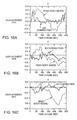
- FIGS. 8A and 8B show a 3-point Dixon GRE image of the articular cartilage of medial fermorotibial compartment in a normal 35-year old volunteer.
- FIG. 13A has the subject in supine position and
- FIG. 13B has the subject in an upright position.
- FIGS. 9A-9C show patient position and application of imaging coil and tracker coil for kinetic MR imaging of the knee. Patient is in upright weight-bearing position for active flexion and extension study of the knee.
- FIG. 9B is a 2D cartilage thickness map demonstrating abrupt decrease in cartilage thickness in the area of the defect (arrows).
- the ⁇ thickness between the neighboring pixels can be used to define the borders of the cartilage defect. Note defused cartilage thinning in the area enclosed by the asterisks (*).
- FIGS. 10A-10C show a 3D surface registration of femoral condyles based on T1-weighted Spin-Echo MR images.
- FIG. 6A is a baseline with a knee and neutral position.
- 6 B is a follow-up with knee and external rotation with a 3D view that is the identical to the one used in 6 A but the difference in knee rotation is apparent.
- transformation and reregistration of Scan B into the object coordinate system of Scan A shows the anatomic match to A is excellent.
- FIG. 11 panels A and B, shows a 2D cartilage thickness map ( FIG. 11B ) constructed from a proton density fast spin-echo MR image ( FIG. 11A ) of a focal cartilage defect in the posterior lateral femoral condyle (black arrows).
- White arrows indicate endpoints of the thickness map.
- FIG. 12 panels A and B, shows the anatomic coordinate system in the femur ( FIG. 12A ) and in the tibia ( FIG. 12B ).
- FIG. 13 shows calculation of the anatomic coordinate system from palpable bony landmarks.
- FIG. 14 panels A and B, shows additional marker names and locations for MR to optical cross registration.
- FIG. 14A shows additional marker names on a side-view of a subject.
- FIG. 14B shows additional marker names on a frontal view of a subject.
- FIG. 15 panels A and B, shows the marker names and locations for the standard point-cluster technique protocol.
- FIG. 15A shows marker names and locations on a side-view of a subject.
- FIG. 15B shows marker names and locations on a frontal view of a subject.
- FIG. 16 panels A to C, are graphs depicting displacement over time in the X ( FIG. 16A ), Y ( FIG. 16B ) and Z ( FIG. 16C ) axes. Each graph depicts the error in the tibial location estimate for the rigid body model (dotted line) and the intrical deformation correction technique (solid line).
- FIG. 17 panels A to C, are graphs depicting displacement over time in the ⁇ ( FIG. 17A ), ⁇ ( FIG. 17B ) and ⁇ ( FIG. 17C ) axes.
- Each graph depicts the error in tibial orientation estimate for the rigid body model (dotted line) and the interval deformation correction technique (solid line).
- FIG. 18A-18I show functional joint imaging.
- FIG. 19 shows the super imposition of the tibiofemoral contact line onto the 3D cartilage thickness map.
- FIG. 20 shows the determination of the natural line of curvature as the cutting plain is rotated about the transepicondyear reference, the cartilage-plain intersection results in a curve.
- FIG. 21 shows the determination of the tibiofemoral contact line through the proximity detection and approach algorithm.
- FIGS. 22A and 22B show a 2D MRI (3D SPGR) and 3D cartilage thickness map.
- FIGS. 23A-E show the matching of 3D thickness maps generated from MR images obtained with a knee neutral position and external rotation.
- FIG. 1 is a schematic overview of some of the various aspects of the invention. While a complete description of the many aspects of the invention is found in the specification and claims, the schematic overview gives some of the broad aspects of the invention.
- This invention relates to assessing the condition of a joint in a mammal.
- One aspect is a method for such an assessment.
- the assessment can be done using internal images, or maps, of the cartilage alone or in combination with a movement pattern of the joint. If used alone, a map obtained at an initial time is compared with a map obtained at a later time to provide a view of the change in cartilage over time.
- Another aspect is a method is comparing the movement pattern for a joint of a subject being studied with the cartilage degeneration pattern of the subject, then determining the relationship between the movement pattern and the degeneration pattern. If, in determining the relationship between the two patterns, one finds that the movement pattern has caused the degeneration pattern or will continue to adversely affect the degeneration pattern, therapy can be prescribed to minimize the adverse effects, such as further degeneration or inflammation.
- FIG. 1 is based on the full range of processes, preferably applied to a knee and surrounding cartilage.
- the first step 10 represents obtaining an image of the cartilage itself. This is typically achieved using MRI techniques to take an image of the entire knee and then, optionally, manipulating (e.g., “subtracting out” or “extracting”) the non-cartilage images as shown in step 12 .
- Non-cartilage images typically come from bone and fluid.
- the MRI is taken using external markers to provide reference points to the MRI image (step 11 ).
- the cartilage is imaged with a 2D MRI acquisition technique
- the resulting stack of 2D images so obtained can be combined into a 3D image, as indicated in step 14 .
- a preferred alternative is to use 3D MRI acquisition techniques to acquire a 3D image directly. In either case, the same “non-cartilage image extraction techniques referred to in step 12 can be used.
- various “maps” or displays of the cartilage can be constructed to give a cartilage degeneration pattern. This is represented by step 16 .
- One such display can, for example, be a color-coding of a displayed image to reflect the thickness for the cartilage. This will allow easy visual identification of actual or potential defects in the cartilage.
- a 3D image of the knee joint is taken, again preferably using MRI. Many of the same techniques as applied in steps 10 to 14 are used to do this. However, as illustrated by sub-step 22 , it is useful to define and register a skin-external frame of reference around the joint. This is achieved by placing fiduciary markers on the skin around the outside of the knee (step 22 ) prior to taking the image.
- an image is manipulated to enhance the image of the position of the markers (step 24 ).
- the resulting manipulated image is used to give a 3D image of the joint and associated bones (step 26 ).
- an additional set of markers is placed on the skin along the outside of the leg, and an external image of the limb is obtained.
- images are then taken of the subject in a static state.
- images are also taken of the subject while moving. This is shown collectively by step 32 .
- the images obtained are then processed to relate the movement of the skin relative to the bone.
- certain calculations are performed, for example, the center of mass is calculated. These manipulations are shown in Step 34 .
- a correlation between the fiduciary and the additional set of markers can be made. This is shown in step 36 .
- the static 3D image of the joint (with associated fiduciary markers) and the movement images of the leg bones (also with fiduciary markers in place) can be combined:
- the fiduciary markers therefore, serve as baseline references.
- the combination (step 40 ) of 3D cartilage image (from step 14 ), 3D knee joint image (step 26 ), and the moving leg co-ordinates (step 34 ) will, after appropriate corrections, result in a displayable, 3D motion image of the joint moving as per step 46 .
- the moving images, showing the contact areas of the knee joint can be used in conjunction with the various “maps” or displays generated at step 16 to provide a visual indication of potential or actual cartilage defects and help in determining their relation between movement and degeneration patterns. This is shown in step 48 .
- real measurements such as cartilage thickness
- later or earlier measurements and/or imaging This allows the tracking of the progression of a defect, or conversely, continued tracking of healthy cartilage. This aids a health worker in providing therapy for the patients.
- the method allows monitoring and evaluation of remedial actions as well as possible treatment prescriptions.
- this invention discloses, for example, a method to examine the relationship between articular cartilage morphology and the functional load bearing areas of a knee joint measured during movement.
- the method includes enhanced imaging techniques to reconstruct the volumetric and biochemical parameters of the articular cartilage in three dimensions; and a method for in vivo kinematic measurements of the knee.
- the kinematic measurement permits direct in vivo measurements of complete six-degrees of freedom motion of the femur or the tibia or associated bones during normal activities. This permits the study of load bearing of articular cartilage during movement.
- this method can aid in locating cartilage defects relative to the changing load bearing areas of the knee joint during daily activities. While the various aspects of the invention are useful in mammals generally, they are particularly useful for human patients.
- the joint of a patient is that place of union, more or less movable, between two or more bones.
- a joint comprises cartilage and other elements such as the accompanying bones on either side of the joint, fluid, and other anatomical elements.
- Joints are classified into three general morphological types: fibrous, cartilaginous, and synovial. This invention is particularly useful for assessing synovial joints, particularly the knee.
- MRI magnetic resonance imaging
- CT computed tomography scanning
- CAT computerized axial tomography
- ultrasound imaging techniques Others may be apparent to one of skill in the art. MRI techniques are preferred.
- MRI with its superior soft tissue contrast, is the best technique available for assessing tissue and its defects, for example articular cartilage and cartilage lesions, to obtain a cartilage degeneration can provide morphologic information about the area of damage. Specifically, changes such as fissuring, partial or full thickness cartilage loss, and signal changes within residual cartilage can be detected.
- MR imaging techniques are particularly suitable for cartilage is because they can provide accurate assessment of cartilage thickness, demonstrate internal cartilage signal changes, evaluate the subchondral bone for signal abnormalities, and demonstrate morphologic changes of the cartilage surface.
- MRI provides several important advantages over other techniques in this invention.
- One advantage is good contrast between cartilage, bone, joint fluid, ligaments, and muscle in order to facilitate the delineation and segmentation of the data sets.
- Another is the coverage of the entire region of interest in a single scan within acceptable acquisition times.
- MRI employs pulse sequences that allow for better contrast of different parts of the area being imaged. Different pulse sequences are better fitted for visualization of different anatomic areas, for example, hyaline cartilage or joint fluid. More than one pulse sequence can be employed at the same time. A brief discussion of different types of pulse sequences is provided below.
- Routine MRI pulse sequences available for imaging tissue include conventional T1 and T2-weighted spin-echo imaging, gradient recalled echo (GRE) imaging, magnetization transfer contrast (MTC) imaging, fast spin-echo (FSE) imaging, contrast enhanced imaging, rapid acquisition relaxation enhancement, (RARE) imaging, gradient echo acquisition in the steady state, (GRASS), and driven equilibrium Fourier transform (DEFT) imaging.
- GRE gradient recalled echo
- MTC magnetization transfer contrast
- FSE fast spin-echo
- RARE rapid acquisition relaxation enhancement
- GASS gradient echo acquisition in the steady state
- DEFT driven equilibrium Fourier transform
- T1 and T2-weighted MRI depicts articular cartilage, and can demonstrate defects and gross morphologic changes.
- T1-weighted images show excellent intra-substance anatomic detail of hyaline cartilage.
- T1-weighted imaging does not show significant contrast between joint effusions and the cartilage surface, making surface irregularities difficult to detect.
- T2-weighted imaging demonstrates joint effusions and thus surface cartilage abnormalities, but since some components of cartilage have relatively short T2 relaxation times, these are not as well depicted as other preferred imaging.
- Gradient-recalled echo imaging has 3D capability and ability to provide high resolution images with relatively short scan times.
- Fat suppressed 3D spoiled gradient echo (FS-3D-SPGR) imaging has been shown to be more sensitive than standard MR imaging for the detection of hyaline cartilage defects in the knee.
- Magnetization transfer imaging can be used to separate articular cartilage from adjacent joint fluid and inflamed synovium.
- Fast spin-echo imaging is another useful pulse sequence to evaluate articular cartilage.
- Incidental magnetization transfer contrast contributes to the signal characteristics of articular cartilage on fast spin-echo images and can enhance the contrast between cartilage and joint fluid.
- Sensitivity and specificity of fast spin-echo imaging have been reported to be 87% and 94% in a study with arthroscopic correlation.
- gadolinium for imaging of articular cartilage has been applied in several different forms.
- Direct magnetic resonance (MR) arthrography wherein a dilute solution containing gadolinium is injected directly into the joint, improves contrast between cartilage and the arthrographic fluid.
- Indirect MR arthrography with a less invasive intravenous injection, can also been applied.
- Gadolinium enhanced imaging has the potential to monitor glycosaminoglycan content within the cartilage, which may have implications for longitudinal evaluations of injured cartilage.
- DEFT driven equilibrium fourier transform
- SPGR spoiled gradient echo
- the basic DEFT pulse sequence is shown in FIG. 2 .
- a conventional spin echo pulse sequence was followed by an additional refocusing pulse to form another echo, and then a reversed, negated, excitation pulse to return any residual magnetization to the +z axis.
- Typical MRI parameters for cartilage are a T1-relaxation time of 900 Milliseconds (ms) and a T2-relaxation time of 40 ms, while synovial fluid has a T1-relaxation time of 3000 ms and a T2-relaxation time of 200 ms.
- synovial fluid has a 30% greater proton density than cartilage.
- the signal levels of cartilage and synovial fluid were plotted in FIG. 3 for a RARE pulse sequence and for DEFT, and show that DEFT maintains excellent contrast for any relaxation time (TR). It achieves this contrast while maintaining a signal-to-noise ratio (SNR) efficiency (SNR/(T acquisition )) that is equal to or better than other methods with much lower contrast, such as T1-weighted GRASS.
- SNR signal-to-noise ratio
- the DEFT was compared with a fast spin-echo (FSE), a gradient-echo (GRE), and a spoiled gradient-echo (SPGR) sequence with parameters similar to the ones published by Disler et al.
- FSE fast spin-echo
- GRE gradient-echo
- SPGR spoiled gradient-echo
- the patella was scanned in 10 normal volunteer knees using a 1.5 T whole-body system (GE Signa) with a 3 inch surface coil. All images were acquired with field of view (FOV) 10 ⁇ 10 cm, matrix 256 ⁇ 256 elements, slice thickness 4 mm using fat-saturation.
- FOV field of view
- CNR Contrast-to-noise ratios
- DEFT was combined with a partial k-space echo-planar data acquisition. This pulse sequence is illustrated in FIG. 6 above.
- a slab selective pulse in z defines the imaging volume, which is then resolved with phase-encoding gradients in the y and z axes, and an oscillating EPI gradient in the x axis.
- Example images acquired with this approach are shown in FIG. 7 .
- This case was optimized for resolution, in order to image the patellar cartilage.
- the EPI readout acquired 5 echoes for each DEFT sequence.
- Partial k-space acquisition collected only 60% of the data along the x-axis. Correction for the missing data was performed using a homodyne reconstruction.
- the image matrix was 192 ⁇ 192 ⁇ 32, with a resolution of 0.5 ⁇ 0.5 ⁇ 2.5 mm, resulting in a 10 ⁇ 10 ⁇ 8 cm FOV.
- the echo time TE was 22 ms, and the TR was 400 ms. Fat was suppressed with a fat presaturation pulse. The total scan time for this acquisition was 5 minutes.
- Additional image studies that can be performed using this approach may require greater spatial coverage, but one can permit slightly less spatial resolution, and a longer scan time similar to the one used with the 3D SPGR approach. If one relaxes the resolution to 0.75 ⁇ 0.75 ⁇ 1.5 mm, and doubles the z slab thickness and z phase encodes, the result will be a FOV of 15 ⁇ 15 ⁇ 16 cm, and a total scan time of approximately 15 minutes, which exactly fits the desired scan protocol. Similar to the 3D SPGR acquisition, one can acquire a first 3D DEFT scan in the sagittal plane with fat saturation. The 3D DEFT acquisition can then be repeated without fat saturation using the identical parameters and slice coordinates used during the previous acquisition with fat saturation. The resultant non-fat-saturated 3D DEFT images can be used for 3D rendering of the femoral and tibial bone contours.
- Driven Equilibrium Fourier Transform is a pulse sequence preferred for cartilage imaging that provides higher contrast-to-noise ratios and contrast between cartilage and joint fluid than SPGR, GRE, and FSE sequences.
- Cartilage morphology is better delineated with DEFT sequences than with SPGR, GRE, and FSE images.
- the combination of high anatomic detail and high cartilage-joint fluid CNR and contrast may render this sequence particularly useful for longitudinal studies of cartilage in patients with osteoarthritis.
- Vertically open MRI units can provide the opportunity to study the knee joint in true weight-bearing position with the patient placed in upright position.
- a vertically open MRI unit (GE Signa SP 0.5 T) exists at Stanford University. The vertically open MRI unit can be used to develop techniques for kinematic joint imaging under weight-bearing conditions as a means of diagnosing joint instability.
- a cartilage sensitive MRI scans was obtained in the open magnet using a three-point Dixon gradient-echo sequence in supine and upright positions. A representative example is demonstrated in FIG. 8 .
- a MR image can be performed using a whole body magnet operating at a field strength of 1.5 T (GE Signa, for example, equipped with the GE SR-120 high speed gradients [2.2 Gauss/cm in 184 ⁇ sec risetimes]).
- Gd-DTPA Magneticnevist®, Berlex Inc., Wayne, N.J.
- T1 relaxation time approximately 1.0 sec can be applied to the skin around the knee joint and optionally at the same positions used for gait analysis in a biomotion laboratory (discussed below).
- the external markers can be included in the field of view of all imaging studies. Patients can be placed in the scanner in supine position.
- the scanner table can then be moved to obtain coronal and sagittal images of the knee joint and tibia using the same sequence parameters.
- These T1-weighted scans can be employed to identify axes through the femur and tibia which can be used later for defining the geometry of the knee joint.
- GASS gradient echo sequence
- 2DFT 2D Fourier Transform
- This scout scan can be used to demonstrate the position of the knee joint space in the coil and to prescribe all subsequent high resolution imaging sequences centered over the joint space. Additionally, using the graphic, image based sequence prescription mode provided with the scanner software, the scout scan can help to ensure that all external markers around the knee joint are included in the field of view of the high resolution cartilage sensitive MR sequences.
- the fat-saturated 3D SPGR sequences can be used for rendering the cartilage in three dimensions (see description below).
- the 3D SPGR sequence can then be repeated in the sagittal plane without fat saturation using the identical parameters and slice coordinates used during the previous acquisition with fat saturation.
- the resultant non-fat-saturated 3D SPGR images demonstrate good contrast between low signal intensity cortical bone and high signal intensity bone marrow thereby facilitating 3D rendering of the femoral and tibial bone contours. It is to be understood that this approach is representative only and should not be viewed as limiting in any way.
- Upright weight-bearing MR imaging of the knee can be performed using a 0.5 T vertically open MR unit (GE Signa SP, General Electric, Milwaukee, Wis.) and a MR tracking system.
- Gd-DTPA Magneticnevist®, Berlex Inc., Wayne, N.J.
- T1 relaxation time approximately 1.0 sec can be applied to the skin around the knee joint at the same positions optionally used for gait analysis in the biomotion laboratory (discussed below).
- the subject can be placed in upright position inside the magnet (see FIG. 9 ).
- the knee joint can be perpendicular to the main magnetic field.
- a flexible transmit/receive coil can be positioned around the knee and secured to the knee with elastic straps.
- the external MR tracking coil can be taped to the skin overlying the tibial tuberosity.
- a field of view of 20 cm can be chosen in order to achieve sufficient anatomic coverage in superoinferior direction across the knee joint. Using this technique, one can assess tibiofemoral motion during knee flexion and extension.
- VAI Volumes of Interest
- the invention allows a health practitioner to determine cartilage loss in a reproducible fashion and thus follow the progression of a cartilage defect over time.
- a T1-weighted spin-echo sequence can be used for surfaces extraction of the femoral condyles.
- the T1-weighted spin-echo sequence provides high contrast between low signal intensity cortical bone and high signal intensity fatty marrow.
- a step-by-step problem solving procedure i.e., an algorithm, can convolve a data set with a 3D kernel to locate the maximum gradient location.
- the maximum gradient location corresponds to the zero crossing of a spatial location.
- This operation is preferably three-dimensional rather than two-dimensional.
- the surface of the joint, e.g. the femoral condyles, on the baseline scan can be registered in an object coordinate system A.
- the surface of the joint, e.g. the femoral condyles, on the follow-up scan can be registered in an object coordinate system B.
- a transformation B to B′ can be performed that best matches B′ with A.
- Such transformations can, for example, be performed using a Levenberg Marquardt technique.
- the transformations and matching can be applied to the cartilage only. The same transformation can be applied to the cartilage sensitive images on the follow-up scan in order to match the cartilage surfaces.
- the size of the targeted volumes of interest can be selected to exceed that of the cartilage defect in anteroposterior and mediolateral direction, e.g. by 0.5 to 1 cm. If the defect is located high on the femoral Condyle or in the trochlear region, the targeted VOI can be chosen so that its size exceeds that of the cartilage defect in superoinferior and mediolateral direction.
- the third dimension of the targeted VOI (parallel to the surface normal of the cartilage) can be fixed, for example at 1 cm.
- VOI size and placement can be manual or automatic on the baseline study.
- the 3D coordinates of the targeted VOI relative to the 3D contour of the joint and object coordinate system A can be registered and saved.
- follow-up studies e.g. scans inadvertently obtained with slightly different patient position
- the 3D surface of the joint is registered to match the orientation of the baseline scan and the targeted VOI is then automatically placed on the joint using object coordinate system B′ and the coordinates saved on the baseline study.
- Cartilage volume within the targeted VOI on baseline and follow-up studies can, for example, be determined using standard thresholding and seed growing techniques.
- external reference markers are placed on the skin around the joint of the subject being imaged.
- the external marker is designed not only to show up in the MRI, but also to show up if an external image of the joint is obtained. The importance and value of such unique reference markers will be discussed in more detail hereinafter.
- one embodiment of the invention is a skin reference marker that can be used in the assessment of the condition of a joint of a human.
- Multiple skin reference markers can be placed upon one or more limbs of a patient prior to internal imaging and external imaging.
- Each skin reference marker comprises a material detectable by an imaging technique, a container for the material in which the container preferably has multiple surfaces, a means for affixing the container to the skin (e.g. an adhesive placed on at least one surface of the container in an amount sufficient to adhere the container to the skin of a human), and a reflective material (preferably retro-reflective) placed on another surface of the container located away from the adhesive.
- Several imaging techniques can be used that are able to detect the marker.
- the material detectable by an imaging can be either in a liquid form or a solid form.
- the material can be any imaging contrast agent or solution, e.g. a paramagnetic material.
- the material can be a lanthanide, such as one belonging to the yttrium group of rare earth metals. More specifically, the material can be gadolinium.
- the shape of the container can be any shape allowing it to be placed on the skin of a human.
- the size of the container can be any size, but optimally a size allowing it to be recorded by an imaging machine.
- the longest dimension of the container can be up to 5.0 cm, but preferably is about 0.25 to 2.0 cm.
- the reflective or retro-reflective material can be any material that is able to reflect light directly back to the source of the light so that the position of the reference marker is captured by the opto-electrical recording means, e.g. a video camera. 3M Corporation makes several retro-reflective materials.
- a magnetic resonance image Once a magnetic resonance image is obtained, it can be manipulated to improve the image by reducing unwanted, non-cartilage images.
- the cartilage can be image by image using a signal-intensity-based threshold combined with a seed growing technique.
- the femoral, tibial, and patellar cartilage can be segmented separately based on the fat-saturated 3D SPGR or 3D DEFT sequence.
- Manual disarticulation is performed by outlining the cartilage contour in areas where the signal intensity of the articular cartilage is similar to that of adjacent structures.
- the contours of the femoral, tibial, and patellar bone are segmented separately using the non-fat-saturated 3D SPGR or 3D DEFT sequence. Segmentation software allows for manual editing of cartilage thickness maps and cartilage defects detected using the above embodiments.
- Such software includes seed-growing algorithms and active-contour algorithms that are run on standard PC's.
- a sharp interface is present between the high signal intensity bone marrow and the low signal intensity cortical bone thereby facilitating seed growing.
- Fat-saturated and non-fat-saturated 3D sequences can be acquired with the same field of view, slice thickness and slice positions, thereby enabling super imposition and cross registration of any resultant 3D renderings of the femoral, tibial, and patellar cartilage and bone.
- External reference markers aid in registering the 3D data in the same object coordinate system.
- 3D maps of cartilage thickness can be generated using several different techniques.
- One representative, but not limiting, approach uses a 3D surface detection technique which is based on a 2D edge detector (Wang-Binford) that has been extended to 3D.
- This surface detection technique can generate surface points and their corresponding surface normal.
- the program samples 25 percent of the surface points and fits a cubic spline to the sample points.
- the program can compute the curvature along sample spline points and find two sample points that have the maximum curvature and are separated by about half the number of voxels on the contour. These points partition the spline into two subcontours. For each subcontour, the program can compute the average distance between the points and the center of the mass.
- the program can designate the subcontour with the smaller average distance as the inner cartilage surface and the other subcontour as the outer cartilage surface (OCS).
- OCS outer cartilage surface
- the intersect between the inner cartilage surface (ICS) (located at the subchondral bone interface) and the outer cartilage surface with the surface normal can be used to compute the 3D thickness of the articular cartilage on a pixel-by-pixel basis.
- the set of segmented two dimensional MR images can be transformed to a voxel representation using a computer program developed in the AVS Express (Advanced Visual Systems, Inc., Waltham, Mass.). Every voxel has a value of zero if it is not within an object of interest or a value ranging from one to 4095, depending on the signal intensity as recorded by the 1.5 T MR.
- An isosurface can then be calculated that corresponds to the boundary elements of the volume of interest. A tesselation of this isosurface is calculated, along with the outward pointing normal of each polygon of the tesselation. These polygons are written to a file in a standard graphics format (Virtual Reality Modeling Language Version 1.0: VRML output language).
- the program reads in a scene, which scene consists of the various 3D geometric representations or “actors” (for example, VRML files of the tibia, tibia cartilage, femur, femoral cartilage), the static relationship transformations between these actors, and, if available, sequence of transformations describing how these actors move with respect to each other as the patient performs some activity, such as walking, jogging, etc.
- actors for example, VRML files of the tibia, tibia cartilage, femur, femoral cartilage
- the program allows the user, through the use of the mouse and/or keyboard, the ability to observe the scene from arbitrary angles; to start and stop the animation derived from the motion profiles and to observe the contact line and any cartilage lesions while the animation is running. Additionally, the user may derive quantitative information on the scene through selecting points with the mouse.
- the software program is written in the CTT computer language and is compiled to run on both Silicon Graphics Workstations and Windows/Intel personal computers.
- Cartilage thickness is determined by several methods.
- One example is detecting the locations of the bone-cartilage and the cartilage-joint fluid interface along the surface normal using the same edge detector described below, and subtracting them. This procedure can be repeated for each pixel located along the bone-cartilage interface.
- the x, y, and z position of each pixel located along the bone-cartilage interface is registered on a 3D map or multiple 2D maps and thickness values are translated into color values. In this fashion, the anatomic location of each pixel at the bone cartilage interface can be displayed simultaneously with the thickness of the cartilage in this location.
- the edge detector produces accurate surface points and their corresponding surface normal.
- the detector is applied to the baseline and the follow-up data set.
- both the surface points and surface normals are used to form locally supporting planes (for each voxel). These planes can form an approximated surface for the baseline skeletal site.
- the surface points can be matched in the registration procedure onto the surface of the baseline data set.
- a preferred approach for calculating the cartilage thickness is based on a 3D Euclidian distance transformation (EDT).
- EDT Euclidian distance transformation
- the voxels on the edge of the cartilage structure can be extracted using a slice by slice 8-neighbor search, resulting in a binary volume with the voxels on the cartilage surface having a value of 1 and all others being 0.
- a semi-automatic approach which requires the user to enter a point that lies outside the cartilage structure and faces the ICS, is useful. From this point, rays are cast in all directions of the volume using a modified Bresenham's line drawing algorithm.
- a ray hits a voxel with a value of 1, this point is classified as part of the ICS.
- the ICS voxels are given a value of 0, whereas all other voxels are set to 1.
- the following representative algorithm is useful. It can decompose the calculation into a series of 3 one-dimensional transformations and can use the square of the actual distances, which accelerates the process by avoiding the determination of square roots.
- each point can be assigned the square of the distance to the closest feature point in the same row in i-direction.
- h ijk min y ⁇ ⁇ g iyk + ( ⁇ ⁇ ( j - y ) ) 2 ; 1 ⁇ y ⁇ M ⁇ [ Eq . ⁇ 4 ]
- the algorithm can search each column in the j-direction.
- the sum of the square distance between a point (i, j, k) and a point (i, y, k) in the same column, ( ⁇ (j ⁇ y)) 2 , and the square distance between (i, y, k) and a particular feature point, g iyk equals the square distance between the point (i, j, k) and that feature point.
- the minimum of these sums is the square distance between (i, j, k) and the closest feature point in the two-dimensional i-j-plane.
- the third dimension can be added by equation (5), which is the same transformation as described in the equation for the k-direction (4).
- the thickness of the cartilage for a given point (a, b, c) on the OCS equals the square root of s abc .
- the x, y, and z position of each pixel located along the bone-cartilage interface can be registered on a 3D map and thickness values are translated into color values. In this fashion, the anatomic location of each pixel at the bone cartilage interface can be displayed simultaneous with the thickness of the cartilage in this location.
- the cartilage thickness maps obtained using the algorithm described above display only a visual assessment of cartilage thickness along the articular surface.
- a 25% decrement in cartilage thickness will be the smallest change that can be observed with most current imaging sequences. Therefore, for example, pixels along the bone-cartilage interface that demonstrate a decrease exceeding the smallest change observable on a given MRI pulse sequence, in this example 25% or greater, in overlying cartilage thickness when compared to cartilage thickness at the neighboring bone-cartilage interface pixels, can be used to define the margins of a focal cartilage defect. Other criteria can be employed to define a cartilage defect based on comparisons of neighboring pixels.
- a fixed value can be used. If the difference in cartilage thickness between neighboring pixels exceeds the fixed value, e.g. 1 mm, the pixel where this difference is observed can be used to define the margin of the cartilage defect.
- This comparison can be performed for each pixel located along the bone-cartilage interface for the entire data set. This comparison is preferably performed in three dimensions. Pixels that demonstrate a decrease in cartilage thickness exceeding defined criteria but that are completely surrounded by other pixels fulfilling the same criteria may not be considered to be part of the margin of the cartilage defect, but will typically be considered to lie inside the cartilage defect.
- the invention provides for means for calculating the area covered by the cartilage defect A cartilage defect and the mean thickness of the cartilage in the region of the defect D cartilage defect as well as the mean thickness of a defined area of surrounding normal cartilage.
- V cartilage loss A cartilage defect ⁇ D cartilage loss [Eq. 7].
- FIGS. 22A and 22B one can see a 2D MRI (3D SPGR) and 3D cartilage thickness map.
- the 2D MRI demonstrates a full thickness cartilage defect in the posterior lateral femorl condyle (arrows).
- FIG. 22B shows a 3D cartilage thickness map generated using a 3D Euclidian distance transformation.
- the thickness of the articular cartilage is color encoded and displayed on a pixel-by-pixel basis along the 3D surface of the articular cartilage.
- the cartilage defect is black reflecting a thickness of zero (arrows) (M: medial, L: lateral, S: superior, I: inferior).
- FIGS. 23A-23E one can see the matching of 3D thickness maps generated from MR images obtained with the knee in neutral position and in external rotation.
- A. Sagittal baseline MR image (3D SPGR) with the knee in neutral position.
- B. Sagittal follow-up MR image of the same volunteer obtained two weeks later with the knee in 40 degree external rotation (note the artificially widened appearance of the femur resulting from the rotation).
- C. 3D thickness map generated based on baseline MRI in neutral position.
- D 3D thickness map generated based on follow-up MRI in external rotation (note segmentation error between condyles in trochlear region).
- One aspect is a method of estimating the loss of cartilage in a joint.
- the method comprises
- this aspect of the invention is directed to a volume of interest in the cartilage, i.e., a region of the cartilage that includes a cartilage defect.
- a defect may be the result of a disease of the cartilage (e.g., osteoarthritis) or the result of degeneration due to overuse or age.
- This invention allows a health practitioner to evaluate and treat such defects.
- the volume of interest may include only the region of cartilage that has the defect, but preferably will also include contiguous parts of the cartilage surrounding the cartilage defect.
- Another aspect of the invention is a method for assessing the condition of cartilage in a joint of a human, which method comprises
- Another aspect of the invention is a method for determining the volume of cartilage loss in a region of a cartilage defect of a cartilage in joint of a mammal.
- the method comprises (a) determining the thickness, DN, of the normal cartilage near the cartilage defect; (b) obtaining the thickness of the cartilage defect, DD, of the region; (c) subtracting DD from DN to give the thickness of the cartilage loss, DL; and (d) multiplying the DL value times the area of the cartilage defect, AD, to give the volume of cartilage loss.
- the method is useful for situations wherein the region of cartilage defect is limited to the defective cartilage and preferably wherein the region of the cartilage defect includes a portion of the cartilage contiguous to the defect.
- the normal thickness of the defect area could be estimated. It may be estimated from measurements of cartilage of other subjects having similar characteristics such as gender, age, body type, height, weight, and other factors. It may be estimated from measurements of a similar ‘normal” cartilage from another corresponding joint (e.g., if the right knee has the defect, measure the normal left knee). It may have been measured at an initial time T 1 when the cartilage was normal to provide a baseline. Other means of determining the normal thickness may be available to one of skill in the art. Once the thickness D N is obtained and the thickness D D is obtained the two are subtracted to give the D L . The D L is multiplied by the area of the defect A D to give the volume of cartilage loss. By determining the volume of cartilage loss at an initial T 1 and again at a later time T 2 , one can determine the change in volume loss over time.
- Still another aspect of the invention is a method of estimating the change of a region of cartilage in a joint of a mammal over time.
- the method comprises (a) estimating the width or area or volume of a region of cartilage at an initial time T 1 , (b) estimating the width or area or volume of the region of cartilage at a later time T 2 , and (c) determining the change in the width or area or volume of the region of cartilage between the initial and the later times.
- the method is particularly useful for regions of degenerated cartilage or diseased cartilage.
- Still another aspect of the invention is a method of estimating the loss of cartilage in a joint.
- the method comprises (a) defining a 3D object coordinate system of the joint at an initial time, T 1 ; (b) identifying a region of a cartilage defect within the 3D object coordinate system; (c) defining a volume of interest around the region of the cartilage defect whereby the volume of interest is larger than the region of cartilage defect, but does not encompass the entire articular cartilage; (d) defining the 3D object coordinate system of the joint at a second timepoint, T 2 ; (e) placing the identically-sized volume of interest into the 3D object coordinate system at timepoint T 2 using the object coordinates of the volume of interest at timepoint T 1 ; (f) and measuring any differences in cartilage volume within the volume of interest between timepoints T 1 and T 2 .
- the invention provides for techniques to represent a biochemical components of articular cartilage.
- a biochemical component includes, but is not limited to, glycosaminoglycan, water, sodium, or hyaluronic acid.
- Biochemical data can be generated with other magnetic resonance based techniques including the use of paramagnetic and other contrast media and sodium rather than proton MR imaging.
- one aspect of this invention is a method for providing a biochemically-based map of joint cartilage. The method comprises
- the biochemical map may be used in the method aspects of the invention in a manner similar to the cartilage thickness map.
- one aspect is a method of estimating the loss of cartilage in a joint.
- the method comprises
- this aspect of the invention is directed to a volume of interest in the cartilage, i.e., a region of the cartilage that includes a cartilage defect.
- a defect may be the result of a disease of the cartilage (e.g., osteoarthritis) or the result of degeneration due to overuse or age.
- This invention allows a health practitioner to evaluate and treat such defects.
- the volume of interest may include only the region of cartilage that has the defect, but preferably will also include contiguous parts of the cartilage surrounding the cartilage defect.
- Another aspect of the invention is a method for assessing the condition of cartilage in a joint using the biochemical map.
- the method comprises
- Another aspect of the invention is a method for determining the change of biochemical content in a region of a cartilage defect of a cartilage in joint of a mammal.
- the method comprises (a) determining the biochemical content (BCN) of the normal cartilage near the cartilage defect; (b) obtaining the biochemical content of the cartilage defect (BCD) of the region; and (c) subtracting BCD from BCN to give the value of the cartilage change, BCD.
- BCN biochemical content
- BCD biochemical content of the cartilage defect
- the method is useful for situations wherein the region of cartilage defect is limited to the defective cartilage and preferably wherein the region of the cartilage defect includes a portion of the cartilage contiguous to the defect.
- the normal content of the defect area could be estimated. It may be estimated from measurements of cartilage of other subjects having similar characteristics such as gender, age, body type, height, weight, and other factors. It may be estimated from measurements of a similar ‘normal” cartilage from another corresponding joint (e.g., if the right knee has the defect, measure the normal left knee). It may have been measured at an initial time T 1 when the cartilage was normal to provide a baseline. Other means of determining the normal content may be available to one of skill in the art. Once BCN is obtained and BCD is obtained the two are subtracted to give the ⁇ . By determining the change of content at an initial T 1 and again at a later time T 2 , one can determine the change in volume loss over time.
- morphological maps of articular cartilage obtained with MR imaging are superimposed, merged or fused with the biochemical map or data.
- Several different techniques can be applied in order to superimpose, merge, or fuse morphological data with biochemical data.
- 2D or 3D morphological data of articular cartilage can be acquired with the same object coordinates as the biochemical data. Morphological data and biochemical data can then be easily displayed simultaneously using different colors, opacities, and or gray scales.
- 2D or 3D morphological data or articular cartilage can be acquired with different object coordinates as the biochemical data.
- a 3D surface registration can be applied in order to superimpose, merge, or fuse the morphological data and the biochemical data.
- anatomic landmarks can be used to register the morphological data and subsequently the biochemical data in a 3D object coordinate system. 3D object coordinate systems can then be matched by matching the landmarks obtained from the morphological data with those obtained from the biochemical data.
- another aspect of this invention is a method for assessing the condition of a subject's cartilage in a joint, the method comprises obtaining a three dimensional biochemical representation of the cartilage, obtaining a morphological representation of the cartilage, and merging the two representations, and simultaneously displaying the merged representations on a medium.
- the merged representations are then used to assess the condition of a cartilage, estimate the loss of cartilage in a joint, determining the volume of cartilage loss in a region of cartilage defect, or estimating the change of a region of cartilage at a particular point in time or over a period of time.
- Similar steps would be followed as spelled out for the use of a thickness map or biochemical map.
- Simultaneous display of morphological data with biochemical data provides a useful tool to assess longitudinal changes in morphology or articular cartilage and biochemical composition of articular cartilage, for example during treatment with chondroprotective and chondroregenerative agents.
- one aspect of this invention is a method for assessing the condition of cartilage in a joint from a distant location. The method comprises
- the degeneration pattern includes a measure of cartilage thickness or regional cartilage volume.
- the electronically generated image of the cartilage preferably is an MR image and the degeneration pattern is displayed as a three-dimensional image as a thickness pattern, a biochemical content pattern or a merged thickness biochemical pattern.
- the electronically generated image is transmitted via Dicom, using the international standards for transmission of such images.
- kit for aiding in assessing the condition of cartilage in a joint of a mammal comprises a software program, which that when installed and executed on a computer reads a cartilage degeneration pattern presented in a standard graphics format and produces a computer readout showing a cartilage thickness map of the degenerated cartilage.
- the software can be installed in a PC, a Silicon Graphics, Inc. (SGI) computer or a Macintosh computer.
- the software calculates the thickness or regional volume of a region of degeneration of the cartilage which does not include the entire volume of the articular cartilage.
- a movement pattern of a joint in accordance with this invention, one obtains an internal image of the bones in a joint, preferably using MRI techniques, and obtains an external image of the bones in motion.
- the images are correlated, preferably through the use of external marker sets, to give a pattern that shows a static or moving condition.
- the correlated images are then displayed and the relation between the movement and degeneration patterns is determined.
- MRI techniques that provide an image of the bones on either side of the joint.
- the external markers are placed at any landmarks about the joint of interest. At least three markers are used for each limb being imaged.
- the markers will be made of a material that not only will be detected by MRI imaging techniques, but also will be detected by external imaging techniques.
- the markers will be associated with a means to affix them to the skin and preferably have an adhesive portion for adhering to the skin and a detectable entity that will show up on the MRI image.
- the preferred MRI imaging technique useful for obtaining an internal image is a spoiled 3D gradient echo or a 3D DEFT sequence.
- a further discussion may be found hereinbefore or in the 2 nd Edition of Brown and Semelka's book entitled “MRI Basic Principles and Applications.”
- the set of segmented two dimensional MR images are transformed to a voxel representation inside AVS Express (Advanced Visual Systems, Inc., Waltham, Mass.). Every voxel has a value of zero if it is not within an object of interest or a value ranging from one to 4095, depending on the signal intensity as recorded by the 1.5 T MR.
- An isosurface can then be calculated that corresponds to the boundary elements of the region of interest. A tesselation of this isosurface can be calculated, along with the outward pointing normal of each polygon of the tesselation. These polygons can then be written to a file in a standard graphics format (Virtual Reality Modeling Language Version 1.0).
- the use of reference markers on the skin around the joint and the bones provides an image that can later be matched to the reference markers for the cartilage image and the bone images obtained from external measurements.
- a semi-automated, 3D surface-based registration technique that does not require the use of an external frame or fiducial markers can be used.
- This 3D surface-based registration technique can be used to match the anatomic orientation of a skeletal structure on a baseline and a follow-up CT or MRI scan.
- FIG. 10 A registration technique for the femoral condyles and the tibial plateau is shown in FIG. 10 . It shows an example where 3D surfaces of the femoral condyles were extracted from two differently oriented T1-weighted spin-echo MRI scans (baseline A and follow-up B, respectively) obtained in the same patient in neutral position (A) and in 40 degree external rotation (B). The 3D surfaces were used to derive a coordinate transformation relating the two scans.
- FIG. 10C demonstrates the use of the derived transformation to re-register scan B in the object coordinate system of scan A. Such a transformation relating two T1-weighted scans can then be used to register DEFT cartilage-sensitive scans that are acquired in the same respective orientations as the A and B T1-weighted scans.
- FIG. 11 shows an example of a 2D map of cartilage thickness derived from the surface normals of the lateral femoral condyle.
- FIG. 11A shows a proton density fast spin-echo MR image that demonstrates a focal cartilage defect in the posterior lateral femoral condyle (black arrows).
- White arrows indicate endpoints of thickness map.
- FIG. 11B is a 2D cartilage thickness map that demonstrates abrupt decrease in cartilage thickness in the area of the defect (arrows). The A thickness between neighboring pixels can be used to define the borders of the cartilage defect. Note diffuse cartilage thinning in area enclosed by the astericks (*).
- cartilage sensitive images can be used instead of T1-weighted or T2-weighted scans and the surface match can be performed based on the cartilage contour.
- anatomic landmarks present on both baseline and follow-up scans can be used to match the data obtained during the baseline and those obtained during the follow-up scan.
- Another alternative for matching the baseline and the follow-up scan includes the use of external or internal fiducial markers that can been detected with MR imaging. In that case, a transformation is performed that matches the position of the markers on the follow-up scan with the position of the markers on the baseline scan or vice versa.
- biomechanical data include, but are not limited to, estimations of static loading alignment in standing or weight-bearing position and lying or non-weight-bearing position, as well as during joint motion, e.g., the movement of load-bearing pathway on the cartilage in the knee joint during gait.
- Biomechanical data may be generated using theoretical computations, based on data stored in a database that can be accessed by calling up and screening for certain characteristics.
- gait analysis may be performed for an individual and data obtained during gait analysis may be merged or fused with morphological MRI data. Morphological data and biomechanical data can then be easily displayed simultaneously using different colors, opacities, and or gray scales.
- the load-bearing pathway for example around a cartilage defect, can be plotted or superimposed onto morphological maps.
- reference markers or fiducial markers can be applied to the external surface on the skin overlying the joint.
- These markers adhere to the skin are typically made of materials that can be detected with MRI and that can be used to register joint motion during biomechanical analysis, e.g. gait analysis. These markers can then be used to correlate the morphological with the biomechanical data.
- Simultaneous display of morphological data with biomechanical data provides a useful tool to assess the load pathway applied to articular cartilage and inside and around cartilage defects.
- Estimation of load pathway applied in and around a cartilage defect can be used to assess a cartilage defect and to guide the choice of therapy, e.g. treatment with chondroprotective or chondroregenerative agents, osteochondral allografting, cartilage transplantation, femoral or tibial osteotomy, or joint replacement surgery.
- the level walking measurements can include, but is not limited to, six stride cycles for each side over a range of walking speeds.
- the subject can be instructed to walk at a comfortable speed (normal), slower than normal and faster than normal.
- this protocol produces gait measurements over a range of walking speeds.
- the standing and laying portions of the protocol can be used in the cross registration to the MR data.
- the instrumentation preferably includes, at least a two camera, video-based opto-electronic system for 3-D motion analysis, a multi-component force plate for measurement of foot-ground reaction force and a computer system for acquisition, processing and analysis of data.
- the anatomic coordinate systems are defined through bony landmarks which can be identified through palpation.
- bony landmarks which can be identified through palpation.
- a subset of the markers in a point cluster technique are referenced to bony landmarks on the femur and tibia. Techniques described previously by Hopenfeld and Benedetti are used to locate these bony landmarks.
- the anatomic coordinate systems used are similar to that previously described by LaFortune with the exception of the origin of the femoral coordinate system.
- a coordinate system is located in the femoral condyles.
- the femoral condyles medial (M)-lateral (L) axis ( FIG.
- the trans-epicondylar line (a line drawn between the medial-lateral femoral epicondyles). The midpoint of this axis is the origin.
- the inferior (I)-superior(S) axis runs parallel to the long axis of the femur, passing through the midpoint of the trans-epicondylar line.
- the anterior (A)-posterior (P) axis is the cross product of the medial-lateral and inferior-superior axes. The final position of the inferior-superior axis is made orthogonal to the anterior-posterior and medial-lateral axis through a cross product operation ( FIG. 13 ).
- the tibial coordinate system begins with the medial-lateral axis running through the most medial and lateral edges of the plateau.
- the inferior-superior axis is perpendicular to the medial-lateral axis passing through the tibial eminence.
- the anterior-posterior axis is the cross product of the medial-lateral and inferior-superior axes.
- the lower extremity is idealized as 3 segments with six degree-of-freedom joints at the knee and ankle.
- at least 3 markers per segment are used.
- FIG. 14 shows 21 passive retro-reflective markers located on the leg: some at bony prominences (greater trochanter, lateral malleolus, lateral epicondyle, lateral tibial pleateu), some clustered on the thigh and shank (Fa1-3, I1-3, Fp1-3; Ta1-3, Tl1-13). Additionally, two markers are placed on the foot at the lateral aspect of the calcaneus and base of the fifth metatarsal and one on the pelvis at theiliac crest).
- the reference database is a compendium of demographic and motion analysis data for all subjects whose data has been processed by a central processing site (e.g. ⁇ 4500 over the past 10 years).
- This database contains fields describing each of the subject's name, age, height, weight, injury types, orthopedic medical history, other anatomic measurements (thigh length, shank length, shoe size, etc.).
- the database also contains the results of any and all gait analysis run on these patients. This includes, for all activities tested (walk, run, jog, etc.), a number of peak valves (peak knee flexing, peak hip adduction movement; toe-out, angle, etc). along with the motion trajectories of the limb segments while the subjects are performing different activities.
- the sex, age, height, weight, limb length, and type of activity desired is entered as an avery into the database.
- the database searches for a set of subjects most closely watching the input avery. From this set of data, a typical motion pattern is distilled and a data set is output. This data set includes, over a time interval, the motion characteristics: hip/knee/ankle/flexion/extension angles, knee/hip/ankle adduction/abduction angles, movement, stride length, cadence, etc. This data can then be used to drive an animation of the motion of the desired joint.
- Each limb segment (thigh, shank and foot) can idealized as a rigid body with a local coordinate system defined to coincide with a set of anatomical axes (the assumption of rigidity is dropped in calculating the location of the femur and tibia).
- the intersegmental moments and forces are calculated from the estimated position of the bones, the ground reaction force measurements, and the limb segment mass/inertia properties.
- the moment at the knee can be resolved into a coordinate system fixed in a tibial reference system with axes defining flexion-extension, abduction-adduction, and internal-external rotation.
- the Point Cluster Technique (PCT) movement analysis protocol is an extensible and accurate approach to bone motion estimation. Basically, a number of retro-reflective markers (e.g. retro-reflective material from 3M, Corp.) are attached to each limb segment under observation. Multiple video cameras acquire data with the subject standing still and during activities of interest. An over-abundance of markers on each limb segment is used to define a cluster coordinate system, which is tied to an anatomically relevant coordinate system calculated with the subject at rest.
- PCT Point Cluster Technique
- each marker is assigned a unit mass and the inertia tensor, center of mass, principal axes and principal moments of inertia are calculated. By treating the center of mass and principal axes as a transformation, local coordinates are calculated. Another set of coordinate systems are established; limb segment specific anatomic landmarks are identified through palpation and a clinically relevant coordinate system defined. For the femur and tibia, these anatomic coordinate systems are shown in FIG. 12 .
- the transformation from the reference cluster coordinate system to the anatomic coordinate system is determined with the subject at rest by vector operations.
- the transformation from the global coordinate system to the cluster coordinate system is calculated at each time step. To place the anatomic coordinate in the global system during the activity, the reference coordinate system to anatomic system transformation is applied, followed by the inverse global coordinate system to cluster coordinate system transformation for each time step.
- a cluster of N markers can be placed on a limb segment of the subject.
- a unit weight factor is assigned to each marker for the purpose of calculating the center of mass, inertia tensor, principal axes and principal moments of inertia of the cluster of markers.
- the cluster center of mass and principal axes form an orthogonal coordinate system described as the cluster system.
- the local coordinates of each of the markers relative to this coordinate system are calculated.
- G(t) is a matrix of all marker coordinate vectors
- C(t) is the center of mass of G(t)
- E(t) is the matrix of eigenvectors of the inertia tensor of G(t)
- L(i,t) are the local coordinates of marker i.
- markers are observed by opto-electronic means while the subject performs activities and while standing completely still in a reference position. With the subject in this same reference position, a subset of the markers is observed relative to the underlying bones by other techniques, which might include x-rays, CT scan, or palpation.
- these systems are used to describe joint motion.
- the motion that is of interest is how the femoral condyles move with respect to the tibial plateau. This is done by first defining a set of coordinate axes in the femoral condyles and the tibial plateau.
- a coordinate system is located in both the femoral condyles and the tibial plateau.
- the femoral condyles medial-lateral (ML) axis rims through the trans-epicondylar line (TEL), a line drawn between the ML femoral epincondyles. The midpoint of this axis is the origin.
- the inferior-superior (IS) runs parallel to the long axis of the femur, passing through the midpoint of the TEL.
- the anterior-posterior (AP) is the cross product of the ML and IS axes.
- the tibial coordinate system begins with the ML axis running through the most medial and lateral edges of the plateau.
- the IS axis is perpendicular to the ML axis passing through the tibial eminence.
- the AP axis is the cross product of the ML and IS axes. These are known as the anatomic coordinate system (A(t) thigh , A(t) shank ).
- Relating the cluster system to the anatomic coordinate system is done by use of another transformation matrix. This is done by relating the thigh cluster to a cluster of markers, a sub cluster, that is related to the femoral condyles and femur (cluster to anatomic transformation).
- R ( t ) thigh U ( t ) thigh A ( t ) thigh
- the tibia has a similar transformation matrix.
- R ( t ) shank U ( t ) shank A ( t ) shank
- TS(t) is the motion of the thigh with respect to the shank.
- Angles are calculated by a projection angle system, an axis from the femoral anatomic system and one from the tibia are projected onto a plane in the tibial coordinate system.
- flexion/extension can be determined by projecting the IS axis of the femur and tibia onto the sagittal plane (AP-IS plane) of the tibia.
- the point cluster technique was evaluated as a method for measuring in vivo limb segment movement from skin placed marker clusters.
- An Ilizarov device is an external fixture where 5 mm diameter pins are placed directly into the bone on either side of a bony defect. The rigid external struts affixed to these pins form a rigid system fixed in the underlying bone.
- Two subjects were tested with Ilizarov fixation devices. One subject had the Ilizarov device placed on the femur and second subject had the device placed on the tibia. Each subject was instrumented with point clusters placed on the thigh and shank segment. In addition, markers were placed on the Ilizarov device to establish a system fixed in the underlying bone.
- the relative angular movement and translational displacement between the system affixed in the bone and the point cluster coordinate system were calculated while ascending a 20-cm step (Step Test). Angular changes between the three orthogonal axes fixed in the bone versus three axes in the point cluster were calculated. The average difference over the trials for three axes were 0.95 ⁇ 1.26, 2.33 ⁇ 1.63, and 0.58 ⁇ 0.58 degrees. Similarly, the average error for the distance between coordinate systems was 0.28 ⁇ 0.14 cm.
- the second subject with the Ilizarov device placed on the femur could not perform the Step-Test, but was able to perform a weight-bearing flexion test where his knee flexed to approximately 20° from a standing position. The average change between the coordinate origin was 0.28 ⁇ 0.14 cm. The changes in axis orientation were 1.92 ⁇ 0.42, 1.11 ⁇ 0.69 and 1.24 ⁇ 0.16 degrees.
- the simultaneously acquired motion for a coordinate system embedded in bone (Ilizarov system) and a set of skin-based markers was compared.
- the location and orientation of the Ilizarov system, the rigid body model skin marker system, and the interval deformation technique skin marker system were determined.
- the change in the transformation from the Ilizarov system to one of the skin marker systems over time is a measure of the deformation unaccounted for in the skin marker system.
- the interval deformation technique produced a substantial improvement in the estimate of the location and orientation of the underlying bone.
- the change in the transformation from the Ilizarov system to the skin marker systems are shown in FIGS. 14 and 15 , for location and orientation respectively, for both a rigid body model and the interval deformation technique.
- the location error was reduced from 7.1 cm to 2.3 cm and the orientation error from 107 degrees to 24 degrees, with the error summed over the entire time interval.
- the subject performed a 10 cm step-up; the marker deformation was modeled as a single Gaussian function.
- the Point Cluster Technique decreases the effects of marker movement relative to the underlying bone through averaging. If more correction is required, one of a number of deformation correction techniques may be added. In order of increasing computational complexity and deformation correction ability, these are rigid body linear least square error correction, global optimization correction, anatomic artifact correlation correction and interval deformation correction.
- the technique provides a maximum likelihood estimate of the bone pose, assuming that each marker on a limb segment deforms relative to the underlying bone in some functional form.
- the technique parameterizes these functional forms and then performs a multi-objective non-linear optimization with constraints to calculate these parameters. This is an extremely computationally intensive technique, with the current instantiation of the algorithm requiring 6-8 hours per limb segment of running time on 266 MHz Pentium 2 computer.
- the input data is the local coordinates in the cluster system L(i,t) for all i, T o ⁇ t ⁇ t f .
- each marker has some parameterized trajectory, d(a i,j , t), relative to the underlying bone at each time step, with independent and identically distributed noises v(i, j, t)
- the problem is indeterminate, as the locations of the markers in the bone system R(i,t) are never observable with the opto-electronic system.
- the indeterminate problem can be converted to a chi-squared estimate problem through a series of steps. An observation of the truly unobservables at the time boundaries is inferred; that is, it is assumed that T cb (t ⁇ t o ) and T cb (t ⁇ t f ) are observed.
- the value of T cb can be selected depending on the activity being studied. For example, consider the step up activity, where the subject starts and stops in the reference position.
- the probability of the entire data set being a realization is a product of each of the individual probabilities:
- deformation constraints allow the formulation of the problem as a general non-linear programming problem.
- the constraints arise from two sources; human limb segments do not deform outside a small range, and the locations of the markers are chosen with specific properties in mind.
- the deformation constraints are selected to be:
- the Point Cluster Technique marker set was designed to ensure that the cluster of points is non-coplanar and possess no axes of rotational symmetry. These properties ensure a local coordinate system that is well defined and unambiguous over the entire time interval. The constraints are then:
- the Point Cluster Marker set was affixed to the subject's shank (6 markers), along with a set of four markers rigidly attached to the Ilizarov device, which is rigidly connected to the tibia with bone pins. These four markers define a true bone embedded coordinate system. Data were acquired by GaitLink software (Computerized Functional Testing Corporation) controlling four Qualisys cameras operating at a video frequency of 120 Hz. Three dimensional coordinates were calculated using the modified direct linear transform.
- the subject was a 46 year old male (height 1.75 m, weight 84.1 kg) fitted with a tibial Ilizarov external fixation device.
- the device was rigidly attached to the tibia with nine bone pins, located in three sets (top, middle, and bottom) of three (medial, anterior, and lateral).
- the clinical purpose of the device was tibial lengthening; the test on the subject was performed two days prior to final removal of the device.
- the subject exhibited a limited range of motion and was tested performing a 10 cm step-up onto a platform.
- the simultaneously acquired motion for a coordinate system embedded in bone (Ilizarov system) and a set of skin-based markers was compared.
- the location and orientation of the Ilizarov system, the rigid body model skin marker system, and the interval deformation technique skin marker system was determined.
- the change in the transformation from the Ilizarov system to one of the skin marker systems over time is a measure of the deformation unaccounted for in the skin marker system.
- the interval deformation technique produced a substantial improvement in the estimate of the location and orientation of the underlying bone.
- the change in the transformation from the Ilizarov system to the skin marker systems are shown in FIGS. 14 and 15 for location and orientation respectively, for both a rigid body model and the interval deformation technique.
- the location error was reduced from 7.1 cm to 2.3 cm and the orientation error from 107 degrees to 24 degrees, with the error summed over the entire time interval.
- the subject performed a 10 cm step-up; the marker deformation was modeled as a single Gaussian function.
- the process comprises:
- a patient can have a number of retro-reflective markers attached to each limb segment under observation.
- Multiple video cameras acquire data with the subject standing still and during activities of interest.
- another reference data set (subject standing still, prescribed posture) can be acquired using 8 additional markers clustered about the knee. These markers are filled with gadalinium and covered with a retro-reflective material to allow for correlation between the MRI image and the video data.
- each marker is assigned a unit mass and the inertia tensor, center of mass, principal axes and principal moments of inertia can be calculated.
- center of mass and principal axes By treating the center of mass and principal axes as a transformation, local coordinates are calculated.
- Another set of coordinate systems can also be required for this technique; limb segment specific anatomic landmarks are identified through palpation and a clinically relevant coordinate system can be defined. The required transformations are summarized in Table below.
- Optimization 1 One calculates the linear least square error rigid body transformation from the MRI common local coordinate system to the VID common local coordinate system.
- the MR slices are segmented for the multiple anatomic and fiducial elements.
- the slices are combined to a voxel representation.
- An isosurface can be calculated from the boundary voxel elements.
- a tessellation of the isosurface can be calculated, along with the outward pointing normal for each surface element.
- This data can then be stored in a standard 3D graphic format, the Virtual Reality Modeling Language (VRML).
- VRML Virtual Reality Modeling Language
- transformation sequence is provided below in Table 1. This transformation sequence can be applied to each of the anatomic elements over each time step of the activity, starting with sequence 6.
- the use of external reference markers that are detectable by both MRI and optical techniques is an important and useful tool in the method of this invention.
- the use of the reference markers forms the basis for an aspect of this invention that is a method for correlating cartilage image data, bone image data, and/or opto-electrical image data for the assessment of the condition of a joint of a human. This method comprises, obtaining the cartilage image data of the joint with a set of skin reference markers placed externally near the joint, obtaining the bone image data of the joint with a set of skin reference markers placed externally near the joint, obtaining the external bone image data opto-electrical image data of the joint with a set of skin reference markers placed externally near the joint.
- the cartilage image data and the bone image data can be obtained by magnetic resonance imaging, ultrasound or X-ray. MRI is preferred. In the case of X-ray, further manipulations must be performed in which multiple X-ray images are assimilated by a computer into a 2 dimensional cross-sectional image called a Computed Tomography (CT) Scan.
- CT Computed Tomography
- the opto-electrical image data can be obtained by any means, for example, a video camera or a movie camera. Multiple skin reference markers can be placed on one or more limbs of the patient prior to imaging. The skin reference markers are described hereinbefore.
- the first required transformation is from the MR global coordinate system to the MR center of mass/principal axis coordinate system. This is done by calculating the center of mass of each of the individual markers, resulting in a set of eight three dimensional points. Each of these points is assigned a unit mass, and the center of mass, inertia tensor, and principal axes are calculated. The same procedure is performed on these markers as determined by the optical system, providing a transformation from the optical global system to a center of mass/principal axis system.
- the relative orientation of the tibia and femur as determined by the MR system and the optical system are identical, it is only necessary to apply the optical reference system to the anatomic system transformation of the MR local data. If this is not the case, an optimization calculation is performed to determine the rotation and translation of, for example, the femur with respect to the tibia.
- FIG. 18 For visualization or contact surface determination, one can examine the relative motion of one segment to the other, for example the motion of the femur relative to a fixed tibial frame. This can be accomplished by applying the global to tibial anatomic system transform to all of the elements.
- FIG. 18A is a photograph demonstrating the position of the external markers positioned around the knee joint. The markers are filled with dilute Gd-solution.
- B is Sagittal 3D SPGR image through the medial femorotibial compartment. Two of the external markers are seen anteriorly as rounded structures with high signal intensity.
- C is 3D reconstruction of femoral and tibial bones (light grey), external markers (dark grey), femoral cartilage (red), and tibial cartilage (blue) based on the original SPGR MR images.
- D-I show a functional joint imaging sequence at selected phases of leg extension from a seated position, D-F, anterior projection. The vectors represent the relative location and orientation of the femur with respect to the tibia.
- G-I is a lateral projection.
- the thickness map is embedded in the femoral geometry, one can define a scalar metric that characterizes the location of any cartilage lesions relative to the point of contact line.
- One approach is a simple 3D distance along the surface from the center of the cartilage lesion to the point of closest approach of the contact line.
- Another metric that could be useful would be to multiply the area of the lesion by the adduction moment at that time instant, then divide by the distance from lesion center to point of closest approach. This could result in a metric that increases with lesion area, adduction moment, and closeness of approach.
- the first approach is based on anatomic planes; the second is a natural approach building on the three dimensional cartilage thickness map.
- the location of the pathway of loading relative to the femoral and tibial anatomy and geometry can be assessed by defining sagittal planes bisecting the medial femoral condyle, the lateral femoral condyle, the medial tibial plateau, and the lateral tibial plateau.
- the operator can manually delete surface points located along the trochlea.
- a sagittal plane parallel to the sagittal midfemoral plane is defined through the most medial aspect of the medial femoral condyle followed by a sagittal plane parallel to the sagittal midfemoral plane through the most lateral aspect of the medial femoral condyle.
- the sagittal plane that is located halfway between these two planes is defined as the “midcondylar sagittal plane”.
- the intersection between the midcondylar sagittal plane and the external cartilage surface yields the “anatomic midcondylar cartilage line”.
- the location of the pathway of loading can be assessed relative to the anatomic midcondylar cartilage line of the medial femoral condyle. The identical procedure can be repeated for the lateral femoral condyle.
- a plane parallel to the sagittal tibial plateau plane is defined through the most medial point of the medial tibial plateau.
- a parallel plane located halfway between this plane and the sagittal tibial plateau plane yields the “midsagittal plane of the medial tibial plateau.”
- the intersection of the midsagittal plane of the medial tibial plateau and the external cartilage surface yields the “anatomic midtibial plateau cartilage line” of the medial tibial plateau.
- the identical procedure can be repeated for the lateral tibial plateau.
- the line of contact with the flat surface would be the natural line of curvature.
- the next step is to rotate the plane by some increment, for example by one degree, and repeat the procedure.
- the operator can identify the rotation angles where the plane is intersecting the distinct medial-lateral compartments of the cartilage, and two points can be chosen, one from the medial femoral condyle and one from the lateral femoral condyle. If cartilage defects are present, in which case a compartment will not intersect in a curve but in a set of points, one can fit a spline through the points, then take the peak point of the spline as the contact point.
- the tibial and femoral cartilage components deform under load, leading in general to a contact patch between opposing surfaces.
- the geometric models are rigid, they will not deform under this load, but will instead intersect in a non-realizable manner.
- the PDAA has been designed to incrementally displace and rotate one of the surfaces until a realizable contact is achieved. It is understood that this is not a true point contact line, but rather a reproducible representation of contact location ( FIG. 21 ).
- the MR generated subject specific geometries can be used to detect rigid body contact proximity when the subject is in full extension.
- the femoral component can then be incrementally displaced until simultaneous medial and lateral condyle contact occur. This is a first order approximation to the location of the contact point; slip velocity calculations can then be used to determine the final estimate of the contact point.
- the next time step in the activity can now be examined, using the previous time step solution as a starting point for the calculation.
- the full extension time step can be chosen to match with the static reference posture; should it be necessary, one can add in other reference postures.
- a coordinate system can be defined on the surface of the femoral cartilage, choosing as a reference line the point of contact. the femoral component would have had were it rolled along a flat plane. This allows one to determine a contact line relative to the subject specific anatomy.
- a 2D or 3D surface registration technique can be used as an aid to providing therapy to match the anatomic orientation of the cartilage thickness map of a baseline and follow-up scan of a patient.
- the re-registered cartilage thickness map of the follow-up scan can then be subtracted from the baseline scan. This will yield the thickness difference, i.e. cartilage loss, as a function of x, y, and z. This can also be expressed as percentage difference.
- the thickness difference can be thresholded with respect to the precision of the 2D or 3D surface registration technique. This will yield a volume of cartilage defect length vectors.
- the area of the cartilage defect can be calculated by adding up the area between all the voxels on the bone-cartilage interface that survived the thresholded subtraction.
- the volume of the cartilage defect can be calculated by numerically integrating the volume of thickness difference vectors.
- biomechanical loading conditions can be assessed in normal articular cartilage in various anatomic region.
- these anatomic regions include the posterior, central, and anterior medial femoral condyle, the posterior, central, and anterior medial tibial plateau, the posterior, central, and anterior lateral femoral condyle, the posterior, central, and anterior lateral tibial plateau, the medial and lateral aspect of the trochlea, and the medial and lateral facet and the median ridge of the patella.
- a risk profile can be established for each individual based on the biomechanical stresses applied to cartilage.
- patients who are at risk for developing early cartilage loss and osteoarthritis can be identified.
- patients with a valgus or varus deformity of the knee joint will demonstrate higher biomechanical stresses applied to the articular cartilage in the medial femorotibial or lateral femorotibial or patellofemoral compartments than patients with normal joint anatomy.
- patients with disturbances of joint congruity will demonstrate higher biomechanical stress applied to certain regions of the articular cartilage. Such disturbances of joint congruity are often difficult to detect using standard clinical and imaging assessment.
- the amount of stress applied to the articular cartilage can be used to determine the patient's individual prognosis for developing cartilage loss and osteoarthritis.
- Estimation of biomechanical forces applied to normal cartilage can be used to determine a patient's risk for developing cartilage loss and osteoarthritis.
- Estimation of forces applied in and around a cartilage defect can be used to determine the prognosis of a cartilage defect and to guide the choice of therapy, e.g. treatment with chondroprotective or chondroregenerative agents, osteochondral allografting, cartilage transplantation, femoral or tibial osteotomy, or joint replacement surgery.
- One aspect of the invention is a method of assessing the condition of a joint in a mammal.
- the method comprises:
- Another aspect of the invention is a method for monitoring the treatment of a degenerative joint condition in a mammal.
- the method comprises
- Still another aspect of the invention is a method of assessing the rate of degeneration of cartilage in the joint of a mammal, wherein the joint comprises cartilage and the bones on either side of the cartilage, which method comprises
- Another aspect of the specification is a method for assessing the condition of the knee joint of a human patient, wherein the knee joint comprises cartilage and associated bones on either side of the joint.
- the method comprises
- Still another aspect of this invention is a method for assessing the condition of the knee joint of a human patient, wherein the knee joint comprises cartilage and associated bones on either side of the joint.
- the method comprises
- Another aspect of this invention is a method for deriving the motion of bones about a joint from markers placed on the skin, which method comprises
- Another aspect of the invention is a system for assessing the condition of cartilage in a joint of a human, which system comprises
- a cartilage degeneration pattern may be, i.a., 2D or 3D thickness map of the cartilage or a biochemical map of the cartilage.
Abstract
Description
CNR=|(SI Joint Fluid −SI Cartilage)/SI Background Noise| Eq. 1
C=|[(SI Joint Fluid −SI cartilage)/SI Joint Fluid]×100| Eq. 2.
D cartilage loss =D normal cartilage −D cartilage defect [Eq.6].
V cartilage loss =A cartilage defect ×D cartilage loss [Eq. 7].
-
- (a) obtaining a three-dimensional map of the cartilage at an initial time and calculating the thickness or regional volume of a region thought to contain degenerated cartilage so mapped at the initial time,
- (b) obtaining a three-dimensional map of the cartilage at a later time, and calculating the thickness or regional volume of the region thought to contain degenerated cartilage so mapped at the later time, and
- (c) determining the loss in thickness or regional volume of the cartilage between the later and initial times.
-
- (a) electronically transferring an electronically-generated image of a cartilage of the joint from a transferring device to a receiving device located distant from the transferring device;
- (b) receiving the transferred image at the distant location;
- (c) converting the transferred image to a degeneration pattern of the cartilage; and
- (d) transmitting the degeneration pattern to a site for analysis.
-
- (a) measuring a detectable biochemical component throughout the cartilage,
- (b) determining the relative amounts of the biochemical component throughout the cartilage;
- (c) mapping the amounts of the biochemical component through the cartilage; and
- (d) determining the areas of cartilage deficit by identifying the areas having an altered amount of the biochemical component present.
-
- (a) obtaining a biochemical map of the cartilage at an initial time and analyzing the biochemical content of a region thought to contain degenerated cartilage so mapped at the initial time,
- (b) obtaining a biochemical map of the cartilage at a later time, and time analyzing the biochemical content of the region thought to contain degenerated cartilage so mapped at the later time, and
- (c) determining the change in biochemical content of the cartilage between the later and initial times.
-
- (a) electronically transferring an electronically-generated biochemically based image of a cartilage of the joint from a transferring device to a receiving device located distant from the transferring device;
- (b) receiving the transferred image at the distant location;
- (c) converting the transferred image to a degeneration pattern of the cartilage; and
- (d) transmitting the degeneration pattern to a site for analysis.
-
- (a) electronically transferring an electronically-generated image of a cartilage of the joint from a transferring device to a receiving device located distant from the transferring device;
- (b) receiving the transferred image at the distant location;
- (c) converting the transferred image to a degeneration pattern of the cartilage; and
- (d) transmitting the degeneration pattern to a site for analysis.
G(i,t)=C(t)+E(t)·L(i,t)=T c(t)·L(i,t) i=1 . . . N
G(i,t)=P(t)+O(t)·R(i,t)=T b(t)·R(i,t) i=1 . . . N
L(i,t)=T bc(t)·R(i,t)
substituting and eliminating R(i,t) yields
T b(t)=T c(t)·T cb(t)
R(t)thigh =U(t)thigh A(t)thigh
R(t)shank =U(t)shank A(t)shank
TS(t)=A(t)thigh ·G(t)thigh ·R(t)shank ·A(t)shank
R j(i,t)=d(a i,j ,t)+v(i,j,t) j=1 . . . 3 i=1 . . . N
or, equivalently
R(i,t)=F(a i ,t)+v(i,t) i=1 . . . N
-
- where ai,j is a vector of parameters for marker i, ordinate j; ai is a vector of parameters combining all of the parameters for all of the ordinates of marker i. Then the estimate of the data, M(i,t), can be given by
M(i,t)=T bc(t)·R(i,t)
- where ai,j is a vector of parameters for marker i, ordinate j; ai is a vector of parameters combining all of the parameters for all of the ordinates of marker i. Then the estimate of the data, M(i,t), can be given by
T cb(t<t o)=T cb(t=t o)
and
T cb(t>t f)=T cb(t f)
-
- and seek a which in some sense minimizes X(a), a matrix whose elements represent the error over the entire time interval for each ordinate of each marker. If the normal noise distribution assumption is true, then this minimization results in the maximum likelihood estimate of the parameterization, and by inference maximum likelihood estimate of the transformation from the bone system to the cluster system. If the normal noise assumption is not true, the chi-squared estimate is still appropriate for parameter estimation; the results cannot be interpreted as a maximum likelihood estimate, but, for example, confidence regions on the estimate or the formal covariance matrix of the fit can be determined.
-
- and seek a that minimizes f(a).
- 1. The axes of the cluster system moves by less than 15 degrees relative to the bone system.
- 2. The center of mass of the cluster system moves by less than 3 cm relative to the bone system.
- 3. The markers move by less than 4 cm relative to the bone system.
- 4. Each of the principal moments of inertia of the cluster system change by less than 25 percent from the reference values.
- 5. The ratio of the smallest principal moment of inertia of the cluster system to the largest is more than 5 percent; the magnitude of the smallest principal moment of inertia of the cluster system is greater than some small positive value.
- 6. The principal moments of each axis are different from each other by at least 5 percent.
-
- Minimize f(a)
- aεRD
-
- gi(a)=0 i=1 . . . me
- gi(a)≦0 i=me+1 . . . m
- a1≦a≦au
- where D is the total number of parameters; mc, the number of equality constraints, is 0; and m, the total number of constraints, is 10.
-
- a) acquiring data from MRI (or other imaging techniques), and PCT gait protocols;
- b) directly calculating a set of transformations from the data;
- c) calculating a set of transformations from optimizations, as needed;
- d) generating a 3D geometric representation of the anatomic element from the MR data; and
- e) applying the transformations of (b) and (c) to the 3D geometric representation.
| TABLE 1 | ||||||
| eq | From System | To System | rm | Xfo | ||
| MR | MR Local | 1 | ED | |||
| MR Local | Common Local | T1 | OP | |||
| Common Local | MR Anatomic | A2 | AN | |||
| MR Anatomic | VID Anatomic | T2 | OP | |||
| VID Anatomic | VID Ref | A3 | AN | |||
| VID Ref | VID Deformed(t) | 3 | ED | |||
| VID Deformed(t) | VID Bone(t) | T3 | OP | |||
| VID Bone (t) | VID Global(t) | 4 | ED | |||
Correlating Marker Sets
-
- (a) comparing the movement pattern of the joint with the cartilage degeneration pattern of the joint; and
- (b) determining the relationship between the movement pattern and the cartilage degeneration pattern
-
- (a) comparing the movement pattern of the joint with the cartilage degeneration pattern of the joint;
- (b) determining the relationship between the movement pattern and the cartilage degeneration pattern;
- (c) treating the mammal to minimize further degeneration of the joint condition; and
- (d) monitoring the treatment to the mammal.
-
- (a) obtaining a cartilage degeneration pattern of the joint that shows an area of greater than normal degeneration,
- (b) obtaining a movement pattern of the joint that shows where the opposing cartilage surface contact,
- (c) comparing the cartilage degeneration pattern with the movement pattern of the joint, and
- (d) determining if the movement pattern shows contact of one cartilage surface with a portion of the opposing cartilage surface showing greater than normal degeneration in the cartilage degeneration pattern.
-
- (a) obtaining the patient's magnetic resonance imaging (MRI) data of the knee showing at least the cartilage on at least one side of the joint,
- (b) segmenting the MRI data from step (a),
- (c) generating a geometrical or biochemical representation of the cartilage of the joint from the segmented MRI data,
- (d) assessing the patient's gait to determine the cartilage surface contact pattern in the joint during the gait assessment, and
- (e) correlating the contact pattern obtained in step (d) with the geometrical representation obtained in step (c).
-
- (a) obtaining the patient's magnetic resonance imaging (MRI) data of the knee showing at least the bones on either side of the joint,
- (b) segmenting the MRI data from step (a),
- (c) generating a geometrical representation of the bone of the joint from the segmented MRI data,
- (d) assessing the patient's gait to determine the load pattern of the articular cartilage in the joint during the gait assessment,
- (e) correlating the load pattern obtained in step (d) with the geometrical representation obtained in step (c).
-
- (a) placing at least three external markers on the patient's limb segments surrounding the joint,
- (b) registering the location of each marker on the patient's limb while the patient is standing completing still and while moving the limb,
- (c) calculating the principal axis, principal moments and deformation of rigidity of the cluster of markers, and
- (d) calculating a correction to the artifact induced by the motion of the skin markers relative to the underlying bone.
-
- (a) a device for electronically transferring a cartilage degeneration pattern for the joint to receiving device located distant from the transferring device;
- (b) a device for receiving the cartilage degeneration pattern at the remote location;
- (c) a database accessible at the remote location for generating a movement pattern for the joint of the human wherein the database includes a collection of movement patterns for human joints, which patterns are organized and can be accessed by reference to characteristics such as type of joint, gender, age, height, weight, bone size, type of movement, and distance of movement;
- (d) a device for generating a movement pattern that most closely approximates a movement pattern for the human patient based on the characteristics of the human patient;
- (e) a device for correlating the movement pattern with the cartilage degeneration pattern; and
- (f) a device for transmitting the correlated movement pattern with the cartilage degeneration pattern back to the source of the cartilage degeneration pattern.
Claims (8)
Priority Applications (2)
| Application Number | Priority Date | Filing Date | Title |
|---|---|---|---|
| US13/608,766 US8862202B2 (en) | 1998-09-14 | 2012-09-10 | Assessing the condition of a joint and preventing damage |
| US14/513,364 US20150327795A1 (en) | 1998-09-14 | 2014-10-14 | Assessing the Condition of a Joint and Preventing Damage |
Applications Claiming Priority (4)
| Application Number | Priority Date | Filing Date | Title |
|---|---|---|---|
| US11298998P | 1998-09-14 | 1998-09-14 | |
| PCT/US1999/030265 WO2000035346A2 (en) | 1998-09-14 | 1999-12-16 | Assessing the condition of a joint and preventing damage |
| US09/882,363 US8265730B2 (en) | 1998-09-14 | 2001-06-15 | Assessing the condition of a joint and preventing damage |
| US13/608,766 US8862202B2 (en) | 1998-09-14 | 2012-09-10 | Assessing the condition of a joint and preventing damage |
Related Parent Applications (1)
| Application Number | Title | Priority Date | Filing Date |
|---|---|---|---|
| US09/882,363 Continuation US8265730B2 (en) | 1998-09-14 | 2001-06-15 | Assessing the condition of a joint and preventing damage |
Related Child Applications (1)
| Application Number | Title | Priority Date | Filing Date |
|---|---|---|---|
| US14/513,364 Continuation US20150327795A1 (en) | 1998-09-14 | 2014-10-14 | Assessing the Condition of a Joint and Preventing Damage |
Publications (2)
| Publication Number | Publication Date |
|---|---|
| US20130006598A1 US20130006598A1 (en) | 2013-01-03 |
| US8862202B2 true US8862202B2 (en) | 2014-10-14 |
Family
ID=22346958
Family Applications (3)
| Application Number | Title | Priority Date | Filing Date |
|---|---|---|---|
| US09/882,363 Expired - Lifetime US8265730B2 (en) | 1998-09-14 | 2001-06-15 | Assessing the condition of a joint and preventing damage |
| US13/608,766 Expired - Lifetime US8862202B2 (en) | 1998-09-14 | 2012-09-10 | Assessing the condition of a joint and preventing damage |
| US14/513,364 Abandoned US20150327795A1 (en) | 1998-09-14 | 2014-10-14 | Assessing the Condition of a Joint and Preventing Damage |
Family Applications Before (1)
| Application Number | Title | Priority Date | Filing Date |
|---|---|---|---|
| US09/882,363 Expired - Lifetime US8265730B2 (en) | 1998-09-14 | 2001-06-15 | Assessing the condition of a joint and preventing damage |
Family Applications After (1)
| Application Number | Title | Priority Date | Filing Date |
|---|---|---|---|
| US14/513,364 Abandoned US20150327795A1 (en) | 1998-09-14 | 2014-10-14 | Assessing the Condition of a Joint and Preventing Damage |
Country Status (8)
| Country | Link |
|---|---|
| US (3) | US8265730B2 (en) |
| EP (1) | EP1139872B1 (en) |
| JP (1) | JP2002532126A (en) |
| AT (1) | ATE439806T1 (en) |
| AU (2) | AU772012B2 (en) |
| CA (1) | CA2354525A1 (en) |
| DE (1) | DE69941304D1 (en) |
| WO (1) | WO2000035346A2 (en) |
Cited By (5)
| Publication number | Priority date | Publication date | Assignee | Title |
|---|---|---|---|---|
| US20080188737A1 (en) * | 2007-02-07 | 2008-08-07 | Stefan Assmann | Method and device for automatic determination of a flow of a bodily fluid within vessels of an organism |
| US20140077802A1 (en) * | 2012-09-14 | 2014-03-20 | Theresa Bachschmidt | Method and control device to control a magnetic resonance system |
| US9286686B2 (en) | 1998-09-14 | 2016-03-15 | The Board Of Trustees Of The Leland Stanford Junior University | Assessing the condition of a joint and assessing cartilage loss |
| WO2016108168A1 (en) * | 2014-12-29 | 2016-07-07 | Ncs Lab S.R.L. | System and method for identifying alterations in a musculoskeletal system of a specific subject |
| US20170061589A1 (en) * | 2014-05-01 | 2017-03-02 | The Arizona Board Of Regents On Behalf Of The University Of Arizona | Systems, methods and devices for performing motion artifact correction |
Families Citing this family (251)
| Publication number | Priority date | Publication date | Assignee | Title |
|---|---|---|---|---|
| US8083745B2 (en) | 2001-05-25 | 2011-12-27 | Conformis, Inc. | Surgical tools for arthroplasty |
| US8882847B2 (en) * | 2001-05-25 | 2014-11-11 | Conformis, Inc. | Patient selectable knee joint arthroplasty devices |
| US20090222103A1 (en) * | 2001-05-25 | 2009-09-03 | Conformis, Inc. | Articular Implants Providing Lower Adjacent Cartilage Wear |
| US8556983B2 (en) | 2001-05-25 | 2013-10-15 | Conformis, Inc. | Patient-adapted and improved orthopedic implants, designs and related tools |
| US9603711B2 (en) | 2001-05-25 | 2017-03-28 | Conformis, Inc. | Patient-adapted and improved articular implants, designs and related guide tools |
| US20110071802A1 (en) * | 2009-02-25 | 2011-03-24 | Ray Bojarski | Patient-adapted and improved articular implants, designs and related guide tools |
| US7468075B2 (en) | 2001-05-25 | 2008-12-23 | Conformis, Inc. | Methods and compositions for articular repair |
| US7534263B2 (en) | 2001-05-25 | 2009-05-19 | Conformis, Inc. | Surgical tools facilitating increased accuracy, speed and simplicity in performing joint arthroplasty |
| US7618451B2 (en) * | 2001-05-25 | 2009-11-17 | Conformis, Inc. | Patient selectable joint arthroplasty devices and surgical tools facilitating increased accuracy, speed and simplicity in performing total and partial joint arthroplasty |
| US8234097B2 (en) | 2001-05-25 | 2012-07-31 | Conformis, Inc. | Automated systems for manufacturing patient-specific orthopedic implants and instrumentation |
| US8617242B2 (en) | 2001-05-25 | 2013-12-31 | Conformis, Inc. | Implant device and method for manufacture |
| US8480754B2 (en) | 2001-05-25 | 2013-07-09 | Conformis, Inc. | Patient-adapted and improved articular implants, designs and related guide tools |
| US8735773B2 (en) | 2007-02-14 | 2014-05-27 | Conformis, Inc. | Implant device and method for manufacture |
| US20110071645A1 (en) * | 2009-02-25 | 2011-03-24 | Ray Bojarski | Patient-adapted and improved articular implants, designs and related guide tools |
| US8545569B2 (en) | 2001-05-25 | 2013-10-01 | Conformis, Inc. | Patient selectable knee arthroplasty devices |
| US8771365B2 (en) | 2009-02-25 | 2014-07-08 | Conformis, Inc. | Patient-adapted and improved orthopedic implants, designs, and related tools |
| AU772012B2 (en) * | 1998-09-14 | 2004-04-08 | Board Of Trustees Of The Leland Stanford Junior University | Assessing the condition of a joint and preventing damage |
| US9289153B2 (en) * | 1998-09-14 | 2016-03-22 | The Board Of Trustees Of The Leland Stanford Junior University | Joint and cartilage diagnosis, assessment and modeling |
| US7239908B1 (en) | 1998-09-14 | 2007-07-03 | The Board Of Trustees Of The Leland Stanford Junior University | Assessing the condition of a joint and devising treatment |
| EP1640737A3 (en) * | 1999-11-01 | 2007-04-18 | Arthrovision, Inc. | Evaluating disease progression using magnetic resonance imaging |
| WO2001032079A2 (en) | 1999-11-01 | 2001-05-10 | Arthrovision, Inc. | Evaluating disease progression using magnetic resonance imaging |
| EP1365684A2 (en) * | 2000-08-29 | 2003-12-03 | Imaging Therapeutics | Methods and devices for quantitative analysis of x-ray images |
| US7467892B2 (en) | 2000-08-29 | 2008-12-23 | Imaging Therapeutics, Inc. | Calibration devices and methods of use thereof |
| US6904123B2 (en) | 2000-08-29 | 2005-06-07 | Imaging Therapeutics, Inc. | Methods and devices for quantitative analysis of x-ray images |
| US20020186818A1 (en) * | 2000-08-29 | 2002-12-12 | Osteonet, Inc. | System and method for building and manipulating a centralized measurement value database |
| AU9088801A (en) | 2000-09-14 | 2002-03-26 | Univ Leland Stanford Junior | Assessing the condition of a joint and devising treatment |
| EP1319217B1 (en) | 2000-09-14 | 2008-11-12 | The Board Of Trustees Of The Leland Stanford Junior University | Technique for manipulating medical images |
| US8639009B2 (en) | 2000-10-11 | 2014-01-28 | Imatx, Inc. | Methods and devices for evaluating and treating a bone condition based on x-ray image analysis |
| US7660453B2 (en) * | 2000-10-11 | 2010-02-09 | Imaging Therapeutics, Inc. | Methods and devices for analysis of x-ray images |
| US7157909B1 (en) * | 2001-01-26 | 2007-01-02 | Fonar Corporation | Driven equilibrium and fast-spin echo scanning |
| US7339375B1 (en) | 2001-01-26 | 2008-03-04 | Fonar Corporation | Driven equilibrium and fast-spin echo scanning |
| US7242190B1 (en) | 2001-01-26 | 2007-07-10 | Fonar Corporation | Driven equilibrium and fast-spin echo scanning |
| US8000766B2 (en) | 2001-05-25 | 2011-08-16 | Imatx, Inc. | Methods to diagnose treat and prevent bone loss |
| US8439926B2 (en) | 2001-05-25 | 2013-05-14 | Conformis, Inc. | Patient selectable joint arthroplasty devices and surgical tools |
| ATE504264T1 (en) | 2001-05-25 | 2011-04-15 | Conformis Inc | METHODS AND COMPOSITIONS FOR REPAIRING THE SURFACE OF JOINTS |
| US7469166B2 (en) | 2001-06-29 | 2008-12-23 | Honda Motor Co., Ltd. | System and method of predicting novel motion in a serial chain system |
| US7217247B2 (en) | 2002-09-23 | 2007-05-15 | Honda Giken Kogyo Kabushiki Kaisha | Gravity compensation method in a human assist system and a human assist system with gravity compensation control |
| US7684896B2 (en) | 2001-06-29 | 2010-03-23 | Honda Motor Co., Ltd. | System and method of estimating joint loads using an approach of closed form dynamics |
| US7390309B2 (en) | 2002-09-23 | 2008-06-24 | Honda Motor Co., Ltd. | Human assist system using gravity compensation control system and method using multiple feasibility parameters |
| US7774177B2 (en) | 2001-06-29 | 2010-08-10 | Honda Motor Co., Ltd. | Exoskeleton controller for a human-exoskeleton system |
| US7623944B2 (en) | 2001-06-29 | 2009-11-24 | Honda Motor Co., Ltd. | System and method of estimating joint loads in a three-dimensional system |
| US7135003B2 (en) * | 2001-06-29 | 2006-11-14 | Honda Giken Kogyo Kabushiki Kaisha | Feedback estimation of joint forces and joint moments |
| US7650204B2 (en) | 2001-06-29 | 2010-01-19 | Honda Motor Co., Ltd. | Active control of an ankle-foot orthosis |
| JP4133216B2 (en) | 2001-10-29 | 2008-08-13 | 本田技研工業株式会社 | Human assist device simulation system, method, and computer program |
| US7457731B2 (en) * | 2001-12-14 | 2008-11-25 | Siemens Medical Solutions Usa, Inc. | Early detection of disease outbreak using electronic patient data to reduce public health threat from bio-terrorism |
| JP3639825B2 (en) * | 2002-04-03 | 2005-04-20 | キヤノン株式会社 | Moving image display method, program, computer-readable storage medium, and moving image display device |
| US8801720B2 (en) | 2002-05-15 | 2014-08-12 | Otismed Corporation | Total joint arthroplasty system |
| US7226426B2 (en) * | 2002-07-25 | 2007-06-05 | Thomson Paul E | Apparatus and method for the detection and quantification of joint and tissue inflammation |
| US8965075B2 (en) | 2002-09-16 | 2015-02-24 | Imatx, Inc. | System and method for predicting future fractures |
| US7840247B2 (en) | 2002-09-16 | 2010-11-23 | Imatx, Inc. | Methods of predicting musculoskeletal disease |
| EP1546982A1 (en) * | 2002-09-16 | 2005-06-29 | Imaging Therapeutics, Inc. | Imaging markers in musculoskeletal disease |
| US8600124B2 (en) | 2004-09-16 | 2013-12-03 | Imatx, Inc. | System and method of predicting future fractures |
| US7402142B2 (en) | 2002-09-23 | 2008-07-22 | Honda Giken Kogyo Kabushiki Kaisha | Method and processor for obtaining moments and torques in a biped walking system |
| CA2501041A1 (en) * | 2002-10-07 | 2004-04-22 | Conformis, Inc. | Minimally invasive joint implant with 3-dimensional geometry matching the articular surfaces |
| CN1780594A (en) | 2002-11-07 | 2006-05-31 | 康复米斯公司 | Methods for determining meniscal size and shape and for devising treatment |
| WO2004051301A2 (en) | 2002-12-04 | 2004-06-17 | Conformis, Inc. | Fusion of multiple imaging planes for isotropic imaging in mri and quantitative image analysis using isotropic or near-isotropic imaging |
| US20040147830A1 (en) * | 2003-01-29 | 2004-07-29 | Virtualscopics | Method and system for use of biomarkers in diagnostic imaging |
| US7664298B2 (en) | 2003-03-25 | 2010-02-16 | Imaging Therapeutics, Inc. | Methods for the compensation of imaging technique in the processing of radiographic images |
| US20050060130A1 (en) * | 2003-07-25 | 2005-03-17 | Vadim Shapiro | Modeling and analysis of objects having heterogeneous material properties |
| US8290564B2 (en) * | 2003-09-19 | 2012-10-16 | Imatx, Inc. | Method for bone structure prognosis and simulated bone remodeling |
| WO2005027732A2 (en) | 2003-09-19 | 2005-03-31 | Imaging Therapeutics, Inc. | Method for bone structure prognosis and simulated bone remodeling |
| JP2005143733A (en) * | 2003-11-13 | 2005-06-09 | Toshiba Corp | Ultrasonic diagnosis apparatus, three-dimensional image data displaying apparatus and three-dimensional image data displaying method |
| DE102004026524A1 (en) * | 2004-05-25 | 2005-12-22 | Aesculap Ag & Co. Kg | Bone based coordinate system determination procedure for implant operations uses tomography to find centre of gravity and load paths |
| JP2005342088A (en) | 2004-06-01 | 2005-12-15 | Fuji Photo Film Co Ltd | Radiographic imaging apparatus, and program thereof |
| US20080094396A1 (en) * | 2004-09-09 | 2008-04-24 | Koninklijke Philips Electronics, N.V. | System For The Three-Dimensional Imaging Of A Moving Joint |
| US8043315B2 (en) * | 2004-09-23 | 2011-10-25 | Arthrex, Inc. | Osteochondral repair using plug fashioned from partial distal allograft femur or condyle |
| US20060247864A1 (en) * | 2005-04-29 | 2006-11-02 | Jose Tamez-Pena | Method and system for assessment of biomarkers by measurement of response to surgical implant |
| EP1877981A4 (en) * | 2005-05-02 | 2009-12-16 | Univ Virginia | Systems, devices, and methods for interpreting movement |
| US8082062B2 (en) | 2005-06-10 | 2011-12-20 | Honda Motor Co., Ltd. | Regenerative actuation in motion control |
| GB0521640D0 (en) * | 2005-10-24 | 2005-11-30 | Ccbr As | Automatic quantification of a pathology indicating measure from cartilage scan data |
| EP1951158A4 (en) * | 2005-11-21 | 2010-03-31 | Vertegen Inc | Devices and methods for treating facet joints, uncovertebral joints, costovertebral joints and other joints |
| US8623026B2 (en) | 2006-02-06 | 2014-01-07 | Conformis, Inc. | Patient selectable joint arthroplasty devices and surgical tools incorporating anatomical relief |
| TWI434675B (en) | 2006-02-06 | 2014-04-21 | Conformis Inc | Patient selectable joint arthroplasty devices and surgical tools |
| US9017336B2 (en) | 2006-02-15 | 2015-04-28 | Otismed Corporation | Arthroplasty devices and related methods |
| US9808262B2 (en) | 2006-02-15 | 2017-11-07 | Howmedica Osteonics Corporation | Arthroplasty devices and related methods |
| US8070752B2 (en) | 2006-02-27 | 2011-12-06 | Biomet Manufacturing Corp. | Patient specific alignment guide and inter-operative adjustment |
| US8407067B2 (en) | 2007-04-17 | 2013-03-26 | Biomet Manufacturing Corp. | Method and apparatus for manufacturing an implant |
| US9339278B2 (en) | 2006-02-27 | 2016-05-17 | Biomet Manufacturing, Llc | Patient-specific acetabular guides and associated instruments |
| US8608749B2 (en) | 2006-02-27 | 2013-12-17 | Biomet Manufacturing, Llc | Patient-specific acetabular guides and associated instruments |
| US8858561B2 (en) | 2006-06-09 | 2014-10-14 | Blomet Manufacturing, LLC | Patient-specific alignment guide |
| US20150335438A1 (en) | 2006-02-27 | 2015-11-26 | Biomet Manufacturing, Llc. | Patient-specific augments |
| US9907659B2 (en) | 2007-04-17 | 2018-03-06 | Biomet Manufacturing, Llc | Method and apparatus for manufacturing an implant |
| US9113971B2 (en) | 2006-02-27 | 2015-08-25 | Biomet Manufacturing, Llc | Femoral acetabular impingement guide |
| US9345548B2 (en) | 2006-02-27 | 2016-05-24 | Biomet Manufacturing, Llc | Patient-specific pre-operative planning |
| US8092465B2 (en) | 2006-06-09 | 2012-01-10 | Biomet Manufacturing Corp. | Patient specific knee alignment guide and associated method |
| US7967868B2 (en) | 2007-04-17 | 2011-06-28 | Biomet Manufacturing Corp. | Patient-modified implant and associated method |
| US8282646B2 (en) | 2006-02-27 | 2012-10-09 | Biomet Manufacturing Corp. | Patient specific knee alignment guide and associated method |
| US9289253B2 (en) | 2006-02-27 | 2016-03-22 | Biomet Manufacturing, Llc | Patient-specific shoulder guide |
| US8603180B2 (en) | 2006-02-27 | 2013-12-10 | Biomet Manufacturing, Llc | Patient-specific acetabular alignment guides |
| US10278711B2 (en) | 2006-02-27 | 2019-05-07 | Biomet Manufacturing, Llc | Patient-specific femoral guide |
| US8241293B2 (en) | 2006-02-27 | 2012-08-14 | Biomet Manufacturing Corp. | Patient specific high tibia osteotomy |
| US9918740B2 (en) | 2006-02-27 | 2018-03-20 | Biomet Manufacturing, Llc | Backup surgical instrument system and method |
| US8535387B2 (en) | 2006-02-27 | 2013-09-17 | Biomet Manufacturing, Llc | Patient-specific tools and implants |
| US8473305B2 (en) | 2007-04-17 | 2013-06-25 | Biomet Manufacturing Corp. | Method and apparatus for manufacturing an implant |
| US8591516B2 (en) | 2006-02-27 | 2013-11-26 | Biomet Manufacturing, Llc | Patient-specific orthopedic instruments |
| US9173661B2 (en) | 2006-02-27 | 2015-11-03 | Biomet Manufacturing, Llc | Patient specific alignment guide with cutting surface and laser indicator |
| US8377066B2 (en) | 2006-02-27 | 2013-02-19 | Biomet Manufacturing Corp. | Patient-specific elbow guides and associated methods |
| US8608748B2 (en) | 2006-02-27 | 2013-12-17 | Biomet Manufacturing, Llc | Patient specific guides |
| US8298237B2 (en) | 2006-06-09 | 2012-10-30 | Biomet Manufacturing Corp. | Patient-specific alignment guide for multiple incisions |
| US8133234B2 (en) | 2006-02-27 | 2012-03-13 | Biomet Manufacturing Corp. | Patient specific acetabular guide and method |
| US8568487B2 (en) | 2006-02-27 | 2013-10-29 | Biomet Manufacturing, Llc | Patient-specific hip joint devices |
| US8864769B2 (en) | 2006-02-27 | 2014-10-21 | Biomet Manufacturing, Llc | Alignment guides with patient-specific anchoring elements |
| WO2007136769A2 (en) * | 2006-05-19 | 2007-11-29 | Mako Surgical Corp. | Method and apparatus for controlling a haptic device |
| US9795399B2 (en) | 2006-06-09 | 2017-10-24 | Biomet Manufacturing, Llc | Patient-specific knee alignment guide and associated method |
| US20080139922A1 (en) * | 2006-07-21 | 2008-06-12 | Jean-Pierre Pelletier | Evaluation of cartilage of the hip using MRI images |
| US8460302B2 (en) | 2006-12-18 | 2013-06-11 | Otismed Corporation | Arthroplasty devices and related methods |
| WO2008157412A2 (en) | 2007-06-13 | 2008-12-24 | Conformis, Inc. | Surgical cutting guide |
| US7959742B2 (en) * | 2007-07-11 | 2011-06-14 | Whirlpool Corporation | Outer support body for a drawer-type dishwasher |
| EP2194836B1 (en) * | 2007-09-25 | 2015-11-04 | Perception Raisonnement Action En Medecine | Apparatus for assisting cartilage diagnostic and therapeutic procedures |
| US8265949B2 (en) | 2007-09-27 | 2012-09-11 | Depuy Products, Inc. | Customized patient surgical plan |
| EP2957244B1 (en) | 2007-09-30 | 2020-04-15 | DePuy Products, Inc. | Method of generating a customized patient-specific orthopaedic surgical instrumentation |
| US8357111B2 (en) | 2007-09-30 | 2013-01-22 | Depuy Products, Inc. | Method and system for designing patient-specific orthopaedic surgical instruments |
| US8460303B2 (en) | 2007-10-25 | 2013-06-11 | Otismed Corporation | Arthroplasty systems and devices, and related methods |
| USD642263S1 (en) | 2007-10-25 | 2011-07-26 | Otismed Corporation | Arthroplasty jig blank |
| US8994747B2 (en) * | 2007-11-09 | 2015-03-31 | Imacor Inc. | Superimposed display of image contours |
| US10582934B2 (en) | 2007-11-27 | 2020-03-10 | Howmedica Osteonics Corporation | Generating MRI images usable for the creation of 3D bone models employed to make customized arthroplasty jigs |
| AU2008335328B2 (en) | 2007-12-06 | 2014-11-27 | Smith & Nephew, Inc. | Systems and methods for determining the mechanical axis of a femur |
| US8737700B2 (en) | 2007-12-18 | 2014-05-27 | Otismed Corporation | Preoperatively planning an arthroplasty procedure and generating a corresponding patient specific arthroplasty resection guide |
| US8545509B2 (en) | 2007-12-18 | 2013-10-01 | Otismed Corporation | Arthroplasty system and related methods |
| US8221430B2 (en) * | 2007-12-18 | 2012-07-17 | Otismed Corporation | System and method for manufacturing arthroplasty jigs |
| US8160345B2 (en) | 2008-04-30 | 2012-04-17 | Otismed Corporation | System and method for image segmentation in generating computer models of a joint to undergo arthroplasty |
| US8777875B2 (en) | 2008-07-23 | 2014-07-15 | Otismed Corporation | System and method for manufacturing arthroplasty jigs having improved mating accuracy |
| US8715291B2 (en) | 2007-12-18 | 2014-05-06 | Otismed Corporation | Arthroplasty system and related methods |
| US8311306B2 (en) | 2008-04-30 | 2012-11-13 | Otismed Corporation | System and method for image segmentation in generating computer models of a joint to undergo arthroplasty |
| US8480679B2 (en) * | 2008-04-29 | 2013-07-09 | Otismed Corporation | Generation of a computerized bone model representative of a pre-degenerated state and useable in the design and manufacture of arthroplasty devices |
| US8617171B2 (en) | 2007-12-18 | 2013-12-31 | Otismed Corporation | Preoperatively planning an arthroplasty procedure and generating a corresponding patient specific arthroplasty resection guide |
| JP2009153945A (en) * | 2007-12-28 | 2009-07-16 | Furuno Electric Co Ltd | Bone thickness measurement apparatus |
| US9408618B2 (en) | 2008-02-29 | 2016-08-09 | Howmedica Osteonics Corporation | Total hip replacement surgical guide tool |
| WO2009111626A2 (en) | 2008-03-05 | 2009-09-11 | Conformis, Inc. | Implants for altering wear patterns of articular surfaces |
| CN102006841A (en) * | 2008-03-05 | 2011-04-06 | 肯弗默斯股份有限公司 | Edge-matched articular implant |
| AU2009246474B2 (en) | 2008-05-12 | 2015-04-16 | Conformis, Inc. | Devices and methods for treatment of facet and other joints |
| JP5235103B2 (en) * | 2008-06-18 | 2013-07-10 | 日立アロカメディカル株式会社 | Ultrasonic diagnostic equipment |
| JP5302578B2 (en) * | 2008-06-18 | 2013-10-02 | 日立アロカメディカル株式会社 | Ultrasonic diagnostic equipment |
| FR2932674B1 (en) | 2008-06-20 | 2011-11-18 | Tornier Sa | METHOD FOR MODELING A GLENOIDAL SURFACE OF AN OMOPLATE, DEVICE FOR IMPLANTING A GLENOIDAL COMPONENT OF A SHOULDER PROSTHESIS, AND METHOD FOR MANUFACTURING SUCH COMPOUND |
| JP5192921B2 (en) * | 2008-06-23 | 2013-05-08 | 日立アロカメディカル株式会社 | Ultrasonic diagnostic equipment |
| US8617175B2 (en) | 2008-12-16 | 2013-12-31 | Otismed Corporation | Unicompartmental customized arthroplasty cutting jigs and methods of making the same |
| US8078440B2 (en) * | 2008-09-19 | 2011-12-13 | Smith & Nephew, Inc. | Operatively tuning implants for increased performance |
| JP2010253243A (en) * | 2008-12-04 | 2010-11-11 | Fujifilm Corp | System for measuring joint fissure width, method and program for measuring joint fissure width |
| US8696603B2 (en) | 2008-12-04 | 2014-04-15 | Fujifilm Corporation | System for measuring space width of joint, method for measuring space width of joint and recording medium |
| JP2010167023A (en) * | 2009-01-21 | 2010-08-05 | Fujifilm Corp | Load generator, image processor with the load generator, and control method of the image processor |
| US8939917B2 (en) * | 2009-02-13 | 2015-01-27 | Imatx, Inc. | Methods and devices for quantitative analysis of bone and cartilage |
| US8170641B2 (en) | 2009-02-20 | 2012-05-01 | Biomet Manufacturing Corp. | Method of imaging an extremity of a patient |
| US9017334B2 (en) | 2009-02-24 | 2015-04-28 | Microport Orthopedics Holdings Inc. | Patient specific surgical guide locator and mount |
| US8808297B2 (en) | 2009-02-24 | 2014-08-19 | Microport Orthopedics Holdings Inc. | Orthopedic surgical guide |
| US8808303B2 (en) | 2009-02-24 | 2014-08-19 | Microport Orthopedics Holdings Inc. | Orthopedic surgical guide |
| CA2753485C (en) | 2009-02-25 | 2014-01-14 | Mohamed Rashwan Mahfouz | Customized orthopaedic implants and related methods |
| EP2419035B1 (en) * | 2009-04-16 | 2017-07-05 | ConforMIS, Inc. | Patient-specific joint arthroplasty methods for ligament repair |
| DE102009028503B4 (en) | 2009-08-13 | 2013-11-14 | Biomet Manufacturing Corp. | Resection template for the resection of bones, method for producing such a resection template and operation set for performing knee joint surgery |
| US20110190774A1 (en) * | 2009-11-18 | 2011-08-04 | Julian Nikolchev | Methods and apparatus for performing an arthroscopic procedure using surgical navigation |
| EP2509539B1 (en) | 2009-12-11 | 2020-07-01 | ConforMIS, Inc. | Patient-specific and patient-engineered orthopedic implants |
| US20110176715A1 (en) * | 2010-01-21 | 2011-07-21 | Foos David H | Four-dimensional volume imaging system |
| US8632547B2 (en) | 2010-02-26 | 2014-01-21 | Biomet Sports Medicine, Llc | Patient-specific osteotomy devices and methods |
| US9066727B2 (en) | 2010-03-04 | 2015-06-30 | Materialise Nv | Patient-specific computed tomography guides |
| GB201004068D0 (en) * | 2010-03-11 | 2010-04-28 | Goodfellow John | Tibial prosthetic component for a partial or unicondylar meniscal bearing knee replacement,method of selecting such a tibial prosthetic component |
| US9532732B2 (en) * | 2010-05-03 | 2017-01-03 | Emovi Inc. | Method and system for knee joint evaluation and diagnostic aid in normal and pathologic state |
| RU2444290C1 (en) * | 2010-07-13 | 2012-03-10 | Федеральное государственное бюджетное учреждение "Российский научный центр "Восстановительная травматология и ортопедия" имени академика Г.А. Илизарова" Министерства здравоохранения и социального развития Российской Федерации (ФГБУ "РНЦ "ВТО" им. акад. Г.А. Илизарова" Минздравсоцразвития России) | Method of quantitative estimation of organotypic change of pathologic nidus in treatment of patients with defect-pseudoarthroses of diaphysis of long bones |
| US9271744B2 (en) | 2010-09-29 | 2016-03-01 | Biomet Manufacturing, Llc | Patient-specific guide for partial acetabular socket replacement |
| US11880178B1 (en) | 2010-11-16 | 2024-01-23 | Ectoscan Systems, Llc | Surface data, acquisition, storage, and assessment system |
| US9599461B2 (en) | 2010-11-16 | 2017-03-21 | Ectoscan Systems, Llc | Surface data acquisition, storage, and assessment system |
| US9968376B2 (en) | 2010-11-29 | 2018-05-15 | Biomet Manufacturing, Llc | Patient-specific orthopedic instruments |
| EP2754419B1 (en) | 2011-02-15 | 2024-02-07 | ConforMIS, Inc. | Patient-adapted and improved orthopedic implants |
| US9241745B2 (en) | 2011-03-07 | 2016-01-26 | Biomet Manufacturing, Llc | Patient-specific femoral version guide |
| JP2011125757A (en) * | 2011-03-30 | 2011-06-30 | Hitachi Aloka Medical Ltd | Ultrasonic image data processor |
| US8715289B2 (en) | 2011-04-15 | 2014-05-06 | Biomet Manufacturing, Llc | Patient-specific numerically controlled instrument |
| US9675400B2 (en) | 2011-04-19 | 2017-06-13 | Biomet Manufacturing, Llc | Patient-specific fracture fixation instrumentation and method |
| US8668700B2 (en) | 2011-04-29 | 2014-03-11 | Biomet Manufacturing, Llc | Patient-specific convertible guides |
| US8956364B2 (en) | 2011-04-29 | 2015-02-17 | Biomet Manufacturing, Llc | Patient-specific partial knee guides and other instruments |
| US8532807B2 (en) | 2011-06-06 | 2013-09-10 | Biomet Manufacturing, Llc | Pre-operative planning and manufacturing method for orthopedic procedure |
| US9084618B2 (en) | 2011-06-13 | 2015-07-21 | Biomet Manufacturing, Llc | Drill guides for confirming alignment of patient-specific alignment guides |
| BR112013032144A2 (en) | 2011-06-16 | 2016-12-13 | Smith & Nephew Inc | surgical alignment using references |
| US9504851B2 (en) * | 2011-06-27 | 2016-11-29 | Koninklijke Philips N.V. | Magnetic resonance imaging of bone tissue |
| US20130001121A1 (en) | 2011-07-01 | 2013-01-03 | Biomet Manufacturing Corp. | Backup kit for a patient-specific arthroplasty kit assembly |
| US8764760B2 (en) | 2011-07-01 | 2014-07-01 | Biomet Manufacturing, Llc | Patient-specific bone-cutting guidance instruments and methods |
| US8597365B2 (en) | 2011-08-04 | 2013-12-03 | Biomet Manufacturing, Llc | Patient-specific pelvic implants for acetabular reconstruction |
| US9066734B2 (en) | 2011-08-31 | 2015-06-30 | Biomet Manufacturing, Llc | Patient-specific sacroiliac guides and associated methods |
| US9295497B2 (en) | 2011-08-31 | 2016-03-29 | Biomet Manufacturing, Llc | Patient-specific sacroiliac and pedicle guides |
| US9386993B2 (en) | 2011-09-29 | 2016-07-12 | Biomet Manufacturing, Llc | Patient-specific femoroacetabular impingement instruments and methods |
| JP5761755B2 (en) * | 2011-10-13 | 2015-08-12 | 国立大学法人 東京大学 | Knee measuring device |
| US9554910B2 (en) | 2011-10-27 | 2017-01-31 | Biomet Manufacturing, Llc | Patient-specific glenoid guide and implants |
| US9301812B2 (en) | 2011-10-27 | 2016-04-05 | Biomet Manufacturing, Llc | Methods for patient-specific shoulder arthroplasty |
| US9451973B2 (en) | 2011-10-27 | 2016-09-27 | Biomet Manufacturing, Llc | Patient specific glenoid guide |
| KR20130046336A (en) | 2011-10-27 | 2013-05-07 | 삼성전자주식회사 | Multi-view device of display apparatus and contol method thereof, and display system |
| EP2770918B1 (en) | 2011-10-27 | 2017-07-19 | Biomet Manufacturing, LLC | Patient-specific glenoid guides |
| US20140324061A1 (en) | 2011-12-09 | 2014-10-30 | Brainlab Ag | Acquiring contact position parameters and detecting contact of a joint |
| EP2787917A1 (en) | 2011-12-09 | 2014-10-15 | Brainlab AG | Method for determining contact position parameters of a joint connecting two bones |
| US9408686B1 (en) | 2012-01-20 | 2016-08-09 | Conformis, Inc. | Devices, systems and methods for manufacturing orthopedic implants |
| US9023416B2 (en) * | 2012-01-27 | 2015-05-05 | Cartilast II LLC | Preservation of the biological activity of undenatured type II collagen |
| JP5345715B2 (en) * | 2012-01-31 | 2013-11-20 | 日立アロカメディカル株式会社 | Bone density measuring device |
| US9237950B2 (en) | 2012-02-02 | 2016-01-19 | Biomet Manufacturing, Llc | Implant with patient-specific porous structure |
| US9486226B2 (en) | 2012-04-18 | 2016-11-08 | Conformis, Inc. | Tibial guides, tools, and techniques for resecting the tibial plateau |
| US9675471B2 (en) | 2012-06-11 | 2017-06-13 | Conformis, Inc. | Devices, techniques and methods for assessing joint spacing, balancing soft tissues and obtaining desired kinematics for joint implant components |
| US9402637B2 (en) | 2012-10-11 | 2016-08-02 | Howmedica Osteonics Corporation | Customized arthroplasty cutting guides and surgical methods using the same |
| US20140114174A1 (en) * | 2012-10-22 | 2014-04-24 | Tufts Medical Center | Systems and method for quantification of knee cartilage from magnetic resonance imaging |
| ES2460415B8 (en) * | 2012-11-12 | 2015-10-13 | Grupo Hospitalario Quirón, S.A. | PROCEDURE FOR THE CALCULATION OF A CARTÍLAGO DEGENERATION INDEX |
| US9060788B2 (en) | 2012-12-11 | 2015-06-23 | Biomet Manufacturing, Llc | Patient-specific acetabular guide for anterior approach |
| US9204977B2 (en) | 2012-12-11 | 2015-12-08 | Biomet Manufacturing, Llc | Patient-specific acetabular guide for anterior approach |
| US9091628B2 (en) | 2012-12-21 | 2015-07-28 | L-3 Communications Security And Detection Systems, Inc. | 3D mapping with two orthogonal imaging views |
| US9387083B2 (en) | 2013-01-30 | 2016-07-12 | Conformis, Inc. | Acquiring and utilizing kinematic information for patient-adapted implants, tools and surgical procedures |
| US9839438B2 (en) | 2013-03-11 | 2017-12-12 | Biomet Manufacturing, Llc | Patient-specific glenoid guide with a reusable guide holder |
| US9579107B2 (en) | 2013-03-12 | 2017-02-28 | Biomet Manufacturing, Llc | Multi-point fit for patient specific guide |
| US9498233B2 (en) | 2013-03-13 | 2016-11-22 | Biomet Manufacturing, Llc. | Universal acetabular guide and associated hardware |
| US9826981B2 (en) | 2013-03-13 | 2017-11-28 | Biomet Manufacturing, Llc | Tangential fit of patient-specific guides |
| US9517145B2 (en) | 2013-03-15 | 2016-12-13 | Biomet Manufacturing, Llc | Guide alignment system and method |
| US9615797B2 (en) * | 2013-03-16 | 2017-04-11 | Jaison C. John | Method, apparatus and system for determining a health risk using a wearable housing for sensors |
| US9684674B2 (en) * | 2013-04-02 | 2017-06-20 | Blackford Analysis Limited | Image data processing |
| US9633433B1 (en) | 2013-08-08 | 2017-04-25 | Intellimed Systems, Llc | Scanning system and display for aligning 3D images with each other and/or for detecting and quantifying similarities or differences between scanned images |
| US20150112349A1 (en) | 2013-10-21 | 2015-04-23 | Biomet Manufacturing, Llc | Ligament Guide Registration |
| EP3068317B1 (en) | 2013-11-13 | 2018-08-22 | Tornier | Shoulder patient specific instrument |
| JP6291812B2 (en) * | 2013-11-29 | 2018-03-14 | コニカミノルタ株式会社 | Medical imaging system |
| US20170172746A1 (en) | 2014-02-07 | 2017-06-22 | Episurf Ip-Management Ab | Method and node for manufacturing a surgical kit for cartilage repair |
| DE102014102615A1 (en) * | 2014-02-27 | 2015-08-27 | Aesculap Ag | Method for aligning the tibia relative to the femur in a high tibial osteotomy and apparatus for performing the method |
| US10282488B2 (en) | 2014-04-25 | 2019-05-07 | Biomet Manufacturing, Llc | HTO guide with optional guided ACL/PCL tunnels |
| US9408616B2 (en) | 2014-05-12 | 2016-08-09 | Biomet Manufacturing, Llc | Humeral cut guide |
| US9561040B2 (en) | 2014-06-03 | 2017-02-07 | Biomet Manufacturing, Llc | Patient-specific glenoid depth control |
| US9839436B2 (en) | 2014-06-03 | 2017-12-12 | Biomet Manufacturing, Llc | Patient-specific glenoid depth control |
| US9826994B2 (en) | 2014-09-29 | 2017-11-28 | Biomet Manufacturing, Llc | Adjustable glenoid pin insertion guide |
| US9833245B2 (en) | 2014-09-29 | 2017-12-05 | Biomet Sports Medicine, Llc | Tibial tubercule osteotomy |
| US11432734B2 (en) * | 2014-12-19 | 2022-09-06 | New York Society For The Relief Of The Ruptured And Crippled, Maintaining The Hospital For Special Surgery | System and apparatus for securing knee joint with a load for magnetic resonance imaging |
| EP3273846A1 (en) * | 2015-03-23 | 2018-01-31 | Consensus Orthopedics, Inc | Joint sensor system and method of operation thereof |
| US10973580B2 (en) | 2015-03-26 | 2021-04-13 | Biomet Manufacturing, Llc | Method and system for planning and performing arthroplasty procedures using motion-capture data |
| US9820868B2 (en) | 2015-03-30 | 2017-11-21 | Biomet Manufacturing, Llc | Method and apparatus for a pin apparatus |
| US9836118B2 (en) * | 2015-06-16 | 2017-12-05 | Wilson Steele | Method and system for analyzing a movement of a person |
| DE102015110020B4 (en) * | 2015-06-23 | 2022-01-05 | Sicat Gmbh & Co. Kg | Process for the representation of anatomical conditions in the temporomandibular joint |
| US10226262B2 (en) | 2015-06-25 | 2019-03-12 | Biomet Manufacturing, Llc | Patient-specific humeral guide designs |
| US10568647B2 (en) | 2015-06-25 | 2020-02-25 | Biomet Manufacturing, Llc | Patient-specific humeral guide designs |
| US10317491B2 (en) * | 2015-11-17 | 2019-06-11 | Siemens Healthcare Gmbh | Navigator-based magnetic resonance method and apparatus to detect non-rigid motion in large joint magnetic resonance imaging |
| EP3389513A1 (en) | 2015-12-16 | 2018-10-24 | Tornier, Inc. | Patient specific instruments and methods for joint prosthesis |
| US11526988B2 (en) | 2015-12-18 | 2022-12-13 | Episurf Ip-Management Ab | System and method for creating a decision support material indicating damage to an anatomical joint |
| DK3181050T3 (en) | 2015-12-18 | 2020-05-11 | Episurf Ip Man Ab | System and method for forming a decision support material indicating damage to an anatomical joint |
| DE102016200202B4 (en) | 2016-01-11 | 2023-07-13 | Siemens Healthcare Gmbh | Method for the automatic determination of joint load information, image recording device, patient couch and computer program |
| BR112018068656A2 (en) * | 2016-03-14 | 2019-02-05 | R Mahfouz Mohamed | ultra-wideband positioning for wireless ultrasound communication and tracking |
| JP2017221273A (en) * | 2016-06-13 | 2017-12-21 | 東芝メディカルシステムズ株式会社 | Image processing device and magnetic resonance imaging device |
| JP6659501B2 (en) | 2016-09-14 | 2020-03-04 | 富士フイルム株式会社 | Cartilage quantification device, method and program |
| JP6685250B2 (en) * | 2017-02-24 | 2020-04-22 | 富士フイルム株式会社 | Mapping image generation device, method and program |
| US10638970B2 (en) | 2017-03-08 | 2020-05-05 | Strive Orthopedics, Inc | Method for identifying human joint characteristics |
| US10722310B2 (en) | 2017-03-13 | 2020-07-28 | Zimmer Biomet CMF and Thoracic, LLC | Virtual surgery planning system and method |
| EP3416131A1 (en) * | 2017-06-16 | 2018-12-19 | Episurf IP-Management AB | System and method for creating a decision support material indicating damage to an anatomical joint |
| US11250561B2 (en) | 2017-06-16 | 2022-02-15 | Episurf Ip-Management Ab | Determination and visualization of damage to an anatomical joint |
| US10959742B2 (en) | 2017-07-11 | 2021-03-30 | Tornier, Inc. | Patient specific humeral cutting guides |
| EP3651664A1 (en) | 2017-07-11 | 2020-05-20 | Tornier, Inc. | Guides and instruments for improving accuracy of glenoid implant placement |
| WO2019113064A1 (en) | 2017-12-06 | 2019-06-13 | Ectoscan Systems, Llc | Performance scanning system and method for improving athletic performance |
| JP7202022B2 (en) * | 2018-03-29 | 2023-01-11 | 国立大学法人 筑波大学 | 3D model generation method, 3D model generation device, and 3D model generation program |
| AU2019282561A1 (en) * | 2018-06-05 | 2020-12-03 | Sparta Software Corporation | Systems, devices, and methods for determining injury risk and athletic readiness |
| US11051829B2 (en) | 2018-06-26 | 2021-07-06 | DePuy Synthes Products, Inc. | Customized patient-specific orthopaedic surgical instrument |
| US11645749B2 (en) | 2018-12-14 | 2023-05-09 | Episurf Ip-Management Ab | Determination and visualization of damage to an anatomical joint |
| CN109949267B (en) * | 2019-01-21 | 2021-01-29 | 西安交通大学 | Rapid three-dimensional reconstruction method for multi-material fault data to entity |
| US20230233106A1 (en) * | 2020-06-30 | 2023-07-27 | Kneedly Ab | Solution for Determination of Supraphysiological Body Joint Movements |
| KR102574514B1 (en) * | 2020-12-17 | 2023-09-06 | 서울대학교산학협력단 | Apparatus for diagnosing of arthritis and method of providing information for diagnosing of arthritis using thereof, computer-readable storage medium and computer program |
| JP2022112260A (en) | 2021-01-21 | 2022-08-02 | キヤノンメディカルシステムズ株式会社 | Magnetic resonance imaging apparatus |
| AU2022340659A1 (en) * | 2021-09-02 | 2024-03-21 | Arthrex, Inc. | Two-dimensional image registration methods and systems for three-dimensional object transformation |
| CN116306326B (en) * | 2023-05-25 | 2023-09-15 | 南方医科大学珠江医院 | Joint contact mechanics simulation model building method and device and electronic equipment |
Citations (543)
| Publication number | Priority date | Publication date | Assignee | Title |
|---|---|---|---|---|
| US3314420A (en) | 1961-10-23 | 1967-04-18 | Haeger Potteries Inc | Prosthetic parts and methods of making the same |
| US3605123A (en) | 1969-04-29 | 1971-09-20 | Melpar Inc | Bone implant |
| US3694820A (en) | 1969-08-25 | 1972-10-03 | Nat Res Dev | Prosthetic shoulder joint |
| US3798679A (en) | 1971-07-09 | 1974-03-26 | Ewald Frederick | Joint prostheses |
| US3808606A (en) | 1972-02-22 | 1974-05-07 | R Tronzo | Bone implant with porous exterior surface |
| US3816855A (en) | 1971-06-01 | 1974-06-18 | Nat Res Dev | Knee joint prosthesis |
| US3843975A (en) | 1973-04-09 | 1974-10-29 | R Tronzo | Prosthesis for femoral shaft |
| US3852830A (en) | 1973-02-15 | 1974-12-10 | Richards Mfg Co | Knee prosthesis |
| US3855638A (en) | 1970-06-04 | 1974-12-24 | Ontario Research Foundation | Surgical prosthetic device with porous metal coating |
| DE2306552B2 (en) | 1973-02-10 | 1975-07-03 | Friedrichsfeld Gmbh Steinzeug- Und Kunststoffwerke, 6800 Mannheim | Joint endoprosthesis |
| US3938198A (en) | 1970-08-04 | 1976-02-17 | Cutter Laboratories, Inc. | Hip joint prosthesis |
| US3987499A (en) | 1973-08-10 | 1976-10-26 | Sybron Corporation | Surgical implant and method for its production |
| US3991425A (en) | 1975-11-20 | 1976-11-16 | Minnesota Mining And Manufacturing Company | Prosthetic bone joint devices |
| US4052753A (en) | 1976-08-02 | 1977-10-11 | Dedo Richard G | Knee spacer and method of reforming sliding body surfaces |
| US4055862A (en) | 1976-01-23 | 1977-11-01 | Zimmer Usa, Inc. | Human body implant of graphitic carbon fiber reinforced ultra-high molecular weight polyethylene |
| US4085466A (en) | 1974-11-18 | 1978-04-25 | National Research Development Corporation | Prosthetic joint device |
| US4098626A (en) | 1976-11-15 | 1978-07-04 | Thiokol Corporation | Hydroxy terminated polybutadiene based polyurethane bound propellant grains |
| US4164793A (en) | 1978-04-26 | 1979-08-21 | Swanson Alfred B | Lunate implant |
| US4178641A (en) | 1977-01-26 | 1979-12-18 | Schutt and Grundei O.H.G. | Knee-joint-endoprothese |
| US4203444A (en) | 1977-11-07 | 1980-05-20 | Dyonics, Inc. | Surgical instrument suitable for closed surgery such as of the knee |
| US4207627A (en) | 1979-01-18 | 1980-06-17 | Cloutier Jean Marie | Knee prosthesis |
| US4211228A (en) | 1979-01-24 | 1980-07-08 | Cloutier Jean Marie | Multipurpose tibial template |
| US4213816A (en) | 1978-06-12 | 1980-07-22 | Glasrock Products, Inc. | Method for bonding porous coating to rigid structural member |
| US4219893A (en) | 1977-09-01 | 1980-09-02 | United States Surgical Corporation | Prosthetic knee joint |
| JPS5683343U (en) | 1979-11-30 | 1981-07-04 | ||
| US4280231A (en) | 1979-06-14 | 1981-07-28 | Swanson Alfred B | Elbow prosthesis |
| US4309778A (en) | 1979-07-02 | 1982-01-12 | Biomedical Engineering Corp. | New Jersey meniscal bearing knee replacement |
| US4340978A (en) | 1979-07-02 | 1982-07-27 | Biomedical Engineering Corp. | New Jersey meniscal bearing knee replacement |
| US4344193A (en) | 1980-11-28 | 1982-08-17 | Kenny Charles H | Meniscus prosthesis |
| US4364389A (en) | 1980-03-19 | 1982-12-21 | Waldemar Link Gmbh & Co. | Instrument for holding and inserting the tibia plate for an endo-knee prosthesis having sliding surfaces |
| US4368040A (en) | 1981-06-01 | 1983-01-11 | Ipco Corporation | Dental impression tray for forming a dental prosthesis in situ |
| US4436684A (en) | 1982-06-03 | 1984-03-13 | Contour Med Partners, Ltd. | Method of forming implantable prostheses for reconstructive surgery |
| US4459985A (en) | 1983-03-04 | 1984-07-17 | Howmedica Inc. | Tibial prosthesis extractor and method for extracting a tibial implant |
| US4501266A (en) | 1983-03-04 | 1985-02-26 | Biomet, Inc. | Knee distraction device |
| US4502161A (en) | 1981-09-21 | 1985-03-05 | Wall W H | Prosthetic meniscus for the repair of joints |
| US4575805A (en) | 1980-12-24 | 1986-03-11 | Moermann Werner H | Method and apparatus for the fabrication of custom-shaped implants |
| US4586496A (en) | 1984-03-05 | 1986-05-06 | Waldemar Link Gmbh & Co. | Surgical chisel |
| US4594380A (en) | 1985-05-01 | 1986-06-10 | At&T Bell Laboratories | Elastomeric controlled release formulation and article comprising same |
| US4601290A (en) | 1983-10-11 | 1986-07-22 | Cabot Medical Corporation | Surgical instrument for cutting body tissue from a body area having a restricted space |
| US4609551A (en) | 1984-03-20 | 1986-09-02 | Arnold Caplan | Process of and material for stimulating growth of cartilage and bony tissue at anatomical sites |
| JPS61247448A (en) | 1985-04-25 | 1986-11-04 | 日石三菱株式会社 | Production of artificial joint |
| DE3516743A1 (en) | 1985-05-09 | 1986-11-13 | orthoplant Endoprothetik GmbH, 2800 Bremen | Endoprosthesis for a femoral head |
| US4627853A (en) | 1985-05-29 | 1986-12-09 | American Hospital Supply Corporation | Method of producing prostheses for replacement of articular cartilage and prostheses so produced |
| US4631676A (en) | 1983-05-25 | 1986-12-23 | Hospital For Joint Diseases Or | Computerized video gait and motion analysis system and method |
| US4655227A (en) | 1985-06-06 | 1987-04-07 | Diagnospine Research Inc. | Equipment for the detection of mechanical injuries in the lumbar spine of a patient, using a mathematical model |
| US4662889A (en) | 1983-04-28 | 1987-05-05 | Feldmuhle Aktiengesellschaft | Knee joint prosthesis |
| FR2589720A1 (en) | 1985-11-14 | 1987-05-15 | Aubaniac Jean | KNEE JOINT PROSTHETIC ASSEMBLY |
| US4673409A (en) | 1984-04-25 | 1987-06-16 | Minnesota Mining And Manufacturing Company | Implant with attachment surface |
| US4699156A (en) | 1985-06-06 | 1987-10-13 | Diagnospine Research Inc. | Non invasive method and equipment for the detection of torsional injuries in the lumar spine of a patient |
| CN86209787U (en) | 1986-11-29 | 1987-11-18 | 于也宽 | Sleeve-shaped artificial elbow joint |
| US4714472A (en) | 1987-01-20 | 1987-12-22 | Osteonics Corp. | Knee prosthesis with accommodation for angular misalignment |
| US4714474A (en) | 1986-05-12 | 1987-12-22 | Dow Corning Wright Corporation | Tibial knee joint prosthesis with removable articulating surface insert |
| US4721104A (en) | 1985-12-02 | 1988-01-26 | Dow Corning Wright Corporation | Femoral surface shaping apparatus for posterior-stabilized knee implants |
| US4759350A (en) | 1986-10-17 | 1988-07-26 | Dunn Harold K | Instruments for shaping distal femoral and proximal tibial surfaces |
| US4769040A (en) | 1986-11-18 | 1988-09-06 | Queen's University At Kingston | Tibial prosthesis |
| US4813436A (en) | 1987-07-30 | 1989-03-21 | Human Performance Technologies, Inc. | Motion analysis system employing various operating modes |
| US4822365A (en) | 1986-05-30 | 1989-04-18 | Walker Peter S | Method of design of human joint prosthesis |
| US4823807A (en) | 1988-02-11 | 1989-04-25 | Board Of Regents, Univ. Of Texas System | Device for non-invasive diagnosis and monitoring of articular and periarticular pathology |
| US4841975A (en) | 1987-04-15 | 1989-06-27 | Cemax, Inc. | Preoperative planning of bone cuts and joint replacement using radiant energy scan imaging |
| US4846835A (en) | 1987-06-15 | 1989-07-11 | Grande Daniel A | Technique for healing lesions in cartilage |
| US4865607A (en) | 1985-10-02 | 1989-09-12 | Ulrich Witzel | Tibial plate for a knee-joint endoprosthesis |
| DE8909091U1 (en) | 1988-07-27 | 1989-09-21 | Howmedica International Inc., Shannon, Clare, Ie | |
| JPH01249049A (en) | 1988-03-30 | 1989-10-04 | Kyocera Corp | Artificial knee joint |
| US4872452A (en) | 1989-01-09 | 1989-10-10 | Minnesota Mining And Manufacturing Company | Bone rasp |
| EP0337901A1 (en) | 1988-04-01 | 1989-10-18 | Christian Broc | Mounting device especially for the tibial and/or femoral part of a two-piece knee joint prosthesis |
| US4880429A (en) | 1987-07-20 | 1989-11-14 | Stone Kevin R | Prosthetic meniscus |
| US4883488A (en) | 1988-06-13 | 1989-11-28 | Harrington Arthritis Research Center | Tibial component for a knee prosthesis |
| US4886258A (en) | 1988-08-24 | 1989-12-12 | Scott James W | Well leg operative support |
| US4888021A (en) | 1988-02-02 | 1989-12-19 | Joint Medical Products Corporation | Knee and patellar prosthesis |
| US4936853A (en) | 1989-01-11 | 1990-06-26 | Kirschner Medical Corporation | Modular knee prosthesis |
| US4936862A (en) | 1986-05-30 | 1990-06-26 | Walker Peter S | Method of designing and manufacturing a human joint prosthesis |
| US4944757A (en) | 1988-11-07 | 1990-07-31 | Martinez David M | Modulator knee prosthesis system |
| WO1990009769A1 (en) | 1989-03-02 | 1990-09-07 | Regen Corporation | Prosthetic meniscus |
| US4979949A (en) | 1988-04-26 | 1990-12-25 | The Board Of Regents Of The University Of Washington | Robot-aided system for surgery |
| US5002547A (en) | 1987-02-07 | 1991-03-26 | Pfizer Hospital Products Group, Inc. | Apparatus for knee prosthesis |
| DE3933459A1 (en) | 1989-10-06 | 1991-04-18 | Karsten Dipl Ing Reumann | Biomedical implant production equipment - uses computer tomographic image to generate implant profile for component mfr. |
| US5021061A (en) | 1990-09-26 | 1991-06-04 | Queen's University At Kingston | Prosthetic patello-femoral joint |
| US5041138A (en) | 1986-11-20 | 1991-08-20 | Massachusetts Institute Of Technology | Neomorphogenesis of cartilage in vivo from cell culture |
| US5059216A (en) | 1989-09-29 | 1991-10-22 | Winters Thomas F | Knee joint replacement apparatus |
| US5067964A (en) | 1989-12-13 | 1991-11-26 | Stryker Corporation | Articular surface repair |
| WO1992003108A1 (en) | 1990-08-28 | 1992-03-05 | British Technology Group Ltd | Prosthetic femoral components |
| US5099859A (en) | 1988-12-06 | 1992-03-31 | Bell Gene D | Method and apparatus for comparative analysis of videofluoroscopic joint motion |
| US5108452A (en) | 1989-02-08 | 1992-04-28 | Smith & Nephew Richards Inc. | Modular hip prosthesis |
| US5122144A (en) | 1989-09-26 | 1992-06-16 | Kirschner Medical Corporation | Method and instrumentation for unicompartmental total knee arthroplasty |
| US5123927A (en) | 1990-12-05 | 1992-06-23 | University Of British Columbia | Method and apparatus for antibiotic knee prothesis |
| US5129908A (en) | 1990-01-23 | 1992-07-14 | Petersen Thomas D | Method and instruments for resection of the patella |
| US5133759A (en) | 1991-05-24 | 1992-07-28 | Turner Richard H | Asymmetrical femoral condye total knee arthroplasty prosthesis |
| US5133770A (en) | 1989-11-16 | 1992-07-28 | Sulzer Brothers Limited | Shaft for prosthesis |
| US5147365A (en) | 1991-08-19 | 1992-09-15 | Intermedics Orthopedics, Inc. | Patellar osteotomy guide |
| US5150304A (en) | 1989-10-28 | 1992-09-22 | Metalpraecis Berchem+Schaberg Gesellschaft Fur Metallformgebung Mbh | Method of making an implantable joint prosthesis |
| US5154178A (en) | 1990-10-09 | 1992-10-13 | Sri International | Method and apparatus for obtaining in-vivo nmr data from a moving subject |
| US5162430A (en) | 1988-11-21 | 1992-11-10 | Collagen Corporation | Collagen-polymer conjugates |
| US5171322A (en) | 1990-02-13 | 1992-12-15 | Kenny Charles H | Stabilized meniscus prosthesis |
| EP0528080A1 (en) | 1989-12-13 | 1993-02-24 | Stryker Corporation | Articular cartilage repair piece |
| WO1993004710A2 (en) | 1991-09-06 | 1993-03-18 | Shaw, Robert, Francis | Methods and compositions for the treatment and repair of defects or lesions in cartilage or bone |
| US5197985A (en) | 1990-11-16 | 1993-03-30 | Caplan Arnold I | Method for enhancing the implantation and differentiation of marrow-derived mesenchymal cells |
| US5206023A (en) | 1991-01-31 | 1993-04-27 | Robert F. Shaw | Method and compositions for the treatment and repair of defects or lesions in cartilage |
| WO1993009819A1 (en) | 1991-11-18 | 1993-05-27 | Michael Braden | The use of biomaterials for tissue repair |
| US5226914A (en) | 1990-11-16 | 1993-07-13 | Caplan Arnold I | Method for treating connective tissue disorders |
| JPH05184612A (en) | 1991-06-17 | 1993-07-27 | Andre Bahler | Artificial joint with which natural joint is replaced, particularly artificial knee joint |
| US5234433A (en) | 1989-09-26 | 1993-08-10 | Kirschner Medical Corporation | Method and instrumentation for unicompartmental total knee arthroplasty |
| US5245282A (en) | 1991-06-28 | 1993-09-14 | University Of Virginia Alumni Patents Foundation | Three-dimensional magnetic resonance imaging |
| US5246530A (en) | 1990-04-20 | 1993-09-21 | Dynamet Incorporated | Method of producing porous metal surface |
| US5246013A (en) | 1989-12-22 | 1993-09-21 | Massachusetts Institute Of Technology | Probe, system and method for detecting cartilage degeneration |
| US5250050A (en) | 1987-02-07 | 1993-10-05 | Pfizer Hospital Products Group, Inc. | Apparatus for knee prosthesis |
| US5258032A (en) | 1992-04-03 | 1993-11-02 | Bertin Kim C | Knee prosthesis provisional apparatus and resection guide and method of use in knee replacement surgery |
| WO1993025157A1 (en) | 1992-06-18 | 1993-12-23 | Klaus Radermacher | Template for treatment tools and method for the treatment of osseous structures |
| US5274565A (en) | 1990-10-03 | 1993-12-28 | Board Of Regents, The University Of Texas System | Process for making custom joint replacements |
| US5288797A (en) | 1991-04-30 | 1994-02-22 | Tremco Ltd. | Moisture curable polyurethane composition |
| US5291401A (en) | 1991-11-15 | 1994-03-01 | Telescan, Limited | Teleradiology system |
| US5303148A (en) | 1987-11-27 | 1994-04-12 | Picker International, Inc. | Voice actuated volume image controller and display controller |
| US5306307A (en) | 1991-07-22 | 1994-04-26 | Calcitek, Inc. | Spinal disk implant |
| US5306311A (en) | 1987-07-20 | 1994-04-26 | Regen Corporation | Prosthetic articular cartilage |
| US5314482A (en) | 1988-02-05 | 1994-05-24 | British Technology Group Ltd. | Femoral component, tool and method |
| US5314478A (en) | 1991-03-29 | 1994-05-24 | Kyocera Corporation | Artificial bone connection prosthesis |
| US5320102A (en) | 1992-11-18 | 1994-06-14 | Ciba-Geigy Corporation | Method for diagnosing proteoglycan deficiency in cartilage based on magnetic resonance image (MRI) |
| US5326365A (en) | 1992-04-10 | 1994-07-05 | Alvine Franklin G | Ankle implant |
| US5329924A (en) | 1991-12-04 | 1994-07-19 | Apogee Medical Products, Inc. | Sequential imaging apparatus |
| US5344459A (en) | 1991-12-03 | 1994-09-06 | Swartz Stephen J | Arthroscopically implantable prosthesis |
| US5360446A (en) | 1992-12-18 | 1994-11-01 | Zimmer, Inc. | Interactive prosthesis design system for implantable prosthesis |
| US5365996A (en) | 1992-06-10 | 1994-11-22 | Amei Technologies Inc. | Method and apparatus for making customized fixation devices |
| US5380332A (en) | 1992-11-16 | 1995-01-10 | Wright Medical Technology, Inc. | System and method for profiling an anatomic patella |
| US5387216A (en) | 1993-02-18 | 1995-02-07 | Thornhill; Thomas S. | Intramedullary based instrument systems for total knee revision |
| US5413116A (en) | 1993-06-24 | 1995-05-09 | Bioresearch | Method and apparatus for diagnosing joints |
| US5423828A (en) | 1992-05-14 | 1995-06-13 | Bentwood Place, Inc. | Method and apparatus for simplifying prosthetic joint replacements |
| US5427099A (en) | 1994-03-17 | 1995-06-27 | Adams; Timothy L. | Marker for magnetic resonance imaging |
| US5433215A (en) | 1992-04-21 | 1995-07-18 | Board Of Regents, University Of Texas System | Arthroscopic indenter |
| US5445152A (en) | 1992-11-23 | 1995-08-29 | Resonex Holding Company | Kinematic device for producing precise incremental flexing of the knee |
| JPH07236648A (en) | 1994-03-01 | 1995-09-12 | Sankyo Seiki Mfg Co Ltd | Artificial bone material and its processing method |
| WO1995027450A1 (en) | 1994-04-11 | 1995-10-19 | Corin Medical Limited | Unicompartmental knee prosthesis________________________________ |
| WO1995028688A1 (en) | 1994-04-19 | 1995-10-26 | Materialise, Naamloze Vennootschap | Method for making a perfected medical model on the basis of digital image information of a part of the body |
| WO1995030390A1 (en) | 1994-05-04 | 1995-11-16 | Ysebaert S.A. | Knee prosthesis with a movable meniscus |
| WO1995032623A1 (en) | 1994-05-27 | 1995-12-07 | Regen Biologics, Inc. | Meniscal augmentation device |
| US5474559A (en) | 1993-07-06 | 1995-12-12 | Zimmer, Inc. | Femoral milling instrumentation for use in total knee arthroplasty with optional cutting guide attachment |
| US5478739A (en) | 1992-10-23 | 1995-12-26 | Advanced Tissue Sciences, Inc. | Three-dimensional stromal cell and tissue culture system |
| US5480430A (en) | 1993-06-04 | 1996-01-02 | Mcghan Medical Corporation | Shape-retaining shell for a fluid filled prosthesis |
| US5489309A (en) | 1993-01-06 | 1996-02-06 | Smith & Nephew Richards Inc. | Modular humeral component system |
| US5501687A (en) | 1992-11-20 | 1996-03-26 | Sulzer Medizinaltechnik Ag | Body for distributing bone cement for the anchoring of implants |
| US5503162A (en) | 1992-04-21 | 1996-04-02 | Board Of Regents, University Of Texas System | Arthroscopic cartilage evaluator and method for using the same |
| EP0704193A1 (en) | 1994-09-27 | 1996-04-03 | Schuster, Luis, Dr.med. | Process for making an endoprosthesis suited for knee joint replacement |
| US5507820A (en) | 1992-12-14 | 1996-04-16 | Biomedical Engineering Trust I | Fixed bearing joint endoprosthesis |
| US5510121A (en) | 1988-11-21 | 1996-04-23 | Rhee; Woonza M. | Glycosaminoglycan-synthetic polymer conjugates |
| US5522900A (en) | 1993-12-17 | 1996-06-04 | Avanta Orthopaedics | Prosthetic joint and method of manufacture |
| US5523843A (en) | 1990-07-09 | 1996-06-04 | Canon Kabushiki Kaisha | Position detecting system |
| JPH08173465A (en) | 1994-12-26 | 1996-07-09 | Kyocera Corp | Artificial knee joint |
| US5540696A (en) | 1995-01-06 | 1996-07-30 | Zimmer, Inc. | Instrumentation for use in orthopaedic surgery |
| US5541515A (en) | 1994-01-11 | 1996-07-30 | Kabushiki Kaisha Toshiba | MRI joint imaging system |
| US5542947A (en) | 1995-05-12 | 1996-08-06 | Huwmedica Inc. | Slotted patella resection guide and stylus |
| WO1996024302A1 (en) | 1995-02-07 | 1996-08-15 | Matrix Biotechnologies, Inc. | Surgical implantation of cartilage repair unit |
| US5554190A (en) | 1992-04-24 | 1996-09-10 | Draenert; Klaus | Prosthesis component and a method of producing it |
| US5556432A (en) | 1991-12-11 | 1996-09-17 | Joachim Theusner | Artificial joint |
| WO1996025123A3 (en) | 1995-02-15 | 1996-09-26 | Smith & Nephew Richards Inc | Trial knee prosthesis and bone preparation system |
| US5560096A (en) | 1995-01-23 | 1996-10-01 | Smith & Nephew Richards Inc. | Method of manufacturing femoral knee implant |
| GB2291355B (en) | 1994-07-06 | 1996-10-02 | British Tech Group | Endoprosthetic knee joint device |
| US5564437A (en) | 1992-12-15 | 1996-10-15 | Universite Joseph Fourier | Method and system for determining the fixation point on the femur of a crossed ligament of the knee |
| US5571191A (en) | 1995-03-16 | 1996-11-05 | Fitz; William R. | Artificial facet joint |
| US5575793A (en) | 1995-02-15 | 1996-11-19 | Smith & Nephew Richards Inc. | Patella clamp apparatus |
| US5578037A (en) | 1994-11-14 | 1996-11-26 | Johnson & Johnson Professional, Inc. | Surgical guide for femoral resection |
| US5591165A (en) | 1992-11-09 | 1997-01-07 | Sofamor, S.N.C. | Apparatus and method for spinal fixation and correction of spinal deformities |
| US5593450A (en) | 1995-02-27 | 1997-01-14 | Johnson & Johnson Professional, Inc. | Oval domed shaped patella prosthesis |
| US5597379A (en) | 1994-09-02 | 1997-01-28 | Hudson Surgical Design, Inc. | Method and apparatus for femoral resection alignment |
| US5601563A (en) | 1995-08-25 | 1997-02-11 | Zimmer, Inc. | Orthopaedic milling template with attachable cutting guide |
| US5609640A (en) | 1991-07-05 | 1997-03-11 | Johnson; David P. | Patella prostheses |
| US5611802A (en) | 1995-02-14 | 1997-03-18 | Samuelson; Kent M. | Method and apparatus for resecting bone |
| US5616146A (en) | 1994-05-16 | 1997-04-01 | Murray; William M. | Method and apparatus for machining bone to fit an orthopedic surgical implant |
| US5630820A (en) | 1994-12-05 | 1997-05-20 | Sulzer Orthopedics Inc. | Surgical bicompartmental tensiometer for revision knee surgery |
| EP0600806B1 (en) | 1992-12-01 | 1997-06-04 | Medinov Sa | Tricompartimental knee prosthesis |
| EP0780090A1 (en) | 1995-12-21 | 1997-06-25 | JOHNSON & JOHNSON PROFESSIONAL Inc. | Knee prosthesis implantation system with universal handle |
| US5649929A (en) | 1995-07-10 | 1997-07-22 | Callaway; George Hadley | Knee joint flexion-gap distraction device |
| EP0626156B1 (en) | 1991-02-04 | 1997-07-23 | WALKER, Peter Stanley, The Institute of Orthopaedics Department of Biomedical Engineering | Prosthesis for knee replacement |
| WO1997026847A1 (en) | 1996-01-23 | 1997-07-31 | Advanced Bio Surfaces, Inc. | Articulating joint repair |
| WO1997027885A1 (en) | 1996-01-29 | 1997-08-07 | John Michael Lee | Preparation of biological material for implants |
| JPH09206322A (en) | 1996-02-02 | 1997-08-12 | Naohide Tomita | Artificial knee articulation |
| US5671741A (en) | 1995-08-04 | 1997-09-30 | The Regents Of The University Of California | Magnetic resonance imaging technique for tissue characterization |
| WO1997038676A1 (en) | 1996-04-16 | 1997-10-23 | Depuy Orthopaedics, Inc. | Bioerodable polymeric adhesives for tissue repair |
| US5681354A (en) | 1996-02-20 | 1997-10-28 | Board Of Regents, University Of Colorado | Asymmetrical femoral component for knee prosthesis |
| US5684562A (en) | 1994-12-12 | 1997-11-04 | Nidek Company, Ltd. | Ophthalmic apparatus |
| US5683466A (en) | 1996-03-26 | 1997-11-04 | Vitale; Glenn C. | Joint surface replacement system |
| US5682886A (en) | 1995-12-26 | 1997-11-04 | Musculographics Inc | Computer-assisted surgical system |
| US5683468A (en) | 1995-03-13 | 1997-11-04 | Pappas; Michael J. | Mobile bearing total joint replacement |
| US5687210A (en) | 1993-05-28 | 1997-11-11 | Europ Scan | Method and device for determining the attenuation function of an object with respect to the transmission of a reference material thickness |
| WO1997046665A1 (en) | 1996-06-04 | 1997-12-11 | Sulzer Orthopedics Ltd. | Method for making cartilage and implants |
| US5702463A (en) | 1996-02-20 | 1997-12-30 | Smith & Nephew Inc. | Tibial prosthesis with polymeric liner and liner insertion/removal instrument |
| US5702464A (en) | 1996-02-20 | 1997-12-30 | Smith & Nephew Inc. | Modular trial tibial insert |
| FR2740326B1 (en) | 1995-10-31 | 1998-02-20 | Osteal Medical Lab | FEMORO-PATELLAR PROSTHESIS OF THE KNEE |
| US5723331A (en) | 1994-05-05 | 1998-03-03 | Genzyme Corporation | Methods and compositions for the repair of articular cartilage defects in mammals |
| US5728162A (en) | 1993-01-28 | 1998-03-17 | Board Of Regents Of University Of Colorado | Asymmetric condylar and trochlear femoral knee component |
| WO1998012994A1 (en) | 1996-09-27 | 1998-04-02 | Ingvar Eriksson | Orthopedic device |
| WO1998008469A3 (en) | 1996-08-30 | 1998-04-23 | Vts Holdings Ltd | Method, instruments and kit for autologous transplantation |
| US5749876A (en) | 1994-07-12 | 1998-05-12 | Biomicron | Apparatus for resection of the condyles of the knee for putting a prosthesis into place, and method for putting such an apparatus into position |
| US5749362A (en) | 1992-05-27 | 1998-05-12 | International Business Machines Corporation | Method of creating an image of an anatomical feature where the feature is within a patient's body |
| US5749874A (en) | 1995-02-07 | 1998-05-12 | Matrix Biotechnologies, Inc. | Cartilage repair unit and method of assembling same |
| WO1998020816A1 (en) | 1996-11-13 | 1998-05-22 | Theusner, Joachim | Artificial joint, in particular endoprosthesis for replacing natural joints |
| US5759205A (en) | 1994-01-21 | 1998-06-02 | Brown University Research Foundation | Negatively charged polymeric electret implant |
| US5766259A (en) | 1995-03-14 | 1998-06-16 | Sammarco; Giacomo J. | Total ankle prosthesis and method |
| US5769899A (en) | 1994-08-12 | 1998-06-23 | Matrix Biotechnologies, Inc. | Cartilage repair unit |
| US5772595A (en) | 1993-04-06 | 1998-06-30 | Fonar Corporation | Multipositional MRI for kinematic studies of movable joints |
| US5779651A (en) | 1997-02-07 | 1998-07-14 | Bio Syntech | Medical apparatus for the diagnosis of cartilage degeneration via spatial mapping of compression-induced electrical potentials |
| WO1998030617A1 (en) | 1997-01-11 | 1998-07-16 | Smith & Nephew Plc | Hydrogels |
| WO1998032384A1 (en) | 1997-01-28 | 1998-07-30 | New York Society For The Relief Of The Ruptured And Crippled Maintaining The Hospital For Special Surgery | Method and apparatus for femoral resection |
| US5795353A (en) | 1994-05-06 | 1998-08-18 | Advanced Bio Surfaces, Inc. | Joint resurfacing system |
| US5824102A (en) | 1992-06-19 | 1998-10-20 | Buscayret; Christian | Total knee prosthesis |
| US5824085A (en) * | 1996-09-30 | 1998-10-20 | Integrated Surgical Systems, Inc. | System and method for cavity generation for surgical planning and initial placement of a bone prosthesis |
| US5827289A (en) | 1994-01-26 | 1998-10-27 | Reiley; Mark A. | Inflatable device for use in surgical protocols relating to treatment of fractured or diseased bones |
| US5832422A (en) | 1995-04-11 | 1998-11-03 | Wiedenhoefer; Curt | Measuring device |
| US5835619A (en) | 1996-03-29 | 1998-11-10 | Teijin Limited | Method of processing a sectional image of a sample bone including a cortical bone portion and a cancellous bone portion |
| WO1998052498A1 (en) | 1997-05-23 | 1998-11-26 | Volkmar Jansson | Replacement structures for bones and cartilage |
| US5842477A (en) | 1996-02-21 | 1998-12-01 | Advanced Tissue Sciences, Inc. | Method for repairing cartilage |
| US5847804A (en) | 1994-10-28 | 1998-12-08 | Eyesys Technologies, Inc. | Multi-camera corneal analysis system |
| US5853746A (en) | 1991-01-31 | 1998-12-29 | Robert Francis Shaw | Methods and compositions for the treatment and repair of defects or lesions in cartilage or bone using functional barrier |
| US5861175A (en) | 1996-03-15 | 1999-01-19 | Alliance Pharmaceutical Corp. | Use of fluorocarbons for diagnosis and treatment of articular disorders |
| WO1999002654A1 (en) | 1997-07-14 | 1999-01-21 | Depuy Orthopaedics, Inc. | Isolation of bone and cartilage precursor cells |
| JPH1119104A (en) | 1997-06-30 | 1999-01-26 | Kazumasa Itokazu | Artificial bone replenishing material for knee tibia round part sinking fracture |
| GB2304051B (en) | 1995-08-09 | 1999-01-27 | Corin Medical Ltd | A knee prosthesis |
| CN2305966Y (en) | 1997-06-09 | 1999-02-03 | 山东省文登整骨医院 | Artificial body for generating bone and joint |
| US5871546A (en) | 1995-09-29 | 1999-02-16 | Johnson & Johnson Professional, Inc. | Femoral component condyle design for knee prosthesis |
| US5871540A (en) | 1996-07-30 | 1999-02-16 | Osteonics Corp. | Patellar implant component and method |
| WO1999008728A1 (en) | 1997-08-14 | 1999-02-25 | Sulzer Innotec Ag | Composition and device for in vivo cartilage repair |
| WO1999008598A1 (en) | 1997-08-19 | 1999-02-25 | Mendlein John D | Ultrasonic transmission films and devices, particularly for hygienic transducer surfaces |
| US5880976A (en) | 1997-02-21 | 1999-03-09 | Carnegie Mellon University | Apparatus and method for facilitating the implantation of artificial components in joints |
| US5885297A (en) | 1996-06-21 | 1999-03-23 | Matsen, Iii; Frederick A. | Joint replacement method and apparatus |
| US5885296A (en) | 1995-11-02 | 1999-03-23 | Medidea, Llc | Bone cutting guides with removable housings for use in the implantation of prosthetic joint components |
| US5885298A (en) | 1994-02-23 | 1999-03-23 | Biomet, Inc. | Patellar clamp and reamer with adjustable stop |
| US5895428A (en) | 1996-11-01 | 1999-04-20 | Berry; Don | Load bearing spinal joint implant |
| US5900245A (en) | 1996-03-22 | 1999-05-04 | Focal, Inc. | Compliant tissue sealants |
| US5906934A (en) | 1995-03-14 | 1999-05-25 | Morphogen Pharmaceuticals, Inc. | Mesenchymal stem cells for cartilage repair |
| US5906643A (en) | 1994-07-28 | 1999-05-25 | Walker; Peter Stanley | Stabilised mobile bearing knee |
| US5911723A (en) | 1996-05-28 | 1999-06-15 | Howmedice International Inc. | Surgical apparatus |
| US5913821A (en) | 1997-10-14 | 1999-06-22 | Cornell Research Foundation, Inc. | Diagnostic method and apparatus for assessing canine hip dysplasia |
| US5916220A (en) | 1998-02-02 | 1999-06-29 | Medidea, Llc | Bone cutting guide and method to accommodate different-sized implants |
| US5928945A (en) | 1996-11-20 | 1999-07-27 | Advanced Tissue Sciences, Inc. | Application of shear flow stress to chondrocytes or chondrocyte stem cells to produce cartilage |
| DE19803673A1 (en) | 1998-01-30 | 1999-08-05 | Norbert M Dr Meenen | Biohybrid joint replacement |
| US5939323A (en) | 1996-05-28 | 1999-08-17 | Brown University | Hyaluronan based biodegradable scaffolds for tissue repair |
| WO1999040864A1 (en) | 1998-02-12 | 1999-08-19 | Midwest Orthopaedic Research Foundation | Tibial resection guide |
| WO1999042061A1 (en) | 1998-02-17 | 1999-08-26 | Krehl Frieder W | Method for guiding external air from the outside through a window in a forced ventilated space and window for the same |
| WO1999047186A1 (en) | 1998-03-18 | 1999-09-23 | University Of Pittsburgh | Chitosan-based composite materials containing glycosaminoglycan for cartilage repair |
| US5961454A (en) | 1993-08-13 | 1999-10-05 | The Brigham And Women's Hospital | Fusion of anatomical data sets into stereotactic coordinates |
| WO1999051719A1 (en) | 1998-04-07 | 1999-10-14 | Tissue Engineering, Inc. | Methods and apparatus for the conditioning of cartilage replacement tissue |
| US5968051A (en) | 1995-07-27 | 1999-10-19 | Johnson & Johnson Professional, Inc. | Patella clamping device |
| US5968099A (en) | 1997-01-17 | 1999-10-19 | Ceramtec Ag Innovative Ceramic Engineering | Fixation of a tibial part on a tibial plate of a knee-joint endoprosthesis |
| US5972385A (en) | 1997-01-15 | 1999-10-26 | Orquest, Inc. | Collagen-polysaccharide matrix for bone and cartilage repair |
| WO1999056674A1 (en) | 1998-05-01 | 1999-11-11 | Weiss James M | Hip replacement methods and apparatus |
| US6001895A (en) | 1993-03-22 | 1999-12-14 | Johnson & Johnson Medical, Inc. | Composite surgical material |
| US6007537A (en) | 1998-06-15 | 1999-12-28 | Sulzer Orthopedics Inc. | Nested cutting block |
| US6010509A (en) | 1998-07-01 | 2000-01-04 | The Dana Center For Orthopaedic Implants | Patella resection drill and prosthesis implantation device |
| US6013103A (en) | 1996-07-11 | 2000-01-11 | Wright Medical Technology, Inc. | Medial pivot knee prosthesis |
| WO2000015153A1 (en) | 1998-09-10 | 2000-03-23 | Mansmann Kevin A | Arthroscopic replacement of cartilage using flexible inflatable envelopes |
| US6044289A (en) | 1991-12-04 | 2000-03-28 | Bonutti; Peter M. | Apparatus and method for controlling bending of a joint of a patient during imaging |
| US6046379A (en) | 1995-06-07 | 2000-04-04 | Stone; Kevin R. | Meniscal xenografts |
| US6056756A (en) | 1998-08-11 | 2000-05-02 | Johnson & Johnson Professional, Inc. | Femoral tensing and sizing device |
| US6056754A (en) | 1994-09-02 | 2000-05-02 | Hudson Surgical Design, Inc. | Method and apparatus for patella resection and guide handle |
| US6057927A (en) | 1998-02-25 | 2000-05-02 | American Iron And Steel Institute | Laser-ultrasound spectroscopy apparatus and method with detection of shear resonances for measuring anisotropy, thickness, and other properties |
| WO2000009179A3 (en) | 1998-08-14 | 2000-06-02 | Verigen Transplantation Serv | Methods, instruments and materials for chondrocyte cell transplantation |
| EP0672397B1 (en) | 1994-03-15 | 2000-06-07 | Sulzer Orthopädie AG | Tibial plate for an artificial knee joint |
| US6074352A (en) | 1998-03-26 | 2000-06-13 | Brigham And Women's Hospital | Method for the treatment of joint diseases characterized by unwanted pannus |
| US6078680A (en) | 1997-07-25 | 2000-06-20 | Arch Development Corporation | Method, apparatus, and storage medium for detection of nodules in biological tissue using wavelet snakes to characterize features in radiographic images |
| US6081577A (en) | 1998-07-24 | 2000-06-27 | Wake Forest University | Method and system for creating task-dependent three-dimensional images |
| US6082364A (en) | 1997-12-15 | 2000-07-04 | Musculoskeletal Development Enterprises, Llc | Pluripotential bone marrow cell line and methods of using the same |
| WO2000019911A3 (en) | 1998-10-02 | 2000-07-13 | Synthes Ag | Spinal disc space distractor |
| US6090144A (en) | 1998-05-12 | 2000-07-18 | Letot; Patrick | Synthetic knee system |
| US6093204A (en) | 1995-06-07 | 2000-07-25 | Crosscart, Inc. | Meniscal heterografts |
| US6102955A (en) | 1995-01-19 | 2000-08-15 | Mendes; David | Surgical method, surgical tool and artificial implants for repairing knee joints |
| US6106529A (en) | 1998-12-18 | 2000-08-22 | Johnson & Johnson Professional, Inc. | Epicondylar axis referencing drill guide |
| US6110209A (en) | 1997-08-07 | 2000-08-29 | Stone; Kevin R. | Method and paste for articular cartilage transplantation |
| US6112109A (en) | 1993-09-10 | 2000-08-29 | The University Of Queensland | Constructive modelling of articles |
| US6120541A (en) | 1999-03-23 | 2000-09-19 | Johnson; Lanny L. | Apparatus for use in grafting articular cartilage |
| US6120543A (en) | 1995-06-14 | 2000-09-19 | Joachim Theusner | Artificial joint, in particular endoprosthesis for replacing natural joints |
| US6126690A (en) | 1996-07-03 | 2000-10-03 | The Trustees Of Columbia University In The City Of New York | Anatomically correct prosthesis and method and apparatus for manufacturing prosthesis |
| WO2000059411A1 (en) | 1999-04-02 | 2000-10-12 | Fell Barry M | Surgically implantable knee prosthesis |
| WO2000035346A3 (en) | 1998-09-14 | 2000-10-26 | Univ Stanford | Assessing the condition of a joint and preventing damage |
| US6139578A (en) | 1995-05-19 | 2000-10-31 | Etex Corporation | Preparation of cell seeded ceramic compositions |
| US6146422A (en) | 1999-01-25 | 2000-11-14 | Lawson; Kevin Jon | Prosthetic nucleus replacement for surgical reconstruction of intervertebral discs and treatment method |
| WO2000068749A1 (en) | 1999-05-11 | 2000-11-16 | 3Di Gmbh | Method for generating patient-specific implants |
| US6151521A (en) | 1997-11-19 | 2000-11-21 | Mitsubishi Denki Kabushiki Kaisha | Medical support system |
| US6152960A (en) | 1998-10-13 | 2000-11-28 | Biomedical Engineering Trust I | Femoral component for knee endoprosthesis |
| US6156069A (en) | 1999-02-04 | 2000-12-05 | Amstutz; Harlan C. | Precision hip joint replacement method |
| US6161080A (en) | 1997-11-17 | 2000-12-12 | The Trustees Of Columbia University In The City Of New York | Three dimensional multibody modeling of anatomical joints |
| WO2000048550A3 (en) | 1999-02-16 | 2000-12-14 | Sulzer Biolog Inc | Device and method for regeneration and repair of cartilage lesions |
| DE19926083A1 (en) | 1999-06-08 | 2000-12-14 | Universitaetsklinikum Freiburg | Biological joint construct |
| US6162208A (en) | 1997-09-11 | 2000-12-19 | Genzyme Corporation | Articulating endoscopic implant rotator surgical apparatus and method for using same |
| WO2000076428A1 (en) | 1999-06-16 | 2000-12-21 | Btg International Limited | Tibial component of a knee prosthesis |
| US6165221A (en) | 1996-11-14 | 2000-12-26 | Plus Endoprothetik Ag | Implant |
| US6171340B1 (en) | 1998-02-27 | 2001-01-09 | Mcdowell Charles L. | Method and device for regenerating cartilage in articulating joints |
| US6175655B1 (en) | 1996-09-19 | 2001-01-16 | Integrated Medical Systems, Inc. | Medical imaging system for displaying, manipulating and analyzing three-dimensional images |
| US6178225B1 (en) | 1999-06-04 | 2001-01-23 | Edge Medical Devices Ltd. | System and method for management of X-ray imaging facilities |
| US6197064B1 (en) | 1994-09-02 | 2001-03-06 | Hudson Surgical Design, Inc. | Prosthetic implant |
| GB2348373B (en) | 1999-03-09 | 2001-03-14 | Corin Medical Ltd | A knee prosthesis |
| WO2001017463A1 (en) | 1999-09-10 | 2001-03-15 | Mansmann Kevin A | Improved resorbable scaffolds to promote cartilage regeneration |
| US6203576B1 (en) | 1996-12-09 | 2001-03-20 | Groupe Controle Dedienne Gcd Societe De Droit Francais | Complete knee joint prosthesis |
| US6203546B1 (en) | 1999-07-27 | 2001-03-20 | Macmahon Edward B | Method and apparatus for medial tibial osteotomy |
| US6205411B1 (en) | 1997-02-21 | 2001-03-20 | Carnegie Mellon University | Computer-assisted surgery planner and intra-operative guidance system |
| US6206927B1 (en) | 1999-04-02 | 2001-03-27 | Barry M. Fell | Surgically implantable knee prothesis |
| US6219571B1 (en) | 1998-04-06 | 2001-04-17 | Board Of Trustees Of The Leland Stanford Junior University | Magnetic resonance imaging using driven equilibrium fourier transform |
| US6224632B1 (en) | 1994-10-27 | 2001-05-01 | Biomedical Engineering Trust I | Prosthesis fixturing device |
| US6228116B1 (en) | 1987-12-22 | 2001-05-08 | Walter J. Ledergerber | Tissue expander |
| WO2001035968A1 (en) | 1999-11-19 | 2001-05-25 | Children's Medical Center Corporation | Methods for inducing chondrogenesis and producing de novo cartilage in vitro |
| US6249692B1 (en) | 2000-08-17 | 2001-06-19 | The Research Foundation Of City University Of New York | Method for diagnosis and management of osteoporosis |
| US6251143B1 (en) | 1999-06-04 | 2001-06-26 | Depuy Orthopaedics, Inc. | Cartilage repair unit |
| WO2001045764A1 (en) | 1999-12-20 | 2001-06-28 | Verigen Transplantation Service International Ag | Cellular matrix |
| US6254639B1 (en) | 1996-09-25 | 2001-07-03 | Ninian Peckitt | Prosthetic implants |
| WO2001010356A3 (en) | 1999-08-10 | 2001-08-23 | Nicolaas Jacobus J Verdonschot | Method and apparatus for delivering cement to bones and/or positioning components |
| US6281195B1 (en) | 1997-02-07 | 2001-08-28 | Stryker Corporation | Matrix-free osteogenic devices, implants and methods of use thereof |
| US6289115B1 (en) | 1998-02-20 | 2001-09-11 | Fuji Photo Film Co., Ltd. | Medical network system |
| WO2001066021A1 (en) | 2000-03-10 | 2001-09-13 | Smith & Nephew, Inc | A method of arthroplasty on a knee joint and apparatus for use in same |
| US6289753B1 (en) | 1997-02-14 | 2001-09-18 | The United States Of America As Represented By The Department Of Health And Human Services | Method for measuring mechanical properties of the collagen network in cartilage |
| WO2001068800A1 (en) | 2000-03-11 | 2001-09-20 | The Trustees Of Columbia University In The City Of New York | Bioreactor for generating functional cartilaginous tissue |
| WO2001070142A1 (en) | 2000-03-17 | 2001-09-27 | Kinamed, Inc. | A custom replacement device for resurfacing a femur and method of making the same |
| US6296646B1 (en) | 2000-06-29 | 2001-10-02 | Richard V. Williamson | Instruments and methods for use in performing knee surgery |
| US6299645B1 (en) | 1999-07-23 | 2001-10-09 | William S. Ogden | Dove tail total knee replacement unicompartmental |
| US6302582B1 (en) | 1998-12-22 | 2001-10-16 | Bio-Imaging Technologies, Inc. | Spine phantom simulating cortical and trabecular bone for calibration of dual energy x-ray bone densitometers |
| WO2001019254A3 (en) | 1999-09-17 | 2001-10-25 | Genzyme Corp | Surgical fastener and device and method for applying it |
| FR2795945B1 (en) | 1999-07-09 | 2001-10-26 | Scient X | ANATOMICAL INTERSOMATIC IMPLANT AND GRIPPER FOR SUCH AN IMPLANT |
| US6310477B1 (en) | 1999-05-10 | 2001-10-30 | General Electric Company | MR imaging of lesions and detection of malignant tumors |
| US6310619B1 (en) | 1998-11-10 | 2001-10-30 | Robert W. Rice | Virtual reality, tissue-specific body model having user-variable tissue-specific attributes and a system and method for implementing the same |
| US6311083B1 (en) | 1997-07-21 | 2001-10-30 | Siemens Aktiengesellschaft | Method for determining an examination point for the diaphanoscopic examination of a being and device for realizing the same |
| US20010039455A1 (en) | 2000-03-14 | 2001-11-08 | Timothy Simon | Cartilage repair plug |
| US6316153B1 (en) | 1998-04-21 | 2001-11-13 | The University Of Connecticut | Free-form fabricaton using multi-photon excitation |
| US6322588B1 (en) | 1999-08-17 | 2001-11-27 | St. Jude Medical, Inc. | Medical devices with metal/polymer composites |
| US6328765B1 (en) | 1998-12-03 | 2001-12-11 | Gore Enterprise Holdings, Inc. | Methods and articles for regenerating living tissue |
| US20010051830A1 (en) | 1997-03-14 | 2001-12-13 | Kurt F. James | Prosthetic implant and surgical tool |
| US6334066B1 (en) | 1999-12-21 | 2001-12-25 | Siemens Aktiengesellschaft | Method for monitoring growth disorder therapy |
| US6334006B1 (en) | 1998-06-29 | 2001-12-25 | Mitsubishi Denki Kabushiki Kaisha | Loading device and electronic equipment system |
| WO2002000270A1 (en) | 2000-06-24 | 2002-01-03 | Victrex Manufacturing Limited | Bio-compatible polymeric materials |
| WO2002000275A1 (en) | 2000-06-24 | 2002-01-03 | Victrex Manufacturing Limited | Bio-compatible polymeric materials |
| WO2002002158A1 (en) | 2000-06-24 | 2002-01-10 | Victrex Manufacturing Limited | Bio-compatible polymeric materials |
| US6342075B1 (en) | 2000-02-18 | 2002-01-29 | Macarthur A. Creig | Prosthesis and methods for total knee arthroplasty |
| US20020013626A1 (en) | 2000-07-19 | 2002-01-31 | Peter Geistlich | Bone material and collagen combination for repair of injured joints |
| US6344043B1 (en) | 1997-11-18 | 2002-02-05 | Michael J. Pappas | Anterior-posterior femoral resection guide with set of detachable collets |
| US6344059B1 (en) | 1996-02-26 | 2002-02-05 | Gabor Krakovits | Knee surface replacement prosthesis |
| WO2002009623A1 (en) | 2000-08-01 | 2002-02-07 | Aubaniac Jean Manuel | Unicompartmental knee prosthesis |
| US20020016543A1 (en) | 2000-04-06 | 2002-02-07 | Tyler Jenny A. | Method for diagnosis of and prognosis for damaged tissue |
| US20020022884A1 (en) | 2000-03-27 | 2002-02-21 | Mansmann Kevin A. | Meniscus-type implant with hydrogel surface reinforced by three-dimensional mesh |
| US6352558B1 (en) | 1996-02-22 | 2002-03-05 | Ed. Geistlich Soehne Ag Fuer Chemische Industrie | Method for promoting regeneration of surface cartilage in a damage joint |
| US20020029038A1 (en) | 1994-09-02 | 2002-03-07 | Haines Timothy G. | Methods and apparatus for femoral and tibial resection |
| US6358253B1 (en) | 1997-02-11 | 2002-03-19 | Smith & Newhew Inc | Repairing cartilage |
| WO2002022013A1 (en) | 2000-09-14 | 2002-03-21 | The Board Of Trustees Of The Leland Stanford Junior University | Assessing condition of a joint and cartilage loss |
| WO2002022014A1 (en) | 2000-09-14 | 2002-03-21 | The Board Of Trustees Of The Leland Stanford Junior University | Assessing the condition of a joint and devising treatment |
| US6365405B1 (en) | 1999-09-16 | 2002-04-02 | Universite Pierre Et Marie Curie (Paris Iv) | Compositions of chondrocytes, preparation and utilization |
| WO2001082677A3 (en) | 2000-05-01 | 2002-04-11 | Std Mfg Inc | System and method for joint resurface repair |
| US6371958B1 (en) | 2000-03-02 | 2002-04-16 | Ethicon, Inc. | Scaffold fixation device for use in articular cartilage repair |
| US6373250B1 (en) | 2000-05-19 | 2002-04-16 | Ramot University Authority For Applied Research And Industrial Development Ltd. | Method of magnetic resonance imaging |
| US6375658B1 (en) | 2000-04-28 | 2002-04-23 | Smith & Nephew, Inc. | Cartilage grafting |
| US20020049382A1 (en) | 1998-10-13 | 2002-04-25 | Suh Jun-Kyo | Arthroscopic diagnostic probe to measure mechanical properties of articular cartilage |
| US6379388B1 (en) | 1999-12-08 | 2002-04-30 | Ortho Development Corporation | Tibial prosthesis locking system and method of repairing knee joint |
| US6382028B1 (en) | 2000-02-23 | 2002-05-07 | Massachusetts Institute Of Technology | Ultrasonic defect detection system |
| US6383228B1 (en) | 1998-02-11 | 2002-05-07 | Plus Endoprothetik Ag | Femoral hip-joint prosthesis with a logarithmically extending curvature |
| WO2002036147A1 (en) | 2000-10-31 | 2002-05-10 | Orquest, Inc. | Mineralized collagen-polysaccharide matrix for bone and cartilage repair |
| US6387131B1 (en) | 1997-10-21 | 2002-05-14 | Aesculap | Knee prosthesis comprising a tibial wedge |
| US20020059049A1 (en) | 2000-04-05 | 2002-05-16 | Therics, Inc | System and method for rapidly customizing design, manufacture and/or selection of biomedical devices |
| DE10055465A1 (en) | 2000-11-09 | 2002-05-23 | Blz Gmbh | Material useful for making bone replacement implants comprises nonmetallic inorganic filler particles embedded in a laser-sinterable biocompatible polymer matrix |
| US20020067798A1 (en) | 2000-08-29 | 2002-06-06 | Philipp Lang | Methods and devices for quantitative analysis of x-ray images |
| US20020068979A1 (en) | 2000-12-05 | 2002-06-06 | Brown David Ray | Unicondylar femoral prosthesis and instruments |
| US6402786B1 (en) | 1998-03-10 | 2002-06-11 | Zimmer, Inc. | Four compartment knee |
| US6405068B1 (en) | 1997-02-04 | 2002-06-11 | Osteometer Meditech A/S | Diagnostic imaging in relation to arthritic conditions |
| US20020072821A1 (en) | 2000-12-11 | 2002-06-13 | Baker Gregg S. | Parametric input to a design and production system |
| US20020082703A1 (en) | 2000-12-26 | 2002-06-27 | Repicci John A. | Prosthetic knee |
| US20020082779A1 (en) | 2000-10-17 | 2002-06-27 | Maria-Grazia Ascenzi | System and method for modeling bone structure |
| WO2002002021A3 (en) | 2000-06-29 | 2002-07-18 | Richard V Williamson | Instruments and methods for use in performing knee surgery |
| US6425920B1 (en) | 1999-10-13 | 2002-07-30 | James S. Hamada | Spinal fusion implant |
| US6429013B1 (en) | 1999-08-19 | 2002-08-06 | Artecel Science, Inc. | Use of adipose tissue-derived stromal cells for chondrocyte differentiation and cartilage repair |
| US20020106625A1 (en) | 2002-02-07 | 2002-08-08 | Hung Clark T. | Bioreactor for generating functional cartilaginous tissue |
| US20020111694A1 (en) | 2000-12-06 | 2002-08-15 | Bioti As | Medical prosthetic devices and implants having improved biocompatibility |
| US20020114425A1 (en) | 2000-10-11 | 2002-08-22 | Philipp Lang | Methods and devices for analysis of X-ray images |
| US20020120281A1 (en) | 2001-02-26 | 2002-08-29 | Ethicon, Inc. | Scaffold fixation device for use in articular cartilage repair |
| US20020120274A1 (en) | 2001-02-26 | 2002-08-29 | Ethicon, Inc. | Tissue scaffold anchor for cartilage repair |
| US6443991B1 (en) | 1998-09-21 | 2002-09-03 | Depuy Orthopaedics, Inc. | Posterior stabilized mobile bearing knee |
| US6444222B1 (en) | 2001-05-08 | 2002-09-03 | Verigen Transplantation Services International Ag | Reinforced matrices |
| US6443988B2 (en) | 1994-05-06 | 2002-09-03 | Disc Dynamics, Inc. | Mold apparatus and kit for in situ tissue repair |
| WO2002037423A3 (en) | 2000-10-31 | 2002-09-06 | Ecole Technologie Superieure | High precision modeling of a body part using a 3d imaging system |
| US20020127264A1 (en) | 2000-08-28 | 2002-09-12 | Felt Jeffrey C. | Method and system for mammalian joint resurfacing |
| US6450978B1 (en) | 1998-05-28 | 2002-09-17 | Orthosoft, Inc. | Interactive computer-assisted surgical system and method thereof |
| US20020143402A1 (en) | 1995-09-04 | 2002-10-03 | Limber Ltd. | Hip joint prostheses |
| EP0833620B1 (en) | 1995-06-12 | 2002-10-09 | Yeda Research And Development Co. Ltd. | Fgfr3 as a marker for mesenchymal skeletal progenitor cells |
| EP0809987B1 (en) | 1996-05-28 | 2002-10-09 | Howmedica International S. De R.L. | Tibial element for a replacement knee prosthesis |
| US20020147392A1 (en) | 2000-09-14 | 2002-10-10 | Daniel Steines | Technique for manipulating medical images |
| US20020151986A1 (en) | 1996-08-30 | 2002-10-17 | Asculai Samuel S. | Method, Instruments,and kit for autologous tansplantation |
| US20020156150A1 (en) | 1999-03-25 | 2002-10-24 | Metabolix, Inc. | Medical devices and applications of polyhydroxyalkanoate polymers |
| US6479996B1 (en) | 2000-07-10 | 2002-11-12 | Koninklijke Philips Electronics | Magnetic resonance imaging of several volumes |
| US6482209B1 (en) | 2001-06-14 | 2002-11-19 | Gerard A. Engh | Apparatus and method for sculpting the surface of a joint |
| US20020186818A1 (en) | 2000-08-29 | 2002-12-12 | Osteonet, Inc. | System and method for building and manipulating a centralized measurement value database |
| US6500208B1 (en) | 1998-10-16 | 2002-12-31 | Biomet, Inc. | Nonmodular joint prosthesis convertible in vivo to a modular prosthesis |
| US6510334B1 (en) | 2000-11-14 | 2003-01-21 | Luis Schuster | Method of producing an endoprosthesis as a joint substitute for a knee joint |
| US20030015208A1 (en) | 2001-05-25 | 2003-01-23 | Philipp Lang | Methods to diagnose treat and prevent bone loss |
| US20030028196A1 (en) | 2000-01-14 | 2003-02-06 | Bonutti Peter M. | Method of performing surgery |
| US20030031292A1 (en) | 2000-08-29 | 2003-02-13 | Philipp Lang | Methods and devices for quantitative analysis of x-ray images |
| US6520964B2 (en) | 2000-05-01 | 2003-02-18 | Std Manufacturing, Inc. | System and method for joint resurface repair |
| US20030035773A1 (en) | 2001-07-27 | 2003-02-20 | Virtualscopics Llc | System and method for quantitative assessment of joint diseases and the change over time of joint diseases |
| WO2002034310B1 (en) | 2000-10-25 | 2003-02-20 | Sdgi Holdings Inc | Self-forming orthopedic implants |
| WO2001077988A3 (en) | 2000-04-05 | 2003-03-06 | Therics Inc | System and method for rapidly customizing a design and remotely manufacturing biomedical devices using a computer system |
| US20030045935A1 (en) | 2001-02-28 | 2003-03-06 | Angelucci Christopher M. | Laminoplasty implants and methods of use |
| US20030055502A1 (en) | 2001-05-25 | 2003-03-20 | Philipp Lang | Methods and compositions for articular resurfacing |
| US20030055501A1 (en) | 1999-05-10 | 2003-03-20 | Fell Barry M. | Surgically implantable knee prosthesis having different tibial and femoral surface profiles |
| US20030055500A1 (en) | 1999-05-10 | 2003-03-20 | Fell Barry M. | Surgically implantable knee prosthesis having two-piece keyed components |
| US20030060883A1 (en) | 1999-05-10 | 2003-03-27 | Fell Barry M. | Surgically implantable knee prosthesis having attachment apertures |
| US20030060885A1 (en) | 1999-05-10 | 2003-03-27 | Fell Barry M. | Surgically implantable knee prosthesis having enlarged femoral surface |
| US20030060884A1 (en) | 1999-05-10 | 2003-03-27 | Fell Barry M. | Surgically implantable knee prosthesis having keels |
| US20030060882A1 (en) | 1999-05-10 | 2003-03-27 | Fell Barry M. | Surgically implantable knee prosthesis having medially shifted tibial surface |
| US20030063704A1 (en) | 2000-08-29 | 2003-04-03 | Philipp Lang | Methods and devices for quantitative analysis of x-ray images |
| US20030069591A1 (en) | 2001-02-27 | 2003-04-10 | Carson Christopher Patrick | Computer assisted knee arthroplasty instrumentation, systems, and processes |
| WO2003028566A1 (en) | 2001-10-04 | 2003-04-10 | Stryker Spine | Spinal osteosynthesis assembly comprising the head of an anchoring member and a tool for fixing said head |
| US6556855B2 (en) | 2000-07-25 | 2003-04-29 | Siemens Aktiengesellschaft | Method for the implementation of a perfusion measurement with magnetic resonance imaging |
| US6558421B1 (en) | 2000-09-19 | 2003-05-06 | Barry M. Fell | Surgically implantable knee prosthesis |
| US6560476B1 (en) | 1999-11-01 | 2003-05-06 | Arthrovision, Inc. | Evaluating disease progression using magnetic resonance imaging |
| WO2003037192A1 (en) | 2001-11-02 | 2003-05-08 | Eugene Sherry | Apparatus and methods for bone surgery |
| WO2003007788A3 (en) | 2001-07-16 | 2003-05-22 | Depuy Products Inc | Unitary surgical device and method |
| US20030100907A1 (en) | 2001-11-28 | 2003-05-29 | Rosa Richard A. | Instrumentation for minimally invasive unicompartmental knee replacement |
| US20030100953A1 (en) | 2001-11-28 | 2003-05-29 | Rosa Richard A. | Knee joint prostheses |
| WO2003047470A2 (en) | 2001-12-04 | 2003-06-12 | Discure Ltd. | Cushion bearing implants for load bearing applications |
| US20030112921A1 (en) | 2000-10-11 | 2003-06-19 | Philipp Lang | Methods and devices for analysis of x-ray images |
| DE20303498U1 (en) | 2003-02-26 | 2003-07-03 | Aesculap Ag & Co Kg | Surgical adjusting and holding device for tool guiding arrangement, in particular for performance of operation at femur or tibia |
| WO2003055400A1 (en) | 2001-12-29 | 2003-07-10 | Benwen Qian | An apparatus for fitting the protecting femoral neck device |
| US6591581B2 (en) | 2000-03-08 | 2003-07-15 | Arthrex, Inc. | Method for preparing and inserting round, size specific osteochondral cores in the knee |
| US20030158558A1 (en) | 2002-02-15 | 2003-08-21 | Horn Paul C | Long oblique ulna shortening osteotomy jig |
| US20030158606A1 (en) | 2002-02-20 | 2003-08-21 | Coon Thomas M. | Knee arthroplasty prosthesis and method |
| US6610089B1 (en) | 1997-08-26 | 2003-08-26 | Sdgi Holdings, Inc. | Spinal implant and cutting tool preparation accessory for mounting the implant |
| US20030163137A1 (en) | 2002-02-26 | 2003-08-28 | Smucker Donald M. | Patella resection guide |
| US6620168B1 (en) | 1998-10-13 | 2003-09-16 | Stryker Technologies Corporation | Methods and tools for tibial intermedullary revision surgery and associated tibial components |
| US20030173695A1 (en) | 1999-11-12 | 2003-09-18 | Therics, Inc. | Rapid prototyping and manufacturing process |
| US6623526B1 (en) | 1999-01-08 | 2003-09-23 | Corin Limited | Knee prosthesis |
| US6626948B2 (en) | 2000-03-27 | 2003-09-30 | Benoist Girard Sas | Femoral hip prosthesis |
| US6632235B2 (en) | 2001-04-19 | 2003-10-14 | Synthes (U.S.A.) | Inflatable device and method for reducing fractures in bone and in treating the spine |
| US6632246B1 (en) | 2000-03-14 | 2003-10-14 | Chondrosite, Llc | Cartilage repair plug |
| US20030216669A1 (en) | 2001-05-25 | 2003-11-20 | Imaging Therapeutics, Inc. | Methods and compositions for articular repair |
| US20030225457A1 (en) | 2002-05-24 | 2003-12-04 | Justin Daniel F. | Femoral components for knee arthroplasty |
| US20030236521A1 (en) | 2002-06-21 | 2003-12-25 | Scott Brown | Prosthesis cutting guide, cutting tool and method |
| US6673116B2 (en) | 1999-10-22 | 2004-01-06 | Mark A. Reiley | Intramedullary guidance systems and methods for installing ankle replacement prostheses |
| US6679917B2 (en) | 2000-05-01 | 2004-01-20 | Arthrosurface, Incorporated | System and method for joint resurface repair |
| FR2819714B1 (en) | 2001-01-19 | 2004-02-06 | Frederic Fortin | INTERVERTEBRAL DISC PROSTHESIS AND ITS IMPLEMENTATION METHOD |
| US6690816B2 (en) | 2000-04-07 | 2004-02-10 | The University Of North Carolina At Chapel Hill | Systems and methods for tubular object processing |
| US6692448B2 (en) | 2000-09-18 | 2004-02-17 | Fuji Photo Film Co., Ltd. | Artificial bone template selection system, artificial bone template display system, artificial bone template storage system and artificial bone template recording medium |
| US6702821B2 (en) | 2000-01-14 | 2004-03-09 | The Bonutti 2003 Trust A | Instrumentation for minimally invasive joint replacement and methods for using same |
| EP1219269B1 (en) | 2000-12-27 | 2004-04-07 | Depuy Orthopaedics, Inc. | Prosthesis evaluation assembly |
| US6719794B2 (en) | 2001-05-03 | 2004-04-13 | Synthes (U.S.A.) | Intervertebral implant for transforaminal posterior lumbar interbody fusion procedure |
| US20040098132A1 (en) | 2002-11-19 | 2004-05-20 | Thomas Andriacchi | Femoral prosthesis |
| WO2004006811A3 (en) | 2002-07-11 | 2004-05-21 | Advanced Bio Surfaces Inc | Method and kit for interpositional arthroplasty |
| WO2004043305A1 (en) | 2002-11-07 | 2004-05-27 | Conformis, Inc. | Methods for determing meniscal size and shape and for devising treatment |
| WO2004032806A9 (en) | 2002-10-07 | 2004-05-27 | Imaging Therapeutics Inc | Minimally invasive joint implant with 3-dimensional geometry matching the articular surfaces |
| US20040102852A1 (en) | 2002-11-22 | 2004-05-27 | Johnson Erin M. | Modular knee prosthesis |
| US20040102851A1 (en) | 2002-11-22 | 2004-05-27 | Joseph Saladino | Modular knee prosthesis |
| US20040106868A1 (en) | 2002-09-16 | 2004-06-03 | Siau-Way Liew | Novel imaging markers in musculoskeletal disease |
| WO2004047688A1 (en) | 2002-11-22 | 2004-06-10 | Juridical Foundation Osaka Industrial Promotion Organization | Artificial joint, medical implant, and methods of producing the artificial joint and medical implant |
| US20040117015A1 (en) | 2000-12-04 | 2004-06-17 | Spineco | Molded surgical implant and method |
| US20040122521A1 (en) | 2002-12-20 | 2004-06-24 | Lee Chelynne Nicole | Prosthetic knee implant with modular augment |
| US6770078B2 (en) | 2000-01-14 | 2004-08-03 | Peter M. Bonutti | Movable knee implant and methods therefor |
| US20040153164A1 (en) | 2003-02-03 | 2004-08-05 | Adam Sanford | Mobile bearing unicompartmental knee |
| US20040153162A1 (en) | 2003-02-04 | 2004-08-05 | Sanford Adam H. | Provisional orthopedic prosthesis for partially resected bone |
| EP1132061B1 (en) | 2000-03-07 | 2004-08-18 | Zimmer Technology, Inc. | Polymer filled spinal fusion cage |
| US20040167390A1 (en) | 1998-09-14 | 2004-08-26 | Alexander Eugene J. | Assessing the condition of a joint and devising treatment |
| US20040167630A1 (en) | 2003-02-20 | 2004-08-26 | Rolston Lindsey R. | Device and method for bicompartmental arthroplasty |
| US20040193280A1 (en) | 2003-02-03 | 2004-09-30 | Webster Vincent A. | Mobile bearing unicondylar tibial knee prosthesis |
| US20040204644A1 (en) | 2002-12-04 | 2004-10-14 | Imaging Therapeutics, Inc. | Fusion of multiple imaging planes for isotropic imaging in MRI and quantitative image analysis using isotropic or near-isotropic imaging |
| US20040204766A1 (en) | 2003-04-08 | 2004-10-14 | Thomas Siebel | Anatomical knee prosthesis |
| US20040204760A1 (en) | 2001-05-25 | 2004-10-14 | Imaging Therapeutics, Inc. | Patient selectable knee arthroplasty devices |
| US20040236424A1 (en) | 2001-05-25 | 2004-11-25 | Imaging Therapeutics, Inc. | Patient selectable joint arthroplasty devices and surgical tools facilitating increased accuracy, speed and simplicity in performing total and partial joint arthroplasty |
| US20040242987A1 (en) | 2002-09-16 | 2004-12-02 | Imaging Therapeutics, Inc. | Methods of predicting musculoskeletal disease |
| US20040249386A1 (en) | 2003-04-25 | 2004-12-09 | Francisco Faoro | Apparatus for the preparation of a femoral condyle |
| EP1437101A3 (en) | 2002-12-31 | 2004-12-22 | DePuy Spine, Inc. | Prosthetic facet joint ligament |
| US6835377B2 (en) | 1998-05-13 | 2004-12-28 | Osiris Therapeutics, Inc. | Osteoarthritis cartilage regeneration |
| US20050010106A1 (en) | 2003-03-25 | 2005-01-13 | Imaging Therapeutics, Inc. | Methods for the compensation of imaging technique in the processing of radiographic images |
| US20050015153A1 (en) | 2002-05-24 | 2005-01-20 | Medicine Lodge, Inc. | Implants and related methods and apparatus for securing an implant on an articulating surface of an orthopedic joint |
| US20050021039A1 (en) | 2003-02-04 | 2005-01-27 | Howmedica Osteonics Corp. | Apparatus for aligning an instrument during a surgical procedure |
| US20050033424A1 (en) | 1999-05-10 | 2005-02-10 | Fell Barry M. | Surgically implantable knee prosthesis |
| US20050043807A1 (en) | 2003-08-18 | 2005-02-24 | Wood David John | Two-thirds prosthetic arthroplasty |
| WO2004073550A3 (en) | 2003-02-20 | 2005-03-03 | Ian P Murray | Knee spacer |
| US6873741B2 (en) | 2002-01-10 | 2005-03-29 | Sharp Laboratories Of America | Nonlinear edge-enhancement filter |
| US20050078802A1 (en) | 2000-08-29 | 2005-04-14 | Philipp Lang | Calibration devices and methods of use thereof |
| WO2003061522A9 (en) | 2002-01-22 | 2005-04-28 | Advanced Bio Surfaces Inc | Interpositional arthroplasty system and method |
| WO2005016175A3 (en) | 2003-06-27 | 2005-05-12 | Advanced Bio Surfaces Inc | Meniscus preserving implant method and apparatus |
| US6893467B1 (en) | 1999-07-26 | 2005-05-17 | Michel Bercovy | Knee prosthesis |
| US20050107883A1 (en) | 2003-11-18 | 2005-05-19 | Goodfried Gary P. | Modular implant system with fully porous coated sleeve |
| US20050119751A1 (en) | 2003-11-28 | 2005-06-02 | Lawson Kevin J. | Intervertebral bone fusion device |
| US20050143745A1 (en) | 2003-12-30 | 2005-06-30 | Medicinelodge, Inc. | Instruments and methods for preparing a joint articulation surface for an implant |
| EP1550416A1 (en) | 2003-12-30 | 2005-07-06 | DePuy Products, Inc. | Instrument augments for filling a bone joint gap |
| US20050148860A1 (en) | 2003-09-19 | 2005-07-07 | Imaging Therapeutics, Inc. | Method for bone structure prognosis and simulated bone remodeling |
| US20050148843A1 (en) | 2003-12-30 | 2005-07-07 | Roose Jeffrey R. | System and method of designing and manufacturing customized instrumentation for accurate implantation of prosthesis by utilizing computed tomography data |
| US20050192588A1 (en) | 2004-02-27 | 2005-09-01 | Garcia Daniel X. | Instrumentation and method for prosthetic knee |
| EP1070487B1 (en) | 1999-07-23 | 2005-09-14 | Ethicon, Inc. | Graft fixation device |
| US20050216305A1 (en) | 2002-02-08 | 2005-09-29 | Are Funderud | System for preparation and transmission of specification for customized prostheses |
| WO2005020850B1 (en) | 2003-08-29 | 2005-09-29 | Gerraspine A G | Facet implant |
| EP1074229B1 (en) | 1999-08-04 | 2005-10-05 | Schuster, Luis, Dr.med. | Method of manufacturing a knee joint replacing endoprosthesis |
| US20050234461A1 (en) | 2001-05-25 | 2005-10-20 | Burdulis Albert G Jr | Surgical tools facilitating increased accuracy, speed and simplicity in performing joint arthroplasty |
| US20050267584A1 (en) | 2001-05-25 | 2005-12-01 | Burdulis Albert G Jr | Patient selectable knee joint arthroplasty devices |
| US6984981B2 (en) | 2000-03-31 | 2006-01-10 | Virtualscopics, Llc | Magnetic resonance method and system forming an isotropic, high resolution, three-dimensional diagnostic image of a subject from two-dimensional image data scans |
| DE10135771B4 (en) | 2001-07-23 | 2006-02-16 | Aesculap Ag & Co. Kg | Facet joint implant |
| US7008430B2 (en) | 2003-01-31 | 2006-03-07 | Howmedica Osteonics Corp. | Adjustable reamer with tip tracker linkage |
| US7013191B2 (en) | 1999-11-30 | 2006-03-14 | Orametrix, Inc. | Interactive orthodontic care system based on intra-oral scanning of teeth |
| US20060062442A1 (en) | 2004-09-16 | 2006-03-23 | Imaging Therapeutics, Inc. | System and method of predicting future fractures |
| US7020314B1 (en) | 2001-11-13 | 2006-03-28 | Koninklijke Philips Electronics N.V. | Black blood angiography method and apparatus |
| EP1327423B1 (en) | 2002-01-11 | 2006-03-29 | Zimmer GmbH | A surgically implantable knee prosthesis having keels |
| US20060074432A1 (en) | 2004-10-06 | 2006-04-06 | Depuy Spine, Inc. | Modular medical tool and connector |
| US20060111722A1 (en) | 2004-11-19 | 2006-05-25 | Hacene Bouadi | Surgical cutting tool |
| US7058209B2 (en) | 2001-09-20 | 2006-06-06 | Eastman Kodak Company | Method and computer program product for locating facial features |
| US20060136067A1 (en) | 2004-12-21 | 2006-06-22 | Pendleton John E | Tibial tray inserter |
| WO2006065774A1 (en) | 2004-12-13 | 2006-06-22 | St. Francis Medical Technologies, Inc. | Inter-facet implant |
| EP1683593A2 (en) | 2004-12-30 | 2006-07-26 | Howmedica Osteonics Corp. | Laser produced porous structure |
| US20060184176A1 (en) | 2005-02-17 | 2006-08-17 | Zimmer Technology, Inc. | Tibial trialing assembly and method of trialing a tibial implant |
| US20060195196A1 (en) | 2005-02-26 | 2006-08-31 | Zimmer Technology, Inc. | Modular tibial implant with a mortise coupling |
| US20060200162A1 (en) | 2005-02-21 | 2006-09-07 | Zimmer Technology, Inc. | Total knee arthroplasty instruments |
| US7104997B2 (en) | 2003-06-19 | 2006-09-12 | Lionberger Jr David R | Cutting guide apparatus and surgical method for use in knee arthroplasty |
| US7117027B2 (en) | 2001-02-07 | 2006-10-03 | Synthes (Usa) | Method for establishing a three-dimensional representation of a bone from image data |
| EP1550418B1 (en) | 2003-12-30 | 2006-10-11 | Zimmer Technology, Inc. | Implant systems with fastener for mounting on an articulation surface of an orthopedic joint |
| US7141053B2 (en) | 2001-11-28 | 2006-11-28 | Wright Medical Technology, Inc. | Methods of minimally invasive unicompartmental knee replacement |
| US20070015995A1 (en) | 1998-09-14 | 2007-01-18 | Philipp Lang | Joint and cartilage diagnosis, assessment and modeling |
| US7174282B2 (en) | 2001-06-22 | 2007-02-06 | Scott J Hollister | Design methodology for tissue engineering scaffolds and biomaterial implants |
| US20070047794A1 (en) | 2000-10-11 | 2007-03-01 | Philipp Lang | Methods and devices for analysis of x-ray images |
| WO2007041375A2 (en) | 2005-09-30 | 2007-04-12 | Conformis, Inc. | Joint arthroplasty devices |
| US20070083266A1 (en) | 2001-05-25 | 2007-04-12 | Vertegen, Inc. | Devices and methods for treating facet joints, uncovertebral joints, costovertebral joints and other joints |
| WO2006127283A3 (en) | 2005-05-26 | 2007-05-03 | Zimmer Tech Inc | Milling system for resecting a joint articulation surface |
| US20070118222A1 (en) | 2005-11-21 | 2007-05-24 | Vertegen, Inc. | Intervertebral devices and methods |
| US20070118141A1 (en) | 2003-02-13 | 2007-05-24 | Howmedica Osteonics Corp. | Modular patella instrument |
| US20070118243A1 (en) | 2005-10-14 | 2007-05-24 | Vantus Technology Corporation | Personal fit medical implants and orthopedic surgical instruments and methods for making |
| US20070156171A1 (en) | 2001-05-25 | 2007-07-05 | Conformis, Inc. | Implant Grasper |
| US7244273B2 (en) | 1999-12-17 | 2007-07-17 | Cartificial A/S | Prosthetic device |
| US20070198022A1 (en) | 2001-05-25 | 2007-08-23 | Conformis, Inc. | Patient Selectable Joint Arthroplasty Devices and Surgical Tools |
| US20070233269A1 (en) | 2001-05-25 | 2007-10-04 | Conformis, Inc. | Interpositional Joint Implant |
| US7282054B2 (en) | 2003-12-26 | 2007-10-16 | Zimmer Technology, Inc. | Adjustable cut block |
| WO2007109641A3 (en) | 2006-03-21 | 2007-12-06 | Conformis Inc | Interpositional joint implant |
| US20070293868A1 (en) | 2004-10-19 | 2007-12-20 | Daniel Delfosse | Ligament-Tentioning Device, Section Template and Osteotomy Method |
| US20080004709A1 (en) | 2005-12-30 | 2008-01-03 | Howmedica Osteonics Corp. | Laser-produced implants |
| US20080009950A1 (en) | 2005-01-12 | 2008-01-10 | Richardson Rodney Lan W | Prosthetic Knee |
| US20080031412A1 (en) | 2003-09-19 | 2008-02-07 | Imaging Therapeutics, Inc. | Method for Bone Structure Prognosis and Simulated Bone Remodeling |
| WO2008021494A2 (en) | 2006-08-18 | 2008-02-21 | Smith & Nephew, Inc. | Systems and methods for designing, analyzing and using orthopaedic devices |
| US20080058613A1 (en) | 2003-09-19 | 2008-03-06 | Imaging Therapeutics, Inc. | Method and System for Providing Fracture/No Fracture Classification |
| US20080172125A1 (en) | 2000-05-01 | 2008-07-17 | Arthrosurface Incorporated | System and Method for Joint Resurface Repair |
| US20080195216A1 (en) | 2001-05-25 | 2008-08-14 | Conformis, Inc. | Implant Device and Method for Manufacture |
| WO2008112996A1 (en) | 2007-03-14 | 2008-09-18 | Conformis, Inc. | Surgical tools for arthroplasty |
| US20080243127A1 (en) | 2001-05-25 | 2008-10-02 | Conformis, Inc. | Surgical Tools for Arthroplasty |
| US7438685B2 (en) | 2001-11-05 | 2008-10-21 | Computerized Medical Systems, Inc. | Apparatus and method for registration, guidance and targeting of external beam radiation therapy |
| US20080275452A1 (en) | 2001-05-25 | 2008-11-06 | Conformis, Inc. | Surgical Cutting Guide |
| EP1120087B1 (en) | 2000-01-28 | 2008-11-12 | Depuy Orthopaedics, Inc. | Apparatus for affixing tissue repair material to bone |
| US20090131941A1 (en) | 2002-05-15 | 2009-05-21 | Ilwhan Park | Total joint arthroplasty system |
| US20090222014A1 (en) | 2001-05-25 | 2009-09-03 | Conformis, Inc. | Patient Selectable Joint Arthroplasty Devices and Surgical Tools |
| WO2009111639A1 (en) | 2008-03-05 | 2009-09-11 | Conformis, Inc. | Patient selectable joint arthroplasty devices and surgical tools |
| US20090276045A1 (en) | 2001-05-25 | 2009-11-05 | Conformis, Inc. | Devices and Methods for Treatment of Facet and Other Joints |
| US7615054B1 (en) | 2000-01-14 | 2009-11-10 | Martec, LLC | Bicompartmental knee implant and method |
| WO2010099353A1 (en) | 2009-02-25 | 2010-09-02 | Conformis, Inc. | Patient-adapted and improved orthopedic implants, designs and related tools |
| WO2010099231A2 (en) | 2009-02-24 | 2010-09-02 | Conformis, Inc. | Automated systems for manufacturing patient-specific orthopedic implants and instrumentation |
| US20100298894A1 (en) | 2006-02-06 | 2010-11-25 | Conformis, Inc. | Patient-Specific Joint Arthroplasty Devices for Ligament Repair |
| WO2011028624A1 (en) | 2009-08-26 | 2011-03-10 | Conformis, Inc. | Patient-specific orthopedic implants and models |
| WO2011056995A2 (en) | 2009-11-04 | 2011-05-12 | Conformis, Inc. | Patient-adapted and improved orthopedic implants, designs and related tools |
| DE102006037067B4 (en) | 2006-08-08 | 2011-06-16 | Metoxit Ag | Method for producing an implant with a porous, ceramic surface layer |
| US7983777B2 (en) | 2005-08-19 | 2011-07-19 | Mark Melton | System for biomedical implant creation and procurement |
| WO2011072235A3 (en) | 2009-12-11 | 2011-12-15 | Conformis, Inc. | Patient-specific and patient-engineered orthopedic implants |
Family Cites Families (3)
| Publication number | Priority date | Publication date | Assignee | Title |
|---|---|---|---|---|
| US112989A (en) | 1871-03-21 | Improvement in thill-couplings | ||
| CA2057108C (en) | 1991-12-05 | 1996-12-31 | Kelvin B. James | System for controlling artificial knee joint action in an above knee prosthesis |
| CA2764002A1 (en) | 2009-05-29 | 2010-12-02 | Smith & Nephew, Inc. | Methods and apparatus for performing knee arthroplasty |
-
1999
- 1999-12-16 AU AU20566/00A patent/AU772012B2/en not_active Ceased
- 1999-12-16 AT AT99964291T patent/ATE439806T1/en not_active IP Right Cessation
- 1999-12-16 WO PCT/US1999/030265 patent/WO2000035346A2/en active IP Right Grant
- 1999-12-16 DE DE69941304T patent/DE69941304D1/en not_active Expired - Lifetime
- 1999-12-16 CA CA002354525A patent/CA2354525A1/en not_active Abandoned
- 1999-12-16 EP EP99964291A patent/EP1139872B1/en not_active Expired - Lifetime
- 1999-12-16 JP JP2000587668A patent/JP2002532126A/en active Pending
-
2001
- 2001-06-15 US US09/882,363 patent/US8265730B2/en not_active Expired - Lifetime
-
2004
- 2004-07-07 AU AU2004203105A patent/AU2004203105B2/en not_active Ceased
-
2012
- 2012-09-10 US US13/608,766 patent/US8862202B2/en not_active Expired - Lifetime
-
2014
- 2014-10-14 US US14/513,364 patent/US20150327795A1/en not_active Abandoned
Patent Citations (763)
| Publication number | Priority date | Publication date | Assignee | Title |
|---|---|---|---|---|
| US3314420A (en) | 1961-10-23 | 1967-04-18 | Haeger Potteries Inc | Prosthetic parts and methods of making the same |
| US3605123A (en) | 1969-04-29 | 1971-09-20 | Melpar Inc | Bone implant |
| US3694820A (en) | 1969-08-25 | 1972-10-03 | Nat Res Dev | Prosthetic shoulder joint |
| US3855638A (en) | 1970-06-04 | 1974-12-24 | Ontario Research Foundation | Surgical prosthetic device with porous metal coating |
| US3938198A (en) | 1970-08-04 | 1976-02-17 | Cutter Laboratories, Inc. | Hip joint prosthesis |
| US3816855A (en) | 1971-06-01 | 1974-06-18 | Nat Res Dev | Knee joint prosthesis |
| US3798679A (en) | 1971-07-09 | 1974-03-26 | Ewald Frederick | Joint prostheses |
| US3808606A (en) | 1972-02-22 | 1974-05-07 | R Tronzo | Bone implant with porous exterior surface |
| DE2306552B2 (en) | 1973-02-10 | 1975-07-03 | Friedrichsfeld Gmbh Steinzeug- Und Kunststoffwerke, 6800 Mannheim | Joint endoprosthesis |
| GB1451283A (en) | 1973-02-10 | 1976-09-29 | Friedrichsfeld Gmbh | Joint endoprostheses |
| US3852830A (en) | 1973-02-15 | 1974-12-10 | Richards Mfg Co | Knee prosthesis |
| US3843975A (en) | 1973-04-09 | 1974-10-29 | R Tronzo | Prosthesis for femoral shaft |
| US3987499A (en) | 1973-08-10 | 1976-10-26 | Sybron Corporation | Surgical implant and method for its production |
| US4085466A (en) | 1974-11-18 | 1978-04-25 | National Research Development Corporation | Prosthetic joint device |
| US3991425A (en) | 1975-11-20 | 1976-11-16 | Minnesota Mining And Manufacturing Company | Prosthetic bone joint devices |
| US4055862A (en) | 1976-01-23 | 1977-11-01 | Zimmer Usa, Inc. | Human body implant of graphitic carbon fiber reinforced ultra-high molecular weight polyethylene |
| US4052753A (en) | 1976-08-02 | 1977-10-11 | Dedo Richard G | Knee spacer and method of reforming sliding body surfaces |
| US4098626A (en) | 1976-11-15 | 1978-07-04 | Thiokol Corporation | Hydroxy terminated polybutadiene based polyurethane bound propellant grains |
| US4178641A (en) | 1977-01-26 | 1979-12-18 | Schutt and Grundei O.H.G. | Knee-joint-endoprothese |
| US4219893A (en) | 1977-09-01 | 1980-09-02 | United States Surgical Corporation | Prosthetic knee joint |
| US4203444A (en) | 1977-11-07 | 1980-05-20 | Dyonics, Inc. | Surgical instrument suitable for closed surgery such as of the knee |
| US4203444B1 (en) | 1977-11-07 | 1987-07-21 | ||
| US4164793A (en) | 1978-04-26 | 1979-08-21 | Swanson Alfred B | Lunate implant |
| US4213816A (en) | 1978-06-12 | 1980-07-22 | Glasrock Products, Inc. | Method for bonding porous coating to rigid structural member |
| US4207627A (en) | 1979-01-18 | 1980-06-17 | Cloutier Jean Marie | Knee prosthesis |
| US4211228A (en) | 1979-01-24 | 1980-07-08 | Cloutier Jean Marie | Multipurpose tibial template |
| US4280231A (en) | 1979-06-14 | 1981-07-28 | Swanson Alfred B | Elbow prosthesis |
| US4309778A (en) | 1979-07-02 | 1982-01-12 | Biomedical Engineering Corp. | New Jersey meniscal bearing knee replacement |
| US4340978A (en) | 1979-07-02 | 1982-07-27 | Biomedical Engineering Corp. | New Jersey meniscal bearing knee replacement |
| JPS5683343U (en) | 1979-11-30 | 1981-07-04 | ||
| US4364389A (en) | 1980-03-19 | 1982-12-21 | Waldemar Link Gmbh & Co. | Instrument for holding and inserting the tibia plate for an endo-knee prosthesis having sliding surfaces |
| US4344193A (en) | 1980-11-28 | 1982-08-17 | Kenny Charles H | Meniscus prosthesis |
| US4575805A (en) | 1980-12-24 | 1986-03-11 | Moermann Werner H | Method and apparatus for the fabrication of custom-shaped implants |
| US4368040A (en) | 1981-06-01 | 1983-01-11 | Ipco Corporation | Dental impression tray for forming a dental prosthesis in situ |
| US4502161A (en) | 1981-09-21 | 1985-03-05 | Wall W H | Prosthetic meniscus for the repair of joints |
| US4502161B1 (en) | 1981-09-21 | 1989-07-25 | ||
| US4436684A (en) | 1982-06-03 | 1984-03-13 | Contour Med Partners, Ltd. | Method of forming implantable prostheses for reconstructive surgery |
| US4436684B1 (en) | 1982-06-03 | 1988-05-31 | ||
| US4459985A (en) | 1983-03-04 | 1984-07-17 | Howmedica Inc. | Tibial prosthesis extractor and method for extracting a tibial implant |
| US4501266A (en) | 1983-03-04 | 1985-02-26 | Biomet, Inc. | Knee distraction device |
| US4662889A (en) | 1983-04-28 | 1987-05-05 | Feldmuhle Aktiengesellschaft | Knee joint prosthesis |
| US4631676A (en) | 1983-05-25 | 1986-12-23 | Hospital For Joint Diseases Or | Computerized video gait and motion analysis system and method |
| US4601290A (en) | 1983-10-11 | 1986-07-22 | Cabot Medical Corporation | Surgical instrument for cutting body tissue from a body area having a restricted space |
| US4586496A (en) | 1984-03-05 | 1986-05-06 | Waldemar Link Gmbh & Co. | Surgical chisel |
| US4609551A (en) | 1984-03-20 | 1986-09-02 | Arnold Caplan | Process of and material for stimulating growth of cartilage and bony tissue at anatomical sites |
| US4673409A (en) | 1984-04-25 | 1987-06-16 | Minnesota Mining And Manufacturing Company | Implant with attachment surface |
| JPS61247448A (en) | 1985-04-25 | 1986-11-04 | 日石三菱株式会社 | Production of artificial joint |
| US4594380A (en) | 1985-05-01 | 1986-06-10 | At&T Bell Laboratories | Elastomeric controlled release formulation and article comprising same |
| DE3516743A1 (en) | 1985-05-09 | 1986-11-13 | orthoplant Endoprothetik GmbH, 2800 Bremen | Endoprosthesis for a femoral head |
| US4627853A (en) | 1985-05-29 | 1986-12-09 | American Hospital Supply Corporation | Method of producing prostheses for replacement of articular cartilage and prostheses so produced |
| US4655227A (en) | 1985-06-06 | 1987-04-07 | Diagnospine Research Inc. | Equipment for the detection of mechanical injuries in the lumbar spine of a patient, using a mathematical model |
| US4699156A (en) | 1985-06-06 | 1987-10-13 | Diagnospine Research Inc. | Non invasive method and equipment for the detection of torsional injuries in the lumar spine of a patient |
| US4865607A (en) | 1985-10-02 | 1989-09-12 | Ulrich Witzel | Tibial plate for a knee-joint endoprosthesis |
| FR2589720A1 (en) | 1985-11-14 | 1987-05-15 | Aubaniac Jean | KNEE JOINT PROSTHETIC ASSEMBLY |
| WO1987002882A1 (en) | 1985-11-14 | 1987-05-21 | Jean Manuel Aubaniac | Prosthetic assembly for the knee joint |
| US4721104A (en) | 1985-12-02 | 1988-01-26 | Dow Corning Wright Corporation | Femoral surface shaping apparatus for posterior-stabilized knee implants |
| US4714474A (en) | 1986-05-12 | 1987-12-22 | Dow Corning Wright Corporation | Tibial knee joint prosthesis with removable articulating surface insert |
| US4822365A (en) | 1986-05-30 | 1989-04-18 | Walker Peter S | Method of design of human joint prosthesis |
| US4936862A (en) | 1986-05-30 | 1990-06-26 | Walker Peter S | Method of designing and manufacturing a human joint prosthesis |
| US4759350A (en) | 1986-10-17 | 1988-07-26 | Dunn Harold K | Instruments for shaping distal femoral and proximal tibial surfaces |
| US4769040A (en) | 1986-11-18 | 1988-09-06 | Queen's University At Kingston | Tibial prosthesis |
| US5041138A (en) | 1986-11-20 | 1991-08-20 | Massachusetts Institute Of Technology | Neomorphogenesis of cartilage in vivo from cell culture |
| CN86209787U (en) | 1986-11-29 | 1987-11-18 | 于也宽 | Sleeve-shaped artificial elbow joint |
| US4714472A (en) | 1987-01-20 | 1987-12-22 | Osteonics Corp. | Knee prosthesis with accommodation for angular misalignment |
| US5002547A (en) | 1987-02-07 | 1991-03-26 | Pfizer Hospital Products Group, Inc. | Apparatus for knee prosthesis |
| US5250050A (en) | 1987-02-07 | 1993-10-05 | Pfizer Hospital Products Group, Inc. | Apparatus for knee prosthesis |
| US4841975A (en) | 1987-04-15 | 1989-06-27 | Cemax, Inc. | Preoperative planning of bone cuts and joint replacement using radiant energy scan imaging |
| US4846835A (en) | 1987-06-15 | 1989-07-11 | Grande Daniel A | Technique for healing lesions in cartilage |
| US4880429A (en) | 1987-07-20 | 1989-11-14 | Stone Kevin R | Prosthetic meniscus |
| US5306311A (en) | 1987-07-20 | 1994-04-26 | Regen Corporation | Prosthetic articular cartilage |
| US4813436A (en) | 1987-07-30 | 1989-03-21 | Human Performance Technologies, Inc. | Motion analysis system employing various operating modes |
| US5303148A (en) | 1987-11-27 | 1994-04-12 | Picker International, Inc. | Voice actuated volume image controller and display controller |
| US6228116B1 (en) | 1987-12-22 | 2001-05-08 | Walter J. Ledergerber | Tissue expander |
| US4888021A (en) | 1988-02-02 | 1989-12-19 | Joint Medical Products Corporation | Knee and patellar prosthesis |
| US5314482A (en) | 1988-02-05 | 1994-05-24 | British Technology Group Ltd. | Femoral component, tool and method |
| US4823807A (en) | 1988-02-11 | 1989-04-25 | Board Of Regents, Univ. Of Texas System | Device for non-invasive diagnosis and monitoring of articular and periarticular pathology |
| JPH01249049A (en) | 1988-03-30 | 1989-10-04 | Kyocera Corp | Artificial knee joint |
| EP0337901A1 (en) | 1988-04-01 | 1989-10-18 | Christian Broc | Mounting device especially for the tibial and/or femoral part of a two-piece knee joint prosthesis |
| US5154717A (en) | 1988-04-26 | 1992-10-13 | The Board Of Regents Of The University Of Washington | Robot-aided system for surgery |
| US4979949A (en) | 1988-04-26 | 1990-12-25 | The Board Of Regents Of The University Of Washington | Robot-aided system for surgery |
| US5690635A (en) | 1988-04-26 | 1997-11-25 | The Board Of Regents Of The University Of Washington | Prosthesis template for use in a robot-aided system for surgery |
| US5403319A (en) | 1988-04-26 | 1995-04-04 | Board Of Regents Of The University Of Washington | Bone imobilization device |
| US4883488A (en) | 1988-06-13 | 1989-11-28 | Harrington Arthritis Research Center | Tibial component for a knee prosthesis |
| DE8909091U1 (en) | 1988-07-27 | 1989-09-21 | Howmedica International Inc., Shannon, Clare, Ie | |
| US5047057A (en) | 1988-07-27 | 1991-09-10 | Peter Lawes | Tibial component for a replacement knee prosthesis |
| US4886258A (en) | 1988-08-24 | 1989-12-12 | Scott James W | Well leg operative support |
| US4944757A (en) | 1988-11-07 | 1990-07-31 | Martinez David M | Modulator knee prosthesis system |
| US5510121A (en) | 1988-11-21 | 1996-04-23 | Rhee; Woonza M. | Glycosaminoglycan-synthetic polymer conjugates |
| US5162430A (en) | 1988-11-21 | 1992-11-10 | Collagen Corporation | Collagen-polymer conjugates |
| US5099859A (en) | 1988-12-06 | 1992-03-31 | Bell Gene D | Method and apparatus for comparative analysis of videofluoroscopic joint motion |
| US4872452A (en) | 1989-01-09 | 1989-10-10 | Minnesota Mining And Manufacturing Company | Bone rasp |
| US4936853A (en) | 1989-01-11 | 1990-06-26 | Kirschner Medical Corporation | Modular knee prosthesis |
| US5108452A (en) | 1989-02-08 | 1992-04-28 | Smith & Nephew Richards Inc. | Modular hip prosthesis |
| WO1990009769A1 (en) | 1989-03-02 | 1990-09-07 | Regen Corporation | Prosthetic meniscus |
| US5122144A (en) | 1989-09-26 | 1992-06-16 | Kirschner Medical Corporation | Method and instrumentation for unicompartmental total knee arthroplasty |
| US5234433A (en) | 1989-09-26 | 1993-08-10 | Kirschner Medical Corporation | Method and instrumentation for unicompartmental total knee arthroplasty |
| US5059216A (en) | 1989-09-29 | 1991-10-22 | Winters Thomas F | Knee joint replacement apparatus |
| DE3933459A1 (en) | 1989-10-06 | 1991-04-18 | Karsten Dipl Ing Reumann | Biomedical implant production equipment - uses computer tomographic image to generate implant profile for component mfr. |
| US5150304A (en) | 1989-10-28 | 1992-09-22 | Metalpraecis Berchem+Schaberg Gesellschaft Fur Metallformgebung Mbh | Method of making an implantable joint prosthesis |
| US5133770A (en) | 1989-11-16 | 1992-07-28 | Sulzer Brothers Limited | Shaft for prosthesis |
| EP0528080A1 (en) | 1989-12-13 | 1993-02-24 | Stryker Corporation | Articular cartilage repair piece |
| US5067964A (en) | 1989-12-13 | 1991-11-26 | Stryker Corporation | Articular surface repair |
| US5246013A (en) | 1989-12-22 | 1993-09-21 | Massachusetts Institute Of Technology | Probe, system and method for detecting cartilage degeneration |
| US5129908A (en) | 1990-01-23 | 1992-07-14 | Petersen Thomas D | Method and instruments for resection of the patella |
| US5171322A (en) | 1990-02-13 | 1992-12-15 | Kenny Charles H | Stabilized meniscus prosthesis |
| US5246530A (en) | 1990-04-20 | 1993-09-21 | Dynamet Incorporated | Method of producing porous metal surface |
| US5523843A (en) | 1990-07-09 | 1996-06-04 | Canon Kabushiki Kaisha | Position detecting system |
| WO1992003108A1 (en) | 1990-08-28 | 1992-03-05 | British Technology Group Ltd | Prosthetic femoral components |
| US5021061A (en) | 1990-09-26 | 1991-06-04 | Queen's University At Kingston | Prosthetic patello-femoral joint |
| US5274565A (en) | 1990-10-03 | 1993-12-28 | Board Of Regents, The University Of Texas System | Process for making custom joint replacements |
| US5448489A (en) | 1990-10-03 | 1995-09-05 | Board Of Regents, The University Of Texas System | Process for making custom joint replacements |
| US5154178A (en) | 1990-10-09 | 1992-10-13 | Sri International | Method and apparatus for obtaining in-vivo nmr data from a moving subject |
| US5226914A (en) | 1990-11-16 | 1993-07-13 | Caplan Arnold I | Method for treating connective tissue disorders |
| US5197985A (en) | 1990-11-16 | 1993-03-30 | Caplan Arnold I | Method for enhancing the implantation and differentiation of marrow-derived mesenchymal cells |
| US5123927A (en) | 1990-12-05 | 1992-06-23 | University Of British Columbia | Method and apparatus for antibiotic knee prothesis |
| US5853746A (en) | 1991-01-31 | 1998-12-29 | Robert Francis Shaw | Methods and compositions for the treatment and repair of defects or lesions in cartilage or bone using functional barrier |
| US5206023A (en) | 1991-01-31 | 1993-04-27 | Robert F. Shaw | Method and compositions for the treatment and repair of defects or lesions in cartilage |
| US5368858A (en) | 1991-01-31 | 1994-11-29 | Robert F. Shaw | Methods and compositions for the treatment and repair of defects or lesions in cartilage |
| EP0626156B1 (en) | 1991-02-04 | 1997-07-23 | WALKER, Peter Stanley, The Institute of Orthopaedics Department of Biomedical Engineering | Prosthesis for knee replacement |
| US5314478A (en) | 1991-03-29 | 1994-05-24 | Kyocera Corporation | Artificial bone connection prosthesis |
| US5288797A (en) | 1991-04-30 | 1994-02-22 | Tremco Ltd. | Moisture curable polyurethane composition |
| US5133759A (en) | 1991-05-24 | 1992-07-28 | Turner Richard H | Asymmetrical femoral condye total knee arthroplasty prosthesis |
| JPH05184612A (en) | 1991-06-17 | 1993-07-27 | Andre Bahler | Artificial joint with which natural joint is replaced, particularly artificial knee joint |
| US5282868A (en) | 1991-06-17 | 1994-02-01 | Andre Bahler | Prosthetic arrangement for a complex joint, especially knee joint |
| US5245282A (en) | 1991-06-28 | 1993-09-14 | University Of Virginia Alumni Patents Foundation | Three-dimensional magnetic resonance imaging |
| US5609640A (en) | 1991-07-05 | 1997-03-11 | Johnson; David P. | Patella prostheses |
| US5306307A (en) | 1991-07-22 | 1994-04-26 | Calcitek, Inc. | Spinal disk implant |
| US5147365A (en) | 1991-08-19 | 1992-09-15 | Intermedics Orthopedics, Inc. | Patellar osteotomy guide |
| US5270300A (en) | 1991-09-06 | 1993-12-14 | Robert Francis Shaw | Methods and compositions for the treatment and repair of defects or lesions in cartilage or bone |
| WO1993004710A2 (en) | 1991-09-06 | 1993-03-18 | Shaw, Robert, Francis | Methods and compositions for the treatment and repair of defects or lesions in cartilage or bone |
| EP0530804B1 (en) | 1991-09-06 | 2004-06-02 | Shaw, Robert Francis | Kits and compositions for the treatment and repair of defects or lesions in cartilage or bone |
| US5291401A (en) | 1991-11-15 | 1994-03-01 | Telescan, Limited | Teleradiology system |
| EP0613380B1 (en) | 1991-11-18 | 1999-12-29 | BRADEN, Michael | The use of monomeric/polymeric materials in the preparation of a curable composition for cartilage repair |
| WO1993009819A1 (en) | 1991-11-18 | 1993-05-27 | Michael Braden | The use of biomaterials for tissue repair |
| US5468787A (en) | 1991-11-18 | 1995-11-21 | Braden; Michael | Biomaterials for tissue repair |
| US5344459A (en) | 1991-12-03 | 1994-09-06 | Swartz Stephen J | Arthroscopically implantable prosthesis |
| US6141579A (en) | 1991-12-04 | 2000-10-31 | Bonutti; Peter M. | Sequential imaging apparatus |
| US5329924A (en) | 1991-12-04 | 1994-07-19 | Apogee Medical Products, Inc. | Sequential imaging apparatus |
| US6044289A (en) | 1991-12-04 | 2000-03-28 | Bonutti; Peter M. | Apparatus and method for controlling bending of a joint of a patient during imaging |
| US5562094A (en) | 1991-12-04 | 1996-10-08 | Apogee Medical Products, Inc. | Sequential imaging apparatus |
| US5879390A (en) | 1991-12-11 | 1999-03-09 | Joachim Theusner | Artificial joint |
| US5556432A (en) | 1991-12-11 | 1996-09-17 | Joachim Theusner | Artificial joint |
| US5258032A (en) | 1992-04-03 | 1993-11-02 | Bertin Kim C | Knee prosthesis provisional apparatus and resection guide and method of use in knee replacement surgery |
| US5326365A (en) | 1992-04-10 | 1994-07-05 | Alvine Franklin G | Ankle implant |
| US5433215A (en) | 1992-04-21 | 1995-07-18 | Board Of Regents, University Of Texas System | Arthroscopic indenter |
| US5503162A (en) | 1992-04-21 | 1996-04-02 | Board Of Regents, University Of Texas System | Arthroscopic cartilage evaluator and method for using the same |
| US5554190A (en) | 1992-04-24 | 1996-09-10 | Draenert; Klaus | Prosthesis component and a method of producing it |
| US5423828A (en) | 1992-05-14 | 1995-06-13 | Bentwood Place, Inc. | Method and apparatus for simplifying prosthetic joint replacements |
| US5749362A (en) | 1992-05-27 | 1998-05-12 | International Business Machines Corporation | Method of creating an image of an anatomical feature where the feature is within a patient's body |
| US5365996A (en) | 1992-06-10 | 1994-11-22 | Amei Technologies Inc. | Method and apparatus for making customized fixation devices |
| WO1993025157A1 (en) | 1992-06-18 | 1993-12-23 | Klaus Radermacher | Template for treatment tools and method for the treatment of osseous structures |
| US5824102A (en) | 1992-06-19 | 1998-10-20 | Buscayret; Christian | Total knee prosthesis |
| US5478739A (en) | 1992-10-23 | 1995-12-26 | Advanced Tissue Sciences, Inc. | Three-dimensional stromal cell and tissue culture system |
| US5591165A (en) | 1992-11-09 | 1997-01-07 | Sofamor, S.N.C. | Apparatus and method for spinal fixation and correction of spinal deformities |
| US5380332A (en) | 1992-11-16 | 1995-01-10 | Wright Medical Technology, Inc. | System and method for profiling an anatomic patella |
| US5320102A (en) | 1992-11-18 | 1994-06-14 | Ciba-Geigy Corporation | Method for diagnosing proteoglycan deficiency in cartilage based on magnetic resonance image (MRI) |
| US5501687A (en) | 1992-11-20 | 1996-03-26 | Sulzer Medizinaltechnik Ag | Body for distributing bone cement for the anchoring of implants |
| US5445152A (en) | 1992-11-23 | 1995-08-29 | Resonex Holding Company | Kinematic device for producing precise incremental flexing of the knee |
| EP0600806B1 (en) | 1992-12-01 | 1997-06-04 | Medinov Sa | Tricompartimental knee prosthesis |
| US5507820A (en) | 1992-12-14 | 1996-04-16 | Biomedical Engineering Trust I | Fixed bearing joint endoprosthesis |
| US5564437A (en) | 1992-12-15 | 1996-10-15 | Universite Joseph Fourier | Method and system for determining the fixation point on the femur of a crossed ligament of the knee |
| US5360446A (en) | 1992-12-18 | 1994-11-01 | Zimmer, Inc. | Interactive prosthesis design system for implantable prosthesis |
| US5489309A (en) | 1993-01-06 | 1996-02-06 | Smith & Nephew Richards Inc. | Modular humeral component system |
| US5728162A (en) | 1993-01-28 | 1998-03-17 | Board Of Regents Of University Of Colorado | Asymmetric condylar and trochlear femoral knee component |
| US5387216A (en) | 1993-02-18 | 1995-02-07 | Thornhill; Thomas S. | Intramedullary based instrument systems for total knee revision |
| US6001895A (en) | 1993-03-22 | 1999-12-14 | Johnson & Johnson Medical, Inc. | Composite surgical material |
| US5772595A (en) | 1993-04-06 | 1998-06-30 | Fonar Corporation | Multipositional MRI for kinematic studies of movable joints |
| US5810006A (en) | 1993-04-06 | 1998-09-22 | Fonar Corporation | Multipositional MRI for kinematic studies of movable joints |
| US5899859A (en) | 1993-04-06 | 1999-05-04 | Fonar Corporation | Multipositional MRI for kinematic studies of movable joints |
| US5687210A (en) | 1993-05-28 | 1997-11-11 | Europ Scan | Method and device for determining the attenuation function of an object with respect to the transmission of a reference material thickness |
| US5480430A (en) | 1993-06-04 | 1996-01-02 | Mcghan Medical Corporation | Shape-retaining shell for a fluid filled prosthesis |
| US5413116A (en) | 1993-06-24 | 1995-05-09 | Bioresearch | Method and apparatus for diagnosing joints |
| US5533519A (en) | 1993-06-24 | 1996-07-09 | Radke; John C. | Method and apparatus for diagnosing joints |
| US5474559A (en) | 1993-07-06 | 1995-12-12 | Zimmer, Inc. | Femoral milling instrumentation for use in total knee arthroplasty with optional cutting guide attachment |
| US5961454A (en) | 1993-08-13 | 1999-10-05 | The Brigham And Women's Hospital | Fusion of anatomical data sets into stereotactic coordinates |
| US6112109A (en) | 1993-09-10 | 2000-08-29 | The University Of Queensland | Constructive modelling of articles |
| US5522900A (en) | 1993-12-17 | 1996-06-04 | Avanta Orthopaedics | Prosthetic joint and method of manufacture |
| US5549690A (en) | 1993-12-17 | 1996-08-27 | Avanta Orthopaedics | Prosthetic thumb joint and method of manufacture |
| US5541515A (en) | 1994-01-11 | 1996-07-30 | Kabushiki Kaisha Toshiba | MRI joint imaging system |
| US5759205A (en) | 1994-01-21 | 1998-06-02 | Brown University Research Foundation | Negatively charged polymeric electret implant |
| US5827289A (en) | 1994-01-26 | 1998-10-27 | Reiley; Mark A. | Inflatable device for use in surgical protocols relating to treatment of fractured or diseased bones |
| US5885298A (en) | 1994-02-23 | 1999-03-23 | Biomet, Inc. | Patellar clamp and reamer with adjustable stop |
| JPH07236648A (en) | 1994-03-01 | 1995-09-12 | Sankyo Seiki Mfg Co Ltd | Artificial bone material and its processing method |
| EP0672397B1 (en) | 1994-03-15 | 2000-06-07 | Sulzer Orthopädie AG | Tibial plate for an artificial knee joint |
| US5427099A (en) | 1994-03-17 | 1995-06-27 | Adams; Timothy L. | Marker for magnetic resonance imaging |
| WO1995027450A1 (en) | 1994-04-11 | 1995-10-19 | Corin Medical Limited | Unicompartmental knee prosthesis________________________________ |
| US5768134A (en) | 1994-04-19 | 1998-06-16 | Materialise, Naamloze Vennootschap | Method for making a perfected medical model on the basis of digital image information of a part of the body |
| WO1995028688A1 (en) | 1994-04-19 | 1995-10-26 | Materialise, Naamloze Vennootschap | Method for making a perfected medical model on the basis of digital image information of a part of the body |
| WO1995030390A1 (en) | 1994-05-04 | 1995-11-16 | Ysebaert S.A. | Knee prosthesis with a movable meniscus |
| US5786217A (en) | 1994-05-05 | 1998-07-28 | Genzyme Corporation | Methods and compositions for the repair of articular cartilage defects in mammals |
| US5723331A (en) | 1994-05-05 | 1998-03-03 | Genzyme Corporation | Methods and compositions for the repair of articular cartilage defects in mammals |
| US5795353A (en) | 1994-05-06 | 1998-08-18 | Advanced Bio Surfaces, Inc. | Joint resurfacing system |
| US6443988B2 (en) | 1994-05-06 | 2002-09-03 | Disc Dynamics, Inc. | Mold apparatus and kit for in situ tissue repair |
| US5616146A (en) | 1994-05-16 | 1997-04-01 | Murray; William M. | Method and apparatus for machining bone to fit an orthopedic surgical implant |
| WO1995032623A1 (en) | 1994-05-27 | 1995-12-07 | Regen Biologics, Inc. | Meniscal augmentation device |
| GB2291355B (en) | 1994-07-06 | 1996-10-02 | British Tech Group | Endoprosthetic knee joint device |
| US5871542A (en) | 1994-07-06 | 1999-02-16 | British Technology Group Limited | Endoprosthetic knee joint device |
| US5749876A (en) | 1994-07-12 | 1998-05-12 | Biomicron | Apparatus for resection of the condyles of the knee for putting a prosthesis into place, and method for putting such an apparatus into position |
| US5906643A (en) | 1994-07-28 | 1999-05-25 | Walker; Peter Stanley | Stabilised mobile bearing knee |
| US5769899A (en) | 1994-08-12 | 1998-06-23 | Matrix Biotechnologies, Inc. | Cartilage repair unit |
| US20020029038A1 (en) | 1994-09-02 | 2002-03-07 | Haines Timothy G. | Methods and apparatus for femoral and tibial resection |
| US5597379A (en) | 1994-09-02 | 1997-01-28 | Hudson Surgical Design, Inc. | Method and apparatus for femoral resection alignment |
| US7344541B2 (en) | 1994-09-02 | 2008-03-18 | Hudson Surgical Design, Inc. | Methods and apparatus for femoral and tibial resection |
| US20050055028A1 (en) | 1994-09-02 | 2005-03-10 | Hudson Surgical Design, Inc. | Methods and apparatus for femoral and tibial resection |
| US6197064B1 (en) | 1994-09-02 | 2001-03-06 | Hudson Surgical Design, Inc. | Prosthetic implant |
| US6695848B2 (en) | 1994-09-02 | 2004-02-24 | Hudson Surgical Design, Inc. | Methods for femoral and tibial resection |
| US6056754A (en) | 1994-09-02 | 2000-05-02 | Hudson Surgical Design, Inc. | Method and apparatus for patella resection and guide handle |
| DE4434539C2 (en) | 1994-09-27 | 1998-06-04 | Luis Dr Med Schuster | Process for the production of an endoprosthesis as a joint replacement for knee joints |
| US5735277A (en) | 1994-09-27 | 1998-04-07 | Schuster; Luis | Method of producing an endoprosthesis as a joint substitute for knee-joints |
| EP0704193A1 (en) | 1994-09-27 | 1996-04-03 | Schuster, Luis, Dr.med. | Process for making an endoprosthesis suited for knee joint replacement |
| US6224632B1 (en) | 1994-10-27 | 2001-05-01 | Biomedical Engineering Trust I | Prosthesis fixturing device |
| US5847804A (en) | 1994-10-28 | 1998-12-08 | Eyesys Technologies, Inc. | Multi-camera corneal analysis system |
| US5578037A (en) | 1994-11-14 | 1996-11-26 | Johnson & Johnson Professional, Inc. | Surgical guide for femoral resection |
| US5630820A (en) | 1994-12-05 | 1997-05-20 | Sulzer Orthopedics Inc. | Surgical bicompartmental tensiometer for revision knee surgery |
| US5684562A (en) | 1994-12-12 | 1997-11-04 | Nidek Company, Ltd. | Ophthalmic apparatus |
| JPH08173465A (en) | 1994-12-26 | 1996-07-09 | Kyocera Corp | Artificial knee joint |
| US5540696A (en) | 1995-01-06 | 1996-07-30 | Zimmer, Inc. | Instrumentation for use in orthopaedic surgery |
| US6102955A (en) | 1995-01-19 | 2000-08-15 | Mendes; David | Surgical method, surgical tool and artificial implants for repairing knee joints |
| US5560096B1 (en) | 1995-01-23 | 1998-03-10 | Smith & Nephew Richards Inc | Method of manufacturing femoral knee implant |
| US5560096A (en) | 1995-01-23 | 1996-10-01 | Smith & Nephew Richards Inc. | Method of manufacturing femoral knee implant |
| WO1996024302A1 (en) | 1995-02-07 | 1996-08-15 | Matrix Biotechnologies, Inc. | Surgical implantation of cartilage repair unit |
| US5749874A (en) | 1995-02-07 | 1998-05-12 | Matrix Biotechnologies, Inc. | Cartilage repair unit and method of assembling same |
| WO1997025942A1 (en) | 1995-02-07 | 1997-07-24 | Matrix Biotechnologies, Inc. | Cartilage repair unit |
| US5632745A (en) | 1995-02-07 | 1997-05-27 | R&D Biologicals, Inc. | Surgical implantation of cartilage repair unit |
| EP0814731B1 (en) | 1995-02-07 | 2002-08-21 | Matrix Biotechnologies, Inc. | Cartilage repair unit |
| US5611802A (en) | 1995-02-14 | 1997-03-18 | Samuelson; Kent M. | Method and apparatus for resecting bone |
| US5575793A (en) | 1995-02-15 | 1996-11-19 | Smith & Nephew Richards Inc. | Patella clamp apparatus |
| US5683470A (en) | 1995-02-15 | 1997-11-04 | Smith & Nephew, Inc. | Tibial trial prosthesis and bone preparation system |
| WO1996025123A3 (en) | 1995-02-15 | 1996-09-26 | Smith & Nephew Richards Inc | Trial knee prosthesis and bone preparation system |
| US5593450A (en) | 1995-02-27 | 1997-01-14 | Johnson & Johnson Professional, Inc. | Oval domed shaped patella prosthesis |
| EP0732091B1 (en) | 1995-03-13 | 2001-12-05 | Michael J. Pappas | Mobile bearing total joint replacement |
| US5683468A (en) | 1995-03-13 | 1997-11-04 | Pappas; Michael J. | Mobile bearing total joint replacement |
| US6214369B1 (en) | 1995-03-14 | 2001-04-10 | Morphogen Pharmaceuticals, Inc. | Mesenchymal stem cells for cartilage repair |
| US5906934A (en) | 1995-03-14 | 1999-05-25 | Morphogen Pharmaceuticals, Inc. | Mesenchymal stem cells for cartilage repair |
| US5766259A (en) | 1995-03-14 | 1998-06-16 | Sammarco; Giacomo J. | Total ankle prosthesis and method |
| US5571191A (en) | 1995-03-16 | 1996-11-05 | Fitz; William R. | Artificial facet joint |
| US5832422A (en) | 1995-04-11 | 1998-11-03 | Wiedenhoefer; Curt | Measuring device |
| US5542947A (en) | 1995-05-12 | 1996-08-06 | Huwmedica Inc. | Slotted patella resection guide and stylus |
| US6139578A (en) | 1995-05-19 | 2000-10-31 | Etex Corporation | Preparation of cell seeded ceramic compositions |
| US6277151B1 (en) | 1995-05-19 | 2001-08-21 | Etex Corporation | Cartilage growth from cell seeded ceramic compositions |
| US6046379A (en) | 1995-06-07 | 2000-04-04 | Stone; Kevin R. | Meniscal xenografts |
| US6093204A (en) | 1995-06-07 | 2000-07-25 | Crosscart, Inc. | Meniscal heterografts |
| EP0833620B1 (en) | 1995-06-12 | 2002-10-09 | Yeda Research And Development Co. Ltd. | Fgfr3 as a marker for mesenchymal skeletal progenitor cells |
| US6120543A (en) | 1995-06-14 | 2000-09-19 | Joachim Theusner | Artificial joint, in particular endoprosthesis for replacing natural joints |
| US5649929A (en) | 1995-07-10 | 1997-07-22 | Callaway; George Hadley | Knee joint flexion-gap distraction device |
| US5968051A (en) | 1995-07-27 | 1999-10-19 | Johnson & Johnson Professional, Inc. | Patella clamping device |
| US5671741A (en) | 1995-08-04 | 1997-09-30 | The Regents Of The University Of California | Magnetic resonance imaging technique for tissue characterization |
| GB2304051B (en) | 1995-08-09 | 1999-01-27 | Corin Medical Ltd | A knee prosthesis |
| US5601563A (en) | 1995-08-25 | 1997-02-11 | Zimmer, Inc. | Orthopaedic milling template with attachable cutting guide |
| US20030120347A1 (en) | 1995-09-04 | 2003-06-26 | Limber Ltd. | Joint prostheses |
| US20020143402A1 (en) | 1995-09-04 | 2002-10-03 | Limber Ltd. | Hip joint prostheses |
| US5871546A (en) | 1995-09-29 | 1999-02-16 | Johnson & Johnson Professional, Inc. | Femoral component condyle design for knee prosthesis |
| FR2740326B1 (en) | 1995-10-31 | 1998-02-20 | Osteal Medical Lab | FEMORO-PATELLAR PROSTHESIS OF THE KNEE |
| US5885296A (en) | 1995-11-02 | 1999-03-23 | Medidea, Llc | Bone cutting guides with removable housings for use in the implantation of prosthetic joint components |
| US5897559A (en) | 1995-11-02 | 1999-04-27 | Medidea, Llc | Bone cutting guides for use in the implantation of prosthetic joint components |
| US6102916A (en) | 1995-11-02 | 2000-08-15 | Medidea, Llc | Bone cutting guides with removable housings for use in the implantation of prosthetic joint components |
| US5961523A (en) | 1995-11-02 | 1999-10-05 | Medidea, Llc | Bone cutting guides to accommodate variable thicknesses of augmentation for use in the implantation of prosthetic joint components |
| US6187010B1 (en) | 1995-11-02 | 2001-02-13 | Medidea, Llc | Bone cutting guides for use in the implantation of prosthetic joint components |
| US20010001120A1 (en) | 1995-11-02 | 2001-05-10 | Medldea, Llc | Apparatus and method for preparing box cuts in a distal femur with a cutting guide attached to an intramedullary stem |
| US5788701A (en) | 1995-12-21 | 1998-08-04 | Johnson & Johnson Professional, Inc. | Instrument system for knee prothesis implantation with universal handle or slap hammer |
| EP0780090A1 (en) | 1995-12-21 | 1997-06-25 | JOHNSON & JOHNSON PROFESSIONAL Inc. | Knee prosthesis implantation system with universal handle |
| US5871018A (en) | 1995-12-26 | 1999-02-16 | Delp; Scott L. | Computer-assisted surgical method |
| US5682886A (en) | 1995-12-26 | 1997-11-04 | Musculographics Inc | Computer-assisted surgical system |
| US6200606B1 (en) | 1996-01-16 | 2001-03-13 | Depuy Orthopaedics, Inc. | Isolation of precursor cells from hematopoietic and nonhematopoietic tissues and their use in vivo bone and cartilage regeneration |
| WO1997026847A1 (en) | 1996-01-23 | 1997-07-31 | Advanced Bio Surfaces, Inc. | Articulating joint repair |
| WO1997027885A1 (en) | 1996-01-29 | 1997-08-07 | John Michael Lee | Preparation of biological material for implants |
| JPH09206322A (en) | 1996-02-02 | 1997-08-12 | Naohide Tomita | Artificial knee articulation |
| US5681354A (en) | 1996-02-20 | 1997-10-28 | Board Of Regents, University Of Colorado | Asymmetrical femoral component for knee prosthesis |
| US5702463A (en) | 1996-02-20 | 1997-12-30 | Smith & Nephew Inc. | Tibial prosthesis with polymeric liner and liner insertion/removal instrument |
| US5702464A (en) | 1996-02-20 | 1997-12-30 | Smith & Nephew Inc. | Modular trial tibial insert |
| US5842477A (en) | 1996-02-21 | 1998-12-01 | Advanced Tissue Sciences, Inc. | Method for repairing cartilage |
| US6352558B1 (en) | 1996-02-22 | 2002-03-05 | Ed. Geistlich Soehne Ag Fuer Chemische Industrie | Method for promoting regeneration of surface cartilage in a damage joint |
| US6344059B1 (en) | 1996-02-26 | 2002-02-05 | Gabor Krakovits | Knee surface replacement prosthesis |
| US5861175A (en) | 1996-03-15 | 1999-01-19 | Alliance Pharmaceutical Corp. | Use of fluorocarbons for diagnosis and treatment of articular disorders |
| US6368613B1 (en) | 1996-03-15 | 2002-04-09 | Alliance Pharmaceutical Corp. | Use of fluorocarbons for diagnosis and treatment of articular disorders |
| US5900245A (en) | 1996-03-22 | 1999-05-04 | Focal, Inc. | Compliant tissue sealants |
| US6217894B1 (en) | 1996-03-22 | 2001-04-17 | Focal, Inc. | Compliant tissue sealants |
| US5683466A (en) | 1996-03-26 | 1997-11-04 | Vitale; Glenn C. | Joint surface replacement system |
| US5835619A (en) | 1996-03-29 | 1998-11-10 | Teijin Limited | Method of processing a sectional image of a sample bone including a cortical bone portion and a cancellous bone portion |
| WO1997038676A1 (en) | 1996-04-16 | 1997-10-23 | Depuy Orthopaedics, Inc. | Bioerodable polymeric adhesives for tissue repair |
| US6299905B1 (en) | 1996-04-16 | 2001-10-09 | Depuy Orthopaedics, Inc. | Bioerodable polymeric adhesives for tissue repair |
| US5911723A (en) | 1996-05-28 | 1999-06-15 | Howmedice International Inc. | Surgical apparatus |
| EP0809987B1 (en) | 1996-05-28 | 2002-10-09 | Howmedica International S. De R.L. | Tibial element for a replacement knee prosthesis |
| US5939323A (en) | 1996-05-28 | 1999-08-17 | Brown University | Hyaluronan based biodegradable scaffolds for tissue repair |
| WO1997046665A1 (en) | 1996-06-04 | 1997-12-11 | Sulzer Orthopedics Ltd. | Method for making cartilage and implants |
| US5885297A (en) | 1996-06-21 | 1999-03-23 | Matsen, Iii; Frederick A. | Joint replacement method and apparatus |
| US6459948B1 (en) | 1996-07-03 | 2002-10-01 | The Trustees Of Columbia University In The City Of New York | Anatomically correct prosthesis and method and apparatus for manufacturing prosthesis |
| US6126690A (en) | 1996-07-03 | 2000-10-03 | The Trustees Of Columbia University In The City Of New York | Anatomically correct prosthesis and method and apparatus for manufacturing prosthesis |
| US6013103A (en) | 1996-07-11 | 2000-01-11 | Wright Medical Technology, Inc. | Medial pivot knee prosthesis |
| US5871540A (en) | 1996-07-30 | 1999-02-16 | Osteonics Corp. | Patellar implant component and method |
| US6379367B1 (en) | 1996-08-30 | 2002-04-30 | Verigen Transplantation Service International (Vtsi) Ag | Method instruments and kit for autologous transplantation |
| WO1998008469A3 (en) | 1996-08-30 | 1998-04-23 | Vts Holdings Ltd | Method, instruments and kit for autologous transplantation |
| US6283980B1 (en) | 1996-08-30 | 2001-09-04 | Verigen Transplantation Services Internt'l | Method, instruments, and kit for autologous transplantation |
| US20020151986A1 (en) | 1996-08-30 | 2002-10-17 | Asculai Samuel S. | Method, Instruments,and kit for autologous tansplantation |
| US6569172B2 (en) | 1996-08-30 | 2003-05-27 | Verigen Transplantation Service International (Vtsi) | Method, instruments, and kit for autologous transplantation |
| US6175655B1 (en) | 1996-09-19 | 2001-01-16 | Integrated Medical Systems, Inc. | Medical imaging system for displaying, manipulating and analyzing three-dimensional images |
| US6254639B1 (en) | 1996-09-25 | 2001-07-03 | Ninian Peckitt | Prosthetic implants |
| WO1998012994A1 (en) | 1996-09-27 | 1998-04-02 | Ingvar Eriksson | Orthopedic device |
| US5824085A (en) * | 1996-09-30 | 1998-10-20 | Integrated Surgical Systems, Inc. | System and method for cavity generation for surgical planning and initial placement of a bone prosthesis |
| US5895428A (en) | 1996-11-01 | 1999-04-20 | Berry; Don | Load bearing spinal joint implant |
| US6235060B1 (en) | 1996-11-13 | 2001-05-22 | Hjs Gelenk System Gmbh | Artificial joint, in particular endoprosthesis for replacing natural joints |
| WO1998020816A1 (en) | 1996-11-13 | 1998-05-22 | Theusner, Joachim | Artificial joint, in particular endoprosthesis for replacing natural joints |
| US6165221A (en) | 1996-11-14 | 2000-12-26 | Plus Endoprothetik Ag | Implant |
| US5928945A (en) | 1996-11-20 | 1999-07-27 | Advanced Tissue Sciences, Inc. | Application of shear flow stress to chondrocytes or chondrocyte stem cells to produce cartilage |
| US6203576B1 (en) | 1996-12-09 | 2001-03-20 | Groupe Controle Dedienne Gcd Societe De Droit Francais | Complete knee joint prosthesis |
| WO1998030617A1 (en) | 1997-01-11 | 1998-07-16 | Smith & Nephew Plc | Hydrogels |
| US5972385A (en) | 1997-01-15 | 1999-10-26 | Orquest, Inc. | Collagen-polysaccharide matrix for bone and cartilage repair |
| US5968099A (en) | 1997-01-17 | 1999-10-19 | Ceramtec Ag Innovative Ceramic Engineering | Fixation of a tibial part on a tibial plate of a knee-joint endoprosthesis |
| US6575980B1 (en) | 1997-01-28 | 2003-06-10 | New York Society For The Ruptured And Crippled Maintaining The Hospital For Special Surgery | Method and apparatus for femoral resection |
| WO1998032384A1 (en) | 1997-01-28 | 1998-07-30 | New York Society For The Relief Of The Ruptured And Crippled Maintaining The Hospital For Special Surgery | Method and apparatus for femoral resection |
| US6405068B1 (en) | 1997-02-04 | 2002-06-11 | Osteometer Meditech A/S | Diagnostic imaging in relation to arthritic conditions |
| US5779651A (en) | 1997-02-07 | 1998-07-14 | Bio Syntech | Medical apparatus for the diagnosis of cartilage degeneration via spatial mapping of compression-induced electrical potentials |
| US6281195B1 (en) | 1997-02-07 | 2001-08-28 | Stryker Corporation | Matrix-free osteogenic devices, implants and methods of use thereof |
| US6358253B1 (en) | 1997-02-11 | 2002-03-19 | Smith & Newhew Inc | Repairing cartilage |
| US6289753B1 (en) | 1997-02-14 | 2001-09-18 | The United States Of America As Represented By The Department Of Health And Human Services | Method for measuring mechanical properties of the collagen network in cartilage |
| US6205411B1 (en) | 1997-02-21 | 2001-03-20 | Carnegie Mellon University | Computer-assisted surgery planner and intra-operative guidance system |
| US6002859A (en) | 1997-02-21 | 1999-12-14 | Carnegie Mellon University | Apparatus and method facilitating the implantation of artificial components in joints |
| US5995738A (en) | 1997-02-21 | 1999-11-30 | Carnegie Mellon University | Apparatus and method for facilitating the implantation of artificial components in joints |
| US5880976A (en) | 1997-02-21 | 1999-03-09 | Carnegie Mellon University | Apparatus and method for facilitating the implantation of artificial components in joints |
| US20010051830A1 (en) | 1997-03-14 | 2001-12-13 | Kurt F. James | Prosthetic implant and surgical tool |
| WO1998052498A1 (en) | 1997-05-23 | 1998-11-26 | Volkmar Jansson | Replacement structures for bones and cartilage |
| CN2305966Y (en) | 1997-06-09 | 1999-02-03 | 山东省文登整骨医院 | Artificial body for generating bone and joint |
| JPH1119104A (en) | 1997-06-30 | 1999-01-26 | Kazumasa Itokazu | Artificial bone replenishing material for knee tibia round part sinking fracture |
| WO1999002654A1 (en) | 1997-07-14 | 1999-01-21 | Depuy Orthopaedics, Inc. | Isolation of bone and cartilage precursor cells |
| US6311083B1 (en) | 1997-07-21 | 2001-10-30 | Siemens Aktiengesellschaft | Method for determining an examination point for the diaphanoscopic examination of a being and device for realizing the same |
| US6078680A (en) | 1997-07-25 | 2000-06-20 | Arch Development Corporation | Method, apparatus, and storage medium for detection of nodules in biological tissue using wavelet snakes to characterize features in radiographic images |
| US6110209A (en) | 1997-08-07 | 2000-08-29 | Stone; Kevin R. | Method and paste for articular cartilage transplantation |
| EP0896825B1 (en) | 1997-08-14 | 2002-07-17 | Sulzer Innotec Ag | Composition and device for in vivo cartilage repair comprising nanocapsules with osteoinductive and/or chondroinductive factors |
| WO1999008728A1 (en) | 1997-08-14 | 1999-02-25 | Sulzer Innotec Ag | Composition and device for in vivo cartilage repair |
| US6514514B1 (en) | 1997-08-14 | 2003-02-04 | Sùlzer Biologics Inc. | Device and method for regeneration and repair of cartilage lesions |
| WO1999008598A1 (en) | 1997-08-19 | 1999-02-25 | Mendlein John D | Ultrasonic transmission films and devices, particularly for hygienic transducer surfaces |
| US6610089B1 (en) | 1997-08-26 | 2003-08-26 | Sdgi Holdings, Inc. | Spinal implant and cutting tool preparation accessory for mounting the implant |
| US6162208A (en) | 1997-09-11 | 2000-12-19 | Genzyme Corporation | Articulating endoscopic implant rotator surgical apparatus and method for using same |
| US5913821A (en) | 1997-10-14 | 1999-06-22 | Cornell Research Foundation, Inc. | Diagnostic method and apparatus for assessing canine hip dysplasia |
| US6387131B1 (en) | 1997-10-21 | 2002-05-14 | Aesculap | Knee prosthesis comprising a tibial wedge |
| US6161080A (en) | 1997-11-17 | 2000-12-12 | The Trustees Of Columbia University In The City Of New York | Three dimensional multibody modeling of anatomical joints |
| US6344043B1 (en) | 1997-11-18 | 2002-02-05 | Michael J. Pappas | Anterior-posterior femoral resection guide with set of detachable collets |
| US6151521A (en) | 1997-11-19 | 2000-11-21 | Mitsubishi Denki Kabushiki Kaisha | Medical support system |
| US6082364A (en) | 1997-12-15 | 2000-07-04 | Musculoskeletal Development Enterprises, Llc | Pluripotential bone marrow cell line and methods of using the same |
| US6319712B1 (en) | 1998-01-30 | 2001-11-20 | Deutsche Institute Fur Textil-Und Faserforschung Stuttgart | Biohybrid articular surface replacement |
| DE19803673A1 (en) | 1998-01-30 | 1999-08-05 | Norbert M Dr Meenen | Biohybrid joint replacement |
| US5916220A (en) | 1998-02-02 | 1999-06-29 | Medidea, Llc | Bone cutting guide and method to accommodate different-sized implants |
| US6383228B1 (en) | 1998-02-11 | 2002-05-07 | Plus Endoprothetik Ag | Femoral hip-joint prosthesis with a logarithmically extending curvature |
| WO1999040864A1 (en) | 1998-02-12 | 1999-08-19 | Midwest Orthopaedic Research Foundation | Tibial resection guide |
| WO1999042061A1 (en) | 1998-02-17 | 1999-08-26 | Krehl Frieder W | Method for guiding external air from the outside through a window in a forced ventilated space and window for the same |
| US6289115B1 (en) | 1998-02-20 | 2001-09-11 | Fuji Photo Film Co., Ltd. | Medical network system |
| US6057927A (en) | 1998-02-25 | 2000-05-02 | American Iron And Steel Institute | Laser-ultrasound spectroscopy apparatus and method with detection of shear resonances for measuring anisotropy, thickness, and other properties |
| US6171340B1 (en) | 1998-02-27 | 2001-01-09 | Mcdowell Charles L. | Method and device for regenerating cartilage in articulating joints |
| US6402786B1 (en) | 1998-03-10 | 2002-06-11 | Zimmer, Inc. | Four compartment knee |
| WO1999047186A1 (en) | 1998-03-18 | 1999-09-23 | University Of Pittsburgh | Chitosan-based composite materials containing glycosaminoglycan for cartilage repair |
| US6074352A (en) | 1998-03-26 | 2000-06-13 | Brigham And Women's Hospital | Method for the treatment of joint diseases characterized by unwanted pannus |
| US6219571B1 (en) | 1998-04-06 | 2001-04-17 | Board Of Trustees Of The Leland Stanford Junior University | Magnetic resonance imaging using driven equilibrium fourier transform |
| WO1999051719A1 (en) | 1998-04-07 | 1999-10-14 | Tissue Engineering, Inc. | Methods and apparatus for the conditioning of cartilage replacement tissue |
| US6316153B1 (en) | 1998-04-21 | 2001-11-13 | The University Of Connecticut | Free-form fabricaton using multi-photon excitation |
| WO1999056674A1 (en) | 1998-05-01 | 1999-11-11 | Weiss James M | Hip replacement methods and apparatus |
| US6090144A (en) | 1998-05-12 | 2000-07-18 | Letot; Patrick | Synthetic knee system |
| US6835377B2 (en) | 1998-05-13 | 2004-12-28 | Osiris Therapeutics, Inc. | Osteoarthritis cartilage regeneration |
| US6450978B1 (en) | 1998-05-28 | 2002-09-17 | Orthosoft, Inc. | Interactive computer-assisted surgical system and method thereof |
| US6533737B1 (en) | 1998-05-28 | 2003-03-18 | Orthosoft, Inc. | Interactive computer-assisted surgical system and method thereof |
| US6007537A (en) | 1998-06-15 | 1999-12-28 | Sulzer Orthopedics Inc. | Nested cutting block |
| US6334006B1 (en) | 1998-06-29 | 2001-12-25 | Mitsubishi Denki Kabushiki Kaisha | Loading device and electronic equipment system |
| US6010509A (en) | 1998-07-01 | 2000-01-04 | The Dana Center For Orthopaedic Implants | Patella resection drill and prosthesis implantation device |
| US6081577A (en) | 1998-07-24 | 2000-06-27 | Wake Forest University | Method and system for creating task-dependent three-dimensional images |
| US6056756A (en) | 1998-08-11 | 2000-05-02 | Johnson & Johnson Professional, Inc. | Femoral tensing and sizing device |
| US20020045940A1 (en) | 1998-08-14 | 2002-04-18 | Bruno Giannetti | Methods, instruments and materials for chondrocyte cell transplantation |
| WO2000009179A3 (en) | 1998-08-14 | 2000-06-02 | Verigen Transplantation Serv | Methods, instruments and materials for chondrocyte cell transplantation |
| US6866668B2 (en) | 1998-08-14 | 2005-03-15 | Verigen Transplantation Service International (“VTSL”) AG | Methods, instruments and materials for chondrocyte cell transplantation |
| WO2000015153A1 (en) | 1998-09-10 | 2000-03-23 | Mansmann Kevin A | Arthroscopic replacement of cartilage using flexible inflatable envelopes |
| US6132468A (en) | 1998-09-10 | 2000-10-17 | Mansmann; Kevin A. | Arthroscopic replacement of cartilage using flexible inflatable envelopes |
| US20130071828A1 (en) | 1998-09-14 | 2013-03-21 | The Board Of Trustees Of The Leland Stanford Junior University | Assessing the Condition of a Joint and Devising Treatment |
| US8036729B2 (en) | 1998-09-14 | 2011-10-11 | The Board Of Trustees Of The Leland Stanford Junior University | Assessing the condition of a joint and devising treatment |
| USRE43282E1 (en) | 1998-09-14 | 2012-03-27 | The Board Of Trustees Of The Leland Stanford Junior University | Assessing the condition of a joint and devising treatment |
| US20040167390A1 (en) | 1998-09-14 | 2004-08-26 | Alexander Eugene J. | Assessing the condition of a joint and devising treatment |
| US20080015433A1 (en) | 1998-09-14 | 2008-01-17 | The Board Of Trustees Of The Leland Stanford Junior University | Assessing the Condition of a Joint and Devising Treatment |
| US8265730B2 (en) * | 1998-09-14 | 2012-09-11 | The Board Of Trustees Of The Leland Stanford Junior University | Assessing the condition of a joint and preventing damage |
| US8112142B2 (en) | 1998-09-14 | 2012-02-07 | The Board Of Trustees Of The Leland Stanford Junior University | Assessing the condition of a joint and devising treatment |
| US20020177770A1 (en) | 1998-09-14 | 2002-11-28 | Philipp Lang | Assessing the condition of a joint and assessing cartilage loss |
| US8306601B2 (en) | 1998-09-14 | 2012-11-06 | The Board Of Trustees Of The Leland Stanford Junior University | Assessing the condition of a joint and devising treatment |
| US20070276224A1 (en) | 1998-09-14 | 2007-11-29 | The Board Of Trustees Of The Leland Stanford Junior University | Assessing the Condition of a Joint and Devising Treatment |
| US20070203430A1 (en) | 1998-09-14 | 2007-08-30 | The Board Of Trustees Of The Leland Stanford Junior University | Assessing the Condition of a Joint and Assessing Cartilage Loss |
| US7881768B2 (en) | 1998-09-14 | 2011-02-01 | The Board Of Trustees Of The Leland Stanford Junior University | Assessing the condition of a joint and devising treatment |
| US7239908B1 (en) | 1998-09-14 | 2007-07-03 | The Board Of Trustees Of The Leland Stanford Junior University | Assessing the condition of a joint and devising treatment |
| US7184814B2 (en) | 1998-09-14 | 2007-02-27 | The Board Of Trustees Of The Leland Stanford Junior University | Assessing the condition of a joint and assessing cartilage loss |
| US20090076371A1 (en) | 1998-09-14 | 2009-03-19 | The Board Of Trustees Of The Leland Stanford Junior University | Joint and Cartilage Diagnosis, Assessment and Modeling |
| US20070015995A1 (en) | 1998-09-14 | 2007-01-18 | Philipp Lang | Joint and cartilage diagnosis, assessment and modeling |
| US20110125009A1 (en) | 1998-09-14 | 2011-05-26 | The Board Of Trustees Of The Leland Stanford Junior University | Assessing the Condition of a Joint and Devising Treatment |
| WO2000035346A3 (en) | 1998-09-14 | 2000-10-26 | Univ Stanford | Assessing the condition of a joint and preventing damage |
| US20020087274A1 (en) | 1998-09-14 | 2002-07-04 | Alexander Eugene J. | Assessing the condition of a joint and preventing damage |
| US8369926B2 (en) | 1998-09-14 | 2013-02-05 | The Board Of Trustees Of The Leland Stanford Junior University | Assessing the condition of a joint and devising treatment |
| US6443991B1 (en) | 1998-09-21 | 2002-09-03 | Depuy Orthopaedics, Inc. | Posterior stabilized mobile bearing knee |
| US6261296B1 (en) | 1998-10-02 | 2001-07-17 | Synthes U.S.A. | Spinal disc space distractor |
| WO2000019911A3 (en) | 1998-10-02 | 2000-07-13 | Synthes Ag | Spinal disc space distractor |
| US6620168B1 (en) | 1998-10-13 | 2003-09-16 | Stryker Technologies Corporation | Methods and tools for tibial intermedullary revision surgery and associated tibial components |
| EP0993807B1 (en) | 1998-10-13 | 2005-04-06 | Howmedica Osteonics Corp. | Tibial intramedullary revision surgery components and tools |
| US6152960A (en) | 1998-10-13 | 2000-11-28 | Biomedical Engineering Trust I | Femoral component for knee endoprosthesis |
| US6585666B2 (en) | 1998-10-13 | 2003-07-01 | The Administrators Of The Tulane Educational Fund | Arthroscopic diagnostic probe to measure mechanical properties of articular cartilage |
| US20020049382A1 (en) | 1998-10-13 | 2002-04-25 | Suh Jun-Kyo | Arthroscopic diagnostic probe to measure mechanical properties of articular cartilage |
| US6500208B1 (en) | 1998-10-16 | 2002-12-31 | Biomet, Inc. | Nonmodular joint prosthesis convertible in vivo to a modular prosthesis |
| US6310619B1 (en) | 1998-11-10 | 2001-10-30 | Robert W. Rice | Virtual reality, tissue-specific body model having user-variable tissue-specific attributes and a system and method for implementing the same |
| US6328765B1 (en) | 1998-12-03 | 2001-12-11 | Gore Enterprise Holdings, Inc. | Methods and articles for regenerating living tissue |
| US6106529A (en) | 1998-12-18 | 2000-08-22 | Johnson & Johnson Professional, Inc. | Epicondylar axis referencing drill guide |
| US6302582B1 (en) | 1998-12-22 | 2001-10-16 | Bio-Imaging Technologies, Inc. | Spine phantom simulating cortical and trabecular bone for calibration of dual energy x-ray bone densitometers |
| US6623526B1 (en) | 1999-01-08 | 2003-09-23 | Corin Limited | Knee prosthesis |
| US6146422A (en) | 1999-01-25 | 2000-11-14 | Lawson; Kevin Jon | Prosthetic nucleus replacement for surgical reconstruction of intervertebral discs and treatment method |
| US6156069A (en) | 1999-02-04 | 2000-12-05 | Amstutz; Harlan C. | Precision hip joint replacement method |
| WO2000048550A3 (en) | 1999-02-16 | 2000-12-14 | Sulzer Biolog Inc | Device and method for regeneration and repair of cartilage lesions |
| GB2348373B (en) | 1999-03-09 | 2001-03-14 | Corin Medical Ltd | A knee prosthesis |
| US6120541A (en) | 1999-03-23 | 2000-09-19 | Johnson; Lanny L. | Apparatus for use in grafting articular cartilage |
| US20020156150A1 (en) | 1999-03-25 | 2002-10-24 | Metabolix, Inc. | Medical devices and applications of polyhydroxyalkanoate polymers |
| US6838493B2 (en) | 1999-03-25 | 2005-01-04 | Metabolix, Inc. | Medical devices and applications of polyhydroxyalkanoate polymers |
| US6206927B1 (en) | 1999-04-02 | 2001-03-27 | Barry M. Fell | Surgically implantable knee prothesis |
| WO2000059411A1 (en) | 1999-04-02 | 2000-10-12 | Fell Barry M | Surgically implantable knee prosthesis |
| US6923831B2 (en) | 1999-05-10 | 2005-08-02 | Barry M. Fell | Surgically implantable knee prosthesis having attachment apertures |
| US6966928B2 (en) | 1999-05-10 | 2005-11-22 | Fell Barry M | Surgically implantable knee prosthesis having keels |
| US6855165B2 (en) | 1999-05-10 | 2005-02-15 | Barry M. Fell | Surgically implantable knee prosthesis having enlarged femoral surface |
| US20030055500A1 (en) | 1999-05-10 | 2003-03-20 | Fell Barry M. | Surgically implantable knee prosthesis having two-piece keyed components |
| US20050033424A1 (en) | 1999-05-10 | 2005-02-10 | Fell Barry M. | Surgically implantable knee prosthesis |
| US6866684B2 (en) | 1999-05-10 | 2005-03-15 | Barry M. Fell | Surgically implantable knee prosthesis having different tibial and femoral surface profiles |
| US6893463B2 (en) | 1999-05-10 | 2005-05-17 | Barry M. Fell | Surgically implantable knee prosthesis having two-piece keyed components |
| US6310477B1 (en) | 1999-05-10 | 2001-10-30 | General Electric Company | MR imaging of lesions and detection of malignant tumors |
| US20030060882A1 (en) | 1999-05-10 | 2003-03-27 | Fell Barry M. | Surgically implantable knee prosthesis having medially shifted tibial surface |
| US6911044B2 (en) | 1999-05-10 | 2005-06-28 | Barry M. Fell | Surgically implantable knee prosthesis having medially shifted tibial surface |
| US20030060883A1 (en) | 1999-05-10 | 2003-03-27 | Fell Barry M. | Surgically implantable knee prosthesis having attachment apertures |
| US20030055501A1 (en) | 1999-05-10 | 2003-03-20 | Fell Barry M. | Surgically implantable knee prosthesis having different tibial and femoral surface profiles |
| US20030060885A1 (en) | 1999-05-10 | 2003-03-27 | Fell Barry M. | Surgically implantable knee prosthesis having enlarged femoral surface |
| US20030060884A1 (en) | 1999-05-10 | 2003-03-27 | Fell Barry M. | Surgically implantable knee prosthesis having keels |
| US6932842B1 (en) | 1999-05-11 | 2005-08-23 | 3Di Gmbh | Method for generating patient-specific implants |
| WO2000068749A1 (en) | 1999-05-11 | 2000-11-16 | 3Di Gmbh | Method for generating patient-specific implants |
| US6251143B1 (en) | 1999-06-04 | 2001-06-26 | Depuy Orthopaedics, Inc. | Cartilage repair unit |
| US6468314B2 (en) | 1999-06-04 | 2002-10-22 | Depuy Orthopaedics, Inc. | Cartilage repair unit |
| WO2000074554A3 (en) | 1999-06-04 | 2003-11-06 | Depuy Orthopaedics Inc | Cartilage repair unit |
| US20010010023A1 (en) | 1999-06-04 | 2001-07-26 | Schwartz Robert E. | Cartilage repair unit |
| US6178225B1 (en) | 1999-06-04 | 2001-01-23 | Edge Medical Devices Ltd. | System and method for management of X-ray imaging facilities |
| DE19926083A1 (en) | 1999-06-08 | 2000-12-14 | Universitaetsklinikum Freiburg | Biological joint construct |
| WO2000074741A3 (en) | 1999-06-08 | 2002-07-18 | Universitaetsklinikum Freiburg | Biological joint construct |
| WO2000076428A1 (en) | 1999-06-16 | 2000-12-21 | Btg International Limited | Tibial component of a knee prosthesis |
| US7060101B2 (en) | 1999-06-16 | 2006-06-13 | Btg International Limited | Tibial component |
| FR2795945B1 (en) | 1999-07-09 | 2001-10-26 | Scient X | ANATOMICAL INTERSOMATIC IMPLANT AND GRIPPER FOR SUCH AN IMPLANT |
| EP1070487B1 (en) | 1999-07-23 | 2005-09-14 | Ethicon, Inc. | Graft fixation device |
| US6299645B1 (en) | 1999-07-23 | 2001-10-09 | William S. Ogden | Dove tail total knee replacement unicompartmental |
| US6893467B1 (en) | 1999-07-26 | 2005-05-17 | Michel Bercovy | Knee prosthesis |
| US6203546B1 (en) | 1999-07-27 | 2001-03-20 | Macmahon Edward B | Method and apparatus for medial tibial osteotomy |
| EP1074229B1 (en) | 1999-08-04 | 2005-10-05 | Schuster, Luis, Dr.med. | Method of manufacturing a knee joint replacing endoprosthesis |
| WO2001010356A3 (en) | 1999-08-10 | 2001-08-23 | Nicolaas Jacobus J Verdonschot | Method and apparatus for delivering cement to bones and/or positioning components |
| US6322588B1 (en) | 1999-08-17 | 2001-11-27 | St. Jude Medical, Inc. | Medical devices with metal/polymer composites |
| US6841150B2 (en) | 1999-08-19 | 2005-01-11 | Artecal, Sciences, Inc. | Use of adipose tissue-derived stromal cells for chondrocyte differentiation and cartilage repair |
| US20020115647A1 (en) | 1999-08-19 | 2002-08-22 | Halvorsen Yuan-Di C. | Use of adipose tissue-derived stromal cells for chondrocyte differentiation and cartilage repair |
| EP1077253B1 (en) | 1999-08-19 | 2006-06-14 | Zen-Bio, Inc. | Use of adipose tissue-derived stromal cells for chondrocyte differentiation and cartilage repair |
| US6429013B1 (en) | 1999-08-19 | 2002-08-06 | Artecel Science, Inc. | Use of adipose tissue-derived stromal cells for chondrocyte differentiation and cartilage repair |
| WO2001017463A1 (en) | 1999-09-10 | 2001-03-15 | Mansmann Kevin A | Improved resorbable scaffolds to promote cartilage regeneration |
| US6365405B1 (en) | 1999-09-16 | 2002-04-02 | Universite Pierre Et Marie Curie (Paris Iv) | Compositions of chondrocytes, preparation and utilization |
| WO2001019254A3 (en) | 1999-09-17 | 2001-10-25 | Genzyme Corp | Surgical fastener and device and method for applying it |
| US6425920B1 (en) | 1999-10-13 | 2002-07-30 | James S. Hamada | Spinal fusion implant |
| US6673116B2 (en) | 1999-10-22 | 2004-01-06 | Mark A. Reiley | Intramedullary guidance systems and methods for installing ankle replacement prostheses |
| US6560476B1 (en) | 1999-11-01 | 2003-05-06 | Arthrovision, Inc. | Evaluating disease progression using magnetic resonance imaging |
| US20030173695A1 (en) | 1999-11-12 | 2003-09-18 | Therics, Inc. | Rapid prototyping and manufacturing process |
| WO2001035968A1 (en) | 1999-11-19 | 2001-05-25 | Children's Medical Center Corporation | Methods for inducing chondrogenesis and producing de novo cartilage in vitro |
| US7013191B2 (en) | 1999-11-30 | 2006-03-14 | Orametrix, Inc. | Interactive orthodontic care system based on intra-oral scanning of teeth |
| US6379388B1 (en) | 1999-12-08 | 2002-04-30 | Ortho Development Corporation | Tibial prosthesis locking system and method of repairing knee joint |
| US7244273B2 (en) | 1999-12-17 | 2007-07-17 | Cartificial A/S | Prosthetic device |
| WO2001045764A1 (en) | 1999-12-20 | 2001-06-28 | Verigen Transplantation Service International Ag | Cellular matrix |
| US6334066B1 (en) | 1999-12-21 | 2001-12-25 | Siemens Aktiengesellschaft | Method for monitoring growth disorder therapy |
| US6770078B2 (en) | 2000-01-14 | 2004-08-03 | Peter M. Bonutti | Movable knee implant and methods therefor |
| US20030028196A1 (en) | 2000-01-14 | 2003-02-06 | Bonutti Peter M. | Method of performing surgery |
| US7104996B2 (en) | 2000-01-14 | 2006-09-12 | Marctec. Llc | Method of performing surgery |
| US7806896B1 (en) | 2000-01-14 | 2010-10-05 | Marctec, Llc | Knee arthroplasty method |
| US7615054B1 (en) | 2000-01-14 | 2009-11-10 | Martec, LLC | Bicompartmental knee implant and method |
| US6702821B2 (en) | 2000-01-14 | 2004-03-09 | The Bonutti 2003 Trust A | Instrumentation for minimally invasive joint replacement and methods for using same |
| EP1120087B1 (en) | 2000-01-28 | 2008-11-12 | Depuy Orthopaedics, Inc. | Apparatus for affixing tissue repair material to bone |
| US6342075B1 (en) | 2000-02-18 | 2002-01-29 | Macarthur A. Creig | Prosthesis and methods for total knee arthroplasty |
| US6382028B1 (en) | 2000-02-23 | 2002-05-07 | Massachusetts Institute Of Technology | Ultrasonic defect detection system |
| EP1129675A3 (en) | 2000-03-02 | 2003-02-26 | Ethicon, Inc. | Scaffold fixation device for use in articular cartilage repair |
| US6371958B1 (en) | 2000-03-02 | 2002-04-16 | Ethicon, Inc. | Scaffold fixation device for use in articular cartilage repair |
| EP1132061B1 (en) | 2000-03-07 | 2004-08-18 | Zimmer Technology, Inc. | Polymer filled spinal fusion cage |
| US6591581B2 (en) | 2000-03-08 | 2003-07-15 | Arthrex, Inc. | Method for preparing and inserting round, size specific osteochondral cores in the knee |
| WO2001066021A1 (en) | 2000-03-10 | 2001-09-13 | Smith & Nephew, Inc | A method of arthroplasty on a knee joint and apparatus for use in same |
| WO2001068800A1 (en) | 2000-03-11 | 2001-09-20 | The Trustees Of Columbia University In The City Of New York | Bioreactor for generating functional cartilaginous tissue |
| WO2001091672A1 (en) | 2000-03-14 | 2001-12-06 | Timothy Simon | Cartilage repair plug |
| US6632246B1 (en) | 2000-03-14 | 2003-10-14 | Chondrosite, Llc | Cartilage repair plug |
| US20010039455A1 (en) | 2000-03-14 | 2001-11-08 | Timothy Simon | Cartilage repair plug |
| US6626945B2 (en) | 2000-03-14 | 2003-09-30 | Chondrosite, Llc | Cartilage repair plug |
| US20080215059A1 (en) | 2000-03-17 | 2008-09-04 | Kinamed, Inc. | Marking template for installing a custom replacement device for resurfacing a femur and associated installation method |
| US6905514B2 (en) | 2000-03-17 | 2005-06-14 | Kinamed, Inc. | Custom replacement device for resurfacing a femur and method of making the same |
| US6712856B1 (en) | 2000-03-17 | 2004-03-30 | Kinamed, Inc. | Custom replacement device for resurfacing a femur and method of making the same |
| US20050119664A1 (en) | 2000-03-17 | 2005-06-02 | Kinamed, Inc. | Marking template for installing a custom replacement device for resurfacing a femur and associated installation method |
| WO2001070142A1 (en) | 2000-03-17 | 2001-09-27 | Kinamed, Inc. | A custom replacement device for resurfacing a femur and method of making the same |
| US20040098133A1 (en) | 2000-03-17 | 2004-05-20 | Roger Carignan | Custom replacement device for resurfacing a femur and method of making the same |
| US6626948B2 (en) | 2000-03-27 | 2003-09-30 | Benoist Girard Sas | Femoral hip prosthesis |
| US20020022884A1 (en) | 2000-03-27 | 2002-02-21 | Mansmann Kevin A. | Meniscus-type implant with hydrogel surface reinforced by three-dimensional mesh |
| US6998841B1 (en) | 2000-03-31 | 2006-02-14 | Virtualscopics, Llc | Method and system which forms an isotropic, high-resolution, three-dimensional diagnostic image of a subject from two-dimensional image data scans |
| US6984981B2 (en) | 2000-03-31 | 2006-01-10 | Virtualscopics, Llc | Magnetic resonance method and system forming an isotropic, high resolution, three-dimensional diagnostic image of a subject from two-dimensional image data scans |
| WO2001077988A3 (en) | 2000-04-05 | 2003-03-06 | Therics Inc | System and method for rapidly customizing a design and remotely manufacturing biomedical devices using a computer system |
| US20020059049A1 (en) | 2000-04-05 | 2002-05-16 | Therics, Inc | System and method for rapidly customizing design, manufacture and/or selection of biomedical devices |
| US6772026B2 (en) | 2000-04-05 | 2004-08-03 | Therics, Inc. | System and method for rapidly customizing design, manufacture and/or selection of biomedical devices |
| US20020016543A1 (en) | 2000-04-06 | 2002-02-07 | Tyler Jenny A. | Method for diagnosis of and prognosis for damaged tissue |
| US6690816B2 (en) | 2000-04-07 | 2004-02-10 | The University Of North Carolina At Chapel Hill | Systems and methods for tubular object processing |
| US6375658B1 (en) | 2000-04-28 | 2002-04-23 | Smith & Nephew, Inc. | Cartilage grafting |
| US6520964B2 (en) | 2000-05-01 | 2003-02-18 | Std Manufacturing, Inc. | System and method for joint resurface repair |
| US20080172125A1 (en) | 2000-05-01 | 2008-07-17 | Arthrosurface Incorporated | System and Method for Joint Resurface Repair |
| US6679917B2 (en) | 2000-05-01 | 2004-01-20 | Arthrosurface, Incorporated | System and method for joint resurface repair |
| WO2001082677A3 (en) | 2000-05-01 | 2002-04-11 | Std Mfg Inc | System and method for joint resurface repair |
| US6373250B1 (en) | 2000-05-19 | 2002-04-16 | Ramot University Authority For Applied Research And Industrial Development Ltd. | Method of magnetic resonance imaging |
| WO2002000275A1 (en) | 2000-06-24 | 2002-01-03 | Victrex Manufacturing Limited | Bio-compatible polymeric materials |
| WO2002000270A1 (en) | 2000-06-24 | 2002-01-03 | Victrex Manufacturing Limited | Bio-compatible polymeric materials |
| WO2002002158A1 (en) | 2000-06-24 | 2002-01-10 | Victrex Manufacturing Limited | Bio-compatible polymeric materials |
| US6478799B1 (en) | 2000-06-29 | 2002-11-12 | Richard V. Williamson | Instruments and methods for use in performing knee surgery |
| WO2002002021A3 (en) | 2000-06-29 | 2002-07-18 | Richard V Williamson | Instruments and methods for use in performing knee surgery |
| US20050085920A1 (en) | 2000-06-29 | 2005-04-21 | Williamson Richard V. | Instruments and methods for use in performing knee surgery |
| US6296646B1 (en) | 2000-06-29 | 2001-10-02 | Richard V. Williamson | Instruments and methods for use in performing knee surgery |
| US6479996B1 (en) | 2000-07-10 | 2002-11-12 | Koninklijke Philips Electronics | Magnetic resonance imaging of several volumes |
| US20020013626A1 (en) | 2000-07-19 | 2002-01-31 | Peter Geistlich | Bone material and collagen combination for repair of injured joints |
| US6576015B2 (en) | 2000-07-19 | 2003-06-10 | Ed. Geistlich Soehne Ag Fuer Chemische Industrie | Bone material and collagen combination for repair of injured joints |
| US6556855B2 (en) | 2000-07-25 | 2003-04-29 | Siemens Aktiengesellschaft | Method for the implementation of a perfusion measurement with magnetic resonance imaging |
| WO2002009623A1 (en) | 2000-08-01 | 2002-02-07 | Aubaniac Jean Manuel | Unicompartmental knee prosthesis |
| US6249692B1 (en) | 2000-08-17 | 2001-06-19 | The Research Foundation Of City University Of New York | Method for diagnosis and management of osteoporosis |
| US20020183850A1 (en) | 2000-08-28 | 2002-12-05 | Felt Jeffrey C. | Method and system for mammalian joint resurfacing |
| US7914582B2 (en) | 2000-08-28 | 2011-03-29 | Vertebral Technologies, Inc. | Method and system for mammalian joint resurfacing |
| US20020127264A1 (en) | 2000-08-28 | 2002-09-12 | Felt Jeffrey C. | Method and system for mammalian joint resurfacing |
| US6652587B2 (en) | 2000-08-28 | 2003-11-25 | Advanced Bio Surfaces, Inc. | Method and system for mammalian joint resurfacing |
| US20020173852A1 (en) | 2000-08-28 | 2002-11-21 | Felt Jeffrey C. | Method and system for mammalian joint resurfacing |
| US6904123B2 (en) | 2000-08-29 | 2005-06-07 | Imaging Therapeutics, Inc. | Methods and devices for quantitative analysis of x-ray images |
| US20020067798A1 (en) | 2000-08-29 | 2002-06-06 | Philipp Lang | Methods and devices for quantitative analysis of x-ray images |
| US7245697B2 (en) | 2000-08-29 | 2007-07-17 | Imaging Therapeutics, Inc. | Methods and devices for quantitative analysis of x-ray images |
| US20030031292A1 (en) | 2000-08-29 | 2003-02-13 | Philipp Lang | Methods and devices for quantitative analysis of x-ray images |
| US20080170659A1 (en) | 2000-08-29 | 2008-07-17 | Imaging Therapeutics, Inc. | Calibration Devices and Methods of use Thereof |
| US20080219412A1 (en) | 2000-08-29 | 2008-09-11 | Imaging Therapeutics, Inc. | Methods and Devices for Quantitative Analysis of X-Ray Images |
| US20020186818A1 (en) | 2000-08-29 | 2002-12-12 | Osteonet, Inc. | System and method for building and manipulating a centralized measurement value database |
| US20060210017A1 (en) | 2000-08-29 | 2006-09-21 | Imaging Therapeutics, Inc. | Methods and devices for quantitative analysis of x-ray images |
| US20050078802A1 (en) | 2000-08-29 | 2005-04-14 | Philipp Lang | Calibration devices and methods of use thereof |
| US20080097794A1 (en) | 2000-08-29 | 2008-04-24 | Imaging Therapeutics, Inc. | System and Method for Building and Manipulating a Centralized Measurement Value Database |
| US20080025463A1 (en) | 2000-08-29 | 2008-01-31 | Imaging Therapeutics, Inc. | Methods and Devices for Quantitative Analysis of X-Ray Images |
| US7379529B2 (en) | 2000-08-29 | 2008-05-27 | Imaging Therapeutics, Inc. | Methods and devices for quantitative analysis of x-ray images |
| US20060210018A1 (en) | 2000-08-29 | 2006-09-21 | Imaging Therapeutics, Inc. | Methods and devices for quantitative analysis of x-ray images |
| US20030063704A1 (en) | 2000-08-29 | 2003-04-03 | Philipp Lang | Methods and devices for quantitative analysis of x-ray images |
| US7467892B2 (en) | 2000-08-29 | 2008-12-23 | Imaging Therapeutics, Inc. | Calibration devices and methods of use thereof |
| US20050226374A1 (en) | 2000-08-29 | 2005-10-13 | Philipp Lang | Methods and devices for quantitative analysis of x-ray images |
| US7058159B2 (en) | 2000-08-29 | 2006-06-06 | Imaging Therapeutics, Inc. | Methods and devices for quantitative analysis of x-ray images |
| US20070274444A1 (en) | 2000-08-29 | 2007-11-29 | Imaging Therapeutics, Inc. | Methods and Devices for Quantitative Analysis of X-Ray Images |
| US7545964B2 (en) | 2000-08-29 | 2009-06-09 | Imaging Therapeutics, Inc. | Methods and devices for quantitative analysis of x-ray images |
| US7580504B2 (en) | 2000-08-29 | 2009-08-25 | Imaging Therapeutics, Inc. | Calibration devices and methods of use thereof |
| US7676023B2 (en) | 2000-08-29 | 2010-03-09 | Imaging Therapeutics, Inc. | Methods and devices for quantitative analysis of x-ray images |
| US7292674B2 (en) | 2000-08-29 | 2007-11-06 | Imaging Therapeutics, Inc. | Methods and devices for quantitative analysis of x-ray images |
| US7050534B2 (en) | 2000-08-29 | 2006-05-23 | Imaging Therapeutics, Inc. | Methods and devices for quantitative analysis of x-ray images |
| US6799066B2 (en) | 2000-09-14 | 2004-09-28 | The Board Of Trustees Of The Leland Stanford Junior University | Technique for manipulating medical images |
| WO2002022013A1 (en) | 2000-09-14 | 2002-03-21 | The Board Of Trustees Of The Leland Stanford Junior University | Assessing condition of a joint and cartilage loss |
| WO2002022014A1 (en) | 2000-09-14 | 2002-03-21 | The Board Of Trustees Of The Leland Stanford Junior University | Assessing the condition of a joint and devising treatment |
| US20020147392A1 (en) | 2000-09-14 | 2002-10-10 | Daniel Steines | Technique for manipulating medical images |
| WO2002023483A3 (en) | 2000-09-14 | 2003-03-27 | Univ Leland Stanford Junior | Technique for manipulating medical images |
| US6692448B2 (en) | 2000-09-18 | 2004-02-17 | Fuji Photo Film Co., Ltd. | Artificial bone template selection system, artificial bone template display system, artificial bone template storage system and artificial bone template recording medium |
| US6558421B1 (en) | 2000-09-19 | 2003-05-06 | Barry M. Fell | Surgically implantable knee prosthesis |
| US20020114425A1 (en) | 2000-10-11 | 2002-08-22 | Philipp Lang | Methods and devices for analysis of X-ray images |
| US20030112921A1 (en) | 2000-10-11 | 2003-06-19 | Philipp Lang | Methods and devices for analysis of x-ray images |
| US20040081287A1 (en) | 2000-10-11 | 2004-04-29 | Imaging Therapeutics, Inc. | Methods and devices for analysis of x-ray images |
| US6690761B2 (en) | 2000-10-11 | 2004-02-10 | Imaging Therapeutics, Inc. | Methods and devices for analysis of X-ray images |
| US7660453B2 (en) | 2000-10-11 | 2010-02-09 | Imaging Therapeutics, Inc. | Methods and devices for analysis of x-ray images |
| US6811310B2 (en) | 2000-10-11 | 2004-11-02 | Imaging Therapeutics, Inc. | Methods and devices for analysis of X-ray images |
| US20040062358A1 (en) | 2000-10-11 | 2004-04-01 | Imaging Therapeutics, Inc. | Methods and devices for analysis of X-ray images |
| US7120225B2 (en) | 2000-10-11 | 2006-10-10 | Imaging Therapeutics, Inc. | Methods and devices for analysis of x-ray images |
| US20070047794A1 (en) | 2000-10-11 | 2007-03-01 | Philipp Lang | Methods and devices for analysis of x-ray images |
| US20020082779A1 (en) | 2000-10-17 | 2002-06-27 | Maria-Grazia Ascenzi | System and method for modeling bone structure |
| WO2002034310B1 (en) | 2000-10-25 | 2003-02-20 | Sdgi Holdings Inc | Self-forming orthopedic implants |
| US20030236473A1 (en) | 2000-10-31 | 2003-12-25 | Sylvie Dore | High precision modeling of a body part using a 3D imaging system |
| WO2002036147A1 (en) | 2000-10-31 | 2002-05-10 | Orquest, Inc. | Mineralized collagen-polysaccharide matrix for bone and cartilage repair |
| WO2002037423A3 (en) | 2000-10-31 | 2002-09-06 | Ecole Technologie Superieure | High precision modeling of a body part using a 3d imaging system |
| DE10055465A1 (en) | 2000-11-09 | 2002-05-23 | Blz Gmbh | Material useful for making bone replacement implants comprises nonmetallic inorganic filler particles embedded in a laser-sinterable biocompatible polymer matrix |
| US6510334B1 (en) | 2000-11-14 | 2003-01-21 | Luis Schuster | Method of producing an endoprosthesis as a joint substitute for a knee joint |
| US20040117015A1 (en) | 2000-12-04 | 2004-06-17 | Spineco | Molded surgical implant and method |
| US20020068979A1 (en) | 2000-12-05 | 2002-06-06 | Brown David Ray | Unicondylar femoral prosthesis and instruments |
| US6494914B2 (en) | 2000-12-05 | 2002-12-17 | Biomet, Inc. | Unicondylar femoral prosthesis and instruments |
| US20020111694A1 (en) | 2000-12-06 | 2002-08-15 | Bioti As | Medical prosthetic devices and implants having improved biocompatibility |
| US20020072821A1 (en) | 2000-12-11 | 2002-06-13 | Baker Gregg S. | Parametric input to a design and production system |
| US6726724B2 (en) | 2000-12-26 | 2004-04-27 | John A. Repicci | Prosthetic knee |
| US20020082703A1 (en) | 2000-12-26 | 2002-06-27 | Repicci John A. | Prosthetic knee |
| US20020133230A1 (en) | 2000-12-26 | 2002-09-19 | Repicci John A. | Prosthetic knee |
| US6503280B2 (en) | 2000-12-26 | 2003-01-07 | John A. Repicci | Prosthetic knee and method of inserting |
| EP1219269B1 (en) | 2000-12-27 | 2004-04-07 | Depuy Orthopaedics, Inc. | Prosthesis evaluation assembly |
| FR2819714B1 (en) | 2001-01-19 | 2004-02-06 | Frederic Fortin | INTERVERTEBRAL DISC PROSTHESIS AND ITS IMPLEMENTATION METHOD |
| US7117027B2 (en) | 2001-02-07 | 2006-10-03 | Synthes (Usa) | Method for establishing a three-dimensional representation of a bone from image data |
| US20020120274A1 (en) | 2001-02-26 | 2002-08-29 | Ethicon, Inc. | Tissue scaffold anchor for cartilage repair |
| EP1234555B1 (en) | 2001-02-26 | 2007-02-07 | Ethicon, Inc. | Tissue scaffold anchor for cartilage repair |
| US20020120281A1 (en) | 2001-02-26 | 2002-08-29 | Ethicon, Inc. | Scaffold fixation device for use in articular cartilage repair |
| US6575986B2 (en) | 2001-02-26 | 2003-06-10 | Ethicon, Inc. | Scaffold fixation device for use in articular cartilage repair |
| US6743232B2 (en) | 2001-02-26 | 2004-06-01 | David W. Overaker | Tissue scaffold anchor for cartilage repair |
| EP1234552B1 (en) | 2001-02-26 | 2006-08-16 | Ethicon, Inc. | Scaffold fixation device for use in articular cartilage repair |
| US20030069591A1 (en) | 2001-02-27 | 2003-04-10 | Carson Christopher Patrick | Computer assisted knee arthroplasty instrumentation, systems, and processes |
| US20030045935A1 (en) | 2001-02-28 | 2003-03-06 | Angelucci Christopher M. | Laminoplasty implants and methods of use |
| US6632235B2 (en) | 2001-04-19 | 2003-10-14 | Synthes (U.S.A.) | Inflatable device and method for reducing fractures in bone and in treating the spine |
| US6719794B2 (en) | 2001-05-03 | 2004-04-13 | Synthes (U.S.A.) | Intervertebral implant for transforaminal posterior lumbar interbody fusion procedure |
| US6444222B1 (en) | 2001-05-08 | 2002-09-03 | Verigen Transplantation Services International Ag | Reinforced matrices |
| US20100305573A1 (en) | 2001-05-25 | 2010-12-02 | Conformis, Inc. | Patient Selectable Joint Arthroplasty Devices and Surgical Tools |
| US7717956B2 (en) | 2001-05-25 | 2010-05-18 | Conformis, Inc. | Joint arthroplasty devices formed in situ |
| US20080281329A1 (en) | 2001-05-25 | 2008-11-13 | Conformis, Inc. | Patient Selectable Joint Arthroplasty Devices and Surgical Tools |
| US20080281426A1 (en) | 2001-05-25 | 2008-11-13 | Conformis, Inc. | Patient Selectable Joint Arthroplasty Devices and Surgical Tools |
| US8083745B2 (en) | 2001-05-25 | 2011-12-27 | Conformis, Inc. | Surgical tools for arthroplasty |
| US20080275452A1 (en) | 2001-05-25 | 2008-11-06 | Conformis, Inc. | Surgical Cutting Guide |
| US20080243127A1 (en) | 2001-05-25 | 2008-10-02 | Conformis, Inc. | Surgical Tools for Arthroplasty |
| US7468075B2 (en) | 2001-05-25 | 2008-12-23 | Conformis, Inc. | Methods and compositions for articular repair |
| US8066708B2 (en) | 2001-05-25 | 2011-11-29 | Conformis, Inc. | Patient selectable joint arthroplasty devices and surgical tools |
| US20080195216A1 (en) | 2001-05-25 | 2008-08-14 | Conformis, Inc. | Implant Device and Method for Manufacture |
| US7534263B2 (en) | 2001-05-25 | 2009-05-19 | Conformis, Inc. | Surgical tools facilitating increased accuracy, speed and simplicity in performing joint arthroplasty |
| US20090222014A1 (en) | 2001-05-25 | 2009-09-03 | Conformis, Inc. | Patient Selectable Joint Arthroplasty Devices and Surgical Tools |
| US20090276045A1 (en) | 2001-05-25 | 2009-11-05 | Conformis, Inc. | Devices and Methods for Treatment of Facet and Other Joints |
| US20030015208A1 (en) | 2001-05-25 | 2003-01-23 | Philipp Lang | Methods to diagnose treat and prevent bone loss |
| US7618451B2 (en) | 2001-05-25 | 2009-11-17 | Conformis, Inc. | Patient selectable joint arthroplasty devices and surgical tools facilitating increased accuracy, speed and simplicity in performing total and partial joint arthroplasty |
| US20090306676A1 (en) | 2001-05-25 | 2009-12-10 | Conformis, Inc. | Methods and compositions for articular repair |
| US20030055502A1 (en) | 2001-05-25 | 2003-03-20 | Philipp Lang | Methods and compositions for articular resurfacing |
| US20090312805A1 (en) | 2001-05-25 | 2009-12-17 | Conformis, Inc. | Methods and compositions for articular repair |
| US20090307893A1 (en) | 2001-05-25 | 2009-12-17 | Conformis, Inc. | Surgical Tools Facilitating Increased Accuracy, Speed and Simplicity in Performing Joint Arthroplasty |
| US20080281328A1 (en) | 2001-05-25 | 2008-11-13 | Conformis, Inc. | Surgical Tools for Arthroplasty |
| US20050234461A1 (en) | 2001-05-25 | 2005-10-20 | Burdulis Albert G Jr | Surgical tools facilitating increased accuracy, speed and simplicity in performing joint arthroplasty |
| US20100305574A1 (en) | 2001-05-25 | 2010-12-02 | Conformis, Inc. | Patient Selectable Joint Arthroplasty Devices and Surgical Tools |
| US20050267584A1 (en) | 2001-05-25 | 2005-12-01 | Burdulis Albert G Jr | Patient selectable knee joint arthroplasty devices |
| WO2002096268A9 (en) | 2001-05-25 | 2003-04-10 | Imaging Therapeutics | Methods and compositions for articular resurfacing |
| US20070250169A1 (en) | 2001-05-25 | 2007-10-25 | Philipp Lang | Joint arthroplasty devices formed in situ |
| US20030216669A1 (en) | 2001-05-25 | 2003-11-20 | Imaging Therapeutics, Inc. | Methods and compositions for articular repair |
| US20070233269A1 (en) | 2001-05-25 | 2007-10-04 | Conformis, Inc. | Interpositional Joint Implant |
| US20110066193A1 (en) | 2001-05-25 | 2011-03-17 | Conformis, Inc. | Surgical Tools for Arthroplasty |
| US20110071581A1 (en) | 2001-05-25 | 2011-03-24 | Conformis, Inc. | Surgical Tools for Arthroplasty |
| US20070198022A1 (en) | 2001-05-25 | 2007-08-23 | Conformis, Inc. | Patient Selectable Joint Arthroplasty Devices and Surgical Tools |
| US20070156171A1 (en) | 2001-05-25 | 2007-07-05 | Conformis, Inc. | Implant Grasper |
| US20070100462A1 (en) | 2001-05-25 | 2007-05-03 | Conformis, Inc | Joint Arthroplasty Devices |
| US20070083266A1 (en) | 2001-05-25 | 2007-04-12 | Vertegen, Inc. | Devices and methods for treating facet joints, uncovertebral joints, costovertebral joints and other joints |
| US7981158B2 (en) | 2001-05-25 | 2011-07-19 | Conformis, Inc. | Patient selectable joint arthroplasty devices and surgical tools |
| US20040236424A1 (en) | 2001-05-25 | 2004-11-25 | Imaging Therapeutics, Inc. | Patient selectable joint arthroplasty devices and surgical tools facilitating increased accuracy, speed and simplicity in performing total and partial joint arthroplasty |
| US20040204760A1 (en) | 2001-05-25 | 2004-10-14 | Imaging Therapeutics, Inc. | Patient selectable knee arthroplasty devices |
| US8062302B2 (en) | 2001-05-25 | 2011-11-22 | Conformis, Inc. | Surgical tools for arthroplasty |
| US7520901B2 (en) | 2001-06-14 | 2009-04-21 | Alexandria Research Technologies, Inc. | Bicompartmental implants and method of use |
| US7115131B2 (en) | 2001-06-14 | 2006-10-03 | Alexandria Research Technologies, Llc | Apparatus and method for sculpting the surface of a joint |
| US6482209B1 (en) | 2001-06-14 | 2002-11-19 | Gerard A. Engh | Apparatus and method for sculpting the surface of a joint |
| US7174282B2 (en) | 2001-06-22 | 2007-02-06 | Scott J Hollister | Design methodology for tissue engineering scaffolds and biomaterial implants |
| WO2003007788A3 (en) | 2001-07-16 | 2003-05-22 | Depuy Products Inc | Unitary surgical device and method |
| DE10135771B4 (en) | 2001-07-23 | 2006-02-16 | Aesculap Ag & Co. Kg | Facet joint implant |
| US20030035773A1 (en) | 2001-07-27 | 2003-02-20 | Virtualscopics Llc | System and method for quantitative assessment of joint diseases and the change over time of joint diseases |
| US7058209B2 (en) | 2001-09-20 | 2006-06-06 | Eastman Kodak Company | Method and computer program product for locating facial features |
| WO2003028566A1 (en) | 2001-10-04 | 2003-04-10 | Stryker Spine | Spinal osteosynthesis assembly comprising the head of an anchoring member and a tool for fixing said head |
| WO2003037192A1 (en) | 2001-11-02 | 2003-05-08 | Eugene Sherry | Apparatus and methods for bone surgery |
| US7438685B2 (en) | 2001-11-05 | 2008-10-21 | Computerized Medical Systems, Inc. | Apparatus and method for registration, guidance and targeting of external beam radiation therapy |
| US7020314B1 (en) | 2001-11-13 | 2006-03-28 | Koninklijke Philips Electronics N.V. | Black blood angiography method and apparatus |
| US20030100907A1 (en) | 2001-11-28 | 2003-05-29 | Rosa Richard A. | Instrumentation for minimally invasive unicompartmental knee replacement |
| US7141053B2 (en) | 2001-11-28 | 2006-11-28 | Wright Medical Technology, Inc. | Methods of minimally invasive unicompartmental knee replacement |
| US20060235421A1 (en) | 2001-11-28 | 2006-10-19 | Wright Medical Technology, Inc. | Instrumentation for Minimally Invasive Unicompartmental Knee Replacement |
| US7060074B2 (en) | 2001-11-28 | 2006-06-13 | Wright Medical Technology, Inc. | Instrumentation for minimally invasive unicompartmental knee replacement |
| US20030100953A1 (en) | 2001-11-28 | 2003-05-29 | Rosa Richard A. | Knee joint prostheses |
| WO2003047470A2 (en) | 2001-12-04 | 2003-06-12 | Discure Ltd. | Cushion bearing implants for load bearing applications |
| WO2003055400A1 (en) | 2001-12-29 | 2003-07-10 | Benwen Qian | An apparatus for fitting the protecting femoral neck device |
| US6873741B2 (en) | 2002-01-10 | 2005-03-29 | Sharp Laboratories Of America | Nonlinear edge-enhancement filter |
| EP1327423B1 (en) | 2002-01-11 | 2006-03-29 | Zimmer GmbH | A surgically implantable knee prosthesis having keels |
| WO2003061522A9 (en) | 2002-01-22 | 2005-04-28 | Advanced Bio Surfaces Inc | Interpositional arthroplasty system and method |
| US20020106625A1 (en) | 2002-02-07 | 2002-08-08 | Hung Clark T. | Bioreactor for generating functional cartilaginous tissue |
| US20050216305A1 (en) | 2002-02-08 | 2005-09-29 | Are Funderud | System for preparation and transmission of specification for customized prostheses |
| US20030158558A1 (en) | 2002-02-15 | 2003-08-21 | Horn Paul C | Long oblique ulna shortening osteotomy jig |
| US6689139B2 (en) | 2002-02-15 | 2004-02-10 | Paul C. Horn | Long oblique ulna shortening osteotomy jig |
| US20030158606A1 (en) | 2002-02-20 | 2003-08-21 | Coon Thomas M. | Knee arthroplasty prosthesis and method |
| US20030163137A1 (en) | 2002-02-26 | 2003-08-28 | Smucker Donald M. | Patella resection guide |
| US20090131941A1 (en) | 2002-05-15 | 2009-05-21 | Ilwhan Park | Total joint arthroplasty system |
| US20050015153A1 (en) | 2002-05-24 | 2005-01-20 | Medicine Lodge, Inc. | Implants and related methods and apparatus for securing an implant on an articulating surface of an orthopedic joint |
| US20030225457A1 (en) | 2002-05-24 | 2003-12-04 | Justin Daniel F. | Femoral components for knee arthroplasty |
| US20030236521A1 (en) | 2002-06-21 | 2003-12-25 | Scott Brown | Prosthesis cutting guide, cutting tool and method |
| US20060111726A1 (en) | 2002-07-11 | 2006-05-25 | Advanced Bio Surfaces, Inc. | Method and kit for interpositional arthroplasty |
| WO2004006811A3 (en) | 2002-07-11 | 2004-05-21 | Advanced Bio Surfaces Inc | Method and kit for interpositional arthroplasty |
| US7840247B2 (en) | 2002-09-16 | 2010-11-23 | Imatx, Inc. | Methods of predicting musculoskeletal disease |
| US20040106868A1 (en) | 2002-09-16 | 2004-06-03 | Siau-Way Liew | Novel imaging markers in musculoskeletal disease |
| US20040242987A1 (en) | 2002-09-16 | 2004-12-02 | Imaging Therapeutics, Inc. | Methods of predicting musculoskeletal disease |
| US20040138754A1 (en) | 2002-10-07 | 2004-07-15 | Imaging Therapeutics, Inc. | Minimally invasive joint implant with 3-Dimensional geometry matching the articular surfaces |
| US7799077B2 (en) | 2002-10-07 | 2010-09-21 | Conformis, Inc. | Minimally invasive joint implant with 3-dimensional geometry matching the articular surfaces |
| WO2004032806A9 (en) | 2002-10-07 | 2004-05-27 | Imaging Therapeutics Inc | Minimally invasive joint implant with 3-dimensional geometry matching the articular surfaces |
| US20040133276A1 (en) | 2002-10-07 | 2004-07-08 | Imaging Therapeutics, Inc. | Minimally invasive joint implant with 3-Dimensional geometry matching the articular surfaces |
| US20040147927A1 (en) | 2002-11-07 | 2004-07-29 | Imaging Therapeutics, Inc. | Methods for determining meniscal size and shape and for devising treatment |
| WO2004043305A1 (en) | 2002-11-07 | 2004-05-27 | Conformis, Inc. | Methods for determing meniscal size and shape and for devising treatment |
| US8077950B2 (en) | 2002-11-07 | 2011-12-13 | Conformis, Inc. | Methods for determining meniscal size and shape and for devising treatment |
| US20040153079A1 (en) | 2002-11-07 | 2004-08-05 | Imaging Therapeutics, Inc. | Methods for determining meniscal size and shape and for devising treatment |
| US7796791B2 (en) | 2002-11-07 | 2010-09-14 | Conformis, Inc. | Methods for determining meniscal size and shape and for devising treatment |
| US20040098132A1 (en) | 2002-11-19 | 2004-05-20 | Thomas Andriacchi | Femoral prosthesis |
| US6770099B2 (en) | 2002-11-19 | 2004-08-03 | Zimmer Technology, Inc. | Femoral prosthesis |
| US20050278034A1 (en) | 2002-11-22 | 2005-12-15 | Johnson Erin M | Modular knee prosthesis |
| US20050107884A1 (en) | 2002-11-22 | 2005-05-19 | Johnson Erin M. | Modular knee prosthesis |
| WO2004047688A1 (en) | 2002-11-22 | 2004-06-10 | Juridical Foundation Osaka Industrial Promotion Organization | Artificial joint, medical implant, and methods of producing the artificial joint and medical implant |
| US20040102851A1 (en) | 2002-11-22 | 2004-05-27 | Joseph Saladino | Modular knee prosthesis |
| US20040102852A1 (en) | 2002-11-22 | 2004-05-27 | Johnson Erin M. | Modular knee prosthesis |
| US7105026B2 (en) | 2002-11-22 | 2006-09-12 | Zimmer Technology, Inc. | Modular knee prosthesis |
| WO2004049981A3 (en) | 2002-11-27 | 2005-04-14 | Conformis Inc | Patient selectable joint arthroplasty devices and surgical tools facilitating increased accuracy, speed and simplicity in performing total and partial joint arthroplasty |
| WO2004051301A3 (en) | 2002-12-04 | 2004-12-02 | Conformis Inc | Fusion of multiple imaging planes for isotropic imaging in mri and quantitative image analysis using isotropic or near-isotropic imaging |
| US7634119B2 (en) | 2002-12-04 | 2009-12-15 | Conformis, Inc. | Fusion of multiple imaging planes for isotropic imaging in MRI and quantitative image analysis using isotropic or near-isotropic imaging |
| US20100054572A1 (en) | 2002-12-04 | 2010-03-04 | Conformis, Inc. | Fusion of Multiple Imaging Planes for Isotropic Imaging in MRI and Quantitative Image Analysis using Isotropic or Near-isotropic Imaging |
| US20040204644A1 (en) | 2002-12-04 | 2004-10-14 | Imaging Therapeutics, Inc. | Fusion of multiple imaging planes for isotropic imaging in MRI and quantitative image analysis using isotropic or near-isotropic imaging |
| US8094900B2 (en) | 2002-12-04 | 2012-01-10 | Conformis, Inc. | Fusion of multiple imaging planes for isotropic imaging in MRI and quantitative image analysis using isotropic or near-isotropic imaging |
| US20040122521A1 (en) | 2002-12-20 | 2004-06-24 | Lee Chelynne Nicole | Prosthetic knee implant with modular augment |
| US6869447B2 (en) | 2002-12-20 | 2005-03-22 | Depuy Products, Inc. | Prosthetic knee implant with modular augment |
| EP1437101A3 (en) | 2002-12-31 | 2004-12-22 | DePuy Spine, Inc. | Prosthetic facet joint ligament |
| US7008430B2 (en) | 2003-01-31 | 2006-03-07 | Howmedica Osteonics Corp. | Adjustable reamer with tip tracker linkage |
| US20040153164A1 (en) | 2003-02-03 | 2004-08-05 | Adam Sanford | Mobile bearing unicompartmental knee |
| US6946001B2 (en) | 2003-02-03 | 2005-09-20 | Zimmer Technology, Inc. | Mobile bearing unicompartmental knee |
| US20040193280A1 (en) | 2003-02-03 | 2004-09-30 | Webster Vincent A. | Mobile bearing unicondylar tibial knee prosthesis |
| US20050021039A1 (en) | 2003-02-04 | 2005-01-27 | Howmedica Osteonics Corp. | Apparatus for aligning an instrument during a surgical procedure |
| US20040153162A1 (en) | 2003-02-04 | 2004-08-05 | Sanford Adam H. | Provisional orthopedic prosthesis for partially resected bone |
| US6916324B2 (en) | 2003-02-04 | 2005-07-12 | Zimmer Technology, Inc. | Provisional orthopedic prosthesis for partially resected bone |
| US20070118141A1 (en) | 2003-02-13 | 2007-05-24 | Howmedica Osteonics Corp. | Modular patella instrument |
| US20050171612A1 (en) | 2003-02-20 | 2005-08-04 | Rolston Lindsey R. | Device and method for bicompartmental arthroplasty |
| US6916341B2 (en) | 2003-02-20 | 2005-07-12 | Lindsey R. Rolston | Device and method for bicompartmental arthroplasty |
| WO2004073550A3 (en) | 2003-02-20 | 2005-03-03 | Ian P Murray | Knee spacer |
| US20040167630A1 (en) | 2003-02-20 | 2004-08-26 | Rolston Lindsey R. | Device and method for bicompartmental arthroplasty |
| DE20303498U1 (en) | 2003-02-26 | 2003-07-03 | Aesculap Ag & Co Kg | Surgical adjusting and holding device for tool guiding arrangement, in particular for performance of operation at femur or tibia |
| US20050010106A1 (en) | 2003-03-25 | 2005-01-13 | Imaging Therapeutics, Inc. | Methods for the compensation of imaging technique in the processing of radiographic images |
| US7664298B2 (en) | 2003-03-25 | 2010-02-16 | Imaging Therapeutics, Inc. | Methods for the compensation of imaging technique in the processing of radiographic images |
| US20040204766A1 (en) | 2003-04-08 | 2004-10-14 | Thomas Siebel | Anatomical knee prosthesis |
| US20040249386A1 (en) | 2003-04-25 | 2004-12-09 | Francisco Faoro | Apparatus for the preparation of a femoral condyle |
| US7201755B2 (en) | 2003-04-25 | 2007-04-10 | Zimmer Gmbh | Apparatus for the preparation of a femoral condyle |
| US20070073305A1 (en) | 2003-06-19 | 2007-03-29 | Lionberger David R | Cutting guide apparatus and surgical method for use in knee arthroplasty |
| US7104997B2 (en) | 2003-06-19 | 2006-09-12 | Lionberger Jr David R | Cutting guide apparatus and surgical method for use in knee arthroplasty |
| US20070067032A1 (en) | 2003-06-27 | 2007-03-22 | Felt Jeffrey C | Meniscus preserving implant method and apparatus |
| WO2005016175A3 (en) | 2003-06-27 | 2005-05-12 | Advanced Bio Surfaces Inc | Meniscus preserving implant method and apparatus |
| US20050043807A1 (en) | 2003-08-18 | 2005-02-24 | Wood David John | Two-thirds prosthetic arthroplasty |
| WO2005020850B1 (en) | 2003-08-29 | 2005-09-29 | Gerraspine A G | Facet implant |
| US20050148860A1 (en) | 2003-09-19 | 2005-07-07 | Imaging Therapeutics, Inc. | Method for bone structure prognosis and simulated bone remodeling |
| US20080058613A1 (en) | 2003-09-19 | 2008-03-06 | Imaging Therapeutics, Inc. | Method and System for Providing Fracture/No Fracture Classification |
| US20080031412A1 (en) | 2003-09-19 | 2008-02-07 | Imaging Therapeutics, Inc. | Method for Bone Structure Prognosis and Simulated Bone Remodeling |
| US20050107883A1 (en) | 2003-11-18 | 2005-05-19 | Goodfried Gary P. | Modular implant system with fully porous coated sleeve |
| WO2005051239A1 (en) | 2003-11-25 | 2005-06-09 | Conformis, Inc. | Joint arthroplasty devices formed in situ |
| WO2005051240A1 (en) | 2003-11-25 | 2005-06-09 | Conformis, Inc. | Patient selectable knee joint arthroplasty devices |
| EP1686930B1 (en) | 2003-11-25 | 2011-04-06 | Conformis, Inc. | Joint arthroplasty devices formed in situ |
| US20050119751A1 (en) | 2003-11-28 | 2005-06-02 | Lawson Kevin J. | Intervertebral bone fusion device |
| US7282054B2 (en) | 2003-12-26 | 2007-10-16 | Zimmer Technology, Inc. | Adjustable cut block |
| US20050148843A1 (en) | 2003-12-30 | 2005-07-07 | Roose Jeffrey R. | System and method of designing and manufacturing customized instrumentation for accurate implantation of prosthesis by utilizing computed tomography data |
| US20050143745A1 (en) | 2003-12-30 | 2005-06-30 | Medicinelodge, Inc. | Instruments and methods for preparing a joint articulation surface for an implant |
| EP1550416A1 (en) | 2003-12-30 | 2005-07-06 | DePuy Products, Inc. | Instrument augments for filling a bone joint gap |
| EP1550419B1 (en) | 2003-12-30 | 2007-02-21 | Zimmer Technology, Inc. | Tethered implant systems with fastener for mounting on an articulation surface of an orthopedic joint |
| EP1550418B1 (en) | 2003-12-30 | 2006-10-11 | Zimmer Technology, Inc. | Implant systems with fastener for mounting on an articulation surface of an orthopedic joint |
| US20050192588A1 (en) | 2004-02-27 | 2005-09-01 | Garcia Daniel X. | Instrumentation and method for prosthetic knee |
| US20060062442A1 (en) | 2004-09-16 | 2006-03-23 | Imaging Therapeutics, Inc. | System and method of predicting future fractures |
| US20060074432A1 (en) | 2004-10-06 | 2006-04-06 | Depuy Spine, Inc. | Modular medical tool and connector |
| US20070293868A1 (en) | 2004-10-19 | 2007-12-20 | Daniel Delfosse | Ligament-Tentioning Device, Section Template and Osteotomy Method |
| US20060111722A1 (en) | 2004-11-19 | 2006-05-25 | Hacene Bouadi | Surgical cutting tool |
| WO2006058057A8 (en) | 2004-11-24 | 2006-07-13 | Conformis Inc | Patient selectable knee joint arthroplasty devices |
| WO2006060795A1 (en) | 2004-12-02 | 2006-06-08 | Conformis, Inc. | Surgical tools facilitating increased accuracy, speed and simplicity in performing joint arthroplasty |
| WO2006065774A1 (en) | 2004-12-13 | 2006-06-22 | St. Francis Medical Technologies, Inc. | Inter-facet implant |
| US20060136067A1 (en) | 2004-12-21 | 2006-06-22 | Pendleton John E | Tibial tray inserter |
| EP1683593A2 (en) | 2004-12-30 | 2006-07-26 | Howmedica Osteonics Corp. | Laser produced porous structure |
| US20080009950A1 (en) | 2005-01-12 | 2008-01-10 | Richardson Rodney Lan W | Prosthetic Knee |
| US20060184176A1 (en) | 2005-02-17 | 2006-08-17 | Zimmer Technology, Inc. | Tibial trialing assembly and method of trialing a tibial implant |
| US20060200162A1 (en) | 2005-02-21 | 2006-09-07 | Zimmer Technology, Inc. | Total knee arthroplasty instruments |
| US20060195196A1 (en) | 2005-02-26 | 2006-08-31 | Zimmer Technology, Inc. | Modular tibial implant with a mortise coupling |
| WO2006127283A3 (en) | 2005-05-26 | 2007-05-03 | Zimmer Tech Inc | Milling system for resecting a joint articulation surface |
| US7983777B2 (en) | 2005-08-19 | 2011-07-19 | Mark Melton | System for biomedical implant creation and procurement |
| WO2007041375A2 (en) | 2005-09-30 | 2007-04-12 | Conformis, Inc. | Joint arthroplasty devices |
| US20070118243A1 (en) | 2005-10-14 | 2007-05-24 | Vantus Technology Corporation | Personal fit medical implants and orthopedic surgical instruments and methods for making |
| WO2007062079A3 (en) | 2005-11-21 | 2007-09-07 | Philipp Lang | Devices and methods for treating facet joints, uncovertebral joints, costovertebral joints and other joints |
| US20070118222A1 (en) | 2005-11-21 | 2007-05-24 | Vertegen, Inc. | Intervertebral devices and methods |
| US20080004709A1 (en) | 2005-12-30 | 2008-01-03 | Howmedica Osteonics Corp. | Laser-produced implants |
| US20100298894A1 (en) | 2006-02-06 | 2010-11-25 | Conformis, Inc. | Patient-Specific Joint Arthroplasty Devices for Ligament Repair |
| WO2007092841A3 (en) | 2006-02-06 | 2007-11-08 | Conformis Inc | Patient selectable joint arthroplasty devices and surgical tools |
| WO2007109641A3 (en) | 2006-03-21 | 2007-12-06 | Conformis Inc | Interpositional joint implant |
| DE102006037067B4 (en) | 2006-08-08 | 2011-06-16 | Metoxit Ag | Method for producing an implant with a porous, ceramic surface layer |
| WO2008021494A2 (en) | 2006-08-18 | 2008-02-21 | Smith & Nephew, Inc. | Systems and methods for designing, analyzing and using orthopaedic devices |
| WO2008101090A2 (en) | 2007-02-14 | 2008-08-21 | Conformis, Inc. | Implant device and method for manufacture |
| WO2008112996A1 (en) | 2007-03-14 | 2008-09-18 | Conformis, Inc. | Surgical tools for arthroplasty |
| WO2008157412A2 (en) | 2007-06-13 | 2008-12-24 | Conformis, Inc. | Surgical cutting guide |
| WO2009111639A1 (en) | 2008-03-05 | 2009-09-11 | Conformis, Inc. | Patient selectable joint arthroplasty devices and surgical tools |
| WO2009140294A1 (en) | 2008-05-12 | 2009-11-19 | Conformis, Inc. | Devices and methods for treatment of facet and other joints |
| WO2010099231A2 (en) | 2009-02-24 | 2010-09-02 | Conformis, Inc. | Automated systems for manufacturing patient-specific orthopedic implants and instrumentation |
| WO2010099353A1 (en) | 2009-02-25 | 2010-09-02 | Conformis, Inc. | Patient-adapted and improved orthopedic implants, designs and related tools |
| WO2011028624A1 (en) | 2009-08-26 | 2011-03-10 | Conformis, Inc. | Patient-specific orthopedic implants and models |
| WO2011056995A2 (en) | 2009-11-04 | 2011-05-12 | Conformis, Inc. | Patient-adapted and improved orthopedic implants, designs and related tools |
| WO2011072235A3 (en) | 2009-12-11 | 2011-12-15 | Conformis, Inc. | Patient-specific and patient-engineered orthopedic implants |
Non-Patent Citations (327)
Cited By (9)
| Publication number | Priority date | Publication date | Assignee | Title |
|---|---|---|---|---|
| US9286686B2 (en) | 1998-09-14 | 2016-03-15 | The Board Of Trustees Of The Leland Stanford Junior University | Assessing the condition of a joint and assessing cartilage loss |
| US20080188737A1 (en) * | 2007-02-07 | 2008-08-07 | Stefan Assmann | Method and device for automatic determination of a flow of a bodily fluid within vessels of an organism |
| US9026191B2 (en) * | 2007-02-07 | 2015-05-05 | Siemens Aktiengesellschaft | Method and device for automatic determination of a flow of a bodily fluid within vessels of an organism |
| US20140077802A1 (en) * | 2012-09-14 | 2014-03-20 | Theresa Bachschmidt | Method and control device to control a magnetic resonance system |
| US9739859B2 (en) * | 2012-09-14 | 2017-08-22 | Siemens Aktiengesellschaft | Method and control device to control a magnetic resonance system |
| US20170061589A1 (en) * | 2014-05-01 | 2017-03-02 | The Arizona Board Of Regents On Behalf Of The University Of Arizona | Systems, methods and devices for performing motion artifact correction |
| US10013743B2 (en) * | 2014-05-01 | 2018-07-03 | The Arizona Board Of Regents On Behalf Of The University Of Arizona | Systems, methods and devices for performing motion artifact correction |
| WO2016108168A1 (en) * | 2014-12-29 | 2016-07-07 | Ncs Lab S.R.L. | System and method for identifying alterations in a musculoskeletal system of a specific subject |
| US11942225B2 (en) | 2014-12-29 | 2024-03-26 | Alyve Medical, Inc. | System and method for identifying alterations in a muscle-skeleton system of a specific subject |
Also Published As
| Publication number | Publication date |
|---|---|
| ATE439806T1 (en) | 2009-09-15 |
| AU772012B2 (en) | 2004-04-08 |
| AU2004203105A1 (en) | 2004-08-05 |
| EP1139872B1 (en) | 2009-08-19 |
| US20130006598A1 (en) | 2013-01-03 |
| JP2002532126A (en) | 2002-10-02 |
| AU2056600A (en) | 2000-07-03 |
| WO2000035346A3 (en) | 2000-10-26 |
| AU2004203105B2 (en) | 2007-05-17 |
| US8265730B2 (en) | 2012-09-11 |
| US20150327795A1 (en) | 2015-11-19 |
| EP1139872A2 (en) | 2001-10-10 |
| DE69941304D1 (en) | 2009-10-01 |
| WO2000035346A2 (en) | 2000-06-22 |
| US20020087274A1 (en) | 2002-07-04 |
| CA2354525A1 (en) | 2000-06-22 |
Similar Documents
| Publication | Publication Date | Title |
|---|---|---|
| US8862202B2 (en) | Assessing the condition of a joint and preventing damage | |
| AU2001290887B2 (en) | Assessing condition of a joint and cartilage loss | |
| AU2001290888B8 (en) | Assessing the condition of a joint and devising treatment | |
| US7239908B1 (en) | Assessing the condition of a joint and devising treatment | |
| US9289153B2 (en) | Joint and cartilage diagnosis, assessment and modeling | |
| AU2001290887A1 (en) | Assessing condition of a joint and cartilage loss | |
| AU2007209792A1 (en) | Assessing the condition of a joint and preventing damage | |
| AU2007201573A1 (en) | Assessing the condition of a joint and devising treatment | |
| AU2006207884A1 (en) | Assessing condition of a joint and cartilage loss |
Legal Events
| Date | Code | Title | Description |
|---|---|---|---|
| AS | Assignment |
Owner name: THE BOARD OF TRUSTEES OF THE LELAND STANFORD JUNIO Free format text: ASSIGNMENT OF ASSIGNORS INTEREST;ASSIGNORS:ALEXANDER, EUGENE J.;ANDRIACCHI, THOMAS P.;LANG, PHILIPP;AND OTHERS;SIGNING DATES FROM 20011024 TO 20011026;REEL/FRAME:028932/0096 |
|
| STCF | Information on status: patent grant |
Free format text: PATENTED CASE |
|
| MAFP | Maintenance fee payment |
Free format text: PAYMENT OF MAINTENANCE FEE, 4TH YEAR, LARGE ENTITY (ORIGINAL EVENT CODE: M1551) Year of fee payment: 4 |
|
| MAFP | Maintenance fee payment |
Free format text: PAYMENT OF MAINTENANCE FEE, 8TH YEAR, LARGE ENTITY (ORIGINAL EVENT CODE: M1552); ENTITY STATUS OF PATENT OWNER: LARGE ENTITY Year of fee payment: 8 |