US8877233B2 - Porous sponge matrix medical devices and methods - Google Patents
Porous sponge matrix medical devices and methods Download PDFInfo
- Publication number
- US8877233B2 US8877233B2 US10/184,559 US18455902A US8877233B2 US 8877233 B2 US8877233 B2 US 8877233B2 US 18455902 A US18455902 A US 18455902A US 8877233 B2 US8877233 B2 US 8877233B2
- Authority
- US
- United States
- Prior art keywords
- sponge
- compacted
- medical device
- matrix material
- density
- Prior art date
- Legal status (The legal status is an assumption and is not a legal conclusion. Google has not performed a legal analysis and makes no representation as to the accuracy of the status listed.)
- Expired - Fee Related
Links
Images


Classifications
-
- A—HUMAN NECESSITIES
- A61—MEDICAL OR VETERINARY SCIENCE; HYGIENE
- A61L—METHODS OR APPARATUS FOR STERILISING MATERIALS OR OBJECTS IN GENERAL; DISINFECTION, STERILISATION OR DEODORISATION OF AIR; CHEMICAL ASPECTS OF BANDAGES, DRESSINGS, ABSORBENT PADS OR SURGICAL ARTICLES; MATERIALS FOR BANDAGES, DRESSINGS, ABSORBENT PADS OR SURGICAL ARTICLES
- A61L31/00—Materials for other surgical articles, e.g. stents, stent-grafts, shunts, surgical drapes, guide wires, materials for adhesion prevention, occluding devices, surgical gloves, tissue fixation devices
- A61L31/14—Materials characterised by their function or physical properties, e.g. injectable or lubricating compositions, shape-memory materials, surface modified materials
- A61L31/146—Porous materials, e.g. foams or sponges
-
- A—HUMAN NECESSITIES
- A61—MEDICAL OR VETERINARY SCIENCE; HYGIENE
- A61B—DIAGNOSIS; SURGERY; IDENTIFICATION
- A61B17/00—Surgical instruments, devices or methods, e.g. tourniquets
- A61B17/0057—Implements for plugging an opening in the wall of a hollow or tubular organ, e.g. for sealing a vessel puncture or closing a cardiac septal defect
-
- A—HUMAN NECESSITIES
- A61—MEDICAL OR VETERINARY SCIENCE; HYGIENE
- A61B—DIAGNOSIS; SURGERY; IDENTIFICATION
- A61B17/00—Surgical instruments, devices or methods, e.g. tourniquets
- A61B17/12—Surgical instruments, devices or methods, e.g. tourniquets for ligaturing or otherwise compressing tubular parts of the body, e.g. blood vessels, umbilical cord
- A61B17/12022—Occluding by internal devices, e.g. balloons or releasable wires
- A61B17/12131—Occluding by internal devices, e.g. balloons or releasable wires characterised by the type of occluding device
- A61B17/12181—Occluding by internal devices, e.g. balloons or releasable wires characterised by the type of occluding device formed by fluidized, gelatinous or cellular remodelable materials, e.g. embolic liquids, foams or extracellular matrices
- A61B17/1219—Occluding by internal devices, e.g. balloons or releasable wires characterised by the type of occluding device formed by fluidized, gelatinous or cellular remodelable materials, e.g. embolic liquids, foams or extracellular matrices expandable in contact with liquids
-
- A—HUMAN NECESSITIES
- A61—MEDICAL OR VETERINARY SCIENCE; HYGIENE
- A61L—METHODS OR APPARATUS FOR STERILISING MATERIALS OR OBJECTS IN GENERAL; DISINFECTION, STERILISATION OR DEODORISATION OF AIR; CHEMICAL ASPECTS OF BANDAGES, DRESSINGS, ABSORBENT PADS OR SURGICAL ARTICLES; MATERIALS FOR BANDAGES, DRESSINGS, ABSORBENT PADS OR SURGICAL ARTICLES
- A61L26/00—Chemical aspects of, or use of materials for, wound dressings or bandages in liquid, gel or powder form
- A61L26/0009—Chemical aspects of, or use of materials for, wound dressings or bandages in liquid, gel or powder form containing macromolecular materials
- A61L26/0028—Polypeptides; Proteins; Degradation products thereof
- A61L26/0033—Collagen
-
- A—HUMAN NECESSITIES
- A61—MEDICAL OR VETERINARY SCIENCE; HYGIENE
- A61L—METHODS OR APPARATUS FOR STERILISING MATERIALS OR OBJECTS IN GENERAL; DISINFECTION, STERILISATION OR DEODORISATION OF AIR; CHEMICAL ASPECTS OF BANDAGES, DRESSINGS, ABSORBENT PADS OR SURGICAL ARTICLES; MATERIALS FOR BANDAGES, DRESSINGS, ABSORBENT PADS OR SURGICAL ARTICLES
- A61L26/00—Chemical aspects of, or use of materials for, wound dressings or bandages in liquid, gel or powder form
- A61L26/0061—Use of materials characterised by their function or physical properties
- A61L26/0085—Porous materials, e.g. foams or sponges
-
- A—HUMAN NECESSITIES
- A61—MEDICAL OR VETERINARY SCIENCE; HYGIENE
- A61L—METHODS OR APPARATUS FOR STERILISING MATERIALS OR OBJECTS IN GENERAL; DISINFECTION, STERILISATION OR DEODORISATION OF AIR; CHEMICAL ASPECTS OF BANDAGES, DRESSINGS, ABSORBENT PADS OR SURGICAL ARTICLES; MATERIALS FOR BANDAGES, DRESSINGS, ABSORBENT PADS OR SURGICAL ARTICLES
- A61L27/00—Materials for grafts or prostheses or for coating grafts or prostheses
- A61L27/14—Macromolecular materials
- A61L27/22—Polypeptides or derivatives thereof, e.g. degradation products
- A61L27/24—Collagen
-
- A—HUMAN NECESSITIES
- A61—MEDICAL OR VETERINARY SCIENCE; HYGIENE
- A61L—METHODS OR APPARATUS FOR STERILISING MATERIALS OR OBJECTS IN GENERAL; DISINFECTION, STERILISATION OR DEODORISATION OF AIR; CHEMICAL ASPECTS OF BANDAGES, DRESSINGS, ABSORBENT PADS OR SURGICAL ARTICLES; MATERIALS FOR BANDAGES, DRESSINGS, ABSORBENT PADS OR SURGICAL ARTICLES
- A61L27/00—Materials for grafts or prostheses or for coating grafts or prostheses
- A61L27/50—Materials characterised by their function or physical properties, e.g. injectable or lubricating compositions, shape-memory materials, surface modified materials
- A61L27/56—Porous materials, e.g. foams or sponges
-
- A—HUMAN NECESSITIES
- A61—MEDICAL OR VETERINARY SCIENCE; HYGIENE
- A61L—METHODS OR APPARATUS FOR STERILISING MATERIALS OR OBJECTS IN GENERAL; DISINFECTION, STERILISATION OR DEODORISATION OF AIR; CHEMICAL ASPECTS OF BANDAGES, DRESSINGS, ABSORBENT PADS OR SURGICAL ARTICLES; MATERIALS FOR BANDAGES, DRESSINGS, ABSORBENT PADS OR SURGICAL ARTICLES
- A61L31/00—Materials for other surgical articles, e.g. stents, stent-grafts, shunts, surgical drapes, guide wires, materials for adhesion prevention, occluding devices, surgical gloves, tissue fixation devices
- A61L31/04—Macromolecular materials
- A61L31/043—Proteins; Polypeptides; Degradation products thereof
- A61L31/044—Collagen
-
- A—HUMAN NECESSITIES
- A61—MEDICAL OR VETERINARY SCIENCE; HYGIENE
- A61B—DIAGNOSIS; SURGERY; IDENTIFICATION
- A61B10/00—Other methods or instruments for diagnosis, e.g. instruments for taking a cell sample, for biopsy, for vaccination diagnosis; Sex determination; Ovulation-period determination; Throat striking implements
- A61B10/02—Instruments for taking cell samples or for biopsy
- A61B10/0233—Pointed or sharp biopsy instruments
-
- A—HUMAN NECESSITIES
- A61—MEDICAL OR VETERINARY SCIENCE; HYGIENE
- A61B—DIAGNOSIS; SURGERY; IDENTIFICATION
- A61B17/00—Surgical instruments, devices or methods, e.g. tourniquets
- A61B2017/00004—(bio)absorbable, (bio)resorbable, resorptive
-
- A—HUMAN NECESSITIES
- A61—MEDICAL OR VETERINARY SCIENCE; HYGIENE
- A61B—DIAGNOSIS; SURGERY; IDENTIFICATION
- A61B17/00—Surgical instruments, devices or methods, e.g. tourniquets
- A61B17/0057—Implements for plugging an opening in the wall of a hollow or tubular organ, e.g. for sealing a vessel puncture or closing a cardiac septal defect
- A61B2017/00637—Implements for plugging an opening in the wall of a hollow or tubular organ, e.g. for sealing a vessel puncture or closing a cardiac septal defect for sealing trocar wounds through abdominal wall
-
- A—HUMAN NECESSITIES
- A61—MEDICAL OR VETERINARY SCIENCE; HYGIENE
- A61B—DIAGNOSIS; SURGERY; IDENTIFICATION
- A61B17/00—Surgical instruments, devices or methods, e.g. tourniquets
- A61B17/0057—Implements for plugging an opening in the wall of a hollow or tubular organ, e.g. for sealing a vessel puncture or closing a cardiac septal defect
- A61B2017/00646—Type of implements
- A61B2017/00654—Type of implements entirely comprised between the two sides of the opening
-
- A—HUMAN NECESSITIES
- A61—MEDICAL OR VETERINARY SCIENCE; HYGIENE
- A61B—DIAGNOSIS; SURGERY; IDENTIFICATION
- A61B17/00—Surgical instruments, devices or methods, e.g. tourniquets
- A61B2017/00831—Material properties
- A61B2017/00898—Material properties expandable upon contact with fluid
-
- A—HUMAN NECESSITIES
- A61—MEDICAL OR VETERINARY SCIENCE; HYGIENE
- A61L—METHODS OR APPARATUS FOR STERILISING MATERIALS OR OBJECTS IN GENERAL; DISINFECTION, STERILISATION OR DEODORISATION OF AIR; CHEMICAL ASPECTS OF BANDAGES, DRESSINGS, ABSORBENT PADS OR SURGICAL ARTICLES; MATERIALS FOR BANDAGES, DRESSINGS, ABSORBENT PADS OR SURGICAL ARTICLES
- A61L2400/00—Materials characterised by their function or physical properties
- A61L2400/04—Materials for stopping bleeding
Definitions
- the present invention relates generally to medical devices and methods.
- the invention relates to porous sponge matrix devices and materials which are medically useful, for example, to facilitate hemostasis when applied to or within patient tissues.
- sponge matrix devices have found wide application in the medical (including veterinary) fields.
- sponge matrices have been used to provide hemostasis, and to serve as substrates and/or scaffolds in the delivery of therapeutic chemicals, proteins, nucleic acids or cells to patients.
- tissue biopsies are often taken from suspect tissue for diagnostic purposes.
- a wide variety of core biopsy devices and methods have been proposed, all of which typically excise a volume of tissue from the patient. Such procedures can lead to internal bleeding within the biopsied tissues, both due to the removal of tissue and to the needle tract created to extend the sampling portion of the device to the tissue site from which the biopsy is needed.
- the present invention provides medical devices that include a dry sponge matrix material in a compressed configuration.
- the preferred sponge matrix material of the invention defines pores and is stabilized in a compressed state, for example by drying the sponge matrix material in a compressed state.
- the preferred material is highly dense and compact when dry, but expands substantially when wetted.
- the inventive compressed matrix material expands at least 100% by volume when wetted.
- the sponge matrix material desirably has a density, in its compacted, dry state, of at least about 0.05 g/cm 3 .
- the preferred matrix material is crosslinked, desirably with a polar crosslinking agent that imparts a hydrophilic character to the matrix material thus improving its wettability. Suitable crosslinking agents for these purposes include for instance polyepoxide compounds such as polyglycidyl ethers.
- the compacted or compressed sponge matrix material of the invention is incorporated in percutaneously-deliverable medical devices.
- Such devices are advantageously sized and configured for passage through needle and/or catheter cannulas.
- the present invention provides a hemostasis device which comprise a compacted, dry sponge matrix, wherein the device is sized for deployment through a cannula, for example a cannula of a diameter consistent with a core biopsy needle and/or associated catheter.
- the invention provides a method for preparing an expandable sponge matrix, which comprises providing a hydrated or otherwise wetted, porous sponge matrix, and drying the material under compression. Drying can be conducted, for instance, by freeze drying or vacuum drying the sponge matrix. Compression forces may be applied in one dimension or multiple dimensions during the drying process.
- the invention also provides a method for treating a patient which comprises implanting in the patient a medical device including a compressed sponge matrix of the invention as described above.
- this method provides hemostasis in a biopsy site from which a biopsy tissue sample has been taken.
- the invention also provides a tissue biopsy method that includes inserting a cannula (such as a needle cannula) to extend to a tissue site for biopsy, the cannula having an associated cutting member for cutting a sample of tissue from the tissue site.
- the cutting member is used to cut the sample of tissue, which is extracted from the patient through the cannula.
- the method also includes delivering to the tissue site through the cannula a hemostatic element formed from a dry sponge matrix stabilized in a compressed configuration and expansible when wetted.
- the present invention provides improved medical devices including sponge matrices which can be used for example in providing hemostasis, and methods for preparing and using the matrices and devices. Additional objects, features and advantages of the invention will be apparent from the descriptions herein.
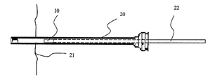
- FIG. 1 provides a perspective view of a hemostatic sponge pellet device of the invention.
- FIGS. 2A-2C provide cross-sectional views of various stages during the implantation of a compressed sponge pellet device of the invention.
- the present invention provides medical sponge matrices which are useful inter alia in hemostasis implant devices.
- Preferred such devices include compact, dry, sponge elements configured for deployment through a cannula to a biopsy site.
- the invention also provides methods for using such devices and matrices in the treatment of patients, for example for treatment of biopsied sites. Further, the invention provides methods for preparing highly compact and dense sponge matrices which involve compressing sponge matrices while hydrated, and drying the matrices in their compressed state.
- Sponge matrices in accordance with the invention will generally comprise porous, three-dimensionally stable bodies formed from suitable biocompatible matrix materials.
- suitable biocompatible matrix materials include naturally-occurring polymers and/or synthetic polymers.
- More preferred sponge compositions of the invention will comprise collagen as a matrix-forming material, either alone or in combination with one or more other matrix forming materials.
- sponge matrices of the invention can be formed by providing a liquid solution or suspension of a matrix-forming material, and causing the material to form a porous three-dimensionally stable structure. Other methods are known and can be used within the scope of the present invention.
- a collagen solution in the formation of a collagen sponge, can be prepared.
- the collagen may be derived from mammalian or other animal sources, for example, bovine, porcine or human sources. Synthetically-derived collagen may also be used. The determination of suitable collagen concentrations in the solution will be within the purview of those skilled in the art, with concentration ranges of about 0.05 g/ml to about 0.2 g/ml being typical.
- Digestion of the collagen to form the collagen solution is usually carried out under acidic conditions, starting with ground, minced or otherwise comminuted collagen-containing tissue.
- enzymatic digestion may be utilized using known enzymes for this purpose such as pepsin, trypsin, and/or papain. After digestion, the enzymes can be removed by suitable, known techniques.
- the collagen solution is treated with a precipitating buffer solution to neutralize the pH and precipitate the collagen. This precipitation can occur during incubation over several hours or days.
- the resulting product can be dried directly, but is preferably crosslinked with a suitable crosslinking agent and then dried.
- suitable crosslinking agent for these purposes include glutaraldehyde, formaldehyde, carbodiimides, UV irradiation, or other crosslinking agents.
- the crosslinking agent will contain polar groups that impart a hydrophilic character to the final sponge matrix material.
- a polyepoxide crosslinker is utilized for this purpose, especially a polyglycidyl ether compound.
- Suitable such compounds include ethylene glycol diglycidyl ether, available under the trade name Denacol EX810 from Nagese Chemical Co., Osaka, Japan, and glycerol polyglycidyl ether available under the trade name Denacol EX313 also from Nagese Chemical Co.
- polyglycidyl ethers or other polyepoxide compounds utilized in the invention will have from 2 to about 10 epoxide groups per molecule.
- the use of such epoxides and/or other crosslinking agents which impart polar groups and a hydrophilic character to the resulting matrix will provide for good wettability and rapid hydration and expansion of hemostasis devices of the invention.
- Preferred sources of collagen for use in the sponge matrices of the invention include extracellular matrix materials such as collagenous submucosal tissues, and other collagenous basement membrane materials. These include, for example, small intestinal submucosa (SIS), stomach submucosa, urinary bladder submucosa, liver basement membrane, and other basement membrane materials.
- SIS small intestinal submucosa
- stomach submucosa urinary bladder submucosa
- liver basement membrane and other basement membrane materials.
- these materials are preferably processed and utilized under conditions which retain their favorable growth properties. This may include, for example, processing under conditions in which native proteins and/or other materials, for instance biotropic agents, are retained in their bioactive form.
- the collagen sources, and resulting sponge matrices may include active native substances such as one or more growth factors, e.g. basic fibroblast growth factor (FGF-2); transforming growth factor beta (TGFss); epidermal growth factor (EFG); platelet derived growth factor (PDGF); and/or other substances such as glycosaminoglycans (GAGs); and/or fibronectin (FN).
- FGF-2 basic fibroblast growth factor
- TGFss transforming growth factor beta
- EGF epidermal growth factor
- PDGF platelet derived growth factor
- FN glycosaminoglycans
- FN glycosaminoglycans
- compact sponge matrices of the invention will be used in forming percutaneously-implantable medical devices.
- the matrices may be used to provide a small sponge element such as a pellet 10 that is useful for implantation into biopsied tissues to facilitate hemostasis and/or to deliver agents.
- sponge element 10 of the invention will be highly compacted and configured for passage through the cannula of a needle and/or a catheter such as that used to obtain core biopsies.
- Preferred sponge pellets of the invention will have compacted sizes having diameters “d” preferably less than about 2 millimeters so as to be deployable through a needle of corresponding size, e.g.
- Illustrative lengths “1” for sponge pellets of this diameter are less than about 3 centimeters, typically in the range of about 0.25 to about 3 centimeters. These diameters and lengths may of course be varied to suit a particular patient need.
- Preferred dry, compressed sponge matrices will be highly dense, typically having densities of at least about 0.05 g/cm 3 , preferably in the range of about 0.05 g/cm 3 to about 0.2 g/cm 3 , and more preferably about 0.075 g/cm 3 to about 0.2 g/cm 3 .
- the preferred compacted sponge matrix will have sufficient rigidity to be deployed by passage through needles or catheters as discussed above, for example by utilizing a pusher rod or other pusher element to force the sponge matrix device through the needle and/or catheter cannula.
- Expanded sponge densities (dry) will generally be less than the corresponding compacted densities. Typical expanded densities (dry) will range from about 0.01 g/cm 3 to about 0.1 g/cm 3 , more preferably about 0.02 g/cm 3 to about 0.07 g/cm 3 .
- Sponge matrix materials of the invention will advantageously be highly expandable when wetted, so as to achieve an expanded configuration (see 10 A, FIG. 1 ).
- Preferred sponge materials will exhibit the capacity to expand at least 100% by volume, more preferably at least about 200% by volume, and typically in the range of about 300% by volume to about 1000% by volume, when wetted to saturation with deionized water.
- Preferred sponge materials of the invention will also exhibit advantageous rates of expansion, achieving volume expansions as noted above in less than about 10 seconds, more preferably less than about 5 seconds, when immersed in deionized water.
- the expanded sizes typical for hemostatic sponge pellets of the invention include diameters “D” of about 0.5 cm to about 3 cm, and lengths “L” of about 0.5 cm to 3 cm. Such levels of expansion and final sizes are expected to exert compression on surrounding tissues when implanted, so as to benefit the patient by providing a hemostatic effect within the biopsied tissue.
- the pellets may deliver active agents to the implantation site and surrounding tissue.
- Highly compact, dense sponge matrices of the invention can be prepared by first hydrating or otherwise wetting a porous sponge matrix, and then compressing and drying the element. Such preparative processes generally provide a more dense, rigid and stably compressed sponge matrix than processes such as simple compaction of the dry sponge matrix. Drying will be conducted sufficiently to stabilize the sponge matrix. For example, preferred drying procedures will reduce the liquid (e.g. water) content of the matrix to less than about 20% by weight, more preferably less than about 10% by weight. Compression forces will be applied so as to achieve a final density and/or configuration desired, and can be applied in one, two or three dimensions, including radially.
- liquid e.g. water
- a sponge pellet prepared from the inventive matrices can have a generally cylindrical shape having a circular or multi-sided (e.g. square or rectangular) cross section, and can have a diameter approximating that or smaller than that of the needle and/or catheter cannula through which it is to be passed.
- the drying of the compacted element can involve lyophilization (or freeze drying) or vacuum drying at ambient or elevated temperatures.
- the sponge matrix upon removal of the compaction force, the sponge matrix is stabilized structurally and remains in its highly dense and compacted state until contacted with a liquid susceptible to absorption by the matrix, for example body fluids.
- the pores of the matrix are thereby stably retained at a volume substantially reduced from their maximum volume, but return to a partially or fully expanded state when the matrix material is wetted.
- Sponge elements or other devices of the invention may be formed individually by compaction/drying of an appropriately sized sponge element, or they may be individually excised from a larger compacted/dried sponge matrix.
- the compacted or compressed sponge matrix device can be sterilized using any suitable means, including for example radiation.
- the device will be suitably packaged in sterile packaging for medical use, to form medical articles of the invention.
- products of the invention may include biopsy kits containing at least one needle for obtaining a biopsy, and at least one sponge pellet of the invention.
- Suitable biopsy devices including needles include for example Quick-Core® biopsy needles or Twist-Core® biopsy needles available from Cook Diagnostic & Interventional Products.
- sponge elements of the invention are implanted into the biopsy site, for instance including the site of the excised tissue and/or the needle tract from the biopsy procedure. Implantation of the sponge element can be achieved through a cannula disposed within the biopsy needle tract.
- the biopsy procedure is performed with a biopsy device including an outer cannula 20 and an inner sampling needle (not shown).
- the inner sampling needle is used to obtain the biopsy tissue from the target patient tissue area 21 and withdrawn from the outer needle cannula, which is left in place.
- a sponge element 10 of the invention can be passed through the outer cannula ( FIG.
- FIG. 2A for example using a rod 22 or other device or mechanism to force the sponge element through the outer cannula and out an opening thereof ( FIG. 2B ).
- FIG. 2B multiple sponge elements can be delivered to a single biopsy site.
- the outer cannula can be withdrawn ( FIG. 2C ), leaving the sponge devices in place in the affected tissue.
- Sponge elements or other devices of the invention may also contain one or more active agents therapeutic to the patient.
- they may include proteins or other substances which promote clotting, for example Thrombin and/or Fibrinogen.
- sponge elements or other devices of the invention may include local anesthetics to be delivered to the affected (e.g. biopsied) tissue, and/or growth factors to promote tissue growth and healing within the affected tissue.
- active agents can be included in the liquid used to wet the sponge prior to compression.
- Sponge elements of the invention may also contain agents which promote further retention of the compressed, high density form of the elements. These may include for example starch, cellulose, sugars such as dextrose, or glycerin. Such agents can optionally be included in the liquid (preferably aqueous) used to hydrate or otherwise wet the sponge prior to compaction and drying.
- SIS tissue Fifty grams of SIS tissue, cut into two-centimeter length pieces, are added to 200 milliliters of a 0.1% pepsin in 0.5 M aqueous acetic acid solution. The resulting preparation is incubated at 37° C. for about 48 hours with stirring. Optionally, any undigested material at that point may be removed by centrifugation at 12,000 RPM for 20 minutes at room temperature.
- the gelled preparation is dialyzed (molecular weight cutoff of 3500) against several changes of phosphate buffered saline (pH 7.4) over 48 hours at 4° C. The gelled solution is then stored at 4° C. until ready for use.
- the chilled SIS gel is spread into a mold of desired shape and submersed in a collagen crosslinking solution.
- the crosslinking solution contains 2% vol/vol diglycidyl ether plus 20% vol/vol ethanol solution at 4° C. for 3-6 days.
- the resulting crosslinked SIS forms are removed from the crosslinking solution and frozen in a ⁇ 80° C. freezer.
- the frozen SIS forms are then lyophilized over a period of approximately 8 hours.
- the SIS forms are then soaked in several baths of high purity water, ringing residual water from the SIS sponge material between rinses.
- An SIS sponge material in hydrated form, prepared as in Example 1, is placed between two ridged plates and compressed. The plates are clamped so that compression is maintained, and the clamped structure is lyophilized over a period of approximately 1-2 hours. The rigid plates are removed, leaving the SIS sponge material in a highly densed, compacted, flattened shape. This compacted material is highly absorbent and expandable, and can be used in a variety of medical applications.
- a sponge pellet configured for percutaneous insertion through the cannula of a catheter and/or needle is cut from the compacted SIS sponge matrix.
Abstract
Description
Claims (33)
Priority Applications (1)
| Application Number | Priority Date | Filing Date | Title |
|---|---|---|---|
| US10/184,559 US8877233B2 (en) | 2001-06-29 | 2002-06-28 | Porous sponge matrix medical devices and methods |
Applications Claiming Priority (2)
| Application Number | Priority Date | Filing Date | Title |
|---|---|---|---|
| US30224501P | 2001-06-29 | 2001-06-29 | |
| US10/184,559 US8877233B2 (en) | 2001-06-29 | 2002-06-28 | Porous sponge matrix medical devices and methods |
Publications (2)
| Publication Number | Publication Date |
|---|---|
| US20030013989A1 US20030013989A1 (en) | 2003-01-16 |
| US8877233B2 true US8877233B2 (en) | 2014-11-04 |
Family
ID=23166916
Family Applications (1)
| Application Number | Title | Priority Date | Filing Date |
|---|---|---|---|
| US10/184,559 Expired - Fee Related US8877233B2 (en) | 2001-06-29 | 2002-06-28 | Porous sponge matrix medical devices and methods |
Country Status (5)
| Country | Link |
|---|---|
| US (1) | US8877233B2 (en) |
| EP (1) | EP1404390B1 (en) |
| AU (1) | AU2002320182B2 (en) |
| CA (1) | CA2452040C (en) |
| WO (1) | WO2003002168A1 (en) |
Cited By (22)
| Publication number | Priority date | Publication date | Assignee | Title |
|---|---|---|---|---|
| WO2016014244A1 (en) | 2014-07-20 | 2016-01-28 | Riina Howard Anthony | Anchored suture line, device and method for surgical suturing |
| US9445883B2 (en) | 2011-12-29 | 2016-09-20 | Sofradim Production | Barbed prosthetic knit and hernia repair mesh made therefrom as well as process for making said prosthetic knit |
| US9499927B2 (en) | 2012-09-25 | 2016-11-22 | Sofradim Production | Method for producing a prosthesis for reinforcing the abdominal wall |
| US9554887B2 (en) | 2011-03-16 | 2017-01-31 | Sofradim Production | Prosthesis comprising a three-dimensional and openworked knit |
| US9622843B2 (en) | 2011-07-13 | 2017-04-18 | Sofradim Production | Umbilical hernia prosthesis |
| US9750837B2 (en) | 2012-09-25 | 2017-09-05 | Sofradim Production | Haemostatic patch and method of preparation |
| US9839505B2 (en) | 2012-09-25 | 2017-12-12 | Sofradim Production | Prosthesis comprising a mesh and a strengthening means |
| US9867909B2 (en) | 2011-09-30 | 2018-01-16 | Sofradim Production | Multilayer implants for delivery of therapeutic agents |
| US9931198B2 (en) | 2015-04-24 | 2018-04-03 | Sofradim Production | Prosthesis for supporting a breast structure |
| US9980802B2 (en) | 2011-07-13 | 2018-05-29 | Sofradim Production | Umbilical hernia prosthesis |
| US10070948B2 (en) | 2008-06-27 | 2018-09-11 | Sofradim Production | Biosynthetic implant for soft tissue repair |
| US10080639B2 (en) | 2011-12-29 | 2018-09-25 | Sofradim Production | Prosthesis for inguinal hernia |
| US10159555B2 (en) | 2012-09-28 | 2018-12-25 | Sofradim Production | Packaging for a hernia repair device |
| US10184032B2 (en) | 2015-02-17 | 2019-01-22 | Sofradim Production | Method for preparing a chitosan-based matrix comprising a fiber reinforcement member |
| US10213283B2 (en) | 2013-06-07 | 2019-02-26 | Sofradim Production | Textile-based prosthesis for laparoscopic surgery |
| US10363690B2 (en) | 2012-08-02 | 2019-07-30 | Sofradim Production | Method for preparing a chitosan-based porous layer |
| US10405960B2 (en) | 2013-06-07 | 2019-09-10 | Sofradim Production | Textile-based prothesis for laparoscopic surgery |
| US10646321B2 (en) | 2016-01-25 | 2020-05-12 | Sofradim Production | Prosthesis for hernia repair |
| US10675137B2 (en) | 2017-05-02 | 2020-06-09 | Sofradim Production | Prosthesis for inguinal hernia repair |
| US10682215B2 (en) | 2016-10-21 | 2020-06-16 | Sofradim Production | Method for forming a mesh having a barbed suture attached thereto and the mesh thus obtained |
| US10743976B2 (en) | 2015-06-19 | 2020-08-18 | Sofradim Production | Synthetic prosthesis comprising a knit and a non porous film and method for forming same |
| US11471257B2 (en) | 2018-11-16 | 2022-10-18 | Sofradim Production | Implants suitable for soft tissue repair |
Families Citing this family (85)
| Publication number | Priority date | Publication date | Assignee | Title |
|---|---|---|---|---|
| US7597715B2 (en) | 2005-04-21 | 2009-10-06 | Biomet Manufacturing Corp. | Method and apparatus for use of porous implants |
| US8123814B2 (en) | 2001-02-23 | 2012-02-28 | Biomet Manufacturing Corp. | Method and appartus for acetabular reconstruction |
| US20030088240A1 (en) * | 2001-11-02 | 2003-05-08 | Vahid Saadat | Methods and apparatus for cryo-therapy |
| US7550004B2 (en) * | 2002-08-20 | 2009-06-23 | Cook Biotech Incorporated | Endoluminal device with extracellular matrix material and methods |
| US20040122349A1 (en) * | 2002-12-20 | 2004-06-24 | Lafontaine Daniel M. | Closure device with textured surface |
| US8709038B2 (en) * | 2002-12-20 | 2014-04-29 | Boston Scientific Scimed, Inc. | Puncture hole sealing device |
| US7942897B2 (en) | 2003-07-10 | 2011-05-17 | Boston Scientific Scimed, Inc. | System for closing an opening in a body cavity |
| US7744852B2 (en) | 2003-07-25 | 2010-06-29 | Rubicor Medical, Llc | Methods and systems for marking post biopsy cavity sites |
| US20050020899A1 (en) * | 2003-07-25 | 2005-01-27 | Rubicor Medical, Inc. | Post-biopsy cavity treatmetn implants and methods |
| US7537788B2 (en) * | 2003-07-25 | 2009-05-26 | Rubicor Medical, Inc. | Post-biopsy cavity treatment implants and methods |
| US7645229B2 (en) * | 2003-09-26 | 2010-01-12 | Armstrong David N | Instrument and method for endoscopic visualization and treatment of anorectal fistula |
| EP1686903B1 (en) | 2003-11-28 | 2014-07-30 | Cook Medical Technologies LLC | Vascular occlusion devices |
| EP1706040B1 (en) * | 2004-01-21 | 2010-05-26 | Cook Incorporated | Implantable graft to close a fistula |
| GB2430626B (en) | 2004-02-09 | 2008-09-24 | Cook Biotech Inc | Stent graft devices having collagen coating |
| US20080089933A1 (en) | 2006-10-16 | 2008-04-17 | Amir Alon | Device and method for reducing calorie intake |
| CA2560876A1 (en) | 2004-03-29 | 2005-10-13 | Cook Biotech Incorporated | Medical graft products with differing regions and methods and systems for producing the same |
| US20060052816A1 (en) * | 2004-08-31 | 2006-03-09 | Cook Incorporated | Device for treating an aneurysm |
| WO2006062862A1 (en) * | 2004-12-06 | 2006-06-15 | Cook Incorporated | Inflatable occlusion devices, methods, and systems |
| EP1833384B1 (en) * | 2004-12-30 | 2017-08-16 | Cook Medical Technologies LLC | Inverting occlusion devices and systems |
| US9138445B2 (en) * | 2005-03-09 | 2015-09-22 | Cook Biotech Incorporated | Medical graft materials with adherent extracellular matrix fibrous mass |
| US8066778B2 (en) * | 2005-04-21 | 2011-11-29 | Biomet Manufacturing Corp. | Porous metal cup with cobalt bearing surface |
| US8292967B2 (en) * | 2005-04-21 | 2012-10-23 | Biomet Manufacturing Corp. | Method and apparatus for use of porous implants |
| US8266780B2 (en) * | 2005-04-21 | 2012-09-18 | Biomet Manufacturing Corp. | Method and apparatus for use of porous implants |
| US8021432B2 (en) | 2005-12-05 | 2011-09-20 | Biomet Manufacturing Corp. | Apparatus for use of porous implants |
| US9788821B2 (en) | 2005-04-29 | 2017-10-17 | Cook Biotech Incorporated | Physically modified extracellular matrix materials and uses thereof |
| CA2606409A1 (en) * | 2005-04-29 | 2007-01-25 | Cook Biotech Incorporated | Fistula graft with deformable sheet-form material |
| WO2006119256A2 (en) | 2005-04-29 | 2006-11-09 | Cook Biotech Incorporated | Volumetric grafts for treatment of fistulae and related methods and systems |
| WO2007002260A2 (en) * | 2005-06-21 | 2007-01-04 | Cook Incorporated | Implantable graft to close a fistula |
| CN1903143A (en) * | 2005-07-29 | 2007-01-31 | 广东冠昊生物科技有限公司 | Biological type artificial blood vessel and method for preparing the same |
| CN1903144A (en) * | 2005-07-29 | 2007-01-31 | 广东冠昊生物科技有限公司 | Biological artificial ligamentum and method for preparing same |
| CN100482178C (en) * | 2005-08-04 | 2009-04-29 | 广东冠昊生物科技有限公司 | Blood vessel tumor clip with biological film |
| US8057495B2 (en) * | 2005-09-13 | 2011-11-15 | Cook Medical Technologies Llc | Aneurysm occlusion device |
| AU2006320507B2 (en) * | 2005-12-02 | 2012-11-01 | Cook Medical Technologies Llc | Devices, systems, and methods for occluding a defect |
| CN1986007B (en) * | 2005-12-20 | 2011-09-14 | 广东冠昊生物科技股份有限公司 | Biological surgical patch |
| CN1985778B (en) * | 2005-12-20 | 2010-10-13 | 广东冠昊生物科技股份有限公司 | Artificial biological cornea |
| CN1986006A (en) | 2005-12-20 | 2007-06-27 | 广州知光生物科技有限公司 | Biological nerve duct |
| CN1986001B (en) * | 2005-12-20 | 2011-09-14 | 广东冠昊生物科技股份有限公司 | Biological wound-protecting film |
| AU2007210970B2 (en) * | 2006-01-31 | 2013-09-05 | Cook Biotech Incorporated | Fistula grafts and related methods and systems for treating fistulae |
| US7635447B2 (en) * | 2006-02-17 | 2009-12-22 | Biomet Manufacturing Corp. | Method and apparatus for forming porous metal implants |
| EP2043531B1 (en) | 2006-06-15 | 2013-01-02 | Cook Medical Technologies LLC | Systems and devices for the delivery of endoluminal prostheses |
| JP5269779B2 (en) * | 2006-06-21 | 2013-08-21 | クック・バイオテック・インコーポレーテッド | Acupuncture grafts and related methods and systems useful for the treatment of gastrointestinal fistulas |
| US20100023129A1 (en) * | 2008-07-22 | 2010-01-28 | Guo-Feng Xu | Jawbone prosthesis and method of manufacture |
| CN101332316B (en) * | 2008-07-22 | 2012-12-26 | 广东冠昊生物科技股份有限公司 | Biotype nose bridge implantation body |
| CN101332314B (en) * | 2008-07-22 | 2012-11-14 | 广东冠昊生物科技股份有限公司 | Biotype articular cartilage repair piece |
| AU2007286657B2 (en) * | 2006-08-24 | 2012-11-15 | Cook Medical Technologies Llc | Devices and methods for occluding a fistula |
| US8343536B2 (en) | 2007-01-25 | 2013-01-01 | Cook Biotech Incorporated | Biofilm-inhibiting medical products |
| WO2008124361A2 (en) * | 2007-04-06 | 2008-10-16 | Cook Biotech Incorporated | Fistula plugs having increased column strength and fistula plug delivery apparatuses and methods |
| US20080279833A1 (en) * | 2007-05-10 | 2008-11-13 | Matheny Robert G | Laminate sheet articles for tissue regeneration |
| US8932619B2 (en) * | 2007-06-27 | 2015-01-13 | Sofradim Production | Dural repair material |
| US20090004455A1 (en) * | 2007-06-27 | 2009-01-01 | Philippe Gravagna | Reinforced composite implant |
| US8535349B2 (en) * | 2007-07-02 | 2013-09-17 | Cook Biotech Incorporated | Fistula grafts having a deflectable graft body portion |
| DE102007037053A1 (en) * | 2007-07-24 | 2009-01-29 | Aesculap Ag | Hemostatic for minimally invasive surgery |
| US9113851B2 (en) | 2007-08-23 | 2015-08-25 | Cook Biotech Incorporated | Fistula plugs and apparatuses and methods for fistula plug delivery |
| US20090068250A1 (en) | 2007-09-07 | 2009-03-12 | Philippe Gravagna | Bioresorbable and biocompatible compounds for surgical use |
| US20090069843A1 (en) * | 2007-09-10 | 2009-03-12 | Agnew Charles W | Fistula plugs including a hydration resistant component |
| WO2009036250A1 (en) | 2007-09-12 | 2009-03-19 | Cook Incorporated | Enhanced remodelable materials for occluding bodily vessels and related methods and systems |
| JP5205462B2 (en) * | 2007-09-25 | 2013-06-05 | バイオメット・マニュファクチャリング・コーポレイション | Cementless tibial tray |
| US9308068B2 (en) | 2007-12-03 | 2016-04-12 | Sofradim Production | Implant for parastomal hernia |
| US8956378B2 (en) * | 2008-02-29 | 2015-02-17 | Cook Biotech Incorporated | Coated embolization device |
| GB2471632B (en) | 2008-05-02 | 2012-04-18 | Cook Biotech Inc | Self deploying SIS in needle |
| GB2471635B (en) * | 2008-05-29 | 2012-09-26 | Cook Biotech Inc | Devices for treating rectovaginal and other fistulae |
| US9295757B2 (en) | 2008-06-10 | 2016-03-29 | Cook Biotech Incorporated | Quilted implantable graft |
| CN102056634B (en) * | 2008-06-10 | 2015-11-25 | 库克生物科技公司 | Quilted implantable graft |
| BRPI0914304A2 (en) * | 2008-06-20 | 2015-10-13 | Cook Biotech Inc | compressible / expandable graft medical products, and methods for applying haemostasis |
| CA2760201C (en) * | 2009-05-04 | 2019-11-19 | Oregon Biomedical Engineering Institute, Inc. | Hemorrhage control devices and methods |
| US8652500B2 (en) | 2009-07-22 | 2014-02-18 | Acell, Inc. | Particulate tissue graft with components of differing density and methods of making and using the same |
| FR2949688B1 (en) | 2009-09-04 | 2012-08-24 | Sofradim Production | FABRIC WITH PICOTS COATED WITH A BIORESORBABLE MICROPOROUS LAYER |
| WO2012050836A1 (en) | 2010-09-28 | 2012-04-19 | Cook Biotech Incorporated | Devices and methods for treating fistulae and other bodily openings and passageways |
| US9427233B2 (en) | 2011-06-06 | 2016-08-30 | Cook Medical Technologies, LLC | Vascular occlusion devices and methods |
| EP2720619B1 (en) | 2011-06-14 | 2016-10-26 | Cook Medical Technologies LLC | Fistula closure devices |
| WO2013046058A2 (en) | 2011-09-30 | 2013-04-04 | Sofradim Production | Reversible stiffening of light weight mesh |
| US10130346B2 (en) * | 2012-07-24 | 2018-11-20 | Omrix Biopharmaceuticals Ltd. | Device and method for the application of a curable fluid composition to a bodily organ |
| USD754325S1 (en) | 2013-06-06 | 2016-04-19 | Omrix Biopharmaceuticals Ltd. | Device of a curable fluid composition to a bodily organ |
| EP3000489B1 (en) | 2014-09-24 | 2017-04-05 | Sofradim Production | Method for preparing an anti-adhesion barrier film |
| EP3000432B1 (en) | 2014-09-29 | 2022-05-04 | Sofradim Production | Textile-based prosthesis for treatment of inguinal hernia |
| EP3000433B1 (en) | 2014-09-29 | 2022-09-21 | Sofradim Production | Device for introducing a prosthesis for hernia treatment into an incision and flexible textile based prosthesis |
| EP3029189B1 (en) | 2014-12-05 | 2021-08-11 | Sofradim Production | Prosthetic porous knit, method of making same and hernia prosthesis |
| US9238090B1 (en) | 2014-12-24 | 2016-01-19 | Fettech, Llc | Tissue-based compositions |
| WO2016154634A1 (en) * | 2015-03-26 | 2016-09-29 | Boston Scientific Scimed, Inc. | Biologic-based expandable occlusion devices |
| US20170245570A1 (en) * | 2016-02-25 | 2017-08-31 | William Yuen | Smart garment |
| WO2018051205A1 (en) * | 2016-09-16 | 2018-03-22 | Universidad De Los Andes | Extracellular-matrix haemostatic plug for percutaneous biopsies and device for the production thereof |
| WO2018140967A1 (en) * | 2017-01-30 | 2018-08-02 | Slendine Ag | Matrix materials having mechanically-shaped sponge architecture and methods of preparing thereof |
| CN108714241A (en) * | 2018-06-22 | 2018-10-30 | 中国人民解放军陆军军医大学第附属医院 | Application of the SIS epoxy resins in preparing hemostatic material |
| CN110975010B (en) * | 2019-11-25 | 2022-02-15 | 银丰低温医学科技有限公司 | Placenta tissue matrix material and preparation method thereof |
| CN113289063A (en) * | 2021-05-27 | 2021-08-24 | 江阴市人民医院 | Biological material for soft tissue repair and preparation method thereof |
Citations (47)
| Publication number | Priority date | Publication date | Assignee | Title |
|---|---|---|---|---|
| GB887844A (en) | 1958-11-13 | 1962-01-24 | John Straythorn Robins | Surgical dressing |
| US3566871A (en) | 1968-06-11 | 1971-03-02 | American Cyanamid Co | Hydrophilic medical sponge and method of using same |
| US3810473A (en) | 1972-12-04 | 1974-05-14 | Avicon Inc | Liquid-laid, non-woven, fibrous collagen derived surgical web having hemostatic and wound sealing properties |
| US3823212A (en) * | 1968-11-27 | 1974-07-09 | Freudenberg C Fa | Process for the production of collagen fiber fabrics in the form of felt-like membranes or sponge-like layers |
| US4193813A (en) | 1976-09-07 | 1980-03-18 | Medi-Coll, Inc. | Method for making collagen sponge |
| US4271835A (en) | 1978-05-17 | 1981-06-09 | Kcdp Corporation | Fluid-expansible contraceptive tampon and applicator |
| US4511653A (en) | 1981-11-26 | 1985-04-16 | Foundation Merieux | Process for the industrial preparation of collagenous materials from human placental tissues, human collagenous materials obtained and their application as biomaterials |
| US4578067A (en) | 1982-04-12 | 1986-03-25 | Alcon (Puerto Rico) Inc. | Hemostatic-adhesive, collagen dressing for severed biological surfaces |
| US4846793A (en) | 1987-03-18 | 1989-07-11 | Endocon, Inc. | Injector for implanting multiple pellet medicaments |
| US4852568A (en) | 1987-02-17 | 1989-08-01 | Kensey Nash Corporation | Method and apparatus for sealing an opening in tissue of a living being |
| US4902508A (en) | 1988-07-11 | 1990-02-20 | Purdue Research Foundation | Tissue graft composition |
| US4956178A (en) | 1988-07-11 | 1990-09-11 | Purdue Research Foundation | Tissue graft composition |
| US4970298A (en) * | 1984-03-27 | 1990-11-13 | University Of Medicine And Dentistry Of New Jersey | Biodegradable matrix and methods for producing same |
| US5206028A (en) * | 1991-02-11 | 1993-04-27 | Li Shu Tung | Dense collagen membrane matrices for medical uses |
| US5219576A (en) | 1988-06-30 | 1993-06-15 | Collagen Corporation | Collagen wound healing matrices and process for their production |
| US5275616A (en) | 1990-10-01 | 1994-01-04 | Quinton Instrument Company | Insertion assembly and method of inserting a vessel plug into the body of a patient |
| US5279555A (en) | 1992-08-24 | 1994-01-18 | Merck & Co., Inc. | Device for injecting implants |
| US5325857A (en) | 1993-07-09 | 1994-07-05 | Hossein Nabai | Skin biopsy device and method |
| US5388588A (en) * | 1993-05-04 | 1995-02-14 | Nabai; Hossein | Biopsy wound closure device and method |
| US5391183A (en) | 1990-09-21 | 1995-02-21 | Datascope Investment Corp | Device and method sealing puncture wounds |
| US5394886A (en) | 1993-09-20 | 1995-03-07 | Nabai; Hossein | Skin biopsy plug and method |
| US5449375A (en) | 1992-12-10 | 1995-09-12 | Howmedica Inc. | Method of making a hemostatic plug |
| US5456693A (en) * | 1992-09-21 | 1995-10-10 | Vitaphore Corporation | Embolization plugs for blood vessels |
| US5532221A (en) * | 1991-04-05 | 1996-07-02 | Lifecore Biomedical, Inc. | Ionically crosslinked carboxyl-containing polysaccharides for adhesion prevention |
| US5554389A (en) | 1995-04-07 | 1996-09-10 | Purdue Research Foundation | Urinary bladder submucosa derived tissue graft |
| US5571181A (en) * | 1992-05-11 | 1996-11-05 | Li; Shu-Tung | Soft tissue closure systems |
| US5674298A (en) | 1994-10-21 | 1997-10-07 | The Board Of Regents Of The University Of Michigan | Calcification-resistant bioprosthetic tissue and methods of making same |
| WO1998009617A1 (en) | 1996-09-07 | 1998-03-12 | Beisel Guenther | Product with extender effect |
| WO1998022158A2 (en) | 1996-08-23 | 1998-05-28 | Cook Biotech, Incorporated | Graft prosthesis, materials and methods |
| WO1998025637A1 (en) | 1996-12-10 | 1998-06-18 | Purdue Research Foundation | Biomaterial derived from vertebrate liver tissue |
| US5782914A (en) | 1996-11-29 | 1998-07-21 | Bio-Vascular, Inc. | Method for preparing heterogeneous tissue grafts |
| CA2245754A1 (en) | 1997-09-05 | 1999-03-05 | Dr. Suwelack Skin & Health Care Ag | Agent for oral intake, its production and use |
| US6008292A (en) | 1997-12-02 | 1999-12-28 | Baxter International Inc. | Method for inhibiting calcification of aldehyde-fixed bioprosthetic materials |
| US6056970A (en) * | 1998-05-07 | 2000-05-02 | Genzyme Corporation | Compositions comprising hemostatic compounds and bioabsorbable polymers |
| US6071301A (en) * | 1998-05-01 | 2000-06-06 | Sub Q., Inc. | Device and method for facilitating hemostasis of a biopsy tract |
| WO2000032250A1 (en) | 1998-12-01 | 2000-06-08 | Cook Biotech, Inc. | A multi-formed collagenous biomaterial medical device |
| EP1022031A1 (en) | 1999-01-21 | 2000-07-26 | Nissho Corporation | Suturable adhesion-preventing membrane |
| US6099567A (en) | 1996-12-10 | 2000-08-08 | Purdue Research Foundation | Stomach submucosa derived tissue graft |
| US6130264A (en) | 1994-10-06 | 2000-10-10 | Xomed Surgical Products, Inc. | Synthetic sponge and surgical spear comprising synthetic sponge |
| US6183497B1 (en) | 1998-05-01 | 2001-02-06 | Sub-Q, Inc. | Absorbable sponge with contrasting agent |
| US6190350B1 (en) | 1997-12-29 | 2001-02-20 | Alza Corporation | Implanter device for subcutaneous implants |
| WO2001013800A1 (en) | 1999-08-24 | 2001-03-01 | Sub-Q, Inc. | Device and method for facilitating hemostasis of a biopsy tract |
| US6261309B1 (en) | 1998-11-02 | 2001-07-17 | Datascope Investment Corp. | Collapsible hemostatic plug |
| US6270464B1 (en) | 1998-06-22 | 2001-08-07 | Artemis Medical, Inc. | Biopsy localization method and device |
| US6371904B1 (en) | 1998-12-24 | 2002-04-16 | Vivant Medical, Inc. | Subcutaneous cavity marking device and method |
| US6536782B2 (en) | 2001-04-02 | 2003-03-25 | Rohm Gmbh | Quick-release drill chuck |
| US6605047B2 (en) | 2001-09-10 | 2003-08-12 | Vivant Medical, Inc. | Biopsy marker delivery system |
-
2002
- 2002-06-28 AU AU2002320182A patent/AU2002320182B2/en not_active Ceased
- 2002-06-28 US US10/184,559 patent/US8877233B2/en not_active Expired - Fee Related
- 2002-06-28 EP EP02749686A patent/EP1404390B1/en not_active Expired - Lifetime
- 2002-06-28 CA CA2452040A patent/CA2452040C/en not_active Expired - Fee Related
- 2002-06-28 WO PCT/US2002/020462 patent/WO2003002168A1/en not_active Application Discontinuation
Patent Citations (56)
| Publication number | Priority date | Publication date | Assignee | Title |
|---|---|---|---|---|
| GB887844A (en) | 1958-11-13 | 1962-01-24 | John Straythorn Robins | Surgical dressing |
| US3566871A (en) | 1968-06-11 | 1971-03-02 | American Cyanamid Co | Hydrophilic medical sponge and method of using same |
| US3823212A (en) * | 1968-11-27 | 1974-07-09 | Freudenberg C Fa | Process for the production of collagen fiber fabrics in the form of felt-like membranes or sponge-like layers |
| US3810473A (en) | 1972-12-04 | 1974-05-14 | Avicon Inc | Liquid-laid, non-woven, fibrous collagen derived surgical web having hemostatic and wound sealing properties |
| US4193813A (en) | 1976-09-07 | 1980-03-18 | Medi-Coll, Inc. | Method for making collagen sponge |
| US4271835A (en) | 1978-05-17 | 1981-06-09 | Kcdp Corporation | Fluid-expansible contraceptive tampon and applicator |
| US4511653A (en) | 1981-11-26 | 1985-04-16 | Foundation Merieux | Process for the industrial preparation of collagenous materials from human placental tissues, human collagenous materials obtained and their application as biomaterials |
| US4578067A (en) | 1982-04-12 | 1986-03-25 | Alcon (Puerto Rico) Inc. | Hemostatic-adhesive, collagen dressing for severed biological surfaces |
| US4970298A (en) * | 1984-03-27 | 1990-11-13 | University Of Medicine And Dentistry Of New Jersey | Biodegradable matrix and methods for producing same |
| US4852568A (en) | 1987-02-17 | 1989-08-01 | Kensey Nash Corporation | Method and apparatus for sealing an opening in tissue of a living being |
| US4846793A (en) | 1987-03-18 | 1989-07-11 | Endocon, Inc. | Injector for implanting multiple pellet medicaments |
| US5219576A (en) | 1988-06-30 | 1993-06-15 | Collagen Corporation | Collagen wound healing matrices and process for their production |
| US4902508A (en) | 1988-07-11 | 1990-02-20 | Purdue Research Foundation | Tissue graft composition |
| US4956178A (en) | 1988-07-11 | 1990-09-11 | Purdue Research Foundation | Tissue graft composition |
| US5391183A (en) | 1990-09-21 | 1995-02-21 | Datascope Investment Corp | Device and method sealing puncture wounds |
| US5275616A (en) | 1990-10-01 | 1994-01-04 | Quinton Instrument Company | Insertion assembly and method of inserting a vessel plug into the body of a patient |
| US5275616B1 (en) | 1990-10-01 | 1996-01-23 | Quinton Instr | Insertion assembly and method of inserting a vessel plug into the body of a patient |
| US5206028A (en) * | 1991-02-11 | 1993-04-27 | Li Shu Tung | Dense collagen membrane matrices for medical uses |
| US5532221A (en) * | 1991-04-05 | 1996-07-02 | Lifecore Biomedical, Inc. | Ionically crosslinked carboxyl-containing polysaccharides for adhesion prevention |
| US5571181A (en) * | 1992-05-11 | 1996-11-05 | Li; Shu-Tung | Soft tissue closure systems |
| US5279555A (en) | 1992-08-24 | 1994-01-18 | Merck & Co., Inc. | Device for injecting implants |
| US5456693A (en) * | 1992-09-21 | 1995-10-10 | Vitaphore Corporation | Embolization plugs for blood vessels |
| US5449375A (en) | 1992-12-10 | 1995-09-12 | Howmedica Inc. | Method of making a hemostatic plug |
| US5467780A (en) | 1993-05-04 | 1995-11-21 | Nabai; Hossein | Biopsy wound closure device and method |
| US5479936A (en) | 1993-05-04 | 1996-01-02 | Nabai; Hossein | Biopsy wound closure device and method |
| US5483972A (en) | 1993-05-04 | 1996-01-16 | Nabai; Hossein | Biopsy wound closure device |
| US5388588A (en) * | 1993-05-04 | 1995-02-14 | Nabai; Hossein | Biopsy wound closure device and method |
| US5325857A (en) | 1993-07-09 | 1994-07-05 | Hossein Nabai | Skin biopsy device and method |
| US5394886A (en) | 1993-09-20 | 1995-03-07 | Nabai; Hossein | Skin biopsy plug and method |
| US6130264A (en) | 1994-10-06 | 2000-10-10 | Xomed Surgical Products, Inc. | Synthetic sponge and surgical spear comprising synthetic sponge |
| US5674298A (en) | 1994-10-21 | 1997-10-07 | The Board Of Regents Of The University Of Michigan | Calcification-resistant bioprosthetic tissue and methods of making same |
| US5554389A (en) | 1995-04-07 | 1996-09-10 | Purdue Research Foundation | Urinary bladder submucosa derived tissue graft |
| US6206931B1 (en) | 1996-08-23 | 2001-03-27 | Cook Incorporated | Graft prosthesis materials |
| WO1998022158A2 (en) | 1996-08-23 | 1998-05-28 | Cook Biotech, Incorporated | Graft prosthesis, materials and methods |
| US6541031B1 (en) | 1996-09-07 | 2003-04-01 | Beisel Guenther | Composition with release-controlling action |
| WO1998009617A1 (en) | 1996-09-07 | 1998-03-12 | Beisel Guenther | Product with extender effect |
| US5782914A (en) | 1996-11-29 | 1998-07-21 | Bio-Vascular, Inc. | Method for preparing heterogeneous tissue grafts |
| US6099567A (en) | 1996-12-10 | 2000-08-08 | Purdue Research Foundation | Stomach submucosa derived tissue graft |
| WO1998025637A1 (en) | 1996-12-10 | 1998-06-18 | Purdue Research Foundation | Biomaterial derived from vertebrate liver tissue |
| US6379710B1 (en) | 1996-12-10 | 2002-04-30 | Purdue Research Foundation | Biomaterial derived from vertebrate liver tissue |
| EP0901792A1 (en) | 1997-09-05 | 1999-03-17 | Dr. Suwelack Skin & Health Care AG | Agent for peroral administration, its production and use |
| CA2245754A1 (en) | 1997-09-05 | 1999-03-05 | Dr. Suwelack Skin & Health Care Ag | Agent for oral intake, its production and use |
| US6008292A (en) | 1997-12-02 | 1999-12-28 | Baxter International Inc. | Method for inhibiting calcification of aldehyde-fixed bioprosthetic materials |
| US6190350B1 (en) | 1997-12-29 | 2001-02-20 | Alza Corporation | Implanter device for subcutaneous implants |
| US6071301A (en) * | 1998-05-01 | 2000-06-06 | Sub Q., Inc. | Device and method for facilitating hemostasis of a biopsy tract |
| US6183497B1 (en) | 1998-05-01 | 2001-02-06 | Sub-Q, Inc. | Absorbable sponge with contrasting agent |
| US6056970A (en) * | 1998-05-07 | 2000-05-02 | Genzyme Corporation | Compositions comprising hemostatic compounds and bioabsorbable polymers |
| US6270464B1 (en) | 1998-06-22 | 2001-08-07 | Artemis Medical, Inc. | Biopsy localization method and device |
| US6699205B2 (en) | 1998-06-22 | 2004-03-02 | Artemis Medical, Inc. | Biopsy localization method and device |
| US6261309B1 (en) | 1998-11-02 | 2001-07-17 | Datascope Investment Corp. | Collapsible hemostatic plug |
| WO2000032250A1 (en) | 1998-12-01 | 2000-06-08 | Cook Biotech, Inc. | A multi-formed collagenous biomaterial medical device |
| US6371904B1 (en) | 1998-12-24 | 2002-04-16 | Vivant Medical, Inc. | Subcutaneous cavity marking device and method |
| EP1022031A1 (en) | 1999-01-21 | 2000-07-26 | Nissho Corporation | Suturable adhesion-preventing membrane |
| WO2001013800A1 (en) | 1999-08-24 | 2001-03-01 | Sub-Q, Inc. | Device and method for facilitating hemostasis of a biopsy tract |
| US6536782B2 (en) | 2001-04-02 | 2003-03-25 | Rohm Gmbh | Quick-release drill chuck |
| US6605047B2 (en) | 2001-09-10 | 2003-08-12 | Vivant Medical, Inc. | Biopsy marker delivery system |
Non-Patent Citations (14)
| Title |
|---|
| "Gylosaminoglycans and the regulation of blood coagulation" Biochem. J. (1993) 289, 313-330. * |
| Chan-Myers, HB, et al. "Sterilization of a Small Caliber Vascular Graft with a Polyepoxy Compound". ASAIO Journal, 1992. vol. 38, pp. 116-119. Lippincott Williams & Wilkins, United States. |
| Fandrich, CA, et al. "Small Gauge Gelfoam Plug Liver Biopsy in High Risk Patients: Safety and Diagnostic Value". Australasian Radiology, Aug. 1996. vol. 40, No. 3. pp. 230-234. Blackwell Scientific Publications, Australia. |
| Kallmes, DF, et al. "In Vivo Evaluation of a New Type I Collagen Hemostatic Plug for High-Risk, Large-Core Biopsies". Journal of Vascular and Interventional Radiology, Jul.-Aug. 1998. vol. 9, No. 4. pp. 656-659. Society of Cardiovascular and Interventional Radiology, United States. |
| Kaufman, R, et al. "Hair Transplantation: Gelatin Plugs for Hemostasis". Z Hautkr, Aug. 1983. vol. 58, No. 15. pp. 1139-1141. Grosse, Germany. |
| Lohre, JM, et al. "Evaluation of Epoxy Ether Fixed Bovine Arterial Grafts for Mutagenic Potential". ASAIO Journal, Apr.-Jun. 1993. vol. 39, No. 2, pp. 106-113. Lippincott Williams & Wilkins, United States. |
| Lohre, JM, et al. "Evaluation of Two Epoxy Ether Compounds for Biocompatible Potential". Artificial Organs, Dec. 1992. vol. 16, No. 6. pp. 630-633. Blackwell Science, United States. |
| Robinson, JD et al, "The Biocompatibility of Compressed Collagen Foam Plugs", Cardiovascular and Interventional Radiology, Feb.-Mar. 1990. vol. 13, No. 1. pp. 36-39. Springer Vertag, United States. |
| Robinson, JD, et al. "The Biocompatibility of Compressed Collagen Foam Plugs". Cardiovascular and Interventional Radiology, Feb.-Mar. 1990. vol. 13, No. 1. pp. 36-39. Springer Verlag, United States. |
| Skelton, HG, et al. "Helistat Absorbable Collagen Hemostatic Sponges in Cutaneous Surgery in HIV-1+ Patients." Military Medical Consortium for the Advancement of Military Medicine (MMCAR). International Journal of Dermatology, Nov. 1993. vol. 32, No. 11. pp. 835-837. Decker Periodicals, United States. |
| Smith, KJ, et al. "Bovine Collagen Products and Gelatin Sponges for Hemostasis in Punch Biopsies of HIV-1+ Patients". Dermatologic Surgery, Jun. 1995. vol. 21, No. 6. pp. 563-564. Blackwell Science, United States. |
| Sung, HW, et al. "Comparison of the Cross-Linking Characteristics of Porcine Heart Valves Fixed with Glutaraldehyde or Epoxy Compounds". ASAIO Journal, Jul.-Sep. 1993. vol. 39, No. 3, pp. 532-536. Lippincott Williams & Wilkins, United States. |
| Tang, Z, et al. "Crosslinkage of Collagen by Polyglycidyl Ethers". ASAIO Journal, 1995. vol. 41, pp. 72-78. Lippincott Williams & Wilkins, United States. |
| Zhou, J., et al. "Porcine Aortic Wall Flexibility. Fresh vs. Denacol Fixed vs. Glutaraldehyde Fixed". ASAIO Journal, Sep.-Oct. 1997. vol. 43, No. 5, pp. 470-475. Lippincott Williams & Wilkins, United States. |
Cited By (41)
| Publication number | Priority date | Publication date | Assignee | Title |
|---|---|---|---|---|
| US10070948B2 (en) | 2008-06-27 | 2018-09-11 | Sofradim Production | Biosynthetic implant for soft tissue repair |
| US9554887B2 (en) | 2011-03-16 | 2017-01-31 | Sofradim Production | Prosthesis comprising a three-dimensional and openworked knit |
| US10472750B2 (en) | 2011-03-16 | 2019-11-12 | Sofradim Production | Prosthesis comprising a three-dimensional and openworked knit |
| US11612472B2 (en) | 2011-03-16 | 2023-03-28 | Sofradim Production | Prosthesis comprising a three-dimensional and openworked knit |
| US9980802B2 (en) | 2011-07-13 | 2018-05-29 | Sofradim Production | Umbilical hernia prosthesis |
| US11039912B2 (en) | 2011-07-13 | 2021-06-22 | Sofradim Production | Umbilical hernia prosthesis |
| US10709538B2 (en) | 2011-07-13 | 2020-07-14 | Sofradim Production | Umbilical hernia prosthesis |
| US9622843B2 (en) | 2011-07-13 | 2017-04-18 | Sofradim Production | Umbilical hernia prosthesis |
| US11903807B2 (en) | 2011-07-13 | 2024-02-20 | Sofradim Production | Umbilical hernia prosthesis |
| US9867909B2 (en) | 2011-09-30 | 2018-01-16 | Sofradim Production | Multilayer implants for delivery of therapeutic agents |
| US11925543B2 (en) | 2011-12-29 | 2024-03-12 | Sofradim Production | Barbed prosthetic knit and hernia repair mesh made therefrom as well as process for making said prosthetic knit |
| US11471256B2 (en) | 2011-12-29 | 2022-10-18 | Sofradim Production | Prosthesis for inguinal hernia |
| US10080639B2 (en) | 2011-12-29 | 2018-09-25 | Sofradim Production | Prosthesis for inguinal hernia |
| US9445883B2 (en) | 2011-12-29 | 2016-09-20 | Sofradim Production | Barbed prosthetic knit and hernia repair mesh made therefrom as well as process for making said prosthetic knit |
| US10342652B2 (en) | 2011-12-29 | 2019-07-09 | Sofradim Production | Barbed prosthetic knit and hernia repair mesh made therefrom as well as process for making said prosthetic knit |
| US11266489B2 (en) | 2011-12-29 | 2022-03-08 | Sofradim Production | Barbed prosthetic knit and hernia repair mesh made therefrom as well as process for making said prosthetic knit |
| US10363690B2 (en) | 2012-08-02 | 2019-07-30 | Sofradim Production | Method for preparing a chitosan-based porous layer |
| US9839505B2 (en) | 2012-09-25 | 2017-12-12 | Sofradim Production | Prosthesis comprising a mesh and a strengthening means |
| US9750837B2 (en) | 2012-09-25 | 2017-09-05 | Sofradim Production | Haemostatic patch and method of preparation |
| US9499927B2 (en) | 2012-09-25 | 2016-11-22 | Sofradim Production | Method for producing a prosthesis for reinforcing the abdominal wall |
| US10159555B2 (en) | 2012-09-28 | 2018-12-25 | Sofradim Production | Packaging for a hernia repair device |
| US10213283B2 (en) | 2013-06-07 | 2019-02-26 | Sofradim Production | Textile-based prosthesis for laparoscopic surgery |
| US11622845B2 (en) | 2013-06-07 | 2023-04-11 | Sofradim Production | Textile-based prothesis for laparoscopic surgery |
| US11304790B2 (en) | 2013-06-07 | 2022-04-19 | Sofradim Production | Textile-based prothesis for laparoscopic surgery |
| US10405960B2 (en) | 2013-06-07 | 2019-09-10 | Sofradim Production | Textile-based prothesis for laparoscopic surgery |
| US9775598B2 (en) | 2014-07-20 | 2017-10-03 | New York University | Anchored suture line, device and method for surgical suturing |
| WO2016014244A1 (en) | 2014-07-20 | 2016-01-28 | Riina Howard Anthony | Anchored suture line, device and method for surgical suturing |
| US10815345B2 (en) | 2015-02-17 | 2020-10-27 | Sofradim Production | Method for preparing a chitosan-based matrix comprising a fiber reinforcement member |
| US10184032B2 (en) | 2015-02-17 | 2019-01-22 | Sofradim Production | Method for preparing a chitosan-based matrix comprising a fiber reinforcement member |
| US9931198B2 (en) | 2015-04-24 | 2018-04-03 | Sofradim Production | Prosthesis for supporting a breast structure |
| US10660741B2 (en) | 2015-04-24 | 2020-05-26 | Sofradim Production | Prosthesis for supporting a breast structure |
| US11439498B2 (en) | 2015-04-24 | 2022-09-13 | Sofradim Production | Prosthesis for supporting a breast structure |
| US10743976B2 (en) | 2015-06-19 | 2020-08-18 | Sofradim Production | Synthetic prosthesis comprising a knit and a non porous film and method for forming same |
| US11826242B2 (en) | 2015-06-19 | 2023-11-28 | Sofradim Production | Synthetic prosthesis comprising a knit and a non porous film and method for forming same |
| US10646321B2 (en) | 2016-01-25 | 2020-05-12 | Sofradim Production | Prosthesis for hernia repair |
| US11389282B2 (en) | 2016-01-25 | 2022-07-19 | Sofradim Production | Prosthesis for hernia repair |
| US11696819B2 (en) | 2016-10-21 | 2023-07-11 | Sofradim Production | Method for forming a mesh having a barbed suture attached thereto and the mesh thus obtained |
| US10682215B2 (en) | 2016-10-21 | 2020-06-16 | Sofradim Production | Method for forming a mesh having a barbed suture attached thereto and the mesh thus obtained |
| US11672636B2 (en) | 2017-05-02 | 2023-06-13 | Sofradim Production | Prosthesis for inguinal hernia repair |
| US10675137B2 (en) | 2017-05-02 | 2020-06-09 | Sofradim Production | Prosthesis for inguinal hernia repair |
| US11471257B2 (en) | 2018-11-16 | 2022-10-18 | Sofradim Production | Implants suitable for soft tissue repair |
Also Published As
| Publication number | Publication date |
|---|---|
| EP1404390B1 (en) | 2012-08-01 |
| EP1404390A1 (en) | 2004-04-07 |
| US20030013989A1 (en) | 2003-01-16 |
| CA2452040A1 (en) | 2003-01-09 |
| AU2002320182B2 (en) | 2008-02-21 |
| CA2452040C (en) | 2011-03-22 |
| WO2003002168A1 (en) | 2003-01-09 |
Similar Documents
| Publication | Publication Date | Title |
|---|---|---|
| US8877233B2 (en) | Porous sponge matrix medical devices and methods | |
| AU2002320182A1 (en) | Porous sponge matrix medical devices and methods | |
| EP2358409B1 (en) | Compressible/expandable medical graft products, and methods for applying hemostasis | |
| US9788821B2 (en) | Physically modified extracellular matrix materials and uses thereof | |
| US7153518B2 (en) | Processed soft tissue for topical or internal application | |
| JP5689233B2 (en) | Superabsorbent lyophilized hydrogel for medical use | |
| US5326350A (en) | Soft tissue closure systems | |
| CN110665061A (en) | Acellular scaffold solution-GelMA hydrogel composite material and preparation method thereof | |
| US8518064B2 (en) | Method for anchoring occlusion plug | |
| US8518104B2 (en) | Methods for occluding bodily vessels | |
| CN1218378A (en) | Occlusion system of soft tissue | |
| JPH01212559A (en) | Biological implant substance | |
| US20230132953A1 (en) | Plug for lung tissue tract sealing | |
| JPS59155248A (en) | Coating membrane and production thereof | |
| WO2023079401A1 (en) | Plug for lung tissue tract sealing | |
| CN111643713A (en) | Preparation method of antibacterial absorbable medical periplaneta americana suture |
Legal Events
| Date | Code | Title | Description |
|---|---|---|---|
| AS | Assignment |
Owner name: COOK BIOTECH INCORPORATED, INDIANA Free format text: ASSIGNMENT OF ASSIGNORS INTEREST;ASSIGNORS:OBERMILLER, JOSEPH;HILES, MICHAEL C.;REEL/FRAME:013301/0229 Effective date: 20020903 |
|
| AS | Assignment |
Owner name: COOK BIOTECH INCORPORATED, INDIANA Free format text: ASSIGNMENT OF ASSIGNORS INTEREST;ASSIGNORS:OBERMILLER, F. JOSEPH;HILES, MICHAEL C.;REEL/FRAME:015927/0854 Effective date: 20040929 |
|
| FEPP | Fee payment procedure |
Free format text: MAINTENANCE FEE REMINDER MAILED (ORIGINAL EVENT CODE: REM.) |
|
| LAPS | Lapse for failure to pay maintenance fees |
Free format text: PATENT EXPIRED FOR FAILURE TO PAY MAINTENANCE FEES (ORIGINAL EVENT CODE: EXP.); ENTITY STATUS OF PATENT OWNER: LARGE ENTITY |
|
| STCH | Information on status: patent discontinuation |
Free format text: PATENT EXPIRED DUE TO NONPAYMENT OF MAINTENANCE FEES UNDER 37 CFR 1.362 |
|
| FP | Expired due to failure to pay maintenance fee |
Effective date: 20181104 |