US9005245B2 - Acromioclavicular joint fixation technique - Google Patents
Acromioclavicular joint fixation technique Download PDFInfo
- Publication number
- US9005245B2 US9005245B2 US11/687,882 US68788207A US9005245B2 US 9005245 B2 US9005245 B2 US 9005245B2 US 68788207 A US68788207 A US 68788207A US 9005245 B2 US9005245 B2 US 9005245B2
- Authority
- US
- United States
- Prior art keywords
- button
- circular
- suture
- oblong
- buttons
- Prior art date
- Legal status (The legal status is an assumption and is not a legal conclusion. Google has not performed a legal analysis and makes no representation as to the accuracy of the status listed.)
- Active, expires
Links
Images


Classifications
-
- A—HUMAN NECESSITIES
- A61—MEDICAL OR VETERINARY SCIENCE; HYGIENE
- A61B—DIAGNOSIS; SURGERY; IDENTIFICATION
- A61B17/00—Surgical instruments, devices or methods, e.g. tourniquets
- A61B17/04—Surgical instruments, devices or methods, e.g. tourniquets for suturing wounds; Holders or packages for needles or suture materials
- A61B17/0401—Suture anchors, buttons or pledgets, i.e. means for attaching sutures to bone, cartilage or soft tissue; Instruments for applying or removing suture anchors
-
- A—HUMAN NECESSITIES
- A61—MEDICAL OR VETERINARY SCIENCE; HYGIENE
- A61B—DIAGNOSIS; SURGERY; IDENTIFICATION
- A61B17/00—Surgical instruments, devices or methods, e.g. tourniquets
- A61B17/16—Bone cutting, breaking or removal means other than saws, e.g. Osteoclasts; Drills or chisels for bones; Trepans
- A61B17/17—Guides or aligning means for drills, mills, pins or wires
- A61B17/1739—Guides or aligning means for drills, mills, pins or wires specially adapted for particular parts of the body
- A61B17/1778—Guides or aligning means for drills, mills, pins or wires specially adapted for particular parts of the body for the shoulder
-
- A—HUMAN NECESSITIES
- A61—MEDICAL OR VETERINARY SCIENCE; HYGIENE
- A61B—DIAGNOSIS; SURGERY; IDENTIFICATION
- A61B17/00—Surgical instruments, devices or methods, e.g. tourniquets
- A61B17/56—Surgical instruments or methods for treatment of bones or joints; Devices specially adapted therefor
- A61B17/58—Surgical instruments or methods for treatment of bones or joints; Devices specially adapted therefor for osteosynthesis, e.g. bone plates, screws, setting implements or the like
- A61B17/68—Internal fixation devices, including fasteners and spinal fixators, even if a part thereof projects from the skin
-
- A—HUMAN NECESSITIES
- A61—MEDICAL OR VETERINARY SCIENCE; HYGIENE
- A61B—DIAGNOSIS; SURGERY; IDENTIFICATION
- A61B17/00—Surgical instruments, devices or methods, e.g. tourniquets
- A61B17/04—Surgical instruments, devices or methods, e.g. tourniquets for suturing wounds; Holders or packages for needles or suture materials
- A61B17/0469—Suturing instruments for use in minimally invasive surgery, e.g. endoscopic surgery
-
- A—HUMAN NECESSITIES
- A61—MEDICAL OR VETERINARY SCIENCE; HYGIENE
- A61B—DIAGNOSIS; SURGERY; IDENTIFICATION
- A61B17/00—Surgical instruments, devices or methods, e.g. tourniquets
- A61B17/16—Bone cutting, breaking or removal means other than saws, e.g. Osteoclasts; Drills or chisels for bones; Trepans
- A61B17/17—Guides or aligning means for drills, mills, pins or wires
- A61B17/1714—Guides or aligning means for drills, mills, pins or wires for applying tendons or ligaments
-
- A—HUMAN NECESSITIES
- A61—MEDICAL OR VETERINARY SCIENCE; HYGIENE
- A61B—DIAGNOSIS; SURGERY; IDENTIFICATION
- A61B17/00—Surgical instruments, devices or methods, e.g. tourniquets
- A61B17/04—Surgical instruments, devices or methods, e.g. tourniquets for suturing wounds; Holders or packages for needles or suture materials
- A61B17/0401—Suture anchors, buttons or pledgets, i.e. means for attaching sutures to bone, cartilage or soft tissue; Instruments for applying or removing suture anchors
- A61B2017/0404—Buttons
-
- A61B2017/1778—
-
- A—HUMAN NECESSITIES
- A61—MEDICAL OR VETERINARY SCIENCE; HYGIENE
- A61B—DIAGNOSIS; SURGERY; IDENTIFICATION
- A61B17/00—Surgical instruments, devices or methods, e.g. tourniquets
- A61B17/56—Surgical instruments or methods for treatment of bones or joints; Devices specially adapted therefor
- A61B2017/564—Methods for bone or joint treatment
Definitions
- the present invention relates to the field of surgery and, in particular, to a joint or ligament reconstruction technique and associated fixation and reconstruction device.
- the present invention provides a technique and reconstruction system for fixation of bone to bone, or soft tissue to bone.
- the reconstruction system of the present invention comprises two fixation devices (for example, two buttons) joined by a continuous loop of flexible material.
- Each button is provided with at least one opening that allows the passage of the flexible material.
- the button may be formed, for example, of metal, PEEK or PLLA.
- the flexible material may be suture such as FIBERWIRE® suture, sold by Arthrex, Inc. of Naples, Fla.
- the present invention also provides a method of fixation of bone to bone, or soft tissue to bone.
- the method of the present invention comprises the steps of: (i) providing a tunnel through a first bone and a second bone; (ii) providing a two button/suture loop construct including two buttons joined by a continuous loop of flexible material in the vicinity of the bones; (iii) advancing one of the two buttons through the first and second bones, until it exits the second bone; and (iv) advancing the other button to the surface of the first bone.
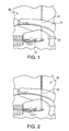
- FIGS. 1-7 illustrate subsequent steps of a method of joint reconstruction in accordance with an exemplary embodiment of the present invention.
- FIG. 8 is a schematic illustration of a fixation and reconstruction device of the present invention.
- FIGS. 9 a and 9 b illustrate exemplary embodiments of one member of the fixation device shown in FIG. 8 .
- FIG. 10 illustrates an embodiment of the other member of the fixation device shown in FIG. 8 .
- the present invention provides a technique and reconstruction system for fixation of bone to bone, or soft tissue to bone.
- the reconstruction system (TightRope system) of the present invention comprises two fixation devices (for example, two buttons) having various geometries (for example, circular or oblong) and being joined by a continuous loop of suture.
- Each button is provided with at least one opening (preferably two openings) that allow the passage of suture.
- the button may be formed, for example, of metal, PEEK or PLLA.
- the suture may be FIBERWIRE® suture, sold by Arthrex, Inc. of Naples, Fla.
- the present invention also provides a method for fixation of bone to bone, or soft tissue to bone.
- the method of the present invention comprises the steps of: (i) providing a first hole through a first bone and a second hole through a second bone of a joint; (ii) providing a two button/suture loop construct (TightRope) including two buttons joined by a continuous loop of flexible material in the vicinity of the joint; (iii) advancing one of the two buttons through the holes in the first and second bones, until it exits the second bone; and (iv) advancing the other button to the surface of the first bone.
- TightRope two button/suture loop construct
- the present invention provides a method of acromioclavicular (AC) joint reconstruction, comprising the steps of: (i) providing a hole through the clavicle and the coracoid; (ii) providing a two button/suture loop construct (including two buttons joined by a continuous loop of suture) in the vicinity of the AC joint; (iii) advancing one of the two buttons through the hole in the clavicle and the coracoid until it exits the coracoid base; and (iv) advancing the other button to the surface of the clavicle.
- AC acromioclavicular
- the acromioclavicular (AC) joint reconstruction technique of the present invention comprises the steps of: (i) drilling a hole of about 4 mm through the clavicle and the coracoid by using a C-Ring adapteur guide and a cannulated drill; (ii) leaving the cannulated drill in the clavicle and the coracoid; (iii) advancing a suture passing wire through the cannulated drill and subsequently removing the drill; (iv) inserting two traction sutures from the oblong button of the fixation system of the invention through the wire loop of the suture passing wire; (v) pulling the suture passing wire to retrieve the two traction sutures out of the anterior/inferior cannula; (vi) advancing the oblong button through the clavicle and the coracoid until it exits the coracoid base, and pulling on each of the traction sutures of the present invention
- the method of the present invention has applications to joint and ligament reconstruction. Particular applications relate to acromioclavicular joint reconstructions, where the method of the present invention provides a simple, reproducible, minimally invasive technique for acute acromioclavicular joint stabilization which enables a rapid return to activity for the acute injury.
- the technique is indicated for acute acromioclavicular joint dislocation (Rockwood type III to VI) of less than one month duration.
- FIGS. 1-7 illustrate a human joint 99 undergoing joint reconstruction according to an embodiment of the present invention.
- FIGS. 8-10 illustrate a fixation system 100 of the invention which, as described below, is inserted into a drilled hole through the joint bones using a single suture strand to obtain the repair structure of FIG. 7 .
- the joint 99 is an acromioclavicular (AC) joint 99 of the human shoulder comprising clavicle 10 and coracoid 20 and undergoing acromioclavicular (AC) joint reconstruction according to an embodiment of the present invention.
- AC acromioclavicular
- FIGS. 8-10 reference to the fixation system 100 of FIGS. 8-10 will be made in this application as to the acromioclavicular reconstruction or fixation system 100 (which is inserted into a drilled hole through the clavicle and the coracoid using a single suture strand to obtain the repair structure of FIG. 7 ).
- the invention is not limited to this particular acromioclavicular (AC) joint fixation system, but contemplates reconstruction and/or fixation systems for any structures (bone, cartilage, soft tissue, etc.) that need to be stabilized, fixated and/or reconstructed.
- AC acromioclavicular
- the fixation system 100 of the present invention comprises two buttons 101 , 102 joined by a continuous loop of suture 110 .
- the buttons may have various configurations and dimensions and may be similar to, or different from, each other.
- the buttons may be circular, oblong, rectangular or parallelepipedal, among many other configurations.
- one button is circular and the other button is oblong.
- button 101 is circular and button 102 is oblong.
- Button 102 is illustrated in FIG. 9 a as having a body 112 provided with first and second apertures 114 , 116 .
- the button 102 is preferably formed from titanium or stainless steel, although any other suitable material could be used, in particular any suitable bioabsorbable material.
- the first aperture 114 and the second aperture 116 are oblong in shape, the longitudinal mid-line of each of the first and second apertures 114 , 116 being located substantially about a longitudinal mid-line of the button 102 .
- the button 102 has a length of about 9 mm to about 20 mm, more preferably of about 12 mm to about 15 mm, and a width that is less than about 1 mm narrower than the width of the drill holes through which the button is inserted and subsequently passed through.
- button 102 is very small having a width that allows it to pass through a 3 mm cortical pin hole without over drilling, which in turn saves time and preserves bone.
- FIG. 9 b illustrates yet another embodiment of a button of the present invention, indicated as 202 .
- the button 202 is about 9.0 mm in length by about 3.5 mm in width, with a thickness of about 1.5 mm.
- the button 202 has first and second apertures 214 and 216 , respectively, provided within body 212 .
- each of the apertures 214 , 216 are triangular in shape, the respective apices 215 being directed away from each other and being located substantially about a longitudinal mid-line of the button 202 .
- Button 202 may be formed, for example, of metals such as titanium, titanium alloys or stainless steel, PEEK or PLLA, or other biocompatible and/or bioabsorbable materials known in the art.
- FIG. 10 illustrates button 101 of the system 100 having a circular or disc-shaped configuration.
- the button 101 is preferably formed from titanium or stainless steel although, as known to those skilled in the art, any other suitable material, in particular any suitable bioabsorbable materials, may be used.
- the button 101 also has at least two flexible coupling-locating apertures 104 . In the illustrated embodiment, there are four apertures 104 circumferentially arranged about the outer edge of the button. In the illustrated embodiment only, each of the apertures 104 has a diameter of about 1.0 mm. Each of the apertures 104 may have beveled edges, above and below, or only above, or only below.
- Flexible strand or continuous loop 110 extends between at least one of the apertures of the first button 101 and at least one of the apertures of the second button 102 .
- Flexible strand or continuous loop 110 may be formed of suture, for example a high strength suture material such as FIBERWIRE® suture, sold by Arthrex, Inc. of Naples, Fla., and described in U.S. Pat. No. 6,716,234, the disclosure of which is incorporated by reference herein.
- the high strength suture may be available in various lengths and, preferably, is a #5 FIBERWIRE® suture strand.
- FIBERWIRE® suture is formed of an advanced, high-strength fiber material, namely ultrahigh molecular weight polyethylene (UHMWPE), sold under the tradenames SPECTRA (Honeywell) and DYNEEMA (DSM), braided with at least one other fiber, natural or synthetic, to form lengths of suture material.
- UHMWPE ultrahigh molecular weight polyethylene
- the suture may optionally include filaments of various colors.
- the suture may be also formed of a plurality of suture strands configured to separate from a single strand to a plurality of strands in the continuous loop.
- the patient Prior to undergoing acromioclavicular joint reconstruction, the patient is positioned in the beach chair or lateral decubitus position under a general anesthesia. Subsequently, the following actions may be undertaken: induce the arthroscope into the glenohumeral joint via a standard posterior portal; create an anterior/superior portal with an outside/in technique using a spinal needle for position; insert a 7 mm Partially Threaded Cannula into this portal; create an anterior/inferior portal near the tip of the coracoid, with an outside/in technique using the spinal needle to ensure that the base of the coracoid can be reached; insert an 8.25 mm Twist-In Cannula through this portal and start the debridement of the rotator interval; and introduce a 4.5 mm full radius shaver blade through the anterior/inferior cannula and into the rotator interval and debride until the tip of the coracoid can be visualized.
- the following actions may be also performed: using a 70° arthroscope, release the middle glenohumeral ligament and partially release the middle glenohumeral ligament; retract the anterior/inferior cannula behind the rotator interval to completely reach the superior and middle glenohumeral ligaments; once the interval has been cleared, start to expose the base of the coracoid using a mechanical shaver and radiofrequency device; move the arthroscope to the superior portal to facilitate the view of the base of the coracoid; and strip the bursa and periosteum from the base of the coracoid, to obtain a full view of the undersurface.
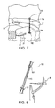
- FIG. 1 Insert assembled Adapteur Drill Guide C-Ring with the Coracoid Drill Stop and graduated Guide Pin Sleeve 88 ( FIG. 8 ) through the anterior/inferior portal. Position the drill stop tip under base 21 of the coracoid 20 , as close to the scapula as possible. Position the top of the Guide Pin Sleeve over the superior clavicle 10 at its midline approximately 25 mm from the distal clavicle through a 1.5 cm incision made in Langers lines by splitting the deltotrapezial fascia.
- a drill guide pin 51 for example, a 2.4 mm Drill Tip Guide Pin
- the tip of the guide pin is captured by the drill stop at the base of the coracoid under direct visualization.
- FIG. 2 Attach the Coracoid Drill Stop to a handle (for example, a Tear Drop handle), to hold the guide pin 51 in place while drilling over it.
- a handle for example, a Tear Drop handle
- advance a drill 53 for example, a 4 mm Cannulated Drill
- a drill 53 for example, a 4 mm Cannulated Drill
- a drill 53 for example, a 4 mm Cannulated Drill
- 10 a , 20 a FIG. 4
- Cannulated drilling beyond the coracoid 20 should be avoided under direct arthroscopic visualization.
- a passing wire 55 for example, a Nitinol suture passing wire
- a passing wire 55 for example, a Nitinol suture passing wire
- traction suture 102 a for example, two white traction sutures
- the oblong button 102 of the reconstruction system 100 FIG. 8
- the wire loop 55 a of the Nitinol suture passing wire 55 inserts at least one traction suture 102 a (for example, two white traction sutures) from the oblong button 102 of the reconstruction system 100 ( FIG. 8 ) through the wire loop 55 a of the Nitinol suture passing wire 55 .
- FIG. 5 Pull the suture passing wire 55 to retrieve the two white traction sutures 102 a out of the anterior/inferior cannula. Pull on one of the two white traction sutures 102 a to flip the oblong button 102 into a vertical position suitable for advancement through the bone tunnels 10 a , 20 a.
- the invention is not limited to this embodiment. Accordingly, the present invention also contemplates other kinds of joint and ligament reconstructions, the acromioclavicular joint reconstruction being just illustrative.
Abstract
Description
Claims (13)
Priority Applications (3)
| Application Number | Priority Date | Filing Date | Title |
|---|---|---|---|
| US11/687,882 US9005245B2 (en) | 2002-08-30 | 2007-03-19 | Acromioclavicular joint fixation technique |
| US12/611,656 US20100045186A1 (en) | 2006-10-04 | 2009-11-03 | Dual brightness twinkle in a miniature light bulb |
| US13/027,700 US9072510B2 (en) | 2001-08-30 | 2011-02-15 | Acromioclavicular joint fixation technique |
Applications Claiming Priority (5)
| Application Number | Priority Date | Filing Date | Title |
|---|---|---|---|
| US10/233,122 US7235091B2 (en) | 2002-06-20 | 2002-08-30 | Apparatus and method for fixation of ankle syndesmosis |
| US69712505P | 2005-07-07 | 2005-07-07 | |
| US78386906P | 2006-03-21 | 2006-03-21 | |
| US11/482,038 US8512376B2 (en) | 2002-08-30 | 2006-07-07 | Method and apparatus for internal fixation of an acromioclavicular joint dislocation of the shoulder |
| US11/687,882 US9005245B2 (en) | 2002-08-30 | 2007-03-19 | Acromioclavicular joint fixation technique |
Related Parent Applications (2)
| Application Number | Title | Priority Date | Filing Date |
|---|---|---|---|
| US11/482,038 Continuation-In-Part US8512376B2 (en) | 2001-08-30 | 2006-07-07 | Method and apparatus for internal fixation of an acromioclavicular joint dislocation of the shoulder |
| US12/247,975 Continuation-In-Part US20090039794A1 (en) | 1995-06-26 | 2008-10-08 | Miniature light bulb for random high-low twinkle in series-wired light string |
Related Child Applications (2)
| Application Number | Title | Priority Date | Filing Date |
|---|---|---|---|
| US12/611,656 Continuation-In-Part US20100045186A1 (en) | 2006-10-04 | 2009-11-03 | Dual brightness twinkle in a miniature light bulb |
| US13/027,700 Division US9072510B2 (en) | 2001-08-30 | 2011-02-15 | Acromioclavicular joint fixation technique |
Publications (2)
| Publication Number | Publication Date |
|---|---|
| US20070179531A1 US20070179531A1 (en) | 2007-08-02 |
| US9005245B2 true US9005245B2 (en) | 2015-04-14 |
Family
ID=46327532
Family Applications (2)
| Application Number | Title | Priority Date | Filing Date |
|---|---|---|---|
| US11/687,882 Active 2026-01-07 US9005245B2 (en) | 2001-08-30 | 2007-03-19 | Acromioclavicular joint fixation technique |
| US13/027,700 Expired - Lifetime US9072510B2 (en) | 2001-08-30 | 2011-02-15 | Acromioclavicular joint fixation technique |
Family Applications After (1)
| Application Number | Title | Priority Date | Filing Date |
|---|---|---|---|
| US13/027,700 Expired - Lifetime US9072510B2 (en) | 2001-08-30 | 2011-02-15 | Acromioclavicular joint fixation technique |
Country Status (1)
| Country | Link |
|---|---|
| US (2) | US9005245B2 (en) |
Cited By (27)
| Publication number | Priority date | Publication date | Assignee | Title |
|---|---|---|---|---|
| US9066716B2 (en) | 2011-03-30 | 2015-06-30 | Arthrosurface Incorporated | Suture coil and suture sheath for tissue repair |
| US9204873B2 (en) | 2000-05-01 | 2015-12-08 | Arthrosurface Incorporated | System and method for joint resurface repair |
| US9283076B2 (en) | 2009-04-17 | 2016-03-15 | Arthrosurface Incorporated | Glenoid resurfacing system and method |
| US9351745B2 (en) | 2003-02-24 | 2016-05-31 | Arthrosurface Incorporated | Trochlear resurfacing system and method |
| US9358029B2 (en) | 2006-12-11 | 2016-06-07 | Arthrosurface Incorporated | Retrograde resection apparatus and method |
| US9357989B2 (en) | 2000-05-01 | 2016-06-07 | Arthrosurface Incorporated | System and method for joint resurface repair |
| US9468448B2 (en) | 2012-07-03 | 2016-10-18 | Arthrosurface Incorporated | System and method for joint resurfacing and repair |
| US9492200B2 (en) | 2013-04-16 | 2016-11-15 | Arthrosurface Incorporated | Suture system and method |
| US9662126B2 (en) | 2009-04-17 | 2017-05-30 | Arthrosurface Incorporated | Glenoid resurfacing system and method |
| US9757113B2 (en) | 2013-07-31 | 2017-09-12 | Medos International Sàrl | Adjustable graft fixation device |
| US9861492B2 (en) | 2014-03-07 | 2018-01-09 | Arthrosurface Incorporated | Anchor for an implant assembly |
| US9918711B2 (en) | 2011-08-24 | 2018-03-20 | Howmedica Osteonics Corp. | Method and apparatus for the stabilization of the trapeziometacarpal joint |
| US9974643B2 (en) | 2013-03-11 | 2018-05-22 | Medos International Sàrl | Implant having adjustable filament coils |
| US10052094B2 (en) | 2013-03-11 | 2018-08-21 | Medos International Sàrl | Implant having adjustable filament coils |
| US10076343B2 (en) | 2002-12-03 | 2018-09-18 | Arthrosurface Incorporated | System for articular surface replacement |
| US10194899B2 (en) | 2015-10-28 | 2019-02-05 | Arthrex, Inc. | Systems and methods for acromioclavicular stabilization |
| US10405968B2 (en) | 2013-12-11 | 2019-09-10 | Medos International Sarl | Implant having filament limbs of an adjustable loop disposed in a shuttle suture |
| US10624752B2 (en) | 2006-07-17 | 2020-04-21 | Arthrosurface Incorporated | Tibial resurfacing system and method |
| US10624748B2 (en) | 2014-03-07 | 2020-04-21 | Arthrosurface Incorporated | System and method for repairing articular surfaces |
| US10653409B2 (en) | 2015-12-04 | 2020-05-19 | Crossroads Extremity Systems, Llc | Devices and methods for anchoring tissue |
| US10758224B2 (en) | 2017-03-27 | 2020-09-01 | Trimed, Incorporated | System and method controlling a relationship between first and second bodies on a person |
| US10945743B2 (en) | 2009-04-17 | 2021-03-16 | Arthrosurface Incorporated | Glenoid repair system and methods of use thereof |
| US11160663B2 (en) | 2017-08-04 | 2021-11-02 | Arthrosurface Incorporated | Multicomponent articular surface implant |
| US11478358B2 (en) | 2019-03-12 | 2022-10-25 | Arthrosurface Incorporated | Humeral and glenoid articular surface implant systems and methods |
| US11607319B2 (en) | 2014-03-07 | 2023-03-21 | Arthrosurface Incorporated | System and method for repairing articular surfaces |
| US11684468B2 (en) | 2018-11-06 | 2023-06-27 | Arthrex, Inc. | Surgical construct with adjustable loops and sliding knot |
| US11712276B2 (en) | 2011-12-22 | 2023-08-01 | Arthrosurface Incorporated | System and method for bone fixation |
Families Citing this family (63)
| Publication number | Priority date | Publication date | Assignee | Title |
|---|---|---|---|---|
| US8512376B2 (en) | 2002-08-30 | 2013-08-20 | Arthrex, Inc. | Method and apparatus for internal fixation of an acromioclavicular joint dislocation of the shoulder |
| US7875057B2 (en) * | 2007-01-19 | 2011-01-25 | Arthrex, Inc. | Method and suture-button construct for stabilization of cranial cruciate ligament deficient stifle |
| US8348960B2 (en) | 2007-07-12 | 2013-01-08 | Arthrex, Inc. | Applicator for suture/button construct |
| US8282674B2 (en) * | 2008-07-18 | 2012-10-09 | Suspension Orthopaedic Solutions, Inc. | Clavicle fixation |
| US8876900B2 (en) * | 2008-11-17 | 2014-11-04 | Arthrex, Inc. | AC joint repair using suture button graft construct and method of surgery |
| US9168044B2 (en) * | 2008-12-19 | 2015-10-27 | Jeffrey B. Kirkham | Multi-use cleat |
| US20100191332A1 (en) | 2009-01-08 | 2010-07-29 | Euteneuer Charles L | Implantable Tendon Protection Systems and Related Kits and Methods |
| US9179910B2 (en) | 2009-03-20 | 2015-11-10 | Rotation Medical, Inc. | Medical device delivery system and method |
| US8439976B2 (en) * | 2009-03-31 | 2013-05-14 | Arthrex, Inc. | Integrated adjustable button-suture-graft construct with two fixation devices |
| US8460379B2 (en) | 2009-03-31 | 2013-06-11 | Arthrex, Inc. | Adjustable suture button construct and methods of tissue reconstruction |
| AU2010256414C1 (en) | 2009-06-04 | 2016-01-21 | Rotation Medical, Inc. | Methods and apparatus for deploying sheet-like materials |
| US8821536B2 (en) | 2009-06-04 | 2014-09-02 | Rotation Medical, Inc. | Methods and apparatus for delivering staples to a target tissue |
| US8753375B2 (en) | 2009-10-14 | 2014-06-17 | Arthrex, Inc. | Z-shaped button for tissue repair |
| USD737502S1 (en) | 2009-12-18 | 2015-08-25 | Jeffrey B. Kirkham | Multi-use cleat |
| US9198750B2 (en) | 2010-03-11 | 2015-12-01 | Rotation Medical, Inc. | Tendon repair implant and method of arthroscopic implantation |
| EP2455002B1 (en) | 2010-11-17 | 2019-04-03 | Arthrex, Inc. | Adjustable suture-button construct for ankle syndesmosis repair |
| EP2455040B1 (en) | 2010-11-17 | 2015-03-04 | Arthrex, Inc. | Adjustable suture-button construct for knotless stabilization of cranial cruciate deficient ligament stifle |
| EP2455001B1 (en) | 2010-11-17 | 2020-07-22 | Arthrex, Inc. | Adjustable suture-button constructs for ligament reconstruction |
| EP2462876B1 (en) | 2010-12-09 | 2015-10-14 | Arthrex, Inc. | Suture button construct with dog-bone shaped button for acromioclavicular joint fixation |
| US9138219B2 (en) | 2010-12-29 | 2015-09-22 | Tarsus Medical Inc. | Methods and devices for treating a syndesmosis injury |
| US20140155937A1 (en) * | 2011-01-11 | 2014-06-05 | Padmakar Shinde | Bridge button for ligament reconstruction |
| US9314314B2 (en) | 2011-02-15 | 2016-04-19 | Rotation Medical, Inc. | Anatomical location markers and methods of use in positioning sheet-like materials during surgery |
| US10952783B2 (en) | 2011-12-29 | 2021-03-23 | Rotation Medical, Inc. | Guidewire having a distal fixation member for delivering and positioning sheet-like materials in surgery |
| EP2675391B1 (en) | 2011-02-15 | 2017-09-27 | Rotation Medical, Inc. | Apparatus for delivering and positioning sheet-like materials |
| WO2012145059A1 (en) | 2011-02-15 | 2012-10-26 | Rotation Medical, Inc. | Methods and apparatus for fixing sheet-like materials to a target tissue |
| US8926661B2 (en) | 2011-06-02 | 2015-01-06 | Smith & Nephew, Inc. | Surgical fastening |
| US9277912B2 (en) * | 2011-06-14 | 2016-03-08 | University Of South Florida | Systems and methods for ankle syndesmosis fixation |
| US9301745B2 (en) | 2011-07-21 | 2016-04-05 | Arthrex, Inc. | Knotless suture constructs |
| US9332979B2 (en) | 2011-07-22 | 2016-05-10 | Arthrex, Inc. | Tensionable knotless acromioclavicular repairs and constructs |
| AU2012322239A1 (en) * | 2011-08-24 | 2014-04-10 | Shinde PADMAKAR | Bridge button for ligament reconstruction |
| US9107653B2 (en) | 2011-09-22 | 2015-08-18 | Arthrex, Inc. | Tensionable knotless anchors with splice and methods of tissue repair |
| US10245016B2 (en) | 2011-10-12 | 2019-04-02 | Arthrex, Inc. | Adjustable self-locking loop constructs for tissue repairs and reconstructions |
| EP2599449B1 (en) | 2011-11-29 | 2015-02-25 | Arthrex, Inc. | Applicator for suture/button construct with positive retention and control |
| US9615821B2 (en) | 2011-12-09 | 2017-04-11 | Arthrex, Inc. | Tensionable knotless anchor systems and methods of tissue repair |
| AU2012369140B2 (en) | 2011-12-19 | 2016-11-10 | Rotation Medical, Inc. | Fasteners for affixing sheet -like materials to bone or tissue |
| US9107661B2 (en) | 2011-12-19 | 2015-08-18 | Rotation Medical, Inc. | Fasteners and fastener delivery devices for affixing sheet-like materials to bone or tissue |
| EP3403601A1 (en) | 2011-12-19 | 2018-11-21 | Rotation Medical, Inc. | Apparatus for forming pilot holes in bone and delivering fasteners therein for retaining an implant |
| US9271726B2 (en) | 2011-12-19 | 2016-03-01 | Rotation Medical, Inc. | Fasteners and fastener delivery devices for affixing sheet-like materials to bone or tissue |
| EP2797532B1 (en) | 2011-12-29 | 2016-04-06 | Rotation Medical, Inc. | Apparatus for delivering and positioning sheet-like materials in surgery |
| US9259217B2 (en) | 2012-01-03 | 2016-02-16 | Biomet Manufacturing, Llc | Suture Button |
| RU2493798C1 (en) * | 2012-03-23 | 2013-09-27 | Андрей Сергеевич Фёдоров | Device for replacement of coracoid-clavicular ligament |
| US9737292B2 (en) | 2012-06-22 | 2017-08-22 | Arthrex, Inc. | Knotless suture anchors and methods of tissue repair |
| WO2014134328A1 (en) | 2013-02-27 | 2014-09-04 | Coorstek Medical Llc D/B/A Imds | Graft fixation |
| US10123813B2 (en) | 2014-02-17 | 2018-11-13 | Smith & Nephew, Inc. | Drill guide |
| WO2015172052A1 (en) | 2014-05-09 | 2015-11-12 | Rotation Medical, Inc. | Medical implant delivery system for sheet-like implant |
| US10123796B2 (en) | 2014-11-04 | 2018-11-13 | Rotation Medical, Inc. | Medical implant delivery system and related methods |
| EP3215026B1 (en) | 2014-11-04 | 2023-10-25 | Rotation Medical, Inc. | Medical implant delivery system |
| WO2016073502A1 (en) | 2014-11-04 | 2016-05-12 | Rotation Medical, Inc. | Medical implant delivery system and related methods |
| CA2983488C (en) | 2015-04-24 | 2020-12-22 | Biomet Manufacturing, Llc | Clavicle implants |
| WO2016179372A1 (en) | 2015-05-06 | 2016-11-10 | Rotation Medical, Inc. | Medical implant delivery system and related methods |
| EP3307204B1 (en) | 2015-06-15 | 2021-11-24 | Rotation Medical, Inc. | Tendon repair implant |
| US10499960B2 (en) | 2015-07-13 | 2019-12-10 | IntraFuse, LLC | Method of bone fixation |
| US10136929B2 (en) | 2015-07-13 | 2018-11-27 | IntraFuse, LLC | Flexible bone implant |
| US10485595B2 (en) | 2015-07-13 | 2019-11-26 | IntraFuse, LLC | Flexible bone screw |
| US10154863B2 (en) | 2015-07-13 | 2018-12-18 | IntraFuse, LLC | Flexible bone screw |
| US10335136B2 (en) | 2015-08-20 | 2019-07-02 | Arthrex, Inc. | Tensionable constructs with multi-limb locking mechanism through single splice and methods of tissue repair |
| US10265060B2 (en) | 2015-08-20 | 2019-04-23 | Arthrex, Inc. | Tensionable constructs with multi-limb locking mechanism through single splice and methods of tissue repair |
| JP6653389B2 (en) | 2015-12-31 | 2020-02-26 | ローテーション メディカル インコーポレイテッドRotation Medical,Inc. | Medical implant delivery system and related methods |
| CA3008670A1 (en) | 2015-12-31 | 2017-07-06 | Rotation Medical, Inc. | Fastener delivery system and related methods |
| CA3055945C (en) * | 2017-03-13 | 2022-10-18 | Arthrex, Inc. | Surgical fixation systems and methods |
| CN110225726A (en) | 2017-12-07 | 2019-09-10 | 罗特迅医疗有限公司 | Medical implant transportation system and correlation technique |
| US11406399B2 (en) * | 2018-10-08 | 2022-08-09 | Conmed Corporation | Multi-barrel drill guide and anchor deployment assembly |
| US20210228221A1 (en) * | 2020-01-23 | 2021-07-29 | Arthrex, Inc. | Self-positioning drill guide |
Citations (27)
| Publication number | Priority date | Publication date | Assignee | Title |
|---|---|---|---|---|
| US261501A (en) * | 1882-07-18 | Bag and sail tie | ||
| US4988351A (en) | 1989-01-06 | 1991-01-29 | Concept, Inc. | Washer for use with cancellous screw for attaching soft tissue to bone |
| US5219359A (en) | 1990-09-18 | 1993-06-15 | Femcare Limited | Suture apparatus |
| US5306290A (en) | 1993-02-12 | 1994-04-26 | Mitek Surgical Products, Inc. | Suture button |
| US5366480A (en) | 1990-12-24 | 1994-11-22 | American Cyanamid Company | Synthetic elastomeric buttressing pledget |
| US5409490A (en) | 1993-08-16 | 1995-04-25 | Depuy Inc. | Shoulder separation reconstruction |
| US5464426A (en) | 1993-05-14 | 1995-11-07 | Bonutti; Peter M. | Method of closing discontinuity in tissue |
| US5593424A (en) | 1994-08-10 | 1997-01-14 | Segmed, Inc. | Apparatus and method for reducing and stabilizing the circumference of a vascular structure |
| US5921986A (en) | 1998-02-06 | 1999-07-13 | Bonutti; Peter M. | Bone suture |
| US5989256A (en) | 1999-01-19 | 1999-11-23 | Spineology, Inc. | Bone fixation cable ferrule |
| US6066160A (en) * | 1998-11-23 | 2000-05-23 | Quickie Llc | Passive knotless suture terminator for use in minimally invasive surgery and to facilitate standard tissue securing |
| US6110207A (en) * | 1996-04-23 | 2000-08-29 | Aesculap Ag & Co. Kg | Implant for securing a tendon replacement member |
| US20020156475A1 (en) * | 1999-10-30 | 2002-10-24 | Aesculap Ag & Co. Kg | Surgical connecting element for fixing adjacently arranged bone plates |
| US6494888B1 (en) * | 1999-06-22 | 2002-12-17 | Ndo Surgical, Inc. | Tissue reconfiguration |
| US6517578B2 (en) * | 1999-12-15 | 2003-02-11 | Atlantech Medical Devices Limited | Graft suspension device |
| US6533802B2 (en) * | 2001-05-16 | 2003-03-18 | Smith & Nephew, Inc. | Endobutton continuous loop for bone-tendon-bone |
| US20030130694A1 (en) | 1999-12-02 | 2003-07-10 | Ray Bojarski | Soft tissue attachment and repair |
| US6635073B2 (en) | 2000-05-03 | 2003-10-21 | Peter M. Bonutti | Method of securing body tissue |
| US20030236555A1 (en) * | 2002-06-20 | 2003-12-25 | Brian Thornes | Apparatus and method for fixation of ankle syndesmosis |
| US6716234B2 (en) | 2001-09-13 | 2004-04-06 | Arthrex, Inc. | High strength suture material |
| US20040097939A1 (en) * | 1998-02-06 | 2004-05-20 | Bonutti Peter M. | Apparatus and method for treating a fracture of a bone |
| US20040133238A1 (en) * | 1999-06-22 | 2004-07-08 | Cerier Jeffrey C. | Tissue fixation devices and methods of fixing tissue |
| US20040199166A1 (en) * | 2003-03-18 | 2004-10-07 | Reinhold Schmieding | ACL reconstruction technique using retrodrill |
| US20040236373A1 (en) | 2003-05-20 | 2004-11-25 | Anspach William E. | Surgical method for suturing tendons/ligaments to bones |
| US20050049599A1 (en) * | 2001-06-15 | 2005-03-03 | Aesculap Ag & Co. Kg | Implant for fixing bone plates |
| US7153312B1 (en) * | 1999-12-02 | 2006-12-26 | Smith & Nephew Inc. | Closure device and method for tissue repair |
| US20070016208A1 (en) | 2002-08-30 | 2007-01-18 | Brian Thornes | Method and apparatus for internal fixation of an acromioclavicular joint dislocation of the shoulder |
Family Cites Families (15)
| Publication number | Priority date | Publication date | Assignee | Title |
|---|---|---|---|---|
| US3835849A (en) * | 1973-01-26 | 1974-09-17 | Guire G Mc | Bone clamp and adjustable drill guide |
| US4754749A (en) * | 1986-04-29 | 1988-07-05 | Tsou Paul M | Surgical screw with counter-rotation prevention means |
| US5593425A (en) * | 1990-06-28 | 1997-01-14 | Peter M. Bonutti | Surgical devices assembled using heat bonable materials |
| US5464424A (en) * | 1994-06-27 | 1995-11-07 | O'donnell, Jr.; Francis E. | Laser adjustable suture |
| US6099568A (en) * | 1998-03-03 | 2000-08-08 | Linvatec Corporation | ACL graft fixation device and method |
| DE19941574A1 (en) * | 1999-09-01 | 2001-03-08 | Storz Karl Gmbh & Co Kg | Instruments for implanting a tendon replacement |
| US20040254609A1 (en) * | 2001-03-14 | 2004-12-16 | Esplin Vermon S. | Soft tissue anchor |
| DE10129490A1 (en) * | 2001-06-21 | 2003-01-02 | Helmut Mueckter | Implantable screw for stabilization of joint or bone fracture, has flexible shaft which interconnects proximal head portion and distal insertion portion of elongated screw body |
| US7892256B2 (en) * | 2001-09-13 | 2011-02-22 | Arthrex, Inc. | High strength suture tape |
| US7594923B2 (en) * | 2003-06-11 | 2009-09-29 | Medicine Lodge, Inc | Line lock suture attachment systems and methods |
| US7255700B2 (en) * | 2003-06-18 | 2007-08-14 | Biomet Sports Medicine, Inc. | Device and method of fastening a graft to a bone |
| US9364214B2 (en) * | 2003-07-10 | 2016-06-14 | Arthrex, Inc. | Cannulated instrument with curved shaft for passing suture through tissue |
| US7455683B2 (en) * | 2004-02-26 | 2008-11-25 | Depuy Mitek, Inc. | Methods and devices for repairing triangular fibrocartilage complex tears |
| EP1753355B1 (en) * | 2004-03-26 | 2011-11-09 | Synthes GmbH | Articulated bone screw |
| US7488347B1 (en) * | 2005-01-06 | 2009-02-10 | Medicine Lodge, Inc. | Transosseous graft retention system and method |
-
2007
- 2007-03-19 US US11/687,882 patent/US9005245B2/en active Active
-
2011
- 2011-02-15 US US13/027,700 patent/US9072510B2/en not_active Expired - Lifetime
Patent Citations (28)
| Publication number | Priority date | Publication date | Assignee | Title |
|---|---|---|---|---|
| US261501A (en) * | 1882-07-18 | Bag and sail tie | ||
| US4988351A (en) | 1989-01-06 | 1991-01-29 | Concept, Inc. | Washer for use with cancellous screw for attaching soft tissue to bone |
| US5219359A (en) | 1990-09-18 | 1993-06-15 | Femcare Limited | Suture apparatus |
| US5366480A (en) | 1990-12-24 | 1994-11-22 | American Cyanamid Company | Synthetic elastomeric buttressing pledget |
| US5306290A (en) | 1993-02-12 | 1994-04-26 | Mitek Surgical Products, Inc. | Suture button |
| US5464426A (en) | 1993-05-14 | 1995-11-07 | Bonutti; Peter M. | Method of closing discontinuity in tissue |
| US5409490A (en) | 1993-08-16 | 1995-04-25 | Depuy Inc. | Shoulder separation reconstruction |
| US5593424A (en) | 1994-08-10 | 1997-01-14 | Segmed, Inc. | Apparatus and method for reducing and stabilizing the circumference of a vascular structure |
| US6110207A (en) * | 1996-04-23 | 2000-08-29 | Aesculap Ag & Co. Kg | Implant for securing a tendon replacement member |
| US20040097939A1 (en) * | 1998-02-06 | 2004-05-20 | Bonutti Peter M. | Apparatus and method for treating a fracture of a bone |
| US5921986A (en) | 1998-02-06 | 1999-07-13 | Bonutti; Peter M. | Bone suture |
| US6066160A (en) * | 1998-11-23 | 2000-05-23 | Quickie Llc | Passive knotless suture terminator for use in minimally invasive surgery and to facilitate standard tissue securing |
| US5989256A (en) | 1999-01-19 | 1999-11-23 | Spineology, Inc. | Bone fixation cable ferrule |
| US20040133238A1 (en) * | 1999-06-22 | 2004-07-08 | Cerier Jeffrey C. | Tissue fixation devices and methods of fixing tissue |
| US6494888B1 (en) * | 1999-06-22 | 2002-12-17 | Ndo Surgical, Inc. | Tissue reconfiguration |
| US20020156475A1 (en) * | 1999-10-30 | 2002-10-24 | Aesculap Ag & Co. Kg | Surgical connecting element for fixing adjacently arranged bone plates |
| US7153312B1 (en) * | 1999-12-02 | 2006-12-26 | Smith & Nephew Inc. | Closure device and method for tissue repair |
| US20030130694A1 (en) | 1999-12-02 | 2003-07-10 | Ray Bojarski | Soft tissue attachment and repair |
| US6517578B2 (en) * | 1999-12-15 | 2003-02-11 | Atlantech Medical Devices Limited | Graft suspension device |
| US6635073B2 (en) | 2000-05-03 | 2003-10-21 | Peter M. Bonutti | Method of securing body tissue |
| US6533802B2 (en) * | 2001-05-16 | 2003-03-18 | Smith & Nephew, Inc. | Endobutton continuous loop for bone-tendon-bone |
| US20050049599A1 (en) * | 2001-06-15 | 2005-03-03 | Aesculap Ag & Co. Kg | Implant for fixing bone plates |
| US6716234B2 (en) | 2001-09-13 | 2004-04-06 | Arthrex, Inc. | High strength suture material |
| US20030236555A1 (en) * | 2002-06-20 | 2003-12-25 | Brian Thornes | Apparatus and method for fixation of ankle syndesmosis |
| US7235091B2 (en) * | 2002-06-20 | 2007-06-26 | Brian Thornes | Apparatus and method for fixation of ankle syndesmosis |
| US20070016208A1 (en) | 2002-08-30 | 2007-01-18 | Brian Thornes | Method and apparatus for internal fixation of an acromioclavicular joint dislocation of the shoulder |
| US20040199166A1 (en) * | 2003-03-18 | 2004-10-07 | Reinhold Schmieding | ACL reconstruction technique using retrodrill |
| US20040236373A1 (en) | 2003-05-20 | 2004-11-25 | Anspach William E. | Surgical method for suturing tendons/ligaments to bones |
Non-Patent Citations (1)
| Title |
|---|
| E.P. Su et al., "Using Suture Anchors for Coracoclavicular Fixation in Treatment of Complete Acromioclavicular Separation," May 2004, The American Journal of Orthopedics, pp. 256-257; Orthopaedic Surgery, Hospital for Special Surgery; New York, New York. |
Cited By (52)
| Publication number | Priority date | Publication date | Assignee | Title |
|---|---|---|---|---|
| US9357989B2 (en) | 2000-05-01 | 2016-06-07 | Arthrosurface Incorporated | System and method for joint resurface repair |
| US9204873B2 (en) | 2000-05-01 | 2015-12-08 | Arthrosurface Incorporated | System and method for joint resurface repair |
| US10076343B2 (en) | 2002-12-03 | 2018-09-18 | Arthrosurface Incorporated | System for articular surface replacement |
| US11337819B2 (en) | 2003-02-24 | 2022-05-24 | Arthrosurface Incorporated | Trochlear resurfacing system and method |
| US9351745B2 (en) | 2003-02-24 | 2016-05-31 | Arthrosurface Incorporated | Trochlear resurfacing system and method |
| US10624749B2 (en) | 2003-02-24 | 2020-04-21 | Arthrosurface Incorporated | Trochlear resurfacing system and method |
| US9931211B2 (en) | 2003-02-24 | 2018-04-03 | Arthrosurface Incorporated | Trochlear resurfacing system and method |
| US11471289B2 (en) | 2006-07-17 | 2022-10-18 | Arthrosurface Incorporated | Tibial resurfacing system and method |
| US10624752B2 (en) | 2006-07-17 | 2020-04-21 | Arthrosurface Incorporated | Tibial resurfacing system and method |
| US10045788B2 (en) | 2006-12-11 | 2018-08-14 | Arthrosurface Incorporated | Retrograde resection apparatus and method |
| US10959740B2 (en) | 2006-12-11 | 2021-03-30 | Arthrosurface Incorporated | Retrograde resection apparatus and method |
| US9358029B2 (en) | 2006-12-11 | 2016-06-07 | Arthrosurface Incorporated | Retrograde resection apparatus and method |
| US10478200B2 (en) | 2009-04-17 | 2019-11-19 | Arthrosurface Incorporated | Glenoid resurfacing system and method |
| US9662126B2 (en) | 2009-04-17 | 2017-05-30 | Arthrosurface Incorporated | Glenoid resurfacing system and method |
| US10945743B2 (en) | 2009-04-17 | 2021-03-16 | Arthrosurface Incorporated | Glenoid repair system and methods of use thereof |
| US11478259B2 (en) | 2009-04-17 | 2022-10-25 | Arthrosurface, Incorporated | Glenoid resurfacing system and method |
| US9283076B2 (en) | 2009-04-17 | 2016-03-15 | Arthrosurface Incorporated | Glenoid resurfacing system and method |
| US9066716B2 (en) | 2011-03-30 | 2015-06-30 | Arthrosurface Incorporated | Suture coil and suture sheath for tissue repair |
| US9918711B2 (en) | 2011-08-24 | 2018-03-20 | Howmedica Osteonics Corp. | Method and apparatus for the stabilization of the trapeziometacarpal joint |
| US11712276B2 (en) | 2011-12-22 | 2023-08-01 | Arthrosurface Incorporated | System and method for bone fixation |
| US11191552B2 (en) | 2012-07-03 | 2021-12-07 | Arthrosurface, Incorporated | System and method for joint resurfacing and repair |
| US10307172B2 (en) | 2012-07-03 | 2019-06-04 | Arthrosurface Incorporated | System and method for joint resurfacing and repair |
| US9468448B2 (en) | 2012-07-03 | 2016-10-18 | Arthrosurface Incorporated | System and method for joint resurfacing and repair |
| US10856967B2 (en) | 2013-03-11 | 2020-12-08 | Medos International Sàrl | Implant having adjustable filament coils |
| US11896475B2 (en) | 2013-03-11 | 2024-02-13 | Medos International Sarl | Implant having adjustable filament coils |
| US9974643B2 (en) | 2013-03-11 | 2018-05-22 | Medos International Sàrl | Implant having adjustable filament coils |
| US10052094B2 (en) | 2013-03-11 | 2018-08-21 | Medos International Sàrl | Implant having adjustable filament coils |
| US10898178B2 (en) | 2013-03-11 | 2021-01-26 | Medos International Sàrl | Implant having adjustable filament coils |
| US9492200B2 (en) | 2013-04-16 | 2016-11-15 | Arthrosurface Incorporated | Suture system and method |
| US10695096B2 (en) | 2013-04-16 | 2020-06-30 | Arthrosurface Incorporated | Suture system and method |
| US11648036B2 (en) | 2013-04-16 | 2023-05-16 | Arthrosurface Incorporated | Suture system and method |
| US9757113B2 (en) | 2013-07-31 | 2017-09-12 | Medos International Sàrl | Adjustable graft fixation device |
| US10441265B2 (en) | 2013-07-31 | 2019-10-15 | Medos International Sàrl | Adjustable graft fixation device |
| US11534288B2 (en) | 2013-12-11 | 2022-12-27 | Medos International Sarl | Implant having filament limbs of an adjustable loop disposed in a shuttle suture |
| US10405968B2 (en) | 2013-12-11 | 2019-09-10 | Medos International Sarl | Implant having filament limbs of an adjustable loop disposed in a shuttle suture |
| US10624754B2 (en) | 2014-03-07 | 2020-04-21 | Arthrosurface Incorporated | System and method for repairing articular surfaces |
| US10624748B2 (en) | 2014-03-07 | 2020-04-21 | Arthrosurface Incorporated | System and method for repairing articular surfaces |
| US11083587B2 (en) | 2014-03-07 | 2021-08-10 | Arthrosurface Incorporated | Implant and anchor assembly |
| US11766334B2 (en) | 2014-03-07 | 2023-09-26 | Arthrosurface Incorporated | System and method for repairing articular surfaces |
| US9931219B2 (en) | 2014-03-07 | 2018-04-03 | Arthrosurface Incorporated | Implant and anchor assembly |
| US9861492B2 (en) | 2014-03-07 | 2018-01-09 | Arthrosurface Incorporated | Anchor for an implant assembly |
| US10575957B2 (en) | 2014-03-07 | 2020-03-03 | Arthrosurface Incoporated | Anchor for an implant assembly |
| US11607319B2 (en) | 2014-03-07 | 2023-03-21 | Arthrosurface Incorporated | System and method for repairing articular surfaces |
| US9962265B2 (en) | 2014-03-07 | 2018-05-08 | Arthrosurface Incorporated | System and method for repairing articular surfaces |
| US10791007B2 (en) | 2015-10-28 | 2020-09-29 | Arthrex, Inc. | Systems and methods for acromioclavicular stabilization |
| US10194899B2 (en) | 2015-10-28 | 2019-02-05 | Arthrex, Inc. | Systems and methods for acromioclavicular stabilization |
| US10653409B2 (en) | 2015-12-04 | 2020-05-19 | Crossroads Extremity Systems, Llc | Devices and methods for anchoring tissue |
| US11806005B2 (en) | 2015-12-04 | 2023-11-07 | Crossroads Extremity Systems, Llc | Devices and methods for anchoring tissue |
| US10758224B2 (en) | 2017-03-27 | 2020-09-01 | Trimed, Incorporated | System and method controlling a relationship between first and second bodies on a person |
| US11160663B2 (en) | 2017-08-04 | 2021-11-02 | Arthrosurface Incorporated | Multicomponent articular surface implant |
| US11684468B2 (en) | 2018-11-06 | 2023-06-27 | Arthrex, Inc. | Surgical construct with adjustable loops and sliding knot |
| US11478358B2 (en) | 2019-03-12 | 2022-10-25 | Arthrosurface Incorporated | Humeral and glenoid articular surface implant systems and methods |
Also Published As
| Publication number | Publication date |
|---|---|
| US9072510B2 (en) | 2015-07-07 |
| US20110137341A1 (en) | 2011-06-09 |
| US20070179531A1 (en) | 2007-08-02 |
Similar Documents
| Publication | Publication Date | Title |
|---|---|---|
| US9005245B2 (en) | Acromioclavicular joint fixation technique | |
| US20240090891A1 (en) | Filamentary Fixation Device | |
| US11116491B2 (en) | Soft suture-based anchors | |
| US11058531B2 (en) | Whipstitched graft construct and method of making the same | |
| US10709436B2 (en) | Graft fixation using a plug against suture | |
| US8663279B2 (en) | Swivel anchor for knotless fixation of tissue | |
| US8876900B2 (en) | AC joint repair using suture button graft construct and method of surgery | |
| CA2255911C (en) | Knotless suture system and method | |
| US8771351B2 (en) | Method for double row fixation of tendon to bone | |
| JP4169503B2 (en) | Knotless suture fixation system and device | |
| US9101461B2 (en) | Button and continuous loop for fixation of ligaments | |
| US9907591B2 (en) | Strand for minimally invasive removal of T-anchor | |
| EP2215975A1 (en) | Fenestrated suture anchor for knotless fixation of tissue | |
| US20140128921A1 (en) | Bone plate with suture holes for soft tissue reattachments on the diaphyseal region of the plate | |
| US20090149884A1 (en) | System and method for bridge anchor tendon attachment |
Legal Events
| Date | Code | Title | Description |
|---|---|---|---|
| AS | Assignment |
Owner name: ARTHREX, INC., FLORIDA Free format text: ASSIGNMENT OF ASSIGNORS INTEREST;ASSIGNOR:THORNES, BRIAN;REEL/FRAME:019032/0830 Effective date: 20070316 |
|
| AS | Assignment |
Owner name: ARTHREX, INC., FLORIDA Free format text: ASSIGNMENT OF ASSIGNORS INTEREST;ASSIGNOR:TENNENT, DUNCAN;REEL/FRAME:023106/0904 Effective date: 20090803 |
|
| STCF | Information on status: patent grant |
Free format text: PATENTED CASE |
|
| MAFP | Maintenance fee payment |
Free format text: PAYMENT OF MAINTENANCE FEE, 4TH YEAR, LARGE ENTITY (ORIGINAL EVENT CODE: M1551); ENTITY STATUS OF PATENT OWNER: LARGE ENTITY Year of fee payment: 4 |
|
| MAFP | Maintenance fee payment |
Free format text: PAYMENT OF MAINTENANCE FEE, 8TH YEAR, LARGE ENTITY (ORIGINAL EVENT CODE: M1552); ENTITY STATUS OF PATENT OWNER: LARGE ENTITY Year of fee payment: 8 |