CROSS REFERENCE TO RELATED APPLICATIONS
This application claims priority to U.S. Provisional Application No. 61/846,287, filed on Jul. 15, 2013, the disclosure of which is incorporated herein by reference.
GOVERNMENT FUNDING
This invention was made with government support under Grant AI057472 awarded by USPHS. The government has certain rights in the invention.
FIELD OF THE INVENTION
The invention relates to bacteriophages that inhibit the growth of Gram positive and Gram negative bacterial cells. These bacteriophages are useful to identify and treat pathogenic bacteria.
BACKGROUND OF THE INVENTION
Bacteriophage-based diagnostics and therapeutics have been recognized as tools to combat bacterial infections for nearly a century. Wip1 (for worm intestinal phage 1) is a recently identified phage that infects the pathogen Bacillus anthracis and was isolated from the intestinal tract of Eisenia fetida worms [Schuch, R., et al., Prevalence of Bacillus anthracis-like organisms and bacteriophages in the intestinal tract of the earthworm Eisenia fetida. Applied and environmental microbiology, 2010. 76(7): p. 2286-94]. It is a tailless, double-stranded DNA phage possessing an internal lipid membrane beneath an icosahedral protein coat [Schuch, R. F. V. A., The Secret Life of the Anthrax Agent Bacillus Anthracis: Bacteriophage-Mediated Ecological Adaptations. PLos One, 2009. 4(8): p. e6532]. These features indicate that Wip1 belongs to the family Tectiviridae, a relatively rare phage group with surprising structural similarity to and a proposed evolutionary lineage with the mammalian adenovirus [Merckel, M. C. H., J. T.; Bamford, D. H.; Goldman, A.; Tuma, R., The Structure of the Bacteriophage PRD1 Spike Sheds Light on the Evolution of Viral Capsid Architecture. Molecular Cell, 2005. 18: p. 161-170, Bamford, D., Evolution of Viral Structure. Theoretical Population Biology, 2002. 61(4): p. 461-470.]. The Tectiviridae family consists of six isolates that infect gram-negative bacteria, including PRD1 [Olsen, R. H. S., J.; Gray, R. H., Characteristics of PRD1, a Plasmid-Dependent Broad Host Range DNA Bacteriophage. Journal of Virology, 1974. 14(3): p. 689-699], and six that infect gram-positive bacteria, including Bam35, Gil16, AP50, and Wip1 [Schuch, R., et al., Prevalence of Bacillus anthracis-like organisms and bacteriophages in the intestinal tract of the earthworm Eisenia fetida. Applied and environmental microbiology, 2010. 76(7): p. 2286-94; Ackermann, H. W. R., R.; Martin, M.; Murthy, M. R.; Smirnoff, W. A., Partial Characterization of a Cubic Bacillus Phage. Canadian Journal of Microbiology, 1978. 24: p. 986-993; Verheust, C., N. Fornelos, and J. Mahillon, GIL16, a new gram positive tectiviral phage related to the Bacillus thuringiensis GIL01 and the Bacillus cereus pBClin15 elements. Journal of bacteriology, 2005. 187(6): p. 1966-73; Nagy, E. P., B.; Ivanovics, G., Characteristics of Phage AP50, an RNA Phage Containing Phospholipids. Journal of General Virology, 1976. 32: p. 129-132]. While PRD1 has been studied in detail, tectiviruses that infect gram-positive bacteria are not as well characterized.
Wip1 phage exhibits a very narrow host range and is highly specific to B. anthracis [Schuch, R., et al., Prevalence of Bacillus anthracis-like organisms and bacteriophages in the intestinal tract of the earthworm Eisenia fetida. Applied and environmental microbiology, 2010. 76(7): p. 2286-94], the notorious biothreat agent and gram-positive bacterium that causes anthrax disease. The current gold standard for identifying suspected B. anthracis involves testing for γ phage sensitivity [Abshire, T. G., J. E. Brown, and J. W. Ezzell, Production and validation of the use of gamma phage for identification of Bacillus anthracis. Journal of clinical microbiology, 2005. 43(9): p. 4780-8; Anthrax Q & A: diagnosis. 2002; Available from: www.bt.cdc.gov/agent/anthrax/faq/diagnosis.asp]. However, using γ as a diagnostic tool can lead to false positives due to the susceptibility of several Bacillus cereus strains to infection by this phage [Schuch, R. and V. A. Fischetti, Detailed genomic analysis of the Wbeta and gamma phages infecting Bacillus anthracis: implications for evolution of environmental fitness and antibiotic resistance. Journal of bacteriology, 2006. 188(8): p. 3037-51; Schuch, R. N., D.; Fischetti, V. A., A bacteriolytic agent that detects and kills Bacillus anthracis. Nature, 2002. 418: p. 884-889]. Recent studies have shown that the host range of γ is less specific to B. anthracis than those of tectiviruses Wip1 and AP50 [Schuch, R., et al., Prevalence of Bacillus anthracis-like organisms and bacteriophages in the intestinal tract of the earthworm Eisenia fetida. Applied and environmental microbiology, 2010. 76(7): p. 2286-94; Sozhamannan, S., et al., Molecular Characterization of a Variant of Bacillus anthracis-Specific Phage AP50 with Improved Bacteriolytic Activity. Applied and environmental microbiology, 2008. 74(21): p. 6792-6796]. For example, Bacillus cereus ATCC 4342 is sensitive to infection by γ phage but not to infection by either Wip1 or AP50. Additionally, the γ diagnostic phage yields plaques on B. anthracis ΔSterne only after 5 days, whereas Wip1 plaques can be detected after just 12 hours post infection [Schuch, R., et al., Prevalence of Bacillus anthracis-like organisms and bacteriophages in the intestinal tract of the earthworm Eisenia fetida. Applied and environmental microbiology, 2010. 76(7): p. 2286-94].
Wip1's high specificity to B. anthracis is likely mediated by the initial recognition and binding of the virus to the host cell. Receptor binding proteins on the phage coat interact very specifically with receptors exposed on the surface of the bacterium [Haywood, A. M., Virus receptors: binding, adhesion strengthening, and changes in viral structure. Journal of virology, 1994. 68(1): p. 1-5]. For tectiviruses, the receptor binding protein assembles with other phage proteins into a protruding complex that extends from each particle vertex [Sokolova, A., et al., Solution structure of bacteriophage PRD1 vertex complex. The Journal of biological chemistry, 2001. 276(49): p. 46187-95]. In the PRD1 spike complex, two elongated proteins, monomeric receptor binding protein P2 and trimeric spike protein P5, form two separate spikes that each protrude from penton base protein P31 [Bamford, J. K. B., D. H., A New Mutant Class, Made by Targeted Mutagenesis, of Phage PRD1 Reveals That Protein P5 Connects the Receptor Binding Protein to the Vertex. Journal of virology, 2000. 74(17): p. 7781-7786; Huiskonen, J. T., V. Manole, and S. J. Butcher, Tale of two spikes in bacteriophage PRD1. Proceedings of the National Academy of Sciences of the United States of America, 2007. 104(16): p. 6666-71; Mindich, L. B., D.; McGraw, T.; Mackenzie, G, Assembly of bacteriophage PRD1: particle formation with wild-type and mutant viruses. Journal of virology, 1982. 44(3): p. 1021-1030; Xu, L. B., S. D.; Butcher, S. J.; Bamford, D. H.; Burnett, R. M., The Receptor Binding Protein P2 of PRD1, a Virus Targeting Antibiotic-Resistant Bacteria, Has a Novel Fold Suggesting Multiple Functions. Structure, 2003. 11: p. 309-322].
Spike complex protein components have also been identified for Bam35, a tectivirus that infects gram-positive Bacillus thuringiensis [Gaidelyte, A., et al., The Entry Mechanism of Membrane-Containing Phage Bam35 Infecting Bacillus thuringiensis. Journal of bacteriology, 2006. 188(16): p. 5925-5934]. By threading Bam35 gene products onto PRD1 X-ray structures, it was determined that gp28 is homologous to spike protein P5 and that gp29 is homologous to the C-terminal half of receptor binding protein P2 [Laurinmaki, P. A. H., J. T.; Bamford, D. H.; Butcher, S. J., Membrane Proteins Modulate the Bilayer Curvature in the Bacterial Virus Bam35. Structure, 2005. 13: p. 1819-1828; Ravantti, J. J. G., A.; Bamford, D. H.; Bamford, J. K., Comparative analysis of bacterial viruses Bam35, infecting a gram positive host, and PRD1, infecting gram-negative hosts, demonstrates a viral lineage. Virology, 2003. 313: p. 401-414. In addition, gp28 and gp29 were determined to reside on the surface of Bam35 from phage aggregation and neutralization assays using polyclonal antibodies [Gaidelyte, A., et al., The Entry Mechanism of Membrane-Containing Phage Bam35 Infecting Bacillus thuringiensis. Journal of bacteriology, 2006. 188(16): p. 5925-5934]. However, competitive binding assays using both recombinant and dissociated surface proteins were inconclusive, and a Bam35 receptor binding protein could not be identified.
The described invention addresses these problems, and provides recombinant phage proteins, and uses thereof, to identify and treat pathogenic bacteria.
SUMMARY OF THE INVENTION
The described invention provides a recombinant protein composition comprising Wip1 p23 receptor binding protein, variants or fragments thereof. The invention further provides a Wip1 p24 receptor binding protein, variants or fragments thereof. The invention further provides a Wip1 p23 receptor binding protein that further comprises a reporter molecule. The invention further provides a reporter molecule that is a fluorophore, a fluorophore/quencher pair, an antibody, a llama-body, an isotope, or combinations thereof. The invention further provides a Wip1 p23 receptor binding protein that is capable of binding Bacillus anthracis. The invention further provides a recombinant protein composition further comprising a substrate. The described invention further provides a native or recombinant Wip1 bacteriophage having affinity for and lytic activity against Bacillus anthracis. The invention further provides a bacteriophage that further comprises a recombinant detectable element, a regulatory element, reporter, or combinations thereof. Furthermore, the described invention provides a recombinant protein composition that comprises the native or recombinant Wip1 bacteriophage having affinity for and lytic activity against Bacillus anthracis.
The described invention further provides a system for detecting Bacillus anthracis comprising a recombinant protein composition where the recombinant protein combination contains at least one of a Wip1 p23 receptor binding protein, variants or fragments thereof, a Wip1 p24 receptor binding protein, variants or fragments thereof, a native or recombinant Wip1 bacteriophage having affinity for and activity against Bacillus anthracis, or combinations thereof, and a detector in communication with said recombinant protein composition, wherein the detector is capable of detecting a signal generated upon recognition of a Bacillus anthracis receptor by recombinant protein composition. The invention further provides the system further comprising a light source in optical communication with the recombinant protein composition. The invention further provides the system further comprising a processor for processing signals detected by the detector.
The described invention further provides a method of identifying or detecting Bacillus anthracis in a sample, the method comprising: (a) providing a sample suspected of containing Bacillus anthracis; (b) contacting the sample with a recombinant protein composition where the recombinant protein combination contains at least one of a Wip1 p23 receptor binding protein, variants or fragments thereof, a Wip1 p24 receptor binding protein, variants or fragments thereof, a Wip1 native or recombinant bacteriophage having affinity for and activity against Bacillus anthracis, or combinations thereof, wherein a change in a signal generated by a reporter molecule indicates the presence of Bacillus anthracis in the sample. The invention further provides the method wherein the sample is a biological sample or environmental sample. The invention further provides the method wherein the reporter molecule is a fluorophore or flurophore/quencher pair. The invention further provides the method wherein the recombinant protein composition changes conformation when contacting a Bacillus anthracis receptor thereby changing detectable properties of the recombinant protein composition.
The described invention further provides an isolated nucleic acid encoding a Wip1 p23 receptor binding protein, a Wip1 p24 receptor binding protein, a variant thereof, or combination thereof, wherein the Wip1 p23 receptor binding protein, Wip1 p24 receptor binding protein, variants or combinations thereof bind to Bacillus anthracis. The invention further provides the the isolated nucleic acid wherein the isolated nucleic acid is operably linked to a regulatory element, reporter, a detectable element, or combinations thereof. The invention further provides the isolated nucleic acid wherein the isolated nucleic acid is a cDNA.
The described invention further provides a recombinant expression vector comprising the inventive isolated nucleic acids.
The described invention further provides a recombinant expression composition comprising the inventive recombinant expression vector. The invention further provides the recombinant expression composition wherein the recombinant expression composition further comprises a detectable element.
BRIEF DESCRIPTION OF THE DRAWINGS
The patent or application file contains at least one drawing executed in color. Copies of this patent or patent application publication with color drawing(s) will be provided by the Office upon request and payment of the necessary fee.
FIG. 1 shows TEM of the Wip1 phage on B. anthracis. Wip1 was incubated with B. anthracis ΔSterne for 5 minutes at a MOI of 10 before being fixed for TEM imaging. Scale bar=0.1μ.
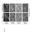
FIG. 2 shows alignment of Bam35c, Gil16c, AP50 and Wip1 genome maps. Predicted genes are represented as block arrows and the color key in the bottom right indicates postulated functions. Shaded regions pair conserved sequence segments between phages and amino acid identity, percentage ranges are shown in the lower left corner.
FIG. 3 shows purified proteins. The Ni-NTA eluate containing both his-p23 and p24 (Lane 1) was further purified by charge using ion exchange chromatography. The distinct peaks were evaluated with SDS-PAGE, which revealed the presence of his-p23 alone in Peak 1 and the presence of his-p23 and p24 together in Peak 2.
FIG. 4 shows Wip1 inhibition of infectivity by recombinant Wip1 proteins. A) Wip1 phage was assayed for inactivation by recombinant viral proteins at various concentrations. Protein concentrations are indicated per protein. His-p23 (red) and the his-p23+p24 complex (purple) inactivated Wip1 infectivity by up to 100% in a dose-dependent manner.
His-p22 (blue) and his-p24 (green) did not affect phage activity. Bars represent standard error for a minimum of 3 experiments. B) Anti-his-p23 antibody neutralization of Wip1 activity. Polyclonal antibodies were generated against his-p23 and tested for neutralization of Wip1 activity using methods described. After preincubation with phage, anti-his-p23 (closed circles) inhibited Wip1 adsorption to B. anthracis ΔSterne by up to 90% in a dose-dependent manner. Pre-bleed serum (open circles) did not affect Wip1 adsorption activity. Bars represent standard error of at least 3 experiments.
FIG. 5 shows indirect immunofluorescence microscopy using his-tagged Wip1 proteins. 1000× magnification.
FIG. 6 shows indirect immunofluorescence microscopy using his-p23 and the his-p23+p24 complex. 1000× magnification.
FIG. 7. Following expression and purification methods as described, the recombinant his-tagged Wip1 proteins were then measured using standard BSA assays. Protein solutions were diluted to concentrations of 250 μg/ml for single protein samples (his-p22; his-p23; his-p24) and 500 μg/ml for co-expressed protein samples (his-p23+p24). SDS-PAGE analysis of these protein samples revealed purification to near homogeneity. The samples shown here are the exact samples used in the following protein overlay-based inhibition assays.
DETAILED DESCRIPTION OF THE INVENTION
The described invention provides a better understanding of Wip1's highly specific tropism for B. anthracis. We started by imaging the morphological changes of Wip1 phage upon adsorption, expanding its host range analysis with adsorption studies, and sequencing its viral genome. Based on genomic analysis with other gram-positive infecting tectiviruses, candidate gene products for the Wip1 spike complex were predicted and used to identify a Wip1 receptor binding protein that detects and exhibits specificity for B. anthracis.
In the described invention, we characterized the Wip1 phage and its genome to develop the tools to identify the Wip1 gene product 23 as a receptor binding protein. Wip1 tropism was previously shown to be highly specific to B. anthracis. Here, we determined with adsorption assays that specificity to B. anthracis is mediated by Wip1's receptor binding. Indeed, receptor binding protein p23 was demonstrated to bind very specifically to bacterial strains that correspond with Wip1's narrow host range.
The identification of Wip1 p23 as a receptor binding protein shows that it is a unique protein with no homology to any other known proteins. Additionally, ORF23 shares no sequence identity with AP50, a tectivirus with a similar host range that is also highly specific to B. anthracis. Genomic analysis showed that the overall Wip1 genome shares significant similarities to the AP50 genome in ORF size, sequence, and organization. In fact, the genes neighboring ORF23 display this conservation. ORF22 (291 residues) shares 64% sequence identity with AP50 ORF27 (304 residues) and ORF24 (118 residues) shares 51% sequence identity with AP50 ORF29 (118 residues).
AP50 ORF28, which is located in the corresponding genomic position to Wip1 ORF23, is the gene that harbors one of two sequence mutations that differentiates isolate AP50t, which produces turbid plaques, from isolate AP50c, which produces clear plaques. Generally, clear plaques are formed when the host is completely susceptible to the phage while turbid plaques are formed if the host is partially resistant to the phage (for example, if 10% of the cells survive infection). Furthermore, AP50 ORF28 and Wip1 ORF23 are located at a highly variable region of their respective genomes. Although the Eip1 receptor-binding domain ORF23 exhibits sequence diversity, it was unexpected that two closely related tectiviruses with a similar host range have evolved uniquely different receptor binding proteins.
Additionally, it is shown that the his-p23 plus p24 complex exhibited higher competitive inhibition than his-p23 alone, suggesting that p24 complemented or enhanced his-p23 binding activity. This enhancement could result from simply protecting his-p23 proteins from degradation. However, we were careful to use fresh protein stocks in all assays. It also is possible that p24 could play a secondary but complementary role in Wip1 binding. The adsorption of phage to the gram-positive bacterial surface has been suggested to occur in two stages. The first step involves reversible binding to general recognition molecules in the cell wall and is followed by a subsequent irreversible step involving a more specific factor [Jacobson, E. D. L., O. E., Adsorption of bacteriophages phi 29 and 22a to protoplasts of Bacillus substilis. Journal of virology, 1977. 21(3): p. 1223-1227; Monteville, M. R. A., B.; Geller, B. L., Lactococcal Bacteriophages Require a Host Cell Wall Carbohydrate and a Plasma Membrane Protein for Adsorption and Ejection of DNA. Applied and environmental microbiology, 1994. 60(9): p. 3204-3211]. In fact, the seahorse-like structure for PRD1 receptor binding protein P2 consists of multiple domains with different purported functions. In one P2 model, the fin-shaped domain is proposed to make initial contacts by scanning the host surface in order to bring the head domain closer to its receptor [Xu, L. B., S. D.; Butcher, S. J.; Bamford, D. H.; Burnett, R. M., The Receptor Binding Protein P2 of PRD1, a Virus Targeting Antibiotic-Resistant Bacteria, Has a Novel Fold Suggesting Multiple Functions. Structure, 2003. 11: p. 309-322]. It is possible that Wip1 p24 is such a spike complex domain with non-specific, reversible surface scanning properties.
Polyclonal antibodies against his-p23 also were able to inactivate Wip1 binding activity. It should be noted, however, that the neutralizing effect of anti-his-p23 serum on Wip1 activity was much weaker compared to that of anti-gp28 or anti-gp29 serum on Bam35 activity [Gaidelyte, A., et al., The Entry Mechanism of Membrane-Containing Phage Bam35 Infecting Bacillus thuringiensis. Journal of bacteriology, 2006. 188(16): p. 5925-5934]. At a mere 20× dilution, anti-his-p23 antibody inactivation measured below 20%. In contrast, anti-gp28 and anti-29 antibodies did not demonstrate inactivation rates below 20% until they reached dilutions of 5,000× and 100,000×, respectively. One explanation for this difference is the possibility that p23 is a poor antigen that generated a weak immunogenic response. However, an indirect ELISA assay determined that the polyclonal antisera against his-p23 exhibited reasonable immunogenic strength (reading of Abs=1 at 1:2000 dilution).
The narrow infectivity and adsorption host range of Wip1 indicates that its receptor is unique to the exposed surface of B. anthracis and B. cereus CDC32805 in a manner that is accessible to phage. Indirect immunofluorescence microscopy demonstrated that p23 and the p23+p24 complex detect and label B. anthracis with a specificity that seems to match its narrow host range. This activity makes both the Wip1 phage and its receptor molecules useful diagnostic tools for B. anthracis with better specificity than the reagents currently being used.
The abbreviations used herein for amino acids are those abbreviations which are conventionally used: A=Ala=Alanine; R=Arg=Arginine; N=Asn=Asparagine; D=Asp=Aspartic acid; C=Cys=Cysteine; Q=Gln=Glutamine; E=Glu=Gutamic acid; G=Gly=Glycine; H=His=Histidine; I=Ile=lsoleucine; L=Leu=Leucine; K=Lys=Lysine; M=Met=Methionine; F=Phe=Phenyalanine; P=Pro=Proline; S=Ser=Serine; T=Thr=Threonine; W=Trp=Tryptophan; Y=Tyr=Tyrosine; V=Val=Valine. The amino acids may be L- or D-amino acids. An amino acid may be replaced by a synthetic amino acid which is altered so as to increase the half-life of the peptide or to increase the potency of the peptide, or to increase the bioavailability of the peptide.
The following represent groups of amino acids that are conservative substitutions for one another: 1) Alanine (A), Serine (S), Threonine (T); 2) Aspartic Acid (D), Glutamic Acid (E); 3) Asparagine (N), Glutamic Acid (Q); 4) Arginine (R), Lysine (K); 5) Isoleucine (I), Leucine (L), Methionine (M), Valine (V); and 6) Phenylalanine (F), Tyrosine (Y), Tryptophan (W).
Accordingly, the described invention provides a recombinant protein composition comprising Wip1 p23 receptor binding protein or fragments thereof. Further, the recombinant protein composition also can include Wip1 p24 receptor binding protein or fragments thereof. The Wip1 p23 receptor binding protein can further include a reporter molecule or other detectable label or agent. Such reporter molecules or other detectable labels or agents include, for example, but are not limited to, a fluorophore, a fluorophore/quencher pair, an antibody, a llama-body, an isotope, or combinations thereof. Furthermore, the described invention provides a recombinant protein composition comprising a native or recombinant Wip1 bacteriophage.
The described invention further provides a recombinant protein composition attached or associated with a substrate.
A substrate includes a microfabricated solid surface to which molecules may be attached through either covalent or non-covalent bonds. This includes, but is not limited to, Langmuir-Bodgett films, functionalized glass, membranes, charged paper, nylon, germanium, silicon, PTFE, polystyrene, gallium arsenide, gold, and silver. Any other material known in the art that is capable of having functional groups such as amino, carboxyl, thiol or hydroxyl incorporated on its surface, is contemplated. This includes surfaces with any topology, such as spherical surfaces and grooved surfaces. Such recombinant protein compositions and substrates can be incorporated into a kit. Further, the recombinant protein composition can be attached or associated with a lateral flow test.
The described invention further provides an isolated nucleic acid encoding a Wip1 p23 receptor binding protein, or variant, where the Wip1 p23 receptor binding protein, or variant, binds to Bacillus anthracis. The isolated nucleic acid can further be operably linked to a regulatory element, reporter, or detectable element. The term “operably linked” refers to a functional linkage between a promoter and a second sequence, wherein the promoter sequence initiates and mediates transcription of the DNA sequence corresponding to the second sequence. Generally, “operably linked” means that the nucleic acid sequences being linked are contiguous and, where necessary to join two protein coding regions, are contiguous and in the same reading frame. Additionally, the described invention provides an isolated nucleic acid encoding a Wip1 p24 receptor binding protein, or variant, where the Wip1 p24 receptor binding protein increases the binding affinity of a Wip1 p23 receptor binding protein or variant to Bacillus anthracis. Furthermore, the isolated nucleic acid can be a cDNA. Additionally, the isolated nucleic acids can be encoded within a recombinant expression cassette. The described invention further provides a recombinant expression vector encoding at least one of a Wip1 p23 receptor binding protein, a Wip1 p24 receptor binding protein, or variants thereof. Such recombinant expression vectors can further encode a reporter or a detectable element, a regulatory sequence, a controllable regulatory element, or combinations thereof. The recombinant expression vectors can be included in recombinant expression compositions. Such recombinant expression compositions can include, but are not limited to, buffers, detectable reagents, and the like.
The inventive isolated nucleic acids, including cDNA, can be included in a microarray. Microarray preparation methods for making oligonucleotide probes for Bacillus anthracis identification include the following: (1) spotting a solution on a prepared surface using spotting robots; (2) in situ synthesis by printing reagents via ink jet or other computer printing technology and using phosphoramidite chemistry; (3) in situ parallel synthesis using electrochemically generated acid for removal of protecting groups and using standard phosphoramidite chemistry; (4) in situ synthesis using maskless photo-generated acid for removal of protecting groups and using regular phosphoramidite chemistry; (5) mask-directed in situ parallel synthesis using photo-cleavage of photolabile protecting groups (PLPG) and phosphoramidite chemistry; (6) maskless in situ parallel synthesis using PLPG and digital photolithography and standard phosphoramidite chemistry; and (7) electric field attraction/repulsion for depositing fully formed oligonucleotides onto known locations.
An electrode microarray for in situ oligo synthesis using electrochemical deblocking is disclosed in Montgomery U.S. Pat. Nos. 6,093,302; 6,280,595, and 6,444,111 (Montgomery I, II, and III respectively), all of which are incorporated by reference herein. Another and materially different electrode array (not a microarray) for in situ oligo synthesis on surfaces separate and apart from electrodes using electrochemical deblocking is disclosed in Southern U.S. Pat. No. 5,667,667, which is incorporated by reference herein. Photolithographic techniques for in situ oligo synthesis are disclosed in Fodor et al. U.S. Pat. No. 5,445,934 and the additional patents claiming priority thereto, all of which are incorporated by reference herein. Electric field attraction/repulsion microarrays are disclosed in Hollis et al. U.S. Pat. No. 5,653,939 and Heller et al. U.S. Pat. No. 5,929,208, both of which are incorporated by reference herein. A review of oligo microarray synthesis is provided by: Gao et al., Biopolymers 2004, 73:579.
The described invention further provides a system for detecting Bacillus anthracis comprising a recombinant protein composition and a detector in communication with the recombinant protein composition. The detector is capable of detecting a signal generated upon recognition of a Bacillus anthracis receptor by the recombinant protein composition. The system can further include a light source in optical communication with the recombinant protein composition. The system also can include a processor for processing signals detected by the detector.
The described invention further provides a method of identifying or detecting Bacillus anthracis in a sample, the method comprising: (a) providing a sample suspected of containing Bacillus anthracis; (b) contacting the sample with the recombinant protein composition, wherein a change in a signal generated by a reporter molecule indicates the presence of Bacillus anthracis in the sample. Such samples include, but are not limited to, a biological sample or environmental sample. The reporter molecule can be a detectable label or agent such as, but not limited to a fluorophore or flurophore/quencher pair. The recombinant protein composition can change conformation when contacting a Bacillus anthracis receptor thereby changing detectable properties of the recombinant protein composition.
EXAMPLES
The following examples are put forth so as to provide those of ordinary skill in the art with a complete disclosure and description of how to make and use the present invention, and are not intended to limit the scope of what the inventors regard as their invention nor are they intended to represent that the experiments below are all or the only experiments performed. Efforts have been made to ensure accuracy with respect to numbers used (for example, amounts, temperature, etc.) but some experimental errors and deviations should be accounted for. Unless indicated otherwise, parts are parts by weight, molecular weight is weight average molecular weight, temperature is in degrees Centigrade, and pressure is at or near atmospheric.
Example 1
Wip-1 Characterization
Bacterial Strains and Phages.
The majority of bacterial strains in the present study were previously described [Schuch, R., et al., Prevalence of Bacillus anthracis-like organisms and bacteriophages in the intestinal tract of the earthworm Eisenia fetida. Applied and environmental microbiology, 2010. 76(7): p. 2286-94; Schuch, R. F. V. A., The Secret Life of the Anthrax Agent Bacillus Anthracis: Bacteriophage-Mediated Ecological Adaptations. PLos One, 2009. 4(8): p. e6532; Schuch, R. N., D.; Fischetti, V. A., A bacteriolytic agent that detects and kills Bacillus anthracis. Nature, 2002. 418: p. 884-889]. All bacterial strains were grown in brain heart infusion (BHI) broth or agar plates at 30° C. according to standard protocols. The bacteriophage Wip1 was isolated from the intestinal tract of Eisenia fetida worms from Pennsylvania, USA. Phage propagation was performed on the B. anthracis ΔSterne strain.
Phage Propagation.
High titer phage stocks were obtained by infecting stationary cell cultures (100 ul) with 100 ul of a series of diluted (1:100 to 1:1000) Wip1 phage stocks. The phage-bacterium mixtures were incubated in a 37° C. water bath for 15 min and then plated with molten top agar (0.8%) onto BHI plates and incubated overnight at 30° C. When the viral plaques reached near confluency, the soft agar overlays were collected in conical tubes, incubated with 2 ml 10 mM K phosphate per plate for 15 min at room temperature, and centrifuged at 4,000 rpm for 20 min at 4° C. The resulting supernatants were filtered (0.45-um-pore-size-filter) and stored at 4° C.
Transmission Electron Microscopy.
Wip1 phages were incubated with overnight cultures of B. anthracis ΔSterne at a MOI of 10 for 5 minutes at 37° C. After incubation, the mixtures were transferred to a new Eppendorf tube with solidified agar on the bottom to act as a cushion during the subsequent centrifugation at 6000 rpm for 3 minutes. Supernatant was removed and the pellet was resuspended in 1× glutaraldehyde fixative. The TEM analyses were then performed at The Rockefeller University Bio-Imaging Resource Center as previously described [Schuch, R. N., D.; Fischetti, V. A., A bacteriolytic agent that detects and kills Bacillus anthracis. Nature, 2002. 418: p. 884-889].
Chloroform Sensitivity Assay.
Wip1 phage samples (2 ml) were incubated with and without various volumes of chloroform (up to 80 ul) in capped glass tubes with gentle mixing at room temperature for 15 min. The mixtures were then titered on B. anthracis ΔSterne. W2 phage with and without chloroform plated on B. cereus ATCC 4342 was used as a control.
Phage Adsorption Assay.
Various bacterial strains were grown to stationary phase (approximately 2×10⁸ CFU/ml) and 100 ul of bacteria was mixed with 100 ul of Wip1 at 2×10⁷ PFU/ml. The phage-bacterium mixtures were incubated in a 37° C. water bath for 20 min and then pelleted at 7,000 rpm for 3 min. The resulting supernatants were subsequently spin-filtered (Millipore; 0.22 um) and titered on plates of B. anthracis ΔSterne.
DNA Manipulation and Sequencing.
To obtain Wip1 DNA, Wip1 phage stocks (1×10⁸ CFU/ml) were lysed as follows: 25 μl of phage stock was suspended in 25 μl of 0.5M NaOH (Sigma-Aldrich), incubated for 5 minutes at room temperature, neutralized with 50 μl of Tris pH 8.0 (Life Technologies), and diluted in 450 μl dH 20. Wip1 DNA was processed as described [Schuch, R., et al., Prevalence of Bacillus anthracis-like organisms and bacteriophages in the intestinal tract of the earthworm Eisenia fetida. Applied and environmental microbiology, 2010. 76(7): p. 2286-94] digested for 5 min at 65° C. with 0.1 units of Tsp509I (New England Biolabs), ligated to EcoRI adaptors (GeneLink), PCR amplified using adaptor-specific primers and cloned into the pBAD TOPO® TA expression vector (Life Technologies). The resulting plasmid library was transformed into One Shot TOP1O E. coli (Life Technologies), and random plasmid preparations were sequenced. Sequences derived from these transformed cells were confirmed by sequencing PCR products generated directly from Wip1 phage DNA. Primers were designed to sequence specific regions on the PCR products and unknown sequence regions were determined using primer walking on the purified Wip1 genome. GenBank Acession number KF188458.
Cloning of his-Tagged Wip1 ORFs 22, 23, and 24.
The PCR products containing the coding sequences for the Wip1 gene products 22, 23, and 24 were separately amplified using specific primers. Each DNA fragment was inserted into a modified CDFDuet-1 plasmid between the SalI-NotI sites preceded by a T7lac promoter and ribosome binding site as well as 2×His-tag sequences. Clones were confirmed by sequencing using primers that flank the insert.
Purification of his-Tagged Wip1 p22, p23, and p24.
Overnight cultures of E. coli DH5 alpha cells carrying the cloned constructs were diluted 1:100 and grown in LB medium with spectinomycin (20 ug/ml) for 4 h while shaking at 37° C. After being moved to 16° C., the cultures were induced with isopropyl-b-D-thiogalactopyranoside (IPTG) at a final concentration of 0.25 mM and shaken for an additional 18 h. Bacterial pellets were collected by centrifugation (Sorvall SLC-6000 rotor; 7200 rpm; 30 min; 4° C.) and resuspended in a cold buffer (50 mM Tris, pH 8.0+200 mM NaCl+5 mM imidazole) at 1/100 of the original culture volume. Bacterial lysis was conducted by multiple passages through a French pressure cell (at ˜105 MPa) at 4° C. The cell debris was removed by centrifugation (Sorvall SS-34 rotor; 8,000 rpm; 20 min; 4° C.) followed by filtration (Nalgene; 0.45 um).
The following purification steps were conducted at room temperature using buffers kept at 4° C. 25 mL columns were loaded with 1.25 ml bed volume of Ni-NTA Agarose (Qiagen) and equilibrated with 2× column volumes of buffer (50 mM Tris, pH 8.0+200 mM NaCl+5 mM imidazole). The cell lysate from induced cultures was passed through the columns 2× using gravity flow. The Ni-NTA Agarose was then washed with 1× column volume of wash buffer A (50 mM Tris, pH 8.0+500 mM NaCl+30 mM imidazole) and 0.5× column volume of wash buffer B (50 mM Tris, pH 8.0+500 mM NaCl+60 mM imidazole). Finally, the eluate was collected by passing 5× bed volume of elution buffer (50 mM Tris, pH 8.0+500 mM NaCl+250 mM imidazole) through the column.
In preparation for the next step of the purification process, the eluted proteins were dialyzed against buffer A (20 mM phosphate buffer; pH 7.4) at 4° C. Ion exchange chromatography was then conducted using a 5 mL HiTrap Q HF column at a linear gradient from 100% Buffer A targeting 50% Buffer B (20 mM phosphate buffer+1M NaCl; pH 7.4). Fractions containing the purified proteins of interest were collected, analyzed by SDS-PAGE, pooled, and dialyzed against 1×PBS at 4° C. (FIG. 7).
Cloning and Purification of Co-Expressed his-p23 and p24.
The cloning and purification schematic of his-p23 and p24 is exactly the same as described for the individual recombinant proteins. The only difference is that the PCR insert at the multiple cloning site began with the start codon for ORF23 and ended with the last codon for ORF24. It should be noted that ORF24 was not cloned into a separate site with its own promoter and tag.
Inhibition of Phage Infection.
Overnight cultures of B. anthracis ΔSterne (12 ul) were mixed with molten soft agar (0.8%; 400 ul) and overlayed onto BHI agar plates measuring 35×10 mm. After allowing the bacterial soft agar to sit for 15 min, various dilutions of the purified his-tagged phage protein stocks (PBS dilutions; 100 ul) were evenly placed on top. The protein overlay was incubated atop the bacterial agar for 15 min before a final overlay of Wip1 phage (˜100 PFU) was evenly added. Resulting plaques were counted at 48 hrs post infection. All steps were conducted at room temperature.
Inhibition of Phage Adsorption Using Polyclonal Antisera Against his-Tagged Wip1 p23.
To generate polyclonal antisera against his-tagged Wip1 p23, 500 ug of the purified protein was run on an SDS-PAGE gel and excised bands were used as antigen to immunize two New Zealand white rabbits. Pre-immune serum was collected before the first immunization in complete Freund's adjuvant and for the second, third, and fourth immunizations in incomplete Freund's adjuvant. Immunizations and production bleeds were collected at 3 week intervals for a total of 21 weeks. An indirect ELISA assay was used to determine the titer of the polyclonal antisera against his-p23. The titer was found to be 2,000 (the highest dilution to yield an absorbance=1 in 15 minutes).
The antisera were then tested for ability to inhibit Wip1 phage binding activity. Various dilutions of serum (in BHI media) were mixed with Wip1 phage stock (˜800 PFU) and incubated in a 37° C. water bath for 30 min. After adding 100 ul of overnight cultures of B. anthracis ΔSterne, the serum-phage-bacterium mixtures were incubated at room temperature for 30 min. Bacterial pellets were collected via centrifugation at 12,500 rpm for 30 seconds, washed, and resuspended in 10 mM K phosphate buffer. Bacteria-bound phage were plated onto BHI and proceeded to form plaques overnight. The inhibition rate was measured as the percentage reduction of Wip1 PFUs as compared to a no-serum, PBS control.
Fluorescence Microscopy.
The following protocol is a modified version of one previously described [Raz, A. and V. A. Fischetti, Sortase A localizes to distinct foci on the Streptococcus pyogenes membrane. Proceedings of the National Academy of Sciences of the United States of America, 2008. 105(47): p. 18549-54]. Overnight cultures of various bacterial strains were inoculated at a 1:100 dilution in BHI media and grown to mid-exponential phase (unless otherwise noted) by shaking for 3 h at 30° C. The cell cultures were then pelleted by centrifugation (Eppendorf 5810 R; 4,000 rpm; 5 min; 4 C), washed, and then resuspended in PBS. To fix the cells, paraformaldehyde and NaPO4, pH 7.4 were added to the suspension at final concentrations of 2.6% and 30 mM, respectively. The cells were incubated for 15 min at room temperature, followed by 30 min on ice, washed with PBS, and then attached to polylysine-coated glass cover slips. The fixed cells attached onto glass were then washed with PBS and blocked with PBS containing 1% BSA for 15 min. The cells subsequently underwent a series of three labeling steps: first with purified his-tagged Wip1 protein, then with anti-His mouse antibodies, and finally with anti-mouse rhodamine dye and 4′,6-diamidino-2-phenylindole (DAPI) stain. Each labeling step consisted of incubating the cells with the labeling mixture for 45 min in a moist chamber and was followed by thorough washes with PBS. To reduce bleaching, the slides were mounted with 50% glycerol and 0.1% p-phenylenediamine in PBS pH 8.0. Images were captured using a Delta-Vision image restoration microscope (Applied Precision/Olympus) equipped with CoolSnap QE cooled CCD camera (Photometrics). An Olympus 100× oil immersion objective was used in conjunction with a 1.5× optovar.
Wip1 is Morphologically Similar to Other Tectiviruses.
Wip1 phage is able to produce clear plaques on B. anthracis Sterne and ΔSterne strains. Samples of the viral particles with and without the bacterium B. anthracis ΔSterne were analyzed by transmission electron microscopy. Wip1 phage alone are tailless, bilayer, icosahedral particles with a vertex to vertex diameter of approximately 60 nm, similar in morphology to other tectiviral phages [Schuch, R. F. V. A., The Secret Life of the Anthrax Agent Bacillus Anthracis: Bacteriophage-Mediated Ecological Adaptations. PLos One, 2009. 4(8): p. e6532]. Treatment with chloroform inhibited Wip1 activity, which is consistent with Wip1 possessing an inner lipid membrane (data not shown).
Upon incubation with its host B. anthracis ΔSterne, Wip1 displayed a tube-like structure (FIG. 1) that has been described in other tectiviruses such as Bam35 and PRD1 [Ackermann, H. W. R., R.; Martin, M.; Murthy, M. R.; Smirnoff, W. A., Partial Characterization of a Cubic Bacillus Phage. Canadian Journal of Microbiology, 1978. 24: p. 986-993; Laurinmaki, P. A. H., J. T.; Bamford, D. H.; Butcher, S. J., Membrane Proteins Modulate the Bilayer Curvature in the Bacterial Virus Bam35. Structure, 2005. 13: p. 1819-1828; Rydman, P. S. C., J.; Butcher, S. J.; Fuller, S. D.; Rutten, T.; Bamford, D. H., Bacteriophage PRD1 contains a labile receptor-binding structure at each vertex. J. Mol. Biol., 1999. 291: p. 575-587]. In PRD1, interaction of protein P2 with the host receptor leads to a conformational change that results in the dissociation of the spike complex proteins from the virion [Grahn, A. M. C., J.; Bamford, J. K.; Bamford, D. H., Stable Packaging of Phage PRD1 DNA Requires Adsorption Protein P2, Which Binds to the IncP Plasmid-Encoded Conjugative Transfer Complex. Journal of bacteriology, 1999. 181(21): p. 6689-6696]. This is followed by the transformation of the spherical internal membrane into a tubular channel structure for the delivery of viral DNA into the host [Daugelavicius, R. B., J. K.; Bamford, D. H., Changes in host cell energetics in response to bacteriophage PRD1 DNA entry. Journal of bacteriology, 1997. 179: p. 5203-5210; Grahn, A. M. D., R.; Bamford, D. H., Sequential model of phage PRD1 DNA delivery: active involvement of the viral membrane. Mol. Microbiol., 2002. 46: p. 1199-1209]. In some cases, the phage tails were seen to interact in a conventional way with the bacterial cell surface. However, TEM images also captured several Wip1 phages with tube-like structures facing away from the bacterial surface (FIG. 1) instead of directly towards the host, a phenomenon not reported in other tectiviruses [Bamford, D. H., Personal communication, 2006: New York]. This could indicate that the labile vertex undergoing tubular transformation may not be the same as the vertex that initially binds the receptor. This could also be explained by reversible binding between the Wip1 tube-like structure and its host.
Wip1 Infection and Adsorption are Highly Specific to B. anthracis.
Previous studies determined that Wip1 infectivity is more specific to B. anthracis than the standard diagnostic tool γ phage [Schuch, R., et al., Prevalence of Bacillus anthracis-like organisms and bacteriophages in the intestinal tract of the earthworm Eisenia fetida. Applied and environmental microbiology, 2010. 76(7): p. 2286-94]. We decided to expand the host range study to include adsorption and infection of Wip1 virions to the surface of different bacteria. Adsorption assays showed that Wip1 binding also exhibited a high specificity to B. anthracis that corresponded with its narrow infectivity host range (Table 1). This observation further supported the model of Wip1 tropism being mediated by the receptor binding proteins on its surface.
| TABLE 1 |
| |
| Wip1 infectivity and adsorption range. |
| |
Infectivity (PFU/ml) |
Adsorption (%) |
| |
B. anthracis
|
|
|
|
| |
deltaSterne |
3.0E+09 |
6.0E+9 |
100 |
| |
Bacillus cereus
|
| |
ATCC 4342 |
1.0E+05 |
<10 |
<5 |
| |
CDC32805 |
4.0E+07 |
3.0E+07 |
94 |
| |
CDC13100 |
<10 |
<10 |
<5 |
| |
CDC13140 |
<10 |
<10 |
<5 |
| |
ATCC 10987 |
<10 |
<10 |
<5 |
| |
NRL 569 |
<10 |
<10 |
<5 |
| |
ATCC 14579 |
<10 |
<10 |
<5 |
| |
ATCC 13472 |
<10 |
<10 |
<5 |
| |
ATCC 11980 |
<10 |
<10 |
<5 |
| |
RTS 100 |
<10 |
<10 |
<5 |
| |
B. thuringiensis
|
| |
HD1 |
<10 |
<10 |
<5 |
| |
HD73 |
<10 |
<10 |
<5 |
| |
B. subtilis SL4 |
<10 |
<10 |
<5 |
| |
B. pumilis
|
<10 |
<10 |
<5 |
| |
SL4680 |
| |
Sporosarcina
|
<10 |
<10 |
<5 |
| |
ureae
|
| |
B. megaterium
|
<10 |
<10 |
<5 |
| |
WH32 |
| |
Brevibacillus
|
<10 |
<10 |
<5 |
| |
laterosporus
|
| |
|
Wip1 is Related to Other Tectiviruses with Notable Differences.
The Wip1 genome was determined to be a linear molecule of DNA measuring 14,319 bp. Detailed analysis of the Wip1 sequence revealed the existence of 27 putative open reading frames (ORFs), as shown in FIG. 2. Wip1 exhibits sequence similarities to gram-positive infecting tectiviruses Bam35c and Gil16c. Wip1 is most closely related to AP50, which also has a narrow host range highly specific to B. anthracis [Sozhamannan, S., et al., Molecular Characterization of a Variant of Bacillus anthracis-Specific Phage AP50 with Improved Bacteriolytic Activity. Applied and environmental microbiology, 2008. 74(21): p. 6792-6796]. The genome of Wip1 from ORF5 through ORF25 is strikingly similar to the section of AP50 from ORF10 through ORF30 in ORF size, sequence, and organization (Tables 2a, 2b, 2c, and FIG. 2). Among the 27 total putative Wip1 ORFs, 19 share sequence identity of at least 50% to other tectiviral proteins and 14 share high sequence identity of at least 75% to AP50 proteins. We noted that the Wip1 genome GC content at both extremities is lower than the percentage observed in the central section of the genome. The GC content is approximately the same in all the Wip1 ORFs as compared to their corresponding AP50 homologs [Sozhamannan, S., et al., Molecular Characterization of a Variant of Bacillus anthracis-Specific Phage AP50 with Improved Bacteriolytic Activity. Applied and environmental microbiology, 2008. 74(21): p. 6792-6796].
| TABLE 2a |
| |
| Comparison of genes in Wip1 with other Tectiviridae |
| |
No. of residues |
|
|
| |
(genome |
| Wip1 ORF |
coordinates) |
Strand |
G + C content (%) |
| |
| 1 |
57 (396-569) |
+ |
29.5 |
| 2 |
196 (598-1188) |
+ |
30.7 |
| 3 |
112 (1219-1557) |
+ |
25.7 |
| 4 |
91 (1690-1965) |
+ |
25.7 |
| 5 |
67 (1958-2161) |
+ |
27.9 |
| 6 |
116 (2121-2471) |
+ |
43.9 |
| 7 |
236 (2309-3019) |
+ |
46.8 |
| 8 |
83 (2865-3116) |
+ |
46.4 |
| 9 |
212 (3125-3763) |
+ |
42.4 |
| 10 |
54 (3760-3924) |
+ |
36.4 |
| 11 |
47 (3938-4081) |
+ |
33.6 |
| 12 |
353 (4081-5142) |
+ |
40.7 |
| 13 |
74 (5191-5415) |
+ |
38.2 |
| 14 |
59 (5421-5600) |
+ |
32.8 |
| 15 |
157 (5677-6150) |
+ |
38.8 |
| 16 |
61 (6138-6323) |
+ |
36.0 |
| 17 |
48 (6323-6469) |
+ |
36.1 |
| 18 |
91 (6482-6757) |
+ |
43.1 |
| 19 |
213 (6761-7402) |
+ |
44.8 |
| 20 |
218 (7402-8058) |
+ |
44.1 |
| 21 |
175 (8055-8582) |
+ |
46.4 |
| 22 |
291 (8593-9468) |
+ |
44.7 |
| 23 |
117 (9472-9825) |
+ |
39.8 |
| 24 |
118 (9838-10194) |
+ |
36.7 |
| 25 |
48 (10196-10342) |
+ |
33.3 |
| 26 |
213 (10344-10985) |
+ |
42.3 |
| 27 |
898 (11342-14038) |
− |
29.7 |
| |
| TABLE 2b |
| |
| Comparison of genes in Wip1 with other Tectiviridae |
| Wip1 |
Identity (%) |
ORF (# residues) |
| ORF |
AP50 |
GIL16c |
Bam35c |
AP50 |
GIL16c |
Bam35c |
| |
| 1 |
|
|
|
|
|
|
|
|
|
| 2 |
| 3 |
| 4 |
| 5 |
55.1 |
|
|
10 |
(57) |
| 6 |
69.6 |
62.5 |
45.7 |
11 |
(114) |
9 |
(125) |
10 |
(145) |
| 7 |
78.4 |
42.2 |
44.0 |
12 |
(235) |
10 |
(248) |
11 |
(252) |
| 8 |
94.0 |
53.2 |
53.2 |
13 |
(83) |
11 |
(80) |
12 |
(80) |
| 9 |
93.9 |
59.9 |
63.7 |
14 |
(212) |
13 |
(212) |
14 |
(212) |
| 10 |
89.1 |
60.9 |
60.9 |
15 |
(46) |
14 |
(46) |
15 |
(46) |
| 11 |
80.9 |
48.8 |
48.8 |
16 |
(49) |
16 |
(46) |
16 |
(46) |
| 12 |
83.8 |
63.1 |
63.9 |
17 |
(354) |
18 |
(356) |
18 |
(356) |
| 13 |
78.4 |
35.7 |
34.3 |
18 |
(74) |
19 |
(76) |
19 |
(76) |
| 14 |
34.4 |
46.8 |
39.4 |
19 |
(56) |
20 |
(52) |
20 |
(68) |
| 15 |
86.6 |
37.2 |
37.8 |
20 |
(157) |
22 |
(143) |
21 |
(143) |
| 16 |
79.3 |
60.7 |
60.7 |
21 |
(58) |
23 |
(58) |
22 |
(58) |
| 17 |
77.1 |
54.2 |
54.2 |
22 |
(48) |
24 |
(48) |
23 |
(48) |
| 18 |
82.4 |
35.6 |
33.3 |
23 |
(91) |
25 |
(91) |
24 |
(91) |
| 19 |
85.1 |
23.5 |
25.9 |
24 |
(210) |
26 |
(204) |
25 |
(204) |
| 20 |
91.7 |
66.4 |
65.4 |
25 |
(218) |
27 |
(250) |
26 |
(250) |
| 21 |
84.6 |
62.2 |
63.4 |
26 |
(175) |
28 |
(170) |
27 |
(170) |
| 22 |
63.9 |
43.1 |
40.3 |
27 |
(304) |
29 |
(297) |
28 |
(304) |
| 23 |
| 24 |
50.8 |
|
|
29 |
(118) |
| 25 |
71.7 |
|
|
30 |
(49) |
| 26 |
| 27 |
| |
| TABLE 2c |
| |
| PRD1 protein and postulated function of Wip1 ORFs. |
| Wip1 | PRD1 protein (no. | |
| ORF | of residues) Ref] | Postulated function |
| |
| 1 | | |
| 2 | | Transcription factor |
| 3 | | LexA-type repressor |
| 4 |
| 5 |
| 6 | P6 (166) [32 [1]] | DNA packaging/unique vertex |
| 7 | P10 (203) | Assembly |
| | [33[2], 34[3]] |
| 8 |
| 9 | P9 (227) [d[4]] | DNA packaging ATPase |
| 10 |
| 11 | P20 (42) [e] | DNA packaging/unique vertex |
| 12 | P3 (395) [f[5]] | Major capsid protein |
| 13 | P22 (47) [g] | DNA packaging/unique vertex |
| 14 |
| 15 |
| 16 |
| 17 |
| 18 |
| 19 | P11 (207) [h[6]] | DNA delivery |
| 20 | P7 (265) | Lysin |
| 21 |
| 22 | P5 (340) [i[7]] | Trimeric spike protein |
| 23 |
| 24 |
| 25 |
| 26 | | Lysin |
| 27 | | DNA polymerase |
| |
ORFs were predicted using GeneMark (exon.biology.gatech.edu/heuristic_hmm2.cgi) and G+C % were determined using the GC calculator at www.sciencebuddies.org. Protein identities were determined with the Pairwise Sequence Alignment program Water available from EMBOSS at the EMBL-EBI node (www.ebi.ac.uk/Tools/psa/emboss_water!). A summary of PRD1 gene functions can be found in [j[8]] and PRD1 homologs in Bam35c were determined in [22]. Relevant references are indicated in brackets: [a] Stromsten, N. J., Benson, S. D., Burnett, R. M., Bamford, D. H., Bamford, J. K., The
Bacillus thuringiensis linear double-stranded DNA phage Bam35, which is highly similar to the
Bacillus cereus linear plasmid pBClin15, has a prophage state. Journal of Bacteriology, 2003. 185(23): p. 6985-6989; [b] Mindich, L., Bamford, D., McGraw, T., Mackenzie, G., Assembly of bacteriophage PRD1: particle formation with wild-type and mutant viruses. Journal of Virology, 1982. 44(3): p. 1021-1030; [c] Rydman, P. S., Bamford, J. K., Bamford, D. H., A minor capsid protein P30 is essential for bacteriophage PRD1 capsid assembly. J Mol Biol, 2001. 313(4): p. 785-795; [d] Bamford, J. K., Hanninen, A. L., Pakula, T. M., Ojala, P. M., Kalkkinen, N., Frilander, M., Bamford, D. H., Genome organization of membrane-containing bacteriophage PRD1. Virology, 1991. 183(2): p. 658-676; [e] uniprot.org/uniprot/P27587; [f] Benson S. D., Bamford, J. K., Bamford, D. H., Burnett, R. M., The X-ray crystal structure of P3, the major coat protein of the lipid-containing bacteriophage PRD1, at 1.65 A resolution. Acta Crystallogr D Biol Crystallogr., 2002. 58: p. 39-59; [g] uniprot.org/uniprot/P27388; and [h] Bamford, J. K. and Bamford, D. H., Large-scale purification of membrane-containing bacteriophage PRD1 and its subviral particles. Virology, 1991. 181(1): p. 348-352.
A distinction of the Wip1 genome is the placement of the putative DNA polymerase, ORF27, at the 3′ end of the genome on the negative strand. All other tectiviral DNA polymerases are encoded in the first 5,000 base pairs of their genomes on the positive strand. While several polymerase motifs were identified in the Wip1 ORF27 sequence, the unusual Wip1 ORF27 gene product does not share any significant homology with any other proteins in the NCBI database.
Another notable section of the genome includes Wip1 ORF22, ORF23, and ORF24. Wip1 ORF22 is predicted to be a putative spike complex protein as it shares 40.3% sequence identity with Bam35 gp28, a homolog for PRD1 trimeric spike protein P5. Bam35 gp28 is followed by gp29, a 293 amino acid protein that also resides on the phage surface [Ravantti, J. J., et al., Comparative analysis of bacterial viruses Bam35, infecting a gram positive host, and PRD1, infecting gram-negative hosts, demonstrates a viral lineage. Virology, 2003. 313(2): p. 401-14]. If Wip1 and AP50 genomic organization both align with that of Bam35c, then a putative spike complex protein should follow. Interestingly, in Wip1 and AP50, the genes downstream the P5 homolog are located in a highly variable region and do not share sequence identity with any Bam35 ORFs (FIG. 2). Despite the lack of homology, we predicted that Wip1 gene products 23 and 24 (totaling 235 amino acids together) were putative spike complex proteins based on strikingly similar gene cassette alignment both upstream and downstream of this region. Finally, we were intrigued by the observation that while Wip1 ORF24 shares 51% sequence identity with AP50 ORF29, Wip1 ORF23 is a unique gene that does not share sequence homology to any other known genes, tectiviral or otherwise.
Wip1 p23 and p24 Form a Stable Complex.
Genomic analysis predicted three Wip1 proteins, p22, p23, and p24 to be possible candidates for the Wip1 spike complex. We developed expression and purification schemes for the his-tagged constructs of all three viral proteins. However, the separately expressed his-p23 and his-p24 constructs resulted in extremely low yields of soluble protein due to the formation of inclusion bodies (data not shown). Curiously, when his-p23 and p24 were co-expressed, significantly higher yields of soluble protein were generated, suggesting that his-p23 and p24 assist each other in proper folding when expressed together. We used this co-expressed complex to purify the two molecules away each other. However, when the his-p23 and p24 complex were eluted from a stringently-washed Ni-NTA column, both eluted together despite the fact that p24 was not his-tagged (FIG. 3). When we used this Ni-NTA eluate to separate the two proteins by ion exchange chromatography, the two proteins were again observed together in the same fractions despite the fact that his-p23 and p24 exhibit drastically different pIs of 8.4 and 4.9, respectively (FIG. 3). This observation strongly suggests that p23 and p24 form a stable complex.
Wip1 p23 is a Receptor-Binding Protein.
The purified recombinant proteins were subsequently used in the Wip1 activity inhibition assay, which consisted of overlaying the viral proteins on top of B. anthracis ΔSterne growing in soft agar before adding a final overlay of infectious Wip1 phage. His-p23 was shown to competitively inhibit Wip1 infectivity up to 100% in a dose-dependent manner while his-p22 and his-p24 had no effect on phage infectivity (FIG. 4A). This finding suggests that Wip1 p23 is a receptor binding protein. We also tested the his-p23 and p24 complex in this assay at a 2× total concentration to account for the presence of two proteins that make up each protein complex. The his-23 plus p24 complex exhibited higher inhibition levels than his-p23 alone, a finding that may be explained by either increased protein stability or enhanced activity of the receptor binding complex. The possibility that recombinant his-p22 and his-p24 may not possess their full biological activity because of the linked histidine should be noted. However, the issues of potential misfolding or activity interference caused by a histidine tag are partially addressed for p24 with the co-expressed his-p23 plus p24 complex.
In a second phage inhibition assay, we used polyclonal antisera against Wip1 his-p23 to inhibit phage binding activity. B. anthracis ΔSterne bacteria was added to pre-incubated mixtures of Wip1 phage and antiserum at multiple dilutions. The cells were pelleted and the supernatant plated to reveal the titer of unbound phage. The results revealed that anti-his-p23 antibodies reduced Wip1 adsorption by up to 90% in a dose-dependent manner. Pre-bleed serum did not have an effect on Wip1 activity (FIG. 4B). This suggests that Wip1 p23 protein resides on the phage surface and is consistent with the finding that it is a receptor-binding molecule.
Wip1 p23 Binding is Specific to B. anthracis.
To further understand the interaction between Wip1 proteins and bacterial surfaces, the purified his-tagged viral proteins were tested for surface labeling of select bacterial strains using indirect immunofluorescence microscopy. FIG. 5 shows that both his-p23 and the his-p23 plus p24 complex bind specifically to the surface of B. anthracis ΔSterne. Surprisingly, the Wip1 proteins bound all mid-log phase ΔSterne uniformly but bound only a subpopulation of stationary phase ΔSterne. His-p22 and his-p24 were unable to bind either growth phase of ΔSterne, further suggesting that Wip1 p22 and p24 are not involved with phage adsorption. The lesser ability of Wip1 to bind to stationary phase bacteria is probably due to the replacement of surface array protein (Sap) with EA1 protein as the bacteria transition from log phase to stationary phase [Mignot, T., et al., Developmental switch of S-layer protein synthesis in Bacillus anthracis. Mol Microbiol, 2002. 43(6): p. 1615-27]; Sap is believed to be the AP50 tectivirus receptor [Bishop-Lilly, K. A., et al., Whole genome sequencing of phage resistant Bacillus anthracis mutants reveals an essential role for cell surface anchoring protein CsaB in phage AP50c adsorption. Virol J, 2012. 9: p. 246.] and may be important for Wip1 attachment as well.
In FIG. 6, the specificity of the binding of his-p23 and the his-p23 plus p24 complex to B. anthracis is shown by indirect immunofluorescence microscopy. Neither of the protein constructs were able to bind the surface of B. cereus ATCC 4342, CDC13100, and CDC13140; the three strains that are susceptible to γ infection but not to Wip1 infection. As expected, they were also unable to bind B. cereus strains ATCC 10987 and NRL 569, as well as B. thuringiensis strains HD1 and HD73, none of which can be infected by either γ or Wip1 phage. His-p23 and the his-p23 plus p24 complex bound positively to only B. anthracis Sterne, ΔSterne, and B. cereus CDC32805, the only B. cereus strain in our host range analysis that supported Wip1 replication (Table S1). These findings suggest that his-p23 does not bind to just any gram-positive bacterial surface, but binds very specifically to the bacterial hosts that support Wip1 infection, including B. anthracis ΔSterne.
| TABLE S1 |
| |
| Comparative table of Wip1 host range and his-p23 binding. |
| |
|
Adsorp- |
|
| |
|
tion |
| |
Infectivity (PFU/ml) |
(%) |
Immunofluorescence |
| Strain |
Wgamma |
Wip1 |
Wip1 |
his-p23 |
his-p23+24 |
| |
|
B. anthracis
|
|
|
|
|
|
| deltaSterne |
3.0E+09 |
6.0E+9 |
100 |
+ |
+ |
|
Bacillus
|
|
cereus
|
| ATCC 4342 |
1.0E+05 |
<10 |
<5 |
− |
− |
| CDC32805 |
4.0E+07 |
3.0E+07 |
94 |
+ |
+ |
| CDC13100 |
<10 |
<10 |
<5 |
− |
− |
| CDC13140 |
<10 |
<10 |
<5 |
− |
− |
| ATCC 10987 |
<10 |
<10 |
<5 |
− |
− |
| NRL 569 |
<10 |
<10 |
<5 |
− |
− |
|
B. thuringiensis
|
| HD1 |
<10 |
<10 |
<5 |
− |
− |
| HD73 |
<10 |
<10 |
<5 |
− |
− |
| |
Bacterial strains that support Wip1 infectivity and adsorption showed positive labeling by immunofluorescent his-p23 and his-p23+p24 complex. Bacterial strains resistant to Wip1 activity were not labeled by his-p23 or his-p23+p24 complex. The lower limit of detection of infectivity is indicated by “<10”, while the lower limit of detection of adsorption is indicated by “<5.”
The polynucleotide sequences presented in the Sequence Listing that is part of this specification comprise the polynucleotide sequence of the genome of Bacillus phage Wip1 (the genome is designated as ORIGIN) and 26 open reading frames (ORFs) encoded by the genomic sequence, the ORFs constituting polypeptide sequences. For convenience in this specification, the polypeptides encoded by the ORFs are referred to as, for example, ORF1, ORF2, etc., and p1, p2, etc., and these polypeptide sequences are part of this description, as are the names and descriptions of the putative functions of the proteins as presented in the Sequence Listing below. In the Sequence Listing, nucleotide triplets with paired colors designate start and stop codons for the ORFs in the 5′-3′ direction.
While the present invention has been described with reference to the specific embodiments thereof, it should be understood by those skilled in the art that various changes may be made and equivalents may be substituted without departing from the true spirit and scope of the invention. In addition, many modifications may be made to adopt a particular situation, material, composition of matter, process, process step or steps, to the objective spirit and scope of the present invention. All such modifications are intended to be within the scope of the claims appended hereto.
Where a value of ranges is provided, it is understood that each intervening value, to the tenth of the unit of the lower limit unless the context clearly dictates otherwise, between the upper and lower limit of that range and any other stated or intervening value in that stated range is encompassed within the invention. The upper and lower limits of these smaller ranges which may independently be included in the smaller ranges is also encompassed within the invention, subject to any specifically excluded limit in the stated range. Where the stated range includes one or both of the limits, ranges excluding either both of those included limits are also included in the invention.
Unless defined otherwise, all technical and scientific terms used herein have the same meaning as commonly understood by one of ordinary skill in the art to which this invention belongs. Although any methods and materials similar or equivalent to those described herein can also be used in the practice or testing of the present invention, the preferred methods and materials are now described. All publications mentioned herein are incorporated herein by reference to disclose and describe the methods and/or materials in connection with which the publications are cited.
It must be noted that as used herein and in the appended claims, the singular forms “a”, “and” and “the” include plural references unless the context clearly dictates otherwise. All technical and scientific terms used herein have the same meaning.
Publications disclosed herein are incorporated in their entirety. Nothing herein is to be construed as an admission that the present invention is not entitled to antedate such publications by virtue of prior invention. Further, the dates of publication provided may be different from the actual publication dates which may need to be independently confirmed.
| |
14319 bp |
DNA linear |
| |
Source |
Bacillus phage Wipl |
| |
Organism |
Bacillus phage Wipl |
| |
|
Viruses; dsDNA viruses, no RNA stage; |
| Tectiviridae; Tectivirus. |
| |
FEATURES |
Location/Qualifiers |
| |
source |
1 . . . 14319 |
| |
|
/organism=“Bacillus phage Wip1” |
| |
|
/mol_type=“genomic DNA” |
| |
|
/host=“Bacillus anthracis” |
| |
CDS |
396 . . . 569 |
| |
|
/note=“ORF1” |
| |
|
/codon_start=1 |
| |
|
/trans_table=11 |
| |
|
/product=“hypothetical protein” |
| |
/translation=“MAKIHPYPQYVRKIICTDCGCIIEKNINYKPPKNNYFQKCNEXQSPNTIVS |
| |
CDS |
598 . . . 1188 |
| |
|
/note=“ORF2” |
| |
/trans_table=11 |
| |
/product=“hypothetical protein” |
| |
/translation=“LKKKKTLYKEIRELGFDVNFSRKVSRRKDGAFDKVRLEELLFKYKGDASLVEKVFH |
| GNQSLTKKEISKFINAERRERKFFKGRYAGEFSTKERSILEKTLSSSGLKEVNRLLKNNTIDILSRTEEFKQFIGRG |
| KKPPKHMIKDIKRINKFMGASPNGQPGLFVVREMYINGLTELEAIELIKDRQSPVDKDTVFYS” |
| |
CDS |
1219 . . . 1557 |
| |
|
/note=“ORF3” |
| |
|
/codon_start=1 |
| |
|
/trans_table=11 |
| |
|
/product=“putative LexA-like repressor” |
| |
/translation=“MMNDTEKTIFNAIENFQTEHGYSPSLTELEEETFYSRSTVRHCIKTLEEKGYLELD |
| RQVRRNIRLRNMSAIIKDVKENINDDSKVISVDVIIDILNILHNELSNDNRTKRII” |
| |
CDS |
1690 . . . 1965 |
| |
|
/note=“ORF4” |
| |
|
/codon_start=1 |
| |
|
/trans_table=11 |
| |
|
/product=“hypothetical protein” |
| |
/translation=“MKIHDKKFEIDKELSDAICWELSEYQTIITLALTNCSKDEVLKISQLVDRNERFSE |
| TTKEWLKETINKFHKPIWEMELKNNKTELKIAQNV” |
| |
CDS |
1958 . . . 2161 |
| |
|
/note=“ORF5” |
| |
|
/codon_start=1 |
| |
|
/trans_table=11 |
| |
|
/product=“hypothetical protein” |
| |
/translation=“MFKSVSKIYRDSLKQNIKLNDENAELREQNILLKRKLAKSESLLYQLQNDRSVTNG |
| |
CDS |
2121 . . . 2471 |
| |
|
/note=“ORF6” |
| |
|
/codon_start=1 |
| |
|
/trans_table=11 |
| |
|
/product=“putative capsid protein” |
| |
/translation=“MDSLKVVEEKPKFRLGDFQFFAKKKEEGEEEDLEDVEEEYEEGEDVPKPKRKPKSK |
| SEEEAPAWAQKIIDLVTPKAEEQNQKQKVPVPEAPVVEEEEEEEQPQQEGAVKRFLRQLW” |
| |
CDS |
2309 . . . 3019 |
| |
|
/note=“ORF7” |
| |
|
/codon_start=1 |
| |
|
/trans_table=11 |
| |
|
/product=“putative virion assembly protein” |
| |
/translation=“MGAKDNRLSNPESGGTEPKTESTSTRSTSSGGRGRRGAAATGGSSEKIPKATLVEV |
| PGQEKSPEDAKKEEQRKAAAARKRKSRAAANTKKKASASVGDATQLTALVLTTSNIIAAREGMAMWAMSQQEVDQII |
| TPLYSILSRNDGLGEVMGEYADHIALIVAAFTIFVPKFMMWKASRPKKEGTHYARPNQSTKREQGKQTGEVAAGSGP |
| SGGQSTNNGTTFGRELSQLIPPSAGI” |
| |
CDS |
2865 . . . 3116 |
| |
|
/note=“ORF8” |
| |
|
/codon_start=1 |
| |
|
/trans_table=11 |
| |
|
/product=“hypothetical protein” |
| |
/translation=“MLDQIKVPNENKGSKQERLQLVVDQVVDSLPTTVRLLAGSYLNSFRQVLESEQQDIDG |
| NIDNALSRLREYIDYIQYGHDQENE” |
| |
CDS |
3125 . . . 3763 |
| |
|
/note=“ORF9” |
| |
|
/codon_start=1 |
| |
|
/trans_table=11 |
| |
|
/product=“putative DNA packaging ATPase” |
| |
/translation=“MERIPTDQHVFITGQTGTGKSFLAETYLAGYEHVVKLDTKGEVFERRKKKQPVWRG |
| LREGKDFTVIERLADIDDVETKKIIYAPVFQEQEMEYYDALMQWVYRRENTQLWVDELMEVCPSPFKYPPYLKGLMT |
| RGRSKEATVWACTQRPSDIPSIVMGNSDHFFVFDQNLPSDRKKLCETTGSSEFMELPGYRNFWYFKRGWSDPVLATL |
| KV” |
| |
CDS |
3760 . . . 3924 |
| |
|
/note=“ORF10” |
| |
|
/codon_start=1 |
| |
|
/trans_table=11 |
| |
|
/product=“hypothetical protein” |
| |
/translation=“VTLKGGAIVEGKFAGIGLKNILAIFFLFIVFIVMAKVVLTKYPIKGISEVIQTV” |
| |
CDS |
3938 . . . 4081 |
| |
|
/note=“ORF11” |
| |
|
/codon_start=1 |
| |
|
/trans_table=11 |
| |
|
/product=“putative DNA packaging protein” |
| |
/translation=“MNLFSPKWWISSLIAAFMAMFMIYLVKQIASKANIPFVSKVTEEAYK” |
| |
CDS |
4081 . . . 5142 |
| |
|
/note=“ORF12” |
| |
|
/codon_start=1 |
| |
|
/trans_table=11 |
| |
|
/product=“putative major capsid protein” |
| |
/translation=“MAEQQISAQARAANFATATRQNYQMLPSQQVREESSTIEFTLPKARLLSKIILNVE |
| AVATLKSKGTAIQTHDFTPYPILRRVSLDLNNGFSPFIVSGRDLVQYNMLRLNPNVLFPSTNPRAMNYVENGASPEG |
| KDAKIKFSVELPITLNPRDPVGLILLQNPETSVTLTVDVETLAKAYSLNASNADQVLFKSMKVTPMLEAFNIPPVPQ |
| AFPDISTLKLVSSKSDTFSGNGQNILKLNTGTIYRKLILFIEDKNGNPLADEDFQGNLELVFNQADIPYSIKPEILA |
| HINHSQLGYALPKGMYAFDFTNQGIPNLGGSRDFIDTERLTEFWLRFSTQKEGKVTVVSENLSRLR” |
| |
CDS |
5191 . . . 5415 |
| |
|
/note=“ORF13” |
| |
|
/codon_start=1 |
| |
|
/trans_table=11 |
| |
|
/product=“hypothetical protein” |
| |
/translation=“MAGELSHFKKDLYPNLGFENTSYLSIPEAEDQQAMVDDQKVAEETARTSNKAGHKN |
| |
CDS |
5421 . . . 5600 |
| |
|
/note=“ORF14” |
| |
|
/codon_start=1 |
| |
|
/trans_table=11 |
| |
|
/product=“hypothetical protein” |
| |
/translation=“MDVAQMTQLIGNMGFPIFTAIYFMTYMKKTLDTCTQSMVANTQIMIRIEKFLDDKE |
| |
CDS |
5677 . . . 6150 |
| |
|
/note=“ORF15” |
| |
|
/codon_start=1 |
| |
|
/trans_table=11 |
| |
|
/product=“putative DNA delivery protein” |
| |
/translation=“MDRGLTFFTLALLLIWLVFDDLFGEKKYLSKLAGAMTPNLSLPDPARDAVDKVVED |
| TKENAKKDVTDIKKDTKDAVKDTKKSFDDFINGGFEKEMKKDVNDFKDWTKDLPNPDKMKEKANNDFKAIWDEVSKA |
| LEDTKKSANDMWDDVTSSVKGWFK” |
| |
CDS |
6138 . . . 6323 |
| |
|
/note=“ORF16” |
| |
|
/codon_start=1 |
| |
|
/trans_table=11 |
| |
|
/product=“putative DNA delivery protein” |
| |
/translation=“MVQMKNFTESLGFIVAFMVMTIFISMFTNESVTNGFLLLVLASMMVVNADKFTKFL |
| |
CDS |
6323 . . . 6469 |
| |
|
/note=“ORF17” |
| |
|
/codon_start=1 |
| |
|
/trans_table=11 |
| |
|
/product=“hypothetical protein” |
| |
/translation=“MGRILGIISGIGLLIALYLFLSNARQTTQIIDSMAGNAVSGIKVLQGR” |
| |
CDS |
6482 . . . 6757 |
| |
|
/note=“ORF18” |
| |
|
/codon_start=1 |
| |
|
/trans_table=11 |
| |
|
/product=“putative minor capsid protein” |
| |
/translation=“MLDGTYRYQVQNRQALQALDKRVEIPDARLGVGAAEHLEAMGIRVVYENKVPKLVL |
| PSVHRLPFEQVQPKNVEPDMFVTDDFHVGDAIMGV” |
| |
CDS |
6761 . . . 7402 |
| |
|
/note=“ORF19” |
| |
|
/codon_start=1 |
| |
|
/trans_table=11 |
| |
|
/product=“putative DNA delivery protein” |
| |
/translation=“MADIVPVVGGGGGGGHGSS PSKKTNNKMLFMVGGVVVVVLLVFLQRSKSSGGNVDT |
| LQNTIPISDSQRLDNFQSIVSGETSAQINGMMKDAQDGWSGMFKDFSEKMTNQMKEMDDRNKEYNKQQQDWVKDSFT |
| NIKDSLGVGAIRNDDNATFTIGNGTTGAAKTYDQQLNDFRNDRQKLAEEIKRTQSVITFRKNNGLDVSNQVQHYKNL |
| GAL” |
| |
CDS |
7402 . . . 8058 |
| |
|
/note=“ORF20” |
| |
|
/codon_start=1 |
| |
|
/trans_table=11 |
| |
|
/product=“lysin” |
| |
/translation=“MAADITPFIADAQRIQKQTGIPASIILGQIIFESSGKFPGGLSGLAYNNKNLFGIK |
| GKGTAGTANMWSKEYDAGGNRVSGFRSYNSWTESLNDHARLLQTDRYAKYLKNATSVEDYANGIIKGGYATDPAYAK |
| QLLGIIKSNGLTKYDDGKYTFTGGDVSGGSAGGGGSGGSFFAPLFNAIIRALLFVLCVVAALLLFANAFPSVEQTVK |
| SVAKKVKS” |
| |
CDS |
8055 . . . 8582 |
| |
|
/note=“ORF21” |
| |
|
/codon_start=1 |
| |
|
/trans_table=11 |
| |
|
/product=“putative pentameric base spike protein” |
| |
/translation=“MSGTNGLKLNSKLQEAYNKAIASGLRFTSGFRSGSTGPSGRPDSHSQGMAMDFAGS |
| KAQMKQFSEWAKMTGLFTEVLYETAGHYDHVHVGWQTGKHPDGKTYVGDHKLIDRVGSGTLGDLQTVGDTVAPAGGG |
| DKAGFMSSLFTGIFRVVMIVICLIGGVYFIMNAFPQMKQLIK” |
| |
CDS |
8593 . . . 9468 |
| |
|
/note=“ORF22” |
| |
|
/codon_start=1 |
| |
|
/trans_table=11 |
| |
|
/product=“putative trimeric spike protein” |
| |
/translation=“MDRKTNSTWREQTPTIPSKTVFDVVFPDTKPNHYHINNLSAAPIYLGTTTLASPKT |
| YDIVVNGNGDNMHARDLGVTRITLYNDSPDKARIVLTTFEDKFNPAVLAGRGGSVTVTGGGGGAGGVITGFNASLPS |
| GDNNIGRVKVTEMPAIDFVLGTLPAGTNNIGKVEVSKLPPLASVGGKIGDVGIQGGVTITSMPAVELEVSKDLNVKE |
| KSYNDFFYQEPNVEQTEVVFTTDLSRIIFISNDGQNPLKVTLNNRTITLLQNEVIEELPLLTKTIKLVRPSGSGSAR |
| IMGV” |
| |
CDS |
9472 . . . 9825 |
| |
|
/note=“ORF23” |
| |
|
/codon_start=1 |
| |
|
/trans_table=11 |
| |
|
/product=“hypothetical protein” |
| |
/translation=“MGLKKPAVGGKRVAKGIGKPFQPQGSASWVVEVRGLSFKPSVVATKPKKSQIDADY |
| PYRVGTVGIARTAFEPPLDEDLLNMINNDGDYSVGRVVTFYDDGFKIYLDKGNEQPWVAYE” |
| |
CDS |
9838 . . . 10194 |
| |
|
/note=“ORF24” |
| |
|
/codon_start=1 |
| |
|
/trans_table=11 |
| |
|
/product=“hypothetical protein” |
| |
/translation=“MYVNKRVYFEKDTGIVVMVTGGFRDDWLHSHPTVQEDMAKYSVLAERVPDTLSMLE |
| LKEGTYDEEFSKARSFKVDVKTNTIVFDFTPEDKKEVEEKKTPEHRVTMVESAINDILLGGM” |
| |
CDS |
10196 . . . 10342 |
| |
|
/note=“ORF25” |
| |
|
/codon_start=1 |
| |
|
/trans_table=11 |
| |
|
/product=“hypothetical protein” |
| |
/translation=“MTLSGLAAYILNQWLLGKFTNSDLNTLVDRGRITEEHRVYFLSMKEEK” |
| |
CDS |
10344 . . . 10985 |
| |
|
/note=“ORF26” |
| |
|
/codon_start=1 |
| |
|
/trans_table=11 |
| |
|
/product=“putative lysin” |
| |
/translation=“MYYHNRNLANLEKLAPHTRQKAKQWYQYCVENGIEVLIYETTRTIEQQRENVRKGA |
| SQTMKSYHLVGQALDFVPAREAEVYWDGYYRNDIQKAIGYAKSIGFEWGGDWKGFVDSPPLHYNYNGYGTDKGNVSD |
| EPVHVTGNTGVVRVVVDSALVRREPTTQSPINTDAGENGRLYRGTEWQAWGSTIGEGGYTWYPLGNEMWVRGDLVSW |
| RNA” |
| |
CDS |
11342 . . . 14038 |
| |
|
/note=“ORF27” |
| |
|
/codon_start=1 |
| |
|
/trans_table=11 |
| |
|
/product=“putative DNA polymerase” |
| |
/translation=“MKKINPSFLGVEGLDKLEYSEMEVHLSNVHTFSSPKPTEYLYSDKPKGLYGFKFEI |
| DEVTGEIMLFGFCSYNPVTKKNGYHYIYRENEKDMDLSVIYMLKNIIKDCHFNNKIICHFNEIESILILKLLMKRKY |
| LEQDEQVKIFQRVLKGINGKFHKKSGSWTEPPIIELDIGNGESIGVSKVLNGNLELFVIKNKKMSKCNTFRSKPFWQ |
| KTPIECARNANLKYAEEIKESKINWDLFLNGYNKKVDKNNHKLQDNTYVKKILRENMYNSFLSKDLIYVVMNMFKES |
| FGCFPNSFYSAGTLSEAAIAKVLDENELKTFSIDSTMNDWQEKDIDVDIIAKAINLSFDANQGAIIEHEKVGYIARG |
| ANTDIGASYPAIMRYCIPDLRNSRVQYFDNIKDFTEIPKPSLKRIVMLTCEFEIPPDVRHTIMIKQKGEEGTVNQIR |
| RNVLGFGNFVTTVHYKEVEFLLSQIDKDKHNEVIKEIIEVVVIETDGKLHPISKVIDILWELRLKLRDIGNHSEYIV |
| KLITNAIYGKFFQAFQQYACSENEDGEQEIYFAGFDVGYMFNPIVSSMITAFGRIRVQEGALNIERNGGKVISILTD |
| CVKFEMPNTERPAYDYLDHCFDSVLSEFEMINTNGWSPKGYKVLGIFEEPEEFTEGLFLNTAVYEYKLSNDRWEVKT |
| SGYQTFDKEFENEAYLMKKLDNFLKKPESHFFKYGKRAGEYGLKLGKEEIINYFHVVEDLADFRQLGIKKHKELPLI |
| FDSFSLKPKRAYYTHLNEDIELSLNNIKDRLFETSPIDITMEYGCYNDDFEWQCFDNRKKTARELLTIPKISDIKKE |
| KKEKRRKTQNDFIKNKREILSVLQKQIKENINLFPFDKFTMKEGSNGGHNARDYGIEKMRDLLNERGIKPAA” |
| ORIGIN |
| 1 |
CGAATCTCACTTTGTTCGATTAACCCCAATTCTTTTTATGAGTCGGGAAATATTTTTGTA
|
| |
| 61 |
GCATAACTTCATTGTGACTTGTCACACTTTTCCTCAAATGTTAAAAATGACATATTGTGT |
| |
| 121 |
TGTTTTTTAATTGAAAATGATTTAAAATTAATTTATGTAATTCATCACAATATGGAAGGA |
| |
| 181 |
AGTTTTAAGTTTGAAAAAACTCACAACTATAACAGTTGATACTCGAGCCAAAAAAAAAGA |
| |
| 241 |
TTTGATAGAGTGTCACATAGGTTGGGAACAAAAAACGCAACTGAAACACTTACAAAACTA |
| |
| 301 |
ATTGATTACTTTGAAAAAGCTGATGATAAGCTATTCATTGAGTTGCATGATTTCTTTTAT |
| |
| 361 |
|
| |
| 421 |
AATACGTTAGGAAAATTATTTGTACAGATTGTGGATGTATAATAGAAAAAAACATTAATT |
| |
| 481 |
ATAAACCGCCTAAAAATAATTATTTTCAGAAGTGCAATGAAYGCCAAAGTCCAAACACTA |
| |
| 541 |
|
| |
| 601 |
AAGAAGAAGAAGACACTTTATAAAGAGATAAGAGAATTAGGTTTTGATGTTAATTTTTCA |
| |
| 661 |
AGGAAAGTCTCACGAAGAAAAGATGGTGCATTTGATAAAGTTCGTTTGGAAGAGCTTTTA |
| |
| 721 |
TTTAAATATAAAGGTGATGCATCTTTGGTAGAAAAAGTATTCMATGGTAATCAAAGTCTT |
| |
| 781 |
ACTAAAAAAGAGATCAGTAAATTTATTAACGCCGAARGAAGAGAAAGGAAATTTTTTAAA |
| |
| 841 |
GGAAGATACGCTGGTGAGTTCTCCACGAAAGAAAGAAGCATTTTAGAAAAAACATTAAGT |
| |
| 901 |
TCAAGCGGATTAAAAGAAGTTAATCGTTTATTGAAAAACAATACTATAGATATATTAAGT |
| |
| 961 |
AGAACAGAAGAGTTCAAACAATTTATTGGTAGAGGTAAAAAACCACCAAAGCATATGATT |
| |
| 1021 |
AAAGATATTAAACGAATAAATAAATTCATGGGAGCAAGTCCGAACGGACAGCCAGGTTTA |
| |
| 1081 |
TTTGTTGTAAGGGAAATGTACATCAATGGTTTGACTGAATTAGAAGCTATTGAACTTATA |
| |
| 1141 |
|
| |
| 1201 |
|
| |
| 1261 |
AATTTTCAAACTGAACATGGTTATAGTCCTTCCTTAACAGAACTAGAAGAAGAAACATTT |
| |
| 1321 |
TATTCACGTAGCACTGTGAGGCATTGCATAAAAACTTTAGAAGAAAAAGGATATTTAGAA |
| |
| 1381 |
TTGGATAGACAAGTAAGAAGGAATATCCGTTTGCGTAATATGTCAGCTATTATAAAAGAT |
| |
| 1441 |
GTTAAAGAAAATATTAATGATGATAGTAAGGTTATAAGTGTAGATGTAATTATAGATATT |
| |
| 1501 |
|
| |
| 1561 |
TAAAGACTAACTTTCAGGTTAGTCTTTTTTATTTACCTAAAAATGTCACATTGTGTCACA |
| |
| 1621 |
GTACACTATATATTGTGTTTTATTTTTTAGTTATATACTAAATGTAGAAAAACAAAAGGG |
| |
| 1681 |
|
| |
| 1741 |
ATTTGTTGGGAGTTGTCGGAATATCAAACAATAATAaCTTTAGCTTTGACTaATTGTAGC |
| |
| 1801 |
AAAGATGAAGTTTTGAAAATATCACAATTGGTTGATAGAAATGAACGTTTTTCTGAAACA |
| |
| 1861 |
ACTAAAGAATGGTTGAAAGAAACTATAAATAAATTCCATAAACCTATATGGGAGATGGAA |
| |
| 1921 |
|
| |
| 1981 |
ATATCGTGATTCACTAAAGCAAAACATTAAACTTAATGACGAAAATGCAGAATTAAGAGA |
| |
| 2041 |
ACAAAACATTTTGTTGAAAAGAAAGTTAGCGAAAAGTGAATCACTTTTATATCAACTACA |
| |
| 2101 |
|
| |
| 2161 |
|
| |
| 2221 |
AAGACGTTGAGGAAGAGTATGAAGAAGGTGAAGACGTACCGAAACCTAAACGTAAACCAA |
| |
| 2281 |
|
| |
| 2341 |
CGAAAGCGGAGGAACAGAACCAAAAACAGAAAGTACCAGTACCCGAAGCACCAGTAGTGG |
| |
| 2401 |
AGGAAGAGGAAGAAGAGGAGCAGCCGCAACAGGAGGGAGCAGTGAAAAGATTCCTAAGGC |
| |
| 2461 |
|
| |
| 2521 |
ACGTAAGGCAGCAGCCGCACGAAAACGAAAATCACGTGCAGCAGCCAATACGAAAAAGAA |
| |
| 2581 |
AGCAAGTGCATCCGTTGGCGATGCAACGCAATTAARAGYGTTGGTGCTTACAACTTCAAA |
| |
| 2641 |
TATCATCGCTGCAAGAGAAGGTATGGCGATGTGGGCAATGAGTSAGCAAGAAGTGGACCA |
| |
| 2701 |
AATTATAACACCTCTTTACAGCATCCTGTCACGTAATGACGGGTTGGGAGAAGTCATGGG |
| |
| 2761 |
TGAATATGCCGACCACATTGCTTTAATCGTGGCAGCATTTACTATATTTGTACCAAAATT |
| |
| 2821 |
|
| |
| 2881 |
AAGTACCAAACGAGAACAAGGGAAGCAAACAGGAGAGGTTGCAGCTGGTAGTGGACCAAG |
| |
| 2941 |
TGGTGGACAGTCTACCAACAACGGTACGACTTTTGGCAGGGAGCTATCTCAACTCATTCC |
| |
| 3001 |
|
| |
| 3061 |
|
| |
| 3121 |
|
| |
| 3181 |
AAAATCTTTTCTTGCTGAAACGTATTTAGCGGGCTATGAACATGTAGTCAAGCTGGACAC |
| |
| 3241 |
AAAAGGTGAAGTGTTTGAAAGACGAAAAAAGAAACAACCTGTATGGCGTGGGTTACGGGA |
| |
| 3301 |
AGGAAAGGACTTTACGGTCATAGAGCGTTTAGCGGACATTGATGATGTAGAAACAAAGAA |
| |
| 3361 |
AATCATTTATGCTCCTGTTTTCCAAGAACAAGAAATGGAATACTATGACGCGTTGATGCA |
| |
| 3421 |
ATGGGTGTACAGGAGAGAAAACACACAATTATGGGTTGATGAACTCATGGAGGTATGCCC |
| |
| 3481 |
GAGTCCTTTCAAATACCCTCCTTACTTAAAAGGTTTAATGACTAGGGGGCGTTCAAAAGA |
| |
| 3541 |
AGCTACTGTATGGGCTTGTACGCAACGCCCAAGTGACATTCCTTCTATTGTAATGGGGAA |
| |
| 3601 |
CAGTGACCACTTTTTCGTCTTTGACCAAAACTTGCCTAGTGACCGTAAGAAGTTATGTGA |
| |
| 3661 |
AACAACGGGTAGTTCTGAATTTATGGAATTACCGGGCTATCGTAACTTCTGGTATTTCAA |
| |
| 3721 |
|
| |
| 3781 |
ATAGTGGAGGGGAAATTTGCAGGGATTGGACTCAAAAATATACTCGCTATCTTTTTCTTA |
| |
| 3841 |
TTCATTGTTTTCATTGTGATGGCTAAAGTTGTTTTGACGAAATATCCCATCAAAGGAATT |
| |
| 3901 |
|
| |
| 3961 |
GTGGATTTCAAGTTTAATTGCCGCTTTCATGGCAATGTTCATGATTTAYCTAGTAAAACA |
| |
| 4021 |
|
| |
| 4081 |
|
| |
| 4141 |
CAAAACTATCAAATGTTACCATCACAACAAGTCAGGGAAGAAAGCAGCACAATCGAATTT |
| |
| 4201 |
ACATTACCAAAAGCACGTCTATTATCAAAGATTATTTTAAATGTGGAAGCCGTAGCGACT |
| |
| 4261 |
CTAAAGAGTAAAGGGACTGCCATCCAAACGCACGACTTCACACCTTATCCTATTTTACGA |
| |
| 4321 |
CGTGTATCACTAGACCTCAATAACGGATTTAGTCCATTTATTGTGAGTGGTCGTGACCTT |
| |
| 4381 |
GTACAATACAACATGCTGCGTTTAAATCCAAACGTATTATTCCCAAGTACAAATCCACGT |
| |
| 4441 |
GCGATGAACTATGTGGAAAACGGGGCTAGTCCTGAGGGTAAAGATGCAAAGATTAAGTTT |
| |
| 4501 |
TCAGTAGAGTTACCTATCACATTGAATCCACGTGACCCTGTAGGTCTTATCTTGCTGCAA |
| |
| 4561 |
AACCCTGAGACAAGCGTGACATTAACAGTGGATGTCGAAACATTAGCAAAAGCGTATAGC |
| |
| 4621 |
TTGAATGCATCGAATGCCGACCAAGTTTTATTTAAATCGATGAAAGTTACACCGATGTTA |
| |
| 4681 |
GAAGCGTTTAACATTCCACCTGTTCCGCAAGCGTTCCCTGATATTTCTACACTGAAACTC |
| |
| 4741 |
GTTTCTAGTAAATCAGATACATTCTCGGGTAATGGTCAAAACATCTTGAAATTAAACACA |
| |
| 4801 |
GGTACAATCTATCGTAAGTTAATTTTATTCATTGAAGATAAAAACGGAAACCCGCTTGCG |
| |
| 4861 |
GATGAAGATTTCCAAGGCAACTTAGAACTCGTGTTTAACCAAGCGGACATCCCGTATAGC |
| |
| 4921 |
ATCAAGCCCGAAATATTGGCTCATATCAATCACAGTCAATTAGGGTACGCACTTCCAAAA |
| |
| 4981 |
GGTATGTACGCCTTTGACTTTACAAATCAAGGGATTCCGAATTTAGGCGGTAGCCGTGAC |
| |
| 5041 |
TTTATCGATACGGAACGTTTAACAGAATTCTGGCTACGATTCAGTACGCAAAAAGAGGGT |
| |
| 5101 |
|
| |
| 5161 |
|
| |
| 5221 |
GACCTGTATCCAAATTTAGGTTTTGAAAATACATCTTACTTATCAATTCCCGAAGCCGAA |
| |
| 5281 |
GACCAACAAGCAATGGTAGATGACCAAAAGGTTGCTGAGGAAACCGCAAGGACATCAAAC |
| |
| 5341 |
AAAGCGGGTCACAAAAACATCATGCTCGGGATTGTCTTGTTAATTATTATCATGTTCGTA |
| |
| 5401 |
|
| |
| 5461 |
GTTTCCCTATTTTTACTGCTATTTATTTTATGACGTACATGAAAAAGACGCTTGATACAT |
| |
| 5521 |
GTACACAATCAATGGTAGCGAATACACAAATCATGATTCGTATTGAAAAGTTTTTAGATG |
| |
| 5581 |
|
| |
| 5641 |
|
| |
| 5701 |
ACATTAGCTTTGCTTCTCATATGGTTAGTCTTTGACGATCTATTCGGTGAAAAGAAATAC |
| |
| 5761 |
TTGTCTAAATTAGCGGGAGCTATGACACCGAACTTGTCTCTACCTGACCCCGCACGTGAT |
| |
| 5821 |
GCGGTAGACAAGGTTGTAGAAGATACAAAGGAAAACGCAAAGAAAGATGTGACAGACATC |
| |
| 5881 |
AAAAAGGATACAAAGGATGCCGTGAAAGATACAAAGAAATCGTTTGATGATTTCATAAAC |
| |
| 5941 |
GGTGGTTTTGAAAAGGAAATGAAGAAGGACGTTAACGATTTTAAAGATTGGACAAAAGAC |
| |
| 6001 |
CTTCCTAATCCTGACAAGATGAAAGAGAAAGCCAATAACGATTTTAAAGCAATATGGGAT |
| |
| 6061 |
GAAGTCTCCAAAGCGTTAGAGGATACAAAAAAGTCAGCTAACGATATGTGGGATGATGTG |
| |
| 6121 |
|
| |
| 6181 |
TAGCGTTTATGGTTATGACAATTTTCATTAGCATGTTTACAAATGAGTCTGTGACAAACG |
| |
| 6241 |
GATTCTTGCTGCTCGTGCTTGCGTCAATGATGGTCGTAAACGCTGATAAGTTTACAAAAT |
| |
| 6301 |
|
| |
| 6361 |
GCTAATTGCTTTGTACTTATTTTTAAGTAACGCACGACAAACAACGCAAATCATTGATAG |
| |
| 6421 |
|
| |
| 6481 |
|
| |
| 6541 |
TAAACGTGTTGAGATTCCCGATGCACGTTTAGGAGTCGGTGCTGCGGAACACTTAGAAGC |
| |
| 6601 |
AATGGGTATCCGTGTTGTCTATGAAAACAAAGTACCAAAACTGGTTTTACCATCTGTACA |
| |
| 6661 |
TCGTTTGCCATTCGAGCAAGTTCAACCTAAAAATGTAGAGCCTGACATGTTTGTCACAGA |
| |
| 6721 |
|
| |
| 6781 |
AGTTGGCGGGGGCGGGGGCGGCGGGCATGGCTCGTCCCCTTCTAAAAAGARAAACAACAA |
| |
| 6841 |
AATGTTATTTATGGTTGGTGGTGTGGTCGTAGTTGTGCTGCTCGTATTCTTGCAACGCTC |
| |
| 6901 |
GAAGTCATCGGGCGGCAATGTGGACACGCTGCAAAACACGATTCCGATTTCGGATTCGCA |
| |
| 6961 |
AAGGCTCGACAATTTTCAATCCATTGTGTCAGGTGAGACATCGGCACAAATCAACGGGAT |
| |
| 7021 |
GATGAAAGATGCACAGGACGGTTGGTCGGGAATGTTCAAGGATTTCAGTGAAAAGATGAC |
| |
| 7081 |
CAATCAAATGAAAGAAATGGATGACCGCAACAAGGAATACAACAAGCAACAACAGGACTG |
| |
| 7141 |
GGTGAAGGATTCCTTTACAAATATCAAGGACTCGCTAGGTGTGGGAGCGATTAGAAATGA |
| |
| 7201 |
CGATAACGCTACCTTCACAATCGGTAACGGGACAACAGGTGCGGCGAAAACATATGACCA |
| |
| 7261 |
ACAACTGAATGATTTCCGCAATGATCGTCAAAAGTTGGCTGAGGAAATTAAACGTACACA |
| |
| 7321 |
ATCCGTTATTACATTCCGTAAAAACAACGGATTAGACGTTTCTAATCAAGTCCAACACTA |
| |
| 7381 |
|
| |
| 7441 |
CGAATTCAAAAACAAACAGGAATCCCAGCTTCTATTATATTAGGTCAAATCATTTTCGAA |
| |
| 7501 |
TCAAGCGGGAAGTTTCCTGGCGGTTTGTCAGGACTCGCTTATAACAACAAAAACCTTTTC |
| |
| 7561 |
GGGATTAAAGGAAAGGGGACGGCTGGAACGGCTAATATGTGGTCAAAAGAATATGATGCG |
| |
| 7621 |
GGAGGGAATCGGGTTTCTGGTTTCCGCTCGTACAATTCATGGACAGAATCGCTCAATGAC |
| |
| 7681 |
CATGCACGGTTGTTGCAAACAGACCGTTACGCAAAGTATTTAAAGAATGCAACATCTGTT |
| |
| 7741 |
GAAGATTATGCAAATGGGATTATAAAAGGCGGTTATGCCACTGACCCAGCTTATGCAAAA |
| |
| 7801 |
CAACTGTTAGGCATTATTAAATCAAATGGGCTTACAAAATACGATGATGGGAAATACACC |
| |
| 7861 |
TTTACAGGCGGTGACGTGTCGGGCGGTTCTGCTGGTGGTGGGGGTAGTGGCGGTTCATTC |
| |
| 7921 |
TTTGCACCGTTATTCAATGCCATTATTCGGGCTTTGCTATTTGTTCTATGTGTCGTGGCT |
| |
| 7981 |
GCGTTGCTGCTATTCGCAAATGCTTTCCCGAGTGTGGAACAAACAGTGAAATCAGTTGCG |
| |
| 8041 |
|
| |
| 8101 |
CTTACAACAAAGCTATTGCTAGTGGGTTGCGGTTCACATCGGGATTCCGTTCGGGGTCAA |
| |
| 8161 |
CTGGTCCGAGTGGGAGACCTGATAGCCATTCCCAAGGCATGGCTATGGATTTTGCGGGAA |
| |
| 8221 |
GTAAAGCACAAATGAAACAATTTTCCGAGTGGGCGAAAATGACAGGACTCTTTACAGAAG |
| |
| 8281 |
TGTTGTATGAGACGGCGGGTCATTACGATCATGTCCATGTCGGATGGCAAACTGGAAAAC |
| |
| 8341 |
ACCCTGACGGAAAAACATATGTaGGCGAcCaCaAACTCATTGATAGAGTAGGTAGCGGGA |
| |
| 8401 |
CGCTCGGTGACTTGCAAACGGTTGGTGACACGGTTGCTCCTGCTGGTGGCGGAGACAAGG |
| |
| 8461 |
CGGGGTTTATGTCTTCCCTATTTACTGGCATATTTCGAGTTGTAATGATTGTCATATGTC |
| |
| 8521 |
|
| |
| 8581 |
|
| |
| 8641 |
CCGTCGAAAACAGTATTTGACGTGGTATTCCCTGATACAAAGCCGAACCATTACCACATC |
| |
| 8701 |
AATAACTTATCGGCTGCACCTATTTATTTAGGGACAACTACACTCGCATCACCAAAGACG |
| |
| 8761 |
TATGACATTGTTGTAAACGGGAACGGGGACAACATGCACGCTCGTGACCTCGGTGTGACT |
| |
| 8821 |
CGCATAACGCTATATAATGACAGTCCTGATAAGGCTCGAATTGTTTTAACTACGTTCGAA |
| |
| 8881 |
GACAAGTTTAACCCTGCGGTGCTTGCTGGTCGTGGTGGTAGCGTTACGGTGACAGGTGGT |
| |
| 8941 |
GGCGGTGGTGCTGGTGGCGTCATTACAGGTTTCAACGCTTCCCTTCCTAGTGGTGACAAT |
| |
| 9001 |
AATATTGGTCGAGTAAAAGTGACCGAAATGCCTGCAATTGATTTTGTACTCGGTACATTA |
| |
| 9061 |
CCTGCTGGTACAAACAATATAGGGAAAGTAGAAGTTAGCAAATTACCACCGCTTGCTAGT |
| |
| 9121 |
GTTGGCGGGAAAATTGGAGATGTCGGAATACAGGGAGGCGTGACGATTACGTCCATGCCC |
| |
| 9181 |
GCCGTAGAGTTAGAGGTAAGTAAGGACTTGAATGTAAAAGAGAAGTCGTACAACGATTTC |
| |
| 9241 |
TTCTATCAAGAACCAAATGTAGAGCAAACAGAAGTTGTATTCACAACAGACCTGTCACGT |
| |
| 9301 |
ATCATTTTTATTTCGAATGATGGACAGAACCCGTTAAAGGTCACGCTTAATAACCGTACC |
| |
| 9361 |
ATTACATTGCTGCAAAACGAGGTAATAGAAGAACTACCACTACTCACAAAAACAATTAAA |
| |
| 9421 |
|
| |
| 9481 |
AAGAAACCTGCGGTTGGTGGGAAGAGGGTTGCTAAAGGAATAGGGAAGCCTTTTCAACCT |
| |
| 9541 |
CAAGGGTCTGCAAGTTGGGTTGTTGAAGTGAGAGGACTTTCTTTTAAACCTAGTGTCGTA |
| |
| 9601 |
GCAACAAAACCTAAAAAATCGCAAATAGATGCGGATTATCCATACAGGGtTGGTACAGTT |
| |
| 9661 |
GGTATAGCAAGAACCGCATTTGAACCACCTTTAGATGAAGACCTTTTAAACATGATTAAC |
| |
| 9721 |
AATGATGGTGACTATTCTGTAGGTCGTGTTGTTACGTTTTATGATGATGGATTCAAAATA |
| |
| 9781 |
|
| |
| 9841 |
TATGTAAATAAAAGAGTTTATTTCGAAAAAGATACAGGTATTGTCGTAATGGTTACGGGT |
| |
| 9901 |
GGATTTCGTGATGATTGGTTACATTCCCACCCGACAGTGCAAGAGGACATGGCAAAGTAT |
| |
| 9961 |
TCAGTGCTTGCTGAACGTGTTCCAGATACTTTAAGCATGTTGGAATTAAAAGAAGGAACG |
| |
| 10021 |
TATGATGAGGAATTTTCAAAGGCTCGTAGTTTCAAAGTAGATGTGAAAACAAATACGATT |
| |
| 10081 |
GTGTTTGATTTTACACCCGAAGATAAAAAGGAAGTAGAAGAGAAAAAGACACCGGAACAT |
| |
| 10141 |
|
| |
| 10201 |
TTTATCGGGATTAGCTGCTTATATTTTAAATCAATGGTTATTAGGTAAATTTACAAATAG |
| |
| 10261 |
TGATTTAAATACGCTCGTGGATCGTGGACGTATTACAGAAGAACACCGTGTCTACTTCTT |
| |
| 10321 |
|
| |
| 10381 |
AGTTAGCACCGCATACAAGACAGAAAGCAAAACAGTGGTATCAATATTGTGTAGAAAACG |
| |
| 10441 |
GTATTGAGGTACTAATCTATGAAACAACTCGTACCATCGAACAACAACGTGAGAACGTAC |
| |
| 10501 |
GTAAAGGCGCGTCACAAACTATGAAGTCATACCACTTAGTAGGACAAGCTTTGGATTTCG |
| |
| 10561 |
TACCAGCAAGAGAAGCCGAGGTATATTGGGATGGATACTATAGAAACGACATTCAAAAGG |
| |
| 10621 |
CTATCGGGTATGCGAAGTCTATCGGTTTTGAATGGGGTGGTGACTGGAAAGGGTTTGTTG |
| |
| 10681 |
ATAGTCCACRCTTACARTACAACTATAACGGTTACGGGACGGACAAAGGAAACGTGTCAG |
| |
| 10741 |
ATGAGCCTGTACACGTGACAGGGAATACAGGGGTTGTAAGAGTTGTTGTTGATAGCGCAC |
| |
| 10801 |
TTGTCAGAAGAGAGCCTACAACACAATCACCTATCAATACGGATGCTGGAGAAAATGGTC |
| |
| 10861 |
GCTTATATCGTGGTACTGAATGGCAAGCGTGGGGAAGTACAATTGGTGAAGGTGGTTATA |
| |
| 10921 |
CATGGTATCCATTAGGAAATGAAATGTGGGTACGTGGTGATTTAGTAAGTTGGAGAAATG |
| |
| 10981 |
|
| |
| 11041 |
AGAAAAAATTGATAACAAAATTGACACATGATATAATCAAGTTGTTCCATTACTTAATAT |
| |
|
11101
|
AAACAAGATAAACCGTTCTGATTTTGTTTATGTTAAGTAATCATTTCAAAGTAAATATTT
|
| |
|
11161
|
CGTGGATATTTATTTTGTGACTTTCATTGTGAACTTATCAAATGGAAATAAATTTATGTT |
| |
| 11221 |
TTCTTTTATTTGTTTTTGTCGCTTTATAAAGTGACTTCTTCTTCTTCGACTCGCTAATAG |
| |
| 11281 |
CGGGTCTTTTTTTTGTTCATAAAAAAGAGAGTAGACATTCGTGGTATCTACTCTCTTTGC |
| |
| 11341 |
|
| |
| 11401 |
AATCCCTTGCGTTGTGACCTCCATTGCTGCCTTCCTTCATTGTGAATTTATCAAATGGAA |
| |
| 11461 |
ATAAATTTATATTTTCTTTTATTTGTTTTTGTAAGACTGATAATATTTCACGTTTATTTT |
| |
| 11521 |
TTATGAAATCATTTTGTGTTTTTCTTCTTTTTTCTTTCTTCTCTTTTTTAATATCAGAAA |
| |
| 11581 |
TCTTTGGTATTGTTAATAATTCTCTTGCTGTTTTTTTTCTGTTATCAAAACATTGCCATT |
| |
| 11641 |
CGAAATCATCATTATAGCAACCGTATTCCATTGTTATATCTATTGGTGATGTTTCAAATA |
| |
| 11701 |
ATCTATCTTTAATATTATTTAAACTTAATTCTATGTCTTCGTTTAAATGTGTATAATACG |
| |
| 11761 |
CTCGTTTAGGTTTCAAAGAGAAACTATCAAATATAAGGGGTAATTCCTTATGTTTTTTTA |
| |
| 11821 |
TTCCTAGTTGCCTAAAATCAGCTAAATCTTCAACAACATGAAAATAATTTATAATTTCTT |
| |
| 11881 |
CTTTACCTAATTTTAAACCGTATTCCCCTGCACGTTTTCcATATTTAAAGAAATGGGATT |
| |
| 11941 |
CTGGTTTCTTCAAGAAGTTATCTAATTTCTTCATTAGGTAAGCTTCGTTTTCAAATTCCT |
| |
| 12001 |
TATCAAATGTTTGATAACCCGATGTTTTAACTTCCCATCTGTCATTAGATAATTTGTATT |
| |
| 12061 |
CATACACTGCCGTATTTAAAAATAAACCCTCTGTAAATTCTTCTGGTTCTTCAAAGATAC |
| |
| 12121 |
CTAGAACTTTATACCCTTTAGGACTCCACCCATTTGTGTTTATCATTTCAAATTCGGATA |
| |
| 12181 |
ATACACTATCAAAACAATGATCCAAATAATCATATGCTGGTCTTTCTGTGTTAGGCATTT |
| |
| 12241 |
CGAACTTAACACAATCTGTTAAAATGGAAATTACCTTACCGCCATTTCTTTCTATGTTTA |
| |
| 12301 |
ACGCTCCTTCTTGCACTCTTATTCTACCGAATGCGGTTATCATAGACGATACAATCGGAT |
| |
| 12361 |
TAAACATATAACCTACATCGAATCCAGCGAAATATATTTCCTGCTCACCATCTTCATTTT |
| |
| 12421 |
CTGAACAAGCATACTGTTGAAAGGCTTGAAAGAACTTTCCGTATATAGCATTCGTAATTA |
| |
| 12481 |
ATTTAACAATATACTCGGAATGATTTCCTATATCTCTTAGTTTCAAACGTAGTTCCCAAA |
| |
| 12541 |
GTATATCAATTACTTTACTAATTGGATGTAATTTTCCGTCGGTTTCGATAACAACAACTT |
| |
| 12601 |
CAATGATTTCTTTAATAACTTCATTGTGTTTATCTTTATCAATTTGTGACAAAAGAAACT |
| |
| 12661 |
CTACTTCTTTGTAATGAACTGTAGTCACAAAATTACCAAAACCTAAAACGTTACGCCTTA |
| |
| 12721 |
TTTGATTTACAGTTCCTTCTTCTCCCTTTTGTTTAATCATAATTGTATGTCTAACATCAG |
| |
| 12781 |
GAGGTATTTCGAATTCACAAGTTAACATAACGATACGTTTTAAACTAGGTTTAGGAATTT |
| |
| 12841 |
CAGTAAAATCTTTTATGTTATCAAAGTATTGAACCCTGCTATTTCTCAAATCAGGAATAC |
| |
| 12901 |
AATAACGCATAATTGCGGGATAACTAGCACCTATATCAGTGTTTGCTCCCCTTGCTATAT |
| |
| 12961 |
AACCAACTTTTTCATGCTCAATTATAGCTCCTTGGTTAGCATCAAAACTTAAATTGATAG |
| |
| 13021 |
CCTTCGCTATGATATCAACATCAATATCTTTTTCTTGCCAATCATTCATAGTTGAATCAA |
| |
| 13081 |
TTGAAAAAGTTTTCAACTCATTTTCATCTAAAACTTTAGCAATAGCCGCTTCGGATAATG |
| |
| 13141 |
TACCCGCCGAATAAAAAGAATTAGGAAAACAACCGAAACTTTCTTTAAACATATTCATCA |
| |
| 13201 |
CGACATAAATTAAATCCTTAGATAAAAAAGAATTATACATGTTTTCCCTTAAAATCTTTT |
| |
| 13261 |
TAACATAAGTATTATCTTGTAACTTATGATTATTTTTATCTACCTTTTTGTTATATCCAT |
| |
| 13321 |
TCAAAAATAAATCCCAATTAATTTTACTTTCTTTTATTTCTTCGGCATATTTAAGGTTTG |
| |
| 13381 |
CATTTCTAGCACATTCGATAGGAGTCTTTTGCCAAAATGGTTTAGAACGAAAAGTATTAC |
| |
| 13441 |
ATTTACTCATCTTTTTGTTTTTTATAACAAATAATTCTAGATTGCCATTTAAAACTTTAG |
| |
| 13501 |
ATACCCCTATACTTTCACCATTGCCGATATCTAATTCTATAATGGGAGGTTCTGTCCACG |
| |
| 13561 |
AACCACTTTTCTTATGAAATTTTCCGTTTATACCTTTTAAAACTCTTTGGAATATTTTCA |
| |
| 13621 |
CCTGTTCATCTTGTTCTAAGTATTTTCTTTTCATTAATAACTTTAAAATAAGGATTGATT |
| |
| 13681 |
CAATTTCATTAAAGTGACAAATAATTTTATTATTAAAATGACAATCTTTAATGATATTTT |
| |
| 13741 |
TCAGCATGTAAATAACCGATAAATCCATATCCTTTTCGTTTTCCCGATAAATATAGTGAT |
| |
| 13801 |
ATCCGTTTTTCTTTGTCACAGGATTGTACGAGCAAAAACCAAATAACATAATTTCTCCAG |
| |
| 13861 |
TCACTTCATCAATTTCAAATTTAAATCCATATAACCCTTTAGGTTTATCTGAATATAAAT |
| |
| 13921 |
ATTCCGTTGGCTTCGGACTTGAAAAAGTATGTACATTAGATAGATGAACTTCCATCTCTG |
| |
| 13981 |
|
| |
| 14041 |
ACATAACTTACCTCCTTTTCAAAAAACATAACTTACAAGCACAATTATAGCACATAAAAA |
| |
| 14101 |
ATGWAATCACGTCATATAATTCTATATTAAGAGAAAAAATAAaTTGAAATCAGTTAAATA |
| |
| 14161 |
ATAATTAAATCAGTAAAACTGAATATGATTTTATGTCACAAATAATAAGATTAAGAGAGT |
| |
| 14221 |
GTGACAAGTCACAATGAAGTTATGCTACAAAAATATTTCCCGACTCATAAAAAGAATTGG
|
| |
| 14281 |
GGTTAATCGAACAAAGTGAGATTCGTAATTCGGCTCGAG |