US9492432B2 - Methods and compositions for preserving photoreceptor and retinal pigment epithelial cells - Google Patents
Methods and compositions for preserving photoreceptor and retinal pigment epithelial cells Download PDFInfo
- Publication number
- US9492432B2 US9492432B2 US14/934,810 US201514934810A US9492432B2 US 9492432 B2 US9492432 B2 US 9492432B2 US 201514934810 A US201514934810 A US 201514934810A US 9492432 B2 US9492432 B2 US 9492432B2
- Authority
- US
- United States
- Prior art keywords
- alkyl
- optionally substituted
- nec
- necrostatin
- rip
- Prior art date
- Legal status (The legal status is an assumption and is not a legal conclusion. Google has not performed a legal analysis and makes no representation as to the accuracy of the status listed.)
- Active
Links
- 0 [1*]C1=C2NC=C(CC3NC(=C)N([2*])C3=O)C2=CC=C1 Chemical compound [1*]C1=C2NC=C(CC3NC(=C)N([2*])C3=O)C2=CC=C1 0.000 description 67
- TYEVBHCVLLNFNI-UHFFFAOYSA-N B.C1=CC=CC=C1.CC.CC(C)C Chemical compound B.C1=CC=CC=C1.CC.CC(C)C TYEVBHCVLLNFNI-UHFFFAOYSA-N 0.000 description 9
- LCEXZXRIBXVGJD-UHFFFAOYSA-N C1=CC=CC=C1.CC.CC(C)C Chemical compound C1=CC=CC=C1.CC.CC(C)C LCEXZXRIBXVGJD-UHFFFAOYSA-N 0.000 description 8
- SHQHRDPILKDALG-UHFFFAOYSA-N *.CC.CC(C)C1=CC=CC=C1 Chemical compound *.CC.CC(C)C1=CC=CC=C1 SHQHRDPILKDALG-UHFFFAOYSA-N 0.000 description 5
- IGXYWQYGCINGDI-UHFFFAOYSA-N C=C1NC(CC2=CNC3=CC=CC=C23)C(=O)N1C Chemical compound C=C1NC(CC2=CNC3=CC=CC=C23)C(=O)N1C IGXYWQYGCINGDI-UHFFFAOYSA-N 0.000 description 5
- WIKGAEMMNQTUGL-UHFFFAOYSA-N CN(C(C(Cc1c[nH]c2c1cccc2Cl)N1)=O)C1=O Chemical compound CN(C(C(Cc1c[nH]c2c1cccc2Cl)N1)=O)C1=O WIKGAEMMNQTUGL-UHFFFAOYSA-N 0.000 description 3
- TXUWMXQFNYDOEZ-UHFFFAOYSA-N CN1C(=O)C(CC2=CNC3=CC=CC=C23)NC1=S Chemical compound CN1C(=O)C(CC2=CNC3=CC=CC=C23)NC1=S TXUWMXQFNYDOEZ-UHFFFAOYSA-N 0.000 description 3
- YTSCJIKOOQHEMY-UHFFFAOYSA-N C=C1NC(CC2=CNC3=C(Cl)C=CC=C23)C(=O)N1C Chemical compound C=C1NC(CC2=CNC3=C(Cl)C=CC=C23)C(=O)N1C YTSCJIKOOQHEMY-UHFFFAOYSA-N 0.000 description 2
- PANAMPMZPUJRLL-UHFFFAOYSA-N CN(C(C(Cc1c[nH]c2c1cccc2)N1)=O)C1=O Chemical compound CN(C(C(Cc1c[nH]c2c1cccc2)N1)=O)C1=O PANAMPMZPUJRLL-UHFFFAOYSA-N 0.000 description 2
- OOQUQPRPJIITGZ-SNVBAGLBSA-N [C-]#[N+]C1=CC=C(C(=O)C[C@@H](C)C2=C(F)C=CC=C2Cl)N1C Chemical compound [C-]#[N+]C1=CC=C(C(=O)C[C@@H](C)C2=C(F)C=CC=C2Cl)N1C OOQUQPRPJIITGZ-SNVBAGLBSA-N 0.000 description 2
- IGXYWQYGCINGDI-CYBMUJFWSA-N [H][C@]1(CC2=CNC3=CC=CC=C23)NC(=C)N(C)C1=O Chemical compound [H][C@]1(CC2=CNC3=CC=CC=C23)NC(=C)N(C)C1=O IGXYWQYGCINGDI-CYBMUJFWSA-N 0.000 description 2
- QGYHLFWWFWBKLR-UHFFFAOYSA-N C1=CC=C2OCCOC2=C1.CC(C)C Chemical compound C1=CC=C2OCCOC2=C1.CC(C)C QGYHLFWWFWBKLR-UHFFFAOYSA-N 0.000 description 1
- JBBDEFAALZYHGH-UHFFFAOYSA-N C1=CC=CC=C1.CC(C)C.CC(C)C Chemical compound C1=CC=CC=C1.CC(C)C.CC(C)C JBBDEFAALZYHGH-UHFFFAOYSA-N 0.000 description 1
- PKUAFTYZDMGEOR-UHFFFAOYSA-N C1=CC=NC=C1.CC(C)C.CC(C)C Chemical compound C1=CC=NC=C1.CC(C)C.CC(C)C PKUAFTYZDMGEOR-UHFFFAOYSA-N 0.000 description 1
- FOIBFNOOYBOADB-QOBZNHTFSA-N CC(C)(CCC1=C(C2CC2)N=NS1)C1=C(Cl)C=CC=C1F.CC(CCC1=C(C2CC2)N=NS1)C1=C(Cl)C=CC=C1F.CCCCC(CCC1=C(C2CC2)N=NS1)C1=C(Cl)C=CC=C1F.C[C@@H](CCC1=C(C2CC2)N=NS1)C1=C(Cl)C=CC=C1F.C[C@H](CCC1=C(C2CC2)N=NS1)C1=C(Cl)C=CC=C1F.FC1=CC=CC(Cl)=C1C(CCC1=C(C2CC2)N=NS1)C1=CC=CC=C1.[H]C1=C(Cl)N(C)C(C(=O)CCC2=C(F)C=CC=C2Cl)=C1[H].[H]C1=C([H])N([H])C(C(=O)CCC2=C(F)C=CC=C2Cl)=C1[H] Chemical compound CC(C)(CCC1=C(C2CC2)N=NS1)C1=C(Cl)C=CC=C1F.CC(CCC1=C(C2CC2)N=NS1)C1=C(Cl)C=CC=C1F.CCCCC(CCC1=C(C2CC2)N=NS1)C1=C(Cl)C=CC=C1F.C[C@@H](CCC1=C(C2CC2)N=NS1)C1=C(Cl)C=CC=C1F.C[C@H](CCC1=C(C2CC2)N=NS1)C1=C(Cl)C=CC=C1F.FC1=CC=CC(Cl)=C1C(CCC1=C(C2CC2)N=NS1)C1=CC=CC=C1.[H]C1=C(Cl)N(C)C(C(=O)CCC2=C(F)C=CC=C2Cl)=C1[H].[H]C1=C([H])N([H])C(C(=O)CCC2=C(F)C=CC=C2Cl)=C1[H] FOIBFNOOYBOADB-QOBZNHTFSA-N 0.000 description 1
- KPRFGIWZMADDIL-UHFFFAOYSA-N CC(C)C1=CC=C2OCCOC2=C1 Chemical compound CC(C)C1=CC=C2OCCOC2=C1 KPRFGIWZMADDIL-UHFFFAOYSA-N 0.000 description 1
- OBGSDBQMTFBMJM-UHFFFAOYSA-N CC1=C(C(=O)CCC2=C(F)C=CC=C2Cl)SN=N1 Chemical compound CC1=C(C(=O)CCC2=C(F)C=CC=C2Cl)SN=N1 OBGSDBQMTFBMJM-UHFFFAOYSA-N 0.000 description 1
- QAWVGBROYHVESE-KZGNIFANSA-N CC1=CC=C(C2=CC=C(C3=NNC=C3/C=C3\SC(=N)N(C4=NC=CS4)C3=O)C=C2)C=C1.CC1=CC=C(C2=CC=C(C3=NNC=C3/C=C3\SC(=N)N(C4=NC=CS4)C3=O)C=C2)C=C1.N=C1S/C(=C\C2=CNN=C2C2=CC=C(C(=O)N3CCCCC3)C=C2)C(=O)N1C1=NC=CS1.N=C1S/C(=C\C2=CNN=C2C2=CC=C(C3=CC=CC=C3CO)C=C2)C(=O)N1C1=NC=CS1.N=C1S/C(=C\C2=CNN=C2C2=CC=C(N3CCCCC3)C=C2)C(=O)N1C1=NC=CS1.N=C1S/C(=C\C2=CNN=C2C2=CC=C(N3CCSCC3)C=C2)C(=O)N1C1=NC=CS1 Chemical compound CC1=CC=C(C2=CC=C(C3=NNC=C3/C=C3\SC(=N)N(C4=NC=CS4)C3=O)C=C2)C=C1.CC1=CC=C(C2=CC=C(C3=NNC=C3/C=C3\SC(=N)N(C4=NC=CS4)C3=O)C=C2)C=C1.N=C1S/C(=C\C2=CNN=C2C2=CC=C(C(=O)N3CCCCC3)C=C2)C(=O)N1C1=NC=CS1.N=C1S/C(=C\C2=CNN=C2C2=CC=C(C3=CC=CC=C3CO)C=C2)C(=O)N1C1=NC=CS1.N=C1S/C(=C\C2=CNN=C2C2=CC=C(N3CCCCC3)C=C2)C(=O)N1C1=NC=CS1.N=C1S/C(=C\C2=CNN=C2C2=CC=C(N3CCSCC3)C=C2)C(=O)N1C1=NC=CS1 QAWVGBROYHVESE-KZGNIFANSA-N 0.000 description 1
- VPDFWEXZRDEHCK-AJENHTTASA-N CC1=CC=C(C2=CC=C(C3=NNC=C3/C=C3\SC(=N)N(C4=NC=CS4)C3=O)C=C2)C=C1.N=C1S/C(=C\C2=CNN=C2C2=CC=C(S(=O)(=O)C3=CC=CC=C3)C=C2)C(=O)N1C1=NC=CS1 Chemical compound CC1=CC=C(C2=CC=C(C3=NNC=C3/C=C3\SC(=N)N(C4=NC=CS4)C3=O)C=C2)C=C1.N=C1S/C(=C\C2=CNN=C2C2=CC=C(S(=O)(=O)C3=CC=CC=C3)C=C2)C(=O)N1C1=NC=CS1 VPDFWEXZRDEHCK-AJENHTTASA-N 0.000 description 1
- XEKZQXMRALKDJB-ZLZGXWECSA-N CC1=CC=C(C2=NNC=C2/C=C2\SC(=N)N(C3=NC(C)=CS3)C2=O)C=C1.CC1=CC=C(C2=NNC=C2/C=C2\SC(=N)N(C3=NC=CS3)C2=O)C=C1.CC1=CC=C(C2=NNC=C2/C=C2\SC(=N)N(C3=NC=CS3)C2=O)C=C1F.N=C1S/C(=C\C2=CNN=C2C2=CC(F)=C(N3CCOCC3)C=C2)C(=O)N1C1=NC=CS1.N=C1S/C(=C\C2=CNN=C2C2=CC=C(C3=CC=CC=C3)C=C2)C(=O)N1C1=NC=CS1.N=C1S/C(=C\C2=CNN=C2C2=CC=C(N3CCOCC3)C=C2)C(=O)N1C1=NC=CS1.N=C1S/C(=C\C2=CNN=C2C2=CC=CC=C2)C(=O)N1C1=NC=CS1 Chemical compound CC1=CC=C(C2=NNC=C2/C=C2\SC(=N)N(C3=NC(C)=CS3)C2=O)C=C1.CC1=CC=C(C2=NNC=C2/C=C2\SC(=N)N(C3=NC=CS3)C2=O)C=C1.CC1=CC=C(C2=NNC=C2/C=C2\SC(=N)N(C3=NC=CS3)C2=O)C=C1F.N=C1S/C(=C\C2=CNN=C2C2=CC(F)=C(N3CCOCC3)C=C2)C(=O)N1C1=NC=CS1.N=C1S/C(=C\C2=CNN=C2C2=CC=C(C3=CC=CC=C3)C=C2)C(=O)N1C1=NC=CS1.N=C1S/C(=C\C2=CNN=C2C2=CC=C(N3CCOCC3)C=C2)C(=O)N1C1=NC=CS1.N=C1S/C(=C\C2=CNN=C2C2=CC=CC=C2)C(=O)N1C1=NC=CS1 XEKZQXMRALKDJB-ZLZGXWECSA-N 0.000 description 1
- RKLHOVWLMWMFLE-WRBCWBSPSA-N CC1=CC=C(C2=NNC=C2/C=C2\SC(=N)N(C3=NC=CS3)C2=O)C=C1 Chemical compound CC1=CC=C(C2=NNC=C2/C=C2\SC(=N)N(C3=NC=CS3)C2=O)C=C1 RKLHOVWLMWMFLE-WRBCWBSPSA-N 0.000 description 1
- WIKGAEMMNQTUGL-SNVBAGLBSA-N CN(C([C@@H](Cc1c[nH]c2c1cccc2Cl)N1)=O)C1=O Chemical compound CN(C([C@@H](Cc1c[nH]c2c1cccc2Cl)N1)=O)C1=O WIKGAEMMNQTUGL-SNVBAGLBSA-N 0.000 description 1
- RWDYSXITAWTASH-AMMQDNIMSA-N COC1=CC(/C=C/C(=O)/C=C/C2=CC(OC)=C(OC)C=C2)=CC=C1C Chemical compound COC1=CC(/C=C/C(=O)/C=C/C2=CC(OC)=C(OC)C=C2)=CC=C1C RWDYSXITAWTASH-AMMQDNIMSA-N 0.000 description 1
- YVCUWCRVZYQSRZ-UHFFFAOYSA-N COC1=CC=C(C2C3CCC4=CC=C(C)C=C4C3=NN2C(C)=O)C=C1 Chemical compound COC1=CC=C(C2C3CCC4=CC=C(C)C=C4C3=NN2C(C)=O)C=C1 YVCUWCRVZYQSRZ-UHFFFAOYSA-N 0.000 description 1
- OVRPUVGBRNDNAS-VIFPVBQESA-N C[C@@H](c(c(Cl)ccc1)c1F)NC(c1ccc(C#N)[n]1C)=O Chemical compound C[C@@H](c(c(Cl)ccc1)c1F)NC(c1ccc(C#N)[n]1C)=O OVRPUVGBRNDNAS-VIFPVBQESA-N 0.000 description 1
- YKJSNFKNJWXCSP-UHFFFAOYSA-N N#CCSC1=NC2=C(C(=O)N1C1=CC=C3OCCOC3=C1)C1=C(C=CC=C1)S2 Chemical compound N#CCSC1=NC2=C(C(=O)N1C1=CC=C3OCCOC3=C1)C1=C(C=CC=C1)S2 YKJSNFKNJWXCSP-UHFFFAOYSA-N 0.000 description 1
- UZVWNPUPNIVVKM-XNTAHMMVSA-N [H]C1=C([H])N(C(C)C)C(C(=O)CC(C)C2=C(F)C=CC=C2Cl)=C1[H].[H]C1=C([H])N(C(C)C)C(C(=O)CC(CCCC)C2=C(F)C=CC=C2Cl)=C1[H].[H]C1=C([H])N(C(C)C)C(C(=O)C[C@@H](C)C2=C(F)C=CC=C2Cl)=C1[H].[H]C1=C([H])N(C(C)C)C(C(=O)C[C@H](C)C2=C(F)C=CC=C2Cl)=C1[H].[H]C1=C([H])N(C(C)C2=CC=CC=C2)C(C(=O)CCC2=C(F)C=CC=C2Cl)=C1[H].[H]C1=C([H])N(C)C(C(=O)CCC2=C(F)C=CC=C2Cl)=C1C.[H]C1=C([H])N(C)C(C(=O)CCC2=C(F)C=CC=C2Cl)=C1[H].[H]C1=C([H])N(CC2=CC=CC=C2)C(C(=O)CCC2=C(F)C=CC=C2Cl)=C1[H].[H]C1=C([H])N([H])C(C(=O)CCC2=C(F)C=CC=C2Cl)=C1C Chemical compound [H]C1=C([H])N(C(C)C)C(C(=O)CC(C)C2=C(F)C=CC=C2Cl)=C1[H].[H]C1=C([H])N(C(C)C)C(C(=O)CC(CCCC)C2=C(F)C=CC=C2Cl)=C1[H].[H]C1=C([H])N(C(C)C)C(C(=O)C[C@@H](C)C2=C(F)C=CC=C2Cl)=C1[H].[H]C1=C([H])N(C(C)C)C(C(=O)C[C@H](C)C2=C(F)C=CC=C2Cl)=C1[H].[H]C1=C([H])N(C(C)C2=CC=CC=C2)C(C(=O)CCC2=C(F)C=CC=C2Cl)=C1[H].[H]C1=C([H])N(C)C(C(=O)CCC2=C(F)C=CC=C2Cl)=C1C.[H]C1=C([H])N(C)C(C(=O)CCC2=C(F)C=CC=C2Cl)=C1[H].[H]C1=C([H])N(CC2=CC=CC=C2)C(C(=O)CCC2=C(F)C=CC=C2Cl)=C1[H].[H]C1=C([H])N([H])C(C(=O)CCC2=C(F)C=CC=C2Cl)=C1C UZVWNPUPNIVVKM-XNTAHMMVSA-N 0.000 description 1
- YTSCJIKOOQHEMY-GFCCVEGCSA-N [H][C@]1(CC2=CNC3=C(Cl)C=CC=C23)NC(=C)N(C)C1=O Chemical compound [H][C@]1(CC2=CNC3=C(Cl)C=CC=C23)NC(=C)N(C)C1=O YTSCJIKOOQHEMY-GFCCVEGCSA-N 0.000 description 1
- YVCUWCRVZYQSRZ-RXVVDRJESA-N [H][C@]12CCC3=CC=C(C)C=C3C1=NN(C(C)=O)[C@H]2C1=CC=C(OC)C=C1 Chemical compound [H][C@]12CCC3=CC=C(C)C=C3C1=NN(C(C)=O)[C@H]2C1=CC=C(OC)C=C1 YVCUWCRVZYQSRZ-RXVVDRJESA-N 0.000 description 1
Images


Classifications
-
- A—HUMAN NECESSITIES
- A61—MEDICAL OR VETERINARY SCIENCE; HYGIENE
- A61K—PREPARATIONS FOR MEDICAL, DENTAL OR TOILETRY PURPOSES
- A61K38/00—Medicinal preparations containing peptides
- A61K38/04—Peptides having up to 20 amino acids in a fully defined sequence; Derivatives thereof
- A61K38/05—Dipeptides
-
- A—HUMAN NECESSITIES
- A61—MEDICAL OR VETERINARY SCIENCE; HYGIENE
- A61B—DIAGNOSIS; SURGERY; IDENTIFICATION
- A61B3/00—Apparatus for testing the eyes; Instruments for examining the eyes
- A61B3/02—Subjective types, i.e. testing apparatus requiring the active assistance of the patient
- A61B3/028—Subjective types, i.e. testing apparatus requiring the active assistance of the patient for testing visual acuity; for determination of refraction, e.g. phoropters
-
- A—HUMAN NECESSITIES
- A61—MEDICAL OR VETERINARY SCIENCE; HYGIENE
- A61B—DIAGNOSIS; SURGERY; IDENTIFICATION
- A61B5/00—Measuring for diagnostic purposes; Identification of persons
- A61B5/48—Other medical applications
- A61B5/4836—Diagnosis combined with treatment in closed-loop systems or methods
- A61B5/4839—Diagnosis combined with treatment in closed-loop systems or methods combined with drug delivery
-
- A—HUMAN NECESSITIES
- A61—MEDICAL OR VETERINARY SCIENCE; HYGIENE
- A61F—FILTERS IMPLANTABLE INTO BLOOD VESSELS; PROSTHESES; DEVICES PROVIDING PATENCY TO, OR PREVENTING COLLAPSING OF, TUBULAR STRUCTURES OF THE BODY, e.g. STENTS; ORTHOPAEDIC, NURSING OR CONTRACEPTIVE DEVICES; FOMENTATION; TREATMENT OR PROTECTION OF EYES OR EARS; BANDAGES, DRESSINGS OR ABSORBENT PADS; FIRST-AID KITS
- A61F9/00—Methods or devices for treatment of the eyes; Devices for putting-in contact lenses; Devices to correct squinting; Apparatus to guide the blind; Protective devices for the eyes, carried on the body or in the hand
- A61F9/0008—Introducing ophthalmic products into the ocular cavity or retaining products therein
-
- A—HUMAN NECESSITIES
- A61—MEDICAL OR VETERINARY SCIENCE; HYGIENE
- A61K—PREPARATIONS FOR MEDICAL, DENTAL OR TOILETRY PURPOSES
- A61K31/00—Medicinal preparations containing organic active ingredients
- A61K31/12—Ketones
-
- A—HUMAN NECESSITIES
- A61—MEDICAL OR VETERINARY SCIENCE; HYGIENE
- A61K—PREPARATIONS FOR MEDICAL, DENTAL OR TOILETRY PURPOSES
- A61K31/00—Medicinal preparations containing organic active ingredients
- A61K31/185—Acids; Anhydrides, halides or salts thereof, e.g. sulfur acids, imidic, hydrazonic or hydroximic acids
- A61K31/19—Carboxylic acids, e.g. valproic acid
- A61K31/195—Carboxylic acids, e.g. valproic acid having an amino group
-
- A—HUMAN NECESSITIES
- A61—MEDICAL OR VETERINARY SCIENCE; HYGIENE
- A61K—PREPARATIONS FOR MEDICAL, DENTAL OR TOILETRY PURPOSES
- A61K31/00—Medicinal preparations containing organic active ingredients
- A61K31/33—Heterocyclic compounds
- A61K31/395—Heterocyclic compounds having nitrogen as a ring hetero atom, e.g. guanethidine or rifamycins
- A61K31/40—Heterocyclic compounds having nitrogen as a ring hetero atom, e.g. guanethidine or rifamycins having five-membered rings with one nitrogen as the only ring hetero atom, e.g. sulpiride, succinimide, tolmetin, buflomedil
-
- A—HUMAN NECESSITIES
- A61—MEDICAL OR VETERINARY SCIENCE; HYGIENE
- A61K—PREPARATIONS FOR MEDICAL, DENTAL OR TOILETRY PURPOSES
- A61K31/00—Medicinal preparations containing organic active ingredients
- A61K31/33—Heterocyclic compounds
- A61K31/395—Heterocyclic compounds having nitrogen as a ring hetero atom, e.g. guanethidine or rifamycins
- A61K31/41—Heterocyclic compounds having nitrogen as a ring hetero atom, e.g. guanethidine or rifamycins having five-membered rings with two or more ring hetero atoms, at least one of which being nitrogen, e.g. tetrazole
- A61K31/415—1,2-Diazoles
- A61K31/4155—1,2-Diazoles non condensed and containing further heterocyclic rings
-
- A—HUMAN NECESSITIES
- A61—MEDICAL OR VETERINARY SCIENCE; HYGIENE
- A61K—PREPARATIONS FOR MEDICAL, DENTAL OR TOILETRY PURPOSES
- A61K31/00—Medicinal preparations containing organic active ingredients
- A61K31/33—Heterocyclic compounds
- A61K31/395—Heterocyclic compounds having nitrogen as a ring hetero atom, e.g. guanethidine or rifamycins
- A61K31/41—Heterocyclic compounds having nitrogen as a ring hetero atom, e.g. guanethidine or rifamycins having five-membered rings with two or more ring hetero atoms, at least one of which being nitrogen, e.g. tetrazole
- A61K31/415—1,2-Diazoles
- A61K31/416—1,2-Diazoles condensed with carbocyclic ring systems, e.g. indazole
-
- A—HUMAN NECESSITIES
- A61—MEDICAL OR VETERINARY SCIENCE; HYGIENE
- A61K—PREPARATIONS FOR MEDICAL, DENTAL OR TOILETRY PURPOSES
- A61K31/00—Medicinal preparations containing organic active ingredients
- A61K31/33—Heterocyclic compounds
- A61K31/395—Heterocyclic compounds having nitrogen as a ring hetero atom, e.g. guanethidine or rifamycins
- A61K31/41—Heterocyclic compounds having nitrogen as a ring hetero atom, e.g. guanethidine or rifamycins having five-membered rings with two or more ring hetero atoms, at least one of which being nitrogen, e.g. tetrazole
- A61K31/4164—1,3-Diazoles
- A61K31/4174—Arylalkylimidazoles, e.g. oxymetazolin, naphazoline, miconazole
-
- A—HUMAN NECESSITIES
- A61—MEDICAL OR VETERINARY SCIENCE; HYGIENE
- A61K—PREPARATIONS FOR MEDICAL, DENTAL OR TOILETRY PURPOSES
- A61K31/00—Medicinal preparations containing organic active ingredients
- A61K31/33—Heterocyclic compounds
- A61K31/395—Heterocyclic compounds having nitrogen as a ring hetero atom, e.g. guanethidine or rifamycins
- A61K31/41—Heterocyclic compounds having nitrogen as a ring hetero atom, e.g. guanethidine or rifamycins having five-membered rings with two or more ring hetero atoms, at least one of which being nitrogen, e.g. tetrazole
- A61K31/4164—1,3-Diazoles
- A61K31/4178—1,3-Diazoles not condensed 1,3-diazoles and containing further heterocyclic rings, e.g. pilocarpine, nitrofurantoin
-
- A—HUMAN NECESSITIES
- A61—MEDICAL OR VETERINARY SCIENCE; HYGIENE
- A61K—PREPARATIONS FOR MEDICAL, DENTAL OR TOILETRY PURPOSES
- A61K31/00—Medicinal preparations containing organic active ingredients
- A61K31/33—Heterocyclic compounds
- A61K31/395—Heterocyclic compounds having nitrogen as a ring hetero atom, e.g. guanethidine or rifamycins
- A61K31/495—Heterocyclic compounds having nitrogen as a ring hetero atom, e.g. guanethidine or rifamycins having six-membered rings with two or more nitrogen atoms as the only ring heteroatoms, e.g. piperazine or tetrazines
- A61K31/505—Pyrimidines; Hydrogenated pyrimidines, e.g. trimethoprim
- A61K31/519—Pyrimidines; Hydrogenated pyrimidines, e.g. trimethoprim ortho- or peri-condensed with heterocyclic rings
-
- A—HUMAN NECESSITIES
- A61—MEDICAL OR VETERINARY SCIENCE; HYGIENE
- A61K—PREPARATIONS FOR MEDICAL, DENTAL OR TOILETRY PURPOSES
- A61K38/00—Medicinal preparations containing peptides
- A61K38/005—Enzyme inhibitors
-
- A—HUMAN NECESSITIES
- A61—MEDICAL OR VETERINARY SCIENCE; HYGIENE
- A61K—PREPARATIONS FOR MEDICAL, DENTAL OR TOILETRY PURPOSES
- A61K38/00—Medicinal preparations containing peptides
- A61K38/04—Peptides having up to 20 amino acids in a fully defined sequence; Derivatives thereof
- A61K38/06—Tripeptides
-
- A—HUMAN NECESSITIES
- A61—MEDICAL OR VETERINARY SCIENCE; HYGIENE
- A61K—PREPARATIONS FOR MEDICAL, DENTAL OR TOILETRY PURPOSES
- A61K45/00—Medicinal preparations containing active ingredients not provided for in groups A61K31/00 - A61K41/00
- A61K45/06—Mixtures of active ingredients without chemical characterisation, e.g. antiphlogistics and cardiaca
-
- A—HUMAN NECESSITIES
- A61—MEDICAL OR VETERINARY SCIENCE; HYGIENE
- A61K—PREPARATIONS FOR MEDICAL, DENTAL OR TOILETRY PURPOSES
- A61K9/00—Medicinal preparations characterised by special physical form
- A61K9/0012—Galenical forms characterised by the site of application
- A61K9/0048—Eye, e.g. artificial tears
-
- A—HUMAN NECESSITIES
- A61—MEDICAL OR VETERINARY SCIENCE; HYGIENE
- A61P—SPECIFIC THERAPEUTIC ACTIVITY OF CHEMICAL COMPOUNDS OR MEDICINAL PREPARATIONS
- A61P27/00—Drugs for disorders of the senses
- A61P27/02—Ophthalmic agents
Definitions
- the field of the invention relates generally to methods and compositions for preserving the viability of photoreceptor cells and/or retinal pigment epithelial cells, for example, in a subject affected with an ocular condition wherein a symptom of the ocular condition is loss of photoreceptor cell viability and/or retinal pigment epithelial cell viability, e.g., age-related macular degeneration (AMD), retinitis pigmentosa (RP), or a retinal detachment.
- AMD age-related macular degeneration
- RP retinitis pigmentosa
- the invention relates to the use of a necrosis inhibitor, e.g., a RIP kinase inhibitor, e.g., a necrostatin, either alone or in combination with an apoptosis inhibitor, e.g., a pan-caspase inhibitor, for preserving the viability of photoreceptor and/or retinal pigment epithelial cells during the treatment of the ocular disorder.
- a necrosis inhibitor e.g., a RIP kinase inhibitor, e.g., a necrostatin
- an apoptosis inhibitor e.g., a pan-caspase inhibitor
- the retina is a delicate neural tissue lining the back of the eye that converts light stimuli into electric signals for processing by the brain.
- Ocular disorders affecting the retina including, for example, retinal detachment, AMID, and RP can lead to vision loss and blindness.
- Early detection and treatment are critical in correcting problems before vision is lost or in preventing further deterioration of vision.
- Photoreceptor death after retinal detachment is a major cause of permanent visual loss in various ocular diseases.
- RD retinal detachment
- the entire retina or a portion of the retina becomes dissociated from the underlying retinal pigment epithelium and choroid.
- the sensitive photoreceptor cells disposed in the detached portion of the retina become deprived of their normal supply of blood and nutrients. If untreated, the retina or, more particularly, the sensitive photoreceptor cells disposed within the retina die causing partial or even complete blindness.
- Physical separation of photoreceptors from the underlying retinal pigment epithelium occurs in age-related macular degeneration (Dunaief J L et al. (2002) A RCH . O PHTHALMOL.
- Age-related macular degeneration is the leading cause of irreversible vision loss in the developed world affecting approximately 15% of individuals over the age of 60.
- Macular degeneration is categorized as either dry (atrophic) or wet (neovascular).
- the dry form is more common than the wet, with about 90% of AMD patients diagnosed with dry AMD.
- RPE retinal pigment epithelial cells
- These RPE cells are important to the function of the retina, as they metabolically support the overlying photoreceptors.
- AMD is a challenging disease for both patients and doctors because there are very few treatment options.
- Current therapies, including laser photocoagulation, photodynamic therapy, and anti-angiogenic therapeutics have had mixed results, and, in certain instances, have caused deleterious side effects. A need therefore exists for a treatment that reduces or limits the effects of macular degeneration.
- Retinitis pigmentosa is a group of genetic eye conditions that leads to incurable blindness. Though the majority of mutations target photoreceptors, some affect RPE cells directly. Together, these mutations affect such processes as molecular trafficking between photoreceptors and RPE cells and phototransduction, for example. Currently, there is no cure for RP.
- Apoptosis and necrosis represent two different mechanisms of cell death.
- Apoptosis is a highly regulated process involving the caspase family of cysteine proteases, and characterized by cellular shrinkage, chromatin condensation, and DNA degradation.
- necrosis is associated with cellular and organelle swelling and plasma membrane rupture with ensuing release of intracellular contents and secondary inflammation (Kroemer G et al. (2009) C ELL D EATH D IFFER. 16:3-11).
- necrosis has been considered a passive, unregulated form of cell death; however, recent evidence indicates that some necrosis can be induced by regulated signal transduction pathways such as those mediated by receptor interacting protein (RIP) kinases, especially in conditions where caspases are inhibited or cannot be activated efficiently (Golstein P & Kroemer G (2007) T RENDS B IOCHEM . S CI. 32:37-43; Festjens N et al. (2006) B IOCHIM . B IOPHYS . A CTA 1757:1371-1387).
- RIP receptor interacting protein
- DRs death domain receptors
- caspase-8 or treated with pan-caspase inhibitor Z-VAD
- stimulation of death domain receptors (DR) causes a RIP-1 kinase dependent programmed necrotic cell death instead of apoptosis
- This novel mechanism of cell death is termed “programmed necrosis” or “necroptosis” (Degterev A et al. (2005) N AT . C HEM . B IOL. 1:112-119).
- Receptor Interacting Protein kinase 1 is a serine/threonine kinase that contains a death domain and forms a death signaling complex with the Fas-associated death domain and caspase-8 in response to death receptor (DR) stimulation (Festjens N et al. (2007) C ELL D EATH D IFFER. 14:400-410).
- DR death receptor
- RIP-1 is cleaved and inactivated by caspase-8, the process of which is prevented by caspase inhibition (Lin Y et al. (1999) G ENES . D EV. 13:2514-2526).
- necrosis inhibitor e.g., RIP kinase inhibitor, e.g., a necrostatin, e.g., necrostatin-1
- a necrosis inhibitor e.g., RIP kinase inhibitor
- a necrostatin e.g., necrostatin-1
- an apoptotic inhibitor e.g., a pan-caspase inhibitor, e.g., Z-VAD and/or IDN-6556.
- the invention provides a method preserving the visual function of an eye of a subject with an ocular condition, wherein a symptom of the ocular condition is the loss of photoreceptor cell viability in the retina of the eye with the condition.
- the method comprises (a) administering to the eye of the subject an effective amount of a necrostatin and an effective amount of an apoptosis inhibitor thereby preserving the viability of the photoreceptor cells disposed within the retina of the eye, and (b) then measuring the visual function (e.g., visual acuity) of the eye after the administration of the necrosis inhibitor and the apoptosis inhibitor.
- the visual function e.g., visual acuity
- the visual function of the eye may be preserved or improved relative to the visual function of the eye prior to administration of the necrosis inhibitor and the apoptosis inhibitor.
- the ocular condition may be a condition selected from the group consisting of AMD, RP, macular edema, diabetic retinopathy, central areolar choroidal dystrophy, BEST disease, adult vitelliform disease, pattern dystrophy, myopic degeneration, central serous retinopathy, Stargardt's disease, Cone-Rod dystrophy, North Carolina dystrophy, infectious retinitis, inflammatory retinitis, uveitis, toxic retinitis and light-induced toxicity.
- AMD may be the neovascular or the dry form of AMD.
- Retinal detachment may be a rhegmatogenous, a serous, or a tractional retinal detachment.
- the invention provides a method of preserving the viability of retinal pigment epithelial (RPE) cells within the retina of a subject with an ocular condition, wherein a symptom of the ocular condition is the loss of retinal pigment epithelial cells in the retina of the eye with the condition.
- the method comprises administering to the eye of the subject an effective amount of a necrosis inhibitor and an apoptosis inhibitor thereby preserving the viability of the retinal pigment epithelial cells.
- the ocular condition may be selected from the group consisting of AMD, BEST disease, myopic degeneration, Stargardt's disease, uveitis, adult foveomacular dystrophy, fundus falvimaculatus, multiple evanescent white dot syndrome, serpiginous choroidopathy, acute multifocal posterior placoid epitheliopathy (AMPPE), and other uveitis disorders.
- the invention provides a method of preserving the viability of photoreceptor cells disposed within a retina of a subject with an ocular condition selected from the group consisting of AMD, RP, macular edema, diabetic retinopathy, central areolar choroidal dystrophy, BEST disease, adult vitelliform disease, pattern dystrophy, myopic degeneration, central serous retinopathy, Stargardt's disease, Cone-Rod dystrophy, North Carolina dystrophy, infectious retinitis, inflammatory retinitis, uveitis, toxic retinitis and light-induced toxicity.
- the method comprises administering to the eye an effective amount of a necrosis inhibitor and an effective amount of an apoptosis inhibitor thereby to preserve the viability of the photoreceptor cells disposed within the retina of the subject with a condition.
- the invention provides a method of preserving the viability of photoreceptor cells disposed within a retina of a mammalian eye following retinal detachment.
- the method comprises administering a necrostatin and an apoptosis inhibitor to the eye in which a region of the retina has been detached in amounts sufficient to preserve the viability of photoreceptor cells disposed within the region of the detached retina.
- necrostatin-1 is the only necrostatin administered, the region is exposed to a final concentration of necrostatin-1 in the eye greater than about 100 ⁇ M.
- necrostatin-1 e.g., necrostatin-1
- concentrations of necrostatin-1 exceeding 100 ⁇ M were toxic and, therefore, could not be administered at such a dosage.
- a necrostatin, e.g., necrostatin-1, and a pan-caspase inhibitor, e.g., Z-VAD achieved a synergistic effect in reducing photoreceptor cell death following retinal detachment compared to either drug alone.
- the retinal detachment may be a rhegmatogenous retinal detachment, tractional retinal detachment, or serous retinal detachment.
- the retinal detachment may occur as a result of a retinal tear, retinoblastoma, melanoma or other cancers, diabetic retinopathy, uveitis, choroidal neovascularization, retinal ischemia, pathologic myopia, or trauma.
- the invention provides a method of preserving visual function of an eye of a subject with an ocular condition selected from the group consisting of AMD, RP, macular edema, central areolar choroidal dystrophy, retinal detachment, diabetic retinopathy, BEST disease, adult vitelliform disease, pattern dystrophy, myopic degeneration, central serous retinopathy, Stargardt's disease, Cone-Rod dystrophy, North Carolina dystrophy, infectious retinitis, inflammatory retinitis, uveitis, toxic retinitis and light-induced toxicity, wherein a symptom of the ocular condition is the loss of photoreceptor cells viability in the retina of the eye.
- an ocular condition selected from the group consisting of AMD, RP, macular edema, central areolar choroidal dystrophy, retinal detachment, diabetic retinopathy, BEST disease, adult vitelliform disease, pattern dystrophy, myopic degeneration,
- the method comprises reducing the production and/or activity of a RIP-1 kinase and/or RIP-3 kinase in the eye thereby preserving the viability of the photoreceptor cells disposed with the retina of the eye.
- the reduction in the production or activity of the RIP-1 kinase and/or the RIP-3 kinase can achieved by administering an effective amount of RIP kinase (RIPK) inhibitor, e.g., a necrostatin.
- RIPK RIP kinase
- the invention provides a method of preserving the visual function of an eye of a subject with an ocular condition, wherein a symptom of the ocular condition is the loss of photoreceptor cell viability and/or RPE viability in the retina of the eye.
- the method comprises (a) reducing the production or activity of a RIP-1 kinase and/or a RIP-3 kinase in the eye thereby to preserve the viability of the photoreceptor cells and/or RPE cells disposed within the retina of the eye; and (b), after treatment, measuring visual function (e.g., visual acuity) of the eye.
- the reduction in the production or activity of the RIP-1 kinase and/or the RIP-3 kinase can be achieved by administering an effective amount of RIPK inhibitor, e.g., a necrostatin.
- RIPK inhibitor e.g., a necrostatin.
- the invention provides a combination of a necrosis inhibitor (e.g., a RIPK inhibitor, e.g., a necrostatin) and an apoptosis inhibitor (e.g., a pan-caspase inhibitor, e.g., Z-VAD or IDN-6556), for use in preserving visual function of an eye of a subject with certain ocular conditions described herein, wherein a symptom of the ocular condition is the loss of photoreceptor cell and/or RPE cell viability in the retina of the eye with the condition.
- a necrosis inhibitor e.g., a RIPK inhibitor, e.g., a necrostatin
- an apoptosis inhibitor e.g., a pan-caspase inhibitor, e.g., Z-VAD or IDN-6556
- the invention provides a combination of a necrosis inhibitor (e.g., a RIPK inhibitor, e.g., a necrostatin) and an apoptosis inhibitor (e.g., a pan-caspase inhibitor, e.g., Z-VAD or IDN-6556), for use in preserving the viability of photoreceptor cells disposed in the retina of an eye with an ocular condition, wherein a symptom of the ocular condition is the loss of photoreceptor cell viability in the retina of the eye with the condition.
- a necrosis inhibitor e.g., a RIPK inhibitor, e.g., a necrostatin
- an apoptosis inhibitor e.g., a pan-caspase inhibitor, e.g., Z-VAD or IDN-6556
- the ocular condition may be selected from AMD, RP, macular edema, central areolar choroidal dystrophy, retinal detachment, diabetic retinopathy, BEST disease, adult vitelliform disease, pattern dystrophy, myopic degeneration, central serous retinopathy, Stargardt's disease, Cone-Rod dystrophy, North Carolina dystrophy, infectious retinitis, inflammatory retinitis, uveitis, toxic retinitis and light-induced toxicity.
- the invention provides a combination of a necrosis inhibitor (e.g., a RIPK inhibitor, e.g., a necrostatin) and an apoptosis inhibitor (e.g., a pan-caspase inhibitor, e.g., Z-VAD or IDN-6556), for use in preserving the viability of photoreceptor cells disposed in the retina of an eye following retinal detachment, provided that when necrostatin-1 is the only necrostatin administered, the region of the retina that has been detached is exposed to a final concentration of necrostatin-1 in the eye greater than 100 ⁇ M.
- a necrosis inhibitor e.g., a RIPK inhibitor, e.g., a necrostatin
- an apoptosis inhibitor e.g., a pan-caspase inhibitor, e.g., Z-VAD or IDN-6556
- the invention provides a combination of a necrosis inhibitor (e.g., a RIPK inhibitor, e.g., a necrostatin) and an apoptosis inhibitor (e.g., a pan-caspase inhibitor, e.g., Z-VAD or IDN-6556), for use in preserving the viability of retinal pigment epithelial cells disposed in the retina of an eye with an ocular condition, wherein a symptom of the ocular condition is the loss of retinal pigment epithelial cell viability in the retina of the eye with the condition.
- a necrosis inhibitor e.g., a RIPK inhibitor, e.g., a necrostatin
- an apoptosis inhibitor e.g., a pan-caspase inhibitor, e.g., Z-VAD or IDN-6556
- the ocular condition may be selected from the group consisting of AMD, BEST disease, myopic degeneration, Stargardt's disease, uveitis, adult foveomacular dystrophy, fundus falvimaculatus, multiple evanescent white dot syndrome, serpiginous choroidopathy, acute multifocal posterior placoid epitheliopathy (AMPPE), and other uveitis disorders.
- the necrosis inhibitor can be a RIP kinase inhibitor, for example, a necrostatin.
- the necrostatin is necrostatin-1, necrostatin-2, necrostatin-3, necrostatin-4, necreostatin-5, necrostatin-7, or a combination thereof.
- the necrostatin when a necrostatin is administered, the necrostatin is administered to provide a final concentration of necrostatin in the eye greater than about 100 ⁇ M.
- the final concentration of necrostatin in the eye may range from about 150 ⁇ M to about 1000 ⁇ M, from about 200 ⁇ M to about 800 ⁇ M or from about 200 ⁇ M to about 600 ⁇ M. In certain embodiments, the final concentration of necrostatin in the eye is about 400 ⁇ M.
- necrostatin when a necrostatin is administered, from about 0.05 mg to about 2 mg, 0.1 mg to about 1 mg, from about 0.2 mg to about 1 mg, or from about 0.2 mg to about 0.8 mg, of necrostatin can be administered locally to the eye of a mammal. In an exemplary embodiment, about 0.5 mg of necrostatin can be administered locally to the eye of a mammal.
- pan-caspase inhibitor when a pan-caspase inhibitor is administered, the pan-caspase inhibitor is administered to provide a final concentration of the pan-caspase inhibitor in eye greater than about 100 ⁇ M.
- the final concentration of pan-caspase inhibitor in the eye may range from about 150 ⁇ M to about 500 ⁇ M or from about 200 ⁇ M to about 400 ⁇ M.
- the final concentration of the pan-caspase inhibitor in the eye is about 300 ⁇ M.
- Exemplary pan-caspase inhibitors include zVAD, IDN-6556 or a combination thereof.
- from about 0.05 mg to about 1.5 mg, from about 0.15 mg to about 1.5 mg, from about 0.2 mg to about 1 mg, from about 0.2 mg to about 0.8 mg, from about 0.4 mg to about 1 mg, or from about 0.5 mg to about 0.8 mg, of the pan-caspase inhibitor can be administered locally to the eye of a mammal.
- about 0.7 mg of a pan-caspase inhibitor can be administered locally to the eye of a mammal.
- the necrosis inhibitor e.g., a necrostatin, and/or the apoptosis inhibitor may be administered to the eye by intraocular, intravitreal, subretinal or trasscleral administration.
- the necrosis inhibitor e.g., a necrostatin, and/or the apoptosis inhibitor may be solubilized in a viscoelastic carrier that is introduced into the eye.
- the necrosis inhibitor, e.g., a necrostatin, and/or the apoptosis inhibitor may be administered systemically.
- necrosis inhibitor e.g., a necrostatin
- apoptosis inhibitor may be administered sequentially or simultaneously.
- the necrosis inhibitor, e.g., a necrostatin, and the apoptosis inhibitor may be administered in the same or different carriers.
- the necrostatin and/or the apoptosis inhibitor may be administered to the subject prior to reattachment and/or after reattachment of the retina or a region of the retina that has become detached.
- the necrostatin can be selected from one or more of the following necrostatins.
- the necrostatin is a Nec-1 related compound of Formula I:
- X is O or S
- R 1 is hydrogen, C 1 -C 6 alkyl, C 1 -C 6 alkoxyl, or halogen
- R 2 is hydrogen or C 1 -C 6 alkyl.
- necrostatin can be a Nec-1 related compound of Formula I-A:
- R 1 is H, alkyl, alkoxyl, or a halogen and R 2 is H or an alkyl.
- necrostatin can be a Nec-1 related compound of Formula I-B:
- necrostatin can be a Nec-1 related compound of Formula I-C:
- necrostatin can be a Nec-1 related compound of Formula I-D:
- necrostatin can be a Nec-1 related compound of Formula I-E:
- R 1 is H, alkyl, alkoxyl, or a halogen (for example, F, Cl, Br or I) and R 2 is H or an alkyl.
- necrostatin can be a Nec-1 related compound of Formula I-F:
- necrostatin can be a Nec-1 related compound of Formula I-G:
- necrostatin can be a Nec-2 related compound of Formula II:
- X is —CH 2 —, —C(H)(R 14 )—, —C( ⁇ S)—, —C( ⁇ NH)—, or —C(O)—;
- R 1 , R 2 , R 3 , R 4 , R 5 , R 6 , R 7 , R 8 , R 9 , and R 10 each represent independently hydrogen, acyl, acetyl, alkyl, halogen, amino, C 1 -C 6 alkoxyl, nitro, —C(O)R 12 , —C(S)R 12 , —C(O)OR 12 , —C(O)NR 12 R 13 , —C(S)NR 12 R 13 , or —S(O 2 )R 12 ;
- R 11 is hydrogen, acyl, acetyl, alkyl, or acylamino
- R 12 and R 13 each represent independently hydrogen, an optionally substituted alkyl, an optionally substituted aryl, an optionally substituted heteroaryl, an optionally substituted aralkyl, or an optionally substituted heteroaralkyl;
- R 14 is acyl, acetyl, alkyl, halogen, amino, acylamino, nitro, —SR 11 , —N(R 11 ) 2 , or —OR 11 ;
- the bond indicated by (a) can be a single or double bond
- the bond indicated by (b) can be a single or double bond.
- necrostatin can be a Nec-2 related compound of Formula IIA:
- R 1 , R 2 , R 5 , R 6 , R 7 , and R 10 each represent independently hydrogen, alkyl, halogen, amino, or methoxyl;
- R 3 , R 4 , R 8 , and R 9 are C 1 -C 6 alkoxyl.
- necrostatin can be a Nec-3 related compound of Formula III:
- Z is —CH 2 —, —CH 2 CH 2 —, —O—, —S—, —S(O)—, —S(O 2 )—, or —N(R 7 )—;
- R 1 , R 3 , and R 5 each represent independently for each occurrence hydrogen, halogen, hydroxyl, amino, C 1 -C 6 alkyl, C 1 -C 6 alkoxy, C 1 -C 6 alkoxy-C 1 -C 6 alkyl, C 1 -C 6 alkanoyl, C 1 -C 6 alkylsulfinyl, C 1 -C 6 alkylsulfonyl, C 1 -C 6 alkylsulfonyl-C 1 -C 6 alkyl, aryl, aralkyl, heterocycloalkyl, heteroaryl, or heteroaralkyl;
- R 2 and R 4 are C 1 -C 6 alkoxy
- R 6 is —C(O)R 8 , —C(S)R 8 , —C(O)OR 8 , —C(O)NR 8 R 9 , —C(S)NR 8 R 9 , —C(NH)R 8 , or —S(O 2 )R 8 ;
- R 7 is alkyl, aralkyl, or heteroaralkyl
- R 8 and R 9 each represent independently hydrogen, C 1 -C 6 alkyl, heteroalkyl, aryl, heteroaryl, aralkyl, or heteroaralkyl;
- n independently for each occurrence 0, 1, or 2.
- necrostatin can be a Nec-4 related compound of Formula IV:
- R 2 and R 3 each represent independently for each occurrence hydrogen or methyl
- R 4 represents independently for each occurrence halogen, hydrogen, C 1 -C 6 alkyl, C 2 -C 6 alkenyl, or C 2 -C 4 alkynyl;
- R 5 is C 1 -C 4 alkyl
- R 6 is hydrogen, halogen, or —CN
- R 7 is hydrogen or C 1 -C 4 alkyl
- R 8 is C 1 -C 6 alkyl, or R 8 taken together with R 9 , when present, forms a carbocyclic ring;
- R 9 is hydrogen or C 1 -C 6 alkyl, or R 9 taken together with R 8 forms a carbocyclic ring;
- R 10 is hydrogen or C 1 -C 6 alkyl
- A is phenylene or a 5-6 membered heteroarylene
- X is N or —C(R 9 )—
- Y is N or —C(R 10 )—
- Z is S or O
- n and n each represent independently 1, 2, or 3.
- necrostatin can be a Nec-4 related compound of Formula IV-A:
- necrostatin can be a Nec-5 related compound of Formula V:
- A is a saturated or unsaturated 5-6 membered carbocyclic ring
- X is a bond or C 1 -C 4 alkylene
- R 1 is C 1 -C 6 alkyl, halogen, hydroxyl, C 1 -C 6 alkoxyl, —N(R 4 ) 2 , —C(O)R 4 , CO 2 R 4 , or C(O)N(R 4 ) 2 ;
- R 3 is —C 1 -C 6 alkylene-CN, —CN, C 1 -C 6 alkyl, or C 2 -C 6 alkenyl;
- R 4 represents independently for each occurrence hydrogen, C 1 -C 6 alkyl, aryl, or aralkyl;
- R 5 represents independently for each occurrence C 1 -C 6 alkyl, halogen, hydroxyl, C 1 -C 6 alkoxyl, —N(R 4 ) 2 , —C(O)R 4 , CO 2 R 4 , or C(O)N(R 4 ) 2 ;
- B is a 5-6 membered heterocyclic or carbocylic ring
- n and p each represent independently 0, 1, or 2.
- necrostatin can be a Nec-5 related compound of Formula V-A:
- R 1 is C 1 -C 6 alkyl, halogen, hydroxyl, C 1 -C 6 alkoxyl, or —N(R 4 ) 2 ;
- R 3 is —C 1 -C 6 alkylene-CN
- R 4 represents independently for each occurrence hydrogen, C 1 -C 6 alkyl, aryl, or aralkyl
- R 5 represents independently for each occurrence C 1 -C 6 alkyl, halogen, hydroxyl, C 1 -C 6 alkoxyl, —N(R 4 ) 2 , —C(O)R 4 , CO 2 R 4 , or C(O)N(R 4 ) 2 ;
- B is a 5-6 membered heterocyclic or carbocylic ring
- n and p each represent independently 0, 1, or 2.
- necrostatin can be a Nec-7 related compound of Formula VII:
- R 1 , R 2 , and R 3 each represent independently hydrogen or C 1 -C 4 alkyl
- R 5 and R 6 each represent independently for each occurrence halogen, C 1 -C 6 alkyl, hydroxyl, C 1 -C 6 alkoxyl, —N(R 7 ) 2 , —NO 2 , —S—C 1 -C 6 alkyl, —S-aryl, —SO 2 —C 1 -C 6 alkyl, —SO 2 -aryl, —C(O)R 7 , —CO 2 R 7 , —C(O)N(R 7 ) 2 , heterocycloalkyl, aryl, or heteroaryl;
- R 7 represents independently for each occurrence hydrogen, C 1 -C 6 alkyl, aryl, or aralkyl; or two occurrences of R 7 attached to the same nitrogen atom are taken together with the nitrogen atom to which they are attached to form a 3-7 membered heterocyclic ring;
- A is a 5-6 membered heterocyclic ring
- p 0, 1, or 2.
- necrostatin can be a Nec-7 related compound of Formula VIII:
- each X 1 , X 2 , X 3 , X 4 , X 5 , and X 6 is selected, independently, from N or CR X1 ;
- each Y 1 , Y 2 , and Y 3 is selected, independently, from O, S, NR Y1 , or CR Y2 R Y3 ;
- each Z 1 and Z 2 is selected, independently, from 0, S, or NR Z1 ;
- each R Y1 and R Z1 is selected, independently, from H, optionally substituted C 1-6 alkyl, optionally substituted C 2-6 alkenyl, optionally substituted C 2-6 alkynyl, optionally substituted cycloalkyl, optionally substituted heterocyclyl, optionally substituted aryl, optionally substituted heteroaryl, —C( ⁇ O)R 5A , —C( ⁇ O)OR 5A , or —C( ⁇ O)NR 5A R 6A ;
- each R X1 , R Y2 , and R Y3 is selected, independently, from H, halogen, CN, NC, NO 2 , N 3 , OR 3 , SR 3 , NR 3 R 4 , —C( ⁇ O)R 5A , —C( ⁇ O)OR 5A , —C( ⁇ O)NR 5A R 6A , —S( ⁇ O)R 5A , —S( ⁇ O) 2 R 5A , —S( ⁇ O) 2 OR 5A , —S( ⁇ O) 2 NR 5A R 6A , optionally substituted C 1-6 alkyl, optionally substituted C 2-6 alkenyl, optionally substituted C 2-6 alkynyl, optionally substituted cycloalkyl, optionally substituted heterocyclyl, optionally substituted aryl, or optionally substituted heteroaryl;
- each R 1 , R 2 R 5A , R 5B , R 6A , and R 6B is selected from H, optionally substituted C 1-6 alkyl, optionally substituted C 2-6 alkenyl, optionally substituted C 2-6 alkynyl, optionally substituted cycloalkyl, optionally substituted heterocyclyl, optionally substituted aryl, or optionally substituted heteroaryl; or R 5A and R 6A , or R 5B and R 6B combine to form a heterocyclyl; and
- each R 3 and R 4 is selected from H, optionally substituted C 1-6 alkyl, optionally substituted cycloalkyl, optionally substituted heterocyclyl, optionally substituted aryl, optionally substituted heteroaryl, —C( ⁇ O)R 5B , —C( ⁇ S)R 5B , —C(NR 6B )R 5B , —C( ⁇ O)OR 5B , —C( ⁇ O)NR 5B R 6B , —S( ⁇ O)R 5B , —S( ⁇ O) 2 R 5B , —S( ⁇ O) 2 OR 5B , or —S( ⁇ O) 2 NR 5B R 6B .
- R 1 is H
- X 1 , X 2 , and X 4 are each CH
- X 3 , X 5 , and X 6 are each N
- Y 1 and Y 3 are each S
- Y 2 is NH
- Z 1 is NH
- Z 2 is O
- R 2 is not 4-fluorophenyl
- necrostatin can be a Nec-4 related compound of Formula IX:
- X 1 and X 2 are, independently, N or CR 4 ;
- X 3 is selected from O, S, NR 5 , or —(CR 5 ) 2 ;
- Y is selected from C(O) or CH 2 ;
- Z is (CR 6 R 7 ) n ;
- R 1 is selected from H, halogen, optionally substituted C 1-6 alkyl, or optionally substituted C 1-6 cycloalkyl, or optionally substituted aryl;
- R 2 is selected from H or optionally substituted C 1-6 alkyl
- R 3 is optionally substituted aryl
- each R 4 is selected from H, halogen, carboxamido, nitro, cyano, optionally substituted C 1-6 alkyl, or optionally substituted aryl;
- R 5 is selected from H, halogen, optionally substituted C 1-6 alkyl, or optionally substituted aryl;
- each R 6 and R 7 is, independently, selected from H, optionally substituted C 1-6 alkyl, or aryl;
- n 0, 1, 2, or 3.
- X 1 and X 2 are N
- X 3 is S
- Y is C(O)
- Z is CH 2
- R 2 is H
- R 3 is 2-chloro-6-fluoro-phenyl
- FIG. 1A provides a schematic representation of the RIP-1 signaling pathway.
- FIG. 1B-D depict graphs and photographs that show the increase in RIP-3 ( FIG. 1B ) and RIP-1 ( FIG. 1C ) expression after retinal detachment, as determined by quantitative real-time PCR analysis, and by Western blot analysis ( FIG. 1D ).
- FIG. 2 depicts photographs showing results of in situ hybridization analysis of RIP-3.
- FIG. 3A-B are photographs of immunoblots of an immunoprecipitation of RIP-1 from retinal lysates ( FIG. 3A ) and phosphorylation of RIP-1 after retinal detachment ( FIG. 3B ).
- FIG. 4A-C are photographs and graphs showing that necrostatin-1 (Nec-1) combined with Z-VAD prevents photoreceptor loss 3 and 5 days after retinal detachment.
- FIG. 5A-B are graphs showing that intravitreal injection of Z-VAD+Nec-1 at one day after retinal detachment prevents photoreceptor loss when measured three days after retinal detachment.
- FIG. 6A-F are graphs showing quantification of TUNEL-positive photoreceptors ( FIGS. 6A, 6C and 6E ) and ONL thickness ratio ( FIGS. 6B, 6D and 6F ) on day three after retinal detachment.
- FIG. 7A-D are transmission electron microscope (TEM) photomicrographs and FIG. 7E is a graph depicting the involvement of programmed necrosis during retinal detachment induced photoreceptor death, wherein cells denoted as A represent apoptotic cells, and cells denoted as N represent necrotic cells.
- TEM transmission electron microscope
- FIG. 8A-D are photographs of PI staining ( FIGS. 8A and 8C ) and graphs showing quantification of PI-positive photoreceptors ( FIGS. 8B and 8D ) on day three after retinal detachment in retina treated with vehicle, Z-VAD or Z-VAD+Nec-1 in wild-type ( FIGS. 8A and 8B ) and RIP-3 ⁇ / ⁇ retinas ( FIGS. 8C and 8D ).
- FIG. 9A-C are TUNEL photographs ( FIG. 9A ) and graphs ( FIGS. 9B and 9C ) showing the reduction of photoreceptor cell death following retinal detachment in RIP-3 ⁇ / ⁇ mice and in RIP-3 ⁇ / ⁇ mice treated with Z-VAD.
- FIG. 10A-B are TEM photographs in the ONL on day 3 after retinal detachment in wild-type and RIP-3 ⁇ / ⁇ retina treated with vehicle, Z-VAD, Nec-1, or Z-VAD+Nec-1.
- FIG. 11A-F are photographs and graphs showing that Nec-1 suppressed the inflammatory response after retinal detachment.
- FIG. 14A-F are photographs and graphs showing RIP kinase inhibition prevents ROS production and AIF nuclear translocation after retinal detachment.
- FIG. 15A-C are schematic representations showing a proposed mechanism of photoreceptor loss after retinal detachment.
- FIG. 15A After retinal detachment, photoreceptor death is caused mainly by apoptosis.
- FIG. 15B Caspase inhibition by Z-VAD decreases apoptosis but promotes RIP-mediated programmed necrosis.
- FIG. 15C Blockade of both caspases and RIP kinases is essential for effective prevention of photoreceptor loss.
- FIG. 16 is a photograph of an immunoblot from a Western blot analysis for phospho-NF ⁇ B p65 in control retina without a retinal detachment and in retina three days after retinal detachment with treatment of vehicle, Z-VAD or Z-VAD+Nec-1 (lane-loading differences were normalized by the level of ⁇ -tubulin).
- FIG. 17A-B are photographs of immunoblots from a ( FIG. 17A ) Western blot analysis for light chain 3 in control retina without retinal detachment and in retina three days after retinal detachment with treatment of vehicle or Z-VAD+Nec1 and a ( FIG. 17B ) Western blot analysis of phosphorylated ribosomal protein S6 in control retina without retinal detachment and in retina 3 days after retinal detachment.
- FIG. 19 is another schematic diagram of the RIP-1 signaling pathway.
- FIG. 20A is a schematic representation of the TLR3 and RIP signaling pathway.
- FIG. 20B is a photograph showing significant loss of photoreceptors and RPE in the ONL following poly I:C treatment.
- FIG. 21A-D are graphs showing RIP-1 ( FIG. 21A ) and RIP-3 ( FIG. 21B ) expression in the retina, as well as RIP-3 expression in the RPE cells ( FIG. 21C ) and macrophages ( FIG. 21D ), following poly I:C treatment, as determined by quantitative real-time PCR analysis.
- FIG. 22A-D are photographs and graphs showing quantification of TUNEL-positive photoreceptors ( FIG. 22A and FIG. 22B ) and ONL thickness ( FIG. 22C and FIG. 22D ) in RIP-3 ⁇ / ⁇ mice two days after treatment with poly I:C.
- FIG. 23A-B are photographs of PI staining and graph showing quantification of PI-positive photoreceptors in RIP-3 ⁇ / ⁇ mice two days after treatment with poly I:C.
- FIG. 24A-B are graphs showing quantification of TUNEL-positive photoreceptors ( FIG. 24A ) and ONL thickness ( FIG. 24B ) in mice treated with poly I:C.
- FIG. 25A are photographs showing the development of drusen-like lesions, retinal degeneration and choroidal neovascularization in CX3CR1 ⁇ / ⁇ CCL2 ⁇ / ⁇ double knockout mice.
- FIG. 25B provides graphs showing RIP-1 and RIP-3 expression in the retina of CX3CR1 ⁇ / ⁇ CCL2 ⁇ / ⁇ double knockout mice at 5 weeks (5 w) and 5 months (5M) of age.
- FIG. 26 provides photographs showing the development of drusen-like lesions in CX3CR1 ⁇ / ⁇ CCL2 ⁇ / ⁇ RIP-3+/+ knockout mice (first row), CX3CR1 ⁇ / ⁇ CCL2 ⁇ / ⁇ RIP-3+/ ⁇ knockout mice (second row), and CX3CR1 ⁇ / ⁇ CCL2 ⁇ / ⁇ RIP-3 ⁇ / ⁇ triple knockout mice (third row).
- FIG. 27 provides graphs showing expression of various inflammatory cytokines in the retina of WT mice, CX3CR1 ⁇ / ⁇ CCL2 ⁇ / ⁇ double knockout mice, and CX3CR1 ⁇ / ⁇ CCL2 ⁇ / ⁇ RIP-3 ⁇ / ⁇ triple knockout mice at 2 months of age.
- FIG. 28 provides photographs showing loss of photoreceptors and RPE in the ONL in rd10 mice at postnatal day 28.
- FIG. 29A-B are graphs showing RIP-1 and RIP-3 expression in the retina of rd10 mice at postnatal day 21, 28, and 35.
- FIG. 30A-B are photographs and a graph showing ONL thickness in rd10 mice following Z-VAD+NEC-1 daily intraperitoneal injections from postnatal day 25 to postnatal day 28.
- FIG. 31A-B are photographs and a graph showing ONL thickness in rd10 RIP-3 double mutant mice.
- FIG. 32A-B are graphs showing RIP-1 ( FIG. 32A ) and RIP-3 ( FIG. 32B ) expression in poly I:C treated RPE cells.
- FIG. 33A is a graph showing the viability of RPE cells treated with poly I:C in combination with Z-VAD or Z-VAD+Nec1.
- FIG. 33B is a graph showing the viability of WT or RIP-3 ⁇ / ⁇ RPE cells treated with poly I:C in combination with Z-VAD.
- the invention relates to methods and composition for preserving the viability of photoreceptor cells and/or retinal pigment epithelial cells disposed within a retina of an eye of a subject with certain ocular conditions, wherein the viability of the photoreceptor cells and/or the retinal pigment epithelial cells are affected by the ocular disorder.
- programmed necrosis appears to be a critical mechanism of photoreceptor death in certain ocular disorders, for example, AMD, RP, and following retinal detachment in the presence of an apoptosis inhibitor, e.g., a pan-caspase inhibitor.
- an apoptosis inhibitor e.g., a pan-caspase inhibitor.
- FIG. 1A there are two pathways for cell death—apoptosis and necrosis, which appear to be mediated by RIP-1, a serine/threonine kinase.
- RIP-1 can act on or modulate NF- ⁇ B to affect cell survival.
- RIP-1 can also form a complex with the Fas-associated death domain (FADD) and caspase-8 in response to death receptor stimulation to modulate the apoptotic pathway of cell death.
- RIP-1 can form a complex with RIP-3 to modulate the necrotic pathway of cell death.
- modulation of the apoptotic pathway e.g., with a pan-caspase inhibitor, e.g., Z-VAD
- the methods and compositions described herein are directed to therapies that target both the necrotic and apoptotic pathways of programmed cell death.
- the methods and compositions disclosed herein facilitate a combination therapy where a necrosis inhibitor, e.g, a necrostatin (e.g., necrostatin-1 or necrostatin-4), can be administered either alone or in combination (either sequentially or simultaneously) with an apoptosis inhibitor e.g., a pan-caspase inhibitor (e.g., Z-VAD or IDN-6556).
- a necrosis inhibitor e.g., a necrostatin
- an apoptosis inhibitor e.g., a pan-caspase inhibitor
- the disclosed methods surprisingly use necrostatins at concentrations higher than those previously thought to be clinically tolerable.
- necrostatin e.g., necrostatin-1 or necrostatin-4
- pan-caspase inhibitor e.g., Z-VAD or IDN-6556
- cell death is understood to mean the death of a cell by either apoptosis or necrosis.
- apoptosis is understood to mean caspase-dependent cell death, which is characterized by any of the following properties: cell shrinkage, nuclear condensation, DNA fragmentation or membrane blebbing.
- apoptosis inhibitor is understood to mean any agent that, when administered to a mammal, reduces apoptotic cell death in photoreceptor and/or RPE cells.
- certain useful apoptosis inhibitors act by reducing or eliminating the activity of one or more members of the intrinsic or extrinsic or common apoptotic pathways.
- an agent that either directly or indirectly affects the activity of one or more caspases e.g., a pan-caspase inhibitor
- a caspase inhibitor can affect the activity of a caspase either directly by modulating a specific caspase in the apoptotic pathway or indirectly by modulating a downstream caspase present in the apoptotic pathway.
- pan-caspase inhibitor is understood to mean a broad-spectrum caspase inhibitor that inhibits at least two, preferably at least three different caspases (e.g., caspase-1, caspase-2, caspase-3, caspase-4, caspase-5, caspase-6, caspase-7, caspase-8, caspase-9, caspase-10, caspase-11, caspase-12, caspase-13, and/or caspase-14.
- caspase-1, caspase-2, caspase-3, caspase-4, caspase-5, caspase-6, caspase-7, caspase-8, caspase-9, caspase-10, caspase-11, caspase-12, caspase-13, and/or caspase-14 e.g., caspase-1, caspase-2, caspase-3, caspase-4, caspase-5, caspase-6, caspase-7, caspase-8, caspase-9, caspase-10, caspase-11, caspase
- Z-VAD also known as Benzyloxycarbonyl-Val-Ala-Asp(OMe)-fluoromethylketone and carbobenzoxy-valyl-alanyl-aspartyl-[O-methyl]-fluoromethylketone
- IDN-6556 also known as “PF-3,491,390”
- a “pan-caspase inhibitor” may also be a cocktail (e.g., a combination) of caspase inhibitors including two or more of specific caspase inhibitors (e.g., synthetic or endogenous caspase inhibitors).
- necrosis is understood to mean caspase-independent cell death characterized by any of the following properties: cellular and/or organelle swelling, plasma membrane rupture, or discontinuity in plasma, nuclear and/or organelle membranes.
- the terms “necroptosis” and “programmed necrosis” refer to a form of necrosis and is understood to mean one form of programmed or regulated necrosis, and in certain embodiments, necroptosis is mediated by the serine/threonine kinase activity of receptor interacting protein (RIP) kinases, for example, RIP-1 kinase and/or RIP-3 kinase.
- RIP receptor interacting protein
- necrosis inhibitor is understood to mean an agent, which, when administered to a mammal, reduces necrotic cell death in photoreceptor and/or RPE cells.
- necrosis inhibitors act by reducing or inhibiting necroptosis or programmed necrosis.
- a necrosis inhibitor can be an agent that modulates the production and/or activity of one or more RIP kinases (e.g., RIP-1 kinase and/or RIP-3 kinase).
- an inhibitor of RIP-1 kinase is understood to modulate the activity of RIP-1 kinase as well as downstream RIP kinases, e.g., RIP-3 kinase, in the necrosis cascade. Accordingly, a RIP-1 kinase inhibitor is also understood to modulate RIP-3 kinase activity.
- necrostatin As used herein, the term “necrostatin” or “nec” is understood to mean an inhibitor of caspase-independent cell death or necroptosis.
- exemplary necrostatins include necrostatin-1 (“Nec-1”), necrostatin-2 (“Nec-2”), necrostatin-3 (“Nec-3”), necrostatin-4 (“Nec-4”), necrostatin-5 (“Nec-5”) and necrostatin-7 (“Nec-7”).
- the necrostatin is a Nec-1 related compound of Formula I:
- X is O or S
- R 1 is hydrogen, C 1 -C 6 alkyl, C 1 -C 6 alkoxyl, or halogen
- R 2 is hydrogen or C 1 -C 6 alkyl.
- X is O.
- R 1 is hydrogen or halogen (such as chlorine).
- R 2 is a methyl or ethyl.
- R 1 is hydrogen or Cl, and R 2 is a methyl.
- the necrostatin is a Nec-1 related compound of Formula I-A, shown below:
- R 1 is H, alkyl, alkoxyl, or a halogen (for example, F, Cl, Br or I) and R 2 is H or an alkyl.
- R 1 is H or Cl.
- R 2 is a methyl or ethyl.
- R 1 is H or Cl, and R 2 is a methyl.
- the necrostatin is a Nec-1 related compound of Formula I-B, shown below:
- the necrostatin is a Nec-1 related compound of Formula I-C, shown below:
- the necrostatin is a Nec-1 related compound of Formula I-D, shown below:
- the necrostatin is a Nec-1 related compound of Formula I-E, shown below:
- R 1 is H, alkyl, alkoxyl, or a halogen (for example, F, Cl, Br or I) and R 2 is H or an alkyl.
- R 1 is H or Cl.
- R 2 is a methyl or ethyl.
- R 1 is H or Cl, and R 2 is a methyl.
- the necrostatin is a Nec-1 related compound of Formula I-F, shown below:
- necrostatin is a Nec-1 related compound of Formula I-G, shown below:
- Nec-1 related compounds described above can be prepared based on synthetic procedures described in the literature, such as in Degterev et al. in Nature Chemical Biology , (2005), vol. 1, 112-119; Degterev et al. in Nature Chemical Biology , (2008), vol. 4, 313-321; and International Patent Application Publication No. WO 2007/075772, all of which are hereby incorporated by reference.
- the necrostatin is a Nec-2 related compound of Formula II:
- X is —CH 2 —, —C(H)(R 14 )—, —C( ⁇ S)—, —C( ⁇ NH)—, or —C(O)—;
- R 1 , R 2 , R 3 , R 4 , R 5 , R 6 , R 7 , R 8 , R 9 , and R 10 each represent independently hydrogen, acyl, acetyl, alkyl, halogen, amino, C 1 -C 6 alkoxyl, nitro, —C(O)R 12 , —C(S)R 12 , —C(O)OR 12 , —C(O)NR 12 R 13 , —C(S)NR 12 R 13 , or —S(O 2 )R 12 ;
- R 11 is hydrogen, acyl, acetyl, alkyl, or acylamino
- R 12 and R 13 each represent independently hydrogen, an optionally substituted alkyl, an optionally substituted aryl, an optionally substituted heteroaryl, an optionally substituted aralkyl, or an optionally substituted heteroaralkyl;
- R 14 is acyl, acetyl, alkyl, halogen, amino, acylamino, nitro, —SR 11 , —N(R 11 ) 2 , or —OR 11 ;
- the bond indicated by (a) can be a single or double bond
- the bond indicated by (b) can be a single or double bond.
- X is —C(O)—.
- R 1 , R 2 , R 5 , R 6 , R 7 , and R 10 each represent independently hydrogen, acyl, alkyl, halogen, or amino.
- R 3 , R 4 , R 8 , and R 9 are C 1 -C 6 alkoxyl.
- the bond indicated by (a) is a double bond; and the bond indicated by (b) is a double bond.
- X is not —C(O)—, —CH 2 —, or —CH(OH)—.
- the necrostatin is a Nec-2 related compound of Formula II-A:
- R 1 , R 2 , R 5 , R 6 , R 7 , and R 10 each represent independently hydrogen, alkyl, halogen, amino, or methoxyl;
- R 3 , R 4 , R 8 , and R 9 are C 1 -C 6 alkoxyl.
- the Nec-2 related compound is N-(2-aminoethyl)-2-aminoethyl-N-(2-aminoethyl)-2-aminoethyl-N-(2-aminoethyl)-2-aminoethyl-N-(2-aminoethyl)-2-aminoethyl-N-(2-aminoethyl)-2-aminoethyl-N-N
- Nec-2 related compounds described above can be prepared based on synthetic procedures described in the literature, such as in International Patent Application Publication No. WO 2007/075772, which is hereby incorporated by reference.
- the necrostatin is a Nec-3 related compound of Formula III:
- Z is —CH 2 —, —CH 2 CH 2 —, —O—, —S—, —S(O)—, —S(O 2 )—, or —N(R 7 )—;
- R 1 , R 3 , and R 5 each represent independently for each occurrence hydrogen, halogen, hydroxyl, amino, C 1 -C 6 alkyl, C 1 -C 6 alkoxy, C 1 -C 6 alkoxy-C 1 -C 6 alkyl, C 1 -C 6 alkanoyl, C 1 -C 6 alkylsulfinyl, C 1 -C 6 alkylsulfonyl, C 1 -C 6 alkylsulfonyl-C 1 -C 6 alkyl, aryl, aralkyl, heterocycloalkyl, heteroaryl, or heteroaralkyl;
- R 2 and R 4 are C 1 -C 6 alkoxy
- R 6 is —C(O)R 8 , —C(S)R 8 , —C(O)OR 8 , —C(O)NR 8 R 9 , —C(S)NR 8 R 9 , —C(NH)R 8 , or —S(O 2 )R 8 ;
- R 7 is alkyl, aralkyl, or heteroaralkyl
- R 8 and R 9 each represent independently hydrogen, C 1 -C 6 alkyl, heteroalkyl, aryl, heteroaryl, aralkyl, or heteroaralkyl;
- n independently for each occurrence 0, 1, or 2.
- Z is —CH 2 —.
- R 1 , R 3 , and R 5 each represent independently for each occurrence hydrogen, halogen, hydroxyl, amino, or C 1 -C 6 alkyl.
- R 2 and R 4 are methoxy.
- R 6 is C(O)R 8
- R 8 is C 1 -C 6 alkyl.
- R 7 is alkyl.
- R 8 and R 9 each represent independently hydrogen or C 1 -C 6 alkyl.
- n is 0.
- the Nec-3 related compound is N-(2-aminoethyl)-2-aminoethyl-N-(2-aminoethyl)-2-aminoethyl-N-(2-aminoethyl)-2-aminoethyl-N-(2-aminoethyl)-2-aminoethyl-N-(2-aminoethyl)-2
- the Nec-3 related compound is N-(2-aminoethyl)-2-aminoethyl-N-(2-aminoethyl)-2-aminoethyl-N-(2-aminoethyl)-2-aminoethyl-N-(2-aminoethyl)-2-aminoethyl-N-(2-aminoethyl)-2-aminoethyl-N-N
- Nec-3 related compounds described above can be prepared based on synthetic procedures described in the literature, such as in Degterev et al. in Nature Chemical Biology , (2008), vol. 4, 313-321; and International Patent Application Publication No. WO 2007/075772, both of which is hereby incorporated by reference.
- the necrostatin is a Nec-4 related compound of Formula IV:
- R 2 and R 3 each represent independently for each occurrence hydrogen or methyl
- R 4 represents independently for each occurrence halogen, hydrogen, C 1 -C 6 alkyl, C 2 -C 6 alkenyl, or C 2 -C 4 alkynyl;
- R 5 is C 1 -C 4 alkyl
- R 6 is hydrogen, halogen, or —CN
- R 7 is hydrogen or C 1 -C 4 alkyl
- R 8 is C 1 -C 6 alkyl, or R 8 taken together with R 9 , when present, forms a carbocyclic ring;
- R 9 is hydrogen or C 1 -C 6 alkyl, or R 9 taken together with R 8 forms a carbocyclic ring;
- R 10 is hydrogen or C 1 -C 6 alkyl
- A is phenylene or a 5-6 membered heteroarylene
- X is N or —C(R 9 )—
- Y is N or —C(R 10 )—
- Z is S or O
- n and n each represent independently 1, 2, or 3.
- R 1 is N-(2-aminoethyl)-2-aminoethyl-N-(2-aminoethyl)-2-aminoethyl-N-(2-aminoethyl)-2-aminoethyl-N-(2-aminoethyl)-2-aminoethyl-N-(2-aminoethyl)-2-aminoethyl
- R 1 is
- R 2 is hydrogen. In certain embodiments, R 3 is methyl. In certain other embodiments, R 3 is hydrogen. In certain embodiments, R 4 is halogen, such as fluorine or chlorine. In certain embodiments, R 4 is halogen. In certain embodiments, R 5 is methyl or ethyl. In certain embodiments, R 6 is —CN. In certain embodiments, A is phenylene. In certain embodiments, X is N. In certain embodiments, Y is N. In certain embodiments, Z is S. In certain embodiments, A is phenylene. In certain embodiments, R 1 is C 1 -C 6 alkyl, such as methyl. In certain embodiments, m is 1. In certain embodiments, n is 2.
- the necrostatin is a Nec-4 related compound of Formula IV-A:
- the necrostatin is a Nec-4 related compound of Formula IV-B:
- Nec-4 related compounds described above can be prepared based on synthetic procedures described in the literature, such as in Teng et al. (2007) B IOORG M ED C HEM L ETT, 17: 6836-6840; and Teng et al. (2008) B IOORG M ED C HEM L ETT, 18: 3219-3223, both of which are incorporated herein by reference.
- the necrostatin is a Nec-5 related compound of Formula V:
- A is a saturated or unsaturated 5-6 membered carbocyclic ring
- X is a bond or C 1 -C 4 alkylene
- R 1 is C 1 -C 6 alkyl, halogen, hydroxyl, C 1 -C 6 alkoxyl, —N(R 4 ) 2 , —C(O)R 4 , CO 2 R 4 , or C(O)N(R 4 ) 2 ;
- R 3 is —C 1 -C 6 alkylene-CN, —CN, C 1 -C 6 alkyl, or C 2 -C 6 alkenyl;
- R 4 represents independently for each occurrence hydrogen, C 1 -C 6 alkyl, aryl, or aralkyl
- R 5 represents independently for each occurrence C 1 -C 6 alkyl, halogen, hydroxyl, C 1 -C 6 alkoxyl, —N(R 4 ) 2 , —C(O)R 4 , CO 2 R 4 , or C(O)N(R 4 ) 2 ;
- B is a 5-6 membered heterocyclic or carbocylic ring
- n and p each represent independently 0, 1, or 2.
- X is a bond.
- A is an unsaturated 6-membered carbocyclic ring.
- R 1 is C 1 -C 6 alkyl, halogen, hydroxyl, or C 1 -C 6 alkoxyl.
- R 2 is
- R 3 is —C 1 -C 6 alkylene-CN, such as —CH 2 —CN.
- R 4 represents independently for each occurrence hydrogen or C 1 -C 6 alkyl.
- R 5 represents independently for each occurrence C 1 -C 6 alkyl, halogen, hydroxyl, or C 1 -C 6 alkoxyl.
- B is a 5-6 membered heterocyclic ring.
- n is 0.
- p is 0.
- the necrostatin is a Nec-5 related compound of Formula V-A:
- R 1 is C 1 -C 6 alkyl, halogen, hydroxyl, C 1 -C 6 alkoxyl, or —N(R 4 ) 2 ;
- R 3 is —C 1 -C 6 alkylene-CN
- R 4 represents independently for each occurrence hydrogen, C 1 -C 6 alkyl, aryl, or aralkyl
- R 5 represents independently for each occurrence C 1 -C 6 alkyl, halogen, hydroxyl, C 1 -C 6 alkoxyl, —N(R 4 ) 2 , —C(O)R 4 , CO 2 R 4 , or C(O)N(R 4 ) 2 ;
- B is a 5-6 membered heterocyclic or carbocylic ring
- n and p each represent independently 0, 1, or 2.
- the Nec-5 compound is N-(2-aminoethyl)-2-aminoethyl-N-(2-aminoethyl)-2-aminoethyl-N-(2-aminoethyl)-2-aminoethyl-N-(2-aminoethyl)-2-aminoethyl-N-(2-aminoethyl)-2-aminoethyl-N-N
- Nec-5 related compounds described above can be prepared based on synthetic procedures described in the literature, such as in Degterev et al. in Nature Chemical Biology, (2008), vol. 4, 313-321; and International Patent Application Publication No. WO 2008/045406, both of which is hereby incorporated by reference.
- the necrostatin is a Nec-7 related compound of Formula VII:
- R 1 , R 2 , and R 3 each represent independently hydrogen or C 1 -C 4 alkyl
- R 5 and R 6 each represent independently for each occurrence halogen, C 1 -C 6 alkyl, hydroxyl, C 1 -C 6 alkoxyl, —N(R 7 ) 2 , —NO 2 , —S—C 1 -C 6 alkyl, —S-aryl, —SO 2 -aryl, —C(O)R 7 , —CO 2 R 7 , —C(O)N(R 7 ) 2 , heterocycloalkyl, aryl, or heteroaryl;
- R 7 represents independently for each occurrence hydrogen, C 1 -C 6 alkyl, aryl, or aralkyl; or two occurrences of R 7 attached to the same nitrogen atom are taken together with the nitrogen atom to which they are attached to form a 3-7 membered heterocyclic ring;
- A is a 5-6 membered heterocyclic ring
- p 0, 1, or 2.
- R 1 is hydrogen. In certain embodiments, R 2 is hydrogen. In certain embodiments, R 3 is hydrogen. In certain embodiments, R 4 is
- R 5 is halogen, C 1 -C 6 alkyl, hydroxyl, C 1 -C 6 alkoxyl, or —N(R 7 ) 2 . In certain other embodiments, R 5 is halogen, such as fluorine or chlorine. In certain embodiments, p is 0. In certain other embodiments, R 4 is
- the Nec-7 related compound is N-(2-aminoethyl)-2-aminoethyl-N-(2-aminoethyl)-2-aminoethyl-N-(2-aminoethyl)-2-aminoethyl-N-(2-aminoethyl)-2-aminoethyl-N-(2-aminoethyl)-2
- Nec-7 related compounds described above can be prepared based on synthetic procedures described in the literature, such as in Zheng et al. in B IOORG M ED C HEM L ETT, 2008, vol. 18, 4932-4935, which is incorporated herein by reference.
- the necrostatin is a Nec-7 related compound of Formula VIII:
- each X 1 , X 2 , X 3 , X 4 , X 5 , and X 6 is selected, independently, from N or CR X1 ;
- each Y 1 , Y 2 , and Y 3 is selected, independently, from O, S, NR Y1 , or CR Y2 R Y3 ;
- each Z 1 and Z 2 is selected, independently, from O, S, or NR Z1 ;
- each R Y1 and R Z1 is selected, independently, from H, optionally substituted C 1-6 alkyl, optionally substituted C 2-6 alkenyl, optionally substituted C 2-6 alkynyl, optionally substituted cycloalkyl, optionally substituted heterocyclyl, optionally substituted aryl, optionally substituted heteroaryl, —C( ⁇ O)R 5A , —C( ⁇ O)OR 5A , or —C( ⁇ O)NR 5A R 6A ;
- each R X1 , R Y2 , and R Y3 is selected, independently, from H, halogen, CN, NC, NO 2 , N 3 , OR 3 , SR 3 , NR 3 R 4 , —C( ⁇ O)R 5A , —C( ⁇ O)OR 5A , —C( ⁇ O)NR 5A R 6A , —S( ⁇ O)R 5A , —S( ⁇ O) 2 R 5A , —S( ⁇ O) 2 OR 5A , —S( ⁇ O) 2 NR 5A R 6A , optionally substituted C 1-6 alkyl, optionally substituted C 2-6 alkenyl, optionally substituted C 2-6 alkynyl, optionally substituted cycloalkyl, optionally substituted heterocyclyl, optionally substituted aryl, or optionally substituted heteroaryl;
- each R 1 , R 2 R 5A , R 5B , R 6A , and R 6B is selected from H, optionally substituted C 1-6 alkyl, optionally substituted C 2-6 alkenyl, optionally substituted C 2-6 alkynyl, optionally substituted cycloalkyl, optionally substituted heterocyclyl, optionally substituted aryl, or optionally substituted heteroaryl; or R 5A and R 6A , or R 5B and R 6B combine to form a heterocyclyl; and
- each R 3 and R 4 is selected from H, optionally substituted C 1-6 alkyl, optionally substituted cycloalkyl, optionally substituted heterocyclyl, optionally substituted aryl, optionally substituted heteroaryl, —C( ⁇ O)R 5B , —C( ⁇ S)R 5B , —C(NR 6B )R 5B , —C( ⁇ O)OR 5B , —C( ⁇ O)NR 5B R 6B , —S( ⁇ O)R 5B , —S( ⁇ O) 2 R 5B , —S( ⁇ O) 2 OR 5B , or —S( ⁇ O) 2 NR 5B R 6B .
- R 1 is H
- X 1 , X 2 , and X 4 are each CH
- X 3 , X 5 , and X 6 are each N
- Y 1 and Y 3 are each S
- Y 2 is NH
- Z 1 is NH
- Z 2 is O
- R 2 is not 4-fluorophenyl
- the necrostatin is a Nec-7 related compound of Formula VIII-A:
- X 1 , X 2 , X 4 , X 5 , R 1 , Y 2 , and R Z1 are as defined for Formula (VIII);
- each R 2A , R 2B , R 2C , R 2D , and R 2E is selected, independently, from H, halogen, optionally substituted C 1-6 alkyl, optionally substituted C 2-6 alkenyl, optionally substituted C 2-6 alkynyl, optionally substituted cycloalkyl, optionally substituted heterocyclyl, optionally substituted aryl, optionally substituted heteroaryl, CN, NC, NO 2 , N 3 , OR 7 , SR 7 , S( ⁇ O)R 12 , S( ⁇ O) 2 R 12 , S( ⁇ O)OR 12 , S( ⁇ O) 2 OR 12 , NR 7 R 8 , C( ⁇ O)R 12 , C( ⁇ O)OR 12 , C( ⁇ O)NR 12 R 13 , C( ⁇ S)R 12 , C( ⁇ S)OR 12 , C( ⁇ S)NR 12 R 13 , C( ⁇ NR 9 )R 12 , C( ⁇ NR 9 )OR 12
- each R 7 , R 8 , and R 9 is selected, independently, from H, optionally substituted C 1-6 alkyl, optionally substituted cycloalkyl, optionally substituted heterocyclyl, optionally substituted aryl, optionally substituted heieroaryl, S( ⁇ O)R 10 , S( ⁇ O) 2 R 10 , C( ⁇ O)R 10 , C(O)OR 10 , C( ⁇ O)NR 10 R 11 , C( ⁇ S)R 10 , C( ⁇ S)OR 10 , C( ⁇ S)NR 10 R 11 , C(NR 14 )R R10 , C(NR 14 )OR 10 , or C( ⁇ NR 14 )NR 10 R 11 , or R 7 and R 8 combine to form an optionally substituted heterocyclyl; and
- each R 10 , R 11 , R 12 , R 13 , and R 14 is selected, independently, from H, optionally substituted C 1-6 alkyl, optionally substituted C 2-6 alkenyl, optionally substituted C 2-6 alkynyl, optionally substituted cycloalkyl, optionally substituted heterocyclyl, optionally substituted aryl, or optionally substituted heteroaryl, or R 10 and R 11 or R 12 and R 13 combine to form an optionally substituted heterocyclyl.
- each R 2A , R 2B , R 2C , R 2D , and R 2E is selected, independently, from H, halogen, C 1-6 alkyl, C 2-6 alkenyl, C 2-6 alkynyl, cycloalkyl, heterocyclyl, aryl, or heteroaryl.
- the necrostatin is a Nec-7 related compound selected from:
- Nec-7 related compounds described above can be prepared based on synthetic procedures described in the literature, such as International Patent Application Publication No. WO 2010/075290, which is hereby incorporated by reference.
- the necrostatin is a Nec-4 related compound of Formula IX:
- X 1 and X 2 are, independently, N or CR 4 ;
- X 3 is selected from O, S, NR 5 , or —(CR 5 ) 2 ;
- Y is selected from C(O) or CH 2 ;
- Z is (CR 6 R 7 ) n ;
- R 2 is selected from H or optionally substituted C 1-6 alkyl
- R 3 is optionally substituted aryl
- each R 4 is selected from H, halogen, carboxamido, nitro, cyano, optionally substituted C 1-6 alkyl, or optionally substituted aryl;
- R 5 is selected from H, halogen, optionally substituted C 1-6 alkyl, or optionally substituted aryl;
- each R 6 and R 7 is, independently, selected from H, optionally substituted C 1-6 alkyl, or aryl;
- n 0, 1, 2, or 3.
- X 1 and X 2 are N
- X 3 is S
- Y is C(O)
- Z is CH 2
- R 2 is H
- R 3 is 2-chloro-6-fluoro-phenyl
- the necrostatin is a Nec-4 related compound of Formula IX-A:
- R 1 , R 2 , R 3 , R 6 and R 7 are as defined in Formula (IX).
- the necrostatin is a Nec-4 related compound selected from:
- Nec-4 related compounds described above can be prepared based on synthetic procedures described in the literature, such as U.S. Patent Application Publication No. 2009/0099242, which is hereby incorporated by reference.
- alkyl is art-recognized, and includes saturated aliphatic groups, including straight-chain alkyl groups, branched-chain alkyl groups, cycloalkyl (alicyclic) groups, alkyl substituted cycloalkyl groups, and cycloalkyl substituted alkyl groups.
- a straight chain or branched chain alkyl has about 10 or fewer carbon atoms in its backbone (e.g., C 1 -C 10 for straight chain, C 3 -C 10 for branched chain), and alternatively, 5, 4, 3, 2 or 1 carbon atoms in its backbone.
- cycloalkyls have from about 3 to about 10 carbon atoms in their ring structure, and alternatively about 5, 6 or 7 carbons in the ring structure.
- exemplary alkyl groups include methyl, ethyl, n-propyl, isopropyl, n-butyl, sec-butyl, isobutyl, tert-butyl, cyclopropyl, and cyclobutyl.
- alkoxyl or “alkoxy” are art-recognized and refer to an alkyl group, as defined above, having an oxygen radical attached thereto. Representative alkoxyl groups include methoxy, ethoxy, propyloxy, tert-butoxy and the like.
- An “ether” is two hydrocarbons covalently linked by an oxygen. Accordingly, the substituent of an alkyl that renders that alkyl an ether is or resembles an alkoxyl, such as may be represented by one of —O-alkyl, —O-alkenyl, or —O-alkynyl.
- alkylene refers to a diradical of an alkyl group. An exemplary alkylene group is —CH 2 CH 2 —.
- aralkyl refers to an alkyl group substituted with an aryl group.
- heteroarylkyl refers to an alkyl group substituted with a heteroaryl group.
- alkenyl refers to an unsaturated straight or branched hydrocarbon having at least one carbon-carbon double bond, such as a straight or branched group of 2-12, 2-10, or 2-6 carbon atoms, referred to herein as C 2 -C 12 alkenyl, C 2 -C 10 alkenyl, and C 2 -C 6 alkenyl, respectively.
- alkenyl groups include, but are not limited to, vinyl, allyl, butenyl, pentenyl, hexenyl, butadienyl, pentadienyl, hexadienyl, 2-ethylhexenyl, 2-propyl-2-butenyl, 4-(2-methyl-3-butene)-pentenyl, etc.
- alkynyl refers to an unsaturated straight or branched hydrocarbon having at least one carbon-carbon triple bond, such as a straight or branched group of 2-12, 2-8, or 2-6 carbon atoms, referred to herein as C 2 -C 12 alkynyl, C 2 -C 8 alkynyl, and C 2 -C 6 alkynyl, respectively.
- alkynyl groups include, but are not limited to, ethynyl, propynyl, butynyl, pentynyl, hexynyl, methylpropynyl, 4-methyl-1-butynyl, 4-propyl-2-pentynyl, and 4-butyl-2-hexynyl, etc.
- aryl is art-recognized and refers to a carbocyclic aromatic group. Representative aryl groups include phenyl, naphthyl, anthracenyl, and the like. Unless specified otherwise, the aromatic ring may be substituted at one or more ring positions with, for example, halogen, azide, alkyl, aralkyl, alkenyl, alkynyl, cycloalkyl, hydroxyl, alkoxyl, amino, nitro, sulfhydryl, imino, amido, carboxylic acid, —C(O)alkyl, —CO 2 alkyl, carbonyl, carboxyl, alkylthio, sulfonyl, sulfonamido, sulfonamide, ketone, aldehyde, ester, heterocyclyl, heteroaryl, —CF 3 , —CN, or the like.
- aryl also includes polycyclic ring systems having two or more carbocyclic rings in which two or more carbons are common to two adjoining rings (the rings are “fused rings”) wherein at least one of the rings is aromatic, e.g., the other cyclic rings may be cycloalkyls, cycloalkenyls, cycloalkynyls, and/or aryls.
- the aromatic group is not substituted, i.e., it is unsubstituted.
- phenylene refers to a multivalent radical (e.g., a divalent or trivalent radical) of benzene.
- a divalent valent radical of benzene is illustrated by the formula
- heterocyclyl or “heterocyclic group” are art-recognized and refer to saturated, partially unsaturated, or aromatic 3- to 10-membered ring structures, alternatively 3- to 7-membered rings, whose ring structures include one to four heteroatoms, such as nitrogen, oxygen, and sulfur. Heterocycles may also be mono-, bi-, or other multi-cyclic ring systems. A heterocycle may be fused to one or more aryl, partially unsaturated, or saturated rings.
- Heterocyclyl groups include, for example, biotinyl, chromenyl, dihydrofuryl, dihydroindolyl, dihydropyranyl, dihydrothienyl, dithiazolyl, homopiperidinyl, imidazolidinyl, isoquinolyl, isothiazolidinyl, isoxazolidinyl, morpholinyl, oxolanyl, oxazolidinyl, phenoxanthenyl, piperazinyl, piperidinyl, pyranyl, pyrazolidinyl, pyrazolinyl, pyridyl, pyrimidinyl, pyrrolidinyl, pyrrolidin-2-onyl, pyrrolinyl, tetrahydrofuryl, tetrahydroisoquinolyl, tetrahydropyranyl, tetrahydroquinolyl, thiazolidinyl, th
- the heterocyclic ring is optionally substituted at one or more positions with substituents such as alkanoyl, alkoxy, alkyl, alkenyl, alkynyl, amido, amidino, amino, aryl, arylalkyl, azido, carbamate, carbonate, carboxy, cyano, cycloalkyl, ester, ether, formyl, halogen, haloalkyl, heteroaryl, heterocyclyl, hydroxyl, imino, ketone, nitro, phosphate, phosphonato, phosphinato, sulfate, sulfide, sulfonamido, sulfonyl and thiocarbonyl.
- the heterocycicyl group is not substituted, i.e., it is unsubstituted.
- heteroaryl is art-recognized and refers to aromatic groups that include at least one ring heteroatom. In certain instances, a heteroaryl group contains 1, 2, 3, or 4 ring heteroatoms. Representative examples of heteroaryl groups include pyrrolyl, furanyl, thiophenyl, imidazolyl, oxazolyl, thiazolyl, triazolyl, pyrazolyl, pyridinyl, pyrazinyl, pyridazinyl and pyrimidinyl, and the like.
- the heteroaryl ring may be substituted at one or more ring positions with, for example, halogen, azide, alkyl, aralkyl, alkenyl, alkynyl, cycloalkyl, hydroxyl, alkoxyl, amino, nitro, sulfhydryl, imino, amido, carboxylic acid, —C(O)alkyl, —CO 2 alkyl, carbonyl, carboxyl, alkylthio, sulfonyl, sulfonamido, sulfonamide, ketone, aldehyde, ester, heterocyclyl, aryl, —CF 3 , —CN, or the like.
- heteroaryl also includes polycyclic ring systems having two or more rings in which two or more carbons are common to two adjoining rings (the rings are “fused rings”) wherein at least one of the rings is heteroaromatic, e.g., the other cyclic rings may be cycloalkyls, cycloalkenyls, cycloalkynyls, and/or aryls.
- heteroarylene refers to a multi-valent (e.g., di-valent or trivalent) aromatic group that comprises at least one ring heteroatom.
- An exemplary “heteroarylene” is pyridinylene, which is a multi-valent radical of pyridine.
- a divalent radical of pyridine is illustrated by the formula
- ortho, meta and para are art-recognized and refer to 1,2-, 1,3- and 1,4-disubstituted benzenes, respectively.
- 1,2-dimethylbenzene and ortho-dimethylbenzene are synonymous.
- amine and “amino” are art-recognized and refer to both unsubstituted and substituted amines, e.g., a moiety that may be represented by the general formula:
- R 50 and R 51 each independently represent hydrogen, alkyl, alkenyl, or —(CH 2 ) m —R 61 ; or R 50 and R 51 , taken together with the N atom to which they are attached complete a heterocycle having from 4 to 8 atoms in the ring structure; wherein R 61 is aryl, cycloalkyl, cycloalkenyl, a heterocycle or a polycycle; and m is zero or an integer in the range of 1 to 8.
- R 50 and R 51 each independently represent hydrogen or alkyl.
- amide or “amido” as used herein refers to a radical of the form —R a C(O)N(R b )—, —R a C(O)N(R b )R c —, —C(O)NR b R c , or —C(O)NH 2 , wherein R a , R b and R c are each independently selected from alkoxy, alkyl, alkenyl, alkynyl, amide, amino, aryl, arylalkyl, carbamate, cycloalkyl, ester, ether, formyl, halogen, haloalkyl, heteroaryl, heterocyclyl, hydrogen, hydroxyl, ketone, and nitro.
- the amide can be attached to another group through the carbon, the nitrogen, R b , R c , or R a .
- the amide also may be cyclic, for example R b and R c , R a and R b , or R a and R c may be joined to form a 3- to 12-membered ring, such as a 3- to 10-membered ring or a 5- to 6-membered ring.
- the term “carboxamido” refers to the structure —C(O)NR b R c .
- sulfonamide or “sulfonamido” as used herein refers to a radical having the structure —N(R r )—S(O) 2 —R S — or —S(O) 2 —N(R r )R S , where R r , and R S can be, for example, hydrogen, alkyl, aryl, cycloalkyl, and heterocyclyl.
- Exemplary sulfonamides include alkylsulfonamides (e.g., where R S is alkyl), arylsulfonamides (e.g., where R S is aryl), cycloalkyl sulfonamides (e.g., where R S is cycloalkyl), and heterocyclyl sulfonamides (e.g., where R S is heterocyclyl), etc.
- sulfonyl refers to a radical having the structure R u SO 2 —, where R u can be alkyl, aryl, cycloalkyl, and heterocyclyl, e.g., alkylsulfonyl.
- alkylsulfonyl refers to an alkyl group attached to a sulfonyl group.
- the term “optionally substituted” as used herein means that the specified group may be substituted at one, two or more positions with, for example, halogen, azide, alkyl, aralkyl, alkenyl, alkynyl, cycloalkyl, hydroxyl, alkoxyl, amino, nitro, sulfhydryl, imino, amido, carboxylic acid, —C(O)alkyl, —CO 2 alkyl, carbonyl, carboxyl, alkylthio, sulfonyl, sulfonamido, sulfonamide, ketone, aldehyde, ester, heterocyclyl, heteroaryl, —CF 3 , —CN, or the like.
- the term “therapeutically effective amount” is understood to mean the amount of an active ingredient, for example, a necrostatin (e.g., necrostatin-1 or necrostatin-4) and/or a pan-caspase inhibitor (e.g., Z-VAD or IDN-6556) that is sufficient to reduce, minimize or eliminate the death of photoreceptor and/or RPE cells associated with certain ocular disorders described herein.
- the compounds of the invention are administered in amounts effective at, e.g., reducing the death of photoreceptor and/or RPE cells, increasing efficacy compared to monotherapy with either drug alone, preserving or improving vision, preserving or improving visual function, and/or preventing vision loss. It is understood that preserving vision or visual function, includes stabilizing vision or visual function and/or slowing the decline of vision or visual function prior to treatment.
- “pharmaceutically acceptable” or “pharmacologically acceptable” mean molecular entities and compositions that do not produce an adverse, allergic or other untoward reaction when administered to an animal, or to a human, as appropriate.
- pharmaceutically acceptable carrier includes any and all solvents, dispersion media, coatings, antibacterial and antifungal agents, isotonic and absorption delaying agents and the like. The use of such media and agents for pharmaceutical active substances is well known in the art. Except insofar as any conventional media or agent is incompatible with the active ingredient, its use in the therapeutic compositions is contemplated. Supplementary active ingredients can also be incorporated into the compositions.
- a method of preserving the visual function of an eye of a subject with an ocular condition wherein a symptom of the ocular condition is the loss of photoreceptor cell viability in the retina of the eye with the condition.
- the method comprises (a) administering to the eye of the subject an effective amount of a necrostatin and an effective amount of an apoptosis inhibitor thereby preserving the viability of the photoreceptor cells disposed within the retina of the eye, and (b) measuring the visual function (e.g., visual acuity) of the eye after the administration of the necrosis inhibitor and the apoptosis inhibitor.
- the visual function of the eye may be preserved or improved relative to the visual function of the eye prior to administration of the necrosis inhibitor and the apoptosis inhibitor.
- a method of preserving the visual function of an eye of a subject with an ocular condition wherein a symptom of the eye is the loss of photoreceptor cell and/or RPE cell viability in the retina of the eye comprises reducing the production and/or activity of a RIP-1 kinase and/or a RIP-3 kinase in the eye to preserve the viability of the photoreceptor cells and/or RPE cells disposed within the eye.
- the reduction in the production and/or activity of the RIP-1 kinase and/or RIP-3 kinase may be achieved by administering an effective amount of a necrosis inhibitor (e.g., RIPK inhibitor, e.g., a necrostatin).
- a necrosis inhibitor e.g., RIPK inhibitor, e.g., a necrostatin
- the reduction in the production and/or activity of the RIP-1 kinase and/or RIP-3 kinase may be direct (e.g., the necrosis inhibitor modulates the production and/or activity the RIP-1 kinase and/or RIP-3 kinase directly) or indirect (e.g., the necrosis inhibitor acts upstream of the RIP-1 kinase and/or RIP-3 kinase but the administration of which indirectly modulates the production and/or activity of the RIP-1 kinase and/or RIP-3 kinase).
- direct e.g., the necrosis inhibitor modulates the production and/or activity the RIP-1 kinase and/or RIP-3 kinase directly
- indirect e.g., the necrosis inhibitor acts upstream of the RIP-1 kinase and/or RIP-3 kinase but the administration of which indirectly modulates the production and/or activity of the RIP-1 kinase and/
- Visual function of the eye may be measured before and/or after the administration of the necrosis inhibitor that directly or indirectly reduces the production and/or activity of a RIP-1 kinase and/or RIP-3 kinase. After administration of the necrosis inhibitor the visual function of the eye may be preserved or improved relative to the visual function of the eye prior to administration of the necrosis inhibitor.
- the ocular condition wherein a symptom of the condition is the loss of photoreceptor cell viability in the retina of the eye, includes but is not limited to AMD (e.g., dry AMD or neovascular AMD), RP and allied diseases (i.e., diseases in the same spectrum, for example, Usher's Syndrome, Cone-Rod dystrophy, and Bardet-Biedl Syndrome), retinal detachment, macular edema (e.g, macular edema caused by vein occlusion, diabetes and intraocular inflammation), diabetic retinopathy, central areolar choroidal dystrophy, BEST disease, adult vitelliform disease, pattern dystrophy, myopic degeneration, central serous retinopathy, Stargardt's disease, Cone-Rod dystrophy, North Carolina dystrophy, infectious retinitis, inflammatory retinitis, uveitis, toxic retinitis, and light induced toxicity.
- AMD e.g.,
- the method comprises administering a necrosis inhibitor and/or an apoptosis inhibitor to the eye in which a region of the retina has been affected in amounts sufficient to preserve the viability of photoreceptor cells disposed within the region of the affected retina.
- the method comprises administering a necrosis inhibitor and/or an apoptosis inhibitor to the eye in which a region of the retina has been affected in amounts sufficient to preserve the viability of the RPE cells.
- the ocular condition wherein a symptom of the condition is the loss of RPE cell viability in the retina of the eye, includes but is not limited to AMD (e.g., dry AMD or neovascular AMD), BEST disease, Stargardt's disease, uveitis, adult foveomacular dystrophy, fundus flavimaclatus, myopic degeneration, multiple evanescent white dot syndrome, serpiginous choroidopathy, AMPPE, and other uveitis disorders.
- AMD e.g., dry AMD or neovascular AMD
- BEST disease e.g., Stargardt's disease
- uveitis e.g., adult foveomacular dystrophy
- fundus flavimaclatus e.g., myopic degeneration
- multiple evanescent white dot syndrome evanescent white dot syndrome
- serpiginous choroidopathy AMPPE
- the method comprises administering a necrostatin and an apoptosis inhibitor to the eye in which a region of the retina has been detached in amounts sufficient to preserve the viability of photoreceptor cells disposed within the region of the detached retina.
- the region is exposed to a final concentration of necrostatin in the eye of greater than about 100 ⁇ M.
- the retinal detachment can be rhegmatogenous retinal detachment, tractional retinal detachment, or serous retinal detachment.
- necrostatin-1 e.g., necrostatin-1
- concentrations of necrostatin-1 exceeding 100 ⁇ M were toxic and, therefore, could not be administered at such a dosage. It was an unexpected finding that the combination of a necrostatin, e.g., necrostatin-1, and a pan-caspase inhibitor, e.g., Z-VAD, achieved a synergistic effect in reducing photoreceptor cell death following retinal detachment compared to either drug alone.
- the necrostatin can be administered to give a final concentration of greater than about 30 ⁇ M, for example, in the range of about 30 ⁇ M to about 1000 ⁇ M. In certain embodiments, the necrostatin can be administered in an amount sufficient to give a final concentration of necrostatin in the eye of greater than about 30 ⁇ M.
- the necrostatin may be administered in an amount sufficient to give a final concentration of necrostatin in the eye in the range from about 30 ⁇ M to about 1000 ⁇ M, 50 ⁇ M to about 1000 ⁇ M, 80 ⁇ M to about 1000 ⁇ M, about 100 ⁇ M to about 1000 ⁇ M, about 150 ⁇ M to about 1000 ⁇ M, from about 200 ⁇ M to about 800 ⁇ M, or from about 200 ⁇ M to about 600 ⁇ M.
- the necrostatin is administered in an amount sufficient to give a final concentration of necrostatin in the eye of about 400 ⁇ M.
- the apoptosis inhibitor for example, the pan-caspase inhibitor, can be administered in an amount sufficient to give a final concentration of the inhibitor in the eye of greater than about 40 ⁇ M, for example, in the range of about 40 ⁇ M to about 500 ⁇ M.
- the apoptosis inhibitor can be administered in an amount sufficient to give a final concentration of the inhibitor in the eye of greater than about 60 ⁇ M, 80 ⁇ M, or 100 ⁇ M.
- the apoptosis inhibitor can be administered in an amount sufficient to give a final concentration of the inhibitor in the eye in the range from about 80 ⁇ M to about 500 ⁇ M, 100 ⁇ M to about 500 ⁇ M, 125 ⁇ M to about 500 ⁇ M, 150 ⁇ M to about 500 ⁇ M or from about 200 ⁇ M to about 400 ⁇ M.
- apoptosis inhibitor e.g., the pan-caspase inhibitor
- the volume of the eye in a given subject is known (for example, typical human eye contains 4 to 6 mL of fluid (humor)) it is possible to calculate the dosage of the necrostatin and/or the pan-caspase inhibitor to be administered to give the therapeutically effective concentrations noted above. For example, from about 0.035 mg to about 2 mg of necrostatin-1 and from about 0.05 mg to about 1.5 mg of a pan-caspase inhibitor can be administered to achieve the concentrations noted above.
- from about 0.025 mg to about 4 mg, from about 0.035 mg to about 2 mg, from about 0.05 mg to about 2 mg, from about 0.1 mg to about 2 mg, from about 0.2 mg to about 1 mg, or from about 0.2 mg to about 0.8 mg of the necrosis inhibitor (e.g., a necrostatin) can be administered locally to the eye of a mammal. In one embodiment, 0.5 mg of necrostatin is administered locally to the eye of a mammal.
- from about 0.05 mg to about 2 mg, from about 0.2 mg to about 2 mg, from about 0.05 mg to about 1.5 mg, from about 0.15 mg to about 1.5 mg, from about 0.4 mg to about 1 mg, or from about 0.5 mg to about 0.8 mg of an apoptosis inhibitor (e.g., a pan-caspase inhibitor, e.g., Z-VAD) can be administered locally to the eye of a mammal.
- a pan-caspase inhibitor e.g., Z-VAD
- about 0.7 mg of a pan-caspase inhibitor, e.g., Z-VAD is administered locally to the eye of a mammal.
- one or more of a necrosis inhibitor, one or more of an apoptosis inhibitor, or one or more of a necrosis inhibitor and one or more of an apoptosis inhibitor can be administered to the eye in which a region of the retina has been affected in amounts sufficient to preserve the viability of photoreceptor cells disposed within the region of the affected retina.
- the necrosis inhibitor is a necrostatin, for example, necrostatin-1, a necrostatin-2, a necrostatin-4, a necrostatin-5, and a necrostatin-7.
- a necrostatin for example, necrostatin-1, a necrostatin-2, a necrostatin-4, a necrostatin-5, and a necrostatin-7.
- apoptosis inhibitors e.g., IDN-6556
- one or more of the necrostatins shown by Formua I, I-A, I-B, I-C, I-D, I-E, I-F, I-G, II, II-A, III, IV, IV-A, IV-B, V, V-A, VII, VIII, VIII-A, IX, or IX-A can be administered with one or more of the apoptosis inhibitors (e.g., IDN-6556 or IDN-6734) listed below.
- the apoptosis inhibitors e.g., IDN-6556 or IDN-6734
- the necrosis inhibitor reduces the production and/or activity of a RIP-1 kinase and/or a RIP-3 kinase.
- RIP kinase inhibitors e.g., RIP-1 kinase and/or RIP-3 kinase inhibitors
- RNAs including small inhibitory RNAs (siRNAs) and short hairpin RNAs (shRNAs). Methods for designing and synthesizing siRNAs and shRNAs are well known in the art.
- Exemplary RIP-1 kinase inhibitors include, for example, a pSIREN-RIP-1 shRNA construct which targets RIP-1 kinase as disclosed in Kaiser et al.
- RIP-3 kinase inhibitors include, for example, sc-61482-SH and sc-135170 available from Santa Cruz Biotechnology.
- RIP kinase inhibitors e.g., RIP-1 kinase and/or RIP-3 kinase inhibitors
- IAPB inhibitor of apoptosis proteins
- the one or more apoptosis inhibitors may include a pan-caspase inhibitor.
- the pan-caspase inhibitor can be Z-VAD (i.e., Z-Val-Ala-Asp(OMe)-CH 2 F*), IDN-6556 available from Conatus Pharmaceuticals (i.e., (3- ⁇ 2-[(2-tert-butyl-phenylaminooxalyl)-amino]-propionylamino ⁇ -4-oxo-5-(2,3,5,6-tetrafluoro-phenoxy)-pentanoic acid) (3- ⁇ 2-[(2-tert-butyl-phenylaminooxalyl)-amino]-propionylamino ⁇ -4-oxo-5-(2,3,5,6-tetrafluoro-phenoxy)-pentanoic acid), IDN-6734 available from Conatus Pharmaceuticals, VX-799 available from Vertex Pharmaceuticals, MX1013 and MX20
- the pan-caspase inhibitor can be a cocktail of caspase inhibitors including two or more specific caspase inhibitors (e.g., synthetic caspase inhibitors) such as a caspase 1 inhibitor, a caspase 2 inhibitor, a caspase 3 inhibitor, a caspase 4 inhibitor, a caspase 5 inhibitor, a caspase 6 inhibitor, a caspase 7 inhibitor, a caspase 8 inhibitor, and a caspase 9 inhibitor. It is contemplated that one or more of the pan-caspase inhibitors may be used in combination with one or more necrostatins (e.g., necrostain-1 and/or necrostatin-4).
- necrostatins e.g., necrostain-1 and/or necrostatin-4
- Exemplary synthetic caspase 1 inhibitors include, for example, Ac-N-Me-Tyr-Val-Ala-Asp-aldehyde, Ac-Trp-Glu-His-Asp-aldehyde, Ac-Tyr-N-Me-Val-Ala-N-Me-Asp-aldehyde, Ac-Tyr-Val-Ala-Asp-Aldehyde, Ac-Tyr-Val-Ala-Asp-chloromethylketone, Ac-Tyr-Val-Ala-Asp-2,6-dimethylbenzoyloxymethylketone, Ac-Tyr-Val-Ala-Asp(OtBu)-aldehyde-dimethyl acetol, Ac-Tyr-Val-Lys-Asp-aldehyde, Ac-Tyr-Val-Lys(biotinyl)-Asp-2,6-dimethylbenzoy
- exemplary caspase 1 inhibitors include, for example, Z-Val-Ala-Asp-fluoromethylketone, biotin-X-Val-Ala-Asp-fluoromethylketone, Ac-Val-Ala-Asp-aldehyde, Boc-Asp-fluoromethylketone, Ac-Ala-Ala-Val-Ala-Leu-Leu-Pro-Ala-Val-Leu-Leu-Ala-Leu-Leu-Pro-Tyr-Val-Ala-Asp-aldehyde (SEQ ID NO: 1), biotin-Tyr-Val-Ala-Asp-fluoroacyloxymethylketone, Ac-Tyr-Val-Ala-Asp-acyloxymethylketone, Z-Asp-CH2-DCB, and Z-Tyr-Val-Ala-Asp-fluoromethylketone, all of which are available from Calbiochem, IDN-11104 available from Conatus
- Exemplary synthetic caspase 2 inhibitors include, for example, Ac-Val-Asp-Val-Ala-Asp-aldehyde, which can be obtained from Bachem Bioscience Inc., PA, and Z-Val-Asp-Val-Ala-Asp-fluoromethylketone, which can be obtained from Calbiochem, CA.
- Exemplary synthetic caspase 3 precursor protease inhibitors include, for example, Ac-Glu-Ser-Met-Asp-aldehyde (pseudo acid) and Ac-Ile-Glu-Thr-Asp-aldehyde (pseudo acid) which can be obtained from Bachem Bioscience Inc., PA.
- Exemplary synthetic caspase 3 inhibitors include, for example, Ac-Asp-Glu-Val-Asp-aldehyde, Ac-Asp-Met-Gin-Asp-aldehyde, biotinyl-Asp-Glu-Val-Asp-aldehyde, Z-Asp-Glu-Val-Asp-chloromethylketone, Z-Asp(OMe)-Glu(OMe)-Val-DL-Asp(OMe)-fluoromethylketone, and Z-Val-Ala-DL-Asp(OMe)-fluoromethylketone which can be obtained from Bachem Bioscience Inc., PA.
- caspase 3 inhibitors include, for example, Ac-Ala-Ala-Val-Ala-Leu-Leu-Pro-Ala-Val-Leu-Leu-Ala-Leu-Leu-Ala-Pro-Asp-Glu-Val-Asp-aldehyde (SEQ ID NO: 2), biotin-X-Asp-Glu-Val-Asp-fluoromethylketone, Ac-Asp-Glu-Val-Asp-chloromethylketone, all of which are available from Calbiochem.
- Another exemplary caspase 3 inhibitor includes, the caspase 3 inhibitor N-benzyloxycarbonal-Asp(OMe)-Glu(OMe)-Val-Asp(Ome)-fluoromethyketone (z-Asp-Glu-Val-Asp-fmk), which is available from Enzyme Systems Products.
- Additional exemplary caspase 3 inhibitors include M-826 and M-791 available from Merck-Frosst, Immunocasp-3, Ad-G/iCasp3, and PEF-F8-CP3.
- Exemplary synthetic caspase 4 inhibitors include, for example, Ac-Leu-Glu-Val-Asp-aldehyde and Z-Tyr-Val-Ala-DL-Asp-fluoromethylketone, which can be obtained from Bachem Bioscience Inc., PA, and Ac-Ala-Ala-Val-Ala-Leu-Leu-Pro-Ala-Val-Leu-Leu-Ala-Leu-Leu-Ala-Pro-Leu-Glu-Val-Pro-aldehyde (SEQ ID NO: 3), which can be obtained from Calbiochem, CA.
- Exemplary synthetic caspase 5 inhibitors include, for example, Z-Trp-His-Glu-Asp-fluoromethylketone, which can be obtained from Calbiochem, CA, and Ac-Trp-Glu-His-Asp-aldehyde and Z-Trp-Glu(O-Me)-His-Asp(O-Me) fluoromethylketone, which can be obtained from Sigma Aldrich, Germany.
- Exemplary synthetic caspase 6 inhibitors include, for example, Ac-Val-Glu-Ile-Asp-aldehyde, Z-Val-Glu-Ile-Asp-fluoromethylketone, and Ac-Ala-Ala-Val-Ala-Leu-Leu-Pro-Ala-Val-Leu-Leu-Ala-Leu-Leu-Ala-Pro-Val-Glu-Ile-Asp-aldehyde (SEQ ID NO: 4), which can be obtained from Calbiochem.
- Another exemplary caspase 6 inhibitor includes Immunocasp-6.
- Exemplary synthetic caspase 7 inhibitors include, for example, Z-Asp(OMe)-Gln-Met-Asp(OMe) fluoromethylketone, Ac-Asp-Glu-Val-Asp-aldehyde, Biotin-Asp-Glu-Val-Asp-fluoromethylketone, Z-Asp-Glu-Val-Asp-fluoromethylketone, Ac-Ala-Ala-Val-Ala-Leu-Leu-Pro-Ala-Val-Leu-Leu-Ala-Leu-Leu-Ala-Pro-Asp-Glu-Val-Asp-aldehyde (SEQ ID NO: 2), which can be obtained from Sigma Aldrich, Germany.
- Exemplary synthetic caspase 8 inhibitors include, for example, Ac-Asp-Glu-Val-Asp-aldehyde, Ac-Ile-Glu-Pro-Asp-aldehyde, Ac-Ile-Glu-Thr-Asp-aldehyde, Ac-Trp-Glu-His-Asp-aldehyde and Boc-Ala-Glu-Val-Asp-aldehyde which can be obtained from Bachem Bioscience Inc., PA.
- exemplary caspase 8 inhibitors include, for example, Ac-Ala-Ala-Val-Ala-Leu-Leu-Pro-Ala-Val-Leu-Leu-Ala-Leu-Leu-Ala-Pro-Ile-Glu-Thr-Asp-aldehyde (SEQ ID NO: 5) and Z-Ile-Glu-Thr-Asp-fluoromethylketone, which can be obtained from Calbiochem, CA.
- Exemplary synthetic caspase 9 inhibitors include, for example, Ac-Asp-Glu-Val-Asp-aldehyde, Ac-Leu-Glu-His-Asp-aldehyde, and Ac-Leu-Glu-His-Asp-chloromethylketone which can be obtained from Bachem Bioscience Inc., PA.
- caspase 9 inhibitors include, for example, Z-Leu-Glu-His-Asp-fluoromethylketone and Ac-Ala-Ala-Val-Ala-Leu-Leu-Pro-Ala-Val-Leu-Leu-Ala-Leu-Leu-Ala-Pro-Leu-Glu-His-Asp-aldehyde (SEQ ID NO:6), which can be obtained from Calbiochem, CA.
- Another exemplary caspase 9 inhibitor includes FKBP12/caspase-9 fusion protein.
- the pan-caspase inhibitor may also be an endogenous caspase inhibitor or a combination of an endogenous caspase inhibitor with one or more synthetic caspase inhibitors.
- one useful class of endogenous caspase inhibitor includes proteins known as inhibitors of apoptosis proteins (IAPB) (Deveraux et al. (1998) EMBO J. 17(8): 2215-2223) including bioactive fragments and analogs thereof.
- IAPB apoptosis proteins
- One exemplary IAP includes X-linked inhibitor of apoptosis protein (XIAP), which has been shown to be a direct and selective inhibitor of caspase-3, caspase-7 and caspase-9.
- Another exemplary IAP includes survivin (see, U.S. Pat. No.
- the one or more apoptosis inhibitors may target the inhibitor of apoptosis proteins (IAPB) and second mitochondria-derived activator of caspases (SMACs).
- IAPB apoptosis proteins
- SMACs second mitochondria-derived activator of caspases
- Exemplary apoptosis inhibitors that target IAPB and SMACs include, for example, BIR3 antagonists available from Idun Pharmaceuticals, capped tripeptide XIAP antagonists from Abbot Laboratories, TWX024, polyphenylurea derivatives, SMAC-mimetic compounds, embelin, XIAP antisense and RNAi constructs, AEG35156/GEM®640 available from Aegera Therapeutics, HIV-Tat- and polyarginine conjugated SMAC peptides, and nonpeptide small-molecule SMAC mimetics. It is contemplated that one or more of the apoptosis inhibitors which target IAPB and SMACs may be used in
- the one or more apoptosis inhibitors may target the TNF-related apoptosis-inducing ligand (TRAIL) receptors.
- TRAIL TNF-related apoptosis-inducing ligand
- Exemplary apoptosis inhibitors that target the TRAIL receptors include, for example, HGS-ETR1, HGS-ETR2, and HGS-TR2J available from Human Genome Sciences, and PRO1762 available from Amgen. It is contemplated that one or more of the apoptosis inhibitors which target the TRAIL receptors may be used in combination with one or more necrostatins (e.g., necrostain-1 and/or necrostatin-4).
- necrostatins e.g., necrostain-1 and/or necrostatin-4.
- the one or more apoptosis inhibitors may target CD95/Fas.
- exemplary apoptosis inhibitors that target CD95/FAS include, for example, CD95-Fc available from ApoGenix GmbH. It is contemplated that one or more of the apoptosis inhibitors which target CD95/Fas may be used in combination with one or more necrostatins (e.g., necrostain-1 and/or necrostatin-4).
- the one or more apoptosis inhibitors may be an anti-FasL factors.
- anti-FasL factors include, for example, anti-FasL neutralizing antibody (available, for example, from Pharmingen, San Diego, Calif.); peptides and nucleic acids (for example, anti-FasL aptamers) that bind FasL to prevent or reduce its binding to its cognate receptor; certain antibodies and antigen binding fragments thereof and peptides that bind preferentially to the Fas receptor; antisense nucleotides and double stranded RNA for RNAi that ultimately reduce or eliminate the production of either FasL or the Fas receptor; soluble Fas; soluble FasL; decoy receptor-3 (DcR3) and analogues thereof; matrix metalloproteinases (MMPs); vasoactive intestinal peptide (VIP); pituitary adenylate cyclase-activating polypeptide (PACAP); forskolin
- the one or more apoptosis inhibitors may target the tumor necrosis factor (TNF).
- TNF tumor necrosis factor
- exemplary apoptosis inhibitors that target TNF include, for example, recombinant TNF- ⁇ , adalimumab available from Abbott, infliximab available from Centocor Ortho Biotech Inc., etanercept from Amgen, CDP571 available from Celltech, and ISIS 104838 (a 2′-O-methoxyethyl antisense construct against TNF-alpha) available from ISIS Pharmaceuticals. It is contemplated that one or more of the apoptosis inhibitors which target TNF may be used in combination with one or more necrostatins (e.g., necrostain-1 and/or necrostatin-4).
- necrostatins e.g., necrostain-1 and/or necrostatin-4.
- the one or more apoptosis inhibitors may target survivin.
- exemplary apoptosis inhibitors that target survivin include, for example, LY2181308 available from ISIS Pharmaceuticals and Ad-survivin T34A. It is contemplated that one or more of the apoptosis inhibitors which target survivin may be used in combination with one or more necrostatins (e.g., necrostain-1 and/or necrostatin-4).
- the one or more apoptosis inhibitors may target the Bcl-2 proteins.
- Exemplary apoptosis inhibitors that target the Bcl-2 proteins include, for example, Bcl-2 blockers available from Idun Pharmaceuticals and Abbot Laboratories, Gx01 series of compounds available from Gemin X Pharmaceuticals, Bcl-2 small-molecule antagonist, Tetrocarcin-A derivatives available from Kyowa Hakko Kogyo Co., Chelerythrine, antimycin A derivatives, HA14-1, synthetic compound binding to the BH3 of Bcl-2, Genasense available from Sanofi-Aventis, ISIS 22783 available from ISIS Pharmaceuticals, bispecific Bc1-2/Bc1-XL antisense, BH3 peptides from Bax, Bak, Bid or Bad, SAHBs, and BH3Is. It is contemplated that one or more of the apoptosis inhibitors which target the Bcl-2 proteins may be used in combination with one or
- the one or more apoptosis inhibitors may target p53.
- exemplary apoptosis inhibitors that target p53 include, for example, INGN201 available from Invitrogen Therapeutics, SCH58500 available from Schering-Plough, ONYX-015 available from Onyx Pharmaceuticals, C-terminal p53 peptides, CDB3, Amifostine, CP31398 available from Pfizer, Prima-1, HPF E6-binding peptide aptamers, Nutlins available from Roche, Chalcones, Small peptide compounds, and Pifithrin-a. It is contemplated that one or more of the apoptosis inhibitors which target p53 may be used in combination with one or more necrostatins (e.g., necrostain-1 and/or necrostatin-4).
- necrostatins e.g., necrostain-1 and/or necrostatin-4
- necrostatins may be used in combination with a pan-caspase inhibitor.
- necrostain-1 and/or necrostatin-4 may be used in combination with Z-VAD available from R&D Systems (Cat. No. FMK001) and Promega (Cat. No. G7231).
- necrostain-1 and/or necrostatin-4 may be used in combination with IDN-6556 available from Conatus Pharmaceuticals.
- necrostain-1 and/or necrostatin-4 may be used in combination with IDN-6734 available from Conatus Pharmaceuticals.
- necrostatins may be used in combination with a TNF inhibitor.
- necrostain-1 and/or necrostatin-4 may be used in combination with adalimumab available from Abbot Laboratories.
- necrostain-1 and/or necrostatin-4 may be used in combination with etanercept available from Amgen, Inc.
- necrostain-1 and/or necrostatin-4 may be used in combination with infiximab available from Centocor Ortho Biotech, Inc.
- necrostatins may be used in combination with a p53 agonist.
- necrostain-1 and/or necrostatin-4 may be used in combination with INGN 201 available from Invitrogen Therapeutics.
- necrostain-1 and/or necrostatin-4 may be used in combination with nutlins, for example, nutlin-3 available from Cayman Chemical (Cat. No. 10004372).
- necrostain-1 and/or necrostatin-4 may be used in combination with CP31398 available from Tocris Bioscience (Cat. No. 3023).
- necrostatins may be used in combination with an anti-FasL factor.
- necrostain-1 and/or necrostatin-4 may be used in combination with anti-FasL neutralizing antibody available from Pharmingen (San Diego, Calif.).
- apoptotic inhibitor can modulate both the apoptotic and necrotic pathways, and depending upon the specific necrosis inhibitor chosen, it is possible that the necrosis inhibitor can modulate both the necrotic and apoptotic pathways.
- a RIP-1 inhibitor may inhibit both necrotic and apoptotic cell death thus preserving the viability of the photoreceptor and/or RPE cells in the retina of the eye of a subject with an ocular condition as disclosed herein.
- the methods and compositions of the invention can preserve the visual function of an eye of a subject with an ocular condition.
- Visual function can be measured using one or more of a variety of methods well-known in the art.
- visual function can be assessed by measuring visual acuity.
- Visual acuity can be assessed, for example, by using conventional “eye charts” in which visual acuity is evaluated by the ability to discern letters of a certain size, with five letters of a given size present on each line (see, e.g., the “ETDRS” eye chart described in the Murphy, R. P., C URRENT T ECHNIQUES IN O PTHALMIC L ASER S URGERY, 3 rd Ed., edited by L. D.
- Visual acuity may also be achieved by measuring reading speed and reading time. Visual acuity may be measured to evaluate whether administration of a necrosis inhibitor and/or an apoptosis inhibitor to the affected eye preserves or permits improvement of visual acuity (e.g., to 20/40 vision or to 20/20 vision).
- Visual function may also be measured by determining whether there is an increase in the thickness of the macula (e.g., macula thickness is 15% thicker than, 35% thicker than, 50% thicker than, 60% thicker than, 70% thicker than, or 80% thicker than a macula without the treatment as measured by optical coherence tomography (OCT); an improvement of the photoreceptor cell layer or its subdivisions as seen in the OCT; an improvement of visual field (e.g., by at least 10% in the mean standard deviation on the Humphrey Visual Field Test; an improvement of an electroretinograph (ERG), a measurement of the electrical response of the retina to light stimulation, (e.g., to increase ERG amplitude by at least 15%); and or preservation or improvement of multifocal ERG, which evaluates the response of the retina to multifocal stimulation and allows characterization of the function of a limited area of the retina.
- OCT optical coherence tomography
- Visual function may also be measured by electrooculography (EOG), which is a technique for measuring the resting potential of the retina.
- EOG is particularly useful for the assessment of RPE function.
- EOG may be used to evaluate whether administration of a necrosis inhibitor and/or an apoptosis inhibitor to the retina of the affected eye preserves or permits improvement in, for example, the Arden ratio (e.g., an increase in Arden ratio of at least 10%).
- Visual function may also be assessed through fundus autofluorescence (AF) imaging, which is a clinical tool that allows evaluation of the interaction between photoreceptor cells and the RPE.
- AF fundus autofluorescence
- Fundus AF imaging may be used to evaluate whether administration of a necrosis inhibitor and/or an apoptosis inhibitor to the retina of the affected eye slows disease progression.
- Visual function may also be assessed by evaluation of contrast sensitivity, which a measurement of the ability to discern between luminances of different levels in a static image.
- contrast sensitivity a measurement of the ability to discern between luminances of different levels in a static image.
- An evaluation of contrast sensitivity may be used to assess whether administration of a necrosis inhibitor and/or an apoptosis inhibitor to the retina of the affected eye preserves or permits improvement in the resolving power of the eye.
- Visual function may also be assessed by microperimetry, which monitors retinal visual function against retinal thickness or structure and the condition of the subject's fixation over time. Microperimetry may be used to assess whether administration of a necrosis inhibitor and/or an apoptosis inhibitor to the retina of the affected eye preserves or permits improvement in retinal sensitivity and fixation.
- compositions described herein can be used to preserve the viability of photoreceptor and retinal pigment epithelial cells during treatment of the underlying conditions, e.g., AMD, RP, etc.
- the necrosis inhibitor e.g., a necrostatin
- an apoptosis inhibitor e.g., a pan-caspase inhibitor such as Z-VAD or IDN-6556
- the necrosis inhibitor and/or the apoptosis inhibitor may be administered to the mammal from the time the retinal detachment is detected to the time the retina is repaired, for example, via surgical reattachment.
- the retina may be surgically reattached using procedures known in the art.
- An exemplary procedure for surgically reattaching a retina following detachment includes removing the vitreous fluid and aqueous humor from the eye and applying a gas, e.g., fluoropropane, to push the retina against the choroid.
- a gas e.g., fluoropropane
- buckle surgery which does not require removing the vitreous fluid and aqueous humor from the eye, may be performed to reattach the retina.
- necrosis inhibitor e.g., a necrostatin
- apoptosis inhibitor e.g., a pan-caspase inhibitor such as Z-VAD or IDN-6556
- Z-VAD a pan-caspase inhibitor
- IDN-6556 a pan-caspase inhibitor
- persistent subretinal fluid may exist under the fovea as detected by ocular coherence tomography long after the surgery has been performed (see, Hagimura et al. (2002) A M . J. O PHTHALMOL. 133:516-520).
- the retina may still not be completely reattached to the underlying retinal pigment epithelium and choroid.
- necrosis inhibitor and/or the apoptosis inhibitor may be beneficial to administer the necrosis inhibitor and/or the apoptosis inhibitor to the mammal for one week, two weeks, three weeks, one month, three months, six months, nine months, one year, two years or more (i) after retinal detachment has been identified, and/or (ii) after surgical reattachment of the retina has occurred, and/or (iii) after detection of an underlying degenerative disorder, so as to minimize photoreceptor cell death.
- the necrosis inhibitor e.g., a necrostatin
- the apoptosis inhibitor e.g., a pan-caspase inhibitor such as Z-VAD or IDN-6556
- OCT optical coherence
- the necrosis inhibitor e.g., a necrostatin
- the apoptosis inhibitor e.g., a pan-caspase inhibitor such as Z-VAD or IDN-6556
- Both agents may be administered on the day of diagnosis and/or the same day that the retina is surgically reattached or has gone through other treatments and/or in the post-operative period.
- the agents may then be administered every three days, every five days, or every seven days until the mammal, e.g., a human, has improved vision (e.g., visual acuity has improved to 20/40 vision or to 20/20 vision), the thickness of the macula has increased (e.g., macula thickness is 15% thicker than, 35% thicker than, 50% thicker than, 60% thicker than, 70% thicker than, or 80% thicker than without treatment as measured by OCT); the appearance of the photoreceptor cell layer and RPE as detected by OCT; the visual field has improved by at least 10% in the mean standard deviation as determined by Humphrey Visual Field testing; and/or the mammal's retina shows an increased response to light stimulation (e.g., at least a 15% increase in amplitude as determined by electroretinography).
- vision e.g., visual acuity has improved to 20/40 vision or to 20/20 vision
- the thickness of the macula has increased (e.g., macula thickness is
- the necrosis inhibitor and the apoptosis inhibitor can be administered by the same route or by different routes.
- the necrosis inhibitor and/or the apoptosis inhibitor may be administered locally to the eye, for example, by intravitreal, intraocular, intraorbital, subconjuctival, subretinal or transscleral routes.
- the necrosis inhibitor and/or the apoptosis inhibitor may be administered locally to the eye by intravitreal injection. It is contemplated that local modes of administration may reduce or eliminate the incidence of potential side effects (e.g., systemic toxicity) that may occur during systemic administration.
- necrosis inhibitor and/or the apoptosis inhibitor may be administered systemically, e.g., by oral or parenteral routes.
- Parenteral routes include, for example, intravenous, intrarterial, intramuscular, intradermal, subcutaneous, intranasal and intraperitoneal routes.
- the necrosis inhibitor and the apoptosis inhibitor may be administered to a subject simultaneously or sequentially. It will be appreciated that when administered simultaneously, the necrosis inhibitor and the apoptosis inhibitor may be in the same pharmaceutically acceptable carrier (e.g., solubilized in the same viscoelastic carrier that is introduced into the eye) or the two drugs may be dissolved or dispersed in separate pharmaceutical carriers, which are administered at the same time. Alternatively, the drugs may be provided in separate dosage forms and administered sequentially.
- the necrostatin may be administered before the pan-caspase inhibitor. In other examples, the pan-caspase inhibitor may be administered before the necrostatin.
- a single active agent may inhibit both necrosis and apoptosis.
- Administration may be provided as a periodic bolus (for example, intravitreally or intravenously) or as continuous infusion from an internal reservoir (for example, from an implant disposed at an intra- or extra-ocular location (see, U.S. Pat. Nos. 5,443,505 and 5,766,242)) or from an external reservoir (for example, from an intravenous bag, or a contact lens slow release formulation system).
- an internal reservoir for example, from an implant disposed at an intra- or extra-ocular location (see, U.S. Pat. Nos. 5,443,505 and 5,766,242)
- an external reservoir for example, from an intravenous bag, or a contact lens slow release formulation system.
- the necrosis inhibitor and/or the apoptosis inhibitor may be administered locally, for example, by continuous release from a sustained release drug delivery device immobilized to an inner wall of the eye or via targeted transscleral controlled release into the choroid (see, for example, PCT/US00/00207, PCT/US02/14279, Ambati et al. (2000) I NVEST . O PHTHALMOL . V IS . S CI. 41:1181-1185, and Ambati et al. (2000) I NVEST . O PHTHALMOL . S CI. 41:1186-1191).
- a variety of devices suitable for administering the disclosed necrosis and/or apoptosis inhibitors locally to the inside of the eye are known in the art. See, for example, U.S. Pat. Nos. 6,251,090, 6,299,895, 6,416,777, 6,413,540, and 6,375,972, and PCT/US00/28187.
- the necrosis inhibitor and/or the apoptosis inhibitor may be solubilized in a carrier, for example, a viscoelastic carrier, that is introduced locally into the eye.
- a carrier for example, a viscoelastic carrier
- One or both inhibitors also may be administered in a pharmaceutically acceptable carrier or vehicle so that administration does not otherwise adversely affect the recipient's electrolyte and/or volume balance.
- the carrier may comprise, for example, physiologic saline or other buffer system.
- the necrostatin, the pan-caspase inhibitor, or both the necrostatin and the pan-caspase inhibitor may be solubilized in PBS or another aqueous buffer by sonication.
- one or both drugs may be solubilized using conventional solvent or solubilization systems, for example, dimethyl sulfoxide (DMSO), dimethoxyethane (DME), dimethylformamide (DMF), cyclodextran, micelles, liposomes, liposomal agents, and other solvents known in the art to aid in the solubilization and administration of hydrophobic agents.
- DMSO dimethyl sulfoxide
- DME dimethoxyethane
- DMF dimethylformamide
- cyclodextran cyclodextran
- micelles micelles
- liposomes liposomal agents
- liposomal agents and other solvents known in the art to aid in the solubilization and administration of hydrophobic agents.
- the necrosis inhibitor and/or the apoptosis inhibitor may be solubilized in a liposome or microsphere.
- Methods for delivery of a drug or combination of drugs in liposomes and/or microspheres are well-known in the art.
- necrosis inhibitor and/or the apoptosis inhibitor may be formulated so as to permit release of one or both inhibitors over a prolonged period of time.
- a release system can include a matrix of a biodegradable material or a material, which releases the incorporated active agents.
- the active agents can be homogeneously or heterogeneously distributed within a release system.
- release systems may be useful in the practice of the invention, however, the choice of the appropriate system will depend upon the rate of release required by a particular drug regime. Both non-degradable and degradable release systems can be used.
- Suitable release systems include polymers and polymeric matrices, non-polymeric matrices, or inorganic and organic excipients and diluents such as, but not limited to, calcium carbonate and sugar (for example, trehalose). Release systems may be natural or synthetic. However, under certain circumstances, synthetic release systems are preferred because generally they are more reliable, more reproducible and produce more defined release profiles.
- the release system material can be selected so that inhibitors having different molecular weights are released by diffusion through or degradation of the material.
- Representative synthetic, biodegradable polymers include, for example: polyamides such as poly(amino acids) and poly(peptides); polyesters such as poly(lactic acid), poly(glycolic acid), poly(lactic-co-glycolic acid), and poly(caprolactone); poly(anhydrides); polyorthoesters; polycarbonates; and chemical derivatives thereof (substitutions, additions of chemical groups, for example, alkyl, alkylene, hydroxylations, oxidations, and other modifications routinely made by those skilled in the art), copolymers and mixtures thereof.
- polyamides such as poly(amino acids) and poly(peptides)
- polyesters such as poly(lactic acid), poly(glycolic acid), poly(lactic-co-glycolic acid), and poly(caprolactone)
- poly(anhydrides) polyorthoesters
- polycarbonates and chemical derivatives thereof (substitutions, additions of chemical groups, for example, alkyl, alkylene, hydroxylation
- Representative synthetic, non-degradable polymers include, for example: polyethers such as poly(ethylene oxide), poly(ethylene glycol), and poly(tetramethylene oxide); vinyl polymers-polyacrylates and polymethacrylates such as methyl, ethyl, other alkyl, hydroxyethyl methacrylate, acrylic and methacrylic acids, and others such as poly(vinyl alcohol), poly(vinyl pyrolidone), and poly(vinyl acetate); poly(urethanes); cellulose and its derivatives such as alkyl, hydroxyalkyl, ethers, esters, nitrocellulose, and various cellulose acetates; polysiloxanes; and any chemical derivatives thereof (substitutions, additions of chemical groups, for example, alkyl, alkylene, hydroxylations, oxidations, and other modifications routinely made by those skilled in the art), copolymers and mixtures thereof.
- polyethers such as poly(ethylene oxide), poly(ethylene glycol), and poly(
- the microspheres are composed of a polymer of lactic acid and glycolic acid, which are structured to form hollow spheres. These spheres can be approximately 15-30 ⁇ m in diameter and can be loaded with a variety of compounds varying in size from simple molecules to high molecular weight proteins such as antibodies. The biocompatibility of these microspheres is well established (see, Sintzel et al. (1996) E UR . J. P HARM . B IOPHARM. 42: 358-372), and microspheres have been used to deliver a wide variety of pharmacological agents in numerous biological systems.
- poly(lactide-co-glycolide) microspheres are hydrolyzed by the surrounding tissues, which cause the release of the contents of the microspheres (Zhu et al. (2000) N AT . B IOTECH. 18: 52-57).
- the in vivo half-life of a microsphere can be adjusted depending on the specific needs of the system.
- compositions are described as having, including, or comprising specific components, or where processes are described as having, including, or comprising specific process steps, it is contemplated that compositions of the present invention also consist essentially of, or consist of, the recited components, and that the processes of the present invention also consist essentially of, or consist of, the recited processing steps. Further, it should be understood that the order of steps or order for performing certain actions are immaterial so long as the invention remains operable. Moreover, two or more steps or actions may be conducted simultaneously.
- RIP-3 ⁇ / ⁇ mice were generated as described previously and backcrossed to C57BL/6 mice (Newton et al. (2004) MOL CELL BIOL 24:1464-9). Rd10 mice were purchased from the Jackson Laboratory (Bar Harbor, Me.). CCL2/Cx3Cr1 double knockout mice were kindly provided by Dr. Chi-Chao Chan (National Eye Institute and National Institutes of Health, Bethesda, Md.). CCL2/Cx3Cr1/RIP-3 triple knockout mice were generated by breeding CCL2/Cx3Cr1 double knockout mice with RIP-3 knockout mice.
- Photoreceptor death after retinal detachment has been thought to be caused mainly by apoptosis (Cook et al. (1995) I NVEST O PHTHALMOL V IS S CI 36: 990-996; Arroyo et al. (2005) A M J O PHTHALMOL 139:605-610.
- caspase-dependent pathway is known to be activated after retinal detachment
- pan-caspase inhibitor fails to prevent photoreceptor death (Zacks et al. (2003) I NVEST O PHTHALMOL V IS S CI 44: 1262-1267; Hisatomi et al. (2001) A M J O PHTHALMOL 158:1271-1278).
- RIP-1 and RIP-3 proteins were measured after retinal detachment.
- RIP-3 is a key regulator of RIP-1 kinase activation (Galluzzi L et al. (2009) J. M OL . C ELL B IOL .) and its expression level has been shown to correlate with responsiveness to programmed necrosis (He S et al. (2009) C ELL 137:1100-1111). As described below, it was demonstrated that necrotic photoreceptor death occurs after retinal detachment although its frequency is about half that of apoptosis.
- RNA extraction and reverse transcription were performed as previously reported (Nakazawa et al. (2006) MOL. VIS. 12:867-878; Hisatomi et al. (2008). J. C LIN . I NVEST. 118:2025-2038).
- a real-time PCR assay was performed with Prism 7700 Sequence Detection System (Applied Biosystems, Foster City, Calif.).
- TaqMan Gene Expression assays were used to check the expression of RIP-1 (Rn01757378_m1) and RIP-3 (Rn00595154_m1). For relative comparison of each gene, the Ct value of real-time PCR data was analyzed with the ⁇ Ct method and normalized by an endogenous control (18S ribosomal RNA).
- Partial sequence of mouse RIP-3 gene was amplified by RT-PCR using primers AGCACAGGACACATCAGTTGG and CTTGAGGCAGTAGTTCTTGGTG, and cloned into the pCR-II vector (Invitrogen).
- Digoxigenin-labeled riboprobe was hybridized at 61° C. overnight, followed by stringent washes. The cryosections were then treated with an alkaline phosphatase-conjugated antidigoxigenin antibody (Roche, Indianapolis, Ind.). Hybridization signals were visualized with BM purple AP substrate (Roche).
- the vitreous and neural retina combined was collected on day 3 after retinal detachment.
- Samples were run on 4-12% SDS-polyacrylamide gel electrophoresis and transferred onto PVDF membrane. After blocking with 3% nonfat dried milk, the membrane was reacted with RIP-3 (1:10000; Sigma, Saint Louis, Mo.), RIP-1 (1:2000; BD Biosciences), phosphoserine (1:2000; Enzo, Morris Meeting, Pa.) or anti-phosphorylated NF- ⁇ B p65 (1:1000; Cell signaling) antibody. Membranes were then developed with enhanced chemiluminescence. Lane-loading differences were normalized by ⁇ -tubulin (1:1000; Cell Signaling).
- Equal amount of retinal lysates (1 mg) were incubated with 1 ⁇ g anti-RIP-1 antibody (BD Biosciences) and 20 ⁇ l of Protein A/G Agarose beads (Thermo Scientific, Rockford, Ill.) at 4° C. overnight. Beads were washed 5 times with lysis buffer and TBS, and the immunopellets were then subjected to Western blotting.
- Protein expression of RIP-3 and RIP-1 after retinal detachment was analyzed by Western blot. Lane-loading differences were normalized by levels of ⁇ -tubulin.
- RIP-3 analysis spleen samples from wild-type (WT) and RIP-3 ⁇ / ⁇ animals were used as positive and negative controls, respectively.
- the bar graphs indicate the relative level of RIP-3 and RIP-1 to ⁇ -tubulin by densitometric analysis, reflecting the results from four independent experiments (*, P ⁇ 0.05).
- RIP-3 signal was detected in retinal tissues, especially in the outer nuclear layer (ONL) after retinal detachment.
- the retina from RIP-3 ⁇ / ⁇ animals was used as negative control.
- In situ hybridization showed that RIP-3 signal was detected in neurosensory retina, especially in the ONL, after retinal detachment ( FIG. 2 ; GCL: ganglion cell layer; INL: inner nuclear layer; scale bar, 50 ⁇ m).
- Necrosis Inhibitor Prevents Retinal Detachment-Induced Photoreceptor Death in Combination with a Caspase Inhibitor
- Necrostatin-1 (Nec-1) is a potent and selective inhibitor of programmed necrosis, which targets RIP-1 kinase activity (Degterev A et al. (2008) N AT . C HEM . B IOL. 4:313-321). The effectiveness of a RIP-1 kinase inhibitor in combination with a capsase inhibitor to prevent retinal detachment induced photoreceptor death was investigated.
- RIP-1 is phosphorylated at several serine residues in its kinase domain during programmed necrosis and Nec-1 inhibits this phosphorylation (Degterev (2008) supra; Cho Y et al. (2009) Cell 137:1100-1111).
- the phosphorylation status of RIP-1 was first assessed by immunoprecipitating RIP-1 from the retina ( FIG. 3A ) and then blotting with an anti-phosphoserine antibody.
- RIP-1 was immunoprecipitated from retinal lysates. One mg of retinal lysates were incubated with anti-RIP-1 antibody and Protein A/G agarose beads.
- Extracts before (Pre) and after (Post) immunoprecipitation and the immunopellets (IP) were run on a 4-12% SDS-PAGE and blotted with anti-RIP-1 antibody.
- Anti-RIP-1 antibody alone was used as negative control. Extracts post-immunoprecipitation showed almost complete immunodepletion of RIP-1 from the retinal extract.
- RIP-1 was immunoprecipitated from lysates of untreated retina and of retina treated with either vehicle, Nec-1, Z-VAD, or Nec-1+Z-VAD at two days after retinal detachment. Phosphorylation of RIP-1 was then assessed by Western blot analysis.
- RIP-1 phosphorylation was elevated in Z-VAD-treated retina compared to untreated retina ( FIG. 3B ). As shown in FIG. 3B , Nec-1 and Z-VAD combination treatment substantially inhibited this increase in RIP-1 phosphorylation.
- Photoreceptor death was also assessed by TUNEL analysis at three days after retinal detachment in retina treated with either vehicle, Nec-1, Z-VAD or Nec-1+Z-VAD.
- TUNEL and quantification of TUNEL positive cells in ONL were performed as previously described (Nakazawa T et al. (2007) P ROC . N ATL . A CAD . S CI . USA 104:2425-2430) by using the ApopTag Fluorescein In Situ Apoptosis Detection Kit (Millipore, Billerica, Mass.).
- the ratio of the thickness of the ONL to the thickness of neuroretina in the central area of the detached retina was determined by National Institutes of Health ImageJ software and standardized by that in the attached retina.
- the data are expressed as normalized ONL thickness ratio [(ONL/neuroretina thickness in detached retina)/(ONL/neuroretina thickness in attached retina)].
- ONL thickness ratio was measured at ten points in each section by masked observers.
- the data are expressed as normalized ONL thickness ratio [(ONL/neuroretina thickness in detached retina/(ONL/neuroretina thickness in attached retina)].
- FIG. 4A shows TUNEL (green) and DAPI (blue) staining in detached retina treated with vehicle, Z-VAD, Nec-1 (a Nec-1 of Formula I-C) or Nec-1+Z-VAD on day three after retinal detachment.
- FIG. 4B-C show quantification of TUNEL-positive photoreceptors ( FIG. 4B ) and ONL thickness ratio ( FIG.
- the thickness ratio of ONL to the total retinal thickness in detached retina compared to normal attached retina was also measured.
- the normalized ONL thickness ratio was reduced to 0.65 ⁇ 0.10 in vehicle-treated detached retina three days (bars with stippled shading in FIG. 4C ) after retinal detachment.
- Treatment with Z-VAD or Nec-1 alone had no protective effect on the reduction of ONL thickness ratio (0.72 ⁇ 0.08 and 0.71 ⁇ 0.09, respectively); whereas Z-VAD+Nec-1 treatment significantly prevented the reduction in retinal thickness (0.92 ⁇ 0.06, P ⁇ 0.05; FIGS. 4A and 4C ).
- Photoreceptor loss was more severe five days (bars with solid shading in FIG.
- Five ⁇ l of Nec-1 (2 mM) and/or Z-VAD (3 mM) were injected intravitreally one day after retinal detachment-induced photoreceptor death.
- Treatment with Nec-1+Z-VAD significantly decreased the number of TUNEL-positive cells and prevented the reduction of ONL thickness ratio after retinal detachment.
- Nec-4 (a compound of Formula IV-A), IDN-6556 and/or Z-VAD were injected subretinally at the doses indicated in FIG. 6A-F .
- Nec-4 and IDN-6556 were prepared as described in Teng et al., (2008) B IOORG M ED C HEM L ETT, 18: 3219-3223 and Hoglen et al.
- Co-administration of a RIP-1 inhibitor, e.g., necrostatin-1 or necrostatin-4, and a pan-caspase inhibitor, e.g., Z-VAD or IDN-6556, may be used to prevent vision loss in various retinal disorders associated with retinal detachment and other degenerative diseases.
- TEM transmission electron microscopy
- PI propidium iodide
- TEM was performed as previously described (Hisatomi T et al. (2008). J. CLIN. INVEST. 118:2025-2038). The specimens were observed with Philips CM10 electron microscope. Over 200 photoreceptors each eye were photographed and subjected to quantification of cell death modes in a masked fashion. Photoreceptors showing cellular shrinkage and nuclear condensation were defined as apoptotic cells, while photoreceptors associated with cellular and organelle swelling and discontinuities in nuclear and plasma membrane were defined as necrotic cells.
- FIG. 7A-E show TEM photomicrographs in the ONL on day three after retinal detachment in retina treated with vehicle ( FIG. 7A ), Z-VAD ( FIG. 7B ) or Nec-1+Z-VAD (the Nec-1 compound was a compound of Formula I-C) ( FIG. 7C ).
- photoreceptor death caused by apoptosis was almost twice that caused by necrosis in vehicle-treated retina (% apoptotic cells: 21.7 ⁇ 1.3%, necrotic cells: 13.3 ⁇ 1.0%, unclassified: 4.4 ⁇ 1.4%; FIGS. 7A and 7E ).
- Nec-1 treatment did not affect the percentage of apoptotic and necrotic cells after retinal detachment (% apoptotic cells: 19.8 ⁇ 2.0%, necrotic cells: 13.1 ⁇ 1.4%, unclassified: 2.8 ⁇ 0.4%; FIGS. 7 B and 7 E).
- Z-VAD treatment significantly decreased apoptotic photoreceptor death and increased necrotic cell death (% apoptotic cells: 11.4 ⁇ 1.2%, necrotic cells: 21.9 ⁇ 2.3%, unclassified: 5.6 ⁇ 1.4%, P ⁇ 0.05; FIGS. 7 C and 7 E).
- PI staining was performed by injecting 5 ⁇ l of PI (50 ⁇ g/m1) into the subretinal space three days after retinal detachment. After two hours, the eyes were enucleated and 10 ⁇ m thick cryosections were cut, air-dried, and fixed in 100% ethanol. DAPI was used to counterstain the nuclei. The center of the detached retina was photographed by fluorescence microscope, and the number of PI-positive cells in ONL was analyzed by ImageJ software.
- TUNEL assays were used to measure the amount of apoptotic and necrotic cell death (Grasl-Kraupp et al., H EPTALOGY (1995) 21(5):1465-8). TUNEL and quantification of TUNEL positive cells in ONL were performed as described above by using the ApopTag Fluorescein In Situ Apoptosis Detection Kit (Millipore, Billerica, Mass.).
- the ratio of the ONL thickness to the thickness of neuroretina in the central area of the detached retina was determined by National Institutes of Health ImageJ software and standardized by that in the attached retina. The data are expressed as normalized ONL thickness ratio [(ONL/neuroretina thickness in detached retina)/(ONL/neuroretina thickness in attached retina)].
- FIG. 9A shows TUNEL (green) and DAPI (blue) staining in detached retina in WT and RIP-3 ⁇ / ⁇ mice treated with vehicle, Z-VAD, Nec-1, or Nec-1+Z-VAD (the Nec-1 compound was a compound of Formula I-C) at three days after retinal detachment.
- FIG. 9A shows TUNEL (green) and DAPI (blue) staining in detached retina in WT and RIP-3 ⁇ / ⁇ mice treated with vehicle, Z-VAD, Nec-1, or Nec-1+Z-VAD (the Nec-1 compound was a
- RIP-3 ⁇ / ⁇ mice Without retinal detachment, the morphology of the retina and ONL thickness ratio were similar in RIP-3 ⁇ / ⁇ and wild-type (WT) mice.
- RIP-3 ⁇ / ⁇ mice showed significantly less TUNEL-positive cells (804.7 ⁇ 204.5 cells/mm 2 ) than WT mice (1668.7 ⁇ 305.8 cells/mm 2 , P ⁇ 0.01; FIGS. 9A and 9B ).
- Z-VAD treatment in RIP-3 ⁇ / ⁇ mice further decreased the number of TUNEL-positive cells after retinal detachment (407.7 ⁇ 188.9 cells/mm 2 , P ⁇ 0.05, vs.
- RIP-3 ⁇ / ⁇ mice treated with vehicle did not provide any additional effect (786.7 ⁇ 278.7 cells/mm 2 ; FIGS. 9A and 9B ).
- the thickness ratio of ONL to the total retinal thickness in detached retina compared to normal attached retina was also measured ( FIG. 9C ).
- the normalized ONL thickness ratio increased in RIP-3 ⁇ / ⁇ mice compared to WT mice five days after retinal detachment (WT: 0.644 ⁇ 0.064; RIP-3 ⁇ / ⁇ : 0.754 ⁇ 0.049) suggesting that RIP-3 ⁇ / ⁇ mice have a protective effect compared to WT mice following retinal detachment.
- the morphology of the RIP-3 ⁇ / ⁇ retina after retinal detachment was also assessed by TEM.
- the percentage of necrotic photoreceptors was significantly increased by Z-VAD treatment (% apoptotic cells: 13.3 ⁇ 1.5%, necrotic cells: 22.1 ⁇ 1.0%, unclassified: 1.0 ⁇ 0.6%) compared to vehicle treatment (% apoptotic cells: 22.2 ⁇ 4.0%, necrotic cells: 13.0 ⁇ 2.1%, unclassified: 2.5 ⁇ 1.3%, P ⁇ 0.05; FIGS. 10A and B).
- FIG. 10A shows TEM micrographs in the ONL on day three after retinal detachment in WT and RIP-3 ⁇ / ⁇ retina treated with vehicle, Nec-1 (a Nec-1 of Formula 1-C), Z-VAD, or Nec-1+Z-VAD.
- Nec-1 a Nec-1 of Formula 1-C
- Z-VAD a Nec-1 of Formula 1-C
- Nec-1+Z-VAD a Nec-1 of Formula 1-C
- the retinal morphology was similar in WT and RIP-3 ⁇ / ⁇ mice (A: apoptotic cell. N: Necrotic cell.)
- FIG. 10A shows TEM micrographs in the ONL on day three after retinal detachment in WT and RIP-3 ⁇ / ⁇ retina treated with vehicle, Nec-1 (a Nec-1 of Formula 1-C), Z-VAD, or Nec-1+Z-VAD.
- A apoptotic cell.
- N Necrotic cell.
- RIP-3 deficiency inhibits the switch to necrotic cell death by Z-VAD treatment and prevents photoreceptor death after retinal detachment.
- Scale bar 5 ⁇ m.
- FIGS. 8C and 8D show PI staining ( FIG. 8C ) and quantification of PI-positive photoreceptors ( FIG.
- RIP-3 ⁇ / ⁇ retinas showed less apoptotic cells after retinal detachment (% apoptotic cells: 13.4 ⁇ 1.1%, necrotic cells: 8.1 ⁇ 1.0%, unclassified: 1.2 ⁇ 0.5%) compared to WT retinas, suggesting that RIP-3 may be involved not only in RIP-1-mediated programmed necrosis but also in other cell death pathways.
- the kinetics of a single administration of Nec-1 to inhibit RIP kinase is different than the chronic loss of RIP kinase activity in RIP-3 ⁇ / ⁇ animals.
- RIP-3 mediates necrotic photoreceptor death after retinal detachment. Suppression of RIP-3 gene expression significantly limits necrotic photoreceptor death after retinal detachment via a similar pathway to Nec-1 treatment. Treatment with Nec-1 does not provide additional protection in RIP-3 ⁇ / ⁇ mice. However, administration of Z-VAD in a RIP-3 ⁇ / ⁇ mouse significantly limits photoreceptor death after retinal detachment suggesting that blocking both necrosis and apoptosis mediated cell death may be used to prevent vision loss in various retinal disorders associated with retinal detachment. Thus, RIP-3 deficiency provides neuroprotection after retinal detachment that is augmented by Z-VAD.
- Immunohistochemistry was performed as previously reported (Murakami Y et al. (2008) A M J P ATHOL 173:1326-1338). Rat anti-CD1 lb antibody (1:50; BD Biosciences, San Diego, Calif.) was used as the primary antibody. The specimens were imaged by confocal microscopy using the Leica HCX APOL40X lens.
- RNA extraction and reverse transcription were performed as previously reported (Nakazawa T et al. (2006) M OL V IS 12: 867-878; Hisatomi T et al. (2008). J C LIN I NVEST 118:2025-2038).
- a real-time PCR assay was performed with Prism 7700 Sequence Detection System (Applied Biosystems, Foster City, Calif.).
- TaqMan Gene Expression assays were also used to check the expression of MCP-1 (Rn00580555_m1), TNF- ⁇ (Rn99999017_m1), and Fas-L (Rn00563754_m1). For relative comparison of each gene, the Ct value of real-time PCR data was analyzed with the ⁇ Ct method and normalized by an endogenous control (18S ribosomal RNA).
- MCP-1 protein contents in rat retina were determined using ELISA Kit for rat MCP-1 (Thermo, Rockford, Ill.).
- FIG. 11C-D show immunofluorescence for CD11b ( FIG. 11C ) and quantification of CD11b-positive macrophage/microglia ( FIG. 11D ) in WT and RIP-3 ⁇ / ⁇ retina after retinal detachment.
- Z-VAD treatment significantly increased infiltration of CD11b-positive cells compared to vehicle treatment (P ⁇ 0.05). This increase of CD11b-positive cells was significantly suppressed with Nec-1+Z-VAD treatment or RIP-3 deficiency (P ⁇ 0.01; Scale bar, 50 ⁇ m).
- ROS reactive oxygen species
- AIF apoptosis-inducing factor
- Immunohistochemistry was performed as previously reported (Murakami Y et al. (2008) A M J P ATHOL 173:1326-1338). Rabbit anti-AIF antibody (1:100; Cell Signaling Technology, Danvers, Mass.) was used as the primary antibody. The specimens were imaged by confocal microscopy using the Leica HCX APOL40X lens.
- Immunofluorescence was performed as previously reported (Hisatomi et al. (2008) J C LIN I NVEST 118:2025-2038. The enucleated eyes were frozen in OCT compound (Sakura Finetechnical Co., Tokyo, Japan). Five ⁇ m-thick sections were cut, air dried and fixed in cold acetone for 10 minutes. Rabbit anti-AIF antibody (1:100; Cell Signaling Technology, Danvers, Mass.) was used as the primary antibody and incubated at 4° C. overnight. A non-immune serum was used as a negative control. Alexa Fluor 488-conjugated goat anti-rabbit IgG was used as the secondary antibody and incubated at room temperature for one hour. The specimens were imaged by confocal microscopy using the Leica HCX APOL40X lens.
- Apoptosis-inducing factor is a caspase-independent inducer of cell death, which is released from the mitochondria and translocates into the nuclei during cell death (Susin S A et al. (1999) N ATURE 397:441-446).
- the mitochondrial release of AIF is a critical event for the photoreceptor death after retinal detachment (Hisatomi T et al. (2001) A M . J. P ATHOL. 158:1271-1278; Hisatomi T et al. (2008) J. C LIN . I NVEST. 118:2025-2038). Because ROS overproduction is known to induce mitochondrial membrane permeabilization and AIF release (Krantic S et al.
- RIP-3 activates metabolic enzymes such as glutamate dehydrogenase 1, and thereby increases mitochondrial ROS production in NIH3T3 cells (Zhang et al. (2009) S CIENCE 325:332-336).
- metabolic enzymes such as glutamate dehydrogenase 1
- RIP-1 kinase mediates, at least in part, the ROS generation after retinal detachment, and the mitochondrial release of AIF, a key effector of caspase-independent apoptosis and programmed necrosis (Susin S A et al. (2000) E XP . M ED. 192:571-580; Moubarak R S et al.
- RIP-1 is an adaptor kinase that acts downstream of death domain receptors and is essential for both cell survival and death (Vandenabeele et al. (2010) S CI S IGNAL 3: re4).
- the intermediate domain of RIP-1 mediates activation of the prosurvival transcriptional factor NF- ⁇ B through polyubiquitination of RIP-1 (Li H et al. (2006) J. B IOL . C HEM. 281:13636-13643), and RIP-1 knockout mice die soon after birth with reduced NF- ⁇ B activation and massive necrosis of lymphoid and adipose tissue (Kelliher M A et al. (1998) I MIVIUNITY 8:297-303).
- N-terminal kinase functions of RIP-1 is crucial for programmed necrosis but dispensable for prosurvival of NF- ⁇ B activation (Holler N et al. (2000) N AT . I MMUNOL. 1:489-495; Degterev A et al. (2008) N AT . C HEM . B IOL. 4:313-321; Lee T H et al. (2004) J. B IOL . C HEM. 279:33185-33191). Consistent with these results, Nec-1 showed no effect on NF- ⁇ B p65 phosphorylation after retinal detachment ( FIG. 16 ).
- RIP-1 switches function to a regulator of cell death when it is unubiquitinated and forms a death-signaling complex
- P ROC . N ATL . A CAD . S CI . USA 105:11778-11783; Wang et al. (2008) C ELL 133:693-703 recent studies revealed that formation of RIP-1-RIP-3 complex is a critical step in the RIP-1 kinase activation and induction of programmed necrosis (He et al. (2009) C ELL 137:1100-1111; Cho et al. (2009) C ELL 137:1112-1123; Zhang et al. (2009) S CIENCE 325:332-336).
- RIP-1 was expressed at similar levels in fourteen cell lines undergoing cell death, while RIP-3 expression levels correlated with the cellular necrotic response (He et al. (2009) C ELL 137:1100-1111).
- RIP-3 expression increased over 10-fold and RIP-3 deficiency prevented the shift to necrotic photoreceptor death by Z-VAD suggesting that RIP-3 is a key regulator of programmed necrosis after retinal detachment.
- the greater fold increase of RIP-3 compared to RIP-1 that was observed after retinal detachment may be a crucial step that directs the photoreceptors to programmed necrosis.
- the level of macroautophagy induction in the retina was measured following retinal detachment.
- Cell death with autophagy is another form of cell death that is morphologically defined by the lack of chromatin condensation and accumulation of autophagic vacuoles (Kroemer G et al. (2009) C ELL D EATH D IFFER. 16:3-11), and these morphological signs have been described in several neurodegenerative diseases (Kourtis N & Tavernarakis N (2009) C ELL D EATH D IFFER. 16:21-30). Degterev et al. also reported that macroautophagy was activated during programmed necrosis (2005 N AT . C HEM . B IOL. 1:112-119).
- FIG. 17A Western blot analysis of the lipidated form of light chain 3, a well-known marker of macroautophagy, demonstrated no significant difference in its expression after retinal detachment.
- chaperone-mediated autophagy (CMA), another lysosomal degradation pathway, is known to be upregulated during starvation and oxidative stress (Dice J F (2007) AUTOPHAGY 3:295-299).
- Lysosomal-associated membrane protein type 2A (LAMP-2A), a lysosomal receptor involved in CMA, is a marker of CMA.
- Starvation increases LAMP-2A by preventing its degradation while oxidative stress results in transcriptional upregulation of LAMP-2A.
- LAMP-2A expression levels were unchanged in the retina after retinal detachment, although its localization in lysosomal membrane that directly correlates with CMA activity remains to be addressed in further studies (Cuervo A M & Dice J F (1996) S CIENCE 273:501-503).
- TNF- ⁇ and Fas-L are potent inducer of programmed necrosis as well as apoptosis (Degterev A (2005) NAT CHEM BIOL 1:112-119).
- the effectiveness of a TNF- ⁇ blockade after retinal detachment was measured.
- the role of TNF- ⁇ one of the main stimuli responsible for the initiation of programmed necrosis of photoreceptors after retinal detachment, was investigated in part because TNF- ⁇ was increased in detached retina ( FIGS. 18A and 18B ) and treatment with a neutralizing anti-TNF- ⁇ antibody suppressed photoreceptor loss ( FIG. 18C-D ).
- TNF- ⁇ protein contents in rat retina were determined using ELISA Kit for rat TNF- ⁇ (Invitrogen, Carlsbad, Calif.). Goat anti-mouse/rat TNF- ⁇ blocking antibody was purchased from R&D Systems (Minneapolis, Minn.). The rat eyes were subretinally injected with 1% of sodium hyaluronate containing 0.1 mg/ml of anti-TNF- ⁇ antibody or control antibody.
- TNF-blockade was not as effective as treatment with Z-VAD in combination with Nec-1.
- TNF- ⁇ was increased in detached retina ( FIGS. 18A and 18B ) and treatment with a neutralizing anti-TNF- ⁇ antibody suppressed photoreceptor loss ( FIG. 18C ), suggesting that TNF- ⁇ may contribute to the induction of apoptosis and programmed necrosis.
- Fas-L/Fas pathway is known to be activated and mediate photoreceptor death after retinal detachment (Zacks et al. (2004) I NVEST O PHTHALMOL V IS S CI 45: 4563-4569; Zacks et al.
- FIG. 19 RIP-1 mediates prosurvival NF- ⁇ B activation through polyubiquitination of RIP-1 in response to TNF- ⁇ .
- RIP-1 When RIP-1 is unubiquitinated, RIP-1 switches function to a regulator of cell death.
- RIP-1 forms a death signaling complex with Fas-associated death domain (FADD) and caspase-8 after stimulation of death domain receptors, and induces caspase-dependent apoptosis.
- FADD Fas-associated death domain
- RIP-1 kinase In conditions where caspase pathway is blocked, RIP-1 kinase is activated in RIP-1-RIP-3 complex and promotes programmed necrosis.
- TRAF2 tumor necrosis factor-receptor associated signaling adaptor.
- TRADD TNF receptor—associated death domain.
- cIAP cellular inhibitor of apoptosis.
- CYLD cylindromatosis.
- Poly I:C polyinosinic:polycytidylic acid
- Poly I:C is a synthetic double-stranded RNA (dsRNA) analog that activates toll-like receptor 3 (TLR3) signaling ( FIG. 20A ).
- dsRNA synthetic double-stranded RNA
- TLR3 toll-like receptor 3
- 20B also shows that intraocular injection of poly I:C in mice caused significant geographic loss of photoreceptors and RPE thus mimicking human dry AMD (compare to PBS treated mice).
- 2-3 ⁇ l of Poly I:C (1 mg/ml) was injected into the subretinal space of the eye (e.g., approximately 2-3 ⁇ g of Poly I:C was administered to the eye) to induce AMD.
- RIP-1 and RIP-3 proteins Pathways leading to photoreceptor death in AMD were investigated.
- RIP-3 is a key regulator of RIP-1 kinase activation (Galluzzi L et al. (2009) J. MOL. CELL BIOL.) and its expression level has been shown to correlate with responsiveness to programmed necrosis (He et al. (2009) C ELL 137:1100-1111). As described below, it was demonstrated that necrotic photoreceptor death occurs in dry AMD.
- RNA extraction and reverse transcription were performed as previously reported (Nakazawa T et al. (2006) M OL . V IS. 12:867-878) (Hisatomi T et al. (2008). J. C LIN . I NVEST. 118:2025-2038).
- a real-time PCR assay was performed with Prism 7700 Sequence Detection System (Applied Biosystems, Foster City, Calif.).
- TaqMan Gene Expression assays were used to check the expression of RIP-1 (Rn01757378_m1) and RIP-3 (Rn00595154_m1). For relative comparison of each gene, the Ct value of real-time PCR data was analyzed with the AACt method and normalized by an endogenous control (18S ribosomal RNA).
- FIG. 21A-B provide graphs showing the quantitative real-time PCR analysis for RIP-1 ( FIG. 21A ) and RIP-3 ( FIG. 21B ) in control retina (untreated) and in the retina of mice treated with poly I:C.
- RIP-1 expression increased approximately two-fold in the retina of mice treated with poly I:C compared to that in untreated retina ( FIG. 21A ).
- RIP-3 expression increased approximately 6-fold in poly I:C treated mice compared to control ( FIG. 21B ).
- the expression levels of RIP-3 in RPE cells and macrophages were also examined. As depicted in FIG.
- RIP-3 expression increased approximately three-fold in the RPE cells of mice treated with poly I:C compared to that in untreated RPE cells. In comparison, no significant difference in RIP-3 expression was observed in macrophage cells in mice treated with poly I:C compared to controls ( FIG. 21D ).
- FIG. 22A shows TUNEL (green) and DAPI (blue) staining in retina from wild type or RIP-3 ⁇ / ⁇ mice treated with poly I:C.
- RIP-3 ⁇ / ⁇ mice showed significantly less TUNEL-positive cells than WT mice. Specifically, the number of TUNEL-positive cells in the RIP-3 ⁇ / ⁇ mice treated with poly I:C was at least three fold less than control mice consistent with RIP-3 playing a role in mediating programmed necrosis in AMD ( FIG. 22B ).
- the thickness of the outer nuclear layer was also determined in WT or RIP-3 ⁇ / ⁇ mice at 14 days following treatment with poly I:C. Sections were randomly selected from either WT or RIP-3 ⁇ / ⁇ mice following poly I:C treatment and the retina was photographed. The ONL thickness was measured in each section by masked observers. FIG. 22C-D show that ONL thickness was significantly reduced in WT retina treated with poly I:C. In comparison, ONL thickness was not affected in RIP-3 ⁇ / ⁇ retina treated with poly I:C indicating that RIP-3 deficiency had a protective effect in photoreceptor loss in AMD.
- Photoreceptors in WT or RIP-3 ⁇ / ⁇ mice following poly I:C treatment were also examined by PI staining.
- In vivo PI staining was performed by injecting 5 ⁇ l of PI (50 ⁇ g/ml) into the subretinal space at two days after poly I:C treatment. After two hours, the eyes were enucleated and 10 ⁇ m thick cryosections were cut, air-dried, and fixed in 100% ethanol.
- DAPI was used to counterstain the nuclei. The center of the retina was photographed by fluorescence microscope, and the number of PI-positive cells in ONL was analyzed by ImageJ software.
- FIG. 23A-B show PI staining ( FIG. 23A ) and quantification of PI-positive photoreceptors ( FIG. 23B ) on day two after treatment with vehicle or poly I:C in wildtype or RIP-3 ⁇ / ⁇ mice.
- Subretinal injection of PI prior to enucleation a technique to demonstrate cells with disrupted cell membrane, showed decreased number of PI-positive photoreceptors in RIP-3 ⁇ / ⁇ mice compared to wildtype mice.
- Necrosis Inhibitor Prevents Poly I:C-Induced Retinal Degeneration in Combination with a Caspase Inhibitor
- Necrostatin-1 is a potent and selective inhibitor of programmed necrosis, which targets RIP-1 kinase activity (Degterev A et al. (2008) N AT . C HEM . B IOL. 4:313-321). The effectiveness of a RIP-1 kinase inhibitor in combination with a caspase inhibitor in the treatment of dry AMD was investigated.
- FIG. 24A shows that treatment with Z-VAD or Nec-1 alone had a slight effect on the number of TUNEL-positive cells in ONL as compared with vehicle treatment.
- combined treatment with Nec-1+Z-VAD significantly reduced the number of TUNEL-positive cells in ONL.
- the thickness of ONL was also measured.
- FIG. 24B the combination of Nec-1+Z-VAD significantly prevented the reduction in ONL thickness, whereas either Z-VAD or Nec-1 alone had minimal effect in preserving retinal thickness.
- CX3CR1 ⁇ / ⁇ CCL2 ⁇ / ⁇ double knockout mice is a well-established model for dry and wet AMD (Tuo et al. IOVS (2007) 48(8): 3827-3836). At 9-12 weeks of age, these mice spontaneously develop drusen-like lesions characterized by heterogeneous, round or domed-shaped, soft-bordered, yellowish deposits within the subretina ( FIG. 25A ), which are associated with dry AMD. By 6 months of age, choroidal neovascularization, a hallmark of wet AMD, was observed in the eye of the CX3CR1 ⁇ / ⁇ CCL2 ⁇ / ⁇ double knockout mice ( FIG. 25A ).
- RIP-1 and RIP-3 mRNA were assessed in the retina of CX3CR1 ⁇ / ⁇ CCL2 ⁇ / ⁇ double knockout mice at 5 weeks (5w) and at 5 months (5M) of age.
- RIP-1 expression was elevated in the CX3CR1 ⁇ / ⁇ CCL2 ⁇ / ⁇ double knockout mice compared to WT mice. Further, RIP-1 expression progressively increased with age.
- RIP-3 expression was also elevated in CX3CR1 ⁇ / ⁇ CCL2 ⁇ / ⁇ double knockout mice compared to WT mice.
- RIP-3 expression also increased with age ( FIG. 25C ).
- the expression levels of various inflammatory cytokines were assessed in the retina of CX3CR1 ⁇ / ⁇ CCL2 ⁇ / ⁇ double knockout mice at 2 months (2M) of age.
- the expression of certain inflammatory cytokines such as TNF- ⁇ , CCL3 (MIP1-alpha), CCL5 (RANTES), and IL-6 were increased in the CX3CR1 ⁇ / ⁇ CCL2 ⁇ / ⁇ double knockout mice compared to WT mice.
- RIP-3 deficiency in the context of the CX3CR1 ⁇ / ⁇ CCL2 ⁇ / ⁇ RIP-3 ⁇ / ⁇ mice reduced the expression of these inflammatory cytokines.
- This data indicates that RIP-3 is involved in the inflammatory response associated with AMD. This data further suggests that inhibition of RIP-3 may provide effective protection against secondary inflammation associated with AMD.
- Retinal degeneration 10 mice are a model of autosomal recessive retinitis pigmentosa (RP), identified by Chang et al. in 2002 (Vision Res. 42:517-525). These mice carry a spontaneous mutation of the rod-phosphodiesterase (PDE) gene, leading to a rod degeneration that starts around post-natal day 18 (P18) and photoreceptor death which peaks at P25 ( FIG. 28 ).
- PDE rod-phosphodiesterase
- RIP-1 and RIP-3 mRNA were assessed in rd10 mice.
- RIP-1 expression was significantly elevated in rd10 mice compared to wild type mice and peaked at P28.
- the slight drop in RIP-1 expression by P35 was most likely due to significant death of photoreceptor cells by that time.
- RIP-3 expression was also elevated in rd10 mice when compared to control and its expression also peaked at P28 ( FIG. 29B ).
- Nec-1 and a pan-caspase inhibitor were injected with Nec-1 (50 ⁇ s/g/day) and Z-VAD (10 ⁇ s/g/day). At postnatal day 25 (P25) through P28, mice received daily intraperiotoneal injections. As shown in FIG. 30A-B , the combination of Nec-1+Z-VAD significantly prevented the reduction in ONL thickness in rd10 mice compared to vehicle treated mice, indicating that RIP-mediated necrosis as well as apoptosis are involved in photoreceptor cell death in RP.
- RIP-3 The role of RIP-3 in RIP-mediated programmed necrosis in photoreceptor death in RP was investigated in RIP-3 knockout mice. ONL thickness was measured at postnatal day 28 (P28) to assess photoreceptor cell death in rd10 RIP-3 double mutant mice. As shown in FIG. 31A-B , RIP-3 deficiency significantly prevented the reduction in ONL thickness in rd10 mice consistent with RIP-3 playing a role in mediating programmed necrosis in RP.
- poly I:C is a dsRNA analog that activates TLR3 signaling and the RIP-1 kinase pathway. Further, intraocular injection of poly I:C in mice caused significant geographic loss of photoreceptors and RPE cells thus mimicking human dry AMD ( FIG. 20B ). In this Example, pathways leading to RPE cell death following poly I:C treatment were investigated.
- FIG. 32A-B provide graphs showing the quantitative real-time PCR analysis for RIP-1 ( FIG. 32A ) and RIP-3 ( FIG. 32B ) in control RPE cells treated with PBS and in RPE cells after treatment with poly I:C.
- RIP-1 expression increased approximately two-fold in RPE cells treated with poly I:C compared to untreated RPE cells.
- RIP-3 expression in RPE cells also increased approximately three-fold after poly I:C treatment.
- necrosis inhibitor i.e., necrostatin-1
- FIG. 33A shows the viability of RPE cells treated with poly I:C in combination with Z-VAD or Nec-1+Z-VAD.
- Z-VAD or Nec-1 treatment alone did not protect RPE cells from poly I:C induced cell death.
- Nec-1 combined with Z-VAD substantially restored cell viability and prevented poly I:C induced cell death.
- This data indicates that programmed necrosis as well as apoptosis are involved in poly I:C induced RPE cell death, and that the RIP-1 and RIP-3 mediated programmed necrosis becomes a predominant form of RPE cell death when the caspase-dependent pathway is inhibited.
- FIG. 33B shows the viability of WT or RIP-3 ⁇ / ⁇ RPE cells treated with poly I:C in combination with Z-VAD. Without poly I:C treatment, the viability of RPE cells was similar in WT and RIP-3 ⁇ / ⁇ cells. Z-VAD treatment did not protect WT RPE cells from poly I:C induced cell death. In contrast, RIP-3 ⁇ / ⁇ cells treated with both poly I:C and Z-VAD were protected from cell death.
- CX3CR1 ⁇ / ⁇ CCL2 ⁇ / ⁇ RIP-3 ⁇ / ⁇ mice exhibited a significant reduction in the number of drusen-like lesions within the subretina when compared to CX3CR1 ⁇ / ⁇ CCL2 ⁇ / ⁇ mice ( FIG. 26 ).
- Drusen is usually located between the RPE cells and their vascular supply, the choriocapillaris. The presence of drusen may therefore interfere with RPE function by depriving the RPE cells of oxygen and nutrients. Additionally, drusen formation is thought to signify RPE dysfunction (i.e., result as a function of diseased RPE).
- necrosis inhibitor e.g., necrostain-1 or necrostatin-4
- an apoptosis inhibitor e.g., a pan-caspase inhibitor, e.g., IDN-6556
- a necrostatin-1 or necrostatin-4 either alone or in combination with IDN-6556 will be administered during one of three standard surgical procedures for treating patients over the age of 18 with a macula-off retinal detachment.
- the onset of the disease is fairly rapid, and patients usually present within a few days to the eye doctor with photopsias (light sensation), floaters and rapid vision loss.
- the only treatment option for macula-off retinal detachment is surgery, and currently there is no pharmacotherapy to prevent photoreceptor cell loss in these patients.
- the three types of surgical procedures used to treat macula-off retinal detachment are pneumatic retinopexy, scleral buckle and vitrectomy.
- necrosis inhibitor e.g., necrostatin-1 or necrostatin-4
- apoptosis inhibitor e.g., IDN-6556
- the necrosis inhibitor either alone or in combination with an apoptosis inhibitor will also be administered at the time of surgery.
- necrosis inhibitor e.g., necrostatin-1 or necrostatin-4
- apoptosis inhibitor e.g., IDN-6556
- necrosis inhibitor e.g., necrostatin-1 or necrostatin-4
- apoptosis inhibitor e.g., IDN-6556
- the study endpoint measures the proportion of patients achieving 20/40 vision or better at three months after treatment. It is contemplated that a higher proportion of patients treated with a combination of a necrosis inhibitor (e.g., necrostatin-1 or necrostatin-4) and an apoptosis inhibitor (e.g., IDN-6556) in addition to surgery will achieve a 20/40 reading vision or better and/or will show an improvement in levels of visual acuity gain when compared to controls.
- a necrosis inhibitor e.g., necrostatin-1 or necrostatin-4
- an apoptosis inhibitor e.g., IDN-6556
- necrosis inhibitor e.g., a necrostain-1 or necrostatin-4
- an apoptosis inhibitor e.g., a pan-caspase inhibitor, e.g., Z-VAD or IDN-6556
- a necrostatin-1 or necrostatin-4 either alone or in combination with IDN-6556 will be administered in combination with anti-VEGF therapy for the treatment of patients over the age of 18 who are affected with wet AMD. It is contemplated that 15 mM of IDN-6556 and/or 20 mM of necrostain-1 or necrostatin-4 (in a total volume of 100 ⁇ L) can be administered to the patient to achieve a final concentration of 300 ⁇ M and 400 ⁇ M, respectively, in the eye (assuming that the average volume of an eye is approximately 5 mL). Alternatively, a slow release formulation of the necrosis inhibitor either alone or in combination with an apoptosis inhibitor may be administered.
- necrosis inhibitor e.g., necrostatin-1 or necrostatin-4
- apoptosis inhibitor e.g., IDN-6556
- the study endpoint measures the proportion of patients achieving a 20/40 vision or better at six months and at twelve months after the first injection of necrostatin-1 or necrostatin-4 alone or in combination with IDN-6556. It is contemplated that a higher proportion of patients treated with a combination of a necrostatin-1 and IDN-6556 or a combination of necrosatin-4 and IDN-6556 in addition to anti-VEGF therapy will achieve a 20/40 reading vision or better and/or will show an improvement in levels of visual acuity gain when compared to controls.
- necrosis inhibitor e.g., a necrostain-1 or necrostatin-4
- an apoptosis inhibitor e.g., a pan-caspase inhibitor, e.g., IDN-6556
- the efficacy of a necrosis inhibitor e.g., a necrostain-1 or necrostatin-4
- an apoptosis inhibitor e.g., a pan-caspase inhibitor, e.g., IDN-6556
- the efficacy of a necrosis inhibitor e.g., a necrostain-1 or necrostatin-4
- an apoptosis inhibitor e.g., a pan-caspase inhibitor, e.g., IDN-6556
- the efficacy of a necrosis inhibitor e.g., a necrostain-1 or necrostatin-4
- an apoptosis inhibitor e.g., a pan
- efficacy of treatment may be measured by determining whether there is an increase or preservation in the thickness of the macula as measured by optical coherence tomography (OCT); a preservation or an improvement of visual field (e.g., by at least 10% in the mean standard deviation on the Humphrey Visual Field Test; and/or an improvement or preservation of an electroretinograph (ERG), a measurement of the electrical response of the retina to light stimulation, (e.g., to increase ERG amplitude by at least 15%).
- OCT optical coherence tomography
- ERG electroretinograph
- efficacy of treatment may be measured by determining whether there is a preservation or an increase in the thickness of the macula as measured by OCT; and/or an improvement or preservation of a multifocal ERG.
- efficacy of treatment may be measured by determining whether there is an increase in the thickness of the macula as measured by OCT; an improvement of an ERG, and/or preservation of a multifocal ERG.
- efficacy of treatment may be evaluated by microperimetry and by measurement of fundus autofluorescence. Alternatively, visual acuity of these patients may be assessed.
Abstract
Description

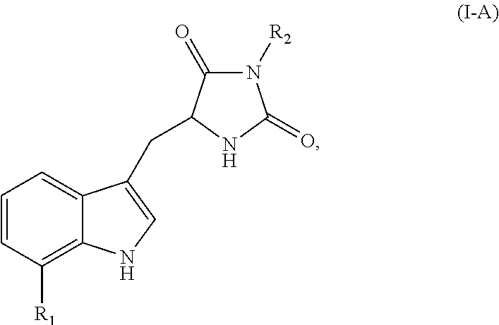
or a pharmaceutically acceptable salt, ester, or prodrug thereof, or optical isomers or racemic mixtures thereof, wherein R1 is H, alkyl, alkoxyl, or a halogen and R2 is H or an alkyl.

or a pharmaceutically acceptable salt, ester, or prodrug thereof.

or a pharmaceutically acceptable salt, ester, or prodrug thereof.

or a pharmaceutically acceptable salt, ester, or prodrug thereof.

or a pharmaceutically acceptable salt, ester, or prodrug thereof, wherein R1 is H, alkyl, alkoxyl, or a halogen (for example, F, Cl, Br or I) and R2 is H or an alkyl.

or a pharmaceutically acceptable salt, ester, or prodrug thereof.

or a pharmaceutically acceptable salt, ester, or prodrug thereof.


or a pharmaceutically acceptable salt thereof.

or

or


or


or a pharmaceutically acceptable salt, ester, or prodrug thereof, wherein:

or a pharmaceutically acceptable salt, ester, or prodrug thereof, wherein:


or a pharmaceutically acceptable salt, ester, or prodrug thereof, or optical isomers or racemic mixtures thereof, wherein R1 is H, alkyl, alkoxyl, or a halogen (for example, F, Cl, Br or I) and R2 is H or an alkyl. In certain embodiments, R1 is H or Cl. In certain other embodiments, R2 is a methyl or ethyl. In certain other embodiments, R1 is H or Cl, and R2 is a methyl.

or a pharmaceutically acceptable salt, ester, or prodrug thereof.

or a pharmaceutically acceptable salt, ester, or prodrug thereof.

or a pharmaceutically acceptable salt, ester, or prodrug thereof.

or a pharmaceutically acceptable salt, ester, or prodrug thereof, wherein R1 is H, alkyl, alkoxyl, or a halogen (for example, F, Cl, Br or I) and R2 is H or an alkyl. In certain embodiments, R1 is H or Cl. In certain other embodiments, R2 is a methyl or ethyl. In certain other embodiments, R1 is H or Cl, and R2 is a methyl.

or a pharmaceutically acceptable salt, ester, or prodrug thereof. In certain other embodiments, the necrostatin is a Nec-1 related compound of Formula I-G, shown below:

or a pharmaceutically acceptable salt, ester, or prodrug thereof.

or a pharmaceutically acceptable salt, ester, or prodrug thereof, wherein:

or a pharmaceutically acceptable salt thereof, wherein:

or a pharmaceutically acceptable salt thereof.

or a pharmaceutically acceptable salt, ester, or prodrug thereof, wherein:

or a pharmaceutically acceptable salt thereof.

or a pharmaceutically acceptable salt thereof.

or a pharmaceutically acceptable salt, ester, or prodrug thereof, wherein:


In certain other embodiments, R1 is

In certain embodiments, R2 is hydrogen. In certain embodiments, R3 is methyl. In certain other embodiments, R3 is hydrogen. In certain embodiments, R4 is halogen, such as fluorine or chlorine. In certain embodiments, R4 is halogen. In certain embodiments, R5 is methyl or ethyl. In certain embodiments, R6 is —CN. In certain embodiments, A is phenylene. In certain embodiments, X is N. In certain embodiments, Y is N. In certain embodiments, Z is S. In certain embodiments, A is phenylene. In certain embodiments, R1 is C1-C6alkyl, such as methyl. In certain embodiments, m is 1. In certain embodiments, n is 2.

or a pharmaceutically acceptable salt thereof.

or a pharmaceutically acceptable salt thereof.

or a pharmaceutically acceptable salt, ester, or prodrug thereof, wherein:
or
such as

In certain embodiments, R3 is —C1-C6alkylene-CN, such as —CH2—CN. In certain embodiments, R4 represents independently for each occurrence hydrogen or C1-C6alkyl. In certain embodiments, R5 represents independently for each occurrence C1-C6alkyl, halogen, hydroxyl, or C1-C6alkoxyl. In certain embodiments, B is a 5-6 membered heterocyclic ring. In certain embodiments, n is 0. In certain embodiments, p is 0.

or a pharmaceutically acceptable salt, ester, or prodrug thereof, wherein:
or

or a pharmaceutically acceptable salt thereof.

or a pharmaceutically acceptable salt, ester, or prodrug thereof, wherein:

or


In certain embodiments, R5 is halogen, C1-C6alkyl, hydroxyl, C1-C6alkoxyl, or —N(R7)2. In certain other embodiments, R5 is halogen, such as fluorine or chlorine. In certain embodiments, p is 0. In certain other embodiments, R4 is

such as
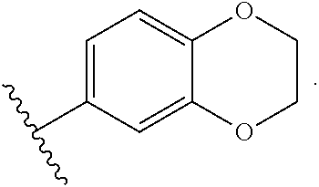
In certain embodiments, the Nec-7 related compound is

or a pharmaceutically acceptable salt thereof.

or a pharmaceutically acceptable salt, ester, or prodrug thereof, wherein:

or a pharmaceutically acceptable salt, ester, or prodrug thereof, wherein:


and pharmaceutically acceptable salts thereof.

or a pharmaceutically acceptable salt, ester, or prodrug thereof, wherein:
-
- R1 is selected from H, halogen, optionally substituted C1-6alkyl, or optionally substituted C1-6cycloalkyl, or optionally substituted aryl;

or a pharmaceutically acceptable salt, ester, or prodrug thereof, wherein:


and pharmaceutically acceptable salts thereof.


wherein R50 and R51 each independently represent hydrogen, alkyl, alkenyl, or —(CH2)m—R61; or R50 and R51, taken together with the N atom to which they are attached complete a heterocycle having from 4 to 8 atoms in the ring structure; wherein R61 is aryl, cycloalkyl, cycloalkenyl, a heterocycle or a polycycle; and m is zero or an integer in the range of 1 to 8. In certain embodiments, R50 and R51 each independently represent hydrogen or alkyl.
Claims (15)


Priority Applications (3)
| Application Number | Priority Date | Filing Date | Title |
|---|---|---|---|
| US14/934,810 US9492432B2 (en) | 2010-04-23 | 2015-11-06 | Methods and compositions for preserving photoreceptor and retinal pigment epithelial cells |
| US15/288,214 US10149884B2 (en) | 2010-04-23 | 2016-10-07 | Methods and compositions for preserving photoreceptor and retinal pigment epithelial cells |
| US16/171,837 US10639345B2 (en) | 2010-04-23 | 2018-10-26 | Compositions for preserving photoreceptor cells, retinal pigment epithelial cells, or visual function |
Applications Claiming Priority (9)
| Application Number | Priority Date | Filing Date | Title |
|---|---|---|---|
| US32747610P | 2010-04-23 | 2010-04-23 | |
| US40500310P | 2010-10-20 | 2010-10-20 | |
| US40905510P | 2010-11-01 | 2010-11-01 | |
| US41486210P | 2010-11-17 | 2010-11-17 | |
| US201161472144P | 2011-04-05 | 2011-04-05 | |
| US201161472153P | 2011-04-05 | 2011-04-05 | |
| PCT/US2011/033704 WO2011133964A2 (en) | 2010-04-23 | 2011-04-23 | Methods and compositions for preserving photoreceptor and retinal pigment epithelial cells |
| US201313642887A | 2013-02-14 | 2013-02-14 | |
| US14/934,810 US9492432B2 (en) | 2010-04-23 | 2015-11-06 | Methods and compositions for preserving photoreceptor and retinal pigment epithelial cells |
Related Parent Applications (2)
| Application Number | Title | Priority Date | Filing Date |
|---|---|---|---|
| PCT/US2011/033704 Continuation WO2011133964A2 (en) | 2010-04-23 | 2011-04-23 | Methods and compositions for preserving photoreceptor and retinal pigment epithelial cells |
| US13/642,887 Continuation US20130137642A1 (en) | 2010-04-23 | 2011-04-23 | Methods and compositions for preserving photoreceptor and retinal pigment epithelial cells |
Related Child Applications (1)
| Application Number | Title | Priority Date | Filing Date |
|---|---|---|---|
| US15/288,214 Continuation US10149884B2 (en) | 2010-04-23 | 2016-10-07 | Methods and compositions for preserving photoreceptor and retinal pigment epithelial cells |
Publications (2)
| Publication Number | Publication Date |
|---|---|
| US20160250189A1 US20160250189A1 (en) | 2016-09-01 |
| US9492432B2 true US9492432B2 (en) | 2016-11-15 |
Family
ID=44627940
Family Applications (4)
| Application Number | Title | Priority Date | Filing Date |
|---|---|---|---|
| US13/642,887 Abandoned US20130137642A1 (en) | 2010-04-23 | 2011-04-23 | Methods and compositions for preserving photoreceptor and retinal pigment epithelial cells |
| US14/934,810 Active US9492432B2 (en) | 2010-04-23 | 2015-11-06 | Methods and compositions for preserving photoreceptor and retinal pigment epithelial cells |
| US15/288,214 Active US10149884B2 (en) | 2010-04-23 | 2016-10-07 | Methods and compositions for preserving photoreceptor and retinal pigment epithelial cells |
| US16/171,837 Active US10639345B2 (en) | 2010-04-23 | 2018-10-26 | Compositions for preserving photoreceptor cells, retinal pigment epithelial cells, or visual function |
Family Applications Before (1)
| Application Number | Title | Priority Date | Filing Date |
|---|---|---|---|
| US13/642,887 Abandoned US20130137642A1 (en) | 2010-04-23 | 2011-04-23 | Methods and compositions for preserving photoreceptor and retinal pigment epithelial cells |
Family Applications After (2)
| Application Number | Title | Priority Date | Filing Date |
|---|---|---|---|
| US15/288,214 Active US10149884B2 (en) | 2010-04-23 | 2016-10-07 | Methods and compositions for preserving photoreceptor and retinal pigment epithelial cells |
| US16/171,837 Active US10639345B2 (en) | 2010-04-23 | 2018-10-26 | Compositions for preserving photoreceptor cells, retinal pigment epithelial cells, or visual function |
Country Status (4)
| Country | Link |
|---|---|
| US (4) | US20130137642A1 (en) |
| EP (1) | EP2560629B1 (en) |
| AU (3) | AU2011242465B2 (en) |
| WO (1) | WO2011133964A2 (en) |
Cited By (4)
| Publication number | Priority date | Publication date | Assignee | Title |
|---|---|---|---|---|
| US9993517B2 (en) | 2010-11-01 | 2018-06-12 | Massachusetts Eye And Ear Infirmary | Methods and compositions for preserving retinal ganglion cells |
| US10022419B2 (en) | 2011-10-21 | 2018-07-17 | Massachusetts Eye And Ear Infirmary | Methods for treating spinal cord injury |
| US10149884B2 (en) | 2010-04-23 | 2018-12-11 | Massachusetts Eye And Ear Infirmary | Methods and compositions for preserving photoreceptor and retinal pigment epithelial cells |
| US11786694B2 (en) | 2019-05-24 | 2023-10-17 | NeuroLight, Inc. | Device, method, and app for facilitating sleep |
Families Citing this family (16)
| Publication number | Priority date | Publication date | Assignee | Title |
|---|---|---|---|---|
| EP3366302B1 (en) * | 2011-07-18 | 2021-12-08 | University Of Kentucky Research Foundation | Protection of cells from alu-rna-induced degeneration and inhibitors for protecting cells |
| AU2012314514B2 (en) * | 2011-09-26 | 2018-03-15 | Qiagen Gmbh | Stabilisation and isolation of extracellular nucleic acids |
| WO2013077579A1 (en) * | 2011-11-25 | 2013-05-30 | 가톨릭대학교 산학협력단 | Pharmaceutical composition for preventing and treating ophthalmic disorders |
| EP2861243A4 (en) * | 2012-06-13 | 2016-04-27 | Univ Columbia | Intranasal delivery of cell permeant therapeutics for the treatment of edema |
| TWI637951B (en) | 2013-02-15 | 2018-10-11 | 英商葛蘭素史克智慧財產發展有限公司 | Heterocyclic amides as kinase inhibitors |
| EP2968276A4 (en) | 2013-03-15 | 2017-02-15 | President and Fellows of Harvard College | Hybrid necroptosis inhibitors |
| US10143703B2 (en) | 2014-01-02 | 2018-12-04 | Massachusetts Eye And Ear Infirmary | Treating ocular neovascularization |
| CN106573006A (en) * | 2014-08-21 | 2017-04-19 | 葛兰素史密斯克莱知识产权发展有限公司 | Heterocyclic amides as RIP1 kinase inhibitors as medicaments |
| SG11201704764PA (en) | 2014-12-11 | 2017-07-28 | Harvard College | Inhibitors of cellular necrosis and related methods |
| EP3395364B1 (en) * | 2015-12-24 | 2021-10-20 | The Doshisha | Caspase inhibitor-containing drug for treating or preventing disorders caused by tgf-beta , and applications thereof |
| EA201891620A1 (en) | 2016-02-05 | 2019-02-28 | Денали Терапьютикс Инк. | INHIBITORS OF PROTEINKINASE 1, INTERACTIVATING WITH RECEPTOR |
| WO2018013519A1 (en) * | 2016-07-11 | 2018-01-18 | The Trustees Of Columbia University In The City Of New York | Ocular delivery of cell permeant therapeutics for the treatment of retinal edema |
| HRP20220636T1 (en) | 2016-12-09 | 2022-07-22 | Denali Therapeutics Inc. | Compounds useful as ripk1 inhibitors |
| US20190374477A1 (en) * | 2018-06-11 | 2019-12-12 | Fondazione Istituto Italiano Di Tecnologia | Eye-injectable polymeric nanoparticles and method of use therefor |
| JP7340863B2 (en) * | 2018-06-29 | 2023-09-08 | 学校法人同志社 | Preparations containing emricasan |
| EP4281053A1 (en) * | 2021-01-22 | 2023-11-29 | Cornell University | Compositions and methods for the identification of compounds that protect against lipofuscin cytotoxicity |
Citations (26)
| Publication number | Priority date | Publication date | Assignee | Title |
|---|---|---|---|---|
| US5443505A (en) | 1993-11-15 | 1995-08-22 | Oculex Pharmaceuticals, Inc. | Biocompatible ocular implants |
| WO2000040089A1 (en) | 1999-01-05 | 2000-07-13 | Massachusetts Eye And Ear Infirmary | Targeted transscleral controlled release drug delivery to the retina and choroid |
| WO2001028474A1 (en) | 1999-10-21 | 2001-04-26 | Alcon Inc. | Ophthalmic drug delivery device |
| WO2001028493A2 (en) | 1999-10-15 | 2001-04-26 | President And Fellows Of Harvard College | Small molecule inhibitors of necrosis |
| WO2001039792A2 (en) | 1999-12-03 | 2001-06-07 | Alcon Universal Ltd. | The use of caspase 9 inhibitors to treat ocular neural pathology |
| US6245523B1 (en) | 1996-11-20 | 2001-06-12 | Yale University | Survivin, a protein that inhibits cellular apoptosis, and its modulation |
| US6251090B1 (en) | 1994-12-12 | 2001-06-26 | Robert Logan Avery | Intravitreal medicine delivery |
| US6299895B1 (en) | 1997-03-24 | 2001-10-09 | Neurotech S.A. | Device and method for treating ophthalmic diseases |
| US6375972B1 (en) | 2000-04-26 | 2002-04-23 | Control Delivery Systems, Inc. | Sustained release drug delivery devices, methods of use, and methods of manufacturing thereof |
| US6413540B1 (en) | 1999-10-21 | 2002-07-02 | Alcon Universal Ltd. | Drug delivery device |
| WO2002089767A1 (en) | 2001-05-03 | 2002-11-14 | Massachusetts Eye And Ear Infirmary | Implantable drug delivery device and use thereof |
| WO2003061519A2 (en) | 2002-01-18 | 2003-07-31 | Massachusetts Eye And Ear Infirmary | Methods and compositions for preserving the viability of photoreceptor cells |
| WO2005077344A2 (en) | 2003-08-29 | 2005-08-25 | The Brigham And Women's Hospital, Inc. | Hydantoin derivatives as inhibitors of cellular necrosis |
| US20070049565A1 (en) | 2005-08-24 | 2007-03-01 | Neurotech Pharmaceuticals Co., Ltd. | Combination of cell necrosis inhibitor and lithium for treating neuronal death or neurological dysfunction |
| WO2007071448A2 (en) | 2005-12-23 | 2007-06-28 | Partnership & Corp. Technology Transfer | Synthetic peptides for use as inhibitors of neurotransmitter secretion and as inducers of muscle relaxation |
| WO2007075772A2 (en) | 2005-12-20 | 2007-07-05 | President And Fellows Of Harvard College | Compounds, screens, and methods of treatment |
| US20070298129A1 (en) | 2005-08-24 | 2007-12-27 | Neurotech Pharmaceuticals Co., Ltd. | Compounds and compositions for treating neuronal death or neurological dysfunction |
| WO2008045406A2 (en) | 2006-10-10 | 2008-04-17 | President And Fellows Of Harvard College | Compounds, screens, and methods of treatment |
| WO2009023272A1 (en) | 2007-08-15 | 2009-02-19 | President And Fellows Of Harvard College | Heterocyclic inhibitors of necroptosis |
| US7622106B1 (en) | 2009-03-06 | 2009-11-24 | Board Of Regents, The University Of Texas System | Necrosis assay |
| WO2010075290A1 (en) | 2008-12-22 | 2010-07-01 | President And Fellows Of Harvard College | Unsaturated heterocyclic inhibitors of necroptosis |
| US20110071088A1 (en) | 2003-12-16 | 2011-03-24 | Childrens Medical Center Corporation | Method for treating neurological disorders |
| WO2011071088A1 (en) | 2009-12-09 | 2011-06-16 | 北海道公立大学法人 札幌医科大学 | Method for producing superoxide, method for evaluating superoxide scavenging ability, device for producing superoxide, and device for evaluating superoxide scavenging ability |
| WO2011133964A2 (en) | 2010-04-23 | 2011-10-27 | Massachusetts Eye And Ear Infirmary | Methods and compositions for preserving photoreceptor and retinal pigment epithelial cells |
| WO2012061045A2 (en) | 2010-11-01 | 2012-05-10 | Massachusetts Eye And Ear Infirmary | Methods and compositions for preserving retinal ganglion cells |
| WO2013059791A2 (en) | 2011-10-21 | 2013-04-25 | Massachusetts Eye And Ear Infirmary | Methods and compositions for promoting axon regeneration and nerve function |
Family Cites Families (3)
| Publication number | Priority date | Publication date | Assignee | Title |
|---|---|---|---|---|
| WO2002063959A1 (en) | 2001-02-09 | 2002-08-22 | The Schepens Eye Research Institute | Methods and compositions for stimulating axon regeneration and prventing neuronal cell degeneration |
| US20050130877A1 (en) | 2003-03-14 | 2005-06-16 | Children's Medical Center Corporation | Axon regeneration with PKC inhibitors |
| US20110190356A1 (en) | 2008-08-19 | 2011-08-04 | Knopp Neurosciences Inc. | Compositions and Methods of Using (R)- Pramipexole |
-
2011
- 2011-04-23 AU AU2011242465A patent/AU2011242465B2/en not_active Ceased
- 2011-04-23 EP EP11729791.1A patent/EP2560629B1/en active Active
- 2011-04-23 WO PCT/US2011/033704 patent/WO2011133964A2/en active Application Filing
- 2011-04-23 US US13/642,887 patent/US20130137642A1/en not_active Abandoned
-
2015
- 2015-11-06 US US14/934,810 patent/US9492432B2/en active Active
-
2016
- 2016-10-07 US US15/288,214 patent/US10149884B2/en active Active
-
2017
- 2017-04-19 AU AU2017202550A patent/AU2017202550B2/en not_active Ceased
-
2018
- 2018-10-05 AU AU2018241165A patent/AU2018241165A1/en not_active Abandoned
- 2018-10-26 US US16/171,837 patent/US10639345B2/en active Active
Patent Citations (32)
| Publication number | Priority date | Publication date | Assignee | Title |
|---|---|---|---|---|
| US5766242A (en) | 1993-11-15 | 1998-06-16 | Oculex Pharmaceuticals, Inc. | Biocompatible ocular implants |
| US5443505A (en) | 1993-11-15 | 1995-08-22 | Oculex Pharmaceuticals, Inc. | Biocompatible ocular implants |
| US6251090B1 (en) | 1994-12-12 | 2001-06-26 | Robert Logan Avery | Intravitreal medicine delivery |
| US6245523B1 (en) | 1996-11-20 | 2001-06-12 | Yale University | Survivin, a protein that inhibits cellular apoptosis, and its modulation |
| US6299895B1 (en) | 1997-03-24 | 2001-10-09 | Neurotech S.A. | Device and method for treating ophthalmic diseases |
| WO2000040089A1 (en) | 1999-01-05 | 2000-07-13 | Massachusetts Eye And Ear Infirmary | Targeted transscleral controlled release drug delivery to the retina and choroid |
| WO2001028493A2 (en) | 1999-10-15 | 2001-04-26 | President And Fellows Of Harvard College | Small molecule inhibitors of necrosis |
| WO2001028474A1 (en) | 1999-10-21 | 2001-04-26 | Alcon Inc. | Ophthalmic drug delivery device |
| US6413540B1 (en) | 1999-10-21 | 2002-07-02 | Alcon Universal Ltd. | Drug delivery device |
| US6416777B1 (en) | 1999-10-21 | 2002-07-09 | Alcon Universal Ltd. | Ophthalmic drug delivery device |
| WO2001039792A2 (en) | 1999-12-03 | 2001-06-07 | Alcon Universal Ltd. | The use of caspase 9 inhibitors to treat ocular neural pathology |
| US6375972B1 (en) | 2000-04-26 | 2002-04-23 | Control Delivery Systems, Inc. | Sustained release drug delivery devices, methods of use, and methods of manufacturing thereof |
| WO2002089767A1 (en) | 2001-05-03 | 2002-11-14 | Massachusetts Eye And Ear Infirmary | Implantable drug delivery device and use thereof |
| WO2003061519A2 (en) | 2002-01-18 | 2003-07-31 | Massachusetts Eye And Ear Infirmary | Methods and compositions for preserving the viability of photoreceptor cells |
| WO2005077344A2 (en) | 2003-08-29 | 2005-08-25 | The Brigham And Women's Hospital, Inc. | Hydantoin derivatives as inhibitors of cellular necrosis |
| US20110071088A1 (en) | 2003-12-16 | 2011-03-24 | Childrens Medical Center Corporation | Method for treating neurological disorders |
| US20070049565A1 (en) | 2005-08-24 | 2007-03-01 | Neurotech Pharmaceuticals Co., Ltd. | Combination of cell necrosis inhibitor and lithium for treating neuronal death or neurological dysfunction |
| US20070298129A1 (en) | 2005-08-24 | 2007-12-27 | Neurotech Pharmaceuticals Co., Ltd. | Compounds and compositions for treating neuronal death or neurological dysfunction |
| WO2007075772A2 (en) | 2005-12-20 | 2007-07-05 | President And Fellows Of Harvard College | Compounds, screens, and methods of treatment |
| WO2007071448A2 (en) | 2005-12-23 | 2007-06-28 | Partnership & Corp. Technology Transfer | Synthetic peptides for use as inhibitors of neurotransmitter secretion and as inducers of muscle relaxation |
| WO2008045406A2 (en) | 2006-10-10 | 2008-04-17 | President And Fellows Of Harvard College | Compounds, screens, and methods of treatment |
| WO2009023272A1 (en) | 2007-08-15 | 2009-02-19 | President And Fellows Of Harvard College | Heterocyclic inhibitors of necroptosis |
| US20090099242A1 (en) | 2007-08-15 | 2009-04-16 | Cuny Gregory D | Heterocyclic inhibitors of necroptosis |
| WO2010075290A1 (en) | 2008-12-22 | 2010-07-01 | President And Fellows Of Harvard College | Unsaturated heterocyclic inhibitors of necroptosis |
| US7622106B1 (en) | 2009-03-06 | 2009-11-24 | Board Of Regents, The University Of Texas System | Necrosis assay |
| WO2011071088A1 (en) | 2009-12-09 | 2011-06-16 | 北海道公立大学法人 札幌医科大学 | Method for producing superoxide, method for evaluating superoxide scavenging ability, device for producing superoxide, and device for evaluating superoxide scavenging ability |
| WO2011133964A2 (en) | 2010-04-23 | 2011-10-27 | Massachusetts Eye And Ear Infirmary | Methods and compositions for preserving photoreceptor and retinal pigment epithelial cells |
| US20130137642A1 (en) | 2010-04-23 | 2013-05-30 | Demetrios Vavvas | Methods and compositions for preserving photoreceptor and retinal pigment epithelial cells |
| WO2012061045A2 (en) | 2010-11-01 | 2012-05-10 | Massachusetts Eye And Ear Infirmary | Methods and compositions for preserving retinal ganglion cells |
| US20140024598A1 (en) | 2010-11-01 | 2014-01-23 | Demetrios Vavvas | Methods and compositions for preserving retinal ganglion cells |
| WO2013059791A2 (en) | 2011-10-21 | 2013-04-25 | Massachusetts Eye And Ear Infirmary | Methods and compositions for promoting axon regeneration and nerve function |
| US20140357570A1 (en) | 2011-10-21 | 2014-12-04 | Massachusetts Eye And Ear Infirmary | Compositions comprising necrosis inhibitors, such as necrostatins, alone or in combination, for promoting axon regeneration and nerve function, thereby treating cns disorders |
Non-Patent Citations (127)
Cited By (7)
| Publication number | Priority date | Publication date | Assignee | Title |
|---|---|---|---|---|
| US10149884B2 (en) | 2010-04-23 | 2018-12-11 | Massachusetts Eye And Ear Infirmary | Methods and compositions for preserving photoreceptor and retinal pigment epithelial cells |
| US10639345B2 (en) | 2010-04-23 | 2020-05-05 | Massachusetts Eye And Ear Infirmary | Compositions for preserving photoreceptor cells, retinal pigment epithelial cells, or visual function |
| US9993517B2 (en) | 2010-11-01 | 2018-06-12 | Massachusetts Eye And Ear Infirmary | Methods and compositions for preserving retinal ganglion cells |
| US10617735B2 (en) | 2010-11-01 | 2020-04-14 | Massachusetts Eye And Ear Infirmary | Methods and compositions for preserving retinal ganglion cells |
| US10022419B2 (en) | 2011-10-21 | 2018-07-17 | Massachusetts Eye And Ear Infirmary | Methods for treating spinal cord injury |
| US10799552B2 (en) | 2011-10-21 | 2020-10-13 | Massachusetts Eye And Ear Infirmary | Methods for treating diabetic neurotherapy |
| US11786694B2 (en) | 2019-05-24 | 2023-10-17 | NeuroLight, Inc. | Device, method, and app for facilitating sleep |
Also Published As
| Publication number | Publication date |
|---|---|
| EP2560629A2 (en) | 2013-02-27 |
| US20190216881A1 (en) | 2019-07-18 |
| US10149884B2 (en) | 2018-12-11 |
| AU2017202550A1 (en) | 2017-05-11 |
| AU2017202550B2 (en) | 2018-07-05 |
| US20170216393A1 (en) | 2017-08-03 |
| AU2011242465B2 (en) | 2017-01-19 |
| AU2018241165A1 (en) | 2018-11-01 |
| AU2011242465A1 (en) | 2012-12-13 |
| US10639345B2 (en) | 2020-05-05 |
| EP2560629B1 (en) | 2020-06-03 |
| US20160250189A1 (en) | 2016-09-01 |
| US20130137642A1 (en) | 2013-05-30 |
| WO2011133964A2 (en) | 2011-10-27 |
| WO2011133964A3 (en) | 2012-08-09 |
Similar Documents
| Publication | Publication Date | Title |
|---|---|---|
| US10639345B2 (en) | Compositions for preserving photoreceptor cells, retinal pigment epithelial cells, or visual function | |
| US10617735B2 (en) | Methods and compositions for preserving retinal ganglion cells | |
| US10799552B2 (en) | Methods for treating diabetic neurotherapy | |
| JP2018521120A (en) | Compositions and methods for treating pterygium | |
| US20140187495A1 (en) | Methods for treating glioblastoma | |
| KR20120022721A (en) | Prophylactic or therapeutic agent for retinal disease and method for prophylaxis or therapy of retinal disease using jnk (c-jun amino-terminal kinase)-inhibitory peptide, and use of the peptide | |
| JP2012519696A (en) | Method for inhibiting apoptosis of photoreceptor cells | |
| AU2003256370B2 (en) | Use of dantrolene, a ryanodine antagonist, in treating neural injury | |
| Park et al. | Effect of lovastatin on wound-healing modulation after glaucoma filtration surgery in a rabbit model | |
| EP2326328B1 (en) | Use of dihydralazine for the treatment of diseases related to elevated semicarbazide sensitive amine-oxidase (ssao) activity | |
| WO2020009248A1 (en) | Composition for inhibiting fibrosis in ocular tissue | |
| US20220062255A1 (en) | Treatment of neurodegenerative eye disease using pridopidine | |
| KR20180028890A (en) | Pharmaceutical Composition for Treating Macular Degeneration Containing mTOR Inhibitor | |
| US11717513B2 (en) | Mirabegron for the treatment of retinal diseases | |
| Waki et al. | Effect of topically applied iganidipine dihydrochloride, a novel calcium antagonist, on optic nerve head circulation in rabbits | |
| JP2011500580A (en) | Thrombopoietin receptor agonist (TpoRA) that kills acute human myeloid leukemia cells | |
| US20150284393A1 (en) | Methods for treating eye disorders | |
| US9872863B2 (en) | Targeting primary cilia to treat glaucoma | |
| TW202143994A (en) | Use of cyclosporine analogues for treating fibrosis | |
| WO2023111802A1 (en) | Methods of treatment using lou064 | |
| Cheruvu | Transscleral drug delivery: Role of physiological, physico-chemical and pathological factors in delivering drugs to the posterior segment of eye | |
| KR20170140383A (en) | Peptide compositions and methods of use |
Legal Events
| Date | Code | Title | Description |
|---|---|---|---|
| AS | Assignment |
Owner name: MASSACHUSETTS EYE & EAR INFIRMARY, MASSACHUSETTS Free format text: ASSIGNMENT OF ASSIGNORS INTEREST;ASSIGNORS:VAVVAS, DEMETRIOS;TRICHONAS, GEORGIOS;MILLER, JOAN;AND OTHERS;SIGNING DATES FROM 20110824 TO 20110915;REEL/FRAME:039043/0475 |
|
| STCF | Information on status: patent grant |
Free format text: PATENTED CASE |
|
| CC | Certificate of correction | ||
| MAFP | Maintenance fee payment |
Free format text: PAYMENT OF MAINTENANCE FEE, 4TH YR, SMALL ENTITY (ORIGINAL EVENT CODE: M2551); ENTITY STATUS OF PATENT OWNER: SMALL ENTITY Year of fee payment: 4 |
|
| AS | Assignment |
Owner name: NATIONAL INSTITUTES OF HEALTH (NIH), U.S. DEPT. OF HEALTH AND HUMAN SERVICES (DHHS), U.S. GOVERNMENT, MARYLAND Free format text: CONFIRMATORY LICENSE;ASSIGNOR:MASSACHUSETTS EYE AND EAR INFIRMARY;REEL/FRAME:060379/0082 Effective date: 20190722 |