US9733248B2 - Methods and materials for treating renal cell carcinoma - Google Patents
Methods and materials for treating renal cell carcinoma Download PDFInfo
- Publication number
- US9733248B2 US9733248B2 US14/994,980 US201614994980A US9733248B2 US 9733248 B2 US9733248 B2 US 9733248B2 US 201614994980 A US201614994980 A US 201614994980A US 9733248 B2 US9733248 B2 US 9733248B2
- Authority
- US
- United States
- Prior art keywords
- tumor
- cells
- renal cell
- cell carcinoma
- tissue
- Prior art date
- Legal status (The legal status is an assumption and is not a legal conclusion. Google has not performed a legal analysis and makes no representation as to the accuracy of the status listed.)
- Active
Links
Images


Classifications
-
- G—PHYSICS
- G01—MEASURING; TESTING
- G01N—INVESTIGATING OR ANALYSING MATERIALS BY DETERMINING THEIR CHEMICAL OR PHYSICAL PROPERTIES
- G01N33/00—Investigating or analysing materials by specific methods not covered by groups G01N1/00 - G01N31/00
- G01N33/48—Biological material, e.g. blood, urine; Haemocytometers
- G01N33/50—Chemical analysis of biological material, e.g. blood, urine; Testing involving biospecific ligand binding methods; Immunological testing
- G01N33/53—Immunoassay; Biospecific binding assay; Materials therefor
- G01N33/574—Immunoassay; Biospecific binding assay; Materials therefor for cancer
- G01N33/57407—Specifically defined cancers
-
- A—HUMAN NECESSITIES
- A61—MEDICAL OR VETERINARY SCIENCE; HYGIENE
- A61N—ELECTROTHERAPY; MAGNETOTHERAPY; RADIATION THERAPY; ULTRASOUND THERAPY
- A61N5/00—Radiation therapy
- A61N5/10—X-ray therapy; Gamma-ray therapy; Particle-irradiation therapy
-
- A—HUMAN NECESSITIES
- A61—MEDICAL OR VETERINARY SCIENCE; HYGIENE
- A61P—SPECIFIC THERAPEUTIC ACTIVITY OF CHEMICAL COMPOUNDS OR MEDICINAL PREPARATIONS
- A61P35/00—Antineoplastic agents
-
- C—CHEMISTRY; METALLURGY
- C07—ORGANIC CHEMISTRY
- C07K—PEPTIDES
- C07K16/00—Immunoglobulins [IGs], e.g. monoclonal or polyclonal antibodies
- C07K16/18—Immunoglobulins [IGs], e.g. monoclonal or polyclonal antibodies against material from animals or humans
- C07K16/22—Immunoglobulins [IGs], e.g. monoclonal or polyclonal antibodies against material from animals or humans against growth factors ; against growth regulators
-
- C—CHEMISTRY; METALLURGY
- C07—ORGANIC CHEMISTRY
- C07K—PEPTIDES
- C07K16/00—Immunoglobulins [IGs], e.g. monoclonal or polyclonal antibodies
- C07K16/18—Immunoglobulins [IGs], e.g. monoclonal or polyclonal antibodies against material from animals or humans
- C07K16/28—Immunoglobulins [IGs], e.g. monoclonal or polyclonal antibodies against material from animals or humans against receptors, cell surface antigens or cell surface determinants
- C07K16/2896—Immunoglobulins [IGs], e.g. monoclonal or polyclonal antibodies against material from animals or humans against receptors, cell surface antigens or cell surface determinants against molecules with a "CD"-designation, not provided for elsewhere
-
- G—PHYSICS
- G01—MEASURING; TESTING
- G01N—INVESTIGATING OR ANALYSING MATERIALS BY DETERMINING THEIR CHEMICAL OR PHYSICAL PROPERTIES
- G01N33/00—Investigating or analysing materials by specific methods not covered by groups G01N1/00 - G01N31/00
- G01N33/48—Biological material, e.g. blood, urine; Haemocytometers
- G01N33/50—Chemical analysis of biological material, e.g. blood, urine; Testing involving biospecific ligand binding methods; Immunological testing
- G01N33/53—Immunoassay; Biospecific binding assay; Materials therefor
- G01N33/574—Immunoassay; Biospecific binding assay; Materials therefor for cancer
- G01N33/57407—Specifically defined cancers
- G01N33/57438—Specifically defined cancers of liver, pancreas or kidney
-
- A—HUMAN NECESSITIES
- A61—MEDICAL OR VETERINARY SCIENCE; HYGIENE
- A61K—PREPARATIONS FOR MEDICAL, DENTAL OR TOILETRY PURPOSES
- A61K39/00—Medicinal preparations containing antigens or antibodies
- A61K2039/505—Medicinal preparations containing antigens or antibodies comprising antibodies
-
- G—PHYSICS
- G01—MEASURING; TESTING
- G01N—INVESTIGATING OR ANALYSING MATERIALS BY DETERMINING THEIR CHEMICAL OR PHYSICAL PROPERTIES
- G01N2333/00—Assays involving biological materials from specific organisms or of a specific nature
- G01N2333/435—Assays involving biological materials from specific organisms or of a specific nature from animals; from humans
- G01N2333/705—Assays involving receptors, cell surface antigens or cell surface determinants
- G01N2333/70503—Immunoglobulin superfamily, e.g. VCAMs, PECAM, LFA-3
-
- G—PHYSICS
- G01—MEASURING; TESTING
- G01N—INVESTIGATING OR ANALYSING MATERIALS BY DETERMINING THEIR CHEMICAL OR PHYSICAL PROPERTIES
- G01N2333/00—Assays involving biological materials from specific organisms or of a specific nature
- G01N2333/435—Assays involving biological materials from specific organisms or of a specific nature from animals; from humans
- G01N2333/705—Assays involving receptors, cell surface antigens or cell surface determinants
- G01N2333/70596—Molecules with a "CD"-designation not provided for elsewhere in G01N2333/705
-
- G—PHYSICS
- G01—MEASURING; TESTING
- G01N—INVESTIGATING OR ANALYSING MATERIALS BY DETERMINING THEIR CHEMICAL OR PHYSICAL PROPERTIES
- G01N2800/00—Detection or diagnosis of diseases
- G01N2800/52—Predicting or monitoring the response to treatment, e.g. for selection of therapy based on assay results in personalised medicine; Prognosis
Definitions
- This document relates to methods and materials involved in treating renal cell carcinoma.
- this document provides methods and materials for assessing a renal cell carcinoma patient for tumor or peritumoral tissue containing CD14 + cells and proceeding with a renal cell carcinoma treatment option based on the presence, absence, or level of CD14 + cells present within the tumor or peritumoral tissue.
- renal cell carcinoma The incidence and deaths caused by renal cell carcinoma are increasing in the United States. Of particular note, incidence and mortality rates for renal cell carcinoma have risen among both genders, and these trends are not explained by the increased use of abdominal imaging (Chow et al., JAMA, 281:1628-31 (1999)). Indeed, mortality from renal cell carcinoma has increased over 30% since 1950.
- One treatment for renal cell carcinoma is surgical resection. The majority of patients with renal cell carcinoma confined to the kidney can be cured by surgery; however, about 30 percent of patients can develop metastases and require additional treatments.
- Renal cell carcinoma encompasses a group of at least five subtypes with unique morphologic, genetic, and behavioral characteristics (Cheville et al., Am. J. Surg. Pathol., 27:612-24 (2003)). Cancer-specific survival is dependent on subtype, and over 80 percent of renal cell carcinomas and the vast majority of renal cell carcinoma-related deaths are due to clear cell renal cell carcinoma. To date, tumor stage and grade are the primary prognostic indicators for patients with clear cell renal cell carcinoma treated by nephrectomy (Gettman et al., Cancer, 91:354-61 (2001)). There is, however, variability in patient outcome that cannot be explained by the combination of stage and grade.
- This document relates to methods and materials involved in treating renal cell carcinoma.
- this document provides methods and materials for assessing a renal cell carcinoma patient for tumor or peritumoral tissue containing CD14 + cells and proceeding with a renal cell carcinoma treatment option based on the presence, absence, or level of CD14 + cells present within the tumor or peritumoral tissue.
- the presence of a moderate to marked level of CD14 + cells within tumor or peritumoral tissue of a renal cell carcinoma patient can indicate that it is unlikely that the renal cell carcinoma patient will respond positively to a standard renal cell carcinoma treatment that is appropriate for renal cell carcinoma patients lacking a moderate to marked level of CD14 + cells within their tumor and peritumoral tissue.
- a standard renal cell carcinoma treatment that is appropriate for renal cell carcinoma patients lacking a moderate to marked level of CD14 + cells within their tumor and peritumoral tissue can be referred to as a type A treatment.
- a renal cell carcinoma treatment that is appropriate for renal cell carcinoma patients having a moderate to marked level of CD14 + cells within their tumor and peritumoral tissue can be referred to as a type B treatment.
- the absence of a moderate to marked level of CD14 + cells within tumor or peritumoral tissue of a renal cell carcinoma patient can indicate that it is likely that the renal cell carcinoma patient will respond to a type A treatment and/or that it is not necessary to proceed with a type B treatment.
- the methods and materials provided herein can allow clinicians to determine proper treatment options for cancer patients (e.g., renal cell carcinoma patients).
- cancer patients e.g., renal cell carcinoma patients
- the methods and materials provided herein can be used to identify renal cell carcinoma patients who would benefit from a type A treatment or a type B treatment.
- the methods and materials provided herein can be used to identify renal cell carcinoma patients who would benefit from switching from a type A treatment to a type B treatment. Identifying cancer patients who would benefit from a particular treatment option can avoid unnecessary treatment attempts and unnecessary patient suffering.
- one aspect of this document features a method for treating a human having renal cell carcinoma.
- the method comprises, or consists essentially of, (a) determining whether or not tumor or peritumoral tissue from the human contains greater than 30 percent of CD14 + cells, and (b) proceeding with a type B treatment if the tissue contains greater than 30 percent of CD14 + cells, and proceeding with a type A or type B treatment if the tissue does not contain greater than 30 percent of CD14 + cells.
- the renal cell carcinoma can be clear cell renal cell carcinoma.
- the step (a) can comprise determining whether or not the tumor tissue contains greater than 30 percent of CD14 + cells.
- the step (a) can comprise determining whether or not the peritumoral tissue contains greater than 30 percent of CD14 + cells.
- the step (a) can comprise determining whether or not the tumor or peritumoral tissue contains greater than 35 percent of CD14 + cells.
- the step (a) can comprise determining whether or not the tumor or peritumoral tissue contains greater than 40 percent of CD14 + cells.
- the step (a) can comprise determining whether or not the tumor or peritumoral tissue contains greater than 30 percent of CD14 + cells in a diffuse staining pattern.
- the tumor or peritumoral tissue from the human can contain greater than 30 percent of CD14 + cells.
- the method can comprise proceeding with a type B treatment selected from the group consisting of chemotherapies, biologic therapies, and cell therapies.
- the method can comprise proceeding with a type B treatment selected from the group consisting of treatment with Sunitinib, treatment with Sorafenib, treatment with Bevacizumab, treatment with Temsirolimus, and treatment with Everolimus.
- the method can comprise initiating a type B treatment for the human after the step (a).
- the tumor or peritumoral tissue from the human can lack greater than 30 percent of CD14 + cells.
- the method can comprise performing a surgical treatment after the step (a).
- this document features a method for treating a human having renal cell carcinoma.
- the method comprises, or consists essentially of, (a) determining whether or not tumor or peritumoral tissue from the human contains greater than 10 percent of CD14 + cells, and (b) proceeding with a type B treatment if the tissue contains greater than 10 percent of CD14 + cells, and proceeding with a type A or type B treatment if the tissue does not contain greater than 10 percent of CD14 + cells.
- the renal cell carcinoma can be clear cell renal cell carcinoma.
- the step (a) can comprise determining whether or not the tumor tissue contains greater than 10 percent of CD14 + cells.
- the step (a) can comprise determining whether or not the peritumoral tissue contains greater than 10 percent of CD14 + cells.
- the step (a) can comprise determining whether or not the tumor or peritumoral tissue contains greater than 15 percent of CD14 + cells.
- the step (a) can comprise determining whether or not the tumor or peritumoral tissue contains greater than 20 percent of CD14 + cells.
- the step (a) can comprise determining whether or not the tumor or peritumoral tissue contains greater than 10 percent of CD14 + cells in a diffuse staining pattern.
- the tumor or peritumoral tissue from the human can contain greater than 10 percent of CD14 + cells.
- the method can comprise proceeding with a type B treatment selected from the group consisting of chemotherapies, biologic therapies, and cell therapies.
- the method can comprise proceeding with a type B treatment selected from the group consisting of treatment with Sunitinib, treatment with Sorafenib, treatment with Bevacizumab, treatment with Temsirolimus, and treatment with Everolimus.
- the method can comprise initiating a type B treatment for the human after the step (a).
- the tumor or peritumoral tissue from the human can lack greater than 10 percent of CD14 + cells.
- the method can comprise performing a surgical treatment after the step (a).
- this document features a method for treating a human having glioblastoma multiforme.
- the method comprises, or consists essentially of, (a) determining whether or not tumor or peritumoral tissue from the human contains greater than 10 percent of CD14 + cells, and (b) proceeding with a conventional glioblastoma multiforme therapy if the tissue does not contain greater than 10 percent of CD14 + cells, and proceeding with a therapy more aggressive than the conventional glioblastoma multiforme therapy if the tissue contains greater than 10 percent of CD14 + cells.
- the conventional glioblastoma multiforme therapy can include temozolomide chemotherapy.
- the conventional glioblastoma multiforme therapy can include temozolomide chemotherapy after surgery.
- the conventional glioblastoma multiforme therapy can include temozolomide chemotherapy after radiotherapy.
- the conventional glioblastoma multiforme therapy can include temozolomide chemotherapy after surgery and radiotherapy.
- the therapy more aggressive than the conventional glioblastoma multiforme therapy can be an experimental therapy.
- this document features a method for identifying a potential treatment agent for reducing cancer aggressiveness.
- the method comprises, or consists essentially of, (a) administering a test agent to a mammal having cancer, wherein tumor or peritumoral tissue of the cancer contains a detectable level of CD14 + cells, and (b) determining whether or not the level of CD14 + cells present in the tumor or peritumoral tissue of the cancer is reduced as compared to the level of CD14 + cells present in the tumor or peritumoral tissue of the cancer prior to the administering step (a), wherein a reduction in the level of CD14 + cells present in the tumor or peritumoral tissue of the cancer indicates that the test agent is the potential treatment agent for reducing cancer aggressiveness.
- the mammal can be a human.
- the test agent can be a small molecule compound.
- the cancer can be a renal cell carcinoma.
- the cancer can be a clear cell renal cell carcinoma.
- the step (b) can comprise determining whether or not the level of CD14+ cells present in a tumor tissue biopsy of the cancer obtained from the mammal after the administering step (a) is reduced.
- the step (b) can comprise determining whether or not the level of CD14+ cells present in a peritumoral tissue biopsy of the cancer obtained from the mammal after the administering step (a) is reduced.
- this document features a method for treating a human having a solid tumor.
- the method comprises, or consists essentially of, (a) determining whether or not tumor or peritumoral tissue from the human contains greater than 30 percent of CD14 + cells, and (b) proceeding with a conventional ovarian cancer therapy if the tissue does not contain greater than 10 percent of CD14 + cells, and proceeding with a therapy more aggressive than the conventional ovarian cancer therapy if the tissue contains greater than 10 percent of CD14 + cells.
- the solid tumor can be a renal cell carcinoma.
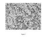
- FIG. 1 is a photograph of an example of renal cell tumor tissue stained for CD14 expression demonstrating low or absent CD14 staining.
- Renal cell carcinoma tissue was obtained from a renal cell carcinoma patient having a low SSIGN score (e.g., SSIGN score of 2 or less) and stained with an anti-CD14 antibody.
- SSIGN score e.g., SSIGN score of 2 or less
- FIG. 2 is a photograph an example of renal cell carcinoma tissue stained for CD14 expression demonstrating high CD14 staining. Renal cell carcinoma tissue was obtained from a renal cell carcinoma patient having a high SSIGN score (e.g., SSIGN score of 7 or more) and stained with an anti-CD14 antibody.
- SSIGN score e.g., SSIGN score of 7 or more
- FIG. 3 Cancer-specific survival following surgery by maximal intratumoral and peritumoral CD14 expression for 375 ccRCC patients.
- FIG. 4 is a graph plotting the HLA-DR mean fluorescence intensity (MFI) of peripheral blood cells that are CD14 + and obtained from patients having tumor tissue with a high intensity of CD14 + staining (high), a medium intensity of CD14 + staining (medium), or a low/no intensity of CD14 + staining (low/none).
- MFI mean fluorescence intensity
- This document provides methods and materials related to treating solid tumors (e.g., renal cell carcinomas and ovarian cancers). For example, this document provides methods and materials for assessing a renal cell carcinoma patient for tumor or peritumoral tissue containing CD14 + cells and proceeding with a renal cell carcinoma treatment option based on the presence, absence, or level of CD14 + cells present within the tumor or peritumoral tissue.
- solid tumors e.g., renal cell carcinomas and ovarian cancers.
- a moderate to marked level of CD14 + cells within tumor or peritumoral tissue of a renal cell carcinoma patient can indicate that it is unlikely that the renal cell carcinoma patient will respond positively to a type A treatment and that the renal cell carcinoma patient should proceed with a type B treatment.
- a moderate level of CD14 + cells within tumor tissue or peritumoral tissue is a level where between about 10 and about 30 percent of the cells are CD14 + cells
- a marked level of CD14 + cells within tumor tissue or peritumoral tissue is a level where greater than about 30 percent of the cells are CD14 + cells.
- a tumor biopsy sample is obtained from a cancer patient using standard biopsy techniques.
- the tumor biopsy sample can include tumor tissue, peritumoral tissue, or both tumor tissue and peritumoral tissue. Once obtained, the sample can be treated such that the level of CD14 + cells present can be determined. Standard cell staining and immunoflourescence techniques can be used to determine the presence or absence of a moderate level or marked level of CD14 + cells within tissue. For example, a standard microscope can be used in connection with immunohistochemical staining techniques to determine the presence or absence of a moderate level or marked level of CD14 + cells within tissue ( FIGS. 1 and 2 ).
- CD14 expression within tumor tissue, peritumoral tissue, or both tumor tissue and peritumoral tissue can be assessed using a nucleic acid-based assay (e.g., reverse transcriptase-PCR or nucleic acid microarray assays).
- a nucleic acid-based assay e.g., reverse transcriptase-PCR or nucleic acid microarray assays.
- CD14 expression levels within tumor tissue can be determined using a microarray having the ability to measure the amount of CD14 mRNA.
- the presence of at least a moderate level of CD14 + cells within tumor or peritumoral tissue of a renal cell carcinoma patient can indicate that it is likely that the renal cell carcinoma patient will relapse quickly and should proceed with an experimental or non-standard treatment.
- presence of at least a moderate level of CD14 + cells within tumor or peritumoral tissue of a renal cell carcinoma patient can indicate that it is unlikely that the renal cell carcinoma patient will respond positively to a type A treatment and that the renal cell carcinoma patient should proceed with a type B treatment.
- type A treatments can include, without limitation, surgery alone such as a partial nephrectomy, a simple nephrectomy, or a radical nephrectomy.
- type B treatments can include, without limitation, internal or external radiation therapies (e.g., x-ray or radioisotope therapy), chemotherapies, biologic therapies (e.g., immuno, antibody, or high dose IL-2 therapy), cell therapies (e.g., stem cell or vaccine therapy), treatment with Sunitinib (SU11248; an oral, small-molecule, multi-targeted receptor tyrosine kinase inhibitor), treatment with Sorafenib (BAY 43-9006, a small molecular inhibitor of several tyrosine protein kinases), treatment with Bevacizumab (a humanized monoclonal antibody that recognizes and blocks vascular endothelial growth factor), treatment with a rapamycin inhibitor (e.g., Temsirolimus (CCI-779) or Everolimus (RAD001)), and combinations thereof.
- internal or external radiation therapies e.g., x-ray or radioisotope therapy
- biologic therapies e.g., immuno
- a type B treatment can be surgery (e.g., a partial nephrectomy, a simple nephrectomy, or a radical nephrectomy) in combination with one or more of the following therapies: an internal or external radiation therapy (e.g., x-ray or radioisotope therapy), chemotherapy, biologic therapy (e.g., immuno, antibody, or high dose IL-2 therapy), cell therapy (e.g., stem cell or vaccine therapy), treatment with Sunitinib, treatment with Sorafenib, treatment with Bevacizumab, and treatment with a rapamycin inhibitor (e.g., Temsirolimus (CCI-779) or Everolimus (RAD001)).
- an internal or external radiation therapy e.g., x-ray or radioisotope therapy
- biologic therapy e.g., immuno, antibody, or high dose IL-2 therapy
- cell therapy e.g., stem cell or vaccine therapy
- Sunitinib treatment with
- the absence of at least a moderate level of CD14 + cells within tumor or peritumoral tissue of a renal cell carcinoma patient can indicate that the patient should be observed but will likely experience a long period of tumor remission.
- the methods and materials provided herein can be used to treat glioblastoma multiforme (GBM) cancer.
- GBM glioblastoma multiforme
- the methods and materials provided herein can be used to assess a GBM patient for tumor or peritumoral tissue containing CD14 + cells. Once assessed, the patient can be instructed to proceed with a GBM treatment option based on the presence, absence, or level of CD14 + cells present within the GBM tumor or GBM peritumoral tissue.
- the presence of a moderate to marked level of CD14 + cells within tumor or peritumoral tissue of a GBM patient can indicate that it is unlikely that the GBM patient will respond positively to conventional (standard of care) therapy (e.g., temozolomide chemotherapy after surgery and radiotherapy) and should be included in an experimental or alternative therapy (e.g., immune based therapies including vaccine or antibody therapy, anti-angiogenesis therapies, cell based therapies, or experimental chemotherapies).
- conventional therapy e.g., temozolomide chemotherapy after surgery and radiotherapy
- an experimental or alternative therapy e.g., immune based therapies including vaccine or antibody therapy, anti-angiogenesis therapies, cell based therapies, or experimental chemotherapies.
- the methods and materials provided herein can be used to treat ovarian cancer.
- the methods and materials provided herein can be used to assess an ovarian cancer patient for tumor or peritumoral tissue containing CD14 + cells. Once assessed, the patient can be instructed to proceed with an ovarian cancer treatment option based on the presence, absence, or level of CD14 + cells present within the ovarian tumor or ovarian peritumoral tissue. The presence of a moderate to marked level of CD14 + cells within tumor or peritumoral tissue of an ovarian patient can indicate that it is unlikely that the ovarian patient will respond positively to conventional (standard of care) therapy and should be included in an experimental or alternative therapy.
- This document also provides methods and materials for identifying treatment agents that are capable of reducing cancer aggressiveness (e.g., RCC aggressiveness).
- a potential treatment agent e.g., small molecule compound
- a tumor or peritumoral tissue biopsy can be obtained and assessed for a moderate to marked level of CD14 + cells.
- a reduction in the level of CD14 + cells within the tumor or peritumoral tissue as compared to the level determined prior to receiving the potential treatment agent can indicate that the potential treatment agent is an effective agent for reducing cancer aggressiveness.
- ccRCC tumors with high Stage, Size, Grade, and Necrosis (SSIGN) scores were found to have significantly higher levels of CD14 expression compared with tumors with low SSIGN scores (Table 1).
- SSIGN Stage, Size, Grade, and Necrosis
- CD14 expression was evaluated in 50 clear cell renal cell carcinoma (ccRCC) cases. The samples were randomly selected from a ccRCC cohort and consisted of 25 cases with low (0-2) and 25 cases with high (7+) SSIGN scores (Frank et al., J. Urology, 168:2395 (2002)). Intratumoral and peritumoral CD14 expression was quantitated as absent, focal, moderate, or marked.
- the SSIGN score is a composite scoring system developed specifically for patients with ccRCC based on primary tumor classification, regional lymph node involvement, distant metastases, tumor size, nuclear grade, and coagulative tumor necrosis. To obtain these features, a pathologist reviewed the microscope slides from all specimens without knowledge of patient outcome.
- a formalin-fixed, paraffin-embedded block with representative tumor tissue was selected for each patient in the cohort. Staining for CD14 was conducted as follows. Tissue sections were deparaffinized in xylene and rehydrated in a graded series of ethanols. Antigen-retrieval was performed by heating tissue sections in Target Retrieval solution pH 6 (Dako #S1699) to 121° C. using a Digital Decloaking Chamber (Biocare Medical, Walnut Creek, Calif.). Sections were cooled to 90° C. and incubated for an additional five minutes before the Decloaking Chamber was opened.
- Target Retrieval solution pH 6 Dako #S1699
- Sections were washed in running DH 2 O for five minutes and incubated for five minutes in Wash Buffer (Dako #S3006) before being placed on the Autostainer Plus (Dako) for the following protocol. Sections were blocked for endogenous peroxidase for five minutes using Endogenous Blocking solution (Dako #S2001), washed twice in wash buffer, incubated five minutes in Background Reducer Sniper (Biocare Medical #BS966L), washed twice in wash buffer, and incubated for 60 minutes in purified rabbit anti-human CD14 antibody (Sigma PA #HPA001882) diluted 1:300 with DaVinci Green antibody diluent (Biocare Medical #PD900M).
- Sections were washed twice in wash buffer, incubated 15 minutes in the rabbit probe from the Mach 3 Rabbit HRP-Polymer Kit (Biocare Medical #M3R531L), washed twice in wash buffer, and incubated 15 minutes with the rabbit polymer from the Rabbit HRP-Polymer Kit. Sections were washed with wash buffer and visualized by incubating Betazoid DAB (Biocare Medical #BDB2004L) for eight minutes. Sections were washed with DH 2 O, counterstained with hematoxylin, dehydrated in ethanol, and cleared in xylene. Coverslips were mounted in permanent mounting media. All stained slides were reviewed and quantitated by a pathologist without knowledge of patient outcome. Intratumoral and (if possible) peritumoral CD14 expression was assessed as absent, focal, moderate, or marked.
- Associations of CD14 expression with clinical and pathologic features were evaluated using chi-square tests. Associations of CD14 expression with cancer-specific survival were illustrated using Kaplan-Meier curves. The magnitudes of the associations of CD14 expression with death from RCC were evaluated using Cox proportional hazards regression models and summarized with hazard ratios (HRs) and 95% confidence intervals (CI). The duration of follow-up was calculated from the date of nephrectomy to the date of death or last follow-up. Patients who died from causes other than RCC were censored at the date of death, while patients who were still alive at the time of analysis were censored at the date of last follow-up. Statistical analyses were performed using the SAS software package (SAS Institute; Cary, N.C.). All tests were two-sided, and p-values ⁇ 0.05 were considered statistically significant.
- Intratumoral, peritumoral, and maximal CD14 expression between the two are summarized in Table 3.
- a comparison of intratumoral and peritumoral expression is provided in Table 4.
- Associations of maximal CD14 expression with clinical and pathologic features are summarized in Table 5. Since only six tumors did not demonstrate intratumoral or peritumoral CD14 expression, these tumors were combined with those with focal expression for analysis.
- CD14 expression, particularly marked CD14 expression was significantly associated with symptoms at presentation, larger tumors of later stage and higher grade, coagulative tumor necrosis, sarcomatoid differentiation, and higher SSIGN scores. For example, median SSIGN scores for tumors with absent/focal, moderate, and marked CD14 expression were 1, 5, and 7, respectively.
- the HRs for the associations of moderate and marked CD14 expression with death from RCC were 2.87 (p ⁇ 0.001) and 3.03 (p ⁇ 0.001), respectively.
- a newly diagnosed RCC patient who is currently not being treated and who has a high level of CD14 + cells within tumor or peritumoral tissue is treated with standard of care (e.g., surgery), but is subjected to increased surveillance as this patient is highly likely to relapse and/or experience metastasis.
- a newly diagnosed RCC patient who is currently not being treated and who has no CD14 + cells within tumor or peritumoral tissue is treated by surgery if indicated and observation.
- An RCC patient who is currently being treated with a standard cancer treatment and who has a high level of CD14 + cells within tumor or peritumoral tissue is included in a clinical trial or other aggressive therapy without waiting for relapse.
- An RCC patient who is currently being treated with a standard cancer treatment and who has no CD14 + cells within tumor or peritumoral tissue is under a routine schedule of observation without additional treatment.
- MFI mean fluorescence intensity
Abstract
This document provides methods and materials related to treating renal cell carcinoma. For example, methods and materials for assessing a cancer patient (e.g., a renal cell carcinoma patient) for tumor or peritumoral tissue containing CD14+ cells and proceeding with a cancer treatment option (e.g., a renal cell carcinoma treatment option) based on the presence, absence, or level of CD14+ cells present within the tumor or peritumoral tissue are provided.
Description
This application is a continuation of U.S. application Ser. No. 13/509,089, filed May 10, 2012, which is a National Stage application under 35 U.S.C. §371 of International Application No. PCT/US2010/055856, having an International filing date of Nov. 8, 2010, which claims the benefit of U.S. Provisional Application Ser. No. 61/259,879, filed Nov. 10, 2009. The disclosures of the prior applications are considered part of (and are incorporated by reference in) the disclosure of this application.
1. Technical Field
This document relates to methods and materials involved in treating renal cell carcinoma. For example, this document provides methods and materials for assessing a renal cell carcinoma patient for tumor or peritumoral tissue containing CD14+ cells and proceeding with a renal cell carcinoma treatment option based on the presence, absence, or level of CD14+ cells present within the tumor or peritumoral tissue.
2. Background Information
The incidence and deaths caused by renal cell carcinoma are increasing in the United States. Of particular note, incidence and mortality rates for renal cell carcinoma have risen among both genders, and these trends are not explained by the increased use of abdominal imaging (Chow et al., JAMA, 281:1628-31 (1999)). Indeed, mortality from renal cell carcinoma has increased over 30% since 1950. One treatment for renal cell carcinoma is surgical resection. The majority of patients with renal cell carcinoma confined to the kidney can be cured by surgery; however, about 30 percent of patients can develop metastases and require additional treatments.
Renal cell carcinoma encompasses a group of at least five subtypes with unique morphologic, genetic, and behavioral characteristics (Cheville et al., Am. J. Surg. Pathol., 27:612-24 (2003)). Cancer-specific survival is dependent on subtype, and over 80 percent of renal cell carcinomas and the vast majority of renal cell carcinoma-related deaths are due to clear cell renal cell carcinoma. To date, tumor stage and grade are the primary prognostic indicators for patients with clear cell renal cell carcinoma treated by nephrectomy (Gettman et al., Cancer, 91:354-61 (2001)). There is, however, variability in patient outcome that cannot be explained by the combination of stage and grade.
This document relates to methods and materials involved in treating renal cell carcinoma. For example, this document provides methods and materials for assessing a renal cell carcinoma patient for tumor or peritumoral tissue containing CD14+ cells and proceeding with a renal cell carcinoma treatment option based on the presence, absence, or level of CD14+ cells present within the tumor or peritumoral tissue.
As described herein, the presence of a moderate to marked level of CD14+ cells within tumor or peritumoral tissue of a renal cell carcinoma patient can indicate that it is unlikely that the renal cell carcinoma patient will respond positively to a standard renal cell carcinoma treatment that is appropriate for renal cell carcinoma patients lacking a moderate to marked level of CD14+ cells within their tumor and peritumoral tissue. A standard renal cell carcinoma treatment that is appropriate for renal cell carcinoma patients lacking a moderate to marked level of CD14+ cells within their tumor and peritumoral tissue can be referred to as a type A treatment. The presence of a moderate to marked level of CD14+ cells within tumor or peritumoral tissue of a renal cell carcinoma patient also can indicate that it is likely that the renal cell carcinoma patient will respond positively to a renal cell carcinoma treatment that is appropriate for renal cell carcinoma patients having a moderate to marked level of CD14+ cells within their tumor and peritumoral tissue. A renal cell carcinoma treatment that is appropriate for renal cell carcinoma patients having a moderate to marked level of CD14+ cells within their tumor and peritumoral tissue can be referred to as a type B treatment. As also described herein, the absence of a moderate to marked level of CD14+ cells within tumor or peritumoral tissue of a renal cell carcinoma patient can indicate that it is likely that the renal cell carcinoma patient will respond to a type A treatment and/or that it is not necessary to proceed with a type B treatment.
The methods and materials provided herein can allow clinicians to determine proper treatment options for cancer patients (e.g., renal cell carcinoma patients). For example, the methods and materials provided herein can be used to identify renal cell carcinoma patients who would benefit from a type A treatment or a type B treatment. In some cases, the methods and materials provided herein can be used to identify renal cell carcinoma patients who would benefit from switching from a type A treatment to a type B treatment. Identifying cancer patients who would benefit from a particular treatment option can avoid unnecessary treatment attempts and unnecessary patient suffering.
In general, one aspect of this document features a method for treating a human having renal cell carcinoma. The method comprises, or consists essentially of, (a) determining whether or not tumor or peritumoral tissue from the human contains greater than 30 percent of CD14+ cells, and (b) proceeding with a type B treatment if the tissue contains greater than 30 percent of CD14+ cells, and proceeding with a type A or type B treatment if the tissue does not contain greater than 30 percent of CD14+ cells. The renal cell carcinoma can be clear cell renal cell carcinoma. The step (a) can comprise determining whether or not the tumor tissue contains greater than 30 percent of CD14+ cells. The step (a) can comprise determining whether or not the peritumoral tissue contains greater than 30 percent of CD14+ cells. The step (a) can comprise determining whether or not the tumor or peritumoral tissue contains greater than 35 percent of CD14+ cells. The step (a) can comprise determining whether or not the tumor or peritumoral tissue contains greater than 40 percent of CD14+ cells. The step (a) can comprise determining whether or not the tumor or peritumoral tissue contains greater than 30 percent of CD14+ cells in a diffuse staining pattern. The tumor or peritumoral tissue from the human can contain greater than 30 percent of CD14+ cells. The method can comprise proceeding with a type B treatment selected from the group consisting of chemotherapies, biologic therapies, and cell therapies. The method can comprise proceeding with a type B treatment selected from the group consisting of treatment with Sunitinib, treatment with Sorafenib, treatment with Bevacizumab, treatment with Temsirolimus, and treatment with Everolimus. The method can comprise initiating a type B treatment for the human after the step (a). The tumor or peritumoral tissue from the human can lack greater than 30 percent of CD14+ cells. The method can comprise performing a surgical treatment after the step (a).
In another aspect, this document features a method for treating a human having renal cell carcinoma. The method comprises, or consists essentially of, (a) determining whether or not tumor or peritumoral tissue from the human contains greater than 10 percent of CD14+ cells, and (b) proceeding with a type B treatment if the tissue contains greater than 10 percent of CD14+ cells, and proceeding with a type A or type B treatment if the tissue does not contain greater than 10 percent of CD14+ cells. The renal cell carcinoma can be clear cell renal cell carcinoma. The step (a) can comprise determining whether or not the tumor tissue contains greater than 10 percent of CD14+ cells. The step (a) can comprise determining whether or not the peritumoral tissue contains greater than 10 percent of CD14+ cells. The step (a) can comprise determining whether or not the tumor or peritumoral tissue contains greater than 15 percent of CD14+ cells. The step (a) can comprise determining whether or not the tumor or peritumoral tissue contains greater than 20 percent of CD14+ cells. The step (a) can comprise determining whether or not the tumor or peritumoral tissue contains greater than 10 percent of CD14+ cells in a diffuse staining pattern. The tumor or peritumoral tissue from the human can contain greater than 10 percent of CD14+ cells. The method can comprise proceeding with a type B treatment selected from the group consisting of chemotherapies, biologic therapies, and cell therapies. The method can comprise proceeding with a type B treatment selected from the group consisting of treatment with Sunitinib, treatment with Sorafenib, treatment with Bevacizumab, treatment with Temsirolimus, and treatment with Everolimus. The method can comprise initiating a type B treatment for the human after the step (a). The tumor or peritumoral tissue from the human can lack greater than 10 percent of CD14+ cells. The method can comprise performing a surgical treatment after the step (a).
In another aspect, this document features a method for treating a human having glioblastoma multiforme. The method comprises, or consists essentially of, (a) determining whether or not tumor or peritumoral tissue from the human contains greater than 10 percent of CD14+ cells, and (b) proceeding with a conventional glioblastoma multiforme therapy if the tissue does not contain greater than 10 percent of CD14+ cells, and proceeding with a therapy more aggressive than the conventional glioblastoma multiforme therapy if the tissue contains greater than 10 percent of CD14+ cells. The conventional glioblastoma multiforme therapy can include temozolomide chemotherapy. The conventional glioblastoma multiforme therapy can include temozolomide chemotherapy after surgery. The conventional glioblastoma multiforme therapy can include temozolomide chemotherapy after radiotherapy. The conventional glioblastoma multiforme therapy can include temozolomide chemotherapy after surgery and radiotherapy. The therapy more aggressive than the conventional glioblastoma multiforme therapy can be an experimental therapy.
In another aspect, this document features a method for identifying a potential treatment agent for reducing cancer aggressiveness. The method comprises, or consists essentially of, (a) administering a test agent to a mammal having cancer, wherein tumor or peritumoral tissue of the cancer contains a detectable level of CD14+ cells, and (b) determining whether or not the level of CD14+ cells present in the tumor or peritumoral tissue of the cancer is reduced as compared to the level of CD14+ cells present in the tumor or peritumoral tissue of the cancer prior to the administering step (a), wherein a reduction in the level of CD14+ cells present in the tumor or peritumoral tissue of the cancer indicates that the test agent is the potential treatment agent for reducing cancer aggressiveness. The mammal can be a human. The test agent can be a small molecule compound. The cancer can be a renal cell carcinoma. The cancer can be a clear cell renal cell carcinoma. The step (b) can comprise determining whether or not the level of CD14+ cells present in a tumor tissue biopsy of the cancer obtained from the mammal after the administering step (a) is reduced. The step (b) can comprise determining whether or not the level of CD14+ cells present in a peritumoral tissue biopsy of the cancer obtained from the mammal after the administering step (a) is reduced.
In another aspect, this document features a method for treating a human having a solid tumor. The method comprises, or consists essentially of, (a) determining whether or not tumor or peritumoral tissue from the human contains greater than 30 percent of CD14+ cells, and (b) proceeding with a conventional ovarian cancer therapy if the tissue does not contain greater than 10 percent of CD14+ cells, and proceeding with a therapy more aggressive than the conventional ovarian cancer therapy if the tissue contains greater than 10 percent of CD14+ cells. The solid tumor can be a renal cell carcinoma.
Unless otherwise defined, all technical and scientific terms used herein have the same meaning as commonly understood by one of ordinary skill in the art to which this invention pertains. Although methods and materials similar or equivalent to those described herein can be used to practice the invention, suitable methods and materials are described below. All publications, patent applications, patents, and other references mentioned herein are incorporated by reference in their entirety. In case of conflict, the present specification, including definitions, will control. In addition, the materials, methods, and examples are illustrative only and not intended to be limiting.
The details of one or more embodiments of the invention are set forth in the accompanying drawings and the description below. Other features, objects, and advantages of the invention will be apparent from the description and drawings, and from the claims.
This document provides methods and materials related to treating solid tumors (e.g., renal cell carcinomas and ovarian cancers). For example, this document provides methods and materials for assessing a renal cell carcinoma patient for tumor or peritumoral tissue containing CD14+ cells and proceeding with a renal cell carcinoma treatment option based on the presence, absence, or level of CD14+ cells present within the tumor or peritumoral tissue.
As described herein, the presence of a moderate to marked level of CD14+ cells within tumor or peritumoral tissue of a renal cell carcinoma patient can indicate that it is unlikely that the renal cell carcinoma patient will respond positively to a type A treatment and that the renal cell carcinoma patient should proceed with a type B treatment. In general, a moderate level of CD14+ cells within tumor tissue or peritumoral tissue is a level where between about 10 and about 30 percent of the cells are CD14+ cells, while a marked level of CD14+ cells within tumor tissue or peritumoral tissue is a level where greater than about 30 percent of the cells are CD14+ cells. Typically, a tumor biopsy sample is obtained from a cancer patient using standard biopsy techniques. The tumor biopsy sample can include tumor tissue, peritumoral tissue, or both tumor tissue and peritumoral tissue. Once obtained, the sample can be treated such that the level of CD14+ cells present can be determined. Standard cell staining and immunoflourescence techniques can be used to determine the presence or absence of a moderate level or marked level of CD14+ cells within tissue. For example, a standard microscope can be used in connection with immunohistochemical staining techniques to determine the presence or absence of a moderate level or marked level of CD14+ cells within tissue (FIGS. 1 and 2 ). In some cases, CD14 expression within tumor tissue, peritumoral tissue, or both tumor tissue and peritumoral tissue can be assessed using a nucleic acid-based assay (e.g., reverse transcriptase-PCR or nucleic acid microarray assays). For example, CD14 expression levels within tumor tissue can be determined using a microarray having the ability to measure the amount of CD14 mRNA.
The presence of at least a moderate level of CD14+ cells within tumor or peritumoral tissue of a renal cell carcinoma patient can indicate that it is likely that the renal cell carcinoma patient will relapse quickly and should proceed with an experimental or non-standard treatment. In some cases, presence of at least a moderate level of CD14+ cells within tumor or peritumoral tissue of a renal cell carcinoma patient can indicate that it is unlikely that the renal cell carcinoma patient will respond positively to a type A treatment and that the renal cell carcinoma patient should proceed with a type B treatment. Examples of type A treatments can include, without limitation, surgery alone such as a partial nephrectomy, a simple nephrectomy, or a radical nephrectomy. Examples of type B treatments can include, without limitation, internal or external radiation therapies (e.g., x-ray or radioisotope therapy), chemotherapies, biologic therapies (e.g., immuno, antibody, or high dose IL-2 therapy), cell therapies (e.g., stem cell or vaccine therapy), treatment with Sunitinib (SU11248; an oral, small-molecule, multi-targeted receptor tyrosine kinase inhibitor), treatment with Sorafenib (BAY 43-9006, a small molecular inhibitor of several tyrosine protein kinases), treatment with Bevacizumab (a humanized monoclonal antibody that recognizes and blocks vascular endothelial growth factor), treatment with a rapamycin inhibitor (e.g., Temsirolimus (CCI-779) or Everolimus (RAD001)), and combinations thereof. In some cases, a type B treatment can be surgery (e.g., a partial nephrectomy, a simple nephrectomy, or a radical nephrectomy) in combination with one or more of the following therapies: an internal or external radiation therapy (e.g., x-ray or radioisotope therapy), chemotherapy, biologic therapy (e.g., immuno, antibody, or high dose IL-2 therapy), cell therapy (e.g., stem cell or vaccine therapy), treatment with Sunitinib, treatment with Sorafenib, treatment with Bevacizumab, and treatment with a rapamycin inhibitor (e.g., Temsirolimus (CCI-779) or Everolimus (RAD001)). In some cases, the absence of at least a moderate level of CD14+ cells within tumor or peritumoral tissue of a renal cell carcinoma patient (e.g., patients with low or absent CD14+ staining) can indicate that the patient should be observed but will likely experience a long period of tumor remission.
In some cases, the methods and materials provided herein can be used to treat glioblastoma multiforme (GBM) cancer. For example, the methods and materials provided herein can be used to assess a GBM patient for tumor or peritumoral tissue containing CD14+ cells. Once assessed, the patient can be instructed to proceed with a GBM treatment option based on the presence, absence, or level of CD14+ cells present within the GBM tumor or GBM peritumoral tissue. The presence of a moderate to marked level of CD14+ cells within tumor or peritumoral tissue of a GBM patient can indicate that it is unlikely that the GBM patient will respond positively to conventional (standard of care) therapy (e.g., temozolomide chemotherapy after surgery and radiotherapy) and should be included in an experimental or alternative therapy (e.g., immune based therapies including vaccine or antibody therapy, anti-angiogenesis therapies, cell based therapies, or experimental chemotherapies).
In some cases, the methods and materials provided herein can be used to treat ovarian cancer. For example, the methods and materials provided herein can be used to assess an ovarian cancer patient for tumor or peritumoral tissue containing CD14+ cells. Once assessed, the patient can be instructed to proceed with an ovarian cancer treatment option based on the presence, absence, or level of CD14+ cells present within the ovarian tumor or ovarian peritumoral tissue. The presence of a moderate to marked level of CD14+ cells within tumor or peritumoral tissue of an ovarian patient can indicate that it is unlikely that the ovarian patient will respond positively to conventional (standard of care) therapy and should be included in an experimental or alternative therapy.
This document also provides methods and materials for identifying treatment agents that are capable of reducing cancer aggressiveness (e.g., RCC aggressiveness). For example, patients having moderate to marked level of CD14+ cells within tumor or peritumoral tissue can be administered a potential treatment agent (e.g., small molecule compound). After administration of a potential treatment agent for a period of time (e.g., once a day to once a week for about two to three months), a tumor or peritumoral tissue biopsy can be obtained and assessed for a moderate to marked level of CD14+ cells. A reduction in the level of CD14+ cells within the tumor or peritumoral tissue as compared to the level determined prior to receiving the potential treatment agent can indicate that the potential treatment agent is an effective agent for reducing cancer aggressiveness.
The invention will be further described in the following examples, which do not limit the scope of the invention described in the claims.
In a pilot study, ccRCC tumors with high Stage, Size, Grade, and Necrosis (SSIGN) scores were found to have significantly higher levels of CD14 expression compared with tumors with low SSIGN scores (Table 1). Briefly, CD14 expression was evaluated in 50 clear cell renal cell carcinoma (ccRCC) cases. The samples were randomly selected from a ccRCC cohort and consisted of 25 cases with low (0-2) and 25 cases with high (7+) SSIGN scores (Frank et al., J. Urology, 168:2395 (2002)). Intratumoral and peritumoral CD14 expression was quantitated as absent, focal, moderate, or marked.
| TABLE 1 |
| CD14 expression in 50 ccRCC cases. |
| SSIGN Score |
| Low | High | |||
| CD14 Expression | N = 25 | N (%) | N = 25 | P-value |
| Intratumoral | ||||
| Absent | 2 (8) | 1 (4) | 0.165 | |
| Focal | 14 (56) | 8 (32) | ||
| Moderate | 7 (28) | 9 (36) | ||
| Marked | 2 (8) | 7 (28) | ||
| Intratumoral | ||||
| Absent/Focal | 16 (64) | 9 (36) | 0.048 | |
| Moderate/Marked | 9 (36) | 16 (64) | ||
| Peritumoral (N = 46) | ||||
| Absent | 2 (8) | 0 | <0.001 | |
| Focal | 19 (79) | 6 (27) | ||
| Moderate | 3 (13) | 10 (46) | ||
| Marked | 0 | 6 (27) | ||
| Peritumoral (N = 46) | ||||
| Absent/Focal | 21 (87) | 6 (27) | <0.001 | |
| Moderate/Marked | 3 (13) | 16 (73) | ||
| Max of Intratumoral and | ||||
| Peritumoral | ||||
| Absent | 1 (4) | 0 | 0.017 | |
| Focal | 13 (52) | 5 (20) | ||
| Moderate | 9 (36) | 11 (44) | ||
| Marked | 2 (8) | 9 (36) | ||
| Max of Intratumoral and | ||||
| Peritumoral | ||||
| Absent/Focal | 14 (56) | 5 (20) | 0.009 | |
| Moderate/Marked | 11 (44) | 20 (80) | ||
In another study, the associations of tumor CD14 expression with clinical and pathologic features and patient outcome were determined using a large cohort of ccRCC patients.
Patient Selection
Using the Mayo Clinic Nephrectomy Registry, 375 patients treated with radical or partial nephrectomy for unilateral, sporadic, non-cystic ccRCC were identified.
Clinical and Pathologic Features
The clinical features studied included age, gender, and symptoms at presentation. Patients with a palpable flank or abdominal mass, discomfort, gross hematuria, acute onset varicocele, or the constitutional symptoms of rash, sweats, weight loss, fatigue, early satiety, or anorexia were considered symptomatic at presentation. The pathologic features studied included histologic subtype classified according to the Union Internationale Contre le Cancer, American Joint Committee on Cancer, and Heidelberg guidelines, the 2002 primary tumor classification, regional lymph node involvement, distant metastases, the 2002 TNM stage groupings, tumor size, nuclear grade, coagulative tumor necrosis, sarcomatoid differentiation, and SSIGN score. The SSIGN score is a composite scoring system developed specifically for patients with ccRCC based on primary tumor classification, regional lymph node involvement, distant metastases, tumor size, nuclear grade, and coagulative tumor necrosis. To obtain these features, a pathologist reviewed the microscope slides from all specimens without knowledge of patient outcome.
Patient Outcome
Vital status for patients in the Nephrectomy Registry is updated each year. If a patient died in the previous year, a death certificate was ordered to determine the cause of death. A visit to the institution within six months of the date of death for metastatic RCC was good documentation that RCC was the cause of death. If the death certificate does not support this, the medical history was reviewed by a urologist to determine the cause of death. If a death certificate cannot be obtained, the cause of death was verified with the patient's family or local physician.
Immunohistochemical Staining and Quantitation of CD14 Expression
A formalin-fixed, paraffin-embedded block with representative tumor tissue was selected for each patient in the cohort. Staining for CD14 was conducted as follows. Tissue sections were deparaffinized in xylene and rehydrated in a graded series of ethanols. Antigen-retrieval was performed by heating tissue sections in Target Retrieval solution pH 6 (Dako #S1699) to 121° C. using a Digital Decloaking Chamber (Biocare Medical, Walnut Creek, Calif.). Sections were cooled to 90° C. and incubated for an additional five minutes before the Decloaking Chamber was opened. Sections were washed in running DH2O for five minutes and incubated for five minutes in Wash Buffer (Dako #S3006) before being placed on the Autostainer Plus (Dako) for the following protocol. Sections were blocked for endogenous peroxidase for five minutes using Endogenous Blocking solution (Dako #S2001), washed twice in wash buffer, incubated five minutes in Background Reducer Sniper (Biocare Medical #BS966L), washed twice in wash buffer, and incubated for 60 minutes in purified rabbit anti-human CD14 antibody (Sigma PA #HPA001882) diluted 1:300 with DaVinci Green antibody diluent (Biocare Medical #PD900M). Sections were washed twice in wash buffer, incubated 15 minutes in the rabbit probe from the Mach 3 Rabbit HRP-Polymer Kit (Biocare Medical #M3R531L), washed twice in wash buffer, and incubated 15 minutes with the rabbit polymer from the Rabbit HRP-Polymer Kit. Sections were washed with wash buffer and visualized by incubating Betazoid DAB (Biocare Medical #BDB2004L) for eight minutes. Sections were washed with DH2O, counterstained with hematoxylin, dehydrated in ethanol, and cleared in xylene. Coverslips were mounted in permanent mounting media. All stained slides were reviewed and quantitated by a pathologist without knowledge of patient outcome. Intratumoral and (if possible) peritumoral CD14 expression was assessed as absent, focal, moderate, or marked.
Statistical Methods
Associations of CD14 expression with clinical and pathologic features were evaluated using chi-square tests. Associations of CD14 expression with cancer-specific survival were illustrated using Kaplan-Meier curves. The magnitudes of the associations of CD14 expression with death from RCC were evaluated using Cox proportional hazards regression models and summarized with hazard ratios (HRs) and 95% confidence intervals (CI). The duration of follow-up was calculated from the date of nephrectomy to the date of death or last follow-up. Patients who died from causes other than RCC were censored at the date of death, while patients who were still alive at the time of analysis were censored at the date of last follow-up. Statistical analyses were performed using the SAS software package (SAS Institute; Cary, N.C.). All tests were two-sided, and p-values <0.05 were considered statistically significant.
Results
Clinical and Pathologic Features
A summary of clinical and pathologic features for the 375 ccRCC patients studied is provided in Table 2. Mean age at surgery was 63 years (median 63; range 26-87); mean tumor size was 6.8 cm (median 5.8; range 1.0-22.0); and mean SSIGN Score was 3.9 (median 3; range 0-15).
| TABLE 2 |
| Clinical and pathologic features for 375 ccRCC patients. |
| Feature | N (%) | ||
| Age at surgery (years) | |||
| <65 | 204 (54.4) | ||
| ≧65 | 171 (45.6) | ||
| Gender | |||
| Female | 113 (30.1) | ||
| Male | 262 (69.9) | ||
| Symptoms | 192 (51.2) | ||
| Constitutional symptoms | 50 (13.3) | ||
| 2002 Primary tumor classification | |||
| pT1a | 114 (30.4) | ||
| pT1b | 94 (25.1) | ||
| pT2 | 71 (18.9) | ||
| pT3a | 40 (10.7) | ||
| pT3b | 50 (13.3) | ||
| pT3c | 3 (0.8) | ||
| pT4 | 3 (0.8) | ||
| Regional lymph node involvement | |||
| pNX and pN0 | 349 (93.1) | ||
| pN1 and pN2 | 26 (6.9) | ||
| Distant metastases | |||
| M0 | 324 (86.4) | ||
| M1 | 51 (13.6) | ||
| 2002 TNM stage groupings | |||
| I | 203 (54.1) | ||
| II | 54 (14.4) | ||
| III | 62 (16.5) | ||
| IV | 56 (14.9) | ||
| Tumor size (cm) | |||
| <5 | 144 (38.4) | ||
| 5 to <7 | 73 (19.5) | ||
| 7 to <10 | 75 (20.0) | ||
| ≧10 | 83 (22.1) | ||
| | |||
| 1 | 24 (6.4) | ||
| 2 | 136 (36.3) | ||
| 3 | 183 (48.8) | ||
| 4 | 32 (8.5) | ||
| Coagulative tumor necrosis | 97 (25.9) | ||
| Sarcomatoid differentiation | 17 (4.5) | ||
| SSIGN score | |||
| Low (0-2) | 175 (46.7) | ||
| Intermediate (3-6) | 111 (29.6) | ||
| High (≧7) | 89 (23.7) | ||
CD14 Expression
Intratumoral, peritumoral, and maximal CD14 expression between the two are summarized in Table 3. A comparison of intratumoral and peritumoral expression is provided in Table 4. Associations of maximal CD14 expression with clinical and pathologic features are summarized in Table 5. Since only six tumors did not demonstrate intratumoral or peritumoral CD14 expression, these tumors were combined with those with focal expression for analysis. CD14 expression, particularly marked CD14 expression, was significantly associated with symptoms at presentation, larger tumors of later stage and higher grade, coagulative tumor necrosis, sarcomatoid differentiation, and higher SSIGN scores. For example, median SSIGN scores for tumors with absent/focal, moderate, and marked CD14 expression were 1, 5, and 7, respectively.
| TABLE 3 |
| CD14 expression for 375 ccRCC patients. |
| CD14 Expression | N (%) | ||
| Intratumoral | |||
| Absent | 11 (2.9) | ||
| Focal | 224 (59.7) | ||
| Moderate | 100 (26.7) | ||
| Marked | 40 (10.7) | ||
| Peritumoral (N = 373) | |||
| Absent | 9 (2.4) | ||
| Focal | 204 (54.7) | ||
| Moderate | 133 (35.7) | ||
| Marked | 27 (7.2) | ||
| Maximal Intratumoral and Peritumoral | |||
| Absent | 6 (1.6) | ||
| Focal | 178 (47.5) | ||
| Moderate | 142 (37.9) | ||
| Marked | 49 (13.1) | ||
| TABLE 4 |
| CD14 expression for 375 ccRCC patients. |
| Peritumoral |
| Intratumoral | Missing | Absent | Focal | Moderate | |
| Absent |
| 0 | 6 | 5 | 0 | 0 | |
| |
0 | 3 | 170 | 51 | 0 |
| |
1 | 0 | 27 | 63 | 9 |
| |
1 | 0 | 2 | 19 | 18 |
| TABLE 5 |
| Associations of CD14 expression with clinical |
| and pathologic features for 375 ccRCC patients. |
| Maximal Intratumoral and Peritumoral | ||
| CD14 |
| Moderate | ||||
| Absent/Focal | N = 142 | Marked | ||
| Feature | N = 184 | N (%) | N = 49 | P-value |
| Age at surgery (years) | ||||
| <65 | 109 (59.2) | 73 (51.4) | 22 (44.9) | 0.133 |
| ≧65 | 75 (40.8) | 69 (48.6) | 27 (55.1) | |
| Gender | ||||
| Female | 64 (34.8) | 35 (24.7) | 14 (28.6) | 0.137 |
| Male | 120 (65.2) | 107 (75.4) | 35 (71.4) | |
| Symptoms | 74 (40.2) | 88 (62.0) | 30 (61.2) | <0.001 |
| Constitutional | 13 (7.1) | 20 (14.1) | 17 (34.7) | <0.001 |
| symptoms | ||||
| 2002 Primary tumor | ||||
| classification | ||||
| pT1a and pT1b | 138 (75.0) | 60 (42.3) | 10 (20.4) | <0.001 |
| pT2 | 27 (14.7) | 30 (21.1) | 14 (28.6) | |
| pT3a, pT3b, pT3c, | 19 (10.3) | 52 (36.6) | 25 (51.0) | |
| and pT4 | ||||
| Regional lymph node | ||||
| involvement | ||||
| pNX and pN0 | 179 (97.3) | 128 (90.1) | 42 (85.7) | 0.004 |
| pN1 and pN2 | 5 (2.7) | 14 (9.9) | 7 (14.3) | |
| Distant metastases | ||||
| M0 | 172 (93.5) | 118 (83.1) | 34 (69.4) | <0.001 |
| M1 | 12 (6.5) | 24 (16.9) | 15 (30.6) | |
| 2002 TNM stage | ||||
| groupings | ||||
| I | 136 (73.9) | 58 (40.9) | 9 (18.4) | <0.001 |
| II | 25 (13.6) | 22 (15.5) | 7 (14.3) | |
| III | 10 (5.4) | 34 (23.9) | 18 (36.7) | |
| IV | 13 (7.1) | 28 (19.7) | 15 (30.6) | |
| Tumor size (cm) | ||||
| <5 | 100 (54.4) | 37 (26.1) | 7 (14.3) | <0.001 |
| 5 to <7 | 35 (19.0) | 28 (19.7) | 10 (20.4) | |
| 7 to <10 | 31 (16.9) | 32 (22.5) | 12 (24.5) | |
| ≧10 | 18 (9.8) | 45 (31.7) | 20 (40.8) | |
| Nuclear grade | ||||
| 1 | 17 (9.2) | 7 (4.9) | 0 | <0.001 |
| 2 | 99 (53.8) | 34 (23.9) | 3 (6.1) | |
| 3 | 66 (35.9) | 87 (61.3) | 30 (61.2) | |
| 4 | 2 (1.1) | 14 (9.9) | 16 (32.7) | |
| Coagulative tumor | 12 (6.5) | 53 (37.3) | 32 (65.3) | <0.001 |
| necrosis | ||||
| Sarcomatoid | 1 (0.5) | 7 (4.9) | 9 (18.4) | <0.001 |
| differentiation | ||||
| SSIGN score | ||||
| Low (0-2) | 128 (69.6) | 44 (31.0) | 3 (6.1) | <0.001 |
| Intermediate (3-6) | 42 (22.8) | 53 (37.3) | 16 (32.7) | |
| High (≧7) | 14 (7.6) | 45 (31.7) | 30 (61.2) | |
Patient Outcome
At last follow-up, 159 patients had died, including 100 who died from RCC at a mean of 2.3 years following surgery (median 1.7; range 0-9). Among the 216 patients who were still alive at last follow-up, the mean duration of follow-up was 7.1 years (median 7.3; range 0-10); only 5 (2.3%) patients had fewer than 2 years of follow-up. Estimated cancer-specific survival rates (95% CI, number still at risk) at 1, 3, 5, and 7 years following surgery were 91.0% (88.1-94.0; 328), 79.0% (74.9-83.4; 268), 75.3% (70.9-80.0; 232), and 71.9% (67.3-77.0; 136), respectively.
The association of maximal intratumoral and peritumoral CD14 expression with cancer-specific survival was determined (FIG. 3 ). The magnitudes of the associations of CD14 expression with death from RCC are summarized in Table 6. Univariately, patients whose tumors contained moderate CD14 expression were nearly five times more likely to die from RCC compared with patients whose tumors contained absent/focal CD14 expression (HR 4.79; p<0.001). Patients whose tumors contained marked CD14 expression were over 11 times more likely to die from RCC compared with patients whose tumors contained absent/focal CD14 expression (HR 11.51; p<0.001). These associations were also evaluated in a multivariable setting adjusting for the SSIGN score. After accounting for the association between SSIGN score and death from RCC, the HRs for the associations of moderate and marked CD14 expression with death from RCC were 2.87 (p<0.001) and 3.03 (p<0.001), respectively. After further adjustment for age at surgery, gender, symptoms at presentation, sarcomatoid differentiation, and the SSIGN score, the HRs for the associations of moderate and marked CD14 expression with death from RCC were 2.53 (95% CI 1.45-4.42; p=0.001) and 2.69 (1.44-5.01; p=0.002), respectively.
| TABLE 6 |
| Univariate and multivariable associations of CD14 expression |
| with death from RCC for 375 patients with ccRCC. |
| CD14 Expression | ||
| Maximal Intratumoral and Peritumoral | HR (95% CI) | P-value |
| Univariate |
| Absent/Focal | 1.0 (reference) | |
| Moderate | 4.79 (2.76-8.32) | <0.001 |
| Marked | 11.51 (6.39-20.73) | <0.001 |
| Adjusted for SSIGN Score |
| Absent/Focal | 1.0 (reference) | |
| Moderate | 2.87 (1.65-5.00) | <0.001 |
| Marked | 3.03 (1.65-5.58) | <0.001 |
A newly diagnosed RCC patient who is currently not being treated and who has a high level of CD14+ cells within tumor or peritumoral tissue is treated with standard of care (e.g., surgery), but is subjected to increased surveillance as this patient is highly likely to relapse and/or experience metastasis.
A newly diagnosed RCC patient who is currently not being treated and who has no CD14+ cells within tumor or peritumoral tissue is treated by surgery if indicated and observation.
An RCC patient who is currently being treated with a standard cancer treatment and who has a high level of CD14+ cells within tumor or peritumoral tissue is included in a clinical trial or other aggressive therapy without waiting for relapse.
An RCC patient who is currently being treated with a standard cancer treatment and who has no CD14+ cells within tumor or peritumoral tissue is under a routine schedule of observation without additional treatment.
A group of 17 patients had tumor samples that were assessed for the level of CD14+ cells and had peripheral blood samples that were assessed for CD14 and HLA-DR staining. The tumor samples were classified as having a high intensity of CD14+ staining (high), a medium intensity of CD14+ staining (medium), or a low/no intensity of CD14+ staining (low/none). The peripheral blood samples were assessed to determine the mean fluorescence intensity (MFI) of HLA-DR+ staining for peripheral blood cells that are CD14+.
As the level of CD14+ staining increases in intratumoral tissue, more CD14+ cells in the periphery lose HLA-DR expression (FIG. 4 ). These results demonstrate that there is an association between the level of intratumoral CD14+ cells and the level of peripheral CD14+/DR− cells. These results also demonstrate that the level of CD14+/DR− cells can be used to measure the intratumoral level of CD14+ cells, and therefore be an accurate surrogate for the tumor staining.
It is to be understood that while the invention has been described in conjunction with the detailed description thereof, the foregoing description is intended to illustrate and not limit the scope of the invention, which is defined by the scope of the appended claims. Other aspects, advantages, and modifications are within the scope of the following claims.
Claims (1)
1. A method for treating a human having ovarian cancer, wherein said method comprises:
(a) determining that tumor or peritumoral tissue from said human having ovarian cancer contains greater than 10 percent of CD14+ cells, and
(b) administering an immune-based therapy, an anti-angiogenesis therapy, or a cell-based therapy to said human having ovarian cancer when it is determined that said tumor or peritumoral tissue contains greater than 10 percent of CD14+ cells.
Priority Applications (3)
| Application Number | Priority Date | Filing Date | Title |
|---|---|---|---|
| US14/994,980 US9733248B2 (en) | 2009-11-10 | 2016-01-13 | Methods and materials for treating renal cell carcinoma |
| US15/643,155 US10234458B2 (en) | 2009-11-10 | 2017-07-06 | Methods and materials for treating renal cell carcinoma |
| US16/264,182 US10670601B2 (en) | 2009-11-10 | 2019-01-31 | Methods and materials for treating renal cell carcinoma |
Applications Claiming Priority (4)
| Application Number | Priority Date | Filing Date | Title |
|---|---|---|---|
| US25987909P | 2009-11-10 | 2009-11-10 | |
| PCT/US2010/055856 WO2011059926A2 (en) | 2009-11-10 | 2010-11-08 | Methods and materials for treating renal cell carcinoma |
| US201213509089A | 2012-05-10 | 2012-05-10 | |
| US14/994,980 US9733248B2 (en) | 2009-11-10 | 2016-01-13 | Methods and materials for treating renal cell carcinoma |
Related Parent Applications (2)
| Application Number | Title | Priority Date | Filing Date |
|---|---|---|---|
| PCT/US2010/055856 Continuation WO2011059926A2 (en) | 2009-11-10 | 2010-11-08 | Methods and materials for treating renal cell carcinoma |
| US13/509,089 Continuation US9266957B2 (en) | 2009-11-10 | 2010-11-08 | Methods and materials for treating renal cell carcinoma and glioblastoma multiforme |
Related Child Applications (1)
| Application Number | Title | Priority Date | Filing Date |
|---|---|---|---|
| US15/643,155 Continuation US10234458B2 (en) | 2009-11-10 | 2017-07-06 | Methods and materials for treating renal cell carcinoma |
Publications (2)
| Publication Number | Publication Date |
|---|---|
| US20160139128A1 US20160139128A1 (en) | 2016-05-19 |
| US9733248B2 true US9733248B2 (en) | 2017-08-15 |
Family
ID=43992352
Family Applications (4)
| Application Number | Title | Priority Date | Filing Date |
|---|---|---|---|
| US13/509,089 Active 2032-08-24 US9266957B2 (en) | 2009-11-10 | 2010-11-08 | Methods and materials for treating renal cell carcinoma and glioblastoma multiforme |
| US14/994,980 Active US9733248B2 (en) | 2009-11-10 | 2016-01-13 | Methods and materials for treating renal cell carcinoma |
| US15/643,155 Active US10234458B2 (en) | 2009-11-10 | 2017-07-06 | Methods and materials for treating renal cell carcinoma |
| US16/264,182 Active US10670601B2 (en) | 2009-11-10 | 2019-01-31 | Methods and materials for treating renal cell carcinoma |
Family Applications Before (1)
| Application Number | Title | Priority Date | Filing Date |
|---|---|---|---|
| US13/509,089 Active 2032-08-24 US9266957B2 (en) | 2009-11-10 | 2010-11-08 | Methods and materials for treating renal cell carcinoma and glioblastoma multiforme |
Family Applications After (2)
| Application Number | Title | Priority Date | Filing Date |
|---|---|---|---|
| US15/643,155 Active US10234458B2 (en) | 2009-11-10 | 2017-07-06 | Methods and materials for treating renal cell carcinoma |
| US16/264,182 Active US10670601B2 (en) | 2009-11-10 | 2019-01-31 | Methods and materials for treating renal cell carcinoma |
Country Status (2)
| Country | Link |
|---|---|
| US (4) | US9266957B2 (en) |
| WO (1) | WO2011059926A2 (en) |
Cited By (1)
| Publication number | Priority date | Publication date | Assignee | Title |
|---|---|---|---|---|
| US10670601B2 (en) | 2009-11-10 | 2020-06-02 | Mayo Foundation For Medical Education And Research | Methods and materials for treating renal cell carcinoma |
Families Citing this family (3)
| Publication number | Priority date | Publication date | Assignee | Title |
|---|---|---|---|---|
| US9970936B2 (en) | 2012-11-13 | 2018-05-15 | Mayo Foundation For Medical Education And Research | Methods and materials for assessing immune system profiles |
| WO2014182761A1 (en) | 2013-05-09 | 2014-11-13 | Mayo Foundation For Medical Education And Research | Treating patients based on immune subtypes |
| CN114699398A (en) * | 2022-04-26 | 2022-07-05 | 中国人民解放军陆军特色医学中心 | Inhibitor for treating renal clear cell carcinoma |
Citations (8)
| Publication number | Priority date | Publication date | Assignee | Title |
|---|---|---|---|---|
| US6376202B1 (en) | 1998-12-03 | 2002-04-23 | Becton Dickinson And Company | Methods and reagents for quantitation of HLA-DR expression on peripheral blood cells |
| US20070031443A1 (en) | 2001-02-01 | 2007-02-08 | Biogen, Inc., A Massachusetts Corporation | Methods for treating or preventing skin disorders using CD2-binding agents |
| US20080057043A1 (en) | 2006-08-01 | 2008-03-06 | Luigi Naldini | Monocyte cell |
| US20120141514A1 (en) | 1997-10-06 | 2012-06-07 | Nuvo Research Ag | Use of a chemically-stabilized chlorite solution for inhibiting an antigen-specific immune response |
| US20120183575A1 (en) | 2009-07-02 | 2012-07-19 | Ith Immune Therapy Holdings Ab | Exosome based treatment of cancer |
| US20120230989A1 (en) | 2009-11-10 | 2012-09-13 | Mayo Foundation For Medical Education And Research | Methods and materials for treating renal cell carcinoma |
| WO2012138857A2 (en) | 2011-04-08 | 2012-10-11 | Mayo Foundation For Medical Education And Research | Methods and materials for reducing supression of immune function |
| US20120276004A1 (en) | 2011-04-28 | 2012-11-01 | University Of Southern California | Human myeloid derived suppressor cell cancer markers |
-
2010
- 2010-11-08 WO PCT/US2010/055856 patent/WO2011059926A2/en active Application Filing
- 2010-11-08 US US13/509,089 patent/US9266957B2/en active Active
-
2016
- 2016-01-13 US US14/994,980 patent/US9733248B2/en active Active
-
2017
- 2017-07-06 US US15/643,155 patent/US10234458B2/en active Active
-
2019
- 2019-01-31 US US16/264,182 patent/US10670601B2/en active Active
Patent Citations (9)
| Publication number | Priority date | Publication date | Assignee | Title |
|---|---|---|---|---|
| US20120141514A1 (en) | 1997-10-06 | 2012-06-07 | Nuvo Research Ag | Use of a chemically-stabilized chlorite solution for inhibiting an antigen-specific immune response |
| US6376202B1 (en) | 1998-12-03 | 2002-04-23 | Becton Dickinson And Company | Methods and reagents for quantitation of HLA-DR expression on peripheral blood cells |
| US20070031443A1 (en) | 2001-02-01 | 2007-02-08 | Biogen, Inc., A Massachusetts Corporation | Methods for treating or preventing skin disorders using CD2-binding agents |
| US20080057043A1 (en) | 2006-08-01 | 2008-03-06 | Luigi Naldini | Monocyte cell |
| US20120183575A1 (en) | 2009-07-02 | 2012-07-19 | Ith Immune Therapy Holdings Ab | Exosome based treatment of cancer |
| US20120230989A1 (en) | 2009-11-10 | 2012-09-13 | Mayo Foundation For Medical Education And Research | Methods and materials for treating renal cell carcinoma |
| US9266957B2 (en) | 2009-11-10 | 2016-02-23 | Mayo Foundation For Medical Education And Research | Methods and materials for treating renal cell carcinoma and glioblastoma multiforme |
| WO2012138857A2 (en) | 2011-04-08 | 2012-10-11 | Mayo Foundation For Medical Education And Research | Methods and materials for reducing supression of immune function |
| US20120276004A1 (en) | 2011-04-28 | 2012-11-01 | University Of Southern California | Human myeloid derived suppressor cell cancer markers |
Non-Patent Citations (66)
Cited By (1)
| Publication number | Priority date | Publication date | Assignee | Title |
|---|---|---|---|---|
| US10670601B2 (en) | 2009-11-10 | 2020-06-02 | Mayo Foundation For Medical Education And Research | Methods and materials for treating renal cell carcinoma |
Also Published As
| Publication number | Publication date |
|---|---|
| US10234458B2 (en) | 2019-03-19 |
| US20160139128A1 (en) | 2016-05-19 |
| US10670601B2 (en) | 2020-06-02 |
| WO2011059926A9 (en) | 2011-10-06 |
| US20120230989A1 (en) | 2012-09-13 |
| US20190162728A1 (en) | 2019-05-30 |
| US9266957B2 (en) | 2016-02-23 |
| US20170307617A1 (en) | 2017-10-26 |
| WO2011059926A2 (en) | 2011-05-19 |
Similar Documents
| Publication | Publication Date | Title |
|---|---|---|
| US10670601B2 (en) | Methods and materials for treating renal cell carcinoma | |
| Zhu et al. | Biomarkers of immunotherapy in urothelial and renal cell carcinoma: PD-L1, tumor mutational burden, and beyond | |
| Voss et al. | Phase II trial and correlative genomic analysis of everolimus plus bevacizumab in advanced non–clear cell renal cell carcinoma | |
| Rachner et al. | High serum levels of Dickkopf-1 are associated with a poor prognosis in prostate cancer patients | |
| Parker et al. | High expression levels of survivin protein independently predict a poor outcome for patients who undergo surgery for clear cell renal cell carcinoma | |
| McLornan et al. | Prognostic significance of TRAIL signaling molecules in stage II and III colorectal cancer | |
| Nouh et al. | Cathepsin B: a potential prognostic marker for inflammatory breast cancer | |
| Edlund et al. | CD99 is a novel prognostic stromal marker in non‐small cell lung cancer | |
| Lebacle et al. | Epidemiology, biology and treatment of sarcomatoid RCC: current state of the art | |
| Miyazaki et al. | Assessment of PD-1 positive cells on initial and secondary resected tumor specimens of newly diagnosed glioblastoma and its implications on patient outcome | |
| Staton et al. | Expression of class 3 semaphorins and their receptors in human breast neoplasia | |
| Abdelrahman et al. | Fatty acid synthase, Her2/neu, and E2F1 as prognostic markers of progression in non-muscle invasive bladder cancer | |
| Schinagl et al. | Oxidized macrophage migration inhibitory factor is a potential new tissue marker and drug target in cancer | |
| Tanaka et al. | Reaction of plasma hepatocyte growth factor levels in non‐small cell lung cancer patients treated with EGFR‐TKIs | |
| Al-Zeheimi et al. | Neoadjuvant chemotherapy alters neuropilin-1, PlGF, and SNAI1 expression levels and predicts breast cancer patients response | |
| Ramnefjell et al. | Low expression of SerpinB2 is associated with reduced survival in lung adenocarcinomas | |
| Nakao et al. | The correlation of immunoscore and frailty in colorectal cancer | |
| Dibajnia et al. | The emerging landscape of neo/adjuvant immunotherapy in renal cell carcinoma | |
| Dowell et al. | PD-L2 is constitutively expressed in normal and malignant urothelium | |
| Zhao et al. | HER4 is a novel prognostic biomarker in gastrointestinal stromal tumor specifically originated from stomach | |
| Yin et al. | Overexpression of CD55 correlates with tumor progression and poor prognosis in gastric stromal tumors | |
| Otto et al. | Microsatellite instability and manifestations of angiogenesis in stage IV of sporadic colorectal carcinoma | |
| Guo et al. | Receptors for advanced glycation end products (RAGE) is associated with microvessel density and is a prognostic biomarker for clear cell renal cell carcinoma | |
| Sonal et al. | Molecular basis of extramural vascular invasion (EMVI) In colorectal carcinoma | |
| Tamura et al. | Successful treatment of advanced squamous cell carcinoma arising from mature cystic teratoma of the ovary with homologous recombination deficiency: A case report |
Legal Events
| Date | Code | Title | Description |
|---|---|---|---|
| AS | Assignment |
Owner name: MAYO FOUNDATION FOR MEDICAL EDUCATION AND RESEARCH Free format text: ASSIGNMENT OF ASSIGNORS INTEREST;ASSIGNOR:DIETZ, ALLAN B.;REEL/FRAME:041937/0209 Effective date: 20130611 |
|
| STCF | Information on status: patent grant |
Free format text: PATENTED CASE |
|
| MAFP | Maintenance fee payment |
Free format text: PAYMENT OF MAINTENANCE FEE, 4TH YR, SMALL ENTITY (ORIGINAL EVENT CODE: M2551); ENTITY STATUS OF PATENT OWNER: SMALL ENTITY Year of fee payment: 4 |