US9808310B2 - Method and device for uterine fibroid treatment - Google Patents
Method and device for uterine fibroid treatment Download PDFInfo
- Publication number
- US9808310B2 US9808310B2 US12/973,642 US97364210A US9808310B2 US 9808310 B2 US9808310 B2 US 9808310B2 US 97364210 A US97364210 A US 97364210A US 9808310 B2 US9808310 B2 US 9808310B2
- Authority
- US
- United States
- Prior art keywords
- fibroid
- sheath
- treatment
- energy
- needles
- Prior art date
- Legal status (The legal status is an assumption and is not a legal conclusion. Google has not performed a legal analysis and makes no representation as to the accuracy of the status listed.)
- Active, expires
Links
Images


Classifications
-
- A—HUMAN NECESSITIES
- A61—MEDICAL OR VETERINARY SCIENCE; HYGIENE
- A61B—DIAGNOSIS; SURGERY; IDENTIFICATION
- A61B18/00—Surgical instruments, devices or methods for transferring non-mechanical forms of energy to or from the body
- A61B18/04—Surgical instruments, devices or methods for transferring non-mechanical forms of energy to or from the body by heating
- A61B18/12—Surgical instruments, devices or methods for transferring non-mechanical forms of energy to or from the body by heating by passing a current through the tissue to be heated, e.g. high-frequency current
- A61B18/14—Probes or electrodes therefor
- A61B18/1477—Needle-like probes
-
- A—HUMAN NECESSITIES
- A61—MEDICAL OR VETERINARY SCIENCE; HYGIENE
- A61B—DIAGNOSIS; SURGERY; IDENTIFICATION
- A61B18/00—Surgical instruments, devices or methods for transferring non-mechanical forms of energy to or from the body
- A61B2018/00315—Surgical instruments, devices or methods for transferring non-mechanical forms of energy to or from the body for treatment of particular body parts
- A61B2018/00559—Female reproductive organs
-
- A—HUMAN NECESSITIES
- A61—MEDICAL OR VETERINARY SCIENCE; HYGIENE
- A61B—DIAGNOSIS; SURGERY; IDENTIFICATION
- A61B18/00—Surgical instruments, devices or methods for transferring non-mechanical forms of energy to or from the body
- A61B18/04—Surgical instruments, devices or methods for transferring non-mechanical forms of energy to or from the body by heating
- A61B18/12—Surgical instruments, devices or methods for transferring non-mechanical forms of energy to or from the body by heating by passing a current through the tissue to be heated, e.g. high-frequency current
- A61B18/14—Probes or electrodes therefor
- A61B2018/1405—Electrodes having a specific shape
- A61B2018/1425—Needle
- A61B2018/143—Needle multiple needles
-
- A—HUMAN NECESSITIES
- A61—MEDICAL OR VETERINARY SCIENCE; HYGIENE
- A61B—DIAGNOSIS; SURGERY; IDENTIFICATION
- A61B90/00—Instruments, implements or accessories specially adapted for surgery or diagnosis and not covered by any of the groups A61B1/00 - A61B50/00, e.g. for luxation treatment or for protecting wound edges
- A61B90/36—Image-producing devices or illumination devices not otherwise provided for
- A61B90/37—Surgical systems with images on a monitor during operation
- A61B2090/378—Surgical systems with images on a monitor during operation using ultrasound
- A61B2090/3782—Surgical systems with images on a monitor during operation using ultrasound transmitter or receiver in catheter or minimal invasive instrument
-
- A—HUMAN NECESSITIES
- A61—MEDICAL OR VETERINARY SCIENCE; HYGIENE
- A61B—DIAGNOSIS; SURGERY; IDENTIFICATION
- A61B8/00—Diagnosis using ultrasonic, sonic or infrasonic waves
- A61B8/08—Detecting organic movements or changes, e.g. tumours, cysts, swellings
- A61B8/0833—Detecting organic movements or changes, e.g. tumours, cysts, swellings involving detecting or locating foreign bodies or organic structures
- A61B8/085—Detecting organic movements or changes, e.g. tumours, cysts, swellings involving detecting or locating foreign bodies or organic structures for locating body or organic structures, e.g. tumours, calculi, blood vessels, nodules
-
- A—HUMAN NECESSITIES
- A61—MEDICAL OR VETERINARY SCIENCE; HYGIENE
- A61B—DIAGNOSIS; SURGERY; IDENTIFICATION
- A61B8/00—Diagnosis using ultrasonic, sonic or infrasonic waves
- A61B8/12—Diagnosis using ultrasonic, sonic or infrasonic waves in body cavities or body tracts, e.g. by using catheters
Definitions
- the present invention relates generally to medical devices and methods. More particularly, the invention relates to methods and devices for locating and treating uterine fibroids.
- Fibroids are benign tumors of the uterine myometria (i.e., muscle) and are the most common tumor of the female pelvis. Fibroid tumors affect up to 30% of women of childbearing age and can cause significant symptoms such as discomfort, pelvic pain, mennorhagia, pressure, anemia, compression, infertility and miscarriage. Fibroids may be located in the myometrium (i.e., intramural), adjacent to the endometrium (i.e., submucosal), or in the outer layer of the uterus (i.e., subserosal). Most commonly fibroids are a smooth muscle overgrowth that arise intramurally and can grow to be several centimeters in diameter.
- GnRH analogues Current treatment for fibroids includes medical treatment with NSAIDS, estrogen-progesterone combinations, and GnRH analogues.
- Pharmacologic therapy with GnRH analogues is limited due to its side effects, such as hot flashes, vaginal dryness, mood changes and bone density loss. Further, its relatively short time of treatment (e.g., 3 months) offers temporary shrinkage, wherein the fibroids usually regrow after medical discontinuation. Pharmacologic therapy is relatively ineffective and palliative rather than curative.
- Hysterectomy i.e., surgical removal of the uterus
- fibroids are the indication for hysterectomy in up to one third of all cases.
- Hysterectomy for treating fibroids may be very effective but has many undesirable side effects such as loss of fertility, open surgery, sexual dysfunction, and long recovery time.
- morbidity e.g., sepsis, hemorrhage, peritonitis, bowel and bladder injury
- Surgical myomectomy is also an open surgical procedure requiring laparotomy and general anesthesia in which fibroids are removed. Often these procedures result in significant blood loss and can only remove a portion of the culprit tissue.
- laparoscopic myomectomy remains technically challenging. For example, it requires laparoscopic suturing which is performed only by the most skilled of laparoscopic gynecologists.
- prolonged anesthesia time, increased blood loss, and possibly higher risk of uterine rupture in pregnancy make laparoscopic myomectomy a challenging procedure.
- the removal of subserosal or intramural fibroids requires an abdominal approach.
- Hysteroscopy i.e., process by which a thin fiber optic camera is used to image inside the uterus
- Hysteroscopic resection is a surgical technique that uses a variety of devices (e.g., loops, roller balls, bipolar electrodes) to ablate or resect uterine tissue.
- the uterus needs to be filled with fluid for better viewing and thus has potential side effects of fluid overload.
- Hysteroscopic ablation is also limited by its visualization technique and is thus only appropriate for those fibroids that are submucosal and/or protrude into the uterine cavity.
- Uterine artery embolization has also been suggested as an alternative minimally invasive treatment for fibroids.
- Uterine artery embolization was introduced in the early 1990's and is performed by injecting small particles through a groin incision into the uterine artery to selectively block the blood supply to fibroids.
- Uterine artery embolization results in reduction of the myoma size from 20-70% at six months.
- side effects of this procedure include pelvic infection, premature menopause, and severe pelvic pain.
- long term MRI data suggest that incomplete fibroid infarction may result in regrowth of infracted fibroid tissue.
- Endometrial ablation which is primarily a procedure for dysfunctional or abnormal uterine bleeding, may be used at times for fibroids.
- Endometrial ablation destroys the endometrial tissue lining the uterus, but does not specifically treat fibroids. This technique is also not suitable for women who desire to bear children. Endometrial ablation remains a successful therapy for dysfunctional uterine bleeding, but is limited in its ability to treat fibroids.
- Myolysis is another alternative minimally invasive technique for fibroid treatment.
- Myolysis was first performed in the 1980's in Europe by using lasers to coagulate tissue, denature proteins, and necrose myometrium with laparoscopic visualization. This technique has been in use for the past several years and involves applying energy directly to the myoma.
- Laparoscopic myolysis can be an alternative to myomectomy, as the fibroids are coagulated and then undergo coagulative necrosis resulting in a dramatic decrease in size. Shrinkage of fibroids has been reported at 30-50%.
- all laparoscopic techniques are limited by the fact that they can only see, and therefore only treat, subserosal fibroids.
- Needle myolysis is a promising technique whereby a laparoscope is used to introduce one or more needles into a fibroid tumor under visual control. Bipolar radiofrequency current is then delivered between two adjacent needles, or monopolar current between a single needle and a distant dispersive electrode affixed to the thigh or back.
- the aim of needle myolysis is to coagulate a significant volume of the tumor and thereby cause it to shrink substantially.
- the traditional technique is to make multiple passes through different areas of the tumor using the coagulating needle to destroy many cylindrical cores of abnormal tissue.
- the desirability of multiple passes is mitigated by the risk of adhesion formation, which is thought to increase with increasing amounts of injured uterine serosa and by the operative time and skill required.
- a sheath, catheter, or probe may be transcervically introduced into the uterus.
- a location of the fibroid tumor may be determined by using a visualization element within or on the sheath.
- the physician will be able to image the tumors transendometrially from within the uterine cavity.
- the visualization element may comprise an ultrasonic element or other visualization means, such as hysteroscopy, that is capable of producing a visual image.
- the needle is anchored within the uterine tissue and the fibroid is treated with the needle.
- Fibroid treatment may take several forms as discussed in more detail below. Generally, each individual fibroid tumor will be navigated to, imaged, targeted and treated separately. It will further be appreciated that external imaging may be preformed prior to sheath introduction so as to initially “map” the location of the fibroid tumors.
- Anchoring comprises manually positioning and penetrating the treatment needle through an endometrium so as to engage the fibroid.
- the visualization element not only provides a field of view for locating the fibroid but also provides a field of view for directly observing and verifying needle deployment and fibroid treatment in real-time. Visualization may be aided by steering, rotating, or deflecting the visualization element independent of the treatment needle so as to provide a complete view.
- At least one treatment needle preferably two treatment needles, will be anchored in the uterine tissue so that the needles will remain substantially immobile during the delivery of treatment.
- anchoring may comprise deploying at least two treatment needles in a converging manner so as to pinch the fibroid therebetween.
- anchoring may comprise deploying at least two treatment needles in a diverging manner so as to hook the fibroid therebetween.
- anchoring may comprise deploying at least two treatment needles in a telescoping manner.
- the uterine fibroid may be treated in several ways. Usually the fibroid will be treated by delivering ablative energy to the fibroid with the needle to necrose the tissue.
- the ablative energy may comprise electrically energy (e.g., radiofrequency energy, laser energy, or microwave energy), freezing energy (e.g., cryo energy), ultrasound energy, high intensity focused ultrasound (HIFU), or radiation.
- the treatment needle comprises electrically conductive electrodes that deliver ablative radiofrequency energy in a bipolar or monopolar fashion.
- the fibroids may be treated by delivering at least one therapeutic agent to the fibroid with the needle.
- the fibroid may be treated by mechanical cutting.
- the fibroid may be morcelated with a tip of the needle.
- the treatment needle or other elements e.g., non-treatment needle, thermocouple
- minimally invasive devices for imaging and treating submucosal, intramural, or subserosal fibroids in one real-time system.
- the device comprises a sheath, probe, catheter, or other shaft which is adapted for transcervical introduction into a uterus.
- a visualization element is within or on a distal steerable portion of the sheath.
- the visualization element is capable of determining a location of a fibroid on wall of the uterus while the sheath is in the uterus.
- the visualization element comprise an ultrasonic transducer.
- the visualization element may comprise a phased array transducer having 64 elements or a mechanically scanned transducer.
- the element may comprise other visualization means, such as hysteroscopy, that is capable of producing a visual image.
- At least one self-anchoring treatment needle is within or on a distal portion of the sheath. The treatment needle is deployable against the fibroid whose position has been located by the visualization element.
- the at least one treatment needle will be anchored by providing a geometry which inhibits displacement after the needles have been deployed.
- Exemplary geometries include non-linear, such as arcuate, helical, such as cork screw, curved, co-axial, everting, and like configurations.
- the geometry may comprises a pair of converging or diverging needles which when deployed in tissue will remain firmly anchored as a result of the opposed geometry.
- Such geometries may be conveniently referred to as being “self-anchoring.”
- Such anchoring needles advantageously provide targeted treatment of larger volumes (e.g., larger fibroids) with less damage to non-target tissue.
- the treatment needle may take on a variety of forms, typically having both extended and retracted configurations, and be made from a variety of materials (e.g., nitinol).
- the treatment needle may comprise electrodes, electrosurgical needles, or other tissue-penetrating elements capable of delivering ablative radio-frequency energy to target and treat the tumors.
- the treatment needle may comprise an antenna capable of delivering microwave energy to treat the fibroid.
- the treatment needle may comprise a hollow tube so as to deliver at least one therapeutic agent to the fibroid.
- the treatment needle may comprise a cutting tube so as to morcelate the fibroid.
- the visualization element will preferably be located near and/or coupled to the treatment needle so that needle positioning, deployment, and treatment is within a surgeon's field of view.
- the sheath, visualization element, and/or treatment needle may be integrally formed or comprises separate, modular components that are coupleable to one another.
- the visualization element may comprise a re-usable ultrasound core that may be positioned within a disposable needle carrying sheath.
- at least a portion of the sheath, visualization element, and/or treatment needle may be steerable, rotatable, deflectable, flexible, pre-shaped, or pre-formed so as provide transvaginal access to the uterus for identification and treatment of fibroids.
- FIGS. 1A though 1 F illustrate a first embodiment of the method and device comprising converging ablation needles and on board ultrasound imaging constructed in accordance with the principles of the present invention.
- FIGS. 2A through 2D illustrate a second embodiment of the method and device comprising diverging ablation needles and on board ultrasound imaging constructed in accordance with the principles of the present invention.
- FIGS. 3A through 3D illustrate a third embodiment of the method and device comprising telescoping ablation needles and on board ultrasound imaging constructed in accordance with the principles of the present invention.
- FIGS. 4A through 4F illustrate a fourth embodiment of the method and device comprising an inflatable balloon which provides treatment and on board ultrasound imaging constructed in accordance with the principles of the present invention.
- FIGS. 5A through 5C illustrate a fifth embodiment of the method and device comprising another inflatable balloon which provides treatment and on board ultrasound imaging constructed in accordance with the principles of the present invention.
- FIG. 6 illustrates a sixth embodiment of the method and device comprising a mechanical cutting element having a morcelating tip and on board ultrasound imaging constructed in accordance with the principles of the present invention.
- FIGS. 7A through 7C illustrate a seventh embodiment of the method and device comprising drug delivery needles and on board ultrasound imaging constructed in accordance with the principles of the present invention.
- FIG. 8 illustrates an eighth embodiment of the method and device comprising laproscopically injecting bubbles containing drugs that are activated by intra-uteral ultrasound imaging.
- FIGS. 9A and 9B illustrate impedance monitoring for directed fibroid treatment which may be employed with the present invention.
- FIG. 10 illustrates a method of laproscopically imaging and treating a fibroid.
- FIGS. 11A through 11C illustrate methods of decoupling the ultrasound imaging from the steerable, flexible needle catheter.
- FIG. 12 illustrates a direct transuteral diagnostic ultrasound imager.
- FIGS. 13A and 13B illustrate schematics of a system constructed in accordance with the principles of the present invention.
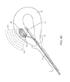
- FIGS. 1A through 1F a first embodiment of the invention is illustrated including two converging ablation needles 14 and an ultrasound imaging module 12 .
- a flexible, steerable catheter 10 is shown that acts as a sheath for the ultrasound catheter 12 .
- the two treatment needles 14 are in a retracted configuration within the sheath 10 .
- the ultrasound catheter 12 is shown within the sheath 10 with the two treatment needles 14 in a deployed configuration.
- One or both converging ablation needles 14 may have insulating sleeves so as to prevent treating non-target tissue and/or thermocouples at a tip region to measure a tissue temperature.
- FIG. 1C shows application of radiofrequency ablation energy between the two bipolar needle electrodes 14 and the resulting energy field 16 therebetween.
- FIG. 1D shows the sheath 10 inserted into the uterus 18 via the cervix 20 with a flexible shaft portion 22 .
- the ultrasound beam 12 not only allows for identification of the fibroids 24 , 26 , but also serves to provide real-time visualization of needle anchoring and ablation treatment.
- the ultrasound catheter 12 may further be steered, rotated, or deflected independently of the treatment needles 14 so as to allow for a complete reconstruction view. For example, the ultrasound catheter may be torqued or rotated so that positioning of both needles 14 and treatment 16 may be verified.
- FIG. 1E show deployment of the treatment needles 14 during ultrasound visualization while FIG.
- FIG. 1F shows radiofrequency ablation treatment 16 of the fibroid tumor 24 .
- each individual fibroid tumor 24 , 26 will be navigated to, imaged, targeted and treated separately. It will be appreciated that the above depictions are for illustrative purposes only and do not necessarily reflect the actual shape, size, or dimensions of the device. This applies to all depictions hereinafter.
- FIGS. 2A through 2D a second embodiment of the invention is illustrated including two diverging ablation needles 28 and the ultrasound imaging module 12 .
- the flexible, steerable catheter 10 is shown acting as a sheath for the ultrasound catheter 12 .
- the ultrasound catheter 12 is inserted into the sheath 10 and is visualizing the fibroid tumor 24 within the uterus in an imaging field transverse to an axis of the sheath as denoted by the dashed lines 30 .
- a hollow nitinol needle 32 is deployed through a lumen 34 in the sheath 10 and across the imaging field, as illustrated in FIG. 2B .
- two hooked treatment needles 28 are deployed through the hollow needle 32 within the limits of the imaging field as illustrated in FIG. 2C and anchored against the fibroid 24 .
- Radiofrequency ablative energy is then delivered in a bipolar fashion between the two poles of the hooked treatment needles 28 while the treatment needles remain with the limits of the imaging field so as to necrose the fibroid tissue 24 as illustrated in FIG. 2D .
- Fibroid identification, needle deployment, and ablation treatment are carried out under ultrasound visualization 30 in real-time. It will be appreciated that the distances (as denoted by arrows 36 , 38 ) that each treatment needle 28 is deployed within the fibroid tissue 24 may be adjusted based on the size of the lesion.
- FIGS. 3A through 3D a third embodiment of the invention is illustrated including a telescoping ablation needle 40 and the ultrasound imaging module 12 .
- the flexible, steerable catheter 10 is shown acting as a sheath for the ultrasound catheter 12 .
- the ultrasound catheter 12 is inserted into the sheath 10 .
- the sheath 10 is transcervically introduced into the uterus and used for visualizing the fibroid tumor 24 as denoted by the dashed lines 30 .
- a first nitinol needle 32 is deployed through the lumen 34 in the sheath 10 .
- a second telescoping needle 40 is deployed through the first needle 32 as illustrated in FIG.
- Radiofrequency ablative energy is delivered in a bipolar fashion between the two telescoping needles 40 , 32 under ultrasound visualization 30 . Again, the distance (as denoted by arrow 42 ) that the telescoping treatment needle 40 is extended within the fibroid tissue 24 may be adjustable to the size of the lesion.
- FIGS. 4A through 4F a fourth embodiment of the invention is illustrated including an inflatable treatment balloon 44 and the ultrasound imaging catheter 12 .
- the flexible, steerable sheath 10 is inserted into the uterus 18 via the cervix 20 with the ultrasound module 12 on board.
- the sheath 10 further has a lumen for insertion of a rotary cutting tube 46 , treatment needle, or other penetrating device.
- FIG. 4B illustrates visualization of the individual fibroid tumor 24 from within the uterine cavity 18 by the ultrasound module 12 , as denoted by the dashed lines 30 .
- FIG. 4C illustrates advancement and penetration of the rotary cutting tube 46 into the fibroid tumor 24 under direct visualization 30 through the ultrasound module 12 .
- the distal end of the rotary cutting tube 46 is depicted with a morcelating tip 47 .
- FIG. 4D some of the fibroid tissue 24 is removed through the rotary cutting tube 46 to create room for the treatment balloon 44 .
- the rotary cutting tube 46 is further partially retracted to make room in the tumor 24 for the treatment balloon 44 .
- the treatment balloon 44 is then deployed through the cutting tube 46 and into the tumor 24 under direct visualization 30 through the ultrasound module 12 .
- FIG. 4E illustrates advancement and penetration of the rotary cutting tube 46 into the fibroid tumor 24 under direct visualization 30 through the ultrasound module 12 .
- the treatment balloon 44 is inflated and ablative energy is applied by the balloon 44 to treat the tumor 24 under direct visualization 30 through the ultrasound module 12 .
- the ablative energy may comprise any of the energy sources described herein including radiofrequency energy, microwave energy, laser energy, cryo energy, ultrasound energy, HIFU, or radiation.
- a radiofrequency basket electrode may be disposed over the balloon to treat the tumor.
- FIGS. 5A through 5C a fifth embodiment of the invention is illustrated including the inflatable treatment balloon 44 and the ultrasound imaging catheter 12 of FIG. 4F .
- This embodiment differs in how the treatment balloon 44 is deployed into the tumor 24 .
- a rotary cutting tube 48 without a morcelating tip is advanced and penetrated into the fibroid tumor 24 under direct visualization 30 through the ultrasound module 12 as shown in FIG. 5A .
- a wire 50 is then advanced into the tumor 24 through the cutting tube 48 under direct visualization 30 through the ultrasound module 12 in FIG. 5B .
- the treatment balloon 44 is advanced through the cutting tube 48 and over the wire 50 and then inflated in the tumor 24 under direct ultrasound visualization 30 so as to treat the tumor 24 with ablative energy.
- the ablative energy may comprise any of the energy sources described herein including radiofrequency energy, microwave energy, laser energy, cryo energy, ultrasound energy, HIFU, or radiation.
- FIG. 6 a sixth embodiment of the invention is illustrated including the rotary cutting tube 46 and the ultrasound imaging catheter 12 of FIG. 4C .
- This embodiment differs in that the rotary cutting tube 46 itself provides treatment of the tumor 24 with its mechanical cutting element having a morcelating tip 47 .
- the fibroid 24 is morcelated or liquefied by the rotary cutting tube 46 and the fibroid tissue 24 is suctioned out through the hollow cutting tube 46 as depicted by reference numeral 52 .
- FIGS. 7A through 7C a seventh embodiment of the invention is illustrated including a drug delivery needle 54 and the ultrasound imaging catheter 12 .
- two treatment needles 54 are anchored within the fibroid 24 and the fibroid treated by the delivery of at least one therapeutic agent 56 to the fibroid with the needles 54 .
- the treatment needles 54 may have both a retracted and extended position and may be adjustable so as to achieve the desired drug delivery profile. Further, drug delivery may take place though a single treatment needle 54 or through multiple treatment needles 54 .
- the therapeutic agent 56 may comprise a variety of agents.
- the agent 56 may comprise a chemotherapeutic or chemoablative agent (e.g., alcohol or a chemokine), a gene therapy agent, a tissue necrosis agent, an antibody, or the like.
- the drug delivery needles 54 may treat tumors of various sizes. For example, FIG. 7B illustrates treatment of a large tumor 24 ′ (e.g., 40 mm), while FIG. 7C illustrates treatment of a smaller tumor 24 ′′ (e.g., 20 mm).
- a syringe 58 is used to laproscopically inject contrast bubbles 60 containing at least one therapeutic agent 56 into the fibroid 24 instead of transcervical drug delivery via treatment needles 54 .
- the ultrasound imaging catheter 12 in the uterus 18 activates the agent 56 by targeted ultrasound 30 .
- this may cause the bubbles 60 to burst or break in the fibroid blood supply 24 which in turn releases the therapeutic agent 56 to the fibroid 24 for treatment.
- the flexible, steerable catheter 10 is shown inserted into the uterus 18 via the cervix 20 .
- the catheter 10 has an on board ultrasound imaging module 12 and a lumen for insertion of at least one needle 62 or other penetrating device.
- multiple needles 62 are shown inserted into the fibroid tumor 24 with impedance monitoring to denote the change in the impedance of the tissue from inside the tumor 24 versus tissue outside the tumor 24 and/or tissue outside the uterine wall.
- Impedance monitoring will aid in directly targeting the fibroid tumor 24 for treatment (e.g., energy delivery, drug delivery, mechanical cutting, etc.) and may also safely control treatment delivery so that it is only within the uterus 18 itself.
- impedance profiling may denote border recognition of tissue. This in turn may allow for implementation of additional safety mechanisms. For example, automatic shutoff of the device may be implemented if the needle 62 is extended beyond the fibroid 24 and/or uterus 18 .
- the flexible, steerable catheter 10 is shown inserted uterus 18 via the cervix 20 .
- the catheter 10 has an on board ultrasound imaging module 12 and a lumen for insertion of at least one needle 64 or other penetrating device.
- the needle 64 is shown inserted into the fibroid tumor 24 with impedance monitoring to denote the change in the impedance of the tissue from inside the tumor 24 versus tissue outside the tumor and/or tissue outside the uterine wall. Impedance monitoring will aid in directly targeting the fibroid tumor 24 for treatment from the uterine wall.
- a flexible, steerable laparoscopic probe 10 accessing the uterus 18 from an abdominal port 66 in the abdominal wall 68 .
- the probe 10 uses the ultrasound module 12 outside of the uterus 18 to target fibroid tumors 24 that are within the uterus 18 .
- the probe 10 then uses the treatment needle 70 under direct visualization 30 through the ultrasound module 12 to then treat the fibroid 24 with ablative energy.
- a flexible, steerable catheter based probe 10 having a treatment needle 72 is shown inserted into the uterus 18 and within the fibroid 24 using a non-coupled vaginal ultrasound probe 74 .
- the two devices 10 , 74 are operated independently of each other.
- FIG. 11B the flexible, steerable needle catheter 10 is shown inserted into the uterus 18 and the treatment needle 72 within the fibroid 24 using a non-coupled abdominal ultrasound probe 76 .
- the two devices 10 , 76 are operated independently of each other.
- FIG. 11C the flexible, steerable laparoscopic needle probe 10 is shown accessing the uterus 18 from an abdominal port 66 in the abdominal wall 68 .
- the treatment needle 70 of the probe 10 is shown accessing the fibroid 24 with the aid of ultrasound visualization 30 from the abdominal ultrasound probe 76 .
- the two devices 70 , 76 are operated independently of each other.
- a flexible, steerable intrauterine ultrasound imaging device 78 is shown for imaging the uterine wall and lining transendometrially for the diagnosis of fibroids 24 , 26 .
- the ultrasound imaging head 82 generally comprises an ultrasonic phased array transducer having 64 elements.
- the ultrasound transducer may also be mechanical, linear, or curved.
- a sterile drape 80 may be placed over the diagnostic imager 78 , wherein a gel may be used within the drape 80 for improved image coupling.
- the diagnostic imager 78 may also be used without a drape 80 , when disposable, using natural body fluids for image coupling.
- the diagnostic imager 78 further has a flexible section 84 capable of deflection in a range from 0 degrees to about 90 degrees via an angle adjustment knob 86 .
- the diagnostic ultrasound imager 78 is inserted directly into the uterine cavity 18 , either with or without dilation of the cervix 20 , in order to directly image the fibroids 24 , 26 within the wall of the uterus 18 .
- This imaging provides a closer and more direct view of the tumors 24 , 46 in order to more accurately diagnose the location and characterization of the fibroids or other pathology.
- FIGS. 13A and 13B illustrate schematics of a system constructed in accordance with the principles of the present invention.
- the system comprises a combined ultrasound recognition and radiofrequency treatment system 88 .
- the system 88 may provide a variety of features including ultrasound mapping, ultrasound recognition of treatment area (e.g., tissue differentiation via temperature profiling), radiofrequency ablation treatment under ultrasound imaging, temperature monitoring, time monitoring, and/or impedance monitoring.
- the system 88 may be coupled to various devices 90 described herein having single or multiple treatment needle configurations to ablate in either bipolar or monopolor modes.
Abstract
Methods and devices for both imaging and treating uterine fibroid tumors in one real-time system are provided. One minimally invasive method comprises introducing a sheath into a uterus and determining a location of a fibroid using a visualization element within or on the sheath. Upon identification, a portion of the sheath is steered to position at least one treatment needle at the determined location. The needle is then anchored in uterine tissue and the fibroid treated with the needle.
Description
The present application is a continuation of U.S. patent application Ser. No. 11/347,018 filed Feb. 2, 2006, which claims the benefit of Provisional Patent Application Nos. 60/710,712, filed Aug. 22, 2005, and 60/649,839, filed Feb. 2, 2005, the full disclosures of which are incorporated herein by reference.
Field of the Invention
The present invention relates generally to medical devices and methods. More particularly, the invention relates to methods and devices for locating and treating uterine fibroids.
There are unmet needs in the pathophysiology of the female reproductive tract, such as dysfunctional uterine bleeding and fibroids. Fibroids are benign tumors of the uterine myometria (i.e., muscle) and are the most common tumor of the female pelvis. Fibroid tumors affect up to 30% of women of childbearing age and can cause significant symptoms such as discomfort, pelvic pain, mennorhagia, pressure, anemia, compression, infertility and miscarriage. Fibroids may be located in the myometrium (i.e., intramural), adjacent to the endometrium (i.e., submucosal), or in the outer layer of the uterus (i.e., subserosal). Most commonly fibroids are a smooth muscle overgrowth that arise intramurally and can grow to be several centimeters in diameter.
Current treatment for fibroids includes medical treatment with NSAIDS, estrogen-progesterone combinations, and GnRH analogues. Pharmacologic therapy with GnRH analogues is limited due to its side effects, such as hot flashes, vaginal dryness, mood changes and bone density loss. Further, its relatively short time of treatment (e.g., 3 months) offers temporary shrinkage, wherein the fibroids usually regrow after medical discontinuation. Pharmacologic therapy is relatively ineffective and palliative rather than curative.
Hysterectomy (i.e., surgical removal of the uterus) is a common treatment for fibroids. It is performed up to 600,000 times annually in the United States. Indeed, fibroids are the indication for hysterectomy in up to one third of all cases. Hysterectomy for treating fibroids may be very effective but has many undesirable side effects such as loss of fertility, open surgery, sexual dysfunction, and long recovery time. There is also significant morbidity (e.g., sepsis, hemorrhage, peritonitis, bowel and bladder injury), mortality, and costs associated with hysterectomy treatments.
Surgical myomectomy is also an open surgical procedure requiring laparotomy and general anesthesia in which fibroids are removed. Often these procedures result in significant blood loss and can only remove a portion of the culprit tissue. In the early 1990's there was a growth in advanced operative laparoscopy techniques and laparoscopic myomectomy was pioneered. However, laparoscopic myomectomy remains technically challenging. For example, it requires laparoscopic suturing which is performed only by the most skilled of laparoscopic gynecologists. Further, prolonged anesthesia time, increased blood loss, and possibly higher risk of uterine rupture in pregnancy make laparoscopic myomectomy a challenging procedure. Currently, the removal of subserosal or intramural fibroids requires an abdominal approach.
Hysteroscopy (i.e., process by which a thin fiber optic camera is used to image inside the uterus) may include an attachment to destroy tissue. Hysteroscopic resection is a surgical technique that uses a variety of devices (e.g., loops, roller balls, bipolar electrodes) to ablate or resect uterine tissue. The uterus needs to be filled with fluid for better viewing and thus has potential side effects of fluid overload. Hysteroscopic ablation is also limited by its visualization technique and is thus only appropriate for those fibroids that are submucosal and/or protrude into the uterine cavity.
Uterine artery embolization has also been suggested as an alternative minimally invasive treatment for fibroids. Uterine artery embolization was introduced in the early 1990's and is performed by injecting small particles through a groin incision into the uterine artery to selectively block the blood supply to fibroids. Uterine artery embolization results in reduction of the myoma size from 20-70% at six months. However, side effects of this procedure include pelvic infection, premature menopause, and severe pelvic pain. In addition, long term MRI data suggest that incomplete fibroid infarction may result in regrowth of infracted fibroid tissue. Despite much interest in uterine artery embolization, the procedure rates remain low and have not grown past a few thousand performed per year in the United States. This may be due to the fact that interventional radiologists, instead of gynecologists who know how to diagnose and treat fibroid tumors, are the ones who perform uterine artery embolization procedures.
Endometrial ablation, which is primarily a procedure for dysfunctional or abnormal uterine bleeding, may be used at times for fibroids. Recently there have been many new technologies to perform endometrial ablation such as cryo energy, microwave energy, and impedance controlled radiofrequency. Endometrial ablation destroys the endometrial tissue lining the uterus, but does not specifically treat fibroids. This technique is also not suitable for women who desire to bear children. Endometrial ablation remains a successful therapy for dysfunctional uterine bleeding, but is limited in its ability to treat fibroids.
Myolysis is another alternative minimally invasive technique for fibroid treatment. Myolysis was first performed in the 1980's in Europe by using lasers to coagulate tissue, denature proteins, and necrose myometrium with laparoscopic visualization. This technique has been in use for the past several years and involves applying energy directly to the myoma. Laparoscopic myolysis can be an alternative to myomectomy, as the fibroids are coagulated and then undergo coagulative necrosis resulting in a dramatic decrease in size. Shrinkage of fibroids has been reported at 30-50%. In addition there is the obvious economic benefit of out-patient surgery, rapid recovery, and return to normal lifestyle. However, all laparoscopic techniques are limited by the fact that they can only see, and therefore only treat, subserosal fibroids.
Needle myolysis is a promising technique whereby a laparoscope is used to introduce one or more needles into a fibroid tumor under visual control. Bipolar radiofrequency current is then delivered between two adjacent needles, or monopolar current between a single needle and a distant dispersive electrode affixed to the thigh or back. The aim of needle myolysis is to coagulate a significant volume of the tumor and thereby cause it to shrink substantially. The traditional technique is to make multiple passes through different areas of the tumor using the coagulating needle to destroy many cylindrical cores of abnormal tissue. However, the desirability of multiple passes is mitigated by the risk of adhesion formation, which is thought to increase with increasing amounts of injured uterine serosa and by the operative time and skill required.
For these and other reasons, it would be desirable to provide a minimally invasive method and device to selectively eradicate fibroid tumors within the uterus. It would be desirable if the method and device could locate and treat all types of fibroids in the uterus in a safe and effective manner with minimum risk and discomfort for the patient. It would be further desirable to provide a method and device for eradicating fibroid tumors that combines imaging and treatment in one simple hand held device. At least some of these objectives will be met by the methods and devices of the present invention described hereinafter.
Description of the Background Art
Relevant references include U.S. Pat. No. 5,456,689 Kresch et al.; U.S. Pat. No. 5,979,453 Savage et al.; U.S. Pat. No. 6,002,968 Edwards; U.S. Pat. No. 6,550,482 Burbank et al.; U.S. Pat. No. 6,626,855 Weng et al.; U.S. Pat. No. 6,716,184 Vaezy et al.; WO 2004/064658; and US 2005/0107781. The full disclosures of each of the above references are incorporated herein by reference.
In a first aspect of the present invention, methods for minimally invasive identification and treatment of submucosal, intramural, or subserosal fibroids of the uterus are provided. A sheath, catheter, or probe may be transcervically introduced into the uterus. A location of the fibroid tumor may be determined by using a visualization element within or on the sheath. Preferably, the physician will be able to image the tumors transendometrially from within the uterine cavity. The visualization element may comprise an ultrasonic element or other visualization means, such as hysteroscopy, that is capable of producing a visual image. Once having identified the location, a portion of the sheath is steered to position at least one treatment needle at the determined location. The needle is anchored within the uterine tissue and the fibroid is treated with the needle. Fibroid treatment may take several forms as discussed in more detail below. Generally, each individual fibroid tumor will be navigated to, imaged, targeted and treated separately. It will further be appreciated that external imaging may be preformed prior to sheath introduction so as to initially “map” the location of the fibroid tumors.
Anchoring comprises manually positioning and penetrating the treatment needle through an endometrium so as to engage the fibroid. Preferably, the visualization element not only provides a field of view for locating the fibroid but also provides a field of view for directly observing and verifying needle deployment and fibroid treatment in real-time. Visualization may be aided by steering, rotating, or deflecting the visualization element independent of the treatment needle so as to provide a complete view. At least one treatment needle, preferably two treatment needles, will be anchored in the uterine tissue so that the needles will remain substantially immobile during the delivery of treatment. For example, anchoring may comprise deploying at least two treatment needles in a converging manner so as to pinch the fibroid therebetween. Alternatively, anchoring may comprise deploying at least two treatment needles in a diverging manner so as to hook the fibroid therebetween. Still further, anchoring may comprise deploying at least two treatment needles in a telescoping manner.
The uterine fibroid may be treated in several ways. Usually the fibroid will be treated by delivering ablative energy to the fibroid with the needle to necrose the tissue. The ablative energy may comprise electrically energy (e.g., radiofrequency energy, laser energy, or microwave energy), freezing energy (e.g., cryo energy), ultrasound energy, high intensity focused ultrasound (HIFU), or radiation. Preferably, the treatment needle comprises electrically conductive electrodes that deliver ablative radiofrequency energy in a bipolar or monopolar fashion. In addition to or in lieu of ablative energy treatment, the fibroids may be treated by delivering at least one therapeutic agent to the fibroid with the needle. Still further, and in addition to or in lieu of energy and/or drug delivery treatments, the fibroid may be treated by mechanical cutting. For example, the fibroid may be morcelated with a tip of the needle. The treatment needle or other elements (e.g., non-treatment needle, thermocouple) may additionally monitor tissue impedance and/or measure a tissue temperature so as to aid in diagnosis, blood supply measurement, thermal signature, tissue targeting, and the like.
In another aspect of the present invention, minimally invasive devices for imaging and treating submucosal, intramural, or subserosal fibroids in one real-time system are provided. The device comprises a sheath, probe, catheter, or other shaft which is adapted for transcervical introduction into a uterus. A visualization element is within or on a distal steerable portion of the sheath. The visualization element is capable of determining a location of a fibroid on wall of the uterus while the sheath is in the uterus. Typically, the visualization element comprise an ultrasonic transducer. For example, the visualization element may comprise a phased array transducer having 64 elements or a mechanically scanned transducer. Still further, the element may comprise other visualization means, such as hysteroscopy, that is capable of producing a visual image. At least one self-anchoring treatment needle is within or on a distal portion of the sheath. The treatment needle is deployable against the fibroid whose position has been located by the visualization element.
The at least one treatment needle, usually two treatment needles, will be anchored by providing a geometry which inhibits displacement after the needles have been deployed. Exemplary geometries include non-linear, such as arcuate, helical, such as cork screw, curved, co-axial, everting, and like configurations. For example, the geometry may comprises a pair of converging or diverging needles which when deployed in tissue will remain firmly anchored as a result of the opposed geometry. Such geometries may be conveniently referred to as being “self-anchoring.” Such anchoring needles advantageously provide targeted treatment of larger volumes (e.g., larger fibroids) with less damage to non-target tissue. The treatment needle may take on a variety of forms, typically having both extended and retracted configurations, and be made from a variety of materials (e.g., nitinol). For example, the treatment needle may comprise electrodes, electrosurgical needles, or other tissue-penetrating elements capable of delivering ablative radio-frequency energy to target and treat the tumors. Alternatively, the treatment needle may comprise an antenna capable of delivering microwave energy to treat the fibroid. Further, the treatment needle may comprise a hollow tube so as to deliver at least one therapeutic agent to the fibroid. Still further, the treatment needle may comprise a cutting tube so as to morcelate the fibroid.
The visualization element will preferably be located near and/or coupled to the treatment needle so that needle positioning, deployment, and treatment is within a surgeon's field of view. The sheath, visualization element, and/or treatment needle may be integrally formed or comprises separate, modular components that are coupleable to one another. For example, the visualization element may comprise a re-usable ultrasound core that may be positioned within a disposable needle carrying sheath. Further, at least a portion of the sheath, visualization element, and/or treatment needle may be steerable, rotatable, deflectable, flexible, pre-shaped, or pre-formed so as provide transvaginal access to the uterus for identification and treatment of fibroids. An exemplary interventional deployment and imaging system is described in more detail in co-pending U.S. Provisional Patent Application Ser. No. 60/758,881, filed Jan. 12, 2006, which is assigned to the assignee of the present application and incorporated herein by reference.
A further understanding of the nature and advantages of the present invention will become apparent by reference to the remaining portions of the specification and drawings.
The following drawings should be read with reference to the detailed description. Like numbers in different drawings refer to like elements. The drawings, which are not necessarily to scale, illustratively depict embodiments of the present invention and are not intended to limit the scope of the invention.
Referring now to FIGS. 1A through 1F , a first embodiment of the invention is illustrated including two converging ablation needles 14 and an ultrasound imaging module 12. A flexible, steerable catheter 10 is shown that acts as a sheath for the ultrasound catheter 12. In FIG. 1A , the two treatment needles 14 are in a retracted configuration within the sheath 10. In FIG. 1B , the ultrasound catheter 12 is shown within the sheath 10 with the two treatment needles 14 in a deployed configuration. One or both converging ablation needles 14 may have insulating sleeves so as to prevent treating non-target tissue and/or thermocouples at a tip region to measure a tissue temperature. FIG. 1C shows application of radiofrequency ablation energy between the two bipolar needle electrodes 14 and the resulting energy field 16 therebetween. FIG. 1D shows the sheath 10 inserted into the uterus 18 via the cervix 20 with a flexible shaft portion 22. As described above, the ultrasound beam 12 not only allows for identification of the fibroids 24, 26, but also serves to provide real-time visualization of needle anchoring and ablation treatment. The ultrasound catheter 12 may further be steered, rotated, or deflected independently of the treatment needles 14 so as to allow for a complete reconstruction view. For example, the ultrasound catheter may be torqued or rotated so that positioning of both needles 14 and treatment 16 may be verified. FIG. 1E show deployment of the treatment needles 14 during ultrasound visualization while FIG. 1F shows radiofrequency ablation treatment 16 of the fibroid tumor 24. Generally, each individual fibroid tumor 24, 26 will be navigated to, imaged, targeted and treated separately. It will be appreciated that the above depictions are for illustrative purposes only and do not necessarily reflect the actual shape, size, or dimensions of the device. This applies to all depictions hereinafter.
Referring now to FIGS. 2A through 2D , a second embodiment of the invention is illustrated including two diverging ablation needles 28 and the ultrasound imaging module 12. Again, the flexible, steerable catheter 10 is shown acting as a sheath for the ultrasound catheter 12. In FIG. 2A , the ultrasound catheter 12 is inserted into the sheath 10 and is visualizing the fibroid tumor 24 within the uterus in an imaging field transverse to an axis of the sheath as denoted by the dashed lines 30. A hollow nitinol needle 32 is deployed through a lumen 34 in the sheath 10 and across the imaging field, as illustrated in FIG. 2B . Thereafter, two hooked treatment needles 28 are deployed through the hollow needle 32 within the limits of the imaging field as illustrated in FIG. 2C and anchored against the fibroid 24. Radiofrequency ablative energy is then delivered in a bipolar fashion between the two poles of the hooked treatment needles 28 while the treatment needles remain with the limits of the imaging field so as to necrose the fibroid tissue 24 as illustrated in FIG. 2D . Fibroid identification, needle deployment, and ablation treatment are carried out under ultrasound visualization 30 in real-time. It will be appreciated that the distances (as denoted by arrows 36, 38) that each treatment needle 28 is deployed within the fibroid tissue 24 may be adjusted based on the size of the lesion.
Referring now to FIGS. 3A through 3D , a third embodiment of the invention is illustrated including a telescoping ablation needle 40 and the ultrasound imaging module 12. Again, the flexible, steerable catheter 10 is shown acting as a sheath for the ultrasound catheter 12. As shown in FIG. 3A , the ultrasound catheter 12 is inserted into the sheath 10. The sheath 10 is transcervically introduced into the uterus and used for visualizing the fibroid tumor 24 as denoted by the dashed lines 30. Similar to FIG. 2B , a first nitinol needle 32 is deployed through the lumen 34 in the sheath 10. Thereafter, a second telescoping needle 40 is deployed through the first needle 32 as illustrated in FIG. 3C . Radiofrequency ablative energy is delivered in a bipolar fashion between the two telescoping needles 40, 32 under ultrasound visualization 30. Again, the distance (as denoted by arrow 42) that the telescoping treatment needle 40 is extended within the fibroid tissue 24 may be adjustable to the size of the lesion.
Referring now to FIGS. 4A through 4F , a fourth embodiment of the invention is illustrated including an inflatable treatment balloon 44 and the ultrasound imaging catheter 12. As shown in FIG. 4A , the flexible, steerable sheath 10 is inserted into the uterus 18 via the cervix 20 with the ultrasound module 12 on board. The sheath 10 further has a lumen for insertion of a rotary cutting tube 46, treatment needle, or other penetrating device. FIG. 4B illustrates visualization of the individual fibroid tumor 24 from within the uterine cavity 18 by the ultrasound module 12, as denoted by the dashed lines 30. FIG. 4C illustrates advancement and penetration of the rotary cutting tube 46 into the fibroid tumor 24 under direct visualization 30 through the ultrasound module 12. The distal end of the rotary cutting tube 46 is depicted with a morcelating tip 47. In FIG. 4D , some of the fibroid tissue 24 is removed through the rotary cutting tube 46 to create room for the treatment balloon 44. The rotary cutting tube 46 is further partially retracted to make room in the tumor 24 for the treatment balloon 44. As shown in FIG. 4E , the treatment balloon 44 is then deployed through the cutting tube 46 and into the tumor 24 under direct visualization 30 through the ultrasound module 12. As shown in FIG. 4F , the treatment balloon 44 is inflated and ablative energy is applied by the balloon 44 to treat the tumor 24 under direct visualization 30 through the ultrasound module 12. The ablative energy may comprise any of the energy sources described herein including radiofrequency energy, microwave energy, laser energy, cryo energy, ultrasound energy, HIFU, or radiation. Alternatively or in addition to the treatment balloon 44, a radiofrequency basket electrode may be disposed over the balloon to treat the tumor.
Referring now to FIGS. 5A through 5C , a fifth embodiment of the invention is illustrated including the inflatable treatment balloon 44 and the ultrasound imaging catheter 12 of FIG. 4F . This embodiment differs in how the treatment balloon 44 is deployed into the tumor 24. After identification of the fibroid tumor 24 from within the uterine cavity 18, a rotary cutting tube 48 without a morcelating tip is advanced and penetrated into the fibroid tumor 24 under direct visualization 30 through the ultrasound module 12 as shown in FIG. 5A . A wire 50 is then advanced into the tumor 24 through the cutting tube 48 under direct visualization 30 through the ultrasound module 12 in FIG. 5B . In FIG. 5C , the treatment balloon 44 is advanced through the cutting tube 48 and over the wire 50 and then inflated in the tumor 24 under direct ultrasound visualization 30 so as to treat the tumor 24 with ablative energy. The ablative energy may comprise any of the energy sources described herein including radiofrequency energy, microwave energy, laser energy, cryo energy, ultrasound energy, HIFU, or radiation.
Referring now to FIG. 6 , a sixth embodiment of the invention is illustrated including the rotary cutting tube 46 and the ultrasound imaging catheter 12 of FIG. 4C . This embodiment differs in that the rotary cutting tube 46 itself provides treatment of the tumor 24 with its mechanical cutting element having a morcelating tip 47. After identification of the fibroid tumor 24 and advancement/penetration of the rotary cutting tube 46 into the fibroid tumor 24 under direct visualization 30 through the ultrasound module 12 in the uterus 18, the fibroid 24 is morcelated or liquefied by the rotary cutting tube 46 and the fibroid tissue 24 is suctioned out through the hollow cutting tube 46 as depicted by reference numeral 52.
Referring now to FIGS. 7A through 7C , a seventh embodiment of the invention is illustrated including a drug delivery needle 54 and the ultrasound imaging catheter 12. In FIG. 7A , under ultrasound visualization in the uterus, two treatment needles 54 are anchored within the fibroid 24 and the fibroid treated by the delivery of at least one therapeutic agent 56 to the fibroid with the needles 54. It will be appreciated that the treatment needles 54 may have both a retracted and extended position and may be adjustable so as to achieve the desired drug delivery profile. Further, drug delivery may take place though a single treatment needle 54 or through multiple treatment needles 54. The therapeutic agent 56 may comprise a variety of agents. For example, the agent 56 may comprise a chemotherapeutic or chemoablative agent (e.g., alcohol or a chemokine), a gene therapy agent, a tissue necrosis agent, an antibody, or the like. The drug delivery needles 54 may treat tumors of various sizes. For example, FIG. 7B illustrates treatment of a large tumor 24′ (e.g., 40 mm), while FIG. 7C illustrates treatment of a smaller tumor 24″ (e.g., 20 mm).
Referring now to FIG. 8 , another drug delivery method and device is illustrated. A syringe 58 is used to laproscopically inject contrast bubbles 60 containing at least one therapeutic agent 56 into the fibroid 24 instead of transcervical drug delivery via treatment needles 54. After drug delivery injection into the fibroid 24, the ultrasound imaging catheter 12 in the uterus 18 activates the agent 56 by targeted ultrasound 30. For example, this may cause the bubbles 60 to burst or break in the fibroid blood supply 24 which in turn releases the therapeutic agent 56 to the fibroid 24 for treatment.
Referring now to FIG. 9A , the flexible, steerable catheter 10 is shown inserted into the uterus 18 via the cervix 20. The catheter 10 has an on board ultrasound imaging module 12 and a lumen for insertion of at least one needle 62 or other penetrating device. In this illustration, multiple needles 62 are shown inserted into the fibroid tumor 24 with impedance monitoring to denote the change in the impedance of the tissue from inside the tumor 24 versus tissue outside the tumor 24 and/or tissue outside the uterine wall. Impedance monitoring will aid in directly targeting the fibroid tumor 24 for treatment (e.g., energy delivery, drug delivery, mechanical cutting, etc.) and may also safely control treatment delivery so that it is only within the uterus 18 itself. Further, impedance profiling may denote border recognition of tissue. This in turn may allow for implementation of additional safety mechanisms. For example, automatic shutoff of the device may be implemented if the needle 62 is extended beyond the fibroid 24 and/or uterus 18.
Referring now to FIG. 9B , the flexible, steerable catheter 10 is shown inserted uterus 18 via the cervix 20. The catheter 10 has an on board ultrasound imaging module 12 and a lumen for insertion of at least one needle 64 or other penetrating device. The needle 64 is shown inserted into the fibroid tumor 24 with impedance monitoring to denote the change in the impedance of the tissue from inside the tumor 24 versus tissue outside the tumor and/or tissue outside the uterine wall. Impedance monitoring will aid in directly targeting the fibroid tumor 24 for treatment from the uterine wall.
Referring now to FIG. 10 , a flexible, steerable laparoscopic probe 10 is shown accessing the uterus 18 from an abdominal port 66 in the abdominal wall 68. The probe 10 uses the ultrasound module 12 outside of the uterus 18 to target fibroid tumors 24 that are within the uterus 18. The probe 10 then uses the treatment needle 70 under direct visualization 30 through the ultrasound module 12 to then treat the fibroid 24 with ablative energy.
Referring now to FIG. 11A , a flexible, steerable catheter based probe 10 having a treatment needle 72 is shown inserted into the uterus 18 and within the fibroid 24 using a non-coupled vaginal ultrasound probe 74. The two devices 10, 74 are operated independently of each other. Referring now to FIG. 11B , the flexible, steerable needle catheter 10 is shown inserted into the uterus 18 and the treatment needle 72 within the fibroid 24 using a non-coupled abdominal ultrasound probe 76. The two devices 10, 76 are operated independently of each other. With respect to FIG. 11C , the flexible, steerable laparoscopic needle probe 10 is shown accessing the uterus 18 from an abdominal port 66 in the abdominal wall 68. The treatment needle 70 of the probe 10 is shown accessing the fibroid 24 with the aid of ultrasound visualization 30 from the abdominal ultrasound probe 76. The two devices 70, 76 are operated independently of each other.
Referring now to FIG. 12 , a flexible, steerable intrauterine ultrasound imaging device 78 is shown for imaging the uterine wall and lining transendometrially for the diagnosis of fibroids 24, 26. The ultrasound imaging head 82 generally comprises an ultrasonic phased array transducer having 64 elements. The ultrasound transducer may also be mechanical, linear, or curved. A sterile drape 80 may be placed over the diagnostic imager 78, wherein a gel may be used within the drape 80 for improved image coupling. The diagnostic imager 78 may also be used without a drape 80, when disposable, using natural body fluids for image coupling. The diagnostic imager 78 further has a flexible section 84 capable of deflection in a range from 0 degrees to about 90 degrees via an angle adjustment knob 86. The diagnostic ultrasound imager 78 is inserted directly into the uterine cavity 18, either with or without dilation of the cervix 20, in order to directly image the fibroids 24, 26 within the wall of the uterus 18. This imaging provides a closer and more direct view of the tumors 24, 46 in order to more accurately diagnose the location and characterization of the fibroids or other pathology.
Although certain exemplary embodiments and methods have been described in some detail, for clarity of understanding and by way of example, it will be apparent from the foregoing disclosure to those skilled in the art that variations, modifications, changes, and adaptations of such embodiments and methods may be made without departing from the true spirit and scope of the invention. Therefore, the above description should not be taken as limiting the scope of the invention which is defined by the appended claims.
Claims (11)
1. A method for minimally invasive treatment of uterine fibroids, said method comprising:
introducing a sheath into a uterus, said sheath having a longitudinal axis;
determining a location of a fibroid using a visualization element within or on the sheath, wherein the visualization element produces an imaging field transverse to the longitudinal axis of the sheath and wherein both the visualization element and the imaging field are deflectable with respect to the longitudinal axis of the sheath;
advancing a hollow needle from the sheath through a wall of the uterus and across the imaging field;
advancing a plurality of diverging needles from the hollow needle into the fibroid; and
treating the fibroid with the diverging needles, wherein the diverging needles remain within the limits of the imaging field during treatment, and
wherein determining the location of the fibroid comprises rotating the visualization element and the imaging field about the longitudinal axis of the sheath independently of the plurality of diverging needles after the hollow needle has been advanced through the wall of the uterus and across the imaging filed.
2. A method as in claim 1 , wherein the sheath is introduced transcervically.
3. A method as in claim 1 , wherein the visualization element is ultrasonic and produces a visual image.
4. A method as in claim 3 , wherein introducing comprises manually positioning and penetrating the plurality of diverging needles through an endometrium so as to engage the fibroid.
5. A method as in claim 1 , wherein treating the fibroid comprises delivering ablative energy to the fibroid with the plurality of diverging needles.
6. A method as in claim 5 , wherein the ablative energy comprises radiofrequency energy, microwave energy, laser energy, cryo energy, ultrasound energy, HIFU, or radiation.
7. A method as in claim 6 , wherein the ablative energy comprises radiofrequency energy delivered in a bipolar or monopolar fashion.
8. A method as in claim 1 , wherein treating the fibroid comprises delivering at least one therapeutic agent to the fibroid with the plurality of diverging needles.
9. A method as in claim 1 , further comprising imaging advancing the hollow needle, advancing the plurality of diverging needles, and treatment in real-time with the visualization element.
10. A method as in claim 1 , further comprising monitoring tissue impedance.
11. A method as in claim 1 , further comprising measuring a tissue temperature so as to aid in diagnosis, blood supply measurement, thermal signature, or tissue targeting.
Priority Applications (5)
| Application Number | Priority Date | Filing Date | Title |
|---|---|---|---|
| US12/973,642 US9808310B2 (en) | 2005-02-02 | 2010-12-20 | Method and device for uterine fibroid treatment |
| US15/720,199 US9987080B2 (en) | 2005-02-02 | 2017-09-29 | Method and device for uterine fibroid treatment |
| US15/970,428 US10182862B2 (en) | 2005-02-02 | 2018-05-03 | Method and device for uterine fibroid treatment |
| US16/221,138 US11419668B2 (en) | 2005-02-02 | 2018-12-14 | Method and device for uterine fibroid treatment |
| US17/863,988 US11950837B2 (en) | 2022-07-13 | Method and device for uterine fibroid treatment |
Applications Claiming Priority (4)
| Application Number | Priority Date | Filing Date | Title |
|---|---|---|---|
| US64983905P | 2005-02-02 | 2005-02-02 | |
| US71071205P | 2005-08-22 | 2005-08-22 | |
| US11/347,018 US7918795B2 (en) | 2005-02-02 | 2006-02-02 | Method and device for uterine fibroid treatment |
| US12/973,642 US9808310B2 (en) | 2005-02-02 | 2010-12-20 | Method and device for uterine fibroid treatment |
Related Parent Applications (1)
| Application Number | Title | Priority Date | Filing Date |
|---|---|---|---|
| US11/347,018 Continuation US7918795B2 (en) | 2005-02-02 | 2006-02-02 | Method and device for uterine fibroid treatment |
Related Child Applications (1)
| Application Number | Title | Priority Date | Filing Date |
|---|---|---|---|
| US15/720,199 Continuation US9987080B2 (en) | 2005-02-02 | 2017-09-29 | Method and device for uterine fibroid treatment |
Publications (2)
| Publication Number | Publication Date |
|---|---|
| US20110087100A1 US20110087100A1 (en) | 2011-04-14 |
| US9808310B2 true US9808310B2 (en) | 2017-11-07 |
Family
ID=36913745
Family Applications (5)
| Application Number | Title | Priority Date | Filing Date |
|---|---|---|---|
| US11/347,018 Active 2028-03-31 US7918795B2 (en) | 2005-02-02 | 2006-02-02 | Method and device for uterine fibroid treatment |
| US12/973,642 Active 2026-11-04 US9808310B2 (en) | 2005-02-02 | 2010-12-20 | Method and device for uterine fibroid treatment |
| US15/720,199 Active US9987080B2 (en) | 2005-02-02 | 2017-09-29 | Method and device for uterine fibroid treatment |
| US15/970,428 Active US10182862B2 (en) | 2005-02-02 | 2018-05-03 | Method and device for uterine fibroid treatment |
| US16/221,138 Active 2026-09-01 US11419668B2 (en) | 2005-02-02 | 2018-12-14 | Method and device for uterine fibroid treatment |
Family Applications Before (1)
| Application Number | Title | Priority Date | Filing Date |
|---|---|---|---|
| US11/347,018 Active 2028-03-31 US7918795B2 (en) | 2005-02-02 | 2006-02-02 | Method and device for uterine fibroid treatment |
Family Applications After (3)
| Application Number | Title | Priority Date | Filing Date |
|---|---|---|---|
| US15/720,199 Active US9987080B2 (en) | 2005-02-02 | 2017-09-29 | Method and device for uterine fibroid treatment |
| US15/970,428 Active US10182862B2 (en) | 2005-02-02 | 2018-05-03 | Method and device for uterine fibroid treatment |
| US16/221,138 Active 2026-09-01 US11419668B2 (en) | 2005-02-02 | 2018-12-14 | Method and device for uterine fibroid treatment |
Country Status (1)
| Country | Link |
|---|---|
| US (5) | US7918795B2 (en) |
Cited By (3)
| Publication number | Priority date | Publication date | Assignee | Title |
|---|---|---|---|---|
| US9987080B2 (en) | 2005-02-02 | 2018-06-05 | Gynesonics, Inc. | Method and device for uterine fibroid treatment |
| US10993770B2 (en) | 2016-11-11 | 2021-05-04 | Gynesonics, Inc. | Controlled treatment of tissue and dynamic interaction with, and comparison of, tissue and/or treatment data |
| US11950837B2 (en) | 2022-07-13 | 2024-04-09 | Gynesonics, Inc. | Method and device for uterine fibroid treatment |
Families Citing this family (162)
| Publication number | Priority date | Publication date | Assignee | Title |
|---|---|---|---|---|
| US6050943A (en) | 1997-10-14 | 2000-04-18 | Guided Therapy Systems, Inc. | Imaging, therapy, and temperature monitoring ultrasonic system |
| US7678106B2 (en) * | 2000-08-09 | 2010-03-16 | Halt Medical, Inc. | Gynecological ablation procedure and system |
| US6840935B2 (en) * | 2000-08-09 | 2005-01-11 | Bekl Corporation | Gynecological ablation procedure and system using an ablation needle |
| US7914453B2 (en) | 2000-12-28 | 2011-03-29 | Ardent Sound, Inc. | Visual imaging system for ultrasonic probe |
| EP1501411B1 (en) * | 2002-04-22 | 2014-03-12 | Johns Hopkins University | Apparatus for insertion of a medical device during a medical imaging process |
| JP4395603B2 (en) * | 2004-03-31 | 2010-01-13 | 富士フイルム株式会社 | Ultrasound endoscope |
| EP1763321A4 (en) * | 2004-04-16 | 2010-02-17 | Critical Care Innovations Inc | Systems and methods for improving image-guided tissue ablation |
| US7393325B2 (en) | 2004-09-16 | 2008-07-01 | Guided Therapy Systems, L.L.C. | Method and system for ultrasound treatment with a multi-directional transducer |
| US7824348B2 (en) | 2004-09-16 | 2010-11-02 | Guided Therapy Systems, L.L.C. | System and method for variable depth ultrasound treatment |
| US9011336B2 (en) | 2004-09-16 | 2015-04-21 | Guided Therapy Systems, Llc | Method and system for combined energy therapy profile |
| US8444562B2 (en) | 2004-10-06 | 2013-05-21 | Guided Therapy Systems, Llc | System and method for treating muscle, tendon, ligament and cartilage tissue |
| US10864385B2 (en) | 2004-09-24 | 2020-12-15 | Guided Therapy Systems, Llc | Rejuvenating skin by heating tissue for cosmetic treatment of the face and body |
| US8535228B2 (en) | 2004-10-06 | 2013-09-17 | Guided Therapy Systems, Llc | Method and system for noninvasive face lifts and deep tissue tightening |
| US8690778B2 (en) | 2004-10-06 | 2014-04-08 | Guided Therapy Systems, Llc | Energy-based tissue tightening |
| CA2583600A1 (en) | 2004-10-06 | 2006-04-20 | Guided Therapy Systems, L.L.C. | Method and system for noninvasive cosmetic enhancement |
| US11883688B2 (en) | 2004-10-06 | 2024-01-30 | Guided Therapy Systems, Llc | Energy based fat reduction |
| DK1855759T3 (en) | 2004-10-06 | 2017-06-06 | Guided Therapy Systems Llc | Tissue Ultrasound Treatment System |
| US9827449B2 (en) | 2004-10-06 | 2017-11-28 | Guided Therapy Systems, L.L.C. | Systems for treating skin laxity |
| US7758524B2 (en) | 2004-10-06 | 2010-07-20 | Guided Therapy Systems, L.L.C. | Method and system for ultra-high frequency ultrasound treatment |
| US9694212B2 (en) | 2004-10-06 | 2017-07-04 | Guided Therapy Systems, Llc | Method and system for ultrasound treatment of skin |
| US20060111744A1 (en) | 2004-10-13 | 2006-05-25 | Guided Therapy Systems, L.L.C. | Method and system for treatment of sweat glands |
| US8133180B2 (en) | 2004-10-06 | 2012-03-13 | Guided Therapy Systems, L.L.C. | Method and system for treating cellulite |
| US11235179B2 (en) | 2004-10-06 | 2022-02-01 | Guided Therapy Systems, Llc | Energy based skin gland treatment |
| US11724133B2 (en) | 2004-10-07 | 2023-08-15 | Guided Therapy Systems, Llc | Ultrasound probe for treatment of skin |
| US11207548B2 (en) | 2004-10-07 | 2021-12-28 | Guided Therapy Systems, L.L.C. | Ultrasound probe for treating skin laxity |
| EP1875327A2 (en) | 2005-04-25 | 2008-01-09 | Guided Therapy Systems, L.L.C. | Method and system for enhancing computer peripheral saftey |
| US8080009B2 (en) | 2005-07-01 | 2011-12-20 | Halt Medical Inc. | Radio frequency ablation device for the destruction of tissue masses |
| US8512333B2 (en) * | 2005-07-01 | 2013-08-20 | Halt Medical Inc. | Anchored RF ablation device for the destruction of tissue masses |
| US7691127B2 (en) * | 2005-12-13 | 2010-04-06 | Cardiva Medical, Inc. | Drug eluting vascular closure devices and methods |
| US9179897B2 (en) * | 2005-12-13 | 2015-11-10 | Cardiva Medical, Inc. | Vascular closure devices and methods providing hemostatic enhancement |
| US11259825B2 (en) | 2006-01-12 | 2022-03-01 | Gynesonics, Inc. | Devices and methods for treatment of tissue |
| US10058342B2 (en) | 2006-01-12 | 2018-08-28 | Gynesonics, Inc. | Devices and methods for treatment of tissue |
| US9357977B2 (en) | 2006-01-12 | 2016-06-07 | Gynesonics, Inc. | Interventional deployment and imaging system |
| US7815571B2 (en) * | 2006-04-20 | 2010-10-19 | Gynesonics, Inc. | Rigid delivery systems having inclined ultrasound and needle |
| US7874986B2 (en) * | 2006-04-20 | 2011-01-25 | Gynesonics, Inc. | Methods and devices for visualization and ablation of tissue |
| WO2007143281A2 (en) * | 2006-01-13 | 2007-12-13 | Mirabilis Medica Inc. | Methods and apparatus for the treatment of menometrorrhagia, endometrial pathology, and cervical neoplasia using high intensity focused ultrasound energy |
| CA2646363C (en) | 2006-03-14 | 2015-07-21 | The Johns Hopkins University | Apparatus for insertion of a medical device within a body during a medical imaging process and devices and methods related thereto |
| US8206300B2 (en) | 2008-08-26 | 2012-06-26 | Gynesonics, Inc. | Ablation device with articulated imaging transducer |
| US20100056926A1 (en) | 2008-08-26 | 2010-03-04 | Gynesonics, Inc. | Ablation device with articulated imaging transducer |
| US10595819B2 (en) | 2006-04-20 | 2020-03-24 | Gynesonics, Inc. | Ablation device with articulated imaging transducer |
| US8298145B2 (en) * | 2006-08-01 | 2012-10-30 | Gynesonics, Inc. | Peri-capsular fibroid treatment |
| US20080071173A1 (en) * | 2006-09-18 | 2008-03-20 | Aldrich William N | Visualizing Formation of Ablation Lesions |
| US9566454B2 (en) | 2006-09-18 | 2017-02-14 | Guided Therapy Systems, Llc | Method and sysem for non-ablative acne treatment and prevention |
| US20210121227A1 (en) | 2006-09-29 | 2021-04-29 | Baylis Medical Company Inc. | Connector system for electrosurgical device |
| US11666377B2 (en) | 2006-09-29 | 2023-06-06 | Boston Scientific Medical Device Limited | Electrosurgical device |
| US11331121B2 (en) * | 2006-09-29 | 2022-05-17 | Baylis Medical Company Inc. | Transseptal needle |
| US20080097469A1 (en) | 2006-10-18 | 2008-04-24 | Gruber William H | Intrauterine access and procedure system with laterally deflectable sheath |
| US20080146872A1 (en) | 2006-11-07 | 2008-06-19 | Gruber William H | Mechanical distension systems for performing a medical procedure in a remote space |
| US8025656B2 (en) | 2006-11-07 | 2011-09-27 | Hologic, Inc. | Methods, systems and devices for performing gynecological procedures |
| US20090138011A1 (en) * | 2007-03-13 | 2009-05-28 | Gordon Epstein | Intermittent ablation rf driving for moderating return electrode temperature |
| US20080228180A1 (en) * | 2007-03-13 | 2008-09-18 | Halt Medical, Inc | Ablation system and heat preventing electrodes therefor |
| US20090187183A1 (en) * | 2007-03-13 | 2009-07-23 | Gordon Epstein | Temperature responsive ablation rf driving for moderating return electrode temperature |
| US9095366B2 (en) | 2007-04-06 | 2015-08-04 | Hologic, Inc. | Tissue cutter with differential hardness |
| EP2134283B1 (en) | 2007-04-06 | 2014-06-11 | Hologic, Inc. | System and device for tissue removal |
| US20090270898A1 (en) | 2007-04-06 | 2009-10-29 | Interlace Medical, Inc. | Tissue removal device with high reciprocation rate |
| US9259233B2 (en) | 2007-04-06 | 2016-02-16 | Hologic, Inc. | Method and device for distending a gynecological cavity |
| TWI526233B (en) | 2007-05-07 | 2016-03-21 | 指導治療系統股份有限公司 | Methods and systems for modulating medicants using acoustic energy |
| US20150174388A1 (en) | 2007-05-07 | 2015-06-25 | Guided Therapy Systems, Llc | Methods and Systems for Ultrasound Assisted Delivery of a Medicant to Tissue |
| US20090131790A1 (en) * | 2007-05-15 | 2009-05-21 | Gynesonics, Inc. | Systems and methods for deploying echogenic components in ultrasonic imaging fields |
| US8088072B2 (en) | 2007-10-12 | 2012-01-03 | Gynesonics, Inc. | Methods and systems for controlled deployment of needles in tissue |
| WO2009055720A1 (en) * | 2007-10-26 | 2009-04-30 | University Of Virginia Patent Foundation | System for treatment and imaging using ultrasonic energy and microbubbles and related method thereof |
| US8439907B2 (en) | 2007-11-07 | 2013-05-14 | Mirabilis Medica Inc. | Hemostatic tissue tunnel generator for inserting treatment apparatus into tissue of a patient |
| US8251991B2 (en) | 2007-11-14 | 2012-08-28 | Halt Medical Inc. | Anchored RF ablation device for the destruction of tissue masses |
| US8241276B2 (en) * | 2007-11-14 | 2012-08-14 | Halt Medical Inc. | RF ablation device with jam-preventing electrical coupling member |
| CN104545998B (en) | 2008-06-06 | 2020-07-14 | 奥赛拉公司 | System and method for cosmetic treatment and imaging |
| US8145293B2 (en) * | 2008-06-16 | 2012-03-27 | Siemens Medical Solutions Usa, Inc. | Adaptive medical image acquisition system and method |
| US8197477B2 (en) | 2008-10-21 | 2012-06-12 | Hermes Innovations Llc | Tissue ablation methods |
| US8500732B2 (en) | 2008-10-21 | 2013-08-06 | Hermes Innovations Llc | Endometrial ablation devices and systems |
| US8821486B2 (en) | 2009-11-13 | 2014-09-02 | Hermes Innovations, LLC | Tissue ablation systems and methods |
| US8382753B2 (en) | 2008-10-21 | 2013-02-26 | Hermes Innovations, LLC | Tissue ablation methods |
| US8540708B2 (en) | 2008-10-21 | 2013-09-24 | Hermes Innovations Llc | Endometrial ablation method |
| US9662163B2 (en) | 2008-10-21 | 2017-05-30 | Hermes Innovations Llc | Endometrial ablation devices and systems |
| US8197476B2 (en) | 2008-10-21 | 2012-06-12 | Hermes Innovations Llc | Tissue ablation systems |
| KR20110101204A (en) | 2008-12-24 | 2011-09-15 | 가이디드 테라피 시스템스, 엘.엘.씨. | Methods and systems for fat reduction and/or cellulite treatment |
| US8262574B2 (en) | 2009-02-27 | 2012-09-11 | Gynesonics, Inc. | Needle and tine deployment mechanism |
| US11903602B2 (en) | 2009-04-29 | 2024-02-20 | Hologic, Inc. | Uterine fibroid tissue removal device |
| US10123821B2 (en) * | 2009-09-10 | 2018-11-13 | Atricure, Inc. | Scope and magnetic introducer systems and methods |
| AU2010314930C1 (en) | 2009-11-05 | 2014-04-03 | Stratus Medical, LLC | Methods and systems for spinal radio frequency neurotomy |
| US8715278B2 (en) * | 2009-11-11 | 2014-05-06 | Minerva Surgical, Inc. | System for endometrial ablation utilizing radio frequency |
| US8529562B2 (en) | 2009-11-13 | 2013-09-10 | Minerva Surgical, Inc | Systems and methods for endometrial ablation |
| US11896282B2 (en) | 2009-11-13 | 2024-02-13 | Hermes Innovations Llc | Tissue ablation systems and method |
| US9289257B2 (en) | 2009-11-13 | 2016-03-22 | Minerva Surgical, Inc. | Methods and systems for endometrial ablation utilizing radio frequency |
| US8715186B2 (en) | 2009-11-24 | 2014-05-06 | Guided Therapy Systems, Llc | Methods and systems for generating thermal bubbles for improved ultrasound imaging and therapy |
| IT1405000B1 (en) * | 2010-02-04 | 2013-12-16 | El En Spa | DEVICE FOR THE TREATMENT OF THE VAGINAL CHANNEL AND ITS APPARATUS |
| JP2013526940A (en) | 2010-05-21 | 2013-06-27 | ニンバス・コンセプツ・エルエルシー | Systems and methods for tissue ablation |
| US8956348B2 (en) | 2010-07-21 | 2015-02-17 | Minerva Surgical, Inc. | Methods and systems for endometrial ablation |
| US9504446B2 (en) | 2010-08-02 | 2016-11-29 | Guided Therapy Systems, Llc | Systems and methods for coupling an ultrasound source to tissue |
| US10183182B2 (en) | 2010-08-02 | 2019-01-22 | Guided Therapy Systems, Llc | Methods and systems for treating plantar fascia |
| WO2012054466A2 (en) | 2010-10-18 | 2012-04-26 | Bioceptive, Inc. | Methods and apparatus for inserting a device or pharmaceutical into a body cavity |
| US9510897B2 (en) | 2010-11-05 | 2016-12-06 | Hermes Innovations Llc | RF-electrode surface and method of fabrication |
| US8857438B2 (en) | 2010-11-08 | 2014-10-14 | Ulthera, Inc. | Devices and methods for acoustic shielding |
| US9943360B2 (en) | 2011-01-30 | 2018-04-17 | University Health Network | Coil electrode for thermal therapy |
| US8858471B2 (en) | 2011-07-10 | 2014-10-14 | Guided Therapy Systems, Llc | Methods and systems for ultrasound treatment |
| KR20190080967A (en) | 2011-07-11 | 2019-07-08 | 가이디드 테라피 시스템스, 엘.엘.씨. | Systems and methods for coupling an ultrasound source to tissue |
| US9211141B2 (en) * | 2012-04-13 | 2015-12-15 | Boston Scientific Scimed, Inc. | Vaginal manipulator |
| US9263663B2 (en) | 2012-04-13 | 2016-02-16 | Ardent Sound, Inc. | Method of making thick film transducer arrays |
| BR112014030643A8 (en) | 2012-05-31 | 2018-05-15 | Baylis Medical Co Inc | radiofrequency drilling rig. |
| US20140031715A1 (en) * | 2012-07-30 | 2014-01-30 | Michael David SHERAR | Coil electrode apparatus for thermal therapy for treating bone tissue |
| US8992427B2 (en) * | 2012-09-07 | 2015-03-31 | Gynesonics, Inc. | Methods and systems for controlled deployment of needle structures in tissue |
| US9510802B2 (en) | 2012-09-21 | 2016-12-06 | Guided Therapy Systems, Llc | Reflective ultrasound technology for dermatological treatments |
| CN204637350U (en) | 2013-03-08 | 2015-09-16 | 奥赛拉公司 | Aesthstic imaging and processing system, multifocal processing system and perform the system of aesthetic procedure |
| US11937873B2 (en) | 2013-03-12 | 2024-03-26 | Boston Scientific Medical Device Limited | Electrosurgical device having a lumen |
| US20140276026A1 (en) * | 2013-03-13 | 2014-09-18 | Volcano Corporation | Devices and methods for imaging and delivering a fertilized egg into a woman's uterus |
| US20140276101A1 (en) * | 2013-03-14 | 2014-09-18 | Boston Scientific Scimed, Inc. | Medical device for detecting a target and related methods of use |
| US10499980B2 (en) | 2013-03-14 | 2019-12-10 | Spiration, Inc. | Flexible RF ablation needle |
| US10561862B2 (en) | 2013-03-15 | 2020-02-18 | Guided Therapy Systems, Llc | Ultrasound treatment device and methods of use |
| EP4233991A1 (en) | 2013-03-15 | 2023-08-30 | Medtronic Ardian Luxembourg S.à.r.l. | Controlled neuromodulation systems |
| US9901394B2 (en) | 2013-04-04 | 2018-02-27 | Hermes Innovations Llc | Medical ablation system and method of making |
| US9668763B2 (en) | 2013-09-11 | 2017-06-06 | Covidien Lp | System for myomectomy and morcellation |
| US9603624B2 (en) | 2013-09-11 | 2017-03-28 | Covidien Lp | System for myomectomy and morcellation |
| US9198719B2 (en) * | 2013-09-30 | 2015-12-01 | Gyrus Acmi, Inc. | Electrosurgical fibroid ablation system and method |
| US9649125B2 (en) | 2013-10-15 | 2017-05-16 | Hermes Innovations Llc | Laparoscopic device |
| DE112014004768T5 (en) | 2013-10-18 | 2016-08-11 | Ziva Medical, Inc. | Methods and systems for the treatment of polycystic ovarian syndrome |
| CN105873534A (en) | 2013-11-13 | 2016-08-17 | 美国奥林匹斯外科技术吉鲁斯阿克米公司 | Fibroid ablation positioning device and methods |
| US9993263B2 (en) | 2013-12-12 | 2018-06-12 | Catalin Esanu | Method and device for ultrasound guided minimal invasive access of a bodily cavity |
| CN106572880B (en) | 2014-03-28 | 2019-07-16 | 斯波瑞申有限公司 | The system of predictable configuration for medical device |
| SG11201608691YA (en) | 2014-04-18 | 2016-11-29 | Ulthera Inc | Band transducer ultrasound therapy |
| US10492856B2 (en) | 2015-01-26 | 2019-12-03 | Hermes Innovations Llc | Surgical fluid management system and method of use |
| US10376308B2 (en) | 2015-02-05 | 2019-08-13 | Axon Therapies, Inc. | Devices and methods for treatment of heart failure by splanchnic nerve ablation |
| EP3277191B1 (en) * | 2015-03-31 | 2023-11-15 | May Health US Inc. | Methods and systems for the manipulation of ovarian tissues |
| EP3288477A4 (en) | 2015-04-29 | 2018-12-19 | Cirrus Technologies Ltd. | Medical ablation device and method of use |
| EP3310276B1 (en) | 2015-06-17 | 2019-10-16 | Stryker European Holdings I, LLC | Surgical instrument with ultrasonic tip for fibrous tissue removal |
| BR112018004263B1 (en) | 2015-09-09 | 2022-11-16 | Baylis Medical Company Inc | EPIARDIAL ACCESS SYSTEM AND METHODS |
| CN108135655B (en) | 2015-10-20 | 2021-08-06 | 捷锐士股份有限公司 | Ablation device |
| AU2017208980B2 (en) | 2016-01-18 | 2022-03-31 | Ulthera, Inc. | Compact ultrasound device having annular ultrasound array peripherally electrically connected to flexible printed circuit board and method of assembly thereof |
| US10052149B2 (en) | 2016-01-20 | 2018-08-21 | RELIGN Corporation | Arthroscopic devices and methods |
| US10321952B2 (en) * | 2016-01-26 | 2019-06-18 | Boston Scientific Scimed, Inc. | Medical device capable of injection, cutting and coagulation |
| CN212261512U (en) | 2016-04-14 | 2021-01-01 | 特里萨·布兰德纳 | Device for removing tissue from a body lumen |
| EP3445258A4 (en) | 2016-04-22 | 2019-12-04 | Relign Corporation | Arthroscopic devices and methods |
| WO2018005382A1 (en) | 2016-07-01 | 2018-01-04 | Aaron Germain | Arthroscopic devices and methods |
| CN109843160B (en) | 2016-07-29 | 2022-04-15 | 阿克松疗法公司 | Devices, systems, and methods for treating heart failure through cardiac nerve ablation |
| KR102593310B1 (en) | 2016-08-16 | 2023-10-25 | 얼테라, 인크 | Ultrasound imaging system configured to reduce imaging misalignment, ultrasound imaging module, and method for reducing imaging misalignment |
| US11219483B2 (en) * | 2016-11-14 | 2022-01-11 | Gynesonics Inc. | Methods and systems for real-time planning and monitoring of ablation needle deployment in tissue |
| CA3060579A1 (en) | 2017-05-04 | 2018-11-08 | Gynesonics Inc. | Methods for monitoring ablation progress with doppler ultrasound |
| US10660665B2 (en) | 2017-07-10 | 2020-05-26 | Covidien Lp | Surgical instruments for tissue removal |
| US10206711B1 (en) | 2017-08-02 | 2019-02-19 | Covidien Lp | Surgical instruments for engaging tissue to stabilize tissue and facilitate tissue manipulation |
| WO2019113043A1 (en) | 2017-12-05 | 2019-06-13 | Pedersen Wesley Robert | Transseptal guide wire puncture system |
| WO2019118976A1 (en) | 2017-12-17 | 2019-06-20 | Axon Therapies, Inc. | Methods and devices for endovascular ablation of a splanchnic nerve |
| US11751939B2 (en) * | 2018-01-26 | 2023-09-12 | Axon Therapies, Inc. | Methods and devices for endovascular ablation of a splanchnic nerve |
| US11944849B2 (en) | 2018-02-20 | 2024-04-02 | Ulthera, Inc. | Systems and methods for combined cosmetic treatment of cellulite with ultrasound |
| US11166625B2 (en) * | 2018-03-26 | 2021-11-09 | Spiration, Inc. | Sheath with detectable leader |
| EP3796835A4 (en) | 2018-05-21 | 2022-03-09 | Gynesonics, Inc. | Methods and systems for in situ exchange |
| EP4286894A3 (en) * | 2018-09-05 | 2024-01-24 | Magic Leap, Inc. | Directed emitter/sensor for electromagnetic tracking in augmented reality systems |
| CA3075537A1 (en) * | 2018-09-14 | 2020-03-14 | Half Moon Medical, Inc. | Devices and methods for remodeling tissue |
| EP3914171A2 (en) | 2019-01-25 | 2021-12-01 | Ablacare SAS | Systems and methods for applying energy to ovarian tissue |
| US11547471B2 (en) | 2019-03-27 | 2023-01-10 | Gyrus Acmi, Inc. | Device with loop electrodes for treatment of menorrhagia |
| WO2020257763A1 (en) | 2019-06-20 | 2020-12-24 | Axon Therapies, Inc. | Methods and devices for endovascular ablation of a splanchnic nerve |
| US11554214B2 (en) | 2019-06-26 | 2023-01-17 | Meditrina, Inc. | Fluid management system |
| CN110478027A (en) * | 2019-08-13 | 2019-11-22 | 上海导向医疗系统有限公司 | A kind of cryoablation needle of adjustable targeting district |
| US11759190B2 (en) | 2019-10-18 | 2023-09-19 | Boston Scientific Medical Device Limited | Lock for medical devices, and related systems and methods |
| US11801087B2 (en) | 2019-11-13 | 2023-10-31 | Boston Scientific Medical Device Limited | Apparatus and methods for puncturing tissue |
| US11724070B2 (en) | 2019-12-19 | 2023-08-15 | Boston Scientific Medical Device Limited | Methods for determining a position of a first medical device with respect to a second medical device, and related systems and medical devices |
| CA3166892A1 (en) * | 2020-01-07 | 2021-07-15 | Covidien Lp | Devices, systems, and methods for trans-vaginal, ultrasound-guided hysteroscopic surgical procedures |
| US11931098B2 (en) | 2020-02-19 | 2024-03-19 | Boston Scientific Medical Device Limited | System and method for carrying out a medical procedure |
| CN111265253B (en) * | 2020-03-04 | 2022-07-26 | 杭州市第三人民医院 | Be used for flexible ureteroscope pararenal pelvis cyst to have a wound positioner |
| US11819243B2 (en) | 2020-03-19 | 2023-11-21 | Boston Scientific Medical Device Limited | Medical sheath and related systems and methods |
| JP2023527614A (en) * | 2020-03-30 | 2023-06-30 | エンプレス メディカル,インク. | Prediction of curved penetration paths for surgical devices |
| US11826075B2 (en) | 2020-04-07 | 2023-11-28 | Boston Scientific Medical Device Limited | Elongated medical assembly |
| WO2021255556A1 (en) | 2020-06-17 | 2021-12-23 | Baylis Medical Company Inc. | Electroanatomical mapping system |
| US11938285B2 (en) | 2020-06-17 | 2024-03-26 | Boston Scientific Medical Device Limited | Stop-movement device for elongated medical assembly |
| US11937796B2 (en) | 2020-06-18 | 2024-03-26 | Boston Scientific Medical Device Limited | Tissue-spreader assembly |
| RU2750143C1 (en) * | 2020-10-21 | 2021-06-22 | Государственное Бюджетное Учреждение Здравоохранения "Многопрофильный Центр Лазерной Медицины" | Method for myolysis of submucous myomatous node |
Citations (180)
| Publication number | Priority date | Publication date | Assignee | Title |
|---|---|---|---|---|
| US4869258A (en) * | 1986-12-05 | 1989-09-26 | Siemens Aktiengesellschaft | Intracavitary ultrasound scanner means |
| US4936281A (en) | 1989-04-13 | 1990-06-26 | Everest Medical Corporation | Ultrasonically enhanced RF ablation catheter |
| US5090414A (en) * | 1988-08-22 | 1992-02-25 | Kabushiki Kaisha Toshiba | Intracavitary ultrasound probe |
| US5372587A (en) | 1989-01-09 | 1994-12-13 | Pilot Cariovascular Systems, Inc. | Steerable medical device |
| US5456689A (en) | 1993-10-13 | 1995-10-10 | Arnold J. Kresch | Method and device for tissue resection |
| US5469853A (en) * | 1992-12-11 | 1995-11-28 | Tetrad Corporation | Bendable ultrasonic probe and sheath for use therewith |
| US5471988A (en) | 1993-12-24 | 1995-12-05 | Olympus Optical Co., Ltd. | Ultrasonic diagnosis and therapy system in which focusing point of therapeutic ultrasonic wave is locked at predetermined position within observation ultrasonic scanning range |
| US5474075A (en) * | 1993-11-24 | 1995-12-12 | Thomas Jefferson University | Brush-tipped catheter for ultrasound imaging |
| US5492126A (en) | 1994-05-02 | 1996-02-20 | Focal Surgery | Probe for medical imaging and therapy using ultrasound |
| WO1997017105A1 (en) | 1995-11-09 | 1997-05-15 | Femrx, Inc. | Needle myolysis system for uterine fibroids |
| US5662664A (en) | 1992-09-04 | 1997-09-02 | Laurus Medical Corporation | Endoscopic suture system |
| US5666954A (en) | 1991-03-05 | 1997-09-16 | Technomed Medical Systems Inserm-Institut National De La Sante Et De La Recherche Medicale | Therapeutic endo-rectal probe, and apparatus constituting an application thereof for destroying cancer tissue, in particular of the prostate, and preferably in combination with an imaging endo-cavitary-probe |
| US5697897A (en) | 1994-01-14 | 1997-12-16 | Siemens Aktiengesellschaft | Endoscope carrying a source of therapeutic ultrasound |
| US5730752A (en) | 1996-10-29 | 1998-03-24 | Femrx, Inc. | Tubular surgical cutters having aspiration flow control ports |
| WO1998011834A1 (en) | 1996-09-20 | 1998-03-26 | Vidacare, Inc. | Ablation of rectal and other internal body structures |
| WO1998014169A1 (en) | 1996-09-30 | 1998-04-09 | Brigham & Women's Hospital | Methods and compounds for treatment of abnormal uterine bleeding |
| US5741287A (en) | 1996-11-01 | 1998-04-21 | Femrx, Inc. | Surgical tubular cutter having a tapering cutting chamber |
| US5769880A (en) | 1996-04-12 | 1998-06-23 | Novacept | Moisture transport system for contact electrocoagulation |
| US5860974A (en) | 1993-07-01 | 1999-01-19 | Boston Scientific Corporation | Heart ablation catheter with expandable electrode and method of coupling energy to an electrode on a catheter shaft |
| US5863294A (en) | 1996-01-26 | 1999-01-26 | Femrx, Inc. | Folded-end surgical tubular cutter and method for fabrication |
| US5873828A (en) | 1994-02-18 | 1999-02-23 | Olympus Optical Co., Ltd. | Ultrasonic diagnosis and treatment system |
| US5876340A (en) | 1997-04-17 | 1999-03-02 | Irvine Biomedical, Inc. | Ablation apparatus with ultrasonic imaging capabilities |
| US5876399A (en) | 1997-05-28 | 1999-03-02 | Irvine Biomedical, Inc. | Catheter system and methods thereof |
| US5891137A (en) | 1997-05-21 | 1999-04-06 | Irvine Biomedical, Inc. | Catheter system having a tip with fixation means |
| US5906615A (en) | 1997-03-31 | 1999-05-25 | Femrx, Inc. | Serpentine ablation/coagulation electrode |
| US5916198A (en) | 1997-08-05 | 1999-06-29 | Femrx, Inc. | Non-binding surgical valve |
| WO1999043366A1 (en) | 1998-02-27 | 1999-09-02 | Micro Therapeutics, Inc. | Gynecologic embolotherapy methods |
| US5957941A (en) | 1996-09-27 | 1999-09-28 | Boston Scientific Corporation | Catheter system and drive assembly thereof |
| US5979452A (en) | 1995-06-07 | 1999-11-09 | General Surgical Innovations, Inc. | Endoscopic linton procedure using balloon dissectors and retractors |
| US5984942A (en) | 1997-04-02 | 1999-11-16 | Femrx, Inc. | Methods and systems for reducing tissue adhesion |
| US6002968A (en) | 1994-06-24 | 1999-12-14 | Vidacare, Inc. | Uterine treatment apparatus |
| US6007499A (en) | 1997-10-31 | 1999-12-28 | University Of Washington | Method and apparatus for medical procedures using high-intensity focused ultrasound |
| WO2000000098A1 (en) | 1998-06-30 | 2000-01-06 | Arthrocare Corporation | Systems and methods for electrosurgical ablation of viable body structures |
| US6032673A (en) | 1994-10-13 | 2000-03-07 | Femrx, Inc. | Methods and devices for tissue removal |
| US6039748A (en) | 1997-08-05 | 2000-03-21 | Femrx, Inc. | Disposable laparoscopic morcellator |
| US6141577A (en) | 1997-07-28 | 2000-10-31 | University Of Central Florida | Three dimensional optical imaging colposcopy |
| US6146378A (en) | 1999-03-19 | 2000-11-14 | Endocare, Inc. | Placement guide for ablation devices |
| US6158250A (en) | 2000-02-14 | 2000-12-12 | Novacept | Flat-bed knitting machine and method of knitting |
| US6190383B1 (en) | 1998-10-21 | 2001-02-20 | Sherwood Services Ag | Rotatable electrode device |
| US6193714B1 (en) | 1997-04-11 | 2001-02-27 | Vidamed, Inc. | Medical probe device with transparent distal extremity |
| US6211153B1 (en) | 1995-12-15 | 2001-04-03 | Praecis Pharmaceuticals, Inc. | Methods for treating LHRH associated disorders with LHRH antagonists |
| US6254601B1 (en) | 1998-12-08 | 2001-07-03 | Hysterx, Inc. | Methods for occlusion of the uterine arteries |
| US6280441B1 (en) | 1997-12-15 | 2001-08-28 | Sherwood Services Ag | Apparatus and method for RF lesioning |
| US6296639B1 (en) | 1999-02-12 | 2001-10-02 | Novacept | Apparatuses and methods for interstitial tissue removal |
| US6306129B1 (en) | 1997-09-22 | 2001-10-23 | Femrx, Inc. | Cryosurgical system and method |
| WO2001080723A2 (en) | 2000-04-25 | 2001-11-01 | Curon Medical, Inc. | Ablation of rectal and other internal body structures |
| US20010051802A1 (en) | 1993-05-10 | 2001-12-13 | Arthrocare Corporation | Electrosurgical apparatus and methods for treating tissue |
| WO2002011639A1 (en) | 2000-08-09 | 2002-02-14 | Lee Bruce B | Gynecological ablation procedure and system using an ablation needle |
| US6379348B1 (en) | 2000-03-15 | 2002-04-30 | Gary M. Onik | Combined electrosurgical-cryosurgical instrument |
| US20020068871A1 (en) | 1997-08-19 | 2002-06-06 | John D. Mendlein | Ultrasonic transmission films and devices, particularly for hygienic transducer surfaces |
| US6405732B1 (en) | 1994-06-24 | 2002-06-18 | Curon Medical, Inc. | Method to treat gastric reflux via the detection and ablation of gastro-esophageal nerves and receptors |
| US20020077550A1 (en) | 1999-10-05 | 2002-06-20 | Rabiner Robert A. | Apparatus and method for treating gynecological diseases using an ultrasonic medical device operating in a transverse mode |
| US6419653B2 (en) | 1992-08-12 | 2002-07-16 | Vidamed, Inc. | Medical probe device and method |
| US6419648B1 (en) | 2000-04-21 | 2002-07-16 | Insightec-Txsonics Ltd. | Systems and methods for reducing secondary hot spots in a phased array focused ultrasound system |
| US6425867B1 (en) | 1998-09-18 | 2002-07-30 | University Of Washington | Noise-free real time ultrasonic imaging of a treatment site undergoing high intensity focused ultrasound therapy |
| US6461296B1 (en) | 1998-06-26 | 2002-10-08 | 2000 Injectx, Inc. | Method and apparatus for delivery of genes, enzymes and biological agents to tissue cells |
| US6463331B1 (en) | 1999-04-19 | 2002-10-08 | Novasys Medical, Inc. | Application of energy and substances in the treatment of uro-genital disorders |
| US6468228B1 (en) * | 1996-06-18 | 2002-10-22 | Vance Products Incorporated | Surgical tissue morcellator |
| US6482203B2 (en) | 1997-09-30 | 2002-11-19 | Scimed Life Systems, Inc. | Deflectable interstitial ablation device |
| US6485413B1 (en) | 1991-04-29 | 2002-11-26 | The General Hospital Corporation | Methods and apparatus for forward-directed optical scanning instruments |
| US20030009164A1 (en) | 1995-06-07 | 2003-01-09 | Arthrocare Corporation | Articulated electrosurgical probe |
| US6507747B1 (en) | 1998-12-02 | 2003-01-14 | Board Of Regents, The University Of Texas System | Method and apparatus for concomitant structural and biochemical characterization of tissue |
| US6506156B1 (en) | 2000-01-19 | 2003-01-14 | Vascular Control Systems, Inc | Echogenic coating |
| US6506154B1 (en) | 2000-11-28 | 2003-01-14 | Insightec-Txsonics, Ltd. | Systems and methods for controlling a phased array focused ultrasound system |
| US6506171B1 (en) | 2000-07-27 | 2003-01-14 | Insightec-Txsonics, Ltd | System and methods for controlling distribution of acoustic energy around a focal point using a focused ultrasound system |
| US20030014046A1 (en) | 1998-01-14 | 2003-01-16 | Conway-Stuart Medical, Inc. | Sphincter treatment device |
| US6508815B1 (en) | 1998-05-08 | 2003-01-21 | Novacept | Radio-frequency generator for powering an ablation device |
| US6522142B1 (en) | 2001-12-14 | 2003-02-18 | Insightec-Txsonics Ltd. | MRI-guided temperature mapping of tissue undergoing thermal treatment |
| US6540677B1 (en) | 2000-11-17 | 2003-04-01 | Bjorn A. J. Angelsen | Ultrasound transceiver system for remote operation through a minimal number of connecting wires |
| US6543272B1 (en) | 2000-04-21 | 2003-04-08 | Insightec-Txsonics Ltd. | Systems and methods for testing and calibrating a focused ultrasound transducer array |
| US6550482B1 (en) | 2000-04-21 | 2003-04-22 | Vascular Control Systems, Inc. | Methods for non-permanent occlusion of a uterine artery |
| US6554780B1 (en) | 1999-11-10 | 2003-04-29 | Novacept | System and method for detecting perforations in a body cavity |
| US6559644B2 (en) | 2001-05-30 | 2003-05-06 | Insightec - Txsonics Ltd. | MRI-based temperature mapping with error compensation |
| US6569159B1 (en) | 1993-11-08 | 2003-05-27 | Rita Medical Systems, Inc. | Cell necrosis apparatus |
| US20030130575A1 (en) | 1991-10-18 | 2003-07-10 | Ashvin Desai | Method and apparatus for tissue treatment with laser and electromagnetic radiation |
| US20030130655A1 (en) | 1995-06-07 | 2003-07-10 | Arthrocare Corporation | Electrosurgical systems and methods for removing and modifying tissue |
| US6613005B1 (en) | 2000-11-28 | 2003-09-02 | Insightec-Txsonics, Ltd. | Systems and methods for steering a focused ultrasound array |
| US6613004B1 (en) | 2000-04-21 | 2003-09-02 | Insightec-Txsonics, Ltd. | Systems and methods for creating longer necrosed volumes using a phased array focused ultrasound system |
| US6612988B2 (en) | 2000-08-29 | 2003-09-02 | Brigham And Women's Hospital, Inc. | Ultrasound therapy |
| US6623481B1 (en) | 1999-07-21 | 2003-09-23 | Thermo-Med 2000 Kft | Electrosurgical probe for tumor treatment by radiofrequency |
| US6626854B2 (en) | 2000-12-27 | 2003-09-30 | Insightec - Txsonics Ltd. | Systems and methods for ultrasound assisted lipolysis |
| US6626855B1 (en) | 1999-11-26 | 2003-09-30 | Therus Corpoation | Controlled high efficiency lesion formation using high intensity ultrasound |
| US6632193B1 (en) | 1995-06-07 | 2003-10-14 | Arthrocare Corporation | Systems and methods for electrosurgical tissue treatment |
| US6635055B1 (en) | 1998-05-06 | 2003-10-21 | Microsulis Plc | Microwave applicator for endometrial ablation |
| US6635065B2 (en) | 2000-11-16 | 2003-10-21 | Vascular Control Systems, Inc. | Doppler directed suture ligation device and method |
| US20030199472A1 (en) | 2002-03-19 | 2003-10-23 | Board Of Regents, The University Of Texas System | Adenovirus-mediated therapy for uterine fibroids |
| US6638286B1 (en) | 2000-11-16 | 2003-10-28 | Vascular Control Systems, Inc. | Doppler directed suture ligation device and method |
| US6638275B1 (en) | 2000-10-05 | 2003-10-28 | Medironic, Inc. | Bipolar ablation apparatus and method |
| US6645162B2 (en) | 2000-12-27 | 2003-11-11 | Insightec - Txsonics Ltd. | Systems and methods for ultrasound assisted lipolysis |
| US20030216725A1 (en) | 1993-05-10 | 2003-11-20 | Arthrocare Corporation | Electrosurgical apparatus and methods for laparoscopy |
| US6652516B1 (en) | 1995-08-15 | 2003-11-25 | Rita Medical Systems, Inc. | Cell necrosis apparatus |
| US6654202B2 (en) | 2000-06-20 | 2003-11-25 | Seagate Technology Llc | Disc drive with layered write coil |
| US6660024B1 (en) * | 1995-10-13 | 2003-12-09 | Transvascular, Inc. | Tissue penetrating catheters having integral imaging transducers and their methods of use |
| US6660002B1 (en) | 1993-11-08 | 2003-12-09 | Rita Medical Systems, Inc. | RF treatment apparatus |
| US6666833B1 (en) | 2000-11-28 | 2003-12-23 | Insightec-Txsonics Ltd | Systems and methods for focussing an acoustic energy beam transmitted through non-uniform tissue medium |
| US20040002699A1 (en) | 2002-06-27 | 2004-01-01 | Ethicon, Inc. | Helical device and method for aiding the ablation and assessment of tissue |
| WO2004002293A2 (en) | 2002-06-27 | 2004-01-08 | Arthrocare Corporation | Systems and methods for electrosurgical tissue treatment |
| US6679855B2 (en) | 2000-11-07 | 2004-01-20 | Gerald Horn | Method and apparatus for the correction of presbyopia using high intensity focused ultrasound |
| US6685639B1 (en) | 1998-01-25 | 2004-02-03 | Chongqing Hifu | High intensity focused ultrasound system for scanning and curing tumor |
| US6689128B2 (en) | 1996-10-22 | 2004-02-10 | Epicor Medical, Inc. | Methods and devices for ablation |
| US6692490B1 (en) | 1999-05-18 | 2004-02-17 | Novasys Medical, Inc. | Treatment of urinary incontinence and other disorders by application of energy and drugs |
| WO2004020011A1 (en) | 2002-08-30 | 2004-03-11 | Boston Scientific Limited | Embolization |
| US6705994B2 (en) | 2002-07-08 | 2004-03-16 | Insightec - Image Guided Treatment Ltd | Tissue inhomogeneity correction in ultrasound imaging |
| US20040054366A1 (en) | 1998-08-11 | 2004-03-18 | Arthrocare Corporation | Instrument for electrosurgical tissue treatment |
| US6712815B2 (en) | 2001-01-16 | 2004-03-30 | Novacept, Inc. | Apparatus and method for treating venous reflux |
| US6728571B1 (en) | 2001-07-16 | 2004-04-27 | Scimed Life Systems, Inc. | Electronically scanned optical coherence tomography with frequency modulated signals |
| US20040082883A1 (en) * | 2002-10-18 | 2004-04-29 | Fuji Photo Optical Co., Ltd. | Ultrasound endoscope |
| WO2004035110A2 (en) | 2002-10-17 | 2004-04-29 | Pro Surg, Inc. | Gel injection apparatus and treatment of breast, fibroids and endometrial ablation |
| US6730081B1 (en) | 1991-10-18 | 2004-05-04 | Ashvin H. Desai | Endoscopic surgical instrument |
| US6735461B2 (en) | 2001-06-19 | 2004-05-11 | Insightec-Txsonics Ltd | Focused ultrasound system with MRI synchronization |
| US20040120668A1 (en) | 2002-12-20 | 2004-06-24 | Loeb Marvin P. | Device and method for delivery of long wavelength laser energy to a tissue site |
| US20040143252A1 (en) | 2003-01-16 | 2004-07-22 | Charlotte-Mecklenburg Hospital Authority D/B/A Carolinas Medical Center | Echogenic needle for transvaginal ultrasound directed reduction of uterine fibroids and an associated method |
| US6773431B2 (en) | 1995-06-07 | 2004-08-10 | Arthrocare Corporation | Method for epidermal tissue ablation |
| US20040175399A1 (en) | 2003-03-03 | 2004-09-09 | Allergan, Inc. | Methods for treating uterine disorders |
| US20040176760A1 (en) | 2003-03-05 | 2004-09-09 | Qiu Xue Hua | Electrosurgical apparatus with cooling device |
| US6790180B2 (en) | 2001-12-03 | 2004-09-14 | Insightec-Txsonics Ltd. | Apparatus, systems, and methods for measuring power output of an ultrasound transducer |
| US20040193028A1 (en) | 2003-03-28 | 2004-09-30 | Vascular Control Systems, Inc. | Uterine tissue monitoring device and method |
| US20040199179A1 (en) * | 2003-04-02 | 2004-10-07 | Elliott Christopher J. | Steerable ablation probe |
| US6805128B1 (en) | 1996-10-22 | 2004-10-19 | Epicor Medical, Inc. | Apparatus and method for ablating tissue |
| US6805129B1 (en) | 1996-10-22 | 2004-10-19 | Epicor Medical, Inc. | Apparatus and method for ablating tissue |
| US6813520B2 (en) | 1996-04-12 | 2004-11-02 | Novacept | Method for ablating and/or coagulating tissue using moisture transport |
| US20040230190A1 (en) | 1998-08-11 | 2004-11-18 | Arthrocare Corporation | Electrosurgical apparatus and methods for tissue treatment and removal |
| US20040254572A1 (en) | 2003-04-25 | 2004-12-16 | Mcintyre Jon T. | Self anchoring radio frequency ablation array |
| US6837888B2 (en) | 1995-06-07 | 2005-01-04 | Arthrocare Corporation | Electrosurgical probe with movable return electrode and methods related thereto |
| US20050038340A1 (en) | 1998-09-18 | 2005-02-17 | University Of Washington | Use of contrast agents to increase the effectiveness of high intensity focused ultrasound therapy |
| US20050085730A1 (en) * | 2003-10-21 | 2005-04-21 | Aime Flesch | Bi-plane ultrasonic probe |
| US20050107781A1 (en) | 2003-11-18 | 2005-05-19 | Isaac Ostrovsky | System and method for tissue ablation |
| US20050124882A1 (en) | 2003-02-14 | 2005-06-09 | Igal Ladabaum | System and method of operating microfabricated ultrasonic transducers for harmonic imaging |
| US20050149013A1 (en) | 2000-08-09 | 2005-07-07 | Lee Bruce B. | Gynecological ablation procedure and system |
| US20050197577A1 (en) | 2004-03-08 | 2005-09-08 | Makin Inder Raj S. | Intra-cavitary ultrasound medical system and method |
| US6944490B1 (en) | 2002-09-25 | 2005-09-13 | Advanced Cardiovascular Systems, Inc. | Apparatus and method for positioning and delivering a therapeutic tool to the inside of a heart |
| US20050215990A1 (en) | 2004-03-24 | 2005-09-29 | Assaf Govari | Phased-array for tissue treatment |
| US20050256405A1 (en) | 2004-05-17 | 2005-11-17 | Makin Inder Raj S | Ultrasound-based procedure for uterine medical treatment |
| US20050255039A1 (en) | 1998-06-26 | 2005-11-17 | Pro Surg, Inc., A California Corporation | Gel injection treatment of breast, fibroids & endometrial ablation |
| US6969354B1 (en) | 2001-09-25 | 2005-11-29 | Acuson Corporation | Adaptable intraoperative or endocavity ultrasound probe |
| US20060010207A1 (en) | 2000-09-25 | 2006-01-12 | Crossbeam Systems, Inc. | Network application apparatus |
| US20060178665A1 (en) | 2005-02-08 | 2006-08-10 | Todd Sloan | Radio frequency ablation system with integrated ultrasound imaging |
| US20060184049A1 (en) | 2005-01-26 | 2006-08-17 | Fuji Photo Film Co., Ltd. | Apparatus for acquiring tomographic image formed by ultrasound-modulated fluorescence |
| US20070006215A1 (en) | 2005-07-01 | 2007-01-04 | Gordon Epstein | Anchored RF ablation device for the destruction of tissue masses |
| US7160296B2 (en) | 2001-05-10 | 2007-01-09 | Rita Medical Systems, Inc. | Tissue ablation apparatus and method |
| US7229401B2 (en) | 2001-11-23 | 2007-06-12 | Nucletron B. V. | Self controlled image guided device and method for inserting a needle in an animal body for effecting radiation therapy in said body |
| US20070179380A1 (en) | 2006-01-12 | 2007-08-02 | Gynesonics, Inc. | Interventional deployment and imaging system |
| US7387628B1 (en) | 2000-09-15 | 2008-06-17 | Boston Scientific Scimed, Inc. | Methods and systems for focused bipolar tissue ablation |
| US20080228081A1 (en) | 2004-04-02 | 2008-09-18 | Koninklijke Philips Electronics, N.V. | Ultrasonic Intracavity Probe For 3D Imaging |
| US20090043295A1 (en) | 2005-06-06 | 2009-02-12 | Ams Research Corporation | Fibroid Treatment Methods and Devices |
| US20100262133A1 (en) | 2009-02-03 | 2010-10-14 | Tsunami Medtech, Llc | Medical systems and methods for ablating and absorbing tissue |
| US7918795B2 (en) | 2005-02-02 | 2011-04-05 | Gynesonics, Inc. | Method and device for uterine fibroid treatment |
| US7963941B2 (en) | 2005-04-12 | 2011-06-21 | Wilk Peter J | Intra-abdominal medical method and associated device |
| US8080009B2 (en) | 2005-07-01 | 2011-12-20 | Halt Medical Inc. | Radio frequency ablation device for the destruction of tissue masses |
| US8157741B2 (en) | 2006-03-31 | 2012-04-17 | Terumo Kabushiki Kaisha | Rotational imaging probe safety mechanism for conditional rotational speed reduction |
| US8157745B2 (en) | 2004-12-07 | 2012-04-17 | Benedictus Christiaan Schoot | Device and method for examination of a body cavity |
| US8221321B2 (en) | 2002-06-07 | 2012-07-17 | Verathon Inc. | Systems and methods for quantification and classification of fluids in human cavities in ultrasound images |
| US20120209115A1 (en) | 2009-10-30 | 2012-08-16 | Hitachi Medical Corporation | Ultrasonic diagnostic device, method for generating image for evaluating disorder of part to be diagnosed of object, and program for generating image for evaluating disorder of part to be diagnosed of object |
| US8287485B2 (en) | 2009-01-28 | 2012-10-16 | Olympus Medical Systems Corp. | Treatment system for surgery and control method of treatment system for surgery |
| US20120277737A1 (en) | 2011-04-12 | 2012-11-01 | Thermedical, Inc. | Devices and methods for remote temperature monitoring in fluid enhanced ablation therapy |
| US8377041B2 (en) | 2005-02-28 | 2013-02-19 | Olympus Endo Technology America Inc. | Rotate-to-advance catheterization system |
| US8469893B2 (en) | 1995-06-29 | 2013-06-25 | Teratech Corp. | Portable ultrasound imaging system |
| US8512330B2 (en) | 2005-07-01 | 2013-08-20 | Halt Medical Inc. | Ablation method |
| US8540634B2 (en) | 2007-08-15 | 2013-09-24 | Koninklijke Philips N.V. | Method and device for producing images of heating tines near a tissue part |
| US20130281863A1 (en) | 1995-06-29 | 2013-10-24 | Alice Chiang | Portable ultrasound imaging system |
| US8585598B2 (en) | 2009-02-17 | 2013-11-19 | Inneroptic Technology, Inc. | Systems, methods, apparatuses, and computer-readable media for image guided surgery |
| US20130317366A1 (en) | 2011-02-01 | 2013-11-28 | Panasonic Corporation | Intracavitary ultrasound probe |
| US8622911B2 (en) | 2007-10-26 | 2014-01-07 | University Of Virginia Patent Foundation | System for treatment and imaging using ultrasonic energy and microbubbles and related method thereof |
| US8663130B2 (en) | 2008-05-28 | 2014-03-04 | Technion Researh & Development Foundation Ltd. | Ultrasound guided robot for flexible needle steering |
| US8718339B2 (en) | 2008-03-31 | 2014-05-06 | Hitachi Medical Corporation | Ultrasonic diagnostic arrangements selecting parameters for selecting relevant estimation data for classifying an elasticity image |
| US20140180273A1 (en) | 2012-12-21 | 2014-06-26 | Volcano Corporation | Device, System, and Method for Imaging and Tissue Characterization of Ablated Tissue |
| US8814796B2 (en) | 2012-01-10 | 2014-08-26 | Hologic, Inc. | System and method for tissue ablation in a body cavity |
| US20140276081A1 (en) | 2013-03-12 | 2014-09-18 | St. Jude Medical Puerto Rico Llc | Ultrasound assisted needle puncture mechanism |
| US20150150497A1 (en) | 2012-07-18 | 2015-06-04 | Mor Research Applications Ltd. | Intrauterine device |
| US20150173592A1 (en) | 2013-11-14 | 2015-06-25 | Clph, Llc | Apparatus, systems, and methods for epicardial imaging and injection |
| US9089287B2 (en) | 2008-12-30 | 2015-07-28 | St. Jude Medical, Atrial Fibrillation Division, Inc. | Image-guided ablation system and method for monitoring an ablation procedure |
| US20150257779A1 (en) | 2014-03-12 | 2015-09-17 | Yegor D. Sinelnikov | Carotid body ablation with a transvenous ultrasound imaging and ablation catheter |
| US9198719B2 (en) | 2013-09-30 | 2015-12-01 | Gyrus Acmi, Inc. | Electrosurgical fibroid ablation system and method |
| US9198707B2 (en) | 2013-03-15 | 2015-12-01 | Warsaw Orthopedic, Inc. | Nerve and soft tissue ablation device and method |
| US9247925B2 (en) | 2013-01-31 | 2016-02-02 | Muffin Incorporated | 3D catheter-based ultrasound assembly with gimbal-mount transducer and single-coil drive |
| US20160151041A1 (en) | 2014-12-01 | 2016-06-02 | Samsung Medison Co., Ltd. | Ultrasound image apparatus and method of operating the same |
| US9439627B2 (en) | 2012-05-22 | 2016-09-13 | Covidien Lp | Planning system and navigation system for an ablation procedure |
| US20160278740A1 (en) | 2015-03-23 | 2016-09-29 | Hyland Software, Inc. | Ultrasound imaging system and method |
| US20160310042A1 (en) | 2015-04-22 | 2016-10-27 | Acclarent, Inc. | System and method to map structures of nasal cavity |
| US9516996B2 (en) | 2008-06-27 | 2016-12-13 | Intuitive Surgical Operations, Inc. | Medical robotic system providing computer generated auxiliary views of a camera instrument for controlling the position and orienting of its tip |
Family Cites Families (59)
| Publication number | Priority date | Publication date | Assignee | Title |
|---|---|---|---|---|
| US4289132A (en) | 1979-06-25 | 1981-09-15 | Rieman Robert D | Surgical instrument and method of using the same |
| US5000185A (en) | 1986-02-28 | 1991-03-19 | Cardiovascular Imaging Systems, Inc. | Method for intravascular two-dimensional ultrasonography and recanalization |
| US4802487A (en) | 1987-03-26 | 1989-02-07 | Washington Research Foundation | Endoscopically deliverable ultrasound imaging system |
| US5372138A (en) | 1988-03-21 | 1994-12-13 | Boston Scientific Corporation | Acousting imaging catheters and the like |
| US5662680A (en) | 1991-10-18 | 1997-09-02 | Desai; Ashvin H. | Endoscopic surgical instrument |
| US5190050A (en) | 1991-11-08 | 1993-03-02 | Electro-Catheter Corporation | Tip deflectable steerable catheter |
| DE4209425C1 (en) | 1992-03-24 | 1993-09-02 | Markus 73563 Moegglingen De Dubberke | |
| US5335663A (en) | 1992-12-11 | 1994-08-09 | Tetrad Corporation | Laparoscopic probes and probe sheaths useful in ultrasonic imaging applications |
| US5964740A (en) | 1996-07-09 | 1999-10-12 | Asahi Kogaku Kogyo Kabushiki Kaisha | Treatment accessory for an endoscope |
| US6203524B1 (en) | 1997-02-10 | 2001-03-20 | Emx, Inc. | Surgical and pharmaceutical site access guide and methods |
| US5649911A (en) | 1996-05-17 | 1997-07-22 | Indiana University Foundation | Intravenous catheter and delivery system |
| US5752518A (en) | 1996-10-28 | 1998-05-19 | Ep Technologies, Inc. | Systems and methods for visualizing interior regions of the body |
| US5853368A (en) | 1996-12-23 | 1998-12-29 | Hewlett-Packard Company | Ultrasound imaging catheter having an independently-controllable treatment structure |
| US5842994A (en) | 1997-07-02 | 1998-12-01 | Boston Scientific Technology, Inc. | Multifunction intraluminal ultrasound catheter having a removable core with maximized transducer aperture |
| US6171249B1 (en) | 1997-10-14 | 2001-01-09 | Circon Corporation | Ultrasound guided therapeutic and diagnostic device |
| US6146380A (en) | 1998-01-09 | 2000-11-14 | Radionics, Inc. | Bent tip electrical surgical probe |
| JP4125814B2 (en) | 1998-03-04 | 2008-07-30 | Hoya株式会社 | Ultrasound endoscope |
| US6669643B1 (en) | 1998-04-13 | 2003-12-30 | Theodore J. Dubinsky | Method and apparatus for sonographic examination, biopsy, and excision |
| WO2000028909A1 (en) | 1998-11-16 | 2000-05-25 | United States Surgical Corporation | Apparatus for thermal treatment of tissue |
| CA2320097C (en) | 1998-12-09 | 2009-04-14 | Cook Incorporated | Hollow, curved, superelastic medical needle |
| US6306097B1 (en) | 1999-06-17 | 2001-10-23 | Acuson Corporation | Ultrasound imaging catheter guiding assembly with catheter working port |
| US20030032896A1 (en) | 2000-09-25 | 2003-02-13 | Vance Products, Inc., D/B/A/ Cook Urological, Inc. | Microvolume embryo transfer system |
| JP2001224595A (en) | 1999-12-08 | 2001-08-21 | Olympus Optical Co Ltd | Ultrasonic probe for microscopic operation |
| US6453272B1 (en) * | 2000-02-28 | 2002-09-17 | The Foxboro Company | Spurious noise filter |
| JP5026651B2 (en) | 2000-03-10 | 2012-09-12 | スミス アンド ネフュー インコーポレーテッド | Device used for knee arthroplasty |
| US6475152B1 (en) | 2000-03-13 | 2002-11-05 | Koninklijke Philips Electronics N.V. | Biopsy needle guide for attachment to an ultrasound transducer |
| US6419048B1 (en) | 2000-05-04 | 2002-07-16 | Laurence Robinson | Adjustable supports |
| EP1296598B1 (en) | 2000-05-16 | 2007-11-14 | Atrionix, Inc. | Apparatus incorporating an ultrasound transducer on a delivery member |
| US6662680B2 (en) | 2000-05-17 | 2003-12-16 | Peter Rocket | Device and method for attaching a supplemental set of handlebars to a bicycle |
| US6554801B1 (en) | 2000-10-26 | 2003-04-29 | Advanced Cardiovascular Systems, Inc. | Directional needle injection drug delivery device and method of use |
| US6558323B2 (en) | 2000-11-29 | 2003-05-06 | Olympus Optical Co., Ltd. | Ultrasound transducer array |
| US6572613B1 (en) | 2001-01-16 | 2003-06-03 | Alan G. Ellman | RF tissue penetrating probe |
| US6994706B2 (en) | 2001-08-13 | 2006-02-07 | Minnesota Medical Physics, Llc | Apparatus and method for treatment of benign prostatic hyperplasia |
| US6938048B1 (en) | 2001-11-14 | 2005-08-30 | Qgenisys, Inc. | Universal task management system, method and product for automatically managing remote workers, including automatically training the workers |
| US20030114732A1 (en) | 2001-12-18 | 2003-06-19 | Advanced Cardiovascular Systems, Inc. | Sheath for guiding imaging instruments |
| US20050177209A1 (en) | 2002-03-05 | 2005-08-11 | Baylis Medical Company Inc. | Bipolar tissue treatment system |
| US7166075B2 (en) | 2002-03-08 | 2007-01-23 | Wisconsin Alumni Research Foundation | Elastographic imaging of in vivo soft tissue |
| EP1501411B1 (en) | 2002-04-22 | 2014-03-12 | Johns Hopkins University | Apparatus for insertion of a medical device during a medical imaging process |
| US20040006336A1 (en) | 2002-07-02 | 2004-01-08 | Scimed Life Systems, Inc. | Apparatus and method for RF ablation into conductive fluid-infused tissue |
| WO2004045378A2 (en) | 2002-11-15 | 2004-06-03 | The Government Of The United States Of America As Represented By The Secretary Of Health And Human Services | Method and device for catheter-based repair of cardiac valves |
| AU2004206871B2 (en) | 2003-01-16 | 2009-10-29 | Ams Research Corporation | Non-surgical incontinence treatment system and method |
| WO2004114554A1 (en) * | 2003-06-19 | 2004-12-29 | Fujitsu Limited | Route determining method in optical signal transmission system |
| DE202004021944U1 (en) | 2003-09-12 | 2013-07-16 | Vessix Vascular, Inc. | Selectable eccentric remodeling and / or ablation of atherosclerotic material |
| US20050203410A1 (en) | 2004-02-27 | 2005-09-15 | Ep Medsystems, Inc. | Methods and systems for ultrasound imaging of the heart from the pericardium |
| US20060058680A1 (en) | 2004-08-25 | 2006-03-16 | Stephen Solomon | Needle guide for laparoscopic ultrasonography |
| US10058342B2 (en) | 2006-01-12 | 2018-08-28 | Gynesonics, Inc. | Devices and methods for treatment of tissue |
| US20070161905A1 (en) | 2006-01-12 | 2007-07-12 | Gynesonics, Inc. | Intrauterine ultrasound and method for use |
| US7874986B2 (en) | 2006-04-20 | 2011-01-25 | Gynesonics, Inc. | Methods and devices for visualization and ablation of tissue |
| US11259825B2 (en) | 2006-01-12 | 2022-03-01 | Gynesonics, Inc. | Devices and methods for treatment of tissue |
| US7815571B2 (en) | 2006-04-20 | 2010-10-19 | Gynesonics, Inc. | Rigid delivery systems having inclined ultrasound and needle |
| US10595819B2 (en) | 2006-04-20 | 2020-03-24 | Gynesonics, Inc. | Ablation device with articulated imaging transducer |
| US8206300B2 (en) | 2008-08-26 | 2012-06-26 | Gynesonics, Inc. | Ablation device with articulated imaging transducer |
| US20100056926A1 (en) | 2008-08-26 | 2010-03-04 | Gynesonics, Inc. | Ablation device with articulated imaging transducer |
| US8298145B2 (en) | 2006-08-01 | 2012-10-30 | Gynesonics, Inc. | Peri-capsular fibroid treatment |
| US20090131790A1 (en) | 2007-05-15 | 2009-05-21 | Gynesonics, Inc. | Systems and methods for deploying echogenic components in ultrasonic imaging fields |
| US8088072B2 (en) | 2007-10-12 | 2012-01-03 | Gynesonics, Inc. | Methods and systems for controlled deployment of needles in tissue |
| US20090287081A1 (en) | 2008-04-29 | 2009-11-19 | Gynesonics , Inc | Submucosal fibroid ablation for the treatment of menorrhagia |
| US20120071794A1 (en) | 2010-09-20 | 2012-03-22 | Alma Lasers Ltd. | Robotic System for Delivering Energy for Treatment of Skin of a Subject |
| US8992427B2 (en) | 2012-09-07 | 2015-03-31 | Gynesonics, Inc. | Methods and systems for controlled deployment of needle structures in tissue |
-
2006
- 2006-02-02 US US11/347,018 patent/US7918795B2/en active Active
-
2010
- 2010-12-20 US US12/973,642 patent/US9808310B2/en active Active
-
2017
- 2017-09-29 US US15/720,199 patent/US9987080B2/en active Active
-
2018
- 2018-05-03 US US15/970,428 patent/US10182862B2/en active Active
- 2018-12-14 US US16/221,138 patent/US11419668B2/en active Active
Patent Citations (226)
| Publication number | Priority date | Publication date | Assignee | Title |
|---|---|---|---|---|
| US4869258A (en) * | 1986-12-05 | 1989-09-26 | Siemens Aktiengesellschaft | Intracavitary ultrasound scanner means |
| US5090414A (en) * | 1988-08-22 | 1992-02-25 | Kabushiki Kaisha Toshiba | Intracavitary ultrasound probe |
| US5372587A (en) | 1989-01-09 | 1994-12-13 | Pilot Cariovascular Systems, Inc. | Steerable medical device |
| US4936281A (en) | 1989-04-13 | 1990-06-26 | Everest Medical Corporation | Ultrasonically enhanced RF ablation catheter |
| US5666954A (en) | 1991-03-05 | 1997-09-16 | Technomed Medical Systems Inserm-Institut National De La Sante Et De La Recherche Medicale | Therapeutic endo-rectal probe, and apparatus constituting an application thereof for destroying cancer tissue, in particular of the prostate, and preferably in combination with an imaging endo-cavitary-probe |
| US6485413B1 (en) | 1991-04-29 | 2002-11-26 | The General Hospital Corporation | Methods and apparatus for forward-directed optical scanning instruments |
| US6730081B1 (en) | 1991-10-18 | 2004-05-04 | Ashvin H. Desai | Endoscopic surgical instrument |
| US20030130575A1 (en) | 1991-10-18 | 2003-07-10 | Ashvin Desai | Method and apparatus for tissue treatment with laser and electromagnetic radiation |
| US6419653B2 (en) | 1992-08-12 | 2002-07-16 | Vidamed, Inc. | Medical probe device and method |
| US6610054B1 (en) | 1992-08-12 | 2003-08-26 | Vidamed, Inc. | Medical probe device and method |
| US5662664A (en) | 1992-09-04 | 1997-09-02 | Laurus Medical Corporation | Endoscopic suture system |
| US5469853A (en) * | 1992-12-11 | 1995-11-28 | Tetrad Corporation | Bendable ultrasonic probe and sheath for use therewith |
| US20030216725A1 (en) | 1993-05-10 | 2003-11-20 | Arthrocare Corporation | Electrosurgical apparatus and methods for laparoscopy |
| US20020052600A1 (en) | 1993-05-10 | 2002-05-02 | Davison Terry S. | Electrosurgical apparatus and methods for ablating tissue |
| US20010051802A1 (en) | 1993-05-10 | 2001-12-13 | Arthrocare Corporation | Electrosurgical apparatus and methods for treating tissue |
| US6589237B2 (en) | 1993-05-10 | 2003-07-08 | Arthrocare Corp. | Electrosurgical apparatus and methods for treating tissue |
| US6746447B2 (en) | 1993-05-10 | 2004-06-08 | Arthrocare Corporation | Methods for ablating tissue |
| US5860974A (en) | 1993-07-01 | 1999-01-19 | Boston Scientific Corporation | Heart ablation catheter with expandable electrode and method of coupling energy to an electrode on a catheter shaft |
| US5527331A (en) | 1993-10-13 | 1996-06-18 | Femrx | Method for prostatic tissue resection |
| US5456689A (en) | 1993-10-13 | 1995-10-10 | Arnold J. Kresch | Method and device for tissue resection |
| US6660002B1 (en) | 1993-11-08 | 2003-12-09 | Rita Medical Systems, Inc. | RF treatment apparatus |
| US6663624B2 (en) | 1993-11-08 | 2003-12-16 | Rita Medical Systems, Inc. | RF treatment apparatus |
| US6569159B1 (en) | 1993-11-08 | 2003-05-27 | Rita Medical Systems, Inc. | Cell necrosis apparatus |
| US5474075A (en) * | 1993-11-24 | 1995-12-12 | Thomas Jefferson University | Brush-tipped catheter for ultrasound imaging |
| US5471988A (en) | 1993-12-24 | 1995-12-05 | Olympus Optical Co., Ltd. | Ultrasonic diagnosis and therapy system in which focusing point of therapeutic ultrasonic wave is locked at predetermined position within observation ultrasonic scanning range |
| US5697897A (en) | 1994-01-14 | 1997-12-16 | Siemens Aktiengesellschaft | Endoscope carrying a source of therapeutic ultrasound |
| US5873828A (en) | 1994-02-18 | 1999-02-23 | Olympus Optical Co., Ltd. | Ultrasonic diagnosis and treatment system |
| US5492126A (en) | 1994-05-02 | 1996-02-20 | Focal Surgery | Probe for medical imaging and therapy using ultrasound |
| US6002968A (en) | 1994-06-24 | 1999-12-14 | Vidacare, Inc. | Uterine treatment apparatus |
| US6405732B1 (en) | 1994-06-24 | 2002-06-18 | Curon Medical, Inc. | Method to treat gastric reflux via the detection and ablation of gastro-esophageal nerves and receptors |
| US6032673A (en) | 1994-10-13 | 2000-03-07 | Femrx, Inc. | Methods and devices for tissue removal |
| US20030130655A1 (en) | 1995-06-07 | 2003-07-10 | Arthrocare Corporation | Electrosurgical systems and methods for removing and modifying tissue |
| US6837888B2 (en) | 1995-06-07 | 2005-01-04 | Arthrocare Corporation | Electrosurgical probe with movable return electrode and methods related thereto |
| US6832996B2 (en) | 1995-06-07 | 2004-12-21 | Arthrocare Corporation | Electrosurgical systems and methods for treating tissue |
| US5979452A (en) | 1995-06-07 | 1999-11-09 | General Surgical Innovations, Inc. | Endoscopic linton procedure using balloon dissectors and retractors |
| US6837887B2 (en) | 1995-06-07 | 2005-01-04 | Arthrocare Corporation | Articulated electrosurgical probe and methods |
| US20030009164A1 (en) | 1995-06-07 | 2003-01-09 | Arthrocare Corporation | Articulated electrosurgical probe |
| US6632193B1 (en) | 1995-06-07 | 2003-10-14 | Arthrocare Corporation | Systems and methods for electrosurgical tissue treatment |
| US6773431B2 (en) | 1995-06-07 | 2004-08-10 | Arthrocare Corporation | Method for epidermal tissue ablation |
| US20130281863A1 (en) | 1995-06-29 | 2013-10-24 | Alice Chiang | Portable ultrasound imaging system |
| US8469893B2 (en) | 1995-06-29 | 2013-06-25 | Teratech Corp. | Portable ultrasound imaging system |
| US6652516B1 (en) | 1995-08-15 | 2003-11-25 | Rita Medical Systems, Inc. | Cell necrosis apparatus |
| US6660024B1 (en) * | 1995-10-13 | 2003-12-09 | Transvascular, Inc. | Tissue penetrating catheters having integral imaging transducers and their methods of use |
| US5979453A (en) | 1995-11-09 | 1999-11-09 | Femrx, Inc. | Needle myolysis system for uterine fibriods |
| WO1997017105A1 (en) | 1995-11-09 | 1997-05-15 | Femrx, Inc. | Needle myolysis system for uterine fibroids |
| US6211153B1 (en) | 1995-12-15 | 2001-04-03 | Praecis Pharmaceuticals, Inc. | Methods for treating LHRH associated disorders with LHRH antagonists |
| US5863294A (en) | 1996-01-26 | 1999-01-26 | Femrx, Inc. | Folded-end surgical tubular cutter and method for fabrication |
| US6813520B2 (en) | 1996-04-12 | 2004-11-02 | Novacept | Method for ablating and/or coagulating tissue using moisture transport |
| US5769880A (en) | 1996-04-12 | 1998-06-23 | Novacept | Moisture transport system for contact electrocoagulation |
| US6419673B1 (en) | 1996-05-06 | 2002-07-16 | Stuart Edwards | Ablation of rectal and other internal body structures |
| US6077257A (en) | 1996-05-06 | 2000-06-20 | Vidacare, Inc. | Ablation of rectal and other internal body structures |
| US6468228B1 (en) * | 1996-06-18 | 2002-10-22 | Vance Products Incorporated | Surgical tissue morcellator |
| WO1998011834A1 (en) | 1996-09-20 | 1998-03-26 | Vidacare, Inc. | Ablation of rectal and other internal body structures |
| US5957941A (en) | 1996-09-27 | 1999-09-28 | Boston Scientific Corporation | Catheter system and drive assembly thereof |
| WO1998014169A1 (en) | 1996-09-30 | 1998-04-09 | Brigham & Women's Hospital | Methods and compounds for treatment of abnormal uterine bleeding |
| US6689128B2 (en) | 1996-10-22 | 2004-02-10 | Epicor Medical, Inc. | Methods and devices for ablation |
| US6719755B2 (en) | 1996-10-22 | 2004-04-13 | Epicor Medical, Inc. | Methods and devices for ablation |
| US6805128B1 (en) | 1996-10-22 | 2004-10-19 | Epicor Medical, Inc. | Apparatus and method for ablating tissue |
| US6805129B1 (en) | 1996-10-22 | 2004-10-19 | Epicor Medical, Inc. | Apparatus and method for ablating tissue |
| US6701931B2 (en) | 1996-10-22 | 2004-03-09 | Epicor Medical, Inc. | Methods and devices for ablation |
| US5730752A (en) | 1996-10-29 | 1998-03-24 | Femrx, Inc. | Tubular surgical cutters having aspiration flow control ports |
| US5741287A (en) | 1996-11-01 | 1998-04-21 | Femrx, Inc. | Surgical tubular cutter having a tapering cutting chamber |
| US5906615A (en) | 1997-03-31 | 1999-05-25 | Femrx, Inc. | Serpentine ablation/coagulation electrode |
| US5984942A (en) | 1997-04-02 | 1999-11-16 | Femrx, Inc. | Methods and systems for reducing tissue adhesion |
| US6193714B1 (en) | 1997-04-11 | 2001-02-27 | Vidamed, Inc. | Medical probe device with transparent distal extremity |
| US5876340A (en) | 1997-04-17 | 1999-03-02 | Irvine Biomedical, Inc. | Ablation apparatus with ultrasonic imaging capabilities |
| US5891137A (en) | 1997-05-21 | 1999-04-06 | Irvine Biomedical, Inc. | Catheter system having a tip with fixation means |
| US5876399A (en) | 1997-05-28 | 1999-03-02 | Irvine Biomedical, Inc. | Catheter system and methods thereof |
| US6141577A (en) | 1997-07-28 | 2000-10-31 | University Of Central Florida | Three dimensional optical imaging colposcopy |
| US5916198A (en) | 1997-08-05 | 1999-06-29 | Femrx, Inc. | Non-binding surgical valve |
| US6039748A (en) | 1997-08-05 | 2000-03-21 | Femrx, Inc. | Disposable laparoscopic morcellator |
| US20020068871A1 (en) | 1997-08-19 | 2002-06-06 | John D. Mendlein | Ultrasonic transmission films and devices, particularly for hygienic transducer surfaces |
| US20030195420A1 (en) | 1997-08-19 | 2003-10-16 | Mendlein John D. | Ultrasonic transmission films and devices, particularly for hygienic transducer surfaces |
| US6306129B1 (en) | 1997-09-22 | 2001-10-23 | Femrx, Inc. | Cryosurgical system and method |
| US6482203B2 (en) | 1997-09-30 | 2002-11-19 | Scimed Life Systems, Inc. | Deflectable interstitial ablation device |
| US6315741B1 (en) | 1997-10-31 | 2001-11-13 | Roy W. Martin | Method and apparatus for medical procedures using high-intensity focused ultrasound |
| US6432067B1 (en) | 1997-10-31 | 2002-08-13 | University Of Washington | Method and apparatus for medical procedures using high-intensity focused ultrasound |
| US6007499A (en) | 1997-10-31 | 1999-12-28 | University Of Washington | Method and apparatus for medical procedures using high-intensity focused ultrasound |
| US6280441B1 (en) | 1997-12-15 | 2001-08-28 | Sherwood Services Ag | Apparatus and method for RF lesioning |
| US20030014046A1 (en) | 1998-01-14 | 2003-01-16 | Conway-Stuart Medical, Inc. | Sphincter treatment device |
| US6685639B1 (en) | 1998-01-25 | 2004-02-03 | Chongqing Hifu | High intensity focused ultrasound system for scanning and curing tumor |
| US6059766A (en) | 1998-02-27 | 2000-05-09 | Micro Therapeutics, Inc. | Gynecologic embolotherapy methods |
| WO1999043366A1 (en) | 1998-02-27 | 1999-09-02 | Micro Therapeutics, Inc. | Gynecologic embolotherapy methods |
| US6635055B1 (en) | 1998-05-06 | 2003-10-21 | Microsulis Plc | Microwave applicator for endometrial ablation |
| US6508815B1 (en) | 1998-05-08 | 2003-01-21 | Novacept | Radio-frequency generator for powering an ablation device |
| US20050255039A1 (en) | 1998-06-26 | 2005-11-17 | Pro Surg, Inc., A California Corporation | Gel injection treatment of breast, fibroids & endometrial ablation |
| US6461296B1 (en) | 1998-06-26 | 2002-10-08 | 2000 Injectx, Inc. | Method and apparatus for delivery of genes, enzymes and biological agents to tissue cells |
| WO2000000098A1 (en) | 1998-06-30 | 2000-01-06 | Arthrocare Corporation | Systems and methods for electrosurgical ablation of viable body structures |
| US6663626B2 (en) | 1998-07-13 | 2003-12-16 | Novacept | Apparatuses and methods for interstitial tissue removal |
| US20040054366A1 (en) | 1998-08-11 | 2004-03-18 | Arthrocare Corporation | Instrument for electrosurgical tissue treatment |
| US20040230190A1 (en) | 1998-08-11 | 2004-11-18 | Arthrocare Corporation | Electrosurgical apparatus and methods for tissue treatment and removal |
| US6716184B2 (en) | 1998-09-18 | 2004-04-06 | University Of Washington | Ultrasound therapy head configured to couple to an ultrasound imaging probe to facilitate contemporaneous imaging using low intensity ultrasound and treatment using high intensity focused ultrasound |
| US20030028111A1 (en) | 1998-09-18 | 2003-02-06 | The University Of Washington | Noise-free real time ultrasonic imaging of a treatment site undergoing high intensity focused ultrasound therapy |
| US20050038340A1 (en) | 1998-09-18 | 2005-02-17 | University Of Washington | Use of contrast agents to increase the effectiveness of high intensity focused ultrasound therapy |
| US6425867B1 (en) | 1998-09-18 | 2002-07-30 | University Of Washington | Noise-free real time ultrasonic imaging of a treatment site undergoing high intensity focused ultrasound therapy |
| US6190383B1 (en) | 1998-10-21 | 2001-02-20 | Sherwood Services Ag | Rotatable electrode device |
| US20040153057A1 (en) | 1998-11-20 | 2004-08-05 | Arthrocare Corporation | Electrosurgical apparatus and methods for ablating tissue |
| US6507747B1 (en) | 1998-12-02 | 2003-01-14 | Board Of Regents, The University Of Texas System | Method and apparatus for concomitant structural and biochemical characterization of tissue |
| US20010014805A1 (en) | 1998-12-08 | 2001-08-16 | Fred Burbank | Devices for occlusion of the uterine arteries |
| US6254601B1 (en) | 1998-12-08 | 2001-07-03 | Hysterx, Inc. | Methods for occlusion of the uterine arteries |
| US20030216759A1 (en) | 1998-12-08 | 2003-11-20 | Vascular Control Systems, Inc. | Devices and methods for occlusion of the uterine arteries |
| US6602251B2 (en) | 1998-12-08 | 2003-08-05 | Vascular Control Systems, Inc. | Device and methods for occlusion of the uterine artieries |
| US6764488B1 (en) | 1998-12-08 | 2004-07-20 | Vascular Control Systems, Inc. | Devices and methods for occlusion of the uterine arteries |
| US6296639B1 (en) | 1999-02-12 | 2001-10-02 | Novacept | Apparatuses and methods for interstitial tissue removal |
| US6146378A (en) | 1999-03-19 | 2000-11-14 | Endocare, Inc. | Placement guide for ablation devices |
| US6463331B1 (en) | 1999-04-19 | 2002-10-08 | Novasys Medical, Inc. | Application of energy and substances in the treatment of uro-genital disorders |
| US6692490B1 (en) | 1999-05-18 | 2004-02-17 | Novasys Medical, Inc. | Treatment of urinary incontinence and other disorders by application of energy and drugs |
| US6623481B1 (en) | 1999-07-21 | 2003-09-23 | Thermo-Med 2000 Kft | Electrosurgical probe for tumor treatment by radiofrequency |
| US20020077550A1 (en) | 1999-10-05 | 2002-06-20 | Rabiner Robert A. | Apparatus and method for treating gynecological diseases using an ultrasonic medical device operating in a transverse mode |
| US6554780B1 (en) | 1999-11-10 | 2003-04-29 | Novacept | System and method for detecting perforations in a body cavity |
| US6743184B2 (en) | 1999-11-10 | 2004-06-01 | Novacept | System and method for detecting perforations in a body cavity |
| US20040030268A1 (en) | 1999-11-26 | 2004-02-12 | Therus Corporation (Legal) | Controlled high efficiency lesion formation using high intensity ultrasound |
| US6626855B1 (en) | 1999-11-26 | 2003-09-30 | Therus Corpoation | Controlled high efficiency lesion formation using high intensity ultrasound |
| US6506156B1 (en) | 2000-01-19 | 2003-01-14 | Vascular Control Systems, Inc | Echogenic coating |
| US6158250A (en) | 2000-02-14 | 2000-12-12 | Novacept | Flat-bed knitting machine and method of knitting |
| US6379348B1 (en) | 2000-03-15 | 2002-04-30 | Gary M. Onik | Combined electrosurgical-cryosurgical instrument |
| US6550482B1 (en) | 2000-04-21 | 2003-04-22 | Vascular Control Systems, Inc. | Methods for non-permanent occlusion of a uterine artery |
| US6543272B1 (en) | 2000-04-21 | 2003-04-08 | Insightec-Txsonics Ltd. | Systems and methods for testing and calibrating a focused ultrasound transducer array |
| US6419648B1 (en) | 2000-04-21 | 2002-07-16 | Insightec-Txsonics Ltd. | Systems and methods for reducing secondary hot spots in a phased array focused ultrasound system |
| US6613004B1 (en) | 2000-04-21 | 2003-09-02 | Insightec-Txsonics, Ltd. | Systems and methods for creating longer necrosed volumes using a phased array focused ultrasound system |
| WO2001080723A2 (en) | 2000-04-25 | 2001-11-01 | Curon Medical, Inc. | Ablation of rectal and other internal body structures |
| US20020183735A1 (en) * | 2000-04-25 | 2002-12-05 | Edwards Stuart D. | Ablation of rectal and other internal body structures |
| WO2001095819A1 (en) | 2000-06-09 | 2001-12-20 | Arthrocare Corporation | Electrosurgical apparatus and methods for ablating tissue |
| US6654202B2 (en) | 2000-06-20 | 2003-11-25 | Seagate Technology Llc | Disc drive with layered write coil |
| US6506171B1 (en) | 2000-07-27 | 2003-01-14 | Insightec-Txsonics, Ltd | System and methods for controlling distribution of acoustic energy around a focal point using a focused ultrasound system |
| US20020022835A1 (en) | 2000-08-09 | 2002-02-21 | Lee Bruce B. | Gynecological ablation procedure and system using an ablation needle |
| US6840935B2 (en) | 2000-08-09 | 2005-01-11 | Bekl Corporation | Gynecological ablation procedure and system using an ablation needle |
| US20040215182A1 (en) | 2000-08-09 | 2004-10-28 | Lee Bruce B | Gynecological ablation procedure and system using an ablation needle |
| US20050149013A1 (en) | 2000-08-09 | 2005-07-07 | Lee Bruce B. | Gynecological ablation procedure and system |
| WO2002011639A1 (en) | 2000-08-09 | 2002-02-14 | Lee Bruce B | Gynecological ablation procedure and system using an ablation needle |
| US6612988B2 (en) | 2000-08-29 | 2003-09-02 | Brigham And Women's Hospital, Inc. | Ultrasound therapy |
| US7387628B1 (en) | 2000-09-15 | 2008-06-17 | Boston Scientific Scimed, Inc. | Methods and systems for focused bipolar tissue ablation |
| US8216231B2 (en) | 2000-09-15 | 2012-07-10 | Boston Scientific Scimed, Inc. | Methods and systems for focused bipolar tissue ablation |
| US20060010207A1 (en) | 2000-09-25 | 2006-01-12 | Crossbeam Systems, Inc. | Network application apparatus |
| US6638275B1 (en) | 2000-10-05 | 2003-10-28 | Medironic, Inc. | Bipolar ablation apparatus and method |
| US6679855B2 (en) | 2000-11-07 | 2004-01-20 | Gerald Horn | Method and apparatus for the correction of presbyopia using high intensity focused ultrasound |
| US6635065B2 (en) | 2000-11-16 | 2003-10-21 | Vascular Control Systems, Inc. | Doppler directed suture ligation device and method |
| US6638286B1 (en) | 2000-11-16 | 2003-10-28 | Vascular Control Systems, Inc. | Doppler directed suture ligation device and method |
| US6540677B1 (en) | 2000-11-17 | 2003-04-01 | Bjorn A. J. Angelsen | Ultrasound transceiver system for remote operation through a minimal number of connecting wires |
| US6506154B1 (en) | 2000-11-28 | 2003-01-14 | Insightec-Txsonics, Ltd. | Systems and methods for controlling a phased array focused ultrasound system |
| US6613005B1 (en) | 2000-11-28 | 2003-09-02 | Insightec-Txsonics, Ltd. | Systems and methods for steering a focused ultrasound array |
| US6666833B1 (en) | 2000-11-28 | 2003-12-23 | Insightec-Txsonics Ltd | Systems and methods for focussing an acoustic energy beam transmitted through non-uniform tissue medium |
| US6626854B2 (en) | 2000-12-27 | 2003-09-30 | Insightec - Txsonics Ltd. | Systems and methods for ultrasound assisted lipolysis |
| US6645162B2 (en) | 2000-12-27 | 2003-11-11 | Insightec - Txsonics Ltd. | Systems and methods for ultrasound assisted lipolysis |
| US6712815B2 (en) | 2001-01-16 | 2004-03-30 | Novacept, Inc. | Apparatus and method for treating venous reflux |
| US7160296B2 (en) | 2001-05-10 | 2007-01-09 | Rita Medical Systems, Inc. | Tissue ablation apparatus and method |
| US6559644B2 (en) | 2001-05-30 | 2003-05-06 | Insightec - Txsonics Ltd. | MRI-based temperature mapping with error compensation |
| US6735461B2 (en) | 2001-06-19 | 2004-05-11 | Insightec-Txsonics Ltd | Focused ultrasound system with MRI synchronization |
| WO2003005882A2 (en) | 2001-07-09 | 2003-01-23 | Arthrocare Corporation | Articulated electrosurgical probe |
| US6728571B1 (en) | 2001-07-16 | 2004-04-27 | Scimed Life Systems, Inc. | Electronically scanned optical coherence tomography with frequency modulated signals |
| US6969354B1 (en) | 2001-09-25 | 2005-11-29 | Acuson Corporation | Adaptable intraoperative or endocavity ultrasound probe |
| US7229401B2 (en) | 2001-11-23 | 2007-06-12 | Nucletron B. V. | Self controlled image guided device and method for inserting a needle in an animal body for effecting radiation therapy in said body |
| US6790180B2 (en) | 2001-12-03 | 2004-09-14 | Insightec-Txsonics Ltd. | Apparatus, systems, and methods for measuring power output of an ultrasound transducer |
| US6522142B1 (en) | 2001-12-14 | 2003-02-18 | Insightec-Txsonics Ltd. | MRI-guided temperature mapping of tissue undergoing thermal treatment |
| WO2003065908A1 (en) | 2002-02-05 | 2003-08-14 | Omnisonics Medical Technologies, Inc. | Apparatus and method for treating gynecological diseases using an ultrsonic medical device operating in a transverse mode |
| US20030199472A1 (en) | 2002-03-19 | 2003-10-23 | Board Of Regents, The University Of Texas System | Adenovirus-mediated therapy for uterine fibroids |
| US8221321B2 (en) | 2002-06-07 | 2012-07-17 | Verathon Inc. | Systems and methods for quantification and classification of fluids in human cavities in ultrasound images |
| WO2004002293A2 (en) | 2002-06-27 | 2004-01-08 | Arthrocare Corporation | Systems and methods for electrosurgical tissue treatment |
| US20040002699A1 (en) | 2002-06-27 | 2004-01-01 | Ethicon, Inc. | Helical device and method for aiding the ablation and assessment of tissue |
| WO2004002550A2 (en) | 2002-06-27 | 2004-01-08 | Ethicon, Inc. | Helical device and method for aiding the ablation and assessment of tissue |
| US6705994B2 (en) | 2002-07-08 | 2004-03-16 | Insightec - Image Guided Treatment Ltd | Tissue inhomogeneity correction in ultrasound imaging |
| WO2004020011A1 (en) | 2002-08-30 | 2004-03-11 | Boston Scientific Limited | Embolization |
| US6944490B1 (en) | 2002-09-25 | 2005-09-13 | Advanced Cardiovascular Systems, Inc. | Apparatus and method for positioning and delivering a therapeutic tool to the inside of a heart |
| WO2004035110A2 (en) | 2002-10-17 | 2004-04-29 | Pro Surg, Inc. | Gel injection apparatus and treatment of breast, fibroids and endometrial ablation |
| US20040082883A1 (en) * | 2002-10-18 | 2004-04-29 | Fuji Photo Optical Co., Ltd. | Ultrasound endoscope |
| WO2004058328A2 (en) | 2002-12-20 | 2004-07-15 | Trimedyne, Inc. | Device and method for delivery of long wavelength laser energy to a tissue site |
| US20040120668A1 (en) | 2002-12-20 | 2004-06-24 | Loeb Marvin P. | Device and method for delivery of long wavelength laser energy to a tissue site |
| US6936048B2 (en) | 2003-01-16 | 2005-08-30 | Charlotte-Mecklenburg Hospital Authority | Echogenic needle for transvaginal ultrasound directed reduction of uterine fibroids and an associated method |
| US20040143252A1 (en) | 2003-01-16 | 2004-07-22 | Charlotte-Mecklenburg Hospital Authority D/B/A Carolinas Medical Center | Echogenic needle for transvaginal ultrasound directed reduction of uterine fibroids and an associated method |
| WO2004064658A1 (en) | 2003-01-16 | 2004-08-05 | Charlotte-Mecklenburg Hospital Authority D/B/A Carolinas Medical Center | Echogenic needle for transvaginal ultrasound directed reduction of uterine fibroids and an associated method |
| US20050228288A1 (en) | 2003-01-16 | 2005-10-13 | Charlotte-Mecklenburg Hospital Authority D/B/A Carolinas Medical Center | Echogenic needle for transvaginal ultrasound directed reduction of uterine fibroids and an associated method |
| US20050124882A1 (en) | 2003-02-14 | 2005-06-09 | Igal Ladabaum | System and method of operating microfabricated ultrasonic transducers for harmonic imaging |
| US20040175399A1 (en) | 2003-03-03 | 2004-09-09 | Allergan, Inc. | Methods for treating uterine disorders |
| US20040176760A1 (en) | 2003-03-05 | 2004-09-09 | Qiu Xue Hua | Electrosurgical apparatus with cooling device |
| US20040193028A1 (en) | 2003-03-28 | 2004-09-30 | Vascular Control Systems, Inc. | Uterine tissue monitoring device and method |
| US20040199179A1 (en) * | 2003-04-02 | 2004-10-07 | Elliott Christopher J. | Steerable ablation probe |
| US20040254572A1 (en) | 2003-04-25 | 2004-12-16 | Mcintyre Jon T. | Self anchoring radio frequency ablation array |
| US20050085730A1 (en) * | 2003-10-21 | 2005-04-21 | Aime Flesch | Bi-plane ultrasonic probe |
| US20050107781A1 (en) | 2003-11-18 | 2005-05-19 | Isaac Ostrovsky | System and method for tissue ablation |
| US20050197577A1 (en) | 2004-03-08 | 2005-09-08 | Makin Inder Raj S. | Intra-cavitary ultrasound medical system and method |
| US7247141B2 (en) | 2004-03-08 | 2007-07-24 | Ethicon Endo-Surgery, Inc. | Intra-cavitary ultrasound medical system and method |
| US20050215990A1 (en) | 2004-03-24 | 2005-09-29 | Assaf Govari | Phased-array for tissue treatment |
| US20080228081A1 (en) | 2004-04-02 | 2008-09-18 | Koninklijke Philips Electronics, N.V. | Ultrasonic Intracavity Probe For 3D Imaging |
| US20050256405A1 (en) | 2004-05-17 | 2005-11-17 | Makin Inder Raj S | Ultrasound-based procedure for uterine medical treatment |
| US8157745B2 (en) | 2004-12-07 | 2012-04-17 | Benedictus Christiaan Schoot | Device and method for examination of a body cavity |
| US20060184049A1 (en) | 2005-01-26 | 2006-08-17 | Fuji Photo Film Co., Ltd. | Apparatus for acquiring tomographic image formed by ultrasound-modulated fluorescence |
| US7918795B2 (en) | 2005-02-02 | 2011-04-05 | Gynesonics, Inc. | Method and device for uterine fibroid treatment |
| US7517346B2 (en) | 2005-02-08 | 2009-04-14 | Boston Scientific Scimed, Inc. | Radio frequency ablation system with integrated ultrasound imaging |
| US20060178665A1 (en) | 2005-02-08 | 2006-08-10 | Todd Sloan | Radio frequency ablation system with integrated ultrasound imaging |
| US8377041B2 (en) | 2005-02-28 | 2013-02-19 | Olympus Endo Technology America Inc. | Rotate-to-advance catheterization system |
| US7963941B2 (en) | 2005-04-12 | 2011-06-21 | Wilk Peter J | Intra-abdominal medical method and associated device |
| US20090043295A1 (en) | 2005-06-06 | 2009-02-12 | Ams Research Corporation | Fibroid Treatment Methods and Devices |
| US20120165813A1 (en) | 2005-07-01 | 2012-06-28 | Halt Medical Inc. | Radio frequency ablation device for the destruction of tissue masses |
| US20070006215A1 (en) | 2005-07-01 | 2007-01-04 | Gordon Epstein | Anchored RF ablation device for the destruction of tissue masses |
| US9510898B2 (en) | 2005-07-01 | 2016-12-06 | Halt Medical, Inc. | Ablation method |
| US8080009B2 (en) | 2005-07-01 | 2011-12-20 | Halt Medical Inc. | Radio frequency ablation device for the destruction of tissue masses |
| US8512330B2 (en) | 2005-07-01 | 2013-08-20 | Halt Medical Inc. | Ablation method |
| US8512333B2 (en) | 2005-07-01 | 2013-08-20 | Halt Medical Inc. | Anchored RF ablation device for the destruction of tissue masses |
| WO2007149595A2 (en) | 2006-01-12 | 2007-12-27 | Gynesonics, Inc. | Interventional deployment and imaging system |
| US20070179380A1 (en) | 2006-01-12 | 2007-08-02 | Gynesonics, Inc. | Interventional deployment and imaging system |
| US8157741B2 (en) | 2006-03-31 | 2012-04-17 | Terumo Kabushiki Kaisha | Rotational imaging probe safety mechanism for conditional rotational speed reduction |
| US8540634B2 (en) | 2007-08-15 | 2013-09-24 | Koninklijke Philips N.V. | Method and device for producing images of heating tines near a tissue part |
| US8622911B2 (en) | 2007-10-26 | 2014-01-07 | University Of Virginia Patent Foundation | System for treatment and imaging using ultrasonic energy and microbubbles and related method thereof |
| US8718339B2 (en) | 2008-03-31 | 2014-05-06 | Hitachi Medical Corporation | Ultrasonic diagnostic arrangements selecting parameters for selecting relevant estimation data for classifying an elasticity image |
| US8663130B2 (en) | 2008-05-28 | 2014-03-04 | Technion Researh & Development Foundation Ltd. | Ultrasound guided robot for flexible needle steering |
| US9516996B2 (en) | 2008-06-27 | 2016-12-13 | Intuitive Surgical Operations, Inc. | Medical robotic system providing computer generated auxiliary views of a camera instrument for controlling the position and orienting of its tip |
| US9089287B2 (en) | 2008-12-30 | 2015-07-28 | St. Jude Medical, Atrial Fibrillation Division, Inc. | Image-guided ablation system and method for monitoring an ablation procedure |
| US8287485B2 (en) | 2009-01-28 | 2012-10-16 | Olympus Medical Systems Corp. | Treatment system for surgery and control method of treatment system for surgery |
| US20100262133A1 (en) | 2009-02-03 | 2010-10-14 | Tsunami Medtech, Llc | Medical systems and methods for ablating and absorbing tissue |
| US8585598B2 (en) | 2009-02-17 | 2013-11-19 | Inneroptic Technology, Inc. | Systems, methods, apparatuses, and computer-readable media for image guided surgery |
| US20120209115A1 (en) | 2009-10-30 | 2012-08-16 | Hitachi Medical Corporation | Ultrasonic diagnostic device, method for generating image for evaluating disorder of part to be diagnosed of object, and program for generating image for evaluating disorder of part to be diagnosed of object |
| US20130317366A1 (en) | 2011-02-01 | 2013-11-28 | Panasonic Corporation | Intracavitary ultrasound probe |
| US20120277737A1 (en) | 2011-04-12 | 2012-11-01 | Thermedical, Inc. | Devices and methods for remote temperature monitoring in fluid enhanced ablation therapy |
| US8814796B2 (en) | 2012-01-10 | 2014-08-26 | Hologic, Inc. | System and method for tissue ablation in a body cavity |
| US9439627B2 (en) | 2012-05-22 | 2016-09-13 | Covidien Lp | Planning system and navigation system for an ablation procedure |
| US20150150497A1 (en) | 2012-07-18 | 2015-06-04 | Mor Research Applications Ltd. | Intrauterine device |
| US20140180273A1 (en) | 2012-12-21 | 2014-06-26 | Volcano Corporation | Device, System, and Method for Imaging and Tissue Characterization of Ablated Tissue |
| US9247925B2 (en) | 2013-01-31 | 2016-02-02 | Muffin Incorporated | 3D catheter-based ultrasound assembly with gimbal-mount transducer and single-coil drive |
| US20140276081A1 (en) | 2013-03-12 | 2014-09-18 | St. Jude Medical Puerto Rico Llc | Ultrasound assisted needle puncture mechanism |
| US9198707B2 (en) | 2013-03-15 | 2015-12-01 | Warsaw Orthopedic, Inc. | Nerve and soft tissue ablation device and method |
| US9198719B2 (en) | 2013-09-30 | 2015-12-01 | Gyrus Acmi, Inc. | Electrosurgical fibroid ablation system and method |
| US20150173592A1 (en) | 2013-11-14 | 2015-06-25 | Clph, Llc | Apparatus, systems, and methods for epicardial imaging and injection |
| US20150257779A1 (en) | 2014-03-12 | 2015-09-17 | Yegor D. Sinelnikov | Carotid body ablation with a transvenous ultrasound imaging and ablation catheter |
| US20160151041A1 (en) | 2014-12-01 | 2016-06-02 | Samsung Medison Co., Ltd. | Ultrasound image apparatus and method of operating the same |
| US20160278740A1 (en) | 2015-03-23 | 2016-09-29 | Hyland Software, Inc. | Ultrasound imaging system and method |
| US20160310042A1 (en) | 2015-04-22 | 2016-10-27 | Acclarent, Inc. | System and method to map structures of nasal cavity |
Non-Patent Citations (17)
| Title |
|---|
| Bergamini, et al. Laparoscopic Radiofrequency Thermal Ablation: A New Approach to Symptomatic Uterine Myomas. Am. J. Obstetrics and Gynecology (2005) 192: 768-73. |
| CNN.com Health Women. Experimental technique uses lasers to shrink uterine fibroids. Nov. 28, 2000. |
| Hindley, et al. MRI guidance of focused ultrasound therapy of uterine fibroids: Early results. American Journal of Roentgenology, 2004, 183(6): 1173-1719. |
| Kanaoka, et al. Microwave endometrial ablation at a frequency of 2.45 Ghz. A pilot study. J Reprod Med. Jun. 2001; 46(60): 559-63. |
| Law, et al. Magnetic resonance-guided percutaneous laser ablation of uterine fibroids. J Magn Reson Imaging, Oct. 2000; 12(4):565-70. |
| Liu, et al. Catheter-Based Intraluminal Sonography. J. Ultrasound Med., 2004, 23:145-160. |
| Mogami, et al. Usefulness of MR-guided percutaneous cryotherapy. Med. Imaging Technol. 2004, 22(3): 131-6. (English abstract). |
| MSNBC OnLine Articles, About Us: Articles; "Intrauerine Fibroids Can Now Be Treated Nonsurgically". |
| Notice of allowance dated Oct. 5, 2010 for U.S. Appl. No. 11/347,018. |
| Office action dated Dec. 22, 2009 for U.S. Appl. No. 11/347,018. |
| Office action dated Jan. 23, 2009 for U.S. Appl. No. 11/347,018. |
| Office action dated Sep. 17, 2009 for U.S. Appl. No. 11/347,018. |
| RSNA 2000 Explore News Release. Lasers Liquefy Uterine Fibroid Tumors. 11:30 a.m. CST, Monday, Nov. 27, 2000. |
| Senoh, et al. Saline Infusion Contrast Intrauterine Sonographic Assessment of the Endometrium with High-Frequency, Real-Time Miniature Transducer Normal Menstrual Cycle: a Preliminary Report. Human Reproduction, 14 (10): 2600-2603, 1999. |
| U.S. Appl. No. 60/758,881, filed Jan. 12, 2006. |
| Vascular and Interventional Radiology, SRSC; Nonsurgical Treatment of Uterine Fibroids. |
| Websand, Inc., New treatment options for fibroid tumors, Copyright 2002 by WebSand, Inc. |
Cited By (6)
| Publication number | Priority date | Publication date | Assignee | Title |
|---|---|---|---|---|
| US9987080B2 (en) | 2005-02-02 | 2018-06-05 | Gynesonics, Inc. | Method and device for uterine fibroid treatment |
| US10182862B2 (en) | 2005-02-02 | 2019-01-22 | Gynesonics, Inc. | Method and device for uterine fibroid treatment |
| US11419668B2 (en) | 2005-02-02 | 2022-08-23 | Gynesonics, Inc. | Method and device for uterine fibroid treatment |
| US10993770B2 (en) | 2016-11-11 | 2021-05-04 | Gynesonics, Inc. | Controlled treatment of tissue and dynamic interaction with, and comparison of, tissue and/or treatment data |
| US11419682B2 (en) | 2016-11-11 | 2022-08-23 | Gynesonics, Inc. | Controlled treatment of tissue and dynamic interaction with, and comparison of, tissue and/or treatment data |
| US11950837B2 (en) | 2022-07-13 | 2024-04-09 | Gynesonics, Inc. | Method and device for uterine fibroid treatment |
Also Published As
| Publication number | Publication date |
|---|---|
| US20060189972A1 (en) | 2006-08-24 |
| US11419668B2 (en) | 2022-08-23 |
| US20110087100A1 (en) | 2011-04-14 |
| US20220346866A1 (en) | 2022-11-03 |
| US9987080B2 (en) | 2018-06-05 |
| US7918795B2 (en) | 2011-04-05 |
| US20180021083A1 (en) | 2018-01-25 |
| US10182862B2 (en) | 2019-01-22 |
| US20190192217A1 (en) | 2019-06-27 |
| US20180250070A1 (en) | 2018-09-06 |
Similar Documents
| Publication | Publication Date | Title |
|---|---|---|
| US11419668B2 (en) | Method and device for uterine fibroid treatment | |
| US9517047B2 (en) | Interventional deployment and imaging system | |
| US7874986B2 (en) | Methods and devices for visualization and ablation of tissue | |
| US8298145B2 (en) | Peri-capsular fibroid treatment | |
| US7815571B2 (en) | Rigid delivery systems having inclined ultrasound and needle | |
| US6936048B2 (en) | Echogenic needle for transvaginal ultrasound directed reduction of uterine fibroids and an associated method | |
| US20180078303A1 (en) | Interventional deployment and imaging system | |
| US9198719B2 (en) | Electrosurgical fibroid ablation system and method | |
| US11259825B2 (en) | Devices and methods for treatment of tissue | |
| US11950837B2 (en) | Method and device for uterine fibroid treatment | |
| US20220175405A1 (en) | Devices and methods for treatment of tissue |
Legal Events
| Date | Code | Title | Description |
|---|---|---|---|
| AS | Assignment |
Owner name: GYNESONICS, INC., CALIFORNIA Free format text: ASSIGNMENT OF ASSIGNORS INTEREST;ASSIGNOR:GROSSMAN, JESSICA;REEL/FRAME:043100/0783 Effective date: 20060414 |
|
| STCF | Information on status: patent grant |
Free format text: PATENTED CASE |
|
| MAFP | Maintenance fee payment |
Free format text: PAYMENT OF MAINTENANCE FEE, 4TH YR, SMALL ENTITY (ORIGINAL EVENT CODE: M2551); ENTITY STATUS OF PATENT OWNER: SMALL ENTITY Year of fee payment: 4 |