WO2000009109A2 - Pipecolic acid derivatives for vision and memory disorders - Google Patents
Pipecolic acid derivatives for vision and memory disorders Download PDFInfo
- Publication number
- WO2000009109A2 WO2000009109A2 PCT/US1999/018242 US9918242W WO0009109A2 WO 2000009109 A2 WO2000009109 A2 WO 2000009109A2 US 9918242 W US9918242 W US 9918242W WO 0009109 A2 WO0009109 A2 WO 0009109A2
- Authority
- WO
- WIPO (PCT)
- Prior art keywords
- piperidinecarboxylate
- dimethyl
- oxopentanoyl
- ethyl
- oxoacetyl
- Prior art date
Links
- 0 C[C@](C1)[C@@](C(C(N(CCCC2)[C@@]2C(O[C@@](CCCC=C)C(C)=C[C@](CC2)C[C@@](*)[C@@]2O)=O)=O)=O)(O)OC(*)[C@@]1OC Chemical compound C[C@](C1)[C@@](C(C(N(CCCC2)[C@@]2C(O[C@@](CCCC=C)C(C)=C[C@](CC2)C[C@@](*)[C@@]2O)=O)=O)=O)(O)OC(*)[C@@]1OC 0.000 description 3
Classifications
-
- A—HUMAN NECESSITIES
- A61—MEDICAL OR VETERINARY SCIENCE; HYGIENE
- A61K—PREPARATIONS FOR MEDICAL, DENTAL OR TOILETRY PURPOSES
- A61K45/00—Medicinal preparations containing active ingredients not provided for in groups A61K31/00 - A61K41/00
- A61K45/06—Mixtures of active ingredients without chemical characterisation, e.g. antiphlogistics and cardiaca
-
- A—HUMAN NECESSITIES
- A61—MEDICAL OR VETERINARY SCIENCE; HYGIENE
- A61K—PREPARATIONS FOR MEDICAL, DENTAL OR TOILETRY PURPOSES
- A61K31/00—Medicinal preparations containing organic active ingredients
-
- A—HUMAN NECESSITIES
- A61—MEDICAL OR VETERINARY SCIENCE; HYGIENE
- A61K—PREPARATIONS FOR MEDICAL, DENTAL OR TOILETRY PURPOSES
- A61K31/00—Medicinal preparations containing organic active ingredients
- A61K31/33—Heterocyclic compounds
- A61K31/395—Heterocyclic compounds having nitrogen as a ring hetero atom, e.g. guanethidine or rifamycins
- A61K31/435—Heterocyclic compounds having nitrogen as a ring hetero atom, e.g. guanethidine or rifamycins having six-membered rings with one nitrogen as the only ring hetero atom
- A61K31/4353—Heterocyclic compounds having nitrogen as a ring hetero atom, e.g. guanethidine or rifamycins having six-membered rings with one nitrogen as the only ring hetero atom ortho- or peri-condensed with heterocyclic ring systems
- A61K31/436—Heterocyclic compounds having nitrogen as a ring hetero atom, e.g. guanethidine or rifamycins having six-membered rings with one nitrogen as the only ring hetero atom ortho- or peri-condensed with heterocyclic ring systems the heterocyclic ring system containing a six-membered ring having oxygen as a ring hetero atom, e.g. rapamycin
-
- A—HUMAN NECESSITIES
- A61—MEDICAL OR VETERINARY SCIENCE; HYGIENE
- A61K—PREPARATIONS FOR MEDICAL, DENTAL OR TOILETRY PURPOSES
- A61K31/00—Medicinal preparations containing organic active ingredients
- A61K31/33—Heterocyclic compounds
- A61K31/395—Heterocyclic compounds having nitrogen as a ring hetero atom, e.g. guanethidine or rifamycins
- A61K31/435—Heterocyclic compounds having nitrogen as a ring hetero atom, e.g. guanethidine or rifamycins having six-membered rings with one nitrogen as the only ring hetero atom
- A61K31/44—Non condensed pyridines; Hydrogenated derivatives thereof
- A61K31/4427—Non condensed pyridines; Hydrogenated derivatives thereof containing further heterocyclic ring systems
- A61K31/444—Non condensed pyridines; Hydrogenated derivatives thereof containing further heterocyclic ring systems containing a six-membered ring with nitrogen as a ring heteroatom, e.g. amrinone
-
- A—HUMAN NECESSITIES
- A61—MEDICAL OR VETERINARY SCIENCE; HYGIENE
- A61K—PREPARATIONS FOR MEDICAL, DENTAL OR TOILETRY PURPOSES
- A61K31/00—Medicinal preparations containing organic active ingredients
- A61K31/33—Heterocyclic compounds
- A61K31/395—Heterocyclic compounds having nitrogen as a ring hetero atom, e.g. guanethidine or rifamycins
- A61K31/435—Heterocyclic compounds having nitrogen as a ring hetero atom, e.g. guanethidine or rifamycins having six-membered rings with one nitrogen as the only ring hetero atom
- A61K31/44—Non condensed pyridines; Hydrogenated derivatives thereof
- A61K31/445—Non condensed piperidines, e.g. piperocaine
- A61K31/4523—Non condensed piperidines, e.g. piperocaine containing further heterocyclic ring systems
- A61K31/453—Non condensed piperidines, e.g. piperocaine containing further heterocyclic ring systems containing a six-membered ring with oxygen as a ring hetero atom
-
- A—HUMAN NECESSITIES
- A61—MEDICAL OR VETERINARY SCIENCE; HYGIENE
- A61K—PREPARATIONS FOR MEDICAL, DENTAL OR TOILETRY PURPOSES
- A61K31/00—Medicinal preparations containing organic active ingredients
- A61K31/33—Heterocyclic compounds
- A61K31/395—Heterocyclic compounds having nitrogen as a ring hetero atom, e.g. guanethidine or rifamycins
- A61K31/435—Heterocyclic compounds having nitrogen as a ring hetero atom, e.g. guanethidine or rifamycins having six-membered rings with one nitrogen as the only ring hetero atom
- A61K31/44—Non condensed pyridines; Hydrogenated derivatives thereof
- A61K31/445—Non condensed piperidines, e.g. piperocaine
- A61K31/4523—Non condensed piperidines, e.g. piperocaine containing further heterocyclic ring systems
- A61K31/4535—Non condensed piperidines, e.g. piperocaine containing further heterocyclic ring systems containing a heterocyclic ring having sulfur as a ring hetero atom, e.g. pizotifen
-
- A—HUMAN NECESSITIES
- A61—MEDICAL OR VETERINARY SCIENCE; HYGIENE
- A61K—PREPARATIONS FOR MEDICAL, DENTAL OR TOILETRY PURPOSES
- A61K31/00—Medicinal preparations containing organic active ingredients
- A61K31/33—Heterocyclic compounds
- A61K31/395—Heterocyclic compounds having nitrogen as a ring hetero atom, e.g. guanethidine or rifamycins
- A61K31/435—Heterocyclic compounds having nitrogen as a ring hetero atom, e.g. guanethidine or rifamycins having six-membered rings with one nitrogen as the only ring hetero atom
- A61K31/44—Non condensed pyridines; Hydrogenated derivatives thereof
- A61K31/445—Non condensed piperidines, e.g. piperocaine
- A61K31/4523—Non condensed piperidines, e.g. piperocaine containing further heterocyclic ring systems
- A61K31/4545—Non condensed piperidines, e.g. piperocaine containing further heterocyclic ring systems containing a six-membered ring with nitrogen as a ring hetero atom, e.g. pipamperone, anabasine
-
- A—HUMAN NECESSITIES
- A61—MEDICAL OR VETERINARY SCIENCE; HYGIENE
- A61K—PREPARATIONS FOR MEDICAL, DENTAL OR TOILETRY PURPOSES
- A61K31/00—Medicinal preparations containing organic active ingredients
- A61K31/33—Heterocyclic compounds
- A61K31/395—Heterocyclic compounds having nitrogen as a ring hetero atom, e.g. guanethidine or rifamycins
- A61K31/495—Heterocyclic compounds having nitrogen as a ring hetero atom, e.g. guanethidine or rifamycins having six-membered rings with two or more nitrogen atoms as the only ring heteroatoms, e.g. piperazine or tetrazines
- A61K31/50—Pyridazines; Hydrogenated pyridazines
- A61K31/5025—Pyridazines; Hydrogenated pyridazines ortho- or peri-condensed with heterocyclic ring systems
-
- A—HUMAN NECESSITIES
- A61—MEDICAL OR VETERINARY SCIENCE; HYGIENE
- A61P—SPECIFIC THERAPEUTIC ACTIVITY OF CHEMICAL COMPOUNDS OR MEDICINAL PREPARATIONS
- A61P17/00—Drugs for dermatological disorders
- A61P17/02—Drugs for dermatological disorders for treating wounds, ulcers, burns, scars, keloids, or the like
-
- A—HUMAN NECESSITIES
- A61—MEDICAL OR VETERINARY SCIENCE; HYGIENE
- A61P—SPECIFIC THERAPEUTIC ACTIVITY OF CHEMICAL COMPOUNDS OR MEDICINAL PREPARATIONS
- A61P25/00—Drugs for disorders of the nervous system
-
- A—HUMAN NECESSITIES
- A61—MEDICAL OR VETERINARY SCIENCE; HYGIENE
- A61P—SPECIFIC THERAPEUTIC ACTIVITY OF CHEMICAL COMPOUNDS OR MEDICINAL PREPARATIONS
- A61P25/00—Drugs for disorders of the nervous system
- A61P25/28—Drugs for disorders of the nervous system for treating neurodegenerative disorders of the central nervous system, e.g. nootropic agents, cognition enhancers, drugs for treating Alzheimer's disease or other forms of dementia
-
- A—HUMAN NECESSITIES
- A61—MEDICAL OR VETERINARY SCIENCE; HYGIENE
- A61P—SPECIFIC THERAPEUTIC ACTIVITY OF CHEMICAL COMPOUNDS OR MEDICINAL PREPARATIONS
- A61P27/00—Drugs for disorders of the senses
- A61P27/02—Ophthalmic agents
-
- A—HUMAN NECESSITIES
- A61—MEDICAL OR VETERINARY SCIENCE; HYGIENE
- A61P—SPECIFIC THERAPEUTIC ACTIVITY OF CHEMICAL COMPOUNDS OR MEDICINAL PREPARATIONS
- A61P29/00—Non-central analgesic, antipyretic or antiinflammatory agents, e.g. antirheumatic agents; Non-steroidal antiinflammatory drugs [NSAID]
-
- A—HUMAN NECESSITIES
- A61—MEDICAL OR VETERINARY SCIENCE; HYGIENE
- A61P—SPECIFIC THERAPEUTIC ACTIVITY OF CHEMICAL COMPOUNDS OR MEDICINAL PREPARATIONS
- A61P37/00—Drugs for immunological or allergic disorders
- A61P37/02—Immunomodulators
- A61P37/06—Immunosuppressants, e.g. drugs for graft rejection
-
- A—HUMAN NECESSITIES
- A61—MEDICAL OR VETERINARY SCIENCE; HYGIENE
- A61P—SPECIFIC THERAPEUTIC ACTIVITY OF CHEMICAL COMPOUNDS OR MEDICINAL PREPARATIONS
- A61P5/00—Drugs for disorders of the endocrine system
-
- A—HUMAN NECESSITIES
- A61—MEDICAL OR VETERINARY SCIENCE; HYGIENE
- A61P—SPECIFIC THERAPEUTIC ACTIVITY OF CHEMICAL COMPOUNDS OR MEDICINAL PREPARATIONS
- A61P7/00—Drugs for disorders of the blood or the extracellular fluid
- A61P7/04—Antihaemorrhagics; Procoagulants; Haemostatic agents; Antifibrinolytic agents
-
- A—HUMAN NECESSITIES
- A61—MEDICAL OR VETERINARY SCIENCE; HYGIENE
- A61P—SPECIFIC THERAPEUTIC ACTIVITY OF CHEMICAL COMPOUNDS OR MEDICINAL PREPARATIONS
- A61P9/00—Drugs for disorders of the cardiovascular system
Definitions
- This invention relates to pharmaceutical compositions and methods for treating vision loss, preventing vision degeneration, and promoting vision regeneration ("neopsis") using low molecular weight, small molecule derivatives.
- the visual system is composed of the eyes, ocular adnexa and the visual pathways. Dysfunction of the visual system may lead to permanent or temporary visual impairment, i.e. a deviation from normal in one or more functions of the eye. Visual impairment manifests itself in various ways and includes a broad range of visual dysfunctions and disturbances.
- these dysfunctions and disturbances include partial or total loss of vision, the need for correction of visual acuity for objects near and far, loss of visual field, impaired ocular motility without diplopia (double vision) , impaired or skewed color perception, limited adaptation to light and dark, diminished accommodation, metamorphopsic distortion, impaired binocular vision, paresis of accommodation, iridoplegia, entropion, ectropion, epiphora, lagophthalmos, and scarring.
- PDR Physicians ' Desk Reference
- the visual system may be adversely affected by various ophthalmologic disorders, diseases, injuries, and complications, including, without limitation, genetic disorders; [non-genetic disorders;] disorders associated with aging or degenerative diseases; disorders correlating to physical injury to the eye, head, or other parts of the body resulting from external forces; disorders resulting from environmental factors; disorders resulting from a broad range of diseases; and combinations of any of the above .
- the visual system is a complex system composed of numerous components. Visual impairment can involve the entire visual system, any one component, or any combination of components, depending upon the precise nature of the circumstances.
- the eye is composed of a lens, which is suspended in the zonules of Zinn and is focused by the ciliary body.
- the ciliary body also secretes aqueous humor, which fills the posterior chamber, passes through the pupil into the anterior chamber, then drains primarily via the canal of Schlemm.
- the iris regulates the quantity of light entering the eye by adjusting the size of its central opening, the pupil.
- a visual image is focused onto the retina, the fovea centralis being the retinal area of sharpest visual acuity.
- the conjunctiva is the mucus membrane which lines the eyelids and the eyeball, and ends abruptly at the limbus conjunctivae, the edge of the conjunctiva overlapping the cornea.
- the cornea is the clear, transparent anterior portion of the fibrous coat of the eye; it is important in light refraction and is covered with an epithelium that differs in many respects from the conjunctival epithelium.
- the retina is the innermost, light sensitive portion of the eye, containing two types of photoreceptors, cones, which are responsible for color vision in brighter light, and rods, which are essential for vision in dim light but do not perceive colors.
- the cells of the pigment epithelium layer act as an anatomical barrier to liquids and substances located outside of the eye, forming the "blood-retina" barrier, and provide nourishment, oxygen, a source of functionally useful substances like vitamin A, and phagocytosis of decomposition products to photoreceptor cells.
- rods or cones When rods or cones are excited by light, signals are transmitted through successive neurons in the retina itself, into the optic nerve fibers, and ultimately to the cerebral cortex. Both rods and cones contain molecules that decompose on exposure to light and, in the process, excite the nerve fibers leading from the eye.
- the molecule in rods is rhodopsin.
- the three light-sensitive molecules in cones, collectively called iodopsin have compositions only slightly different from that of rhodopsin and are maximally excited by red, blue, or green light, respectively. Neither rods nor cones generate action potentials.
- the light-induced membrane hyperpolarization generated in the outer, photosensitive segment of a rod or cone cell is transmitted from the outer segment through the inner segment to the synaptic body by direct conduction of the electrical voltage itself, a process called electrotonic conduction.
- the membrane potential controls the release of an unknown transmitter molecule.
- rod and cone cell membranes are depolarized and the rate of transmitter release is greatest.
- Light-induced hyperpolarization causes a marked decrease in the release of transmitter molecules.
- the transmitters released by rod and cone cells induce signals in the bipolar neurons and horizontal cells.
- the signals in both these cells are also transmitted by electrotonic conduction and not by action potential.
- the rod bipolar neurons connect with as many as 50 rod cells, while the dwarf and diffuse bipolar cells connect with one or several cone cells.
- a depolarizing bipolar cell is stimulated when its connecting rods or cones are exposed to light.
- the release of transmitter molecules inhibits the depolarizing bipolar cell. Therefore, in the dark, when the rods and cones are secreting large quantities of transmitter molecules, the depolarizing bipolar cells are inhibited. In the light, the decrease in release of transmitter molecules from the rods and cones reduces the inhibition of the bipolar cell, allowing it to become excited.
- both positive and negative signals can be transmitted through different bipolar cells from the rods and cones to the amacrine and ganglion cells.
- horizontal cells project horizontally in the retina, where they may synapse with rods, cones, other horizontal cells, or a combination of cells types.
- the function of horizontal cells is unclear, although some mechanism in the convergence of photoreceptor signaling has been postulated.
- bipolar cells connect with ganglion cells, which are of two primary types.
- A-type ganglion cells predominately connect with rod bipolar cells
- B-type ganglion cells predominately connect with dwarf and diffuse bipolar cells. It appears that A-type ganglion cells are sensitive to contrast, light intensity, and perception of movement, while B-type ganglion cells appear more concerned with color vision and visual acuity.
- Amacrine cells horizontally synapse with several to many other cells, in this case bipolar cells, ganglion cells, and other Amacrine cells.
- the function of Amacrine cells is also unclear.
- the axons of ganglion cells carry signals into the nerve fiber layer of the eye, where the axons converge into fibers which further converge at the optic disc, where they exit the eye as the optic nerve.
- the ganglion cells transmit their signals through the optic nerve fibers to the brain in the form of action potentials. These cells, even when unstimulated, transmit continuous nerve impulses at an average, baseline rate of about 5 per second.
- the visual signal is superimposed onto this baseline level of ganglion cell stimulation. It can be either an excitatory signal, with the number of impulses increasing above the baseline rate, or an inhibitory signal, with the number of nerve impulses decreasing below the baseline rate.
- the eye As part of the central nervous system, the eye is in some ways an extension of the brain; as such, it has a limited capacity for regeneration. This limited regeneration capacity further complicates the challenging task of improving vision, resolving dysfunction of the visual system, and/or treating or preventing ophthalmologic disorders.
- Many disorders of the eye such as retinal photic injury, retinal ischemia-induced eye injury, age-related macular degeneration, free radical-induced eye diseases, as well as numerous other disorders, are considered to be entirely untreatable.
- Other ophthalmologic disorders e.g., disorders causing permanent visual impairment, are corrected only by the use of ophthalmic devices and/or surgery, with varying degrees of success.
- the immunosuppressant drugs FK506, rapamycin, and cyclosporin are well known as potent T-cell specific immunosuppressants, and are effective against autoimmunity, transplant or graft rejection, inflammation, allergic responses, other autoimmune or immune-mediated diseases, and infectious diseases. It has been disclosed that application of Cyclosporin, FK-506, Rapamycin, Buspirone, Spiperone, and/or their derivatives are effective in treating some ophthalmologic disorders of these types.
- Several ophthalmologic disorders or vision problems are known to be associated with autoimmune and immunologically-mediated activities; hence, immunomodulatory compounds are expected to demonstrate efficacy for treating those types of ophthalmologic disorders or vision problems.
- non-immunosuppressant small molecule compounds, and compositions and methods for use of such compounds, that are useful in improving vision; preventing, treating, and/or repairing visual impairment or dysfunction of the visual system; and preventing, treating, and/or resolving ophthalmologic disorders.
- non- immunosuppressive compounds disclosing methods of use for permitting or promoting wound healing (whether from injury or surgery) ; controlling intraocular pressure (often resulting from glaucoma); controlling neurodegenerative eye disorders, including damage or injury to retinal neurons, damage or injury to retinal ganglion cells, and macular degeneration; stimulating neurite outgrowth; preventing or reducing oxidative damage caused by free radicals; and treating impaired oxygen and nutrient supply, as well as impaired waste product removal, resulting from low blood flow.
- These non-immunosuppressive substances fall into one of two general categories: naturally occurring molecules, such as proteins, glycoproteins, peptides, hormones, and growth factors; and synthetic molecules.
- 5,667,968 discloses the use of a variety of neurotrophic proteins, including brain-derived neurotrophic factor, ciliary neurotrophic factor, neurotrophin-3 or neurotrophin-4 , acidic or basic fibroblast growth factors, interleukin, tumor necrosis factor- , insulin-like growth factor-2 and other growth factors.
- Wong et al . U.S. Patent No. 5,632,984, discloses the use of interferons, especially interferon -2a, for treating the symptoms of macular degeneration by reducing hemorrhage and limiting neovascularization.
- Wallace et al. U.S. Patent No.
- NVF lung-derived neurotrophic factor
- a key characteristic of factors derived from specific cell lines is their localization to specific cell lines or tissues; systemic treatment with these molecules would run a substantial risk of unintended, and potentially dangerous, effects in cell lines where the genes encoding these molecules are inactive.
- hormones and growth factors often activate a large number of genes in many cell lines; again, non-localized application of these molecules would run a substantial risk of provoking an inappropriate, and potentially dangerous, response.
- synthetic molecules most of the patented compounds are immunosuppressive and disclose uses in treating inflammatory, autoimmune, and allergic responses, as discussed above. A few.others are non-immunosuppressive and claim the ability to treat cellular degeneration, and in some cases promote cellular regeneration, most often in the context of their antioxidant properties.
- Tso et al. U.S. Patent No. 5,527,533, discloses the use of astaxanthin, a carotenoid antioxidant, for preventing or reducing photoreceptor damage resulting from the presence of free radicals.
- Babcock et al. U.S. Patent No. 5,252,319 discloses the use of antioxidant aminosteroids for treating eye disease and injury, by increasing resistance to oxidative damage.
- Freeman U.S. Patent No. 5,468,752 discloses the use of the antiviral phosphonylmethoxyalkylcytosines to reduce abnormally increased intraocular pressure.
- these compounds may be differentiated from the non-immunosuppressive compounds used to treat vision disorders by their novel small molecule structure and their lack of general, systemic effects.
- Naturally occurring hormones, growth factors, cytokines, and signaling molecules are generally multifunctional and activate many genes in diverse cell lines.
- the present compounds do not, thus avoiding the unexpected, and potentially dangerous, side effects of systemic use.
- the present compounds also avoid the potential unexpected side effects of introducing cell line-specific molecules into other cell lines were they do not naturally occur.
- the present invention relates to a method for treating a vision disorder, improving vision, treating memory impairment, or enhancing memory performance in an animal, which comprises administering to said animal an effective amount of a low molecular weight, small molecule pipecolic acid derivative.
- the present invention further relates to a pharmaceutical composition which comprises:
- an effective amount of a pipecolic acid derivative for treating a vision disorder, improving vision, treating memory impairment, or enhancing memory performance in an animal (i) an effective amount of a pipecolic acid derivative for treating a vision disorder, improving vision, treating memory impairment, or enhancing memory performance in an animal; and (ii) a pharmaceutically acceptable carrier.
- Figure 1 A, B and C show that GPI 1046 protects retinal ganglion cells against degeneration following retinal ischemia .
- Figure 2 shows that GPI 1046 prevents degeneration of optic nerve axons and myelin following retinal ischemia.
- Figure 3 shows that GPI 1046 provides moderate protection against retinal ganglion cell death after optic nerve transection.
- FIG. 4 shows that GPI 1046 treatment duration significantly affects the process of optic nerve axonal degeneration after transection .
- Figure 5 shows that GPI 1046 treatment produces a greater effect on optic nerve axons than ganglion cell bodies.
- Figure 6 shows that GPI 1046 treatment for 28 days after optic nerve transection prevents . myelin degeneration in the proximal stump.
- Figure 7 shows that FKBP-12 immunohistochemistry labels oligodendroglia (large dark cells with fibrous processes), the cells which produce myelin, located between the fascicles of optic nerve fibers, and also some optic nerve axons.
- Figure 8 shows GPI 1046 treatment for 28 days after optic nerve transection prevents myelin degeneration in the distal stump.
- Figure 9 shows that 28 day treatment with GPI 1046 treatment beginning 8 weeks after onset of streptozotocin induced diabetes decreases the extent of neovascularization in the inner and outer retina and protects neurons in the inner nuclear layer (INL) and ganglion cell layer (GCL) from degeneration.
- INL inner nuclear layer
- GCL ganglion cell layer
- Eye refers to the anatomical structure responsible for vision in humans and other animals, and encompasses the following anatomical structures, without limitation: lens, vitreous body, ciliary body, posterior chamber, anterior chamber, pupil, cornea, iris, canal of Schlemm, zonules of Zinn, limbus, conjunctiva, choroid, retina, central vessels of the retina, optic nerve, fovea centralis, macula lutea, and sclera.
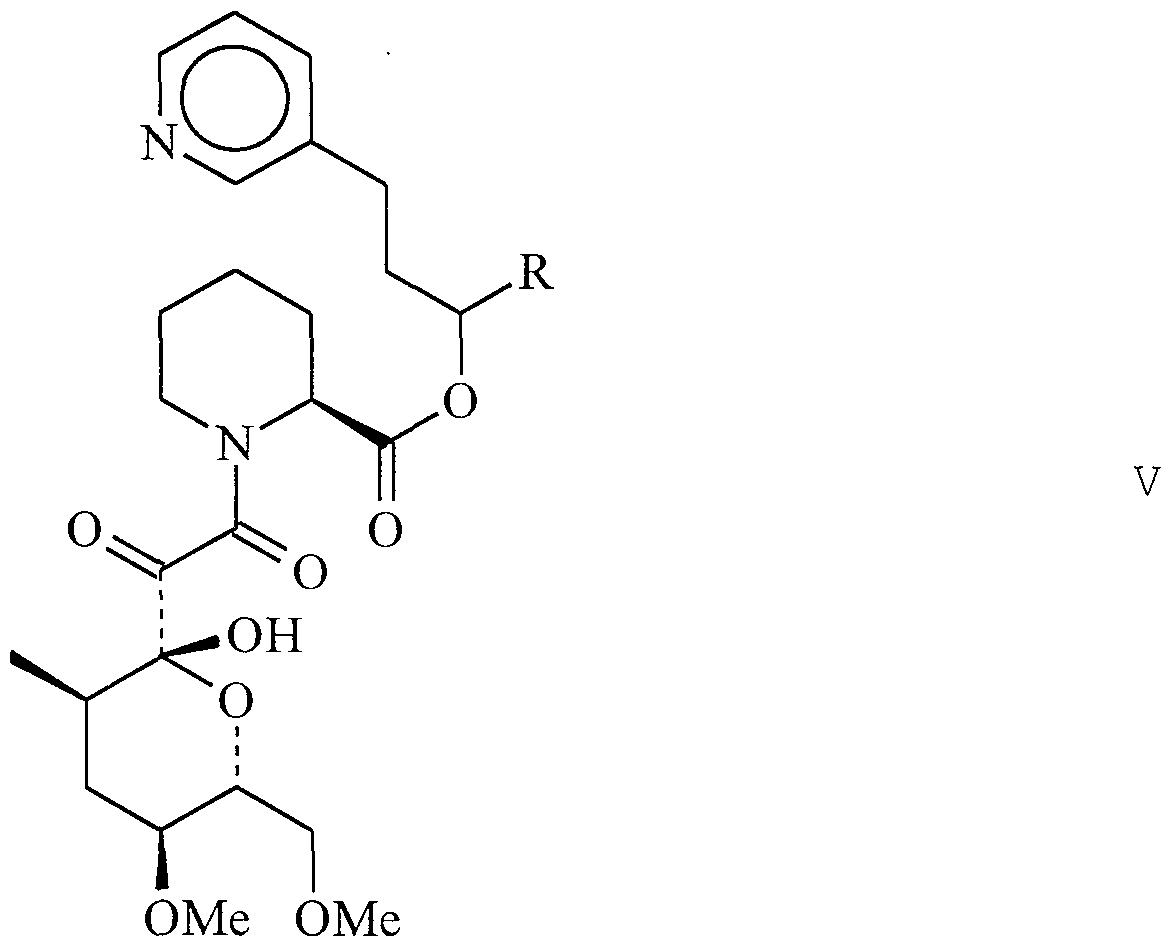
- GPI 1102 refers to Compound 98, 4-phenyl-l- (3- phenylpropyl) butyl 1- (3, 3-dimethyl-2-oxopentanoyl) -2- piperidinecarboxylate .
- GPI 1116 refers to Compound 103, l-phenethyl-3- phenylpropyl 1- (3, 3 -dimethyl -2 -oxopentanoyl) -2 - piperidinecarboxylate.
- “Isomers” refer to different compounds that have the same molecular formula. “Stereoisomers” are isomers that differ only in the way the atoms are arranged in space. "Enantiomers” are a pair of stereoisomers that are non- superimposable mirror images of each other. “Diastereoisomers” are stereoisomers which are not mirror images of each other. “Racemic mixture” means a mixture containing equal parts of individual enantiomers. “Non- racemic mixture” is a mixture containing unequal parts of individual enantiomers or stereoisomers.
- “Enhancing memory performance” refers to improving or increasing the mental faculty by which to register, retain or recall past experiences, knowledge, ideas, sensations, thoughts or impressions.
- “Memory impairment” refers to a diminished mental registration, retention or recall of past experiences, knowledge, ideas, sensations, thoughts or impressions. Memory impairment may affect short and long-term information retention, facility with spatial relationships, memory (rehearsal) strategies, and verbal retrieval and production. Common causes of memory impairment are age, severe head trauma, brain anoxia or ischemia, alcoholic-nutritional diseases, and drug intoxications. Examples of memory impairment include, without limitation, benign forgetfulness, amnesia and any disorder in which memory deficiency is present, such as Korsakoff's amnesic psychosis, dementia and learning disorders.
- Neopsic factors or “neopsics” refers to compounds useful in treating vision loss, preventing vision degeneration, or promoting vision regeneration.
- Neopsis refers to the process of treating vision loss, preventing vision degeneration, or promoting vision regeneration.
- Optological refers to anything about or concerning the eye, without limitation, and is used interchangeably with “ocular,” “ophthalmic,”
- “Pharmaceutically acceptable salt, ester, or solvate” refers to a salt, ester, or solvate of a subject compound which possesses the desired pharmacological activity and which is neither biologically nor otherwise undesirable.
- a salt, ester, or solvate can be formed with inorganic acids such as acetate, adipate, alginate, aspartate, benzoate, benzenesulfonate, bisulfate, butyrate, citrate, camphorate, camphorsulfonate, cyclopentanepropionate, digluconate, dodecylsulfate, ethanesulfonate, fumarate, glucoheptanoate, gluconate, glycerophosphate, hemisulfate, heptanoate, hexanoate, hydrochloride, hydrobromide, hydroiodide, 2- hydroxyethanesulfonate, lactate, maleate, methanesul
- base salts, esters, or solvates include ammonium salts; alkali metal salts, such as sodium and potassium salts; alkaline earth metal salts, such as calcium and magnesium salts; salts with organic bases, such as dicyclohexylamine salts; N-methyl-D-glucamine; and salts with amino acids, such as arginine, lysine, and so forth.
- the basic nitrogen- containing groups can be quarternized with such agents as lower alkyl halides, such as methyl, ethyl, propyl, and butyl chlorides, bromides, and iodides; dialkyl sulfates, such as dimethyl, diethyl, dibutyl, and diamyl sulfates; long chain halides, such as decyl, lauryl, myristyl, and stearyl chlorides, bromides, and iodides; aralkyl halides, such as benzyl and phenethyl bromides; and others. Water or oil- soluble or dispersible products are thereby obtained.
- lower alkyl halides such as methyl, ethyl, propyl, and butyl chlorides, bromides, and iodides
- dialkyl sulfates such as dimethyl, diethyl, dibutyl, and diamyl sulf
- Preventing vision degeneration refers to the ability to prevent degeneration of vision in patients newly diagnosed as having a degenerative disease affecting vision, or at risk of developing a new degenerative disease affecting vision, and for preventing further degeneration of vision in patients who are already suffering from or have symptoms of a degenerative disease affecting vision.
- “Promoting vision regeneration” refers to maintaining, improving, stimulating or accelerating recovery of, or revitalizing one or more components of the visual system in a manner which improves or enhances vision, either in the presence or absence of any ophthalmologic disorder, disease, or injury. “Treating” refers to:
- Vision refers to the ability of humans and other animals to process images, and is used interchangeably with
- Vision disorder refers to any disorder that affects or involves vision, including without limitation visual impairment, orbital disorders, disorders of the lacrimal apparatus, disorders of the eyelids, disorders of the conjunctiva, disorders of the cornea, cataracts, disorders of the uveal tract, disorders of the retina, disorders of the optic nerve or visual pathways, free radical induced eye disorders and diseases, immunologically-mediated eye disorders and diseases, eye injuries, and symptoms and complications of eye disease, eye disorder, or eye injury.
- Visual impairment refers to any dysfunction in vision including without limitation disturbances or diminution in vision (e.g., binocular, central, peripheral, scotopic) , visual acuity for objects near and far, visual field, ocular motility, color perception, adaptation to light and dark, accommodation, refraction, and lacrimation. See Physician's
- the present invention relates to a method of treating a vision disorder, improving vision, treating memory impairment, or enhancing memory performance in an animal, which comprises administering to said animal an effective amount of a derivative.
- inventive methods are particularly useful for treating various eye disorders including but not limited to visual disorders, diseases, injuries, and complications, genetic disorders; disorders associated with aging or degenerative vision diseases; vision disorders correlating to physical injury to the eye, head, or other parts of the body resulting from external- forces; vision disorders resulting from environmental factors; vision disorders resulting from a broad range of diseases; and combinations of any of the above .
- compositions and methods of the present invention are useful for improving vision, or correcting, treating, or preventing visual (ocular) impairment or dysfunction of the visual system, including permanent and temporary visual impairment, without limitation.
- the present invention is also useful in preventing and treating ophthalmologic diseases and disorders, treating damaged and injured eyes, and preventing and treating diseases, disorders, and injuries which result in vision deficiency, vision loss, or reduced capacity to see or process images, and the symptoms and complications resulting from same.
- the eye diseases and disorders which may be treated or prevented by the compositions and methods of the present invention are not limited with regard to the cause of said diseases or disorders. Accordingly, said compositions and methods are applicable whether the disease or disorder is caused by genetic or environmental factors, as well as any other influences.
- compositions and methods of the present invention are particularly useful for eye problems or vision loss or deficiency associated with all of the following, without limitation: aging, cellular or physiological degeneration, central nervous system or neurological disorder, vascular defects, muscular defects, and exposure to adverse environmental conditions or substances .
- compositions and methods of the present invention are particularly useful in correcting, treating, or improving visual impairment, without limitation.
- Visual impairment in varying degrees occurs in the presence of a deviation from normal in one or more functions of the eye, including (1) visual acuity for objects at distance and near; (2) visual fields; and (3) ocular motility without diplopia.
- PDR Planar Rexidinal Rexidinal Rexidinal Rexidinal Rexidos.
- Vision is imperfect without the coordinated function of all three. Id.
- compositions and methods of use are also useful in correcting, treating, or improving other ocular functions including, without limitation, color perception, adaptation to light and dark, accommodation, metamorphopsia, and binocular vision.
- the compositions and methods of use are particularly useful in treating, correcting, or preventing ocular disturbances including, without limitation, paresis of accommodation, iridoplegia, entropion, ectropion, epiphora, lagophthalmos, scarring, vitreous opacities, non-reactive pupil, light scattering disturbances of the cornea or other media, and permanent deformities of the orbit.
- compositions and methods of use of the present invention are also highly useful in improving vision and treating vision loss. Vision loss ranging from slight loss to absolute loss may be treated or prevented using said compositions and methods of use. Vision may be improved by the treatment of eye disorders, diseases, and injuries using the compositions and methods of the invention. However, improvements in vision using the compositions and methods of use are not so limited, and may occur in the absence of any such disorder, disease, or injury.
- compositions and methods of the present invention are also useful in the treatment or prevention of the following non-limiting exemplary diseases and disorders, and symptoms and complications resulting therefrom.
- Vision disorders include but are not limited to the following : visual impairment, such as diminished visual acuity for objects near and far, visual fields, and ocular motility; orbital disorders, such as orbital cellulitis, periorbital cellulitis, cavernous sinus thrombosis, and exophthalmos (proptosis); disorders of the lacrimal apparatus, such as dacryostenosis, congenital dacryostenosis, and dacryocystitis (acute or chronic) ; disorders of the eyelids, such as lid edema, blepharitis, ptosis, Bell's palsy, blepharospasm, hordeolum (stye), external hordeolum, internal hordeolum (meibomian stye), chalazion, entropion (inversion of the eyelid), ectropion (eversion of the eyelid) , tumors (benign and malignant
- glaucoma e.g., primary glaucoma, chronic open-angle glaucoma, acute or chronic angle-closure, congenital (infantile) glaucoma, secondary glaucoma, and absolute glaucoma
- disorders of the optic nerve or visual pathways such as papilledema (choked disk) , papillitis (optic neuritis) , retrobulbar neuritis, ischemic optic neuropathy, toxic amblyopia, optic atrophy, higher visual pathway lesions, disorders of ocular motility (e.g., third cranial nerve palsies, fourth cranial nerve palsies, sixth cranial nerve palsies, internuclear ophthalmoplegia, and gaze pal
- disorders of ocular motility e.g., third cranial nerve palsies, fourth cranial nerve palsies, sixth cranial nerve palsies, internuclear ophthalmoplegia, and gaze pal
- compositions and methods of the present invention are also useful in the treatment of the following non- limiting eye injuries, and symptoms and complications resulting therefrom: conjunctival and corneal foreign body injuries, corneal abrasion, intraocular foreign body injuries, lacerations, lid lacerations, contusions, lid contusions (black eye) , trauma to the globe, laceration of the iris, cataract, dislocated lens, glaucoma, vitreous hemorrhage, orbital-floor fractures, retinal hemorrhage or detachment, and rupture of the eyeball, anterior chamber hemorrhage (traumatic hyphema) , burns, eyelid burns, chemical burns, chemical burns of the cornea and conjunctiva, and ultraviolet light burns (sunburn).
- compositions and methods of the present invention are also useful in treating and/or preventing the following non-limiting exemplary symptoms and complications of eye disease, eye disorder or eye injury: subconjunctival hemorrhages, vitreous hemorrhages, retinal hemorrhages, floaters, retinal detachments, photophobia, ocular pain, scotomas (negative and positive) , errors of refraction, emmetropia, ametropia, hyperopia (farsightedness), myopia
- the derivative may be administered in combination with an effective amount of one or more factor (s) useful in treating vision disorder, improving vision, treating memory impairment, or enhancing memory performance.
- the factor (s) to be combined with the derivative is/are selected from the group consisting of immunosuppressants for treating autoimmune, inflammatory, and immunologically-mediated disorders; wound healing agents for treating wounds resulting from injury or surgery; antiglaucomatous medications for treating abnormally elevated intraocular pressure; neurotrophic factors and growth factors for treating neurodegenerative disorders or stimulating neurite outgrowth; compounds effective in limiting or preventing hemorrhage or neovascularization for treating macular degeneration; and antioxidants for treating oxidative damage to eye tissues.
- immunosuppressants for treating autoimmune, inflammatory, and immunologically-mediated disorders
- wound healing agents for treating wounds resulting from injury or surgery
- antiglaucomatous medications for treating abnormally elevated intraocular pressure
- neurotrophic factors and growth factors for treating neurodegenerative disorders or stimulating neurite outgrowth
- compounds effective in limiting or preventing hemorrhage or neovascularization for treating macular degeneration and antioxidants for treating oxidative damage to eye tissues.
- compositions of the Present Invention also relates to a pharmaceutical composition comprising:
- the derivative may be administered in combination with an effective amount of one or more factor (s) useful in treating vision disorders, improving vision, treating memory impairment, or enhancing memory performance.
- the pipecolic acid derivatives used in the methods and pharmaceutical compositions of the present invention have an affinity for FKBP-type immunophilins, such as FKBP12.
- FKBP-type immunophilins such as FKBP12.
- a pipecolic acid derivative binds to an FKBP-type immunophilin, it has been found to inhibit the prolyl- peptidyl cis-trans isomerase, or rotamase, activity of the binding protein.
- the compounds have also been found to stimulate hair growth.
- These rotamase inhibiting compounds may be immunosuppressive or non-immunosuppressive. Examples of useful compounds are set forth below.
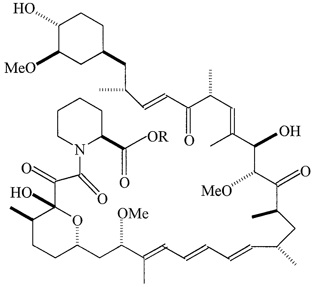
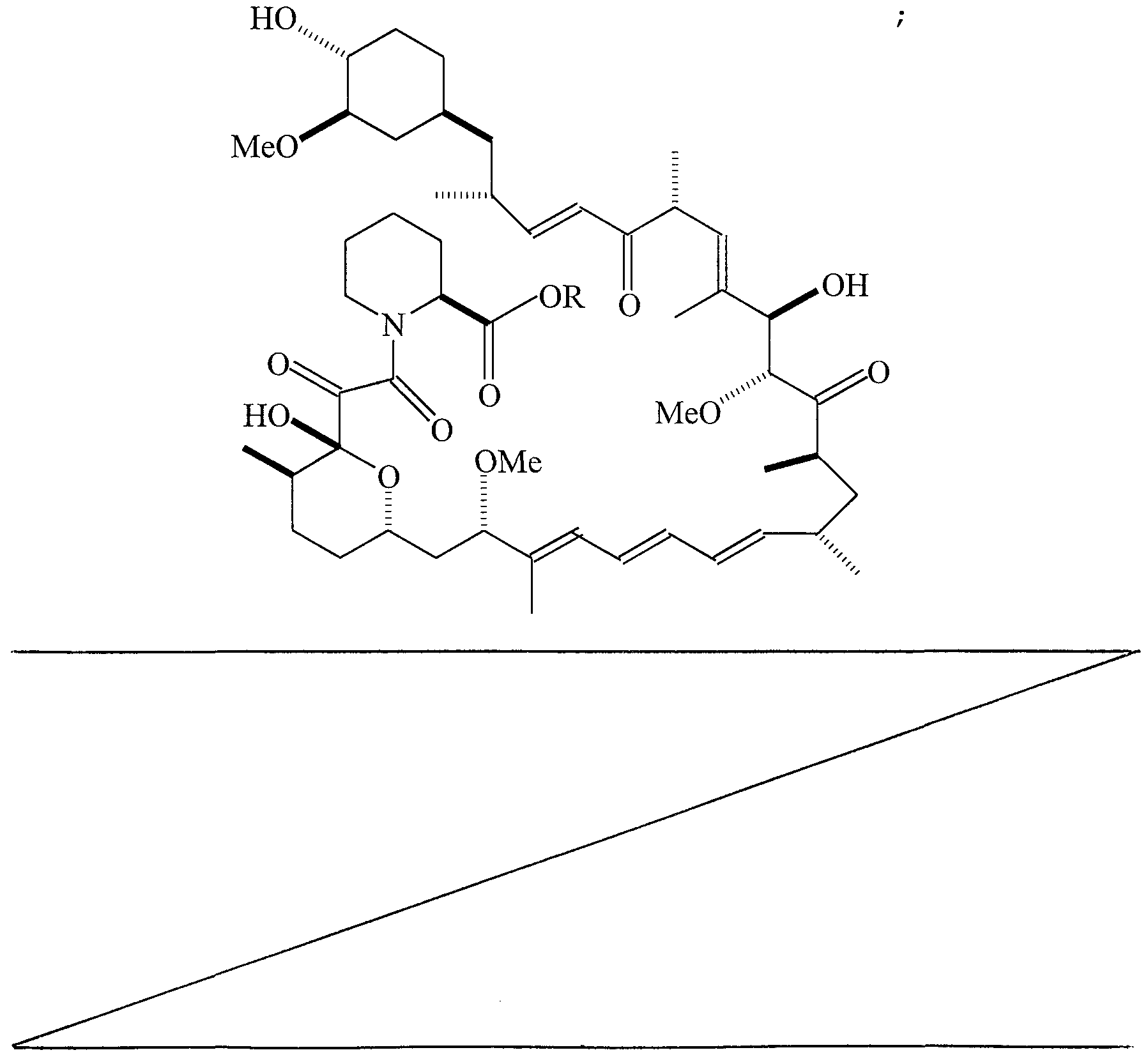
- COMPOUND 1 Ocain et al., Biochemi cal and Biophysi cal Research Communica tions, Vol. 192, No. 3, 1993, incorporated herein by reference, discloses an exemplary pipecolic acid derivative represented by Formula I.
- the compound was synthesized at Wyeth-Ayerst by Dr. Phil Hughes by reaction of 4-phenyl- 1, 2 , 4-triazoline-3, 5-dione with rapamycin.
- R 2 Phe-o- tert-butyl
- R 2 Phe-o- ert-butyl
- R 3 Val-O- tert-butyl Compound Structure
- R 3 Leu-O- ert-butyl
- R 3 I leu-O- tert-butyl
- R 3 allylalanine-O- ert-butyl
- R 3 Val-O- tert-butyl
- R 3 CH 2 OMe
- Additional exemplary pipecolic acid derivatives are represented by Formulas XXI-XXV and Tables XXI-XXV.
- All the compounds of Formulas I-XXV possess asymmetric centers and thus can be produced as mixtures of stereoisomers or as individual R- and S- stereoisomers.
- the individual stereoisomers may be obtained by using an optically active starting material, by resolving a racemic or non-racemic mixture of an intermediate at some appropriate stage of the synthesis, or by resolving the compounds of Formulas I-XXV. It is understood that the compounds of Formulas I-XXV encompass individual stereoisomers as well as mixtures (racemic and non-racemic) of stereoisomers.
- S- stereoisomers are used in the pharmeceutical compositions and methods of the present invention.
- the compounds used in the inventive methods and pharmaceutical compositions have an affinity for the FK506 binding protein, particularly FKBP12.
- the inhibition of the prolyl peptidyl cis-trans isomerase activity of FKBP may be measured as an indicator of this affinity.
- Inhibition of the peptidyl-prolyl isomerase (rotamase) activity of the compounds used in the inventive methods and pharmaceutical compositions can be evaluated by known methods described in the literature (Harding et al . , Na ture, 1989, 341:758-760; Holt et al . J. Am . Chem . Soc , 115:9923-9938). These values are obtained as apparent Ki ' s and are presented for representative compounds in TABLE XXVII.
- the cis-trans isomerization of an alanine-proline bond in a model substrate, N-succinyl-Ala-Ala-Pro-Phe-p- nitroanilide is monitored spectrophotometrically in a chymotrypsin-coupled assay, which releases para-nitroanilide from the trans form of the substrate.
- the inhibition of this reaction caused by the addition of different concentrations of inhibitor is determined, and the data is analyzed as a change in first-order rate constant as a function of inhibitor concentration to yield the apparent K ⁇ values.
- a plastic cuvette In a plastic cuvette are added 950 mL of ice cold assay buffer (25 mM HEPES, pH 7.8, 100 mM NaCl), 10 mL of FKBP (2.5 mM in 10 mM Tris-Cl pH 7.5, 100 mM NaCl, 1 mM dithiothreitol) , 25 mL of chymotrypsin (50 mg/ml in 1 mM HCI) and 10 mL of test compound at various concentrations in dimethyl sulfoxide.
- ice cold assay buffer 25 mM HEPES, pH 7.8, 100 mM NaCl
- FKBP 2.5 mM in 10 mM Tris-Cl pH 7.5, 100 mM NaCl, 1 mM dithiothreitol
- 25 mL of chymotrypsin 50 mg/ml in 1 mM HCI
- 10 mL of test compound at various concentrations in dimethyl
- the reaction is initiated by the addition of 5 mL of substrate (succinyl-Ala-Phe-Pro-Phe-para- nitroanilide, 5 mg/mL in 2.35 mM LiCl in trifluoroethanol) .
- substrate succinyl-Ala-Phe-Pro-Phe-para- nitroanilide, 5 mg/mL in 2.35 mM LiCl in trifluoroethanol
- the absorbance at 390 nm versus time is monitored for 90 seconds using a spectrophotometer and the rate constants are determined from the absorbance versus time data files.
- the compounds used in the inventive methods and pharmaceutical compositions must readily affect the targeted areas.
- the compounds are preferably administered [topically to the skin.]
- Dosage levels on the order of about 0.1 mg to about 10,000 mg of the active ingredient compound are useful in the treatment of the above conditions, with preferred levels of about 0.1 mg to about 1,000 mg.
- the specific dose level for any particular patient will vary depending upon a variety of factors, including the activity of the specific compound employed; the age, body weight, general health, sex and diet of the patient; the time of administration; the rate of excretion; drug combination; the severity of the particular disease being treated; and the form of administration. Typically, in vi tro dosage-effect results provide useful guidance on the proper doses for patient administration. Studies in animal models are also helpful. The considerations for determining the proper dose levels are well known in the art.
- the compounds can be administered with other hair revitalizing agents. Specific dose levels for the other hair revitalizing agents will depend upon the factors previously stated and the effectiveness of the drug combination. EXAMPLES
- GPI 1046 protects retinal ganglion cells against degeneration following retinal ischemia.
- Retinal ganglion cells were retrogradely labeled in adult rats by bilateral injection of fluorogold in their lateral geniculate nuclei. Labeled ganglion cells in the normal rat retina appear as white profiles against the dark background ( Figure IA) .
- Complete retinal ischemia was produced by infusing normal saline solution into the retinal vitreous cavity of each eye until the intraocular pressure exceeded arterial blood pressure. 28 days after the ischemic episode extensive degeneration of retinal ganglion cell was evidenced by massive reduction in the density of fluorogold labeled cells
- Figure IB Administration of GPI 1046 (lOmg/kg, s.c.) 1 hour prior to the ischemic episode and at lOmg/kg/day for the next four days produced noticeable protection of a large proportion of the vulnerable ganglion cell population ( Figure IC) .
- GPI 1046 prevents degeneration of optic nerve axons and myelin following retinal ischemia Examination of the optic nerves from the same retinal ischemia cases reveals that GPI 1046 produces dramatic protection of optic nerve element from ischemic degeneration. Toluidine blue staining of epon embedded optic nerve cross sections revealed the detail of myelin sheaths (white circles) and optic nerve axons (black centers) in the normal rat optic nerve. Optic nerves from vehicle treated cases examined 28 days after a 1 hour retinal ischemic episode are characterized by a decreased density of optic nerve axons and the appearance of numerous degenerating myelin figures (bright white filled circles). Treatment with GPI 1046 protected the majority of optic nerve axons from degeneration and also dramatically decreased the density of degenerating myelin figures.
- GPI 1046 provides moderate protection against retinal ganglion cell death after optic nerve transection
- GPI 1046 treatment duration significantly affects the process of optic nerve axonal degeneration after transection.
- FIG. 1046 treatment produces a greater effect on optic nerve axons than ganglion cell bodies
- This summary figure shows data from Figure 3 ganglion cell protection and higher power photomicrographs of optic nerve axon protection ( Figure 5A&B, upper panels) .
- 28 day treatment with GPI 1046 produced a significant increase in the density of large, and particularly medium and small caliber optic nerve axons ( Figure 5C&D, lower panels).
- FIG. 7 FKBP-12 immunohistochemistry labels oligodendroglia (large dark cells with fibrous processes) , the cells which produce myelin, located between the fascicles of optic nerve fibers , and also some optic nerve axons .
- GPI 1046 treatment for the first 14 days after transection did not protect against shrinkage of the distal stump but did slightly increase the density of myelin, though the density of degenerating myelin figures remained high ( Figure 8C, Table A) .
- GPI 1046 treatment through the first 28 days produced dramatic protection of the fascicular pattern of myelin labeling, decreased the density of degenerating myelin figures, prevented cross sectional shrinkage of the distal stump of the transected nerve and maintained the myelin levels at -99% of normal levels (Figure 8D, Table A) .
- Figure 9 28 day treatment with GPI 1046 treatment beginning 8 weeks after onset of streptozotocin induced diabetes decreases the extent of neovascularization in the inner and outer retina and protects neurons in the inner nuclear layer (INL) and ganglion cell layer (GCL) from degeneration.
- INL inner nuclear layer
- GCL ganglion cell layer
- the extent of degeneration reduction or prevention in retinal ganglion cells and optic nerve axons was determined in a vision loss model utilizing surgical optic nerve transection to simulate mechanical damage to the optic nerve.
- the effects of several neuroimmunophilin FKBP ligands on retinal ganglion cells neuroprotection and optic nerve axon density was determined experimentally, comparing 14 day and 28 day neuroimmunophilin FKBP ligand treatments.
- the effects of treatment with neuroimmunophilin FKBP ligands on retinal ganglion cells and optic nerve axons was correlated.
- mice were divided into six experimental groups of six rats (12 eyes) per group.
- One group received a neuroimmunophilin FKBP ligand (10 milligrams per kg per day sc in PEG vehicle (20 percent propylene glycol, 20 percent ethanol, and 60 percent saline)) for 14 days.
- a second group received the same neuroimmunophilin FKBP ligand dose for 28 days.
- Each treated group had a corresponding sham/surgery and transection control group which received corresponding 14 or 28 day dosing with the vehicle only.
- Retinas were removed from eyes and prepared for wholemount analysis. For each group, five eyes with dense and intense FG labeling were selected for quantitative analysis using a 20 power objective. Digital images were obtained from five fields in the central retina (3-4 millimeters radial to optic nerve head) . FG labeled Large (>18 ⁇ m) , medium (12-16 ⁇ m) , and small ( ⁇ 10 ⁇ m) ganglion cells and microglia were counted in five 400 ⁇ m by 400 ⁇ m fields per case, 5 cases per group. Examination of Optic Nerves Proximal and distal optic nerve stumps were identified, measured, and transferred to 30% sucrose saline.
- proximal stumps of five nerves were blocked and affixed to a chuck, and 10 micron cross sections were cut on a cryostat; one in ten sections were saved per set. Sections including the region 1-2 mm behind the orbit were reacted for RT97 neurofilament immunohistochemistry.
- Analysis of optic nerve axon density was performed using a 63 power oil immersion lens, a Dage 81 camera, and the Simple Image Analysis program.
- RT97 positive optic nerve axons were counted in three 200 ⁇ m by 200 ⁇ m fields per nerve. The area of the nerve was also determined for each case at 10 power.
- the number of axons persisting in the proximal stump of the optic nerve represented approximately one half of the number of surviving ganglion cells in groups of animals that received vehicle alone or the 14 day course of treatment with a neuroimmunophilin FKBP ligand.
- ⁇ estimate for 200 ⁇ m x 200 ⁇ m region in normal optic nerve assuming 120,000 RGC axons in normal rat optic nerve, measured to be 0.630 mm ⁇ mean cross sectional area adjusted for optic nerve diameter calculated by multiplying axonal density by ON area
- GP1 1046 preserves optic nerve axons in the proximal stump following transection
- a patient is suffering from macular degeneration.
- a derivative as identified. above, alone or in combination with one or more other neopsic factors, or a pharmaceutical composition comprising the same, may be administered to the patient.
- a reduction in vision loss, prevention of vision degeneration, and/or promotion of vision regeneration are/is expected to occur following treatment.
- a patient is suffering from glaucoma, resulting in cupping of the optic nerve disc and damage to nerve fibers.
- a derivative as identified above, alone or in combination with one or more other neopsic factors, or a pharmaceutical composition comprising the same, may be administered to the patient.
- a reduction in vision loss, prevention of vision degeneration, and/or promotion of vision regeneration are/is expected to occur following treatment.
- a patient is suffering from cataracts requiring surgery. Following surgery, a derivative as identified above, alone or in combination with one or more other neopsic factors, or a pharmaceutical composition comprising the same, may be administered to the patient.
- a reduction in vision loss, prevention of vision degeneration, and/or promotion of vision regeneration are/is expected to occur following treatment.
- Example 6 A patient is suffering from an impairment or blockage of retinal blood supply relating to diabetic retinopathy, ischemic optic neuropathy, or retinal artery or vein blockage.
- a derivative as identified above, alone or in combination with one or more other neopsic factors, or a pharmaceutical composition comprising the same, may be administered to the patient.
- a reduction in vision loss, prevention of vision degeneration, and/or promotion of vision regeneration are/is expected to occur following treatment.
- Example 7 A patient is suffering from a detached retina.
- a derivative as identified above, alone or in combination with one or more other neopsic factors, or a pharmaceutical composition comprising the same, may be administered to the patient.
- a reduction in vision loss, prevention of vision degeneration, and/or promotion of vision regeneration are/is expected to occur following treatment.
- a patient is suffering from tissue damage caused by inflammation associated with uveitis or conjunctivitis.
- a derivative as identified above, alone or in combination with one or more other neopsic factors, or a pharmaceutical composition comprising the same, may be administered to the patient.
- a reduction in vision loss, prevention of vision degeneration, and/or promotion of vision regeneration are/is expected to occur following treatment.
- a patient is suffering from photoreceptor damage caused by chronic or acute exposure to ultraviolet light.
- a derivative as identified above, alone or in combination with one or more other neopsic factors, or a pharmaceutical composition comprising the same, may be administered to the patient.
- a reduction in vision loss, prevention of vision degeneration, and/or promotion of vision regeneration are/is expected to occur following treatment.
- a patient is suffering from optic neuritis.
- a derivative as identified above, alone or in combination with one or more other neopsic factors, or a pharmaceutical composition comprising the same, may be administered to the patient.
- a reduction in vision loss, prevention of vision degeneration, and/or promotion of vision regeneration are/is expected to occur following treatment.
- Example 11 A patient is suffering from tissue damage associated with a "dry eye” disorder.
- a derivative as identified above, alone or in combination with one or more other neopsic factors, or a pharmaceutical composition comprising the same, may be administered to the patient.
- a reduction in vision loss, prevention of vision degeneration, and/or promotion of vision regeneration are/is expected to occur following treatment .
- Example 12 Efficacy of representative compounds from different immunophilin ligand series in protecting retinal ganglion cell axons from degeneration following optic nerve transection is set forth in Table E.
- Ki rotamase nM 30.3%
- Ki rotamase 210 nM ⁇ 4.8% SEM
- Ki rotamase 86 nM 13.0%
- Ki rotamase >7743 nM 7.8%
- Ki rotamase 7 nM -6.3%
- Retinal ganglion cells in adult male Sprague Dawley rats were retrogradely labeled by fluorogold injection in the LGNd and four days later the optic nerves were transected 5 mm behind the globe. Groups of animals received either GPI-1046 lOmg/kg/day s.c. or vehicle for 28 days. All experimental animals and controls were sacrificed 90 days after transection.
- Peripheral neuropathy is a common debilitating complication of Type 2 diabetes in some 30-40% of diabetic patients.
- Neurotrophic factors such as nerve growth factor
- NGF neuroimmunophilin FKBP-12
- GPI- 1046 the small molecule GPI- 1046
- the Morris watermaze is widely used for assessing spatial memory formation and retention in experimental animals.
- the test depends on the animal's ability to utilize spatial visual information in order to locate a submerged escape platform in a water tank. It is important that the tank itself be as devoid of specific visual features as possible - thus, it is always circular in shape, the sides are kept smooth and in uniform dull colors, and the water is rendered opaque with nontoxic watercolour pigment or powdered milk. This is to ensure that the animal navigates only by the use of more distant visual cues, or by the use of intra- maze cues specifically provided by the experimenter.
- the tank is filled to a level which forces the animal to swim actively. Normal mice and rats react aversively to the swimming part of the test and will climb onto, and remain on, an escape platform from which they are removed to a heated resting cage.
- the platform is visible (i.e. above the surface), animals placed in the tank will quickly learn to home in on the platform and climb out onto it. Testing with a visible platform will also ensure that the experimental animals are not blind and show sufficient motivation and stamina to perform the task, which can be important in experiments involving aged rodents. If the platform is invisible (i.e. submerged just below the surface) , normal animals learn to use distant visual cues in the test room for orientation in the test tank, and, when placed in the tank, will quickly home in on the approximate location of the platform and circle in that area until the platform is found.
- the animals' path, speed, and swim time are tracked with a ceiling camera for later computerized analysis. Over the course of several successive trials, spatial learning can therefore be defined as a drop of distance swum, or time elapsed, from placement in the tank until escape onto the invisible platform.
- the test can be adapted to assess several aspects of spatial memory: a) acquisition of a cued task, where the animal's ability to link one visual cue directly with the escape platform depends on cortical function (i.e. a ball is suspended over the escape platform and the animal learns to follow this cue to find the platform) ; b) acquisition of a spatial task, where the animal's ability to learn the location of a submerged escape platform based on a combination of distant visual cues is dependent upon hippocampal function (i.e. the animal learns to triangulate its position in the tank by visually aligning the paper-tower dispenser with the door and ceiling lamp) ; c) retention of a successfully acquired spatial task, which is predominantly dependant on cortical function (i.e.
- the animal must remember the spatial location of the platform over several weeks); d) a hippocampus-dependant reversal task where the animals must reacquire a new spatial platform location (i.e. the platform is moved to a new location between swim trials and the animal must abandon its previous search strategy and acquire a new one) .
- mice were given 4 trials/day
Abstract
Description


Claims


Priority Applications (5)
| Application Number | Priority Date | Filing Date | Title |
|---|---|---|---|
| MXPA01001642A MXPA01001642A (en) | 1998-08-14 | 1999-08-12 | Pipecolic acid derivatives for vision and memory disorders. |
| CA002344520A CA2344520A1 (en) | 1998-08-14 | 1999-08-12 | Pipecolic acid derivatives for vision and memory disorders |
| JP2000564612A JP2002522485A (en) | 1998-08-14 | 1999-08-12 | Pipecolic acid derivatives for visual and memory disorders |
| AU55557/99A AU5555799A (en) | 1998-08-14 | 1999-08-12 | Pipecolic acid derivatives for vision and memory disorders |
| EP99942109A EP1109554A2 (en) | 1998-08-14 | 1999-08-12 | Pipecolic acid derivatives for vision and memory disorders |
Applications Claiming Priority (2)
| Application Number | Priority Date | Filing Date | Title |
|---|---|---|---|
| US09/134,417 | 1998-08-14 | ||
| US09/134,417 US6376517B1 (en) | 1998-08-14 | 1998-08-14 | Pipecolic acid derivatives for vision and memory disorders |
Publications (2)
| Publication Number | Publication Date |
|---|---|
| WO2000009109A2 true WO2000009109A2 (en) | 2000-02-24 |
| WO2000009109A3 WO2000009109A3 (en) | 2000-08-17 |
Family
ID=22463300
Family Applications (1)
| Application Number | Title | Priority Date | Filing Date |
|---|---|---|---|
| PCT/US1999/018242 WO2000009109A2 (en) | 1998-08-14 | 1999-08-12 | Pipecolic acid derivatives for vision and memory disorders |
Country Status (7)
| Country | Link |
|---|---|
| US (1) | US6376517B1 (en) |
| EP (1) | EP1109554A2 (en) |
| JP (2) | JP2002522485A (en) |
| AU (1) | AU5555799A (en) |
| CA (1) | CA2344520A1 (en) |
| MX (1) | MXPA01001642A (en) |
| WO (1) | WO2000009109A2 (en) |
Cited By (17)
| Publication number | Priority date | Publication date | Assignee | Title |
|---|---|---|---|---|
| WO2000038703A1 (en) * | 1998-12-24 | 2000-07-06 | R-Tech Ueno, Ltd. | Agent for treating visual cell function disorder |
| WO2000066122A1 (en) * | 1999-04-30 | 2000-11-09 | Sucampo Ag | Use of macrolide compounds for the treatment of dry eye |
| WO2003048142A1 (en) * | 2001-12-06 | 2003-06-12 | Institute Of Pharmacology And Toxicology Academy Of Military Medical Sciences P.L.A. | Substituted 6-membered n-heterocyclic compounds and their uses as neurological regulator |
| US6864232B1 (en) | 1998-12-24 | 2005-03-08 | Sucampo Ag | Agent for treating visual cell function disorder |
| US6872383B2 (en) | 1999-04-30 | 2005-03-29 | Sucampo Ag | Use of macrolide compounds for the treatment of dry eye |
| EP1539157A2 (en) * | 2002-09-18 | 2005-06-15 | Trustees Of The University Of Pennsylvania | Method of inhibiting choroidal neovascularization |
| US7273874B2 (en) | 2004-12-20 | 2007-09-25 | Wyeth | Rapamycin derivatives and the uses thereof in the treatment of neurological disorders |
| US7276498B2 (en) | 2004-12-20 | 2007-10-02 | Wyeth | Rapamycin analogues and uses thereof in the treatment of neurological disorders |
| US8367097B2 (en) | 2005-02-09 | 2013-02-05 | Santen Pharmaceutical Co., Ltd. | Liquid formulations for treatment of diseases or conditions |
| EP2589383A1 (en) | 2011-11-06 | 2013-05-08 | Max-Planck-Gesellschaft zur Förderung der Wissenschaften e.V. Berlin | FKBP subtype-specific rapamycin analogue for use in treatment of diseases |
| WO2013097947A1 (en) * | 2011-12-28 | 2013-07-04 | MAX-PLANCK-Gesellschaft zur Förderung der Wissenschaften e.V. | Pipecolate-sulfonamides for treatment of psychiatric disorders |
| US8486960B2 (en) | 2006-03-23 | 2013-07-16 | Santen Pharmaceutical Co., Ltd. | Formulations and methods for vascular permeability-related diseases or conditions |
| US8658667B2 (en) | 2006-02-09 | 2014-02-25 | Santen Pharmaceutical Co., Ltd. | Stable formulations, and methods of their preparation and use |
| WO2015004455A2 (en) * | 2013-07-09 | 2015-01-15 | Isomerase Therapeutics Limited | Novel compounds |
| KR20150126345A (en) * | 2013-03-13 | 2015-11-11 | 산텐 세이야꾸 가부시키가이샤 | Therapeutic agent for meibomian dysfunction |
| WO2019185708A1 (en) | 2018-03-28 | 2019-10-03 | Georg-August-Universitaet Goettingen, Stiftung Oeffentlichen Rechts, Universitaetsmedizin | Prevention or treatment of chronic organ injury |
| US11400080B2 (en) | 2016-05-25 | 2022-08-02 | Santen Pharmaceutical Co., Ltd. | Use of sirolimus to treat exudative age-related macular degeneration with persistent edema |
Families Citing this family (17)
| Publication number | Priority date | Publication date | Assignee | Title |
|---|---|---|---|---|
| AU2003272539A1 (en) * | 2002-09-17 | 2004-04-08 | New York University | Methods of treating age associated memory impairment (aami), mild cognitive impairment (mci), and dementias with cell cycle inhibitors |
| AR042890A1 (en) * | 2003-01-16 | 2005-07-06 | Sucampo Pharmaceuticals Inc | METHOD OF DRY EYE TREATMENT WITH AN OPHTHALMIC COMPOSITION CONTAINING A MACROLID COMPOUND |
| US20060258668A1 (en) * | 2003-05-29 | 2006-11-16 | Scolnick Edward M | Use of phosphatase inhibitors as adjunct therapy for psychiatric disorders |
| AU2004274026A1 (en) * | 2003-09-18 | 2005-03-31 | Macusight, Inc. | Transscleral delivery |
| US20070059336A1 (en) * | 2004-04-30 | 2007-03-15 | Allergan, Inc. | Anti-angiogenic sustained release intraocular implants and related methods |
| US20050244472A1 (en) * | 2004-04-30 | 2005-11-03 | Allergan, Inc. | Intraocular drug delivery systems containing excipients with reduced toxicity and related methods |
| US8591885B2 (en) * | 2004-04-30 | 2013-11-26 | Allergan, Inc. | Carbonic anhydrase inhibitor sustained release intraocular drug delivery systems |
| US8663639B2 (en) * | 2005-02-09 | 2014-03-04 | Santen Pharmaceutical Co., Ltd. | Formulations for treating ocular diseases and conditions |
| EP1871366A2 (en) * | 2005-03-21 | 2008-01-02 | Macusight, Inc. | Drug delivery systems for treatment of diseases or conditions |
| US11078262B2 (en) | 2007-04-30 | 2021-08-03 | Allergan, Inc. | High viscosity macromolecular compositions for treating ocular conditions |
| US9428845B1 (en) | 2010-12-28 | 2016-08-30 | Warp Drive Bio, Inc. | Identifying new therapeutic agents |
| EP3007696B1 (en) * | 2013-06-14 | 2019-10-16 | The Board of Regents of The University of Texas System | Novel allosteric inhibitors of proteasome and methods of use thereof |
| CN115645399A (en) | 2016-06-30 | 2023-01-31 | 度瑞公司 | Long-acting formulations |
| US10682340B2 (en) | 2016-06-30 | 2020-06-16 | Durect Corporation | Depot formulations |
| EA201990127A1 (en) | 2016-12-30 | 2020-08-18 | Дьюрект Корпорейшн | DEPO-PREPARATION |
| US11020383B2 (en) | 2017-04-20 | 2021-06-01 | The Board Of Regents Of The University Of Texas System | Methods for the treatment of cancer metastasis |
| WO2019152527A1 (en) | 2018-01-30 | 2019-08-08 | Board Of Regents, The University Of Texas System | Pipecolic esters for inhibition of the proteasome |
Citations (24)
| Publication number | Priority date | Publication date | Assignee | Title |
|---|---|---|---|---|
| EP0356399A2 (en) * | 1988-08-26 | 1990-02-28 | Sandoz Ag | Substituted 4-azatricyclo (22.3.1.04.9) octacos-18-ene derivatives, their preparation and pharmaceutical compositions containing them |
| EP0402931A1 (en) * | 1989-06-14 | 1990-12-19 | Sandoz Ltd. | Heteroatoms-containing tricyclic compounds |
| WO1992000278A1 (en) * | 1990-07-02 | 1992-01-09 | Vertex Pharmaceuticals Incorporated | Novel immunosuppressive compounds |
| EP0484936A1 (en) * | 1990-11-08 | 1992-05-13 | Fujisawa Pharmaceutical Co., Ltd. | Suspensions containing tricyclic compounds |
| DE4039587A1 (en) * | 1990-12-12 | 1992-06-17 | Sandoz Ag | New derivs. of FR 520 and FK 506 - for treating transplant resistance, auto:immune diseases and inflammatory or hyper proliferative skin diseases |
| EP0509753A1 (en) * | 1991-04-18 | 1992-10-21 | Merck & Co. Inc. | Semi-synthetic immunosuppressive macrolides |
| WO1992019278A1 (en) * | 1991-04-26 | 1992-11-12 | Kurume University | Use of macrolide compounds for eye diseases |
| WO1992021313A2 (en) * | 1991-05-24 | 1992-12-10 | Vertex Pharmaceuticals Incorporated | Novel immunosuppressive compounds |
| EP0532862A1 (en) * | 1991-07-25 | 1993-03-24 | University Of Louisville Research Foundation, Inc. | Use of rapamycin for the preparation of a medicament for the treatment of ocular inflammation |
| US5252579A (en) * | 1993-02-16 | 1993-10-12 | American Home Products Corporation | Macrocyclic immunomodulators |
| WO1994021642A1 (en) * | 1993-03-17 | 1994-09-29 | Abbott Laboratories | Macrocyclic amide and urea immunomodulators |
| WO1995014023A1 (en) * | 1993-11-19 | 1995-05-26 | Abbott Laboratories | Semisynthetic analogs of rapamycin (macrolides) being immunomodulators |
| US5541193A (en) * | 1991-09-05 | 1996-07-30 | Abbott Laboratories | Heterocycle-containing macrocyclic immunomodulators |
| WO1996040140A1 (en) * | 1995-06-07 | 1996-12-19 | Guilford Pharmaceuticals Inc. | Inhibitors of rotamase enzyme activity |
| WO1996041609A2 (en) * | 1995-06-08 | 1996-12-27 | Vertex Pharmaceuticals Incorporated | Methods and compositions for stimulating neurite growth |
| WO1997016190A1 (en) * | 1995-10-31 | 1997-05-09 | Guilford Pharmaceuticals Inc. | Rotamase enzyme activity inhibitors |
| WO1997025977A1 (en) * | 1996-01-19 | 1997-07-24 | Novartis Ag | Pharmaceutical compositions |
| US5721256A (en) * | 1997-02-12 | 1998-02-24 | Gpi Nil Holdings, Inc. | Method of using neurotrophic sulfonamide compounds |
| WO1998013343A1 (en) * | 1996-09-25 | 1998-04-02 | Guilford Pharmaceuticals Inc. | Heterocyclic thioesters and ketones |
| WO1998013355A1 (en) * | 1996-09-25 | 1998-04-02 | Guilford Pharmaceuticals Inc. | Heterocyclic esters and amides |
| WO1998020893A1 (en) * | 1996-11-13 | 1998-05-22 | Vertex Pharmaceuticals Incorporated | Methods and compositions for stimulating neurite growth using compounds with affinity for fkbp12 in combination with neurotrophic factors |
| WO1998029116A1 (en) * | 1996-12-31 | 1998-07-09 | Guilford Pharmaceuticals Inc. | N-linked sulfonamides of heterocyclic thioesters |
| WO1998037882A1 (en) * | 1997-02-27 | 1998-09-03 | Guilford Pharmaceuticals Inc. | Method of using neurotrophic carbamates and ureas |
| WO1999042104A1 (en) * | 1998-02-23 | 1999-08-26 | Fujisawa Pharmaceutical Co., Ltd. | Use of macrolide compounds for treating glaucoma |
Family Cites Families (87)
| Publication number | Priority date | Publication date | Assignee | Title |
|---|---|---|---|---|
| US4070361A (en) | 1977-04-21 | 1978-01-24 | E. R. Squibb & Sons, Inc. | Mercaptoalkylsulfonyl proline and pipecolic acid and esters thereof |
| IL58849A (en) | 1978-12-11 | 1983-03-31 | Merck & Co Inc | Carboxyalkyl dipeptides and derivatives thereof,their preparation and pharmaceutical compositions containing them |
| US4310461A (en) | 1980-06-23 | 1982-01-12 | E. R. Squibb & Sons, Inc. | Imido, amido and amino derivatives of mercaptoacyl prolines and pipecolic acids |
| US4578474A (en) | 1980-06-23 | 1986-03-25 | E. R. Squibb & Sons, Inc. | Imido, amido and amino derivatives of mercaptoacyl prolines and pipecolic acids |
| US4390695A (en) | 1980-06-23 | 1983-06-28 | E. R. Squibb & Sons, Inc. | Imido, amido and amino derivatives of mercaptoacyl prolines and pipecolic acids |
| US4950649A (en) | 1980-09-12 | 1990-08-21 | University Of Illinois | Didemnins and nordidemnins |
| EP0048159A3 (en) | 1980-09-17 | 1982-05-12 | University Of Miami | Novel carboxyalkyl peptides and thioethers and ethers of peptides as antihypertensive agents |
| ZA817261B (en) | 1980-10-23 | 1982-09-29 | Schering Corp | Carboxyalkyl dipeptides,processes for their production and pharmaceutical compositions containing them |
| DE3174844D1 (en) | 1980-10-23 | 1986-07-24 | Schering Corp | Carboxyalkyl dipeptides, processes for their production and pharmaceutical compositions containing them |
| ZA826022B (en) | 1981-08-21 | 1983-08-31 | Univ Miami | Novel complex amido and imido derivatives of carboxyalkyl peptides and thioethers and ethers of peptides |
| DE3360065D1 (en) | 1982-03-08 | 1985-03-28 | Schering Corp | Carboxyalkyl dipeptides, processes for their production and pharmaceutical compositions containing them |
| US4531964A (en) | 1982-09-13 | 1985-07-30 | Nippon Kayaku Kabushiki Kaisha | Heterocyclic compound and a herbicidal composition containing said compound |
| US4574079A (en) | 1983-05-27 | 1986-03-04 | Gavras Haralambos P | Radiolabeled angiotensin converting enzyme inhibitors for radiolabeling mammalian organ sites |
| US4593102A (en) | 1984-04-10 | 1986-06-03 | A. H. Robins Company, Inc. | N-[(amino)alkyl]-1-pyrrolidine, 1-piperidine and 1-homopiperidinecarboxamides (and thiocarboxamides) with sulfur linked substitution in the 2, 3 or 4-position |
| CN86101850A (en) | 1985-03-22 | 1987-02-04 | 森得克斯(美国)有限公司 | N, the manufacture method and the purposes of N '-dialkyl group guanidine radicals dipeptides |
| ES2037723T3 (en) | 1986-09-10 | 1993-07-01 | Syntex (U.S.A.) Inc. | SELECTIVE DIAMINE AMIDINATION. |
| IT1206078B (en) | 1987-06-03 | 1989-04-14 | Polifarma Spa | PROCEDURE FOR THE PRODUCTION OF 3-INDOLPIRUVIC ACID AND ITS DERIVATIVES THEIR PHARMACEUTICAL USE |
| US4839342A (en) | 1987-09-03 | 1989-06-13 | University Of Georgia Research Foundation, Inc. | Method of increasing tear production by topical administration of cyclosporin |
| US5187156A (en) | 1988-03-16 | 1993-02-16 | Fujisawa Pharmaceutical Co., Ltd. | Peptide compounds, processes for preparation thereof and pharmaceutical composition comprising the same |
| IL90872A0 (en) | 1988-07-08 | 1990-02-09 | Smithkline Beckman Corp | Retroviral protease binding peptides |
| US5166317A (en) | 1988-10-31 | 1992-11-24 | Houston Biotechnology Incorporated | Neurotrophic factor |
| EP0378318A1 (en) | 1989-01-11 | 1990-07-18 | Merck & Co. Inc. | Process for synthesis of FK-506 and tricarbonyl intermediates |
| US5359138A (en) | 1989-04-15 | 1994-10-25 | Zaidan Hojin Biseibutsu Kagaku Kenkyu Kai | Poststatin and related compounds or salts thereof |
| WO1990012805A1 (en) | 1989-04-15 | 1990-11-01 | Zaidan Hojin Biseibutsu Kagaku Kenkyu Kai | Postostatin and related compound thereof, or their salts |
| US5164525A (en) | 1989-06-30 | 1992-11-17 | Merck & Co., Inc. | Synthetic process for fk-506 type macrolide intermediates |
| US5284826A (en) | 1989-07-24 | 1994-02-08 | Sandoz Ltd. | 0-hydroxyethyl and acyloxyethyl derivatives of [ser]8 cyclosporins |
| US5703088A (en) | 1989-08-21 | 1997-12-30 | Beth Israel Deaconess Medical Center, Inc. | Topical application of spiperone or derivatives thereof for treatment of pathological conditions associated with immune responses |
| US5244902A (en) | 1989-08-21 | 1993-09-14 | Beth Israel Hospital Association | Topical application of spiperone or derivatives thereof for treatment of pathological conditions associated with immune responses |
| NZ234883A (en) | 1989-08-22 | 1995-01-27 | Merck Frosst Canada Inc | Quinolin-2-ylmethoxy indole derivatives, preparation and pharmaceutical compositions thereof |
| US5667968A (en) | 1989-08-30 | 1997-09-16 | Regeneron Pharmaceuticals, Inc. | Prevention of retinal injury and degeneration by specific factors |
| GB8922026D0 (en) | 1989-09-29 | 1989-11-15 | Pharma Mar Sa | Novel anti-viral and cytotoxic agent |
| US5115098A (en) | 1990-02-28 | 1992-05-19 | President And Fellows Of Harvard College | End-blocked peptides inhibiting binding capacity of gp120 |
| JPH04211648A (en) | 1990-07-27 | 1992-08-03 | Nippon Kayaku Co Ltd | Keto-acid amide derivative |
| US5252319A (en) | 1990-06-12 | 1993-10-12 | Insite Vision Incorporated | Aminosteroids for ophthalmic use |
| WO1992003472A1 (en) | 1990-08-24 | 1992-03-05 | The Upjohn Company | Peptides containing amino-polyols as transition-state mimics |
| WO1992004370A1 (en) | 1990-08-29 | 1992-03-19 | Vertex Pharmaceuticals Incorporated | Modified di- and tripeptidyl immunosuppressive compounds |
| GB2247456A (en) | 1990-09-03 | 1992-03-04 | Fujisawa Pharmaceutical Co | Tetrahydropyrane compounds, a process for their production and a pharmaceutical composition containing the same |
| AU1677092A (en) | 1991-03-20 | 1992-10-21 | Vertex Pharmaceuticals Incorporated | Tetrahydroxyalkane derivatives as inhibitors of hiv aspartyl protease |
| IT1245712B (en) | 1991-04-09 | 1994-10-14 | Boehringer Mannheim Italia | USEFUL HETEROCYCLIC AMINES THERAPY OF ASTHMA AND AIRWAY INFLAMMATION |
| US5147877A (en) | 1991-04-18 | 1992-09-15 | Merck & Co. Inc. | Semi-synthetic immunosuppressive macrolides |
| CA2102117A1 (en) | 1991-05-08 | 1992-11-09 | Matthew W. Harding | Rfkbp: a novel prolyl isomerase and rapamycin/fk506 binding protein |
| KR100244372B1 (en) | 1991-05-09 | 2000-03-02 | 조슈아 에스.보저 | Novel immunosuppressive compounds |
| US5565560A (en) | 1991-05-13 | 1996-10-15 | Merck & Co., Inc. | O-Aryl,O-alkyl,O-alkenyl and O-alkynylmacrolides having immunosuppressive activity |
| JPH05178824A (en) | 1991-08-05 | 1993-07-20 | Takeda Chem Ind Ltd | Asparagine derivative and its use |
| US5189042A (en) | 1991-08-22 | 1993-02-23 | Merck & Co. Inc. | Fluoromacrolides having immunosuppressive activity |
| US5457111A (en) | 1991-09-05 | 1995-10-10 | Abbott Laboratories | Macrocyclic immunomodulators |
| AU2803692A (en) | 1991-10-11 | 1993-05-03 | Vertex Pharmaceuticals Incorporated | Isolation of an mr 52,000 fk506 binding protein and molecular cloning of a corresponding human cdna |
| US5194434A (en) | 1991-12-03 | 1993-03-16 | Texas A&M University System | Use of OB-101 to treat ocular inflammation |
| US5198454A (en) | 1991-12-03 | 1993-03-30 | Texas A&M University System | Use of OB-104 to treat ocular inflammation |
| ZA929869B (en) | 1991-12-20 | 1994-06-20 | Syntex Inc | Hiv protease inhibitors |
| ATE185697T1 (en) | 1992-04-21 | 1999-11-15 | Schepens Eye Res Inst | ANDROGEN THERAPY ON THE EYE FOR SJÖGREN'S SYNDROME |
| US5688765A (en) | 1992-04-21 | 1997-11-18 | The Schepens Eye Research Institute, Inc. | Ocular therapy in Sjogren's syndrome using topically applied androgensor TGF-β |
| AU4388893A (en) | 1992-05-20 | 1993-12-13 | Vertex Pharmaceuticals Incorporated | Method of detecting tissue-specific FK506 binding protein messenger RNAs and uses thereof |
| IT1254373B (en) | 1992-05-29 | 1995-09-14 | HETEROPROSTANOIDS, PROCEDURE FOR THEIR PREPARATION AND THEIR EMPLOYE THERAPEUTIC. | |
| US5334719A (en) | 1992-06-17 | 1994-08-02 | Merck Frosst Canada, Inc. | Bicyclic(azaaromatic)indoles as inhibitors of leukotriene bisynthesis |
| IS2334B (en) | 1992-09-08 | 2008-02-15 | Vertex Pharmaceuticals Inc., (A Massachusetts Corporation) | Aspartyl protease inhibitor of a new class of sulfonamides |
| US5723490A (en) | 1992-09-08 | 1998-03-03 | Vertex Pharmaceuticals Incorporated | THF-containing sulfonamide inhibitors of aspartyl protease |
| CA2106034A1 (en) | 1992-09-24 | 1994-03-25 | Ralph J. Russo | 21-norrapamycin |
| NZ314207A (en) | 1992-09-28 | 2000-12-22 | Vertex Pharma | 1-(2-Oxoacetyl)-piperidine-2-carboxylic acid derivatives as multi drug resistant cancer cell sensitizers |
| US5258389A (en) | 1992-11-09 | 1993-11-02 | Merck & Co., Inc. | O-aryl, O-alkyl, O-alkenyl and O-alkynylrapamycin derivatives |
| AU5748194A (en) | 1992-12-11 | 1994-07-04 | Vertex Pharmaceuticals Incorporated | Mannitol derivatives and their use as inhibitors of aspartyl protease |
| US5693645A (en) | 1992-12-23 | 1997-12-02 | Beth Israel Deaconess Medical Center, Inc. | Use of spiperone or spiperone derivatives as immunosuppressant agents |
| DE4302860A1 (en) | 1993-01-22 | 1994-08-04 | Chemie Linz Deutschland | N-Cyclic and N, N'dicyclic ureas |
| US5457194A (en) * | 1993-03-17 | 1995-10-10 | Abbott Laboratories | Substituted aliphatic amine-containing macrocyclic immunomodulators |
| US5631017A (en) | 1993-03-26 | 1997-05-20 | Beth Israel Deaconess Medical Center, Inc. | Topical application of buspirone for treatment of pathological conditions associated with immune responses |
| US5319098A (en) | 1993-05-18 | 1994-06-07 | Celgene Corporation | Process for the stereoselective preparation of L-alanyl-L-proline |
| US5798355A (en) * | 1995-06-07 | 1998-08-25 | Gpi Nil Holdings, Inc. | Inhibitors of rotamase enzyme activity |
| WO1995003009A1 (en) | 1993-07-22 | 1995-02-02 | Oculex Pharmaceuticals, Inc. | Method of treatment of macular degeneration |
| US5700909A (en) | 1993-07-30 | 1997-12-23 | The Regents Of The University Of California | Prosaposin and cytokine-derived peptides |
| IT1270882B (en) | 1993-10-05 | 1997-05-13 | Isagro Srl | FUNGICIDE-BASED OLIGOPEPTIDES |
| JPH0892129A (en) * | 1993-10-08 | 1996-04-09 | Kanagawa Kagaku Kenkyusho:Kk | Therapeutic agent for ophthalmicus attack |
| CA2152822A1 (en) | 1993-11-04 | 1995-05-11 | William R. Baker | Cyclobutane derivatives as inhibitors of squalene synthetase and protein farnesyltransferase |
| MX9603909A (en) | 1994-03-07 | 1997-03-29 | Vertex Pharma | Sulphonamide derivatives as aspartyl protease inhibitors. |
| US5744485A (en) | 1994-03-25 | 1998-04-28 | Vertex Pharmaceuticals Incorporated | Carbamates and ureas as modifiers of multi-drug resistance |
| US5468752A (en) | 1994-04-04 | 1995-11-21 | Freeman; William R. | Treatment of conditions of abnormally increased intraocular pressure by administration of HPMPC and related phosphonylmethoxyalkylcytosines |
| US5856116A (en) | 1994-06-17 | 1999-01-05 | Vertex Pharmaceuticals, Incorporated | Crystal structure and mutants of interleukin-1 beta converting enzyme |
| US5716929A (en) | 1994-06-17 | 1998-02-10 | Vertex Pharmaceuticals, Inc. | Inhibitors of interleukin-1β converting enzyme |
| US5488816A (en) | 1994-07-21 | 1996-02-06 | Boehringer Mannheim Corporation | Method and apparatus for manufacturing a coagulation assay device in a continuous manner |
| WO1996006097A1 (en) | 1994-08-18 | 1996-02-29 | Ariad Gene Therapeutics, Inc. | New multimerizing agents |
| US5527533A (en) | 1994-10-27 | 1996-06-18 | Board Of Trustees Of The University Of Illinois | Method of retarding and ameliorating central nervous system and eye damage |
| US5543423A (en) | 1994-11-16 | 1996-08-06 | Vertex Pharmaceuticals, Incorporated | Amino acid derivatives with improved multi-drug resistance activity |
| IL115685A (en) | 1994-11-16 | 2000-08-31 | Vertex Pharma | Amino acid derivatives pharmaceutical compositions containing the same and processes for the preparation thereof |
| US5621108A (en) | 1994-12-05 | 1997-04-15 | Trustees Of The University Of Pennsylvania | Processes and intermediates for preparing macrocycles |
| US5691372A (en) | 1995-04-19 | 1997-11-25 | Vertex Pharmaceuticals Incorporated | Oxygenated-Heterocycle containing sulfonamide inhibitors of aspartyl protease |
| US5614547A (en) | 1995-06-07 | 1997-03-25 | Guilford Pharmaceuticals Inc. | Small molecule inhibitors of rotamase enzyme |
| US5641750A (en) | 1995-11-29 | 1997-06-24 | Amgen Inc. | Methods for treating photoreceptors using glial cell line-derived neurotrophic factor (GDNF) protein product |
| US5641749A (en) | 1995-11-29 | 1997-06-24 | Amgen Inc. | Method for treating retinal ganglion cell injury using glial cell line-derived neurothrophic factor (GDNF) protein product |
-
1998
- 1998-08-14 US US09/134,417 patent/US6376517B1/en not_active Expired - Lifetime
-
1999
- 1999-08-12 EP EP99942109A patent/EP1109554A2/en not_active Withdrawn
- 1999-08-12 MX MXPA01001642A patent/MXPA01001642A/en active IP Right Grant
- 1999-08-12 AU AU55557/99A patent/AU5555799A/en not_active Abandoned
- 1999-08-12 JP JP2000564612A patent/JP2002522485A/en not_active Ceased
- 1999-08-12 CA CA002344520A patent/CA2344520A1/en not_active Abandoned
- 1999-08-12 WO PCT/US1999/018242 patent/WO2000009109A2/en not_active Application Discontinuation
-
2011
- 2011-01-31 JP JP2011018408A patent/JP2011088934A/en active Pending
Patent Citations (24)
| Publication number | Priority date | Publication date | Assignee | Title |
|---|---|---|---|---|
| EP0356399A2 (en) * | 1988-08-26 | 1990-02-28 | Sandoz Ag | Substituted 4-azatricyclo (22.3.1.04.9) octacos-18-ene derivatives, their preparation and pharmaceutical compositions containing them |
| EP0402931A1 (en) * | 1989-06-14 | 1990-12-19 | Sandoz Ltd. | Heteroatoms-containing tricyclic compounds |
| WO1992000278A1 (en) * | 1990-07-02 | 1992-01-09 | Vertex Pharmaceuticals Incorporated | Novel immunosuppressive compounds |
| EP0484936A1 (en) * | 1990-11-08 | 1992-05-13 | Fujisawa Pharmaceutical Co., Ltd. | Suspensions containing tricyclic compounds |
| DE4039587A1 (en) * | 1990-12-12 | 1992-06-17 | Sandoz Ag | New derivs. of FR 520 and FK 506 - for treating transplant resistance, auto:immune diseases and inflammatory or hyper proliferative skin diseases |
| EP0509753A1 (en) * | 1991-04-18 | 1992-10-21 | Merck & Co. Inc. | Semi-synthetic immunosuppressive macrolides |
| WO1992019278A1 (en) * | 1991-04-26 | 1992-11-12 | Kurume University | Use of macrolide compounds for eye diseases |
| WO1992021313A2 (en) * | 1991-05-24 | 1992-12-10 | Vertex Pharmaceuticals Incorporated | Novel immunosuppressive compounds |
| EP0532862A1 (en) * | 1991-07-25 | 1993-03-24 | University Of Louisville Research Foundation, Inc. | Use of rapamycin for the preparation of a medicament for the treatment of ocular inflammation |
| US5541193A (en) * | 1991-09-05 | 1996-07-30 | Abbott Laboratories | Heterocycle-containing macrocyclic immunomodulators |
| US5252579A (en) * | 1993-02-16 | 1993-10-12 | American Home Products Corporation | Macrocyclic immunomodulators |
| WO1994021642A1 (en) * | 1993-03-17 | 1994-09-29 | Abbott Laboratories | Macrocyclic amide and urea immunomodulators |
| WO1995014023A1 (en) * | 1993-11-19 | 1995-05-26 | Abbott Laboratories | Semisynthetic analogs of rapamycin (macrolides) being immunomodulators |
| WO1996040140A1 (en) * | 1995-06-07 | 1996-12-19 | Guilford Pharmaceuticals Inc. | Inhibitors of rotamase enzyme activity |
| WO1996041609A2 (en) * | 1995-06-08 | 1996-12-27 | Vertex Pharmaceuticals Incorporated | Methods and compositions for stimulating neurite growth |
| WO1997016190A1 (en) * | 1995-10-31 | 1997-05-09 | Guilford Pharmaceuticals Inc. | Rotamase enzyme activity inhibitors |
| WO1997025977A1 (en) * | 1996-01-19 | 1997-07-24 | Novartis Ag | Pharmaceutical compositions |
| WO1998013343A1 (en) * | 1996-09-25 | 1998-04-02 | Guilford Pharmaceuticals Inc. | Heterocyclic thioesters and ketones |
| WO1998013355A1 (en) * | 1996-09-25 | 1998-04-02 | Guilford Pharmaceuticals Inc. | Heterocyclic esters and amides |
| WO1998020893A1 (en) * | 1996-11-13 | 1998-05-22 | Vertex Pharmaceuticals Incorporated | Methods and compositions for stimulating neurite growth using compounds with affinity for fkbp12 in combination with neurotrophic factors |
| WO1998029116A1 (en) * | 1996-12-31 | 1998-07-09 | Guilford Pharmaceuticals Inc. | N-linked sulfonamides of heterocyclic thioesters |
| US5721256A (en) * | 1997-02-12 | 1998-02-24 | Gpi Nil Holdings, Inc. | Method of using neurotrophic sulfonamide compounds |
| WO1998037882A1 (en) * | 1997-02-27 | 1998-09-03 | Guilford Pharmaceuticals Inc. | Method of using neurotrophic carbamates and ureas |
| WO1999042104A1 (en) * | 1998-02-23 | 1999-08-26 | Fujisawa Pharmaceutical Co., Ltd. | Use of macrolide compounds for treating glaucoma |
Non-Patent Citations (9)
| Title |
|---|
| CHAKRABORTY ET AL.: "Synthesis of Rapamycin-peptide hybrid molecule (RAP-P): high affinity FKBP12 ligand" TETRAHEDRON, vol. 52, no. 11, 1996, pages 4053-4064, XP002125245 * |
| CHEMICAL ABSTRACTS, vol. 128, no. 4, 26 January 1998 (1998-01-26) Columbus, Ohio, US; abstract no. 030302, GOLD B G ET AL: "A nonimmunosuppressant FKBP-12 ligand increases nerve regeneration" XP002125246 & EXP. NEUROL. (EXNEAC,00144886);1997; VOL.147 (2); PP.269-278, Oregon Health Sciences University;Center for Research on Occupational and Environmental Toxicology; Portland; 97201-3098; OR; USA (US) * |
| FURBER: "FKBP12-ligand-calcineurin interactions: analogs of SBL506" J. AM. CHEM. SOC., vol. 117, no. 27, 1995, pages 7267-7268, XP002125242 * |
| HAMILTON ET AL.: "Neuroimmunophilin ligands as novel therapeutics for the treatment of degenerative disorders of the nervous system" CURR. PHARM. DESIGN, vol. 3, no. 4, 1997, pages 405-428, XP002125243 * |
| KAWASHIMA ET AL.: "Effects of a new immunospuppressive agent, FK506, on experimental autoimmune uveoretinitis in rats" INVEST. OPHTHALMOL. VIS. SCI., vol. 29, 1988, pages 1265-1271, XP000856700 * |
| PATENT ABSTRACTS OF JAPAN vol. 0, no. 0 & JP 08 092129 A (KANAGAWA KAGAKU KENKYUSHO), 9 April 1996 (1996-04-09) * |
| RAO ET AL.: "Treatment of uveitis with immunosuppressive agents" INDIAN J. OPHTHALMOLOGY, vol. 41, no. 3, 1993, pages 107-113, XP002125241 * |
| ROSS ET AL.: "The FKBP neuroimmunophilin lignand GPI-1046 enhances retinal ganglion cell survival and arrests axonal dying back following optic nerve transection" SOC. NEUROSCI. ABSTR., vol. 24, no. 1-2, 7 - 12 November 1998, page 814 XP000856019 * |
| STEINER ET AL.: "Neurotrophic actions of non-immunosuppressive analogs of immunosuppressive drugs FK506, rapamycin and cyclosporin A" NAT. MED., vol. 3, no. 4, 1997, pages 421-428, XP002125244 * |
Cited By (45)
| Publication number | Priority date | Publication date | Assignee | Title |
|---|---|---|---|---|
| US6864232B1 (en) | 1998-12-24 | 2005-03-08 | Sucampo Ag | Agent for treating visual cell function disorder |
| WO2000038703A1 (en) * | 1998-12-24 | 2000-07-06 | R-Tech Ueno, Ltd. | Agent for treating visual cell function disorder |
| US7063857B1 (en) | 1999-04-30 | 2006-06-20 | Sucampo Ag | Use of macrolide compounds for the treatment of dry eye |
| US6872383B2 (en) | 1999-04-30 | 2005-03-29 | Sucampo Ag | Use of macrolide compounds for the treatment of dry eye |
| WO2000066122A1 (en) * | 1999-04-30 | 2000-11-09 | Sucampo Ag | Use of macrolide compounds for the treatment of dry eye |
| WO2003048142A1 (en) * | 2001-12-06 | 2003-06-12 | Institute Of Pharmacology And Toxicology Academy Of Military Medical Sciences P.L.A. | Substituted 6-membered n-heterocyclic compounds and their uses as neurological regulator |
| US7304057B2 (en) | 2001-12-06 | 2007-12-04 | Institute of Pharmacology and Toxicology Accademy of Military Medical Sciences P.L.A. China | Substituted 6-membered N-heterocyclic compounds and method for their use as neurological regulator |
| EP1539157A2 (en) * | 2002-09-18 | 2005-06-15 | Trustees Of The University Of Pennsylvania | Method of inhibiting choroidal neovascularization |
| AU2003272471B2 (en) * | 2002-09-18 | 2010-10-07 | Trustees Of The University Of Pennsylvania | Method of inhibiting choroidal neovascularization |
| US8618088B2 (en) | 2002-09-18 | 2013-12-31 | University Of Pennsylvania | Methods of inhibiting choroidal neovascularization |
| EP1539157A4 (en) * | 2002-09-18 | 2008-09-03 | Univ Pennsylvania | Method of inhibiting choroidal neovascularization |
| EP2514420A3 (en) * | 2002-09-18 | 2012-11-28 | Trustees Of The University Of Pennsylvania | Use of rapamycin for the treatment or prevention of age-related macular degeneration |
| US8163726B2 (en) | 2002-09-18 | 2012-04-24 | University Of Pennsylvania | Method of inhibiting choroidal neovascularization |
| US7276498B2 (en) | 2004-12-20 | 2007-10-02 | Wyeth | Rapamycin analogues and uses thereof in the treatment of neurological disorders |
| US7795252B2 (en) | 2004-12-20 | 2010-09-14 | Pfizer Inc. | Rapamycin analogues and the uses thereof in the treatment of neurological, proliferative, and inflammatory disorders |
| US7560457B2 (en) | 2004-12-20 | 2009-07-14 | Wyeth | Rapamycin analogues and the uses thereof in the treatment of neurological, proliferative, and inflammatory disorders |
| US7476678B2 (en) | 2004-12-20 | 2009-01-13 | Wyeth | Rapamycin derivatives and the uses thereof in the treatment of neurological disorders |
| US7470682B2 (en) | 2004-12-20 | 2008-12-30 | Wyeth | Rapamycin analogues and the uses thereof in the treatment of neurological disorders |
| US7273874B2 (en) | 2004-12-20 | 2007-09-25 | Wyeth | Rapamycin derivatives and the uses thereof in the treatment of neurological disorders |
| US9387165B2 (en) | 2005-02-09 | 2016-07-12 | Santen Pharmaceutical Co., Ltd. | Rapamycin formulations and methods of their use |
| US9381153B2 (en) | 2005-02-09 | 2016-07-05 | Santen Pharmaceutical Co., Ltd. | Liquid formulations for treatment of diseases or conditions |
| US8367097B2 (en) | 2005-02-09 | 2013-02-05 | Santen Pharmaceutical Co., Ltd. | Liquid formulations for treatment of diseases or conditions |
| US8927005B2 (en) | 2005-02-09 | 2015-01-06 | Santen Pharmaceutical Co., Ltd. | Liquid formulations for treatment of diseases or conditions |
| US8637070B2 (en) | 2005-02-09 | 2014-01-28 | Santen Pharmaceutical Co., Ltd. | Rapamycin formulations and methods of their use |
| US8658667B2 (en) | 2006-02-09 | 2014-02-25 | Santen Pharmaceutical Co., Ltd. | Stable formulations, and methods of their preparation and use |
| US8486960B2 (en) | 2006-03-23 | 2013-07-16 | Santen Pharmaceutical Co., Ltd. | Formulations and methods for vascular permeability-related diseases or conditions |
| US9452156B2 (en) | 2006-03-23 | 2016-09-27 | Santen Pharmaceutical Co., Ltd. | Formulations and methods for vascular permeability-related diseases or conditions |
| WO2013064703A2 (en) | 2011-11-06 | 2013-05-10 | MAX-PLANCK-Gesellschaft zur Förderung der Wissenschaften e.V. | Fkbp subtype-specific rapamycin analogue for use in treatment of diseases |
| EP2589383A1 (en) | 2011-11-06 | 2013-05-08 | Max-Planck-Gesellschaft zur Förderung der Wissenschaften e.V. Berlin | FKBP subtype-specific rapamycin analogue for use in treatment of diseases |
| WO2013097947A1 (en) * | 2011-12-28 | 2013-07-04 | MAX-PLANCK-Gesellschaft zur Förderung der Wissenschaften e.V. | Pipecolate-sulfonamides for treatment of psychiatric disorders |
| KR20150126345A (en) * | 2013-03-13 | 2015-11-11 | 산텐 세이야꾸 가부시키가이샤 | Therapeutic agent for meibomian dysfunction |
| CN107823202A (en) * | 2013-03-13 | 2018-03-23 | 参天制药株式会社 | The therapeutic agent of meibomian gland dysfunction |
| CN105073112A (en) * | 2013-03-13 | 2015-11-18 | 参天制药株式会社 | Therapeutic agent for meibomian dysfunction |
| KR102175622B1 (en) | 2013-03-13 | 2020-11-06 | 산텐 세이야꾸 가부시키가이샤 | Therapeutic agent for meibomian dysfunction |
| EP3689348A1 (en) * | 2013-03-13 | 2020-08-05 | Santen Pharmaceutical Co., Ltd. | Therapeutic agent for meibomian gland dysfunction |
| EP2974728A4 (en) * | 2013-03-13 | 2016-10-26 | Santen Pharmaceutical Co Ltd | Therapeutic agent for meibomian dysfunction |
| TWI623313B (en) * | 2013-03-13 | 2018-05-11 | Santen Pharmaceutical Co Ltd | Agent for meibomian gland dysfunction |
| CN105073112B (en) * | 2013-03-13 | 2017-12-29 | 参天制药株式会社 | The therapeutic agent of meibomian gland dysfunction |
| WO2015004455A2 (en) * | 2013-07-09 | 2015-01-15 | Isomerase Therapeutics Limited | Novel compounds |
| US9505773B2 (en) | 2013-07-09 | 2016-11-29 | Isomerase Therapeutics Ltd. | Rapamycin analogues and their pharmaceutical use |
| US9976165B2 (en) | 2013-07-09 | 2018-05-22 | Isomerase Therapeutics, Ltd. | Methods for increasing molecular diversity of polyketides and non-ribosomal proteins |
| US20160176893A1 (en) * | 2013-07-09 | 2016-06-23 | Isomerase Therapeutics Limited | Rapamycin analogues and their pharmaceutical use |
| WO2015004455A3 (en) * | 2013-07-09 | 2015-03-12 | Isomerase Therapeutics Limited | Rapamycin analogues and their pharmaceutical use |
| US11400080B2 (en) | 2016-05-25 | 2022-08-02 | Santen Pharmaceutical Co., Ltd. | Use of sirolimus to treat exudative age-related macular degeneration with persistent edema |
| WO2019185708A1 (en) | 2018-03-28 | 2019-10-03 | Georg-August-Universitaet Goettingen, Stiftung Oeffentlichen Rechts, Universitaetsmedizin | Prevention or treatment of chronic organ injury |
Also Published As
| Publication number | Publication date |
|---|---|
| WO2000009109A3 (en) | 2000-08-17 |
| MXPA01001642A (en) | 2002-06-04 |
| JP2002522485A (en) | 2002-07-23 |
| AU5555799A (en) | 2000-03-06 |
| EP1109554A2 (en) | 2001-06-27 |
| JP2011088934A (en) | 2011-05-06 |
| CA2344520A1 (en) | 2000-02-24 |
| US6376517B1 (en) | 2002-04-23 |
Similar Documents
| Publication | Publication Date | Title |
|---|---|---|
| US6376517B1 (en) | Pipecolic acid derivatives for vision and memory disorders | |
| EP1105126A2 (en) | Sulfonamides of n-containing heterocyclic thioesters for vision and memory disorders | |
| US6339101B1 (en) | N-linked sulfonamides of N-heterocyclic carboxylic acids or isosteres for vision and memory disorders | |
| US6218423B1 (en) | Pyrrolidine derivatives for vision and memory disorders | |
| US7338976B1 (en) | Heterocyclic esters or amides for vision and memory disorders | |
| US7265150B1 (en) | Carboxylic acids and carboxylic acid isosteres of N-heterocyclic compounds for vision and memory disorders | |
| US6399648B1 (en) | N-oxides of heterocyclic ester, amide, thioester, or ketone for vision and memory disorders | |
| US6335348B1 (en) | Nitrogen-containing linear and azepinyl/ compositions and uses for vision and memory disorders | |
| US6384056B1 (en) | Heterocyclic thioesters or ketones for vision and memory disorders | |
| US6395758B1 (en) | Small molecule carbamates or ureas for vision and memory disorders | |
| US6333340B1 (en) | Small molecule sulfonamides for vision and memory disorders | |
| EP1107754A1 (en) | Ureas and carbamates of n-heterocyclic carboxylic acids and isosteres for vision and memory disorders | |
| EP1104299A2 (en) | N-linked urea or carbamate of heterocyclic thioesters for vision and memory disorders |
Legal Events
| Date | Code | Title | Description |
|---|---|---|---|
| AK | Designated states |
Kind code of ref document: A2 Designated state(s): AE AL AM AT AU AZ BA BB BG BR BY CA CH CN CR CU CZ DE DK DM EE ES FI GB GD GE GH GM HR HU ID IL IN IS JP KE KG KP KR KZ LC LK LR LS LT LU LV MD MG MK MN MW MX NO NZ PL PT RO RU SD SE SG SI SK SL TJ TM TR TT UA UG UZ VN YU ZA ZW |
|
| AL | Designated countries for regional patents |
Kind code of ref document: A2 Designated state(s): GH GM KE LS MW SD SL SZ UG ZW AM AZ BY KG KZ MD RU TJ TM AT BE CH CY DE DK ES FI FR GB GR IE IT LU MC NL PT SE BF BJ CF CG CI CM GA GN GW ML MR NE SN TD TG |
|
| DFPE | Request for preliminary examination filed prior to expiration of 19th month from priority date (pct application filed before 20040101) | ||
| 121 | Ep: the epo has been informed by wipo that ep was designated in this application | ||
| AK | Designated states |
Kind code of ref document: A3 Designated state(s): AE AL AM AT AU AZ BA BB BG BR BY CA CH CN CR CU CZ DE DK DM EE ES FI GB GD GE GH GM HR HU ID IL IN IS JP KE KG KP KR KZ LC LK LR LS LT LU LV MD MG MK MN MW MX NO NZ PL PT RO RU SD SE SG SI SK SL TJ TM TR TT UA UG UZ VN YU ZA ZW |
|
| AL | Designated countries for regional patents |
Kind code of ref document: A3 Designated state(s): GH GM KE LS MW SD SL SZ UG ZW AM AZ BY KG KZ MD RU TJ TM AT BE CH CY DE DK ES FI FR GB GR IE IT LU MC NL PT SE BF BJ CF CG CI CM GA GN GW ML MR NE SN TD TG |
|
| WWE | Wipo information: entry into national phase |
Ref document number: 55557/99 Country of ref document: AU |
|
| WWE | Wipo information: entry into national phase |
Ref document number: 1999942109 Country of ref document: EP |
|
| WWE | Wipo information: entry into national phase |
Ref document number: PA/a/2001/001642 Country of ref document: MX |
|
| ENP | Entry into the national phase |
Ref document number: 2344520 Country of ref document: CA Ref country code: CA Ref document number: 2344520 Kind code of ref document: A Format of ref document f/p: F |
|
| REG | Reference to national code |
Ref country code: DE Ref legal event code: 8642 |
|
| WWP | Wipo information: published in national office |
Ref document number: 1999942109 Country of ref document: EP |
|
| WWW | Wipo information: withdrawn in national office |
Ref document number: 1999942109 Country of ref document: EP |