WO2010126066A1 - Anti-il-3rα antibody for use in treatment of blood tumor - Google Patents
Anti-il-3rα antibody for use in treatment of blood tumor Download PDFInfo
- Publication number
- WO2010126066A1 WO2010126066A1 PCT/JP2010/057510 JP2010057510W WO2010126066A1 WO 2010126066 A1 WO2010126066 A1 WO 2010126066A1 JP 2010057510 W JP2010057510 W JP 2010057510W WO 2010126066 A1 WO2010126066 A1 WO 2010126066A1
- Authority
- WO
- WIPO (PCT)
- Prior art keywords
- seq
- antibody
- amino acid
- cells
- acid sequence
- Prior art date
Links
Images


Classifications
-
- C—CHEMISTRY; METALLURGY
- C07—ORGANIC CHEMISTRY
- C07K—PEPTIDES
- C07K16/00—Immunoglobulins [IGs], e.g. monoclonal or polyclonal antibodies
- C07K16/18—Immunoglobulins [IGs], e.g. monoclonal or polyclonal antibodies against material from animals or humans
- C07K16/28—Immunoglobulins [IGs], e.g. monoclonal or polyclonal antibodies against material from animals or humans against receptors, cell surface antigens or cell surface determinants
- C07K16/2866—Immunoglobulins [IGs], e.g. monoclonal or polyclonal antibodies against material from animals or humans against receptors, cell surface antigens or cell surface determinants against receptors for cytokines, lymphokines, interferons
-
- A—HUMAN NECESSITIES
- A61—MEDICAL OR VETERINARY SCIENCE; HYGIENE
- A61P—SPECIFIC THERAPEUTIC ACTIVITY OF CHEMICAL COMPOUNDS OR MEDICINAL PREPARATIONS
- A61P35/00—Antineoplastic agents
-
- A—HUMAN NECESSITIES
- A61—MEDICAL OR VETERINARY SCIENCE; HYGIENE
- A61P—SPECIFIC THERAPEUTIC ACTIVITY OF CHEMICAL COMPOUNDS OR MEDICINAL PREPARATIONS
- A61P35/00—Antineoplastic agents
- A61P35/02—Antineoplastic agents specific for leukemia
-
- C—CHEMISTRY; METALLURGY
- C07—ORGANIC CHEMISTRY
- C07K—PEPTIDES
- C07K16/00—Immunoglobulins [IGs], e.g. monoclonal or polyclonal antibodies
- C07K16/18—Immunoglobulins [IGs], e.g. monoclonal or polyclonal antibodies against material from animals or humans
- C07K16/28—Immunoglobulins [IGs], e.g. monoclonal or polyclonal antibodies against material from animals or humans against receptors, cell surface antigens or cell surface determinants
- C07K16/30—Immunoglobulins [IGs], e.g. monoclonal or polyclonal antibodies against material from animals or humans against receptors, cell surface antigens or cell surface determinants from tumour cells
- C07K16/3061—Blood cells
-
- G—PHYSICS
- G01—MEASURING; TESTING
- G01N—INVESTIGATING OR ANALYSING MATERIALS BY DETERMINING THEIR CHEMICAL OR PHYSICAL PROPERTIES
- G01N33/00—Investigating or analysing materials by specific methods not covered by groups G01N1/00 - G01N31/00
- G01N33/48—Biological material, e.g. blood, urine; Haemocytometers
- G01N33/50—Chemical analysis of biological material, e.g. blood, urine; Testing involving biospecific ligand binding methods; Immunological testing
- G01N33/53—Immunoassay; Biospecific binding assay; Materials therefor
- G01N33/574—Immunoassay; Biospecific binding assay; Materials therefor for cancer
- G01N33/57407—Specifically defined cancers
- G01N33/57426—Specifically defined cancers leukemia
-
- G—PHYSICS
- G01—MEASURING; TESTING
- G01N—INVESTIGATING OR ANALYSING MATERIALS BY DETERMINING THEIR CHEMICAL OR PHYSICAL PROPERTIES
- G01N33/00—Investigating or analysing materials by specific methods not covered by groups G01N1/00 - G01N31/00
- G01N33/48—Biological material, e.g. blood, urine; Haemocytometers
- G01N33/50—Chemical analysis of biological material, e.g. blood, urine; Testing involving biospecific ligand binding methods; Immunological testing
- G01N33/53—Immunoassay; Biospecific binding assay; Materials therefor
- G01N33/574—Immunoassay; Biospecific binding assay; Materials therefor for cancer
- G01N33/57484—Immunoassay; Biospecific binding assay; Materials therefor for cancer involving compounds serving as markers for tumor, cancer, neoplasia, e.g. cellular determinants, receptors, heat shock/stress proteins, A-protein, oligosaccharides, metabolites
- G01N33/57492—Immunoassay; Biospecific binding assay; Materials therefor for cancer involving compounds serving as markers for tumor, cancer, neoplasia, e.g. cellular determinants, receptors, heat shock/stress proteins, A-protein, oligosaccharides, metabolites involving compounds localized on the membrane of tumor or cancer cells
-
- C—CHEMISTRY; METALLURGY
- C07—ORGANIC CHEMISTRY
- C07K—PEPTIDES
- C07K2317/00—Immunoglobulins specific features
- C07K2317/20—Immunoglobulins specific features characterized by taxonomic origin
- C07K2317/21—Immunoglobulins specific features characterized by taxonomic origin from primates, e.g. man
-
- C—CHEMISTRY; METALLURGY
- C07—ORGANIC CHEMISTRY
- C07K—PEPTIDES
- C07K2317/00—Immunoglobulins specific features
- C07K2317/30—Immunoglobulins specific features characterized by aspects of specificity or valency
- C07K2317/33—Crossreactivity, e.g. for species or epitope, or lack of said crossreactivity
-
- C—CHEMISTRY; METALLURGY
- C07—ORGANIC CHEMISTRY
- C07K—PEPTIDES
- C07K2317/00—Immunoglobulins specific features
- C07K2317/50—Immunoglobulins specific features characterized by immunoglobulin fragments
- C07K2317/51—Complete heavy chain or Fd fragment, i.e. VH + CH1
-
- C—CHEMISTRY; METALLURGY
- C07—ORGANIC CHEMISTRY
- C07K—PEPTIDES
- C07K2317/00—Immunoglobulins specific features
- C07K2317/50—Immunoglobulins specific features characterized by immunoglobulin fragments
- C07K2317/515—Complete light chain, i.e. VL + CL
-
- C—CHEMISTRY; METALLURGY
- C07—ORGANIC CHEMISTRY
- C07K—PEPTIDES
- C07K2317/00—Immunoglobulins specific features
- C07K2317/50—Immunoglobulins specific features characterized by immunoglobulin fragments
- C07K2317/56—Immunoglobulins specific features characterized by immunoglobulin fragments variable (Fv) region, i.e. VH and/or VL
- C07K2317/565—Complementarity determining region [CDR]
-
- C—CHEMISTRY; METALLURGY
- C07—ORGANIC CHEMISTRY
- C07K—PEPTIDES
- C07K2317/00—Immunoglobulins specific features
- C07K2317/70—Immunoglobulins specific features characterized by effect upon binding to a cell or to an antigen
- C07K2317/73—Inducing cell death, e.g. apoptosis, necrosis or inhibition of cell proliferation
- C07K2317/732—Antibody-dependent cellular cytotoxicity [ADCC]
-
- C—CHEMISTRY; METALLURGY
- C07—ORGANIC CHEMISTRY
- C07K—PEPTIDES
- C07K2317/00—Immunoglobulins specific features
- C07K2317/70—Immunoglobulins specific features characterized by effect upon binding to a cell or to an antigen
- C07K2317/75—Agonist effect on antigen
-
- C—CHEMISTRY; METALLURGY
- C07—ORGANIC CHEMISTRY
- C07K—PEPTIDES
- C07K2317/00—Immunoglobulins specific features
- C07K2317/90—Immunoglobulins specific features characterized by (pharmaco)kinetic aspects or by stability of the immunoglobulin
- C07K2317/92—Affinity (KD), association rate (Ka), dissociation rate (Kd) or EC50 value
-
- G—PHYSICS
- G01—MEASURING; TESTING
- G01N—INVESTIGATING OR ANALYSING MATERIALS BY DETERMINING THEIR CHEMICAL OR PHYSICAL PROPERTIES
- G01N2333/00—Assays involving biological materials from specific organisms or of a specific nature
- G01N2333/435—Assays involving biological materials from specific organisms or of a specific nature from animals; from humans
- G01N2333/705—Assays involving receptors, cell surface antigens or cell surface determinants
- G01N2333/715—Assays involving receptors, cell surface antigens or cell surface determinants for cytokines; for lymphokines; for interferons
- G01N2333/7155—Assays involving receptors, cell surface antigens or cell surface determinants for cytokines; for lymphokines; for interferons for interleukins [IL]
Definitions
- the present invention relates to an antibody against human IL-3R ⁇ protein (also known as human CD123).
- the present invention also relates to inventions for therapeutic agents and diagnostic agents for myeloid malignant tumors, particularly acute myeloid leukemia (AML), comprising human IL-3R ⁇ antibody as an active ingredient.
- AML acute myeloid leukemia
- malignant tumors Malignant tumors (cancer) are the leading cause of death in Japan, and the number of patients is increasing year by year, and the development of highly effective and safe drugs and treatment methods is strongly desired. .
- causes of malignant tumor formation include DNA mutation caused by radiation, ultraviolet rays, and various carcinogens.
- Research on malignant tumors has focused on molecularly identifying these genetic changes. As a result, it is thought that tumor formation is caused by accumulation of a large number of mutations.
- Some critical mutations have been shown to be directly linked to tumorigenesis by cell line models and the like.
- leukemia which is one of the target diseases of the present invention, many chromosomal abnormalities are recognized and classified.
- translocation-related genes have already been identified for the major chromosomal translocations. From the functional analysis of translocation-related genes, there are known examples in which the genes are involved in the development of leukemia.
- Cancer stem cells On the other hand, from the viewpoint of cell biology, a so-called cancer stem cell hypothesis that stem cells are the origin of malignant tumors as well as normal tissues has been proposed for a long time.
- Stem cells are defined as cells having self-replicating ability and pluripotency, and are generally divided into totipotent stem cells and tissue stem cells. Tissue stem cells originate from specific tissues and organs such as the blood system, liver, and nervous system, and are present at a very low frequency.
- Non-patent Document 1 Cancer stem cells, unlike normal stem cells, have not been able to capture the entity for a long time, and research has been delayed. However, in 1997, Dick et al. Identified cancer stem cells for the first time in acute myeloid leukemia. Since then, the presence of cancer stem cells has been reported in various malignant tumors. Collectively, it is present at a frequency of several percent or less of the entire tumor, and is a rare cell like normal stem cells. The remaining cells that form the tumor are considered to be tumor progenitor cells or tumor cells with limited amplification capacity.
- cancer stem cells are thought to retain various characteristics of normal stem cells. For example, there are similarities with respect to being a rare cell, being present in a microenvironment (niche), expressing a multidrug resistance gene, having stopped the cell cycle, and the like.
- Non-patent Document 2 The multidrug resistance gene BCRP is a pump that attenuates the drug efficacy by discharging various anticancer drugs out of the cell, and a method for collecting stem cells using the activity has been reported (Non-patent Document 2).
- Non-Patent Document 3 being in the resting phase of the cell cycle and being in a “hibernation” state (Non-Patent Document 3) has led to a decrease in sensitivity to many anticancer agents and radiation focused on early cell growth of cancer (Non-Patent Document 3). 4 and 5).
- cancer stem cells that are resistant to treatment are considered to be the cause of tumor recurrence.
- About molecular targeted drugs Three main strategies for the treatment of malignant tumors are anticancer drug therapy, radiation therapy, and resection. In blood tumors, as described above, cancer stem cells can be resistant to these treatments, limited to anticancer drug therapy and radiation therapy. Another problem is that the two treatments have significant side effects because they affect the whole body. Molecularly-targeted drugs are expected to be solutions to this problem. Side effects can be reduced by exerting a medicinal effect only in cells in which the target molecule is expressed.
- Imatinib and rituximab are typical drugs in the blood disease area of molecular target medicine.
- Imatinib targets a leukemia factor called Bcr-Abl produced by a chromosomal abnormality (Philadelphia chromosome) observed in 95% of CML patients. It is a small molecule drug that induces suicide of leukemia cells by inhibiting the function of Bcr-Abl.
- Rituximab is an antibody drug that recognizes CD20, a surface molecule on B cells, and has an antitumor effect against malignant tumors of B cells (such as non-Hodgkin lymphoma).
- Various forms of molecular target drugs are being researched and developed, including antibody drugs, small molecule drugs, peptide drugs, in vivo protein preparations such as cytokines, siRNA, and aptamers.
- the use of antibodies as therapeutic agents is useful for the treatment of pathological conditions in which diseased cells express specific antigens due to their specificity.
- the antibody binds a protein expressed on the cell surface as an antigen and acts effectively on the bound cell.
- An antibody has characteristics such as a long half-life in blood and high specificity to an antigen, and is also very useful as an antitumor agent.
- an antibody targets a tumor-specific antigen
- the administered antibody accumulates in the tumor and is mediated by complement-dependent cytotoxicity (CDC) or antibody-dependent cellular cytotoxicity (ADCC). Attacks on tumor cells can be expected. Further, by binding a radioactive substance or a cytotoxic substance to the antibody, it becomes possible to efficiently deliver and act on the tumor site. At the same time, the amount of drug reaching nonspecific other tissues can be reduced, and side effects can be reduced.
- administer an antibody with agonistic activity, or neutralize if the tumor-specific antigen is involved in cell growth and survival Tumor growth arrest or regression can be expected by administering an antibody having activity.
- the antibody is considered to be suitable for use as an antitumor agent because of its characteristics described above.
- Chimeric antibodies have been developed as one approach to avoid such problems (Patent Documents 1 and 2).
- a chimeric antibody comprises a portion of an antibody from two or more species, such as a murine antibody variable region and a human antibody constant region. Although the advantage of such a chimeric antibody retains the characteristics of a mouse antibody, it has human Fc and can stimulate human complement or cytotoxic activity. However, such chimeric antibodies are still known to elicit a “human anti-chimeric antibody” or “HACA” response (Non-Patent Document 7).
- Non-patent Documents 3 and 4 Using CDR grafting technology, an antibody consisting of a mouse CDR, a human variable region framework and a constant region, a so-called “humanized antibody” can be prepared (Non-patent Document 8). Furthermore, versatile techniques are also provided for the production of fully human antibodies by using human antibody-producing mice or screening using human antibody libraries (Non-patent Documents 9 and 10). About IL-3R ⁇ IL3R ⁇ is an ⁇ chain of the IL-3 receptor, belongs to the cytokine receptor family, and exhibits weak binding to IL-3, which is a ligand.
- CDR complementarity determining region
- IL-3R ⁇ By forming a heteroreceptor with the ⁇ chain (CD131, hereinafter also referred to as IL-3R ⁇ ), it becomes an IL-3 receptor that has strong binding, and signals such as proliferation and differentiation are intracellularly transmitted through the intracellular site of the ⁇ chain To communicate.
- the ⁇ chain is shared with the IL-5 receptor ⁇ chain and the GM-CSF receptor ⁇ chain.
- IL-3R ⁇ is a type I membrane protein that transmembranes once, and it is known from the sequence that an IL-3 binding site and a fibronectin type III site exist in the extramembrane region. It is known that there is no structure capable of transmitting a signal in the intramembrane region.
- the three-dimensional structure of IL-3R ⁇ has not been deciphered, but the cytokine receptors are conserved among families, and many of the positions of Cysteine residues that form structurally important SS bonds are conserved. Can be estimated. Among the same cytokine receptors, the crystal structures of IL-13 receptor ⁇ chain, IL-4 receptor ⁇ chain, and GM-CSF receptor ⁇ chain have been analyzed.
- IL-3R ⁇ is largely divided into three domains (A-B-C domain).
- Antibody 7G3 that recognizes the human IL-3R ⁇ A domain is known to block the IL-3 signal (Non-patent Document 11).
- IL-3R ⁇ molecules lacking the A domain are expressed (Non-patent Document 12).
- an antibody that recognizes the A domain does not recognize the A domain-deficient IL-3R ⁇ .
- the C domain is the root of the IL-3R ⁇ molecule and is considered highly likely to sterically inhibit the association of IL-3R ⁇ and IL-3R ⁇ .
- IL-3R ⁇ is the only known ligand for IL-3R ⁇ .
- IL-3 is a hematopoietic factor known to promote the following colony formation: red blood cells, megakaryocytes, neutrophils, eosinophils, basophils, mast cells, monocyte cells.
- IL-3 is also known to stimulate pluripotent progenitor cells, but rather than immature stem cells with self-replicating ability, they are committed progenitors (Commiting) It is said to promote cell differentiation.
- IL-3R ⁇ is known to be involved in the proliferation and differentiation of myeloid cells by forming a heterodimer with the ⁇ chain and transmitting IL-3 signals into the cell via the Serine / Threonine phosphorylation pathway. ing. IL-3R ⁇ expression is known to be expressed in Granulocyte-MacrophageProgenitor (GMP) or Common MyeloidProgenitor (CMP) among hematopoietic progenitor cells, and to neutrophils and macrophages via IL-3 signal Induces proliferation and differentiation.
- GMP Granulocyte-MacrophageProgenitor
- CMP Common MyeloidProgenitor
- MEP MegakaryocyteMErythroid Progenitor downstream of CMP is reported to have no expression of IL-3R ⁇ unlike GMP which is also downstream of CMP.
- Non-patent Document 13 the high potential of IL-3R ⁇ as a marker for leukemia stem cells as well as AML stem cells has been reported.
- cancer treatment including leukemia it is important to remove only cancer cells without damaging normal cells as much as possible. This difference in the expression of IL-3R ⁇ normal and leukemic stem cells targets leukemia stem cells. I think it is useful for treatment.
- Non-patent Document 17 There is no report that IL-3R ⁇ , which forms a heterodimer with IL-3R ⁇ , is highly expressed in leukemia stem cells. Has not been identified as a molecule with enhanced (Non-patent Document 17). There is no report that IL-3R ⁇ , which forms a heterodimer with IL-3R ⁇ , is highly expressed in leukemia stem cells. Has not been identified as a molecule with enhanced (Non-patent Document 18).
- leukemia cells that depend on IL-3 have been known for a long time, but the old study focused on blasts that account for most leukemia cells. In today's research on leukemia stem cells, it is said that leukemia stem cells acquire resistance to anticancer drugs by suppressing proliferation as much as possible. In addition, IL-3 reactive blasts are considered to be highly proliferative, and it is speculated that these cells are effective for normal anticancer drug treatment.
- IL-3 itself has been administered to patients with hematopoietic insufficiency for a long time, but as a result it has not become a drug.
- a clinical trial is underway for a fusion protein in which diphtheria toxin is added to IL-3, targeting leukemia.
- IL-3 and diphtheria toxin-added IL-3 bind strongly to IL-3R ⁇ and ⁇ heteroproteins, not to IL-3R ⁇ alone, due to the nature of IL-3. It is not suitable as a drug targeting cells whose expression is specifically elevated.
- Non-patent Document 19 the results of the first phase of IL-3R ⁇ human mouse chimeric antibody 7G3 have been reported (Non-patent Document 19).
- 7G3 chimeric antibody has a mechanism of AML treatment that blocks IL-3 signal, and is not a drug intended to remove IL-3R ⁇ -positive cells.
- several IL-3R ⁇ antibodies are known (9F5 (BectonickDickinson), 6H6 (SANTA CRUZ BIOTECHNOLOGY), AC145 (Miltenyi-Biotec)), but removal of cells that highly express IL-3R ⁇ It has no ability.
- An object of the present invention is to provide a therapeutic agent capable of removing only leukemia stem cells and hardly adversely affecting normal cells (having less side effects).
- an antibody against human IL-3R ⁇ chain that does not inhibit IL-3 signal and binds to the B domain of human IL-3R ⁇ chain and does not bind to the C domain, a composition comprising the antibody, and the antibody It is providing the treatment method using this, or the detection method.
- the present invention relates to the following (1) to (9).
- the antibody according to (1) above which has higher antibody-dependent cytotoxic activity (ADCC).
- a high antibody-dependent cytotoxic activity (ADCC) is 10% with a specific lysis rate of 0.01 ⁇ g / mL or less in the Colon-26 / hCD123ADCC assay using PBMC cultured in IL-2.
- a heavy chain variable region comprising the amino acid sequence from the 20th glutamine (Q) to the 139th serine (S) of the amino acid sequence represented by SEQ ID NO: 57 and the 23rd amino acid sequence of SEQ ID NO: 59 A light chain variable region comprising the amino acid sequence from valine (V) to 129th lysine (K).
- the heavy chain variable region comprising the amino acid sequence from the 20th glutamine (Q) to the 139th serine (S) of the amino acid sequence shown in SEQ ID NO: 61 and the 23rd of the amino acid sequence shown in SEQ ID NO: 63
- a light chain variable region comprising an amino acid sequence from aspartic acid (D) to 129th lysine (K).
- a heavy chain variable region comprising the amino acid sequence from the 20th glutamine (Q) to the 139th serine (S) of the amino acid sequence represented by SEQ ID NO: 65 and the 23rd amino acid sequence represented by SEQ ID NO: 67 A light chain variable region comprising an amino acid sequence from aspartic acid (D) to 129th lysine (K).
- a heavy chain variable region comprising the amino acid sequence from the 20th glutamine (Q) to the 138th serine (S) of the amino acid sequence represented by SEQ ID NO: 69 and the 23rd amino acid sequence represented by SEQ ID NO: 71 A light chain variable region comprising an amino acid sequence from aspartic acid (D) to 129th lysine (K).
- a heavy chain comprising an amino acid sequence in which 1 to 3 amino acid residues are deleted, substituted, added or inserted into the heavy chain variable region and / or light chain variable region shown in (a) to (e) Variable region and / or light chain variable region.
- (6) A cell in which IL-3R ⁇ is expressed in bone marrow or peripheral blood in a subject, comprising the IL-3R ⁇ antibody according to any one of (1) to (5) as an active ingredient
- a composition for preventing or treating a recognized blood tumor comprising the IL-3R ⁇ antibody according to any one of (1) to (5) as an active ingredient Of blood tumors in which cells are found.
- IL-3R ⁇ is expressed in bone marrow or peripheral blood
- a composition for detecting a blood tumor in which cells are observed is observed.
- AML acute myeloid leukemia
- an antibody against human IL-3R ⁇ chain that does not inhibit IL-3 signal and binds to the B domain of human IL-3R ⁇ chain and does not bind to the C domain, a composition comprising the antibody, and the antibody A treatment method and a detection method using can be provided.
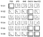
- FIG. 4 is a diagram showing, by dotted lines, regions of the human IL-3R ⁇ molecule in which the regions 1 to 7 arranged outside the molecule are replaced with GM-CSFR ⁇ sequences in the A and B domain nucleic acid and amino acid sequences.
- the vertical axis shows the cell growth inhibition rate (%), and the horizontal axis shows the names of various IL-3R ⁇ antibodies.
- GM indicates Granulocyte / Macrophage strain
- E indicates Erythroid colony
- GEMM indicates mixed colony.
- FIG. 8 used PBMC not cultured with IL-2
- FIG. 9 used PBMC cultured with IL-2.
- the present invention relates to human IL-3R ⁇ that does not inhibit IL-3 signal and binds to the B domain of human IL-3 receptor ⁇ chain (hereinafter abbreviated as IL-3R ⁇ ) and does not bind to the C domain. Relates to antibodies to the chain.
- IL-3R ⁇ human IL-3 receptor ⁇ chain
- IL-3 receptor (hereinafter abbreviated as IL-3R), particularly IL-3R ⁇ , is expressed on the cell surface of leukemia stem cells.
- IL-3R ⁇ the IL-3 receptor ⁇ chain
- IL-3R ⁇ the IL-3 receptor ⁇ chain
- IL-3R ⁇ the IL-3 receptor ⁇ chain
- IL-3R ⁇ the IL-3 receptor ⁇ chain
- IL-3R ⁇ the IL-3 receptor ⁇ chain
- IL-3R ⁇ IL-3 receptor ⁇ chain
- IL-3R ⁇ IL-3 receptor ⁇
- IL-3R ⁇ IL-3 receptor ⁇ gene is a type I transmembrane protein belonging to the cytokine receptor family.
- IL-3R ⁇ molecules are expressed in some hematopoietic progenitor cells, basophils, and some dendritic cells.
- expression in hematopoietic tumors and leukemias is mainly known.
- tumors expressing IL-3R ⁇ include AML, acutely transformed CML blasts, leukemia stem cells and differentiation marker negative CD34 positive CD38 negative fractions, AML, CML, MDS, ALL, It is known to be expressed in SM.
- IL-3 a known ligand of IL-3R ⁇ , is expressed in the blood by activated T cells, natural killer cells, mast cells, and some megakaryocyte cells.
- IL-3R ⁇ is also called CD123.
- IL-3R ⁇ includes mammalian (eg, primate, human) IL-3R ⁇ .
- IL-3R ⁇ sequences such as human IL-3R ⁇ include polymorphic variants.
- Specific examples of full-length human IL-3R ⁇ include the following amino acid sequences.
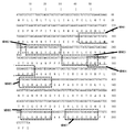
- the C domain As the A domain, from the 18th glutamine (Q) of the amino acid of SEQ ID NO: 2 to the 100th serine (S), as the B domain, from the 101st glycine (G) of the amino acid of SEQ ID NO: 2 to the 203rd Up to serine (S), the C domain has a region from 204th glutamine (Q) to 308th leucine (L) of the amino acid of SEQ ID NO: 2. Furthermore, among the A and B domains, the following seven regions can be mentioned as the regions arranged outside the molecule.
- region 1 from the 55th aspartic acid (D) to 61st proline (P) of the amino acid of SEQ ID NO: 2, and as region 2, the 63rd valine (V) of the amino acid of SEQ ID NO: 2 to 70th Up to phenylalanine (F), region 3 and from the 91st serine (S) of amino acid of SEQ ID NO: 2 to 98th glutamic acid (E), as region 4, the 97th amino acid of SEQ ID NO: 2 From proline (P) to 104th tryptophan (W), as region 5, from amino acid 122 of cysteine (C) of amino acid of SEQ ID NO: 2 to 128th proline (P), as region 6, SEQ ID NO: 2 From region 182 to isoleucine (I) of amino acid 182 to serine (S) of position 188, examples of region 7 include 192nd glycine (G) to amino acid 198 of lysine (K) of amino acid SEQ ID NO: 2.
- the antibody of the present invention is an antibody that binds to the 101st to 203rd amino acid sequence and does not bind to the 204th to 308th amino acid sequence in the amino acid sequence of SEQ ID NO: 2, which is the extracellular domain of IL-3R ⁇ . Furthermore, antibodies that bind to amino acid sequences 182 to 188 and 192 to 198 in the amino acid sequence shown in SEQ ID NO: 2 can be mentioned. The antibody of the present invention binds to the specific region described above of the extracellular domain of IL-3R ⁇ and does not inhibit IL-3 signal.
- “does not inhibit IL-3 signal” means that IL-3 does not inhibit intracellular signal via IL-3R, and does not inhibit the association of IL-3 and IL-3R and This includes not inhibiting the binding of IL-3R ⁇ and ⁇ chains.
- the cell growth inhibition rate shown in FIG. 5 is 40% or more, preferably 60% or more, more preferably 80% or more when the antibody concentration is 10 ⁇ g / mL. It means that.
- “blocking of IL-3 signal” and “inhibition of IL-3 signal” are used interchangeably and are not distinguished from each other. The ability to inhibit a signal.
- the antibody of the present invention has high antibody-dependent cytotoxic activity (ADCC).
- An IL-3R ⁇ antibody having ADCC activity refers to an antibody that binds to cells expressing IL-3R ⁇ and can kill IL-3R ⁇ -expressing cells via effector cells having cytotoxic activity such as NK cells.
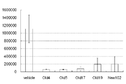
- the high ADCC activity is specific when the antibody concentration is 0.01 ⁇ g / mL or less as measured by the Colon-26 / hCD123ADCC measurement method using IL-2 cultured PBMC described in Example 11. This means that the dissolution rate is 10% or more.
- IL-3R ⁇ -expressing cells include blood tumor cells (acutemyeloidleukemia (AML) cells, chronicmyeloidleukemia (CML) cells, myelodysplasticsyndromes (MDS) cells, acutelymphoidleukemia (ALL) cells, chroniclymphoidleukemia (CLL) cells, multiple myeloma) : MM) cells, systemicmastocytoma (SM) cells, etc., regulatory T cells (eg CD4 positive CD25 positive cells), antigen presenting cells (eg dendritic cells, monocytes / macrophages and similar cells (liver stellate cells, Osteoclasts, microglial cells, intraepidermal macrophages, dust cells (alveolar macrophages))), basophils and the like.
- AML acutemyeloidleukemia
- CML chronicmyeloidleukemia
- MDS myelodysplasticsyndromes
- ALL acutelymphoidleukemia
- AML cells, CML cells, ALL cells, CLL cells, MDS cells, SM cells, MM cells, and various lymphoma cells include respective tumor stem cells.
- the tumor stem cell is one of a group of cells constituting a tumor, for example, lineage ( ⁇ ) CD34 (+) CD38 ( ⁇ ) bone marrow cells in acute myeloid leukemia (AML). Therefore, since the antibody of the present invention has high ADCC activity, it induces reduction or elimination of cells in which IL-3R ⁇ is expressed.
- the IL-3R ⁇ antibody of the present invention includes an IL-3R ⁇ antibody having a heavy chain CDR and a light chain CDR selected from the group consisting of the following (a) to (e).
- the heavy chain CDRs 1 to 3 are amino acid sequences represented by SEQ ID NOs: 113 to 115 and the light chain CDRs 1 to 3 are represented by SEQ ID NOs: 131 to 133.
- the heavy chain CDRs 1 to 3 are SEQ ID NOs: The amino acid sequence of 116 to 118 and the light chain CDRs 1 to 3 are represented by SEQ ID NOs: 134 to 136 (c) The heavy chain CDRs 1 to 3 of the amino acid sequence represented by SEQ ID NOs: 119 to 121 and the light chain CDRs 1 to 3 are represented by SEQ ID NOs: 137 to 139 (d) Heavy chain CDRs 1 to 3 are represented by SEQ ID NOs: 122 to 124 and light chain CDRs are represented by SEQ ID NOs: 140 to 142 Amino acid sequence (e) The heavy chain CDRs 1 to 3 are represented by SEQ ID NOs: 125 to 127, and the light chain CDRs 1 to 3 are represented by SEQ ID NOs: 143 to 145.
- IL-3R ⁇ antibody having a heavy chain variable region and a light chain variable region selected from the group consisting of (a) to (f) of the above (in parentheses, the name of the antibody of the below-mentioned Examples from which each variable region sequence is derived) Show.)
- Antibody name: Old5 (c) the heavy chain variable region comprising the amino acid sequence from the 20th glutamine (Q) to the 139th serine (S) of the amino acid sequence shown in SEQ ID NO: 61 and the 23rd of the amino acid sequence shown in SEQ ID NO: 63 A light chain variable region comprising an amino acid sequence from aspartic acid (D) to 129th lysine (K).
- Antibody name: Old17 (d) a heavy chain variable region comprising the amino acid sequence from the 20th glutamine (Q) to the 139th serine (S) of the amino acid sequence represented by SEQ ID NO: 65 and the 23rd amino acid sequence represented by SEQ ID NO: 67 A light chain variable region comprising an amino acid sequence from aspartic acid (D) to 129th lysine (K).
- antibody name: Old19 a heavy chain variable region comprising the amino acid sequence from the 20th glutamine (Q) to the 138th serine (S) of the amino acid sequence represented by SEQ ID NO: 69 and the 23rd amino acid sequence represented by SEQ ID NO: 71 A light chain variable region comprising an amino acid sequence from aspartic acid (D) to 129th lysine (K).
- a heavy chain comprising an amino acid sequence in which 1 to 3 amino acid residues are deleted, substituted, added or inserted in the heavy chain variable region and / or light chain variable region represented by (a) to (e) Variable region and / or light chain variable region.
- the term antibody is used in the broadest sense, and includes monoclonal antibodies, polyclonal antibodies, multivalent antibodies, multispecific antibodies (e.g., bispecific antibodies), and antibody fragments as long as they exhibit their desired biological activity. Including.
- the antibody comprises a mature heavy or light chain variable region sequence.
- Antibodies also have modified and mutated forms, such as substitutions within or outside of the antibody constant region, complementarity determining region (CDR) or framework (FR) region of the mature heavy or light chain variable region sequence. Including. In certain embodiments, the substitution includes a conservative amino acid substitution.
- the antibody also includes a partial sequence of a mature heavy or light chain variable region sequence. In particular embodiments, the subsequence is selected from Fab, Fab ′, F (ab ′) 2 , Fv, Fd, single chain Fv (scFv), disulfide bond Fv (sdFv) and VL or VH.
- the antibody also includes a heterologous domain.
- the heterologous domain includes a tag, a detectable label, or a cytotoxic agent.
- Antibodies include monoclonal and polyclonal antibodies, any isotype or subclass thereof.
- the antibody is an IgG (eg, IgG1, IgG2, IgG3 or IgG4), IgA, IgM, IgE, or IgD isotype.
- a “monoclonal” antibody is based on a single clone, including a eukaryotic clone, a prokaryotic clone, or a phage clone, and is derived from a single clone, including a eukaryotic clone, a prokaryotic clone, or a phage clone, or a eukaryotic clone.
- a “monoclonal” antibody is therefore structurally defined and not the method by which it is produced.
- IL-3R ⁇ antibody, anti-IL-3R ⁇ and anti-IL-3R ⁇ antibody refer to antibodies that specifically bind to IL-3R ⁇ . Specific binding is selective for an epitope present in IL-3R ⁇ . Specific binding can be distinguished from non-specific binding using assays known in the art (eg, immunoprecipitation, ELISA, flow cytometry, Western blotting).
- IL-3R ⁇ antibody specifically binds to another protein with high sequence or structural homology to the IL-3R ⁇ epitope, depending on the sequence or degree of structural homology of the IL-3R ⁇ epitope. there is a possibility.
- IL-3R ⁇ antibodies may bind to different proteins when epitopes with sufficient sequence or structural homology exist in different proteins.
- IL-3R ⁇ antibodies include isolated and purified antibodies.
- Antibodies of the present invention, including isolated or purified IL-3R ⁇ antibodies, include humans.
- isolated used as a modifier of a composition means that the composition is made by hand or one or more other components in a naturally occurring in vivo environment. Generally means separated by one or more operational steps or processes. In general, a composition so separated is substantially free of one or more materials to which such composition normally binds naturally, eg, one or more proteins, nucleic acids, lipids, carbohydrates, cell membranes. . Thus, an isolated composition is separated from other biological components in the cells of the organism in which it naturally occurs, or from an artificial medium from which the composition is produced (eg, synthetically or by cell culture). ing.
- an isolated IL-3R ⁇ antibody is obtained from an animal from which the antibody is produced (eg, a non-transgenic mammal or a transgenic mammal (such as a rodent (mouse) or ungulate (bovine) animal)). Separated from other polypeptides and nucleic acids. Therefore, the serum containing the antibody obtained from the animal is considered to be isolated.
- isolated does not exclude other physical forms; for example, an isolated antibody can include antibody subsequences, chimeric, multimeric, or derivatized forms.
- purified used as a composition modifier refers to a composition that is generally free of most or substantially all of the materials with which it naturally binds. Purified antibodies are generally removed from components normally present in the antibody environment. Therefore, it is considered that the antibody supernatant separated from the antibody-producing hybridoma cell culture is purified. Thus, purification does not require absolute purity, but is a context specific. Furthermore, a “purified” composition can be combined with one or more other molecules. As such, the term “purified” does not exclude combinations of compositions.
- Purity should be determined by any appropriate method such as, for example, UV spectroscopy, chromatography (eg, HPLC, gas phase), gel electrophoresis (eg, silver or Coomassie staining) and sequence analysis (peptides and nucleic acids). Can do.
- Protein and nucleic acids include proteins and nucleic acids obtained by standard purification methods. The term also includes proteins and nucleic acids obtained by recombinant expression or chemical synthesis in a host cell.
- purified means that the level of contaminants is higher than the level approved by the supervisory authority for administration to humans or non-human animals, for example, the Food and Drug Administration (FDA). Sometimes refers to a low composition.
- FDA Food and Drug Administration
- IL-3R ⁇ antibodies include antibodies that bind to IL-3R ⁇ and modulate IL-3R ⁇ function or activity in vivo or in vitro (eg, in a subject).
- modulate and grammatical variations thereof when used with respect to IL-3R ⁇ activity or function may affect, modify or alter IL-3R ⁇ activity or function in a detectable manner. Means that inhibiting IL-3 signaling is not included.
- an IL-3R ⁇ antibody that modulates the activity or function of IL-3R ⁇ is affected, altered or altered so that one or more IL-3R ⁇ activity or function can be detected without inhibiting the IL-3R signal.
- Such an IL-3R ⁇ activity or function includes, for example, IL-3R ⁇ binding to an IL-3R ⁇ ligand (eg, IL-3), IL-3R ⁇ -mediated signaling or IL-3R ⁇ -mediated cells.
- IL-3R ⁇ ligand eg, IL-3
- IL-3R ⁇ -mediated signaling e.g. IL-3
- IL-3R ⁇ -mediated cells e.g. IL-3R ⁇ -mediated cell.
- Responses or cellular responses that can be modulated by IL-3R ⁇ , or other IL-3R ⁇ activities or functions described or otherwise known or known can be included.
- IL-3R ⁇ activities and functions that can be modulated include, for example, IL-3R ⁇ -mediated signaling or IL-3R ⁇ -mediated cellular responses or cell responses that can be modulated by IL-3R ⁇ , cell proliferation or Increase (eg, AML cells, CML cells, ALL cells, CLL cells, MDS cells, MM cells, SM cells, various lymphoma cells, monocytes, macrophages, mast cells, basophils, helper T cells, regulatory T cells, Natural killer cells, myeloid progenitor cells, lymphoid progenitor cells, etc.), cell survival or apoptosis (eg, AML cells, CML cells, ALL cells, CLL cells, MDS cells, MM cells, SM cells, various lymphoma cells, Monocytes, macrophages, mast cells, basophils, helper T cells, regulatory T cells, natural killer cells, myeloid progenitor cells, lymphocyte progenitor cells, etc.), cytokines (eg For example, AML
- Specific cytokines that are modulated include, but are not limited to, IL-1, IL-2, IL-4, IL-5, IL-6, IL-9, IL-10, IL-14, IL-16, IL-17, IL-23, IL-26, TNF- ⁇ and interferon ⁇ (in vivo or in vitro).
- Specific anti-apoptotic protein or pro-apoptotic protein expression includes, but is not limited to, Bcl-xL, Bcl-2, Bad, Bim and Mcl-1.
- exemplary IL-3R ⁇ antibodies described herein include one or more IL-3R ⁇ -mediated signaling or IL-3R ⁇ -mediated cellular responses or cell responses induced by IL-3R ⁇ , cell proliferation (eg, , AML cells, CML cells, ALL cells, CLL cells, MDS cells, MM cells, SM cells, various lymphoma cells, monocytes, macrophages, mast cells, basophils, helper T cells, regulatory T cells, natural killer cells , Myeloid progenitor cells, lymphoid progenitor cells, etc.), cell survival or apoptosis (eg, AML cells, CML cells, ALL cells, CLL cells, MDS cells, MM cells, SM cells, various lymphoma cells, monocytes, Macrophages, mast cells, basophils, helper T cells, regulatory T cells, natural killer cells, myeloid progenitor cells, lymphoid progenitor cells, etc.), cytokines (eg Th1, Th2 and other non-
- an IL-3R ⁇ antibody of the invention modulates AML cell proliferation or survival and other blood tumor cells (eg, CML cells, ALL cells, CLL cells, MDS cells, MM cells, SM cells). , Or various lymphoma cells), monocytes, macrophages, mast cells, basophils, helper T cells, regulatory T cells, natural killer cells, myeloid progenitor cells, lymphocyte progenitor cells, etc. Modulates the growth or survival of non-hematologic tumor cells, or reduces, disappears or depletes AML, CML, ALL, CLL, MDS, MM, SM, or various lymphoma cells.
- AML cell proliferation or survival and other blood tumor cells eg, CML cells, ALL cells, CLL cells, MDS cells, MM cells, SM cells.
- IL-3R ⁇ antibodies include modified forms such as substitutions (eg, amino acid substitutions), additions and deletions (eg, partial sequences or fragments), also referred to as “variants”. Such modified antibody forms and variants are capable of binding at least a portion of the IL-3R ⁇ antibody function or activity of the present invention, eg, IL-3R ⁇ , or IL-3R ⁇ activity or function (eg, IL-3R ⁇ ). -3R ⁇ signaling) is retained.
- a modified IL-3R ⁇ antibody can retain, for example, the ability to modulate at least some IL-3R ⁇ binding or one or more IL-3R ⁇ functions or activities (eg, signaling, cellular responses, etc.). it can.
- Modify and grammatical variations thereof mean that the composition is out of the reference composition.
- Modified proteins, nucleic acids and other compositions have higher or lower activity than unmodified reference proteins, nucleic acids or other compositions, or are unmodified reference proteins, nucleic acids or other compositions Can have different functions.
- nucleic acid sequences encoding antibodies that contain amino acid substitutions are also provided.
- identity or “identical” means that two or more referenced entities are the same. Thus, when two protein sequences (eg, IL-3R ⁇ antibodies) are identical, they have the same amino acid sequence at least within the referenced region or portion. “Identity region” refers to the same portion of two or more referenced entities. Thus, if two protein sequences are identical in one or more sequence regions, they share identity within that region.
- “Substantial identity” means that a molecule has a function or activity of at least part of one or more reference molecule functions or activities, or an associated / corresponding region or part of a reference molecule with which the molecule shares identity Means that it is structurally or functionally preserved, as expected.
- a polypeptide having substantial identity eg, IL-3R ⁇ antibody
- one or more modifications that retain at least some activity or function of an unmodified IL-3R ⁇ antibody eg, deletion, substitution, addition, or addition of 1-3 amino acid residues
- An IL-3R ⁇ antibody having an insertion is considered to have substantial identity to a reference IL-3R ⁇ antibody.
- the amount of sequence identity required to retain function or activity will depend on the protein, the region and the function or activity of the region.
- a protein can retain some activity or function with only 30% amino acid sequence identity but is generally higher than the reference sequence, e.g., 50%, 60%, 75%, 85 There is%, 90%, 95%, 96%, 97%, or 98% identity.
- the degree of identity between two sequences can be ascertained using computer programs and mathematical algorithms known in the art. Such algorithms that calculate percent sequence identity (homology) generally account for sequence gaps and mismatches across the comparison region. For example, a BLAST (eg, BLAST 2.0) search algorithm (see, eg, Altschul et al., J. Mol.
- Biol. 215: 403 (1990), publicly available through NCBI) is an exemplary search parameter such as: Has: mismatch-2; gap start 5; gap extension 2.
- the BLASTP algorithm is typically used in combination with a score matrix such as PAM100, PAM 250, BLOSUM 62 or BLOSUM50.FASTA (eg, FASTA2 and FASTA3), and the SSEARCH sequence comparison program also quantifies the degree of identity (Pearson et al., Proc. Natl. Acad. Sci USA 85: 2444 (1988); Pearson, Methods Mol Biol. 132: 185 (2000); and Smith et al., J. Mol. Biol. 147: 195 (1981)) .
- a program for quantifying protein structural similarity using Delaunay-based phase mapping has also been developed (Bostick et al., Biochem Biophys ResCommun. 304: 320 (2003)).
- a “conservative substitution” is a substitution of one amino acid with a biologically, chemically or structurally similar residue.
- Biologically similar means that the substitution does not destroy biological activity, eg, IL-3R ⁇ binding activity.
- Structurally similar means that the amino acids have side chains of the same length (eg, alanine, glycine and serine) or are of similar size.
- Chemical similarity means that the residues have the same charge or are hydrophilic or hydrophobic.
- substitution of one hydrophobic residue with another such as isoleucine, valine, leucine or methionine, or substitution of another monopolar residue, for example, substitution of arginine with lysine
- substitution of glutamic acid with aspartic acid substitution of glutamine with asparagine, substitution of serine with threonine, and the like.
- Modified antibodies also include peptides having one or more D-amino acids, structural and functional analogs substituted with L-amino acids (and mixtures thereof), eg, synthetic or unnatural amino acids or amino acid analogs Also included are mimetics and derivatized forms. Modifications include cyclic structures such as end-to-end amide bonds or intra- or intermolecular disulfide bonds between amino and carboxy terminals.
- Non-limiting further specific examples of amino acid modifications include IL-3R ⁇ subsequences and fragments.
- Exemplary IL-3R ⁇ subsequences and fragments comprise a portion of an IL-3R ⁇ sequence to which an exemplary IL-3R ⁇ antibody of the invention binds.
- Exemplary IL-3R ⁇ subsequences and fragments also include an immunogenic portion, eg, a portion of IL-3R ⁇ that includes a sequence to which an exemplary IL-3R ⁇ antibody of the invention binds.
- a nucleic acid encoding an IL-3R ⁇ antibody and an IL-3R ⁇ antibody partial sequence or fragment that retains at least part of the function or activity of the unmodified or reference IL-3R ⁇ antibody is provided.
- the term “subsequence” or “fragment” means a portion of a full-length molecule.
- a partial sequence of an IL-3R ⁇ antibody that encodes an IL-3R ⁇ antibody has at least one less amino acid than the full-length IL-3R ⁇ (eg, deletion of one or more internal or terminal amino acids from either the amino or carboxy terminus) ).
- the partial sequence of the IL-3R ⁇ antibody has at least one fewer amino acid than the full-length IL-3R ⁇ antibody.
- the nucleic acid subsequence has at least one fewer nucleotide than the full length comparison nucleic acid sequence.
- the partial sequence can be any length up to the full length native IL-3R ⁇ .
- IL-3R ⁇ antibody subsequences and fragments have a binding affinity as a full-length antibody, a binding specificity as a full-length antibody, or one or more activities or functions as a full-length antibody, such as the function of an IL-3R ⁇ antagonist or agonist antibody Or it may have activity.
- the terms “functional subsequence” and “functional fragment” when referring to an antibody retain one or more functions or activities as a full-length reference antibody, eg, at least part of the function or activity of an IL-3R ⁇ antibody. Refers to the antibody portion. For example, an antibody subsequence or fragment that binds to IL-3R ⁇ or a fragment of IL-3R ⁇ is considered a functional subsequence.
- Antibody partial sequences and fragments can be combined.
- VL or VH subsequences can be linked by a linker sequence, thereby forming a VL-VH chimera.
- Combinations of single chain Fv (scFv) subsequences can be linked by a linker sequence, thereby forming an scFv-scFv chimera.
- IL-3R ⁇ antibody subsequences and fragments include single chain antibodies or variable regions alone or in combination with all or part of other IL-3R ⁇ antibody subsequences.
- Antibody subsequences and fragments can be prepared by proteolytic hydrolysis of the antibody, for example, pepsin or papain digestion of whole antibodies. Antibody subsequences and fragments generated by enzymatic cleavage with pepsin yield a 5S fragment denoted as F (ab ′) 2 . This fragment can be further cleaved using a thiol reducing agent to create a 3.5SFab ′ monovalent fragment. Alternatively, enzymatic cleavage with pepsin directly results in two monovalent Fab ′ and Fc fragments (eg, US Pat. Nos. 4,036,945 and 4,331,647; and Edelman et al., Methods Enymol. 1: 422 ( 1967)). Other antibody cleavage methods may also be used, such as heavy chain separation to form a monovalent light-heavy chain fragment, further cleavage of the fragment, or other enzymatic or chemical methods.
- Proteins and antibodies, and their partial sequences and fragments can be created by genetic methods.
- the technology involves expressing all or part of a gene encoding a protein or antibody in a host cell such as Cos cells or E. coli.
- Recombinant host cells synthesize full-length or partial sequences, eg, scFv (eg, Whitlow et al., In: Methods: A Companion to Methods inEnzymology 2:97 (1991), Bird et al., Science 242: 423 (1988); And US Pat. No. 4,946,778).
- Single chain Fv and antibodies are described in U.S. Pat.Nos.
- Modified forms include derivatized sequences such as free amino groups forming amine hydrochlorides, p-toluenesulfonyl groups, carbobenzoxy groups; free carboxy groups forming salts, methyl and ethyl esters.
- Modifications can be made using methods known in the art (eg, PCR-based site-specific, deletion and insertion mutagenesis, chemical modification and mutagenesis, cross-linking, etc.).
- Modified forms of proteins include adducts and inserts.
- an addition can be a covalent or non-covalent association of any type of molecule with a protein (eg, antibody), nucleic acid or other composition.
- Adducts and inserts include fusion (chimeric) polypeptide or nucleic acid sequences that have one or more molecules that are not normally present in a reference natural (wild-type) sequence covalently linked to said sequence. It is.
- a specific example is the amino acid sequence of another protein (eg, antibody) to create a multifunctional protein (eg, multispecific antibody).
- the antibodies of the present invention also include chimeras or fusions in which one or more additional domains are covalently linked to provide different or accessory functions or activities.
- Antibodies include chimeras or fusions that do not naturally occur in nature, in which two or more amino acid sequences are linked together.
- an IL-3R ⁇ antibody containing a heterologous domain and a nucleic acid encoding the IL-3R ⁇ antibody are provided.
- a heterologous domain can be an amino acid adduct or an insert, but is not limited to amino acid residues.
- a heterologous domain can consist of any of a variety of different types of small or large functional parts.
- moieties include nucleic acids, peptides, carbohydrates, lipids or small organic compounds such as drugs, metals (gold, silver) and the like.
- heterologous domains include, for example, tags, detectable labels and cytotoxic agents.
- tags and detectable labels include T7-, His-, myc-, HA- and FLAG-tags; enzymes (horseradish peroxidase, urease, catalase, alkaline phosphatase, ⁇ -galactosidase, chloramphenicol Enzyme substrate; ligand (eg, biotin); receptor (avidin); radionuclide (eg, C14, S35, P32, P33, H3, I125 and I131); electron density reagent; energy transfer molecule; paramagnetic label Fluorophores (fluorescein, rhodamine, phycoerythrin); chromophores; chemiluminescent agents (imidazole, luciferase); and bioluminescent agents.
- cytotoxic agents include diphtheria toxin (diptheria, toxin), cholera
- a linker sequence such that the two entities at least partially maintain different functions or activities between a protein (eg, antibody), nucleic acid, or other composition and an adduct or insert (eg, a heterologous domain) May be inserted.
- a linker sequence may have one or more properties that can promote or interact with either domain, such as flexible structure, Subsequent structures cannot be formed or include hydrophobicity or chargeability.
- Amino acids commonly found in the flexible protein region include glycine, asparagine and serine. Other near neutral amino acids such as threonine and alanine may also be used in the linker sequence.
- the length of the linker sequence can vary (see, eg, US Pat. No. 6,087,329).
- Linkers include chemical crosslinkers and conjugating agents such as sulfo-succinimidyl derivatives (sulfo-SMCC, sulfo-SMPB), disuccinimidyl suberate (DSS), disuccinimidyl glutarate (DSG) and tartaric acid Further included is disuccinimidyl (DST).
- sulfo-succinimidyl derivatives sulfo-SMCC, sulfo-SMPB
- DSS disuccinimidyl suberate
- DSG disuccinimidyl glutarate
- tartaric acid Further included is disuccinimidyl (DST).
- Modified and mutated antibodies are those that can retain the detectable activity of an IL-3R ⁇ antibody.
- the modified antibody has binding activity to an IL-3R ⁇ molecule and induces reduction or elimination of IL-3R ⁇ -expressing cells by an immune system centered on effector cells. It is involved in the functional control of IL-3R ⁇ -expressing cells and induces cell survival, proliferation, rest, cell death, and the like. Cell death includes apoptosis, necrosis, autophagy and the like.
- the present invention further provides cell-free methods (eg, in solution, in solid phase) and cell-based methods (eg, in vitro or in vivo) for screening, detecting, and identifying IL-3R ⁇ .
- the These methods can be performed in solution, using biomaterials or samples in vitro, and in vivo, for example, in samples of animal-derived cells (eg, lymphocytes).
- the method comprises contacting a biomaterial or sample with an antibody that binds IL-3R ⁇ under conditions that allow binding of the antibody to IL-3R ⁇ ; binding of the antibody to IL-3R ⁇ ; Assaying for.
- the presence of IL-3R ⁇ is detected by binding the antibody to IL-3R ⁇ .
- IL-3R ⁇ is present in a cell or tissue.
- the biomaterial or sample is obtained from a mammalian subject.
- composition such as a protein (eg, IL-3R ⁇ antibody), material, sample, or treatment
- contacting refers to that composition (eg, an IL-3R ⁇ antibody) and other references.
- a specific example of direct interaction is binding.
- a specific example of indirect interaction is when the composition acts on an intermediate molecule that acts on the next referenced entity.
- a cell eg, lymphocyte
- the antibody is bound to the cell (eg, through binding to IL-3R ⁇ ) or the antibody is intermediate And this intermediate then acts on the cell.
- the terms “assaying” and “measuring” and grammatical variations thereof are used interchangeably herein and refer to either qualitative or quantitative measurements, or qualitative and quantitative measurements. Refers to both. When these terms are used in reference to binding, any means of assessing relative amounts, binding affinity or specificity is contemplated, including various methods described herein and known in the art.
- binding of an IL-3R ⁇ antibody to IL-3R ⁇ can be assayed or measured by a flow cytometry assay. (Preparation of antibody)
- the present invention also provides a method for producing a human IL-3R ⁇ antibody having IL-3R ⁇ positive cytotoxic activity.
- the method comprises transforming a human IL-3R ⁇ extracellular domain or IL-3R ⁇ transgenic cell conjugated with a human Fc recombinant protein into an animal capable of expressing human immunoglobulin (eg, a transgenic mouse or a trans Screening the animal for expression of human IL-3R ⁇ antibody; selecting an animal producing human IL-3R ⁇ antibody; isolating the antibody from the selected animal .
- a human immunoglobulin eg, a transgenic mouse or a trans Screening the animal for expression of human IL-3R ⁇ antibody; selecting an animal producing human IL-3R ⁇ antibody; isolating the antibody from the selected animal .
- IL-3R ⁇ protein suitable for antibody production can be produced by any of a variety of standard protein purification or recombinant expression techniques.
- IL-3R ⁇ sequences can be generated by standard peptide synthesis techniques, such as solid phase synthesis.
- a portion of the protein may include an amino acid sequence such as a FLAG tag, T7 tag or polyhistidine sequence to facilitate purification of the expressed or synthesized protein.
- the protein can be expressed in cells and purified.
- the protein can be expressed as part of a larger protein (eg, a fusion or chimera) by recombinant methods.
- Suitable forms of IL-3R ⁇ for raising an immune response include IL-3R ⁇ subsequences, such as immunogenic fragments. Additional forms of IL-3R ⁇ include IL-3R ⁇ expressing cells, IL-3R ⁇ containing preparations or cell extracts or fractions, partially purified IL-3R ⁇ .
- IL-3R ⁇ or an immunogenic fragment thereof is optionally conjugated with a carrier such as keyhole limpet hemocyanin (KLH) or ovalbumin (eg BSA), or an adjuvant such as Freund's complete or incomplete adjuvant. And is used to immunize animals.
- KLH keyhole limpet hemocyanin
- BSA ovalbumin
- an adjuvant such as Freund's complete or incomplete adjuvant.
- splenocytes from immunized animals that respond to IL-3R ⁇ can be isolated and fused with myeloma cells.
- Monoclonal antibodies produced by hybridomas can be screened for reactivity with IL-3R ⁇ or immunogenic fragments thereof.
- Immunized animals include primates, mice, rats, rabbits, goats, sheep, cows, or guinea pigs.
- the initial and optional boosts may be by intravenous, intraperitoneal, intramuscular, or subcutaneous routes.
- the antigen can be combined with another protein such as ovalbumin or keyhole limpet hemocyanin (KLH), thyroglobulin and tetanus toxoid, or such as Freund's complete or incomplete adjuvant. Can be mixed with an adjuvant.
- KLH keyhole limpet hemocyanin
- thyroglobulin and tetanus toxoid or such as Freund's complete or incomplete adjuvant.
- the initial and optional booster immunization may be by the intraperitoneal route, intramuscular route, intraocular route, or subcutaneous route.
- the booster immunizations may be the same or different concentrations of IL-3R ⁇ preparation and may be regular or irregular intervals.
- Animals include those that have been genetically modified to include human loci and can be used to create human antibodies.
- Transgenic animals having one or more human immunoglobulin genes are described, for example, in US Pat. No. 5,939,598, WO02 / 43478, and WO 02/092812.
- splenocytes from immunized mice that are highly responsive to antigen can be isolated and fused with myeloma cells.
- Monoclonal antibodies that bind to IL-3R ⁇ can be obtained.
- human when used in reference to an antibody means that the amino acid sequence of the antibody is a fully human amino acid sequence, ie, a human heavy chain and human light chain variable region and a human constant region. Thus, all of the amino acids are human amino acids or are present in human antibodies.
- An antibody that is a non-human antibody can be made a fully human antibody by replacing non-human amino acid residues with amino acid residues present in the human antibody.
- Amino acid residues, CDR region maps and human antibody consensus residues present in human antibodies are known in the art (eg, Kabat, Sequences of Proteins of Immunological Interest, 44th edition USDepartmentofHealthandHumanServices.PublicHealthService (1987) See Chothia and Lesk (1987), a human VH subgroup III consensus sequence based on a survey of 22 known human VHIII sequences, and a human VL based on a survey of 30 known human ⁇ I sequences. Consensus sequences for kappa chain subgroup I are described in Padlan Mol. Immunol.31: 169 (1994); and PadlanMol.Immunol.28: 489 (1991). Are substituted with one or more amino acids present in any other human antibody.
- IL-3R ⁇ antibodies include, for example, CDR-grafting (EP 239,400; W091 / 09967; U.S. Pat.No. 5,225,539; do 5,530,101; and 5,585,089), veneering or reser Resurfacing (EP592,106; EP519,596; Padlan, MolecularImmunol. 28: 489 (1991); Studnicka et al., Protein Engineering 7: 805 (1994); Roguska. Et al., Proc. Nat'l Acad. Sci. USA 91: 969 (1994)), and humanized antibodies that can be generated using techniques known in the art such as chain shuffling (US Pat. No. 5,565,332). Human consensus sequences (Padlan, Mol.
- humanized when used in reference to an antibody refers to one or more complementarity determining region (CDR) non-human amino acids whose antibody amino acid sequence specifically binds to a desired antigen in an acceptor human immunoglobulin molecule. Having residues (eg, mouse, rat, goat, rabbit, etc.) and one or more human amino acid residues in the Fv framework region (FR) (which are amino acid residues flanking the CDR) means.
- CDR complementarity determining region
- Antibodies referred to as “primatization” include any human amino acid residue in the acceptor human immunoglobulin molecule and framework region in addition to any human residue (eg, monkey, gibbon, gorilla, It is within the meaning of “humanization” except that it can be a chimpanzee orangutan, macaque monkey).
- the human FR residues of the immunoglobulin can be replaced with corresponding non-human residues.
- a humanized antibody may comprise residues that are found neither in the human antibody nor in the donor CDRs or framework sequences.
- FR substitutions at a particular position not found in human antibodies or donor non-human antibodies can be expected to improve binding affinity or specific human antibodies at that position.
- Antibody frameworks and CDR substitutions based on molecular modeling are well known in the art, for example, modeling of CDR and framework residue interactions and specific locations to identify framework residues important for antigen binding. By sequence comparison to identify unusual framework residues in (see, eg, US Pat. No. 5,585,089; and Riechmann et al., Nature 332: 323 (1988)).
- IL-3R ⁇ antibody includes chimeric antibody.
- the term “chimera” and grammatical variations thereof when used with respect to an antibody is derived from two or more different species, wherein the amino acid sequence of the antibody is derived from two or more different species, or simply Means containing one or more moieties that are separated or based on two or more different species.
- a portion of the antibody can be human (eg, a constant region) and another portion of the antibody can be non-human (eg, a murine heavy chain or murine light chain variable region).
- an example of a chimeric antibody is an antibody in which different portions of the antibody are of different species origin. Chimeric antibodies, unlike humanized or primatized antibodies, can have different species of sequences in any region of the antibody.
- IL-3R ⁇ antibodies can also be produced using hybridoma technology, recombinant technology, and phage display technology, or combinations thereof (US Pat. Nos. 4,902,614, 4,543,439, and 4,411,993). See MonoclonalAntibodies.Hybridomas: ANewDimensioninBiologicalAnalyses, PlenumPress, Kennett, McKearn, and Bechtol (ed.), 1980, and Harlow et al., Antibodies: ALaboratoryManual, ColdSpringHarborLaboratoryPress, 2nd edition 1988)
- the human anti-human IL-3R ⁇ antibody of the present invention uses chromosome-transfected mice (KM mice (trademark)) immunized with cell lines expressing various forms of solubilized recombinant human IL-3R ⁇ protein or IL-3R ⁇ . (WO02 / 43478, WO 02/092812, and Ishida, et al., IBC's 11th Antibody Engineering Meeting. Abstract (2000)).
- the human anti-human antibody stains the human IL-3R ⁇ stable transfected cell line, such as Jurkat-IL-3R ⁇ and L929-IL-3R ⁇ cells, in a detectable manner, rather than the non-transformed parental cell line. Have been shown to bind specifically to human IL-3R ⁇ .
- the antibodies of the present invention may be a kappa or lambda light chain sequence, either one full length, as in a naturally occurring antibody, a mixture thereof (ie, a fusion of a kappa and lambda chain sequence), And their partial sequences / fragments.
- Naturally occurring antibody molecules contain two kappa or two lambda light chains.
- the present invention further provides a method for producing an antibody that specifically binds to IL-3R ⁇ .
- a method for making an IL-3R ⁇ antibody comprises a human IL-3R ⁇ , subsequence or fragment (eg, IL-3R ⁇ extracellular domain), optionally conjugated to a human Fc recombinant protein.
- this method determines whether a human IL-3R ⁇ antibody has IL-3R ⁇ antagonist or agonist activity.
- Effector activity refers to antibody-dependent activity induced through the Fc region of an antibody.
- Antibody-dependent cytotoxic activity (ADCC activity), complement-dependent cytotoxic activity (CDC activity), macrophages and dendritic cells
- ADCC activity Antibody-dependent cytotoxic activity
- CDC activity complement-dependent cytotoxic activity
- macrophages and dendritic cells
- Antibody-dependent phagocytosis (antibody dependent phagocytosis, ADP activity) by phagocytic cells is known.
- N-acetylglucosamine present at the reducing end of an N-linked complex type sugar chain that binds to the 297th asparagine (Asn) of the Fc region of the antibody A method for controlling the amount of fucose (also called core fucose) that binds ⁇ -1,6 to (GlcNAc) (WO2005 / 035586, WO2002 / 31140, WO00 / 61739), or modification of the amino acid residue in the Fc region of an antibody
- the method of controlling with is known.
- the effector activity can be controlled by any method using the anti-IL-3R ⁇ monoclonal antibody of the present invention.
- the effector activity of the antibody can be increased or decreased.
- Examples of a method for reducing the content of fucose bound to the N-linked complex sugar chain bound to Fc of the antibody include defucosylated (non-fucosylated). Defucosylation refers to expressing an antibody using CHO cells lacking the ⁇ 1,6-fucose transferase gene, and an antibody to which fucose is not bound can be obtained. Antibodies without fucose binding have high ADCC activity.
- the antibody is expressed using a host cell into which an ⁇ 1,6-fucose transferase gene has been introduced.
- an antibody to which fucose is bound can be obtained.
- An antibody to which fucose is bound has a lower ADCC activity than an antibody to which fucose is not bound.
- ADCC activity and CDC activity can be increased or decreased by modifying amino acid residues in the Fc region of the antibody.
- the CDC activity of the antibody can be increased.
- ADCC activity or CDC activity can be increased or decreased by performing the amino acid modification described in US6,737,056, US7,297,775, or US7,317,091.
- nucleic acid sequences of the present invention such as vectors.
- the vector includes a nucleic acid sequence encoding an IL-3R ⁇ antibody, subsequence or fragment thereof.
- Nucleic acids can be of various lengths.
- the length of the nucleic acid encoding the IL-3R ⁇ antibody of the present invention or a partial sequence thereof is generally about 100 nucleotides to 600 nucleotides, or any numerical value or numerical range within such a length range, 150, 150-200, 200-250, 250-300, 300-350, 350-400, 400-450, 450-500, 500-550, or about 550-600 nucleotides Covers any number or range or value (anynumerical value or range or value) within a length or range of such lengths.
- the length of the nucleic acid that specifically hybridizes with the nucleic acid encoding the IL-3R ⁇ antibody of the present invention or a partial sequence thereof is generally about 10-20, 20-30, 30-50, 50-100, 100-150, 150-200, 200-250, 250-300, 300-400, 400-500, 500-600 nucleotides, or any number within such a range Or the numerical range.
- nucleic acid and “polynucleotide” refer to at least two or more ribo- or deoxy-ribonucleobase pairs (nucleotides) joined by a phosphate ester bond or equivalent.
- Nucleic acids include polynucleotides and polynucleosides. Nucleic acids include single molecules, double molecules or triple molecules, circular molecules or linear molecules. Exemplary nucleic acids include, but are not limited to: RNA, DNA, cDNA, genomic nucleic acids, naturally occurring and non-natural nucleic acids, eg, synthetic nucleic acids.
- Short nucleic acids and polynucleotides are generally “oligonucleotides” or “probes” of single- or double-stranded DNA. Called.
- Nucleic acids can be created using a variety of standard cloning and chemical synthesis techniques. Techniques include, but are not limited to, nucleic acid amplification of genomic DNA or cDNA targets using primers (eg, degenerate primer mixtures) that can be annealed to antibody coding sequences, such as polymerase chain reaction (PCR). It is done. Nucleic acids can also be created by chemical synthesis (eg, solid phase phosphoramidite synthesis) or transcription from a gene.
- primers eg, degenerate primer mixtures
- PCR polymerase chain reaction
- the generated sequence is then translated in vitro or cloned into a plasmid, propagated, and then in a cell (eg, a host cell such as yeast or bacteria, eukaryote (such as an animal or mammalian cell or plant)) Can be expressed.
- a cell eg, a host cell such as yeast or bacteria, eukaryote (such as an animal or mammalian cell or plant)
- a cell eg, a host cell such as yeast or bacteria, eukaryote (such as an animal or mammalian cell or plant)
- a vector is a mediator that can be manipulated by insertion or incorporation of nucleic acids.
- Vectors include plasmid vectors, viral vectors, prokaryotic (bacterial) vectors and eukaryotic (plant, fungal, mammalian) vectors.
- Vectors can be used for expression of nucleic acids in vitro or in vivo.
- Such vectors are referred to as “expression vectors” and include the introduction of nucleic acids, including nucleic acids encoding IL-3R ⁇ antibodies, subsequences and fragments thereof, or in vitro of encoded proteins (eg, in solution). Useful in expression in a subject in cells or in vivo.
- Vectors can also be used to manipulate nucleic acids.
- a “cloning vector” can be used to transcribe or translate an inserted nucleic acid in vitro (eg, in solution or in solid phase), in a cell, or in a subject in vivo.
- Vectors generally contain an origin of replication for propagation in cells in vitro or in vivo.
- Control elements such as expression control elements present in the vector can be included to facilitate transcription and translation, if desired.
- ⁇ Vectors may contain selectable markers.
- a “selectable marker” is a gene that allows for selection of cells containing the gene. “Positive selection” refers to the process by which positive selection occurs to select cells containing a selectable marker. Drug resistance is an example of a positive selection marker, cells containing the marker survive in the drug-containing culture medium, and cells without the marker die. Selectable markers include drug resistance genes such as neo that confer G418 resistance; hygr that confer hygromycin resistance; and puro that confer puromycin resistance. Other positive selectable marker genes include genes that allow identification or screening of cells that contain the marker.
- GFP and GFP-like chromophore, luciferase are surface markers such as fluorescent protein (GFP and GFP-like chromophore, luciferase) gene, lacZ gene, alkaline phosphatase gene, and CD8, among others.
- Negative selection refers to the process of exposing cells containing a negative selection marker to exposure to an appropriate negative selection agent.
- cells containing the herpes simplex virus thymidine kinase (HSV-tk) gene (Wigler et al., Cell 11: 223 (1977)) are sensitive to the drug ganciclovir (GANC).
- GANC herpes simplex virus thymidine kinase
- the gpt gene renders cells sensitive to 6-thioxanthine.
- Viral vectors include retrovirus (a lentivirus for infecting not only dividing cells but also non-dividing cells), foamy viruses (US Pat. Nos. 5,624,820, 5,693,508, 5,665,577, 6,013,516 and 5,674,703; WO92 / 05266 and WO92 / 14829), adenovirus (US Pat. Nos. 5,700,470, 5,731,172 and 5,928,944), adeno-associated virus (AAV) (US Pat. No. 5,604,090) ), Herpes simplex virus vectors (US Pat. No. 5,501,979), cytomegalovirus (CMV) vectors (US Pat. No.
- retrovirus a lentivirus for infecting not only dividing cells but also non-dividing cells
- foamy viruses US Pat. Nos. 5,624,820, 5,693,508, 5,665,577, 6,013,516 and 5,674,703; WO92 / 05266 and
- Adenoviruses can efficiently infect slowly replicating and / or terminally differentiated cells and can be used to target slowly replicating and / or terminally differentiated cells.
- Additional viral vectors useful for expression include parvovirus, norwalk virus, coronavirus, paramyxovirus and rhabdovirus, togavirus (eg, Sindbis virus and Semliki Forest virus) and vesicular stomatitis virus (VSV). It is done.
- a vector containing a nucleic acid can be expressed when the nucleic acid is operably linked to an expression control element.
- operably linked refers to a physical or functional relationship between the elements that allows them to function as intended.
- a nucleic acid “operably linked” to an expression control element means that the control element modulates nucleic acid transcription, and optionally, translation of the transcript.
- an “expression control element” or “expression control sequence” is a polynucleotide that affects the expression of operably linked nucleic acids. Promoters and enhancers are non-limiting specific examples of expression control elements and sequences.
- a “promoter” is a cis-acting DNA regulatory region capable of initiating transcription of a downstream (3 ′ direction) nucleic acid sequence. The promoter sequence includes nucleotides that promote transcription initiation. Enhancers also regulate nucleic acid expression but act away from the transcription start site of the nucleic acid to which it is operably linked. Enhancers also act when present at either the 5 ′ or 3 ′ end of a nucleic acid and also when present within a nucleic acid (eg, an intron or coding sequence).
- Additional expression control elements include leader and fusion partner sequences, endogenous ribosome binding site (IRES) elements for the creation of multiple genes, or polycistronic messages, intron splicing signals, in-frame translation of mRNA Maintenance of the correct reading frame of the gene to do, a polyadenylation signal that results in proper polyadenylation of the transcript of interest, and a stop codon.
- IRS endogenous ribosome binding site
- Expression control elements include “constitutive” elements in which transcription of operably linked nucleic acids occurs in the absence of a signal or stimulus.
- An expression control element that provides expression in response to a signal or stimulus and increases or decreases the expression of a operably linked nucleic acid is “regulatable”.
- a regulatable element that increases the expression of a linked nucleic acid in a manner that is responsive to a signal or stimulus is called an “inducible element”.
- a regulatable element that reduces the expression of a linked nucleic acid in a manner that is functionally responsive to a signal or stimulus is a “suppressor element” (ie, the signal decreases expression; if the signal is removed or absent) The expression increases).
- constitutive promoters include T7 and inducible promoters such as bacteriophage ⁇ pL, plac, ptrp, ptac (ptrp-lac hybrid promoter).
- constitutive or inducible promoters eg, ecdysone
- yeast constitutive promoters include, for example, inducible promoters such as ADH or LEU2 and GAL (eg, Ausubel et al., In: CurrentProtocolsin Molecular Biology, Volume 2, Chapter 13, GreenePublish. Assoc.
- a constitutive promoter of virus or other origin may be used.
- LTR viral long terminal repeat
- mammalian cell genome eg, metallothionein IIA promoter; heat shock promoter, steroid / thyroid hormone / retinoic acid response element
- mammalian virus eg, An inducible promoter derived from an adenovirus late promoter; mouse breast cancer virus LTR
- the expression control element includes an element that is active in a specific tissue or cell type, and such an element is called a “tissue-specific expression control element”.
- a tissue-specific expression control element is generally more active in a particular cell or tissue type, which means that the tissue-specific expression control element is that particular cell or tissue compared to other cells or tissue types. This is because it is recognized by transcription-activating proteins active in the species, or other transcriptional regulators.
- Non-limiting specific examples of such expression control elements include hexokinase II, COX-2, ⁇ -fetoprotein, carcinoembryonic antigen, DE3 / MUC1, prostate specific antigen, C-erB2 / neu, glucose dependent insulin Promoters such as secretory stimulating polypeptides (GIP), telomerase reverse transcriptase and hypoxia-responsive promoters.
- GIP secretory stimulating polypeptides
- a host cell transformed or transfected with the IL-3R ⁇ nucleic acid or vector of the present invention includes, but are not limited to, prokaryotic and eukaryotic cells such as cells of bacteria, fungi (yeast), plants, insects, and animals (eg, mammals such as primates and humans). .
- prokaryotic and eukaryotic cells such as cells of bacteria, fungi (yeast), plants, insects, and animals (eg, mammals such as primates and humans).
- Non-limiting examples of transformed cells include bacteria transformed with recombinant bacteriophage nucleic acid, plasmid nucleic acid or cosmid nucleic acid expression vectors; yeast transformed with recombinant yeast expression vectors; recombinant virus expression vectors (for example, plant cells infected with cauliflower mosaic virus, CaMV; tobacco mosaic virus, TMV) or transformed with recombinant plasmid expression vectors (eg, Ti plasmid); recombinant virus expression vectors (eg, baculovirus) Infected insect cells; and animal cells infected with recombinant viral expression vectors (eg, retroviruses, adenoviruses, vaccinia viruses), or transformed animal cells engineered for stable expression.
- recombinant virus expression vectors for example, plant cells infected with cauliflower mosaic virus, CaMV; tobacco mosaic virus, TMV
- recombinant virus expression vectors eg, Ti plasmid
- Non-limiting examples of mammalian host cells that express IL-3R ⁇ antibodies, subsequences and fragments thereof include CHO cells.
- the host cell may be a primary cell isolate, an isolated secondary cell or subculture cell, or a plurality of cells or cell populations derived from a cell line or immortal cell culture.
- transfected when used in reference to a cell (eg, a host cell) or organism refers to an exogenous molecule, eg, a protein or nucleic acid (eg, a transgene), into the cell. It means a genetic change in cells after uptake.
- a “transfected” or “transformed” cell is a cell into which an exogenous molecule has been introduced by the hand of man, eg, by recombinant DNA technology, or a progeny thereof.
- the nucleic acid or protein can be stably or transiently transfected or transformed (expressed) in the cell and its progeny.
- Cells can be grown and the introduced protein can be expressed or the nucleic acid can be transcribed. Because there may be mutations that occur during replication, the progeny of the transfected or transformed cell may not be identical to the parent cell.
- vectors are used for cell transfection or transformation.
- the vector can be contained within a viral particle or vesicle and, if desired, can be directed to a particular cell type by including a protein on the particle or vesicle surface that binds to a target cell ligand or receptor.
- viral particles or vesicles themselves, or viral surface proteins can be made to target cells for transfection or transformation in vitro, exvivo or in vivo.
- viral and non-viral vector delivery techniques to cells, tissues or organs in vitro, in vivo and exvivo are included.
- introduction of a nucleic acid into a target cell can also be performed by methods known in the art such as osmotic impact (eg, calcium phosphate), electroporation, microinjection, cell fusion, and the like. Can do. Introduction of nucleic acids and polypeptides in vitro, exvivo and in vivo can also be performed using other techniques.
- polymeric materials such as polyester, polyamic acid, hydrogel, polyvinyl pyrrolidone, ethylene-vinyl acetate, methyl cellulose, carboxymethyl cellulose, protamine sulfate, or lactide / glycolide copolymer, polylactide / glycolide copolymer, or ethylene vinyl acetate copolymer.
- Nucleic acids are produced by coacervation techniques or by interfacial polymerization, for example, in microcapsules prepared using hydroxymethylcellulose or gelatin-microcapsules, or poly (methylmethacrolate) microcapsules, respectively.
- it can be encapsulated in a colloidal system.
- Colloidal dispersions include systems based on polymer complexes, nanocapsules, microspheres, beads, and lipids (such as oil-in-water emulsions, micelles, mixed micelles, and liposomes).
- Liposomes for introducing various compositions into cells are known in the art and include, for example, phosphatidylcholine, phosphatidylserine, lipofectin and DOTAP (eg, US Pat. Nos. 4,844,904, 5,000,959, 4,863,740, and 4,975,282; moths and GIBCO-BRL, Gaithersburg, Md).
- Piperazine-based amphilic cationic lipids based on piperazine useful for gene therapy are also known (see, for example, US Pat. No. 5,861,397).
- Cationic lipid systems are also known (see, eg, US Pat. No. 5,459,127).
- polymeric materials, microcapsules and colloidal dispersions such as liposomes
- vesicles are collectively referred to as “vesicles”.
- IL-3R ⁇ antibody isotype can be determined using an ELISA assay, for example, human Ig can be identified using mouse Ig absorbed anti-human Ig.
- Binding affinity can be determined by binding (Ka) and dissociation (Kd) rates.
- the equilibrium affinity constant, KD is the Ka / Kd ratio.
- Binding (Ka) and dissociation (Kd) rates can be measured using surface plasmon resonance (SPR) (Rich and Myszka, Curr. Opin. Biotechnol 11:54 (2000); Englebienne, Analyst. 123: 1599 ( 1998)). Instrumentation and methods for real-time detection and monitoring of binding rates are known and commercially available (BiaCore 2000, Biacore AB, Upsala, Sweden; and Malmqvist, Biochem. Soc. Trans. 27: 335 (1999)).
- the KD value can be defined as the IL-3R ⁇ antibody concentration required to saturate half (50%) of the binding sites in IL-3R ⁇ .
- chimpanzees have major limitations, such as being unable to conduct an anatomical test after the last dose and impossible to carry out a reproductive toxicity test. Therefore, being able to confirm the drug efficacy in monkeys (cynomolgus monkeys and / or rhesus monkeys) is useful also from the viewpoint of advancing toxicity tests and the like essential for drug development.
- the method for confirming monkey crossing can be confirmed by a known method such as immunochemical tissue staining, solid-phase enzyme immunoassay (hereinafter, “ELISA”), flow cytometry (FCM) and the like.
- ELISA solid-phase enzyme immunoassay
- FCM flow cytometry
- the antibody can be included in a pharmaceutical composition.
- the antibody comprises a pharmaceutically acceptable carrier, stabilizer or excipient and is prepared in the form of an aqueous solution or as a lyophilized formulation. Typically, an appropriate amount of pharmaceutically acceptable salt is used to make the formulation isotonic.
- Acceptable carriers, stabilizers or excipients include, for example, buffers such as phosphate, citrate, and other organic acids; low molecular weight (less than 10 residues) polypeptides; serum albumin, gelatin, or immune Proteins such as globulins; hydrophilic polymers such as polyvinylpyrrolidone; amino acids such as glycine, glutamine, asparagine, histidine, arginine, or lysine; monosaccharides such as glucose, mannose or dextrin, disaccharides and other carbohydrates; Chelating agents; sugars such as sucrose, mannitol, trehalose or sorbitol, salt-forming counterions such as sodium; antioxidants including methionine and ascorbic acid; metal complexes (eg Zn-protein complexes); preservatives (eg octadecyldimethylbenzyl) Ammonium chloride; hexamethonium chloride ; Benzalkon
- Diseases that are considered for therapeutic use include blood tumor cells that express IL-3R ⁇ (AML cells, CML cells, MDS cells, ALL cells, CLL cells, multiple myeloma cells, etc.), mast cells, basophils Spheres, helper T cells (eg Th1 cells, Th17 cells), regulatory T cells (eg CD4 positive CD25 positive cells) antigen presenting cells (eg dendritic cells, monocytes / macrophages and similar cells (liver
- AML acute myeloid leukemia
- M0 differentiated based on the FAB classification ⁇ ⁇ ⁇ (French-American-British criteria) ⁇ ⁇ ⁇ based on which stage of the cell has differentiated from hematopoietic stem cells to various blood cells.
- M1 undifferentiated myeloblastic leukemia
- M2 differentiateiated myeloblastic leukemia
- M3 promyelocytic leukemia
- M4 myelomonocytic leukemia
- M5 Monocytic leukemia
- M6 erythroleukemia
- M7 megakaryocytic leukemia
- Additional diseases include, for example, acute lymphocytic leukemia, atypical leukemia, chronic lymphocytic leukemia, adult T-cell leukemia, NK / T cell lymphoma, granular lymphocytosis (LGL leukemia), true erythrocytosis Polysis, essential thrombocythemia, hypereosinophilic syndrome, Hodgkin lymphoma, non-Hodgkin lymphoma, follicular lymphoma, MALT lymphoma, mantle cell lymphoma, diffuse large B-cell lymphoma, Burkitt lymphoma, lymphoblastic Includes lymphoma and Catsleman disease.
- acute lymphocytic leukemia atypical leukemia, chronic lymphocytic leukemia, adult T-cell leukemia, NK / T cell lymphoma, granular lymphocytosis (LGL leukemia), true erythrocytosis Polysis, essential thrombocyth
- the method of the present invention including administration or delivery of an anti-tumor substance targeting IL-3R ⁇ antibody or IL-3R ⁇ -expressing cells can be carried out by any acceptable method. In certain embodiments, they are administered locally, locally, or systemically to the subject. In addition, anti-tumor substances that target IL-3R ⁇ antibodies or IL-3R ⁇ -expressing cells to treat the above diseases are combined with other therapeutic agents (typically chemotherapeutic agents) suitable for similar diseases. Combination with radiation therapy can also be considered.
- Suitable therapeutic agents include cytarabine (Ara-C), anthracycline antitumor agents (typically daunorubicin (DNR), idarubicin (IDA)), all-trans retinoic acid (ATRA), differentiation-inducing therapeutic agents such as arsenite and Am80 (tamibarotene), gemtuzumab ozogamicin (ozogamicin-conjugated anti-CD33 antibody), topotecan, fludarabine, cyclosporine, mitoxantrone (MIT), interferon and imatinib , But is not limited to these, and includes combinations with clinically effective therapies.
- cytarabine Ara-C
- anthracycline antitumor agents typically daunorubicin (DNR), idarubicin (IDA)
- ATRA all-trans retinoic acid
- differentiation-inducing therapeutic agents such as arsenite and Am80 (tamibarotene), gemtuzum
- Subjects that can be treated according to the present invention include mammals (eg, humans).
- a subject suspected of being a blood tumor or having been treated for a blood tumor a subject suspected of having an IL-3R ⁇ -mediated cellular response or an IL-3R ⁇ -mediated cellular response
- Subjects who have been treated for subjects who are suspected of having myeloid malignancies or who have been treated for myeloid malignancies; subjects who are suspected of having acute myeloid leukemia or acute bone marrow Subject treated for sexual leukemia.
- the terms “treat”, “treatment”, “treatment” and grammatical variations thereof are performed in each subject or patient in whom it is desirable to obtain a physiological effect or outcome in the patient.
- the methods of the present invention include, among other things, treatments and therapies that provide measurable improvements or beneficial effects in a particular subject's disorder, disease, physiological condition, condition or symptom.
- a measurable improvement or beneficial effect is any objective or subjective, excessive, temporary, or long-term improvement in a disorder, disease, physiological condition, condition or symptom, or the disorder, disease, physiological Reduction in the onset, severity, duration or frequency of adverse symptoms associated with or resulting from a condition, medical condition or condition.
- the method of the present invention does not necessarily have an immediate effect, with a slight delay, and may see a final improvement or beneficial effect over time, resulting in stabilization or improvement in a particular subject.
- Example 1 Preparation of human cynomolgus monkey, rhesus monkey IL-3R ⁇ expressing cells (molecular cloning of IL-3R ⁇ cDNA and preparation of expression vector) Human IL-3R ⁇ cDNA was amplified from blood cell-derived cDNA (CLONTECHHumanMTCPanel) by PCR using ExTaq (Takara Bio Inc.).
- a PCR device GeneAmpPCRSystem 9700 (Applied Biosystems, hereinafter, the PCR device is the same in this specification) was used. The PCR reaction was carried out at a denaturation step of 94 ° C. for 5 minutes, followed by 40 cycles of 94 ° C. for 30 seconds-55 ° C.
- PCR primers used were as follows: IL-3R ⁇ _Fw: 5'-CGGCAATTGCCACCATGGTCCTCCTTTGGCTCAC-3 '(SEQ ID NO: 3)
- IL-3R ⁇ _Re 5'-ATTGCGGCCGCTCAAGTTTTCTGCACGACCT-3 '(SEQ ID NO: 4)
- the obtained PCR product was subjected to 0.8% agarose gel electrophoresis (135 V, 15 minutes, TAE buffer). DNA was visualized by ethidium bromide staining. A band around 1.2 kb was cut out and DNA was extracted using JetSorb (Genomed).
- the extracted DNA was cleaved with MfeI and NotI, mixed with pEGFP-N1vector (Clontech) or pEF6 / Myc-Hisvector cleaved with EcoRI and NotI, and ligated using TaKaRa Ligation Kit.
- ligation samples and DH10B competent cells were mixed and plated on LB plates (containing kanamycin).
- the insert check of pEGFP-N1 vector was performed by colony direct PCR using LA Taq (Takara). The PCR reaction was carried out at a denaturation step of 94 ° C. for 5 minutes, followed by 40 cycles of 94 ° C. for 30 seconds-55 ° C. for 30 seconds-72 ° C. for 2 minutes, followed by 99 ° C. for 30 seconds.
- IL-3R ⁇ _Fw and IL-3R ⁇ _Re were used as PCR primers.
- the obtained PCR product was subjected to 0.8% agarose gel electrophoresis (135 V, 15 minutes, TAE buffer). DNA was visualized by ethidium bromide staining. The base sequence was determined by direct sequencing for the colony from which amplification of about 1.2 kb was obtained. The sequence sample reaction was carried out using BigDye (R) Terminator v3.1 Cycle Sequencing Kit (Applied Biosystems) and GeneAmp PCR System 9700 (Applied Biosystems) (these were used for all DNA sequence analysis in this specification).
- the primers used were IL-3R ⁇ _Fw, IL-3R ⁇ _Re and the following primers: IL-3R ⁇ _seqF1: 5'-GTCTTCACTACAAAACGGAT-3 '(SEQ ID NO: 5)
- ABI3700XL DNA analyzer (Applied Biosystems) was used as the sequence analyzer.
- a clone having the same sequence as the coding region of GenBank accession number NP_002174.1 was selected, and plasmid DNA was extracted by the miniprep method.
- the vector names are pEGFR-N1 / hCD123 and pEF6 / Myc-His / hCD123, respectively.
- the sequence of the insert (MfeI to NotI) is as follows: (SEQ ID NO: 6) Cynomolgus monkey and rhesus monkey IL-3R ⁇ cDNA was amplified from cynomolgus monkey bone marrow-derived cDNA or rhesus monkey bone marrow-derived cDNA by PCR using LATAq (Takara Bio Inc.). GeneAmpPCRSystem9700 (Applied Biosystems) was used as the PCR device. The PCR reaction was performed at a denaturation step of 95 ° C. for 1 minute, followed by 40 cycles of 3 steps of 95 ° C. for 15 seconds-56 ° C. for 15 seconds-72 ° C. for 70 seconds, followed by 72 ° C.
- DNA was visualized by ethidium bromide staining. A band around 1.2 kb was cut out and DNA was extracted using GelExtractionKit (QIAGEN). The extracted DNA was mixed with pGEM-T Easy vector (Promega) and ligated using TaKaRa Ligation Kit. For transformation, ligation samples and DH10B competent cells were mixed and plated on LB plates (containing ampicillin). The insert check of pGEM-TEasyvector was performed by colony direct PCR using LATAq (Takara).
- the PCR reaction was performed at a denaturation step of 95 ° C for 1 minute, followed by 35 cycles of 95 ° C for 15 seconds-56 ° C for 15 seconds-72 ° C for 1 minute followed by 72 ° C for 2 minutes.
- the following primers were used: T7: TAATACGACTCACTATAGGG (SEQ ID NO: 9)
- SP6 GATTTAGGTGACACTATAG (SEQ ID NO: 10)
- the obtained PCR product was subjected to 0.8% agarose gel electrophoresis (135 V, 15 minutes, TAE buffer). DNA was visualized by ethidium bromide staining.
- the base sequence was determined by direct sequencing for the colony from which amplification of about 1.2 kb was obtained.
- PCR primers used were T7 and SP6. A clone showing no mutation by PCR was selected, and plasmid DNA was extracted by the miniprep method. The obtained DNA was cleaved with MfeI and NotI, mixed with pEGFP-N1vector (Clontech) cleaved with EcoRI and NotI, and ligated using TaKaRaLigationKit. For transformation, ligation samples and DH10B competent cells were mixed and plated on LB plates (containing kanamycin).
- the insert check of pEGFP-N1vector was performed by colony direct PCR using LATAq (Takara).
- the PCR reaction was carried out at a denaturation step of 94 ° C. for 5 minutes, followed by 40 cycles of 94 ° C. for 30 seconds-55 ° C. for 30 seconds-72 ° C. for 2 minutes, followed by 99 ° C. for 30 seconds.
- Rhe123Fw1 and Rhe123Rv1 were used as PCR primers.
- the obtained PCR product was subjected to 0.8% agarose gel electrophoresis (135 V, 15 minutes, TAE buffer). DNA was visualized by ethidium bromide staining.
- the base sequence was determined by direct sequencing for the colony from which amplification of about 1.2 kb was obtained.
- the sequence sample reaction was carried out using BigDye (R) Terminator v3.1 Cycle Sequencing Kit (Applied Biosystems) and GeneAmp PCR System 9700 (Applied Biosystems) (these were used for all DNA sequence analysis in this specification).
- Rhe123Fw1 and Rhe123Rv1 were used as primers.
- the vector names are pEGFR-N1 / cyCD123 and pEGFR-N1 / rhCD123, respectively.
- the sequence of the cynomolgus IL-3R ⁇ insert (MfeI to NotI) is as follows: (SEQ ID NO: 11)
- the sequence of the rhesus monkey IL-3R ⁇ insert (MfeI to NotI) is as follows: (SEQ ID NO: 12) (Preparation of IL-3R ⁇ forced expression cell line) LE9 cells (ATCC) and Colon-26 cells (ATCC) were infected with pEGFP-N1vector / hCD123 or pEF6 / Myc-His vector / hCD123 using electroporation (BTX). Specifically, 10-20 ⁇ g of DNA was mixed with 100,000 cells and reacted at 300 V and 950 ⁇ F.
- Drug-resistant cells were selected using neomycin (Calbiochem) for pEGFP-N1 / hCD123 and blasticidin (Invitrogen) for pEF6 / Myc-His / hCD123.
- the selected cells were further selected by sorting GFP positive cells or IL-3R ⁇ (CD123) highly expressing cells by flow cytometry (FACSVantage, FACSAria, etc., BD Biosciences), L929 / hCD123, and Colon-26 / It was named hCD123.
- L929 and Colon-26 were prepared in the same manner as human IL-3R ⁇ forced expression cell lines, and L929 / cyCD123, Colon-26 / cyCD123, L929 They are named / rhCD123 and Colon-26 / rhCD123.
- Example 2 Production of Solubilized Extracellular IL-3R ⁇ Protein (Preparation of Solubilized Extracellular Human IL-3R ⁇ Protein Expression Vector) A cDNA encoding the extracellular region of human IL-3R ⁇ was amplified by PCR, and a FLAG tag was linked downstream.
- cDNA encoding the extracellular region of human IL-3R ⁇ was amplified by a PCR method using Platinum Pfu polymerase (Invitrogen) using pEF6 / Myc-His / hCD123 plasmid DNA as a template.
- the PCR reaction was followed by a denaturation step at 96 ° C. for 2 minutes, followed by 30 cycles of a 3-step reaction at 96 ° C. for 20 seconds-55 ° C. for 30 seconds-68 ° C. for 65 seconds.
- the PCR primers used were IL-3R ⁇ _Fw and the following primers: hIL-3R ⁇ sol-FLAG-NotI: 5′-ATTGCGGCCGCTCACTTATCGTCGTCATCCTTGTAGTCCCGCCAGGCACGTGTGTTTG-3 ′ (SEQ ID NO: 13)
- the obtained PCR product was subjected to 0.8% agarose gel electrophoresis (135 V, 15 minutes, TAE buffer). DNA was visualized by ethidium bromide staining. DNA was extracted using JetSorb (Genomed). The purified DNA was digested with MfeI and NotI, and 0.8% agarose gel electrophoresis (135V, 15 minutes, TAE buffer) was performed again.
- a band around 1.0 kb was cut out and DNA was extracted using JetSorb (Genomed).
- the purified DNA and the pTracer-CMV / Bsd vector cleaved with the same enzyme were mixed and ligated using TaKaRa Ligation Kit.
- ligation samples and DH10B competent cells were mixed and plated on LB plates (containing ampicillin). Insert check was performed by colony direct PCR using LATAq (Takara Bio Inc.).
- the PCR reaction was performed at a denaturation step of 95 ° C. for 1 minute, followed by 35 cycles of 95 ° C. 15 seconds-56 ° C. 15 seconds-72 ° C. 40 seconds, followed by extension reaction at 72 ° C. for 2 minutes.
- the PCR primers used were IL-3R ⁇ _Fw and IL-3R ⁇ sol-FLAG-NotI.
- the obtained PCR product was subjected to 0.8% agarose gel electrophoresis (135 V, 15 minutes, TAE buffer). DNA was visualized by ethidium bromide staining. Plasmid DNA was extracted by a miniprep method from a colony in which amplification around 1.0 kb was obtained. The purified plasmid DNA was confirmed by DNA sequence analysis to have the same sequence as that region of GenBank accession number NP_002174.1.
- the sequence of the insert (MfeI to NotI) is as follows: (SEQ ID NO: 14) (Preparation of solubilized human IL-3R ⁇ protein)
- the plasmid DNA of the pTracerCMV expression vector containing the solubilized IL-3R ⁇ sequence was purified using the QIAGEN Plasmid Maxi Kit.
- CHOras1 cells were used as host cells for expression.
- CHOras1 cells were cultured with shaking using SFM II medium (Invitrogen) (37 ° C., 5% CO 2 ).
- the PEI method was used for gene transfer.
- Polyethylenimine, Linear, MW 25,000 (Polysciences) was weighed and dissolved in PBS while adjusting the pH to around 7.0 with HCl (1 g / L). After stirring for 1 hour, the solution was sterilized by filtration with a membrane filter MILLEX-GV (Millipore) having a pore size of 0.22 ⁇ m.
- Solution A was prepared by mixing 1 mg of purified plasmid DNA and 20 mL of Opti-Pro SFM (Invitrogen).
- a solution B was prepared by mixing 2.5 mL of PEI solution (1 g / L) and 20 mL of Opti-ProSFM (Invitrogen). Solution A and solution B were mixed, allowed to stand for 10 minutes, and then added to CHOras1 cells (1000000 cells per mL). After 6 days, the cell supernatant was collected and used for protein purification.
- the solubilized IL-3R ⁇ protein was purified from the culture supernatant by the following method.
- the culture supernatant containing the solubilized IL-3R ⁇ protein was collected by centrifugation 6 days after the gene introduction and passed through a filter.
- the solution was diluted 5-fold with Tris-buffered saline (TBS), prepared using Anti-FLAGM2AgaroseAffinityGel (Sigma), and applied to an Anti-FLAG column using HiLoad Pump P-50 (Pharmacia Biotech). Elution was performed using FLAG peptide (Sigma) and following the manual.
- the eluate was fractionated, and each fraction was subjected to SDS-PAGE (Multigel II Mini 10/20% Gradient Gel; Cosmo Bio) under reducing conditions, followed by silver staining and Western blotting.
- Silver staining reagent “Daiichi” (Daiichi Kagaku) was used for silver staining.
- anti-FLAG M2 antibody (Sigma) and alkaline phosphatase-labeled rabbit anti-mouse immunoglobulin antibody were used. Fractions in which the target protein was observed were concentrated using Amicon Ultra-4 10K (Millipore) and subjected to gel filtration chromatography using Superdex200 gp (GE Healthcare).
- each fraction was subjected to SDS-PAGE under reducing conditions (Multigel II Mini 10/20% Gradient Gel; Cosmo Bio), followed by silver staining and Western blotting.
- Silver staining reagent “Daiichi” (Daiichi Kagaku) was used for silver staining.
- Western blotting anti-FLAG M2 antibody (Sigma) and alkaline phosphatase-labeled rabbit anti-mouse immunoglobulin antibody were used.
- the fraction in which the target protein was observed was concentrated using Amicon Ultra-4 10K (Millipore) and washed with PBS.
- the solution was sterilized by filtration with a membrane filter MILLEX-GV (Millipore) having a pore size of 0.22 ⁇ m to obtain a solubilized human IL-3R ⁇ protein.
- a membrane filter MILLEX-GV Micropore
- endotoxin was not detected.
- the concentration of solubilized IL-3R ⁇ protein was calculated by measuring the absorbance at 280 nm and setting 1 mg / mL to 1.4 OD.
- Example 3 Production of anti-human IL-3R ⁇ human antibody using human antibody-producing mouse (human antibody-producing mouse)
- the mouse used for immunization has a homozygous genetic background for both endogenous Ig heavy chain and kappa light chain disruption and chromosome 14 fragment containing the human Ig heavy chain locus (SC20) And the human Ig ⁇ chain transgene (KCo5).
- SC20 human Ig heavy chain locus
- KCo5 human Ig ⁇ chain transgene
- This mouse was produced by crossing a strain A mouse having a human Ig heavy chain locus with a strain B mouse having a human Ig kappa chain transgene.
- Strain A is a mouse strain that is homozygous for both endogenous Ig heavy chain and kappa light chain disruption and retains a chromosome 14 fragment (SC20) capable of offspring transmission.
- strain B is a mouse strain (transgenic mouse) that is homozygous for both endogenous Ig heavy chain and ⁇ light chain disruption and carries the human Ig ⁇ chain transgene (KCo5), for example, as reported by Fishwild et al. [Nat Biotechnol, (1996), l14: 845].
- IL-3R ⁇ as an immunogen uses IL-3R ⁇ -expressing L929 cells (CCL-1, ATCC), IL-3R ⁇ -expressing Colon-26 cells (CellResourceCenter for Biomedical Research Institute of Development, Aging and Cancer Tohoku University) or solubilized human IL-3R ⁇ human Fc fusion protein It was.
- the above KM mouse was used as an immunized animal.
- KM mice were treated with IL-3R ⁇ -expressing L929 cells or IL-3R ⁇ -expressing Colon-26 cells prepared in Example 1 intraperitoneally every 1 to 2 weeks. A total of 4 immunizations were performed at a dose of 1 ⁇ 10 7 cells / animal. Three days before the removal of the spleen described below, 20 ⁇ g / mouse of solubilized human IL-3R ⁇ protein was administered via the tail vein. The spleen was surgically obtained from the immunized mouse, placed in PBS, and crushed using a syringe piston on a mesh (Cell Strainer: Falcon).
- the cell suspension that passed through the mesh was centrifuged to precipitate the cells, and then resuspended in Red Blood Cell Lysing Buffer (Sigma). After incubation for 5 minutes at room temperature, add serum-free DMEM medium (Invitrogen) containing 350 mg / mL sodium bicarbonate, 50 units / mL penicillin, 50 ⁇ g / mL streptomycin (hereinafter referred to as “serum-free DMEM medium”), and add cells. Precipitated. The cell number was again measured by suspending in serum-free DMEM medium.
- myeloma cells SP2 / 0 is a DMEM medium (Invitrogen) containing 10% FCS (Invitrogen), 50 units / mL penicillin, 50 ⁇ g / mL streptomycin (hereinafter referred to as “DMEM medium with serum”). )
- DMEM medium with serum 10% FCS (Invitrogen), 50 units / mL penicillin, 50 ⁇ g / mL streptomycin
- the fusion cells obtained by removing the supernatant were treated with 10% FCS (Invitrogen), penicillin-streptomycin-glutamine (Sigma), IL-6 (5 ng / mL), 2-mercaptoethanol (Invitrogen) ) In a DMEM medium (Invitrogen) (hereinafter referred to as “IL-6-containing DMEM medium”) and cultured in the presence of 5% carbon dioxide at 37 ° C. On the next day, the cells were collected by pipetting, and the cell pellet precipitated by centrifugation was resuspended in DMEM medium containing IL-6. The suspended cells were limiting diluted in 96-well plates and cultured for about 7 to 14 days.
- the culture supernatant was used for the hybridoma screening shown in the examples below. (Screening of human monoclonal antibody-producing hybridomas that bind to human IL-3R ⁇ ) Hybridomas were screened using the cell supernatant prepared in the above example. The method was simply performed by flow cytometry using a human IL-3R ⁇ stable expression cell line.
- a combination of human IL-3R ⁇ -expressing L929 cells and parental strain L929 cells, or a combination of human IL3R ⁇ -expressing Colon-26 cells and parental strain Colon-26 cells, each mixed with the hybridoma supernatant, 4 ° C., 30 Let sit for a minute. After washing twice with staining medium (Dulbecco'sPBS containing 2% fetal bovine serum, 2 mM EDTA, 0.05% NaN 3 ), Goat F (ab ') 2Anti-Human IgG-PE (Southern Biotech) ) was added and allowed to stand at 4 ° C. for 30 minutes. After washing twice with staining medium, analysis was performed with FACSCalibur (BD Biosciences). Hybridomas that reacted only with human IL-3R ⁇ -expressing L929 cells were collected.
- staining medium Dulbecco'sPBS containing 2% fetal bovine serum, 2 mM EDTA, 0.05%
- the selected hybridomas were subjected to limiting dilution and screened using the culture supernatant. Specifically, human IL-3R ⁇ -expressing L929 cells and parental L929 cells were each mixed with the hybridoma supernatant and allowed to stand at 4 ° C. for 30 minutes. After washing twice with staining medium, GoatF (ab ′) 2Anti-HumanKappa-PE (Dako) was added as a secondary antibody and allowed to stand at 4 ° C. for 30 minutes. After washing twice with staining medium, analysis was performed with FACSCalibur (BD Biosciences). Hybridomas that reacted only with human IL-3R ⁇ -expressing L929 cells were collected.
- Example 4 Production of recombinant anti-human IL-3R ⁇ human antibody (Acquisition of anti-human IL-3R ⁇ antibody gene from hybridoma and production of expression vector) From the hybridoma obtained in Example 3, clones Old4, Old5, Old17, Old19, New102, and Old6 were added with 10 ng / mL IL-6 (R & DSystems), 10% Fetal Bovine Serum (SIGMA) containing eRDF medium (Kyokuto Pharmaceutical) ), Cells were collected by centrifugation, TRIZOL (GIBCO) was added, and total RNA was extracted according to the instruction manual. The cloning of the variable region of the antibody cDNA was performed using SMART RACE cDNA amplification Kit (Clontech) according to the attached instructions.
- variable region was cloned, and the obtained cDNA was linked to the DNA encoding the human IgG1 constant region to obtain a chimeric antibody expression vector was made. Specifically, cells were collected by centrifuging cryopreserved hybridomas, TRIZOL (GIBCO) was added, and Total RNA was extracted according to the instruction manual. The cloning of the variable region of the antibody cDNA was performed according to the attached instructions using a primer specific for mouse IgG antibody in addition to SMART RACE cDNA amplification Kit (Clontech).
- First strand cDNA was prepared using 5 ⁇ g of total RNA as a template. 1) Synthesis of 1st strand cDNA TotalRNA 5 ⁇ gm / 3 ⁇ L 5'CDS1 ⁇ L SMARToligo 1 ⁇ L After incubating the reaction solution having the above composition at 70 ° C. for 2 minutes, 5 ⁇ Buffer2 ⁇ L DTT1 ⁇ L DNTPmix 1 ⁇ L SuperscriptII 1 ⁇ L And incubated at 42 ° C. for 1.5 hours.
- UPM SMARTRACEcDNAamplificationKit; Clontech
- hh-6 primer 5'-GGTCCGGGAGATCATGAGGGTGTCCTT-3 ') (SEQ ID NO: 15) were used, and a cycle of 98 ° C for 1 second and 68 ° C for 30 seconds was performed for 30 cycles. Repeated times.
- 1 ⁇ L of this reaction solution was used as a template, NUP (SMARTRACEcDNAamplificationKit; Clontech) and hh-3 primer (5′-GTGCACGCCGCT GGT CAGGGCGCCTG-3 ′) (SEQ ID NO: 16) (98 ° C. for 1 second, 68 ° C.) The 30 second cycle was repeated 20 times.
- the amplified PCR product was purified with a PCR purification kit (Qiagen), and the nucleotide sequence of hh-4 (5′-GGT GCC AGG GGG AAG ACC GAT GG-3 ′) (SEQ ID NO: 17) was used as a primer. Made a decision. Based on the sequence information, the following specific primers were synthesized, and the sequence was determined from the opposite direction using this primer.
- Old4 heavy chain specific primer Fw (5'-AGAGAGAGAGGTCGACCACCATGGACTGGACCTGGAGGTTCCTCTTTG T-3 ') (SEQ ID NO: 18) Old4 heavy chain specific primer Rv (5'-AGAGAGAGGCTAGCTGAAGAGACGGTGACCATTGTCCC -3 ') (SEQ ID NO: 19) Old5 heavy chain specific primer Fw (5'-AGAGAGAGAGGTCGACCACCATGGACTGGACCTGGAGGTTCCTCT TTG T-3 ') (SEQ ID NO: 20) Old5 heavy chain specific primer Rv (5'-AGAGAGAGGCTAGCTGAAGAGACGGTGACCATTGTCCC -3 ') (SEQ ID NO: 21) Old17 heavy chain specific primer Fw (5'-AGAGAGAGGTCGACCACCATGGACTGGACCTGGAGGTTCCTCT TTG T-3 ') (SEQ ID NO: 22) Old17 heavy chain specific primer Rv (5'-AGAGAGAGGCTAGCTGAGGAGACGGTGACA
- the cycle of 98 ° C. for 1 second and 68 ° C. for 30 seconds was repeated 30 times. Furthermore, using 1 ⁇ L of this reaction solution as a template, a cycle of 98 ° C. for 1 second and 68 ° C. for 30 seconds using NUP (SMARTRACEcDNAamplificationKit; Clontech) and mH_Rv2 primer (5′-GCACACYRCTGGACAGGGATCCAGAGTTCC-3 ′) (SEQ ID NO: 31) was repeated 20 times. Thereafter, the amplified PCR product was purified with a PCR purification kit (Qiagen), and the base sequence of the heavy chain variable region was determined using the mH_Rv2 primer (SEQ ID NO: 31) as a primer.
- NUP SMARTRACEcDNAamplificationKit
- mH_Rv2 primer 5′-GCACACYRCTGGACAGGGATCCAGAGTTCC-3 ′
- the light chain is 98 ° C. for 1 second at 68 ° C. using UPM (SMARTRACEcDNAamplificationKit; Clontech) and hk-2 (5′-GTT GAAGCT CTT TGT GAC GGG CGA GC-3 ′) (SEQ ID NO: 34) primer.
- the second cycle was amplified 30 times.
- 1 ⁇ L of this reaction solution was used as a template, NUP (SMARTRACEcDNAamplificationKit; Clontech) and hk-6 (5′-TGGCGGGAAGATG AAG ACA GAT GGT G-3 ′) (SEQ ID NO: 35) were used at 98 ° C. for 1 second, A cycle of 68 ° C. for 30 seconds was repeated 20 times.
- the amplified PCR product was purified with a PCR purification kit (Qiagen), and the nucleotide sequence was determined using the hk-6 primer. Based on the sequence information, the following specific primers were synthesized, and the sequence was determined from the opposite direction.
- Old4 light chain specific primer Fw (5'-AGAGAGAGATCTCTCACCATGGACATGAGGGTCC CCG CTC AGC -3 ') (SEQ ID NO: 36) Old4 light chain specific primer Rv (5'-AGAGAGAGAGCGTACGTTTGATCTCCAGCTTGGTCC CCT G-3 ') (SEQ ID NO: 37) Old5 light chain specific primer Fw (5'-AGA GAGAGATCTCTCACCATGGACATGAGGGTCCCCG CTC AGC-3 ') (SEQ ID NO: 38) Old5 light chain specific primer Rv (5'-AGAGAGAGAGCGTACGTTTGATCTCCAGCTTGGTCC CCT G-3 ') (SEQ ID NO: 39) Old17 light chain specific primer Fw (5'-AGAGAGAGATCTCTCACCATGGACATGAGGGTCC TCG CTC AG -3 ') (SEQ ID NO: 40) Old17 light chain specific primer Rv (5'-AGAGAGAGCGTACGTTTGATCTCCAGCT
- Amplification was performed by repeating a cycle of 98 ° C. for 1 second and 68 ° C. for 30 seconds 30 times. Furthermore, using 1 ⁇ L of this reaction solution as a template, NUP (SMARTRACEcDNAamplificationKit; Clontech) and mK_Rv2 (5′-GTAGGTGCTGTCTTTGCTGTCCTGATCAGT-3 ′) (SEQ ID NO: 49) were used for a cycle of 98 ° C. for 1 second and 68 ° C. for 30 seconds. Repeated 20 times. Thereafter, the amplified PCR product was purified with a PCR purification kit (Qiagen), and the nucleotide sequence was determined using mK_Rv2 primer.
- NUP SMARTRACEcDNAamplificationKit
- mK_Rv2 5′-GTAGGTGCTGTCTTTGCTGTCCTGATCAGT-3 ′
- the heavy chain variable region is from the N-terminus of SEQ ID NO: 53 to the 139th serine (S) residue, and the 140th alanine (A) and subsequent are the constant regions.
- the heavy chain signal sequence was predicted from the N-terminus of SEQ ID NO: 53 to the 19th serine (S).
- the N-terminus of the mature body is considered to be the 20th glutamine (Q) of SEQ ID NO: 53.
- ⁇ Old4 light chain variable region > CACAGATCTCTCACC ATG GACATGAGGGTCCCCGCTCAGCTCCTGGGGCTCCTGCTGCTCTGGCTCCCAGGTGCCAGATGTGTCATCTGGATGACCCAGTCTCCATCCTTACTCTCTGCATCTACAGGAGACAGAGTCACCATCAGTTGTCGGATGAGTCAGGGCATTAGGAGTTATTTAGCCTGGTATCAGCAAAAACCAGGGAAAGCCCCTGAGCTCCTGATCTATGCTGCATCCACTTTGCAAAGTGGGGTCCCATCAAGGTTCAGTGGCAGTGGATCTGGGACAGATTTCACTCTCACCATCAGCAGCCTGCAGTCTGAAGATTTTGCAACTTATTACTGTCAACAGTATTATAGTTTCCCGTACACTTTTGGCCAGGGGACCAAGCTGGAGATCAATCAA A CGTACGGTGG (SEQ ID NO: 54) ⁇ Old4 light chain variable region> MDMRVPAQLLGLLWLPGARCVIWMTQSPSLLSASTGDRVTISCR
- the light chain variable region extends from the N-terminus of SEQ ID NO: 55 to the 129th lysine (K) residue, and the 130th and subsequent arginine (R) are constant regions.
- the signal sequence of the light chain was predicted from the N-terminus of SEQ ID NO: 55 to the 22nd cysteine (C) by the gene sequence prediction software (Signal P ver. 2).
- the N-terminus of the mature body is considered to be the 23rd valine (V) of SEQ ID NO: 55.
- the heavy chain variable region is from the N-terminus of SEQ ID NO: 57 to the 139th serine (S) residue, and the 140th alanine (A) and later are constant regions.
- the signal sequence of the heavy chain was predicted from the N-terminus of SEQ ID NO: 57 to the 19th serine (S).
- the N-terminus of the mature body is considered to be the 20th glutamine (Q) of SEQ ID NO: 57.
- the light chain variable region is from the N-terminus of SEQ ID NO: 59 to the 129th lysine (K) residue, and the 130th and subsequent arginine (R) are constant regions.
- the signal sequence of the light chain was predicted from the N-terminus of SEQ ID NO: 59 to the 22nd cysteine (C) by the gene sequence prediction software (Signal P ver. 2).
- the N-terminus of the mature body is considered to be the 23rd valine (V) of SEQ ID NO: 59.
- the heavy chain variable region is from the N-terminus of SEQ ID NO: 61 to the 139th serine (S) residue, and the 140th alanine (A) and thereafter are constant regions.
- the signal sequence of the heavy chain was predicted from the N-terminus of SEQ ID NO: 61 to the 19th serine (S).
- the N-terminus of the mature body is considered to be the 20th glutamine (Q) of SEQ ID NO: 61.
- the light chain variable region is from the N-terminus of SEQ ID NO: 63 to the 129th lysine (K) residue, and the 130th and subsequent arginine (R) are constant regions.
- the signal sequence of the light chain was predicted from the N-terminus of SEQ ID NO: 63 to the 22nd cysteine (C) by the gene sequence prediction software (Signal P ver. 2).
- the N-terminus of the mature body is considered to be the 23rd aspartic acid (D) of SEQ ID NO: 63.
- the heavy chain variable region is from the N-terminus of SEQ ID NO: 65 to the 139th serine (S) residue, and the 140th alanine (A) and later are constant regions.
- the heavy chain signal sequence was predicted from the N-terminus of SEQ ID NO: 65 to the 19th serine (S) by gene sequence prediction software (Signal P ver. 2).
- the N-terminus of the mature body is considered to be the 20th glutamine (Q) of SEQ ID NO: 65.
- the light chain variable region extends from the N-terminus of SEQ ID NO: 67 to the 129th lysine (K) residue, and the 130th and subsequent arginine (R) are constant regions.
- the signal sequence of the light chain was predicted from the N-terminus of SEQ ID NO: 67 to the 22nd cysteine (C) by the gene sequence prediction software (Signal P ver. 2).
- the N-terminus of the mature body is considered to be the 23rd aspartic acid (D) of SEQ ID NO: 67.
- the heavy chain variable region is from the N-terminus of SEQ ID NO: 69 to the 138th serine (S) residue, and the 139th alanine (A) and later are constant regions.
- the signal sequence of the heavy chain was predicted from the N-terminus of SEQ ID NO: 69 to the 19th serine (S).
- the N-terminus of the mature body is considered to be the 20th glutamine (Q) of SEQ ID NO: 69.
- the light chain variable region extends from the N-terminus of SEQ ID NO: 71 to the 129th lysine (K) residue, and the 130th and subsequent arginine (R) are constant regions.
- the signal sequence of the light chain was predicted from the N-terminus of SEQ ID NO: 71 to the 22nd cysteine (C) by the gene sequence prediction software (Signal P ver. 2).
- the N-terminus of the mature body is considered to be the 23rd aspartic acid (D) of SEQ ID NO: 71.
- the heavy chain variable region is from the N-terminus of SEQ ID NO: 73 to the 136th serine (S) residue, and the 137th alanine (A) and later are constant regions.
- the heavy chain signal sequence was predicted from the N-terminus of SEQ ID NO: 73 to the 19th cysteine (C) by the gene sequence prediction software (Signal P ver. 2).
- the N-terminus of the mature body is considered to be the 20th glutamic acid (E) of SEQ ID NO: 73.
- the light chain variable region extends from the N-terminus of SEQ ID NO: 75 to the 129th lysine (K) residue, and the 130th and subsequent arginine (R) are constant regions.
- the signal sequence of the light chain was predicted from the N-terminus of SEQ ID NO: 75 to the 23rd cysteine (C) by the gene sequence prediction software (Signal P ver. 2).
- the N-terminus of the mature body is considered to be the 24th alanine (A) of SEQ ID NO: 75.
- the heavy chain variable region is from the N-terminus of SEQ ID NO: 77 to the 139th alanine (A) residue, and the 140th alanine (A) and later are constant regions.
- the signal sequence of the heavy chain was predicted from the N-terminus of SEQ ID NO: 77 to the 19th serine (S) by the gene sequence prediction software (Signal P ver. 2).
- the N-terminus of the mature body is considered to be the 20th glutamic acid (E) of SEQ ID NO: 77.
- the light chain variable region is from the N-terminus of SEQ ID NO: 79 to the 133rd lysine (K) residue, and the 134th arginine (R) and thereafter are constant regions.
- the signal sequence of the light chain was predicted from the N-terminus of SEQ ID NO: 79 to the 22nd glycine (G) by the gene sequence prediction software (Signal P ver. 2).
- the N-terminus of the mature body is considered to be the 22nd aspartic acid (D) of SEQ ID NO: 79.
- HEK293F (Invitrogen) was used as a host cell for expression.
- An expression vector was introduced into HEK293F using 293fectin (Invitrogen).
- HEK293F was cultured in a CO 2 5%, 37 ° C. environment using a shaker, and the culture supernatant was collected after about 5 days.
- the collected culture supernatant is rmpProtein A (Amersham Pharmacia Biotech) and 0.8 x 40 cm column (BioRad) depending on the amount of purification, PBS as an adsorption buffer, 0.02 M glycine buffer (pH) as an elution buffer 3) was used for affinity purification.
- the elution fraction was adjusted to around pH 7.2 by adding 1M Tris (pH 9.0).
- the prepared antibody solution was replaced with PBS using a dialysis membrane (10000 cut, SpectrumLaboratories), sterilized by filtration with a 0.22 ⁇ m membrane filter MILLEX-GV (Millipore), and purified human anti-IL-3R ⁇ monoclonal antibody Got.
- the concentration of the purified antibody was calculated by measuring the absorbance at 280 nm and 1 mg / mL as 1.4 OD.
- Table 1 shows a list of amino acid sequences and SEQ ID NOs of CDRs (complementarity-determining regions) of each human antibody.
- Example 5 Purification of anti-IL-3R ⁇ human antibody from hybridoma culture supernatant Hybridoma was cultured after acclimating to E-RDF medium (Kyokuto Pharmaceutical Co., Ltd.) from IL-6-containing DMEM medium used in Example 3. The antibody was purified from the supernatant. Antibody purification was performed according to the method described in Example 4. First, a human anti-IL-3R ⁇ monoclonal antibody-producing hybridoma was acclimated to an eRDF medium (Kyokuto Pharmaceutical) containing 10 ng / ml IL-6 and 10% Fetal Calf Serum (FCS: SIGMA).
- E-RDF medium Kerokuto Pharmaceutical Co., Ltd.
- FCS Fetal Calf Serum
- bovine insulin (5 ⁇ g / mL, Gibco BRL), human transferrin (5 ⁇ g / mL, Gibco BRL), ethanolamine (0.01 mM, Sigma), sodium selenite (2.5 ⁇ 10 -5 mM, Sigma) and 1% Low IgG FCS (HyClone) -containing eRDF medium (Kyokuto Pharmaceutical).
- the acclimated hybridoma was cultured in a flask, and the culture supernatant was collected. The collected supernatant was applied to a 10 ⁇ m filter and a 0.2 ⁇ m filter (German Science) to remove miscellaneous wastes such as hybridomas.
- Example 6 Calculation of Binding Dissociation Constant Using Purified Anti-IL-3R ⁇ Human Antibody Analysis device based on the principle of surface plasmon resonance for the binding dissociation constant of purified anti-IL-3R ⁇ antibody (Biacore, GE Healthcare, GE) was used for analysis. Briefly, anti-human antibody or anti-mouse antibody is immobilized on a CM5 sensor chip, then anti-IL-3R ⁇ human or mouse antibody is allowed to flow and bind, and then the solubilized IL-3R ⁇ protein prepared in Example 2 is used. Flow was observed and bond dissociation was observed using a Biacore 2000. Throughout the entire experimental process, reference was basically made to the experimental method for calculating the binding dissociation constant of GEHealthcare.
- CM5 Research Grade
- the CM5 chip was activated by flowing an equal amount of 400 mM MEDC (N-ethyl-N ′-(3-dimethylaminopropyl) carbodiimide hydrochloride) and 100 mM NHS (N-hydroxysuccinimide) through the CM5 chip.
- MEDC N-ethyl-N ′-(3-dimethylaminopropyl) carbodiimide hydrochloride
- NHS N-hydroxysuccinimide
- mouse antibody used as a control, an antibody against the mouse antibody attached to MouseAntibodyCaptureKit (GE) (hereinafter referred to as anti-mouse antibody antibody) was diluted in the solution supplied with the kit, and the required amount was immobilized on a CM5 chip. . Next, 1M ethanolamidehydrochloride was flowed to block and inactivate the activated chip surface. In the steps so far described, the dissociation constant K D of Preparation measurable CM5 sensor chip was completed.
- GE MouseAntibodyCaptureKit
- anti-IL-3R ⁇ antibody is diluted to 5 ⁇ g / mL in HBS-EP buffer (GE), one type per flow cell, and allowed to bind to the immobilized anti-human antibody antibody or anti-mouse antibody antibody. It was.
- solubilized IL-3R ⁇ protein was run. In order to dissociate the bound anti-IL-3R ⁇ antibody and solubilized IL-3R ⁇ protein, 3M MgCl 2 attached to Human AntibodyCaptureKit or pH1.7Glycine-HCl attached to MouseAntibodyCaptureKit was passed in the amount attached to the kit. The process up to this point is defined as one step, and the same process is repeated at a plurality of concentrations of solubilized IL-3R ⁇ protein to obtain data (sensorgram) for calculating the binding dissociation constant.
- the concentration of solubilized human IL-3R ⁇ protein flowed as an analyte was calculated by measuring the absorbance at 280 nm and setting 1 mg / mL as 1.4 OD.
- the molecular weight of the solubilized human IL-3R ⁇ protein was calculated as follows.
- the human IL-3R ⁇ protein has a molecular weight of 360 amino acids, 6 N-type sugar chain binding sites, and a molecular weight of 70 KDa (TheCytokine FactsBook second edition, AcademicPress).
- the molecular weight of the solubilized human IL-3R ⁇ protein was calculated to be about 63 kDa by subtracting the molecular weight of the transmembrane region and the intramembrane region from the literature information of 70 kDa and adding the amino acid of the Flag sequence.
- Example 7 Epitope analysis of anti-human IL-3R ⁇ human antibody (production of cells expressing IL-3R ⁇ / GM-CSFR ⁇ chimeric protein)
- IL-3R ⁇ human antibody production of cells expressing IL-3R ⁇ / GM-CSFR ⁇ chimeric protein
- a chimeric protein in which a part of the IL-3R ⁇ extra-membrane region is replaced with GM-CSFR ⁇ is expressed in the cell, and the binding property of each anti-IL-3R ⁇ antibody to the cell is expressed. Analyzed.
- the IL-3R ⁇ molecule and the GM-CSFR ⁇ molecule are divided into three regions (A, B, C domains from the N-terminal described above), and secondly, the A, B, Each vector was constructed to express a molecule in which each C domain was replaced with the corresponding domain of GM-CSFR ⁇ .
- the vector was forcibly expressed in HEK293F cells, and the fourth was labeled with a fluorescent dye by flow cytometry. It was observed whether each anti-IL-3R ⁇ antibody bound.
- GM-CSFR ⁇ , CD116 Human GM-CSF receptor ⁇ chain (GM-CSFR ⁇ , CD116) cDNA was amplified from spleen-derived cDNA (CLONTECHHumanMTC Panel) by PCR using KOD-Plus-Ver.2 (Toyobo Co., Ltd.). GeneAmp PCR System 9700 (Applied Biosystems) was used as the PCR device. The PCR reaction was a denaturation step at 94 ° C. for 2 minutes, followed by 35 cycles of a 3-step reaction at 98 ° C. for 10 seconds-55 ° C. for 30 seconds-68 ° C. for 75 seconds.
- PCR primers used are as follows: hCD116Fw-MfeI: 5'-CGGCAATTGCCACCATGCTTCTCCTGGTGACAAGCCT-3 '(SEQ ID NO: 80) hCD116Rv-NotI: 5'-ATTGCGGCCGCTCAGGTAATTTCCTTCACGG-3 '(SEQ ID NO: 81)
- TAE buffer 0.8% agarose gel electrophoresis
- DNA was visualized by ethidium bromide staining. A band around 1.2 kb was excised and the DNA was extracted using the JETsorb kit (Genomed, Bad Oeynhausen, Germany) and then digested with NotI and MfeI.
- pEF6 / Myc-HisC plasmid DNA (Invitrogen) was digested with EcoRI and NotI. Each DNA was subjected to 0.8% agarose gel electrophoresis, the bands around 1.2 kb and 6 kb were cut out, and the DNA was extracted using a JETsorb kit (Genomed, Bad Oeynhausen, Germany).
- the pEF6 / Myc-HisC plasmid DNA-derived DNA solution 0.5 uL and the PCR product-derived DNA solution 4 uL were mixed and ligated using TaKaRaLigationKit (Takara Bio Inc.). For transformation, ligation samples and DH5alpha competent cells were mixed and plated on LB plates.
- Insert check was performed by colony direct PCR using LA Taq (Takara Bio Inc.). The PCR reaction was performed at a denaturation step of 94 ° C for 5 minutes, followed by 40 cycles of 3 steps of 94 ° C for 30 seconds-55 ° C for 30 seconds-72 ° C for 2 minutes, followed by treatment at 99 ° C for 30 minutes.
- PCR primers used are as follows: hCD116Fw-MfeI: 5'-CGGCAATTGCCACCATGCTTCTCCTGGTGACAAGCCT-3 '(SEQ ID NO: 82) hCD116Rv-NotI: 5'-ATTGCGGCCGCTCAGGTAATTTCCTTCACGG-3 '(SEQ ID NO: 83)
- the obtained PCR product was subjected to 0.8% agarose gel electrophoresis (135 V, 15 minutes, TAE buffer). DNA was visualized by ethidium bromide staining.
- the base sequence was determined by direct sequencing for the colony from which amplification of about 1.2 kb was obtained.
- telomere sequence samples were performed using BigDye® Terminator v3.1Cycle Sequencing Kit (Applied Biosystems) and GeneAmp PCR System 9700 (Applied Biosystems) (these are used for all DNA sequence analysis in this specification).
- the PCR primers used are as follows: hCD116Fw-MfeI: 5'-CGGCAATTGCCACCATGCTTCTCCTGGTGACAAGCCT-3 '(SEQ ID NO: 84) hCD116Rv-NotI: 5'-ATTGCGGCCGCTCAGGTAATTTCCTTCACGG-3 '(SEQ ID NO: 85) hCD116SeqFw1: 5'-TGAACTGTACCTGGGCGAGG-3 '(SEQ ID NO: 86) hCD116SeqFw2: 5'-CTGGCACGGAAAACCTACTG-3 '(SEQ ID NO: 87) hCD116SeqRv1: 5'-CCTGAATTTGGATAAAGCAG-3
- IL-3RA-FLAG / pEGFP-N1 A full-length cDNA of human IL-3R ⁇ (CD123) was amplified by PCR, and a FLAG tag was ligated downstream (IL-3RA-FLAG / pEGFP-N1).
- the cDNA of human IL-3RA was amplified by PCR using LATaq (Takara Bio Inc.) using hCD123 / pEGFP-N1 plasmid DNA as a template.
- the PCR reaction was carried out after a denaturation step at 95 ° C. for 30 seconds, followed by 10 cycles of 95 ° C. 15 seconds-56 ° C. 15 seconds-72 ° C. 60 seconds, followed by a 2-minute extension reaction.
- the PCR primers used are as follows: T7: 5'-TAATACGACTCACTATAGGG -3 '(SEQ ID NO: 89) hCD123-C-FLAG-R1: 5'-TCGTCATCGTCCTTGTAGTCAGTTTTCTGCACGACCTGTA-3 '(SEQ ID NO: 90)
- the obtained PCR product 2 uL was used as a template and amplified by the PCR method using LA Taq (Takara Bio Inc.).
- the PCR reaction was carried out at a denaturation step of 95 ° C. for 1 minute, followed by 15 cycles of 95 ° C. 15 seconds-56 ° C. 15 seconds-72 ° C. 60 seconds, followed by extension reaction at 72 ° C. for 2 minutes.
- PCR primers used are as follows: IL-3R ⁇ _Fw: 5'-CGGCAATTGCCACCATGGTCCTCCTTTGGCTCAC-3 '(SEQ ID NO: 91)
- C-FLAG-NotR2 5'-AAAAGCGGCCGCTCACTTGTCGTCATCGTCCTTGTAGTC-3 '(SEQ ID NO: 92)
- the obtained PCR product was subjected to 0.8% agarose gel electrophoresis (135 V, 15 minutes, TAE buffer). DNA was visualized by ethidium bromide staining. A band around 1 kb was cut out, and DNA was extracted using WizardSV Gel and PCR Clean-Up System.
- the total amount of the extracted DNA was digested with MfeI and NotI, and 0.8% agarose gel electrophoresis (135 V, 15 minutes, TAE buffer) was performed. DNA was visualized by ethidium bromide staining. A band around 1 kb was cut out and DNA was extracted using WizardSVGeland PCR Clean-UpSystem.
- the extracted IL-3RA-FLAG cDNA 5uL and pEGFP-N1 plasmid DNA 1uL cleaved with EcoRI and NotI were mixed and ligated using TaKaRa Ligation Kit (Takara Bio Inc.). For transformation, ligation samples and DH10B competent cells were mixed and plated on LB plates (containing kanamycin).
- Insert check was performed by colony direct PCR using LA Taq (Takara Bio Inc.). The PCR reaction was performed at a denaturation step of 95 ° C. for 1 minute, followed by 35 cycles of 95 ° C. 15 seconds-56 ° C. 15 seconds-72 ° C. 60 seconds, followed by extension reaction at 72 ° C. for 2 minutes.
- PCR primers used are as follows: pEGFP-N1-Fw: 5'-CGTGTACGGTGGGAGGTCTA-3 '(SEQ ID NO: 93) pEGFP-N1-Re: 5'-TTTATGTTTCAGGTTCAGG-3 '(SEQ ID NO: 94) Plasmid DNA was extracted by a miniprep method from a colony in which amplification around 0.8 kb was obtained.
- the purified IL-3RA-FLAG / pEGFP-N1 plasmid DNA was confirmed to be free of PCR mutation and the presence of a FLAG tag by DNA sequence analysis.
- Primers used for DNA sequence analysis are as follows: pEGFP-N1-Fw: 5'-CGTGTACGGTGGGAGGTCTA-3 '(SEQ ID NO: 95)
- pEGFP-N1-Re 5'-TTTATGTTTCAGGTTCAGG-3 '(SEQ ID NO: 96) (IL-3R ⁇ domain mapping)
- IL-13R ⁇ As a result of BLASTP search (database: ProteinDataBankproteins (pdb)), the IL-13 receptor alpha chain (IL-13R ⁇ ) was hit with the highest score (PDB: 3BPNC; ChainC, CrystalStructureOfTheIl4-Il4r-Il13raTernaryComplex).
- the three-dimensional structure of the IL-13R ⁇ protein was visualized, and the three domains (A, B and C domains described above) constituting the extracellular region were divided.
- the IL-3R ⁇ amino acid sequence was compared with the IL-13R ⁇ amino acid sequence using MUSCLE, a multiple alignment software, and the IL-3R ⁇ extracellular region was also divided into three domains.
- GM-CSFR ⁇ and IL-3R ⁇ were similarly compared, and the GM-CSFR ⁇ extracellular region was also divided into three domains.
- a protein in which the three domains of IL-3R ⁇ divided as described above are replaced with the corresponding domain of GM-CSFR ⁇ one by one is expressed on the cell membrane, The presence or absence of antibody binding was confirmed.
- Amplification was performed by PCR using PrimeSTAR® HS DNA Polymerase (Takara Bio Inc.) using IL-3RA-FLAG / pEGFP-N1 plasmid DNA as a template. The PCR reaction was 25 cycles of a 2-step reaction at 98 ° C. for 10 seconds to 68 ° C. for 6 minutes.
- the PCR primers used are as follows: A domain deficiency; CD123R11pEGFPN1: AAAGGTACCGAATTCGAAGCTTGAGCTC (SEQ ID NO: 97) CD123F11: AAAGGTACCGGGAAGCCTTGGGCAGGT (SEQ ID NO: 98) B domain deficiency; CD123R12-2: AAAGGTACCACTGTTCTCAGGGAAGAGGAT (SEQ ID NO: 99) CD123F12-2: AAAGGTACCCAGATTGAGATATTAACTCC (SEQ ID NO: 100) C domain deficiency; CD123R13: AAAGGTACCTGAAAAGACGACAAACTT (SEQ ID NO: 101) CD123F13: AAAGGTACCTCGCTGCTGATCGCGCTG (SEQ ID NO: 102) The obtained PCR product was subjected to 0.8% agarose gel electrophoresis (135 V, 15 minutes, TAE buffer).
- DNA was visualized by ethidium bromide staining. After confirming amplification, it was purified using WizardSVGelandPCRClean-UpSystem. The obtained DNA was digested with KpnI and DpnI, purified using WizardSVGeland PCR Clean-Up System, and ligated using TaKaRaLigationKit. For transformation, ligation samples and DH10B competent cells were mixed and plated on LB plates (containing kanamycin). Insert check was performed by colony direct PCR using LA Taq (Takara Bio Inc.). The PCR reaction was performed at a denaturation step of 95 ° C. for 1 minute, followed by 38 cycles of a 3-step reaction at 95 ° C.
- PCR primers used are as follows: pEGFP-N1-Fw: 5'-CGTGTACGGTGGGAGGTCTA-3 '(SEQ ID NO: 103) pEGFP-N1-Re: 5'-TTTATGTTTCAGGTTCAGG-3 '(SEQ ID NO: 104)
- the obtained PCR product was subjected to 0.8% agarose gel electrophoresis (135 V, 15 minutes, TAE buffer). DNA was visualized by ethidium bromide staining. Plasmid DNA was extracted by a miniprep method from a colony in which amplification around 1 kb was obtained.
- Amplification was performed by PCR using PrimeSTAR® HS DNA Polymerase (Takara Bio Inc.) using GM-CSFR / pEF6 / Myc-HisC plasmid DNA as a template.
- the PCR reaction was 25 cycles of a 2-step reaction at 98 ° C. for 10 seconds to 68 ° C. for 30 seconds.
- GM-CSFRF11 AAAGGTACCGCCACCATGCTTCTCCTGGTGACA (SEQ ID NO: 105)
- GM-CSFRR11 AAAGGTACCTGAATTTGGATAAAGCAG (SEQ ID NO: 106)
- B domain insertion GM-CSFRF12: AAAGGTACCGGAAGGGAGGGTACCGCT (SEQ ID NO: 107)
- GM-CSFRR12 AAAGGTACCCTTTGTGTCCAAAAGTGA (SEQ ID NO: 108)
- C domain insertion GM-CSFRF13: AAAGGTACCAAAATAGAACGATTCAAC (SEQ ID NO: 109)
- GM-CSFRR13 AAAGGTACCAATGTACACAGAGCCGAG (SEQ ID NO: 110)
- the obtained PCR product was subjected to 0.8% agarose gel electrophoresis (135 V, 15 minutes, TAE buffer). DNA was visualized by ethidium bromide staining. After confirming a
- the resulting DNA was digested with KpnI, purified using QIAquick Gel Extraction Kit (Qiagen), and IL-3RA-FLAG / pEGFP-N1 plasmid DNA lacking the corresponding domain (cleaved and purified with KpnI) and Mixed and ligated using TaKaRa Ligation Kit.
- ligation samples and DH10B competent cells were mixed and plated on LB plates (containing kanamycin). Insert check was performed by colony direct PCR using LA Taq (Takara Bio Inc.). The PCR reaction was performed at a denaturation step of 95 ° C. for 1 minute, followed by 38 cycles of a 3-step reaction at 95 ° C. for 15 seconds-56 ° C.
- PCR primers used are as follows: pEGFP-N1-Fw: 5'-CGTGTACGGTGGGAGGTCTA -3 '(SEQ ID NO: 111)
- pEGFP-N1-Re 5'-TTTATGTTTCAGGTTCAGG-3 '(SEQ ID NO: 112)
- the obtained PCR product was subjected to 0.8% agarose gel electrophoresis (135 V, 15 minutes, TAE buffer). DNA was visualized by ethidium bromide staining. Plasmid DNA was extracted by a miniprep method from a colony in which amplification around 1 kb was obtained.
- each human antibody was labeled with a fluorescent dye AlexaFlour488 (Molecular Probe, Invitrogen).
- AlexaFlour488 Molecular Probe, Invitrogen
- the labeling method was in accordance with the manual of Invitrogen distribution, and the fluorescence was detected by FL1 of flow cytometry (FACSCalibur, BD Biosciences).
- the anti-human IL-3R ⁇ antibody labeled with AlexaFlour488 obtained as described above measures the absorbance at wavelengths of 280 nm and 494 nm (A280 and A494, respectively) using an absorptiometer, and calculates the antibody concentration using the following formula: did.
- Antibody concentration (mg / mL) (A280-A494 x 0.11) /1.4 (Flow cytometric analysis of IL-3R ⁇ / GM-CSFR ⁇ chimeric protein expressing cells using labeled anti-IL-3R ⁇ antibody) HEK293T cells (ATCC CRL 1268) were used for the production of IL-3R ⁇ / GM-CSFR ⁇ chimeric protein expressing cells.
- HEK293T The plasmid DNA obtained above was introduced into HEK293T as an expression vector using 293fectin (Invitrogen).
- HEK293T into which the expression vector was introduced was cultured in a CO 2 5%, 37 ° C. environment using a shaker, and used for flow cytometry analysis two days after the introduction.
- AlexaFlour488-labeled human antibody at a concentration of 1 ⁇ g / mL or a commercially available FITC-labeled anti-IL-3R ⁇ mouse antibody (7G3, 9F5: BDBiosciences, 6H6: AcrisAntibodies, AC145: MiltenyiBiotec, 107D2.08: Dendritics) was reacted on ice for 30 minutes.
- a staining medium Dulbecco's PBS supplemented with 2% fetal bovine serum, 2 mM EDTA, 0.05% NaN 3 ) was used for antibody and cell dilution.
- the cells reacted with the antibody were washed three times with staining medium, and it was confirmed by flow cytometry whether the antibody labeled with the cells was bound.
- TF-1 cells were diluted in RPMI 1640 medium (TF-1 medium) containing 1 ng / mL of IL-3 and 10% fetal calf serum, and seeded in a 96-well plate. Furthermore, human serum-derived IgG as various IL-3R ⁇ antibodies and negative control antibodies were diluted in TF-1 medium, transferred to a 96-well plate, and added so that the final concentrations of the antibodies were 10 and 100 ⁇ g / mL. As a control, a well containing only medium without cells and a well containing TF-1 cells were provided. The cells were cultured for 3 days at 37 ° C.
- the solution was sterilized by filtration through a membrane filter MILLEX-GV (Millipore) having a pore size of 0.22 ⁇ m to obtain an antibody using PBS as a solvent.
- the antibody concentration was calculated by measuring absorbance at 280 nm and 1 mg / mL as 1.4 OD. The results are shown in FIG. Old4 antibody, Old5 antibody, Old17 antibody, Old19 antibody, New102 antibody, 9F5 antibody, 6H6 antibody was found not to inhibit IL-3 signal, while 7G3 antibody, Old6 antibody, 107D2.08 antibody did not inhibit IL-3 signal. It was found to inhibit.
- Example 9 Examination of influence on colony forming ability using anti-IL-3R ⁇ human antibody A colony assay was performed to determine whether various IL-3R ⁇ antibodies had an effect on the ability of hematopoietic progenitor cells to form colonies.
- AllCells umbilical cord blood-derived CD34 positive cells
- Methocult medium StemCellTechnologie
- erythropoietin IL-3
- G-CSF G-CSF
- Stem Cell Factor Stem Cell Factor at 400 cells / mL for 14 days. After 16 days, the number of colonies was measured. Colonies were classified into Granulocyte / Macrophage colonies (CFU-GM), Erythroid colonies (BFU-E), and mixed colonies (CFU-Mix or CFU-GEMM) and counted.
- CFU-GM Granulocyte / Macrophage colonies
- BFU-E Erythroid colonies
- CFU-Mix or CFU-GEMM mixed colonies
- chimeric 7G3 antibody was used as an antibody in which blocking ability of IL-3 signal was recognized in Example 8, and New102 antibody was used as an antibody in which blocking ability was not recognized.
- the results are shown in FIG.
- a decrease in the number of colonies and a decrease in colony size were observed by the addition of 7G3 antibody having the ability to block IL-3 signal.
- Example 10 Antitumor effect in mouse tumor-bearing model using anti-IL-3R ⁇ human antibody
- the obtained anti-IL-3R ⁇ antibody was administered to a mouse cancer-bearing model, and the antitumor effect was examined. Briefly, leukemia cells were transferred to the mouse from the tail vein, the antibody was administered the next day, and the number of leukemia cells in bone marrow cells collected from the bone of the mouse was counted about 3 weeks later.
- anti-Asialo GM1 antiserum (Wako Pure Chemical Industries) was administered to scid mice (Claire Japan) in an amount equivalent to 0.01 mL in physiological saline (Day-1).
- MOLM13 ATCC
- MOLM13 a cell line of acute myeloid leukemia
- 10 ⁇ g of anti-IL-3R ⁇ antibody was intraperitoneally administered.
- mice were sacrificed, bone marrow was collected from the femur and tibia, and bone marrow cells were stained with FITC-labeled human CD45 antibody and PE-labeled anti-IL-3R ⁇ antibody (both BDBiosciences).
- antibodies were added to about 1 million bone marrow cells so that the final concentration was 1 ⁇ g / mL, and the mixture was allowed to stand on ice for 30 minutes. Then, using staining medium (PBS (GIBCO) added with 2% fetal bovine serum, 0.05% sodium azide, 2 mM EDTA), the cells stained with antibody were washed 3 times, and flow cytometry (FACSCalibur BD Biosciences), human CD45-positive and human IL-3R ⁇ -positive cells were detected. In addition, when collecting mouse bone marrow, the number of bone marrow cells was counted using a Turku solution. Furthermore, the absolute number of MOLM13 cells contained per femur was measured by adding quantified fluorescent beads (Flow-Count, Beckman® Coulter) simultaneously with the antibody staining.
- PBS staining medium
- flow cytometry FACSCalibur BD Biosciences
- PBMC peripheral blood-derived mononuclear cells
- Peripheral blood was collected from healthy volunteers and anticoagulant was added. The blood was allowed to stand on Ficoll-PlaquePlus (GE Healthcare) and centrifuged at 2000 rpm for 20 minutes using a large centrifuge (CF9RX, Hitachi, etc.) so as not to disturb the interface. An intermediate layer containing cells was collected and washed with PBS, and platelets were removed by centrifugation at 900 rpm for 20 minutes. Peripheral blood-derived mononuclear cells (PBMC) were used as effectors that exert ADCC.
- PBMC Peripheral blood-derived mononuclear cells
- PBMC RPMI1640 medium containing 10% fetal bovine serum supplemented with human IL-2 (Peprotech) at a final concentration of 4 ng / mL (40 IU / mL or higher), cultured overnight at 37 ° C in a 5% CO 2 environment
- the PBMC was also used as an effector for the ADCC assay.
- target cells are cultured in the presence of an antibody and PBMC, and a specific target cell lysis rate by the antibody is measured.
- Colon-26 / hCD123ADCC measurement method was used to measure the dissolution rate.
- IL-3R ⁇ forced expression Colon-26 cells as target cells are sodium chromate labeled with the radioisotope 51 Cr (Na 2 51 CrO 4 , PerkinElmer, NEZ030S) and 37 ° C, 5% CO 2
- the target cells were labeled with 51 Cr by culturing under 1 hour.
- the labeled target cells were washed 3 times to remove excess 51 Cr, suspended in a medium, and transferred to a 96-well plate to which antibodies were added in advance at various concentrations.
- the anti-IL-3R ⁇ antibody purified in Example 4 was used as the antibody, and human serum-derived IgG (Sigma) was used as the negative control.
- human serum-derived IgG Sigma
- the 96-well plate containing the mixed solution was cultured at 37 ° C. in the presence of 5% CO 2 for 4 hours.
- the solubility of the target cells was determined by measuring the amount of 51 Cr in sodium chromate released from the cells after cell lysis. That is, the value obtained by subtracting the value of the well to which no antibody is added from the value of each well is the value of the well to which no antibody is added from the value of the well to which Triton-X100 is added (specific lysis rate is 100%). The “specific lysis rate” was calculated by dividing the value by the subtracted value.
- FIGS. Various IL-3R ⁇ antibodies showed ADCC activity against target cells in a concentration-dependent manner. In addition, it exhibited high ADCC ability compared to the control chimeric 7G3 antibody. This indicates that the IL-3R ⁇ antibody exhibits a high ADCC ability for IL-3R ⁇ -expressing cells, and shows the possibility of treatment with IL-3R ⁇ -positive cell removal as a medicinal effect.
- Example 12 Binding test of anti-IL-3R ⁇ antibody to monkey IL-3R ⁇ protein The presence or absence of binding of the obtained anti-human IL-3R ⁇ antibody to monkey IL-3R ⁇ was determined by forced cynomolgus IL-3R ⁇ prepared in Example 1. Whether the anti-human IL-3R ⁇ antibody prepared in Example 7 was bound to the expressed cells was analyzed using flow cytometry.
- a PE-labeled anti-human antibody ⁇ chain specific antibody (Southern Bio) was reacted in a staining medium at a final concentration of 1 ⁇ g / mL, and washed with the staining medium three times in the same manner. Finally, the cells were mixed with staining medium, and the presence or absence of PE was analyzed by flow cytometry.
- Example 13 Detailed Epitope Analysis of Anti-human IL-3R ⁇ Human Antibody (Preparation of IL-3R ⁇ / GM-CSFR ⁇ Chimera Protein Expression Cell)
- a chimeric protein in which a region smaller than the domain of IL-3R ⁇ extra-membrane region was replaced with GM-CSFR ⁇ was expressed in the cell, and each anti-IL-3R ⁇ against that cell Antibody binding was analyzed.
- the region that is thought to be located outside is determined from the three-dimensional structure prediction of the IL-3R ⁇ molecule, and secondly, IL-3R ⁇ in which the small region is replaced with GM-CSFR ⁇ .
- Vectors for expressing molecules were constructed, and thirdly, forced expression in HEK293F cells, and fourth, flow cytometry was used to observe whether each anti-IL-3R ⁇ antibody labeled with a fluorescent dye was bound.
- IL-3R ⁇ domain mapping Of the three domains classified from Example 7, analysis was made in detail by focusing on the A and B domains recognized by the obtained antibodies Old19 and New102.
- IL-4 receptor alpha chain (IL-4R ⁇ , CD124) (PDB: 3BPNC; ChainC, CrystalStructureOfTheIl4-Il4r-Il13raTernaryComplex) SWISS-MODEL -MODEL.html) was used to model the three-dimensional structure of IL-3R ⁇ protein.
- the predicted IL-3R ⁇ protein structure was visualized using the graphic software RasMol (http://rasmol.org/), and seven amino acid regions thought to be located outside the IL-3R ⁇ molecule were determined (FIG. 4). .
- each of the six amino acid regions of IL-3R ⁇ divided as described above was replaced with a corresponding region of GM-CSFR ⁇ , and expressed on the cell membrane. The presence or absence of antibody binding was confirmed.
- Amplification was carried out by PCR using PrimeSTAR® HS DNA Polymerase (Takara Bio Inc.) using IL-3RA-FLAG / pEGFP-N1 plasmid DNA as a template. The PCR reaction was 25 cycles of a 2-step reaction at 98 ° C. for 10 seconds to 68 ° C. for 5 minutes.
- CD123-Fw21 CGTGGAACCCGCAGTGAACAATAGCTATT (SEQ ID NO: 149)
- CD123-Re21 ACTCTGTTCTTTTTAACACACTCGATATCG (SEQ ID NO: 150)
- Region 2 deficiency CD123-Fw22: CTTTATCCAAATAACAGTGGGAAGCCTTG (SEQ ID NO: 151)
- CD123-Re22 CAGTTTCTGTTGGAATGGTGGGTTGGCCACT (SEQ ID NO: 152)
- CD123-Fw23 AGGGAGGGTACCGGTGCGGAGAATCTGACCTGCT (SEQ ID NO: 153)
- CD123-Re23 TCCTGAATTTGGATAGAAGAGGATCCACGTGG (SEQ ID NO: 154)
- Region 4 deficiency CD123-Fw24: GGTCCGACGGCCCCCGCGGACGTCCAGTA (SEQ ID NO: 155)
- CD123-Re24 GGTCCGACGGCCCCCGCGGACGT
- DNA was visualized by ethidium bromide staining. After confirming amplification, it was purified using WizardSVGelandPCRClean-UpSystem. The obtained DNA was phosphorylated with Polynucleotidekinase (New England Biolabs), and after ethanol precipitation, a part was reacted with TaKaRa Ligation Kit. For transformation, ligation samples and DH10B competent cells were mixed and plated on LB plates (containing kanamycin). Plasmid DNA was extracted from the obtained colonies by the Miniprep method, digested with XhoI and NotI, and the insert was confirmed.
- HEK293T cells were used for the production of IL-3R ⁇ / GM-CSFR ⁇ chimeric protein expressing cells.
- the plasmid DNA obtained above was introduced into HEK293T as an expression vector.
- HEK293T into which the expression vector was introduced was cultured in an environment of 5% CO 2 and 37 ° C and used for flow cytometry analysis two days after the introduction.
- AlexaFlour488 labeled human antibody at a concentration of 1 ⁇ g / mL or commercially available FITC-labeled anti-IL-3R ⁇ mouse antibody (7G3, 9F5: BDBiosciences, 6H6: AcrisAntibodies)
- FITC-labeled anti-IL-3R ⁇ mouse antibody 7G3, 9F5: BDBiosciences, 6H6: AcrisAntibodies
- a staining medium Dulbecco's PBS supplemented with 2% fetal bovine serum, 2 mM EDTA, 0.05% NaN 3
- washed three times with staining medium was confirmed by flow cytometry whether the antibody labeled with the cells was bound.
- an antibody against human IL-3R ⁇ protein also known as human CD123
- a therapeutic agent for myeloid malignant tumors particularly acute myeloid leukemia (AML)
- AML acute myeloid leukemia
- SEQ ID NO: 3 IL-3R ⁇ _Fw primer
- SEQ ID NO: 4 IL-3R ⁇ _Re primer
- SEQ ID NO: 5 IL-3R ⁇ _seqF1 primer
- Sequence number 6 Insert (from MfeI to NotI)
- Sequence number 7 Rhe123Fw1 primer
- Sequence number 8 Rhe123Rv1 primer Sequence number 9: T7 primer
- SEQ ID NO: 10 SP6 primer
- SEQ ID NO: 11 Cynomolgus IL-3R ⁇ insert (from MfeI to NotI)
- SEQ ID NO: 12 Rhesus monkey IL-3R ⁇ insert (from MfeI to NotI)
- SEQ ID NO: 14 Insert (from MfeI to NotI)
- SEQ ID NO: 16 hh-3 primer
Abstract
Description
本発明はまた、ヒトIL-3Rα抗体を有効成分とする、骨髄性悪性腫瘍、特に急性骨髄性白血病(AML)に対する治療薬および診断薬の発明に関する。 The present invention relates to an antibody against human IL-3Rα protein (also known as human CD123).
The present invention also relates to inventions for therapeutic agents and diagnostic agents for myeloid malignant tumors, particularly acute myeloid leukemia (AML), comprising human IL-3Rα antibody as an active ingredient.
悪性腫瘍(癌)は、わが国における死亡原因の第一位を占め、さらに患者数は年々増加してきており、有効性及び安全性の高い薬剤や治療法の開発が強く望まれている。悪性腫瘍を形成する原因として、放射線、紫外線や各種発癌性物質によるDNAの変異がある。悪性腫瘍に関する研究は、これら遺伝的な変化を分子生物学的に同定することに注力されてきた。その結果、多数の変異の蓄積などにより腫瘍化が引き起こされると考えられている。いくつかの決定的な変異については細胞株のモデルなどにより腫瘍化に直結することが示されてきている。本発明の対象疾患の一つである白血病においては、染色体異常が多く認められ、分類されている。その多くが染色体転座であり、おもな染色体転座についてはすでに転座関連遺伝子が同定されている。転座関連遺伝子の機能解析により、その遺伝子が白血病の発症に関与する例が知られている。
癌幹細胞について
一方、細胞生物学的な見地から、正常組織同様に幹細胞が悪性腫瘍の起源であるとする、いわゆる癌幹細胞仮説が古くから提唱されてきている。幹細胞は、自己複製能と多分化能を有する細胞であると定義され、一般に全能性幹細胞と組織幹細胞に大別される。組織幹細胞は、血液系、肝臓、神経系など特定の組織・臓器の起源であり、極めて低い頻度で存在する。中でも造血幹細胞はもっとも研究が進んでいる。致死量の放射線照射により造血系を破壊したマウスに対し、1個の造血幹細胞を移植することで長期にわたって造血系を再建できることが報告されている(非特許文献1)。癌幹細胞は、正常幹細胞と異なり、長い間その実体を捉えられず、研究が遅れていた。しかし、1997年にDickらにより、急性骨髄性白血病においてはじめて癌幹細胞が同定された。以後、様々な悪性腫瘍において癌幹細胞の存在が報告されている。総合すると、腫瘍全体の数%以下の頻度で存在し、正常幹細胞同様に希少な細胞である。腫瘍を形成する残りの細胞は増幅能力の制限された腫瘍前駆細胞または腫瘍細胞であると考えられる。 About malignant tumors Malignant tumors (cancer) are the leading cause of death in Japan, and the number of patients is increasing year by year, and the development of highly effective and safe drugs and treatment methods is strongly desired. . Causes of malignant tumor formation include DNA mutation caused by radiation, ultraviolet rays, and various carcinogens. Research on malignant tumors has focused on molecularly identifying these genetic changes. As a result, it is thought that tumor formation is caused by accumulation of a large number of mutations. Some critical mutations have been shown to be directly linked to tumorigenesis by cell line models and the like. In leukemia, which is one of the target diseases of the present invention, many chromosomal abnormalities are recognized and classified. Most of them are chromosomal translocations, and translocation-related genes have already been identified for the major chromosomal translocations. From the functional analysis of translocation-related genes, there are known examples in which the genes are involved in the development of leukemia.
Cancer stem cells On the other hand, from the viewpoint of cell biology, a so-called cancer stem cell hypothesis that stem cells are the origin of malignant tumors as well as normal tissues has been proposed for a long time. Stem cells are defined as cells having self-replicating ability and pluripotency, and are generally divided into totipotent stem cells and tissue stem cells. Tissue stem cells originate from specific tissues and organs such as the blood system, liver, and nervous system, and are present at a very low frequency. Among them, hematopoietic stem cells are most studied. It has been reported that a hematopoietic system can be reconstructed over a long period of time by transplanting one hematopoietic stem cell to a mouse that has destroyed the hematopoietic system by irradiation with a lethal dose of radiation (Non-patent Document 1). Cancer stem cells, unlike normal stem cells, have not been able to capture the entity for a long time, and research has been delayed. However, in 1997, Dick et al. Identified cancer stem cells for the first time in acute myeloid leukemia. Since then, the presence of cancer stem cells has been reported in various malignant tumors. Collectively, it is present at a frequency of several percent or less of the entire tumor, and is a rare cell like normal stem cells. The remaining cells that form the tumor are considered to be tumor progenitor cells or tumor cells with limited amplification capacity.
癌幹細胞の特性と治療上の問題
多くの報告を総合すると、癌幹細胞は正常幹細胞の持つ様々な特性を保持していると考えられる。たとえば、希少な細胞であること、微小環境(niche)に存在すること、多剤耐性遺伝子を発現すること、細胞周期が止まっていること、などに関する類似性が挙げられる。 These reports indicate that tumors also have a hierarchy similar to normal tissues, and that cancer stem cells at the apex (origin) have a strong tumor-forming ability.
Characteristics of cancer stem cells and therapeutic problems In summary of many reports, cancer stem cells are thought to retain various characteristics of normal stem cells. For example, there are similarities with respect to being a rare cell, being present in a microenvironment (niche), expressing a multidrug resistance gene, having stopped the cell cycle, and the like.
分子標的薬について
悪性腫瘍の治療は、抗癌剤療法、放射線療法、切除の3つが主な方針となる。血液腫瘍においては、抗癌剤療法と放射線療法に限られ、癌幹細胞がこれらの治療に対する抵抗性を持ちうることは前述したとおりである。もう一つの問題は、この二つの治療は影響が全身に及ぶため、副作用が大きいことである。この問題に対する解決手段と期待されるのが分子標的医薬である。標的分子が発現している細胞でのみ薬効を発揮することにより、副作用が軽減できる可能性を有する。 Due to these characteristics, cancer stem cells that are resistant to treatment are considered to be the cause of tumor recurrence.
About molecular targeted drugs Three main strategies for the treatment of malignant tumors are anticancer drug therapy, radiation therapy, and resection. In blood tumors, as described above, cancer stem cells can be resistant to these treatments, limited to anticancer drug therapy and radiation therapy. Another problem is that the two treatments have significant side effects because they affect the whole body. Molecularly-targeted drugs are expected to be solutions to this problem. Side effects can be reduced by exerting a medicinal effect only in cells in which the target molecule is expressed.
抗体医薬品について
当初の抗体作製は、免疫対象動物としてマウスが使用された。しかしながら、多数の理由によりマウス抗体の医薬品としての使用は制限される。ヒト体内において外来物と認識されうるマウス抗体は、いわゆる「ヒト抗マウス抗体」すなわち「HAMA」応答を惹起させうる(非特許文献6)。さらに、マウス抗体のFc部分は、ヒト補体または細胞傷害活性を介した疾患細胞の攻撃に有効ではない。 Various forms of molecular target drugs are being researched and developed, including antibody drugs, small molecule drugs, peptide drugs, in vivo protein preparations such as cytokines, siRNA, and aptamers. The use of antibodies as therapeutic agents is useful for the treatment of pathological conditions in which diseased cells express specific antigens due to their specificity. The antibody binds a protein expressed on the cell surface as an antigen and acts effectively on the bound cell. An antibody has characteristics such as a long half-life in blood and high specificity to an antigen, and is also very useful as an antitumor agent. For example, if an antibody targets a tumor-specific antigen, the administered antibody accumulates in the tumor and is mediated by complement-dependent cytotoxicity (CDC) or antibody-dependent cellular cytotoxicity (ADCC). Attacks on tumor cells can be expected. Further, by binding a radioactive substance or a cytotoxic substance to the antibody, it becomes possible to efficiently deliver and act on the tumor site. At the same time, the amount of drug reaching nonspecific other tissues can be reduced, and side effects can be reduced. If the tumor-specific antigen has activity that induces cell death, administer an antibody with agonistic activity, or neutralize if the tumor-specific antigen is involved in cell growth and survival Tumor growth arrest or regression can be expected by administering an antibody having activity. The antibody is considered to be suitable for use as an antitumor agent because of its characteristics described above.
About antibody drugs In the initial antibody production, mice were used as immunized animals. However, the use of mouse antibodies as pharmaceuticals is limited for a number of reasons. Mouse antibodies that can be recognized as foreign in the human body can elicit a so-called “human anti-mouse antibody” or “HAMA” response (Non-Patent Document 6). Furthermore, the Fc portion of mouse antibodies is not effective in attacking diseased cells through human complement or cytotoxic activity.
IL-3Rαについて
IL3Rαは、IL-3受容体のα鎖で、サイトカイン受容体ファミリーに属し、リガンドであるIL-3と弱い結合性を示す。β鎖(CD131、以後、IL-3Rβとも表現する)とヘテロ受容体を形成することで強い結合を有するIL-3受容体となり、β鎖の細胞内部位を通じて増殖・分化等のシグナルを細胞内に伝達する。β鎖は、IL-5受容体α鎖、GM-CSF受容体α鎖と共有している。 Furthermore, recombinant antibodies have been developed in which only part of the substituted antibody is a complementarity determining region (“CDR”) (Patent Documents 3 and 4). Using CDR grafting technology, an antibody consisting of a mouse CDR, a human variable region framework and a constant region, a so-called “humanized antibody” can be prepared (Non-patent Document 8). Furthermore, versatile techniques are also provided for the production of fully human antibodies by using human antibody-producing mice or screening using human antibody libraries (Non-patent Documents 9 and 10).
About IL-3Rα IL3Rα is an α chain of the IL-3 receptor, belongs to the cytokine receptor family, and exhibits weak binding to IL-3, which is a ligand. By forming a heteroreceptor with the β chain (CD131, hereinafter also referred to as IL-3Rβ), it becomes an IL-3 receptor that has strong binding, and signals such as proliferation and differentiation are intracellularly transmitted through the intracellular site of the β chain To communicate. The β chain is shared with the IL-5 receptor α chain and the GM-CSF receptor α chain.
IL-3Rαとヘテロダイマーを形成するIL-3Rβに関しては、白血病幹細胞において高発現している報告は存在せず、実際に白血病幹細胞と正常幹細胞のmRNA発現を比較したマイクロアレイにおいても、白血病幹細胞において発現が亢進している分子として同定されていない(非特許文献18)。 There is no report that IL-3Rβ, which forms a heterodimer with IL-3Rα, is highly expressed in leukemia stem cells. Has not been identified as a molecule with enhanced (Non-patent Document 17).
There is no report that IL-3Rβ, which forms a heterodimer with IL-3Rα, is highly expressed in leukemia stem cells. Has not been identified as a molecule with enhanced (Non-patent Document 18).
(1)IL-3シグナルを阻害せず、かつヒトIL-3Rα鎖のBドメインに結合し、Cドメインには結合しない、ヒトIL-3Rα鎖に対する抗体。
(2)さらに高い抗体依存性細胞傷害活性(ADCC)を有する、上記(1)記載の抗体。
(3)高い抗体依存性細胞傷害活性(ADCC)が、IL-2で培養したPBMCを用いたColon-26/hCD123ADCC測定法において、抗体濃度が0.01μg/mL以下で特異的溶解率10%となる、上記(1)または(2)記載の抗体。
(4)以下の(a)~(e)からなる群から選択された重鎖のCDRと軽鎖のCDRのアミノ酸配列を有する、上記(1)~(3)のいずれかに記載の抗体。
(a) 重鎖のCDR1~3が配列番号113~115で示されるアミノ酸配列および軽鎖のCDR1~3が配列番号131~133で示されるアミノ酸配列
(b) 重鎖のCDR1~3が配列番号116~118で示されるアミノ酸配列かつ軽鎖のCDR1~3が配列番号134~136で示されるアミノ酸配列
(c) 重鎖のCDR1~3が配列番号119~121で示されるアミノ酸配列かつ軽鎖のCDR1~3が配列番号137~139で示されるアミノ酸配列
(d) 重鎖のCDR1~3が配列番号122~124で示されるアミノ酸配列かつ軽鎖のCDR1~3が配列番号140~142で示されるアミノ酸配列
(e) 重鎖のCDR1~3が配列番号125~127で示されるアミノ酸配列かつ軽鎖のCDR1~3が配列番号143~145で示されるアミノ酸配列
(5)以下の(a)~(f)からなる群から選択された重鎖可変領域及び軽鎖可変領域を有する、上記(1)~(4)のいずれかに記載の抗体。
(a) 配列番号53で示されるアミノ酸配列の20番目のグルタミン(Q)から139番目のセリン(S)のアミノ酸配列を含む重鎖可変領域及び配列番号55で示されるアミノ酸配列の23番目のバリン(V)から129番目のリジン(K)までのアミノ酸配列を含む軽鎖可変領域。
(b)配列番号57で示されるアミノ酸配列の20番目のグルタミン(Q)から139番目のセリン(S)までのアミノ酸配列を含む重鎖可変領域及び配列番号59で示されるアミノ酸配列の23番目のバリン(V)から129番目のリジン(K)までのアミノ酸配列を含む軽鎖可変領域。
(c) 配列番号61で示されるアミノ酸配列の20番目のグルタミン(Q)から139番目のセリン(S)までのアミノ酸配列を含む重鎖可変領域及び配列番号63で示されるアミノ酸配列の23番目のアスパラギン酸(D)から129番目のリジン(K)までのアミノ酸配列を含む軽鎖可変領域。
(d) 配列番号65で示されるアミノ酸配列の20番目のグルタミン(Q)から139番目のセリン(S)までのアミノ酸配列を含む重鎖可変領域及び配列番号67で示されるアミノ酸配列の23番目のアスパラギン酸(D)から129番目のリジン(K)までのアミノ酸配列を含む軽鎖可変領域。
(e) 配列番号69で示されるアミノ酸配列の20番目のグルタミン(Q)から138番目のセリン(S)までのアミノ酸配列を含む重鎖可変領域及び配列番号71で示されるアミノ酸配列の23番目のアスパラギン酸(D)から129番目のリジン(K)までのアミノ酸配列を含む軽鎖可変領域。
(f) (a)から(e)で示される重鎖可変領域及び/又は軽鎖可変領域に1から3個のアミノ酸残基が欠失、置換、付加又は挿入されたアミノ酸配列を含む重鎖可変領域及び/または軽鎖可変領域。
(6)(1)から(5)のいずれかに記載のIL-3Rα抗体を有効成分として含むことを特徴とする、被検体において、骨髄または末梢血にIL-3Rαが発現している細胞が認められる血液腫瘍を予防又は治療するための組成物。
(7)(1)から(5)のいずれかに記載のIL-3Rα抗体を有効成分とする組成物を被検体に対して投与することを含む骨髄または末梢血にIL-3Rαが発現している細胞が認められる血液腫瘍の治療方法。
(8)(1)から(5)のいずれかに記載のIL-3Rα抗体を含むことを特徴とする、被験体からの生物学的検体において、骨髄または末梢血にIL-3Rα発現している細胞が認められる血液腫瘍を検出するための組成物。
(9)上記血液腫瘍が、急性骨髄性白血病(AML)である、(1)から(5)のいずれかに記載の組成物又は方法。 The present invention relates to the following (1) to (9).
(1) An antibody against human IL-3Rα chain that does not inhibit IL-3 signal and binds to the B domain of human IL-3Rα chain and does not bind to the C domain.
(2) The antibody according to (1) above, which has higher antibody-dependent cytotoxic activity (ADCC).
(3) A high antibody-dependent cytotoxic activity (ADCC) is 10% with a specific lysis rate of 0.01 μg / mL or less in the Colon-26 / hCD123ADCC assay using PBMC cultured in IL-2. The antibody according to (1) or (2) above.
(4) The antibody according to any one of (1) to (3) above, which has the heavy chain CDR and light chain CDR amino acid sequences selected from the group consisting of the following (a) to (e):
(a) The amino acid sequence in which CDRs 1 to 3 of the heavy chain are represented by SEQ ID NOs: 113 to 115 and the amino acid sequence in which CDRs 1 to 3 of the light chain are represented by SEQ ID NOs: 131 to 133
(b) Amino acid sequences in which the
(c) The amino acid sequence in which CDRs 1 to 3 of the heavy chain are represented by SEQ ID NOs: 119 to 121 and the
(d) Amino acid sequences represented by
(e) The
(a) a heavy chain variable region comprising the amino acid sequence from the 20th glutamine (Q) to the 139th serine (S) of the amino acid sequence represented by SEQ ID NO: 53 and the 23rd valine of the amino acid sequence represented by SEQ ID NO: 55 A light chain variable region comprising an amino acid sequence from (V) to 129th lysine (K).
(b) a heavy chain variable region comprising the amino acid sequence from the 20th glutamine (Q) to the 139th serine (S) of the amino acid sequence represented by SEQ ID NO: 57 and the 23rd amino acid sequence of SEQ ID NO: 59 A light chain variable region comprising the amino acid sequence from valine (V) to 129th lysine (K).
(c) the heavy chain variable region comprising the amino acid sequence from the 20th glutamine (Q) to the 139th serine (S) of the amino acid sequence shown in SEQ ID NO: 61 and the 23rd of the amino acid sequence shown in SEQ ID NO: 63 A light chain variable region comprising an amino acid sequence from aspartic acid (D) to 129th lysine (K).
(d) a heavy chain variable region comprising the amino acid sequence from the 20th glutamine (Q) to the 139th serine (S) of the amino acid sequence represented by SEQ ID NO: 65 and the 23rd amino acid sequence represented by SEQ ID NO: 67 A light chain variable region comprising an amino acid sequence from aspartic acid (D) to 129th lysine (K).
(e) a heavy chain variable region comprising the amino acid sequence from the 20th glutamine (Q) to the 138th serine (S) of the amino acid sequence represented by SEQ ID NO: 69 and the 23rd amino acid sequence represented by SEQ ID NO: 71 A light chain variable region comprising an amino acid sequence from aspartic acid (D) to 129th lysine (K).
(f) A heavy chain comprising an amino acid sequence in which 1 to 3 amino acid residues are deleted, substituted, added or inserted into the heavy chain variable region and / or light chain variable region shown in (a) to (e) Variable region and / or light chain variable region.
(6) A cell in which IL-3Rα is expressed in bone marrow or peripheral blood in a subject, comprising the IL-3Rα antibody according to any one of (1) to (5) as an active ingredient A composition for preventing or treating a recognized blood tumor.
(7) IL-3Rα is expressed in bone marrow or peripheral blood including administering to a subject a composition comprising the IL-3Rα antibody according to any one of (1) to (5) as an active ingredient Of blood tumors in which cells are found.
(8) In a biological specimen from a subject, comprising IL-3Rα antibody according to any one of (1) to (5), IL-3Rα is expressed in bone marrow or peripheral blood A composition for detecting a blood tumor in which cells are observed.
(9) The composition or method according to any one of (1) to (5), wherein the blood tumor is acute myeloid leukemia (AML).
本明細書に用いられるセクションの見出しは、組織化の目的のためのみであり、記載される主題に限定されると解釈されるべきではない。本出願に引用される全ての引用文献は、任意の目的のために本明細書について参照として明白に援用される。
(概要)
本発明は、IL-3シグナルを阻害せず、かつヒトIL-3受容体α鎖(以下、IL-3Rαと略記する)のBドメインに結合し、Cドメインには結合しない、ヒトIL-3Rα鎖に対する抗体に関する。 Detailed Description of Certain Preferred Embodiments
The section headings used herein are for organizational purposes only and are not to be construed as limited to the subject matter described. All references cited in this application are hereby expressly incorporated by reference for any purpose.
(Overview)
The present invention relates to human IL-3Rα that does not inhibit IL-3 signal and binds to the B domain of human IL-3 receptor α chain (hereinafter abbreviated as IL-3Rα) and does not bind to the C domain. Relates to antibodies to the chain.
したがって、IL-3シグナルを阻害することは、正常幹細胞による正常な造血を阻害し得る等の副作用の懸念がある。そこで、白血病幹細胞をターゲットとする新たな治療法としては、IL-3Rαをターゲットとし、さらにIL-3シグナルを阻害しないことが望ましい。
(IL-3Rα)
IL-3Rα遺伝子はサイトカイン受容体ファミリーに属するI型膜貫通蛋白質である。IL-3Rα分子は、正常細胞においては、造血前駆細胞の一部、好塩基球、樹状細胞の一部などに発現している。腫瘍においては、造血系の腫瘍・白血病における発現が主に知られている。IL-3Rαを発現している腫瘍の例としては、AMLや急性転化したCMLの芽球、白血病幹細胞とされる分化マーカー陰性CD34陽性CD38陰性の画分においては、AML、CML、MDS、ALL、SMにおいて発現していることが知られている。IL-3Rαの既知のリガンドであるIL-3は、血液中では活性化T細胞、natural killer細胞、肥満細胞、巨核球系の一部の細胞が発現している。また、IL-3Rαは、CD123とも呼ばれる。IL-3Rαには、哺乳類(例えば、霊長類、ヒト)型IL-3Rαが含まれる。ヒトIL-3RαなどのIL-3Rα配列には、多型変異体が含まれる。全長ヒトIL-3Rαの具体例としては、以下のアミノ酸配列があげられる。
MVLLWLTLLLIALPCLLQTKEDPNPPITNLRMKAKAQQLTWDLNRNVTDIECVKDADYSMPAVNNSYCQFGAISLCEVTNYTVRVANPPFSTWILFPENSGKPWAGAENLTCWIHDVDFLSCSWAVGPGAPADVQYDLYLNVANRRQQYECLHYKTDAQGTRIGCRFDDISRLSSGSQSSHILVRGRSAAFGIPCTDKFVVFSQIEILTPPNMTAKCNKTHSFMHWKMRSHFNRKFRYELQIQKRMQPVITEQVRDRTSFQLLNPGTYTVQIRARERVYEFLSAWSTPQRFECDQEEGANTRAWRTSLLIALGTLLALVCVFVICRRYLVMQRLFPRIPHMKDPIGDSFQNDKLVVWEAGKAGLEECLVTEVQVVQKT(配列番号1)
ヒトIL-3Rα細胞外ドメインの具体例としては、以下のアミノ酸配列があげられる。
MVLLWLTLLLIALPCLLQTKEDPNPPITNLRMKAKAQQLTWDLNRNVTDIECVKDADYSMPAVNNSYCQFGAISLCEVTNYTVRVANPPFSTWILFPENSGKPWAGAENLTCWIHDVDFLSCSWAVGPGAPADVQYDLYLNVANRRQQYECLHYKTDAQGTRIGCRFDDISRLSSGSQSSHILVRGRSAAFGIPCTDKFVVFSQIEILTPPNMTAKCNKTHSFMHWKMRSHFNRKFRYELQIQKRMQPVITEQVRDRTSFQLLNPGTYTVQIRARERVYEFLSAWSTPQRFECDQEEGANTRAWRTSL(配列番号2)
また、IL-3Rαの細胞外ドメインは、A~Cの3つのドメインに分けられる。 IL-3 receptor (hereinafter abbreviated as IL-3R), particularly IL-3Rα, is expressed on the cell surface of leukemia stem cells. Usually, it is the IL-3 receptor β chain (hereinafter abbreviated as IL-3Rβ) that transmits IL-3 signals into cells and induces proliferation and differentiation.
Therefore, inhibition of IL-3 signal has a side effect such as normal hematopoiesis caused by normal stem cells. Therefore, as a new treatment method targeting leukemia stem cells, it is desirable to target IL-3Rα and not inhibit IL-3 signal.
(IL-3Rα)
IL-3Rα gene is a type I transmembrane protein belonging to the cytokine receptor family. In normal cells, IL-3Rα molecules are expressed in some hematopoietic progenitor cells, basophils, and some dendritic cells. In tumors, expression in hematopoietic tumors and leukemias is mainly known. Examples of tumors expressing IL-3Rα include AML, acutely transformed CML blasts, leukemia stem cells and differentiation marker negative CD34 positive CD38 negative fractions, AML, CML, MDS, ALL, It is known to be expressed in SM. IL-3, a known ligand of IL-3Rα, is expressed in the blood by activated T cells, natural killer cells, mast cells, and some megakaryocyte cells. IL-3Rα is also called CD123. IL-3Rα includes mammalian (eg, primate, human) IL-3Rα. IL-3Rα sequences such as human IL-3Rα include polymorphic variants. Specific examples of full-length human IL-3Rα include the following amino acid sequences.
MVLLWLTLLLIALPCLLQTKEDPNPPITNLRMKAKAQQLTWDLNRNVTDIECVKDADYSMPAVNNSYCQFGAISLCEVTNYTVRVANPPFSTWILFPENSGKPWAGAENLTCWIHDVDFLSCSWAVGPGAPADVQYDLYLNVANRRQQYECLHYKTDAQGTRIGCRFDDISRLSSGSQSSHILVRGRSAAFGIPCTDKFVVFSQIEILTPPNMTAKCNKTHSFMHWKMRSHFNRKFRYELQIQKRMQPVITEQVRDRTSFQLLNPGTYTVQIRARERVYEFLSAWSTPQRFECDQEEGANTRAWRTSLLIALGTLLALVCVFVICRRYLVMQRLFPRIPHMKDPIGDSFQNDKLVVWEAGKAGLEECLVTEVQVVQKT (SEQ ID NO: 1)
Specific examples of the human IL-3Rα extracellular domain include the following amino acid sequences.
MVLLWLTLLLIALPCLL Q TKEDPNPPITNLRMKAKAQQLTWDLNRNVTDIECVKDADYSMPAVNNSYCQFGAISLCEVTNYTVRVANPPFSTWILFPEN SG KPWAGAENLTCWIHDVDFLSCSWAVGPGAPADVQYDLYLNVANRRQQYECLHYKTDAQGTRIGCRFDDISRLSSGSQSSHILVRGRSAAFGIPCTDKFVVF SQ IEILTPPNMTAKCNKTHSFMHWKMRSHFNRKFRYELQIQKRMQPVITEQVRDRTSFQLLNPGTYTVQIRARERVYEFLSAWSTPQRFECDQEEGANTRAWRTSL (SEQ ID NO: 2)
The extracellular domain of IL-3Rα is divided into three domains A to C.
さらに、AおよびBドメインのうち、分子の外側に配置される領域としては、以下の7領域があげられる。
領域1としては、配列番号2のアミノ酸の55番目のアスパラギン酸(D)から61番目のプロリン(P)まで、領域2としては、配列番号2のアミノ酸の63番目のバリン(V)から70番目のフェニルアラニン(F)まで、領域3とそしては、配列番号2のアミノ酸の91番目のセリン(S)から98番目のグルタミン酸(E)まで、領域4としては、配列番号2のアミノ酸の97番目のプロリン(P)から104番目のトリプトファン(W)まで、領域5としては、配列番号2のアミノ酸の122番目のシステイン(C)から128番目のプロリン(P)まで、領域6としては、配列番号2のアミノ酸の182番目のイソロイシン(I)から188番目のセリン(S)まで、領域7としては、配列番号2のアミノ酸の192番目のグリシン(G)から198番目のリシン(K)があげられる。 As the A domain, from the 18th glutamine (Q) of the amino acid of SEQ ID NO: 2 to the 100th serine (S), as the B domain, from the 101st glycine (G) of the amino acid of SEQ ID NO: 2 to the 203rd Up to serine (S), the C domain has a region from 204th glutamine (Q) to 308th leucine (L) of the amino acid of SEQ ID NO: 2.
Furthermore, among the A and B domains, the following seven regions can be mentioned as the regions arranged outside the molecule.
As
本発明の抗体は、IL-3Rαの細胞外ドメインの上述した特定の領域に結合し、IL-3シグナルを阻害しない。
本発明において「IL-3シグナルを阻害せず」とは、IL-3によるIL-3Rを介した細胞内シグナルを阻害しないことをいい、IL-3とIL-3Rの会合を阻害しない場合及びIL-3Rα鎖とβ鎖の結合を阻害しないことが含まれる。具体的には、実施例8における解析において、図5で示される細胞増殖阻害率が抗体の濃度を10μg/mLとしたときにおいて、40%以上、好ましくは60%以上、さらに好ましくは80%以上であることをいう。本明細書において、「IL-3シグナルのブロッキング」と「IL-3シグナルの阻害」とは同意義に用いられ、区別されるものではなく、IL-3シグナルのブロッキング能とは、IL-3シグナルを阻害する能力をいう。
また、本発明の抗体は、上述の性質に加えて、高い抗体依存性細胞傷害活性(ADCC)を有する。 Accordingly, the antibody of the present invention is an antibody that binds to the 101st to 203rd amino acid sequence and does not bind to the 204th to 308th amino acid sequence in the amino acid sequence of SEQ ID NO: 2, which is the extracellular domain of IL-3Rα. Furthermore, antibodies that bind to amino acid sequences 182 to 188 and 192 to 198 in the amino acid sequence shown in SEQ ID NO: 2 can be mentioned.
The antibody of the present invention binds to the specific region described above of the extracellular domain of IL-3Rα and does not inhibit IL-3 signal.
In the present invention, “does not inhibit IL-3 signal” means that IL-3 does not inhibit intracellular signal via IL-3R, and does not inhibit the association of IL-3 and IL-3R and This includes not inhibiting the binding of IL-3R α and β chains. Specifically, in the analysis in Example 8, the cell growth inhibition rate shown in FIG. 5 is 40% or more, preferably 60% or more, more preferably 80% or more when the antibody concentration is 10 μg / mL. It means that. In the present specification, “blocking of IL-3 signal” and “inhibition of IL-3 signal” are used interchangeably and are not distinguished from each other. The ability to inhibit a signal.
In addition to the above-described properties, the antibody of the present invention has high antibody-dependent cytotoxic activity (ADCC).
高いADCC活性とは、具体的には実施例11に記載された、IL-2培養したPBMCを用いたColon-26/hCD123ADCC測定法により測定したときに、抗体濃度が0.01μg/mL以下で特異的溶解率が10%以上のことをいう。 An IL-3Rα antibody having ADCC activity refers to an antibody that binds to cells expressing IL-3Rα and can kill IL-3Rα-expressing cells via effector cells having cytotoxic activity such as NK cells.
Specifically, the high ADCC activity is specific when the antibody concentration is 0.01 μg / mL or less as measured by the Colon-26 / hCD123ADCC measurement method using IL-2 cultured PBMC described in Example 11. This means that the dissolution rate is 10% or more.
IL-3Rαを発現する細胞としては、血液腫瘍細胞(acutemyeloidleukemia(AML)細胞、chronicmyeloidleukemia(CML)細胞、myelodysplasticsyndromes(MDS)細胞、acutelymphoidleukemia(ALL)細胞、chroniclymphoidleukemia(CLL)細胞、多発性骨髄腫(multiplemyeloma:MM)細胞、systemicmastocytoma(SM)細胞など)、制御性T細胞(たとえば、CD4陽性CD25陽性細胞)、抗原提示細胞(例えば、樹状細胞、単球・マクロファージおよびそれに類する細胞(肝臓星細胞、破骨細胞、ミクログリア細胞、表皮内大食細胞、塵埃細胞(肺胞大食細胞)など))、好塩基球などがあげられる。 The specific lysis rate is a value obtained by measuring the solubility of the target cell by the antibody, and is specifically calculated by Example 11 described later.
IL-3Rα-expressing cells include blood tumor cells (acutemyeloidleukemia (AML) cells, chronicmyeloidleukemia (CML) cells, myelodysplasticsyndromes (MDS) cells, acutelymphoidleukemia (ALL) cells, chroniclymphoidleukemia (CLL) cells, multiple myeloma) : MM) cells, systemicmastocytoma (SM) cells, etc., regulatory T cells (eg CD4 positive CD25 positive cells), antigen presenting cells (eg dendritic cells, monocytes / macrophages and similar cells (liver stellate cells, Osteoclasts, microglial cells, intraepidermal macrophages, dust cells (alveolar macrophages))), basophils and the like.
腫瘍幹細胞とは、例えば急性骨髄性白血病(AML)においてLineage(-)CD34(+)CD38(-)骨髄細胞に代表される、腫瘍を構成する細胞群の一つである。
したがって、本発明の抗体は、高いADCC活性を有するため、IL-3Rαが発現している細胞の低減または除去を誘導する。 In addition, AML cells, CML cells, ALL cells, CLL cells, MDS cells, SM cells, MM cells, and various lymphoma cells include respective tumor stem cells.
The tumor stem cell is one of a group of cells constituting a tumor, for example, lineage (−) CD34 (+) CD38 (−) bone marrow cells in acute myeloid leukemia (AML).
Therefore, since the antibody of the present invention has high ADCC activity, it induces reduction or elimination of cells in which IL-3Rα is expressed.
(a) 重鎖のCDR1~3が配列番号113~115で示されるアミノ酸配列かつ軽鎖のCDR1~3が配列番号131~133で示されるアミノ酸配列
(b) 重鎖のCDR1~3が配列番号116~118で示されるアミノ酸配列かつ軽鎖のCDR1~3が配列番号134~136で示されるアミノ酸配列
(c) 重鎖のCDR1~3が配列番号119~121で示されるアミノ酸配列かつ軽鎖のCDR1~3が配列番号137~139で示されるアミノ酸配列
(d) 重鎖のCDR1~3が配列番号122~124で示されるアミノ酸配列かつ軽鎖のCDR1~3が配列番号140~142で示されるアミノ酸配列
(e) 重鎖のCDR1~3が配列番号125~127で示されるアミノ酸配列かつ軽鎖のCDR1~3が配列番号143~145で示されるアミノ酸配列
さらに、本発明の抗体には、以下の(a)~(f)からなる群から選択される重鎖可変領域及び軽鎖可変領域を有するIL-3Rα抗体(カッコ内に、各可変領域配列が由来する後述実施例の抗体の名称を示す。)。 The IL-3Rα antibody of the present invention includes an IL-3Rα antibody having a heavy chain CDR and a light chain CDR selected from the group consisting of the following (a) to (e).
(a) The
(b) 配列番号57で示されるアミノ酸配列の20番目のグルタミン(Q)から139番目のセリン(S)までのアミノ酸配列を含む重鎖可変領域及び配列番号59で示されるアミノ酸配列の23番目のバリン(V)から129番目のリジン(K)までのアミノ酸配列を含む軽鎖可変領域。(抗体の名称:Old5)
(c) 配列番号61で示されるアミノ酸配列の20番目のグルタミン(Q)から139番目のセリン(S)までのアミノ酸配列を含む重鎖可変領域及び配列番号63で示されるアミノ酸配列の23番目のアスパラギン酸(D)から129番目のリジン(K)までのアミノ酸配列を含む軽鎖可変領域。(抗体の名称:Old17)
(d) 配列番号65で示されるアミノ酸配列の20番目のグルタミン(Q)から139番目のセリン(S)までのアミノ酸配列を含む重鎖可変領域及び配列番号67で示されるアミノ酸配列の23番目のアスパラギン酸(D)から129番目のリジン(K)までのアミノ酸配列を含む軽鎖可変領域。(抗体の名称:Old19)
(e) 配列番号69で示されるアミノ酸配列の20番目のグルタミン(Q)から138番目のセリン(S)までのアミノ酸配列を含む重鎖可変領域及び配列番号71で示されるアミノ酸配列の23番目のアスパラギン酸(D)から129番目のリジン(K)までのアミノ酸配列を含む軽鎖可変領域。(抗体の名称:New102)
(f) (a) から (e) で示される重鎖可変領域及び/又は軽鎖可変領域に1から3個のアミノ酸残基が欠失、置換、付加又は挿入されたアミノ酸配列を含む重鎖可変領域及び/または軽鎖可変領域。
(抗体)
抗体とは、最も広義に使用され、モノクローナル抗体、ポリクローナル抗体、多価抗体、多重特異性抗体(例えば、二重特異性抗体)、及びそれらの所望の生物学的活性を示す限りにおいて抗体断片も含む。 (a) the heavy chain variable region comprising the amino acid sequence from the 20th glutamine (Q) to the 139th serine (S) of the amino acid sequence represented by SEQ ID NO: 53 and the 23rd amino acid sequence represented by SEQ ID NO: 55 A light chain variable region comprising an amino acid sequence from valine (V) to 129th lysine (K). (Antibody name: Old4)
(b) a heavy chain variable region comprising the amino acid sequence from the 20th glutamine (Q) to the 139th serine (S) of the amino acid sequence represented by SEQ ID NO: 57 and the 23rd amino acid sequence of SEQ ID NO: 59 A light chain variable region comprising an amino acid sequence from valine (V) to 129th lysine (K). (Antibody name: Old5)
(c) the heavy chain variable region comprising the amino acid sequence from the 20th glutamine (Q) to the 139th serine (S) of the amino acid sequence shown in SEQ ID NO: 61 and the 23rd of the amino acid sequence shown in SEQ ID NO: 63 A light chain variable region comprising an amino acid sequence from aspartic acid (D) to 129th lysine (K). (Antibody name: Old17)
(d) a heavy chain variable region comprising the amino acid sequence from the 20th glutamine (Q) to the 139th serine (S) of the amino acid sequence represented by SEQ ID NO: 65 and the 23rd amino acid sequence represented by SEQ ID NO: 67 A light chain variable region comprising an amino acid sequence from aspartic acid (D) to 129th lysine (K). (Antibody name: Old19)
(e) a heavy chain variable region comprising the amino acid sequence from the 20th glutamine (Q) to the 138th serine (S) of the amino acid sequence represented by SEQ ID NO: 69 and the 23rd amino acid sequence represented by SEQ ID NO: 71 A light chain variable region comprising an amino acid sequence from aspartic acid (D) to 129th lysine (K). (Antibody name: New102)
(f) A heavy chain comprising an amino acid sequence in which 1 to 3 amino acid residues are deleted, substituted, added or inserted in the heavy chain variable region and / or light chain variable region represented by (a) to (e) Variable region and / or light chain variable region.
(antibody)
The term antibody is used in the broadest sense, and includes monoclonal antibodies, polyclonal antibodies, multivalent antibodies, multispecific antibodies (e.g., bispecific antibodies), and antibody fragments as long as they exhibit their desired biological activity. Including.
また、抗体は、成熟重鎖または軽鎖可変領域配列の部分配列も含む。特定の態様では、部分配列は、Fab、Fab'、F(ab')2、Fv、Fd、単鎖Fv(scFv)、ジスルフィド結合Fv(sdFv)およびVLまたはVHから選択される。 The antibody comprises a mature heavy or light chain variable region sequence. Antibodies also have modified and mutated forms, such as substitutions within or outside of the antibody constant region, complementarity determining region (CDR) or framework (FR) region of the mature heavy or light chain variable region sequence. Including. In certain embodiments, the substitution includes a conservative amino acid substitution.
The antibody also includes a partial sequence of a mature heavy or light chain variable region sequence. In particular embodiments, the subsequence is selected from Fab, Fab ′, F (ab ′) 2 , Fv, Fd, single chain Fv (scFv), disulfide bond Fv (sdFv) and VL or VH.
抗体には、モノクローナル抗体およびポリクローナル抗体、それらのいずれものイソタイプまたはサブクラスが含まれる。特定の態様では、前記抗体はIgG(例えば、IgG1、IgG2、IgG3またはIgG4)、IgA、IgM、IgE、またはIgDアイソタイプである。「モノクローナル」抗体とは、真核生物クローン、原核生物クローン、またはファージクローンを含む単一クローンに基づき、真核生物クローン、原核生物クローン、またはファージクローンを含む単一クローンから得られあるいは真核生物クローン、原核生物クローン、またはファージクローンを含む単一クローンから誘導される抗体を指す。ゆえに、「モノクローナル」抗体は、構造的に定義されるものであり、それが産生される方法ではない。 The antibody also includes a heterologous domain. In certain embodiments, the heterologous domain includes a tag, a detectable label, or a cytotoxic agent.
Antibodies include monoclonal and polyclonal antibodies, any isotype or subclass thereof. In certain embodiments, the antibody is an IgG (eg, IgG1, IgG2, IgG3 or IgG4), IgA, IgM, IgE, or IgD isotype. A “monoclonal” antibody is based on a single clone, including a eukaryotic clone, a prokaryotic clone, or a phage clone, and is derived from a single clone, including a eukaryotic clone, a prokaryotic clone, or a phage clone, or a eukaryotic clone. Refers to an antibody derived from a single clone, including a biological clone, a prokaryotic clone, or a phage clone. A “monoclonal” antibody is therefore structurally defined and not the method by which it is produced.
組成物の修飾語として用いられる用語「単離(された)」とは、その組成物が人の手で作られるということ、あるいは天然に存在するin vivo環境にある1種以上の他の成分から、一般に、1以上の操作ステップまたはプロセスにより分離されるということを意味する。一般に、そのように分離された組成物は、そのような組成物が通常自然に結合する1種以上の材料、例えば、1種以上のタンパク質、核酸、脂質、炭水化物、細胞膜を実質的に含まない。そのため、単離組成物は、その組成物が自然に発生する生物の細胞中の他の生体成分から、あるいはその組成物が(例えば、合成によりまたは細胞培養により)産生される人工培地から分離されている。例えば、単離IL-3Rα抗体は、その抗体が産生される動物(例えば、非トランスジェニック哺乳類またはトランスジェニック哺乳類(齧歯類(マウス)または有蹄類(ウシ)動物などの))から得ることができ、他のポリペプチドおよび核酸から分離されている。よって、その動物から得られる抗体を含有する血清は単離されていると考えられる。用語「単離(された)」は、別の物理的形状を排除するものではなく、例えば、単離抗体には、抗体部分配列、キメラ、マルチマー、または誘導体化された形態が含まれ得る。 IL-3Rα antibodies include isolated and purified antibodies. Antibodies of the present invention, including isolated or purified IL-3Rα antibodies, include humans.
The term “isolated” used as a modifier of a composition means that the composition is made by hand or one or more other components in a naturally occurring in vivo environment. Generally means separated by one or more operational steps or processes. In general, a composition so separated is substantially free of one or more materials to which such composition normally binds naturally, eg, one or more proteins, nucleic acids, lipids, carbohydrates, cell membranes. . Thus, an isolated composition is separated from other biological components in the cells of the organism in which it naturally occurs, or from an artificial medium from which the composition is produced (eg, synthetically or by cell culture). ing. For example, an isolated IL-3Rα antibody is obtained from an animal from which the antibody is produced (eg, a non-transgenic mammal or a transgenic mammal (such as a rodent (mouse) or ungulate (bovine) animal)). Separated from other polypeptides and nucleic acids. Therefore, the serum containing the antibody obtained from the animal is considered to be isolated. The term “isolated” does not exclude other physical forms; for example, an isolated antibody can include antibody subsequences, chimeric, multimeric, or derivatized forms.
よって、本明細書に記載の例示的なIL-3Rα抗体には、1種以上のIL-3Rα媒介シグナル伝達またはIL-3Rα媒介細胞応答もしくはIL-3Rαにより誘導される細胞応答、細胞増殖(例えば、AML細胞、CML細胞、ALL細胞、CLL細胞、MDS細胞、MM細胞、SM細胞、各種リンパ腫細胞、単球、マクロファージ、肥満細胞、好塩基球、ヘルパーT細胞、制御性T細胞、ナチュラルキラー細胞、骨髄球系前駆細胞、リンパ球系前駆細胞など)、細胞生存またはアポトーシス(例えば、AML細胞、CML細胞、ALL細胞、CLL細胞、MDS細胞、MM細胞、SM細胞、各種リンパ腫細胞、単球、マクロファージ、肥満細胞、好塩基球、ヘルパーT細胞、制御性T細胞、ナチュラルキラー細胞、骨髄球系前駆細胞、リンパ球系前駆細胞など)、サイトカイン(例えば、Th1、Th2および他の非Th1/Th2サイトカイン、例えば、IL-17、IL-23およびIL-26)およびインターフェロンの発現または産生(Th1、Th2、非Th1/Th2、IL-1、IL-2、IL-4、IL-5、IL-6、IL-9、IL-10、IL-14、IL-16、IL-17、IL-23、IL-26、TNF-α、インターフェロンγ、およびGM-CSF(in vivoまたはin vitro)など)、抗アポトーシスタンパク質またはプロアポトーシスタンパク質の発現(例えば、Bcl-xL、Bcl-2、Bad、BimまたはMcl-1)、ならびにそれらの障害、疾患、病状および症状の処置、抑制または改善をモジュレートする抗体が含まれる。特定の態様では、本発明のIL-3Rα抗体は、AML細胞の増殖または生存をモジュレートし、他の血液腫瘍細胞(例えば、CML細胞、ALL細胞、CLL細胞、MDS細胞、MM細胞、SM細胞、または各種リンパ腫細胞)の数をモジュレートし、単球、マクロファージ、肥満細胞、好塩基球、ヘルパーT細胞、制御性T細胞、ナチュラルキラー細胞、骨髄球系前駆細胞、リンパ球系前駆細胞など非血液腫瘍細胞の増殖または生存をモジュレートし、あるいはAML細胞、CML細胞、ALL細胞、CLL細胞、MDS細胞、MM細胞、SM細胞、または各種リンパ腫細胞を減少、消失または枯渇させる。
IL-3Rα抗体には、「変異体」とも呼ばれる置換物(例えば、アミノ酸置換物)、付加物および欠失物(例えば、部分配列またはフラグメント)などの改変形態が含まれる。そのような改変抗体形態および変異体は、本発明で示されるIL-3Rα抗体の少なくとも一部の機能または活性、例えばIL-3Rαと結合すること、あるいはIL-3Rαの活性または機能(例えば、IL-3Rαシグナル伝達)をモジュレートすることを保持する。よって、改変IL-3Rα抗体は、例えば、少なくとも一部のIL-3Rα結合あるいは1種以上のIL-3Rα機能または活性(例えば、シグナル伝達、細胞応答など)をモジュレートする能力を保持することができる。 Various non-limiting IL-3Rα activities and functions that can be modulated include, for example, IL-3Rα-mediated signaling or IL-3Rα-mediated cellular responses or cell responses that can be modulated by IL-3Rα, cell proliferation or Increase (eg, AML cells, CML cells, ALL cells, CLL cells, MDS cells, MM cells, SM cells, various lymphoma cells, monocytes, macrophages, mast cells, basophils, helper T cells, regulatory T cells, Natural killer cells, myeloid progenitor cells, lymphoid progenitor cells, etc.), cell survival or apoptosis (eg, AML cells, CML cells, ALL cells, CLL cells, MDS cells, MM cells, SM cells, various lymphoma cells, Monocytes, macrophages, mast cells, basophils, helper T cells, regulatory T cells, natural killer cells, myeloid progenitor cells, lymphocyte progenitor cells, etc.), cytokines (eg For example, Th1, Th2 and non-Th1 / Th2 cytokines) and interferon expression or production, anti-apoptotic protein or pro-apoptotic protein expression or production, and treatment, suppression or Includes improvements. Specific cytokines that are modulated include, but are not limited to, IL-1, IL-2, IL-4, IL-5, IL-6, IL-9, IL-10, IL-14, IL-16, IL-17, IL-23, IL-26, TNF-α and interferon γ (in vivo or in vitro). Specific anti-apoptotic protein or pro-apoptotic protein expression includes, but is not limited to, Bcl-xL, Bcl-2, Bad, Bim and Mcl-1.
Thus, exemplary IL-3Rα antibodies described herein include one or more IL-3Rα-mediated signaling or IL-3Rα-mediated cellular responses or cell responses induced by IL-3Rα, cell proliferation (eg, , AML cells, CML cells, ALL cells, CLL cells, MDS cells, MM cells, SM cells, various lymphoma cells, monocytes, macrophages, mast cells, basophils, helper T cells, regulatory T cells, natural killer cells , Myeloid progenitor cells, lymphoid progenitor cells, etc.), cell survival or apoptosis (eg, AML cells, CML cells, ALL cells, CLL cells, MDS cells, MM cells, SM cells, various lymphoma cells, monocytes, Macrophages, mast cells, basophils, helper T cells, regulatory T cells, natural killer cells, myeloid progenitor cells, lymphoid progenitor cells, etc.), cytokines (eg Th1, Th2 and other non-Th1 / Th2) Cytokines, For example, IL-17, IL-23 and IL-26) and interferon expression or production (Th1, Th2, non-Th1 / Th2, IL-1, IL-2, IL-4, IL-5, IL-6, IL-9, IL-10, IL-14, IL-16, IL-17, IL-23, IL-26, TNF-α, interferon γ, and GM-CSF (in vivo or in vitro), anti Antibodies that modulate the expression of apoptotic or pro-apoptotic proteins (eg, Bcl-xL, Bcl-2, Bad, Bim or Mcl-1) and the treatment, suppression or amelioration of those disorders, diseases, conditions and symptoms included. In certain embodiments, an IL-3Rα antibody of the invention modulates AML cell proliferation or survival and other blood tumor cells (eg, CML cells, ALL cells, CLL cells, MDS cells, MM cells, SM cells). , Or various lymphoma cells), monocytes, macrophages, mast cells, basophils, helper T cells, regulatory T cells, natural killer cells, myeloid progenitor cells, lymphocyte progenitor cells, etc. Modulates the growth or survival of non-hematologic tumor cells, or reduces, disappears or depletes AML, CML, ALL, CLL, MDS, MM, SM, or various lymphoma cells.
IL-3Rα antibodies include modified forms such as substitutions (eg, amino acid substitutions), additions and deletions (eg, partial sequences or fragments), also referred to as “variants”. Such modified antibody forms and variants are capable of binding at least a portion of the IL-3Rα antibody function or activity of the present invention, eg, IL-3Rα, or IL-3Rα activity or function (eg, IL-3Rα). -3Rα signaling) is retained. Thus, a modified IL-3Rα antibody can retain, for example, the ability to modulate at least some IL-3Rα binding or one or more IL-3Rα functions or activities (eg, signaling, cellular responses, etc.). it can.
用語「同一性」または「同一の」とは、2つ以上の参照される実体が同じであるということを意味する。よって、2つのタンパク質配列(例えば、IL-3Rα抗体)が同一である場合、それらは少なくとも参照される領域または部分内で同じアミノ酸配列を有する。「同一性領域」とは、2つ以上の参照される実体の同じである部分を指す。よって、2つのタンパク質配列が1つ以上の配列領域で同一である場合、それらはその領域内で同一性を共有する。「実質的同一性」とは、分子が、1種以上の参照分子機能または活性の少なくとも一部の機能または活性、あるいはその分子が同一性を共有する参照分子の関連/対応領域または部分を有するかあるいは有すると予測されるように、構造的にまたは機能的に保存されているということを意味する。よって、実質的同一性を有するポリペプチド(例えば、IL-3Rα抗体)は、参照ポリペプチド(例えば、IL-3Rα抗体)としての少なくとも一部の活性または機能を有するかあるいは有すると予測される。例えば、特定の一実施形態では、非改変IL-3Rα抗体の少なくとも一部の活性または機能を保持する1種以上の改変(例えば、1から3個のアミノ酸残基の欠失、置換、付加又は挿入)を有するIL-3Rα抗体は、参照IL-3Rα抗体に対して実質的同一性を有すると考えられる。 Such antibodies that contain amino acid substitutions can be encoded by the nucleic acid. Thus, nucleic acid sequences encoding antibodies that contain amino acid substitutions are also provided.
The term “identity” or “identical” means that two or more referenced entities are the same. Thus, when two protein sequences (eg, IL-3Rα antibodies) are identical, they have the same amino acid sequence at least within the referenced region or portion. “Identity region” refers to the same portion of two or more referenced entities. Thus, if two protein sequences are identical in one or more sequence regions, they share identity within that region. “Substantial identity” means that a molecule has a function or activity of at least part of one or more reference molecule functions or activities, or an associated / corresponding region or part of a reference molecule with which the molecule shares identity Means that it is structurally or functionally preserved, as expected. Thus, a polypeptide having substantial identity (eg, IL-3Rα antibody) has or is predicted to have at least some activity or function as a reference polypeptide (eg, IL-3Rα antibody). For example, in one particular embodiment, one or more modifications that retain at least some activity or function of an unmodified IL-3Rα antibody (eg, deletion, substitution, addition, or addition of 1-3 amino acid residues) An IL-3Rα antibody having an insertion) is considered to have substantial identity to a reference IL-3Rα antibody.
付加物および挿入物には、融合(キメラ)ポリペプチドまたは核酸配列が含まれ、それらは前記配列と共有結合した参照天然(野生型)配列中には通常存在しない1種以上の分子を有する配列である。特定の例は、多機能タンパク質(例えば、多重特異性抗体)を作り出すための別のタンパク質(例えば、抗体)のアミノ酸配列である。 Modified forms of proteins (eg, antibodies), nucleic acids, and other compositions include adducts and inserts. For example, an addition can be a covalent or non-covalent association of any type of molecule with a protein (eg, antibody), nucleic acid or other composition. In general, additions and insertions provide different functions or activities.
Adducts and inserts include fusion (chimeric) polypeptide or nucleic acid sequences that have one or more molecules that are not normally present in a reference natural (wild-type) sequence covalently linked to said sequence. It is. A specific example is the amino acid sequence of another protein (eg, antibody) to create a multifunctional protein (eg, multispecific antibody).
本発明によれば、異種ドメインを含むIL-3Rα抗体およびIL-3Rα抗体をコードする核酸が提供される。異種ドメインは、アミノ酸付加物または挿入物であり得るが、アミノ酸残基に限定されない。よって、異種ドメインは、種々の異なる種類の小型または大型機能的部分のいずれかからなり得る。そのような部分には、核酸、ペプチド、炭水化物、脂質または小有機化合物、例えば薬物、金属(金、銀)などが含まれる。 The antibodies of the present invention also include chimeras or fusions in which one or more additional domains are covalently linked to provide different or accessory functions or activities. Antibodies include chimeras or fusions that do not naturally occur in nature, in which two or more amino acid sequences are linked together.
According to the present invention, an IL-3Rα antibody containing a heterologous domain and a nucleic acid encoding the IL-3Rα antibody are provided. A heterologous domain can be an amino acid adduct or an insert, but is not limited to amino acid residues. Thus, a heterologous domain can consist of any of a variety of different types of small or large functional parts. Such moieties include nucleic acids, peptides, carbohydrates, lipids or small organic compounds such as drugs, metals (gold, silver) and the like.
そのような修飾配列は、細胞発現またはinvitro翻訳を介する組換えDNA技術を用いて作製することができる。ポリペプチドおよび核酸配列は、当技術分野で公知の方法、例えば、自動ペプチド合成装置(例えば、Applied Biosystems, Foster City, CA参照)を用いた化学合成によっても作り出すことができる。 Additional examples of additions include glycosylation, fatty acids, lipids, acetylation, phosphorylation, amidation, formylation, ubiquitination, and derivatization with protecting or blocking groups and any of a number of chemical modifications. . Other substitutions and possibilities will be readily apparent to those skilled in the art and are considered to be within the scope of the present invention.
Such modified sequences can be generated using recombinant DNA technology via cell expression or in vitro translation. Polypeptide and nucleic acid sequences can also be generated by methods known in the art, for example, chemical synthesis using automated peptide synthesizers (see, eg, Applied Biosystems, Foster City, CA).
(IL-3Rαのスクリーニング方法)
本発明によれば、IL-3Rαをスクリーニングし、検出し、同定する無細胞方法(例えば、溶液中で、固相で)および細胞に基づいた方法(例えば、in vitroまたはinvivo)がさらに提供される。これらの方法は、溶液中で、invitroで生体材料またはサンプルを用いて、およびinvivoで、例えば、動物由来の細胞(例えば、リンパ球)のサンプルにおいて実施することができる。一実施形態では、方法は、生体材料またはサンプルを、IL-3Rαとの抗体の結合を可能にする条件下でIL-3Rαと結合する抗体と接触させることと;IL-3Rαとの抗体の結合についてアッセイすることとを含む。抗体をIL-3Rαと結合させることによりIL-3Rαの存在が検出される。一態様では、IL-3Rαは細胞または組織に存在する。別の態様では、前記生体材料またはサンプルは哺乳類被験体から得られる。 Modified and mutated antibodies, such as substitutes, subsequences and adducts, are those that can retain the detectable activity of an IL-3Rα antibody. In one embodiment, the modified antibody has binding activity to an IL-3Rα molecule and induces reduction or elimination of IL-3Rα-expressing cells by an immune system centered on effector cells. It is involved in the functional control of IL-3Rα-expressing cells and induces cell survival, proliferation, rest, cell death, and the like. Cell death includes apoptosis, necrosis, autophagy and the like.
(Screening method for IL-3Rα)
The present invention further provides cell-free methods (eg, in solution, in solid phase) and cell-based methods (eg, in vitro or in vivo) for screening, detecting, and identifying IL-3Rα. The These methods can be performed in solution, using biomaterials or samples in vitro, and in vivo, for example, in samples of animal-derived cells (eg, lymphocytes). In one embodiment, the method comprises contacting a biomaterial or sample with an antibody that binds IL-3Rα under conditions that allow binding of the antibody to IL-3Rα; binding of the antibody to IL-3Rα; Assaying for. The presence of IL-3Rα is detected by binding the antibody to IL-3Rα. In one aspect, IL-3Rα is present in a cell or tissue. In another aspect, the biomaterial or sample is obtained from a mammalian subject.
(抗体の調製)
本発明は、IL-3Rα陽性細胞傷害活性を有するヒトIL-3Rα抗体を作製するための方法も提供する。一実施形態では、方法は、ヒトFc組換えタンパク質とコンジュゲートされたヒトIL-3Rα細胞外ドメインまたはIL-3Rα遺伝子導入細胞を、ヒト免疫グロブリンを発現可能な動物(例えば、トランスジェニックマウスまたはトランスジェニックウシ)に投与すること;該動物をヒトIL-3Rα抗体の発現についてスクリーニングすること;ヒトIL-3Rα抗体を産生する動物を選択すること;選択された動物から抗体を単離することを含む。 The terms “assaying” and “measuring” and grammatical variations thereof are used interchangeably herein and refer to either qualitative or quantitative measurements, or qualitative and quantitative measurements. Refers to both. When these terms are used in reference to binding, any means of assessing relative amounts, binding affinity or specificity is contemplated, including various methods described herein and known in the art. The For example, binding of an IL-3Rα antibody to IL-3Rα can be assayed or measured by a flow cytometry assay.
(Preparation of antibody)
The present invention also provides a method for producing a human IL-3Rα antibody having IL-3Rα positive cytotoxic activity. In one embodiment, the method comprises transforming a human IL-3Rα extracellular domain or IL-3Rα transgenic cell conjugated with a human Fc recombinant protein into an animal capable of expressing human immunoglobulin (eg, a transgenic mouse or a trans Screening the animal for expression of human IL-3Rα antibody; selecting an animal producing human IL-3Rα antibody; isolating the antibody from the selected animal .
本発明によれば、IL-3Rαと特異的に結合する抗体を作製する方法がさらに提供される。一実施形態では、IL-3Rα抗体を作製するための方法は、所望により、ヒトFc組換えタンパク質とコンジュゲートされた、ヒトIL-3Rα、部分配列またはフラグメント(例えば、IL-3Rα細胞外ドメイン)を、ヒト免疫グロブリンを発現可能な動物(例えば、トランスジェニックマウスまたはトランスジェニックウシ)に投与すること、その動物をヒトIL-3Rα抗体の発現についてスクリーニングすること、ヒトIL-3Rα抗体を産生する動物を選択すること、及び選択された動物から抗体を単離することを含む。一態様では、この方法によりヒトIL-3Rα抗体がIL-3Rαアンタゴニストまたはアゴニスト活性を有するかどうかが判定される。 The antibodies of the present invention may be a kappa or lambda light chain sequence, either one full length, as in a naturally occurring antibody, a mixture thereof (ie, a fusion of a kappa and lambda chain sequence), And their partial sequences / fragments. Naturally occurring antibody molecules contain two kappa or two lambda light chains.
The present invention further provides a method for producing an antibody that specifically binds to IL-3Rα. In one embodiment, a method for making an IL-3Rα antibody comprises a human IL-3Rα, subsequence or fragment (eg, IL-3Rα extracellular domain), optionally conjugated to a human Fc recombinant protein. To an animal capable of expressing human immunoglobulin (for example, a transgenic mouse or transgenic bovine), screening the animal for expression of human IL-3Rα antibody, animal producing human IL-3Rα antibody And isolating the antibody from the selected animal. In one aspect, this method determines whether a human IL-3Rα antibody has IL-3Rα antagonist or agonist activity.
本発明の抗IL-3Rαモノクローナル抗体のエフェクター活性を制御する方法としては、抗体のFc領域の297番目のアスパラギン(Asn)に結合するN結合複合型糖鎖の還元末端に存在するN-アセチルグルコサミン(GlcNAc)にα-1,6結合するフコース(コアフコースともいう)の量を制御する方法(WO2005/035586、WO2002/31140、WO00/61739)や、抗体のFc領域のアミノ酸残基を改変することで制御する方法などが知られている。本発明の抗IL-3Rαモノクローナル抗体にはいずれの方法を用いても、エフェクター活性を制御することができる。 Effector activity refers to antibody-dependent activity induced through the Fc region of an antibody. Antibody-dependent cytotoxic activity (ADCC activity), complement-dependent cytotoxic activity (CDC activity), macrophages and dendritic cells Antibody-dependent phagocytosis (antibody dependent phagocytosis, ADP activity) by phagocytic cells is known.
As a method for controlling the effector activity of the anti-IL-3Rα monoclonal antibody of the present invention, N-acetylglucosamine present at the reducing end of an N-linked complex type sugar chain that binds to the 297th asparagine (Asn) of the Fc region of the antibody A method for controlling the amount of fucose (also called core fucose) that binds α-1,6 to (GlcNAc) (WO2005 / 035586, WO2002 / 31140, WO00 / 61739), or modification of the amino acid residue in the Fc region of an antibody The method of controlling with is known. The effector activity can be controlled by any method using the anti-IL-3Rα monoclonal antibody of the present invention.
本発明によれば、ベクターなどの本発明の核酸配列がさらに提供される。一実施形態では、ベクターには、IL-3Rα抗体、その部分配列またはフラグメントをコードする核酸配列が含まれる。 Furthermore, ADCC activity and CDC activity can be increased or decreased by modifying amino acid residues in the Fc region of the antibody. For example, by using the amino acid sequence of the Fc region described in US2007 / 0148165, the CDC activity of the antibody can be increased. Moreover, ADCC activity or CDC activity can be increased or decreased by performing the amino acid modification described in US6,737,056, US7,297,775, or US7,317,091. Further, by combining the above-described methods and using the antibody for one antibody, an antibody in which the effector activity of the antibody is controlled can be obtained.
According to the present invention, there are further provided nucleic acid sequences of the present invention, such as vectors. In one embodiment, the vector includes a nucleic acid sequence encoding an IL-3Rα antibody, subsequence or fragment thereof.
ベクターは、一般に、invitroでまたはin vivoでの細胞における増殖のための複製起点を含む。ベクター内に存在する発現制御エレメントなどの制御エレメントは、必要に応じて、転写および翻訳を容易にするために含めることができる。 Vectors can also be used to manipulate nucleic acids. In genetic engineering, a “cloning vector” can be used to transcribe or translate an inserted nucleic acid in vitro (eg, in solution or in solid phase), in a cell, or in a subject in vivo.
Vectors generally contain an origin of replication for propagation in cells in vitro or in vivo. Control elements such as expression control elements present in the vector can be included to facilitate transcription and translation, if desired.
(霊長類における交差性)
現在、500に及ぶ抗体医薬が世界で開発されているが、ヒト抗体は免疫原性の問題を回避できる可能性が高いとされている。しかし、一方ではヒト抗体はげっ歯類では薬効がまったく認められない場合が多い。その場合、毒性試験には霊長類が用いらざるを得ない場合が多く、チンバンジーのみに反応性が認められるという場合も少なくない。チンパンジーにしか薬理反応が認められない場合には、毒性試験の制約はさらに大きくなる。そもそもチンバンジー試験を実施可能な施設が極めて限られ、個体がHIVに感染していることも多く、試験従事者の労働衛生の問題も存在する。また、チンパンジーに関しては、最終投薬後の解剖試験は行えず、生殖毒性試験の実施も不可能であるなど、大きな制約がある。したがって、サル(カニクイザル及び/またはアカゲザル)において薬効を確認できることは、医薬品開発のために必須である毒性試験等を進める観点からも有用である。 Binding affinity can be determined by binding (Ka) and dissociation (Kd) rates. The equilibrium affinity constant, KD, is the Ka / Kd ratio. Binding (Ka) and dissociation (Kd) rates can be measured using surface plasmon resonance (SPR) (Rich and Myszka, Curr. Opin. Biotechnol 11:54 (2000); Englebienne, Analyst. 123: 1599 ( 1998)). Instrumentation and methods for real-time detection and monitoring of binding rates are known and commercially available (BiaCore 2000, Biacore AB, Upsala, Sweden; and Malmqvist, Biochem. Soc. Trans. 27: 335 (1999)). The KD value can be defined as the IL-3Rα antibody concentration required to saturate half (50%) of the binding sites in IL-3Rα.
(Crossability in primates)
Currently, over 500 antibody drugs are being developed around the world, but human antibodies are considered highly likely to avoid the problem of immunogenicity. On the other hand, however, human antibodies often have no medicinal effects in rodents. In that case, primates are often used for toxicity tests, and there are many cases in which reactivity is recognized only by chimbunzees. If a pharmacological response is observed only in chimpanzees, the toxicity test becomes even more restrictive. In the first place, there are very few facilities that can carry out the chimbunzy test, individuals are often infected with HIV, and there are occupational health problems of test workers. In addition, chimpanzees have major limitations, such as being unable to conduct an anatomical test after the last dose and impossible to carry out a reproductive toxicity test. Therefore, being able to confirm the drug efficacy in monkeys (cynomolgus monkeys and / or rhesus monkeys) is useful also from the viewpoint of advancing toxicity tests and the like essential for drug development.
(医薬組成物)
抗体は医薬組成物に含めることができる。一実施形態では、抗体は製薬上許容される担体、安定剤または賦形剤を含んでおり、水溶液の形態又は凍結乾燥製剤として調製される。典型的には、製薬的に許容可能な適当量の塩が製剤の等張化のために用いられる。許容できる担体、安定化剤又は賦形剤は、例えば、リン酸、クエン酸、及び他の有機酸等の緩衝液;低分子量(残基数10個未満)ポリペプチド;血清アルブミン、ゼラチン又は免疫グロブリン等のタンパク質;ポリビニルピロリドン等の親水性重合体;グリシン、グルタミン、アスパラギン、ヒスチジン、アルギニン、又はリシン等のアミノ酸;グルコース、マンノース又はデキストリン等の単糖類、二糖類及び他の炭水化物;EDTA等のキレート剤;スクロース、マンニトール、トレハロース又はソルビトール等の糖類、ナトリウム等の塩形成対イオン;メチオニン及びアスコルビン酸を含む抗酸化剤;金属錯体(例えばZn-タンパク質錯体);防腐剤(例えば、オクタデシルジメチルベンジルアンモニウムクロリド;塩化ヘキサメトニウム;塩化ベンザルコニウム;塩化ベンゼトニウム;フェノール、ブチル又はベンジルアルコール;アルキルパラベン類、例えばメチル又はプロピルパラベン;カテコール;レゾルシノール;シクロヘキサノール;3-ペンタノール;及びm-クレゾール);及び/又はTWEENTM、PLURONICSTM又はポリエチレングリコール(PEG)等の非イオン性界面活性剤を含む。
(IL-3Rα発現細胞を標的とした抗腫瘍物質の治療的使用)
治療的使用が検討される疾患としては、IL-3Rαを発現する血液腫瘍細胞(AML細胞、CML細胞、MDS細胞、ALL細胞、CLL細胞、多発性骨髄腫細胞、など)、肥満細胞、好塩基球、ヘルパーT細胞(たとえば、Th1細胞、Th17細胞)、制御性T細胞(たとえば、CD4陽性CD25陽性細胞)抗原提示細胞(例えば、樹状細胞、単球・マクロファージおよびそれに類する細胞(肝臓星細胞、破骨細胞、ミクログリア細胞、表皮内大食細胞、塵埃細胞(肺胞大食細胞)など))に結合または標的とすることにより治療が可能な疾患が考えられるが、これらに限定されるものではない。 The method for confirming monkey crossing can be confirmed by a known method such as immunochemical tissue staining, solid-phase enzyme immunoassay (hereinafter, “ELISA”), flow cytometry (FCM) and the like.
(Pharmaceutical composition)
The antibody can be included in a pharmaceutical composition. In one embodiment, the antibody comprises a pharmaceutically acceptable carrier, stabilizer or excipient and is prepared in the form of an aqueous solution or as a lyophilized formulation. Typically, an appropriate amount of pharmaceutically acceptable salt is used to make the formulation isotonic. Acceptable carriers, stabilizers or excipients include, for example, buffers such as phosphate, citrate, and other organic acids; low molecular weight (less than 10 residues) polypeptides; serum albumin, gelatin, or immune Proteins such as globulins; hydrophilic polymers such as polyvinylpyrrolidone; amino acids such as glycine, glutamine, asparagine, histidine, arginine, or lysine; monosaccharides such as glucose, mannose or dextrin, disaccharides and other carbohydrates; Chelating agents; sugars such as sucrose, mannitol, trehalose or sorbitol, salt-forming counterions such as sodium; antioxidants including methionine and ascorbic acid; metal complexes (eg Zn-protein complexes); preservatives (eg octadecyldimethylbenzyl) Ammonium chloride; hexamethonium chloride ; Benzalkonium chloride; benzethonium chloride; phenol, butyl or benzyl alcohol; alkyl parabens such as methyl or propyl paraben; catechol; resorcinol; cyclohexanol; 3-pentanol; and m- cresol); and / or TWEEN TM, Includes nonionic surfactants such as PLURONICS ™ or polyethylene glycol (PEG).
(Therapeutic use of antitumor substances targeting IL-3Rα expressing cells)
Diseases that are considered for therapeutic use include blood tumor cells that express IL-3Rα (AML cells, CML cells, MDS cells, ALL cells, CLL cells, multiple myeloma cells, etc.), mast cells, basophils Spheres, helper T cells (eg Th1 cells, Th17 cells), regulatory T cells (eg CD4 positive CD25 positive cells) antigen presenting cells (eg dendritic cells, monocytes / macrophages and similar cells (liver stellate cells) , Osteoclasts, microglial cells, intraepidermal macrophages, dust cells (alveolar macrophages), etc.))) is not.
また、上記の疾患を治療するためにIL-3Rα抗体あるいはIL-3Rα発現細胞を標的とした抗腫瘍物質は、同様の疾患に好適な他の治療剤(典型的には化学療法剤)と組み合わせや放射線療法との併用も考慮することができる。好適な他の治療剤としては、シタラビン(Ara-C)、アントラサイクリン系の抗腫瘍剤(典型的には、ダウノルビシン(DNR)、イダルビシン(IDA))等の化学療法剤、all-trans retinoic acid(ATRA)、亜ヒ酸やAm80(タミバロテン)等の分化誘導療法剤、ゲムツズマブ・オゾガマイシン(オゾガマイシンコンジュゲート抗CD33抗体)、トポテカン、フルダラビン、シクロスポリン、ミトキサントロン(MIT)、インターフェロン及び、イマチニブが挙げられるが、これらに限定されるものではなく、臨床上有効とされる治療法との組合せも含まれる。 The method of the present invention including administration or delivery of an anti-tumor substance targeting IL-3Rα antibody or IL-3Rα-expressing cells can be carried out by any acceptable method. In certain embodiments, they are administered locally, locally, or systemically to the subject.
In addition, anti-tumor substances that target IL-3Rα antibodies or IL-3Rα-expressing cells to treat the above diseases are combined with other therapeutic agents (typically chemotherapeutic agents) suitable for similar diseases. Combination with radiation therapy can also be considered. Other suitable therapeutic agents include cytarabine (Ara-C), anthracycline antitumor agents (typically daunorubicin (DNR), idarubicin (IDA)), all-trans retinoic acid (ATRA), differentiation-inducing therapeutic agents such as arsenite and Am80 (tamibarotene), gemtuzumab ozogamicin (ozogamicin-conjugated anti-CD33 antibody), topotecan, fludarabine, cyclosporine, mitoxantrone (MIT), interferon and imatinib , But is not limited to these, and includes combinations with clinically effective therapies.
(IL-3Rα cDNAの分子クローニング及び発現ベクターの作製)
ヒトIL-3Rα cDNAは血液細胞由来cDNA(CLONTECHHumanMTCPanel)よりExTaq(タカラバイオ株式会社)を用いたPCR法により増幅した。PCR装置はGeneAmpPCRSystem9700(アプライドバイオシステムズ、以下、本明細書においてPCR装置は同様である)を用いた。PCR反応は94℃5分間の変性段階につづいて、94℃30秒-55℃30秒-72℃2分の3ステップ反応を40サイクル行った後、99℃30秒の反応を行った。用いたPCRプライマーは以下のとおり;
IL-3Rα_Fw: 5’-CGGCAATTGCCACCATGGTCCTCCTTTGGCTCAC-3’(配列番号3)
IL-3Rα_Re: 5’-ATTGCGGCCGCTCAAGTTTTCTGCACGACCT-3’(配列番号4)
得られたPCR産物は0.8%アガロースゲル電気泳動(135V、15分、TAEbuffer)を行った。DNAはエチジウムブロマイド染色により可視化した。1.2kb付近のバンドを切り出し、DNAをJetSorb(Genomed社)を用いて抽出した。抽出したDNAをMfeI及びNotIで切断し、EcoRI及びNotIで切断したpEGFP-N1vector(Clontech社)或いはpEF6/Myc-Hisvectorと混合し、TaKaRa LigationKitを用い連結した。形質転換は、ライゲーションサンプルとDH10Bコンピテント細胞と混合し、LBプレート(カナマイシン含有)へ撒いた。pEGFP-N1 vectorのインサートチェックは、LA Taq(Takara社)を用いたコロニーダイレクトPCRにより行った。PCR反応は94℃5分間の変性段階につづいて、94℃30秒-55℃30秒-72℃2分の3ステップ反応を40サイクル行った後、99℃30秒の反応を行った。用いたPCRプライマーはIL-3Rα_Fw及びIL-3Rα_Reを用いた。 Example 1 Preparation of human cynomolgus monkey, rhesus monkey IL-3Rα expressing cells (molecular cloning of IL-3Rα cDNA and preparation of expression vector)
Human IL-3Rα cDNA was amplified from blood cell-derived cDNA (CLONTECHHumanMTCPanel) by PCR using ExTaq (Takara Bio Inc.). As a PCR device, GeneAmpPCRSystem 9700 (Applied Biosystems, hereinafter, the PCR device is the same in this specification) was used. The PCR reaction was carried out at a denaturation step of 94 ° C. for 5 minutes, followed by 40 cycles of 94 ° C. for 30 seconds-55 ° C. for 30 seconds-72 ° C. for 2 minutes, followed by 99 ° C. for 30 seconds. The PCR primers used were as follows:
IL-3Rα_Fw: 5'-CGGCAATTGCCACCATGGTCCTCCTTTGGCTCAC-3 '(SEQ ID NO: 3)
IL-3Rα_Re: 5'-ATTGCGGCCGCTCAAGTTTTCTGCACGACCT-3 '(SEQ ID NO: 4)
The obtained PCR product was subjected to 0.8% agarose gel electrophoresis (135 V, 15 minutes, TAE buffer). DNA was visualized by ethidium bromide staining. A band around 1.2 kb was cut out and DNA was extracted using JetSorb (Genomed). The extracted DNA was cleaved with MfeI and NotI, mixed with pEGFP-N1vector (Clontech) or pEF6 / Myc-Hisvector cleaved with EcoRI and NotI, and ligated using TaKaRa Ligation Kit. For transformation, ligation samples and DH10B competent cells were mixed and plated on LB plates (containing kanamycin). The insert check of pEGFP-N1 vector was performed by colony direct PCR using LA Taq (Takara). The PCR reaction was carried out at a denaturation step of 94 ° C. for 5 minutes, followed by 40 cycles of 94 ° C. for 30 seconds-55 ° C. for 30 seconds-72 ° C. for 2 minutes, followed by 99 ° C. for 30 seconds. IL-3Rα_Fw and IL-3Rα_Re were used as PCR primers.
IL-3Rα_seqF1: 5’-GTCTTCACTACAAAACGGAT-3’(配列番号5)
シークエンス解析装置はABI3700XL DNA analyzer(アプライドバイオシステムズ)を用いた。GenBank accession number NP_002174.1のコーディングリージョンと同一の配列を有するクローンを選定し、ミニプレップ法によりプラスミドDNAを抽出した。ベクター名はそれぞれ、pEGFR-N1/hCD123、pEF6/Myc-His/hCD123とする。 The obtained PCR product was subjected to 0.8% agarose gel electrophoresis (135 V, 15 minutes, TAE buffer). DNA was visualized by ethidium bromide staining. The base sequence was determined by direct sequencing for the colony from which amplification of about 1.2 kb was obtained. The sequence sample reaction was carried out using BigDye (R) Terminator v3.1 Cycle Sequencing Kit (Applied Biosystems) and GeneAmp PCR System 9700 (Applied Biosystems) (these were used for all DNA sequence analysis in this specification). The primers used were IL-3Rα_Fw, IL-3Rα_Re and the following primers:
IL-3Rα_seqF1: 5'-GTCTTCACTACAAAACGGAT-3 '(SEQ ID NO: 5)
ABI3700XL DNA analyzer (Applied Biosystems) was used as the sequence analyzer. A clone having the same sequence as the coding region of GenBank accession number NP_002174.1 was selected, and plasmid DNA was extracted by the miniprep method. The vector names are pEGFR-N1 / hCD123 and pEF6 / Myc-His / hCD123, respectively.
CAATTGCCACCATGGTCCTCCTTTGGCTCACGCTGCTCCTGATCGCCCTGCCCTGTCTCCTGCAAACGAAGGAAGATCCAAACCCACCAATCACGAACCTAAGGATGAAAGCAAAGGCTCAGCAGTTGACCTGGGACCTTAACAGAAATGTGACCGATATCGAGTGTGTTAAAGACGCCGACTATTCTATGCCGGCAGTGAACAATAGCTATTGCCAGTTTGGAGCAATTTCCTTATGTGAAGTGACCAACTACACCGTCCGAGTGGCCAACCCACCATTCTCCACGTGGATCCTCTTCCCTGAGAACAGTGGGAAGCCTTGGGCAGGTGCGGAGAATCTGACCTGCTGGATTCATGACGTGGATTTCTTGAGCTGCAGCTGGGCGGTAGGCCCGGGGGCCCCCGCGGACGTCCAGTACGACCTGTACTTGAACGTTGCCAACAGGCGTCAACAGTACGAGTGTCTTCACTACAAAACGGATGCTCAGGGAACACGTATCGGGTGTCGTTTCGATGACATCTCTCGACTCTCCAGCGGTTCTCAAAGTTCCCACATCCTGGTGCGGGGCAGGAGCGCAGCCTTCGGTATCCCCTGCACAGATAAGTTTGTCGTCTTTTCACAGATTGAGATATTAACTCCACCCAACATGACTGCAAAGTGTAATAAGACACATTCCTTTATGCACTGGAAAATGAGAAGTCATTTCAATCGCAAATTTCGCTATGAGCTTCAGATACAAAAGAGAATGCAGCCTGTAATCACAGAACAGGTCAGAGACAGAACCTCCTTCCAGCTACTCAATCCTGGAACGTACACAGTACAAATAAGAGCCCGGGAAAGAGTGTATGAATTCTTGAGCGCCTGGAGCACCCCCCAGCGCTTCGAGTGCGACCAGGAGGAGGGCGCAAACACACGTGCCTGGCGGACGTCGCTGCTGATCGCGCTGGGGACGCTGCTGGCCCTGGTCTGTGTCTTCGTGATCTGCAGAAGGTATCTGGTGATGCAGAGACTCTTTCCCCGCATCCCTCACATGAAAGACCCCATCGGTGACAGCTTCCAAAACGACAAGCTGGTGGTCTGGGAGGCGGGCAAAGCCGGCCTGGAGGAGTGTCTGGTGACTGAAGTACAGGTCGTGCAGAAAACTTGAGCGGCCGC(配列番号6)
カニクイザル及びアカゲザルIL-3Rα cDNAは、カニクイザル骨髄由来cDNA或いはアカゲザル骨髄由来cDNAよりLATaq(タカラバイオ株式会社)を用いたPCR法により増幅した。PCR装置はGeneAmpPCRSystem9700(アプライドバイオシステムズ)を用いた。PCR反応は95℃1分間の変性段階につづいて、95℃15秒-56℃15秒-72℃70秒の3ステップ反応を40サイクル行った後、72℃2分の反応を行った。hIL-3RAcDNA 配列を基に、公共のアカゲザルゲノムデータベース(http://www.hgsc.bcm.tmc.edu/blast.hgsc)に対するBLAST検索により部分配列を取得し、プライマーを設計した。用いたプライマー配列は以下のとおりである:
Rhe123Fw1:CGGCAATTGCCACCATGACCCTCCTTTGGCTGACGCTG(配列番号7)
Rhe123Rv1:TATATTGCGGCCGCTCAAGTTTTCTCCACCACCTGCAC(配列番号8)
得られたPCR産物は0.8%アガロースゲル電気泳動(135V、15分、TAEbuffer)を行った。DNAはエチジウムブロマイド染色により可視化した。1.2kb付近のバンドを切り出し、DNAをGelExtractionKit(QIAGEN社)を用いて抽出した。抽出したDNAをpGEM-T Easy vector(Promega社)と混合し、TaKaRa Ligation Kitを用い連結した。形質転換は、ライゲーションサンプルとDH10Bコンピテント細胞と混合し、LBプレート(アンピシリン含有)へ撒いた。pGEM-TEasyvectorのインサートチェックは、LATaq(Takara社)を用いたコロニーダイレクトPCRにより行った。PCR反応は95℃1分間の変性段階につづいて、95℃15秒-56℃15秒-72℃1分の3ステップ反応を35サイクル行った後、72℃2分の反応を行った。プライマーは以下を用いた:
T7:TAATACGACTCACTATAGGG(配列番号9)
SP6:GATTTAGGTGACACTATAG(配列番号10)
得られたPCR産物は0.8%アガロースゲル電気泳動(135V、15分、TAEbuffer)を行った。DNAはエチジウムブロマイド染色により可視化した。1.2kb付近の増幅が得られたコロニーを対象に、ダイレクトシークエンシング法により塩基配列を決定した。PCRプライマーはT7及びSP6を用いた。PCRによる変異の認められないクローンを選定し、ミニプレップ法によりプラスミドDNAを抽出した。得られたDNAをMfeI及びNotIで切断し、EcoRI及びNotIで開裂したpEGFP-N1vector(Clontech社)と混合し、TaKaRaLigationKitを用い連結した。形質転換は、ライゲーションサンプルとDH10Bコンピテント細胞と混合し、LBプレート(カナマイシン含有)へ撒いた。 The sequence of the insert (MfeI to NotI) is as follows:
(SEQ ID NO: 6)
Cynomolgus monkey and rhesus monkey IL-3Rα cDNA was amplified from cynomolgus monkey bone marrow-derived cDNA or rhesus monkey bone marrow-derived cDNA by PCR using LATAq (Takara Bio Inc.). GeneAmpPCRSystem9700 (Applied Biosystems) was used as the PCR device. The PCR reaction was performed at a denaturation step of 95 ° C. for 1 minute, followed by 40 cycles of 3 steps of 95 ° C. for 15 seconds-56 ° C. for 15 seconds-72 ° C. for 70 seconds, followed by 72 ° C. for 2 minutes. Based on the hIL-3RA cDNA sequence, partial sequences were obtained by BLAST search against a public rhesus monkey genome database (http://www.hgsc.bcm.tmc.edu/blast.hgsc), and primers were designed. The primer sequences used are as follows:
Rhe123Fw1: CGGCAATTGCCACCATGACCCTCCTTTGGCTGACGCTG (SEQ ID NO: 7)
Rhe123Rv1: TATATTGCGGCCGCTCAAGTTTTCTCCACCACCTGCAC (SEQ ID NO: 8)
The obtained PCR product was subjected to 0.8% agarose gel electrophoresis (135 V, 15 minutes, TAE buffer). DNA was visualized by ethidium bromide staining. A band around 1.2 kb was cut out and DNA was extracted using GelExtractionKit (QIAGEN). The extracted DNA was mixed with pGEM-T Easy vector (Promega) and ligated using TaKaRa Ligation Kit. For transformation, ligation samples and DH10B competent cells were mixed and plated on LB plates (containing ampicillin). The insert check of pGEM-TEasyvector was performed by colony direct PCR using LATAq (Takara). The PCR reaction was performed at a denaturation step of 95 ° C for 1 minute, followed by 35 cycles of 95 ° C for 15 seconds-56 ° C for 15 seconds-72 ° C for 1 minute followed by 72 ° C for 2 minutes. The following primers were used:
T7: TAATACGACTCACTATAGGG (SEQ ID NO: 9)
SP6: GATTTAGGTGACACTATAG (SEQ ID NO: 10)
The obtained PCR product was subjected to 0.8% agarose gel electrophoresis (135 V, 15 minutes, TAE buffer). DNA was visualized by ethidium bromide staining. The base sequence was determined by direct sequencing for the colony from which amplification of about 1.2 kb was obtained. PCR primers used were T7 and SP6. A clone showing no mutation by PCR was selected, and plasmid DNA was extracted by the miniprep method. The obtained DNA was cleaved with MfeI and NotI, mixed with pEGFP-N1vector (Clontech) cleaved with EcoRI and NotI, and ligated using TaKaRaLigationKit. For transformation, ligation samples and DH10B competent cells were mixed and plated on LB plates (containing kanamycin).
得られたPCR産物は0.8%アガロースゲル電気泳動(135V、15分、TAEbuffer)を行った。DNAはエチジウムブロマイド染色により可視化した。1.2kb付近の増幅が得られたコロニーを対象に、ダイレクトシークエンシング法により塩基配列を決定した。シークエンスサンプルの反応はBigDye(R) Terminator v3.1 Cycle Sequencing Kit(アプライドバイオシステムズ)とGeneAmp PCR System 9700(アプライドバイオシステムズ)を用いた(本明細書における全てのDNA配列解析でこれらを使用)。プライマーはRhe123Fw1及びRhe123Rv1を用いた。ベクター名はそれぞれ、pEGFR-N1/cyCD123、pEGFR-N1/rhCD123、とする。 The insert check of pEGFP-N1vector was performed by colony direct PCR using LATAq (Takara). The PCR reaction was carried out at a denaturation step of 94 ° C. for 5 minutes, followed by 40 cycles of 94 ° C. for 30 seconds-55 ° C. for 30 seconds-72 ° C. for 2 minutes, followed by 99 ° C. for 30 seconds. Rhe123Fw1 and Rhe123Rv1 were used as PCR primers.
The obtained PCR product was subjected to 0.8% agarose gel electrophoresis (135 V, 15 minutes, TAE buffer). DNA was visualized by ethidium bromide staining. The base sequence was determined by direct sequencing for the colony from which amplification of about 1.2 kb was obtained. The sequence sample reaction was carried out using BigDye (R) Terminator v3.1 Cycle Sequencing Kit (Applied Biosystems) and GeneAmp PCR System 9700 (Applied Biosystems) (these were used for all DNA sequence analysis in this specification). Rhe123Fw1 and Rhe123Rv1 were used as primers. The vector names are pEGFR-N1 / cyCD123 and pEGFR-N1 / rhCD123, respectively.
CAATTGCCACCATGACCCTCCTTTGGCTGACGCTGCTCCTGGTCGCCACGCCCTGTCTCCTGCAAACGAAGGAGGATCCAAATGCACCAATCAGGAATCTAAGGATGAAAGAAAAGGCTCAGCAGTTGATGTGGGACCTGAACAGAAACGTGACCGACGTGGAGTGTATCAAAGGCACCGACTATTCTATGCCGGCAATGAACAACAGCTATTGCCAGTTCGGAGCCATTTCCTTATGTGAAGTGACCAACTACACCGTCCGAGTGGCCAGTCCCCCGTTCTCCACGTGGATCCTCTTCCCTGAGAACAGTGGGACGCCTCAGGCAGGCGCGGAGAATCTGACCTGCTGGGTTCATGACGTGGATTTCTTGAGCTGCAGCTGGGTGGCAGGCCCGGCGGCCCCCGCTGACGTCCAGTACGACCTGTACTTGAACAATCCCAACAGCCACGAACAGTACAGGTGCCTTCACTACAAAACGGATGCTCGGGGAACACAGATCGGGTGTCGGTTCGATGACATCGCTCGACTCTCCCGCGGTTCTCAAAGTTCCCACATCCTGGTGAGGGGCAGGAGCGCAGCCGTCAGTATCCCCTGCACAGATAAGTTTGTCTTCTTTTCACAGATTGAGAGATTAACTCCACCCAACATGACTGGAGAGTGTAATGAGACACATTCCTTCATGCACTGGAAAATGAAAAGTCATTTCAATCGCAAATTCCGCTATGAGCTTCGGATCCAAAAGAGAATGCAGCCTGTAAGGACAGAACAGGTCAGAGACACAACCTCCTTCCAGCTACCCAATCCTGGAACGTACACAGTGCAAATAAGAGCCCGGGAAACAGTGTATGAATTCTTGAGTGCCTGGAGCACCCCCCAGCGCTTCGAGTGCGACCAGGAGGAGGGCGCGAGCTCGCGTGCCTGGCGGACGTCGCTGCTGATCGCGCTGGGGACGCTGCTGGCCTTGCTCTGTGTGTTCCTCATCTGCAGAAGGTATCTGGTGATGCAGAGGCTGTTTCCCCGCATCCCACACATGAAAGACCCCATCGGTGACACCTTCCAACAGGACAAGCTGGTGGTCTGGGAGGCGGGCAAAGCCGGCCTGGAGGAGTGTCTGGTGTCTGAAGTGCAGGTGGTGGAGAAAACTTGAGCGGCCGC(配列番号11)
アカゲザルIL-3Rαのインサート(MfeIからNotIまで)の配列は以下のとおりである:
CAATTGCCACCATGACCCTCCTTTGGCTGACGCTGCTCCTGGTCGCCACGCCCTGTCTCCTGCAAACCAAGGAGGATCCAAATGCACCAATCAGGAATCTAAGGATGAAAGAAAAGGCTCAGCAGTTGATGTGGGACCTGAACAGAAACGTGACCGACGTGGAGTGTATCAAAGGCACCGACTATTCTATGCCGGCAATGAACGACAGCTATTGCCAGTTCGGAGCCATTTCCTTATGTGAAGTGACCAACTACACCGTCCGAGTGGCCAGTCCTCCGTTCTCCACGTGGATCCTCTTCCCTGAGAACAGTGGGACGCCTCGGGCAGGCGCGGAGAATTTGACCTGCTGGGTTCATGACGTGGATTTCTTGAGCTGCAGCTGGGTGGTAGGCCCGGCGGCCCCCGCTGACGTCCAGTACGACCTGTACTTGAACAATCCCAACAGCCACGAACAGTACAGGTGCCTTCGCTACAAAACGGATGCTCGGGGAACACAGATCGGGTGTCGGTTCGATGACATCGCTCGACTCTCCCGCGGTTCTCAAAGTTCCCACATCCTGGTGAGGGGCAGGAGCGCAGCCGTCAGTATCCCCTGCACAGATAAGTTTGTCTTCTTTTCACAGATTGAGAGATTAACTCCACCCAACATGACTGGAGAGTGTAATGAGACACATTCCTTCATGCACTGGAAAATGAAAAGTCATTTCAATCGCAAATTCCACTATGAGCTTCGGATCCAAAAGAGAATGCAGCCTGTAAGGACAGAACAGGTCAGAGACACAACCTCCTTCCAGCTACCCAATCCTGGAACGTACACAGTGCAAATAAGAGCCCGGGAAACAGTGTATGAATTCTTGAGTGCCTGGAGCACCCCCCAGCGCTTCGAGTGCGACCAGGAGGAGGGCGCGAGCTCGCGTGCCTGGCGGACGTCGCTGCTGATCGCGCTGGGGACGCTGCTGGCCTTGCTCTGTGTGTTCCTCATCTGCAGAAGGTATCTGGTGATGCAGAGGCTGTTTCCCCGCATCCCACACATGAAAGACCCCATCGGTGACACCTTCCAACAGGACAAGCTGGTGGTCTGGGAGGCGGGCAAAGCCGGCCTGGAGGAGTGTCTGGTGTCTGAAGTGCAGGTGGTGGAGAAAACTTGAGCGGCCGC(配列番号12)
(IL-3Rα強制発現細胞株の作製)
L929細胞(ATCC製)及びColon-26細胞(ATCC製)に、pEGFP-N1vector/hCD123またはpEF6/Myc-His vector/hCD123をエレクトロポレーション(BTX)を用いて感染させた。具体的には、10-20μgのDNAを10万細胞と混ぜ、300V、950μFで反応させた。細胞は、pEGFP-N1/hCD123ではネオマイシン(Calbiochem社)、pEF6/Myc-His/hCD123ではブラストサイジン(Invitrogen社)を用いて薬剤耐性細胞を選抜した。選抜した細胞は、さらにフローサイトメトリー(FACSVantage、FACSAriaなど、BD Biosciences社)によりGFP陽性細胞或いはIL-3Rα(CD123)の高発現の細胞をソーティングにより選抜し、L929/hCD123、及びColon-26/hCD123と命名した。 The sequence of the cynomolgus IL-3Rα insert (MfeI to NotI) is as follows:
(SEQ ID NO: 11)
The sequence of the rhesus monkey IL-3Rα insert (MfeI to NotI) is as follows:
(SEQ ID NO: 12)
(Preparation of IL-3Rα forced expression cell line)
LE9 cells (ATCC) and Colon-26 cells (ATCC) were infected with pEGFP-N1vector / hCD123 or pEF6 / Myc-His vector / hCD123 using electroporation (BTX). Specifically, 10-20 μg of DNA was mixed with 100,000 cells and reacted at 300 V and 950 μF. Drug-resistant cells were selected using neomycin (Calbiochem) for pEGFP-N1 / hCD123 and blasticidin (Invitrogen) for pEF6 / Myc-His / hCD123. The selected cells were further selected by sorting GFP positive cells or IL-3Rα (CD123) highly expressing cells by flow cytometry (FACSVantage, FACSAria, etc., BD Biosciences), L929 / hCD123, and Colon-26 / It was named hCD123.
実施例2 可溶化型細胞外IL-3Rα蛋白質の作製
(可溶化型細胞膜外ヒトIL-3Rα蛋白質発現ベクターの調製)
ヒトIL-3Rαの細胞外領域をコードするcDNAをPCR法で増幅し、下流にFLAGタグを連結した。具体的には、ヒトIL-3Rαの細胞外領域をコードするcDNAはpEF6/Myc-His/hCD123プラスミドDNAを鋳型としPlatinum Pfu polymerase(Invitrogen社)を用いたPCR法により増幅した。PCR反応は96℃2分間の変性段階につづいて、96℃20秒-55℃30秒-68℃65秒の3ステップ反応を30サイクル行った。用いたPCRプライマーはIL-3Rα_Fw及び以下のプライマーを用いた:
hIL-3Rαsol-FLAG-NotI:5’-ATTGCGGCCGCTCACTTATCGTCGTCATCCTTGTAGTCCCGCCAGGCACGTGTGTTTG-3’(配列番号13)
得られたPCR産物は0.8%アガロースゲル電気泳動(135V、15分、TAEbuffer)を行った。DNAはエチジウムブロマイド染色により可視化した。DNAをJetSorb(Genomed社)を用いて抽出した。精製されたDNAをMfeIとNotIで消化し、再度0.8%アガロースゲル電気泳動(135V、15分、TAE buffer)を行った。1.0kb付近のバンドを切り出し、DNAを、JetSorb(Genomed社)を用いて抽出した。精製されたDNAと同一酵素で解裂されていたpTracer-CMV/Bsdベクターを混合し、TaKaRa Ligation Kitを用い連結した。形質転換は、ライゲーションサンプルとDH10Bコンピテント細胞と混合し、LBプレート(アンピシリン含有)へ撒いた。インサートチェックは、LATaq(タカラバイオ株式会社)を用いたコロニーダイレクトPCRにより行った。PCR反応は95℃1分間の変性段階につづいて、95℃15秒-56℃15秒-72℃40秒の3ステップ反応を35サイクル行った後、72℃2分間の伸長反応を行った。用いたPCRプライマーはIL-3Rα_Fw及びIL-3Rαsol-FLAG-NotI。 Regarding the production of cynomolgus monkey IL-3Rα and rhesus monkey IL-3Rα forced expression cells, L929 and Colon-26 were prepared in the same manner as human IL-3Rα forced expression cell lines, and L929 / cyCD123, Colon-26 / cyCD123, L929 They are named / rhCD123 and Colon-26 / rhCD123.
Example 2 Production of Solubilized Extracellular IL-3Rα Protein (Preparation of Solubilized Extracellular Human IL-3Rα Protein Expression Vector)
A cDNA encoding the extracellular region of human IL-3Rα was amplified by PCR, and a FLAG tag was linked downstream. Specifically, cDNA encoding the extracellular region of human IL-3Rα was amplified by a PCR method using Platinum Pfu polymerase (Invitrogen) using pEF6 / Myc-His / hCD123 plasmid DNA as a template. The PCR reaction was followed by a denaturation step at 96 ° C. for 2 minutes, followed by 30 cycles of a 3-step reaction at 96 ° C. for 20 seconds-55 ° C. for 30 seconds-68 ° C. for 65 seconds. The PCR primers used were IL-3Rα_Fw and the following primers:
hIL-3Rαsol-FLAG-NotI: 5′-ATTGCGGCCGCTCACTTATCGTCGTCATCCTTGTAGTCCCGCCAGGCACGTGTGTTTG-3 ′ (SEQ ID NO: 13)
The obtained PCR product was subjected to 0.8% agarose gel electrophoresis (135 V, 15 minutes, TAE buffer). DNA was visualized by ethidium bromide staining. DNA was extracted using JetSorb (Genomed). The purified DNA was digested with MfeI and NotI, and 0.8% agarose gel electrophoresis (135V, 15 minutes, TAE buffer) was performed again. A band around 1.0 kb was cut out and DNA was extracted using JetSorb (Genomed). The purified DNA and the pTracer-CMV / Bsd vector cleaved with the same enzyme were mixed and ligated using TaKaRa Ligation Kit. For transformation, ligation samples and DH10B competent cells were mixed and plated on LB plates (containing ampicillin). Insert check was performed by colony direct PCR using LATAq (Takara Bio Inc.). The PCR reaction was performed at a denaturation step of 95 ° C. for 1 minute, followed by 35 cycles of 95 ° C. 15 seconds-56 ° C. 15 seconds-72 ° C. 40 seconds, followed by extension reaction at 72 ° C. for 2 minutes. The PCR primers used were IL-3Rα_Fw and IL-3Rαsol-FLAG-NotI.
CAATTGCCACCATGGTCCTCCTTTGGCTCACGCTGCTCCTGATCGCCCTGCCCTGTCTCCTGCAAACGAAGGAAGATCCAAACCCACCAATCACGAACCTAAGGATGAAAGCAAAGGCTCAGCAGTTGACCTGGGACCTTAACAGAAATGTGACCGATATCGAGTGTGTTAAAGACGCCGACTATTCTATGCCGGCAGTGAACAATAGCTATTGCCAGTTTGGAGCAATTTCCTTATGTGAAGTGACCAACTACACCGTCCGAGTGGCCAACCCACCATTCTCCACGTGGATCCTCTTCCCTGAGAACAGTGGGAAGCCTTGGGCAGGTGCGGAGAATCTGACCTGCTGGATTCATGACGTGGATTTCTTGAGCTGCAGCTGGGCGGTAGGCCCGGGGGCCCCCGCGGACGTCCAGTACGACCTGTACTTGAACGTTGCCAACAGGCGTCAACAGTACGAGTGTCTTCACTACAAAACGGATGCTCAGGGAACACGTATCGGGTGTCGTTTCGATGACATCTCTCGACTCTCCAGCGGTTCTCAAAGTTCCCACATCCTGGTGCGGGGCAGGAGCGCAGCCTTCGGTATCCCCTGCACAGATAAGTTTGTCGTCTTTTCACAGATTGAGATATTAACTCCACCCAACATGACTGCAAAGTGTAATAAGACACATTCCTTTATGCACTGGAAAATGAGAAGTCATTTCAATCGCAAATTTCGCTATGAGCTTCAGATACAAAAGAGAATGCAGCCTGTAATCACAGAACAGGTCAGAGACAGAACCTCCTTCCAGCTACTCAATCCTGGAACGTACACAGTACAAATAAGAGCCCGGGAAAGAGTGTATGAATTCTTGAGCGCCTGGAGCACCCCCCAGCGCTTCGAGTGCGACCAGGAGGAGGGCGCAAACACACGTGCCTGGCGGGACTACAAGGATGACGACGATAAGTGAGCGGCCGC(配列番号14)
(可溶化型ヒトIL-3Rα蛋白質の調製)
可溶化IL-3Rα配列を含むpTracerCMV発現ベクターのプラスミドDNAをQIAGEN Plasmid Maxi Kitにより精製した。発現のための宿主細胞には、CHOras1細胞を用いた。CHOras1細胞はSFM II培地(インビトロジェン社)を用いて振とう培養した(37℃、5% CO2)。
遺伝子導入にはPEI法を用いた。Polyethylenimine,Linear,MW25,000(Polysciences社)を秤量し、HClでpH7.0付近に調整しながらPBS中に溶解させた(1g/L)。1時間攪拌後、孔径0.22μmのメンブランフィルターMILLEX-GV(ミリポア社)でろ過滅菌した。精製したプラスミドDNA 1mgとOpti-Pro SFM (Invitrogen社) 20 mLを混合し、溶液Aとした。PEI溶液(1 g/L)2.5mLとOpti-ProSFM(Invitrogen社) 20 mLを混合し、溶液Bとした。溶液Aと溶液Bを混合し、10分間静置した後、CHOras1細胞 (1 mLあたり細胞1000000個)に添加した。6日後、細胞上清を回収し、蛋白精製に用いた。 The sequence of the insert (MfeI to NotI) is as follows:
(SEQ ID NO: 14)
(Preparation of solubilized human IL-3Rα protein)
The plasmid DNA of the pTracerCMV expression vector containing the solubilized IL-3Rα sequence was purified using the QIAGEN Plasmid Maxi Kit. CHOras1 cells were used as host cells for expression. CHOras1 cells were cultured with shaking using SFM II medium (Invitrogen) (37 ° C., 5% CO 2 ).
The PEI method was used for gene transfer. Polyethylenimine, Linear, MW 25,000 (Polysciences) was weighed and dissolved in PBS while adjusting the pH to around 7.0 with HCl (1 g / L). After stirring for 1 hour, the solution was sterilized by filtration with a membrane filter MILLEX-GV (Millipore) having a pore size of 0.22 μm. Solution A was prepared by mixing 1 mg of purified plasmid DNA and 20 mL of Opti-Pro SFM (Invitrogen). A solution B was prepared by mixing 2.5 mL of PEI solution (1 g / L) and 20 mL of Opti-ProSFM (Invitrogen). Solution A and solution B were mixed, allowed to stand for 10 minutes, and then added to CHOras1 cells (1000000 cells per mL). After 6 days, the cell supernatant was collected and used for protein purification.
実施例3 ヒト抗体産生マウスを用いた抗ヒトIL-3Rαヒト抗体の作製
(ヒト抗体産生マウス)
免疫に用いたマウスは、内因性Ig重鎖及びκ軽鎖破壊の両者についてホモ接合体の遺伝的背景を有しており、かつ、ヒトIg重鎖遺伝子座を含む14番染色体断片(SC20)及びヒトIgκ鎖トランスジーン(KCo5)を同時に保持する。このマウスはヒトIg重鎖遺伝子座を持つ系統Aのマウスと、ヒトIgκ鎖トランスジーンを持つ系統Bのマウスとの交配により作製された。系統Aは、内因性Ig重鎖及びκ軽鎖破壊の両者についてホモ接合体であり、子孫伝達可能な14番染色体断片 (SC20)を保持するマウス系統であり、例えば富塚らの報告[Tomizuka. et al., Proc Natl Acad Sci USA., 2000 Vol97:722]に記載されている。また、系統Bは内因性Ig重鎖及びκ軽鎖破壊の両者についてホモ接合体であり、ヒトIgκ鎖トランスジーン(KCo5)を保持するマウス系統(トランスジェニックマウス)であり、例えばFishwildらの報告[Nat Biotechnol, (1996),l14:845]に記載されている。 The solubilized IL-3Rα protein was purified from the culture supernatant by the following method. The culture supernatant containing the solubilized IL-3Rα protein was collected by centrifugation 6 days after the gene introduction and passed through a filter. The solution was diluted 5-fold with Tris-buffered saline (TBS), prepared using Anti-FLAGM2AgaroseAffinityGel (Sigma), and applied to an Anti-FLAG column using HiLoad Pump P-50 (Pharmacia Biotech). Elution was performed using FLAG peptide (Sigma) and following the manual. The eluate was fractionated, and each fraction was subjected to SDS-PAGE (
Example 3 Production of anti-human IL-3Rα human antibody using human antibody-producing mouse (human antibody-producing mouse)
The mouse used for immunization has a homozygous genetic background for both endogenous Ig heavy chain and kappa light chain disruption and chromosome 14 fragment containing the human Ig heavy chain locus (SC20) And the human Igκ chain transgene (KCo5). This mouse was produced by crossing a strain A mouse having a human Ig heavy chain locus with a strain B mouse having a human Ig kappa chain transgene. Strain A is a mouse strain that is homozygous for both endogenous Ig heavy chain and kappa light chain disruption and retains a chromosome 14 fragment (SC20) capable of offspring transmission. For example, Totsuka et al. [Tomizuka. et al., Proc Natl Acad Sci USA., 2000 Vol97: 722]. In addition, strain B is a mouse strain (transgenic mouse) that is homozygous for both endogenous Ig heavy chain and κ light chain disruption and carries the human Igκ chain transgene (KCo5), for example, as reported by Fishwild et al. [Nat Biotechnol, (1996), l14: 845].
(ヒトIL-3Rαに対するヒトモノクローナル抗体の作製)
本実施例におけるモノクローナル抗体の作製は、単クローン抗体実験操作入門(安東民衛ら著作、講談社発行 1991)等に記載されるような一般的方法に従って調製した。免疫原としてのIL-3Rαは、IL-3Rα発現L929細胞(CCL-1、ATCC)、IL-3Rα発現Colon-26細胞(CellResourceCenterforBiomedicalResearchInstituteofDevelopment,AgingandCancerTohokuUniversity)あるいは可溶化型ヒトIL-3RαヒトFc融合蛋白質を用いた。被免疫動物として上記のKMマウスを用いた。 Individuals from which human Ig heavy chain and κ light chain are simultaneously detected in serum obtained by crossing male mice of line A and female mice of line B, or female mice of line A and male mice of line B [Ishida & Lonberg , IBC's 11th Antibody Engineering, Abstract 2000] was used for the following immunization experiments. The human antibody-producing mouse (referred to as KM mouse) can be obtained from Kyowa Hakko Kirin Co., Ltd. by signing a contract.
(Preparation of human monoclonal antibody against human IL-3Rα)
Monoclonal antibodies in this example were prepared according to a general method as described in the introduction to monoclonal antibody experiment procedures (authored by Ando Minhe et al., Kodansha 1991). IL-3Rα as an immunogen uses IL-3Rα-expressing L929 cells (CCL-1, ATCC), IL-3Rα-expressing Colon-26 cells (CellResourceCenter for Biomedical Research Institute of Development, Aging and Cancer Tohoku University) or solubilized human IL-3Rα human Fc fusion protein It was. The above KM mouse was used as an immunized animal.
免疫されたマウスから脾臓を外科的に取得し、PBS中に入れ、メッシュ(セルストレイナー:ファルコン社)上でシリンジのピストンを用いてつぶした。メッシュを通過した細胞懸濁液を遠心して細胞を沈澱させた後、Red Blood Cell Lysing Buffer (シグマ社)で再懸濁した。室温で5分間のインキュベーションの後、350mg/mL炭酸水素ナトリウム、50単位/mL ペニシリン、50μg/mLストレプトマイシンを含む無血清DMEM培地(インビトロジェン社)(以下「無血清DMEM培地」という)を加え、細胞を沈澱させた。再度、無血清DMEM培地に懸濁して細胞数を測定した。 For the purpose of preparing human monoclonal antibodies against human IL-3Rα, KM mice were treated with IL-3Rα-expressing L929 cells or IL-3Rα-expressing Colon-26 cells prepared in Example 1 intraperitoneally every 1 to 2 weeks. A total of 4 immunizations were performed at a dose of 1 × 10 7 cells / animal. Three days before the removal of the spleen described below, 20 μg / mouse of solubilized human IL-3Rα protein was administered via the tail vein.
The spleen was surgically obtained from the immunized mouse, placed in PBS, and crushed using a syringe piston on a mesh (Cell Strainer: Falcon). The cell suspension that passed through the mesh was centrifuged to precipitate the cells, and then resuspended in Red Blood Cell Lysing Buffer (Sigma). After incubation for 5 minutes at room temperature, add serum-free DMEM medium (Invitrogen) containing 350 mg / mL sodium bicarbonate, 50 units / mL penicillin, 50 μg / mL streptomycin (hereinafter referred to as “serum-free DMEM medium”), and add cells. Precipitated. The cell number was again measured by suspending in serum-free DMEM medium.
(ヒトIL-3Rαに結合するヒトモノクローナル抗体産生ハイブリドーマのスクリーニング)
上の実施例で作製した細胞上清を用いてハイブリドーマのスクリーニングを行った。方法は、簡単には、ヒトIL-3Rα安定発現細胞株を利用したフローサイトメトリー法で行った。 On the other hand, myeloma cells SP2 / 0 (ATCC No. CRL-1581) is a DMEM medium (Invitrogen) containing 10% FCS (Invitrogen), 50 units / mL penicillin, 50 μg / mL streptomycin (hereinafter referred to as “DMEM medium with serum”). ) In the presence of 5% carbon dioxide at 37 ° C. SP2 / 0 cells were similarly washed with serum-free DMEM medium, suspended in serum-free DMEM medium, and the number of cells was measured. The collected suspension of spleen-derived cells and mouse myeloma suspension were mixed at a cell number of 5: 1, and after centrifugation, the supernatant was completely removed. To this pellet, 50% (w / v) polyethylene glycol 1500 (Boehringer Mannheim) as a fusing agent was slowly added while stirring the pellet with the tip of a pipette, and then heated to 37 ° C. in advance. Serum-free DMEM medium was added slowly. Further, an appropriate amount of serum-free DMEM medium was slowly added. Thereafter, the mixture was allowed to stand for 5 minutes in the presence of 5% carbon dioxide at 37 ° C. After centrifugation, the fusion cells obtained by removing the supernatant were treated with 10% FCS (Invitrogen), penicillin-streptomycin-glutamine (Sigma), IL-6 (5 ng / mL), 2-mercaptoethanol (Invitrogen) ) In a DMEM medium (Invitrogen) (hereinafter referred to as “IL-6-containing DMEM medium”) and cultured in the presence of 5% carbon dioxide at 37 ° C. On the next day, the cells were collected by pipetting, and the cell pellet precipitated by centrifugation was resuspended in DMEM medium containing IL-6. The suspended cells were limiting diluted in 96-well plates and cultured for about 7 to 14 days. The culture supernatant was used for the hybridoma screening shown in the examples below.
(Screening of human monoclonal antibody-producing hybridomas that bind to human IL-3Rα)
Hybridomas were screened using the cell supernatant prepared in the above example. The method was simply performed by flow cytometry using a human IL-3Rα stable expression cell line.
実施例4 組み換え抗ヒトIL-3Rαヒト抗体の作製
(ハイブリドーマからの抗ヒトIL-3Rα抗体遺伝子の取得及び発現ベクターの作製)
実施例3にて取得したハイブリドーマより、クローン名Old4、Old5、Old17、Old19、New102及びOld6を10ng/mLIL-6(R&DSystems社)、10%Fetal Bovine Serum(SIGMA社)含有eRDF培地(極東製薬社)で培養し、遠心分離により細胞を集めた後TRIZOL(GIBCO社)を添加し、取扱説明書にしたがってTotal RNAを抽出した。抗体cDNAの可変領域のクローニングは、SMART RACE cDNA amplification Kit(クローンテック社)を用い、添付の説明書にしたがって行った。 The selected hybridomas were subjected to limiting dilution and screened using the culture supernatant. Specifically, human IL-3Rα-expressing L929 cells and parental L929 cells were each mixed with the hybridoma supernatant and allowed to stand at 4 ° C. for 30 minutes. After washing twice with staining medium, GoatF (ab ′) 2Anti-HumanKappa-PE (Dako) was added as a secondary antibody and allowed to stand at 4 ° C. for 30 minutes. After washing twice with staining medium, analysis was performed with FACSCalibur (BD Biosciences). Hybridomas that reacted only with human IL-3Rα-expressing L929 cells were collected.
Example 4 Production of recombinant anti-human IL-3Rα human antibody (Acquisition of anti-human IL-3Rα antibody gene from hybridoma and production of expression vector)
From the hybridoma obtained in Example 3, clones Old4, Old5, Old17, Old19, New102, and Old6 were added with 10 ng / mL IL-6 (R & DSystems), 10% Fetal Bovine Serum (SIGMA) containing eRDF medium (Kyokuto Pharmaceutical) ), Cells were collected by centrifugation, TRIZOL (GIBCO) was added, and total RNA was extracted according to the instruction manual. The cloning of the variable region of the antibody cDNA was performed using SMART RACE cDNA amplification Kit (Clontech) according to the attached instructions.
1)1st strand cDNA の合成
TotalRNA 5μgm/3μL
5’CDS1μL
SMARToligo 1μL
上記組成の反応液を70℃で2分間インキュベートした後、
5×Buffer2μL
DTT1μL
DNTPmix 1μL
SuperscriptII 1μL
を加え42℃で1.5時間インキュベートした。 First strand cDNA was prepared using 5 μg of total RNA as a template.
1) Synthesis of 1st strand cDNA TotalRNA 5μgm / 3μL
5'CDS1μL
SMARToligo 1μL
After incubating the reaction solution having the above composition at 70 ° C. for 2 minutes,
5 × Buffer2μL
DTT1μL
DNTPmix 1μL
SuperscriptII 1μL
And incubated at 42 ° C. for 1.5 hours.
2)PCRによる重鎖遺伝子、軽鎖遺伝子の増幅及び組換え抗体発現ベクターの構築
cDNAの増幅には、Takara社のZ-Taqを用いた。
cDNA 2μL
10xZ-Taq Buffer 5μL
dNTPmix 4μL
Z-Taq1μL
プライマー1
プライマー2
上記組成の反応液を再蒸留水にて最終容量50μLとし、PCRに供した。 Further, 100 μL of TricineBuffer was added, followed by incubation at 72 ° C. for 7 minutes to obtain 1st strand cDNA.
2) Amplification of heavy chain gene and light chain gene by PCR and construction of recombinant antibody expression vector Z-Taq of Takara was used for cDNA amplification.
cDNA 2μL
10xZ-Taq Buffer 5μL
dNTPmix 4μL
Z-Taq1μL
Primer 2
The reaction solution having the above composition was made up to a final volume of 50 μL with double-distilled water and subjected to PCR.
Old4重鎖特異的プライマーFw(5’-AGAGAGAGAGGTCGACCACCATGGACTGGACCTGGAGGTTCCTCTTTG T -3’)(配列番号18)
Old4重鎖特異的プライマーRv (5’-AGAGAGAGAGGCTAGCTGAAGAGACGGTGACCATTGTCCC -3’)(配列番号19)
Old5重鎖特異的プライマーFw (5’-AGAGAGAGAGGTCGACCACCATGGACTGGACCTGGAGGTTCCTCT TTG T -3’)(配列番号20)
Old5重鎖特異的プライマーRv (5’-AGAGAGAGAGGCTAGCTGAAGAGACGGTGACCATTGTCCC -3’)(配列番号21)
Old17重鎖特異的プライマーFw (5’-AGAGAGAGAGGTCGACCACCATGGACTGGACCTGGAGGTTCCTCT TTG T -3’)(配列番号22)
Old17重鎖特異的プライマーRv(5’-AGAGAGAGAGGCTAGCTGAGGAGACGGTGACAAGGGTTCCC-3’)(配列番号23)
Old19重鎖特異的プライマーFw(5’-AGAGAGAGAGGTCGACCACCATGGACTGGACCTGGAGGTTCCTCT TTG T -3’)(配列番号24)
Old19重鎖特異的プライマーRv (5’-AGAGAGAGAGGCTAGCTGAGGAGACGGTGACCAGGGTTC -3’)(配列番号25)
New102重鎖特異的プライマーFw(5’-AGAGAGAGAGGTCGACCACCATGGACTGGACCTGGAGGTTCCTCTTTG T -3’)(配列番号26)
New102重鎖特異的プライマーRv(5’-AGAGAGAGAGGCTAGCTGAGGAGACGGTGACCAGGGTT -3’)(配列番号27)
Old6重鎖特異的プライマーFw(5’-AGAGAGAGAGGTCGACCCACCATGGAACTGGGGCTCCGCTG-3’)(配列番号28)
Old6重鎖特異的プライマーRv(5’-AGAGAGAGAGGCTAGCTGAGGAGACGGTGACCAGGGTTC-3’)(配列番号29)
マウス抗体7G3の重鎖の増幅には、UPM(SMART RACE cDNA amplification Kit;クローンテック社)とmH_Rv1プライマー(5’-ATTTTG TCG ACC KYG GTS YTG CTG GCY GGGTG-3’)(配列番号30)を用い、98℃1秒、68℃30秒のサイクルを30回繰り返した。さらに、この反応液1μLを鋳型とし、NUP(SMARTRACEcDNAamplificationKit;クローンテック社)とmH_Rv2プライマー(5’-GCACACYRCTGGACAGGGATCCAGAGTTCC-3’)(配列番号31)を用いて、98℃1秒、68℃30秒のサイクルを20回繰り返した。この後、増幅したPCR産物をPCR purification kit(キアゲン社)により精製し、mH_Rv2プライマー(配列番号31)をプライマーとして、重鎖可変領域の塩基配列の決定を行った。配列情報を基に、以下の特異的プライマーを合成し、このプライマーを用いて反対方向からも配列を決定した。
7G3重鎖特異的プライマーFw(5’-AGAGAGAGAGGTCGACCACCATGGGATGGAGCTGGATCTTTCTC-3’)(配列番号32)
7G3重鎖特異的プライマーRv(5’-AGAGAGAGAGGCTAGCTGCAGAGACAGTGACCAGAGTCCC-3’)(配列番号33)
上記の特異的プライマーを用いてPCRを行い(98℃1秒、60℃30秒、72℃30秒)、重鎖増幅cDNA断片をSalI、NheIで消化し、同一酵素で解裂されていたN5KG1-Val Larkベクター(IDECPharmaceuticals,N5KG1(USpatent 6001358)の改変ベクター)、に導入した。挿入された配列がdirect sequenceによって決定されたものと同一であることを、ベクターを鋳型として配列を決定することにより確認した。 For heavy chain amplification, UPM (SMARTRACEcDNAamplificationKit; Clontech) and hh-6 primer (5'-GGTCCGGGAGATCATGAGGGTGTCCTT-3 ') (SEQ ID NO: 15) were used, and a cycle of 98 ° C for 1 second and 68 ° C for 30 seconds was performed for 30 cycles. Repeated times. Furthermore, 1 μL of this reaction solution was used as a template, NUP (SMARTRACEcDNAamplificationKit; Clontech) and hh-3 primer (5′-GTGCACGCCGCT GGT CAGGGCGCCTG-3 ′) (SEQ ID NO: 16) (98 ° C. for 1 second, 68 ° C.) The 30 second cycle was repeated 20 times. Thereafter, the amplified PCR product was purified with a PCR purification kit (Qiagen), and the nucleotide sequence of hh-4 (5′-GGT GCC AGG GGG AAG ACC GAT GG-3 ′) (SEQ ID NO: 17) was used as a primer. Made a decision. Based on the sequence information, the following specific primers were synthesized, and the sequence was determined from the opposite direction using this primer.
Old4 heavy chain specific primer Fw (5'-AGAGAGAGAGGTCGACCACCATGGACTGGACCTGGAGGTTCCTCTTTG T-3 ') (SEQ ID NO: 18)
Old4 heavy chain specific primer Rv (5'-AGAGAGAGAGGCTAGCTGAAGAGACGGTGACCATTGTCCC -3 ') (SEQ ID NO: 19)
Old5 heavy chain specific primer Fw (5'-AGAGAGAGAGGTCGACCACCATGGACTGGACCTGGAGGTTCCTCT TTG T-3 ') (SEQ ID NO: 20)
Old5 heavy chain specific primer Rv (5'-AGAGAGAGAGGCTAGCTGAAGAGACGGTGACCATTGTCCC -3 ') (SEQ ID NO: 21)
Old17 heavy chain specific primer Fw (5'-AGAGAGAGAGGTCGACCACCATGGACTGGACCTGGAGGTTCCTCT TTG T-3 ') (SEQ ID NO: 22)
Old17 heavy chain specific primer Rv (5'-AGAGAGAGAGGCTAGCTGAGGAGACGGTGACAAGGGTTCCC-3 ') (SEQ ID NO: 23)
Old19 heavy chain specific primer Fw (5'-AGAGAGAGAGGTCGACCACCATGGACTGGACCTGGAGGTTCCTCT TTG T-3 ') (SEQ ID NO: 24)
Old19 heavy chain specific primer Rv (5'-AGAGAGAGAGGCTAGCTGAGGAGACGGTGACCAGGGTTC-3 ') (SEQ ID NO: 25)
New102 heavy chain specific primer Fw (5'-AGAGAGAGAGGTCGACCACCATGGACTGGACCTGGAGGTTCCTCTTTG T-3 ') (SEQ ID NO: 26)
New102 heavy chain specific primer Rv (5'-AGAGAGAGAGGCTAGCTGAGGAGACGGTGACCAGGGTT-3 ') (SEQ ID NO: 27)
Old6 heavy chain specific primer Fw (5'-AGAGAGAGAGGTCGACCCACCATGGAACTGGGGCTCCGCTG-3 ') (SEQ ID NO: 28)
Old6 heavy chain specific primer Rv (5'-AGAGAGAGAGGCTAGCTGAGGAGACGGTGACCAGGGTTC-3 ') (SEQ ID NO: 29)
For amplification of the heavy chain of mouse antibody 7G3, UPM (SMART RACE cDNA amplification Kit; Clontech) and mH_Rv1 primer (5'-ATTTTG TCG ACC KYG GTS YTG CTG GCY GGGTG-3 ') (SEQ ID NO: 30) were used. The cycle of 98 ° C. for 1 second and 68 ° C. for 30 seconds was repeated 30 times. Furthermore, using 1 μL of this reaction solution as a template, a cycle of 98 ° C. for 1 second and 68 ° C. for 30 seconds using NUP (SMARTRACEcDNAamplificationKit; Clontech) and mH_Rv2 primer (5′-GCACACYRCTGGACAGGGATCCAGAGTTCC-3 ′) (SEQ ID NO: 31) Was repeated 20 times. Thereafter, the amplified PCR product was purified with a PCR purification kit (Qiagen), and the base sequence of the heavy chain variable region was determined using the mH_Rv2 primer (SEQ ID NO: 31) as a primer. Based on the sequence information, the following specific primers were synthesized, and the sequence was determined from the opposite direction using this primer.
7G3 heavy chain specific primer Fw (5'-AGAGAGAGAGGTCGACCACCATGGGATGGAGCTGGATCTTTCTC-3 ') (SEQ ID NO: 32)
7G3 heavy chain specific primer Rv (5'-AGAGAGAGAGGCTAGCTGCAGAGACAGTGACCAGAGTCCC-3 ') (SEQ ID NO: 33)
PCR was performed using the above specific primers (98 ° C for 1 second, 60 ° C for 30 seconds, 72 ° C for 30 seconds), and the heavy chain amplified cDNA fragment was digested with SalI and NheI and cleaved with the same enzyme. -Val Lark vector (IDEC Pharmaceuticals, modified vector of N5KG1 (USpatent 6001358)). It was confirmed by determining the sequence using the vector as a template that the inserted sequence was the same as that determined by the direct sequence.
Old4軽鎖特異的プライマーFw(5’-AGAGAGAGAGATCTCTCACCATGGACATGAGGGTCC CCG CTC AGC -3’)(配列番号36)
Old4軽鎖特異的プライマーRv (5’-AGAGAGAGAGCGTACGTTTGATCTCCAGCTTGGTCC CCT G -3’)(配列番号37)
Old5軽鎖特異的プライマーFw(5’-AGA GAGAGAGATCTCTCACCATGGACATGAGGGTCCCCG CTC AGC -3’)(配列番号38)
Old5軽鎖特異的プライマーRv (5’-AGAGAGAGAGCGTACGTTTGATCTCCAGCTTGGTCC CCT G -3’)(配列番号39)
Old17軽鎖特異的プライマーFw(5’-AGAGAGAGAGATCTCTCACCATGGACATGAGGGTCC TCG CTC AG -3’)(配列番号40)
Old17軽鎖特異的プライマーRv (5’-AGAGAGAGAGCGTACGTTTGATCTCCAGCTTGGTCC CCT G -3’)(配列番号41)
Old19軽鎖特異的プライマーFw(5’-AGAGAGAGAGATCTCTCACCATGGACATGAGGGTCC TCG CTC AG -3’)(配列番号42)
Old19軽鎖特異的プライマーRv (5’-AGAGAGAGAGCGTACGTTTGATTTCCACCTTGGTCC CTT GGC -3’)(配列番号43)
New102軽鎖特異的プライマーFw (5’-AGAGAGAGAGATCTCTCACCATGGACATGAGGGTCC TCG CTC AG -3’)(配列番号44)
New102軽鎖特異的プライマーRv(5’-AGAGAGAGAGCGTACGTTTGATCTCCAGCTTGG TCC CCT G -3’)(配列番号45)
Old6軽鎖特異的プライマーFw(5’-AGAGAGAGAGATCTCTCACCATGGACATGAGGGTCCCCGCTCAGC-3’)(配列番号46)
Old6軽鎖特異的プライマーRv(5’-AGAGAGAGAGCGTACGTTTGATATCCACTTTGGTCCCAGGGC-3’)(配列番号47)
マウス抗体7G3の軽鎖は、UPM(SMART RACE cDNA amplification Kit;クローンテック社)とmK_Rv1(5’-TT GAA GCT CTT GAC AAT GGG TGA AGT TGAT-3’)(配列番号48)プライマーを使って、98℃1秒、68℃30秒のサイクルを30回繰り返して増幅した。さらに、この反応液1μLを鋳型とし、NUP(SMARTRACEcDNAamplificationKit;クローンテック社)とmK_Rv2(5’- GTAGGTGCTGTCTTTGCTGTCCTGATCAGT-3’)(配列番号49)を用いて、98℃1秒、68℃30秒のサイクルを20回繰り返した。この後、増幅したPCR産物をPCR purification kit(キアゲン社)により精製し、mK_Rv2プライマーを用いて塩基配列を決定した。配列情報を基に、以下の特異的プライマーを合成し、反対方向からも配列を決定した。
7G3軽鎖特異的プライマーFw(5’-AGAGAGAGAGAGATCTCACCATGGAATCACAGACTCAGGTCCTC-3’)(配列番号50)
7G3軽鎖特異的プライマーRv(5’-AGAGAGAGAGCGTACGTTTTATTTCCAGCTTGGTCCCCCC-3’)(配列番号51)
上記の特異的プライマーを用いてPCRを行い(98℃1秒、60℃30秒、72℃30秒)、軽鎖増幅cDNA断片をBglII、BsiWIで消化し、同一酵素で解裂されていたN5KG1-Val Larkベクターに導入した。挿入された配列がdirect sequenceによって決定されたものと同一であることを、ベクターを鋳型として配列を決定することにより確認した。 The light chain is 98 ° C. for 1 second at 68 ° C. using UPM (SMARTRACEcDNAamplificationKit; Clontech) and hk-2 (5′-GTT GAAGCT CTT TGT GAC GGG CGA GC-3 ′) (SEQ ID NO: 34) primer. The second cycle was amplified 30 times. Furthermore, 1 μL of this reaction solution was used as a template, NUP (SMARTRACEcDNAamplificationKit; Clontech) and hk-6 (5′-TGGCGGGAAGATG AAG ACA GAT GGT G-3 ′) (SEQ ID NO: 35) were used at 98 ° C. for 1 second, A cycle of 68 ° C. for 30 seconds was repeated 20 times. Thereafter, the amplified PCR product was purified with a PCR purification kit (Qiagen), and the nucleotide sequence was determined using the hk-6 primer. Based on the sequence information, the following specific primers were synthesized, and the sequence was determined from the opposite direction.
Old4 light chain specific primer Fw (5'-AGAGAGAGAGATCTCTCACCATGGACATGAGGGTCC CCG CTC AGC -3 ') (SEQ ID NO: 36)
Old4 light chain specific primer Rv (5'-AGAGAGAGAGCGTACGTTTGATCTCCAGCTTGGTCC CCT G-3 ') (SEQ ID NO: 37)
Old5 light chain specific primer Fw (5'-AGA GAGAGAGATCTCTCACCATGGACATGAGGGTCCCCG CTC AGC-3 ') (SEQ ID NO: 38)
Old5 light chain specific primer Rv (5'-AGAGAGAGAGCGTACGTTTGATCTCCAGCTTGGTCC CCT G-3 ') (SEQ ID NO: 39)
Old17 light chain specific primer Fw (5'-AGAGAGAGAGATCTCTCACCATGGACATGAGGGTCC TCG CTC AG -3 ') (SEQ ID NO: 40)
Old17 light chain specific primer Rv (5'-AGAGAGAGAGCGTACGTTTGATCTCCAGCTTGGTCC CCT G-3 ') (SEQ ID NO: 41)
Old19 light chain specific primer Fw (5'-AGAGAGAGAGATCTCTCACCATGGACATGAGGGTCC TCG CTC AG -3 ') (SEQ ID NO: 42)
Old19 light chain specific primer Rv (5'-AGAGAGAGAGCGTACGTTTGATTTCCACCTTGGTCC CTT GGC-3 ') (SEQ ID NO: 43)
New102 light chain specific primer Fw (5'-AGAGAGAGAGATCTCTCACCATGGACATGAGGGTCC TCG CTC AG -3 ') (SEQ ID NO: 44)
New102 light chain specific primer Rv (5'-AGAGAGAGAGCGTACGTTTGATCTCCAGCTTGG TCC CCT G-3 ') (SEQ ID NO: 45)
Old6 light chain specific primer Fw (5'-AGAGAGAGAGATCTCTCACCATGGACATGAGGGTCCCCGCTCAGC-3 ') (SEQ ID NO: 46)
Old6 light chain specific primer Rv (5'-AGAGAGAGAGCGTACGTTTGATATCCACTTTGGTCCCAGGGC-3 ') (SEQ ID NO: 47)
The light chain of the mouse antibody 7G3 is obtained by using UPM (SMART RACE cDNA amplification Kit; Clontech) and mK_Rv1 (5'-TT GAA GCT CTT GAC AAT GGG TGA AGT TGAT-3 ') (SEQ ID NO: 48) primer. Amplification was performed by repeating a cycle of 98 ° C. for 1 second and 68 ° C. for 30
7G3 light chain specific primer Fw (5'-AGAGAGAGAGAGATCTCACCATGGAATCACAGACTCAGGTCCTC-3 ') (SEQ ID NO: 50)
7G3 light chain specific primer Rv (5'-AGAGAGAGAGCGTACGTTTTATTTCCAGCTTGGTCCCCCC-3 ') (SEQ ID NO: 51)
PCR was performed using the above specific primers (98 ° C for 1 second, 60 ° C for 30 seconds, 72 ° C for 30 seconds), and the light chain amplified cDNA fragment was digested with BglII and BsiWI and cleaved with the same enzyme N5KG1 -Introduced into Val Lark vector. It was confirmed by determining the sequence using the vector as a template that the inserted sequence was the same as that determined by the direct sequence.
<Old4 重鎖可変領域>
GACCCGTCGACCACCATGGACTGGACCTGGAGGTTCCTCTTTGTGGTGGCAGCAGCTACAGGTGTCCAGTCCCAGGTCCAGCTGCTACAGTCTGGGGCTGAGGTGAAGAAGCCTGGGTCCTCGGTGAAGGTCTCATGCAAGGCTTCTGGAGGCACCTTCAGCACCTATGCTATCAGCTGGGTGCGACAGGCCCCTGGACAAGGGCTTGAGTGGATGGGAGGGATCATCCCTATCTTTGGTATAGTAAACTACGCACAGAAGTTCCAGGGCAGAGTCACGATTACCGCGGACGAATCCACGAGTACAGCCTACATGGAACTGAGCAGCCTGAGATCTGAGGACACGGCCGTGTATTATTGTGCGAGAGGGGGGGGCTCGGGCCCAGATGTTCTTGATATCTGGGGCCAAGGGACAATGGTCACCGTCTCTTCAGCTAGCACCAA(配列番号52)
<Old4 重鎖可変領域>
MDWTWRFLFVVAAATGVQSQVQLLQSGAEVKKPGSSVKVSCKASGGTFSTYAISWVRQAPGQGLEWMGGIIPIFGIVNYAQKFQGRVTITADESTSTAYMELSSLRSEDTAVYYCARGGGSGPDVLDIWGQGTMVTVSSASTX(配列番号53)
重鎖DNAの翻訳開始点は、配列番号52の5'末端から16番目のアデニン(A)からはじまるATGコドンであり、抗体可変領域と定常領域の境界は5'末端から432番目のアデニン(A)と433番目のグアニン(G)間に位置する。重鎖アミノ酸配列において、重鎖可変領域は配列番号53のN末端から139番目のセリン(S)残基までであり、140番目のアラニン(A)以降が定常領域である。遺伝子配列予測ソフトウェア(Signal P ver.2)により、重鎖のシグナル配列は配列番号53のN末端より19番目のセリン(S)までと予測された。成熟体のN末端は配列番号53の20番目のグルタミン(Q)であるものと考えられる。
<Old4 軽鎖可変領域>
CACAGATCTCTCACCATGGACATGAGGGTCCCCGCTCAGCTCCTGGGGCTCCTGCTGCTCTGGCTCCCAGGTGCCAGATGTGTCATCTGGATGACCCAGTCTCCATCCTTACTCTCTGCATCTACAGGAGACAGAGTCACCATCAGTTGTCGGATGAGTCAGGGCATTAGGAGTTATTTAGCCTGGTATCAGCAAAAACCAGGGAAAGCCCCTGAGCTCCTGATCTATGCTGCATCCACTTTGCAAAGTGGGGTCCCATCAAGGTTCAGTGGCAGTGGATCTGGGACAGATTTCACTCTCACCATCAGCAGCCTGCAGTCTGAAGATTTTGCAACTTATTACTGTCAACAGTATTATAGTTTCCCGTACACTTTTGGCCAGGGGACCAAGCTGGAGATCAAACGTACGGTGG(配列番号54)
<Old4 軽鎖可変領域>
MDMRVPAQLLGLLLLWLPGARCVIWMTQSPSLLSASTGDRVTISCRMSQGIRSYLAWYQQKPGKAPELLIYAASTLQSGVPSRFSGSGSGTDFTLTISSLQSEDFATYYCQQYYSFPYTFGQGTKLEIKRTVX(配列番号55)
軽鎖DNAの翻訳開始点は、配列番号54の5'末端から16番目のアデニン(A)からはじまるATGコドンであり、抗体可変領域と定常領域の境界は5'末端から402番目のアデニン(A)と403番目のシトシン(C)間に位置する。軽鎖アミノ酸配列において、軽鎖可変領域は配列番号55のN末端から129番目のリジン(K)残基までであり、130番目のアルギニン(R)以降が定常領域である。遺伝子配列予測ソフトウェア(Signal P ver.2)により、軽鎖のシグナル配列は配列番号55のN末端より22番目のシステイン(C)までと予測された。成熟体のN末端は配列番号55の23番目のバリン(V)であるものと考えられる。 The DNA encoding the heavy chain variable region and light chain variable region of Old4, and the amino acid sequences of the heavy chain variable region and the light chain variable region are shown below.
<Old4 heavy chain variable region>
GACCCGTCGACCACC ATG GACTGGACCTGGAGGTTCCTCTTTGTGGTGGCAGCAGCTACAGGTGTCCAGTCCCAGGTCCAGCTGCTACAGTCTGGGGCTGAGGTGAAGAAGCCTGGGTCCTCGGTGAAGGTCTCATGCAAGGCTTCTGGAGGCACCTTCAGCACCTATGCTATCAGCTGGGTGCGACAGGCCCCTGGACAAGGGCTTGAGTGGATGGGAGGGATCATCCCTATCTTTGGTATAGTAAACTACGCACAGAAGTTCCAGGGCAGAGTCACGATTACCGCGGACGAATCCACGAGTACAGCCTACATGGAACTGAGCAGCCTGAGATCTGAGGACACGGCCGTGTATTATTGTGCGAGAGGGGGGGGCTCGGGCCCAGATGTTCTTGATAT C TGGGGCCAAGGGACAATGGTCACCGTCTCTTC A GCTAGCACCAA (SEQ ID NO: 52)
<Old4 heavy chain variable region>
MDWTWRFLFVVAAATGVQSQVQLLQSGAEVKKPGSSVKVSCKASGGTFSTYAISWVRQAPGQGLEWMGGIIPIFGIVNYAQKFQGRVTITADESTSTAYMELSSLRSEDTAVYYCARGGGSGPDVLDIWGQGTMVTVSSASTX (sequence number 53)
The translation start point of heavy chain DNA is the ATG codon starting from the 16th adenine (A) from the 5 ′ end of SEQ ID NO: 52, and the boundary between the antibody variable region and the constant region is the 432 th adenine (A ) And the 433rd guanine (G). In the heavy chain amino acid sequence, the heavy chain variable region is from the N-terminus of SEQ ID NO: 53 to the 139th serine (S) residue, and the 140th alanine (A) and subsequent are the constant regions. According to the gene sequence prediction software (Signal P ver. 2), the heavy chain signal sequence was predicted from the N-terminus of SEQ ID NO: 53 to the 19th serine (S). The N-terminus of the mature body is considered to be the 20th glutamine (Q) of SEQ ID NO: 53.
<Old4 light chain variable region>
CACAGATCTCTCACC ATG GACATGAGGGTCCCCGCTCAGCTCCTGGGGCTCCTGCTGCTCTGGCTCCCAGGTGCCAGATGTGTCATCTGGATGACCCAGTCTCCATCCTTACTCTCTGCATCTACAGGAGACAGAGTCACCATCAGTTGTCGGATGAGTCAGGGCATTAGGAGTTATTTAGCCTGGTATCAGCAAAAACCAGGGAAAGCCCCTGAGCTCCTGATCTATGCTGCATCCACTTTGCAAAGTGGGGTCCCATCAAGGTTCAGTGGCAGTGGATCTGGGACAGATTTCACTCTCACCATCAGCAGCCTGCAGTCTGAAGATTTTGCAACTTATTACTGTCAACAGTATTATAGTTTCCCGTACACTTTTGGCCAGGGGACCAAGCTGGAGATCAA A CGTACGGTGG (SEQ ID NO: 54)
<Old4 light chain variable region>
MDMRVPAQLLGLLLLWLPGARCVIWMTQSPSLLSASTGDRVTISCRMSQGIRSYLAWYQQKPGKAPELLIYAASTLQSGVPSRFSGSGSGTDFTLTISSLQSEDFATYYCQQYYSFPYTFGQGTKLEIKRTVX (sequence number 55)
The translation start point of the light chain DNA is the ATG codon starting from the 16th adenine (A) from the 5 ′ end of SEQ ID NO: 54, and the boundary between the antibody variable region and the constant region is the 402 th adenine (A ) And the 403rd cytosine (C). In the light chain amino acid sequence, the light chain variable region extends from the N-terminus of SEQ ID NO: 55 to the 129th lysine (K) residue, and the 130th and subsequent arginine (R) are constant regions. The signal sequence of the light chain was predicted from the N-terminus of SEQ ID NO: 55 to the 22nd cysteine (C) by the gene sequence prediction software (Signal P ver. 2). The N-terminus of the mature body is considered to be the 23rd valine (V) of SEQ ID NO: 55.
<Old5 重鎖可変領域>
GTCGACCACCATGGACTGGACCTGGAGGTTCCTCTTTGTGGTGGCAGCAGCTACAGGTGTCCAGTCCCAGGTCCAGCTGGTGCAGTCTGGGGCTGAGGTGAAGAAGCCTGGGTCCTCGGTGAAGGTCTCATGCAAGGCTTCTGGAGGCACCTTCAGCACCTATGCTATCAGCTGGGTGCGACAGGCCCCTGGACAAGGGCTTGAGTGGATGGGAGGGCTCATCCCTATCTTTGATATAGAAAACTACGCACAGAAGTTCCAGGGCAGAGTCACGATTACCGCGGACGAATCCACGAGCACAGTCTATATGGAACTGAGCAGCCTGAGATCTGAGGACACGGCCATGTATTACTGTGCGAGAGGGGGGGGTTCGGGCCCTGATGTTCTTGATATCTGGGGCCAAGGGACAATGGTCACCGTCTCTTCAGCTAGC(配列番号56)
<Old5 重鎖可変領域>
MDWTWRFLFVVAAATGVQSQVQLVQSGAEVKKPGSSVKVSCKASGGTFSTYAISWVRQAPGQGLEWMGGLIPIFDIENYAQKFQGRVTITADESTSTVYMELSSLRSEDTAMYYCARGGGSGPDVLDIWGQGTMVTVSSAS(配列番号57)
重鎖DNAの翻訳開始点は、配列番号56の5'末端から11番目のアデニン(A)からはじまるATGコドンであり、抗体可変領域と定常領域の境界は5'末端から427番目のアデニン(A)と428番目のグアニン(G)間に位置する。重鎖アミノ酸配列において、重鎖可変領域は配列番号57のN末端から139番目のセリン(S)残基までであり、140番目のアラニン(A)以降が定常領域である。遺伝子配列予測ソフトウェア(Signal P ver.2)により、重鎖のシグナル配列は配列番号57のN末端より19番目のセリン(S)までと予測された。成熟体のN末端は配列番号57の20番目のグルタミン(Q)であるものと考えられる。
<Old5軽鎖可変領域>
CACAGATCTCTCACCATGGACATGAGGGTCCCCGCTCAGCTCCTGGGGCTCCTGCTGCTCTGGCTCCCAGGTGCCAGATGTGTCATCTGGATGACCCAGTCTCCATCCTTACTCTCTGCATCTACAGGAGACAGAGTCACCATCAGTTGTCGGATGAGTCAGGGCATTAGGAGTTATTTAGCCTGGTATCAGCAAAAACCAGGGAAAGCCCCTGAGCTCCTGATCTATGCTGCATCCACTTTGCAAAGTGGGGTCCCATCAAGGTTCAGTGGCAGTGGATCTGGGACAGATTTCACTCTCACCATCAGCAGCCTGCAGTCTGAAGATTTTGCAACTTATTACTGTCAACAGTATTATAGTTTCCCGTACACTTTTGGCCAGGGGACCAAGCTGGAGATCAAACGTACGGTGG(配列番号58)
<Old5 軽鎖可変領域>
MDMRVPAQLLGLLLLWLPGARCVIWMTQSPSLLSASTGDRVTISCRMSQGIRSYLAWYQQKPGKAPELLIYAASTLQSGVPSRFSGSGSGTDFTLTISSLQSEDFATYYCQQYYSFPYTFGQGTKLEIKRTVX(配列番号59)
軽鎖DNAの翻訳開始点は、配列番号58の5'末端から16番目のアデニン(A)からはじまるATGコドンであり、抗体可変領域と定常領域の境界は5'末端から402番目のアデニン(A)と403番目のシトシン(C)間に位置する。軽鎖アミノ酸配列において、軽鎖可変領域は配列番号59のN末端から129番目のリジン(K)残基までであり、130番目のアルギニン(R)以降が定常領域である。遺伝子配列予測ソフトウェア(Signal P ver.2)により、軽鎖のシグナル配列は配列番号59のN末端より22番目のシステイン(C)までと予測された。成熟体のN末端は配列番号59の23番目のバリン(V)であるものと考えられる。 The DNA encoding the heavy chain variable region and light chain variable region of Old5 and the amino acid sequences of the heavy chain variable region and the light chain variable region are shown below, respectively.
<Old5 heavy chain variable region>
GTCGACCACC ATG GACTGGACCTGGAGGTTCCTCTTTGTGGTGGCAGCAGCTACAGGTGTCCAGTCCCAGGTCCAGCTGGTGCAGTCTGGGGCTGAGGTGAAGAAGCCTGGGTCCTCGGTGAAGGTCTCATGCAAGGCTTCTGGAGGCACCTTCAGCACCTATGCTATCAGCTGGGTGCGACAGGCCCCTGGACAAGGGCTTGAGTGGATGGGAGGGCTCATCCCTATCTTTGATATAGAAAACTACGCACAGAAGTTCCAGGGCAGAGTCACGATTACCGCGGACGAATCCACGAGCACAGTCTATATGGAACTGAGCAGCCTGAGATCTGAGGACACGGCCATGTATTACTGTGCGAGAGGGGGGGGTTCGGGCCCTGATGTTCTTGATATCTGGGGCCAAGGGACAATGGTCACCGTCTCTTC A GCTAGC (SEQ ID NO: 56)
<Old5 heavy chain variable region>
MDWTWRFLFVVAAATGVQSQVQLVQSGAEVKKPGSSVKVSCKASGGTFSTYAISWVRQAPGQGLEWMGGLIPIFDIENYAQKFQGRVTITADESTSTVYMELSSLRSEDTAMYYCARGGGSGPDVLDIWGQGTMVTVSSAS (sequence number 57)
The translation start point of the heavy chain DNA is the ATG codon starting from the 11th adenine (A) from the 5 ′ end of SEQ ID NO: 56, and the boundary between the antibody variable region and the constant region is the 427th adenine (A ) And 428th guanine (G). In the heavy chain amino acid sequence, the heavy chain variable region is from the N-terminus of SEQ ID NO: 57 to the 139th serine (S) residue, and the 140th alanine (A) and later are constant regions. According to the gene sequence prediction software (Signal P ver. 2), the signal sequence of the heavy chain was predicted from the N-terminus of SEQ ID NO: 57 to the 19th serine (S). The N-terminus of the mature body is considered to be the 20th glutamine (Q) of SEQ ID NO: 57.
<Old5 light chain variable region>
CACAGATCTCTCACC ATG GACATGAGGGTCCCCGCTCAGCTCCTGGGGCTCCTGCTGCTCTGGCTCCCAGGTGCCAGATGTGTCATCTGGATGACCCAGTCTCCATCCTTACTCTCTGCATCTACAGGAGACAGAGTCACCATCAGTTGTCGGATGAGTCAGGGCATTAGGAGTTATTTAGCCTGGTATCAGCAAAAACCAGGGAAAGCCCCTGAGCTCCTGATCTATGCTGCATCCACTTTGCAAAGTGGGGTCCCATCAAGGTTCAGTGGCAGTGGATCTGGGACAGATTTCACTCTCACCATCAGCAGCCTGCAGTCTGAAGATTTTGCAACTTATTACTGTCAACAGTATTATAGTTTCCCGTACACTTTTGGCCAGGGGACCAAGCTGGAGATCAA A CGTACGGTGG (SEQ ID NO: 58)
<Old5 light chain variable region>
MDMRVPAQLLGLLLLWLPGARCVIWMTQSPSLLSASTGDRVTISCRMSQGIRSYLAWYQQKPGKAPELLIYAASTLQSGVPSRFSGSGSGTDFTLTISSLQSEDFATYYCQQYYSFPYTFGQGTKLEIKRTVX (SEQ ID NO: 59)
The translation start point of the light chain DNA is an ATG codon starting from the 16th adenine (A) from the 5 ′ end of SEQ ID NO: 58, and the boundary between the antibody variable region and the constant region is the 402 th adenine (A ) And the 403rd cytosine (C). In the light chain amino acid sequence, the light chain variable region is from the N-terminus of SEQ ID NO: 59 to the 129th lysine (K) residue, and the 130th and subsequent arginine (R) are constant regions. The signal sequence of the light chain was predicted from the N-terminus of SEQ ID NO: 59 to the 22nd cysteine (C) by the gene sequence prediction software (Signal P ver. 2). The N-terminus of the mature body is considered to be the 23rd valine (V) of SEQ ID NO: 59.
<Old17 重鎖可変領域>
GACCCGTCGACCACCATGGACTGGACCTGGAGGTTCCTCTTTGTGGTGGCAGCAGCTACAGGTGTCCAGTCCCAGGTCCAGCTGGTGCAGTCTGGGGCTGAGGTGAAGAAGCCTGGGTCCTCGGTGAAGGTCTCCTGCAAGACTTCTGGAGGCACCTTCAGCAACTTTGCTATCAGCTGGGTGCGACAGGCCCCTGGACAAGGGCTTGAGTGGATGGGAGGGATCATCCCTATCTTTGGTTCAACAAACTACGCACAGAAGTTCCAGGGCAGAGTCACGATTAACGCGGACGAATCCACGAGCACAGCCTACATGGAGCTGAGCAGTCTGAGATCTGAGGACACGGCCGTGTATTACTGTGCGGGTGGAGACAAATATGGTCCTTACTACTTTCACTACTGGGGCCAGGGAACCCTTGTCACCGTCTCCTCAGCTAGC(配列番号60)
<Old17 重鎖可変領域>
MDWTWRFLFVVAAATGVQSQVQLVQSGAEVKKPGSSVKVSCKTSGGTFSNFAISWVRQAPGQGLEWMGGIIPIFGSTNYAQKFQGRVTINADESTSTAYMELSSLRSEDTAVYYCAGGDKYGPYYFHYWGQGTLVTVSSAS(配列番号61)
重鎖DNAの翻訳開始点は、配列番号60の5'末端から16番目のアデニン(A)からはじまるATGコドンであり、抗体可変領域と定常領域の境界は5'末端から432番目のアデニン(A)と433番目のグアニン(G)間に位置する。重鎖アミノ酸配列において、重鎖可変領域は配列番号61のN末端から139番目のセリン(S)残基までであり、140番目のアラニン(A)以降が定常領域である。遺伝子配列予測ソフトウェア(Signal P ver.2)により、重鎖のシグナル配列は配列番号61のN末端より19番目のセリン(S)までと予測された。成熟体のN末端は配列番号61の20番目のグルタミン(Q)であるものと考えられる。
<Old17 軽鎖可変領域>
AGATCTCTCACCATGGACATGAGGGTCCTCGCTCAGCTCCTGGGGCTCCTGCTGCTCTGTTTCCCAGGTGCCAGATGTGACATCCAGATGACCCAGTCTCCATCCTCACTGTCTGCATCTGTAGGAGACAGAGTCACCATCACTTGTCGGGCGAGTCAGGGTATTAGCAGCTGGTTAGCCTGGTATCAGCAGAAACCAGAGAAAGCCCCTAAGTCCCTGATCTATGCTGCATCCAGTTTGCAAAGTGGGGTCCCATCAAGGTTCAGCGGCAGTGGATCTGGGACAGATTTCACTCTCACCATCAGCAGCCTGCAGCCTGAAGATTTTGCAACTTATTACTGCCAACAGTATAATAGTTACCCGTACACTTTTGGCCAGGGGACCAAGCTGGAGATCAAACGTACGGT(配列番号62)
<Old17 軽鎖可変領域>
MDMRVLAQLLGLLLLCFPGARCDIQMTQSPSSLSASVGDRVTITCRASQGISSWLAWYQQKPEKAPKSLIYAASSLQSGVPSRFSGSGSGTDFTLTISSLQPEDFATYYCQQYNSYPYTFGQGTKLEIKRTX(配列番号63)
軽鎖DNAの翻訳開始点は、配列番号62の5'末端から19番目のアデニン(A)からはじまるATGコドンであり、抗体可変領域と定常領域の境界は5'末端から399番目のアデニン(A)と400番目のシトシン(C)間に位置する。軽鎖アミノ酸配列において、軽鎖可変領域は配列番号63のN末端から129番目のリジン(K)残基までであり、130番目のアルギニン(R)以降が定常領域である。遺伝子配列予測ソフトウェア(Signal P ver.2)により、軽鎖のシグナル配列は配列番号63のN末端より22番目のシステイン(C)までと予測された。成熟体のN末端は配列番号63の23番目のアスパラギン酸(D)であるものと考えられる。 The DNA encoding the heavy chain variable region and light chain variable region of Old17 and the amino acid sequences of the heavy chain variable region and the light chain variable region are shown below, respectively.
<Old17 heavy chain variable region>
GACCCGTCGACCACC ATG GACTGGACCTGGAGGTTCCTCTTTGTGGTGGCAGCAGCTACAGGTGTCCAGTCCCAGGTCCAGCTGGTGCAGTCTGGGGCTGAGGTGAAGAAGCCTGGGTCCTCGGTGAAGGTCTCCTGCAAGACTTCTGGAGGCACCTTCAGCAACTTTGCTATCAGCTGGGTGCGACAGGCCCCTGGACAAGGGCTTGAGTGGATGGGAGGGATCATCCCTATCTTTGGTTCAACAAACTACGCACAGAAGTTCCAGGGCAGAGTCACGATTAACGCGGACGAATCCACGAGCACAGCCTACATGGAGCTGAGCAGTCTGAGATCTGAGGACACGGCCGTGTATTACTGTGCGGGTGGAGACAAATATGGTCCTTACTACTTTCACTACTGGGGCCAGGGAACCCTTGTCACCGTCTCCTC A GCTAGC (SEQ ID NO: 60)
<Old17 heavy chain variable region>
MDWTWRFLFVVAAATGVQSQVQLVQSGAEVKKPGSSVKVSCKTSGGTFSNFAISWVRQAPGQGLEWMGGIIPIFGSTNYAQKFQGRVTINADESTSTAYMELSSLRSEDTAVYYCAGGDKYGPYYFHYWGQGTLVTVSSAS (sequence number 61)
The translation start point of the heavy chain DNA is the ATG codon starting from the 16th adenine (A) from the 5 ′ end of SEQ ID NO: 60, and the boundary between the antibody variable region and the constant region is the 432 th adenine (A ) And the 433rd guanine (G). In the heavy chain amino acid sequence, the heavy chain variable region is from the N-terminus of SEQ ID NO: 61 to the 139th serine (S) residue, and the 140th alanine (A) and thereafter are constant regions. According to the gene sequence prediction software (Signal P ver. 2), the signal sequence of the heavy chain was predicted from the N-terminus of SEQ ID NO: 61 to the 19th serine (S). The N-terminus of the mature body is considered to be the 20th glutamine (Q) of SEQ ID NO: 61.
<Old17 light chain variable region>
AGATCTCTCACCATGGAC ATG AGGGTCCTCGCTCAGCTCCTGGGGCTCCTGCTGCTCTGTTTCCCAGGTGCCAGATGTGACATCCAGATGACCCAGTCTCCATCCTCACTGTCTGCATCTGTAGGAGACAGAGTCACCATCACTTGTCGGGCGAGTCAGGGTATTAGCAGCTGGTTAGCCTGGTATCAGCAGAAACCAGAGAAAGCCCCTAAGTCCCTGATCTATGCTGCATCCAGTTTGCAAAGTGGGGTCCCATCAAGGTTCAGCGGCAGTGGATCTGGGACAGATTTCACTCTCACCATCAGCAGCCTGCAGCCTGAAGATTTTGCAACTTATTACTGCCAACAGTATAATAGTTACCCGTACACTTTTGGCCAGGGGACCAAGCTGGAGATCAA A CGTACGGT (SEQ ID NO: 62)
<Old17 light chain variable region>
MDMRVLAQLLGLLLLCFPGARCDIQMTQSPSSLSASVGDRVTITCRASQGISSWLAWYQQKPEKAPKSLIYAASSLQSGVPSRFSGSGSGTDFTLTISSLQPEDFATYYCQQYNSYPYTFGQGTKLEIKRTX (sequence number 63)
The translation start point of the light chain DNA is an ATG codon starting from the 19th adenine (A) from the 5 ′ end of SEQ ID NO: 62, and the boundary between the antibody variable region and the constant region is the 399th adenine (A ) And 400th cytosine (C). In the light chain amino acid sequence, the light chain variable region is from the N-terminus of SEQ ID NO: 63 to the 129th lysine (K) residue, and the 130th and subsequent arginine (R) are constant regions. The signal sequence of the light chain was predicted from the N-terminus of SEQ ID NO: 63 to the 22nd cysteine (C) by the gene sequence prediction software (Signal P ver. 2). The N-terminus of the mature body is considered to be the 23rd aspartic acid (D) of SEQ ID NO: 63.
<Old19 重鎖可変領域>
TCGACCCCATGGACTGGACCTGGAGGTTCCTCTTTGTGGTGGCAGCAGCTACAGGTGTCCAGTCCCAGGTCCAGCTGGTGCAGTCTGGGGCTGAGGTGAAGAAGCCTGGGTCCTCGGTGAAGGTCTCCTGCAAGGCTTCTGGAGGCACCTTCAGCAGCTATGCTATCAGCTGGGTGCGACAGGCCCCTGGACAAGGGCTTGAGTGGGTGGGAGGGATCATCCCTATCTTTGGTACAGCAAACTACGCACAGAAGTTCCAGGGCAGAGTCACGATTACCGCGGACGAATCCACGAGCACAGCCTACATGGAGCTGAGCAGCCTGAGATCTGAGGACACGGCCGTGTATTACTGTGCGAGAGGACACAAATATGGCCCCTACTACTTTGACTACTGGGGCCAGGGAACCCTGGTCACCGTCTCCTCAGCTAGCACCAAG(配列番号64)
<Old19 重鎖可変領域>
MDWTWRFLFVVAAATGVQSQVQLVQSGAEVKKPGSSVKVSCKASGGTFSSYAISWVRQAPGQGLEWVGGIIPIFGTANYAQKFQGRVTITADESTSTAYMELSSLRSEDTAVYYCARGHKYGPYYFDYWGQGTLVTVSSASTK(配列番号65)
重鎖DNAの翻訳開始点は、配列番号64の5'末端から9番目のアデニン(A)からはじまるATGコドンであり、抗体可変領域と定常領域の境界は5'末端から425番目のアデニン(A)と426番目のグアニン(G)間に位置する。重鎖アミノ酸配列において、重鎖可変領域は配列番号65のN末端から139番目のセリン(S)残基までであり、140番目のアラニン(A)以降が定常領域である。遺伝子配列予測ソフトウェア(Signal P ver.2)により、重鎖のシグナル配列は配列番号65のN末端より19番目のセリン(S)までと予測された。成熟体のN末端は配列番号65の20番目のグルタミン(Q)であるものと考えられる。
<Old19 軽鎖可変領域>
AGATCTCTCACCATGGACATGAGGGTCCTCGCTCAGCTCCTGGGGCTCCTGCTGCTCTGTTTCCCAGGTGCCAGATGTGACATCCAGATGACCCAGTCTCCATCCTCACTGTCTGCATCTGTAGGAGACAGAGTCACCATCACTTGTCGGGCGAGTCAGGGTATTAGCAGCTGGTTAGCCTGGTATCAGCAGAAACCAGAGAAAGCCCCTAAGTCCCTGATCTATGCTGCATCCAGTTTGCAAAGTGGGGTCCCATCAAGGTTCAGCGGCAGTGGATCTGGGACAGATTTCACTCTCACCATCAGCAGCCTGCAGCCTGAAGATTTTGCAACTTATTACTGCCAACAGTATAATAGTTACCCTCGGACGTTCGGCCAAGGGACCAAGGTGGAAATCAAACGTACGGTGGCT(配列番号66)
<Old19 軽鎖可変領域>
MDMRVLAQLLGLLLLCFPGARCDIQMTQSPSSLSASVGDRVTITCRASQGISSWLAWYQQKPEKAPKSLIYAASSLQSGVPSRFSGSGSGTDFTLTISSLQPEDFATYYCQQYNSYPRTFGQGTKVEIKRTVA(配列番号67)
軽鎖DNAの翻訳開始点は、配列番号66の5'末端から13番目のアデニン(A)からはじまるATGコドンであり、抗体可変領域と定常領域の境界は5'末端から399番目のアデニン(A)と400番目のシトシン(C)間に位置する。軽鎖アミノ酸配列において、軽鎖可変領域は配列番号67のN末端から129番目のリジン(K)残基までであり、130番目のアルギニン(R)以降が定常領域である。遺伝子配列予測ソフトウェア(Signal P ver.2)により、軽鎖のシグナル配列は配列番号67のN末端より22番目のシステイン(C)までと予測された。成熟体のN末端は配列番号67の23番目のアスパラギン酸(D)であるものと考えられる。 The DNA encoding the heavy chain variable region and light chain variable region of Old19, and the amino acid sequences of the heavy chain variable region and the light chain variable region are shown below.
<Old19 heavy chain variable region>
TCGACCCC ATG GACTGGACCTGGAGGTTCCTCTTTGTGGTGGCAGCAGCTACAGGTGTCCAGTCCCAGGTCCAGCTGGTGCAGTCTGGGGCTGAGGTGAAGAAGCCTGGGTCCTCGGTGAAGGTCTCCTGCAAGGCTTCTGGAGGCACCTTCAGCAGCTATGCTATCAGCTGGGTGCGACAGGCCCCTGGACAAGGGCTTGAGTGGGTGGGAGGGATCATCCCTATCTTTGGTACAGCAAACTACGCACAGAAGTTCCAGGGCAGAGTCACGATTACCGCGGACGAATCCACGAGCACAGCCTACATGGAGCTGAGCAGCCTGAGATCTGAGGACACGGCCGTGTATTACTGTGCGAGAGGACACAAATATGGCCCCTACTACTTTGACTACTGGGGCCAGGGAACCCTGGTCACCGTCTCCTC A GCTAGCACCAAG (SEQ ID NO: 64)
<Old19 heavy chain variable region>
MDWTWRFLFVVAAATGVQSQVQLVQSGAEVKKPGSSVKVSCKASGGTFSSYAISWVRQAPGQGLEWVGGIIPIFGTANYAQKFQGRVTITADESTSTAYMELSSLRSEDTAVYYCARGHKYGPYYFDYWGQGTLVTVSSASTK (sequence number 65)
The translation start point of the heavy chain DNA is an ATG codon starting from the 9th adenine (A) from the 5 ′ end of SEQ ID NO: 64, and the boundary between the antibody variable region and the constant region is the 425th adenine (A ) And 426th guanine (G). In the heavy chain amino acid sequence, the heavy chain variable region is from the N-terminus of SEQ ID NO: 65 to the 139th serine (S) residue, and the 140th alanine (A) and later are constant regions. The heavy chain signal sequence was predicted from the N-terminus of SEQ ID NO: 65 to the 19th serine (S) by gene sequence prediction software (Signal P ver. 2). The N-terminus of the mature body is considered to be the 20th glutamine (Q) of SEQ ID NO: 65.
<Old19 light chain variable region>
AGATCTCTCACC ATG GACATGAGGGTCCTCGCTCAGCTCCTGGGGCTCCTGCTGCTCTGTTTCCCAGGTGCCAGATGTGACATCCAGATGACCCAGTCTCCATCCTCACTGTCTGCATCTGTAGGAGACAGAGTCACCATCACTTGTCGGGCGAGTCAGGGTATTAGCAGCTGGTTAGCCTGGTATCAGCAGAAACCAGAGAAAGCCCCTAAGTCCCTGATCTATGCTGCATCCAGTTTGCAAAGTGGGGTCCCATCAAGGTTCAGCGGCAGTGGATCTGGGACAGATTTCACTCTCACCATCAGCAGCCTGCAGCCTGAAGATTTTGCAACTTATTACTGCCAACAGTATAATAGTTACCCTCGGACGTTCGGCCAAGGGACCAAGGTGGAAATCAA A CGTACGGTGGCT (SEQ ID NO: 66)
<Old19 light chain variable region>
MDMRVLAQLLGLLLLCFPGARCDIQMTQSPSSLSASVGDRVTITCRASQGISSWLAWYQQKPEKAPKSLIYAASSLQSGVPSRFSGSGSGTDFTLTISSLQPEDFATYYCQQYNSYPRTFGQGTKVEIKRTVA (sequence number 67)
The translation start point of the light chain DNA is the ATG codon starting from the 13th adenine (A) from the 5 ′ end of SEQ ID NO: 66, and the boundary between the antibody variable region and the constant region is the 399th adenine (A ) And 400th cytosine (C). In the light chain amino acid sequence, the light chain variable region extends from the N-terminus of SEQ ID NO: 67 to the 129th lysine (K) residue, and the 130th and subsequent arginine (R) are constant regions. The signal sequence of the light chain was predicted from the N-terminus of SEQ ID NO: 67 to the 22nd cysteine (C) by the gene sequence prediction software (Signal P ver. 2). The N-terminus of the mature body is considered to be the 23rd aspartic acid (D) of SEQ ID NO: 67.
<New102 重鎖可変領域>
TCGACCACCATGGACTGGACCTGGAGGTTCCTCTTTGTGGTGGCAGCAGCTACAGGTGTCCAGTCCCAGGTCCAGCTGGTGCAGTCTGGGGCTGAGGTGAAGAAGCCTGGATCCTCGGTGAAGGTCTCCTGCATGGCTTCAGGAGGCACCGTCAGCAGCTACGCTATCAGCTGGGTGCGACAGGCCCCTGGACAAGGGCTTGAGTGGATGGGAGAGATCATCCCTATCTTTGGTATAGTAAACTACGCACAGAAGTTCCAGGGCAGAGTCACGATTACCGCGGACGAATCCACGAACACAGCCTACATGGAGCTGAGCAGCCTGAGATCTGAGGACACGGCCATATATTACTGTGCGAGAGAGACAGCAGTGGCTGGTATTCTTGGTTACTGGGGCCAGGGAACCCTGGTCACCGTCTCCTCAGCTAGCACCAAG(配列番号68)
<New102 重鎖可変領域>
MDWTWRFLFVVAAATGVQSQVQLVQSGAEVKKPGSSVKVSCMASGGTVSSYAISWVRQAPGQGLEWMGEIIPIFGIVNYAQKFQGRVTITADESTNTAYMELSSLRSEDTAIYYCARETAVAGILGYWGQGTLVTVSSASTK(配列番号69)
重鎖DNAの翻訳開始点は、配列番号68の5'末端から9番目のアデニン(A)からはじまるATGコドンであり、抗体可変領域と定常領域の境界は5'末端から423番目のアデニン(A)と424番目のグアニン(G)間に位置する。重鎖アミノ酸配列において、重鎖可変領域は配列番号69のN末端から138番目のセリン(S)残基までであり、139番目のアラニン(A)以降が定常領域である。遺伝子配列予測ソフトウェア(Signal P ver.2)により、重鎖のシグナル配列は配列番号69のN末端より19番目のセリン(S)までと予測された。成熟体のN末端は配列番号69の20番目のグルタミン(Q)であるものと考えられる。
<New102 軽鎖可変領域>
AGATCTCTCACCATGGACATGAGGGTCCTCGCTCAGCTCCTGGGGCTCCTGCTGCTCTGTTTCCCAGGTGCCAGATGTGACATCCAGATGACCCAGTCTCCATCCTCACTGTCTGCATCTGTAGGAGACAGAGTCACCATCACTTGTCGGGCGAGTCAGGGTATTAGCAGCTGGTTAGCCTGGTATCAGCAGAAACCAGAGAAAGCCCCTAAGTCCCTGATCTATGCTGCATCCAGTTTGCAAAGTGGGGTCCCATCAAGGTTCAGCGGCAGTGGATCTGGGACAGATTTCACTCTCACCATCAGCAGCCTGCAGCCTGAAGATTTTGCAACTTATTACTGCCAACAGTATAATAGTTACCCGTACACTTTTGGCCAGGGGACCAAGCTGGAGATCAAACGTACGGTGGCTGCA(配列番号70)
<New102軽鎖可変領域>
MDMRVLAQLLGLLLLCFPGARCDIQMTQSPSSLSASVGDRVTITCRASQGISSWLAWYQQKPEKAPKSLIYAASSLQSGVPSRFSGSGSGTDFTLTISSLQPEDFATYYCQQYNSYPYTFGQGTKLEIKRTVAA(配列番号71)
軽鎖DNAの翻訳開始点は、配列番号70の5'末端から13番目のアデニン(A)からはじまるATGコドンであり、抗体可変領域と定常領域の境界は5'末端から399番目のアデニン(A)と400番目のシトシン(C)間に位置する。軽鎖アミノ酸配列において、軽鎖可変領域は配列番号71のN末端から129番目のリジン(K)残基までであり、130番目のアルギニン(R)以降が定常領域である。遺伝子配列予測ソフトウェア(Signal P ver.2)により、軽鎖のシグナル配列は配列番号71のN末端より22番目のシステイン(C)までと予測された。成熟体のN末端は配列番号71の23番目のアスパラギン酸(D)であるものと考えられる。 The DNAs encoding the heavy chain variable region and light chain variable region of New102, and the amino acid sequences of the heavy chain variable region and the light chain variable region are shown below.
<New102 heavy chain variable region>
TCGACCACC ATG GACTGGACCTGGAGGTTCCTCTTTGTGGTGGCAGCAGCTACAGGTGTCCAGTCCCAGGTCCAGCTGGTGCAGTCTGGGGCTGAGGTGAAGAAGCCTGGATCCTCGGTGAAGGTCTCCTGCATGGCTTCAGGAGGCACCGTCAGCAGCTACGCTATCAGCTGGGTGCGACAGGCCCCTGGACAAGGGCTTGAGTGGATGGGAGAGATCATCCCTATCTTTGGTATAGTAAACTACGCACAGAAGTTCCAGGGCAGAGTCACGATTACCGCGGACGAATCCACGAACACAGCCTACATGGAGCTGAGCAGCCTGAGATCTGAGGACACGGCCATATATTACTGTGCGAGAGAGACAGCAGTGGCTGGTATTCTTGGTTACTGGGGCCAGGGAACCCTGGTCACCGTCTCCTC A GCTAGCACCAAG (SEQ ID NO: 68)
<New102 heavy chain variable region>
MDWTWRFLFVVAAATGVQSQVQLVQSGAEVKKPGSSVKVSCMASGGTVSSYAISWVRQAPGQGLEWMGEIIPIFGIVNYAQKFQGRVTITADESTNTAYMELSSLRSEDTAIYYCARETAVAGILGYWGQGTLVTVSSASTK (sequence number 69)
The translation start point of the heavy chain DNA is the ATG codon starting from the 9th adenine (A) from the 5 ′ end of SEQ ID NO: 68, and the boundary between the antibody variable region and the constant region is the 423rd adenine (A ) And 424th guanine (G). In the heavy chain amino acid sequence, the heavy chain variable region is from the N-terminus of SEQ ID NO: 69 to the 138th serine (S) residue, and the 139th alanine (A) and later are constant regions. According to the gene sequence prediction software (Signal P ver. 2), the signal sequence of the heavy chain was predicted from the N-terminus of SEQ ID NO: 69 to the 19th serine (S). The N-terminus of the mature body is considered to be the 20th glutamine (Q) of SEQ ID NO: 69.
<New102 light chain variable region>
AGATCTCTCACC ATG GACATGAGGGTCCTCGCTCAGCTCCTGGGGCTCCTGCTGCTCTGTTTCCCAGGTGCCAGATGTGACATCCAGATGACCCAGTCTCCATCCTCACTGTCTGCATCTGTAGGAGACAGAGTCACCATCACTTGTCGGGCGAGTCAGGGTATTAGCAGCTGGTTAGCCTGGTATCAGCAGAAACCAGAGAAAGCCCCTAAGTCCCTGATCTATGCTGCATCCAGTTTGCAAAGTGGGGTCCCATCAAGGTTCAGCGGCAGTGGATCTGGGACAGATTTCACTCTCACCATCAGCAGCCTGCAGCCTGAAGATTTTGCAACTTATTACTGCCAACAGTATAATAGTTACCCGTACACTTTTGGCCAGGGGACCAAGCTGGAGATCAA A CGTACGGTGGCTGCA (SEQ ID NO: 70)
<New102 light chain variable region>
MDMRVLAQLLGLLLLCFPGARCDIQMTQSPSSLSASVGDRVTITCRASQGISSWLAWYQQKPEKAPKSLIYAASSLQSGVPSRFSGSGSGTDFTLTISSLQPEDFATYYCQQYNSYPYTFGQGTKLEIKRTVAA (sequence number 71)
The translation start point of the light chain DNA is the ATG codon starting from the 13th adenine (A) from the 5 ′ end of SEQ ID NO: 70, and the boundary between the antibody variable region and the constant region is the 399th adenine (A ) And 400th cytosine (C). In the light chain amino acid sequence, the light chain variable region extends from the N-terminus of SEQ ID NO: 71 to the 129th lysine (K) residue, and the 130th and subsequent arginine (R) are constant regions. The signal sequence of the light chain was predicted from the N-terminus of SEQ ID NO: 71 to the 22nd cysteine (C) by the gene sequence prediction software (Signal P ver. 2). The N-terminus of the mature body is considered to be the 23rd aspartic acid (D) of SEQ ID NO: 71.
<Old6重鎖可変領域>
CGACCCACCATGGAACTGGGGCTCCGCTGGGTTTTCCTTGTTGCTATTTTAGAAGGTGTCCAGTGTGAGGTGCAGTTGGTGGAGTCTGGGGGAGGCCTGGTCAAGCCTGGGGGGTCCCTGAGACTCTCCTGTGCAGCCTCTGGATTCACCTTCAGTAGCCATAACATGAACTGGGTCCGCCAGGCTCCAGGGAAGGGGCTGGAGTGGGTCTCATCCATTAGTAGTAGTAGTAGTTACATATATTATGCAGACTCAGTGAAGGGCCGATTCACCATCTCCAGAGACAACGCCAAGAACTCACTGTATCTGCAAATGAACAGCCTGAGAGCCGAGGACACGGCTGTGTATTACTGTGCGAGAGAGGACTGGGGCTACTTTGACTACTGGGGCCAGGGAACCCTGGTCACCGTCTCCTCAGCTAGC(配列番号72)
<Old6重鎖可変領域>
MELGLRWVFLVAILEGVQCEVQLVESGGGLVKPGGSLRLSCAASGFTFSSHNMNWVRQAPGKGLEWVSSISSSSSYIYYADSVKGRFTISRDNAKNSLYLQMNSLRAEDTAVYYCAREDWGYFDYWGQGTLVTVSSASTK(配列番号73)
重鎖DNAの翻訳開始点は、配列番号72の5'末端から10番目のアデニン(A)からはじまるATGコドンであり、抗体可変領域と定常領域の境界は5'末端から417番目のアデニン(A)と418番目のグアニン(G)間に位置する。重鎖アミノ酸配列において、重鎖可変領域は配列番号73のN末端から136番目のセリン(S)残基までであり、137番目のアラニン(A)以降が定常領域である。遺伝子配列予測ソフトウェア(Signal P ver.2)により、重鎖のシグナル配列は配列番号73のN末端より19番目のシステイン(C)までと予測された。成熟体のN末端は配列番号73の20番目のグルタミン酸(E)であるものと考えられる。
<Old6軽鎖可変領域>
AGATCTCTCACCATGGACATGAGGGTCCCCGCTCAGCTCCTGGGGCTTCTGCTGCTCTGGCTCCCAGGTGCCAGATGTGCCATCCAGTTGACCCAGTCTCCATCCTCCCTGTCTGCATCTGTAGGAGACAGAGTCACCATCACTTGCCGGGCAAGTCAGGGCATTAGCAGTGATTTAGCCTGGTATCAGCAGAAACCAGGGAAAGCTCCTAAGCTCCTGATCTATGATGCCTCCAGTTTGGAAAGTGGGGTCCCATCAAGGTTCAGCGGCAGTGGATCTGGGACAGATTTCACTCTCACCATCAGCAGCCTGCAGCCTGAAGATTTTGCAACTTATTACTGTCAACAGTTTAATAGTTACCCATTCACTTTCGGCCCTGGGACCAAAGTGGATATCAAACGTACGGT(配列番号74)
<Old6軽鎖可変領域>
MDMRVPAQLLGLLLLWLPGARCAIQLTQSPSSLSASVGDRVTITCRASQGISSDLAWYQQKPGKAPKLLIYDASSLESGVPSRFSGSGSGTDFTLTISSLQPEDFATYYCQQFNSYPFTFGPGTKVDIKRTVAA(配列番号75)
軽鎖DNAの翻訳開始点は、配列番号74の5'末端から13番目のアデニン(A)からはじまるATGコドンであり、抗体可変領域と定常領域の境界は5'末端から399番目のアデニン(A)と400番目のシトシン(C)間に位置する。軽鎖アミノ酸配列において、軽鎖可変領域は配列番号75のN末端から129番目のリジン(K)残基までであり、130番目のアルギニン(R)以降が定常領域である。遺伝子配列予測ソフトウェア(Signal P ver.2)により、軽鎖のシグナル配列は配列番号75のN末端より23番目のシステイン(C)までと予測された。成熟体のN末端は配列番号75の24番目のアラニン(A)であるものと考えられる。 The DNA encoding the heavy chain variable region and light chain variable region of Old6 and the amino acid sequences of the heavy chain variable region and the light chain variable region are shown below, respectively.
<Old6 heavy chain variable region>
CGACCCACC ATG GAACTGGGGCTCCGCTGGGTTTTCCTTGTTGCTATTTTAGAAGGTGTCCAGTGTGAGGTGCAGTTGGTGGAGTCTGGGGGAGGCCTGGTCAAGCCTGGGGGGTCCCTGAGACTCTCCTGTGCAGCCTCTGGATTCACCTTCAGTAGCCATAACATGAACTGGGTCCGCCAGGCTCCAGGGAAGGGGCTGGAGTGGGTCTCATCCATTAGTAGTAGTAGTAGTTACATATATTATGCAGACTCAGTGAAGGGCCGATTCACCATCTCCAGAGACAACGCCAAGAACTCACTGTATCTGCAAATGAACAGCCTGAGAGCCGAGGACACGGCTGTGTATTACTGTGCGAGAGAGGACTGGGGCTACTTTGACTACTGGGGCCAGGGAACCCTGGTCACCGTCTCCTC A GCTAGC (SEQ ID NO: 72)
<Old6 heavy chain variable region>
MELGLRWVFLVAILEGVQCEVQLVESGGGLVKPGGSLRLSCAASGFTFSSHNMNWVRQAPGKGLEWVSSISSSSSYIYYADSVKGRFTISRDNAKNSLYLQMNSLRAEDTAVYYCAREDWGYFDYWGQGTLVTVSSASTK (SEQ ID NO: 73)
The translation start point of the heavy chain DNA is an ATG codon starting from the 10th adenine (A) from the 5 ′ end of SEQ ID NO: 72, and the boundary between the antibody variable region and the constant region is the 417th adenine (A ) And 418th guanine (G). In the heavy chain amino acid sequence, the heavy chain variable region is from the N-terminus of SEQ ID NO: 73 to the 136th serine (S) residue, and the 137th alanine (A) and later are constant regions. The heavy chain signal sequence was predicted from the N-terminus of SEQ ID NO: 73 to the 19th cysteine (C) by the gene sequence prediction software (Signal P ver. 2). The N-terminus of the mature body is considered to be the 20th glutamic acid (E) of SEQ ID NO: 73.
<Old6 light chain variable region>
AGATCTCTCACC ATG GACATGAGGGTCCCCGCTCAGCTCCTGGGGCTTCTGCTGCTCTGGCTCCCAGGTGCCAGATGTGCCATCCAGTTGACCCAGTCTCCATCCTCCCTGTCTGCATCTGTAGGAGACAGAGTCACCATCACTTGCCGGGCAAGTCAGGGCATTAGCAGTGATTTAGCCTGGTATCAGCAGAAACCAGGGAAAGCTCCTAAGCTCCTGATCTATGATGCCTCCAGTTTGGAAAGTGGGGTCCCATCAAGGTTCAGCGGCAGTGGATCTGGGACAGATTTCACTCTCACCATCAGCAGCCTGCAGCCTGAAGATTTTGCAACTTATTACTGTCAACAGTTTAATAGTTACCCATTCACTTTCGGCCCTGGGACCAAAGTGGATATCAA A CGTACGGT (SEQ ID NO: 74)
<Old6 light chain variable region>
MDMRVPAQLLGLLLLWLPGARCAIQLTQSPSSLSASVGDRVTITCRASQGISSDLAWYQQKPGKAPKLLIYDASSLESGVPSRFSGSGSGTDFTLTISSLQPEDFATYYCQQFNSYPFTFGPGTKVDIKRTVAA (sequence number 75)
The translation start point of the light chain DNA is the ATG codon starting from the 13th adenine (A) from the 5 ′ end of SEQ ID NO: 74, and the boundary between the antibody variable region and the constant region is the 399th adenine (A ) And 400th cytosine (C). In the light chain amino acid sequence, the light chain variable region extends from the N-terminus of SEQ ID NO: 75 to the 129th lysine (K) residue, and the 130th and subsequent arginine (R) are constant regions. The signal sequence of the light chain was predicted from the N-terminus of SEQ ID NO: 75 to the 23rd cysteine (C) by the gene sequence prediction software (Signal P ver. 2). The N-terminus of the mature body is considered to be the 24th alanine (A) of SEQ ID NO: 75.
<7G3重鎖可変領域>
GTCGACCACCATGGGATGGAGCTGGATCTTTCTCTTTCTCGTGTCAGGAACTGGAGGTGTCCTCTCTGAGGTCCAGCTGCAACAGTCTGGACCTGAGCTGGTGAAGCCTGGGGCTTCAGTAAAGATGTCCTGCAAGGCTTCTGGATACACCTTCACTGACTACTACATGAAGTGGGTGAAACAGAGCCATGGAAAGAGCCTTGAGTGGATTGGAGATATTATTCCTAGCAATGGTGCCACTTTCTACAACCAGAAGTTCAAGGGCAAGGCCACTTTGACTGTGGACAGATCCTCCAGCACAGCCTACATGCACCTCAACAGCCTGACATCTGAGGACTCTGCAGTCTATTACTGTACAAGATCGCATTTACTGCGGGCCTCCTGGTTTGCTTACTGGGGCCAAGGGACTCTGGTCACTGTCTCTGCAGCTAGC(配列番号76)
<7G3重鎖可変領域>
MGWSWIFLFLVSGTGGVLSEVQLQQSGPELVKPGASVKMSCKASGYTFTDYYMKWVKQSHGKSLEWIGDIIPSNGATFYNQKFKGKATLTVDRSSSTAYMHLNSLTSEDSAVYYCTRSHLLRASWFAYWGQGTLVTVSAAS(配列番号77)
重鎖DNAの翻訳開始点は、配列番号76の5'末端から16番目のアデニン(A)からはじまるATGコドンであり、抗体可変領域と定常領域の境界は5'末端から427番目のアデニン(A)と428番目のグアニン(G)間に位置する。重鎖アミノ酸配列において、重鎖可変領域は配列番号77のN末端から139番目のアラニン(A)残基までであり、140番目のアラニン(A)以降が定常領域である。遺伝子配列予測ソフトウェア(Signal P ver.2)により、重鎖のシグナル配列は配列番号77のN末端より19番目のセリン(S)までと予測された。成熟体のN末端は配列番号77の20番目のグルタミン酸(E)であるものと考えられる。
<7G3軽鎖可変領域>
AGATCTCACCATGGAATCACAGACTCAGGTCCTCATGTCCCTGCTGTTCTGGGTATCTGGTACCTGTGGGGACTTTGTGATGACACAGTCTCCATCCTCCCTGACTGTGACAGCAGGAGAGAAGGTCACTATGAGCTGCAAGTCTAGTCAGAGTCTGTTAAACAGTGGAAATCAAAAGAACTACTTGACCTGGTATCTGCAGAAACCAGGGCAGCCTCCTAAATTGTTGATCTATTGGGCATCCACTAGGGAATCTGGGGTCCCTGATCGCTTCACAGGCAGTGGATCTGGAACAGATTTCACTCTCACCATCAGCAGTGTGCAGGCTGAAGACCTGGCAGTTTATTACTGTCAGAATGATTATAGTTATCCGTACACGTTCGGAGGGGGGACCAAGCTGGAAATAAAACGT(配列番号78)
<7G3軽鎖可変領域>
MESQTQVLMSLLFWVSGTCGDFVMTQSPSSLTVTAGEKVTMSCKSSQSLLNSGNQKNYLTWYLQKPGQPPKLLIYWASTRESGVPDRFTGSGSGTDFTLTISSVQAEDLAVYYCQNDYSYPYTFGGGTKLEIKR(配列番号79)
軽鎖DNAの翻訳開始点は、配列番号78の5'末端から11番目のアデニン(A)からはじまるATGコドンであり、抗体可変領域と定常領域の境界は5'末端から409番目のアデニン(A)と410番目のシトシン(C)間に位置する。軽鎖アミノ酸配列において、軽鎖可変領域は配列番号79のN末端から133番目のリジン(K)残基までであり、134番目のアルギニン(R)以降が定常領域である。遺伝子配列予測ソフトウェア(Signal P ver.2)により、軽鎖のシグナル配列は配列番号79のN末端より22番目のグリシン(G)までと予測された。成熟体のN末端は配列番号79の22番目のアスパラギン酸(D)であるものと考えられる。
(組換え型抗体の作製)
構築した6種類の組換え型抗体発現ベクターを宿主細胞に導入し、組換え型抗体発現細胞を作製した。発現のための宿主細胞には、HEK293F(Invitrogen社)を用いた。
293フェクチン(Invitrogen社)を用いてHEK293Fに発現ベクターを導入した。HEK293Fは、シェーカーを用いてCO2 5%、37℃の環境下で培養し、約5日後に培養上清を回収した。回収した培養上清をrmpProtein A(アマシャムファルマシアバイオテク社)及び精製量に応じて0.8×40cmカラム(バイオラッド社)などを用い、吸着緩衝液としてPBS、溶出緩衝液として0.02M グリシン緩衝液(pH 3)を用いてアフィニティー精製した。溶出画分は1M Tris (pH 9.0)を添加してpH7.2付近に調整した。調製された抗体溶液は、透析膜(10000カット、SpectrumLaboratories社)を用いてPBSに置換し、孔径0.22μmのメンブランフィルターMILLEX-GV(ミリポア社)でろ過滅菌し、精製ヒト抗IL-3Rαモノクローナル抗体を得た。精製抗体の濃度は280nmの吸光度を測定し、1mg/mL を1.4 ODとして算出した。
各ヒト抗体のCDR(相補性決定部位;complementarity-determiningregion)のアミノ酸配列および配列番号の一覧を表1に示す。 The DNAs encoding the heavy chain variable region and the light chain variable region of 7G3, and the amino acid sequences of the heavy chain variable region and the light chain variable region are shown below.
<7G3 heavy chain variable region>
GTCGACCACCATGGG ATG GAGCTGGATCTTTCTCTTTCTCGTGTCAGGAACTGGAGGTGTCCTCTCTGAGGTCCAGCTGCAACAGTCTGGACCTGAGCTGGTGAAGCCTGGGGCTTCAGTAAAGATGTCCTGCAAGGCTTCTGGATACACCTTCACTGACTACTACATGAAGTGGGTGAAACAGAGCCATGGAAAGAGCCTTGAGTGGATTGGAGATATTATTCCTAGCAATGGTGCCACTTTCTACAACCAGAAGTTCAAGGGCAAGGCCACTTTGACTGTGGACAGATCCTCCAGCACAGCCTACATGCACCTCAACAGCCTGACATCTGAGGACTCTGCAGTCTATTACTGTACAAGATCGCATTTACTGCGGGCCTCCTGGTTTGCTTACTGGGGCCAAGGGACTCTGGTCACTGTCTCTGC A GCTAGC (SEQ ID NO: 76)
<7G3 heavy chain variable region>
MGWSWIFLFLVSGTGGVLS E VQLQQSGPELVKPGASVKMSCKASGYTFTDYYMKWVKQSHGKSLEWIGDIIPSNGATFYNQKFKGKATLTVDRSSSTAYMHLNSLTSEDSAVYYCTRSHLLRASWFAYWGQGTLVTVS A AS (sequence number 77)
The translation start point of the heavy chain DNA is an ATG codon starting from the 16th adenine (A) from the 5 ′ end of SEQ ID NO: 76, and the boundary between the antibody variable region and the constant region is the 427th adenine (A ) And 428th guanine (G). In the heavy chain amino acid sequence, the heavy chain variable region is from the N-terminus of SEQ ID NO: 77 to the 139th alanine (A) residue, and the 140th alanine (A) and later are constant regions. The signal sequence of the heavy chain was predicted from the N-terminus of SEQ ID NO: 77 to the 19th serine (S) by the gene sequence prediction software (Signal P ver. 2). The N-terminus of the mature body is considered to be the 20th glutamic acid (E) of SEQ ID NO: 77.
<7G3 light chain variable region>
AGATCTCACC ATG GAATCACAGACTCAGGTCCTCATGTCCCTGCTGTTCTGGGTATCTGGTACCTGTGGGGACTTTGTGATGACACAGTCTCCATCCTCCCTGACTGTGACAGCAGGAGAGAAGGTCACTATGAGCTGCAAGTCTAGTCAGAGTCTGTTAAACAGTGGAAATCAAAAGAACTACTTGACCTGGTATCTGCAGAAACCAGGGCAGCCTCCTAAATTGTTGATCTATTGGGCATCCACTAGGGAATCTGGGGTCCCTGATCGCTTCACAGGCAGTGGATCTGGAACAGATTTCACTCTCACCATCAGCAGTGTGCAGGCTGAAGACCTGGCAGTTTATTACTGTCAGAATGATTATAGTTATCCGTACACGTTCGGAGGGGGGACCAAGCTGGAAATAAA A CGT (SEQ ID NO: 78)
<7G3 light chain variable region>
MESQTQVLMSLLFWVSGTCG D FVMTQSPSSLTVTAGEKVTMSCKSSQSLLNSGNQKNYLTWYLQKPGQPPKLLIYWASTRESGVPDRFTGSGSGTDFTLTISSVQAEDLAVYYCQNDYSYPYTFGGGTKLEI K R (SEQ ID NO: 79)
The translation start point of the light chain DNA is an ATG codon starting from the 11th adenine (A) from the 5 ′ end of SEQ ID NO: 78, and the boundary between the antibody variable region and the constant region is the 409th adenine (A ) And 410th cytosine (C). In the light chain amino acid sequence, the light chain variable region is from the N-terminus of SEQ ID NO: 79 to the 133rd lysine (K) residue, and the 134th arginine (R) and thereafter are constant regions. The signal sequence of the light chain was predicted from the N-terminus of SEQ ID NO: 79 to the 22nd glycine (G) by the gene sequence prediction software (Signal P ver. 2). The N-terminus of the mature body is considered to be the 22nd aspartic acid (D) of SEQ ID NO: 79.
(Production of recombinant antibodies)
The constructed 6 types of recombinant antibody expression vectors were introduced into host cells to prepare recombinant antibody expression cells. HEK293F (Invitrogen) was used as a host cell for expression.
An expression vector was introduced into HEK293F using 293fectin (Invitrogen). HEK293F was cultured in a
Table 1 shows a list of amino acid sequences and SEQ ID NOs of CDRs (complementarity-determining regions) of each human antibody.

ハイブリドーマは実施例3で用いられたIL-6入りDMEM培地より、E-RDF培地(極東製薬)に馴化させ培養した後、該培養上清より抗体を精製した。抗体精製は、実施例4に記載の方法に従って実施した。
まずヒト抗IL-3Rαモノクローナル抗体産生ハイブリドーマを10ng/mlIL-6、10% Fetal Calf Serum(FCS:SIGMA社)含有eRDF培地(極東製薬社)に馴化した。次に、ウシインシュリン(5μg/mL、ギブコ・ビーアールエル社)、ヒトトランスフェリン(5μg/mL、ギブコ・ビーアールエル社)、エタノールアミン(0.01mM、シグマ社)、亜セレン酸ナトリウム(2.5x10-5mM、シグマ社)、1% Low IgG FCS(HyClone社)含有eRDF培地(極東製薬社)に馴化した。この馴化したハイブリドーマをフラスコにて培養し、培養上清を回収した。回収した上清は、10μmと0.2μmのフィルター(ゲルマンサイエンス社)に供し、ハイブリドーマ等の雑排物を除去した。回収した培養上清より、実施例4と同様の方法により、抗体を精製した。
実施例6 精製した抗IL-3Rαヒト抗体を用いた結合解離定数の算出
精製した抗IL-3Rα抗体の結合解離定数を、表面プラズモン共鳴の原理による解析装置(Biacore、GEHealthcare社、以下GE社)を用いて解析した。簡単には、抗ヒト抗体或いは抗マウス抗体をCM5センサーチップに固相化し、次に抗IL-3Rαヒト或いはマウス抗体を流して結合させ、次いで実施例2で作製した可溶化IL-3Rαタンパク質を流し、結合解離をBiacore2000を用いて観察した。全実験工程を通して、基本的にはGEHealthcare社の結合解離定数算出のための実験方法を参照した。 Example 5 Purification of anti-IL-3Rα human antibody from hybridoma culture supernatant Hybridoma was cultured after acclimating to E-RDF medium (Kyokuto Pharmaceutical Co., Ltd.) from IL-6-containing DMEM medium used in Example 3. The antibody was purified from the supernatant. Antibody purification was performed according to the method described in Example 4.
First, a human anti-IL-3Rα monoclonal antibody-producing hybridoma was acclimated to an eRDF medium (Kyokuto Pharmaceutical) containing 10 ng / ml IL-6 and 10% Fetal Calf Serum (FCS: SIGMA). Next, bovine insulin (5 μg / mL, Gibco BRL), human transferrin (5 μg / mL, Gibco BRL), ethanolamine (0.01 mM, Sigma), sodium selenite (2.5 × 10 -5 mM, Sigma) and 1% Low IgG FCS (HyClone) -containing eRDF medium (Kyokuto Pharmaceutical). The acclimated hybridoma was cultured in a flask, and the culture supernatant was collected. The collected supernatant was applied to a 10 μm filter and a 0.2 μm filter (German Science) to remove miscellaneous wastes such as hybridomas. The antibody was purified from the collected culture supernatant by the same method as in Example 4.
Example 6 Calculation of Binding Dissociation Constant Using Purified Anti-IL-3Rα Human Antibody Analysis device based on the principle of surface plasmon resonance for the binding dissociation constant of purified anti-IL-3Rα antibody (Biacore, GE Healthcare, GE) Was used for analysis. Briefly, anti-human antibody or anti-mouse antibody is immobilized on a CM5 sensor chip, then anti-IL-3Rα human or mouse antibody is allowed to flow and bind, and then the solubilized IL-3Rα protein prepared in Example 2 is used. Flow was observed and bond dissociation was observed using a Biacore 2000. Throughout the entire experimental process, reference was basically made to the experimental method for calculating the binding dissociation constant of GEHealthcare.
結果を以下の表2に示す。 The analysis used Biaevaluation software (GE) and referred to the Biaevaluation Software Handbook. Specifically, the Kinetics analysis was performed simultaneously, and the fitting was basically performed using the 1: 1 Langmuir Binding reaction model to calculate the binding rate constant (Ka) and dissociation rate constant (Kd). was calculated dissociation constants K D values by calculation of ka.
The results are shown in Table 2 below.
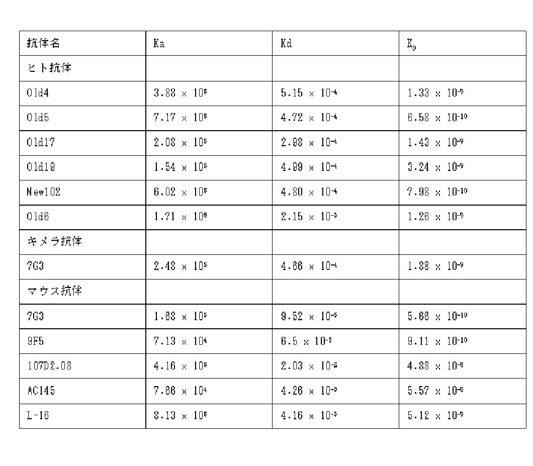
(IL-3Rα/GM-CSFRαキメラタンパク発現細胞の作製)
IL-3Rα抗体のエピトープ解析を実施するため、IL-3Rαの膜外領域の一部をGM-CSFRαと置き換えたキメラタンパクを細胞に発現させ、その細胞に対する各抗IL-3Rα抗体の結合性を解析した。簡単には、第一に、IL-3Rα分子およびGM-CSFRα分子を3領域に区分けし(上述のN末端よりA、B、Cドメイン)、第二に、IL-3Rα分子のA、B、Cドメインの1つずつをGM-CSFRαの該当するドメインと置き換えた分子を発現させるベクターをそれぞれ構築し、第三にHEK293F細胞に強制発現させ、第四としてフローサイトメトリーにて蛍光色素でラベルした各抗IL-3Rα抗体が結合するか観察した。
(GM-CSFR / pEF6/Myc-HisCプラスミドDNAの作製)
ヒトGM-CSF受容体α鎖(GM-CSFRα、CD116)のcDNAは脾臓由来cDNA(CLONTECHHumanMTC Panel)よりKOD-Plus-Ver.2(東洋紡績株式会社)を用いたPCR法により増幅した。PCR装置はGeneAmp PCR System 9700(アプライドバイオシステムズ)を用いた。PCR反応は94℃2分間の変性段階につづいて、98℃10秒-55℃30秒-68℃75秒の3ステップ反応を35サイクル行った。用いたPCRプライマーは以下のとおりである:
hCD116Fw-MfeI:5’-CGGCAATTGCCACCATGCTTCTCCTGGTGACAAGCCT-3’(配列番号80)
hCD116Rv-NotI:5’-ATTGCGGCCGCTCAGGTAATTTCCTTCACGG-3’(配列番号81)
得られたPCR産物は0.8%アガロースゲル電気泳動(TAE buffer)を行った。DNAはエチジウムブロマイド染色により可視化した。1.2 kb付近のバンドを切り出し、DNAをJETsorbキット(Genomed、Bad Oeynhausen、Germany)を用いて抽出した後、NotIおよびMfeIで消化した。pEF6/Myc-HisCプラスミドDNA(インビトロジェン社)をEcoRIおよびNotIで消化した。それぞれのDNAを0.8%アガロースゲル電気泳動し、1.2 kb付近と6 kb付近のバンドを切り出し、DNAをJETsorbキット(Genomed、Bad Oeynhausen、Germany)を用いて抽出した。pEF6/Myc-HisCプラスミドDNA由来DNA溶液0.5uLとPCR産物由来DNA溶液4 uLを混合し、TaKaRaLigationKit(タカラバイオ株式会社)を用い連結した。形質転換は、ライゲーションサンプルとDH5alphaコンピテント細胞と混合し、LBプレートへ撒いた。インサートチェックは、LA Taq(タカラバイオ株式会社)を用いたコロニーダイレクトPCRにより行った。PCR反応は94℃5分間の変性段階につづいて、94℃30秒-55℃30秒-72℃2分の3ステップ反応を40サイクル行った後、99℃30分間の処理を行った。 Example 7 Epitope analysis of anti-human IL-3Rα human antibody (production of cells expressing IL-3Rα / GM-CSFRα chimeric protein)
In order to perform epitope analysis of IL-3Rα antibody, a chimeric protein in which a part of the IL-3Rα extra-membrane region is replaced with GM-CSFRα is expressed in the cell, and the binding property of each anti-IL-3Rα antibody to the cell is expressed. Analyzed. Briefly, first, the IL-3Rα molecule and the GM-CSFRα molecule are divided into three regions (A, B, C domains from the N-terminal described above), and secondly, the A, B, Each vector was constructed to express a molecule in which each C domain was replaced with the corresponding domain of GM-CSFRα. Third, the vector was forcibly expressed in HEK293F cells, and the fourth was labeled with a fluorescent dye by flow cytometry. It was observed whether each anti-IL-3Rα antibody bound.
(Preparation of GM-CSFR / pEF6 / Myc-HisC plasmid DNA)
Human GM-CSF receptor α chain (GM-CSFRα, CD116) cDNA was amplified from spleen-derived cDNA (CLONTECHHumanMTC Panel) by PCR using KOD-Plus-Ver.2 (Toyobo Co., Ltd.). GeneAmp PCR System 9700 (Applied Biosystems) was used as the PCR device. The PCR reaction was a denaturation step at 94 ° C. for 2 minutes, followed by 35 cycles of a 3-step reaction at 98 ° C. for 10 seconds-55 ° C. for 30 seconds-68 ° C. for 75 seconds. The PCR primers used are as follows:
hCD116Fw-MfeI: 5'-CGGCAATTGCCACCATGCTTCTCCTGGTGACAAGCCT-3 '(SEQ ID NO: 80)
hCD116Rv-NotI: 5'-ATTGCGGCCGCTCAGGTAATTTCCTTCACGG-3 '(SEQ ID NO: 81)
The obtained PCR product was subjected to 0.8% agarose gel electrophoresis (TAE buffer). DNA was visualized by ethidium bromide staining. A band around 1.2 kb was excised and the DNA was extracted using the JETsorb kit (Genomed, Bad Oeynhausen, Germany) and then digested with NotI and MfeI. pEF6 / Myc-HisC plasmid DNA (Invitrogen) was digested with EcoRI and NotI. Each DNA was subjected to 0.8% agarose gel electrophoresis, the bands around 1.2 kb and 6 kb were cut out, and the DNA was extracted using a JETsorb kit (Genomed, Bad Oeynhausen, Germany). The pEF6 / Myc-HisC plasmid DNA-derived DNA solution 0.5 uL and the PCR product-derived DNA solution 4 uL were mixed and ligated using TaKaRaLigationKit (Takara Bio Inc.). For transformation, ligation samples and DH5alpha competent cells were mixed and plated on LB plates. Insert check was performed by colony direct PCR using LA Taq (Takara Bio Inc.). The PCR reaction was performed at a denaturation step of 94 ° C for 5 minutes, followed by 40 cycles of 3 steps of 94 ° C for 30 seconds-55 ° C for 30 seconds-72 ° C for 2 minutes, followed by treatment at 99 ° C for 30 minutes.
hCD116Fw-MfeI:5’-CGGCAATTGCCACCATGCTTCTCCTGGTGACAAGCCT-3’(配列番号82)
hCD116Rv-NotI:5’-ATTGCGGCCGCTCAGGTAATTTCCTTCACGG-3’(配列番号83)
得られたPCR産物は0.8%アガロースゲル電気泳動(135V、15分、TAEbuffer)を行った。DNAはエチジウムブロマイド染色により可視化した。1.2kb付近の増幅が得られたコロニーを対象に、ダイレクトシークエンシング法により塩基配列を決定した。シークエンスサンプルの反応はBigDye(R) Terminator v3.1Cycle Sequencing Kit(アプライドバイオシステムズ)とGeneAmp PCR System 9700(アプライドバイオシステムズ)を用いた(本明細書における全てのDNA配列解析でこれらを使用)。用いたPCRプライマーは以下のとおりである:
hCD116Fw-MfeI:5’-CGGCAATTGCCACCATGCTTCTCCTGGTGACAAGCCT-3’(配列番号84)
hCD116Rv-NotI:5’-ATTGCGGCCGCTCAGGTAATTTCCTTCACGG-3’(配列番号85)
hCD116SeqFw1:5’-TGAACTGTACCTGGGCGAGG-3’ (配列番号86)
hCD116SeqFw2:5’-CTGGCACGGAAAACCTACTG-3’ (配列番号87)
hCD116SeqRv1:5’-CCTGAATTTGGATAAAGCAG-3’ (配列番号88)
シークエンス解析装置はABI 3700XL DNA analyzer(アプライドバイオシステムズ)を用いた(本明細書における全てのDNA配列解析でこれを使用)。PCRによるアミノ酸配列の変異がおこっていないクローンを選定し、ラージプレップ法(キアゲン社)によりプラスミドDNAを抽出した。
(IL-3RA-FLAG/pEGFP-N1の作製)
ヒトIL-3Rα(CD123)の全長cDNAをPCR法で増幅し、下流にFLAGタグを連結した(IL-3RA-FLAG/pEGFP-N1)。 The PCR primers used are as follows:
hCD116Fw-MfeI: 5'-CGGCAATTGCCACCATGCTTCTCCTGGTGACAAGCCT-3 '(SEQ ID NO: 82)
hCD116Rv-NotI: 5'-ATTGCGGCCGCTCAGGTAATTTCCTTCACGG-3 '(SEQ ID NO: 83)
The obtained PCR product was subjected to 0.8% agarose gel electrophoresis (135 V, 15 minutes, TAE buffer). DNA was visualized by ethidium bromide staining. The base sequence was determined by direct sequencing for the colony from which amplification of about 1.2 kb was obtained. The reaction of the sequence samples was performed using BigDye® Terminator v3.1Cycle Sequencing Kit (Applied Biosystems) and GeneAmp PCR System 9700 (Applied Biosystems) (these are used for all DNA sequence analysis in this specification). The PCR primers used are as follows:
hCD116Fw-MfeI: 5'-CGGCAATTGCCACCATGCTTCTCCTGGTGACAAGCCT-3 '(SEQ ID NO: 84)
hCD116Rv-NotI: 5'-ATTGCGGCCGCTCAGGTAATTTCCTTCACGG-3 '(SEQ ID NO: 85)
hCD116SeqFw1: 5'-TGAACTGTACCTGGGCGAGG-3 '(SEQ ID NO: 86)
hCD116SeqFw2: 5'-CTGGCACGGAAAACCTACTG-3 '(SEQ ID NO: 87)
hCD116SeqRv1: 5'-CCTGAATTTGGATAAAGCAG-3 '(SEQ ID NO: 88)
The ABI 3700XL DNA analyzer (Applied Biosystems) was used as the sequence analyzer (this is used for all DNA sequence analysis in this specification). A clone having no amino acid sequence mutation by PCR was selected, and plasmid DNA was extracted by the large prep method (Qiagen).
(Preparation of IL-3RA-FLAG / pEGFP-N1)
A full-length cDNA of human IL-3Rα (CD123) was amplified by PCR, and a FLAG tag was ligated downstream (IL-3RA-FLAG / pEGFP-N1).
T7: 5’-TAATACGACTCACTATAGGG -3’ (配列番号89)
hCD123-C-FLAG-R1: 5’-TCGTCATCGTCCTTGTAGTCAGTTTTCTGCACGACCTGTA-3’ (配列番号90)
得られたPCR産物2 uLを鋳型に、LA Taq (タカラバイオ株式会社) を用いたPCR法により増幅した。PCR反応は95℃1分間の変性段階につづいて、95℃15秒-56℃15秒-72℃60秒の3ステップ反応を15サイクル行った後、72℃2分間の伸長反応を行った。用いたPCRプライマーは以下のとおりである:
IL-3Rα_Fw: 5’-CGGCAATTGCCACCATGGTCCTCCTTTGGCTCAC-3’(配列番号91)
C-FLAG-NotR2:5’-AAAAGCGGCCGCTCACTTGTCGTCATCGTCCTTGTAGTC-3’ (配列番号92)
得られたPCR産物は0.8%アガロースゲル電気泳動(135V、15分、TAEbuffer)を行った。DNAはエチジウムブロマイド染色により可視化した。1kb付近のバンドを切り出し、DNAをWizardSV Gel and PCR Clean-Up Systemを用いて抽出した。抽出したDNA全量をMfeIおよびNotIで消化し、0.8%アガロースゲル電気泳動(135V、15分、TAEbuffer)を行った。DNAはエチジウムブロマイド染色により可視化した。1kb付近のバンドを切り出し、DNAをWizardSVGelandPCRClean-UpSystemを用いて抽出した。抽出したIL-3RA-FLAGcDNA5uLとEcoRIおよびNotIで開裂したpEGFP-N1プラスミドDNA DNA 1uLを混合し、TaKaRa Ligation Kit(タカラバイオ株式会社)を用い連結した。形質転換は、ライゲーションサンプルとDH10Bコンピテント細胞と混合し、LBプレート(カナマイシン含有)へ撒いた。インサートチェックは、LA Taq(タカラバイオ株式会社)を用いたコロニーダイレクトPCRにより行った。PCR反応は95℃1分間の変性段階につづいて、95℃15秒-56℃15秒-72℃60秒の3ステップ反応を35サイクル行った後、72℃2分間の伸長反応を行った。用いたPCRプライマーは以下のとおりである:
pEGFP-N1-Fw: 5’-CGTGTACGGTGGGAGGTCTA-3’ (配列番号93)
pEGFP-N1-Re: 5’-TTTATGTTTCAGGTTCAGG-3’ (配列番号94)
0.8kb付近の増幅が得られたコロニーから、ミニプレップ法によりプラスミドDNAを抽出した。 The cDNA of human IL-3RA was amplified by PCR using LATaq (Takara Bio Inc.) using hCD123 / pEGFP-N1 plasmid DNA as a template. The PCR reaction was carried out after a denaturation step at 95 ° C. for 30 seconds, followed by 10 cycles of 95 ° C. 15 seconds-56 ° C. 15 seconds-72 ° C. 60 seconds, followed by a 2-minute extension reaction. The PCR primers used are as follows:
T7: 5'-TAATACGACTCACTATAGGG -3 '(SEQ ID NO: 89)
hCD123-C-FLAG-R1: 5'-TCGTCATCGTCCTTGTAGTCAGTTTTCTGCACGACCTGTA-3 '(SEQ ID NO: 90)
The obtained PCR product 2 uL was used as a template and amplified by the PCR method using LA Taq (Takara Bio Inc.). The PCR reaction was carried out at a denaturation step of 95 ° C. for 1 minute, followed by 15 cycles of 95 ° C. 15 seconds-56 ° C. 15 seconds-72 ° C. 60 seconds, followed by extension reaction at 72 ° C. for 2 minutes. The PCR primers used are as follows:
IL-3Rα_Fw: 5'-CGGCAATTGCCACCATGGTCCTCCTTTGGCTCAC-3 '(SEQ ID NO: 91)
C-FLAG-NotR2: 5'-AAAAGCGGCCGCTCACTTGTCGTCATCGTCCTTGTAGTC-3 '(SEQ ID NO: 92)
The obtained PCR product was subjected to 0.8% agarose gel electrophoresis (135 V, 15 minutes, TAE buffer). DNA was visualized by ethidium bromide staining. A band around 1 kb was cut out, and DNA was extracted using WizardSV Gel and PCR Clean-Up System. The total amount of the extracted DNA was digested with MfeI and NotI, and 0.8% agarose gel electrophoresis (135 V, 15 minutes, TAE buffer) was performed. DNA was visualized by ethidium bromide staining. A band around 1 kb was cut out and DNA was extracted using WizardSVGeland PCR Clean-UpSystem. The extracted IL-3RA-FLAG cDNA 5uL and pEGFP-N1 plasmid DNA 1uL cleaved with EcoRI and NotI were mixed and ligated using TaKaRa Ligation Kit (Takara Bio Inc.). For transformation, ligation samples and DH10B competent cells were mixed and plated on LB plates (containing kanamycin). Insert check was performed by colony direct PCR using LA Taq (Takara Bio Inc.). The PCR reaction was performed at a denaturation step of 95 ° C. for 1 minute, followed by 35 cycles of 95 ° C. 15 seconds-56 ° C. 15 seconds-72 ° C. 60 seconds, followed by extension reaction at 72 ° C. for 2 minutes. The PCR primers used are as follows:
pEGFP-N1-Fw: 5'-CGTGTACGGTGGGAGGTCTA-3 '(SEQ ID NO: 93)
pEGFP-N1-Re: 5'-TTTATGTTTCAGGTTCAGG-3 '(SEQ ID NO: 94)
Plasmid DNA was extracted by a miniprep method from a colony in which amplification around 0.8 kb was obtained.
pEGFP-N1-Fw: 5’-CGTGTACGGTGGGAGGTCTA-3’ (配列番号95)
pEGFP-N1-Re: 5’-TTTATGTTTCAGGTTCAGG-3’ (配列番号96)
(IL-3Rαのドメインマッピング)
BLASTP search(database:ProteinDataBankproteins(pdb))の結果、IL-13受容体alpha鎖(IL-13Rα)が最も高いスコアでヒットした(PDB: 3BPNC;ChainC,CrystalStructureOfTheIl4-Il4r-Il13raTernaryComplex)。ProteinDataBankからダウンロードしたPDBファイルとグラフィックソフトであるRasMolを用いてIL-13Rα蛋白質の立体構造を可視化し、細胞外領域を構成する3つのドメイン(上述のA、B及びCドメイン)を分割した。Multiple AlignmentソフトであるMUSCLEを用いてIL-3Rαアミノ酸配列とIL-13Rαアミノ酸配列を比較し、IL-3Rα細胞外領域も3つのドメインに区分した。さらに、GM-CSFRαとIL-3Rαを同様に比較し、GM-CSFRα細胞外領域も3つのドメインに区分した。 The purified IL-3RA-FLAG / pEGFP-N1 plasmid DNA was confirmed to be free of PCR mutation and the presence of a FLAG tag by DNA sequence analysis. Primers used for DNA sequence analysis are as follows:
pEGFP-N1-Fw: 5'-CGTGTACGGTGGGAGGTCTA-3 '(SEQ ID NO: 95)
pEGFP-N1-Re: 5'-TTTATGTTTCAGGTTCAGG-3 '(SEQ ID NO: 96)
(IL-3Rα domain mapping)
As a result of BLASTP search (database: ProteinDataBankproteins (pdb)), the IL-13 receptor alpha chain (IL-13Rα) was hit with the highest score (PDB: 3BPNC; ChainC, CrystalStructureOfTheIl4-Il4r-Il13raTernaryComplex). Using the PDB file downloaded from ProteinDataBank and the graphic software RasMol, the three-dimensional structure of the IL-13Rα protein was visualized, and the three domains (A, B and C domains described above) constituting the extracellular region were divided. The IL-3Rα amino acid sequence was compared with the IL-13Rα amino acid sequence using MUSCLE, a multiple alignment software, and the IL-3Rα extracellular region was also divided into three domains. Furthermore, GM-CSFRα and IL-3Rα were similarly compared, and the GM-CSFRα extracellular region was also divided into three domains.
IL-3RA-FLAG/pEGFP-N1プラスミドDNAを鋳型としPrimeSTAR(R) HS DNA Polymerase(タカラバイオ株式会社)を用いたPCR法により増幅した。PCR反応は98℃10秒-68℃6分の2ステップ反応を25サイクル行った。用いたPCRプライマーは以下のとおりである:
Aドメイン欠損;
CD123R11pEGFPN1:AAAGGTACCGAATTCGAAGCTTGAGCTC(配列番号97)
CD123F11:AAAGGTACCGGGAAGCCTTGGGCAGGT(配列番号98)
Bドメイン欠損;
CD123R12-2:AAAGGTACCACTGTTCTCAGGGAAGAGGAT(配列番号99)
CD123F12-2:AAAGGTACCCAGATTGAGATATTAACTCC(配列番号100)
Cドメイン欠損;
CD123R13: AAAGGTACCTGAAAAGACGACAAACTT(配列番号101)
CD123F13:AAAGGTACCTCGCTGCTGATCGCGCTG(配列番号102)
得られたPCR産物は0.8%アガロースゲル電気泳動(135V、15分、TAEbuffer)を行った。DNAはエチジウムブロマイド染色により可視化した。増幅を確認後、WizardSVGelandPCRClean-UpSystemを用いて精製した。得られたDNAをKpnIとDpnIで消化後、WizardSVGelandPCRClean-Up Systemを用いて精製し、TaKaRaLigationKitを用いて連結した。形質転換は、ライゲーションサンプルとDH10Bコンピテント細胞と混合し、LBプレート(カナマイシン含有)へ撒いた。インサートチェックは、LA Taq(タカラバイオ株式会社)を用いたコロニーダイレクトPCRにより行った。PCR反応は95℃1分間の変性段階につづいて、95℃15秒-56℃15秒-72℃40秒の3ステップ反応を38サイクル行った後、72℃2分間の伸長反応を行った。用いたPCRプライマーは以下のとおりである:
pEGFP-N1-Fw:5’-CGTGTACGGTGGGAGGTCTA -3’ (配列番号103)
pEGFP-N1-Re:5’-TTTATGTTTCAGGTTCAGG -3’ (配列番号104)
得られたPCR産物は0.8%アガロースゲル電気泳動(135V、15分、TAEbuffer)を行った。DNAはエチジウムブロマイド染色により可視化した。1kb付近の増幅が得られたコロニーからミニプレップ法によりプラスミドDNAを抽出した。 In order to identify the epitope of anti-human IL-3Rα human antibody, a protein in which the three domains of IL-3Rα divided as described above are replaced with the corresponding domain of GM-CSFRα one by one is expressed on the cell membrane, The presence or absence of antibody binding was confirmed.
Amplification was performed by PCR using PrimeSTAR® HS DNA Polymerase (Takara Bio Inc.) using IL-3RA-FLAG / pEGFP-N1 plasmid DNA as a template. The PCR reaction was 25 cycles of a 2-step reaction at 98 ° C. for 10 seconds to 68 ° C. for 6 minutes. The PCR primers used are as follows:
A domain deficiency;
CD123R11pEGFPN1: AAAGGTACCGAATTCGAAGCTTGAGCTC (SEQ ID NO: 97)
CD123F11: AAAGGTACCGGGAAGCCTTGGGCAGGT (SEQ ID NO: 98)
B domain deficiency;
CD123R12-2: AAAGGTACCACTGTTCTCAGGGAAGAGGAT (SEQ ID NO: 99)
CD123F12-2: AAAGGTACCCAGATTGAGATATTAACTCC (SEQ ID NO: 100)
C domain deficiency;
CD123R13: AAAGGTACCTGAAAAGACGACAAACTT (SEQ ID NO: 101)
CD123F13: AAAGGTACCTCGCTGCTGATCGCGCTG (SEQ ID NO: 102)
The obtained PCR product was subjected to 0.8% agarose gel electrophoresis (135 V, 15 minutes, TAE buffer). DNA was visualized by ethidium bromide staining. After confirming amplification, it was purified using WizardSVGelandPCRClean-UpSystem. The obtained DNA was digested with KpnI and DpnI, purified using WizardSVGeland PCR Clean-Up System, and ligated using TaKaRaLigationKit. For transformation, ligation samples and DH10B competent cells were mixed and plated on LB plates (containing kanamycin). Insert check was performed by colony direct PCR using LA Taq (Takara Bio Inc.). The PCR reaction was performed at a denaturation step of 95 ° C. for 1 minute, followed by 38 cycles of a 3-step reaction at 95 ° C. for 15 seconds-56 ° C. for 15 seconds-72 ° C. for 40 seconds, followed by an extension reaction at 72 ° C. for 2 minutes. The PCR primers used are as follows:
pEGFP-N1-Fw: 5'-CGTGTACGGTGGGAGGTCTA-3 '(SEQ ID NO: 103)
pEGFP-N1-Re: 5'-TTTATGTTTCAGGTTCAGG-3 '(SEQ ID NO: 104)
The obtained PCR product was subjected to 0.8% agarose gel electrophoresis (135 V, 15 minutes, TAE buffer). DNA was visualized by ethidium bromide staining. Plasmid DNA was extracted by a miniprep method from a colony in which amplification around 1 kb was obtained.
Aドメイン挿入;
GM-CSFRF11:AAAGGTACCGCCACCATGCTTCTCCTGGTGACA(配列番号105)
GM-CSFRR11:AAAGGTACCTGAATTTGGATAAAGCAG(配列番号106)
Bドメイン挿入;
GM-CSFRF12:AAAGGTACCGGAAGGGAGGGTACCGCT(配列番号107)
GM-CSFRR12:AAAGGTACCCTTTGTGTCCAAAAGTGA(配列番号108)
Cドメイン挿入;
GM-CSFRF13:AAAGGTACCAAAATAGAACGATTCAAC(配列番号109)
GM-CSFRR13:AAAGGTACCAATGTACACAGAGCCGAG(配列番号110)
得られたPCR産物は0.8%アガロースゲル電気泳動(135V、15分、TAEbuffer)を行った。DNAはエチジウムブロマイド染色により可視化した。増幅を確認後、WizardSVGelandPCRClean-UpSystemを用いて精製した。 Amplification was performed by PCR using PrimeSTAR® HS DNA Polymerase (Takara Bio Inc.) using GM-CSFR / pEF6 / Myc-HisC plasmid DNA as a template. The PCR reaction was 25 cycles of a 2-step reaction at 98 ° C. for 10 seconds to 68 ° C. for 30 seconds. The PCR primers used are as follows:
A domain insertion;
GM-CSFRF11: AAAGGTACCGCCACCATGCTTCTCCTGGTGACA (SEQ ID NO: 105)
GM-CSFRR11: AAAGGTACCTGAATTTGGATAAAGCAG (SEQ ID NO: 106)
B domain insertion;
GM-CSFRF12: AAAGGTACCGGAAGGGAGGGTACCGCT (SEQ ID NO: 107)
GM-CSFRR12: AAAGGTACCCTTTGTGTCCAAAAGTGA (SEQ ID NO: 108)
C domain insertion;
GM-CSFRF13: AAAGGTACCAAAATAGAACGATTCAAC (SEQ ID NO: 109)
GM-CSFRR13: AAAGGTACCAATGTACACAGAGCCGAG (SEQ ID NO: 110)
The obtained PCR product was subjected to 0.8% agarose gel electrophoresis (135 V, 15 minutes, TAE buffer). DNA was visualized by ethidium bromide staining. After confirming amplification, it was purified using WizardSVGelandPCRClean-UpSystem.
pEGFP-N1-Fw:5’-CGTGTACGGTGGGAGGTCTA -3’ (配列番号111)
pEGFP-N1-Re:5’-TTTATGTTTCAGGTTCAGG -3’ (配列番号112)
得られたPCR産物は0.8%アガロースゲル電気泳動(135V、15分、TAEbuffer)を行った。DNAはエチジウムブロマイド染色により可視化した。1kb付近の増幅が得られたコロニーからミニプレップ法によりプラスミドDNAを抽出した。
(抗IL-3Rα抗体の蛍光色素のラベル化)
抗ヒトIL-3Rαヒト抗体が結合するか確認するため、各ヒト抗体を蛍光色素AlexaFlour488(Molecular Probe, Invitrogen社)でラベルした。ラベル方法は、Invitrogen社頒布のマニュアルに従い、検出はフローサイトメトリー(FACSCalibur、BD Biosciences社)のFL1にて蛍光を検出した。 The resulting DNA was digested with KpnI, purified using QIAquick Gel Extraction Kit (Qiagen), and IL-3RA-FLAG / pEGFP-N1 plasmid DNA lacking the corresponding domain (cleaved and purified with KpnI) and Mixed and ligated using TaKaRa Ligation Kit. For transformation, ligation samples and DH10B competent cells were mixed and plated on LB plates (containing kanamycin). Insert check was performed by colony direct PCR using LA Taq (Takara Bio Inc.). The PCR reaction was performed at a denaturation step of 95 ° C. for 1 minute, followed by 38 cycles of a 3-step reaction at 95 ° C. for 15 seconds-56 ° C. for 15 seconds-72 ° C. for 40 seconds, followed by an extension reaction at 72 ° C. for 2 minutes. The PCR primers used are as follows:
pEGFP-N1-Fw: 5'-CGTGTACGGTGGGAGGTCTA -3 '(SEQ ID NO: 111)
pEGFP-N1-Re: 5'-TTTATGTTTCAGGTTCAGG-3 '(SEQ ID NO: 112)
The obtained PCR product was subjected to 0.8% agarose gel electrophoresis (135 V, 15 minutes, TAE buffer). DNA was visualized by ethidium bromide staining. Plasmid DNA was extracted by a miniprep method from a colony in which amplification around 1 kb was obtained.
(Labeling of fluorescent dye of anti-IL-3Rα antibody)
In order to confirm whether the anti-human IL-3Rα human antibody was bound, each human antibody was labeled with a fluorescent dye AlexaFlour488 (Molecular Probe, Invitrogen). The labeling method was in accordance with the manual of Invitrogen distribution, and the fluorescence was detected by FL1 of flow cytometry (FACSCalibur, BD Biosciences).
抗体濃度(mg/mL)=(A280 - A494 × 0.11)/1.4
(ラベル化抗IL-3Rα抗体を用いたIL-3Rα/GM-CSFRαキメラタンパク発現細胞のフローサイトメトリー解析)
IL-3Rα/GM-CSFRαキメラタンパク発現細胞の作製には、HEK293T細胞(ATCC CRL 1268)を用いた。293フェクチン(Invitrogen社)を用いて、HEK293Tに上記で取得したプラスミドDNAを発現ベクターとして導入した。発現ベクターを導入したHEK293Tは、シェーカーを用いてCO2 5%、37℃の環境下で培養し、導入2日後にフローサイトメトリー解析に用いた。 Specifically, 1/10 amount of 1M Na 2 CO 3 was added to the antibody solution dissolved in PBS. Next, a predetermined amount of the antibody solution described in the manual was added to a container containing AlexaFlour488 powder to which tetrafluorophenyl (TFP) was added and allowed to react at room temperature in the dark for 1 hour while stirring. Next, after sufficiently replacing the gel filtration column (such as NAP-10, GE Healthcare) with PBS, the antibody solution reacted with AlexaFlour488 was added, and while obtaining the antibody fraction exhibiting a yellow green color, The antibody solution buffer was replaced with PBS. The anti-human IL-3Rα antibody labeled with AlexaFlour488 obtained as described above measures the absorbance at wavelengths of 280 nm and 494 nm (A280 and A494, respectively) using an absorptiometer, and calculates the antibody concentration using the following formula: did.
Antibody concentration (mg / mL) = (A280-A494 x 0.11) /1.4
(Flow cytometric analysis of IL-3Rα / GM-CSFRα chimeric protein expressing cells using labeled anti-IL-3Rα antibody)
HEK293T cells (ATCC CRL 1268) were used for the production of IL-3Rα / GM-CSFRα chimeric protein expressing cells. The plasmid DNA obtained above was introduced into HEK293T as an expression vector using 293fectin (Invitrogen). HEK293T into which the expression vector was introduced was cultured in a

得られたIL-3Rα抗体がIL-3シグナルを阻害しないか検討するため、IL-3又はGM-CSF依存性に増殖する細胞株TF-1(DMSZ no.ACC344)を用いた。
具体的には、TF-1細胞を、IL-3を1ng/mL及び10%牛胎児血清を含んだRPMI1640培地(TF-1培地)に希釈し、96穴プレートに撒いた。さらに、各種IL-3Rα抗体および陰性コントロール抗体としてヒト血清由来IgGをTF-1培地に希釈し96穴プレートに移し、抗体の最終濃度が10及び100μg/mLの最終濃度になるように添加した。対照として、細胞なしの培地のみのウェル、TF-1細胞が添加されたウェルを設けた。3日間37℃5%CO2環境下で培養し、CelltiterGlo(Promega社)を培地と同量添加した。30分静置した後、プレートリーダ(ARBO、PerkinElmer社)を用いて発光量を定量した。
増殖の阻害率は、以下の計算を行った:
(サンプルの発光量-細胞なしのウェル)/(TF-1細胞のみ添加したウェル-細胞なしのウェル)x100(%)
市販抗体9F5、6H6、107D2.08に関しては、バッファーをPBSに置換するため、NAP-5カラムを利用した。具体的には、PBSにて十分に置換したNAP-5カラムに0.5mLの抗体溶液を添加した。次に1.0mLのPBSを添加し、カラムより出てきた溶液を回収した。溶液は、孔径0.22μmのメンブランフィルターMILLEX-GV(ミリポア社)でろ過滅菌し、PBSを溶媒とした抗体を得た。抗体の濃度は280nmの吸光度を測定し、1mg/mLを1.4 ODとして算出した。
結果を図5に示す。Old4抗体、Old5抗体、Old17抗体、Old19抗体、New102抗体、9F5抗体、6H6抗体はIL-3シグナルを阻害しないことが判明し、一方7G3抗体、Old6抗体、107D2.08抗体はIL-3シグナルを阻害することが判明した。
実施例9 抗IL-3Rαヒト抗体を用いたコロニー形成能への影響の検討
各種IL-3Rα抗体が造血前駆細胞によるコロニー形成能に影響しないか、コロニーアッセイを行った。 From the above, 7G3, 9F5, 6H6, AC145 may recognize A domain, Old4, Old5, New102 will recognize B domain, Old19 may recognize A domain and B domain, Old6, 107D2.08 may recognize B domain and C domain, respectively Indicated. Therefore, the reactivity of various anti-IL-3Rα antibodies to the AC domains of IL-3Rα was as shown in Table 3 below.
Specifically, TF-1 cells were diluted in RPMI 1640 medium (TF-1 medium) containing 1 ng / mL of IL-3 and 10% fetal calf serum, and seeded in a 96-well plate. Furthermore, human serum-derived IgG as various IL-3Rα antibodies and negative control antibodies were diluted in TF-1 medium, transferred to a 96-well plate, and added so that the final concentrations of the antibodies were 10 and 100 μg / mL. As a control, a well containing only medium without cells and a well containing TF-1 cells were provided. The cells were cultured for 3 days at 37 ° C. in a 5% CO 2 environment, and CelltiterGlo (Promega) was added in the same amount as the medium. After standing for 30 minutes, the amount of luminescence was quantified using a plate reader (ARBO, PerkinElmer).
The percent inhibition of proliferation was calculated as follows:
(Sample luminescence amount-well without cells) / (well added with TF-1 cells only-well without cells) x 100 (%)
For the commercially available antibodies 9F5, 6H6, and 107D2.08, a NAP-5 column was used to replace the buffer with PBS. Specifically, 0.5 mL of the antibody solution was added to a NAP-5 column sufficiently substituted with PBS. Next, 1.0 mL of PBS was added, and the solution coming out of the column was collected. The solution was sterilized by filtration through a membrane filter MILLEX-GV (Millipore) having a pore size of 0.22 μm to obtain an antibody using PBS as a solvent. The antibody concentration was calculated by measuring absorbance at 280 nm and 1 mg / mL as 1.4 OD.
The results are shown in FIG. Old4 antibody, Old5 antibody, Old17 antibody, Old19 antibody, New102 antibody, 9F5 antibody, 6H6 antibody was found not to inhibit IL-3 signal, while 7G3 antibody, Old6 antibody, 107D2.08 antibody did not inhibit IL-3 signal. It was found to inhibit.
Example 9 Examination of influence on colony forming ability using anti-IL-3Rα human antibody A colony assay was performed to determine whether various IL-3Rα antibodies had an effect on the ability of hematopoietic progenitor cells to form colonies.
結果を図6に示す。エリスロポエチン、IL-3、G-CSF、StemCell Factorを添加したコロニーアッセイにおいて、IL-3シグナルのブロッキング能を有する7G3抗体の添加により、コロニー数の減少およびコロニーサイズの減少が認められた。一方New102抗体添加によるコロニー数の変化は認められなかった。この結果より、IL-3シグナルを阻害またはブロッキングしないほうが、正常の造血機能に与える影響は小さく、副作用が少ないことが推測される。
実施例10 抗IL-3Rαヒト抗体を用いたマウス担ガンモデルにおける抗腫瘍効果
得られた抗IL-3Rα抗体をマウス担ガンモデルに投与し、その抗腫瘍効果を検討した。簡単には、マウスに白血病細胞を尾静脈より移入し、翌日に抗体を投与し、約3週間後にマウスの骨より採取した骨髄細胞中の白血病細胞の数を計測した。 As the antibody, chimeric 7G3 antibody was used as an antibody in which blocking ability of IL-3 signal was recognized in Example 8, and New102 antibody was used as an antibody in which blocking ability was not recognized.
The results are shown in FIG. In the colony assay to which erythropoietin, IL-3, G-CSF, and StemCell Factor were added, a decrease in the number of colonies and a decrease in colony size were observed by the addition of 7G3 antibody having the ability to block IL-3 signal. On the other hand, there was no change in the number of colonies due to the addition of New102 antibody. From this result, it is presumed that the effect on the normal hematopoietic function is small and the side effects are less if the IL-3 signal is not inhibited or blocked.
Example 10 Antitumor effect in mouse tumor-bearing model using anti-IL-3Rα human antibody The obtained anti-IL-3Rα antibody was administered to a mouse cancer-bearing model, and the antitumor effect was examined. Briefly, leukemia cells were transferred to the mouse from the tail vein, the antibody was administered the next day, and the number of leukemia cells in bone marrow cells collected from the bone of the mouse was counted about 3 weeks later.
実施例11 抗IL-3Rα抗体によるIL-3Rα発現細胞株傷害性試験
抗体を介した細胞傷害活性[抗体依存性細胞性細胞傷害活性(Antibody-DependentCellularCytotoxicity)以下、ADCCと略記する]を測定するために、抗体の存在下でエフェクターとしてヒト末梢血由来単核球(PeripheralBlood Mononuclear Cells以下、PBMC)を用い実施した。 The results are shown in FIG. It was confirmed that the number of MOLM13 cells in the femur bone marrow was significantly reduced in the group administered with the antibody compared to the vehicle group not administered with the antibody. This result suggests that anti-IL-3Rα antibody may be a therapeutic agent for leukemia.
Example 11 IL-3Rα-expressing cell line cytotoxicity test with anti-IL-3Rα antibody To measure antibody-mediated cytotoxic activity [Antibody-Dependent Cellular Cytotoxicity (hereinafter abbreviated as ADCC)] In addition, human peripheral blood-derived mononuclear cells (Peripheral Blood Mononuclear Cells, hereinafter referred to as PBMC) were used as effectors in the presence of antibodies.
方法は簡単には、ターゲット細胞を抗体及びPBMC存在下で培養し、抗体による特異的なターゲット細胞の溶解率を計測するものである。 Furthermore, using RPMI1640 medium containing 10% fetal bovine serum supplemented with human IL-2 (Peprotech) at a final concentration of 4 ng / mL (40 IU / mL or higher), cultured overnight at 37 ° C in a 5% CO 2 environment The PBMC was also used as an effector for the ADCC assay.
In a simple method, target cells are cultured in the presence of an antibody and PBMC, and a specific target cell lysis rate by the antibody is measured.
ターゲット細胞の溶解度は、細胞が溶解し培地中に放出されたクロム酸ナトリウム中の51Cr量を測定した。すなわち、各ウェルの値から抗体が添加されていないウェルの値を差し引いた値を、Triton-X100を加えたウェル(特異的溶解率100%とする)の値から抗体が添加されていないウェルの値を差し引いた値で割ることにより、「特異的溶解率」を算出した。 After centrifuging the plate, the supernatant was transferred to a 96-well plate with scintillator (Lumaplate-TM, PerkinElmer) and dried at 56 ° C. for 2 hours. The plate was sealed (TopSeal-A, Packard) and measured with a microplate reader (TopCount, PerkinElmer).
The solubility of the target cells was determined by measuring the amount of 51 Cr in sodium chromate released from the cells after cell lysis. That is, the value obtained by subtracting the value of the well to which no antibody is added from the value of each well is the value of the well to which no antibody is added from the value of the well to which Triton-X100 is added (specific lysis rate is 100%). The “specific lysis rate” was calculated by dividing the value by the subtracted value.
実施例12 抗IL-3Rα抗体のサルIL-3Rα蛋白質への結合性試験
取得した抗ヒトIL-3Rα抗体のサルIL-3Rαへの結合の有無は、実施例1で作製したカニクイザルIL-3Rα強制発現細胞に、実施例7で作製した抗ヒトIL-3Rα抗体が結合するかをフローサイトメトリーを用いて解析した。 The results are shown in FIGS. Various IL-3Rα antibodies showed ADCC activity against target cells in a concentration-dependent manner. In addition, it exhibited high ADCC ability compared to the control chimeric 7G3 antibody. This indicates that the IL-3Rα antibody exhibits a high ADCC ability for IL-3Rα-expressing cells, and shows the possibility of treatment with IL-3Rα-positive cell removal as a medicinal effect.
Example 12 Binding test of anti-IL-3Rα antibody to monkey IL-3Rα protein The presence or absence of binding of the obtained anti-human IL-3Rα antibody to monkey IL-3Rα was determined by forced cynomolgus IL-3Rα prepared in Example 1. Whether the anti-human IL-3Rα antibody prepared in Example 7 was bound to the expressed cells was analyzed using flow cytometry.
実施例13 抗ヒトIL-3Rαヒト抗体の詳細なエピトープ解析
(IL-3Rα/GM-CSFRαキメラタンパク発現細胞の作製)
IL-3Rα抗体のより詳細なエピトープ解析を実施するため、IL-3Rαの膜外領域のドメインより小さい領域をGM-CSFRαと置き換えたキメラタンパクを細胞に発現させ、その細胞に対する各抗IL-3Rα抗体の結合性を解析した。簡単には、第一に、IL-3Rα分子の立体的な構造予測から外側に位置していると考えられる領域を決定し、第二に、その小さい領域をGM-CSFRαに置き換えたIL-3Rα分子を発現させるベクターをそれぞれ構築し、第三に、HEK293F細胞に強制発現させ、第四としてフローサイトメトリーにて蛍光色素でラベルした各抗IL-3Rα抗体が結合するか観察した。
(IL-3Rαのドメインマッピング)
実施例7より区分した3ドメインのうち、取得した抗体Old19及びNew102が認識するA、Bドメインに絞り、詳細に解析した。IL-4受容体alpha鎖(IL-4Rα、CD124)(PDB: 3BPNC;ChainC,CrystalStructureOfTheIl4-Il4r-Il13raTernaryComplex)の立体構造を元に、SWISS-MODEL (http://swissmodel.expasy.org//SWISS-MODEL.html)を用いてIL-3Rα蛋白質の立体構造をホモロジーモデリングした。予測されたIL-3Rα蛋白質構造をグラフィックソフトRasMol(http://rasmol.org/)を用いて可視化し、IL-3Rα分子の外側に位置すると考えられるアミノ酸領域7箇所を決定した(図4)。 The result is shown in FIG. It was confirmed that the anti-human IL-3Rα human antibodies Old4, Old5, Old17, Old19, New102, and the chimeric 7G3 antibody react with cynomolgus IL-3Rα.
Example 13 Detailed Epitope Analysis of Anti-human IL-3Rα Human Antibody (Preparation of IL-3Rα / GM-CSFRα Chimera Protein Expression Cell)
In order to carry out more detailed epitope analysis of IL-3Rα antibody, a chimeric protein in which a region smaller than the domain of IL-3Rα extra-membrane region was replaced with GM-CSFRα was expressed in the cell, and each anti-IL-3Rα against that cell Antibody binding was analyzed. Briefly, first, the region that is thought to be located outside is determined from the three-dimensional structure prediction of the IL-3Rα molecule, and secondly, IL-3Rα in which the small region is replaced with GM-CSFRα. Vectors for expressing molecules were constructed, and thirdly, forced expression in HEK293F cells, and fourth, flow cytometry was used to observe whether each anti-IL-3Rα antibody labeled with a fluorescent dye was bound.
(IL-3Rα domain mapping)
Of the three domains classified from Example 7, analysis was made in detail by focusing on the A and B domains recognized by the obtained antibodies Old19 and New102. Based on the three-dimensional structure of IL-4 receptor alpha chain (IL-4Rα, CD124) (PDB: 3BPNC; ChainC, CrystalStructureOfTheIl4-Il4r-Il13raTernaryComplex) SWISS-MODEL -MODEL.html) was used to model the three-dimensional structure of IL-3Rα protein. The predicted IL-3Rα protein structure was visualized using the graphic software RasMol (http://rasmol.org/), and seven amino acid regions thought to be located outside the IL-3Rα molecule were determined (FIG. 4). .
IL-3RA-FLAG/pEGFP-N1プラスミドDNAを鋳型としPrimeSTAR(R) HS DNAPolymerase (タカラバイオ株式会社)を用いたPCR法により増幅した。PCR反応は98℃10秒-68℃5分の2ステップ反応を25サイクル行った。用いたPCRプライマーは以下のとおりである:
領域1欠損;
CD123-Fw21:CGTGGAACCCGCAGTGAACAATAGCTATT(配列番号149)
CD123-Re21:ACTCTGTTCTTTTTAACACACTCGATATCG(配列番号150)
領域2欠損;
CD123-Fw22:CTTTATCCAAATAACAGTGGGAAGCCTTG(配列番号151)
CD123-Re22:CAGTTTCTGTTGGAATGGTGGGTTGGCCACT(配列番号152)
領域3欠損;
CD123-Fw23:AGGGAGGGTACCGGTGCGGAGAATCTGACCTGCT(配列番号153)
CD123-Re23:TCCTGAATTTGGATAGAAGAGGATCCACGTGG(配列番号154)
領域4欠損;
CD123-Fw24:GGTCCGACGGCCCCCGCGGACGTCCAGTA(配列番号155)
CD123-Re24 :CCTCGCCCAGGTACAGCTCAAGAAATCCACGT(配列番号156)
領域5欠損;
CD123-Fw25:ACGGAACCAGCGCAGCCTTCGGTATCCCCT(配列番号157)
CD123-Re25:TAACCAGAAAGTGGGAACTTTGAGAACC(配列番号158)
領域6欠損;
CD123-Fw26:TCTTTGATTCATTTGTCGTCTTTTCACA(配列番号159)
CD123-Re26:ATTGGATGCCGAAGGCTGCGCTCCTGCCC(配列番号160)
得られたPCR産物は0.8%アガロースゲル電気泳動(135V、15分、TAEbuffer)を行った。DNAはエチジウムブロマイド染色により可視化した。増幅を確認後、WizardSVGelandPCRClean-UpSystemを用いて精製した。得られたDNAをPolynucleotidekinase(New EnglandBiolabs)でリン酸化し、エタノール沈殿の後、一部をTaKaRa LigationKitを用いて反応させた。形質転換は、ライゲーションサンプルとDH10Bコンピテント細胞と混合し、LBプレート(カナマイシン含有)へ撒いた。得られたコロニーからMiniprep法によりプラスミドDNAを抽出し、XhoIおよびNotIで消化し、インサートを確認した。
(ラベル化抗IL-3Rα抗体を用いたIL-3Rα/GM-CSFRαキメラタンパク発現細胞のフローサイトメトリー解析)
IL-3Rα/GM-CSFRαキメラタンパク発現細胞の作製の作製には、HEK293T細胞を用いた。リポフェクション法を用いて、HEK293Tに上記で取得したプラスミドDNAを発現ベクターとして導入した。発現ベクターを導入したHEK293Tは、CO2 5%、37℃の環境下で培養し、導入2日後にフローサイトメトリー解析に用いた。 In order to identify the epitope of anti-human IL-3Rα human antibody, each of the six amino acid regions of IL-3Rα divided as described above was replaced with a corresponding region of GM-CSFRα, and expressed on the cell membrane. The presence or absence of antibody binding was confirmed.
Amplification was carried out by PCR using PrimeSTAR® HS DNA Polymerase (Takara Bio Inc.) using IL-3RA-FLAG / pEGFP-N1 plasmid DNA as a template. The PCR reaction was 25 cycles of a 2-step reaction at 98 ° C. for 10 seconds to 68 ° C. for 5 minutes. The PCR primers used are as follows:
CD123-Fw21: CGTGGAACCCGCAGTGAACAATAGCTATT (SEQ ID NO: 149)
CD123-Re21: ACTCTGTTCTTTTTAACACACTCGATATCG (SEQ ID NO: 150)
Region 2 deficiency;
CD123-Fw22: CTTTATCCAAATAACAGTGGGAAGCCTTG (SEQ ID NO: 151)
CD123-Re22: CAGTTTCTGTTGGAATGGTGGGTTGGCCACT (SEQ ID NO: 152)
Region 3 deficiency;
CD123-Fw23: AGGGAGGGTACCGGTGCGGAGAATCTGACCTGCT (SEQ ID NO: 153)
CD123-Re23: TCCTGAATTTGGATAGAAGAGGATCCACGTGG (SEQ ID NO: 154)
Region 4 deficiency;
CD123-Fw24: GGTCCGACGGCCCCCGCGGACGTCCAGTA (SEQ ID NO: 155)
CD123-Re24: CCTCGCCCAGGTACAGCTCAAGAAATCCACGT (SEQ ID NO: 156)
CD123-Fw25: ACGGAACCAGCGCAGCCTTCGGTATCCCCT (SEQ ID NO: 157)
CD123-Re25: TAACCAGAAAGTGGGAACTTTGAGAACC (SEQ ID NO: 158)
Region 6 deficiency;
CD123-Fw26: TCTTTGATTCATTTGTCGTCTTTTCACA (SEQ ID NO: 159)
CD123-Re26: ATTGGATGCCGAAGGCTGCGCTCCTGCCC (SEQ ID NO: 160)
The obtained PCR product was subjected to 0.8% agarose gel electrophoresis (135 V, 15 minutes, TAE buffer). DNA was visualized by ethidium bromide staining. After confirming amplification, it was purified using WizardSVGelandPCRClean-UpSystem. The obtained DNA was phosphorylated with Polynucleotidekinase (New England Biolabs), and after ethanol precipitation, a part was reacted with TaKaRa Ligation Kit. For transformation, ligation samples and DH10B competent cells were mixed and plated on LB plates (containing kanamycin). Plasmid DNA was extracted from the obtained colonies by the Miniprep method, digested with XhoI and NotI, and the insert was confirmed.
(Flow cytometric analysis of IL-3Rα / GM-CSFRα chimeric protein expressing cells using labeled anti-IL-3Rα antibody)
HEK293T cells were used for the production of IL-3Rα / GM-CSFRα chimeric protein expressing cells. Using the lipofection method, the plasmid DNA obtained above was introduced into HEK293T as an expression vector. HEK293T into which the expression vector was introduced was cultured in an environment of 5% CO 2 and 37 ° C and used for flow cytometry analysis two days after the introduction.
以上の結果を表4としてまとめた。

The above results are summarized in Table 4.
配列番号4:IL-3Rα_Reプライマー
配列番号5:IL-3Rα_seqF1プライマー
配列番号6:インサート(MfeIからNotIまで)
配列番号7:Rhe123Fw1プライマー
配列番号8:Rhe123Rv1プライマー
配列番号9:T7プライマー
配列番号10:SP6プライマー
配列番号11:カニクイザルIL-3Rαのインサート(MfeIからNotIまで)
配列番号12:アカゲザルIL-3Rαのインサート(MfeIからNotIまで)
配列番号13: hIL-3Rαsol-FLAG-NotIプライマー
配列番号14:インサート(MfeIからNotIまで)
配列番号15:hh-6プライマー
配列番号16:hh-3 プライマー
配列番号17:hh-4プライマー
配列番号18:Old4重鎖特異的プライマーFw
配列番号19:Old4重鎖特異的プライマーRv
配列番号20:Old5重鎖特異的プライマーFw
配列番号21:Old5重鎖特異的プライマーRv
配列番号22:Old17重鎖特異的プライマーFw
配列番号23:Old17重鎖特異的プライマーRv
配列番号24:Old19重鎖特異的プライマーFw
配列番号25:Old19重鎖特異的プライマーRv
配列番号26:New102重鎖特異的プライマーFw
配列番号27:New102重鎖特異的プライマーRv
配列番号28:Old6重鎖特異的プライマーFw
配列番号29:Old6重鎖特異的プライマーRv
配列番号30:mH_Rv1プライマー
配列番号31:mH_Rv2プライマー
配列番号32:7G3重鎖特異的プライマーFw
配列番号33:7G3重鎖特異的プライマーRv
配列番号34:hk-2プライマー
配列番号35:hk-6プライマー
配列番号36:Old4軽鎖特異的プライマーFw
配列番号37:Old4軽鎖特異的プライマーRv
配列番号38:Old5軽鎖特異的プライマーFw
配列番号39:Old5軽鎖特異的プライマーRv
配列番号40:Old17軽鎖特異的プライマーFw
配列番号41:Old17軽鎖特異的プライマーRv
配列番号42:Old19軽鎖特異的プライマーFw
配列番号43:Old19軽鎖特異的プライマーRv
配列番号44:New102軽鎖特異的プライマーFw
配列番号45:New102軽鎖特異的プライマーRv
配列番号46:Old6軽鎖特異的プライマーFw
配列番号47:Old6軽鎖特異的プライマーRv
配列番号48:mK_Rv1プライマー
配列番号49:mK_Rv2プライマー
配列番号50:7G3軽鎖特異的プライマーFw
配列番号51:7G3軽鎖特異的プライマーRv
配列番号80:hCD116Fw-MfeIプライマー
配列番号81:hCD116Rv-NotIプライマー
配列番号82:hCD116Fw-MfeIプライマー
配列番号83:hCD116Rv-NotIプライマー
配列番号84:hCD116Fw-MfeIプライマー
配列番号85:hCD116Rv-NotIプライマー
配列番号86:hCD116SeqFw1プライマー
配列番号87:hCD116SeqFw2プライマー
配列番号88:hCD116SeqRv1プライマー
配列番号89:T7プライマー
配列番号90:hCD123-C-FLAG-R1プライマー
配列番号91:IL-3Rα_Fwプライマー
配列番号92:C-FLAG-NotR2プライマー
配列番号93:pEGFP-N1-Fwプライマー
配列番号94:pEGFP-N1-Reプライマー
配列番号95:pEGFP-N1-Fwプライマー
配列番号96:pEGFP-N1-Reプライマー
配列番号97:CD123R11pEGFPN1プライマー
配列番号98:CD123F11プライマー
配列番号99:CD123R12-2プライマー
配列番号100:CD123F12-2プライマー
配列番号101:CD123R13プライマー
配列番号102:CD123F13プライマー
配列番号103:pEGFP-N1-Fwプライマー
配列番号104:pEGFP-N1-Reプライマー
配列番号105:GM-CSFRF11プライマー
配列番号106:GM-CSFRR11プライマー
配列番号107:GM-CSFRF12プライマー
配列番号108:GM-CSFRR12プライマー
配列番号109:GM-CSFRF13プライマー
配列番号110:GM-CSFRR13プライマー
配列番号111:pEGFP-N1-Fwプライマー
配列番号112:pEGFP-N1-Reプライマー
配列番号149:CD123-Fw21プライマー
配列番号150:CD123-Re21プライマー
配列番号151:CD123-Fw22プライマー
配列番号152:CD123-Re22プライマー
配列番号153:CD123-Fw23プライマー
配列番号154:CD123-Re23プライマー
配列番号155:CD123-Fw24プライマー
配列番号156:CD123-Re24プライマー
配列番号157:CD123-Fw25プライマー
配列番号158:CD123-Re25プライマー
配列番号159:CD123-Fw26プライマー
配列番号160:CD123-Re26プライマー SEQ ID NO: 3: IL-3Rα_Fw primer SEQ ID NO: 4: IL-3Rα_Re primer
SEQ ID NO: 5: IL-3Rα_seqF1 primer
Sequence number 6: Insert (from MfeI to NotI)
Sequence number 7: Rhe123Fw1 primer Sequence number 8: Rhe123Rv1 primer Sequence number 9: T7 primer
SEQ ID NO: 10: SP6 primer SEQ ID NO: 11: Cynomolgus IL-3Rα insert (from MfeI to NotI)
SEQ ID NO: 12 Rhesus monkey IL-3Rα insert (from MfeI to NotI)
SEQ ID NO: 13: hIL-3Rαsol-FLAG-NotI primer SEQ ID NO: 14: Insert (from MfeI to NotI)
SEQ ID NO: 15: hh-6 primer SEQ ID NO: 16: hh-3 primer SEQ ID NO: 17: hh-4 primer SEQ ID NO: 18: Old4 heavy chain specific primer Fw
SEQ ID NO: 19: Old4 heavy chain specific primer Rv
SEQ ID NO: 20: Old5 heavy chain specific primer Fw
SEQ ID NO: 21: Old5 heavy chain specific primer Rv
SEQ ID NO: 22: Old17 heavy chain specific primer Fw
SEQ ID NO: 23: Old17 heavy chain specific primer Rv
SEQ ID NO: 24: Old19 heavy chain specific primer Fw
SEQ ID NO: 25: Old19 heavy chain specific primer Rv
SEQ ID NO: 26: New102 heavy chain specific primer Fw
SEQ ID NO: 27: New102 heavy chain specific primer Rv
SEQ ID NO: 28: Old6 heavy chain specific primer Fw
SEQ ID NO: 29: Old6 heavy chain specific primer Rv
SEQ ID NO: 30: mH_Rv1 primer SEQ ID NO: 31: mH_Rv2 primer SEQ ID NO: 32: 7G3 heavy chain specific primer Fw
SEQ ID NO: 33: 7G3 heavy chain specific primer Rv
SEQ ID NO: 34: hk-2 primer SEQ ID NO: 35: hk-6 primer SEQ ID NO: 36: Old4 light chain specific primer Fw
SEQ ID NO: 37: Old4 light chain specific primer Rv
SEQ ID NO: 38: Old5 light chain specific primer Fw
SEQ ID NO: 39: Old5 light chain specific primer Rv
SEQ ID NO: 40: Old17 light chain specific primer Fw
SEQ ID NO: 41: Old17 light chain specific primer Rv
SEQ ID NO: 42: Old19 light chain specific primer Fw
SEQ ID NO: 43: Old19 light chain specific primer Rv
SEQ ID NO: 44: New102 light chain specific primer Fw
SEQ ID NO: 45: New102 light chain specific primer Rv
SEQ ID NO: 46: Old6 light chain specific primer Fw
SEQ ID NO: 47: Old6 light chain specific primer Rv
SEQ ID NO: 48: mK_Rv1 primer SEQ ID NO: 49: mK_Rv2 primer SEQ ID NO: 50: 7G3 light chain specific primer Fw
SEQ ID NO: 51: 7G3 light chain specific primer Rv
SEQ ID NO: 80: hCD116Fw-MfeI primer SEQ ID NO: 81: hCD116Rv-NotI primer SEQ ID NO: 82: hCD116Fw-MfeI primer SEQ ID NO: 83: hCD116Rv-NotI primer SEQ ID NO: 84: hCD116Fw-MfeI primer SEQ ID NO: 85: hCD116Rv-NotI primer SEQ ID NO: 86: hCD116SeqFw1 primer SEQ ID NO: 87: hCD116SeqFw2 primer SEQ ID NO: 88: hCD116SeqRv1 primer SEQ ID NO: 89: T7 primer SEQ ID NO: 90: hCD123-C-FLAG-R1 primer SEQ ID NO: 91: IL-3Rα_Fw primer SEQ ID NO: 92: C-FLAG- NotR2 primer SEQ ID NO: 93: pEGFP-N1-Fw primer SEQ ID NO: 94: pEGFP-N1-Re primer SEQ ID NO: 95: pEGFP-N1-Fw primer SEQ ID NO: 96: pEGFP-N1-Re primer SEQ ID NO: 97: CD123R11pEGFPN1 primer SEQ ID NO: 98: CD123F11 primer SEQ ID NO: 99: CD123R12-2 primer SEQ ID NO: 100: CD123F12-2 primer SEQ ID NO: 101: CD123R13 primer SEQ ID NO: 102: CD123F13 primer SEQ ID NO: 103: pEGFP-N1-Fw primer SEQ ID NO: 104: pEGFP-N1-Re primer SEQ ID NO: 105: GM-CSFRF11 primer SEQ ID NO: 106: GM-CSFRR11 primer SEQ ID NO: 107: GM-CSFRF12 primer SEQ ID NO: 108: GM-CSFRR12 primer SEQ ID NO: 109: GM-CSFRF13 primer SEQ ID NO: 110: GM-CSFRR13 primer SEQ ID NO: 111: pEGFP-N1-Fw primer SEQ ID NO: 112: pEGFP-N1-Re primer SEQ ID NO: 149: CD123-Fw21 primer SEQ ID NO: 150: CD123-Re21 primer SEQ ID NO: 151: CD123-Fw22 primer SEQ ID NO: 152: CD123-Re22 primer SEQ ID NO: 153: CD123-Fw23 primer SEQ ID NO: 154: CD123-Re23 primer SEQ ID NO: 155: CD123-Fw24 primer SEQ ID NO: 156: CD123-Re24 primer SEQ ID NO: 15 : CD123-FW25 primer SEQ ID NO 158: CD123-Re25 primer SEQ ID NO 159: CD123-Fw26 primer SEQ ID NO 160: CD123-Re26 primer
Claims (9)
- IL-3シグナルを阻害せず、かつヒトIL-3Rα鎖のBドメインに結合し、Cドメインには結合しない、ヒトIL-3Rα鎖に対する抗体。 An antibody against human IL-3Rα chain that does not inhibit IL-3 signal and binds to the B domain of human IL-3Rα chain and does not bind to the C domain.
- さらに高い抗体依存性細胞傷害活性(ADCC)を有する、請求項1記載の抗体。 The antibody according to claim 1, further having high antibody-dependent cytotoxic activity (ADCC).
- 高い抗体依存性細胞傷害活性(ADCC)が、IL-2で培養したPBMCを用いたColon-26/hCD123ADCC測定法において、抗体濃度が0.01μg/mL以下で特異的溶解率10%となる、請求項1または2記載の抗体。 High antibody-dependent cytotoxicity (ADCC) is a specific lysis rate of 10% at an antibody concentration of 0.01 μg / mL or less in the Colon-26 / hCD123ADCC assay using PBMC cultured in IL-2. Item 3. The antibody according to Item 1 or 2.
- 以下の(a)~(e)からなる群から選択された重鎖のCDRと軽鎖のCDRのアミノ酸配列を有する、請求項1~3のいずれかに記載の抗体。
(a) 重鎖のCDR1~3が配列番号113~115で示されるアミノ酸配列および軽鎖のCDR1~3が配列番号131~133で示されるアミノ酸配列
(b) 重鎖のCDR1~3が配列番号116~118で示されるアミノ酸配列かつ軽鎖のCDR1~3が配列番号134~136で示されるアミノ酸配列
(c) 重鎖のCDR1~3が配列番号119~121で示されるアミノ酸配列かつ軽鎖のCDR1~3が配列番号137~139で示されるアミノ酸配列
(d) 重鎖のCDR1~3が配列番号122~124で示されるアミノ酸配列かつ軽鎖のCDR1~3が配列番号140~142で示されるアミノ酸配列
(e) 重鎖のCDR1~3が配列番号125~127で示されるアミノ酸配列かつ軽鎖のCDR1~3が配列番号143~145で示されるアミノ酸配列 The antibody according to any one of claims 1 to 3, which has an amino acid sequence of a CDR of a heavy chain and a CDR of a light chain selected from the group consisting of the following (a) to (e):
(a) The amino acid sequence in which CDRs 1 to 3 of the heavy chain are represented by SEQ ID NOs: 113 to 115 and the amino acid sequence in which CDRs 1 to 3 of the light chain are represented by SEQ ID NOs: 131 to 133
(b) Amino acid sequences in which the heavy chain CDRs 1 to 3 are represented by SEQ ID NOs: 116 to 118 and the light chain CDRs 1 to 3 are represented by SEQ ID NOs: 134 to 136
(c) The amino acid sequence in which CDRs 1 to 3 of the heavy chain are represented by SEQ ID NOs: 119 to 121 and the CDRs 1 to 3 of the light chain are represented by SEQ ID NOs: 137 to 139
(d) Amino acid sequences represented by heavy chain CDRs 1 to 3 as SEQ ID NOs 122 to 124 and light chain CDRs 1 to 3 represented by SEQ ID NOs 140 to 142
(e) Amino acid sequences in which the heavy chain CDRs 1 to 3 are represented by SEQ ID NOs: 125 to 127 and the light chain CDRs 1 to 3 are represented by SEQ ID NOs: 143 to 145 - 以下の(a)~(f)からなる群から選択された重鎖可変領域及び軽鎖可変領域を有する、請求項1~4のいずれかに記載の抗体。
(a) 配列番号53で示されるアミノ酸配列の20番目のグルタミン(Q)から139番目のセリン(S)のアミノ酸配列を含む重鎖可変領域及び配列番号55で示されるアミノ酸配列の23番目のバリン(V)から129番目のリジン(K)までのアミノ酸配列を含む軽鎖可変領域。
(b)配列番号57で示されるアミノ酸配列の20番目のグルタミン(Q)から139番目のセリン(S)までのアミノ酸配列を含む重鎖可変領域及び配列番号59で示されるアミノ酸配列の23番目のバリン(V)から129番目のリジン(K)までのアミノ酸配列を含む軽鎖可変領域。
(c) 配列番号61で示されるアミノ酸配列の20番目のグルタミン(Q)から139番目のセリン(S)までのアミノ酸配列を含む重鎖可変領域及び配列番号63で示されるアミノ酸配列の23番目のアスパラギン酸(D)から129番目のリジン(K)までのアミノ酸配列を含む軽鎖可変領域。
(d) 配列番号65で示されるアミノ酸配列の20番目のグルタミン(Q)から139番目のセリン(S)までのアミノ酸配列を含む重鎖可変領域及び配列番号67で示されるアミノ酸配列の23番目のアスパラギン酸(D)から129番目のリジン(K)までのアミノ酸配列を含む軽鎖可変領域。
(e) 配列番号69で示されるアミノ酸配列の20番目のグルタミン(Q)から138番目のセリン(S)までのアミノ酸配列を含む重鎖可変領域及び配列番号71で示されるアミノ酸配列の23番目のアスパラギン酸(D)から129番目のリジン(K)までのアミノ酸配列を含む軽鎖可変領域。
(f) (a)から(e)で示される重鎖可変領域及び/又は軽鎖可変領域に1から3個のアミノ酸残基が欠失、置換、付加又は挿入されたアミノ酸配列を含む重鎖可変領域及び/または軽鎖可変領域。 The antibody according to any one of claims 1 to 4, which has a heavy chain variable region and a light chain variable region selected from the group consisting of the following (a) to (f):
(a) a heavy chain variable region comprising the amino acid sequence from the 20th glutamine (Q) to the 139th serine (S) of the amino acid sequence represented by SEQ ID NO: 53 and the 23rd valine of the amino acid sequence represented by SEQ ID NO: 55 A light chain variable region comprising an amino acid sequence from (V) to 129th lysine (K).
(b) a heavy chain variable region comprising the amino acid sequence from the 20th glutamine (Q) to the 139th serine (S) of the amino acid sequence represented by SEQ ID NO: 57 and the 23rd amino acid sequence of SEQ ID NO: 59 A light chain variable region comprising the amino acid sequence from valine (V) to 129th lysine (K).
(c) the heavy chain variable region comprising the amino acid sequence from the 20th glutamine (Q) to the 139th serine (S) of the amino acid sequence shown in SEQ ID NO: 61 and the 23rd of the amino acid sequence shown in SEQ ID NO: 63 A light chain variable region comprising an amino acid sequence from aspartic acid (D) to 129th lysine (K).
(d) a heavy chain variable region comprising the amino acid sequence from the 20th glutamine (Q) to the 139th serine (S) of the amino acid sequence represented by SEQ ID NO: 65 and the 23rd amino acid sequence represented by SEQ ID NO: 67 A light chain variable region comprising an amino acid sequence from aspartic acid (D) to 129th lysine (K).
(e) a heavy chain variable region comprising the amino acid sequence from the 20th glutamine (Q) to the 138th serine (S) of the amino acid sequence represented by SEQ ID NO: 69 and the 23rd amino acid sequence represented by SEQ ID NO: 71 A light chain variable region comprising an amino acid sequence from aspartic acid (D) to 129th lysine (K).
(f) A heavy chain comprising an amino acid sequence in which 1 to 3 amino acid residues are deleted, substituted, added or inserted into the heavy chain variable region and / or light chain variable region shown in (a) to (e) Variable region and / or light chain variable region. - 請求項1~5のいずれかに記載のIL-3Rα抗体を有効成分として含むことを特徴とする、被検体において、骨髄または末梢血にIL-3Rαが発現している細胞が認められる血液腫瘍を予防又は治療するための組成物。 A blood tumor in which cells expressing IL-3Rα are observed in bone marrow or peripheral blood in a subject, comprising the IL-3Rα antibody according to any one of claims 1 to 5 as an active ingredient A composition for prevention or treatment.
- 請求項1~5のいずれかに記載のIL-3Rα抗体を有効成分とする組成物を被検体に対して投与することを含む骨髄または末梢血にIL-3Rαが発現している細胞が認められる血液腫瘍の治療方法。 Cells expressing IL-3Rα are observed in bone marrow or peripheral blood including administration to a subject of a composition comprising the IL-3Rα antibody according to any one of claims 1 to 5 as an active ingredient How to treat blood tumors.
- 請求項1~5のいずれかに記載のIL-3Rα抗体を含むことを特徴とする、被験体からの生物学的検体において、骨髄または末梢血にIL-3Rα発現している細胞が認められる血液腫瘍を検出するための組成物。 A blood in which IL-3Rα-expressing cells are found in bone marrow or peripheral blood in a biological specimen from a subject, comprising the IL-3Rα antibody according to any one of claims 1 to 5. A composition for detecting a tumor.
- 上記血液腫瘍が、急性骨髄性白血病(AML)である、請求項1~5のいずれかに記載の組成物又は方法。 The composition or method according to any one of claims 1 to 5, wherein the blood tumor is acute myeloid leukemia (AML).
Priority Applications (11)
| Application Number | Priority Date | Filing Date | Title |
|---|---|---|---|
| CA2764432A CA2764432C (en) | 2009-04-27 | 2010-04-27 | Interleukin-3 receptor alpha chain-binding antibody to treat leukemia |
| CN201080027383.1A CN102459342B (en) | 2009-04-27 | 2010-04-27 | Anti-il-3ra antibody for use in treatment of blood tumor |
| JP2011511422A JP5694923B2 (en) | 2009-04-27 | 2010-04-27 | Anti-IL-3Rα antibody for blood tumor treatment |
| EP10769764.1A EP2426148B1 (en) | 2009-04-27 | 2010-04-27 | Anti-il-3ra antibody for use in treatment of blood tumor |
| ES10769764.1T ES2550639T3 (en) | 2009-04-27 | 2010-04-27 | Anti-IL-3Ralpha antibody for use in the treatment of a blood tumor |
| PL10769764T PL2426148T3 (en) | 2009-04-27 | 2010-04-27 | Anti-il-3ra antibody for use in treatment of blood tumor |
| US13/266,603 US8492119B2 (en) | 2009-04-27 | 2010-04-27 | Antibody to human IL-3 receptor alpha chain |
| KR1020117025066A KR101732201B1 (en) | 2009-04-27 | 2010-04-27 | Anti-il-3r antibody for use in treatment of blood tumor |
| AU2010242598A AU2010242598C1 (en) | 2009-04-27 | 2010-04-27 | Anti-IL-3Ralpha antibody for use in treatment of blood tumor |
| US13/912,978 US9394370B2 (en) | 2009-04-27 | 2013-06-07 | Antibody to human IL-3 receptor alpha chain |
| US15/182,891 US9540441B2 (en) | 2009-04-27 | 2016-06-15 | Polynucleotide encoding antibody to human IL-3 receptor alpha chain |
Applications Claiming Priority (2)
| Application Number | Priority Date | Filing Date | Title |
|---|---|---|---|
| US17292309P | 2009-04-27 | 2009-04-27 | |
| US61/172923 | 2009-04-27 |
Related Child Applications (2)
| Application Number | Title | Priority Date | Filing Date |
|---|---|---|---|
| US13/266,603 A-371-Of-International US8492119B2 (en) | 2009-04-27 | 2010-04-27 | Antibody to human IL-3 receptor alpha chain |
| US13/912,978 Continuation US9394370B2 (en) | 2009-04-27 | 2013-06-07 | Antibody to human IL-3 receptor alpha chain |
Publications (1)
| Publication Number | Publication Date |
|---|---|
| WO2010126066A1 true WO2010126066A1 (en) | 2010-11-04 |
Family
ID=43032210
Family Applications (1)
| Application Number | Title | Priority Date | Filing Date |
|---|---|---|---|
| PCT/JP2010/057510 WO2010126066A1 (en) | 2009-04-27 | 2010-04-27 | Anti-il-3rα antibody for use in treatment of blood tumor |
Country Status (12)
| Country | Link |
|---|---|
| US (3) | US8492119B2 (en) |
| EP (2) | EP2949673A1 (en) |
| JP (2) | JP5694923B2 (en) |
| KR (1) | KR101732201B1 (en) |
| CN (2) | CN104558179A (en) |
| AU (1) | AU2010242598C1 (en) |
| CA (1) | CA2764432C (en) |
| ES (1) | ES2550639T3 (en) |
| HU (1) | HUE025966T2 (en) |
| PL (1) | PL2426148T3 (en) |
| PT (1) | PT2426148E (en) |
| WO (1) | WO2010126066A1 (en) |
Cited By (40)
| Publication number | Priority date | Publication date | Assignee | Title |
|---|---|---|---|---|
| WO2011108502A1 (en) | 2010-03-02 | 2011-09-09 | 協和発酵キリン株式会社 | Modified antibody composition |
| AU2011253598B1 (en) * | 2010-08-17 | 2012-01-19 | Csl Limited | Humanized anti-interleukin 3 receptor alpha chain antibodies |
| WO2012021934A1 (en) * | 2010-08-17 | 2012-02-23 | Csl Limited | Humanized anti-interleukin 3 receptor alpha chain antibodies |
| EP2470212A1 (en) * | 2009-10-01 | 2012-07-04 | CSL Limited | Method of treatment of philadelphia chromosome positive leukaemia |
| KR20150002758A (en) * | 2012-04-05 | 2015-01-07 | 에프. 호프만-라 로슈 아게 | Bispecific antibodies against human tweak and human il17 and uses thereof |
| AU2012202125B2 (en) * | 2010-08-17 | 2015-03-19 | Csl Limited | Humanized Anti-Interleukin 3 Receptor Alpha Chain Antibodies |
| WO2016090034A2 (en) | 2014-12-03 | 2016-06-09 | Novartis Ag | Methods for b cell preconditioning in car therapy |
| US9573988B2 (en) | 2013-02-20 | 2017-02-21 | Novartis Ag | Effective targeting of primary human leukemia using anti-CD123 chimeric antigen receptor engineered T cells |
| WO2017114497A1 (en) | 2015-12-30 | 2017-07-06 | Novartis Ag | Immune effector cell therapies with enhanced efficacy |
| JP2017519502A (en) * | 2014-06-17 | 2017-07-20 | セレクティスCellectis | CD123-specific multi-chain chimeric antigen receptor |
| WO2017149515A1 (en) | 2016-03-04 | 2017-09-08 | Novartis Ag | Cells expressing multiple chimeric antigen receptor (car) molecules and uses therefore |
| US9758585B2 (en) | 2009-02-18 | 2017-09-12 | Csl Limited | Compositions and methods for targeting type 1 interferon producing cells |
| WO2017165683A1 (en) | 2016-03-23 | 2017-09-28 | Novartis Ag | Cell secreted minibodies and uses thereof |
| JP2017529838A (en) * | 2014-09-05 | 2017-10-12 | ヤンセン ファーマシューティカ エヌ.ベー. | CD123 binder and use thereof |
| WO2017181119A2 (en) | 2016-04-15 | 2017-10-19 | Novartis Ag | Compositions and methods for selective protein expression |
| US9815901B2 (en) | 2014-08-19 | 2017-11-14 | Novartis Ag | Treatment of cancer using a CD123 chimeric antigen receptor |
| WO2018026819A2 (en) | 2016-08-01 | 2018-02-08 | Novartis Ag | Treatment of cancer using a chimeric antigen receptor in combination with an inhibitor of a pro-m2 macrophage molecule |
| JP2018524349A (en) * | 2015-06-29 | 2018-08-30 | イミュノジェン・インコーポレーテッド | Anti-CD123 antibodies, and complexes and derivatives thereof |
| JP2018524361A (en) * | 2015-07-14 | 2018-08-30 | 協和発酵キリン株式会社 | IDO inhibitor-containing tumor therapeutic agent administered in combination with an antibody |
| WO2019079569A1 (en) | 2017-10-18 | 2019-04-25 | Novartis Ag | Compositions and methods for selective protein degradation |
| JP2019522647A (en) * | 2016-06-15 | 2019-08-15 | バイエル・ファルマ・アクティエンゲゼルシャフト | Specific antibody-drug-conjugate (ADC) comprising KSP inhibitor and anti-CD123 antibody |
| WO2019210153A1 (en) | 2018-04-27 | 2019-10-31 | Novartis Ag | Car t cell therapies with enhanced efficacy |
| WO2019213282A1 (en) | 2018-05-01 | 2019-11-07 | Novartis Ag | Biomarkers for evaluating car-t cells to predict clinical outcome |
| WO2019227003A1 (en) | 2018-05-25 | 2019-11-28 | Novartis Ag | Combination therapy with chimeric antigen receptor (car) therapies |
| WO2019237035A1 (en) | 2018-06-08 | 2019-12-12 | Intellia Therapeutics, Inc. | Compositions and methods for immunooncology |
| WO2020012337A1 (en) | 2018-07-10 | 2020-01-16 | Novartis Ag | 3-(5-amino-1-oxoisoindolin-2-yl)piperidine-2,6-dione derivatives and their use in the treatment of i karos family zinc finger 2 (ikzf2)-dependent diseases |
| WO2020128972A1 (en) | 2018-12-20 | 2020-06-25 | Novartis Ag | Dosing regimen and pharmaceutical combination comprising 3-(1-oxoisoindolin-2-yl)piperidine-2,6-dione derivatives |
| WO2020165834A1 (en) | 2019-02-15 | 2020-08-20 | Novartis Ag | Substituted 3-(1-oxoisoindolin-2-yl)piperidine-2,6-dione derivatives and uses thereof |
| WO2020165833A1 (en) | 2019-02-15 | 2020-08-20 | Novartis Ag | 3-(1-oxo-5-(piperidin-4-yl)isoindolin-2-yl)piperidine-2,6-dione derivatives and uses thereof |
| WO2020219742A1 (en) | 2019-04-24 | 2020-10-29 | Novartis Ag | Compositions and methods for selective protein degradation |
| US10906978B2 (en) | 2015-01-23 | 2021-02-02 | Sanofi | Anti-CD3 antibodies, anti-CD123 antibodies and bispecific antibodies specifically binding to CD3 and/or CD123 |
| WO2021123996A1 (en) | 2019-12-20 | 2021-06-24 | Novartis Ag | Uses of anti-tgf-beta antibodies and checkpoint inhibitors for the treatment of proliferative diseases |
| WO2021252920A1 (en) | 2020-06-11 | 2021-12-16 | Novartis Ag | Zbtb32 inhibitors and uses thereof |
| WO2021260528A1 (en) | 2020-06-23 | 2021-12-30 | Novartis Ag | Dosing regimen comprising 3-(1-oxoisoindolin-2-yl)piperidine-2,6-dione derivatives |
| WO2022029573A1 (en) | 2020-08-03 | 2022-02-10 | Novartis Ag | Heteroaryl substituted 3-(1-oxoisoindolin-2-yl)piperidine-2,6-dione derivatives and uses thereof |
| EP4043485A1 (en) | 2017-01-26 | 2022-08-17 | Novartis AG | Cd28 compositions and methods for chimeric antigen receptor therapy |
| WO2022215011A1 (en) | 2021-04-07 | 2022-10-13 | Novartis Ag | USES OF ANTI-TGFβ ANTIBODIES AND OTHER THERAPEUTIC AGENTS FOR THE TREATMENT OF PROLIFERATIVE DISEASES |
| US11692039B2 (en) | 2020-12-31 | 2023-07-04 | Innate Pharma | Multifunctional natural killer (NK) cell engagers binding to NKp46 and CD123 |
| WO2023214325A1 (en) | 2022-05-05 | 2023-11-09 | Novartis Ag | Pyrazolopyrimidine derivatives and uses thereof as tet2 inhibitors |
| US11939389B2 (en) | 2018-06-13 | 2024-03-26 | Novartis Ag | BCMA chimeric antigen receptors and uses thereof |
Families Citing this family (18)
| Publication number | Priority date | Publication date | Assignee | Title |
|---|---|---|---|---|
| KR101732201B1 (en) * | 2009-04-27 | 2017-05-02 | 교와 핫꼬 기린 가부시키가이샤 | Anti-il-3r antibody for use in treatment of blood tumor |
| MA47849A (en) | 2014-05-28 | 2020-01-29 | Agenus Inc | ANTI-GITR ANTIBODIES AND THEIR METHODS OF USE |
| EP2990416B1 (en) * | 2014-08-29 | 2018-06-20 | GEMoaB Monoclonals GmbH | Universal chimeric antigen receptor expressing immune cells for targeting of diverse multiple antigens and method of manufacturing the same and use of the same for treatment of cancer, infections and autoimmune disorders |
| US10100118B2 (en) | 2015-04-08 | 2018-10-16 | Sorrento Therapeutics, Inc. | Antibody therapeutics that bind CD123 |
| JP2018513856A (en) * | 2015-04-08 | 2018-05-31 | ソレント・セラピューティクス・インコーポレイテッドSorrento Therapeutics, Inc. | Antibody therapeutics that bind to CD123 |
| WO2017082214A1 (en) | 2015-11-09 | 2017-05-18 | 国立大学法人京都工芸繊維大学 | Screening method for single-chain antibodies, and single-chain antibodies |
| US11447557B2 (en) | 2015-12-02 | 2022-09-20 | Agenus Inc. | Antibodies and methods of use thereof |
| CN114539414B (en) | 2016-01-22 | 2024-03-29 | 默沙东有限责任公司 | Anticoagulant factor XI antibodies |
| JP7251981B2 (en) | 2016-03-24 | 2023-04-04 | バイエル ファーマ アクチエンゲゼルシャフト | Prodrugs of Cytotoxic Active Agents with Enzymatic Cleavage Groups |
| CA3172367A1 (en) | 2016-06-14 | 2017-12-21 | Merck Sharp & Dohme Corp. | Anti-coagulation factor xi antibodies |
| CN107840889A (en) * | 2016-09-19 | 2018-03-27 | 上海吉倍生物技术有限公司 | The anti-CD123 antibody of high-affinity and its application |
| BR112019005333A2 (en) * | 2016-09-21 | 2019-06-11 | Aptevo Research And Development Llc | cd123 binding proteins and related compositions and methods |
| US20190351066A1 (en) | 2016-12-21 | 2019-11-21 | Bayer Aktiengesellschaft | Prodrugs of cytotoxic active agents having enzymatically cleavable groups |
| JP7066714B2 (en) | 2016-12-21 | 2022-05-13 | バイエル・ファルマ・アクティエンゲゼルシャフト | Antibody drug conjugate (ADC) with an enzymatically cleavable group |
| JP7030811B2 (en) | 2016-12-21 | 2022-03-07 | バイエル・ファルマ・アクティエンゲゼルシャフト | Specific antibody with KSP inhibitor-drug conjugate (ADC) |
| WO2019072824A1 (en) * | 2017-10-09 | 2019-04-18 | Cellectis | Improved anti-cd123 car in universal engineered immune t cells |
| EP3746079A1 (en) | 2018-01-31 | 2020-12-09 | Bayer Aktiengesellschaft | Antibody drug conjugates (adcs) with nampt inhibitors |
| WO2021013693A1 (en) | 2019-07-23 | 2021-01-28 | Bayer Pharma Aktiengesellschaft | Antibody drug conjugates (adcs) with nampt inhibitors |
Citations (61)
| Publication number | Priority date | Publication date | Assignee | Title |
|---|---|---|---|---|
| US4036945A (en) | 1976-05-03 | 1977-07-19 | The Massachusetts General Hospital | Composition and method for determining the size and location of myocardial infarcts |
| US4331647A (en) | 1980-03-03 | 1982-05-25 | Goldenberg Milton David | Tumor localization and therapy with labeled antibody fragments specific to tumor-associated markers |
| US4411993A (en) | 1981-04-29 | 1983-10-25 | Steven Gillis | Hybridoma antibody which inhibits interleukin 2 activity |
| EP0120694A2 (en) | 1983-03-25 | 1984-10-03 | Celltech Limited | Processes for the production of multichain polypeptides or proteins |
| EP0125023A1 (en) | 1983-04-08 | 1984-11-14 | Genentech, Inc. | Recombinant immunoglobulin preparations, methods for their preparation, DNA sequences, expression vectors and recombinant host cells therefor |
| US4543439A (en) | 1982-12-13 | 1985-09-24 | Massachusetts Institute Of Technology | Production and use of monoclonal antibodies to phosphotyrosine-containing proteins |
| EP0239400A2 (en) | 1986-03-27 | 1987-09-30 | Medical Research Council | Recombinant antibodies and methods for their production |
| US4844904A (en) | 1985-11-22 | 1989-07-04 | Takeda Chemical Industries, Ltd. | Liposome composition |
| US4863740A (en) | 1984-04-09 | 1989-09-05 | Sandoz Ltd. | Interleukin therapy |
| US4902614A (en) | 1984-12-03 | 1990-02-20 | Teijin Limited | Monoclonal antibody to human protein C |
| US4946778A (en) | 1987-09-21 | 1990-08-07 | Genex Corporation | Single polypeptide chain binding molecules |
| US4975282A (en) | 1985-06-26 | 1990-12-04 | The Liposome Company, Inc. | Multilamellar liposomes having improved trapping efficiencies |
| US5000959A (en) | 1987-04-16 | 1991-03-19 | Takeda Chemical Industries, Ltd. | Liposome composition and production thereof |
| WO1991009967A1 (en) | 1989-12-21 | 1991-07-11 | Celltech Limited | Humanised antibodies |
| WO1992001047A1 (en) | 1990-07-10 | 1992-01-23 | Cambridge Antibody Technology Limited | Methods for producing members of specific binding pairs |
| WO1992005266A2 (en) | 1990-09-21 | 1992-04-02 | Viagene, Inc. | Packaging cells |
| WO1992014829A1 (en) | 1991-02-19 | 1992-09-03 | The Regents Of The University Of California | Viral particles having altered host range |
| EP0519596A1 (en) | 1991-05-17 | 1992-12-23 | Merck & Co. Inc. | A method for reducing the immunogenicity of antibody variable domains |
| US5225539A (en) | 1986-03-27 | 1993-07-06 | Medical Research Council | Recombinant altered antibodies and methods of making altered antibodies |
| US5258498A (en) | 1987-05-21 | 1993-11-02 | Creative Biomolecules, Inc. | Polypeptide linkers for production of biosynthetic proteins |
| EP0592106A1 (en) | 1992-09-09 | 1994-04-13 | Immunogen Inc | Resurfacing of rodent antibodies |
| US5413923A (en) | 1989-07-25 | 1995-05-09 | Cell Genesys, Inc. | Homologous recombination for universal donor cells and chimeric mammalian hosts |
| US5459127A (en) | 1990-04-19 | 1995-10-17 | Vical, Inc. | Cationic lipids for intracellular delivery of biologically active molecules |
| US5501979A (en) | 1989-02-01 | 1996-03-26 | The General Hospital Corporation | Herpes simplex virus type I expression vector |
| US5530101A (en) | 1988-12-28 | 1996-06-25 | Protein Design Labs, Inc. | Humanized immunoglobulins |
| US5545806A (en) | 1990-08-29 | 1996-08-13 | Genpharm International, Inc. | Ransgenic non-human animals for producing heterologous antibodies |
| US5561063A (en) | 1987-06-26 | 1996-10-01 | Syntro Corporation | Recombinant human cytomegalovirus containing foreign gene |
| US5565332A (en) | 1991-09-23 | 1996-10-15 | Medical Research Council | Production of chimeric antibodies - a combinatorial approach |
| US5569825A (en) | 1990-08-29 | 1996-10-29 | Genpharm International | Transgenic non-human animals capable of producing heterologous antibodies of various isotypes |
| WO1996033735A1 (en) | 1995-04-27 | 1996-10-31 | Abgenix, Inc. | Human antibodies derived from immunized xenomice |
| WO1996034096A1 (en) | 1995-04-28 | 1996-10-31 | Abgenix, Inc. | Human antibodies derived from immunized xenomice |
| US5604090A (en) | 1994-06-06 | 1997-02-18 | Fred Hutchinson Cancer Research Center | Method for increasing transduction of cells by adeno-associated virus vectors |
| US5624820A (en) | 1993-11-12 | 1997-04-29 | Case Western Reserve University | Episomal expression vector for human gene therapy |
| US5625126A (en) | 1990-08-29 | 1997-04-29 | Genpharm International, Inc. | Transgenic non-human animals for producing heterologous antibodies |
| US5633425A (en) | 1990-08-29 | 1997-05-27 | Genpharm International, Inc. | Transgenic non-human animals capable of producing heterologous antibodies |
| US5661016A (en) | 1990-08-29 | 1997-08-26 | Genpharm International Inc. | Transgenic non-human animals capable of producing heterologous antibodies of various isotypes |
| US5665577A (en) | 1989-02-06 | 1997-09-09 | Dana-Farber Cancer Institute | Vectors containing HIV packaging sequences, packaging defective HIV vectors, and uses thereof |
| US5674703A (en) | 1992-12-02 | 1997-10-07 | Woo; Savio L. C. | Episomal vector systems and related methods |
| US5693508A (en) | 1994-11-08 | 1997-12-02 | Chang; Lung-Ji | Retroviral expression vectors containing MoMLV/CMV-IE/HIV-TAR chimeric long terminal repeats |
| US5700470A (en) | 1995-03-15 | 1997-12-23 | Sumitomo Pharmaceuticals Company, Limited | Recombinant adenovirus with removed EZA gene and method of preparation |
| US5719054A (en) | 1991-03-14 | 1998-02-17 | Cantab Pharmaceuticals Research Limited | Recombinant virus vectors encoding human papillomavirus proteins |
| US5731172A (en) | 1994-03-09 | 1998-03-24 | Sumitomo Pharmaceuticals Company, Ltd. | Recombinant adenovirus and process for producing the same |
| WO1998024893A2 (en) | 1996-12-03 | 1998-06-11 | Abgenix, Inc. | TRANSGENIC MAMMALS HAVING HUMAN IG LOCI INCLUDING PLURAL VH AND Vλ REGIONS AND ANTIBODIES PRODUCED THEREFROM |
| US5807715A (en) | 1984-08-27 | 1998-09-15 | The Board Of Trustees Of The Leland Stanford Junior University | Methods and transformed mammalian lymphocyte cells for producing functional antigen-binding protein including chimeric immunoglobulin |
| US5814318A (en) | 1990-08-29 | 1998-09-29 | Genpharm International Inc. | Transgenic non-human animals for producing heterologous antibodies |
| US5861397A (en) | 1996-10-03 | 1999-01-19 | Vical Incorporated | Piperazine based cytofectins |
| US5885793A (en) | 1991-12-02 | 1999-03-23 | Medical Research Council | Production of anti-self antibodies from antibody segment repertoires and displayed on phage |
| US5916771A (en) | 1996-10-11 | 1999-06-29 | Abgenix, Inc. | Production of a multimeric protein by cell fusion method |
| US5928944A (en) | 1994-02-04 | 1999-07-27 | The United States Of America As Represented By The Department Of Health And Human Services | Method of adenoviral-medicated cell transfection |
| US5939598A (en) | 1990-01-12 | 1999-08-17 | Abgenix, Inc. | Method of making transgenic mice lacking endogenous heavy chains |
| US6001358A (en) | 1995-11-07 | 1999-12-14 | Idec Pharmaceuticals Corporation | Humanized antibodies to human gp39, compositions containing thereof |
| US6013516A (en) | 1995-10-06 | 2000-01-11 | The Salk Institute For Biological Studies | Vector and method of use for nucleic acid delivery to non-dividing cells |
| US6087329A (en) | 1991-10-25 | 2000-07-11 | Immunex Corporation | CD40 ligand polypeptide |
| WO2002031140A1 (en) | 2000-10-06 | 2002-04-18 | Kyowa Hakko Kogyo Co., Ltd. | Cells producing antibody compositions |
| WO2002043478A2 (en) | 2000-11-30 | 2002-06-06 | Medarex, Inc. | Transgenic transchromosomal rodents for making human antibodies |
| WO2002092812A1 (en) | 2001-05-11 | 2002-11-21 | Kirin Beer Kabushiki Kaisha | ARTIFICIAL HUMAN CHROMOSOME CONTAINING HUMAN ANTIBODY μ LIGHT CHAIN GENE |
| US6737056B1 (en) | 1999-01-15 | 2004-05-18 | Genentech, Inc. | Polypeptide variants with altered effector function |
| WO2005035586A1 (en) | 2003-10-08 | 2005-04-21 | Kyowa Hakko Kogyo Co., Ltd. | Fused protein composition |
| US20070148165A1 (en) | 2005-07-22 | 2007-06-28 | Kyowa Hakko Kogyo Co., Ltd. | Recombinant antibody composition |
| US7297775B2 (en) | 1998-04-02 | 2007-11-20 | Genentech, Inc. | Polypeptide variants |
| US7317091B2 (en) | 2002-03-01 | 2008-01-08 | Xencor, Inc. | Optimized Fc variants |
Family Cites Families (10)
| Publication number | Priority date | Publication date | Assignee | Title |
|---|---|---|---|---|
| EP0580751A1 (en) | 1991-04-19 | 1994-02-02 | Schering Corporation | Subunit of the human interleukin-3 receptor |
| US6177078B1 (en) | 1995-12-29 | 2001-01-23 | Medvet Science Pty Limited | Monoclonal antibody antagonists to IL-3 |
| EP2270148A3 (en) | 1999-04-09 | 2011-06-08 | Kyowa Hakko Kirin Co., Ltd. | Method for controlling the activity of immunologically functional molecule |
| CA3036031C (en) * | 2000-03-06 | 2021-04-13 | Craig T. Jordan | A compound that selectively binds to cd123 and use thereof to kill hematologic cancer progenitor cells |
| HUE026039T2 (en) * | 2005-07-01 | 2016-05-30 | Squibb & Sons Llc | Human monoclonal antibodies to programmed death ligand 1 (pd-l1) |
| US8163279B2 (en) | 2007-04-13 | 2012-04-24 | Stemline Therapeutics, Inc. | IL3Rα antibody conjugates and uses thereof |
| AU2008331436A1 (en) | 2007-12-06 | 2009-06-11 | Csl Limited | Method of inhibition of leukemic stem cells |
| KR101732201B1 (en) * | 2009-04-27 | 2017-05-02 | 교와 핫꼬 기린 가부시키가이샤 | Anti-il-3r antibody for use in treatment of blood tumor |
| EP2470212A4 (en) | 2009-10-01 | 2013-12-04 | Csl Ltd | Method of treatment of philadelphia chromosome positive leukaemia |
| NZ604510A (en) | 2010-08-17 | 2013-10-25 | Csl Ltd | Dilutable biocidal compositions and methods of use |
-
2010
- 2010-04-27 KR KR1020117025066A patent/KR101732201B1/en active IP Right Grant
- 2010-04-27 PL PL10769764T patent/PL2426148T3/en unknown
- 2010-04-27 EP EP15172601.5A patent/EP2949673A1/en not_active Withdrawn
- 2010-04-27 US US13/266,603 patent/US8492119B2/en active Active
- 2010-04-27 PT PT107697641T patent/PT2426148E/en unknown
- 2010-04-27 JP JP2011511422A patent/JP5694923B2/en not_active Expired - Fee Related
- 2010-04-27 CN CN201410818341.XA patent/CN104558179A/en active Pending
- 2010-04-27 HU HUE10769764A patent/HUE025966T2/en unknown
- 2010-04-27 CN CN201080027383.1A patent/CN102459342B/en not_active Expired - Fee Related
- 2010-04-27 EP EP10769764.1A patent/EP2426148B1/en active Active
- 2010-04-27 CA CA2764432A patent/CA2764432C/en not_active Expired - Fee Related
- 2010-04-27 ES ES10769764.1T patent/ES2550639T3/en active Active
- 2010-04-27 AU AU2010242598A patent/AU2010242598C1/en not_active Ceased
- 2010-04-27 WO PCT/JP2010/057510 patent/WO2010126066A1/en active Application Filing
-
2013
- 2013-06-07 US US13/912,978 patent/US9394370B2/en active Active
-
2015
- 2015-02-05 JP JP2015021138A patent/JP5913657B2/en not_active Expired - Fee Related
-
2016
- 2016-06-15 US US15/182,891 patent/US9540441B2/en active Active
Patent Citations (65)
| Publication number | Priority date | Publication date | Assignee | Title |
|---|---|---|---|---|
| US4036945A (en) | 1976-05-03 | 1977-07-19 | The Massachusetts General Hospital | Composition and method for determining the size and location of myocardial infarcts |
| US4331647A (en) | 1980-03-03 | 1982-05-25 | Goldenberg Milton David | Tumor localization and therapy with labeled antibody fragments specific to tumor-associated markers |
| US4411993A (en) | 1981-04-29 | 1983-10-25 | Steven Gillis | Hybridoma antibody which inhibits interleukin 2 activity |
| US4543439A (en) | 1982-12-13 | 1985-09-24 | Massachusetts Institute Of Technology | Production and use of monoclonal antibodies to phosphotyrosine-containing proteins |
| EP0120694A2 (en) | 1983-03-25 | 1984-10-03 | Celltech Limited | Processes for the production of multichain polypeptides or proteins |
| US4816397A (en) | 1983-03-25 | 1989-03-28 | Celltech, Limited | Multichain polypeptides or proteins and processes for their production |
| US4816567A (en) | 1983-04-08 | 1989-03-28 | Genentech, Inc. | Recombinant immunoglobin preparations |
| EP0125023A1 (en) | 1983-04-08 | 1984-11-14 | Genentech, Inc. | Recombinant immunoglobulin preparations, methods for their preparation, DNA sequences, expression vectors and recombinant host cells therefor |
| US4863740A (en) | 1984-04-09 | 1989-09-05 | Sandoz Ltd. | Interleukin therapy |
| US5807715A (en) | 1984-08-27 | 1998-09-15 | The Board Of Trustees Of The Leland Stanford Junior University | Methods and transformed mammalian lymphocyte cells for producing functional antigen-binding protein including chimeric immunoglobulin |
| US4902614A (en) | 1984-12-03 | 1990-02-20 | Teijin Limited | Monoclonal antibody to human protein C |
| US4975282A (en) | 1985-06-26 | 1990-12-04 | The Liposome Company, Inc. | Multilamellar liposomes having improved trapping efficiencies |
| US4844904A (en) | 1985-11-22 | 1989-07-04 | Takeda Chemical Industries, Ltd. | Liposome composition |
| GB2188638A (en) | 1986-03-27 | 1987-10-07 | Gregory Paul Winter | Chimeric antibodies |
| EP0239400A2 (en) | 1986-03-27 | 1987-09-30 | Medical Research Council | Recombinant antibodies and methods for their production |
| US5225539A (en) | 1986-03-27 | 1993-07-06 | Medical Research Council | Recombinant altered antibodies and methods of making altered antibodies |
| US5000959A (en) | 1987-04-16 | 1991-03-19 | Takeda Chemical Industries, Ltd. | Liposome composition and production thereof |
| US5258498A (en) | 1987-05-21 | 1993-11-02 | Creative Biomolecules, Inc. | Polypeptide linkers for production of biosynthetic proteins |
| US5561063A (en) | 1987-06-26 | 1996-10-01 | Syntro Corporation | Recombinant human cytomegalovirus containing foreign gene |
| US4946778A (en) | 1987-09-21 | 1990-08-07 | Genex Corporation | Single polypeptide chain binding molecules |
| US5585089A (en) | 1988-12-28 | 1996-12-17 | Protein Design Labs, Inc. | Humanized immunoglobulins |
| US5530101A (en) | 1988-12-28 | 1996-06-25 | Protein Design Labs, Inc. | Humanized immunoglobulins |
| US5501979A (en) | 1989-02-01 | 1996-03-26 | The General Hospital Corporation | Herpes simplex virus type I expression vector |
| US5665577A (en) | 1989-02-06 | 1997-09-09 | Dana-Farber Cancer Institute | Vectors containing HIV packaging sequences, packaging defective HIV vectors, and uses thereof |
| US5413923A (en) | 1989-07-25 | 1995-05-09 | Cell Genesys, Inc. | Homologous recombination for universal donor cells and chimeric mammalian hosts |
| WO1991009967A1 (en) | 1989-12-21 | 1991-07-11 | Celltech Limited | Humanised antibodies |
| US5939598A (en) | 1990-01-12 | 1999-08-17 | Abgenix, Inc. | Method of making transgenic mice lacking endogenous heavy chains |
| US5459127A (en) | 1990-04-19 | 1995-10-17 | Vical, Inc. | Cationic lipids for intracellular delivery of biologically active molecules |
| WO1992001047A1 (en) | 1990-07-10 | 1992-01-23 | Cambridge Antibody Technology Limited | Methods for producing members of specific binding pairs |
| US5814318A (en) | 1990-08-29 | 1998-09-29 | Genpharm International Inc. | Transgenic non-human animals for producing heterologous antibodies |
| US5633425A (en) | 1990-08-29 | 1997-05-27 | Genpharm International, Inc. | Transgenic non-human animals capable of producing heterologous antibodies |
| US5569825A (en) | 1990-08-29 | 1996-10-29 | Genpharm International | Transgenic non-human animals capable of producing heterologous antibodies of various isotypes |
| US5661016A (en) | 1990-08-29 | 1997-08-26 | Genpharm International Inc. | Transgenic non-human animals capable of producing heterologous antibodies of various isotypes |
| US5625126A (en) | 1990-08-29 | 1997-04-29 | Genpharm International, Inc. | Transgenic non-human animals for producing heterologous antibodies |
| US5545806A (en) | 1990-08-29 | 1996-08-13 | Genpharm International, Inc. | Ransgenic non-human animals for producing heterologous antibodies |
| WO1992005266A2 (en) | 1990-09-21 | 1992-04-02 | Viagene, Inc. | Packaging cells |
| WO1992014829A1 (en) | 1991-02-19 | 1992-09-03 | The Regents Of The University Of California | Viral particles having altered host range |
| US5719054A (en) | 1991-03-14 | 1998-02-17 | Cantab Pharmaceuticals Research Limited | Recombinant virus vectors encoding human papillomavirus proteins |
| EP0519596A1 (en) | 1991-05-17 | 1992-12-23 | Merck & Co. Inc. | A method for reducing the immunogenicity of antibody variable domains |
| US5565332A (en) | 1991-09-23 | 1996-10-15 | Medical Research Council | Production of chimeric antibodies - a combinatorial approach |
| US6087329A (en) | 1991-10-25 | 2000-07-11 | Immunex Corporation | CD40 ligand polypeptide |
| US5885793A (en) | 1991-12-02 | 1999-03-23 | Medical Research Council | Production of anti-self antibodies from antibody segment repertoires and displayed on phage |
| EP0592106A1 (en) | 1992-09-09 | 1994-04-13 | Immunogen Inc | Resurfacing of rodent antibodies |
| US5674703A (en) | 1992-12-02 | 1997-10-07 | Woo; Savio L. C. | Episomal vector systems and related methods |
| US5624820A (en) | 1993-11-12 | 1997-04-29 | Case Western Reserve University | Episomal expression vector for human gene therapy |
| US5928944A (en) | 1994-02-04 | 1999-07-27 | The United States Of America As Represented By The Department Of Health And Human Services | Method of adenoviral-medicated cell transfection |
| US5731172A (en) | 1994-03-09 | 1998-03-24 | Sumitomo Pharmaceuticals Company, Ltd. | Recombinant adenovirus and process for producing the same |
| US5604090A (en) | 1994-06-06 | 1997-02-18 | Fred Hutchinson Cancer Research Center | Method for increasing transduction of cells by adeno-associated virus vectors |
| US5693508A (en) | 1994-11-08 | 1997-12-02 | Chang; Lung-Ji | Retroviral expression vectors containing MoMLV/CMV-IE/HIV-TAR chimeric long terminal repeats |
| US5700470A (en) | 1995-03-15 | 1997-12-23 | Sumitomo Pharmaceuticals Company, Limited | Recombinant adenovirus with removed EZA gene and method of preparation |
| WO1996033735A1 (en) | 1995-04-27 | 1996-10-31 | Abgenix, Inc. | Human antibodies derived from immunized xenomice |
| WO1996034096A1 (en) | 1995-04-28 | 1996-10-31 | Abgenix, Inc. | Human antibodies derived from immunized xenomice |
| US6013516A (en) | 1995-10-06 | 2000-01-11 | The Salk Institute For Biological Studies | Vector and method of use for nucleic acid delivery to non-dividing cells |
| US6001358A (en) | 1995-11-07 | 1999-12-14 | Idec Pharmaceuticals Corporation | Humanized antibodies to human gp39, compositions containing thereof |
| US5861397A (en) | 1996-10-03 | 1999-01-19 | Vical Incorporated | Piperazine based cytofectins |
| US5916771A (en) | 1996-10-11 | 1999-06-29 | Abgenix, Inc. | Production of a multimeric protein by cell fusion method |
| WO1998024893A2 (en) | 1996-12-03 | 1998-06-11 | Abgenix, Inc. | TRANSGENIC MAMMALS HAVING HUMAN IG LOCI INCLUDING PLURAL VH AND Vλ REGIONS AND ANTIBODIES PRODUCED THEREFROM |
| US7297775B2 (en) | 1998-04-02 | 2007-11-20 | Genentech, Inc. | Polypeptide variants |
| US6737056B1 (en) | 1999-01-15 | 2004-05-18 | Genentech, Inc. | Polypeptide variants with altered effector function |
| WO2002031140A1 (en) | 2000-10-06 | 2002-04-18 | Kyowa Hakko Kogyo Co., Ltd. | Cells producing antibody compositions |
| WO2002043478A2 (en) | 2000-11-30 | 2002-06-06 | Medarex, Inc. | Transgenic transchromosomal rodents for making human antibodies |
| WO2002092812A1 (en) | 2001-05-11 | 2002-11-21 | Kirin Beer Kabushiki Kaisha | ARTIFICIAL HUMAN CHROMOSOME CONTAINING HUMAN ANTIBODY μ LIGHT CHAIN GENE |
| US7317091B2 (en) | 2002-03-01 | 2008-01-08 | Xencor, Inc. | Optimized Fc variants |
| WO2005035586A1 (en) | 2003-10-08 | 2005-04-21 | Kyowa Hakko Kogyo Co., Ltd. | Fused protein composition |
| US20070148165A1 (en) | 2005-07-22 | 2007-06-28 | Kyowa Hakko Kogyo Co., Ltd. | Recombinant antibody composition |
Non-Patent Citations (77)
| Title |
|---|
| "A Guide to Monoclonal Antibody Experimental Operations", 1991, KODANSHA |
| "Eukaryotic Viral Vectors", 1982, COLD SPRING HARBOR LABORATORY |
| "The Cytokine Facts Book", ACADEMIC PRESS |
| ALTSCHUL ET AL., J. MOL. BIOL, vol. 215, 1990, pages 403 |
| AUSUBEL ET AL.: "Current Protocols in Molecular Biology", vol. 2, 1988, GREENE PUBLISH. ASSOC. & WILEY INTERSCIENCE |
| BAO S ET AL., NATURE, vol. 444, 2006, pages 756 - 60 |
| BARRY., S.C. ET AL.: "Roles of the N and C terminal domains of the interleukin-3 receptor alpha chain in receptor function.", BLOOD, vol. 89, no. 3, 1 February 1997 (1997-02-01), pages 842 - 852, XP008154469 * |
| BIRD ET AL., SCIENCE, vol. 242, 1988, pages 423 |
| BITTER: "Methods in Enzymology", vol. 152, 1987, ACAD. PRESS, pages: 673 - 684 |
| BLOOD, vol. 112, no. 11, 2008 |
| BONNET ET AL., NAT MED, vol. 3, 1997, pages 73 0 |
| BOSTICK ET AL., BIOCHEM. BIOPHYS. RES. COMMUN., vol. 304, 2003, pages 320 |
| BOULIANNE ET AL., NATURE, vol. 312, 1984, pages 643 |
| BRUGGEMANN ET AL., J. EXP. MED, vol. 170, 1989, pages 2153 - 2157 |
| CAPON ET AL., NATURE, vol. 337, 1989, pages 525 |
| CARTER ET AL., PROC. NATL. ACAD SCI. USA, vol. 89, 1992, pages 4285 |
| CHEN ET AL., JBIOL CHEM, vol. 284, 2009, pages 5763 |
| CHEN, J. ET AL.: "A new isoform of interleukin-3 receptor alpha with novel differentiation activity and high affinity binding mode.", J. BIOL. CHEM., vol. 284, no. 9, 27 February 2009 (2009-02-27), pages 5763 - 5773, XP008154471 * |
| CONE ET AL., PROC. NATL. ACAD SCI. USA, vol. 81, 1984, pages 6349 |
| DU, X. ET AL.: "New immunotoxins targeting CD123, a stem cell antigen on acute myeloid leukemia cells.", J. IMMUNOTHER., vol. 30, no. 6, September 2007 (2007-09-01), pages 607 - 613, XP008116347 * |
| EDELMAN ET AL., METHODS ENZYMOL., vol. 1, 1967, pages 422 |
| ENGLEBIENNE, ANALYST., vol. 123, 1998, pages 1599 |
| FISHWILD ET AL., NAT. BIOTECHNOL, vol. 114, 1996, pages 845 |
| GILLIES ET AL., J. IMMUNOL. METHODS, vol. 125, 1989, pages 191 |
| GLOVER: "DNA Cloning", vol. 11, 1986, IRL PRESS |
| GOODELL MA ET AL., J EXP MED, vol. 183, 1996, pages 1797 - 806 |
| GRANT ET AL., METHODS IN ENZYMOLOGY, vol. 153, 1987, pages 516 - 544 |
| HAEMATOLOGICA, vol. 86, 2001, pages 1261 |
| HARLOW ET AL.: "Antibodies: A Laboratory Manual", 1988, COLD SPRING HARBOR LABORATORY PRESS |
| HUSTON ET AL., METHODS ENZYMOL, vol. 203, 1991, pages 46 |
| ISHIDA ET AL., IBC'S 11TH ANTIBODY ENGINEERING MEETING, 2000 |
| ISHIDA I ET AL., CLONING STEM CELLS., vol. 4, 2002, pages 91 - 102 |
| ISHIDA; LONBERG, IBC'S 11TH ANTIBODY ENGINEERING |
| ISHIKAWA F ET AL., NAT BIOTECHNOL., vol. 25, 2007, pages 1315 - 21 |
| JORDAN C.T. ET AL.: "The interleukin-3 receptor alpha chain is a unique marker for human acute myelogenous leukemia stem cells.", LEUKEMIA, vol. 14, no. 10, October 2000 (2000-10-01), pages 1777 - 1784, XP002390122 * |
| JORDAN ET AL., LEUKEMIA, vol. 14, 2000, pages 1777 |
| KABAT: "Sequences of Proteins of Immunological Interest", 1987, PUBLIC HEALTH SERVICE |
| KENNETT; MCKEARN; BECHTOL ET AL.: "Monoclonal Antibodies. Hybridomas: A New Dimensionin Biological Analyses", 1980, PLENUM PRESS |
| KEN'YA SHITARA: "Potelligent Antibodies as Next Generation Therapeutic Antibodies", YAKUGAKU ZASSHI, vol. 129, no. 1, January 2009 (2009-01-01), pages 3 - 9, XP008154444 * |
| KUROIWA ET AL., NAT. BIOTECHNOL., vol. 20, 2002, pages 889 |
| LEUKLYMPHOMA, vol. 47, 2006, pages 207 |
| MAJETI ET AL., PROC NATL ACAD SCI USA., vol. 106, 2009, pages 3396 |
| MALMQVIST, BIOCHEM. SOC. TRANS., vol. 27, 1999, pages 335 |
| MORRISON ET AL., PROC. NAT'L. ACAD. SCI. USA, vol. 81, 1984, pages 6851 |
| MORRISON, SCIENCE, vol. 229, 1985, pages 1202 |
| MUNOZ, L. ET AL.: "Interleukin-3 receptor alpha chain (CD123) is widely expressed in hematologic malignancies.", HAEMATOLOGICA, vol. 86, no. 12, December 2001 (2001-12-01), pages 1261 - 1269, XP008154442 * |
| MUNRO, NATURE, vol. 312, 1984, pages 597 |
| NEUBERGER ET AL., NATURE, vol. 312, 1984, pages 604 |
| OI ET AL., BIOTECHNIQUES, vol. 4, 1986, pages 214 |
| OSAWA M ET AL., SCIENCE, vol. 273, no. 2, 1996, pages 42 - 5 |
| PADLAN, MOL. IMMUNOL., vol. 28, 1991, pages 489 |
| PADLAN, MOL. IMMUNOL., vol. 31, 1994, pages 169 |
| PADLAN, MOLECULAR IMMUNOL., vol. 28, 1991, pages 489 |
| PEARSON ET AL., PROC. NATL. ACAD SCI. USA, vol. 85, 1988, pages 2444 |
| PEARSON, METHODS MOL. BIO., vol. 132, 2000, pages 185 |
| PRESTA ET AL., J. IMMUNOL., vol. 151, 1993, pages 2623 |
| RICH; MYSZKA, CURR. OPIN. BIOTECHNOL., vol. 11, 2000, pages 54 |
| RIECHMANN ET AL., NATURE, vol. 332, 1988, pages 323 |
| RIECHMANN ET AL., NATURE, vol. 332, 1988, pages 323 - 327 |
| ROGUSKA ET AL., PROC. NAT'L ACAD SCI. USA, vol. 91, 1994, pages 969 |
| SARVER ET AL., MOL. CELL. BIOL., vol. 1, 1981, pages 486 |
| SCHIFF ET AL., CANC. RES., vol. 45, 1985, pages 879 - 885 |
| See also references of EP2426148A4 * |
| SHARON ET AL., NATURE, vol. 309, 1984, pages 364 |
| SHU ET AL., PROC. NATL. ACAD SCI. USA, vol. 90, 1993, pages 7995 |
| SKERRA ET AL., SCIENCE, vol. 240, 1988, pages 1038 |
| SMITH ET AL., J. MOL. BIOL., vol. 147, 1981, pages 195 |
| STRATHEM ET AL.: "The Molecular Biology of the Yeast Saccharomyces", vol. 1, 11, 1982, COLD SPRING HARBOR PRESS |
| STUDNICKA ET AL., PROTEIN ENGINEERING, vol. 7, 1994, pages 805 |
| SUN ET AL., BLOOD, vol. 87, 1996, pages 83 |
| SUN, Q. ET AL.: "Monoclonal antibody 7G3 recognizes the N-terminal domain of the human interleukin-3 (IL-3) receptor alpha-chain and functions as a specific IL-3 receptor antagonist.", BLOOD, vol. 87, no. 1, January 1996 (1996-01-01), pages 83 - 92, XP002105606 * |
| TOMIZUKA ET AL., PROC. NATL. ACAD SCI. USA, vol. 97, 2000, pages 722 |
| TRAUNECKER ET AL., NATURE, vol. 339, 1989, pages 68 |
| WHITLOW ET AL., METHODS: A COMPANION TO METHODS IN ENZYMOLOGY, vol. 2, 1991, pages 97 |
| WIGLER ET AL., CELL, vol. 11, 1977, pages 223 |
| WU ET AL., JMOL BIOL., vol. 19, 1999, pages 151 - 62 |
| YAMAZAKI S ET AL., EMBO J., vol. 25, 2006, pages 3515 - 23 |
Cited By (60)
| Publication number | Priority date | Publication date | Assignee | Title |
|---|---|---|---|---|
| US9758585B2 (en) | 2009-02-18 | 2017-09-12 | Csl Limited | Compositions and methods for targeting type 1 interferon producing cells |
| EP2470212A4 (en) * | 2009-10-01 | 2013-12-04 | Csl Ltd | Method of treatment of philadelphia chromosome positive leukaemia |
| EP2470212A1 (en) * | 2009-10-01 | 2012-07-04 | CSL Limited | Method of treatment of philadelphia chromosome positive leukaemia |
| WO2011108502A1 (en) | 2010-03-02 | 2011-09-09 | 協和発酵キリン株式会社 | Modified antibody composition |
| AU2012202125B2 (en) * | 2010-08-17 | 2015-03-19 | Csl Limited | Humanized Anti-Interleukin 3 Receptor Alpha Chain Antibodies |
| WO2012021934A1 (en) * | 2010-08-17 | 2012-02-23 | Csl Limited | Humanized anti-interleukin 3 receptor alpha chain antibodies |
| US8569461B2 (en) | 2010-08-17 | 2013-10-29 | Csl Limited | Humanized anti-interleukin 3 receptor alpha chain antibodies |
| GB2488091A (en) * | 2010-08-17 | 2012-08-15 | Csl Ltd | Humanized anti-interleukin 3 receptor alpha chain antibodies |
| EP2778175A1 (en) * | 2010-08-17 | 2014-09-17 | CSL Limited | Humanized anti-interleukin 3 receptor alpha chain antibodies for use in treating acute myeloid leukemia (AML) |
| GB2488091B (en) * | 2010-08-17 | 2013-05-01 | Csl Ltd | Humanized anti-interleukin 3 receptor alpha chain antibodies |
| US10047161B2 (en) | 2010-08-17 | 2018-08-14 | Csl Limited | Humanized anti-interleukin 3 receptor alpha chain antibodies |
| AU2011253598B1 (en) * | 2010-08-17 | 2012-01-19 | Csl Limited | Humanized anti-interleukin 3 receptor alpha chain antibodies |
| KR20150002758A (en) * | 2012-04-05 | 2015-01-07 | 에프. 호프만-라 로슈 아게 | Bispecific antibodies against human tweak and human il17 and uses thereof |
| KR101674784B1 (en) | 2012-04-05 | 2016-11-09 | 에프. 호프만-라 로슈 아게 | Bispecific antibodies against human tweak and human il17 and uses thereof |
| US9573988B2 (en) | 2013-02-20 | 2017-02-21 | Novartis Ag | Effective targeting of primary human leukemia using anti-CD123 chimeric antigen receptor engineered T cells |
| US11028177B2 (en) | 2013-02-20 | 2021-06-08 | Novartis Ag | Effective targeting of primary human leukemia using anti-CD123 chimeric antigen receptor engineered T cells |
| JP2017519502A (en) * | 2014-06-17 | 2017-07-20 | セレクティスCellectis | CD123-specific multi-chain chimeric antigen receptor |
| US10703819B2 (en) | 2014-08-09 | 2020-07-07 | The Trustees Of The University Of Pennsylvania | Treatment of cancer using a CD123 chimeric antigen receptor |
| US11591404B2 (en) | 2014-08-19 | 2023-02-28 | Novartis Ag | Treatment of cancer using a CD123 chimeric antigen receptor |
| US9815901B2 (en) | 2014-08-19 | 2017-11-14 | Novartis Ag | Treatment of cancer using a CD123 chimeric antigen receptor |
| JP2017529838A (en) * | 2014-09-05 | 2017-10-12 | ヤンセン ファーマシューティカ エヌ.ベー. | CD123 binder and use thereof |
| WO2016090034A2 (en) | 2014-12-03 | 2016-06-09 | Novartis Ag | Methods for b cell preconditioning in car therapy |
| US10906978B2 (en) | 2015-01-23 | 2021-02-02 | Sanofi | Anti-CD3 antibodies, anti-CD123 antibodies and bispecific antibodies specifically binding to CD3 and/or CD123 |
| JP2018524349A (en) * | 2015-06-29 | 2018-08-30 | イミュノジェン・インコーポレーテッド | Anti-CD123 antibodies, and complexes and derivatives thereof |
| US11897961B2 (en) | 2015-06-29 | 2024-02-13 | Immunogen, Inc. | Anti-CD123 antibodies and conjugates and derivatives thereof |
| JP7203497B2 (en) | 2015-06-29 | 2023-01-13 | イミュノジェン・インコーポレーテッド | Anti-CD123 Antibodies, and Conjugates and Derivatives Thereof |
| US11332535B2 (en) | 2015-06-29 | 2022-05-17 | Immunogen, Inc. | Anti-CD123 antibodies and conjugates and derivatives thereof |
| JP2018524361A (en) * | 2015-07-14 | 2018-08-30 | 協和発酵キリン株式会社 | IDO inhibitor-containing tumor therapeutic agent administered in combination with an antibody |
| EP3991749A2 (en) | 2015-07-14 | 2022-05-04 | Kyowa Kirin Co., Ltd. | A therapeutic agent for a tumor comprising an ido inhibitor administered in combination with an antibody |
| EP4219689A2 (en) | 2015-12-30 | 2023-08-02 | Novartis AG | Immune effector cell therapies with enhanced efficacy |
| WO2017114497A1 (en) | 2015-12-30 | 2017-07-06 | Novartis Ag | Immune effector cell therapies with enhanced efficacy |
| WO2017149515A1 (en) | 2016-03-04 | 2017-09-08 | Novartis Ag | Cells expressing multiple chimeric antigen receptor (car) molecules and uses therefore |
| WO2017165683A1 (en) | 2016-03-23 | 2017-09-28 | Novartis Ag | Cell secreted minibodies and uses thereof |
| EP4219721A2 (en) | 2016-04-15 | 2023-08-02 | Novartis AG | Compositions and methods for selective protein expression |
| WO2017181119A2 (en) | 2016-04-15 | 2017-10-19 | Novartis Ag | Compositions and methods for selective protein expression |
| JP7022707B2 (en) | 2016-06-15 | 2022-02-18 | バイエル・ファルマ・アクティエンゲゼルシャフト | Specific antibody-drug-conjugate (ADC) including KSP inhibitor and anti-CD123 antibody |
| JP2021113217A (en) * | 2016-06-15 | 2021-08-05 | バイエル・ファルマ・アクティエンゲゼルシャフト | Specific antibody-drug conjugates (adcs) comprising ksp inhibitors and anti-cd123 antibodies |
| JP2019522647A (en) * | 2016-06-15 | 2019-08-15 | バイエル・ファルマ・アクティエンゲゼルシャフト | Specific antibody-drug-conjugate (ADC) comprising KSP inhibitor and anti-CD123 antibody |
| JP7134292B2 (en) | 2016-06-15 | 2022-09-09 | バイエル・ファルマ・アクティエンゲゼルシャフト | Specific Antibody-Drug-Conjugates (ADCs) Comprising KSP Inhibitors and Anti-CD123 Antibodies |
| WO2018026819A2 (en) | 2016-08-01 | 2018-02-08 | Novartis Ag | Treatment of cancer using a chimeric antigen receptor in combination with an inhibitor of a pro-m2 macrophage molecule |
| EP4043485A1 (en) | 2017-01-26 | 2022-08-17 | Novartis AG | Cd28 compositions and methods for chimeric antigen receptor therapy |
| WO2019079569A1 (en) | 2017-10-18 | 2019-04-25 | Novartis Ag | Compositions and methods for selective protein degradation |
| WO2019210153A1 (en) | 2018-04-27 | 2019-10-31 | Novartis Ag | Car t cell therapies with enhanced efficacy |
| WO2019213282A1 (en) | 2018-05-01 | 2019-11-07 | Novartis Ag | Biomarkers for evaluating car-t cells to predict clinical outcome |
| WO2019227003A1 (en) | 2018-05-25 | 2019-11-28 | Novartis Ag | Combination therapy with chimeric antigen receptor (car) therapies |
| WO2019237035A1 (en) | 2018-06-08 | 2019-12-12 | Intellia Therapeutics, Inc. | Compositions and methods for immunooncology |
| US11952428B2 (en) | 2018-06-13 | 2024-04-09 | Novartis Ag | BCMA chimeric antigen receptors and uses thereof |
| US11939389B2 (en) | 2018-06-13 | 2024-03-26 | Novartis Ag | BCMA chimeric antigen receptors and uses thereof |
| WO2020012337A1 (en) | 2018-07-10 | 2020-01-16 | Novartis Ag | 3-(5-amino-1-oxoisoindolin-2-yl)piperidine-2,6-dione derivatives and their use in the treatment of i karos family zinc finger 2 (ikzf2)-dependent diseases |
| WO2020128972A1 (en) | 2018-12-20 | 2020-06-25 | Novartis Ag | Dosing regimen and pharmaceutical combination comprising 3-(1-oxoisoindolin-2-yl)piperidine-2,6-dione derivatives |
| WO2020165833A1 (en) | 2019-02-15 | 2020-08-20 | Novartis Ag | 3-(1-oxo-5-(piperidin-4-yl)isoindolin-2-yl)piperidine-2,6-dione derivatives and uses thereof |
| WO2020165834A1 (en) | 2019-02-15 | 2020-08-20 | Novartis Ag | Substituted 3-(1-oxoisoindolin-2-yl)piperidine-2,6-dione derivatives and uses thereof |
| WO2020219742A1 (en) | 2019-04-24 | 2020-10-29 | Novartis Ag | Compositions and methods for selective protein degradation |
| WO2021123996A1 (en) | 2019-12-20 | 2021-06-24 | Novartis Ag | Uses of anti-tgf-beta antibodies and checkpoint inhibitors for the treatment of proliferative diseases |
| WO2021252920A1 (en) | 2020-06-11 | 2021-12-16 | Novartis Ag | Zbtb32 inhibitors and uses thereof |
| WO2021260528A1 (en) | 2020-06-23 | 2021-12-30 | Novartis Ag | Dosing regimen comprising 3-(1-oxoisoindolin-2-yl)piperidine-2,6-dione derivatives |
| WO2022029573A1 (en) | 2020-08-03 | 2022-02-10 | Novartis Ag | Heteroaryl substituted 3-(1-oxoisoindolin-2-yl)piperidine-2,6-dione derivatives and uses thereof |
| US11692039B2 (en) | 2020-12-31 | 2023-07-04 | Innate Pharma | Multifunctional natural killer (NK) cell engagers binding to NKp46 and CD123 |
| WO2022215011A1 (en) | 2021-04-07 | 2022-10-13 | Novartis Ag | USES OF ANTI-TGFβ ANTIBODIES AND OTHER THERAPEUTIC AGENTS FOR THE TREATMENT OF PROLIFERATIVE DISEASES |
| WO2023214325A1 (en) | 2022-05-05 | 2023-11-09 | Novartis Ag | Pyrazolopyrimidine derivatives and uses thereof as tet2 inhibitors |
Also Published As
| Publication number | Publication date |
|---|---|
| CN102459342A (en) | 2012-05-16 |
| KR101732201B1 (en) | 2017-05-02 |
| US9540441B2 (en) | 2017-01-10 |
| EP2426148A4 (en) | 2012-09-05 |
| HUE025966T2 (en) | 2016-05-30 |
| US20120070448A1 (en) | 2012-03-22 |
| ES2550639T3 (en) | 2015-11-11 |
| EP2426148A1 (en) | 2012-03-07 |
| CA2764432A1 (en) | 2010-11-04 |
| JP5694923B2 (en) | 2015-04-01 |
| CA2764432C (en) | 2017-10-24 |
| AU2010242598A1 (en) | 2011-12-15 |
| JP2015145366A (en) | 2015-08-13 |
| AU2010242598C1 (en) | 2015-02-05 |
| AU2010242598B2 (en) | 2014-06-19 |
| CN102459342B (en) | 2015-01-07 |
| JP5913657B2 (en) | 2016-04-27 |
| KR20120022811A (en) | 2012-03-12 |
| JPWO2010126066A1 (en) | 2012-11-01 |
| US20130295107A1 (en) | 2013-11-07 |
| PL2426148T3 (en) | 2016-01-29 |
| PT2426148E (en) | 2015-10-26 |
| US8492119B2 (en) | 2013-07-23 |
| CN104558179A (en) | 2015-04-29 |
| EP2426148B1 (en) | 2015-08-05 |
| EP2949673A1 (en) | 2015-12-02 |
| US20160280792A1 (en) | 2016-09-29 |
| US9394370B2 (en) | 2016-07-19 |
Similar Documents
| Publication | Publication Date | Title |
|---|---|---|
| JP5694923B2 (en) | Anti-IL-3Rα antibody for blood tumor treatment | |
| JP5748653B2 (en) | Hematological tumor therapy using anti-TIM-3 antibody | |
| KR102384845B1 (en) | Anti-PD-L1 antibodies and uses thereof | |
| CN110662552A (en) | Methods of cancer treatment comprising TIGIT binding agents | |
| JP2016536020A (en) | Novel anti-clodin antibodies and methods of use | |
| CA2865415A1 (en) | Anti sez6 antibodies and methods of use | |
| AU2011338425A1 (en) | Novel modulators and methods of use | |
| JP2017500028A (en) | Novel anti-DPEP3 antibody and method of use | |
| JP2016538318A (en) | New SEZ6 modulator and method of use | |
| AU2018396877A1 (en) | Anti-LAG3 antibody and uses thereof | |
| WO2019129137A1 (en) | Anti-lag-3 antibody and uses thereof | |
| KR20230009459A (en) | Novel antibodies specifically binding to human CEACAM1/3/5 and uses thereof | |
| AU2014227539B2 (en) | Anti-IL-3Ra antibody for use in treatment of blood tumor |
Legal Events
| Date | Code | Title | Description |
|---|---|---|---|
| WWE | Wipo information: entry into national phase |
Ref document number: 201080027383.1 Country of ref document: CN |
|
| 121 | Ep: the epo has been informed by wipo that ep was designated in this application |
Ref document number: 10769764 Country of ref document: EP Kind code of ref document: A1 |
|
| WWE | Wipo information: entry into national phase |
Ref document number: 2011511422 Country of ref document: JP |
|
| ENP | Entry into the national phase |
Ref document number: 20117025066 Country of ref document: KR Kind code of ref document: A |
|
| NENP | Non-entry into the national phase |
Ref country code: DE |
|
| WWE | Wipo information: entry into national phase |
Ref document number: 2010242598 Country of ref document: AU |
|
| WWE | Wipo information: entry into national phase |
Ref document number: 2010769764 Country of ref document: EP |
|
| WWE | Wipo information: entry into national phase |
Ref document number: 13266603 Country of ref document: US |
|
| WWE | Wipo information: entry into national phase |
Ref document number: 2764432 Country of ref document: CA |
|
| ENP | Entry into the national phase |
Ref document number: 2010242598 Country of ref document: AU Date of ref document: 20100427 Kind code of ref document: A |