WO2011053738A1 - Method of producing recombinant vitamin k dependent proteins - Google Patents
Method of producing recombinant vitamin k dependent proteins Download PDFInfo
- Publication number
- WO2011053738A1 WO2011053738A1 PCT/US2010/054581 US2010054581W WO2011053738A1 WO 2011053738 A1 WO2011053738 A1 WO 2011053738A1 US 2010054581 W US2010054581 W US 2010054581W WO 2011053738 A1 WO2011053738 A1 WO 2011053738A1
- Authority
- WO
- WIPO (PCT)
- Prior art keywords
- vitamin
- factor
- protein
- dependent protein
- dependent
- Prior art date
Links
- 108091005605 Vitamin K-dependent proteins Proteins 0.000 title claims abstract description 114
- 238000000034 method Methods 0.000 title claims abstract description 65
- 108010076282 Factor IX Proteins 0.000 claims abstract description 122
- 102100022641 Coagulation factor IX Human genes 0.000 claims abstract description 118
- 229960004222 factor ix Drugs 0.000 claims abstract description 102
- 108090000623 proteins and genes Proteins 0.000 claims abstract description 84
- 102100023804 Coagulation factor VII Human genes 0.000 claims abstract description 28
- 108010023321 Factor VII Proteins 0.000 claims abstract description 28
- 229940012413 factor vii Drugs 0.000 claims abstract description 26
- FBOZXECLQNJBKD-ZDUSSCGKSA-N L-methotrexate Chemical compound C=1N=C2N=C(N)N=C(N)C2=NC=1CN(C)C1=CC=C(C(=O)N[C@@H](CCC(O)=O)C(O)=O)C=C1 FBOZXECLQNJBKD-ZDUSSCGKSA-N 0.000 claims abstract description 8
- 229960000485 methotrexate Drugs 0.000 claims abstract description 8
- 208000009292 Hemophilia A Diseases 0.000 claims abstract description 6
- 208000031220 Hemophilia Diseases 0.000 claims abstract description 5
- 210000004027 cell Anatomy 0.000 claims description 143
- 102000004169 proteins and genes Human genes 0.000 claims description 45
- 238000010367 cloning Methods 0.000 claims description 34
- 239000000427 antigen Substances 0.000 claims description 29
- 108091007433 antigens Proteins 0.000 claims description 29
- 102000036639 antigens Human genes 0.000 claims description 29
- 239000007787 solid Substances 0.000 claims description 22
- 230000004071 biological effect Effects 0.000 claims description 21
- 238000010790 dilution Methods 0.000 claims description 21
- 239000012895 dilution Substances 0.000 claims description 21
- 210000004962 mammalian cell Anatomy 0.000 claims description 20
- 210000004978 chinese hamster ovary cell Anatomy 0.000 claims description 19
- 108010014173 Factor X Proteins 0.000 claims description 17
- 229930003448 Vitamin K Natural products 0.000 claims description 17
- 229940012426 factor x Drugs 0.000 claims description 17
- SHUZOJHMOBOZST-UHFFFAOYSA-N phylloquinone Natural products CC(C)CCCCC(C)CCC(C)CCCC(=CCC1=C(C)C(=O)c2ccccc2C1=O)C SHUZOJHMOBOZST-UHFFFAOYSA-N 0.000 claims description 17
- 235000019168 vitamin K Nutrition 0.000 claims description 17
- 239000011712 vitamin K Substances 0.000 claims description 17
- 150000003721 vitamin K derivatives Chemical class 0.000 claims description 17
- 229940046010 vitamin k Drugs 0.000 claims description 17
- 241000699802 Cricetulus griseus Species 0.000 claims description 16
- 239000011159 matrix material Substances 0.000 claims description 16
- 230000001419 dependent effect Effects 0.000 claims description 11
- 101800004937 Protein C Proteins 0.000 claims description 7
- 102000017975 Protein C Human genes 0.000 claims description 7
- 101800001700 Saposin-D Proteins 0.000 claims description 7
- 229960000856 protein c Drugs 0.000 claims description 7
- 108010077524 Peptide Elongation Factor 1 Proteins 0.000 claims description 6
- 108010066124 Protein S Proteins 0.000 claims description 6
- 229940096437 Protein S Drugs 0.000 claims description 6
- 102000029301 Protein S Human genes 0.000 claims description 6
- 108010094028 Prothrombin Proteins 0.000 claims description 6
- 210000001672 ovary Anatomy 0.000 claims description 6
- 239000008194 pharmaceutical composition Substances 0.000 claims description 6
- 230000004481 post-translational protein modification Effects 0.000 claims description 6
- AGVAZMGAQJOSFJ-WZHZPDAFSA-M cobalt(2+);[(2r,3s,4r,5s)-5-(5,6-dimethylbenzimidazol-1-yl)-4-hydroxy-2-(hydroxymethyl)oxolan-3-yl] [(2r)-1-[3-[(1r,2r,3r,4z,7s,9z,12s,13s,14z,17s,18s,19r)-2,13,18-tris(2-amino-2-oxoethyl)-7,12,17-tris(3-amino-3-oxopropyl)-3,5,8,8,13,15,18,19-octamethyl-2 Chemical compound [Co+2].N#[C-].[N-]([C@@H]1[C@H](CC(N)=O)[C@@]2(C)CCC(=O)NC[C@@H](C)OP(O)(=O)O[C@H]3[C@H]([C@H](O[C@@H]3CO)N3C4=CC(C)=C(C)C=C4N=C3)O)\C2=C(C)/C([C@H](C\2(C)C)CCC(N)=O)=N/C/2=C\C([C@H]([C@@]/2(CC(N)=O)C)CCC(N)=O)=N\C\2=C(C)/C2=N[C@]1(C)[C@@](C)(CC(N)=O)[C@@H]2CCC(N)=O AGVAZMGAQJOSFJ-WZHZPDAFSA-M 0.000 claims description 5
- 238000012216 screening Methods 0.000 claims description 5
- 238000010370 cell cloning Methods 0.000 claims description 4
- 229940088594 vitamin Drugs 0.000 claims description 4
- 229930003231 vitamin Natural products 0.000 claims description 4
- 235000013343 vitamin Nutrition 0.000 claims description 4
- 239000011782 vitamin Substances 0.000 claims description 4
- 150000003722 vitamin derivatives Chemical class 0.000 claims description 4
- 238000003306 harvesting Methods 0.000 claims description 3
- 102000010292 Peptide Elongation Factor 1 Human genes 0.000 claims 3
- 238000012545 processing Methods 0.000 abstract description 19
- 102000004190 Enzymes Human genes 0.000 abstract description 18
- 108090000790 Enzymes Proteins 0.000 abstract description 18
- 229940088598 enzyme Drugs 0.000 abstract description 18
- 108010054265 Factor VIIa Proteins 0.000 abstract 2
- 229940012414 factor viia Drugs 0.000 abstract 2
- 238000002955 isolation Methods 0.000 abstract 1
- 235000018102 proteins Nutrition 0.000 description 36
- 239000013598 vector Substances 0.000 description 22
- 108020004414 DNA Proteins 0.000 description 19
- 238000003556 assay Methods 0.000 description 19
- 238000004519 manufacturing process Methods 0.000 description 18
- 230000000694 effects Effects 0.000 description 16
- 238000002360 preparation method Methods 0.000 description 16
- 239000007788 liquid Substances 0.000 description 13
- 239000013604 expression vector Substances 0.000 description 12
- 102000004961 Furin Human genes 0.000 description 11
- 108090001126 Furin Proteins 0.000 description 11
- 230000000975 bioactive effect Effects 0.000 description 11
- 239000003114 blood coagulation factor Substances 0.000 description 11
- 210000002381 plasma Anatomy 0.000 description 11
- 108010039209 Blood Coagulation Factors Proteins 0.000 description 10
- 102000015081 Blood Coagulation Factors Human genes 0.000 description 10
- 102000004210 Vitamin K Epoxide Reductases Human genes 0.000 description 8
- 108090000779 Vitamin K Epoxide Reductases Proteins 0.000 description 8
- 238000002474 experimental method Methods 0.000 description 8
- 239000002609 medium Substances 0.000 description 8
- 239000000203 mixture Substances 0.000 description 8
- 239000013612 plasmid Substances 0.000 description 8
- 240000004808 Saccharomyces cerevisiae Species 0.000 description 7
- 235000014680 Saccharomyces cerevisiae Nutrition 0.000 description 7
- 239000006228 supernatant Substances 0.000 description 7
- 241000588724 Escherichia coli Species 0.000 description 6
- LFQSCWFLJHTTHZ-UHFFFAOYSA-N Ethanol Chemical compound CCO LFQSCWFLJHTTHZ-UHFFFAOYSA-N 0.000 description 6
- 102000007056 Recombinant Fusion Proteins Human genes 0.000 description 6
- 108010008281 Recombinant Fusion Proteins Proteins 0.000 description 6
- 230000004913 activation Effects 0.000 description 6
- KRKNYBCHXYNGOX-UHFFFAOYSA-N citric acid Chemical compound OC(=O)CC(O)(C(O)=O)CC(O)=O KRKNYBCHXYNGOX-UHFFFAOYSA-N 0.000 description 6
- 108010013113 glutamyl carboxylase Proteins 0.000 description 6
- 238000010899 nucleation Methods 0.000 description 6
- 238000000746 purification Methods 0.000 description 6
- 230000010076 replication Effects 0.000 description 6
- 238000013518 transcription Methods 0.000 description 6
- 230000035897 transcription Effects 0.000 description 6
- 206010053567 Coagulopathies Diseases 0.000 description 5
- 238000002965 ELISA Methods 0.000 description 5
- 108010074860 Factor Xa Proteins 0.000 description 5
- FAPWRFPIFSIZLT-UHFFFAOYSA-M Sodium chloride Chemical compound [Na+].[Cl-] FAPWRFPIFSIZLT-UHFFFAOYSA-M 0.000 description 5
- 238000013459 approach Methods 0.000 description 5
- 230000035602 clotting Effects 0.000 description 5
- 239000002299 complementary DNA Substances 0.000 description 5
- 238000012258 culturing Methods 0.000 description 5
- 230000002950 deficient Effects 0.000 description 5
- 239000000499 gel Substances 0.000 description 5
- 208000031169 hemorrhagic disease Diseases 0.000 description 5
- 210000001519 tissue Anatomy 0.000 description 5
- 230000014616 translation Effects 0.000 description 5
- XLYOFNOQVPJJNP-UHFFFAOYSA-N water Substances O XLYOFNOQVPJJNP-UHFFFAOYSA-N 0.000 description 5
- UXVMQQNJUSDDNG-UHFFFAOYSA-L Calcium chloride Chemical compound [Cl-].[Cl-].[Ca+2] UXVMQQNJUSDDNG-UHFFFAOYSA-L 0.000 description 4
- CURLTUGMZLYLDI-UHFFFAOYSA-N Carbon dioxide Chemical compound O=C=O CURLTUGMZLYLDI-UHFFFAOYSA-N 0.000 description 4
- 108010062466 Enzyme Precursors Proteins 0.000 description 4
- 102000010911 Enzyme Precursors Human genes 0.000 description 4
- 208000032843 Hemorrhage Diseases 0.000 description 4
- 108091028043 Nucleic acid sequence Proteins 0.000 description 4
- 239000011149 active material Substances 0.000 description 4
- 230000023555 blood coagulation Effects 0.000 description 4
- 229940019700 blood coagulation factors Drugs 0.000 description 4
- 239000001110 calcium chloride Substances 0.000 description 4
- 229910001628 calcium chloride Inorganic materials 0.000 description 4
- 238000004113 cell culture Methods 0.000 description 4
- 238000003776 cleavage reaction Methods 0.000 description 4
- 230000006251 gamma-carboxylation Effects 0.000 description 4
- 238000000338 in vitro Methods 0.000 description 4
- 239000003550 marker Substances 0.000 description 4
- 230000001323 posttranslational effect Effects 0.000 description 4
- 230000008569 process Effects 0.000 description 4
- 230000007017 scission Effects 0.000 description 4
- 239000000126 substance Substances 0.000 description 4
- 230000002103 transcriptional effect Effects 0.000 description 4
- QTBSBXVTEAMEQO-UHFFFAOYSA-N Acetic acid Chemical compound CC(O)=O QTBSBXVTEAMEQO-UHFFFAOYSA-N 0.000 description 3
- 108010048049 Factor IXa Proteins 0.000 description 3
- 241000238631 Hexapoda Species 0.000 description 3
- 102000004316 Oxidoreductases Human genes 0.000 description 3
- 108090000854 Oxidoreductases Proteins 0.000 description 3
- DNIAPMSPPWPWGF-UHFFFAOYSA-N Propylene glycol Chemical compound CC(O)CO DNIAPMSPPWPWGF-UHFFFAOYSA-N 0.000 description 3
- 108020004511 Recombinant DNA Proteins 0.000 description 3
- 108010000499 Thromboplastin Proteins 0.000 description 3
- 102000002262 Thromboplastin Human genes 0.000 description 3
- 102100037116 Transcription elongation factor 1 homolog Human genes 0.000 description 3
- 235000001014 amino acid Nutrition 0.000 description 3
- 150000001413 amino acids Chemical class 0.000 description 3
- 230000003321 amplification Effects 0.000 description 3
- 230000027455 binding Effects 0.000 description 3
- 208000034158 bleeding Diseases 0.000 description 3
- 230000000740 bleeding effect Effects 0.000 description 3
- 210000004369 blood Anatomy 0.000 description 3
- 239000008280 blood Substances 0.000 description 3
- -1 cachet Substances 0.000 description 3
- 238000006243 chemical reaction Methods 0.000 description 3
- 210000000349 chromosome Anatomy 0.000 description 3
- 238000012761 co-transfection Methods 0.000 description 3
- 230000015271 coagulation Effects 0.000 description 3
- 238000005345 coagulation Methods 0.000 description 3
- 239000003086 colorant Substances 0.000 description 3
- 239000000356 contaminant Substances 0.000 description 3
- 230000007812 deficiency Effects 0.000 description 3
- 239000012634 fragment Substances 0.000 description 3
- 230000023597 hemostasis Effects 0.000 description 3
- 238000001802 infusion Methods 0.000 description 3
- 238000001990 intravenous administration Methods 0.000 description 3
- 229920000609 methyl cellulose Polymers 0.000 description 3
- 239000001923 methylcellulose Substances 0.000 description 3
- 238000010369 molecular cloning Methods 0.000 description 3
- 238000003199 nucleic acid amplification method Methods 0.000 description 3
- 108020004707 nucleic acids Proteins 0.000 description 3
- 102000039446 nucleic acids Human genes 0.000 description 3
- 150000007523 nucleic acids Chemical class 0.000 description 3
- 239000000047 product Substances 0.000 description 3
- 239000011780 sodium chloride Substances 0.000 description 3
- 235000002639 sodium chloride Nutrition 0.000 description 3
- 239000003381 stabilizer Substances 0.000 description 3
- 239000000758 substrate Substances 0.000 description 3
- 239000003104 tissue culture media Substances 0.000 description 3
- 238000001890 transfection Methods 0.000 description 3
- 238000013519 translation Methods 0.000 description 3
- 241000701161 unidentified adenovirus Species 0.000 description 3
- 241000701447 unidentified baculovirus Species 0.000 description 3
- 230000003612 virological effect Effects 0.000 description 3
- KUTXFBIHPWIDJQ-BTPXSFBUSA-N (1ar,7as)-7a-methyl-1a-[(e,7r,11r)-3,7,11,15-tetramethylhexadec-2-enyl]naphtho[2,3-b]oxirene-2,7-dione Chemical compound O=C1C2=CC=CC=C2C(=O)[C@@]2(C/C=C(C)/CCC[C@H](C)CCC[C@H](C)CCCC(C)C)[C@]1(C)O2 KUTXFBIHPWIDJQ-BTPXSFBUSA-N 0.000 description 2
- PGOHTUIFYSHAQG-LJSDBVFPSA-N (2S)-6-amino-2-[[(2S)-5-amino-2-[[(2S)-2-[[(2S)-2-[[(2S)-2-[[(2S)-4-amino-2-[[(2S)-2-[[(2S)-2-[[(2S)-2-[[(2S)-2-[[(2S)-5-amino-2-[[(2S)-5-amino-2-[[(2S)-2-[[(2S)-2-[[(2S)-2-[[(2S,3R)-2-[[(2S)-5-amino-2-[[(2S)-2-[[(2S)-2-[[(2S,3R)-2-[[(2S)-2-[[(2S)-2-[[(2S)-2-[[(2S)-2-[[(2S)-5-amino-2-[[(2S)-1-[(2S,3R)-2-[[(2S)-2-[[(2S)-2-[[(2R)-2-[[(2S)-2-[[(2S)-2-[[2-[[(2S)-2-[[(2S)-2-[[(2S)-2-[[(2S)-1-[(2S)-2-[[(2S)-2-[[(2S)-2-[[(2S)-2-amino-4-methylsulfanylbutanoyl]amino]-3-(1H-indol-3-yl)propanoyl]amino]-5-carbamimidamidopentanoyl]amino]propanoyl]pyrrolidine-2-carbonyl]amino]-3-methylbutanoyl]amino]-4-methylpentanoyl]amino]-4-methylpentanoyl]amino]acetyl]amino]-3-hydroxypropanoyl]amino]-4-methylpentanoyl]amino]-3-sulfanylpropanoyl]amino]-4-methylsulfanylbutanoyl]amino]-5-carbamimidamidopentanoyl]amino]-3-hydroxybutanoyl]pyrrolidine-2-carbonyl]amino]-5-oxopentanoyl]amino]-3-hydroxypropanoyl]amino]-3-hydroxypropanoyl]amino]-3-(1H-imidazol-5-yl)propanoyl]amino]-4-methylpentanoyl]amino]-3-hydroxybutanoyl]amino]-3-(1H-indol-3-yl)propanoyl]amino]-5-carbamimidamidopentanoyl]amino]-5-oxopentanoyl]amino]-3-hydroxybutanoyl]amino]-3-hydroxypropanoyl]amino]-3-carboxypropanoyl]amino]-3-hydroxypropanoyl]amino]-5-oxopentanoyl]amino]-5-oxopentanoyl]amino]-3-phenylpropanoyl]amino]-5-carbamimidamidopentanoyl]amino]-3-methylbutanoyl]amino]-4-methylpentanoyl]amino]-4-oxobutanoyl]amino]-5-carbamimidamidopentanoyl]amino]-3-(1H-indol-3-yl)propanoyl]amino]-4-carboxybutanoyl]amino]-5-oxopentanoyl]amino]hexanoic acid Chemical compound CSCC[C@H](N)C(=O)N[C@@H](Cc1c[nH]c2ccccc12)C(=O)N[C@@H](CCCNC(N)=N)C(=O)N[C@@H](C)C(=O)N1CCC[C@H]1C(=O)N[C@@H](C(C)C)C(=O)N[C@@H](CC(C)C)C(=O)N[C@@H](CC(C)C)C(=O)NCC(=O)N[C@@H](CO)C(=O)N[C@@H](CC(C)C)C(=O)N[C@@H](CS)C(=O)N[C@@H](CCSC)C(=O)N[C@@H](CCCNC(N)=N)C(=O)N[C@@H]([C@@H](C)O)C(=O)N1CCC[C@H]1C(=O)N[C@@H](CCC(N)=O)C(=O)N[C@@H](CO)C(=O)N[C@@H](CO)C(=O)N[C@@H](Cc1cnc[nH]1)C(=O)N[C@@H](CC(C)C)C(=O)N[C@@H]([C@@H](C)O)C(=O)N[C@@H](Cc1c[nH]c2ccccc12)C(=O)N[C@@H](CCCNC(N)=N)C(=O)N[C@@H](CCC(N)=O)C(=O)N[C@@H]([C@@H](C)O)C(=O)N[C@@H](CO)C(=O)N[C@@H](CC(O)=O)C(=O)N[C@@H](CO)C(=O)N[C@@H](CCC(N)=O)C(=O)N[C@@H](CCC(N)=O)C(=O)N[C@@H](Cc1ccccc1)C(=O)N[C@@H](CCCNC(N)=N)C(=O)N[C@@H](C(C)C)C(=O)N[C@@H](CC(C)C)C(=O)N[C@@H](CC(N)=O)C(=O)N[C@@H](CCCNC(N)=N)C(=O)N[C@@H](Cc1c[nH]c2ccccc12)C(=O)N[C@@H](CCC(O)=O)C(=O)N[C@@H](CCC(N)=O)C(=O)N[C@@H](CCCCN)C(O)=O PGOHTUIFYSHAQG-LJSDBVFPSA-N 0.000 description 2
- 241000894006 Bacteria Species 0.000 description 2
- 108091026890 Coding region Proteins 0.000 description 2
- 241000196324 Embryophyta Species 0.000 description 2
- 102000005593 Endopeptidases Human genes 0.000 description 2
- 108010059378 Endopeptidases Proteins 0.000 description 2
- PEDCQBHIVMGVHV-UHFFFAOYSA-N Glycerine Chemical compound OCC(O)CO PEDCQBHIVMGVHV-UHFFFAOYSA-N 0.000 description 2
- DHMQDGOQFOQNFH-UHFFFAOYSA-N Glycine Chemical compound NCC(O)=O DHMQDGOQFOQNFH-UHFFFAOYSA-N 0.000 description 2
- 101000651439 Homo sapiens Prothrombin Proteins 0.000 description 2
- 108091092195 Intron Proteins 0.000 description 2
- ZDXPYRJPNDTMRX-VKHMYHEASA-N L-glutamine Chemical compound OC(=O)[C@@H](N)CCC(N)=O ZDXPYRJPNDTMRX-VKHMYHEASA-N 0.000 description 2
- 229930182816 L-glutamine Natural products 0.000 description 2
- GUBGYTABKSRVRQ-QKKXKWKRSA-N Lactose Natural products OC[C@H]1O[C@@H](O[C@H]2[C@H](O)[C@@H](O)C(O)O[C@@H]2CO)[C@H](O)[C@@H](O)[C@H]1O GUBGYTABKSRVRQ-QKKXKWKRSA-N 0.000 description 2
- 108090001030 Lipoproteins Proteins 0.000 description 2
- 102000004895 Lipoproteins Human genes 0.000 description 2
- 241000829100 Macaca mulatta polyomavirus 1 Species 0.000 description 2
- 241000283973 Oryctolagus cuniculus Species 0.000 description 2
- 101710182846 Polyhedrin Proteins 0.000 description 2
- WCUXLLCKKVVCTQ-UHFFFAOYSA-M Potassium chloride Chemical compound [Cl-].[K+] WCUXLLCKKVVCTQ-UHFFFAOYSA-M 0.000 description 2
- VYPSYNLAJGMNEJ-UHFFFAOYSA-N Silicium dioxide Chemical compound O=[Si]=O VYPSYNLAJGMNEJ-UHFFFAOYSA-N 0.000 description 2
- DBMJMQXJHONAFJ-UHFFFAOYSA-M Sodium laurylsulphate Chemical compound [Na+].CCCCCCCCCCCCOS([O-])(=O)=O DBMJMQXJHONAFJ-UHFFFAOYSA-M 0.000 description 2
- 208000007536 Thrombosis Diseases 0.000 description 2
- IQFYYKKMVGJFEH-XLPZGREQSA-N Thymidine Chemical compound O=C1NC(=O)C(C)=CN1[C@@H]1O[C@H](CO)[C@@H](O)C1 IQFYYKKMVGJFEH-XLPZGREQSA-N 0.000 description 2
- 241000251539 Vertebrata <Metazoa> Species 0.000 description 2
- 238000002835 absorbance Methods 0.000 description 2
- 239000007864 aqueous solution Substances 0.000 description 2
- 239000012298 atmosphere Substances 0.000 description 2
- 230000015572 biosynthetic process Effects 0.000 description 2
- 230000003139 buffering effect Effects 0.000 description 2
- 239000011575 calcium Substances 0.000 description 2
- 239000002775 capsule Substances 0.000 description 2
- 239000001569 carbon dioxide Substances 0.000 description 2
- 229910002092 carbon dioxide Inorganic materials 0.000 description 2
- 239000001768 carboxy methyl cellulose Substances 0.000 description 2
- 239000006285 cell suspension Substances 0.000 description 2
- 230000005757 colony formation Effects 0.000 description 2
- 239000003636 conditioned culture medium Substances 0.000 description 2
- 230000001186 cumulative effect Effects 0.000 description 2
- 201000010099 disease Diseases 0.000 description 2
- 208000037265 diseases, disorders, signs and symptoms Diseases 0.000 description 2
- 238000009826 distribution Methods 0.000 description 2
- 239000002552 dosage form Substances 0.000 description 2
- 239000000839 emulsion Substances 0.000 description 2
- 230000002255 enzymatic effect Effects 0.000 description 2
- 210000003527 eukaryotic cell Anatomy 0.000 description 2
- 238000013213 extrapolation Methods 0.000 description 2
- 239000000796 flavoring agent Substances 0.000 description 2
- 235000019634 flavors Nutrition 0.000 description 2
- 238000009472 formulation Methods 0.000 description 2
- 238000010353 genetic engineering Methods 0.000 description 2
- 239000001963 growth medium Substances 0.000 description 2
- 229940039715 human prothrombin Drugs 0.000 description 2
- FDGQSTZJBFJUBT-UHFFFAOYSA-N hypoxanthine Chemical compound O=C1NC=NC2=C1NC=N2 FDGQSTZJBFJUBT-UHFFFAOYSA-N 0.000 description 2
- 238000011534 incubation Methods 0.000 description 2
- 230000000977 initiatory effect Effects 0.000 description 2
- 210000003292 kidney cell Anatomy 0.000 description 2
- 239000008101 lactose Substances 0.000 description 2
- 239000012577 media supplement Substances 0.000 description 2
- 108020004999 messenger RNA Proteins 0.000 description 2
- 229910021645 metal ion Inorganic materials 0.000 description 2
- LXCFILQKKLGQFO-UHFFFAOYSA-N methylparaben Chemical compound COC(=O)C1=CC=C(O)C=C1 LXCFILQKKLGQFO-UHFFFAOYSA-N 0.000 description 2
- 230000000813 microbial effect Effects 0.000 description 2
- 238000002156 mixing Methods 0.000 description 2
- 230000004048 modification Effects 0.000 description 2
- 238000012986 modification Methods 0.000 description 2
- 239000000825 pharmaceutical preparation Substances 0.000 description 2
- 230000008488 polyadenylation Effects 0.000 description 2
- 108090000765 processed proteins & peptides Proteins 0.000 description 2
- 102000004196 processed proteins & peptides Human genes 0.000 description 2
- QELSKZZBTMNZEB-UHFFFAOYSA-N propylparaben Chemical compound CCCOC(=O)C1=CC=C(O)C=C1 QELSKZZBTMNZEB-UHFFFAOYSA-N 0.000 description 2
- 238000009256 replacement therapy Methods 0.000 description 2
- 239000011347 resin Substances 0.000 description 2
- 229920005989 resin Polymers 0.000 description 2
- 239000000523 sample Substances 0.000 description 2
- 230000028327 secretion Effects 0.000 description 2
- 235000019333 sodium laurylsulphate Nutrition 0.000 description 2
- 239000000243 solution Substances 0.000 description 2
- 239000000375 suspending agent Substances 0.000 description 2
- 239000000725 suspension Substances 0.000 description 2
- 238000012360 testing method Methods 0.000 description 2
- 238000002560 therapeutic procedure Methods 0.000 description 2
- 239000002562 thickening agent Substances 0.000 description 2
- DIGQNXIGRZPYDK-WKSCXVIASA-N (2R)-6-amino-2-[[2-[[(2S)-2-[[2-[[(2R)-2-[[(2S)-2-[[(2R,3S)-2-[[2-[[(2S)-2-[[2-[[(2S)-2-[[(2S)-2-[[(2R)-2-[[(2S,3S)-2-[[(2R)-2-[[(2S)-2-[[(2S)-2-[[(2S)-2-[[2-[[(2S)-2-[[(2R)-2-[[2-[[2-[[2-[(2-amino-1-hydroxyethylidene)amino]-3-carboxy-1-hydroxypropylidene]amino]-1-hydroxy-3-sulfanylpropylidene]amino]-1-hydroxyethylidene]amino]-1-hydroxy-3-sulfanylpropylidene]amino]-1,3-dihydroxypropylidene]amino]-1-hydroxyethylidene]amino]-1-hydroxypropylidene]amino]-1,3-dihydroxypropylidene]amino]-1,3-dihydroxypropylidene]amino]-1-hydroxy-3-sulfanylpropylidene]amino]-1,3-dihydroxybutylidene]amino]-1-hydroxy-3-sulfanylpropylidene]amino]-1-hydroxypropylidene]amino]-1,3-dihydroxypropylidene]amino]-1-hydroxyethylidene]amino]-1,5-dihydroxy-5-iminopentylidene]amino]-1-hydroxy-3-sulfanylpropylidene]amino]-1,3-dihydroxybutylidene]amino]-1-hydroxy-3-sulfanylpropylidene]amino]-1,3-dihydroxypropylidene]amino]-1-hydroxyethylidene]amino]-1-hydroxy-3-sulfanylpropylidene]amino]-1-hydroxyethylidene]amino]hexanoic acid Chemical compound C[C@@H]([C@@H](C(=N[C@@H](CS)C(=N[C@@H](C)C(=N[C@@H](CO)C(=NCC(=N[C@@H](CCC(=N)O)C(=NC(CS)C(=N[C@H]([C@H](C)O)C(=N[C@H](CS)C(=N[C@H](CO)C(=NCC(=N[C@H](CS)C(=NCC(=N[C@H](CCCCN)C(=O)O)O)O)O)O)O)O)O)O)O)O)O)O)O)N=C([C@H](CS)N=C([C@H](CO)N=C([C@H](CO)N=C([C@H](C)N=C(CN=C([C@H](CO)N=C([C@H](CS)N=C(CN=C(C(CS)N=C(C(CC(=O)O)N=C(CN)O)O)O)O)O)O)O)O)O)O)O)O DIGQNXIGRZPYDK-WKSCXVIASA-N 0.000 description 1
- 108020005065 3' Flanking Region Proteins 0.000 description 1
- OSJPPGNTCRNQQC-UWTATZPHSA-N 3-phospho-D-glyceric acid Chemical compound OC(=O)[C@H](O)COP(O)(O)=O OSJPPGNTCRNQQC-UWTATZPHSA-N 0.000 description 1
- CYDQOEWLBCCFJZ-UHFFFAOYSA-N 4-(4-fluorophenyl)oxane-4-carboxylic acid Chemical compound C=1C=C(F)C=CC=1C1(C(=O)O)CCOCC1 CYDQOEWLBCCFJZ-UHFFFAOYSA-N 0.000 description 1
- 108020005029 5' Flanking Region Proteins 0.000 description 1
- 108010088751 Albumins Proteins 0.000 description 1
- 102000009027 Albumins Human genes 0.000 description 1
- GUBGYTABKSRVRQ-XLOQQCSPSA-N Alpha-Lactose Chemical compound O[C@@H]1[C@@H](O)[C@@H](O)[C@@H](CO)O[C@H]1O[C@@H]1[C@@H](CO)O[C@H](O)[C@H](O)[C@H]1O GUBGYTABKSRVRQ-XLOQQCSPSA-N 0.000 description 1
- 241001367049 Autographa Species 0.000 description 1
- 241000304886 Bacilli Species 0.000 description 1
- DWRXFEITVBNRMK-UHFFFAOYSA-N Beta-D-1-Arabinofuranosylthymine Natural products O=C1NC(=O)C(C)=CN1C1C(O)C(O)C(CO)O1 DWRXFEITVBNRMK-UHFFFAOYSA-N 0.000 description 1
- 102000004506 Blood Proteins Human genes 0.000 description 1
- 108010017384 Blood Proteins Proteins 0.000 description 1
- OYPRJOBELJOOCE-UHFFFAOYSA-N Calcium Chemical compound [Ca] OYPRJOBELJOOCE-UHFFFAOYSA-N 0.000 description 1
- 229920002134 Carboxymethyl cellulose Polymers 0.000 description 1
- 241000701489 Cauliflower mosaic virus Species 0.000 description 1
- 241000282693 Cercopithecidae Species 0.000 description 1
- 102100026735 Coagulation factor VIII Human genes 0.000 description 1
- 101100007328 Cocos nucifera COS-1 gene Proteins 0.000 description 1
- 108020004705 Codon Proteins 0.000 description 1
- 241000699800 Cricetinae Species 0.000 description 1
- 241000701022 Cytomegalovirus Species 0.000 description 1
- WQZGKKKJIJFFOK-QTVWNMPRSA-N D-mannopyranose Chemical compound OC[C@H]1OC(O)[C@@H](O)[C@@H](O)[C@@H]1O WQZGKKKJIJFFOK-QTVWNMPRSA-N 0.000 description 1
- 108090000204 Dipeptidase 1 Proteins 0.000 description 1
- 238000012286 ELISA Assay Methods 0.000 description 1
- 238000008157 ELISA kit Methods 0.000 description 1
- 241001522878 Escherichia coli B Species 0.000 description 1
- 108700024394 Exon Proteins 0.000 description 1
- 108010054218 Factor VIII Proteins 0.000 description 1
- 102000001690 Factor VIII Human genes 0.000 description 1
- 201000003542 Factor VIII deficiency Diseases 0.000 description 1
- 241000282326 Felis catus Species 0.000 description 1
- 102000009123 Fibrin Human genes 0.000 description 1
- 108010073385 Fibrin Proteins 0.000 description 1
- BWGVNKXGVNDBDI-UHFFFAOYSA-N Fibrin monomer Chemical compound CNC(=O)CNC(=O)CN BWGVNKXGVNDBDI-UHFFFAOYSA-N 0.000 description 1
- 241000255890 Galleria Species 0.000 description 1
- 108010010803 Gelatin Proteins 0.000 description 1
- 108010044091 Globulins Proteins 0.000 description 1
- 102000006395 Globulins Human genes 0.000 description 1
- WHUUTDBJXJRKMK-UHFFFAOYSA-N Glutamic acid Natural products OC(=O)C(N)CCC(O)=O WHUUTDBJXJRKMK-UHFFFAOYSA-N 0.000 description 1
- 239000004471 Glycine Substances 0.000 description 1
- 102000003886 Glycoproteins Human genes 0.000 description 1
- 108090000288 Glycoproteins Proteins 0.000 description 1
- 101000911390 Homo sapiens Coagulation factor VIII Proteins 0.000 description 1
- 241000701109 Human adenovirus 2 Species 0.000 description 1
- UGQMRVRMYYASKQ-UHFFFAOYSA-N Hypoxanthine nucleoside Natural products OC1C(O)C(CO)OC1N1C(NC=NC2=O)=C2N=C1 UGQMRVRMYYASKQ-UHFFFAOYSA-N 0.000 description 1
- FFEARJCKVFRZRR-BYPYZUCNSA-N L-methionine Chemical compound CSCC[C@H](N)C(O)=O FFEARJCKVFRZRR-BYPYZUCNSA-N 0.000 description 1
- QIVBCDIJIAJPQS-VIFPVBQESA-N L-tryptophane Chemical compound C1=CC=C2C(C[C@H](N)C(O)=O)=CNC2=C1 QIVBCDIJIAJPQS-VIFPVBQESA-N 0.000 description 1
- 102000003792 Metallothionein Human genes 0.000 description 1
- 108090000157 Metallothionein Proteins 0.000 description 1
- 229920000881 Modified starch Polymers 0.000 description 1
- 241000699666 Mus <mouse, genus> Species 0.000 description 1
- WHNWPMSKXPGLAX-UHFFFAOYSA-N N-Vinyl-2-pyrrolidone Chemical compound C=CN1CCCC1=O WHNWPMSKXPGLAX-UHFFFAOYSA-N 0.000 description 1
- 108700020497 Nucleopolyhedrovirus polyhedrin Proteins 0.000 description 1
- 108010087702 Penicillinase Proteins 0.000 description 1
- 102000035195 Peptidases Human genes 0.000 description 1
- 108091005804 Peptidases Proteins 0.000 description 1
- 102000002508 Peptide Elongation Factors Human genes 0.000 description 1
- 108010068204 Peptide Elongation Factors Proteins 0.000 description 1
- 108091000080 Phosphotransferase Proteins 0.000 description 1
- 239000004372 Polyvinyl alcohol Substances 0.000 description 1
- 239000004365 Protease Substances 0.000 description 1
- 102100027378 Prothrombin Human genes 0.000 description 1
- 239000012614 Q-Sepharose Substances 0.000 description 1
- 108020005067 RNA Splice Sites Proteins 0.000 description 1
- 230000004570 RNA-binding Effects 0.000 description 1
- 241001598112 Rachiplusia ou MNPV Species 0.000 description 1
- 241000700159 Rattus Species 0.000 description 1
- VMHLLURERBWHNL-UHFFFAOYSA-M Sodium acetate Chemical compound [Na+].CC([O-])=O VMHLLURERBWHNL-UHFFFAOYSA-M 0.000 description 1
- 241000256251 Spodoptera frugiperda Species 0.000 description 1
- 235000021355 Stearic acid Nutrition 0.000 description 1
- 229930006000 Sucrose Natural products 0.000 description 1
- CZMRCDWAGMRECN-UGDNZRGBSA-N Sucrose Chemical compound O[C@H]1[C@H](O)[C@@H](CO)O[C@@]1(CO)O[C@@H]1[C@H](O)[C@@H](O)[C@H](O)[C@@H](CO)O1 CZMRCDWAGMRECN-UGDNZRGBSA-N 0.000 description 1
- 108010022394 Threonine synthase Proteins 0.000 description 1
- 108090000190 Thrombin Proteins 0.000 description 1
- 102000006601 Thymidine Kinase Human genes 0.000 description 1
- 108020004440 Thymidine kinase Proteins 0.000 description 1
- 241000255993 Trichoplusia ni Species 0.000 description 1
- QIVBCDIJIAJPQS-UHFFFAOYSA-N Tryptophan Natural products C1=CC=C2C(CC(N)C(O)=O)=CNC2=C1 QIVBCDIJIAJPQS-UHFFFAOYSA-N 0.000 description 1
- 241000700605 Viruses Species 0.000 description 1
- 208000027418 Wounds and injury Diseases 0.000 description 1
- 239000002250 absorbent Substances 0.000 description 1
- 230000002745 absorbent Effects 0.000 description 1
- DPXJVFZANSGRMM-UHFFFAOYSA-N acetic acid;2,3,4,5,6-pentahydroxyhexanal;sodium Chemical compound [Na].CC(O)=O.OCC(O)C(O)C(O)C(O)C=O DPXJVFZANSGRMM-UHFFFAOYSA-N 0.000 description 1
- 239000012190 activator Substances 0.000 description 1
- 239000004480 active ingredient Substances 0.000 description 1
- 230000006978 adaptation Effects 0.000 description 1
- 238000001042 affinity chromatography Methods 0.000 description 1
- 210000004102 animal cell Anatomy 0.000 description 1
- 239000008365 aqueous carrier Substances 0.000 description 1
- 235000021311 artificial sweeteners Nutrition 0.000 description 1
- 238000003149 assay kit Methods 0.000 description 1
- 229940031422 benefix Drugs 0.000 description 1
- IQFYYKKMVGJFEH-UHFFFAOYSA-N beta-L-thymidine Natural products O=C1NC(=O)C(C)=CN1C1OC(CO)C(O)C1 IQFYYKKMVGJFEH-UHFFFAOYSA-N 0.000 description 1
- 102000006635 beta-lactamase Human genes 0.000 description 1
- 239000011230 binding agent Substances 0.000 description 1
- 229960000074 biopharmaceutical Drugs 0.000 description 1
- 208000015294 blood coagulation disease Diseases 0.000 description 1
- 229960000182 blood factors Drugs 0.000 description 1
- 230000036765 blood level Effects 0.000 description 1
- 239000000872 buffer Substances 0.000 description 1
- 239000006172 buffering agent Substances 0.000 description 1
- 229910052791 calcium Inorganic materials 0.000 description 1
- 235000011148 calcium chloride Nutrition 0.000 description 1
- 125000003178 carboxy group Chemical group [H]OC(*)=O 0.000 description 1
- 235000010948 carboxy methyl cellulose Nutrition 0.000 description 1
- 230000021523 carboxylation Effects 0.000 description 1
- 238000006473 carboxylation reaction Methods 0.000 description 1
- 239000008112 carboxymethyl-cellulose Substances 0.000 description 1
- 230000015556 catabolic process Effects 0.000 description 1
- 230000010261 cell growth Effects 0.000 description 1
- 239000003153 chemical reaction reagent Substances 0.000 description 1
- 239000003795 chemical substances by application Substances 0.000 description 1
- 238000004587 chromatography analysis Methods 0.000 description 1
- 238000011097 chromatography purification Methods 0.000 description 1
- 239000003593 chromogenic compound Substances 0.000 description 1
- 230000002759 chromosomal effect Effects 0.000 description 1
- 210000001728 clone cell Anatomy 0.000 description 1
- 239000013065 commercial product Substances 0.000 description 1
- 238000010276 construction Methods 0.000 description 1
- 238000011109 contamination Methods 0.000 description 1
- 230000001276 controlling effect Effects 0.000 description 1
- 238000007796 conventional method Methods 0.000 description 1
- 239000012531 culture fluid Substances 0.000 description 1
- 239000012228 culture supernatant Substances 0.000 description 1
- 230000001351 cycling effect Effects 0.000 description 1
- 230000006378 damage Effects 0.000 description 1
- 238000006731 degradation reaction Methods 0.000 description 1
- 238000001514 detection method Methods 0.000 description 1
- 238000011161 development Methods 0.000 description 1
- 102000004419 dihydrofolate reductase Human genes 0.000 description 1
- 239000003085 diluting agent Substances 0.000 description 1
- 239000007884 disintegrant Substances 0.000 description 1
- 239000002270 dispersing agent Substances 0.000 description 1
- 239000003937 drug carrier Substances 0.000 description 1
- 239000000975 dye Substances 0.000 description 1
- 238000010828 elution Methods 0.000 description 1
- 238000005516 engineering process Methods 0.000 description 1
- 150000002118 epoxides Chemical class 0.000 description 1
- 108010028825 factor IX Cambridge Proteins 0.000 description 1
- 238000000855 fermentation Methods 0.000 description 1
- 230000004151 fermentation Effects 0.000 description 1
- 229950003499 fibrin Drugs 0.000 description 1
- 238000013467 fragmentation Methods 0.000 description 1
- 238000006062 fragmentation reaction Methods 0.000 description 1
- 239000012737 fresh medium Substances 0.000 description 1
- 230000004927 fusion Effects 0.000 description 1
- 229920000159 gelatin Polymers 0.000 description 1
- 239000008273 gelatin Substances 0.000 description 1
- 235000019322 gelatine Nutrition 0.000 description 1
- 235000011852 gelatine desserts Nutrition 0.000 description 1
- 235000013922 glutamic acid Nutrition 0.000 description 1
- 239000004220 glutamic acid Substances 0.000 description 1
- 125000000291 glutamic acid group Chemical group N[C@@H](CCC(O)=O)C(=O)* 0.000 description 1
- 235000011187 glycerol Nutrition 0.000 description 1
- 230000002414 glycolytic effect Effects 0.000 description 1
- 230000012010 growth Effects 0.000 description 1
- 208000009429 hemophilia B Diseases 0.000 description 1
- 235000006486 human diet Nutrition 0.000 description 1
- 244000052637 human pathogen Species 0.000 description 1
- 238000009396 hybridization Methods 0.000 description 1
- 239000008172 hydrogenated vegetable oil Substances 0.000 description 1
- 108010002685 hygromycin-B kinase Proteins 0.000 description 1
- 210000001822 immobilized cell Anatomy 0.000 description 1
- 230000000984 immunochemical effect Effects 0.000 description 1
- 238000012296 in situ hybridization assay Methods 0.000 description 1
- 238000001727 in vivo Methods 0.000 description 1
- 238000002347 injection Methods 0.000 description 1
- 239000007924 injection Substances 0.000 description 1
- 208000014674 injury Diseases 0.000 description 1
- 230000003834 intracellular effect Effects 0.000 description 1
- 238000004255 ion exchange chromatography Methods 0.000 description 1
- 230000000670 limiting effect Effects 0.000 description 1
- 150000002632 lipids Chemical class 0.000 description 1
- 239000000314 lubricant Substances 0.000 description 1
- 239000000463 material Substances 0.000 description 1
- 230000007246 mechanism Effects 0.000 description 1
- 230000002503 metabolic effect Effects 0.000 description 1
- 229930182817 methionine Natural products 0.000 description 1
- 239000004292 methyl p-hydroxybenzoate Substances 0.000 description 1
- 235000010270 methyl p-hydroxybenzoate Nutrition 0.000 description 1
- 229960002216 methylparaben Drugs 0.000 description 1
- 244000005700 microbiome Species 0.000 description 1
- 235000021096 natural sweeteners Nutrition 0.000 description 1
- 238000007899 nucleic acid hybridization Methods 0.000 description 1
- 235000015097 nutrients Nutrition 0.000 description 1
- QIQXTHQIDYTFRH-UHFFFAOYSA-N octadecanoic acid Chemical compound CCCCCCCCCCCCCCCCCC(O)=O QIQXTHQIDYTFRH-UHFFFAOYSA-N 0.000 description 1
- OQCDKBAXFALNLD-UHFFFAOYSA-N octadecanoic acid Natural products CCCCCCCC(C)CCCCCCCCC(O)=O OQCDKBAXFALNLD-UHFFFAOYSA-N 0.000 description 1
- 238000002515 oligonucleotide synthesis Methods 0.000 description 1
- 239000003002 pH adjusting agent Substances 0.000 description 1
- 230000036961 partial effect Effects 0.000 description 1
- 229950009506 penicillinase Drugs 0.000 description 1
- 230000000737 periodic effect Effects 0.000 description 1
- 239000000546 pharmaceutical excipient Substances 0.000 description 1
- 102000020233 phosphotransferase Human genes 0.000 description 1
- MBWXNTAXLNYFJB-NKFFZRIASA-N phylloquinone Chemical compound C1=CC=C2C(=O)C(C/C=C(C)/CCC[C@H](C)CCC[C@H](C)CCCC(C)C)=C(C)C(=O)C2=C1 MBWXNTAXLNYFJB-NKFFZRIASA-N 0.000 description 1
- 230000004962 physiological condition Effects 0.000 description 1
- 229920003023 plastic Polymers 0.000 description 1
- 239000004033 plastic Substances 0.000 description 1
- 239000000244 polyoxyethylene sorbitan monooleate Substances 0.000 description 1
- 235000010482 polyoxyethylene sorbitan monooleate Nutrition 0.000 description 1
- 229920001184 polypeptide Polymers 0.000 description 1
- 229940068968 polysorbate 80 Drugs 0.000 description 1
- 229920000053 polysorbate 80 Polymers 0.000 description 1
- 229920002451 polyvinyl alcohol Polymers 0.000 description 1
- 229920000036 polyvinylpyrrolidone Polymers 0.000 description 1
- 235000013855 polyvinylpyrrolidone Nutrition 0.000 description 1
- 239000001103 potassium chloride Substances 0.000 description 1
- 235000011164 potassium chloride Nutrition 0.000 description 1
- 229940069328 povidone Drugs 0.000 description 1
- 239000002243 precursor Substances 0.000 description 1
- 239000003755 preservative agent Substances 0.000 description 1
- 230000002335 preservative effect Effects 0.000 description 1
- 125000002924 primary amino group Chemical group [H]N([H])* 0.000 description 1
- 230000001737 promoting effect Effects 0.000 description 1
- 239000004405 propyl p-hydroxybenzoate Substances 0.000 description 1
- 235000010232 propyl p-hydroxybenzoate Nutrition 0.000 description 1
- 229960003415 propylparaben Drugs 0.000 description 1
- 238000001742 protein purification Methods 0.000 description 1
- 238000001243 protein synthesis Methods 0.000 description 1
- 230000002797 proteolythic effect Effects 0.000 description 1
- 230000006337 proteolytic cleavage Effects 0.000 description 1
- 229940039716 prothrombin Drugs 0.000 description 1
- 230000006798 recombination Effects 0.000 description 1
- 238000005215 recombination Methods 0.000 description 1
- 230000002829 reductive effect Effects 0.000 description 1
- 230000022532 regulation of transcription, DNA-dependent Effects 0.000 description 1
- 230000001105 regulatory effect Effects 0.000 description 1
- 230000003362 replicative effect Effects 0.000 description 1
- 238000011160 research Methods 0.000 description 1
- 210000003705 ribosome Anatomy 0.000 description 1
- 230000003248 secreting effect Effects 0.000 description 1
- 238000010187 selection method Methods 0.000 description 1
- 238000012807 shake-flask culturing Methods 0.000 description 1
- 239000000377 silicon dioxide Substances 0.000 description 1
- 235000012239 silicon dioxide Nutrition 0.000 description 1
- 239000001632 sodium acetate Substances 0.000 description 1
- 235000017281 sodium acetate Nutrition 0.000 description 1
- WXMKPNITSTVMEF-UHFFFAOYSA-M sodium benzoate Chemical compound [Na+].[O-]C(=O)C1=CC=CC=C1 WXMKPNITSTVMEF-UHFFFAOYSA-M 0.000 description 1
- 239000004299 sodium benzoate Substances 0.000 description 1
- 235000010234 sodium benzoate Nutrition 0.000 description 1
- 229960003885 sodium benzoate Drugs 0.000 description 1
- 235000019812 sodium carboxymethyl cellulose Nutrition 0.000 description 1
- 229920001027 sodium carboxymethylcellulose Polymers 0.000 description 1
- 239000001509 sodium citrate Substances 0.000 description 1
- NLJMYIDDQXHKNR-UHFFFAOYSA-K sodium citrate Chemical compound O.O.[Na+].[Na+].[Na+].[O-]C(=O)CC(O)(CC([O-])=O)C([O-])=O NLJMYIDDQXHKNR-UHFFFAOYSA-K 0.000 description 1
- 239000001540 sodium lactate Substances 0.000 description 1
- 229940005581 sodium lactate Drugs 0.000 description 1
- 235000011088 sodium lactate Nutrition 0.000 description 1
- 239000008109 sodium starch glycolate Substances 0.000 description 1
- 229920003109 sodium starch glycolate Polymers 0.000 description 1
- 229940079832 sodium starch glycolate Drugs 0.000 description 1
- 239000002904 solvent Substances 0.000 description 1
- 241000894007 species Species 0.000 description 1
- 230000003068 static effect Effects 0.000 description 1
- 239000008117 stearic acid Substances 0.000 description 1
- 238000003860 storage Methods 0.000 description 1
- 239000005720 sucrose Substances 0.000 description 1
- 239000013595 supernatant sample Substances 0.000 description 1
- 239000004094 surface-active agent Substances 0.000 description 1
- 208000024891 symptom Diseases 0.000 description 1
- 238000003786 synthesis reaction Methods 0.000 description 1
- 230000001225 therapeutic effect Effects 0.000 description 1
- 229960004072 thrombin Drugs 0.000 description 1
- 229940104230 thymidine Drugs 0.000 description 1
- 230000005030 transcription termination Effects 0.000 description 1
- 238000003151 transfection method Methods 0.000 description 1
- 238000012546 transfer Methods 0.000 description 1
- 238000011426 transformation method Methods 0.000 description 1
- 230000001131 transforming effect Effects 0.000 description 1
- 230000009261 transgenic effect Effects 0.000 description 1
- 238000011144 upstream manufacturing Methods 0.000 description 1
- 210000005166 vasculature Anatomy 0.000 description 1
- 239000011345 viscous material Substances 0.000 description 1
- 230000036642 wellbeing Effects 0.000 description 1
Classifications
-
- C—CHEMISTRY; METALLURGY
- C07—ORGANIC CHEMISTRY
- C07K—PEPTIDES
- C07K14/00—Peptides having more than 20 amino acids; Gastrins; Somatostatins; Melanotropins; Derivatives thereof
- C07K14/435—Peptides having more than 20 amino acids; Gastrins; Somatostatins; Melanotropins; Derivatives thereof from animals; from humans
- C07K14/745—Blood coagulation or fibrinolysis factors
-
- A—HUMAN NECESSITIES
- A61—MEDICAL OR VETERINARY SCIENCE; HYGIENE
- A61K—PREPARATIONS FOR MEDICAL, DENTAL OR TOILETRY PURPOSES
- A61K38/00—Medicinal preparations containing peptides
- A61K38/16—Peptides having more than 20 amino acids; Gastrins; Somatostatins; Melanotropins; Derivatives thereof
- A61K38/17—Peptides having more than 20 amino acids; Gastrins; Somatostatins; Melanotropins; Derivatives thereof from animals; from humans
- A61K38/36—Blood coagulation or fibrinolysis factors
-
- A—HUMAN NECESSITIES
- A61—MEDICAL OR VETERINARY SCIENCE; HYGIENE
- A61P—SPECIFIC THERAPEUTIC ACTIVITY OF CHEMICAL COMPOUNDS OR MEDICINAL PREPARATIONS
- A61P7/00—Drugs for disorders of the blood or the extracellular fluid
- A61P7/04—Antihaemorrhagics; Procoagulants; Haemostatic agents; Antifibrinolytic agents
-
- C—CHEMISTRY; METALLURGY
- C12—BIOCHEMISTRY; BEER; SPIRITS; WINE; VINEGAR; MICROBIOLOGY; ENZYMOLOGY; MUTATION OR GENETIC ENGINEERING
- C12N—MICROORGANISMS OR ENZYMES; COMPOSITIONS THEREOF; PROPAGATING, PRESERVING, OR MAINTAINING MICROORGANISMS; MUTATION OR GENETIC ENGINEERING; CULTURE MEDIA
- C12N9/00—Enzymes; Proenzymes; Compositions thereof; Processes for preparing, activating, inhibiting, separating or purifying enzymes
- C12N9/14—Hydrolases (3)
- C12N9/48—Hydrolases (3) acting on peptide bonds (3.4)
- C12N9/50—Proteinases, e.g. Endopeptidases (3.4.21-3.4.25)
- C12N9/64—Proteinases, e.g. Endopeptidases (3.4.21-3.4.25) derived from animal tissue
-
- C—CHEMISTRY; METALLURGY
- C12—BIOCHEMISTRY; BEER; SPIRITS; WINE; VINEGAR; MICROBIOLOGY; ENZYMOLOGY; MUTATION OR GENETIC ENGINEERING
- C12P—FERMENTATION OR ENZYME-USING PROCESSES TO SYNTHESISE A DESIRED CHEMICAL COMPOUND OR COMPOSITION OR TO SEPARATE OPTICAL ISOMERS FROM A RACEMIC MIXTURE
- C12P21/00—Preparation of peptides or proteins
- C12P21/02—Preparation of peptides or proteins having a known sequence of two or more amino acids, e.g. glutathione
Definitions
- the invention related to recombinant vitamin K dependent proteins and methods of preparing the protein in a mammalian cell without the use of heterologous post- translational modification enzymes.
- Bleeding disorders can result from a deficiency in the functional levels of one or more of the blood proteins, collectively known as blood coagulation factors, that are required for normal hemostasis, i.e. blood coagulation.
- the severity of a given bleeding disorder is dependent on the blood level of functional coagulation factors. Mild bleeding disorders are generally observed when the functional level of a given coagulation factor reaches about 5% of normal, but if the functional level falls below 1%, severe bleeding is likely to occur with any injury to the vasculature.
- vitamin Independent blood coagulation proteins e.g. Factors II, VII, IX, X, Protein C and Protein S
- recombinant technology has been limited by the structural complexity of these proteins and the inability to create genetically engineered cell systems that overcome the inherent deficiencies in the enzymatic activities required for efficient and complete post-translational modification to occur.
- Herlitschka, et al. (Herlitschka, et al. (1996) Protein Expression and Purification vol. 8: 358-364) used human prothrombin as a reporter with hygromycin phosphotransferase/ dihyrofolate reductase (DHFR) as a dominant selection/amplification fusion marker.
- DHFR dihyrofolate reductase
- Levels of up to 200-250 mU/10 6 cells/24 hours were produced using 293 kidney cells and 5-15 mU/10 6 cells/24 hours using CHO cells. Taking 1 Unit as equivalent to about 100 ⁇ g, this translates to a maximum level of 25 ⁇ g using 293 cells. Although the relative biological activity was not determined, the authors indicated that the 293 cells had been chosen because of its high carboxylation potential.
- F VII /Vila has 10 gla residue sites which should be carboxylated and Factor IX has 12. A majority of these residues must be gamma- carboxylated in order to have bioactive protein.
- pro-Factor IX a form of Factor IX that contains a propeptide domain that is required for the efficient intracellular gamma-carboxylation of the protein, must be processed properly prior to secretion, as must Factor VII be processed prior to secretion.
- One approach is to co-transfect with genes encoding enzymes which function to post-translationally process Factor IX.
- Appropriate enzymes include Vitamin K dependent ⁇ -glutamyl carboxylase (VKGC), Vitamin K dependent epoxide reductase (VKOR), and Paired basic amino acid converting enzyme (PACE).
- VKGC Vitamin K dependent ⁇ -glutamyl carboxylase
- VKOR Vitamin K dependent epoxide reductase
- PACE Paired basic amino acid converting enzyme
- VKGC incorporates a carboxyl group into glutamic acid to modify multiple residues within the vitamin K dependent protein within about 40 residues of the propeptide, within the so-called "gla domain".
- VKOR is important for vitamin K dependent proteins because vitamin K is converted to vitamin K epoxide during reactions in which it is a cofactor. The amount of vitamin K in the human diet is limited. Therefore, vitamin K epoxide must be converted back to vitamin K by VKOR to prevent depletion. Consequently, co-transfection with VKOR enhances the appropriate cycling of vitamin K inside the cell and provides sufficient vitamin K for proper functioning of the vitamin K dependent enzymes such as VKGC.
- PACE is an acronym for paired basic amino acid converting (or cleaving) enzyme
- PACE is a subtilisin-like endopeptidase, i.e., a propeptide-cleaving enzyme which exhibits specificity for cleavage at basic residues of a polypeptide, e.g., -Lys- Arg-, -Arg-Arg, or -Lys-Lys-.
- the approach taken here is a process of initial selection, whereby a gene, such as a DNA sequence with introns or a cDNA encoding a gene product for a vitamin K dependent protein such as Factor VII or Factor IX is cloned into mammalian cells, followed by selection for transfected clones.
- the high level expressers are identified, isolated and optionally pooled and may be re-cloned. In any case, the cloned cells are cultured to select even higher expressing clones.
- the method selects for cell lines which express high levels of vitamin K dependent proteins without requiring co-transfection with multiple heterologous genes, such as genes encoding enzymes necessary for the post-translational modification of vitamin K dependent proteins.
- cloning refers to manipulations for isolating and establishing clones.
- the term "clone” has its usual and customary meaning and refers to a population of cells produced generated from a single parent cell and which therefore should be genetically identical. In some embodiments, limit dilution cloning is used to produce high producing clones.
- Limit dilution cloning has its usual and customary meaning and refers to a process of obtaining a monoclonal cell population starting from a polyclonal mass population of cells. The starting (polyclonal) culture is serially diluted until a single cell is statistically isolated and used to derive a monoclonal culture is obtained.
- Semi-solid matrix cloning refers to seeding cells at very low densities into into a semi-solid matrix, typically although not necessarily following a the transfection and selection process. Preferably, the cells are seeded into a semi-solid matrix with as few population doublings as possible. The shortest number of population doublings minimizes the risk of losing the highest expressing cells in the original mixed population, since these cells would carry the greatest metabolic burden and likely be overgrown by the faster growing cells that express lower levels of recombinant protein or none at all.
- Cells are seeded at very low densities (typically 1,000-4,000 but may be as high as 10,000 cells per ml) into a mixture of media, media supplements, conditioned medium (usually 5-20% volume :volume), and a generally inert, biologically compatible, semi-solid medium such as methylcellulose.
- conditioned medium usually 5-20% volume :volume
- a generally inert, biologically compatible, semi-solid medium such as methylcellulose.
- bioactive as used herein in reference to vitamin K dependent proteins is a broad term which has its usual and customary meaning of a substance which has an effect on living matter.
- the meaning is expanded to include zymogen forms which may not be bioactive per se but are capable of activation.
- Activation may be by administration to a living body (that is, activation occurs within the body after administration by endogenous factors) or may be in vitro activated by treatment with an appropriate enzyme or set of incubation conditions (e.g. pH, concentration, temperature, etc.).
- an appropriate enzyme or set of incubation conditions e.g. pH, concentration, temperature, etc.
- proteolytic cleavage is needed to convert the zymogen, Factor VII, to the active form Factor Vila.
- Factor IX is a zymogen which requires proteolytic cleayage to Factor IXa.
- Factor Vila and Factor IXa are considered “bioactive”.
- Factor VII and Factor IX are also considered to be “bioactive” if they have been appropriately post-translationally modified, (with Gla residues for example), so that they are capable of being converted to the bioactive form either in vivo or in vitro.
- gene as used herein has its usual and customary meaning of a DNA sequence which may contain introns and exons and also includes a cDNA encoding a gene product.
- treating do not necessarily mean total cure or abolition of the disease or condition. Any alleviation of any undesired signs or symptoms of a disease or condition, to any extent can be considered treatment and/or therapy. Furthermore, treatment may include acts that may worsen the patient's overall feeling of well-being or appearance.
- Embodiments of the invention are directed to cell lines which produce recombinant vitamin K dependent protein having at least 20% biological activity.
- the cell line does not contain heterologous genetic material encoding proteins involved in the post-translation modification of vitamin K dependent proteins.
- the cell line is a mammalian cell line, more preferably, the mammalian cell line is a Chinese Hamster Ovary (CHO) cell line.
- the gene encoding the Vitamin K dependent protein is operably linked to the Chinese hamster elongation factor 1 (CHEF-1) promoter.
- the cell line is cultured in a media that includes vitamin K. In some embodiments, the cell line is cultured in a media that does not include vitamin K.
- the cell line does not contain heterologous DHFR. In preferred embodiments, the cell line has not been subjected to selection with methotrexate.
- Vitamin K dependent protein is Factor II, Factor VII, Factor IX, Factor X, Protein C or Protein S, and more preferably, Factor Vll/VIIa or Factor IX.
- Embodiments of the invention are directed to methods of producing a recombinant biologically active Vitamin K dependent protein which include one or more of the following steps:
- step (c) optionally, repeating the cell cloning of step (b) one or more times to identify single cells producing more than 10 mg/L of the Vitamin K dependent protein which is at least 10% biologically active; and (d) harvesting the Vitamin K dependent protein.
- the Vitamin K dependent protein of the method is Factor II, Factor VII, Factor IX, Factor X, Protein C or Protein S, more preferably Factor IX or Factor VII.
- the Vitamin K dependent protein is operably linked to the Chinese hamster elongation factor 1 (CHEF-1) promoter.
- the mammalian cell line is a Chinese Hamster Ovary Cell line.
- the media may optionally include Vitamin K.
- cloning is by limit dilution cloning or semi-solid matrix cloning methods. More preferably, limit dilution cloning is used for producing recombinant Factor IX and semi-solid matrix cloning is used for producing recombinant Factor Vll/VIIa.
- the method may include preselecting cells producing at least 10 mg/L Vitamin K dependent protein antigen, preferably Factor Vll/VIIa or Factor IX antigen, before step (b).
- cells are selected which produce at least 10 mg/L Vitamin K dependent protein antigen, preferably Factor Vll/VIIa or Factor IX antigen, after step (b).
- the Vitamin dependent protein is Factor IX and at least 20 %, more preferably at least 30 %, and yet more preferably at least 40% of the Vitamin K dependent protein is biologically active. In a most preferred embodiment, at least 58 % of the Vitamin K dependent protein is biologically active.
- the Vitamin K dependent protein is Factor Vll/VIIa and at least 60%, more preferably 70%, more preferably 80%, more preferably 90%, more preferably 95%, more preferably 98%, more preferably 99% and most preferably 100% of the Vitamin K dependent protein is biologically active.
- the vitamin K dependent protein is produced in an amount of at least 20 mg/L, more preferably at least 30 mg/L, and yet more preferably at least 40 mg/L.
- Embodiments of the invention are directed to recombinant Vitamin K dependent proteins and pharmaceutical preparations containing Vitamin K dependent proteins, in particular Factor Vll/VIIa protein and Factor IX protein, where the protein is produced by any of the methods described above.
- Embodiments of the invention also include kits which may include one or more Vitamin K dependent proteins, preferably as a pharmaceutical composition.
- the kit includes Factor VH/VIIaor Factor IX protein in a pharmaceutically acceptable carrier.
- Embodiments of the invention are directed to methods of treating hemophilia by administering an effective amount of a pharmaceutical preparation of a Vitamin K dependent protein, in particular where the Vitamin K dependent protein is Factor VH/VIIa or Factor IX, to a patient in need of treatment for hemophilia or uncontrollable hemorrhage.
- Embodiments of the invention are directed to recombinant Factor IX proteins having at least 10%, more preferably 20%, yet more preferably 30%, yet more preferably 40% and yet more preferably 58% biological activity.
- Embodiments of the invention are directed to Factor VH/VIIa protein having at least 60%, more preferably 70%, more preferably 80%, more preferably 90%, more preferably 95%, more preferably 98%, more preferably 99% and most preferably 100% biological activity.
- Embodiments of the invention are directed to cell lines which produce recombinant vitamin K dependent protein having at least 20% biological activity.
- the cell line is generated by transfecting a population of mammalian cells with a cDNA encoding the Vitamin K dependent protein operably linked to a Chinese hamster elongation factor 1 (CHEF-1) promoter; and performing at least one round of cloning and screening to identify cell clones which produce at least 10 mg/L of the Vitamin K dependent protein which is at least 10% biologically active.
- CHEF-1 Chinese hamster elongation factor 1
- techniques of limit dilution cloning or semi-solid matrix cloning are used.
- Figure 1 shows Factor IX ELISA total antigen results for CHO cell first transfectant clones. There were 152 T335 clones and 171 T337 clones.
- Figure 2 shows Factor IX clones % activity versus titer.
- the present selection method selects for cell lines which produce high levels of biologically active vitamin K dependent proteins.
- the nucleic acid encoding the vitamin K dependent protein of interest is cloned into a vector where it is operably linked to a strong promoter, preferably CHEF-1.
- CHEF-1 a strong promoter
- This approach does not rely upon methotrexate selection and selects for optimal levels of all of the necessary post-translational enzymes, as well as vitamin K dependent protein.
- Vitamin K dependent proteins include Factors II, VII, IX, X, Protein C and Protein S.
- any appropriate cell line may be used including, but not limited to insect cells, plant cells and mammalian cells.
- Mammalian cell lines include Chinese Hamster Ovary (CHO) cells and HEK 293 cells. The cell may be selected from a variety of sources, but is otherwise a cell that may be transfected with an expression vector containing a nucleic acid, preferably a cDNA of a vitamin K-dependent protein.
- clones are selected that produce quantities of the vitamin K-dependent protein over a range (Target Range) that extends from the highest level to the lowest level that is minimally acceptable for the production of a commercial product.
- Cell clones that produce quantities of the vitamin K- dependent protein within the Target Range may be combined to obtain a single pool or multiple sub-pools that divide the clones into populations of clones that produce high, medium or low levels of the vitamin K-dependent protein within the Target Range.
- the clones are not pooled but are maintained as monoclonal cultures.
- transfected cells that produce a vitamin K-dependent protein within the Target Range may be analyzed to determine the extent to which fully functional protein is produced.
- Levels of vitamin K dependent protein antigen may be determined by conventional ELISA. Commercial kits are available such as VisuLize® (Factor IX Antigen ELISA kit from Affinity biologicals (Ancaster, Ontario, Canada).
- the selected transfectant pool is cloned to determine the optimal level of production of fully functional vitamin K-dependent protein. It is contemplated that higher percentages of fully functional vitamin K-dependent protein will be produced by cell clones that produce lower total amounts of the vitamin K-dependent protein within the Target Range. In preferred embodiments, the optimal level of production will be the highest level of functional vitamin K-dependent protein.
- Means to assay bioactivity of vitamin K dependent proteins are well known and include one- and two-stage clotting assays as well as chromogenic assays.
- assay of Factor IX may use a Universal Coagulation Reference Plasma (UCRP) as a standard for Factor IX activity and Factor IX-deficient plasma for dilution of calibration standards and unknown samples.
- the assay involves mixing plasma with activator and calcium chloride to initiate the clotting cascade, with formation of the fibrin clot measured by absorbance.
- the clotting time measured in this assay is the aPTT (activated partial thromboplastin time), the time required for the absorbance to cross a pre-determined threshold value.
- UCRP Factor IX Reference Standard
- the assay is conducted on an automated coagulation analyzer. Potency in units/mL is obtained by use of an international WHO standard for blood coagulation factor IX.
- Clotting assays are also known for FVII/FVIIa (U.S. Patent No. 5750358 which is incorporated herein by reference).
- Chromogenic assays include conventional chromogenic FVIIa Bioactivity assays such as BIOPHEN FVII® (Ref No. 221304) available from HYPHEN BioMed. A cleavage product produced during the assay is measured spectrophotometrically.
- a vector is a replicable DNA construct. Many transfection methods to create genetically engineered cells that express large quantities of recombinant proteins are well known. Embodiments of the invention are not dependent on the use of any specific expression vector. In preferred embodiments, cells are transfected with an expression vector that contains the cDNA encoding the protein.
- Vectors are used herein either to amplify DNA encoding Vitamin K Dependent Proteins and/or to express DNA which encodes Vitamin K Dependent Proteins.
- An expression vector is a replicable DNA construct in which a DNA sequence encoding a Vitamin K dependent protein is operably linked to suitable control sequences capable of effecting the expression of a Vitamin K dependent protein in a suitable host. The need for such control sequences will vary depending upon the host selected and the transformation method chosen. Generally, control sequences include a transcriptional promoter, an optional operator sequence to control transcription, a sequence encoding suitable mRNA ribosomal binding sites, and sequences which control the termination of transcription and translation.
- Amplification vectors do not require expression control domains. All that is needed is the ability to replicate in a host, usually conferred by an origin of replication, and a selection gene to facilitate recognition of transformants.
- Vectors comprise plasmids, viruses (e.g., adenovirus, cytomegalovirus), phage, and integratable DNA fragments (i.e., fragments integratable into the host genome by recombination).
- viruses e.g., adenovirus, cytomegalovirus
- integratable DNA fragments i.e., fragments integratable into the host genome by recombination.
- the vector replicates and functions independently of the host genome, or may, in some instances, integrate into the genome itself.
- Expression vectors should contain a promoter and RNA binding sites which are operably linked to the gene to be expressed and are operable in the host organism.
- DNA regions are operably linked or operably associated when they are functionally related to each other.
- a promoter is operably linked to a coding sequence if it controls the transcription of the sequence; or a ribosome binding site is operably linked to a coding sequence if it is positioned so as to permit translation.
- Transformed host cells are cells which have been transformed or transfected with one or more Vitamin K dependent protein vector(s) constructed using recombinant DNA techniques.
- a promoter for the elongation factor -la from Chinese hamster is used (CHEF1) to provide high level expression of a vitamin K dependent coagulation factor and/or processing factor(s).
- the CHEF1 vector is used as described in Deer, et al.(2004) "High-level expression of proteins in mammalian cells using transcription regulatory sequences from the Chinese Hamster EF-la gene" Biotechnol. Prog. 20: 880-889 and in U.S. Patent No. 5,888,809 which is incorporated herein by reference.
- the CHEF1 vector utilizes the 5' and 3' flanking sequences from the Chinese hamster EF-la.
- the CHEF1 promoter sequence includes approximately 3.7 kb DNA extending from a Spel restriction site to the initiating methionine (ATG) codon of the EF-la protein.
- the DNA sequence is set forth in SEQ ID NO: 1 of U.S. Patent No. 5,888,809.
- Suitable host cells include prokaryote, yeast or higher eukaryotic cells such as mammalian cells and insect cells.
- Cells derived from multicellular organisms are a particularly suitable host for recombinant Vitamin K Dependent protein synthesis, and mammalian cells are particularly preferred. Propagation of such cells in cell culture has become a routine procedure (Tissue Culture, Academic Press, Kruse and Patterson, editors (1973)).
- useful host cell lines are VERO and HeLa cells, Chinese hamster ovary (CHO) cell lines, and WI138, HEK 293, BHK, COS-7, CV, and MDCK cell lines.
- Expression vectors for such cells ordinarily include (if necessary) an origin of replication, a promoter located upstream from the DNA encoding vitamin K dependent protein(s) to be expressed and operatively associated therewith, along with a ribosome binding site, an RNA splice site (if intron-containing genomic DNA is used), a polyadenylation site, and a transcriptional termination sequence.
- expression is carried out in Chinese Hamster Ovary (CHO) cells using the expression system of U.S. Patent No. 5,888,809, which is incorporated herein by reference.
- the transcriptional and translational control sequences in expression vectors to be used in transforming vertebrate cells are often provided by viral sources.
- promoters are derived from Cauliflower mosaic virus (cmv), polyoma, Adenovirus 2, and Simian Virus 40 (SV40). See. e.g.. U.S. Pat. No. 4,599,308.
- An origin of replication may be provided either by construction of the vector to include an exogenous origin, such as may be derived from SV 40 or other viral (e.g. Polyoma, Adenovirus, VSV, or BPV) source, or may be provided by the host cell chromosomal replication mechanism. If the vector is integrated into the host cell chromosome, the latter is often sufficient.
- an exogenous origin such as may be derived from SV 40 or other viral (e.g. Polyoma, Adenovirus, VSV, or BPV) source, or may be provided by the host cell chromosomal replication mechanism. If the vector is integrated into the host cell chromosome, the latter is often sufficient.
- DHFR dihydrofolate reductase
- thymidine kinase thymidine kinase
- Vitamin K Dependent protein(s) in recombinant vertebrate cell culture
- methods suitable for adaptation to the synthesis of Vitamin K Dependent protein(s) in recombinant vertebrate cell culture include those described in M-J. Gething et al., Nature 293, 620 (1981); N. Mantei et al., Nature 281, 40; A. Levinson et al., EPO Application Nos. 117,060A and 117,058 A.
- Host cells such as insect cells (e.g., cultured Spodoptera frugiperda cells) and expression vectors such as the baculovirus expression vector (e.g., vectors derived from Autographa califomica MNPV, Trichoplusia ni MNPV, Rachiplusia ou MNPV, or Galleria ou MNPV) may be employed in carrying out the present invention, as described in U.S. Pat. Nos. 4,745,051 and 4,879,236 to Smith et al.
- a baculovirus expression vector comprises a baculovirus genome containing the gene to be expressed inserted into the polyhedrin gene at a position ranging from the polyhedrin transcriptional start signal to the ATG start site and under the transcriptional control of a baculovirus polyhedrin promoter.
- Prokaryote host cells include gram negative or gram positive organisms, for example Escherichia coli (E. coli) or Bacilli. Higher eukaryotic cells include established cell lines of mammalian origin as described below. Exemplary host cells are E. coli W31 10 (ATCC 27,325), E. coli B, E. coli X1776 (ATCC 31,537), E. coli 294 (ATCC 31,446). A broad variety of suitable prokaryotic and microbial vectors are available. E. coli is typically transformed using pBR322.
- Promoters most commonly used in recombinant microbial expression vectors include the betalactamase (penicillinase) and lactose promoter systems (Chang et al., Nature 275, 615 (1978); and Goeddel et al, Nature 281, 544 (1979)), a tryptophan (trp) promoter system (Goeddel et al., Nucleic Acids Res. 8, 4057 (1980) and EPO App. Publ. No. 36,776) and the tac promoter (H. De Boer et al., Proc. Natl. Acad. Sci. USA 80, 21 (1983)).
- the promoter and Shine-Dalgarno sequence are operably linked to the DNA encoding the Vitamin K Dependent protein(s), i.e., they are positioned so as to promote transcription of Vitamin K Dependent Protein(s) messenger RNA from the DNA.
- Eukaryotic microbes such as yeast cultures may also be transformed with Vitamin K Dependent Protein-encoding vectors, see, e.g., U.S. Pat. No. 4,745,057. Saccharomyces cerevisiae is the most commonly used among lower eukaryotic host microorganisms, although a number of other strains are commonly available.
- Yeast vectors may contain an origin of replication from the 2 micron yeast plasmid or an autonomously replicating sequence (ARS), a promoter, DNA encoding one or more Vitamin K Dependent proteins, sequences for polyadenylation and transcription termination, and a selection gene.
- ARS autonomously replicating sequence
- YRp7 An exemplary plasmid is YRp7, (Stinchcomb et al., Nature 282, 39 (1979); Kingsman et al., Gene 7, 141 (1979); Tschemper et al., Gene 10, 157 (1980)).
- Suitable promoting sequences in yeast vectors include the promoters for metallothionein, 3 -phosphogly cerate kinase (Hitzeman et al., J. Biol. Chem. 255, 2073 (1980) or other glycolytic enzymes (Hess et al., J. Adv. Enzyme Reg. 7, 149 (1968); and Holland et al., Biochemistry 17, 4900 (1978)).
- Suitable vectors and promoters for use in yeast expression are further described in R. Hitzeman et al., EPO Publn. No. 73,657.
- Cloned genes of the present invention may code for any species of origin, including mouse, rat, rabbit, cat, porcine, and human, but preferably code for Vitamin K dependent proteins of human origin.
- DNA encoding Vitamin K dependent proteins that is hybridizable with DNA encoding for proteins disclosed herein is also encompassed.
- Hybridization of such sequences may be carried out under conditions of reduced stringency or even stringent conditions (e.g., conditions represented by a wash stringency of 0.3M NaCl, 0.03M sodium citrate, 0.1% SDS at 60° C or even 70° C to DNA encoding the vitamin K dependent protein disclosed herein in a standard in situ hybridization assay. See J. Sambrook et al., Molecular Cloning, A Laboratory Manual (2d Ed. 1989)(Cold Spring Harbor Laboratory)).
- the present invention provides a method of providing a functional Vitamin K dependent proteins.
- the method comprises culturing a host cell which expresses a vitamin K dependent protein; and then harvesting the proteins from the culture.
- the culture can be carried out in any suitable fermentation vessel, with a growth media and under conditions appropriate for the expression of the vitamin K dependent protein(s) by the particular host cell chosen.
- Vitamin K dependent protein can be collected directly from the culture media, or the host cells lysed and the vitamin K dependent protein collected therefrom. Vitamin K dependent protein can then be further purified in accordance with known techniques.
- the purity of the recombinant protein produced according to the present invention will preferably be an appropriate purity known to the skilled art worker to lead to the optimal activity and stability of the protein.
- the vitamin K dependent protein such as Factor IX or Factor Vll/VIIa is preferably of ultrahigh purity.
- the recombinant protein has been subjected to multiple chromatographic purification steps, such as affinity chromatography, ion-exchange chromatography and preferably immunoaffmity chromatography to remove substances which cause fragmentation, activation and/or degradation of the recombinant protein during manufacture, storage and/or use.
- Illustrative examples of such substances that are preferably removed by purification include thrombin and Factor IXa; other protein contaminants; proteins, such as hamster proteins, which are released into the tissue culture media from the production cells during recombinant protein production; non-protein contaminants, such as lipids; and mixtures of protein and non-protein contaminants, such as lipoproteins.
- the purification of the recombinant vitamin K dependent protein may also include in vitro activation of the zymogen into the active protease form.
- purification may optionally include an in vitro activation step to Factor Vila
- vitamin K dependent proteins Purification procedures for vitamin K dependent proteins are known in the art. For example, see U.S. Patent No. 5,714,583, which is incorporated herein by reference.
- a method commonly used in purification of vitamin K dependent protein is pseudochromatography which involves metal ion elution from a positively charged resin such as Q-Sepharose HP (See U.S. Pat No. 4,981,952 which is incorporated herein by reference)
- the method relies upon the ability of the gla domain to bind metal ions such as calcium.
- Factor IX DNA coding sequences are disclosed in European Patent App. 373012, European Patent App. 251874, PCT Patent Appl. 8505376, PCT Patent Appln. 8505125, European Patent Appln. 162782, and PCT Patent Appln. 8400560.
- Genes for other coagulation factors are also known and available, for example, Factor II (Accession No. NM_000506), Factor VII (Accession No. NM_019616, and Factor X (Accession No. NM_000504).
- Vitamin K dependent protein compositions may be blended with conventional excipients such as binders, including gelatin, pre-gelatinized starch, and the like; lubricants, such as hydrogenated vegetable oil, stearic acid and the like; diluents, such as lactose, mannose, and sucrose; disintegrants, such as carboxymethyl cellulose and sodium starch glycolate; suspending agents, such as povidone, polyvinyl alcohol, and the like; absorbents, such as silicon dioxide; preservative, such as methylparaben, propylparaben, and sodium benzoate; surfactants, such as sodium lauryl sulfate, polysorbate 80, and the like; and colorants, such as F.D & C. dyes and the like.
- binders including gelatin, pre-gelatinized starch, and the like
- lubricants such as hydrogenated vegetable oil, stearic acid and the like
- diluents such as lactose, mannose,
- Liquid form preparations include solutions, suspensions, and emulsions.
- Aqueous solutions suitable for oral use are prepared by dissolving the active component in water or other suitable liquid and adding suitable colorants, flavors, stabilizing agents, and thickening agents as desired.
- Aqueous solutions suitable for oral use may also be made by dispersing the finely divided active component in water or other suitable liquid with viscous material, such as natural or synthetic gums, resins, methylcellulose, sodium carboxymethylcellulose, arid other suspending agents known in the art.
- solid form preparations which are intended to be converted, shortly before use, to liquid form preparations for either oral or parental administration.
- liquid forms include solutions, suspensions, and emulsions.
- solid form preparations are provided in unit dose form and as such are used to provide a single liquid dosage unit.
- sufficient solid preparation may be provided so that the after conversion to liquid form, multiple individual liquid doses may be obtained by measuring predetermined volumes of the liquid form preparation as with a syringe, teaspoon, or other volumetric measuring device.
- compositions of Vitamin K dependent protein for injection or intravenous administration comprise therapeutically effective amounts of Vitamin K dependent protein and an appropriate physiologically acceptable carrier.
- aqueous carriers may be used, e.g., buffered water, saline, 0.3% glycine and the like.
- Stabilizers such as plant-derived glycoproteins, albumin, free amino acids, small peptides, lipoprotein, and/or globulin may also be added.
- Other components of the pharmaceutical compositions of the invention can include pharmaceutically acceptable auxiliary substances as required to approximate physiological conditions, such as pH adjusting and buffering agents, tonicity adjusting agents and the like, for example, sodium acetate, sodium lactate, sodium chloride, potassium chloride, calcium chloride, etc.
- the solid and liquid forms may contain, in addition to the active material, flavorants, colorants, stabilizers, buffers, artificial and natural sweeteners, dispersants, thickeners, solubilizing agents, and the like.
- the liquid utilized for preparing the liquid form preparation is suitably water, isotonic water, ethanol, glycerin, propylene glycol, and the like, as well as combinations thereof. The liquid utilized will be chosen with regard to the route of administration.
- the preparations are unit dosage form.
- the preparation is subdivided into unit doses containing appropriate quantities of the active components.
- the unit dosage form can be a packaged preparation, such as packaged tablets or capsules.
- the unit dosage can be a capsule, cachet, or tablet itself or it can be the appropriate number of any of these in packaged form.
- the quantity of active material in a unit dose of preparation is varied according to the particular application and potency of the active ingredients.
- a gene for Factor IX was synthesized, operably linked to the CHEF-1 promoter, and transfected into CHO cells.
- the primary transfectants were grown up in a 6- well microtiter plate and were assayed in triplicate to give an .average level of antigen expression for the cells in the well. As presented in Table 1, the cells were designated primary transfectant line T-335. It should be kept in mind that this is an average production for the primary transfectants. Some of the cells produce no Factor IX, some produce a moderate level and some cells produce high levels of the protein.
- the primary transfectants were followed by measuring the amount of Factor IX antigen produced. Biological activity was not measured at this early stage of expression.
- Table 1 Ex ression of wild t e Factor IX in transfected CHO cells.
- the cloning and growth of CHO cells transfected with the Factor IX gene are conducted by one of three methods. As one skilled in the art appreciates, cell cultures are set up to produce the amount of cells or tissue culture fluid needed for the experiments which are performed with these materials. The smallest size system is growth of cells in 96-well microtiter plates. The cells on these plates were grown for 14 days but since they have the smallest surface area for cells to multiply on, they produce the fewest cells and in the least amount of tissue culture media and lowest amounts of Factor IX. The second system is a 6- well model assay as shown in Table 1 above. Cells were grown on a 6 well tissue culture plates for 9 days. Cells grown in 6 well plates generally produce an intermediate amount of cells in a medium amount of tissue culture media and an intermediate amount of Factor IX. The highest concentration (and actual number) of cells was produced in a 1.5 liter shake flask incubated for 18 days.
- Example 1 For comparison, the wild type Factor IX gene of Example 1 was cloned into a construct using the CMV promoter and transfected into HEK293 cells. As in Example 1 above, the primary transfectants were grown up in a 6-well microtiter plate. The titer for these samples was less than 0.2 mg/L (data not shown). Further experiments were conducted using the CHEF-1 promoter.
- the T-335 clones were separated into clones that expressed Factor IX at a level of greater than 0.4 mg/L and those that produced Factor IX at lower levels. Clones expressing Factor IX at levels higher than 0.4 mg/L were considered high expresser clones. Clones with expression levels lower than 0.4 mg/L were considered low expressers.
- the range of expression for Factor IX can be seen to be distributed in a wide range between no expression to above 1.6 mg/L with an average of 0.87 mg/L of Factor IX.
- reasons may include that (1) some cells simply do not receive the transfected gene, (2) others may receive a gene copy but it may not integrate into the chromosome, (3) other cells may have the gene integrated in a nonfunctional region of the chromosome, (4) yet other cells may receive multiple copies of the gene but only some are integrated in the correct position of the genome for expression and (5) some cells may receive multiple copies of the gene but only some of the copies are integrated in a functional way at the time the ELISA is performed on individual clones. The last possibility predicts that as these cells divide and grow, more copies of unintegrated DNA may become integrated with time. (6) Duplication of integrated plasmid may occur and number of duplications may increase upon further cloning or culturing.
- the "estimated 6-well titers" is based on our experience that about 25-fold more antigen is produced in 6-well plates when compared to 96-well plates.
- Example 5 recombinant Factor VII produced by semi-solid matrix cloning
- a gene for Factor VII was synthesized, operably linked to the CHEF-1 promoter, and transfected into CHO cells using a vector which included the DHFR marker for selection in Hypoxanthine Thymidine (HT)-minus medium.
- the primary transfectants were grown up in a microtiter plate and were assayed to give an average level of antigen expression for the cells in the well.
- the pool of primary transfectants were then subjected to cell cloning by a semi-solid matrix cloning method.
- the cells were seeded into a semi-solid matrix at very low densities (typically 1,000-4,000 cells per ml) into a mixture of media (Cloning medium A®, Invitrogen), media supplements (4 mM L-glutamine, 10 ⁇ g/nlL vitamin K, ImM CaCl 2 ), conditioned medium (5-20% of 7 day old culture of parental CHO cells), and a generally inert, biologically compatible, semi-solid medium such as methylcellulose (CloneMatrix®).
- media typically 1,000-4,000 cells per ml
- media supplements 4 mM L-glutamine, 10 ⁇ g/nlL vitamin K, ImM CaCl 2
- conditioned medium 5-20% of 7 day old culture of parental CHO cells
- a generally inert, biologically compatible, semi-solid medium such as methylcellulose (CloneMatrix®).
- the culture plates After seeding the low density cells into the mixture as a single cell suspension and letting it "gel” for a few minutes, into a semi-solid, the culture plates are returned to an incubator (37°C, with humidity and carbon dioxide buffering atmosphere) and allowed to sit undisturbed for anywhere from -lweek to ⁇ 3weeks.
- an incubator 37°C, with humidity and carbon dioxide buffering atmosphere
- a fluorescent antibody against human FVII was included in the semi-solid medium formulation such that the FVII-antibody that was impregnated in the gel allowed detection of high expressing colonies (clones) when the culture plates containing semi-solid matrix with colonies were viewed under a fluorescence microscope. About two weeks after initially seeding the cells into the semisolid matrix the culture plates were observed for colony formation (number of colonies, their size and how far separated they are from one another in the gel) and then individual colonies were picked and seeded into separate cluster plates, each as a clonal population.
- FVII FVII in expansion medium
- OptiCHO® L-glutamine, 200mM, Vitamin Kl (2% in ethanol) and 1 M CaCl 2 .
- the cells were split and grown in fresh medium. Periodically, FVII/FVIIa levels and bioactivity were assessed.
- FVII/FVIIa levels were determined by conventional FVII ELISA assay.
- Bioactivity was determined by a conventional chromogenic FVIIa Bioactivity assay such as BIOPHEN FVII® (Ref No. 221304) available from HYPHEN BioMed.
- BIOPHEN FVII® Ref No. 221304
- the basis of the assay relies upon the ability of Factor VII to activate Factor X to Factor Xa.
- the FVII complex activates Factor X to Factor Xa which activity is measured by cleavage of a chromogenic substrate (SXa-11).
- SXa-11 a chromogenic substrate
- Factor Xa cleaves the substrate and generates pNA. The amount of pNA generated is directly proportional to the Factor Xa activity.
- Calibration is performed with a normal pooled titrated plasma, with the assigned value of 100 % Factor VII.
- the assay kit includes a standard plasma dilution of 1 : 100.0. By definition, this latter dilution of the pool represents the 100 % Factor VII activity.
- the dynamic range is from 0 to 200 % FactorVII.
- the 200 % Factor VII activity is the 1 :500 dilution of the plasma pool. A standard curve was generated.
- Samples were diluted in order to get a final Factor VII concentration in the tested dilution range of 0.1 to 1 ng/ml.
- the samples were placed in a microplate well or plastic tube and incubated.
- the SXa-11 substrate was introduced followed by further incubation.
- the reaction was stopped by adding 60 ⁇ - ⁇ or 200 ⁇ /tube citric acid (20 g/L), or 20% acetic acid.
- the yellow color is stable for 2 hours.
- the sample blank was prepared by mixing the reagents in the opposite order from the test (i.e., Citric Acid (20 g/L), SXa-1 1 substrate, diluted plasma, Factor X, and Thromboplastin-Ca).
- the concentration of bioactive FVII/FVIIa was determined for the unknown samples from the standard curve generated with plasma samples from the kit discussed above.
- the concentration of bioactive FVII represents the amount in ⁇ g/ml of FVII/FVIIa in the supernatant that was sufficiently carboxylated to be measured as "active FVIIa”.
- Table 6 shows the titers of FVII/VIIa antigen in the supernatants of 15 different clones transfected with a FVII encoding plasmid containing the DHFR selectable marker and a CHEF-1 promoter and how much of this FVII VIIa protein in the supernatants was biologically active as measured by the chromogenic assay described above.
- the various clones produce reasonably high levels of FVII/VIIa antigen into their supernatant (e.g. up to 7 ug/ml) even though they do not contain plasmid for expressing any exogenous (human) VKOR or VKGC. More importantly, for these representative clones the proportion of FVII/VIIa antigen in the supernatant which is bioactive is very high, ranging from 66% to 100% of the secreted FVII.
- Table 7 shows similar kind of data as Table 6 except that these results were obtained from the clones at a later stage, after the individual clones had undergone more cumulative population doublings.
- the clones have increased their volumetric productivities (i.e. amount of FVII VIIa antigen per unit volume of culture supernatant) the longer they've been cultured.
- volumetric productivities i.e. amount of FVII VIIa antigen per unit volume of culture supernatant
- the s6condary screen results are from static cultures grown in cluster plates whereas the later assays are derived from cultures grown under more optimal culture conditions, i.e., in a shaking flask where the cultures are better aerated and with better availability of nutrients before the supernatant sample was harvested for assaying.
- Table 7. Titers of FVII/FVIIa antigen and Bioactivity in supernatants of 5 selected clones after repeated culturing.
- Numbers in parentheses refers to number of total cumulative population doublings from the time the cells recovered from transfection and selection until seeding in the productivity assessment experiment indicated.
Abstract
Methods for producing cell lines with high levels of biologically active recombinant vitamin K dependent proteins are described. The transfected cell lines do not include heterologous genes for processing enzymes and are not subject to selection pressure such as methotrexate resistance. Cell lines producing Factor VII/VIIa and Factor IX are described. These cell lines can be used for isolation of Factor VII/VIIa and/or Factor IX for treatment of Hemophilia.
Description
METHOD OF PRODUCING RECOMBINANT VITAMIN K DEPENDENT
PROTEINS
RELATED APPLICATIONS
[0001] This application claims priority to provisional application No. 61/256,802, filed October 30, 2009, which is incorporated herein by reference.
BACKGROUND OF THE INVENTION
Field of the Invention
[0002] The invention related to recombinant vitamin K dependent proteins and methods of preparing the protein in a mammalian cell without the use of heterologous post- translational modification enzymes.
Description of the Related Art
[0003] Bleeding disorders can result from a deficiency in the functional levels of one or more of the blood proteins, collectively known as blood coagulation factors, that are required for normal hemostasis, i.e. blood coagulation. The severity of a given bleeding disorder is dependent on the blood level of functional coagulation factors. Mild bleeding disorders are generally observed when the functional level of a given coagulation factor reaches about 5% of normal, but if the functional level falls below 1%, severe bleeding is likely to occur with any injury to the vasculature.
[0004] Medical experience has shown that essentially normal hemostasis can be temporarily restored by intravenous infusion of biological preparations containing one or more of the blood coagulation factors. So-called replacement therapy, whereby a biological preparation containing the deficient blood coagulation factor is infused when bleeding occurs (on demand) or to prevent bleeding (prophylactically), has been shown to be effective in managing patients with a wide variety of bleeding disorders. In general, for replacement therapy to be effective, intravenous infusions of the missing coagulation factor are targeted to achieve levels that are well above 5% of normal over a two- to three-day period.
[0005] Historically, patients who suffer from hemophilia, a genetically acquired bleeding disorder that results from a deficiency in either blood coagulation Factor VIII
(hemophilia A) or Factor IX (hemophilia B), were successfully treated by periodic infusion of whole blood or blood plasma fractions of varying degrees of purity.
[0006] More recently, with the advent of biotechnology, biologically active preparations of synthetic (recombinant) blood coagulation factors have become commercially available for treatment of blood coagulation disorders. Recombinant blood coagulation proteins are essentially free of the risks of human pathogen contamination that continue to be a concern that is associated with even high purity commercial preparations that are derived from human blood.
[0007] Several of the proteins required for normal blood coagulation are very complex in terms of having multiple structural domains each being associated with a very specific functional property that is essential for the overall effectiveness of the protein in controlling hemostasis and/or preventing thrombosis. In particular, the so-called "vitamin Independent" blood coagulation proteins, e.g. Factors II, VII, IX, X, Protein C and Protein S, are very complex proteins and must undergo extensive post-translational modification for normal function. Achieving high levels of functional vitamin K-dependent proteins by recombinant technology has been limited by the structural complexity of these proteins and the inability to create genetically engineered cell systems that overcome the inherent deficiencies in the enzymatic activities required for efficient and complete post-translational modification to occur.
[0008] Kaufman, et al. (Kaufman, et al. (1986) The Journal of Biological Chemistry, vol. 261 no. 21 : 9622-9628) report the production of recombinant, biologically active Factor IX. However, while upwards of 100 μg/mL of Factor IX was produced, the level of active material was only 1.5%.
[0009] Other vitamin K dependent proteins have been produced recombinantly with limited success. Jorgensen, et al. (Jorgensen, et al. (1987) The Journal of Biological Chemistry, vol. 262 (14): 6729-6734) report that human prothrombin was produced in CHO cells at a level of up to 0.55 μg/ml. At this level, the prothrombin was all biologically active. However, when levels were increased 10-15 fold, biological activity dropped to 60%. They hypothesized that the γ-carboxylation system of CHO cells is limited and that only a certain level of protein can be efficiently processed.
[0010] Messier, et al. (Messier, et al. (1991) Gene vol. 99: 291-294) cloned and expressed human Factor X in COS-1 Monkey kidney Cells. Both the level produced (0.25- 0.27 μg/ml) and the biological activity (9-10%) were low.
[0011] Herlitschka, et al. (Herlitschka, et al. (1996) Protein Expression and Purification vol. 8: 358-364) used human prothrombin as a reporter with hygromycin phosphotransferase/ dihyrofolate reductase (DHFR) as a dominant selection/amplification fusion marker. Levels of up to 200-250 mU/106 cells/24 hours were produced using 293 kidney cells and 5-15 mU/106 cells/24 hours using CHO cells. Taking 1 Unit as equivalent to about 100 μg, this translates to a maximum level of 25 μg using 293 cells. Although the relative biological activity was not determined, the authors indicated that the 293 cells had been chosen because of its high carboxylation potential.
[0012] Himmelspach, et al. (Himmelspach, et al.(2000) Thrombosis Research vol. 97:51-67) obtained 120 μg/mL/day of recombinant human Factor X using DHFR deficient CHO cells with methotrexate selection. Biological activity was up to 25%. The role of Furin in processing of Factor X was investigated by these workers. While Factor X, like Factor IX, also requires gamma carboxylation and post-translational cleavage, it is not clear why higher levels of Factor X having biological activity have been obtained compared to Factor IX. Significant amounts of the recombinant Factor X produced remained covalently attached to the propeptide and/or remained as a single chain precursor. In the presence of recombinant Furin (PACE), the amount of biologically active Factor X approximately doubled (from 22% to 43%). In Factor X, removal of the propeptide appeared to rely upon an endopeptidase other than Furin, while light/heavy chain processing was furin-dependent.
[0013] Sun, et al. (Sun, et al. (2005) Blood vol. 106 (12): 3811-3815) reported that the percentage of carboxylated Factor X can be increased from 50% to 95% by coexpression of Vitamin K epoxide reductase (VKOR).
[0014] Wasley, et al. (Wasley, et al. The Journal of Biological Chemistry vol. 268 (12): 8458-8465, 1993) reported that Factor IX is poorly processed in Chinese Hamster Ovary (CHO) cells but that coexpression of PACE (Paired basic Amino acid Cleaving Enzyme) improved processing and specific activity 2-3 fold.
[0015] Among the problems encountered in recombinant systems is that in order to produce biologically active Factor IX and Factor Vll/VIIa, substantial gamma- carboxylation of glutamic acid residues in the amino terminal region of the protein referred to as the gla-domain, is needed. For example, F VII /Vila has 10 gla residue sites which should be carboxylated and Factor IX has 12. A majority of these residues must be gamma- carboxylated in order to have bioactive protein. Additionally, pro-Factor IX, a form of Factor IX that contains a propeptide domain that is required for the efficient intracellular gamma-carboxylation of the protein, must be processed properly prior to secretion, as must Factor VII be processed prior to secretion.
[0016] One approach is to co-transfect with genes encoding enzymes which function to post-translationally process Factor IX. Appropriate enzymes include Vitamin K dependent γ-glutamyl carboxylase (VKGC), Vitamin K dependent epoxide reductase (VKOR), and Paired basic amino acid converting enzyme (PACE). US Application No. 11/643,563, filed December 21, 2006 is directed to this approach.
[0017] VKGC incorporates a carboxyl group into glutamic acid to modify multiple residues within the vitamin K dependent protein within about 40 residues of the propeptide, within the so-called "gla domain". VKOR is important for vitamin K dependent proteins because vitamin K is converted to vitamin K epoxide during reactions in which it is a cofactor. The amount of vitamin K in the human diet is limited. Therefore, vitamin K epoxide must be converted back to vitamin K by VKOR to prevent depletion. Consequently, co-transfection with VKOR enhances the appropriate cycling of vitamin K inside the cell and provides sufficient vitamin K for proper functioning of the vitamin K dependent enzymes such as VKGC. The term "PACE" is an acronym for paired basic amino acid converting (or cleaving) enzyme; PACE, is a subtilisin-like endopeptidase, i.e., a propeptide-cleaving enzyme which exhibits specificity for cleavage at basic residues of a polypeptide, e.g., -Lys- Arg-, -Arg-Arg, or -Lys-Lys-.
[0018] While the above mentioned enzymes may be incorporated into a transgenic cell line for processing of vitamin K dependent proteins, mammalian cells naturally produce certain levels of these enzymes endogenously.
[0019] The approach taken here is a process of initial selection, whereby a gene, such as a DNA sequence with introns or a cDNA encoding a gene product for a vitamin K dependent protein such as Factor VII or Factor IX is cloned into mammalian cells, followed by selection for transfected clones. The high level expressers are identified, isolated and optionally pooled and may be re-cloned. In any case, the cloned cells are cultured to select even higher expressing clones. The method selects for cell lines which express high levels of vitamin K dependent proteins without requiring co-transfection with multiple heterologous genes, such as genes encoding enzymes necessary for the post-translational modification of vitamin K dependent proteins.
Definitions
[0020] The term cloning as used herein refers to manipulations for isolating and establishing clones. The term "clone" has its usual and customary meaning and refers to a population of cells produced generated from a single parent cell and which therefore should be genetically identical. In some embodiments, limit dilution cloning is used to produce high producing clones. "Limit dilution cloning" has its usual and customary meaning and refers to a process of obtaining a monoclonal cell population starting from a polyclonal mass population of cells. The starting (polyclonal) culture is serially diluted until a single cell is statistically isolated and used to derive a monoclonal culture is obtained.
[0021] In alternate embodiments, "semi-solid matrix cloning" methods are used. Semi-solid matrix cloning refers to seeding cells at very low densities into into a semi-solid matrix, typically although not necessarily following a the transfection and selection process. Preferably, the cells are seeded into a semi-solid matrix with as few population doublings as possible. The shortest number of population doublings minimizes the risk of losing the highest expressing cells in the original mixed population, since these cells would carry the greatest metabolic burden and likely be overgrown by the faster growing cells that express lower levels of recombinant protein or none at all.
[0022] Cells are seeded at very low densities (typically 1,000-4,000 but may be as high as 10,000 cells per ml) into a mixture of media, media supplements, conditioned medium (usually 5-20% volume :volume), and a generally inert, biologically compatible, semi-solid medium such as methylcellulose. After seeding the low density cells into the
mixture as a low density single cell suspension and letting it "gel" for a few minutes into a semi-solid, the culture plates are returned to an incubator (37°C, with humidity and carbon dioxide buffering atmosphere) and allowed to sit undisturbed for anywhere from -lweek to ~3weeks. During this time many of the suspended single cells will grow slowly but eventually begin to replicate and form "colonies" representing a cluster of daughter cells all genetically identical to the single suspended cell from which they were derived. About two weeks after initially seeding the cells into the semisolid matrix one observes the culture plates for colony formation (number of colonies, their size and how far separated they are from one another in the gel) and then individual colonies are picked and seeded into separate cluster plates, each as a clonal population. They are expanded thru larger plates and screened.
[0023] The term "bioactive" as used herein in reference to vitamin K dependent proteins is a broad term which has its usual and customary meaning of a substance which has an effect on living matter. In the present context the meaning is expanded to include zymogen forms which may not be bioactive per se but are capable of activation. Activation may be by administration to a living body (that is, activation occurs within the body after administration by endogenous factors) or may be in vitro activated by treatment with an appropriate enzyme or set of incubation conditions (e.g. pH, concentration, temperature, etc.). For example, in the case of Factor VII, proteolytic cleavage is needed to convert the zymogen, Factor VII, to the active form Factor Vila. Likewise, Factor IX is a zymogen which requires proteolytic cleayage to Factor IXa. In the context of the invention, Factor Vila and Factor IXa are considered "bioactive". However, Factor VII and Factor IX are also considered to be "bioactive" if they have been appropriately post-translationally modified, (with Gla residues for example), so that they are capable of being converted to the bioactive form either in vivo or in vitro.
[0024] The term "gene" as used herein has its usual and customary meaning of a DNA sequence which may contain introns and exons and also includes a cDNA encoding a gene product.
[0025] As used herein, the terms "treating," "treatment," "therapeutic," or "therapy" do not necessarily mean total cure or abolition of the disease or condition. Any alleviation of any undesired signs or symptoms of a disease or condition, to any extent can be
considered treatment and/or therapy. Furthermore, treatment may include acts that may worsen the patient's overall feeling of well-being or appearance.
SUMMARY OF THE INVENTION
[0026] Embodiments of the invention are directed to cell lines which produce recombinant vitamin K dependent protein having at least 20% biological activity. Preferably, the cell line does not contain heterologous genetic material encoding proteins involved in the post-translation modification of vitamin K dependent proteins. Preferably, the cell line is a mammalian cell line, more preferably, the mammalian cell line is a Chinese Hamster Ovary (CHO) cell line. Preferably, the gene encoding the Vitamin K dependent protein is operably linked to the Chinese hamster elongation factor 1 (CHEF-1) promoter.
[0027] In some embodiments, the cell line is cultured in a media that includes vitamin K. In some embodiments, the cell line is cultured in a media that does not include vitamin K.
[0028] In some embodiments, the cell line does not contain heterologous DHFR. In preferred embodiments, the cell line has not been subjected to selection with methotrexate.
[0029] Preferably, Vitamin K dependent protein is Factor II, Factor VII, Factor IX, Factor X, Protein C or Protein S, and more preferably, Factor Vll/VIIa or Factor IX.
[0030] Embodiments of the invention are directed to methods of producing a recombinant biologically active Vitamin K dependent protein which include one or more of the following steps:
(a) transfecting. a population of mammalian cells with a gene encoding the Vitamin K dependent protein operably linked to a promoter;
(b) performing at least one round of cloning and screening to identify cell clones which produce at least 10 mg/L of the Vitamin K dependent protein which is at least 10% biologically active;
(c) optionally, repeating the cell cloning of step (b) one or more times to identify single cells producing more than 10 mg/L of the Vitamin K dependent protein which is at least 10% biologically active; and
(d) harvesting the Vitamin K dependent protein.
[0031] Preferably, the Vitamin K dependent protein of the method is Factor II, Factor VII, Factor IX, Factor X, Protein C or Protein S, more preferably Factor IX or Factor VII. In preferred embodiments, the Vitamin K dependent protein is operably linked to the Chinese hamster elongation factor 1 (CHEF-1) promoter. In preferred embodiments, the mammalian cell line is a Chinese Hamster Ovary Cell line. The media may optionally include Vitamin K.
[0032] Preferably, cloning is by limit dilution cloning or semi-solid matrix cloning methods. More preferably, limit dilution cloning is used for producing recombinant Factor IX and semi-solid matrix cloning is used for producing recombinant Factor Vll/VIIa.
[0033] In preferred embodiments, the method may include preselecting cells producing at least 10 mg/L Vitamin K dependent protein antigen, preferably Factor Vll/VIIa or Factor IX antigen, before step (b). In some embodiments, cells are selected which produce at least 10 mg/L Vitamin K dependent protein antigen, preferably Factor Vll/VIIa or Factor IX antigen, after step (b).
[0034] In preferred embodiments, the Vitamin dependent protein is Factor IX and at least 20 %, more preferably at least 30 %, and yet more preferably at least 40% of the Vitamin K dependent protein is biologically active. In a most preferred embodiment, at least 58 % of the Vitamin K dependent protein is biologically active.
[0035] In preferred embodiments, the Vitamin K dependent protein is Factor Vll/VIIa and at least 60%, more preferably 70%, more preferably 80%, more preferably 90%, more preferably 95%, more preferably 98%, more preferably 99% and most preferably 100% of the Vitamin K dependent protein is biologically active..
[0036] In preferred embodiments, the vitamin K dependent protein is produced in an amount of at least 20 mg/L, more preferably at least 30 mg/L, and yet more preferably at least 40 mg/L.
[0037] Embodiments of the invention are directed to recombinant Vitamin K dependent proteins and pharmaceutical preparations containing Vitamin K dependent proteins, in particular Factor Vll/VIIa protein and Factor IX protein, where the protein is produced by any of the methods described above.
[0038] Embodiments of the invention also include kits which may include one or more Vitamin K dependent proteins, preferably as a pharmaceutical composition. Preferably the kit includes Factor VH/VIIaor Factor IX protein in a pharmaceutically acceptable carrier.
[0039] Embodiments of the invention are directed to methods of treating hemophilia by administering an effective amount of a pharmaceutical preparation of a Vitamin K dependent protein, in particular where the Vitamin K dependent protein is Factor VH/VIIa or Factor IX, to a patient in need of treatment for hemophilia or uncontrollable hemorrhage.
[0040] Embodiments of the invention are directed to recombinant Factor IX proteins having at least 10%, more preferably 20%, yet more preferably 30%, yet more preferably 40% and yet more preferably 58% biological activity.
[0041] Embodiments of the invention are directed to Factor VH/VIIa protein having at least 60%, more preferably 70%, more preferably 80%, more preferably 90%, more preferably 95%, more preferably 98%, more preferably 99% and most preferably 100% biological activity.
[0042] Embodiments of the invention are directed to cell lines which produce recombinant vitamin K dependent protein having at least 20% biological activity. Preferably, the cell line is generated by transfecting a population of mammalian cells with a cDNA encoding the Vitamin K dependent protein operably linked to a Chinese hamster elongation factor 1 (CHEF-1) promoter; and performing at least one round of cloning and screening to identify cell clones which produce at least 10 mg/L of the Vitamin K dependent protein which is at least 10% biologically active. Preferably, techniques of limit dilution cloning or semi-solid matrix cloning are used.
[0043] Further aspects, features and advantages of this invention will become apparent from the detailed description of the preferred embodiments which follow.
BRIEF DESCRIPTION OF THE DRAWINGS
[0044] These and other feature of this invention will now be described with reference to the drawings of preferred embodiments which are intended to illustrate and not to limit the invention.
[0045] Figure 1 shows Factor IX ELISA total antigen results for CHO cell first transfectant clones. There were 152 T335 clones and 171 T337 clones.
[0046] Figure 2 shows Factor IX clones % activity versus titer.
DETAILED DESCRIPTION OF THE PREFERRED EMBODIMENT
[0047] Kaufman, et al. (supra) teach a method of increasing the expression level of a gene encoding Factor IX in CHO cells by including a gene encoding DHFR on the plasmid containing the gene for Factor IX and using the selective pressure of increasing levels of methotrexate. As the cells having a high copy number of the plasmid encoding DHFR are selected by increasing methotrexate levels, increased levels of Factor IX are also produced. However, the limitation of this method is that, while reasonably high levels of Factor IX are produced, the level of the processing enzymes remain low. So most of the Factor IX produced by this method is not biologically active.
[0048] While one approach to address this problem is to add heterologous processing enzymes, processing of Vitamin K dependent proteins is complex. Other factors may be involved which then become limiting in the presence of heterologous factors.
[0049] In contrast, the present selection method selects for cell lines which produce high levels of biologically active vitamin K dependent proteins. The nucleic acid encoding the vitamin K dependent protein of interest is cloned into a vector where it is operably linked to a strong promoter, preferably CHEF-1. This approach does not rely upon methotrexate selection and selects for optimal levels of all of the necessary post-translational enzymes, as well as vitamin K dependent protein. Vitamin K dependent proteins include Factors II, VII, IX, X, Protein C and Protein S. In particular, the present inventors have found that for production of both recombinant Factor Vll VIIa and Factor IX protein, use of the CHEF-1 promoter operably linked to a gene coding for Factor Vll VIIa or Factor IX in the present method produces very high levels of biologically active Factor Vll/VIIa or Factor IX, even without introduction of genes for processing factors and/or addition of processing factors to provide means for post-translational processing of the recombinant Factor Vll VIIa and Factor IX proteins.
[0050] Any appropriate cell line may be used including, but not limited to insect cells, plant cells and mammalian cells. Mammalian cell lines include Chinese Hamster Ovary (CHO) cells and HEK 293 cells. The cell may be selected from a variety of sources, but is otherwise a cell that may be transfected with an expression vector containing a nucleic acid, preferably a cDNA of a vitamin K-dependent protein.
[0051] In some embodiments, from a pool of transfected cells, clones are selected that produce quantities of the vitamin K-dependent protein over a range (Target Range) that extends from the highest level to the lowest level that is minimally acceptable for the production of a commercial product. Cell clones that produce quantities of the vitamin K- dependent protein within the Target Range may be combined to obtain a single pool or multiple sub-pools that divide the clones into populations of clones that produce high, medium or low levels of the vitamin K-dependent protein within the Target Range. Alternatively, the clones are not pooled but are maintained as monoclonal cultures. It is considered to be within the scope of the present invention that transfected cells that produce a vitamin K-dependent protein within the Target Range may be analyzed to determine the extent to which fully functional protein is produced. Levels of vitamin K dependent protein antigen may be determined by conventional ELISA. Commercial kits are available such as VisuLize® (Factor IX Antigen ELISA kit from Affinity biologicals (Ancaster, Ontario, Canada).
[0052] In preferred embodiments of the method of the present invention, the selected transfectant pool is cloned to determine the optimal level of production of fully functional vitamin K-dependent protein. It is contemplated that higher percentages of fully functional vitamin K-dependent protein will be produced by cell clones that produce lower total amounts of the vitamin K-dependent protein within the Target Range. In preferred embodiments, the optimal level of production will be the highest level of functional vitamin K-dependent protein.
[0053] Means to assay bioactivity of vitamin K dependent proteins are well known and include one- and two-stage clotting assays as well as chromogenic assays. For example, assay of Factor IX may use a Universal Coagulation Reference Plasma (UCRP) as a standard for Factor IX activity and Factor IX-deficient plasma for dilution of calibration
standards and unknown samples. The assay involves mixing plasma with activator and calcium chloride to initiate the clotting cascade, with formation of the fibrin clot measured by absorbance. The clotting time measured in this assay is the aPTT (activated partial thromboplastin time), the time required for the absorbance to cross a pre-determined threshold value. Accurate determination of Factor IX activity is achieved by comparing the signal of the unknowns to Factor IX Reference Standard (UCRP) assayed simultaneously. In preferred embodiments, the assay is conducted on an automated coagulation analyzer. Potency in units/mL is obtained by use of an international WHO standard for blood coagulation factor IX.
[0054] Clotting assays are also known for FVII/FVIIa (U.S. Patent No. 5750358 which is incorporated herein by reference).
[0055] Chromogenic assays include conventional chromogenic FVIIa Bioactivity assays such as BIOPHEN FVII® (Ref No. 221304) available from HYPHEN BioMed. A cleavage product produced during the assay is measured spectrophotometrically.
Genetic Engineering Techniques
[0056] The practice of the present invention employs, unless otherwise indicated, conventional techniques of molecular biology, microbiology, recombinant DNA, and immunology, which are within the skill of the art. Such techniques are explained fully in the literature. See, e.g., Sambrook, et al., "Molecular Cloning; A Laboratory Manual", 2nd ed (1989); "DNA Cloning", Vols. I and II (D. N Glover ed. 1985); "Oligonucleotide Synthesis" (M. J. Gait ed. 1984); "Nucleic Acid Hybridization" (B. D. Hames & S. J. Higgins eds. 1984); "Transcription and Translation" (B. D. Hames & S. J. Higgins eds. 1984); "Animal Cell Culture" (R. I. Freshney ed. 1986); "Immobilized Cells and Enzymes" (IRL Press, 1986); B. Perbal, "A Practical Guide to Molecular Cloning" (1984); the series, Methods in Enzymology (Academic Press, Inc.), particularly Vols. 154 and 155 (Wu and Grossman, and Wu, eds., respectively); "Gene Transfer Vectors for Mammalian Cells" (J. H. Miller and M. P. Calos eds. 1987, Cold Spring Harbor Laboratory); " Immunochemical Methods in Cell and Molecular Biology", Mayer and Walker, eds. (Academic Press, London, 1987); Scopes, "Protein Purification: Principles and Practice", 2nd ed. 1987 (Springer- Verlag, N.Y.); and
"Handbook of Experimental Immunology" Vols I-IV (D. M. Weir and C. C. Blackwell eds 1986). All patents, patent applications, and publications cited in the background and specification are incorporated herein by reference.
[0057] The production of cloned genes, recombinant DNA, vectors, transformed host cells, proteins and protein fragments by genetic engineering is well known. See, e.g., U.S. Pat. No. 4,761,371 to Bell et al. at Col. 6 line 3 to Col. 9 line 65; U.S. Pat. No. 4,877,729 to Clark et al. at Col. 4 line 38 to Col. 7 line 6; U.S. Pat. No. 4,912,038 to Schilling at Col. 3 line 26 to Col. 14 line 12; and U.S. Pat. No. 4,879,224 to Wallner at Col. 6 line 8 to Col. 8 line 59.
Expression vectors
[0058] A vector is a replicable DNA construct. Many transfection methods to create genetically engineered cells that express large quantities of recombinant proteins are well known. Embodiments of the invention are not dependent on the use of any specific expression vector. In preferred embodiments, cells are transfected with an expression vector that contains the cDNA encoding the protein.
[0059] Vectors are used herein either to amplify DNA encoding Vitamin K Dependent Proteins and/or to express DNA which encodes Vitamin K Dependent Proteins. An expression vector is a replicable DNA construct in which a DNA sequence encoding a Vitamin K dependent protein is operably linked to suitable control sequences capable of effecting the expression of a Vitamin K dependent protein in a suitable host. The need for such control sequences will vary depending upon the host selected and the transformation method chosen. Generally, control sequences include a transcriptional promoter, an optional operator sequence to control transcription, a sequence encoding suitable mRNA ribosomal binding sites, and sequences which control the termination of transcription and translation.
[0060] Amplification vectors do not require expression control domains. All that is needed is the ability to replicate in a host, usually conferred by an origin of replication, and a selection gene to facilitate recognition of transformants.
[0061] Vectors comprise plasmids, viruses (e.g., adenovirus, cytomegalovirus), phage, and integratable DNA fragments (i.e., fragments integratable into the host genome by recombination). The vector replicates and functions independently of the host genome, or
may, in some instances, integrate into the genome itself. Expression vectors should contain a promoter and RNA binding sites which are operably linked to the gene to be expressed and are operable in the host organism.
[0062] DNA regions are operably linked or operably associated when they are functionally related to each other. For example, a promoter is operably linked to a coding sequence if it controls the transcription of the sequence; or a ribosome binding site is operably linked to a coding sequence if it is positioned so as to permit translation.
[0063] Transformed host cells are cells which have been transformed or transfected with one or more Vitamin K dependent protein vector(s) constructed using recombinant DNA techniques.
[0064] In a preferred embodiment, a promoter for the elongation factor -la from Chinese hamster is used (CHEF1) to provide high level expression of a vitamin K dependent coagulation factor and/or processing factor(s). The CHEF1 vector is used as described in Deer, et al.(2004) "High-level expression of proteins in mammalian cells using transcription regulatory sequences from the Chinese Hamster EF-la gene" Biotechnol. Prog. 20: 880-889 and in U.S. Patent No. 5,888,809 which is incorporated herein by reference. The CHEF1 vector utilizes the 5' and 3' flanking sequences from the Chinese hamster EF-la. The CHEF1 promoter sequence includes approximately 3.7 kb DNA extending from a Spel restriction site to the initiating methionine (ATG) codon of the EF-la protein. The DNA sequence is set forth in SEQ ID NO: 1 of U.S. Patent No. 5,888,809.
Host cells
[0065] Suitable host cells include prokaryote, yeast or higher eukaryotic cells such as mammalian cells and insect cells. Cells derived from multicellular organisms are a particularly suitable host for recombinant Vitamin K Dependent protein synthesis, and mammalian cells are particularly preferred. Propagation of such cells in cell culture has become a routine procedure (Tissue Culture, Academic Press, Kruse and Patterson, editors (1973)). Examples of useful host cell lines are VERO and HeLa cells, Chinese hamster ovary (CHO) cell lines, and WI138, HEK 293, BHK, COS-7, CV, and MDCK cell lines. Expression vectors for such cells ordinarily include (if necessary) an origin of replication, a promoter located upstream from the DNA encoding vitamin K dependent protein(s) to be
expressed and operatively associated therewith, along with a ribosome binding site, an RNA splice site (if intron-containing genomic DNA is used), a polyadenylation site, and a transcriptional termination sequence. In a preferred embodiment, expression is carried out in Chinese Hamster Ovary (CHO) cells using the expression system of U.S. Patent No. 5,888,809, which is incorporated herein by reference.
[0066] The transcriptional and translational control sequences in expression vectors to be used in transforming vertebrate cells are often provided by viral sources. For example, commonly used promoters are derived from Cauliflower mosaic virus (cmv), polyoma, Adenovirus 2, and Simian Virus 40 (SV40). See. e.g.. U.S. Pat. No. 4,599,308.
[0067] An origin of replication may be provided either by construction of the vector to include an exogenous origin, such as may be derived from SV 40 or other viral (e.g. Polyoma, Adenovirus, VSV, or BPV) source, or may be provided by the host cell chromosomal replication mechanism. If the vector is integrated into the host cell chromosome, the latter is often sufficient.
[0068] Rather than using vectors which contain viral origins of replication, one can transform mammalian cells by the method of cotransformation with a selectable marker and the DNA for the Vitamin K Dependent protein(s). Examples of suitable selectable markers are dihydrofolate reductase (DHFR) or thymidine kinase. This method is further described in U.S. Pat. No. 4,399,216 which is incorporated by reference.
[0069] Other methods suitable for adaptation to the synthesis of Vitamin K Dependent protein(s) in recombinant vertebrate cell culture include those described in M-J. Gething et al., Nature 293, 620 (1981); N. Mantei et al., Nature 281, 40; A. Levinson et al., EPO Application Nos. 117,060A and 117,058 A.
[0070] Host cells such as insect cells (e.g., cultured Spodoptera frugiperda cells) and expression vectors such as the baculovirus expression vector (e.g., vectors derived from Autographa califomica MNPV, Trichoplusia ni MNPV, Rachiplusia ou MNPV, or Galleria ou MNPV) may be employed in carrying out the present invention, as described in U.S. Pat. Nos. 4,745,051 and 4,879,236 to Smith et al. In general, a baculovirus expression vector comprises a baculovirus genome containing the gene to be expressed inserted into the
polyhedrin gene at a position ranging from the polyhedrin transcriptional start signal to the ATG start site and under the transcriptional control of a baculovirus polyhedrin promoter.
[0071] Prokaryote host cells include gram negative or gram positive organisms, for example Escherichia coli (E. coli) or Bacilli. Higher eukaryotic cells include established cell lines of mammalian origin as described below. Exemplary host cells are E. coli W31 10 (ATCC 27,325), E. coli B, E. coli X1776 (ATCC 31,537), E. coli 294 (ATCC 31,446). A broad variety of suitable prokaryotic and microbial vectors are available. E. coli is typically transformed using pBR322. Promoters most commonly used in recombinant microbial expression vectors include the betalactamase (penicillinase) and lactose promoter systems (Chang et al., Nature 275, 615 (1978); and Goeddel et al, Nature 281, 544 (1979)), a tryptophan (trp) promoter system (Goeddel et al., Nucleic Acids Res. 8, 4057 (1980) and EPO App. Publ. No. 36,776) and the tac promoter (H. De Boer et al., Proc. Natl. Acad. Sci. USA 80, 21 (1983)). The promoter and Shine-Dalgarno sequence (for prokaryotic host expression) are operably linked to the DNA encoding the Vitamin K Dependent protein(s), i.e., they are positioned so as to promote transcription of Vitamin K Dependent Protein(s) messenger RNA from the DNA.
[0072] Eukaryotic microbes such as yeast cultures may also be transformed with Vitamin K Dependent Protein-encoding vectors, see, e.g., U.S. Pat. No. 4,745,057. Saccharomyces cerevisiae is the most commonly used among lower eukaryotic host microorganisms, although a number of other strains are commonly available. Yeast vectors may contain an origin of replication from the 2 micron yeast plasmid or an autonomously replicating sequence (ARS), a promoter, DNA encoding one or more Vitamin K Dependent proteins, sequences for polyadenylation and transcription termination, and a selection gene. An exemplary plasmid is YRp7, (Stinchcomb et al., Nature 282, 39 (1979); Kingsman et al., Gene 7, 141 (1979); Tschemper et al., Gene 10, 157 (1980)). Suitable promoting sequences in yeast vectors include the promoters for metallothionein, 3 -phosphogly cerate kinase (Hitzeman et al., J. Biol. Chem. 255, 2073 (1980) or other glycolytic enzymes (Hess et al., J. Adv. Enzyme Reg. 7, 149 (1968); and Holland et al., Biochemistry 17, 4900 (1978)). Suitable vectors and promoters for use in yeast expression are further described in R. Hitzeman et al., EPO Publn. No. 73,657.
[0073] Cloned genes of the present invention may code for any species of origin, including mouse, rat, rabbit, cat, porcine, and human, but preferably code for Vitamin K dependent proteins of human origin. DNA encoding Vitamin K dependent proteins that is hybridizable with DNA encoding for proteins disclosed herein is also encompassed. Hybridization of such sequences may be carried out under conditions of reduced stringency or even stringent conditions (e.g., conditions represented by a wash stringency of 0.3M NaCl, 0.03M sodium citrate, 0.1% SDS at 60° C or even 70° C to DNA encoding the vitamin K dependent protein disclosed herein in a standard in situ hybridization assay. See J. Sambrook et al., Molecular Cloning, A Laboratory Manual (2d Ed. 1989)(Cold Spring Harbor Laboratory)).
[0074] As noted above, the present invention provides a method of providing a functional Vitamin K dependent proteins. In general, the method comprises culturing a host cell which expresses a vitamin K dependent protein; and then harvesting the proteins from the culture. The culture can be carried out in any suitable fermentation vessel, with a growth media and under conditions appropriate for the expression of the vitamin K dependent protein(s) by the particular host cell chosen. Vitamin K dependent protein can be collected directly from the culture media, or the host cells lysed and the vitamin K dependent protein collected therefrom. Vitamin K dependent protein can then be further purified in accordance with known techniques.
[0075] As a general proposition, the purity of the recombinant protein produced according to the present invention will preferably be an appropriate purity known to the skilled art worker to lead to the optimal activity and stability of the protein. The vitamin K dependent protein, such as Factor IX or Factor Vll/VIIa is preferably of ultrahigh purity. Preferably, the recombinant protein has been subjected to multiple chromatographic purification steps, such as affinity chromatography, ion-exchange chromatography and preferably immunoaffmity chromatography to remove substances which cause fragmentation, activation and/or degradation of the recombinant protein during manufacture, storage and/or use. Illustrative examples of such substances that are preferably removed by purification include thrombin and Factor IXa; other protein contaminants; proteins, such as hamster proteins, which are released into the tissue culture media from the production cells during
recombinant protein production; non-protein contaminants, such as lipids; and mixtures of protein and non-protein contaminants, such as lipoproteins. The purification of the recombinant vitamin K dependent protein may also include in vitro activation of the zymogen into the active protease form. For example, in the case of Factor VII, purification may optionally include an in vitro activation step to Factor Vila
[0076] Purification procedures for vitamin K dependent proteins are known in the art. For example, see U.S. Patent No. 5,714,583, which is incorporated herein by reference. A method commonly used in purification of vitamin K dependent protein is pseudochromatography which involves metal ion elution from a positively charged resin such as Q-Sepharose HP (See U.S. Pat No. 4,981,952 which is incorporated herein by reference) In the case of vitamin K dependent proteins, the method relies upon the ability of the gla domain to bind metal ions such as calcium.
[0077] Factor IX DNA coding sequences, along with vectors and host cells for the expression thereof, are disclosed in European Patent App. 373012, European Patent App. 251874, PCT Patent Appl. 8505376, PCT Patent Appln. 8505125, European Patent Appln. 162782, and PCT Patent Appln. 8400560. Genes for other coagulation factors are also known and available, for example, Factor II (Accession No. NM_000506), Factor VII (Accession No. NM_019616, and Factor X (Accession No. NM_000504).
[0078] It will be understood by those of skill in the art that numerous and various modifications can be made without departing from the spirit of the present invention. Therefore, it should be clearly understood that the forms of the present invention are illustrative only and are not intended to limit the scope of the present invention.
Pharmaceutical compositions
[0079] The blood clotting Factor formulations may be formed by methods well known in the art. Vitamin K dependent protein compositions may be blended with conventional excipients such as binders, including gelatin, pre-gelatinized starch, and the like; lubricants, such as hydrogenated vegetable oil, stearic acid and the like; diluents, such as lactose, mannose, and sucrose; disintegrants, such as carboxymethyl cellulose and sodium starch glycolate; suspending agents, such as povidone, polyvinyl alcohol, and the like; absorbents, such as silicon dioxide; preservative, such as methylparaben, propylparaben, and
sodium benzoate; surfactants, such as sodium lauryl sulfate, polysorbate 80, and the like; and colorants, such as F.D & C. dyes and the like.
[0080] Liquid form preparations include solutions, suspensions, and emulsions. Aqueous solutions suitable for oral use are prepared by dissolving the active component in water or other suitable liquid and adding suitable colorants, flavors, stabilizing agents, and thickening agents as desired. Aqueous solutions suitable for oral use may also be made by dispersing the finely divided active component in water or other suitable liquid with viscous material, such as natural or synthetic gums, resins, methylcellulose, sodium carboxymethylcellulose, arid other suspending agents known in the art.
[0081] Also included are solid form preparations which are intended to be converted, shortly before use, to liquid form preparations for either oral or parental administration. Such liquid forms include solutions, suspensions, and emulsions. These particular solid form preparations are provided in unit dose form and as such are used to provide a single liquid dosage unit. Alternatively, sufficient solid preparation may be provided so that the after conversion to liquid form, multiple individual liquid doses may be obtained by measuring predetermined volumes of the liquid form preparation as with a syringe, teaspoon, or other volumetric measuring device.
[0082] Pharmaceutical compositions of Vitamin K dependent protein for injection or intravenous administration comprise therapeutically effective amounts of Vitamin K dependent protein and an appropriate physiologically acceptable carrier. A variety of aqueous carriers may be used, e.g., buffered water, saline, 0.3% glycine and the like. Stabilizers such as plant-derived glycoproteins, albumin, free amino acids, small peptides, lipoprotein, and/or globulin may also be added. Other components of the pharmaceutical compositions of the invention can include pharmaceutically acceptable auxiliary substances as required to approximate physiological conditions, such as pH adjusting and buffering agents, tonicity adjusting agents and the like, for example, sodium acetate, sodium lactate, sodium chloride, potassium chloride, calcium chloride, etc.
[0083] The solid and liquid forms may contain, in addition to the active material, flavorants, colorants, stabilizers, buffers, artificial and natural sweeteners, dispersants, thickeners, solubilizing agents, and the like. The liquid utilized for preparing the liquid form
preparation is suitably water, isotonic water, ethanol, glycerin, propylene glycol, and the like, as well as combinations thereof. The liquid utilized will be chosen with regard to the route of administration.
[0084] Preferably, the preparations are unit dosage form. In such form, the preparation is subdivided into unit doses containing appropriate quantities of the active components. The unit dosage form can be a packaged preparation, such as packaged tablets or capsules. The unit dosage can be a capsule, cachet, or tablet itself or it can be the appropriate number of any of these in packaged form.
[0085] The quantity of active material in a unit dose of preparation is varied according to the particular application and potency of the active ingredients.
EXAMPLES
Example 1 : Determination of Factor IX antigen level
[0086] A gene for Factor IX was synthesized, operably linked to the CHEF-1 promoter, and transfected into CHO cells. The primary transfectants were grown up in a 6- well microtiter plate and were assayed in triplicate to give an .average level of antigen expression for the cells in the well. As presented in Table 1, the cells were designated primary transfectant line T-335. It should be kept in mind that this is an average production for the primary transfectants. Some of the cells produce no Factor IX, some produce a moderate level and some cells produce high levels of the protein. The primary transfectants were followed by measuring the amount of Factor IX antigen produced. Biological activity was not measured at this early stage of expression. As a control, a Factor IX gene construct in which the wild type Factor IX Propeptide sequence was replaced with the Propeptide sequence from another Vitamin K dependent protein, Protein C, was transfected into CHO cells and designated primary transfectant cell line T-337.
Table 1 Ex ression of wild t e Factor IX in transfected CHO cells.
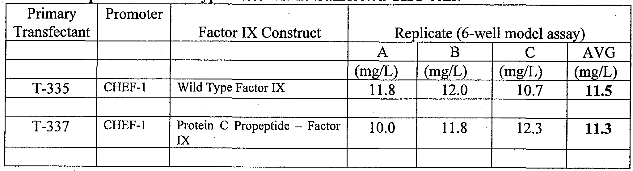
[0087] All transfectants were assayed for Factor IX antigen by a commercially available ELISA test which used a commercially available Factor IX preparation as a standard (BeneFix, Wyeth Laboratories).
[0088] The cloning and growth of CHO cells transfected with the Factor IX gene are conducted by one of three methods. As one skilled in the art appreciates, cell cultures are set up to produce the amount of cells or tissue culture fluid needed for the experiments which are performed with these materials. The smallest size system is growth of cells in 96-well microtiter plates. The cells on these plates were grown for 14 days but since they have the smallest surface area for cells to multiply on, they produce the fewest cells and in the least amount of tissue culture media and lowest amounts of Factor IX. The second system is a 6- well model assay as shown in Table 1 above. Cells were grown on a 6 well tissue culture plates for 9 days. Cells grown in 6 well plates generally produce an intermediate amount of cells in a medium amount of tissue culture media and an intermediate amount of Factor IX. The highest concentration (and actual number) of cells was produced in a 1.5 liter shake flask incubated for 18 days.
[0089] Due to our extensive experience using all three of systems, it is possible to extrapolate from the amount of Factor IX produced in one system to that produced in another system. For example, the amount of Factor IX produce in the shake Flask system is essentially 6 times greater than produced in the 6-well model system. In this way, when it is necessary for comparison, one can extrapolate the quantity of Factor IX that will be produced in a Shake Flask system from the amount measured in a smaller system like the 6-well model or from the 96-well microtiter plates. Table 1 above indicates that CHO cells can be transfected with a wild type Factor IX gene and produce an average of 11.5 mg/L in a 6-well
model assay system. By extrapolation, as described above, this equates to 69 mg/L in a 1.5 L shake flask.
Example 2
[0090] For comparison, the wild type Factor IX gene of Example 1 was cloned into a construct using the CMV promoter and transfected into HEK293 cells. As in Example 1 above, the primary transfectants were grown up in a 6-well microtiter plate. The titer for these samples was less than 0.2 mg/L (data not shown). Further experiments were conducted using the CHEF-1 promoter.
Example 3
[0091] In order to identify and characterize clones with high levels of Factor IX expression, primary transfectants were cloned by limit dilution into 96-well plates. As a consequence of the low surface area for cells to grow the level of expression was less that that reported in Example 1 where cells were grown on 6-well microtiter plates. One hundred and fifty two single cell clones were identified from the T-335 primary transfectant experiment. The Factor IX antigen expression level for each clone was measured by ELISA analysis. The distribution of Factor IX expression levels from the clones are shown in Figure 1.
[0092] The T-335 clones were separated into clones that expressed Factor IX at a level of greater than 0.4 mg/L and those that produced Factor IX at lower levels. Clones expressing Factor IX at levels higher than 0.4 mg/L were considered high expresser clones. Clones with expression levels lower than 0.4 mg/L were considered low expressers. The range of expression for Factor IX can be seen to be distributed in a wide range between no expression to above 1.6 mg/L with an average of 0.87 mg/L of Factor IX. In order to determine if other genes transfected into CHO cells would have essentially the same broad range of distribution when recloned, cells form the primary T-337 transfection were also cloned by limit dilution in the 96-well microtiter plate assay. As can be seen in Figure 1 above and Table 2 below, of the 171 clones evaluated the range of antigen expression was again quite broad being between none and above 1.8 mg/L with an average of 0.73 mg/L.
Table 2. Limit dilution cloning of Primary Transfectants

[0093] The results of Figure 1 and Table 2 demonstrate that when CHO cells are transfected with a gene under the control of the CHEF-1 promoter, all cells are not transfected in a way that they can produce detectable Factor IX and that those cells that are transfected in a productive way produce varying amounts of Factor IX. The reason that these cells produce varying levels of Factor IX is not understood. Without intending to be limited by theory, reasons may include that (1) some cells simply do not receive the transfected gene, (2) others may receive a gene copy but it may not integrate into the chromosome, (3) other cells may have the gene integrated in a nonfunctional region of the chromosome, (4) yet other cells may receive multiple copies of the gene but only some are integrated in the correct position of the genome for expression and (5) some cells may receive multiple copies of the gene but only some of the copies are integrated in a functional way at the time the ELISA is performed on individual clones. The last possibility predicts that as these cells divide and grow, more copies of unintegrated DNA may become integrated with time. (6) Duplication of integrated plasmid may occur and number of duplications may increase upon further cloning or culturing. In the last two cases (5) and (6), one predicts that upon further cloning or culturing, (essentially repeating the above experiment), cell lines would be isolated which would express higher levels of Factor IX (or other protein, e.g., Factor VII/VHa).
[0094] To determine if this were the case, high level expressors of Wild Type Factor IX, as described in Table 2, were pooled and expanded in a shake flask culture. Individual clones were then isolated by limit dilution cloning. The clones producing the highest level of Factor IX were isolated. The twenty highest producers are presented in Table 3.
Table 3 Second limit dilution of pooled high expressing clones of T-335 and selection for high Factor IX anti en ex ression.

The "estimated 6-well titers" is based on our experience that about 25-fold more antigen is produced in 6-well plates when compared to 96-well plates.
[0095] As shown in Table 3, in the 96 well microtiter model, the range of production for Factor IX antigen from the highest yielding 20 clones is between 1.79 and 4.14 mg/L. As presented in the last column of Table 3, one can estimate the concentration of Factor IX that would be expected in 6-well microtiter plates. It is estimated that the level of these clones in 6-microtitre plates would be between 46 and 104 mg/L. This is a substantial increase from the level of expression of the primary transfectants seen in Table 2 (a range of 0- 1.65 mg/L was reported). The biological activity of Factor IX produced from this selected group of clones is between 4 and 30% (Table 3). It appears, that, in general, the higher the level of Factor IX antigen production the lower the level of biologically active Factor IX produced. This is shown graphically in Figure 2. This observation has been made by others attempting to overproduce Vitamin K dependent coagulation factors.
[0096] It has always proven difficult to produce Factor IX in tissue culture systems with more than 1.5-5% biologically activity. For example, Kaufman (Kaufman, et al. The Journal of Biological Chemistry vol. 261 (21):9622-9628, July 25, 1986) report CHO cell lines secreting more than 100 μg/ml Factor IX by methotrexate amplification in dihyrofolate reductase deficient cell lines. Yet, biologically active Factor IX was never
produced at a level higher than 1.5 μg/ml. It was suggested that the CHO cell line was deficient in processing factors needed to make biologically active Factor IX. It is generally accepted that the presence of processing cofactors is necessary to produce biologically active vitamin K dependent proteins in recombinant systems. We evaluated the potential of our selection system to also identify clones producing a high percentage of biologically active Factor IX. Table 4 presents the 20 Factor IX producing clones with the highest percentage of biologically active Factor IX.
Table 4. Second limit dilution of high expresser clones of T-335 and selection for high percentage of biolo icall active Factor IX.

[0097] As can be seen in Table 4, all of the 20 clones producing Factor IX, which have been selected for high content of biologically active material produce above 25% biologically active Factor IX. In this experiment, the highest level of functional Factor IX reported is for clone P2A7 at 48%. Specific activity ranges from 64-119 U/mg. In contrast, Kaufman (ibid) reported specific activity of 35-75 U/mg, up to half the specific activity of plasma derived Factor IX (150 U/mg) using the adenovirus major late promoter. By use of the CHEF-1 promoter in CHO cells, biologically active recombinant Factor IX was produced at levels much closer to the levels obtained with plasma-derived Factor IX. It was unexpected that such high levels of biologically active Factor IX protein could be produced recombinantly without addition of processing factors.
[0098] In summary, limit dilution selection of clones which produce Factor IX in a tissue culture system in conjunction with a high level promoter system produced more Factor IX antigen and a higher level of biologically active Factor IX protein than has been possible in the past.
Example 4
[0099] In order to determine if our extrapolation of Factor IX production from 96- well plates to 6-well microtiter plates was accurate in both antigen produced and percentage biologically active protein recovered, we expanded a group of eleven clones from the second limited dilution experiment in 6 well microtiter plates. As can be seen in Table 5, the results were similar than those reported in 96 well micro titer plates. The level of Factor IX antigen production was between 2 and 40 mg/L and the percentage of biologically active Factor IX recovered was between 10 and 58 %. The level of biologically active Factor IX was significantly higher than reported previously for any other Vitamin K dependent protein prepared in the absence of vitamin K or processing factors involved in post-translational processing of blood factor proteins.
Table 5. Factor IX1 antigen production and percentage of biologically active Factor IX in 6- well microtiter plates.

Example 5: recombinant Factor VII produced by semi-solid matrix cloning
[0101] A gene for Factor VII was synthesized, operably linked to the CHEF-1 promoter, and transfected into CHO cells using a vector which included the DHFR marker for selection in Hypoxanthine Thymidine (HT)-minus medium. The primary transfectants were grown up in a microtiter plate and were assayed to give an average level of antigen expression for the cells in the well. The pool of primary transfectants were then subjected to cell cloning by a semi-solid matrix cloning method.
[0102] The cells were seeded into a semi-solid matrix at very low densities (typically 1,000-4,000 cells per ml) into a mixture of media (Cloning medium A®, Invitrogen), media supplements (4 mM L-glutamine, 10 μg/nlL vitamin K, ImM CaCl2), conditioned medium (5-20% of 7 day old culture of parental CHO cells), and a generally inert, biologically compatible, semi-solid medium such as methylcellulose (CloneMatrix®). After seeding the low density cells into the mixture as a single cell suspension and letting it "gel" for a few minutes, into a semi-solid, the culture plates are returned to an incubator (37°C, with humidity and carbon dioxide buffering atmosphere) and allowed to sit undisturbed for anywhere from -lweek to ~3weeks.
[0103] A fluorescent antibody against human FVII was included in the semi-solid medium formulation such that the FVII-antibody that was impregnated in the gel allowed detection of high expressing colonies (clones) when the culture plates containing semi-solid matrix with colonies were viewed under a fluorescence microscope. About two weeks after initially seeding the cells into the semisolid matrix the culture plates were observed for colony formation (number of colonies, their size and how far separated they are from one another in the gel) and then individual colonies were picked and seeded into separate cluster plates, each as a clonal population. They were expanded through larger plates and screened for production of FVII in expansion medium (OptiCHO®, L-glutamine, 200mM, Vitamin Kl
(2% in ethanol) and 1 M CaCl2). The cells were split and grown in fresh medium. Periodically, FVII/FVIIa levels and bioactivity were assessed.
[0104] FVII/FVIIa levels were determined by conventional FVII ELISA assay.
[0105] Bioactivity was determined by a conventional chromogenic FVIIa Bioactivity assay such as BIOPHEN FVII® (Ref No. 221304) available from HYPHEN BioMed. The basis of the assay relies upon the ability of Factor VII to activate Factor X to Factor Xa. After forming an enzymatic complex with Tissue Factor, provided by rabbit Thromboplastin, the FVII complex activates Factor X to Factor Xa which activity is measured by cleavage of a chromogenic substrate (SXa-11). Factor Xa cleaves the substrate and generates pNA. The amount of pNA generated is directly proportional to the Factor Xa activity. Finally, there is a direct relationship between the amount of Factor VII in the assayed sample and the Factor Xa activity generated, measured by the amount of pNA released, determined by color development at 405 nm. Alternatively, a two-stage coagulation (clotting) assay may be used to determine FVII/FVIIa activity.
[0106] Calibration is performed with a normal pooled titrated plasma, with the assigned value of 100 % Factor VII. The assay kit includes a standard plasma dilution of 1 : 100.0. By definition, this latter dilution of the pool represents the 100 % Factor VII activity. The dynamic range is from 0 to 200 % FactorVII. The 200 % Factor VII activity is the 1 :500 dilution of the plasma pool. A standard curve was generated.
[0107] Samples were diluted in order to get a final Factor VII concentration in the tested dilution range of 0.1 to 1 ng/ml. The samples were placed in a microplate well or plastic tube and incubated. The SXa-11 substrate was introduced followed by further incubation. The reaction was stopped by adding 60 μΙ-ΛνβΙΙ or 200 μΕ/tube citric acid (20 g/L), or 20% acetic acid. The yellow color is stable for 2 hours. The sample blank was prepared by mixing the reagents in the opposite order from the test (i.e., Citric Acid (20 g/L), SXa-1 1 substrate, diluted plasma, Factor X, and Thromboplastin-Ca). The concentration of bioactive FVII/FVIIa was determined for the unknown samples from the standard curve generated with plasma samples from the kit discussed above. The concentration of bioactive FVII represents the amount in μg/ml of FVII/FVIIa in the supernatant that was sufficiently carboxylated to be measured as "active FVIIa".
[0108] Table 6 shows the titers of FVII/VIIa antigen in the supernatants of 15 different clones transfected with a FVII encoding plasmid containing the DHFR selectable marker and a CHEF-1 promoter and how much of this FVII VIIa protein in the supernatants was biologically active as measured by the chromogenic assay described above. The various clones produce reasonably high levels of FVII/VIIa antigen into their supernatant (e.g. up to 7 ug/ml) even though they do not contain plasmid for expressing any exogenous (human) VKOR or VKGC. More importantly, for these representative clones the proportion of FVII/VIIa antigen in the supernatant which is bioactive is very high, ranging from 66% to 100% of the secreted FVII.
Table 6. Titers of FVII/FVIIa antigen and Bioactivity in supernatants of 15 different clones.

[0109] Table 7 shows similar kind of data as Table 6 except that these results were obtained from the clones at a later stage, after the individual clones had undergone more cumulative population doublings. The clones have increased their volumetric productivities (i.e. amount of FVII VIIa antigen per unit volume of culture supernatant) the longer they've been cultured. By comparing the results in Table 7 for Clone #10 with that in Table 6, one sees that this clone's expression levels increased from ~3.3 ug/ml in the secondary screening stage to ~29ug/ml after time in culture. The s6condary screen results are from static cultures grown in cluster plates whereas the later assays are derived from cultures grown under more optimal culture conditions, i.e., in a shaking flask where the cultures are better aerated and with better availability of nutrients before the supernatant sample was harvested for assaying.
Table 7. Titers of FVII/FVIIa antigen and Bioactivity in supernatants of 5 selected clones after repeated culturing.
Clone # Ag (ng/ml) Bioactivity (ng/ml) ; % Bioactive
1 23000 15000 65
2 26000 19000 73
3 28000 20000 71
6 26000 18000 69
10 29000 21000 72
[0110] This increase in cell specific productivity (Qp) is shown in the following Table for a particular clone. An increase of 0.4 pg/cell/day at initial assessment increases to 0.56 pg/cell/day as a function of time in culture, without recourse to supertransfection with processing factors. Typical values for FVII/FVIIa production before initiation of the cell stability studies was 11-12 μ ηιΐ, with 60-70% bioactivity. By the end of productivity assessment #3, these numbers typically increased to 35-45 μg/mL with somewhat higher bioactivity (65-70%).
Table 8. Increase in cell specific productivity (Qp) as a function of time in culture
Experiment Qp (pg/cell/day)
Cell Stability flasks:
Prod. Assess #1 0.4 (23)*
Prod Assess #2 0.4 (48)
Prod Assess #3 0.56 (73)
* Numbers in parentheses refers to number of total cumulative population doublings from the time the cells recovered from transfection and selection until seeding in the productivity assessment experiment indicated.
[0111] These results show that by cell cloning methods such as limit dilution cloning and semi-solid matrix cloning, and with repeated culturing, high levels of bioactive vitamin K dependent proteins can be achieved without co-transfection of processing factors such as VKOR, VKGC, and /or PACE.
[0112] It will be understood by those of skill in the art that numerous and various modifications can be made without departing from the spirit of the present invention. Therefore, it should be clearly understood that the forms of the present invention are illustrative only and are not intended to limit the scope of the present invention.
Claims
1. A cell line which produces recombinant vitamin K dependent protein having at least 20% biological activity, wherein the cell line does not contain heterologous genetic material encoding proteins involved in the post-translation modification of vitamin K dependent proteins.
2. The cell line of claim 1, wherein the cell line is a mammalian cell line.
3. The cell line of claim 2, wherein the mammalian cell line is Chinese Hamster
Ovary.
4. The cell line of any of claims 1-3, which has not been subjected to selection with methotrexate.
5. The cell line of any of claims 1-3, wherein the Vitamin K dependent protein is selected from the group consisting of Factor II, Factor VII, Factor IX, Factor X, Protein C and Protein S.
6. The cell line of claim 5, wherein the Vitamin K dependent protein is Factor VII VIIa or Factor IX.
7. The cell line of any of claims 1-3, comprising a gene encoding the Vitamin K dependent protein which is operably linked to a Chinese hamster elongation factor 1 (CHEF- 1) promoter.
8. A method of producing a recombinant biologically active Vitamin In¬ dependent protein comprising the steps of:
(a) transfecting a population of mammalian cells with a gene encoding the Vitamin K dependent protein operably linked to a promoter;
(b) performing at least one round of cloning and screening to identify cell clones which produce at least 10 mg/L of the Vitamin K dependent protein which is at least 10% biologically active;
(c) optionally, repeating the cell cloning of step (b) one or more times to identify single cells producing more than 10 mg/L of the Vitamin K dependent protein which is at least 10% biologically active; and
(d) harvesting the Vitamin K dependent protein.
9. The method of claim 8, wherein the Vitamin K dependent protein is selected from the group consisting of Factor II, Factor VII, Factor IX, Factor X, Protein C and Protein S.
10. The method of claim 9, wherein the Vitamin K dependent Protein is Factor
IX.
11. The method of claim 10, wherein the cloning is limit dilution cloning.
12. The method of claim 9, wherein the Vitamin K dependent Protein is Factor
VII.
13. The method of claim 12, wherein the cloning is semi-solid matrix cloning.
14. The method of any of claims 8-13, wherein the promoter is a Chinese hamster elongation factor 1 (CHEF-1) promoter.
15. The method of any of claims 8-13, further comprising preselecting cells producing at least 10 mg/L Vitamin K dependent protein antigen before step (b).
16. The method of any of claims 8-13, wherein the cell line is cultured in a media that includes vitamin K.
17. The method of any of claims 8-13, wherein the cell line is cultured in a media that does not include vitamin K.
18. The method of any of claims 8-13, wherein at least 20 % of the Vitamin K dependent protein is biologically active.
19. The method of any of claims 8-13, wherein at least 30 % of the Vitamin K dependent protein is biologically active.
20. The method of any of claims 8-13, wherein at least 40 % of the Vitamin K dependent protein is biologically active.
21. The method of any of claims 8-13, wherein at least 58 % of the Vitamin K dependent protein is biologically active.
22. The method of any of claims 8-13, wherein at least 70 % of the Vitamin K dependent protein is biologically active.
23. The method of any of claims 8-13, wherein at least 85 % of the Vitamin K dependent protein is biologically active.
24. The method of any of claims 8-13, wherein at least 95 % of the Vitamin K dependent protein is biologically active.
25. The method of any of claims 8-13, wherein at least 99 % of the Vitamin K dependent protein is biologically active
26. The method of claim any of claims 8-13, wherein the vitamin K dependent protein is produced in an amount of at least 20 mg/L.
27. The method of any of claims 8-13, wherein the vitamin K dependent protein is produced in an amount of at least 30 mg/L.
28. The method of any of claims 8-13, wherein the vitamin K dependent protein is produced in an amount of at least 40 mg/L.
29. The method of any of claims 8-13, wherein the mammalian cells are Chinese Hamster Ovary Cells.
30. A recombinant Factor IX protein produced by the method of any of claims 8-
11.
31. A recombinant Factor VII protein produced by the method of any of claims 8, 9, 12 or 13
32. A pharmaceutical composition comprising the Factor IX protein of Claim 30 or the Factor VII protein of claim 31.
33. A kit comprising the recombinant Factor IX protein of claim 30 or the Factor VII protein of claim 31.
34. A method of treating hemophilia which comprises administering an effective amount of the pharmaceutical composition of claim 32 to a patient in need thereof.
35. A recombinant Factor IX protein having at least 10% biological activity.
36. The recombinant Factor IX protein of claim 35, having at least 20% biological activity.
37. The recombinant Factor IX protein of claim 35, having at least 30% biological activity.
38. The recombinant Factor IX protein of claim 35, having at least 40% biological activity.
39. The recombinant Factor IX protein of claim 35, having at least 58% biological activity.
40. A recombinant Factor VH VIIa protein having at least 70% biological activity.
41. A recombinant Factor VH/VIIa protein having at least 85% biological activity.
42. A recombinant Factor VH/VIIa protein having at least 99% biological activity.
43. A cell line which produces recombinant vitamin K dependent protein having at least 20% biological activity, wherein the cell line is generated by:
(a) transfecting a population of mammalian cells with a gene encoding the Vitamin K dependent protein operably linked to a Chinese hamster elongation factor 1 (CHEF-1) promoter; and
(b) performing at least one round of cloning and screening to identify cell clones which produce at least 10 mg/L of the Vitamin K dependent protein which is at least 10% biologically active.
44. The method of claim 43, wherein the cloning is limit dilution cloning or semisolid matrix cloning
Priority Applications (4)
| Application Number | Priority Date | Filing Date | Title |
|---|---|---|---|
| JP2012537082A JP5851410B2 (en) | 2009-10-30 | 2010-10-28 | Method for producing recombinant vitamin K-dependent protein |
| EP10827499.4A EP2494040B1 (en) | 2009-10-30 | 2010-10-28 | Method of producing recombinant vitamin k dependent proteins |
| US13/459,743 US20120276079A1 (en) | 2009-10-30 | 2012-04-30 | Method of producing recombinant vitamin k dependent proteins |
| US14/475,881 US9587008B2 (en) | 2009-10-30 | 2014-09-03 | Method of producing recombinant vitamin K dependent proteins |
Applications Claiming Priority (2)
| Application Number | Priority Date | Filing Date | Title |
|---|---|---|---|
| US25680209P | 2009-10-30 | 2009-10-30 | |
| US61/256,802 | 2009-10-30 |
Related Child Applications (1)
| Application Number | Title | Priority Date | Filing Date |
|---|---|---|---|
| US13/459,743 Continuation US20120276079A1 (en) | 2009-10-30 | 2012-04-30 | Method of producing recombinant vitamin k dependent proteins |
Publications (1)
| Publication Number | Publication Date |
|---|---|
| WO2011053738A1 true WO2011053738A1 (en) | 2011-05-05 |
Family
ID=43922560
Family Applications (1)
| Application Number | Title | Priority Date | Filing Date |
|---|---|---|---|
| PCT/US2010/054581 WO2011053738A1 (en) | 2009-10-30 | 2010-10-28 | Method of producing recombinant vitamin k dependent proteins |
Country Status (4)
| Country | Link |
|---|---|
| US (2) | US20120276079A1 (en) |
| EP (1) | EP2494040B1 (en) |
| JP (1) | JP5851410B2 (en) |
| WO (1) | WO2011053738A1 (en) |
Families Citing this family (1)
| Publication number | Priority date | Publication date | Assignee | Title |
|---|---|---|---|---|
| JP5851410B2 (en) | 2009-10-30 | 2016-02-03 | シーエヌジェイ ホールディングス、インク. | Method for producing recombinant vitamin K-dependent protein |
Citations (21)
| Publication number | Priority date | Publication date | Assignee | Title |
|---|---|---|---|---|
| US4399216A (en) | 1980-02-25 | 1983-08-16 | The Trustees Of Columbia University | Processes for inserting DNA into eucaryotic cells and for producing proteinaceous materials |
| WO1984000560A1 (en) | 1982-08-04 | 1984-02-16 | Nat Res Dev | Molecular cloning of the gene for human anti-haemophilic factor ix |
| EP0117058A2 (en) | 1983-01-19 | 1984-08-29 | Genentech, Inc. | Methods for producing mature protein in vertebrate host cells |
| EP0117060A2 (en) | 1983-01-19 | 1984-08-29 | Genentech, Inc. | Methods of screening and amplification in eukaryotic host cells, and nucleotide sequences and expression vectors for use therein |
| WO1985005125A1 (en) | 1984-05-09 | 1985-11-21 | Transgene S.A. | Vectors for the expression of factor ix, cells transformed by said vectors and process for the preparation of factor ix |
| EP0162782A1 (en) | 1984-05-22 | 1985-11-27 | Transgene S.A. | Expression vectors for factor IX, and cell lines producing the active factor IX |
| US4599308A (en) | 1981-10-06 | 1986-07-08 | Hamer Dean H | Protein from SV40 recombinants |
| EP0251874A1 (en) | 1986-06-23 | 1988-01-07 | Transgene S.A. | Integration vectors for the expansion of factor IX in eucaryotic cells, cell lines obtained and method for their preparation |
| US4745051A (en) | 1983-05-27 | 1988-05-17 | The Texas A&M University System | Method for producing a recombinant baculovirus expression vector |
| US4761371A (en) | 1985-02-12 | 1988-08-02 | Genentech, Inc. | Insulin receptor |
| US4877729A (en) | 1986-07-14 | 1989-10-31 | Genetics Institute, Inc. | Recombinant DNA encoding novel family of primate hematopoietic growth factors |
| US4879236A (en) | 1984-05-16 | 1989-11-07 | The Texas A&M University System | Method for producing a recombinant baculovirus expression vector |
| US4879224A (en) | 1985-01-10 | 1989-11-07 | Biogen, Inc. | DNA sequences, recombinant DNA molecules and processes for producing human phospholipase inhibitor polypeptides |
| US4912038A (en) | 1984-12-11 | 1990-03-27 | California Biotechnology Inc. | Recombinant DNA sequence encoding alveolar surfactant protein |
| EP0373012A1 (en) | 1988-11-09 | 1990-06-13 | Transgene S.A. | DNA encoding for factor IX or an analogous protein |
| US4981952A (en) | 1988-10-04 | 1991-01-01 | Eli Lilly And Company | Method for the purification of vitamin K-dependent proteins |
| US5714583A (en) | 1995-06-07 | 1998-02-03 | Genetics Institute, Inc. | Factor IX purification methods |
| US5750358A (en) | 1991-04-10 | 1998-05-12 | Oklahoma Medical Research Foundation | Quantitative clotting assay for activated factor VII |
| US5888809A (en) | 1997-05-01 | 1999-03-30 | Icos Corporation | Hamster EF-1α transcriptional regulatory DNA |
| US20070122884A1 (en) * | 2000-10-02 | 2007-05-31 | Novo Nordisk Healthcare A/G | Factor VII glycoforms |
| US20080045453A1 (en) * | 2005-12-21 | 2008-02-21 | Drohan William N | Method of producing biologically active vitamin K dependent proteins by recombinant methods |
Family Cites Families (3)
| Publication number | Priority date | Publication date | Assignee | Title |
|---|---|---|---|---|
| GR860984B (en) | 1985-04-17 | 1986-08-18 | Zymogenetics Inc | Expression of factor vii and ix activities in mammalian cells |
| WO2008022151A1 (en) * | 2006-08-15 | 2008-02-21 | Inspiration Biopharmaceuticals, Inc. | Prophylactic treatment of hemophilia |
| JP5851410B2 (en) | 2009-10-30 | 2016-02-03 | シーエヌジェイ ホールディングス、インク. | Method for producing recombinant vitamin K-dependent protein |
-
2010
- 2010-10-28 JP JP2012537082A patent/JP5851410B2/en not_active Expired - Fee Related
- 2010-10-28 WO PCT/US2010/054581 patent/WO2011053738A1/en active Application Filing
- 2010-10-28 EP EP10827499.4A patent/EP2494040B1/en not_active Not-in-force
-
2012
- 2012-04-30 US US13/459,743 patent/US20120276079A1/en not_active Abandoned
-
2014
- 2014-09-03 US US14/475,881 patent/US9587008B2/en active Active
Patent Citations (22)
| Publication number | Priority date | Publication date | Assignee | Title |
|---|---|---|---|---|
| US4399216A (en) | 1980-02-25 | 1983-08-16 | The Trustees Of Columbia University | Processes for inserting DNA into eucaryotic cells and for producing proteinaceous materials |
| US4599308A (en) | 1981-10-06 | 1986-07-08 | Hamer Dean H | Protein from SV40 recombinants |
| WO1984000560A1 (en) | 1982-08-04 | 1984-02-16 | Nat Res Dev | Molecular cloning of the gene for human anti-haemophilic factor ix |
| EP0117058A2 (en) | 1983-01-19 | 1984-08-29 | Genentech, Inc. | Methods for producing mature protein in vertebrate host cells |
| EP0117060A2 (en) | 1983-01-19 | 1984-08-29 | Genentech, Inc. | Methods of screening and amplification in eukaryotic host cells, and nucleotide sequences and expression vectors for use therein |
| US4745051A (en) | 1983-05-27 | 1988-05-17 | The Texas A&M University System | Method for producing a recombinant baculovirus expression vector |
| WO1985005125A1 (en) | 1984-05-09 | 1985-11-21 | Transgene S.A. | Vectors for the expression of factor ix, cells transformed by said vectors and process for the preparation of factor ix |
| US4879236A (en) | 1984-05-16 | 1989-11-07 | The Texas A&M University System | Method for producing a recombinant baculovirus expression vector |
| EP0162782A1 (en) | 1984-05-22 | 1985-11-27 | Transgene S.A. | Expression vectors for factor IX, and cell lines producing the active factor IX |
| WO1985005376A1 (en) | 1984-05-22 | 1985-12-05 | Transgene S.A. | Vectors for the expression of factor ix and cell lineages producing the active factor ix |
| US4912038A (en) | 1984-12-11 | 1990-03-27 | California Biotechnology Inc. | Recombinant DNA sequence encoding alveolar surfactant protein |
| US4879224A (en) | 1985-01-10 | 1989-11-07 | Biogen, Inc. | DNA sequences, recombinant DNA molecules and processes for producing human phospholipase inhibitor polypeptides |
| US4761371A (en) | 1985-02-12 | 1988-08-02 | Genentech, Inc. | Insulin receptor |
| EP0251874A1 (en) | 1986-06-23 | 1988-01-07 | Transgene S.A. | Integration vectors for the expansion of factor IX in eucaryotic cells, cell lines obtained and method for their preparation |
| US4877729A (en) | 1986-07-14 | 1989-10-31 | Genetics Institute, Inc. | Recombinant DNA encoding novel family of primate hematopoietic growth factors |
| US4981952A (en) | 1988-10-04 | 1991-01-01 | Eli Lilly And Company | Method for the purification of vitamin K-dependent proteins |
| EP0373012A1 (en) | 1988-11-09 | 1990-06-13 | Transgene S.A. | DNA encoding for factor IX or an analogous protein |
| US5750358A (en) | 1991-04-10 | 1998-05-12 | Oklahoma Medical Research Foundation | Quantitative clotting assay for activated factor VII |
| US5714583A (en) | 1995-06-07 | 1998-02-03 | Genetics Institute, Inc. | Factor IX purification methods |
| US5888809A (en) | 1997-05-01 | 1999-03-30 | Icos Corporation | Hamster EF-1α transcriptional regulatory DNA |
| US20070122884A1 (en) * | 2000-10-02 | 2007-05-31 | Novo Nordisk Healthcare A/G | Factor VII glycoforms |
| US20080045453A1 (en) * | 2005-12-21 | 2008-02-21 | Drohan William N | Method of producing biologically active vitamin K dependent proteins by recombinant methods |
Non-Patent Citations (17)
| Title |
|---|
| "Immobilized Cells and Enzymes", 1986, IRL PRESS |
| B. D. HAMES & S. J. HIGGINS: "Nucleic Acid Hybridization", 1984 |
| B. D. HAMES & S. J. HIGGINS: "Transcription and Translation", 1984 |
| B. PERBAL: "A Practical Guide to Molecular Cloning", 1984 |
| D. M. WEIR AND C. C. BLACKWELL: "Handbook of Experimental Immunology", vol. I-IV, 1986 |
| D. N GLOVER: "DNA Cloning", vol. I and II, 1985 |
| DEER ET AL.: "High-level expression of proteins in mammalian cells using transcription regulatory sequences from the Chinese Hamster EF-la gene", BIOTECHNOL. PROG., vol. 20, 2004, pages 880 - 889, XP002557337, DOI: doi:10.1021/bp034383r |
| J. H. MILLER AND M. P. CALOS: "Gene Transfer Vectors for Mammalian Cells", 1987, COLD SPRING HARBOR LABORATORY |
| KRUSE AND PATTERSON: "Tissue Culture", 1973, ACADEMIC PRESS |
| M. J. GAIT: "Oligonucleotide Synthesis", 1984 |
| MAYER AND WALKER: "Immunochemical Methods in Cell and Molecular Biology", 1987, ACADEMIC PRESS |
| M-J. GETHING ET AL., NATURE, vol. 293, 1981, pages 620 |
| N. MANTEI ET AL., NATURE, vol. 281, pages 40 |
| R. I. FRESHNEY: "Animal Cell Culture", 1986 |
| SAMBROOK ET AL.: "Molecular Cloning; A Laboratory Manual, 2md ed.", 1989 |
| SCOPES: "Protein Purification: Principles and Practice, 2nd ed.", 1987, SPRINGER- VERLAG |
| WU AND GROSSMAN: "Methods in Enzymology", vol. 154, 155, ACADEMIC PRESS, INC. |
Also Published As
| Publication number | Publication date |
|---|---|
| US20120276079A1 (en) | 2012-11-01 |
| US9587008B2 (en) | 2017-03-07 |
| EP2494040A4 (en) | 2013-06-26 |
| US20140369994A1 (en) | 2014-12-18 |
| JP2013509190A (en) | 2013-03-14 |
| EP2494040A1 (en) | 2012-09-05 |
| JP5851410B2 (en) | 2016-02-03 |
| EP2494040B1 (en) | 2018-08-29 |
Similar Documents
| Publication | Publication Date | Title |
|---|---|---|
| US20230287471A1 (en) | Method of producing biologically active vitamin k dependent proteins by recombinant methods | |
| US20230272360A1 (en) | Recombinant vitamin k dependent proteins with high sialic acid content and methods of preparing same | |
| US9587008B2 (en) | Method of producing recombinant vitamin K dependent proteins | |
| JPH022376A (en) | Vector and compound for developing enzyme precursor type human protein c | |
| AU2016238889B2 (en) | Recombinant vitamin K dependent proteins with high sialic acid content and methods of preparing same | |
| AU2014202989B2 (en) | Recombinant vitamin k dependent proteins with high sialic acid content and methods of preparing same | |
| IE66340B1 (en) | Cell culture methods for producing activated protein C | |
| US20190264188A1 (en) | Recombinant vitamin k dependent proteins with high sialic acid content and methods of preparing same | |
| AU2016200807A1 (en) | Method of producing biologically active vitamin K dependent proteins by recombinant methods | |
| AU2013211534A1 (en) | Method of producing biologically active vitamin K dependent proteins by recombinant methods |
Legal Events
| Date | Code | Title | Description |
|---|---|---|---|
| 121 | Ep: the epo has been informed by wipo that ep was designated in this application |
Ref document number: 10827499 Country of ref document: EP Kind code of ref document: A1 |
|
| WWE | Wipo information: entry into national phase |
Ref document number: 2012537082 Country of ref document: JP |
|
| NENP | Non-entry into the national phase |
Ref country code: DE |
|
| WWE | Wipo information: entry into national phase |
Ref document number: 2010827499 Country of ref document: EP |