WO2013124817A2 - MicroRNAS FOR THE GENERATION OF ASTROCYTES - Google Patents

MicroRNAS FOR THE GENERATION OF ASTROCYTES Download PDF

Info

Publication number
WO2013124817A2
WO2013124817A2 PCT/IB2013/051430 IB2013051430W WO2013124817A2 WO 2013124817 A2 WO2013124817 A2 WO 2013124817A2 IB 2013051430 W IB2013051430 W IB 2013051430W WO 2013124817 A2 WO2013124817 A2 WO 2013124817A2
Authority
WO
WIPO (PCT)
Prior art keywords
mir
cells
mirna
population
mscs
Prior art date
Application number
PCT/IB2013/051430
Other languages
French (fr)
Other versions
WO2013124817A3 (en
WO2013124817A8 (en
Inventor
Chaya Brodie
Shimon Slavin
Original Assignee
Brainstem Biotec Ltd.
Priority date (The priority date is an assumption and is not a legal conclusion. Google has not performed a legal analysis and makes no representation as to the accuracy of the date listed.)
Filing date
Publication date
Application filed by Brainstem Biotec Ltd. filed Critical Brainstem Biotec Ltd.
Priority to JP2014558253A priority Critical patent/JP6329911B2/en
Priority to US14/380,155 priority patent/US10034902B2/en
Priority to EP18180526.8A priority patent/EP3401393B1/en
Priority to EP13713247.8A priority patent/EP2844744A2/en
Publication of WO2013124817A2 publication Critical patent/WO2013124817A2/en
Publication of WO2013124817A8 publication Critical patent/WO2013124817A8/en
Publication of WO2013124817A3 publication Critical patent/WO2013124817A3/en
Priority to US16/048,676 priority patent/US20190015453A1/en

Links

Classifications

    • AHUMAN NECESSITIES
    • A61MEDICAL OR VETERINARY SCIENCE; HYGIENE
    • A61KPREPARATIONS FOR MEDICAL, DENTAL OR TOILETRY PURPOSES
    • A61K35/00Medicinal preparations containing materials or reaction products thereof with undetermined constitution
    • A61K35/12Materials from mammals; Compositions comprising non-specified tissues or cells; Compositions comprising non-embryonic stem cells; Genetically modified cells
    • A61K35/30Nerves; Brain; Eyes; Corneal cells; Cerebrospinal fluid; Neuronal stem cells; Neuronal precursor cells; Glial cells; Oligodendrocytes; Schwann cells; Astroglia; Astrocytes; Choroid plexus; Spinal cord tissue
    • AHUMAN NECESSITIES
    • A61MEDICAL OR VETERINARY SCIENCE; HYGIENE
    • A61KPREPARATIONS FOR MEDICAL, DENTAL OR TOILETRY PURPOSES
    • A61K35/00Medicinal preparations containing materials or reaction products thereof with undetermined constitution
    • A61K35/12Materials from mammals; Compositions comprising non-specified tissues or cells; Compositions comprising non-embryonic stem cells; Genetically modified cells
    • A61K35/28Bone marrow; Haematopoietic stem cells; Mesenchymal stem cells of any origin, e.g. adipose-derived stem cells
    • AHUMAN NECESSITIES
    • A61MEDICAL OR VETERINARY SCIENCE; HYGIENE
    • A61KPREPARATIONS FOR MEDICAL, DENTAL OR TOILETRY PURPOSES
    • A61K35/00Medicinal preparations containing materials or reaction products thereof with undetermined constitution
    • A61K35/12Materials from mammals; Compositions comprising non-specified tissues or cells; Compositions comprising non-embryonic stem cells; Genetically modified cells
    • A61K35/48Reproductive organs
    • A61K35/50Placenta; Placental stem cells; Amniotic fluid; Amnion; Amniotic stem cells
    • AHUMAN NECESSITIES
    • A61MEDICAL OR VETERINARY SCIENCE; HYGIENE
    • A61KPREPARATIONS FOR MEDICAL, DENTAL OR TOILETRY PURPOSES
    • A61K35/00Medicinal preparations containing materials or reaction products thereof with undetermined constitution
    • A61K35/12Materials from mammals; Compositions comprising non-specified tissues or cells; Compositions comprising non-embryonic stem cells; Genetically modified cells
    • A61K35/48Reproductive organs
    • A61K35/51Umbilical cord; Umbilical cord blood; Umbilical stem cells
    • AHUMAN NECESSITIES
    • A61MEDICAL OR VETERINARY SCIENCE; HYGIENE
    • A61PSPECIFIC THERAPEUTIC ACTIVITY OF CHEMICAL COMPOUNDS OR MEDICINAL PREPARATIONS
    • A61P21/00Drugs for disorders of the muscular or neuromuscular system
    • AHUMAN NECESSITIES
    • A61MEDICAL OR VETERINARY SCIENCE; HYGIENE
    • A61PSPECIFIC THERAPEUTIC ACTIVITY OF CHEMICAL COMPOUNDS OR MEDICINAL PREPARATIONS
    • A61P25/00Drugs for disorders of the nervous system
    • AHUMAN NECESSITIES
    • A61MEDICAL OR VETERINARY SCIENCE; HYGIENE
    • A61PSPECIFIC THERAPEUTIC ACTIVITY OF CHEMICAL COMPOUNDS OR MEDICINAL PREPARATIONS
    • A61P25/00Drugs for disorders of the nervous system
    • A61P25/08Antiepileptics; Anticonvulsants
    • AHUMAN NECESSITIES
    • A61MEDICAL OR VETERINARY SCIENCE; HYGIENE
    • A61PSPECIFIC THERAPEUTIC ACTIVITY OF CHEMICAL COMPOUNDS OR MEDICINAL PREPARATIONS
    • A61P25/00Drugs for disorders of the nervous system
    • A61P25/14Drugs for disorders of the nervous system for treating abnormal movements, e.g. chorea, dyskinesia
    • A61P25/16Anti-Parkinson drugs
    • AHUMAN NECESSITIES
    • A61MEDICAL OR VETERINARY SCIENCE; HYGIENE
    • A61PSPECIFIC THERAPEUTIC ACTIVITY OF CHEMICAL COMPOUNDS OR MEDICINAL PREPARATIONS
    • A61P25/00Drugs for disorders of the nervous system
    • A61P25/28Drugs for disorders of the nervous system for treating neurodegenerative disorders of the central nervous system, e.g. nootropic agents, cognition enhancers, drugs for treating Alzheimer's disease or other forms of dementia
    • AHUMAN NECESSITIES
    • A61MEDICAL OR VETERINARY SCIENCE; HYGIENE
    • A61PSPECIFIC THERAPEUTIC ACTIVITY OF CHEMICAL COMPOUNDS OR MEDICINAL PREPARATIONS
    • A61P9/00Drugs for disorders of the cardiovascular system
    • CCHEMISTRY; METALLURGY
    • C12BIOCHEMISTRY; BEER; SPIRITS; WINE; VINEGAR; MICROBIOLOGY; ENZYMOLOGY; MUTATION OR GENETIC ENGINEERING
    • C12NMICROORGANISMS OR ENZYMES; COMPOSITIONS THEREOF; PROPAGATING, PRESERVING, OR MAINTAINING MICROORGANISMS; MUTATION OR GENETIC ENGINEERING; CULTURE MEDIA
    • C12N5/00Undifferentiated human, animal or plant cells, e.g. cell lines; Tissues; Cultivation or maintenance thereof; Culture media therefor
    • C12N5/06Animal cells or tissues; Human cells or tissues
    • C12N5/0602Vertebrate cells
    • C12N5/0618Cells of the nervous system
    • C12N5/0622Glial cells, e.g. astrocytes, oligodendrocytes; Schwann cells
    • CCHEMISTRY; METALLURGY
    • C12BIOCHEMISTRY; BEER; SPIRITS; WINE; VINEGAR; MICROBIOLOGY; ENZYMOLOGY; MUTATION OR GENETIC ENGINEERING
    • C12QMEASURING OR TESTING PROCESSES INVOLVING ENZYMES, NUCLEIC ACIDS OR MICROORGANISMS; COMPOSITIONS OR TEST PAPERS THEREFOR; PROCESSES OF PREPARING SUCH COMPOSITIONS; CONDITION-RESPONSIVE CONTROL IN MICROBIOLOGICAL OR ENZYMOLOGICAL PROCESSES
    • C12Q1/00Measuring or testing processes involving enzymes, nucleic acids or microorganisms; Compositions therefor; Processes of preparing such compositions
    • C12Q1/68Measuring or testing processes involving enzymes, nucleic acids or microorganisms; Compositions therefor; Processes of preparing such compositions involving nucleic acids
    • C12Q1/6876Nucleic acid products used in the analysis of nucleic acids, e.g. primers or probes
    • CCHEMISTRY; METALLURGY
    • C12BIOCHEMISTRY; BEER; SPIRITS; WINE; VINEGAR; MICROBIOLOGY; ENZYMOLOGY; MUTATION OR GENETIC ENGINEERING
    • C12NMICROORGANISMS OR ENZYMES; COMPOSITIONS THEREOF; PROPAGATING, PRESERVING, OR MAINTAINING MICROORGANISMS; MUTATION OR GENETIC ENGINEERING; CULTURE MEDIA
    • C12N15/00Mutation or genetic engineering; DNA or RNA concerning genetic engineering, vectors, e.g. plasmids, or their isolation, preparation or purification; Use of hosts therefor
    • C12N15/09Recombinant DNA-technology
    • C12N15/11DNA or RNA fragments; Modified forms thereof; Non-coding nucleic acids having a biological activity
    • C12N15/113Non-coding nucleic acids modulating the expression of genes, e.g. antisense oligonucleotides; Antisense DNA or RNA; Triplex- forming oligonucleotides; Catalytic nucleic acids, e.g. ribozymes; Nucleic acids used in co-suppression or gene silencing
    • CCHEMISTRY; METALLURGY
    • C12BIOCHEMISTRY; BEER; SPIRITS; WINE; VINEGAR; MICROBIOLOGY; ENZYMOLOGY; MUTATION OR GENETIC ENGINEERING
    • C12NMICROORGANISMS OR ENZYMES; COMPOSITIONS THEREOF; PROPAGATING, PRESERVING, OR MAINTAINING MICROORGANISMS; MUTATION OR GENETIC ENGINEERING; CULTURE MEDIA
    • C12N2310/00Structure or type of the nucleic acid
    • C12N2310/10Type of nucleic acid
    • C12N2310/14Type of nucleic acid interfering N.A.
    • C12N2310/141MicroRNAs, miRNAs
    • CCHEMISTRY; METALLURGY
    • C12BIOCHEMISTRY; BEER; SPIRITS; WINE; VINEGAR; MICROBIOLOGY; ENZYMOLOGY; MUTATION OR GENETIC ENGINEERING
    • C12NMICROORGANISMS OR ENZYMES; COMPOSITIONS THEREOF; PROPAGATING, PRESERVING, OR MAINTAINING MICROORGANISMS; MUTATION OR GENETIC ENGINEERING; CULTURE MEDIA
    • C12N2320/00Applications; Uses
    • C12N2320/10Applications; Uses in screening processes
    • C12N2320/11Applications; Uses in screening processes for the determination of target sites, i.e. of active nucleic acids
    • CCHEMISTRY; METALLURGY
    • C12BIOCHEMISTRY; BEER; SPIRITS; WINE; VINEGAR; MICROBIOLOGY; ENZYMOLOGY; MUTATION OR GENETIC ENGINEERING
    • C12NMICROORGANISMS OR ENZYMES; COMPOSITIONS THEREOF; PROPAGATING, PRESERVING, OR MAINTAINING MICROORGANISMS; MUTATION OR GENETIC ENGINEERING; CULTURE MEDIA
    • C12N2330/00Production
    • C12N2330/10Production naturally occurring
    • CCHEMISTRY; METALLURGY
    • C12BIOCHEMISTRY; BEER; SPIRITS; WINE; VINEGAR; MICROBIOLOGY; ENZYMOLOGY; MUTATION OR GENETIC ENGINEERING
    • C12NMICROORGANISMS OR ENZYMES; COMPOSITIONS THEREOF; PROPAGATING, PRESERVING, OR MAINTAINING MICROORGANISMS; MUTATION OR GENETIC ENGINEERING; CULTURE MEDIA
    • C12N2501/00Active agents used in cell culture processes, e.g. differentation
    • C12N2501/01Modulators of cAMP or cGMP, e.g. non-hydrolysable analogs, phosphodiesterase inhibitors, cholera toxin
    • CCHEMISTRY; METALLURGY
    • C12BIOCHEMISTRY; BEER; SPIRITS; WINE; VINEGAR; MICROBIOLOGY; ENZYMOLOGY; MUTATION OR GENETIC ENGINEERING
    • C12NMICROORGANISMS OR ENZYMES; COMPOSITIONS THEREOF; PROPAGATING, PRESERVING, OR MAINTAINING MICROORGANISMS; MUTATION OR GENETIC ENGINEERING; CULTURE MEDIA
    • C12N2501/00Active agents used in cell culture processes, e.g. differentation
    • C12N2501/10Growth factors
    • C12N2501/11Epidermal growth factor [EGF]
    • CCHEMISTRY; METALLURGY
    • C12BIOCHEMISTRY; BEER; SPIRITS; WINE; VINEGAR; MICROBIOLOGY; ENZYMOLOGY; MUTATION OR GENETIC ENGINEERING
    • C12NMICROORGANISMS OR ENZYMES; COMPOSITIONS THEREOF; PROPAGATING, PRESERVING, OR MAINTAINING MICROORGANISMS; MUTATION OR GENETIC ENGINEERING; CULTURE MEDIA
    • C12N2501/00Active agents used in cell culture processes, e.g. differentation
    • C12N2501/10Growth factors
    • C12N2501/115Basic fibroblast growth factor (bFGF, FGF-2)
    • CCHEMISTRY; METALLURGY
    • C12BIOCHEMISTRY; BEER; SPIRITS; WINE; VINEGAR; MICROBIOLOGY; ENZYMOLOGY; MUTATION OR GENETIC ENGINEERING
    • C12NMICROORGANISMS OR ENZYMES; COMPOSITIONS THEREOF; PROPAGATING, PRESERVING, OR MAINTAINING MICROORGANISMS; MUTATION OR GENETIC ENGINEERING; CULTURE MEDIA
    • C12N2501/00Active agents used in cell culture processes, e.g. differentation
    • C12N2501/10Growth factors
    • C12N2501/135Platelet-derived growth factor [PDGF]
    • CCHEMISTRY; METALLURGY
    • C12BIOCHEMISTRY; BEER; SPIRITS; WINE; VINEGAR; MICROBIOLOGY; ENZYMOLOGY; MUTATION OR GENETIC ENGINEERING
    • C12NMICROORGANISMS OR ENZYMES; COMPOSITIONS THEREOF; PROPAGATING, PRESERVING, OR MAINTAINING MICROORGANISMS; MUTATION OR GENETIC ENGINEERING; CULTURE MEDIA
    • C12N2501/00Active agents used in cell culture processes, e.g. differentation
    • C12N2501/10Growth factors
    • C12N2501/195Heregulin, neu differentiation factor
    • CCHEMISTRY; METALLURGY
    • C12BIOCHEMISTRY; BEER; SPIRITS; WINE; VINEGAR; MICROBIOLOGY; ENZYMOLOGY; MUTATION OR GENETIC ENGINEERING
    • C12NMICROORGANISMS OR ENZYMES; COMPOSITIONS THEREOF; PROPAGATING, PRESERVING, OR MAINTAINING MICROORGANISMS; MUTATION OR GENETIC ENGINEERING; CULTURE MEDIA
    • C12N2501/00Active agents used in cell culture processes, e.g. differentation
    • C12N2501/40Regulators of development
    • C12N2501/41Hedgehog proteins; Cyclopamine (inhibitor)
    • CCHEMISTRY; METALLURGY
    • C12BIOCHEMISTRY; BEER; SPIRITS; WINE; VINEGAR; MICROBIOLOGY; ENZYMOLOGY; MUTATION OR GENETIC ENGINEERING
    • C12NMICROORGANISMS OR ENZYMES; COMPOSITIONS THEREOF; PROPAGATING, PRESERVING, OR MAINTAINING MICROORGANISMS; MUTATION OR GENETIC ENGINEERING; CULTURE MEDIA
    • C12N2501/00Active agents used in cell culture processes, e.g. differentation
    • C12N2501/65MicroRNA
    • CCHEMISTRY; METALLURGY
    • C12BIOCHEMISTRY; BEER; SPIRITS; WINE; VINEGAR; MICROBIOLOGY; ENZYMOLOGY; MUTATION OR GENETIC ENGINEERING
    • C12NMICROORGANISMS OR ENZYMES; COMPOSITIONS THEREOF; PROPAGATING, PRESERVING, OR MAINTAINING MICROORGANISMS; MUTATION OR GENETIC ENGINEERING; CULTURE MEDIA
    • C12N2502/00Coculture with; Conditioned medium produced by
    • C12N2502/13Coculture with; Conditioned medium produced by connective tissue cells; generic mesenchyme cells, e.g. so-called "embryonic fibroblasts"
    • C12N2502/1305Adipocytes
    • CCHEMISTRY; METALLURGY
    • C12BIOCHEMISTRY; BEER; SPIRITS; WINE; VINEGAR; MICROBIOLOGY; ENZYMOLOGY; MUTATION OR GENETIC ENGINEERING
    • C12NMICROORGANISMS OR ENZYMES; COMPOSITIONS THEREOF; PROPAGATING, PRESERVING, OR MAINTAINING MICROORGANISMS; MUTATION OR GENETIC ENGINEERING; CULTURE MEDIA
    • C12N2502/00Coculture with; Conditioned medium produced by
    • C12N2502/13Coculture with; Conditioned medium produced by connective tissue cells; generic mesenchyme cells, e.g. so-called "embryonic fibroblasts"
    • C12N2502/1311Osteocytes, osteoblasts, odontoblasts
    • CCHEMISTRY; METALLURGY
    • C12BIOCHEMISTRY; BEER; SPIRITS; WINE; VINEGAR; MICROBIOLOGY; ENZYMOLOGY; MUTATION OR GENETIC ENGINEERING
    • C12NMICROORGANISMS OR ENZYMES; COMPOSITIONS THEREOF; PROPAGATING, PRESERVING, OR MAINTAINING MICROORGANISMS; MUTATION OR GENETIC ENGINEERING; CULTURE MEDIA
    • C12N2502/00Coculture with; Conditioned medium produced by
    • C12N2502/13Coculture with; Conditioned medium produced by connective tissue cells; generic mesenchyme cells, e.g. so-called "embryonic fibroblasts"
    • C12N2502/1317Chondrocytes
    • CCHEMISTRY; METALLURGY
    • C12BIOCHEMISTRY; BEER; SPIRITS; WINE; VINEGAR; MICROBIOLOGY; ENZYMOLOGY; MUTATION OR GENETIC ENGINEERING
    • C12NMICROORGANISMS OR ENZYMES; COMPOSITIONS THEREOF; PROPAGATING, PRESERVING, OR MAINTAINING MICROORGANISMS; MUTATION OR GENETIC ENGINEERING; CULTURE MEDIA
    • C12N2502/00Coculture with; Conditioned medium produced by
    • C12N2502/13Coculture with; Conditioned medium produced by connective tissue cells; generic mesenchyme cells, e.g. so-called "embryonic fibroblasts"
    • C12N2502/1323Adult fibroblasts
    • CCHEMISTRY; METALLURGY
    • C12BIOCHEMISTRY; BEER; SPIRITS; WINE; VINEGAR; MICROBIOLOGY; ENZYMOLOGY; MUTATION OR GENETIC ENGINEERING
    • C12NMICROORGANISMS OR ENZYMES; COMPOSITIONS THEREOF; PROPAGATING, PRESERVING, OR MAINTAINING MICROORGANISMS; MUTATION OR GENETIC ENGINEERING; CULTURE MEDIA
    • C12N2502/00Coculture with; Conditioned medium produced by
    • C12N2502/13Coculture with; Conditioned medium produced by connective tissue cells; generic mesenchyme cells, e.g. so-called "embryonic fibroblasts"
    • C12N2502/1329Cardiomyocytes
    • CCHEMISTRY; METALLURGY
    • C12BIOCHEMISTRY; BEER; SPIRITS; WINE; VINEGAR; MICROBIOLOGY; ENZYMOLOGY; MUTATION OR GENETIC ENGINEERING
    • C12NMICROORGANISMS OR ENZYMES; COMPOSITIONS THEREOF; PROPAGATING, PRESERVING, OR MAINTAINING MICROORGANISMS; MUTATION OR GENETIC ENGINEERING; CULTURE MEDIA
    • C12N2502/00Coculture with; Conditioned medium produced by
    • C12N2502/13Coculture with; Conditioned medium produced by connective tissue cells; generic mesenchyme cells, e.g. so-called "embryonic fibroblasts"
    • C12N2502/1335Skeletal muscle cells, myocytes, myoblasts, myotubes
    • CCHEMISTRY; METALLURGY
    • C12BIOCHEMISTRY; BEER; SPIRITS; WINE; VINEGAR; MICROBIOLOGY; ENZYMOLOGY; MUTATION OR GENETIC ENGINEERING
    • C12NMICROORGANISMS OR ENZYMES; COMPOSITIONS THEREOF; PROPAGATING, PRESERVING, OR MAINTAINING MICROORGANISMS; MUTATION OR GENETIC ENGINEERING; CULTURE MEDIA
    • C12N2502/00Coculture with; Conditioned medium produced by
    • C12N2502/13Coculture with; Conditioned medium produced by connective tissue cells; generic mesenchyme cells, e.g. so-called "embryonic fibroblasts"
    • C12N2502/1341Tenocytes, cells from tendons and ligaments
    • CCHEMISTRY; METALLURGY
    • C12BIOCHEMISTRY; BEER; SPIRITS; WINE; VINEGAR; MICROBIOLOGY; ENZYMOLOGY; MUTATION OR GENETIC ENGINEERING
    • C12NMICROORGANISMS OR ENZYMES; COMPOSITIONS THEREOF; PROPAGATING, PRESERVING, OR MAINTAINING MICROORGANISMS; MUTATION OR GENETIC ENGINEERING; CULTURE MEDIA
    • C12N2502/00Coculture with; Conditioned medium produced by
    • C12N2502/13Coculture with; Conditioned medium produced by connective tissue cells; generic mesenchyme cells, e.g. so-called "embryonic fibroblasts"
    • C12N2502/1347Smooth muscle cells
    • CCHEMISTRY; METALLURGY
    • C12BIOCHEMISTRY; BEER; SPIRITS; WINE; VINEGAR; MICROBIOLOGY; ENZYMOLOGY; MUTATION OR GENETIC ENGINEERING
    • C12NMICROORGANISMS OR ENZYMES; COMPOSITIONS THEREOF; PROPAGATING, PRESERVING, OR MAINTAINING MICROORGANISMS; MUTATION OR GENETIC ENGINEERING; CULTURE MEDIA
    • C12N2502/00Coculture with; Conditioned medium produced by
    • C12N2502/13Coculture with; Conditioned medium produced by connective tissue cells; generic mesenchyme cells, e.g. so-called "embryonic fibroblasts"
    • C12N2502/1352Mesenchymal stem cells
    • C12N2502/1358Bone marrow mesenchymal stem cells (BM-MSC)
    • CCHEMISTRY; METALLURGY
    • C12BIOCHEMISTRY; BEER; SPIRITS; WINE; VINEGAR; MICROBIOLOGY; ENZYMOLOGY; MUTATION OR GENETIC ENGINEERING
    • C12NMICROORGANISMS OR ENZYMES; COMPOSITIONS THEREOF; PROPAGATING, PRESERVING, OR MAINTAINING MICROORGANISMS; MUTATION OR GENETIC ENGINEERING; CULTURE MEDIA
    • C12N2502/00Coculture with; Conditioned medium produced by
    • C12N2502/13Coculture with; Conditioned medium produced by connective tissue cells; generic mesenchyme cells, e.g. so-called "embryonic fibroblasts"
    • C12N2502/1352Mesenchymal stem cells
    • C12N2502/1364Dental pulp stem cells, dental follicle stem cells
    • CCHEMISTRY; METALLURGY
    • C12BIOCHEMISTRY; BEER; SPIRITS; WINE; VINEGAR; MICROBIOLOGY; ENZYMOLOGY; MUTATION OR GENETIC ENGINEERING
    • C12NMICROORGANISMS OR ENZYMES; COMPOSITIONS THEREOF; PROPAGATING, PRESERVING, OR MAINTAINING MICROORGANISMS; MUTATION OR GENETIC ENGINEERING; CULTURE MEDIA
    • C12N2502/00Coculture with; Conditioned medium produced by
    • C12N2502/13Coculture with; Conditioned medium produced by connective tissue cells; generic mesenchyme cells, e.g. so-called "embryonic fibroblasts"
    • C12N2502/1352Mesenchymal stem cells
    • C12N2502/137Blood-borne mesenchymal stem cells, e.g. Msc from umbilical cord blood
    • CCHEMISTRY; METALLURGY
    • C12BIOCHEMISTRY; BEER; SPIRITS; WINE; VINEGAR; MICROBIOLOGY; ENZYMOLOGY; MUTATION OR GENETIC ENGINEERING
    • C12NMICROORGANISMS OR ENZYMES; COMPOSITIONS THEREOF; PROPAGATING, PRESERVING, OR MAINTAINING MICROORGANISMS; MUTATION OR GENETIC ENGINEERING; CULTURE MEDIA
    • C12N2502/00Coculture with; Conditioned medium produced by
    • C12N2502/13Coculture with; Conditioned medium produced by connective tissue cells; generic mesenchyme cells, e.g. so-called "embryonic fibroblasts"
    • C12N2502/1352Mesenchymal stem cells
    • C12N2502/1376Mesenchymal stem cells from hair follicles
    • CCHEMISTRY; METALLURGY
    • C12BIOCHEMISTRY; BEER; SPIRITS; WINE; VINEGAR; MICROBIOLOGY; ENZYMOLOGY; MUTATION OR GENETIC ENGINEERING
    • C12NMICROORGANISMS OR ENZYMES; COMPOSITIONS THEREOF; PROPAGATING, PRESERVING, OR MAINTAINING MICROORGANISMS; MUTATION OR GENETIC ENGINEERING; CULTURE MEDIA
    • C12N2502/00Coculture with; Conditioned medium produced by
    • C12N2502/13Coculture with; Conditioned medium produced by connective tissue cells; generic mesenchyme cells, e.g. so-called "embryonic fibroblasts"
    • C12N2502/1352Mesenchymal stem cells
    • C12N2502/1382Adipose-derived stem cells [ADSC], adipose stromal stem cells
    • CCHEMISTRY; METALLURGY
    • C12BIOCHEMISTRY; BEER; SPIRITS; WINE; VINEGAR; MICROBIOLOGY; ENZYMOLOGY; MUTATION OR GENETIC ENGINEERING
    • C12NMICROORGANISMS OR ENZYMES; COMPOSITIONS THEREOF; PROPAGATING, PRESERVING, OR MAINTAINING MICROORGANISMS; MUTATION OR GENETIC ENGINEERING; CULTURE MEDIA
    • C12N2502/00Coculture with; Conditioned medium produced by
    • C12N2502/13Coculture with; Conditioned medium produced by connective tissue cells; generic mesenchyme cells, e.g. so-called "embryonic fibroblasts"
    • C12N2502/1352Mesenchymal stem cells
    • C12N2502/1388Mesenchymal stem cells from other natural sources
    • CCHEMISTRY; METALLURGY
    • C12BIOCHEMISTRY; BEER; SPIRITS; WINE; VINEGAR; MICROBIOLOGY; ENZYMOLOGY; MUTATION OR GENETIC ENGINEERING
    • C12NMICROORGANISMS OR ENZYMES; COMPOSITIONS THEREOF; PROPAGATING, PRESERVING, OR MAINTAINING MICROORGANISMS; MUTATION OR GENETIC ENGINEERING; CULTURE MEDIA
    • C12N2502/00Coculture with; Conditioned medium produced by
    • C12N2502/13Coculture with; Conditioned medium produced by connective tissue cells; generic mesenchyme cells, e.g. so-called "embryonic fibroblasts"
    • C12N2502/1394Bone marrow stromal cells; whole marrow
    • CCHEMISTRY; METALLURGY
    • C12BIOCHEMISTRY; BEER; SPIRITS; WINE; VINEGAR; MICROBIOLOGY; ENZYMOLOGY; MUTATION OR GENETIC ENGINEERING
    • C12NMICROORGANISMS OR ENZYMES; COMPOSITIONS THEREOF; PROPAGATING, PRESERVING, OR MAINTAINING MICROORGANISMS; MUTATION OR GENETIC ENGINEERING; CULTURE MEDIA
    • C12N2506/00Differentiation of animal cells from one lineage to another; Differentiation of pluripotent cells
    • C12N2506/02Differentiation of animal cells from one lineage to another; Differentiation of pluripotent cells from embryonic cells
    • C12N2506/025Differentiation of animal cells from one lineage to another; Differentiation of pluripotent cells from embryonic cells from extra-embryonic cells, e.g. trophoblast, placenta
    • CCHEMISTRY; METALLURGY
    • C12BIOCHEMISTRY; BEER; SPIRITS; WINE; VINEGAR; MICROBIOLOGY; ENZYMOLOGY; MUTATION OR GENETIC ENGINEERING
    • C12NMICROORGANISMS OR ENZYMES; COMPOSITIONS THEREOF; PROPAGATING, PRESERVING, OR MAINTAINING MICROORGANISMS; MUTATION OR GENETIC ENGINEERING; CULTURE MEDIA
    • C12N2506/00Differentiation of animal cells from one lineage to another; Differentiation of pluripotent cells
    • C12N2506/13Differentiation of animal cells from one lineage to another; Differentiation of pluripotent cells from connective tissue cells, from mesenchymal cells
    • C12N2506/1346Differentiation of animal cells from one lineage to another; Differentiation of pluripotent cells from connective tissue cells, from mesenchymal cells from mesenchymal stem cells
    • C12N2506/1353Differentiation of animal cells from one lineage to another; Differentiation of pluripotent cells from connective tissue cells, from mesenchymal cells from mesenchymal stem cells from bone marrow mesenchymal stem cells (BM-MSC)
    • CCHEMISTRY; METALLURGY
    • C12BIOCHEMISTRY; BEER; SPIRITS; WINE; VINEGAR; MICROBIOLOGY; ENZYMOLOGY; MUTATION OR GENETIC ENGINEERING
    • C12NMICROORGANISMS OR ENZYMES; COMPOSITIONS THEREOF; PROPAGATING, PRESERVING, OR MAINTAINING MICROORGANISMS; MUTATION OR GENETIC ENGINEERING; CULTURE MEDIA
    • C12N2506/00Differentiation of animal cells from one lineage to another; Differentiation of pluripotent cells
    • C12N2506/13Differentiation of animal cells from one lineage to another; Differentiation of pluripotent cells from connective tissue cells, from mesenchymal cells
    • C12N2506/1346Differentiation of animal cells from one lineage to another; Differentiation of pluripotent cells from connective tissue cells, from mesenchymal cells from mesenchymal stem cells
    • C12N2506/1369Differentiation of animal cells from one lineage to another; Differentiation of pluripotent cells from connective tissue cells, from mesenchymal cells from mesenchymal stem cells from blood-borne mesenchymal stem cells, e.g. MSC from umbilical blood
    • CCHEMISTRY; METALLURGY
    • C12BIOCHEMISTRY; BEER; SPIRITS; WINE; VINEGAR; MICROBIOLOGY; ENZYMOLOGY; MUTATION OR GENETIC ENGINEERING
    • C12NMICROORGANISMS OR ENZYMES; COMPOSITIONS THEREOF; PROPAGATING, PRESERVING, OR MAINTAINING MICROORGANISMS; MUTATION OR GENETIC ENGINEERING; CULTURE MEDIA
    • C12N2506/00Differentiation of animal cells from one lineage to another; Differentiation of pluripotent cells
    • C12N2506/13Differentiation of animal cells from one lineage to another; Differentiation of pluripotent cells from connective tissue cells, from mesenchymal cells
    • C12N2506/1346Differentiation of animal cells from one lineage to another; Differentiation of pluripotent cells from connective tissue cells, from mesenchymal cells from mesenchymal stem cells
    • C12N2506/1384Differentiation of animal cells from one lineage to another; Differentiation of pluripotent cells from connective tissue cells, from mesenchymal cells from mesenchymal stem cells from adipose-derived stem cells [ADSC], from adipose stromal stem cells
    • CCHEMISTRY; METALLURGY
    • C12BIOCHEMISTRY; BEER; SPIRITS; WINE; VINEGAR; MICROBIOLOGY; ENZYMOLOGY; MUTATION OR GENETIC ENGINEERING
    • C12NMICROORGANISMS OR ENZYMES; COMPOSITIONS THEREOF; PROPAGATING, PRESERVING, OR MAINTAINING MICROORGANISMS; MUTATION OR GENETIC ENGINEERING; CULTURE MEDIA
    • C12N2506/00Differentiation of animal cells from one lineage to another; Differentiation of pluripotent cells
    • C12N2506/13Differentiation of animal cells from one lineage to another; Differentiation of pluripotent cells from connective tissue cells, from mesenchymal cells
    • C12N2506/1346Differentiation of animal cells from one lineage to another; Differentiation of pluripotent cells from connective tissue cells, from mesenchymal cells from mesenchymal stem cells
    • C12N2506/1392Differentiation of animal cells from one lineage to another; Differentiation of pluripotent cells from connective tissue cells, from mesenchymal cells from mesenchymal stem cells from mesenchymal stem cells from other natural sources
    • CCHEMISTRY; METALLURGY
    • C12BIOCHEMISTRY; BEER; SPIRITS; WINE; VINEGAR; MICROBIOLOGY; ENZYMOLOGY; MUTATION OR GENETIC ENGINEERING
    • C12NMICROORGANISMS OR ENZYMES; COMPOSITIONS THEREOF; PROPAGATING, PRESERVING, OR MAINTAINING MICROORGANISMS; MUTATION OR GENETIC ENGINEERING; CULTURE MEDIA
    • C12N2510/00Genetically modified cells
    • CCHEMISTRY; METALLURGY
    • C12BIOCHEMISTRY; BEER; SPIRITS; WINE; VINEGAR; MICROBIOLOGY; ENZYMOLOGY; MUTATION OR GENETIC ENGINEERING
    • C12QMEASURING OR TESTING PROCESSES INVOLVING ENZYMES, NUCLEIC ACIDS OR MICROORGANISMS; COMPOSITIONS OR TEST PAPERS THEREFOR; PROCESSES OF PREPARING SUCH COMPOSITIONS; CONDITION-RESPONSIVE CONTROL IN MICROBIOLOGICAL OR ENZYMOLOGICAL PROCESSES
    • C12Q2600/00Oligonucleotides characterized by their use
    • C12Q2600/178Oligonucleotides characterized by their use miRNA, siRNA or ncRNA

Definitions

  • the present invention in some embodiments thereof, relates to methods of ex vivo differentiating mesenchymal stem cells towards astrocytic cells using microRNAs.
  • MSCs Mesenchymal stem cells
  • MSCs are a heterogeneous population of stromal cells that can be isolated from multiple species, residing in most connective tissues including bone marrow, adipose, placenta, umbilical cord and perivascular tissues. MSCs can also be isolated from the placenta and cord's Wharton's jelly.
  • MSCs concentration of MSCs in all tissues, including bone marrow and adipose tissue is very low but their number can be expanded in vitro. Typically, expansion of MSCs using up to 15 passages does not result in mutations indicating genetic stability. MSC can differentiate into cells of the mesenchymal lineage, such as bone, cartilage and fat but, under certain conditions, have been reported to acquire the phenotype of cells of the endodermal and neuroectodermal lineage, suggesting some potential for ' 'transdif f erentiation' ' .
  • MSCs Within the bone marrow compartment, these cells are tightly intermingled with and support hematopoiesis and the survival of hematopoietic stem cells in acquiescent state (7).
  • MSCs after expansion in culture, retain their ability to modulate innate and adaptive immunity (8).
  • MSCs migrate actively to sites of inflammation and protect damaged tissues, including the CNS, properties that supported their use as new immunosuppressive or rather immunoregulatory or anti-inflammatory agents for the treatment of inflammatory and immune-mediated diseases including autoimmune disorders (9).
  • MSCs merited their use to control life- threatening graft-versus-host-disease (GVHD) following allogeneic bone marrow transplantation, thus controlling one of the most serious complications of allogeneic bone marrow transplantation, helping to lower transplant-related toxicity and mortality associated with multi- system organ injury (10).
  • GVHD life- threatening graft-versus-host-disease
  • MSCs both ex- vivo differentiated and non-differentiated have been proposed as candidates for cell replacement therapy for the treatment of various neurological disorders including multiple sclerosis, Parkinson's disease, ALS,
  • Alzheimer's disease spinal cord injury and stroke.
  • cell therapy may be aimed at restoring or reestablishing the normal anatomy (e.g. connectivity) and physiology (e.g. appropriate synaptic contacts and functioning neurotransmitters and neuroregulators) of a diseased or damaged tissue.
  • normal anatomy e.g. connectivity
  • physiology e.g. appropriate synaptic contacts and functioning neurotransmitters and neuroregulators
  • Astrocytes are the most abundant type of glial cells in the central nervous system and play major roles in the development and normal physiological functions of the brain. Mature astrocytes are divided into two types: fibrous and protoplasmic astrocytes.
  • Fibrous astrocytes populate the white matter and typically have a 'star-like' appearance with dense glial filaments that can be stained with the intermediate filament marker glial fibrillary acidic protein (GFAP).
  • GFAP intermediate filament marker
  • Protoplasmic astrocytes are found in the grey matter, have more irregular, 'bushy', processes and typically have few glial filaments. These cells come into contact with and ensheath thin processes, some of which also contact blood vessels.
  • Astrocytes also regulate water balance, redox potential and ion and neurotransmitter concentrations, secrete neurotrophic factors, remove toxins and debris from the cerebrospinal fluid (CSF) and maintain the blood-brain barrier. They also participate in cell-cell signaling by regulating calcium flux, releasing d-serine, producing neuropeptides and modulating synaptic transmission. Since astrocytes provide structural and physiological support in the central nervous system, differentiation of MSCs towards an astrocytic lineage has been proposed for the treatment of neurological disorders.
  • GDNF GDNF
  • glia cells oligodendrocytes and astrocytes
  • neuroblastoma glioblastoma cell lines.
  • rat BMSCs cultured in DMEM supplemented with 20 % fetal bovine serum, at passage 6 express GDNF and NGF [Garcia R, et al., Biochem Biophys Res Commun. 316(3):753- 4, 2004].
  • IL2011/000660 teaches expression of miRNAs in mesenchymal stem cells to induce oligodendrocytic differentiation thereof.
  • a method of generating a population of cells useful for treating a nerve disease or disorder in a subject comprising up-regulating a level of at least one exogenous miRNA being selected from the group consisting of miR-1293, miR-18, miR-1182, miR-1185, miR-1276, miR-17-5p, miR-141, miR-302b, miR-20b, miR-101, miR-126, miR-146a, miR-146b, miR-3a, miR-29, miR-504, miR-891, miR-874 and miR-132 in mesenchymal stem cells (MSCs), thereby generating the population of cells useful for treating the nerve disease or disorder.
  • miR-1293, miR-18, miR-1182, miR-1185, miR-1276, miR-17-5p miR-141, miR-302b, miR-20b, miR-101, miR-126, miR-146a, miR-146b, miR-3a, miR-29,
  • a method of generating a population of cells useful for treating a nerve disease or disorder in a subject comprising down-regulating an expression of at least one miRNA, the miRNA being selected from the group consisting of mi-R-193b, mi-R- 1238, miR-889, mi-R-370, mi-R-548-dl, mi-R-221, mi-R-135a, mi-R-149, mi-R-222, mi-R-199a, mi-R-302a, miR-302b, mi-R-302c, mi-R-302d, mi-R-369-3p, mi-R-let7a, mi-R-let7b, mi-R-lOb, mi-R-23a, mi-R-23b, mi-R-138, mi-R-182, mi-R-487, mi-R-214, mi-R-409, miR-133, miR-145 and mi-R-32, wherein the down-regulating an expression of at least one miRNA, the miRNA being selected from the
  • a method of generating a population of cells useful for treating a nerve disease or disorder in a subject comprising up-regulating a level of exogenous miR- 9 and exogenous miR-20b in a population of MSCs, thereby generating the population of cells.
  • a method of generating a population of cells useful for treating a nerve disease or disorder in a subject comprising up-regulating a level of exogenous miR- 9, exogenous miR-146 and exogenous miR-101 in a population of MSCs and down- regulating an expression of miR-lOb and miR-302 using in the population of MSCs thereby generating the population of cells.
  • a method of generating a population of cells useful for treating a nerve disease or disorder in a subject comprising up-regulating a level of exogenous miR- 101 in a population of MSCs and down-regulating an expression of miR-138 in the population of MSCs thereby generating the population of cells.
  • a genetically modified isolated population of cells which comprise at least one exogenous miRNA selected from the group consisting of miR-18, miR-17-5p, miR-141, miR-302b, miR-20b, miR-101, miR-126, miR-146a, miR-146b, miR-9, miR-504, miR- 891, miR-874, miR-1182, miR-1185, miR-1276, miR-1293 and miR-132 and/or at least one polynucleotide agent that hybridizes and inhibits a function of a miRNA being selected from the group consisting of mi-R-193b, mi-R-221, mi-R-135a, mi-R-149, mi- R-222, mi-R-199a, mi-R-302a, mi-R-302c, mi-R-302d, mi-R-369-3p, mi-R-370, mi-R- let7a, mi-R-let
  • a method of treating a nerve disease or disorder in a subject in need thereof comprising administering to the subject a therapeutically effective amount of the isolated population of cells described herein, thereby treating the nerve disease or disorder.
  • a pharmaceutical composition comprising the isolated population of cells described herein and a pharmaceutically acceptable carrier.
  • a method of generating a population of cells useful for treating a nerve disease or disorder in a subject comprising up-regulating a level of at least one exogenous miRNA set forth in Table 1 in mesenchymal stem cells (MSCs), thereby generating the population of cells useful for treating the nerve disease or disorder.
  • MSCs mesenchymal stem cells
  • a method of generating a population of cells useful for treating a nerve disease or disorder in a subject comprising down-regulating a level of at least one exogenous miRNA set forth in Table 2 in mesenchymal stem cells (MSCs), thereby generating the population of cells useful for treating the nerve disease or disorder.
  • MSCs mesenchymal stem cells
  • a method of treating Parkinson's disease in a subject in need thereof comprising administering to the subject a therapeutically effective amount of MSCs which have been modified to express an exogenous miR504, thereby treating Parkinson's.
  • a genetically modified isolated population of cells which comprise at least one exogenous miRNA selected from the group consisting of miR-18, miR-1293, miR-1182, miR-1185 and miR-1276 and/or at least one polynucleotide agent that hybridizes and inhibits a function of a miRNA being selected from the group consisting of mi-R-193b, mi-R-1238, miR-889, mi-R-370 and mi-R-548-dl, said cells having an astrocytic phenotype.
  • the at least one miRNA is selected from the group consisting of miR-18, miR-1293, miR-1182, miR-1185 and miR-1276.
  • the at least one miRNA is selected from the group consisting of miR-20b, miR-146, miR-101 and miR-141.
  • the at least one miRNA is selected from the group consisting of miR-32, miR-221, miR-302a and miR-302b.
  • the at least one miRNA is selected from the group consisting of mi-R-193b, mi-R-1238, miR-889, mi-R-370 and mi-R-548-dl.
  • the at least one miRNA comprises each of the miR-20b, the miR-101 and the miR-146a.
  • the MSCs are isolated from a tissue selected from the group consisting of bone marrow, adipose tissue, placenta, cord blood and umbilical cord.
  • the MSCs are autologous to the subject.
  • the MSCs are non-autologous to the subject.
  • the MSCs are semi-allogeneic to the subject.
  • the up-regulating comprises introducing into the MSCs the miRNAs.
  • the up-regulating is effected by transfecting the MSCs with an expression vector which comprises a polynucleotide sequence which encodes a pre-miRNA of the at least one miRNA.
  • the up-regulating is effected by transfecting the MSCs with an expression vector which comprises a polynucleotide sequence which encodes the at least one miRNA.
  • the method further comprises analyzing an expression of at least one marker selected from the group consisting of S100, GFAP, glutamine synthetase, EAAT1 and EAAT2 following the generating.
  • the method is effected in vivo. According to some embodiments of the invention, the method is effected ex vivo.
  • the method further comprises incubating the MSCs in a differentiation medium comprising at least one agent selected from the group consisting of platelet derived growth factor (PDGF), neuregulin, FGF-b and a c-AMP inducing agent following, prior to or concomitant with the contacting.
  • PDGF platelet derived growth factor
  • neuregulin neuregulin
  • FGF-b a c-AMP inducing agent
  • At least 50 % of the population of cells express at least one marker selected from the group consisting of S100, GFAP, glutamine synthetase, EAAT1 and EAAT2.
  • the isolated population of cells is for use in treating a brain disease or disorder.
  • the brain disease or disorder is a neurodegenerative disorder.
  • the neurodegenerative disorder is selected from the group consisting of multiple sclerosis, Parkinson's, epilepsy, amyotrophic lateral sclerosis (ALS), stroke, Rett Syndrome, autoimmune encephalomyelitis, stroke, Alzheimer's disease and Huntingdon's disease.
  • ALS amyotrophic lateral sclerosis
  • stroke Rett Syndrome
  • autoimmune encephalomyelitis stroke
  • Alzheimer's disease Huntingdon's disease.
  • the nerve disease or disorder is a neurodegenerative disorder.
  • the neurodegenerative disorder is selected from the group consisting of multiple sclerosis, Parkinson's, epilepsy, amyotrophic lateral sclerosis (ALS), stroke, Rett Syndrome, autoimmune encephalomyelitis, stroke, Alzheimer's disease and Huntingdon's disease.
  • ALS amyotrophic lateral sclerosis
  • stroke Rett Syndrome
  • autoimmune encephalomyelitis stroke
  • Alzheimer's disease Huntingdon's disease.
  • the method further comprises analyzing expression of an astrocyte specific gene following step (a) and prior to step (b).
  • the astrocyte specific gene is
  • the neurodegenerative disorder is selected from the group consisting of multiple sclerosis, Parkinson's, epilepsy, amyotrophic lateral sclerosis (ALS), stroke, Rett Syndrome, autoimmune encephalomyelitis, stroke, Alzheimer's disease and Huntingdon's disease.
  • ALS amyotrophic lateral sclerosis
  • stroke Rett Syndrome
  • autoimmune encephalomyelitis stroke
  • Alzheimer's disease Huntingdon's disease.
  • FIGs. 1A-F are photographs illustrating that MSCs may be differentiated into astrocyte-like cells.
  • BM-MSCs were incubated with the differentiation media and were then analyzed for cell morphology using phase contrast microscopy and were stained with anti-GFAP antibody. Similar results were obtained with AD-MSCs and with MSCs derived from cord and from placenta (data not shown).
  • FIG. 2 is a bar graph illustrating that differentiated MSCs express astrocytic markers. Control and differentiated MSCs were treated as described in the methods. RNA was extracted and qRT-PCR was performed using primers for GFAP, glutamine synthetase and S100.
  • FIG. 3 is a bar graph illustrating that differentiated MSCs express glutamate transporters. Control and differentiated MSCs were treated as described in the methods. RNA was extracted and qRT-PCR was performed using primers for glutamate transporters.
  • FIG. 4 is a bar graph representing results of the analysis of miRNA signature of stem cell sets of miRNAs. This set consists of miRNAs that are associated with stem cell signature and self renewal.
  • FIG. 5 is a bar graph representing results of the analysis of miRNA signature of the neural set of miRNAs. This set consists of miRNAs that are associated with neural development.
  • FIG. 6 is a bar graph representing results of the analysis of miRNA signature of the hematopoietic set of miRNAs. This set consists of miRNAs that are associated with hematopoiesis.
  • FIG. 7 is a bar graph representing analysis of miRNA signature of the organ set of miRNAs. This set consists of miRNA that are associated with differentiated tissue identification.
  • FIG. 8 is a bar graph illustrating a change in expression of exemplary miRNAs during astrocytic differentiation of MSCs as measured by quantitative RT-PCR.
  • FIGs. 9A-B are photographs of BM-MSCs transduced with a GFAP-GFP reporter.
  • the MSCs were transfected with both antagomiR-138 and miR- 101. The cells were viewed under a fluorescence microscope after 10 days.
  • the present invention in some embodiments thereof, relates to methods of ex vivo differentiating mesenchymal stem cells towards astrocytic cells using microRNAs.
  • Astrocytes are the most abundant type of glial cells in the central nervous system and play major roles in the development and normal physiological functions of the brain. Mature astrocytes are divided into two types: fibrous and protoplasmic astrocytes. Fibrous astrocytes populate the white matter and typically have a 'star-like' appearance with dense glial filaments that can be stained with the intermediate filament marker glial fibrillary acidic protein (GFAP). Protoplasmic astrocytes are found in the grey matter, have more irregular, 'bushy', processes and typically have few glial filaments. These cells come into contact with and ensheath of thin processes, some of which also contact blood vessels.
  • GFAP intermediate filament marker glial fibrillary acidic protein
  • Astrocytes also regulate water balance, redox potential and ion and neurotransmitter concentrations, secrete neurotrophic factors, remove toxins and debris from the cerebrospinal fluid (CSF) and maintain the blood-brain barrier. They also participate in cell-cell signaling by regulating calcium flux, releasing d-serine, producing neuropeptides and modulating synaptic transmission.
  • CSF cerebrospinal fluid
  • astrocytes provide structural and physiological support in the central nervous system
  • generation of cells which have an astrocytic phenotype has been proposed for the treatment of neurological disorders.
  • miRNAs mesenchymal stem cells
  • miR-18, miR-17-5p, miR-141, miR-302b, miR-20b, miR-101, miR-126, miR-146a, miR-146b, miR-3a, miR-26, miR-29, miR-504, miR- 891, miR-874, miR-1182, miR-1185, miR-1276, miR-1293 and miR-132 induces astrocytic differentiation of mesenchymal stem cells (MSCs) and propose that such differentiated MSCs may be used to treat patients with brain diseases or disorders.
  • MSCs mesenchymal stem cells
  • transfection of MSCs with particular combinations of the miRNAs listed above e.g. the combination of miR-9 and miR-20b as well as the combination of miR-20b, 101 and 146a
  • changed the morphological appearance of the cells and further increased expression of various astrocytic markers therein e.g. GFAP expression.
  • miRNAs whose down-regulation is associated with astrocytic differentiation of MSCs. Included in this list are mi-R-193b, mi-R-221, mi-R-135a, mi-R-149, mi-R-222, mi-R-199a, mi-R-302a, mi-R-302c, mi-R-302d, mi-R-369-3p, mi-R-370, mi-R-let7a, mi-R-let7b, mi-R-lOb, mi- R-23a, mi-R-23b, mi-R-32, miR-133, mi-R-145, mi-R-138, mi-R-182, mi-R-487, mi-R- 214, mi-R-409, mi-R-548-dl, mi-R-889 and mi-R-1238.
  • a method of generating a population of cells useful for treating a nerve disease or disorder in a subject comprising up-regulating a level of at least one exogenous miRNA being selected from the group consisting of miR-18, miR-17-5p, miR-141, miR- 302b, miR-20b, miR-101, miR-126, miR-146a, miR-146b, miR-3a, miR-26, miR-29, miR-132, miR-504, miR-891, miR-874, miR-1182, miR-1185, miR-1276 and miR-1293 in mesenchymal stem cells (MSCs), thereby generating the population of cells useful for treating the nerve disease or disorder.
  • miR-18, miR-17-5p miR-141, miR- 302b, miR-20b
  • miR-101 miR-126, miR-146a, miR-146b, miR-3a, miR-26, miR-29, miR-132, miR-50
  • miR-92ap miR-21, miR-26a, miR-18a, miR-124, miR-99a, miR-30c, miR-301a, miR- 145-50, miR-143-3p, miR-373, miR-20b, miR-29c, miR-29b, miR-143, let-7g, let-7a, let-7b, miR-98, miR-30a*, miR-17, miR-1, miR-192, miR-155, miR-516-ap, miR-31, miR-181a, miR-181b, miR-181c, miR-34-c, miR-34b*, miR-103a, miR-210, miR-16, miR-30a, miR-31, miR-222, miR-17, miR-17*, miR-200b, miR-200c, miR-128, miR- 503, miR-424, miR-
  • Additional miRNAs contemplated for upregulation include, mir-372, mir-373, mir-520a-3p, mir-520b, mir-520c-3p, mir-520d-3p, mir-520e, mir-199a-3p, mir-199b- 3p, mir-3129-5p.
  • the upregulation may be effected in vivo or ex vivo.
  • Mesenchymal stem cells give rise to one or more mesenchymal tissues (e.g., adipose, osseous, cartilaginous, elastic and fibrous connective tissues, myoblasts) as well as to tissues other than those originating in the embryonic mesoderm (e.g., neural cells) depending upon various influences from bioactive factors such as cytokines.
  • mesenchymal tissues e.g., adipose, osseous, cartilaginous, elastic and fibrous connective tissues, myoblasts
  • tissues other than those originating in the embryonic mesoderm e.g., neural cells
  • bioactive factors such as cytokines.
  • MSCs mesenchymal stem cells
  • Mesenchymal stem cells may be isolated from various tissues including but not limited to bone marrow, peripheral blood, blood, placenta (e.g. chorionic and/or amniotic), cord blood, umbilical cord, amniotic fluid and from adipose tissue.
  • placenta e.g. chorionic and/or amniotic
  • cord blood e.g. umbilical cord
  • amniotic fluid e.g. adipose tissue.
  • a method of isolating mesenchymal stem cells from peripheral blood is described by Kassis et al [Bone Marrow Transplant. 2006 May; 37(10):967-76].
  • a method of isolating mesenchymal stem cells from placental tissue is described by Zhang et al [Chinese Medical Journal, 2004, 117 (6):882-887].
  • Methods of isolating and culturing adipose tissue, placental and cord blood mesenchymal stem cells are described by Kern et al [Stem Cells, 2006; 24: 1294-1301].
  • the mesenchymal stem cells are human.
  • the mesenchymal stem cells are isolated from placenta and umbilical cord of newborn humans.
  • Bone marrow can be isolated from the iliac crest of an individual by aspiration.
  • Low-density BM mononuclear cells may be separated by a FICOL-PAQUE density gradient or by elimination of red blood cells using Hetastarch (hydroxyethyl starch).
  • mesenchymal stem cell cultures are generated by diluting BM aspirates (usually 20 ml) with equal volumes of Hank's balanced salt solution (HBSS; GIBCO Laboratories, Grand Island, NY, USA) and layering the diluted cells over about 10 ml of a Ficoll column (Ficoll-Paque; Pharmacia, Piscataway, NJ, USA). Following 30 minutes of centrifugation at 2,500 x g, the mononuclear cell layer is removed from the interface and suspended in HBSS.
  • HBSS Hank's balanced salt solution
  • MSC complete medium
  • FCS fetal calf serum
  • Resuspended cells are plated in about 25 ml of medium in a 10 cm culture dish (Corning Glass Works, Corning, NY) and incubated at 37 °C with 5 % humidified C0 2 . Following 24 hours in culture, non-adherent cells are discarded, and the adherent cells are thoroughly washed twice with phosphate buffered saline (PBS). The medium is replaced with a fresh complete medium every 3 or 4 days for about 14 days. Adherent cells are then harvested with 0.25 % trypsin and 1 mM EDTA (Trypsin/EDTA, GIBCO) for 5 min at 37 °C, replated in a 6-cm plate and cultured for another 14 days.
  • Trypsin/EDTA GIBCO
  • Cells are then trypsinized and counted using a cell counting device such as for example, a hemocytometer (Hausser Scientific, Horsham, PA). Cultured cells are recovered by centrifugation and resuspended with 5 % DMSO and 30 % FCS at a concentration of 1 to 2 X 10 6 cells per ml. Aliquots of about 1 ml each are slowly frozen and stored in liquid nitrogen.
  • a cell counting device such as for example, a hemocytometer (Hausser Scientific, Horsham, PA).
  • Adipose tissue-derived MSCs can be obtained by liposuction and mononuclear cells can be isolated manually by removal of the fat and fat cells, or using the Celution System (Cytori Therapeutics) following the same procedure as described above for preparation of MSCs.
  • the populations are plated on polystyrene plastic surfaces (e.g. in a flask) and mesenchymal stem cells are isolated by removing nonadherent cells.
  • mesenchymal stem cell may be isolated by FACS using mesenchymal stem cell markers.
  • the MSCs are at least 50 % purified, more preferably at least 75 % purified and even more preferably at least 90 % purified.
  • frozen cells are thawed at 37 °C, diluted with a complete medium and recovered by centrifugation to remove the DMSO. Cells are resuspended in a complete medium and plated at a concentration of about
  • MSC cultures can grow for about 50 population doublings and be expanded for about 2000 fold [Colter DC, et al. Rapid expansion of recycling stem cells in cultures of plastic-adherent cells from human bone marrow. Proc Natl Acad Sci USA. 97: 3213-3218, 2000].
  • MSC cultures utilized by some embodiments of the invention preferably include three groups of cells which are defined by their morphological features: small and agranular cells (referred to as RS-1, herein below), small and granular cells (referred to as RS-2, herein below) and large and moderately granular cells (referred to as mature MSCs, herein below).
  • RS-1 small and agranular cells
  • RS-2 small and granular cells
  • mature MSCs large and moderately granular cells
  • MSCs When MSCs are cultured under the culturing conditions of some embodiments of the invention they exhibit negative staining for the hematopoietic stem cell markers CD34, CD11B, CD43 and CD45. A small fraction of cells (less than 10 %) are dimly positive for CD31 and/or CD38 markers. In addition, mature MSCs are dimly positive for the hematopoietic stem cell marker, CD 117 (c-Kit), moderately positive for the osteogenic MSCs marker, Stro-1 [Simmons, P. J. & Torok-Storb, B. (1991). Blood 78, 5562] and positive for the thymocytes and peripheral T lymphocytes marker, CD90 (Thy-1). On the other hand, the RS-1 cells are negative for the CD117 and Strol markers and are dimly positive for the CD90 marker, and the RS-2 cells are negative for all of these markers.
  • the mesenchymal stem cells of the present invention may be of autologous, syngeneic or allogeneic related (matched siblings or haploidentical family members) or unrelated fully mismatched source, as further described herein below.
  • Culturing of the mesenchymal stem cells can be performed in any media that support (or at least does not inhibit) the differentiation of the cells towards astrocytic cells such as those described in U.S. Pat. No. 6,528,245 and by Sanchez-Ramos et al.
  • the media may be G5, neurobasal medium, DMEM or DMEM/F12, OptiMEMTM or any other medium that supports neuronal or astrocytic growth.
  • the miRNA comprises at least one of miR-20b, miR-146, miR-101 and miR-141.
  • a particular combination contemplated by the present inventors includes up- regulating each of miR-20b, miR-101 and miR-146a in the MSC population.
  • Another combination contemplated by the present inventors is up-regulating the level of exogenous miR-9 and exogenous miR-20b in the MSC population.
  • microRNA refers to a collection of non-coding single- stranded RNA molecules of about 19-28 nucleotides in length, which regulate gene expression. MiRNAs are found in a wide range of organisms and have been shown to play a role in development, homeostasis, and disease etiology.
  • the pri-miRNA is typically part of a polycistronic RNA comprising multiple pri-miRNAs.
  • the pri-miRNA may form a hairpin with a stem and loop.
  • the stem may comprise mismatched bases.
  • the hairpin structure of the pri-miRNA is recognized by Drosha, which is an
  • Drosha typically recognizes terminal loops in the pri-miRNA and cleaves approximately two helical turns into the stem to produce a 60-70 nt precursor known as the pre-miRNA. Drosha cleaves the pri-miRNA with a staggered cut typical of RNase III endonucleases yielding a pre-miRNA stem loop with a 5' phosphate and ⁇ 2 nucleotide 3' overhang. It is estimated that approximately one helical turn of stem (-10 nucleotides) extending beyond the Drosha cleavage site is essential for efficient processing. The pre-miRNA is then actively transported from the nucleus to the cytoplasm by Ran-GTP and the export receptor exportin-5.
  • the double- stranded stem of the pre-miRNA is then recognized by Dicer, which is also an RNase III endonuclease. Dicer may also recognize the 5' phosphate and 3' overhang at the base of the stem loop. Dicer then cleaves off the terminal loop two helical turns away from the base of the stem loop leaving an additional 5' phosphate and ⁇ 2 nucleotide 3' overhang.
  • the resulting siRNA-like duplex which may comprise mismatches, comprises the mature miRNA and a similar-sized fragment known as the miRNA*.
  • the miRNA and miRNA* may be derived from opposing arms of the pri- miRNA and pre-miRNA. miRNA* sequences may be found in libraries of cloned miRNAs but typically at lower frequency than the miRNAs.
  • RISC RNA-induced silencing complex
  • the miRNA* duplex When the miRNA strand of the miRNA: miRNA* duplex is loaded into the RISC, the miRNA* is removed and degraded.
  • the strand of the miRNA: miRNA* duplex that is loaded into the RISC is the strand whose 5' end is less tightly paired. In cases where both ends of the miRNA: miRNA* have roughly equivalent 5' pairing, both miRNA and miRNA* may have gene silencing activity.
  • the RISC identifies target nucleic acids based on high levels of complementarity between the miRNA and the mRNA, especially by nucleotides 2-7 of the miRNA.
  • the target sites in the mRNA may be in the 5' UTR, the 3' UTR or in the coding region.
  • multiple miRNAs may regulate the same mRNA target by recognizing the same or multiple sites.
  • the presence of multiple miRNA binding sites in most genetically identified targets may indicate that the cooperative action of multiple RISCs provides the most efficient translational inhibition.
  • miRNAs may direct the RISC to downregulate gene expression by either of two mechanisms: mRNA cleavage or translational repression.
  • the miRNA may specify cleavage of the mRNA if the mRNA has a certain degree of complementarity to the miRNA. When a miRNA guides cleavage, the cut is typically between the nucleotides pairing to residues 10 and 11 of the miRNA.
  • the miRNA may repress translation if the miRNA does not have the requisite degree of complementarity to the miRNA. Translational repression may be more prevalent in animals since animals may have a lower degree of complementarity between the miRNA and binding site.
  • any pair of miRNA and miRNA* there may be variability in the 5' and 3' ends of any pair of miRNA and miRNA*. This variability may be due to variability in the enzymatic processing of Drosha and Dicer with respect to the site of cleavage. Variability at the 5' and 3' ends of miRNA and miRNA* may also be due to mismatches in the stem structures of the pri-miRNA and pre-miRNA. The mismatches of the stem strands may lead to a population of different hairpin structures. Variability in the stem structures may also lead to variability in the products of cleavage by Drosha and Dicer.
  • microRNA mimic refers to synthetic non-coding RNAs that are capable of entering the RNAi pathway and regulating gene expression. miRNA mimics imitate the function of endogenous microRNAs (miRNAs) and can be designed as mature, double stranded molecules or mimic precursors (e.g., or pre-miRNAs). miRNA mimics can be comprised of modified or unmodified RNA, DNA, RNA-DNA hybrids, or alternative nucleic acid chemistries (e.g., LNAs or 2'-0, 4'-C-ethylene-bridged nucleic acids (EN A)). Other modifications are described herein below.
  • miRNA mimics imitate the function of endogenous microRNAs (miRNAs) and can be designed as mature, double stranded molecules or mimic precursors (e.g., or pre-miRNAs). miRNA mimics can be comprised of modified or unmodified RNA, DNA, RNA-DNA hybrids, or alternative nucleic acid chemistries (e.g.,
  • the length of the duplex region can vary between 13- 33, 18-24 or 21-23 nucleotides.
  • the miRNA may also comprise a total of at least 5, 6, 7, 8, 9, 10, 11, 12, 13, 14, 15, 16, 17, 18, 19, 20, 21, 22, 23, 24, 25, 26, 27, 28, 29, 30, 31, 32, 33, 34, 35, 36, 37, 38, 39 or 40 nucleotides.
  • the sequence of the miRNA may be the first 13-33 nucleotides of the pre-miRNA.
  • the sequence of the miRNA may also be the last 13-33 nucleotides of the pre-miRNA.
  • the sequence of the miRNA may comprise any of the sequences of the disclosed miRNAs, or variants thereof.
  • contacting mesenchymal stem cells may be affected in a number of ways:
  • the miRNAs designed according to the teachings of the present invention can be generated according to any oligonucleotide synthesis method known in the art, including both enzymatic syntheses and solid-phase syntheses. Equipment and reagents for executing solid-phase synthesis are commercially available from, for example, Applied Biosystems. Any other means for such synthesis may also be employed; the actual synthesis of the oligonucleotides is well within the capabilities of one skilled in the art and can be accomplished via established methodologies as detailed in, for example: Sambrook, J. and Russell, D. W.
  • the pre-miRNA sequence may comprise from 45-90, 60-80 or 60-70 nucleotides.
  • the sequence of the pre-miRNA may comprise a miRNA and a miRNA* as set forth herein.
  • the sequence of the pre-miRNA may also be that of a pri-miRNA excluding from 0-160 nucleotides from the 5' and 3' ends of the pri-miRNA.
  • the sequence of the pre-miRNA may comprise the sequence of the miRNA.
  • the pri-miRNA sequence may comprise from 45-30,000, 50-25,000, 100-20,000, 1,000-1,500 or 80- 100 nucleotides.
  • the sequence of the pri-miRNA may comprise a pre- miRNA, miRNA and miRNA*, as set forth herein, and variants thereof. Preparation of miRNAs mimics can be effected by chemical synthesis methods or by recombinant methods.
  • the present invention also contemplates differentiation of mesenchymal stem cells towards an astrocytic phenotype by down-regulation of particular miRNAs - namely mi-R- 193b, mi-R-221, mi-R- 135a, mi-R-149, mi-R-222, mi-R- 199a, mi-R-302, mi-R-302c, mi-R-302d, mi-R-369-3p, mi-R-370, mi-R-let7a, mi- R-let7b, mi-R-lOb, mi-R-23a, mi-R-23b, mi-R-32, miR-145, miR-133, mi-R-138, mi-R- 182, mi-R-487, mi-R-214, mi-R-409, mi-R-548-dl, mi-R-889, as well as mi-R- 1238.
  • miRNAs namely mi-R- 193b, mi-R-221, mi-R- 135a, mi-R
  • miRNAs contemplated for down-regulation are set forth below.
  • At least one of miR-32, miR-221, miR- 302a, miR-138 and miR-302b is down-regulated in order to produce the astrocyte-like cells of the present invention.
  • Down-regulating miRNAs can be affected using a polynucleotide which is hybridizable in cells under physiological conditions to the miRNA.
  • the cell population is generated by up- regulating an expression of miR-9, miR-146 and miR-101 in a population of MSCs and down-regulating an expression of miR-lOb and miR-302 in the population of MSCs.
  • the cell population is generated by up- regulating an expression of miR-101 and down-regulating an expression of miR-138.
  • hybridizable refers to capable of hybridizing, i.e., forming a double strand molecule such as RNA:RNA, RNA:DNA and/or DNA:DNA molecules.
  • Physiological conditions refer to the conditions present in cells, tissue or a whole organism or body.
  • the physiological conditions used by the present invention include a temperature between 34-40 °C, more preferably, a temperature between 35-38 °C, more preferably, a temperature between 36 and 37.5 °C, most preferably, a temperature between 37 to 37.5 °C; salt concentrations (e.g., sodium chloride NaCl) between 0.8-1 , more preferably, about 0.9 ; and/or pH values in the range of 6.5-8, more preferably, 6.5-7.5, most preferably, pH of 7-7.5.
  • salt concentrations e.g., sodium chloride NaCl
  • polynucleotides which downregulate the above list of miRNAs and the miRNAs described herein above may be provided as modified polynucleotides using various methods known in the art.
  • the oligonucleotides e.g. miRNAs
  • polynucleotides of the present invention may comprise heterocylic nucleosides consisting of purines and the pyrimidines bases, bonded in a 3'-to-5' phosphodiester linkage.
  • oligonucleotides or polynucleotides are those modified either in backbone, internucleoside linkages, or bases, as is broadly described herein under.
  • oligonucleotides or polynucleotides useful according to this aspect of the present invention include oligonucleotides or polynucleotides containing modified backbones or non-natural internucleoside linkages. Oligonucleotides or polynucleotides having modified backbones include those that retain a phosphorus atom in the backbone, as disclosed in U.S. Pat.
  • Preferred modified oligonucleotide or polynucleotide backbones include, for example: phosphorothioates; chiral phosphorothioates; phosphorodithioates; phosphotriesters; aminoalkyl phosphotriesters; methyl and other alkyl phosphonates, including 3'-alkylene phosphonates and chiral phosphonates; phosphinates; phosphoramidates, including 3'-amino phosphoramidate and aminoalkylphosphoramidates ; thionophosphoramidates ; thionoalkylphosphonates ; thionoalkylphosphotriesters; and boranophosphates having normal 3'-5' linkages, 2'-5' linked analogues of these, and those having inverted polarity wherein the adjacent pairs of nucleoside units are linked 3'-5' to 5'-3' or 2'-5' to 5'-2'.
  • modified oligonucleotide or polynucleotide backbones that do not include a phosphorus atom therein have backbones that are formed by short-chain alkyl or cycloalkyl internucleoside linkages, mixed heteroatom and alkyl or cycloalkyl internucleoside linkages, or one or more short-chain heteroatomic or heterocyclic internucleoside linkages.
  • oligonucleotides or polynucleotides which may be used according to the present invention are those modified in both sugar and the internucleoside linkage, i.e., the backbone of the nucleotide units is replaced with novel groups. The base units are maintained for complementation with the appropriate polynucleotide target.
  • An example of such an oligonucleotide mimetic includes a peptide nucleic acid (PNA).
  • PNA oligonucleotide refers to an oligonucleotide where the sugar-backbone is replaced with an amide-containing backbone, in particular an aminoethylglycine backbone.
  • Oligonucleotides or polynucleotides of the present invention may also include base modifications or substitutions.
  • "unmodified” or “natural” bases include the purine bases adenine (A) and guanine (G) and the pyrimidine bases thymine (T), cytosine (C), and uracil (U).
  • Modified bases include but are not limited to other synthetic and natural bases, such as: 5-methylcytosine (5-me-C); 5-hydroxymethyl cytosine; xanthine; hypoxanthine; 2-aminoadenine; 6-methyl and other alkyl derivatives of adenine and guanine; 2-propyl and other alkyl derivatives of adenine and guanine; 2- thiouracil, 2-thiothymine, and 2-thiocytosine; 5-halouracil and cytosine; 5-propynyl uracil and cytosine; 6-azo uracil, cytosine, and thymine; 5-uracil (pseudouracil); 4- thiouracil; 8-halo, 8-amino, 8-thiol, 8-thioalkyl, 8-hydroxyl, and other 8-substituted adenines and guanines; 5-halo, particularly 5-bromo, 5-trifluoromethyl, 5-
  • modified bases include those disclosed in: U.S. Pat. No. 3,687,808; Kroschwitz, J. I., ed. (1990), "The Concise Encyclopedia Of Polymer Science And Engineering,” pages 858-859, John Wiley & Sons; Englisch et al. (1991), “Angewandte Chemie,” International Edition, 30, 613; and Sanghvi, Y. S., “Antisense Research and Applications,” Chapter 15, pages 289-302, S. T. Crooke and B. Lebleu, eds., CRC Press, 1993.
  • Such modified bases are particularly useful for increasing the binding affinity of the oligomeric compounds of the invention.
  • 5- substituted pyrimidines include 5- substituted pyrimidines, 6-azapyrimidines, and N-2, N-6, and O-6-substituted purines, including 2-aminopropyladenine, 5-propynyluracil, and 5-propynylcytosine.
  • 5- methylcytosine substitutions have been shown to increase nucleic acid duplex stability by 0.6-1.2°C (Sanghvi, Y. S. et al. (1993), "Antisense Research and Applications," pages 276-278, CRC Press, Boca Raton), and are presently preferred base substitutions, even more particularly when combined with 2'-0-methoxyethyl sugar modifications.
  • a polynucleotide sequence encoding the miRNA is preferably ligated into a nucleic acid construct suitable for mesenchymal stem cell expression.
  • a nucleic acid construct includes a promoter sequence for directing transcription of the polynucleotide sequence in the cell in a constitutive or inducible manner.
  • nucleic acid construct of some embodiments of the invention can also utilize miRNA homologues which exhibit the desired activity (i.e., astrocytic differentiating ability).
  • Such homologues can be, for example, at least 80 , at least 81 , at least 82 , at least 83 , at least 84 , at least 85 , at least 86 , at least 87 %, at least 88 %, at least 89 %, at least 90 %, at least 91 %, at least 92 %, at least 93 , at least 94 , at least 95 , at least 96 , at least 97 , at least 98 , at least 99 % or 100 % identical to any of the sequences provided, as determined using the BestFit software of the Wisconsin sequence analysis package, utilizing the Smith and Waterman algorithm, where gap weight equals 50, length weight equals 3, average match equals 10 and average mismatch equals -9.
  • the homologues can be, for example, at least 60 , at least 61 , at least 62 , at least 63 , at least 64 , at least 65 , at least 66 , at least 67 , at least 68 , at least 69 , at least 70 , at least 71 , at least 72 , at least 73 , at least 74 , at least 75 , at least 76 , at least 77 , at least 78 , at least 79 , at least 80 %, at least 81 %, at least 82 %, at least 83 %, at least 84 %, at least 85 %, at least 86 %, at least 87 %, at least 88 %, at least 89 %, at least 90 %, at least 91 %, at least 92 , at least 93 , at least 94 , at least 95 , at least 96 , at least 97 , at least 98 , at least 99 % or
  • Constitutive promoters suitable for use with some embodiments of the invention are promoter sequences which are active under most environmental conditions and most types of cells such as the cytomegalovirus (CMV) and Rous sarcoma virus (RSV).
  • Inducible promoters suitable for use with some embodiments of the invention include for example tetracycline-inducible promoter (Zabala M, et al., Cancer Res. 2004, 64(8): 2799-804).
  • Eukaryotic promoters typically contain two types of recognition sequences, the TATA box and upstream promoter elements.
  • the TATA box located 25-30 base pairs upstream of the transcription initiation site, is thought to be involved in directing RNA polymerase to begin RNA synthesis.
  • the other upstream promoter elements determine the rate at which transcription is initiated.
  • the promoter utilized by the nucleic acid construct of some embodiments of the invention is active in the specific cell population transformed - i.e. mesenchymal stem cells.
  • Enhancer elements can stimulate transcription up to 1,000 fold from linked homologous or heterologous promoters. Enhancers are active when placed downstream or upstream from the transcription initiation site. Many enhancer elements derived from viruses have a broad host range and are active in a variety of tissues. For example, the SV40 early gene enhancer is suitable for many cell types. Other enhancer/promoter combinations that are suitable for some embodiments of the invention include those derived from polyoma virus, human or murine cytomegalovirus (CMV), the long term repeat from various retroviruses such as murine leukemia virus, murine or Rous sarcoma virus and HIV. See, Enhancers and Eukaryotic Expression, Cold Spring Harbor Press, Cold Spring Harbor, N.Y. 1983, which is incorporated herein by reference.
  • CMV cytomegalovirus
  • the promoter is preferably positioned approximately the same distance from the heterologous transcription start site as it is from the transcription start site in its natural setting. As is known in the art, however, some variation in this distance can be accommodated without loss of promoter function.
  • the expression vector of some embodiments of the invention may typically contain other specialized elements intended to increase the level of expression of cloned nucleic acids or to facilitate the identification of cells that carry the recombinant DNA.
  • a number of animal viruses contain DNA sequences that promote the extra chromosomal replication of the viral genome in permissive cell types. Plasmids bearing these viral replicons are replicated episomally as long as the appropriate factors are provided by genes either carried on the plasmid or with the genome of the host cell.
  • the vector may or may not include a eukaryotic replicon. If a eukaryotic replicon is present, then the vector is amplifiable in eukaryotic cells using the appropriate selectable marker. If the vector does not comprise a eukaryotic replicon, no episomal amplification is possible. Instead, the recombinant DNA integrates into the genome of the engineered cell, where the promoter directs expression of the desired nucleic acid.
  • mammalian expression vectors include, but are not limited to, pcDNA3, pcDNA3.1(+/-), pGL3, pZeoSV2(+/-), pSecTag2, pDisplay, pEF/myc/cyto, pCMV/myc/cyto, pCR3.1, pSinRep5, DH26S, DHBB, pNMTl, pNMT41, pNMT81, which are available from Invitrogen, pCI which is available from Promega, pMbac, pPbac, pBK-RSV and pBK-CMV which are available from Strategene, pTRES which is available from Clontech, and their derivatives.
  • Expression vectors containing regulatory elements from eukaryotic viruses such as retroviruses can be also used.
  • SV40 vectors include pSVT7 and pMT2.
  • Vectors derived from bovine papilloma virus include pBV-lMTHA, and vectors derived from Epstein Bar virus include pHEBO, and p205.
  • exemplary vectors include pMSG, pAV009/A + , pMTO10/A + , pMAMneo-5, baculovirus pDSVE, and any other vector allowing expression of proteins under the direction of the SV-40 early promoter, SV-40 later promoter, metallothionein promoter, murine mammary tumor virus promoter, Rous sarcoma virus promoter, polyhedrin promoter, or other promoters shown effective for expression in eukaryotic cells.
  • viruses are very specialized infectious agents that have evolved, in many cases, to elude host defense mechanisms.
  • viruses infect and propagate in specific cell types.
  • the targeting specificity of viral vectors utilizes its natural specificity to specifically target predetermined cell types and thereby introduce a recombinant gene into the infected cell.
  • the type of vector used by some embodiments of the invention will depend on the cell type transformed. The ability to select suitable vectors according to the cell type transformed is well within the capabilities of the ordinary skilled artisan and as such no general description of selection consideration is provided herein.
  • bone marrow cells can be targeted using the human T cell leukemia virus type I (HTLV-I) and kidney cells may be targeted using the heterologous promoter present in the baculovirus Autographa calif ornica nucleopolyhedro virus (AcMNPV) as described in Liang CY et al., 2004 (Arch Virol. 149: 51-60).
  • HTLV-I human T cell leukemia virus type I
  • AcMNPV Autographa calif ornica nucleopolyhedro virus
  • a lentiviral vector is used to transfect the mesenchymal stem cells.
  • nucleic acids by viral infection offers several advantages over other methods such as lipofection and electroporation, since higher transfection efficiency can be obtained due to the infectious nature of viruses.
  • vectors can be used that are non- viral, such as cationic lipids, polylysine, and dendrimers.
  • the miRNAs, miRNA mimics and pre-miRs can be transfected into cells also using nanoparticels such as gold nanoparticles and by ferric oxide magnetic NP - see for example Ghosh et al., Biomaterials. 2013 Jan;34(3):807-16; Crew E, et al., Anal Chem. 2012 Jan 3;84(l):26-9.
  • the polynucleotides which down-regulate the miRNAs described herein above may be provided as modified polynucleotides using various methods known in the art.
  • prokaryotic or eukaryotic cells can be used as host-expression systems to express the miRNAs or polynucleotide agent capable of down-regulating the miRNA of some embodiments of the invention.
  • these include, but are not limited to, microorganisms, such as bacteria transformed with a recombinant bacteriophage DNA, plasmid DNA or cosmid DNA expression vector containing the coding sequence; yeast transformed with recombinant yeast expression vectors containing the coding sequence; plant cell systems infected with recombinant virus expression vectors (e.g., cauliflower mosaic virus, CaMV; tobacco mosaic virus, TMV) or transformed with recombinant plasmid expression vectors, such as Ti plasmid, containing the coding sequence.
  • Mammalian expression systems can also be used to express the miRNAs of some embodiments of the invention.
  • bacterial constructs include the pET series of E. coli expression vectors [Studier et al. (1990) Methods in Enzymol. 185:60-89).
  • yeast a number of vectors containing constitutive or inducible promoters can be used, as disclosed in U.S. Pat. Application No: 5,932,447.
  • vectors can be used which promote integration of foreign DNA sequences into the yeast chromosome.
  • the conditions used for contacting the mesenchymal stem cells are selected for a time period/concentration of cells/concentration of miRNA/ratio between cells and miRNA which enable the miRNA (or inhibitors thereof) to induce differentiation thereof.
  • the present invention further contemplates incubation of the mesenchymal stem cells with a differentiation factor which promotes differentiation towards an astrocytic lineage.
  • the incubation with such differentiation factors may be affected prior to, concomitant with or following the contacting with the miRNA.
  • the medium may be supplemented with at least one of SHH (e.g. about 250 ng/ml), FGFb (e.g. 50 ng/ml), EGF (e.g.
  • a cAMP inducer e.g. IB MX or dbcycAMP
  • PDGF e.g. about 5 ng/ml
  • neuregulin e.g. about 50 ng/ml
  • FGFb e.g. about 20 ng/ml
  • the mesenchymal stem cells may be genetically modified so as to express such differentiation factors, using expression constructs such as those described herein above.
  • the mesenchymal stem cells may be monitored for their differentiation state.
  • Cell differentiation can be determined upon examination of cell or tissue- specific markers which are known to be indicative of differentiation.
  • the differentiated cells may express the following markers: S100 beta, glial fibrillary acidic protein (GFAP), glutamine synthetase, GLT- 1, Excitatory Amino Acid Transporter 1 (EAAT1) and Excitatory Amino Acid Transporter 2 (EAAT2).
  • the differentiated cells may secrete a neurotrophic factor including for example glial derived neurotrophic factor (GDNF), GenBank accession nos. L19063, L15306; nerve growth factor (NGF), GenBank accession no.
  • GDNF glial derived neurotrophic factor
  • GenBank accession nos. L19063, L15306 nerve growth factor
  • NGF nerve growth factor
  • BDNF brain-derived neurotrophic factor
  • GenBank accession no CAA62632 neurotrophin-3
  • NT-3 neurotrophin-3
  • M37763
  • the differentiation time may be selected so as to obtain early progenitors of astrocytes or more mature astrocytes. Enrichment for a particular early or mature astrocytic cell is also contemplated. Selection for cells which express markers such as CD44, A2B5 and SI 00 allows for the enrichment of progenitor type astrocytes, whereas selection for cells which express markers such as GFAP and glutamine synthase allows for selection of mature astrocytes.
  • Tissue/cell specific markers can be detected using immunological techniques well known in the art [Thomson JA et al., (1998). Science 282: 1145-7]. Examples include, but are not limited to, flow cytometry for membrane -bound markers, immunohistochemistry for extracellular and intracellular markers and enzymatic immunoassay, for secreted molecular markers.
  • cell differentiation can be also followed by specific reporters that are tagged with GFP or RFP and exhibit increased fluorescence upon differentiation.
  • Isolated cell populations obtained according to the methods describe herein are typically non-homogeneous, although homogeneous cell populations are also contemplated.
  • the cell populations are genetically modified to express a miRNA or a polynucleotide agent capable of down-regulating the miRNA.
  • isolated refers to a population of cells that has been removed from its in-vivo location (e.g. bone marrow, neural tissue).
  • the isolated cell population is substantially free from other substances (e.g., other cells) that are present in its in-vivo location.
  • Cell populations may be selected such that more than about 50 % of the cells express at least one, at least two, at least three, at least four, at least five or all of the following markers: SI 00 beta, glial fibrillary acidic protein (GFAP), glutamine sythetase, GLT-1, GDNF, BDNF, IGF-1 and GLAST.
  • GFAP glial fibrillary acidic protein
  • Cell populations may be selected such that more than about 60 % of the cells express at least one, at least two, at least three, at least four, at least five or all of the following markers: SI 00 beta, glial fibrillary acidic protein (GFAP), glutamine sythetase, GLT-1, GDNF, BDNF, IGF-1 and GLAST.
  • GFAP glial fibrillary acidic protein
  • Cell populations may be selected such that more than about 70 % of the cells express at least one, at least two, at least three, at least four, at least five or all of the following markers: SI 00 beta, glial fibrillary acidic protein (GFAP), glutamine sythetase, GLT-1, GDNF, BDNF, IGF-1 and GLAST.
  • GFAP glial fibrillary acidic protein
  • Cell populations may be selected such that more than about 80 % of the cells express at least one, at least two, at least three, at least four, at least five or all of the following markers: SI 00 beta, glial fibrillary acidic protein (GFAP), glutamine sythetase, GLT- 1 , GDNF, BDNF, IGF- 1 and GLAST.
  • GFAP glial fibrillary acidic protein
  • Cell populations may be selected such that more than about 90 % of the cells express at least one, at least two, at least three, at least four, at least five or all of the following markers: SI 00 beta, glial fibrillary acidic protein (GFAP), glutamine sythetase, GLT-1, GDNF, BDNF, IGF-1 and GLAST.
  • GFAP glial fibrillary acidic protein
  • Cell populations may be selected such that more than about 95 % of the cells express at least one, at least two, at least three, at least four, at least five or all of the following markers: SI 00 beta, glial fibrillary acidic protein (GFAP), glutamine sythetase, GLT-1, GDNF, BDNF, IGF-1 and GLAST.
  • GFAP glial fibrillary acidic protein
  • Isolation of particular subpopulations of cells may be effected using techniques known in the art including fluorescent activated cell sorting and/or magnetic separation of cells.
  • the cells of the populations of this aspect of the present invention may comprise structural astrocytic phenotypes including a cell size, a cell shape, an organelle size and an organelle number.
  • structural astrocytic phenotypes include a round nucleus, a "star shaped" body and many long processes that end as vascular foot plates on the small blood vessels of the CNS.
  • phenotypes may be analyzed using microscopic techniques (e.g. scanning electron microscopy).
  • Antibodies or dyes may be used to highlight distinguishing features in order to aid in the analysis.
  • miRNA 504 which is upregulated on differentiation of MSCs towards an astrocytic phenotype targets a-Synuclein (see Figure 10). Mutations within the a-Synuclein gene are associated with autosomal dominant familial PD.
  • the present inventors further propose use of MSCs as a cargo cell to transport miRNA 504 to the brain where the miRNA then targets the ⁇ -Synuclein as a treatment for Parkinson's.
  • miRNA 152 Another miRNA which is upregulated on differentiation of MSCs towards an astrocytic phenotype targets Huntingdon (HTT) gene. Mutations within this gene are associated with Huntingdon disease (HD).
  • the present inventors further propose use of MSCs as a cargo cell to transport miRNA 152 to the brain where the miRNA then targets the ⁇ -Synuclein as a treatment for Huntingdon' s disease.
  • miRNA 665 Another miRNA (miRNA 665) which is upregulated on differentiation of MSCs towards an astrocytic phenotype targets the prion gene (PRNP).
  • PRNP prion gene
  • miRNA 340 Another miRNA (miRNA 340) which is upregulated on differentiation of MSCs towards an astrocytic phenotype targets SOD1 gene. Mutations within this gene are associated with ALS.
  • the present inventors further propose use of MSCs as a cargo cell to transport miRNA 340 to the brain where the miRNA then targets the SOD1 gene as a treatment for ALS.
  • the MSCs may be manipulated to express the miRNA (or mimic thereof) and cultured so that they differentiate towards the astrocytic phenotype as described herein above.
  • the MSCs may be manipulated to express the miRNA (or mimic thereof) and administered to the patient (e.g. a patient with Parkinson's) without allowing for astrocytic differentiation.
  • CNS diseases or disorders that can be beneficially treated with the cells described herein include, but are not limited to, a pain disorder, a motion disorder, a dissociative disorder, a mood disorder, an affective disorder, a neurodegenerative disease or disorder and a convulsive disorder.
  • Such conditions include, but are not limited to, Parkinson's, ALS, Multiple Sclerosis, Huntingdon's disease, autoimmune encephalomyelitis, diabetic neuropathy, glaucatomus neuropathy, macular degeneration, action tremors and tardive dyskinesia, panic, anxiety, depression, alcoholism, insomnia, manic behavior, Alzheimer's and epilepsy.
  • differentiated MSCs may be also indicated for treatment of traumatic lesions of the nervous system including spinal cord injury and also for treatment of stroke caused by bleeding or thrombosis or embolism because of the need to induce neurogenesis and provide survival factors to minimize insult to damaged neurons.
  • the cells may be obtained from an autologous, semi-autologous or non-autologous (i.e., allogeneic or xenogeneic) human donor or embryo or cord/placenta.
  • cells may be isolated from a human cadaver or a donor subject.
  • semi-autologous refers to donor cells which are partially-mismatched to recipient cells at a major histocompatibility complex (MHC) class I or class II locus.
  • MHC major histocompatibility complex
  • the cells of the present invention can be administered to the treated individual using a variety of transplantation approaches, the nature of which depends on the site of implantation.
  • transplantation refers to the introduction of the cells of the present invention to target tissue.
  • the cells can be derived from the recipient or from an allogeneic, semi-allogeneic or xenogeneic donor.
  • the cells can be injected systemically into the circulation, administered intrathecally or grafted into the central nervous system, the spinal cord or into the ventricular cavities or subdurally onto the surface of a host brain.
  • Conditions for successful transplantation include: (i) viability of the implant; (ii) retention of the graft at the site of transplantation; and (iii) minimum amount of pathological reaction at the site of transplantation.
  • Methods for transplanting various nerve tissues, for example embryonic brain tissue, into host brains have been described in: "Neural grafting in the mammalian CNS", Bjorklund and Stenevi, eds. (1985); Freed et al., 2001; Olanow et al., 2003).
  • These procedures include intraparenchymal transplantation, i.e. within the host brain (as compared to outside the brain or extraparenchymal transplantation) achieved by injection or deposition of tissue within the brain parenchyma at the time of transplantation.
  • Intraparenchymal transplantation can be performed using two approaches: (i) injection of cells into the host brain parenchyma or (ii) preparing a cavity by surgical means to expose the host brain parenchyma and then depositing the graft into the cavity. Both methods provide parenchymal deposition between the graft and host brain tissue at the time of grafting, and both facilitate anatomical integration between the graft and host brain tissue. This is of importance if it is required that the graft becomes an integral part of the host brain and survives for the life of the host.
  • the graft may be placed in a ventricle, e.g. a cerebral ventricle or subdurally, i.e. on the surface of the host brain where it is separated from the host brain parenchyma by the intervening pia mater or arachnoid and pia mater.
  • a ventricle e.g. a cerebral ventricle or subdurally, i.e. on the surface of the host brain where it is separated from the host brain parenchyma by the intervening pia mater or arachnoid and pia mater.
  • Grafting to the ventricle may be accomplished by injection of the donor cells or by growing the cells in a substrate such as 3% collagen to form a plug of solid tissue which may then be implanted into the ventricle to prevent dislocation of the graft.
  • the cells may be injected around the surface of the brain after making a slit in the dura.
  • Injections into selected regions of the host brain may be made by drilling a hole and piercing the dura to permit the needle of a microsyringe to be inserted.
  • the microsyringe is preferably mounted in a stereotaxic frame and three dimensional stereotaxic coordinates are selected for placing the needle into the desired location of the brain or spinal cord.
  • the cells may also be introduced into the putamen, nucleus basalis, hippocampus cortex, striatum, substantia nigra or caudate regions of the brain, as well as the spinal cord.
  • the cells may also be transplanted to a healthy region of the tissue.
  • the exact location of the damaged tissue area may be unknown and the cells may be inadvertently transplanted to a healthy region.
  • the cells preferably migrate to the damaged area.
  • the cell suspension is drawn up into the syringe and administered to anesthetized transplantation recipients. Multiple injections may be made using this procedure.
  • the cellular suspension procedure thus permits grafting of the cells to any predetermined site in the brain or spinal cord, is relatively non-traumatic, allows multiple grafting simultaneously in several different sites or the same site using the same cell suspension, and permits mixtures of cells from different anatomical regions.
  • Multiple grafts may consist of a mixture of cell types, and/or a mixture of transgenes inserted into the cells. Preferably from approximately 10 4 to approximately 10 9 cells are introduced per graft.
  • Cells can be administered concomitantly to different locations such as combined administration intrathecally and intravenously to maximize the chance of targeting into affected areas.
  • tissue is removed from regions close to the external surface of the central nerve system (CNS) to form a transplantation cavity, for example as described by Stenevi et al. (Brain Res. 114: 1-20., 1976), by removing bone overlying the brain and stopping bleeding with a material such a gelfoam. Suction may be used to create the cavity. The graft is then placed in the cavity. More than one transplant may be placed in the same cavity using injection of cells or solid tissue implants. Preferably, the site of implantation is dictated by the CNS disorder being treated. Demyelinated MS lesions are distributed across multiple locations throughout the CNS, such that effective treatment of MS may rely more on the migratory ability of the cells to the appropriate target sites.
  • CNS central nerve system
  • MSCs typically down regulate MHC class 2 and are therefore less immunogenic. Embryonal or newborn cells obtained from the cord blood, cord's Warton' s gelly or placenta are further less likely to be strongly immunogenic and therefore less likely to be rejected, especially since such cells are immunosuppressive and immunoregulatory to start with.
  • non-autologous cells may induce an immune reaction when administered to the body several approaches have been developed to reduce the likelihood of rejection of non-autologous cells.
  • diseases such as multiple sclerosis are inflammatory based diseases, the problem of immune reaction is exacerbated. These include either administration of cells to privileged sites, or alternatively, suppressing the recipient's immune system, providing anti-inflammatory treatment which may be indicated to control autoimmune disorders to start with and/or encapsulating the non-autologous/semi-autologous cells in immunoisolating, semipermeable membranes before transplantation.
  • the present inventors also propose use of newborn mesenchymal stem cells to limit the immune reaction.
  • Differentiated MSCs may serve as stimulators in one way mixed lymphocyte culture with allogeneic T cells and proliferative responses in comparison with T cells responding against allogeneic lymphocytes isolated from the same donor may be evaluated by H- Thymidine uptake to document hyporesponsiveness.
  • Differentiated MSCs may be added/co-cultured to one way mixed lymphocyte cultures and to cell cultures with T cell mitogens (phytohemmaglutinin and concanavalin A) to confirm the immunosuppressive effects on proliferative responses mediated by T cells.
  • Cord and placenta cells cultured from Brown Norway rats may be enriched for MSCs and these cells may be infused into Lewis rats with induced experimental autoimmune encephalomyelitis (EAE).
  • EAE experimental autoimmune encephalomyelitis
  • cord and placenta cells cultured from BALB/c mice, (BALB/cxC57BL/6)Fl or xenogeneic cells from Brown Norway rats (unmodified and differentiated) may be enriched for MSCs and these cells may be infused into C57BL/6 or SJL/j recipients with induced experimental autoimmune encephalomyelitis (EAE).
  • EAE experimental autoimmune encephalomyelitis
  • the clinical effects against paralysis may be investigated to evaluate the therapeutic effects of xenogeneic, fully MHC mismatched or haploidentically mismatched MSCs. Such experiments may provide the basis for treatment of patients with a genetic disorder or genetically proned disorder with family member's haploidentical MSCs.
  • BALB/c MSCs cultured from cord and placenta may be transfused with pre-miR labeled with GFP or RFP, which will allow the inventors to follow the migration and persistence of these cells in the brain of C57BL/6 recipients with induced EAE.
  • the clinical effects of labeled MHC mismatched differentiated MSCs may be evaluated by monitoring signs of disease, paralysis and histopathology. The migration and localization of such cells may be also monitored by using fluorescent cells from genetically transduced GFP "green” or Red2 "red” donors.
  • the present invention also contemplates encapsulation techniques to minimize an immune response.
  • Encapsulation techniques are generally classified as microencapsulation, involving small spherical vehicles and macroencapsulation, involving larger flat-sheet and hollow-fiber membranes (Uludag, H. et al. Technology of mammalian cell encapsulation. Adv Drug Deliv Rev. 2000; 42: 29-64).
  • microcapsules Methods of preparing microcapsules are known in the arts and include for example those disclosed by Lu M Z, et al., Cell encapsulation with alginate and alpha- phenoxycinnamylidene-acetylated poly(allylamine). Biotechnol Bioeng. 2000, 70: 479- 83, Chang T M and Prakash S. Procedures for microencapsulation of enzymes, cells and genetically engineered microorganisms. Mol. Biotechnol. 2001, 17: 249-60, and Lu M Z, et al., A novel cell encapsulation method using photosensitive poly(allylamine alpha- cyanocinnamylideneacetate). J. Microencapsul. 2000, 17: 245-51.
  • microcapsules are prepared by complexing modified collagen with a ter-polymer shell of 2-hydroxyethyl methylacrylate (HEMA), methacrylic acid (MAA) and methyl methacrylate (MMA), resulting in a capsule thickness of 2-5 .mu.m.
  • HEMA 2-hydroxyethyl methylacrylate
  • MAA methacrylic acid
  • MMA methyl methacrylate
  • Such microcapsules can be further encapsulated with additional 2-5 .mu.m ter-polymer shells in order to impart a negatively charged smooth surface and to minimize plasma protein absorption (Chia, S. M. et al. Multi-layered microcapsules for cell encapsulation Biomaterials. 2002 23: 849-56).
  • microcapsules are based on alginate, a marine polysaccharide (Sambanis, A. Encapsulated islets in diabetes treatment. Diabetes Technol. Ther. 2003, 5: 665-8) or its derivatives.
  • microcapsules can be prepared by the polyelectrolyte complexation between the polyanions sodium alginate and sodium cellulose sulphate with the polycation poly(methylene-co-guanidine) hydrochloride in the presence of calcium chloride.
  • immunosuppressive agents include, but are not limited to, methotrexate, cyclophosphamide, cyclosporine, cyclosporin A, chloroquine, hydroxychloroquine, sulfasalazine (sulphasalazopyrine), gold salts, D-penicillamine, leflunomide, azathioprine, anakinra, infliximab (REMICADETM), etanercept, TNF alpha blockers, a biological agent that targets an inflammatory cytokine, and Nonsteroidal Anti-Inflammatory Drug (NSAIDs).
  • methotrexate cyclophosphamide
  • cyclosporine cyclosporin A
  • chloroquine hydroxychloroquine
  • sulfasalazine sulphasalazopyrine
  • gold salts gold salts
  • D-penicillamine leflunomide
  • azathioprine anakinra
  • NSAIDs include, but are not limited to acetyl salicylic acid, choline magnesium salicylate, diflunisal, magnesium salicylate, salsalate, sodium salicylate, diclofenac, etodolac, fenoprofen, flurbiprofen, indomethacin, ketoprofen, ketorolac, meclofenamate, naproxen, nabumetone, phenylbutazone, piroxicam, sulindac, tolmetin, acetaminophen, ibuprofen, Cox-2 inhibitors and tramadol.
  • the cells can be administered either per se or, preferably as a part of a pharmaceutical composition that further comprises a pharmaceutically acceptable carrier.
  • a "pharmaceutical composition” refers to a preparation of one or more of the cell compositions described herein, with other chemical components such as pharmaceutically suitable carriers and excipients.
  • the purpose of a pharmaceutical composition is to facilitate administration of the cells to a subject.
  • the term "pharmaceutically acceptable carrier” refers to a carrier or a diluent that does not cause significant irritation to a subject and does not abrogate the biological activity and properties of the administered compound.
  • examples, without limitations, of carriers are propylene glycol, saline, emulsions and mixtures of organic solvents with water.
  • excipient refers to an inert substance added to a pharmaceutical composition to further facilitate administration of a compound.
  • excipients include calcium carbonate, calcium phosphate, various sugars and types of starch, cellulose derivatives, gelatin, vegetable oils and polyethylene glycols.
  • Suitable routes of administration include direct administration into the circulation (intravenously or intra-arterial), into the spinal fluid or into the tissue or organ of interest.
  • the cells may be administered directly into the brain.
  • the therapeutically effective amount or dose can be estimated initially from in vitro and cell culture assays.
  • a dose is formulated in an animal model to achieve a desired concentration or titer. Such information can be used to more accurately determine useful doses in humans.
  • Toxicity and therapeutic efficacy of the active ingredients described herein can be determined by standard pharmaceutical procedures in vitro, in cell cultures or experimental animals.
  • animal models of demyelinating diseases include shiverer (shi/shi, MBP deleted) mouse, MD rats (PLP deficiency), Jimpy mouse (PLP mutation), dog shaking pup (PLP mutation), twitcher mouse (galactosylceramidase defect, as in human Krabbe disease), trembler mouse (PMP-22 deficiency).
  • Virus induced demyelination model comprise use if Theiler's virus and mouse hepatitis virus. Autoimmune EAE is a possible model for multiple sclerosis.
  • the data obtained from these in vitro and cell culture assays and animal studies can be used in formulating a range of dosage for use in human.
  • the dosage may vary depending upon the dosage form employed and the route of administration utilized.
  • the exact formulation, route of administration and dosage can be chosen by the individual physician in view of the patient's condition, (see e.g., Fingl, et al., 1975, in "The Pharmacological Basis of Therapeutics", Ch. 1 p. 1).
  • a multiple sclerosis patient can be monitored symptomatically for improved motor functions indicating positive response to treatment.
  • the active ingredients of the pharmaceutical composition may be formulated in aqueous solutions, preferably in physiologically compatible buffers such as Hank's solution, Ringer's solution, or physiological salt buffer.
  • Dosage amount and interval may be adjusted individually to levels of the active ingredient which are sufficient to effectively treat the brain disease/disorder. Dosages necessary to achieve the desired effect will depend on individual characteristics and route of administration. Detection assays can be used to determine plasma concentrations.
  • dosing can be of a single or a plurality of administrations, with course of treatment lasting from several days to several weeks or diminution of the disease state is achieved.
  • the amount of a composition to be administered will, of course, be dependent on the individual being treated, the severity of the affliction, the manner of administration, the judgment of the prescribing physician, etc.
  • the dosage and timing of administration will be responsive to a careful and continuous monitoring of the individual changing condition. For example, a treated multiple sclerosis patient will be administered with an amount of cells which is sufficient to alleviate the symptoms of the disease, based on the monitoring indications.
  • the cells of the present invention may be co-administered with therapeutic agents useful in treating neurodegenerative disorders, such as gangliosides; antibiotics, neurotransmitters, neurohormones, toxins, neurite promoting molecules; and antimetabolites and precursors of neurotransmitter molecules such as L-DOPA.
  • therapeutic agents useful in treating neurodegenerative disorders such as gangliosides; antibiotics, neurotransmitters, neurohormones, toxins, neurite promoting molecules; and antimetabolites and precursors of neurotransmitter molecules such as L-DOPA.
  • range format is merely for convenience and brevity and should not be construed as an inflexible limitation on the scope of the invention. Accordingly, the description of a range should be considered to have specifically disclosed all the possible subranges as well as individual numerical values within that range. For example, description of a range such as from 1 to 6 should be considered to have specifically disclosed subranges such as from 1 to 3, from 1 to 4, from 1 to 5, from 2 to 4, from 2 to 6, from 3 to 6 etc., as well as individual numbers within that range, for example, 1, 2, 3, 4, 5, and 6. This applies regardless of the breadth of the range.
  • method refers to manners, means, techniques and procedures for accomplishing a given task including, but not limited to, those manners, means, techniques and procedures either known to, or readily developed from known manners, means, techniques and procedures by practitioners of the chemical, pharmacological, biological, biochemical and medical arts.
  • the term "treating” includes abrogating, substantially inhibiting, slowing or reversing the progression of a condition, substantially ameliorating clinical or aesthetical symptoms of a condition or substantially preventing the appearance of clinical or aesthetical symptoms of a condition. It is appreciated that certain features of the invention, which are, for clarity, described in the context of separate embodiments, may also be provided in combination in a single embodiment. Conversely, various features of the invention, which are, for brevity, described in the context of a single embodiment, may also be provided separately or in any suitable subcombination or as suitable in any other described embodiment of the invention. Certain features described in the context of various embodiments are not to be considered essential features of those embodiments, unless the embodiment is inoperative without those elements.
  • compositions, method or structure may include additional ingredients, steps and/or parts, but only if the additional ingredients, steps and/or parts do not materially alter the basic and novel characteristics of the claimed composition, method or structure.
  • At least one compound may include a plurality of compounds, including mixtures thereof.
  • range format is merely for convenience and brevity and should not be construed as an inflexible limitation on the scope of the invention. Accordingly, the description of a range should be considered to have specifically disclosed all the possible subranges as well as individual numerical values within that range. For example, description of a range such as from 1 to 6 should be considered to have specifically disclosed subranges such as from 1 to 3, from 1 to 4, from 1 to 5, from 2 to 4, from 2 to 6, from 3 to 6 etc., as well as individual numbers within that range, for example, 1, 2, 3, 4, 5, and 6. This applies regardless of the breadth of the range.
  • method refers to manners, means, techniques and procedures for accomplishing a given task including, but not limited to, those manners, means, techniques and procedures either known to, or readily developed from known manners, means, techniques and procedures by practitioners of the chemical, pharmacological, biological, biochemical and medical arts.
  • treating includes abrogating, substantially inhibiting, slowing or reversing the progression of a condition, substantially ameliorating clinical or aesthetical symptoms of a condition or substantially preventing the appearance of clinical or aesthetical symptoms of a condition.
  • MSCs from the four different sources bone marrow (BM-MSCs), adipose-derived (AD-MSCs), cord and placenta-derived cells
  • BM-MSCs bone marrow
  • AD-MSCs adipose-derived
  • cord and placenta-derived cells were employed in these studies.
  • the cells were plated first in DMEM+10 % FCS for 1 day and were then transferred for 5 days to NM media containing SHH 250 ng/ml, FGFb (50 ng/ml) and EGF 50 ng/ml.
  • the cells were incubated for an additional 10 days with IBMX (0.5 mM), dbcycAMP (1 mM), PDGF (5 ng/ml) neuregulin (50 ng/ml) and FGFb (20 ng/ml). In the last stage, the cells were incubated for 5 days in G5 media supplemented with the same factors.
  • the differentiated cells were analyzed for the following markers:
  • the present inventors further analyzed the differentiated cells and found that they expressed mRNA of GFAP and S100 as well as the glutamate transporters, as shown in Figures 2 and 3.
  • miRNAs for the differentiation of MSCs into astrocytes MATERIALS AND METHODS
  • miRNA microarray analysis For analyzing the differential expression of specific miRNA in control and differentiated MSCs, the Stem cell microRNA qPCR array was used, with quantiMiR from SBI company (catalog # RA620A-1).
  • the system allows for the ability to quantitate fold differences of 95 separate microRNAs between 2 separate experimental RNA samples.
  • the array plate also includes the U6 transcript as a normalization signal. All 95 microRNAs chosen for the array have published implications with regard to potential roles in stem cell self- renewal, hematopoiesis, neuronal development and differentiated tissue identification.
  • Total RNA was isolated from 10 5 -10 6 cells of control and differentiated MSCs using miRneasy total RNA isolation kit from Qiagen ( catalog # 217004) that isolate RNA fraction with sizes ⁇ 200 bp.
  • RNA 500 ng was processed according to "SBI Stem Cell MicroRNA qPCR Array with QuantiMirTM" (Cat. # RA620A-1) user protocol, the contents of which are incorporated herein by reference.
  • Applied Biosystems Power SYBR master mix catalog # 4367659 was used.
  • sybr-green qPCR of the specific miRNA of interest was performed on the same RNA samples processed according to QIAGEN miScript System handbook (cat # 218061 & 218073).
  • Hu hsa-miR MicroRNA Profiling Kit System Biosciences
  • SBI Stem Cell MicroRNA qPCR Array with QuantiMirTM Cat. # RA620A-1 which detects the expression of 96 miRNAs, was used to profile the miRNAs in unmodified BM-MSC compared with MSCs differentiated to astrocytes.
  • 500 ng of total RNA was tagged with poly(A) to its 3' end by poly A polymerase, and reverse-transcribed with oligo-dT adaptors by QuantiMir RT technology.
  • Expression levels of the miRNAs were measured by quantitative PCR using SYBR green reagent and VIIA7, Real-Time PCR System (Applied Biosystems). All miRNAs could be measured with miRNA specific forward primers and a universal reverse primer (SBI). Expression level of the miRNAs was normalized to U6 snRNA, using the comparative CT method for relative
  • miRNAs that may be involved in the differentiation of MSCs into astrocytes
  • the miRNA signature of control unmodified MSCs was compared to MSCs differentiated into astrocytes.
  • a qRT-PCR microarray was analyzed that contained 96 miRNAs, all of which were related to stem cells and that were divided into subgroups based on their known association with stem cells, neural-related, hematopoietic and organ-related miRNAs.
  • Bone marrow mesenchymal stem cells were transduced with a GFAP-GFP reporter. The cells were then transfected with both antagomiR-138 and miR-101. The cells were viewed under a fluorescence microscope after 10 days. Additional gene and miR arrays were used to characterize the differentiated cells.
  • silencing of miR- 138 together with overexpression of miR-101 leads to the differentiation of MSCs into GFAP positive cells. In addition, these cells also expressed high levels of the glutamate transporters (data not shown).
  • miR array analysis identified the following miRs that were increased in the differentiated cells: miR-504, miR-891 and miR-874; and the following miRs that were decreased in the differentiated cells: miR- 138, miR- 182, miR-487, miR-214 and miR- 409.
  • Gene array analysis of the differentiated astrocytes demonstrated a decrease in a variety of genes related to osteogenic, adipogenic and chondrogenic differentiation and an increased expression of neural markers.
  • the differentiated astrocytes expressed high levels of NGF, IGF-1, VEGF, BDNF and GDNF.
  • CXCR4 chemokines and IL-8 that play a role in cell migration.
  • Table 1 is a list of additional miRNAs that are up-regulated (over three fold) on differentiation of MSCs to astrocytes as described in Example 1, materials and methods as compared to non-differentiated MSCs.
  • Table 2 is a list of additional miRNAs that are down-regulated (over three fold) on differentiation of MSCs to astrocytes as described in Example 1, materials and methods as compared to non-differentiated MSCs.
  • miR-92ap miR-21, miR-26a, miR-18a, miR-124, miR-99a, miR-30c, miR-301a, miR- 145-50, miR-143-3p, miR-373, miR-20b, miR-29c, miR-29b, miR- 143, let-7g, let- 7a, let-7b, miR-98, miR-30a*, miR-17, miR-1, miR-192, miR-155, miR-516-ap, miR- 31, miR-181a, miR-181b, miR-181c, miR-34-c, miR-34b*, miR-103a, miR-210, miR- 16, miR-30a, miR-31, miR-222, miR-17, miR-17*, miR-200b, miR-200c, miR-128, miR-503, miR-424, miR- 195, miR- 1256, miR-203a, mi
  • miR-504 was identified as a putative candidate and potential miR-504 binding sites in the 3' UTR region of a- Synuclein were identified. Using Western blot analysis, it was found that miR-504 that induces differentiation of MSCs to astrocytes, also decreases the expression of a- Synuclein ( Figure 10).

Abstract

A method of generating a population of cells useful for treating a nerve disease or disorder in a subject, the method comprising up-regulating a level of at least one exogenous mi RNA in mesenchymal stem cells (MSCs) and/or down-regulating a level of at least one mi RNA using a polynucleotide agent that hybridizes to the mi RNA, thereby generating the population of cells useful for treating the nerve disease or disorder. Isolated populations of cells generated thereby and uses thereof are also provided.

Description

MicroRNAS FOR THE GENERATION OF ASTROCYTES
RELATED APPLICATION
This application claims the benefit of priority of U.S. Provisional Patent Application No. 61/601,624 filed February 22, 2012, the contents of which are incorporated herein by reference in their entirety.
FIELD AND BACKGROUND OF THE INVENTION
The present invention, in some embodiments thereof, relates to methods of ex vivo differentiating mesenchymal stem cells towards astrocytic cells using microRNAs.
Mesenchymal stem cells (MSCs) are a heterogeneous population of stromal cells that can be isolated from multiple species, residing in most connective tissues including bone marrow, adipose, placenta, umbilical cord and perivascular tissues. MSCs can also be isolated from the placenta and cord's Wharton's jelly.
The concentration of MSCs in all tissues, including bone marrow and adipose tissue is very low but their number can be expanded in vitro. Typically, expansion of MSCs using up to 15 passages does not result in mutations indicating genetic stability. MSC can differentiate into cells of the mesenchymal lineage, such as bone, cartilage and fat but, under certain conditions, have been reported to acquire the phenotype of cells of the endodermal and neuroectodermal lineage, suggesting some potential for ' 'transdif f erentiation' ' .
Within the bone marrow compartment, these cells are tightly intermingled with and support hematopoiesis and the survival of hematopoietic stem cells in acquiescent state (7). In addition, after expansion in culture, MSCs retain their ability to modulate innate and adaptive immunity (8). Furthermore, MSCs migrate actively to sites of inflammation and protect damaged tissues, including the CNS, properties that supported their use as new immunosuppressive or rather immunoregulatory or anti-inflammatory agents for the treatment of inflammatory and immune-mediated diseases including autoimmune disorders (9). These features of MSCs merited their use to control life- threatening graft-versus-host-disease (GVHD) following allogeneic bone marrow transplantation, thus controlling one of the most serious complications of allogeneic bone marrow transplantation, helping to lower transplant-related toxicity and mortality associated with multi- system organ injury (10).
Several studies have shown that MSCs following exposure to different factors in vitro, change their phenotype and demonstrate neuronal and glial markers [Kopen, G.C., et al., Proc Natl Acad USA. 96(19): 10711-6, 1999; Sanchez-Ramos, et al. Exp Neurol.
164(2):247-56. 2000; Woodbury, D., J Neurosci Res. 61(4):364-70,2000; Woodbury,
D., et al., J Neurosci Res. 69(6):908-17, 2002; Black, LB., Woodbury, D. Blood Cells
Mol Dis. 27(3):632-6, 2001; Kohyama, J., et al. Differentiation. 68(4-5):235-44, 2001;
Levy, Y.S. J Mol Neurosci. 21(2): 121-32, 2003].
Accordingly, MSCs (both ex- vivo differentiated and non-differentiated) have been proposed as candidates for cell replacement therapy for the treatment of various neurological disorders including multiple sclerosis, Parkinson's disease, ALS,
Alzheimer's disease, spinal cord injury and stroke.
As an alternative to neuronal cell replacement strategy, in order to increase the survival of existing functional and morphologically normal cells, cell therapy may be aimed at restoring or reestablishing the normal anatomy (e.g. connectivity) and physiology (e.g. appropriate synaptic contacts and functioning neurotransmitters and neuroregulators) of a diseased or damaged tissue.
Astrocytes are the most abundant type of glial cells in the central nervous system and play major roles in the development and normal physiological functions of the brain. Mature astrocytes are divided into two types: fibrous and protoplasmic astrocytes.
Fibrous astrocytes populate the white matter and typically have a 'star-like' appearance with dense glial filaments that can be stained with the intermediate filament marker glial fibrillary acidic protein (GFAP). Protoplasmic astrocytes are found in the grey matter, have more irregular, 'bushy', processes and typically have few glial filaments. These cells come into contact with and ensheath thin processes, some of which also contact blood vessels.
Astrocytes also regulate water balance, redox potential and ion and neurotransmitter concentrations, secrete neurotrophic factors, remove toxins and debris from the cerebrospinal fluid (CSF) and maintain the blood-brain barrier. They also participate in cell-cell signaling by regulating calcium flux, releasing d-serine, producing neuropeptides and modulating synaptic transmission. Since astrocytes provide structural and physiological support in the central nervous system, differentiation of MSCs towards an astrocytic lineage has been proposed for the treatment of neurological disorders.
Various cells type produce GDNF including glia cells (oligodendrocytes and astrocytes), neuroblastoma and glioblastoma cell lines. It has been shown that rat BMSCs cultured in DMEM supplemented with 20 % fetal bovine serum, at passage 6 express GDNF and NGF [Garcia R, et al., Biochem Biophys Res Commun. 316(3):753- 4, 2004].
International Patent Publications WO2006/ 134602 and WO2009/144718 teach differentiation of mesenchymal stem cells into cells which produce neurotrophic factors.
International Patent Publication WO2010/111522 teaches mesenchymal stem cells which secrete and deliver microRNAs for the treatment of diseases.
International Patent Publication WO2010/144698 teaches expression of miRNAs in mesenchymal stem cells to induce neuronal differentiation thereof.
International Application No. IL2011/000660 teaches expression of miRNAs in mesenchymal stem cells to induce oligodendrocytic differentiation thereof.
SUMMARY OF THE INVENTION
According to an aspect of some embodiments of the present invention there is provided a method of generating a population of cells useful for treating a nerve disease or disorder in a subject, the method comprising up-regulating a level of at least one exogenous miRNA being selected from the group consisting of miR-1293, miR-18, miR-1182, miR-1185, miR-1276, miR-17-5p, miR-141, miR-302b, miR-20b, miR-101, miR-126, miR-146a, miR-146b, miR-3a, miR-29, miR-504, miR-891, miR-874 and miR-132 in mesenchymal stem cells (MSCs), thereby generating the population of cells useful for treating the nerve disease or disorder.
According to an aspect of some embodiments of the present invention there is provided a method of generating a population of cells useful for treating a nerve disease or disorder in a subject, the method comprising down-regulating an expression of at least one miRNA, the miRNA being selected from the group consisting of mi-R-193b, mi-R- 1238, miR-889, mi-R-370, mi-R-548-dl, mi-R-221, mi-R-135a, mi-R-149, mi-R-222, mi-R-199a, mi-R-302a, miR-302b, mi-R-302c, mi-R-302d, mi-R-369-3p, mi-R-let7a, mi-R-let7b, mi-R-lOb, mi-R-23a, mi-R-23b, mi-R-138, mi-R-182, mi-R-487, mi-R-214, mi-R-409, miR-133, miR-145 and mi-R-32, wherein the down-regulating is effected by up-regulating a level of at least one polynucleotide agent that hybridizes and inhibits a function of the at least one miRNA thereby generating the population of cells useful for treating the nerve disease or disorder.
According to an aspect of some embodiments of the present invention there is provided a method of generating a population of cells useful for treating a nerve disease or disorder in a subject, the method comprising up-regulating a level of exogenous miR- 9 and exogenous miR-20b in a population of MSCs, thereby generating the population of cells.
According to an aspect of some embodiments of the present invention there is provided a method of generating a population of cells useful for treating a nerve disease or disorder in a subject, the method comprising up-regulating a level of exogenous miR- 9, exogenous miR-146 and exogenous miR-101 in a population of MSCs and down- regulating an expression of miR-lOb and miR-302 using in the population of MSCs thereby generating the population of cells.
According to an aspect of some embodiments of the present invention there is provided a method of generating a population of cells useful for treating a nerve disease or disorder in a subject, the method comprising up-regulating a level of exogenous miR- 101 in a population of MSCs and down-regulating an expression of miR-138 in the population of MSCs thereby generating the population of cells.
According to an aspect of some embodiments of the present invention there is provided a genetically modified isolated population of cells which comprise at least one exogenous miRNA selected from the group consisting of miR-18, miR-17-5p, miR-141, miR-302b, miR-20b, miR-101, miR-126, miR-146a, miR-146b, miR-9, miR-504, miR- 891, miR-874, miR-1182, miR-1185, miR-1276, miR-1293 and miR-132 and/or at least one polynucleotide agent that hybridizes and inhibits a function of a miRNA being selected from the group consisting of mi-R-193b, mi-R-221, mi-R-135a, mi-R-149, mi- R-222, mi-R-199a, mi-R-302a, mi-R-302c, mi-R-302d, mi-R-369-3p, mi-R-370, mi-R- let7a, mi-R-let7b, mi-R-lOb, mi-R-23a, mi-R-23b, mi-R-138, mi-R-182, mi-R-487, mi- R-214, mi-R-409, mi-R-548-dl, mi-R-889, mi-R-1238 and mi-R-32, the cells having an astrocytic phenotype. According to an aspect of some embodiments of the present invention there is provided a method of treating a nerve disease or disorder in a subject in need thereof, the method comprising administering to the subject a therapeutically effective amount of the isolated population of cells described herein, thereby treating the nerve disease or disorder.
According to an aspect of some embodiments of the present invention there is provided a pharmaceutical composition comprising the isolated population of cells described herein and a pharmaceutically acceptable carrier.
According to an aspect of some embodiments of the present invention there is provided a method of selecting a miRNA which may be regulated for the treatment of a nerve disease or disorder comprising:
(a) differentiating a population of MSCs towards an astrocytic phenotype; and
(b) analyzing a change in expression of a miRNA in the population of MSCs prior to and following the differentiating of the MSCs towards an astrocytic phenotype, wherein a change of expression of a miRNA above or below a predetermined level is indicative that the miRNA may be regulated for the treatment of the nerve disease or disorder.
According to an aspect of some embodiments of the present invention there is provided a method of generating a population of cells useful for treating a nerve disease or disorder in a subject, the method comprising up-regulating a level of at least one exogenous miRNA set forth in Table 1 in mesenchymal stem cells (MSCs), thereby generating the population of cells useful for treating the nerve disease or disorder.
According to an aspect of some embodiments of the present invention there is provided a method of generating a population of cells useful for treating a nerve disease or disorder in a subject, the method comprising down-regulating a level of at least one exogenous miRNA set forth in Table 2 in mesenchymal stem cells (MSCs), thereby generating the population of cells useful for treating the nerve disease or disorder.
According to an aspect of some embodiments of the present invention there is provided a method of treating Parkinson's disease in a subject in need thereof, comprising administering to the subject a therapeutically effective amount of MSCs which have been modified to express an exogenous miR504, thereby treating Parkinson's.
According to an aspect of some embodiments of the present invention there is provided a genetically modified isolated population of cells which comprise at least one exogenous miRNA selected from the group consisting of miR-18, miR-1293, miR-1182, miR-1185 and miR-1276 and/or at least one polynucleotide agent that hybridizes and inhibits a function of a miRNA being selected from the group consisting of mi-R-193b, mi-R-1238, miR-889, mi-R-370 and mi-R-548-dl, said cells having an astrocytic phenotype.
According to some embodiments of the invention, the at least one miRNA is selected from the group consisting of miR-18, miR-1293, miR-1182, miR-1185 and miR-1276.
According to some embodiments of the invention, the at least one miRNA is selected from the group consisting of miR-20b, miR-146, miR-101 and miR-141.
According to some embodiments of the invention, the at least one miRNA is selected from the group consisting of miR-32, miR-221, miR-302a and miR-302b.
According to some embodiments of the invention, the at least one miRNA is selected from the group consisting of mi-R-193b, mi-R-1238, miR-889, mi-R-370 and mi-R-548-dl.
According to some embodiments of the invention, the at least one miRNA comprises each of the miR-20b, the miR-101 and the miR-146a.
According to some embodiments of the invention, the MSCs are isolated from a tissue selected from the group consisting of bone marrow, adipose tissue, placenta, cord blood and umbilical cord.
According to some embodiments of the invention, the MSCs are autologous to the subject.
According to some embodiments of the invention, the MSCs are non-autologous to the subject.
According to some embodiments of the invention, the MSCs are semi-allogeneic to the subject.
According to some embodiments of the invention, the up-regulating comprises introducing into the MSCs the miRNAs. According to some embodiments of the invention, the up-regulating is effected by transfecting the MSCs with an expression vector which comprises a polynucleotide sequence which encodes a pre-miRNA of the at least one miRNA.
According to some embodiments of the invention, the up-regulating is effected by transfecting the MSCs with an expression vector which comprises a polynucleotide sequence which encodes the at least one miRNA.
According to some embodiments of the invention, the method further comprises analyzing an expression of at least one marker selected from the group consisting of S100, GFAP, glutamine synthetase, EAAT1 and EAAT2 following the generating.
According to some embodiments of the invention, the method is effected in vivo. According to some embodiments of the invention, the method is effected ex vivo.
According to some embodiments of the invention, the method further comprises incubating the MSCs in a differentiation medium comprising at least one agent selected from the group consisting of platelet derived growth factor (PDGF), neuregulin, FGF-b and a c-AMP inducing agent following, prior to or concomitant with the contacting.
According to some embodiments of the invention, at least 50 % of the population of cells express at least one marker selected from the group consisting of S100, GFAP, glutamine synthetase, EAAT1 and EAAT2.
According to some embodiments of the invention, the isolated population of cells is for use in treating a brain disease or disorder.
According to some embodiments of the invention, the brain disease or disorder is a neurodegenerative disorder.
According to some embodiments of the invention, the neurodegenerative disorder is selected from the group consisting of multiple sclerosis, Parkinson's, epilepsy, amyotrophic lateral sclerosis (ALS), stroke, Rett Syndrome, autoimmune encephalomyelitis, stroke, Alzheimer's disease and Huntingdon's disease.
According to some embodiments of the invention, the nerve disease or disorder is a neurodegenerative disorder.
According to some embodiments of the invention, the neurodegenerative disorder is selected from the group consisting of multiple sclerosis, Parkinson's, epilepsy, amyotrophic lateral sclerosis (ALS), stroke, Rett Syndrome, autoimmune encephalomyelitis, stroke, Alzheimer's disease and Huntingdon's disease.
According to some embodiments of the invention, the method further comprises analyzing expression of an astrocyte specific gene following step (a) and prior to step (b).
According to some embodiments of the invention, the astrocyte specific gene is
GFAP.
According to some embodiments of the invention, the neurodegenerative disorder is selected from the group consisting of multiple sclerosis, Parkinson's, epilepsy, amyotrophic lateral sclerosis (ALS), stroke, Rett Syndrome, autoimmune encephalomyelitis, stroke, Alzheimer's disease and Huntingdon's disease.
Unless otherwise defined, all technical and/or scientific terms used herein have the same meaning as commonly understood by one of ordinary skill in the art to which the invention pertains. Although methods and materials similar or equivalent to those described herein can be used in the practice or testing of embodiments of the invention, exemplary methods and/or materials are described below. In case of conflict, the patent specification, including definitions, will control. In addition, the materials, methods, and examples are illustrative only and are not intended to be necessarily limiting.
BRIEF DESCRIPTION OF THE DRAWINGS
Some embodiments of the invention are herein described, by way of example only, with reference to the accompanying drawings. With specific reference now to the drawings in detail, it is stressed that the particulars shown are by way of example and for purposes of illustrative discussion of embodiments of the invention. In this regard, the description taken with the drawings makes apparent to those skilled in the art how embodiments of the invention may be practiced.
In the drawings:
FIGs. 1A-F are photographs illustrating that MSCs may be differentiated into astrocyte-like cells. BM-MSCs were incubated with the differentiation media and were then analyzed for cell morphology using phase contrast microscopy and were stained with anti-GFAP antibody. Similar results were obtained with AD-MSCs and with MSCs derived from cord and from placenta (data not shown).
FIG. 2 is a bar graph illustrating that differentiated MSCs express astrocytic markers. Control and differentiated MSCs were treated as described in the methods. RNA was extracted and qRT-PCR was performed using primers for GFAP, glutamine synthetase and S100.
FIG. 3 is a bar graph illustrating that differentiated MSCs express glutamate transporters. Control and differentiated MSCs were treated as described in the methods. RNA was extracted and qRT-PCR was performed using primers for glutamate transporters.
FIG. 4 is a bar graph representing results of the analysis of miRNA signature of stem cell sets of miRNAs. This set consists of miRNAs that are associated with stem cell signature and self renewal.
FIG. 5 is a bar graph representing results of the analysis of miRNA signature of the neural set of miRNAs. This set consists of miRNAs that are associated with neural development.
FIG. 6 is a bar graph representing results of the analysis of miRNA signature of the hematopoietic set of miRNAs. This set consists of miRNAs that are associated with hematopoiesis.
FIG. 7 is a bar graph representing analysis of miRNA signature of the organ set of miRNAs. This set consists of miRNA that are associated with differentiated tissue identification.
FIG. 8 is a bar graph illustrating a change in expression of exemplary miRNAs during astrocytic differentiation of MSCs as measured by quantitative RT-PCR.
FIGs. 9A-B are photographs of BM-MSCs transduced with a GFAP-GFP reporter. In Figure 9B, the MSCs were transfected with both antagomiR-138 and miR- 101. The cells were viewed under a fluorescence microscope after 10 days.
FIG. 10 is a photograph of results of a Western blot analysis illustrating that miRNA 504 downregulates a synuclein in SH-SY5Y cells (lane 1 = control; lanes 2+3 = miRNA 504). DESCRIPTION OF SPECIFIC EMBODIMENTS OF THE INVENTION
The present invention, in some embodiments thereof, relates to methods of ex vivo differentiating mesenchymal stem cells towards astrocytic cells using microRNAs.
Before explaining at least one embodiment of the invention in detail, it is to be understood that the invention is not necessarily limited in its application to the details set forth in the following description or exemplified by the Examples. The invention is capable of other embodiments or of being practiced or carried out in various ways.
Astrocytes are the most abundant type of glial cells in the central nervous system and play major roles in the development and normal physiological functions of the brain. Mature astrocytes are divided into two types: fibrous and protoplasmic astrocytes. Fibrous astrocytes populate the white matter and typically have a 'star-like' appearance with dense glial filaments that can be stained with the intermediate filament marker glial fibrillary acidic protein (GFAP). Protoplasmic astrocytes are found in the grey matter, have more irregular, 'bushy', processes and typically have few glial filaments. These cells come into contact with and ensheath of thin processes, some of which also contact blood vessels.
Astrocytes also regulate water balance, redox potential and ion and neurotransmitter concentrations, secrete neurotrophic factors, remove toxins and debris from the cerebrospinal fluid (CSF) and maintain the blood-brain barrier. They also participate in cell-cell signaling by regulating calcium flux, releasing d-serine, producing neuropeptides and modulating synaptic transmission.
Since astrocytes provide structural and physiological support in the central nervous system, generation of cells which have an astrocytic phenotype has been proposed for the treatment of neurological disorders.
Whilst reducing the present invention to practice, the present inventors have found that out of a vast number of potential micro RNAs (miRNAs), only up-regulation of particular miRNAs including miR-18, miR-17-5p, miR-141, miR-302b, miR-20b, miR-101, miR-126, miR-146a, miR-146b, miR-3a, miR-26, miR-29, miR-504, miR- 891, miR-874, miR-1182, miR-1185, miR-1276, miR-1293 and miR-132 induces astrocytic differentiation of mesenchymal stem cells (MSCs) and propose that such differentiated MSCs may be used to treat patients with brain diseases or disorders. Specifically, the present inventors have shown that transfection of MSCs with particular combinations of the miRNAs listed above (e.g. the combination of miR-9 and miR-20b as well as the combination of miR-20b, 101 and 146a) changed the morphological appearance of the cells and further increased expression of various astrocytic markers therein (e.g. GFAP expression).
In addition, the present inventors have identified a number of miRNAs whose down-regulation is associated with astrocytic differentiation of MSCs. Included in this list are mi-R-193b, mi-R-221, mi-R-135a, mi-R-149, mi-R-222, mi-R-199a, mi-R-302a, mi-R-302c, mi-R-302d, mi-R-369-3p, mi-R-370, mi-R-let7a, mi-R-let7b, mi-R-lOb, mi- R-23a, mi-R-23b, mi-R-32, miR-133, mi-R-145, mi-R-138, mi-R-182, mi-R-487, mi-R- 214, mi-R-409, mi-R-548-dl, mi-R-889 and mi-R-1238. Further it was found that inhibiting miR-lOb and miR-302 whilst at the same time over expressing miR-9, 146 and 101 enhanced differentiation towards an astrocytic phenotype as measured by GFAP expression. In addition, it was found that inhibiting miR-138, whilst at the same time overexpressing miR-101 enhanced differentiation towards an astrocytic phenotype as measured by GFAP expression.
Thus, according to one aspect of the present invention, there is provided a method of generating a population of cells useful for treating a nerve disease or disorder in a subject, the method comprising up-regulating a level of at least one exogenous miRNA being selected from the group consisting of miR-18, miR-17-5p, miR-141, miR- 302b, miR-20b, miR-101, miR-126, miR-146a, miR-146b, miR-3a, miR-26, miR-29, miR-132, miR-504, miR-891, miR-874, miR-1182, miR-1185, miR-1276 and miR-1293 in mesenchymal stem cells (MSCs), thereby generating the population of cells useful for treating the nerve disease or disorder.
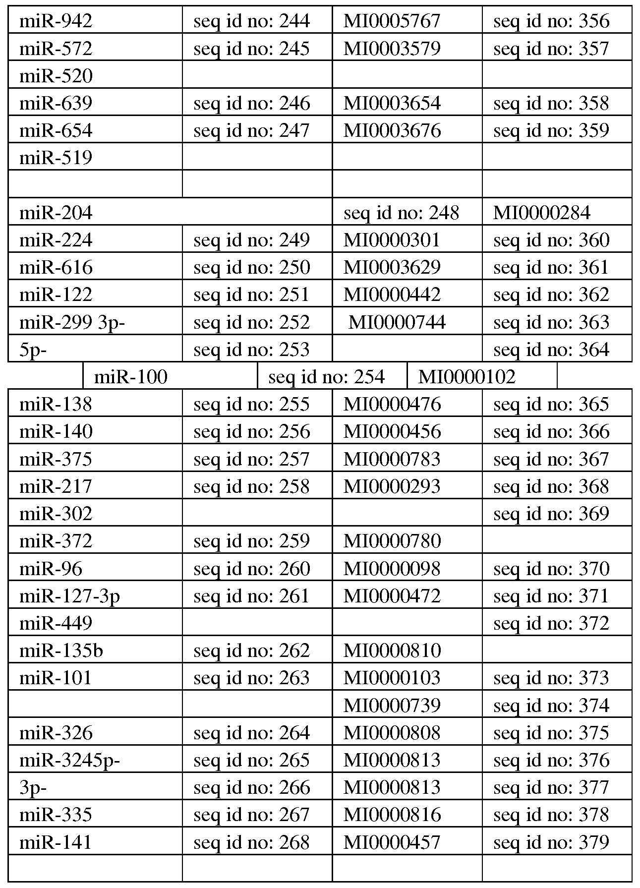
Additional miRNAs contemplated for upregulation are provided herein below. miR-92ap, miR-21, miR-26a, miR-18a, miR-124, miR-99a, miR-30c, miR-301a, miR- 145-50, miR-143-3p, miR-373, miR-20b, miR-29c, miR-29b, miR-143, let-7g, let-7a, let-7b, miR-98, miR-30a*, miR-17, miR-1, miR-192, miR-155, miR-516-ap, miR-31, miR-181a, miR-181b, miR-181c, miR-34-c, miR-34b*, miR-103a, miR-210, miR-16, miR-30a, miR-31, miR-222, miR-17, miR-17*, miR-200b, miR-200c, miR-128, miR- 503, miR-424, miR-195, miR-1256, miR-203a, miR-199, miR-93, miR-98, miR-125-a , miR-133a, miR-133b, miR-126, miR-194, miR-346, miR-15b, miR-338-3p, miR-373, miR-205, miR-210, miR-125, miR-1226, miR-708, miR-449, miR-422, miR-340, miR- 605, miR-522, miR-663, miR-130a, miR-130b, miR-942, miR-572, miR-520, miR-639, miR-654, miR-519, mir-202, mir-767-5p, mir-29a, mir-29b, mir-29c, let-7a, let-7b, let-7c, let-7d, let-7e, let-7f, let-7g, let-7i, mir-4458, mir-4500, mir-98, mir-148a, mir-148b, mir-152, mir-4658, mir-3662, mir-25, mir-32, mir-363, mir-367, mir- 92a, mir-92b, mir-520d-5p, mir-524-5p, mir-4724-3p, mir-1294, mir-143, mir-4770, mir-3659, mir-145, mir-3163, mir-181a, mir-181b, mir-181c, mir-181d, mir-4262, mir-4279, mir-144, mir-642b, mir-4742-3p, mir-3177-5p, mir-656, mir-3121-3p, mir-106a, mir-106b, mir-17, mir-20a, mir-20b, mir-519d, mir-93, mir-1297, mir- 26a, mir-26b, mir-4465, mir-326, mir-330-5p, mir-3927 and mir-2113.
Additional miRNAs contemplated for upregulation include, mir-372, mir-373, mir-520a-3p, mir-520b, mir-520c-3p, mir-520d-3p, mir-520e, mir-199a-3p, mir-199b- 3p, mir-3129-5p.
The upregulation may be effected in vivo or ex vivo.
Mesenchymal stem cells give rise to one or more mesenchymal tissues (e.g., adipose, osseous, cartilaginous, elastic and fibrous connective tissues, myoblasts) as well as to tissues other than those originating in the embryonic mesoderm (e.g., neural cells) depending upon various influences from bioactive factors such as cytokines. Although such cells can be isolated from embryonic yolk sac, placenta, umbilical cord, fetal and adolescent skin, blood and other tissues, their abundance in the easily accessible fat tissue and BM far exceeds their abundance in other tissues and as such isolation from BM and fat tissue is presently preferred.
Methods of isolating, purifying and expanding mesenchymal stem cells (MSCs) are known in the arts and include, for example, those disclosed by Caplan and Haynesworth in U.S. Pat. No. 5,486,359 and Jones E.A. et al., 2002, Isolation and characterization of bone marrow multipotential mesenchymal progenitor cells, Arthritis Rheum. 46(12): 3349-60.
Mesenchymal stem cells may be isolated from various tissues including but not limited to bone marrow, peripheral blood, blood, placenta (e.g. chorionic and/or amniotic), cord blood, umbilical cord, amniotic fluid and from adipose tissue.
A method of isolating mesenchymal stem cells from peripheral blood is described by Kassis et al [Bone Marrow Transplant. 2006 May; 37(10):967-76]. A method of isolating mesenchymal stem cells from placental tissue is described by Zhang et al [Chinese Medical Journal, 2004, 117 (6):882-887]. Methods of isolating and culturing adipose tissue, placental and cord blood mesenchymal stem cells are described by Kern et al [Stem Cells, 2006; 24: 1294-1301].
According to a preferred embodiment of this aspect of the present invention, the mesenchymal stem cells are human.
According to another embodiment of this aspect of the present invention, the mesenchymal stem cells are isolated from placenta and umbilical cord of newborn humans.
Bone marrow can be isolated from the iliac crest of an individual by aspiration.
Low-density BM mononuclear cells (BMMNC) may be separated by a FICOL-PAQUE density gradient or by elimination of red blood cells using Hetastarch (hydroxyethyl starch). Preferably, mesenchymal stem cell cultures are generated by diluting BM aspirates (usually 20 ml) with equal volumes of Hank's balanced salt solution (HBSS; GIBCO Laboratories, Grand Island, NY, USA) and layering the diluted cells over about 10 ml of a Ficoll column (Ficoll-Paque; Pharmacia, Piscataway, NJ, USA). Following 30 minutes of centrifugation at 2,500 x g, the mononuclear cell layer is removed from the interface and suspended in HBSS. Cells are then centrifuged at 1,500 x g for 15 minutes and resuspended in a complete medium (MEM, a medium without deoxyribonucleotides or ribonucleotides; GIBCO); 20 % fetal calf serum (FCS) derived from a lot selected for rapid growth of MSCs (Atlanta Biologicals, Norcross, GA); 100 units/ml penicillin (GIBCO), 100 μg/ml streptomycin (GIBCO); and 2 mM L-glutamine (GIBCO). Resuspended cells are plated in about 25 ml of medium in a 10 cm culture dish (Corning Glass Works, Corning, NY) and incubated at 37 °C with 5 % humidified C02. Following 24 hours in culture, non-adherent cells are discarded, and the adherent cells are thoroughly washed twice with phosphate buffered saline (PBS). The medium is replaced with a fresh complete medium every 3 or 4 days for about 14 days. Adherent cells are then harvested with 0.25 % trypsin and 1 mM EDTA (Trypsin/EDTA, GIBCO) for 5 min at 37 °C, replated in a 6-cm plate and cultured for another 14 days. Cells are then trypsinized and counted using a cell counting device such as for example, a hemocytometer (Hausser Scientific, Horsham, PA). Cultured cells are recovered by centrifugation and resuspended with 5 % DMSO and 30 % FCS at a concentration of 1 to 2 X 10⁶ cells per ml. Aliquots of about 1 ml each are slowly frozen and stored in liquid nitrogen.
Adipose tissue-derived MSCs can be obtained by liposuction and mononuclear cells can be isolated manually by removal of the fat and fat cells, or using the Celution System (Cytori Therapeutics) following the same procedure as described above for preparation of MSCs.
According to one embodiment the populations are plated on polystyrene plastic surfaces (e.g. in a flask) and mesenchymal stem cells are isolated by removing nonadherent cells. Alternatively mesenchymal stem cell may be isolated by FACS using mesenchymal stem cell markers.
Preferably the MSCs are at least 50 % purified, more preferably at least 75 % purified and even more preferably at least 90 % purified.
To expand the mesenchymal stem cell fraction, frozen cells are thawed at 37 °C, diluted with a complete medium and recovered by centrifugation to remove the DMSO. Cells are resuspended in a complete medium and plated at a concentration of about
5,000 cells/cm . Following 24 hours in culture, non-adherent cells are removed and the adherent cells are harvested using Trypsin/EDTA, dissociated by passage through a narrowed Pasteur pipette, and preferably replated at a density of about 1.5 to about 3.0 cells/cm . Under these conditions, MSC cultures can grow for about 50 population doublings and be expanded for about 2000 fold [Colter DC, et al. Rapid expansion of recycling stem cells in cultures of plastic-adherent cells from human bone marrow. Proc Natl Acad Sci USA. 97: 3213-3218, 2000].
MSC cultures utilized by some embodiments of the invention preferably include three groups of cells which are defined by their morphological features: small and agranular cells (referred to as RS-1, herein below), small and granular cells (referred to as RS-2, herein below) and large and moderately granular cells (referred to as mature MSCs, herein below). The presence and concentration of such cells in culture can be assayed by identifying a presence or absence of various cell surface markers, by using, for example, immunofluorescence, in situ hybridization, and activity assays.
When MSCs are cultured under the culturing conditions of some embodiments of the invention they exhibit negative staining for the hematopoietic stem cell markers CD34, CD11B, CD43 and CD45. A small fraction of cells (less than 10 %) are dimly positive for CD31 and/or CD38 markers. In addition, mature MSCs are dimly positive for the hematopoietic stem cell marker, CD 117 (c-Kit), moderately positive for the osteogenic MSCs marker, Stro-1 [Simmons, P. J. & Torok-Storb, B. (1991). Blood 78, 5562] and positive for the thymocytes and peripheral T lymphocytes marker, CD90 (Thy-1). On the other hand, the RS-1 cells are negative for the CD117 and Strol markers and are dimly positive for the CD90 marker, and the RS-2 cells are negative for all of these markers.
The mesenchymal stem cells of the present invention may be of autologous, syngeneic or allogeneic related (matched siblings or haploidentical family members) or unrelated fully mismatched source, as further described herein below.
Culturing of the mesenchymal stem cells can be performed in any media that support (or at least does not inhibit) the differentiation of the cells towards astrocytic cells such as those described in U.S. Pat. No. 6,528,245 and by Sanchez-Ramos et al.
(2000) ; Woodburry et al. (2000); Woodburry et al. (J. Neurosci. Res. 96:908-917, 2001); Black and Woodbury (Blood Cells Mol. Dis. 27:632-635, 2001); Deng et al.
(2001) , Kohyama et al. (2001), Reyes and Verfatile (Ann. N.Y. Acad. Sci. 938:231-235, 2001) and Jiang et al. (Nature 418:47-49, 2002).
The media may be G5, neurobasal medium, DMEM or DMEM/F12, OptiMEM™ or any other medium that supports neuronal or astrocytic growth.
According to a particular embodiment the miRNA comprises at least one of miR-20b, miR-146, miR-101 and miR-141.
A particular combination contemplated by the present inventors includes up- regulating each of miR-20b, miR-101 and miR-146a in the MSC population.
Another combination contemplated by the present inventors is up-regulating the level of exogenous miR-9 and exogenous miR-20b in the MSC population.
The term "microRNA", "miRNA", and "miR" are synonymous and refer to a collection of non-coding single- stranded RNA molecules of about 19-28 nucleotides in length, which regulate gene expression. MiRNAs are found in a wide range of organisms and have been shown to play a role in development, homeostasis, and disease etiology.
Below is a brief description of the mechanism of miRNA activity. Genes coding for miRNAs are transcribed leading to production of an miRNA precursor known as the pri-miRNA. The pri-miRNA is typically part of a polycistronic RNA comprising multiple pri-miRNAs. The pri-miRNA may form a hairpin with a stem and loop. The stem may comprise mismatched bases.
The hairpin structure of the pri-miRNA is recognized by Drosha, which is an
RNase III endonuclease. Drosha typically recognizes terminal loops in the pri-miRNA and cleaves approximately two helical turns into the stem to produce a 60-70 nt precursor known as the pre-miRNA. Drosha cleaves the pri-miRNA with a staggered cut typical of RNase III endonucleases yielding a pre-miRNA stem loop with a 5' phosphate and ~2 nucleotide 3' overhang. It is estimated that approximately one helical turn of stem (-10 nucleotides) extending beyond the Drosha cleavage site is essential for efficient processing. The pre-miRNA is then actively transported from the nucleus to the cytoplasm by Ran-GTP and the export receptor exportin-5.
The double- stranded stem of the pre-miRNA is then recognized by Dicer, which is also an RNase III endonuclease. Dicer may also recognize the 5' phosphate and 3' overhang at the base of the stem loop. Dicer then cleaves off the terminal loop two helical turns away from the base of the stem loop leaving an additional 5' phosphate and ~2 nucleotide 3' overhang. The resulting siRNA-like duplex, which may comprise mismatches, comprises the mature miRNA and a similar-sized fragment known as the miRNA*. The miRNA and miRNA* may be derived from opposing arms of the pri- miRNA and pre-miRNA. miRNA* sequences may be found in libraries of cloned miRNAs but typically at lower frequency than the miRNAs.
Although initially present as a double- stranded species with miRNA*, the miRNA eventually become incorporated as a single-stranded RNA into a ribonucleoprotein complex known as the RNA-induced silencing complex (RISC). Various proteins can form the RISC, which can lead to variability in specificity for miRNA/miRNA* duplexes, binding site of the target gene, activity of miRNA (repress or activate), and which strand of the miRNA/miRNA* duplex is loaded in to the RISC.
When the miRNA strand of the miRNA: miRNA* duplex is loaded into the RISC, the miRNA* is removed and degraded. The strand of the miRNA: miRNA* duplex that is loaded into the RISC is the strand whose 5' end is less tightly paired. In cases where both ends of the miRNA: miRNA* have roughly equivalent 5' pairing, both miRNA and miRNA* may have gene silencing activity.
The RISC identifies target nucleic acids based on high levels of complementarity between the miRNA and the mRNA, especially by nucleotides 2-7 of the miRNA.
A number of studies have looked at the base-pairing requirement between miRNA and its mRNA target for achieving efficient inhibition of translation (reviewed by Bartel 2004, Cell 116-281). In mammalian cells, the first 8 nucleotides of the miRNA may be important (Doench & Sharp 2004 GenesDev 2004-504). However, other parts of the microRNA may also participate in mRNA binding. Moreover, sufficient base pairing at the 3' can compensate for insufficient pairing at the 5' (Brennecke et al, 2005 PLoS 3-e85). Computation studies, analyzing miRNA binding on whole genomes have suggested a specific role for bases 2-7 at the 5' of the miRNA in target binding but the role of the first nucleotide, found usually to be "A" was also recognized (Lewis et at 2005 Cell 120-15). Similarly, nucleotides 1-7 or 2-8 were used to identify and validate targets by Krek et al (2005, Nat Genet 37-495).
The target sites in the mRNA may be in the 5' UTR, the 3' UTR or in the coding region. Interestingly, multiple miRNAs may regulate the same mRNA target by recognizing the same or multiple sites. The presence of multiple miRNA binding sites in most genetically identified targets may indicate that the cooperative action of multiple RISCs provides the most efficient translational inhibition.
miRNAs may direct the RISC to downregulate gene expression by either of two mechanisms: mRNA cleavage or translational repression. The miRNA may specify cleavage of the mRNA if the mRNA has a certain degree of complementarity to the miRNA. When a miRNA guides cleavage, the cut is typically between the nucleotides pairing to residues 10 and 11 of the miRNA. Alternatively, the miRNA may repress translation if the miRNA does not have the requisite degree of complementarity to the miRNA. Translational repression may be more prevalent in animals since animals may have a lower degree of complementarity between the miRNA and binding site.
It should be noted that there may be variability in the 5' and 3' ends of any pair of miRNA and miRNA*. This variability may be due to variability in the enzymatic processing of Drosha and Dicer with respect to the site of cleavage. Variability at the 5' and 3' ends of miRNA and miRNA* may also be due to mismatches in the stem structures of the pri-miRNA and pre-miRNA. The mismatches of the stem strands may lead to a population of different hairpin structures. Variability in the stem structures may also lead to variability in the products of cleavage by Drosha and Dicer.
The term "microRNA mimic" refers to synthetic non-coding RNAs that are capable of entering the RNAi pathway and regulating gene expression. miRNA mimics imitate the function of endogenous microRNAs (miRNAs) and can be designed as mature, double stranded molecules or mimic precursors (e.g., or pre-miRNAs). miRNA mimics can be comprised of modified or unmodified RNA, DNA, RNA-DNA hybrids, or alternative nucleic acid chemistries (e.g., LNAs or 2'-0, 4'-C-ethylene-bridged nucleic acids (EN A)). Other modifications are described herein below. For mature, double stranded miRNA mimics, the length of the duplex region can vary between 13- 33, 18-24 or 21-23 nucleotides. The miRNA may also comprise a total of at least 5, 6, 7, 8, 9, 10, 11, 12, 13, 14, 15, 16, 17, 18, 19, 20, 21, 22, 23, 24, 25, 26, 27, 28, 29, 30, 31, 32, 33, 34, 35, 36, 37, 38, 39 or 40 nucleotides. The sequence of the miRNA may be the first 13-33 nucleotides of the pre-miRNA. The sequence of the miRNA may also be the last 13-33 nucleotides of the pre-miRNA. The sequence of the miRNA may comprise any of the sequences of the disclosed miRNAs, or variants thereof.
It will be appreciated from the description provided herein above, that contacting mesenchymal stem cells may be affected in a number of ways:
1. Transiently transfecting the mesenchymal stem cells with the mature miRNA (or modified form thereof, as described herein below). The miRNAs designed according to the teachings of the present invention can be generated according to any oligonucleotide synthesis method known in the art, including both enzymatic syntheses and solid-phase syntheses. Equipment and reagents for executing solid-phase synthesis are commercially available from, for example, Applied Biosystems. Any other means for such synthesis may also be employed; the actual synthesis of the oligonucleotides is well within the capabilities of one skilled in the art and can be accomplished via established methodologies as detailed in, for example: Sambrook, J. and Russell, D. W. (2001), "Molecular Cloning: A Laboratory Manual"; Ausubel, R. M. et al., eds. (1994, 1989), "Current Protocols in Molecular Biology," Volumes I-III, John Wiley & Sons, Baltimore, Maryland; Perbal, B. (1988), "A Practical Guide to Molecular Cloning," John Wiley & Sons, New York; and Gait, M. J., ed. (1984), "Oligonucleotide Synthesis"; utilizing solid-phase chemistry, e.g. cyanoethyl phosphoramidite followed by deprotection, desalting, and purification by, for example, an automated trityl-on method or HPLC.
2. Stably, or transiently transfecting the mesenchymal stem cells with an expression vector which encodes the mature miRNA.
3. Stably, or transiently transfecting the mesenchymal stem cells with an expression vector which encodes the pre-miRNA. The pre-miRNA sequence may comprise from 45-90, 60-80 or 60-70 nucleotides. The sequence of the pre-miRNA may comprise a miRNA and a miRNA* as set forth herein. The sequence of the pre-miRNA may also be that of a pri-miRNA excluding from 0-160 nucleotides from the 5' and 3' ends of the pri-miRNA. The sequence of the pre-miRNA may comprise the sequence of the miRNA.
4. Stably, or transiently transfecting the mesenchymal stem cells with an expression vector which encodes the pri-miRNA. The pri-miRNA sequence may comprise from 45-30,000, 50-25,000, 100-20,000, 1,000-1,500 or 80- 100 nucleotides. The sequence of the pri-miRNA may comprise a pre- miRNA, miRNA and miRNA*, as set forth herein, and variants thereof. Preparation of miRNAs mimics can be effected by chemical synthesis methods or by recombinant methods.
As mentioned, the present invention also contemplates differentiation of mesenchymal stem cells towards an astrocytic phenotype by down-regulation of particular miRNAs - namely mi-R- 193b, mi-R-221, mi-R- 135a, mi-R-149, mi-R-222, mi-R- 199a, mi-R-302, mi-R-302c, mi-R-302d, mi-R-369-3p, mi-R-370, mi-R-let7a, mi- R-let7b, mi-R-lOb, mi-R-23a, mi-R-23b, mi-R-32, miR-145, miR-133, mi-R-138, mi-R- 182, mi-R-487, mi-R-214, mi-R-409, mi-R-548-dl, mi-R-889, as well as mi-R- 1238.
Additional miRNAs contemplated for down-regulation are set forth below.
miR-204, miR-224, miR-616, miR-122, miR-299, miR-100, miR-138, miR-140, miR- 375, miR-217, miR-302, miR-372, miR-96, miR-127-3p, miR-449, miR-135b, miR- 101, miR-326, miR-324, miR-335, miR- 14, miR- 16.
Additional miRNAs contemplated for down-regulation are set forth below. mir-410, mir-3163, mir-148a, mir-148b, mir-152, mir-3121-3p, mir-495, mir-203, mir- 4680-3p.
According to a particular embodiment, at least one of miR-32, miR-221, miR- 302a, miR-138 and miR-302b is down-regulated in order to produce the astrocyte-like cells of the present invention.
Down-regulating miRNAs can be affected using a polynucleotide which is hybridizable in cells under physiological conditions to the miRNA.
According to a particular embodiment, the cell population is generated by up- regulating an expression of miR-9, miR-146 and miR-101 in a population of MSCs and down-regulating an expression of miR-lOb and miR-302 in the population of MSCs.
According to another embodiment, the cell population is generated by up- regulating an expression of miR-101 and down-regulating an expression of miR-138.
As used herein, the term "hybridizable" refers to capable of hybridizing, i.e., forming a double strand molecule such as RNA:RNA, RNA:DNA and/or DNA:DNA molecules. "Physiological conditions" refer to the conditions present in cells, tissue or a whole organism or body. Preferably, the physiological conditions used by the present invention include a temperature between 34-40 °C, more preferably, a temperature between 35-38 °C, more preferably, a temperature between 36 and 37.5 °C, most preferably, a temperature between 37 to 37.5 °C; salt concentrations (e.g., sodium chloride NaCl) between 0.8-1 , more preferably, about 0.9 ; and/or pH values in the range of 6.5-8, more preferably, 6.5-7.5, most preferably, pH of 7-7.5.
As mentioned herein above, the polynucleotides which downregulate the above list of miRNAs and the miRNAs described herein above may be provided as modified polynucleotides using various methods known in the art.
For example, the oligonucleotides (e.g. miRNAs) or polynucleotides of the present invention may comprise heterocylic nucleosides consisting of purines and the pyrimidines bases, bonded in a 3'-to-5' phosphodiester linkage.
Preferably used oligonucleotides or polynucleotides are those modified either in backbone, internucleoside linkages, or bases, as is broadly described herein under.
Specific examples of preferred oligonucleotides or polynucleotides useful according to this aspect of the present invention include oligonucleotides or polynucleotides containing modified backbones or non-natural internucleoside linkages. Oligonucleotides or polynucleotides having modified backbones include those that retain a phosphorus atom in the backbone, as disclosed in U.S. Pat. Nos.: 4,469,863 4,476,301; 5,023,243; 5,177,196; 5,188,897; 5,264,423; 5,276,019; 5,278,302 5,286,717; 5,321,131; 5,399,676; 5,405,939; 5,453,496; 5,455,233; 5,466,677 5,476,925; 5,519,126; 5,536,821; 5,541,306; 5,550,111; 5,563,253; 5,571,799 5,587,361; and 5,625,050.
Preferred modified oligonucleotide or polynucleotide backbones include, for example: phosphorothioates; chiral phosphorothioates; phosphorodithioates; phosphotriesters; aminoalkyl phosphotriesters; methyl and other alkyl phosphonates, including 3'-alkylene phosphonates and chiral phosphonates; phosphinates; phosphoramidates, including 3'-amino phosphoramidate and aminoalkylphosphoramidates ; thionophosphoramidates ; thionoalkylphosphonates ; thionoalkylphosphotriesters; and boranophosphates having normal 3'-5' linkages, 2'-5' linked analogues of these, and those having inverted polarity wherein the adjacent pairs of nucleoside units are linked 3'-5' to 5'-3' or 2'-5' to 5'-2'. Various salts, mixed salts, and free acid forms of the above modifications can also be used.
Alternatively, modified oligonucleotide or polynucleotide backbones that do not include a phosphorus atom therein have backbones that are formed by short-chain alkyl or cycloalkyl internucleoside linkages, mixed heteroatom and alkyl or cycloalkyl internucleoside linkages, or one or more short-chain heteroatomic or heterocyclic internucleoside linkages. These include those having morpholino linkages (formed in part from the sugar portion of a nucleoside); siloxane backbones; sulfide, sulfoxide, and sulfone backbones; formacetyl and thioformacetyl backbones; methylene formacetyl and thioformacetyl backbones; alkene-containing backbones; sulfamate backbones; methyleneimino and methylenehydrazino backbones; sulfonate and sulfonamide backbones; amide backbones; and others having mixed N, O, S and CH2 component parts, as disclosed in U.S. Pat. Nos.: 5,034,506; 5,166,315; 5,185,444; 5,214,134
5,216,141; 5,235,033; 5,264,562; 5,264,564; 5,405,938; 5,434,257; 5,466,677;
5,470,967; 5,489,677; 5,541,307; 5,561,225; 5,596,086; 5,602,240; 5,610,289; 5,602,240; 5,608,046; 5,610,289; 5,618,704; 5,623,070; 5,663,312; 5,633,360;
5,677,437 and 5,677,439.
Other oligonucleotides or polynucleotides which may be used according to the present invention are those modified in both sugar and the internucleoside linkage, i.e., the backbone of the nucleotide units is replaced with novel groups. The base units are maintained for complementation with the appropriate polynucleotide target. An example of such an oligonucleotide mimetic includes a peptide nucleic acid (PNA). A PNA oligonucleotide refers to an oligonucleotide where the sugar-backbone is replaced with an amide-containing backbone, in particular an aminoethylglycine backbone. The bases are retained and are bound directly or indirectly to aza-nitrogen atoms of the amide portion of the backbone. United States patents that teach the preparation of PNA compounds include, but are not limited to, U.S. Pat. Nos. 5,539,082; 5,714,331; and 5,719,262; each of which is herein incorporated by reference. Other backbone modifications which may be used in the present invention are disclosed in U.S. Pat. No. 6,303,374.
Oligonucleotides or polynucleotides of the present invention may also include base modifications or substitutions. As used herein, "unmodified" or "natural" bases include the purine bases adenine (A) and guanine (G) and the pyrimidine bases thymine (T), cytosine (C), and uracil (U). "Modified" bases include but are not limited to other synthetic and natural bases, such as: 5-methylcytosine (5-me-C); 5-hydroxymethyl cytosine; xanthine; hypoxanthine; 2-aminoadenine; 6-methyl and other alkyl derivatives of adenine and guanine; 2-propyl and other alkyl derivatives of adenine and guanine; 2- thiouracil, 2-thiothymine, and 2-thiocytosine; 5-halouracil and cytosine; 5-propynyl uracil and cytosine; 6-azo uracil, cytosine, and thymine; 5-uracil (pseudouracil); 4- thiouracil; 8-halo, 8-amino, 8-thiol, 8-thioalkyl, 8-hydroxyl, and other 8-substituted adenines and guanines; 5-halo, particularly 5-bromo, 5-trifluoromethyl, and other 5- substituted uracils and cytosines; 7-methylguanine and 7-methyladenine; 8-azaguanine and 8-azaadenine; 7-deazaguanine and 7-deazaadenine; and 3-deazaguanine and 3- deazaadenine. Additional modified bases include those disclosed in: U.S. Pat. No. 3,687,808; Kroschwitz, J. I., ed. (1990), "The Concise Encyclopedia Of Polymer Science And Engineering," pages 858-859, John Wiley & Sons; Englisch et al. (1991), "Angewandte Chemie," International Edition, 30, 613; and Sanghvi, Y. S., "Antisense Research and Applications," Chapter 15, pages 289-302, S. T. Crooke and B. Lebleu, eds., CRC Press, 1993. Such modified bases are particularly useful for increasing the binding affinity of the oligomeric compounds of the invention. These include 5- substituted pyrimidines, 6-azapyrimidines, and N-2, N-6, and O-6-substituted purines, including 2-aminopropyladenine, 5-propynyluracil, and 5-propynylcytosine. 5- methylcytosine substitutions have been shown to increase nucleic acid duplex stability by 0.6-1.2°C (Sanghvi, Y. S. et al. (1993), "Antisense Research and Applications," pages 276-278, CRC Press, Boca Raton), and are presently preferred base substitutions, even more particularly when combined with 2'-0-methoxyethyl sugar modifications.
To express miRNAs or polynucleotide agents which regulate miRNAs in mesencyhymal stem cells, a polynucleotide sequence encoding the miRNA (or pre- miRNA, or pri-miRNA, or polynucleotide which down-regulates the miRNAs) is preferably ligated into a nucleic acid construct suitable for mesenchymal stem cell expression. Such a nucleic acid construct includes a promoter sequence for directing transcription of the polynucleotide sequence in the cell in a constitutive or inducible manner.
It will be appreciated that the nucleic acid construct of some embodiments of the invention can also utilize miRNA homologues which exhibit the desired activity (i.e., astrocytic differentiating ability). Such homologues can be, for example, at least 80 , at least 81 , at least 82 , at least 83 , at least 84 , at least 85 , at least 86 , at least 87 %, at least 88 %, at least 89 %, at least 90 %, at least 91 %, at least 92 %, at least 93 , at least 94 , at least 95 , at least 96 , at least 97 , at least 98 , at least 99 % or 100 % identical to any of the sequences provided, as determined using the BestFit software of the Wisconsin sequence analysis package, utilizing the Smith and Waterman algorithm, where gap weight equals 50, length weight equals 3, average match equals 10 and average mismatch equals -9.
In addition, the homologues can be, for example, at least 60 , at least 61 , at least 62 , at least 63 , at least 64 , at least 65 , at least 66 , at least 67 , at least 68 , at least 69 , at least 70 , at least 71 , at least 72 , at least 73 , at least 74 , at least 75 , at least 76 , at least 77 , at least 78 , at least 79 , at least 80 %, at least 81 %, at least 82 %, at least 83 %, at least 84 %, at least 85 %, at least 86 %, at least 87 %, at least 88 %, at least 89 %, at least 90 %, at least 91 %, at least 92 , at least 93 , at least 94 , at least 95 , at least 96 , at least 97 , at least 98 , at least 99 % or 100 % identical to any of the sequences provided herein, as determined using the BestFit software of the Wisconsin sequence analysis package, utilizing the Smith and Waterman algorithm, where gap weight equals 50, length weight equals 3, average match equals 10 and average mismatch equals -9.
Constitutive promoters suitable for use with some embodiments of the invention are promoter sequences which are active under most environmental conditions and most types of cells such as the cytomegalovirus (CMV) and Rous sarcoma virus (RSV). Inducible promoters suitable for use with some embodiments of the invention include for example tetracycline-inducible promoter (Zabala M, et al., Cancer Res. 2004, 64(8): 2799-804).
Eukaryotic promoters typically contain two types of recognition sequences, the TATA box and upstream promoter elements. The TATA box, located 25-30 base pairs upstream of the transcription initiation site, is thought to be involved in directing RNA polymerase to begin RNA synthesis. The other upstream promoter elements determine the rate at which transcription is initiated.
Preferably, the promoter utilized by the nucleic acid construct of some embodiments of the invention is active in the specific cell population transformed - i.e. mesenchymal stem cells.
Enhancer elements can stimulate transcription up to 1,000 fold from linked homologous or heterologous promoters. Enhancers are active when placed downstream or upstream from the transcription initiation site. Many enhancer elements derived from viruses have a broad host range and are active in a variety of tissues. For example, the SV40 early gene enhancer is suitable for many cell types. Other enhancer/promoter combinations that are suitable for some embodiments of the invention include those derived from polyoma virus, human or murine cytomegalovirus (CMV), the long term repeat from various retroviruses such as murine leukemia virus, murine or Rous sarcoma virus and HIV. See, Enhancers and Eukaryotic Expression, Cold Spring Harbor Press, Cold Spring Harbor, N.Y. 1983, which is incorporated herein by reference.
In the construction of the expression vector, the promoter is preferably positioned approximately the same distance from the heterologous transcription start site as it is from the transcription start site in its natural setting. As is known in the art, however, some variation in this distance can be accommodated without loss of promoter function. In addition to the elements already described, the expression vector of some embodiments of the invention may typically contain other specialized elements intended to increase the level of expression of cloned nucleic acids or to facilitate the identification of cells that carry the recombinant DNA. For example, a number of animal viruses contain DNA sequences that promote the extra chromosomal replication of the viral genome in permissive cell types. Plasmids bearing these viral replicons are replicated episomally as long as the appropriate factors are provided by genes either carried on the plasmid or with the genome of the host cell.
The vector may or may not include a eukaryotic replicon. If a eukaryotic replicon is present, then the vector is amplifiable in eukaryotic cells using the appropriate selectable marker. If the vector does not comprise a eukaryotic replicon, no episomal amplification is possible. Instead, the recombinant DNA integrates into the genome of the engineered cell, where the promoter directs expression of the desired nucleic acid.
Examples for mammalian expression vectors include, but are not limited to, pcDNA3, pcDNA3.1(+/-), pGL3, pZeoSV2(+/-), pSecTag2, pDisplay, pEF/myc/cyto, pCMV/myc/cyto, pCR3.1, pSinRep5, DH26S, DHBB, pNMTl, pNMT41, pNMT81, which are available from Invitrogen, pCI which is available from Promega, pMbac, pPbac, pBK-RSV and pBK-CMV which are available from Strategene, pTRES which is available from Clontech, and their derivatives.
Expression vectors containing regulatory elements from eukaryotic viruses such as retroviruses can be also used. SV40 vectors include pSVT7 and pMT2. Vectors derived from bovine papilloma virus include pBV-lMTHA, and vectors derived from Epstein Bar virus include pHEBO, and p205. Other exemplary vectors include pMSG, pAV009/A+, pMTO10/A+, pMAMneo-5, baculovirus pDSVE, and any other vector allowing expression of proteins under the direction of the SV-40 early promoter, SV-40 later promoter, metallothionein promoter, murine mammary tumor virus promoter, Rous sarcoma virus promoter, polyhedrin promoter, or other promoters shown effective for expression in eukaryotic cells.
As described above, viruses are very specialized infectious agents that have evolved, in many cases, to elude host defense mechanisms. Typically, viruses infect and propagate in specific cell types. The targeting specificity of viral vectors utilizes its natural specificity to specifically target predetermined cell types and thereby introduce a recombinant gene into the infected cell. Thus, the type of vector used by some embodiments of the invention will depend on the cell type transformed. The ability to select suitable vectors according to the cell type transformed is well within the capabilities of the ordinary skilled artisan and as such no general description of selection consideration is provided herein. For example, bone marrow cells can be targeted using the human T cell leukemia virus type I (HTLV-I) and kidney cells may be targeted using the heterologous promoter present in the baculovirus Autographa calif ornica nucleopolyhedro virus (AcMNPV) as described in Liang CY et al., 2004 (Arch Virol. 149: 51-60).
According to one embodiment, a lentiviral vector is used to transfect the mesenchymal stem cells.
Various methods can be used to introduce the expression vector of some embodiments of the invention into mesenchymal stem cells. Such methods are generally described in Sambrook et al., Molecular Cloning: A Laboratory Manual, Cold Springs Harbor Laboratory, New York (1989, 1992), in Ausubel et al., Current Protocols in Molecular Biology, John Wiley and Sons, Baltimore, Md. (1989), Chang et al., Somatic Gene Therapy, CRC Press, Ann Arbor, Mich. (1995), Vega et al., Gene Targeting, CRC Press, Ann Arbor Mich. (1995), Vectors: A Survey of Molecular Cloning Vectors and Their Uses, Butterworths, Boston Mass. (1988) and Gilboa et at. [Biotechniques 4 (6): 504-512, 1986] and include, for example, stable or transient transfection, lipofection, electroporation and infection with recombinant viral vectors. In addition, see U.S. Pat. Nos. 5,464,764 and 5,487,992 for positive-negative selection methods.
Introduction of nucleic acids by viral infection offers several advantages over other methods such as lipofection and electroporation, since higher transfection efficiency can be obtained due to the infectious nature of viruses.
Other vectors can be used that are non- viral, such as cationic lipids, polylysine, and dendrimers.
The miRNAs, miRNA mimics and pre-miRs can be transfected into cells also using nanoparticels such as gold nanoparticles and by ferric oxide magnetic NP - see for example Ghosh et al., Biomaterials. 2013 Jan;34(3):807-16; Crew E, et al., Anal Chem. 2012 Jan 3;84(l):26-9. As mentioned herein above, the polynucleotides which down-regulate the miRNAs described herein above may be provided as modified polynucleotides using various methods known in the art.
Other modes of transfection that do not involved integration include the use of minicircle DNA vectors or the use of PiggyBac transposon that allows the transfection of genes that can be later removed from the genome.
As mentioned hereinabove, a variety of prokaryotic or eukaryotic cells can be used as host-expression systems to express the miRNAs or polynucleotide agent capable of down-regulating the miRNA of some embodiments of the invention. These include, but are not limited to, microorganisms, such as bacteria transformed with a recombinant bacteriophage DNA, plasmid DNA or cosmid DNA expression vector containing the coding sequence; yeast transformed with recombinant yeast expression vectors containing the coding sequence; plant cell systems infected with recombinant virus expression vectors (e.g., cauliflower mosaic virus, CaMV; tobacco mosaic virus, TMV) or transformed with recombinant plasmid expression vectors, such as Ti plasmid, containing the coding sequence. Mammalian expression systems can also be used to express the miRNAs of some embodiments of the invention.
Examples of bacterial constructs include the pET series of E. coli expression vectors [Studier et al. (1990) Methods in Enzymol. 185:60-89).
In yeast, a number of vectors containing constitutive or inducible promoters can be used, as disclosed in U.S. Pat. Application No: 5,932,447. Alternatively, vectors can be used which promote integration of foreign DNA sequences into the yeast chromosome.
The conditions used for contacting the mesenchymal stem cells are selected for a time period/concentration of cells/concentration of miRNA/ratio between cells and miRNA which enable the miRNA (or inhibitors thereof) to induce differentiation thereof. The present invention further contemplates incubation of the mesenchymal stem cells with a differentiation factor which promotes differentiation towards an astrocytic lineage. The incubation with such differentiation factors may be affected prior to, concomitant with or following the contacting with the miRNA. According to this embodiment the medium may be supplemented with at least one of SHH (e.g. about 250 ng/ml), FGFb (e.g. 50 ng/ml), EGF (e.g. about 50 ng/ml), a cAMP inducer (e.g. IB MX or dbcycAMP), PDGF (e.g. about 5 ng/ml) neuregulin (e.g. about 50 ng/ml) and FGFb (e.g. about 20 ng/ml).
Alternatively, or additionally, the mesenchymal stem cells may be genetically modified so as to express such differentiation factors, using expression constructs such as those described herein above.
During or following the differentiation step the mesenchymal stem cells may be monitored for their differentiation state. Cell differentiation can be determined upon examination of cell or tissue- specific markers which are known to be indicative of differentiation. For example, the differentiated cells may express the following markers: S100 beta, glial fibrillary acidic protein (GFAP), glutamine synthetase, GLT- 1, Excitatory Amino Acid Transporter 1 (EAAT1) and Excitatory Amino Acid Transporter 2 (EAAT2). Further, the differentiated cells may secrete a neurotrophic factor including for example glial derived neurotrophic factor (GDNF), GenBank accession nos. L19063, L15306; nerve growth factor (NGF), GenBank accession no. CAA37703; brain-derived neurotrophic factor (BDNF), GenBank accession no CAA62632; neurotrophin-3 (NT-3), GenBank Accession No. M37763; neurotrophin- 4/5; Neurturin (NTN), GenBank Accession No. NP_004549; Neurotrophin-4, GenBank Accession No. M86528; Persephin, GenBank accession no. AAC39640; brain derived neurotrophic factor, (BDNF), GenBank accession no. CAA42761; artemin (ART), GenBank accession no. AAD13110; ciliary neurotrophic factor (CNTF), GenBank accession no. NP_000605; insulin growth factor-I (IGF-1), GenBank accession no. NP_000609; and/or Neublastin GenBank accession no. AAD21075.
It will be appreciated that the differentiation time may be selected so as to obtain early progenitors of astrocytes or more mature astrocytes. Enrichment for a particular early or mature astrocytic cell is also contemplated. Selection for cells which express markers such as CD44, A2B5 and SI 00 allows for the enrichment of progenitor type astrocytes, whereas selection for cells which express markers such as GFAP and glutamine synthase allows for selection of mature astrocytes.
Tissue/cell specific markers can be detected using immunological techniques well known in the art [Thomson JA et al., (1998). Science 282: 1145-7]. Examples include, but are not limited to, flow cytometry for membrane -bound markers, immunohistochemistry for extracellular and intracellular markers and enzymatic immunoassay, for secreted molecular markers.
In addition, cell differentiation can be also followed by specific reporters that are tagged with GFP or RFP and exhibit increased fluorescence upon differentiation.
Isolated cell populations obtained according to the methods describe herein are typically non-homogeneous, although homogeneous cell populations are also contemplated.
According to a particular embodiment, the cell populations are genetically modified to express a miRNA or a polynucleotide agent capable of down-regulating the miRNA.
The term "isolated" as used herein refers to a population of cells that has been removed from its in-vivo location (e.g. bone marrow, neural tissue). Preferably the isolated cell population is substantially free from other substances (e.g., other cells) that are present in its in-vivo location.
Cell populations may be selected such that more than about 50 % of the cells express at least one, at least two, at least three, at least four, at least five or all of the following markers: SI 00 beta, glial fibrillary acidic protein (GFAP), glutamine sythetase, GLT-1, GDNF, BDNF, IGF-1 and GLAST.
Cell populations may be selected such that more than about 60 % of the cells express at least one, at least two, at least three, at least four, at least five or all of the following markers: SI 00 beta, glial fibrillary acidic protein (GFAP), glutamine sythetase, GLT-1, GDNF, BDNF, IGF-1 and GLAST.
Cell populations may be selected such that more than about 70 % of the cells express at least one, at least two, at least three, at least four, at least five or all of the following markers: SI 00 beta, glial fibrillary acidic protein (GFAP), glutamine sythetase, GLT-1, GDNF, BDNF, IGF-1 and GLAST.
Cell populations may be selected such that more than about 80 % of the cells express at least one, at least two, at least three, at least four, at least five or all of the following markers: SI 00 beta, glial fibrillary acidic protein (GFAP), glutamine sythetase, GLT- 1 , GDNF, BDNF, IGF- 1 and GLAST.
Cell populations may be selected such that more than about 90 % of the cells express at least one, at least two, at least three, at least four, at least five or all of the following markers: SI 00 beta, glial fibrillary acidic protein (GFAP), glutamine sythetase, GLT-1, GDNF, BDNF, IGF-1 and GLAST.
Cell populations may be selected such that more than about 95 % of the cells express at least one, at least two, at least three, at least four, at least five or all of the following markers: SI 00 beta, glial fibrillary acidic protein (GFAP), glutamine sythetase, GLT-1, GDNF, BDNF, IGF-1 and GLAST.
Isolation of particular subpopulations of cells may be effected using techniques known in the art including fluorescent activated cell sorting and/or magnetic separation of cells.
The cells of the populations of this aspect of the present invention may comprise structural astrocytic phenotypes including a cell size, a cell shape, an organelle size and an organelle number. Thus, mature astrocytic structural phenotypes include a round nucleus, a "star shaped" body and many long processes that end as vascular foot plates on the small blood vessels of the CNS.
These structural phenotypes may be analyzed using microscopic techniques (e.g. scanning electron microscopy). Antibodies or dyes may be used to highlight distinguishing features in order to aid in the analysis.
The present inventors have further shown that a particular miRNA (miRNA 504) which is upregulated on differentiation of MSCs towards an astrocytic phenotype targets a-Synuclein (see Figure 10). Mutations within the a-Synuclein gene are associated with autosomal dominant familial PD.
Thus, the present inventors further propose use of MSCs as a cargo cell to transport miRNA 504 to the brain where the miRNA then targets the α-Synuclein as a treatment for Parkinson's.
Another miRNA (miRNA 152) which is upregulated on differentiation of MSCs towards an astrocytic phenotype targets Huntingdon (HTT) gene. Mutations within this gene are associated with Huntingdon disease (HD).
Thus, the present inventors further propose use of MSCs as a cargo cell to transport miRNA 152 to the brain where the miRNA then targets the α-Synuclein as a treatment for Huntingdon' s disease.
Another miRNA (miRNA 665) which is upregulated on differentiation of MSCs towards an astrocytic phenotype targets the prion gene (PRNP). Thus, the present inventors further propose use of MSCs as a cargo cell to transport miRNA 665 to the brain where the miRNA then targets PRNP.
Another miRNA (miRNA 340) which is upregulated on differentiation of MSCs towards an astrocytic phenotype targets SOD1 gene. Mutations within this gene are associated with ALS.
Thus, the present inventors further propose use of MSCs as a cargo cell to transport miRNA 340 to the brain where the miRNA then targets the SOD1 gene as a treatment for ALS.
According to this aspect of the invention, the MSCs may be manipulated to express the miRNA (or mimic thereof) and cultured so that they differentiate towards the astrocytic phenotype as described herein above. Alternatively, the MSCs may be manipulated to express the miRNA (or mimic thereof) and administered to the patient (e.g. a patient with Parkinson's) without allowing for astrocytic differentiation.
The cells and cell populations of the present invention may be useful for a variety of therapeutic purposes. Representative examples of CNS diseases or disorders that can be beneficially treated with the cells described herein include, but are not limited to, a pain disorder, a motion disorder, a dissociative disorder, a mood disorder, an affective disorder, a neurodegenerative disease or disorder and a convulsive disorder.
More specific examples of such conditions include, but are not limited to, Parkinson's, ALS, Multiple Sclerosis, Huntingdon's disease, autoimmune encephalomyelitis, diabetic neuropathy, glaucatomus neuropathy, macular degeneration, action tremors and tardive dyskinesia, panic, anxiety, depression, alcoholism, insomnia, manic behavior, Alzheimer's and epilepsy.
The use of differentiated MSCs may be also indicated for treatment of traumatic lesions of the nervous system including spinal cord injury and also for treatment of stroke caused by bleeding or thrombosis or embolism because of the need to induce neurogenesis and provide survival factors to minimize insult to damaged neurons.
In any of the methods described herein the cells may be obtained from an autologous, semi-autologous or non-autologous (i.e., allogeneic or xenogeneic) human donor or embryo or cord/placenta. For example, cells may be isolated from a human cadaver or a donor subject. The term semi-autologous refers to donor cells which are partially-mismatched to recipient cells at a major histocompatibility complex (MHC) class I or class II locus.
The cells of the present invention can be administered to the treated individual using a variety of transplantation approaches, the nature of which depends on the site of implantation.
The term or phrase "transplantation", "cell replacement" or "grafting" are used interchangeably herein and refer to the introduction of the cells of the present invention to target tissue. As mentioned, the cells can be derived from the recipient or from an allogeneic, semi-allogeneic or xenogeneic donor.
The cells can be injected systemically into the circulation, administered intrathecally or grafted into the central nervous system, the spinal cord or into the ventricular cavities or subdurally onto the surface of a host brain. Conditions for successful transplantation include: (i) viability of the implant; (ii) retention of the graft at the site of transplantation; and (iii) minimum amount of pathological reaction at the site of transplantation. Methods for transplanting various nerve tissues, for example embryonic brain tissue, into host brains have been described in: "Neural grafting in the mammalian CNS", Bjorklund and Stenevi, eds. (1985); Freed et al., 2001; Olanow et al., 2003). These procedures include intraparenchymal transplantation, i.e. within the host brain (as compared to outside the brain or extraparenchymal transplantation) achieved by injection or deposition of tissue within the brain parenchyma at the time of transplantation.
Intraparenchymal transplantation can be performed using two approaches: (i) injection of cells into the host brain parenchyma or (ii) preparing a cavity by surgical means to expose the host brain parenchyma and then depositing the graft into the cavity. Both methods provide parenchymal deposition between the graft and host brain tissue at the time of grafting, and both facilitate anatomical integration between the graft and host brain tissue. This is of importance if it is required that the graft becomes an integral part of the host brain and survives for the life of the host.
Alternatively, the graft may be placed in a ventricle, e.g. a cerebral ventricle or subdurally, i.e. on the surface of the host brain where it is separated from the host brain parenchyma by the intervening pia mater or arachnoid and pia mater. Grafting to the ventricle may be accomplished by injection of the donor cells or by growing the cells in a substrate such as 3% collagen to form a plug of solid tissue which may then be implanted into the ventricle to prevent dislocation of the graft. For subdural grafting, the cells may be injected around the surface of the brain after making a slit in the dura. Injections into selected regions of the host brain may be made by drilling a hole and piercing the dura to permit the needle of a microsyringe to be inserted. The microsyringe is preferably mounted in a stereotaxic frame and three dimensional stereotaxic coordinates are selected for placing the needle into the desired location of the brain or spinal cord. The cells may also be introduced into the putamen, nucleus basalis, hippocampus cortex, striatum, substantia nigra or caudate regions of the brain, as well as the spinal cord.
The cells may also be transplanted to a healthy region of the tissue. In some cases the exact location of the damaged tissue area may be unknown and the cells may be inadvertently transplanted to a healthy region. In other cases, it may be preferable to administer the cells to a healthy region, thereby avoiding any further damage to that region. Whatever the case, following transplantation, the cells preferably migrate to the damaged area.
For transplanting, the cell suspension is drawn up into the syringe and administered to anesthetized transplantation recipients. Multiple injections may be made using this procedure.
The cellular suspension procedure thus permits grafting of the cells to any predetermined site in the brain or spinal cord, is relatively non-traumatic, allows multiple grafting simultaneously in several different sites or the same site using the same cell suspension, and permits mixtures of cells from different anatomical regions. Multiple grafts may consist of a mixture of cell types, and/or a mixture of transgenes inserted into the cells. Preferably from approximately 104 to approximately 109 cells are introduced per graft. Cells can be administered concomitantly to different locations such as combined administration intrathecally and intravenously to maximize the chance of targeting into affected areas.
For transplantation into cavities, which may be preferred for spinal cord grafting, tissue is removed from regions close to the external surface of the central nerve system (CNS) to form a transplantation cavity, for example as described by Stenevi et al. (Brain Res. 114: 1-20., 1976), by removing bone overlying the brain and stopping bleeding with a material such a gelfoam. Suction may be used to create the cavity. The graft is then placed in the cavity. More than one transplant may be placed in the same cavity using injection of cells or solid tissue implants. Preferably, the site of implantation is dictated by the CNS disorder being treated. Demyelinated MS lesions are distributed across multiple locations throughout the CNS, such that effective treatment of MS may rely more on the migratory ability of the cells to the appropriate target sites.
Intranasal administration of the cells described herein is also contemplated. MSCs typically down regulate MHC class 2 and are therefore less immunogenic. Embryonal or newborn cells obtained from the cord blood, cord's Warton' s gelly or placenta are further less likely to be strongly immunogenic and therefore less likely to be rejected, especially since such cells are immunosuppressive and immunoregulatory to start with.
Notwithstanding, since non-autologous cells may induce an immune reaction when administered to the body several approaches have been developed to reduce the likelihood of rejection of non-autologous cells. Furthermore, since diseases such as multiple sclerosis are inflammatory based diseases, the problem of immune reaction is exacerbated. These include either administration of cells to privileged sites, or alternatively, suppressing the recipient's immune system, providing anti-inflammatory treatment which may be indicated to control autoimmune disorders to start with and/or encapsulating the non-autologous/semi-autologous cells in immunoisolating, semipermeable membranes before transplantation.
As mentioned herein above, the present inventors also propose use of newborn mesenchymal stem cells to limit the immune reaction.
The following experiments may be performed to confirm the potential use of newborn's MSCs isolated from the cord / placenta for treatment of neurological disorders:
1) Differentiated MSCs (to various neural cells or neural progenitor cells) may serve as stimulators in one way mixed lymphocyte culture with allogeneic T cells and proliferative responses in comparison with T cells responding against allogeneic lymphocytes isolated from the same donor may be evaluated by H- Thymidine uptake to document hyporesponsiveness. 2) Differentiated MSCs may be added/co-cultured to one way mixed lymphocyte cultures and to cell cultures with T cell mitogens (phytohemmaglutinin and concanavalin A) to confirm the immunosuppressive effects on proliferative responses mediated by T cells.
3) Cord and placenta cells cultured from Brown Norway rats (unmodified and differentiated), may be enriched for MSCs and these cells may be infused into Lewis rats with induced experimental autoimmune encephalomyelitis (EAE). Alternatively, cord and placenta cells cultured from BALB/c mice, (BALB/cxC57BL/6)Fl or xenogeneic cells from Brown Norway rats (unmodified and differentiated), may be enriched for MSCs and these cells may be infused into C57BL/6 or SJL/j recipients with induced experimental autoimmune encephalomyelitis (EAE). The clinical effects against paralysis may be investigated to evaluate the therapeutic effects of xenogeneic, fully MHC mismatched or haploidentically mismatched MSCs. Such experiments may provide the basis for treatment of patients with a genetic disorder or genetically proned disorder with family member's haploidentical MSCs.
4) BALB/c MSCs cultured from cord and placenta may be transfused with pre-miR labeled with GFP or RFP, which will allow the inventors to follow the migration and persistence of these cells in the brain of C57BL/6 recipients with induced EAE. The clinical effects of labeled MHC mismatched differentiated MSCs may be evaluated by monitoring signs of disease, paralysis and histopathology. The migration and localization of such cells may be also monitored by using fluorescent cells from genetically transduced GFP "green" or Red2 "red" donors.
As mentioned, the present invention also contemplates encapsulation techniques to minimize an immune response.
Encapsulation techniques are generally classified as microencapsulation, involving small spherical vehicles and macroencapsulation, involving larger flat-sheet and hollow-fiber membranes (Uludag, H. et al. Technology of mammalian cell encapsulation. Adv Drug Deliv Rev. 2000; 42: 29-64).
Methods of preparing microcapsules are known in the arts and include for example those disclosed by Lu M Z, et al., Cell encapsulation with alginate and alpha- phenoxycinnamylidene-acetylated poly(allylamine). Biotechnol Bioeng. 2000, 70: 479- 83, Chang T M and Prakash S. Procedures for microencapsulation of enzymes, cells and genetically engineered microorganisms. Mol. Biotechnol. 2001, 17: 249-60, and Lu M Z, et al., A novel cell encapsulation method using photosensitive poly(allylamine alpha- cyanocinnamylideneacetate). J. Microencapsul. 2000, 17: 245-51.
For example, microcapsules are prepared by complexing modified collagen with a ter-polymer shell of 2-hydroxyethyl methylacrylate (HEMA), methacrylic acid (MAA) and methyl methacrylate (MMA), resulting in a capsule thickness of 2-5 .mu.m. Such microcapsules can be further encapsulated with additional 2-5 .mu.m ter-polymer shells in order to impart a negatively charged smooth surface and to minimize plasma protein absorption (Chia, S. M. et al. Multi-layered microcapsules for cell encapsulation Biomaterials. 2002 23: 849-56).
Other microcapsules are based on alginate, a marine polysaccharide (Sambanis, A. Encapsulated islets in diabetes treatment. Diabetes Technol. Ther. 2003, 5: 665-8) or its derivatives. For example, microcapsules can be prepared by the polyelectrolyte complexation between the polyanions sodium alginate and sodium cellulose sulphate with the polycation poly(methylene-co-guanidine) hydrochloride in the presence of calcium chloride.
It will be appreciated that cell encapsulation is improved when smaller capsules are used. Thus, the quality control, mechanical stability, diffusion properties, and in vitro activities of encapsulated cells improved when the capsule size was reduced from 1 mm to 400 .mu.m (Canaple L. et al., Improving cell encapsulation through size control. J Biomater Sci Polym Ed. 2002; 13:783-96). Moreover, nanoporous biocapsules with well-controlled pore size as small as 7 nm, tailored surface chemistries and precise microarchitectures were found to successfully immunoisolate microenvironments for cells (Williams D. Small is beautiful: microparticle and nanoparticle technology in medical devices. Med Device Technol. 1999, 10: 6-9; Desai, T. A. Microfabrication technology for pancreatic cell encapsulation. Expert Opin Biol Ther. 2002, 2: 633-46).
Examples of immunosuppressive agents include, but are not limited to, methotrexate, cyclophosphamide, cyclosporine, cyclosporin A, chloroquine, hydroxychloroquine, sulfasalazine (sulphasalazopyrine), gold salts, D-penicillamine, leflunomide, azathioprine, anakinra, infliximab (REMICADE™), etanercept, TNF alpha blockers, a biological agent that targets an inflammatory cytokine, and Nonsteroidal Anti-Inflammatory Drug (NSAIDs). Examples of NSAIDs include, but are not limited to acetyl salicylic acid, choline magnesium salicylate, diflunisal, magnesium salicylate, salsalate, sodium salicylate, diclofenac, etodolac, fenoprofen, flurbiprofen, indomethacin, ketoprofen, ketorolac, meclofenamate, naproxen, nabumetone, phenylbutazone, piroxicam, sulindac, tolmetin, acetaminophen, ibuprofen, Cox-2 inhibitors and tramadol.
In any of the methods described herein, the cells can be administered either per se or, preferably as a part of a pharmaceutical composition that further comprises a pharmaceutically acceptable carrier.
As used herein a "pharmaceutical composition" refers to a preparation of one or more of the cell compositions described herein, with other chemical components such as pharmaceutically suitable carriers and excipients. The purpose of a pharmaceutical composition is to facilitate administration of the cells to a subject.
Hereinafter, the term "pharmaceutically acceptable carrier" refers to a carrier or a diluent that does not cause significant irritation to a subject and does not abrogate the biological activity and properties of the administered compound. Examples, without limitations, of carriers are propylene glycol, saline, emulsions and mixtures of organic solvents with water.
Herein the term "excipient" refers to an inert substance added to a pharmaceutical composition to further facilitate administration of a compound. Examples, without limitation, of excipients include calcium carbonate, calcium phosphate, various sugars and types of starch, cellulose derivatives, gelatin, vegetable oils and polyethylene glycols.
Techniques for formulation and administration of drugs may be found in "Remington's Pharmaceutical Sciences," Mack Publishing Co., Easton, Pa., latest edition, which is incorporated herein by reference.
Suitable routes of administration include direct administration into the circulation (intravenously or intra-arterial), into the spinal fluid or into the tissue or organ of interest. Thus, for example the cells may be administered directly into the brain. For any preparation used in the methods of the invention, the therapeutically effective amount or dose can be estimated initially from in vitro and cell culture assays. Preferably, a dose is formulated in an animal model to achieve a desired concentration or titer. Such information can be used to more accurately determine useful doses in humans.
Toxicity and therapeutic efficacy of the active ingredients described herein can be determined by standard pharmaceutical procedures in vitro, in cell cultures or experimental animals. For example, animal models of demyelinating diseases include shiverer (shi/shi, MBP deleted) mouse, MD rats (PLP deficiency), Jimpy mouse (PLP mutation), dog shaking pup (PLP mutation), twitcher mouse (galactosylceramidase defect, as in human Krabbe disease), trembler mouse (PMP-22 deficiency). Virus induced demyelination model comprise use if Theiler's virus and mouse hepatitis virus. Autoimmune EAE is a possible model for multiple sclerosis.
The data obtained from these in vitro and cell culture assays and animal studies can be used in formulating a range of dosage for use in human. The dosage may vary depending upon the dosage form employed and the route of administration utilized. The exact formulation, route of administration and dosage can be chosen by the individual physician in view of the patient's condition, (see e.g., Fingl, et al., 1975, in "The Pharmacological Basis of Therapeutics", Ch. 1 p. 1). For example, a multiple sclerosis patient can be monitored symptomatically for improved motor functions indicating positive response to treatment.
For injection, the active ingredients of the pharmaceutical composition may be formulated in aqueous solutions, preferably in physiologically compatible buffers such as Hank's solution, Ringer's solution, or physiological salt buffer.
Dosage amount and interval may be adjusted individually to levels of the active ingredient which are sufficient to effectively treat the brain disease/disorder. Dosages necessary to achieve the desired effect will depend on individual characteristics and route of administration. Detection assays can be used to determine plasma concentrations.
Depending on the severity and responsiveness of the condition to be treated, dosing can be of a single or a plurality of administrations, with course of treatment lasting from several days to several weeks or diminution of the disease state is achieved. The amount of a composition to be administered will, of course, be dependent on the individual being treated, the severity of the affliction, the manner of administration, the judgment of the prescribing physician, etc. The dosage and timing of administration will be responsive to a careful and continuous monitoring of the individual changing condition. For example, a treated multiple sclerosis patient will be administered with an amount of cells which is sufficient to alleviate the symptoms of the disease, based on the monitoring indications.
The cells of the present invention may be co-administered with therapeutic agents useful in treating neurodegenerative disorders, such as gangliosides; antibiotics, neurotransmitters, neurohormones, toxins, neurite promoting molecules; and antimetabolites and precursors of neurotransmitter molecules such as L-DOPA.
As used herein the term "about" refers to +/-10%.
Throughout this application, various embodiments of this invention may be presented in a range format. It should be understood that the description in range format is merely for convenience and brevity and should not be construed as an inflexible limitation on the scope of the invention. Accordingly, the description of a range should be considered to have specifically disclosed all the possible subranges as well as individual numerical values within that range. For example, description of a range such as from 1 to 6 should be considered to have specifically disclosed subranges such as from 1 to 3, from 1 to 4, from 1 to 5, from 2 to 4, from 2 to 6, from 3 to 6 etc., as well as individual numbers within that range, for example, 1, 2, 3, 4, 5, and 6. This applies regardless of the breadth of the range.
As used herein the term "method" refers to manners, means, techniques and procedures for accomplishing a given task including, but not limited to, those manners, means, techniques and procedures either known to, or readily developed from known manners, means, techniques and procedures by practitioners of the chemical, pharmacological, biological, biochemical and medical arts.
As used herein, the term "treating" includes abrogating, substantially inhibiting, slowing or reversing the progression of a condition, substantially ameliorating clinical or aesthetical symptoms of a condition or substantially preventing the appearance of clinical or aesthetical symptoms of a condition. It is appreciated that certain features of the invention, which are, for clarity, described in the context of separate embodiments, may also be provided in combination in a single embodiment. Conversely, various features of the invention, which are, for brevity, described in the context of a single embodiment, may also be provided separately or in any suitable subcombination or as suitable in any other described embodiment of the invention. Certain features described in the context of various embodiments are not to be considered essential features of those embodiments, unless the embodiment is inoperative without those elements.
Various embodiments and aspects of the present invention as delineated hereinabove and as claimed in the claims section below find experimental support in the following examples.
As used herein the term "about" refers to ± 10 %.
The terms "comprises", "comprising", "includes", "including", "having" and their conjugates mean "including but not limited to".
The term "consisting of means "including and limited to".
The term "consisting essentially of" means that the composition, method or structure may include additional ingredients, steps and/or parts, but only if the additional ingredients, steps and/or parts do not materially alter the basic and novel characteristics of the claimed composition, method or structure.
As used herein, the singular form "a", "an" and "the" include plural references unless the context clearly dictates otherwise. For example, the term "a compound" or
"at least one compound" may include a plurality of compounds, including mixtures thereof.
Throughout this application, various embodiments of this invention may be presented in a range format. It should be understood that the description in range format is merely for convenience and brevity and should not be construed as an inflexible limitation on the scope of the invention. Accordingly, the description of a range should be considered to have specifically disclosed all the possible subranges as well as individual numerical values within that range. For example, description of a range such as from 1 to 6 should be considered to have specifically disclosed subranges such as from 1 to 3, from 1 to 4, from 1 to 5, from 2 to 4, from 2 to 6, from 3 to 6 etc., as well as individual numbers within that range, for example, 1, 2, 3, 4, 5, and 6. This applies regardless of the breadth of the range.
Whenever a numerical range is indicated herein, it is meant to include any cited numeral (fractional or integral) within the indicated range. The phrases "ranging/ranges between" a first indicate number and a second indicate number and "ranging/ranges from" a first indicate number "to" a second indicate number are used herein interchangeably and are meant to include the first and second indicated numbers and all the fractional and integral numerals therebetween.
As used herein the term "method" refers to manners, means, techniques and procedures for accomplishing a given task including, but not limited to, those manners, means, techniques and procedures either known to, or readily developed from known manners, means, techniques and procedures by practitioners of the chemical, pharmacological, biological, biochemical and medical arts.
As used herein, the term "treating" includes abrogating, substantially inhibiting, slowing or reversing the progression of a condition, substantially ameliorating clinical or aesthetical symptoms of a condition or substantially preventing the appearance of clinical or aesthetical symptoms of a condition.
It is appreciated that certain features of the invention, which are, for clarity, described in the context of separate embodiments, may also be provided in combination in a single embodiment. Conversely, various features of the invention, which are, for brevity, described in the context of a single embodiment, may also be provided separately or in any suitable subcombination or as suitable in any other described embodiment of the invention. Certain features described in the context of various embodiments are not to be considered essential features of those embodiments, unless the embodiment is inoperative without those elements.
It is noted that for each miR described herein the corresponding sequence (mature and pre) is provided in the sequence listing which should be regarded as part of the specification.
Various embodiments and aspects of the present invention as delineated hereinabove and as claimed in the claims section below find experimental support in the following examples. EXAMPLES
Reference is now made to the following examples, which together with the above descriptions illustrate some embodiments of the invention in a non limiting fashion.
Generally, the nomenclature used herein and the laboratory procedures utilized in the present invention include molecular, biochemical, microbiological and recombinant DNA techniques. Such techniques are thoroughly explained in the literature. See, for example, "Molecular Cloning: A laboratory Manual" Sambrook et al., (1989); "Current Protocols in Molecular Biology" Volumes I-III Ausubel, R. M., ed. (1994); Ausubel et al., "Current Protocols in Molecular Biology", John Wiley and Sons, Baltimore, Maryland (1989); Perbal, "A Practical Guide to Molecular Cloning", John Wiley & Sons, New York (1988); Watson et al., "Recombinant DNA", Scientific American Books, New York; Birren et al. (eds) "Genome Analysis: A Laboratory Manual Series", Vols. 1-4, Cold Spring Harbor Laboratory Press, New York (1998); methodologies as set forth in U.S. Pat. Nos. 4,666,828; 4,683,202; 4,801,531; 5,192,659 and 5,272,057; "Cell Biology: A Laboratory Handbook", Volumes I-III Cellis, J. E., ed. (1994); "Culture of Animal Cells - A Manual of Basic Technique" by Freshney, Wiley- Liss, N. Y. (1994), Third Edition; "Current Protocols in Immunology" Volumes I-III Coligan J. E., ed. (1994); Stites et al. (eds), "Basic and Clinical Immunology" (8th Edition), Appleton & Lange, Norwalk, CT (1994); Mishell and Shiigi (eds), "Selected Methods in Cellular Immunology", W. H. Freeman and Co., New York (1980); available immunoassays are extensively described in the patent and scientific literature, see, for example, U.S. Pat. Nos. 3,791,932; 3,839,153; 3,850,752; 3,850,578; 3,853,987; 3,867,517; 3,879,262; 3,901,654; 3,935,074; 3,984,533; 3,996,345; 4,034,074; 4,098,876; 4,879,219; 5,011,771 and 5,281,521; "Oligonucleotide Synthesis" Gait, M. J., ed. (1984); "Nucleic Acid Hybridization" Hames, B. D., and Higgins S. J., eds. (1985); "Transcription and Translation" Hames, B. D., and Higgins S. J., eds. (1984); "Animal Cell Culture" Freshney, R. I., ed. (1986); "Immobilized Cells and Enzymes" IRL Press, (1986); "A Practical Guide to Molecular Cloning" Perbal, B., (1984) and "Methods in Enzymology" Vol. 1-317, Academic Press; "PCR Protocols: A Guide To Methods And Applications", Academic Press, San Diego, CA (1990); Marshak et al., "Strategies for Protein Purification and Characterization - A Laboratory Course Manual" CSHL Press (1996); all of which are incorporated by reference as if fully set forth herein. Other general references are provided throughout this document. The procedures therein are believed to be well known in the art and are provided for the convenience of the reader. All the information contained therein is incorporated herein by reference.
EXAMPLE 1
Soluble factors for the differentiation of MSCs towards an astrocytic phenotype
MATERIALS AND METHODS
Differentiation of MSCs to cells expressing astrocytic phenotypes: MSCs from the four different sources (bone marrow (BM-MSCs), adipose-derived (AD-MSCs), cord and placenta-derived cells) were employed in these studies. The cells were plated first in DMEM+10 % FCS for 1 day and were then transferred for 5 days to NM media containing SHH 250 ng/ml, FGFb (50 ng/ml) and EGF 50 ng/ml. The cells were incubated for an additional 10 days with IBMX (0.5 mM), dbcycAMP (1 mM), PDGF (5 ng/ml) neuregulin (50 ng/ml) and FGFb (20 ng/ml). In the last stage, the cells were incubated for 5 days in G5 media supplemented with the same factors.
The differentiated cells were analyzed for the following markers:
Nestin, 01ig2, β-ΙΙΙ tubulin, GFAP, glutamine synthase.
RESULTS
Using the above described differentiation protocols, both BM-MSC (Figure 1) and the other MSC types (data not shown) exhibited astrocytic morphology and were stained positive for the astrocytic marker GFAP (Figure 1).
The present inventors further analyzed the differentiated cells and found that they expressed mRNA of GFAP and S100 as well as the glutamate transporters, as shown in Figures 2 and 3.
EXAMPLE 2
miRNAs for the differentiation of MSCs into astrocytes MATERIALS AND METHODS
miRNA microarray analysis: For analyzing the differential expression of specific miRNA in control and differentiated MSCs, the Stem cell microRNA qPCR array was used, with quantiMiR from SBI company (catalog # RA620A-1).
The system allows for the ability to quantitate fold differences of 95 separate microRNAs between 2 separate experimental RNA samples. The array plate also includes the U6 transcript as a normalization signal. All 95 microRNAs chosen for the array have published implications with regard to potential roles in stem cell self- renewal, hematopoiesis, neuronal development and differentiated tissue identification.
Total RNA was isolated from 105-106 cells of control and differentiated MSCs using miRneasy total RNA isolation kit from Qiagen ( catalog # 217004) that isolate RNA fraction with sizes < 200 bp.
500 ng of total RNA was processed according to "SBI Stem Cell MicroRNA qPCR Array with QuantiMir™" (Cat. # RA620A-1) user protocol, the contents of which are incorporated herein by reference. For the qPCR, the Applied Biosystems Power SYBR master mix (cat# 4367659) was used.
For validation, sybr-green qPCR of the specific miRNA of interest was performed on the same RNA samples processed according to QIAGEN miScript System handbook (cat # 218061 & 218073).
Hu hsa-miR MicroRNA Profiling Kit (System Biosciences) "SBI Stem Cell MicroRNA qPCR Array with QuantiMir™" (Cat. # RA620A-1) which detects the expression of 96 miRNAs, was used to profile the miRNAs in unmodified BM-MSC compared with MSCs differentiated to astrocytes. 500 ng of total RNA was tagged with poly(A) to its 3' end by poly A polymerase, and reverse-transcribed with oligo-dT adaptors by QuantiMir RT technology. Expression levels of the miRNAs were measured by quantitative PCR using SYBR green reagent and VIIA7, Real-Time PCR System (Applied Biosystems). All miRNAs could be measured with miRNA specific forward primers and a universal reverse primer (SBI). Expression level of the miRNAs was normalized to U6 snRNA, using the comparative CT method for relative
.. _ .. , , . , ... . . .. --[(CT astrocyte diff miRNA-CT quantification as calculated with the following equation: 2
astrocyte endogenous control)-(CT DMEM miRNA-CT DMEM endogenous control)] RESULTS
To identify miRNAs that may be involved in the differentiation of MSCs into astrocytes, the miRNA signature of control unmodified MSCs was compared to MSCs differentiated into astrocytes. A qRT-PCR microarray was analyzed that contained 96 miRNAs, all of which were related to stem cells and that were divided into subgroups based on their known association with stem cells, neural-related, hematopoietic and organ-related miRNAs.
As presented in Figures 4-7, there were significant changes in the expression of specific miRNA of each group between the control MSCs and the differentiated ones. qRT-PCR studies were then performed to validate the differences in the miRNA expression that were observed between the control and differentiated cells.
Similar to the results that were obtained with the microarray data, qRT-PCR it was found that the differentiated MSCs demonstrated a decrease in miRs, 32, 133, 221, 145, 302a and 302b and an increase in miRs 9, 20b, 101, 141, 146a and 146b.
The role of specific miRNAs in the astrocytic differentiation of the cells was further examined. It was found that the combination of miR-9 and miR-20b as well as combination of miR-20b, 101 and 146a also increased GFAP expression. Similarly, it was found that inhibiting miR-lOb and miR-302 and expressing miR-9, 146 and 101 also increased GFAP expression (data not shown).
EXAMPLE 3
Identification of additional miRNAs for the differentiation of MSCs into an astrocytic phenotype
MATERIALS AND METHODS
Bone marrow mesenchymal stem cells (BM-MSCs) were transduced with a GFAP-GFP reporter. The cells were then transfected with both antagomiR-138 and miR-101. The cells were viewed under a fluorescence microscope after 10 days. Additional gene and miR arrays were used to characterize the differentiated cells.
RESULTS
As illustrated in Figures 9A-B, silencing of miR- 138 together with overexpression of miR-101 leads to the differentiation of MSCs into GFAP positive cells. In addition, these cells also expressed high levels of the glutamate transporters (data not shown).
miR array analysis identified the following miRs that were increased in the differentiated cells: miR-504, miR-891 and miR-874; and the following miRs that were decreased in the differentiated cells: miR- 138, miR- 182, miR-487, miR-214 and miR- 409. Gene array analysis of the differentiated astrocytes demonstrated a decrease in a variety of genes related to osteogenic, adipogenic and chondrogenic differentiation and an increased expression of neural markers. Similarly, it was found that the differentiated astrocytes expressed high levels of NGF, IGF-1, VEGF, BDNF and GDNF. In addition, they expressed high levels of CXCR4, chemokines and IL-8 that play a role in cell migration.
Further miR array results are provided in Table 1 and Table 2 herein below. Table 1 is a list of additional miRNAs that are up-regulated (over three fold) on differentiation of MSCs to astrocytes as described in Example 1, materials and methods as compared to non-differentiated MSCs. Table 2 is a list of additional miRNAs that are down-regulated (over three fold) on differentiation of MSCs to astrocytes as described in Example 1, materials and methods as compared to non-differentiated MSCs.
Table 1
miR-92ap, miR-21, miR-26a, miR-18a, miR-124, miR-99a, miR-30c, miR-301a, miR- 145-50, miR-143-3p, miR-373, miR-20b, miR-29c, miR-29b, miR- 143, let-7g, let- 7a, let-7b, miR-98, miR-30a*, miR-17, miR-1, miR-192, miR-155, miR-516-ap, miR- 31, miR-181a, miR-181b, miR-181c, miR-34-c, miR-34b*, miR-103a, miR-210, miR- 16, miR-30a, miR-31, miR-222, miR-17, miR-17*, miR-200b, miR-200c, miR-128, miR-503, miR-424, miR- 195, miR- 1256, miR-203a, miR- 199, miR-93, miR-98, miR- 125-a, miR-133a, miR-133b, miR-126, miR-194, miR-346, miR-15b, miR-338-3p, miR-373, miR-205, miR-210, miR-125, miR-1226, miR-708, miR-449, miR-422, miR- 340, miR-605, miR-522, miR-663, miR-130a, miR-130b, miR-942, miR-572, miR-520, miR-639, miR-654, miR-519, mir-202, mir-767-5p, mir-29a, mir-29b, mir-29c, let- 7a, let-7b, let-7c, let-7d, let-7e, let-7f, let-7g, let-7i, mir-4458, mir-4500, mir-98, mir-148a, mir-148b, mir-152, mir-4658, mir-3662, mir-25, mir-32, mir-363, mir- 367, mir-92a, mir-92b, mir-520d-5p, mir-524-5p, mir-4724-3p, mir-1294, mir-143, mir-4770, mir-3659, mir-145, mir-3163, mir-181a, mir-181b, mir-181c, mir-181d, mir-4262, mir-4279, mir-144, mir-642b, mir-4742-3p, mir-3177-5p, mir-656, mir- 3121-3p, mir-106a, mir-106b, mir-17, mir-20a, mir-20b, mir-519d, mir-93, mir- 1297, mir-26a, mir-26b, mir-4465, mir-326, mir-330-5p, mir-3927 and mir-2113. Table 2
miR-204, miR-224, miR-616, miR-122, miR-299, miR-100, miR-138, miR-140, miR-375, miR-217, miR-302, miR-372, miR-96, miR-127-3p, miR-449, miR-135b, miR-101, miR-326, miR-324, miR-335, miR-14, miR-16, mir-410, mir-3163, mir-148a, mir-148b, mir-152, mir-3121-3p, mir-495, mir-203, mir-4680-3p.
EXAMPLE 4
Down-regulation of a synuclein in MSC using miRNA a-Synuclein is widely expressed in the adult brain. Mutations within the a- Synuclein gene are associated with autosomal dominant familial PD. The overexpression of the human wild-type form and the expression of α-Synuclein mutant forms exhibit a higher tendency to form insoluble aggregates and constitute the main structure of Lewy Bodies which result in increased susceptibility of neurons to oxidative stress.
Using several target prediction software tools, miR-504 was identified as a putative candidate and potential miR-504 binding sites in the 3' UTR region of a- Synuclein were identified. Using Western blot analysis, it was found that miR-504 that induces differentiation of MSCs to astrocytes, also decreases the expression of a- Synuclein (Figure 10).
EXAMPLE 5
Sequences
Table 3
Figure imgf000048_0001
Figure imgf000049_0001
hsa-mir-4279 seq id no: 49 seq id no: 124 hsa-mir-4458 seq id no: 50 seq id no: 125 hsa-mir-4465 seq id no: 51 seq id no: 126 hsa-mir-4500 seq id no: 52 seq id no: 127 hsa-mir-4658 seq id no: 53 seq id no: 128 hsa-mir-4724-3p seq id no: 54 seq id no: 129 hsa-mir-4742-3p seq id no: 55 seq id no: 130 hsa-mir-4770 seq id no: 56 seq id no: 131 hsa-mir-519d seq id no: 57 seq id no: 132 hsa-mir-520a-3p seq id no: 58 seq id no: 133 hsa-mir-520b seq id no: 59 seq id no: 134 hsa-mir-520c-3p seq id no: 60 seq id no: 135 hsa-mir-520d-3p seq id no: 61 seq id no: 136 hsa-mir-520d-5p seq id no: 62 seq id no: 137 hsa-mir-520e seq id no: 63 seq id no: 138 hsa-mir-524-5p seq id no: 64 seq id no: 139 hsa-mir-642b seq id no: 65 seq id no: 140 hsa-mir-656 seq id no: 66 seq id no: 141 hsa-mir-767-5p seq id no: 67 seq id no: 142 hsa-mir-92a seq id no: 68 seq id no: 143 seq id no: 69 seq id no: 144 hsa-mir-92b seq id no: 70 seq id no: 145 hsa-mir-93 seq id no: 71 seq id no: 146 hsa-mir-98 seq id no: 72 seq id no: 147
Table 4
Sequence of
Name Sequence of mature premiRNA hsa-mir-410 seq id no: 148 seq id no: 156 hsa-mir-3163 seq id no: 149 seq id no: 157 hsa-mir-148a seq id no: 150 seq id no: 158 hsa-mir-148b seq id no: 151 seq id no: 159 hsa-mir-152 seq id no: 152 seq id no: 160 hsa-mir-3121-3p seq id no: 153 seq id no: 161 hsa-mir-495 seq id no: 154 seq id no: 162 hsa-mir-4680-3p seq id no: 155 seq id no: 163 Table 5
Figure imgf000051_0001
Figure imgf000052_0001
Figure imgf000053_0001
Table 6
Sequence of Sequence of mature premiRNA
Name miRNA
seq id no: seq id no: miR-1275 381 414
seq id no: seq id no: miR-891a 382 415
seq id no: seq id no: miR-154 383 416
seq id no: seq id no: miR-1202 384 417
seq id no: seq id no: miR-572 385 418
seq id no: seq id no: miR-935a 386 419 miR-4317 seq id no: seq id no:
Figure imgf000054_0001
412 447
seq id no: seq id no:
miR-548-dl 413 448
Table 7
Figure imgf000055_0001
Although the invention has been described in conjunction with specific embodiments thereof, it is evident that many alternatives, modifications and variations will be apparent to those skilled in the art. Accordingly, it is intended to embrace all such alternatives, modifications and variations that fall within the spirit and broad scope of the appended claims.
All publications, patents and patent applications mentioned in this specification are herein incorporated in their entirety by reference into the specification, to the same extent as if each individual publication, patent or patent application was specifically and individually indicated to be incorporated herein by reference. In addition, citation or identification of any reference in this application shall not be construed as an admission that such reference is available as prior art to the present invention. To the extent that section headings are used, they should not be construed as necessarily limiting.

Claims

WHAT IS CLAIMED IS:
1. A method of generating a population of cells useful for treating a nerve disease or disorder in a subject, the method comprising up-regulating a level of at least one exogenous miRNA being selected from the group consisting of miR-1293, miR-18, miR-1182, miR-1185, miR-1276, miR-17-5p, miR-141, miR-302b, miR-20b, miR-101, miR-126, miR-146a, miR-146b, miR-3a, miR-29, miR-504, miR-891, miR-874 and miR-132 in mesenchymal stem cells (MSCs), thereby generating the population of cells useful for treating the nerve disease or disorder.
2. A method of generating a population of cells useful for treating a nerve disease or disorder in a subject, the method comprising down-regulating an expression of at least one miRNA, said miRNA being selected from the group consisting of mi-R- 193b, mi-R-1238, miR-889, mi-R-370, mi-R-548-dl, mi-R-221, mi-R-135a, mi-R-149, mi-R-222, mi-R-199a, mi-R-302a, miR-302b, mi-R-302c, mi-R-302d, mi-R-369-3p, mi- R-let7a, mi-R-let7b, mi-R-lOb, mi-R-23a, mi-R-23b, mi-R-138, mi-R-182, mi-R-487, mi-R-214, mi-R-409, miR-133, miR-145 and mi-R-32, wherein said down-regulating is effected by up-regulating a level of at least one polynucleotide agent that hybridizes and inhibits a function of said at least one miRNA thereby generating the population of cells useful for treating the nerve disease or disorder.
3. The method of claim 1, wherein said at least one miRNA is selected from the group consisting of miR-18, miR-1293, miR-1182, miR-1185 and miR-1276.
4. The method of claim 1, wherein said at least one miRNA is selected from the group consisting of miR-20b, miR-146, miR-101 and miR-141.
5. The method of claim 2, wherein said at least one miRNA is selected from the group consisting of miR-32, miR-221, miR-302a and miR-302b.
6. The method of claim 2, wherein said at least one miRNA is selected from the group consisting of mi-R-193b, mi-R-1238, miR-889, mi-R-370 and mi-R-548-dl.
7. The method of claim 1, wherein said at least one miRNA comprises each of said miR-20b, said miR-101 and said miR-146a.
8. A method of generating a population of cells useful for treating a nerve disease or disorder in a subject, the method comprising up-regulating a level of exogenous miR-9 and exogenous miR-20b in a population of MSCs, thereby generating the population of cells.
9. A method of generating a population of cells useful for treating a nerve disease or disorder in a subject, the method comprising up-regulating a level of exogenous miR-9, exogenous miR-146 and exogenous miR-101 in a population of MSCs and down-regulating an expression of miR-lOb and miR-302 using in said population of MSCs thereby generating the population of cells.
10. A method of generating a population of cells useful for treating a nerve disease or disorder in a subject, the method comprising up-regulating a level of exogenous miR-101 in a population of MSCs and down-regulating an expression of miR-138 in said population of MSCs thereby generating the population of cells.
11. The method of any one of claims 1, 2, 8, 9 or 10, wherein said MSCs are isolated from a tissue selected from the group consisting of bone marrow, adipose tissue, placenta, cord blood and umbilical cord.
12. The method of any one of claims 1, 2, 8, 9 or 9 or 10, wherein said MSCs are autologous to said subject.
13. The method of any one of claims 1, 2, 8, 9 or 10, wherein said MSCs are non-autologous to said subject.
14. The method of any one of claims 1, 2, 8, 9 or 10 wherein said MSCs are semi-allogeneic to said subject.
15. The method of any one of claims 1, 8, 9 or 10, wherein said up-regulating comprises introducing into said MSCs said miRNAs.
16. The method of any one of claims 1, 8, 9 or 10, wherein said up-regulating is effected by transfecting said MSCs with an expression vector which comprises a polynucleotide sequence which encodes a pre-miRNA of said at least one miRNA.
17. The method of any one of claims 1, 8, 9 or 10 wherein said up-regulating is effected by transfecting said MSCs with an expression vector which comprises a polynucleotide sequence which encodes said at least one miRNA.
18. The method of claim 1, 2, 8 or 9 further comprising analyzing an expression of at least one marker selected from the group consisting of S100, GFAP, glutamine sythetase, EAAT1 and EAAT2 following said generating.
19. The method of any one of claims 1, 2, 8, 9 or 10 being effected in vivo.
20. The method of any one of claims 1, 2, 8, 9 or 10 being effected ex vivo.
21. The method of any one of claims 1, 2, 8, 9 or 10, further comprising incubating said MSCs in a differentiation medium comprising at least one agent selected from the group consisting of platelet derived growth factor (PDGF), neuregulin, FGF-b and a c-AMP inducing agent following, prior to or concomitant with said contacting.
22. A genetically modified isolated population of cells which comprise at least one exogenous miRNA selected from the group consisting of miR-18, miR-17-5p, miR-141, miR-302b, miR-20b, miR-101, miR-126, miR-146a, miR-146b, miR-9, miR- 504, miR-891, miR-874, miR-1182, miR-1185, miR-1276, miR-1293 and miR-132 and/or at least one polynucleotide agent that hybridizes and inhibits a function of a miRNA being selected from the group consisting of mi-R-193b, mi-R-221, mi-R-135a, mi-R-149, mi-R-222, mi-R-199a, mi-R-302a, mi-R-302c, mi-R-302d, mi-R-369-3p, mi- R-370, mi-R-let7a, mi-R-let7b, mi-R-lOb, mi-R-23a, mi-R-23b, mi-R-138, mi-R-182, mi-R-487, mi-R-214, mi-R-409, mi-R-548-dl, mi-R-889, mi-R-1238 and mi-R-32, said cells having an astrocytic phenotype.
23. The isolated population of cells of claim 22, wherein at least 50 % of the population of cells express at least one marker selected from the group consisting of S100, GFAP, glutamine sythetase, EAAT1 and EAAT2.
24. The isolated population of cells of claim 22 for use in treating a brain disease or disorder.
25. The isolated population of cells of claim 24, wherein said brain disease or disorder is a neurodegenerative disorder.
26. The isolated population of cells of claim 25, wherein said neurodegenerative disorder is selected from the group consisting of multiple sclerosis, Parkinson's, epilepsy, amyotrophic lateral sclerosis (ALS), stroke, Rett Syndrome, autoimmune encephalomyelitis, stroke, Alzheimer's disease and Huntingdon's disease.
27. A method of treating a nerve disease or disorder in a subject in need thereof, the method comprising administering to the subject a therapeutically effective amount of the isolated population of cells of claim 22, thereby treating the nerve disease or disorder.
28. The method of claim 27, wherein the nerve disease or disorder is a neurodegenerative disorder.
29. The method of claim 28, wherein said neurodegenerative disorder is selected from the group consisting of multiple sclerosis, Parkinson's, epilepsy, amyotrophic lateral sclerosis (ALS), stroke, Rett Syndrome, autoimmune encephalomyelitis, stroke, Alzheimer's disease and Huntingdon's disease.
30. A pharmaceutical composition comprising the isolated population of cells of claim 22 and a pharmaceutically acceptable carrier.
31. A method of selecting a miRNA which may be regulated for the treatment of a nerve disease or disorder comprising:
(a) differentiating a population of MSCs towards an astrocytic phenotype; and
(b) analyzing a change in expression of a miRNA in said population of MSCs prior to and following said differentiating of said MSCs towards an astrocytic phenotype, wherein a change of expression of a miRNA above or below a predetermined level is indicative that said miRNA may be regulated for the treatment of the nerve disease or disorder.
32. The method of claim 31, further comprising analyzing expression of an astrocyte specific gene following step (a) and prior to step (b).
33. The method of claim 32, wherein said astrocyte specific gene is GFAP.
34. A method of generating a population of cells useful for treating a nerve disease or disorder in a subject, the method comprising up-regulating a level of at least one exogenous miRNA set forth in Table 1 in mesenchymal stem cells (MSCs), thereby generating the population of cells useful for treating the nerve disease or disorder.
35. A method of generating a population of cells useful for treating a nerve disease or disorder in a subject, the method comprising down-regulating a level of at least one exogenous miRNA set forth in Table 2 in mesenchymal stem cells (MSCs), thereby generating the population of cells useful for treating the nerve disease or disorder.
36. A method of treating Parkinson's disease in a subject in need thereof, comprising administering to the subject a therapeutically effective amount of MSCs which have been modified to express an exogenous miR504, thereby treating Parkinson's.
37. A genetically modified isolated population of cells which comprise at least one exogenous miRNA selected from the group consisting of miR-18, miR-1293, miR-1182, miR-1185 and miR-1276 and/or at least one polynucleotide agent that hybridizes and inhibits a function of a miRNA being selected from the group consisting of mi-R-193b, mi-R-1238, miR-889, mi-R-370 and mi-R-548-dl, said cells having an astrocytic phenotype.
PCT/IB2013/051430 2012-02-22 2013-02-21 MicroRNAS FOR THE GENERATION OF ASTROCYTES WO2013124817A2 (en)

Priority Applications (5)

Application Number Priority Date Filing Date Title
JP2014558253A JP6329911B2 (en) 2012-02-22 2013-02-21 MicroRNA for the production of astrocytes
US14/380,155 US10034902B2 (en) 2012-02-22 2013-02-21 MicroRNAs for the generation of astrocytes
EP18180526.8A EP3401393B1 (en) 2012-02-22 2013-02-21 Micrornas for the generation of astrocytes
EP13713247.8A EP2844744A2 (en) 2012-02-22 2013-02-21 MicroRNAS FOR THE GENERATION OF ASTROCYTES
US16/048,676 US20190015453A1 (en) 2012-02-22 2018-07-30 MicroRNAS FOR THE GENERATION OF ASTROCYTES

Applications Claiming Priority (2)

Application Number Priority Date Filing Date Title
US201261601624P 2012-02-22 2012-02-22
US61/601,624 2012-02-22

Related Child Applications (2)

Application Number Title Priority Date Filing Date
US14/380,155 A-371-Of-International US10034902B2 (en) 2012-02-22 2013-02-21 MicroRNAs for the generation of astrocytes
US16/048,676 Continuation-In-Part US20190015453A1 (en) 2012-02-22 2018-07-30 MicroRNAS FOR THE GENERATION OF ASTROCYTES

Publications (3)

Publication Number Publication Date
WO2013124817A2 true WO2013124817A2 (en) 2013-08-29
WO2013124817A8 WO2013124817A8 (en) 2013-10-31
WO2013124817A3 WO2013124817A3 (en) 2014-01-03

Family

ID=48040382

Family Applications (1)

Application Number Title Priority Date Filing Date
PCT/IB2013/051430 WO2013124817A2 (en) 2012-02-22 2013-02-21 MicroRNAS FOR THE GENERATION OF ASTROCYTES

Country Status (4)

Country Link
US (1) US10034902B2 (en)
EP (2) EP3401393B1 (en)
JP (1) JP6329911B2 (en)
WO (1) WO2013124817A2 (en)

Cited By (11)

* Cited by examiner, † Cited by third party
Publication number Priority date Publication date Assignee Title
WO2014113822A1 (en) * 2013-01-18 2014-07-24 Henry Ford Health System Methods, systems, and compositions relating to mirna-146a
US9783781B2 (en) 2010-08-16 2017-10-10 Exostem Biotec Ltd. Methods of generating oligodendrocytes and cell populations comprising same
US9803175B2 (en) 2012-02-22 2017-10-31 Exostem Biotec Ltd. Generation of neural stem cells and motor neurons
JP2017536127A (en) * 2014-11-24 2017-12-07 サイトストームアールエックス エルエルシー Encapsulated stem cells for treating inflammatory diseases
US10034902B2 (en) 2012-02-22 2018-07-31 Exostem Biotec Ltd. MicroRNAs for the generation of astrocytes
WO2018211510A1 (en) * 2017-05-16 2018-11-22 Exostem Biotec Ltd. Methods of inhibiting aging and treating aging-related disorders
US10227593B2 (en) 2011-12-13 2019-03-12 Henry Ford Health System Methods, systems, and compositions for cell-derived/vesicle-based microRNA delivery
US10308959B2 (en) 2013-01-18 2019-06-04 Henry Ford Health System Methods, systems, and compositions relating to MiRNA-146a
CN109843305A (en) * 2016-08-14 2019-06-04 雷蒙特亚特特拉维夫大学有限公司 The excretion body for treating the mesenchymal cell source of neurological disorder
US10385314B2 (en) 2010-08-16 2019-08-20 Exostem Biotec Ltd. Methods of generating oligodendrocytes and cell populations comprising same
EP3417887A4 (en) * 2016-11-21 2020-03-25 Nantong University Construction of microrna gene-mediated novel tissue engineered nerve and applications thereof in repairing nerve defect

Families Citing this family (5)

* Cited by examiner, † Cited by third party
Publication number Priority date Publication date Assignee Title
KR101814868B1 (en) * 2015-06-18 2018-01-04 재단법인대구경북과학기술원 Method of determining function decreases, repressing function decreases and screeing function decreases inhibition of hippocampus using correlation of micro RNA and NADA receptor
IT201600093825A1 (en) * 2016-09-19 2018-03-19 Fondazione St Italiano Tecnologia Pharmaceutical composition of miRNA and its therapeutic uses.
WO2020212996A1 (en) * 2019-04-18 2020-10-22 Exostem Biotec Ltd. Differentiated and nondifferentiated msc compositions and use thereof
KR102285380B1 (en) * 2019-12-26 2021-08-02 한양대학교 산학협력단 Composition and method for inducing differentiation of neuroblast into neural cell comprising inhibitor of microRNA-24-3p
CN116333994A (en) * 2021-12-07 2023-06-27 上海鲸奇生物科技有限公司 Treatment of non-coding RNA mediated neurological diseases

Citations (77)

* Cited by examiner, † Cited by third party
Publication number Priority date Publication date Assignee Title
US3687808A (en) 1969-08-14 1972-08-29 Univ Leland Stanford Junior Synthetic polynucleotides
US3791932A (en) 1971-02-10 1974-02-12 Akzona Inc Process for the demonstration and determination of reaction components having specific binding affinity for each other
US3839153A (en) 1970-12-28 1974-10-01 Akzona Inc Process for the detection and determination of specific binding proteins and their corresponding bindable substances
US3850752A (en) 1970-11-10 1974-11-26 Akzona Inc Process for the demonstration and determination of low molecular compounds and of proteins capable of binding these compounds specifically
US3850578A (en) 1973-03-12 1974-11-26 H Mcconnell Process for assaying for biologically active molecules
US3853987A (en) 1971-09-01 1974-12-10 W Dreyer Immunological reagent and radioimmuno assay
US3867517A (en) 1971-12-21 1975-02-18 Abbott Lab Direct radioimmunoassay for antigens and their antibodies
US3879262A (en) 1972-05-11 1975-04-22 Akzona Inc Detection and determination of haptens
US3901654A (en) 1971-06-21 1975-08-26 Biological Developments Receptor assays of biologically active compounds employing biologically specific receptors
US3935074A (en) 1973-12-17 1976-01-27 Syva Company Antibody steric hindrance immunoassay with two antibodies
US3984533A (en) 1975-11-13 1976-10-05 General Electric Company Electrophoretic method of detecting antigen-antibody reaction
US3996345A (en) 1974-08-12 1976-12-07 Syva Company Fluorescence quenching with immunological pairs in immunoassays
US4034074A (en) 1974-09-19 1977-07-05 The Board Of Trustees Of Leland Stanford Junior University Universal reagent 2-site immunoradiometric assay using labelled anti (IgG)
US4098876A (en) 1976-10-26 1978-07-04 Corning Glass Works Reverse sandwich immunoassay
US4469863A (en) 1980-11-12 1984-09-04 Ts O Paul O P Nonionic nucleic acid alkyl and aryl phosphonates and processes for manufacture and use thereof
US4476301A (en) 1982-04-29 1984-10-09 Centre National De La Recherche Scientifique Oligonucleotides, a process for preparing the same and their application as mediators of the action of interferon
US4666828A (en) 1984-08-15 1987-05-19 The General Hospital Corporation Test for Huntington's disease
US4683202A (en) 1985-03-28 1987-07-28 Cetus Corporation Process for amplifying nucleic acid sequences
US4801531A (en) 1985-04-17 1989-01-31 Biotechnology Research Partners, Ltd. Apo AI/CIII genomic polymorphisms predictive of atherosclerosis
US4879219A (en) 1980-09-19 1989-11-07 General Hospital Corporation Immunoassay utilizing monoclonal high affinity IgM antibodies
US5011771A (en) 1984-04-12 1991-04-30 The General Hospital Corporation Multiepitopic immunometric assay
US5023243A (en) 1981-10-23 1991-06-11 Molecular Biosystems, Inc. Oligonucleotide therapeutic agent and method of making same
US5034506A (en) 1985-03-15 1991-07-23 Anti-Gene Development Group Uncharged morpholino-based polymers having achiral intersubunit linkages
US5166315A (en) 1989-12-20 1992-11-24 Anti-Gene Development Group Sequence-specific binding polymers for duplex nucleic acids
US5177196A (en) 1990-08-16 1993-01-05 Microprobe Corporation Oligo (α-arabinofuranosyl nucleotides) and α-arabinofuranosyl precursors thereof
US5185444A (en) 1985-03-15 1993-02-09 Anti-Gene Deveopment Group Uncharged morpolino-based polymers having phosphorous containing chiral intersubunit linkages
US5188897A (en) 1987-10-22 1993-02-23 Temple University Of The Commonwealth System Of Higher Education Encapsulated 2',5'-phosphorothioate oligoadenylates
US5192659A (en) 1989-08-25 1993-03-09 Genetype Ag Intron sequence analysis method for detection of adjacent and remote locus alleles as haplotypes
US5214134A (en) 1990-09-12 1993-05-25 Sterling Winthrop Inc. Process of linking nucleosides with a siloxane bridge
US5216141A (en) 1988-06-06 1993-06-01 Benner Steven A Oligonucleotide analogs containing sulfur linkages
US5235033A (en) 1985-03-15 1993-08-10 Anti-Gene Development Group Alpha-morpholino ribonucleoside derivatives and polymers thereof
US5264564A (en) 1989-10-24 1993-11-23 Gilead Sciences Oligonucleotide analogs with novel linkages
US5264562A (en) 1989-10-24 1993-11-23 Gilead Sciences, Inc. Oligonucleotide analogs with novel linkages
US5264423A (en) 1987-03-25 1993-11-23 The United States Of America As Represented By The Department Of Health And Human Services Inhibitors for replication of retroviruses and for the expression of oncogene products
US5272057A (en) 1988-10-14 1993-12-21 Georgetown University Method of detecting a predisposition to cancer by the use of restriction fragment length polymorphism of the gene for human poly (ADP-ribose) polymerase
US5276019A (en) 1987-03-25 1994-01-04 The United States Of America As Represented By The Department Of Health And Human Services Inhibitors for replication of retroviruses and for the expression of oncogene products
US5278302A (en) 1988-05-26 1994-01-11 University Patents, Inc. Polynucleotide phosphorodithioates
US5281521A (en) 1992-07-20 1994-01-25 The Trustees Of The University Of Pennsylvania Modified avidin-biotin technique
US5321131A (en) 1990-03-08 1994-06-14 Hybridon, Inc. Site-specific functionalization of oligodeoxynucleotides for non-radioactive labelling
US5399676A (en) 1989-10-23 1995-03-21 Gilead Sciences Oligonucleotides with inverted polarity
US5405939A (en) 1987-10-22 1995-04-11 Temple University Of The Commonwealth System Of Higher Education 2',5'-phosphorothioate oligoadenylates and their covalent conjugates with polylysine
US5405938A (en) 1989-12-20 1995-04-11 Anti-Gene Development Group Sequence-specific binding polymers for duplex nucleic acids
US5434257A (en) 1992-06-01 1995-07-18 Gilead Sciences, Inc. Binding compentent oligomers containing unsaturated 3',5' and 2',5' linkages
US5455233A (en) 1989-11-30 1995-10-03 University Of North Carolina Oligoribonucleoside and oligodeoxyribonucleoside boranophosphates
US5464764A (en) 1989-08-22 1995-11-07 University Of Utah Research Foundation Positive-negative selection methods and vectors
US5466677A (en) 1993-03-06 1995-11-14 Ciba-Geigy Corporation Dinucleoside phosphinates and their pharmaceutical compositions
US5470967A (en) 1990-04-10 1995-11-28 The Dupont Merck Pharmaceutical Company Oligonucleotide analogs with sulfamate linkages
US5476925A (en) 1993-02-01 1995-12-19 Northwestern University Oligodeoxyribonucleotides including 3'-aminonucleoside-phosphoramidate linkages and terminal 3'-amino groups
US5486359A (en) 1990-11-16 1996-01-23 Osiris Therapeutics, Inc. Human mesenchymal stem cells
US5489677A (en) 1990-07-27 1996-02-06 Isis Pharmaceuticals, Inc. Oligonucleoside linkages containing adjacent oxygen and nitrogen atoms
US5519126A (en) 1988-03-25 1996-05-21 University Of Virginia Alumni Patents Foundation Oligonucleotide N-alkylphosphoramidates
US5539082A (en) 1993-04-26 1996-07-23 Nielsen; Peter E. Peptide nucleic acids
US5541307A (en) 1990-07-27 1996-07-30 Isis Pharmaceuticals, Inc. Backbone modified oligonucleotide analogs and solid phase synthesis thereof
US5550111A (en) 1984-07-11 1996-08-27 Temple University-Of The Commonwealth System Of Higher Education Dual action 2',5'-oligoadenylate antiviral derivatives and uses thereof
US5561225A (en) 1990-09-19 1996-10-01 Southern Research Institute Polynucleotide analogs containing sulfonate and sulfonamide internucleoside linkages
US5571799A (en) 1991-08-12 1996-11-05 Basco, Ltd. (2'-5') oligoadenylate analogues useful as inhibitors of host-v5.-graft response
US5587361A (en) 1991-10-15 1996-12-24 Isis Pharmaceuticals, Inc. Oligonucleotides having phosphorothioate linkages of high chiral purity
US5596086A (en) 1990-09-20 1997-01-21 Gilead Sciences, Inc. Modified internucleoside linkages having one nitrogen and two carbon atoms
US5602240A (en) 1990-07-27 1997-02-11 Ciba Geigy Ag. Backbone modified oligonucleotide analogs
US5608046A (en) 1990-07-27 1997-03-04 Isis Pharmaceuticals, Inc. Conjugated 4'-desmethyl nucleoside analog compounds
US5610289A (en) 1990-07-27 1997-03-11 Isis Pharmaceuticals, Inc. Backbone modified oligonucleotide analogues
US5618704A (en) 1990-07-27 1997-04-08 Isis Pharmacueticals, Inc. Backbone-modified oligonucleotide analogs and preparation thereof through radical coupling
US5623070A (en) 1990-07-27 1997-04-22 Isis Pharmaceuticals, Inc. Heteroatomic oligonucleoside linkages
US5625050A (en) 1994-03-31 1997-04-29 Amgen Inc. Modified oligonucleotides and intermediates useful in nucleic acid therapeutics
US5633360A (en) 1992-04-14 1997-05-27 Gilead Sciences, Inc. Oligonucleotide analogs capable of passive cell membrane permeation
US5663312A (en) 1993-03-31 1997-09-02 Sanofi Oligonucleotide dimers with amide linkages replacing phosphodiester linkages
US5677437A (en) 1990-07-27 1997-10-14 Isis Pharmaceuticals, Inc. Heteroatomic oligonucleoside linkages
US5677439A (en) 1990-08-03 1997-10-14 Sanofi Oligonucleotide analogues containing phosphate diester linkage substitutes, compositions thereof, and precursor dinucleotide analogues
US5714331A (en) 1991-05-24 1998-02-03 Buchardt, Deceased; Ole Peptide nucleic acids having enhanced binding affinity, sequence specificity and solubility
US5719262A (en) 1993-11-22 1998-02-17 Buchardt, Deceased; Ole Peptide nucleic acids having amino acid side chains
US5932447A (en) 1994-05-17 1999-08-03 Bristol-Myers Squibb Company Cloning and expression of a gene encoding bryodin 1 from Bryonia dioica
US6303374B1 (en) 2000-01-18 2001-10-16 Isis Pharmaceuticals Inc. Antisense modulation of caspase 3 expression
US6528245B2 (en) 1998-05-07 2003-03-04 University Of South Florida Bone marrow cells as a source of neurons for brain and spinal cord repair
WO2006134602A2 (en) 2005-06-16 2006-12-21 Ramot At Tel Aviv University Ltd. Isolated cells and populations comprising same for the treatment of cns diseases
WO2009144718A1 (en) 2008-05-28 2009-12-03 Ramot At Tel Aviv University Ltd. Mesenchymal stem cells for the treatment of cns diseases
WO2010111522A2 (en) 2009-03-26 2010-09-30 The Regents Of The University Of California Mesenchymal stem cells producing inhibitory rna for disease modification
WO2010144698A2 (en) 2009-06-10 2010-12-16 Henry Ford Health System Methods, systems, and compositions for neuronal differentiation of multipotent stromal cells

Family Cites Families (22)

* Cited by examiner, † Cited by third party
Publication number Priority date Publication date Assignee Title
KR100449141B1 (en) * 2001-04-19 2004-09-21 (주)라이프코드 Method for differentiating a mesenchymal stem cell into neural cells
US20050058641A1 (en) 2002-05-22 2005-03-17 Siemionow Maria Z. Tolerance induction and maintenance in hematopoietic stem cell allografts
SE0301087D0 (en) * 2003-04-14 2003-04-14 Cartela Ab New monoclonal antibody
EP1506997A1 (en) 2003-08-14 2005-02-16 NeuroProgen GmbH Leipzig Method of generating neural stem cells
US20050271670A1 (en) 2004-04-28 2005-12-08 Spong Suzanne M Treatments for cancer
EP2302055B1 (en) 2004-11-12 2014-08-27 Asuragen, Inc. Methods and compositions involving miRNA and miRNA inhibitor molecules
ATE431846T1 (en) 2005-03-23 2009-06-15 Stiftung Caesar NEURAL STEM CELLS
KR101022401B1 (en) 2005-09-29 2011-03-15 아주대학교산학협력단 Composition for treating a cancer comprising mesenchymal stem cells engineered to express suicidal gene
WO2007066338A1 (en) 2005-12-08 2007-06-14 Ramot At Tel Aviv University Ltd. Isolated oligodendrocyte-like cells and populations comprising same for the treatment of cns diseases
EP2064319B1 (en) 2006-08-28 2017-02-22 Yeda Research and Development Co. Ltd. Methods of generating glial and neuronal cells and use of same for the treatment of medical conditions of the cns
US20080176328A1 (en) 2007-01-19 2008-07-24 Seoul National University Industry Foundation Method of inducing differentiation of mesenchymal stem cells into neurons
WO2009023525A2 (en) 2007-08-09 2009-02-19 Dharmacon, Inc. Methods of modulating mesenchymal stem cell differentiation
WO2009122413A1 (en) 2008-03-31 2009-10-08 Hadasit Medical Research Services & Development Limited Motor neurons developed from stem cells
WO2011030336A1 (en) 2009-09-08 2011-03-17 Ramot At Tel-Aviv University Ltd. Methods of diagnosing amyotrophic lateral sclerosis (als)
WO2011103016A2 (en) * 2010-02-19 2011-08-25 The Regents Of The University Of Michigan Compositions and methods for inhibiting ezh2
WO2011159075A2 (en) 2010-06-14 2011-12-22 차의과학대학교 산학협력단 Method for the differentiation of adult stem cells into neural progenitor cells using two-dimensional culture, and pharmaceutical composition for the treatment of diseases connected to neural damage using the neural progenitor cells
KR101183620B1 (en) 2010-06-18 2012-09-18 서울대학교산학협력단 Composition for Diagnosing Parkinson's Disease Containing Adipose Tissue-Derived Mesenchymal Stromal Cell
CN103189511B (en) * 2010-07-12 2016-10-12 国立大学法人鸟取大学 Utilize the novel hiPSC facture that siRNA imports
CA2808372C (en) 2010-08-16 2021-11-16 Brainstem Biotec Ltd. Methods of generating oligodendrocytes and cell populations comprising same
US20150024966A1 (en) 2012-02-22 2015-01-22 Brainstem Biotec Ltd. Mesenchymal stem cells for in vitro modeling and cell-based therapy of human diseases and banks thereof
WO2013124816A2 (en) 2012-02-22 2013-08-29 Brainstem Biotec Ltd. Generation of neural stem cells and motor neurons
EP3401393B1 (en) 2012-02-22 2020-02-19 Exostem Biotec Ltd Micrornas for the generation of astrocytes

Patent Citations (84)

* Cited by examiner, † Cited by third party
Publication number Priority date Publication date Assignee Title
US3687808A (en) 1969-08-14 1972-08-29 Univ Leland Stanford Junior Synthetic polynucleotides
US3850752A (en) 1970-11-10 1974-11-26 Akzona Inc Process for the demonstration and determination of low molecular compounds and of proteins capable of binding these compounds specifically
US3839153A (en) 1970-12-28 1974-10-01 Akzona Inc Process for the detection and determination of specific binding proteins and their corresponding bindable substances
US3791932A (en) 1971-02-10 1974-02-12 Akzona Inc Process for the demonstration and determination of reaction components having specific binding affinity for each other
US3901654A (en) 1971-06-21 1975-08-26 Biological Developments Receptor assays of biologically active compounds employing biologically specific receptors
US3853987A (en) 1971-09-01 1974-12-10 W Dreyer Immunological reagent and radioimmuno assay
US3867517A (en) 1971-12-21 1975-02-18 Abbott Lab Direct radioimmunoassay for antigens and their antibodies
US3879262A (en) 1972-05-11 1975-04-22 Akzona Inc Detection and determination of haptens
US3850578A (en) 1973-03-12 1974-11-26 H Mcconnell Process for assaying for biologically active molecules
US3935074A (en) 1973-12-17 1976-01-27 Syva Company Antibody steric hindrance immunoassay with two antibodies
US3996345A (en) 1974-08-12 1976-12-07 Syva Company Fluorescence quenching with immunological pairs in immunoassays
US4034074A (en) 1974-09-19 1977-07-05 The Board Of Trustees Of Leland Stanford Junior University Universal reagent 2-site immunoradiometric assay using labelled anti (IgG)
US3984533A (en) 1975-11-13 1976-10-05 General Electric Company Electrophoretic method of detecting antigen-antibody reaction
US4098876A (en) 1976-10-26 1978-07-04 Corning Glass Works Reverse sandwich immunoassay
US4879219A (en) 1980-09-19 1989-11-07 General Hospital Corporation Immunoassay utilizing monoclonal high affinity IgM antibodies
US4469863A (en) 1980-11-12 1984-09-04 Ts O Paul O P Nonionic nucleic acid alkyl and aryl phosphonates and processes for manufacture and use thereof
US5023243A (en) 1981-10-23 1991-06-11 Molecular Biosystems, Inc. Oligonucleotide therapeutic agent and method of making same
US4476301A (en) 1982-04-29 1984-10-09 Centre National De La Recherche Scientifique Oligonucleotides, a process for preparing the same and their application as mediators of the action of interferon
US5011771A (en) 1984-04-12 1991-04-30 The General Hospital Corporation Multiepitopic immunometric assay
US5550111A (en) 1984-07-11 1996-08-27 Temple University-Of The Commonwealth System Of Higher Education Dual action 2',5'-oligoadenylate antiviral derivatives and uses thereof
US4666828A (en) 1984-08-15 1987-05-19 The General Hospital Corporation Test for Huntington's disease
US5235033A (en) 1985-03-15 1993-08-10 Anti-Gene Development Group Alpha-morpholino ribonucleoside derivatives and polymers thereof
US5185444A (en) 1985-03-15 1993-02-09 Anti-Gene Deveopment Group Uncharged morpolino-based polymers having phosphorous containing chiral intersubunit linkages
US5034506A (en) 1985-03-15 1991-07-23 Anti-Gene Development Group Uncharged morpholino-based polymers having achiral intersubunit linkages
US4683202B1 (en) 1985-03-28 1990-11-27 Cetus Corp
US4683202A (en) 1985-03-28 1987-07-28 Cetus Corporation Process for amplifying nucleic acid sequences
US4801531A (en) 1985-04-17 1989-01-31 Biotechnology Research Partners, Ltd. Apo AI/CIII genomic polymorphisms predictive of atherosclerosis
US5264423A (en) 1987-03-25 1993-11-23 The United States Of America As Represented By The Department Of Health And Human Services Inhibitors for replication of retroviruses and for the expression of oncogene products
US5276019A (en) 1987-03-25 1994-01-04 The United States Of America As Represented By The Department Of Health And Human Services Inhibitors for replication of retroviruses and for the expression of oncogene products
US5286717A (en) 1987-03-25 1994-02-15 The United States Of America As Represented By The Department Of Health And Human Services Inhibitors for replication of retroviruses and for the expression of oncogene products
US5188897A (en) 1987-10-22 1993-02-23 Temple University Of The Commonwealth System Of Higher Education Encapsulated 2',5'-phosphorothioate oligoadenylates
US5405939A (en) 1987-10-22 1995-04-11 Temple University Of The Commonwealth System Of Higher Education 2',5'-phosphorothioate oligoadenylates and their covalent conjugates with polylysine
US5519126A (en) 1988-03-25 1996-05-21 University Of Virginia Alumni Patents Foundation Oligonucleotide N-alkylphosphoramidates
US5453496A (en) 1988-05-26 1995-09-26 University Patents, Inc. Polynucleotide phosphorodithioate
US5278302A (en) 1988-05-26 1994-01-11 University Patents, Inc. Polynucleotide phosphorodithioates
US5216141A (en) 1988-06-06 1993-06-01 Benner Steven A Oligonucleotide analogs containing sulfur linkages
US5272057A (en) 1988-10-14 1993-12-21 Georgetown University Method of detecting a predisposition to cancer by the use of restriction fragment length polymorphism of the gene for human poly (ADP-ribose) polymerase
US5487992A (en) 1989-08-22 1996-01-30 University Of Utah Research Foundation Cells and non-human organisms containing predetermined genomic modifications and positive-negative selection methods and vectors for making same
US5464764A (en) 1989-08-22 1995-11-07 University Of Utah Research Foundation Positive-negative selection methods and vectors
US5192659A (en) 1989-08-25 1993-03-09 Genetype Ag Intron sequence analysis method for detection of adjacent and remote locus alleles as haplotypes
US5399676A (en) 1989-10-23 1995-03-21 Gilead Sciences Oligonucleotides with inverted polarity
US5264564A (en) 1989-10-24 1993-11-23 Gilead Sciences Oligonucleotide analogs with novel linkages
US5264562A (en) 1989-10-24 1993-11-23 Gilead Sciences, Inc. Oligonucleotide analogs with novel linkages
US5455233A (en) 1989-11-30 1995-10-03 University Of North Carolina Oligoribonucleoside and oligodeoxyribonucleoside boranophosphates
US5166315A (en) 1989-12-20 1992-11-24 Anti-Gene Development Group Sequence-specific binding polymers for duplex nucleic acids
US5405938A (en) 1989-12-20 1995-04-11 Anti-Gene Development Group Sequence-specific binding polymers for duplex nucleic acids
US5321131A (en) 1990-03-08 1994-06-14 Hybridon, Inc. Site-specific functionalization of oligodeoxynucleotides for non-radioactive labelling
US5563253A (en) 1990-03-08 1996-10-08 Worcester Foundation For Biomedical Research Linear aminoalkylphosphoramidate oligonucleotide derivatives
US5536821A (en) 1990-03-08 1996-07-16 Worcester Foundation For Biomedical Research Aminoalkylphosphorothioamidate oligonucleotide deratives
US5541306A (en) 1990-03-08 1996-07-30 Worcester Foundation For Biomedical Research Aminoalkylphosphotriester oligonucleotide derivatives
US5470967A (en) 1990-04-10 1995-11-28 The Dupont Merck Pharmaceutical Company Oligonucleotide analogs with sulfamate linkages
US5677437A (en) 1990-07-27 1997-10-14 Isis Pharmaceuticals, Inc. Heteroatomic oligonucleoside linkages
US5623070A (en) 1990-07-27 1997-04-22 Isis Pharmaceuticals, Inc. Heteroatomic oligonucleoside linkages
US5618704A (en) 1990-07-27 1997-04-08 Isis Pharmacueticals, Inc. Backbone-modified oligonucleotide analogs and preparation thereof through radical coupling
US5602240A (en) 1990-07-27 1997-02-11 Ciba Geigy Ag. Backbone modified oligonucleotide analogs
US5608046A (en) 1990-07-27 1997-03-04 Isis Pharmaceuticals, Inc. Conjugated 4'-desmethyl nucleoside analog compounds
US5489677A (en) 1990-07-27 1996-02-06 Isis Pharmaceuticals, Inc. Oligonucleoside linkages containing adjacent oxygen and nitrogen atoms
US5610289A (en) 1990-07-27 1997-03-11 Isis Pharmaceuticals, Inc. Backbone modified oligonucleotide analogues
US5541307A (en) 1990-07-27 1996-07-30 Isis Pharmaceuticals, Inc. Backbone modified oligonucleotide analogs and solid phase synthesis thereof
US5677439A (en) 1990-08-03 1997-10-14 Sanofi Oligonucleotide analogues containing phosphate diester linkage substitutes, compositions thereof, and precursor dinucleotide analogues
US5177196A (en) 1990-08-16 1993-01-05 Microprobe Corporation Oligo (α-arabinofuranosyl nucleotides) and α-arabinofuranosyl precursors thereof
US5214134A (en) 1990-09-12 1993-05-25 Sterling Winthrop Inc. Process of linking nucleosides with a siloxane bridge
US5561225A (en) 1990-09-19 1996-10-01 Southern Research Institute Polynucleotide analogs containing sulfonate and sulfonamide internucleoside linkages
US5596086A (en) 1990-09-20 1997-01-21 Gilead Sciences, Inc. Modified internucleoside linkages having one nitrogen and two carbon atoms
US5486359A (en) 1990-11-16 1996-01-23 Osiris Therapeutics, Inc. Human mesenchymal stem cells
US5714331A (en) 1991-05-24 1998-02-03 Buchardt, Deceased; Ole Peptide nucleic acids having enhanced binding affinity, sequence specificity and solubility
US5571799A (en) 1991-08-12 1996-11-05 Basco, Ltd. (2'-5') oligoadenylate analogues useful as inhibitors of host-v5.-graft response
US5587361A (en) 1991-10-15 1996-12-24 Isis Pharmaceuticals, Inc. Oligonucleotides having phosphorothioate linkages of high chiral purity
US5633360A (en) 1992-04-14 1997-05-27 Gilead Sciences, Inc. Oligonucleotide analogs capable of passive cell membrane permeation
US5434257A (en) 1992-06-01 1995-07-18 Gilead Sciences, Inc. Binding compentent oligomers containing unsaturated 3',5' and 2',5' linkages
US5281521A (en) 1992-07-20 1994-01-25 The Trustees Of The University Of Pennsylvania Modified avidin-biotin technique
US5476925A (en) 1993-02-01 1995-12-19 Northwestern University Oligodeoxyribonucleotides including 3'-aminonucleoside-phosphoramidate linkages and terminal 3'-amino groups
US5466677A (en) 1993-03-06 1995-11-14 Ciba-Geigy Corporation Dinucleoside phosphinates and their pharmaceutical compositions
US5663312A (en) 1993-03-31 1997-09-02 Sanofi Oligonucleotide dimers with amide linkages replacing phosphodiester linkages
US5539082A (en) 1993-04-26 1996-07-23 Nielsen; Peter E. Peptide nucleic acids
US5719262A (en) 1993-11-22 1998-02-17 Buchardt, Deceased; Ole Peptide nucleic acids having amino acid side chains
US5625050A (en) 1994-03-31 1997-04-29 Amgen Inc. Modified oligonucleotides and intermediates useful in nucleic acid therapeutics
US5932447A (en) 1994-05-17 1999-08-03 Bristol-Myers Squibb Company Cloning and expression of a gene encoding bryodin 1 from Bryonia dioica
US6528245B2 (en) 1998-05-07 2003-03-04 University Of South Florida Bone marrow cells as a source of neurons for brain and spinal cord repair
US6303374B1 (en) 2000-01-18 2001-10-16 Isis Pharmaceuticals Inc. Antisense modulation of caspase 3 expression
WO2006134602A2 (en) 2005-06-16 2006-12-21 Ramot At Tel Aviv University Ltd. Isolated cells and populations comprising same for the treatment of cns diseases
WO2009144718A1 (en) 2008-05-28 2009-12-03 Ramot At Tel Aviv University Ltd. Mesenchymal stem cells for the treatment of cns diseases
WO2010111522A2 (en) 2009-03-26 2010-09-30 The Regents Of The University Of California Mesenchymal stem cells producing inhibitory rna for disease modification
WO2010144698A2 (en) 2009-06-10 2010-12-16 Henry Ford Health System Methods, systems, and compositions for neuronal differentiation of multipotent stromal cells

Non-Patent Citations (74)

* Cited by examiner, † Cited by third party
Title
"Cell Biology: A Laboratory Handbook", vol. 1-111, 1994
"Enhancers and Eukaryotic Expression", 1983, COLD SPRING HARBOR PRESS
"Immobilized Cells and Enzymes", 1986, IRL PRESS
"Methods in Enzymology", vol. 1-317, ACADEMIC PRESS
"PCR Protocols: A Guide To Methods And Applications", 1990, ACADEMIC PRESS
"Remington's Pharmaceutical Sciences", MACK PUBLISHING CO.
"Vectors: A Survey of Molecular Cloning Vectors and Their Uses", 1988, BUTTERWORTHS
AUSUBEL ET AL.: "Current Protocols in Molecular Biology", 1989, JOHN WILEY AND SONS
AUSUBEL, R. M. ET AL.,: "Current Protocols in Molecular Biology", vol. 1-111, 1989, JOHN WILEY & SONS
AUSUBEL, R. M.,: "Current Protocols in Molecular Biology", vol. 1-111, 1994
BARTEL, CELL, 2004, pages 116 - 281
BIRREN ET AL.: "Genome Analysis: A Laboratory Manual Series", vol. 1-4, 1998, COLD SPRING HARBOR LABORATORY PRESS
BLACK, I.B.; WOODBURY, D., BLOOD CELLS MOL DIS., vol. 27, no. 3, 2001, pages 632 - 6
BLACK; WOODBURY, BLOOD CELLS MOL. DIS., vol. 27, 2001, pages 632 - 635
BRENNECKE ET AL., PLOS, 2005, pages 3 - E85
CANAPLE L. ET AL.: "Improving cell encapsulation through size control.", J BIOMATER SCI POLYM ED., vol. 13, 2002, pages 783 - 96
CHANG ET AL.: "Somatic Gene Therapy", 1995, CRC PRESS
CHANG T M; PRAKASH S.: "Procedures for microencapsulation of enzymes, cells and genetically engineered microorganisms", MOL. BIOTECHNOL., vol. 17, 2001, pages 249 - 60
CHIA, S. M. ET AL.: "Multi-layered microcapsules for cell encapsulation", BIOMATERIALS, vol. 23, 2002, pages 849 - 56, XP004322709, DOI: doi:10.1016/S0142-9612(01)00191-0
COLIGAN J. E.,: "Current Protocols in Immunology", vol. 1-111, 1994
COLTER DC. ET AL.: "Rapid expansion of recycling stem cells in cultures of plastic-adherent cells from human bone marrow", PROC NATL ACAD SCI USA., vol. 97, 2000, pages 3213 - 3218, XP002205331, DOI: doi:10.1073/pnas.070034097
CREW E ET AL., ANAL CHEM., vol. 84, no. 1, 3 January 2012 (2012-01-03), pages 26 - 9
DESAI, T. A.: "Microfabrication technology for pancreatic cell encapsulation", EXPERT OPIN BIOL THER, vol. 2, 2002, pages 633 - 46
DOENCH; SHARP, GENESDEV, 2004, pages 504
ENGLISCH ET AL., ANGEWANDTE CHEMIE, vol. 30, 1991, pages 613
FREED ET AL.: "Neural grafting in the mammalian CNS", 1985
FRESHNEY, R. I.,: "Animal Cell Culture", 1986
FRESHNEY: "Culture of Animal Cells - A Manual of Basic Technique", 1994, WILEY-LISS
GAIT, M. J.: "Oligonucleotide Synthesis", 1984
GARCIA R ET AL., BIOCHEM BIOPHYS RES COMMUN., vol. 316, no. 3, 2004, pages 753 - 4
GHOSH ET AL., BIOMATERIALS, vol. 34, no. 3, January 2013 (2013-01-01), pages 807 - 16
GILBOA, BIOTECHNIQUES, vol. 4, no. 6, 1986, pages 504 - 512
HAMES, B. D., AND HIGGINS S. J.,: "Nucleic Acid Hybridization", 1985
HAMES, B. D., AND HIGGINS S. J.,: "Transcription and Translation", 1984
JIANG ET AL., NATURE, vol. 418, 2002, pages 47 - 49
JONES E.A. ET AL.: "Isolation and characterization of bone marrow multipotential mesenchymal progenitor cells", ARTHRITIS RHEUM., vol. 46, no. 12, 2002, pages 3349 - 60, XP009083397, DOI: doi:10.1002/art.10696
KASSIS ET AL., BONE MARROW TRANSPLANT., vol. 37, no. 10, May 2006 (2006-05-01), pages 967 - 76
KERN ET AL., STEM CELLS, vol. 24, 2006, pages 1294 - 1301
KOHYAMA, J. ET AL., DIFFERENTIATION, vol. 68, no. 4-5, 2001, pages 235 - 44
KOPEN, G.C. ET AL., PROC NATL ACAD USA, vol. 96, no. 19, 1999, pages 10711 - 6
KREK ET AL., NAT GENET, 2005, pages 37 - 495
KROSCHWITZ, J. I.,: "The Concise Encyclopedia Of Polymer Science And Engineering", 1990, JOHN WILEY & SONS, pages: 858 - 859
LEVY, Y.S., J MOL NEUROSCI., vol. 21, no. 2, 2003, pages 121 - 32
LEWIS, CELL, 2005, pages 120 - 15
LIANG CY ET AL., ARCH VIROL., vol. 149, 2004, pages 51 - 60
LU M Z ET AL.: "A novel cell encapsulation method using photosensitive poly(allylamine alpha- cyanocinnamylideneacetate", J. MICROENCAPSUL., vol. 17, 2000, pages 245 - 51
LU M Z ET AL.: "Cell encapsulation with alginate and alpha- phenoxycinnamylidene-acetylated poly(allylamine", BIOTECHNOL BIOENG, vol. 70, 2000, pages 479 - 83, XP008025769, DOI: doi:10.1002/1097-0290(20001205)70:5<479::AID-BIT1>3.0.CO;2-E
MARSHAK ET AL.: "Strategies for Protein Purification and Characterization - A Laboratory Course Manual", 1996, CSHL PRESS
MISHELL AND SHIIGI: "Selected Methods in Cellular Immunology", 1980, W. H. FREEMAN AND CO.
PERBAL, B.: "A Practical Guide to Molecular Cloning", 1984
PERBAL, B.: "A Practical Guide to Molecular Cloning", 1988, JOHN WILEY & SONS
PERBAL: "A Practical Guide to Molecular Cloning", 1988, JOHN WILEY & SONS
REYES; VERFATILE, ANN. N.Y. ACAD. SCI., vol. 938, 2001, pages 231 - 235
SAMBANIS, A.: "Encapsulated islets in diabetes treatment", DIABETES TECHNOL. THER., vol. 5, 2003, pages 665 - 8
SAMBROOK ET AL.: "Molecular Cloning: A laboratory Manual", 1989
SAMBROOK ET AL.: "Molecular Cloning: A Laboratory Manual", 1989, COLD SPRINGS HARBOR LABORATORY
SAMBROOK, J.; RUSSELL, D. W.: "Molecular Cloning: A Laboratory Manual", 2001
SANCHEZ-RAMOS ET AL., EXP NEUROL., vol. 164, no. 2, 2000, pages 247 - 56
SANGHVI, Y. S. ET AL.: "Antisense Research and Applications", 1993, CRC PRESS, pages: 276 - 278
SANGHVI, Y. S.: "Antisense Research and Applications", vol. 15, 1993, CRC PRESS, pages: 289 - 302
SIMMONS, P. J.; TOROK-STORB, B., BLOOD, vol. 78, 1991, pages 5562
STENEVI ET AL., BRAIN RES., vol. 114, 1976, pages 1 - 20
STITES ET AL.: "Basic and Clinical Immunology(8th Edition),", 1994, APPLETON & LANGE
STUDIER ET AL., METHODS IN ENZYMOL., vol. 185, 1990, pages 60 - 89
THOMSON JA ET AL., SCIENCE, vol. 282, 1998, pages 1145 - 7
ULUDAG, H. ET AL.: "Technology of mammalian cell encapsulation", ADV DRUG DELIV REV., vol. 42, 2000, pages 29 - 64, XP002302229, DOI: doi:10.1016/S0169-409X(00)00053-3
VEGA ET AL.: "Gene Targeting", 1995, CRC PRESS
WATSON ET AL.: "Recombinant DNA", SCIENTIFIC AMERICAN BOOKS
WILLIAMS D.: "Small is beautiful: microparticle and nanoparticle technology in medical devices", MED DEVICE TECHNOL., vol. 10, 1999, pages 6 - 9
WOODBURRY ET AL., J. NEUROSCI. RES., vol. 96, 2001, pages 908 - 917
WOODBURY, D. ET AL., J NEUROSCI RES., vol. 69, no. 6, 2002, pages 908 - 17
WOODBURY, D., J NEUROSCI RES., vol. 61, no. 4, 2000, pages 364 - 70
ZABALA M ET AL., CANCER RES., vol. 64, no. 8, 2004, pages 2799 - 804
ZHANG ET AL., CHINESE MEDICAL JOURNAL, vol. 117, no. 6, 2004, pages 882 - 887

Cited By (13)

* Cited by examiner, † Cited by third party
Publication number Priority date Publication date Assignee Title
US10385314B2 (en) 2010-08-16 2019-08-20 Exostem Biotec Ltd. Methods of generating oligodendrocytes and cell populations comprising same
US9783781B2 (en) 2010-08-16 2017-10-10 Exostem Biotec Ltd. Methods of generating oligodendrocytes and cell populations comprising same
US10227593B2 (en) 2011-12-13 2019-03-12 Henry Ford Health System Methods, systems, and compositions for cell-derived/vesicle-based microRNA delivery
US10034902B2 (en) 2012-02-22 2018-07-31 Exostem Biotec Ltd. MicroRNAs for the generation of astrocytes
US9803175B2 (en) 2012-02-22 2017-10-31 Exostem Biotec Ltd. Generation of neural stem cells and motor neurons
US10752883B2 (en) 2012-02-22 2020-08-25 Exostem Biotec Ltd. Generation of neural stem cells and motor neurons
WO2014113822A1 (en) * 2013-01-18 2014-07-24 Henry Ford Health System Methods, systems, and compositions relating to mirna-146a
US10308959B2 (en) 2013-01-18 2019-06-04 Henry Ford Health System Methods, systems, and compositions relating to MiRNA-146a
JP2017536127A (en) * 2014-11-24 2017-12-07 サイトストームアールエックス エルエルシー Encapsulated stem cells for treating inflammatory diseases
CN109843305A (en) * 2016-08-14 2019-06-04 雷蒙特亚特特拉维夫大学有限公司 The excretion body for treating the mesenchymal cell source of neurological disorder
CN109843305B (en) * 2016-08-14 2023-03-10 雷蒙特亚特特拉维夫大学有限公司 Mesenchymal cell-derived exosomes for treating neurological disorders
EP3417887A4 (en) * 2016-11-21 2020-03-25 Nantong University Construction of microrna gene-mediated novel tissue engineered nerve and applications thereof in repairing nerve defect
WO2018211510A1 (en) * 2017-05-16 2018-11-22 Exostem Biotec Ltd. Methods of inhibiting aging and treating aging-related disorders

Also Published As

Publication number Publication date
EP3401393A1 (en) 2018-11-14
US10034902B2 (en) 2018-07-31
WO2013124817A3 (en) 2014-01-03
US20150037298A1 (en) 2015-02-05
JP6329911B2 (en) 2018-05-23
JP2015514392A (en) 2015-05-21
EP3401393B1 (en) 2020-02-19
WO2013124817A8 (en) 2013-10-31
EP2844744A2 (en) 2015-03-11

Similar Documents

Publication Publication Date Title
US20200377854A1 (en) Generation of neural stem cells and motor neurons
US10034902B2 (en) MicroRNAs for the generation of astrocytes
CA2808372C (en) Methods of generating oligodendrocytes and cell populations comprising same
KR101993027B1 (en) Stem cell microparticles
WO2017199250A1 (en) Mesenchymal stem cell and use thereof for treatment of muscle injury and muscle-associated diseases
US20150024966A1 (en) Mesenchymal stem cells for in vitro modeling and cell-based therapy of human diseases and banks thereof
US20190015453A1 (en) MicroRNAS FOR THE GENERATION OF ASTROCYTES
US20210161967A1 (en) Extracellular vesicles and uses thereof
US20220106562A1 (en) Differentiated and nondifferentiated msc compositions and use thereof
US20130217125A1 (en) Culture method
TW202140783A (en) Induction of multipotent stem cells expansion and derivation in-vitro
US20190367873A1 (en) Methods of generating oligodendrocytes and cell populations comprising same
CN112912492A (en) In vitro induction of adult stem cell proliferation and differentiation
Chang et al. MicroRNAs in Development, Stem Cell Differentiation, and Regenerative Medicine
Linda Molecular Characterization of Human Adipose Tissue-derived Stem Cells

Legal Events

Date Code Title Description
121 Ep: the epo has been informed by wipo that ep was designated in this application

Ref document number: 13713247

Country of ref document: EP

Kind code of ref document: A2

WWE Wipo information: entry into national phase

Ref document number: 14380155

Country of ref document: US

ENP Entry into the national phase

Ref document number: 2014558253

Country of ref document: JP

Kind code of ref document: A

WWE Wipo information: entry into national phase

Ref document number: 234276

Country of ref document: IL

WWE Wipo information: entry into national phase

Ref document number: 2013713247

Country of ref document: EP

32PN Ep: public notification in the ep bulletin as address of the adressee cannot be established

Free format text: NOTING OF LOSS OF RIGHTS PURSUANT TO RULE 112(1) EPC (EPO FORM1205A DATED 19/11/2014)