WO2016077381A1 - Anti-interleukin-33 antibodies and uses thereof - Google Patents
Anti-interleukin-33 antibodies and uses thereof Download PDFInfo
- Publication number
- WO2016077381A1 WO2016077381A1 PCT/US2015/060008 US2015060008W WO2016077381A1 WO 2016077381 A1 WO2016077381 A1 WO 2016077381A1 US 2015060008 W US2015060008 W US 2015060008W WO 2016077381 A1 WO2016077381 A1 WO 2016077381A1
- Authority
- WO
- WIPO (PCT)
- Prior art keywords
- amino acid
- acid sequence
- seq
- antibody
- domain
- Prior art date
Links
Classifications
-
- C—CHEMISTRY; METALLURGY
- C07—ORGANIC CHEMISTRY
- C07K—PEPTIDES
- C07K16/00—Immunoglobulins [IGs], e.g. monoclonal or polyclonal antibodies
- C07K16/18—Immunoglobulins [IGs], e.g. monoclonal or polyclonal antibodies against material from animals or humans
- C07K16/22—Immunoglobulins [IGs], e.g. monoclonal or polyclonal antibodies against material from animals or humans against growth factors ; against growth regulators
-
- A—HUMAN NECESSITIES
- A61—MEDICAL OR VETERINARY SCIENCE; HYGIENE
- A61K—PREPARATIONS FOR MEDICAL, DENTAL OR TOILETRY PURPOSES
- A61K39/00—Medicinal preparations containing antigens or antibodies
- A61K39/395—Antibodies; Immunoglobulins; Immune serum, e.g. antilymphocytic serum
- A61K39/39533—Antibodies; Immunoglobulins; Immune serum, e.g. antilymphocytic serum against materials from animals
- A61K39/3955—Antibodies; Immunoglobulins; Immune serum, e.g. antilymphocytic serum against materials from animals against proteinaceous materials, e.g. enzymes, hormones, lymphokines
-
- A—HUMAN NECESSITIES
- A61—MEDICAL OR VETERINARY SCIENCE; HYGIENE
- A61P—SPECIFIC THERAPEUTIC ACTIVITY OF CHEMICAL COMPOUNDS OR MEDICINAL PREPARATIONS
- A61P1/00—Drugs for disorders of the alimentary tract or the digestive system
- A61P1/04—Drugs for disorders of the alimentary tract or the digestive system for ulcers, gastritis or reflux esophagitis, e.g. antacids, inhibitors of acid secretion, mucosal protectants
-
- A—HUMAN NECESSITIES
- A61—MEDICAL OR VETERINARY SCIENCE; HYGIENE
- A61P—SPECIFIC THERAPEUTIC ACTIVITY OF CHEMICAL COMPOUNDS OR MEDICINAL PREPARATIONS
- A61P1/00—Drugs for disorders of the alimentary tract or the digestive system
- A61P1/16—Drugs for disorders of the alimentary tract or the digestive system for liver or gallbladder disorders, e.g. hepatoprotective agents, cholagogues, litholytics
-
- A—HUMAN NECESSITIES
- A61—MEDICAL OR VETERINARY SCIENCE; HYGIENE
- A61P—SPECIFIC THERAPEUTIC ACTIVITY OF CHEMICAL COMPOUNDS OR MEDICINAL PREPARATIONS
- A61P11/00—Drugs for disorders of the respiratory system
- A61P11/02—Nasal agents, e.g. decongestants
-
- A—HUMAN NECESSITIES
- A61—MEDICAL OR VETERINARY SCIENCE; HYGIENE
- A61P—SPECIFIC THERAPEUTIC ACTIVITY OF CHEMICAL COMPOUNDS OR MEDICINAL PREPARATIONS
- A61P11/00—Drugs for disorders of the respiratory system
- A61P11/06—Antiasthmatics
-
- A—HUMAN NECESSITIES
- A61—MEDICAL OR VETERINARY SCIENCE; HYGIENE
- A61P—SPECIFIC THERAPEUTIC ACTIVITY OF CHEMICAL COMPOUNDS OR MEDICINAL PREPARATIONS
- A61P11/00—Drugs for disorders of the respiratory system
- A61P11/08—Bronchodilators
-
- A—HUMAN NECESSITIES
- A61—MEDICAL OR VETERINARY SCIENCE; HYGIENE
- A61P—SPECIFIC THERAPEUTIC ACTIVITY OF CHEMICAL COMPOUNDS OR MEDICINAL PREPARATIONS
- A61P17/00—Drugs for dermatological disorders
- A61P17/04—Antipruritics
-
- A—HUMAN NECESSITIES
- A61—MEDICAL OR VETERINARY SCIENCE; HYGIENE
- A61P—SPECIFIC THERAPEUTIC ACTIVITY OF CHEMICAL COMPOUNDS OR MEDICINAL PREPARATIONS
- A61P17/00—Drugs for dermatological disorders
- A61P17/06—Antipsoriatics
-
- A—HUMAN NECESSITIES
- A61—MEDICAL OR VETERINARY SCIENCE; HYGIENE
- A61P—SPECIFIC THERAPEUTIC ACTIVITY OF CHEMICAL COMPOUNDS OR MEDICINAL PREPARATIONS
- A61P19/00—Drugs for skeletal disorders
- A61P19/02—Drugs for skeletal disorders for joint disorders, e.g. arthritis, arthrosis
-
- A—HUMAN NECESSITIES
- A61—MEDICAL OR VETERINARY SCIENCE; HYGIENE
- A61P—SPECIFIC THERAPEUTIC ACTIVITY OF CHEMICAL COMPOUNDS OR MEDICINAL PREPARATIONS
- A61P25/00—Drugs for disorders of the nervous system
-
- A—HUMAN NECESSITIES
- A61—MEDICAL OR VETERINARY SCIENCE; HYGIENE
- A61P—SPECIFIC THERAPEUTIC ACTIVITY OF CHEMICAL COMPOUNDS OR MEDICINAL PREPARATIONS
- A61P25/00—Drugs for disorders of the nervous system
- A61P25/04—Centrally acting analgesics, e.g. opioids
-
- A—HUMAN NECESSITIES
- A61—MEDICAL OR VETERINARY SCIENCE; HYGIENE
- A61P—SPECIFIC THERAPEUTIC ACTIVITY OF CHEMICAL COMPOUNDS OR MEDICINAL PREPARATIONS
- A61P25/00—Drugs for disorders of the nervous system
- A61P25/28—Drugs for disorders of the nervous system for treating neurodegenerative disorders of the central nervous system, e.g. nootropic agents, cognition enhancers, drugs for treating Alzheimer's disease or other forms of dementia
-
- A—HUMAN NECESSITIES
- A61—MEDICAL OR VETERINARY SCIENCE; HYGIENE
- A61P—SPECIFIC THERAPEUTIC ACTIVITY OF CHEMICAL COMPOUNDS OR MEDICINAL PREPARATIONS
- A61P27/00—Drugs for disorders of the senses
- A61P27/02—Ophthalmic agents
-
- A—HUMAN NECESSITIES
- A61—MEDICAL OR VETERINARY SCIENCE; HYGIENE
- A61P—SPECIFIC THERAPEUTIC ACTIVITY OF CHEMICAL COMPOUNDS OR MEDICINAL PREPARATIONS
- A61P27/00—Drugs for disorders of the senses
- A61P27/02—Ophthalmic agents
- A61P27/06—Antiglaucoma agents or miotics
-
- A—HUMAN NECESSITIES
- A61—MEDICAL OR VETERINARY SCIENCE; HYGIENE
- A61P—SPECIFIC THERAPEUTIC ACTIVITY OF CHEMICAL COMPOUNDS OR MEDICINAL PREPARATIONS
- A61P29/00—Non-central analgesic, antipyretic or antiinflammatory agents, e.g. antirheumatic agents; Non-steroidal antiinflammatory drugs [NSAID]
-
- A—HUMAN NECESSITIES
- A61—MEDICAL OR VETERINARY SCIENCE; HYGIENE
- A61P—SPECIFIC THERAPEUTIC ACTIVITY OF CHEMICAL COMPOUNDS OR MEDICINAL PREPARATIONS
- A61P3/00—Drugs for disorders of the metabolism
- A61P3/08—Drugs for disorders of the metabolism for glucose homeostasis
- A61P3/10—Drugs for disorders of the metabolism for glucose homeostasis for hyperglycaemia, e.g. antidiabetics
-
- A—HUMAN NECESSITIES
- A61—MEDICAL OR VETERINARY SCIENCE; HYGIENE
- A61P—SPECIFIC THERAPEUTIC ACTIVITY OF CHEMICAL COMPOUNDS OR MEDICINAL PREPARATIONS
- A61P31/00—Antiinfectives, i.e. antibiotics, antiseptics, chemotherapeutics
-
- A—HUMAN NECESSITIES
- A61—MEDICAL OR VETERINARY SCIENCE; HYGIENE
- A61P—SPECIFIC THERAPEUTIC ACTIVITY OF CHEMICAL COMPOUNDS OR MEDICINAL PREPARATIONS
- A61P31/00—Antiinfectives, i.e. antibiotics, antiseptics, chemotherapeutics
- A61P31/04—Antibacterial agents
-
- A—HUMAN NECESSITIES
- A61—MEDICAL OR VETERINARY SCIENCE; HYGIENE
- A61P—SPECIFIC THERAPEUTIC ACTIVITY OF CHEMICAL COMPOUNDS OR MEDICINAL PREPARATIONS
- A61P31/00—Antiinfectives, i.e. antibiotics, antiseptics, chemotherapeutics
- A61P31/12—Antivirals
-
- A—HUMAN NECESSITIES
- A61—MEDICAL OR VETERINARY SCIENCE; HYGIENE
- A61P—SPECIFIC THERAPEUTIC ACTIVITY OF CHEMICAL COMPOUNDS OR MEDICINAL PREPARATIONS
- A61P31/00—Antiinfectives, i.e. antibiotics, antiseptics, chemotherapeutics
- A61P31/12—Antivirals
- A61P31/14—Antivirals for RNA viruses
- A61P31/16—Antivirals for RNA viruses for influenza or rhinoviruses
-
- A—HUMAN NECESSITIES
- A61—MEDICAL OR VETERINARY SCIENCE; HYGIENE
- A61P—SPECIFIC THERAPEUTIC ACTIVITY OF CHEMICAL COMPOUNDS OR MEDICINAL PREPARATIONS
- A61P33/00—Antiparasitic agents
-
- A—HUMAN NECESSITIES
- A61—MEDICAL OR VETERINARY SCIENCE; HYGIENE
- A61P—SPECIFIC THERAPEUTIC ACTIVITY OF CHEMICAL COMPOUNDS OR MEDICINAL PREPARATIONS
- A61P33/00—Antiparasitic agents
- A61P33/02—Antiprotozoals, e.g. for leishmaniasis, trichomoniasis, toxoplasmosis
-
- A—HUMAN NECESSITIES
- A61—MEDICAL OR VETERINARY SCIENCE; HYGIENE
- A61P—SPECIFIC THERAPEUTIC ACTIVITY OF CHEMICAL COMPOUNDS OR MEDICINAL PREPARATIONS
- A61P35/00—Antineoplastic agents
-
- A—HUMAN NECESSITIES
- A61—MEDICAL OR VETERINARY SCIENCE; HYGIENE
- A61P—SPECIFIC THERAPEUTIC ACTIVITY OF CHEMICAL COMPOUNDS OR MEDICINAL PREPARATIONS
- A61P37/00—Drugs for immunological or allergic disorders
-
- A—HUMAN NECESSITIES
- A61—MEDICAL OR VETERINARY SCIENCE; HYGIENE
- A61P—SPECIFIC THERAPEUTIC ACTIVITY OF CHEMICAL COMPOUNDS OR MEDICINAL PREPARATIONS
- A61P37/00—Drugs for immunological or allergic disorders
- A61P37/08—Antiallergic agents
-
- A—HUMAN NECESSITIES
- A61—MEDICAL OR VETERINARY SCIENCE; HYGIENE
- A61P—SPECIFIC THERAPEUTIC ACTIVITY OF CHEMICAL COMPOUNDS OR MEDICINAL PREPARATIONS
- A61P43/00—Drugs for specific purposes, not provided for in groups A61P1/00-A61P41/00
-
- A—HUMAN NECESSITIES
- A61—MEDICAL OR VETERINARY SCIENCE; HYGIENE
- A61P—SPECIFIC THERAPEUTIC ACTIVITY OF CHEMICAL COMPOUNDS OR MEDICINAL PREPARATIONS
- A61P9/00—Drugs for disorders of the cardiovascular system
-
- A—HUMAN NECESSITIES
- A61—MEDICAL OR VETERINARY SCIENCE; HYGIENE
- A61P—SPECIFIC THERAPEUTIC ACTIVITY OF CHEMICAL COMPOUNDS OR MEDICINAL PREPARATIONS
- A61P9/00—Drugs for disorders of the cardiovascular system
- A61P9/10—Drugs for disorders of the cardiovascular system for treating ischaemic or atherosclerotic diseases, e.g. antianginal drugs, coronary vasodilators, drugs for myocardial infarction, retinopathy, cerebrovascula insufficiency, renal arteriosclerosis
-
- C—CHEMISTRY; METALLURGY
- C07—ORGANIC CHEMISTRY
- C07K—PEPTIDES
- C07K16/00—Immunoglobulins [IGs], e.g. monoclonal or polyclonal antibodies
- C07K16/18—Immunoglobulins [IGs], e.g. monoclonal or polyclonal antibodies against material from animals or humans
- C07K16/24—Immunoglobulins [IGs], e.g. monoclonal or polyclonal antibodies against material from animals or humans against cytokines, lymphokines or interferons
- C07K16/244—Interleukins [IL]
-
- C—CHEMISTRY; METALLURGY
- C07—ORGANIC CHEMISTRY
- C07K—PEPTIDES
- C07K16/00—Immunoglobulins [IGs], e.g. monoclonal or polyclonal antibodies
- C07K16/40—Immunoglobulins [IGs], e.g. monoclonal or polyclonal antibodies against enzymes
-
- C—CHEMISTRY; METALLURGY
- C12—BIOCHEMISTRY; BEER; SPIRITS; WINE; VINEGAR; MICROBIOLOGY; ENZYMOLOGY; MUTATION OR GENETIC ENGINEERING
- C12N—MICROORGANISMS OR ENZYMES; COMPOSITIONS THEREOF; PROPAGATING, PRESERVING, OR MAINTAINING MICROORGANISMS; MUTATION OR GENETIC ENGINEERING; CULTURE MEDIA
- C12N15/00—Mutation or genetic engineering; DNA or RNA concerning genetic engineering, vectors, e.g. plasmids, or their isolation, preparation or purification; Use of hosts therefor
- C12N15/09—Recombinant DNA-technology
- C12N15/63—Introduction of foreign genetic material using vectors; Vectors; Use of hosts therefor; Regulation of expression
-
- A—HUMAN NECESSITIES
- A61—MEDICAL OR VETERINARY SCIENCE; HYGIENE
- A61K—PREPARATIONS FOR MEDICAL, DENTAL OR TOILETRY PURPOSES
- A61K39/00—Medicinal preparations containing antigens or antibodies
- A61K2039/505—Medicinal preparations containing antigens or antibodies comprising antibodies
- A61K2039/507—Comprising a combination of two or more separate antibodies
-
- C—CHEMISTRY; METALLURGY
- C07—ORGANIC CHEMISTRY
- C07K—PEPTIDES
- C07K2317/00—Immunoglobulins specific features
- C07K2317/10—Immunoglobulins specific features characterized by their source of isolation or production
- C07K2317/14—Specific host cells or culture conditions, e.g. components, pH or temperature
-
- C—CHEMISTRY; METALLURGY
- C07—ORGANIC CHEMISTRY
- C07K—PEPTIDES
- C07K2317/00—Immunoglobulins specific features
- C07K2317/20—Immunoglobulins specific features characterized by taxonomic origin
- C07K2317/24—Immunoglobulins specific features characterized by taxonomic origin containing regions, domains or residues from different species, e.g. chimeric, humanized or veneered
-
- C—CHEMISTRY; METALLURGY
- C07—ORGANIC CHEMISTRY
- C07K—PEPTIDES
- C07K2317/00—Immunoglobulins specific features
- C07K2317/30—Immunoglobulins specific features characterized by aspects of specificity or valency
- C07K2317/31—Immunoglobulins specific features characterized by aspects of specificity or valency multispecific
-
- C—CHEMISTRY; METALLURGY
- C07—ORGANIC CHEMISTRY
- C07K—PEPTIDES
- C07K2317/00—Immunoglobulins specific features
- C07K2317/30—Immunoglobulins specific features characterized by aspects of specificity or valency
- C07K2317/33—Crossreactivity, e.g. for species or epitope, or lack of said crossreactivity
-
- C—CHEMISTRY; METALLURGY
- C07—ORGANIC CHEMISTRY
- C07K—PEPTIDES
- C07K2317/00—Immunoglobulins specific features
- C07K2317/40—Immunoglobulins specific features characterized by post-translational modification
- C07K2317/41—Glycosylation, sialylation, or fucosylation
-
- C—CHEMISTRY; METALLURGY
- C07—ORGANIC CHEMISTRY
- C07K—PEPTIDES
- C07K2317/00—Immunoglobulins specific features
- C07K2317/50—Immunoglobulins specific features characterized by immunoglobulin fragments
- C07K2317/52—Constant or Fc region; Isotype
- C07K2317/53—Hinge
-
- C—CHEMISTRY; METALLURGY
- C07—ORGANIC CHEMISTRY
- C07K—PEPTIDES
- C07K2317/00—Immunoglobulins specific features
- C07K2317/50—Immunoglobulins specific features characterized by immunoglobulin fragments
- C07K2317/54—F(ab')2
-
- C—CHEMISTRY; METALLURGY
- C07—ORGANIC CHEMISTRY
- C07K—PEPTIDES
- C07K2317/00—Immunoglobulins specific features
- C07K2317/50—Immunoglobulins specific features characterized by immunoglobulin fragments
- C07K2317/56—Immunoglobulins specific features characterized by immunoglobulin fragments variable (Fv) region, i.e. VH and/or VL
- C07K2317/565—Complementarity determining region [CDR]
-
- C—CHEMISTRY; METALLURGY
- C07—ORGANIC CHEMISTRY
- C07K—PEPTIDES
- C07K2317/00—Immunoglobulins specific features
- C07K2317/50—Immunoglobulins specific features characterized by immunoglobulin fragments
- C07K2317/56—Immunoglobulins specific features characterized by immunoglobulin fragments variable (Fv) region, i.e. VH and/or VL
- C07K2317/567—Framework region [FR]
-
- C—CHEMISTRY; METALLURGY
- C07—ORGANIC CHEMISTRY
- C07K—PEPTIDES
- C07K2317/00—Immunoglobulins specific features
- C07K2317/70—Immunoglobulins specific features characterized by effect upon binding to a cell or to an antigen
- C07K2317/76—Antagonist effect on antigen, e.g. neutralization or inhibition of binding
-
- C—CHEMISTRY; METALLURGY
- C07—ORGANIC CHEMISTRY
- C07K—PEPTIDES
- C07K2317/00—Immunoglobulins specific features
- C07K2317/90—Immunoglobulins specific features characterized by (pharmaco)kinetic aspects or by stability of the immunoglobulin
- C07K2317/92—Affinity (KD), association rate (Ka), dissociation rate (Kd) or EC50 value
-
- C—CHEMISTRY; METALLURGY
- C07—ORGANIC CHEMISTRY
- C07K—PEPTIDES
- C07K2317/00—Immunoglobulins specific features
- C07K2317/90—Immunoglobulins specific features characterized by (pharmaco)kinetic aspects or by stability of the immunoglobulin
- C07K2317/94—Stability, e.g. half-life, pH, temperature or enzyme-resistance
Definitions
- the invention relates to anti-interleukin-33 (IL-33) antibodies, and methods of using the same, including for treatment of IL-33-mediated disorders.
- IL-33 anti-interleukin-33
- IL-33 is a member of the interleukin-1 (IL-1 ) cytokine family that is encoded by the /Z.33 gene, and is constitutively expressed in structural cells, such as smooth muscle, epithelial, and endothelial cells.
- IL-33 can be induced by inflammatory factors in macrophages and dendritic cells.
- IL-33 Cellular stress caused by environmental triggers, such as allergens, toxins, and pathogens, can lead to IL-33 release.
- Bioavailable IL-33 associates with a heterodimeric IL-33 receptor complex composed of suppression of tumorigenicity 2 (ST2) protein and interleukin-1 receptor accessory protein (IL-1 RAcP) to activate the AP-1 and NF- ⁇ pathways through the adaptor protein myeloid differentiation primary response 88 (MyD88) and possibly MyD88-adapter-like (Mai) protein.
- ST2 tumorigenicity 2
- IL-1 RAcP interleukin-1 receptor accessory protein
- MyD88 myeloid differentiation primary response 88
- Main MyD88-adapter-like protein.
- IL-33 stimulates a number of cell types, including innate type II (ILC2) cells, mast cells, basophils, eosinophils, and dendritic cells, to promote Type 2 immunity.
- the IL-33 pathway has been suggested to be involved in various diseases, including allergy- related diseases for which there remains a need to develop improved compositions, including therapeutic anti-IL-33 antagonists, and methods for treatment.
- the present invention relates to anti-IL-33 antibodies, including bispecific anti- 1 L-33/anti- 1 L- 13 antibodies, and methods of using the same.
- the invention features an isolated antibody that specifically binds both human and cynomolgus monkey (cyno) interleukin-33 (IL-33) with a K D of about 500 pM or lower.
- the antibody specifically binds human IL-33 with a K D between about 100 fM and about 500 pM.
- the antibody specifically binds human IL-33 with a K D between about 1 pM and about 200 pM.
- the antibody specifically binds human IL-33 with a K D between about 1 5 pM and about 180 pM.
- the antibody specifically binds human IL-33 with a K D between about 1 5 and about 140 pM.
- the antibody specifically binds cyno IL-33 with a K D between about 100 fM and about 500 pM. In some embodiments, the antibody specifically binds cyno IL-33 with a K D between about 1 pM and about 500 pM. In some embodiments, the antibody specifically binds cyno IL-33 with a K D between about 100 and about 500 pM. In some embodiments, the antibody specifically binds cyno IL-33 with a K D between about 125 and about 500 pM. In some embodiments, the antibody the antibody specifically binds both human and cyno IL-33 with a K D of between about 1 pM and about 500 pM.
- the antibody specifically binds human IL-33 with a K D of between about 1 pM and about 200 pM.
- any one of the preceding antibodies is capable of inhibiting the binding of IL-33 to an IL-33 receptor (e.g., ST2 and/or IL-1 RAcP) .
- the inhibiting is measured using a cell-based blocking assay.
- the antibody inhibits the binding of human IL-33 to an IL-33 receptor with a 90% inhibitory concentration (IC90) of between about 0.001 ⁇ 9/ ⁇ and about 0.5 ⁇ 9/ ⁇ .
- the IC90 is between about 0.002 ⁇ 9/ ⁇ and about 0.25 ⁇ 9/ ⁇ . In some embodiments, the IC90 is about 0.17 ⁇ 9/ ⁇ . In some embodiments, the IC90 is about 0.004 ⁇ 9/ ⁇ . In some embodiments, the antibody inhibits the binding of human IL-33 to an IL-33 receptor with a 50% inhibitory concentration (IC50) of between about 800 fM and about 10 pM. In some embodiments, the IC50 is between about 1 pM and about 5 pM. In some embodiments, the IC50 is about 2.5 pM.
- IC50 50% inhibitory concentration
- the antibody inhibits the binding of cyno IL-33 to an IL-33 receptor with an IC50 of between about 1 nM and about 5 nM. In some embodiments, IC50 is about 4 nM.
- HEK- BLU ETM IL-33/IL-1 ⁇ cells are used in the cell-based blocking assay. In some embodiments, the HEK- BLU ETM IL-33/IL-1 ⁇ cells comprise a nucleic acid comprising the sequence of SEQ ID NO: 31 1 . In some embodiments, the assay comprises treating H EK-BLUETM IL-33/IL-1 ⁇ cells with IL-33.
- the IL-33 comprises the amino acid sequence of any one of SEQ ID NOs: 313-318.
- sST2-LZ is used as a positive control in the cell-based blocking assay.
- the sST2-LZ comprises the amino acid sequence of SEQ ID NO: 310.
- the antibody comprises a binding domain comprising: (a) an HVR-H1 comprising the amino acid sequence of SFSMS (SEQ ID NO: 1 ) ; (b) an HVR-H2 comprising the amino acid sequence of TISGGKTFTDYVDSVKG (SEQ ID NO: 2) ; and (c) an HVR-H3 comprising the amino acid sequence of ANYGNWFFEV (SEQ ID NO: 3).
- the binding domain further comprises: (a) an FR-H1 comprising the amino acid sequence of
- the binding domain further comprises: (a) an FR-H1 comprising the amino acid sequence of
- the binding domain further comprises: (a) an FR-H1 comprising the amino acid sequence of
- EVQLVESGGGLVKPGGSLRLSCAASGFTFS (SEQ ID NO: 20) ; (b) an FR-H2 comprising the amino acid sequence of WVRQAPGKGLEWVA (SEQ ID NO: 13) or WVRQAPGKGLEWVS (SEQ ID NO: 21 ) ; (c) an FR-H3 comprising the amino acid sequence of RFTISRDNSKNTLYLQMNSLRAEDTAVYYCTR (SEQ ID NO: 22), RFTISRDDAKNSLYLQMNSLRAEDTAVYYCTR (SEQ ID NO: 23),
- the binding domain further comprises: (a) an HVR-L1 comprising the amino acid sequence of RASESVAKYGLSLLN (SEQ ID NO: 4) ; (b) an HVR-L2 comprising the amino acid sequence of AASN RGS (SEQ ID NO: 5) ; and (c) an HVR-L3 comprising the amino acid sequence of QQSKEVPFT (SEQ ID NO: 6).
- the binding domain further comprises: (a) an FR-L1 comprising the amino acid sequence of
- EIVLTQSPATLSLSPGERATLSC (SEQ ID NO: 25) ; (b) an FR-L2 comprising the amino acid sequence of WFQQKPGQPPRLLIF (SEQ ID NO: 26) ; (c) an FR-L3 comprising the amino acid sequence of
- the binding domain further comprises: (a) an FR-L1 comprising the amino acid sequence of DIVLTQSPGFLVVSLGQRATISC (SEQ ID NO: 29) ; (b) an FR-L2 comprising the amino acid sequence of WFQQKPGQPPKLLIF (SEQ ID NO: 30) ; (c) an FR-L3 comprising the amino acid sequence of
- the binding domain further comprises: (a) an FR-L1 comprising the amino acid sequence of EIVLTQSPATLSLSPGERATLSC
- G IPARFSGSGSGTDFTLTISSLEPEDFAVYFC (SEQ ID NO: 35) ; and (d) an FR-L4 comprising the amino acid sequence of FGQGTKVEIK (SEQ ID NO: 28).
- the antibody comprises a binding domain comprising:
- an HVR-H1 comprising the amino acid sequence of SSIFYWG (SEQ ID NO: 65) ;
- an HVR-H2 comprising the amino acid sequence of SIYYSGRTYYNPSLKS (SEQ ID NO: 66) or
- the binding domain further comprises: (a) an FR-H1 comprising the amino acid sequence of
- the binding domain further comprises: (a) an FR-H1 comprising the amino acid sequence of
- the binding domain further comprises: (a) an FR-H1 comprising the amino acid sequence of
- QVQLQESGPGLVKPSETLSLTCTVSGGSIR (SEQ ID NO: 77) ; (b) an FR-H2 comprising the amino acid sequence of WIRQPPGKGLEWIG (SEQ ID NO: 73) ; (c) an FR-H3 comprising the amino acid sequence of RVTIS VDTS KNQ FS LM LTSVTAADTAVYYCAR (SEQ ID NO: 74) ; and (d) an FR-H4 comprising the amino acid sequence of WGQGTTVTVSS (SEQ ID NO: 75) or WGNGTTVTVSS (SEQ ID NO: 78).
- the binding domain further comprises: (a) an HVR-L1 comprising the amino acid sequence of RASQSFSSSYLA (SEQ ID NO: 69) ; (b) an HVR-L2 comprising the amino acid sequence of GASSRAT (SEQ ID NO: 70) ; and (c) an HVR-L3 comprising the amino acid sequence of QQYDRSPLT (SEQ ID NO: 71 ).
- the binding domain further comprises: (a) an FR-L1 comprising the amino acid sequence of EIVLTQSPGTLSLSPGERATLSC (SEQ ID NO: 79) ; (b) an FR-L2 comprising the amino acid sequence of WYQQKPGQAPRLLIY (SEQ ID NO: 80) ; (c) an FR-L3 comprising the amino acid sequence of G IPDRFSGSGSGTDFTLTISRLEPEDFAVYYC (SEQ ID NO: 81 ) ; and (d) an FR-L4 comprising the amino acid sequence of FGGGTKVEIK (SEQ ID NO: 82).
- the invention features an isolated antibody that specifically binds IL-33, wherein the antibody comprises a binding domain comprising the following six HVRs: (a) an HVR-H1 comprising the amino acid sequence of SFSXiS (SEQ ID NO: 62), wherein X ⁇ is Met, Leu, or Val; (b) an HVR-H2 comprising the amino acid sequence of TISGGKTFTDYVDX ⁇ KG (SEQ ID NO: 63), wherein is Ser or Ala; (c) an HVR-H3 comprising the amino acid sequence of ANYGX 1 X 2 FFEV (SEQ ID NO: 64), wherein X ⁇ is Asn or Asp, and X 2 is Trp or Phe; (d) an HVR-L1 comprising the amino acid sequence of RASESVAKYGLSLLN (SEQ ID NO: 4) ; (e) an HVR-L2 comprising the amino acid sequence of
- the binding domain comprises the following six HVRs: (a) an HVR-H1 comprising the amino acid sequence of SFSMS (SEQ ID NO: 1 ) ; (b) an HVR-H2 comprising the amino acid sequence of TISGGKTFTDYVDSVKG (SEQ ID NO: 2) ; (c) an HVR-H3 comprising the amino acid sequence of ANYGNWFFEV (SEQ ID NO: 3) ; (d) an HVR-L1 comprising the amino acid sequence of RASESVAKYGLSLLN (SEQ ID NO: 4) ; (e) an HVR-L2 comprising the amino acid sequence of
- AASNRGS (SEQ ID NO: 5) ; and (f) an HVR-L3 comprising the amino acid sequence of QQSKEVPFT (SEQ ID NO: 6).
- the invention features an isolated antibody that specifically binds IL-33, wherein the antibody comprises a binding domain comprising (a) a heavy chain variable (VH) domain comprising an amino acid sequence having at least 95% sequence identity to the amino acid sequence of SEQ ID NO: 36; (b) a light chain variable (VL) domain comprising an amino acid sequence having at least 95% sequence identity to the amino acid sequence of SEQ ID NO: 37; or (c) a VH domain as in (a) and a VL domain as in (b).
- VH heavy chain variable
- VL light chain variable domain
- the VH domain further comprises: (a) an FR-H1 comprising the amino acid sequence of EVQLVESGGGLVQPGGSLRLSCAASGFTFS (SEQ ID NO: 12) ; (b) an FR- H2 comprising the amino acid sequence of WVRQAPGKGLEWVA (SEQ ID NO: 13) ; (c) an FR-H3 comprising the amino acid sequence of RFTISRDDSKNTLYLQMNSLRAEDTAVYYCTR (SEQ ID NO: 14) ; and (d) an FR-H4 comprising the amino acid sequence of WGQGTLVTVSS (SEQ ID NO: 15).
- the VH domain comprises the amino acid sequence of SEQ ID NO: 36.
- the VL domain further comprises: (a) an FR-L1 comprising the amino acid sequence of EIVLTQSPATLSLSPGERATLSC (SEQ ID NO: 25) ; (b) an FR-L2 comprising the amino acid sequence of WFQQKPGQPPRLLIF (SEQ ID NO: 26) ; (c) an FR-L3 comprising the amino acid sequence of
- G IPARFSGSGSGTDFTLTISSLEPEDFAVYYC (SEQ ID NO: 27) ; and (d) an FR-L4 comprising the amino acid sequence of FGQGTKVEIK (SEQ ID NO: 28).
- the VL domain comprises the amino acid sequence of SEQ ID NO: 37.
- the VH domain further comprises: (a) an FR-H1 comprising the amino acid sequence of DVNLVESGGGSVKPGGSLKLSCVASGFTFS (SEQ ID NO: 16) ; (b) an FR-H2 comprising the amino acid sequence of WVRQTPEKRLEWVA (SEQ ID NO: 17) ; (c) an FR-H3 comprising the amino acid sequence of RFTISRDDAKNTLYLQMSSLESEDTAMYYCTR (SEQ ID NO: 18) ; and (d) an FR-H4 comprising the amino acid sequence of WGAGTTVAVSS (SEQ ID NO: 19).
- the VH domain comprises the amino acid sequence of SEQ ID NO: 38.
- the VL domain further comprises: (a) an FR-L1 comprising the amino acid sequence of DIVLTQSPGFLVVSLGQRATISC (SEQ ID NO: 29) ; (b) an FR-L2 comprising the amino acid sequence of WFQQKPGQPPKLLIF (SEQ ID NO: 30) ; (c) an FR-L3 comprising the amino acid sequence of GVPARFSGSGSGTDFSLN IHPMEEDDTAMYFC (SEQ ID NO: 31 ) ; and (d) an FR-L4 comprising the amino acid sequence of FGSGTKLEIK (SEQ ID NO: 32).
- the VL domain comprises the amino acid sequence of SEQ ID NO: 39.
- the VH domain further comprises: (a) an FR-H1 comprising the amino acid sequence of
- EVQLVESGGGLVKPGGSLRLSCAASGFTFS (SEQ ID NO: 20) ; (b) an FR-H2 comprising the amino acid sequence of WVRQAPGKGLEWVA (SEQ ID NO: 13) or WVRQAPGKGLEWVS (SEQ ID NO: 21 ) ; (c) an FR-H3 comprising the amino acid sequence of RFTISRDNSKNTLYLQMNSLRAEDTAVYYCTR (SEQ ID NO: 22), RFTISRDDAKNSLYLQMNSLRAEDTAVYYCTR (SEQ ID NO: 23),
- RFTISRDDSKNTLYLQMNSLRAEDTAVYYCTR (SEQ ID NO: 14) ; and (d) an FR-H4 comprising the amino acid sequence of WGQGTLVTVSS (SEQ ID NO: 15).
- the VH domain comprises the amino acid sequence of SEQ ID NO: 40.
- the VL domain further comprises: (a) an FR-L1 comprising the amino acid sequence of EIVLTQSPATLSLSPGERATLSC (SEQ ID NO: 25) ; (b) an FR-L2 comprising the amino acid sequence of WFQQKPGQPPRLLIF (SEQ ID NO: 26) ; (c) an FR-L3 comprising the amino acid sequence of GIPARFSGSGSGTDFTLTISSLEPEDFAVYYC (SEQ ID NO: 27), GVPARFSGSGSGTDFTLTISSLEPEDFAVYFC(SEQ ID NO: 33),
- G IPARFSGSGSGTDFTLTISSLEPEDFAVYFC (SEQ ID NO: 35) ; and (d) an FR-L4 comprising the amino acid sequence of FGQGTKVEIK (SEQ ID NO: 28).
- the VL domain comprises the amino acid sequence of SEQ ID NO: 41 .
- the invention features an isolated antibody that specifically binds IL-33, wherein the antibody comprises a binding domain comprising (a) a VH domain comprising an amino acid sequence having at least 99% sequence identity to the amino acid sequence of SEQ ID NO: 36 and (b) a VL domain comprising an amino acid sequence having at least 99% sequence identity to the amino acid sequence of SEQ ID NO: 37.
- the invention features an isolated antibody that specifically binds IL-33, wherein the antibody comprises a binding domain comprising (a) a VH domain comprising an amino acid sequence having at least 99% sequence identity to the amino acid sequence of SEQ ID NO: 38 and (b) a VL domain comprising an amino acid sequence having at least 99% sequence identity to the amino acid sequence of SEQ ID NO: 39.
- the invention features an isolated antibody that specifically binds IL-33, wherein the antibody comprises a binding domain comprising (a) a VH domain comprising an amino acid sequence having at least 99% sequence identity to the amino acid sequence of SEQ ID NO: 40 and (b) a VL domain comprising an amino acid sequence having at least 99% sequence identity to the amino acid sequence of SEQ ID NO: 41 .
- the invention features an isolated antibody that specifically binds IL-33, wherein the antibody comprises a binding domain comprising the following six HVRs: (a) an HVR-H1 comprising the amino acid sequence of SSIFYWG (SEQ ID NO: 65) ; (b) an HVR-H2 comprising the amino acid sequence of SIYYSGRTYYN PX ⁇ KS (SEQ ID NO: 90), wherein is Ser or Ala; (c) an HVR- H3 comprising the amino acid sequence of AGGLYNWNDESFSFYMDV (SEQ ID NO: 68) ; (d) an HVR-L1 comprising the amino acid sequence of RASQSFSSSYLA (SEQ ID NO: 69) ; (e) an HVR-L2 comprising the amino acid sequence of GASSRAT (SEQ ID NO: 70) ; and (f) an HVR-L3 comprising the amino acid sequence of QQYDRSPLT (SEQ ID NO: 71 ).
- the binding domain comprises the following six HVRs: (a) an HVR-H1 comprising the amino acid sequence of SSIFYWG (SEQ ID NO: 65) ; (b) an HVR-H2 comprising the amino acid sequence of SIYYSGRTYYNPSLKS (SEQ ID NO: 66) ; (c) an HVR-H3 comprising the amino acid sequence of AGGLYNWNDESFSFYMDV (SEQ ID NO: 68) ; (d) an HVR-L1 comprising the amino acid sequence of RASQSFSSSYLA (SEQ ID NO: 69) ; (e) an HVR-L2 comprising the amino acid sequence of GASSRAT (SEQ ID NO: 70) ; and (f) an HVR-L3 comprising the amino acid sequence of QQYDRSPLT (SEQ ID NO: 71 ).
- the binding domain comprises the following six HVRs: (a) an HVR-H1 comprising the amino acid sequence of SSIFYWG (SEQ ID NO: 65) ; (b) an HVR-H2 comprising the amino acid sequence of S I YYSG RTYYN P ALKS (SEQ ID NO: 67) ; (c) an HVR-H3 comprising the amino acid sequence of AGGLYNWNDESFSFYMDV (SEQ ID NO: 68) ; (d) an HVR-L1 comprising the amino acid sequence of RASQSFSSSYLA (SEQ ID NO: 69) ; (e) an HVR-L2 comprising the amino acid sequence of GASSRAT (SEQ ID NO: 70) ; and (f) an HVR-L3 comprising the amino acid sequence of QQYDRSPLT (SEQ ID NO: 71 ).
- the invention features an isolated antibody that specifically binds IL-33, wherein the antibody comprises a binding domain comprising (a) a heavy chain variable (VH) domain comprising an amino acid sequence having at least 95% sequence identity to the amino acid sequence of SEQ ID NO: 84; (b) a light chain variable (VL) domain comprising an amino acid sequence having at least 95% sequence identity to the amino acid sequence of SEQ ID NO: 85; or (c) a VH domain as in (a) and a VL domain as in (b).
- VH heavy chain variable
- VL light chain variable domain
- the VH domain further comprises: (a) an FR-H1 comprising the amino acid sequence of ELQLQESGPGLVKPSETLSLTCTVSGGSIR (SEQ ID NO: 72) ; (b) an FR-H2 comprising the amino acid sequence of WIRQPPGKGLEWIG (SEQ ID NO: 73) ; (c) an FR-H3 comprising the amino acid sequence of R VT I S VDTS KN Q FS LM LTS VTAADTAVYYC AR (SEQ ID NO: 74) ; and (d) an FR-H4 comprising the amino acid sequence of WGQGTTVTVSS (SEQ ID NO: 75).
- an FR-H1 comprising the amino acid sequence of ELQLQESGPGLVKPSETLSLTCTVSGGSIR
- FR-H2 comprising the amino acid sequence of WIRQPPGKGLEWIG
- FR-H3 comprising the amino acid sequence of R VT I S VDTS KN
- the VH domain comprises the amino acid sequence of SEQ ID NO: 84.
- the VL domain further comprises: (a) an FR-L1 comprising the amino acid sequence of EIVLTQSPGTLSLSPGERATLSC (SEQ ID NO: 79) ; (b) an FR-L2 comprising the amino acid sequence of WYQQKPGQAPRLLIY (SEQ ID NO: 80) ; (c) an FR-L3 comprising the amino acid sequence of
- G IPDRFSGSGSGTDFTLTISRLEPEDFAVYYC (SEQ ID NO: 81 ) ; and (d) an FR-L4 comprising the amino acid sequence of FGGGTKVEIK (SEQ ID NO: 82).
- the VL domain comprises the amino acid sequence of SEQ ID NO: 85.
- the VH domain comprises the amino acid sequence of SEQ ID NO: 86.
- the VL domain further comprises: (a) an FR-L1 comprising the amino acid sequence of EIVLTQSPGTLSLSPGERATLSC (SEQ ID NO: 79) ; (b) an FR-L2 comprising the amino acid sequence of WYQQKPGQAPRLLIY (SEQ ID NO: 80) ; (c) an FR-L3 comprising the amino acid sequence of GIPDRFSGSGSGTDFTLTISRLEPEDFAVYYC (SEQ ID NO: 81 ) ; and (d) an FR-L4 comprising the amino acid sequence of FGGGTKVEIK (SEQ ID NO: 82).
- the VL domain comprises the amino acid sequence of SEQ ID NO: 87.
- the VH domain further comprises: (a) an FR-H1 comprising the amino acid sequence of QLQLQESGPGLVKPSETLSLTCTVSGGSIR (SEQ ID NO: 76) ; (b) an FR-H2 comprising the amino acid sequence of WIRQPPGKGLEWIG (SEQ ID NO: 73) ; (c) an FR-H3 comprising the amino acid sequence of RVTIS VDTS KNQ FS LM LTSVTAADTAVYYCAR (SEQ ID NO: 74) ; and (d) an FR-H4 comprising the amino acid sequence of WGNGTTVTVSS (SEQ ID NO: 78).
- the VH domain comprises the amino acid sequence of SEQ ID NO: 88.
- the VL domain further comprises: (a) an FR-L1 comprising the amino acid sequence of EIVLTQSPGTLSLSPGERATLSC (SEQ ID NO: 79) ; (b) an FR-L2 comprising the amino acid sequence of WYQQKPGQAPRLLIY (SEQ ID NO: 80) ; (c) an FR-L3 comprising the amino acid sequence of GIPDRFSGSGSGTDFTLTISRLEPEDFAVYYC (SEQ ID NO: 81 ) ; and (d) an FR-L4 comprising the amino acid sequence of FGGGTKVEIK (SEQ ID NO: 82).
- the VL domain comprises the amino acid sequence of SEQ ID NO: 89.
- the invention features an isolated antibody that specifically binds IL-33, wherein the antibody comprises a binding domain comprising (a) a VH domain comprising an amino acid sequence having at least 99% sequence identity to the amino acid sequence of SEQ ID NO: 84 and (b) a VL domain comprising an amino acid sequence having at least 99% sequence identity to the amino acid sequence of SEQ ID NO: 85.
- the invention features an isolated antibody that specifically binds IL-33, wherein the antibody comprises a binding domain comprising (a) a VH domain comprising an amino acid sequence having at least 99% sequence identity to the amino acid sequence of SEQ ID NO: 86 and (b) a VL domain comprising an amino acid sequence having at least 99% sequence identity to the amino acid sequence of SEQ ID NO: 87.
- the invention features an isolated antibody that specifically binds IL-33, wherein the antibody comprises a binding domain comprising (a) a VH domain comprising an amino acid sequence having at least 99% sequence identity to the amino acid sequence of SEQ ID NO: 88 and (b) a VL domain comprising an amino acid sequence having at least 99% sequence identity to the amino acid sequence of SEQ ID NO: 89.
- the invention features an isolated antibody that specifically binds IL-33, wherein the antibody comprises a binding domain comprising the following six HVRs: (a) an HVR-H1 comprising the amino acid sequence of ⁇ ⁇ ⁇ (SEQ ID NO: 97), wherein is Trp, Phe, or Tyr; (b) an HVR-H2 comprising the amino acid sequence of EITLKFNX 1 YX 2 THYAESVKG (SEQ ID NO: 98), wherein is Asn, Asp, Ser, or Ala, and X 2 is Ser or Ala; (c) an HVR-H3 comprising the amino acid sequence of RNYGXiX 2 YINV (SEQ ID NO: 99), wherein X, is Asp or Asn, and X 2 is Trp or Phe; (d) an HVR-L1 comprising the amino acid sequence of RASESVDKFGXiSFLN (SEQ ID NO: 100), wherein is Met, Val, or Leu; (e) an HVR-L
- the binding domain comprises the following six HVRs: (a) an HVR-H1 comprising the amino acid sequence of NYWMN (SEQ ID NO: 101 ) ; (b) an HVR-H2 comprising the amino acid sequence of EITLKFNNYSTHYAESVKG (SEQ ID NO: 104) ; (c) an HVR-H3 comprising the amino acid sequence of RNYGDWYINV (SEQ ID NO: 109) ; (d) an HVR-L1 comprising the amino acid sequence of
- RASESVDKFGMSFLN (SEQ ID NO: 1 12) ; (e) an HVR-L2 comprising the amino acid sequence of VASSQGS (SEQ ID NO: 1 13) ; and (f) an HVR-L3 comprising the amino acid sequence of QQSKDIPYT (SEQ ID NO: 1 14).
- the binding domain comprises the following six HVRs: (a) an HVR-H1 comprising the amino acid sequence of NYWMN (SEQ ID NO: 101 ) ; (b) an HVR-H2 comprising the amino acid sequence of EITLKFNNYSTHYAESVKG (SEQ ID NO: 104) ; (c) an HVR-H3 comprising the amino acid sequence of RNYGNWYINV (SEQ ID NO: 1 10) ; (d) an HVR-L1 comprising the amino acid sequence of RASESVDKFGMSFLN (SEQ ID NO: 1 12) ; (e) an HVR-L2 comprising the amino acid sequence of VASSQGS (SEQ ID NO: 1 13) ; and (f) an HVR-L3 comprising the amino acid sequence of QQSKDIPYT (SEQ ID NO: 1 14).
- the binding domain comprises the following six HVRs: (a) an HVR-H1 comprising the amino acid sequence of NYWMN (SEQ ID NO: 101 ) ; (b) an HVR- H2 comprising the amino acid sequence of EITLKFNDYSTHYAESVKG (SEQ ID NO: 105) ; (c) an HVR- H3 comprising the amino acid sequence of RNYGNWYINV (SEQ ID NO: 1 10) ; (d) an HVR-L1 comprising the amino acid sequence of RASESVDKFGVSFLN (SEQ ID NO: 1 1 5) ; (e) an HVR-L2 comprising the amino acid sequence of VASSQGS (SEQ ID NO: 1 13) ; and (f) an HVR-L3 comprising the amino acid sequence of QQSKDIPYT (SEQ ID NO: 1 14).
- the invention features an isolated antibody that specifically binds IL-33, wherein the antibody comprises a binding domain comprising (a) a heavy chain variable (VH) domain comprising an amino acid sequence having at least 95% sequence identity to the amino acid sequence of SEQ ID NO: 134; (b) a light chain variable (VL) domain comprising an amino acid sequence having at least 95% sequence identity to the amino acid sequence of SEQ ID NO: 135; or (c) a VH domain as in (a) and a VL domain as in (b).
- VH heavy chain variable
- VL light chain variable domain
- the VH domain further comprises: (a) an FR-H 1 comprising the amino acid sequence of EVKLEESGGGLVQPGGSMKLSCVASGFTFS (SEQ ID NO: 1 17) ; (b) an FR-H2 comprising the amino acid sequence of WVRQSPEKGLEWMA (SEQ ID NO: 1 1 9) ; (c) an FR-H3 comprising the amino acid sequence of RFSISRDDSKSTVYLQMNN LRAEDTG IYYCAR (SEQ ID NO: 121 ) ; and (d) an FR-H4 comprising the amino acid sequence of WGAGTTVTVSS (SEQ ID NO: 124).
- the VH domain comprises the amino acid sequence of SEQ ID NO: 134.
- the VL domain further comprises: (a) an FR-L1 comprising the amino acid sequence of DIVLTQSPTSLAVSLGQRATISC (SEQ ID NO: 126) ; (b) an FR-L2 comprising the amino acid sequence of WFQQKPGQPPKLLIF (SEQ ID NO: 128) ; (c) an FR-L3 comprising the amino acid sequence of
- GVPARFSGSGSGTDFSLN IHPVEEDDTAMYFC (SEQ ID NO: 130) ; and (d) an FR-L4 comprising the amino acid sequence of FGGGTKLEIK (SEQ ID NO: 132).
- the VL domain comprises the amino acid sequence of SEQ ID NO: 135.
- the invention features an isolated antibody that specifically binds IL-33, wherein the antibody comprises a binding domain comprising (a) a heavy chain variable (VH) domain comprising an amino acid sequence having at least 95% sequence identity to the amino acid sequence of SEQ ID NO: 136; (b) a light chain variable (VL) domain comprising an amino acid sequence having at least 95% sequence identity to the amino acid sequence of SEQ ID NO: 137; or (c) a VH domain as in (a) and a VL domain as in (b).
- VH heavy chain variable
- VL light chain variable domain
- the VH domain further comprises: (a) an FR-H 1 comprising the amino acid sequence of EVQLVESGGGLVQPGGSLRLSCAASGFTFS (SEQ ID NO: 1 18) ; (b) an FR-H2 comprising the amino acid sequence of WVRQAPGKGLEWMA (SEQ ID NO: 120) ; (c) an FR-H3 comprising the amino acid sequence of RFTISRDNSKNTVYLQMNSLRAEDTAVYYCAR (SEQ ID NO: 122) or RFTISRDDSKNTVYLQMNSLRAEDTAVYYCAR (SEQ ID NO: 123) ; and (d) an FR-H4 comprising the amino acid sequence of WGQGTLVTVSS (SEQ ID NO: 125).
- the VH domain comprises the amino acid sequence of SEQ ID NO: 138.
- the VL domain further comprises: (a) an FR-L1 comprising the amino acid sequence of
- VL domain comprises the amino acid sequence of SEQ ID NO: 139.
- the invention features an isolated antibody that specifically binds IL-33, wherein the antibody comprises a binding domain comprising (a) a VH domain comprising an amino acid sequence having at least 99% sequence identity to the amino acid sequence of SEQ ID NO: 134 and (b) a VL domain comprising an amino acid sequence having at least 99% sequence identity to the amino acid sequence of SEQ ID NO: 135.
- the invention features an isolated antibody that specifically binds IL-33, wherein the antibody comprises a binding domain comprising (a) a VH domain comprising an amino acid sequence having at least 99% sequence identity to the amino acid sequence of SEQ ID NO: 136 and (b) a VL domain comprising an amino acid sequence having at least 99% sequence identity to the amino acid sequence of SEQ ID NO: 137.
- the invention features an isolated antibody that specifically binds IL-33, wherein the antibody comprises a binding domain comprising (a) a VH domain comprising an amino acid sequence having at least 99% sequence identity to the amino acid sequence of SEQ ID NO: 138 and (b) a VL domain comprising an amino acid sequence having at least 99% sequence identity to the amino acid sequence of SEQ ID NO: 139.
- the invention features an isolated antibody that specifically binds IL-33, wherein the antibody comprises a binding domain comprising the following six HVRs: (a) an HVR-H1 comprising the amino acid sequence of KFWMN (SEQ ID NO: 158) ; (b) an HVR-H2 comprising the amino acid sequence of EIRLX 1 X 2 INYVKDYAESVKG (SEQ ID NO: 161 ), wherein is Asn or Ser, and X 2 is Ser or Ala; (c) an HVR-H3 comprising the amino acid sequence of RNYGNWFFEI (SEQ ID NO: 160) ; (d) an HVR-L1 comprising the amino acid sequence of RASESVDRYG ISFMN (SEQ ID NO: 164) ; (e) an HVR- L2 comprising the amino acid sequence of AASNQGS (SEQ ID NO: 165) ; and (f) an HVR-L3 comprising the amino acid sequence of QHSKEVPYT (SEQ ID NO: 158)
- the binding domain comprises the following six HVRs: (a) an HVR-H1 comprising the amino acid sequence of KFWMN (SEQ ID NO: 1 58) ; (b) an HVR-H2 comprising the amino acid sequence of EIRLNSINYVKDYAESVKG (SEQ ID NO: 159) ; (c) an HVR-H3 comprising the amino acid sequence of RNYGNWFFEI (SEQ ID NO: 160) ; (d) an HVR-L1 comprising the amino acid sequence of RASESVDRYG ISFMN (SEQ ID NO: 164) ; (e) an HVR-L2 comprising the amino acid sequence of AASNQGS (SEQ ID NO: 1 65) ; and (f) an HVR-L3 comprising the amino acid sequence of QHSKEVPYT (SEQ ID NO: 166). In some embodiments, the binding domain comprises the following six HVRs: (a) an HVR-H1 comprising the amino acid sequence of KFWMN (SEQ ID NO
- EIRLSSINYVKDYAESVKG (SEQ ID NO: 162) ; (c) an HVR-H3 comprising the amino acid sequence of RNYGNWFFEI (SEQ ID NO: 160) ; (d) an HVR-L1 comprising the amino acid sequence of
- RASESVDRYG ISFMN (SEQ ID NO: 164) ; (e) an HVR-L2 comprising the amino acid sequence of AASNQGS (SEQ ID NO: 1 65) ; and (f) an HVR-L3 comprising the amino acid sequence of QHSKEVPYT (SEQ ID NO: 166).
- the binding domain comprises the following six HVRs: (a) an HVR-H1 comprising the amino acid sequence of KFWMN (SEQ ID NO: 158) ; (b) an HVR-H2 comprising the amino acid sequence of EIRLNAINYVKDYAESVKG (SEQ ID NO: 163) ; (c) an HVR-H3 comprising the amino acid sequence of RNYGNWFFEI (SEQ ID NO: 160) ; (d) an HVR-L1 comprising the amino acid sequence of RASESVDRYG ISFMN (SEQ ID NO: 164) ; (e) an HVR-L2 comprising the amino acid sequence of AASNQGS (SEQ ID NO: 165) ; and (f) an HVR-L3 comprising the amino acid sequence of QHSKEVPYT (SEQ ID NO: 166).
- the invention features an isolated antibody that specifically binds IL-33, wherein the antibody comprises a binding domain comprising (a) a heavy chain variable (VH) domain comprising an amino acid sequence having at least 95% sequence identity to the amino acid sequence of SEQ ID NO: 183; (b) a light chain variable (VL) domain comprising an amino acid sequence having at least 95% sequence identity to the amino acid sequence of SEQ ID NO: 184; or (c) a VH domain as in (a) and a VL domain as in (b).
- VH heavy chain variable
- VL light chain variable domain
- the VH domain further comprises: (a) an FR-H 1 comprising the amino acid sequence of EVKLEESGGGLVQPGGSMKLSCVASGFTFN (SEQ ID NO: 167) ; (b) an FR-H2 comprising the amino acid sequence of WVRQSPEKGLEWVA (SEQ ID NO: 168) ; (c) an FR-H3 comprising the amino acid sequence of RFTISRDDSKNSVYLQMNNLRAEDTG IYYCIR (SEQ ID NO: 169) ; and (d) an FR-H4 comprising the amino acid sequence of WGAGTTVTVSS (SEQ ID NO: 170).
- the VH domain comprises the amino acid sequence of SEQ ID NO: 183.
- the VL domain further comprises: (a) an FR-L1 comprising the amino acid sequence of DIVLTQSPASLAVSLGQRATISC (SEQ ID NO: 175) ; (b) an FR-L2 comprising the amino acid sequence of WFQQKPGQSPKLLIY (SEQ ID NO: 176) ; (c) an FR-L3 comprising the amino acid sequence of
- GVPARFSGSGSGTDFSLN IHPLEEDDAAMYFC (SEQ ID NO: 177) ; and (d) an FR-L4 comprising the amino acid sequence of FGGGTKLEIK (SEQ ID NO: 1 78).
- the VL domain comprises the amino acid sequence of SEQ ID NO: 184.
- the invention features an isolated antibody that specifically binds IL-33, wherein the antibody comprises a binding domain comprising (a) a heavy chain variable (VH) domain comprising an amino acid sequence having at least 95% sequence identity to the amino acid sequence of SEQ ID NO: 185; (b) a light chain variable (VL) domain comprising an amino acid sequence having at least 95% sequence identity to the amino acid sequence of SEQ ID NO: 186; or (c) a VH domain as in (a) and a VL domain as in (b).
- VH heavy chain variable
- VL light chain variable domain
- VH domain further comprises: (a) an FR-H1 comprising the amino acid sequence of EVQLVESGGGLVQPGGSLRLSCAASGFTFN (SEQ ID NO: 171 ) ; (b) an FR-H2 comprising the amino acid sequence of WVRQAPGKGLEWVA (SEQ ID NO: 172) ; (c) an FR-H3 comprising the amino acid sequence of RFTISRDNAKNSVYLQMNSLRAEDTAVYYCIR (SEQ ID NO: 173) ; and (d) an FR-H4 comprising the amino acid sequence of WGQGTLVTVSS (SEQ ID NO: 174).
- the VH domain comprises the amino acid sequence of SEQ ID NO: 185.
- the VL domain further comprises: (a) an FR-L1 comprising the amino acid sequence of DIQMTQSPSSLSASVGDRVTITC (SEQ ID NO: 179) ; (b) an FR-L2 comprising the amino acid sequence of WFQQKPGKAPKLLIY (SEQ ID NO: 1 80) ; (c) an FR-L3 comprising the amino acid sequence of GVPSRFSGSGSGTDFTLTISSLQPEDFATYYC (SEQ ID NO: 181 ) ; and (d) an FR-L4 comprising the amino acid sequence of FGQGTKVEIK (SEQ ID NO: 182).
- the VL domain comprises the amino acid sequence of SEQ ID NO: 186. In some embodiments, the VH domain comprises the amino acid sequence of SEQ ID NO: 187. In some embodiments, the VL domain comprises the amino acid sequence of SEQ ID NO: 188. In some embodiments, the VH domain comprises the amino acid sequence of SEQ ID NO: 189. In some embodiments, the VL domain comprises the amino acid sequence of SEQ ID NO: 190.
- the invention features an isolated antibody that specifically binds IL-33, wherein the antibody comprises a binding domain comprising (a) a VH domain comprising an amino acid sequence having at least 99% sequence identity to the amino acid sequence of SEQ ID NO: 1 83 and (b) a VL domain comprising an amino acid sequence having at least 99% sequence identity to the amino acid sequence of SEQ ID NO: 184.
- the invention features an isolated antibody that specifically binds IL-33, wherein the antibody comprises a binding domain comprising (a) a VH domain comprising an amino acid sequence having at least 99% sequence identity to the amino acid sequence of SEQ ID NO: 1 85 and (b) a VL domain comprising an amino acid sequence having at least 99% sequence identity to the amino acid sequence of SEQ ID NO: 186.
- the invention features an isolated antibody that specifically binds IL-33, wherein the antibody comprises a binding domain comprising (a) a VH domain comprising an amino acid sequence having at least 99% sequence identity to the amino acid sequence of SEQ ID NO: 1 87 and (b) a VL domain comprising an amino acid sequence having at least 99% sequence identity to the amino acid sequence of SEQ ID NO: 188.
- the invention features an isolated antibody that specifically binds IL-33, wherein the antibody comprises a binding domain comprising (a) a VH domain comprising an amino acid sequence having at least 99% sequence identity to the amino acid sequence of SEQ ID NO: 1 89 and (b) a VL domain comprising an amino acid sequence having at least 99% sequence identity to the amino acid sequence of SEQ ID NO: 190.
- the invention features an isolated antibody that specifically binds IL-33, wherein the antibody comprises a binding domain comprising the following six HVRs: (a) an HVR-H1 comprising the amino acid sequence of DYNMN (SEQ ID NO: 1 91 ) ; (b) an HVR-H2 comprising the amino acid sequence of DINPKX 1 X 2 DTFYNQNFKD (SEQ ID NO: 192), wherein is Asn or Ser, and X 2 is Gly or Ala; (c) an HVR-H3 comprising the amino acid sequence of HYYYGSSYGGFVY (SEQ ID NO: 196) ; (d) an HVR-L1 comprising the amino acid sequence of HASQN INVWLS (SEQ ID NO: 197) ; (e) an HVR-L2 comprising the amino acid sequence of AASKLHT (SEQ ID NO: 198) ; and (f) an HVR-L3 comprising the amino acid sequence of QQGQS
- the binding domain comprises the following six HVRs: (a) an HVR-H1 comprising the amino acid sequence of DYNMN (SEQ ID NO: 1 91 ) ; (b) an HVR-H2 comprising the amino acid sequence of DINPKNGDTFYNQNFKD (SEQ ID NO: 193) ; (c) an HVR-H3 comprising the amino acid sequence of HYYYGSSYGGFVY (SEQ ID NO: 196) ; (d) an HVR-L1 comprising the amino acid sequence of HASQN INVWLS (SEQ ID NO: 197) ; (e) an HVR- L2 comprising the amino acid sequence of AASKLHT (SEQ ID NO: 1 98) ; and (f) an HVR-L3 comprising the amino acid sequence of QQGQSYPLT (SEQ ID NO: 199).
- the binding domain comprises the following six HVRs: (a) an HVR-H1 comprising the amino acid sequence of DYNMN (SEQ ID NO: 191 ) ; (b) an HVR-H2 comprising the amino acid sequence of
- the invention features an isolated antibody that specifically binds IL-33, wherein the antibody comprises a binding domain comprising (a) a heavy chain variable (VH) domain comprising an amino acid sequence having at least 95% sequence identity to the amino acid sequence of SEQ ID NO: 216; (b) a light chain variable (VL) domain comprising an amino acid sequence having at least 95% sequence identity to the amino acid sequence of SEQ ID NO: 217; or (c) a VH domain as in (a) and a VL domain as in (b).
- VH heavy chain variable
- VL light chain variable domain
- the VH domain further comprises: (a) an FR-H 1 comprising the amino acid sequence of EVLLQQSGPELVKPGASVKISCNASGYTFS (SEQ ID NO: 200) ; (b) an FR-H2 comprising the amino acid sequence of WVKQSHGKSLESIG (SEQ ID NO: 201 ) ; (c) an FR- H3 comprising the amino acid sequence of KAT LT I D KSS ST V YM ELRSLTSEDTAM Y YC A R (SEQ ID NO: 202) ; and (d) an FR-H4 comprising the amino acid sequence of WGQGTLVTVAA (SEQ ID NO: 203).
- the VH domain comprises the amino acid sequence of SEQ ID NO: 21 6.
- the VL domain further comprises: (a) an FR-L1 comprising the amino acid sequence of DIQMNQSPSSLSASLGDTITITC (SEQ ID NO: 208) ; (b) an FR-L2 comprising the amino acid sequence of WYQQKAGNNPKLLIY (SEQ ID NO: 209) ; (c) an FR-L3 comprising the amino acid sequence of
- VL domain comprises the amino acid sequence of SEQ ID NO: 217.
- the invention features an isolated antibody that specifically binds IL-33, wherein the antibody comprises a binding domain comprising (a) a heavy chain variable (VH) domain comprising an amino acid sequence having at least 95% sequence identity to the amino acid sequence of SEQ ID NO: 218; (b) a light chain variable (VL) domain comprising an amino acid sequence having at least 95% sequence identity to the amino acid sequence of SEQ ID NO: 219; or (c) a VH domain as in (a) and a VL domain as in (b).
- VH heavy chain variable
- VL light chain variable domain
- the VH domain further comprises: (a) an FR-H 1 comprising the amino acid sequence of EVQLVQSGAEVKKPGASVKVSCKASGYTFS (SEQ ID NO: 204) ; (b) an FR-H2 comprising the amino acid sequence of WVRQAPGQGLESIG (SEQ ID NO: 205) ; (c) an FR-H3 comprising the amino acid sequence of R ATLT I D KSTSTAYL E LSS LRS E DTAVYYC AR (SEQ ID NO: 206) ; and (d) an FR-H4 comprising the amino acid sequence of WGQGTLVTVSS (SEQ ID NO: 207).
- the VH domain comprises the amino acid sequence of SEQ ID NO: 218.
- the VL domain further comprises: (a) an FR-L1 comprising the amino acid sequence of DIQMTQSPSSLSASVGDRVTITC (SEQ ID NO: 212) ; (b) an FR-L2 comprising the amino acid sequence of WYQQKPGKNPKLLIY (SEQ ID NO: 213) ; (c) an FR-L3 comprising the amino acid sequence of GVPSRFSGSGSGTDFTLTISSLQPEDFATYYC (SEQ ID NO: 214) ; and (d) an FR-L4 comprising the amino acid sequence of FGQGTKVEIK (SEQ ID NO: 215).
- the VL domain comprises the amino acid sequence of SEQ ID NO: 219.
- the invention features an isolated antibody that specifically binds IL-33, wherein the antibody comprises a binding domain comprising (a) a VH domain comprising an amino acid sequence having at least 99% sequence identity to the amino acid sequence of SEQ ID NO: 216 and (b) a VL domain comprising an amino acid sequence having at least 99% sequence identity to the amino acid sequence of SEQ ID NO: 217.
- the VH domain comprises the amino acid sequence of SEQ ID NO: 220.
- the VL domain comprises the amino acid sequence of SEQ ID NO: 219.
- the invention features an isolated antibody that specifically binds IL-33, wherein the antibody comprises a binding domain comprising (a) a VH domain comprising an amino acid sequence having at least 99% sequence identity to the amino acid sequence of SEQ ID NO: 218 and (b) a VL domain comprising an amino acid sequence having at least 99% sequence identity to the amino acid sequence of SEQ ID NO: 219.
- the invention features an isolated antibody that specifically binds IL-33, wherein the antibody comprises a binding domain comprising (a) a VH domain comprising an amino acid sequence having at least 99% sequence identity to the amino acid sequence of SEQ ID NO: 220 and (b) a VL domain comprising an amino acid sequence having at least 99% sequence identity to the amino acid sequence of SEQ ID NO: 219.
- the invention features an isolated antibody that specifically binds IL-33, wherein the antibody comprises a binding domain comprising the following six HVRs: (a) an HVR-H1 comprising the amino acid sequence of SYWIN (SEQ ID NO: 222) ; (b) an HVR-H2 comprising the amino acid sequence of RIAPGSGFISYN ELFKD (SEQ ID NO: 223) ; (c) an HVR-H3 comprising the amino acid sequence of EFYYGSFYGGFAY (SEQ ID NO: 224) ; (d) an HVR-L1 comprising the amino acid sequence of HASQN IHVWLS (SEQ ID NO: 225) ; (e) an HVR-L2 comprising the amino acid sequence of KASTLHT (SEQ ID NO: 226) ; and (f) an HVR-L3 comprising the amino acid sequence of QQGQSSPLT (SEQ ID NO: 227).
- HVR-H1 comprising the amino acid sequence of SYWIN
- the invention features an isolated antibody that specifically binds IL-33, wherein the antibody comprises a binding domain comprising (a) a heavy chain variable (VH) domain comprising an amino acid sequence having at least 95% sequence identity to the amino acid sequence of SEQ ID NO: 236; (b) a light chain variable (VL) domain comprising an amino acid sequence having at least 95% sequence identity to the amino acid sequence of SEQ ID NO: 237; or (c) a VH domain as in (a) and a VL domain as in (b).
- VH heavy chain variable
- VL light chain variable domain
- the VH domain further comprises: (a) an FR-H 1 comprising the amino acid sequence of QVQLQQSGN DLVKPGASVKLSCKASGYTFT (SEQ ID NO: 228) ; (b) an FR-H2 comprising the amino acid sequence of WIKQRPGQGLEWIG (SEQ ID NO: 229) ; (c) an FR- H3 comprising the amino acid sequence of KATLTVDTSSSTAYIQLGSLSSEDSAVYFCAR (SEQ ID NO: 230) ; and (d) an FR-H4 comprising the amino acid sequence of WGQGTLVTVSA (SEQ ID NO: 231 ).
- the VH domain comprises the amino acid sequence of SEQ ID NO: 236.
- the VL domain further comprises: (a) an FR-L1 comprising the amino acid sequence of
- DIQMNQSPSSLSASLGDTITITC (SEQ ID NO: 232) ; (b) an FR-L2 comprising the amino acid sequence of WYQQKPGN IPKLLIY (SEQ ID NO: 233) ; (c) an FR-L3 comprising the amino acid sequence of
- VL domain comprises the amino acid sequence of SEQ ID NO: 237.
- the invention features an isolated antibody that specifically binds IL-33, wherein the antibody comprises a binding domain comprising (a) a heavy chain variable (VH) domain comprising an amino acid sequence having at least 95% sequence identity to the amino acid sequence of SEQ ID NO: 246; (b) a light chain variable (VL) domain comprising an amino acid sequence having at least 95% sequence identity to the amino acid sequence of SEQ ID NO: 247; or (c) a VH domain as in (a) and a VL domain as in (b).
- VH heavy chain variable
- VL light chain variable domain
- the VH domain further comprises: (a) an FR-H 1 comprising the amino acid sequence of EVQLVQSGAEVKKPGASVKVSCKASGYTFT (SEQ ID NO: 238) ; (b) an FR-H2 comprising the amino acid sequence of WVRQAPGQGLEWIG (SEQ ID NO: 239) ; (c) an FR-H3 comprising the amino acid sequence of RVTITRDTSTSTAYLE LSS LRS E DTAVYYCAR (SEQ ID NO: 240) ; and (d) an FR-H4 comprising the amino acid sequence of WGQGTLVTVSS (SEQ ID NO: 241 ).
- the VH domain comprises the amino acid sequence of SEQ ID NO: 246.
- the VL domain further comprises: (a) an FR-L1 comprising the amino acid sequence of DIQMTQSPSSLSASVGDRVTITC (SEQ ID NO: 242) ; (b) an FR-L2 comprising the amino acid sequence of WYQQKPGKAPKLLIY (SEQ ID NO: 243) ; (c) an FR-L3 comprising the amino acid sequence of GVPSRFSGSGSGTDFTLTISSLQPEDFATYYC (SEQ ID NO: 244) ; and (d) an FR-L4 comprising the amino acid sequence of FGQGTKVEIK (SEQ ID NO: 245).
- the VL domain comprises the amino acid sequence of SEQ ID NO: 247.
- the invention features an isolated antibody that specifically binds IL-33, wherein the antibody comprises a binding domain comprising (a) a VH domain comprising an amino acid sequence having at least 99% sequence identity to the amino acid sequence of SEQ ID NO: 236 and (b) a VL domain comprising an amino acid sequence having at least 99% sequence identity to the amino acid sequence of SEQ ID NO: 237.
- the invention features an isolated antibody that specifically binds IL-33, wherein the antibody comprises a binding domain comprising (a) a VH domain comprising an amino acid sequence having at least 99% sequence identity to the amino acid sequence of SEQ ID NO: 246 and (b) a VL domain comprising an amino acid sequence having at least 99% sequence identity to the amino acid sequence of SEQ ID NO: 247.
- the invention features an isolated antibody that specifically binds IL-33, wherein the antibody comprises a binding domain comprising the following six HVRs: (a) an HVR-H1 comprising the amino acid sequence of GSAXT H (SEQ ID NO: 248), wherein X ⁇ is Met or lie; (b) an HVR- H2 comprising the amino acid sequence of RIRSX 1 X 2 NX 3 YATX 4 YX 5 ASVKG (SEQ ID NO: 249), wherein is Arg or Lys, X 2 is Asn, Thr, or Gly, X 3 is Asn or Ser, X 4 is Ala or Glu, and X 5 is Ala or Asp; (c) an HVR-H3 comprising the amino acid sequence of X 1 X 2 X 3 X 4 PFDY (SEQ ID NO: 250), wherein X ⁇ is Leu or Gin, X 2 is Gin, Gly, or Phe, X 3 is Gin or Gly, and X 4
- the binding domain comprises the following six HVRs: (a) an HVR-H1 comprising the amino acid sequence of GSAMH (SEQ ID NO: 254) ; (b) an HVR-H2 comprising the amino acid sequence of RIRSRNNNYATAYAASVKG (SEQ ID NO: 255) ; (c) an HVR-H3 comprising the amino acid sequence of LQQPPFDY (SEQ ID NO: 256) ; (d) an HVR-L1 comprising the amino acid sequence of
- RASQG IRNDLD (SEQ ID NO: 251 ) ; (e) an HVR-L2 comprising the amino acid sequence of AASSLQS (SEQ ID NO: 252) ; and (f) an HVR-L3 comprising the amino acid sequence of LOHDSYPLT (SEQ ID NO: 257).
- the binding domain comprises the following six HVRs: (a) an HVR-H1 comprising the amino acid sequence of GSAIH (SEQ ID NO: 258) ; (b) an HVR-H2 comprising the amino acid sequence of RIRSRTNNYATEYDASVKG (SEQ ID NO: 259) ; (c) an HVR-H3 comprising the amino acid sequence of LGQPPFDY (SEQ ID NO: 260) ; (d) an HVR-L1 comprising the amino acid sequence of RASQG IRNDLD (SEQ ID NO: 251 ) ; (e) an HVR-L2 comprising the amino acid sequence of AASSLQS (SEQ ID NO: 252) ; and (f) an HVR-L3 comprising the amino acid sequence of LQHSIYPPT (SEQ ID NO: 261 ).
- the binding domain comprises the following six HVRs: (a) an HVR-H1 comprising the amino acid sequence of GSAMH (SEQ ID NO: 254) ; (b) an HVR-H2 comprising the amino acid sequence of R I RS KG N S YATAYAAS VKG (SEQ ID NO: 262) ; (c) an HVR-H3 comprising the amino acid sequence of QFGDPFDY (SEQ ID NO: 263) ; (d) an HVR-L1 comprising the amino acid sequence of RASQG IRNDLD (SEQ ID NO: 251 ) ; (e) an HVR-L2 comprising the amino acid sequence of AASSLQS (SEQ ID NO: 252) ; and (f) an HVR-L3 comprising the amino acid sequence of LQHDSYPLT (SEQ ID NO: 257).
- the invention features an isolated antibody that specifically binds IL-33, wherein the antibody comprises a binding domain comprising (a) a heavy chain variable (VH) domain comprising an amino acid sequence having at least 95% sequence identity to the amino acid sequence of SEQ ID NO: 282; (b) a light chain variable (VL) domain comprising an amino acid sequence having at least 95% sequence identity to the amino acid sequence of SEQ ID NO: 283; or (c) a VH domain as in (a) and a VL domain as in (b).
- VH heavy chain variable
- VL light chain variable domain
- the VH domain further comprises: (a) an FR-H 1 comprising the amino acid sequence of QVQLVQSGGGLVQPGGSLKLSCAASGFTFS (SEQ ID NO: 264) ; (b) an FR-H2 comprising the amino acid sequence of WVRQASGKGLEWVG (SEQ ID NO: 267) ; (c) an FR-H3 comprising the amino acid sequence of RFTISRDDSKRTTYLQMNSLKTEDTAVYYCTR (SEQ ID NO: 269) ; and (d) an FR-H4 comprising the amino acid sequence of WGQGTLVTVSS (SEQ ID NO: 272).
- the VH domain comprises the amino acid sequence of SEQ ID NO: 282.
- the VL domain further comprises: (a) an FR-L1 comprising the amino acid sequence of DIQMTQSPSSLSASVGDRVTITC (SEQ ID NO: 273) ; (b) an FR-L2 comprising the amino acid sequence of WYQQKPGKAPKRLIY (SEQ ID NO: 276) ; (c) an FR-L3 comprising the amino acid sequence of GVPSRFNGSGSGTEFTLTISSLQPEDFATYYC (SEQ ID NO: 277) ; and (d) an FR-L4 comprising the amino acid sequence of FGGGTKVEIK (SEQ ID NO: 280).
- the VL domain comprises the amino acid sequence of SEQ ID NO: 283.
- the invention features an isolated antibody that specifically binds IL-33, wherein the antibody comprises a binding domain comprising (a) a heavy chain variable (VH) domain comprising an amino acid sequence having at least 95% sequence identity to the amino acid sequence of SEQ ID NO: 284; (b) a light chain variable (VL) domain comprising an amino acid sequence having at least 95% sequence identity to the amino acid sequence of SEQ ID NO: 285; or (c) a VH domain as in (a) and a VL domain as in (b).
- VH heavy chain variable
- VL light chain variable domain
- the VH domain further comprises: (a) an FR-H 1 comprising the amino acid sequence of EVQLVESGGDLVQPGGSLKLSCAASGFTFS (SEQ ID NO: 265) ; (b) an FR-H2 comprising the amino acid sequence of WVRQASGKGLEWVG (SEQ ID NO: 267) ; (c) an FR-H3 comprising the amino acid sequence of RFTISRDDSKRTAYLQMNSLKTEDTAVYYCTR (SEQ ID NO: 270) ; and (d) an FR-H4 comprising the amino acid sequence of WGQGTLVTVSS (SEQ ID NO: 272).
- the VH domain comprises the amino acid sequence of SEQ ID NO: 284.
- the VL domain further comprises: (a) an FR-L1 comprising the amino acid sequence of AIQMTQSPSSLSASVGDRVTITC (SEQ ID NO: 274) ; (b) an FR-L2 comprising the amino acid sequence of WYQQKPGKAPKRLIY (SEQ ID NO: 276) ; (c) an FR-L3 comprising the amino acid sequence of GVPSRFSGSGSGTDFTLTISSLQPEDFATYYC (SEQ ID NO: 278) ; and (d) an FR-L4 comprising the amino acid sequence of FGQGTKVEIK (SEQ ID NO: 281 ).
- the VL domain comprises the amino acid sequence of SEQ ID NO: 285.
- the invention features an isolated antibody that specifically binds IL-33, wherein the antibody comprises a binding domain comprising (a) a heavy chain variable (VH) domain comprising an amino acid sequence having at least 95% sequence identity to the amino acid sequence of SEQ ID NO: 286; (b) a light chain variable (VL) domain comprising an amino acid sequence having at least 95% sequence identity to the amino acid sequence of SEQ ID NO: 287; or (c) a VH domain as in (a) and a VL domain as in (b).
- VH heavy chain variable
- VL light chain variable domain
- the VH domain further comprises: (a) an FR-H 1 comprising the amino acid sequence of EVQLVESGGGLVQPGGSLKLSCAASGFTFS (SEQ ID NO: 266) ; (b) an FR-H2 comprising the amino acid sequence of WVRQAPGKGLEWVG (SEQ ID NO: 268) ; (c) an FR-H3 comprising the amino acid sequence of RFSISRDDSKRTAYLQMSSLKTEDSAVYYCAR (SEQ ID NO: 271 ) ; and (d) an FR-H4 comprising the amino acid sequence of WGQGTLVTVSS (SEQ ID NO: 272).
- the VH domain comprises the amino acid sequence of SEQ ID NO: 286.
- the VL domain further comprises: (a) an FR-L1 comprising the amino acid sequence of
- AIRITQSPSSLSASVGDRVTITC (SEQ ID NO: 275) ; (b) an FR-L2 comprising the amino acid sequence of WYQQKPGKAPKRLIY (SEQ ID NO: 276) ; (c) an FR-L3 comprising the amino acid sequence of
- VL domain comprises the amino acid sequence of SEQ ID NO: 287.
- the invention features an isolated antibody that specifically binds IL-33, wherein the antibody comprises a binding domain comprising (a) a VH domain comprising an amino acid sequence having at least 99% sequence identity to the amino acid sequence of SEQ ID NO: 282 and (b) a VL domain comprising an amino acid sequence having at least 99% sequence identity to the amino acid sequence of SEQ ID NO: 283.
- the invention features an isolated antibody that specifically binds IL-33, wherein the antibody comprises a binding domain comprising (a) a VH domain comprising an amino acid sequence having at least 99% sequence identity to the amino acid sequence of SEQ ID NO: 284 and (b) a VL domain comprising an amino acid sequence having at least 99% sequence identity to the amino acid sequence of SEQ ID NO: 285.
- the invention features an isolated antibody that specifically binds IL-33, wherein the antibody comprises a binding domain comprising (a) a VH domain comprising an amino acid sequence having at least 99% sequence identity to the amino acid sequence of SEQ ID NO: 286 and (b) a VL domain comprising an amino acid sequence having at least 99% sequence identity to the amino acid sequence of SEQ ID NO: 287.
- the invention features an isolated antibody that specifically binds IL-33, wherein the antibody comprises (a) a heavy chain comprising an amino acid sequence having at least 99% sequence identity to the amino acid sequence of SEQ ID NO: 288 and (b) a light chain comprising an amino acid sequence having at least 99% sequence identity to the amino acid sequence of SEQ ID NO: 289.
- the invention features an isolated antibody that specifically binds IL-33, wherein the antibody comprises (a) a heavy chain comprising an amino acid sequence having at least 99% sequence identity to the amino acid sequence of SEQ ID NO: 290 and (b) a light chain comprising an amino acid sequence having at least 99% sequence identity to the amino acid sequence of SEQ ID NO: 291 .
- the invention features an isolated antibody that specifically binds IL-33, wherein the antibody comprises (a) a heavy chain comprising an amino acid sequence having at least 99% sequence identity to the amino acid sequence of SEQ ID NO: 292 and (b) a light chain comprising an amino acid sequence having at least 99% sequence identity to the amino acid sequence of SEQ ID NO: 293.
- the invention features an isolated antibody that specifically binds IL-33, wherein the antibody comprises (a) a heavy chain comprising an amino acid sequence having at least 99% sequence identity to the amino acid sequence of SEQ ID NO: 294 and (b) a light chain comprising an amino acid sequence having at least 99% sequence identity to the amino acid sequence of SEQ ID NO: 295.
- the antibody specifically binds human or cyno IL-33. In some embodiments, the antibody specifically binds both human and cyno IL-33. In some embodiments, the antibody specifically binds both human and cyno IL-33 with a K D of about 1 nM or lower. In some embodiments, the antibody specifically binds human IL-33 with a K D between about 1 00 fM and about 1 nM. In some embodiments, the antibody specifically binds human IL-33 with a K D between about 1 pM and about 200 pM.
- the antibody specifically binds human IL- 33 with a K D between about 75 pM and about 180 pM. In some embodiments, the antibody specifically binds human IL-33 with a K D between about 75 and about 140 pM. In some embodiments, the antibody specifically binds cyno IL-33 with a K D between about 100 fM and about 1 nM. In some embodiments, the antibody specifically binds cyno IL-33 with a K D between about 1 pM and about 500 pM. In some embodiments, the antibody specifically binds cyno IL-33 with a K D between about 200 and about 500 pM.
- the antibody specifically binds cyno IL-33 with a K D between about 250 and about 500 pM. In some embodiments, the antibody specifically binds both human and cyno IL-33 with a K D of between about 1 pM and about 500 pM. In some embodiments, the antibody specifically binds human IL- 33 with a KD of between about 1 pM and about 200 pM.
- the antibody is capable of inhibiting the binding of IL-33 to an IL-33 receptor. In some embodiments, the inhibiting is measured using a cell- based blocking assay. In some embodiments, the antibody inhibits the binding of human IL-33 to an IL- 33 receptor with a 90% inhibitory concentration (IC90) of between about 0.001 ⁇ 9/ ⁇ and about 0.5 ⁇ g/ml. In some embodiments, the IC90 is between about 0.002 ⁇ g/ml and about 0.25 ⁇ g/ml. In some embodiments, the IC90 is about 0.17 ⁇ g/ml. In some embodiments, the IC90 is about 0.004 ⁇ g/ml. In some embodiments of any of the preceding aspects, the antibody comprises an aglycosylation site mutation.
- the antibody is monoclonal, human, humanized, or chimeric.
- the antibody is an antibody fragment that binds IL-33.
- antibody fragment is selected from the group consisting of Fab, Fab'- SH, Fv, scFv, and (Fab') 2 fragments.
- the antibody is a full-length antibody.
- the antibody is an IgG antibody.
- the IgG antibody is an lgG1 antibody.
- the IgG antibody is an lgG4 antibody.
- the lgG4 antibody comprises a mutation in the hinge region.
- the mutation is a substitution mutation.
- the substitution mutation is at amino acid residue S228 (EU numbering).
- the substitution mutation is an S228P mutation.
- the antibody is a monospecific antibody. In some embodiments of any of the preceding aspects, the antibody is a multispecific antibody.
- the antibody is a bispecific antibody.
- the bispecific antibody comprises a second binding domain that binds to a second biological molecule, wherein the second biological molecule is selected from the group consisting of interleukin-13 (IL-13), interleukin-4 (IL-4), interleukin-5 (IL-5), interleukin-17 (IL-1 7), Factor D, HtrA1 , VEGF, and a VEGF receptor.
- the second biological molecule is Factor D.
- the second biological molecule is HtrA1 .
- the second biological molecule is VEGF.
- the second biological molecule is IL-13.
- the second binding domain comprises the following six HVRs: (a) an HVR-H1 comprising the amino acid sequence of AYSVN (SEQ ID NO: 296) ; (b) an HVR-H2 comprising the amino acid sequence of MIWGDGKIVYNSALKS (SEQ ID NO: 297) ; (c) an HVR-H3 comprising the amino acid sequence of DGYYPYAMDN (SEQ ID NO: 298) ; (d) an HVR-L1 comprising the amino acid sequence of RASKSVDSYGNSFMH (SEQ ID NO: 299) ; (e) an HVR-L2 comprising the amino acid sequence of LASN LES (SEQ ID NO: 300) ; and (f) an HVR-L3 comprising the amino acid sequence of QQNNEDPRT (SEQ ID NO: 301 ).
- the second binding domain comprises (a) a VH domain comprising an amino acid sequence having at least 95% sequence identity to the amino acid sequence of SEQ ID NO: 302; (b) a VL domain comprising an amino acid sequence having at least 95% sequence identity to the amino acid sequence of SEQ ID NO: 303; or (c) a VH domain as in (a) and a VL domain as in (b).
- the VH domain comprises the amino acid sequence of SEQ ID NO: 302.
- the VL domain comprises the amino acid sequence of SEQ ID NO: 303.
- the invention features an isolated antibody that specifically binds both IL-33 and IL-13, wherein the antibody comprises a first binding domain that specifically binds IL-33 comprising the following six HVRs: (a) an HVR-H1 comprising the amino acid sequence of SFSMS (SEQ ID NO: 1 ) ; (b) an HVR-H2 comprising the amino acid sequence of TISGGKTFTDYVDSVKG (SEQ ID NO: 2) ; (c) an HVR-H3 comprising the amino acid sequence of ANYGNWFFEV (SEQ ID NO: 3) ; (d) an HVR-L1 comprising the amino acid sequence of RASESVAKYGLSLLN (SEQ ID NO: 4) ; (e) an HVR-L2 comprising the amino acid sequence of AASNRGS (SEQ ID NO: 5) ; and (f) an HVR-L3 comprising the amino acid sequence of QQSKEVPFT (SEQ ID NO: 6) ; and a second binding domain that specifically
- MIWGDGKIVYNSALKS (SEQ ID NO: 297) ; (c) an HVR-H3 comprising the amino acid sequence of DGYYPYAMDN (SEQ ID NO: 298) ; (d) an HVR-L1 comprising the amino acid sequence of
- RASKSVDSYGNSFMH (SEQ ID NO: 299) ; (e) an HVR-L2 comprising the amino acid sequence of LASNLES (SEQ ID NO: 300) ; and (f) an HVR-L3 comprising the amino acid sequence of QQNNEDPRT (SEQ ID NO: 301 ).
- the invention features an isolated antibody that specifically binds both IL-33 and IL-13, wherein the antibody comprises a first binding domain that specifically binds IL-33 comprising the following six HVRs: (a) an HVR-H1 comprising the amino acid sequence of SSIFYWG (SEQ ID NO: 65) ; (b) an HVR-H2 comprising the amino acid sequence of SIYYSGRTYYNPSLKS (SEQ ID NO: 66) ; (c) an HVR-H3 comprising the amino acid sequence of AGGLYNWNDESFSFYMDV (SEQ ID NO: 68) ; (d) an HVR-L1 comprising the amino acid sequence of RASQSFSSSYLA (SEQ ID NO: 69) ; (e) an HVR-L2 comprising the amino acid sequence of GASSRAT (SEQ ID NO: 70) ; and (f) an HVR-L3 comprising the amino acid sequence of QQYDRSPLT (SEQ ID NO: 71
- MIWGDGKIVYNSALKS (SEQ ID NO: 297) ; (c) an HVR-H3 comprising the amino acid sequence of DGYYPYAMDN (SEQ ID NO: 298) ; (d) an HVR-L1 comprising the amino acid sequence of
- RASKSVDSYGNSFMH (SEQ ID NO: 299) ; (e) an HVR-L2 comprising the amino acid sequence of LASNLES (SEQ ID NO: 300) ; and (f) an HVR-L3 comprising the amino acid sequence of QQNNEDPRT (SEQ ID NO: 301 ).
- the invention features an isolated antibody that specifically binds both IL-33 and IL-13, wherein the antibody comprises a first binding domain that specifically binds IL-33 comprising (a) a VH domain comprising an amino acid sequence having at least 99% sequence identity to the amino acid sequence of SEQ ID NO: 36 and (b) a VL domain comprising an amino acid sequence having at least 99% sequence identity to the amino acid sequence of SEQ ID NO: 37, and a second binding domain that specifically binds IL-13 comprising (a) a VH domain comprising an amino acid sequence having at least 99% sequence identity to the amino acid sequence of SEQ ID NO: 302 and (b) a VL domain comprising an amino acid sequence having at least 99% sequence identity to the amino acid sequence of SEQ ID NO: 303.
- the invention features an isolated antibody that specifically binds both IL-33 and IL-13, wherein the antibody comprises a first binding domain that specifically binds IL-33 comprising (a) a VH domain comprising an amino acid sequence having at least 99% sequence identity to the amino acid sequence of SEQ ID NO: 84 and (b) a VL domain comprising an amino acid sequence having at least 99% sequence identity to the amino acid sequence of SEQ ID NO: 85, and a second binding domain that specifically binds IL-13 comprising (a) a VH domain comprising an amino acid sequence having at least 99% sequence identity to the amino acid sequence of SEQ ID NO: 302 and (b) a VL domain comprising an amino acid sequence having at least 99% sequence identity to the amino acid sequence of SEQ ID NO: 303.
- the invention features an isolated antibody that specifically binds both IL-33 and IL-13, wherein the antibody comprises: (a) a first heavy chain and a first light chain that specifically bind IL-33, wherein the first heavy chain comprises an amino acid sequence having at least 99% sequence identity to the amino acid sequence of SEQ ID NO: 306 and the first light chain comprises an amino acid sequence having at least 99% sequence identity to the amino acid sequence of SEQ ID NO: 307, and (b) a second heavy chain and a second light chain that specifically bind IL-13, wherein the second heavy chain comprises an amino acid sequence having at least 99% sequence identity to the amino acid sequence of SEQ ID NO: 304 and the second light chain comprising an amino acid sequence having at least 99% sequence identity to the amino acid sequence of SEQ ID NO: 305.
- the invention features an isolated antibody that specifically binds both IL-33 and IL-13, wherein the antibody comprises: (a) a first heavy chain and a first light chain that specifically bind IL-33, wherein the first heavy chain comprises an amino acid sequence having at least 99% sequence identity to the amino acid sequence of SEQ ID NO: 308 and the first light chain comprises an amino acid sequence having at least 99% sequence identity to the amino acid sequence of SEQ ID NO: 309, and (b) a second heavy chain and a second light chain that specifically bind IL-13, wherein the second heavy chain comprises an amino acid sequence having at least 99% sequence identity to the amino acid sequence of SEQ ID NO: 304 and the second light chain comprising an amino acid sequence having at least 99% sequence identity to the amino acid sequence of SEQ ID NO: 305.
- the antibody is an antigen-binding antibody fragment.
- the antigen-binding antibody fragment is selected from the group consisting of Fab, Fab'-SH , Fv, scFv, and (Fab') 2 fragments.
- the antigen- binding antibody fragment is an Fab.
- the invention features an isolated nucleic acid encoding any of the antibodies described herein.
- the invention features a vector (e.g., an expression vector) comprising the isolated nucleic acid for expressing the antibody.
- the invention features host cells comprising the preceding nucleic acids and/or vectors.
- the host cell is a mammalian cell.
- the mammalian cell is a Chinese hamster ovary (CHO) cell.
- the host cell is a prokaryotic cell.
- the prokaryotic cell is E. coli.
- the invention features a method of producing any of the antibodies described herein, the method comprising culturing a host cell that comprises any of the preceding vectors (e.g., expression vectors) in a culture medium . In some embodiments, the method further comprises recovering the antibody from the host cell or the culture medium.
- the invention features a composition comprising any one of the preceding antibodies.
- the composition further comprises a pharmaceutically acceptable carrier, excipient, or diluent.
- the composition is a pharmaceutical composition.
- the pharmaceutical composition further comprises an ST2 binding antagonist, a Factor D binding antagonist, an HtrA1 antagonist, a VEGF antagonist, a tryptase-beta binding antagonist, a chemoattractant receptor-homologous molecule expressed on Th2 cells (CRTH2) binding antagonist, an interleukin-13 (IL-13) binding antagonist, an interleukin-17 (IL-17) binding antagonist, a JAK1 antagonist, and/or an interleukin-5 (IL-5) binding antagonist.
- CRTH2 Th2 cells
- the pharmaceutical composition comprises a Factor D binding antagonist.
- the Factor D binding antagonist is an anti-Factor D antibody or an antigen-binding fragment thereof.
- the pharmaceutical composition comprises an HtrA1 antagonist.
- the HtrA1 binding antagonist is an anti-HtrA1 antibody or an antigen-binding fragment thereof.
- the pharmaceutical composition comprises a VEGF antagonist.
- the VEGF antagonist is an anti-VEGF antibody or an antigen-binding fragment therof.
- any one of the preceding antibodies can be used as a medicament.
- any one of the preceding antibodies can be used in treating an IL-33-mediated disorder.
- the IL-33-mediated disorder is selected from the group consisting of an inflammatory condition, an immune disorder, a fibrotic disorder, an eosinophilic disorder, an infection, pain, a central nervous system disorder, a solid tumor, and an ophthalmologic disorder.
- the inflammatory condition is selected from the group consisting of asthma, sepsis, septic shock, atopic dermatitis, allergic rhinitis, rheumatoid arthritis, and chronic obstructive pulmonary disease (COPD).
- COPD chronic obstructive pulmonary disease
- the immune disorder is selected from the group consisting of asthma, rheumatoid arthritis, allergic rhinitis, psoriasis, inflammatory bowel disease (IBD), Crohn's disease, diabetes, and liver disease.
- the fibrotic disease is idiopathic pulmonary fibrosis (IPF).
- the eosinophilic disorder is an eosinophil-associated gastrointestinal disorder (EG ID).
- the EG ID is eosinophilic esophagitis.
- the infection is a helminth infection, a protozoan infection, or a viral infection.
- the protozoan infection is a Leishmania major infection.
- the viral infection is a respiratory syncytial virus (RSV) infection or an influenza infection.
- the pain is inflammatory pain.
- the central nervous system disorder is Alzheimer's disease.
- the solid tumor is selected from the group consisting of breast tumor, colon tumor, prostate tumor, lung tumor, kidney tumor, liver tumor, pancreas tumor, stomach tumor, intestinal tumor, brain tumor, bone tumor, and skin tumor.
- the ophthalmologic disorder is selected from the group consisting of age-related macular degeneration (AMD), retinopathy of the eye, polypoidal choroidal vasculopathy (PCV), diabetic macular edema, dry eye disease, Behcet's disease, retina detachment, glaucoma, uveitis, retinitis pigmentosa, Leber Congenital Amaurosis, Stargardt's disease, traumatic eye injury, and conjunctivitis.
- AMD is wet AMD, dry AMD, or geographic atrophy (GA).
- the AMD is intermediate AMD or advanced AMD.
- the retinopathy of the eye is diabetic retinopathy (DR) or retinopathy of prematurity (ROP). In some embodiments, the retinopathy of the eye is high-altitude DR.
- the conjunctivitis is infectious conjunctivitis or non-infectious conjunctivitis. In some embodiments, the conjunctivitis is allergic conjunctivitis.
- any one of the preceding antibodies can be used in the manufacture of a medicament for treating an IL-33-mediated disorder.
- the IL-33-mediated disorder is selected from the group consisting of an inflammatory condition, an immune disorder, a fibrotic disorder, an eosinophilic disorder, an infection, pain, a central nervous system disorder, a solid tumor, and an ophthalmologic disorder.
- the inflammatory condition is selected from the group consisting of asthma, sepsis, septic shock, atopic dermatitis, allergic rhinitis, rheumatoid arthritis, and chronic obstructive pulmonary disease (COPD).
- COPD chronic obstructive pulmonary disease
- the immune disorder is selected from the group consisting of asthma, rheumatoid arthritis, allergic rhinitis, psoriasis, inflammatory bowel disease (IBD), Crohn's disease, diabetes, and liver disease.
- the fibrotic disease is idiopathic pulmonary fibrosis (IPF).
- the eosinophilic disorder is an eosinophil-associated gastrointestinal disorder (EG ID).
- the EG ID is eosinophilic esophagitis.
- the infection is a helminth infection, a protozoan infection, or a viral infection.
- the protozoan infection is a Leishmania major infection.
- the viral infection is a respiratory syncytial virus (RSV) infection or an influenza infection.
- the pain is inflammatory pain.
- the central nervous system disorder is Alzheimer's disease.
- the solid tumor is selected from the group consisting of breast tumor, colon tumor, prostate tumor, lung tumor, kidney tumor, liver tumor, pancreas tumor, stomach tumor, intestinal tumor, brain tumor, bone tumor, and skin tumor.
- the ophthalmologic disorder is selected from the group consisting of age-related macular degeneration (AMD), retinopathy of the eye, polypoidal choroidal vasculopathy (PCV), diabetic macular edema, dry eye disease, Behcet's disease, retina detachment, glaucoma, uveitis, retinitis pigmentosa, Leber Congenital Amaurosis, Stargardt's disease, traumatic eye injury, and conjunctivitis.
- AMD is wet AMD, dry AMD, or geographic atrophy (GA).
- the AMD is intermediate AMD or advanced AMD.
- the retinopathy of the eye is diabetic retinopathy (DR) or retinopathy of prematurity (ROP). In some embodiments, the retinopathy of the eye is high-altitude DR.
- the conjunctivitis is infectious conjunctivitis or non-infectious conjunctivitis. In some embodiments, the conjunctivitis is allergic conjunctivitis.
- the medicament is formulated for use in combination with an ST2 binding antagonist, a Factor D binding antagonist, an HtrA1 binding antagonist, a VEGF antagonist, a tryptase-beta binding antagonist, a chemoattractant receptor-homologous molecule expressed on Th2 cells (CRTH2) binding antagonist, an interleukin-13 (IL- 13) binding antagonist, an interleukin-17 (IL-17) binding antagonist, a JAK1 antagonist, and/or an interleukin-5 (IL-5) binding antagonist.
- the medicament is formulated for use in combination with a Factor D binding antagonist.
- the Factor D binding antagonist is an anti-Factor D antibody or an antigen-binding fragment thereof.
- the medicament is formulated for use in combination with an HtrA1 binding antagonist.
- the HtrA1 binding antagonist is an anti-HtrA1 antibody or an antigen-binding fragment thereof.
- the medicament is formulated for use in combination with a VEGF antagonist.
- the VEGF antagonist is an anti-VEGF antibody or an antigen-binding fragment therof.
- the invention provides for the use of a bispecific antibody that specifically binds both IL-33 and Factor D or an antigen-binding antibody fragment thereof in the manufacture of a medicament for treating geographic atrophy (GA).
- the antigen-binding antibody fragment is a (Fab') 2 fragment.
- any one of the preceding antibodies can be used in the manufacture of a medicament for treating geographic atrophy (GA), wherein the medicament is formulated for use in combination with a Factor D binding antagonist.
- the Factor D binding antagonist is an anti-Factor D antibody or an antigen-binding fragment thereof.
- the invention provides for the use of a bispecific antibody that specifically binds both IL-33 and HtrA1 or an antigen-binding antibody fragment thereof in the manufacture of a medicament for treating geographic atrophy (GA), AMD (wet or dry), DR, PCV, or ROP.
- the antigen-binding antibody fragment is a (Fab') 2 fragment.
- any one of the preceding antibodies can be used in the manufacture of a medicament for treating geographic atrophy (GA), AMD (wet or dry), DR, PCV, or ROP, wherein the medicament is formulated for use in combination with an HtrA1 binding antagonist.
- GA geographic atrophy
- AMD wet or dry
- DR DR
- PCV PCV
- ROP ROP
- the HtrA1 binding antagonist is an anti-HtrA1 antibody or an antigen-binding fragment thereof.
- the invention provides for the use of a bispecific antibody that specifically binds both IL-33 and VEGF or an antigen-binding antibody fragment thereof in the manufacture of a medicament for treating wet AMD.
- the antigen-binding antibody fragment is a (Fab') 2 fragment.
- any one of the preceding antibodies can be used in the manufacture of a medicament for treating wet AMD, wherein the medicament is formulated for use in combination with a VEGF antagonist.
- the VEGF antagonist is an anti-VEGF antibody or an antigen- binding fragment therof.
- the invention features a method of treating an IL-33-mediated disorder in a subject in need thereof, the method comprising administering to the subject a therapeutically effective amount of any one of the preceding antibodies.
- the IL-33-mediated disorder is selected from the group consisting of an inflammatory condition, an immune disorder, a fibrotic disorder, an eosinophilic disorder, an infection, pain, a central nervous system disorder, a solid tumor, and an ophthalmologic disorder.
- the inflammatory condition is selected from the group consisting of asthma, sepsis, septic shock, atopic dermatitis, allergic rhinitis, rheumatoid arthritis, and chronic obstructive pulmonary disease (COPD).
- the immune disorder is selected from the group consisting of asthma, rheumatoid arthritis, allergic rhinitis, psoriasis, inflammatory bowel disease (IBD), Crohn's disease, diabetes, and liver disease.
- the fibrotic disease is idiopathic pulmonary fibrosis (IPF).
- the eosinophilic disorder is an eosinophil- associated gastrointestinal disorder (EGID).
- the EGID is eosinophilic esophagitis.
- the infection is a helminth infection, a protozoan infection, or a viral infection.
- the protozoan infection is a Leishmania major infection.
- the viral infection is a respiratory syncytial virus (RSV) infection or an influenza infection.
- the pain is inflammatory pain.
- the central nervous system disorder is Alzheimer's disease.
- the solid tumor is selected from the group consisting of breast tumor, colon tumor, prostate tumor, lung tumor, kidney tumor, liver tumor, pancreas tumor, stomach tumor, intestinal tumor, brain tumor, bone tumor, and skin tumor.
- the ophthalmologic disorder is selected from the group consisting of age-related macular degeneration (AMD), retinopathy of the eye, polypoidal choroidal vasculopathy (PCV), diabetic macular edema, dry eye disease, Behcet's disease, retina detachment, glaucoma, uveitis, retinitis pigmentosa, Leber Congenital Amaurosis, Stargardt's disease, traumatic eye injury, and conjunctivitis.
- AMD is wet AMD, dry AMD, or geographic atrophy (GA).
- the AMD is intermediate AMD or advanced AMD.
- the retinopathy of the eye is diabetic retinopathy (DR) or retinopathy of prematurity (ROP). In some embodiments, the retinopathy of the eye is high-altitude DR.
- the conjunctivitis is infectious conjunctivitis or non-infectious conjunctivitis. In some embodiments, the conjunctivitis is allergic conjunctivitis.
- the method further comprises administering to the subject an ST2 binding antagonist, a Factor D binding antagonist, an HtrA1 binding antagonist, a VEGF antagonist, a tryptase-beta binding antagonist, a chemoattractant receptor-homologous molecule expressed on Th2 cells (CRTH2) binding antagonist, an interleukin-13 (IL- 13) binding antagonist, an interleukin-17 (IL-17) binding antagonist, a JAK1 antagonist, and/or an interleukin-5 (IL-5) binding antagonist.
- the method further comprises
- the method further comprises administering to the subject an HtrA1 binding antagonist. In some embodiments, the Factor D binding antagonist is anti-Factor D antibody or an antigen-binding fragment thereof. In some embodiments, the method further comprises administering to the subject an HtrA1 binding antagonist. In some embodiments, the Factor D binding antagonist is anti-Factor D antibody or an antigen-binding fragment thereof. In some embodiments, the method further comprises administering to the subject an HtrA1 binding antagonist. In some
- the HtrA1 binding antagonist is anti-HtrA1 antibody or an antigen-binding fragment thereof.
- the method further comprises administering to the subject a VEGF antagonist.
- the VEGF antagonist is an anti-VEGF antibody or an antigen-binding fragment thereof.
- the invention features a method of treating geographic atrophy (GA) in a subject in need thereof, the method comprising administering to the subject a therapeutically effective amount of a bispecific antibody that specifically binds both IL-33 and Factor D or an antigen-binding antibody fragment thereof.
- the antigen-binding antibody fragment is a (Fab') 2 fragment.
- the invention features a method of treating geographic atrophy (GA) in a subject in need thereof, the method comprising administering to the subject a therapeutically effective amount of any one of the preceding antibodies and a therapeutically effective amount of a Factor D binding antagonist.
- the Factor D binding antagonist is an anti-Factor D antibody or an antigen-binding fragment thereof.
- the invention features a method of treating geographic atrophy (GA), AMD (wet or dry), DR, PCV, or ROP in a subject in need thereof, the method comprising administering to the subject a therapeutically effective amount of a bispecific antibody that specifically binds both IL-33 and HtrA1 or an antigen-binding antibody fragment thereof.
- the antigen-binding antibody fragment is a (Fab') 2 fragment.
- the invention features a method of treating geographic atrophy (GA), AMD (wet or dry), DR, PCV, or ROP in a subject in need thereof, the method comprising administering to the subject a therapeutically effective amount of any one of the preceding antibodies and a therapeutically effective amount of a HtrA1 binding antagonist.
- the HtrA1 binding antagonist is an anti- HtrA1 antibody or an antigen-binding fragment thereof.
- the invention features a method of treating wet AMD in a subject in need thereof, the method comprising administering to the subject a therapeutically effective amount of a bispecific antibody that specifically binds both IL-33 and VEGF or an antigen-binding antibody fragment thereof.
- the antigen-binding antibody fragment is a (Fab') 2 fragment.
- the invention features a method of treating wet AMD in a subject in need thereof, the method comprising administering to the subject a therapeutically effective amount of any one of the preceding antibodies and a therapeutically effective amount of a VEGF antagonist.
- the VEGF antagonist is an anti-VEGF antibody or an antigen-binding fragment thereof.
- the antibody is administered
- the subject is a human.
- transdermal ⁇ intraperitoneally, intraorbitally, by implantation, by inhalation, intrathecal ⁇ , intraventricularly, intranasally, intravitreally, intraocularly, periocularly, conjunctival ⁇ , subconjunctival ⁇ , subtenonly, intracamerally, subretinally, retrobulbarly, or intracanalicularly.
- the subject is a human.
- FIGURE 1 A is a diagram showing a schematic of a cell-based IL-33 blocking assay.
- SEAP is used as a reporter gene for N F-KB/AP-1 secreted alkaline phosphatase.
- Soluble ST2 (sST2) is used as a positive control.
- FIGURE 1 B is a graph showing the results of a cell-based IL-33 blocking assay for human IL-33.
- FIGURE 1 C is a graph showing the results of a cell-based IL-33 blocking assay for cynomolgus monkey (cyno) IL-33.
- FIGURE 2 is a graph showing the results of a cell-based IL-33 blocking assay for the indicated anti-IL-33 antibody clones.
- FIGURE 3 is a table showing the results of cell-based IL-33 blocking assays (IC 50 and IC 90 ) for the indicated parental and humanized anti-IL-33 antibodies.
- FIGURE 4A is an image showing the macular and peripheral retina dissected for RNA-seq analysis.
- the macular area between the superior and inferior temporal vascular arcades was separated from the peripheral fundus using dissecting scissors.
- RNA was isolated from both macular and peripheral retina tissues for RNA-seq.
- the dashed outline indicates the area of the macula, and the solid circle indicates the area of the fovea.
- the arrow indicates the location of the optic disc.
- FIGURE 5A is an image showing a representative cross-section of an eye with central and peripheral areas studied by quantitative analysis (see Figure 5B) indicated by the solid and dashed lines, respectively. Arrows indicate the fovea and the ciliary bodies. Bar, 5 mm.
- FIGURES 5B and 5C are images showing immunohistochemical triple staining of IL-33 (green), vimentin (red), and GFAP (yellow) in the central ( Figure 5B) and peripheral ( Figure 5C) retina of a control eye from an 84-year-old male donor. 4',6-Diamidino-2-Phenylindole (DAPI) staining is shown in blue.
- DAPI Diamidino-2-Phenylindole
- Arrows indicate IL-33 + Muller cells in the inner nuclear layer (INL) ; closed arrowheads indicate IL-33 + cells in the retinal pigment epithelium (RPE) ; open arrowheads indicate IL-33 + cells in the choroidal vasculature; and angle brackets indicate IL-33 + astrocytes in the ganglion cell layer (GCL). Bars, 50 ⁇ .
- FIGURE 5D is a graph showing quantification of IL-33 + cells in each retinal layer of central and peripheral areas.
- IL-33 + cells were quantified along an approximately 500 ⁇ section within the central and peripheral areas from 7 eyes from normal human donors whose ages ranged from 67-89 years (median age 84, 5 males and 2 females).
- the number of IL-33 + cells in the inner nuclear layer (IN L) and retinal pigment epithelium (RPE) were higher in the central retina compared to the peripheral retina.
- GCL ganglion cell layer
- ONL outer nuclear layer.
- * P ⁇ 0.05
- **** P ⁇ 0.0001 ; ns, non-significant;
- FIGURE 5E is a series of representative high-magnification images of IL-33 + cells in GCL, INL, RPE, and choroid in the central area of a control donor eye.
- IL-33 green
- vimentin red
- GFAP yellow
- IL-33 + astrocytes GFAP + , angle brackets
- IL-33 + Muller cells vimentin "1" , arrows
- IL-33+ endothelial cells (open arrowheads) of the choroid vasculature are shown by IL-33 (red) and PLVAP (green) co-staining. Closed arrowheads, IL-33 + RPE.
- DAPI staining is shown in blue. Bars, 10 ⁇ .
- FIGURE 6A is a series of representative images showing fluorescence immunohistochemical triple staining for IL-33 (green), Iba1 (red), and GFAP (yellow) in the central retina of a control human donor eye from an 84-year-old male with no history of ocular diseases, and from an eye of an 82-year-old female donor diagnosed with AMD.
- Numbers of IL-33 + Muller cells and mononuclear phagocytic cells (Iba1 + cells) were significantly increased in areas of retina degeneration (AMD: Lesion) compared with adjacent areas of the same donors that did not exhibit retina degeneration (AMD: Non-lesion), or compared with the central retina of control donor eyes (Control : Central). Arrows, IL-33 + Muller cells; arrowheads, Iba1 + cells in the subretinal space. DAPI (blue), nuclear stain. Bars, 100 ⁇ .
- FIGURE 6B is a series of representative high-magnification images showing
- IL-33 green
- Iba1 red
- GFAP yellow
- the bright field (BF) images show RPE loss in the AMD lesion site. Bars, 50 ⁇ .
- FIGURES 6C-6E are graphs showing quantification of IL-33 + Muller cells in the IN L (Figure 6C), IL-33 + cells in the choroid ( Figure 6D), and Iba1 + cells in the retina ( Figure 6E) counted along an approximately 500 ⁇ -long section within the central retina of 7 control human donors aged 67-89 years (median age 84) and lesion and non-lesion areas of eyes from 7 AMD donors aged 82-92 years (median age 86). ** , P ⁇ 0.01 ; *** , P ⁇ 0.001 , **** , P ⁇ 0.0001 ; one-way ANOVA with Tukey's post-test. Horizontal bars represent means.
- FIGURE 6F is a graph showing increased IL-33 levels in the vitreous from human AMD patients.
- **** P ⁇ 0.0001 ; one-way ANOVA with Tukey's post-test. Horizontal bars represent means.
- FIGURE 7A shows the results of immunohistochemical staining of IL-33 (brown) and vimentin
- FIGURE 7B shows the results of immunohistochemical staining of IL-33 (brown) in BALB/c mice.
- IL-33 + Muller cells IL-33 + Muller cells.
- IL-33 + cells in INL, RPE, GCL, and choroid in the central and peripheral retina were counted along a -500 ⁇ long section. The results of this quantification are shown in the right panel. Bar, 1 0 ⁇ .
- FIGURE 7D is a series of graphs showing expression of GFAP and vimentin in rMC-1 cells measured by intracellular staining using GFAP- and vimentin-specific antibodies or control antibodies followed by flow cytometry.
- Activated Muller cells are GFAP + .
- FIGURE 7E is a Western blot showing IL-33 expression in the nuclear (“nucl”) and cytoplasmic
- cyto cyto fractions of rMC-1 cells.
- Full-length (IL-33p30) and a processed form (IL-33p19) of IL-33 were detected in the nucleus, but only IL-33p19 was present in the cytoplasm .
- the data represent at least two experiments with similar results.
- FIGURE 7F is a graph showing results of a time course analysis of GFAP expression in rMC-1 cells cultured in high-glucose medium ("HG") by qPCR.
- HG high-glucose medium
- GFAP mRNA was normalized to ⁇ -actin mRNA.
- GFAP expression at 0 h was set as 1 .
- Data represents mean ⁇ SEM of triplicate experiments. *** , P ⁇ 0.001 ; **** , P ⁇ 0.0001 ; one-way ANOVA followed by Dunnett's post-test.
- FIGURE 7G shows ELISA (left panel) and Western blot (right panel) analysis of IL-33 secretion in rMC-1 cells cultured in high-glucose (HG) and low-glucose (LG)-containing medium.
- IL-33p19 was present in both rat vitreous and rMC-1 culture supernatant. Data shown are mean ⁇ SEM of triplicate wells and represent three independent experiments. *** , P ⁇ 0.001 ; **** , P ⁇ 0.0001 .
- FIGURE 7H is a graph showing that increased IL-33p19 secretion in rMC-1 cells cultured in HG medium (see Figure 7G) was not associated with increased cell death.
- Cell viability was assessed by flow cytometry analysis of annexin V and propidium iodide (PI) staining. Live cells were gated as annexin V " ⁇ . Data shown are mean ⁇ SEM of triplicate wells. The data represent at least two experiments with similar results.
- FIGURE 7I is a series of images showing terminal deoxynucleotidyl transferase dUTP nick end labeling (TUNEL) staining (brown) of retina sections to detect photoreceptor cell death.
- TUNEL terminal deoxynucleotidyl transferase dUTP nick end labeling
- photoreceptors Bars, 50 ⁇ .
- FIGURE 7K shows that CLE increases expression and secretion of IL-33p19 in rat vitreous (left panels) and retina (right panels).
- SD rats were exposed to bright light for up to 1 0 days (see schematic diagram).
- IL-33 expression in the vitreous and retina was analyzed by ELISA and Western blot (WB).
- the ratio of IL-33p19 to IL-33p30 in the retina was quantitated with ImageJ software.
- a recombinant rat IL-33 protein (rrlL-33) (a. a. 109-264; approximately 18 kDa) was used as the positive control for the detection antibody.
- FIGURE 7L is an image showing RT-PCR analysis of IL-33 transcripts in activated rMC-1 cells and light-injured retina (CLE). FL, full-length.
- FIGURE 7M shows that IL33 tm2 tm2 mice with the IL-33 cytokine domain replaced by dsRed and an intact nuclear localization sequence (NLS) and chromatin binding domain (CBD) show localization of IL-33 N-term-dsRed in nuclei of Muller cells in the INL.
- Images (left panel) are Z-section views of confocal microscopic imaging of IL33 tm2 tm2 retina flat-mount. dsRed signal is shown in red; DAPI signal is shown in blue. Arrows indicate IL-33 + Muller cells.
- IL33 tm2 tm2 retina confirmed the expression of IL-33 in Muller cells (MC) but not in rods, ganglion cells (RGC), or microglia (MGL).
- IL-33 N-term-dsRed was processed after CLE.
- dsRed mean fluorescent intensity (MFI) of IL33 tm2 tm2 Muller cells as well as cell numbers at dO and d7 of CLE were measured by flow cytometry (right panel).
- MFI mean fluorescent intensity
- FIGURE 8A is a series of graphs showing increased expression of membrane-bound (ST2L, left panel) and soluble ST2 (sST2, right panel) following CLE.
- Retinal RNA from BALB/c mice exposed to light for various days was analyzed by qPCR using probes specific for ST2L and sST2, and normalized by 18s rRNA expression.
- the fold change of ST2 expression is shown relative to ST2 expression in non- exposed mice (dO).
- Data represent two independent experiments. * , P ⁇ 0.05; **** , P ⁇ 0.0001 ; ns, non-significant; one-way ANOVA with Dunnett's post-test.
- FIGURE 8B is a series of graphs showing flow cytometry analysis of ST2 expression on various retinal cell populations. The results indicated exclusive expression of ST2 on activated MQIIer cells (MC) following 7 days light exposure. ST2 was only expressed on activated MQIIer cells (GFAP + vimentin "1" MC), but not on resting MQIIer cells (GFAP " vimentin + MC), microglia (CD1 1 b + CD45 MGL), or photoreceptor cells. ST2 _ " mice were used as the negative control in addition to an isotype control antibody.
- FIGURE 8D is a series of graphs showing lack of retina protection in IL-1 R1 _ " and IL-18R1 " ' " mice following CLE.
- ST2 _ " , IL-1 R1 _ ⁇ , and IL-18R1 _ ⁇ mice as well as the corresponding wild-type (+/+) mice were exposed to light for 14 days.
- Retina thickness was measured by OCT.
- FIGURE 8E is a series of graphs showing that rods and cones are protected in ST2 _ " mice.
- FIGURE 9A is a series of graphs showing in vitro activity of soluble ST2.
- Bone marrow derived mast cells (BMMCs) were stimulated with 1 ng/ml recombinant mouse IL-33 in the absence or presence of 20 ⁇ g/ml of soluble ST2-His (sST2) or a control His-tagged protein for 24 h.
- IL-13 and IL-6 secretion in the supernatant were quantified by ELISA. Data represent means ⁇ SEM of triplicate experiments from two independent experiments.
- FIGURE 9B shows expression of AAV-sST2.
- HEK293 cells were infected with an AAV vector expressing sST2-His (sST2) or empty vector (EV).
- sST2 expression in the culture supernatant was determined by ELISA (left panel) and Western blot (right panel).
- FIGURE 9C shows expression of AAV-sST2.
- BALB/c mice were injected subretinally with an AAV expressing soluble ST2 (AAV-sST2) or AAV empty vector (AAV-EV), which served as a negative control.
- AAV-sST2 AAV expressing soluble ST2
- AAV-EV AAV empty vector
- FIGURE 1 0 is a series of graphs showing that administration of soluble ST2 protects
- AAV-sST2 AAV expressing soluble ST2
- AAV-EV AAV empty vector
- FIGURE 1 1 A shows the top 22 light exposure-inducible genes that were decreased in ST2 _ " mice compared to ST2 + + mice following CLE for 3 days.
- the right panel is a table showing the gene symbol, the log2 fold change in expression, and the P value.
- FIGURE 1 1 B is a table showing Gene Ontology (GO) analysis of genes with decreased expression in ST2 _ " mice compared to ST2 + + mice (see Figure 1 1 A). The top 17 enriched GO terms are shown.
- FIGURE 1 1 C is a series of graphs showing expression of CCL2, IL-6, and IL-1 ⁇ .
- the top panel shows a time course qPCR analysis of CCL2, IL-6, and IL-1 ⁇ expression in the retina following CLE.
- Gene expression in dO ST2 + + mice was set as 1 .
- CCL2 protein expression in the retina was measured by ELISA (bottom panel). Data represent two independent experiments with similar results.
- FIGURE 1 1 D is a series of graphs showing that addition of IL-33 induced CCL2 secretion from rMC-1 cells.
- ST2 expression on the surface of rMC-1 cells was detected by flow cytometry (left panel).
- CCL2 levels in 24 h culture supernatants were measured by ELISA. Data shown are means ⁇ SEM of triplicate wells and represent two independent experiments with similar results.
- FIGURE 1 1 E is a series of graphs showing that autocrine induction and release of CCL2 from rMC-1 cells was blocked by addition of IL-33 TRAP.
- rMC-1 cells were cultured in HG containing medium in the presence of IL-33 TRAP or a control protein for up to 72 h.
- CCL2 mRNA expression (left panel) and secretion (right panel) were measured by qPCR and ELISA, respectively. Data shown are mean ⁇ SEM of triplicate experiments. *** , P ⁇ 0.001 ; **** , P ⁇ 0.0001 ; two-way ANOVA with Tukey's post-test.
- FIGURE 12A is a series of graphs showing CCR2, Ly6C, and CD1 15 expression on retinal CD1 1 b + CD45 myeloid cells before (dO) and after CLE (d7) as determined by flow cytometry.
- the data represent at least two independent experiments with similar results.
- FIGURES 12B and 12C show reduced Iba1 + cells in ONL, OS, and GCL in ST2 _ " mice compared with ST2 + + mice upon light exposure.
- Images ( Figure 12B) show representative immunohistochemistry of Iba1 staining (red).
- Total Iba1 + cells (arrows) within each retinal layer of the entire superior and inferior retina before (dO) and after CLE (d14) were quantified.
- the results of the quantification are shown in Figure 12C.
- FIGURE 12D shows flow cytometry gating strategy to quantify blood monocyte subsets in clodronate depletion during CLE treatment.
- ST2 + + and ST2 _ " mice were treated with clodronate- liposomes (Clod) or control liposomes (Ctrl) daily during CLE. Seven days after CLE, peripheral blood monocytes were quantified by flow cytometry. Representative flow cytometry plots to identify
- FIGURE 12E shows IL-33/ST2-mediated photoreceptor cell loss in CLE is dependent on circulating monocytes.
- ST2 + + and ST2 _ " mice were treated intravenously with clodronate-liposomes (Clod) daily starting 2 days prior to CLE as shown in the diagram in the left panel.
- Treatment with control liposomes (Ctrl) served as a control.
- * P ⁇ 0.05; **** , P ⁇ 0.0001 ; ns, non-significant; one-way ANOVA with Tukey's post-test. Data represent at least two independent experiments with similar results.
- FIGURE 12F shows that IL33 tm1 tm1 mice in which the N-terminal nuclear localization sequence and chromatin binding domain are replaced with dsRed (shown in diagram in left panel) but retaining the C-terminal cytokine domain showed localization of dsRed-IL-33-C-term in the cytoplasm of Muller cells.
- Images (left panel) are Z-section views of confocal microscopic imaging of IL33 tm1 tm1 retina flat-mount. dsRed signal is shown in red; DAPI signal is shown in blue. Arrows indicate IL-33 + Muller cells.
- FIGURE 12G is a series of graphs showing an ST2-dependent increase of CCL2 and IL-6 expression and loss of cones and RGC in IL33 tm1 tm1 mice.
- Retinas from IL33 + + , IL33 tm1 + , and IL33 tm1 tm1 mice bred on either a ST2 + " or ST2 _ " background were analyzed by qPCR and flow cytometry.
- a significant increase of CCL2 and IL-6 expression, as well as loss of cones and RGC, were observed in IL33 tm1 + and IL33 tm1 tm1 mice in the ST2 + " background, but not in the ST2 _ " background compared to I L33 + + control mice.
- FIGURE 13A is a series of images showing Muller cell activation in mice treated with Nal0 3 .
- FIGURE 13B shows increased IL-33 processing in the retina of mice treated with Nal0 3 .
- IL-33 expression in the retina was analyzed by Western blotting (left panel).
- the ratio of IL-33p19 to IL-33p30 was quantitated with ImageJ software (right panel).
- the 19-kDa processed form of IL-33 peaked at day 3 following Nal0 3 treatment. Data represent two independent experiments.
- FIGURE 13C is a graph showing reduced CCL2 induction in the retina in Nal0 3 -treated ST2 _ " mice compared with ST2 + + mice.
- CCL2 expression in the retina was determined by ELISA.
- n 5- 7/genotype. **** , P ⁇ 0.0001 ; ns, non-significant; one-way ANOVA with Tukey's post-test.
- FIGURE 13D is a series of graphs showing reduced macrophage infiltration in the retina in
- FIGURE 13F shows protection of photoreceptors in ST2 _ " mice. Retina thickness of ST2 + + and ST2 _ " mice before (dO) and after Nal0 3 treatment (d7) was measured by OCT (middle panel).
- FIGURE 13G is a series of graphs showing the lack of retinal protection in IL-1 R1 _ ⁇ and IL-18 _/" mice in RPE damage-induced retinal degeneration.
- IL-1 R1 _ ⁇ and IL-18 _ " mice were treated with 20 mg Nal0 3 .
- Retina thickness at baseline (dO) and d7 was measured by OCT.
- FIGURE 13H is a series of graphs showing clodronate depletion of blood monocytes. ST2 + + and
- mice were treated with clodronate-liposomes ("Clod”) or control liposomes ("Ctrl”) daily starting 1 day prior to Nal0 3 treatment.
- Clod clodronate-liposomes
- Ctrl control liposomes
- Three days after Nal0 3 treatment, when the mice were euthanized for retinal cell quantification, peripheral blood monocytes were quantified by flow cytometry. Representative flow cytometry plots to identify CD1 15 + Ly6C hl monocytes and CD1 15 + Ly6C lo " monocytes are shown. Each data point represents an individual mouse (n 5/group).
- FIGURE 13I is a series of graphs showing that IL-33/ST2-mediated photoreceptor cell loss is dependent on circulating monocytes.
- ST2 + + and ST2 _ " mice were treated intravenously with clodronate- liposomes (Clod) daily starting 1 day prior to Nal0 3 treatment as shown in the schematic diagram .
- Control liposomes Treatment with control liposomes (Ctrl) served as a control. Retinal cells were quantified by flow cytometry 3 days after saline or Nal0 3 treatment. Each data point represents individual mouse
- FIGURE 14A shows a schematic diagram of the Nippostrongylus brasiliensis infection model of Type 2 (Th2) lung inflammation.
- FIGURE 14B shows eosinophil (eos) numbers in lung tissue (left panel) and BALF (right panel) derived from ST2 + + and ST2 _ " (ST2 KO) mice following infection with N. brasiliensis.
- Mice having the indicated genotypes were treated with control anti-ragweed or anti-IL-13 (a-IL-13) antibodies as described in Section A of Example 4.
- Na ' ive ST2 + + mice served as a control.
- the bottom panel is a table showing the raw data from the study. * , P ⁇ 0.05; ns, non-significant.
- FIGURE 14C shows the results of BALF cytokine analysis from the N. brasiliensis infection model of Th2 lung inflammation described in Example 4.
- the levels of IL-4, IL-5, and IL-13 are shown for mice having the indicated genotypes treated with control anti-ragweed, anti-IL-13 (a-IL-13), and/or anti-IL-4 (a- IL-4) antibodies as described in Section A of Example 4.
- Na ' ive mice served as a control.
- the bottom panel is a table showing the raw data from the study. * , P ⁇ 0.05; ns, non-significant.
- FIGURE 1 5A shows eosinophil numbers in lung tissue (top left panel) and BALF (top right panel) derived from ST2 + + and ST2 _ " mice following sensitization/challenge with TNP-OVA antigen.
- the table in the bottom panel shows the raw data from the study. Mice having the indicated genotypes were treated with control anti-ragweed or anti-IL-13 (a-IL-13) antibodies as described in Section B of Example 4.
- FIGURE 1 5B is a series of graphs showing the cell viability (left panel) and cytokine secretion in an antigen recall assay (IL-5, middle panel; IL-13, right panel) from the TNP-OVA model described in Section B of Example 4.
- An anti-GP120 (a-GP120) antibody served as a control.
- FIGURE 1 6A shows augmentation of mast cell degranulation by IL-33.
- the top panel shows a schematic diagram of the mast cell degranulation assay, and the bottom panel shows graphs of results from the mast cell degranulation assay for ⁇ -hexaminodase (left), tryptase (middle), and histamine (right).
- FIGURE 1 6B is a schematic diagram showing the systemic anaphylaxis assay described in Example 5.
- FIGURES 16C and 1 6D are graphs showing the results from the passive systemic anaphylaxis assay (see, e.g., Figure 1 6B).
- Figure 16C shows the results from ST2 + + (WT) or ST2 _ " (KO) mice treated with IgE and IL-33 without DNP-HSA addition.
- Figure 16D shows the results from ST2 + + (WT) or ST2 _ " (KO) mice treated with IgE and DNP-HSA without IL-33 addition.
- FIGURE 1 6E shows a series of graphs showing the results from the passive systemic anaphylaxis assay.
- the graph in the left panel shows body temperature (°F) as a function of time (min) for ST2 + + (WT) or ST2 _ " (KO) mice treated with IgE, DNP-HSA, and IL-33.
- the graph in the right panel shows the area under the curve (AUC) from 45 to 60 min for the indicated genotype and challenge.
- FIGURE 1 6F shows that IL-33 enhances IgE crosslinking-dependent cytokine secretion by human mast cells in vitro.
- the top panel shows a schematic diagram of the experiment.
- the graphs in the bottom panel show the results from the mast cell cytokine measurements following stimulation with IL- 33 and IgE crosslinking for IL-5 (left), IL-13 (middle), and IL-8 (right).
- FIGURE 1 6G shows that IL-33 directly stimulates mast cell cytokine secretion independent of IgE or antigen in vitro.
- the top panel shows a schematic diagram of the experiment.
- the graphs in the bottom panel show the results of ELISA experiments to determine the expression level of the indicated cytokine (TNF-a, IL-10, IL-13, IL-5, or IL-8) in the absence of IL-33 (0 ng/ml) or in the presence of 10 ng/m L IL-33.
- FIGURE 1 6H is a series of graphs showing the relative expression of the indicated genes in mast cells following IL-33 stimulation as determined by microarray analysis.
- the left panel shows examples of genes that were upregulated following IL-33 stimulation, which included genes involved in smooth muscle growth/migration (e.g., CCL1 , IL-3, and IL-8) ; genes involved in smooth muscle contraction (e.g., TNFa, IL-10, IL-6, IL13, and IL-3) ; and genes involved in hyperreactivity (e.g., TNFa, IL-1 0, and TNFSF14).
- smooth muscle growth/migration e.g., CCL1 , IL-3, and IL-8
- genes involved in smooth muscle contraction e.g., TNFa, IL-10, IL-6, IL13, and IL-3
- genes involved in hyperreactivity e.g., TNFa, IL-1 0, and TNFSF14.
- FIGURE 1 7A is a graph showing the arthritic scores from a K/BxN serum transfer experiment as described in Example 6 in IL33 _ " mice as compared to an IL33 + + mice. The data represent averages from groups of ST2 _ " or ST2 + + mice.
- FIGURE 1 7B is a graph showing the arthritic scores from a K/BxN serum transfer experiment as described in Example 6 in ST2 _ " mice as compared to wild-type (ST2 + + ) mice (C57BI/6 background). The data represent averages from groups of ST2 _ " or ST2 + + mice. Similar results were obtained using the ST2 _ " genotype in the Balb/C background.
- FIGURES 17C-17D are graphs showing the arthritic scores from the K/BxN serum transfer studies in ST2 _ " mice compared to wild-type (ST2 + + ) mice. Figure 17C shows the average daily clinical score across the entire experiment, while Figure 17D shows the clinical score at day 7 for individual mice.
- FIGURE 1 8A is schematic diagram of an experiment to determine whether IL-33-induced macrophage recruitment depends on IL-4, IL-5, and IL-13, as described in Example 7.
- FIGURES 18B and 18C are graphs showing that IL-33-induced macrophage recruitment into the lung is independent of IL-4, IL-5, and IL-13.
- FIGURE 1 9A is a graph showing the results of a cell-based IL-33 blocking assay for human IL-33 using HEK-BLUETM IL-33 cells as described in Example 8.
- the concentration of human IL-33 N-His was 15 pM.
- the cells were stimulated for 20 h.
- the tables below the graph show the IC90 and IC50 for 10C12.38.H6.87Y.58I lgG4, the anti-IL-33/anti-IL-13 bispecific clone 10C12.38.H6.87Y.58I/IL-13 lgG4 (abbreviated as "10C12-IL-13 KIH lgG4"), a control lgG4 antibody, and ST2-LZ.
- the values inside parentheses for the IC90 table are the IC90 values in units of ⁇ g/ml.
- FIGURE 1 9B is a schematic diagram of the basophil I L-33 phospho-p38 assay described in Example 8.
- FIGURE 1 9C is a series of graphs showing results from the basophil IL-33 phospho-p38 assay as described in Example 8. The graphs show the percentage of maximum fluorescence intensity as a function of phospho-p38 fluorescence intensity.
- FIGURE 1 9D is a graph showing that the monospecific anti-IL-33 antibody 10C12.38.H6.87Y.58I lgG4 and the bispecific antibody 10C12.38. H6.87Y.58I/IL-13 lgG4 ("10C12-IL13 KIH lgG4") caused a dose-dependent inhibition in IL-33-induced phospho-p38 levels in basophils, as described in Example 8.
- the graph plots mean fluorescence intensity (MFI) (average from two donors) as a function of antibody concentration. A control lgG4 antibody did not inhibit phospho-p38 levels.
- the graph also shows the results from control experiments in which no antibody was added ("No Ab") or in which no IL-33 was added (“No IL-33").
- FIGURE 20 is a table showing the binding kinetics of the 10C12.38.H6.87Y.58I/IL-13 lgG4 bispecific antibody (1 0C12-IL-13 KIH lgG4) to human IL-33, cyno IL-33, and human IL-13 as assessed by BIACORE® 3000 SPR analysis.
- the table shows results from three independent antibody preparations (lots).
- FIGURE 21 A is a graph showing results of a cell-based human IL-33 blocking using H EK-
- FIGURE 21 B is a graph showing dose-response of HEK-BLU ETM IL-33/IL-1 ⁇ reporter cells to human IL-33.
- FIGURE 21 C is a table showing inhibition of human and cynomolgus monkey IL-33 activation of HEK-BLUETM IL-33/IL-1 ⁇ reporter cells by the indicated anti-IL-33 antibodies.
- FIGURE 21 D is a graph showing dose-response curves of the five anti-IL-33 RG antibodies with the highest human IL-33 blocking activity compared to 10C12.38.H6.87Y.58I lgG4 and sST2-LZ. The graph also shows the results from control experiments in which no antibody was added ("No Ab") or in which no IL-33 was added ("No IL-33").
- FIGURE 22A is a graph showing results of a cell-based cyno IL-33 blocking assay using H EK-
- FIGURE 22B is a graph showing dose-response of HEK-BLU ETM IL-33/IL-1 ⁇ reporter cells to cyno IL-33.
- FIGURE 22C is a graph showing dose-response curves of the five anti-IL-33 RG antibodies with the highest cynomolgus monkey IL-33 blocking activity relative to 10C12.38.H6.87Y.58I lgG4 and sST2- LZ. The graph also shows the results from control experiments in which no antibody was added ("No Ab") or in which no IL-33 was added (“No IL-33").
- FIGURE 22D is a graph showing dose-response curves of anti-IL-33 RG antibodies that were non-blocking for cyno IL-33.
- FIGURE 23A is a series of graphs showing flow cytometry analysis of the purity of enriched NK cells (CD56 + CD3 " ) from human PBMCs.
- FIGURE 23B is a graph showing the results of ELISA analysis of human IFN- ⁇ secretion from primary NK cells in response to human IL-33. Dose-response curves were generated from IFN- ⁇ ELISA OD 450 values.
- FIGURE 23C is a graph showing inhibition of human IL-33 activation of primary NK cells by the indicated anti-IL-33 antibodies. Dose-response curves were used to determine the inhibition of human IL-33 activity in NK cells by the indicated anti-IL-33 antibodies (RG1 -RG20 and 10C12.38.H6.87Y.58I lgG4), SST2-LZ, and the isotype control antibody (lgG4).
- FIGURE 23D is a table showing the inhibition of human IL-33 activation of primary N K cells by the indicated anti-IL-33 antibodies.
- FIGURE 23E is a graph showing dose-response curves of the five anti-IL-33 RG antibodies with the highest human IL-33 blocking activity relative to 10C12.38.H6.87Y.58I lgG4 and sST2-LZ.
- FIGURE 23F is a graph showing dose-response curves of anti-IL-33 antibodies RG1 -RG5 relative to 10C12.38.H6.87Y.58I lgG4 and SST2-LZ.
- FIGURE 23G is a graph showing dose-response curves of anti-IL-33 antibodies RG6-RG10 relative to 10C12.38.H6.87Y.58I lgG4 and sST2-LZ.
- FIGURE 23H is a graph showing dose-response curves of anti-IL-33 antibodies RG1 1 -RG15 relative to 10C12.38.H6.87Y.58I lgG4 and sST2-LZ.
- FIGURE 23I is a graph showing dose-response curves of anti-IL-33 antibodies RG16-RG20 relative to 10C12.38.H6.87Y.58I lgG4 and sST2-LZ.
- FIGURE 24A is a schematic diagram showing the flow cytometry analysis of IL-33-induced p38 MAPK (Thr180/Tyr182) phosphorylation in primary basophils (CD123 + ) from human PBMCs.
- FIGURE 24B is a graph showing the mean fluorescence intensity (MFI) of p38 MAPK
- FIGURE 24C is a graph showing inhibition of human IL-33 activity in primary basophils by the indicated anti-IL-33 antibodies. Dose-response curves were used to determine the inhibition of human IL-33 activity in basophils by the indicated anti-IL-33 antibodies (RG1 -RG20 and 10C12.38.H6.87Y.58I lgG4), sST2-LZ, and the isotype control antibody (lgG4).
- FIGURE 24D is a table showing inhibition of human IL-33 activation of primary basophils by anti- IL-33 antibodies. "Partial blocking" indicates dose-dependent blocking activity that fails to reach baseline levels at the highest antibody concentrations.
- FIGURE 24E is a graph showing dose-response curves of the five anti-IL-33 RG antibodies with highest human IL-33 blocking activity relative to 10C12.38.H6.87Y.58I lgG4 and sST2-LZ.
- FIGURE 24F is a graph showing inhibition of human I L-33 activity in primary basophils.
- the graph plots dose-response curves of anti-IL-33 antibodies RG1 -RG5 relative to 10C12.38.H6.87Y.58I lgG4 and SST2-LZ.
- FIGURE 24G is a graph showing inhibition of human I L-33 activity in primary basophils.
- the graph plots dose-response curves of anti-IL-33 antibodies RG6-RG1 0 relative to 1 0C12.38.H6.87Y.58I lgG4 and sST2-LZ.
- FIGURE 24H is a graph showing inhibition of human IL-33 activity in primary basophils.
- the graph plots dose-response curves of anti-IL-33 antibodies RG1 1 -RG 15 relative to 10C12.38.H6.87Y.58I lgG4 and sST2-LZ.
- FIGURE 24I is a graph showing inhibition of human IL-33 activity in primary basophils.
- the graph plots dose-response curves of anti-IL-33 antibodies RG16-RG20 relative to 10C12.38.H6.87Y.58I lgG4 and SST2-LZ.
- FIGURES 24J and 24K are graphs showing mean fluorescence intensity (MFI) values of p38
- MAPK Thr180/Tyr182 phosphorylation (Ph-p38) used to determine the extent of inhibition of 500 pM human IL-33 activity in basophils by 0.4 nM (Figure 24J) or 2 nM (Figure 24K) anti-IL-33 antibodies (RG1 - RG20 and 10C12.38.H6.87Y.58I lgG4), sST2-LZ, and the isotype control antibody (lgG4).
- the table to the left of each graph summarizes the results for the indicated antibodies.
- FIGURES 25A and 25B are graphs showing the results of competitive binding ELISA experiments to measure blocking activity of anti-IL-33 antibodies to human ( Figure 25A) or cyno ( Figure 25B) IL-33, as described in Example 9, Section F.
- the graphs plot % IL-33 binding as a function of anti-IL-33 antibody concentration (M).
- acceptor human framework for the purposes herein is a framework comprising the amino acid sequence of a light chain variable domain (VL) framework or a heavy chain variable domain (VH) framework derived from a human immunoglobulin framework or a human consensus framework, as defined below.
- An acceptor human framework "derived from” a human immunoglobulin framework or a human consensus framework may comprise the same amino acid sequence thereof, or it may contain amino acid sequence changes. In some embodiments, the number of amino acid changes are 10 or less, 9 or less, 8 or less, 7 or less, 6 or less, 5 or less, 4 or less, 3 or less, or 2 or less.
- the VL acceptor human framework is identical in sequence to the VL human immunoglobulin framework sequence or human consensus framework sequence.
- Binding affinity refers to intrinsic binding affinity which reflects a 1 :1 interaction between members of a binding pair (e.g., antibody and antigen).
- the affinity of a molecule X for its partner Y can generally be represented by the dissociation constant (K D ). Affinity can be measured by common methods known in the art, including those described herein. Specific illustrative and exemplary embodiments for measuring binding affinity are described in the following.
- affinity-matured antibody is one with one or more alterations in one or more HVRs and/or framework regions which result in an improvement in the affinity of the antibody for antigen, compared to a parent antibody which does not possess those alteration(s).
- Preferred affinity-matured antibodies will have nanomolar or even picomolar affinities for the target antigen.
- Affinity-matured antibodies are produced by procedures known in the art. For example, Marks et al. Bio/Technology 10:779-783, 1992 describes affinity maturation by VH and VL domain shuffling. Random mutagenesis of HVR and/or framework residues is described by: Barbas et al. Proc. Natl. Acad. Sci.
- antibody herein is used in the broadest sense and encompasses various antibody structures, including but not limited to monoclonal antibodies, polyclonal antibodies, multispecific antibodies (e.g., bispecific antibodies, including anti-IL-33/anti-IL-13 bispecific antibodies), and antibody fragments so long as they exhibit the desired antigen-binding activity.
- IL-33 refers to any native IL-33 from any vertebrate source, including mammals such as primates (e.g., humans and cynomolgus monkeys) and rodents (e.g., mice and rats), unless otherwise indicated.
- IL-33 is also referred to in the art as nuclear factor of high endothelial venules (NF-HEV; see, e.g., Baekkevold et al. Am. J. Pathol. 163(1 ) : 69-79, 2003), DVS27, C9orf26, and interleukin-1 family member 1 (IL-1 F1 1 ) .
- NF-HEV nuclear factor of high endothelial venules
- Human full-length, unprocessed IL-33 contains 270 amino acids (a. a.) and may also be referred to as IL-33 1 -27 o - Processed forms of human IL-33 include, for example, IL-33 95 _27o, IL-33 99 _27o , IL-33 1 09 -27o, IL-33 1 1 2- 27o .
- IL- 33i-i 78, and IL-33 1 79 - 27 o Lefrangais et al. Proc. Natl. Acad. Sci. 109(5) :1673-1678, 2012 and Martin,
- processed forms of human IL-33 e.g., IL- 33 95 -27o , IL-33 99 -27o , IL-33 1 09 -27o , or other forms processed by proteases such as calpain, proteinase 3, neutrophil elastase, and cathepsin G may have increased biological activity compared to full-length IL-33.
- proteases such as calpain, proteinase 3, neutrophil elastase, and cathepsin G
- the term also encompasses naturally occurring variants of IL-33, for example, splice variants (e.g., the constitutively active splice variant splL-33 which lacks exon 3, Hong et al. J. Biol. Chem.
- IL-33 may be present within a cell (e.g., within the nucleus) or as a secreted cytokine form.
- Full-length IL-33 protein contains a helix-turn-helix DNA-binding motif including nuclear localization sequence (a. a.1 -75 of human IL-33), which includes a chromatin binding motif (a. a. 40-58 of human IL-33).
- a. a. 40-58 of human IL-33 Forms of IL-33 that are processed and secreted lack these N-terminal motifs.
- the amino acid sequence of an exemplary human IL-33 can be found, for example, under UniProtKB accession number 095760.
- IL-33 axis is meant a nucleic acid (e.g., a gene or m RNA transcribed from the gene) or polypeptide that is involved in IL-33 signal transduction.
- the IL-33 axis may include the ligand IL-33, a receptor (e.g., ST2 and/or IL-1 RAcP), adaptor molecules (e.g., MyD88), or proteins that associate with receptor molecules and/or adaptor molecules (e.g., kinases, such as interleukin-1 receptor-associated kinase 1 (IRAKI ) and interleukin-1 receptor-associated kinase 4 (IRAK4), or E3 ubiquitin ligases, such as TNF receptor associated factor 6 (TRAF6)).
- a receptor e.g., ST2 and/or IL-1 RAcP
- adaptor molecules e.g., MyD88
- proteins that associate with receptor molecules and/or adaptor molecules e.g.,
- interleukin 1 receptor-like 1 (IL1 RL1 )" and "ST2,” used interchangeably herein, refer to any native ST2 from any vertebrate source, including mammals such as primates (e.g., humans) and rodents (e.g., mice and rats), unless otherwise indicated.
- ST2 is also referred to in the art as DER4, T1 , and FIT-1 .
- the term encompasses "full-length,” unprocessed ST2, as well as any form of ST2 that results from processing in the cell.
- ST2 At least four isoforms of ST2 are known in the art, including soluble (sST2, also known as IL1 RL1 -a) and transmembrane (ST2L, also known as IL1 RL1 -b), which arise from differential m RNA expression from a dual promoter system, and ST2V and ST2LV, which arise from alternative splicing, as described below.
- the domain structure of ST2L includes three extracellular immunoglobulin-like C2 domains, a transmembrane domain, and a cytoplasmic Toll/lnterleukin-1 receptor (TIR) domain.
- sST2 lacks the transmembrane and cytoplasmic domains contained within ST2L and includes a unique 9 amino acid (a.
- sST2 can function as a decoy receptor to inhibit soluble IL-33.
- the term also encompasses naturally occurring variants of ST2, e.g., splice variants (e.g., ST2V, which lacks the third immunoglobulin motif and has a unique hydrophobic tail, and ST2LV, which lacks the transmembrane domain of ST2L) or allelic variants (e.g., variants that are protective against asthma risk or that confer asthma risk as described herein).
- ST2 is a part of the IL-33 receptor along with the co-receptor protein IL-1 RAcP. Binding of IL-33 to ST2 and the co-receptor interleukin-1 receptor accessory protein (IL-1 RAcP) forms a 1 :1 :1 ternary signaling complex to promote downstream signal transduction, as depicted in Figure 1 A (see, e.g., Lingel et al. Structure 17(10) : 1398-1410, 2009, and Liu et al. Proc. Natl. Acad. Sci. 1 10(37) : 14918-14924, 2013).
- IL-1 RAcP co-receptor interleukin-1 receptor accessory protein
- anti-IL-33 antibody an “antibody that binds to IL-33,” and “antibody that specifically binds IL-33” refer to an antibody that is capable of binding IL-33 with sufficient affinity such that the antibody is useful as a diagnostic and/or therapeutic agent in targeting IL-33.
- the extent of binding of an anti- IL-33 antibody to an unrelated, non-IL-33 protein is less than about 1 0% of the binding of the antibody to IL-33 as measured, e.g., by a radioimmunoassay (RIA).
- RIA radioimmunoassay
- an antibody that binds to IL-33 has a dissociation constant (K D ) of ⁇ 1 ⁇ , ⁇ 100 nM, ⁇ 1 0 nM, ⁇ 1 nM, ⁇ 0.1 nM, ⁇ 0.01 nM, or ⁇ 0.001 nM (e.g., 10 "8 M or less, e.g. from 10 "8 M to 10 "13 M, e.g., from 10 "9 M to 10 "13 M).
- K D dissociation constant
- an anti- IL-33 antibody binds to an epitope of IL-33 that is conserved among IL-33 from different species.
- an "antibody that binds to the same epitope” as a reference antibody refers to an antibody that blocks binding of the reference antibody to its antigen in a competition assay by 50% or more, and conversely, the reference antibody blocks binding of the antibody to its antigen in a competition assay by 50% or more.
- An exemplary competition assay is provided herein.
- Antibody fragments comprise a portion of an intact antibody, preferably the antigen binding or variable region of the intact antibody.
- antibody fragments include Fab, Fab', F(ab') 2 , and Fv fragments; diabodies; linear antibodies (see U.S. Patent No. 5,641 ,870, Example 2; Zapata et al. Protein Eng. 8(10) :1057-1062, 1995) ; single-chain antibody molecules; and multispecific antibodies formed from antibody fragments.
- Papain digestion of antibodies produces two identical antigen-binding fragments, called “Fab” fragments, and a residual "Fc” fragment, a designation reflecting the ability to crystallize readily.
- the Fab fragment consists of an entire L chain along with the variable region domain of the H chain (VH), and the first constant domain of one heavy chain (C H 1 ).
- Pepsin treatment of an antibody yields a single large F(ab') 2 fragment which roughly corresponds to two disulfide linked Fab fragments having divalent antigen- binding activity and is still capable of cross-linking antigen.
- Fab' fragments differ from Fab fragments by having an additional few residues at the carboxy terminus of the C H 1 domain including one or more cysteines from the antibody hinge region.
- Fab'-SH is the designation herein for Fab' in which the cysteine residue(s) of the constant domains bear a free thiol group.
- F(ab') 2 antibody fragments originally were produced as pairs of Fab' fragments which have hinge cysteines between them. Other chemical couplings of antibody fragments are also known.
- Fc region herein is used to define a C-terminal region of an immunoglobulin heavy chain that contains at least a portion of the constant region.
- the term includes native sequence Fc regions and variant Fc regions.
- a human IgG heavy chain Fc region extends from Cys226, or from Pro230, to the carboxyl-terminus of the heavy chain.
- the C-terminal lysine (Lys447) of the Fc region may or may not be present.
- numbering of amino acid residues in the Fc region or constant region is according to the EU numbering system , also called the EU index, as described in Kabat et al. Sequences of Proteins of Immunological Interest, 5th Ed. Public Health Service, National Institutes of Health, Bethesda, MD, 1991 .
- Fv consists of a dimer of one heavy- and one light-chain variable region domain in tight, non- covalent association. From the folding of these two domains emanate six hypervariable loops (3 loops each from the H and L chain) that contribute the amino acid residues for antigen binding and confer antigen binding specificity to the antibody. However, even a single variable domain (or half of an Fv comprising only three Hs specific for an antigen) has the ability to recognize and bind antigen, although often at a lower affinity than the entire binding site.
- Single-chain Fv also abbreviated as “sFv” or “scFv” are antibody fragments that comprise the VH and VL antibody domains connected into a single polypeptide chain.
- the sFv polypeptide further comprises a polypeptide linker between the VH and VL domains which enables the sFv to form the desired structure for antigen binding.
- diabodies refers to small antibody fragments prepared by constructing sFv fragments
- bispecific diabodies are heterodimers of two "crossover" sFv fragments in which the VH and VL domains of the two antibodies are present on different polypeptide chains.
- Diabodies are described more fully in, for example, EP 404,097; WO 93/1 1 161 ; and Hollinger et al. Proc. Natl. Acad. Sci. USA 90:6444-6448, 1993.
- binding domain is meant a part of a compound or a molecule that specifically binds to a target epitope, antigen, ligand, or receptor. Binding domains include but are not limited to antibodies (e.g., monoclonal, polyclonal, recombinant, humanized, and chimeric antibodies), antibody fragments or portions thereof (e.g., Fab fragments, Fab'2, scFv antibodies, SMIP, domain antibodies, diabodies, minibodies, scFv-Fc, affibodies, nanobodies, and VH and/or VL domains of antibodies), receptors, ligands, aptamers, and other molecules having an identified binding partner.
- antibodies e.g., monoclonal, polyclonal, recombinant, humanized, and chimeric antibodies
- antibody fragments or portions thereof e.g., Fab fragments, Fab'2, scFv antibodies, SMIP, domain antibodies, diabodies, mini
- blocking antibody or an “antagonist” antibody is one which inhibits or reduces biological activity of the antigen it binds. Certain blocking antibodies or antagonist antibodies substantially or completely inhibit the biological activity of the antigen.
- a “cell-based blocking assay” refers to an assay in which the ability of an antibody to inhibit or reduce the biological activity of the antigen it binds can be measured.
- a cell-based assay can be used to measure the concentration of antibody required to inhibit a specific biological or biochemical function.
- the half maximal inhibitory concentration (IC50) and/or 90% inhibitory concentration (IC90) of an antibody is measured using a cell-based blocking assay.
- the cell-based blocking assay is used to determine whether an antibody blocks the interaction between a ligand (e.g., IL-33) and its receptor (e.g., ST2 and/or the coreceptor IL-1 RAcP).
- a ligand e.g., IL-33
- its receptor e.g., ST2 and/or the coreceptor IL-1 RAcP.
- An exemplary cell-based blocking assay for IL-33 is provided herein in Example 2B. Additional exemplary cell-based blocking assays for IL-33 are provided herein, for example, in Example 8, including primary natural killer (NK) cell assays and primary basophil cell assays.
- NK primary natural killer
- the "class" of an antibody refers to the type of constant domain or constant region possessed by its heavy chain.
- the heavy chain constant domains that correspond to the different classes of immunoglobulins are called ⁇ , ⁇ , ⁇ , ⁇ , and ⁇ , respectively.
- Antibody effector functions refer to those biological activities attributable to the Fc region (a native sequence Fc region or amino acid sequence variant Fc region) of an antibody, and vary with the antibody isotype. Examples of antibody effector functions include: C1 q binding and complement dependent cytotoxicity; Fc receptor binding; antibody-dependent cell-mediated cytotoxicity (ADCC) ; phagocytosis; down regulation of cell surface receptors (e.g., B cell receptor) ; and B cell activation.
- ADCC antibody-dependent cell-mediated cytotoxicity
- ADCC antibody-dependent cell-mediated cytotoxicity
- FcRs Fc receptors
- cytotoxic cells e.g., Natural Killer (N K) cells, neutrophils, and macrophages
- N K Natural Killer
- the antibodies “arm” the cytotoxic cells and are absolutely required for such killing.
- the primary cells for mediating ADCC N K cells, express FcyRI II only, whereas monocytes express FcyRI, FcyRII, and FcyRII I.
- FcR expression on hematopoietic cells is summarized in Table 3 on page 464 of Ravetch et al. Annu. Rev. Immunol. 9:457- 492, 1991 .
- an in vitro ADCC assay such as that described in US Patent No. 5,500,362 or 5,821 ,337 can be performed.
- Useful effector cells for such assays include peripheral blood mononuclear cells (PBMC) and Natural Killer (NK) cells.
- PBMC peripheral blood mononuclear cells
- NK Natural Killer
- ADCC activity of the molecule of interest can be assessed in vivo, e.g., in a animal model such as that disclosed in Clynes et al. Proc. Natl. Acad. Sci. USA 95:652-656, 1 998.
- Fc receptor or “FcR” describes a receptor that binds to the Fc region of an antibody.
- the preferred FcR is a native sequence human FcR.
- a preferred FcR is one which binds an IgG antibody (a gamma receptor) and includes receptors of the FcyRI, FcyRI I, and FcyRI II subclasses, including allelic variants and alternatively spliced forms of these receptors.
- FcyRI I receptors include FcyRI IA (an “activating receptor”) and FcyRI IB (an “inhibiting receptor”), which have similar amino acid sequences that differ primarily in the cytoplasmic domains thereof.
- Activating receptor FcyRIIA contains an immunoreceptor tyrosine-based activation motif (ITAM) in its cytoplasmic domain.
- Inhibiting receptor FcyRI IB contains an immunoreceptor tyrosine-based inhibition motif (ITIM) in its cytoplasmic domain (see review M. in Daeron, Annu. Rev. Immunol. 15:203-234, 1997).
- FcRs are reviewed, for example, in Ravetch et al. Annu. Rev. Immunol. 9:457-492, 1991 ; Capel et al. Immunomethods 4:25-34, 1 994; and de Haas et al. J. Lab. Clin. Med. 126:330-41 , 1995.
- FcR FcR
- the term also includes the neonatal receptor, FcRn, which is responsible for the transfer of maternal IgGs to the fetus (see, e.g., Guyer et al. J. Immunol. 1 17:587, 1976; and Kim et al. J. Immunol. 24:249, 1994).
- FcRn neonatal receptor
- Human effector cells are leukocytes which express one or more FcRs and perform effector functions. Preferably, the cells express at least FcyRI II and perform ADCC effector function.
- human leukocytes which mediate ADCC include peripheral blood mononuclear cells (PBMC), natural killer (NK) cells, monocytes, cytotoxic T cells, and neutrophils; with PBMCs and NK cells being preferred.
- PBMC peripheral blood mononuclear cells
- NK natural killer cells
- monocytes monocytes
- cytotoxic T cells cytotoxic T cells
- neutrophils neutrophils
- the effector cells can be isolated from a native source, e.g., from blood.
- “Complement dependent cytotoxicity” or “CDC” refers to the lysis of a target cell in the presence of complement. Activation of the classical complement pathway is initiated by the binding of the first component of the complement system (C1 q) to antibodies (of the appropriate subclass) which are bound to their cognate antigen.
- C1 q the first component of the complement system
- a CDC assay e.g., as described in Gazzano- Santoro et al. J. Immunol. Methods 202:1 63, 1996, can be performed.
- epitope is the portion of the antigen to which the antibody selectively binds.
- the epitope is generally a peptide portion of about 4-15 amino acid residues.
- full-length antibody “intact antibody,” and “whole antibody” are used herein interchangeably to refer to an antibody having a structure substantially similar to a native antibody structure or having heavy chains that contain an Fc region as defined herein.
- a "human antibody” is one which possesses an amino acid sequence which corresponds to that of an antibody produced by a human and/or has been made using any of the techniques for making human antibodies. This definition of a human antibody specifically excludes a humanized antibody comprising non-human antigen-binding residues.
- a "human consensus framework” is a framework which represents the most commonly occurring amino acid residues in a selection of human immunoglobulin VL or VH framework sequences.
- the selection of human immunoglobulin VL or VH sequences is from a subgroup of variable domain sequences.
- the subgroup of sequences is a subgroup as in Kabat et al. Sequences of Proteins of Immunological Interest, Fifth Edition, N IH Publication 91 -3242, Bethesda MD, vols. 1 -3, 1991 .
- the subgroup is subgroup kappa II I or kappa IV as in Kabat et al. supra.
- the subgroup is subgroup II I as in Kabat et al. supra.
- Humanized forms of non-human (e.g., rodent) antibodies are chimeric antibodies that contain minimal sequence derived from the non-human antibody.
- humanized antibodies are human immunoglobulins (recipient antibody) in which residues from a hypervariable region of the recipient are replaced by residues from a hypervariable region of a non-human species (donor antibody) such as mouse, rat, rabbit or non-human primate having the desired antibody specificity, affinity, and capability.
- donor antibody such as mouse, rat, rabbit or non-human primate having the desired antibody specificity, affinity, and capability.
- framework region (FR) residues of the human immunoglobulin are replaced by corresponding non-human residues.
- humanized antibodies can comprise residues that are not found in the recipient antibody or in the donor antibody. These modifications are made to further refine antibody performance.
- the humanized antibody will comprise substantially all of at least one, and typically two, variable domains, in which all or substantially all of the hypervariable loops correspond to those of a non-human immunoglobulin and all or substantially all of the FRs are those of a human immunoglobulin sequence.
- the humanized antibody optionally also will comprise at least a portion of an immunoglobulin constant region (Fc), typically that of a human immunoglobulin.
- Fc immunoglobulin constant region
- an “immunoconjugate” is an antibody conjugated to one or more heterologous molecule(s), including but not limited to a cytotoxic agent.
- an antibody when used to describe the various antibodies disclosed herein, means an antibody that has been identified and separated and/or recovered from a cell or cell culture from which it was expressed. Contaminant components of its natural environment are materials that would typically interfere with diagnostic or therapeutic uses for the polypeptide, and can include enzymes, hormones, and other proteinaceous or non-proteinaceous solutes.
- an antibody is purified to greater than 95% or 99% purity as determined by, for example, electrophoretic (e.g., sodium dodecyl sulfate polyacrylamide gel electrophoresis (SDS-PAG E), isoelectric focusing (IEF), capillary
- electrophoretic e.g., sodium dodecyl sulfate polyacrylamide gel electrophoresis (SDS-PAG E), isoelectric focusing (IEF), capillary
- the antibody will be purified (1 ) to a degree sufficient to obtain at least 15 residues of N-terminal or internal amino acid sequence by use of a spinning cup sequenator, or (2) to homogeneity by SDS-PAGE under non-reducing or reducing conditions using Coomassie blue or, preferably, silver stain.
- Isolated antibody includes antibodies in situ within recombinant cells, because at least one component of the polypeptide natural environment will not be present. Ordinarily, however, isolated polypeptide will be prepared by at least one purification step.
- monoclonal antibody refers to an antibody obtained from a population of substantially homogeneous antibodies, i.e., the individual antibodies comprising the population are identical and/or bind the same epitope, except for possible variant antibodies, e.g., containing naturally occurring mutations or arising during production of a monoclonal antibody preparation, such variants generally being present in minor amounts.
- polyclonal antibody preparations typically include different antibodies directed against different determinants (epitopes)
- each monoclonal antibody of a monoclonal antibody preparation is directed against a single determinant on an antigen.
- the modifier "monoclonal” indicates the character of the antibody as being obtained from a substantially homogeneous population of antibodies, and is not to be construed as requiring production of the antibody by any particular method.
- the monoclonal antibodies to be used in accordance with the present invention may be made by a variety of techniques, including but not limited to the hybridoma method, recombinant DNA methods, phage-display methods, and methods utilizing transgenic animals containing all or part of the human immunoglobulin loci, such methods and other exemplary methods for making monoclonal antibodies being described herein.
- multispecific antibody is used in the broadest sense and specifically covers an antibody comprising a heavy chain variable domain (VH) and a light chain variable domain (VL), where the VHVL unit has polyepitopic specificity (i.e., is capable of binding to two different epitopes on one biological molecule or each epitope on a different biological molecule).
- Such multispecific antibodies include, but are not limited to, full-length antibodies, antibodies having two or more VL and VH domains, antibody fragments such as Fab, Fv, dsFv, scFv, diabodies, bispecific diabodies and triabodies, antibody fragments that have been linked covalently or non-covalently.
- Polyepitopic specificity refers to the ability to specifically bind to two or more different epitopes on the same or different target(s).
- “Dual specificity” or “bispecificity” refers to the ability to specifically bind to two different epitopes on the same or different target(s).
- bispecific antibodies dual-specific antibodies have two antigen-binding arms that are identical in amino acid sequence and each Fab arm is capable of recognizing two antigens. Dual-specificity allows the antibodies to interact with high affinity with two different antigens as a single Fab or IgG molecule.
- the multispecific antibody in an lgG1 form binds to each epitope with an affinity of 5 ⁇ to 0.001 pM, 3 ⁇ to 0.001 pM, 1 ⁇ to 0.001 pM, 0.5 ⁇ to 0.001 pM or 0.1 ⁇ to 0.001 pM.
- “Monospecific” refers to the ability to bind only one epitope.
- KnH knock-into-hole
- a protuberance for example, KnHs have been introduced in the Fc:Fc binding interfaces, CL:CH1 interfaces, or VH/VL interfaces of antibodies (see, e.g., US 201 1 /0287009, US2007/0178552, WO 96/02701 1 , WO 98/050431 , and Zhu et al. Protein Science 6:781 -788, 1997).
- KnHs drive the pairing of two different heavy chains together during the manufacture of multispecific antibodies.
- multispecific antibodies having KnH in their Fc regions can further comprise single variable domains linked to each Fc region, or further comprise different heavy chain variable domains that pair with similar or different light chain variable domains.
- KnH technology can also be used to pair two different receptor extracellular domains together or any other polypeptide sequences that comprises different target recognition sequences (e.g., including affibodies, peptibodies, and other Fc fusions).
- knock mutation refers to a mutation that introduces a protuberance (knob) into a polypeptide at an interface in which the polypeptide interacts with another polypeptide.
- the other polypeptide has a hole mutation (see e.g., U.S. Patent Nos. 5,731 ,168;
- hole mutation refers to a mutation that introduces a cavity (hole) into a polypeptide at an interface in which the polypeptide interacts with another polypeptide.
- the other polypeptide has a knob mutation (see e.g., U.S. Patent Nos. 5,731 ,1 68;
- naked antibody refers to an antibody that is not conjugated to a heterologous moiety (e.g., a cytotoxic moiety) or radiolabel.
- the naked antibody may be present in a pharmaceutical composition.
- binding is measurably different from a non-specific interaction.
- Specific binding can be measured, for example, by determining binding of a molecule compared to binding of a control molecule. For example, specific binding can be determined by competition with a control molecule that is similar to the target, for example, an excess of non-labeled target. In this case, specific binding is indicated if the binding of the labeled target to a probe is competitively inhibited by excess unlabeled target.
- telomere binding or “specifically binds to” or is “specific for” a particular polypeptide or an epitope on a particular polypeptide target as used herein can be exhibited, for example, by a molecule having a K D for the target of 10 "4 M or lower, alternatively 10 "5 M or lower, alternatively 10 "6 M or lower, alternatively 10 "7 M or lower, alternatively 10 “8 M or lower, alternatively 1 0 “9 M or lower, alternatively 1 0 "10 M or lower, alternatively 1 0 “1 1 M or lower, alternatively 10 "12 M or lower or a K D in the range of 10 "4 M to 10 "6 M or 10 "6 M to 10 "10 M or 10 "7 M to 10 "9 M.
- affinity and K D values are inversely related. A high affinity for an antigen is measured by a low K D value.
- the term "specific binding" refers to binding where a molecule binds to a particular polypeptide or epitope on a particular polypeptide without substantially binding to any other polypeptide or polypeptide epitope.
- variable refers to the fact that certain segments of the variable domains differ extensively in sequence among antibodies.
- the variable or "V” domain mediates antigen binding and defines specificity of a particular antibody for its particular antigen.
- variability is not evenly distributed across the 1 10-amino acid span of the variable domains. Instead, the V regions consist of relatively invariant stretches called framework regions (FRs) of 15-30 amino acids separated by shorter regions of extreme variability called “hypervariable regions” that are each 9-12 amino acids long.
- FRs framework regions
- hypervariable regions that are each 9-12 amino acids long.
- hypervariable region or “HVR” when used herein refers to the amino acid residues of an antibody which are responsible for antigen-binding.
- the hypervariable region generally comprises amino acid residues from e.g., around about residues 24-34 (L1 ), 50-56 (L2) and 89-97 (L3) in the VL, and around about residues 26-35 (H1 ), 49-65 (H2) and 95-102 (H3) in the VH (in one embodiment, H1 is around about residues 31 -35) ; Kabat et al.
- variable domains of native heavy and light chains each comprise four FRs, largely adopting a beta-sheet configuration, connected by three hypervariable regions, which form loops connecting, and in some cases forming part of, the beta-sheet structure.
- the hypervariable regions in each chain are held together in close proximity by the FRs and, with the hypervariable regions from the other chain, contribute to the formation of the antigen-binding site of antibodies (see Kabat et al. supra). Accordingly, the HVR and FR sequences generally appear in the following sequence in VH (or VL) : FR1 -H1 (L1 )-FR2-H2(L2)-FR3-H3(L3)-FR4.
- the constant domains are not involved directly in binding an antibody to an antigen, but exhibit various effector functions, such as participation of the antibody in antibody dependent cellular cytotoxicity (ADCC).
- variable domain residue numbering as in Kabat or "amino acid position numbering as in Kabat,” and variations thereof, refers to the numbering system used for heavy chain variable domains or light chain variable domains of the compilation of antibodies in Kabat et al. supra. Using this numbering system, the actual linear amino acid sequence may contain fewer or additional amino acids corresponding to a shortening of, or insertion into, a FR or HVR of the variable domain.
- a heavy chain variable domain may include a single amino acid insert (residue 52a according to Kabat) after residue 52 of H2 and inserted residues (e.g. residues 82a, 82b, and 82c, etc. according to Kabat) after heavy chain FR residue 82.
- the Kabat numbering of residues may be determined for a given antibody by alignment at regions of homology of the sequence of the antibody with a "standard" Kabat numbered sequence.
- the Kabat numbering system is generally used when referring to a residue in the variable domain (approximately residues 1 -107 of the light chain and residues 1 -1 13 of the heavy chain) (e.g., Kabat et al. supra).
- the "EU numbering system” or "EU index” is generally used when referring to a residue in an immunoglobulin heavy chain constant region (e.g., the EU index reported in Kabat et al. supra).
- the "EU index as in Kabat” refers to the residue numbering of the human lgG1 EU antibody.
- references to residue numbers in the variable domain of antibodies means residue numbering by the Kabat numbering system .
- references to residue numbers in the constant domain of antibodies means residue numbering by the EU numbering system (e.g., see United States Provisional Application No. 60/640,323, Figures for EU numbering).
- administering is meant a method of giving a dosage of a compound (e.g., an anti-IL-33 antibody of the invention or a nucleic acid encoding an anti-IL-33 antibody of the invention) or a composition (e.g., a pharmaceutical composition, e.g., a pharmaceutical composition including an anti-IL- 33 antibody of the invention) to a subject.
- a compound e.g., an anti-IL-33 antibody of the invention or a nucleic acid encoding an anti-IL-33 antibody of the invention
- a composition e.g., a pharmaceutical composition, e.g., a pharmaceutical composition including an anti-IL- 33 antibody of the invention
- compositions utilized in the methods described herein can be administered, for example, intravitreally, intramuscularly, intravenously, intradermal ⁇ , percutaneously, intraarterially, intraperitoneally, intralesionally, intracranially, intraarticularly, intraprostatically, intrapleural ⁇ , intratracheally, intrathecally, intranasally, intravaginally, intrarectally, topically,
- compositions utilized in the methods described herein can also be administered systemically or locally.
- the method of administration can vary depending on various factors (e.g., the compound or composition being administered and the severity of the condition, disease, or disorder being treated).
- asthma refers herein to a disorder characterized by variable and recurring symptoms, reversible airflow obstruction (e.g., by bronchodilator), and bronchial hyper-responsiveness, which may or may not be associated with underlying inflammation. Asthma may therefore be inflammatory/inflamed asthma or non-inflammatory/non-inflamed asthma.
- asthma examples include allergic asthma, exercise-induced asthma, aspirin sensitive/exacerbated asthma, atopic asthma, severe asthma, mild asthma, moderate to severe asthma, corticosteroid na ' ive asthma, chronic asthma, corticosteroid resistant asthma, corticosteroid refractory asthma, newly diagnosed and untreated asthma, asthma due to smoking, asthma uncontrolled on corticosteroids, and other asthmas as mentioned in Bousquet et al. J. Allergy Clin. Immunol. 126(5) : 926-938, 2010.
- a “disorder” or “disease” is any condition that would benefit from treatment with the antibody.
- a disorder may be an IL-33-mediated disorder. This includes chronic and acute disorders or diseases including those pathological conditions which predispose the mammal to the disorder in question.
- disorders to be treated herein include IL-33-mediated disorders (e.g., asthma, allergic rhinitis, atopic dermatitis, and fibrosis (e.g., pulmonary fibrosis, e.g., idiopathic pulmonary fibrosis)).
- chemotherapeutic agent is a chemical compound useful in the treatment of cancer.
- examples of chemotherapeutic agents include alkylating agents such as thiotepa and cyclosphosphamide
- alkyl sulfonates such as busulfan, improsulfan and piposulfan; aziridines such as benzodopa, carboquone, meturedopa, and uredopa; ethylenimines and methylamelamines including altretamine, triethylenemelamine, triethylenephosphoramide, triethylenethiophosphoramide and trimethylomelamine; acetogenins (especially bullatacin and bullatacinone) ; delta-9-tetrahydrocannabinol (dronabinol, MARINOL®) ; beta-lapachone; lapachol; colchicines; betulinic acid; a camptothecin (including the synthetic analogue topotecan (HYCAMTIN®), CPT-1 1 (irinotecan, CAMPTOSAR®),
- dynemicin including dynemicin A; an esperamicin; as well as neocarzinostatin chromophore and related chromoprotein enediyne antibiotic chromophores), aclacinomysins, actinomycin, authramycin, azaserine, bleomycins, cactinomycin, carabicin, caminomycin, carzinophilin, chromomycins, dactinomycin, daunorubicin, detorubicin, 6-diazo-5-oxo-L-norleucine, doxorubicin (including ADRIAMYCIN®, morpholino-doxorubicin, cyanomorpholino-doxorubicin, 2-pyrrolino-doxorubicin
- aminoglutethimide aminoglutethimide, mitotane, trilostane
- folic acid replenisher such as frolinic acid
- aceglatone aminoglutethimide, mitotane, trilostane
- folic acid replenisher such as frolinic acid
- aceglatone aminoglutethimide, mitotane, trilostane
- folic acid replenisher such as frolinic acid
- aceglatone aminoglutethimide, mitotane, trilostane
- folic acid replenisher such as frolinic acid
- aceglatone aminoglutethimide, mitotane, trilostane
- folic acid replenisher such as frolinic acid
- aceglatone aminoglutethimide, mitotane, trilostane
- folic acid replenisher such as frolinic acid
- aceglatone aminoglutethimide, mitotane, trilostane
- aldophosphamide glycoside aminolevulinic acid
- eniluracil amsacrine
- bestrabucil bisantrene
- edatraxate defofamine; demecolcine; diaziquone; elformithine; elliptinium acetate; an epothilone; etoglucid; gallium nitrate; hydroxyurea; lentinan; lonidainine; maytansinoids such as maytansine and ansamitocins; mitoguazone; mitoxantrone; mopidanmol; nitraerine; pentostatin; phenamet; pirarubicin; losoxantrone; 2-ethylhydrazide; procarbazine; PSK® polysaccharide complex (JHS Natural Products, Eugene, Oreg.) ; razoxane; rhizoxin; sizofuran; spirogermanium ; tenuazonic acid; triaziquone; 2,2', 2'- trichlorotriethylamine; trichothecenes (
- urethan vindesine (ELDISINE®, FILDESIN®) ; dacarbazine; mannomustine; mitobronitol; mitolactol; pipobroman; gacytosine; arabinoside ("Ara-C”) ; thiotepa; taxoid, e.g., paclitaxel (TAXOL®, Bristol-Myers Squibb Oncology, Princeton, N.J.), albumin-engineered nanoparticle formulation of paclitaxel
- ABRAXAN ETM docetaxel
- TXOTERE® Rhome-Poulene Rorer, Antony, France
- chloranbucil 6- thioguanine
- mercaptopurine methotrexate
- platinum agents such as cisplatin, oxaliplatin (e.g.,
- ELOXATIN® ELOXATIN®
- carboplatin vincas, which prevent tubulin polymerization from forming microtubules, including vinblastine (VELBAN®), vincristine (ONCOVIN®), vindesine (ELDISINE®, FILDESIN®), and vinorelbine (NAVELBIN E®)
- vinblastine VELBAN®
- vincristine ONCOVIN®
- vindesine ELDISINE®, FILDESIN®
- NAVELBIN E® vinorelbine
- etoposide (VP-16) ifosfamide; mitoxantrone; leucovorin; novantrone;
- edatrexate edatrexate
- daunomycin edatrexate
- aminopterin ibandronate
- topoisomerase inhibitor RFS 2000 edatrexate
- DMFO difluoromethylornithine
- retinoids such as retinoic acid, including bexarotene (TARGRETIN®)
- bisphosphonates such as clodronate (for example, BONEFOS® or OSTAC®), etidronate (DIDROCAL®), NE-58095, zoledronic acid/zoledronate (ZOMETA®), alendronate (FOSAMAX®), pamidronate
- AREDIA® tiludronate
- SKELID® tiludronate
- ACTONEL® risedronate
- troxacitabine a 1 ,3-dioxolane nucleoside cytosine analog
- antisense oligonucleotides particularly those that inhibit expression of genes in signaling pathways implicated in aberrant cell proliferation, such as, for example, PKC-alpha, Raf, H-Ras, and epidermal growth factor receptor (EGF-R) (e.g., erlotinib (TarcevaTM))
- VEGF-A that reduce cell proliferation
- vaccines such as THERATOPE® vaccine and gene therapy vaccines, for example, ALLOVECTIN® vaccine, LEUVECTIN® vaccine, and VAXID® vaccine
- topoisomerase 1 inhibitor e.g., LURTOTECAN®
- rm RH e.g., ABARELIX®
- BAY439006 sorafenib
- celecoxib or etoricoxib proteosome inhibitor
- proteosome inhibitor e.g. PS341
- bortezomib VELCADE®
- CCI-779 tipifarnib (R1 1577)
- orafenib, ABT510 Bcl-2 inhibitor such as oblimersen sodium (GENASENSE®)
- pixantrone EGFR inhibitors
- serine-threonine kinase inhibitors such as rapamycin (sirolimus, RAPAMUNE®) ;
- farnesyltransferase inhibitors such as lonafarnib (SCH 6636, SARASARTM) ; and pharmaceutically acceptable salts, acids or derivatives of any of the above; as well as combinations of two or more of the above such as CHOP, an abbreviation for a combined therapy of cyclophosphamide, doxorubicin, vincristine, and prednisolone; and FOLFOX, an abbreviation for a treatment regimen with oxaliplatin (ELOXATINTM) combined with 5-FU and leucovorin, and pharmaceutically acceptable salts, acids or derivatives of any of the above; as well as combinations of two or more of the above.
- ELOXATINTM oxaliplatin
- Chemotherapeutic agents as defined herein include “anti-hormonal agents” or “endocrine therapeutics” which act to regulate, reduce, block, or inhibit the effects of hormones that can promote the growth of cancer. They may be hormones themselves, including, but not limited to: anti-estrogens with mixed agonist/antagonist profile, including, tamoxifen (NOLVADEX®), 4-hydroxytamoxifen, toremifene (FARESTON®), idoxifene, droloxifene, raloxifene (EVISTA®), trioxifene, keoxifene, and selective estrogen receptor modulators (SERMs) such as SERM3; pure anti-estrogens without agonist properties, such as fulvestrant (FASLODEX®), and EM800 (such agents may block estrogen receptor (ER) dimerization, inhibit DNA binding, increase ER turnover, and/or suppress ER levels) ; aromatase inhibitors, including steroidal aromatase inhibitors such
- diethylstilbestrol and premarin diethylstilbestrol and premarin
- androgens/retinoids such as fluoxymesterone, all transretionic acid and fenretinide; onapristone; anti-progesterones; estrogen receptor down-regulators (ERDs) ; anti- androgens such as flutamide, nilutamide and bicalutamide; and pharmaceutically acceptable salts, acids or derivatives of any of the above; as well as combinations of two or more of the above.
- cytotoxic agent refers to a substance that inhibits or prevents a cellular function and/or causes cell death or destruction.
- Cytotoxic agents include, but are not limited to, radioactive isotopes (e.g., At 21 1 , I 131 , I 125 , Y 90 , Re 186 , Re 188 , Sm 153 , Bi 212 , P 32 , Pb 212 and radioactive isotopes of Lu) ; chemotherapeutic agents or drugs (e.g., methotrexate, adriamicin, vinca alkaloids (vincristine, vinblastine, etoposide), doxorubicin, melphalan, mitomycin C, chlorambucil, daunorubicin or other intercalating agents) ; growth inhibitory agents; enzymes and fragments thereof such as nucleolytic enzymes; antibiotics; toxins such as small molecule toxins or enzymatically active toxins of
- an "effective amount" of an agent refers to an amount effective, at dosages and for periods of time necessary, to achieve the desired therapeutic or prophylactic result.
- a “growth inhibitory agent” when used herein refers to a compound or composition which inhibits growth of a cell either in vitro or in vivo.
- the growth inhibitory agent may be one which significantly reduces the percentage of cells in S phase.
- growth inhibitory agents include agents that block cell cycle progression (at a place other than S phase), such as agents that induce G1 arrest and M- phase arrest.
- Classical M-phase blockers include the vincas (vincristine and vinblastine), taxanes, and topoisomerase II inhibitors such as doxorubicin, epirubicin, daunorubicin, etoposide, and bleomycin.
- DNA alkylating agents such as tamoxifen, prednisone, dacarbazine, mechlorethamine, cisplatin, methotrexate, 5-fluorouracil, and ara- C. Further information can be found in Mendelsohn et al. eds., The Molecular Basis of Cancer, Chapter 1 , entitled “Cell cycle regulation, oncogenes, and antineoplastic drugs” by Murakami et al. (W.B.
- the taxanes are anticancer drugs both derived from the yew tree.
- Docetaxel (TAXOTERE®, Rhone-Poulenc Rorer), derived from the European yew, is a semisynthetic analogue of paclitaxel (TAXOL®, Bristol-Myers Squibb). Paclitaxel and docetaxel promote the assembly of microtubules from tubulin dimers and stabilize microtubules by preventing depolymerization, which results in the inhibition of mitosis in cells.
- IL-33-mediated disorder refers to any disorder or condition mediated by, or associated with, the IL-33 axis.
- IL-33-mediated disorders are associated with excess IL-33 levels or activity in which atypical symptoms may manifest due to the levels or activity of IL-33 locally and/or systemically in the body.
- Exemplary IL-33-mediated disorders include inflammatory conditions, immune disorders, fibrotic disorders, eosinophilic disorders, infections, pain, central nervous system disorders, solid tumors, and ophthalmologic disorders.
- IL-33-mediated disorders are described, for example, in Liew et al. Nature Reviews Immunology 10: 103-1 10, 201 0, which is incorporated herein by reference in its entirety.
- Exemplary inflammatory conditions include asthma (e.g., allergic asthma, exercise-induced asthma, aspirin sensitive/exacerbated asthma, atopic asthma, severe asthma, mild asthma, moderate to severe asthma, corticosteroid na ' ive asthma, chronic asthma, corticosteroid resistant asthma, corticosteroid refractory asthma, newly diagnosed and untreated asthma, asthma due to smoking, asthma uncontrolled on corticosteroids, etc.), airway inflammation, airway hyperreactivity, airway
- asthma e.g., allergic asthma, exercise-induced asthma, aspirin sensitive/exacerbated asthma, atopic asthma, severe asthma, mild asthma, moderate to severe asthma, corticosteroid na ' ive asthma, chronic asthma, corticosteroid resistant asthma, corticosteroid refractory asthma, newly diagnosed and untreated asthma, asthma due to smoking, asthma uncontrolled on corticosteroids, etc.
- arthritis e.g., osteoarthritis, rheumatoid arthritis, collagen-induced arthritis, arthritic joints as a result of injury, etc.
- eosinophilic inflammation mast cell-mediated inflammatory diseases, sepsis, septic shock, seronegative enthesopathy and arthropathy (SEA) syndrome, osteoporosis, eosinophilic esophagitis, scleroderma, dermatitis, atopic dermatitis, allergic rhinitis, bullous pemphigoid, chronic urticaria, cartilage inflammation, polymyalgia rheumatic, polyarteritis nodossa, Wegener's granulomatosis, Behcet's disease, myolitis, polymyolitis, dermatomyolitis, dermatomyositis, vasculitis, arteriti
- Exemplary immune disorders include those mediated at least in part by mast cells, such as asthma (e.g., allergic asthma), eczema, itch, allergy, atopic allergy, anaphylaxis, anaphylactic shock, allergic bronchopulmonary aspergillosis, allergic rhinitis, allergic conjunctivitis, as well as autoimmune disorders including rheumatoid arthritis, juvenile rheumatoid arthritis, psoriatic arthritis, pancreatitis, psoriasis, plaque psoriasis, guttate psoriasis, inverse psoriasis, pustular psoriasis, erythrodermic psoriasis, paraneoplastic autoimmune diseases, autoimmune hepatitis, bullous pemphigoid, myasthenia gravis, inflammatory bowel disease, Crohn's disease, ulcerative colitis, celiac disease, thyroiditis (e.g., Grav
- fibrotic disorder refers to conditions involving formation of excess fibrous connective tissue in an organ or tissue.
- exemplary fibrotic disorders include lung fibrosis, liver fibrosis (e.g., fibrosis associated with cirrhosis (e.g., alcohol-induced cirrhosis, viral-induced cirrhosis, post-hepatitis C cirrhosis, and primary biliary cirrhosis), schistosomiasis, cholangitis (e.g., sclerosing cholangitis), and autoimmune-induced hepatitis), kidney fibrosis (e.g., tubulointerstitial fibrosis, scleroderma, diabetic nephritis, and glomerular nephritis), dermal fibrosis (e.g., scleroderma, hypertrophic and keloid scarring, nephrogenic fibrosing dermatopathy
- cirrhosis e.
- fibrosis e.g., fibrosis associated with post- cataract surgery, proliferative vitreoretinopathy, and retro-orbital fibrosis
- bone marrow fibrosis e.g., idiopathic myelofibrosis and drug-induced myelofibrosis.
- the fibrosis can be organ-specific or systemic (e.g., systemic sclerosis and fibrosis associated with GVHD).
- lung fibrosis examples include, for example, lung or pulmonary fibrosis associated with idiopathic pulmonary fibrosis, fibrosis with collagen vascular disease, Hermansky-Pudlak syndrome, adult respiratory distress syndrome, nonspecific interstitial pneumonia, respiratory bronciolitis, sarcoidosis, histiocytosis X, bronchiolitis obliterans, and cryptogenic organizing pneumonia.
- the lung fibrosis is idiopathic pulmonary fibrosis.
- an "eosinophilic disorder” is a disorder associated with excess eosinophil numbers in which atypical symptoms may manifest due to the levels or activity of eosinophils locally or systemically in the body.
- Eosinophilic disorders include but are not limited to, asthma (including aspirin sensitive asthma, atopic asthma, and severe asthma), eosinophilic inflammation, atopic dermatitis, allergic rhinitis (including seasonal allergic rhinitis), non-allergic rhinitis, chronic eosinophilic pneumonia, allergic bronchopulmonary aspergillosis, celiac disease, Churg-Strauss syndrome (periarteritis nodosa plus atopy), eosinophilic myalgia syndrome, hypereosinophilic syndrome, edematous reactions including episodic angiodema, helminth infections, where eosinophils may have a protective role, onchocercal dermatitis,
- Eosinophil-derived secretory products have also been associated with the promotion of angiogenesis and connective tissue formation in tumors and the fibrotic responses seen in conditions such as chronic asthma, Crohn's disease, scleroderma and endomyocardial fibrosis (Munitz et al. Allergy 59: 268-275, 2004; Adamko et al. Allergy 60: 13-22, 2005; Oldhoff et al. Allergy 60: 693-696, 2005).
- cancer e.g., glioblastoma (such as glioblastoma multiforme) and non-Hodgkin's lymphoma (NHL)
- glioblastoma such as glioblastoma multiforme
- NHL non-Hodgkin's lymphoma
- atopic dermatitis e.g., allergic rhinitis, inflammatory bowel disease, fibrosis (e.g., pulmonary fibrosis (e.g., idiopathic pulmonary fibrosis (IPF) and pulmonary fibrosis secondary to sclerosis) and hepatic fibrosis), and COPD.
- fibrosis e.g., pulmonary fibrosis (e.g., idiopathic pulmonary fibrosis (IPF) and pulmonary fibrosis secondary to sclerosis
- COPD hepatic fibrosis
- infection examples include helminth infection (e.g., nematode infection, such as Trichuris muris infection of mice, which is a model for infection by the human parasite Trichuris trichiura), protozoan infection (e.g., Leishmania major infection), and viral infection (e.g., respiratory syncytial virus infection and influenza virus infection).
- helminth infection e.g., nematode infection, such as Trichuris muris infection of mice, which is a model for infection by the human parasite Trichuris trichiura
- protozoan infection e.g., Leishmania major infection
- viral infection e.g., respiratory syncytial virus infection and influenza virus infection.
- pain examples include inflammatory pain, hyperalgesia (e.g., mechanical hyperalgesia), allodynia, and hypernociception (e.g., cutaneous and articular hypernociception, which may or may not be antigen-induced).
- hyperalgesia e.g., mechanical hyperalgesia
- allodynia e.g., allodynia
- hypernociception e.g., cutaneous and articular hypernociception, which may or may not be antigen-induced.
- central nervous system disorders include subarachnoid hemorrhage, inflammatory diseases of the central nervous system, neurodegenerative diseases (e.g., Alzheimer's disease, experimental autoimmune encephalomyelitis, multiple sclerosis, Parkinson's disease, Huntington's disease), bipolar disorder, and infection of the central nervous system (e.g., viral infection).
- neurodegenerative diseases e.g., Alzheimer's disease, experimental autoimmune encephalomyelitis, multiple sclerosis, Parkinson's disease, Huntington's disease
- bipolar disorder e.g., and chronic myelitis
- solid tumors examples include tumors of the colon, breast, prostate, lung, kidney, liver, pancreas, ovary, head and neck, oral cavity, stomach, duodenum, small intestine, large intestine, gastrointestinal tract, anus, gall bladder, labium , nasopharynx, skin, uterus, male genital organ, urinary organs, bladder, and skin.
- Solid tumors of non-epithelial origin include sarcomas, brain tumors, and bone tumors.
- ophthalmologic disorders include age-related macular degeneration (AMD), including wet AMD, dry AMD, intermediate AMD, advanced AMD, and geographic atrophy (GA)), retinopathy (e.g., diabetic retinopathy (DR), retinopathy of prematurity (ROP), and high-altitude DR), polypoidal choroidal vasculopathy (PCV), diabetic macular edema, dry eye disease, Behcet's disease, retina detachment, glaucoma, uveitis (e.g., infectious and non-infectious uveitis), retinitis pigmentosa, Leber Congenital Amaurosis (also known as Leber's congenital amaurosis), Stargardt's disease, traumatic eye injury, and conjunctivitis (e.g., infectious conjunctivitis, non-infectious conjunctivitis, and allergic conjunctivitis).
- AMD age-related macular degeneration
- DR diabet
- ophthalmologic disorders include AMD (including wet AMD, dry AMD, and
- GA retinopathy
- DR and ROP retinopathy
- PCV diabetic macular edema
- dry eye disease e.g., dry eye disease
- Behcet's disease e.g., allergic conjunctivitis
- retina detachment e.g., AMD, AMD, and retina detachment.
- ophthalmologic disorders include intermediate AMD, advanced AMD, glaucoma, uveitis (e.g., infectious and non-infectious uveitis), retinitis pigmentosa, Leber Congenital Amaurosis (also known as Leber's congenital amaurosis), Stargardt's disease, high- altitude diabetic retinopathy, traumatic eye injury, and conjunctivitis (e.g., infectious conjunctivitis and non-infectious conjunctivitis).
- uveitis e.g., infectious and non-infectious uveitis
- retinitis pigmentosa e.g., Leber Congenital Amaurosis (also known as Leber's congenital amaurosis), Stargardt's disease, high- altitude diabetic retinopathy, traumatic eye injury, and conjunctivitis (e.g., infectious conjunctivitis and non-infectious conjunctiv
- asthma can be categorized in some instances as both an inflammatory disorder and immune disorder and considered by some clinicians to be an autoimmune disorder.
- an "IL-33 axis binding antagonist” refers to a molecule that inhibits the interaction of an IL-33 axis binding partner with one or more of its binding partners.
- an IL-33 axis binding antagonist includes IL-33 binding antagonists, ST2 binding antagonists, and IL1 RAcP binding antagonists.
- Exemplary IL-33 axis binding antagonists include anti-IL-33 antibodies and antigen-binding fragments thereof (e.g., anti-IL-33 antibodies such as ANB-020 (AnaptysBio, Inc.) or any of the antibodies described in EP1725261 , US81 87596, WO201 1031 600, WO2014164959, WO2015099175 or WO2015106080, which are each incorporated herein by reference in their entirety) ; polypeptides that bind IL-33 and/or its receptor (ST2 and/or IL-1 RAcP) and block ligand-receptor interaction (e.g., ST2-Fc proteins, such as those described in WO 2014/152195, which is herein incorporated by reference in its entirety;
- anti-IL-33 antibodies such as ANB-020 (AnaptysBio, Inc.) or any of the antibodies described in EP1725261 , US81 87596, WO201 1031 600, WO2014164959
- anti-IL-33 receptor antibodies e.g., anti-ST2 antibodies, for example, AMG-282 (Amgen) or STLM15 (Janssen) or any of the anti-ST2 antibodies described in WO 2013/173761 and WO 2013/165894, which are each incorporated herein by reference in their entirety; or ST2-Fc proteins, such as those described in WO 2013/173761 ; WO
- IL-33 receptor antagonists such as small molecule inhibitors, aptamers that bind IL-33, and nucleic acids that hybridize under stringent conditions to IL-33 axis nucleic acid sequences (e.g., short interfering RNAs (siRNA) or clustered regularly interspaced short palindromic repeat RNAs (CRISPR-RNA or crRNA), including single guide RNAs (sgRNAs) having a crRNA and tracrRNA sequence as described in Mali et al. (Science. 339: 823-26, 2013), which is incorporated herein by reference in its entirety).
- siRNA short interfering RNAs
- CRISPR-RNA or crRNA clustered regularly interspaced short palindromic repeat RNAs
- sgRNAs single guide RNAs having a crRNA and tracrRNA sequence as described in Mali et al. (Science. 339: 823-26, 2013), which is incorporated herein by reference in its entirety).
- chemoattractant receptor-homologous molecule expressed on Th2 cells refers to any native CRTH2 from any vertebrate source, including mammals such as primates (e.g., humans) and rodents (e.g., mice and rats), unless otherwise indicated.
- CRTH2 is also referred to as G protein coupled receptor 44 (GPR44), cluster of differentiation 294 (CD294), DL1 R, and DP2.
- GPR44 G protein coupled receptor 44
- CD294 cluster of differentiation 294
- DL1 R DL1 R
- DP2 chemoattractant receptor-homologous molecule expressed on Th2 cells
- CRTH2 binding antagonist refers to a molecule that decreases, blocks, inhibits, abrogates or interferes with signal transduction resulting from the interaction of CRTH2 with one or more of its binding partners, such as prostaglandin D 2 .
- Exemplary CRTH2 binding antagonists known in the art include AMG-853, AP768, AP-761 , MLN6095, and ACT129968.
- IL-5 interleukin-5
- mammals such as primates (e.g. humans) and rodents (e.g., mice and rats), unless otherwise indicated.
- the term encompasses "full-length,” unprocessed IL-5, as well as any form of IL-5 that results from processing in the cell.
- the term also encompasses naturally occurring variants of IL-5, such as splice variants or allelic variants.
- the amino acid sequence of an exemplary IL-5 can be found, for example, under UniProtKB accession number P05113.
- IL-5 binding antagonist refers to a molecule that decreases, blocks, inhibits, abrogates or interferes with signal transduction resulting from the interaction of IL-5 with one or more of its binding partners, such as IL-5 receptor, alpha (IL5RA).
- IL-5 binding antagonists that can be used in the methods of the invention include, for example, anti-IL-5 antibodies (e.g., mepolizumab and reslizumab) and anti-IL-5R antibodies.
- IL-13 interleukin-13
- IL-13 refers to any native IL-13 from any vertebrate source, including mammals such as primates (e.g., humans) and rodents (e.g., mice and rats), unless otherwise indicated.
- IL-13 is a cytokine secreted by many cell types, including T helper type 2 (Th2) cells.
- Th2 T helper type 2
- the term encompasses "full-length,” unprocessed IL-13, as well as any form of IL-13 that results from processing in the cell.
- the amino acid sequence of an exemplary human IL-13 can be found, for example, under UniProtKB accession number P35225.
- IL-13 binding antagonist refers to a molecule that decreases, blocks, inhibits, abrogates or interferes with signal transduction resulting from the interaction of IL-13 with one or more of its binding partners, such as IL-4 receptor alpha (IL4Ra), IL-13 receptor alphal (IL13RA1) and IL-13 receptor alpha2 (IL13RA2).
- IL-13 binding antagonists include anti-IL-13 antibodies, for example, lebrikizumab, 228B/C-1 , 228A-4, 227-26, and 227-43 (see, for example, U.S. Pat. Nos.7,674,459;
- interleukin-17 refers to any native IL-17 from any vertebrate source, including mammals such as primates (e.g., humans) and rodents (e.g., mice and rats), unless otherwise indicated, and includes family members IL-17A, IL-17B, IL-17C, IL-17D, IL-17E, and IL-17F.
- the term encompasses "full-length,” unprocessed IL-17, as well as any form of IL-17 that results from processing in the cell.
- the amino acid sequence of an exemplary human IL-17A can be found, for example, under UniProtKB accession number Q16552.
- the amino acid sequence of an exemplary human IL-17B can be found, for example, under UniProtKB accession number Q9UHF5.
- the amino acid sequence of an exemplary human IL-17C can be found, for example, under UniProtKB accession number Q9P0M4.
- the amino acid sequence of an exemplary human IL-17D can be found, for example, under UniProtKB accession number Q8TAD2.
- the amino acid sequence of an exemplary human IL-17E can be found, for example, under UniProtKB accession number Q9H293.
- the amino acid sequence of an exemplary human IL-17F can be found, for example, under UniProtKB accession number Q96PD4.
- IL-17 binding antagonist refers to a molecule that decreases, blocks, inhibits, abrogates or interferes with signal transduction resulting from the interaction of IL-17 with one or more of its binding partners, such as interleukin-17 receptor (IL-17R) family member proteins interleukin 17 receptor A (IL17RA), interleukin 17 receptor B (IL17RB), interleukin 17 receptor C (IL17RC), interleukin 17 receptor D (IL17RD), interleukin 17 receptor E (IL17RE), and interleukin 17 receptor E-like (IL17REL).
- IL-17R interleukin-17 receptor family member proteins interleukin 17 receptor A (IL17RA), interleukin 17 receptor B (IL17RB), interleukin 17 receptor C (IL17RC), interleukin 17 receptor D (IL17RD), interleukin 17 receptor E (IL17RE), and interleukin 17 receptor E-like (IL17REL).
- Exemplary IL-1 7 binding antagonists include, for example, anti-IL-17 antibodies (e.g., ixekizumab (LY2439821 ) and anti-IL-17R antibodies (e.g., brodalumab (AMG-827)).
- anti-IL-17 antibodies e.g., ixekizumab (LY2439821 )
- anti-IL-17R antibodies e.g., brodalumab (AMG-827).
- Janus kinase 1 refers to any native JAK1 from any vertebrate source, including mammals such as primates (e.g. humans) and rodents (e.g., mice and rats), unless otherwise indicated.
- the term encompasses "full-length,” unprocessed JAK1 as well as any form of JAK1 that results from processing in the cell.
- the term also encompasses naturally occurring variants of JAK1 , e.g., splice variants or allelic variants.
- the amino acid sequence of an exemplary JAK1 can be found, for example, under UniProtKB accession number P23458.
- JAK1 antagonist refers to compounds or agents which inhibit or reduce the biological activity of JAK1 .
- exemplary JAK1 antagonists include small molecule inhibitors (e.g., ruxolitinib, GLPG0634, and GSK2586184).
- ST2 binding antagonist refers to a molecule that inhibits the interaction of an ST2 with IL-33, IL1 RAcP, and/or a second ST2 molecule.
- the ST2 binding antagonist may be a protein, such as an "ST2-Fc protein” that includes an IL-33-binding domain (e.g., all or a portion of an ST2 or IL1 RAcP protein) and a multimerizing domain (e.g., an Fc portion of an immunoglobulin, e.g., an Fc domain of an IgG selected from the isotypes lgG1 , lgG2, lgG3, and lgG4, as well as any allotype within each isotype group), which are attached to one another either directly or indirectly through a linker (e.g., a serine- glycine (SG) linker, glycine-glycine (GG) linker, or variant thereof (e.g., a SGG, a S
- a ST2 binding antagonist may be an anti-ST2 antibody, for example, AMG-282 (Amgen) or STLM15 (Janssen) or any of the anti-ST2 antibodies described in WO 2013/173761 and WO 2013/165894.
- tryptase-beta refers to any native tryptase-beta from any vertebrate source, including mammals such as primates (e.g., humans) and rodents (e.g., mice and rats), unless otherwise indicated.
- the term encompasses tryptase beta-1 (encoded by the TPSAB1 gene, which also encodes tryptase alpha-1 ) and tryptase beta-2 (encoded by the TPSB2 gene).
- tryptase-beta antagonist refers to compounds or agents which inhibit or reduce the biological activity of tryptase beta.
- Factor D refers to any native Factor D from any vertebrate source, including mammals such as primates (e.g., humans) and rodents (e.g., mice and rats), unless otherwise indicated.
- Factor D is also referred to as C3 proactivator convertase, properdin factor D esterase, Factor D
- Factor D (complement), Complement Factor D, CFD, and adipsin.
- the term encompasses "full-length,” unprocessed Factor D, as well as any form of Factor D that results from processing in the cell.
- the amino acid sequence of an exemplary human Factor D can be found, for example, under UniProtKB accession number P00746.
- Factor D binding antagonist refers to compounds or agents which inhibit or reduce the biological activity of Factor D.
- Exemplary Factor D binding antagonists include, for example, small molecule inhibitors and anti-Factor D antibodies, for example, any anti-Factor D antibody described in WO 2007/056227, WO 01 /7081 8, and/or US 2002/0081293, which are each incorporated herein by reference in their entirety.
- the anti-Factor D antibody is or is derived from monoclonal antibody 166-32, produced by the hybridoma deposited with the ATCC and designated HB 12476.
- HtrA1 refers to any native HtrA1 from any vertebrate source, including mammals such as primates (e.g., humans) and rodents (e.g., mice and rats), unless otherwise indicated. HtrA1 is also known in the art as HtrA serine peptidase 1 , L56, and Serine protease 1 1 . The term encompasses "full-length,” unprocessed HtrA1 as well as any form of HtrA1 that results from processing in the cell.
- HtrA1 also encompasses naturally occurring variants of HtrA1 , e.g., splice variants or allelic variants.
- the amino acid sequence of an exemplary human HtrA1 can be found, for example, under UniProtKB accession number Q92743.
- HtrA1 binding antagonist refers to compounds or agents which inhibit or reduce the biological activity of HtrA1 .
- exemplary HtrA1 binding antagonists include, for example, small molecule inhibitors and anti-HtrA1 antibodies, for example, any anti-HtrA1 antibody described in WO 2013/055998, which is incorporated herein by reference in its entirety.
- vascular endothelial growth factor refers to vascular endothelial growth factor protein A, as exemplified by Swiss Prot Accession Number P15692.
- VEGF encompasses the protein having the amino acid sequence exemplified by Swiss Prot Accession Number P15692 as well as homologues and isoforms thereof.
- VEGF also encompasses the known isoforms, e.g., splice isoforms of VEGF, e.g., VEGFm , VEGF 121 , VEGF 145 , VEGF 165 , VEGF 189 , and VEGF 206 , together with the naturally-occurring allelic and processed forms thereof, including the 1 10- amino acid human vascular endothelial cell growth factor generated by plasmin cleavage of VEGF 165 as described in Ferrara Mol. Biol. Cell. 21 :687 (2010), Leung et al., Science, 246:1306 (1989), and Houck et al., Mol.
- VEGF also refers to VEGFs from non-human species such as mouse, rat or primate. Sometimes the VEGF from a specific species are indicated by terms such as hVEGF for human VEGF, mVEGF for murine VEGF, and the like.
- VEGF is also used to refer to truncated forms of the polypeptide comprising amino acids 8 to 1 09 or 1 to 109 of the 165-amino acid human vascular endothelial cell growth factor.
- VEGF 109 Reference to any such forms of VEGF may be identified in the present application, e.g., by "VEGF 109 ,” “VEGF (8-109),” “VEGF (1 -109)” or “VEGF 165 .”
- the amino acid positions for a "truncated” native VEGF are numbered as indicated in the native VEGF sequence. For example, amino acid position 1 7 (methionine) in truncated native VEGF is also position 1 7
- VGF F variant refers to a VEGF polypeptide which includes one or more amino acid mutations in the native VEGF sequence.
- the one or more amino acid mutations include amino acid substitution(s).
- numbers refer to the amino acid residue position along the amino acid sequence of the putative native VEGF (provided in Leung et al., supra and Houck et al., supra). Unless specified otherwise, the term "VEGF” as used herein indicates VEGF-A.
- VEGF antagonist refers to a molecule capable of binding to VEGF, reducing VEGF expression levels, or neutralizing, blocking, inhibiting, abrogating, reducing, or interfering with VEGF biological activities, including, but not limited to, VEGF binding to one or more VEGF receptors, VEGF signaling, and VEGF-mediated angiogenesis and endothelial cell survival or proliferation.
- a molecule capable of neutralizing, blocking, inhibiting, abrogating, reducing, or interfering with VEGF biological activities can exert its effects by binding to one or more VEGF receptor (VEGFR) (e.g., VEGFR1 , VEGFR2, VEGFR3, membrane-bound VEGF receptor (mbVEGFR), or soluble VEGF receptor (sVEGFR)).
- VEGFR VEGF receptor
- mbVEGFR3 membrane-bound VEGF receptor
- sVEGFR soluble VEGF receptor
- VEGF antagonists useful in the methods of the invention are polypeptides that specifically bind to VEGF, anti-VEGF antibodies and antigen-binding fragments thereof, receptor molecules and derivatives which bind specifically to VEGF thereby sequestering its binding to one or more receptors, fusions proteins (e.g., VEGF-Trap (Regeneron)), and VEGF 12 i-gelonin
- VEGF antagonists also include antagonist variants of VEGF polypeptides, antisense nucleobase oligomers complementary to at least a fragment of a nucleic acid molecule encoding a VEGF polypeptide; small RNAs complementary to at least a fragment of a nucleic acid molecule encoding a VEGF polypeptide; ribozymes that target VEGF; peptibodies to VEGF; and VEGF aptamers.
- VEGF antagonists also include polypeptides that bind to VEGFR, anti-VEGFR antibodies, and antigen-binding fragments thereof, and derivatives which bind to VEGFR thereby blocking, inhibiting, abrogating, reducing, or interfering with VEGF biological activities (e.g., VEGF signaling), or fusions proteins.
- VEGF antagonists also include nonpeptide small molecules that bind to VEGF or VEGFR and are capable of blocking, inhibiting, abrogating, reducing, or interfering with VEGF biological activities.
- VEGF activities specifically includes VEGF-mediated biological activities of VEGF.
- the VEGF antagonist reduces or inhibits, by at least 10%, 20%, 30%, 40%, 50%, 60%, 70%, 80%, 90% or more, the expression level or biological activity of VEGF.
- the VEGF inhibited by the VEGF-specific antagonist is VEGF (8-109), VEGF (1 -109), or VEGF 165 .
- VEGF antagonists can include, but are not limited to, anti-VEGFR2 antibodies and related molecules (e.g., ramucirumab, tanibirumab, aflibercept), anti-VEGFR1 antibodies and related molecules (e.g., icrucumab, aflibercept (VEGF Trap-Eye; EYLEA®), and ziv-aflibercept (VEGF Trap; ZALTRAP®)), bispecific VEGF antibodies (e.g., MP-0250, vanucizumab (VEGF-ANG2), and bispecific antibodies disclosed in US 2001 /0236388), bispecific antibodies including combinations of two of anti- VEGF, anti-VEGFR1 , and anti-VEGFR2 arms, anti-VEGF antibodies (e.g., bevacizumab, sevacizumab, and ranibizumab), and nonpeptide small molecule VEGF antagonists (e.g., pazopanib, axit
- anti-VEGF antibody an “antibody that binds to VEGF,” and “antibody that specifically binds VEGF” refer to an antibody that is capable of binding VEGF with sufficient affinity such that the antibody is useful as a diagnostic and/or therapeutic agent in targeting VEGF.
- the extent of binding of an anti-VEGF antibody to an unrelated, non-VEGF protein is less than about 1 0% of the binding of the antibody to VEGF as measured, for example, by a radioimmunoassay (RIA).
- RIA radioimmunoassay
- an antibody that binds to VEGF has a dissociation constant (Kd) of ⁇ 1 ⁇ , ⁇ 100 nM, ⁇ 1 0 nM, ⁇ 1 nM, ⁇ 0.1 nM, ⁇ 0.01 nM, or ⁇ 0.001 nM (e.g. 10 "8 M or less, e.g., from 10 "8 M to 10 "13 M, e.g., from 10 "9 M to 10 "13 M).
- Kd dissociation constant
- an anti-VEGF antibody binds to an epitope of VEGF that is conserved among VEGF from different species.
- the anti-VEGF antibody can be used as a therapeutic agent in targeting and interfering with diseases or conditions wherein the VEGF activity is involved.
- the antibody may be subjected to other biological activity assays, e.g., in order to evaluate its effectiveness as a therapeutic.
- assays are known in the art and depend on the target antigen and intended use for the antibody. Examples include the HUVEC inhibition assay; tumor cell growth inhibition assays (as described in WO 89/06692, for example) ; antibody-dependent cellular cytotoxicity (ADCC) and complement-mediated cytotoxicity (CDC) assays (U.S. Pat. No.
- anti-VEGF antibody will usually not bind to other VEGF homologues such as VEGF-B or VEGF-C, nor other growth factors such as PIGF, PDGF, or bFGF.
- anti-VEGF antibody is a monoclonal antibody that binds to the same epitope as the monoclonal anti-VEGF antibody A4.6.1 produced by hybridoma ATCC HB 10709.
- the anti-VEGF antibody is a recombinant humanized anti-VEGF monoclonal antibody generated according to Presta et al. (1997) Cancer Res. 57:4593-4599, including but not limited to the antibody known as bevacizumab (BV; AVASTIN®).
- bevacizumab also known as “rhuMAb VEGF” or “AVASTIN®”
- rhuMAb VEGF a recombinant humanized anti-VEGF monoclonal antibody generated according to Presta et al. (1997) Cancer Res. 57:4593-4599. It comprises mutated human lgG1 framework regions and antigen-binding complementarity-determining regions from the murine anti-hVEGF monoclonal antibody A.4.6.1 that blocks binding of human VEGF to its receptors.
- Approximately 93% of the amino acid sequence of bevacizumab, including most of the framework regions, is derived from human IgG 1 , and about 7% of the sequence is derived from the murine antibody A4.6.1 .
- Bevacizumab has a molecular mass of about 149,000 daltons and is glycosylated. Bevacizumab and other humanized anti-VEGF antibodies are further described in U.S. Pat. No. 6,884,879 issued Feb. 26, 2005, the entire disclosure of which is expressly incorporated herein by reference. Additional preferred antibodies include the G6 or B20 series antibodies (e.g., G6-31 , B20-4.1 ), as described in PCT Application Publication Nos. WO 2005/012359 and WO 2005/044853, which are each incorporated herein by reference in their entirety. For additional preferred antibodies see U.S. Pat. Nos.
- VEGF vascular endothelial growth factor
- Other preferred antibodies include those that bind to a functional epitope on human VEGF comprising of residues F17, M18, D1 9, Y21 , Y25, Q89, 191 , K101 , E103, and C104 or, alternatively, comprising residues F17, Y21 , Q22, Y25, D63, 183, and Q89.
- Additional anti-VEGF antibodies include anti-VEGF antibodies described in PCT Application Publication No. WO 2009/1 55724.
- the anti-VEGF antibody "ranibizumab” also known as "LUCENTIS®” or "rhuFab V2” is a humanized, affinity-matured anti-human VEGF Fab fragment.
- Ranibizumab is produced by standard recombinant technology methods in Escherichia coli expression vector and bacterial fermentation.
- Ranibizumab is not glycosylated and has a molecular mass of -48,000 daltons. See WO 98/45331 and US 2003/01 90317.
- isolated nucleic acid refers to a nucleic acid molecule that has been separated from a component of its natural environment.
- An isolated nucleic acid includes a nucleic acid molecule contained in cells that ordinarily contain the nucleic acid molecule, but the nucleic acid molecule is present extrachromosomally or at a chromosomal location that is different from its natural chromosomal location.
- control sequences refers to DNA sequences necessary for the expression of an operably linked coding sequence in a particular host organism.
- the control sequences that are suitable for prokaryotes include a promoter, optionally an operator sequence, and a ribosome binding site.
- Eukaryotic cells are known to utilize promoters, polyadenylation signals, and enhancers.
- host cell refers to cells into which exogenous nucleic acid has been introduced, including the progeny of such cells.
- Host cells include “transformants” and “transformed cells,” which include the primary transformed cell and progeny derived therefrom without regard to the number of passages. Progeny may not be completely identical in nucleic acid content to a parent cell, but may contain mutations. Mutant progeny that have the same function or biological activity as screened or selected for in the originally transformed cell are included herein.
- Nucleic acid is "operably linked" when it is placed into a functional relationship with another nucleic acid sequence.
- DNA for a presequence or secretory leader is operably linked to DNA for a polypeptide if it is expressed as a preprotein that participates in the secretion of the polypeptide;
- a promoter or enhancer is operably linked to a coding sequence if it affects the transcription of the sequence; or
- a ribosome binding site is operably linked to a coding sequence if it is positioned so as to facilitate translation.
- "operably linked” means that the DNA sequences being linked are contiguous, and, in the case of a secretory leader, contiguous and in reading phase. However, enhancers do not have to be contiguous. Linking is accomplished by ligation at convenient restriction sites. If such sites do not exist, the synthetic oligonucleotide adaptors or linkers are used in accordance with conventional practice.
- Percent (%) amino acid sequence identity with respect to the polypeptide sequences identified herein is defined as the percentage of amino acid residues in a candidate sequence that are identical with the amino acid residues in the polypeptide being compared, after aligning the sequences and introducing gaps, if necessary, to achieve the maximum percent sequence identity, and not considering any conservative substitutions as part of the sequence identity. Alignment for purposes of determining percent amino acid sequence identity can be achieved in various ways that are within the skill in the art, for instance, using publicly available computer software such as BLAST, BLAST-2, ALIGN, or Megalign (DNASTAR) software. Those skilled in the art can determine appropriate parameters for measuring alignment, including any algorithms needed to achieve maximal alignment over the full-length of the sequences being compared.
- % amino acid sequence identity values are generated using the sequence comparison computer program ALIGN-2.
- the ALIGN-2 sequence comparison computer program was authored by Genentech, Inc. and the source code has been filed with user documentation in the U.S. Copyright Office, Washington D.C., 20559, where it is registered under U.S. Copyright Registration No. TXU510087.
- the ALIGN-2 program is publicly available through Genentech, Inc., South San Francisco, California.
- the ALIGN-2 program should be compiled for use on a UN IX operating system, preferably digital UN IX V4.0D. All sequence comparison parameters are set by the ALIGN-2 program and do not vary.
- the % amino acid sequence identity of a given amino acid sequence A to, with, or against a given amino acid sequence B is calculated as follows: 100 times the fraction X/Y where X is the number of amino acid residues scored as identical matches by the sequence alignment program ALIGN-2 in that program's alignment of A and B, and where Y is the total number of amino acid residues in B.
- amino acid sequences described herein are contiguous amino acid sequences unless otherwise specified.
- package insert is used to refer to instructions customarily included in commercial packages of therapeutic products, that contain information about the indications, usage, dosage, administration, combination therapy, contraindications and/or warnings concerning the use of such therapeutic products.
- composition refers to a preparation which is in such form as to permit the biological activity of an active ingredient contained therein to be effective, and which contains no additional components which are unacceptably toxic to a subject to which the formulation would be administered.
- a “pharmaceutically acceptable carrier” refers to an ingredient in a pharmaceutical formulation, other than an active ingredient, which is nontoxic to a subject.
- a pharmaceutically acceptable carrier includes, but is not limited to, a buffer, excipient, stabilizer, or preservative.
- prodrug refers to a precursor or derivative form of a pharmaceutically active substance that is less cytotoxic to tumor cells compared to the parent drug and is capable of being enzymatically activated or converted into the more active parent form .
- Wilman "Prodrugs in Cancer Chemotherapy” Biochemical Society Transactions, 14, pp. 375-382, 615th Meeting Harbor (1986) and Stella et al. "Prodrugs: A Chemical Approach to Targeted Drug Delivery,” Directed Drug Delivery, Borchardt et al. (ed.), pp. 247-267, Humana Press (1985).
- the prodrugs of this invention include, but are not limited to, phosphate-containing prodrugs, thiophosphate-containing prodrugs, sulfate-containing prodrugs, peptide-containing prodrugs, D-amino acid-modified prodrugs, glycosylated prodrugs, ⁇ -lactam-containing prodrugs, optionally substituted phenoxyacetamide-containing prodrugs or optionally substituted phenylacetamide-containing prodrugs, 5-fluorocytosine and other 5- fluorouridine prodrugs which can be converted into the more active cytotoxic free drug.
- cytotoxic drugs that can be derivatized into a prodrug form for use in this invention include, but are not limited to, those chemotherapeutic agents described above.
- Reduce or inhibit is meant the ability to cause an overall decrease preferably of 20% or greater, more preferably of 50% or greater, and most preferably of 75%, 85%, 90%, 95%, or greater.
- Reduce or inhibit can refer to the symptoms of the disorder being treated, the presence or size of metastases, the size of the primary tumor.
- a "subject” is a vertebrate, preferably a mammal, more preferably a human. Mammals include, but are not limited to, farm animals (such as cows, and sheep), sport animals, pets (such as cats, dogs and horses), primates (e.g., humans and non-human primates such as monkeys), and rodents (e.g., mice and rats).
- farm animals such as cows, and sheep
- sport animals such as cats, dogs and horses
- primates e.g., humans and non-human primates such as monkeys
- rodents e.g., mice and rats.
- therapeutically effective amount refers to an amount of an antibody or antibody fragment to treat a disease or disorder in a subject.
- the therapeutically effective amount of the antibody or antibody fragment e.g., an anti-IL-33 antibody, including bispecific anti-IL-33 antibodies that bind to IL-33 and a second biological molecule, e.g., IL-13, e.g., bispecific anti-IL-33/anti-IL-13 antibodies
- the therapeutically effective amount of the antibody or antibody fragment may reduce the number of cancer cells; reduce the primary tumor size; inhibit (i.e., slow to some extent and preferably stop) cancer cell infiltration into peripheral organs; inhibit (i.e., slow to some extent and preferably stop) tumor metastasis; inhibit, to some extent, tumor growth ; and/or relieve to some extent one or more of the symptoms associated with the disorder.
- the antibody or antibody fragment may prevent growth and/or kill existing cancer cells, it may be cytostatic and/or cytotoxic.
- efficacy in vivo can, for example, be measured by assessing the duration of survival, time to disease progression (TTP), duration of disease free survival (DFS), duration of progression free survival (PFS), the response rates (RR), duration of response, and/or quality of life.
- TTP time to disease progression
- DFS duration of disease free survival
- PFS duration of progression free survival
- RR response rates
- treatment refers to clinical intervention in an attempt to alter the natural course of the individual being treated, and can be performed either for prophylaxis or during the course of clinical pathology. Desirable effects of treatment include, but are not limited to, preventing occurrence or recurrence of disease, alleviation of symptoms, diminishment of any direct or indirect pathological consequences of the disease, preventing metastasis, decreasing the rate of disease progression, amelioration or palliation of the disease state, and remission or improved prognosis.
- antibodies of the invention are used to delay development of a disease or to slow the progression of a disease.
- a patient may be successfully "treated” for asthma if, for example, after receiving an asthma therapy, the patient shows observable and/or measurable reduction in or absence of one or more of the following: recurrent wheezing, coughing, trouble breathing, chest tightness, symptoms that occur or worsen at night, symptoms that are triggered by cold air, exercise or exposure to allergens.
- Tumor refers to all neoplastic cell growth and proliferation, whether malignant or benign, and all pre-cancerous and cancerous cells and tissues.
- vector is intended to refer to a nucleic acid molecule capable of transporting another nucleic acid to which it has been linked.
- plasmid refers to a circular double stranded DNA loop into which additional DNA segments may be ligated.
- vectors are capable of autonomous replication in a host cell into which they are introduced (e.g., bacterial vectors having a bacterial origin of replication and episomal mammalian vectors). Other vectors (e.g., non-episomal mammalian vectors) can be integrated into the genome of a host cell upon introduction into the host cell, and thereby are replicated along with the host genome. Moreover, certain vectors are capable of directing the expression of genes to which they are operatively linked.
- recombinant expression vectors Such vectors are referred to herein as “recombinant expression vectors” (or simply, “recombinant vectors” or “expression vectors”).
- expression vectors of utility in recombinant DNA techniques are often in the form of plasmids.
- plasmid and “vector” may be used interchangeably.
- the invention is based, in part, on novel antibodies that bind to IL-33.
- Antibodies of the invention are useful, e.g., for the diagnosis and/or treatment of IL-33-mediated disorders.
- an anti-IL- 33 antibody of the invention specifically binds both human and cynomolgus monkey (cyno) IL-33 with a K D of 100 nM or lower (e.g., 100 nM or lower, 1 0 nM or lower, 1 nM or lower, 100 pM or lower, 10 pM or lower, 1 pM or lower, or 0.1 pM or lower).
- the antibody specifically binds human IL-33 with a K D of 1 nM or lower (e.g., 1 nm or lower, 100 pM or lower, 10 pM or lower, 1 pM or lower, or 0.1 pM or lower).
- a K D of 1 nM or lower e.g., 1 nm or lower, 100 pM or lower, 10 pM or lower, 1 pM or lower, or 0.1 pM or lower.
- the antibody specifically binds human IL-33 with a K D between 100 fM and 1 nM.
- the antibody specifically binds cyno IL-33 with a K D of 1 nM or lower (e.g., 1 nm or lower, 100 pM or lower, 10 pM or lower, 1 pM or lower, or 0.1 pM or lower).
- the antibody specifically binds cyno IL-33 with a K D between 100 fM and 1 nM.
- the antibody specifically binds both human and cyno IL-33 with a K D of 1 nM or lower (e.g., 1 nm or lower, 100 pM or lower, 1 0 pM or lower, 1 pM or lower, or 0.1 pM or lower).
- the antibody specifically binds both human and cyno IL-33 with a K D of between 1 pM and 500 pM.
- the antibody specifically binds human IL-33 with a K D of between 1 pM and 10 pM.
- the antibody does not specifically bind to murine IL-33.
- the antibody specifically binds human IL-33 with a K D of between about 1 pM and about 1 nM (e.g., between about 1 pM and about 900 pM, between about 1 pM and about 800 pM, between about 1 pM and about 700 pM, between about 1 pM and about 600 pM, between about 1 pM and about 500 pM, between about 1 pM and about 400 pM, between about 1 pM and about 300 pM, between about 1 pM and about 200 pM, between about 1 pM and about 190 pM, between about 1 pM and about 180 pM, between about 1 pM and about 170 pM, between about 1 pM and about 1 60 pM, between about 1 pM and about 150 pM, between about 1 pM and about 140 pM, between about 1 pM and about 130 pM, between about 1 pM and about 120 pM, between about 1 pM and
- the antibody specifically binds human IL-33 with a K D of between about 1 pM and about 250 pM (e.g., between about 1 pM and about 250 pM, between about 1 pM and about 225 pM, between about 1 pM and about 200 pM, between about 1 pM and about 190 pM, between about 1 pM and about 1 80 pM, between about 1 pM and about 170 pM, between about 1 pM and about 160 pM, between about 1 pM and about 150 pM, between about 1 pM and about 140 pM, between about 1 pM and about 130 pM, between about 1 pM and about 120 pM, between about 1 pM and about 1 10 pM, between about 1 pM and about 100 pM, between about 1 pM and about 90 pM, between about 1 pM and about 80 pM, between about 1 pM and about 70 pM, between about 1
- the antibody specifically binds human IL-33 with a K D of between about 50 pM and about 180 pM (e.g., about 50 pM, about 60 pM, about 70 pM, about 80 pM, about 90 pM, about 100 pM, about 1 1 0 pM, about 120 pM, about 130 pM, about 140 pM, about 150 pM, about 160 pM, or about 180 pM).
- any of the preceding K D values may be determined by surface plasmon resonance, for example, as described herein (see, e.g., the Examples, including Example 2, Section D and Example 8, Section E).
- the antibody specifically binds human IL-33 with a K D of about 400 pM or lower at 25 °C.
- the antibody specifically binds human IL-33 with a K D of about 390 pM or lower, about 380 pM or lower, about 375 pM or lower, about 350 pM or lower, about 325 pM or lower, about 300 pM or lower, about 275 pM or lower, about 250 pM or lower, about 250 pM or lower, about 225 pM or lower, about 200 pM or lower, about 175 pM or lower, about 150 pM or lower, about 130 pM or lower, about 125 pM or lower, about 1 00 pM or lower, about 75 pM or lower, about 50 pM or lower, or about 25 pM or lower at 25 °C.
- the antibody specifically binds human IL-33 with a K D of about 20 pM to about 150 pM (e.g., about 20 pM, about 30 pM, about 40 pM, about 50 pM, about 60 pM, about 70 pM, about 80 pM, about 90 pM, about 100 pM, about 1 10 pM, about 120 pM, about 130 pM, about 140 pM, or about 150 pM) at 25 °C. In some instances, the antibody specifically binds human IL-33 with a K D of about 130 pM at 25 °C. In some embodiments, any of the preceding K D values may be determined by surface plasmon resonance, for example, as described herein (see, e.g., the Examples, including Example 2, Section D and Example 8, Section E).
- the antibody specifically binds human IL-33 with a K D of about 200 pM or lower at 37°C.
- the antibody specifically binds human IL-33 with a K D of about 190 pM or lower, about 180 pM or lower, about 1 75 pM or lower, about 150 pM or lower, about 130 pM or lower, about 125 pM or lower, about 100 pM or lower, about 90 pM or lower, about 80 pM or lower, about 75 pM or lower, about 50 pM or lower, or about 25 pM or lower at 37°C.
- the antibody specifically binds human IL-33 with a K D of about 20 pM to about 100 pM (e.g., about 20 pM, about 30 pM, about 40 pM, about 50 pM, about 60 pM, about 70 pM, about 80 pM, about 90 pM, or about 100 pM) at 37 ⁇ .
- the antibody specifically binds human IL-33 with a K D of about 90 pM at 37 ⁇ .
- any of the preceding K D values may be determined by surface plasmon resonance, for example, as described herein (see, e.g., the Examples, including Example 2, Section D and Example 8, Section E).
- the antibody specifically binds cyno IL-33 with a K D of between about 1 pM and about 1 nM (e.g., between about 1 pM and about 900 pM, between about 1 pM and about 800 pM, between about 1 pM and about 700 pM, between about 1 pM and about 600 pM, between about 1 pM and about 500 pM, between about 1 pM and about 400 pM, between about 1 pM and about 300 pM, between about 1 pM and about 200 pM, between about 1 pM and about 190 pM, between about 1 pM and about 1 80 pM, between about 1 pM and about 170 pM, between about 1 pM and about 160 pM, between about 1 pM and about 150 pM, between about 1 pM and about 140 pM, between about 1 pM and about 130 pM, between about 1 pM and about 120 pM,
- the antibody specifically binds cyno IL-33 with a K D of between about 1 00 pM and about 750 pM (e.g., between about 1 00 pM and about 750 pM, between about 200 pM and about 750 pM, between about 225 pM and about 750 pM, between about 250 pM and about 750 pM, between about 265 pM and about 750 pM, between about 275 pM and about 750 pM, between about 300 pM and about 750 pM, between about 325 pM and about 750 pM, between about 350 pM and about 750 pM, between about 375 pM and about 750 pM, between about 400 pM and about 750 pM, between about 425 pM and about 750 pM, between about 450 pM and about 750 pM, between about 475 pM and about 750 pM, between about 500
- any of the preceding K D values may be determined by surface plasmon resonance, for example, as described herein (see, e.g., the Examples, including Example 2, Section D and Example 8, Section E).
- the antibody specifically binds cyno IL-33 with a K D of about 650 pM or lower at 25 °C.
- the antibody specifically binds cyno IL-33 with a K D of about 650 pM or lower, about 625 pM or lower, about 600 pM or lower, about 575 pM or lower, about 550 pM or lower, about 525 pM or lower, about 500 pM or lower, about 475 pM or lower, about 450 pM or lower, about 425 pM or lower, about 400 pM or lower, about 375 pM or lower, about 350 pM or lower, about 325 pM or lower, about 300 pM or lower, about 275 pM or lower, about 265 pM or lower, about 250 pM or lower, about 225 pM or lower, about 200 pM or lower, about 175 pM or lower, about 150 pM or lower, about 125 pM or lower, about 100 pM or lower, about 75 pM or lower, about 50 pM or lower, or about 25
- the antibody specifically binds cyno IL-33 with a K D of about 150 pM to about 500 pM (e.g., about 150 pM, about 1 75 pM, about 200 pM, about 225 pM, about 250 pM, about 265 pM, about 275 pM, about 300 pM, about 325 pM, about 350 pM, about 375 pM, about 400 pM, about 425 pM, about 450 pM, about 475 pM or about 500 pM) at 25 °C.
- the antibody specifically binds cyno IL-33 with a K D of about 265 pM at 25 °C.
- any of the preceding K D values may be determined by surface plasmon resonance, for example, as described herein (see, e.g., the Examples, including Example 2, Section D and Example 8, Section E).
- the antibody specifically binds cyno IL-33 with a K D of about 1 nM or lower at 37 ⁇ .
- the antibody specifically binds cyno IL-33 with a K D of about 1 nM or lower, about 950 pM or lower, about 900 pM or lower, about 850 pM or lower, about 800 pM or lower, about 750 pM or lower, about 700 pM or lower, about 650 pM or lower, about 600 pM or lower, about 550 pM or lower, about 525 pM or lower, about 500 pM or lower, about 475 pM or lower, about 450 pM or lower, about 425 pM or lower, about 400 pM or lower, about 350 pM or lower, about 300 pM or lower, about 250 pM or lower, about 200 pM or lower, about 1 50 pM or lower, about 100 pM or lower, or about 50
- the antibody specifically binds cyno IL-33 with a K D of about 250 pM to about 750 pM (e.g., about 250 pM, about 275 pM, about 300 pM, about 325 pM, about 350 pM, about 375 pM, about 400 pM, about 425 pM, about 450 pM, about 475 pM, about 500 pM, about 525 pM, about 550 pM, about 575 pM, about 600 pM, about 625 pM, about 650 pM, about 675 pM, about 700 pM, about 725 pM, or about 750 pM) at 37°C.
- a K D of about 250 pM to about 750 pM (e.g., about 250 pM, about 275 pM, about 300 pM, about 325 pM, about 350 pM, about 375 pM, about 400 pM
- the antibody specifically binds cyno IL-33 with a K D of about 475 pM at 37°C.
- any of the preceding K D values may be determined by surface plasmon resonance, for example, as described herein (see, e.g., the Examples, including Example 2, Section D and Example 8, Section E).
- an anti-IL-33 antibody of the invention is capable of inhibiting the binding of IL-33 to an IL-33 receptor. In some embodiments, the inhibiting is measured using a cell-based blocking assay.
- the antibody inhibits the binding of human IL-33 to an IL-33 receptor with a 90% inhibitory concentration (IC90) of between about 0.0001 ⁇ g/ml to about 1 ⁇ g/ml (e.g., about 0.001 ⁇ g/ml to about 0.5 ⁇ ⁇ ). In some embodiments, the IC90 is between about 0.002 ⁇ g/ml to about 0.25 ⁇ g/ml. In some embodiments, the IC90 is about 0.17 ⁇ 9/ ⁇ . In some embodiments, the IC90 is about 0.004 ⁇ g/ml. In some embodiments, IC90 is about 0.003 ⁇ g/ml. In some embodiments, IC90 is about 0.002 ⁇ g/ml.
- IC90 90% inhibitory concentration
- IC90 is about 0.001 ⁇ 9/ ⁇ .
- the antibody inhibits the binding of human IL-33 to an IL-33 receptor with a half-maximal inhibitory concentration (IC50) of between about 750 fM and about 250 pM (e.g., between about 750 fM and about 250 pM, between about 1 pM and about 250 pM, between about 1 pM and about 100 pM, between about 1 pM and about 50 pM, between about 1 pM and about 10 pM, or between about 1 pM and about 5 pM).
- IC50 half-maximal inhibitory concentration
- the antibody inhibits the binding of human IL-33 to an IL-33 receptor with an IC50 of about 1 0 pM or below (e.g., about 10 pM or below, about 9 pM or below, about 8 pM or below, about 7 pM or below, about 6 pM or below, about 5 pM or below, about 4 pM or below, about 3 pM or below, about 2.5 pM or below, about 2 pM or below, about 1 pM or below, about 900 fM or below, about 800 fM or below, or about 750 fM or below).
- an IC50 of about 1 0 pM or below (e.g., about 10 pM or below, about 9 pM or below, about 8 pM or below, about 7 pM or below, about 6 pM or below, about 5 pM or below, about 4 pM or below, about 3 pM or below, about 2.5 pM or below, about 2 pM
- the antibody inhibits the binding of human IL-33 to an IL-33 receptor with an IC50 of about 2.4 pM.
- the inhibiting is measured using a cell-based blocking assay using HEK-BLUETM cells, for example, as described in Example 8, Section B.
- the antibody inhibits the binding of cyno IL-33 to an IL-33 receptor with an IC50 of between about 1 nM and about 10 nM. (e.g., between about 1 nM and about 10 nM, between about 1 nM and about 9 nM, between about 1 nM and about 8 nM, between about 1 nM and about 7 nM, between about 1 nM and about 6 nM, between about 1 nM and about 5 nM, between about 1 nM and about 4 nM, or between about 1 nM and about 3 nM).
- an IC50 of between about 1 nM and about 10 nM. (e.g., between about 1 nM and about 10 nM, between about 1 nM and about 9 nM, between about 1 nM and about 8 nM, between about 1 nM and about 7 nM, between about 1 nM and about 6 nM, between about 1 nM and about 5
- the antibody inhibits the binding of cyno IL-33 to an IL-33 receptor with an IC50 of about 4.2 nM.
- the inhibiting is measured using a cell-based blocking assay using H EK-BLUETM cells, for example, as described in Example 8, Section B.
- the antibody inhibits human IL-33- and IL-12-mediated induction of TNF-a from human natural killer (NK) cells.
- NK human natural killer
- the antibody inhibits human IL-33- and IL-12-mediated induction of TNF-a from human NK cells with an IC50 of between about 1 pM and about 200 pM (e.g., between about 1 pM and about 200 pM, between about 1 pM and about 175 pM, between about 1 pM and about 150 pM, between about 1 pM and about 125 pM, between about 1 pM and about 1 00 pM, between about 1 pM and about 75 pM, between about 1 pM and about 50 pM, between about 1 pM and about 30 pM, or between about 1 pM and about 25 pM).
- IC50 of between about 1 pM and about 200 pM (e.g., between about 1 pM and about 200 pM, between about
- the antibody inhibits human IL-33- and IL-12-mediated induction of TNF-a from human NK cells with an IC50 of about 100 pM or below, e.g., 100 pM or below, 75 pM or below, 50 pM or below, 30 pM or below, or 25 pM or below. In some instances, the antibody inhibits human IL-33- and IL-12-mediated induction of
- TNF-a from human NK cells with an IC50 of about 30 pM.
- the inhibiting is measured using an N K primary cell assay, for example, as described in Example 8, Section C.
- the antibody inhibits human IL-33-mediated induction of p38 MAPK
- the antibody inhibits human IL-33-mediated induction of p38 MAPK (Thr180/Tyr182) phosphorylation in human basophils with an IC50 of about 1 pM or below (e.g., 1 pM or below, 0.75 pM or below, 0.5 pM or below, 0.25 pM or below, 0.15 pM or below, 0.1 pM or below, or 0.05 pM or below).
- 1 pM or below e.g., 1 pM or below, 0.75 pM or below, 0.5 pM or below, 0.25 pM or below, 0.15 pM or below, 0.1 pM or below, or 0.05 pM or below.
- the antibody inhibits human IL-33-mediated induction of p38 MAPK (Thr180/Tyr182) phosphorylation in human basophils with an IC50 of about 0.1 5 pM.
- the inhibiting is measured using a basophil primary cell assay, for example, as described in Example 8, Section D.
- the antibody inhibits the binding of human IL-33 to an IL-33 receptor in a competitive binding ELISA assay. In some instances, the antibody inhibits the binding of human IL-33 to an IL-33 receptor in a competitive binding ELISA assay with an IC50 of between about 1 pM and about 200 pM (e.g., between about 1 pM and about 200 pM, between about 1 pM and about 175 pM, between about 1 pM and about 1 50 pM, between about 1 pM and about 125 pM, between about 1 pM and about 100 pM, between about 1 pM and about 75 pM, between about 1 pM and about 60 pM, between about 1 pM and about 50 pM, between about 1 pM and about 25 pM, between about 10 pM and about 60 pM, between about 1 0 pM and about 50 pM, or between about 20 pM and about 50 pM).
- the antibody inhibits the binding of human IL-33 to an IL-33 receptor in a competitive binding ELISA assay with an IC50 of about 21 pM. In some instances, the inhibiting is measured using a competitive binding ELISA, for example, as described in Example 8, Section F.
- the antibody inhibits the binding of cyno IL-33 to an IL-33 receptor in a competitive binding ELISA assay. In some instances, the antibody inhibits the binding of cyno IL-33 to an IL-33 receptor in a competitive binding ELISA assay with an IC50 of between about 1 pM and about 20 nM (e.g., between about 1 pM and about 20 nM, between about 1 pM and about 15 nM, between about 1 pM and about 10 nM, between about 1 pM and about 5 nM, between about 1 pM and about 1 nM, between about 1 pM and about 800 pM, between about 1 pM and about 600 pM, between about 1 pM and about 500 pM, between about 1 nM and about 400 pM, between about 1 nM and about 300 pM, between about 200 pM and about 1 nM, between about 200 pM and about 800 pM,
- the antibody inhibits the binding of cyno IL-33 to an IL-33 receptor in a competitive binding ELISA assay with an IC50 of about 1 nM or below, e.g., about 1 nM or below, about 800 pM or below, about 600 pM or below, about 500 pM or below, about 430 pM or below, about 400 pM or below, or about 300 pM or below.
- the antibody inhibits the binding of cyno IL-33 to an IL-33 receptor in a competitive binding ELISA assay with an IC50 of about 430 pM.
- the inhibiting is measured using a competitive binding ELISA, for example, as described in Example 8, Section F.
- any of the anti-IL-33 antibodies described herein may have one or more (e.g., 1 , 2, 3, 4, 5, 6, 7, or 8) of the following features: (i) the antibody specifically binds human IL-33 with a K D of between about 1 pM and about 1 nM ; (ii) the antibody specifically binds cyno IL-33 with a K D of between about 1 pM and about 1 nM ; (iii) the antibody inhibits the binding of human IL-33 to an IL-33 receptor (e.g., ST2 and/or IL-1 RAcP) with an IC50 of between about 750 fM and about 250 pM, for example, in a cell-based blocking assay using HEK-BLUETM cells; (iv) the antibody inhibits the binding of cyno IL-33 to an IL-33 receptor (e.g., ST2 and/or IL-1
- any of the anti-IL-33 antibodies described herein may have one of the preceding features. In some embodiments, any of the anti-IL-33 antibodies described herein may have two of the preceding features. In some embodiments, any of the anti-IL-33 antibodies described herein may have three of the preceding features. In some embodiments, any of the anti-IL-33 antibodies described herein may have four of the preceding features. In some embodiments, any of the anti-IL-33 antibodies described herein may have five of the preceding features. In some embodiments, any of the anti-IL-33 antibodies described herein may have six of the preceding features. In some embodiments, any of the anti-IL-33 antibodies described herein may have seven of the preceding features. In some embodiments, any of the anti-IL-33 antibodies described herein may have eight of the preceding features.
- any of the anti-IL-33 antibodies described herein may have the following features: (i) the antibody specifically binds human IL-33 with a K D of between about 1 pM and about 1 nM (e.g., between about 15 pM and about 180 pM) ; and (ii) the antibody specifically binds cyno IL-33 with a K D of between about 1 pM and about 1 nM (e.g., between about 100 pM and about 500 pM).
- any of the anti-IL-33 antibodies described herein may have the following features: (i) the antibody specifically binds human IL-33 with a K D of between about 1 pM and about 1 nM (e.g., between about 15 pM and about 180 pM) ; and (ii) the antibody inhibits the binding of human IL-33 to an IL-33 receptor with an IC50 of between about 750 fM and about 250 pM (e.g., between about 800 fM and about 10 pM), for example, in a cell-based blocking assay using HEK- BLU ETM cells.
- any of the anti-IL-33 antibodies described herein may have the following features: (i) the antibody specifically binds human IL-33 with a K D of between about 1 pM and about 1 nM (e.g., between about 15 pM and about 180 pM) ; and (ii) the antibody inhibits the binding of cyno IL-33 to an IL-33 receptor with an IC50 of between about 1 nM and about 10 nM (e.g., between about 1 nM and about 5 nM), for example, in a cell-based blocking assay using HEK-BLUETM cells.
- any of the anti-IL-33 antibodies described herein may have the following features: (i) the antibody specifically binds human IL-33 with a K D of between about 1 pM and about 1 nM (e.g., between about 15 pM and about 180 pM) ; and (ii) the antibody inhibits human IL-33- and IL-12-mediated induction of TNF-a from human NK cells with an IC50 of between about 1 pM and about 200 pM (e.g., between about 1 pM and about 80 pM).
- any of the anti-IL-33 antibodies described herein may have the following features: (i) the antibody specifically binds human IL-33 with a K D of between about 1 pM and about 1 nM (e.g., between about 15 pM and about 180 pM) ; and (ii) the antibody inhibits human IL-33-mediated induction of p38 MAPK (Thr180/Tyr182) phosphorylation in human basophils with an IC50 of about 1 pM or below (e.g., between about 0.05 pM to about 0.5 pM).
- any of the anti-IL-33 antibodies described herein may have the following features: (i) the antibody specifically binds human IL-33 with a K D of between about 1 pM and about 1 nM (e.g., between about 15 pM and about 180 pM) ; and (ii) the antibody inhibits the binding of human IL-33 to an IL-33 receptor in a competitive binding ELISA assay with an IC50 of between about 1 pM and about 200 pM (e.g., between about 1 pM to about 50 pM).
- any of the anti-IL-33 antibodies described herein may have the following features: (i) the antibody specifically binds human IL-33 with a K D of between about 1 pM and about 1 nM (e.g., between about 15 pM and about 1 80 pM) ; and (ii) the antibody inhibits the binding of cyno IL-33 to an IL-33 receptor in a competitive binding ELISA assay with an IC50 of between about 1 pM and about 20 nM (e.g., between about 200 pM to about 1 nM).
- any of the anti-IL-33 antibodies described herein may have the following features: (i) the antibody specifically binds cyno IL-33 with a K D of between about 1 pM and about 1 nM (e.g., between about 100 pM and about 500 pM) ; and (ii) the antibody inhibits the binding of human IL-33 to an IL-33 receptor with an IC50 of between about 750 fM and about 250 pM (e.g., between about 800 fM and about 10 pM), for example, in a cell-based blocking assay using HEK-BLUETM cells.
- any of the anti-IL-33 antibodies described herein may have the following features: (i) the antibody specifically binds cyno IL-33 with a K D of between about 1 pM and about 1 nM (e.g., between about 1 00 pM and about 500 pM) ; and (ii) the antibody inhibits the binding of cyno IL-33 to an IL-33 receptor with an IC50 of between about 1 nM and about 10 nM (e.g., between about 1 nM and about 5 nM), for example, in a cell-based blocking assay using HEK-BLUETM cells.
- any of the anti-IL-33 antibodies described herein may have the following features: (i) the antibody specifically binds cyno IL-33 with a K D of between about 1 pM and about 1 nM (e.g., between about 100 pM and about 500 pM) ; and (ii) the antibody inhibits human IL- 33- and IL-12-mediated induction of TNF-a from human NK cells with an IC50 of between about 1 pM and about 200 pM (e.g., between about 1 pM and about 80 pM).
- any of the anti-IL-33 antibodies described herein may have the following features: (i) the antibody specifically binds cyno IL-33 with a K D of between about 1 pM and about 1 nM (e.g., between about 1 00 pM and about 500 pM) ; and (ii) the antibody inhibits human IL-33-mediated induction of p38 MAPK (Thr180/Tyr182) phosphorylation in human basophils with an IC50 of about 1 pM or below (e.g., between about 0.05 pM to about 0.5 pM).
- any of the anti-IL-33 antibodies described herein may have the following features: (i) the antibody specifically binds cyno IL-33 with a K D of between about 1 pM and about 1 nM (e.g., between about 1 00 pM and about 500 pM) ; and (ii) the antibody inhibits the binding of human IL-33 to an IL-33 receptor in a competitive binding ELISA assay with an IC50 of between about 1 pM and about 200 pM (e.g., between about 1 pM to about 50 pM).
- any of the anti-IL-33 antibodies described herein may have the following features: (i) the antibody specifically binds cyno IL-33 with a K D of between about 1 pM and about 1 nM (e.g., between about 100 pM and about 500 pM) ; and (ii) the antibody inhibits the binding of cyno IL-33 to an IL-33 receptor in a competitive binding ELISA assay with an IC50 of between about 1 pM and about 20 nM (e.g., between about 200 pM to about 1 nM).
- any of the anti-IL-33 antibodies described herein may have the following features: (i) the antibody inhibits the binding of human IL-33 to an IL-33 receptor with an IC50 of between about 750 fM and about 250 pM (e.g., between about 800 fM and about 10 pM), for example, in a cell-based blocking assay using HEK-BLUETM cells; and (ii) the antibody inhibits the binding of cyno IL-33 to an IL-33 receptor with an IC50 of between about 1 nM and about 10 nM (e.g., between about 1 nM and about 5 nM), for example, in a cell-based blocking assay using HEK-BLUETM cells.
- any of the anti-IL-33 antibodies described herein may have the following features: (i) the antibody inhibits the binding of human IL-33 to an IL-33 receptor with an IC50 of between about 750 fM and about 250 pM (e.g., between about 800 fM and about 10 pM), for example, in a cell-based blocking assay using HEK-BLUETM cells; and (ii) the antibody inhibits human IL-33- and IL-12-mediated induction of TNF-a from human NK cells with an IC50 of between about 1 pM and about 200 pM (e.g., between about 1 pM and about 80 pM).
- any of the anti-IL-33 antibodies described herein may have the following features: (i) the antibody inhibits the binding of human IL-33 to an IL-33 receptor with an IC50 of between about 750 fM and about 250 pM (e.g., between about 800 fM and about 1 0 pM), for example, in a cell-based blocking assay using H EK-BLUETM cells; and (ii) the antibody inhibits human IL- 33-mediated induction of p38 MAPK (Thr180/Tyr182) phosphorylation in human basophils with an IC50 of about 1 pM or below (e.g., between about 0.05 pM to about 0.5 pM).
- any of the anti-IL-33 antibodies described herein may have the following features: (i) the antibody inhibits the binding of human IL-33 to an IL-33 receptor with an IC50 of between about 750 fM and about 250 pM (e.g., between about 800 fM and about 10 pM), for example, in a cell-based blocking assay using HEK-BLUETM cells; and (ii) the antibody inhibits the binding of human IL-33 to an IL-33 receptor in a competitive binding ELISA assay with an IC50 of between about 1 pM and about 200 pM (e.g., between about 1 pM to about 50 pM).
- any of the anti-IL-33 antibodies described herein may have the following features: (i) the antibody inhibits the binding of human IL-33 to an IL-33 receptor with an IC50 of between about 750 fM and about 250 pM (e.g., between about 800 fM and about 10 pM), for example, in a cell- based blocking assay using HEK-BLUETM cells) ; and (ii) the antibody inhibits the binding of cyno IL-33 to an IL-33 receptor in a competitive binding ELISA assay with an IC50 of between about 1 pM and about 20 nM (e.g., between about 200 pM to about 1 nM).
- any of the anti-IL-33 antibodies described herein may have the following features: (i) the antibody inhibits the binding of cyno IL-33 to an IL-33 receptor with an IC50 of between about 1 nM and about 10 nM (e.g., between about 1 nM and about 5 nM), for example, in a cell-based blocking assay using H EK-BLUETM cells; and (ii) the antibody inhibits human IL- 33- and IL-12-mediated induction of TNF-a from human NK cells with an IC50 of between about 1 pM and about 200 pM (e.g., between about 1 pM and about 80 pM).
- any of the anti-IL-33 antibodies described herein may have the following features: (i) the antibody inhibits the binding of cyno IL-33 to an IL-33 receptor with an IC50 of between about 1 nM and about 10 nM (e.g., between about 1 nM and about 5 nM), for example, in a cell-based blocking assay using H EK-BLUETM cells; and (ii) the antibody inhibits human IL- 33-mediated induction of p38 MAPK (Thr180/Tyr182) phosphorylation in human basophils with an IC50 of about 1 pM or below (e.g., between about 0.05 pM to about 0.5 pM).
- any of the anti-IL-33 antibodies described herein may have the following features: (i) the antibody inhibits the binding of cyno IL-33 to an IL-33 receptor with an IC50 of between about 1 nM and about 10 nM (e.g., between about 1 nM and about 5 nM), for example, in a cell-based blocking assay using HEK-BLUETM cells; and (ii) the antibody inhibits the binding of human IL-33 to an IL-33 receptor in a competitive binding ELISA assay with an IC50 of between about 1 pM and about 200 pM (e.g., between about 1 pM to about 50 pM).
- any of the anti-IL-33 antibodies described herein may have the following features: (i) the antibody inhibits the binding of cyno IL-33 to an IL-33 receptor with an IC50 of between about 1 nM and about 1 0 nM (e.g., between about 1 nM and about 5 nM), for example, in a cell-based blocking assay using H EK-BLUETM cells) ; and (ii) the antibody inhibits the binding of cyno IL-33 to an IL- 33 receptor in a competitive binding ELISA assay with an IC50 of between about 1 pM and about 20 nM (e.g., between about 200 pM to about 1 nM).
- any of the anti-IL-33 antibodies described herein may have the following features: (i) the antibody inhibits human IL-33- and IL-12-mediated induction of TNF-a from human NK cells with an IC50 of between about 1 pM and about 200 pM (e.g., between about 1 pM and about 80 pM) ; and (ii) the antibody inhibits human IL-33-mediated induction of p38 MAPK
- Thr180/Tyr182 phosphorylation in human basophils with an IC50 of about 1 pM or below (e.g., between about 0.05 pM to about 0.5 pM).
- any of the anti-IL-33 antibodies described herein may have the following features: (i) the antibody inhibits human IL-33- and IL-12-mediated induction of TN F-a from human NK cells with an IC50 of between about 1 pM and about 200 pM (e.g., between about 1 pM and about 80 pM) ; and (ii) the antibody inhibits the binding of human IL-33 to an IL-33 receptor in a competitive binding ELISA assay with an IC50 of between about 1 pM and about 200 pM (e.g., between about 1 pM to about 50 pM).
- any of the anti-IL-33 antibodies described herein may have the following features: (i) the antibody inhibits human IL-33- and IL-12-mediated induction of TN F-a from human NK cells with an IC50 of between about 1 pM and about 200 pM (e.g., between about 1 pM and about 80 pM) ; and (ii) the antibody inhibits the binding of cyno IL-33 to an IL-33 receptor in a competitive binding ELISA assay with an IC50 of between about 1 pM and about 20 nM (e.g., between about 200 pM to about 1 nM).
- any of the anti-IL-33 antibodies described herein may have the following features: (i) the antibody inhibits human IL-33-mediated induction of p38 MAPK (Thr180/Tyr182) phosphorylation in human basophils with an IC50 of about 1 pM or below (e.g., between about 0.05 pM to about 0.5 pM) ; and (ii) the antibody inhibits the binding of human IL-33 to an IL-33 receptor in a competitive binding ELISA assay with an IC50 of between about 1 pM and about 200 pM (e.g., between about 1 pM to about 50 pM).
- any of the anti-IL-33 antibodies described herein may have the following features: (i) the antibody inhibits human IL-33-mediated induction of p38 MAPK (Thr180/Tyr182) phosphorylation in human basophils with an IC50 of about 1 pM or below (e.g., between about 0.05 pM to about 0.5 pM) ; and (ii) the antibody inhibits the binding of cyno IL-33 to an IL-33 receptor in a competitive binding ELISA assay with an IC50 of between about 1 pM and about 20 nM (e.g., between about 200 pM to about 1 nM).
- any of the anti-IL-33 antibodies described herein may have the following features: (i) the antibody inhibits the binding of human IL-33 to an IL-33 receptor in a competitive binding ELISA assay with an IC50 of between about 1 pM and about 200 pM (e.g., between about 1 pM to about 50 pM) ; and/or (ii) the antibody inhibits the binding of cyno IL-33 to an IL-33 receptor in a competitive binding ELISA assay with an IC50 of between about 1 pM and about 20 nM (e.g., between about 200 pM to about 1 nM).
- the anti-IL-33 antibody may include at least one, two, three, four, five, or six HVRs selected from : (a) HVR-H1 comprising the amino acid sequence of SFSX ⁇ (SEQ ID NO: 62), wherein X ⁇ is Met, Leu, or Val; (b) HVR-H2 comprising the amino acid sequence of
- HVR-H3 comprising the amino acid sequence of ANYGX 1 X 2 FFEV (SEQ I D NO: 64), wherein X ⁇ is Asn or Asp, and X 2 is Trp or Phe;
- HVR-L1 comprising the amino acid sequence of RASESVAKYGLSLLN (SEQ ID NO: 4) ;
- HVR-L2 comprising the amino acid sequence of AASNRGS (SEQ ID NO: 5) ;
- HVR-L3 comprising the amino acid sequence of QQSKEVPFT (SEQ ID NO: 6), or a combination of one or more of the above HVRs and one or more variants thereof having at least about 80% sequence identity (e.g., 81 %, 82%, 83%, 84%, 85%, 86%, 87%, 88%, 89%, 90%, 91 %, 92%
- the anti-IL-33 antibody may include at least one, two, three, four, five, or six HVRs selected from : (a) HVR-H1 comprising the amino acid sequence of SFSMS (SEQ ID NO: 1 ), SFSLS (SEQ ID NO: 7), or SFSVS (SEQ ID NO: 8) ; (b) HVR-H2 comprising the amino acid sequence of
- HVR-H3 comprising the amino acid sequence of ANYGNWFFEV (SEQ ID NO: 3), ANYGNFFFEV (SEQ ID NO: 10), or ANYGDWFFEV (SEQ ID NO: 1 1 )
- HVR-L1 comprising the amino acid sequence of
- RASESVAKYGLSLLN (SEQ ID NO: 4) ; (e) HVR-L2 comprising the amino acid sequence of AASNRGS (SEQ ID NO: 5) ; and (f) HVR-L3 comprising the amino acid sequence of QQSKEVPFT (SEQ ID NO: 6), or a combination of one or more of the above HVRs and one or more variants thereof having at least about 80% sequence identity (e.g., 81 %, 82%, 83%, 84%, 85%, 86%, 87%, 88%, 89%, 90%, 91 %, 92%, 93%, 94%, 95%, 96%, 97%, 98%, or 99% identity) to any one of SEQ ID NOs: 1 -1 1 .
- sequence identity e.g., 81 %, 82%, 83%, 84%, 85%, 86%, 87%, 88%, 89%, 90%, 91 %, 92%, 93%, 94%, 95%, 96%, 97%
- the anti-IL-33 antibody may includes one, two, three, or four of the following heavy chain framework regions: an FR-H1 comprising the amino acid sequence of
- EVQLVESGGGLVKPGGSLRLSCAASGFTFS (SEQ ID NO: 20) ; an FR-H2 comprising the amino acid sequence of WVRQAPGKGLEWVA (SEQ ID NO: 13), WVRQTPEKRLEWVA (SEQ ID NO: 17), or
- WVRQAPGKGLEWVS SEQ ID NO: 21
- FR-H3 comprising the amino acid sequence of
- RFTISRDDSKNTLYLQMNSLRAEDTAVYYCTR (SEQ ID NO: 14) ; and an FR-H4 comprising the amino acid sequence of WGQGTLVTVSS (SEQ ID NO: 15) or WGAGTTVAVSS (SEQ ID NO: 19).
- the anti-IL-33 antibody may include one, two, three, or four of the following light chain framework regions: an FR-L1 comprising the amino acid sequence of
- EIVLTQSPATLSLSPGERATLSC SEQ ID NO: 25
- DIVLTQSPGFLVVSLGQRATISC SEQ ID NO: 29
- an FR-L2 comprising the amino acid sequence of WFQQKPGQPPRLLIF (SEQ ID NO: 26) or
- WFQQKPGQPPKLLIF (SEQ ID NO: 30) ; an FR-L3 comprising the amino acid sequence of
- GIPARFSGSGSGTDFTLTISSLEPEDFAVYYC SEQ ID NO: 27
- GVPARFSGSGSGTDFSLN IHPMEEDDTAMYFC (SEQ ID NO: 31 ),
- GIPARFSGSGSGTDFTLTISSLEPEDFAVYFC (SEQ ID NO: 35) ; and an FR-L4 comprising the amino acid sequence of FGQGTKVEIK (SEQ ID NO: 28) or FGSGTKLEIK (SEQ ID NO: 32).
- the anti-IL-33 antibody comprises (a) a heavy chain variable (VH) domain comprising an amino acid sequence having at least 90% sequence identity (e.g., at least 91 %, 92%, 93%, 94%, 95%, 96%, 97%, 98%, or 99% sequence identity) to, or the sequence of, any one of SEQ ID NOs: 36, 38, or 40-50; (b) a light chain variable (VL) domain comprising an amino acid sequence having at least 90% sequence (e.g., at least 91 %, 92%, 93%, 94%, 95%, 96%, 97%, 98%, or 99% sequence identity) to, or the sequence of, any one of SEQ ID NOs: 37, 39, or 51 -61 ; or (c) a VH domain as in (a) and a VL domain as in (b).
- VH heavy chain variable
- VL light chain variable
- the antibody comprises a binding domain comprising a VH domain comprising the amino acid sequence of SEQ ID NO: 36 and a VL domain comprising the amino acid sequence of SEQ ID NO: 37.
- the antibody comprises a binding domain comprising a VH domain comprising the amino acid sequence of SEQ ID NO: 38 and a VL domain comprising the amino acid sequence of SEQ ID NO: 39.
- the antibody comprises a binding domain comprising a VH domain comprising the amino acid sequence of SEQ ID NO: 40 and a VL domain comprising the amino acid sequence of SEQ ID NO: 51 .
- the antibody comprises a binding domain comprising a VH domain comprising the amino acid sequence of SEQ ID NO: 41 and a VL domain comprising the amino acid sequence of SEQ ID NO: 52. In some instances, the antibody comprises a binding domain comprising a VH domain comprising the amino acid sequence of SEQ ID NO: 42 and a VL domain comprising the amino acid sequence of SEQ ID NO: 53. In some instances, the antibody comprises a binding domain comprising a VH domain comprising the amino acid sequence of SEQ ID NO: 43 and a VL domain comprising the amino acid sequence of SEQ ID NO: 54.
- the antibody comprises a binding domain comprising a VH domain comprising the amino acid sequence of SEQ ID NO: 44 and a VL domain comprising the amino acid sequence of SEQ ID NO: 55. In some instances, the antibody comprises a binding domain comprising a VH domain comprising the amino acid sequence of SEQ ID NO: 45 and a VL domain comprising the amino acid sequence of SEQ ID NO: 56. In some instances, the antibody comprises a binding domain comprising a VH domain comprising the amino acid sequence of SEQ ID NO: 46 and a VL domain comprising the amino acid sequence of SEQ ID NO: 57.
- the antibody comprises a binding domain comprising a VH domain comprising the amino acid sequence of SEQ ID NO: 47 and a VL domain comprising the amino acid sequence of SEQ ID NO: 58. In some instances, the antibody comprises a binding domain comprising a VH domain comprising the amino acid sequence of SEQ ID NO: 48 and a VL domain comprising the amino acid sequence of SEQ ID NO: 59. In some instances, the antibody comprises a binding domain comprising a VH domain comprising the amino acid sequence of SEQ ID NO: 49 and a VL domain comprising the amino acid sequence of SEQ ID NO: 60. In some instances, the antibody comprises a binding domain comprising a VH domain comprising the amino acid sequence of SEQ ID NO: 50 and a VL domain comprising the amino acid sequence of SEQ ID NO: 61 .
- the anti-IL-33 antibody may include (a) HVR-H1 comprising the amino acid sequence of SFSMS (SEQ ID NO: 1 ) ; (b) HVR-H2 comprising the amino acid sequence of
- HVR-H3 comprising the amino acid sequence of
- HVR-L1 comprising the amino acid sequence of
- RASESVAKYGLSLLN (SEQ ID NO: 4) ; (e) HVR-L2 comprising the amino acid sequence of AASNRGS (SEQ ID NO: 5) ; and (f) HVR-L3 comprising the amino acid sequence of QQSKEVPFT (SEQ ID NO: 6).
- the anti-IL-33 antibody comprises (a) a heavy chain variable (VH) domain comprising an amino acid sequence having at least 90% sequence identity (e.g., at least 91 %, 92%, 93%, 94%, 95%, 96%, 97%, 98%, or 99% sequence identity) to, or the sequence of, SEQ ID NO: 36; (b) a light chain variable (VL) domain comprising an amino acid sequence having at least 90% sequence (e.g., at least 91 %, 92%, 93%, 94%, 95%, 96%, 97%, 98%, or 99% sequence identity) to, or the sequence of, SEQ ID NO: 37; or (c) a VH domain as in (a) and a VL domain as in (b).
- VH heavy chain variable
- VL light chain variable
- the anti-IL-33 antibody includes the following heavy chain framework regions: FR-H1 comprising the amino acid sequence of EVQLVESGGGLVQPGGSLRLSCAASGFTFS (SEQ ID NO: 12) ; FR-H2 comprising the amino acid sequence of WVRQAPGKGLEWVA (SEQ ID NO: 13) ; FR-H3 comprising the amino acid sequence of RFTISRDDSKNTLYLQMNSLRAEDTAVYYCTR (SEQ ID NO: 14) ; and FR-H4 comprising the amino acid sequence of WGQGTLVTVSS (SEQ ID NO: 15).
- the anti-IL-33 antibody includes the following light chain framework regions: FR-L1 comprising the amino acid sequence of EIVLTQSPATLSLSPGERATLSC (SEQ ID NO: 25) ; FR-L2 comprising the amino acid sequence of WFQQKPGQPPRLLIF (SEQ ID NO: 26) ; FR-L3 comprising the amino acid sequence of
- the anti-IL-33 antibody includes a binding domain comprising (a) a VH domain comprising an amino acid sequence of SEQ ID NO: 36 and (b) a VL domain comprising an amino acid sequence of SEQ ID NO: 37.
- the exemplary anti-IL-33 antibody is 10C12.38.H6.87Y.58I.
- the anti-IL-33 antibody may include at least one, two, three, four, five, or six HVRs selected from : (a) HVR-H1 comprising the amino acid sequence of SSIFYWG (SEQ ID NO: 65) ; (b) HVR-H2 comprising the amino acid sequence of SIYYSGRTYYNPX ⁇ KS (SEQ ID NO: 90), wherein is Ser or Ala; (c) HVR-H3 comprising the amino acid sequence of AGGLYNWNDESFSFYMDV (SEQ ID NO: 68) ; (d) HVR-L1 comprising the amino acid sequence of RASQSFSSSYLA (SEQ ID NO: 69) ; (e) HVR-L2 comprising the amino acid sequence of GASSRAT (SEQ ID NO: 70) ; and (f) HVR-L3 comprising the amino acid sequence of QQYDRSPLT (SEQ ID NO: 71 ), or a combination of one or more of the above HVRs and one or more
- the anti-IL-33 antibody may include at least one, two, three, four, five, or six HVRs selected from : (a) HVR-H1 comprising the amino acid sequence of SSIFYWG (SEQ ID NO: 65) ; (b) HVR- H2 comprising the amino acid sequence of SIYYSGRTYYNPSLKS (SEQ ID NO: 66) or
- HVR-H3 comprising the amino acid sequence of
- AGGLYNWNDESFSFYMDV (SEQ ID NO: 68) ; (d) HVR-L1 comprising the amino acid sequence of RASQSFSSSYLA (SEQ ID NO: 69) ; (e) HVR-L2 comprising the amino acid sequence of GASSRAT (SEQ ID NO: 70) ; and (f) HVR-L3 comprising the amino acid sequence of QQYDRSPLT (SEQ ID NO: 71 ), or a combination of one or more of the above HVRs and one or more variants thereof having at least about 80% sequence identity (e.g., 81 %, 82%, 83%, 84%, 85%, 86%, 87%, 88%, 89%, 90%, 91 %, 92%, 93%, 94%, 95%, 96%, 97%, 98%, or 99% identity) to any one of SEQ ID NOs: 65-71 .
- 80% sequence identity e.g., 81 %, 82%, 83%, 84%,
- the anti-IL-33 antibody may include one, two, three, or four of the following heavy chain framework regions: an FR-H1 comprising the amino acid sequence of
- QLQLQESGPGLVKPSETLSLTCTVSGGSIR (SEQ ID NO: 76) ; an FR-H2 comprising the amino acid sequence of WIRQPPGKGLEWIG (SEQ ID NO: 73) ; an FR-H3 comprising the amino acid sequence of R VT ISVDTSKNQFSLM LTS VTAADTAVYYC AR (SEQ ID NO: 74) ; and an FR-H4 comprising the amino acid sequence of WGQGTTVTVSS (SEQ ID NO: 75) or WGNGTTVTVSS (SEQ ID NO: 78).
- the anti-IL-33 antibody may include one, two, three, or four of the following light chain framework regions: an FR-L1 comprising the amino acid sequence of
- EIVLTQSPGTLSLSPGERATLSC (SEQ ID NO: 79) ; an FR-L2 comprising the amino acid sequence of WYQQKPGQAPRLLIY (SEQ ID NO: 80) ; an FR-L3 comprising the amino acid sequence of G IPDRFSGSGSGTDFTLTISRLEPEDFAVYYC (SEQ ID NO: 81 ) or
- G IPDRFSGSGSGTDFTLTISRLEPKDFAVYYC (SEQ ID NO: 83) ; and an FR-L4 comprising the amino acid sequence of FGGGTKVEIK (SEQ ID NO: 82).
- the anti-IL-33 antibody comprises (a) a heavy chain variable (VH) domain comprising an amino acid sequence having at least 90% sequence identity (e.g., at least 91 %, 92%, 93%, 94%, 95%, 96%, 97%, 98%, or 99% sequence identity) to, or the sequence of, any one of SEQ ID NOs: 84, 86, 88, 91 , 92, or 95; (b) a light chain variable (VL) domain comprising an amino acid sequence having at least 90% sequence (e.g., at least 91 %, 92%, 93%, 94%, 95%, 96%, 97%, 98%, or 99% sequence identity) to, or the sequence of, any one of SEQ ID NOs: 85, 87, 89, 93, 94, or 96; or (c) a VH domain as in (a) and a VL domain as in (b).
- VH heavy chain variable
- the antibody comprises a binding domain comprising a VH domain comprising the amino acid sequence of SEQ ID NO: 84 and a VL domain comprising the amino acid sequence of SEQ ID NO: 85.
- the antibody comprises a binding domain comprising a VH domain comprising the amino acid sequence of SEQ ID NO: 86 and a VL domain comprising the amino acid sequence of SEQ ID NO: 87.
- the antibody comprises a binding domain comprising a VH domain comprising the amino acid sequence of SEQ ID NO: 88 and a VL domain comprising the amino acid sequence of SEQ ID NO: 89.
- the antibody comprises a binding domain comprising a VH domain comprising the amino acid sequence of SEQ ID NO: 91 and a VL domain comprising the amino acid sequence of SEQ ID NO: 93. In some instances, the antibody comprises a binding domain comprising a VH domain comprising the amino acid sequence of SEQ ID NO: 92 and a VL domain comprising the amino acid sequence of SEQ ID NO: 94. In some instances, the antibody comprises a binding domain comprising a VH domain comprising the amino acid sequence of SEQ ID NO: 95 and a VL domain comprising the amino acid sequence of SEQ ID NO: 96.
- the anti-IL-33 antibody may include (a) HVR-H1 comprising the amino acid sequence of SSIFYWG (SEQ ID NO: 65) ; (b) HVR-H2 comprising the amino acid sequence of
- AGGLYNWNDESFSFYMDV (SEQ ID NO: 68) ; (d) HVR-L1 comprising the amino acid sequence of RASQSFSSSYLA (SEQ ID NO: 69) ; (e) HVR-L2 comprising the amino acid sequence of GASSRAT (SEQ ID NO: 70) ; and (f) HVR-L3 comprising the amino acid sequence of QQYDRSPLT (SEQ ID NO: 71 ).
- the antibody comprises (a) a heavy chain variable (VH) domain comprising an amino acid sequence having at least 90% sequence identity (e.g., at least 91 %, 92%, 93%, 94%, 95%, 96%, 97%, 98%, or 99% sequence identity) to, or the sequence of, SEQ ID NO: 84; (b) a light chain variable (VL) domain comprising an amino acid sequence having at least 90% sequence (e.g., at least 91 %, 92%, 93%, 94%, 95%, 96%, 97%, 98%, or 99% sequence identity) to, or the sequence of, SEQ ID NO: 85; or (c) a VH domain as in (a) and a VL domain as in (b) .
- the anti-IL-33 antibody includes the following heavy chain framework regions: FR-H1 comprising the amino acid sequence of
- the anti-IL-33 antibody includes the following light chain framework regions: FR-L1 comprising the amino acid sequence of
- EIVLTQSPGTLSLSPGERATLSC (SEQ ID NO: 79) ; FR-L2 comprising the amino acid sequence of WYQQKPGQAPRLLIY (SEQ ID NO: 80) ; FR-L3 comprising the amino acid sequence of
- the anti-IL-33 antibody includes a binding domain comprising (a) a VH domain comprising an amino acid sequence of SEQ ID NO: 84 and (b) a VL domain comprising an amino acid sequence of SEQ ID NO: 85.
- the exemplary anti-IL-33 antibody is 4G12.FW4.
- the anti-IL-33 antibody may include at least one, two, three, four, five, or six HVRs selected from : (a) HVR-H1 comprising the amino acid sequence of ⁇ ⁇ ⁇ (SEQ ID NO: 97), wherein is Trp, Phe, or Tyr; (b) HVR-H2 comprising the amino acid sequence of
- HVR-H3 comprising the amino acid sequence of RNYGX 1 X 2 YINV (SEQ ID NO: 99), wherein is Asp or Asn, and X 2 is Trp or Tyr;
- HVR-L1 comprising the amino acid sequence of RASESVDKFGX ⁇ FLN (SEQ ID NO: 100), wherein is Met, Val, or Leu;
- HVR-L2 comprising the amino acid sequence of VASSQGS (SEQ ID NO: 1 13) ; and
- HVR-L3 comprising the amino acid sequence of QQSKDIPYT (SEQ ID NO: 1 14), or a combination of one or more of the above HVRs and one or more variants thereof having at least about 80% sequence identity (e.g., 81 %, 8
- the anti-IL-33 antibody may include at least one, two, three, four, five, or six HVRs selected from : (a) HVR-H1 comprising the amino acid sequence of NYWMN (SEQ ID NO: 101 ), NYFMN (SEQ ID NO: 102), or NYYMN (SEQ ID NO: 103) ; (b) HVR-H2 comprising the amino acid sequence of EITLKFNNYSTHYAESVKG (SEQ ID NO: 104), EITLKFNDYSTHYAESVKG (SEQ ID NO: 105), EITLKFNSYSTHYAESVKG (SEQ ID NO: 106), EITLKFNAYSTHYAESVKG (SEQ ID NO: 107), or EITLKFNNYATHYAESVKG (SEQ ID NO: 108) ; (c) HVR-H3 comprising the amino acid sequence of RNYGDWYINV (SEQ ID NO: 109), RNYGNWYINV (SEQ ID NO: 1 10), or RNYGNFYINV (SEQ
- any of the preceding antibodies does not comprise an HVR-H1 comprising the amino acid sequence NYWMN (SEQ ID NO: 101 ).
- the anti-IL-33 antibody may include one, two, three, or four of the following heavy chain framework regions: an FR-H1 comprising the amino acid sequence of
- EVQLVESGGGLVQPGGSLRLSCAASGFTFS (SEQ ID NO: 1 1 8) ; an FR-H2 comprising the amino acid sequence of WVRQSPEKGLEWMA (SEQ ID NO: 1 1 9) or WVRQAPGKGLEWMA (SEQ ID NO: 120) ; an FR-H3 comprising the amino acid sequence of RFSISRDDSKSTVYLQMNN LRAEDTG IYYCAR (SEQ ID NO: 121 ), RFTISRDNSKNTVYLQMNSLRAEDTAVYYCAR (SEQ ID NO: 122), or
- any of the preceding antibodies does not comprise an HVR-H 1 comprising the amino acid sequence NYWMN (SEQ ID NO: 101 ).
- the anti-IL-33 antibody may include one, two, three, or four of the following light chain framework regions: an FR-L1 comprising the amino acid sequence of
- DIVLTQSPTSLAVSLGQRATISC SEQ ID NO: 126) or DIVMTQSPDSLAVSLGERATINC (SEQ ID NO: 127) ; an FR-L2 comprising the amino acid sequence of WFQQKPGQPPKLLIF (SEQ ID NO: 128) or WYQQKPGQPPKLLIF (SEQ ID NO: 129) ; an FR-L3 comprising the amino acid sequence of
- GVPARFSGSGSGTDFSLN IHPVEEDDTAMYFC (SEQ ID NO: 130) or
- any of the preceding antibodies does not comprise an HVR-H 1 comprising the amino acid sequence NYWMN (SEQ ID NO: 101 ).
- the anti-IL-33 antibody comprises (a) a heavy chain variable (VH) domain comprising an amino acid sequence having at least 90% sequence identity (e.g., at least 91 %, 92%, 93%, 94%, 95%, 96%, 97%, 98%, or 99% sequence identity) to, or the sequence of, any one of SEQ ID NOs: 134, 136, 138, or 140-148; (b) a light chain variable (VL) domain comprising an amino acid sequence having at least 90% sequence (e.g., at least 91 %, 92%, 93%, 94%, 95%, 96%, 97%, 98%, or 99% sequence identity) to, or the sequence of, any one of SEQ ID NOs: 135, 137, 139, or 149-157; or (c) a VH domain as in (a) and a VL domain as in (b).
- VH heavy chain variable
- the antibody comprises a binding domain comprising a VH domain comprising an amino acid sequence of SEQ ID NO: 134 and a VL domain comprising an amino acid sequence of SEQ ID NO: 135.
- the antibody comprises a binding domain comprising a VH domain comprising an amino acid sequence of SEQ ID NO: 136 and a VL domain comprising an amino acid sequence of SEQ ID NO: 137.
- the antibody comprises a binding domain comprising a VH domain comprising an amino acid sequence of SEQ ID NO: 138 and a VL domain comprising an amino acid sequence of SEQ ID NO: 139.
- the antibody comprises a binding domain comprising a VH domain comprising an amino acid sequence of SEQ ID NO: 140 and a VL domain comprising an amino acid sequence of SEQ ID NO: 149. In some instances, the antibody comprises a binding domain comprising a VH domain comprising an amino acid sequence of SEQ ID NO: 141 and a VL domain comprising an amino acid sequence of SEQ ID NO: 1 50. In some instances, the antibody comprises a binding domain comprising a VH domain comprising an amino acid sequence of SEQ ID NO: 142 and a VL domain comprising an amino acid sequence of SEQ ID NO: 151 .
- the antibody comprises a binding domain comprising a VH domain comprising an amino acid sequence of SEQ ID NO: 143 and a VL domain comprising an amino acid sequence of SEQ ID NO: 152. In some instances, the antibody comprises a binding domain comprising a VH domain comprising an amino acid sequence of SEQ ID NO: 144 and a VL domain comprising an amino acid sequence of SEQ ID NO: 153. In some instances, the antibody comprises a binding domain comprising a VH domain comprising an amino acid sequence of SEQ ID NO: 145 and a VL domain comprising an amino acid sequence of SEQ ID NO: 154.
- the antibody comprises a binding domain comprising a VH domain comprising an amino acid sequence of SEQ ID NO: 146 and a VL domain comprising an amino acid sequence of SEQ ID NO: 155. In some instances, the antibody comprises a binding domain comprising a VH domain comprising an amino acid sequence of SEQ ID NO: 147 and a VL domain comprising an amino acid sequence of SEQ ID NO: 156. In some instances, the antibody comprises a binding domain comprising a VH domain comprising an amino acid sequence of SEQ ID NO: 148 and a VL domain comprising an amino acid sequence of SEQ ID NO: 157. In some embodiments, any of the preceding antibodies does not comprise an HVR-H1 comprising the amino acid sequence NYWMN (SEQ ID NO: 101 ).
- the anti-IL-33 antibody may include (a) HVR-H1 comprising the amino acid sequence of NYWMN (SEQ ID NO: 101 ) ; (b) HVR-H2 comprising the amino acid sequence of
- HVR-H3 comprising the amino acid sequence of RNYGDWYINV (SEQ ID NO: 109)
- HVR-L1 comprising the amino acid sequence of
- the antibody comprises (a) a heavy chain variable (VH) domain comprising an amino acid sequence having at least 90% sequence identity (e.g., at least 91 %, 92%, 93%, 94%, 95%, 96%, 97%, 98%, or 99% sequence identity) to, or the sequence of, SEQ ID NO: 134; (b) a light chain variable (VL) domain comprising an amino acid sequence having at least 90% sequence (e.g., at least 91 %, 92%, 93%, 94%, 95%, 96%, 97%, 98%, or 99% sequence identity) to, or the sequence of, SEQ ID NO: 135; or (c) a VH domain as in (a) and a VL domain as in (b).
- VH heavy chain variable
- VL light chain variable
- the anti-I L- 33 antibody includes the following heavy chain framework regions: FR-H1 comprising the amino acid sequence of EVKLEESGGGLVQPGGSMKLSCVASGFTFS (SEQ ID NO: 1 17) ; FR-H2 comprising the amino acid sequence of WVRQSPEKGLEWMA (SEQ ID NO: 1 19) ; FR-H3 comprising the amino acid sequence of RFSISRDDSKSTVYLQMNNLRAEDTGIYYCAR (SEQ ID NO: 121 ) ; and FR-H4 comprising the amino acid sequence of WGAGTTVTVSS (SEQ ID NO: 124).
- the anti-IL-33 antibody includes the following light chain framework regions: FR-L1 comprising the amino acid sequence of DIVLTQSPTSLAVSLGQRATISC (SEQ ID NO: 126) ; FR-L2 comprising the amino acid sequence of WFQQKPGQPPKLLIF (SEQ ID NO: 128) ; FR-L3 comprising the amino acid sequence of
- the anti-IL-33 antibody includes a binding domain comprising (a) a VH domain comprising an amino acid sequence of SEQ ID NO: 134 and (b) a VL domain comprising an amino acid sequence of SEQ ID NO: 135.
- the exemplary anti-IL-33 antibody is 10H2.
- the anti-IL-33 antibody may include at least one, two, three, four, five, or six HVRs selected from : (a) HVR-H1 comprising the amino acid sequence of KFWMN (SEQ ID NO: 158) ; (b) HVR-H2 comprising the amino acid sequence of EIRLX 1 X 2 INYVKDYAESVKG (SEQ ID NO: 161 ), wherein is Asn or Ser, and X 2 is Ser or Ala, wherein is Asn, Asp, Ser, or Ala, and X 2 is Ser or Ala; (c) HVR- H3 comprising the amino acid sequence of RNYGNWFFEI (SEQ ID NO: 160) ; (d) HVR-L1 comprising the amino acid sequence of RASESVDRYGISFMN (SEQ ID NO: 164) ; (e) HVR-L2 comprising the amino acid sequence of AASNQGS (SEQ ID NO: 165) ; and (f) HVR-L3 comprising the
- the anti-IL-33 antibody may include at least one, two, three, four, five, or six HVRs selected from : (a) HVR-H1 comprising the amino acid sequence of KFWMN (SEQ ID NO: 1 58) ; (b) HVR- H2 comprising the amino acid sequence of EIRLNSINYVKDYAESVKG (SEQ ID NO: 159),
- HVR-H3 comprising the amino acid sequence of RNYGNWFFEI (SEQ ID NO: 1 60) ;
- HVR-L1 comprising the amino acid sequence of RASESVDRYGISFMN (SEQ ID NO: 1 64) ;
- HVR-L2 comprising the amino acid sequence of AASNQGS (SEQ ID NO: 165) ;
- HVR-L3 comprising the amino acid sequence of QHSKEVPYT (SEQ ID NO: 166), or a combination of one or more of the above HVRs and one or more variants thereof having at least about 80% sequence identity (e.g., 81 %, 82%, 83%, 84%, 85%, 86%, 87%, 88%, 89%, 90%, 91 %, 92%, 93%, 94%, 95%
- the anti-IL-33 antibody may include one, two, three, or four of the following heavy chain framework regions: an FR-H1 comprising the amino acid sequence of
- EVQLVESGGGLVQPGGSLRLSCAASGFTFN (SEQ ID NO: 171 ) ; an FR-H2 comprising the amino acid sequence of WVRQSPEKGLEWVA (SEQ ID NO: 168) or WVRQAPGKGLEWVA (SEQ ID NO: 172) ; an FR-H3 comprising the amino acid sequence of RFTISRDDSKNSVYLQMNNLRAEDTG IYYCIR (SEQ ID NO: 169) or RFTISRDNAKNSVYLQMNSLRAEDTAVYYCIR (SEQ ID NO: 173) ; and an FR-H4 comprising the amino acid sequence of WGAGTTVTVSS (SEQ ID NO: 170) or WGQGTLVTVSS (SEQ ID NO: 1 74).
- the anti-IL-33 antibody may include one, two, three, or four of the following light chain framework regions: an FR-L1 comprising the amino acid sequence of
- an FR-L2 comprising the amino acid sequence of WFQQKPGQSPKLLIY (SEQ ID NO: 176) or WFQQKPGKAPKLLIY (SEQ ID NO: 180) ; an FR-L3 comprising the amino acid sequence of
- GVPARFSGSGSGTDFSLN IHPLEEDDAAMYFC (SEQ ID NO: 177) or GVPSRFSGSGSGTDFTLTISSLQPEDFATYYC (SEQ ID NO: 181 ) ; and an FR-L4 comprising the amino acid sequence of FGGGTKLEIK (SEQ ID NO: 178) or FGQGTKVEIK (SEQ ID NO: 182).
- the anti-IL-33 antibody comprises (a) a heavy chain variable (VH) domain comprising an amino acid sequence having at least 90% sequence identity (e.g., at least 91 %, 92%, 93%, 94%, 95%, 96%, 97%, 98%, or 99% sequence identity) to, or the sequence of, any one of SEQ ID NOs: 183, 185, 187, or 189; (b) a light chain variable (VL) domain comprising an amino acid sequence having at least 90% sequence (e.g., at least 91 %, 92%, 93%, 94%, 95%, 96%, 97%, 98%, or 99% sequence identity) to, or the sequence of, any one of SEQ ID NOs: 184, 186, 188, 190; or (c) a VH domain as in (a) and a VL domain as in (b).
- VH heavy chain variable
- the antibody comprises a binding domain comprising a VH domain comprising the amino acid sequence of SEQ ID NO: 183 and a VL domain comprising the amino acid sequence of SEQ ID NO: 1 84.
- the antibody comprises a binding domain comprising a VH domain comprising the amino acid sequence of SEQ ID NO: 1 85 and a VL domain comprising the amino acid sequence of SEQ ID NO: 186.
- the antibody comprises a binding domain comprising a VH domain comprising the amino acid sequence of SEQ ID NO: 187 and a VL domain comprising the amino acid sequence of SEQ ID NO: 188.
- the antibody comprises a binding domain comprising a VH domain comprising the amino acid sequence of SEQ ID NO: 189 and a VL domain comprising the amino acid sequence of SEQ ID NO: 190.
- the anti-IL-33 antibody may include (a) HVR-H1 comprising the amino acid sequence of KFWMN (SEQ ID NO: 1 58) ; (b) HVR-H2 comprising the amino acid sequence of
- HVR-H3 comprising the amino acid sequence of RNYGNWFFEI (SEQ ID NO: 160)
- HVR-L1 comprising the amino acid sequence of
- the antibody comprises (a) a heavy chain variable (VH) domain comprising an amino acid sequence having at least 90% sequence identity (e.g., at least 91 %, 92%, 93%, 94%, 95%, 96%, 97%, 98%, or 99% sequence identity) to, or the sequence of, SEQ ID NO: 183; (b) a light chain variable (VL) domain comprising an amino acid sequence having at least 90% sequence (e.g., at least 91 %, 92%, 93%, 94%, 95%, 96%, 97%, 98%, or 99% sequence identity) to, or the sequence of, SEQ ID NO: 184; or (c) a VH domain as in (a) and a VL domain as in (b).
- VH heavy chain variable
- VL light chain variable
- the anti-I L- 33 antibody includes the following heavy chain framework regions: FR-H1 comprising the amino acid sequence of EVKLEESGGGLVQPGGSMKLSCVASGFTFN (SEQ ID NO: 167) ; FR-H2 comprising the amino acid sequence of WVRQSPEKGLEWVA (SEQ ID NO: 1 68) ; FR-H3 comprising the amino acid sequence of RFTISRDDSKNSVYLQMNNLRAEDTG IYYCIR (SEQ ID NO: 169) ; and FR-H4 comprising the amino acid sequence of WGAGTTVTVSS (SEQ ID NO: 170).
- the anti-IL-33 antibody includes the following light chain framework regions: FR-L1 comprising the amino acid sequence of DIVLTQSPASLAVSLGQRATISC (SEQ ID NO: 175) ; FR-L2 comprising the amino acid sequence of WFQQKPGQSPKLLIY (SEQ ID NO: 176) ; FR-L3 comprising the amino acid sequence of
- the anti-IL-33 antibody includes a binding domain comprising (a) a VH domain comprising an amino acid sequence of SEQ ID NO: 183 and (b) a VL domain comprising an amino acid sequence of SEQ ID NO: 1 84.
- the exemplary anti-IL-33 antibody is 6C1 1 .
- the anti-IL-33 antibody may include at least one, two, three, four, five, or six HVRs selected from : (a) HVR-H1 comprising the amino acid sequence of DYNMN (SEQ ID NO: 1 91 ) ; (b) HVR-H2 comprising the amino acid sequence of DINPKX 1 X 2 DTFYNQNFKD (SEQ ID NO: 192), wherein is Asn or Ser, and X 2 is Gly or Ala; (c) HVR-H3 comprising the amino acid sequence of
- HVR-L1 comprising the amino acid sequence of
- HVR-L2 comprising the amino acid sequence of AASKLHT (SEQ ID NO: 1 98)
- HVR-L3 comprising the amino acid sequence of QQGQSYPLT (SEQ ID NO: 199), or a combination of one or more of the above HVRs and one or more variants thereof having at least about 80% sequence identity (e.g., 81 %, 82%, 83%, 84%, 85%, 86%, 87%, 88%, 89%, 90%, 91 %, 92%, 93%, 94%, 95%, 96%, 97%, 98%, or 99% identity) to any one of SEQ ID NOs: 191 , 192, or 196-199.
- 80% sequence identity e.g., 81 %, 82%, 83%, 84%, 85%, 86%, 87%, 88%, 89%, 90%, 91 %, 92%, 93%, 94%, 95%, 96%, 97%, 98%, or 99% identity
- the anti-IL-33 antibody may include at least one, two, three, four, five, or six HVRs selected from : (a) HVR-H1 comprising the amino acid sequence of DYNMN (SEQ ID NO: 191 ) ; (b) HVR- H2 comprising the amino acid sequence of DINPKNGDTFYNQNFKD (SEQ ID NO: 193),
- HVR-H3 comprising the amino acid sequence of HYYYGSSYGGFVY (SEQ ID NO: 196) ;
- HVR-L1 comprising the amino acid sequence of HASON INVWLS (SEQ ID NO: 197) ;
- HVR-L2 comprising the amino acid sequence of AASKLHT (SEQ ID NO: 198) ;
- HVR-L3 comprising the amino acid sequence of
- QQGQSYPLT (SEQ ID NO: 199), or a combination of one or more of the above HVRs and one or more variants thereof having at least about 80% sequence identity (e.g., 81 %, 82%, 83%, 84%, 85%, 86%, 87%, 88%, 89%, 90%, 91 %, 92%, 93%, 94%, 95%, 96%, 97%, 98%, or 99% identity) to any one of SEQ ID NOs: 191 or 193-199.
- sequence identity e.g., 81 %, 82%, 83%, 84%, 85%, 86%, 87%, 88%, 89%, 90%, 91 %, 92%, 93%, 94%, 95%, 96%, 97%, 98%, or 99% identity
- the anti-IL-33 antibody may include one, two, three, or four of the following heavy chain framework regions: an FR-H1 comprising the amino acid sequence of
- EVQLVQSGAEVKKPGASVKVSCKASGYTFS (SEQ ID NO: 204) ; an FR-H2 comprising the amino acid sequence of WVKQSHGKSLESIG (SEQ ID NO: 201 ) or WVRQAPGQGLESIG (SEQ ID NO: 205) ; an FR- H3 comprising the amino acid sequence of KAT LT I D KSS ST V YM ELRSLTSEDTAM Y YC A R (SEQ ID NO: 204) ; an FR-H2 comprising the amino acid sequence of WVKQSHGKSLESIG (SEQ ID NO: 201 ) or WVRQAPGQGLESIG (SEQ ID NO: 205) ; an FR- H3 comprising the amino acid sequence of KAT LT I D KSS ST V YM ELRSLTSEDTAM Y YC A R (SEQ ID NO: 204) ; an FR-H2 comprising the amino acid sequence of WVKQSHGKSLESIG (
- the anti-IL-33 antibody may include one, two, three, or four of the following light chain framework regions: an FR-L1 comprising the amino acid sequence of
- DIQMNQSPSSLSASLGDTITITC SEQ ID NO: 208
- DIQMTQSPSSLSASVGDRVTITC SEQ ID NO: 209
- an FR-L2 comprising the amino acid sequence of WYQQKAGNNPKLLIY (SEQ ID NO: 209) or WYQQKPGKNPKLLIY (SEQ ID NO: 213) ; an FR-L3 comprising the amino acid sequence of
- GVPSRFTGSGSGTLFTLTISSLQPEDIATYYC SEQ ID NO: 21 0
- GVPSRFSGSGSGTDFTLTISSLQPEDFATYYC SEQ ID NO: 214
- an FR-L4 comprising the amino acid sequence of FGSGTNLELK SEQ ID NO: 21 1
- FGQGTKVEIK SEQ ID NO: 215
- the anti-IL-33 antibody comprises (a) a heavy chain variable (VH) domain comprising an amino acid sequence having at least 90% sequence identity (e.g., at least 91 %, 92%, 93%, 94%, 95%, 96%, 97%, 98%, or 99% sequence identity) to, or the sequence of, any one of SEQ ID NOs: 216, 218, 220, or 221 ; (b) a light chain variable (VL) domain comprising an amino acid sequence having at least 90% sequence (e.g., at least 91 %, 92%, 93%, 94%, 95%, 96%, 97%, 98%, or 99% sequence identity) to, or the sequence of, SEQ ID NO: 217 or SEQ ID NO: 219; or (c) a VH domain as in (a) and a VL domain as in (b).
- VH heavy chain variable
- the antibody comprises a binding domain comprising a VH domain comprising the amino acid sequence of SEQ ID NO: 216 and a VL domain comprising the amino acid sequence of SEQ ID NO: 217.
- the antibody comprises a binding domain comprising a VH domain comprising the amino acid sequence of SEQ ID NO: 218 and a VL domain comprising the amino acid sequence of SEQ ID NO: 21 9.
- the antibody comprises a binding domain comprising a VH domain comprising the amino acid sequence of SEQ ID NO: 220 and a VL domain comprising the amino acid sequence of SEQ ID NO: 219.
- the antibody comprises a binding domain comprising a VH domain comprising the amino acid sequence of SEQ ID NO: 221 and a VL domain comprising the amino acid sequence of SEQ ID NO: 21 9.
- the anti-IL-33 antibody may include (a) HVR-H1 comprising the amino acid sequence of DYNMN (SEQ ID NO: 191 ) ; (b) HVR-H2 comprising the amino acid sequence of
- HVR-H3 comprising the amino acid sequence of HYYYGSSYGGFVY (SEQ ID NO: 196)
- HVR-L1 comprising the amino acid sequence of
- HASQN INVWLS (SEQ ID NO: 197) ; (e) HVR-L2 comprising the amino acid sequence of AASKLHT (SEQ ID NO: 1 98) ; and (f) HVR-L3 comprising the amino acid sequence of QQGQSYPLT (SEQ ID NO: 199).
- the antibody comprises (a) a heavy chain variable (VH) domain comprising an amino acid sequence having at least 90% sequence identity (e.g., at least 91 %, 92%, 93%, 94%, 95%, 96%, 97%, 98%, or 99% sequence identity) to, or the sequence of, SEQ ID NO: 216; (b) a light chain variable (VL) domain comprising an amino acid sequence having at least 90% sequence (e.g., at least 91 %, 92%, 93%, 94%, 95%, 96%, 97%, 98%, or 99% sequence identity) to, or the sequence of, SEQ ID NO: 217; or (c) a VH domain as in (a) and a VL domain as in (b) .
- the anti-IL-33 antibody includes the following heavy chain framework regions: FR-H1 comprising the amino acid sequence of
- the anti-IL-33 antibody includes the following light chain framework regions: FR-L1 comprising the amino acid sequence of
- DIQMNQSPSSLSASLGDTITITC (SEQ ID NO: 208) ; FR-L2 comprising the amino acid sequence of WYQQKAGNNPKLLIY (SEQ ID NO: 209) ; FR-L3 comprising the amino acid sequence of
- the anti-IL-33 antibody includes a binding domain comprising (a) a VH domain comprising an amino acid sequence of SEQ ID NO: 21 6 and (b) a VL domain comprising an amino acid sequence of SEQ ID NO: 217.
- the exemplary anti-IL-33 antibody is 2B6.
- the anti-IL-33 antibody may include at least one, two, three, four, five, or six HVRs selected from : (a) HVR-H1 comprising the amino acid sequence of SYWIN (SEQ ID NO: 222) ; (b) HVR- H2 comprising the amino acid sequence of RIAPGSGFISYNELFKD (SEQ ID NO: 223) ; (c) HVR-H3 comprising the amino acid sequence of EFYYGSFYGGFAY (SEQ ID NO: 224) ; (d) HVR-L1 comprising the amino acid sequence of HASQN IHVWLS (SEQ ID NO: 225) ; (e) HVR-L2 comprising the amino acid sequence of KASTLHT (SEQ ID NO: 226) ; and (f) HVR-L3 comprising the amino acid sequence of QQGQSSPLT (SEQ ID NO: 227), or a combination of one or more of the above HVRs and one or more variants thereof having at least about 80%
- the anti-IL-33 antibody may include one, two, three, or four of the following heavy chain framework regions: an FR-H1 comprising the amino acid sequence of
- EVQLVQSGAEVKKPGASVKVSCKASGYTFT (SEQ ID NO: 238) ; an FR-H2 comprising the amino acid sequence of WIKQRPGQGLEWIG (SEQ ID NO: 229) or WVRQAPGQGLEWIG (SEQ ID NO: 239) ; an FR-H3 comprising the amino acid sequence of KATLTVDTSSSTAYIQLGSLSSEDSAVYFCAR (SEQ ID NO: 230) or RVTITRDTSTSTAYLELSSLRSEDTAVYYCAR (SEQ ID NO: 240) ; and an FR-H4 comprising the amino acid sequence of WGQGTLVTVSA (SEQ ID NO: 231 ) or WGQGTLVTVSS (SEQ ID NO: 241 ).
- the anti-IL-33 antibody may include one, two, three, or four of the following light chain framework regions: an FR-L1 comprising the amino acid sequence of
- DIQMNQSPSSLSASLGDTITITC (SEQ ID NO: 232) or DIQMTQSPSSLSASVGDRVTITC (SEQ ID NO: 242) ; an FR-L2 comprising the amino acid sequence of WYQQKPGN IPKLLIY (SEQ ID NO: 233) or WYQQKPGKAPKLLIY (SEQ ID NO: 243) ; an FR-L3 comprising the amino acid sequence of
- GVPSRFSGSGSGTDFTLTISSLQPEDFATYYC SEQ ID NO: 244
- an FR-L4 comprising the amino acid sequence of FGAGTKLEVK (SEQ ID NO: 235) or FGQGTKVEIK (SEQ ID NO: 245).
- the anti-IL-33 antibody comprises (a) a heavy chain variable (VH) domain comprising an amino acid sequence having at least 90% sequence identity (e.g., at least 91 %, 92%, 93%, 94%, 95%, 96%, 97%, 98%, or 99% sequence identity) to, or the sequence of, SEQ ID NO: 236 or SEQ ID NO: 246; (b) a light chain variable (VL) domain comprising an amino acid sequence having at least 90% sequence (e.g., at least 91 %, 92%, 93%, 94%, 95%, 96%, 97%, 98%, or 99% sequence identity) to, or the sequence of, SEQ ID NO: 237 or SEQ ID NO: 247; or (c) a VH domain as in (a) and a VL domain as in (b).
- VH heavy chain variable
- VL light chain variable
- the anti-IL-33 antibody may include (a) HVR-H1 comprising the amino acid sequence of SYWIN (SEQ ID NO: 222) ; (b) HVR-H2 comprising the amino acid sequence of
- RIAPGSGFISYNELFKD (SEQ ID NO: 223) ; (c) HVR-H3 comprising the amino acid sequence of EFYYGSFYGGFAY (SEQ ID NO: 224) ; (d) HVR-L1 comprising the amino acid sequence of HASQN IHVWLS (SEQ ID NO: 225) ; (e) HVR-L2 comprising the amino acid sequence of KASTLHT (SEQ ID NO: 226) ; and (f) HVR-L3 comprising the amino acid sequence of QQGQSSPLT (SEQ ID NO: 227).
- the antibody comprises (a) a heavy chain variable (VH) domain comprising an amino acid sequence having at least 90% sequence identity (e.g., at least 91 %, 92%, 93%, 94%, 95%, 96%, 97%, 98%, or 99% sequence identity) to, or the sequence of, SEQ ID NO: 236; (b) a light chain variable (VL) domain comprising an amino acid sequence having at least 90% sequence (e.g., at least 91 %, 92%, 93%, 94%, 95%, 96%, 97%, 98%, or 99% sequence identity) to, or the sequence of, SEQ ID NO: 237; or (c) a VH domain as in (a) and a VL domain as in (b) .
- the anti-IL-33 antibody includes the following heavy chain framework regions: FR-H1 comprising the amino acid sequence of
- the anti-IL-33 antibody includes the following light chain framework regions: FR-L1 comprising the amino acid sequence of
- DIQMNQSPSSLSASLGDTITITC (SEQ ID NO: 232) ; FR-L2 comprising the amino acid sequence of WYQQKPGN IPKLLIY (SEQ ID NO: 233) ; FR-L3 comprising the amino acid sequence of
- the anti-IL-33 antibody includes a binding domain comprising (a) a VH domain comprising an amino acid sequence of SEQ ID NO: 236 and (b) a VL domain comprising an amino acid sequence of SEQ ID NO: 237.
- the exemplary anti-IL-33 antibody is 9F6.
- the anti-IL-33 antibody may include at least one, two, three, four, five, or six HVRs selected from : (a) HVR-H1 comprising the amino acid sequence of GSAX ⁇ (SEQ ID NO: 248), wherein is Met or lie; (b) HVR-H2 comprising the amino acid sequence of
- RIRSX 1 X 2 NX 3 YATX 4 YX 5 ASVKG (SEQ ID NO: 249), wherein X, is Arg or Lys, X 2 is Asn, Thr, or Gly, X 3 is Asn or Ser, X 4 is Ala or Glu, and X 5 is Ala or Asp; (c) comprising the amino acid sequence of
- X 1 X 2 X 3 X 4 PFDY (SEQ ID NO: 250), wherein is Leu or Gin, X2 is Gin, Gly, or Phe, X 3 is Gin or Gly, and X 4 is Pro or Asp; (d) HVR-L1 comprising the amino acid sequence of RASQGIRNDLD (SEQ ID NO: 251 ) ; (e) HVR-L2 comprising the amino acid sequence of AASSLQS (SEQ ID NO: 252) ; and (f) HVR-L3 comprising the amino acid sequence of LQHX ⁇ YPXsT (SEQ ID NO: 253), wherein is Asp or Ser, X 2 is Ser or lie, and X 3 is Leu or Pro, or a combination of one or more of the above HVRs and one or more variants thereof having at least about 80% sequence identity (e.g., 81 %, 82%, 83%, 84%, 85%, 86%, 87%, 88%, 89%, 90%,
- the anti-IL-33 antibody may include at least one, two, three, four, five, or six HVRs selected from : (a) HVR-H1 comprising the amino acid sequence of GSAMH (SEQ ID NO: 254) or GSAIH (SEQ ID NO: 258) ; (b) HVR-H2 comprising the amino acid sequence of RIRSRNNNYATAYAASVKG (SEQ ID NO: 255), RIRSRTNNYATEYDASVKG (SEQ ID NO: 259) or RIRSKGNSYATAYAASVKG (SEQ ID NO: 262) ; (c) HVR-H3 comprising the amino acid sequence of LQQPPFDY (SEQ ID NO: 256), LGQPPFDY (SEQ ID NO: 260), or QFGDPFDY (SEQ ID NO: 263) ; (d) HVR-L1 comprising the amino acid sequence of RASQG IRNDLD (SEQ ID NO: 251 ) ; (e) HVR-L2 comprising the amino acid sequence
- the anti-IL-33 antibody may include one, two, three, or four of the following heavy chain framework regions: an FR-H1 comprising the amino acid sequence of
- EVQLVESGGGLVQPGGSLKLSCAASGFTFS (SEQ ID NO: 266) ; an FR-H2 comprising the amino acid sequence of WVRQASGKGLEWVG (SEQ ID NO: 267) or W VRQ APG KG LEW VG (SEQ ID NO: 268) ; an FR-H3 comprising the amino acid sequence of RFTISRDDSKRTTYLQMNSLKTEDTAVYYCTR (SEQ ID NO: 269), RFTISRDDSKRTAYLQMNSLKTEDTAVYYCTR (SEQ ID NO: 270), or
- RFSISRDDSKRTAYLQMSSLKTEDSAVYYCAR (SEQ ID NO: 271 ) ; and an FR-H4 comprising the amino acid sequence of WGQGTLVTVSS (SEQ ID NO: 272).
- the anti-IL-33 antibody may include one, two, three, or four of the following light chain framework regions: an FR-L1 comprising the amino acid sequence of
- AIRITQSPSSLSASVGDRVTITC (SEQ ID NO: 275)
- an FR-L2 comprising the amino acid sequence of WYQQKPGKAPKRLIY
- an FR-L3 comprising the amino acid sequence of GVPSRFNGSGSGTEFTLTISSLQPEDFATYYC (SEQ ID NO: 277)
- GVPSRFSGSGSGTEFTLTISSLQPEDFATYYC SEQ ID NO: 279
- an FR-L4 comprising the amino acid sequence of FGGGTKVEIK (SEQ ID NO: 280) or FGQGTKVEIK (SEQ ID NO: 281 ).
- the anti-IL-33 antibody comprises (a) a heavy chain variable (VH) domain comprising an amino acid sequence having at least 90% sequence identity (e.g., at least 91 %, 92%, 93%, 94%, 95%, 96%, 97%, 98%, or 99% sequence identity) to, or the sequence of, any one of SEQ ID NOs: 282, 284, or 286; (b) a light chain variable (VL) domain comprising an amino acid sequence having at least 90% sequence (e.g., at least 91 %, 92%, 93%, 94%, 95%, 96%, 97%, 98%, or 99% sequence identity) to, or the sequence of, any one of SEQ ID NOs: 283, 285, or 287; or (c) a VH domain as in (a) and a VL domain as in (b).
- VH heavy chain variable
- VL light chain variable
- the antibody comprises a binding domain comprising a VH domain comprising the amino acid sequence of SEQ ID NO: 282 and a VL domain comprising the amino acid sequence of SEQ ID NO: 283. In some instances, the antibody comprises a binding domain comprising a VH domain comprising the amino acid sequence of SEQ ID NO: 284 and a VL domain comprising the amino acid sequence of SEQ ID NO: 285. In some instances, the antibody comprises a binding domain comprising a VH domain comprising the amino acid sequence of SEQ ID NO: 286 and a VL domain comprising the amino acid sequence of SEQ ID NO: 287.
- the anti-IL-33 antibody may include (a) HVR-H1 comprising the amino acid sequence of GSAMH (SEQ ID NO: 254) ; (b) HVR-H2 comprising the amino acid sequence of
- HVR-H3 comprising the amino acid sequence of LQQPPFDY (SEQ ID NO: 256) ;
- HVR-L1 comprising the amino acid sequence of RASQG IRNDLD (SEQ ID NO: 251 ) ;
- HVR-L2 comprising the amino acid sequence of AASSLQS (SEQ ID NO: 252) ;
- HVR-L3 comprising the amino acid sequence of LQHDSYPLT (SEQ ID NO: 257).
- the antibody comprises (a) a heavy chain variable (VH) domain comprising an amino acid sequence having at least 90% sequence identity (e.g., at least 91 %, 92%, 93%, 94%, 95%, 96%, 97%, 98%, or 99% sequence identity) to, or the sequence of, SEQ ID NO: 282; (b) a light chain variable (VL) domain comprising an amino acid sequence having at least 90% sequence (e.g., at least 91 %, 92%, 93%, 94%, 95%, 96%, 97%, 98%, or 99% sequence identity) to, or the sequence of, SEQ ID NO: 283; or (c) a VH domain as in (a) and a VL domain as in (b).
- the anti-IL-33 antibody includes the following heavy chain framework regions: FR-H1 comprising the amino acid sequence of
- the anti-IL-33 antibody includes the following light chain framework regions: FR-L1 comprising the amino acid sequence of
- DIQMTQSPSSLSASVGDRVTITC (SEQ ID NO: 273) ; FR-L2 comprising the amino acid sequence of WYQQKPGKAPKRLIY (SEQ ID NO: 276) ; FR-L3 comprising the amino acid sequence of
- the anti-IL-33 antibody includes a binding domain comprising (a) a VH domain comprising an amino acid sequence of SEQ ID NO: 282 and (b) a VL domain comprising an amino acid sequence of SEQ ID NO: 283.
- the exemplary anti-IL-33 antibody is 101 .B1 1 .
- the invention provides an antibody comprising (a) a heavy chain comprising the amino acid sequence of SEQ ID NO: 288 and/or (b) a light chain comprising the amino acid sequence of SEQ ID NO: 289.
- the antibody is 1 0C12.38.H6.87Y.58I expressed in lgG4.S228P format.
- the invention provides an antibody comprising (a) a heavy chain comprising the amino acid sequence of SEQ ID NO: 292 and/or (b) a light chain comprising the amino acid sequence of SEQ ID NO: 293.
- the antibody is 4G12.FW4 expressed in lgG4.S228P format.
- the invention provides an antibody comprising (a) a heavy chain comprising the amino acid sequence of SEQ ID NO: 290 and/or (b) a light chain comprising the amino acid sequence of SEQ ID NO: 291 .
- the antibody is 1 0C12.38.H6.87Y.58I expressed in lgG1 format.
- the invention provides an antibody comprising (a) a heavy chain comprising the amino acid sequence of SEQ ID NO: 294 and/or (b) a light chain comprising the amino acid sequence of SEQ ID NO: 295.
- the antibody is 4G12.FW4 expressed in lgG1 format.
- the invention provides an antibody that binds to the same epitope as an anti- IL-33 antibody provided herein.
- an antibody is provided that binds to the same epitope as 10C12.38.H6.87Y.58I or 4G12. FW4.
- an anti-IL-33 antibody is a monoclonal antibody, including a chimeric, humanized or human antibody.
- an anti-IL-33 antibody is an antibody fragment, e.g., a Fv, Fab, Fab', scFv, diabody, or F(ab') 2 fragment.
- the antibody is a full length antibody, e.g., an intact lgG1 antibody, an intact lgG4 antibody, or other antibody class or isotype as defined herein.
- the antibody is an lgG4 antibody that comprises a mutation in the hinge region.
- the mutation is a substitution mutation.
- the substitution mutation is at amino acid residue S228 (EU numbering).
- the substitution mutation is an S228P mutation.
- an anti-IL-33 antibody may incorporate any of the features, singly or in combination, as described in Sections 1 -7 below:
- an antibody provided herein has a dissociation constant (K D ) of ⁇ 1 ⁇ ,
- ⁇ 100 nM, ⁇ 10 nM, ⁇ 1 nM, ⁇ 0.1 nM, ⁇ 0.01 nM, ⁇ 1 pM, or ⁇ 0.1 pM e.g., 1 0 "6 M or less, e.g., from 10 "6 M to 10 "9 M or less, e.g., from 10 "9 M to 10 "13 M or less).
- K D is measured by a radiolabeled antigen binding assay (RIA).
- RIA radiolabeled antigen binding assay
- an RIA is performed with the Fab version of an antibody of interest and its antigen.
- solution binding affinity of Fabs for antigen is measured by equilibrating Fab with a minimal concentration of ( 125 l)-labeled antigen in the presence of a titration series of unlabeled antigen, then capturing bound antigen with an anti-Fab antibody-coated plate (see, e.g., Chen et al. J. Mol. Biol.
- M ICROTITER® multi-well plates (Thermo Scientific) are coated overnight with 5 pg/ml of a capturing anti-Fab antibody (Cappel Labs) in 50 mM sodium carbonate (pH 9.6), and subsequently blocked with 2% (w/v) bovine serum albumin in PBS for two to five hours at room temperature (approximately 23 ⁇ ).
- a non-adsorbent plate (Nunc #269620) 100 pM or 26 pM [ 125 l]-antigen are mixed with serial dilutions of a Fab of interest (e.g., consistent with assessment of the anti-VEGF antibody, Fab-12, in Presta et al. Cancer Res. 57:4593-4599, 1997).
- the Fab of interest is then incubated overnight; however, the incubation may continue for a longer period (e.g., about 65 hours) to ensure that equilibrium is reached. Thereafter, the mixtures are transferred to the capture plate for incubation at room temperature (e.g., for one hour).
- K D is measured using a BIACORE® surface plasmon resonance assay.
- a BIACORE®-2000 or a BIACORE ®-3000 (BIAcore, Inc., Piscataway, NJ) is performed at 25 ⁇ with immobilized antigen CM5 chips at ⁇ 10 response units (RU).
- carboxymethylated dextran biosensor chips (CM5, BIACORE, Inc.) are activated with A/-ethyl-/V- (3-dimethylaminopropyl)-carbodiimide hydrochloride (EDC) and N- hydroxysuccinimide (NHS) according to the supplier's instructions.
- Antigen is diluted with 10 mM sodium acetate, pH 4.8, to 5 pg/ml (-0.2 ⁇ ) before injection at a flow rate of 5 ⁇ /minute to achieve
- an antibody provided herein is an antibody fragment.
- Antibody fragments include, but are not limited to, Fab, Fab', Fab'-SH, F(ab') 2 , Fv, and scFv fragments, and other fragments described below. For a review of certain antibody fragments, see Hudson et al. Nat. Med.
- Diabodies are antibody fragments with two antigen-binding sites that may be bivalent or bispecific. See, for example, EP 404,097; WO 1993/01 161 ; Hudson et al. Nat. Med. 9:129-134, 2003; and Hollinger et al. Proc. Natl. Acad. Sci. USA 90: 6444-6448, 1993. Triabodies and tetrabodies are also described in Hudson et al. Nat. Med. 9:129-134, 2003.
- Single-domain antibodies are antibody fragments comprising all or a portion of the heavy chain variable domain or all or a portion of the light chain variable domain of an antibody.
- a single-domain antibody is a human single-domain antibody (see, e.g., U.S. Patent No. 6,248,51 6 B1 ).
- Antibody fragments can be made by various techniques, including but not limited to proteolytic digestion of an intact antibody as well as production by recombinant host cells (e.g. E. coli or phage), as described herein.
- recombinant host cells e.g. E. coli or phage
- an antibody provided herein is a chimeric antibody.
- Certain chimeric antibodies are described, e.g., in U.S. Patent No. 4,81 6,567; and Morrison et al. Proc. Natl. Acad. Sci. USA, 81 :6851 -6855, 1984).
- a chimeric antibody comprises a non-human variable region (e.g., a variable region derived from a mouse, rat, hamster, rabbit, or non-human primate, such as a monkey) and a human constant region.
- a chimeric antibody is a "class switched" antibody in which the class or subclass has been changed from that of the parent antibody. Chimeric antibodies include antigen-binding fragments thereof.
- a chimeric antibody is a humanized antibody.
- a non-human antibody is humanized to reduce immunogenicity to humans, while retaining the specificity and affinity of the parental non-human antibody.
- a humanized antibody comprises one or more variable domains in which HVRs (or portions thereof) are derived from a non-human antibody, and FRs (or portions thereof) are derived from human antibody sequences.
- a humanized antibody optionally will also comprise at least a portion of a human constant region.
- some FR residues in a humanized antibody are substituted with corresponding residues from a non-human antibody (e.g., the antibody from which the HVR residues are derived), for example, to restore or improve antibody specificity or affinity.
- Human framework regions that may be used for humanization include but are not limited to: framework regions selected using the "best-fit" method (see, e.g., Sims et al. J. Immunol. 151 :2296, 1993) ; framework regions derived from the consensus sequence of human antibodies of a particular subgroup of light or heavy chain variable regions (see, e.g., Carter et al. Proc. Natl. Acad. Sci. USA, 89:4285, 1992; and Presta et al. J. Immunol., 1 51 :2623, 1993) ; human mature (somatically mutated) framework regions or human germline framework regions (see, e.g., Almagro et al. Front. Biosci.
- an antibody provided herein is a human antibody.
- Human antibodies can be produced using various techniques known in the art. Human antibodies are described generally in van Dijk et al. Curr. Opin. Pharmacol. 5:368-74, 2001 and Lonberg, Curr. Opin. Immunol. 20:450-459, 2008.
- Human antibodies may be prepared by administering an immunogen to a transgenic animal that has been modified to produce intact human antibodies or intact antibodies with human variable regions in response to antigenic challenge.
- Such animals typically contain all or a portion of the human immunoglobulin loci, which replace the endogenous immunoglobulin loci, or which are present extrachromosomally or integrated randomly into the animal's chromosomes. In such transgenic mice, the endogenous immunoglobulin loci have generally been inactivated.
- Human antibodies can also be made by hybridoma-based methods. Human myeloma and mouse-human heteromyeloma cell lines for the production of human monoclonal antibodies have been described. (See, e.g., Kozbor J. Immunol. 133:3001 , 1984; Brodeur et al. Monoclonal Antibody
- Human antibodies generated via human B-cell hybridoma technology are also described in Li et al. Proc. Natl. Acad. Sci. USA, 1 03:3557-3562, 2006. Additional methods include those described, for example, in U.S. Patent No. 7,189,826 (describing production of monoclonal human IgM antibodies from hybridoma cell lines) and Ni, Xiandai Mianyixue, 26(4) :265-268, 2006 (describing human-human hybridomas).
- Human hybridoma technology (Trioma technology) is also described in Vollmers et al. Histology and Histopathology 20(3) :927-937, 2005 and Vollmers et al.
- Human antibodies may also be generated by isolating Fv clone variable domain sequences selected from human-derived phage display libraries. Such variable domain sequences may then be combined with a desired human constant domain. Techniques for selecting human antibodies from antibody libraries are described below. 5. Library-Derived Antibodies
- Antibodies of the invention may be isolated by screening combinatorial libraries for antibodies with the desired activity or activities. For example, a variety of methods are known in the art for generating phage display libraries and screening such libraries for antibodies possessing the desired binding characteristics. Such methods are reviewed, e.g., in Hoogenboom et al. in Methods in Molecular Biology 178:1 -37 (O'Brien et al., ed., Human Press, Totowa, NJ, 2001 ) and further described, e.g., in the McCafferty et al. Nature 348:552-554, 1990; Clackson et al. Nature 352: 624-628, 1991 ; Marks et al. J. Mol. Biol.
- repertoires of VH and VL genes are separately cloned by polymerase chain reaction (PCR) and recombined randomly in phage libraries, which can then be screened for antigen-binding phage as described in Winter et al. Ann. Rev. Immunol., 12: 433-455, 1994.
- Phage typically display antibody fragments, either as single-chain Fv (scFv) fragments or as Fab fragments.
- naive repertoire can be cloned (e.g., from human) to provide a single source of antibodies to a wide range of non-self and also self antigens without any immunization as described by Griffiths et al. EMBO J. 12: 725-734, 1 993.
- naive libraries can also be made synthetically by cloning unrearranged V-gene segments from stem cells, and using PCR primers containing random sequence to encode the highly variable HVR3 regions and to accomplish rearrangement in vitro, as described by Hoogenboom et al. J. Mol. Biol., 227: 381 -388, 1992.
- Patent publications describing human antibody phage libraries include, for example: US Patent No. 5,750,373, and US Patent Publication Nos. 2005/0079574, 2005/01 19455, 2005/0266000, 2007/01 17126,
- Antibodies or antibody fragments isolated from human antibody libraries are considered human antibodies or human antibody fragments herein.
- an antibody provided herein is a multispecific antibody, for example, a bispecific antibody.
- Multispecific antibodies are monoclonal antibodies that have binding specificities for at least two different sites.
- bispecific antibodies may bind to two different epitopes of IL-33.
- one of the binding specificities is for IL-33 and the other is for any other antigen (e.g., a second biological molecule, e.g., IL-13, IL-4, IL-5, IL-17, Factor D, HtrA1 , VEGF, or a VEGF receptor).
- a second biological molecule e.g., IL-13, IL-4, IL-5, IL-17, Factor D, HtrA1 , VEGF, or a VEGF receptor.
- the bispecific antibody may have binding specificity for IL-33 and IL-13; IL-33 and IL-4; IL-33 and IL-5; IL-33 and IL-17; IL-33 and Factor D; IL-33 and HtrA1 ; IL-33 and VEGF; or IL-33 and a VEGF receptor (e.g., VEGFR1 , VEGFR2, VEGFR3, membrane-bound VEGF receptor (mbVEGFR), or soluble VEGF receptor (sVEGFR)).
- the bispecific antibody may have binding specificity for IL-33 and Factor D.
- the bispecific antibody may have binding specificity for IL-33 and HtrA1 .
- the bispecific antibody may have binding specificity for IL-33 and VEGF. In other instances, the bispecific antibody may have binding specificity for IL-33 and a VEGF receptor. In particular, the bispecific antibody may have binding specificity for IL-33 and IL-13. Bispecific antibodies can be prepared as full length antibodies or antibody fragments.
- a bispecific anti-IL-33 antibody comprising a first binding domain that specifically binds IL-33 comprising at least one, two, three, four, five, or six HVRs selected from : (a) HVR-H1 comprising the amino acid sequence of SFSX ⁇ (SEQ ID NO: 62), wherein is Met, Leu, or Val ; (b) HVR-H2 comprising the amino acid sequence of TISGGKTFTDYVDX ⁇ KG (SEQ ID NO: 63), wherein is Ser or Ala; (c) HVR-H3 comprising the amino acid sequence of ANYGX 1 X 2 FFEV (SEQ ID NO: 64), wherein is Asn or Asp, and X 2 is Trp or Phe; (d) HVR-L1 comprising the amino acid sequence of RASESVAKYGLSLLN (SEQ ID NO: 4) ; (e) HVR-L2 comprising the amino acid sequence of AASNRGS (SEQ ID NO: 5) ;
- the second binding domain that specifically binds to IL-13 may, for example, include at least one, two, three, four, five, or six HVRs selected from (a) HVR-H1 comprising the amino acid sequence of AYSVN (SEQ ID NO: 296) ; (b) HVR-H2 comprising the amino acid sequence of M IWGDGKIVYNSALKS (SEQ ID NO: 297) ; (c) HVR-H3 comprising the amino acid sequence of DGYYPYAMDN (SEQ ID NO: 298) ; (d) HVR-L1 comprising the amino acid sequence of RASKSVDSYGNSFMH (SEQ ID NO: 299) ; (e) HVR-L2 comprising the amino acid sequence of LASN LES (SEQ ID NO: 300) ; and (f) HVR-L3 comprising the amino acid sequence of QQNNEDPRT (SEQ ID NO: 301 ), or a combination of one or more of the above HVRs and one or more variants thereof having at least
- a bispecific anti-IL-33 antibody comprising a first binding domain that specifically binds IL-33 comprising at least one, two, three, four, five, or six hypervariable regions (HVRs) selected from (a) HVR-H1 comprising the amino acid sequence of SFSMS (SEQ ID NO: 1 ) ; (b) HVR-H2 comprising the amino acid sequence of TISGGKTFTDYVDSVKG (SEQ ID NO: 2) ; (c) HVR-H3 comprising the amino acid sequence of ANYGNWFFEV (SEQ ID NO: 3) ; (d) HVR-L1 comprising the amino acid sequence of RASESVAKYGLSLLN (SEQ ID NO: 4) ; (e) HVR-L2 comprising the amino acid sequence of AASNRGS (SEQ ID NO: 5) ; and (f) HVR-L3 comprising the amino acid sequence of
- QQSKEVPFT (SEQ ID NO: 6), such as 1 0C12.38.H6.87Y.58I, has a second binding domain that binds to IL-13.
- the second binding domain that specifically binds to IL-13 may, for example, comprise at least one, two, three, four, five, or six HVRs selected from (a) HVR-H1 comprising the amino acid sequence of AYSVN (SEQ ID NO: 296) ; (b) HVR-H2 comprising the amino acid sequence of MIWGDGKIVYNSALKS (SEQ ID NO: 297) ; (c) HVR-H3 comprising the amino acid sequence of DGYYPYAMDN (SEQ ID NO: 298) ; (d) HVR-L1 comprising the amino acid sequence of RASKSVDSYGNSFMH (SEQ ID NO: 299) ; (e) HVR-L2 comprising the amino acid sequence of LASN LES (SEQ ID NO: 300) ; and (f) HVR-L
- the second binding domain comprises one, two, three, four, five, or six HVRs of the anti-IL-13 antibody lebrikizumab.
- a bispecific anti-IL-33 antibody comprises a first binding domain that specifically binds IL-33 comprising (a) a VH domain comprising an amino acid sequence having at least 80% sequence identity (e.g., 80%, 81 %, 82%, 83%, 84%, 85%, 86%, 87%, 88%, 89%, 90%, 91 %, 92%, 93%, 94%, 95%, 96%, 97%, 98%, or 99% sequence identity) to, or the sequence of, SEQ ID NO: 36; (b) a VL domain comprising an amino acid sequence having at least 80% sequence identity (e.g., 80%, 81 %, 82%, 83%, 84%, 85%, 86%, 87%, 88%, 89%, 90%, 91 %, 92%, 93%, 94%, 95%, 95%,
- the second binding domain that specifically binds to IL-13 may, for example, comprise (a) a VH domain comprising an amino acid sequence having at least 80% sequence identity (e.g., 80%, 81 %, 82%, 83%, 84%, 85%, 86%, 87%, 88%, 89%, 90%, 91 %, 92%, 93%, 94%, 95%, 96%, 97%, 98%, or 99% sequence identity) to, or the sequence of, SEQ ID NO: 302; (b) a VL domain comprising an amino acid sequence having at least 80% sequence identity (e.g., 80%, 81 %, 82%, 83%, 84%, 85%, 86%, 87%, 88%, 89%, 90%, 91 %, 92%, 93%, 94%, 95%, 96%, 97%, 98%, or 99% sequence identity) to, or the sequence of, SEQ ID NO: 303; or (c) a VH domain as in (a) and a VL
- the second binding domain that specifically binds to IL-13 may comprise (a) a VH domain comprising an amino acid sequence having at least 80% sequence identity (e.g., 80%, 81 %, 82%, 83%, 84%, 85%, 86%, 87%, 88%, 89%, 90%, 91 %, 92%, 93%, 94%, 95%, 96%, 97%, 98%, or 99% sequence identity) to, or the sequence of, SEQ ID NO: 328; (b) a VL domain comprising an amino acid sequence having at least 80% sequence identity (e.g., 80%, 81 %, 82%, 83%, 84%, 85%, 86%, 87%, 88%, 89%, 90%, 91 %, 92%, 93%, 94%, 95%, 96%, 97%, 98%, or 99% sequence identity) to, or the sequence of, SEQ ID NO: 329; or (c) a VH domain as in (a) and a VL domain
- the second binding domain that specifically binds to IL-13 may comprise (a) a VH domain comprising an amino acid sequence having at least 80% sequence identity (e.g., 80%, 81 %, 82%, 83%, 84%, 85%, 86%, 87%, 88%, 89%, 90%, 91 %, 92%, 93%, 94%, 95%, 96%, 97%, 98%, or 99% sequence identity) to, or the sequence of, the anti-IL-13 antibody lebrikizumab; (b) a VL domain comprising an amino acid sequence having at least 80% sequence identity (e.g., 80%, 81 %, 82%, 83%, 84%, 85%, 86%, 87%, 88%, 89%, 90%, 91 %, 92%, 93%, 94%, 95%, 96%, 97%, 98%, or 99% sequence identity) to, or the sequence of, the anti-IL-13 antibody lebrikizumab; or (c) a VH domain as
- a bispecific anti-IL-33 antibody comprising a first binding domain that specifically binds IL-33 including at least one, two, three, four, five, or six HVRs selected from : (a) HVR- H1 comprising the amino acid sequence of SSIFYWG (SEQ ID NO: 65) ; (b) HVR-H2 comprising the amino acid sequence of SIYYSGRTYYN PX ⁇ KS (SEQ ID NO: 90), wherein is Ser or Ala; (c) HVR-H3 comprising the amino acid sequence of AGGLYNWNDESFSFYMDV (SEQ ID NO: 68) ; (d) HVR-L1 comprising the amino acid sequence of RASQSFSSSYLA (SEQ ID NO: 69) ; (e) HVR-L2 comprising the amino acid sequence of GASSRAT (SEQ ID NO: 70) ; and (f) HVR-L3 comprising the amino acid sequence of QQYDRSPLT (SEQ ID NO: 71
- the second binding domain that specifically binds to IL-13 may, for example, include at least one, two, three, four, five, or six HVRs selected from (a) HVR-H1 comprising the amino acid sequence of AYSVN (SEQ ID NO: 296) ; (b) HVR-H2 comprising the amino acid sequence of M IWGDGKIVYNSALKS (SEQ ID NO: 297) ; (c) HVR-H3 comprising the amino acid sequence of DGYYPYAMDN (SEQ ID NO: 298) ; (d) HVR-L1 comprising the amino acid sequence of RASKSVDSYGNSFMH (SEQ ID NO: 299) ; (e) HVR-L2 comprising the amino acid sequence of LASNLES (SEQ ID NO: 300) ; and (f) HVR-L3 comprising the amino acid sequence of QQNNEDPRT (SEQ ID NO: 301 ), or a combination of one or more of the above HVRs and one or more variants thereof having at least about
- a bispecific anti-IL-33 antibody comprising a first binding domain that specifically binds IL-33 comprising at least one, two, three, four, five, or six hypervariable regions (HVRs) selected from (a) HVR-H1 comprising the amino acid sequence of SSIFYWG (SEQ ID NO: 65) ; (b) HVR-H2 comprising the amino acid sequence of SIYYSGRTYYNPSLKS (SEQ ID NO: 66) ; (c) HVR- H3 comprising the amino acid sequence of AGGLYNWNDESFSFYMDV (SEQ ID NO: 68) ; (d) HVR-L1 comprising the amino acid sequence of RASQSFSSSYLA (SEQ ID NO: 69) ; (e) HVR-L2 comprising the amino acid sequence of GASSRAT (SEQ ID NO: 70) ; and (f) HVR-L3 comprising the amino acid sequence of QQYDRSPLT (SEQ ID NO: 71
- the second binding domain that specifically binds to IL-13 may, for example, comprise at least one, two, three, four, five, or six HVRs selected from (a) HVR-H1 comprising the amino acid sequence of AYSVN (SEQ ID NO: 296) ; (b) HVR-H2 comprising the amino acid sequence of MIWGDGKIVYNSALKS (SEQ ID NO: 297) ; (c) HVR-H3 comprising the amino acid sequence of
- DGYYPYAMDN (SEQ ID NO: 298) ; (d) HVR-L1 comprising the amino acid sequence of
- the second binding domain comprises one, two, three, four, five, or six HVRs of the anti-IL-13 antibody lebrikizumab.
- a bispecific anti-IL-33 antibody comprising a first binding domain that specifically binds IL-33 comprising (a) a VH domain comprising an amino acid sequence having at least 80% sequence identity (e.g., 80%, 81 %, 82%, 83%, 84%, 85%, 86%, 87%, 88%, 89%, 90%, 91 %, 92%, 93%, 94%, 95%, 96%, 97%, 98%, or 99% sequence identity) to, or the sequence of, SEQ ID NO: 84; (b) a VL domain comprising an amino acid sequence having at least 80% sequence identity (e.g., 80%, 81 %, 82%, 83%, 84%, 85%, 86%, 87%, 88%, 89%, 90%, 91 %, 92%, 93%, 94%, 95%, 96%, 97%, 98%, or 99% sequence identity) to, or the sequence of, SEQ ID NO: 85; or (c) a VH domain comprising
- the second binding domain that specifically binds to IL-13 may, for example, comprise (a) a VH domain comprising an amino acid sequence having at least 80% sequence identity (e.g., 80%, 81 %, 82%, 83%, 84%, 85%, 86%, 87%, 88%, 89%, 90%, 91 %, 92%, 93%, 94%, 95%, 96%, 97%, 98%, or 99% sequence identity) to, or the sequence of, SEQ ID NO: 302; (b) a VL domain comprising an amino acid sequence having at least 80% sequence identity (e.g., 80%, 81 %, 82%, 83%, 84%, 85%, 86%, 87%, 88%, 89%, 90%, 91 %, 92%, 93%, 94%, 95%, 96%, 97%, 98%, or 99% sequence identity) to, or the sequence of, SEQ ID NO: 303; or (c) a VH domain as in (a) and a VL
- the second binding domain that specifically binds to IL-13 may comprise (a) a VH domain comprising an amino acid sequence having at least 80% sequence identity (e.g., 80%, 81 %, 82%, 83%, 84%, 85%, 86%, 87%, 88%, 89%, 90%, 91 %, 92%, 93%, 94%, 95%, 96%, 97%, 98%, or 99% sequence identity) to, or the sequence of, SEQ ID NO: 328; (b) a VL domain comprising an amino acid sequence having at least 80% sequence identity (e.g., 80%, 81 %, 82%, 83%, 84%, 85%, 86%, 87%, 88%, 89%, 90%, 91 %, 92%, 93%, 94%, 95%, 96%, 97%, 98%, or 99% sequence identity) to, or the sequence of, SEQ ID NO: 329; or (c) a VH domain as in (a) and a VL domain
- the second binding domain that specifically binds to IL-13 may comprise (a) a VH domain comprising an amino acid sequence having at least 80% sequence identity (e.g., 80%, 81 %, 82%, 83%, 84%, 85%, 86%, 87%, 88%, 89%, 90%, 91 %, 92%, 93%, 94%, 95%, 96%, 97%, 98%, or 99% sequence identity) to, or the sequence of, the anti-IL-13 antibody lebrikizumab; (b) a VL domain comprising an amino acid sequence having at least 80% sequence identity (e.g., 80%, 81 %, 82%, 83%, 84%, 85%, 86%, 87%, 88%, 89%, 90%, 91 %, 92%, 93%, 94%, 95%, 96%, 97%, 98%, or 99% sequence identity) to, or the sequence of, the anti-IL-13 antibody lebrikizumab; or (c) a VH domain as
- a bispecific anti-IL-33 antibody may comprise (a) a first heavy chain and a first light chain that specifically bind IL-33, wherein the first heavy chain comprises an amino acid sequence having at least 80% sequence identity (e.g., 80%, 81 %, 82%, 83%, 84%, 85%, 86%, 87%, 88%, 89%, 90%, 91 %, 92%, 93%, 94%, 95%, 96%, 97%, 98%, or 99% sequence identity) to, or the sequence of, SEQ ID NO: 306 and the first light chain comprises an amino acid sequence having at least 80% sequence identity (e.g., 80%, 81 %, 82%, 83%, 84%, 85%, 86%, 87%, 88%, 89%, 90%, 91 %, 92%, 93%, 94%, 95%, 96%, 97%, 98%, or 99% sequence identity) to, or the sequence of, SEQ ID NO: 307, and (b) a second heavy
- a bispecific anti-IL-33 antibody may comprise (a) a first heavy chain and a first light chain that specifically bind IL-33, wherein the first heavy chain comprises an amino acid sequence having at least 80% sequence identity (e.g., 80%, 81 %, 82%, 83%, 84%, 85%, 86%, 87%, 88%, 89%, 90%, 91 %, 92%, 93%, 94%, 95%, 96%, 97%, 98%, or 99% sequence identity) to, or the sequence of, SEQ ID NO: 308 and the first light chain comprises an amino acid sequence having at least 80% sequence identity (e.g., 80%, 81 %, 82%, 83%, 84%, 85%, 86%, 87%, 88%, 89%, 90%, 91 %, 92%, 93%, 94%, 95%, 96%, 97%, 98%, or 99% sequence identity) to, or the sequence of, SEQ ID NO: 309, and (b) a second heavy
- any of the preceding bispecific antibodies may specifically bind both human and cynomolgus monkey (cyno) IL-33 with a K D of about 1 nM or lower. Any of the preceding bispecific antibodies may specifically bind human IL-33 with a K D of about 1 nM or lower.
- the bispecific antibody specifically binds human IL-33 with a K D of between about 1 pM and about 1 nM (e.g., between about 1 pM and about 900 pM, between about 1 pM and about 800 pM, between about 1 pM and about 700 pM, between about 1 pM and about 600 pM, between about 1 pM and about 500 pM, between about 1 pM and about 400 pM, between about 1 pM and about 300 pM, between about 1 pM and about 200 pM, between about 1 pM and about 190 pM, between about 1 pM and about 180 pM, between about 1 pM and about 170 pM, between about 1 pM and about 160 pM, between about 1 pM and about 1 50 pM, between about 1 pM and about 140 pM, between about 1 pM and about 130 pM, between about 1 pM and about 120 pM
- a K D
- the bispecific antibody specifically binds human IL-33 with a K D of between about 1 pM and about 250 pM (e.g., between about 1 pM and about 250 pM, between about 1 pM and about 225 pM, between about 1 pM and about 200 pM, between about 1 pM and about 190 pM, between about 1 pM and about 1 80 pM, between about 1 pM and about 170 pM, between about 1 pM and about 160 pM, between about 1 pM and about 150 pM, between about 1 pM and about 140 pM, between about 1 pM and about 130 pM, between about 1 pM and about 120 pM, between about 1 pM and about 1 10 pM, between about 1 pM and about 100 pM, between about 1 pM and about 90 pM, between about 1 pM and about 80 pM, between about 1 pM and about 70 pM, between about 1
- the bispecific antibody specifically binds human IL-33 with a KD of about 25 pM. Any of the preceding bispecific antibodies may specifically bind human IL-13 with a K D of about 1 nM or lower.
- the bispecific antibody specifically binds human IL-13 with a K D of between about 1 pM and about 1 nM (e.g., between about 1 pM and about 900 pM, between about 1 pM and about 800 pM, between about 1 pM and about 700 pM, between about 1 pM and about 600 pM, between about 1 pM and about 500 pM, between about 1 pM and about 400 pM, between about 1 pM and about 300 pM, between about 1 pM and about 200 pM, between about 1 pM and about 190 pM, between about 1 pM and about 180 pM, between about 1 pM and about 170 pM, between about 1 pM
- the bispecific antibody specifically binds human IL-13 with a K D of between about 1 pM and about 250 pM (e.g., between about 1 pM and about 250 pM, between about 1 pM and about 225 pM, between about 1 pM and about 200 pM, between about 1 pM and about 1 90 pM, between about 1 pM and about 180 pM, between about 1 pM and about 170 pM, between about 1 pM and about 160 pM, between about 1 pM and about 150 pM, between about 1 pM and about 140 pM, between about 1 pM and about 130 pM, between about 1 pM and about 120 pM, between about 1 pM and about 1 10 pM, between about 1 pM and about 100 pM, between about 1 pM and about 90 pM, between about 1 pM and about 80 pM, between about 1 pM and about 70 pM, between about 1
- the bispecific antibody specifically binds human IL-13 with a K D of about 10 pM or below. In some embodiments, the bispecific antibody specifically binds human IL-13 with a K D of about 1 pM to about 10 pM (e.g., about 1 pM, about 2 pM, about 3 pM, about 4 pM, about 5 pM, about 6 pM, about 7 pM, about 8 pM, about 9 pM or about 10 pM.
- Multispecific antibodies include, but are not limited to, recombinant co- expression of two immunoglobulin heavy chain-light chain pairs having different specificities (see Milstein et al. Nature 305: 537, 1 983; WO 93/08829; and Traunecker et al. EMBO J. 10: 3655, 1991 ), and "knob- in-hole” engineering (see, e.g., U.S. Patent No. 5,731 ,1 68). Multi-specific antibodies may also be made by engineering electrostatic steering effects for making antibody Fc-heterodimeric molecules
- the antibody or fragment herein also includes a "Dual Acting Fab” or “DAF” comprising an antigen binding site that binds to IL-33 as well as another, different antigen (see, US 2008/0069820, for example).
- knobs-into-holes as a method of producing multispecific antibodies is described, e.g., in U.S. Pat. No. 5,731 ,168, WO2009/089004, US2009/0182127, US201 1 /0287009, Marvin and Zhu, Acta Pharmacol. Sin. (2005) 26(6) :649-658, and Kontermann (2005) Acta Pharmacol. Sin., 26:1 -9.
- a brief nonlimiting discussion is provided below.
- a “protuberance” refers to at least one amino acid side chain which projects from the interface of a first polypeptide and is therefore positionable in a compensatory cavity in the adjacent interface (i.e., the interface of a second polypeptide) so as to stabilize the heteromultimer, and thereby favor heteromultimer formation over homomultimer formation, for example.
- the protuberance may exist in the original interface or may be introduced synthetically (e.g., by altering nucleic acid encoding the interface). In some embodiments, a nucleic acid encoding the interface of the first polypeptide is altered to encode the protuberance.
- nucleic acid encoding at least one "original” amino acid residue in the interface of the first polypeptide is replaced with nucleic acid encoding at least one "import” amino acid residue which has a larger side chain volume than the original amino acid residue. It will be appreciated that there can be more than one original and corresponding import residue.
- the side chain volumes of the various amino residues are shown, for example, in Table 1 of US 201 1 /0287009 or Table 1 of U.S. Patent No. 7,642,228.
- import residues for the formation of a protuberance are naturally occurring amino acid residues selected from arginine (R), phenylalanine (F), tyrosine (Y) and tryptophan (W).
- an import residue is tryptophan or tyrosine.
- the original residue for the formation of the protuberance has a small side chain volume, such as alanine, asparagine, aspartic acid, glycine, serine, threonine, or valine. See, for example, U.S. Patent No. 7,642,228.
- a “cavity” refers to at least one amino acid side chain which is recessed from the interface of a second polypeptide and therefore accommodates a corresponding protuberance on the adjacent interface of a first polypeptide.
- the cavity may exist in the original interface or may be introduced synthetically (e.g., by altering nucleic acid encoding the interface).
- nucleic acid encoding the interface of the second polypeptide is altered to encode the cavity. To achieve this, the nucleic acid encoding at least one "original” amino acid residue in the interface of the second polypeptide is replaced with DNA encoding at least one "import” amino acid residue which has a smaller side chain volume than the original amino acid residue.
- import residues for the formation of a cavity are naturally occurring amino acid residues selected from alanine (A), serine (S), threonine (T), and valine (V).
- an import residue is serine, alanine, or threonine.
- the original residue for the formation of the cavity has a large side chain volume, such as tyrosine, arginine, phenylalanine, or tryptophan.
- the protuberance is "positionable" in the cavity which means that the spatial location of the protuberance and cavity on the interface of a first polypeptide and second polypeptide respectively and the sizes of the protuberance and cavity are such that the protuberance can be located in the cavity without significantly perturbing the normal association of the first and second polypeptides at the interface.
- protuberances such as Tyr, Phe, and Trp do not typically extend perpendicularly from the axis of the interface and have preferred conformations
- the alignment of a protuberance with a corresponding cavity may, in some instances, rely on modeling the protuberance/cavity pair based upon a three-dimensional structure such as that obtained by X-ray crystallography or nuclear magnetic resonance (NMR). This can be achieved using widely-accepted techniques in the art.
- a knob mutation in an lgG1 constant region is T366W.
- a hole mutation in an lgG1 constant region comprises one or more mutations selected from T366S, L368A, and Y407V.
- a hole mutation in an lgG1 constant region comprises T366S, L368A, and Y407V.
- a knob mutation in an lgG4 constant region is T366W.
- a hole mutation in an lgG4 constant region comprises one or more mutations selected from T366S, L368A, and Y407V.
- a hole mutation in an lgG4 constant region comprises T366S, L368A, and Y407V.
- amino acid sequence variants of the antibodies provided herein are contemplated.
- Amino acid sequence variants of an antibody may be prepared by introducing appropriate modifications into the nucleotide sequence encoding the antibody, or by peptide synthesis. Such modifications include, for example, deletions from , and/or insertions into and/or substitutions of residues within the amino acid sequences of the antibody. Any combination of deletion, insertion, and substitution can be made to arrive at the final construct, provided that the final construct possesses the desired characteristics, for example, antigen-binding. a) Substitution, Insertion, and Deletion Variants
- antibody variants having one or more amino acid substitutions are provided.
- Sites of interest for substitutional mutagenesis include the HVRs and FRs.
- Conservative substitutions are shown in Table 1 under the heading of "preferred substitutions.” More substantial changes are provided in Table 1 under the heading of "exemplary substitutions,” and as further described below in reference to amino acid side chain classes.
- Amino acid substitutions may be introduced into an antibody of interest and the products screened for a desired activity, e.g., retained/improved antigen binding, decreased immunogenicity, or improved ADCC or CDC. TABLE 1
- Amino acids may be grouped according to common side-chain properties:
- Non-conservative substitutions will entail exchanging a member of one of these classes for another class.
- substitutional variant involves substituting one or more hypervariable region residues of a parent antibody (e.g., a humanized or human antibody).
- a parent antibody e.g., a humanized or human antibody
- the resulting variant(s) selected for further study will have modifications (e.g., improvements) in certain biological properties (e.g., increased affinity, reduced immunogenicity) relative to the parent antibody and/or will have substantially retained certain biological properties of the parent antibody.
- An exemplary substitutional variant is an affinity matured antibody, which may be conveniently generated, for example, using phage display-based affinity maturation techniques such as those described herein. Briefly, one or more HVR residues are mutated and the variant antibodies displayed on phage and screened for a particular biological activity (e.g., binding affinity).
- Alterations may be made in HVRs, e.g., to improve antibody affinity. Such alterations may be made in HVR "hotspots," i.e., residues encoded by codons that undergo mutation at high frequency during the somatic maturation process (see, e.g., Chowdhury, Methods Mol. Biol.
- Affinity maturation by constructing and reselecting from secondary libraries has been described, e.g., in Hoogenboom et al. in Methods in Molecular Biology 178:1 -37 (O'Brien et al. ed., Human Press, Totowa, NJ, 2001 ).
- affinity maturation diversity is introduced into the variable genes chosen for maturation by any of a variety of methods (e.g., error-prone PCR, chain shuffling, or oligonucleotide-directed mutagenesis) .
- a secondary library is then created.
- the library is then screened to identify any antibody variants with the desired affinity.
- Another method to introduce diversity involves HVR-directed approaches, in which several HVR residues (e.g., 4-6 residues at a time) are randomized. HVR residues involved in antigen binding may be specifically identified, e.g., using alanine scanning mutagenesis or modeling. HVR-H3 and HVR-L3 in particular are often targeted.
- substitutions, insertions, or deletions may occur within one or more HVRs so long as such alterations do not substantially reduce the ability of the antibody to bind antigen.
- conservative alterations e.g., conservative substitutions as provided herein
- Such alterations may, for example, be outside of antigen contacting residues in the HVRs.
- each HVR either is unaltered, or contains no more than one, two or three amino acid substitutions.
- a useful method for identification of residues or regions of an antibody that may be targeted for mutagenesis is called "alanine scanning mutagenesis" as described by Cunningham et al. Science 244:1081 -1085, 1989.
- a residue or group of target residues e.g., charged residues such as Arg, Asp, His, Lys, and Glu
- a neutral or negatively charged amino acid e.g., Ala or polyalanine
- Further substitutions may be introduced at the amino acid locations demonstrating functional sensitivity to the initial substitutions.
- a crystal structure of an antigen-antibody complex to identify contact points between the antibody and antigen. Such contact residues and neighboring residues may be targeted or eliminated as candidates for substitution.
- Variants may be screened to determine whether they contain the desired properties.
- Amino acid sequence insertions include amino- and/or carboxyl-terminal fusions ranging in length from one residue to polypeptides containing a hundred or more residues, as well as intrasequence insertions of single or multiple amino acid residues.
- terminal insertions include an antibody with an N-terminal methionyl residue.
- Other insertional variants of the antibody molecule include the fusion to the N- or C-terminus of the antibody to an enzyme (e.g., for ADEPT) or a polypeptide which increases the serum half-life of the antibody.
- ADEPT enzyme
- an antibody provided herein is altered to increase or decrease the extent to which the antibody is glycosylated.
- Addition or deletion of glycosylation sites to an antibody may be conveniently accomplished by altering the amino acid sequence such that one or more glycosylation sites is created or removed.
- the carbohydrate attached thereto may be altered.
- Native antibodies produced by mammalian cells typically comprise a branched, biantennary
- oligosaccharide that is generally attached by an N-linkage to Asn297 of the CH2 domain of the Fc region. See, for example, Wright et al. TIBTECH 15:26-32, 1997.
- the oligosaccharide may include various carbohydrates, for example, mannose, N-acetyl glucosamine (GlcNAc), galactose, and sialic acid, as well as a fucose attached to a GlcNAc in the "stem" of the biantennary oligosaccharide structure.
- modifications of the oligosaccharide in an antibody of the invention may be made in order to create antibody variants with certain improved properties.
- antibody variants having a carbohydrate structure that lacks fucose attached (directly or indirectly) to an Fc region.
- the amount of fucose in such antibody may be from 1 % to 80%, from 1 % to 65%, from 5% to 65% or from 20% to 40%.
- the amount of fucose is determined by calculating the average amount of fucose within the sugar chain at Asn297, relative to the sum of all glycostructures attached to Asn 297 (e. g. complex, hybrid and high mannose structures) as measured by MALDI-TOF mass spectrometry, as described in WO 2008/077546, for example.
- Asn297 refers to the asparagine residue located at about position 297 in the Fc region (Eu numbering of Fc region residues) ; however, Asn297 may also be located about ⁇ 3 amino acids upstream or downstream of position 297, i.e., between positions 294 and 300, due to minor sequence variations in antibodies. Such fucosylation variants may have improved ADCC function. See, e.g., US Patent Publication Nos. 2003/0157108 and 2004/0093621 .
- Examples of cell lines capable of producing defucosylated antibodies include Led 3 CHO cells deficient in protein fucosylation (Ripka et al. Arch. Biochem. Biophys.
- knockout cell lines such as alpha- 1 ,6-fucosyltransferase gene, FUT8, knockout CHO cells (see, e.g., Yamane-Ohnuki et al. Biotech. Bioeng. 87: 614, 2004; Kanda et al. Biotechnol. Bioeng. 94(4) :680-688, 2006; and WO 2003/085107).
- Antibodies variants are further provided with bisected oligosaccharides, e.g., in which a biantennary oligosaccharide attached to the Fc region of the antibody is bisected by GlcNAc. Such antibody variants may have reduced fucosylation and/or improved ADCC function. Examples of such antibody variants are described, e.g., in WO 2003/01 1878; US Patent No. 6,602,684; and US
- Antibody variants with at least one galactose residue in the oligosaccharide attached to the Fc region are also provided. Such antibody variants may have improved CDC function. Such antibody variants are described, e.g., in WO 1997/30087; WO 1998/58964; and WO 1999/22764.
- one or more amino acid modifications may be introduced into the Fc region of an antibody provided herein, thereby generating an Fc region variant.
- the Fc region variant may comprise a human Fc region sequence (e.g., a human lgG1 , lgG2, lgG3 or lgG4 Fc region) comprising an amino acid modification (e.g. a substitution) at one or more amino acid positions.
- the invention contemplates an antibody variant that possesses some but not all effector functions, which make it a desirable candidate for applications in which the half life of the antibody in vivo is important yet certain effector functions (such as complement and ADCC) are unnecessary or deleterious.
- In vitro and/or in vivo cytotoxicity assays can be conducted to confirm the reduction/depletion of CDC and/or ADCC activities.
- Fc receptor (FcR) binding assays can be conducted to ensure that the antibody lacks FcyR binding (hence likely lacking ADCC activity), but retains FcRn binding ability.
- N K cells The primary cells for mediating ADCC, N K cells, express FcyRII I only, whereas monocytes express FcyRI, FcyRII and FcyRI I I.
- FcR expression on hematopoietic cells is summarized in Table 3 on page 464 of Ravetch et al. Annu. Rev. Immunol. 9:457-492, 1991 .
- Non-limiting examples of in vitro assays to assess ADCC activity of a molecule of interest is described in U.S. Patent No. 5,500,362 (see, e.g. Hellstrom et al. Proc. Natl. Acad. Sci. USA 83:7059-7063, 1986 and Hellstrom et al. Proc. Natl. Acad. Sci.
- non-radioactive assays methods may be employed (see, for example, ACTITM non-radioactive cytotoxicity assay for flow cytometry (CellTechnology, Inc. Mountain View, CA; and CytoTox 96® non-radioactive cytotoxicity assay (Promega, Madison, Wl).
- Useful effector cells for such assays include peripheral blood mononuclear cells (PBMC) and Natural Killer (NK) cells.
- ADCC activity of the molecule of interest may be assessed in vivo, for example, in a animal model such as that disclosed in Clynes et al. Proc. Natl. Acad. Sci. USA 95:652- 656, 1998.
- C1 q binding assays may also be carried out to confirm that the antibody is unable to bind C1 q and hence lacks CDC activity. See, e.g., C1 q and C3c binding ELISA in WO 2006/029879 and
- a CDC assay may be performed (see, e.g., Gazzano-Santoro et al. J. Immunol. Methods 202:163, 1996; Cragg et al. Blood 1 01 :1 045-1052, 2003; and Cragg et al. Blood 103:2738-2743, 2004).
- FcRn binding and in vivo clearance/half life determinations can also be performed using methods known in the art (see, e.g., Petkova et al. Intl. Immunol.
- Antibodies with reduced effector function include those with substitution of one or more of Fc region residues 238, 265, 269, 270, 297, 327 and 329 (U.S. Patent No. 6,737,056).
- Such Fc mutants include Fc mutants with substitutions at two or more of amino acid positions 265, 269, 270, 297 and 327, including the so-called "DANA" Fc mutant with substitution of residues 265 and 297 to alanine (US Patent No. 7,332,581 ).
- an antibody variant comprises an Fc region with one or more amino acid substitutions which improve ADCC, e.g., substitutions at positions 298, 333, and/or 334 of the Fc region (EU numbering of residues).
- alterations are made in the Fc region that result in altered (i.e., either improved or diminished) C1 q binding and/or Complement Dependent Cytotoxicity (CDC), for example, as described in US Patent No. 6,194,551 , WO 99/51642, and Idusogie et al. J. Immunol. 164: 4178-4184, 2000.
- CDC Complement Dependent Cytotoxicity
- Antibodies with increased half lives and improved binding to the neonatal Fc receptor (FcRn), which is responsible for the transfer of maternal IgGs to the fetus are described in US2005/0014934.
- Those antibodies comprise an Fc region with one or more substitutions therein which improve binding of the Fc region to FcRn.
- Such Fc variants include those with substitutions at one or more of Fc region residues: 238, 256, 265, 272, 286, 303, 305, 307, 31 1 , 312, 317, 340, 356, 360, 362, 376, 378, 380, 382, 413, 424 or 434, e.g., substitution of Fc region residue 434 (US Patent No. 7,371 ,826).
- cysteine engineered antibodies for example, "thioMAbs," in which one or more residues of an antibody are substituted with cysteine residues.
- the substituted residues occur at accessible sites of the antibody.
- reactive thiol groups are thereby positioned at accessible sites of the antibody and may be used to conjugate the antibody to other moieties, such as drug moieties or linker-drug moieties, to create an immunoconjugate, as described further herein.
- any one or more of the following residues may be substituted with cysteine: V205 (Kabat numbering) of the light chain; A1 18 (EU numbering) of the heavy chain; and S400 (EU numbering) of the heavy chain Fc region.
- Cysteine engineered antibodies may be generated as described, e.g., in U.S. Patent No. 7,521 ,541 . e) Antibody Derivatives
- an antibody provided herein may be further modified to contain additional nonproteinaceous moieties that are known in the art and readily available.
- the moieties suitable for derivatization of the antibody include but are not limited to water soluble polymers.
- water soluble polymers include, but are not limited to, polyethylene glycol (PEG), copolymers of ethylene glycol/propylene glycol, carboxymethylcellulose, dextran, polyvinyl alcohol, polyvinyl pyrrolidone, poly-1 , 3-dioxolane, poly-1 ,3,6-trioxane, ethylene/maleic anhydride copolymer, polyaminoacids (either homopolymers or random copolymers), and dextran or poly(n-vinyl)
- PEG polyethylene glycol
- copolymers of ethylene glycol/propylene glycol carboxymethylcellulose
- dextran polyvinyl alcohol
- polyvinyl pyrrolidone poly-1 , 3-dioxo
- pyrrolidone polyethylene glycol
- propropylene glycol homopolymers prolypropylene oxide/ethylene oxide co-polymers
- polyoxyethylated polyols e.g., glycerol
- polyvinyl alcohol polyvinyl alcohol
- Polyethylene glycol propionaldehyde may have advantages in manufacturing due to its stability in water.
- the polymer may be of any molecular weight, and may be branched or unbranched.
- the number of polymers attached to the antibody may vary, and if more than one polymer is attached, they can be the same or different molecules. In general, the number and/or type of polymers used for derivatization can be determined based on considerations including, but not limited to, the particular properties or functions of the antibody to be improved, whether the antibody derivative will be used in a therapy under defined conditions, etc.
- conjugates of an antibody and nonproteinaceous moiety that may be selectively heated by exposure to radiation are provided.
- the nonproteinaceous moiety is a carbon nanotube (Kam et al. Proc. Natl. Acad. Sci. USA 102: 1 1600-1 1605, 2005).
- the radiation may be of any wavelength, and includes, but is not limited to, wavelengths that do not harm ordinary cells, but which heat the nonproteinaceous moiety to a temperature at which cells proximal to the antibody-nonproteinaceous moiety are killed.
- Antibodies may be produced using recombinant methods and compositions, for example, as described in U.S. Patent No. 4,816,567.
- isolated nucleic acid encoding an anti-IL-33 antibody described herein is provided.
- Such nucleic acid may encode an amino acid sequence comprising the VL and/or an amino acid sequence comprising the VH of the antibody (e.g., the light and/or heavy chains of the antibody).
- one or more vectors e.g., expression vectors
- a host cell comprising such nucleic acid is provided.
- a host cell comprises (e.g., has been transformed with) : (1 ) a vector comprising a nucleic acid that encodes an amino acid sequence comprising the VL of the antibody and an amino acid sequence comprising the VH of the antibody, or (2) a first vector comprising a nucleic acid that encodes an amino acid sequence comprising the VL of the antibody and a second vector comprising a nucleic acid that encodes an amino acid sequence comprising the VH of the antibody.
- the host cell is eukaryotic, for example, a Chinese Hamster Ovary (CHO) cell, 293 cell, or lymphoid cell (e.g., Y0, NS0, Sp20 cell).
- a method of making an anti-IL-33 antibody comprises culturing a host cell comprising a nucleic acid encoding the antibody, as provided above, under conditions suitable for expression of the antibody, and optionally recovering the antibody from the host cell (or host cell culture medium).
- nucleic acid encoding an antibody for example, as described above, is isolated and inserted into one or more vectors for further cloning and/or expression in a host cell.
- nucleic acid may be readily isolated and sequenced using conventional procedures (e.g., by using oligonucleotide probes that are capable of binding specifically to genes encoding the heavy and light chains of the antibody).
- Suitable host cells for cloning or expression of antibody-encoding vectors include prokaryotic or eukaryotic cells described herein.
- antibodies may be produced in bacteria, in particular when glycosylation and Fc effector function are not needed.
- U.S. Patent Nos. 5,648,237, 5,789,199, and 5,840,523. See also Charlton, Methods in Molecular Biology, Vol. 248 (B.K.C. Lo, ed., Humana Press, Totowa, NJ, 2003), pp. 245-254, describing expression of antibody fragments in E. coli.
- the antibody may be isolated from the bacterial cell paste in a soluble fraction and can be further purified.
- eukaryotic microbes such as filamentous fungi or yeast are suitable cloning or expression hosts for antibody-encoding vectors, including fungi and yeast strains whose glycosylation pathways have been "humanized,” resulting in the production of an antibody with a partially or fully human glycosylation pattern. See Gerngross Nat. Biotech. 22:1409-1414, 2004 and Li et al. Nat. Biotech. 24:210-215, 2006.
- Suitable host cells for the expression of glycosylated antibody are also derived from multicellular organisms (invertebrates and vertebrates). Examples of invertebrate cells include plant and insect cells. Numerous baculoviral strains have been identified which may be used in conjunction with insect cells, particularly for transfection of Spodoptera frugiperda cells.
- Plant cell cultures can also be utilized as hosts. See, for example, US Patent Nos. 5,959,1 77, 6,040,498, 6,420,548, 7,125,978, and 6,417,429 (describing PLANTIBODIESTM technology for producing antibodies in transgenic plants).
- Vertebrate cells may also be used as hosts.
- mammalian cell lines that are adapted to grow in suspension may be useful.
- Other examples of useful mammalian host cell lines are monkey kidney CV1 line transformed by SV40 (COS-7) ; human embryonic kidney line (293 or 293 cells as described, e.g., in Graham et al. J. Gen Virol. 36:59, 1 977) ; baby hamster kidney cells (BH K) ; mouse Sertoli cells (TM4 cells as described, e.g., in Mather Biol. Reprod.
- monkey kidney cells (CV1 ) ; African green monkey kidney cells (VERO-76) ; human cervical carcinoma cells (HELA) ; canine kidney cells (MDCK; buffalo rat liver cells (BRL 3A); human lung cells (W138) ; human liver cells (Hep G2) ; mouse mammary tumor (MMT 060562) ; TRI cells, as described, e.g., in Mather et al., Annals N. Y. Acad. Sci. 383:44-68, 1982; MRC 5 cells; and FS4 cells.
- CHO Chinese hamster ovary
- DHFR CHO cells
- myeloma cell lines such as Y0, NS0 and Sp2/0.
- Anti-IL-33 antibodies provided herein may be identified, screened for, or characterized for their physical/chemical properties and/or biological activities by various assays known in the art. 7. Binding assays and other assays
- an anti-IL-33 antibody of the invention is tested for its antigen-binding activity, for example, by known methods such as ELISA, Western blot, etc.
- competition assays may be used to identify an antibody that competes with an anti-IL-33 antibody of the invention for binding to IL-33.
- a competing antibody binds to the same epitope (e.g., a linear or a conformational epitope) that is bound by an anti-IL- 33 antibody of the invention.
- epitope e.g., a linear or a conformational epitope
- mapping an epitope to which an antibody binds are provided in Morris "Epitope Mapping Protocols," in Methods in Molecular Biology Vol. 66 (Humana Press, Totowa, NJ), 1996.
- immobilized IL-33 is incubated in a solution comprising a first labeled antibody that binds to IL-33 and a second unlabeled antibody that is being tested for its ability to compete with the first antibody for binding to IL-33.
- the second antibody may be present in a hybridoma supernatant.
- immobilized IL-33 is incubated in a solution comprising the first labeled antibody but not the second unlabeled antibody. After incubation under conditions permissive for binding of the first antibody to IL-33, excess unbound antibody is removed, and the amount of label associated with immobilized IL-33 is measured. If the amount of label associated with immobilized IL-33 is substantially reduced in the test sample relative to the control sample, then that indicates that the second antibody is competing with the first antibody for binding to IL-33. See Harlow et al. Antibodies: A
- assays are provided for identifying anti-IL-33 antibodies thereof having biological activity.
- Biological activity may include, for example, binding to IL-33 (e.g., IL-33 in the blood stream), or a peptide fragment thereof, either in vivo, in vitro, or ex vivo.
- biological activity may include blocking or neutralizing IL-33, or preventing IL-33 from binding to a ligand, for example, a receptor (e.g., the IL-33 receptor ST2 and/or IL-1 RAcP).
- biological activity may include binding to site 1 on IL-33 and blocking of binding to the IL-33 receptor (i.e., ST2 and/or IL- 1 RAcP).
- Antibodies having such biological activity in vivo and/or in vitro are also provided.
- an antibody of the invention is tested for such biological activity.
- an anti-IL-33 antibody of the invention is tested for inhibition in a cell-based IL-33 blocking assay.
- an anti-IL-33 antibody of the invention is tested for inhibition of IL-33-induced reporter activity in a cell-based blocking assay (e.g., an IL-33 H EK-BLUETM cell-based assay as described herein (see, e.g., Example 2 and Example 8, Section B)).
- an antibody of the invention is tested for inhibition of an IL-33 activity in primary cells, for example, in a primary N K cell assay (see, e.g., Example 8, Section C) or a primary basophil assay (see, e.g., Example 8, Section D).
- an antibody of the invention is tested for inhibiting the binding of IL-33 to an IL-33 receptor in a competitive binding ELISA (see, e.g., Example 8, Section F).
- the invention also provides immunoconjugates comprising an anti-IL-33 antibody herein conjugated to one or more cytotoxic agents, such as chemotherapeutic agents or drugs, growth inhibitory agents, toxins (e.g., protein toxins, enzymatically active toxins of bacterial, fungal, plant, or animal origin, or fragments thereof), or radioactive isotopes.
- cytotoxic agents such as chemotherapeutic agents or drugs, growth inhibitory agents, toxins (e.g., protein toxins, enzymatically active toxins of bacterial, fungal, plant, or animal origin, or fragments thereof), or radioactive isotopes.
- an immunoconjugate is an antibody-drug conjugate (ADC) in which an antibody is conjugated to one or more drugs, including but not limited to a maytansinoid (see U.S. Patent Nos. 5,208,020, 5,416,064 and European Patent EP 0 425 235 B1 ) ; an auristatin such as
- MMAE and MMAF monomethylauristatin drug moieties DE and DF (MMAE and MMAF) (see U.S. Patent Nos. 5,635,483 and 5,780,588, and 7,498,298) ; a dolastatin ; a calicheamicin or derivative thereof (see U.S. Patent Nos. 5,712,374, 5,714,586, 5,739,1 1 6, 5,767,285, 5,770,701 , 5,770,710, 5,773,001 , and 5,877,296; Hinman et al. Cancer Res. 53:3336-3342, 1 993; and Lode et al. Cancer Res.
- an immunoconjugate comprises an antibody as described herein conjugated to an enzymatically active toxin or fragment thereof, including but not limited to diphtheria A chain, nonbinding active fragments of diphtheria toxin, exotoxin A chain (from Pseudomonas aeruginosa), ricin A chain, abrin A chain, modeccin A chain, alpha-sarcin, Aleurites fordii proteins, dianthin proteins, Phytolaca americana proteins (PAPI, PAPI I, and PAP-S), momordica charantia inhibitor, curcin, crotin, sapaonaria officinalis inhibitor, gelonin, mitogellin, restrictocin, phenomycin, enomycin, and the tricothecenes.
- an enzymatically active toxin or fragment thereof including but not limited to diphtheria A chain, nonbinding active fragments of diphtheria toxin, exotoxin A chain (
- an immunoconjugate comprises an antibody as described herein conjugated to a radioactive atom to form a radioconjugate.
- a variety of radioactive isotopes are available for the production of radioconjugates. Examples include At 21 1 , I 131 , I 125 , Y 90 , Re 186 , Re 188 , Sm 153 , Bi 212 , P 32 , Pb 212 and radioactive isotopes of Lu.
- the radioconjugate When used for detection, it may comprise a radioactive atom for scintigraphic studies, for example technetium-99m (tc99m) or I 123 , or a spin label for nuclear magnetic resonance (NMR) imaging (also known as magnetic resonance imaging, mri), such as iodine-123 again, iodine-131 , indium-1 1 1 , fluorine-19, carbon-13, nitrogen-15, oxygen-1 7, gadolinium , manganese or iron.
- a radioactive atom for scintigraphic studies for example technetium-99m (tc99m) or I 123
- NMR nuclear magnetic resonance
- mri nuclear magnetic resonance
- Conjugates of an antibody and cytotoxic agent may be made using a variety of bifunctional protein coupling agents such as N-succinimidyl-3-(2-pyridyldithio) propionate (SPDP), succinimidyl-4-(N- maleimidomethyl) cyclohexane-1 -carboxylate (SMCC), iminothiolane (IT), bifunctional derivatives of imidoesters (such as dimethyl adipimidate HCI), active esters (such as disuccinimidyl suberate), aldehydes (such as glutaraldehyde), bis-azido compounds (such as bis (p-azidobenzoyl) hexanediamine), bis-diazonium derivatives (such as bis-(p-diazoniumbenzoyl)-ethylenediamine), diisocyanates (such as toluene 2,6-diisocyanate), and bis-active fluorine compounds (such as
- a ricin immunotoxin can be prepared as described in Vitetta et al. Science 238:1098, 1987.
- Carbon-14-labeled 1 -isothiocyanatobenzyl-3-methyldiethylene triaminepentaacetic acid (MX-DTPA) is an exemplary chelating agent for conjugation of radionucleotide to the antibody. See WO 94/1 1026.
- the linker may be a "cleavable linker" facilitating release of a cytotoxic drug in the cell.
- an acid- labile linker, peptidase-sensitive linker, photolabile linker, dimethyl linker or disulfide-containing linker see, e.g., Chari et al. Cancer Res. 52:127-131 , 1992; U.S. Patent No. 5,208,020) may be used.
- the immunuoconjugates or ADCs herein expressly contemplate, but are not limited to such conjugates prepared with cross-linker reagents including, but not limited to, BMPS, EMCS, GMBS, HBVS, LC-SMCC, MBS, MPBH, SBAP, SIA, SIAB, SMCC, SMPB, SM PH, sulfo-EMCS, sulfo-GMBS, sulfo- KMUS, sulfo-MBS, sulfo-SIAB, sulfo-SMCC, and sulfo-SMPB, and SVSB (succinimidyl-(4- vinylsulfone)benzoate) which are commercially available (e.g., from Pierce Biotechnology, Inc., Rockford, IL, U.S.A).
- cross-linker reagents including, but not limited to, BMPS, EMCS, GMBS, HBVS, LC-
- any of the anti-IL-33 antibodies provided herein is useful for detecting the presence of IL-33 in a biological sample.
- the term "detecting" as used herein encompasses quantitative or qualitative detection.
- a biological sample comprises a cell or tissue, such as smooth muscle, epithelial cells, endothelial cells, blood, blood cells (e.g., macrophages, innate type II (ILC2) cells, mast cells, basophils, eosinophils, and dendritic cells), central nervous system cells (e.g., glia cells), or eye cells (e.g., retinal cells (e.g., Muller cells or retinal pigment epithelium (RPE) cells) and vascular endothelial cells of the eye).
- RPE retinal cells
- an anti-IL-33 antibody for use in a method of diagnosis or detection is provided.
- a method of detecting the presence of IL-33 in a biological sample comprises contacting the biological sample with an anti-IL- 33 antibody as described herein under conditions permissive for binding of the anti-IL-33 antibody to IL- 33, and detecting whether a complex is formed between the anti-IL-33 antibody and IL-33.
- Such method may be an in vitro or in vivo method.
- an anti-IL-33 antibody is used to select subjects eligible for therapy with an anti-IL-33 antibody, for example, where IL-33 is a biomarker for selection of patients.
- disorders that may be diagnosed using an antibody of the invention include IL-33- mediated disorders, including, for example, inflammatory conditions (e.g., asthma, sepsis, septic shock, atopic dermatitis, allergic rhinitis, rheumatoid arthritis, and chronic obstructive pulmonary disease (COPD)), immune disorders (e.g., asthma, rheumatoid arthritis, allergy, atopic allergy, anaphylaxis, anaphylactic shock, allergic rhinitis, psoriasis, inflammatory bowel disease (IBD), Crohn's disease, diabetes, and liver disease), fibrotic disorders (e.g., pulmonary fibrosis (e.g., idiopathic pulmonary fibrosis), eosinophilic disorders (e.g., eosinophil-associated gastrointestinal disorders (EG IDs) including eosinophilic esophagitis), infections (e.g., helminth
- the ophthalmologic disorder that may be diagnosed using an antibody of the invention includes AMD (e.g., wet AMD, dry AMD, intermediate AMD, advanced AMD, and geographic atrophy (GA)), retinopathy (e.g., diabetic retinopathy (DR), retinopathy of prematurity (ROP), and high- altitude DR), polypoidal choroidal vasculopathy (PCV), diabetic macular edema, dry eye disease, Behcet's disease, retina detachment, glaucoma, uveitis (e.g., infectious and non-infectious uveitis), retinitis pigmentosa, Leber Congenital Amaurosis, Stargardt's disease, traumatic eye injury, and conjunctivitis (e.g., infectious conjunctivitis, non-infectious conjunctivitis, and allergic conjunctivitis).
- AMD e.g., wet AMD, dry AMD, intermediate AMD, advanced AMD, and geographic
- the ophthalmologic disorder includes AMD (including wet AMD, dry AMD, and GA), retinopathy (e.g., DR and ROP), PCV, diabetic macular edema, dry eye disease, Behcet's disease, allergic conjunctivitis, and retina detachment.
- the ophthalmologic disorder includes intermediate AMD, advanced AMD, glaucoma, uveitis (e.g., infectious and non-infectious uveitis), retinitis pigmentosa, Leber Congenital Amaurosis, Stargardt's disease, high-altitude diabetic retinopathy, traumatic eye injury, and conjunctivitis (e.g., infectious conjunctivitis and non-infectious conjunctivitis).
- uveitis e.g., infectious and non-infectious uveitis
- retinitis pigmentosa e.g., Leber Congenital Amaurosis, Stargardt's disease, high-altitude diabetic retinopathy, traumatic eye injury
- conjunctivitis e.g., infectious conjunctivitis and non-infectious conjunctivitis.
- labeled anti-IL-33 antibodies include, but are not limited to, labels or moieties that are detected directly (such as fluorescent, chromophoric, electron- dense, chemiluminescent, and radioactive labels), as well as moieties, such as enzymes or ligands, that are detected indirectly, e.g., through an enzymatic reaction or molecular interaction.
- Exemplary labels include, but are not limited to, the radioisotopes 32 P, 14 C, 125 l, 3 H, and 131 1, fluorophores such as rare earth chelates or fluorescein and its derivatives, rhodamine and its derivatives, dansyl, umbelliferone, luceriferases, e.g., firefly luciferase and bacterial luciferase (U.S. Patent No.
- luciferin 2,3- dihydrophthalazinediones
- horseradish peroxidase H RP
- alkaline phosphatase alkaline phosphatase
- ⁇ -galactosidase glucoamylase
- lysozyme saccharide oxidases
- glucose oxidase galactose oxidase
- glucose-6- phosphate dehydrogenase e.g., glucose-6- phosphate dehydrogenase
- heterocyclic oxidases such as uricase and xanthine oxidase, coupled with an enzyme that employs hydrogen peroxide to oxidize a dye precursor such as HRP, lactoperoxidase, or microperoxidase, biotin/avidin, spin labels, bacteriophage labels, stable free radicals, and the like.
- compositions of an anti-IL-33 antibody of the invention are prepared by mixing such antibody having the desired degree of purity with one or more optional pharmaceutically acceptable carriers (see, e.g., Remington's Pharmaceutical Sciences 16th edition, Osol, A. Ed., 1980), in the form of lyophilized formulations or aqueous solutions.
- Pharmaceutically acceptable carriers are generally nontoxic to recipients at the dosages and concentrations employed, and include, but are not limited to: buffers such as phosphate, citrate, and other organic acids; antioxidants including ascorbic acid and methionine; preservatives (such as octadecyldimethylbenzyl ammonium chloride; hexamethonium chloride; benzalkonium chloride; benzethonium chloride; phenol, butyl or benzyl alcohol; alkyl parabens such as methyl or propyl paraben; catechol; resorcinol ; cyclohexanol; 3-pentanol; and m-cresol) ; low molecular weight (less than about 10 residues) polypeptides; proteins, such as serum albumin, gelatin, or immunoglobulins; hydrophilic polymers such as polyvinylpyrrolidone; amino acids such as glycine, glutamine, asparagine, histidine
- Exemplary pharmaceutically acceptable carriers herein further include insterstitial drug dispersion agents such as soluble neutral-active hyaluronidase glycoproteins (sHASEGP), for example, human soluble PH-20 hyaluronidase glycoproteins, such as rHuPH20 (HYLEN EX®, Baxter International, Inc.).
- sHASEGP soluble neutral-active hyaluronidase glycoproteins
- rHuPH20 HYLEN EX®, Baxter International, Inc.
- Certain exemplary sHASEGPs and methods of use, including rHuPH20 are described in US Patent Publication Nos. 2005/0260186 and 2006/0104968.
- a sHASEGP is combined with one or more additional glycosaminoglycanases such as chondroitinases.
- Exemplary lyophilized antibody formulations are described in US Patent No. 6,267,958.
- Aqueous antibody formulations include those described in US Patent No. 6,1 71 ,586 and WO 2006/044908, the latter formulations including a histidine-acetate buffer.
- the formulation herein may also contain more than one active ingredients as necessary for the particular indication being treated, preferably those with complementary activities that do not adversely affect each other.
- a complement pathway inhibitor e.g., a Factor D binding antagonist
- an HtrA1 binding antagonist e.g., a VEGF antagonist, a tryptase-beta binding antagonist, a chemoattractant receptor-homologous molecule expressed on Th2 cells (CRTH2) binding antagonist, an interleukin-13 (IL-13) binding antagonist, an interleukin-17 (IL-17) binding antagonist, a JAK1 antagonist, and/or an interleukin-5 (IL-5) binding antagonist.
- the complement pathway inhibitor is a Factor D binding antagonist.
- the Factor D binding antagonist is an anti-Factor D antibody or an antigen-binding fragment thereof, for example, as described below in Section G, "Therapeutic Methods and
- the HtrA1 binding antagonist is an anti-HtrA1 antibody or an antigen- binding fragment thereof, for example, as described below in Section G, "Therapeutic Methods and Compositions."
- the anti-HtrA1 antibody fragment is an Fab, Fab'-SH, Fv, scFv, or an (Fab') 2 fragment.
- the anti-Factor D antibody fragment is an Fab, Fab'-SH, Fv, scFv, or an (Fab') 2 fragment.
- the VEGF antagonist is an anti-VEGF antibody or an antigen- binding fragment thereof, for example, as described below in Section G, "Therapeutic Methods and
- the anti-VEGF antibody fragment is an Fab, Fab'-SH, Fv, scFv, or an (Fab') 2 fragment.
- the VEGF antagonist is an anti-VEGF receptor antibody or an antigen-binding fragment thereof.
- the anti-VEGF receptor antibody fragment is an Fab, Fab'-SH, Fv, scFv, or an (Fab') 2 fragment.
- active ingredients are suitably present in combination in amounts that are effective for the purpose intended.
- Active ingredients may be entrapped in microcapsules prepared, for example, by coacervation techniques or by interfacial polymerization, for example, hydroxymethylcellulose or gelatin-microcapsules and poly-(methylmethacylate) microcapsules, respectively, in colloidal drug delivery systems (for example, liposomes, albumin microspheres, microemulsions, nano-particles and nanocapsules) or in macroemulsions.
- colloidal drug delivery systems for example, liposomes, albumin microspheres, microemulsions, nano-particles and nanocapsules
- Sustained-release preparations may be prepared. Suitable examples of sustained-release preparations include semipermeable matrices of solid hydrophobic polymers containing the antibody, which matrices are in the form of shaped articles, e.g., films, or microcapsules.
- an antibody of the invention may be combined, for example, with ophthalmologically acceptable preservatives, co-solvents, surfactants, viscosity enhancers, penetration enhancers, buffers, sodium chloride, and/or water.
- Preservatives may be included, for example, to inhibit microbial contamination during use. Suitable preservatives include: edetate disodium, methyl paraben, propyl paraben, sorbic acid, phenylethyl alcohol, chlorobutanol, polyquaternium-1 , or other agents known in the art.
- Such preservatives are typically employed at a level of from 0.001 to 1 .0% w/v.
- a pharmaceutical formulation of the invention does not include a preservative.
- compositions intended to be administered topically to the eye may be formulated as eye drops or eye ointments.
- the total amount of antibody will be about 0.001 to 1 .0% (w/w), for example, about 0.01 to about 1 .0% (w/w), of such a formulation.
- the formulations to be used for in vivo administration are generally sterile. Sterility may be readily accomplished, for example, by filtration through sterile filtration membranes. G. Therapeutic Methods and Compositions
- anti-IL-33 antibodies of the invention may be used in therapeutic methods.
- the invention provides an IL-33 axis binding antagonist for use as a medicament.
- an anti-IL-33 antibody for use as a medicament is provided.
- an anti-IL-33 antibody for use in treating IL-33-mediated disorders is provided.
- an anti-IL-33 antibody for use in a method of treatment is provided.
- the invention provides an anti-IL-33 antibody for use in a method of treating an individual having an IL-33-mediated disorder comprising administering to the individual an effective amount of the anti-IL-33 antibody.
- the method further comprises administering to the individual an effective amount of at least one additional therapeutic agent, for example, as described below.
- An "individual" according to any of the above embodiments is preferably a human.
- the invention provides an IL-33 axis binding antagonist in the manufacture or preparation of a medicament.
- the invention provides for the use of an anti-IL-33 antibody in the manufacture or preparation of a medicament.
- the medicament is for treatment of an IL-33-mediated disorder.
- the medicament is for use in a method of treating IL- 33-mediated disorder comprising administering to an individual having IL-33-mediated disorder an effective amount of the medicament.
- the method further comprises administering to the individual an effective amount of at least one additional therapeutic agent, e.g., as described below.
- An "individual" according to any of the above embodiments may be a human.
- the invention provides for the use of a bispecific antibody that specifically binds both IL-33 and IL-13 or an antigen-binding antibody fragment thereof in the manufacture of a medicament for an inflammatory disorder, such as, for example, asthma, airway hyperresponsiveness, airway inflammation, sepsis, septic shock, atopic dermatitis, allergic rhinitis, rheumatoid arthritis, or chronic obstructive pulmonary disease (COPD), or a fibrotic disorder, such as, for example, idiopathic pulmonary fibrosis (IPF).
- an inflammatory disorder such as, for example, asthma, airway hyperresponsiveness, airway inflammation, sepsis, septic shock, atopic dermatitis, allergic rhinitis, rheumatoid arthritis, or chronic obstructive pulmonary disease (COPD), or a fibrotic disorder, such as, for example, idiopathic pulmonary fibrosis (IPF).
- an inflammatory disorder such
- the invention provides for the use of a bispecific antibody that specifically binds both IL-33 and IL-13 or an antigen-binding antibody fragment thereof in the manufacture of a medicament for treatment of asthma.
- the bispecific antibody may comprise a binding domain that specifically binds IL-33 that is derived from any of the anti-IL-33 antibodies described herein.
- the bispecific antibody may comprise a binding domain that specifically binds IL-13 as described herein.
- the bispecific antibody that specifically binds both IL-33 and IL-13 comprises a first binding domain that specifically binds IL-33 comprising the following six HVRs: an HVR- H1 comprising the amino acid sequence of SFSMS (SEQ ID NO: 1 ) ; an HVR-H2 comprising the amino acid sequence of TISGGKTFTDYVDSVKG (SEQ ID NO: 2) ; an HVR-H3 comprising the amino acid sequence of ANYGNWFFEV (SEQ ID NO: 3) ; an HVR-L1 comprising the amino acid sequence of RASESVAKYGLSLLN (SEQ ID NO: 4) ; an HVR-L2 comprising the amino acid sequence of AASNRGS (SEQ ID NO: 5) ; and an HVR-L3 comprising the amino acid sequence of QQSKEVPFT (SEQ ID NO: 6) ; and a second binding domain that specifically binds IL-13 comprising the following six HVRs: an HVR-H 1 comprising the amino acid sequence of
- RASKSVDSYGNSFMH (SEQ ID NO: 299) ; an HVR-L2 comprising the amino acid sequence of LASN LES (SEQ ID NO: 300) ; and an HVR-L3 comprising the amino acid sequence of QQNNEDPRT (SEQ ID NO: 301 ).
- the bispecific antibody that specifically binds both IL-33 and IL-13 comprises a first binding domain that specifically binds IL-33 comprising (a) a VH domain comprising an amino acid sequence having at least 99% sequence identity to the amino acid sequence of SEQ ID NO: 36 and (b) a VL domain comprising an amino acid sequence having at least 99% sequence identity to the amino acid sequence of SEQ ID NO: 37, and a second binding domain that specifically binds IL-13 comprising (a) a VH domain comprising an amino acid sequence having at least 99% sequence identity to the amino acid sequence of SEQ ID NO: 302 and (b) a VL domain comprising an amino acid sequence having at least 99% sequence identity to the amino acid sequence of SEQ ID NO: 303.
- the bispecific antibody that specifically binds both IL-33 and IL-13 comprises a first binding domain that specifically binds IL-33 comprising (a) a VH domain comprising the amino acid sequence of SEQ ID NO: 36 and (b) a VL domain comprising the amino acid sequence of SEQ ID NO: 37, and a second binding domain that specifically binds IL-13 comprising (a) a VH domain comprising the amino acid sequence of SEQ ID NO: 302 and (b) a VL domain comprising the amino acid sequence of SEQ ID NO: 303.
- the bispecific antibody that specifically binds both IL-33 and IL-13 comprises: (a) a first heavy chain and a first light chain that specifically bind IL-33, wherein the first heavy chain comprises an amino acid sequence having at least 99% sequence identity to the amino acid sequence of SEQ ID NO: 308 and the first light chain comprises an amino acid sequence having at least 99% sequence identity to the amino acid sequence of SEQ ID NO: 309, and (b) a second heavy chain and a second light chain that specifically bind IL-13, wherein the second heavy chain comprises an amino acid sequence having at least 99% sequence identity to the amino acid sequence of SEQ ID NO: 304 and the second light chain comprising an amino acid sequence having at least 99% sequence identity to the amino acid sequence of SEQ ID NO: 305.
- the bispecific antibody that specifically binds both IL-33 and IL-13 comprises: (a) a first heavy chain and a first light chain that specifically bind IL-33, wherein the first heavy chain comprises the amino acid sequence of SEQ ID NO: 308 and the first light chain comprises the amino acid sequence of SEQ ID NO: 309, and (b) a second heavy chain and a second light chain that specifically bind IL-13, wherein the second heavy chain comprises the amino acid sequence of SEQ ID NO: 304 and the second light chain comprising amino acid sequence of SEQ ID NO: 305.
- the invention provides for the use of a bispecific antibody that specifically binds both IL-33 and Factor D or an antigen-binding antibody fragment thereof in the manufacture of a medicament for treating geographic atrophy (GA).
- the bispecific antibody may comprise a binding domain that specifically binds IL-33 that is derived from any of the anti-IL-33 antibodies described herein.
- the bispecific antibody may comprise a binding domain that specifically binds Factor D that is derived from any of the anti-Factor D antibodies described below.
- the antigen-binding antibody fragment is a (Fab') 2 fragment.
- the invention provides for the use of a bispecific antibody that specifically binds both IL-33 and HtrA1 or an antigen-binding antibody fragment thereof in the manufacture of a medicament for treating geographic atrophy (GA), AMD (wet or dry), DR, PCV, or ROP.
- the bispecific antibody may comprise a binding domain that specifically binds IL-33 that is derived from any of the anti- IL-33 antibodies described herein.
- the bispecific antibody may comprise a binding domain that specifically binds HtrA1 that is derived from any of the anti-HtrA1 antibodies described herein.
- the antigen-binding antibody fragment is a (Fab') 2 fragment.
- the invention provides for the use of a bispecific antibody that specifically binds both IL-33 and VEGF or an antigen-binding antibody fragment thereof in the manufacture of a medicament for treating wet AMD.
- the bispecific antibody may comprise a binding domain that specifically binds IL-33 that is derived from any of the anti-IL-33 antibodies described herein.
- the bispecific antibody may comprise a binding domain that specifically binds VEGF that is derived from any of the anti-VEGF antibodies described below.
- the antigen-binding antibody fragment is a (Fab') 2 fragment.
- the invention provides a method for treating an IL-33-mediated disorder.
- the method comprises administering the individual having such an IL-33-mediated disorder an effective amount of an IL-33 axis binding antagonist.
- the method comprises administering to an individual having such IL-33-mediated disorder an effective amount of an anti-IL-33 antibody.
- the method further comprises administering to the individual an effective amount of at least one additional therapeutic agent, as described below.
- An "individual" according to any of the above embodiments may be a human.
- the invention provides pharmaceutical formulations comprising any of the anti- IL-33 antibodies provided herein, e.g., for use in any of the above therapeutic methods.
- a pharmaceutical formulation comprises any of the anti-IL-33 antibodies provided herein and a pharmaceutically acceptable carrier.
- a pharmaceutical formulation comprises any of the anti-IL-33 antibodies provided herein and at least one additional therapeutic agent, for example, as described below.
- the IL-33 mediated disorder may be an inflammatory condition, an immune disorder, a fibrotic disorder, an eosinophilic disorder, an infection, pain, a central nervous system disorder, a solid tumor, or an ophthalmologic disorder.
- an inflammatory condition may be asthma, airway hyperresponsiveness, airway inflammation, sepsis, septic shock, atopic dermatitis, allergic rhinitis, rheumatoid arthritis, or chronic obstructive pulmonary disease (COPD).
- COPD chronic obstructive pulmonary disease
- an immune disorder may be asthma, rheumatoid arthritis, allergy, atopic allergy, anaphylaxis, anaphylactic shock, allergic rhinitis, psoriasis, inflammatory bowel disease (IBD), Crohn's disease, diabetes, or liver disease.
- the fibrotic disease may be idiopathic pulmonary fibrosis (IPF).
- the eosinophilic disorder may be an eosinophil-associated gastrointestinal disorder (EG ID).
- the EGID may be eosinophilic esophagitis.
- the infection may be a helminth infection, a protozoan infection, or a viral infection.
- the protozoan infection may be Leishmania major infection.
- the viral infection may be respiratory syncytial virus (RSV) infection or influenza infection.
- the pain may be inflammatory pain.
- the central nervous system disorder may be
- the solid tumor may be a breast tumor, colon tumor, prostate tumor, lung tumor, kidney tumor, liver tumor, pancreas tumor, stomach tumor, intestinal tumor, brain tumor, bone tumor, or skin tumor.
- the IL-33-mediated disorder may be asthma, allergic rhinitis, atopic dermatitis, COPD, eosinophilic esophagitis, or pulmonary fibrosis (e.g., IPF).
- the IL-33-mediated disorder is asthma.
- the IL-33- mediated disorder is pulmonary fibrosis (e.g., IPF) .
- the IL-33-mediated disorder may be an opthamologic disorder, including but not limited to age-related macular degeneration (AMD), including wet AMD, dry AMD, intermediate AMD, advanced AMD, and geographic atrophy (GA)), retinopathy (e.g., diabetic retinopathy (DR), retinopathy of prematurity (ROP), and high-altitude DR), polypoidal choroidal vasculopathy (PCV), diabetic macular edema, dry eye disease, Behcet's disease, retina detachment, glaucoma, uveitis (e.g., infectious and non-infectious uveitis), retinitis pigmentosa, Leber Congenital Amaurosis, Stargardt's disease, traumatic eye injury, and conjunctivitis (e.g., infectious conjunctivitis, non-infectious conjunctivitis, and allergic conjunctivitis).
- AMD age-related macular degeneration
- GA geographic atrophy
- the ophthalmologic disorder includes AMD (including wet AMD, dry AMD, and GA), retinopathy (e.g., DR and ROP), PCV, diabetic macular edema, dry eye disease, Behcet's disease, allergic conjunctivitis, and retina detachment.
- the ophthalmologic disorder includes intermediate AMD, advanced AMD, glaucoma, uveitis (e.g., infectious and non-infectious uveitis), retinitis pigmentosa, Leber Congenital
- conjunctivitis e.g., infectious conjunctivitis and non-infectious conjunctivitis.
- the invention provides a method of treating an ophthalmologic disorder in a subject in need thereof, the method comprising administering to the subject a therapeutically effective amount of an IL-33 axis binding antagonist.
- the IL-33 axis binding antagonist is an anti-IL-33 antibody, for example, an anti-IL-33 antibody of the invention.
- the IL-33 axis binding antagonist is anti-IL-33 antibody such as ANB-020 (AnaptyxBio Inc.) or any of the antibodies described in WO2014164959, EP1725261 , US8187569, WO201 1031600, WO2015099175 or WO2015106080 (which are each incorporated herein by reference in their entirety) ; an anti-ST2 antibody such as AMG-282 (Amgen) or STLM15 (Janssen), or any of the antibodies described in WO2013173761 or WO2013165894 (which are each incorporated herein by reference in their entirety) ; or a ST2-Fc protein and variants thereof such as those described in WO 2013/173761 , WO 2013/165894, or WO 2014/152195 (which are each incorporated herein by reference in their entirety).
- the opthamologic disorder may be selected from the group consisting of age-related macular degeneration (AMD), including wet AMD, dry AMD, intermediate AMD, advanced AMD, and geographic atrophy (GA)), retinopathy (e.g., diabetic retinopathy (DR), retinopathy of prematurity (ROP), and high-altitude DR), polypoidal choroidal vasculopathy (PCV), diabetic macular edema, dry eye disease, Behcet's disease, retina detachment, glaucoma, uveitis (e.g., infectious and non-infectious uveitis), retinitis pigmentosa, Leber Congenital Amaurosis (also known as Leber's congenital amaurosis), Stargardt's disease, traumatic eye injury, and conjunctivitis (e.g., infectious conjunctivitis, non-infectious conjunctivitis, and allergic conjunctivitis).
- AMD age-related macular
- the ophthalmologic disorder includes AMD (including wet AMD, dry AMD, and GA), retinopathy (e.g., DR and ROP), PCV, diabetic macular edema, dry eye disease, Behcet's disease, allergic conjunctivitis, and retina detachment.
- the ophthalmologic disorder includes intermediate AMD, advanced AMD, glaucoma, uveitis (e.g., infectious and non-infectious uveitis), retinitis pigmentosa, Leber Congenital Amaurosis, Stargardt's disease, high-altitude diabetic retinopathy, traumatic eye injury, and conjunctivitis (e.g., infectious conjunctivitis and non-infectious conjunctivitis).
- uveitis e.g., infectious and non-infectious uveitis
- retinitis pigmentosa e.g., Leber Congenital Amaurosis, Stargardt's disease, high-altitude diabetic retinopathy, traumatic eye injury
- conjunctivitis e.g., infectious conjunctivitis and non-infectious conjunctivitis.
- IL-33 axis binding antagonists e.g., anti-IL-33 antibodies of the invention
- an IL-33 axis binding antagonist e.g., an anti-IL-33 antibody of the invention
- an additional therapeutic agent is an ST2 binding antagonist, a complement pathway inhibitor (e.g., a Factor D binding antagonist), an HtrA1 binding antagonist, a VEGF antagonist, a tryptase-beta binding antagonist, a chemoattractant receptor-homologous molecule expressed on Th2 cells (CRTH2) binding antagonist, an interleukin-13 (IL-13) binding antagonist, an interleukin-17 (IL-17) binding antagonist, a JAK1 antagonist, and/or an interleukin-5 (IL-5) binding antagonist.
- an additional therapeutic agent is a chemotherapeutic agent, an anti- hormonal agent, a cytotoxic agent, a growth inhibitory agent, or combinations thereof.
- an IL-33 axis binding antagonist e.g., an anti-IL-33 antibody of the invention
- an anti-IL-13 antibody e.g., for the treatment of an inflammatory disorder, such as, for example, asthma, airway hyperresponsiveness, airway inflammation, sepsis, septic shock, atopic dermatitis, allergic rhinitis, rheumatoid arthritis, or chronic obstructive pulmonary disease (COPD), or a fibrotic disorder, such as, for example, idiopathic pulmonary fibrosis (IPF).
- an inflammatory disorder such as, for example, asthma, airway hyperresponsiveness, airway inflammation, sepsis, septic shock, atopic dermatitis, allergic rhinitis, rheumatoid arthritis, or chronic obstructive pulmonary disease (COPD)
- COPD chronic obstructive pulmonary disease
- a fibrotic disorder such as, for example, idiopathic pulmonary fibro
- an IL-33 axis binding antagonist e.g., an anti-IL-33 antibody of the invention
- an anti-IL-13 antibody for treatment of asthma.
- Any of the anti-IL-13 antibodies described herein may be administered in combination with an anti-IL-33 axis binding antagonist.
- an anti-IL-33 antibody is administered in combination with an anti-IL-13 antibody.
- the anti-IL-33 antibody comprises the following six HVRs: an HVR-H 1 comprising the amino acid sequence of SFSMS (SEQ ID NO: 1 ) ; an HVR-H2 comprising the amino acid sequence of
- RASESVAKYGLSLLN (SEQ ID NO: 4) ; an HVR-L2 comprising the amino acid sequence of AASNRGS (SEQ ID NO: 5) ; and an HVR-L3 comprising the amino acid sequence of QQSKEVPFT (SEQ ID NO: 6) ; and the anti-IL-13 antibody comprises the following six HVRs: an HVR-H1 comprising the amino acid sequence of AYSVN (SEQ ID NO: 296) ; an HVR-H2 comprising the amino acid sequence of
- MIWGDGKIVYNSALKS (SEQ ID NO: 297) ; an HVR-H3 comprising the amino acid sequence of
- DGYYPYAMDN (SEQ ID NO: 298) ; an HVR-L1 comprising the amino acid sequence of
- RASKSVDSYGNSFMH (SEQ ID NO: 299) ; an HVR-L2 comprising the amino acid sequence of LASN LES (SEQ ID NO: 300) ; and an HVR-L3 comprising the amino acid sequence of QQNNEDPRT (SEQ ID NO: 301 ).
- the anti-IL-33 antibody comprises (a) a VH domain comprising an amino acid sequence having at least 99% sequence identity to the amino acid sequence of SEQ ID NO: 36 and (b) a VL domain comprising an amino acid sequence having at least 99% sequence identity to the amino acid sequence of SEQ ID NO: 37
- the anti-IL-13 antibody comprises (a) a VH domain comprising an amino acid sequence having at least 99% sequence identity to the amino acid sequence of SEQ ID NO: 302 and (b) a VL domain comprising an amino acid sequence having at least 99% sequence identity to the amino acid sequence of SEQ ID NO: 303.
- the anti-IL-33 antibody comprises (a) a VH domain comprising the amino acid sequence of SEQ ID NO: 36 and (b) a VL domain comprising the amino acid sequence of SEQ ID NO: 37
- the anti-IL-13 antibody comprises (a) a VH domain comprising the amino acid sequence of SEQ ID NO: 302 and (b) a VL domain comprising the amino acid sequence of SEQ ID NO: 303.
- the anti-IL-33 comprises a heavy chain comprising an amino acid sequence having at least 99% sequence identity to the amino acid sequence of SEQ ID NO: 308 and a light chain comprising an amino acid sequence having at least 99% sequence identity to the amino acid sequence of SEQ ID NO: 309
- the anti-IL-13 antibody comprises a heavy chain comrprising an amino acid sequence having at least 99% sequence identity to the amino acid sequence of SEQ ID NO: 304 and a light chain comprising an amino acid sequence having at least 99% sequence identity to the amino acid sequence of SEQ ID NO: 305.
- the anti-IL-33 comprises a heavy chain comprising the amino acid sequence of SEQ ID NO: 308 and a light chain comprising the amino acid sequence of SEQ ID NO: 309
- the anti-IL-13 antibody comprises a heavy chain comprising the amino acid sequence of SEQ ID NO: 304 and a light chain comprising amino acid sequence of SEQ ID NO: 305.
- an IL-33 axis binding antagonist may be co-administered with a complement pathway inhibitor.
- a complement pathway inhibitor may be an inhibitor of the alternative complement pathway (e.g., Factor D, properdin, Factor B, Factor Ba, and Factor Bb) or the classical complement pathway (e.g., C3a, C5, C5a, C5b, C6, C7, C8, C9, and C5b-9).
- the complement pathway inhibitor may be any complement pathway inhibitor described in WO 2007/056227, which is incorporated herein by reference in its entirety.
- the complement pathway inhibitor may be a Factor D binding antagonist.
- a Factor D binding antagonist may an anti-Factor D antibody or an antigen-binding fragment thereof, for example, any Factor D antibody described in WO 2007/056227, WO 01 /70818, and/or US 2002/0081293, which are each incorporated herein by reference in their entirety.
- the anti-Factor D antibody may comprise an amino acid sequence having at least 70% sequence identity (e.g., 70%, 71 %, 72%, 73%, 74%, 75%, 76%, 77%, 78%, 79%, 80%, 81 %, 82%, 83%, 84%, 85%, 86%, 87%, 88%, 89%, 90%, 91 %, 92%, 93%, 94%, 95%, 96%, 97%, 98%, or 99% sequence identity) to, or the sequence of, the monoclonal antibody 166-32 produced from the hybridoma deposited with the American Type Culture Collection (ATCC) and designated HB12476.
- 70% sequence identity e.g., 70%, 71 %, 72%, 73%, 74%, 75%, 76%, 77%, 78%, 79%, 80%, 81 %, 82%, 83%, 84%, 85%, 86%, 87%, 88%, 89%, 90%, 91 %, 92%, 93%, 9
- the anti-Factor D antibody is a humanized derivative of monoclonal antibody 1 66-32. In some embodiments, the anti-Factor D antibody binds to the same epitope as monoclonal antibody 166-32. In some instances, the anti-Factor D antibody is an antibody fragment derived from monoclonal antibody 166-32. In some instances, the antibody fragment derived from monoclonal antibody 166-32 is an Fab, Fab'-SH, Fv, scFv, or an (Fab') 2 fragment. In some embodiments, the antibody fragment derived from monoclonal antibody 166-32 is an Fab.
- an IL-33 axis binding antagonist (e.g., an anti-IL-33 antibody of the invention) may be co-administered with an HtrA1 binding antagonist.
- the HtrA1 binding antagonist may be an anti-HtrA1 antibody or an antigen-binding fragment thereof. Any of the anti-HtrA1 antibodies or antigen-binding fragments thereof known in the art and/or described herein may be used.
- the anti-HtrA1 antibody is an anti-HtrA1 antibody described in WO 2013/055998.
- the anti-HtrA1 antibody fragment is an Fab, Fab'-SH , Fv, scFv, or an (Fab') 2 fragment. In some embodiments, the anti-HtrA1 antibody fragment is an Fab.
- an IL-33 axis binding antagonist may be co-administered with a VEGF antagonist.
- the VEGF antagonist may be an anti-VEGF antibody or an antigen-binding fragment thereof. Any of the anti-VEGF antibodies or antigen- binding fragments thereof known in the art and/or described herein may be used.
- the anti-VEGF antibody is bevacizumab (AVASTIN®) or ranibizumab (LUCENTIS®).
- the anti-VEGF antibody fragment is an Fab, Fab'-SH, Fv, scFv, or an (Fab') 2 fragment.
- the anti-VEGF antibody fragment is an Fab.
- the anti-VEGF antibody or antigen-binding fragment thereof is or is derived from any anti-VEGF antibody described in WO 2005/044853, which is incorporated herein by reference in its entirety.
- the anti-VEGF antibody is or is derived from a G6 series antibody (e.g., G6, G6-8, G6-23, G6-23.1 , G6-23.2, or G6-31 ) or a B20 series antibody (e.g., B20, B20-4, or B20-4.1 ).
- the anti-VEGF antibody comprises (a) a heavy chain variable (VH) domain comprising an amino acid sequence having at least 80% sequence identity (e.g., at least 80%, 81 %, 82%, 83%, 84%, 85%, 86%, 87%, 88%, 89%, 90%, 91 %, 92%, 93%, 94%, 95%, 96%, 97%, 98%, or 99% sequence identity) to, or the sequence of, any one of SEQ ID NOs: 334, 337, or 340; (b) a light chain variable (VL) domain comprising an amino acid sequence having at least 80% sequence identity (e.g., at least 80%, 81 %, 82%, 83%, 84%, 85%, 86%, 87%, 88%, 89%, 90%, 91 %, 92%, 93%, 94%, 95%, 96%, 97%, 98%, or 99% sequence identity) to, or the sequence of, any one of SEQ ID NOs
- the anti-VEGF antibody comprises a VH domain comprising the amino acid sequence of SEQ ID NO: 334 and a VL domain comprising the amino acid sequence of SEQ ID NO: 335 (such as the anti-VEGF antibody G6).
- the anti-VEGF antibody comprises a VH domain comprising the amino acid sequence of SEQ ID NO: 334 and a VL domain comprising the amino acid sequence of SEQ ID NO: 336 (such as the anti-VEGF antibody G6.31 ).
- the anti-VEGF antibody comprises a VH domain comprising the amino acid sequence of SEQ ID NO: 337 and a VL domain comprising the amino acid sequence of SEQ ID NO: 338 (such as the anti-VEGF antibody B20).
- the anti-VEGF antibody comprises a VH domain comprising the amino acid sequence of SEQ ID NO: 337 and a VL domain comprising the amino acid sequence of SEQ ID NO: 339 (such as the anti- VEGF antibody B20-4). In yet other instances, the anti-VEGF antibody comprises a VH domain comprising the amino acid sequence of SEQ ID NO: 340 and a VL domain comprising the amino acid sequence of SEQ ID NO: 341 (such as the anti-VEGF antibody B20-4.1 ). In some embodiments, the anti-VEGF antibody is a humanized derivative of any of the preceding antibodies. In some embodiments, the anti-VEGF antibody is an antibody fragment derived from any of the preceding antibodies. In some embodiments, the antibody fragment is an Fab, Fab'-SH, Fv, scFv, or an (Fab') 2 fragment. In some embodiments, the antibody fragment is an Fab.
- the invention provides a method of treating an inflammatory disorder, such as, for example, asthma, airway hyperresponsiveness, airway inflammation, sepsis, septic shock, atopic dermatitis, allergic rhinitis, rheumatoid arthritis, or chronic obstructive pulmonary disease (COPD), or a fibrotic disorder, such as, for example, idiopathic pulmonary fibrosis (IPF) in a subject in need thereof, the method comprising administering to the subject a therapeutically effective amount of an IL-33 axis binding antagonist (e.g., an anti-IL-33 antibody of the invention) and a therapeutically effective amount of an anti- IL-13 antibody.
- an inflammatory disorder such as, for example, asthma, airway hyperresponsiveness, airway inflammation, sepsis, septic shock, atopic dermatitis, allergic rhinitis, rheumatoid arthritis, or chronic obstructive pulmonary disease (COPD), or a fibro
- the invention provides a method of treating asthma in a subject in need thereof, the method comprising administering to the subject a therapeutically effective amount of an IL-33 axis binding antagonist (e.g., an anti-IL-33 antibody of the invention) and a therapeutically effective amount of an anti-IL-13 antibody.
- an IL-33 axis binding antagonist e.g., an anti-IL-33 antibody of the invention
- an anti-IL-13 antibody e.g., an anti-IL-13 antibody of the invention
- an anti-IL-33 antibody is administered in combination with an anti-IL-13 antibody.
- the anti-IL-33 antibody comprises the following six HVRs: an HVR-H1 comprising the amino acid sequence of SFSMS (SEQ ID NO: 1 ) ; an HVR-H2 comprising the amino acid sequence of TISGGKTFTDYVDSVKG (SEQ ID NO: 2) ; an HVR-H3 comprising the amino acid sequence of
- RASESVAKYGLSLLN (SEQ ID NO: 4) ; an HVR-L2 comprising the amino acid sequence of AASNRGS (SEQ ID NO: 5) ; and an HVR-L3 comprising the amino acid sequence of QQSKEVPFT (SEQ ID NO: 6) ; and the anti-IL-13 antibody comprises the following six HVRs: an HVR-H1 comprising the amino acid sequence of AYSVN (SEQ ID NO: 296) ; an HVR-H2 comprising the amino acid sequence of
- MIWGDGKIVYNSALKS (SEQ ID NO: 297) ; an HVR-H3 comprising the amino acid sequence of
- DGYYPYAMDN (SEQ ID NO: 298) ; an HVR-L1 comprising the amino acid sequence of
- RASKSVDSYGNSFMH (SEQ ID NO: 299) ; an HVR-L2 comprising the amino acid sequence of LASN LES (SEQ ID NO: 300) ; and an HVR-L3 comprising the amino acid sequence of QQNNEDPRT (SEQ ID NO: 301 ).
- the anti-IL-33 antibody comprises (a) a VH domain comprising an amino acid sequence having at least 99% sequence identity to the amino acid sequence of SEQ ID NO: 36 and (b) a VL domain comprising an amino acid sequence having at least 99% sequence identity to the amino acid sequence of SEQ ID NO: 37
- the anti-IL-13 antibody comprises (a) a VH domain comprising an amino acid sequence having at least 99% sequence identity to the amino acid sequence of SEQ ID NO: 302 and (b) a VL domain comprising an amino acid sequence having at least 99% sequence identity to the amino acid sequence of SEQ ID NO: 303.
- the anti-IL-33 antibody comprises (a) a VH domain comprising the amino acid sequence of SEQ ID NO: 36 and (b) a VL domain comprising the amino acid sequence of SEQ ID NO: 37
- the anti-IL-13 antibody comprises (a) a VH domain comprising the amino acid sequence of SEQ ID NO: 302 and (b) a VL domain comprising the amino acid sequence of SEQ ID NO: 303.
- the anti-IL-33 comprises a heavy chain comprising an amino acid sequence having at least 99% sequence identity to the amino acid sequence of SEQ ID NO: 308 and a light chain comprising an amino acid sequence having at least 99% sequence identity to the amino acid sequence of SEQ ID NO: 309
- the anti-IL-13 antibody comprises a heavy chain comrprising an amino acid sequence having at least 99% sequence identity to the amino acid sequence of SEQ ID NO: 304 and a light chain comprising an amino acid sequence having at least 99% sequence identity to the amino acid sequence of SEQ ID NO: 305.
- the anti-IL-33 comprises a heavy chain comprising the amino acid sequence of SEQ ID NO: 308 and a light chain comprising the amino acid sequence of SEQ ID NO: 309
- the anti-IL-13 antibody comprises a heavy chain comprising the amino acid sequence of SEQ ID NO: 304 and a light chain comprising amino acid sequence of SEQ ID NO: 305.
- the invention provides a method of treating an inflammatory disorder, such as, for example, asthma, airway hyperresponsiveness, airway inflammation, sepsis, septic shock, atopic dermatitis, allergic rhinitis, rheumatoid arthritis, or chronic obstructive pulmonary disease (COPD), or a fibrotic disorder, such as, for example, idiopathic pulmonary fibrosis (IPF) in a subject in need thereof, the method comprising administering to the subject a therapeutically effective amount of a bispecific antibody that specifically binds both IL-33 and IL-13 or an antigen-binding antibody fragment thereof.
- an inflammatory disorder such as, for example, asthma, airway hyperresponsiveness, airway inflammation, sepsis, septic shock, atopic dermatitis, allergic rhinitis, rheumatoid arthritis, or chronic obstructive pulmonary disease (COPD), or a fibrotic disorder, such as, for example, idi
- the invention provides a method of treating asthma in a subject in need thereof, the method comprising administering to the subject a therapeutically effective amount of a bispecific antibody that specifically binds both IL-33 and IL-13 or an antigen-binding antibody fragment thereof.
- the bispecific antibody may comprise a binding domain that specifically binds IL-33 that is derived from any of the anti-IL-33 antibodies described herein.
- the bispecific antibody may comprise a binding domain that specifically binds IL-13 as described herein.
- the bispecific antibody that specifically binds both IL-33 and IL-13 comprises a first binding domain that specifically binds IL-33 comprising the following six HVRs: an HVR-H1 comprising the amino acid sequence of SFSMS (SEQ ID NO: 1 ) ; an HVR-H2 comprising the amino acid sequence of TISGGKTFTDYVDSVKG (SEQ ID NO: 2) ; an HVR-H3 comprising the amino acid sequence of ANYGNWFFEV (SEQ ID NO: 3) ; an HVR-L1 comprising the amino acid sequence of RASESVAKYGLSLLN (SEQ ID NO: 4) ; an HVR-L2 comprising the amino acid sequence of AASNRGS (SEQ ID NO: 5) ; and an HVR-L3 comprising the amino acid sequence of QQSKEVPFT (SEQ ID NO: 6) ; and a second binding domain that specifically binds IL-13 comprising the following six HVRs: an HVR-H1 comprising the amino acid sequence of
- the bispecific antibody that specifically binds both IL-33 and IL-13 comprises a first binding domain that specifically binds IL-33 comprising (a) a VH domain comprising an amino acid sequence having at least 99% sequence identity to the amino acid sequence of SEQ ID NO: 36 and (b) a VL domain comprising an amino acid sequence having at least
Abstract
Description


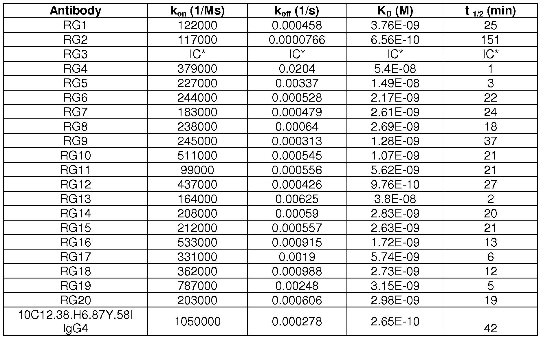


Claims
Priority Applications (16)
| Application Number | Priority Date | Filing Date | Title |
|---|---|---|---|
| EP20167906.5A EP3783023A1 (en) | 2014-11-10 | 2015-11-10 | Anti-interleukin-33 antibodies and uses thereof |
| EP15797767.9A EP3218403B1 (en) | 2014-11-10 | 2015-11-10 | Anti-interleukin-33 antibodies and uses thereof |
| SG11201703767XA SG11201703767XA (en) | 2014-11-10 | 2015-11-10 | Anti-interleukin-33 antibodies and uses thereof |
| MX2017006094A MX2017006094A (en) | 2014-11-10 | 2015-11-10 | Anti-interleukin-33 antibodies and uses thereof. |
| KR1020177012891A KR20170080604A (en) | 2014-11-10 | 2015-11-10 | Anti-interleukin-33 antibodies and uses thereof |
| AU2015346460A AU2015346460A1 (en) | 2014-11-10 | 2015-11-10 | Anti-interleukin-33 antibodies and uses thereof |
| CN201580072740.9A CN107172879B (en) | 2014-11-10 | 2015-11-10 | Anti-interleukin-33 antibodies and uses thereof |
| JP2017525046A JP6929771B2 (en) | 2014-11-10 | 2015-11-10 | Anti-interleukin-33 antibody and its use |
| CA2960297A CA2960297A1 (en) | 2014-11-10 | 2015-11-10 | Anti-interleukin-33 antibodies and uses thereof |
| CR20170240A CR20170240A (en) | 2014-11-10 | 2015-11-10 | ANTI-INTERLEUCINA-33 ANTIBODIES AND THEIR USES |
| EA201791029A EA201791029A1 (en) | 2014-11-10 | 2015-11-10 | ANTIBODIES AGAINST INTERLEUKIN-33 AND THEIR APPLICATION |
| BR112017009728-1A BR112017009728A2 (en) | 2014-11-10 | 2015-11-10 | isolated antibodies, isolated nucleic acid, vector, host cell, method for producing antibody, composition, uses of an antibody and methods of treating a disorder |
| IL251248A IL251248A0 (en) | 2014-11-10 | 2017-03-19 | Anti-interleukin-33 antibodies and uses thereof |
| PH12017500861A PH12017500861A1 (en) | 2014-11-10 | 2017-05-09 | Anti-interleukin-33 antibodies and uses thereof |
| CONC2017/0005650A CO2017005650A2 (en) | 2014-11-10 | 2017-06-07 | Anti-interleukin-33 antibodies |
| HK18102986.2A HK1243440A1 (en) | 2014-11-10 | 2018-03-01 | Anti-interleukin-33 antibodies and uses thereof |
Applications Claiming Priority (4)
| Application Number | Priority Date | Filing Date | Title |
|---|---|---|---|
| US201462077876P | 2014-11-10 | 2014-11-10 | |
| US62/077,876 | 2014-11-10 | ||
| US201562165732P | 2015-05-22 | 2015-05-22 | |
| US62/165,732 | 2015-05-22 |
Publications (2)
| Publication Number | Publication Date |
|---|---|
| WO2016077381A1 true WO2016077381A1 (en) | 2016-05-19 |
| WO2016077381A8 WO2016077381A8 (en) | 2016-06-30 |
Family
ID=54608970
Family Applications (1)
| Application Number | Title | Priority Date | Filing Date |
|---|---|---|---|
| PCT/US2015/060008 WO2016077381A1 (en) | 2014-11-10 | 2015-11-10 | Anti-interleukin-33 antibodies and uses thereof |
Country Status (19)
| Country | Link |
|---|---|
| US (3) | US10093730B2 (en) |
| EP (2) | EP3218403B1 (en) |
| JP (1) | JP6929771B2 (en) |
| KR (1) | KR20170080604A (en) |
| CN (1) | CN107172879B (en) |
| AU (1) | AU2015346460A1 (en) |
| BR (1) | BR112017009728A2 (en) |
| CA (1) | CA2960297A1 (en) |
| CO (1) | CO2017005650A2 (en) |
| CR (1) | CR20170240A (en) |
| EA (1) | EA201791029A1 (en) |
| HK (1) | HK1243440A1 (en) |
| IL (1) | IL251248A0 (en) |
| MX (1) | MX2017006094A (en) |
| PE (1) | PE20170910A1 (en) |
| PH (1) | PH12017500861A1 (en) |
| SG (1) | SG11201703767XA (en) |
| TW (1) | TWI705976B (en) |
| WO (1) | WO2016077381A1 (en) |
Cited By (23)
| Publication number | Priority date | Publication date | Assignee | Title |
|---|---|---|---|---|
| WO2017187307A1 (en) | 2016-04-27 | 2017-11-02 | Pfizer Inc. | Anti-il-33 antibodies, compositions, methods and uses thereof |
| WO2018081075A1 (en) * | 2016-10-28 | 2018-05-03 | Eli Lilly And Company | Anti-il-33 antibodies and uses thereof |
| WO2018102597A1 (en) | 2016-12-01 | 2018-06-07 | Regeneron Pharmaceuticals, Inc. | Methods of treating inflammatory conditions |
| US10059764B2 (en) | 2014-01-10 | 2018-08-28 | Anaptysbio, Inc. | Antibodies directed against interleukin-33 (IL-33) and methods of making and using |
| US10072075B2 (en) | 2015-09-23 | 2018-09-11 | Genentech, Inc. | Optimized variants of anti-VEGF antibodies and methods of treatment thereof by reducing or inhibiting angiogenesis |
| WO2018190990A1 (en) | 2017-04-13 | 2018-10-18 | Regeneron Pharmaceuticals, Inc. | Treatment and inhibition of inflammatory lung diseases in patients having risk alleles in the genes encoding il33 and il1rl1 |
| WO2019157358A1 (en) | 2018-02-09 | 2019-08-15 | Genentech, Inc. | Therapeutic and diagnostic methods for mast cell-mediated inflammatory diseases |
| WO2019199910A1 (en) | 2018-04-11 | 2019-10-17 | Regeneron Pharmaceuticals, Inc. | Methods for quantifying il-33 |
| WO2020037154A1 (en) | 2018-08-17 | 2020-02-20 | 23Andme, Inc. | Anti-il1rap antibodies and methods of use thereof |
| WO2020096959A1 (en) | 2018-11-05 | 2020-05-14 | Genentech, Inc. | Methods of producing two chain proteins in prokaryotic host cells |
| EP3570865A4 (en) * | 2016-09-02 | 2020-05-20 | 180 Therapeutics LP | Method of treating localized fibrotic disorders using an il-33/tnf bispecific antibody |
| US10723795B2 (en) | 2014-11-10 | 2020-07-28 | Genentech, Inc. | Anti-interleukin-33 antibodies and uses thereof |
| WO2021089563A1 (en) | 2019-11-04 | 2021-05-14 | Medimmune Limited | Methods of using il-33 antagonists |
| WO2021089559A1 (en) | 2019-11-04 | 2021-05-14 | Medimmune Limited | Anti il-33 therapeutic agent fpr treating renal disorders |
| WO2021183849A1 (en) | 2020-03-13 | 2021-09-16 | Genentech, Inc. | Anti-interleukin-33 antibodies and uses thereof |
| WO2021180858A1 (en) | 2020-03-13 | 2021-09-16 | Medimmune Limited | Therapeutic methods for the treatment of subjects with risk alelles in il33 |
| WO2021204707A1 (en) | 2020-04-06 | 2021-10-14 | Medimmune Limited | Treating acute respiratory distress syndrome with il-33 axis binding antagonists |
| WO2021228760A1 (en) | 2020-05-11 | 2021-11-18 | Medimmune Limited | Formulations of anti-il-33 antibodies |
| WO2023086807A1 (en) | 2021-11-10 | 2023-05-19 | Genentech, Inc. | Anti-interleukin-33 antibodies and uses thereof |
| US11708608B2 (en) | 2014-11-10 | 2023-07-25 | Genentech, Inc. | Therapeutic and diagnostic methods for IL-33-mediated disorders |
| WO2023166420A1 (en) * | 2022-03-03 | 2023-09-07 | Pfizer Inc. | Multispecific antibodies and uses thereof |
| WO2023180503A1 (en) | 2022-03-25 | 2023-09-28 | Medimmune Limited | Methods for reducing respiratory infections |
| WO2024038185A1 (en) | 2022-08-19 | 2024-02-22 | Medimmune Limited | Method of selecting patients for treatment with an il-33 axis antagonist |
Families Citing this family (9)
| Publication number | Priority date | Publication date | Assignee | Title |
|---|---|---|---|---|
| WO2013188316A1 (en) * | 2012-06-11 | 2013-12-19 | Avalanche Biotechnologies, Inc. | Optical regulation of gene expression in the retina |
| EP3506920A4 (en) * | 2016-09-02 | 2020-05-27 | 180 Therapeutics LP | Method of treating systemic fibrotic disorders using an il-33/tnf bispecific antibody |
| UA126574C2 (en) | 2017-02-10 | 2022-11-02 | Дженентек, Інк. | Anti-tryptase antibodies, compositions thereof, and uses thereof |
| CN110997725A (en) | 2017-06-12 | 2020-04-10 | 蓝鳍生物医药公司 | anti-IL 1RAP antibodies and antibody drug conjugates |
| CN108795934B (en) * | 2018-05-23 | 2022-06-21 | 中国农业科学院上海兽医研究所(中国动物卫生与流行病学中心上海分中心) | SiRNA of schistosoma japonicum SjELAV-like 2 gene and application thereof |
| CN110551745A (en) * | 2018-05-31 | 2019-12-10 | 康码(上海)生物科技有限公司 | Multiple histidine sequence tag and application thereof in protein expression and purification |
| CN111378037B (en) * | 2020-06-01 | 2020-09-01 | 南京诺艾新生物技术有限公司 | anti-hIL-33 humanized monoclonal antibody and application thereof |
| EP4263600A1 (en) | 2020-12-18 | 2023-10-25 | Century Therapeutics, Inc. | Chimeric antigen receptor systems with adaptable receptor specificity |
| US20230295276A1 (en) * | 2022-03-18 | 2023-09-21 | Novascope Biochips Inc. | Antibody for porcine reproductive and respiratory syndrome virus and uses thereof |
Citations (131)
| Publication number | Priority date | Publication date | Assignee | Title |
|---|---|---|---|---|
| US4676980A (en) | 1985-09-23 | 1987-06-30 | The United States Of America As Represented By The Secretary Of The Department Of Health And Human Services | Target specific cross-linked heteroantibodies |
| US4737456A (en) | 1985-05-09 | 1988-04-12 | Syntex (U.S.A.) Inc. | Reducing interference in ligand-receptor binding assays |
| US4816567A (en) | 1983-04-08 | 1989-03-28 | Genentech, Inc. | Recombinant immunoglobin preparations |
| WO1989006692A1 (en) | 1988-01-12 | 1989-07-27 | Genentech, Inc. | Method of treating tumor cells by inhibiting growth factor receptor function |
| EP0404097A2 (en) | 1989-06-22 | 1990-12-27 | BEHRINGWERKE Aktiengesellschaft | Bispecific and oligospecific, mono- and oligovalent receptors, production and applications thereof |
| WO1993001161A1 (en) | 1991-07-11 | 1993-01-21 | Pfizer Limited | Process for preparing sertraline intermediates |
| US5208020A (en) | 1989-10-25 | 1993-05-04 | Immunogen Inc. | Cytotoxic agents comprising maytansinoids and their therapeutic use |
| WO1993008829A1 (en) | 1991-11-04 | 1993-05-13 | The Regents Of The University Of California | Compositions that mediate killing of hiv-infected cells |
| WO1993011161A1 (en) | 1991-11-25 | 1993-06-10 | Enzon, Inc. | Multivalent antigen-binding proteins |
| WO1993016185A2 (en) | 1992-02-06 | 1993-08-19 | Creative Biomolecules, Inc. | Biosynthetic binding protein for cancer marker |
| WO1994010202A1 (en) | 1992-10-28 | 1994-05-11 | Genentech, Inc. | Vascular endothelial cell growth factor antagonists |
| WO1994011026A2 (en) | 1992-11-13 | 1994-05-26 | Idec Pharmaceuticals Corporation | Therapeutic application of chimeric and radiolabeled antibodies to human b lymphocyte restricted differentiation antigen for treatment of b cell lymphoma |
| WO1994029351A2 (en) | 1993-06-16 | 1994-12-22 | Celltech Limited | Antibodies |
| WO1995027062A1 (en) | 1994-04-04 | 1995-10-12 | Genentech, Inc. | Agonist antibodies against the flk2/flt3 receptor and uses thereof |
| US5500362A (en) | 1987-01-08 | 1996-03-19 | Xoma Corporation | Chimeric antibody with specificity to human B cell surface antigen |
| WO1996027011A1 (en) | 1995-03-01 | 1996-09-06 | Genentech, Inc. | A method for making heteromultimeric polypeptides |
| EP0425235B1 (en) | 1989-10-25 | 1996-09-25 | Immunogen Inc | Cytotoxic agents comprising maytansinoids and their therapeutic use |
| WO1996030046A1 (en) | 1995-03-30 | 1996-10-03 | Genentech, Inc. | Vascular endothelial cell growth factor antagonists |
| US5571894A (en) | 1991-02-05 | 1996-11-05 | Ciba-Geigy Corporation | Recombinant antibodies specific for a growth factor receptor |
| US5587458A (en) | 1991-10-07 | 1996-12-24 | Aronex Pharmaceuticals, Inc. | Anti-erbB-2 antibodies, combinations thereof, and therapeutic and diagnostic uses thereof |
| US5624821A (en) | 1987-03-18 | 1997-04-29 | Scotgen Biopharmaceuticals Incorporated | Antibodies with altered effector functions |
| US5635483A (en) | 1992-12-03 | 1997-06-03 | Arizona Board Of Regents Acting On Behalf Of Arizona State University | Tumor inhibiting tetrapeptide bearing modified phenethyl amides |
| US5641870A (en) | 1995-04-20 | 1997-06-24 | Genentech, Inc. | Low pH hydrophobic interaction chromatography for antibody purification |
| US5648237A (en) | 1991-09-19 | 1997-07-15 | Genentech, Inc. | Expression of functional antibody fragments |
| WO1997030087A1 (en) | 1996-02-16 | 1997-08-21 | Glaxo Group Limited | Preparation of glycosylated antibodies |
| US5712374A (en) | 1995-06-07 | 1998-01-27 | American Cyanamid Company | Method for the preparation of substantiallly monomeric calicheamicin derivative/carrier conjugates |
| US5714586A (en) | 1995-06-07 | 1998-02-03 | American Cyanamid Company | Methods for the preparation of monomeric calicheamicin derivative/carrier conjugates |
| US5739116A (en) | 1994-06-03 | 1998-04-14 | American Cyanamid Company | Enediyne derivatives useful for the synthesis of conjugates of methyltrithio antitumor agents |
| US5750373A (en) | 1990-12-03 | 1998-05-12 | Genentech, Inc. | Enrichment method for variant proteins having altered binding properties, M13 phagemids, and growth hormone variants |
| US5770429A (en) | 1990-08-29 | 1998-06-23 | Genpharm International, Inc. | Transgenic non-human animals capable of producing heterologous antibodies |
| US5770701A (en) | 1987-10-30 | 1998-06-23 | American Cyanamid Company | Process for preparing targeted forms of methyltrithio antitumor agents |
| US5770710A (en) | 1987-10-30 | 1998-06-23 | American Cyanamid Company | Antitumor and antibacterial substituted disulfide derivatives prepared from compounds possessing a methlytrithio group |
| US5780588A (en) | 1993-01-26 | 1998-07-14 | Arizona Board Of Regents | Elucidation and synthesis of selected pentapeptides |
| US5789199A (en) | 1994-11-03 | 1998-08-04 | Genentech, Inc. | Process for bacterial production of polypeptides |
| US5821337A (en) | 1991-06-14 | 1998-10-13 | Genentech, Inc. | Immunoglobulin variants |
| WO1998045331A2 (en) | 1997-04-07 | 1998-10-15 | Genentech, Inc. | Anti-vegf antibodies |
| WO1998045332A2 (en) | 1997-04-07 | 1998-10-15 | Genentech, Inc. | Humanized antibodies and methods for forming humanized antibodies |
| WO1998050431A2 (en) | 1997-05-02 | 1998-11-12 | Genentech, Inc. | A method for making multispecific antibodies having heteromultimeric and common components |
| US5840523A (en) | 1995-03-01 | 1998-11-24 | Genetech, Inc. | Methods and compositions for secretion of heterologous polypeptides |
| WO1998058964A1 (en) | 1997-06-24 | 1998-12-30 | Genentech, Inc. | Methods and compositions for galactosylated glycoproteins |
| US5869046A (en) | 1995-04-14 | 1999-02-09 | Genentech, Inc. | Altered polypeptides with increased half-life |
| WO1999022764A1 (en) | 1997-10-31 | 1999-05-14 | Genentech, Inc. | Methods and compositions comprising glycoprotein glycoforms |
| US5959177A (en) | 1989-10-27 | 1999-09-28 | The Scripps Research Institute | Transgenic plants expressing assembled secretory antibodies |
| WO1999051642A1 (en) | 1998-04-02 | 1999-10-14 | Genentech, Inc. | Antibody variants and fragments thereof |
| US6040498A (en) | 1998-08-11 | 2000-03-21 | North Caroline State University | Genetically engineered duckweed |
| US6054297A (en) | 1991-06-14 | 2000-04-25 | Genentech, Inc. | Humanized antibodies and methods for making them |
| US6075181A (en) | 1990-01-12 | 2000-06-13 | Abgenix, Inc. | Human antibodies derived from immunized xenomice |
| WO2000061739A1 (en) | 1999-04-09 | 2000-10-19 | Kyowa Hakko Kogyo Co., Ltd. | Method for controlling the activity of immunologically functional molecule |
| US6150584A (en) | 1990-01-12 | 2000-11-21 | Abgenix, Inc. | Human antibodies derived from immunized xenomice |
| US6171586B1 (en) | 1997-06-13 | 2001-01-09 | Genentech, Inc. | Antibody formulation |
| US6194551B1 (en) | 1998-04-02 | 2001-02-27 | Genentech, Inc. | Polypeptide variants |
| WO2001029246A1 (en) | 1999-10-19 | 2001-04-26 | Kyowa Hakko Kogyo Co., Ltd. | Process for producing polypeptide |
| US6248516B1 (en) | 1988-11-11 | 2001-06-19 | Medical Research Council | Single domain ligands, receptors comprising said ligands methods for their production, and use of said ligands and receptors |
| US6267958B1 (en) | 1995-07-27 | 2001-07-31 | Genentech, Inc. | Protein formulation |
| WO2001070818A1 (en) | 2000-03-23 | 2001-09-27 | Tanox, Inc. | Anti-c2/c2a inhibitors of complement activation |
| WO2002031140A1 (en) | 2000-10-06 | 2002-04-18 | Kyowa Hakko Kogyo Co., Ltd. | Cells producing antibody compositions |
| US20020081293A1 (en) | 1998-02-20 | 2002-06-27 | Tanox, Inc., Delaware Corporation | Inhibitors of complement activation |
| US6420548B1 (en) | 1999-10-04 | 2002-07-16 | Medicago Inc. | Method for regulating transcription of foreign genes |
| US20020164328A1 (en) | 2000-10-06 | 2002-11-07 | Toyohide Shinkawa | Process for purifying antibody |
| WO2003011878A2 (en) | 2001-08-03 | 2003-02-13 | Glycart Biotechnology Ag | Antibody glycosylation variants having increased antibody-dependent cellular cytotoxicity |
| US20030115614A1 (en) | 2000-10-06 | 2003-06-19 | Yutaka Kanda | Antibody composition-producing cell |
| US6582959B2 (en) | 1991-03-29 | 2003-06-24 | Genentech, Inc. | Antibodies to vascular endothelial cell growth factor |
| US6602684B1 (en) | 1998-04-20 | 2003-08-05 | Glycart Biotechnology Ag | Glycosylation engineering of antibodies for improving antibody-dependent cellular cytotoxicity |
| US20030157108A1 (en) | 2001-10-25 | 2003-08-21 | Genentech, Inc. | Glycoprotein compositions |
| US6630579B2 (en) | 1999-12-29 | 2003-10-07 | Immunogen Inc. | Cytotoxic agents comprising modified doxorubicins and daunorubicins and their therapeutic use |
| US20030190317A1 (en) | 1997-04-07 | 2003-10-09 | Genentech, Inc. | Anti-VEGF antibodies |
| WO2003085119A1 (en) | 2002-04-09 | 2003-10-16 | Kyowa Hakko Kogyo Co., Ltd. | METHOD OF ENHANCING ACTIVITY OF ANTIBODY COMPOSITION OF BINDING TO FcϜ RECEPTOR IIIa |
| WO2003085107A1 (en) | 2002-04-09 | 2003-10-16 | Kyowa Hakko Kogyo Co., Ltd. | Cells with modified genome |
| WO2003084570A1 (en) | 2002-04-09 | 2003-10-16 | Kyowa Hakko Kogyo Co., Ltd. | DRUG CONTAINING ANTIBODY COMPOSITION APPROPRIATE FOR PATIENT SUFFERING FROM FcϜRIIIa POLYMORPHISM |
| US20030206899A1 (en) | 1991-03-29 | 2003-11-06 | Genentech, Inc. | Vascular endothelial cell growth factor antagonists |
| US6703020B1 (en) | 1999-04-28 | 2004-03-09 | Board Of Regents, The University Of Texas System | Antibody conjugate methods for selectively inhibiting VEGF |
| US20040093621A1 (en) | 2001-12-25 | 2004-05-13 | Kyowa Hakko Kogyo Co., Ltd | Antibody composition which specifically binds to CD20 |
| US6737056B1 (en) | 1999-01-15 | 2004-05-18 | Genentech, Inc. | Polypeptide variants with altered effector function |
| US20040109865A1 (en) | 2002-04-09 | 2004-06-10 | Kyowa Hakko Kogyo Co., Ltd. | Antibody composition-containing medicament |
| US20040110282A1 (en) | 2002-04-09 | 2004-06-10 | Kyowa Hakko Kogyo Co., Ltd. | Cells in which activity of the protein involved in transportation of GDP-fucose is reduced or lost |
| US20040132140A1 (en) | 2002-04-09 | 2004-07-08 | Kyowa Hakko Kogyo Co., Ltd. | Production process for antibody composition |
| WO2004056312A2 (en) | 2002-12-16 | 2004-07-08 | Genentech, Inc. | Immunoglobulin variants and uses thereof |
| US20050014934A1 (en) | 2002-10-15 | 2005-01-20 | Hinton Paul R. | Alteration of FcRn binding affinities or serum half-lives of antibodies by mutagenesis |
| WO2005012359A2 (en) | 2003-08-01 | 2005-02-10 | Genentech, Inc. | Anti-vegf antibodies |
| US20050079574A1 (en) | 2003-01-16 | 2005-04-14 | Genentech, Inc. | Synthetic antibody phage libraries |
| WO2005035586A1 (en) | 2003-10-08 | 2005-04-21 | Kyowa Hakko Kogyo Co., Ltd. | Fused protein composition |
| WO2005035778A1 (en) | 2003-10-09 | 2005-04-21 | Kyowa Hakko Kogyo Co., Ltd. | PROCESS FOR PRODUCING ANTIBODY COMPOSITION BY USING RNA INHIBITING THE FUNCTION OF α1,6-FUCOSYLTRANSFERASE |
| US6884879B1 (en) | 1997-04-07 | 2005-04-26 | Genentech, Inc. | Anti-VEGF antibodies |
| WO2005044853A2 (en) | 2003-11-01 | 2005-05-19 | Genentech, Inc. | Anti-vegf antibodies |
| US20050119455A1 (en) | 2002-06-03 | 2005-06-02 | Genentech, Inc. | Synthetic antibody phage libraries |
| US20050123546A1 (en) | 2003-11-05 | 2005-06-09 | Glycart Biotechnology Ag | Antigen binding molecules with increased Fc receptor binding affinity and effector function |
| WO2005053742A1 (en) | 2003-12-04 | 2005-06-16 | Kyowa Hakko Kogyo Co., Ltd. | Medicine containing antibody composition |
| WO2005062967A2 (en) | 2003-12-23 | 2005-07-14 | Tanox, Inc. | Novel anti-il 13 antibodies and uses thereof |
| US20050186208A1 (en) | 2003-05-30 | 2005-08-25 | Genentech, Inc. | Treatment with anti-VEGF antibodies |
| WO2005079844A2 (en) * | 2004-02-17 | 2005-09-01 | Schering Corporation | Use for interleukin-33 (il33) and the il-33 receptor complex |
| WO2005100402A1 (en) | 2004-04-13 | 2005-10-27 | F.Hoffmann-La Roche Ag | Anti-p-selectin antibodies |
| US20050260186A1 (en) | 2003-03-05 | 2005-11-24 | Halozyme, Inc. | Soluble glycosaminoglycanases and methods of preparing and using soluble glycosaminoglycanases |
| US20050266000A1 (en) | 2004-04-09 | 2005-12-01 | Genentech, Inc. | Variable domain library and uses |
| US6982321B2 (en) | 1986-03-27 | 2006-01-03 | Medical Research Council | Altered antibodies |
| US20060009360A1 (en) | 2004-06-25 | 2006-01-12 | Robert Pifer | New adjuvant composition |
| US20060025576A1 (en) | 2000-04-11 | 2006-02-02 | Genentech, Inc. | Multivalent antibodies and uses therefor |
| WO2006029879A2 (en) | 2004-09-17 | 2006-03-23 | F.Hoffmann-La Roche Ag | Anti-ox40l antibodies |
| WO2006044908A2 (en) | 2004-10-20 | 2006-04-27 | Genentech, Inc. | Antibody formulation in histidine-acetate buffer |
| US7041870B2 (en) | 2000-11-30 | 2006-05-09 | Medarex, Inc. | Transgenic transchromosomal rodents for making human antibodies |
| US20060104968A1 (en) | 2003-03-05 | 2006-05-18 | Halozyme, Inc. | Soluble glycosaminoglycanases and methods of preparing and using soluble glycosaminogly ycanases |
| US7087409B2 (en) | 1997-12-05 | 2006-08-08 | The Scripps Research Institute | Humanization of murine antibody |
| US7125978B1 (en) | 1999-10-04 | 2006-10-24 | Medicago Inc. | Promoter for regulating expression of foreign genes |
| US7189826B2 (en) | 1997-11-24 | 2007-03-13 | Institute For Human Genetics And Biochemistry | Monoclonal human natural antibodies |
| US20070061900A1 (en) | 2000-10-31 | 2007-03-15 | Murphy Andrew J | Methods of modifying eukaryotic cells |
| WO2007056227A2 (en) | 2005-11-04 | 2007-05-18 | Genentech, Inc. | Use of complement pathway inhibitors to treat ocular diseases |
| US20070117126A1 (en) | 1999-12-15 | 2007-05-24 | Genentech, Inc. | Shotgun scanning |
| US20070160598A1 (en) | 2005-11-07 | 2007-07-12 | Dennis Mark S | Binding polypeptides with diversified and consensus vh/vl hypervariable sequences |
| US20070237764A1 (en) | 2005-12-02 | 2007-10-11 | Genentech, Inc. | Binding polypeptides with restricted diversity sequences |
| US20070292936A1 (en) | 2006-05-09 | 2007-12-20 | Genentech, Inc. | Binding polypeptides with optimized scaffolds |
| US20080069820A1 (en) | 2006-08-30 | 2008-03-20 | Genentech, Inc. | Multispecific antibodies |
| US7371826B2 (en) | 1999-01-15 | 2008-05-13 | Genentech, Inc. | Polypeptide variants with altered effector function |
| WO2008077546A1 (en) | 2006-12-22 | 2008-07-03 | F. Hoffmann-La Roche Ag | Antibodies against insulin-like growth factor i receptor and uses thereof |
| US20090002360A1 (en) | 2007-05-25 | 2009-01-01 | Innolux Display Corp. | Liquid crystal display device and method for driving same |
| US7498298B2 (en) | 2003-11-06 | 2009-03-03 | Seattle Genetics, Inc. | Monomethylvaline compounds capable of conjugation to ligands |
| US7521541B2 (en) | 2004-09-23 | 2009-04-21 | Genetech Inc. | Cysteine engineered antibodies and conjugates |
| US7527791B2 (en) | 2004-03-31 | 2009-05-05 | Genentech, Inc. | Humanized anti-TGF-beta antibodies |
| US20090182127A1 (en) | 2006-06-22 | 2009-07-16 | Novo Nordisk A/S | Production of Bispecific Antibodies |
| WO2009089004A1 (en) | 2008-01-07 | 2009-07-16 | Amgen Inc. | Method for making antibody fc-heterodimeric molecules using electrostatic steering effects |
| WO2009155724A2 (en) | 2008-06-25 | 2009-12-30 | Esbatech, An Alcon Biomedical Research Unit Llc | Stable and soluble antibodies inhibiting vegf |
| WO2011031600A1 (en) | 2009-09-10 | 2011-03-17 | Schering Corporation | Use of il-33 antagonists to treat fibrotic disease |
| US20110236388A1 (en) | 2010-03-26 | 2011-09-29 | Monika Baehner | Bispecific, bivalent anti-vegf/anti-ang-2 antibodies |
| US20110287009A1 (en) | 2010-04-23 | 2011-11-24 | Genentech, Inc. | Production of Heteromultimeric Proteins |
| US8187569B2 (en) | 2006-02-14 | 2012-05-29 | Exxonmobil Chemical Patents Inc. | Process for manufacturing molecular sieve of MFS framework type and its use |
| US8187596B1 (en) | 2006-07-20 | 2012-05-29 | Schering Corporation | Method of treating asthma or allergy by administering an IL-33 receptor antibody |
| WO2013055998A1 (en) | 2011-10-14 | 2013-04-18 | Genentech, Inc. | ANTI-HtrA1 ANTIBODIES AND METHODS OF USE |
| WO2013165894A2 (en) | 2012-04-30 | 2013-11-07 | Janssen Biotech, Inc. | St2l antagonists and methods of use |
| WO2013173761A2 (en) | 2012-05-18 | 2013-11-21 | Amgen Inc. | St2 antigen binding proteins |
| WO2014152195A1 (en) | 2013-03-15 | 2014-09-25 | Regeneron Pharmaceuticals, Inc. | Il-33 antagonists and uses thereof |
| WO2014164959A2 (en) | 2013-03-13 | 2014-10-09 | Regeneron Pharmaceuticals, Inc. | Anti-il-33 antibodies and uses thereof |
| WO2015099175A1 (en) | 2013-12-26 | 2015-07-02 | 田辺三菱製薬株式会社 | Human anti-il-33 neutralizing monoclonal antibody |
| WO2015106080A2 (en) | 2014-01-10 | 2015-07-16 | Anaptysbio, Inc. | Antibodies directed against interleukin-33 (il-33) |
Family Cites Families (61)
| Publication number | Priority date | Publication date | Assignee | Title |
|---|---|---|---|---|
| US6066322A (en) | 1995-03-03 | 2000-05-23 | Millennium Pharmaceuticals, Inc. | Methods for the treatment of immune disorders |
| US6323334B1 (en) | 1999-09-24 | 2001-11-27 | Millennium Pharmaceuticals, Inc. | Nucleic acid molecules encoding a 103 gene product and uses therefor |
| AU2001218871A1 (en) | 2000-03-21 | 2001-10-03 | Takao Arai | Monoclonal antibody and method and kit for the immunoassay of soluble human st2 with the use of the same |
| WO2004056868A2 (en) | 2002-12-19 | 2004-07-08 | Endocube Sas | Nf-hev compositions and methods of use |
| EP3255432B1 (en) | 2006-04-24 | 2019-01-23 | Critical Care Diagnostics, Inc. | Predicting mortality and detecting severe disease |
| US20090305265A1 (en) | 2006-04-27 | 2009-12-10 | Critical Care Diagnostics, Inc. | Interleukin-33 (il-33) for the diagnosis and prognosis of cardiovascular disease |
| HUE025058T2 (en) | 2006-05-02 | 2016-01-28 | Critical Care Diagnostics Inc | Differential diagnosis between pulmonary and cardiovascular disease |
| US8147817B2 (en) | 2006-05-04 | 2012-04-03 | The Brigham And Women's Hospital, Inc. | IL-33 in the treatment and diagnosis of diseases and disorders |
| WO2007140205A2 (en) | 2006-05-24 | 2007-12-06 | Biogen Idec Ma Inc. | Methods of treating fibrosis |
| SE532250C2 (en) | 2006-11-28 | 2009-11-24 | Theravac Pharmaceuticals Ab | New formulations of IL-33 for the treatment of inflammatory conditions with a strong TH2 component by vaccination |
| US8119771B2 (en) | 2007-04-26 | 2012-02-21 | The Provost, Fellow and Scholars of the College of the Holy and Undivided Trinity of Queen Elizabeth, Near Dublin | Products for altering IL-33 activity and methods thereof |
| EP2069784A4 (en) | 2007-05-18 | 2009-11-18 | Medimmune Llc | Il-33 in inflammatory disease |
| CA2703335A1 (en) | 2007-10-26 | 2009-04-30 | Arturo Zychlinsky | Inhibitors of caspase i-dependent cytokines in the treatment of neurodegenerative disorders |
| US20110045501A1 (en) | 2008-01-08 | 2011-02-24 | Irene Bosch | St2-based dengue fever diagnostic |
| WO2009120905A2 (en) | 2008-03-26 | 2009-10-01 | Cellerant Therapeutics, Inc. | Immunoglobulin and/or toll-like receptor proteins associated with myelogenous haematological proliferative disorders and uses thereof |
| US20110165063A1 (en) | 2009-01-29 | 2011-07-07 | Abbott Laboratories | Il-1 binding proteins |
| UA102722C2 (en) | 2009-01-29 | 2013-08-12 | Эббви Инк. | Il-1 binding proteins |
| US8835610B2 (en) | 2009-03-05 | 2014-09-16 | Abbvie Inc. | IL-17 binding proteins |
| JP5428422B2 (en) | 2009-03-17 | 2014-02-26 | ヤマハ株式会社 | Electronic musical instruments |
| WO2011047266A1 (en) | 2009-10-15 | 2011-04-21 | Abbott Laboratories | Il-1 binding proteins |
| HUE033063T2 (en) | 2010-05-14 | 2017-11-28 | Abbvie Inc | Il-1 binding proteins |
| BR112013007041A2 (en) | 2010-10-29 | 2016-09-13 | Hoffmann La Roche | non-human animal, offspring, cell line, tissue, organ explant, or culture thereof, use of inflammatory disease model and methods for screening for anti-inflammatory compounds and for evaluation |
| AU2011343570B2 (en) | 2010-12-16 | 2016-11-03 | Genentech, Inc. | Diagnosis and treatments relating to TH2 inhibition |
| US20120275996A1 (en) | 2010-12-21 | 2012-11-01 | Abbott Laboratories | IL-1 Binding Proteins |
| WO2012103240A2 (en) | 2011-01-25 | 2012-08-02 | Eleven Biotherapeutics, Inc. | Receptor binding agents |
| WO2012113813A1 (en) * | 2011-02-23 | 2012-08-30 | F. Hoffmann-La Roche Ag | Antibodies against human il33r and uses thereof |
| FR2972006B1 (en) | 2011-02-24 | 2016-03-25 | Centre Nat Rech Scient | NOVEL IL-33 SUPERACTIVE FRAGMENTS AND USES THEREOF |
| WO2012145209A2 (en) | 2011-04-21 | 2012-10-26 | Rockefeller University (The) | Anti-inflammatory agents |
| CN106908604B (en) | 2011-07-18 | 2019-01-29 | 重症监护诊断股份有限公司 | The method for treating cardiovascular disease and predicted motion therapy effect |
| BR112014001855A2 (en) | 2011-07-27 | 2017-02-21 | Glaxo Group Ltd | antigen-binding construct and protein, dimer, pharmaceutical composition, polynucleotide sequence, host cell and method for construct production |
| EP2885641B1 (en) | 2012-08-16 | 2017-11-01 | Critical Care Diagnostics, Inc. | Methods for predicting risk of developing hypertension |
| WO2014062621A1 (en) | 2012-10-15 | 2014-04-24 | Stc.Unm | Treatment of autophagy-based disorders and related pharmaceutical compositions, diagnostic and screening assays and kits |
| EP2916858A1 (en) | 2012-11-08 | 2015-09-16 | INSERM (Institut National de la Santé et de la Recherche Médicale) | Method for inducing il-2-free proliferation of gamma delta t cells |
| EP2928916B1 (en) | 2012-12-10 | 2019-03-13 | Vib Vzw | Novel interleukin-33 inhibitors |
| WO2014126277A1 (en) | 2013-02-14 | 2014-08-21 | 건국대학교 산학협력단 | Composition of novel interleukin-33 receptor and binding protein, and use thereof |
| US9867867B2 (en) | 2013-02-22 | 2018-01-16 | Magic Epoch Holdings Limited | IL-33 and treatment of neurodegenerative diseases |
| US9949465B2 (en) | 2013-05-01 | 2018-04-24 | Hyogo College Of Medicine | Atopic dermatitis model animal and use thereof |
| EP3047024B1 (en) | 2013-09-20 | 2019-04-24 | University Of Virginia Patent Foundation | Compositions and methods for treatment of autoimmune and inflammatory diseases and disorders |
| AU2014332352B9 (en) | 2013-10-07 | 2017-05-11 | Inovio Pharmaceuticals, Inc. | Vaccines with Interleukin-33 as an adjuvant |
| SG11201603127WA (en) | 2013-10-23 | 2016-05-30 | Genentech Inc | Methods of diagnosing and treating eosinophilic disorders |
| US20180148489A2 (en) | 2013-11-28 | 2018-05-31 | University Of Manitoba | Idr peptide compositions and use thereof for treatment of th2-dysregulated inflammatory conditions |
| GB201403875D0 (en) | 2014-03-05 | 2014-04-16 | Cantargia Ab | Novel antibodies and uses thereof |
| US20170107300A1 (en) | 2014-03-21 | 2017-04-20 | The Brigham And Women's Hospital, Inc. | Methods and compositions for treatment of immune-related diseases or disorders and/or therapy monitoring |
| CA2946511C (en) | 2014-04-21 | 2023-08-22 | The Childen's Hospital Of Philadelphia | Compositions and methods for treating cytokine-related disorders |
| EP4198059A1 (en) | 2014-05-27 | 2023-06-21 | The University of Queensland | Modulation of cellular stress |
| GB201413913D0 (en) | 2014-08-06 | 2014-09-17 | Cantargia Ab | Novel antibodies and uses thereof |
| US20160145344A1 (en) | 2014-10-20 | 2016-05-26 | University Of Southern California | Murine and human innate lymphoid cells and lung inflammation |
| JP6929771B2 (en) | 2014-11-10 | 2021-09-01 | ジェネンテック, インコーポレイテッド | Anti-interleukin-33 antibody and its use |
| EP3218515B1 (en) | 2014-11-10 | 2023-04-26 | F. Hoffmann-La Roche AG | Therapeutic and diagnostic methods for il-33-mediated disorders |
| US11137397B2 (en) | 2014-11-24 | 2021-10-05 | Albert Einstein College Of Medicine | Peptides for blocking IL1RAP protein-protein interaction and uses thereof for treatment of disease |
| WO2016090250A1 (en) | 2014-12-04 | 2016-06-09 | The University Of North Carolina At Chapel Hill | Compositions and methods for preventing and treating graft versus host disease |
| WO2016122865A1 (en) | 2015-01-30 | 2016-08-04 | Salk Institute For Biological Studies | Compositions and methods for treating age-related diabetes and related disorders |
| WO2016140922A1 (en) | 2015-03-02 | 2016-09-09 | 180 Therapeutics Lp | Method of treating a localized fibrotic disorder using a tnf receptor 2 antagonist |
| JP6991863B2 (en) | 2015-03-03 | 2022-02-03 | カヴァ ヘルスケア インコーポレイテッド | Modulation of cancer immunity by type 2 innate lymphoid cells, interleukin 33, and / or interferon-inducible protein 44 |
| CA2977285A1 (en) | 2015-03-16 | 2016-09-22 | F. Hoffmann-La Roche Ag | Methods of detecting and quantifying il-13 and uses in diagnosing and treating th2-associated diseases |
| EP3733701A1 (en) | 2015-03-31 | 2020-11-04 | MedImmune Limited | A novel il33 form, mutated forms of il33, antibodies, assays and methods of using the same |
| CN108026172B (en) | 2015-06-26 | 2022-04-29 | 赛诺菲生物技术公司 | Monoclonal anti-IL-1 RACP antibodies |
| CN108289930A (en) | 2015-07-10 | 2018-07-17 | 香港科技大学 | Method and composition for treating nervus retrogression and neuro-inflammatory disorders |
| CL2015002153A1 (en) | 2015-07-31 | 2016-01-29 | Pontificia Universidad Católica De Chile | Use of the il-3, il33 and il-12p40 molecular markers to characterize the severity of respiratory infections by respiratory syncytial virus and human metapneumovirus |
| AU2016333903B2 (en) | 2015-10-06 | 2021-07-08 | Regeneron Pharmaceuticals, Inc. | Biomarkers related to interleukin-33 (IL-33)-mediated diseases and uses thereof |
| PE20181952A1 (en) | 2016-04-27 | 2018-12-17 | Pfizer | ANTI-LL-33 ANTIBODIES, COMPOSITIONS, METHODS AND USES OF THE SAME |
-
2015
- 2015-11-10 JP JP2017525046A patent/JP6929771B2/en active Active
- 2015-11-10 EA EA201791029A patent/EA201791029A1/en unknown
- 2015-11-10 CR CR20170240A patent/CR20170240A/en unknown
- 2015-11-10 SG SG11201703767XA patent/SG11201703767XA/en unknown
- 2015-11-10 EP EP15797767.9A patent/EP3218403B1/en active Active
- 2015-11-10 KR KR1020177012891A patent/KR20170080604A/en unknown
- 2015-11-10 AU AU2015346460A patent/AU2015346460A1/en not_active Abandoned
- 2015-11-10 MX MX2017006094A patent/MX2017006094A/en unknown
- 2015-11-10 EP EP20167906.5A patent/EP3783023A1/en not_active Withdrawn
- 2015-11-10 US US14/937,778 patent/US10093730B2/en active Active
- 2015-11-10 CA CA2960297A patent/CA2960297A1/en not_active Abandoned
- 2015-11-10 CN CN201580072740.9A patent/CN107172879B/en active Active
- 2015-11-10 BR BR112017009728-1A patent/BR112017009728A2/en not_active Application Discontinuation
- 2015-11-10 WO PCT/US2015/060008 patent/WO2016077381A1/en active Application Filing
- 2015-11-10 PE PE2017000823A patent/PE20170910A1/en unknown
- 2015-11-10 TW TW104137071A patent/TWI705976B/en not_active IP Right Cessation
-
2017
- 2017-03-19 IL IL251248A patent/IL251248A0/en unknown
- 2017-05-09 PH PH12017500861A patent/PH12017500861A1/en unknown
- 2017-06-07 CO CONC2017/0005650A patent/CO2017005650A2/en unknown
-
2018
- 2018-03-01 HK HK18102986.2A patent/HK1243440A1/en unknown
- 2018-08-31 US US16/119,667 patent/US10723795B2/en active Active
-
2020
- 2020-06-16 US US16/903,300 patent/US11725050B2/en active Active
Patent Citations (156)
| Publication number | Priority date | Publication date | Assignee | Title |
|---|---|---|---|---|
| US4816567A (en) | 1983-04-08 | 1989-03-28 | Genentech, Inc. | Recombinant immunoglobin preparations |
| US4737456A (en) | 1985-05-09 | 1988-04-12 | Syntex (U.S.A.) Inc. | Reducing interference in ligand-receptor binding assays |
| US4676980A (en) | 1985-09-23 | 1987-06-30 | The United States Of America As Represented By The Secretary Of The Department Of Health And Human Services | Target specific cross-linked heteroantibodies |
| US6982321B2 (en) | 1986-03-27 | 2006-01-03 | Medical Research Council | Altered antibodies |
| US5500362A (en) | 1987-01-08 | 1996-03-19 | Xoma Corporation | Chimeric antibody with specificity to human B cell surface antigen |
| US5624821A (en) | 1987-03-18 | 1997-04-29 | Scotgen Biopharmaceuticals Incorporated | Antibodies with altered effector functions |
| US5648260A (en) | 1987-03-18 | 1997-07-15 | Scotgen Biopharmaceuticals Incorporated | DNA encoding antibodies with altered effector functions |
| US5770710A (en) | 1987-10-30 | 1998-06-23 | American Cyanamid Company | Antitumor and antibacterial substituted disulfide derivatives prepared from compounds possessing a methlytrithio group |
| US5770701A (en) | 1987-10-30 | 1998-06-23 | American Cyanamid Company | Process for preparing targeted forms of methyltrithio antitumor agents |
| WO1989006692A1 (en) | 1988-01-12 | 1989-07-27 | Genentech, Inc. | Method of treating tumor cells by inhibiting growth factor receptor function |
| US6248516B1 (en) | 1988-11-11 | 2001-06-19 | Medical Research Council | Single domain ligands, receptors comprising said ligands methods for their production, and use of said ligands and receptors |
| EP0404097A2 (en) | 1989-06-22 | 1990-12-27 | BEHRINGWERKE Aktiengesellschaft | Bispecific and oligospecific, mono- and oligovalent receptors, production and applications thereof |
| EP0425235B1 (en) | 1989-10-25 | 1996-09-25 | Immunogen Inc | Cytotoxic agents comprising maytansinoids and their therapeutic use |
| US5416064A (en) | 1989-10-25 | 1995-05-16 | Immunogen, Inc. | Cytotoxic agents comprising maytansinoids and their therapeutic use |
| US5208020A (en) | 1989-10-25 | 1993-05-04 | Immunogen Inc. | Cytotoxic agents comprising maytansinoids and their therapeutic use |
| US5959177A (en) | 1989-10-27 | 1999-09-28 | The Scripps Research Institute | Transgenic plants expressing assembled secretory antibodies |
| US6417429B1 (en) | 1989-10-27 | 2002-07-09 | The Scripps Research Institute | Transgenic plants expressing assembled secretory antibodies |
| US6075181A (en) | 1990-01-12 | 2000-06-13 | Abgenix, Inc. | Human antibodies derived from immunized xenomice |
| US6150584A (en) | 1990-01-12 | 2000-11-21 | Abgenix, Inc. | Human antibodies derived from immunized xenomice |
| US5770429A (en) | 1990-08-29 | 1998-06-23 | Genpharm International, Inc. | Transgenic non-human animals capable of producing heterologous antibodies |
| US5750373A (en) | 1990-12-03 | 1998-05-12 | Genentech, Inc. | Enrichment method for variant proteins having altered binding properties, M13 phagemids, and growth hormone variants |
| US5571894A (en) | 1991-02-05 | 1996-11-05 | Ciba-Geigy Corporation | Recombinant antibodies specific for a growth factor receptor |
| US20030206899A1 (en) | 1991-03-29 | 2003-11-06 | Genentech, Inc. | Vascular endothelial cell growth factor antagonists |
| US20030203409A1 (en) | 1991-03-29 | 2003-10-30 | Genentech, Inc. | Antibodies to vascular endothelial cell growth factor |
| US6582959B2 (en) | 1991-03-29 | 2003-06-24 | Genentech, Inc. | Antibodies to vascular endothelial cell growth factor |
| US6054297A (en) | 1991-06-14 | 2000-04-25 | Genentech, Inc. | Humanized antibodies and methods for making them |
| US5821337A (en) | 1991-06-14 | 1998-10-13 | Genentech, Inc. | Immunoglobulin variants |
| WO1993001161A1 (en) | 1991-07-11 | 1993-01-21 | Pfizer Limited | Process for preparing sertraline intermediates |
| US5648237A (en) | 1991-09-19 | 1997-07-15 | Genentech, Inc. | Expression of functional antibody fragments |
| US5587458A (en) | 1991-10-07 | 1996-12-24 | Aronex Pharmaceuticals, Inc. | Anti-erbB-2 antibodies, combinations thereof, and therapeutic and diagnostic uses thereof |
| WO1993008829A1 (en) | 1991-11-04 | 1993-05-13 | The Regents Of The University Of California | Compositions that mediate killing of hiv-infected cells |
| WO1993011161A1 (en) | 1991-11-25 | 1993-06-10 | Enzon, Inc. | Multivalent antigen-binding proteins |
| WO1993016185A2 (en) | 1992-02-06 | 1993-08-19 | Creative Biomolecules, Inc. | Biosynthetic binding protein for cancer marker |
| WO1994010202A1 (en) | 1992-10-28 | 1994-05-11 | Genentech, Inc. | Vascular endothelial cell growth factor antagonists |
| EP0666868B1 (en) | 1992-10-28 | 2002-04-03 | Genentech, Inc. | Use of anti-VEGF antibodies for the treatment of cancer |
| WO1994011026A2 (en) | 1992-11-13 | 1994-05-26 | Idec Pharmaceuticals Corporation | Therapeutic application of chimeric and radiolabeled antibodies to human b lymphocyte restricted differentiation antigen for treatment of b cell lymphoma |
| US5635483A (en) | 1992-12-03 | 1997-06-03 | Arizona Board Of Regents Acting On Behalf Of Arizona State University | Tumor inhibiting tetrapeptide bearing modified phenethyl amides |
| US5780588A (en) | 1993-01-26 | 1998-07-14 | Arizona Board Of Regents | Elucidation and synthesis of selected pentapeptides |
| WO1994029351A2 (en) | 1993-06-16 | 1994-12-22 | Celltech Limited | Antibodies |
| WO1995027062A1 (en) | 1994-04-04 | 1995-10-12 | Genentech, Inc. | Agonist antibodies against the flk2/flt3 receptor and uses thereof |
| US5767285A (en) | 1994-06-03 | 1998-06-16 | American Cyanamid Company | Linkers useful for the synthesis of conjugates of methyltrithio antitumor agents |
| US5739116A (en) | 1994-06-03 | 1998-04-14 | American Cyanamid Company | Enediyne derivatives useful for the synthesis of conjugates of methyltrithio antitumor agents |
| US5877296A (en) | 1994-06-03 | 1999-03-02 | American Cyanamid Company | Process for preparing conjugates of methyltrithio antitumor agents |
| US5773001A (en) | 1994-06-03 | 1998-06-30 | American Cyanamid Company | Conjugates of methyltrithio antitumor agents and intermediates for their synthesis |
| US5789199A (en) | 1994-11-03 | 1998-08-04 | Genentech, Inc. | Process for bacterial production of polypeptides |
| US7642228B2 (en) | 1995-03-01 | 2010-01-05 | Genentech, Inc. | Method for making heteromultimeric polypeptides |
| US8216805B2 (en) | 1995-03-01 | 2012-07-10 | Genentech, Inc. | Knobs and holes heteromeric polypeptides |
| US5840523A (en) | 1995-03-01 | 1998-11-24 | Genetech, Inc. | Methods and compositions for secretion of heterologous polypeptides |
| US7695936B2 (en) | 1995-03-01 | 2010-04-13 | Genentech, Inc. | Knobs and holes heteromeric polypeptides |
| US5807706A (en) | 1995-03-01 | 1998-09-15 | Genentech, Inc. | Method for making heteromultimeric polypeptides |
| US5731168A (en) | 1995-03-01 | 1998-03-24 | Genentech, Inc. | Method for making heteromultimeric polypeptides |
| WO1996027011A1 (en) | 1995-03-01 | 1996-09-06 | Genentech, Inc. | A method for making heteromultimeric polypeptides |
| US5821333A (en) | 1995-03-01 | 1998-10-13 | Genetech, Inc. | Method for making heteromultimeric polypeptides |
| WO1996030046A1 (en) | 1995-03-30 | 1996-10-03 | Genentech, Inc. | Vascular endothelial cell growth factor antagonists |
| US5869046A (en) | 1995-04-14 | 1999-02-09 | Genentech, Inc. | Altered polypeptides with increased half-life |
| US5641870A (en) | 1995-04-20 | 1997-06-24 | Genentech, Inc. | Low pH hydrophobic interaction chromatography for antibody purification |
| US5712374A (en) | 1995-06-07 | 1998-01-27 | American Cyanamid Company | Method for the preparation of substantiallly monomeric calicheamicin derivative/carrier conjugates |
| US5714586A (en) | 1995-06-07 | 1998-02-03 | American Cyanamid Company | Methods for the preparation of monomeric calicheamicin derivative/carrier conjugates |
| US6267958B1 (en) | 1995-07-27 | 2001-07-31 | Genentech, Inc. | Protein formulation |
| WO1997030087A1 (en) | 1996-02-16 | 1997-08-21 | Glaxo Group Limited | Preparation of glycosylated antibodies |
| US6884879B1 (en) | 1997-04-07 | 2005-04-26 | Genentech, Inc. | Anti-VEGF antibodies |
| US20050112126A1 (en) | 1997-04-07 | 2005-05-26 | Genentech, Inc. | Anti-VEGF antibodies |
| US7060269B1 (en) | 1997-04-07 | 2006-06-13 | Genentech, Inc. | Anti-VEGF antibodies |
| WO1998045331A2 (en) | 1997-04-07 | 1998-10-15 | Genentech, Inc. | Anti-vegf antibodies |
| US20030190317A1 (en) | 1997-04-07 | 2003-10-09 | Genentech, Inc. | Anti-VEGF antibodies |
| WO1998045332A2 (en) | 1997-04-07 | 1998-10-15 | Genentech, Inc. | Humanized antibodies and methods for forming humanized antibodies |
| US20070178552A1 (en) | 1997-05-02 | 2007-08-02 | Genentech, Inc. | Method for Making Multispecific Antibodies Having Heteromultimeric and Common Components |
| WO1998050431A2 (en) | 1997-05-02 | 1998-11-12 | Genentech, Inc. | A method for making multispecific antibodies having heteromultimeric and common components |
| US6171586B1 (en) | 1997-06-13 | 2001-01-09 | Genentech, Inc. | Antibody formulation |
| WO1998058964A1 (en) | 1997-06-24 | 1998-12-30 | Genentech, Inc. | Methods and compositions for galactosylated glycoproteins |
| WO1999022764A1 (en) | 1997-10-31 | 1999-05-14 | Genentech, Inc. | Methods and compositions comprising glycoprotein glycoforms |
| US7189826B2 (en) | 1997-11-24 | 2007-03-13 | Institute For Human Genetics And Biochemistry | Monoclonal human natural antibodies |
| US7087409B2 (en) | 1997-12-05 | 2006-08-08 | The Scripps Research Institute | Humanization of murine antibody |
| US20020081293A1 (en) | 1998-02-20 | 2002-06-27 | Tanox, Inc., Delaware Corporation | Inhibitors of complement activation |
| US6194551B1 (en) | 1998-04-02 | 2001-02-27 | Genentech, Inc. | Polypeptide variants |
| WO1999051642A1 (en) | 1998-04-02 | 1999-10-14 | Genentech, Inc. | Antibody variants and fragments thereof |
| US6602684B1 (en) | 1998-04-20 | 2003-08-05 | Glycart Biotechnology Ag | Glycosylation engineering of antibodies for improving antibody-dependent cellular cytotoxicity |
| US6040498A (en) | 1998-08-11 | 2000-03-21 | North Caroline State University | Genetically engineered duckweed |
| US7371826B2 (en) | 1999-01-15 | 2008-05-13 | Genentech, Inc. | Polypeptide variants with altered effector function |
| US7332581B2 (en) | 1999-01-15 | 2008-02-19 | Genentech, Inc. | Polypeptide variants with altered effector function |
| US6737056B1 (en) | 1999-01-15 | 2004-05-18 | Genentech, Inc. | Polypeptide variants with altered effector function |
| WO2000061739A1 (en) | 1999-04-09 | 2000-10-19 | Kyowa Hakko Kogyo Co., Ltd. | Method for controlling the activity of immunologically functional molecule |
| US6703020B1 (en) | 1999-04-28 | 2004-03-09 | Board Of Regents, The University Of Texas System | Antibody conjugate methods for selectively inhibiting VEGF |
| US6420548B1 (en) | 1999-10-04 | 2002-07-16 | Medicago Inc. | Method for regulating transcription of foreign genes |
| US7125978B1 (en) | 1999-10-04 | 2006-10-24 | Medicago Inc. | Promoter for regulating expression of foreign genes |
| WO2001029246A1 (en) | 1999-10-19 | 2001-04-26 | Kyowa Hakko Kogyo Co., Ltd. | Process for producing polypeptide |
| US20070117126A1 (en) | 1999-12-15 | 2007-05-24 | Genentech, Inc. | Shotgun scanning |
| US6630579B2 (en) | 1999-12-29 | 2003-10-07 | Immunogen Inc. | Cytotoxic agents comprising modified doxorubicins and daunorubicins and their therapeutic use |
| WO2001070818A1 (en) | 2000-03-23 | 2001-09-27 | Tanox, Inc. | Anti-c2/c2a inhibitors of complement activation |
| US20060025576A1 (en) | 2000-04-11 | 2006-02-02 | Genentech, Inc. | Multivalent antibodies and uses therefor |
| WO2002031140A1 (en) | 2000-10-06 | 2002-04-18 | Kyowa Hakko Kogyo Co., Ltd. | Cells producing antibody compositions |
| US20030115614A1 (en) | 2000-10-06 | 2003-06-19 | Yutaka Kanda | Antibody composition-producing cell |
| US20020164328A1 (en) | 2000-10-06 | 2002-11-07 | Toyohide Shinkawa | Process for purifying antibody |
| US20070061900A1 (en) | 2000-10-31 | 2007-03-15 | Murphy Andrew J | Methods of modifying eukaryotic cells |
| US7041870B2 (en) | 2000-11-30 | 2006-05-09 | Medarex, Inc. | Transgenic transchromosomal rodents for making human antibodies |
| WO2003011878A2 (en) | 2001-08-03 | 2003-02-13 | Glycart Biotechnology Ag | Antibody glycosylation variants having increased antibody-dependent cellular cytotoxicity |
| US20030157108A1 (en) | 2001-10-25 | 2003-08-21 | Genentech, Inc. | Glycoprotein compositions |
| US20040093621A1 (en) | 2001-12-25 | 2004-05-13 | Kyowa Hakko Kogyo Co., Ltd | Antibody composition which specifically binds to CD20 |
| US20040110282A1 (en) | 2002-04-09 | 2004-06-10 | Kyowa Hakko Kogyo Co., Ltd. | Cells in which activity of the protein involved in transportation of GDP-fucose is reduced or lost |
| WO2003084570A1 (en) | 2002-04-09 | 2003-10-16 | Kyowa Hakko Kogyo Co., Ltd. | DRUG CONTAINING ANTIBODY COMPOSITION APPROPRIATE FOR PATIENT SUFFERING FROM FcϜRIIIa POLYMORPHISM |
| WO2003085107A1 (en) | 2002-04-09 | 2003-10-16 | Kyowa Hakko Kogyo Co., Ltd. | Cells with modified genome |
| US20040110704A1 (en) | 2002-04-09 | 2004-06-10 | Kyowa Hakko Kogyo Co., Ltd. | Cells of which genome is modified |
| US20040109865A1 (en) | 2002-04-09 | 2004-06-10 | Kyowa Hakko Kogyo Co., Ltd. | Antibody composition-containing medicament |
| WO2003085119A1 (en) | 2002-04-09 | 2003-10-16 | Kyowa Hakko Kogyo Co., Ltd. | METHOD OF ENHANCING ACTIVITY OF ANTIBODY COMPOSITION OF BINDING TO FcϜ RECEPTOR IIIa |
| US20040132140A1 (en) | 2002-04-09 | 2004-07-08 | Kyowa Hakko Kogyo Co., Ltd. | Production process for antibody composition |
| US20050119455A1 (en) | 2002-06-03 | 2005-06-02 | Genentech, Inc. | Synthetic antibody phage libraries |
| US20050014934A1 (en) | 2002-10-15 | 2005-01-20 | Hinton Paul R. | Alteration of FcRn binding affinities or serum half-lives of antibodies by mutagenesis |
| WO2004056312A2 (en) | 2002-12-16 | 2004-07-08 | Genentech, Inc. | Immunoglobulin variants and uses thereof |
| US20050079574A1 (en) | 2003-01-16 | 2005-04-14 | Genentech, Inc. | Synthetic antibody phage libraries |
| US20050260186A1 (en) | 2003-03-05 | 2005-11-24 | Halozyme, Inc. | Soluble glycosaminoglycanases and methods of preparing and using soluble glycosaminoglycanases |
| US20060104968A1 (en) | 2003-03-05 | 2006-05-18 | Halozyme, Inc. | Soluble glycosaminoglycanases and methods of preparing and using soluble glycosaminogly ycanases |
| US20050186208A1 (en) | 2003-05-30 | 2005-08-25 | Genentech, Inc. | Treatment with anti-VEGF antibodies |
| WO2005012359A2 (en) | 2003-08-01 | 2005-02-10 | Genentech, Inc. | Anti-vegf antibodies |
| WO2005035586A1 (en) | 2003-10-08 | 2005-04-21 | Kyowa Hakko Kogyo Co., Ltd. | Fused protein composition |
| WO2005035778A1 (en) | 2003-10-09 | 2005-04-21 | Kyowa Hakko Kogyo Co., Ltd. | PROCESS FOR PRODUCING ANTIBODY COMPOSITION BY USING RNA INHIBITING THE FUNCTION OF α1,6-FUCOSYLTRANSFERASE |
| WO2005044853A2 (en) | 2003-11-01 | 2005-05-19 | Genentech, Inc. | Anti-vegf antibodies |
| US20050123546A1 (en) | 2003-11-05 | 2005-06-09 | Glycart Biotechnology Ag | Antigen binding molecules with increased Fc receptor binding affinity and effector function |
| US7498298B2 (en) | 2003-11-06 | 2009-03-03 | Seattle Genetics, Inc. | Monomethylvaline compounds capable of conjugation to ligands |
| WO2005053742A1 (en) | 2003-12-04 | 2005-06-16 | Kyowa Hakko Kogyo Co., Ltd. | Medicine containing antibody composition |
| WO2005062967A2 (en) | 2003-12-23 | 2005-07-14 | Tanox, Inc. | Novel anti-il 13 antibodies and uses thereof |
| US7674459B2 (en) | 2003-12-23 | 2010-03-09 | Genentech, Inc. | Treatment of cancer with a novel anti-IL13 monoclonal antibody |
| US8067199B2 (en) | 2003-12-23 | 2011-11-29 | Genentech, Inc. | Anti-IL13 antibodies and uses thereof |
| US8734797B2 (en) | 2003-12-23 | 2014-05-27 | Genentech, Inc. | Anti-IL13 antibodies and uses thereof |
| US8088618B2 (en) | 2003-12-23 | 2012-01-03 | Genentech, Inc. | Anti-IL 13 antibodies and uses thereof |
| US8318160B2 (en) | 2003-12-23 | 2012-11-27 | Genentech, Inc. | Treatment of cancer with novel anti-IL13 monoclonal antibodies |
| EP1725261A2 (en) | 2004-02-17 | 2006-11-29 | Schering Corporation | Use for interleukin-33 (il-33) and the il-33 receptor complex |
| WO2005079844A2 (en) * | 2004-02-17 | 2005-09-01 | Schering Corporation | Use for interleukin-33 (il33) and the il-33 receptor complex |
| US7527791B2 (en) | 2004-03-31 | 2009-05-05 | Genentech, Inc. | Humanized anti-TGF-beta antibodies |
| US20050266000A1 (en) | 2004-04-09 | 2005-12-01 | Genentech, Inc. | Variable domain library and uses |
| WO2005100402A1 (en) | 2004-04-13 | 2005-10-27 | F.Hoffmann-La Roche Ag | Anti-p-selectin antibodies |
| US20060009360A1 (en) | 2004-06-25 | 2006-01-12 | Robert Pifer | New adjuvant composition |
| WO2006029879A2 (en) | 2004-09-17 | 2006-03-23 | F.Hoffmann-La Roche Ag | Anti-ox40l antibodies |
| US7521541B2 (en) | 2004-09-23 | 2009-04-21 | Genetech Inc. | Cysteine engineered antibodies and conjugates |
| WO2006044908A2 (en) | 2004-10-20 | 2006-04-27 | Genentech, Inc. | Antibody formulation in histidine-acetate buffer |
| WO2007056227A2 (en) | 2005-11-04 | 2007-05-18 | Genentech, Inc. | Use of complement pathway inhibitors to treat ocular diseases |
| US20070160598A1 (en) | 2005-11-07 | 2007-07-12 | Dennis Mark S | Binding polypeptides with diversified and consensus vh/vl hypervariable sequences |
| US20070237764A1 (en) | 2005-12-02 | 2007-10-11 | Genentech, Inc. | Binding polypeptides with restricted diversity sequences |
| US8187569B2 (en) | 2006-02-14 | 2012-05-29 | Exxonmobil Chemical Patents Inc. | Process for manufacturing molecular sieve of MFS framework type and its use |
| US20070292936A1 (en) | 2006-05-09 | 2007-12-20 | Genentech, Inc. | Binding polypeptides with optimized scaffolds |
| US20090182127A1 (en) | 2006-06-22 | 2009-07-16 | Novo Nordisk A/S | Production of Bispecific Antibodies |
| US8187596B1 (en) | 2006-07-20 | 2012-05-29 | Schering Corporation | Method of treating asthma or allergy by administering an IL-33 receptor antibody |
| US20080069820A1 (en) | 2006-08-30 | 2008-03-20 | Genentech, Inc. | Multispecific antibodies |
| WO2008077546A1 (en) | 2006-12-22 | 2008-07-03 | F. Hoffmann-La Roche Ag | Antibodies against insulin-like growth factor i receptor and uses thereof |
| US20090002360A1 (en) | 2007-05-25 | 2009-01-01 | Innolux Display Corp. | Liquid crystal display device and method for driving same |
| WO2009089004A1 (en) | 2008-01-07 | 2009-07-16 | Amgen Inc. | Method for making antibody fc-heterodimeric molecules using electrostatic steering effects |
| WO2009155724A2 (en) | 2008-06-25 | 2009-12-30 | Esbatech, An Alcon Biomedical Research Unit Llc | Stable and soluble antibodies inhibiting vegf |
| WO2011031600A1 (en) | 2009-09-10 | 2011-03-17 | Schering Corporation | Use of il-33 antagonists to treat fibrotic disease |
| US20110236388A1 (en) | 2010-03-26 | 2011-09-29 | Monika Baehner | Bispecific, bivalent anti-vegf/anti-ang-2 antibodies |
| US20110287009A1 (en) | 2010-04-23 | 2011-11-24 | Genentech, Inc. | Production of Heteromultimeric Proteins |
| WO2013055998A1 (en) | 2011-10-14 | 2013-04-18 | Genentech, Inc. | ANTI-HtrA1 ANTIBODIES AND METHODS OF USE |
| WO2013165894A2 (en) | 2012-04-30 | 2013-11-07 | Janssen Biotech, Inc. | St2l antagonists and methods of use |
| WO2013173761A2 (en) | 2012-05-18 | 2013-11-21 | Amgen Inc. | St2 antigen binding proteins |
| WO2014164959A2 (en) | 2013-03-13 | 2014-10-09 | Regeneron Pharmaceuticals, Inc. | Anti-il-33 antibodies and uses thereof |
| WO2014152195A1 (en) | 2013-03-15 | 2014-09-25 | Regeneron Pharmaceuticals, Inc. | Il-33 antagonists and uses thereof |
| WO2015099175A1 (en) | 2013-12-26 | 2015-07-02 | 田辺三菱製薬株式会社 | Human anti-il-33 neutralizing monoclonal antibody |
| WO2015106080A2 (en) | 2014-01-10 | 2015-07-16 | Anaptysbio, Inc. | Antibodies directed against interleukin-33 (il-33) |
Non-Patent Citations (161)
| Title |
|---|
| A. OSOL: "Remington's Pharmaceutical Sciences, 16th edition", 1980 |
| ADAMKO ET AL., ALLERGY, vol. 60, 2005, pages 13 - 22 |
| ALMAGRO ET AL., FRONT. BIOSCI., vol. 13, 2008, pages 1619 - 1633 |
| ATWELL ET AL., J MOL BIOL, vol. 270, 1997, pages 26 - 35 |
| BACA ET AL., J. BIOL. CHEM., vol. 272, 1997, pages 10678 - 10684 |
| BAEKKEVOLD ET AL., AM. J. PATHOL., vol. 163, no. 1, 2003, pages 69 - 79 |
| BAKER ET AL., TRENDS BIOTECHNOL., vol. 20, 2002, pages 149 - 156 |
| BARBAS ET AL., PROC. NATL. ACAD. SCI. USA, vol. 91, 1994, pages 3809 - 3813 |
| BESSA ET AL., J. AUTOIMMUNITY, vol. 55, 2014, pages 33 - 41 |
| BHUTTO ET AL., MOLECULAR ASPECTS OF MEDICINE, vol. 33, 2012, pages 295 - 317 |
| BOERNER ET AL., J. IMMUNOL., vol. 147, 1991, pages 86 |
| BORCHARDT ET AL.: "Directed Drug Delivery,", 1985, HUMANA PRESS, article STELLA ET AL.: "Prodrugs: A Chemical Approach to Targeted Drug Delivery", pages: 247 - 267 |
| BOURGON ET AL., PROC. NATL. ACAD. SCI. USA, vol. 107, 2010, pages 9546 - 9551 |
| BOUSQUET ET AL., J. ALLERGY CLIN. IMMUNOL., vol. 126, no. 5, 2010, pages 926 - 938 |
| BRENNAN ET AL., SCIENCE, vol. 229, 1985, pages 81 |
| BRODEUR ET AL.: "Monoclonal Antibody Production Techniques and Applications", 1987, MARCEL DEKKER, INC., pages: 51 - 63 |
| BRUGGEMANN ET AL., J. EXP. MED., vol. 166, 1987, pages 1351 - 1361 |
| CAPEL ET AL., IMMUNOMETHODS, vol. 4, 1994, pages 25 - 34 |
| CARIDO ET AL., INVEST. OPHTHALMOL. VIS. SCI., vol. 55, 2014, pages 5431 - 5444 |
| CARTER ET AL., PROC. NATL. ACAD. SCI. USA, vol. 89, 1992, pages 4285 |
| CHARI ET AL., CANCER RES., vol. 52, 1992, pages 127 - 131 |
| CHARLTON: "Methods in Molecular Biology", vol. 248, 2003, HUMANA PRESS, pages: 245 - 254 |
| CHEN ET AL., J. MOL. BIOL., vol. 293, 1999, pages 865 - 881 |
| CHOTHIA ET AL., J. MOL. BIOL., vol. 196, 1987, pages 901 - 917 |
| CHOWDHURY, METHODS MOL. BIOL., vol. 207, 2008, pages 179 - 196 |
| CLACKSON ET AL., NATURE, vol. 352, 1991, pages 624 - 628 |
| CLYNES ET AL., PROC. NATL. ACAD. SCI. USA, vol. 95, 1998, pages 652 - 656 |
| CRAGG ET AL., BLOOD, vol. 101, 2003, pages 1045 - 1052 |
| CRAGG ET AL., BLOOD, vol. 103, 2004, pages 2738 - 2743 |
| CUNNINGHAM ET AL., SCIENCE, vol. 244, 1989, pages 1081 - 1085 |
| DAERON, ANNU. REV. IMMUNOL., vol. 15, 1997, pages 203 - 234 |
| DALL'ACQUA ET AL., METHODS, vol. 36, 2005, pages 43 - 60 |
| DE HAAS ET AL., J. LAB. CLIN. MED., vol. 126, 1995, pages 330 - 41 |
| DUBOWCHIK ET AL., BIOORG. & MED. CHEM. LETTERS, vol. 12, 2002, pages 1529 - 1532 |
| DUNCAN ET AL., NATURE, vol. 322, 1988, pages 738 - 40 |
| DURINCK ET AL., NATURE GENETICS, vol. 47, 2015, pages 13 - 21 |
| FALCON ET AL., BIOINFORMATICS, vol. 23, 2007, pages 257 - 258 |
| FELLOUSE, PROC. NATL. ACAD. SCI. USA, vol. 101, no. 34, 2004, pages 12467 - 12472 |
| FERRARA, MOL. BIOL. CELL, vol. 21, 2010, pages 687 |
| FLATMAN ET AL., J. CHROMATOGR. B, vol. 848, 2007, pages 79 - 87 |
| FRONTIERS IN IMMUNOLOGY, vol. 4, no. 74, 2013, pages 1 |
| GAZZANO-SANTORO ET AL., J. IMMUNOL. METHODS, vol. 202, 1996, pages 163 |
| GERNGROSS, NAT. BIOTECH., vol. 22, 2004, pages 1409 - 1414 |
| GRAHAM ET AL., J. GEN VIROL., vol. 36, 1977, pages 59 |
| GRIFFITHS ET AL., EMBO J., vol. 12, 1993, pages 725 - 734 |
| GRUBER ET AL., J. IMMUNOL., vol. 152, 1994, pages 5368 |
| GUYER ET AL., J. IMMUNOL., vol. 117, 1976, pages 587 |
| HARLOW ET AL.: "Antibodies: A Laboratory Manual", 1988, COLD SPRING HARBOR LABORATORY, article "Ch.14" |
| HAWKINS ET AL., J. MOL. BIOL., vol. 226, 1992, pages 889 - 896 |
| HELLSTROM ET AL., PROC. NATL. ACAD. SCI. USA, vol. 82, 1985, pages 1499 - 1502 |
| HELLSTROM ET AL., PROC. NATL. ACAD. SCI. USA, vol. 83, 1986, pages 7059 - 7063 |
| HINMAN ET AL., CANCER RES., vol. 53, 1993, pages 3336 - 3342 |
| HOCHBERG ET AL., STATISTICS IN MEDICINE, vol. 9, 1990, pages 811 - 818 |
| HOLLINGER ET AL., PROC. NATL. ACAD. SCI. USA, vol. 90, 1993, pages 6444 - 6448 |
| HONG ET AL., J. BIOL. CHEM., vol. 286, no. 22, 2011, pages 20078 - 20086 |
| HOOGENBOOM ET AL., J. MOL. BIOL., vol. 227, 1992, pages 381 - 388 |
| HOOGENBOOM ET AL.: "Methods in Molecular Biology", vol. 178, 2001, HUMAN PRESS, pages: 1 - 37 |
| HOUCK ET AL., MOL. ENDOCRIN.,, vol. 5, 1991, pages 1806 |
| HUDSON ET AL., NAT. MED., vol. 9, 2003, pages 129 - 134 |
| IDUSOGIE ET AL., J. IMMUNOL., vol. 164, pages 4178 - 4184 |
| JACKSON ET AL., J. IMMUNOL., vol. 154, no. 7, 1995, pages 3310 - 3319 |
| JEFFREY ET AL., BIOORGANIC & MED. CHEM. LETTERS, vol. 16, 2006, pages 358 - 362 |
| JONES ET AL., NATURE, vol. 321, 1986, pages 522 - 525 |
| KABAT ET AL.: "Sequences of Proteins of Immunological Interest, 5th Ed.", 1991, NATIONAL INSTITUTES OF HEALTH |
| KABAT ET AL.: "Sequences of Proteins of Immunological Interest, Fifth Edition,", vol. 1-3, 1991, NIH PUBLICATION 91-3242 |
| KAKKAR ET AL., NAT. REV. DRUG DISC., vol. 7, 2008, pages 827 - 840 |
| KAM ET AL., PROC. NATL. ACAD. SCI. USA, vol. 102, 2005, pages 11600 - 11605 |
| KANDA ET AL., BIOTECHNOL. BIOENG., vol. 94, no. 4, 2006, pages 680 - 688 |
| KASHMIRI ET AL., METHODS, vol. 36, 2005, pages 25 - 34 |
| KIM ET AL., J. IMMUNOL., vol. 24, 1994, pages 249 |
| KIM Y H ET AL: "Anti-IL-33 antibody has a therapeutic effect in a murine model of allergic rhinitis", ALLERGY (OXFORD), vol. 67, no. 2, February 2012 (2012-02-01), pages 183 - 190, XP002753812 * |
| KING ET AL., J. MED. CHEM., vol. 45, 2002, pages 4336 - 4343 |
| KLIMKA ET AL., BR. J. CANCER, vol. 83, 2000, pages 252 - 260 |
| KOLB ET AL.: "Webvision: The Organization of the Retina and Visual System", 1995, article STRAUSS ET AL.: "The Retinal Pigment Epithelium" |
| KONTERMANN, ACTA PHARMACOL. SIN.,, vol. 26, 2005, pages 1 - 9 |
| KOSTELNY ET AL., J. IMMUNOL., vol. 148, no. 5, 1992, pages 1547 - 1553 |
| KOZBOR, J. IMMUNOL., vol. 133, 1984, pages 3001 |
| KRATZ ET AL., CURRENT MED. CHEM., vol. 13, 2006, pages 477 - 523 |
| KUNKEL ET AL., METHODS ENZYMOL., vol. 154, 1987, pages 367 - 382 |
| LAVAIL ET AL., PROC. NATL. ACAD. SCI. USA, vol. 89, 1992, pages 11249 - 11253 |
| LEE ET AL., J. IMMUNOL. METHODS, vol. 284, no. 1-2, 2004, pages 119 - 132 |
| LEE ET AL., J. MOL. BIOL., vol. 340, no. 5, 2004, pages 1073 - 1093 |
| LEE ET AL., SCIENCE, vol. 297, 2002, pages 1689 - 1692 |
| LEFRANGAIS ET AL., PROC. NATL. ACAD. SCI., vol. 109, no. 5, 2012, pages 1673 - 1678 |
| LEUNG ET AL., SCIENCE, vol. 246, 1989, pages 1306 |
| LI ET AL., NAT. BIOTECH., vol. 24, 2006, pages 210 - 215 |
| LI ET AL., PROC. NATL. ACAD. SCI. USA, vol. 103, 2006, pages 3557 - 3562 |
| LI, P., LIN, W., ZHENG, X: "IL-33 neutralization suppresses lupus disease in lupus-prone mice", INFLAMMATION, vol. 37, 3 June 2014 (2014-06-03), pages 824 - 832, XP002753811 * |
| LIEW ET AL., NATURE REVIEWS IMMUNOLOGY, vol. 10, 2010, pages 103 - 110 |
| LIN ET AL., NATURE PROTOCOLS, vol. 9, 2014, pages 1563 - 1577 |
| LINGEL ET AL., STRUCTURE, vol. 17, no. 10, 2009, pages 1398 - 1410 |
| LIU ET AL., PROC. NATL. ACAD. SCI., vol. 110, no. 37, 2013, pages 14918 - 14924 |
| LODE ET AL., CANCER RES., vol. 58, 1998, pages 2925 - 2928 |
| LONBERG, CURR. OPIN. IMMUNOL., vol. 20, 2008, pages 450 - 459 |
| LONBERG, NAT. BIOTECH., vol. 23, 2005, pages 1117 - 1125 |
| LOYET ET AL., INVEST. OPHTHALMOL. VIS. SCI., vol. 53, 2012, pages 6628 - 6637 |
| MALI ET AL., SCIENCE, vol. 339, 2013, pages 823 - 26 |
| MARKS ET AL., BIO/TECHNOLOGY, vol. 10, 1992, pages 779 - 783 |
| MARKS ET AL., J. MOL. BIOL., vol. 222, 1992, pages 581 - 597 |
| MARKS ET AL.: "Methods in Molecular Biology", vol. 248, 2003, HUMAN PRESS, pages: 161 - 175 |
| MARTIN, SEMIN. IMMUNOL., vol. 25, 2013, pages 449 - 457 |
| MARVIN; ZHU, ACTA PHARMACOL. SIN., vol. 26, no. 6, 2005, pages 649 - 658 |
| MATHER BIOL. REPROD., vol. 23, 1980, pages 243 - 251 |
| MATHER ET AL.,, ANNALS N.Y. ACAD. SCI., vol. 383, - 1982, pages 44 - 68 |
| MCCAFFERTY ET AL., NATURE, vol. 348, 1990, pages 552 - 554 |
| MENDELSOHN ET AL.: "The Molecular Basis of Cancer", 1995, W.B. SAUNDERS, article MURAKAMI ET AL.: "Cell cycle regulation, oncogenes, and antineoplastic drugs", pages: 13 |
| MERCHANT ET AL., NATURE BIOTECHNOLOGY, vol. 16, 1998, pages 677 - 681 |
| MILSTEIN ET AL., NATURE, vol. 305, 1983, pages 537 |
| MORRIS: "Methods in Molecular Biology", vol. 66, 1996, HUMANA PRESS, article "Epitope Mapping Protocols" |
| MORRISON ET AL., PROC. NATL. ACAD. SCI. USA, vol. 81, 1984, pages 6851 - 6855 |
| MOULIN ET AL., CYTOKINE, vol. 40, 2007, pages 216 - 225 |
| MUNITZ ET AL., ALLERGY, vol. 59, 2004, pages 268 - 275 |
| NAGARKAR ET AL., J. ALLERGY CLIN. IMMUNOL., vol. 136, no. 1, 2015, pages 202 - 205 |
| NAGY ET AL., PROC. NATL. ACAD. SCI. USA, vol. 97, 2000, pages 829 - 834 |
| NATURE, vol. 464, pages 1367 |
| NI, XIANDAI MIANYIXUE,, vol. 26, no. 4, 2006, pages 265 - 268 |
| NICOLAOU ET AL., ANGEW. CHEM INTL. ED. ENGL., vol. 33, 1994, pages 183 - 186 |
| OKAZAKI ET AL., J. MOL. BIOL., vol. 336, 2004, pages 1239 - 1249 |
| OLDHOFF ET AL., ALLERGY, vol. 60, 2005, pages 693 - 696 |
| OSBOURN ET AL., METHODS, vol. 36, 2005, pages 61 - 68 |
| PADLAN, MOL. IMMUNOL., vol. 28, 1991, pages 489 - 498 |
| PETKOVA ET AL., INTL. IMMUNOL., vol. 18, no. 12, 2006, pages 1759 - 1769 |
| POPKOV ET AL., JOURNAL OF IMMUNOLOGICAL METHODS, vol. 288, 2004, pages 149 - 164 |
| PRESTA ET AL., CANCER RES., vol. 57, 1997, pages 4593 - 4599 |
| PRESTA ET AL., J. IMMUNOL., vol. 151, 1993, pages 2623 |
| PRESTA, CURR. OP. STRUCT. BIOL., vol. 2, 1992, pages 593 - 596 |
| QUEEN ET AL., PROC. NATL. ACAD. SCI. USA, vol. 86, 1989, pages 10029 - 10033 |
| RAVETCH ET AL., ANNU. REV. IMMUNOL., vol. 9, 1991, pages 457 - 492 |
| RIDGWAY ET AL., PROTEIN ENG., vol. 9, 1996, pages 617 - 621 |
| RIECHMANN ET AL., NATURE, vol. 332, 1988, pages 323 - 329 |
| RIPKA ET AL., ARCH. BIOCHEM. BIOPHYS., vol. 249, 1986, pages 533 - 545 |
| ROSENBURG AND MOORE: "The Pharmacology of Monoclonal Antibodies", vol. 113, 1994, SPRINGER-VERLAG, article PLUCKTHUN, pages: 269 - 315 |
| ROSOK ET AL., J. BIOL. CHEM., vol. 271, 1996, pages 22611 - 22618 |
| SARTHY ET AL., INVEST. OPHTHALMOL. VIS. SCI., vol. 39, 1998, pages 212 - 216 |
| SCHIER ET AL., GENE, vol. 169, 1995, pages 147 - 155 |
| SHIELDS ET AL., J. BIOL. CHEM., vol. 9, no. 2, 2001, pages 6591 - 6604 |
| SIDHU ET AL., J. MOL. BIOL., vol. 338, no. 2, 2004, pages 299 - 310 |
| SIMMONS ET AL., J. IMMUNOL. METHODS, vol. 263, 2002, pages 133 - 147 |
| SIMS ET AL., J. IMMUNOL., vol. 151, 1993, pages 2296 |
| SMITHGALL ET AL., INT. IMMUNOL., vol. 20, no. 8, 2008, pages 1019 - 1030 |
| TAKESHI NABE: "Interleukin (IL)-33: New Therapeutic Target for Atopic Diseases", JOURNAL OF PHARMACOLOGICAL SCIENCES, vol. 126, no. 2, 1 January 2014 (2014-01-01), JP, pages 85 - 91, XP055243800, ISSN: 1347-8613, DOI: 10.1254/jphs.14R12CP * |
| TORGOV ET AL., BIOCONJ. CHEM., vol. 16, 2005, pages 717 - 721 |
| TOWNSEND ET AL., J. EXP. MED., vol. 191, 2000, pages 1069 - 1076 |
| TRAUNECKER ET AL., EMBO J., vol. 10, 1991, pages 3655 |
| TUTT ET AL., J. IMMUNOL., vol. 147, 1991, pages 60 |
| URLAUB ET AL., PROC. NATL. ACAD. SCI. USA, vol. 77, 1980, pages 4216 |
| VAN DIJK ET AL., CURR. OPIN. PHARMACOL., vol. 5, 2001, pages 368 - 74 |
| VANDER LUGT ET AL., NATURE IMMUNOLOGY, vol. 15, 2014, pages 161 - 167 |
| VITETTA ET AL., SCIENCE, vol. 238, 1987, pages 1098 |
| VOLLMERS ET AL., HISTOLOGY AND HISTOPATHOLOGY, vol. 20, no. 3, 2005, pages 927 - 937 |
| VOLLMERS ET AL., METHODS AND FINDINGS IN EXPERIMENTAL AND CLINICAL PHARMACOLOGY, vol. 27, no. 3, 2005, pages 185 - 91 |
| WILMAN: "Prodrugs in Cancer Chemotherapy", BIOCHEMICAL SOCIETY TRANSACTIONS, vol. 14, 1986, pages 375 - 382 |
| WINTER ET AL., ANN. REV. IMMUNOL.,, vol. 12, 1994, pages 433 - 455 |
| WRIGHT ET AL., TIBTECH, vol. 15, 1997, pages 26 - 32 |
| WU ET AL., BIOINFORMATICS, vol. 26, 2010, pages 873 - 881 |
| YAMANE-OHNUKI ET AL., BIOTECH. BIOENG., vol. 87, 2004, pages 614 |
| YAZAKI ET AL.: "Methods in Molecular Biology", vol. 248, 2003, HUMANA PRESS, pages: 255 - 268 |
| YELTON ET AL., J. IMMUNOL., vol. 155, 1995, pages 1994 - 2004 |
| YU ET AL., SCI TRANS! MED, vol. 3, 2011, pages 84RA44 |
| ZAPATA ET AL., PROTEIN ENG., vol. 8, no. 10, 1995, pages 1057 - 1062 |
| ZHU ET AL., PROTEIN SCIENCE, vol. 6, 1997, pages 781 - 788 |
Cited By (37)
| Publication number | Priority date | Publication date | Assignee | Title |
|---|---|---|---|---|
| US10059764B2 (en) | 2014-01-10 | 2018-08-28 | Anaptysbio, Inc. | Antibodies directed against interleukin-33 (IL-33) and methods of making and using |
| US10836820B2 (en) | 2014-01-10 | 2020-11-17 | Anaptysbio, Inc. | Method of treating inflammatory disorder with antibodies directed against interleukin-33 (IL-33) |
| US11725050B2 (en) | 2014-11-10 | 2023-08-15 | Genentech, Inc. | Anti-interleukin-33 antibodies and uses thereof |
| US11708608B2 (en) | 2014-11-10 | 2023-07-25 | Genentech, Inc. | Therapeutic and diagnostic methods for IL-33-mediated disorders |
| US10723795B2 (en) | 2014-11-10 | 2020-07-28 | Genentech, Inc. | Anti-interleukin-33 antibodies and uses thereof |
| US10072075B2 (en) | 2015-09-23 | 2018-09-11 | Genentech, Inc. | Optimized variants of anti-VEGF antibodies and methods of treatment thereof by reducing or inhibiting angiogenesis |
| US10906968B2 (en) | 2015-09-23 | 2021-02-02 | Genentech, Inc. | Polynucleotides encoding optimized variants of anti-VEGF antibodies |
| US10899828B2 (en) | 2015-09-23 | 2021-01-26 | Genentech, Inc. | Optimized variants of anti-vegf antibodies and methods of use thereof in treatment |
| US10544212B2 (en) | 2016-04-27 | 2020-01-28 | Pfizer Inc. | Anti-IL-33 antibodies, compositions, methods and uses thereof |
| WO2017187307A1 (en) | 2016-04-27 | 2017-11-02 | Pfizer Inc. | Anti-il-33 antibodies, compositions, methods and uses thereof |
| EP3570865A4 (en) * | 2016-09-02 | 2020-05-20 | 180 Therapeutics LP | Method of treating localized fibrotic disorders using an il-33/tnf bispecific antibody |
| JP2019534282A (en) * | 2016-10-28 | 2019-11-28 | イーライ リリー アンド カンパニー | Anti-IL-33 antibody and use thereof |
| US10913793B2 (en) | 2016-10-28 | 2021-02-09 | Eli Lilly And Company | DNA molecules encoding anti-IL-33 antibodies |
| AU2017351183B2 (en) * | 2016-10-28 | 2020-04-09 | Eli Lilly And Company | Anti-IL-33 antibodies and uses thereof |
| WO2018081075A1 (en) * | 2016-10-28 | 2018-05-03 | Eli Lilly And Company | Anti-il-33 antibodies and uses thereof |
| US10501536B2 (en) | 2016-10-28 | 2019-12-10 | Eli Lilly And Company | Anti-IL-33 antibodies and uses thereof for disease treatment |
| KR102266144B1 (en) | 2016-10-28 | 2021-06-18 | 일라이 릴리 앤드 캄파니 | Anti-IL-33 Antibodies and Uses Thereof |
| KR20190054160A (en) * | 2016-10-28 | 2019-05-21 | 일라이 릴리 앤드 캄파니 | Anti-IL-33 antibodies and uses thereof |
| WO2018102597A1 (en) | 2016-12-01 | 2018-06-07 | Regeneron Pharmaceuticals, Inc. | Methods of treating inflammatory conditions |
| US11708415B2 (en) | 2017-04-13 | 2023-07-25 | Regeneron Pharmaceuticals, Inc. | Treatment and inhibition of inflammatory lung diseases in patients having risk alleles in the genes encoding IL33 and IL1RL1 |
| EP3957752A2 (en) | 2017-04-13 | 2022-02-23 | Regeneron Pharmaceuticals, Inc. | Treatment and inhibition of inflammatory lung diseases in patients having risk alleles in the genes encoding il33 and il1rl1 |
| WO2018190990A1 (en) | 2017-04-13 | 2018-10-18 | Regeneron Pharmaceuticals, Inc. | Treatment and inhibition of inflammatory lung diseases in patients having risk alleles in the genes encoding il33 and il1rl1 |
| WO2019157358A1 (en) | 2018-02-09 | 2019-08-15 | Genentech, Inc. | Therapeutic and diagnostic methods for mast cell-mediated inflammatory diseases |
| WO2019199910A1 (en) | 2018-04-11 | 2019-10-17 | Regeneron Pharmaceuticals, Inc. | Methods for quantifying il-33 |
| WO2020037154A1 (en) | 2018-08-17 | 2020-02-20 | 23Andme, Inc. | Anti-il1rap antibodies and methods of use thereof |
| WO2020096959A1 (en) | 2018-11-05 | 2020-05-14 | Genentech, Inc. | Methods of producing two chain proteins in prokaryotic host cells |
| WO2021089559A1 (en) | 2019-11-04 | 2021-05-14 | Medimmune Limited | Anti il-33 therapeutic agent fpr treating renal disorders |
| WO2021089563A1 (en) | 2019-11-04 | 2021-05-14 | Medimmune Limited | Methods of using il-33 antagonists |
| WO2021180858A1 (en) | 2020-03-13 | 2021-09-16 | Medimmune Limited | Therapeutic methods for the treatment of subjects with risk alelles in il33 |
| WO2021183849A1 (en) | 2020-03-13 | 2021-09-16 | Genentech, Inc. | Anti-interleukin-33 antibodies and uses thereof |
| US11760797B2 (en) | 2020-03-13 | 2023-09-19 | Genentech, Inc. | Anti-interleukin-33 antibodies and uses thereof |
| WO2021204707A1 (en) | 2020-04-06 | 2021-10-14 | Medimmune Limited | Treating acute respiratory distress syndrome with il-33 axis binding antagonists |
| WO2021228760A1 (en) | 2020-05-11 | 2021-11-18 | Medimmune Limited | Formulations of anti-il-33 antibodies |
| WO2023086807A1 (en) | 2021-11-10 | 2023-05-19 | Genentech, Inc. | Anti-interleukin-33 antibodies and uses thereof |
| WO2023166420A1 (en) * | 2022-03-03 | 2023-09-07 | Pfizer Inc. | Multispecific antibodies and uses thereof |
| WO2023180503A1 (en) | 2022-03-25 | 2023-09-28 | Medimmune Limited | Methods for reducing respiratory infections |
| WO2024038185A1 (en) | 2022-08-19 | 2024-02-22 | Medimmune Limited | Method of selecting patients for treatment with an il-33 axis antagonist |
Also Published As
| Publication number | Publication date |
|---|---|
| TWI705976B (en) | 2020-10-01 |
| CR20170240A (en) | 2018-04-03 |
| CO2017005650A2 (en) | 2017-08-31 |
| CA2960297A1 (en) | 2016-05-19 |
| US10723795B2 (en) | 2020-07-28 |
| EA201791029A1 (en) | 2017-12-29 |
| AU2015346460A1 (en) | 2017-03-23 |
| TW201625683A (en) | 2016-07-16 |
| CN107172879A (en) | 2017-09-15 |
| HK1243440A1 (en) | 2018-07-13 |
| US20190092852A1 (en) | 2019-03-28 |
| US20210002361A1 (en) | 2021-01-07 |
| EP3218403A1 (en) | 2017-09-20 |
| EP3218403B1 (en) | 2020-05-13 |
| WO2016077381A8 (en) | 2016-06-30 |
| IL251248A0 (en) | 2017-05-29 |
| JP6929771B2 (en) | 2021-09-01 |
| US20160168242A1 (en) | 2016-06-16 |
| PH12017500861A1 (en) | 2017-11-06 |
| MX2017006094A (en) | 2017-07-19 |
| JP2018500883A (en) | 2018-01-18 |
| BR112017009728A2 (en) | 2018-02-06 |
| EP3783023A1 (en) | 2021-02-24 |
| PE20170910A1 (en) | 2017-07-12 |
| CN107172879B (en) | 2021-11-05 |
| US10093730B2 (en) | 2018-10-09 |
| KR20170080604A (en) | 2017-07-10 |
| SG11201703767XA (en) | 2017-06-29 |
| US11725050B2 (en) | 2023-08-15 |
Similar Documents
| Publication | Publication Date | Title |
|---|---|---|
| US11725050B2 (en) | Anti-interleukin-33 antibodies and uses thereof | |
| US20220048993A1 (en) | HUMANIZED AND AFFINITY MATURED ANTIBODIES TO FcRH5 AND METHODS OF USE | |
| US10941199B2 (en) | Anti-CD79b antibodies and methods of use | |
| US11760797B2 (en) | Anti-interleukin-33 antibodies and uses thereof | |
| BR112020023844A2 (en) | ANTIBODIES, CANCER TREATMENT METHODS AND PRODUCTION OF AN ANTIBODY, NUCLEIC ACID, VECTOR, HOST CELLS AND PHARMACEUTICAL COMPOSITION | |
| AU2016280159A1 (en) | Anti-HER2 antibodies and methods of use | |
| NZ737942B2 (en) | Humanized and affinity matured antibodies to fcrh5 and methods of use |
Legal Events
| Date | Code | Title | Description |
|---|---|---|---|
| 121 | Ep: the epo has been informed by wipo that ep was designated in this application |
Ref document number: 15797767 Country of ref document: EP Kind code of ref document: A1 |
|
| ENP | Entry into the national phase |
Ref document number: 2960297 Country of ref document: CA |
|
| WWE | Wipo information: entry into national phase |
Ref document number: 251248 Country of ref document: IL |
|
| ENP | Entry into the national phase |
Ref document number: 2015346460 Country of ref document: AU Date of ref document: 20151110 Kind code of ref document: A |
|
| REEP | Request for entry into the european phase |
Ref document number: 2015797767 Country of ref document: EP |
|
| WWE | Wipo information: entry into national phase |
Ref document number: 2015797767 Country of ref document: EP |
|
| WWE | Wipo information: entry into national phase |
Ref document number: 000823-2017 Country of ref document: PE Ref document number: 11201703767X Country of ref document: SG |
|
| ENP | Entry into the national phase |
Ref document number: 2017525046 Country of ref document: JP Kind code of ref document: A |
|
| WWE | Wipo information: entry into national phase |
Ref document number: MX/A/2017/006094 Country of ref document: MX |
|
| NENP | Non-entry into the national phase |
Ref country code: DE |
|
| ENP | Entry into the national phase |
Ref document number: 20177012891 Country of ref document: KR Kind code of ref document: A |
|
| REG | Reference to national code |
Ref country code: BR Ref legal event code: B01A Ref document number: 112017009728 Country of ref document: BR |
|
| WWE | Wipo information: entry into national phase |
Ref document number: NC2017/0005650 Country of ref document: CO |
|
| WWE | Wipo information: entry into national phase |
Ref document number: 2017240 Country of ref document: CR Ref document number: 201791029 Country of ref document: EA Ref document number: CR2017-000240 Country of ref document: CR |
|
| WWE | Wipo information: entry into national phase |
Ref document number: A201705672 Country of ref document: UA |
|
| ENP | Entry into the national phase |
Ref document number: 112017009728 Country of ref document: BR Kind code of ref document: A2 Effective date: 20170509 |