Contents
- 1 CLASSIFICATION OF CYCAS
- 2 EXTERNAL FEATURES OF CYCAS
- 3 ANATOMY OF NORMAL YOUNG ROOT OF CYCAS
- 4 ANATOMY OF OLDER PART OF NORMAL ROOT OF CYCAS
- 5 ANATOMY OF CORALLOID ROOT OF CYCAS
- 6 ANATOMY OF YOUNG PART OF STEM OF CYCAS
- 7 ANATOMY OF THE OLD STEM OF CYCAS
- 8 ANATOMY OF RACHIS OF CYCAS
- 9 ANATOMY OF LEAFLET (PINNA) OF CYCAS
- 10 STUDY OF BULBIL OF CYCAS
- 11 EXTERNAL FEATURES OF MALE CONE OF CYCAS
- 12 L.S. OF THE MALE CONE OF CYCAS
- 13 STUDY OF MICROSPOROPHYLL AND MICROSPORANGIA OF CYCAS
- 14 STUDY OF T.S. OF MICROSPOROPHYLL OF CYCAS
- 15 STUDY OF MEGASPOROPHYLL
- 16 L.S. OF MATURE OVULE OF CYCAS
- 17 L.S. OF SEED OF CYCAS
- 18 IDENTIFICATION OF CYCAS
- 19 REFERENCES
CLASSIFICATION OF CYCAS
Kingdom :- Plantae
Division :- Gymnosperm
Class :- Cycadopsida
Order :- Cycadales
Family :- Cycadaceae
Genus :- Cycas
EXTERNAL FEATURES OF CYCAS
- Plant bodyis differentiated into an underground root system, that is distinguished into an erect stem and a crown of leaves.
- Rootsare of two types:
- primary or normal root
- secondary of coralloid root.
- Normal rootis a tap root, growing deep into the soil (positively geotropic). It is sparsely branched and sometimes grows as thick as aerial stem.
- Secondary rootsare negatively geotropic projecting above the soil surface, repeatedly dichotomously branched and appear as coralloid clusters.
- The young stemis almost tuberous but when grows old, it becomes thick, colummar and unbranched (Branching is rare and is caused due to injury, etc.). The trunk is covered by persistent leaf bases.
- Leaves :-The stem bears a terminal group of leaves which are dimorphic (i.e. of two types) (i) foliage leaves (green assimilatory fronds) and (ii) scale leaves (brown and hairy). These leaves alternate with one another.
- Young foliage leaves are circinately coiled and are covered with ramenta (hairs).
- Mature leavesare spirally arranged and pinnately compound. Each leaf has about 80- 100 pairs of pinnae that are closely arranged, opposite one another on the rachis with a decurreht base. Each pinna is tough, leathery and entire with a definite midrib but no lateral veins.
- Scale leavesare small, simple , brown with aborted lamina and covered with hairs. These leaves cover the apex and young developing foliage leaves. Scales are also persistent, like leaf bases.
- Reproductive organs :-Cycas is dioecious and, as such, bears terminally, either male cone or female reproductive structures.
- The male coneis borne terminally at the apex of the stem and the further growth of the stem continues by axillary bud (developed at the base of the cone) which pushes the male cone on one side. The branching in Cycas stem is thus referred to as sympodial.
- The female reproductive structuresare the sporophylls developing in place of foliage leaves. The vegetative apex continues to grow as usual.
- The sporophyllsare smaller than the foliage leaves. They are brown or light brown in colour and are densely covered with wooly hairs.
ANATOMY OF NORMAL YOUNG ROOT OF CYCAS
- The sectionis circular in outline. It shows an outer layer or epiblema, cortex and centrally located stele.
- Epiblemais made of single layer of thin walled cells. Some of these cells bear unicellular root hairs.
- Cortexis multilayered with starch filled parenchymatous cells. A few tannin filed cells are also scattered in this region.
- Endodermisis single layered and indistinguishable. Many-layered pericycle separates the cortex from vascular tissues.
- The central steleis made of radial and exarch vascular bundles. There are two protoxylem groups and thus condition is diarch.
ANATOMY OF OLDER PART OF NORMAL ROOT OF CYCAS
- It shows secondary growth, rest of the structures being similar to that of a young root.
- The epiblemais ruptured due to the thick walled cork cells formed below it. Cork cells are a few layered deep and are arranged in brick-like fashion.
- Cortexis large, parenchymatous and multilayered. It is present below the cork. A few tannin filled cells occur scattered in the cortex.
- Endodermisis single layered. It is followed by many layered pericyc1e.
- Primary phloemis the outennost (near the pericyc1e) and is crushed during secondary growth. Secondary phloem follows this layer, the cells of which are intact.
- Cambiumarcs are fonned along the inner edges of phloem in the vascular region.
- Secondary xylemis situated towards pith. The primary xylem is situated in the same region as it was before the secondary growth. Medullary rays are fonned.
- In the centre is a small parenchymatous pith.
ANATOMY OF CORALLOID ROOT OF CYCAS
- The structure is almost similar to that of a nonnal root. It consists of epiblema differentiated cortex and vascular tissues.
- Epiblema is outermost and single layered.
- The cortex is divisible into three regions -outer, middle and inner. These are similar in size. Cortex parenchymatous
- The middle cortex is also called algal zone. The cells are radially elongated. A blue-green alga Anabaena cycadae occurs endophytically in these cells. It is believed to be symbiotic and helps in nitrogen fixation.
- Endodermis separates cortex and vascular tissues . It is single layered and followed by many layered pericycle.
- Vascular bundles are radial and xylem is triarch and exarch.
- Secondary growth is generally absent; if present, it is very less.
ANATOMY OF YOUNG PART OF STEM OF CYCAS
- Outline of the section is irregular due to the presence of numerous persistent leaf bases.
- The structure is divisible into cortex, vascular tissue and pith.
- Cortex:- Greater part of the stem is made of starch filled parenchymatous cortex. It is traversed by many cut, girdle-shaped leaf traces, supplying the leaves. Many mucilage ducts are irregularly scattered in this region. (In Cycas, a leaf is supplied by two large girdle traces, two direct traces and numerous smaller radial traces. The two girdle traces arise from the side of the stele, opposite the leaf. These unite, bifurcate and take a circular route through the cortex before entering the leaf. The radial traces arise from other points of vascular ring but contrary to girdle traces, they adopt a straight radial course in the cortex. They bifurcate producing anastomosing branches which get attached to the girdle traces. In a transverse section large number of girding leaf traces are cut. This is one of the most conspicuous features of the stem anatomy).
- Steleis an ectophloic siphonostele.
- Endodermissurrounds the stele. It is single layered while underlying pericycle is few celled thick
- Vascular cylinderis composed of many vascular bundles arranged in a ring. Ring of vascular bundles lies near the centre and is very small in comparison to the massive cortex.
- The vascular bundlesare conjoint, collateral, endarch and open.
- Xylemis made of tracheids only and xylem parenchyma. Vessels are absent.
- Phloemconsists of sieve tubes, phloem parenchyma and phloem fibres.
- The young stemis monoxylic (i.e. with one ring of vascular bundles only).
- Pith :-There is parenchymatous pith in the centre, with scattered mucilage canals.
ANATOMY OF THE OLD STEM OF CYCAS
- It shows almost the same structures as those in young stem, except those formed after secondary growth.
- A peridermis present on the outer side. It is composed of thick walled cells.
- Cortexis large and parenchymatous. It forms most part of the section. A few mucilage canals are also present.
- Vascular bundlesare formed, due to successive development of cambium rings. Thus, the old stem is polyxylic (with more than one ring of vascular bundles). The number of vascular bundles as well as the thickness of the successive vascular rings, thus formed, is lesser than the first formed ring.
- Secondary vascular tissues :-The successive rings of secondary vascular tissue are separated by parenchymatous zone. This loose, soft and scanty wood is called manoxylic.
- Medulllary raysare present.
- Pith :-A large pith lies in the centre. Cells are parenchymatous and starch filled. Many mucilage canals are also present.
ANATOMY OF RACHIS OF CYCAS
- Outline :-It is cylindrical. It shows insertion of pinnae on the adaxial side (upper side).
- The rachisis differentiated into epidermis, hypodermis, ground tissue and a ring of vascular bundles.
- Epidermisis single layered, thickly cuticularized and is interrupted by stomata throughout its surface. The condition is known as amphistomatic.
- Hypodermisis mainly composed of thick-walled cells (sclerenchyma). Intermixed with these cells are a few cells having chloroplastschlorenchyma.
- This sclerenchymatous hypodermis is 2-3 layered toward adaxial side and many layered toward abaxial side.
- Ground tissue :-The rest of the tissue that forms most part of the section is called ground tissue. It is parenchymatous.
- Mucilage ductsare scattered throughout the ground tissue. Mucilage ducts are double layered, the inner layer being composed of epithelial cells and the outer of tangentially elongated sc1erenchymatous cells.
- The vascular bundlesare arranged in an inverted omega (Ω) shaped arc. Each vascular bundle is surrounded by a thick walled, singlelayered bundle sheath. It is conjoint, collateral and open.
- The arrangement of xylem and phloem differs in vascular bundles at the base, middle and upper region of the rachis.
- Higher up and for most part of the rachis, bundles are diploxylic i.e. two types of xylem elements are present – centripetal and centrifugal xylem. The centrifugal xylem occurs in two small groups, present on both the sides of large triangular and centrally located centripetal xylem. The phloem is situated on the abaxial side of the rachis.
- At the very base of the rachis, vascular bundles show only centrifugal xylem which is endarch. Phloem occupies the abaxial side of the rachis.
- Little higher up the base of rachis, vascular bundles show centrifugal xylem on abaxial side and centripetal xylem on adaxial side. In the centre of these two xylem groups, lies the protoxylem. This condition is is said to be mesarch.
Features of special interest
- Presence of chlorenchyma, dispersed among the thick walled sclerenchymatous hypodermis.
- Presence of sunken stomata all over the surface. (xerophytic characters).
- Vascular bundles arranged in inverted omega (Ω) shaped arc.
- Diploxylic nature of the vascular bundles.
- Mucilage ducts scattered throughout.
ANATOMY OF LEAFLET (PINNA) OF CYCAS
- The leafletshows a distinct midrib and the wings.
- The midribis swollen, while wings on the lateral sides are narrower and flattened.
- In C. revoluta midrib is less projected than in C. circinalis, where it is much projected on the upper side.
- Margins of wings are revolute in C. revoluta, and C. beddomei while they are straight in C. circinalis, C. rumphii, C. pectinata and C. siamensis.
- Upper epidermisis present on the upper side. It is thickly cuticularized and single-layered.
- Hypodermisis present below the epidermis. It is sclerenchymatous :-
- In C. revoluta, hypodermis is present in the midrib (near both upper and lower epidermis) and wings (below the upper epidermis).
- In C. circinalis, hypodermis in the midrib region is present on both the sides (upper and lower) while in the wings, it occupies only the comers, being absent from rest of wings.
- Mesophylllies below the hypodermis and is well developed. It is differentiated into upper palisade layers and lower of spongy parenchyma.
- In C. revoluta, palisade is present beneath the hypodermis, both in the midrib and the wings.
- In C. circinalis palisade is absent from the midrib region
- Spongy parenchymawith many intercellular spaces lies immediately above the lower epidermis.
- Transfusion tissue :- On either side of the centripetal metaxylem of mid rib bundle and somewhat connected with it, are present two tracheid-like cells-transfusion tissue.
- Accessory transfusion tissue :-Between the palisade and spongy parenchyma cells, there are 3 or 4 layers of tracheid-like, long colourless cells which run transversely from the midrib to near the margin of the lamina. This is known as accessory transfusion tissue. It is connected with the xylem of the vascular bundle of midrib through the transfusion tissue.
- Lower epidermisbounds the leaflet from lower side. It is thickly cuticularized and single layered. Sunken stomata are found in the lower epidermis in the midrib region.
- Stomataare very much sunken in the lower epidermis in C. revoluta, while they are not so much sunken in C. circinalis.
- Midrib bundle :-In middle of the swollen portion representing the midrib lies a single vascular bundle surrounded by parenchymatous tissue (with calcium oxalate crystals). Vascular bundle has a definite and thickened, parenchymatous bundle sheath.
- The vascular bundleis similar in all respects to that found in the upper region of the rachis. It is conjoint, collateral, open and diploxylic.
- Phloemlies towards the abaxial (lower) side. In between xylem and phloem, cambium is present.
- Xylem. It shows a large, triangular patch of centripetal xylem and two small groups of centripetal protoxylem.
Features of special interest :
- Lateral veins are absent.
- Thickly cuticularized upper and lower epidermis.
- Sunken stomata in the lower epidermis.
- Presence of transfusion tissue.
- Diploxylic nature of vascular bundle
STUDY OF BULBIL OF CYCAS
- Bulbils are produced adventitiously, on basal part of the plant, in the crevices between the persistent leaf bases.
- The decurrent base of the bulbil remains covered with scale leaves.
- A few foliage leaves are given out from the central part.
- It genninates under favourable conditions and produces new plant or else develops into a branch (rarely), giving an appearance of dichotomous branching.
- This is the commonest method of reproduction in C. revoluta in north india, male plants being rare in this region.
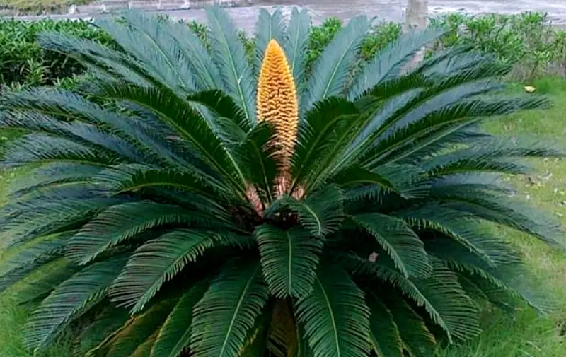
EXTERNAL FEATURES OF MALE CONE OF CYCAS
- The male cone is tenninal, shortly stalked, compact, large and oval or conical in shape and consists of a central cone axis around which numerous microsporophylls are spirally arranged. (Since the male cone tenninates the growth of the apex of male plant, a lateral bud later grows and takes over the continuation of growth of apex. The male plant thus shows sympodial growth).
- The outer covering of the male cone is formed by closely set sterile ends of the microsporophylls usually possessing upcurved apices, apophysis.
L.S. OF THE MALE CONE OF CYCAS
- The L.s. shows stalk and the cone.
- Male cone is attached at the apex of the plant by a stout and broad stalk.
- The cone itself consists of a central cone axis with many microsporophylls.
- Each microsporophyll is attached to the cone axis. The part of microsporophylls away from the axis is upcurved and is called apophysis.
- The upper surface of the microsporophyll is sterile.
- The lower surface of the microsporophyll is fertile and bears many microsporangia in groups (sori).
- Microsporophylls in the middle part of the cone are largest and get gradually smaller towards the base and the apex.
STUDY OF MICROSPOROPHYLL AND MICROSPORANGIA OF CYCAS
- A single microsporophyll is woody, more or less horizontally flattened and triangular structure.
- It is differentiated into a fertile and sterile parts. Fertile part is wedge-shaped and is expanded distally from a narrow point of attachment. Sterile part is the distal part of the microsporophyll which tapers into an upcurved apophysis.
- Lower (abaxial) surface of the fertile part of the microsporophyll bears microsporangia in groups of 3-4, forming definite sori.
- Microsporangia are arranged in sori around central papilla. Sporangia show radial lines of dehiscence. Many hairs are distributed on this surface mixed with sporangia.
STUDY OF T.S. OF MICROSPOROPHYLL OF CYCAS
- The section shows microsporangia attached to the abaxial (lower) surface by their short stalks.
- A mature microsporangium has three layered wall. The outermost layer is thick and cutinized, termed as exothecium. The remaining inner layers are thin and are collectively known as endothecium and enclose a tapetum.
- Numerous microspores remain enclosed inside the wall of the micro sporangium.
- In the microsporophyll are present many mucilage ducts, regularly scattered, among the rounded mesophyll-like cells forming the tissue of the sporophyll.
STUDY OF MEGASPOROPHYLL
- Female reproductive body consists of megasporphylls arranged spirally and arising in acropetal succession on the stem.
- Megasporophylls appear as a rosette or a crown, leaving the apical meristem unaffected to grow further. A crown of megasporophyll is formed each year. Numerically they are more than the leaves.
- They leave their persistent bases on the stem.
- Each megasporophyll is leaf-like and densely covered with brown hairs. It varies in size from 6 to 12 inches.
- Each megasporophyll is distinguished into a proximal (lower) petiole, a middle ovule bearing portion and a distal (upper) pinnately dissected sterile part.
- The nature of upper sterile part varies with species.
- In C. revoluta, the upper part is very much dissected, forming many pinnae.
- In C. rumphii, the upper part bears only short spines which represent reduced pinnae.
- In C. circinalis, the pinnate character is altogether absent and upper part shows only dentate or serrate margins.
- The middle portion of sporophyll bears ovules which are borne in two rows, one on either side. The ovules of the two rows may be opposite or alternate.
- Ovules are generally yellow or orange or dark green coloured, shortly stalked, oval and smooth. Number and size of the ovules differ from species to species.
- In C. revoluta, ovules are many and orange coloured.
- In C. circinalis also, they are numerous, but these are dark green and attain a large size.
- In C. siamensis, the number of ovules is reduced to only two
- In C. thouarsii, the ovules become still larger and may be that they are the largest ovules in the plant kingdom. 9. All the ovules do not develop fully. Some of those which remain unpollinated and small, finally abort.
L.S. OF MATURE OVULE OF CYCAS
- The section shows that the ovule is orthotropous.
- It is unitegmic (possesses a single integument). The integument is very thick. It remains fused with the nucellus except for the nucellar beak leaving a small and narrow micropyle.
- The integumentconsists of three distinct layersan outer fleshy layer, middle stony layer and an inner fleshy layer. The outer and inner fleshy layers are supplied with vascular strands but the middle stony layer receives no vascular supply.
- The nucelluslies just below the integument and forms a nucellar beak in the region of the micropyle.
- A few cells of this nucellar beak dissolve themselves and form a pollen chamber that lies in the tissue in the central region of the beak.
- Female gametophyte :-The innermost region of the ovule is filled with the tissue of female gametophyte, wherein lie two archegonia, situated opposite the pollen chamber.
- Archegonial chamber :-Just above the archegonia is the archegonial chamber.
- Micropyle :-The orange coloured, fleshy ovules are oval in shape and each shows a small point at the distal end which represents the remnant of the micropyle.
L.S. OF SEED OF CYCAS
- It shows seed coat, nucellus, embryo and the female gametophyte.
- Seed coat consists of sarcotesta (from outer fleshy layer of integument), and middle sc1erotesta (from middle stony layer). The inner fleshy layer of the integument appears as thin and papery structure in the seed.
- Nucellus is papery and is situated inside the seed coat.
- Endosperm and female gametophyte form the inner part of seed.
- A straight embryo remains embedded in the endosperm. It has two unequl cotyledons.
IDENTIFICATION OF CYCAS
- DIVISION – Gymnosperms
- (1) Absence of vessels.
- Ovules naked.
- Seeds attached with woody acales.
- Scales generally form a cone.
- Class:- Cycadopsida :-
- Wood manoxylic.
- Large frond-like leaves .
- Seeds with radial symmetry.
- Order– Cycadales
- Plants woody, stem unbranched,
- Wood manoxylic.
- Pressence of mucilage canals.
- Leaf trace diploxylic
- Dioecious
- Ovules orthotropous.
- Sperm with band of flagella
- Family – Cycadaceae
- Leaves with circinate vernation.
- Presence of coralloid roots and endophytic blue green algae.
- Megasporophylls foliar.
- Genus – Cycas
- Two types of leaves.
- Foliage leaves pinnately compound, circinately coiled when young.
- Presence of trasfusiontissue and diploxylic pascular bundle in leaf.
- Secondary xylem in stem manoxlic
- Two types of roots
- Vascular bundles arranged in an inverted omega-shaped manner in the rachis
- Male cone large and single.
REFERENCES
- https://pixabay.com/photos/cycads-flower-sago-palm-1541391/
- https://en.wikipedia.org/wiki/Cycas
Leave a Reply